EP2790738B1 - Collateral gene inactivation biomarkers and targets for cancer therapy - Google Patents
Collateral gene inactivation biomarkers and targets for cancer therapy Download PDFInfo
- Publication number
- EP2790738B1 EP2790738B1 EP12858619.5A EP12858619A EP2790738B1 EP 2790738 B1 EP2790738 B1 EP 2790738B1 EP 12858619 A EP12858619 A EP 12858619A EP 2790738 B1 EP2790738 B1 EP 2790738B1
- Authority
- EP
- European Patent Office
- Prior art keywords
- cancer
- cells
- inhibitor
- eno1
- enolase
- Prior art date
- Legal status (The legal status is an assumption and is not a legal conclusion. Google has not performed a legal analysis and makes no representation as to the accuracy of the status listed.)
- Active
Links
- 108090000623 proteins and genes Proteins 0.000 title description 125
- 230000002779 inactivation Effects 0.000 title description 21
- 239000000090 biomarker Substances 0.000 title description 8
- 238000011275 oncology therapy Methods 0.000 title description 7
- 206010028980 Neoplasm Diseases 0.000 claims description 294
- 239000003112 inhibitor Substances 0.000 claims description 270
- 102100038910 Alpha-enolase Human genes 0.000 claims description 210
- 101710165425 Alpha-enolase Proteins 0.000 claims description 210
- 101710184673 Enolase 1 Proteins 0.000 claims description 210
- 201000011510 cancer Diseases 0.000 claims description 206
- 238000012217 deletion Methods 0.000 claims description 114
- 230000037430 deletion Effects 0.000 claims description 114
- 230000000694 effects Effects 0.000 claims description 113
- 108700039887 Essential Genes Proteins 0.000 claims description 105
- 230000014509 gene expression Effects 0.000 claims description 99
- 239000000203 mixture Substances 0.000 claims description 45
- 150000007523 nucleic acids Chemical group 0.000 claims description 29
- 102000039446 nucleic acids Human genes 0.000 claims description 28
- 108020004707 nucleic acids Proteins 0.000 claims description 28
- 102100031126 6-phosphogluconolactonase Human genes 0.000 claims description 19
- 108010029731 6-phosphogluconolactonase Proteins 0.000 claims description 19
- 239000002246 antineoplastic agent Substances 0.000 claims description 19
- 229940127089 cytotoxic agent Drugs 0.000 claims description 19
- 108010002929 galactose-6-phosphate dehydrogenase Proteins 0.000 claims description 19
- 101710177040 Nicotinamide-nucleotide adenylyltransferase 1 Proteins 0.000 claims description 14
- 102100034451 Nicotinamide/nicotinic acid mononucleotide adenylyltransferase 1 Human genes 0.000 claims description 14
- 101710143608 Nicotinamide/nicotinic acid mononucleotide adenylyltransferase 1 Proteins 0.000 claims description 14
- 101000809046 Homo sapiens Ubiquitin conjugation factor E4 B Proteins 0.000 claims description 12
- 102100038487 Ubiquitin conjugation factor E4 B Human genes 0.000 claims description 12
- 230000013632 homeostatic process Effects 0.000 claims description 12
- 101000971697 Homo sapiens Kinesin-like protein KIF1B Proteins 0.000 claims description 11
- 102100021524 Kinesin-like protein KIF1B Human genes 0.000 claims description 11
- 101710088194 Dehydrogenase Proteins 0.000 claims description 9
- 101710177039 Nicotinamide-nucleotide adenylyltransferase 2 Proteins 0.000 claims description 9
- 102100034450 Nicotinamide/nicotinic acid mononucleotide adenylyltransferase 2 Human genes 0.000 claims description 9
- 102100034456 Nicotinamide/nicotinic acid mononucleotide adenylyltransferase 3 Human genes 0.000 claims description 9
- 101710143617 Nicotinamide/nicotinic acid mononucleotide adenylyltransferase 3 Proteins 0.000 claims description 9
- LDKRAXXVBWHMRH-UHFFFAOYSA-N phosphonoacetohydroxamic acid Chemical compound ONC(=O)CP(O)(O)=O LDKRAXXVBWHMRH-UHFFFAOYSA-N 0.000 claims description 9
- 101000971638 Homo sapiens Kinesin-like protein KIF1A Proteins 0.000 claims description 8
- 102100021527 Kinesin-like protein KIF1A Human genes 0.000 claims description 8
- 101000971703 Homo sapiens Kinesin-like protein KIF1C Proteins 0.000 claims description 5
- 102100021525 Kinesin-like protein KIF1C Human genes 0.000 claims description 5
- FMGSKLZLMKYGDP-UHFFFAOYSA-N Dehydroepiandrosterone Natural products C1C(O)CCC2(C)C3CCC(C)(C(CC4)=O)C4C3CC=C21 FMGSKLZLMKYGDP-UHFFFAOYSA-N 0.000 claims description 4
- 101000808654 Homo sapiens Ubiquitin conjugation factor E4 A Proteins 0.000 claims description 4
- 102100038532 Ubiquitin conjugation factor E4 A Human genes 0.000 claims description 4
- FMGSKLZLMKYGDP-USOAJAOKSA-N dehydroepiandrosterone Chemical compound C1[C@@H](O)CC[C@]2(C)[C@H]3CC[C@](C)(C(CC4)=O)[C@@H]4[C@@H]3CC=C21 FMGSKLZLMKYGDP-USOAJAOKSA-N 0.000 claims description 4
- 229960002847 prasterone Drugs 0.000 claims description 4
- 238000010798 ubiquitination Methods 0.000 claims description 3
- 230000034512 ubiquitination Effects 0.000 claims description 3
- 210000004027 cell Anatomy 0.000 description 393
- 108010022181 Phosphopyruvate Hydratase Proteins 0.000 description 138
- 102000012288 Phosphopyruvate Hydratase Human genes 0.000 description 137
- 238000000034 method Methods 0.000 description 95
- 238000002560 therapeutic procedure Methods 0.000 description 94
- 230000034659 glycolysis Effects 0.000 description 89
- 238000011282 treatment Methods 0.000 description 79
- 230000035772 mutation Effects 0.000 description 75
- 101000674009 Homo sapiens Threonine-tRNA ligase 1, cytoplasmic Proteins 0.000 description 56
- 102100040537 Threonine-tRNA ligase 1, cytoplasmic Human genes 0.000 description 54
- 208000005017 glioblastoma Diseases 0.000 description 51
- 239000011269 tar Substances 0.000 description 51
- 108091027967 Small hairpin RNA Proteins 0.000 description 42
- 230000012010 growth Effects 0.000 description 39
- 150000001875 compounds Chemical class 0.000 description 38
- 239000004055 small Interfering RNA Substances 0.000 description 36
- OJCKRNPLOZHAOU-RSXXJMTFSA-N (1r,2r)-2-[(2s,4e,6e,8r,9s,11r,13s,15s,16s)-7-cyano-8,16-dihydroxy-9,11,13,15-tetramethyl-18-oxo-1-oxacyclooctadeca-4,6-dien-2-yl]cyclopentane-1-carboxylic acid Chemical compound O1C(=O)C[C@H](O)[C@@H](C)C[C@@H](C)C[C@@H](C)C[C@H](C)[C@@H](O)\C(C#N)=C\C=C\C[C@H]1[C@H]1[C@H](C(O)=O)CCC1 OJCKRNPLOZHAOU-RSXXJMTFSA-N 0.000 description 32
- OJCKRNPLOZHAOU-BNXNOGCYSA-N Borrelidin Natural products CC1CC(C)CC(C)C(O)C(=C/C=C/CC(OC(=O)CC(O)C(C)C1)C2CCCC2C(=O)O)C#N OJCKRNPLOZHAOU-BNXNOGCYSA-N 0.000 description 32
- 210000001519 tissue Anatomy 0.000 description 32
- -1 C1orf211 Proteins 0.000 description 31
- 230000035945 sensitivity Effects 0.000 description 31
- 239000013598 vector Substances 0.000 description 31
- 230000004044 response Effects 0.000 description 30
- 108020004414 DNA Proteins 0.000 description 29
- 238000003556 assay Methods 0.000 description 29
- 239000000523 sample Substances 0.000 description 26
- 239000003795 chemical substances by application Substances 0.000 description 25
- 230000005764 inhibitory process Effects 0.000 description 25
- 101000777293 Homo sapiens Serine/threonine-protein kinase Chk1 Proteins 0.000 description 24
- 102100031081 Serine/threonine-protein kinase Chk1 Human genes 0.000 description 24
- 230000008685 targeting Effects 0.000 description 24
- 239000000543 intermediate Substances 0.000 description 23
- 208000024891 symptom Diseases 0.000 description 23
- 230000001225 therapeutic effect Effects 0.000 description 22
- 210000004881 tumor cell Anatomy 0.000 description 22
- 102000004567 6-phosphogluconate dehydrogenase Human genes 0.000 description 21
- 108020001657 6-phosphogluconate dehydrogenase Proteins 0.000 description 21
- 101150015836 ENO1 gene Proteins 0.000 description 21
- 238000001262 western blot Methods 0.000 description 21
- 102100024122 Pantothenate kinase 1 Human genes 0.000 description 20
- 229940002612 prodrug Drugs 0.000 description 20
- 239000000651 prodrug Substances 0.000 description 20
- 230000006870 function Effects 0.000 description 19
- 230000002401 inhibitory effect Effects 0.000 description 19
- 102000004169 proteins and genes Human genes 0.000 description 19
- 102000004190 Enzymes Human genes 0.000 description 18
- 108090000790 Enzymes Proteins 0.000 description 18
- 238000003197 gene knockdown Methods 0.000 description 18
- 230000009467 reduction Effects 0.000 description 18
- 102100039868 Cytoplasmic aconitate hydratase Human genes 0.000 description 17
- 101000745370 Homo sapiens Cytoplasmic aconitate hydratase Proteins 0.000 description 17
- 238000004458 analytical method Methods 0.000 description 17
- 108020004999 messenger RNA Proteins 0.000 description 17
- 210000004287 null lymphocyte Anatomy 0.000 description 17
- 101000678861 Arabidopsis thaliana Aconitate hydratase 1 Proteins 0.000 description 16
- 108700015934 Triose-phosphate isomerases Proteins 0.000 description 16
- 108091007433 antigens Proteins 0.000 description 16
- 102000036639 antigens Human genes 0.000 description 16
- 230000002068 genetic effect Effects 0.000 description 16
- 230000001965 increasing effect Effects 0.000 description 16
- 102000004144 Green Fluorescent Proteins Human genes 0.000 description 15
- 108010043121 Green Fluorescent Proteins Proteins 0.000 description 15
- 101710203425 Pantothenate kinase 1 Proteins 0.000 description 15
- 230000015572 biosynthetic process Effects 0.000 description 15
- 230000030833 cell death Effects 0.000 description 15
- 208000037265 diseases, disorders, signs and symptoms Diseases 0.000 description 15
- 229960003722 doxycycline Drugs 0.000 description 15
- XQTWDDCIUJNLTR-CVHRZJFOSA-N doxycycline monohydrate Chemical compound O.O=C1C2=C(O)C=CC=C2[C@H](C)[C@@H]2C1=C(O)[C@]1(O)C(=O)C(C(N)=O)=C(O)[C@@H](N(C)C)[C@@H]1[C@H]2O XQTWDDCIUJNLTR-CVHRZJFOSA-N 0.000 description 15
- 230000002349 favourable effect Effects 0.000 description 15
- 239000005090 green fluorescent protein Substances 0.000 description 15
- 230000002503 metabolic effect Effects 0.000 description 15
- 239000002207 metabolite Substances 0.000 description 15
- 230000002018 overexpression Effects 0.000 description 15
- 239000000427 antigen Substances 0.000 description 14
- 238000002474 experimental method Methods 0.000 description 14
- 230000001988 toxicity Effects 0.000 description 14
- 231100000419 toxicity Toxicity 0.000 description 14
- 229920001817 Agar Polymers 0.000 description 13
- RBNPOMFGQQGHHO-UWTATZPHSA-N D-glyceric acid Chemical group OC[C@@H](O)C(O)=O RBNPOMFGQQGHHO-UWTATZPHSA-N 0.000 description 13
- 241000699670 Mus sp. Species 0.000 description 13
- 102100024126 Pantothenate kinase 3 Human genes 0.000 description 13
- 101710203430 Pantothenate kinase 3 Proteins 0.000 description 13
- 239000008272 agar Substances 0.000 description 13
- 210000001130 astrocyte Anatomy 0.000 description 13
- 230000007423 decrease Effects 0.000 description 13
- 229940124186 Dehydrogenase inhibitor Drugs 0.000 description 12
- 239000003814 drug Substances 0.000 description 12
- 238000001727 in vivo Methods 0.000 description 12
- 201000001441 melanoma Diseases 0.000 description 12
- 230000037361 pathway Effects 0.000 description 12
- 230000002829 reductive effect Effects 0.000 description 12
- 150000003384 small molecules Chemical class 0.000 description 12
- 108091026890 Coding region Proteins 0.000 description 11
- 230000006907 apoptotic process Effects 0.000 description 11
- 239000003153 chemical reaction reagent Substances 0.000 description 11
- 239000013078 crystal Substances 0.000 description 11
- 230000002950 deficient Effects 0.000 description 11
- 239000000243 solution Substances 0.000 description 11
- 239000000758 substrate Substances 0.000 description 11
- MNULEGDCPYONBU-WMBHJXFZSA-N (1r,4s,5e,5'r,6'r,7e,10s,11r,12s,14r,15s,16s,18r,19s,20r,21e,25s,26r,27s,29s)-4-ethyl-11,12,15,19-tetrahydroxy-6'-[(2s)-2-hydroxypropyl]-5',10,12,14,16,18,20,26,29-nonamethylspiro[24,28-dioxabicyclo[23.3.1]nonacosa-5,7,21-triene-27,2'-oxane]-13,17,23-trio Polymers O([C@@H]1CC[C@@H](/C=C/C=C/C[C@H](C)[C@@H](O)[C@](C)(O)C(=O)[C@H](C)[C@@H](O)[C@H](C)C(=O)[C@H](C)[C@@H](O)[C@H](C)/C=C/C(=O)O[C@H]([C@H]2C)[C@H]1C)CC)[C@]12CC[C@@H](C)[C@@H](C[C@H](C)O)O1 MNULEGDCPYONBU-WMBHJXFZSA-N 0.000 description 10
- MNULEGDCPYONBU-DJRUDOHVSA-N (1s,4r,5z,5'r,6'r,7e,10s,11r,12s,14r,15s,18r,19r,20s,21e,26r,27s)-4-ethyl-11,12,15,19-tetrahydroxy-6'-(2-hydroxypropyl)-5',10,12,14,16,18,20,26,29-nonamethylspiro[24,28-dioxabicyclo[23.3.1]nonacosa-5,7,21-triene-27,2'-oxane]-13,17,23-trione Polymers O([C@H]1CC[C@H](\C=C/C=C/C[C@H](C)[C@@H](O)[C@](C)(O)C(=O)[C@H](C)[C@@H](O)C(C)C(=O)[C@H](C)[C@H](O)[C@@H](C)/C=C/C(=O)OC([C@H]2C)C1C)CC)[C@]12CC[C@@H](C)[C@@H](CC(C)O)O1 MNULEGDCPYONBU-DJRUDOHVSA-N 0.000 description 10
- MNULEGDCPYONBU-YNZHUHFTSA-N (4Z,18Z,20Z)-22-ethyl-7,11,14,15-tetrahydroxy-6'-(2-hydroxypropyl)-5',6,8,10,12,14,16,28,29-nonamethylspiro[2,26-dioxabicyclo[23.3.1]nonacosa-4,18,20-triene-27,2'-oxane]-3,9,13-trione Polymers CC1C(C2C)OC(=O)\C=C/C(C)C(O)C(C)C(=O)C(C)C(O)C(C)C(=O)C(C)(O)C(O)C(C)C\C=C/C=C\C(CC)CCC2OC21CCC(C)C(CC(C)O)O2 MNULEGDCPYONBU-YNZHUHFTSA-N 0.000 description 10
- MNULEGDCPYONBU-VVXVDZGXSA-N (5e,5'r,7e,10s,11r,12s,14s,15r,16r,18r,19s,20r,21e,26r,29s)-4-ethyl-11,12,15,19-tetrahydroxy-6'-[(2s)-2-hydroxypropyl]-5',10,12,14,16,18,20,26,29-nonamethylspiro[24,28-dioxabicyclo[23.3.1]nonacosa-5,7,21-triene-27,2'-oxane]-13,17,23-trione Polymers C([C@H](C)[C@@H](O)[C@](C)(O)C(=O)[C@@H](C)[C@H](O)[C@@H](C)C(=O)[C@H](C)[C@@H](O)[C@H](C)/C=C/C(=O)OC([C@H]1C)[C@H]2C)\C=C\C=C\C(CC)CCC2OC21CC[C@@H](C)C(C[C@H](C)O)O2 MNULEGDCPYONBU-VVXVDZGXSA-N 0.000 description 10
- MNULEGDCPYONBU-UHFFFAOYSA-N 4-ethyl-11,12,15,19-tetrahydroxy-6'-(2-hydroxypropyl)-5',10,12,14,16,18,20,26,29-nonamethylspiro[24,28-dioxabicyclo[23.3.1]nonacosa-5,7,21-triene-27,2'-oxane]-13,17,23-trione Polymers CC1C(C2C)OC(=O)C=CC(C)C(O)C(C)C(=O)C(C)C(O)C(C)C(=O)C(C)(O)C(O)C(C)CC=CC=CC(CC)CCC2OC21CCC(C)C(CC(C)O)O2 MNULEGDCPYONBU-UHFFFAOYSA-N 0.000 description 10
- 238000002965 ELISA Methods 0.000 description 10
- 230000002759 chromosomal effect Effects 0.000 description 10
- 229930191479 oligomycin Natural products 0.000 description 10
- MNULEGDCPYONBU-AWJDAWNUSA-N oligomycin A Polymers O([C@H]1CC[C@H](/C=C/C=C/C[C@@H](C)[C@H](O)[C@@](C)(O)C(=O)[C@@H](C)[C@H](O)[C@@H](C)C(=O)[C@@H](C)[C@H](O)[C@@H](C)/C=C/C(=O)O[C@@H]([C@@H]2C)[C@@H]1C)CC)[C@@]12CC[C@H](C)[C@H](C[C@@H](C)O)O1 MNULEGDCPYONBU-AWJDAWNUSA-N 0.000 description 10
- 108090000765 processed proteins & peptides Proteins 0.000 description 10
- RXWNCPJZOCPEPQ-NVWDDTSBSA-N puromycin Chemical compound C1=CC(OC)=CC=C1C[C@H](N)C(=O)N[C@H]1[C@@H](O)[C@H](N2C3=NC=NC(=C3N=C2)N(C)C)O[C@@H]1CO RXWNCPJZOCPEPQ-NVWDDTSBSA-N 0.000 description 10
- QTBSBXVTEAMEQO-UHFFFAOYSA-N Acetic acid Chemical compound CC(O)=O QTBSBXVTEAMEQO-UHFFFAOYSA-N 0.000 description 9
- 102100040958 Aconitate hydratase, mitochondrial Human genes 0.000 description 9
- 102100035388 Beta-enolase Human genes 0.000 description 9
- 101000877537 Homo sapiens Beta-enolase Proteins 0.000 description 9
- 108010011536 PTEN Phosphohydrolase Proteins 0.000 description 9
- 102000014160 PTEN Phosphohydrolase Human genes 0.000 description 9
- 238000002679 ablation Methods 0.000 description 9
- 230000010261 cell growth Effects 0.000 description 9
- 210000000349 chromosome Anatomy 0.000 description 9
- 238000001514 detection method Methods 0.000 description 9
- LOKCTEFSRHRXRJ-UHFFFAOYSA-I dipotassium trisodium dihydrogen phosphate hydrogen phosphate dichloride Chemical compound P(=O)(O)(O)[O-].[K+].P(=O)(O)([O-])[O-].[Na+].[Na+].[Cl-].[K+].[Cl-].[Na+] LOKCTEFSRHRXRJ-UHFFFAOYSA-I 0.000 description 9
- 230000006698 induction Effects 0.000 description 9
- 230000001939 inductive effect Effects 0.000 description 9
- 231100000518 lethal Toxicity 0.000 description 9
- 230000001665 lethal effect Effects 0.000 description 9
- 231100000225 lethality Toxicity 0.000 description 9
- 230000003647 oxidation Effects 0.000 description 9
- 238000007254 oxidation reaction Methods 0.000 description 9
- 239000002953 phosphate buffered saline Substances 0.000 description 9
- 239000000047 product Substances 0.000 description 9
- 238000011160 research Methods 0.000 description 9
- 101000678873 Arabidopsis thaliana Aconitate hydratase 3, mitochondrial Proteins 0.000 description 8
- 208000026310 Breast neoplasm Diseases 0.000 description 8
- 102100024458 Cyclin-dependent kinase inhibitor 2A Human genes 0.000 description 8
- WSFSSNUMVMOOMR-UHFFFAOYSA-N Formaldehyde Chemical compound O=C WSFSSNUMVMOOMR-UHFFFAOYSA-N 0.000 description 8
- 102100031132 Glucose-6-phosphate isomerase Human genes 0.000 description 8
- 108010070600 Glucose-6-phosphate isomerase Proteins 0.000 description 8
- 101710118630 Homocitrate dehydratase, mitochondrial Proteins 0.000 description 8
- JVTAAEKCZFNVCJ-UHFFFAOYSA-M Lactate Chemical compound CC(O)C([O-])=O JVTAAEKCZFNVCJ-UHFFFAOYSA-M 0.000 description 8
- 101000965243 Neosartorya fumigata (strain ATCC MYA-4609 / Af293 / CBS 101355 / FGSC A1100) Putative aconitate hydratase Proteins 0.000 description 8
- 101150067938 PGD gene Proteins 0.000 description 8
- 101710171478 Putative aconitate hydratase, mitochondrial Proteins 0.000 description 8
- 102000005924 Triose-Phosphate Isomerase Human genes 0.000 description 8
- 102000044209 Tumor Suppressor Genes Human genes 0.000 description 8
- 108700025716 Tumor Suppressor Genes Proteins 0.000 description 8
- 238000011319 anticancer therapy Methods 0.000 description 8
- 230000004663 cell proliferation Effects 0.000 description 8
- 230000001413 cellular effect Effects 0.000 description 8
- 230000003247 decreasing effect Effects 0.000 description 8
- RXKJFZQQPQGTFL-UHFFFAOYSA-N dihydroxyacetone Chemical compound OCC(=O)CO RXKJFZQQPQGTFL-UHFFFAOYSA-N 0.000 description 8
- 208000035475 disorder Diseases 0.000 description 8
- 230000002438 mitochondrial effect Effects 0.000 description 8
- 229930027945 nicotinamide-adenine dinucleotide Natural products 0.000 description 8
- 210000002966 serum Anatomy 0.000 description 8
- 238000012360 testing method Methods 0.000 description 8
- WYWHKKSPHMUBEB-UHFFFAOYSA-N tioguanine Chemical compound N1C(N)=NC(=S)C2=C1N=CN2 WYWHKKSPHMUBEB-UHFFFAOYSA-N 0.000 description 8
- 102100037399 Alanine-tRNA ligase, cytoplasmic Human genes 0.000 description 7
- 108091003079 Bovine Serum Albumin Proteins 0.000 description 7
- 206010006187 Breast cancer Diseases 0.000 description 7
- 239000006144 Dulbecco’s modified Eagle's medium Substances 0.000 description 7
- 208000032612 Glial tumor Diseases 0.000 description 7
- 206010018338 Glioma Diseases 0.000 description 7
- 102100031004 Histidine-tRNA ligase, cytoplasmic Human genes 0.000 description 7
- 101000843187 Homo sapiens Histidine-tRNA ligase, cytoplasmic Proteins 0.000 description 7
- 101000624524 Homo sapiens Leucine-tRNA ligase, cytoplasmic Proteins 0.000 description 7
- 102100023339 Leucine-tRNA ligase, cytoplasmic Human genes 0.000 description 7
- 241001465754 Metazoa Species 0.000 description 7
- 102000001105 Phosphofructokinases Human genes 0.000 description 7
- 108010069341 Phosphofructokinases Proteins 0.000 description 7
- 238000012054 celltiter-glo Methods 0.000 description 7
- 230000000973 chemotherapeutic effect Effects 0.000 description 7
- 230000002939 deleterious effect Effects 0.000 description 7
- 201000010099 disease Diseases 0.000 description 7
- 229940079593 drug Drugs 0.000 description 7
- 230000005014 ectopic expression Effects 0.000 description 7
- 239000012091 fetal bovine serum Substances 0.000 description 7
- 238000003364 immunohistochemistry Methods 0.000 description 7
- BOPGDPNILDQYTO-NNYOXOHSSA-N nicotinamide-adenine dinucleotide Chemical compound C1=CCC(C(=O)N)=CN1[C@H]1[C@H](O)[C@H](O)[C@@H](COP(O)(=O)OP(O)(=O)OC[C@@H]2[C@H]([C@@H](O)[C@@H](O2)N2C3=NC=NC(N)=C3N=C2)O)O1 BOPGDPNILDQYTO-NNYOXOHSSA-N 0.000 description 7
- 230000000144 pharmacologic effect Effects 0.000 description 7
- 239000013612 plasmid Substances 0.000 description 7
- 238000003752 polymerase chain reaction Methods 0.000 description 7
- 239000013615 primer Substances 0.000 description 7
- 102000004196 processed proteins & peptides Human genes 0.000 description 7
- 230000003612 virological effect Effects 0.000 description 7
- 108091032973 (ribonucleotides)n+m Proteins 0.000 description 6
- YXHLJMWYDTXDHS-IRFLANFNSA-N 7-aminoactinomycin D Chemical compound C[C@H]1OC(=O)[C@H](C(C)C)N(C)C(=O)CN(C)C(=O)[C@@H]2CCCN2C(=O)[C@@H](C(C)C)NC(=O)[C@H]1NC(=O)C1=C(N)C(=O)C(C)=C2OC(C(C)=C(N)C=C3C(=O)N[C@@H]4C(=O)N[C@@H](C(N5CCC[C@H]5C(=O)N(C)CC(=O)N(C)[C@@H](C(C)C)C(=O)O[C@@H]4C)=O)C(C)C)=C3N=C21 YXHLJMWYDTXDHS-IRFLANFNSA-N 0.000 description 6
- 108700012813 7-aminoactinomycin D Proteins 0.000 description 6
- 208000003174 Brain Neoplasms Diseases 0.000 description 6
- AOJJSUZBOXZQNB-TZSSRYMLSA-N Doxorubicin Chemical compound O([C@H]1C[C@@](O)(CC=2C(O)=C3C(=O)C=4C=CC=C(C=4C(=O)C3=C(O)C=21)OC)C(=O)CO)[C@H]1C[C@H](N)[C@H](O)[C@H](C)O1 AOJJSUZBOXZQNB-TZSSRYMLSA-N 0.000 description 6
- 101000879354 Homo sapiens Alanine-tRNA ligase, cytoplasmic Proteins 0.000 description 6
- 108091007984 KARS Proteins 0.000 description 6
- 102000010638 Kinesin Human genes 0.000 description 6
- 108010063296 Kinesin Proteins 0.000 description 6
- 102100035529 Lysine-tRNA ligase Human genes 0.000 description 6
- 241000124008 Mammalia Species 0.000 description 6
- 108020005115 Pyruvate Kinase Proteins 0.000 description 6
- 102000013009 Pyruvate Kinase Human genes 0.000 description 6
- 108020004459 Small interfering RNA Proteins 0.000 description 6
- 102100026655 Zinc finger protein castor homolog 1 Human genes 0.000 description 6
- 230000003321 amplification Effects 0.000 description 6
- 230000001640 apoptogenic effect Effects 0.000 description 6
- 238000013459 approach Methods 0.000 description 6
- 210000004556 brain Anatomy 0.000 description 6
- 239000002775 capsule Substances 0.000 description 6
- 238000002512 chemotherapy Methods 0.000 description 6
- 230000000295 complement effect Effects 0.000 description 6
- 230000021953 cytokinesis Effects 0.000 description 6
- 238000011161 development Methods 0.000 description 6
- 230000018109 developmental process Effects 0.000 description 6
- 238000009472 formulation Methods 0.000 description 6
- 238000003780 insertion Methods 0.000 description 6
- 230000037431 insertion Effects 0.000 description 6
- 239000006166 lysate Substances 0.000 description 6
- 238000003199 nucleic acid amplification method Methods 0.000 description 6
- 230000009038 pharmacological inhibition Effects 0.000 description 6
- 108091033319 polynucleotide Proteins 0.000 description 6
- 102000040430 polynucleotide Human genes 0.000 description 6
- 239000002157 polynucleotide Substances 0.000 description 6
- 229920001184 polypeptide Polymers 0.000 description 6
- 238000003127 radioimmunoassay Methods 0.000 description 6
- 239000002924 silencing RNA Substances 0.000 description 6
- 238000006467 substitution reaction Methods 0.000 description 6
- 238000001356 surgical procedure Methods 0.000 description 6
- 230000002195 synergetic effect Effects 0.000 description 6
- 231100000331 toxic Toxicity 0.000 description 6
- 230000002588 toxic effect Effects 0.000 description 6
- GXIURPTVHJPJLF-UWTATZPHSA-N 2-phospho-D-glyceric acid Chemical compound OC[C@H](C(O)=O)OP(O)(O)=O GXIURPTVHJPJLF-UWTATZPHSA-N 0.000 description 5
- ZLWYEPMDOUQDBW-UHFFFAOYSA-N 6-aminonicotinamide Chemical compound NC(=O)C1=CC=C(N)N=C1 ZLWYEPMDOUQDBW-UHFFFAOYSA-N 0.000 description 5
- 108090000672 Annexin A5 Proteins 0.000 description 5
- 102000004121 Annexin A5 Human genes 0.000 description 5
- 102100028449 Arginine-glutamic acid dipeptide repeats protein Human genes 0.000 description 5
- 102100036131 Arginine-tRNA ligase, cytoplasmic Human genes 0.000 description 5
- MLDQJTXFUGDVEO-UHFFFAOYSA-N BAY-43-9006 Chemical compound C1=NC(C(=O)NC)=CC(OC=2C=CC(NC(=O)NC=3C=C(C(Cl)=CC=3)C(F)(F)F)=CC=2)=C1 MLDQJTXFUGDVEO-UHFFFAOYSA-N 0.000 description 5
- 108010029692 Bisphosphoglycerate mutase Proteins 0.000 description 5
- 102100033561 Calmodulin-binding transcription activator 1 Human genes 0.000 description 5
- 102100036369 Carbonic anhydrase 6 Human genes 0.000 description 5
- 241000255581 Drosophila <fruit fly, genus> Species 0.000 description 5
- 102000001390 Fructose-Bisphosphate Aldolase Human genes 0.000 description 5
- 108010068561 Fructose-Bisphosphate Aldolase Proteins 0.000 description 5
- 102000005548 Hexokinase Human genes 0.000 description 5
- 108700040460 Hexokinases Proteins 0.000 description 5
- 101001061654 Homo sapiens Arginine-glutamic acid dipeptide repeats protein Proteins 0.000 description 5
- 101000945309 Homo sapiens Calmodulin-binding transcription activator 1 Proteins 0.000 description 5
- 101000714525 Homo sapiens Carbonic anhydrase 6 Proteins 0.000 description 5
- 239000005511 L01XE05 - Sorafenib Substances 0.000 description 5
- 239000002136 L01XE07 - Lapatinib Substances 0.000 description 5
- 206010027476 Metastases Diseases 0.000 description 5
- 206010033128 Ovarian cancer Diseases 0.000 description 5
- OYONTEXKYJZFHA-SSHUPFPWSA-N PHA-665752 Chemical compound CC=1C(C(=O)N2[C@H](CCC2)CN2CCCC2)=C(C)NC=1\C=C(C1=C2)/C(=O)NC1=CC=C2S(=O)(=O)CC1=C(Cl)C=CC=C1Cl OYONTEXKYJZFHA-SSHUPFPWSA-N 0.000 description 5
- 229910019142 PO4 Inorganic materials 0.000 description 5
- 102000011755 Phosphoglycerate Kinase Human genes 0.000 description 5
- 102000011025 Phosphoglycerate Mutase Human genes 0.000 description 5
- 102100026126 Proline-tRNA ligase Human genes 0.000 description 5
- LCTONWCANYUPML-UHFFFAOYSA-M Pyruvate Chemical compound CC(=O)C([O-])=O LCTONWCANYUPML-UHFFFAOYSA-M 0.000 description 5
- 108091006473 SLC25A33 Proteins 0.000 description 5
- 240000004808 Saccharomyces cerevisiae Species 0.000 description 5
- 235000014680 Saccharomyces cerevisiae Nutrition 0.000 description 5
- 102100040516 Serine-tRNA ligase, cytoplasmic Human genes 0.000 description 5
- 101001099217 Thermotoga maritima (strain ATCC 43589 / DSM 3109 / JCM 10099 / NBRC 100826 / MSB8) Triosephosphate isomerase Proteins 0.000 description 5
- 238000002835 absorbance Methods 0.000 description 5
- 230000004075 alteration Effects 0.000 description 5
- 238000005119 centrifugation Methods 0.000 description 5
- 230000001419 dependent effect Effects 0.000 description 5
- 235000014113 dietary fatty acids Nutrition 0.000 description 5
- 229930195729 fatty acid Natural products 0.000 description 5
- 239000000194 fatty acid Substances 0.000 description 5
- 150000004665 fatty acids Chemical class 0.000 description 5
- 230000003463 hyperproliferative effect Effects 0.000 description 5
- 238000003384 imaging method Methods 0.000 description 5
- 238000000338 in vitro Methods 0.000 description 5
- 208000015181 infectious disease Diseases 0.000 description 5
- 238000002347 injection Methods 0.000 description 5
- 239000007924 injection Substances 0.000 description 5
- 230000010438 iron metabolism Effects 0.000 description 5
- 238000011813 knockout mouse model Methods 0.000 description 5
- 229960004891 lapatinib Drugs 0.000 description 5
- BCFGMOOMADDAQU-UHFFFAOYSA-N lapatinib Chemical compound O1C(CNCCS(=O)(=O)C)=CC=C1C1=CC=C(N=CN=C2NC=3C=C(Cl)C(OCC=4C=C(F)C=CC=4)=CC=3)C2=C1 BCFGMOOMADDAQU-UHFFFAOYSA-N 0.000 description 5
- 150000002632 lipids Chemical class 0.000 description 5
- 238000004519 manufacturing process Methods 0.000 description 5
- 239000002609 medium Substances 0.000 description 5
- GLVAUDGFNGKCSF-UHFFFAOYSA-N mercaptopurine Chemical compound S=C1NC=NC2=C1NC=N2 GLVAUDGFNGKCSF-UHFFFAOYSA-N 0.000 description 5
- 125000003729 nucleotide group Chemical group 0.000 description 5
- 239000010452 phosphate Substances 0.000 description 5
- 230000035755 proliferation Effects 0.000 description 5
- 229950010131 puromycin Drugs 0.000 description 5
- 238000001959 radiotherapy Methods 0.000 description 5
- 230000008707 rearrangement Effects 0.000 description 5
- 230000002441 reversible effect Effects 0.000 description 5
- 238000005204 segregation Methods 0.000 description 5
- QFJCIRLUMZQUOT-HPLJOQBZSA-N sirolimus Chemical compound C1C[C@@H](O)[C@H](OC)C[C@@H]1C[C@@H](C)[C@H]1OC(=O)[C@@H]2CCCCN2C(=O)C(=O)[C@](O)(O2)[C@H](C)CC[C@H]2C[C@H](OC)/C(C)=C/C=C/C=C/[C@@H](C)C[C@@H](C)C(=O)[C@H](OC)[C@H](O)/C(C)=C/[C@@H](C)C(=O)C1 QFJCIRLUMZQUOT-HPLJOQBZSA-N 0.000 description 5
- 229960003787 sorafenib Drugs 0.000 description 5
- 238000010186 staining Methods 0.000 description 5
- 230000004083 survival effect Effects 0.000 description 5
- 230000000381 tumorigenic effect Effects 0.000 description 5
- AXCORWRSEKRTAQ-UHFFFAOYSA-N 2-nitroethylphosphonic acid Chemical compound OP(O)(=O)CC[N+]([O-])=O AXCORWRSEKRTAQ-UHFFFAOYSA-N 0.000 description 4
- 229920000936 Agarose Polymers 0.000 description 4
- 102100021569 Apoptosis regulator Bcl-2 Human genes 0.000 description 4
- 208000005623 Carcinogenesis Diseases 0.000 description 4
- RGJOEKWQDUBAIZ-IBOSZNHHSA-N CoASH Chemical compound O[C@@H]1[C@H](OP(O)(O)=O)[C@@H](COP(O)(=O)OP(O)(=O)OCC(C)(C)[C@@H](O)C(=O)NCCC(=O)NCCS)O[C@H]1N1C2=NC=NC(N)=C2N=C1 RGJOEKWQDUBAIZ-IBOSZNHHSA-N 0.000 description 4
- UHDGCWIWMRVCDJ-CCXZUQQUSA-N Cytarabine Chemical compound O=C1N=C(N)C=CN1[C@H]1[C@@H](O)[C@H](O)[C@@H](CO)O1 UHDGCWIWMRVCDJ-CCXZUQQUSA-N 0.000 description 4
- 102100031856 ERBB receptor feedback inhibitor 1 Human genes 0.000 description 4
- GHASVSINZRGABV-UHFFFAOYSA-N Fluorouracil Chemical compound FC1=CNC(=O)NC1=O GHASVSINZRGABV-UHFFFAOYSA-N 0.000 description 4
- 102100041016 G-protein coupled receptor 157 Human genes 0.000 description 4
- 229920000209 Hexadimethrine bromide Polymers 0.000 description 4
- 241000282412 Homo Species 0.000 description 4
- 101000971171 Homo sapiens Apoptosis regulator Bcl-2 Proteins 0.000 description 4
- 101000874860 Homo sapiens Arginine-tRNA ligase, cytoplasmic Proteins 0.000 description 4
- 101000920812 Homo sapiens ERBB receptor feedback inhibitor 1 Proteins 0.000 description 4
- 101001039303 Homo sapiens G-protein coupled receptor 157 Proteins 0.000 description 4
- 101000825289 Homo sapiens SPRY domain-containing SOCS box protein 1 Proteins 0.000 description 4
- 101000674278 Homo sapiens Serine-tRNA ligase, cytoplasmic Proteins 0.000 description 4
- 101000674040 Homo sapiens Serine-tRNA ligase, mitochondrial Proteins 0.000 description 4
- 101000851370 Homo sapiens Tumor necrosis factor receptor superfamily member 9 Proteins 0.000 description 4
- 101000641003 Homo sapiens Tyrosine-tRNA ligase, cytoplasmic Proteins 0.000 description 4
- 101000841325 Homo sapiens Urotensin-2 Proteins 0.000 description 4
- 241000699666 Mus <mouse, genus> Species 0.000 description 4
- ZDZOTLJHXYCWBA-VCVYQWHSSA-N N-debenzoyl-N-(tert-butoxycarbonyl)-10-deacetyltaxol Chemical compound O([C@H]1[C@H]2[C@@](C([C@H](O)C3=C(C)[C@@H](OC(=O)[C@H](O)[C@@H](NC(=O)OC(C)(C)C)C=4C=CC=CC=4)C[C@]1(O)C3(C)C)=O)(C)[C@@H](O)C[C@H]1OC[C@]12OC(=O)C)C(=O)C1=CC=CC=C1 ZDZOTLJHXYCWBA-VCVYQWHSSA-N 0.000 description 4
- BAWFJGJZGIEFAR-NNYOXOHSSA-O NAD(+) Chemical compound NC(=O)C1=CC=C[N+]([C@H]2[C@@H]([C@H](O)[C@@H](COP(O)(=O)OP(O)(=O)OC[C@@H]3[C@H]([C@@H](O)[C@@H](O3)N3C4=NC=NC(N)=C4N=C3)O)O2)O)=C1 BAWFJGJZGIEFAR-NNYOXOHSSA-O 0.000 description 4
- 108700020796 Oncogene Proteins 0.000 description 4
- 206010061535 Ovarian neoplasm Diseases 0.000 description 4
- 229930012538 Paclitaxel Natural products 0.000 description 4
- 206010061902 Pancreatic neoplasm Diseases 0.000 description 4
- 108010021592 Pantothenate kinase Proteins 0.000 description 4
- 102100037499 Parkinson disease protein 7 Human genes 0.000 description 4
- 108091000080 Phosphotransferase Proteins 0.000 description 4
- 206010060862 Prostate cancer Diseases 0.000 description 4
- 208000000236 Prostatic Neoplasms Diseases 0.000 description 4
- 108010032428 Protein Deglycase DJ-1 Proteins 0.000 description 4
- 108010029485 Protein Isoforms Proteins 0.000 description 4
- 102000001708 Protein Isoforms Human genes 0.000 description 4
- 102100037257 Proton-associated sugar transporter A Human genes 0.000 description 4
- 108091006301 SLC2A5 Proteins 0.000 description 4
- 108091007562 SLC45A1 Proteins 0.000 description 4
- 102100022320 SPRY domain-containing SOCS box protein 1 Human genes 0.000 description 4
- 206010039491 Sarcoma Diseases 0.000 description 4
- 102100022719 Solute carrier family 2, facilitated glucose transporter member 5 Human genes 0.000 description 4
- 102000001742 Tumor Suppressor Proteins Human genes 0.000 description 4
- 108010040002 Tumor Suppressor Proteins Proteins 0.000 description 4
- 102100036856 Tumor necrosis factor receptor superfamily member 9 Human genes 0.000 description 4
- 101710102803 Tumor suppressor ARF Proteins 0.000 description 4
- 102100034298 Tyrosine-tRNA ligase, cytoplasmic Human genes 0.000 description 4
- 102100029097 Urotensin-2 Human genes 0.000 description 4
- 208000006105 Uterine Cervical Neoplasms Diseases 0.000 description 4
- JXLYSJRDGCGARV-WWYNWVTFSA-N Vinblastine Natural products O=C(O[C@H]1[C@](O)(C(=O)OC)[C@@H]2N(C)c3c(cc(c(OC)c3)[C@]3(C(=O)OC)c4[nH]c5c(c4CCN4C[C@](O)(CC)C[C@H](C3)C4)cccc5)[C@@]32[C@H]2[C@@]1(CC)C=CCN2CC3)C JXLYSJRDGCGARV-WWYNWVTFSA-N 0.000 description 4
- 230000035508 accumulation Effects 0.000 description 4
- 238000009825 accumulation Methods 0.000 description 4
- ZSLZBFCDCINBPY-ZSJPKINUSA-N acetyl-CoA Chemical compound O[C@@H]1[C@H](OP(O)(O)=O)[C@@H](COP(O)(=O)OP(O)(=O)OCC(C)(C)[C@@H](O)C(=O)NCCC(=O)NCCSC(=O)C)O[C@H]1N1C2=NC=NC(N)=C2N=C1 ZSLZBFCDCINBPY-ZSJPKINUSA-N 0.000 description 4
- 230000008901 benefit Effects 0.000 description 4
- 238000001574 biopsy Methods 0.000 description 4
- 230000036952 cancer formation Effects 0.000 description 4
- 231100000504 carcinogenesis Toxicity 0.000 description 4
- 230000003833 cell viability Effects 0.000 description 4
- 238000006243 chemical reaction Methods 0.000 description 4
- RGJOEKWQDUBAIZ-UHFFFAOYSA-N coenzime A Natural products OC1C(OP(O)(O)=O)C(COP(O)(=O)OP(O)(=O)OCC(C)(C)C(O)C(=O)NCCC(=O)NCCS)OC1N1C2=NC=NC(N)=C2N=C1 RGJOEKWQDUBAIZ-UHFFFAOYSA-N 0.000 description 4
- 239000005516 coenzyme A Substances 0.000 description 4
- 229940093530 coenzyme a Drugs 0.000 description 4
- 238000002648 combination therapy Methods 0.000 description 4
- 230000000052 comparative effect Effects 0.000 description 4
- 230000002860 competitive effect Effects 0.000 description 4
- 238000003235 crystal violet staining Methods 0.000 description 4
- KDTSHFARGAKYJN-UHFFFAOYSA-N dephosphocoenzyme A Natural products OC1C(O)C(COP(O)(=O)OP(O)(=O)OCC(C)(C)C(O)C(=O)NCCC(=O)NCCS)OC1N1C2=NC=NC(N)=C2N=C1 KDTSHFARGAKYJN-UHFFFAOYSA-N 0.000 description 4
- 229960003668 docetaxel Drugs 0.000 description 4
- 239000012636 effector Substances 0.000 description 4
- 238000000684 flow cytometry Methods 0.000 description 4
- 229960002949 fluorouracil Drugs 0.000 description 4
- 229960005277 gemcitabine Drugs 0.000 description 4
- SDUQYLNIPVEERB-QPPQHZFASA-N gemcitabine Chemical compound O=C1N=C(N)C=CN1[C@H]1C(F)(F)[C@H](O)[C@@H](CO)O1 SDUQYLNIPVEERB-QPPQHZFASA-N 0.000 description 4
- 238000001415 gene therapy Methods 0.000 description 4
- 230000002414 glycolytic effect Effects 0.000 description 4
- LVASCWIMLIKXLA-LSDHHAIUSA-N halofuginone Chemical compound O[C@@H]1CCCN[C@H]1CC(=O)CN1C(=O)C2=CC(Cl)=C(Br)C=C2N=C1 LVASCWIMLIKXLA-LSDHHAIUSA-N 0.000 description 4
- 229950010152 halofuginone Drugs 0.000 description 4
- 230000036541 health Effects 0.000 description 4
- 238000009396 hybridization Methods 0.000 description 4
- 238000009169 immunotherapy Methods 0.000 description 4
- 230000003993 interaction Effects 0.000 description 4
- 238000007917 intracranial administration Methods 0.000 description 4
- 238000004020 luminiscence type Methods 0.000 description 4
- 208000020816 lung neoplasm Diseases 0.000 description 4
- 208000037841 lung tumor Diseases 0.000 description 4
- 238000002844 melting Methods 0.000 description 4
- 229960001428 mercaptopurine Drugs 0.000 description 4
- 230000037353 metabolic pathway Effects 0.000 description 4
- 230000004060 metabolic process Effects 0.000 description 4
- 108091029119 miR-34a stem-loop Proteins 0.000 description 4
- 108091070501 miRNA Proteins 0.000 description 4
- 239000002679 microRNA Substances 0.000 description 4
- 230000001537 neural effect Effects 0.000 description 4
- 229960001592 paclitaxel Drugs 0.000 description 4
- 102000020233 phosphotransferase Human genes 0.000 description 4
- BASFCYQUMIYNBI-UHFFFAOYSA-N platinum Chemical compound [Pt] BASFCYQUMIYNBI-UHFFFAOYSA-N 0.000 description 4
- 230000003389 potentiating effect Effects 0.000 description 4
- 230000008569 process Effects 0.000 description 4
- 230000002062 proliferating effect Effects 0.000 description 4
- 102000027426 receptor tyrosine kinases Human genes 0.000 description 4
- 108091008598 receptor tyrosine kinases Proteins 0.000 description 4
- 230000037432 silent mutation Effects 0.000 description 4
- 238000002741 site-directed mutagenesis Methods 0.000 description 4
- 239000007787 solid Substances 0.000 description 4
- 241000894007 species Species 0.000 description 4
- 238000003786 synthesis reaction Methods 0.000 description 4
- RCINICONZNJXQF-MZXODVADSA-N taxol Chemical compound O([C@@H]1[C@@]2(C[C@@H](C(C)=C(C2(C)C)[C@H](C([C@]2(C)[C@@H](O)C[C@H]3OC[C@]3([C@H]21)OC(C)=O)=O)OC(=O)C)OC(=O)[C@H](O)[C@@H](NC(=O)C=1C=CC=CC=1)C=1C=CC=CC=1)O)C(=O)C1=CC=CC=C1 RCINICONZNJXQF-MZXODVADSA-N 0.000 description 4
- 229960003087 tioguanine Drugs 0.000 description 4
- 238000010361 transduction Methods 0.000 description 4
- 230000026683 transduction Effects 0.000 description 4
- 238000003146 transient transfection Methods 0.000 description 4
- 230000004102 tricarboxylic acid cycle Effects 0.000 description 4
- 230000003827 upregulation Effects 0.000 description 4
- 229960003048 vinblastine Drugs 0.000 description 4
- JXLYSJRDGCGARV-XQKSVPLYSA-N vincaleukoblastine Chemical compound C([C@@H](C[C@]1(C(=O)OC)C=2C(=CC3=C([C@]45[C@H]([C@@]([C@H](OC(C)=O)[C@]6(CC)C=CCN([C@H]56)CC4)(O)C(=O)OC)N3C)C=2)OC)C[C@@](C2)(O)CC)N2CCC2=C1NC1=CC=CC=C21 JXLYSJRDGCGARV-XQKSVPLYSA-N 0.000 description 4
- 229960004528 vincristine Drugs 0.000 description 4
- OGWKCGZFUXNPDA-XQKSVPLYSA-N vincristine Chemical compound C([N@]1C[C@@H](C[C@]2(C(=O)OC)C=3C(=CC4=C([C@]56[C@H]([C@@]([C@H](OC(C)=O)[C@]7(CC)C=CCN([C@H]67)CC5)(O)C(=O)OC)N4C=O)C=3)OC)C[C@@](C1)(O)CC)CC1=C2NC2=CC=CC=C12 OGWKCGZFUXNPDA-XQKSVPLYSA-N 0.000 description 4
- OGWKCGZFUXNPDA-UHFFFAOYSA-N vincristine Natural products C1C(CC)(O)CC(CC2(C(=O)OC)C=3C(=CC4=C(C56C(C(C(OC(C)=O)C7(CC)C=CCN(C67)CC5)(O)C(=O)OC)N4C=O)C=3)OC)CN1CCC1=C2NC2=CC=CC=C12 OGWKCGZFUXNPDA-UHFFFAOYSA-N 0.000 description 4
- 238000005406 washing Methods 0.000 description 4
- VRYALKFFQXWPIH-PBXRRBTRSA-N (3r,4s,5r)-3,4,5,6-tetrahydroxyhexanal Chemical compound OC[C@@H](O)[C@@H](O)[C@H](O)CC=O VRYALKFFQXWPIH-PBXRRBTRSA-N 0.000 description 3
- YMYYQRZCCKBFBE-MDWZMJQESA-N (z)-2-(1h-indol-3-yl)-3-pyridin-3-ylprop-2-enenitrile Chemical compound C=1NC2=CC=CC=C2C=1C(/C#N)=C/C1=CC=CN=C1 YMYYQRZCCKBFBE-MDWZMJQESA-N 0.000 description 3
- MDPMUTNBKNPXNB-UPHRSURJSA-N (z)-3-amino-2-phosphonooxyprop-2-enoic acid Chemical compound N\C=C(C(O)=O)/OP(O)(O)=O MDPMUTNBKNPXNB-UPHRSURJSA-N 0.000 description 3
- GXIURPTVHJPJLF-UHFFFAOYSA-N 2-phosphoglyceric acid Chemical compound OCC(C(O)=O)OP(O)(O)=O GXIURPTVHJPJLF-UHFFFAOYSA-N 0.000 description 3
- IAYGCINLNONXHY-LBPRGKRZSA-N 3-(carbamoylamino)-5-(3-fluorophenyl)-N-[(3S)-3-piperidinyl]-2-thiophenecarboxamide Chemical compound NC(=O)NC=1C=C(C=2C=C(F)C=CC=2)SC=1C(=O)N[C@H]1CCCNC1 IAYGCINLNONXHY-LBPRGKRZSA-N 0.000 description 3
- 101150090289 ALDOB gene Proteins 0.000 description 3
- 101150037140 Aldoa gene Proteins 0.000 description 3
- UIFFUZWRFRDZJC-UHFFFAOYSA-N Antimycin A1 Natural products CC1OC(=O)C(CCCCCC)C(OC(=O)CC(C)C)C(C)OC(=O)C1NC(=O)C1=CC=CC(NC=O)=C1O UIFFUZWRFRDZJC-UHFFFAOYSA-N 0.000 description 3
- NQWZLRAORXLWDN-UHFFFAOYSA-N Antimycin-A Natural products CCCCCCC(=O)OC1C(C)OC(=O)C(NC(=O)c2ccc(NC=O)cc2O)C(C)OC(=O)C1CCCC NQWZLRAORXLWDN-UHFFFAOYSA-N 0.000 description 3
- 201000009030 Carcinoma Diseases 0.000 description 3
- 206010008342 Cervix carcinoma Diseases 0.000 description 3
- KRKNYBCHXYNGOX-UHFFFAOYSA-K Citrate Chemical compound [O-]C(=O)CC(O)(CC([O-])=O)C([O-])=O KRKNYBCHXYNGOX-UHFFFAOYSA-K 0.000 description 3
- 102000004127 Cytokines Human genes 0.000 description 3
- 108090000695 Cytokines Proteins 0.000 description 3
- 101100269568 Danio rerio aldocb gene Proteins 0.000 description 3
- LFQSCWFLJHTTHZ-UHFFFAOYSA-N Ethanol Chemical compound CCO LFQSCWFLJHTTHZ-UHFFFAOYSA-N 0.000 description 3
- KRHYYFGTRYWZRS-UHFFFAOYSA-M Fluoride anion Chemical compound [F-] KRHYYFGTRYWZRS-UHFFFAOYSA-M 0.000 description 3
- DGXLYHAWEBCTRU-UHFFFAOYSA-N Fluorocitric acid Chemical compound OC(=O)CC(O)(C(O)=O)C(F)C(O)=O DGXLYHAWEBCTRU-UHFFFAOYSA-N 0.000 description 3
- 102100036636 Glucose 1,6-bisphosphate synthase Human genes 0.000 description 3
- 101800000637 Hemokinin Proteins 0.000 description 3
- 101001072892 Homo sapiens Glucose 1,6-bisphosphate synthase Proteins 0.000 description 3
- 101000840551 Homo sapiens Hexokinase-2 Proteins 0.000 description 3
- 101000840561 Homo sapiens Hexokinase-3 Proteins 0.000 description 3
- 101001050577 Homo sapiens Kinesin-like protein KIF2A Proteins 0.000 description 3
- 101000601274 Homo sapiens Period circadian protein homolog 3 Proteins 0.000 description 3
- 101000833350 Homo sapiens Phosphoacetylglucosamine mutase Proteins 0.000 description 3
- 101000583553 Homo sapiens Phosphoglucomutase-1 Proteins 0.000 description 3
- 101001072903 Homo sapiens Phosphoglucomutase-2 Proteins 0.000 description 3
- 101001072881 Homo sapiens Phosphoglucomutase-like protein 5 Proteins 0.000 description 3
- 101000579123 Homo sapiens Phosphoglycerate kinase 1 Proteins 0.000 description 3
- 101001026214 Homo sapiens Potassium voltage-gated channel subfamily A member 5 Proteins 0.000 description 3
- 101001048456 Homo sapiens Protein Hook homolog 2 Proteins 0.000 description 3
- 101000866633 Homo sapiens Protein Hook homolog 3 Proteins 0.000 description 3
- 101001091538 Homo sapiens Pyruvate kinase PKM Proteins 0.000 description 3
- 102100023426 Kinesin-like protein KIF2A Human genes 0.000 description 3
- 241000713666 Lentivirus Species 0.000 description 3
- 206010058467 Lung neoplasm malignant Diseases 0.000 description 3
- 206010025323 Lymphomas Diseases 0.000 description 3
- 102000029749 Microtubule Human genes 0.000 description 3
- 108091022875 Microtubule Proteins 0.000 description 3
- 238000005481 NMR spectroscopy Methods 0.000 description 3
- 206010029260 Neuroblastoma Diseases 0.000 description 3
- 238000000636 Northern blotting Methods 0.000 description 3
- 201000010133 Oligodendroglioma Diseases 0.000 description 3
- 108091034117 Oligonucleotide Proteins 0.000 description 3
- 102000043276 Oncogene Human genes 0.000 description 3
- KJWZYMMLVHIVSU-IYCNHOCDSA-N PGK1 Chemical compound CCCCC[C@H](O)\C=C\[C@@H]1[C@@H](CCCCCCC(O)=O)C(=O)CC1=O KJWZYMMLVHIVSU-IYCNHOCDSA-N 0.000 description 3
- LGMXPVXJSFPPTQ-DJUJBXLVSA-N PGK2 Chemical compound CCCCC[C@H](O)\C=C\[C@@H]1[C@@H](C\C=C/CCCC(O)=O)C(=O)CC1=O LGMXPVXJSFPPTQ-DJUJBXLVSA-N 0.000 description 3
- 102100037630 Period circadian protein homolog 3 Human genes 0.000 description 3
- 102100024440 Phosphoacetylglucosamine mutase Human genes 0.000 description 3
- 102100030999 Phosphoglucomutase-1 Human genes 0.000 description 3
- 102100036629 Phosphoglucomutase-2 Human genes 0.000 description 3
- 102100036635 Phosphoglucomutase-like protein 5 Human genes 0.000 description 3
- 102100028251 Phosphoglycerate kinase 1 Human genes 0.000 description 3
- 102100037392 Phosphoglycerate kinase 2 Human genes 0.000 description 3
- 102100023602 Protein Hook homolog 1 Human genes 0.000 description 3
- 102100034911 Pyruvate kinase PKM Human genes 0.000 description 3
- 239000012083 RIPA buffer Substances 0.000 description 3
- 229940127361 Receptor Tyrosine Kinase Inhibitors Drugs 0.000 description 3
- 238000011579 SCID mouse model Methods 0.000 description 3
- 238000012300 Sequence Analysis Methods 0.000 description 3
- 238000002105 Southern blotting Methods 0.000 description 3
- 108010017749 Vesicle-Associated Membrane Protein 3 Proteins 0.000 description 3
- 102000004604 Vesicle-Associated Membrane Protein 3 Human genes 0.000 description 3
- 229940122803 Vinca alkaloid Drugs 0.000 description 3
- PDPXHRBRYUQCQA-UHFFFAOYSA-N [1-fluoro-2-(hydroxyamino)-2-oxoethyl]phosphonic acid Chemical compound ONC(=O)C(F)P(O)(O)=O PDPXHRBRYUQCQA-UHFFFAOYSA-N 0.000 description 3
- 101150057384 aldoc gene Proteins 0.000 description 3
- 150000001413 amino acids Chemical class 0.000 description 3
- UIFFUZWRFRDZJC-SBOOETFBSA-N antimycin A Chemical compound C[C@H]1OC(=O)[C@H](CCCCCC)[C@@H](OC(=O)CC(C)C)[C@H](C)OC(=O)[C@H]1NC(=O)C1=CC=CC(NC=O)=C1O UIFFUZWRFRDZJC-SBOOETFBSA-N 0.000 description 3
- PVEVXUMVNWSNIG-UHFFFAOYSA-N antimycin A3 Natural products CC1OC(=O)C(CCCC)C(OC(=O)CC(C)C)C(C)OC(=O)C1NC(=O)C1=CC=CC(NC=O)=C1O PVEVXUMVNWSNIG-UHFFFAOYSA-N 0.000 description 3
- 238000004113 cell culture Methods 0.000 description 3
- 239000006143 cell culture medium Substances 0.000 description 3
- 230000022131 cell cycle Effects 0.000 description 3
- 230000032823 cell division Effects 0.000 description 3
- 238000001516 cell proliferation assay Methods 0.000 description 3
- 201000010881 cervical cancer Diseases 0.000 description 3
- 238000012512 characterization method Methods 0.000 description 3
- 208000029742 colonic neoplasm Diseases 0.000 description 3
- 230000005757 colony formation Effects 0.000 description 3
- 239000002299 complementary DNA Substances 0.000 description 3
- 238000011498 curative surgery Methods 0.000 description 3
- 238000005520 cutting process Methods 0.000 description 3
- 229960000684 cytarabine Drugs 0.000 description 3
- 230000034994 death Effects 0.000 description 3
- 230000004069 differentiation Effects 0.000 description 3
- 238000009826 distribution Methods 0.000 description 3
- 229960004679 doxorubicin Drugs 0.000 description 3
- 230000013020 embryo development Effects 0.000 description 3
- 101150104041 eno2 gene Proteins 0.000 description 3
- 230000002255 enzymatic effect Effects 0.000 description 3
- 238000000605 extraction Methods 0.000 description 3
- 239000000499 gel Substances 0.000 description 3
- 230000037442 genomic alteration Effects 0.000 description 3
- 230000009036 growth inhibition Effects 0.000 description 3
- 239000001963 growth medium Substances 0.000 description 3
- 238000001794 hormone therapy Methods 0.000 description 3
- 238000003018 immunoassay Methods 0.000 description 3
- 238000007901 in situ hybridization Methods 0.000 description 3
- ODBLHEXUDAPZAU-UHFFFAOYSA-N isocitric acid Chemical compound OC(=O)C(O)C(C(O)=O)CC(O)=O ODBLHEXUDAPZAU-UHFFFAOYSA-N 0.000 description 3
- 230000002147 killing effect Effects 0.000 description 3
- 239000003446 ligand Substances 0.000 description 3
- 230000000670 limiting effect Effects 0.000 description 3
- 229910003002 lithium salt Inorganic materials 0.000 description 3
- 159000000002 lithium salts Chemical class 0.000 description 3
- 210000004072 lung Anatomy 0.000 description 3
- 201000005202 lung cancer Diseases 0.000 description 3
- 238000012423 maintenance Methods 0.000 description 3
- 230000003211 malignant effect Effects 0.000 description 3
- 208000015486 malignant pancreatic neoplasm Diseases 0.000 description 3
- 239000003550 marker Substances 0.000 description 3
- 239000000463 material Substances 0.000 description 3
- 239000011159 matrix material Substances 0.000 description 3
- 230000007246 mechanism Effects 0.000 description 3
- 230000001404 mediated effect Effects 0.000 description 3
- 230000009401 metastasis Effects 0.000 description 3
- 238000002493 microarray Methods 0.000 description 3
- 210000004688 microtubule Anatomy 0.000 description 3
- 230000025090 microtubule depolymerization Effects 0.000 description 3
- KKZJGLLVHKMTCM-UHFFFAOYSA-N mitoxantrone Chemical compound O=C1C2=C(O)C=CC(O)=C2C(=O)C2=C1C(NCCNCCO)=CC=C2NCCNCCO KKZJGLLVHKMTCM-UHFFFAOYSA-N 0.000 description 3
- 238000012544 monitoring process Methods 0.000 description 3
- 210000003205 muscle Anatomy 0.000 description 3
- 230000000955 neuroendocrine Effects 0.000 description 3
- 230000007971 neurological deficit Effects 0.000 description 3
- 238000001668 nucleic acid synthesis Methods 0.000 description 3
- 239000002773 nucleotide Substances 0.000 description 3
- 229940127084 other anti-cancer agent Drugs 0.000 description 3
- 201000002528 pancreatic cancer Diseases 0.000 description 3
- 208000008443 pancreatic carcinoma Diseases 0.000 description 3
- 230000007170 pathology Effects 0.000 description 3
- 239000000546 pharmaceutical excipient Substances 0.000 description 3
- 229930029653 phosphoenolpyruvate Natural products 0.000 description 3
- DTBNBXWJWCWCIK-UHFFFAOYSA-N phosphoenolpyruvic acid Chemical compound OC(=O)C(=C)OP(O)(O)=O DTBNBXWJWCWCIK-UHFFFAOYSA-N 0.000 description 3
- 239000004033 plastic Substances 0.000 description 3
- ZAHRKKWIAAJSAO-UHFFFAOYSA-N rapamycin Natural products COCC(O)C(=C/C(C)C(=O)CC(OC(=O)C1CCCCN1C(=O)C(=O)C2(O)OC(CC(OC)C(=CC=CC=CC(C)CC(C)C(=O)C)C)CCC2C)C(C)CC3CCC(O)C(C3)OC)C ZAHRKKWIAAJSAO-UHFFFAOYSA-N 0.000 description 3
- 108010054624 red fluorescent protein Proteins 0.000 description 3
- 238000002271 resection Methods 0.000 description 3
- 229940080817 rotenone Drugs 0.000 description 3
- JUVIOZPCNVVQFO-UHFFFAOYSA-N rotenone Natural products O1C2=C3CC(C(C)=C)OC3=CC=C2C(=O)C2C1COC1=C2C=C(OC)C(OC)=C1 JUVIOZPCNVVQFO-UHFFFAOYSA-N 0.000 description 3
- HXCHCVDVKSCDHU-PJKCJEBCSA-N s-[(2r,3s,4s,6s)-6-[[(2r,3s,4s,5r,6r)-5-[(2s,4s,5s)-5-(ethylamino)-4-methoxyoxan-2-yl]oxy-4-hydroxy-6-[[(2s,5z,9r,13e)-9-hydroxy-12-(methoxycarbonylamino)-13-[2-(methyltrisulfanyl)ethylidene]-11-oxo-2-bicyclo[7.3.1]trideca-1(12),5-dien-3,7-diynyl]oxy]-2-m Chemical compound C1[C@H](OC)[C@@H](NCC)CO[C@H]1O[C@H]1[C@H](O[C@@H]2C\3=C(NC(=O)OC)C(=O)C[C@@](C/3=C/CSSSC)(O)C#C\C=C/C#C2)O[C@H](C)[C@@H](NO[C@@H]2O[C@H](C)[C@@H](SC(=O)C=3C(=C(OC)C(O[C@H]4[C@@H]([C@H](OC)[C@@H](O)[C@H](C)O4)O)=C(I)C=3C)OC)[C@@H](O)C2)[C@@H]1O HXCHCVDVKSCDHU-PJKCJEBCSA-N 0.000 description 3
- 238000000926 separation method Methods 0.000 description 3
- 238000012163 sequencing technique Methods 0.000 description 3
- 229960002930 sirolimus Drugs 0.000 description 3
- 238000002415 sodium dodecyl sulfate polyacrylamide gel electrophoresis Methods 0.000 description 3
- 238000004611 spectroscopical analysis Methods 0.000 description 3
- 238000010561 standard procedure Methods 0.000 description 3
- 239000006228 supernatant Substances 0.000 description 3
- 108010008054 testis specific phosphoglycerate kinase Proteins 0.000 description 3
- 229940124597 therapeutic agent Drugs 0.000 description 3
- 238000001890 transfection Methods 0.000 description 3
- 230000007704 transition Effects 0.000 description 3
- 230000014616 translation Effects 0.000 description 3
- 230000004614 tumor growth Effects 0.000 description 3
- 231100000588 tumorigenic Toxicity 0.000 description 3
- 238000011144 upstream manufacturing Methods 0.000 description 3
- 210000002700 urine Anatomy 0.000 description 3
- YBJHBAHKTGYVGT-ZKWXMUAHSA-N (+)-Biotin Chemical compound N1C(=O)N[C@@H]2[C@H](CCCCC(=O)O)SC[C@@H]21 YBJHBAHKTGYVGT-ZKWXMUAHSA-N 0.000 description 2
- SHPNMMVXXKFVOJ-UHFFFAOYSA-N (3-hydroxy-2-nitropropyl)phosphonic acid Chemical compound OCC([N+]([O-])=O)CP(O)(O)=O SHPNMMVXXKFVOJ-UHFFFAOYSA-N 0.000 description 2
- JESMSCGUTIEROV-RTWAVKEYSA-N (5as,6r,9s,9as)-1-oxo-6-propan-2-ylspiro[3,5a,6,7,8,9a-hexahydro-2-benzoxepine-9,2'-oxirane]-4-carboxylic acid Chemical compound C([C@@H]([C@@H]1[C@@H]2C(OCC(=C1)C(O)=O)=O)C(C)C)C[C@]12CO1 JESMSCGUTIEROV-RTWAVKEYSA-N 0.000 description 2
- HZAXFHJVJLSVMW-UHFFFAOYSA-N 2-Aminoethan-1-ol Chemical compound NCCO HZAXFHJVJLSVMW-UHFFFAOYSA-N 0.000 description 2
- QKNYBSVHEMOAJP-UHFFFAOYSA-N 2-amino-2-(hydroxymethyl)propane-1,3-diol;hydron;chloride Chemical compound Cl.OCC(N)(CO)CO QKNYBSVHEMOAJP-UHFFFAOYSA-N 0.000 description 2
- MEOVPKDOYAIVHZ-UHFFFAOYSA-N 2-chloro-1-(1-methylpyrrol-2-yl)ethanol Chemical compound CN1C=CC=C1C(O)CCl MEOVPKDOYAIVHZ-UHFFFAOYSA-N 0.000 description 2
- 102100036009 5'-AMP-activated protein kinase catalytic subunit alpha-2 Human genes 0.000 description 2
- STQGQHZAVUOBTE-UHFFFAOYSA-N 7-Cyan-hept-2t-en-4,6-diinsaeure Natural products C1=2C(O)=C3C(=O)C=4C(OC)=CC=CC=4C(=O)C3=C(O)C=2CC(O)(C(C)=O)CC1OC1CC(N)C(O)C(C)O1 STQGQHZAVUOBTE-UHFFFAOYSA-N 0.000 description 2
- 108700028369 Alleles Proteins 0.000 description 2
- 102000000412 Annexin Human genes 0.000 description 2
- 108050008874 Annexin Proteins 0.000 description 2
- IJGRMHOSHXDMSA-UHFFFAOYSA-N Atomic nitrogen Chemical compound N#N IJGRMHOSHXDMSA-UHFFFAOYSA-N 0.000 description 2
- 102100031680 Beta-catenin-interacting protein 1 Human genes 0.000 description 2
- 241000244203 Caenorhabditis elegans Species 0.000 description 2
- 102100028801 Calsyntenin-1 Human genes 0.000 description 2
- GAGWJHPBXLXJQN-UORFTKCHSA-N Capecitabine Chemical compound C1=C(F)C(NC(=O)OCCCCC)=NC(=O)N1[C@H]1[C@H](O)[C@H](O)[C@@H](C)O1 GAGWJHPBXLXJQN-UORFTKCHSA-N 0.000 description 2
- CURLTUGMZLYLDI-UHFFFAOYSA-N Carbon dioxide Chemical compound O=C=O CURLTUGMZLYLDI-UHFFFAOYSA-N 0.000 description 2
- 108010001857 Cell Surface Receptors Proteins 0.000 description 2
- 102100038221 Chromodomain-helicase-DNA-binding protein 5 Human genes 0.000 description 2
- 206010009944 Colon cancer Diseases 0.000 description 2
- 102100030115 Cysteine-tRNA ligase, cytoplasmic Human genes 0.000 description 2
- 238000001712 DNA sequencing Methods 0.000 description 2
- 108010092160 Dactinomycin Proteins 0.000 description 2
- KCXVZYZYPLLWCC-UHFFFAOYSA-N EDTA Chemical compound OC(=O)CN(CC(O)=O)CCN(CC(O)=O)CC(O)=O KCXVZYZYPLLWCC-UHFFFAOYSA-N 0.000 description 2
- 101000581536 Escherichia coli Microcin C7 Proteins 0.000 description 2
- 208000000461 Esophageal Neoplasms Diseases 0.000 description 2
- 102100029951 Estrogen receptor beta Human genes 0.000 description 2
- FWVHWDSCPKXMDB-LSDHHAIUSA-N Febrifugine Chemical group O[C@@H]1CCCN[C@H]1CC(=O)CN1C(=O)C2=CC=CC=C2N=C1 FWVHWDSCPKXMDB-LSDHHAIUSA-N 0.000 description 2
- 102000016621 Focal Adhesion Protein-Tyrosine Kinases Human genes 0.000 description 2
- 108010067715 Focal Adhesion Protein-Tyrosine Kinases Proteins 0.000 description 2
- 101150066002 GFP gene Proteins 0.000 description 2
- 101150112014 Gapdh gene Proteins 0.000 description 2
- WZUVPPKBWHMQCE-UHFFFAOYSA-N Haematoxylin Chemical compound C12=CC(O)=C(O)C=C2CC2(O)C1C1=CC=C(O)C(O)=C1OC2 WZUVPPKBWHMQCE-UHFFFAOYSA-N 0.000 description 2
- 101000783681 Homo sapiens 5'-AMP-activated protein kinase catalytic subunit alpha-2 Proteins 0.000 description 2
- 101000993469 Homo sapiens Beta-catenin-interacting protein 1 Proteins 0.000 description 2
- 101000916423 Homo sapiens Calsyntenin-1 Proteins 0.000 description 2
- 101000883731 Homo sapiens Chromodomain-helicase-DNA-binding protein 5 Proteins 0.000 description 2
- 101000883736 Homo sapiens Chromodomain-helicase-DNA-binding protein 6 Proteins 0.000 description 2
- 101000586290 Homo sapiens Cysteine-tRNA ligase, cytoplasmic Proteins 0.000 description 2
- 101001038390 Homo sapiens Guided entry of tail-anchored proteins factor 1 Proteins 0.000 description 2
- 101000875582 Homo sapiens Isoleucine-tRNA ligase, cytoplasmic Proteins 0.000 description 2
- 101000573447 Homo sapiens Multiple inositol polyphosphate phosphatase 1 Proteins 0.000 description 2
- 101000981497 Homo sapiens Pantothenate kinase 1 Proteins 0.000 description 2
- 101000595746 Homo sapiens Phosphatidylinositol 4,5-bisphosphate 3-kinase catalytic subunit delta isoform Proteins 0.000 description 2
- 101000616291 Homo sapiens Protein LZIC Proteins 0.000 description 2
- 101001096330 Homo sapiens Retinoid-binding protein 7 Proteins 0.000 description 2
- 101000801088 Homo sapiens Transmembrane protein 201 Proteins 0.000 description 2
- 101000787286 Homo sapiens Valine-tRNA ligase Proteins 0.000 description 2
- 102100036015 Isoleucine-tRNA ligase, cytoplasmic Human genes 0.000 description 2
- 102100037691 Kinesin-like protein KIF20B Human genes 0.000 description 2
- 108050007394 Kinesin-like protein KIF20B Proteins 0.000 description 2
- 102100026284 Multiple inositol polyphosphate phosphatase 1 Human genes 0.000 description 2
- NWIBSHFKIJFRCO-WUDYKRTCSA-N Mytomycin Chemical compound C1N2C(C(C(C)=C(N)C3=O)=O)=C3[C@@H](COC(N)=O)[C@@]2(OC)[C@@H]2[C@H]1N2 NWIBSHFKIJFRCO-WUDYKRTCSA-N 0.000 description 2
- 208000001894 Nasopharyngeal Neoplasms Diseases 0.000 description 2
- 206010061306 Nasopharyngeal cancer Diseases 0.000 description 2
- 206010030155 Oesophageal carcinoma Diseases 0.000 description 2
- 206010031096 Oropharyngeal cancer Diseases 0.000 description 2
- 206010057444 Oropharyngeal neoplasm Diseases 0.000 description 2
- 102100036056 Phosphatidylinositol 4,5-bisphosphate 3-kinase catalytic subunit delta isoform Human genes 0.000 description 2
- 206010035226 Plasma cell myeloma Diseases 0.000 description 2
- 108010068086 Polyubiquitin Proteins 0.000 description 2
- 102100037935 Polyubiquitin-C Human genes 0.000 description 2
- 102100021802 Protein LZIC Human genes 0.000 description 2
- 108010090931 Proto-Oncogene Proteins c-bcl-2 Proteins 0.000 description 2
- 102000013535 Proto-Oncogene Proteins c-bcl-2 Human genes 0.000 description 2
- 102100037879 Retinoid-binding protein 7 Human genes 0.000 description 2
- AUNGANRZJHBGPY-SCRDCRAPSA-N Riboflavin Chemical compound OC[C@@H](O)[C@@H](O)[C@@H](O)CN1C=2C=C(C)C(C)=CC=2N=C2C1=NC(=O)NC2=O AUNGANRZJHBGPY-SCRDCRAPSA-N 0.000 description 2
- 108091006304 SLC2A7 Proteins 0.000 description 2
- 102100030937 Solute carrier family 2, facilitated glucose transporter member 7 Human genes 0.000 description 2
- 102100033827 Solute carrier family 25 member 33 Human genes 0.000 description 2
- 208000005718 Stomach Neoplasms Diseases 0.000 description 2
- 101150033985 TPI gene Proteins 0.000 description 2
- 208000024313 Testicular Neoplasms Diseases 0.000 description 2
- 206010057644 Testis cancer Diseases 0.000 description 2
- MUMGGOZAMZWBJJ-DYKIIFRCSA-N Testostosterone Chemical compound O=C1CC[C@]2(C)[C@H]3CC[C@](C)([C@H](CC4)O)[C@@H]4[C@@H]3CCC2=C1 MUMGGOZAMZWBJJ-DYKIIFRCSA-N 0.000 description 2
- 208000024770 Thyroid neoplasm Diseases 0.000 description 2
- 102100033708 Transmembrane protein 201 Human genes 0.000 description 2
- 208000007097 Urinary Bladder Neoplasms Diseases 0.000 description 2
- 102100025607 Valine-tRNA ligase Human genes 0.000 description 2
- 102000003970 Vinculin Human genes 0.000 description 2
- 108090000384 Vinculin Proteins 0.000 description 2
- IGYJTTVQPDNPPB-UHFFFAOYSA-N [O-][N+](C(CO)COP(O)=O)=O Chemical compound [O-][N+](C(CO)COP(O)=O)=O IGYJTTVQPDNPPB-UHFFFAOYSA-N 0.000 description 2
- 125000002777 acetyl group Chemical group [H]C([H])([H])C(*)=O 0.000 description 2
- 239000002253 acid Substances 0.000 description 2
- RJURFGZVJUQBHK-UHFFFAOYSA-N actinomycin D Natural products CC1OC(=O)C(C(C)C)N(C)C(=O)CN(C)C(=O)C2CCCN2C(=O)C(C(C)C)NC(=O)C1NC(=O)C1=C(N)C(=O)C(C)=C2OC(C(C)=CC=C3C(=O)NC4C(=O)NC(C(N5CCCC5C(=O)N(C)CC(=O)N(C)C(C(C)C)C(=O)OC4C)=O)C(C)C)=C3N=C21 RJURFGZVJUQBHK-UHFFFAOYSA-N 0.000 description 2
- 230000009471 action Effects 0.000 description 2
- 230000004913 activation Effects 0.000 description 2
- 230000002411 adverse Effects 0.000 description 2
- 239000000443 aerosol Substances 0.000 description 2
- 208000026935 allergic disease Diseases 0.000 description 2
- PMMURAAUARKVCB-UHFFFAOYSA-N alpha-D-ara-dHexp Natural products OCC1OC(O)CC(O)C1O PMMURAAUARKVCB-UHFFFAOYSA-N 0.000 description 2
- 230000001668 ameliorated effect Effects 0.000 description 2
- 238000010171 animal model Methods 0.000 description 2
- 229940045799 anthracyclines and related substance Drugs 0.000 description 2
- 239000003242 anti bacterial agent Substances 0.000 description 2
- 230000000845 anti-microbial effect Effects 0.000 description 2
- 230000002141 anti-parasite Effects 0.000 description 2
- 229940088710 antibiotic agent Drugs 0.000 description 2
- 239000004599 antimicrobial Substances 0.000 description 2
- 239000003096 antiparasitic agent Substances 0.000 description 2
- 238000003782 apoptosis assay Methods 0.000 description 2
- VSRXQHXAPYXROS-UHFFFAOYSA-N azanide;cyclobutane-1,1-dicarboxylic acid;platinum(2+) Chemical compound [NH2-].[NH2-].[Pt+2].OC(=O)C1(C(O)=O)CCC1 VSRXQHXAPYXROS-UHFFFAOYSA-N 0.000 description 2
- 230000002715 bioenergetic effect Effects 0.000 description 2
- 230000033228 biological regulation Effects 0.000 description 2
- 239000012472 biological sample Substances 0.000 description 2
- 230000000903 blocking effect Effects 0.000 description 2
- 210000004369 blood Anatomy 0.000 description 2
- 239000008280 blood Substances 0.000 description 2
- 210000000481 breast Anatomy 0.000 description 2
- 229930195731 calicheamicin Natural products 0.000 description 2
- 229960004562 carboplatin Drugs 0.000 description 2
- 230000015556 catabolic process Effects 0.000 description 2
- 210000001159 caudate nucleus Anatomy 0.000 description 2
- 230000003915 cell function Effects 0.000 description 2
- 230000022534 cell killing Effects 0.000 description 2
- 230000005754 cellular signaling Effects 0.000 description 2
- 210000003169 central nervous system Anatomy 0.000 description 2
- 230000008859 change Effects 0.000 description 2
- 239000007806 chemical reaction intermediate Substances 0.000 description 2
- HVYWMOMLDIMFJA-DPAQBDIFSA-N cholesterol Chemical compound C1C=C2C[C@@H](O)CC[C@]2(C)[C@@H]2[C@@H]1[C@@H]1CC[C@H]([C@H](C)CCCC(C)C)[C@@]1(C)CC2 HVYWMOMLDIMFJA-DPAQBDIFSA-N 0.000 description 2
- 230000001010 compromised effect Effects 0.000 description 2
- 230000002596 correlated effect Effects 0.000 description 2
- 230000000875 corresponding effect Effects 0.000 description 2
- 238000005859 coupling reaction Methods 0.000 description 2
- CVSVTCORWBXHQV-UHFFFAOYSA-N creatine Chemical compound NC(=[NH2+])N(C)CC([O-])=O CVSVTCORWBXHQV-UHFFFAOYSA-N 0.000 description 2
- 239000000824 cytostatic agent Substances 0.000 description 2
- 230000001085 cytostatic effect Effects 0.000 description 2
- 231100000433 cytotoxic Toxicity 0.000 description 2
- 230000001472 cytotoxic effect Effects 0.000 description 2
- STQGQHZAVUOBTE-VGBVRHCVSA-N daunorubicin Chemical compound O([C@H]1C[C@@](O)(CC=2C(O)=C3C(=O)C=4C=CC=C(C=4C(=O)C3=C(O)C=21)OC)C(C)=O)[C@H]1C[C@H](N)[C@H](O)[C@H](C)O1 STQGQHZAVUOBTE-VGBVRHCVSA-N 0.000 description 2
- 238000006731 degradation reaction Methods 0.000 description 2
- 238000013461 design Methods 0.000 description 2
- 238000003745 diagnosis Methods 0.000 description 2
- 230000003292 diminished effect Effects 0.000 description 2
- 239000001177 diphosphate Substances 0.000 description 2
- 235000011180 diphosphates Nutrition 0.000 description 2
- 231100000673 dose–response relationship Toxicity 0.000 description 2
- 230000003828 downregulation Effects 0.000 description 2
- 238000001962 electrophoresis Methods 0.000 description 2
- 230000003028 elevating effect Effects 0.000 description 2
- 238000005516 engineering process Methods 0.000 description 2
- 230000002708 enhancing effect Effects 0.000 description 2
- 150000002085 enols Chemical class 0.000 description 2
- 238000007824 enzymatic assay Methods 0.000 description 2
- 201000004101 esophageal cancer Diseases 0.000 description 2
- 230000005284 excitation Effects 0.000 description 2
- 230000001747 exhibiting effect Effects 0.000 description 2
- 150000002222 fluorine compounds Chemical class 0.000 description 2
- OVBPIULPVIDEAO-LBPRGKRZSA-N folic acid Chemical compound C=1N=C2NC(N)=NC(=O)C2=NC=1CNC1=CC=C(C(=O)N[C@@H](CCC(O)=O)C(O)=O)C=C1 OVBPIULPVIDEAO-LBPRGKRZSA-N 0.000 description 2
- 239000012634 fragment Substances 0.000 description 2
- 239000012737 fresh medium Substances 0.000 description 2
- 230000004927 fusion Effects 0.000 description 2
- CHPZKNULDCNCBW-UHFFFAOYSA-N gallium nitrate Chemical compound [Ga+3].[O-][N+]([O-])=O.[O-][N+]([O-])=O.[O-][N+]([O-])=O CHPZKNULDCNCBW-UHFFFAOYSA-N 0.000 description 2
- 210000003976 gap junction Anatomy 0.000 description 2
- 206010017758 gastric cancer Diseases 0.000 description 2
- XGALLCVXEZPNRQ-UHFFFAOYSA-N gefitinib Chemical compound C=12C=C(OCCCN3CCOCC3)C(OC)=CC2=NC=NC=1NC1=CC=C(F)C(Cl)=C1 XGALLCVXEZPNRQ-UHFFFAOYSA-N 0.000 description 2
- 238000001502 gel electrophoresis Methods 0.000 description 2
- 238000007429 general method Methods 0.000 description 2
- 230000004110 gluconeogenesis Effects 0.000 description 2
- 201000010536 head and neck cancer Diseases 0.000 description 2
- 208000014829 head and neck neoplasm Diseases 0.000 description 2
- 201000005787 hematologic cancer Diseases 0.000 description 2
- 208000024200 hematopoietic and lymphoid system neoplasm Diseases 0.000 description 2
- 206010073071 hepatocellular carcinoma Diseases 0.000 description 2
- 231100000844 hepatocellular carcinoma Toxicity 0.000 description 2
- 229940088597 hormone Drugs 0.000 description 2
- 239000005556 hormone Substances 0.000 description 2
- 238000006460 hydrolysis reaction Methods 0.000 description 2
- 229960001101 ifosfamide Drugs 0.000 description 2
- HOMGKSMUEGBAAB-UHFFFAOYSA-N ifosfamide Chemical compound ClCCNP1(=O)OCCCN1CCCl HOMGKSMUEGBAAB-UHFFFAOYSA-N 0.000 description 2
- YLMAHDNUQAMNNX-UHFFFAOYSA-N imatinib methanesulfonate Chemical compound CS(O)(=O)=O.C1CN(C)CCN1CC1=CC=C(C(=O)NC=2C=C(NC=3N=C(C=CN=3)C=3C=NC=CC=3)C(C)=CC=2)C=C1 YLMAHDNUQAMNNX-UHFFFAOYSA-N 0.000 description 2
- 230000002519 immonomodulatory effect Effects 0.000 description 2
- 239000012642 immune effector Substances 0.000 description 2
- 229940121354 immunomodulator Drugs 0.000 description 2
- 238000011534 incubation Methods 0.000 description 2
- JORABGDXCIBAFL-UHFFFAOYSA-M iodonitrotetrazolium chloride Chemical compound [Cl-].C1=CC([N+](=O)[O-])=CC=C1N1[N+](C=2C=CC(I)=CC=2)=NC(C=2C=CC=CC=2)=N1 JORABGDXCIBAFL-UHFFFAOYSA-M 0.000 description 2
- 150000002500 ions Chemical class 0.000 description 2
- UWKQSNNFCGGAFS-XIFFEERXSA-N irinotecan Chemical compound C1=C2C(CC)=C3CN(C(C4=C([C@@](C(=O)OC4)(O)CC)C=4)=O)C=4C3=NC2=CC=C1OC(=O)N(CC1)CCC1N1CCCCC1 UWKQSNNFCGGAFS-XIFFEERXSA-N 0.000 description 2
- 230000007794 irritation Effects 0.000 description 2
- QWTDNUCVQCZILF-UHFFFAOYSA-N isopentane Chemical compound CCC(C)C QWTDNUCVQCZILF-UHFFFAOYSA-N 0.000 description 2
- 208000032839 leukemia Diseases 0.000 description 2
- 230000014759 maintenance of location Effects 0.000 description 2
- 230000008018 melting Effects 0.000 description 2
- 102000006240 membrane receptors Human genes 0.000 description 2
- PQZVLCSYKIYYRQ-CRNXWWIHSA-N microcin c Chemical compound O[C@@H]1[C@H](O)[C@@H](COP(=O)(OCCCN)NC(=O)[C@H](CC(O)=O)NC(=O)[C@H](C)NC(=O)[C@H](CC(N)=O)NC(=O)CNC(=O)[C@H]([C@@H](C)O)NC(=O)[C@H](CCCNC(N)=N)NC(=O)[C@@H](NC=O)CCSC)O[C@H]1N1C2=NC=NC(N)=C2N=C1 PQZVLCSYKIYYRQ-CRNXWWIHSA-N 0.000 description 2
- 230000005787 mitochondrial ATP synthesis coupled electron transport Effects 0.000 description 2
- 230000006677 mitochondrial metabolism Effects 0.000 description 2
- 229960001156 mitoxantrone Drugs 0.000 description 2
- 238000011242 molecular targeted therapy Methods 0.000 description 2
- 230000017095 negative regulation of cell growth Effects 0.000 description 2
- 230000035407 negative regulation of cell proliferation Effects 0.000 description 2
- QZGIWPZCWHMVQL-UIYAJPBUSA-N neocarzinostatin chromophore Chemical compound O1[C@H](C)[C@H](O)[C@H](O)[C@@H](NC)[C@H]1O[C@@H]1C/2=C/C#C[C@H]3O[C@@]3([C@@H]3OC(=O)OC3)C#CC\2=C[C@H]1OC(=O)C1=C(O)C=CC2=C(C)C=C(OC)C=C12 QZGIWPZCWHMVQL-UIYAJPBUSA-N 0.000 description 2
- 201000011519 neuroendocrine tumor Diseases 0.000 description 2
- 238000007899 nucleic acid hybridization Methods 0.000 description 2
- 210000000056 organ Anatomy 0.000 description 2
- 201000006958 oropharynx cancer Diseases 0.000 description 2
- 239000012188 paraffin wax Substances 0.000 description 2
- 239000002245 particle Substances 0.000 description 2
- 230000008506 pathogenesis Effects 0.000 description 2
- 230000001575 pathological effect Effects 0.000 description 2
- 229910052697 platinum Inorganic materials 0.000 description 2
- 230000002265 prevention Effects 0.000 description 2
- CPTBDICYNRMXFX-UHFFFAOYSA-N procarbazine Chemical compound CNNCC1=CC=C(C(=O)NC(C)C)C=C1 CPTBDICYNRMXFX-UHFFFAOYSA-N 0.000 description 2
- 229960000624 procarbazine Drugs 0.000 description 2
- 230000005522 programmed cell death Effects 0.000 description 2
- 108010042589 prolyl T RNA synthetase Proteins 0.000 description 2
- XJMOSONTPMZWPB-UHFFFAOYSA-M propidium iodide Chemical compound [I-].[I-].C12=CC(N)=CC=C2C2=CC=C(N)C=C2[N+](CCC[N+](C)(CC)CC)=C1C1=CC=CC=C1 XJMOSONTPMZWPB-UHFFFAOYSA-M 0.000 description 2
- 238000001243 protein synthesis Methods 0.000 description 2
- 230000005855 radiation Effects 0.000 description 2
- 230000001105 regulatory effect Effects 0.000 description 2
- 230000000241 respiratory effect Effects 0.000 description 2
- 230000026206 response to starvation Effects 0.000 description 2
- 230000003938 response to stress Effects 0.000 description 2
- 238000012552 review Methods 0.000 description 2
- 230000000392 somatic effect Effects 0.000 description 2
- 238000000527 sonication Methods 0.000 description 2
- 239000007921 spray Substances 0.000 description 2
- 238000000528 statistical test Methods 0.000 description 2
- 201000011549 stomach cancer Diseases 0.000 description 2
- PVYJZLYGTZKPJE-UHFFFAOYSA-N streptonigrin Chemical compound C=1C=C2C(=O)C(OC)=C(N)C(=O)C2=NC=1C(C=1N)=NC(C(O)=O)=C(C)C=1C1=CC=C(OC)C(OC)=C1O PVYJZLYGTZKPJE-UHFFFAOYSA-N 0.000 description 2
- 239000000126 substance Substances 0.000 description 2
- 235000000346 sugar Nutrition 0.000 description 2
- 238000002626 targeted therapy Methods 0.000 description 2
- 201000003120 testicular cancer Diseases 0.000 description 2
- 230000004797 therapeutic response Effects 0.000 description 2
- 201000002510 thyroid cancer Diseases 0.000 description 2
- 238000006276 transfer reaction Methods 0.000 description 2
- 230000009466 transformation Effects 0.000 description 2
- 201000005112 urinary bladder cancer Diseases 0.000 description 2
- 230000035899 viability Effects 0.000 description 2
- 238000012800 visualization Methods 0.000 description 2
- DGVVWUTYPXICAM-UHFFFAOYSA-N β‐Mercaptoethanol Chemical compound OCCS DGVVWUTYPXICAM-UHFFFAOYSA-N 0.000 description 2
- DKHFLDXCKWDVMF-UPONEAKYSA-N (-)-chuangxinmycin Chemical compound S([C@H]([C@H]1C)C(O)=O)C2=CC=CC3=C2C1=CN3 DKHFLDXCKWDVMF-UPONEAKYSA-N 0.000 description 1
- NNJPGOLRFBJNIW-HNNXBMFYSA-N (-)-demecolcine Chemical compound C1=C(OC)C(=O)C=C2[C@@H](NC)CCC3=CC(OC)=C(OC)C(OC)=C3C2=C1 NNJPGOLRFBJNIW-HNNXBMFYSA-N 0.000 description 1
- JWYOAMOZLZXDER-UHNVWZDZSA-N (1r,2s)-2-azaniumylcyclopentane-1-carboxylate Chemical compound N[C@H]1CCC[C@H]1C(O)=O JWYOAMOZLZXDER-UHNVWZDZSA-N 0.000 description 1
- WDQLRUYAYXDIFW-RWKIJVEZSA-N (2r,3r,4s,5r,6r)-4-[(2s,3r,4s,5r,6r)-3,5-dihydroxy-4-[(2r,3r,4s,5s,6r)-3,4,5-trihydroxy-6-(hydroxymethyl)oxan-2-yl]oxy-6-[[(2r,3r,4s,5s,6r)-3,4,5-trihydroxy-6-(hydroxymethyl)oxan-2-yl]oxymethyl]oxan-2-yl]oxy-6-(hydroxymethyl)oxane-2,3,5-triol Chemical compound O[C@@H]1[C@@H](CO)O[C@@H](O)[C@H](O)[C@H]1O[C@H]1[C@H](O)[C@@H](O[C@H]2[C@@H]([C@@H](O)[C@H](O)[C@@H](CO)O2)O)[C@H](O)[C@@H](CO[C@H]2[C@@H]([C@@H](O)[C@H](O)[C@@H](CO)O2)O)O1 WDQLRUYAYXDIFW-RWKIJVEZSA-N 0.000 description 1
- FLWWDYNPWOSLEO-HQVZTVAUSA-N (2s)-2-[[4-[1-(2-amino-4-oxo-1h-pteridin-6-yl)ethyl-methylamino]benzoyl]amino]pentanedioic acid Chemical compound C=1N=C2NC(N)=NC(=O)C2=NC=1C(C)N(C)C1=CC=C(C(=O)N[C@@H](CCC(O)=O)C(O)=O)C=C1 FLWWDYNPWOSLEO-HQVZTVAUSA-N 0.000 description 1
- CGMTUJFWROPELF-YPAAEMCBSA-N (3E,5S)-5-[(2S)-butan-2-yl]-3-(1-hydroxyethylidene)pyrrolidine-2,4-dione Chemical compound CC[C@H](C)[C@@H]1NC(=O)\C(=C(/C)O)C1=O CGMTUJFWROPELF-YPAAEMCBSA-N 0.000 description 1
- XOMRRQXKHMYMOC-NRFANRHFSA-N (3s)-3-hexadecanoyloxy-4-(trimethylazaniumyl)butanoate Chemical compound CCCCCCCCCCCCCCCC(=O)O[C@@H](CC([O-])=O)C[N+](C)(C)C XOMRRQXKHMYMOC-NRFANRHFSA-N 0.000 description 1
- TVIRNGFXQVMMGB-OFWIHYRESA-N (3s,6r,10r,13e,16s)-16-[(2r,3r,4s)-4-chloro-3-hydroxy-4-phenylbutan-2-yl]-10-[(3-chloro-4-methoxyphenyl)methyl]-6-methyl-3-(2-methylpropyl)-1,4-dioxa-8,11-diazacyclohexadec-13-ene-2,5,9,12-tetrone Chemical compound C1=C(Cl)C(OC)=CC=C1C[C@@H]1C(=O)NC[C@@H](C)C(=O)O[C@@H](CC(C)C)C(=O)O[C@H]([C@H](C)[C@@H](O)[C@@H](Cl)C=2C=CC=CC=2)C/C=C/C(=O)N1 TVIRNGFXQVMMGB-OFWIHYRESA-N 0.000 description 1
- XRBSKUSTLXISAB-XVVDYKMHSA-N (5r,6r,7r,8r)-8-hydroxy-7-(hydroxymethyl)-5-(3,4,5-trimethoxyphenyl)-5,6,7,8-tetrahydrobenzo[f][1,3]benzodioxole-6-carboxylic acid Chemical compound COC1=C(OC)C(OC)=CC([C@@H]2C3=CC=4OCOC=4C=C3[C@H](O)[C@@H](CO)[C@@H]2C(O)=O)=C1 XRBSKUSTLXISAB-XVVDYKMHSA-N 0.000 description 1
- XRBSKUSTLXISAB-UHFFFAOYSA-N (7R,7'R,8R,8'R)-form-Podophyllic acid Natural products COC1=C(OC)C(OC)=CC(C2C3=CC=4OCOC=4C=C3C(O)C(CO)C2C(O)=O)=C1 XRBSKUSTLXISAB-UHFFFAOYSA-N 0.000 description 1
- AESVUZLWRXEGEX-DKCAWCKPSA-N (7S,9R)-7-[(2S,4R,5R,6R)-4-amino-5-hydroxy-6-methyloxan-2-yl]oxy-6,9,11-trihydroxy-9-(2-hydroxyacetyl)-4-methoxy-8,10-dihydro-7H-tetracene-5,12-dione iron(3+) Chemical compound [Fe+3].COc1cccc2C(=O)c3c(O)c4C[C@@](O)(C[C@H](O[C@@H]5C[C@@H](N)[C@@H](O)[C@@H](C)O5)c4c(O)c3C(=O)c12)C(=O)CO AESVUZLWRXEGEX-DKCAWCKPSA-N 0.000 description 1
- JXVAMODRWBNUSF-KZQKBALLSA-N (7s,9r,10r)-7-[(2r,4s,5s,6s)-5-[[(2s,4as,5as,7s,9s,9ar,10ar)-2,9-dimethyl-3-oxo-4,4a,5a,6,7,9,9a,10a-octahydrodipyrano[4,2-a:4',3'-e][1,4]dioxin-7-yl]oxy]-4-(dimethylamino)-6-methyloxan-2-yl]oxy-10-[(2s,4s,5s,6s)-4-(dimethylamino)-5-hydroxy-6-methyloxan-2 Chemical compound O([C@@H]1C2=C(O)C=3C(=O)C4=CC=CC(O)=C4C(=O)C=3C(O)=C2[C@@H](O[C@@H]2O[C@@H](C)[C@@H](O[C@@H]3O[C@@H](C)[C@H]4O[C@@H]5O[C@@H](C)C(=O)C[C@@H]5O[C@H]4C3)[C@H](C2)N(C)C)C[C@]1(O)CC)[C@H]1C[C@H](N(C)C)[C@H](O)[C@H](C)O1 JXVAMODRWBNUSF-KZQKBALLSA-N 0.000 description 1
- INAUWOVKEZHHDM-PEDBPRJASA-N (7s,9s)-6,9,11-trihydroxy-9-(2-hydroxyacetyl)-7-[(2r,4s,5s,6s)-5-hydroxy-6-methyl-4-morpholin-4-yloxan-2-yl]oxy-4-methoxy-8,10-dihydro-7h-tetracene-5,12-dione;hydrochloride Chemical compound Cl.N1([C@H]2C[C@@H](O[C@@H](C)[C@H]2O)O[C@H]2C[C@@](O)(CC=3C(O)=C4C(=O)C=5C=CC=C(C=5C(=O)C4=C(O)C=32)OC)C(=O)CO)CCOCC1 INAUWOVKEZHHDM-PEDBPRJASA-N 0.000 description 1
- RCFNNLSZHVHCEK-IMHLAKCZSA-N (7s,9s)-7-(4-amino-6-methyloxan-2-yl)oxy-6,9,11-trihydroxy-9-(2-hydroxyacetyl)-4-methoxy-8,10-dihydro-7h-tetracene-5,12-dione;hydrochloride Chemical compound [Cl-].O([C@H]1C[C@@](O)(CC=2C(O)=C3C(=O)C=4C=CC=C(C=4C(=O)C3=C(O)C=21)OC)C(=O)CO)C1CC([NH3+])CC(C)O1 RCFNNLSZHVHCEK-IMHLAKCZSA-N 0.000 description 1
- NOPNWHSMQOXAEI-PUCKCBAPSA-N (7s,9s)-7-[(2r,4s,5s,6s)-4-(2,3-dihydropyrrol-1-yl)-5-hydroxy-6-methyloxan-2-yl]oxy-6,9,11-trihydroxy-9-(2-hydroxyacetyl)-4-methoxy-8,10-dihydro-7h-tetracene-5,12-dione Chemical compound N1([C@H]2C[C@@H](O[C@@H](C)[C@H]2O)O[C@H]2C[C@@](O)(CC=3C(O)=C4C(=O)C=5C=CC=C(C=5C(=O)C4=C(O)C=32)OC)C(=O)CO)CCC=C1 NOPNWHSMQOXAEI-PUCKCBAPSA-N 0.000 description 1
- FPVKHBSQESCIEP-UHFFFAOYSA-N (8S)-3-(2-deoxy-beta-D-erythro-pentofuranosyl)-3,6,7,8-tetrahydroimidazo[4,5-d][1,3]diazepin-8-ol Natural products C1C(O)C(CO)OC1N1C(NC=NCC2O)=C2N=C1 FPVKHBSQESCIEP-UHFFFAOYSA-N 0.000 description 1
- MINDHVHHQZYEEK-UHFFFAOYSA-N (E)-(2S,3R,4R,5S)-5-[(2S,3S,4S,5S)-2,3-epoxy-5-hydroxy-4-methylhexyl]tetrahydro-3,4-dihydroxy-(beta)-methyl-2H-pyran-2-crotonic acid ester with 9-hydroxynonanoic acid Natural products CC(O)C(C)C1OC1CC1C(O)C(O)C(CC(C)=CC(=O)OCCCCCCCCC(O)=O)OC1 MINDHVHHQZYEEK-UHFFFAOYSA-N 0.000 description 1
- FDKXTQMXEQVLRF-ZHACJKMWSA-N (E)-dacarbazine Chemical compound CN(C)\N=N\c1[nH]cnc1C(N)=O FDKXTQMXEQVLRF-ZHACJKMWSA-N 0.000 description 1
- AGNGYMCLFWQVGX-AGFFZDDWSA-N (e)-1-[(2s)-2-amino-2-carboxyethoxy]-2-diazonioethenolate Chemical compound OC(=O)[C@@H](N)CO\C([O-])=C\[N+]#N AGNGYMCLFWQVGX-AGFFZDDWSA-N 0.000 description 1
- FONKWHRXTPJODV-DNQXCXABSA-N 1,3-bis[2-[(8s)-8-(chloromethyl)-4-hydroxy-1-methyl-7,8-dihydro-3h-pyrrolo[3,2-e]indole-6-carbonyl]-1h-indol-5-yl]urea Chemical compound C1([C@H](CCl)CN2C(=O)C=3NC4=CC=C(C=C4C=3)NC(=O)NC=3C=C4C=C(NC4=CC=3)C(=O)N3C4=CC(O)=C5NC=C(C5=C4[C@H](CCl)C3)C)=C2C=C(O)C2=C1C(C)=CN2 FONKWHRXTPJODV-DNQXCXABSA-N 0.000 description 1
- 238000005160 1H NMR spectroscopy Methods 0.000 description 1
- XOHUEYCVLUUEJJ-UHFFFAOYSA-I 2,3-Diphosphoglycerate Chemical compound [O-]P(=O)([O-])OC(C(=O)[O-])COP([O-])([O-])=O XOHUEYCVLUUEJJ-UHFFFAOYSA-I 0.000 description 1
- KSXTUUUQYQYKCR-LQDDAWAPSA-M 2,3-bis[[(z)-octadec-9-enoyl]oxy]propyl-trimethylazanium;chloride Chemical compound [Cl-].CCCCCCCC\C=C/CCCCCCCC(=O)OCC(C[N+](C)(C)C)OC(=O)CCCCCCC\C=C/CCCCCCCC KSXTUUUQYQYKCR-LQDDAWAPSA-M 0.000 description 1
- BTOTXLJHDSNXMW-POYBYMJQSA-N 2,3-dideoxyuridine Chemical compound O1[C@H](CO)CC[C@@H]1N1C(=O)NC(=O)C=C1 BTOTXLJHDSNXMW-POYBYMJQSA-N 0.000 description 1
- BOMZMNZEXMAQQW-UHFFFAOYSA-N 2,5,11-trimethyl-6h-pyrido[4,3-b]carbazol-2-ium-9-ol;acetate Chemical compound CC([O-])=O.C[N+]1=CC=C2C(C)=C(NC=3C4=CC(O)=CC=3)C4=C(C)C2=C1 BOMZMNZEXMAQQW-UHFFFAOYSA-N 0.000 description 1
- BGJMKHPWCFXMOW-UHFFFAOYSA-N 2-[3-[[4-bromo-5-(1-fluoroethenyl)-3-methylthiophen-2-yl]methylamino]propylamino]-1h-quinolin-4-one Chemical compound BrC1=C(C(F)=C)SC(CNCCCNC=2NC3=CC=CC=C3C(=O)C=2)=C1C BGJMKHPWCFXMOW-UHFFFAOYSA-N 0.000 description 1
- QCXJFISCRQIYID-IAEPZHFASA-N 2-amino-1-n-[(3s,6s,7r,10s,16s)-3-[(2s)-butan-2-yl]-7,11,14-trimethyl-2,5,9,12,15-pentaoxo-10-propan-2-yl-8-oxa-1,4,11,14-tetrazabicyclo[14.3.0]nonadecan-6-yl]-4,6-dimethyl-3-oxo-9-n-[(3s,6s,7r,10s,16s)-7,11,14-trimethyl-2,5,9,12,15-pentaoxo-3,10-di(propa Chemical compound C[C@H]1OC(=O)[C@H](C(C)C)N(C)C(=O)CN(C)C(=O)[C@@H]2CCCN2C(=O)[C@H](C(C)C)NC(=O)[C@H]1NC(=O)C1=C(N=C2C(C(=O)N[C@@H]3C(=O)N[C@H](C(N4CCC[C@H]4C(=O)N(C)CC(=O)N(C)[C@@H](C(C)C)C(=O)O[C@@H]3C)=O)[C@@H](C)CC)=C(N)C(=O)C(C)=C2O2)C2=C(C)C=C1 QCXJFISCRQIYID-IAEPZHFASA-N 0.000 description 1
- VNBAOSVONFJBKP-UHFFFAOYSA-N 2-chloro-n,n-bis(2-chloroethyl)propan-1-amine;hydrochloride Chemical compound Cl.CC(Cl)CN(CCCl)CCCl VNBAOSVONFJBKP-UHFFFAOYSA-N 0.000 description 1
- YIMDLWDNDGKDTJ-QLKYHASDSA-N 3'-deamino-3'-(3-cyanomorpholin-4-yl)doxorubicin Chemical compound N1([C@H]2C[C@@H](O[C@@H](C)[C@H]2O)O[C@H]2C[C@@](O)(CC=3C(O)=C4C(=O)C=5C=CC=C(C=5C(=O)C4=C(O)C=32)OC)C(=O)CO)CCOCC1C#N YIMDLWDNDGKDTJ-QLKYHASDSA-N 0.000 description 1
- NDMPLJNOPCLANR-UHFFFAOYSA-N 3,4-dihydroxy-15-(4-hydroxy-18-methoxycarbonyl-5,18-seco-ibogamin-18-yl)-16-methoxy-1-methyl-6,7-didehydro-aspidospermidine-3-carboxylic acid methyl ester Natural products C1C(CC)(O)CC(CC2(C(=O)OC)C=3C(=CC4=C(C56C(C(C(O)C7(CC)C=CCN(C67)CC5)(O)C(=O)OC)N4C)C=3)OC)CN1CCC1=C2NC2=CC=CC=C12 NDMPLJNOPCLANR-UHFFFAOYSA-N 0.000 description 1
- PWMYMKOUNYTVQN-UHFFFAOYSA-N 3-(8,8-diethyl-2-aza-8-germaspiro[4.5]decan-2-yl)-n,n-dimethylpropan-1-amine Chemical compound C1C[Ge](CC)(CC)CCC11CN(CCCN(C)C)CC1 PWMYMKOUNYTVQN-UHFFFAOYSA-N 0.000 description 1
- PRRZDZJYSJLDBS-UHFFFAOYSA-N 3-bromo-2-oxopropanoic acid Chemical compound OC(=O)C(=O)CBr PRRZDZJYSJLDBS-UHFFFAOYSA-N 0.000 description 1
- OSJPPGNTCRNQQC-UWTATZPHSA-N 3-phospho-D-glyceric acid Chemical compound OC(=O)[C@H](O)COP(O)(O)=O OSJPPGNTCRNQQC-UWTATZPHSA-N 0.000 description 1
- AOJJSUZBOXZQNB-VTZDEGQISA-N 4'-epidoxorubicin Chemical compound O([C@H]1C[C@@](O)(CC=2C(O)=C3C(=O)C=4C=CC=C(C=4C(=O)C3=C(O)C=21)OC)C(=O)CO)[C@H]1C[C@H](N)[C@@H](O)[C@H](C)O1 AOJJSUZBOXZQNB-VTZDEGQISA-N 0.000 description 1
- 101710148206 4'-phosphopantetheine phosphatase Proteins 0.000 description 1
- KFVINGKPXQSPNP-UHFFFAOYSA-N 4-amino-2-[2-(diethylamino)ethyl]-n-propanoylbenzamide Chemical compound CCN(CC)CCC1=CC(N)=CC=C1C(=O)NC(=O)CC KFVINGKPXQSPNP-UHFFFAOYSA-N 0.000 description 1
- TVZGACDUOSZQKY-LBPRGKRZSA-N 4-aminofolic acid Chemical compound C1=NC2=NC(N)=NC(N)=C2N=C1CNC1=CC=C(C(=O)N[C@@H](CCC(O)=O)C(O)=O)C=C1 TVZGACDUOSZQKY-LBPRGKRZSA-N 0.000 description 1
- WBSMIPAMAXNXFS-UHFFFAOYSA-N 5-Nitro-2-(3-phenylpropylamino)benzoic acid Chemical compound OC(=O)C1=CC([N+]([O-])=O)=CC=C1NCCCC1=CC=CC=C1 WBSMIPAMAXNXFS-UHFFFAOYSA-N 0.000 description 1
- IDPUKCWIGUEADI-UHFFFAOYSA-N 5-[bis(2-chloroethyl)amino]uracil Chemical compound ClCCN(CCCl)C1=CNC(=O)NC1=O IDPUKCWIGUEADI-UHFFFAOYSA-N 0.000 description 1
- NMUSYJAQQFHJEW-KVTDHHQDSA-N 5-azacytidine Chemical compound O=C1N=C(N)N=CN1[C@H]1[C@H](O)[C@H](O)[C@@H](CO)O1 NMUSYJAQQFHJEW-KVTDHHQDSA-N 0.000 description 1
- 108020005075 5S Ribosomal RNA Proteins 0.000 description 1
- WYXSYVWAUAUWLD-SHUUEZRQSA-N 6-azauridine Chemical compound O[C@@H]1[C@H](O)[C@@H](CO)O[C@H]1N1C(=O)NC(=O)C=N1 WYXSYVWAUAUWLD-SHUUEZRQSA-N 0.000 description 1
- 229960005538 6-diazo-5-oxo-L-norleucine Drugs 0.000 description 1
- YCWQAMGASJSUIP-YFKPBYRVSA-N 6-diazo-5-oxo-L-norleucine Chemical compound OC(=O)[C@@H](N)CCC(=O)C=[N+]=[N-] YCWQAMGASJSUIP-YFKPBYRVSA-N 0.000 description 1
- 102100037685 60S ribosomal protein L22 Human genes 0.000 description 1
- ZGXJTSGNIOSYLO-UHFFFAOYSA-N 88755TAZ87 Chemical compound NCC(=O)CCC(O)=O ZGXJTSGNIOSYLO-UHFFFAOYSA-N 0.000 description 1
- HDZZVAMISRMYHH-UHFFFAOYSA-N 9beta-Ribofuranosyl-7-deazaadenin Natural products C1=CC=2C(N)=NC=NC=2N1C1OC(CO)C(O)C1O HDZZVAMISRMYHH-UHFFFAOYSA-N 0.000 description 1
- 230000002407 ATP formation Effects 0.000 description 1
- 102100032921 ATP-dependent 6-phosphofructokinase, liver type Human genes 0.000 description 1
- 102100032922 ATP-dependent 6-phosphofructokinase, muscle type Human genes 0.000 description 1
- 102100025514 ATP-dependent 6-phosphofructokinase, platelet type Human genes 0.000 description 1
- 229940126565 ATP-synthase inhibitor Drugs 0.000 description 1
- 108091006112 ATPases Proteins 0.000 description 1
- 108010075752 ATPases Associated with Diverse Cellular Activities Proteins 0.000 description 1
- 102000011932 ATPases Associated with Diverse Cellular Activities Human genes 0.000 description 1
- 101150040074 Aco2 gene Proteins 0.000 description 1
- 108010009924 Aconitate hydratase Proteins 0.000 description 1
- 102100022089 Acyl-[acyl-carrier-protein] hydrolase Human genes 0.000 description 1
- 102000057290 Adenosine Triphosphatases Human genes 0.000 description 1
- 108010058060 Alanine-tRNA ligase Proteins 0.000 description 1
- GUBGYTABKSRVRQ-XLOQQCSPSA-N Alpha-Lactose Chemical compound O[C@@H]1[C@@H](O)[C@@H](O)[C@@H](CO)O[C@H]1O[C@@H]1[C@@H](CO)O[C@H](O)[C@H](O)[C@H]1O GUBGYTABKSRVRQ-XLOQQCSPSA-N 0.000 description 1
- CEIZFXOZIQNICU-UHFFFAOYSA-N Alternaria alternata Crofton-weed toxin Natural products CCC(C)C1NC(=O)C(C(C)=O)=C1O CEIZFXOZIQNICU-UHFFFAOYSA-N 0.000 description 1
- 102000052866 Amino Acyl-tRNA Synthetases Human genes 0.000 description 1
- 108700028939 Amino Acyl-tRNA Synthetases Proteins 0.000 description 1
- 206010002091 Anaesthesia Diseases 0.000 description 1
- 102100034598 Angiopoietin-related protein 7 Human genes 0.000 description 1
- 108010014885 Arginine-tRNA ligase Proteins 0.000 description 1
- LZMCAAGVMFMSKC-UHFFFAOYSA-N Ascamycin Natural products OC1C(O)C(COS(=O)(=O)NC(=O)C(N)C)OC1N1C2=NC(Cl)=NC(N)=C2N=C1 LZMCAAGVMFMSKC-UHFFFAOYSA-N 0.000 description 1
- 108010065272 Aspartate-tRNA ligase Proteins 0.000 description 1
- 102100026198 Aspartate-tRNA ligase, mitochondrial Human genes 0.000 description 1
- NOWKCMXCCJGMRR-UHFFFAOYSA-N Aziridine Chemical class C1CN1 NOWKCMXCCJGMRR-UHFFFAOYSA-N 0.000 description 1
- 241000894006 Bacteria Species 0.000 description 1
- 102100021663 Baculoviral IAP repeat-containing protein 5 Human genes 0.000 description 1
- 102000051485 Bcl-2 family Human genes 0.000 description 1
- 108700038897 Bcl-2 family Proteins 0.000 description 1
- VGGGPCQERPFHOB-MCIONIFRSA-N Bestatin Chemical compound CC(C)C[C@H](C(O)=O)NC(=O)[C@@H](O)[C@H](N)CC1=CC=CC=C1 VGGGPCQERPFHOB-MCIONIFRSA-N 0.000 description 1
- 206010004593 Bile duct cancer Diseases 0.000 description 1
- 229940122361 Bisphosphonate Drugs 0.000 description 1
- 206010005003 Bladder cancer Diseases 0.000 description 1
- 108010006654 Bleomycin Proteins 0.000 description 1
- MBABCNBNDNGODA-LTGLSHGVSA-N Bullatacin Natural products O=C1C(C[C@H](O)CCCCCCCCCC[C@@H](O)[C@@H]2O[C@@H]([C@@H]3O[C@H]([C@@H](O)CCCCCCCCCC)CC3)CC2)=C[C@H](C)O1 MBABCNBNDNGODA-LTGLSHGVSA-N 0.000 description 1
- KGGVWMAPBXIMEM-ZRTAFWODSA-N Bullatacinone Chemical compound O1[C@@H]([C@@H](O)CCCCCCCCCC)CC[C@@H]1[C@@H]1O[C@@H]([C@H](O)CCCCCCCCCC[C@H]2OC(=O)[C@H](CC(C)=O)C2)CC1 KGGVWMAPBXIMEM-ZRTAFWODSA-N 0.000 description 1
- KGGVWMAPBXIMEM-JQFCFGFHSA-N Bullatacinone Natural products O=C(C[C@H]1C(=O)O[C@H](CCCCCCCCCC[C@H](O)[C@@H]2O[C@@H]([C@@H]3O[C@@H]([C@@H](O)CCCCCCCCCC)CC3)CC2)C1)C KGGVWMAPBXIMEM-JQFCFGFHSA-N 0.000 description 1
- COVZYZSDYWQREU-UHFFFAOYSA-N Busulfan Chemical compound CS(=O)(=O)OCCCCOS(C)(=O)=O COVZYZSDYWQREU-UHFFFAOYSA-N 0.000 description 1
- 102100021943 C-C motif chemokine 2 Human genes 0.000 description 1
- 101710155857 C-C motif chemokine 2 Proteins 0.000 description 1
- 102100032367 C-C motif chemokine 5 Human genes 0.000 description 1
- KLWPJMFMVPTNCC-UHFFFAOYSA-N Camptothecin Natural products CCC1(O)C(=O)OCC2=C1C=C3C4Nc5ccccc5C=C4CN3C2=O KLWPJMFMVPTNCC-UHFFFAOYSA-N 0.000 description 1
- 206010058019 Cancer Pain Diseases 0.000 description 1
- GAGWJHPBXLXJQN-UHFFFAOYSA-N Capecitabine Natural products C1=C(F)C(NC(=O)OCCCCC)=NC(=O)N1C1C(O)C(O)C(C)O1 GAGWJHPBXLXJQN-UHFFFAOYSA-N 0.000 description 1
- SHHKQEUPHAENFK-UHFFFAOYSA-N Carboquone Chemical compound O=C1C(C)=C(N2CC2)C(=O)C(C(COC(N)=O)OC)=C1N1CC1 SHHKQEUPHAENFK-UHFFFAOYSA-N 0.000 description 1
- 108010022366 Carcinoembryonic Antigen Proteins 0.000 description 1
- 102100025475 Carcinoembryonic antigen-related cell adhesion molecule 5 Human genes 0.000 description 1
- AOCCBINRVIKJHY-UHFFFAOYSA-N Carmofur Chemical compound CCCCCCNC(=O)N1C=C(F)C(=O)NC1=O AOCCBINRVIKJHY-UHFFFAOYSA-N 0.000 description 1
- DLGOEMSEDOSKAD-UHFFFAOYSA-N Carmustine Chemical compound ClCCNC(=O)N(N=O)CCCl DLGOEMSEDOSKAD-UHFFFAOYSA-N 0.000 description 1
- 108090000994 Catalytic RNA Proteins 0.000 description 1
- 102000053642 Catalytic RNA Human genes 0.000 description 1
- 102100038117 Centromere protein S Human genes 0.000 description 1
- 108010055166 Chemokine CCL5 Proteins 0.000 description 1
- 102000019034 Chemokines Human genes 0.000 description 1
- 108010012236 Chemokines Proteins 0.000 description 1
- JWBOIMRXGHLCPP-UHFFFAOYSA-N Chloditan Chemical compound C=1C=CC=C(Cl)C=1C(C(Cl)Cl)C1=CC=C(Cl)C=C1 JWBOIMRXGHLCPP-UHFFFAOYSA-N 0.000 description 1
- XCDXSSFOJZZGQC-UHFFFAOYSA-N Chlornaphazine Chemical compound C1=CC=CC2=CC(N(CCCl)CCCl)=CC=C21 XCDXSSFOJZZGQC-UHFFFAOYSA-N 0.000 description 1
- MKQWTWSXVILIKJ-LXGUWJNJSA-N Chlorozotocin Chemical compound OC[C@@H](O)[C@@H](O)[C@H](O)[C@H](C=O)NC(=O)N(N=O)CCCl MKQWTWSXVILIKJ-LXGUWJNJSA-N 0.000 description 1
- 108010049048 Cholera Toxin Proteins 0.000 description 1
- 102000009016 Cholera Toxin Human genes 0.000 description 1
- 208000003322 Coinfection Diseases 0.000 description 1
- 108020004635 Complementary DNA Proteins 0.000 description 1
- MFYSYFVPBJMHGN-UHFFFAOYSA-N Cortisone Natural products O=C1CCC2(C)C3C(=O)CC(C)(C(CC4)(O)C(=O)CO)C4C3CCC2=C1 MFYSYFVPBJMHGN-UHFFFAOYSA-N 0.000 description 1
- 102100030851 Cortistatin Human genes 0.000 description 1
- 241000699800 Cricetinae Species 0.000 description 1
- 229930188224 Cryptophycin Natural products 0.000 description 1
- 108010009392 Cyclin-Dependent Kinase Inhibitor p16 Proteins 0.000 description 1
- 229920000858 Cyclodextrin Polymers 0.000 description 1
- CMSMOCZEIVJLDB-UHFFFAOYSA-N Cyclophosphamide Chemical compound ClCCN(CCCl)P1(=O)NCCCO1 CMSMOCZEIVJLDB-UHFFFAOYSA-N 0.000 description 1
- 102000004403 Cysteine-tRNA ligases Human genes 0.000 description 1
- 108090000918 Cysteine-tRNA ligases Proteins 0.000 description 1
- 102100023044 Cytosolic acyl coenzyme A thioester hydrolase Human genes 0.000 description 1
- AUNGANRZJHBGPY-UHFFFAOYSA-N D-Lyxoflavin Natural products OCC(O)C(O)C(O)CN1C=2C=C(C)C(C)=CC=2N=C2C1=NC(=O)NC2=O AUNGANRZJHBGPY-UHFFFAOYSA-N 0.000 description 1
- FBPFZTCFMRRESA-KVTDHHQDSA-N D-Mannitol Chemical compound OC[C@@H](O)[C@@H](O)[C@H](O)[C@H](O)CO FBPFZTCFMRRESA-KVTDHHQDSA-N 0.000 description 1
- 102000053602 DNA Human genes 0.000 description 1
- 230000004536 DNA copy number loss Effects 0.000 description 1
- 102100038026 DNA fragmentation factor subunit alpha Human genes 0.000 description 1
- 238000000018 DNA microarray Methods 0.000 description 1
- 239000003155 DNA primer Substances 0.000 description 1
- 230000007023 DNA restriction-modification system Effects 0.000 description 1
- WEAHRLBPCANXCN-UHFFFAOYSA-N Daunomycin Natural products CCC1(O)CC(OC2CC(N)C(O)C(C)O2)c3cc4C(=O)c5c(OC)cccc5C(=O)c4c(O)c3C1 WEAHRLBPCANXCN-UHFFFAOYSA-N 0.000 description 1
- 102000000541 Defensins Human genes 0.000 description 1
- 108010002069 Defensins Proteins 0.000 description 1
- NNJPGOLRFBJNIW-UHFFFAOYSA-N Demecolcine Natural products C1=C(OC)C(=O)C=C2C(NC)CCC3=CC(OC)=C(OC)C(OC)=C3C2=C1 NNJPGOLRFBJNIW-UHFFFAOYSA-N 0.000 description 1
- 206010012335 Dependence Diseases 0.000 description 1
- 241000702421 Dependoparvovirus Species 0.000 description 1
- 108010002156 Depsipeptides Proteins 0.000 description 1
- AUGQEEXBDZWUJY-ZLJUKNTDSA-N Diacetoxyscirpenol Chemical compound C([C@]12[C@]3(C)[C@H](OC(C)=O)[C@@H](O)[C@H]1O[C@@H]1C=C(C)CC[C@@]13COC(=O)C)O2 AUGQEEXBDZWUJY-ZLJUKNTDSA-N 0.000 description 1
- AUGQEEXBDZWUJY-UHFFFAOYSA-N Diacetoxyscirpenol Natural products CC(=O)OCC12CCC(C)=CC1OC1C(O)C(OC(C)=O)C2(C)C11CO1 AUGQEEXBDZWUJY-UHFFFAOYSA-N 0.000 description 1
- 241000255925 Diptera Species 0.000 description 1
- 102100023283 DnaJ homolog subfamily C member 11 Human genes 0.000 description 1
- 229930193152 Dynemicin Natural products 0.000 description 1
- 208000001976 Endocrine Gland Neoplasms Diseases 0.000 description 1
- 206010014733 Endometrial cancer Diseases 0.000 description 1
- 206010014759 Endometrial neoplasm Diseases 0.000 description 1
- AFMYMMXSQGUCBK-UHFFFAOYSA-N Endynamicin A Natural products C1#CC=CC#CC2NC(C=3C(=O)C4=C(O)C=CC(O)=C4C(=O)C=3C(O)=C3)=C3C34OC32C(C)C(C(O)=O)=C(OC)C41 AFMYMMXSQGUCBK-UHFFFAOYSA-N 0.000 description 1
- SAMRUMKYXPVKPA-VFKOLLTISA-N Enocitabine Chemical compound O=C1N=C(NC(=O)CCCCCCCCCCCCCCCCCCCCC)C=CN1[C@H]1[C@@H](O)[C@H](O)[C@@H](CO)O1 SAMRUMKYXPVKPA-VFKOLLTISA-N 0.000 description 1
- HTIJFSOGRVMCQR-UHFFFAOYSA-N Epirubicin Natural products COc1cccc2C(=O)c3c(O)c4CC(O)(CC(OC5CC(N)C(=O)C(C)O5)c4c(O)c3C(=O)c12)C(=O)CO HTIJFSOGRVMCQR-UHFFFAOYSA-N 0.000 description 1
- OBMLHUPNRURLOK-XGRAFVIBSA-N Epitiostanol Chemical compound C1[C@@H]2S[C@@H]2C[C@]2(C)[C@H]3CC[C@](C)([C@H](CC4)O)[C@@H]4[C@@H]3CC[C@H]21 OBMLHUPNRURLOK-XGRAFVIBSA-N 0.000 description 1
- 241000283073 Equus caballus Species 0.000 description 1
- 229930189413 Esperamicin Natural products 0.000 description 1
- 102100031809 Espin Human genes 0.000 description 1
- 208000032027 Essential Thrombocythemia Diseases 0.000 description 1
- 102100038595 Estrogen receptor Human genes 0.000 description 1
- JOYRKODLDBILNP-UHFFFAOYSA-N Ethyl urethane Chemical compound CCOC(N)=O JOYRKODLDBILNP-UHFFFAOYSA-N 0.000 description 1
- 102100026060 Exosome component 10 Human genes 0.000 description 1
- 102100022116 F-box only protein 2 Human genes 0.000 description 1
- 102100026080 F-box only protein 44 Human genes 0.000 description 1
- 102100024513 F-box only protein 6 Human genes 0.000 description 1
- 101150021185 FGF gene Proteins 0.000 description 1
- 108010039731 Fatty Acid Synthases Proteins 0.000 description 1
- 241000282326 Felis catus Species 0.000 description 1
- 102000008857 Ferritin Human genes 0.000 description 1
- 108050000784 Ferritin Proteins 0.000 description 1
- 238000008416 Ferritin Methods 0.000 description 1
- 206010017993 Gastrointestinal neoplasms Diseases 0.000 description 1
- 108010010803 Gelatin Proteins 0.000 description 1
- 201000010915 Glioblastoma multiforme Diseases 0.000 description 1
- WQZGKKKJIJFFOK-GASJEMHNSA-N Glucose Natural products OC[C@H]1OC(O)[C@H](O)[C@@H](O)[C@@H]1O WQZGKKKJIJFFOK-GASJEMHNSA-N 0.000 description 1
- 108010015514 Glutamate-tRNA ligase Proteins 0.000 description 1
- 102100024977 Glutamine-tRNA ligase Human genes 0.000 description 1
- 108010051724 Glycine-tRNA Ligase Proteins 0.000 description 1
- 102000019220 Glycyl-tRNA synthetases Human genes 0.000 description 1
- 102100034473 H(+)/Cl(-) exchange transporter 6 Human genes 0.000 description 1
- 102100029236 Hexokinase-3 Human genes 0.000 description 1
- 102000029746 Histidine-tRNA Ligase Human genes 0.000 description 1
- 101710177011 Histidine-tRNA ligase, cytoplasmic Proteins 0.000 description 1
- 208000017604 Hodgkin disease Diseases 0.000 description 1
- 208000010747 Hodgkins lymphoma Diseases 0.000 description 1
- 101001097555 Homo sapiens 60S ribosomal protein L22 Proteins 0.000 description 1
- 101000730830 Homo sapiens ATP-dependent 6-phosphofructokinase, liver type Proteins 0.000 description 1
- 101000730838 Homo sapiens ATP-dependent 6-phosphofructokinase, muscle type Proteins 0.000 description 1
- 101000693765 Homo sapiens ATP-dependent 6-phosphofructokinase, platelet type Proteins 0.000 description 1
- 101000965314 Homo sapiens Aconitate hydratase, mitochondrial Proteins 0.000 description 1
- 101000924546 Homo sapiens Angiopoietin-related protein 7 Proteins 0.000 description 1
- 101000884588 Homo sapiens Centromere protein S Proteins 0.000 description 1
- 101000919922 Homo sapiens Cortistatin Proteins 0.000 description 1
- 101000903587 Homo sapiens Cytosolic acyl coenzyme A thioester hydrolase Proteins 0.000 description 1
- 101000950906 Homo sapiens DNA fragmentation factor subunit alpha Proteins 0.000 description 1
- 101000908069 Homo sapiens DnaJ homolog subfamily C member 11 Proteins 0.000 description 1
- 101000920837 Homo sapiens Espin Proteins 0.000 description 1
- 101001010910 Homo sapiens Estrogen receptor beta Proteins 0.000 description 1
- 101001055976 Homo sapiens Exosome component 10 Proteins 0.000 description 1
- 101000824158 Homo sapiens F-box only protein 2 Proteins 0.000 description 1
- 101000913298 Homo sapiens F-box only protein 44 Proteins 0.000 description 1
- 101001052796 Homo sapiens F-box only protein 6 Proteins 0.000 description 1
- 101000710240 Homo sapiens H(+)/Cl(-) exchange transporter 6 Proteins 0.000 description 1
- 101001056180 Homo sapiens Induced myeloid leukemia cell differentiation protein Mcl-1 Proteins 0.000 description 1
- 101001006887 Homo sapiens Kelch-like protein 21 Proteins 0.000 description 1
- 101001027621 Homo sapiens Kinesin-like protein KIF20A Proteins 0.000 description 1
- 101001034314 Homo sapiens Lactadherin Proteins 0.000 description 1
- 101001056015 Homo sapiens Mannan-binding lectin serine protease 2 Proteins 0.000 description 1
- 101000587058 Homo sapiens Methylenetetrahydrofolate reductase Proteins 0.000 description 1
- 101001018717 Homo sapiens Mitofusin-2 Proteins 0.000 description 1
- 101000583811 Homo sapiens Mitotic spindle assembly checkpoint protein MAD2B Proteins 0.000 description 1
- 101000780028 Homo sapiens Natriuretic peptides A Proteins 0.000 description 1
- 101000928278 Homo sapiens Natriuretic peptides B Proteins 0.000 description 1
- 101000978705 Homo sapiens Nephrocystin-4 Proteins 0.000 description 1
- 101000887280 Homo sapiens Outer mitochondrial transmembrane helix translocase Proteins 0.000 description 1
- 101001071240 Homo sapiens PHD finger protein 13 Proteins 0.000 description 1
- 101000600178 Homo sapiens Peroxisomal membrane protein PEX14 Proteins 0.000 description 1
- 101000730611 Homo sapiens Pleckstrin homology domain-containing family G member 5 Proteins 0.000 description 1
- 101001109588 Homo sapiens Polynucleotide 5'-hydroxyl-kinase NOL9 Proteins 0.000 description 1
- 101001039297 Homo sapiens Probable G-protein coupled receptor 153 Proteins 0.000 description 1
- 101000595904 Homo sapiens Procollagen-lysine,2-oxoglutarate 5-dioxygenase 1 Proteins 0.000 description 1
- 101000930501 Homo sapiens Protein dispatched homolog 3 Proteins 0.000 description 1
- 101000650334 Homo sapiens RING finger protein 207 Proteins 0.000 description 1
- 101000701815 Homo sapiens Spermidine synthase Proteins 0.000 description 1
- 101000891092 Homo sapiens TAR DNA-binding protein 43 Proteins 0.000 description 1
- 101000890333 Homo sapiens THAP domain-containing protein 3 Proteins 0.000 description 1
- 101000940176 Homo sapiens Telomere zinc finger-associated protein Proteins 0.000 description 1
- 101000843572 Homo sapiens Transcription factor HES-2 Proteins 0.000 description 1
- 101000843569 Homo sapiens Transcription factor HES-3 Proteins 0.000 description 1
- 101000679903 Homo sapiens Tumor necrosis factor receptor superfamily member 25 Proteins 0.000 description 1
- 101000733249 Homo sapiens Tumor suppressor ARF Proteins 0.000 description 1
- 101000765743 Homo sapiens Type-1 angiotensin II receptor-associated protein Proteins 0.000 description 1
- 101000587313 Homo sapiens Tyrosine-protein kinase Srms Proteins 0.000 description 1
- 101000608653 Homo sapiens UbiA prenyltransferase domain-containing protein 1 Proteins 0.000 description 1
- 101000714618 Homo sapiens Uncharacterized protein C1orf127 Proteins 0.000 description 1
- 101000899437 Homo sapiens Uncharacterized protein C1orf167 Proteins 0.000 description 1
- 101000997307 Homo sapiens Voltage-gated potassium channel subunit beta-2 Proteins 0.000 description 1
- 101000911019 Homo sapiens Zinc finger protein castor homolog 1 Proteins 0.000 description 1
- VSNHCAURESNICA-UHFFFAOYSA-N Hydroxyurea Chemical compound NC(=O)NO VSNHCAURESNICA-UHFFFAOYSA-N 0.000 description 1
- 208000037147 Hypercalcaemia Diseases 0.000 description 1
- 206010020843 Hyperthermia Diseases 0.000 description 1
- MPBVHIBUJCELCL-UHFFFAOYSA-N Ibandronate Chemical compound CCCCCN(C)CCC(O)(P(O)(O)=O)P(O)(O)=O MPBVHIBUJCELCL-UHFFFAOYSA-N 0.000 description 1
- XDXDZDZNSLXDNA-TZNDIEGXSA-N Idarubicin Chemical compound C1[C@H](N)[C@H](O)[C@H](C)O[C@H]1O[C@@H]1C2=C(O)C(C(=O)C3=CC=CC=C3C3=O)=C3C(O)=C2C[C@@](O)(C(C)=O)C1 XDXDZDZNSLXDNA-TZNDIEGXSA-N 0.000 description 1
- XDXDZDZNSLXDNA-UHFFFAOYSA-N Idarubicin Natural products C1C(N)C(O)C(C)OC1OC1C2=C(O)C(C(=O)C3=CC=CC=C3C3=O)=C3C(O)=C2CC(O)(C(C)=O)C1 XDXDZDZNSLXDNA-UHFFFAOYSA-N 0.000 description 1
- 208000026350 Inborn Genetic disease Diseases 0.000 description 1
- GNTVWGDQPXCYBV-UHFFFAOYSA-N Indolmycin Natural products O1C(NC)=NC(=O)C1C(C)C1=CNC2=CC=CC=C12 GNTVWGDQPXCYBV-UHFFFAOYSA-N 0.000 description 1
- 102100026539 Induced myeloid leukemia cell differentiation protein Mcl-1 Human genes 0.000 description 1
- 102000006992 Interferon-alpha Human genes 0.000 description 1
- 108010047761 Interferon-alpha Proteins 0.000 description 1
- 102000003996 Interferon-beta Human genes 0.000 description 1
- 108090000467 Interferon-beta Proteins 0.000 description 1
- 102000008070 Interferon-gamma Human genes 0.000 description 1
- 108010074328 Interferon-gamma Proteins 0.000 description 1
- 102000014150 Interferons Human genes 0.000 description 1
- 108010050904 Interferons Proteins 0.000 description 1
- 108010002350 Interleukin-2 Proteins 0.000 description 1
- 206010022971 Iron Deficiencies Diseases 0.000 description 1
- 108090001028 Iron regulatory protein 2 Proteins 0.000 description 1
- 102000004902 Iron regulatory protein 2 Human genes 0.000 description 1
- 102000018434 Iron-Regulatory Proteins Human genes 0.000 description 1
- 108010066420 Iron-Regulatory Proteins Proteins 0.000 description 1
- 102000012011 Isocitrate Dehydrogenase Human genes 0.000 description 1
- 108010075869 Isocitrate Dehydrogenase Proteins 0.000 description 1
- 108010044467 Isoenzymes Proteins 0.000 description 1
- 102000004195 Isomerases Human genes 0.000 description 1
- 108090000769 Isomerases Proteins 0.000 description 1
- 108060001621 Isoprenylcysteine carboxyl methyltransferase Proteins 0.000 description 1
- 101710023482 KIAA2013 Proteins 0.000 description 1
- 102100027799 Kelch-like protein 21 Human genes 0.000 description 1
- 208000008839 Kidney Neoplasms Diseases 0.000 description 1
- 102100037694 Kinesin-like protein KIF20A Human genes 0.000 description 1
- FBOZXECLQNJBKD-ZDUSSCGKSA-N L-methotrexate Chemical compound C=1N=C2N=C(N)N=C(N)C2=NC=1CN(C)C1=CC=C(C(=O)N[C@@H](CCC(O)=O)C(O)=O)C=C1 FBOZXECLQNJBKD-ZDUSSCGKSA-N 0.000 description 1
- 239000005411 L01XE02 - Gefitinib Substances 0.000 description 1
- 102100039648 Lactadherin Human genes 0.000 description 1
- GUBGYTABKSRVRQ-QKKXKWKRSA-N Lactose Natural products OC[C@H]1O[C@@H](O[C@H]2[C@H](O)[C@@H](O)C(O)O[C@@H]2CO)[C@H](O)[C@@H](O)[C@H]1O GUBGYTABKSRVRQ-QKKXKWKRSA-N 0.000 description 1
- 102000002297 Laminin Receptors Human genes 0.000 description 1
- 108010000851 Laminin Receptors Proteins 0.000 description 1
- 229920001491 Lentinan Polymers 0.000 description 1
- 102100020872 Leucyl-cystinyl aminopeptidase Human genes 0.000 description 1
- 102000003960 Ligases Human genes 0.000 description 1
- 108090000364 Ligases Proteins 0.000 description 1
- GQYIWUVLTXOXAJ-UHFFFAOYSA-N Lomustine Chemical compound ClCCN(N=O)C(=O)NC1CCCCC1 GQYIWUVLTXOXAJ-UHFFFAOYSA-N 0.000 description 1
- 108060001084 Luciferase Proteins 0.000 description 1
- 239000005089 Luciferase Substances 0.000 description 1
- 102100026046 Mannan-binding lectin serine protease 2 Human genes 0.000 description 1
- 229930195725 Mannitol Natural products 0.000 description 1
- VJRAUFKOOPNFIQ-UHFFFAOYSA-N Marcellomycin Natural products C12=C(O)C=3C(=O)C4=C(O)C=CC(O)=C4C(=O)C=3C=C2C(C(=O)OC)C(CC)(O)CC1OC(OC1C)CC(N(C)C)C1OC(OC1C)CC(O)C1OC1CC(O)C(O)C(C)O1 VJRAUFKOOPNFIQ-UHFFFAOYSA-N 0.000 description 1
- 229930126263 Maytansine Natural products 0.000 description 1
- 208000000172 Medulloblastoma Diseases 0.000 description 1
- IVDYZAAPOLNZKG-KWHRADDSSA-N Mepitiostane Chemical compound O([C@@H]1[C@]2(CC[C@@H]3[C@@]4(C)C[C@H]5S[C@H]5C[C@@H]4CC[C@H]3[C@@H]2CC1)C)C1(OC)CCCC1 IVDYZAAPOLNZKG-KWHRADDSSA-N 0.000 description 1
- 206010027406 Mesothelioma Diseases 0.000 description 1
- 102100029684 Methylenetetrahydrofolate reductase Human genes 0.000 description 1
- VFKZTMPDYBFSTM-KVTDHHQDSA-N Mitobronitol Chemical compound BrC[C@@H](O)[C@@H](O)[C@H](O)[C@H](O)CBr VFKZTMPDYBFSTM-KVTDHHQDSA-N 0.000 description 1
- 229940123669 Mitochondrial electron transport inhibitor Drugs 0.000 description 1
- 101710151805 Mitochondrial intermediate peptidase 1 Proteins 0.000 description 1
- 102100033703 Mitofusin-2 Human genes 0.000 description 1
- 229930192392 Mitomycin Natural products 0.000 description 1
- 102100030955 Mitotic spindle assembly checkpoint protein MAD2B Human genes 0.000 description 1
- PCZOHLXUXFIOCF-UHFFFAOYSA-N Monacolin X Natural products C12C(OC(=O)C(C)CC)CC(C)C=C2C=CC(C)C1CCC1CC(O)CC(=O)O1 PCZOHLXUXFIOCF-UHFFFAOYSA-N 0.000 description 1
- 208000003445 Mouth Neoplasms Diseases 0.000 description 1
- 101150097381 Mtor gene Proteins 0.000 description 1
- 239000005462 Mubritinib Substances 0.000 description 1
- 108700005084 Multigene Family Proteins 0.000 description 1
- 206010073148 Multiple endocrine neoplasia type 2A Diseases 0.000 description 1
- 208000034578 Multiple myelomas Diseases 0.000 description 1
- 241001529936 Murinae Species 0.000 description 1
- 241000699660 Mus musculus Species 0.000 description 1
- 108010021466 Mutant Proteins Proteins 0.000 description 1
- 102000008300 Mutant Proteins Human genes 0.000 description 1
- OVBPIULPVIDEAO-UHFFFAOYSA-N N-Pteroyl-L-glutaminsaeure Natural products C=1N=C2NC(N)=NC(=O)C2=NC=1CNC1=CC=C(C(=O)NC(CCC(O)=O)C(O)=O)C=C1 OVBPIULPVIDEAO-UHFFFAOYSA-N 0.000 description 1
- OTCCIMWXFLJLIA-UHFFFAOYSA-N N-acetyl-DL-aspartic acid Natural products CC(=O)NC(C(O)=O)CC(O)=O OTCCIMWXFLJLIA-UHFFFAOYSA-N 0.000 description 1
- OTCCIMWXFLJLIA-BYPYZUCNSA-N N-acetyl-L-aspartic acid Chemical compound CC(=O)N[C@H](C(O)=O)CC(O)=O OTCCIMWXFLJLIA-BYPYZUCNSA-N 0.000 description 1
- 108091008067 NPPA-AS1 Proteins 0.000 description 1
- 102100034296 Natriuretic peptides A Human genes 0.000 description 1
- 102100036836 Natriuretic peptides B Human genes 0.000 description 1
- 102000048850 Neoplasm Genes Human genes 0.000 description 1
- 108700019961 Neoplasm Genes Proteins 0.000 description 1
- 102100023192 Nephrocystin-4 Human genes 0.000 description 1
- 239000000020 Nitrocellulose Substances 0.000 description 1
- SYNHCENRCUAUNM-UHFFFAOYSA-N Nitrogen mustard N-oxide hydrochloride Chemical compound Cl.ClCC[N+]([O-])(C)CCCl SYNHCENRCUAUNM-UHFFFAOYSA-N 0.000 description 1
- KGTDRFCXGRULNK-UHFFFAOYSA-N Nogalamycin Natural products COC1C(OC)(C)C(OC)C(C)OC1OC1C2=C(O)C(C(=O)C3=C(O)C=C4C5(C)OC(C(C(C5O)N(C)C)O)OC4=C3C3=O)=C3C=C2C(C(=O)OC)C(C)(O)C1 KGTDRFCXGRULNK-UHFFFAOYSA-N 0.000 description 1
- 208000015914 Non-Hodgkin lymphomas Diseases 0.000 description 1
- KFKHSKBDGSMVIB-UHFFFAOYSA-N ONC(=O)CP(=O)=O Chemical compound ONC(=O)CP(=O)=O KFKHSKBDGSMVIB-UHFFFAOYSA-N 0.000 description 1
- 229930187135 Olivomycin Natural products 0.000 description 1
- 102100039867 Outer mitochondrial transmembrane helix translocase Human genes 0.000 description 1
- 208000007571 Ovarian Epithelial Carcinoma Diseases 0.000 description 1
- 102100036867 PHD finger protein 13 Human genes 0.000 description 1
- VREZDOWOLGNDPW-ALTGWBOUSA-N Pancratistatin Chemical compound C1=C2[C@H]3[C@@H](O)[C@H](O)[C@@H](O)[C@@H](O)[C@@H]3NC(=O)C2=C(O)C2=C1OCO2 VREZDOWOLGNDPW-ALTGWBOUSA-N 0.000 description 1
- VREZDOWOLGNDPW-MYVCAWNPSA-N Pancratistatin Natural products O=C1N[C@H]2[C@H](O)[C@H](O)[C@H](O)[C@H](O)[C@@H]2c2c1c(O)c1OCOc1c2 VREZDOWOLGNDPW-MYVCAWNPSA-N 0.000 description 1
- 108020002230 Pancreatic Ribonuclease Proteins 0.000 description 1
- 102000005891 Pancreatic ribonuclease Human genes 0.000 description 1
- 102100024127 Pantothenate kinase 2, mitochondrial Human genes 0.000 description 1
- 208000000821 Parathyroid Neoplasms Diseases 0.000 description 1
- 108010057150 Peplomycin Proteins 0.000 description 1
- 102000035195 Peptidases Human genes 0.000 description 1
- 108091005804 Peptidases Proteins 0.000 description 1
- 102100037476 Peroxisomal membrane protein PEX14 Human genes 0.000 description 1
- 241000009328 Perro Species 0.000 description 1
- 108010081690 Pertussis Toxin Proteins 0.000 description 1
- 101710114878 Phospholipase A-2-activating protein Proteins 0.000 description 1
- 102000004160 Phosphoric Monoester Hydrolases Human genes 0.000 description 1
- 108090000608 Phosphoric Monoester Hydrolases Proteins 0.000 description 1
- KMSKQZKKOZQFFG-HSUXVGOQSA-N Pirarubicin Chemical compound O([C@H]1[C@@H](N)C[C@@H](O[C@H]1C)O[C@H]1C[C@@](O)(CC=2C(O)=C3C(=O)C=4C=CC=C(C=4C(=O)C3=C(O)C=21)OC)C(=O)CO)[C@H]1CCCCO1 KMSKQZKKOZQFFG-HSUXVGOQSA-N 0.000 description 1
- 208000007913 Pituitary Neoplasms Diseases 0.000 description 1
- 102100022427 Plasmalemma vesicle-associated protein Human genes 0.000 description 1
- 101710193105 Plasmalemma vesicle-associated protein Proteins 0.000 description 1
- 102100032589 Pleckstrin homology domain-containing family G member 5 Human genes 0.000 description 1
- 108010064218 Poly (ADP-Ribose) Polymerase-1 Proteins 0.000 description 1
- 102100023712 Poly [ADP-ribose] polymerase 1 Human genes 0.000 description 1
- 102100022739 Polynucleotide 5'-hydroxyl-kinase NOL9 Human genes 0.000 description 1
- 208000020584 Polyploidy Diseases 0.000 description 1
- 239000004793 Polystyrene Substances 0.000 description 1
- HFVNWDWLWUCIHC-GUPDPFMOSA-N Prednimustine Chemical compound O=C([C@@]1(O)CC[C@H]2[C@H]3[C@@H]([C@]4(C=CC(=O)C=C4CC3)C)[C@@H](O)C[C@@]21C)COC(=O)CCCC1=CC=C(N(CCCl)CCCl)C=C1 HFVNWDWLWUCIHC-GUPDPFMOSA-N 0.000 description 1
- 241000288906 Primates Species 0.000 description 1
- 102100041018 Probable G-protein coupled receptor 153 Human genes 0.000 description 1
- 101710096715 Probable histidine-tRNA ligase, cytoplasmic Proteins 0.000 description 1
- 101710146427 Probable tyrosine-tRNA ligase, cytoplasmic Proteins 0.000 description 1
- 102100035202 Procollagen-lysine,2-oxoglutarate 5-dioxygenase 1 Human genes 0.000 description 1
- 102100038277 Prostaglandin G/H synthase 1 Human genes 0.000 description 1
- 108050003243 Prostaglandin G/H synthase 1 Proteins 0.000 description 1
- 108010072866 Prostate-Specific Antigen Proteins 0.000 description 1
- 102100038358 Prostate-specific antigen Human genes 0.000 description 1
- 239000004365 Protease Substances 0.000 description 1
- 102100035625 Protein dispatched homolog 3 Human genes 0.000 description 1
- 229940123573 Protein synthesis inhibitor Drugs 0.000 description 1
- 102100035033 Protein-S-isoprenylcysteine O-methyltransferase Human genes 0.000 description 1
- 102100023347 Proto-oncogene tyrosine-protein kinase ROS Human genes 0.000 description 1
- 101150117360 RAB6A gene Proteins 0.000 description 1
- 102100027428 RING finger protein 207 Human genes 0.000 description 1
- 108091030071 RNAI Proteins 0.000 description 1
- 108700039779 Rab6 Proteins 0.000 description 1
- 102100025219 Ras-related protein Rab-6A Human genes 0.000 description 1
- 241000700159 Rattus Species 0.000 description 1
- 108091005682 Receptor kinases Proteins 0.000 description 1
- 102000007056 Recombinant Fusion Proteins Human genes 0.000 description 1
- 108010008281 Recombinant Fusion Proteins Proteins 0.000 description 1
- 208000015634 Rectal Neoplasms Diseases 0.000 description 1
- 206010038389 Renal cancer Diseases 0.000 description 1
- OWPCHSCAPHNHAV-UHFFFAOYSA-N Rhizoxin Natural products C1C(O)C2(C)OC2C=CC(C)C(OC(=O)C2)CC2CC2OC2C(=O)OC1C(C)C(OC)C(C)=CC=CC(C)=CC1=COC(C)=N1 OWPCHSCAPHNHAV-UHFFFAOYSA-N 0.000 description 1
- 108010039491 Ricin Proteins 0.000 description 1
- 241000283984 Rodentia Species 0.000 description 1
- NSFWWJIQIKBZMJ-YKNYLIOZSA-N Roridin A Chemical compound C([C@]12[C@]3(C)[C@H]4C[C@H]1O[C@@H]1C=C(C)CC[C@@]13COC(=O)[C@@H](O)[C@H](C)CCO[C@H](\C=C\C=C/C(=O)O4)[C@H](O)C)O2 NSFWWJIQIKBZMJ-YKNYLIOZSA-N 0.000 description 1
- 230000018199 S phase Effects 0.000 description 1
- 241000235070 Saccharomyces Species 0.000 description 1
- 229940124639 Selective inhibitor Drugs 0.000 description 1
- MTCFGRXMJLQNBG-UHFFFAOYSA-N Serine Natural products OCC(N)C(O)=O MTCFGRXMJLQNBG-UHFFFAOYSA-N 0.000 description 1
- 108010030161 Serine-tRNA ligase Proteins 0.000 description 1
- 102100023085 Serine/threonine-protein kinase mTOR Human genes 0.000 description 1
- 229920000519 Sizofiran Polymers 0.000 description 1
- 208000000453 Skin Neoplasms Diseases 0.000 description 1
- FAPWRFPIFSIZLT-UHFFFAOYSA-M Sodium chloride Chemical compound [Na+].[Cl-] FAPWRFPIFSIZLT-UHFFFAOYSA-M 0.000 description 1
- 108091013841 Spermatogenesis-associated protein 6 Proteins 0.000 description 1
- 102100030413 Spermidine synthase Human genes 0.000 description 1
- 229920002472 Starch Polymers 0.000 description 1
- 108010002687 Survivin Proteins 0.000 description 1
- BXFOFFBJRFZBQZ-QYWOHJEZSA-N T-2 toxin Chemical compound C([C@@]12[C@]3(C)[C@H](OC(C)=O)[C@@H](O)[C@H]1O[C@H]1[C@]3(COC(C)=O)C[C@@H](C(=C1)C)OC(=O)CC(C)C)O2 BXFOFFBJRFZBQZ-QYWOHJEZSA-N 0.000 description 1
- 102100040347 TAR DNA-binding protein 43 Human genes 0.000 description 1
- 108091005700 TAS1R1 Proteins 0.000 description 1
- 102100040039 THAP domain-containing protein 3 Human genes 0.000 description 1
- 102100035941 Taste receptor type 1 member 1 Human genes 0.000 description 1
- 102100031146 Telomere zinc finger-associated protein Human genes 0.000 description 1
- CGMTUJFWROPELF-UHFFFAOYSA-N Tenuazonic acid Natural products CCC(C)C1NC(=O)C(=C(C)/O)C1=O CGMTUJFWROPELF-UHFFFAOYSA-N 0.000 description 1
- FOCVUCIESVLUNU-UHFFFAOYSA-N Thiotepa Chemical compound C1CN1P(N1CC1)(=S)N1CC1 FOCVUCIESVLUNU-UHFFFAOYSA-N 0.000 description 1
- 108010029287 Threonine-tRNA ligase Proteins 0.000 description 1
- 102000002938 Thrombospondin Human genes 0.000 description 1
- 108060008245 Thrombospondin Proteins 0.000 description 1
- 102100030951 Tissue factor pathway inhibitor Human genes 0.000 description 1
- 108091023040 Transcription factor Proteins 0.000 description 1
- 102000040945 Transcription factor Human genes 0.000 description 1
- 102100030772 Transcription factor HES-2 Human genes 0.000 description 1
- 102100030773 Transcription factor HES-3 Human genes 0.000 description 1
- UMILHIMHKXVDGH-UHFFFAOYSA-N Triethylene glycol diglycidyl ether Chemical compound C1OC1COCCOCCOCCOCC1CO1 UMILHIMHKXVDGH-UHFFFAOYSA-N 0.000 description 1
- 208000026487 Triploidy Diseases 0.000 description 1
- GLNADSQYFUSGOU-GPTZEZBUSA-J Trypan blue Chemical compound [Na+].[Na+].[Na+].[Na+].C1=C(S([O-])(=O)=O)C=C2C=C(S([O-])(=O)=O)C(/N=N/C3=CC=C(C=C3C)C=3C=C(C(=CC=3)\N=N\C=3C(=CC4=CC(=CC(N)=C4C=3O)S([O-])(=O)=O)S([O-])(=O)=O)C)=C(O)C2=C1N GLNADSQYFUSGOU-GPTZEZBUSA-J 0.000 description 1
- 241000223105 Trypanosoma brucei Species 0.000 description 1
- 108060008682 Tumor Necrosis Factor Proteins 0.000 description 1
- 108010091356 Tumor Protein p73 Proteins 0.000 description 1
- 102000018252 Tumor Protein p73 Human genes 0.000 description 1
- 102100031988 Tumor necrosis factor ligand superfamily member 6 Human genes 0.000 description 1
- 108050002568 Tumor necrosis factor ligand superfamily member 6 Proteins 0.000 description 1
- 102100022203 Tumor necrosis factor receptor superfamily member 25 Human genes 0.000 description 1
- 102100026563 Type-1 angiotensin II receptor-associated protein Human genes 0.000 description 1
- 102100039094 Tyrosinase Human genes 0.000 description 1
- 108060008724 Tyrosinase Proteins 0.000 description 1
- 102000018378 Tyrosine-tRNA ligase Human genes 0.000 description 1
- 101710107268 Tyrosine-tRNA ligase, mitochondrial Proteins 0.000 description 1
- 102100039547 UbiA prenyltransferase domain-containing protein 1 Human genes 0.000 description 1
- 102100036358 Uncharacterized protein C1orf127 Human genes 0.000 description 1
- 102100022522 Uncharacterized protein C1orf167 Human genes 0.000 description 1
- 102100022852 Uncharacterized protein KIAA2013 Human genes 0.000 description 1
- 208000008385 Urogenital Neoplasms Diseases 0.000 description 1
- 208000002495 Uterine Neoplasms Diseases 0.000 description 1
- 201000005969 Uveal melanoma Diseases 0.000 description 1
- 241000700618 Vaccinia virus Species 0.000 description 1
- 108010019530 Vascular Endothelial Growth Factors Proteins 0.000 description 1
- 102000005789 Vascular Endothelial Growth Factors Human genes 0.000 description 1
- 101710145727 Viral Fc-gamma receptor-like protein UL119 Proteins 0.000 description 1
- 241000700605 Viruses Species 0.000 description 1
- 102100034074 Voltage-gated potassium channel subunit beta-2 Human genes 0.000 description 1
- 208000033559 Waldenström macroglobulinemia Diseases 0.000 description 1
- LZMCAAGVMFMSKC-IZKXYQSCSA-N [(2r,3s,4r,5r)-5-(6-amino-2-chloropurin-9-yl)-3,4-dihydroxyoxolan-2-yl]methyl n-[(2s)-2-aminopropanoyl]sulfamate Chemical compound O[C@@H]1[C@H](O)[C@@H](COS(=O)(=O)NC(=O)[C@@H](N)C)O[C@H]1N1C2=NC(Cl)=NC(N)=C2N=C1 LZMCAAGVMFMSKC-IZKXYQSCSA-N 0.000 description 1
- SPJCRMJCFSJKDE-ZWBUGVOYSA-N [(3s,8s,9s,10r,13r,14s,17r)-10,13-dimethyl-17-[(2r)-6-methylheptan-2-yl]-2,3,4,7,8,9,11,12,14,15,16,17-dodecahydro-1h-cyclopenta[a]phenanthren-3-yl] 2-[4-[bis(2-chloroethyl)amino]phenyl]acetate Chemical compound O([C@@H]1CC2=CC[C@H]3[C@@H]4CC[C@@H]([C@]4(CC[C@@H]3[C@@]2(C)CC1)C)[C@H](C)CCCC(C)C)C(=O)CC1=CC=C(N(CCCl)CCCl)C=C1 SPJCRMJCFSJKDE-ZWBUGVOYSA-N 0.000 description 1
- IFJUINDAXYAPTO-UUBSBJJBSA-N [(8r,9s,13s,14s,17s)-17-[2-[4-[4-[bis(2-chloroethyl)amino]phenyl]butanoyloxy]acetyl]oxy-13-methyl-6,7,8,9,11,12,14,15,16,17-decahydrocyclopenta[a]phenanthren-3-yl] benzoate Chemical compound C([C@@H]1[C@@H](C2=CC=3)CC[C@]4([C@H]1CC[C@@H]4OC(=O)COC(=O)CCCC=1C=CC(=CC=1)N(CCCl)CCCl)C)CC2=CC=3OC(=O)C1=CC=CC=C1 IFJUINDAXYAPTO-UUBSBJJBSA-N 0.000 description 1
- XZSRRNFBEIOBDA-CFNBKWCHSA-N [2-[(2s,4s)-4-[(2r,4s,5s,6s)-4-amino-5-hydroxy-6-methyloxan-2-yl]oxy-2,5,12-trihydroxy-7-methoxy-6,11-dioxo-3,4-dihydro-1h-tetracen-2-yl]-2-oxoethyl] 2,2-diethoxyacetate Chemical compound O([C@H]1C[C@](CC2=C(O)C=3C(=O)C4=CC=CC(OC)=C4C(=O)C=3C(O)=C21)(O)C(=O)COC(=O)C(OCC)OCC)[C@H]1C[C@H](N)[C@H](O)[C@H](C)O1 XZSRRNFBEIOBDA-CFNBKWCHSA-N 0.000 description 1
- XJLXINKUBYWONI-DQQFMEOOSA-N [[(2r,3r,4r,5r)-5-(6-aminopurin-9-yl)-3-hydroxy-4-phosphonooxyoxolan-2-yl]methoxy-hydroxyphosphoryl] [(2s,3r,4s,5s)-5-(3-carbamoylpyridin-1-ium-1-yl)-3,4-dihydroxyoxolan-2-yl]methyl phosphate Chemical compound NC(=O)C1=CC=C[N+]([C@@H]2[C@H]([C@@H](O)[C@H](COP([O-])(=O)OP(O)(=O)OC[C@@H]3[C@H]([C@@H](OP(O)(O)=O)[C@@H](O3)N3C4=NC=NC(N)=C4N=C3)O)O2)O)=C1 XJLXINKUBYWONI-DQQFMEOOSA-N 0.000 description 1
- ZOZKYEHVNDEUCO-XUTVFYLZSA-N aceglatone Chemical compound O1C(=O)[C@H](OC(C)=O)[C@@H]2OC(=O)[C@@H](OC(=O)C)[C@@H]21 ZOZKYEHVNDEUCO-XUTVFYLZSA-N 0.000 description 1
- 229950002684 aceglatone Drugs 0.000 description 1
- 150000007513 acids Chemical class 0.000 description 1
- 229930183665 actinomycin Natural products 0.000 description 1
- RJURFGZVJUQBHK-IIXSONLDSA-N actinomycin D Chemical compound C[C@H]1OC(=O)[C@H](C(C)C)N(C)C(=O)CN(C)C(=O)[C@@H]2CCCN2C(=O)[C@@H](C(C)C)NC(=O)[C@H]1NC(=O)C1=C(N)C(=O)C(C)=C2OC(C(C)=CC=C3C(=O)N[C@@H]4C(=O)N[C@@H](C(N5CCC[C@H]5C(=O)N(C)CC(=O)N(C)[C@@H](C(C)C)C(=O)O[C@@H]4C)=O)C(C)C)=C3N=C21 RJURFGZVJUQBHK-IIXSONLDSA-N 0.000 description 1
- 230000003213 activating effect Effects 0.000 description 1
- 239000012190 activator Substances 0.000 description 1
- 239000013543 active substance Substances 0.000 description 1
- 230000001154 acute effect Effects 0.000 description 1
- 230000007059 acute toxicity Effects 0.000 description 1
- 231100000403 acute toxicity Toxicity 0.000 description 1
- 208000009956 adenocarcinoma Diseases 0.000 description 1
- 229950004955 adozelesin Drugs 0.000 description 1
- BYRVKDUQDLJUBX-JJCDCTGGSA-N adozelesin Chemical compound C1=CC=C2OC(C(=O)NC=3C=C4C=C(NC4=CC=3)C(=O)N3C[C@H]4C[C@]44C5=C(C(C=C43)=O)NC=C5C)=CC2=C1 BYRVKDUQDLJUBX-JJCDCTGGSA-N 0.000 description 1
- 230000001919 adrenal effect Effects 0.000 description 1
- 208000024447 adrenal gland neoplasm Diseases 0.000 description 1
- 239000000556 agonist Substances 0.000 description 1
- YNCRAYSPLQSJIP-JHNPATRKSA-N agrocin 84 Chemical compound O[C@@H]1[C@H](O)[C@@H](COP(O)(=O)NC(=O)[C@@H](O)[C@H](O)C(C)C)O[C@H]1N1C2=NC=NC(NP(O)(=O)OC3[C@@H]([C@@H](O)[C@H]([C@H](O)CO)O3)O)=C2N=C1 YNCRAYSPLQSJIP-JHNPATRKSA-N 0.000 description 1
- 108700029371 albomycin Proteins 0.000 description 1
- 229940045714 alkyl sulfonate alkylating agent Drugs 0.000 description 1
- 150000008052 alkyl sulfonates Chemical class 0.000 description 1
- 229940100198 alkylating agent Drugs 0.000 description 1
- 239000002168 alkylating agent Substances 0.000 description 1
- SHGAZHPCJJPHSC-YCNIQYBTSA-N all-trans-retinoic acid Chemical compound OC(=O)\C=C(/C)\C=C\C=C(/C)\C=C\C1=C(C)CCCC1(C)C SHGAZHPCJJPHSC-YCNIQYBTSA-N 0.000 description 1
- 230000003281 allosteric effect Effects 0.000 description 1
- 125000000266 alpha-aminoacyl group Chemical group 0.000 description 1
- VREFGVBLTWBCJP-UHFFFAOYSA-N alprazolam Chemical compound C12=CC(Cl)=CC=C2N2C(C)=NN=C2CN=C1C1=CC=CC=C1 VREFGVBLTWBCJP-UHFFFAOYSA-N 0.000 description 1
- 229960000473 altretamine Drugs 0.000 description 1
- 229960002749 aminolevulinic acid Drugs 0.000 description 1
- 229960003896 aminopterin Drugs 0.000 description 1
- 229960001220 amsacrine Drugs 0.000 description 1
- XCPGHVQEEXUHNC-UHFFFAOYSA-N amsacrine Chemical compound COC1=CC(NS(C)(=O)=O)=CC=C1NC1=C(C=CC=C2)C2=NC2=CC=CC=C12 XCPGHVQEEXUHNC-UHFFFAOYSA-N 0.000 description 1
- 230000001195 anabolic effect Effects 0.000 description 1
- 230000037005 anaesthesia Effects 0.000 description 1
- BBDAGFIXKZCXAH-CCXZUQQUSA-N ancitabine Chemical compound N=C1C=CN2[C@@H]3O[C@H](CO)[C@@H](O)[C@@H]3OC2=N1 BBDAGFIXKZCXAH-CCXZUQQUSA-N 0.000 description 1
- 229950000242 ancitabine Drugs 0.000 description 1
- 239000003098 androgen Substances 0.000 description 1
- 229940030486 androgens Drugs 0.000 description 1
- 230000033115 angiogenesis Effects 0.000 description 1
- 239000005557 antagonist Substances 0.000 description 1
- 238000011122 anti-angiogenic therapy Methods 0.000 description 1
- 230000000844 anti-bacterial effect Effects 0.000 description 1
- 230000001093 anti-cancer Effects 0.000 description 1
- 229940124650 anti-cancer therapies Drugs 0.000 description 1
- 230000000340 anti-metabolite Effects 0.000 description 1
- 230000001028 anti-proliverative effect Effects 0.000 description 1
- 230000002785 anti-thrombosis Effects 0.000 description 1
- 239000003146 anticoagulant agent Substances 0.000 description 1
- 230000000890 antigenic effect Effects 0.000 description 1
- 229940100197 antimetabolite Drugs 0.000 description 1
- 239000002256 antimetabolite Substances 0.000 description 1
- 229940045687 antimetabolites folic acid analogs Drugs 0.000 description 1
- 239000000074 antisense oligonucleotide Substances 0.000 description 1
- 238000012230 antisense oligonucleotides Methods 0.000 description 1
- 230000005975 antitumor immune response Effects 0.000 description 1
- 239000012736 aqueous medium Substances 0.000 description 1
- 239000007864 aqueous solution Substances 0.000 description 1
- 150000008209 arabinosides Chemical class 0.000 description 1
- 238000003491 array Methods 0.000 description 1
- 230000003190 augmentative effect Effects 0.000 description 1
- 239000005441 aurora Substances 0.000 description 1
- 230000003305 autocrine Effects 0.000 description 1
- 229960002756 azacitidine Drugs 0.000 description 1
- 229950011321 azaserine Drugs 0.000 description 1
- 150000001541 aziridines Chemical class 0.000 description 1
- 239000000440 bentonite Substances 0.000 description 1
- 229910000278 bentonite Inorganic materials 0.000 description 1
- SVPXDRXYRYOSEX-UHFFFAOYSA-N bentoquatam Chemical compound O.O=[Si]=O.O=[Al]O[Al]=O SVPXDRXYRYOSEX-UHFFFAOYSA-N 0.000 description 1
- 239000011230 binding agent Substances 0.000 description 1
- 230000008827 biological function Effects 0.000 description 1
- 238000001815 biotherapy Methods 0.000 description 1
- 229960002685 biotin Drugs 0.000 description 1
- 235000020958 biotin Nutrition 0.000 description 1
- 239000011616 biotin Substances 0.000 description 1
- 229950008548 bisantrene Drugs 0.000 description 1
- 150000004663 bisphosphonates Chemical class 0.000 description 1
- 229950006844 bizelesin Drugs 0.000 description 1
- 210000002459 blastocyst Anatomy 0.000 description 1
- 201000000053 blastoma Diseases 0.000 description 1
- OYVAGSVQBOHSSS-UAPAGMARSA-O bleomycin A2 Chemical class N([C@H](C(=O)N[C@H](C)[C@@H](O)[C@H](C)C(=O)N[C@@H]([C@H](O)C)C(=O)NCCC=1SC=C(N=1)C=1SC=C(N=1)C(=O)NCCC[S+](C)C)[C@@H](O[C@H]1[C@H]([C@@H](O)[C@H](O)[C@H](CO)O1)O[C@@H]1[C@H]([C@@H](OC(N)=O)[C@H](O)[C@@H](CO)O1)O)C=1N=CNC=1)C(=O)C1=NC([C@H](CC(N)=O)NC[C@H](N)C(N)=O)=NC(N)=C1C OYVAGSVQBOHSSS-UAPAGMARSA-O 0.000 description 1
- 210000000601 blood cell Anatomy 0.000 description 1
- 230000036770 blood supply Effects 0.000 description 1
- 229960005520 bryostatin Drugs 0.000 description 1
- MJQUEDHRCUIRLF-TVIXENOKSA-N bryostatin 1 Chemical compound C([C@@H]1CC(/[C@@H]([C@@](C(C)(C)/C=C/2)(O)O1)OC(=O)/C=C/C=C/CCC)=C\C(=O)OC)[C@H]([C@@H](C)O)OC(=O)C[C@H](O)C[C@@H](O1)C[C@H](OC(C)=O)C(C)(C)[C@]1(O)C[C@@H]1C\C(=C\C(=O)OC)C[C@H]\2O1 MJQUEDHRCUIRLF-TVIXENOKSA-N 0.000 description 1
- MUIWQCKLQMOUAT-AKUNNTHJSA-N bryostatin 20 Natural products COC(=O)C=C1C[C@@]2(C)C[C@]3(O)O[C@](C)(C[C@@H](O)CC(=O)O[C@](C)(C[C@@]4(C)O[C@](O)(CC5=CC(=O)O[C@]45C)C(C)(C)C=C[C@@](C)(C1)O2)[C@@H](C)O)C[C@H](OC(=O)C(C)(C)C)C3(C)C MUIWQCKLQMOUAT-AKUNNTHJSA-N 0.000 description 1
- MBABCNBNDNGODA-LUVUIASKSA-N bullatacin Chemical compound O1[C@@H]([C@@H](O)CCCCCCCCCC)CC[C@@H]1[C@@H]1O[C@@H]([C@H](O)CCCCCCCCCC[C@@H](O)CC=2C(O[C@@H](C)C=2)=O)CC1 MBABCNBNDNGODA-LUVUIASKSA-N 0.000 description 1
- 229960002092 busulfan Drugs 0.000 description 1
- 108700002839 cactinomycin Proteins 0.000 description 1
- 229950009908 cactinomycin Drugs 0.000 description 1
- IVFYLRMMHVYGJH-PVPPCFLZSA-N calusterone Chemical compound C1C[C@]2(C)[C@](O)(C)CC[C@H]2[C@@H]2[C@@H](C)CC3=CC(=O)CC[C@]3(C)[C@H]21 IVFYLRMMHVYGJH-PVPPCFLZSA-N 0.000 description 1
- 229950009823 calusterone Drugs 0.000 description 1
- 229940127093 camptothecin Drugs 0.000 description 1
- VSJKWCGYPAHWDS-FQEVSTJZSA-N camptothecin Chemical compound C1=CC=C2C=C(CN3C4=CC5=C(C3=O)COC(=O)[C@]5(O)CC)C4=NC2=C1 VSJKWCGYPAHWDS-FQEVSTJZSA-N 0.000 description 1
- 230000004611 cancer cell death Effects 0.000 description 1
- 230000005880 cancer cell killing Effects 0.000 description 1
- 230000009702 cancer cell proliferation Effects 0.000 description 1
- 239000003560 cancer drug Substances 0.000 description 1
- 208000035269 cancer or benign tumor Diseases 0.000 description 1
- 239000012830 cancer therapeutic Substances 0.000 description 1
- 229960004117 capecitabine Drugs 0.000 description 1
- 230000023852 carbohydrate metabolic process Effects 0.000 description 1
- 235000021256 carbohydrate metabolism Nutrition 0.000 description 1
- 239000001569 carbon dioxide Substances 0.000 description 1
- 229910002092 carbon dioxide Inorganic materials 0.000 description 1
- JJWKPURADFRFRB-UHFFFAOYSA-N carbonyl sulfide Chemical compound O=C=S JJWKPURADFRFRB-UHFFFAOYSA-N 0.000 description 1
- 229960002115 carboquone Drugs 0.000 description 1
- 125000003178 carboxy group Chemical group [H]OC(*)=O 0.000 description 1
- 150000001732 carboxylic acid derivatives Chemical class 0.000 description 1
- 208000002458 carcinoid tumor Diseases 0.000 description 1
- 229930188550 carminomycin Natural products 0.000 description 1
- XREUEWVEMYWFFA-CSKJXFQVSA-N carminomycin Chemical compound C1[C@H](N)[C@H](O)[C@H](C)O[C@H]1O[C@@H]1C2=C(O)C(C(=O)C3=C(O)C=CC=C3C3=O)=C3C(O)=C2C[C@@](O)(C(C)=O)C1 XREUEWVEMYWFFA-CSKJXFQVSA-N 0.000 description 1
- XREUEWVEMYWFFA-UHFFFAOYSA-N carminomycin I Natural products C1C(N)C(O)C(C)OC1OC1C2=C(O)C(C(=O)C3=C(O)C=CC=C3C3=O)=C3C(O)=C2CC(O)(C(C)=O)C1 XREUEWVEMYWFFA-UHFFFAOYSA-N 0.000 description 1
- 229960003261 carmofur Drugs 0.000 description 1
- 229960005243 carmustine Drugs 0.000 description 1
- 239000000969 carrier Substances 0.000 description 1
- 229950001725 carubicin Drugs 0.000 description 1
- 229950007509 carzelesin Drugs 0.000 description 1
- BBZDXMBRAFTCAA-AREMUKBSSA-N carzelesin Chemical compound C1=2NC=C(C)C=2C([C@H](CCl)CN2C(=O)C=3NC4=CC=C(C=C4C=3)NC(=O)C3=CC4=CC=C(C=C4O3)N(CC)CC)=C2C=C1OC(=O)NC1=CC=CC=C1 BBZDXMBRAFTCAA-AREMUKBSSA-N 0.000 description 1
- 108010047060 carzinophilin Proteins 0.000 description 1
- 238000006555 catalytic reaction Methods 0.000 description 1
- 150000001768 cations Chemical class 0.000 description 1
- 230000021164 cell adhesion Effects 0.000 description 1
- 230000000453 cell autonomous effect Effects 0.000 description 1
- 230000024245 cell differentiation Effects 0.000 description 1
- 230000007910 cell fusion Effects 0.000 description 1
- 239000013592 cell lysate Substances 0.000 description 1
- 238000002659 cell therapy Methods 0.000 description 1
- 239000001913 cellulose Substances 0.000 description 1
- 229920002678 cellulose Polymers 0.000 description 1
- 210000002230 centromere Anatomy 0.000 description 1
- 210000001175 cerebrospinal fluid Anatomy 0.000 description 1
- 210000003679 cervix uteri Anatomy 0.000 description 1
- 230000007073 chemical hydrolysis Effects 0.000 description 1
- 238000007385 chemical modification Methods 0.000 description 1
- 229960004630 chlorambucil Drugs 0.000 description 1
- JCKYGMPEJWAADB-UHFFFAOYSA-N chlorambucil Chemical compound OC(=O)CCCC1=CC=C(N(CCCl)CCCl)C=C1 JCKYGMPEJWAADB-UHFFFAOYSA-N 0.000 description 1
- 229950008249 chlornaphazine Drugs 0.000 description 1
- 229960001480 chlorozotocin Drugs 0.000 description 1
- 235000012000 cholesterol Nutrition 0.000 description 1
- OEYIOHPDSNJKLS-UHFFFAOYSA-N choline Chemical compound C[N+](C)(C)CCO OEYIOHPDSNJKLS-UHFFFAOYSA-N 0.000 description 1
- 229960001231 choline Drugs 0.000 description 1
- 229960004316 cisplatin Drugs 0.000 description 1
- DQLATGHUWYMOKM-UHFFFAOYSA-L cisplatin Chemical compound N[Pt](N)(Cl)Cl DQLATGHUWYMOKM-UHFFFAOYSA-L 0.000 description 1
- ACSIXWWBWUQEHA-UHFFFAOYSA-N clodronic acid Chemical compound OP(O)(=O)C(Cl)(Cl)P(O)(O)=O ACSIXWWBWUQEHA-UHFFFAOYSA-N 0.000 description 1
- 229960002286 clodronic acid Drugs 0.000 description 1
- 238000010367 cloning Methods 0.000 description 1
- 230000006690 co-activation Effects 0.000 description 1
- 238000011278 co-treatment Methods 0.000 description 1
- 239000011248 coating agent Substances 0.000 description 1
- 238000000576 coating method Methods 0.000 description 1
- 210000001072 colon Anatomy 0.000 description 1
- 238000010293 colony formation assay Methods 0.000 description 1
- 238000009096 combination chemotherapy Methods 0.000 description 1
- 230000002301 combined effect Effects 0.000 description 1
- 238000004891 communication Methods 0.000 description 1
- 230000001447 compensatory effect Effects 0.000 description 1
- 230000001276 controlling effect Effects 0.000 description 1
- 238000007796 conventional method Methods 0.000 description 1
- 238000001816 cooling Methods 0.000 description 1
- 230000001054 cortical effect Effects 0.000 description 1
- 239000006071 cream Substances 0.000 description 1
- 229960003624 creatine Drugs 0.000 description 1
- 239000006046 creatine Substances 0.000 description 1
- 238000002681 cryosurgery Methods 0.000 description 1
- 238000000315 cryotherapy Methods 0.000 description 1
- 108010089438 cryptophycin 1 Proteins 0.000 description 1
- PSNOPSMXOBPNNV-VVCTWANISA-N cryptophycin 1 Chemical compound C1=C(Cl)C(OC)=CC=C1C[C@@H]1C(=O)NC[C@@H](C)C(=O)O[C@@H](CC(C)C)C(=O)O[C@H]([C@H](C)[C@@H]2[C@H](O2)C=2C=CC=CC=2)C/C=C/C(=O)N1 PSNOPSMXOBPNNV-VVCTWANISA-N 0.000 description 1
- 108010090203 cryptophycin 8 Proteins 0.000 description 1
- PSNOPSMXOBPNNV-UHFFFAOYSA-N cryptophycin-327 Natural products C1=C(Cl)C(OC)=CC=C1CC1C(=O)NCC(C)C(=O)OC(CC(C)C)C(=O)OC(C(C)C2C(O2)C=2C=CC=CC=2)CC=CC(=O)N1 PSNOPSMXOBPNNV-UHFFFAOYSA-N 0.000 description 1
- 238000012258 culturing Methods 0.000 description 1
- 229940097362 cyclodextrins Drugs 0.000 description 1
- 229960004397 cyclophosphamide Drugs 0.000 description 1
- 230000002559 cytogenic effect Effects 0.000 description 1
- 210000001151 cytotoxic T lymphocyte Anatomy 0.000 description 1
- 230000003013 cytotoxicity Effects 0.000 description 1
- 231100000135 cytotoxicity Toxicity 0.000 description 1
- 229960003901 dacarbazine Drugs 0.000 description 1
- 229960000640 dactinomycin Drugs 0.000 description 1
- 230000006378 damage Effects 0.000 description 1
- 238000007405 data analysis Methods 0.000 description 1
- 229960000975 daunorubicin Drugs 0.000 description 1
- 230000007812 deficiency Effects 0.000 description 1
- 229960005052 demecolcine Drugs 0.000 description 1
- 238000004925 denaturation Methods 0.000 description 1
- 230000036425 denaturation Effects 0.000 description 1
- 230000030609 dephosphorylation Effects 0.000 description 1
- 238000006209 dephosphorylation reaction Methods 0.000 description 1
- 229950003913 detorubicin Drugs 0.000 description 1
- 230000001627 detrimental effect Effects 0.000 description 1
- WVYXNIXAMZOZFK-UHFFFAOYSA-N diaziquone Chemical compound O=C1C(NC(=O)OCC)=C(N2CC2)C(=O)C(NC(=O)OCC)=C1N1CC1 WVYXNIXAMZOZFK-UHFFFAOYSA-N 0.000 description 1
- 229950002389 diaziquone Drugs 0.000 description 1
- 230000009699 differential effect Effects 0.000 description 1
- AFABGHUZZDYHJO-UHFFFAOYSA-N dimethyl butane Natural products CCCC(C)C AFABGHUZZDYHJO-UHFFFAOYSA-N 0.000 description 1
- 210000001840 diploid cell Anatomy 0.000 description 1
- VSJKWCGYPAHWDS-UHFFFAOYSA-N dl-camptothecin Natural products C1=CC=C2C=C(CN3C4=CC5=C(C3=O)COC(=O)C5(O)CC)C4=NC2=C1 VSJKWCGYPAHWDS-UHFFFAOYSA-N 0.000 description 1
- 239000003534 dna topoisomerase inhibitor Substances 0.000 description 1
- AMRJKAQTDDKMCE-UHFFFAOYSA-N dolastatin Chemical compound CC(C)C(N(C)C)C(=O)NC(C(C)C)C(=O)N(C)C(C(C)C)C(OC)CC(=O)N1CCCC1C(OC)C(C)C(=O)NC(C=1SC=CN=1)CC1=CC=CC=C1 AMRJKAQTDDKMCE-UHFFFAOYSA-N 0.000 description 1
- 229930188854 dolastatin Natural products 0.000 description 1
- ZWAOHEXOSAUJHY-ZIYNGMLESA-N doxifluridine Chemical compound O[C@@H]1[C@H](O)[C@@H](C)O[C@H]1N1C(=O)NC(=O)C(F)=C1 ZWAOHEXOSAUJHY-ZIYNGMLESA-N 0.000 description 1
- 229950005454 doxifluridine Drugs 0.000 description 1
- NOTIQUSPUUHHEH-UXOVVSIBSA-N dromostanolone propionate Chemical compound C([C@@H]1CC2)C(=O)[C@H](C)C[C@]1(C)[C@@H]1[C@@H]2[C@@H]2CC[C@H](OC(=O)CC)[C@@]2(C)CC1 NOTIQUSPUUHHEH-UXOVVSIBSA-N 0.000 description 1
- 229950004683 drostanolone propionate Drugs 0.000 description 1
- 239000003937 drug carrier Substances 0.000 description 1
- 230000009977 dual effect Effects 0.000 description 1
- 229960005501 duocarmycin Drugs 0.000 description 1
- VQNATVDKACXKTF-XELLLNAOSA-N duocarmycin Chemical compound COC1=C(OC)C(OC)=C2NC(C(=O)N3C4=CC(=O)C5=C([C@@]64C[C@@H]6C3)C=C(N5)C(=O)OC)=CC2=C1 VQNATVDKACXKTF-XELLLNAOSA-N 0.000 description 1
- 229930184221 duocarmycin Natural products 0.000 description 1
- AFMYMMXSQGUCBK-AKMKHHNQSA-N dynemicin a Chemical compound C1#C\C=C/C#C[C@@H]2NC(C=3C(=O)C4=C(O)C=CC(O)=C4C(=O)C=3C(O)=C3)=C3[C@@]34O[C@]32[C@@H](C)C(C(O)=O)=C(OC)[C@H]41 AFMYMMXSQGUCBK-AKMKHHNQSA-N 0.000 description 1
- 230000008143 early embryonic development Effects 0.000 description 1
- FSIRXIHZBIXHKT-MHTVFEQDSA-N edatrexate Chemical compound C=1N=C2N=C(N)N=C(N)C2=NC=1CC(CC)C1=CC=C(C(=O)N[C@@H](CCC(O)=O)C(O)=O)C=C1 FSIRXIHZBIXHKT-MHTVFEQDSA-N 0.000 description 1
- 229950006700 edatrexate Drugs 0.000 description 1
- 230000000235 effect on cancer Effects 0.000 description 1
- 230000001094 effect on targets Effects 0.000 description 1
- 238000001493 electron microscopy Methods 0.000 description 1
- XOPYFXBZMVTEJF-PDACKIITSA-N eleutherobin Chemical compound C(/[C@H]1[C@H](C(=CC[C@@H]1C(C)C)C)C[C@@H]([C@@]1(C)O[C@@]2(C=C1)OC)OC(=O)\C=C\C=1N=CN(C)C=1)=C2\CO[C@@H]1OC[C@@H](O)[C@@H](O)[C@@H]1OC(C)=O XOPYFXBZMVTEJF-PDACKIITSA-N 0.000 description 1
- XOPYFXBZMVTEJF-UHFFFAOYSA-N eleutherobin Natural products C1=CC2(OC)OC1(C)C(OC(=O)C=CC=1N=CN(C)C=1)CC(C(=CCC1C(C)C)C)C1C=C2COC1OCC(O)C(O)C1OC(C)=O XOPYFXBZMVTEJF-UHFFFAOYSA-N 0.000 description 1
- 229950000549 elliptinium acetate Drugs 0.000 description 1
- 201000008184 embryoma Diseases 0.000 description 1
- 230000002124 endocrine Effects 0.000 description 1
- JOZGNYDSEBIJDH-UHFFFAOYSA-N eniluracil Chemical compound O=C1NC=C(C#C)C(=O)N1 JOZGNYDSEBIJDH-UHFFFAOYSA-N 0.000 description 1
- 229950010213 eniluracil Drugs 0.000 description 1
- 229950011487 enocitabine Drugs 0.000 description 1
- 230000007071 enzymatic hydrolysis Effects 0.000 description 1
- 238000006047 enzymatic hydrolysis reaction Methods 0.000 description 1
- YQGOJNYOYNNSMM-UHFFFAOYSA-N eosin Chemical compound [Na+].OC(=O)C1=CC=CC=C1C1=C2C=C(Br)C(=O)C(Br)=C2OC2=C(Br)C(O)=C(Br)C=C21 YQGOJNYOYNNSMM-UHFFFAOYSA-N 0.000 description 1
- 230000001973 epigenetic effect Effects 0.000 description 1
- 229960001904 epirubicin Drugs 0.000 description 1
- 229950002973 epitiostanol Drugs 0.000 description 1
- 229930013356 epothilone Natural products 0.000 description 1
- 150000003883 epothilone derivatives Chemical class 0.000 description 1
- 229950002017 esorubicin Drugs 0.000 description 1
- ITSGNOIFAJAQHJ-BMFNZSJVSA-N esorubicin Chemical compound O([C@H]1C[C@@](O)(CC=2C(O)=C3C(=O)C=4C=CC=C(C=4C(=O)C3=C(O)C=21)OC)C(=O)CO)[C@H]1C[C@H](N)C[C@H](C)O1 ITSGNOIFAJAQHJ-BMFNZSJVSA-N 0.000 description 1
- LJQQFQHBKUKHIS-WJHRIEJJSA-N esperamicin Chemical compound O1CC(NC(C)C)C(OC)CC1OC1C(O)C(NOC2OC(C)C(SC)C(O)C2)C(C)OC1OC1C(\C2=C/CSSSC)=C(NC(=O)OC)C(=O)C(OC3OC(C)C(O)C(OC(=O)C=4C(=CC(OC)=C(OC)C=4)NC(=O)C(=C)OC)C3)C2(O)C#C\C=C/C#C1 LJQQFQHBKUKHIS-WJHRIEJJSA-N 0.000 description 1
- 229960001842 estramustine Drugs 0.000 description 1
- FRPJXPJMRWBBIH-RBRWEJTLSA-N estramustine Chemical compound ClCCN(CCCl)C(=O)OC1=CC=C2[C@H]3CC[C@](C)([C@H](CC4)O)[C@@H]4[C@@H]3CCC2=C1 FRPJXPJMRWBBIH-RBRWEJTLSA-N 0.000 description 1
- 229940011871 estrogen Drugs 0.000 description 1
- 239000000262 estrogen Substances 0.000 description 1
- 108010038795 estrogen receptors Proteins 0.000 description 1
- 229960005542 ethidium bromide Drugs 0.000 description 1
- ZMMJGEGLRURXTF-UHFFFAOYSA-N ethidium bromide Chemical compound [Br-].C12=CC(N)=CC=C2C2=CC=C(N)C=C2[N+](CC)=C1C1=CC=CC=C1 ZMMJGEGLRURXTF-UHFFFAOYSA-N 0.000 description 1
- QSRLNKCNOLVZIR-KRWDZBQOSA-N ethyl (2s)-2-[[2-[4-[bis(2-chloroethyl)amino]phenyl]acetyl]amino]-4-methylsulfanylbutanoate Chemical compound CCOC(=O)[C@H](CCSC)NC(=O)CC1=CC=C(N(CCCl)CCCl)C=C1 QSRLNKCNOLVZIR-KRWDZBQOSA-N 0.000 description 1
- 229960005237 etoglucid Drugs 0.000 description 1
- VJJPUSNTGOMMGY-MRVIYFEKSA-N etoposide Chemical compound COC1=C(O)C(OC)=CC([C@@H]2C3=CC=4OCOC=4C=C3[C@@H](O[C@H]3[C@@H]([C@@H](O)[C@@H]4O[C@H](C)OC[C@H]4O3)O)[C@@H]3[C@@H]2C(OC3)=O)=C1 VJJPUSNTGOMMGY-MRVIYFEKSA-N 0.000 description 1
- 238000010195 expression analysis Methods 0.000 description 1
- 239000013604 expression vector Substances 0.000 description 1
- 230000001605 fetal effect Effects 0.000 description 1
- 206010016629 fibroma Diseases 0.000 description 1
- 239000000945 filler Substances 0.000 description 1
- 229960000961 floxuridine Drugs 0.000 description 1
- ODKNJVUHOIMIIZ-RRKCRQDMSA-N floxuridine Chemical compound C1[C@H](O)[C@@H](CO)O[C@H]1N1C(=O)NC(=O)C(F)=C1 ODKNJVUHOIMIIZ-RRKCRQDMSA-N 0.000 description 1
- 229960000390 fludarabine Drugs 0.000 description 1
- GIUYCYHIANZCFB-FJFJXFQQSA-N fludarabine phosphate Chemical compound C1=NC=2C(N)=NC(F)=NC=2N1[C@@H]1O[C@H](COP(O)(O)=O)[C@@H](O)[C@@H]1O GIUYCYHIANZCFB-FJFJXFQQSA-N 0.000 description 1
- 239000006260 foam Substances 0.000 description 1
- 229960000304 folic acid Drugs 0.000 description 1
- 235000019152 folic acid Nutrition 0.000 description 1
- 239000011724 folic acid Substances 0.000 description 1
- 150000002224 folic acids Chemical class 0.000 description 1
- 201000003444 follicular lymphoma Diseases 0.000 description 1
- 229960004783 fotemustine Drugs 0.000 description 1
- YAKWPXVTIGTRJH-UHFFFAOYSA-N fotemustine Chemical compound CCOP(=O)(OCC)C(C)NC(=O)N(CCCl)N=O YAKWPXVTIGTRJH-UHFFFAOYSA-N 0.000 description 1
- 238000007710 freezing Methods 0.000 description 1
- 125000000524 functional group Chemical group 0.000 description 1
- 229940044658 gallium nitrate Drugs 0.000 description 1
- 239000007789 gas Substances 0.000 description 1
- 229960002584 gefitinib Drugs 0.000 description 1
- 239000008273 gelatin Substances 0.000 description 1
- 229920000159 gelatin Polymers 0.000 description 1
- 235000019322 gelatine Nutrition 0.000 description 1
- 235000011852 gelatine desserts Nutrition 0.000 description 1
- 230000030279 gene silencing Effects 0.000 description 1
- 230000009368 gene silencing by RNA Effects 0.000 description 1
- 108091006104 gene-regulatory proteins Proteins 0.000 description 1
- 102000034356 gene-regulatory proteins Human genes 0.000 description 1
- 230000008571 general function Effects 0.000 description 1
- 238000012252 genetic analysis Methods 0.000 description 1
- 208000016361 genetic disease Diseases 0.000 description 1
- 230000002518 glial effect Effects 0.000 description 1
- 239000008103 glucose Substances 0.000 description 1
- ZDXPYRJPNDTMRX-UHFFFAOYSA-N glutamine Natural products OC(=O)C(N)CCC(N)=O ZDXPYRJPNDTMRX-UHFFFAOYSA-N 0.000 description 1
- 108010051239 glutaminyl-tRNA synthetase Proteins 0.000 description 1
- 108010092115 glutamyl-prolyl-tRNA synthetase Proteins 0.000 description 1
- 150000004676 glycans Chemical class 0.000 description 1
- 108020004445 glyceraldehyde-3-phosphate dehydrogenase Proteins 0.000 description 1
- 102000006602 glyceraldehyde-3-phosphate dehydrogenase Human genes 0.000 description 1
- 208000007345 glycogen storage disease Diseases 0.000 description 1
- 229930182470 glycoside Natural products 0.000 description 1
- PCHJSUWPFVWCPO-UHFFFAOYSA-N gold Chemical compound [Au] PCHJSUWPFVWCPO-UHFFFAOYSA-N 0.000 description 1
- 229910052737 gold Inorganic materials 0.000 description 1
- 239000010931 gold Substances 0.000 description 1
- 208000007475 hemolytic anemia Diseases 0.000 description 1
- 230000002440 hepatic effect Effects 0.000 description 1
- UUVWYPNAQBNQJQ-UHFFFAOYSA-N hexamethylmelamine Chemical compound CN(C)C1=NC(N(C)C)=NC(N(C)C)=N1 UUVWYPNAQBNQJQ-UHFFFAOYSA-N 0.000 description 1
- 238000013537 high throughput screening Methods 0.000 description 1
- 238000007489 histopathology method Methods 0.000 description 1
- 230000007062 hydrolysis Effects 0.000 description 1
- 229960001330 hydroxycarbamide Drugs 0.000 description 1
- 230000000148 hypercalcaemia Effects 0.000 description 1
- 208000030915 hypercalcemia disease Diseases 0.000 description 1
- 230000036031 hyperthermia Effects 0.000 description 1
- 229940015872 ibandronate Drugs 0.000 description 1
- 239000005457 ice water Substances 0.000 description 1
- 229950003053 icofungipen Drugs 0.000 description 1
- 229960000908 idarubicin Drugs 0.000 description 1
- 230000028993 immune response Effects 0.000 description 1
- 238000013115 immunohistochemical detection Methods 0.000 description 1
- 230000001024 immunotherapeutic effect Effects 0.000 description 1
- 230000001771 impaired effect Effects 0.000 description 1
- DBIGHPPNXATHOF-UHFFFAOYSA-N improsulfan Chemical compound CS(=O)(=O)OCCCNCCCOS(C)(=O)=O DBIGHPPNXATHOF-UHFFFAOYSA-N 0.000 description 1
- 229950008097 improsulfan Drugs 0.000 description 1
- 230000001976 improved effect Effects 0.000 description 1
- 238000011065 in-situ storage Methods 0.000 description 1
- 230000000415 inactivating effect Effects 0.000 description 1
- GNTVWGDQPXCYBV-PELKAZGASA-N indolmycin Chemical compound O1C(NC)=NC(=O)[C@@H]1[C@H](C)C1=CNC2=CC=CC=C12 GNTVWGDQPXCYBV-PELKAZGASA-N 0.000 description 1
- 239000000411 inducer Substances 0.000 description 1
- 230000008595 infiltration Effects 0.000 description 1
- 238000001764 infiltration Methods 0.000 description 1
- 238000001802 infusion Methods 0.000 description 1
- CDAISMWEOUEBRE-UHFFFAOYSA-N inositol Chemical compound OC1C(O)C(O)C(O)C(O)C1O CDAISMWEOUEBRE-UHFFFAOYSA-N 0.000 description 1
- 230000035990 intercellular signaling Effects 0.000 description 1
- 229940047124 interferons Drugs 0.000 description 1
- 230000003834 intracellular effect Effects 0.000 description 1
- 230000009545 invasion Effects 0.000 description 1
- 229960004768 irinotecan Drugs 0.000 description 1
- 210000003734 kidney Anatomy 0.000 description 1
- 201000010982 kidney cancer Diseases 0.000 description 1
- 229940043355 kinase inhibitor Drugs 0.000 description 1
- 239000008101 lactose Substances 0.000 description 1
- 201000003445 large cell neuroendocrine carcinoma Diseases 0.000 description 1
- 230000001418 larval effect Effects 0.000 description 1
- 238000002430 laser surgery Methods 0.000 description 1
- 229940115286 lentinan Drugs 0.000 description 1
- 230000003902 lesion Effects 0.000 description 1
- 208000012987 lip and oral cavity carcinoma Diseases 0.000 description 1
- 108010013555 lipoprotein-associated coagulation inhibitor Proteins 0.000 description 1
- 239000002502 liposome Substances 0.000 description 1
- 201000007270 liver cancer Diseases 0.000 description 1
- 208000014018 liver neoplasm Diseases 0.000 description 1
- 229960002247 lomustine Drugs 0.000 description 1
- YROQEQPFUCPDCP-UHFFFAOYSA-N losoxantrone Chemical compound OCCNCCN1N=C2C3=CC=CC(O)=C3C(=O)C3=C2C1=CC=C3NCCNCCO YROQEQPFUCPDCP-UHFFFAOYSA-N 0.000 description 1
- 229950008745 losoxantrone Drugs 0.000 description 1
- PCZOHLXUXFIOCF-BXMDZJJMSA-N lovastatin Chemical compound C([C@H]1[C@@H](C)C=CC2=C[C@H](C)C[C@@H]([C@H]12)OC(=O)[C@@H](C)CC)C[C@@H]1C[C@@H](O)CC(=O)O1 PCZOHLXUXFIOCF-BXMDZJJMSA-N 0.000 description 1
- 229960004844 lovastatin Drugs 0.000 description 1
- QLJODMDSTUBWDW-UHFFFAOYSA-N lovastatin hydroxy acid Natural products C1=CC(C)C(CCC(O)CC(O)CC(O)=O)C2C(OC(=O)C(C)CC)CC(C)C=C21 QLJODMDSTUBWDW-UHFFFAOYSA-N 0.000 description 1
- 239000000314 lubricant Substances 0.000 description 1
- 238000003670 luciferase enzyme activity assay Methods 0.000 description 1
- 210000004698 lymphocyte Anatomy 0.000 description 1
- 230000002934 lysing effect Effects 0.000 description 1
- 201000000564 macroglobulinemia Diseases 0.000 description 1
- 208000029559 malignant endocrine neoplasm Diseases 0.000 description 1
- 208000026045 malignant tumor of parathyroid gland Diseases 0.000 description 1
- 239000000594 mannitol Substances 0.000 description 1
- 235000010355 mannitol Nutrition 0.000 description 1
- MQXVYODZCMMZEM-ZYUZMQFOSA-N mannomustine Chemical compound ClCCNC[C@@H](O)[C@@H](O)[C@H](O)[C@H](O)CNCCCl MQXVYODZCMMZEM-ZYUZMQFOSA-N 0.000 description 1
- 229950008612 mannomustine Drugs 0.000 description 1
- WKPWGQKGSOKKOO-RSFHAFMBSA-N maytansine Chemical compound CO[C@@H]([C@@]1(O)C[C@](OC(=O)N1)([C@H]([C@@H]1O[C@@]1(C)[C@@H](OC(=O)[C@H](C)N(C)C(C)=O)CC(=O)N1C)C)[H])\C=C\C=C(C)\CC2=CC(OC)=C(Cl)C1=C2 WKPWGQKGSOKKOO-RSFHAFMBSA-N 0.000 description 1
- 238000005259 measurement Methods 0.000 description 1
- 229960004961 mechlorethamine Drugs 0.000 description 1
- HAWPXGHAZFHHAD-UHFFFAOYSA-N mechlorethamine Chemical compound ClCCN(C)CCCl HAWPXGHAZFHHAD-UHFFFAOYSA-N 0.000 description 1
- 230000021801 meiotic chromosome segregation Effects 0.000 description 1
- 229960001924 melphalan Drugs 0.000 description 1
- SGDBTWWWUNNDEQ-LBPRGKRZSA-N melphalan Chemical compound OC(=O)[C@@H](N)CC1=CC=C(N(CCCl)CCCl)C=C1 SGDBTWWWUNNDEQ-LBPRGKRZSA-N 0.000 description 1
- 230000008172 membrane trafficking Effects 0.000 description 1
- 206010027191 meningioma Diseases 0.000 description 1
- 229950009246 mepitiostane Drugs 0.000 description 1
- 230000004066 metabolic change Effects 0.000 description 1
- 238000007884 metabolite profiling Methods 0.000 description 1
- 238000002705 metabolomic analysis Methods 0.000 description 1
- 230000001431 metabolomic effect Effects 0.000 description 1
- 230000001394 metastastic effect Effects 0.000 description 1
- 208000037819 metastatic cancer Diseases 0.000 description 1
- 208000011575 metastatic malignant neoplasm Diseases 0.000 description 1
- 206010061289 metastatic neoplasm Diseases 0.000 description 1
- 229960000485 methotrexate Drugs 0.000 description 1
- VJRAUFKOOPNFIQ-TVEKBUMESA-N methyl (1r,2r,4s)-4-[(2r,4s,5s,6s)-5-[(2s,4s,5s,6s)-5-[(2s,4s,5s,6s)-4,5-dihydroxy-6-methyloxan-2-yl]oxy-4-hydroxy-6-methyloxan-2-yl]oxy-4-(dimethylamino)-6-methyloxan-2-yl]oxy-2-ethyl-2,5,7,10-tetrahydroxy-6,11-dioxo-3,4-dihydro-1h-tetracene-1-carboxylat Chemical compound O([C@H]1[C@@H](O)C[C@@H](O[C@H]1C)O[C@H]1[C@H](C[C@@H](O[C@H]1C)O[C@H]1C[C@]([C@@H](C2=CC=3C(=O)C4=C(O)C=CC(O)=C4C(=O)C=3C(O)=C21)C(=O)OC)(O)CC)N(C)C)[C@H]1C[C@H](O)[C@H](O)[C@H](C)O1 VJRAUFKOOPNFIQ-TVEKBUMESA-N 0.000 description 1
- 108091074487 miR-34 stem-loop Proteins 0.000 description 1
- 108091092493 miR-34-1 stem-loop Proteins 0.000 description 1
- 108091059780 miR-34-2 stem-loop Proteins 0.000 description 1
- 108091042975 miR-4252 stem-loop Proteins 0.000 description 1
- 108091046601 miR-584 stem-loop Proteins 0.000 description 1
- 108091041487 miR-584-1 stem-loop Proteins 0.000 description 1
- 108091091335 miR-584-2 stem-loop Proteins 0.000 description 1
- 108091072614 miR-584-3 stem-loop Proteins 0.000 description 1
- 108091042285 miR-584-4 stem-loop Proteins 0.000 description 1
- 108091035690 miR-584-5 stem-loop Proteins 0.000 description 1
- 108091059440 miR-584-6 stem-loop Proteins 0.000 description 1
- 108091082246 miR-584-7 stem-loop Proteins 0.000 description 1
- 108091088867 miR-584-8 stem-loop Proteins 0.000 description 1
- HPNSFSBZBAHARI-UHFFFAOYSA-N micophenolic acid Natural products OC1=C(CC=C(C)CCC(O)=O)C(OC)=C(C)C2=C1C(=O)OC2 HPNSFSBZBAHARI-UHFFFAOYSA-N 0.000 description 1
- 238000000386 microscopy Methods 0.000 description 1
- 229960005485 mitobronitol Drugs 0.000 description 1
- 230000006540 mitochondrial respiration Effects 0.000 description 1
- 230000008811 mitochondrial respiratory chain Effects 0.000 description 1
- 230000026326 mitochondrial transport Effects 0.000 description 1
- 229960003539 mitoguazone Drugs 0.000 description 1
- MXWHMTNPTTVWDM-NXOFHUPFSA-N mitoguazone Chemical compound NC(N)=N\N=C(/C)\C=N\N=C(N)N MXWHMTNPTTVWDM-NXOFHUPFSA-N 0.000 description 1
- VFKZTMPDYBFSTM-GUCUJZIJSA-N mitolactol Chemical compound BrC[C@H](O)[C@@H](O)[C@@H](O)[C@H](O)CBr VFKZTMPDYBFSTM-GUCUJZIJSA-N 0.000 description 1
- 229950010913 mitolactol Drugs 0.000 description 1
- 229960004857 mitomycin Drugs 0.000 description 1
- 230000011278 mitosis Effects 0.000 description 1
- 229960000350 mitotane Drugs 0.000 description 1
- 238000002156 mixing Methods 0.000 description 1
- 230000004048 modification Effects 0.000 description 1
- 238000012986 modification Methods 0.000 description 1
- 239000002991 molded plastic Substances 0.000 description 1
- 238000010369 molecular cloning Methods 0.000 description 1
- 238000000329 molecular dynamics simulation Methods 0.000 description 1
- 229950002212 mubritinib Drugs 0.000 description 1
- ZTFBIUXIQYRUNT-MDWZMJQESA-N mubritinib Chemical compound C1=CC(C(F)(F)F)=CC=C1\C=C\C1=NC(COC=2C=CC(CCCCN3N=NC=C3)=CC=2)=CO1 ZTFBIUXIQYRUNT-MDWZMJQESA-N 0.000 description 1
- 206010051747 multiple endocrine neoplasia Diseases 0.000 description 1
- 229960003128 mupirocin Drugs 0.000 description 1
- 229930187697 mupirocin Natural products 0.000 description 1
- DDHVILIIHBIMQU-YJGQQKNPSA-L mupirocin calcium hydrate Chemical compound O.O.[Ca+2].C[C@H](O)[C@H](C)[C@@H]1O[C@H]1C[C@@H]1[C@@H](O)[C@@H](O)[C@H](C\C(C)=C\C(=O)OCCCCCCCCC([O-])=O)OC1.C[C@H](O)[C@H](C)[C@@H]1O[C@H]1C[C@@H]1[C@@H](O)[C@@H](O)[C@H](C\C(C)=C\C(=O)OCCCCCCCCC([O-])=O)OC1 DDHVILIIHBIMQU-YJGQQKNPSA-L 0.000 description 1
- 229960000951 mycophenolic acid Drugs 0.000 description 1
- HPNSFSBZBAHARI-RUDMXATFSA-N mycophenolic acid Chemical compound OC1=C(C\C=C(/C)CCC(O)=O)C(OC)=C(C)C2=C1C(=O)OC2 HPNSFSBZBAHARI-RUDMXATFSA-N 0.000 description 1
- 201000000050 myeloid neoplasm Diseases 0.000 description 1
- 208000031225 myocardial ischemia Diseases 0.000 description 1
- NJSMWLQOCQIOPE-OCHFTUDZSA-N n-[(e)-[10-[(e)-(4,5-dihydro-1h-imidazol-2-ylhydrazinylidene)methyl]anthracen-9-yl]methylideneamino]-4,5-dihydro-1h-imidazol-2-amine Chemical compound N1CCN=C1N\N=C\C(C1=CC=CC=C11)=C(C=CC=C2)C2=C1\C=N\NC1=NCCN1 NJSMWLQOCQIOPE-OCHFTUDZSA-N 0.000 description 1
- 210000000822 natural killer cell Anatomy 0.000 description 1
- 229940086322 navelbine Drugs 0.000 description 1
- 239000006199 nebulizer Substances 0.000 description 1
- 210000005170 neoplastic cell Anatomy 0.000 description 1
- 230000004770 neurodegeneration Effects 0.000 description 1
- 201000002120 neuroendocrine carcinoma Diseases 0.000 description 1
- 210000004498 neuroglial cell Anatomy 0.000 description 1
- 229960001420 nimustine Drugs 0.000 description 1
- VFEDRRNHLBGPNN-UHFFFAOYSA-N nimustine Chemical compound CC1=NC=C(CNC(=O)N(CCCl)N=O)C(N)=N1 VFEDRRNHLBGPNN-UHFFFAOYSA-N 0.000 description 1
- 229920001220 nitrocellulos Polymers 0.000 description 1
- 229910052757 nitrogen Inorganic materials 0.000 description 1
- 229950009266 nogalamycin Drugs 0.000 description 1
- KGTDRFCXGRULNK-JYOBTZKQSA-N nogalamycin Chemical compound CO[C@@H]1[C@@](OC)(C)[C@@H](OC)[C@H](C)O[C@H]1O[C@@H]1C2=C(O)C(C(=O)C3=C(O)C=C4[C@@]5(C)O[C@H]([C@H]([C@@H]([C@H]5O)N(C)C)O)OC4=C3C3=O)=C3C=C2[C@@H](C(=O)OC)[C@@](C)(O)C1 KGTDRFCXGRULNK-JYOBTZKQSA-N 0.000 description 1
- 238000010899 nucleation Methods 0.000 description 1
- 238000011580 nude mouse model Methods 0.000 description 1
- 201000002575 ocular melanoma Diseases 0.000 description 1
- 230000009437 off-target effect Effects 0.000 description 1
- 239000002674 ointment Substances 0.000 description 1
- CZDBNBLGZNWKMC-MWQNXGTOSA-N olivomycin Chemical class O([C@@H]1C[C@@H](O[C@H](C)[C@@H]1O)OC=1C=C2C=C3C[C@H]([C@@H](C(=O)C3=C(O)C2=C(O)C=1)O[C@H]1O[C@@H](C)[C@H](O)[C@@H](OC2O[C@@H](C)[C@H](O)[C@@H](O)C2)C1)[C@H](OC)C(=O)[C@@H](O)[C@@H](C)O)[C@H]1C[C@H](O)[C@H](OC)[C@H](C)O1 CZDBNBLGZNWKMC-MWQNXGTOSA-N 0.000 description 1
- 231100000590 oncogenic Toxicity 0.000 description 1
- 230000002246 oncogenic effect Effects 0.000 description 1
- 201000008968 osteosarcoma Diseases 0.000 description 1
- 230000002611 ovarian Effects 0.000 description 1
- 210000001672 ovary Anatomy 0.000 description 1
- DWAFYCQODLXJNR-BNTLRKBRSA-L oxaliplatin Chemical compound O1C(=O)C(=O)O[Pt]11N[C@@H]2CCCC[C@H]2N1 DWAFYCQODLXJNR-BNTLRKBRSA-L 0.000 description 1
- 229960001756 oxaliplatin Drugs 0.000 description 1
- 238000011499 palliative surgery Methods 0.000 description 1
- VREZDOWOLGNDPW-UHFFFAOYSA-N pancratistatine Natural products C1=C2C3C(O)C(O)C(O)C(O)C3NC(=O)C2=C(O)C2=C1OCO2 VREZDOWOLGNDPW-UHFFFAOYSA-N 0.000 description 1
- 210000000496 pancreas Anatomy 0.000 description 1
- 201000002530 pancreatic endocrine carcinoma Diseases 0.000 description 1
- 208000002593 pantothenate kinase-associated neurodegeneration Diseases 0.000 description 1
- 230000003076 paracrine Effects 0.000 description 1
- 238000007911 parenteral administration Methods 0.000 description 1
- 239000006072 paste Substances 0.000 description 1
- 239000013610 patient sample Substances 0.000 description 1
- RKOUGZGFAYMUIO-RITPCOANSA-N pdl 118 Chemical compound N[C@H]1CC(=C)C[C@H]1C(O)=O RKOUGZGFAYMUIO-RITPCOANSA-N 0.000 description 1
- 229960002340 pentostatin Drugs 0.000 description 1
- FPVKHBSQESCIEP-JQCXWYLXSA-N pentostatin Chemical compound C1[C@H](O)[C@@H](CO)O[C@H]1N1C(N=CNC[C@H]2O)=C2N=C1 FPVKHBSQESCIEP-JQCXWYLXSA-N 0.000 description 1
- QIMGFXOHTOXMQP-GFAGFCTOSA-N peplomycin Chemical compound N([C@H](C(=O)N[C@H](C)[C@@H](O)[C@H](C)C(=O)N[C@@H]([C@H](O)C)C(=O)NCCC=1SC=C(N=1)C=1SC=C(N=1)C(=O)NCCCN[C@@H](C)C=1C=CC=CC=1)[C@@H](O[C@H]1[C@H]([C@@H](O)[C@H](O)[C@H](CO)O1)O[C@@H]1[C@H]([C@@H](OC(N)=O)[C@H](O)[C@@H](CO)O1)O)C=1NC=NC=1)C(=O)C1=NC([C@H](CC(N)=O)NC[C@H](N)C(N)=O)=NC(N)=C1C QIMGFXOHTOXMQP-GFAGFCTOSA-N 0.000 description 1
- 229950003180 peplomycin Drugs 0.000 description 1
- 230000010412 perfusion Effects 0.000 description 1
- 230000002093 peripheral effect Effects 0.000 description 1
- 210000001428 peripheral nervous system Anatomy 0.000 description 1
- 229940124531 pharmaceutical excipient Drugs 0.000 description 1
- 208000028591 pheochromocytoma Diseases 0.000 description 1
- 210000004214 philadelphia chromosome Anatomy 0.000 description 1
- 239000003757 phosphotransferase inhibitor Substances 0.000 description 1
- 238000001126 phototherapy Methods 0.000 description 1
- 235000002949 phytic acid Nutrition 0.000 description 1
- 229960000952 pipobroman Drugs 0.000 description 1
- NJBFOOCLYDNZJN-UHFFFAOYSA-N pipobroman Chemical compound BrCCC(=O)N1CCN(C(=O)CCBr)CC1 NJBFOOCLYDNZJN-UHFFFAOYSA-N 0.000 description 1
- NUKCGLDCWQXYOQ-UHFFFAOYSA-N piposulfan Chemical compound CS(=O)(=O)OCCC(=O)N1CCN(C(=O)CCOS(C)(=O)=O)CC1 NUKCGLDCWQXYOQ-UHFFFAOYSA-N 0.000 description 1
- 229950001100 piposulfan Drugs 0.000 description 1
- 229960001221 pirarubicin Drugs 0.000 description 1
- BLFWHYXWBKKRHI-JYBILGDPSA-N plap Chemical compound N([C@@H](CC(C)C)C(=O)N[C@@H]([C@@H](C)CC)C(=O)N[C@@H](C)C(=O)N[C@@H](CCCCN)C(=O)N[C@@H](C(C)C)C(=O)N[C@@H](CC(C)C)C(=O)N[C@@H]([C@@H](C)O)C(=O)N[C@@H]([C@@H](C)O)C(=O)N[C@@H](CCC(O)=O)C(=O)N1[C@@H](CCC1)C(=O)N1[C@@H](CCC1)C(=O)N[C@@H]([C@@H](C)CC)C(=O)N[C@@H]([C@@H](C)CC)C(=O)N[C@@H]([C@@H](C)O)C(=O)N1[C@@H](CCC1)C(=O)N[C@@H](C(C)C)C(=O)N[C@@H](CCCNC(N)=N)C(=O)N[C@@H](CCCNC(N)=N)C(O)=O)C(=O)[C@@H]1CCCN1C(=O)[C@H](CO)NC(=O)[C@@H](N)CCC(O)=O BLFWHYXWBKKRHI-JYBILGDPSA-N 0.000 description 1
- 210000001850 polyploid cell Anatomy 0.000 description 1
- 229920001282 polysaccharide Polymers 0.000 description 1
- 239000005017 polysaccharide Substances 0.000 description 1
- 229920002223 polystyrene Polymers 0.000 description 1
- 238000002600 positron emission tomography Methods 0.000 description 1
- 230000001124 posttranscriptional effect Effects 0.000 description 1
- 238000011533 pre-incubation Methods 0.000 description 1
- 239000002243 precursor Substances 0.000 description 1
- 229960004694 prednimustine Drugs 0.000 description 1
- 238000002360 preparation method Methods 0.000 description 1
- 238000004393 prognosis Methods 0.000 description 1
- 230000001737 promoting effect Effects 0.000 description 1
- 239000003380 propellant Substances 0.000 description 1
- 230000000069 prophylactic effect Effects 0.000 description 1
- 210000002307 prostate Anatomy 0.000 description 1
- 230000001681 protective effect Effects 0.000 description 1
- 238000000159 protein binding assay Methods 0.000 description 1
- 229940121649 protein inhibitor Drugs 0.000 description 1
- 239000012268 protein inhibitor Substances 0.000 description 1
- 239000000007 protein synthesis inhibitor Substances 0.000 description 1
- 230000017854 proteolysis Effects 0.000 description 1
- 230000002797 proteolythic effect Effects 0.000 description 1
- WOLQREOUPKZMEX-UHFFFAOYSA-N pteroyltriglutamic acid Chemical compound C=1N=C2NC(N)=NC(=O)C2=NC=1CNC1=CC=C(C(=O)NC(CCC(=O)NC(CCC(=O)NC(CCC(O)=O)C(O)=O)C(O)=O)C(O)=O)C=C1 WOLQREOUPKZMEX-UHFFFAOYSA-N 0.000 description 1
- 150000003212 purines Chemical class 0.000 description 1
- 150000003230 pyrimidines Chemical class 0.000 description 1
- 238000012175 pyrosequencing Methods 0.000 description 1
- 238000011002 quantification Methods 0.000 description 1
- 238000003762 quantitative reverse transcription PCR Methods 0.000 description 1
- 230000002285 radioactive effect Effects 0.000 description 1
- 230000003439 radiotherapeutic effect Effects 0.000 description 1
- BMKDZUISNHGIBY-UHFFFAOYSA-N razoxane Chemical compound C1C(=O)NC(=O)CN1C(C)CN1CC(=O)NC(=O)C1 BMKDZUISNHGIBY-UHFFFAOYSA-N 0.000 description 1
- 229960000460 razoxane Drugs 0.000 description 1
- 239000000376 reactant Substances 0.000 description 1
- 238000003753 real-time PCR Methods 0.000 description 1
- 102000005962 receptors Human genes 0.000 description 1
- 108020003175 receptors Proteins 0.000 description 1
- 238000011084 recovery Methods 0.000 description 1
- 206010038038 rectal cancer Diseases 0.000 description 1
- 201000001275 rectum cancer Diseases 0.000 description 1
- 230000000306 recurrent effect Effects 0.000 description 1
- 238000007670 refining Methods 0.000 description 1
- 238000007634 remodeling Methods 0.000 description 1
- 230000008439 repair process Effects 0.000 description 1
- 230000010410 reperfusion Effects 0.000 description 1
- 230000010076 replication Effects 0.000 description 1
- 230000027756 respiratory electron transport chain Effects 0.000 description 1
- 230000000717 retained effect Effects 0.000 description 1
- 229930002330 retinoic acid Natural products 0.000 description 1
- 238000003757 reverse transcription PCR Methods 0.000 description 1
- 201000009410 rhabdomyosarcoma Diseases 0.000 description 1
- OWPCHSCAPHNHAV-LMONGJCWSA-N rhizoxin Chemical compound C/C([C@H](OC)[C@@H](C)[C@@H]1C[C@H](O)[C@]2(C)O[C@@H]2/C=C/[C@@H](C)[C@]2([H])OC(=O)C[C@@](C2)(C[C@@H]2O[C@H]2C(=O)O1)[H])=C\C=C\C(\C)=C\C1=COC(C)=N1 OWPCHSCAPHNHAV-LMONGJCWSA-N 0.000 description 1
- 229960002477 riboflavin Drugs 0.000 description 1
- 235000019192 riboflavin Nutrition 0.000 description 1
- 239000002151 riboflavin Substances 0.000 description 1
- 210000003705 ribosome Anatomy 0.000 description 1
- 108091092562 ribozyme Proteins 0.000 description 1
- 229950004892 rodorubicin Drugs 0.000 description 1
- MBABCNBNDNGODA-WPZDJQSSSA-N rolliniastatin 1 Natural products O1[C@@H]([C@@H](O)CCCCCCCCCC)CC[C@H]1[C@H]1O[C@@H]([C@H](O)CCCCCCCCCC[C@@H](O)CC=2C(O[C@@H](C)C=2)=O)CC1 MBABCNBNDNGODA-WPZDJQSSSA-N 0.000 description 1
- IMUQLZLGWJSVMV-UOBFQKKOSA-N roridin A Natural products CC(O)C1OCCC(C)C(O)C(=O)OCC2CC(=CC3OC4CC(OC(=O)C=C/C=C/1)C(C)(C23)C45CO5)C IMUQLZLGWJSVMV-UOBFQKKOSA-N 0.000 description 1
- VHXNKPBCCMUMSW-FQEVSTJZSA-N rubitecan Chemical compound C1=CC([N+]([O-])=O)=C2C=C(CN3C4=CC5=C(C3=O)COC(=O)[C@]5(O)CC)C4=NC2=C1 VHXNKPBCCMUMSW-FQEVSTJZSA-N 0.000 description 1
- 210000003296 saliva Anatomy 0.000 description 1
- 239000012266 salt solution Substances 0.000 description 1
- 150000003839 salts Chemical class 0.000 description 1
- 238000003118 sandwich ELISA Methods 0.000 description 1
- 229930182947 sarcodictyin Natural products 0.000 description 1
- 229920006395 saturated elastomer Polymers 0.000 description 1
- 210000004761 scalp Anatomy 0.000 description 1
- 238000003345 scintillation counting Methods 0.000 description 1
- 238000007841 sequencing by ligation Methods 0.000 description 1
- 125000005630 sialyl group Chemical group 0.000 description 1
- 238000002603 single-photon emission computed tomography Methods 0.000 description 1
- 229950001403 sizofiran Drugs 0.000 description 1
- 201000000849 skin cancer Diseases 0.000 description 1
- 206010040882 skin lesion Diseases 0.000 description 1
- 231100000444 skin lesion Toxicity 0.000 description 1
- 210000003625 skull Anatomy 0.000 description 1
- 208000000649 small cell carcinoma Diseases 0.000 description 1
- 239000002904 solvent Substances 0.000 description 1
- 238000001228 spectrum Methods 0.000 description 1
- 229950006315 spirogermanium Drugs 0.000 description 1
- ICXJVZHDZFXYQC-UHFFFAOYSA-N spongistatin 1 Natural products OC1C(O2)(O)CC(O)C(C)C2CCCC=CC(O2)CC(O)CC2(O2)CC(OC)CC2CC(=O)C(C)C(OC(C)=O)C(C)C(=C)CC(O2)CC(C)(O)CC2(O2)CC(OC(C)=O)CC2CC(=O)OC2C(O)C(CC(=C)CC(O)C=CC(Cl)=C)OC1C2C ICXJVZHDZFXYQC-UHFFFAOYSA-N 0.000 description 1
- 238000011301 standard therapy Methods 0.000 description 1
- 239000008107 starch Substances 0.000 description 1
- 235000019698 starch Nutrition 0.000 description 1
- 230000004936 stimulating effect Effects 0.000 description 1
- 210000002784 stomach Anatomy 0.000 description 1
- 229960001052 streptozocin Drugs 0.000 description 1
- ZSJLQEPLLKMAKR-GKHCUFPYSA-N streptozocin Chemical compound O=NN(C)C(=O)N[C@H]1[C@@H](O)O[C@H](CO)[C@@H](O)[C@@H]1O ZSJLQEPLLKMAKR-GKHCUFPYSA-N 0.000 description 1
- 150000008163 sugars Chemical class 0.000 description 1
- 229910021653 sulphate ion Inorganic materials 0.000 description 1
- 239000000829 suppository Substances 0.000 description 1
- 238000011477 surgical intervention Methods 0.000 description 1
- 230000002459 sustained effect Effects 0.000 description 1
- 239000012747 synergistic agent Substances 0.000 description 1
- 230000009044 synergistic interaction Effects 0.000 description 1
- 230000009897 systematic effect Effects 0.000 description 1
- 101150047061 tag-72 gene Proteins 0.000 description 1
- NRUKOCRGYNPUPR-QBPJDGROSA-N teniposide Chemical compound COC1=C(O)C(OC)=CC([C@@H]2C3=CC=4OCOC=4C=C3[C@@H](O[C@H]3[C@@H]([C@@H](O)[C@@H]4O[C@@H](OC[C@H]4O3)C=3SC=CC=3)O)[C@@H]3[C@@H]2C(OC3)=O)=C1 NRUKOCRGYNPUPR-QBPJDGROSA-N 0.000 description 1
- 229960001278 teniposide Drugs 0.000 description 1
- 229960005353 testolactone Drugs 0.000 description 1
- BPEWUONYVDABNZ-DZBHQSCQSA-N testolactone Chemical compound O=C1C=C[C@]2(C)[C@H]3CC[C@](C)(OC(=O)CC4)[C@@H]4[C@@H]3CCC2=C1 BPEWUONYVDABNZ-DZBHQSCQSA-N 0.000 description 1
- 229960003604 testosterone Drugs 0.000 description 1
- 229940126585 therapeutic drug Drugs 0.000 description 1
- 238000011285 therapeutic regimen Methods 0.000 description 1
- 229960001196 thiotepa Drugs 0.000 description 1
- YFTWHEBLORWGNI-UHFFFAOYSA-N tiamiprine Chemical compound CN1C=NC([N+]([O-])=O)=C1SC1=NC(N)=NC2=C1NC=N2 YFTWHEBLORWGNI-UHFFFAOYSA-N 0.000 description 1
- 229950011457 tiamiprine Drugs 0.000 description 1
- 238000004448 titration Methods 0.000 description 1
- 229940044693 topoisomerase inhibitor Drugs 0.000 description 1
- 229960000303 topotecan Drugs 0.000 description 1
- UCFGDBYHRUNTLO-QHCPKHFHSA-N topotecan Chemical compound C1=C(O)C(CN(C)C)=C2C=C(CN3C4=CC5=C(C3=O)COC(=O)[C@]5(O)CC)C4=NC2=C1 UCFGDBYHRUNTLO-QHCPKHFHSA-N 0.000 description 1
- 239000003053 toxin Substances 0.000 description 1
- 231100000765 toxin Toxicity 0.000 description 1
- 206010044285 tracheal cancer Diseases 0.000 description 1
- 230000002463 transducing effect Effects 0.000 description 1
- 238000012546 transfer Methods 0.000 description 1
- 238000013519 translation Methods 0.000 description 1
- 238000011269 treatment regimen Methods 0.000 description 1
- 229950001353 tretamine Drugs 0.000 description 1
- IUCJMVBFZDHPDX-UHFFFAOYSA-N tretamine Chemical compound C1CN1C1=NC(N2CC2)=NC(N2CC2)=N1 IUCJMVBFZDHPDX-UHFFFAOYSA-N 0.000 description 1
- 229960001727 tretinoin Drugs 0.000 description 1
- 229960004560 triaziquone Drugs 0.000 description 1
- PXSOHRWMIRDKMP-UHFFFAOYSA-N triaziquone Chemical compound O=C1C(N2CC2)=C(N2CC2)C(=O)C=C1N1CC1 PXSOHRWMIRDKMP-UHFFFAOYSA-N 0.000 description 1
- 229930013292 trichothecene Natural products 0.000 description 1
- 150000003327 trichothecene derivatives Chemical class 0.000 description 1
- 230000001960 triggered effect Effects 0.000 description 1
- 229960001670 trilostane Drugs 0.000 description 1
- KVJXBPDAXMEYOA-CXANFOAXSA-N trilostane Chemical compound OC1=C(C#N)C[C@]2(C)[C@H]3CC[C@](C)([C@H](CC4)O)[C@@H]4[C@@H]3CC[C@@]32O[C@@H]31 KVJXBPDAXMEYOA-CXANFOAXSA-N 0.000 description 1
- NOYPYLRCIDNJJB-UHFFFAOYSA-N trimetrexate Chemical compound COC1=C(OC)C(OC)=CC(NCC=2C(=C3C(N)=NC(N)=NC3=CC=2)C)=C1 NOYPYLRCIDNJJB-UHFFFAOYSA-N 0.000 description 1
- 229960001099 trimetrexate Drugs 0.000 description 1
- 229960000875 trofosfamide Drugs 0.000 description 1
- UMKFEPPTGMDVMI-UHFFFAOYSA-N trofosfamide Chemical compound ClCCN(CCCl)P1(=O)OCCCN1CCCl UMKFEPPTGMDVMI-UHFFFAOYSA-N 0.000 description 1
- HDZZVAMISRMYHH-LITAXDCLSA-N tubercidin Chemical compound C1=CC=2C(N)=NC=NC=2N1[C@@H]1O[C@@H](CO)[C@H](O)[C@H]1O HDZZVAMISRMYHH-LITAXDCLSA-N 0.000 description 1
- 230000004565 tumor cell growth Effects 0.000 description 1
- 230000005748 tumor development Effects 0.000 description 1
- 230000005740 tumor formation Effects 0.000 description 1
- 102000003390 tumor necrosis factor Human genes 0.000 description 1
- 229950009811 ubenimex Drugs 0.000 description 1
- 230000014848 ubiquitin-dependent protein catabolic process Effects 0.000 description 1
- 238000009281 ultraviolet germicidal irradiation Methods 0.000 description 1
- 241000701161 unidentified adenovirus Species 0.000 description 1
- 241001529453 unidentified herpesvirus Species 0.000 description 1
- 241001430294 unidentified retrovirus Species 0.000 description 1
- 229960001055 uracil mustard Drugs 0.000 description 1
- 230000002485 urinary effect Effects 0.000 description 1
- 208000037964 urogenital cancer Diseases 0.000 description 1
- 206010046766 uterine cancer Diseases 0.000 description 1
- 210000004291 uterus Anatomy 0.000 description 1
- 230000002792 vascular Effects 0.000 description 1
- 239000003981 vehicle Substances 0.000 description 1
- 231100000747 viability assay Toxicity 0.000 description 1
- 238000003026 viability measurement method Methods 0.000 description 1
- 229960004355 vindesine Drugs 0.000 description 1
- UGGWPQSBPIFKDZ-KOTLKJBCSA-N vindesine Chemical compound C([C@@H](C[C@]1(C(=O)OC)C=2C(=CC3=C([C@]45[C@H]([C@@]([C@H](O)[C@]6(CC)C=CCN([C@H]56)CC4)(O)C(N)=O)N3C)C=2)OC)C[C@@](C2)(O)CC)N2CCC2=C1N=C1[C]2C=CC=C1 UGGWPQSBPIFKDZ-KOTLKJBCSA-N 0.000 description 1
- GBABOYUKABKIAF-IELIFDKJSA-N vinorelbine Chemical compound C1N(CC=2C3=CC=CC=C3NC=22)CC(CC)=C[C@H]1C[C@]2(C(=O)OC)C1=CC([C@]23[C@H]([C@@]([C@H](OC(C)=O)[C@]4(CC)C=CCN([C@H]34)CC2)(O)C(=O)OC)N2C)=C2C=C1OC GBABOYUKABKIAF-IELIFDKJSA-N 0.000 description 1
- 229960002066 vinorelbine Drugs 0.000 description 1
- CILBMBUYJCWATM-PYGJLNRPSA-N vinorelbine ditartrate Chemical compound OC(=O)[C@H](O)[C@@H](O)C(O)=O.OC(=O)[C@H](O)[C@@H](O)C(O)=O.C1N(CC=2C3=CC=CC=C3NC=22)CC(CC)=C[C@H]1C[C@]2(C(=O)OC)C1=CC([C@]23[C@H]([C@@]([C@H](OC(C)=O)[C@]4(CC)C=CCN([C@H]34)CC2)(O)C(=O)OC)N2C)=C2C=C1OC CILBMBUYJCWATM-PYGJLNRPSA-N 0.000 description 1
- 239000013603 viral vector Substances 0.000 description 1
- 238000002424 x-ray crystallography Methods 0.000 description 1
- 229940053867 xeloda Drugs 0.000 description 1
- 229950009268 zinostatin Drugs 0.000 description 1
- 229960000641 zorubicin Drugs 0.000 description 1
- FBTUMDXHSRTGRV-ALTNURHMSA-N zorubicin Chemical compound O([C@H]1C[C@@](O)(CC=2C(O)=C3C(=O)C=4C=CC=C(C=4C(=O)C3=C(O)C=21)OC)C(\C)=N\NC(=O)C=1C=CC=CC=1)[C@H]1C[C@H](N)[C@H](O)[C@H](C)O1 FBTUMDXHSRTGRV-ALTNURHMSA-N 0.000 description 1
Images
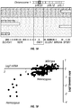

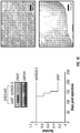

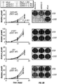

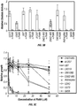


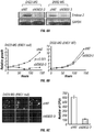


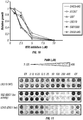

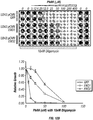


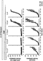
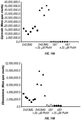


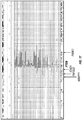


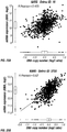

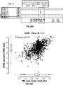

Classifications
-
- A—HUMAN NECESSITIES
- A61—MEDICAL OR VETERINARY SCIENCE; HYGIENE
- A61K—PREPARATIONS FOR MEDICAL, DENTAL OR TOILETRY PURPOSES
- A61K31/00—Medicinal preparations containing organic active ingredients
- A61K31/70—Carbohydrates; Sugars; Derivatives thereof
- A61K31/7088—Compounds having three or more nucleosides or nucleotides
- A61K31/713—Double-stranded nucleic acids or oligonucleotides
-
- A—HUMAN NECESSITIES
- A61—MEDICAL OR VETERINARY SCIENCE; HYGIENE
- A61K—PREPARATIONS FOR MEDICAL, DENTAL OR TOILETRY PURPOSES
- A61K31/00—Medicinal preparations containing organic active ingredients
- A61K31/185—Acids; Anhydrides, halides or salts thereof, e.g. sulfur acids, imidic, hydrazonic or hydroximic acids
- A61K31/19—Carboxylic acids, e.g. valproic acid
- A61K31/191—Carboxylic acids, e.g. valproic acid having two or more hydroxy groups, e.g. gluconic acid
-
- A—HUMAN NECESSITIES
- A61—MEDICAL OR VETERINARY SCIENCE; HYGIENE
- A61K—PREPARATIONS FOR MEDICAL, DENTAL OR TOILETRY PURPOSES
- A61K31/00—Medicinal preparations containing organic active ingredients
- A61K31/185—Acids; Anhydrides, halides or salts thereof, e.g. sulfur acids, imidic, hydrazonic or hydroximic acids
- A61K31/19—Carboxylic acids, e.g. valproic acid
- A61K31/194—Carboxylic acids, e.g. valproic acid having two or more carboxyl groups, e.g. succinic, maleic or phthalic acid
-
- A—HUMAN NECESSITIES
- A61—MEDICAL OR VETERINARY SCIENCE; HYGIENE
- A61K—PREPARATIONS FOR MEDICAL, DENTAL OR TOILETRY PURPOSES
- A61K31/00—Medicinal preparations containing organic active ingredients
- A61K31/185—Acids; Anhydrides, halides or salts thereof, e.g. sulfur acids, imidic, hydrazonic or hydroximic acids
- A61K31/19—Carboxylic acids, e.g. valproic acid
- A61K31/195—Carboxylic acids, e.g. valproic acid having an amino group
- A61K31/197—Carboxylic acids, e.g. valproic acid having an amino group the amino and the carboxyl groups being attached to the same acyclic carbon chain, e.g. gamma-aminobutyric acid [GABA], beta-alanine, epsilon-aminocaproic acid, pantothenic acid
- A61K31/198—Alpha-aminoacids, e.g. alanine, edetic acids [EDTA]
-
- A—HUMAN NECESSITIES
- A61—MEDICAL OR VETERINARY SCIENCE; HYGIENE
- A61K—PREPARATIONS FOR MEDICAL, DENTAL OR TOILETRY PURPOSES
- A61K31/00—Medicinal preparations containing organic active ingredients
- A61K31/33—Heterocyclic compounds
- A61K31/335—Heterocyclic compounds having oxygen as the only ring hetero atom, e.g. fungichromin
- A61K31/35—Heterocyclic compounds having oxygen as the only ring hetero atom, e.g. fungichromin having six-membered rings with one oxygen as the only ring hetero atom
- A61K31/352—Heterocyclic compounds having oxygen as the only ring hetero atom, e.g. fungichromin having six-membered rings with one oxygen as the only ring hetero atom condensed with carbocyclic rings, e.g. methantheline
-
- A—HUMAN NECESSITIES
- A61—MEDICAL OR VETERINARY SCIENCE; HYGIENE
- A61K—PREPARATIONS FOR MEDICAL, DENTAL OR TOILETRY PURPOSES
- A61K31/00—Medicinal preparations containing organic active ingredients
- A61K31/33—Heterocyclic compounds
- A61K31/335—Heterocyclic compounds having oxygen as the only ring hetero atom, e.g. fungichromin
- A61K31/357—Heterocyclic compounds having oxygen as the only ring hetero atom, e.g. fungichromin having two or more oxygen atoms in the same ring, e.g. crown ethers, guanadrel
-
- A—HUMAN NECESSITIES
- A61—MEDICAL OR VETERINARY SCIENCE; HYGIENE
- A61K—PREPARATIONS FOR MEDICAL, DENTAL OR TOILETRY PURPOSES
- A61K31/00—Medicinal preparations containing organic active ingredients
- A61K31/56—Compounds containing cyclopenta[a]hydrophenanthrene ring systems; Derivatives thereof, e.g. steroids
- A61K31/565—Compounds containing cyclopenta[a]hydrophenanthrene ring systems; Derivatives thereof, e.g. steroids not substituted in position 17 beta by a carbon atom, e.g. estrane, estradiol
- A61K31/568—Compounds containing cyclopenta[a]hydrophenanthrene ring systems; Derivatives thereof, e.g. steroids not substituted in position 17 beta by a carbon atom, e.g. estrane, estradiol substituted in positions 10 and 13 by a chain having at least one carbon atom, e.g. androstanes, e.g. testosterone
- A61K31/5685—Compounds containing cyclopenta[a]hydrophenanthrene ring systems; Derivatives thereof, e.g. steroids not substituted in position 17 beta by a carbon atom, e.g. estrane, estradiol substituted in positions 10 and 13 by a chain having at least one carbon atom, e.g. androstanes, e.g. testosterone having an oxo group in position 17, e.g. androsterone
-
- A—HUMAN NECESSITIES
- A61—MEDICAL OR VETERINARY SCIENCE; HYGIENE
- A61K—PREPARATIONS FOR MEDICAL, DENTAL OR TOILETRY PURPOSES
- A61K31/00—Medicinal preparations containing organic active ingredients
- A61K31/66—Phosphorus compounds
-
- A—HUMAN NECESSITIES
- A61—MEDICAL OR VETERINARY SCIENCE; HYGIENE
- A61K—PREPARATIONS FOR MEDICAL, DENTAL OR TOILETRY PURPOSES
- A61K31/00—Medicinal preparations containing organic active ingredients
- A61K31/66—Phosphorus compounds
- A61K31/661—Phosphorus acids or esters thereof not having P—C bonds, e.g. fosfosal, dichlorvos, malathion or mevinphos
-
- A—HUMAN NECESSITIES
- A61—MEDICAL OR VETERINARY SCIENCE; HYGIENE
- A61K—PREPARATIONS FOR MEDICAL, DENTAL OR TOILETRY PURPOSES
- A61K31/00—Medicinal preparations containing organic active ingredients
- A61K31/66—Phosphorus compounds
- A61K31/662—Phosphorus acids or esters thereof having P—C bonds, e.g. foscarnet, trichlorfon
-
- A—HUMAN NECESSITIES
- A61—MEDICAL OR VETERINARY SCIENCE; HYGIENE
- A61K—PREPARATIONS FOR MEDICAL, DENTAL OR TOILETRY PURPOSES
- A61K38/00—Medicinal preparations containing peptides
- A61K38/16—Peptides having more than 20 amino acids; Gastrins; Somatostatins; Melanotropins; Derivatives thereof
- A61K38/43—Enzymes; Proenzymes; Derivatives thereof
- A61K38/45—Transferases (2)
-
- A—HUMAN NECESSITIES
- A61—MEDICAL OR VETERINARY SCIENCE; HYGIENE
- A61K—PREPARATIONS FOR MEDICAL, DENTAL OR TOILETRY PURPOSES
- A61K38/00—Medicinal preparations containing peptides
- A61K38/16—Peptides having more than 20 amino acids; Gastrins; Somatostatins; Melanotropins; Derivatives thereof
- A61K38/43—Enzymes; Proenzymes; Derivatives thereof
- A61K38/52—Isomerases (5)
-
- A—HUMAN NECESSITIES
- A61—MEDICAL OR VETERINARY SCIENCE; HYGIENE
- A61K—PREPARATIONS FOR MEDICAL, DENTAL OR TOILETRY PURPOSES
- A61K45/00—Medicinal preparations containing active ingredients not provided for in groups A61K31/00 - A61K41/00
- A61K45/06—Mixtures of active ingredients without chemical characterisation, e.g. antiphlogistics and cardiaca
-
- A—HUMAN NECESSITIES
- A61—MEDICAL OR VETERINARY SCIENCE; HYGIENE
- A61P—SPECIFIC THERAPEUTIC ACTIVITY OF CHEMICAL COMPOUNDS OR MEDICINAL PREPARATIONS
- A61P35/00—Antineoplastic agents
-
- A—HUMAN NECESSITIES
- A61—MEDICAL OR VETERINARY SCIENCE; HYGIENE
- A61P—SPECIFIC THERAPEUTIC ACTIVITY OF CHEMICAL COMPOUNDS OR MEDICINAL PREPARATIONS
- A61P35/00—Antineoplastic agents
- A61P35/02—Antineoplastic agents specific for leukemia
-
- A—HUMAN NECESSITIES
- A61—MEDICAL OR VETERINARY SCIENCE; HYGIENE
- A61P—SPECIFIC THERAPEUTIC ACTIVITY OF CHEMICAL COMPOUNDS OR MEDICINAL PREPARATIONS
- A61P35/00—Antineoplastic agents
- A61P35/04—Antineoplastic agents specific for metastasis
-
- A—HUMAN NECESSITIES
- A61—MEDICAL OR VETERINARY SCIENCE; HYGIENE
- A61P—SPECIFIC THERAPEUTIC ACTIVITY OF CHEMICAL COMPOUNDS OR MEDICINAL PREPARATIONS
- A61P43/00—Drugs for specific purposes, not provided for in groups A61P1/00-A61P41/00
-
- C—CHEMISTRY; METALLURGY
- C12—BIOCHEMISTRY; BEER; SPIRITS; WINE; VINEGAR; MICROBIOLOGY; ENZYMOLOGY; MUTATION OR GENETIC ENGINEERING
- C12N—MICROORGANISMS OR ENZYMES; COMPOSITIONS THEREOF; PROPAGATING, PRESERVING, OR MAINTAINING MICROORGANISMS; MUTATION OR GENETIC ENGINEERING; CULTURE MEDIA
- C12N15/00—Mutation or genetic engineering; DNA or RNA concerning genetic engineering, vectors, e.g. plasmids, or their isolation, preparation or purification; Use of hosts therefor
- C12N15/09—Recombinant DNA-technology
- C12N15/11—DNA or RNA fragments; Modified forms thereof; Non-coding nucleic acids having a biological activity
- C12N15/113—Non-coding nucleic acids modulating the expression of genes, e.g. antisense oligonucleotides; Antisense DNA or RNA; Triplex- forming oligonucleotides; Catalytic nucleic acids, e.g. ribozymes; Nucleic acids used in co-suppression or gene silencing
- C12N15/1137—Non-coding nucleic acids modulating the expression of genes, e.g. antisense oligonucleotides; Antisense DNA or RNA; Triplex- forming oligonucleotides; Catalytic nucleic acids, e.g. ribozymes; Nucleic acids used in co-suppression or gene silencing against enzymes
-
- C—CHEMISTRY; METALLURGY
- C12—BIOCHEMISTRY; BEER; SPIRITS; WINE; VINEGAR; MICROBIOLOGY; ENZYMOLOGY; MUTATION OR GENETIC ENGINEERING
- C12Q—MEASURING OR TESTING PROCESSES INVOLVING ENZYMES, NUCLEIC ACIDS OR MICROORGANISMS; COMPOSITIONS OR TEST PAPERS THEREFOR; PROCESSES OF PREPARING SUCH COMPOSITIONS; CONDITION-RESPONSIVE CONTROL IN MICROBIOLOGICAL OR ENZYMOLOGICAL PROCESSES
- C12Q1/00—Measuring or testing processes involving enzymes, nucleic acids or microorganisms; Compositions therefor; Processes of preparing such compositions
- C12Q1/68—Measuring or testing processes involving enzymes, nucleic acids or microorganisms; Compositions therefor; Processes of preparing such compositions involving nucleic acids
- C12Q1/6876—Nucleic acid products used in the analysis of nucleic acids, e.g. primers or probes
- C12Q1/6883—Nucleic acid products used in the analysis of nucleic acids, e.g. primers or probes for diseases caused by alterations of genetic material
- C12Q1/6886—Nucleic acid products used in the analysis of nucleic acids, e.g. primers or probes for diseases caused by alterations of genetic material for cancer
-
- G—PHYSICS
- G01—MEASURING; TESTING
- G01N—INVESTIGATING OR ANALYSING MATERIALS BY DETERMINING THEIR CHEMICAL OR PHYSICAL PROPERTIES
- G01N33/00—Investigating or analysing materials by specific methods not covered by groups G01N1/00 - G01N31/00
- G01N33/48—Biological material, e.g. blood, urine; Haemocytometers
- G01N33/50—Chemical analysis of biological material, e.g. blood, urine; Testing involving biospecific ligand binding methods; Immunological testing
- G01N33/5005—Chemical analysis of biological material, e.g. blood, urine; Testing involving biospecific ligand binding methods; Immunological testing involving human or animal cells
- G01N33/5008—Chemical analysis of biological material, e.g. blood, urine; Testing involving biospecific ligand binding methods; Immunological testing involving human or animal cells for testing or evaluating the effect of chemical or biological compounds, e.g. drugs, cosmetics
- G01N33/502—Chemical analysis of biological material, e.g. blood, urine; Testing involving biospecific ligand binding methods; Immunological testing involving human or animal cells for testing or evaluating the effect of chemical or biological compounds, e.g. drugs, cosmetics for testing non-proliferative effects
- G01N33/5038—Chemical analysis of biological material, e.g. blood, urine; Testing involving biospecific ligand binding methods; Immunological testing involving human or animal cells for testing or evaluating the effect of chemical or biological compounds, e.g. drugs, cosmetics for testing non-proliferative effects involving detection of metabolites per se
-
- G—PHYSICS
- G01—MEASURING; TESTING
- G01N—INVESTIGATING OR ANALYSING MATERIALS BY DETERMINING THEIR CHEMICAL OR PHYSICAL PROPERTIES
- G01N33/00—Investigating or analysing materials by specific methods not covered by groups G01N1/00 - G01N31/00
- G01N33/48—Biological material, e.g. blood, urine; Haemocytometers
- G01N33/50—Chemical analysis of biological material, e.g. blood, urine; Testing involving biospecific ligand binding methods; Immunological testing
- G01N33/53—Immunoassay; Biospecific binding assay; Materials therefor
- G01N33/574—Immunoassay; Biospecific binding assay; Materials therefor for cancer
- G01N33/57407—Specifically defined cancers
- G01N33/5743—Specifically defined cancers of skin, e.g. melanoma
-
- A—HUMAN NECESSITIES
- A61—MEDICAL OR VETERINARY SCIENCE; HYGIENE
- A61K—PREPARATIONS FOR MEDICAL, DENTAL OR TOILETRY PURPOSES
- A61K48/00—Medicinal preparations containing genetic material which is inserted into cells of the living body to treat genetic diseases; Gene therapy
-
- C—CHEMISTRY; METALLURGY
- C12—BIOCHEMISTRY; BEER; SPIRITS; WINE; VINEGAR; MICROBIOLOGY; ENZYMOLOGY; MUTATION OR GENETIC ENGINEERING
- C12N—MICROORGANISMS OR ENZYMES; COMPOSITIONS THEREOF; PROPAGATING, PRESERVING, OR MAINTAINING MICROORGANISMS; MUTATION OR GENETIC ENGINEERING; CULTURE MEDIA
- C12N2310/00—Structure or type of the nucleic acid
- C12N2310/10—Type of nucleic acid
- C12N2310/14—Type of nucleic acid interfering N.A.
-
- C—CHEMISTRY; METALLURGY
- C12—BIOCHEMISTRY; BEER; SPIRITS; WINE; VINEGAR; MICROBIOLOGY; ENZYMOLOGY; MUTATION OR GENETIC ENGINEERING
- C12N—MICROORGANISMS OR ENZYMES; COMPOSITIONS THEREOF; PROPAGATING, PRESERVING, OR MAINTAINING MICROORGANISMS; MUTATION OR GENETIC ENGINEERING; CULTURE MEDIA
- C12N2310/00—Structure or type of the nucleic acid
- C12N2310/50—Physical structure
- C12N2310/53—Physical structure partially self-complementary or closed
- C12N2310/531—Stem-loop; Hairpin
-
- C—CHEMISTRY; METALLURGY
- C12—BIOCHEMISTRY; BEER; SPIRITS; WINE; VINEGAR; MICROBIOLOGY; ENZYMOLOGY; MUTATION OR GENETIC ENGINEERING
- C12N—MICROORGANISMS OR ENZYMES; COMPOSITIONS THEREOF; PROPAGATING, PRESERVING, OR MAINTAINING MICROORGANISMS; MUTATION OR GENETIC ENGINEERING; CULTURE MEDIA
- C12N2320/00—Applications; Uses
- C12N2320/30—Special therapeutic applications
- C12N2320/31—Combination therapy
-
- C—CHEMISTRY; METALLURGY
- C12—BIOCHEMISTRY; BEER; SPIRITS; WINE; VINEGAR; MICROBIOLOGY; ENZYMOLOGY; MUTATION OR GENETIC ENGINEERING
- C12Q—MEASURING OR TESTING PROCESSES INVOLVING ENZYMES, NUCLEIC ACIDS OR MICROORGANISMS; COMPOSITIONS OR TEST PAPERS THEREFOR; PROCESSES OF PREPARING SUCH COMPOSITIONS; CONDITION-RESPONSIVE CONTROL IN MICROBIOLOGICAL OR ENZYMOLOGICAL PROCESSES
- C12Q2600/00—Oligonucleotides characterized by their use
- C12Q2600/106—Pharmacogenomics, i.e. genetic variability in individual responses to drugs and drug metabolism
-
- C—CHEMISTRY; METALLURGY
- C12—BIOCHEMISTRY; BEER; SPIRITS; WINE; VINEGAR; MICROBIOLOGY; ENZYMOLOGY; MUTATION OR GENETIC ENGINEERING
- C12Q—MEASURING OR TESTING PROCESSES INVOLVING ENZYMES, NUCLEIC ACIDS OR MICROORGANISMS; COMPOSITIONS OR TEST PAPERS THEREFOR; PROCESSES OF PREPARING SUCH COMPOSITIONS; CONDITION-RESPONSIVE CONTROL IN MICROBIOLOGICAL OR ENZYMOLOGICAL PROCESSES
- C12Q2600/00—Oligonucleotides characterized by their use
- C12Q2600/156—Polymorphic or mutational markers
-
- C—CHEMISTRY; METALLURGY
- C12—BIOCHEMISTRY; BEER; SPIRITS; WINE; VINEGAR; MICROBIOLOGY; ENZYMOLOGY; MUTATION OR GENETIC ENGINEERING
- C12Y—ENZYMES
- C12Y402/00—Carbon-oxygen lyases (4.2)
- C12Y402/01—Hydro-lyases (4.2.1)
- C12Y402/01001—Carbonate dehydratase (4.2.1.1), i.e. carbonic anhydrase
-
- C—CHEMISTRY; METALLURGY
- C12—BIOCHEMISTRY; BEER; SPIRITS; WINE; VINEGAR; MICROBIOLOGY; ENZYMOLOGY; MUTATION OR GENETIC ENGINEERING
- C12Y—ENZYMES
- C12Y402/00—Carbon-oxygen lyases (4.2)
- C12Y402/01—Hydro-lyases (4.2.1)
- C12Y402/01011—Phosphopyruvate hydratase (4.2.1.11), i.e. enolase
-
- G—PHYSICS
- G01—MEASURING; TESTING
- G01N—INVESTIGATING OR ANALYSING MATERIALS BY DETERMINING THEIR CHEMICAL OR PHYSICAL PROPERTIES
- G01N2333/00—Assays involving biological materials from specific organisms or of a specific nature
- G01N2333/90—Enzymes; Proenzymes
- G01N2333/988—Lyases (4.), e.g. aldolases, heparinase, enolases, fumarase
-
- G—PHYSICS
- G01—MEASURING; TESTING
- G01N—INVESTIGATING OR ANALYSING MATERIALS BY DETERMINING THEIR CHEMICAL OR PHYSICAL PROPERTIES
- G01N2800/00—Detection or diagnosis of diseases
- G01N2800/52—Predicting or monitoring the response to treatment, e.g. for selection of therapy based on assay results in personalised medicine; Prognosis
Definitions
- the present invention relates generally to the field of molecular biology and oncology. More particularly, it concerns methods for identifying and treating cancers that have one copy of an essential gene functionally inactivated (e.g., by deletion or mutation of all or part of the gene coding sequence, epigenetic silencing of a locus, among other mechanisms).
- heterozygous deletion of large regions of the genome is a prototypic somatic event in cancer.
- the genetic instability exhibited by cancer cells drives tumor genesis by ablating the function of critical tumor suppressor genes.
- deletions are often large and can encompass neighboring genes with no known role in cancer pathogenesis. To date there has been no guidance as to how such mutations in tumor cells could be harnessed to improve cancer diagnosis or therapy.
- Kaelin et al. (Nature Reviews Cancer 5, 689-698 (2005 )) describes the concept of synthetic lethality in the context of anti-cancer therapy.
- Kamb et al. (Molecular Biology of the cell 14, 2201-2205 (2003 )) describes the consequences of nonadaptive alterations in cancer.
- the present invention relates to a composition for use in treating a cancer in a subject, wherein the cancer has a homozygous deletion in a housekeeping gene and the housekeeping gene has a functionally redundant homologue, the composition comprising an inhibitor of the redundant homologue in an amount sufficient to inhibit the activity of the redundant homologue, wherein the housekeeping gene is enolase 1 (ENO1), the redundant homologue is endolase 2 (ENO2) and the inhibitor is a nucleic acid that inhibits the expression or activity of the redundant homologue, an antibody that specifically binds the redundant homologue, phosphonoacetohydroxamate, preferably further comprising a chemotherapeutic agent, wherein the chemotherapeutic agent preferably interferes with DNA homeostasis.
- ENO1 enolase 1
- ENO2 endolase 2
- the inhibitor is a nucleic acid that inhibits the expression or activity of the redundant homologue
- an antibody that specifically binds the redundant homologue phosphonoacetohydrox
- the present invention relates to a composition for use in treating a cancer in a subject, wherein the cancer has a homozygous deletion in a housekeeping gene and the housekeeping gene has a functionally redundant homologue, the composition comprising an inhibitor of the redundant homologue in an amount sufficient to inhibit the activity of the redundant homologue, wherein the housekeeping gene is hexose-6-phosphate dehydrogenase (H6PD), the redundant homologue is glucose-6 dehydrogenase (G6PD) and the inhibitor is a nucleic acid that inhibits the expression or activity of the redundant homologue, an antibody that specifically binds the redundant homologue, dehydroepiandrosterone, preferably further comprising a chemotherapeutic agent, wherein the chemotherapeutic agent preferably interferes with DNA homeostasis.
- H6PD hexose-6-phosphate dehydrogenase
- G6PD glucose-6 dehydrogenase
- the inhibitor is a nucleic acid that inhibits the expression
- the present invention relates to a composition for use in treating a cancer in a subject, wherein the cancer has a homozygous deletion in a housekeeping gene and the housekeeping gene has a functionally redundant homologue, the composition comprising an inhibitor of the redundant homologue in an amount sufficient to inhibit the activity of the redundant homologue, wherein the housekeeping gene is Nicotinamide nucleotide adenylyl transferase 1 (NMNAT1), the redundant homologue is Nicotinamide nucleotide adenylyl transferase 2 (NMNAT2) or nicotinamide nucleotide adenylyl transferase 3 (NMNAT3) and the inhibitor is a nucleic acid that inhibits the expression or activity of the redundant homologue, an antibody that specifically binds the redundant homologue, Np2AD, Np4AD or Nap4AD, preferably further comprising a chemotherapeutic agent, wherein the chemotherapeutic agent preferably interferes with DNA homeostasis
- the present invention relates to a composition for use in treating a cancer in a subject, wherein the cancer has a homozygous deletion in a housekeeping gene and the housekeeping gene has a functionally redundant homologue, the composition comprising an inhibitor of the redundant homologue in an amount sufficient to inhibit the activity of the redundant homologue, wherein
- cancers have homozygous deletions in redundant housekeeping genes, and that this can be therapeutically exploited by targeting the non-deleted redundant homologue.
- cancers comprising heterozygous deletions in housekeeping genes are also rendered susceptible to therapeutic treatment targeting the non-deleted copy of the housekeeping gene.
- cancer cells comprising heterozygous deletions in two (or more) housekeeping genes are rendered susceptible to, and can be treated with, therapeutic treatment targeting the non-deleted copy of each of the housekeeping genes that comprise the heterozygous deletion.
- the cancer has a homozygous deletion in a housekeeping gene and the housekeeping gene has a functionally redundant homologue.
- the subject is treated by contacting the cell with an inhibitor of the redundant homologue in an amount sufficient to inhibit the activity of the redundant homologue.
- the cell has a homozygous deletion in a housekeeping gene and the housekeeping gene has a functionally redundant homologue.
- Cell proliferation and/or cell death is induced by contacting the cell with an inhibitor of the redundant homologue in an amount sufficient to inhibit the activity of the redundant homologue.
- the cell is further contacted with a chemotherapeutic agent.
- the chemotherapeutic agent may interfere with DNA homeostasis.
- the inhibitor can be, for example, a nucleic acid that inhibits the expression or activity of the redundant homologue, an antibody that specifically binds the redundant homologue or a small molecule.
- Inhibitors include for example phosphonoacetohydroxamate, dehydroepiandrosterone, Np2AD, Np4AD, Nap4AD, fluorocitrate or hopantenate or derivatives thereof.
- the housekeeping gene is endolase 1 (ENO1) and the redundant homologue is endolase 2 (ENO2); the housekeeping gene is hexose-6-phosphate dehydrogenase (H6PD) and the redundant homologue is glucose-6 dehydrogenase (G6PD); the housekeeping gene is kinesin family member 1B (KIF1B) and the redundant homologue is a kinesin family member 1A (KIF1A) or kinesin family member 1C (KIF1A); the housekeeping gene is Nicotinamide nucleotide adenylyl transferase 1 (NMNAT1) and the redundant homologue is Nicotinamide nucleotide adenylyl transferase 2 (NMNAT2) or nicotinamide nucleotide adenylyl transferase 3 (NMNAT3); the housekeeping gene is ubiquitination factor E4B (UBE4B) and the redundant homologue is ubiquitination factor 4A
- Described is a method for treating a subject having a cancer comprising administering at least a first glycolysis inhibitor to the subject, wherein it was determined that cells of the cancer comprise a heterozygous mutation that inactivates one copy of the Enolase 1 (ENO1) gene.
- a method for treating a subject having a cancer comprising (a) selecting a subject determined to comprise cancer cells having a heterozygous mutation that inactivates one copy of the ENO1 gene; and (b) administering at least a first glycolysis inhibitor to the subject.
- a method for selecting a subject having a cancer for a glycolysis inhibitor therapy comprising determining whether cancer cells of the subject comprise a heterozygous mutation that inactivates one copy of the ENO1 gene, wherein if the cancer cells comprise the heterozygous mutation, then the subject is selected for a glycolysis inhibitor therapy.
- a method for selecting a subject having a cancer for a glycolysis inhibitor therapy comprising (a) determining whether cancer cells of the subject comprise a heterozygous mutation that inactivates one copy of the ENO1 gene; and (b) selecting a subject for a glycolysis inhibitor therapy if cancer cells of the subject comprise the heterozygous mutation.
- a method for predicting a response to a glycolysis inhibitor therapy in a subject having a cancer comprising determining whether cancer cells of the subject comprise a heterozygous mutation that inactivates one copy of the ENO1 gene, wherein if the cancer cells comprise the heterozygous mutation, then the subject is predicted to have a favorable response to a glycolysis inhibitor therapy.
- a method for predicting a response to a glycolysis inhibitor therapy in a subject having a cancer comprising (a) determining whether cancer cells of the subject comprise a heterozygous mutation that inactivates one copy of the ENO1 gene; and (b) identifying the subject as predicted to have a favorable response to a glycolysis inhibitor therapy, if cancer cells from the subject comprise the heterozygous mutation; or identifying the subject as not predicted to have a favorable response to a glycolysis inhibitor therapy, if cancer cells from the subject do not comprise the heterozygous mutation.
- a method for treating a subject having a cancer comprising administering at least a t-RNA synthetase (ARS) inhibitor to the subject, wherein it was determined that cells of the cancer comprise a heterozygous mutation that inactivates one copy of an ARS gene.
- ARS t-RNA synthetase
- a method for treating a subject having a cancer comprising (a) selecting a subject determined to comprise cancer cells having a heterozygous mutation that inactivates one copy of an ARS gene; and (b) administering at least a first ARS inhibitor to the subject.
- a method for selecting a subject having a cancer for an ARS inhibitor therapy comprising determining whether cancer cells of the subject comprise a heterozygous mutation that inactivates one copy of an ARS gene, wherein if the cancer cells comprise the heterozygous mutation, then the subject is selected for an ARS inhibitor therapy.
- a method for selecting a subject having a cancer for an ARS inhibitor therapy comprising (a) determining whether cancer cells of the subject comprise a heterozygous mutation that inactivates one copy of an ARS gene (or two or more ARS genes); and (b) selecting a subject for an ARS inhibitor therapy if cancer cells of the subject comprise the heterozygous mutation.
- a method for predicting a response to an ARS inhibitor therapy in a subject having a cancer comprising determining whether cancer cells of the subject comprise a heterozygous mutation that inactivates one copy of an ARS gene, wherein if the cancer cells comprise the heterozygous mutation, then the subject is predicted to have a favorable response to an ARS inhibitor therapy.
- a method for predicting a response to an ARS inhibitor therapy in a subject having a cancer comprising (a) determining whether cancer cells of the subject comprise a heterozygous mutation that inactivates one copy of an ARS gene; and (b) identifying the subject as predicted to have a favorable response to an ARS inhibitor therapy, if cancer cells from the subject comprise the heterozygous mutation; or identifying the subject as not predicted to have a favorable response to an ARS inhibitor therapy, if cancer cells from the subject do not comprise the heterozygous mutation.
- a method for treating a subject having a cancer comprising administering at least a first phosphogluconate dehydrogenase inhibitor to the subject, wherein it was determined that cells of the cancer comprise a heterozygous mutation that inactivates one copy of the phosphogluconate dehydrogenase (PGD) gene.
- PGD phosphogluconate dehydrogenase
- a method for treating a subject having a cancer comprising (a) selecting a subject determined to comprise cancer cells having a heterozygous mutation that inactivates one copy of the PGD gene; and (b) administering at least a first phosphogluconate dehydrogenase inhibitor (e.g., 6-aminonicotinamide) to the subject.
- a first phosphogluconate dehydrogenase inhibitor e.g., 6-aminonicotinamide
- a method for selecting a subject having a cancer for a phosphogluconate dehydrogenase inhibitor therapy comprising determining whether cancer cells of the subject comprise a heterozygous mutation that inactivates one copy of the PGD gene, wherein if the cancer cells comprise the heterozygous mutation, then the subject is selected for a phosphogluconate dehydrogenase inhibitor therapy.
- a method for selecting a subject having a cancer for a phosphogluconate dehydrogenase inhibitor therapy comprising (a) determining whether cancer cells of the subject comprise a heterozygous mutation that inactivates one copy of the PGD gene; and (b) selecting a subject for a phosphogluconate dehydrogenase inhibitor therapy if cancer cells of the subject comprise the heterozygous mutation.
- a method for predicting a response to a phosphogluconate dehydrogenase inhibitor therapy in a subject having a cancer comprising determining whether cancer cells of the subject comprise a heterozygous mutation that inactivates one copy of the PGD gene, wherein if the cancer cells comprise the heterozygous mutation, then the subject is predicted to have a favorable response to a phosphogluconate dehydrogenase inhibitor therapy.
- a method for predicting a response to a phosphogluconate dehydrogenase inhibitor therapy in a subject having a cancer comprising (a) determining whether cancer cells of the subject comprise a heterozygous mutation that inactivates one copy of the PGD gene; and (b) identifying the subject as predicted to have a favorable response to a phosphogluconate dehydrogenase inhibitor therapy, if cancer cells from the subject comprise the heterozygous mutation; or identifying the subject as not predicted to have a favorable response to a phosphogluconate dehydrogenase inhibitor therapy, if cancer cells from the subject do not comprise the heterozygous mutation.
- a method for treating a subject having a cancer comprising administering at least a first glycolysis inhibitor to the subject, wherein it was determined that cells of the cancer comprise a heterozygous mutation that inactivates one copy of the GAPDH, ENO2 and/or TPI genes (each located at 12p13).
- a method for treating a subject having a cancer comprising (a) selecting a subject determined to comprise cancer cells having a heterozygous mutation that inactivates one copy of the GAPDH, ENO2 and/or TPI genes; and (b) administering at least a first glycolysis inhibitor to the subject.
- a method for selecting a subject having a cancer for a glycolysis inhibitor therapy comprising determining whether cancer cells of the subject comprise a heterozygous mutation that inactivates one copy of the GAPDH, ENO2 and/or TPI genes, wherein if the cancer cells comprise the heterozygous mutation, then the subject is selected for a glycolysis inhibitor therapy.
- a method for selecting a subject having a cancer for a glycolysis inhibitor therapy comprising (a) determining whether cancer cells of the subject comprise a heterozygous mutation that inactivates one copy of the GAPDH, ENO2 and/or TPI genes; and (b) selecting a subject for a glycolysis inhibitor therapy if cancer cells of the subject comprise the heterozygous mutation.
- a method for predicting a response to a glycolysis inhibitor therapy in a subject having a cancer comprising determining whether cancer cells of the subject comprise a heterozygous mutation that inactivates one copy of the GAPDH, ENO2 and/or TPI genes, wherein if the cancer cells comprise the heterozygous mutation, then the subject is predicted to have a favorable response to a glycolysis inhibitor therapy.
- a method for predicting a response to a glycolysis inhibitor therapy in a subject having a cancer comprising (a) determining whether cancer cells of the subject comprise a heterozygous mutation that inactivates one copy of the GAPDH, ENO2 and/or TPI genes; and (b) identifying the subject as predicted to have a favorable response to a glycolysis inhibitor therapy, if cancer cells from the subject comprise a heterozygous mutation; or identifying the subject as not predicted to have a favorable response to a glycolysis inhibitor therapy, if cancer cells from the subject do not comprise one of the heterozygous mutations.
- cancers that comprise a heterozygous mutation that inactivates one copy an essential gene, such as one copy of the ENO1, an ARS gene, the PGD gene, the GAPDH gene, the ENO2 gene and/or the TPI gene.
- an essential gene such as one copy of the ENO1, an ARS gene, the PGD gene, the GAPDH gene, the ENO2 gene and/or the TPI gene.
- a heterozygous mutation refers to a mutation in only one of the two copies of the gene (in a diploid cell or of the loss of one in a triploid cell, or more broadly, loss of all but one copy in a polyploid cell).
- the heterozygous mutation can be a deletion, substitution, inversion, rearrangement or insertion that functionally inactivated one copy of ENO1.
- the mutation may be a deletion, such as a loss of heterozygosity at chromosome 1p36.
- a heterozygous deletion can encompass more than ENO1 gene, such as a deletion that inactivates ENO1 as well as RERE, CA6 and/or SLC2A7.
- a method can involve determining a deletion 5' and 3' of ENO1 and thereby determine that ENO1 is inactivated (deleted) indirectly.
- genes position 5' to ENO1 include, without limitation, NPHP4, KCNAB2, CHD5, RPL22, RNF207, C1orf211, GPR153, ICMT, HES3, ACOT7, TNFRSF25, PLEKHG5, DNAJC11, HES2, ESPN, MIR4252, NOL9, TAS1R1, ZBTB48, KLHL21, PHF13, THAP3, CAMTA1, VAMP3, PER3, UTS2, TNFRSF9, TRNA_Pseudo, PARK7, AX747125, ERRFI1, SLC45A1, RERE and BC113958:
- Non-limiting examples of genes position 3' to ENO1 include, CA6, SLC2A7, SLC2A5, GPR157, mir-34, MIR34A, H6PD, SPSB1, 5S rRNA, SLC25A33, TMEM201, PIK3CD, Clorf200, BC038541, CLSTN1, CTNNBIP1,
- a cancer comprises a heterozygous mutation wherein an entire arm of chromosome 1 is lost.
- a heterozygous deletion of ENO1 can be determined by cytogenetic chromosome spreads or copy number analysis of random DNA fragments upstream and downstream of the ENO1 gene.
- the heterozygous mutation can be a deletion, substitution, inversion, rearrangement or insertion that functionally inactivated one copy of GAPDH, ENO2 and/or TPI genes.
- the mutation is a deletion, such as a loss of heterozygosity at chromosome 12p13.
- a heterozygous deletion can encompass more than GAPDH, ENO2 and/or TPI genes, such as a deletion that inactivates two or all three of the genes.
- the heterozygous mutation can be a deletion, substitution, inversion, rearrangement or insertion that functionally inactivated one copy of an ARS gene, such one copy of the EPARS (aka EPRS), VARS, IARS, CARS, SARS, YARS, AARS, KARS, LARS, HARS, RARS or TARS gene.
- the mutation is a deletion, such as a loss of heterozygosity at a chromosomal locus comprising the gene.
- a cancer cell can comprise a loss of heterozygosity at 5q13, 1q41, 6p21, 9q24, 11p15, 1p13, 1p35, 16q13, 16q24, 5q31, 5q32 or 5q35.
- a heterozygous deletion can encompass more than one ARS gene, such as a deletion that inactivates SARS as well as YARS; AARS and KARS; LARS and HARS or LARS HARS and RARS.
- the heterozygous mutation can be a deletion, substitution, inversion, rearrangement or insertion that functionally inactivated one copy of a PGD gene.
- the mutation is a deletion, such as a loss of heterozygosity at a chromosomal locus comprising the gene, e.g ., comprising 1p36.22.
- a deletion can encompass additional portions of the chromosome 1p.
- a cancer cell can comprise a deletion that comprises ENO1 and PGD.
- a cancer identified as comprising an inactivation of one copy of both ENO1 and PGD can be treated with at least a first glycolysis inhibitor in conjunction with a PGD inhibitor, such as 6-aminonicotinamide.
- glycolysis inhibitors include, with limitation inhibitors of pyruvate kinase (e.g., PKLR1 or PKM2), enolase (e.g., ENO1, ENO2 or ENO3), phosphoglycerate mutase ( e.g ., PGM1, PGM2, PGM2L1, PGM3 or PGM5), phosphoglycerate kinase ( e.g ., PGK1 or PGK2), glyceraldehydes phosphate dehydrogenase (GAPDH), triosephosphate isomerase (TPI), fructose biphosphate aldolase ( e.g ., Aldoa, Aldob or Aldoc), phosphofructokinase (PFKL, PFKM or
- the inhibitor is an inhibitor of the mitochondrial electron transport chain.
- the glycolysis inhibitor is an inhibitory polynucleotide (e.g ., an siRNA, shRNA or miRNA) such as an inhibitory polynucleotide complementary to all or part of a gene encoding a pyruvate kinase, enolase (e.g ., ENO1), phosphoglycerate mutase, phosphoglycerate kinase, glyceraldehydes phosphate dehydrogenase, triosephosphate isomerase, fructose biphosphate aldolase, phosphofructokinase, phosphoglucose isomerase and hexokinase.
- an inhibitory polynucleotide e.g ., an siRNA, shRNA or miRNA
- an inhibitory polynucleotide e.g ., an siRNA, shRNA or miRNA
- glycolysis inhibitor is a small molecule inhibitor or a prodrug thereof.
- glycolysis inhibitors for use according to embodiments include, without limitation, 2-deoxyglucose, 6-aminonicotinamide, tetrose diphosphate, koningic acid and MJE3.
- a glycolysis inhibitor for use as a therapeutic is selected based on the heterozygous gene inactivation identified in the cancer. For example, if the cancer cell comprises a heterozygous inactivation of ENO1 then the glycolysis inhibitor therapy can comprise an enolase inhibitor. Likewise, in the case of a cancer has a heterozygous inactivation of a GAPDH, ENO2 or TPI gene, a glycolysis inhibitor therapy can comprise a GAPDH, enolase or TPI inhibitor, respectively. In certain cases, a cancer cell comprises a heterozygous inactivation of two or genes in the glycolysis pathway and a glycolysis inhibitor therapy comprises an inhibitor of each of the genes subject to the heterozygous deletion.
- a glycolysis inhibitor for use according to the embodiments is an enolase inhibitor, such as an enolase 1 inhibitor.
- the enolase 1 inhibitor can be an inhibitory polynucleotide, such as a siRNA, shRNA or miRNA complementary to all or part of an ENO1 gene (e.g ., complementary to all or part of an enolase 1 coding mRNA, see, e.g ., NCBI accession nos. NM_001201483.1 or NM_001428.3).
- the enolase inhibitor is a small molecule enolase inhibitor or a prodrug thereof.
- small molecule enolase inhibitors include, without limitation, D-tartronate semialdehyde phosphate; 3-aminoenolpyruvate-2-phosphate; phosphonoacetohydroaxamate (PhAH); 2-fluoro-2-phosphonoacetohydroxamate; (3-hydroxy-2-nitropropyl) phosphonate; (nitroethyl)phosphonate; d-(phosphonoethyl)nitrolate; fluorides and prodrugs of any of the foregoing.
- the ARS inhibitor is a febrifugine derivative, such as halofuginone (see, e.g., Sundrud et al., 2009 and Keller et al., 2012.
- the ARS inhibitor is anti-microbial ARS inhibitor.
- the ARS inhibitor can be an inhibitory polynucleotide, such as a siRNA, shRNA or miRNA complementary to all or part of an ARS gene.
- an ARS inhibitor is administered in conjunction with a further inhibitor of protein synthesis or an activator of amino acid starvation response.
- PGD inhibitors for use according to the embodiments include, for example, 6-aminonicotinamide or produgs thereof.
- the PGD inhibitor can be an inhibitory polynucleotide, such as a siRNA, shRNA or miRNA complementary to all or part of the PGD gene (see, e.g., NCBI accession no. NM_002631.2).
- Targeted inhibitors therapies for administration according to the embodiments are typically formulated in a pharmaceutically acceptable carrier.
- a therapy may be delivered, for example, intravenously, intradermally, intraarterially, intraperitoneally, intralesionally, intracranially, intraarticularly, intraprostaticaly, intrapleurally, intratracheally, intranasally, intravitreally, intravaginally, intrarectally, topically, intratumorally, intramuscularly, intraperitoneally, subcutaneously, subconjunctival, intravesicularlly, mucosally, intrapericardially, intraumbilically, intraocularally, orally, topically, locally, via inhalation (e.g.
- aerosol inhalation by injection or by infusion.
- the route of delivery can depend, for example, upon the type of cancer to be treated and the type of inhibitor that is used.
- An inhibitor such as a glycolysis, PDG or ARS inhibitor, can be administer two or more times (e.g., 3, 4, 5, 6, 7, 8, 9 or 10 times).
- the timing between doses of such a therapy can be varied and can include, without limitation, about 1, 2 or 3 days, about 1, 2, or 3 weeks or 1 month or more between doses.
- a method for treating a subject having a cancer comprising administering a targeted inhibitor therapy (e.g., a glycolysis, PDG or ARS inhibitor therapy) to the subject in conjunction with at least a second therapy.
- a targeted inhibitor therapy e.g., a glycolysis, PDG or ARS inhibitor therapy
- the second therapy may be administered before, after or during the targeted inhibitor therapy.
- the timing between a targeted inhibitor therapy and a second therapy can be varied and can include, without limitation, about 1, 2 or 3 days, about 1, 2, or 3 weeks or 1 month or more between the therapies.
- the second anticancer therapy may be, without limitation, a surgical therapy, chemotherapy, cancer cell-targeted therapy, radiation therapy, cryotherapy, hyperthermia treatment, phototherapy, radioablation therapy, hormonal therapy, immunotherapy, small molecule therapy, receptor kinase inhibitor therapy, anti-angiogenic therapy, cytokine therapy or a biological therapy such as treatment with a monoclonal antibody, siRNA, antisense oligonucleotide, ribozyme or gene therapy.
- the second therapy can comprise administration of a further, different, glycolysis inhibitor, such as an inhibitor that targets a different glycolysis pathway component relative to the first glycolysis inhibitor.
- the second therapy comprises administration of a mitochondrial electron transport chain inhibitor (e.g., mubritinib or oligomycin) or an inhibitor of the transcription factor Hif1 ⁇ .
- a subject such as a subject having a cancer.
- a subject can be human or non-human animal subject (e.g., a dog, cat, mouse, horse, etc).
- the subject has a cancer, such as an oral cancer, oropharyngeal cancer, nasopharyngeal cancer, respiratory cancer, urogenital cancer, gastrointestinal cancer, central or peripheral nervous system tissue cancer, an endocrine or neuroendocrine cancer or hematopoietic cancer, glioma, sarcoma, carcinoma, lymphoma, melanoma, fibroma, meningioma, brain cancer, oropharyngeal cancer, nasopharyngeal cancer, renal cancer, biliary cancer, pheochromocytoma, pancreatic islet cell cancer, Li-Fraumeni tumors, thyroid cancer, parathyroid cancer, pituitary tumors, adrenal gland tumors, osteogenic sarcoma tumor
- the cancer is a glioma, a glioblastoma, oligodendroglioma or a large cell neuroendocrine lung tumor.
- the cancer can be a melanoma, such as an ocular/uveal melanoma.
- the cancer is a metastatic cancer or a cancer that is resistant to a least a first chemotherapy.
- Some aspects of the disclosure concern determining whether cancer cells of a subject comprise a heterozygous mutation that inactivates one copy of the ENO1 gene.
- methods that can be used to determine whether cancer cells of the subject comprise the heterozygous mutation include, without limitation, DNA sequencing, polymerase chain reaction (PCR), nucleic acid hybridization (e.g., Southern blot or in situ hybridization) or DNA restriction analysis to detect an inactivating mutation (e.g ., a deletion, rearrangement, substitution, insertion or inversion).
- a method of the embodiments can comprise measuring an enolase expression or enolase activity.
- RNA expression of an enolase 1 mRNA or protein can be measured and compared to reference expression level to determine whether cancer cells of a subject comprise a heterozygous mutation that inactivates one copy of the ENO1 gene.
- Methods for measuring RNA expression include, without limitation, nucleic acid hybridization (e.g., Northern blot, in situ hybridization or array hybridization) and quantitative reverse transcription PCR.
- Non-limiting examples of methods for measuring enolase 1 protein expression include performing an ELISA, an immunoassay, a radioimmunoassay (RIA), Immunohistochemistry, an immunoradiometric assay, a fluoroimmunoassay, a chemiluminescent assay, a bioluminescent assay, a gel electrophoresis, a Western blot analysis, flow cytometry, positron emission tomography (PET), or single photon emission computed tomography (SPECT) imaging.
- enolase 1 activity can be measured (e.g ., by measuring substrate catalysis) and compared to reference expression level to determine whether cancer cells of a subject comprise a heterozygous mutation that inactivates one copy of the ENO1 gene.
- a biological sample e.g ., a sample comprising cancer cells or metabolites or nucleic acid therefrom
- the biological sample can be, without limitation, a blood, tissue (e.g ., biopsy), urine, stool or saliva sample.
- the sample is a tumor biopsy sample.
- a method may further comprise reporting whether cancer cells of the subject comprise a heterozygous mutation that inactivates one copy of the ENO1 gene or an ARS gene.
- a method can comprise reporting whether a subject was identified as predicted or not predicted to have a favorable response to a glycolysis inhibitor therapy (e.g ., an enolase inhibitor therapy) or an ARS inhibitor therapy.
- a glycolysis inhibitor therapy e.g ., an enolase inhibitor therapy
- an ARS inhibitor therapy e.g., an enolase inhibitor therapy
- such reporting can be by providing a written, electronic or oral report.
- a report is provided to the subject.
- the report is provided to a third party, such an insurance company or health care provider (e.g., a doctor or hospital).
- a favorable response can be a reduction in tumor size or burden, blocking of tumor growth, reduction in tumor-associated pain, reduction in cancer associated pathology, reduction in cancer associated symptoms, cancer non-progression, increased disease free interval, increased time to progression, induction of remission, reduction of metastasis, increased patient survival or an increase in the sensitivity of the tumor to an anticancer therapy.
- a kit comprising at least a first glycolysis inhibitor (e.g., an enolase inhibitor) and at least a first reagent for testing cells for a heterozygous mutation that inactivates one copy of the ENO1 gene.
- the glycolysis inhibitor can be any of those known in the art or detailed herein.
- the glycolysis inhibitor is an enolase inhibitor.
- reagents for use in testing cells include, without limitation, a nucleic acid molecule that bind with-in or adjacent to the ENO1 gene (e.g., an oligonucleotide primer), an anti-enolase 1 antibody and an enolase enzyme substrate (such as a labeled substrate).
- kits comprising an ARS inhibitor and a reagent for testing cells for a heterozygous mutation that inactivates one copy of an ARS gene.
- reagents for use in testing cells include, without limitation, a nucleic acid molecule that bind with-in or adjacent to an ARS gene (e.g ., an oligonucleotide primer), an anti-ARS antibody and an ARS enzyme substrate (such as a labeled substrate).
- Described is a method for monitoring the effectiveness of an enolase inhibitor therapy comprising determining the levels of glycerate and/or 1,3 dihydroxyacetone in a sample from a subject treated with an enolase inhibitor, wherein if the levels of glycerate or 1,3 dihydroxyacetone are not elevated as compared to a reference level the subject is in need of additional enolase inhibitor therapy and if the levels of glycerate or 1,3 dihydroxyacetone are elevated as compared to a reference level the subject is not in need of additional enolase inhibitor therapy.
- a method for monitoring the effectiveness of an enolase inhibitor therapy comprising (a) determining the levels of glycerate or 1,3 dihydroxyacetone in a sample from a subject treated with an enolase inhibitor; and (b) identifying the subject as in need of additional enolase inhibitor therapy if the levels of glycerate or 1,3 dihydroxyacetone are not elevated as compared to a reference level; or identifying the subject as not in need of additional enolase inhibitor therapy if the levels of glycerate or 1,3 dihydroxyacetone are elevated as compared to a reference level.
- a method may further comprise administering an additional enolase inhibitor therapy to a subject based on the levels of glycerate or 1,3 dihydroxyacetone.
- an additional enolase inhibitor therapy is an additional administration of an enolase inhibitor therapy or the administration of a higher dose of an enolase inhibitor.
- a method of the embodiments further comprises reporting a glycerate or 1,3 dihydroxyacetone level for a subject or reporting whether a subject is in need of additional enolase inhibitor therapy.
- Deletion e.g ., heterozygous deletion
- a portion of enzymatic activity e.g ., a 50% loss
- tumor cells are typically tolerated even in the case of essential, housekeeping genes.
- a 50% loss is typically tolerated even in the case of essential, housekeeping genes.
- One reason for this is because most metabolic enzymes are present in vast excess and toxicity is dependent upon loss of expression below a critical threshold. Typically far greater than a 50% loss in activity is required to have any effect on the cancer cells.
- glioblastoma cells showing heterozygous deletion of ENO1 are 2-8 fold more sensitive to the enolase inhibitor, Phosphoacetohydroxamate (PhAH) as compared to ENO1-WT glioblastoma cell lines and normal human astrocytes (see, FIG. 3b-c and FIG. 11 ).
- PhAH Phosphoacetohydroxamate
- This effect was at least partially reversible by complementing the cells with a vector for expression of ENO1 or ENO2, demonstrating that the effect is due the reduced level of enolase expression in the cancer cells.
- the sensitivity of cells to PhAH could be further enhanced by co treatment with oligomycin ( FIG. 12 ).
- heterozygous inactivation of housekeeping genes such as ARS, PDG or ENO1 can serve as biomarker for guiding anticancer therapy.
- a cancer patient determined to have such a heterozygous deletion of ENO1 could be treated with a glycolysis inhibitor (e.g., an enolase inhibitor) or a prodrug of such an inhibitor.
- a glycolysis inhibitor e.g., an enolase inhibitor
- a prodrug of such an inhibitor e.g., an enolase inhibitor
- patients determined to have such a heterozygous deletion of an ARS gene could be treated with a an ARS inhibitor.
- these patients are treated in conjunction with a second therapy (e.g., a second glycolysis inhibitor or a mitochondrial transport inhibitor in the case of ENO1 or a protein synthesis inhibitor in the case of ARS) thereby further enhancing the efficacy of the specific inhibitor upon the cancer cells.
- a second therapy e.g., a second glycolysis inhibitor or a mitochondrial transport inhibitor in the case of ENO1 or a protein synthesis inhibitor in the case of ARS
- cancer cells from a patient can be analyzed for a heterozygous inactivation of ENO1 or ARS to predict whether the patient is likely to respond to a specific inhibitor therapy or even to estimate the dosage of the therapy that should be applied.
- These methods allow anticancer therapy to be tailored to particular patient and the particular cancer in that patient. Accordingly, the therapy should not only prove more effective, but side effects can be significantly reduced by identifying the therapies that will be effective rather than merely applying "standard” treatment with many side effects and untested efficacy.
- Embodiments of the present invention exploit the genetically determined metabolic differences between normal and cancerous tissue to generate cancer cell specific treatment.
- aspects of the invention are based in part upon the surprising discovery that the homozygous deletion of redundant essential housekeeping genes create cancer-specific vulnerabilities. Complete loss of activity of proteins encoded by these genes and cell death is obtained by administering a selective inhibitor for the non-deleted homolog or a compound targeting both homologs at doses that discriminate between deleted and non-deleted tissues. Using this method, the health of normal cells where both genes are intact and expressed is not effected.
- Cancer genomes are characterized by numerous amplifications and deletions targeting driver oncogenes and tumor suppressor genes, respectively. These copy number alterations targeting cancer genes nearly always involve changes in the copy number of neighboring genes that have no direct role in oncogenic process. Here, these collateral events are exploited in a strategy designed to assess potential unique vulnerabilities in cancer cells that have incurred homozygous deletion of essential housekeeping genes.
- a large body of genetic interaction studies in invertebrates as well as mice indicates that many essential cellular housekeeping functions are carried out by multiple homologous genes encoding overlapping function that enables cell viability in the face of loss of one homologue but complete lethality upon loss of multiple homologues (see, e.g., FIG.
- the 1p36 locus is homozygously deleted in 1-5% of GBMs (as well as oligodendrogliomas and large cell neuroendocrine lung tumors, see e.g., The Cancer Genome Atlas Research Network 2008; Yin et al., 2009; Duncan et al., 2010; Kotliarov et al., 2006; and Peng et al., 2005) and often involves homozygous deletion of the ENO1 gene.
- Enolase encoded by three homologous genes, is an essential enzyme that catalyzes the second to last step of glycolysis, converting 2-phosphoglyceric acid into phosphoenolpyruvate (Poyner et al., 1992).
- enolase activity is encoded by three genes, ENO1 which is ubiquitously expressed, ENO2 which is expressed exclusively in neural tissues, and ENO3 which is expressed in muscle tissues (Table 2).
- ENO1 which is ubiquitously expressed
- ENO2 which is expressed exclusively in neural tissues
- ENO3 which is expressed in muscle tissues
- GBM tumors homozygous null for ENO1 would be predicted to be highly sensitive to inhibition of enolase 2, while normal neuronal tissues would be rescued by the functional redundancy of enolase 1 ( FIG. 16b ).
- Eno2 knockout mice are viable and fertile, suggesting that pharmacological inhibition of enolase 2 is likely to be well tolerated (Table 2).
- cerevisiae which possesses several enolase-homologues shows weak phenotypes with single mutants and incurs cell lethality only when all homologues are deleted (Deutscher et al., 2006; DeLuna et a / ., 2008; and Costanzo et al., 2010).
- C. elegans and Drosophila possess only one gene encoding enolase activity and its deletion is lethal (Buszczak et al., 2007 and Sonnichsen et al., 2005).
- inhibitors of non-deleted homologs can be found either by chemical modifications of already available pan-enzymatic inhibitors (Table 3) or by high throughput screening using available small molecule libraries. Inhibition can be detected using specific activity assays for each enzyme. For example, for enolase this involves following NADH oxidation using a Pyruvate Kinase and LDH coupled assay. For H6PD this can be done by measuring NADPH production (Clarke et al., 2003). Aconitase activity can be measured by monitoring NAPD+ reduction in an isocitrate dehydrogenase coupled reaction (Bulteau et al., 2005, incorporated herein by reference).
- Panthotenate Kinase activity can be measured by incubating the enzyme with 14 C labeled panthotenate and quantifying the radioactive product by scintillation counting (Zheng et al., 2008, incorporated herein by reference). KIFactivity can be measured using an ATPase assay.
- Tumors with homozygous deletions in essential metabolic housekeeping genes can be diagnosed post-surgery or biopsy using copy number analysis or enzymatic assays of the tumor tissue.
- enolase for example, it is possible to run enolase 1 and 2 activity assays (Poyner et al., 1992) on tumor and normal tissues. Tumors that are appropriate therapeutic targets will be identified as having nearly undetectable enolase 1 activity with normal enolase 2 activity. This can be extended to other metabolic enzymes.
- copy number analysis of tumor samples can be used as a diagnostic approach. In our present studies we showed that for enolase 1 this is an effective way to detect true homozygous deletions, as samples that show homozygous deletions based on copy number also have nearly undetectable levels of enolase 1 expression.
- MR spectroscopy imaging could be an effective tool to use.
- This technique has been used to differentiate between brain tumor types and it also has potential applications for staging and predicting prognosis (Nelson et al. ). It combines MRI with NMR spectroscopy to provide information about tissue metabolic profiles by detecting levels and spatial distribution of various metabolites, among which choline, creatine, N-acetylaspartate, lactate and lipid (Nelson et al. ).
- a lactate peak corresponds to areas of anaerobic metabolism and in multiple studies it has been found to be markedly pronounced in glioblastomas (Moller-Hartmann et al. ; Law).
- the data presented here shows that lactate levels decrease significantly after enolase 2 inhibition in ENO1 deleted cells in vitro ( FIG. 14c ). This may indicate that patients exhibiting a successful response to treatment will have decreased tumor lactate levels compared to baseline. Alternatively, it may be possible to analyze serum metabolites before and after treatment. Previous studies have shown that cancer patients can be differentiated from healthy patients with benign conditions based on their serum or urine metabolic profiles.
- inhibitors of homozygously deleted housekeeping genes may be used in combination with other chemotherapeutic treatments. It may be possible to use two compounds that act on similar pathways leading to a synergistic effect on cell death.
- the data shows that, for example, in ENO1 null cells an enolase inhibitor has a synergistic effect with inhibitors of mitochondrial metabolism such as oligomycin, antimycin A and rotenone ( FIG. 15 ). This effect is not seen in cells that are ENO1 wild type and it is rescued by overexpression of enolase 2 in ENO1 null cells.
- An additional list of putative synergistic interactions is shown in Table 3.
- Housekeeping gene is meant a gene that is required for the maintenance of basic cellular function.
- Homozygous deletion means that both alleles of a gene are deleted.
- Heterozygous deletion means that one of the alleles of a gene is deleted.
- a “functionally redundant homologue” or “functionally redundant homolog” of a given gene refers to a gene that can perform substantially the same function as the given gene.
- the functionally redundant homologue would encode an enzyme that acts on the same substrate(s) at the same step in a metabolic pathway (e.g., enzymes having the same EC number).
- Treatment is an intervention performed with the intention of preventing the development or altering the pathology or symptoms of a disorder. Accordingly, “treatment” refers to both therapeutic treatment and prophylactic or preventative measures. Those in need of treatment include those already with the disorder as well as those in which the disorder is to be prevented.
- a therapeutic agent may directly decrease the pathology of tumor cells, or render the tumor cells more susceptible to treatment by other therapeutic agents, e.g., radiation and/or chemotherapy.
- ameliorated refers to a symptom which is approaches a normalized value (for example a value obtained in a healthy patient or individual), e.g., is less than 50% different from a normalized value, preferably is less than about 25% different from a normalized value, more preferably, is less than 10% different from a normalized value, and still more preferably, is not significantly different from a normalized value as determined using routine statistical tests.
- a normalized value for example a value obtained in a healthy patient or individual
- treating may include suppressing, inhibiting, preventing, treating, or a combination thereof.
- Treating refers inter alia to increasing time to sustained progression, expediting remission, inducing remission, augmenting remission, speeding recovery, increasing efficacy of or decreasing resistance to alternative therapeutics, or a combination thereof.
- “Suppressing” or “inhibiting” refers inter alia to delaying the onset of symptoms, preventing relapse to a disease, decreasing the number or frequency of relapse episodes, increasing latency between symptomatic episodes, reducing the severity of symptoms, reducing the severity of an acute episode, reducing the number of symptoms, reducing the incidence of disease-related symptoms, reducing the latency of symptoms, ameliorating symptoms, reducing secondary symptoms, reducing secondary infections, prolonging patient survival, or a combination thereof.
- the symptoms are primary, while in another case, symptoms are secondary.
- Primary refers to a symptom that is a direct result of the proliferative disorder, while, secondary refers to a symptom that is derived from or consequent to a primary cause. Symptoms may be any manifestation of a disease or pathological condition.
- the "treatment of cancer or tumor cells” refers to an amount of small molecule or nucleic acid, described throughout the specification , capable of invoking one or more of the following effects: (1) inhibition of tumor growth, including, (i) slowing down and (ii) complete growth arrest; (2) reduction in the number of tumor cells; (3) maintaining tumor size; (4) reduction in tumor size; (5) inhibition, including (i) reduction, (ii) slowing down or (iii) complete prevention, of tumor cell infiltration into peripheral organs; (6) inhibition, including (i) reduction, (ii) slowing down or (iii) complete prevention, of metastasis; (7) enhancement of antitumor immune response, which may result in (i) maintaining tumor size, (ii) reducing tumor size, (iii) slowing the growth of a tumor, (iv) reducing, slowing or preventing invasion and/or (8) relief, to some extent, of the severity or number of one or more symptoms associated with the disorder.
- an ameliorated symptom or “treated symptom” refers to a symptom which approaches a normalized value, e.g., is less than 50% different from a normalized value, preferably is less than about 25% different from a normalized value, more preferably, is less than 10% different from a normalized value, and still more preferably, is not significantly different from a normalized value as determined using routine statistical tests.
- a "pharmaceutically acceptable” component is one that is suitable for use with humans and/or animals without undue adverse side effects (such as toxicity, irritation, and allergic response) commensurate with a reasonable benefit/risk ratio.
- the term "safe and effective amount” or “therapeutic amount” refers to the quantity of a component which is sufficient to yield a desired therapeutic response without undue adverse side effects (such as toxicity, irritation, or allergic response) commensurate with a reasonable benefit/risk ratio when used in the manner of this invention.
- therapeutically effective amount is meant an amount of a compound of the present invention effective to yield the desired therapeutic response. For example, an amount effective to delay the growth of or to cause a cancer to shrink rr or prevent metastasis.
- the specific safe and effective amount or therapeutically effective amount will vary with such factors as the particular condition being treated, the physical condition of the patient, the type of mammal or animal being treated, the duration of the treatment, the nature of concurrent therapy (if any), and the specific formulations employed and the structure of the compounds or its derivatives.
- cancer refers to all types of cancer or neoplasm or malignant tumors found in mammals, including, but not limited to: leukemias, lymphomas, melanomas, carcinomas and sarcomas.
- Examples of cancers are cancer of the brain, breast, pancreas, cervix, colon, head and neck, kidney, lung, non-small cell lung, melanoma, mesothelioma, ovary, sarcoma, stomach, uterus and Medulloblastoma.
- Additional cancers include, for example, Hodgkin's Disease, Non-Hodgkin's Lymphoma, multiple myeloma, neuroblastoma, breast cancer, ovarian cancer, lung cancer, rhabdomyosarcoma, primary thrombocytosis, primary macroglobulinemia, small-cell lung tumors, primary brain tumors, stomach cancer, colon cancer, malignant pancreatic insulanoma, malignant carcinoid, urinary bladder cancer, premalignant skin lesions, testicular cancer, lymphomas, thyroid cancer, glioma, neuroblastoma, esophageal cancer, genitourinary tract cancer, malignant hypercalcemia, cervical cancer, endometrial cancer, adrenal cortical cancer, and prostate cancer.
- a “proliferative disorder” is a disease or condition caused by cells which grow more quickly than normal cells, i.e., tumor cells.
- Proliferative disorders include benign tumors and malignant tumors. When classified by structure of the tumor, proliferative disorders include solid tumors and hematopoietic tumors.
- patient refers to a mammalian subject to be treated, with human patients being preferred.
- methods of the invention find use in experimental animals, in veterinary application, and in the development of animal models for disease, including, but not limited to, rodents including mice, rats, and hamsters; and primates.
- administering to a cell refers to transducing, transfecting, microinjecting, electroporating, or shooting, the cell with the molecule.
- molecules are introduced into a target cell by contacting the target cell with a delivery cell (e.g., by cell fusion or by lysing the delivery cell when it is in proximity to the target cell).
- a Housekeeping gene in the context of the present disclosure is any gene that is required for the maintenance of basic cellular function.
- a large body of genetic interaction studies in invertebrates as well as mice indicates that many essential cellular housekeeping functions are carried out by multiple homologous genes (i.e., redundant homologues) encoding overlapping function that enables cell viability in the face of loss of one homologue but complete lethality upon loss of multiple homologues.
- Housekeeping genes include for example but are not limited to endolase 1 (ENO1), hexose-6-phosphate dehydrogenase (H6PD), kinesin family member 1B (KIF1B), nicotinamide nucleotide adenylyl transferase 1 (NMNAT1), ubiquitination factor E4B (UBE4B), aconitase 1 (ACO1), kelch-like 9 (KLHL9), pantothenate kinase 1 (PANK1), and kinase family member 20B (KIF20B).
- ENO1 endolase 1
- H6PD hexose-6-phosphate dehydrogenase
- KIF1B kinesin family member 1B
- NMNAT1B nicotinamide nucleotide adenylyl transferase 1
- UBE4B ubiquitination factor E4B
- ACO1 aconitase
- Table 1 shows a curated list of metabolic/housekeeping genes homozygously deleted in glioblastoma for which invertebrate-based evidence exists for essentiality as well as redundancy.
- Pantothenate kinase is the first step in the biosynthesis of Coenzyme A, an essential co-factor for acetyl transfer reactions, fatty acid oxidation and lipid biosynthesis.
- Coenzyme A cannot be imported and must be synthesized in a cell autonomous manner.
- yeast, flies and worms there is only one isoform of the enzyme, and the knockout is lethal (Zhou et al. 2001) indicating that it is an essential gene.
- mice and humans there are three genes encoding PANK activity, the most widely expressed being PANK1 and PANK3 (Leonardi et al. 2010a; Leonardi et al. 2010b).
- Aconitase 1 (AC01) is a dual function enzyme playing an essential but redundant role in both iron metabolism and fueling mitochondrial respiration via generation of isocitrate from citrate.
- ACO2 is a mitochondrial isoform that does not control iron metabolism but is responsible for converting citrate into isocitrate in the Krebs cycle.
- yeast ACO1 and ACO2 single null mutants are viable with only the double null being lethal (Deutscher et al. 2006).
- Aco2 - / - mice have not been reported, but Aco1 -l- and Aco3 - / - mice are viable and fertile.
- ACO3 is enzymatically inactive but like ACO1, regulates iron metabolism via binding of IRE in mRNA.
- Aco1 -l- Aco3 - / - null mice unable to survive past E6.5 Smith et al. 2006
- ACO1 homozygous deletion opens the tumor to two vulnerabilities: a) specific inhibition of ACO2 which would lead to inability of citrate/isocitrate conversion with implications for both bioenergetics and fatty acid production (Deutscher et al. 2006) and b) specific inhibition of ACO3/IREB2 which would lead to iron-starvation (Smith et al. 2006).
- KIF20B (a.k.a. M-phase phosphoprotein 1) is kinesin motor enzyme initially thought to be required for completion of cytokinesis (Abaza et al. 2003).
- KIF20A and KIF20B two KIF20 family members in mammals
- KIF20A and KIF20B two KIF20 family members in mammals
- KIF20A and KIF20B two KIF20 family members in mammals
- KIF20A and KIF20B no direct 1:1 homologue in yeast.
- C. elegans and Drosophila one direct 1:1 homologue of KIF20 is present with genetic deletion resulting in early larval lethality (Moore et al. 1994) with evidence of massive cell death, suggesting that the mammalian homologues KIF20A and B may act redundantly.
- KIF20A has also been thought to be essential for cytokinesis (Hill et al.
- Kinesins are ATP-dependent enzymes that are known to be "druggable” and have been explored with regards to cancer treatment (Parrish et al. 2007).
- a specific inhibitor of KIF20A, Paprotrain was recently described (Tcherniuk et al. 2010).
- KIF20A has been explored as a "general" cancer drug target by siRNA in pancreatic cancer (Taniuchi et al. 2005).
- ARS genes are essential and (in humans) non-redundant genes that are required for protein synthesis. Specifically, the genes are required for production of amino acid-loaded t-RNAs that can be utilized by ribosomes for polypeptide chain elongation. Examples of ARS genes that may be heterozygously inactivated in cancer cells include, without limitation, TARS (5q13); EPARS (lq41); VARS (6p21); IARS (9q24); CARS (11p15); SARS (1p13); YARS (1p35); AARS (16q23); KARS (16q24); LARS (5q31); HARS (5q32); and RARS (5q35) (notation in parenthesis indicate the chromosomal locations of the genes).
- a cancer cell for treatment according to the embodiments may comprise a heterozygous deletion of two or more ARS genes (e.g ., SARS and YARS; AARS and KARS; or LARS, HARS and RARS).
- ARS genes e.g ., SARS and YARS; AARS and KARS; or LARS, HARS and RARS.
- detecting either in vivo or in a sample, a heterozygous inactivation of one copy of a gene, such as ENO1 or an ARS.
- a heterozygous inactivation can be detected by detecting or measuring a change (a mutation) in the nucleic acid sequences of a cancer cell.
- gene inactivation is detected indirectly by detecting a reduced expression or reduced enolase activity associated with a cancer cell.
- Assessing the presence of a heterozygous mutation can involve detecting or quantifying a coding nucleic acid, such as an RNA or DNA encoding a gene product (e.g ., ARS or ENO1.
- a gene product e.g ., ARS or ENO1.
- all or a portion of cancer cell genome is sequenced to detect the presence of a mutation (e.g ., a substitution, deletion, inversion or insertion) in one copy of a gene.
- PCR Polymerase chain reaction
- a mutation in a gene coding sequence can be detected by determining the sequence of all or part of a coding RNA. For instance, reverse transcription PCR can be employed to determine the sequence of an ENO1 or ARS coding sequence. Quantitative PCR or RT PCR can be employed to determine whether cells comprise a reduced amount of an enolase 1 coding sequence ( e.g ., corresponding to the inactivation of one copy of the gene).
- PCR products described above may be subjected to sequence analysis to identify specific kinds of variations using standard sequence analysis techniques.
- exhaustive analysis of genes is carried out by sequence analysis using primer sets designed for optimal sequencing.
- the present embodiments provide methods by which any or all of these types of analyses may be used.
- oligonucleotide primers may be designed to permit the amplification of sequences throughout the ENO1 gene (or ARS gene) and surrounding sequence.
- DNA sequencing may be used to detect and/or quantify enolase 1 coding nucleic acids.
- Methods for such sequence include, but are not limited to, reversible terminator methods (e.g., used by Illumina® and Helicos® BioSciences), pyrosequencing (e.g., 454 sequencing from Roche) and sequencing by ligation (e.g., Life TechnologiesTM SOLiDTM sequencing).
- reversible terminator methods e.g., used by Illumina® and Helicos® BioSciences
- pyrosequencing e.g., 454 sequencing from Roche
- sequencing by ligation e.g., Life TechnologiesTM SOLiDTM sequencing.
- PCRTM the number of molecules of the amplified target DNA increase by a factor approaching two with every cycle of the reaction until some reagent becomes limiting. Thereafter, the rate of amplification becomes increasingly diminished until there is no increase in the amplified target between cycles.
- a graph is plotted in which the cycle number is on the X axis and the log of the concentration of the amplified target DNA is on the Y axis, a curved line of characteristic shape is formed by connecting the plotted points. Beginning with the first cycle, the slope of the line is positive and constant. This is said to be the linear portion of the curve. After a reagent becomes limiting, the slope of the line begins to decrease and eventually becomes zero. At this point the concentration of the amplified target DNA becomes asymptotic to some fixed value. This is said to be the plateau portion of the curve.
- the concentration of the target DNA in the linear portion of the PCRTM amplification is directly proportional to the starting concentration of the target before the reaction began.
- concentration of the amplified products of the target DNA in PCRTM reactions that have completed the same number of cycles and are in their linear ranges, it is possible to determine the relative concentrations of the specific target sequence in the original DNA mixture.
- these methods can be employed to detect ENO1 or ARS sequences that are heterozygously deleted in a cell or to detected a reduced overall expression of enolase 1 coding sequence.
- Nucleic acids may be detected or quantified following gel separation and staining with ethidium bromide and visualization under UV light.
- the products can then be exposed to x-ray film or visualized under the appropriate stimulating spectra, following separation.
- a labeled nucleic acid is brought into contact with the target sequence.
- the probe is conjugated to a chromophore or a radiolabel.
- the probe is conjugated to a binding partner, such as an antibody or biotin, and the other member of the binding pair carries a detectable moiety.
- Northern and Southern blotting techniques are, for instance, well known to those of skill in the art. Northern and Southern blotting involves the use of RNA or DNA, respectively, as a target. Briefly, a probe is used to target an RNA or DNA species that has been immobilized on a suitable matrix, often a filter of nitrocellulose. The different species should be spatially separated to facilitate analysis. This often is accomplished by gel electrophoresis of nucleic acid species followed by "blotting" on to the filter. Subsequently, the blotted target is incubated with a probe (such as a labeled probe) under conditions that promote denaturation and rehybridization.
- a probe such as a labeled probe
- a labeled probe can be used for in situ hybidization to detect the presence of heterozygous mutations in (or the absence of one copy of) ENO1 or ARS.
- FISH fluorescence in situ hybridization
- Certain described methods concern detection of the expression or activity of enolase 1 proteins.
- immunodetection methods for binding, purifying, removing, quantifying and/or otherwise generally detecting enolase 1 proteins can be employed.
- Antibodies prepared in accordance with the present embodiments may be employed to detect and/or quantify enolase 1 in a subject or sample, e.g., a tumor biopsy.
- Some immunodetection methods include enzyme linked immunosorbent assay (ELISA), radioimmunoassay (RIA), immunoradiometric assay, fluoroimmunoassay, chemiluminescent assay, bioluminescent assay, and Western blot to mention a few.
- immunoassays in their most simple and/or direct sense, are binding assays.
- Certain preferred immunoassays are the various types of enzyme linked immunosorbent assays (ELISAs) and/or radioimmunoassays (RIA) known in the art. Immunohistochemical detection using tissue sections is also particularly useful.
- the anti-enolase 1 antibodies may be immobilized onto a selected surface exhibiting protein affinity, such as a well in a polystyrene microtiter plate. Then, a test composition suspected of containing the protein antigen, such as a clinical sample, is added to the wells. After binding and/or washing to remove non-specifically bound immune complexes, the bound protein antigen may be detected. Detection is generally achieved by the addition of another anti- enolase 1 antibody that is linked to a detectable label. This type of ELISA is a simple "sandwich ELISA". Detection and quantification may also be achieved by the addition of a second anti- enolase 1 antibody, followed by the addition of a third antibody that has binding affinity for the second antibody, with the third antibody being linked to a detectable label.
- the samples suspected of containing the enolase 1 protein antigen may be immobilized onto the well surface and/or then contacted with the anti- enolase 1 antibodies of the embodiments. After binding and/or washing to remove non-specifically bound immune complexes, the bound anti-enolase 1 antibodies are detected and quantified. Where the initial anti- enolase 1 antibodies are linked to a detectable label, the immune complexes may be detected directly. Again, the immune complexes may be detected using a second antibody that has binding affinity for the first anti-enolase 1 antibody, with the second antibody being linked to a detectable label.
- the enolase 1 proteins, polypeptides and/or peptides are immobilized.
- ELISA involves the use of antibody competition in the detection.
- labeled antibodies against a enolase 1 antigen are added to the wells, allowed to bind, and/or detected by means of their label.
- the amount of wild type or mutant enolase 1 antigen in an unknown sample is then determined by mixing the sample with the labeled antibodies against the antigen before and/or during incubation with coated wells.
- the presence of a enolase 1 antigen in the sample acts to reduce the amount of antibody against wild type or mutant protein available for binding to the well and thus reduces the ultimate signal.
- This is also appropriate for detecting antibodies against enolase 1 antigen in an unknown sample, where the unlabeled antibodies bind to the antigen-coated wells and also reduces the amount of antigen available to bind the labeled antibodies.
- ELISAs have certain features in common, such as coating, incubating and binding, washing to remove non-specifically bound species, and detecting the bound immune complexes. These are steps are well known to a skilled artisan.
- Anti-enolase 1 antibodies may also be used in conjunction with both fresh-frozen and/or formalin-fixed, paraffin-embedded tissue blocks prepared for study by immunohistochemistry (IHC).
- IHC immunohistochemistry
- the method of preparing tissue blocks from these particulate specimens has been successfully used in previous IHC studies of various prognostic factors, and/or is well known to those of skill in the art (Brown et al., 1990; Abbondanzo et al., 1990; Allred et al., 1990).
- frozen-sections e.g. , vascular tissue sections
- PBS phosphate buffered saline
- OCT viscous embedding medium
- Permanent-sections may be prepared by a similar method involving rehydration of the 50 mg sample in a plastic microfuge tube; pelleting; resuspending in 10% formalin for 4 hours fixation; washing/pelleting; resuspending in warm 2.5% agar; pelleting; cooling in ice water to harden the agar; removing the tissue/agar block from the tube; infiltrating and/or embedding the block in paraffin; and/or cutting up to 50 serial permanent sections.
- Antibodies may also be used in conjunction with electron microscopy to identify intracellular tissue components. Briefly, an electron-dense label is conjugated directly or indirectly to an anti-enolase 1 antibody. Examples of electron-dense labels according to the embodiments are ferritin and gold. The electron-dense label absorbs electrons and can be visualized by the electron microscope.
- kits for use with the immunodetection methods described above.
- anti- enolase 1 antibodies are generally used to detect such antigens, the antibodies will preferably be included in the kit. However, kits including both such components may be provided.
- Immunodetection kits will thus comprise, in suitable container means, a first antibody that binds to a protein, polypeptide and/or peptide (e.g ., an anti-enolase 1 antibody), and/or optionally, an immunodetection reagent and/or further optionally, a purified or recombinant protein, polypeptide and/or peptide.
- Monoclonal antibodies may be used.
- the first antibody that binds to the antigenic protein, polypeptide and/or peptide may be pre-bound to a solid support, such as a column matrix and/or well of a microtitre plate.
- Immunodetection reagents of the kit may take any one of a variety of forms, including those detectable labels that are associated with and/or linked to the given antibody. Detectable labels that are associated with and/or attached to a secondary binding ligand are also contemplated. Exemplary secondary ligands are those secondary antibodies that have binding affinity for the first antibody.
- suitable immunodetection reagents for use in the present kits include the two-component reagent that comprises a secondary antibody that has binding affinity for the first antibody, along with a third antibody that has binding affinity for the second antibody, the third antibody being linked to a detectable label.
- a number of exemplary labels are known in the art and/or all such labels may be employed in connection with the present embodiments.
- Kits may further comprise a suitably aliquoted composition of the enolase 1 antigen, whether labeled and/or unlabeled, as may be used to prepare a standard curve for a detection assay.
- Provided kits may contain antibody-label conjugates either in fully conjugated form, in the form of intermediates, and/or as separate moieties to be conjugated by the user of the kit.
- the components of the kits may be packaged either in aqueous media and/or in lyophilized form.
- the container means of the kits will generally include at least one vial, test tube, flask, bottle, syringe and/or other container means, into which the antibody may be placed, and/or preferably, suitably aliquoted.
- the kits will also typically include a means for containing the antibody, antigen, and/or any other reagent containers in close confinement for commercial sale.
- Such containers may include injection and/or blow-molded plastic containers into which the desired vials are retained.
- An inhibitor is a compound that decreases the expression or activity of a redundant homologue of an essential housekeeping gene.
- a decrease in expression or activity is defined by a reduction of a biological function of the redundant homologue of the essential housekeeping gene.
- Inhibitors are known in the art or are identified using methods described herein. Housekeeping gene activity is measured by methods known in the art.
- inhibitors for useful in the methods are described in Table 1.
- Other suitable inhibitors are readily identifiable by theose skilled in the art by known methods.
- the inhibitor when the housekeeping gene is enolase the inhibitor is phosphonoacetohydroxamate.
- the housekeeping gene is hexose-6-phosphate dehydrogenase (H6PD) and the redundant homologue is glucose-6 dehydrogenase (G6PD)
- the inhibitor can comprise, for example, dehyroepiandosterone.
- the inhibitor is for example;Np3AD, Np4As And Nap4AD.
- the housekeeping gene is aconitse 1 (ACO1) and the redundant homologue is aconitase 2 (ACO2) or aconitase 3(ACO3)
- the inhibitor is for example, fluorocitrate.
- the housekeeping gene is pantothenate kinase 1 (PANK1) and the redundant homologue is pantothenate kinase 3 (PANK3)
- the inhibitor is for example hopantenate.
- a glycolysis inhibitor can be an inhibitor of a pyruvate kinase (e.g., PKLR1 or PKM2), enolase (e.g., ENO1, ENO2 or ENO3), phosphoglycerate mutase ( e.g ., PGM1, PGM2, PGM2L1, PGM3 or PGM5), phosphoglycerate kinase ( e.g ., PGK1 or PGK2), glyceraldehydes phosphate dehydrogenase (GAPDH), triosephosphate isomerase (TPI), fructose biphosphate aldolase ( e.g ., Aldoa, Aldob or Aldoc), phosphofructokinase (PFK1), phosphoglucose isomerase (GPI) or hexokina
- a pyruvate kinase e.g., PKLR1 or PKM2
- the glycolysis inhibitor is a nucleic acid or polypeptide inhibitor.
- the inhibitor can be a an antibody or fragment thereof that binding to a glycolytic enzyme, such as a pyruvate kinase, enolase, phosphoglycerate mutase, phosphoglycerate kinase, GAPDH, TPI, fructose biphosphate aldolase, phosphofructokinase, GPI or hexokinase.
- the inhibitor can be an inhibitory nucleic acid that binds to all or part of a nucleic acid molecule encoding a glycolytic enzyme.
- the inhibitory nucleic acid can be an RNA complementary to all or part of an mRNA encoding PKLR1 PKM2, ENO1, ENO2, ENO3, PGM1, PGM2, PGM2L1, PGM3, PGM5 PGK1, PGK2, GAPDH, TPI, Aldoa, Aldob, Aldoc, PFK1, GPI, HK1, HK2 or HK3.
- the glycolysis inhibitor is a small molecule inhibitor or a prodrug thereof.
- the small molecule inhibitor can be an inhibitor of a pyruvate kinase, enolase, phosphoglycerate mutase, phosphoglycerate kinase, GAPDH, TPI, fructose biphosphate aldolase, phosphofructokinase, GPI or hexokinase.
- glycolysis inhibitor compounds include, without limitation, 2-deoxyglucose, 6-aminonicotinamide, tetrose diphosphate, koningic acid and MJE3.
- exemplary glycolysis inhibitors include compositions related to pyruvate. For example, pyruvate derivates are described in U.S. Patent Application Publications US 2003/0013656 , US 2003/0013847 , US 2003/0013657 , and US 2003/0013846 .
- the glycolysis inhibitor is an enolase inhibitor.
- enolase inhibitors include, without limitation, D-tartronate semialdehyde phosphate; 3-aminoenolpyruvate-2-phosphate; phosphonoacetohydroaxamate (PhAH); 2-fluoro-2-phosphonoacetohydroxamate; (3-hydroxy-2-nitropropyl) phosphonate; (nitroethyl)phosphonate; d-(phosphonoethyl)nitrolate, fluorides and prodrugs and derivatives thereof.
- the disclosure concerns ARS inhibitors and the administration thereof for use in treating cancers having a heterozygous inactivation of the one or more ARS gene(s).
- the ARS inhibitor preferentially inhibits eukaryotic aminoacyl tRNA.
- the ARS inhibitor can be an agent that inhibits a prolyl tRNA synthetase, a cysteinyl tRNA synthetase, a glycyl tRNA synthetase, an alanyl tRNA synthetase, an aspartyl tRNA synthetase, a glutamyl tRNA synthetase, an asparagyl tRNA synthetase, a glutaminyl tRNA synthetase, a seryl tRNA synthetase, an arginyl tRNA synthetase, a histidyl tRNA synthetase, a t
- ARS inhibitors include, without limitation, the TARS inhibitor, Borrelidin, as well as anti-microbial ARS inhibitors (and derivatives thereof) such as Mupirocin; SB-234764; Indolmycin; AN-2690; CB-432; Icofungipen; REP8839; Cispentacin; Chuangxinmycin; Microcin C (or precessed Microcin C); Phosmidosin; Ascamycin; Agrocin; SB-217452; or Albomycin. Additional inhibitors that can be used in accordance with methods of the instant embodiments are provided in U.S. Patent Publn. 20120058133 .
- prodrugs are known to enhance numerous desirable qualities of pharmaceuticals (e.g., solubility, bioavailability, manufacturing, etc .), the compounds employed in some methods may, if desired, be delivered in prodrug form.
- prodrugs will be functional derivatives of the glycolysis inhibitors of the embodiments, which are readily convertible in vivo into the active glycolysis inhibitor.
- Conventional procedures for the selection and preparation of suitable prodrug derivatives are described, for example, in " Design of Prodrugs", ed. H. Bundgaard, Elsevier, 1985 ; Huttunen et al., 2011; and Hsieh et al., 2009.
- a prodrug may be a pharmacologically inactive derivative of a biologically active glycolysis inhibitor (the "parent drug” or “parent molecule”) that requires transformation within the body in order to release the active drug, and that has improved delivery properties over the parent drug molecule.
- the transformation in vivo may be, for example, as the result of some metabolic process, such as chemical or enzymatic hydrolysis of a carboxylic, phosphoric or sulphate ester, or reduction or oxidation of a susceptible functionality.
- prodrugs of the compounds employed in the embodiments may be prepared by modifying functional groups present in the compound in such a way that the modifications are cleaved, either in routine manipulation or in vivo, to the parent compound.
- Prodrugs include, for example, compounds described herein in which a hydroxy, amino, or carboxy group is bonded to any group that, when the prodrug is administered to a subject, cleaves to form a hydroxy, amino, or carboxylic acid, respectively.
- prodrugs of compounds as well as methods of delivering prodrugs.
- the treatment of cancer may be implemented with glycolysis inhibitor along with other anti-cancer agents.
- An "anti-cancer” agent is capable of negatively affecting cancer in a subject, for example, by killing cancer cells, inducing apoptosis in cancer cells, reducing the growth rate of cancer cells, reducing the incidence or number of metastases, reducing tumor size, inhibiting tumor growth, reducing the blood supply to a tumor or cancer cells, promoting an immune response against cancer cells or a tumor, preventing or inhibiting the progression of cancer, or increasing the lifespan of a subject with cancer.
- these other compositions would be provided in a combined amount effective to kill or inhibit proliferation of the cell. This may be achieved by contacting the cell (or administering a subject) with a single composition or pharmacological formulation that includes both agents, or by contacting the cell with two distinct compositions or formulations, at the same time, wherein one composition includes the glycolysis inhibitor and the other includes the second agent(s).
- Treatment with the glycolysis inhibitor may precede or follow the other agent or treatment by intervals ranging from minutes to weeks.
- the other agent and the glycolysis inhibitor are applied separately to the cell, one would generally ensure that a significant period of time did not expire between the time of each delivery, such that the agent and the glycolysis inhibitor would still be able to exert an advantageously combined effect on the cell.
- days e.g., 2, 3, 4, 5, 6 or 7 days
- weeks e.g., 1, 2, 3, 4, 5, 6, 7 or 8 weeks
- glycolysis inhibitor therapy is "A” and the secondary agent or treatment, such as radiotherapy, chemotherapy or mitochondrial electron transport inhibitor, is "B": A/B/A B/A/B B/B/A A/A/B A/B/B B/A/A A/B/B/B B/A/B/B B/B/B/A B/B/A/B A/A/B/B A/B/A/B A/B/B/A B/B/A/A B/A/B/A B/A/A/B A/A/A/B B/A/A/A A/B/A/A A/B/A/A A/B/A/A A/B/A/A A/B/A/A A/B/A/A A/B/A/A A/B/A/A A/B/A/A A/B/A/A A/B/A/A A/B/A/A A/B/A/A A/B/A/A A/B/A/A A/
- glycolysis inhibitor therapy of the present embodiments may follow general protocols for the administration of chemotherapeutics, taking into account the toxicity, if any, of the inhibitor. It is expected that the treatment cycles would be repeated as necessary. It also is contemplated that various standard therapies, as well as surgical intervention, may be applied in combination with the described hyperproliferative cell therapy.
- Cancer therapies also include a variety of combination therapies.
- a glycolysis inhibitor of the embodiments is administered (or formulated) in conjunction with a chemotherapeutic agent.
- Combination chemotherapies include, for example, alkylating agents such as thiotepa and cyclosphosphamide; alkyl sulfonates such as busulfan, improsulfan and piposulfan; aziridines such as benzodopa, carboquone, meturedopa, and uredopa; ethylenimines and methylamelamines including altretamine, triethylenemelamine, trietylenephosphoramide, triethiylenethiophosphoramide and trimethylolomelamine; acetogenins (especially bullatacin and bullatacinone); a camptothecin (including the synthetic analogue topotecan); bryostatin; callystatin; CC-1065 (including its adozelesin
- compositions provided herein may be used in combination with gefitinib.
- present embodiments may be practiced in combination with Gleevac (e.g., from about 400 to about 800 mg/day of Gleevac may be administered to a patient).
- one or more chemotherapeutic may be used in combination with the compositions provided herein.
- ⁇ -rays X-rays
- X-rays X-rays
- UV-irradiation UV-irradiation
- Dosage ranges for X-rays range from daily doses of 50 to 200 roentgens for prolonged periods of time (3 to 4 wk), to single doses of 2000 to 6000 roentgens.
- Dosage ranges for radioisotopes vary widely, and depend on the half-life of the isotope, the strength and type of radiation emitted, and the uptake by the neoplastic cells.
- contacted and “exposed,” when applied to a cell are used herein to describe the process by which a therapeutic composition and a chemotherapeutic or radiotherapeutic agent are delivered to a target cell or are placed in direct juxtaposition with the target cell.
- both agents are delivered to a cell in a combined amount effective to kill the cell or prevent it from dividing.
- Immunotherapeutics generally, rely on the use of immune effector cells and molecules to target and destroy cancer cells.
- the immune effector may be, for example, an antibody specific for some marker on the surface of a tumor cell.
- the antibody alone may serve as an effector of therapy or it may recruit other cells to actually effect cell killing.
- the antibody also may be conjugated to a drug or toxin (chemotherapeutic, radionuclide, ricin A chain, cholera toxin, pertussis toxin, etc .) and serve merely as a targeting agent.
- the effector may be a lymphocyte carrying a surface molecule that interacts, either directly or indirectly, with a tumor cell target.
- Various effector cells include cytotoxic T cells and NK cells.
- Immunotherapy could be used as part of a combined therapy, in conjunction with a glycolysis inhibitor therapy of the present embodiments.
- the general approach for combined therapy is discussed below.
- the tumor cell must bear some marker that is amenable to targeting, i.e., is not present on the majority of other cells.
- Common tumor markers include carcinoembryonic antigen, prostate specific antigen, urinary tumor associated antigen, fetal antigen, tyrosinase (p97), gp68, TAG-72, HMFG, Sialyl Lewis Antigen, MucA, MucB, PLAP, estrogen receptor, laminin receptor, erb B and p155.
- the secondary treatment is a gene therapy in which a therapeutic polynucleotide is administered before, after, or at the same time as the therapeutic composition.
- Viral vectors for the expression of a gene product are well known in the art, and include such eukaryotic expression systems as adenoviruses, adeno-associated viruses, retroviruses, herpesviruses, lentiviruses, poxviruses including vaccinia viruses, and papiloma viruses, including SV40.
- the administration of expression constructs can be accomplished with lipid based vectors such as liposomes or DOTAP:cholesterol vesicles. All of these method are well known in the art (see, e.g. Sambrook et al., 1989; Ausubel et al., 1998; Ausubel, 1996).
- the tumor suppressor oncogenes function to inhibit excessive cellular proliferation.
- the inactivation of these genes destroys their inhibitory activity, resulting in unregulated proliferation.
- Genes that may be employed as secondary treatment in accordance with the present embodiments include p53, p16, Rb, APC, DCC, NF-1, NF-2, WT-1, MEN-I, MEN-II, zac1, p73, VHL, MMAC1 / PTEN, DBCCR-1, FCC, rsk-3, p27, p27/p16 fusions, p21/p27 fusions, anti-thrombotic genes (e.g., COX-1, TFPI), PGS, Dp, E2F, ras, myc, neu, raf, erb, fms, trk, ret, gsp, hst, abl, E1A, p300, genes involved in angiogenesis (e.g., VEGF, FGF, thrombospondin, BAI-1, GDAIF, or their receptors), MCC and other genes listed in Table IV.
- angiogenesis
- Apoptosis or programmed cell death, is an essential process for normal embryonic development, maintaining homeostasis in adult tissues, and suppressing carcinogenesis (Kerr et al., 1972).
- the Bcl-2 family of proteins and ICE-like proteases have been demonstrated to be important regulators and effectors of apoptosis in other systems.
- the Bcl-2 protein plays a prominent role in controlling apoptosis and enhancing cell survival in response to diverse apoptotic stimuli (Bakhshi et al., 1985; Cleary and Sklar, 1985; Cleary et al., 1986; Tsujimoto et al, 1985; Tsujimoto and Croce, 1986).
- the evolutionarily conserved Bcl-2 protein now is recognized to be a member of a family of related proteins, which can be categorized as death agonists or death antagonists.
- Bcl-2 acts to suppress cell death triggered by a variety of stimuli. Also, it now is apparent that there is a family of Bcl-2 cell death regulatory proteins which share in common structural and sequence homologies. These different family members have been shown to either possess similar functions to Bcl-2 (e.g ., Bcl XL , Bcl W , Bcl S , Mcl-1, A1, Bfl-1) or counteract Bcl-2 function and promote cell death (e.g ., Bax, Bak, Bik, Bim, Bid, Bad, Harakiri).
- Curative surgery is a cancer treatment that may be used in conjunction with other therapies, such as the treatments provided herein, chemotherapy, radiotherapy, hormonal therapy, gene therapy, immunotherapy and/or alternative therapies.
- Curative surgery includes resection in which all or part of cancerous tissue is physically removed, excised, and/or destroyed.
- Tumor resection refers to physical removal of at least part of a tumor.
- treatment by surgery includes laser surgery, cryosurgery, electrosurgery, and miscopically controlled surgery (Mohs' surgery). It is further contemplated that the present embodiments may be used in conjunction with removal of superficial cancers, precancers, or incidental amounts of normal tissue.
- a cavity may be formed in the body.
- Treatment may be accomplished by perfusion, direct injection or local application of the area with an additional anti-cancer therapy.
- Such treatment may be repeated, for example, every 1, 2, 3, 4, 5, 6, or 7 days, or every 1, 2, 3, 4, and 5 weeks or every 1, 2, 3, 4, 5, 6, 7, 8, 9, 10, 11, or 12 months.
- These treatments may be of varying dosages as well.
- agents may be used in combination with the compositions provided herein to improve the therapeutic efficacy of treatment.
- additional agents include immunomodulatory agents, agents that affect the upregulation of cell surface receptors and GAP junctions, cytostatic and differentiation agents, inhibitors of cell adehesion, or agents that increase the sensitivity of the hyperproliferative cells to apoptotic inducers.
- Immunomodulatory agents include tumor necrosis factor; interferon alpha, beta, and gamma; IL-2 and other cytokines; F42K and other cytokine analogs; or MIP-1, MIP-lbeta, MCP-1, RANTES, and other chemokines.
- cytostatic or differentiation agents can be used in combination with the compositions provided herein to improve the anti-hyerproliferative efficacy of the treatments.
- Inhibitors of cell adehesion are contemplated to improve the efficacy of the present invention.
- cell adhesion inhibitors are focal adhesion kinase (FAKs) inhibitors and Lovastatin. It is further contemplated that other agents that increase the sensitivity of a hyperproliferative cell to apoptosis, such as the antibody c225, could be used in combination with the compositions provided herein to improve the treatment efficacy.
- hormonal therapy may also be used in conjunction with the present embodiments or in combination with any other cancer therapy previously described.
- the use of hormones may be employed in the treatment of certain cancers such as breast, prostate, ovarian, or cervical cancer to lower the level or block the effects of certain hormones such as testosterone or estrogen. This treatment is often used in combination with at least one other cancer therapy as a treatment option or to reduce the risk of metastases.
- the growth of cells is inhibited, e.g. reduced or cell death is induced by contacting a cell with a composition containing an inhibitor.
- inhibition of cell growth is meant the cell proliferates at a lower rate or has decreased viability compared to a cell not exposed to the composition.
- Cell growth is measured by methods know in the art such as, crystal violet, trypan blue, or measurement of ATP, NADH, Capases, LDH or MTT.
- Cells are directly contacted with an inhibitor.
- the inhibitor is administered systemically.
- Inhibitors are administered in an amount sufficient to decrease ( e.g., inhibit) cell proliferation or induce a cell death.
- the cell in contacted with a chemotherapeutic compound in particular targets a metabolic pathway of the housekeeping gene.
- the chemptherapuetic compound targets DNA homeostasis.
- chemotherapeutic compounds include gemcitabine.
- the cell is a tumor cell such as a carcinoma, adenocarcinoma, blastoma, leukemia, myeloma, or sarcoma.
- the cancer is a glioblastoma.
- the cell has a homozygous deletion of one or more housekeeping genes which has a redundant homologue. Homozygous deletions are identified by methods known in the art.
- the methods are useful to alleviate the symptoms of a variety of cancers. Any cancer containing homozygous deletion in a housekeeping gene which has a redundant homologue is amendable to treatment by the methods. In some cases, the subject is suffering from gliolastoma.
- Treatment is efficacious if the treatment leads to clinical benefit such as, a decrease in size, prevalence, or metastatic potential of the tumor in the subject.
- "efficacious” means that the treatment retards or prevents tumors from forming or prevents or alleviates a symptom of clinical symptom of the tumor. Efficaciousness is determined in association with any known method for diagnosing or treating the particular tumor type.
- Efficaciousness of treatment is determined by measuring the level of one or more metabolites of the metabolic pathway of the housekeeping gene in a sample from the subject. Stalling a pathway in cells will lead to an accumulation of metabolites that precede the produce of the housekeeping gene. This an accumulation of the metabolite indicates that the treatment is efficacious.
- enolase 2 For example upon inhibition of enolase 2, stalling of the glycolytic pathway in cells will lead to accumulation of metabolites that precede the product of enoalse, most immediately, 2-phosphoglycerate. Tumors treated effectively are expected to release these metabolites in the bloodstream at a rate that correlates with the rate of enolase inhibition effectiveness. Measuring serum levels of these metabolites will allow us to monitor tumor targeting effectiveness. We will also perform metabolonic profiling of cell lines treated with the specific inhibitor, to determine what additional metabolites build up and may be most usable in serum profiling or further refining treatment.
- Described is administering to a subject a composition comprising an inhibitor.
- An effective amount of a therapeutic compound is preferably from about 0.1 mg/kg to about 150 mg/kg.
- Effective doses vary, as recognized by those skilled in the art, depending on route of administration, excipient usage, and coadministration with other therapeutic treatments including use of other anti-proliferative agents or therapeutic agents for treating, preventing or alleviating a symptom of a cancer.
- a therapeutic regimen is carried out by identifying a mammal, e.g., a human patient suffering from a cancer that has a homozygous deletion of a essential housekeeping gene using standard methods.
- the pharmaceutical compound is administered to such an individual using methods known in the art.
- the compound is administered orally, rectally, nasally, topically or parenterally, e.g., subcutaneously, intraperitoneally, intramuscularly, and intravenously.
- the inhibitors are optionally formulated as a component of a cocktail of therapeutic drugs to treat cancers.
- formulations suitable for parenteral administration include aqueous solutions of the active agent in an isotonic saline solution, a 5% glucose solution, or another standard pharmaceutically acceptable excipient.
- Standard solubilizing agents such as PVP or cyclodextrins are also utilized as pharmaceutical excipients for delivery of the therapeutic compounds.
- the therapeutic compounds described herein are formulated into compositions for other routes of administration utilizing conventional methods.
- the therapeutic compounds are formulated in a capsule or a tablet for oral administration.
- Capsules may contain any standard pharmaceutically acceptable materials such as gelatin or cellulose.
- Tablets may be formulated in accordance with conventional procedures by compressing mixtures of a therapeutic compound with a solid carrier and a lubricant. Examples of solid carriers include starch and sugar bentonite.
- the compound is administered in the form of a hard shell tablet or a capsule containing a binder, e.g., lactose or mannitol, conventional filler, and a tableting agent.
- Other formulations include an ointment, suppository, paste, spray, patch, cream, gel, resorbable sponge, or foam. Such formulations are produced using methods well known in the art.
- Therapeutic compounds are effective upon direct contact of the compound with the affected tissue. Accordingly, the compound is administered topically. Alternatively, the therapeutic compounds are administered systemically. For example, the compounds are administered by inhalation.
- the compounds are delivered in the form of an aerosol spray from pressured container or dispenser which contains a suitable propellant, e.g., a gas such as carbon dioxide, or a nebulizer.
- compounds can be administered by implanting (either directly into an organ or subcutaneously) a solid or resorbable matrix which slowly releases the compound into adjacent and surrounding tissues of the subject.
- Example 1 Targeting housekeeping genes in cancer cells with homozygous deletions results in cancer cell death
- Cancer cell proliferation and or death is induced where the cancer has a homozygous deletion in a housekeeping gene and the housekeeping gene has a functionally redundant homologue by administering to the subject or contacting a cell with an inhibitor of the redundant homologue in an amount sufficient to inhibit the activity of the redundant homologue.
- Cell lines having the homozxygous deletion are cultures using know methods. (1p36 For comparative purposes, cell lines U87, LN319, SW1088, U343, U373 and A1207 are grown under the same conditions.
- Hairpins targeting the houskeping gene are prepared and screened for their ability to reduce protein levels of the housekeeping gene.
- the hairpin in the GIPZ vector was cloned into the TRIPZ vector using a protocol provided by the manufacturer.
- the TRIPZ vector is a doxycycline inducible system with a Red fluorescent protein reporter which is only expressed upon doxycycline induction.
- Recombinant lentivirus is produced byy transient transfection of 293T cells following a standard protocol. Briefly, 72 ⁇ g of the shRNA plasmid, 54 ⁇ g of Delta 8.9 and 18 ⁇ g of VSVG plasmids are transfected using Fugene® (Roche, Indianapolis, IN) into 293T cells plated in 245 mm 2 dishes.
- Viral supernatant is collected 72 hours after transfection, concentrated by centrifugation at 23,000 rpm and resuspended in cell growth medium.
- viral solutions are added to cell culture medium containing 4 ⁇ g/mL polybrene; 48 hours after infection cells are selected using 2 ⁇ g/mL puromycin and tested for housekeeping gene knockdown by western blot.
- the rescue of the phenotypic effects of knocking the housekeeping gene in the cell is performed by overexpressing an shRNA-resistant form of the housekeeping gene.
- 6 silent mutations are introduced in the housekeeping gene coding region targeted by shRNA, using site-directed mutagenesis according to QuickChange Kit (Stratagene, La Jolla, CA, USA).
- the shRNA-resistant housekeeping gene coding region is cloned into the pHAGE-CMV lentiviral vector and overexpressed in the cell line carrying shRNA in the presence or absence of doxycycline.
- the same cell line is infected by a lentiviral vector carrying the GFP gene.
- Cell growth of shRNA or PhAH treated cell lines is assayed either through crystal violet staining or using the Promega Cell Titer Glo® proliferation kit (Roche, Indianapolis, IN).
- crystal violet assays 10,000 cells are seeded in a 6-well plate for each time point. At the indicated time point, cells are fixed with 10% formalin and stained with crystal violet solution for 1 hour. Dye extraction is performed using 10% acetic acid solution and absorbance was read at 590 nm.
- Cell titer Glo® experiments are performed as indicated by the manufacturer. 1000 cells/well are plated in a 96 well plate for each time point and luminescence readings are taken every 24 hours. All experiments are performed in triplicate.
- Activity of the housekeeping gene is measured using methods known in the art.
- Example 2 Inhibition of ENO2 activity induces cell death in cell lines with a homozygous deletion of ENO1
- Cell lines D423-MG (1p36 homozygous deleted including ENO1) and D502-MG (lp36 homozygously deleted, but excluding ENO1) were obtained and cultured in DMEM medium with 20% FBS (Duncan et al., 2010; D423 and D502 are referred to as H423 and H502 in Duncan et al., but as D423-MG and D502-MG in the Sanger database, the nomenclature adopt here).
- cell lines U87, LN319, SW1088, U343, U373 and A1207 were grown under the same conditions. Normal embryonic human astrocytes obtained from ScienCell (Carlsbad, CA) were grown in media provided by the manufacturer.
- hairpins targeting enolase 2 were screened and 4 independent ones found that yielded a reduction in protein levels >50%. Two of these hairpins were in the pLKO.1 vector (ShENO2-1 and shENO2-2) and the remaining two were in the Expression Arrest GIPZ (shENO2-3) and TRIPZ (shENO2-4) shRNAmir vectors (Open Biosystems, Huntsville, AL). The hairpin in the GIPZ vector was cloned into the TRIPZ vector using a protocol provided by the manufacturer.
- the TRIPZ vector is a doxycycline inducible system with a Red fluorescent protein reporter which is only expressed upon doxycycline induction.
- Recombinant lentivirus was produced by transient transfection of 293T cells following a standard protocol. Briefly, 72 ⁇ g of the shRNA plasmid, 54 ⁇ g of Delta 8.9 and 18 ⁇ g of VSVG plasmids were transfected using Fugene® (Roche, Indianapolis, IN) into 293T cells plated in 245 mm2 dishes. Viral supernatant was collected 72 hours after transfection, concentrated by centrifugation at 23,000 rpm and resuspended in cell growth medium. For transduction, viral solutions were added to cell culture medium containing 4 ⁇ g/mL polybrene; 48 hours after infection cells were selected using 2 ⁇ g/mL puromycin and tested for ENO2 knockdown by western blot.
- the rescue of the phenotypic effects of knocking ENO2 in the cell line D423-MG was performed by overexpressing a shRNA-resistant form of ENO2. Briefly, 6 silent mutations were introduced in the ENO2 coding region targeted by shENO2-4, using site-directed mutagenesis according to QuickChange Kit (Stratagene, La Jolla, CA, USA). The shRNA-resistant ENO2 coding region was cloned into the pHAGE-CMV lentiviral vector and overexpressed in the D423-MG cell line carrying shENO2-4, in the presence or absence of doxycycline. As a control, the same cell line was infected by a lentiviral vector carrying the GFP gene.
- Anchorage-independent growth assays were performed in duplicate or triplicate in 6 well plates. 10 4 of indicated cells per well were seeded in DMEM + 10% FBS containing 0.4% low-melting agarose on the top of bottom agar containing 1% low-melting agarose DMEM + 10% FBS. After 14-21 days, colonies were stained with iodonitrotetrazoliumchloride (Sigma-Aldrich) and counted.
- D423-MG cells were performed as previously described (in Zheng et al., 2008) SCID mice (Charles River) aged 6-12 weeks were placed into stereotactic apparatus equipped with a Z axis (Stoelting). A hole was bored in the skull 0.5 mm anterior and 3.0 mm lateral to the bregma using a dental drill. One hundread thousand cells in Hanks Buffered Salt Solution were injected into the right caudate nucleus 3 mm below the surface of the brain using a 10- ⁇ l Hamilton syringe with an unbeveled 30 gauge needle. The scalp was closed using a 9-mm Autoclip Applier. Animals were followed daily for the development of neurological deficits.
- mice were followed daily for development of neurological deficits. All mice experiments were performed with the approval of the Harvard and Dana-Farber Cancer Institute Institutional Animal Care and Use Committee.
- Enolase activity was measured following NADH oxidation in a pyruvate kinase-lactate dehydrogenase coupled assay as previously described in Joseph et al., 1996. Briefly, cells were lysed in 20 mM Tris HCL, 1 mM EDTA and ImM beta-mercaptoethanol pH 7.4 and homogenized using a polytron homogenizer three times for a period of 10 seconds followed by sonication.. Enolase activity was recorded by measuring oxidation of NADH either spectrophotometrically by absorbance at 340 nm or fluorescently by excitation at 340nm emission at 460nm.
- PhAH Phosphonoacetohydroaxamate Lithium salt
- a GBM cell line, D423-MG20 was identified with a 1p36.23 homozygous deletion spanning CAMTA1, VAMP3, PER3, UTS2, TNFRSF9, PARK7, ERRFI1, SLC45A1, RERE, ENO1, CA6, SLC2A5, GPR157, MIR34A, H6PD, SPSB1, and SLC25A33 genes.
- a second GBM cell line, D502-MG also incurs homozygous deletion of this locus yet leaves intact ENO1 through SLC25A33 genes ( FIG. 1c ).
- Western blot analysis of these lines shows loss of enolase 1 protein and normal expression of enolase 2 in D423-MG and presence of both proteins in D502-MG and all other glial/glioma cell lines tested ( FIG. 1d ).
- PhAH has not been tested on human enolases, previous work demonstrated inhibitory effects on enolases from distantly related organisms (Anderson et al., 1984; de, A. S. N. M. V. et al., 2007) suggesting its potential use over a large phylogenetic distance. PhAH potently inhibited Enolase in vitro in native lysates of glioma cell lines ( FIG. 3b ). Employing PhAH in concentrations ranging from 0.625 to 50 ⁇ M, we observed significant toxicity in ENO1 null cells ( FIG.
- ENO1 null cells do not show any greater sensitivity to other molecular targeted therapies such as receptor tyrosine kinase inhibitors (Stommel et al., 2007) (lapatinib, sorafenib, PHA665752) ( FIG. 10 ).
- D423-MG cells are not broadly susceptible to other anti-cancer agents and that PhAH selectively targets ENO1 null GBM cells. It is worth noting that D502-MG cells with homozygous deletion of 1p36 yet retention of the ENO1 gene are much more resistant to PhAH than ENO1 null D423-MG cells.
- D502-MG cells with homozygous deletion of 1p36 yet retention of the ENO1 gene are much more resistant to PhAH than ENO1 null D423-MG cells.
- U343 and D502 cells showing intermediate levels of enolase activity show intermediate levels of sensitivity to PhAH, suggesting relative sensitivity to enolase inhibition.
- elevating ENO2 activity by re-expressing of hairpin resistant ENO2 completely reversed the PhAH sensitivity of ENO1 null D423-MG cells ( FIG. 7 ).
- Table 1 HOMOZYGOUSLY DELETED GENE CHROMOSOMAL LOCUS TSG Fraction of cases in GBM TCGA TARGET HOMOLOGUE EXPERIMENTAL EVIDENCE FOR COMBINED LETHALITY Pathway POTENTIAL SMALL MOLECULE INHIBITORS CELL LINES Knockout mouse phenotype of target homologue TCGA GBM case ID with genetic event ENO1 1p36.2 ?
- doxorubicin Other antiglycolytics (e.g . 3-bromopyruvate, 2-DG, lonidamide) Glycolysis Synergistic effect on glycolysis Mitochondrial Respiratory Inhibitors (Antimycin A, Oligomycin, Rotenone) Mitochondrial Respiratory Chain Combined inhibition of the two major energy generating pathways in the cell H6PD Pentose Phosphate Shunt Nucleotide analogues (cytarabine, 5-FU, gemcitabine, mercaptopurine, tioguanine) Nucleic acid synthesis PPP is essential for synthesis of sugars for nucleic acids Anthracyclines (e.g .
- Inhibitor Ki Mode of action D-tartronate semialdehyde phosphate 10 -5 Substrate analogue, competitive inhibitor 3-aminoenolpyruvate-2-phosphate 10 -7 Substrate analogue, competitive inhibitor Phosphonoacetohydroxamate 1.5x10 -11 Intermediate analogue 2-fluoro-2-phosphonoacetohydroxamate N/A Intermediate analogue (3 -hydroxy- 2-nitropropyl)phosphonate 10 -9 Intermediate analogue (nitroethyl)phosphonate 10 -6 Intermediate analogue d-(phosphonoethyl)nitrolate 1.4x10 -8 Intermediate analogue Fluoride Varied depending on other ion concentrations Inhibits subunit cooperativity
- Example 3 Deletion of ENO1 renders cells susceptible to glycolysis inhibitors
- Cancer genomes are characterized by numerous copy number amplifications and deletions, which target driver oncogenes and tumor suppressor genes, respectively. Often, these genomic alterations are large regional events, affecting many other genes in addition to the intended target(s). The fact that such broad genomic alterations are not negatively selected against in cancer cells implies that, on their own, the copy number alterations of these neighboring passengers must not carry any detrimental biological consequence. That said, it is conceivable that these passenger genomic events can create unintended (collateral) vulnerabilities unique to those cells; such as when a passenger being co-deleted is a member of a redundant multi-gene family serving an essential housekeeping function.
- Enolase which is encoded by three homologous genes, is an essential enzyme that catalyzes the second to last step of glycolysis, converting 2- - phosphoglyceric acid into phosphoenolpyruvate.
- ENO1 is ubiquitously expressed (Joseph et al., 1996; Stefanini, 1972); ENO2, which is expressed exclusively in neural tissues (Joseph et al., 1996; Kobayakawa et al., 2007); and ENO3, which is expressed in muscle tissues (Comi et al., 2001).
- ENO1 is the major enolase isoform in GBM, accounting for 75-90% of cellular enolase activity (Joseph et al., 1996).
- GBM tumors homozygous null for ENO1 would be predicted to be highly sensitive to inhibition of enolase 2, whereas normal neural tissues should not be affected because of the functional redundancy of enolase 1.
- ENO2 knockout mice are viable and fertile, suggesting that pharmacological inhibition of enolase 2 is likely to be well tolerated at the organism level.
- Saccharomyces cerevisiae which possesses several enolase homologues, shows weak phenotypes with single mutants and incurs cell lethality only when all homologues are deleted (Costanzo et al., 2008; Deutscher et al., 2006); whereas, Caenorhabditis elegans and Drosophila possess only one gene encoding enolase activity, and its deletion is lethal (Sonnichsen et al., 2005).
- the lp36 locus which contains several candidate tumor suppressor genes (Bagchi and Mills, 2008), sustains frequent deletion in GBM ( FIG. 1a ) (Joseph et al., 1996).
- the 1p36 locus is homozygously deleted in 1-5% of GBMs (Cancer Genome Atlas Research Network, 2008; Duncan et al., 2010) (as well as oligodendrogliomas (Kotliarov et al., 2006) and large-cell neuroendocrine lung tumors (Peng et al., 2005)) and ENO1 is often included in the deletion.
- a third GBM cell line, D502-MG (Duncan et al., 2010), also incurs homozygous deletion of many genes in this locus yet leaves ENO1 intact and thus serves as an excellent control ( FIG. 1c ).
- Western blot analysis confirmed the loss of enolase 1 and the retention of enolase 2 protein in D423-MG and Gli56, whereas both proteins were present in D502-MG and in all other glioma and normal glial cell lines tested ( FIG. 1d ).
- the D502-MG (ENOl wild-type [WT]) and D423-MG (ENOl-null) cell lines were used to assess the impact of shRNA-mediated knockdown of ENO2 in an ENO1 WT or null context.
- Two independent ENO2 shRNAs (pLKO.1 vector) resulted in robust protein reduction and led to a profound inhibition of cell growth only in the context of ENO1 genomic deletion ( FIG. 5 ).
- the same result was obtained using an additional, independent ENO2 shRNA (pGIPZ vector) ( FIG. 6a,b ).
- PhAH The most potent enolase inhibitor is PhAH (Anderson et al., 1984), which is thought to act as a transition-state analogue with an inhibitory constant of 15 pM on yeast enolase.
- PhAH has not been tested on human enolases, previous work demonstrated inhibitory effects on enolases from distantly related organisms (Navarro et al. 2007; Anderson et al., 1984; Duncan et al., 2010) suggesting its potential use over a large phylogenetic distance. PhAH was indeed capable of potent inhibition of enolase in vitro in native lysates of human GBM cell lines, with an IC 50 of around 20 nM ( FIG. 3b ).
- PhAH was used in concentrations ranging from 0.625 ⁇ M to 50 ⁇ M and observed marked toxicity in ENO1 -null cells ( FIG. 3a , c , d and FIG. 9 ) and minimal impact on the ENO1 -WT controls, which show at least 10-times greater enolase activity relative to the ENO1 -null cells (because ENO1 accounts for 90% of total cellular enolase activity (Joseph et al., 1996), FIG. 3b ).
- U343 and D502-MG cells which have intermediate levels of enolase activity (and enolase 1 protein expression, FIG. 1d ) compared with the other cell lines, have intermediate levels of sensitivity to PhAH ( FIG. 3 ), which in the case of U343 can be rescued by ectopic overexpression of ENO1 or ENO2 (data not shown).
- a systematic titration of PhAH in D423-MG cell lines with varying levels of enforced ENO1 or ENO2 expression shows a direct relationship between the level of enolase expression/activity and the ensuing resistance to PhAH ( FIG. 9 ).
- PhAH toxicity was also abrogated in Gli56 ENO1- null cells by ectopic expression of physiological levels of ENO1 or overexpression of ENO2 ( FIG. 9 ).
- cell cycle and apoptosis analysis demonstrated that PhAH treatment for 48 h induced a marked decrease of S-phase followed by a marked increase of apoptosis in D423-MG but not in ENO1 WT U373 cells. This effect was completely rescued by ENO2 overexpression.
- ENO1 -null cells do not show any greater sensitivity to other molecular targeted therapies, such as a combination of receptor tyrosine kinase inhibitors (Stomme et al., 2007) (lapatinib, sorafenib, and PHA665752) ( FIG. 10 ) and rapamycin compared to ENO1 WT cells.
- D423-MG cells are not broadly susceptible to other anticancer agents and that PhAH selectively targets ENO1 -null GBM cells.
- FIGs. 1 , 3 and 11 highlight these data.
- FIG. 1d shows that both the D502-MG and U343 cells show reduce expression of enolase 1 corresponding to a heterozygous deletion of ENO1.
- FIGs. 3 and 11 these cells are significantly more sensitive to the toxic effects of PhAH than the wild type cell lines. This effect was found to be partially reversible by overexpression of ENO1 or ENO2 in the cells.
- interesting the heterozygous cells were also further sensitized to the effects of PhAH by treatment with oligomycin. Accordingly, ENO1 heterzygous cancer cells are promising targets for glycolysis inhibitor-based therapies.
- Example 4 Materials and Methods for Example 3
- shRNA experiments were conducted by lentiviral production through transient transfection of 293T cells followed by transduction in medium containing 4 ⁇ g/mL polybrene and selection with 2 ⁇ g/mL puromycin.
- shRNA expression was induced with 1 ⁇ g/mL doxycycline and knock down was tested by western blot.
- the shRNA-resistant ENO2 clone was created by introducing silent mutations with the QuickChange site directed mutagenesis kit from Stratagene and then cloning into the pHAGE-CMV lentiviral vector.
- NADH oxidation was measured in a pyruvate kinase-lactate dehydrogenase coupled reaction as previously described (Joseph et al., 1996).
- cells were incubated with or without PhAH for 48 hours, stained with propidium iodide and sorted via flow cytometric analysis.
- Annexin V/7-AAD assay cells were treated with or without PhAH for 96 hours, stained with Annexin V-PE and 7-AAD, and evaluated for apoptosis by flow cytometry according to the manufacturer's protocol (Biovision).
- the cell lines D423-MG is 1p36 homozygously deleted, including ENO1 and D502-MG is 1p36 homozygously deleted, excluding ENO1 (Duncan et al., 2010).
- Cells D423 and D502 are referred to as H423 and H502 in Duncan et al. but as D423-MG and D502-MG in the Wellcome Trust Sanger Institute database, the nomenclature adopted here (on the world wide web at sanger.ac.uk). Gli56 was described in Mueller et al. (2007).
- D423-MG spans the CAMTA1, VAMP3, PER3, UTS2, TNFRSF9, PARK7, ERRFI1 , SLC45A1, RERE, ENO1, CA6, SLC2A5, GPR157, MIR34A, H6PD, SPSB1, and SLC25A33 genes while the deletion in Gli56 spans the UTS2, TNFRSF9, PARK7, ERRFI1 , SLC45A1, RERE, ENO1, CA6, SLC2A5, GPR157, MIR34A, H6PD, SPSB1, SLC25A33, TMEM201, Clorf200, PIK3CD, CLSTN1, CTNNBIP1, LZIC, NMNAT1, RBP7 and UBE4B loci.
- DMEM Dulbecco's modified Eagle's medium
- FBS fetal bovine serum
- the hairpin in the GIPZ vector was cloned into the TRIPZ vector using a protocol provided by the manufacturer.
- the TRIPZ vector is a doxycycline-inducible system with a red fluorescent protein reporter that is expressed only upon doxycycline induction.
- Recombinant lentiviral particles were produced by transient transfection of 293T cells following a standard protocol. Briefly, 72 ⁇ g of the shRNA plasmid, 54 ⁇ g of delta 8.9 plasmid, and 18 ⁇ g of VSVG plasmid were transfected using FuGene (Roche) into 293T cells plated in 245 mm 2 dishes.
- Viral supernatant was collected 72 h after transfection, concentrated by centrifugation at 23,000 rpm, and resuspended in cell growth medium.
- viral solutions were added to cell culture medium containing 4 ⁇ g/mL polybrene; 48 h after infection, cells were selected using 2 ⁇ g/mL puromycin and tested for ENO2 knockdown by western blotting.
- sequenced verified cDNA clones were gateway cloned into the pHAGE-CMV lentiviral vector and lentivirally transduced into glioma cell lines as described above.
- mice (Charles River) under deep anesthesia were placed into a stereotactic apparatus equipped with a z axis (Stoelting). Then, 3 x 10 5 cells were injected intracranially into the right caudate nucleus 3 mm below the surface of the brain, using a 10- ⁇ l Hamilton syringe. The animals were followed daily for development of neurological deficits. All mice experiments were performed with the approval of the Harvard Cancer Center and Dana-Farber Cancer Institute Institutional Animal Care and Use Committee.
- Enolase activity was measured via NADH oxidation in a pyruvate kinase-lactate dehydrogenase coupled assay as previously described (Joseph et al., 1996). Briefly, cells were lysed in 20 mM Tris HCl, 1 mM EDTA, and 1 mM ⁇ -mercaptoethanol (pH 7.4) and homogenized using a Polytron homogenizer three times for a period of 10 s followed by sonication. Enolase activity was recorded by measuring oxidation of NADH either spectrophotometrically by absorbance at 340 nm or fluorescently by excitation at 340 nm and emission at 460 nm.
- PhAH lithium salt was custom synthesized by TCRS, following the protocol of Anderson et al. (1984). Structure and purity were verified by NMR. PhAH was dissolved in PBS at 50 mM stock and stored frozen at -80 °C until use. Given the instability of the compound, the medium was replaced every 5 days and fresh inhibitor added with fresh medium. Rapamycin, sorafenib, lapatinib, and PHA665752 were obtained from LC Laboratories and Tocris Bioscience, respectively.
- the D423-MG and U373 cell lines were treated for 48 h in the presence or absence of PhAH (25 ⁇ M) and fixed in 75% ethanol at -20 °C overnight. The following day, the cells were washed with cold PBS, treated with 100 ⁇ g of RNase A (Qiagen), and stained with 50 ⁇ g of propidium iodide (Roche). Flow cytometric acquisition was performed using a three-color FACScan flow cytometer and CellQuest software (Becton Dickinson). For each sample, 10 4 events were gated. Data analysis was performed using ModFit LT (Verity Software House).
- the D423-MG and U373 cell lines were treated for 96 h in the presence or absence of PhAH (25 ⁇ M).
- PhAH PhAH
- Annexin V/7-AAD assay cells were stained with Annexin V-PE and 7-AAD, and evaluated for apoptosis by flow cytometry according to the manufacturer's protocol (Biovision).
- the apoptotic cells were determined using a Becton-Dickinson FACScan cytometer. Both early apoptotic (annexin V-positive, 7-AAD-negative) and late apoptotic (annexin V-positive and 7-AAD-positive) cells were included in cell death determinations.
- Example 5 Threonyl-tRNA synthetase (TARS) heterozygous deletion sensitizes cells to TARS inhibitors
- analyses were performed to define the correlation between the genomic copy number and the expression of TARS ( FIG. 19 ).
- the TARS gene copy number was determined in several cell lines ( FIG. 19a ), and these data were plotted against the mRNA expression level of TARS is the same cell lines ( FIG. 19b ).
- SK-MEL-2 cells were identified as having a heterozygous deletion for TARS
- 451 Lu cells were identified as being TARS deficient
- SK-MEL-5 and HEML cells were identified as being wild-type for TARS ( FIG. 20a ).
- western blotting was performed ( FIG. 20c ).
- the effect of various concentrations of Borrelidin on the growth of the four melanoma cells lines over time was also studied ( FIG. 21 ).
- the HMEL cell line was found to be resistant to Borrelidin treatment even at the highest tested concentration, 62.5 nM.
- the SK-MEL-2 cell line which is also wild-type for TARS, was partially sensitive to 62.5 nM Borrelidin but largely resistant to concentrations less than 31.25 nM. Growth of the TARS heterozygous SK-MEL-2 cell line was completely inhibited by 31.25 nM Borrelidin and was at least partially sensitive to concentrations as low as 3.9 nM. Growth of the TARS deficient 451 Lu cell line was nearly inhibited by 62.5 nM Borrelidin and was consistently intermediate between the TARS heterozygous SK-MEL-2 cells and the TARS wild-type cells at all concentrations tested.
- ARS genes were studies to determine whether expression levels correlated with DNA gene copy number. As shown in FIG. 25 , significant correlation was noted in the case of AARS, HARS, LARS and KARS expression indicating that ARS genes in general are excellent targets and biomarkers for antic-cancer therapies.
- Example 6 Immunohistochemistry can be used to accurately detect homozygous ENO1 inactivation
- ENO1 homozygous inactivations e.g ., deletions
- D423-MG ENO1 homozygously deleted
- WT U87
- D423-MG ENO1 null tumor cells stained with Hematoxylin/Eosin could readily be detected by morphology which was verified by staining with an antibody against NUMA (S2825 from Epitomics), which labels only human, not mouse cells.
- ENO1 11204-1-AP from Protein Tech
- Staining with an antibody against ENO1 showed strong immunoreactivity in the normal mouse brain, as expected from a widely expressed housekeeping gene.
- no staining was seen in D423-MG ENO1 null tumors while strong staining was seen in U87 ENO1 WT tumors.
- Example 7 - CHEK1 protein expression does not correlate with genomic copy number and overexpression of CHEK1 does not confer resistance to a CHEK1 inhibitor
Description
- The present invention relates generally to the field of molecular biology and oncology. More particularly, it concerns methods for identifying and treating cancers that have one copy of an essential gene functionally inactivated (e.g., by deletion or mutation of all or part of the gene coding sequence, epigenetic silencing of a locus, among other mechanisms).
- Successful treatment of cancer has remained elusive despite rapid advances in the field in recent years. One major complicating factor in effective treatment is that conventional diagnostic analyses for tumor characterization (e.g., by pathological examination) offer limited guidance as to what types of anti-cancer therapy may be successful for treating any given cancer. In fact, cancer cells exhibit a wide range of resistance/susceptibility to various anti-cancer therapies, thus it has been difficult to predict whether a particular cancer will be resistant or susceptible to a given therapy. To address this issue genetic analysis of tumor cells has recently been be used to further characterize the specific cancer cells in patients. However, the results of such analyses typically show a plethora of genetic changes in the cancer and often do not provide a useful guide for therapeutic intervention. For example, heterozygous deletion of large regions of the genome is a prototypic somatic event in cancer. In many cases, the genetic instability exhibited by cancer cells drives tumor genesis by ablating the function of critical tumor suppressor genes. However, deletions are often large and can encompass neighboring genes with no known role in cancer pathogenesis. To date there has been no guidance as to how such mutations in tumor cells could be harnessed to improve cancer diagnosis or therapy.
- Kaelin et al. (Nature Reviews Cancer 5, 689-698 (2005)) describes the concept of synthetic lethality in the context of anti-cancer therapy. Kamb et al. (Molecular Biology of the )) describes the consequences of nonadaptive alterations in cancer.
- The invention is defined by the claims.
- As such, the present invention relates to a composition for use in treating a cancer in a subject, wherein the cancer has a homozygous deletion in a housekeeping gene and the housekeeping gene has a functionally redundant homologue, the composition comprising an inhibitor of the redundant homologue in an amount sufficient to inhibit the activity of the redundant homologue, wherein the housekeeping gene is enolase 1 (ENO1), the redundant homologue is endolase 2 (ENO2) and the inhibitor is a nucleic acid that inhibits the expression or activity of the redundant homologue, an antibody that specifically binds the redundant homologue, phosphonoacetohydroxamate, preferably further comprising a chemotherapeutic agent, wherein the chemotherapeutic agent preferably interferes with DNA homeostasis.
- In a further aspect the present invention relates to a composition for use in treating a cancer in a subject, wherein the cancer has a homozygous deletion in a housekeeping gene and the housekeeping gene has a functionally redundant homologue, the composition comprising an inhibitor of the redundant homologue in an amount sufficient to inhibit the activity of the redundant homologue, wherein the housekeeping gene is hexose-6-phosphate dehydrogenase (H6PD), the redundant homologue is glucose-6 dehydrogenase (G6PD) and the inhibitor is a nucleic acid that inhibits the expression or activity of the redundant homologue, an antibody that specifically binds the redundant homologue, dehydroepiandrosterone, preferably further comprising a chemotherapeutic agent, wherein the chemotherapeutic agent preferably interferes with DNA homeostasis.
- In a further aspect the present invention relates to a composition for use in treating a cancer in a subject, wherein the cancer has a homozygous deletion in a housekeeping gene and the housekeeping gene has a functionally redundant homologue, the composition comprising an inhibitor of the redundant homologue in an amount sufficient to inhibit the activity of the redundant homologue, wherein the housekeeping gene is Nicotinamide nucleotide adenylyl transferase 1 (NMNAT1), the redundant homologue is Nicotinamide nucleotide adenylyl transferase 2 (NMNAT2) or nicotinamide nucleotide adenylyl transferase 3 (NMNAT3) and the inhibitor is a nucleic acid that inhibits the expression or activity of the redundant homologue, an antibody that specifically binds the redundant homologue, Np2AD, Np4AD or Nap4AD, preferably further comprising a chemotherapeutic agent, wherein the chemotherapeutic agent preferably interferes with DNA homeostasis.
- In a yet further aspect the present invention relates to a composition for use in treating a cancer in a subject, wherein the cancer has a homozygous deletion in a housekeeping gene and the housekeeping gene has a functionally redundant homologue, the composition comprising an inhibitor of the redundant homologue in an amount sufficient to inhibit the activity of the redundant homologue, wherein
- (a) the housekeeping gene is kinesin family member 1B (KIF1B) and the redundant homologue is kinesin family member 1A (KIF1A) or kinesin family member 1C (KIF1C); or
- (b) the housekeeping gene is ubiquitination factor E4B (UBE4B) and the redundant homologue is ubiquitination factor 4A (UBE4A);
- It has been discovered that some cancers have homozygous deletions in redundant housekeeping genes, and that this can be therapeutically exploited by targeting the non-deleted redundant homologue. Likewise, it has been recognized that cancers comprising heterozygous deletions in housekeeping genes are also rendered susceptible to therapeutic treatment targeting the non-deleted copy of the housekeeping gene. It is contemplated that cancer cells comprising heterozygous deletions in two (or more) housekeeping genes are rendered susceptible to, and can be treated with, therapeutic treatment targeting the non-deleted copy of each of the housekeeping genes that comprise the heterozygous deletion.
- Described are methods of treating a cancer in a subject. The cancer has a homozygous deletion in a housekeeping gene and the housekeeping gene has a functionally redundant homologue. The subject is treated by contacting the cell with an inhibitor of the redundant homologue in an amount sufficient to inhibit the activity of the redundant homologue.
- Also described are methods of impairing cell proliferation and or inducing cell death of a cell. The cell has a homozygous deletion in a housekeeping gene and the housekeeping gene has a functionally redundant homologue. Cell proliferation and/or cell death is induced by contacting the cell with an inhibitor of the redundant homologue in an amount sufficient to inhibit the activity of the redundant homologue.
- Optionally, the cell is further contacted with a chemotherapeutic agent. The chemotherapeutic agent may interfere with DNA homeostasis.
- The inhibitor can be, for example, a nucleic acid that inhibits the expression or activity of the redundant homologue, an antibody that specifically binds the redundant homologue or a small molecule. Inhibitors include for example phosphonoacetohydroxamate, dehydroepiandrosterone, Np2AD, Np4AD, Nap4AD, fluorocitrate or hopantenate or derivatives thereof.
- Described is that the housekeeping gene is endolase 1 (ENO1) and the redundant homologue is endolase 2 (ENO2); the housekeeping gene is hexose-6-phosphate dehydrogenase (H6PD) and the redundant homologue is glucose-6 dehydrogenase (G6PD); the housekeeping gene is kinesin family member 1B (KIF1B) and the redundant homologue is a kinesin family member 1A (KIF1A) or kinesin family member 1C (KIF1A); the housekeeping gene is Nicotinamide nucleotide adenylyl transferase 1 (NMNAT1) and the redundant homologue is Nicotinamide nucleotide adenylyl transferase 2 (NMNAT2) or nicotinamide nucleotide adenylyl transferase 3 (NMNAT3); the housekeeping gene is ubiquitination factor E4B (UBE4B) and the redundant homologue is ubiquitination factor 4A (UBE4A); the housekeeping gene is aconitase 1 (ACO1) and the redundant homologue is aconitase 2 (ACO2) or aconitase 3 (ACO3); the housekeeping gene is kelch-like 9 (KLHL9) and the redundant homologue is kelch-like 13 (KLHL13); the housekeeping gene is pantothenate kinase 1 (PANK1) and the redundant homologue is pantothenate kinase 3 (PANK3); or the housekeeping gene is kinase family member 20B (KIF20B) and the redundant homologue is kinase family member 20A (KIF20A).
- Also described are methods of assessing the effectiveness of a treatment regimen in a subject treated by the method according to the invention by measuring the level of one or more metabolites of the metabolic pathway of the housekeeping gene in a sample from the subject. An accumulation of the metabolite indicates that the treatment is efficacious. For example when the housekeeping gene is enolase and the metabolite is glycerate.
- Unless otherwise defined, all technical and scientific terms used herein have the same meaning as commonly understood by one of ordinary skill in the art to which this invention pertains. Suitable methods and materials are described below.
- Described is a method for treating a subject having a cancer comprising administering at least a first glycolysis inhibitor to the subject, wherein it was determined that cells of the cancer comprise a heterozygous mutation that inactivates one copy of the Enolase 1 (ENO1) gene. Thus, a method is provided for treating a subject having a cancer comprising (a) selecting a subject determined to comprise cancer cells having a heterozygous mutation that inactivates one copy of the ENO1 gene; and (b) administering at least a first glycolysis inhibitor to the subject.
- A method is provided for selecting a subject having a cancer for a glycolysis inhibitor therapy (i.e., therapy with a first glycolysis inhibitor) comprising determining whether cancer cells of the subject comprise a heterozygous mutation that inactivates one copy of the ENO1 gene, wherein if the cancer cells comprise the heterozygous mutation, then the subject is selected for a glycolysis inhibitor therapy. A method is provided for selecting a subject having a cancer for a glycolysis inhibitor therapy comprising (a) determining whether cancer cells of the subject comprise a heterozygous mutation that inactivates one copy of the ENO1 gene; and (b) selecting a subject for a glycolysis inhibitor therapy if cancer cells of the subject comprise the heterozygous mutation.
- A method is provided for predicting a response to a glycolysis inhibitor therapy in a subject having a cancer comprising determining whether cancer cells of the subject comprise a heterozygous mutation that inactivates one copy of the ENO1 gene, wherein if the cancer cells comprise the heterozygous mutation, then the subject is predicted to have a favorable response to a glycolysis inhibitor therapy. There is provided a method for predicting a response to a glycolysis inhibitor therapy in a subject having a cancer comprising (a) determining whether cancer cells of the subject comprise a heterozygous mutation that inactivates one copy of the ENO1 gene; and (b) identifying the subject as predicted to have a favorable response to a glycolysis inhibitor therapy, if cancer cells from the subject comprise the heterozygous mutation; or identifying the subject as not predicted to have a favorable response to a glycolysis inhibitor therapy, if cancer cells from the subject do not comprise the heterozygous mutation.
- Also described is a method for treating a subject having a cancer comprising administering at least a t-RNA synthetase (ARS) inhibitor to the subject, wherein it was determined that cells of the cancer comprise a heterozygous mutation that inactivates one copy of an ARS gene. Thus, a method is provided for treating a subject having a cancer comprising (a) selecting a subject determined to comprise cancer cells having a heterozygous mutation that inactivates one copy of an ARS gene; and (b) administering at least a first ARS inhibitor to the subject.
- A method is provided for selecting a subject having a cancer for an ARS inhibitor therapy (i.e., therapy with a first ARS inhibitor) comprising determining whether cancer cells of the subject comprise a heterozygous mutation that inactivates one copy of an ARS gene, wherein if the cancer cells comprise the heterozygous mutation, then the subject is selected for an ARS inhibitor therapy. A method is provided for selecting a subject having a cancer for an ARS inhibitor therapy comprising (a) determining whether cancer cells of the subject comprise a heterozygous mutation that inactivates one copy of an ARS gene (or two or more ARS genes); and (b) selecting a subject for an ARS inhibitor therapy if cancer cells of the subject comprise the heterozygous mutation.
- A method is provided for predicting a response to an ARS inhibitor therapy in a subject having a cancer comprising determining whether cancer cells of the subject comprise a heterozygous mutation that inactivates one copy of an ARS gene, wherein if the cancer cells comprise the heterozygous mutation, then the subject is predicted to have a favorable response to an ARS inhibitor therapy. There is provided a method for predicting a response to an ARS inhibitor therapy in a subject having a cancer comprising (a) determining whether cancer cells of the subject comprise a heterozygous mutation that inactivates one copy of an ARS gene; and (b) identifying the subject as predicted to have a favorable response to an ARS inhibitor therapy, if cancer cells from the subject comprise the heterozygous mutation; or identifying the subject as not predicted to have a favorable response to an ARS inhibitor therapy, if cancer cells from the subject do not comprise the heterozygous mutation.
- There is provided method for treating a subject having a cancer comprising administering at least a first phosphogluconate dehydrogenase inhibitor to the subject, wherein it was determined that cells of the cancer comprise a heterozygous mutation that inactivates one copy of the phosphogluconate dehydrogenase (PGD) gene. Thus, a method is provided for treating a subject having a cancer comprising (a) selecting a subject determined to comprise cancer cells having a heterozygous mutation that inactivates one copy of the PGD gene; and (b) administering at least a first phosphogluconate dehydrogenase inhibitor (e.g., 6-aminonicotinamide) to the subject.
- A method is provided for selecting a subject having a cancer for a phosphogluconate dehydrogenase inhibitor therapy (i.e., therapy with a first phosphogluconate dehydrogenase inhibitor) comprising determining whether cancer cells of the subject comprise a heterozygous mutation that inactivates one copy of the PGD gene, wherein if the cancer cells comprise the heterozygous mutation, then the subject is selected for a phosphogluconate dehydrogenase inhibitor therapy. A method is provided for selecting a subject having a cancer for a phosphogluconate dehydrogenase inhibitor therapy comprising (a) determining whether cancer cells of the subject comprise a heterozygous mutation that inactivates one copy of the PGD gene; and (b) selecting a subject for a phosphogluconate dehydrogenase inhibitor therapy if cancer cells of the subject comprise the heterozygous mutation.
- A method is provided for predicting a response to a phosphogluconate dehydrogenase inhibitor therapy in a subject having a cancer comprising determining whether cancer cells of the subject comprise a heterozygous mutation that inactivates one copy of the PGD gene, wherein if the cancer cells comprise the heterozygous mutation, then the subject is predicted to have a favorable response to a phosphogluconate dehydrogenase inhibitor therapy. There is provided a method for predicting a response to a phosphogluconate dehydrogenase inhibitor therapy in a subject having a cancer comprising (a) determining whether cancer cells of the subject comprise a heterozygous mutation that inactivates one copy of the PGD gene; and (b) identifying the subject as predicted to have a favorable response to a phosphogluconate dehydrogenase inhibitor therapy, if cancer cells from the subject comprise the heterozygous mutation; or identifying the subject as not predicted to have a favorable response to a phosphogluconate dehydrogenase inhibitor therapy, if cancer cells from the subject do not comprise the heterozygous mutation.
- There is provided method for treating a subject having a cancer comprising administering at least a first glycolysis inhibitor to the subject, wherein it was determined that cells of the cancer comprise a heterozygous mutation that inactivates one copy of the GAPDH, ENO2 and/or TPI genes (each located at 12p13). Thus, a method is provided for treating a subject having a cancer comprising (a) selecting a subject determined to comprise cancer cells having a heterozygous mutation that inactivates one copy of the GAPDH, ENO2 and/or TPI genes; and (b) administering at least a first glycolysis inhibitor to the subject.
- A method is provided for selecting a subject having a cancer for a glycolysis inhibitor therapy (i.e., therapy with a first glycolysis inhibitor) comprising determining whether cancer cells of the subject comprise a heterozygous mutation that inactivates one copy of the GAPDH, ENO2 and/or TPI genes, wherein if the cancer cells comprise the heterozygous mutation, then the subject is selected for a glycolysis inhibitor therapy. A method is provided for selecting a subject having a cancer for a glycolysis inhibitor therapy comprising (a) determining whether cancer cells of the subject comprise a heterozygous mutation that inactivates one copy of the GAPDH, ENO2 and/or TPI genes; and (b) selecting a subject for a glycolysis inhibitor therapy if cancer cells of the subject comprise the heterozygous mutation.
- A method is provided for predicting a response to a glycolysis inhibitor therapy in a subject having a cancer comprising determining whether cancer cells of the subject comprise a heterozygous mutation that inactivates one copy of the GAPDH, ENO2 and/or TPI genes, wherein if the cancer cells comprise the heterozygous mutation, then the subject is predicted to have a favorable response to a glycolysis inhibitor therapy. There is provided a method for predicting a response to a glycolysis inhibitor therapy in a subject having a cancer comprising (a) determining whether cancer cells of the subject comprise a heterozygous mutation that inactivates one copy of the GAPDH, ENO2 and/or TPI genes; and (b) identifying the subject as predicted to have a favorable response to a glycolysis inhibitor therapy, if cancer cells from the subject comprise a heterozygous mutation; or identifying the subject as not predicted to have a favorable response to a glycolysis inhibitor therapy, if cancer cells from the subject do not comprise one of the heterozygous mutations.
- Described are cancers that comprise a heterozygous mutation that inactivates one copy an essential gene, such as one copy of the ENO1, an ARS gene, the PGD gene, the GAPDH gene, the ENO2 gene and/or the TPI gene. As used herein a "heterozygous mutation" refers to a mutation in only one of the two copies of the gene (in a diploid cell or of the loss of one in a triploid cell, or more broadly, loss of all but one copy in a polyploid cell).
- For example, the heterozygous mutation can be a deletion, substitution, inversion, rearrangement or insertion that functionally inactivated one copy of ENO1. The mutation may be a deletion, such as a loss of heterozygosity at chromosome 1p36. Thus, a heterozygous deletion can encompass more than ENO1 gene, such as a deletion that inactivates ENO1 as well as RERE, CA6 and/or SLC2A7. For example, a method can involve determining a deletion 5' and 3' of ENO1 and thereby determine that ENO1 is inactivated (deleted) indirectly. Examples of genes position 5' to ENO1 include, without limitation, NPHP4, KCNAB2, CHD5, RPL22, RNF207, C1orf211, GPR153, ICMT, HES3, ACOT7, TNFRSF25, PLEKHG5, DNAJC11, HES2, ESPN, MIR4252, NOL9, TAS1R1, ZBTB48, KLHL21, PHF13, THAP3, CAMTA1, VAMP3, PER3, UTS2, TNFRSF9, TRNA_Pseudo, PARK7, AX747125, ERRFI1, SLC45A1, RERE and BC113958: Non-limiting examples of genes position 3' to ENO1 include, CA6, SLC2A7, SLC2A5, GPR157, mir-34, MIR34A, H6PD, SPSB1, 5S rRNA, SLC25A33, TMEM201, PIK3CD, Clorf200, BC038541, CLSTN1, CTNNBIP1, LZIC, NMNAT1, RBP7, UBE4B, KIF1B, APITD1-CORT, PEX14, CASZ1, Mir_584, PGD, APITD1, CORT, DFFA, C1orf127, TARDBP, MASP2, SRM, MTOR, ANGPTL7, EXOSC10, UBIAD1, PTCHD2, FBXO2, FBXO44, FBXO6, MAD2L2, Clorf187, AK125437, AGTRAP, C1orf167, MTHFR, CLCN6, NPPA-AS1, NPPA, NPPB, KIAA2013, PLOD1 and MFN2. In certain cases, a cancer comprises a heterozygous mutation wherein an entire arm of
chromosome 1 is lost. In certain cases, a heterozygous deletion of ENO1 can be determined by cytogenetic chromosome spreads or copy number analysis of random DNA fragments upstream and downstream of the ENO1 gene. - In some cases, the heterozygous mutation can be a deletion, substitution, inversion, rearrangement or insertion that functionally inactivated one copy of GAPDH, ENO2 and/or TPI genes. In some cases, the mutation is a deletion, such as a loss of heterozygosity at chromosome 12p13. A heterozygous deletion can encompass more than GAPDH, ENO2 and/or TPI genes, such as a deletion that inactivates two or all three of the genes.
- In a further example, the heterozygous mutation can be a deletion, substitution, inversion, rearrangement or insertion that functionally inactivated one copy of an ARS gene, such one copy of the EPARS (aka EPRS), VARS, IARS, CARS, SARS, YARS, AARS, KARS, LARS, HARS, RARS or TARS gene. In some cases, the mutation is a deletion, such as a loss of heterozygosity at a chromosomal locus comprising the gene. For example, a cancer cell can comprise a loss of heterozygosity at 5q13, 1q41, 6p21, 9q24, 11p15, 1p13, 1p35, 16q13, 16q24, 5q31, 5q32 or 5q35. Thus, a heterozygous deletion can encompass more than one ARS gene, such as a deletion that inactivates SARS as well as YARS; AARS and KARS; LARS and HARS or LARS HARS and RARS.
- In still a further example, the heterozygous mutation can be a deletion, substitution, inversion, rearrangement or insertion that functionally inactivated one copy of a PGD gene. In some cases, the mutation is a deletion, such as a loss of heterozygosity at a chromosomal locus comprising the gene, e.g., comprising 1p36.22. A deletion can encompass additional portions of the chromosome 1p. For example, a cancer cell can comprise a deletion that comprises ENO1 and PGD. Thus, a cancer identified as comprising an inactivation of one copy of both ENO1 and PGD can be treated with at least a first glycolysis inhibitor in conjunction with a PGD inhibitor, such as 6-aminonicotinamide.
- Described is administration at least a first glycolysis inhibitor to a subject (e.g., a subject having a mutation in one copy of an ENO1 gene). Examples of glycolysis inhibitors include, with limitation inhibitors of pyruvate kinase (e.g., PKLR1 or PKM2), enolase (e.g., ENO1, ENO2 or ENO3), phosphoglycerate mutase (e.g., PGM1, PGM2, PGM2L1, PGM3 or PGM5), phosphoglycerate kinase (e.g., PGK1 or PGK2), glyceraldehydes phosphate dehydrogenase (GAPDH), triosephosphate isomerase (TPI), fructose biphosphate aldolase (e.g., Aldoa, Aldob or Aldoc), phosphofructokinase (PFKL, PFKM or PFKP), phosphoglucose isomerase (GPI) and hexokinase (e.g., HK1, HK2 or HK3). In further cases, the inhibitor is an inhibitor of the mitochondrial electron transport chain. In some cases, the glycolysis inhibitor is an inhibitory polynucleotide (e.g., an siRNA, shRNA or miRNA) such as an inhibitory polynucleotide complementary to all or part of a gene encoding a pyruvate kinase, enolase (e.g., ENO1), phosphoglycerate mutase, phosphoglycerate kinase, glyceraldehydes phosphate dehydrogenase, triosephosphate isomerase, fructose biphosphate aldolase, phosphofructokinase, phosphoglucose isomerase and hexokinase. In still further cases, the glycolysis inhibitor is a small molecule inhibitor or a prodrug thereof. Examples, of glycolysis inhibitors for use according to embodiments include, without limitation, 2-deoxyglucose, 6-aminonicotinamide, tetrose diphosphate, koningic acid and MJE3.
- In certain cases, a glycolysis inhibitor for use as a therapeutic is selected based on the heterozygous gene inactivation identified in the cancer. For example, if the cancer cell comprises a heterozygous inactivation of ENO1 then the glycolysis inhibitor therapy can comprise an enolase inhibitor. Likewise, in the case of a cancer has a heterozygous inactivation of a GAPDH, ENO2 or TPI gene, a glycolysis inhibitor therapy can comprise a GAPDH, enolase or TPI inhibitor, respectively. In certain cases, a cancer cell comprises a heterozygous inactivation of two or genes in the glycolysis pathway and a glycolysis inhibitor therapy comprises an inhibitor of each of the genes subject to the heterozygous deletion.
- In some preferred cases, a glycolysis inhibitor for use according to the embodiments is an enolase inhibitor, such as an
enolase 1 inhibitor. For example, theenolase 1 inhibitor can be an inhibitory polynucleotide, such as a siRNA, shRNA or miRNA complementary to all or part of an ENO1 gene (e.g., complementary to all or part of anenolase 1 coding mRNA, see, e.g., NCBI accession nos. NM_001201483.1 or NM_001428.3). In still further cases, the enolase inhibitor is a small molecule enolase inhibitor or a prodrug thereof. Examples of small molecule enolase inhibitors include, without limitation, D-tartronate semialdehyde phosphate; 3-aminoenolpyruvate-2-phosphate; phosphonoacetohydroaxamate (PhAH); 2-fluoro-2-phosphonoacetohydroxamate; (3-hydroxy-2-nitropropyl) phosphonate; (nitroethyl)phosphonate; d-(phosphonoethyl)nitrolate; fluorides and prodrugs of any of the foregoing. - Examples of ARS inhibitors for use according to the embodiments include, without limitation Borrelidin or produgs thereof. In still further cases the ARS inhibitor is a febrifugine derivative, such as halofuginone (see, e.g., Sundrud et al., 2009 and Keller et al., 2012. In some cases, the ARS inhibitor is anti-microbial ARS inhibitor. In still further cases the ARS inhibitor can be an inhibitory polynucleotide, such as a siRNA, shRNA or miRNA complementary to all or part of an ARS gene. In certain cases, an ARS inhibitor is administered in conjunction with a further inhibitor of protein synthesis or an activator of amino acid starvation response.
- Examples of PGD inhibitors for use according to the embodiments include, for example, 6-aminonicotinamide or produgs thereof. The PGD inhibitor can be an inhibitory polynucleotide, such as a siRNA, shRNA or miRNA complementary to all or part of the PGD gene (see, e.g., NCBI accession no. NM_002631.2).
- Targeted inhibitors therapies (e.g., glycolysis PGD or ARS inhibitors) for administration according to the embodiments are typically formulated in a pharmaceutically acceptable carrier. Such a therapy may be delivered, for example, intravenously, intradermally, intraarterially, intraperitoneally, intralesionally, intracranially, intraarticularly, intraprostaticaly, intrapleurally, intratracheally, intranasally, intravitreally, intravaginally, intrarectally, topically, intratumorally, intramuscularly, intraperitoneally, subcutaneously, subconjunctival, intravesicularlly, mucosally, intrapericardially, intraumbilically, intraocularally, orally, topically, locally, via inhalation (e.g. aerosol inhalation), by injection or by infusion. The route of delivery can depend, for example, upon the type of cancer to be treated and the type of inhibitor that is used. An inhibitor, such as a glycolysis, PDG or ARS inhibitor, can be administer two or more times (e.g., 3, 4, 5, 6, 7, 8, 9 or 10 times). The timing between doses of such a therapy can be varied and can include, without limitation, about 1, 2 or 3 days, about 1, 2, or 3 weeks or 1 month or more between doses.
- Described is a method for treating a subject having a cancer, comprising administering a targeted inhibitor therapy (e.g., a glycolysis, PDG or ARS inhibitor therapy) to the subject in conjunction with at least a second therapy. For example, the second therapy may be administered before, after or during the targeted inhibitor therapy. The timing between a targeted inhibitor therapy and a second therapy can be varied and can include, without limitation, about 1, 2 or 3 days, about 1, 2, or 3 weeks or 1 month or more between the therapies. The second anticancer therapy may be, without limitation, a surgical therapy, chemotherapy, cancer cell-targeted therapy, radiation therapy, cryotherapy, hyperthermia treatment, phototherapy, radioablation therapy, hormonal therapy, immunotherapy, small molecule therapy, receptor kinase inhibitor therapy, anti-angiogenic therapy, cytokine therapy or a biological therapy such as treatment with a monoclonal antibody, siRNA, antisense oligonucleotide, ribozyme or gene therapy. The second therapy can comprise administration of a further, different, glycolysis inhibitor, such as an inhibitor that targets a different glycolysis pathway component relative to the first glycolysis inhibitor. In further aspects, the second therapy comprises administration of a mitochondrial electron transport chain inhibitor (e.g., mubritinib or oligomycin) or an inhibitor of the transcription factor Hif1α.
- Some aspects of the disclosure involve a subject, such as a subject having a cancer. As used herein a subject can be human or non-human animal subject (e.g., a dog, cat, mouse, horse, etc). In certain cases, the subject has a cancer, such as an oral cancer, oropharyngeal cancer, nasopharyngeal cancer, respiratory cancer, urogenital cancer, gastrointestinal cancer, central or peripheral nervous system tissue cancer, an endocrine or neuroendocrine cancer or hematopoietic cancer, glioma, sarcoma, carcinoma, lymphoma, melanoma, fibroma, meningioma, brain cancer, oropharyngeal cancer, nasopharyngeal cancer, renal cancer, biliary cancer, pheochromocytoma, pancreatic islet cell cancer, Li-Fraumeni tumors, thyroid cancer, parathyroid cancer, pituitary tumors, adrenal gland tumors, osteogenic sarcoma tumors, neuroendocrine tumors, breast cancer, lung cancer, head and neck cancer, prostate cancer, esophageal cancer, tracheal cancer, liver cancer, bladder cancer, stomach cancer, pancreatic cancer, ovarian cancer, uterine cancer, cervical cancer, testicular cancer, colon cancer, rectal cancer or skin cancer. In some cases, the cancer is a glioma, a glioblastoma, oligodendroglioma or a large cell neuroendocrine lung tumor. The cancer can be a melanoma, such as an ocular/uveal melanoma. In still further cases, the cancer is a metastatic cancer or a cancer that is resistant to a least a first chemotherapy.
- Some aspects of the disclosure, concern determining whether cancer cells of a subject comprise a heterozygous mutation that inactivates one copy of the ENO1 gene. Examples of methods that can be used to determine whether cancer cells of the subject comprise the heterozygous mutation include, without limitation, DNA sequencing, polymerase chain reaction (PCR), nucleic acid hybridization (e.g., Southern blot or in situ hybridization) or DNA restriction analysis to detect an inactivating mutation (e.g., a deletion, rearrangement, substitution, insertion or inversion). A method of the embodiments can comprise measuring an enolase expression or enolase activity. For example, expression of an
enolase 1 mRNA or protein can be measured and compared to reference expression level to determine whether cancer cells of a subject comprise a heterozygous mutation that inactivates one copy of the ENO1 gene. Methods for measuring RNA expression include, without limitation, nucleic acid hybridization (e.g., Northern blot, in situ hybridization or array hybridization) and quantitative reverse transcription PCR. Non-limiting examples of methods for measuringenolase 1 protein expression include performing an ELISA, an immunoassay, a radioimmunoassay (RIA), Immunohistochemistry, an immunoradiometric assay, a fluoroimmunoassay, a chemiluminescent assay, a bioluminescent assay, a gel electrophoresis, a Western blot analysis, flow cytometry, positron emission tomography (PET), or single photon emission computed tomography (SPECT) imaging. Likewise, enolase 1 activity can be measured (e.g., by measuring substrate catalysis) and compared to reference expression level to determine whether cancer cells of a subject comprise a heterozygous mutation that inactivates one copy of the ENO1 gene. - In some cases, a biological sample (e.g., a sample comprising cancer cells or metabolites or nucleic acid therefrom) is obtained from a patient for analysis according to the embodiments. For example, the biological sample can be, without limitation, a blood, tissue (e.g., biopsy), urine, stool or saliva sample. In some cases, the sample is a tumor biopsy sample.
- A method may further comprise reporting whether cancer cells of the subject comprise a heterozygous mutation that inactivates one copy of the ENO1 gene or an ARS gene. A method can comprise reporting whether a subject was identified as predicted or not predicted to have a favorable response to a glycolysis inhibitor therapy (e.g., an enolase inhibitor therapy) or an ARS inhibitor therapy. For example, such reporting can be by providing a written, electronic or oral report. In some cases, a report is provided to the subject. In still further cases, the report is provided to a third party, such an insurance company or health care provider (e.g., a doctor or hospital).
- Certain aspects of the disclosure concern predicting whether or not a subject will with have a favorable response to a glycolysis inhibitor therapy or an ARS inhibitor therapy. For example, a favorable response can be a reduction in tumor size or burden, blocking of tumor growth, reduction in tumor-associated pain, reduction in cancer associated pathology, reduction in cancer associated symptoms, cancer non-progression, increased disease free interval, increased time to progression, induction of remission, reduction of metastasis, increased patient survival or an increase in the sensitivity of the tumor to an anticancer therapy.
- Described is a kit comprising at least a first glycolysis inhibitor (e.g., an enolase inhibitor) and at least a first reagent for testing cells for a heterozygous mutation that inactivates one copy of the ENO1 gene. For instance the glycolysis inhibitor can be any of those known in the art or detailed herein. In some aspects, the glycolysis inhibitor is an enolase inhibitor. Examples of reagents for use in testing cells include, without limitation, a nucleic acid molecule that bind with-in or adjacent to the ENO1 gene (e.g., an oligonucleotide primer), an anti-enolase 1 antibody and an enolase enzyme substrate (such as a labeled substrate).
- Likewise, described is a kit comprising an ARS inhibitor and a reagent for testing cells for a heterozygous mutation that inactivates one copy of an ARS gene. Examples of reagents for use in testing cells include, without limitation, a nucleic acid molecule that bind with-in or adjacent to an ARS gene (e.g., an oligonucleotide primer), an anti-ARS antibody and an ARS enzyme substrate (such as a labeled substrate).
- Described is a method for monitoring the effectiveness of an enolase inhibitor therapy comprising determining the levels of glycerate and/or 1,3 dihydroxyacetone in a sample from a subject treated with an enolase inhibitor, wherein if the levels of glycerate or 1,3 dihydroxyacetone are not elevated as compared to a reference level the subject is in need of additional enolase inhibitor therapy and if the levels of glycerate or 1,3 dihydroxyacetone are elevated as compared to a reference level the subject is not in need of additional enolase inhibitor therapy. A method is provided for monitoring the effectiveness of an enolase inhibitor therapy comprising (a) determining the levels of glycerate or 1,3 dihydroxyacetone in a sample from a subject treated with an enolase inhibitor; and (b) identifying the subject as in need of additional enolase inhibitor therapy if the levels of glycerate or 1,3 dihydroxyacetone are not elevated as compared to a reference level; or identifying the subject as not in need of additional enolase inhibitor therapy if the levels of glycerate or 1,3 dihydroxyacetone are elevated as compared to a reference level. A method may further comprise administering an additional enolase inhibitor therapy to a subject based on the levels of glycerate or 1,3 dihydroxyacetone. In some further cases, an additional enolase inhibitor therapy is an additional administration of an enolase inhibitor therapy or the administration of a higher dose of an enolase inhibitor. In yet further cases, a method of the embodiments further comprises reporting a glycerate or 1,3 dihydroxyacetone level for a subject or reporting whether a subject is in need of additional enolase inhibitor therapy.
- As used herein the specification, "a" or "an" may mean one or more. As used herein in the claim(s), when used in conjunction with the word "comprising", the words "a" or "an" may mean one or more than one.
- The use of the term "or" in the claims is used to mean "and/or" unless explicitly indicated to refer to alternatives only or the alternatives are mutually exclusive, although the disclosure supports a definition that refers to only alternatives and "and/or." As used herein "another" may mean at least a second or more.
- Throughout this application, the term "about" is used to indicate that a value includes the inherent variation of error for the device, the method being employed to determine the value, or the variation that exists among the study subjects.
- The following drawings form part of the present specification and are included to further demonstrate certain aspects of the disclosure. The invention may be better understood by reference to one or more of these drawings in combination with the detailed description of specific embodiments presented herein.
-
FIG. 1 : Homozygous deletion of the 1p36 locus in GBM results in loss of ENO1 expression in primary tumors and cell lines. a, Affymetrix aCGH data from TCGA show four primary GBMs with log2 copy number < -1 (actual values in white), indicating homozygous deletion of the 1p36 locus. b, DNA copy number correlates strongly with mRNA expression across primary GBMs; expression is highest in tumors with n = 2 copies (WT, gray) and lowest in tumors with n = 0 copies (null) of ENO1. c, The D423-MG cell line was identified as homozygously deleted by using SNP arrays from the Wellcome Trust Sanger Institute data set. d, The complete absence ofenolase 1 protein in D423-MG and Gli56 cells was confirmed by western blotting. e, shRNA knock-down of ENO2 in D423-MG ENO1 null cells ablated intracranial tumorigenesis in vivo. -
FIG. 2 : shRNA ablation of ENO2 affects ENO1-null but not ENO1-WT GBM cells. a, shRNA ablation by two independent doxycycline (Dox)-inducible TRIPZ hairpins against ENO2 (shENO2-3, shENO2-4) resulted in >70% reduction inenolase 2 protein levels in both ENO1 WT (A1207, U87, LN319) and ENO1-null (D423-MG) cell lines. b, Ablation of ENO2 dramatically inhibited growth of ENO1-null but not ENO1 WT cells, whereas non-targeting shRNA against luciferase (shLuc) had no effect in any cell line. Representative plates at the last time point of growth for cells infected with shLuc, shENO2-3, or hENO2-4, with or without Dox induction, are shown alongside growth curves for each cell line. -
FIG. 3 : Extreme sensitivity of ENO1-null cells to the pan-enolase inhibitor PhAH. a, Crystal violet stained plates of cell lines after treatment with varying concentrations of PhAH showing acute toxicity to D423-MG and Gli56 ENO1-null but to none of ENO1 WT cell lines and normal astrocytes. b, The sensitivity of GBM lines to PhAH treatment broadly correlated with their overall enolase activity. Pre-incubation of the lysates with 1 µM PhAH inhibited enolase enzymatic activity by >95%. c, PhAH treatment had minimal effect on the growth of ENO1 WT GBM cells and normal astrocytes except at concentrations higher than 50 µM. In contrast, even low concentrations of PhAH are sufficient to stall the growth of ENO1-null cells. ENO1 heterozygous cells (D502-MG and U343) showed intermediate sensitivity to PhAH treatment. d, shows a representative view image of crystal violet stained plates of additional GBM cell lines and normal astrocytes in the absence (on the left) or presence of PhAH (25µM, on the right) after 15 days in culture. -
FIG. 4 : No overall reduction in glycolysis-related transcripts despite dramatic reduction of ENO1 mRNA expression in ENO1 homozygously deleted primary tumors. a, Affymetrix U133 microarray data (log2 absolute mRNA expression plotted on the y axis) from the TCGA GBM data set show lowest ENO1 expression in ENO1 homozygously deleted primary tumors (red circles) and intermediate expression in heterozygously deleted tumors (green squares). b, expression of the major glycolysis gene GAPDH (y axis) versus ENO1 expression (x axis). c,d, Expression of ENO3 and ENO2 versus ENO1 expression; ENO3 expression is extremely low (note the differences in scale on the y axis). The major conclusion from these data is that there is no dramatic compensatory upregulation of ENO2 or ENO3 nor major alterations in GAPDH in ENO1-deleted tumors. This finding suggests that no major bioenergetic remodeling occurs in ENO1-null tumors. -
FIG. 5 : Knockdown of ENO2 stalls the growth of ENO1-null GBM cells but does not affect the growth of ENO1 WT GBM cells. a, Two independent hairpins lentivirally delivered through the pLKO. 1 vector are effective at reducingenolase 2 protein levels by >80% compared with a non-targeting hairpin (shNT) in D423-MG, an ENO1-null GBM cell line. b, A similar level of ENO2 knockdown is achieved using the same two hairpins in D502-MG, an ENO1 WT GBM cell line. The two hairpins are selective at targetingenolase 2 and do not reduceenolase 1 protein levels. c, Cell proliferation data obtained using a luminescence viability assay for 100 h indicate that knocking down ENO2 using both independent hairpins significantly slows the growth of ENO1-null cells compared with shNT. d, ENO1 WT cells treated with the same hairpins and cultured under the same conditions proliferate at a similar rate with and without ENO2 knockdown. -
FIG. 6 : Knockdown of ENO2 stalls the growth of ENO1-null GBM cells and decreases both in vitro and in vivo tumorigenic potential. a, A hairpin that was lentivirally delivered through the pGIPZ vector (shENO2-3) reduced the expression of ENO2 by >70% compared with a non-targeting hairpin (shNT) in the cell lines D423-MG (ENO1-null) and D502-MG (ENO1 WT). b, Cell proliferation was dramatically slowed only in the context of ENO1 genomic deletion. c, Soft agar colony formation (c) and in vivo tumorigenic properties (Fig. 2e ) were also dramatically decreased in D423-MG ENO1-null cells by shENO2-3 as compared with shNT. No soft agar colony formation or in vivo tumorigenic properties could be collected for ENO2 ablation in D502-MG cells because the parent cell line was unable to form soft agar colonies or intracranial tumors. -
FIG. 7 : Expression of a hairpin-resistant ENO2 cDNA construct restores enolase activity and prevents the deleterious effects of shENO2 and PhAH in ENO1-null cells. a,b, A construct expressing hairpin-resistant ENO2 (or GFP as a control) was used to infect D423-MG cells already transduced with pTRIPZ shENO2-4, resulting in strong expression ofenolase 2 protein irrespective of hairpin activation by doxycycline (a) as well as strong increase in overall enolase activity (b). c,d, Re-expression of hairpin-resistant ENO2 but not GFP prevented the deleterious effects of shENO2-4 induction by doxycycline. Note that the degree of growth inhibition by shENO2-4 in the control GFP experiment was somewhat lower than that yielded by the same cell clone in previous experiments before pCMV GFP infection (Fig. 3 ). This is attributed to the loss of ENO2 knockdown (compare the level of ENO2 knockdown in (a) withFig. 3 ) during continuous culturing. Re-expression of ENO2 also conferred complete resistance to the enolase inhibitor PhAH (by restoring overall enolase activity to levels even higher than those in WT cells). -
FIG. 8 : Ectopic expression ofenolase 1 in ENO1 null cell line D423-MG prevents the toxic effects ofenolase 2 knock down. a, pCMV lentiviral constructs expressing ENO1 or GFP were used to infect D423-MG cells already expressing two pTRIPZ doxycycline inducible shRNAs targeting enolase 2 (shENO2-3+4). Doxycycline induction of shENO2-3+4 led to a decrease inenolase 2 protein levels of approximately 80%. b, c, Ectopic expression of ENO1 but not GFP prevents the toxic effects caused by doxycycline-mediated induction of the shRNAs. b, crystal violet stains of saturated plates with each treatment group in quadruplicate. c, IncuCyte live growth curves showing average confluence (quadruplicates +/- S.E.M.). -
FIG. 9 : Dose-dependent rescue of PhAH toxicity by ectopic expression of ENO1 and ENO2 in ENO1-null cell lines. a, Gli56 ENO1-null cells were infected with pCMV lentiviral constructs contain GFP, ENO1 or ENO2 which yielded ENO1 expression comparable to levels shown in GBM lines (U87) and strong overexpression of ENO2. b, Expression ofenolase 1 and overexpression ofenolase 2 significantly protected Gli56 cells from the deleterious effects of PhAH. c, The same experiment was performed in D423-MG ENO1-null line, with varying levels of viral titer. Six different titers of pCMV ENO1 and two different titers of pCMV ENO2 yielded graded levels of enolase expression and activity. d, Introduction ofenolase 1 protected these cells from the toxic effects of PhAH and the concentration of inhibitor required to achieve lethality (after 14 days incubation) increased directly in proportion with the level ofenolase 1 expression and enzymatic activity. The same pattern was obtained for the 2 levels ofenolase 2 overexpression. -
FIG. 10 : 1p36 homozygously deleted ENO1-null cells are not sensitive to receptor tyrosine kinase inhibitors. Relative growth of each cell line in the presence of different concentrations of a combination of the receptor tyrosine kinase (RTK) inhibitors sorafenib, lapatinib, and PHA665752 was assayed in parallel with PhAH experiments (FIG. 4 ). ENO1-null D423-MG cells (red line and markers) were no more sensitive to the combination of RTK inhibitors than were ENO1 WT GBM cell lines. -
FIG. 11 : Sensitivity of ENO1 heterozygous cells to the pan-enolase inhibitor PhAH. Crystal violet stained plates of WT (LN319) or ENO1 heterozygous (502 or U343) cell lines after treatment with increasing concentrations of PhAH. Even low concentrations of PhAH are sufficient to inhibit the growth of ENO1 heterozygous cells (D502-MG and U343). -
FIG. 12 : Sensitivity of U343 cells to PhAH is due to reduced ENO1 expression, a, sensitivity of ENO1 heterozygous cells (U343) to PhAH was partially reversible by ectopic ENO1 or ENO2 expression. b, The sensitivity of ENO1 heterozygous cells (U343) to PhAH was enhanced in presence of the ATP synthase inhibitor oligomycin (10 nM). While oligomycin on its own had minimal effects, the combination with PhAH was highly toxic to U343 cells. Again, this effect was largely reversed by complementation with ectopic ENO1 or ENO2 expression. -
FIG. 13a -b: Increased sensitivity of U343 cells to PhAH with or without oligomycin compared to normal human astrocytes. U343 ENO1 heterozygous cells require about 8-times lower doses of PhAH to achieve similar growth inhibition as compared to normal human astrocytes, (a) growth curves of normal human astrocytes and U343 cells in response to increasing doses of PhAH, and (b) in combination with the mitochondrial electron transfer chain inhibition, oligomycin (2.5nM). -
FIG. 14 : Various metabolites are potential biomarkers to monitor treatment effectiveness when ENO1 deleted tumors are treated with anenolase 2 inhibitor. Metabolonics analysis of cell lysates reveals that upon treatment with the enolase inhibitor PhAH, glycerate and ethanolamine levels increase in ENO1 null cells D423MG, while their levels do not change in U87 cells, which are ENO1 wild type (panels a and b) These metabolytes may accumulate in the serum of patients during treatment and could be used as a non-invasive way to assess for disease regression. Lactate levels decrease whenenolase 2 is knocked down in ENO1 null cells (D423MG) using two independent hairpins (Panel c). Lactate is another potential biomarker used to monitor treatment response. Lactate leves in the tumor can be detected using MR spectroscopy imaging and a decrease in the lactate peak may correlate with tumor cell death. -
FIG. 15 : The enolase inhibitor PhAH exerts a synergistic effect on ENO1 null cell death with inhibitors of mitochondrial metabolism such as oligomycin, antimycin A and rotenone. ENO1 null cells are more sensitive than human astrocytes to treatment with PhAH and the mitochondrial inhibitors. Cell viability of the D423MG line, as measured by a luciferase assay, is considerably lower when the mitochondrial inhibitors are used in combination with PhAH than when either of them is used alone. This effect is rescued by overexpression ofenolase 2 in ENO1 null cells. This effect is also not seen in human astrocytes. This finding raises the interesting possibility of using an enolase inhibitor in combination with mitochondrial inhibitors to treat tumors with ENO1 homozygous deletions. Cells tested in each treatment were 423 pCMV GFP (ENO1 null)); 423 pCMV ENO2 (ENO1 null cells in which Enolase activity was restored by high overexpression of ENO2); and Astrocytes (from left to right). -
FIG. 16 : Homozygous deletions in essential metabolic/housekeeping genes sensitize tumors to molecular targeting of redundant homologues. Many essential metabolic processes are executed by redundant genes, such that incidental homozygous deletion during tumor development of one member causes minimal disruption to the system (Panel a). Nevertheless, if the second redundant homologue is targeted by a specific inhibitor, the essential metabolic process is compromised in the tumor with the deletion, but the normal genetically intact tissue is not affected because the tumor-specifically deleted homologue is still functional (Panel b). A specific example of this general concept is the case of ENO1 homozygous deletion in glioblastoma. ENO1 homozygous deletion is tolerable to the tumor because ENO2 is still expressed (Panel c), however a specific inhibitor of ENO2 should completely abrogate enolase activity in ENO1 null tumor cells (hence blocking glycolysis and ATP synthesis) but leave genomically intact normal tissues unaffected (Panel d). -
FIG. 17 : Homozygous deletions targeting major tumor suppressors can include essential metabolic/housekeeping genes. Several loci, including 10q23 (PTEN) and 9p21 (Ink/arf) are well known to exhibit frequent and recurrent homozygous deletions in a number of different cancers. The Fig. shows SNP-array (copy number) changes around the 10q23 locus in multiple primary tumors. The deletions range widely in size and boundaries but are all anchored on PTEN. This type of pattern "minimally common region" has been extensively studied as a flag for the critical tumor suppressor gene targeted by the deletions (Maser et al., 2007). What has receive considerably less attention is that some of these deletions can be quite large and remove a number of non-target, non-tumor suppressor genes ("passenger") which may play an important, if redundant role in the cell. While most of these passenger deleted genes are irrelevant and not expressed in tumor tissue (e.g. Interferons, defensins), a number of ubiquitously expressed metabolic genes are co-deleted with PTEN, decreasing in frequency with increasing distance from PTEN. These include ATAD1 (a highly conserved mitochondrial AAA ATPase of unknown function), MINPP1 (An inositol polyphosphate phosphatase playing a role in glycolysis, riboflavin and carbohydrate metabolism, Cho et al., 2008) and PANK1 (Pantothenate kinase, the first step in the biosynthesis of Coenzyme A, an essential co-factor for acetyl transfer reactions, fatty acid oxidation and lipid biosynthesis, see e.g., Leonardi et al., 2010a and Leonardi et al., 2010b). -
FIG. 18 : Another example of essential redundant housekeeping genes deleted with major tumor suppressors. This figure shows copy number changes onchromosome 9 around the INK/ARF locus, a major tumor suppressor. In this case the gene ACO1, encoding for the TCAcycle enzyme aconitase 1 is deleted together with INK4a/ARF. Panel a shows that in two separate melanoma datasets ACO1 is homozygously deleted in two patient samples. Panel b shows that this homozygous deletion also occurs in 2/356 glioblastoma patients.Aconitase 1 is another important housekeeping gene which is a promising target for our therapeutic strategy. -
FIG. 19 : Genomic copy number correlates with expression of TARS. c Genomic copy number data retrieved from the Sanger COSMIC website (a) and confirmed by expereimental studies (c), showing heterozygous deletion of TARS in the SK-MEL-2 cell line. The numbers in (a) indicate DNA copy number. b, DNA copy number correlates with mRNA expression of TARS. Black box indicates the SK-MEL-2 cell line. -
FIG. 20 : Increased sensitivity to the TARS inhibitor Borrelidin in TARS deficient melanoma cells. a, The SK-MEL-2 cell line was identified as having a heterozygous deletion for TARS; the 451 Lu cell line was identified as being deficient for TARS; and the SK-MEL-5 and HMEL cell lines were identified as being wild-type for the TARSgene (Data retrieved from the CCLE-Broad website). b, Borrelidin treatment had minimal effect on the growth of HMEL and SK-MEL-5 cells at low concentrations. In contrast, low concentrations of Borrelidin stalled the growth of SK-MEL-2 cells. 451 Lu cells showed intermediate sensitivity to Borrelidin treatment. c, The expression level of TARS was confirmed by western blotting, showing a strong correlation with the sensitivity to borrelidin. -
FIG. 21 : Live growth curves (low initial cell density for SK-MEL-2) showing increased sensitivity to the TARS inhibitor Borrelidin in TARS deficient melanoma cells. SK-MEL-2 (TARS Het); SK-MEL-5 (TARS WT); 451 Lu (TARS deficient); HMEL (TARS WT). -
FIG. 22 : Live growth curves (high initial cell density for SK-MEL-2) showing increased cytotoxicity of Borrelidin in TARS deficient melanoma cells. SK-MEL-2 (TARS Het); SK-MEL-5 (TARS WT); 451 Lu (TARS deficient); HMEL (TARS WT). -
FIG. 23 : Increased sensitivity to Borrelidin is reversed by ectopic overexpression of TARS in TARS deficient melanoma cells. a, b, Ectopic expression of TARS but not GFP or CHEK1 prevents the toxic effects caused by Borrelidin in SK-MEL-2 cells. a, A pCMV lentiviral construct expressing GFP was used to infect SK-MEL-2 cells. b, A pCMV lentiviral construct expressing TARS was used to infect SK-MEL-2 cells. c, The sensitivity of SK-MEL-2 cells to Borrelidin was reversed by ectopic expression of TARS but not GFP or CHEK1. d, Western blot showing that infection with a pCMV lentiviral construct containing TARS yielded high levels of TARS expression in SK-MEL-2 cells. -
FIG. 24 : Strong correlation between TARS copy number/mRNA expression with sensitivity to Borrelidin. Sensitivity to borrelidin (IC50, in log10 M, y-axis) of a panel of cancer cell lines was taken from the NCI-60 database and was plotted as a function of TARS genomic copy number (A, x-axis) or mRNA expression (B, x-axis) obtained from the CCLE-Broad dataset. The SK-MEL-2 cell line (dark shading) has the lowest copy number and second low expression of TARS mRNA, and concordingly, the lowest IC50 (greatest sensitivity) to borrelidin. Copy number based on addymetrix aCGH data generated experimental studies confirms copy number loss of TARS in SK-MEL-2 (C) -
FIG. 25 : Genomic copy number correlates with expression level for a wide range of ARS genes. Graphs show that DNA copy number correlates with mRNA expression of AARS (a), KARS (b), HARS (c) and LARS (d). -
FIG. 26 : CHEK1 protein expression does not correlate with genomic copy number and overexpression of CHEK1 does not confer resistance to the CHEK1 inhibitor, AZD7762. A, Genomic copy number from COMIC (Sanger Center) shows that CHEK1 (boxed) onchromosome 11 has only one copy in the cell line SK-MEL-2. B, Microarray expression and array CGH copy number data show a good correlation between mRNA levels and genomic copy of CHEK1 in ∼900 cell lines (SK-MEL-2 is indicated by the box). C, However, SK-MEL-2 cells do not express lower levels of CHEK1 protein by western blot. D, Overexpression of CHEK1 in SK-MEL-2 cells does not confer resistance to AZD7762, a CHEK1 inhibitor. - Deletion (e.g., heterozygous deletion) of large regions of the genome is a prototypic somatic event in cancer, which drives tumorigenesis by ablating the function of critical tumor suppressor genes. These deletions often encompass neighboring genes with no known role in cancer pathogenesis which in many instances include essential genes encoding essential metabolic enzymes. However, the loss of a portion of enzymatic activity (e.g., a 50% loss) in tumor cells is typically tolerated even in the case of essential, housekeeping genes. One reason for this is because most metabolic enzymes are present in vast excess and toxicity is dependent upon loss of expression below a critical threshold. Typically far greater than a 50% loss in activity is required to have any effect on the cancer cells.
- Studies presented here demonstrate that heterozygous inactivation of one copy of the ENO1 gene renders cancer cells exquisitely sensitive to glycolysis pathway inhibitors. Conversely, in cancer cells that retain both copies of ENO1 very high levels of glycolysis inhibitors are need to have any effect on cancer cell growth. Thus, cancer cells with a heterozygous ENO1 inactivation can be effectively treated with far lower concentrations of glycolysis inhibitors than would otherwise be required. For example, glioblastoma cells showing heterozygous deletion of ENO1 (U343 and D502-MG), are 2-8 fold more sensitive to the enolase inhibitor, Phosphoacetohydroxamate (PhAH) as compared to ENO1-WT glioblastoma cell lines and normal human astrocytes (see,
FIG. 3b-c andFIG. 11 ). This effect was at least partially reversible by complementing the cells with a vector for expression of ENO1 or ENO2, demonstrating that the effect is due the reduced level of enolase expression in the cancer cells. Importantly, the sensitivity of cells to PhAH could be further enhanced by co treatment with oligomycin (FIG. 12 ). - In view of these studies, heterozygous inactivation of housekeeping genes such as ARS, PDG or ENO1 can serve as biomarker for guiding anticancer therapy. For example, a cancer patient determined to have such a heterozygous deletion of ENO1 could be treated with a glycolysis inhibitor (e.g., an enolase inhibitor) or a prodrug of such an inhibitor. Likewise patients determined to have such a heterozygous deletion of an ARS gene could be treated with a an ARS inhibitor. Preferably, these patients are treated in conjunction with a second therapy (e.g., a second glycolysis inhibitor or a mitochondrial transport inhibitor in the case of ENO1 or a protein synthesis inhibitor in the case of ARS) thereby further enhancing the efficacy of the specific inhibitor upon the cancer cells. Likewise, cancer cells from a patient can be analyzed for a heterozygous inactivation of ENO1 or ARS to predict whether the patient is likely to respond to a specific inhibitor therapy or even to estimate the dosage of the therapy that should be applied. These methods allow anticancer therapy to be tailored to particular patient and the particular cancer in that patient. Accordingly, the therapy should not only prove more effective, but side effects can be significantly reduced by identifying the therapies that will be effective rather than merely applying "standard" treatment with many side effects and untested efficacy.
- Embodiments of the present invention exploit the genetically determined metabolic differences between normal and cancerous tissue to generate cancer cell specific treatment. Specifically, aspects of the invention are based in part upon the surprising discovery that the homozygous deletion of redundant essential housekeeping genes create cancer-specific vulnerabilities. Complete loss of activity of proteins encoded by these genes and cell death is obtained by administering a selective inhibitor for the non-deleted homolog or a compound targeting both homologs at doses that discriminate between deleted and non-deleted tissues. Using this method, the health of normal cells where both genes are intact and expressed is not effected.
- Cancer genomes are characterized by numerous amplifications and deletions targeting driver oncogenes and tumor suppressor genes, respectively. These copy number alterations targeting cancer genes nearly always involve changes in the copy number of neighboring genes that have no direct role in oncogenic process. Here, these collateral events are exploited in a strategy designed to assess potential unique vulnerabilities in cancer cells that have incurred homozygous deletion of essential housekeeping genes. A large body of genetic interaction studies in invertebrates as well as mice indicates that many essential cellular housekeeping functions are carried out by multiple homologous genes encoding overlapping function that enables cell viability in the face of loss of one homologue but complete lethality upon loss of multiple homologues (see, e.g.,
FIG. 16 and Deutscher et al., 2006; DeLuna et al., 2008; Costanzo et al., 2010; Vavouri et al., 2008; and Brookfield et al., 1997. On this conceptual framework, it was envisioned that the homozygous deletion of redundant essential housekeeping genes could create cancer-specific vulnerabilities whereby pharmacological inactivation of the second non-deleted homologue would result in complete loss of activity in tumor cells carrying the deletion, but not compromise the health of normal cells where both genes are intact and expressed (FIG. 16a ). To search for such genes, the high-resolution comprehensive TCGA dataset of GBM was examined (Table 1 and The Cancer Genome Atlas Research Network 2008). - Genetic and pharmacologic proof of concept of the "collateral-vulnerability" approach is presented herein with the ENO1 gene which resides at the 1p36 locus, a region subject to frequent deletion in GBMa with several candidate tumor suppressor genes including CHD5 and CAMTA1 (
FIG. 1a ; Henrich et al., 2011 and Bagchi et al. 2008). The 1p36 locus is homozygously deleted in 1-5% of GBMs (as well as oligodendrogliomas and large cell neuroendocrine lung tumors, see e.g., The Cancer Genome Atlas Research Network 2008; Yin et al., 2009; Duncan et al., 2010; Kotliarov et al., 2006; and Peng et al., 2005) and often involves homozygous deletion of the ENO1 gene. Enolase, encoded by three homologous genes, is an essential enzyme that catalyzes the second to last step of glycolysis, converting 2-phosphoglyceric acid into phosphoenolpyruvate (Poyner et al., 1992). In mammals, enolase activity is encoded by three genes, ENO1 which is ubiquitously expressed, ENO2 which is expressed exclusively in neural tissues, and ENO3 which is expressed in muscle tissues (Table 2). Thus, given the critical importance of glycolysis to energy generation in normal and especially tumor cells (Wise et al., 2010), GBM tumors homozygous null for ENO1 would be predicted to be highly sensitive to inhibition ofenolase 2, while normal neuronal tissues would be rescued by the functional redundancy of enolase 1 (FIG. 16b ). Correspondingly, Eno2 knockout mice are viable and fertile, suggesting that pharmacological inhibition ofenolase 2 is likely to be well tolerated (Table 2). Moreover, S. cerevisiae which possesses several enolase-homologues shows weak phenotypes with single mutants and incurs cell lethality only when all homologues are deleted (Deutscher et al., 2006; DeLuna et a/., 2008; and Costanzo et al., 2010). C. elegans and Drosophila possess only one gene encoding enolase activity and its deletion is lethal (Buszczak et al., 2007 and Sonnichsen et al., 2005). - The genetic and pharmacological results decribed herein demonstrate that, enolase 2 inhibition is lethal in cells with 1p36 homozygous deletion with collateral loss of ENO1, while ENO1 intact cells can rely on
Enolase 1 to undergo glycolysis and support survival. Given that several homozygously deleted housekeeping genes can occur in the same "deleton" on 1p36 (e.g. H6PD, Table 1), it may be possible to further increase the effectiveness and cancer-cell specific killing by combining the inhibition of ENO2 with that of another homologue of simultaneously deleted housekeeping gene. - Indeed, collateral vulnerability may be expanded to other passenger-homozygously deleted housekeeping genes; besides ENO1. The major homozygously deleted loci in GBM, 9p21 (CDKN2A) and 10q23 (PTEN), encompass a significant number of deleted passenger genes that are members of functionally-redundant housekeeping gene families (Table 1,
FIG. 17 ). Importantly, many of these compounds are completely novel molecular entities with regards to the treatment of cancer. By one estimate, 11% of all protein coding genes in the human genome are deleted in human cancers (Bignell et al., 2010). Thus, given the large number of homozygous deletions across many different cancers types spanning many hundreds of genes (Bignell et al., 2010; Taylor et al., 2010; Cox et al., 2005; and Tonon et al., 2005), the paradigm described here for GBM should be applicable to the development of personalized treatments of many additional cancer types. - Specific inhibitors of non-deleted homologs can be found either by chemical modifications of already available pan-enzymatic inhibitors (Table 3) or by high throughput screening using available small molecule libraries. Inhibition can be detected using specific activity assays for each enzyme. For example, for enolase this involves following NADH oxidation using a Pyruvate Kinase and LDH coupled assay. For H6PD this can be done by measuring NADPH production (Clarke et al., 2003). Aconitase activity can be measured by monitoring NAPD+ reduction in an isocitrate dehydrogenase coupled reaction (Bulteau et al., 2005, incorporated herein by reference). Panthotenate Kinase activity can be measured by incubating the enzyme with 14C labeled panthotenate and quantifying the radioactive product by scintillation counting (Zheng et al., 2008, incorporated herein by reference). KIFactivity can be measured using an ATPase assay.
- Tumors with homozygous deletions in essential metabolic housekeeping genes can be diagnosed post-surgery or biopsy using copy number analysis or enzymatic assays of the tumor tissue. In the case of enolase, for example, it is possible to run
enolase undetectable enolase 1 activity withnormal enolase 2 activity. This can be extended to other metabolic enzymes. For proteins that do not have such readily available enzymatic assays, copy number analysis of tumor samples can be used as a diagnostic approach. In our present studies we showed that forenolase 1 this is an effective way to detect true homozygous deletions, as samples that show homozygous deletions based on copy number also have nearly undetectable levels ofenolase 1 expression. - During treatment, response can be monitored either by non-ivasive imaging or by metabolic profiling. In the case of enolase inhibition in glioblastoma, for example, MR spectroscopy imaging (MRSI) could be an effective tool to use. This technique has been used to differentiate between brain tumor types and it also has potential applications for staging and predicting prognosis (Nelson et al.). It combines MRI with NMR spectroscopy to provide information about tissue metabolic profiles by detecting levels and spatial distribution of various metabolites, among which choline, creatine, N-acetylaspartate, lactate and lipid (Nelson et al.). A lactate peak corresponds to areas of anaerobic metabolism and in multiple studies it has been found to be markedly pronounced in glioblastomas (Moller-Hartmann et al.; Law). The data presented here shows that lactate levels decrease significantly after
enolase 2 inhibition in ENO1 deleted cells in vitro (FIG. 14c ). This may indicate that patients exhibiting a successful response to treatment will have decreased tumor lactate levels compared to baseline. Alternatively, it may be possible to analyze serum metabolites before and after treatment. Previous studies have shown that cancer patients can be differentiated from healthy patients with benign conditions based on their serum or urine metabolic profiles. This holds true for a wide variety of cancers, from ovarian cancer to hepatocellular carcinoma (Chen et al.; Odunsi; Spratlin et al.). Tumor metabolic changes upon treatment may be detected as changes in serum metabolites, in particular those that are upstream or downstream of the target protein. This way a specific biomarker can be found and used to monitor treatment effectiveness. The initial metabolic characterization of D423MG ENO1 null cells treated with PhAH indicates a profound upregulation of Glycerate (FIG. 14a ). Glycerate is likely produced as a consequence of the spontenous hydrolysis of 2-phosphoglycerate and 3-phosphoglycerate, the intermediates expected to accumulate immediately upstream of the enolase block (FIG. 14c ). Glycerate is stable and of low abundance in normal serum and cerebrospinal fluid, hence may be a potential marker of inhibitor effectiveness. - Furthermore, inhibitors of homozygously deleted housekeeping genes may be used in combination with other chemotherapeutic treatments. It may be possible to use two compounds that act on similar pathways leading to a synergistic effect on cell death. The data shows that, for example, in ENO1 null cells an enolase inhibitor has a synergistic effect with inhibitors of mitochondrial metabolism such as oligomycin, antimycin A and rotenone (
FIG. 15 ). This effect is not seen in cells that are ENO1 wild type and it is rescued by overexpression ofenolase 2 in ENO1 null cells. An additional list of putative synergistic interactions is shown in Table 3. - Accordingly, described are methods of treating cancer or inducing cell death in a cell that has a homozygous deletion in a housekeeping gene that has functionally redundant homologue by administering to the subject or contacting the cell with an inhibitor of the redundant homologue or an inhibitor of both homologs at appropriate doses, whereby cells lacking one of the two homologues require lower levels of the inhibitor (than healthy genomically intact) to reach therapeutically relevant inhibition level of the pathway.
- "Housekeeping gene" is meant a gene that is required for the maintenance of basic cellular function.
- "Homozygous deletion", as used herein means that both alleles of a gene are deleted.
- "Heterozygous deletion" as used herein means that one of the alleles of a gene is deleted.
- As used herein a "functionally redundant homologue" or "functionally redundant homolog" of a given gene refers to a gene that can perform substantially the same function as the given gene. For example; in the case of a gene encoding an enzyme, the functionally redundant homologue would encode an enzyme that acts on the same substrate(s) at the same step in a metabolic pathway (e.g., enzymes having the same EC number).
- "Treatment" is an intervention performed with the intention of preventing the development or altering the pathology or symptoms of a disorder. Accordingly, "treatment" refers to both therapeutic treatment and prophylactic or preventative measures. Those in need of treatment include those already with the disorder as well as those in which the disorder is to be prevented. In tumor (e.g., cancer) treatment, a therapeutic agent may directly decrease the pathology of tumor cells, or render the tumor cells more susceptible to treatment by other therapeutic agents, e.g., radiation and/or chemotherapy. As used herein, "ameliorated" or "treatment" refers to a symptom which is approaches a normalized value (for example a value obtained in a healthy patient or individual), e.g., is less than 50% different from a normalized value, preferably is less than about 25% different from a normalized value, more preferably, is less than 10% different from a normalized value, and still more preferably, is not significantly different from a normalized value as determined using routine statistical tests.
- Thus, treating may include suppressing, inhibiting, preventing, treating, or a combination thereof. Treating refers inter alia to increasing time to sustained progression, expediting remission, inducing remission, augmenting remission, speeding recovery, increasing efficacy of or decreasing resistance to alternative therapeutics, or a combination thereof. "Suppressing" or "inhibiting", refers inter alia to delaying the onset of symptoms, preventing relapse to a disease, decreasing the number or frequency of relapse episodes, increasing latency between symptomatic episodes, reducing the severity of symptoms, reducing the severity of an acute episode, reducing the number of symptoms, reducing the incidence of disease-related symptoms, reducing the latency of symptoms, ameliorating symptoms, reducing secondary symptoms, reducing secondary infections, prolonging patient survival, or a combination thereof. The symptoms are primary, while in another case, symptoms are secondary. "Primary" refers to a symptom that is a direct result of the proliferative disorder, while, secondary refers to a symptom that is derived from or consequent to a primary cause. Symptoms may be any manifestation of a disease or pathological condition.
- The "treatment of cancer or tumor cells", refers to an amount of small molecule or nucleic acid, described throughout the specification , capable of invoking one or more of the following effects: (1) inhibition of tumor growth, including, (i) slowing down and (ii) complete growth arrest; (2) reduction in the number of tumor cells; (3) maintaining tumor size; (4) reduction in tumor size; (5) inhibition, including (i) reduction, (ii) slowing down or (iii) complete prevention, of tumor cell infiltration into peripheral organs; (6) inhibition, including (i) reduction, (ii) slowing down or (iii) complete prevention, of metastasis; (7) enhancement of antitumor immune response, which may result in (i) maintaining tumor size, (ii) reducing tumor size, (iii) slowing the growth of a tumor, (iv) reducing, slowing or preventing invasion and/or (8) relief, to some extent, of the severity or number of one or more symptoms associated with the disorder.
- As used herein, "an ameliorated symptom" or "treated symptom" refers to a symptom which approaches a normalized value, e.g., is less than 50% different from a normalized value, preferably is less than about 25% different from a normalized value, more preferably, is less than 10% different from a normalized value, and still more preferably, is not significantly different from a normalized value as determined using routine statistical tests.
- As used herein, a "pharmaceutically acceptable" component is one that is suitable for use with humans and/or animals without undue adverse side effects (such as toxicity, irritation, and allergic response) commensurate with a reasonable benefit/risk ratio.
- As used herein, the term "safe and effective amount" or "therapeutic amount" refers to the quantity of a component which is sufficient to yield a desired therapeutic response without undue adverse side effects (such as toxicity, irritation, or allergic response) commensurate with a reasonable benefit/risk ratio when used in the manner of this invention. By "therapeutically effective amount" is meant an amount of a compound of the present invention effective to yield the desired therapeutic response. For example, an amount effective to delay the growth of or to cause a cancer to shrink rr or prevent metastasis. The specific safe and effective amount or therapeutically effective amount will vary with such factors as the particular condition being treated, the physical condition of the patient, the type of mammal or animal being treated, the duration of the treatment, the nature of concurrent therapy (if any), and the specific formulations employed and the structure of the compounds or its derivatives.
- As used herein, "cancer" refers to all types of cancer or neoplasm or malignant tumors found in mammals, including, but not limited to: leukemias, lymphomas, melanomas, carcinomas and sarcomas. Examples of cancers are cancer of the brain, breast, pancreas, cervix, colon, head and neck, kidney, lung, non-small cell lung, melanoma, mesothelioma, ovary, sarcoma, stomach, uterus and Medulloblastoma. Additional cancers include, for example, Hodgkin's Disease, Non-Hodgkin's Lymphoma, multiple myeloma, neuroblastoma, breast cancer, ovarian cancer, lung cancer, rhabdomyosarcoma, primary thrombocytosis, primary macroglobulinemia, small-cell lung tumors, primary brain tumors, stomach cancer, colon cancer, malignant pancreatic insulanoma, malignant carcinoid, urinary bladder cancer, premalignant skin lesions, testicular cancer, lymphomas, thyroid cancer, glioma, neuroblastoma, esophageal cancer, genitourinary tract cancer, malignant hypercalcemia, cervical cancer, endometrial cancer, adrenal cortical cancer, and prostate cancer.
- A "proliferative disorder" is a disease or condition caused by cells which grow more quickly than normal cells, i.e., tumor cells. Proliferative disorders include benign tumors and malignant tumors. When classified by structure of the tumor, proliferative disorders include solid tumors and hematopoietic tumors.
- The terms "patient" "subject" or "individual" are used interchangeably herein, and refers to a mammalian subject to be treated, with human patients being preferred. In some cases, the methods of the invention find use in experimental animals, in veterinary application, and in the development of animal models for disease, including, but not limited to, rodents including mice, rats, and hamsters; and primates.
- As used herein, the term "administering to a cell" (e.g., an expression vector, nucleic acid, a delivery vehicle, agent, and the like) refers to transducing, transfecting, microinjecting, electroporating, or shooting, the cell with the molecule. In some aspects, molecules are introduced into a target cell by contacting the target cell with a delivery cell (e.g., by cell fusion or by lysing the delivery cell when it is in proximity to the target cell).
- A Housekeeping gene in the context of the present disclosure is any gene that is required for the maintenance of basic cellular function. A large body of genetic interaction studies in invertebrates as well as mice indicates that many essential cellular housekeeping functions are carried out by multiple homologous genes (i.e., redundant homologues) encoding overlapping function that enables cell viability in the face of loss of one homologue but complete lethality upon loss of multiple homologues. (Deutscher et al. 2006; Costanzo et al. 2010; Vavouri et al 2008; Brookfield et al. 1997)
- Exemplary, housekeeping genes and their redundant homologue useful in the methods are described in Table 1. Housekeeping genes include for example but are not limited to endolase 1 (ENO1), hexose-6-phosphate dehydrogenase (H6PD), kinesin family member 1B (KIF1B), nicotinamide nucleotide adenylyl transferase 1 (NMNAT1), ubiquitination factor E4B (UBE4B), aconitase 1 (ACO1), kelch-like 9 (KLHL9), pantothenate kinase 1 (PANK1), and kinase family member 20B (KIF20B).
- Table 1 shows a curated list of metabolic/housekeeping genes homozygously deleted in glioblastoma for which invertebrate-based evidence exists for essentiality as well as redundancy. Starting with a full list of homozygously deleted genes in the GBM-TCGA dataset (log2 copy number <-1), we applied several filters to determine whether or not said deleted gene is an essential redundant housekeeping genes, i.e. ultimately, whether pharmacological inhibition of a homologue would result in tumor-specific cellular compromised function. In order to determine whether a deleted gene serves a housekeeping function, we first queried expression intensity and patterns in tumor and normal tissue as well as the general function of the gene. Second, we asked how conserved the gene is across phylogeny, and whether knockout data was available in invertebrate models (Saccharomyces Genome Database (SGD), Wormbase (WB), Flybase (FB). Third, we asked whether a known genetic interaction resulting in cell inviability was already documented in invertebrates or mice (using the databases SGD, Wormbase, Flybase, MGI). Fourth, we asked whether a small molecular inhibitor was available, that might be modified to selectively hit one homologue over the other. Fifth, we searched for available cell lines with the deleted genes in order to test the redundancy hypothesis. Finally, we searched MGI to determine whether knockout of the drug target-homologue is deleterious, in order to have some idea on how well a potential drug would be tolerate by the patient. The strongest candidates to emerge from this analysis are in decreasing order, ENO1/ENO2; PANK1/PANK3, ACO1/ACO3 and to a lesser extent, KIF20B/KIF20A pairs.
- Pantothenate kinase is the first step in the biosynthesis of Coenzyme A, an essential co-factor for acetyl transfer reactions, fatty acid oxidation and lipid biosynthesis. Coenzyme A cannot be imported and must be synthesized in a cell autonomous manner. In yeast, flies and worms, there is only one isoform of the enzyme, and the knockout is lethal (Zhou et al. 2001) indicating that it is an essential gene. In mice and humans, there are three genes encoding PANK activity, the most widely expressed being PANK1 and PANK3 (Leonardi et al. 2010a; Leonardi et al. 2010b). Knockout of either PANK1 or 3 results in viable mice, whereas PANK1/3 double knockout mice are lethal during very early embryonic development (S. Jackowski, St Jude Children's Research Hospital, Memphis, TN, personal communications). An inhibitor of PANK, Hopantenate, showing some preference for PANK3 already exists, and has been used clinically in humans (Zhang 2007). Thus, inhibition of PANK3 (with a derivative of Hopantenate) in PANK1 null tumors should severely compromise the tumor cell growth but leave normal tissue untouched (given that PANK3 null mice are viable and more or less normal).
- Aconitase 1 (AC01) is a dual function enzyme playing an essential but redundant role in both iron metabolism and fueling mitochondrial respiration via generation of isocitrate from citrate. ACO2 is a mitochondrial isoform that does not control iron metabolism but is responsible for converting citrate into isocitrate in the Krebs cycle. In yeast, ACO1 and ACO2 single null mutants are viable with only the double null being lethal (Deutscher et al. 2006). Aco2 -/- mice have not been reported, but Aco1-l- and Aco3 -/- mice are viable and fertile. ACO3 is enzymatically inactive but like ACO1, regulates iron metabolism via binding of IRE in mRNA. Aco1-l-Aco3 -/- null mice unable to survive past E6.5 (Smith et al. 2006) as a consequence of massive iron deficiency. Thus, ACO1 homozygous deletion opens the tumor to two vulnerabilities: a) specific inhibition of ACO2 which would lead to inability of citrate/isocitrate conversion with implications for both bioenergetics and fatty acid production (Deutscher et al. 2006) and b) specific inhibition of ACO3/IREB2 which would lead to iron-starvation (Smith et al. 2006).
- KIF20B (a.k.a. M-phase phosphoprotein 1) is kinesin motor enzyme initially thought to be required for completion of cytokinesis (Abaza et al. 2003). There are two KIF20 family members in mammals (KIF20A and KIF20B), but no direct 1:1 homologue in yeast. In C. elegans and Drosophila, one direct 1:1 homologue of KIF20 is present with genetic deletion resulting in early larval lethality (Moore et al. 1994) with evidence of massive cell death, suggesting that the mammalian homologues KIF20A and B may act redundantly. Indeed, just as for KIF20B, KIF20A has also been thought to be essential for cytokinesis (Hill et al. 2000) and both are highly upregulated during mitosis. Kinesins are ATP-dependent enzymes that are known to be "druggable" and have been explored with regards to cancer treatment (Parrish et al. 2007). A specific inhibitor of KIF20A, Paprotrain, was recently described (Tcherniuk et al. 2010). Furthermore, KIF20A has been explored as a "general" cancer drug target by siRNA in pancreatic cancer (Taniuchi et al. 2005).
- ARS genes are essential and (in humans) non-redundant genes that are required for protein synthesis. Specifically, the genes are required for production of amino acid-loaded t-RNAs that can be utilized by ribosomes for polypeptide chain elongation. Examples of ARS genes that may be heterozygously inactivated in cancer cells include, without limitation, TARS (5q13); EPARS (lq41); VARS (6p21); IARS (9q24); CARS (11p15); SARS (1p13); YARS (1p35); AARS (16q23); KARS (16q24); LARS (5q31); HARS (5q32); and RARS (5q35) (notation in parenthesis indicate the chromosomal locations of the genes). Thus, in some cases, a cancer cell for treatment according to the embodiments may comprise a heterozygous deletion of two or more ARS genes (e.g., SARS and YARS; AARS and KARS; or LARS, HARS and RARS).
- Other suitable housekeeping genes and redundant homologues are readily identifyable by by those skilled in the art (see, e.g., Nijhawan et al., 2012).
- Described is detecting, either in vivo or in a sample, a heterozygous inactivation of one copy of a gene, such as ENO1 or an ARS. For example, an inactivation can be detected by detecting or measuring a change (a mutation) in the nucleic acid sequences of a cancer cell. In yet further cases, gene inactivation is detected indirectly by detecting a reduced expression or reduced enolase activity associated with a cancer cell.
- Assessing the presence of a heterozygous mutation, can involve detecting or quantifying a coding nucleic acid, such as an RNA or DNA encoding a gene product (e.g., ARS or ENO1. For example, in some aspects all or a portion of cancer cell genome is sequenced to detect the presence of a mutation (e.g., a substitution, deletion, inversion or insertion) in one copy of a gene. Polymerase chain reaction (PCR) may be used to amplify all or part of an ENO1 or ARS sequence.
- A mutation in a gene coding sequence can be detected by determining the sequence of all or part of a coding RNA. For instance, reverse transcription PCR can be employed to determine the sequence of an ENO1 or ARS coding sequence. Quantitative PCR or RT PCR can be employed to determine whether cells comprise a reduced amount of an
enolase 1 coding sequence (e.g., corresponding to the inactivation of one copy of the gene). - PCR products described above may be subjected to sequence analysis to identify specific kinds of variations using standard sequence analysis techniques. Within certain methods, exhaustive analysis of genes is carried out by sequence analysis using primer sets designed for optimal sequencing. The present embodiments provide methods by which any or all of these types of analyses may be used. Using the known sequence of the human genome (or cDNAs derived therefrom), oligonucleotide primers may be designed to permit the amplification of sequences throughout the ENO1 gene (or ARS gene) and surrounding sequence. Likewise, in some cases, DNA sequencing may be used to detect and/or quantify
enolase 1 coding nucleic acids. Methods for such sequence include, but are not limited to, reversible terminator methods (e.g., used by Illumina® and Helicos® BioSciences), pyrosequencing (e.g., 454 sequencing from Roche) and sequencing by ligation (e.g., Life Technologies™ SOLiD™ sequencing). - In PCR™, the number of molecules of the amplified target DNA increase by a factor approaching two with every cycle of the reaction until some reagent becomes limiting. Thereafter, the rate of amplification becomes increasingly diminished until there is no increase in the amplified target between cycles. If a graph is plotted in which the cycle number is on the X axis and the log of the concentration of the amplified target DNA is on the Y axis, a curved line of characteristic shape is formed by connecting the plotted points. Beginning with the first cycle, the slope of the line is positive and constant. This is said to be the linear portion of the curve. After a reagent becomes limiting, the slope of the line begins to decrease and eventually becomes zero. At this point the concentration of the amplified target DNA becomes asymptotic to some fixed value. This is said to be the plateau portion of the curve.
- The concentration of the target DNA in the linear portion of the PCR™ amplification is directly proportional to the starting concentration of the target before the reaction began. By determining the concentration of the amplified products of the target DNA in PCR™ reactions that have completed the same number of cycles and are in their linear ranges, it is possible to determine the relative concentrations of the specific target sequence in the original DNA mixture. Thus, these methods can be employed to detect ENO1 or ARS sequences that are heterozygously deleted in a cell or to detected a reduced overall expression of
enolase 1 coding sequence. - Nucleic acids may be detected or quantified following gel separation and staining with ethidium bromide and visualization under UV light. In some embodiments, if the nucleic acid results from a synthesis or amplification (e.g., by PCR) using integral radio- or fluorometrically-labeled nucleotides, the products can then be exposed to x-ray film or visualized under the appropriate stimulating spectra, following separation.
- In some cases, visualization is achieved indirectly. Following separation of nucleic acids, a labeled nucleic acid is brought into contact with the target sequence. The probe is conjugated to a chromophore or a radiolabel. In another embodiment, the probe is conjugated to a binding partner, such as an antibody or biotin, and the other member of the binding pair carries a detectable moiety. One example of the foregoing is described in
U.S. Pat. No. 5,279,721 , which discloses an apparatus and method for the automated electrophoresis and transfer of nucleic acids. The apparatus permits electrophoresis and blotting without external manipulation of the gel and is ideally suited to carrying out methods according to the present embodiments. - Mutations in a gene can further be assessed by hybridization techniques. Northern and Southern blotting techniques are, for instance, well known to those of skill in the art. Northern and Southern blotting involves the use of RNA or DNA, respectively, as a target. Briefly, a probe is used to target an RNA or DNA species that has been immobilized on a suitable matrix, often a filter of nitrocellulose. The different species should be spatially separated to facilitate analysis. This often is accomplished by gel electrophoresis of nucleic acid species followed by "blotting" on to the filter. Subsequently, the blotted target is incubated with a probe (such as a labeled probe) under conditions that promote denaturation and rehybridization. Because the probe is designed to base pair with the target, the probe will binding a portion of the target sequence under renaturing conditions. Unbound probe is then removed, and detection is accomplished. Similarly, a labeled probe can be used for in situ hybidization to detect the presence of heterozygous mutations in (or the absence of one copy of) ENO1 or ARS. For example, fluorescence in situ hybridization (FISH) may be employed.
- Certain described methods concern detection of the expression or activity of
enolase 1 proteins. For example, immunodetection methods for binding, purifying, removing, quantifying and/or otherwise generally detectingenolase 1 proteins can be employed. Antibodies prepared in accordance with the present embodiments may be employed to detect and/or quantifyenolase 1 in a subject or sample, e.g., a tumor biopsy. Some immunodetection methods include enzyme linked immunosorbent assay (ELISA), radioimmunoassay (RIA), immunoradiometric assay, fluoroimmunoassay, chemiluminescent assay, bioluminescent assay, and Western blot to mention a few. The steps of various useful immunodetection methods have been described in the scientific literature, such as, e.g., Doolittle MH and Ben-Zeev O, 1999; Gulbis B and Galand P, 1993; De Jager R et al., 1993; and Nakamura et al., 1987. - As detailed above, immunoassays, in their most simple and/or direct sense, are binding assays. Certain preferred immunoassays are the various types of enzyme linked immunosorbent assays (ELISAs) and/or radioimmunoassays (RIA) known in the art. Immunohistochemical detection using tissue sections is also particularly useful.
- The anti-enolase 1 antibodies may be immobilized onto a selected surface exhibiting protein affinity, such as a well in a polystyrene microtiter plate. Then, a test composition suspected of containing the protein antigen, such as a clinical sample, is added to the wells. After binding and/or washing to remove non-specifically bound immune complexes, the bound protein antigen may be detected. Detection is generally achieved by the addition of another anti-
enolase 1 antibody that is linked to a detectable label. This type of ELISA is a simple "sandwich ELISA". Detection and quantification may also be achieved by the addition of a second anti-enolase 1 antibody, followed by the addition of a third antibody that has binding affinity for the second antibody, with the third antibody being linked to a detectable label. - The samples suspected of containing the
enolase 1 protein antigen may be immobilized onto the well surface and/or then contacted with the anti-enolase 1 antibodies of the embodiments. After binding and/or washing to remove non-specifically bound immune complexes, the bound anti-enolase 1 antibodies are detected and quantified. Where the initial anti-enolase 1 antibodies are linked to a detectable label, the immune complexes may be detected directly. Again, the immune complexes may be detected using a second antibody that has binding affinity for thefirst anti-enolase 1 antibody, with the second antibody being linked to a detectable label. - In some cases, the
enolase 1 proteins, polypeptides and/or peptides are immobilized. In some embodiments, ELISA involves the use of antibody competition in the detection. In this ELISA, labeled antibodies against aenolase 1 antigen are added to the wells, allowed to bind, and/or detected by means of their label. The amount of wild type ormutant enolase 1 antigen in an unknown sample is then determined by mixing the sample with the labeled antibodies against the antigen before and/or during incubation with coated wells. The presence of aenolase 1 antigen in the sample acts to reduce the amount of antibody against wild type or mutant protein available for binding to the well and thus reduces the ultimate signal. This is also appropriate for detecting antibodies againstenolase 1 antigen in an unknown sample, where the unlabeled antibodies bind to the antigen-coated wells and also reduces the amount of antigen available to bind the labeled antibodies. - Irrespective of the format employed, ELISAs have certain features in common, such as coating, incubating and binding, washing to remove non-specifically bound species, and detecting the bound immune complexes. These are steps are well known to a skilled artisan.
- Anti-enolase 1 antibodies may also be used in conjunction with both fresh-frozen and/or formalin-fixed, paraffin-embedded tissue blocks prepared for study by immunohistochemistry (IHC). The method of preparing tissue blocks from these particulate specimens has been successfully used in previous IHC studies of various prognostic factors, and/or is well known to those of skill in the art (Brown et al., 1990; Abbondanzo et al., 1990; Allred et al., 1990).
- Briefly, frozen-sections (e.g., vascular tissue sections) may be prepared by rehydrating 50 ng of frozen "pulverized" tissue at room temperature in phosphate buffered saline (PBS) in small plastic capsules; pelleting the particles by centrifugation; resuspending them in a viscous embedding medium (OCT); inverting the capsule and/or pelleting again by centrifugation; snap-freezing in 70 °C isopentane; cutting the plastic capsule and/or removing the frozen cylinder of tissue; securing the tissue cylinder on a cryostat microtome chuck; and/or cutting 25-50 serial sections.
- Permanent-sections may be prepared by a similar method involving rehydration of the 50 mg sample in a plastic microfuge tube; pelleting; resuspending in 10% formalin for 4 hours fixation; washing/pelleting; resuspending in warm 2.5% agar; pelleting; cooling in ice water to harden the agar; removing the tissue/agar block from the tube; infiltrating and/or embedding the block in paraffin; and/or cutting up to 50 serial permanent sections.
- Antibodies may also be used in conjunction with electron microscopy to identify intracellular tissue components. Briefly, an electron-dense label is conjugated directly or indirectly to an anti-enolase 1 antibody. Examples of electron-dense labels according to the embodiments are ferritin and gold. The electron-dense label absorbs electrons and can be visualized by the electron microscope.
- Described are immunodetection kits for use with the immunodetection methods described above. As anti-
enolase 1 antibodies are generally used to detect such antigens, the antibodies will preferably be included in the kit. However, kits including both such components may be provided. Immunodetection kits will thus comprise, in suitable container means, a first antibody that binds to a protein, polypeptide and/or peptide (e.g., an anti-enolase 1 antibody), and/or optionally, an immunodetection reagent and/or further optionally, a purified or recombinant protein, polypeptide and/or peptide. - Monoclonal antibodies may be used. The first antibody that binds to the antigenic protein, polypeptide and/or peptide may be pre-bound to a solid support, such as a column matrix and/or well of a microtitre plate.
- Immunodetection reagents of the kit may take any one of a variety of forms, including those detectable labels that are associated with and/or linked to the given antibody. Detectable labels that are associated with and/or attached to a secondary binding ligand are also contemplated. Exemplary secondary ligands are those secondary antibodies that have binding affinity for the first antibody.
- Further suitable immunodetection reagents for use in the present kits include the two-component reagent that comprises a secondary antibody that has binding affinity for the first antibody, along with a third antibody that has binding affinity for the second antibody, the third antibody being linked to a detectable label. As noted above, a number of exemplary labels are known in the art and/or all such labels may be employed in connection with the present embodiments.
- Kits may further comprise a suitably aliquoted composition of the
enolase 1 antigen, whether labeled and/or unlabeled, as may be used to prepare a standard curve for a detection assay. Provided kits may contain antibody-label conjugates either in fully conjugated form, in the form of intermediates, and/or as separate moieties to be conjugated by the user of the kit. The components of the kits may be packaged either in aqueous media and/or in lyophilized form. - The container means of the kits will generally include at least one vial, test tube, flask, bottle, syringe and/or other container means, into which the antibody may be placed, and/or preferably, suitably aliquoted. The kits will also typically include a means for containing the antibody, antigen, and/or any other reagent containers in close confinement for commercial sale. Such containers may include injection and/or blow-molded plastic containers into which the desired vials are retained.
- An inhibitor is a compound that decreases the expression or activity of a redundant homologue of an essential housekeeping gene. A decrease in expression or activity is defined by a reduction of a biological function of the redundant homologue of the essential housekeeping gene. Inhibitors are known in the art or are identified using methods described herein. Housekeeping gene activity is measured by methods known in the art.
- Exemplary, inhibitors for useful in the methods are described in Table 1. Other suitable inhibitors are readily identifiable by theose skilled in the art by known methods.
- For example, when the housekeeping gene is enolase the inhibitor is phosphonoacetohydroxamate. When the housekeeping gene is hexose-6-phosphate dehydrogenase (H6PD) and the redundant homologue is glucose-6 dehydrogenase (G6PD), the inhibitor can comprise, for example, dehyroepiandosterone.
- When the the housekeeping gene is nicotinamide nucleotide adenylyl transferase 1 (NMNAT1) and the redundant homologue is Nicotinamide nucleotide adenylyl transferase 2 (NMNAT2) or nicotinamide nucleotide adenylyl transferase 3 (NMNAT3), the inhibitor is for example;Np3AD, Np4As And Nap4AD. When the housekeeping gene is aconitse 1 (ACO1) and the redundant homologue is aconitase 2 (ACO2) or aconitase 3(ACO3), the inhibitor is for example, fluorocitrate. When the housekeeping gene is pantothenate kinase 1 (PANK1) and the redundant homologue is pantothenate kinase 3 (PANK3), the inhibitor is for example hopantenate.
- The present disclosure relates to compositions and methods aimed at effectively treating cancer cells with inhibitors of glycolysis. For example, a glycolysis inhibitor can be an inhibitor of a pyruvate kinase (e.g., PKLR1 or PKM2), enolase (e.g., ENO1, ENO2 or ENO3), phosphoglycerate mutase (e.g., PGM1, PGM2, PGM2L1, PGM3 or PGM5), phosphoglycerate kinase (e.g., PGK1 or PGK2), glyceraldehydes phosphate dehydrogenase (GAPDH), triosephosphate isomerase (TPI), fructose biphosphate aldolase (e.g., Aldoa, Aldob or Aldoc), phosphofructokinase (PFK1), phosphoglucose isomerase (GPI) or hexokinase (e.g., HK1, HK2 or HK3).
- In some cases, the glycolysis inhibitor is a nucleic acid or polypeptide inhibitor. For example, the inhibitor can be a an antibody or fragment thereof that binding to a glycolytic enzyme, such as a pyruvate kinase, enolase, phosphoglycerate mutase, phosphoglycerate kinase, GAPDH, TPI, fructose biphosphate aldolase, phosphofructokinase, GPI or hexokinase. The inhibitor can be an inhibitory nucleic acid that binds to all or part of a nucleic acid molecule encoding a glycolytic enzyme. For example, the inhibitory nucleic acid can be an RNA complementary to all or part of an mRNA encoding PKLR1 PKM2, ENO1, ENO2, ENO3, PGM1, PGM2, PGM2L1, PGM3, PGM5 PGK1, PGK2, GAPDH, TPI, Aldoa, Aldob, Aldoc, PFK1, GPI, HK1, HK2 or HK3.
- In still further cases, the glycolysis inhibitor is a small molecule inhibitor or a prodrug thereof. For example, the small molecule inhibitor can be an inhibitor of a pyruvate kinase, enolase, phosphoglycerate mutase, phosphoglycerate kinase, GAPDH, TPI, fructose biphosphate aldolase, phosphofructokinase, GPI or hexokinase. Example, glycolysis inhibitor compounds include, without limitation, 2-deoxyglucose, 6-aminonicotinamide, tetrose diphosphate, koningic acid and MJE3. Further, exemplary glycolysis inhibitors include compositions related to pyruvate. For example, pyruvate derivates are described in U.S. Patent Application Publications
US 2003/0013656 ,US 2003/0013847 ,US 2003/0013657 , andUS 2003/0013846 . - In some cases, the glycolysis inhibitor is an enolase inhibitor. Examples of such enolase inhibitors include, without limitation, D-tartronate semialdehyde phosphate; 3-aminoenolpyruvate-2-phosphate; phosphonoacetohydroaxamate (PhAH); 2-fluoro-2-phosphonoacetohydroxamate; (3-hydroxy-2-nitropropyl) phosphonate; (nitroethyl)phosphonate; d-(phosphonoethyl)nitrolate, fluorides and prodrugs and derivatives thereof.
- The disclosure concerns ARS inhibitors and the administration thereof for use in treating cancers having a heterozygous inactivation of the one or more ARS gene(s). In some cases, the ARS inhibitor preferentially inhibits eukaryotic aminoacyl tRNA. For example, the ARS inhibitor can be an agent that inhibits a prolyl tRNA synthetase, a cysteinyl tRNA synthetase, a glycyl tRNA synthetase, an alanyl tRNA synthetase, an aspartyl tRNA synthetase, a glutamyl tRNA synthetase, an asparagyl tRNA synthetase, a glutaminyl tRNA synthetase, a seryl tRNA synthetase, an arginyl tRNA synthetase, a histidyl tRNA synthetase, a tyrosyl tRNA synthetase, or a glutamyl-prolyl-tRNA synthetase (e.g., halofuginone). Examples of ARS inhibitors include, without limitation, the TARS inhibitor, Borrelidin, as well as anti-microbial ARS inhibitors (and derivatives thereof) such as Mupirocin; SB-234764; Indolmycin; AN-2690; CB-432; Icofungipen; REP8839; Cispentacin; Chuangxinmycin; Microcin C (or precessed Microcin C); Phosmidosin; Ascamycin; Agrocin; SB-217452; or Albomycin. Additional inhibitors that can be used in accordance with methods of the instant embodiments are provided in
U.S. Patent Publn. 20120058133 . - Compounds may also exist in prodrug form. Since prodrugs are known to enhance numerous desirable qualities of pharmaceuticals (e.g., solubility, bioavailability, manufacturing, etc.), the compounds employed in some methods may, if desired, be delivered in prodrug form. In general, such prodrugs will be functional derivatives of the glycolysis inhibitors of the embodiments, which are readily convertible in vivo into the active glycolysis inhibitor. Conventional procedures for the selection and preparation of suitable prodrug derivatives are described, for example, in "Design of Prodrugs", ed. H. Bundgaard, Elsevier, 1985; Huttunen et al., 2011; and Hsieh et al., 2009.
- A prodrug may be a pharmacologically inactive derivative of a biologically active glycolysis inhibitor (the "parent drug" or "parent molecule") that requires transformation within the body in order to release the active drug, and that has improved delivery properties over the parent drug molecule. The transformation in vivo may be, for example, as the result of some metabolic process, such as chemical or enzymatic hydrolysis of a carboxylic, phosphoric or sulphate ester, or reduction or oxidation of a susceptible functionality. Thus, prodrugs of the compounds employed in the embodiments may be prepared by modifying functional groups present in the compound in such a way that the modifications are cleaved, either in routine manipulation or in vivo, to the parent compound. Prodrugs include, for example, compounds described herein in which a hydroxy, amino, or carboxy group is bonded to any group that, when the prodrug is administered to a subject, cleaves to form a hydroxy, amino, or carboxylic acid, respectively. Thus, described are prodrugs of compounds as well as methods of delivering prodrugs.
- In order to increase the effectiveness of a glycolysis inhibitor of the present embodiments, it may be desirable to combine these compositions with other agents effective in the treatment of cancer. As a non-limiting example, the treatment of cancer may be implemented with glycolysis inhibitor along with other anti-cancer agents. An "anti-cancer" agent is capable of negatively affecting cancer in a subject, for example, by killing cancer cells, inducing apoptosis in cancer cells, reducing the growth rate of cancer cells, reducing the incidence or number of metastases, reducing tumor size, inhibiting tumor growth, reducing the blood supply to a tumor or cancer cells, promoting an immune response against cancer cells or a tumor, preventing or inhibiting the progression of cancer, or increasing the lifespan of a subject with cancer. More generally, these other compositions would be provided in a combined amount effective to kill or inhibit proliferation of the cell. This may be achieved by contacting the cell (or administering a subject) with a single composition or pharmacological formulation that includes both agents, or by contacting the cell with two distinct compositions or formulations, at the same time, wherein one composition includes the glycolysis inhibitor and the other includes the second agent(s).
- Treatment with the glycolysis inhibitor may precede or follow the other agent or treatment by intervals ranging from minutes to weeks. Where the other agent and the glycolysis inhibitor are applied separately to the cell, one would generally ensure that a significant period of time did not expire between the time of each delivery, such that the agent and the glycolysis inhibitor would still be able to exert an advantageously combined effect on the cell. In such instances, it is contemplated that one may contact the cell with both modalities within about 12-24 hours of each other and, more preferably, within about 6-12 hours of each other. In some situations, it may be desirable to extend the time period for treatment significantly where several days (e.g., 2, 3, 4, 5, 6 or 7 days) to several weeks (e.g., 1, 2, 3, 4, 5, 6, 7 or 8 weeks) lapse between the respective administrations.
- Various combinations may be employed, where the glycolysis inhibitor therapy is "A" and the secondary agent or treatment, such as radiotherapy, chemotherapy or mitochondrial electron transport inhibitor, is "B":
A/B/A B/A/B B/B/A A/A/B A/B/B B/A/A A/B/B/B B/A/B/B B/B/B/A B/B/A/B A/A/B/B A/B/A/B A/B/B/A B/B/A/A B/A/B/A B/A/A/B A/A/A/B B/A/A/A A/B/A/A A/A/B/A - Administration of the glycolysis inhibitor therapy of the present embodiments to a patient may follow general protocols for the administration of chemotherapeutics, taking into account the toxicity, if any, of the inhibitor. It is expected that the treatment cycles would be repeated as necessary. It also is contemplated that various standard therapies, as well as surgical intervention, may be applied in combination with the described hyperproliferative cell therapy.
- Cancer therapies also include a variety of combination therapies. In some aspects, a glycolysis inhibitor of the embodiments is administered (or formulated) in conjunction with a chemotherapeutic agent. Combination chemotherapies include, for example, alkylating agents such as thiotepa and cyclosphosphamide; alkyl sulfonates such as busulfan, improsulfan and piposulfan; aziridines such as benzodopa, carboquone, meturedopa, and uredopa; ethylenimines and methylamelamines including altretamine, triethylenemelamine, trietylenephosphoramide, triethiylenethiophosphoramide and trimethylolomelamine; acetogenins (especially bullatacin and bullatacinone); a camptothecin (including the synthetic analogue topotecan); bryostatin; callystatin; CC-1065 (including its adozelesin, carzelesin and bizelesin synthetic analogues); cryptophycins (particularly cryptophycin 1 and cryptophycin 8); dolastatin; duocarmycin (including the synthetic analogues, KW-2189 and CB1-TM1); eleutherobin; pancratistatin; a sarcodictyin; spongistatin; nitrogen mustards such as chlorambucil, chlornaphazine, cholophosphamide, estramustine, ifosfamide, mechlorethamine, mechlorethamine oxide hydrochloride, melphalan, novembichin, phenesterine, prednimustine, trofosfamide, uracil mustard; nitrosureas such as carmustine, chlorozotocin, fotemustine, lomustine, nimustine, and ranimnustine; antibiotics such as the enediyne antibiotics (e.g., calicheamicin, especially calicheamicin gamma1I and calicheamicin omegaI1; dynemicin, including dynemicin A; bisphosphonates, such as clodronate; an esperamicin; as well as neocarzinostatin chromophore and related chromoprotein enediyne antiobiotic chromophores, aclacinomysins, actinomycin, authrarnycin, azaserine, bleomycins, cactinomycin, carabicin, carminomycin, carzinophilin, chromomycinis, dactinomycin, daunorubicin, detorubicin, 6-diazo-5-oxo-L-norleucine, doxorubicin (including morpholino-doxorubicin, cyanomorpholino-doxorubicin, 2-pyrrolino-doxorubicin and deoxydoxorubicin), epirubicin, esorubicin, idarubicin, marcellomycin, mitomycins such as mitomycin C, mycophenolic acid, nogalamycin, olivomycins, peplomycin, potfiromycin, puromycin, quelamycin, rodorubicin, streptonigrin, streptozocin, tubercidin, ubenimex, zinostatin, zorubicin; anti-metabolites such as methotrexate and 5-fluorouracil (5-FU); folic acid analogues such as denopterin, pteropterin, trimetrexate; purine analogs such as fludarabine, 6-mercaptopurine, thiamiprine, thioguanine; pyrimidine analogs such as ancitabine, azacitidine, 6-azauridine, carmofur, cytarabine, dideoxyuridine, doxifluridine, enocitabine, floxuridine; androgens such as calusterone, dromostanolone propionate, epitiostanol, mepitiostane, testolactone; anti-adrenals such as mitotane, trilostane; folic acid replenisher such as frolinic acid; aceglatone; aldophosphamide glycoside; aminolevulinic acid; eniluracil; amsacrine; bestrabucil; bisantrene; edatraxate; defofamine; demecolcine; diaziquone; elformithine; elliptinium acetate; an epothilone; etoglucid; gallium nitrate; hydroxyurea; lentinan; lonidainine; maytansinoids such as maytansine and ansamitocins; mitoguazone; mitoxantrone; mopidanmol; nitraerine; pentostatin; phenamet; pirarubicin; losoxantrone; podophyllinic acid; 2-ethylhydrazide; procarbazine; PSK polysaccharide complex; razoxane; rhizoxin; sizofiran; spirogermanium; tenuazonic acid; triaziquone; 2,2',2"-trichlorotriethylamine; trichothecenes (especially T-2 toxin, verracurin A, roridin A and anguidine); urethan; vindesine; dacarbazine; mannomustine; mitobronitol; mitolactol; pipobroman; gacytosine; arabinoside ("Ara-C"); cyclophosphamide; taxoids, e.g., paclitaxel and docetaxel gemcitabine; 6-thioguanine; mercaptopurine; platinum coordination complexes such as cisplatin, oxaliplatin and carboplatin; vinblastine; platinum; etoposide (VP-16); ifosfamide; mitoxantrone; vincristine; vinorelbine; novantrone; teniposide; edatrexate; daunomycin; aminopterin; xeloda; ibandronate; irinotecan (e.g., CPT-11); topoisomerase inhibitor RFS 2000; difluorometlhylornithine (DMFO); retinoids such as retinoic acid; capecitabine; carboplatin, procarbazine, plicomycin, gemcitabien, navelbine, farnesyl-protein tansferase inhibitors, transplatinum, and pharmaceutically acceptable salts, acids or derivatives of any of the above. In certain embodiments, the compositions provided herein may be used in combination with gefitinib. In other embodiments, the present embodiments may be practiced in combination with Gleevac (e.g., from about 400 to about 800 mg/day of Gleevac may be administered to a patient). In certain embodiments, one or more chemotherapeutic may be used in combination with the compositions provided herein.
- Other factors effective for cancer therapy and have been used extensively include what are commonly known as γ-rays, X-rays, and/or the directed delivery of radioisotopes to tumor cells. Other forms of DNA damaging factors are also contemplated such as microwaves and UV-irradiation. It is most likely that all of these factors effect a broad range of damage on DNA, on the precursors of DNA, on the replication and repair of DNA, and on the assembly and maintenance of chromosomes. Dosage ranges for X-rays range from daily doses of 50 to 200 roentgens for prolonged periods of time (3 to 4 wk), to single doses of 2000 to 6000 roentgens. Dosage ranges for radioisotopes vary widely, and depend on the half-life of the isotope, the strength and type of radiation emitted, and the uptake by the neoplastic cells.
- The terms "contacted" and "exposed," when applied to a cell, are used herein to describe the process by which a therapeutic composition and a chemotherapeutic or radiotherapeutic agent are delivered to a target cell or are placed in direct juxtaposition with the target cell. To achieve cell killing or stasis, both agents are delivered to a cell in a combined amount effective to kill the cell or prevent it from dividing.
- Immunotherapeutics, generally, rely on the use of immune effector cells and molecules to target and destroy cancer cells. The immune effector may be, for example, an antibody specific for some marker on the surface of a tumor cell. The antibody alone may serve as an effector of therapy or it may recruit other cells to actually effect cell killing. The antibody also may be conjugated to a drug or toxin (chemotherapeutic, radionuclide, ricin A chain, cholera toxin, pertussis toxin, etc.) and serve merely as a targeting agent. Alternatively, the effector may be a lymphocyte carrying a surface molecule that interacts, either directly or indirectly, with a tumor cell target. Various effector cells include cytotoxic T cells and NK cells.
- Immunotherapy, thus, could be used as part of a combined therapy, in conjunction with a glycolysis inhibitor therapy of the present embodiments. The general approach for combined therapy is discussed below. Generally, the tumor cell must bear some marker that is amenable to targeting, i.e., is not present on the majority of other cells. Many tumor markers exist and any of these may be suitable for targeting in the context of the present embodiments. Common tumor markers include carcinoembryonic antigen, prostate specific antigen, urinary tumor associated antigen, fetal antigen, tyrosinase (p97), gp68, TAG-72, HMFG, Sialyl Lewis Antigen, MucA, MucB, PLAP, estrogen receptor, laminin receptor, erb B and p155.
- In yet another case, the secondary treatment is a gene therapy in which a therapeutic polynucleotide is administered before, after, or at the same time as the therapeutic composition. Viral vectors for the expression of a gene product are well known in the art, and include such eukaryotic expression systems as adenoviruses, adeno-associated viruses, retroviruses, herpesviruses, lentiviruses, poxviruses including vaccinia viruses, and papiloma viruses, including SV40. Alternatively, the administration of expression constructs can be accomplished with lipid based vectors such as liposomes or DOTAP:cholesterol vesicles. All of these method are well known in the art (see, e.g. Sambrook et al., 1989; Ausubel et al., 1998; Ausubel, 1996).
- Delivery of a vector encoding one of the following gene products will have a combined anti-hyperproliferative effect on target tissues. A variety of proteins are encompassed, some of which are described below.
- As noted above, the tumor suppressor oncogenes function to inhibit excessive cellular proliferation. The inactivation of these genes destroys their inhibitory activity, resulting in unregulated proliferation.
- Genes that may be employed as secondary treatment in accordance with the present embodiments include p53, p16, Rb, APC, DCC, NF-1, NF-2, WT-1, MEN-I, MEN-II, zac1, p73, VHL, MMAC1 / PTEN, DBCCR-1, FCC, rsk-3, p27, p27/p16 fusions, p21/p27 fusions, anti-thrombotic genes (e.g., COX-1, TFPI), PGS, Dp, E2F, ras, myc, neu, raf, erb, fms, trk, ret, gsp, hst, abl, E1A, p300, genes involved in angiogenesis (e.g., VEGF, FGF, thrombospondin, BAI-1, GDAIF, or their receptors), MCC and other genes listed in Table IV.
- Apoptosis, or programmed cell death, is an essential process for normal embryonic development, maintaining homeostasis in adult tissues, and suppressing carcinogenesis (Kerr et al., 1972). The Bcl-2 family of proteins and ICE-like proteases have been demonstrated to be important regulators and effectors of apoptosis in other systems. The Bcl-2 protein, discovered in association with follicular lymphoma, plays a prominent role in controlling apoptosis and enhancing cell survival in response to diverse apoptotic stimuli (Bakhshi et al., 1985; Cleary and Sklar, 1985; Cleary et al., 1986; Tsujimoto et al, 1985; Tsujimoto and Croce, 1986). The evolutionarily conserved Bcl-2 protein now is recognized to be a member of a family of related proteins, which can be categorized as death agonists or death antagonists.
- Subsequent to its discovery, it was shown that Bcl-2 acts to suppress cell death triggered by a variety of stimuli. Also, it now is apparent that there is a family of Bcl-2 cell death regulatory proteins which share in common structural and sequence homologies. These different family members have been shown to either possess similar functions to Bcl-2 (e.g., BclXL, BclW, BclS, Mcl-1, A1, Bfl-1) or counteract Bcl-2 function and promote cell death (e.g., Bax, Bak, Bik, Bim, Bid, Bad, Harakiri).
- Approximately 60% of persons with cancer will undergo surgery of some type, which includes preventative, diagnostic or staging, curative and palliative surgery. Curative surgery is a cancer treatment that may be used in conjunction with other therapies, such as the treatments provided herein, chemotherapy, radiotherapy, hormonal therapy, gene therapy, immunotherapy and/or alternative therapies.
- Curative surgery includes resection in which all or part of cancerous tissue is physically removed, excised, and/or destroyed. Tumor resection refers to physical removal of at least part of a tumor. In addition to tumor resection, treatment by surgery includes laser surgery, cryosurgery, electrosurgery, and miscopically controlled surgery (Mohs' surgery). It is further contemplated that the present embodiments may be used in conjunction with removal of superficial cancers, precancers, or incidental amounts of normal tissue.
- Upon excision of part of all of cancerous cells, tissue, or tumor, a cavity may be formed in the body. Treatment may be accomplished by perfusion, direct injection or local application of the area with an additional anti-cancer therapy. Such treatment may be repeated, for example, every 1, 2, 3, 4, 5, 6, or 7 days, or every 1, 2, 3, 4, and 5 weeks or every 1, 2, 3, 4, 5, 6, 7, 8, 9, 10, 11, or 12 months. These treatments may be of varying dosages as well.
- It is contemplated that other agents may be used in combination with the compositions provided herein to improve the therapeutic efficacy of treatment. These additional agents include immunomodulatory agents, agents that affect the upregulation of cell surface receptors and GAP junctions, cytostatic and differentiation agents, inhibitors of cell adehesion, or agents that increase the sensitivity of the hyperproliferative cells to apoptotic inducers. Immunomodulatory agents include tumor necrosis factor; interferon alpha, beta, and gamma; IL-2 and other cytokines; F42K and other cytokine analogs; or MIP-1, MIP-lbeta, MCP-1, RANTES, and other chemokines. It is further contemplated that the upregulation of cell surface receptors or their ligands such as Fas / Fas ligand, DR4 or DR5 / TRAIL would potentiate the apoptotic inducing abililties of the compositions provided herein by establishment of an autocrine or paracrine effect on hyperproliferative cells. Increases intercellular signaling by elevating the number of GAP junctions would increase the anti-hyperproliferative effects on the neighboring hyperproliferative cell population. In other embodiments, cytostatic or differentiation agents can be used in combination with the compositions provided herein to improve the anti-hyerproliferative efficacy of the treatments. Inhibitors of cell adehesion are contemplated to improve the efficacy of the present invention. Examples of cell adhesion inhibitors are focal adhesion kinase (FAKs) inhibitors and Lovastatin. It is further contemplated that other agents that increase the sensitivity of a hyperproliferative cell to apoptosis, such as the antibody c225, could be used in combination with the compositions provided herein to improve the treatment efficacy.
- In certain embodiments, hormonal therapy may also be used in conjunction with the present embodiments or in combination with any other cancer therapy previously described. The use of hormones may be employed in the treatment of certain cancers such as breast, prostate, ovarian, or cervical cancer to lower the level or block the effects of certain hormones such as testosterone or estrogen. This treatment is often used in combination with at least one other cancer therapy as a treatment option or to reduce the risk of metastases.
- The growth of cells is inhibited, e.g. reduced or cell death is induced by contacting a cell with a composition containing an inhibitor. By inhibition of cell growth is meant the cell proliferates at a lower rate or has decreased viability compared to a cell not exposed to the composition. Cell growth is measured by methods know in the art such as, crystal violet, trypan blue, or measurement of ATP, NADH, Capases, LDH or MTT.
- Cells are directly contacted with an inhibitor. Alternatively, the inhibitor is administered systemically. Inhibitors are administered in an amount sufficient to decrease (e.g., inhibit) cell proliferation or induce a cell death.
- Optionally, the cell in contacted with a chemotherapeutic compound. In particular the chemotherapeutic compound targets a metabolic pathway of the housekeeping gene. For example, the chemptherapuetic compound targets DNA homeostasis. Exemplary chemotherapeutic compounds include gemcitabine.
- The cell is a tumor cell such as a carcinoma, adenocarcinoma, blastoma, leukemia, myeloma, or sarcoma. In particular, the cancer is a glioblastoma.
- In various aspects the cell has a homozygous deletion of one or more housekeeping genes which has a redundant homologue. Homozygous deletions are identified by methods known in the art.
- The methods are useful to alleviate the symptoms of a variety of cancers. Any cancer containing homozygous deletion in a housekeeping gene which has a redundant homologue is amendable to treatment by the methods. In some cases, the subject is suffering from gliolastoma.
- Treatment is efficacious if the treatment leads to clinical benefit such as, a decrease in size, prevalence, or metastatic potential of the tumor in the subject. When treatment is applied prophylactically, "efficacious" means that the treatment retards or prevents tumors from forming or prevents or alleviates a symptom of clinical symptom of the tumor. Efficaciousness is determined in association with any known method for diagnosing or treating the particular tumor type.
- Also included are methods of determing the efficacy of treatment by the therapeutic methods. Efficaciousness of treatment is determined by measuring the level of one or more metabolites of the metabolic pathway of the housekeeping gene in a sample from the subject. Stalling a pathway in cells will lead to an accumulation of metabolites that precede the produce of the housekeeping gene. This an accumulation of the metabolite indicates that the treatment is efficacious.
- For example upon inhibition of
enolase 2, stalling of the glycolytic pathway in cells will lead to accumulation of metabolites that precede the product of enoalse, most immediately, 2-phosphoglycerate. Tumors treated effectively are expected to release these metabolites in the bloodstream at a rate that correlates with the rate of enolase inhibition effectiveness. Measuring serum levels of these metabolites will allow us to monitor tumor targeting effectiveness. We will also perform metabolonic profiling of cell lines treated with the specific inhibitor, to determine what additional metabolites build up and may be most usable in serum profiling or further refining treatment. - Described is administering to a subject a composition comprising an inhibitor.
- An effective amount of a therapeutic compound is preferably from about 0.1 mg/kg to about 150 mg/kg. Effective doses vary, as recognized by those skilled in the art, depending on route of administration, excipient usage, and coadministration with other therapeutic treatments including use of other anti-proliferative agents or therapeutic agents for treating, preventing or alleviating a symptom of a cancer. A therapeutic regimen is carried out by identifying a mammal, e.g., a human patient suffering from a cancer that has a homozygous deletion of a essential housekeeping gene using standard methods.
- The pharmaceutical compound is administered to such an individual using methods known in the art. Preferably, the compound is administered orally, rectally, nasally, topically or parenterally, e.g., subcutaneously, intraperitoneally, intramuscularly, and intravenously. The inhibitors are optionally formulated as a component of a cocktail of therapeutic drugs to treat cancers. Examples of formulations suitable for parenteral administration include aqueous solutions of the active agent in an isotonic saline solution, a 5% glucose solution, or another standard pharmaceutically acceptable excipient. Standard solubilizing agents such as PVP or cyclodextrins are also utilized as pharmaceutical excipients for delivery of the therapeutic compounds.
- The therapeutic compounds described herein are formulated into compositions for other routes of administration utilizing conventional methods. For example, the therapeutic compounds are formulated in a capsule or a tablet for oral administration. Capsules may contain any standard pharmaceutically acceptable materials such as gelatin or cellulose. Tablets may be formulated in accordance with conventional procedures by compressing mixtures of a therapeutic compound with a solid carrier and a lubricant. Examples of solid carriers include starch and sugar bentonite. The compound is administered in the form of a hard shell tablet or a capsule containing a binder, e.g., lactose or mannitol, conventional filler, and a tableting agent. Other formulations include an ointment, suppository, paste, spray, patch, cream, gel, resorbable sponge, or foam. Such formulations are produced using methods well known in the art.
- Therapeutic compounds are effective upon direct contact of the compound with the affected tissue. Accordingly, the compound is administered topically. Alternatively, the therapeutic compounds are administered systemically. For example, the compounds are administered by inhalation. The compounds are delivered in the form of an aerosol spray from pressured container or dispenser which contains a suitable propellant, e.g., a gas such as carbon dioxide, or a nebulizer.
- Additionally, compounds can be administered by implanting (either directly into an organ or subcutaneously) a solid or resorbable matrix which slowly releases the compound into adjacent and surrounding tissues of the subject.
- The following examples are included to demonstrate preferred embodiments of the invention. It should be appreciated by those of skill in the art that the techniques disclosed in the examples which follow represent techniques discovered by the inventor to function well in the practice of the invention, and thus can be considered to constitute preferred modes for its practice.
- Cancer cell proliferation and or death is induced where the cancer has a homozygous deletion in a housekeeping gene and the housekeeping gene has a functionally redundant homologue by administering to the subject or contacting a cell with an inhibitor of the redundant homologue in an amount sufficient to inhibit the activity of the redundant homologue.
- Cell lines having the homozxygous deletion are cultures using know methods. (1p36 For comparative purposes, cell lines U87, LN319, SW1088, U343, U373 and A1207 are grown under the same conditions.
- Hairpins targeting the houskeping gene are prepared and screened for their ability to reduce protein levels of the housekeeping gene.
- The hairpin in the GIPZ vector was cloned into the TRIPZ vector using a protocol provided by the manufacturer. The TRIPZ vector is a doxycycline inducible system with a Red fluorescent protein reporter which is only expressed upon doxycycline induction. Recombinant lentivirus is produced byy transient transfection of 293T cells following a standard protocol. Briefly, 72 µg of the shRNA plasmid, 54 µg of Delta 8.9 and 18 µg of VSVG plasmids are transfected using Fugene® (Roche, Indianapolis, IN) into 293T cells plated in 245 mm2 dishes. Viral supernatant is collected 72 hours after transfection, concentrated by centrifugation at 23,000 rpm and resuspended in cell growth medium. For transduction, viral solutions are added to cell culture medium containing 4 µg/mL polybrene; 48 hours after infection cells are selected using 2µg/mL puromycin and tested for housekeeping gene knockdown by western blot.
- The rescue of the phenotypic effects of knocking the housekeeping gene in the cell is performed by overexpressing an shRNA-resistant form of the housekeeping gene. Briefly, 6 silent mutations are introduced in the housekeeping gene coding region targeted by shRNA, using site-directed mutagenesis according to QuickChange Kit (Stratagene, La Jolla, CA, USA). The shRNA-resistant housekeeping gene coding region is cloned into the pHAGE-CMV lentiviral vector and overexpressed in the cell line carrying shRNA in the presence or absence of doxycycline. As a control, the same cell line is infected by a lentiviral vector carrying the GFP gene.
- Cell growth of shRNA or PhAH treated cell lines is assayed either through crystal violet staining or using the Promega Cell Titer Glo® proliferation kit (Roche, Indianapolis, IN). For crystal violet assays, 10,000 cells are seeded in a 6-well plate for each time point. At the indicated time point, cells are fixed with 10% formalin and stained with crystal violet solution for 1 hour. Dye extraction is performed using 10% acetic acid solution and absorbance was read at 590 nm. Cell titer Glo® experiments are performed as indicated by the manufacturer. 1000 cells/well are plated in a 96 well plate for each time point and luminescence readings are taken every 24 hours. All experiments are performed in triplicate.
- Activity of the housekeeping gene is measured using methods known in the art.
- After 2 PBS washes, cells are incubated in RIPA buffer for 15 min with gentle shaking. Lysates are then collected, sonicated and centrifuged at 14000 RPM for 10 min at 4 °C. SDS-PAGE and Western blots are the performed as previously (Maser et al., 2007).
- Cell lines D423-MG (1p36 homozygous deleted including ENO1) and D502-MG (lp36 homozygously deleted, but excluding ENO1) were obtained and cultured in DMEM medium with 20% FBS (Duncan et al., 2010; D423 and D502 are referred to as H423 and H502 in Duncan et al., but as D423-MG and D502-MG in the Sanger database, the nomenclature adopt here). For comparative purposes, cell lines U87, LN319, SW1088, U343, U373 and A1207 were grown under the same conditions. Normal embryonic human astrocytes obtained from ScienCell (Carlsbad, CA) were grown in media provided by the manufacturer.
- 22
hairpins targeting enolase 2 were screened and 4 independent ones found that yielded a reduction in protein levels >50%. Two of these hairpins were in the pLKO.1 vector (ShENO2-1 and shENO2-2) and the remaining two were in the Expression Arrest GIPZ (shENO2-3) and TRIPZ (shENO2-4) shRNAmir vectors (Open Biosystems, Huntsville, AL). The hairpin in the GIPZ vector was cloned into the TRIPZ vector using a protocol provided by the manufacturer. The TRIPZ vector is a doxycycline inducible system with a Red fluorescent protein reporter which is only expressed upon doxycycline induction. Recombinant lentivirus was produced by transient transfection of 293T cells following a standard protocol. Briefly, 72 µg of the shRNA plasmid, 54 µg of Delta 8.9 and 18 µg of VSVG plasmids were transfected using Fugene® (Roche, Indianapolis, IN) into 293T cells plated in 245 mm2 dishes. Viral supernatant was collected 72 hours after transfection, concentrated by centrifugation at 23,000 rpm and resuspended in cell growth medium. For transduction, viral solutions were added to cell culture medium containing 4 µg/mL polybrene; 48 hours after infection cells were selected using 2µg/mL puromycin and tested for ENO2 knockdown by western blot. - The rescue of the phenotypic effects of knocking ENO2 in the cell line D423-MG was performed by overexpressing a shRNA-resistant form of ENO2. Briefly, 6 silent mutations were introduced in the ENO2 coding region targeted by shENO2-4, using site-directed mutagenesis according to QuickChange Kit (Stratagene, La Jolla, CA, USA). The shRNA-resistant ENO2 coding region was cloned into the pHAGE-CMV lentiviral vector and overexpressed in the D423-MG cell line carrying shENO2-4, in the presence or absence of doxycycline. As a control, the same cell line was infected by a lentiviral vector carrying the GFP gene.
- Cell growth: of shRNA or PhAH treated cell lines was assayed either through crystal violet staining or using the Promega Cell Titer Glo® proliferation kit (Roche, Indianapolis, IN). For crystal violet assays, 10,000 cells were seeded in a 6-well plate for each time point. At the indicated time point, cells were fixed with 10% formalin and stained with crystal violet solution for 1 hour. Dye extraction was performed using 10% acetic acid solution and absorbance was read at 590 nm. Cell titer Glo® experiments were performed as indicated by the manufacturer. 1000 cells/well were plated in a 96 well plate for each time point and luminescence readings were taken every 24 hours. All experiments were performed in triplicate.
- Anchorage-independent growth assays were performed in duplicate or triplicate in 6 well plates. 104 of indicated cells per well were seeded in DMEM + 10% FBS containing 0.4% low-melting agarose on the top of bottom agar containing 1% low-melting agarose DMEM + 10% FBS. After 14-21 days, colonies were stained with iodonitrotetrazoliumchloride (Sigma-Aldrich) and counted.
- Injection of D423-MG cells was performed as previously described (in Zheng et al., 2008) SCID mice (Charles River) aged 6-12 weeks were placed into stereotactic apparatus equipped with a Z axis (Stoelting). A hole was bored in the skull 0.5 mm anterior and 3.0 mm lateral to the bregma using a dental drill. One hundread thousand cells in Hanks Buffered Salt Solution were injected into the right
caudate nucleus 3 mm below the surface of the brain using a 10-µl Hamilton syringe with an unbeveled 30 gauge needle. The scalp was closed using a 9-mm Autoclip Applier. Animals were followed daily for the development of neurological deficits. - Animals were followed daily for development of neurological deficits. All mice experiments were performed with the approval of the Harvard and Dana-Farber Cancer Institute Institutional Animal Care and Use Committee.
- Enolase activity was measured following NADH oxidation in a pyruvate kinase-lactate dehydrogenase coupled assay as previously described in Joseph et al., 1996. Briefly, cells were lysed in 20 mM Tris HCL, 1 mM EDTA and ImM beta-mercaptoethanol pH 7.4 and homogenized using a polytron homogenizer three times for a period of 10 seconds followed by sonication.. Enolase activity was recorded by measuring oxidation of NADH either spectrophotometrically by absorbance at 340 nm or fluorescently by excitation at 340nm emission at 460nm.
- After 2 PBS washes, cells were incubated in RIPA buffer for 15 min with gentle shaking. Lysates were then collected, sonicated and centrifuged at 14,000 RPM for 10 min at 4 °C. SDS-PAGE and Western blots were the performed as previously Maser et al., 2007. The following antibodies were used: Enolase 1 CST#3810;
Enolase 2 #9536; GAPDH CST# 3683 from Cell signaling Technologies (Danvers, MA) and Vinculin from Sigma-Aldrich (St Louis, MO). - Phosphonoacetohydroaxamate Lithium salt (PhAH) was custom synthesized by TCRS LLC (Bristol, PA), following the protocol of Anderson et al., 1984. Structure and purity were verified by NMR. PhAH was dissolved in PBS at 50mM stock and stored frozen at -80°C until use. Given the instability of the compound, media was replaced every 5 days and fresh inhibitor added with fresh media. Rapamycin, sorafenib, lapatinib and PHA665752 were obtained from LC Labs (Woburn, MA) and Tocris (Ellisville, MO), respectively.
- Analysis of the TCGA Agilent SNP array data and Affymetrix array CGH data database (The Cancer Genome Atlas Research Network, 2008) reveals 5/359 GBM samples with homozygous deletion of ENO1; Other genomic datasets show ENO1 homozygous deletion frequencies of up to 5% 16,19,20. Gene expression analysis shows near complete absence of
enolase 1 expression in these samples (FIG. 1b ,FIG. 18 ). A GBM cell line, D423-MG20, was identified with a 1p36.23 homozygous deletion spanning CAMTA1, VAMP3, PER3, UTS2, TNFRSF9, PARK7, ERRFI1, SLC45A1, RERE, ENO1, CA6, SLC2A5, GPR157, MIR34A, H6PD, SPSB1, and SLC25A33 genes. A second GBM cell line, D502-MG, also incurs homozygous deletion of this locus yet leaves intact ENO1 through SLC25A33 genes (FIG. 1c ). Western blot analysis of these lines shows loss ofenolase 1 protein and normal expression ofenolase 2 in D423-MG and presence of both proteins in D502-MG and all other glial/glioma cell lines tested (FIG. 1d ). - These two cell lines, along with other ENO1 intact controls (U87, A1207, LN319) were used to assess the impact of shRNA-mediated knockdown of
enolase 2 in a wild type or null ENO1 context. Using a doxycycline-inducible system, two independent shRNAs were effective in producing >70% reduction ofenolase 2 protein (FIG. 2a ) and showed no effect onenolase 1 levels. This ablation of ENO2 resulted in a profound inhibition of cell proliferation only in the ENO1 null D423-MG (FIG. 2b ). The same result was obtained using two additionalindependent enolase 2 shRNAs in a non-inducible system (FIG. 5 ). It was further shown that expression of a hairpin resistant ENO2 construct completely reverses the deleterious effects of the shENO2 hairpin (FIG. 7 ), indicating that the effect of the hairpin was indeed specific to downregulation of ENO2 expression. - Next, we assessed the pharmacological impact of inhibition of enolase activity in ENO1 wild type and null cells. Prior studies have focused on the pharmacologic inhibition of enolase, in particular for antibacterial and antiparasitic purposes (Guha-Chowdhury et al., 1997; de, A. S. N. M. V. et al., 2007) and many compounds have been characterized, most of which act as reaction intermediate analogues (Table 4). The most potent enolase inhibitor is the compound phosphonoacetohydroxamate (PhAH; Anderson et al., 1984) which is thought to act as a transition state analogue with an inhibitory constant of 15 pM. While PhAH has not been tested on human enolases, previous work demonstrated inhibitory effects on enolases from distantly related organisms (Anderson et al., 1984; de, A. S. N. M. V. et al., 2007) suggesting its potential use over a large phylogenetic distance. PhAH potently inhibited Enolase in vitro in native lysates of glioma cell lines (
FIG. 3b ). Employing PhAH in concentrations ranging from 0.625 to 50 µM, we observed significant toxicity in ENO1 null cells (FIG. 3a ,c ,d ) and minimal impact on the ENO1 intact controls which show at least 10-fold greater enolase activity relative to the ENO1 null cells (FIG. 3B ; ENO1 is the major Enolase isoform in GBM, accounting for 75 to 90% of cellular Enolase activity; Joseph et al., 1996). Notably, ENO1 null cells do not show any greater sensitivity to other molecular targeted therapies such as receptor tyrosine kinase inhibitors (Stommel et al., 2007) (lapatinib, sorafenib, PHA665752) (FIG. 10 ). These data indicate that D423-MG cells are not broadly susceptible to other anti-cancer agents and that PhAH selectively targets ENO1 null GBM cells. It is worth noting that D502-MG cells with homozygous deletion of 1p36 yet retention of the ENO1 gene are much more resistant to PhAH than ENO1 null D423-MG cells. In addition, U343 and D502 cells showing intermediate levels of enolase activity show intermediate levels of sensitivity to PhAH, suggesting relative sensitivity to enolase inhibition. Finally, elevating ENO2 activity by re-expressing of hairpin resistant ENO2 completely reversed the PhAH sensitivity of ENO1 null D423-MG cells (FIG. 7 ).Table 1: HOMOZYGOUSLY DELETED GENE CHROMOSOMAL LOCUS TSG Fraction of cases in GBM TCGA TARGET HOMOLOGUE EXPERIMENTAL EVIDENCE FOR COMBINED LETHALITY Pathway POTENTIAL SMALL MOLECULE INHIBITORS CELL LINES Knockout mouse phenotype of target homologue TCGA GBM case ID with genetic event ENO1 1p36.2 ? 5/359 ENO2 SGD, FB, WB Glycolysis and Gluconeogenesis Phosphonoacetohydroxa mate Gli56, H423-MG, H527-MG, H892-MG Viable & Fertile 06-0137, 06-0879, 02-0290, 08-0531, 12-0656 H6PD 1p36.2 ? 5/359 G6PD No Pentose Phosphate Shunt Dehydroepiandrosterone Gli56, H423-MG, H527-MG, H892-MG NA 06-0137, 06-0879, 02-0290, 08-0531, 08-0375 KIF1B 1p36.2 ? 1/359 KIF1A/C No Chromosomal Seggregation "Drugable" NB1 Post natal lethal/Viable -06-0879 NMNAT1 1p36.2 ? 2/359 NMNAT2/3 SGD NAD+Biosynthesis Np3AD, Np4AD, and Nap4AD Gli56, H527-MG NA 06-0879, 06-0137 UBE4B 1p36.2 ? 2/359 UBE4A No Polyubiquitin dependent degradation NA Gli56, H527-MG, NB1 NA -06-0879,06-0137 ACO1 9p21.1 INK/ ARF 2/359 ACO2/ACO3 SGD, MGI Regulation of Iron Metabolism/Citric acid cycle Fluorocitrate TS561 NA/late-age neurodegeneration -06-0127, 06-1802 KLHL9 9p22 INK/ ARF 56/359 KLHL13 No Chromosomal segregation Not "Drugable" Several established lines, e.g SW1088 NA 06-0148, 02-290, 06-125 and many others PANK1 10q23.31 PTEN 5/359 PANK3 FB, SGD, WB Acetyl-CoA Biosynthesis Hopantenate TB673, TB637, TB/TS586 Viable & Fertile 06-1087, 14-1825, 19-1786, 08-0386, 19-0963 KIF20B 10q23.31 PTEN 5/359 KIF20A FB Chromosomal segregation/cytokinesis Paprotrain TB673, TB637, TB/TS586 NA 06-1087, 14-1825, 19-0963, 08-0386, 19-1786 Table 2: Distribution and loss-of-function phenotypes of ENO homologues. Location Expression Knockout mouse phenotype Associated Human Genetic Disease ENO1 1p36 Ubiquitous Lethal E10 Hemolytic Anemia ENO2 12p13 CNS Viable, Fertile None Described ENO3 17p11 Muscle N/A Glycogen storage disorder Table 3: Deleted gene Affected pathway Possible synergistic agents Affected pathway Reason ENO1 Glycolysis and Gluconeogenesis Nucleotide analogues (cytarabine, 5-FU, gemcitabine, mercaptopurine, tioguanine) Nucleic acid synthesis Glycolytic intermediates used for nucleic acid synthesis Anthracyclines (e.g. doxorubicin) Other antiglycolytics (e.g. 3-bromopyruvate, 2-DG, lonidamide) Glycolysis Synergistic effect on glycolysis Mitochondrial Respiratory Inhibitors (Antimycin A, Oligomycin, Rotenone) Mitochondrial Respiratory Chain Combined inhibition of the two major energy generating pathways in the cell H6PD Pentose Phosphate Shunt Nucleotide analogues (cytarabine, 5-FU, gemcitabine, mercaptopurine, tioguanine) Nucleic acid synthesis PPP is essential for synthesis of sugars for nucleic acids Anthracyclines (e.g. doxorubicin) KIF1B Chromosomal Segregation Vinca alkaloids (vincristine, vinblastine) Microtubule assembly Both act on cell division pathway Paclitaxel, docetaxel Block microtubule disassembly NMNAT1 NAD+ Biosynthesis FK 866 NAD+ Biosynthesis inhibitor Synergistic effects on NAD+ biosynthesis UBE4B Polyubiquitin dependent degradation Proteosome inhibitors (Bortezomid) Ubiquitin dependent protein degradation Both act to prevent proper protein degradation ACO1 Regulation of Iron Metabolism/Citric acid cycle KLHL9 Chromosomal segregation Vinca alkaloids (vincristine, vinblastine) Microtubule assembly Both act on cell division pathway Paclitaxel, docetaxel Block microtubule disassembly PANK1 Acetyl-CoA Biosynthesis Fatty acid synthase inhibitors Synthesis of Lipids and Fatty acids Synergistic inhibitory effect on fatty acid biosynthesis KIF20B Chromosomal segregation/cytokinesis Vinca alkaloids (vincristine, vinblastine) Microtubule assembly Both act on cell division pathway Paclitaxel, docetaxel Block microtubule disassembly Table 4: General properties of known enolase inhibitors. Inhibitor Ki (M) Mode of action D- tartronate semialdehyde phosphate 10-5 Substrate analogue, competitive inhibitor 3-aminoenolpyruvate-2- phosphate 10-7 Substrate analogue, competitive inhibitor Phosphonoacetohydroxamate 1.5x10⁻¹¹ Intermediate analogue 2-fluoro-2-phosphonoacetohydroxamate N/A Intermediate analogue (3 -hydroxy- 2-nitropropyl) phosphonate 10-9 Intermediate analogue (nitroethyl) phosphonate 10-6 Intermediate analogue d-(phosphonoethyl)nitrolate 1.4x10⁻⁸ Intermediate analogue Fluoride Varied depending on other ion concentrations Inhibits subunit cooperativity - Cancer genomes are characterized by numerous copy number amplifications and deletions, which target driver oncogenes and tumor suppressor genes, respectively. Often, these genomic alterations are large regional events, affecting many other genes in addition to the intended target(s). The fact that such broad genomic alterations are not negatively selected against in cancer cells implies that, on their own, the copy number alterations of these neighboring passengers must not carry any detrimental biological consequence. That said, it is conceivable that these passenger genomic events can create unintended (collateral) vulnerabilities unique to those cells; such as when a passenger being co-deleted is a member of a redundant multi-gene family serving an essential housekeeping function. A large body of genetic interaction studies in invertebrates as well as mice indicates that many essential cellular housekeeping functions are carried out by multiple homologous genes that encode overlapping functions; this redundancy enables cell viability upon loss of one homologue but causes lethality upon loss of multiple homologues (Vavouri et al., 2008; Costanzo et al., 2008; Deutscher et al., 2006). In this conceptual framework, it was hypothesized that the homozygous deletion of redundant essential housekeeping genes could create cancer-specific vulnerabilities whereby pharmacological inactivation of the second, non-deleted homologue would result in complete loss of activity in tumor cells carrying the deletion, without compromising the health of normal cells, in which both genes are intact and expressed.
- By examining the Cancer Genome Atlas (TCGA) GBM data set for homozygous deletions targeting genes involved in essential cell activities (Cancer Genome Atlas Research Network, 2008), the ENO1 gene was identified as a candidate, which resides at the 1p36 tumor suppressor locus. Enolase, which is encoded by three homologous genes, is an essential enzyme that catalyzes the second to last step of glycolysis, converting 2- - phosphoglyceric acid into phosphoenolpyruvate. In mammals, enolase activity is encoded by three genes: ENO1, which is ubiquitously expressed (Joseph et al., 1996; Stefanini, 1972); ENO2, which is expressed exclusively in neural tissues (Joseph et al., 1996; Kobayakawa et al., 2007); and ENO3, which is expressed in muscle tissues (Comi et al., 2001). ENO1 is the major enolase isoform in GBM, accounting for 75-90% of cellular enolase activity (Joseph et al., 1996). Given the critical importance of glycolysis for energy generation and anabolic processes in normal and especially tumor cells (Wise and Thompson, 2010), GBM tumors homozygous null for ENO1 would be predicted to be highly sensitive to inhibition of
enolase 2, whereas normal neural tissues should not be affected because of the functional redundancy ofenolase 1. Correspondingly, ENO2 knockout mice are viable and fertile, suggesting that pharmacological inhibition ofenolase 2 is likely to be well tolerated at the organism level. Moreover, Saccharomyces cerevisiae, which possesses several enolase homologues, shows weak phenotypes with single mutants and incurs cell lethality only when all homologues are deleted (Costanzo et al., 2008; Deutscher et al., 2006); whereas, Caenorhabditis elegans and Drosophila possess only one gene encoding enolase activity, and its deletion is lethal (Sonnichsen et al., 2005). - The lp36 locus, which contains several candidate tumor suppressor genes (Bagchi and Mills, 2008), sustains frequent deletion in GBM (
FIG. 1a ) (Joseph et al., 1996). The 1p36 locus is homozygously deleted in 1-5% of GBMs (Cancer Genome Atlas Research Network, 2008; Duncan et al., 2010) (as well as oligodendrogliomas (Kotliarov et al., 2006) and large-cell neuroendocrine lung tumors (Peng et al., 2005)) and ENO1 is often included in the deletion. By examining the TCGA copy number aberrations (single nucleotide polymorphism [SNP] and array comparative genomic hybridization [aCGH] data) (Cancer Genome Atlas Research Network, 2008) and expression profiles, 5/359 GBM samples were identified with homozygous deletion of ENO1 and associated near-complete absence of its expression (FIG. 1b andFIG. 4 ). Two GBM cell lines, D423-MG (Duncan et al., 2010) and Gli56 (Mueller et al., 2007), were identified with homozygous deletions at the 1p36 locus spanning ENO1. A third GBM cell line, D502-MG (Duncan et al., 2010), also incurs homozygous deletion of many genes in this locus yet leaves ENO1 intact and thus serves as an excellent control (FIG. 1c ). Western blot analysis confirmed the loss ofenolase 1 and the retention ofenolase 2 protein in D423-MG and Gli56, whereas both proteins were present in D502-MG and in all other glioma and normal glial cell lines tested (FIG. 1d ). - The D502-MG (ENOl wild-type [WT]) and D423-MG (ENOl-null) cell lineswere used to assess the impact of shRNA-mediated knockdown of ENO2 in an ENO1 WT or null context. Two independent ENO2 shRNAs (pLKO.1 vector) resulted in robust protein reduction and led to a profound inhibition of cell growth only in the context of ENO1 genomic deletion (
FIG. 5 ). The same result was obtained using an additional, independent ENO2 shRNA (pGIPZ vector) (FIG. 6a,b ). Furthermore, shRNA ablation of ENO2 in ENOl-null cells also resulted in decreased soft agar colony formation and blocked the in vivo tumorigenic potential of intracranially injected cells (FIG. 1e andFIG. 6c ). Finally, the selective toxicity of ENO2 ablation to ENOl-null cells was demonstrated in an isogenic context using the doxycycline-inducible TRIPZ vector. When this doxycycline-inducible system was used in ENO1 WT cell lines (U87, A1207, LN319), two independent shRNAs reducedenolase 2 protein levels by >70% (FIG. 2a ) with no impact onenolase 1 levels (data not shown). This ablation of ENO2 resulted in a profound inhibition of cell proliferation only in the ENOl-null D423-MG cell line (FIG. 2b ). Furthermore, enforced expression of hairpin-resistant ENO2 cDNA fully reversed the deleterious effects of the shENO2 hairpin (FIG. 7 ), showing that the inhibitory effect of the hairpin was indeed specific to diminished ENO2 expression and was not an off-target effect. Finally, when ENO1 was ectopically re-expressed in D423-MG (ENOl null) cell lines at levels similar to those observed in ENO1-WT GBM lines, the deleterious effect of shRNA ablation of ENO2 was completely abrogated (FIG. 8 ). - Next, the impact of pharmacological inhibition of enolase activity was assessed in ENO1 WT and null cells. Previous studies have focused on the pharmacological inhibition of enolase, in particular for antiparasitic purposes (Navarro et al., 2007) and many compounds have been characterized, most of which act as reaction-intermediate analogues (Table 5).
Table 5: General properties of published enolase inhibitors. A large panel of pan-enolase inhibitors has been synthesized in the past as tool to study the mechanism of the enzyme, with PhAH showing the most potency. Most inhibitors are thought to act as transition state analogues, showing intermediate configurations of the reactant (2-phosphoglycerate) and the product (phosphoenolpyruvate). It should be noted that the effective IC50 of these inhibitors are lower than these listed in the table since it is the ionized forms that act as inhibitors and the pKas vary from 8 to 12. For PhAH, the pKa is 10.2, such that at physiological pH, the IC50 is expected to be around 15nM (Navarro et al., 2007). Inhibitor Ki (M) pKa Mode of action Ref. D- tartronate semialdehyde phosphate 10-5 pH independent Substrate analogue, competitive inhibitor Hill et al, 2000 3-aminoenolpyruvate-2-ph osphate 10-7 N/A Substrate analogue, competitive inhibitor Hill et al., 2000 Phosphonoacetohydroxam ate 1.5 x 10⁻¹¹ 10.2 Intermediate analogue Hill et al, 2000,Moore et al., 1994 2-fluoro-2-phosphonoacetohydroxam ate 1.4 x 10⁻⁶ 8.1 Intermediate analogue Moore et al., 1994 (3-hydroxy-2-nitropropyl)phosphonate 6 x 10⁻⁹ 8.1 Intermediate analogue Hill et al., 2000 (nitroethyl) phosphonate 10-6 8.5 Intermediate analogue Hill et al., 2000 d -(phosphonoethyl)nitrolate 1.4 x 10⁻⁸ N/A Intermediate analogue Hill et al., 2000 Fluoride Varied depending on other ion concentrations Inhibits subunit cooperativity Smith et al., 2006 - The most potent enolase inhibitor is PhAH (Anderson et al., 1984), which is thought to act as a transition-state analogue with an inhibitory constant of 15 pM on yeast enolase. Although PhAH has not been tested on human enolases, previous work demonstrated inhibitory effects on enolases from distantly related organisms (Navarro et al. 2007; Anderson et al., 1984; Duncan et al., 2010) suggesting its potential use over a large phylogenetic distance. PhAH was indeed capable of potent inhibition of enolase in vitro in native lysates of human GBM cell lines, with an IC50 of around 20 nM (
FIG. 3b ). PhAH was used in concentrations ranging from 0.625 µM to 50 µM and observed marked toxicity in ENO1-null cells (FIG. 3a ,c ,d andFIG. 9 ) and minimal impact on the ENO1-WT controls, which show at least 10-times greater enolase activity relative to the ENO1-null cells (because ENO1 accounts for 90% of total cellular enolase activity (Joseph et al., 1996),FIG. 3b ). Although the IC50 of PhAH is similar for ENO1 and ENO2 in vitro (data not shown) the greater toxicity of the inhibitor to ENO1 null cells (Gli56, D423-MG) derives from the fact that in these cells enolase activity is already 90% lower (in effect, 90% "pre-inhibited") as compared to ENO1-WT cell lines and consequently, a much lower dose is required to decrease total enolase activity below toxicity threshold. Further data indicate a direct relationship between the levels of enolase activity and the sensitivity to PhAH across different cell lines and in the same cell line with different levels of enforced enolase expression. First, U343 and D502-MG cells, which have intermediate levels of enolase activity (andenolase 1 protein expression,FIG. 1d ) compared with the other cell lines, have intermediate levels of sensitivity to PhAH (FIG. 3 ), which in the case of U343 can be rescued by ectopic overexpression of ENO1 or ENO2 (data not shown). A systematic titration of PhAH in D423-MG cell lines with varying levels of enforced ENO1 or ENO2 expression, shows a direct relationship between the level of enolase expression/activity and the ensuing resistance to PhAH (FIG. 9 ). PhAH toxicity was also abrogated in Gli56 ENO1-null cells by ectopic expression of physiological levels of ENO1 or overexpression of ENO2 (FIG. 9 ). Regarding the mechanism of toxicity, cell cycle and apoptosis analysis demonstrated that PhAH treatment for 48 h induced a marked decrease of S-phase followed by a marked increase of apoptosis in D423-MG but not in ENO1 WT U373 cells. This effect was completely rescued by ENO2 overexpression. The fact that this growth inhibition and subsequent apoptosis is due to energy crisis is substantiated by a strong induction of phosphorylated AMPK (Thrl72), which was observed in D423-MG but not ENO1 WT cell lines (data not shown). It is tempting to speculate that this energy stress response exerts a protective effect and thus concomitant addition of an AMPK inhibitor with PhAH would result in further toxicity. Finally, it is worth noting that ENO1-null cells do not show any greater sensitivity to other molecular targeted therapies, such as a combination of receptor tyrosine kinase inhibitors (Stomme et al., 2007) (lapatinib, sorafenib, and PHA665752) (FIG. 10 ) and rapamycin compared to ENO1 WT cells. These data indicate that D423-MG cells are not broadly susceptible to other anticancer agents and that PhAH selectively targets ENO1 -null GBM cells. - Together, these genetic and pharmacological results demonstrate that
enolase 2 inhibition is lethal in cells with lp36 homozygous deletion with collateral loss of ENO1, whereas ENO1-intact cells can rely onenolase 1 to undergo glycolysis and support survival. These findings are in agreement with genetic data from invertebrates (Costanzo et al., 2008; Deutscher et al., 2006). Given that several homozygously deleted housekeeping genes can occur in the same deletion on 1p36 (e.g., H6PD), it may be possible to further increase the effectiveness and cancer-cell-specific killing by combining the inhibition of ENO2 with that of another homologue of a simultaneously deleted housekeeping gene. - Furthermore, the studies show that cells, such as U343, which are heterozygous at the ENO1 locus, are more sensitive to toxicity by an enolase inhibitor such as phosphonoacetohydroxamate than wild type glioblastoma cell lines and normal astrocytes.
FIGs. 1 ,3 and11 highlight these data.FIG. 1d shows that both the D502-MG and U343 cells show reduce expression ofenolase 1 corresponding to a heterozygous deletion of ENO1. As shown by the dose response studies inFIGs. 3 and11 these cells are significantly more sensitive to the toxic effects of PhAH than the wild type cell lines. This effect was found to be partially reversible by overexpression of ENO1 or ENO2 in the cells. Interesting the heterozygous cells were also further sensitized to the effects of PhAH by treatment with oligomycin. Accordingly, ENO1 heterzygous cancer cells are promising targets for glycolysis inhibitor-based therapies. - Cells were cultured using standard techniques in Dulbecco Modified Eagle's Medium with 20% fetal bovine serum. shRNA experiments were conducted by lentiviral production through transient transfection of 293T cells followed by transduction in medium containing 4µg/mL polybrene and selection with 2µg/mL puromycin. shRNA expression was induced with 1µg/mL doxycycline and knock down was tested by western blot. The shRNA-resistant ENO2 clone was created by introducing silent mutations with the QuickChange site directed mutagenesis kit from Stratagene and then cloning into the pHAGE-CMV lentiviral vector. Cell proliferation experiments were performed using crystal violet staining, the CellTiter-Glo assay (Roche) and by measuring confluence using IncuCyte (Essen Bioscience). Orthotopic intracranial injections of D-423MG cells with and without ENO2 knockdown in SCID mice were performed as previously described (Zheng et al., 2008). Soft agar colony formation assay of the above cells was performed using standard technique by seeding 104 cells in 6 well plates. For the inhibitor studies, PhAH lithium salt was custom synthesized by TCRS, following the protocol of Anderson et al. (1984). For the enolase activity assay, NADH oxidation was measured in a pyruvate kinase-lactate dehydrogenase coupled reaction as previously described (Joseph et al., 1996). For cell cycle studies, cells were incubated with or without PhAH for 48 hours, stained with propidium iodide and sorted via flow cytometric analysis. For Annexin V/7-AAD assay cells were treated with or without PhAH for 96 hours, stained with Annexin V-PE and 7-AAD, and evaluated for apoptosis by flow cytometry according to the manufacturer's protocol (Biovision).
- The cell lines D423-MG is 1p36 homozygously deleted, including ENO1 and D502-MG is 1p36 homozygously deleted, excluding ENO1 (Duncan et al., 2010). Cells D423 and D502 are referred to as H423 and H502 in Duncan et al. but as D423-MG and D502-MG in the Wellcome Trust Sanger Institute database, the nomenclature adopted here (on the world wide web at sanger.ac.uk). Gli56 was described in Mueller et al. (2007). The deletion in D423-MG spans the CAMTA1, VAMP3, PER3, UTS2, TNFRSF9, PARK7, ERRFI1, SLC45A1, RERE, ENO1, CA6, SLC2A5, GPR157, MIR34A, H6PD, SPSB1, and SLC25A33 genes while the deletion in Gli56 spans the UTS2, TNFRSF9, PARK7, ERRFI1, SLC45A1, RERE, ENO1, CA6, SLC2A5, GPR157, MIR34A, H6PD, SPSB1, SLC25A33, TMEM201, Clorf200, PIK3CD, CLSTN1, CTNNBIP1, LZIC, NMNAT1, RBP7 and UBE4B loci. Cells were cultured in Dulbecco's modified Eagle's medium (DMEM) with 20% fetal bovine serum (FBS). For comparison, the cell lines U87, LN319, SW1088, U343, U373, and A1207 were grown under the same conditions. Normal human astrocytes were obtained from ScienCell.
- 22 hairpins targeting ENO2 were screened and 4 independent ones found that reduced protein levels by <50%. Two of these hairpins were in the pLKO.1 vector (shENO2-1 and shENO2-2), and the remaining two were in the Expression Arrest GIPZ (shENO2-3) and TRIPZ (shENO2-4) shRNAmir vectors (Open Biosystems). The ENO2 shRNA sequences were as follows:
- shENO2-1: 5'-CAAGGGAGTCATCAAGGACAA-3' (SEQ ID N0: 1);
- shENO2-2: 5'-CGCCTGGCTAATAAGGCTTTA-3' (SEQ ID NO:2);
- shENO2-3: 5'-CGGCCTTCAACGTGATCAA-3' (SEQ ID NO:3);
- shENO2-4: 5'-GGGACTGAGAACAAATCCA-3' (SEQ ID NO:4).
- The hairpin in the GIPZ vector was cloned into the TRIPZ vector using a protocol provided by the manufacturer. The TRIPZ vector is a doxycycline-inducible system with a red fluorescent protein reporter that is expressed only upon doxycycline induction. Recombinant lentiviral particles were produced by transient transfection of 293T cells following a standard protocol. Briefly, 72 µg of the shRNA plasmid, 54 µg of delta 8.9 plasmid, and 18 µg of VSVG plasmid were transfected using FuGene (Roche) into 293T cells plated in 245 mm2 dishes. Viral supernatant was collected 72 h after transfection, concentrated by centrifugation at 23,000 rpm, and resuspended in cell growth medium. For transduction, viral solutions were added to cell culture medium containing 4 µg/mL polybrene; 48 h after infection, cells were selected using 2 µg/mL puromycin and tested for ENO2 knockdown by western blotting.
- Rescue of the phenotypic effects of knocking down ENO2 in the cell line D423-MG was performed by overexpressing an shRNA-resistant form of ENO2. Briefly, 6 silent mutations were introduced into the ENO2 coding region targeted by shENO2-4, using the QuikChange site-directed mutagenesis kit (Stratagene). The shRNA-resistant ENO2 coding region was cloned into the pHAGE-CMV lentiviral vector (a generous gift of D.N. Kotton) and overexpressed in the D423-MG cell line carrying shENO2-4, in the presence or absence of doxycycline. As a control, the same cell line was infected with a lentiviral vector carrying the green fluorescent protein (GFP) gene. For the ectopic re-expression of ENO1 or ENO2, sequenced verified cDNA clones were gateway cloned into the pHAGE-CMV lentiviral vector and lentivirally transduced into glioma cell lines as described above.
- Cell growth of shRNA- or PhAH-treated cell lines was assayed either through crystal violet staining or using the Promega CellTiter-Glo proliferation kit (Roche) or alternatively, in vivo, by measuring confluence with the IncuCyte (Essen BioScience). Growth curves using the IncuCyte were generated by imaging every 2 hours with quadruplicate replicates. For crystal violet assays, 104 cells were seeded in a 6-well plate for each time point. At the indicated time point, cells were fixed with 10% formalin and stained with crystal violet solution for 1 h. Dye extraction was performed using 10% acetic acid solution, and absorbance was read at 590 nm. CellTiter-Glo experiments were performed according to the manufacturer's instructions; 103 cells/well were plated in a 96-well plate for each time point, and luminescence readings were taken every 24 h. All experiments were performed in triplicate. Soft agar (anchorage-independent) growth was monitored in 6-well plates seeded with 104 cells of the indicated genotype. The medium contained DMEM with 10% FBS; the top agar contained 0.4% low melting agarose, while the bottom agar contained 1% low melting agarose. Growth was monitored by fluorescence (GFP) and after 28 days colonies were stained with iodonitrotetrazolium chloride (Sigma-Aldrich) and counted.
- The in vivo tumorigenic potential of D423-MG cells transduced with non-targeting hairpin or shENO2-3 delivered through pGIPZ was determined as previously described (Zheng et al., 2008). SCID mice (Charles River) under deep anesthesia were placed into a stereotactic apparatus equipped with a z axis (Stoelting). Then, 3 x 10⁵ cells were injected intracranially into the right
caudate nucleus 3 mm below the surface of the brain, using a 10-µl Hamilton syringe. The animals were followed daily for development of neurological deficits. All mice experiments were performed with the approval of the Harvard Cancer Center and Dana-Farber Cancer Institute Institutional Animal Care and Use Committee. - Enolase activity was measured via NADH oxidation in a pyruvate kinase-lactate dehydrogenase coupled assay as previously described (Joseph et al., 1996). Briefly, cells were lysed in 20 mM Tris HCl, 1 mM EDTA, and 1 mM β-mercaptoethanol (pH 7.4) and homogenized using a Polytron homogenizer three times for a period of 10 s followed by sonication. Enolase activity was recorded by measuring oxidation of NADH either spectrophotometrically by absorbance at 340 nm or fluorescently by excitation at 340 nm and emission at 460 nm.
- After two washes with phosphate-buffered saline (PBS), cells were incubated in RIPA buffer for 15 min with gentle shaking. Lysates were then collected, sonicated, and centrifuged at 14,000 rpm for 10 min at 4 °C. SDS-PAGE and western blotting were performed as described previously (Taniuchi et al., 2005). The following antibodies were used:
enolase 1, CST#3810;enolase 2, #9536; and GAPDH CST# 3683; phosphor-AMPK Thr172 CST# 2535 from Cell Signaling Technologies and vinculin from Sigma-Aldrich. - PhAH lithium salt was custom synthesized by TCRS, following the protocol of Anderson et al. (1984). Structure and purity were verified by NMR. PhAH was dissolved in PBS at 50 mM stock and stored frozen at -80 °C until use. Given the instability of the compound, the medium was replaced every 5 days and fresh inhibitor added with fresh medium. Rapamycin, sorafenib, lapatinib, and PHA665752 were obtained from LC Laboratories and Tocris Bioscience, respectively.
- The D423-MG and U373 cell lines were treated for 48 h in the presence or absence of PhAH (25 µM) and fixed in 75% ethanol at -20 °C overnight. The following day, the cells were washed with cold PBS, treated with 100 µg of RNase A (Qiagen), and stained with 50 µg of propidium iodide (Roche). Flow cytometric acquisition was performed using a three-color FACScan flow cytometer and CellQuest software (Becton Dickinson). For each sample, 104 events were gated. Data analysis was performed using ModFit LT (Verity Software House).
- The D423-MG and U373 cell lines were treated for 96 h in the presence or absence of PhAH (25 µM). For Annexin V/7-AAD assay cells were stained with Annexin V-PE and 7-AAD, and evaluated for apoptosis by flow cytometry according to the manufacturer's protocol (Biovision). The apoptotic cells were determined using a Becton-Dickinson FACScan cytometer. Both early apoptotic (annexin V-positive, 7-AAD-negative) and late apoptotic (annexin V-positive and 7-AAD-positive) cells were included in cell death determinations.
- The inventor's undertook studies to determine the effect of TARS deficiency on the sensitivity of cells to the TARS inhibitor Borrelidin. First, analyses were performed to define the correlation between the genomic copy number and the expression of TARS (
FIG. 19 ). The TARS gene copy number was determined in several cell lines (FIG. 19a ), and these data were plotted against the mRNA expression level of TARS is the same cell lines (FIG. 19b ). Among melanoma cell lines, SK-MEL-2 cells were identified as having a heterozygous deletion for TARS, 451 Lu cells were identified as being TARS deficient, and SK-MEL-5 and HEML cells were identified as being wild-type for TARS (FIG. 20a ). To confirm that the expression level of TARS protein corresponded with the expect level based upon gene copy number and mRNA expression level, western blotting was performed (FIG. 20c ). - The effect of Borrelidin treatment on the growth of SK-MEL-2, 451 Lu, SK-MEL-5, and HMEL cells was determined. At low concentrations, Borrelidin treatment had minimal to no effect on the growth of HMEL and SK-MEL-5 cells, which are wild-type for TARS (
FIG. 20b ). In contrast, equivalently low concentrations of Borrelidin stalled the growth of SK-MEL-2 cells. The TARS-deficient 451 Lu cell line showed intermediate sensitivity to Borrelidin treatment. - The effect of various concentrations of Borrelidin on the growth of the four melanoma cells lines over time was also studied (
FIG. 21 ). The HMEL cell line was found to be resistant to Borrelidin treatment even at the highest tested concentration, 62.5 nM. The SK-MEL-2 cell line, which is also wild-type for TARS, was partially sensitive to 62.5 nM Borrelidin but largely resistant to concentrations less than 31.25 nM. Growth of the TARS heterozygous SK-MEL-2 cell line was completely inhibited by 31.25 nM Borrelidin and was at least partially sensitive to concentrations as low as 3.9 nM. Growth of the TARS deficient 451 Lu cell line was nearly inhibited by 62.5 nM Borrelidin and was consistently intermediate between the TARS heterozygous SK-MEL-2 cells and the TARS wild-type cells at all concentrations tested. - Seeing that the highest concentration of Borrelidin tested in the growth assays completely inhibited growth of the SK-MEL-2 cell line prompted the inventors to test for the cytotoxic effect of Borrelidin on SK-MEL-2 cells. To this end, the inventors ran the same live growth assay, except starting with a high confluence of SK-MEL-2 cells at time zero. Under these conditions it became apparent that concentrations of Borrelidin at or above 15 nM are cytotoxic to SK-MEL-2 cells, while lower concentrations are growth inhibitory (
FIG. 22 ). - To confirm that the differential effect of Borrelidin on the four melanoma cell lines is in fact due to the relative expression levels of TARS, the inventors ectopically expressed TARS in SK-MEL-2 cells using a lentiviral pCMV vector. Cells were also transduced with GFP- and CHEK1-expressing pCMV vectors as controls, and TARS protein expression levels in the transduced were confirmed by western blotting (
FIG. 23d ). The effect of Borrelidin on the growth of SK-MEL-2 cells was found to be reversed by expression of exogenous TARS in SK-MEL-2 cells (FIG. 23b ,c ), while the expression of GFP or CHEK1 had no effect on the response of SK-MEL-2 cells to Borrelidin (FIG. 23a ,c ). Studies shown inFIG. 24 further establish that TARS gene copy number and mRNA expression level directly correlate with borrelidin sensitivity in cells. - Finally, additional ARS genes were studies to determine whether expression levels correlated with DNA gene copy number. As shown in
FIG. 25 , significant correlation was noted in the case of AARS, HARS, LARS and KARS expression indicating that ARS genes in general are excellent targets and biomarkers for antic-cancer therapies. - Studies were undertaken to confirm whether ENO1 homozygous inactivations (e.g., deletions) could be detected using antibodies binding studies. Briefly, Human cancerous cell lines either ENO1 homozygously deleted (D423-MG) or WT (U87) were injected into the brain of nude mice and allowed to form tumors. Following standard histopathology procedures, tumor-baring brains were fixed and paraffin sections were cut. D423-MG ENO1 null tumor cells stained with Hematoxylin/Eosin could readily be detected by morphology which was verified by staining with an antibody against NUMA (S2825 from Epitomics), which labels only human, not mouse cells. Staining with an antibody against ENO1 (11204-1-AP from Protein Tech), showed strong immunoreactivity in the normal mouse brain, as expected from a widely expressed housekeeping gene. As expected, no staining was seen in D423-MG ENO1 null tumors while strong staining was seen in U87 ENO1 WT tumors. These results show that it is possible to identify ENO1 null tumors by immunohistochemistry directed against ENO1 protein and that this approach could be used to screen for patients that would benefit from e.g., an ENO2 inhibitor.
- In contrast to the results for ARS genes and enolase genes shown above, studies indicate that CHEK1 protein expression does not correlate with genomic copy number. Genomic copy number from COMIC (Sanger Center) shows that CHEK1 on
chromosome 11 has only one copy in the cell line SK-MEL-2 (FIG. 26A ). Indeed, microarray expression and array CGH copy number data show a good correlation between mRNA levels and genomic copy of CHEK1 (FIG. 26B ). Nonetheless, SK-MEL-2 cells do not express lower levels of CHEK1 protein by western blot (FIG. 26C ). Likewise, overexpression of CHEK1 in SK-MEL-2 cells does not confer resistance to AZD7762, a CHEK1 inhibitor (FIG. 26D ). These results may indicate that primary expression control of CHEK1 is a post transcriptional event and that loss of heterozygosity at the CHEK1 locus is insufficient to significantly reduce expression of CHEK1 protein. - All of the methods disclosed and claimed herein can be made and executed without undue experimentation in light of the present disclosure.
-
-
U.S. Patent 5,279,721 - U.S. Patent Publn.
US 2003/0013656 - U.S. Patent Publn.
US 2003/0013657 - U.S. Patent Publn.
US 2003/0013846 - U.S. Patent Publn.
US 2003/0013847 - Abaza et al. M ).
- Abbondanzo et al, Breast Cancer Res. Treat., 16:182(151), 1990.
- Allred et al., Breast Cancer Res. Treat., 16:182(149), 1990.
- Anderson et al., Biochemistry, 23:2779-2786, 1984.
- Anderson, V. E., Weiss, P. M. & Cleland, W. W. Reaction intermediate analogues for enolase. Biochemistry 23, 2779-2786 (1984).
- Ausubel et al., Current Protocols in Molecular Biology, Greene Publishing Associates and Wiley Interscience, N.Y., 1996.
- Ausubel et al., In: Current Protocols in Molecular Biology, John, Wiley & Sons, Inc, New York, 1998.
- Bagchi, A. & Mills, A. A. The quest for the 1p36 tumor suppressor. Cancer Res. 68, 2551-2556, (2008). ;
- Bakhshi et al., Cell, 41(3):899-906, 1985.
- Bignell et al., Nature, 463:893-898, 2010.
- Brookfield, Genetic redundancy. Adv Genet 36, 137-155 (1997).
- Brown et al. Immunol. Ser., 53:69-82, 1990.
- Bulteau et al. Reversible redox-dependent modulation of mitochondrial aconitase and proteolytic activity during in vivo cardiac ischemia/reperfusion Proc Natl Acad Sci U S A. 2005 April 26; 102(17): 5987-5991.
- Buszczak et al. The carnegie protein trap library: a versatile tool for Drosophila developmental studies. Genetics 175, 1505-1531, (2007).
- Cancer Genome Atlas Research Network, In: Comprehensive genomic characterization defines human glioblastoma genes and core pathways. Nature, 455:1061-1068, 2008.
- Chen et al. Serum and urine metabolite profiling reveals potential biomarkers of human hepatocellular carcinoma. Molecular and cellular proteomics 10: M1 10.004945
- Cho et al., Dephosphorylation of 2,3-bisphosphoglycerate by MIPP expands the regulatory capacity of the Rapoport-Luebering glycolytic shunt. Proc Natl Acad Sci U S A 105, 5998-6003, (2008).
- Clarke et al., Arch. Biochem. Biophys. 415: 229-234, 2003.
- Cleary and Sklar, Proc. Natl. Acad. Sci. USA, 82(21):7439-7443, 1985.
- Cleary et al., J. Exp. Med., 164(1):315-320, 1986.
- Comi et al., Ann. Neurol., 50:202-207, 2001.
- Costanzo et al., The genetic landscape of a cell. Science, 327:425-431, 2008.
- Cox et al. A survey of homozygous deletions in human cancer genomes. Proc Natl Acad Sci U S A 102, 4542-4547, (2005).
- de, A. S. N. M. V. et al. Structural flexibility in Trypanosoma brucei enolase revealed by X-ray crystallography and molecular dynamics. FEBS J. 274, 5077-5089, (2007).
- De Jager et al., Semin. Nucl. Med., 23(2):165-179, 1993.
- DeLuna et al. Exposing the fitness contribution of duplicated genes. Nat. Genet. 40, 676-681, (2008).
- De Soto et al., PARP-1 inhibitors: are they the long-sought genetically specific drugs for BRCAl/2-associated breast cancers? Int ).
- Deutscher et al., Nat. Genet., 38:993-998, 2006.
- Design of Prodrugs (ed. H. Bundgaard), Elsevier Science Publishers BV, Amsterdam, 1985.
- Doolittle and Ben-Zeev, Methods Mol, Biol,, 109:215-237, 1999.
- Druker, Translation of the Philadelphia chromosome into therapy for CML. Blood 112, 4808-4817, (2008).
- Duncan, C. G. et al Integrated genomic analyses identify ERRFI1 and TACC3 as glioblastoma-targeted genes. ).
- Guha PARP inhibitors stumble in breast cancer. Nat. Biotechnol. 29, 373-374, (2011).
- Guha-Chowdhury et al., Inhibition of purified enolases from oral bacteria by fluoride. Oral Microbiol. Immunol. 12, 91-97 (1997).
- Gulbis and Galand, Hum. Pathol. 24(12), 1271-1285, 1993.
- Henrich et al. CAMTA1, a 1p36 tumor suppressor candidate, inhibits growth and activates differentiation programs in neuroblastoma cells. Cancer Res. 71, 3142-3151, (2011).
- Hill et al., The Rab6-binding kinesin, Rab6-KIFL, is required for cytokinesis. EMBO J. 19, 5711-5719 (2000).
- Huttunen et al., Pharmacol. Rev., 63(3):750-71, 2011.
- Hsieh et al., Curr. Pharm. Des., 15(19):2236-50, 2009.
- Joseph et al., Enolase activity and isoenzyme distribution in human brain regions and tumors. J. Neurochem. 66, 2484-2490 (1996).
- Keller et al., Halofuginone and other febrifugine derivatives inhibit prolyl-tRNA synthetase. Nat. Chem. Biol. 8(3):311-7, 2012.
- Kerr et al., Br. J. Cancer, 26(4):239-257, 1972.
- Kobayakawa et al., Nature, 450:503-508, 2007.
- Kotliarov, Y. et al. High-resolution global genomic survey of 178 gliomas reveals novel regions of copy number alteration and allelic imbalances. Cancer Res 66, 9428-9436, (2006).
- Law Perfusion and MRS for brain tumor diagnosis. Clinical MR. Chapter 43: 1215-1243.
- Leonardi et al., ).
- Leonardi et al. Modulation of ).
- Leonardi et al. Activation of human .
- Maser et al. Chromosomally unstable mouse tumours have genomic alterations similar to diverse human cancers. Nature 447, 966-971, (2007).
- Moller-Hartmann et al Clinical application of proton magnetic resonance spectroscopy in the diagnosis of intracranial mass lesions. Neuroradiology 44: 371-381
- Moore et al., Double or nothing: a Drosophila mutation affecting meiotic chromosome segregation in both females and males. Genetics 136, 953-964 (1994).
- Mueller et al., Oncogene, 26:583-593, 2007.
- Nakamura et al., In: Handbook of Experimental Immunology (4th Ed.), Weir et al. (Eds.), 1:27, Blackwell Scientific Publ., Oxford, 1987.
- Navarro et al., FEBS J., 274:5077-5089, 2007.
- Nelson Multivoxel Magnetic Resonance Spectrosopy of brain tumors.
- Nijhawan et al., Cell, 150:842-854, 2012.
- Odunsi Detection of epithelial ovarian cancer using 1H-NMR-based metabolomics. Int. J. of Cancer 2005: 782-788.
- Parrish et al. Novel ATP-competitive kinesin spindle protein inhibitors. J. Med. Chem. 50, 4939-4952, (2007).
- Peng et al. Array-based comparative genomic hybridization analysis of high-grade neuroendocrine tumors of the lung. Cancer Sci. 96, 661-667, (2005).
- Possemato et al. Functional genomics reveal that the serine synthesis pathway is essential in breast cancer. Nature 476, 346-350, (2011).
- Poyner et al., Structure of the bis divalent cation complex with phosphonoacetohydroxamate at the active site of enolase. Biochemistry 31, 7166-7173 (1992).
- Raj et al. Selective killing of cancer cells by a small molecule targeting the stress response to ROS. Nature 475, 231-234, (2011).
- Sambrook et al., Molecular Cloning: A Laboratory Manual, 3rd Edition, Cold Spring Harbor Laboratory, N.Y., 1989.
- Smith et al., Complete loss of iron ).
- Sonnichsen et al. Full-genome RNAi profiling of early embryogenesis in Caenorhabditis elegans. Nature 434, 462-469, (2005).
- Spratlin et al. Clinical applications of metabolonics in oncology: a review. Clinical cancer research 15: 431-440.
- Stefanini, Am. J. Clin. Pathol., 58:408-414, 1972.
- Stommel et al. Coactivation of receptor tyrosine kinases affects the response of tumor cells to targeted therapies. Science 318, 287-290, (2007).
- Sundrud et al., Halofuginone inhibits TH17 cell differentiation by activating the amino acid starvation response. Science, 324(5932):1334-8, 2009.
- Taniuchi et al. Down-regulation of RAB6KIFL/KIF20A, a kinesin involved with membrane trafficking of discs ).
- Taylor et al. Integrative genomic profiling of human prostate cancer. Cancer Cell 18, 11-22, (2010).
- Tcherniuk et al. Relocation of Aurora B and survivin from centromeres to the central spindle impaired by a kinesin-specific MKLP-2 inhibitor. Angew Chem Int Ed Engl 49, 8228-8231, (2010).
- Tonon et al. High-resolution genomic profiles of human lung cancer. Proc Natl Acad Sci U S A 102, 9625-9630, (2005).
- Tsujimoto and Croce, Proc. Natl. Acad. Sci. USA, 83(14):5214-5218, 1986.
- Tsujimoto et al., Nature, 315:340-343, 1985.
- Vavouri et al., Widespread conservation of genetic redundancy during a billion years of eukaryotic evolution. Trends Genet. 24, 485-488, (2008).
- Wise et al., Glutamine addiction: a new therapeutic target in cancer. Trends Biochem. Sci. 35, 427-433, (2010).
- Yin et al. High-resolution genomic copy number profiling of glioblastoma multiforme by single nucleotide polymorphism DNA microarray. Mol. Cancer Res. 7, 665-677, (2009).
- Zhang et al. Chemical knockout of pantothenate kinase reveals the metabolic and genetic program responsible for hepatic coenzyme A homeostasis. Chem. Biol. 14, 291-302, (2007).
- Zheng et al., Nature, 4551129-1133, 2008.
- Zhou et al. A novel pantothenate kinase gene (PANK2) is defective in Hallervorden-Spatz syndrome. Nat. Genet. 28, 345-349, (2001).
-
- <110> MULLER, FLORIAN L
FLETCHER-SANANIKON, ELIOT
COLLA, SIMONA
AQUILANTI, ELISA
DEPINHO, RONALD A - <120> COLLATERAL GENE INACTIVATION BIOMARKERS AND TARGETS FOR CANCER THERAPY
- <130> UTFC.P1138WO
- <140> Unknown
<141> 2012-12-14 - <150>
61/570,366
<151> 2011-12-14 - <150>
61/652,738
<151> 2012-05-29 - <160> 4
- <170> PatentIn version 3.5
- <210> 1
<211> 21
<212> DNA
<213> Artificial Sequence - <220>
<223> Synthetic primer - <400> 1
caagggagtc atcaaggaca a 21 - <210> 2
<211> 21
<212> DNA
<213> Artificial Sequence - <220>
<223> Synthetic primer - <400> 2
cgcctggcta ataaggcttt a 21 - <210> 3
<211> 19
<212> DNA
<213> Artificial Sequence - <220>
<223> Synthetic primer - <400> 3
cggccttcaa cgtgatcaa 19 - <210> 4
<211> 18
<212> DNA
<213> Artificial Sequence - <220>
<223> Synthetic primer - <400> 4
gggactgaga acaaatcc 18
Claims (4)
- A composition for use in treating a cancer in a subject, wherein the cancer has a homozygous deletion in a housekeeping gene and the housekeeping gene has a functionally redundant homologue, the composition comprising an inhibitor of the redundant homologue in an amount sufficient to inhibit the activity of the redundant homologue, wherein the housekeeping gene is enolase 1 (ENO1), the redundant homologue is endolase 2 (ENO2) and the inhibitor is a nucleic acid that inhibits the expression or activity of the redundant homologue, an antibody that specifically binds the redundant homologue, or phosphonoacetohydroxamate, preferably further comprising a chemotherapeutic agent, wherein the chemotherapeutic agent preferably interferes with DNA homeostasis.
- A composition for use in treating a cancer in a subject, wherein the cancer has a homozygous deletion in a housekeeping gene and the housekeeping gene has a functionally redundant homologue, the composition comprising an inhibitor of the redundant homologue in an amount sufficient to inhibit the activity of the redundant homologue, wherein the housekeeping gene is hexose-6-phosphate dehydrogenase (H6PD), the redundant homologue is glucose-6 dehydrogenase (G6PD) and the inhibitor is a nucleic acid that inhibits the expression or activity of the redundant homologue, an antibody that specifically binds the redundant homologue, or dehydroepiandrosterone, preferably further comprising a chemotherapeutic agent, wherein the chemotherapeutic agent preferably interferes with DNA homeostasis.
- A composition for use in treating a cancer in a subject, wherein the cancer has a homozygous deletion in a housekeeping gene and the housekeeping gene has a functionally redundant homologue, the composition comprising an inhibitor of the redundant homologue in an amount sufficient to inhibit the activity of the redundant homologue, wherein the housekeeping gene is Nicotinamide nucleotide adenylyl transferase 1 (NMNAT1), the redundant homologue is Nicotinamide nucleotide adenylyl transferase 2 (NMNAT2) or nicotinamide nucleotide adenylyl transferase 3 (NMNAT3) and the inhibitor is a nucleic acid that inhibits the expression or activity of the redundant homologue, an antibody that specifically binds the redundant homologue, Np2AD, Np4AD or Nap4AD, preferably further comprising a chemotherapeutic agent, wherein the chemotherapeutic agent preferably interferes with DNA homeostasis.
- A composition for use in treating a cancer in a subject, wherein the cancer has a homozygous deletion in a housekeeping gene and the housekeeping gene has a functionally redundant homologue, the composition comprising an inhibitor of the redundant homologue in an amount sufficient to inhibit the activity of the redundant homologue, wherein(a) the housekeeping gene is kinesin family member 1B (KIF1B) and the redundant homologue is kinesin family member 1A (KIF1A) or kinesin family member 1C (KIF1C); or(b) the housekeeping gene is ubiquitination factor E4B (UBE4B) and the redundant homologue is ubiquitination factor 4A (UBE4A);and the inhibitor is a nucleic acid that inhibits the expression or activity of the redundant homologue or an antibody that specifically binds the redundant homologue, preferably further comprising a chemotherapeutic agent, wherein the chemotherapeutic agent preferably interferes with DNA homeostasis.
Priority Applications (2)
| Application Number | Priority Date | Filing Date | Title |
|---|---|---|---|
| EP19191479.5A EP3603678A3 (en) | 2011-12-14 | 2012-12-14 | Collateral gene inactivation biomarkers and targets for cancer therapy |
| HRP20191607 HRP20191607T1 (en) | 2011-12-14 | 2019-09-05 | Collateral gene inactivation biomarkers and targets for cancer therapy |
Applications Claiming Priority (3)
| Application Number | Priority Date | Filing Date | Title |
|---|---|---|---|
| US201161570366P | 2011-12-14 | 2011-12-14 | |
| US201261652738P | 2012-05-29 | 2012-05-29 | |
| PCT/US2012/069767 WO2013090732A2 (en) | 2011-12-14 | 2012-12-14 | Collateral gene inactivation biomarkers and targets for cancer therapy |
Related Child Applications (1)
| Application Number | Title | Priority Date | Filing Date |
|---|---|---|---|
| EP19191479.5A Division EP3603678A3 (en) | 2011-12-14 | 2012-12-14 | Collateral gene inactivation biomarkers and targets for cancer therapy |
Publications (3)
| Publication Number | Publication Date |
|---|---|
| EP2790738A2 EP2790738A2 (en) | 2014-10-22 |
| EP2790738A4 EP2790738A4 (en) | 2015-12-30 |
| EP2790738B1 true EP2790738B1 (en) | 2019-08-14 |
Family
ID=48613367
Family Applications (2)
| Application Number | Title | Priority Date | Filing Date |
|---|---|---|---|
| EP19191479.5A Pending EP3603678A3 (en) | 2011-12-14 | 2012-12-14 | Collateral gene inactivation biomarkers and targets for cancer therapy |
| EP12858619.5A Active EP2790738B1 (en) | 2011-12-14 | 2012-12-14 | Collateral gene inactivation biomarkers and targets for cancer therapy |
Family Applications Before (1)
| Application Number | Title | Priority Date | Filing Date |
|---|---|---|---|
| EP19191479.5A Pending EP3603678A3 (en) | 2011-12-14 | 2012-12-14 | Collateral gene inactivation biomarkers and targets for cancer therapy |
Country Status (11)
| Country | Link |
|---|---|
| US (1) | US9452182B2 (en) |
| EP (2) | EP3603678A3 (en) |
| JP (2) | JP6820653B2 (en) |
| KR (2) | KR102355121B1 (en) |
| CN (3) | CN115177727A (en) |
| DK (1) | DK2790738T3 (en) |
| ES (1) | ES2746058T3 (en) |
| HR (1) | HRP20191607T1 (en) |
| HU (1) | HUE045359T2 (en) |
| IN (1) | IN2014DN05803A (en) |
| WO (1) | WO2013090732A2 (en) |
Families Citing this family (19)
| Publication number | Priority date | Publication date | Assignee | Title |
|---|---|---|---|---|
| CA2936694A1 (en) | 2014-01-13 | 2015-07-16 | Berg Llc | Enolase 1 (eno1) compositions and uses thereof |
| CN104894229A (en) * | 2014-03-04 | 2015-09-09 | 中南大学 | Hexokinase 2 biomarker for predicting nasopharyngeal carcinoma radiotherapy prognosis |
| CA2972339C (en) * | 2014-12-31 | 2022-06-28 | Development Center For Biotechnology | Humanized alpha-enolase specific antibodies and methods of uses in cancer therapy |
| US10363261B2 (en) | 2015-03-09 | 2019-07-30 | Board Of Regents, The University Of Texas System | Enolase inhibitors and methods of treatment therewith |
| EP3368687B1 (en) | 2015-10-27 | 2021-09-29 | The Broad Institute, Inc. | Compositions and methods for targeting cancer-specific sequence variations |
| CA2937896A1 (en) * | 2016-08-02 | 2018-02-02 | Universite De Montreal | Use of mubritinib for the treatment of poor prognosis acute myeloid leukemia |
| CN107217054B (en) * | 2017-04-25 | 2020-08-14 | 中山大学肿瘤防治中心 | Application of G6PD gene and expression product thereof in treating colorectal cancer |
| CN107460250B (en) * | 2017-09-28 | 2020-07-28 | 郑州大学第一附属医院 | Kit for diagnosing clear cell renal carcinoma based on KIF14, KIF15 and KIF20A genes and using method thereof |
| SG11202109531UA (en) * | 2019-03-29 | 2021-09-29 | Univ Texas | Compounds with anti-tumor activity against cancer cells bearing her2 exon 21 insertions |
| US20220244263A1 (en) * | 2019-05-28 | 2022-08-04 | The Regents Of The University Of California | Methods for treating small cell neuroendocrine and related cancers |
| US20230075965A1 (en) * | 2020-01-24 | 2023-03-09 | Constantine S. Mitsiades | Uses of biomarkers for improving immunotherapy |
| CN113567675B (en) * | 2020-04-28 | 2023-09-22 | 苏州浚惠生物科技有限公司 | Liquid biopsy companion diagnosis and kit for hexokinase 2 inhibitors of tumors |
| AU2021356099A1 (en) * | 2020-10-06 | 2023-05-25 | European Molecular Biology Laboratory | Screening method for the identification of novel therapeutic compounds |
| WO2023089032A1 (en) * | 2021-11-19 | 2023-05-25 | Institut Curie | Methods for the treatment of hrd cancer and brca-associated cancer |
| WO2023239958A1 (en) * | 2022-06-10 | 2023-12-14 | The Children's Hospital Of Philadelphia | Alternatively spliced isoform in cancer and methods of use thereof |
| CN114751961B (en) * | 2022-06-14 | 2022-09-20 | 中山大学孙逸仙纪念医院 | circ0005199-173aa protein and application thereof in preparation of esophageal cancer diagnosis product |
| CN115074445B (en) * | 2022-08-09 | 2023-08-08 | 河北医科大学第二医院 | Application of ENO3 in diagnosis and treatment of kidney cancer |
| CN115825308B (en) * | 2022-11-30 | 2023-06-09 | 江西省肿瘤医院(江西省第二人民医院、江西省癌症中心) | Application of nasopharyngeal carcinoma related urine marker in preparation of product for diagnosing/prognosing nasopharyngeal carcinoma |
| CN116286828B (en) * | 2023-05-12 | 2023-08-18 | 中国人民解放军军事科学院军事医学研究院 | Oligonucleotide siRNA and application thereof in preparation of drugs for preventing and treating liver cancer |
Family Cites Families (14)
| Publication number | Priority date | Publication date | Assignee | Title |
|---|---|---|---|---|
| US5279721A (en) | 1993-04-22 | 1994-01-18 | Peter Schmid | Apparatus and method for an automated electrophoresis system |
| CA2283636A1 (en) | 1997-03-20 | 1998-09-24 | Variagenics, Inc. | Target genes for allele-specific drugs |
| US6046002A (en) | 1998-01-05 | 2000-04-04 | The Board Of Trustees Of The Leland Stanford Junior University | Highly parallel and sensitive method for identifying drugs and drug targets |
| US6200754B1 (en) | 1998-03-19 | 2001-03-13 | Variagenics, Inc. | Inhibitors of alternative alleles of genes encoding products that mediate cell response to environmental changes |
| US20030013846A1 (en) | 2001-05-03 | 2003-01-16 | Bing Wang | Pyruvate derivatives |
| WO2003082187A2 (en) | 2002-03-26 | 2003-10-09 | Indiana University | Purification and cloning of nmn adenylyltranserase and its therapeutic use |
| ATE432097T1 (en) * | 2004-08-30 | 2009-06-15 | Interstitial Therapeutics | MEDICAL STENT WITH ATP SYNTHESIS INHIBITORS |
| US7998695B2 (en) * | 2005-02-10 | 2011-08-16 | Oncotherapy Science, Inc. | Method of diagnosing bladder cancer |
| WO2007072219A2 (en) | 2005-09-21 | 2007-06-28 | Aurelium Biopharma Inc. | Alpha enolase-directed diagnostics and therapeutics for cancer and chemotherapeutic drug resistance |
| AU2007223012A1 (en) * | 2006-03-08 | 2007-09-13 | University Of Maryland, Baltimore | Inhibition of microtubule protrusion in cancer cells |
| CN101583622B (en) | 2006-11-02 | 2012-11-07 | 舒泰神(北京)生物制药股份有限公司 | Inhibitors for disrupting the interaction of ubiquitination related enzymes and uses thereof |
| TWI438207B (en) | 2007-02-21 | 2014-05-21 | Oncotherapy Science Inc | Peptide vaccines for cancers expressing tumor-associated antigens |
| WO2009002607A1 (en) | 2007-05-01 | 2008-12-31 | Dana-Farber Cancer Institute, Inc. | Compositions and methods for identifying transforming and tumor suppressor genes |
| CN102225201A (en) * | 2010-05-11 | 2011-10-26 | 中国医学科学院基础医学研究所 | New application of glycolytic inhibitor |
-
2012
- 2012-12-14 US US14/365,367 patent/US9452182B2/en active Active
- 2012-12-14 HU HUE12858619A patent/HUE045359T2/en unknown
- 2012-12-14 ES ES12858619T patent/ES2746058T3/en active Active
- 2012-12-14 KR KR1020207022890A patent/KR102355121B1/en active IP Right Grant
- 2012-12-14 CN CN202210284955.9A patent/CN115177727A/en active Pending
- 2012-12-14 DK DK12858619.5T patent/DK2790738T3/en active
- 2012-12-14 IN IN5803DEN2014 patent/IN2014DN05803A/en unknown
- 2012-12-14 CN CN201810965939.XA patent/CN109276717B/en active Active
- 2012-12-14 WO PCT/US2012/069767 patent/WO2013090732A2/en active Application Filing
- 2012-12-14 JP JP2014547490A patent/JP6820653B2/en active Active
- 2012-12-14 CN CN201280069681.6A patent/CN104144707B/en active Active
- 2012-12-14 KR KR1020147019549A patent/KR102144452B1/en active IP Right Grant
- 2012-12-14 EP EP19191479.5A patent/EP3603678A3/en active Pending
- 2012-12-14 EP EP12858619.5A patent/EP2790738B1/en active Active
-
2018
- 2018-10-11 JP JP2018192504A patent/JP7193296B2/en active Active
-
2019
- 2019-09-05 HR HRP20191607 patent/HRP20191607T1/en unknown
Non-Patent Citations (7)
| Title |
|---|
| ANDERSON V E ET AL: "REACTION INTERMEDIATE ANALOGS FOR ENOLASE", BIOCHEMISTRY, AMERICAN CHEMICAL SOCIETY, US, vol. 23, no. 12, 1 January 1984 (1984-01-01), pages 2779 - 2786, XP002453519, ISSN: 0006-2960, DOI: 10.1021/BI00307A038 * |
| B. A. AKSOY ET AL: "Prediction of individualized therapeutic vulnerabilities in cancer from genomic profiles", BIOINFORMATICS., vol. 30, no. 14, 24 March 2014 (2014-03-24), GB, pages 2051 - 2059, XP055297749, ISSN: 1367-4803, DOI: 10.1093/bioinformatics/btu164 * |
| DAVID DEUTSCHER ET AL: "Multiple knockout analysis of genetic robustness in the yeast metabolic network", NATURE GENETICS., vol. 38, no. 9, 1 September 2006 (2006-09-01), NEW YORK, US, pages 993 - 998, XP055298131, ISSN: 1061-4036, DOI: 10.1038/ng1856 * |
| ELI EISENBERG ET AL: "Human housekeeping genes, revisited", TRENDS IN GENETICS., vol. 29, no. 10, 1 October 2013 (2013-10-01), NL, pages 569 - 574, XP055298140, ISSN: 0168-9525, DOI: 10.1016/j.tig.2013.05.010 * |
| G. R. HOFFMAN ET AL: "Functional epigenetics approach identifies BRM/SMARCA2 as a critical synthetic lethal target in BRG1-deficient cancers", PROCEEDINGS OF THE NATIONAL ACADEMY OF SCIENCES, vol. 111, no. 8, 25 February 2014 (2014-02-25), pages 3128 - 3133, XP055132195, ISSN: 0027-8424, DOI: 10.1073/pnas.1316793111 * |
| T. OIKE ET AL: "A Synthetic Lethality-Based Strategy to Treat Cancers Harboring a Genetic Deficiency in the Chromatin Remodeling Factor BRG1", CANCER RESEARCH, vol. 73, no. 17, 1 September 2013 (2013-09-01), US, pages 5508 - 5518, XP055227924, ISSN: 0008-5472, DOI: 10.1158/0008-5472.CAN-12-4593 * |
| WORKMAN P: "Drugging the cancer kinome: progress and challenges in developing personalized molecular cancer therapeutics", COLD SPRING HARBOR SYMPOSIA ON QUANTITATIVE BIOLOGY, BIOLOGICAL LABORATORY, COLD SPRING HARBOR, NY, US, vol. 70, 1 January 2005 (2005-01-01), pages 499 - 515, XP008098718, ISSN: 0091-7451, DOI: 10.1101/SQB.2005.70.020 * |
Also Published As
| Publication number | Publication date |
|---|---|
| DK2790738T3 (en) | 2019-08-26 |
| KR102144452B1 (en) | 2020-08-18 |
| KR102355121B1 (en) | 2022-02-09 |
| IN2014DN05803A (en) | 2015-05-15 |
| WO2013090732A3 (en) | 2013-09-26 |
| EP2790738A4 (en) | 2015-12-30 |
| US20140378529A1 (en) | 2014-12-25 |
| ES2746058T3 (en) | 2020-03-04 |
| HUE045359T2 (en) | 2019-12-30 |
| CN109276717A (en) | 2019-01-29 |
| JP7193296B2 (en) | 2022-12-20 |
| CN115177727A (en) | 2022-10-14 |
| CN109276717B (en) | 2022-04-12 |
| CN104144707A (en) | 2014-11-12 |
| KR20140128299A (en) | 2014-11-05 |
| KR20200100197A (en) | 2020-08-25 |
| EP2790738A2 (en) | 2014-10-22 |
| EP3603678A3 (en) | 2020-07-29 |
| HRP20191607T1 (en) | 2019-12-13 |
| WO2013090732A2 (en) | 2013-06-20 |
| US9452182B2 (en) | 2016-09-27 |
| EP3603678A2 (en) | 2020-02-05 |
| JP6820653B2 (en) | 2021-01-27 |
| JP2015504041A (en) | 2015-02-05 |
| JP2019011365A (en) | 2019-01-24 |
| CN104144707B (en) | 2018-09-14 |
Similar Documents
| Publication | Publication Date | Title |
|---|---|---|
| JP7193296B2 (en) | Secondary gene inactivation biomarkers and targets for cancer therapy | |
| Jin et al. | The PLAG1-GDH1 axis promotes anoikis resistance and tumor metastasis through CamKK2-AMPK signaling in LKB1-deficient lung cancer | |
| Weston et al. | The PARP inhibitor olaparib induces significant killing of ATM-deficient lymphoid tumor cells in vitro and in vivo | |
| Fazzone et al. | Histone deacetylase inhibitors suppress thymidylate synthase gene expression and synergize with the fluoropyrimidines in colon cancer cells | |
| BR112013028095B1 (en) | Use of csf-1r inhibitors for the treatment of brain tumors | |
| Ganguly et al. | Mucin 5ac serves as the nexus for β-catenin/c-myc interplay to promote glutamine dependency during pancreatic cancer chemoresistance | |
| US20180011102A1 (en) | The protein kinase activity of phosphoglycerate kinase 1 as a target for cancer treatment and diagnosis | |
| Yoshida et al. | Aberrant Activation of Cell-Cycle–Related Kinases and the Potential Therapeutic Impact of PLK1 or CHEK1 Inhibition in Uterine Leiomyosarcoma | |
| US20220047596A1 (en) | Combination of parp inhibitor and brd4 inhibitor for the treatment of cancer | |
| Kim et al. | Transferrin Receptor‐Mediated Iron Uptake Promotes Colon Tumorigenesis | |
| Li et al. | KDM4 inhibitor SD49-7 attenuates leukemia stem cell via KDM4A/MDM2/p21CIP1 axis | |
| Rahman et al. | Bortezomib abrogates temozolomide-induced autophagic flux through an ATG5 dependent pathway | |
| Biziotis et al. | Canagliflozin mediates tumor suppression alone and in combination with radiotherapy in non‐small cell lung cancer (NSCLC) through inhibition of HIF‐1α | |
| Timofeeva et al. | Preclinical investigations of the efficacy of the glutaminase inhibitor CB-839 alone and in combinations in chronic lymphocytic leukemia | |
| US20140275201A1 (en) | Identification of cancer stem cell markers and use of inhibitors thereof to treat cancer | |
| Meng et al. | LINC00978 regulates metabolic rewiring to promote the malignancy of glioblastoma through AKR1B1 | |
| Liao et al. | HIF‑1α silencing suppresses growth of lung adenocarcinoma A549 cells through induction of apoptosis | |
| US11241442B2 (en) | Methods of use for TRP channel antagonist-based combination cancer therapies | |
| Li et al. | Circ_0001946 Promotes the Development of Acute Myeloid Leukemia by Upregulating PDL1 | |
| Bhat et al. | The Dopamine Receptor Antagonist TFP Prevents Phenotype Conversion and Improves Survival in Mouse Models of Glioblastoma | |
| Qi et al. | Tripartite motif containing 33 demonstrated anticancer effect by degrading c‑Myc: Limitation of glutamine metabolism and proliferation in endometrial carcinoma cells | |
| US20130171125A1 (en) | Methods for the Regulation of Cellular Metabolism Through the Modulation of SIRT3 Activity | |
| Cai et al. | XPO1 inhibition displays anti-leukemia efficacy against DNMT3A-mutant acute myeloid leukemia via downregulating glutathione pathway | |
| Armstrong et al. | Targeting CXCR1 and CXCR2 to overcome radiotherapy resistance in PTEN-deficient prostate carcinoma | |
| Hiddingh | Treatment sensitizers for high-grade brain tumors |
Legal Events
| Date | Code | Title | Description |
|---|---|---|---|
| PUAI | Public reference made under article 153(3) epc to a published international application that has entered the european phase |
Free format text: ORIGINAL CODE: 0009012 |
|
| 17P | Request for examination filed |
Effective date: 20140714 |
|
| AK | Designated contracting states |
Kind code of ref document: A2 Designated state(s): AL AT BE BG CH CY CZ DE DK EE ES FI FR GB GR HR HU IE IS IT LI LT LU LV MC MK MT NL NO PL PT RO RS SE SI SK SM TR |
|
| DAX | Request for extension of the european patent (deleted) | ||
| RIC1 | Information provided on ipc code assigned before grant |
Ipc: A61P 35/00 20060101ALI20150728BHEP Ipc: A61K 31/7088 20060101ALI20150728BHEP Ipc: A61K 48/00 20060101AFI20150728BHEP Ipc: A61K 31/7105 20060101ALI20150728BHEP |
|
| RIC1 | Information provided on ipc code assigned before grant |
Ipc: A61K 31/7088 20060101ALI20151117BHEP Ipc: A61K 48/00 20060101AFI20151117BHEP Ipc: A61K 31/7105 20060101ALI20151117BHEP Ipc: A61P 35/00 20060101ALI20151117BHEP |
|
| A4 | Supplementary search report drawn up and despatched |
Effective date: 20151127 |
|
| RIC1 | Information provided on ipc code assigned before grant |
Ipc: A61K 31/7105 20060101ALI20151123BHEP Ipc: A61P 35/00 20060101ALI20151123BHEP Ipc: A61K 48/00 20060101AFI20151123BHEP Ipc: A61K 31/7088 20060101ALI20151123BHEP |
|
| 17Q | First examination report despatched |
Effective date: 20160927 |
|
| STAA | Information on the status of an ep patent application or granted ep patent |
Free format text: STATUS: EXAMINATION IS IN PROGRESS |
|
| GRAP | Despatch of communication of intention to grant a patent |
Free format text: ORIGINAL CODE: EPIDOSNIGR1 |
|
| STAA | Information on the status of an ep patent application or granted ep patent |
Free format text: STATUS: GRANT OF PATENT IS INTENDED |
|
| INTG | Intention to grant announced |
Effective date: 20190131 |
|
| GRAS | Grant fee paid |
Free format text: ORIGINAL CODE: EPIDOSNIGR3 |
|
| GRAJ | Information related to disapproval of communication of intention to grant by the applicant or resumption of examination proceedings by the epo deleted |
Free format text: ORIGINAL CODE: EPIDOSDIGR1 |
|
| GRAL | Information related to payment of fee for publishing/printing deleted |
Free format text: ORIGINAL CODE: EPIDOSDIGR3 |
|
| STAA | Information on the status of an ep patent application or granted ep patent |
Free format text: STATUS: EXAMINATION IS IN PROGRESS |
|
| GRAJ | Information related to disapproval of communication of intention to grant by the applicant or resumption of examination proceedings by the epo deleted |
Free format text: ORIGINAL CODE: EPIDOSDIGR1 |
|
| GRAL | Information related to payment of fee for publishing/printing deleted |
Free format text: ORIGINAL CODE: EPIDOSDIGR3 |
|
| GRAR | Information related to intention to grant a patent recorded |
Free format text: ORIGINAL CODE: EPIDOSNIGR71 |
|
| STAA | Information on the status of an ep patent application or granted ep patent |
Free format text: STATUS: GRANT OF PATENT IS INTENDED |
|
| GRAA | (expected) grant |
Free format text: ORIGINAL CODE: 0009210 |
|
| STAA | Information on the status of an ep patent application or granted ep patent |
Free format text: STATUS: THE PATENT HAS BEEN GRANTED |
|
| INTC | Intention to grant announced (deleted) | ||
| RAP1 | Party data changed (applicant data changed or rights of an application transferred) |
Owner name: THE BOARD OF REGENTS OF THE UNIVERSITY OF TEXAS SY Owner name: DANA-FARBER CANCER INSTITUTE, INC. |
|
| INTG | Intention to grant announced |
Effective date: 20190704 |
|
| AK | Designated contracting states |
Kind code of ref document: B1 Designated state(s): AL AT BE BG CH CY CZ DE DK EE ES FI FR GB GR HR HU IE IS IT LI LT LU LV MC MK MT NL NO PL PT RO RS SE SI SK SM TR |
|
| REG | Reference to a national code |
Ref country code: GB Ref legal event code: FG4D |
|
| REG | Reference to a national code |
Ref country code: CH Ref legal event code: EP Ref country code: AT Ref legal event code: REF Ref document number: 1166228 Country of ref document: AT Kind code of ref document: T Effective date: 20190815 |
|
| REG | Reference to a national code |
Ref country code: DK Ref legal event code: T3 Effective date: 20190823 |
|
| REG | Reference to a national code |
Ref country code: DE Ref legal event code: R096 Ref document number: 602012063018 Country of ref document: DE |
|
| REG | Reference to a national code |
Ref country code: CH Ref legal event code: NV Representative=s name: STOLMAR AND PARTNER INTELLECTUAL PROPERTY S.A., CH |
|
| REG | Reference to a national code |
Ref country code: IE Ref legal event code: FG4D |
|
| REG | Reference to a national code |
Ref country code: HR Ref legal event code: TUEP Ref document number: P20191607T Country of ref document: HR |
|
| REG | Reference to a national code |
Ref country code: SE Ref legal event code: TRGR |
|
| REG | Reference to a national code |
Ref country code: NL Ref legal event code: FP |
|
| REG | Reference to a national code |
Ref country code: NO Ref legal event code: T2 Effective date: 20190814 |
|
| REG | Reference to a national code |
Ref country code: HR Ref legal event code: T1PR Ref document number: P20191607 Country of ref document: HR |
|
| REG | Reference to a national code |
Ref country code: LT Ref legal event code: MG4D Ref country code: HR Ref legal event code: ODRP Ref document number: P20191607 Country of ref document: HR Payment date: 20191202 Year of fee payment: 8 |
|
| REG | Reference to a national code |
Ref country code: HU Ref legal event code: AG4A Ref document number: E045359 Country of ref document: HU |
|
| PG25 | Lapsed in a contracting state [announced via postgrant information from national office to epo] |
Ref country code: PT Free format text: LAPSE BECAUSE OF FAILURE TO SUBMIT A TRANSLATION OF THE DESCRIPTION OR TO PAY THE FEE WITHIN THE PRESCRIBED TIME-LIMIT Effective date: 20191216 Ref country code: LT Free format text: LAPSE BECAUSE OF FAILURE TO SUBMIT A TRANSLATION OF THE DESCRIPTION OR TO PAY THE FEE WITHIN THE PRESCRIBED TIME-LIMIT Effective date: 20190814 Ref country code: BG Free format text: LAPSE BECAUSE OF FAILURE TO SUBMIT A TRANSLATION OF THE DESCRIPTION OR TO PAY THE FEE WITHIN THE PRESCRIBED TIME-LIMIT Effective date: 20191114 |
|
| REG | Reference to a national code |
Ref country code: AT Ref legal event code: MK05 Ref document number: 1166228 Country of ref document: AT Kind code of ref document: T Effective date: 20190814 |
|
| PG25 | Lapsed in a contracting state [announced via postgrant information from national office to epo] |
Ref country code: GR Free format text: LAPSE BECAUSE OF FAILURE TO SUBMIT A TRANSLATION OF THE DESCRIPTION OR TO PAY THE FEE WITHIN THE PRESCRIBED TIME-LIMIT Effective date: 20191115 Ref country code: LV Free format text: LAPSE BECAUSE OF FAILURE TO SUBMIT A TRANSLATION OF THE DESCRIPTION OR TO PAY THE FEE WITHIN THE PRESCRIBED TIME-LIMIT Effective date: 20190814 Ref country code: AL Free format text: LAPSE BECAUSE OF FAILURE TO SUBMIT A TRANSLATION OF THE DESCRIPTION OR TO PAY THE FEE WITHIN THE PRESCRIBED TIME-LIMIT Effective date: 20190814 Ref country code: IS Free format text: LAPSE BECAUSE OF FAILURE TO SUBMIT A TRANSLATION OF THE DESCRIPTION OR TO PAY THE FEE WITHIN THE PRESCRIBED TIME-LIMIT Effective date: 20191214 Ref country code: RS Free format text: LAPSE BECAUSE OF FAILURE TO SUBMIT A TRANSLATION OF THE DESCRIPTION OR TO PAY THE FEE WITHIN THE PRESCRIBED TIME-LIMIT Effective date: 20190814 |
|
| REG | Reference to a national code |
Ref country code: ES Ref legal event code: FG2A Ref document number: 2746058 Country of ref document: ES Kind code of ref document: T3 Effective date: 20200304 |
|
| PG25 | Lapsed in a contracting state [announced via postgrant information from national office to epo] |
Ref country code: TR Free format text: LAPSE BECAUSE OF FAILURE TO SUBMIT A TRANSLATION OF THE DESCRIPTION OR TO PAY THE FEE WITHIN THE PRESCRIBED TIME-LIMIT Effective date: 20190814 |
|
| PG25 | Lapsed in a contracting state [announced via postgrant information from national office to epo] |
Ref country code: EE Free format text: LAPSE BECAUSE OF FAILURE TO SUBMIT A TRANSLATION OF THE DESCRIPTION OR TO PAY THE FEE WITHIN THE PRESCRIBED TIME-LIMIT Effective date: 20190814 Ref country code: PL Free format text: LAPSE BECAUSE OF FAILURE TO SUBMIT A TRANSLATION OF THE DESCRIPTION OR TO PAY THE FEE WITHIN THE PRESCRIBED TIME-LIMIT Effective date: 20190814 Ref country code: RO Free format text: LAPSE BECAUSE OF FAILURE TO SUBMIT A TRANSLATION OF THE DESCRIPTION OR TO PAY THE FEE WITHIN THE PRESCRIBED TIME-LIMIT Effective date: 20190814 Ref country code: AT Free format text: LAPSE BECAUSE OF FAILURE TO SUBMIT A TRANSLATION OF THE DESCRIPTION OR TO PAY THE FEE WITHIN THE PRESCRIBED TIME-LIMIT Effective date: 20190814 |
|
| PG25 | Lapsed in a contracting state [announced via postgrant information from national office to epo] |
Ref country code: SK Free format text: LAPSE BECAUSE OF FAILURE TO SUBMIT A TRANSLATION OF THE DESCRIPTION OR TO PAY THE FEE WITHIN THE PRESCRIBED TIME-LIMIT Effective date: 20190814 Ref country code: CZ Free format text: LAPSE BECAUSE OF FAILURE TO SUBMIT A TRANSLATION OF THE DESCRIPTION OR TO PAY THE FEE WITHIN THE PRESCRIBED TIME-LIMIT Effective date: 20190814 Ref country code: IS Free format text: LAPSE BECAUSE OF FAILURE TO SUBMIT A TRANSLATION OF THE DESCRIPTION OR TO PAY THE FEE WITHIN THE PRESCRIBED TIME-LIMIT Effective date: 20200224 Ref country code: SM Free format text: LAPSE BECAUSE OF FAILURE TO SUBMIT A TRANSLATION OF THE DESCRIPTION OR TO PAY THE FEE WITHIN THE PRESCRIBED TIME-LIMIT Effective date: 20190814 |
|
| REG | Reference to a national code |
Ref country code: DE Ref legal event code: R097 Ref document number: 602012063018 Country of ref document: DE |
|
| PLBE | No opposition filed within time limit |
Free format text: ORIGINAL CODE: 0009261 |
|
| STAA | Information on the status of an ep patent application or granted ep patent |
Free format text: STATUS: NO OPPOSITION FILED WITHIN TIME LIMIT |
|
| PG2D | Information on lapse in contracting state deleted |
Ref country code: IS |
|
| 26N | No opposition filed |
Effective date: 20200603 |
|
| PG25 | Lapsed in a contracting state [announced via postgrant information from national office to epo] |
Ref country code: SI Free format text: LAPSE BECAUSE OF FAILURE TO SUBMIT A TRANSLATION OF THE DESCRIPTION OR TO PAY THE FEE WITHIN THE PRESCRIBED TIME-LIMIT Effective date: 20190814 Ref country code: MC Free format text: LAPSE BECAUSE OF FAILURE TO SUBMIT A TRANSLATION OF THE DESCRIPTION OR TO PAY THE FEE WITHIN THE PRESCRIBED TIME-LIMIT Effective date: 20190814 |
|
| PG25 | Lapsed in a contracting state [announced via postgrant information from national office to epo] |
Ref country code: LU Free format text: LAPSE BECAUSE OF NON-PAYMENT OF DUE FEES Effective date: 20191214 |
|
| REG | Reference to a national code |
Ref country code: HR Ref legal event code: ODRP Ref document number: P20191607 Country of ref document: HR Payment date: 20201210 Year of fee payment: 9 |
|
| REG | Reference to a national code |
Ref country code: HU Ref legal event code: HC9C Owner name: THE BOARD OF REGENTS OF THE UNIVERSITY OF TEXA, US Ref country code: HU Ref legal event code: HC9C Owner name: DANA-FARBER CANCER INSTITUTE, INC., US |
|
| PG25 | Lapsed in a contracting state [announced via postgrant information from national office to epo] |
Ref country code: CY Free format text: LAPSE BECAUSE OF FAILURE TO SUBMIT A TRANSLATION OF THE DESCRIPTION OR TO PAY THE FEE WITHIN THE PRESCRIBED TIME-LIMIT Effective date: 20190814 |
|
| PG25 | Lapsed in a contracting state [announced via postgrant information from national office to epo] |
Ref country code: MT Free format text: LAPSE BECAUSE OF FAILURE TO SUBMIT A TRANSLATION OF THE DESCRIPTION OR TO PAY THE FEE WITHIN THE PRESCRIBED TIME-LIMIT Effective date: 20190814 |
|
| REG | Reference to a national code |
Ref country code: HR Ref legal event code: ODRP Ref document number: P20191607 Country of ref document: HR Payment date: 20211209 Year of fee payment: 10 |
|
| PG25 | Lapsed in a contracting state [announced via postgrant information from national office to epo] |
Ref country code: MK Free format text: LAPSE BECAUSE OF FAILURE TO SUBMIT A TRANSLATION OF THE DESCRIPTION OR TO PAY THE FEE WITHIN THE PRESCRIBED TIME-LIMIT Effective date: 20190814 |
|
| REG | Reference to a national code |
Ref country code: HU Ref legal event code: HC9C Owner name: DANA-FARBER CANCER INSTITUTE, INC., US Ref country code: HU Ref legal event code: HC9C Owner name: THE BOARD OF REGENTS OF THE UNIVERSITY OF TEXAS SYSTEM, US |
|
| REG | Reference to a national code |
Ref country code: HR Ref legal event code: ODRP Ref document number: P20191607 Country of ref document: HR Payment date: 20221213 Year of fee payment: 11 |
|
| PGFP | Annual fee paid to national office [announced via postgrant information from national office to epo] |
Ref country code: BE Payment date: 20221118 Year of fee payment: 11 |
|
| PGFP | Annual fee paid to national office [announced via postgrant information from national office to epo] |
Ref country code: ES Payment date: 20230109 Year of fee payment: 11 Ref country code: CH Payment date: 20230101 Year of fee payment: 11 |
|
| P01 | Opt-out of the competence of the unified patent court (upc) registered |
Effective date: 20230526 |
|
| REG | Reference to a national code |
Ref country code: HR Ref legal event code: ODRP Ref document number: P20191607 Country of ref document: HR Payment date: 20231127 Year of fee payment: 12 |
|
| PGFP | Annual fee paid to national office [announced via postgrant information from national office to epo] |
Ref country code: NL Payment date: 20231123 Year of fee payment: 12 |
|
| PGFP | Annual fee paid to national office [announced via postgrant information from national office to epo] |
Ref country code: GB Payment date: 20231123 Year of fee payment: 12 |
|
| PGFP | Annual fee paid to national office [announced via postgrant information from national office to epo] |
Ref country code: SE Payment date: 20231127 Year of fee payment: 12 Ref country code: NO Payment date: 20231212 Year of fee payment: 12 Ref country code: IT Payment date: 20231128 Year of fee payment: 12 Ref country code: IE Payment date: 20231123 Year of fee payment: 12 Ref country code: HU Payment date: 20231212 Year of fee payment: 12 Ref country code: HR Payment date: 20231127 Year of fee payment: 12 Ref country code: FR Payment date: 20231122 Year of fee payment: 12 Ref country code: FI Payment date: 20231218 Year of fee payment: 12 Ref country code: DK Payment date: 20231214 Year of fee payment: 12 Ref country code: DE Payment date: 20231121 Year of fee payment: 12 |
|
| PGFP | Annual fee paid to national office [announced via postgrant information from national office to epo] |
Ref country code: BE Payment date: 20231129 Year of fee payment: 12 |
|
| PGFP | Annual fee paid to national office [announced via postgrant information from national office to epo] |
Ref country code: ES Payment date: 20240118 Year of fee payment: 12 |