JP5388472B2 - 制御装置、x線撮影システム、制御方法、及び当該制御方法をコンピュータに実行させるためのプログラム。 - Google Patents
制御装置、x線撮影システム、制御方法、及び当該制御方法をコンピュータに実行させるためのプログラム。 Download PDFInfo
- Publication number
- JP5388472B2 JP5388472B2 JP2008104300A JP2008104300A JP5388472B2 JP 5388472 B2 JP5388472 B2 JP 5388472B2 JP 2008104300 A JP2008104300 A JP 2008104300A JP 2008104300 A JP2008104300 A JP 2008104300A JP 5388472 B2 JP5388472 B2 JP 5388472B2
- Authority
- JP
- Japan
- Prior art keywords
- ray
- image
- endoscope
- control device
- imaging
- Prior art date
- Legal status (The legal status is an assumption and is not a legal conclusion. Google has not performed a legal analysis and makes no representation as to the accuracy of the status listed.)
- Expired - Fee Related
Links
Images


Classifications
-
- A—HUMAN NECESSITIES
- A61—MEDICAL OR VETERINARY SCIENCE; HYGIENE
- A61B—DIAGNOSIS; SURGERY; IDENTIFICATION
- A61B6/00—Apparatus or devices for radiation diagnosis; Apparatus or devices for radiation diagnosis combined with radiation therapy equipment
- A61B6/50—Apparatus or devices for radiation diagnosis; Apparatus or devices for radiation diagnosis combined with radiation therapy equipment specially adapted for specific body parts; specially adapted for specific clinical applications
- A61B6/504—Apparatus or devices for radiation diagnosis; Apparatus or devices for radiation diagnosis combined with radiation therapy equipment specially adapted for specific body parts; specially adapted for specific clinical applications for diagnosis of blood vessels, e.g. by angiography
-
- A—HUMAN NECESSITIES
- A61—MEDICAL OR VETERINARY SCIENCE; HYGIENE
- A61B—DIAGNOSIS; SURGERY; IDENTIFICATION
- A61B6/00—Apparatus or devices for radiation diagnosis; Apparatus or devices for radiation diagnosis combined with radiation therapy equipment
- A61B6/12—Arrangements for detecting or locating foreign bodies
-
- A—HUMAN NECESSITIES
- A61—MEDICAL OR VETERINARY SCIENCE; HYGIENE
- A61B—DIAGNOSIS; SURGERY; IDENTIFICATION
- A61B6/00—Apparatus or devices for radiation diagnosis; Apparatus or devices for radiation diagnosis combined with radiation therapy equipment
- A61B6/40—Arrangements for generating radiation specially adapted for radiation diagnosis
- A61B6/4021—Arrangements for generating radiation specially adapted for radiation diagnosis involving movement of the focal spot
- A61B6/4028—Arrangements for generating radiation specially adapted for radiation diagnosis involving movement of the focal spot resulting in acquisition of views from substantially different positions, e.g. EBCT
-
- A—HUMAN NECESSITIES
- A61—MEDICAL OR VETERINARY SCIENCE; HYGIENE
- A61B—DIAGNOSIS; SURGERY; IDENTIFICATION
- A61B6/00—Apparatus or devices for radiation diagnosis; Apparatus or devices for radiation diagnosis combined with radiation therapy equipment
- A61B6/44—Constructional features of apparatus for radiation diagnosis
- A61B6/4417—Constructional features of apparatus for radiation diagnosis related to combined acquisition of different diagnostic modalities
-
- A—HUMAN NECESSITIES
- A61—MEDICAL OR VETERINARY SCIENCE; HYGIENE
- A61B—DIAGNOSIS; SURGERY; IDENTIFICATION
- A61B6/00—Apparatus or devices for radiation diagnosis; Apparatus or devices for radiation diagnosis combined with radiation therapy equipment
- A61B6/44—Constructional features of apparatus for radiation diagnosis
- A61B6/4429—Constructional features of apparatus for radiation diagnosis related to the mounting of source units and detector units
- A61B6/4435—Constructional features of apparatus for radiation diagnosis related to the mounting of source units and detector units the source unit and the detector unit being coupled by a rigid structure
- A61B6/4441—Constructional features of apparatus for radiation diagnosis related to the mounting of source units and detector units the source unit and the detector unit being coupled by a rigid structure the rigid structure being a C-arm or U-arm
-
- A—HUMAN NECESSITIES
- A61—MEDICAL OR VETERINARY SCIENCE; HYGIENE
- A61B—DIAGNOSIS; SURGERY; IDENTIFICATION
- A61B6/00—Apparatus or devices for radiation diagnosis; Apparatus or devices for radiation diagnosis combined with radiation therapy equipment
- A61B6/46—Arrangements for interfacing with the operator or the patient
- A61B6/461—Displaying means of special interest
- A61B6/463—Displaying means of special interest characterised by displaying multiple images or images and diagnostic data on one display
-
- A—HUMAN NECESSITIES
- A61—MEDICAL OR VETERINARY SCIENCE; HYGIENE
- A61B—DIAGNOSIS; SURGERY; IDENTIFICATION
- A61B6/00—Apparatus or devices for radiation diagnosis; Apparatus or devices for radiation diagnosis combined with radiation therapy equipment
- A61B6/48—Diagnostic techniques
- A61B6/481—Diagnostic techniques involving the use of contrast agents
-
- A—HUMAN NECESSITIES
- A61—MEDICAL OR VETERINARY SCIENCE; HYGIENE
- A61B—DIAGNOSIS; SURGERY; IDENTIFICATION
- A61B6/00—Apparatus or devices for radiation diagnosis; Apparatus or devices for radiation diagnosis combined with radiation therapy equipment
- A61B6/48—Diagnostic techniques
- A61B6/482—Diagnostic techniques involving multiple energy imaging
-
- A—HUMAN NECESSITIES
- A61—MEDICAL OR VETERINARY SCIENCE; HYGIENE
- A61B—DIAGNOSIS; SURGERY; IDENTIFICATION
- A61B6/00—Apparatus or devices for radiation diagnosis; Apparatus or devices for radiation diagnosis combined with radiation therapy equipment
- A61B6/52—Devices using data or image processing specially adapted for radiation diagnosis
- A61B6/5211—Devices using data or image processing specially adapted for radiation diagnosis involving processing of medical diagnostic data
- A61B6/5217—Devices using data or image processing specially adapted for radiation diagnosis involving processing of medical diagnostic data extracting a diagnostic or physiological parameter from medical diagnostic data
-
- A—HUMAN NECESSITIES
- A61—MEDICAL OR VETERINARY SCIENCE; HYGIENE
- A61B—DIAGNOSIS; SURGERY; IDENTIFICATION
- A61B6/00—Apparatus or devices for radiation diagnosis; Apparatus or devices for radiation diagnosis combined with radiation therapy equipment
- A61B6/52—Devices using data or image processing specially adapted for radiation diagnosis
- A61B6/5211—Devices using data or image processing specially adapted for radiation diagnosis involving processing of medical diagnostic data
- A61B6/5229—Devices using data or image processing specially adapted for radiation diagnosis involving processing of medical diagnostic data combining image data of a patient, e.g. combining a functional image with an anatomical image
- A61B6/5247—Devices using data or image processing specially adapted for radiation diagnosis involving processing of medical diagnostic data combining image data of a patient, e.g. combining a functional image with an anatomical image combining images from an ionising-radiation diagnostic technique and a non-ionising radiation diagnostic technique, e.g. X-ray and ultrasound
-
- A—HUMAN NECESSITIES
- A61—MEDICAL OR VETERINARY SCIENCE; HYGIENE
- A61B—DIAGNOSIS; SURGERY; IDENTIFICATION
- A61B6/00—Apparatus or devices for radiation diagnosis; Apparatus or devices for radiation diagnosis combined with radiation therapy equipment
- A61B6/54—Control of apparatus or devices for radiation diagnosis
- A61B6/547—Control of apparatus or devices for radiation diagnosis involving tracking of position of the device or parts of the device
-
- A—HUMAN NECESSITIES
- A61—MEDICAL OR VETERINARY SCIENCE; HYGIENE
- A61B—DIAGNOSIS; SURGERY; IDENTIFICATION
- A61B6/00—Apparatus or devices for radiation diagnosis; Apparatus or devices for radiation diagnosis combined with radiation therapy equipment
- A61B6/40—Arrangements for generating radiation specially adapted for radiation diagnosis
- A61B6/405—Source units specially adapted to modify characteristics of the beam during the data acquisition process
-
- A—HUMAN NECESSITIES
- A61—MEDICAL OR VETERINARY SCIENCE; HYGIENE
- A61B—DIAGNOSIS; SURGERY; IDENTIFICATION
- A61B6/00—Apparatus or devices for radiation diagnosis; Apparatus or devices for radiation diagnosis combined with radiation therapy equipment
- A61B6/42—Arrangements for detecting radiation specially adapted for radiation diagnosis
- A61B6/4291—Arrangements for detecting radiation specially adapted for radiation diagnosis the detector being combined with a grid or grating
Landscapes
- Health & Medical Sciences (AREA)
- Life Sciences & Earth Sciences (AREA)
- Engineering & Computer Science (AREA)
- Medical Informatics (AREA)
- Pathology (AREA)
- Heart & Thoracic Surgery (AREA)
- Veterinary Medicine (AREA)
- Biophysics (AREA)
- High Energy & Nuclear Physics (AREA)
- Public Health (AREA)
- Nuclear Medicine, Radiotherapy & Molecular Imaging (AREA)
- Optics & Photonics (AREA)
- General Health & Medical Sciences (AREA)
- Radiology & Medical Imaging (AREA)
- Biomedical Technology (AREA)
- Physics & Mathematics (AREA)
- Molecular Biology (AREA)
- Surgery (AREA)
- Animal Behavior & Ethology (AREA)
- Computer Vision & Pattern Recognition (AREA)
- Vascular Medicine (AREA)
- Dentistry (AREA)
- Oral & Maxillofacial Surgery (AREA)
- Human Computer Interaction (AREA)
- Physiology (AREA)
- Apparatus For Radiation Diagnosis (AREA)
Description
影を実施できる装置を提供することにある。
1a 鉛絞り
2 X線検出器
2a グリッド
3 テーブル
4 X線発生装置制御部
5 X線撮影システム制御部
6 画像入力部
7 機構制御部
8 画像処理部
9 画像保存部
10 診断モニタ
11 操作部
12 ネットワーク
13 プリンタ
14 診断ワークステーション
15 画像データベース
21 Cアーム
31 カテーテル
32 内視鏡
S 患者
V 血管
M 造影剤
Claims (17)
- 複数のX線焦点が並べて配置されたX線源と、X線を検出して画像を得るX線検出器とを用いたX線撮影の制御装置であって、
撮影対象に造影剤が流入していないタイミングと流入しているタイミングとのぞれぞれで、前記複数のX線焦点から順次選択されたX線焦点からX線を照射させる制御部と、
造影剤が流入していないタイミングで前記順次選択されたX線焦点から前記照射されたX線をX線検出器で検出して得られる複数の第一のX線画像と、流入しているタイミングで前記順次選択されたX線焦点から前記照射されたX線を前記X線検出器で検出して得られる複数の第二のX線画像とを取得する取得手段と、
前記X線焦点毎に前記第一のX線画像と前記第二のX線画像との差分画像を生成する生成手段と、
を有することを特徴とするX線撮影の制御装置。 - 複数のX線焦点が並べて配置されたX線源と、X線を検出して画像を得るX線検出器とを用いたX線撮影の制御装置であって、
X線検出器によって取得されたX線画像の変化に基づいて血管中において造影剤の流入する複数の先端位置を決定する決定手段と、
造影剤の流入する先端位置とX線焦点との対応関係を示す情報と、前記複数の先端位置の重心位置とに基づいて、少なくとも1つのX線焦点を前記複数のX線焦点から選択する選択手段と、
当該選択されたX線焦点から前記被写体に対してX線を照射させる制御部と、
を有することを特徴とする制御装置。 - 前記決定手段は、前記X線検出器によって得られるX線動画像の第一のフレームと該第一のフレームに先立つ第二のフレームとの差分に基づいて造影剤の流入する先端位置を決定する
ことを特徴とする請求項2に記載の制御装置。 - 前記X線画像のそれぞれは、エネルギサブトラクションにより得られた差分画像であることを特徴とする請求項1または2に記載の制御装置。
- 前記選択手段は、前記被写体に対する造影剤の注入がなされた後に、前記造影剤の先端位置及び重心位置のいずれかに応じて前記X線焦点の照射角度が変更されるように、前記X線焦点を変更することを特徴とする請求項2に記載の制御装置。
- 前記X線画像は動画像であり、
前記決定手段は、前記X線画像のフレーム間差分に基づいて前記造影剤の先端位置及び重心位置のいずれかを決定する
ことを特徴とする請求項2に記載の制御装置。 - 前記対応関係を示す情報を記憶する記憶部を更に有し、
前記決定手段は、前記対応関係を示す情報を用いて前記X線画像に基づき前記X線焦点の位置を決定する
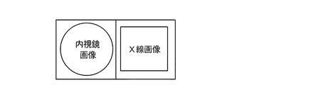
ことを特徴とする請求項2乃至6のいずれか1項に記載の制御装置。 - 被写体内に挿入された内視鏡で撮影された内視鏡画像を取得する内視鏡画像取得手段と、
前記制御に応じて発生されたX線をX線検出器で検出して前記被写体内の内視鏡を撮影したX線画像を取得するX線画像取得手段と、
前記X線画像における前記内視鏡の位置を前記X線画像に基づいて決定する決定手段と、
前記決定された内視鏡の位置に基づいて前記内視鏡をX線撮影するようにX線源を制御しX線を発生させる制御手段と、前記内視鏡画像と前記X線画像とを並べて表示部に表示させる表示制御手段と、を有することを特徴とする制御装置。 - 前記制御装置は、複数のX線焦点が並べて配置されたX線源と、X線を検出して画像を得るX線検出器とを用いたX線撮影の制御装置であり、
前記制御手段は、内視鏡の位置とX線焦点との対応関係を示す情報と、前記内視鏡の位置とに基づいてX線焦点を前記X線源の複数のX線焦点から選択することを特徴とする請求項8に記載の制御装置。 - 請求項1乃至9のいずれか1項に記載の制御装置と、
前記複数のX線焦点を有するX線源と、
を有することを特徴とするX線撮影システム。 - 請求項1乃至9のいずれか1項に記載の制御装置と、
前記X線検出器と、
を有することを特徴とするX線撮影システム。 - 請求項8または9に記載の制御装置と、
前記内視鏡と、
を有することを特徴とする撮影システム。 - 複数のX線焦点が並べて配置されたX線源と、X線を検出して画像を得るX線検出器とを用いたX線撮影の制御方法であって、
撮影対象に造影剤が流入していないタイミングと流入しているタイミングとのぞれぞれで、前記複数のX線焦点から順次選択されたX線焦点からX線を照射させるステップと、
造影剤が流入していないタイミングで前記順次選択されたX線焦点から前記照射されたX線をX線検出器で検出して得られる複数の第一のX線画像と、造影剤が流入しているタイミングで前記順次選択されたX線焦点から前記照射されたX線を前記X線検出器で検出して得られる複数の第二のX線画像とを取得するステップと、
前記X線焦点毎に前記第一のX線画像と前記第二のX線画像との差分画像を生成するステップと、
を有することを特徴とするX線撮影の制御方法。 - 複数のX線焦点が並べて配置されたX線源と、X線を検出して画像を得るX線検出器とを用いたX線撮影の制御方法であって、
X線検出器によって取得されたX線画像の変化に基づいて血管中において造影剤の流入する複数の先端位置を決定するステップと、
造影剤の流入する先端位置とX線焦点との対応関係を示す情報と、前記複数の先端位置の重心位置とに基づいて、少なくとも1つのX線焦点を前記複数のX線焦点から選択するステップと、
制御部が、当該選択されたX線焦点からX線を照射させるステップと、を有することを特徴とするX線撮影の制御方法。 - 撮影対象の内部に挿入された内視鏡で撮影された内視鏡画像を取得するステップと、
X線源から発生されたX線をX線検出器で検出して前記挿入された内視鏡を撮影したX線画像を取得するステップと、
前記X線画像における前記内視鏡の位置を前記X線画像に基づいて決定するステップと、
前記決定された内視鏡の位置に基づいて前記内視鏡をX線撮影するようにX線源を制御しX線を発生させるステップと、
前記内視鏡画像と前記X線画像とを並べて表示部に表示させるステップと、
を有することを特徴とする制御方法。 - 前記制御方法は、複数のX線焦点が並べて配置されたX線源と、X線を検出して画像を得るX線検出器とを用いたX線撮影の制御方法であり、
前記X線を発生させるステップでは、内視鏡の位置とX線焦点との対応関係を示す情報と、前記内視鏡の位置とに基づいてX線焦点を前記X線源の複数のX線焦点から選択することを特徴とする請求項15に記載の制御方法。 - 請求項13乃至16のいずれか1項に記載の制御方法をコンピュータに実行させるためのプログラム。
Priority Applications (3)
| Application Number | Priority Date | Filing Date | Title |
|---|---|---|---|
| JP2008104300A JP5388472B2 (ja) | 2008-04-14 | 2008-04-14 | 制御装置、x線撮影システム、制御方法、及び当該制御方法をコンピュータに実行させるためのプログラム。 |
| US12/421,120 US8249216B2 (en) | 2008-04-14 | 2009-04-09 | X-ray moving image radiographing apparatus |
| US13/542,394 US8842807B2 (en) | 2008-04-14 | 2012-07-05 | X-ray moving image radiographing apparatus |
Applications Claiming Priority (1)
| Application Number | Priority Date | Filing Date | Title |
|---|---|---|---|
| JP2008104300A JP5388472B2 (ja) | 2008-04-14 | 2008-04-14 | 制御装置、x線撮影システム、制御方法、及び当該制御方法をコンピュータに実行させるためのプログラム。 |
Related Child Applications (1)
| Application Number | Title | Priority Date | Filing Date |
|---|---|---|---|
| JP2013212293A Division JP2014000482A (ja) | 2013-10-09 | 2013-10-09 | 制御装置、撮影システム、制御方法、及びプログラム |
Publications (3)
| Publication Number | Publication Date |
|---|---|
| JP2009254428A JP2009254428A (ja) | 2009-11-05 |
| JP2009254428A5 JP2009254428A5 (ja) | 2011-05-26 |
| JP5388472B2 true JP5388472B2 (ja) | 2014-01-15 |
Family
ID=41163983
Family Applications (1)
| Application Number | Title | Priority Date | Filing Date |
|---|---|---|---|
| JP2008104300A Expired - Fee Related JP5388472B2 (ja) | 2008-04-14 | 2008-04-14 | 制御装置、x線撮影システム、制御方法、及び当該制御方法をコンピュータに実行させるためのプログラム。 |
Country Status (2)
| Country | Link |
|---|---|
| US (2) | US8249216B2 (ja) |
| JP (1) | JP5388472B2 (ja) |
Families Citing this family (12)
| Publication number | Priority date | Publication date | Assignee | Title |
|---|---|---|---|---|
| US9554868B2 (en) * | 2009-08-07 | 2017-01-31 | DePuy Synthes Products, Inc. | Method and apparatus for reducing malalignment of fractured bone fragments |
| DE102010018872A1 (de) * | 2010-04-30 | 2011-11-03 | Siemens Aktiengesellschaft | Bildgebendes Verfahren zur hervorgehobenen Darstellung von Gefäßen in einem Untersuchungsbereich eines Patienten und Medizinsystem zur Durchführung des Verfahrens |
| US20140046177A1 (en) * | 2010-11-18 | 2014-02-13 | Shimadzu Corporation | X-ray radiographic apparatus |
| US10849574B2 (en) * | 2011-06-22 | 2020-12-01 | Medtronic Navigation, Inc. | Interventional imaging |
| EP3046473B1 (en) | 2013-09-20 | 2019-03-13 | Koninklijke Philips N.V. | Automatic device-footpprint-free roadmapping for endovascular interventions |
| EP2995254A1 (en) * | 2014-09-12 | 2016-03-16 | Koninklijke Philips N.V. | Imaging system |
| JP6662390B2 (ja) * | 2015-12-18 | 2020-03-11 | 株式会社島津製作所 | X線撮影装置 |
| US11282668B2 (en) * | 2016-03-31 | 2022-03-22 | Nano-X Imaging Ltd. | X-ray tube and a controller thereof |
| US11071505B2 (en) | 2016-05-11 | 2021-07-27 | Koninklijke Philips N.V. | Anatomy adapted acquisition with fixed multi-source x-ray system |
| US10952691B2 (en) * | 2016-06-10 | 2021-03-23 | Principle Imaging Corporation | Scanning digital fluoroscope comprising multiple radiographic image detectors arranged as spokes extending radially outwardly from a central rotational point on a rotational plate |
| JP6947114B2 (ja) * | 2018-04-23 | 2021-10-13 | 株式会社島津製作所 | X線撮影システム |
| JP2021065370A (ja) * | 2019-10-21 | 2021-04-30 | ソニー株式会社 | 画像処理装置、画像処理方法および内視鏡システム |
Family Cites Families (36)
| Publication number | Priority date | Publication date | Assignee | Title |
|---|---|---|---|---|
| DE2410230A1 (de) * | 1974-03-04 | 1975-09-25 | Harald F H Dr Rer N Warrikhoff | Roentgen-abtastsystem |
| US4247780A (en) * | 1979-06-08 | 1981-01-27 | The United States Of America As Represented By The Department Of Health, Education And Welfare | Feedback controlled geometry registration system for radiographs |
| JPS58152542A (ja) * | 1982-03-05 | 1983-09-10 | 株式会社東芝 | X線診断装置 |
| US4692864A (en) * | 1985-05-23 | 1987-09-08 | Elscint Ltd. | Method of determining stenosis of blood vessels |
| US5123056A (en) * | 1990-02-02 | 1992-06-16 | Siemens Medical Systems, Inc. | Whole-leg x-ray image processing and display techniques |
| JPH0413381A (ja) | 1990-04-30 | 1992-01-17 | Shimadzu Corp | デジタルアンギオ装置 |
| JPH05161635A (ja) * | 1991-12-13 | 1993-06-29 | Toshiba Corp | X線診断シートシステム |
| JPH0670237A (ja) | 1992-08-21 | 1994-03-11 | Toshiba Corp | X線撮影装置 |
| US5267296A (en) * | 1992-10-13 | 1993-11-30 | Digiray Corporation | Method and apparatus for digital control of scanning X-ray imaging systems |
| US5651047A (en) * | 1993-01-25 | 1997-07-22 | Cardiac Mariners, Incorporated | Maneuverable and locateable catheters |
| US6368269B1 (en) * | 1993-05-20 | 2002-04-09 | Tilane Corporation | Apparatus for concurrent actuation of multiple foot pedal operated switches |
| US5594770A (en) * | 1994-11-18 | 1997-01-14 | Thermospectra Corporation | Method and apparatus for imaging obscured areas of a test object |
| JPH10285465A (ja) * | 1997-04-07 | 1998-10-23 | Shimadzu Corp | X線映像装置 |
| JP4047420B2 (ja) * | 1997-10-08 | 2008-02-13 | 株式会社東芝 | 画像再構成装置 |
| US6236709B1 (en) * | 1998-05-04 | 2001-05-22 | Ensco, Inc. | Continuous high speed tomographic imaging system and method |
| US6292531B1 (en) * | 1998-12-31 | 2001-09-18 | General Electric Company | Methods and apparatus for generating depth information mammography images |
| US6483890B1 (en) * | 2000-12-01 | 2002-11-19 | Koninklijke Philips Electronics, N.V. | Digital x-ray imaging apparatus with a multiple position irradiation source and improved spatial resolution |
| EP1437601B1 (en) * | 2003-01-10 | 2007-06-20 | Deutsches Krebsforschungszentrum Stiftung des öffentlichen Rechts | Apparatus for detecting the position and the orientation of an invasive device |
| US20050238140A1 (en) * | 2003-08-20 | 2005-10-27 | Dan Hardesty | X-ray imaging system with automatic image resolution enhancement |
| US7432924B2 (en) * | 2003-08-28 | 2008-10-07 | Kabushiki Kaisha Toshiba | 3D digital subtraction angiography image processing apparatus |
| US7187748B2 (en) * | 2003-12-30 | 2007-03-06 | Ge Medical Systems Global Technology Company, Llc | Multidetector CT imaging method and apparatus with reducing radiation scattering |
| US7885375B2 (en) * | 2004-02-27 | 2011-02-08 | General Electric Company | Method and system for X-ray imaging |
| JP4599073B2 (ja) * | 2004-03-22 | 2010-12-15 | 株式会社東芝 | X線断層撮影装置 |
| US7330529B2 (en) * | 2004-04-06 | 2008-02-12 | General Electric Company | Stationary tomographic mammography system |
| DE102004017538A1 (de) * | 2004-04-08 | 2005-11-03 | Siemens Ag | Computertomographie-Gerät mit Aperturblende |
| US7885440B2 (en) * | 2004-11-04 | 2011-02-08 | Dr Systems, Inc. | Systems and methods for interleaving series of medical images |
| US20090281383A1 (en) * | 2005-09-08 | 2009-11-12 | Rao Papineni | Apparatus and method for external fluorescence imaging of internal regions of interest in a small animal using an endoscope for internal illumination |
| US7889227B2 (en) * | 2005-09-15 | 2011-02-15 | Siemens Aktiengesellschaft | Intuitive user interface for endoscopic view visualization |
| US7835785B2 (en) * | 2005-10-04 | 2010-11-16 | Ascension Technology Corporation | DC magnetic-based position and orientation monitoring system for tracking medical instruments |
| US7746974B2 (en) * | 2006-09-29 | 2010-06-29 | Siemens Medical Solutions Usa, Inc. | Radiographic and fluoroscopic CT imaging |
| EP2036494A3 (en) * | 2007-05-07 | 2009-04-15 | Olympus Medical Systems Corp. | Medical guiding system |
| US8364242B2 (en) * | 2007-05-17 | 2013-01-29 | General Electric Company | System and method of combining ultrasound image acquisition with fluoroscopic image acquisition |
| WO2009012453A1 (en) * | 2007-07-19 | 2009-01-22 | The University Of North Carolina At Chapel Hill | Stationary x-ray digital breast tomosynthesis systems and related methods |
| US7724875B2 (en) * | 2007-10-19 | 2010-05-25 | General Electric Company | Image guided acquisition of quantitative dual energy data |
| US7949089B2 (en) * | 2008-04-10 | 2011-05-24 | Arineta Ltd. | Apparatus and method for tracking feature's position in human body |
| EP2523621B1 (en) * | 2010-01-13 | 2016-09-28 | Koninklijke Philips N.V. | Image integration based registration and navigation for endoscopic surgery |
-
2008
- 2008-04-14 JP JP2008104300A patent/JP5388472B2/ja not_active Expired - Fee Related
-
2009
- 2009-04-09 US US12/421,120 patent/US8249216B2/en not_active Expired - Fee Related
-
2012
- 2012-07-05 US US13/542,394 patent/US8842807B2/en not_active Expired - Fee Related
Also Published As
| Publication number | Publication date |
|---|---|
| JP2009254428A (ja) | 2009-11-05 |
| US20120277581A1 (en) | 2012-11-01 |
| US20090257559A1 (en) | 2009-10-15 |
| US8249216B2 (en) | 2012-08-21 |
| US8842807B2 (en) | 2014-09-23 |
Similar Documents
| Publication | Publication Date | Title |
|---|---|---|
| JP5388472B2 (ja) | 制御装置、x線撮影システム、制御方法、及び当該制御方法をコンピュータに実行させるためのプログラム。 | |
| JP4054402B2 (ja) | X線断層撮影装置 | |
| US7436927B2 (en) | Imaging apparatus and method for the operation thereof | |
| CN102160797B (zh) | X射线图像诊断装置和x射线图像处理方法 | |
| CN101797163B (zh) | X射线图像诊断装置以及x射线图像处理方法 | |
| JP4966120B2 (ja) | X線アンギオ撮影装置 | |
| JP2007021006A (ja) | X線ct装置 | |
| CN108697395A (zh) | X射线摄影装置 | |
| JP4327801B2 (ja) | X線断層撮影装置 | |
| JP6076658B2 (ja) | 医用画像処理装置、x線診断装置及び医用画像処理プログラム | |
| JP4744941B2 (ja) | X線画像診断装置及びその診断支援方法 | |
| JP4960212B2 (ja) | X線絞り装置およびx線撮影装置 | |
| JP5019879B2 (ja) | X線ct装置、画像処理プログラム、及び画像処理方法 | |
| KR100280198B1 (ko) | Ct촬영이가능한x선촬영장치및방법 | |
| JP2006296707A (ja) | X線画像診断装置及びその三次元血流画像構成・表示方法並びにプログラム | |
| JPH09276259A (ja) | X線診断装置 | |
| JP6812815B2 (ja) | X線撮影装置およびx線画像解析方法 | |
| JP4236666B2 (ja) | X線断層撮影装置 | |
| JP2014000482A (ja) | 制御装置、撮影システム、制御方法、及びプログラム | |
| JP6565461B2 (ja) | X線撮影装置 | |
| US20180160995A1 (en) | Medical image processing apparatus, x-ray diagnostic apparatus, and medical image processing method | |
| JP7287472B2 (ja) | X線撮影装置 | |
| JP2004081275A (ja) | X線診断装置およびその制御方法 | |
| JP2015047392A (ja) | X線断層撮影装置 | |
| JPH06237924A (ja) | X線診断装置 |
Legal Events
| Date | Code | Title | Description |
|---|---|---|---|
| RD01 | Notification of change of attorney |
Free format text: JAPANESE INTERMEDIATE CODE: A7421 Effective date: 20100218 |
|
| RD01 | Notification of change of attorney |
Free format text: JAPANESE INTERMEDIATE CODE: A7421 Effective date: 20100630 |
|
| A521 | Request for written amendment filed |
Free format text: JAPANESE INTERMEDIATE CODE: A523 Effective date: 20110412 |
|
| A621 | Written request for application examination |
Free format text: JAPANESE INTERMEDIATE CODE: A621 Effective date: 20110412 |
|
| A131 | Notification of reasons for refusal |
Free format text: JAPANESE INTERMEDIATE CODE: A131 Effective date: 20121127 |
|
| A977 | Report on retrieval |
Free format text: JAPANESE INTERMEDIATE CODE: A971007 Effective date: 20121128 |
|
| A521 | Request for written amendment filed |
Free format text: JAPANESE INTERMEDIATE CODE: A523 Effective date: 20130128 |
|
| A131 | Notification of reasons for refusal |
Free format text: JAPANESE INTERMEDIATE CODE: A131 Effective date: 20130611 |
|
| A521 | Request for written amendment filed |
Free format text: JAPANESE INTERMEDIATE CODE: A523 Effective date: 20130809 |
|
| TRDD | Decision of grant or rejection written | ||
| A01 | Written decision to grant a patent or to grant a registration (utility model) |
Free format text: JAPANESE INTERMEDIATE CODE: A01 Effective date: 20130910 |
|
| A61 | First payment of annual fees (during grant procedure) |
Free format text: JAPANESE INTERMEDIATE CODE: A61 Effective date: 20131008 |
|
| R151 | Written notification of patent or utility model registration |
Ref document number: 5388472 Country of ref document: JP Free format text: JAPANESE INTERMEDIATE CODE: R151 |
|
| LAPS | Cancellation because of no payment of annual fees |