JP4831556B2 - 複数ピークのx線源を具備するctイメージングシステム - Google Patents
複数ピークのx線源を具備するctイメージングシステム Download PDFInfo
- Publication number
- JP4831556B2 JP4831556B2 JP2004167147A JP2004167147A JP4831556B2 JP 4831556 B2 JP4831556 B2 JP 4831556B2 JP 2004167147 A JP2004167147 A JP 2004167147A JP 2004167147 A JP2004167147 A JP 2004167147A JP 4831556 B2 JP4831556 B2 JP 4831556B2
- Authority
- JP
- Japan
- Prior art keywords
- ray
- filter
- energy
- cathode
- electrons
- Prior art date
- Legal status (The legal status is an assumption and is not a legal conclusion. Google has not performed a legal analysis and makes no representation as to the accuracy of the status listed.)
- Expired - Fee Related
Links
- 238000013170 computed tomography imaging Methods 0.000 title description 7
- 239000000463 material Substances 0.000 claims description 26
- 238000003384 imaging method Methods 0.000 claims description 14
- 230000007704 transition Effects 0.000 claims description 8
- 238000009826 distribution Methods 0.000 claims description 4
- 230000001678 irradiating effect Effects 0.000 claims 3
- 238000001914 filtration Methods 0.000 claims 1
- 238000002591 computed tomography Methods 0.000 description 24
- 238000001228 spectrum Methods 0.000 description 16
- 238000000034 method Methods 0.000 description 13
- 230000000875 corresponding effect Effects 0.000 description 12
- 238000005259 measurement Methods 0.000 description 9
- 238000010586 diagram Methods 0.000 description 8
- 230000004044 response Effects 0.000 description 7
- 229920002994 synthetic fiber Polymers 0.000 description 7
- 230000005540 biological transmission Effects 0.000 description 6
- 230000003595 spectral effect Effects 0.000 description 6
- 238000011045 prefiltration Methods 0.000 description 5
- 239000000126 substance Substances 0.000 description 5
- 210000001519 tissue Anatomy 0.000 description 5
- 230000002238 attenuated effect Effects 0.000 description 4
- 239000008280 blood Substances 0.000 description 4
- 210000004369 blood Anatomy 0.000 description 4
- 238000013461 design Methods 0.000 description 4
- OYPRJOBELJOOCE-UHFFFAOYSA-N Calcium Chemical compound [Ca] OYPRJOBELJOOCE-UHFFFAOYSA-N 0.000 description 3
- 238000003491 array Methods 0.000 description 3
- 230000008901 benefit Effects 0.000 description 3
- 229910052791 calcium Inorganic materials 0.000 description 3
- 239000011575 calcium Substances 0.000 description 3
- 239000002131 composite material Substances 0.000 description 3
- 230000009977 dual effect Effects 0.000 description 3
- 238000012545 processing Methods 0.000 description 3
- 238000000926 separation method Methods 0.000 description 3
- XLYOFNOQVPJJNP-UHFFFAOYSA-N water Chemical class O XLYOFNOQVPJJNP-UHFFFAOYSA-N 0.000 description 3
- 238000010521 absorption reaction Methods 0.000 description 2
- 210000004204 blood vessel Anatomy 0.000 description 2
- 238000006243 chemical reaction Methods 0.000 description 2
- 238000011156 evaluation Methods 0.000 description 2
- 230000006870 function Effects 0.000 description 2
- 230000003116 impacting effect Effects 0.000 description 2
- 230000007246 mechanism Effects 0.000 description 2
- 239000013077 target material Substances 0.000 description 2
- ZCYVEMRRCGMTRW-UHFFFAOYSA-N 7553-56-2 Chemical class [I] ZCYVEMRRCGMTRW-UHFFFAOYSA-N 0.000 description 1
- 230000015572 biosynthetic process Effects 0.000 description 1
- 210000000988 bone and bone Anatomy 0.000 description 1
- QWUZMTJBRUASOW-UHFFFAOYSA-N cadmium tellanylidenezinc Chemical compound [Zn].[Cd].[Te] QWUZMTJBRUASOW-UHFFFAOYSA-N 0.000 description 1
- 230000002308 calcification Effects 0.000 description 1
- 230000003750 conditioning effect Effects 0.000 description 1
- 230000002596 correlated effect Effects 0.000 description 1
- 230000008021 deposition Effects 0.000 description 1
- 238000003745 diagnosis Methods 0.000 description 1
- 238000010894 electron beam technology Methods 0.000 description 1
- XMBWDFGMSWQBCA-UHFFFAOYSA-N hydrogen iodide Chemical compound I XMBWDFGMSWQBCA-UHFFFAOYSA-N 0.000 description 1
- 238000005286 illumination Methods 0.000 description 1
- 238000007689 inspection Methods 0.000 description 1
- PNDPGZBMCMUPRI-UHFFFAOYSA-N iodine Chemical compound II PNDPGZBMCMUPRI-UHFFFAOYSA-N 0.000 description 1
- 229910052740 iodine Inorganic materials 0.000 description 1
- 239000011630 iodine Substances 0.000 description 1
- 239000000203 mixture Substances 0.000 description 1
- 210000000056 organ Anatomy 0.000 description 1
- 230000008569 process Effects 0.000 description 1
- 230000005855 radiation Effects 0.000 description 1
- 230000002285 radioactive effect Effects 0.000 description 1
- 238000001959 radiotherapy Methods 0.000 description 1
- 210000004872 soft tissue Anatomy 0.000 description 1
- 238000004611 spectroscopical analysis Methods 0.000 description 1
- 238000003786 synthesis reaction Methods 0.000 description 1
- 238000010998 test method Methods 0.000 description 1
- 238000012360 testing method Methods 0.000 description 1
- 230000009466 transformation Effects 0.000 description 1
- 238000013519 translation Methods 0.000 description 1
- 238000002604 ultrasonography Methods 0.000 description 1
Images
Classifications
-
- A—HUMAN NECESSITIES
- A61—MEDICAL OR VETERINARY SCIENCE; HYGIENE
- A61B—DIAGNOSIS; SURGERY; IDENTIFICATION
- A61B6/00—Apparatus or devices for radiation diagnosis; Apparatus or devices for radiation diagnosis combined with radiation therapy equipment
- A61B6/02—Arrangements for diagnosis sequentially in different planes; Stereoscopic radiation diagnosis
- A61B6/03—Computed tomography [CT]
- A61B6/032—Transmission computed tomography [CT]
-
- A—HUMAN NECESSITIES
- A61—MEDICAL OR VETERINARY SCIENCE; HYGIENE
- A61B—DIAGNOSIS; SURGERY; IDENTIFICATION
- A61B6/00—Apparatus or devices for radiation diagnosis; Apparatus or devices for radiation diagnosis combined with radiation therapy equipment
- A61B6/40—Arrangements for generating radiation specially adapted for radiation diagnosis
- A61B6/4007—Arrangements for generating radiation specially adapted for radiation diagnosis characterised by using a plurality of source units
-
- A—HUMAN NECESSITIES
- A61—MEDICAL OR VETERINARY SCIENCE; HYGIENE
- A61B—DIAGNOSIS; SURGERY; IDENTIFICATION
- A61B6/00—Apparatus or devices for radiation diagnosis; Apparatus or devices for radiation diagnosis combined with radiation therapy equipment
- A61B6/40—Arrangements for generating radiation specially adapted for radiation diagnosis
- A61B6/4021—Arrangements for generating radiation specially adapted for radiation diagnosis involving movement of the focal spot
-
- A—HUMAN NECESSITIES
- A61—MEDICAL OR VETERINARY SCIENCE; HYGIENE
- A61B—DIAGNOSIS; SURGERY; IDENTIFICATION
- A61B6/00—Apparatus or devices for radiation diagnosis; Apparatus or devices for radiation diagnosis combined with radiation therapy equipment
- A61B6/40—Arrangements for generating radiation specially adapted for radiation diagnosis
- A61B6/4035—Arrangements for generating radiation specially adapted for radiation diagnosis the source being combined with a filter or grating
-
- A—HUMAN NECESSITIES
- A61—MEDICAL OR VETERINARY SCIENCE; HYGIENE
- A61B—DIAGNOSIS; SURGERY; IDENTIFICATION
- A61B6/00—Apparatus or devices for radiation diagnosis; Apparatus or devices for radiation diagnosis combined with radiation therapy equipment
- A61B6/42—Arrangements for detecting radiation specially adapted for radiation diagnosis
- A61B6/4208—Arrangements for detecting radiation specially adapted for radiation diagnosis characterised by using a particular type of detector
- A61B6/4241—Arrangements for detecting radiation specially adapted for radiation diagnosis characterised by using a particular type of detector using energy resolving detectors, e.g. photon counting
-
- A—HUMAN NECESSITIES
- A61—MEDICAL OR VETERINARY SCIENCE; HYGIENE
- A61B—DIAGNOSIS; SURGERY; IDENTIFICATION
- A61B6/00—Apparatus or devices for radiation diagnosis; Apparatus or devices for radiation diagnosis combined with radiation therapy equipment
- A61B6/48—Diagnostic techniques
- A61B6/482—Diagnostic techniques involving multiple energy imaging
-
- H—ELECTRICITY
- H01—ELECTRIC ELEMENTS
- H01J—ELECTRIC DISCHARGE TUBES OR DISCHARGE LAMPS
- H01J35/00—X-ray tubes
- H01J35/02—Details
- H01J35/04—Electrodes ; Mutual position thereof; Constructional adaptations therefor
- H01J35/06—Cathodes
- H01J35/064—Details of the emitter, e.g. material or structure
-
- A—HUMAN NECESSITIES
- A61—MEDICAL OR VETERINARY SCIENCE; HYGIENE
- A61B—DIAGNOSIS; SURGERY; IDENTIFICATION
- A61B6/00—Apparatus or devices for radiation diagnosis; Apparatus or devices for radiation diagnosis combined with radiation therapy equipment
- A61B6/02—Arrangements for diagnosis sequentially in different planes; Stereoscopic radiation diagnosis
- A61B6/027—Arrangements for diagnosis sequentially in different planes; Stereoscopic radiation diagnosis characterised by the use of a particular data acquisition trajectory, e.g. helical or spiral
-
- H—ELECTRICITY
- H01—ELECTRIC ELEMENTS
- H01J—ELECTRIC DISCHARGE TUBES OR DISCHARGE LAMPS
- H01J2235/00—X-ray tubes
- H01J2235/06—Cathode assembly
- H01J2235/068—Multi-cathode assembly
Landscapes
- Health & Medical Sciences (AREA)
- Life Sciences & Earth Sciences (AREA)
- Engineering & Computer Science (AREA)
- Medical Informatics (AREA)
- Radiology & Medical Imaging (AREA)
- Molecular Biology (AREA)
- Biophysics (AREA)
- Nuclear Medicine, Radiotherapy & Molecular Imaging (AREA)
- Optics & Photonics (AREA)
- Pathology (AREA)
- Physics & Mathematics (AREA)
- Biomedical Technology (AREA)
- Heart & Thoracic Surgery (AREA)
- High Energy & Nuclear Physics (AREA)
- Surgery (AREA)
- Animal Behavior & Ethology (AREA)
- General Health & Medical Sciences (AREA)
- Public Health (AREA)
- Veterinary Medicine (AREA)
- Pulmonology (AREA)
- Theoretical Computer Science (AREA)
- Apparatus For Radiation Diagnosis (AREA)
- Measurement Of Radiation (AREA)
Description
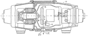
50’ エネルギー識別システム
52’ X線コントローラ
80 ターゲット
82’ 第1の陰極照射装置
84 第2の陰極照射装置
88 回転フィルタ
90 フィルタ
94 フィルタ回転装置
Claims (10)
- 複数の電子を照射する複数の陰極照射装置と、
前記複数の電子が入射し、第1及び第2のX線ビームが生成される向きに配向されたターゲットを有する、単一の回転陽極と、
前記第1及び第2のX線ビームを同時にフィルタリングし、複数のエネルギーレベルで前記第1及び第2のX線のピークとして規定される複数のX線量エネルギーピークを同時に有するX線分布のフィルタ後のX線ビームを生成する第1及び第2のフィルタを備える回転フィルタと、
を含み、
前記第1及び第2のX線ビーム及び前記第1及び第2のフィルタとの間が同期して遷移する、イメージングシステム内でネルギー識別を行うX線源。 - 前記複数の陰極照射装置が、
第1の複数の電子を照射する第1の陰極照射装置(82)と、
第2の複数の電子を照射する第2の陰極照射装置(84)と、
を含むことを特徴とする請求項1に記載のX線源。 - 前記第1の陰極照射装置(82)が第1のkVp(管電圧最高値)で前記第1の複数の電子を照射し、前記第2の陰極照射装置(84)が第2のkVpで第2の複数の電子を照射することを特徴とする請求項2に記載のX線源。
- 前記第1及び第2のフィルタが、低域通過フィルタであり、
前記第1のフィルタと前記第2のフィルタとが、前記第1のX線ビームに対して交互に用いられ、
前記第1のフィルタと前記第2のフィルタとが、前記第2のX線ビームに対して交互に用いられる、請求項1乃至3のいずれかに記載のX線源。 - 前記ターゲットが、前記単一の回転陽極の異なる側面に設けられている、請求項1乃至4のいずれかに記載のX線源。
- 前記ターゲットが、前記単一の回転陽極の同じ側面に複数設けられている、請求項1乃至4のいずれかに記載のX線源。
- 請求項1乃至6のいずれかに記載のX線源(32)と、
前記少なくとも1つのX線ビームを受信し、物質エネルギー識別情報を有するX線信号を生成するエネルギー識別検出器(40)と、
を含むイメージングシステム。 - 前記回転フィルタに結合されたフィルタ回転装置(94)と、
前記フィルタ回転装置に電気的に結合され、該回転フィルタを回転させるコントローラ(52)と、
を更に含む請求項7に記載のシステム。 - 前記コントローラは、スキャン中の複数のビューに含まれる各ビューにつき前記第1及び第2のフィルタ間を遷移する請求項8に記載のシステム。
- 前記少なくとも1つのX線ビームの複数のX線量エネルギーレベルを測定するX線検出器を更に含む請求項7乃至9のいずれかに記載のシステム。
Applications Claiming Priority (2)
| Application Number | Priority Date | Filing Date | Title |
|---|---|---|---|
| US10/250,132 | 2003-06-05 | ||
| US10/250,132 US7120222B2 (en) | 2003-06-05 | 2003-06-05 | CT imaging system with multiple peak x-ray source |
Publications (3)
| Publication Number | Publication Date |
|---|---|
| JP2004363109A JP2004363109A (ja) | 2004-12-24 |
| JP2004363109A5 JP2004363109A5 (ja) | 2009-10-08 |
| JP4831556B2 true JP4831556B2 (ja) | 2011-12-07 |
Family
ID=33489129
Family Applications (1)
| Application Number | Title | Priority Date | Filing Date |
|---|---|---|---|
| JP2004167147A Expired - Fee Related JP4831556B2 (ja) | 2003-06-05 | 2004-06-04 | 複数ピークのx線源を具備するctイメージングシステム |
Country Status (3)
| Country | Link |
|---|---|
| US (2) | US7120222B2 (ja) |
| JP (1) | JP4831556B2 (ja) |
| DE (1) | DE102004027092A1 (ja) |
Families Citing this family (70)
| Publication number | Priority date | Publication date | Assignee | Title |
|---|---|---|---|---|
| US9113839B2 (en) | 2003-04-25 | 2015-08-25 | Rapiscon Systems, Inc. | X-ray inspection system and method |
| GB0525593D0 (en) | 2005-12-16 | 2006-01-25 | Cxr Ltd | X-ray tomography inspection systems |
| US8837669B2 (en) | 2003-04-25 | 2014-09-16 | Rapiscan Systems, Inc. | X-ray scanning system |
| US8451974B2 (en) | 2003-04-25 | 2013-05-28 | Rapiscan Systems, Inc. | X-ray tomographic inspection system for the identification of specific target items |
| US8243876B2 (en) | 2003-04-25 | 2012-08-14 | Rapiscan Systems, Inc. | X-ray scanners |
| US7949101B2 (en) | 2005-12-16 | 2011-05-24 | Rapiscan Systems, Inc. | X-ray scanners and X-ray sources therefor |
| US8223919B2 (en) | 2003-04-25 | 2012-07-17 | Rapiscan Systems, Inc. | X-ray tomographic inspection systems for the identification of specific target items |
| US7869862B2 (en) * | 2003-10-15 | 2011-01-11 | Varian Medical Systems, Inc. | Systems and methods for functional imaging using contrast-enhanced multiple-energy computed tomography |
| US20050082491A1 (en) * | 2003-10-15 | 2005-04-21 | Seppi Edward J. | Multi-energy radiation detector |
| US7649981B2 (en) * | 2003-10-15 | 2010-01-19 | Varian Medical Systems, Inc. | Multi-energy x-ray source |
| US7397904B2 (en) * | 2005-05-11 | 2008-07-08 | Varian Medical Systems Technologies, Inc. | Asymmetric flattening filter for x-ray device |
| WO2007057841A2 (en) * | 2005-11-18 | 2007-05-24 | Koninklijke Philips Electronics N.V. | Systems and methods using x-ray tube spectra for computed tomography applications |
| DE102006002037A1 (de) * | 2006-01-16 | 2007-07-19 | Siemens Ag | Verfahren zur Bearbeitung diagnostischer Bilddaten |
| JP4769089B2 (ja) * | 2006-01-31 | 2011-09-07 | 株式会社東芝 | X線撮影装置 |
| ES2865724T3 (es) * | 2006-02-09 | 2021-10-15 | Leidos Security Detection & Automation Inc | Sistemas y métodos de exploración con radiación |
| EP2008293A1 (en) * | 2006-04-07 | 2008-12-31 | Philips Intellectual Property & Standards GmbH | Dual spectrum x-ray tube with switched focal spots and filter |
| CN101074937B (zh) * | 2006-05-19 | 2010-09-08 | 清华大学 | 能谱调制装置、识别材料的方法和设备及图像处理方法 |
| EP2021783B1 (en) * | 2006-05-31 | 2013-03-13 | L-3 Communications Security and Detection Systems, Inc. | Dual energy x-ray source |
| US20080037703A1 (en) * | 2006-08-09 | 2008-02-14 | Digimd Corporation | Three dimensional breast imaging |
| US7483518B2 (en) * | 2006-09-12 | 2009-01-27 | Siemens Medical Solutions Usa, Inc. | Apparatus and method for rapidly switching the energy spectrum of diagnostic X-ray beams |
| US7852979B2 (en) * | 2007-04-05 | 2010-12-14 | General Electric Company | Dual-focus X-ray tube for resolution enhancement and energy sensitive CT |
| JP5460318B2 (ja) * | 2007-07-19 | 2014-04-02 | 株式会社日立メディコ | X線発生装置及びこれを用いたx線ct装置 |
| CN101358936B (zh) | 2007-08-02 | 2011-03-16 | 同方威视技术股份有限公司 | 一种利用双视角多能量透射图像进行材料识别的方法及系统 |
| US8553844B2 (en) * | 2007-08-16 | 2013-10-08 | Koninklijke Philips N.V. | Hybrid design of an anode disk structure for high prower X-ray tube configurations of the rotary-anode type |
| DE102007041107B4 (de) | 2007-08-30 | 2009-10-29 | Siemens Ag | Röntgengerät |
| US7742566B2 (en) * | 2007-12-07 | 2010-06-22 | General Electric Company | Multi-energy imaging system and method using optic devices |
| US8306189B2 (en) * | 2007-12-21 | 2012-11-06 | Elekta Ab (Publ) | X-ray apparatus |
| WO2010024821A1 (en) * | 2008-08-29 | 2010-03-04 | Analogic Corporation | Multi-cathode x-ray tubes with staggered focal spots, and systems and methods using same |
| US8503616B2 (en) * | 2008-09-24 | 2013-08-06 | Varian Medical Systems, Inc. | X-ray tube window |
| DE102008049049A1 (de) * | 2008-09-26 | 2010-04-08 | Siemens Aktiengesellschaft | Röntgen-CT-System zur tomographischen Darstellung eines Untersuchungsobjektes |
| US7792241B2 (en) * | 2008-10-24 | 2010-09-07 | General Electric Company | System and method of fast KVP switching for dual energy CT |
| DE102008056891B4 (de) * | 2008-11-12 | 2012-04-12 | Siemens Aktiengesellschaft | Computertomographiegerät zur Durchführung eine Spiralscans und Verfahren zum Steuern eines Computertomographiegeräts |
| JP2012510137A (ja) * | 2008-11-25 | 2012-04-26 | コーニンクレッカ フィリップス エレクトロニクス エヌ ヴィ | X線陽極 |
| US7974383B2 (en) * | 2008-12-09 | 2011-07-05 | General Electric Company | System and method to maintain target material in ductile state |
| US7881425B2 (en) * | 2008-12-30 | 2011-02-01 | General Electric Company | Wide-coverage x-ray source with dual-sided target |
| JP5648055B2 (ja) * | 2009-08-11 | 2015-01-07 | プランゼー エスエー | 回転陽極x線管のための回転陽極および回転陽極の製造方法 |
| JP2011067333A (ja) * | 2009-09-25 | 2011-04-07 | Fujifilm Corp | 放射線画像撮影装置及び撮影制御装置 |
| CN102640252A (zh) | 2009-11-02 | 2012-08-15 | Xr科学有限责任公司 | 快速切换双能x射线源 |
| US9271689B2 (en) | 2010-01-20 | 2016-03-01 | General Electric Company | Apparatus for wide coverage computed tomography and method of constructing same |
| WO2012007881A2 (en) * | 2010-07-13 | 2012-01-19 | Koninklijke Philips Electronics N.V. | X-ray tube arrangement with toroidal rotatable filter arrangement and computed tomography device comprising same |
| US20120087464A1 (en) * | 2010-10-09 | 2012-04-12 | Fmi Technologies, Inc. | Multi-source low dose x-ray ct imaging aparatus |
| DE102010042683B4 (de) * | 2010-10-20 | 2013-11-14 | Siemens Aktiengesellschaft | Einrichtung und Verfahren zur Erzeugung von Röntgenstrahlung sowie Rechenprogramm und Datenträger |
| US8737567B2 (en) | 2011-01-27 | 2014-05-27 | Medtronic Navigation, Inc. | Image acquisition optimization |
| US9101272B2 (en) * | 2011-03-24 | 2015-08-11 | Jefferson Radiology, P.C. | Fixed anterior gantry CT shielding |
| BR112013031049A2 (pt) * | 2011-06-06 | 2016-11-29 | Koninkl Philips Nv | tubo de raios x para gerar radiação de raios x, sistema de obtenção de imagem por raios x, método para gerar um feixe de raios x de múltipla energia, uso de uma unidade de filtro para a geração da radiação de raios x de múltipla energia, elemento de programa de computador para controlar um aparelho e meio legível em computador |
| JP5823178B2 (ja) * | 2011-06-14 | 2015-11-25 | 株式会社東芝 | X線ct装置 |
| US9324536B2 (en) * | 2011-09-30 | 2016-04-26 | Varian Medical Systems, Inc. | Dual-energy X-ray tubes |
| US9069092B2 (en) | 2012-02-22 | 2015-06-30 | L-3 Communication Security and Detection Systems Corp. | X-ray imager with sparse detector array |
| WO2014001984A1 (en) * | 2012-06-29 | 2014-01-03 | Koninklijke Philips N.V. | Dynamic modeling of imperfections for photon counting detectors |
| JP6188470B2 (ja) * | 2013-07-24 | 2017-08-30 | キヤノン株式会社 | 放射線発生装置及びそれを用いた放射線撮影システム |
| US20150036792A1 (en) * | 2013-08-01 | 2015-02-05 | Korea Advanced Institute Of Science And Technology | Computed tomography apparatus, and method of generating image by using computed tomography apparatus |
| JP6266284B2 (ja) * | 2013-09-19 | 2018-01-24 | 東芝メディカルシステムズ株式会社 | X線診断装置 |
| DE102014203465A1 (de) * | 2014-02-26 | 2015-08-27 | Siemens Aktiengesellschaft | Verfahren zur Auswahl eines Strahlungsformfilters und Röntgenbildgebungssystem |
| JP2015180859A (ja) * | 2014-03-05 | 2015-10-15 | 株式会社東芝 | フォトンカウンティングct装置 |
| US9976971B2 (en) * | 2014-03-06 | 2018-05-22 | United Technologies Corporation | Systems and methods for X-ray diffraction |
| TWI629474B (zh) * | 2014-05-23 | 2018-07-11 | 財團法人工業技術研究院 | X光光源以及x光成像的方法 |
| US9991014B1 (en) * | 2014-09-23 | 2018-06-05 | Daniel Gelbart | Fast positionable X-ray filter |
| US10405813B2 (en) * | 2015-02-04 | 2019-09-10 | Dental Imaging Technologies Corporation | Panoramic imaging using multi-spectral X-ray source |
| CN104882350A (zh) * | 2015-06-11 | 2015-09-02 | 杭州与盟医疗技术有限公司 | 一种提供多能量和更大覆盖范围x射线球管系统 |
| US10791615B2 (en) | 2016-03-24 | 2020-09-29 | Koninklijke Philips N.V. | Apparatus for generating X-rays |
| CN108781496B (zh) * | 2016-03-24 | 2023-08-22 | 皇家飞利浦有限公司 | 用于生成x射线的装置 |
| US11282668B2 (en) * | 2016-03-31 | 2022-03-22 | Nano-X Imaging Ltd. | X-ray tube and a controller thereof |
| DE102017000994B4 (de) * | 2017-02-01 | 2019-11-21 | Esspen Gmbh | Computertomograph |
| JP6885803B2 (ja) * | 2017-06-27 | 2021-06-16 | ゼネラル・エレクトリック・カンパニイ | 放射線撮影装置及び撮影方法 |
| DE102019213983A1 (de) * | 2019-09-13 | 2021-03-18 | Fraunhofer-Gesellschaft zur Förderung der angewandten Forschung e.V. | Vorrichtung und verfahren zum bestimmen eines aufnahmeparameters und/oder zum bereitstellen einer wartungsempfehlung für eine computertomographieanlage |
| US11293884B2 (en) * | 2020-01-07 | 2022-04-05 | The Boeing Company | Multi source backscattering |
| CN111243916B (zh) * | 2020-01-19 | 2021-10-29 | 中国科学院电子学研究所 | 阳极及其制备方法以及阴极发射测试装置 |
| CN111429410B (zh) * | 2020-03-13 | 2023-09-01 | 杭州电子科技大学 | 一种基于深度学习的物体x射线图像材质判别系统及方法 |
| US11071506B1 (en) * | 2020-04-28 | 2021-07-27 | Wisconsin Alumni Research Foundation | X-ray imaging device providing enhanced spatial resolution by energy encoding |
| JP7451326B2 (ja) * | 2020-06-29 | 2024-03-18 | キヤノンメディカルシステムズ株式会社 | X線診断装置 |
Family Cites Families (44)
| Publication number | Priority date | Publication date | Assignee | Title |
|---|---|---|---|---|
| NL58621C (ja) * | 1939-10-14 | |||
| US2597498A (en) * | 1948-12-10 | 1952-05-20 | Joseph V Kerkhoff | X-ray tube |
| US3610984A (en) * | 1967-12-28 | 1971-10-05 | Tokyo Shibaura Electric Co | Rotating-anode x-ray tube with multiple focal areas |
| US4065689A (en) * | 1974-11-29 | 1977-12-27 | Picker Corporation | Dual filament X-ray tube |
| US4686695A (en) * | 1979-02-05 | 1987-08-11 | Board Of Trustees Of The Leland Stanford Junior University | Scanned x-ray selective imaging system |
| US4445226A (en) * | 1981-05-05 | 1984-04-24 | The Board Of Trustees Of The Leland Stanford Junior University | Multiple-energy X-ray subtraction imaging system |
| EP0081227B1 (en) * | 1981-12-07 | 1987-03-18 | Albert Macovski | Energy-selective x-ray recording and readout system |
| JPH0678501B2 (ja) * | 1985-11-21 | 1994-10-05 | 大日本インキ化学工業株式会社 | 水性被覆組成物 |
| US4963746A (en) * | 1986-11-25 | 1990-10-16 | Picker International, Inc. | Split energy level radiation detection |
| US4823371A (en) * | 1987-08-24 | 1989-04-18 | Grady John K | X-ray tube system |
| JPH01204649A (ja) * | 1988-02-12 | 1989-08-17 | Toshiba Corp | X線撮影装置 |
| DE69033232T2 (de) * | 1989-12-14 | 1999-12-30 | Aloka Co. Ltd., Mitaka | Vorrichtung zur Messung des Kalziumgehaltes von Knochen |
| US5335255A (en) * | 1992-03-24 | 1994-08-02 | Seppi Edward J | X-ray scanner with a source emitting plurality of fan beams |
| US5485492A (en) * | 1992-03-31 | 1996-01-16 | Lunar Corporation | Reduced field-of-view CT system for imaging compact embedded structures |
| DE4230880A1 (de) * | 1992-09-16 | 1994-03-17 | Philips Patentverwaltung | Röntgengenerator zur Speisung einer Röntgenröhre mit wenigstens zwei Elektronenquellen |
| JP3449561B2 (ja) * | 1993-04-19 | 2003-09-22 | 東芝医用システムエンジニアリング株式会社 | X線ct装置 |
| US5511105A (en) * | 1993-07-12 | 1996-04-23 | Siemens Aktiengesellschaft | X-ray tube with multiple differently sized focal spots and method for operating same |
| US5490196A (en) * | 1994-03-18 | 1996-02-06 | Metorex International Oy | Multi energy system for x-ray imaging applications |
| US5661774A (en) * | 1996-06-27 | 1997-08-26 | Analogic Corporation | Dual energy power supply |
| US5943388A (en) * | 1996-07-30 | 1999-08-24 | Nova R & D, Inc. | Radiation detector and non-destructive inspection |
| US6410920B1 (en) | 1997-05-30 | 2002-06-25 | Adac Laboratories | Method and apparatus for performing correction of emission contamination and deadtime loss in a medical imaging system |
| US6008493A (en) | 1997-05-30 | 1999-12-28 | Adac Laboratories | Method and apparatus for performing correction of emission contamination and deadtime loss in a medical imaging system |
| DE19729414A1 (de) * | 1997-07-09 | 1999-02-11 | Siemens Ag | Strahlenblende eines medizinischen Gerätes |
| DE19802668B4 (de) * | 1998-01-24 | 2013-10-17 | Smiths Heimann Gmbh | Röntgenstrahlungserzeuger |
| US6307918B1 (en) * | 1998-08-25 | 2001-10-23 | General Electric Company | Position dependent beam quality x-ray filtration |
| US6226352B1 (en) * | 1998-09-08 | 2001-05-01 | Veritas Pharmaceuticals, Inc. | System and method for radiographic imaging of tissue |
| US6229870B1 (en) * | 1998-11-25 | 2001-05-08 | Picker International, Inc. | Multiple fan beam computed tomography system |
| US6285740B1 (en) | 1999-10-13 | 2001-09-04 | The United States Of America As Represented By The Secretary Of The Navy | Dual energy x-ray densitometry apparatus and method using single x-ray pulse |
| US6246747B1 (en) * | 1999-11-01 | 2001-06-12 | Ge Lunar Corporation | Multi-energy x-ray machine with reduced tube loading |
| US6333968B1 (en) * | 2000-05-05 | 2001-12-25 | The United States Of America As Represented By The Secretary Of The Navy | Transmission cathode for X-ray production |
| DE10048775B4 (de) * | 2000-09-29 | 2006-02-02 | Siemens Ag | Röntgen-Computertomographieeinrichtung |
| US6553096B1 (en) * | 2000-10-06 | 2003-04-22 | The University Of North Carolina Chapel Hill | X-ray generating mechanism using electron field emission cathode |
| WO2002058557A2 (en) * | 2000-10-24 | 2002-08-01 | The Johns Hopkins University | Method and apparatus for multiple-projection, dual-energy x-ray absorptiometry scanning |
| US6614878B2 (en) * | 2001-01-23 | 2003-09-02 | Fartech, Inc. | X-ray filter system for medical imaging contrast enhancement |
| JP2002263091A (ja) * | 2001-03-07 | 2002-09-17 | Tomoki Yamazaki | 立体x線透視法と立体x線透視のためのx線バルブ |
| US6480572B2 (en) * | 2001-03-09 | 2002-11-12 | Koninklijke Philips Electronics N.V. | Dual filament, electrostatically controlled focal spot for x-ray tubes |
| US7636413B2 (en) * | 2002-04-16 | 2009-12-22 | General Electric Company | Method and apparatus of multi-energy imaging |
| US6760407B2 (en) * | 2002-04-17 | 2004-07-06 | Ge Medical Global Technology Company, Llc | X-ray source and method having cathode with curved emission surface |
| US6597758B1 (en) * | 2002-05-06 | 2003-07-22 | Agilent Technologies, Inc. | Elementally specific x-ray imaging apparatus and method |
| US6947522B2 (en) * | 2002-12-20 | 2005-09-20 | General Electric Company | Rotating notched transmission x-ray for multiple focal spots |
| US6968030B2 (en) * | 2003-05-20 | 2005-11-22 | General Electric Company | Method and apparatus for presenting multiple pre-subject filtering profiles during CT data acquisition |
| JP3909048B2 (ja) * | 2003-09-05 | 2007-04-25 | ジーイー・メディカル・システムズ・グローバル・テクノロジー・カンパニー・エルエルシー | X線ct装置およびx線管 |
| US7003077B2 (en) * | 2003-10-03 | 2006-02-21 | General Electric Company | Method and apparatus for x-ray anode with increased coverage |
| US7065179B2 (en) * | 2003-11-07 | 2006-06-20 | General Electric Company | Multiple target anode assembly and system of operation |
-
2003
- 2003-06-05 US US10/250,132 patent/US7120222B2/en not_active Expired - Lifetime
-
2004
- 2004-06-02 DE DE102004027092A patent/DE102004027092A1/de not_active Withdrawn
- 2004-06-04 JP JP2004167147A patent/JP4831556B2/ja not_active Expired - Fee Related
-
2006
- 2006-08-18 US US11/465,472 patent/US7778382B2/en not_active Expired - Fee Related
Also Published As
| Publication number | Publication date |
|---|---|
| US20060285645A1 (en) | 2006-12-21 |
| JP2004363109A (ja) | 2004-12-24 |
| US7778382B2 (en) | 2010-08-17 |
| US7120222B2 (en) | 2006-10-10 |
| US20040247082A1 (en) | 2004-12-09 |
| DE102004027092A1 (de) | 2004-12-23 |
Similar Documents
| Publication | Publication Date | Title |
|---|---|---|
| JP4831556B2 (ja) | 複数ピークのx線源を具備するctイメージングシステム | |
| JP4361778B2 (ja) | 計算機式断層写真法(ct)スカウト画像を形成する方法及び装置 | |
| JP4949664B2 (ja) | Ctコロノグラフィ・システム | |
| US9754387B2 (en) | System and method for improved energy series of images using multi-energy CT | |
| Buzug | Computed tomography | |
| JP5703014B2 (ja) | サンプリングレートを低減した2重エネルギー撮像 | |
| JP4347672B2 (ja) | 構造、灌流及び機能に関する異常を検出する方法及び装置 | |
| US8311182B2 (en) | System and method of notch filtration for dual energy CT | |
| US7724865B2 (en) | System and method of optimizing a monochromatic representation of basis material decomposed CT images | |
| US8787519B2 (en) | System and method of optimizing a representation of dual energy spectral CT images | |
| US20040101090A1 (en) | Methods and apparatus for acquiring perfusion data | |
| JP4445221B2 (ja) | コンピュータ断層撮影走査を実行するための方法及び装置 | |
| US7187748B2 (en) | Multidetector CT imaging method and apparatus with reducing radiation scattering | |
| JP2008237908A (ja) | エネルギ識別を用いた計算機式断層写真法における血栓の検出 | |
| JP4344191B2 (ja) | 画像形成システムの低線量画像シミュレーションのための方法及びシステム | |
| EP1951119A2 (en) | Methods and apparatus for obtaining low-dose imaging | |
| WO2007074772A1 (ja) | X線ct装置 | |
| EP3821811B1 (en) | Systems and methods for coherent scatter imaging using a segmented photon-counting detector for computed tomography | |
| US20240180503A1 (en) | Photon counting computed tomography apparatus and photon-counting ct-scanning condition setting method | |
| Gunnarsson et al. | Principles behind Computed Tomography (CT) |
Legal Events
| Date | Code | Title | Description |
|---|---|---|---|
| A521 | Request for written amendment filed |
Free format text: JAPANESE INTERMEDIATE CODE: A523 Effective date: 20070528 |
|
| A621 | Written request for application examination |
Free format text: JAPANESE INTERMEDIATE CODE: A621 Effective date: 20070528 |
|
| A521 | Request for written amendment filed |
Free format text: JAPANESE INTERMEDIATE CODE: A523 Effective date: 20090825 |
|
| RD02 | Notification of acceptance of power of attorney |
Free format text: JAPANESE INTERMEDIATE CODE: A7422 Effective date: 20090825 |
|
| RD04 | Notification of resignation of power of attorney |
Free format text: JAPANESE INTERMEDIATE CODE: A7424 Effective date: 20090825 |
|
| A977 | Report on retrieval |
Free format text: JAPANESE INTERMEDIATE CODE: A971007 Effective date: 20100804 |
|
| A131 | Notification of reasons for refusal |
Free format text: JAPANESE INTERMEDIATE CODE: A131 Effective date: 20100810 |
|
| A521 | Request for written amendment filed |
Free format text: JAPANESE INTERMEDIATE CODE: A523 Effective date: 20100921 |
|
| TRDD | Decision of grant or rejection written | ||
| A01 | Written decision to grant a patent or to grant a registration (utility model) |
Free format text: JAPANESE INTERMEDIATE CODE: A01 Effective date: 20110830 |
|
| A01 | Written decision to grant a patent or to grant a registration (utility model) |
Free format text: JAPANESE INTERMEDIATE CODE: A01 |
|
| A61 | First payment of annual fees (during grant procedure) |
Free format text: JAPANESE INTERMEDIATE CODE: A61 Effective date: 20110913 |
|
| R150 | Certificate of patent or registration of utility model |
Ref document number: 4831556 Country of ref document: JP Free format text: JAPANESE INTERMEDIATE CODE: R150 Free format text: JAPANESE INTERMEDIATE CODE: R150 |
|
| FPAY | Renewal fee payment (event date is renewal date of database) |
Free format text: PAYMENT UNTIL: 20140930 Year of fee payment: 3 |
|
| R250 | Receipt of annual fees |
Free format text: JAPANESE INTERMEDIATE CODE: R250 |
|
| R250 | Receipt of annual fees |
Free format text: JAPANESE INTERMEDIATE CODE: R250 |
|
| R250 | Receipt of annual fees |
Free format text: JAPANESE INTERMEDIATE CODE: R250 |
|
| R250 | Receipt of annual fees |
Free format text: JAPANESE INTERMEDIATE CODE: R250 |
|
| R250 | Receipt of annual fees |
Free format text: JAPANESE INTERMEDIATE CODE: R250 |
|
| R250 | Receipt of annual fees |
Free format text: JAPANESE INTERMEDIATE CODE: R250 |
|
| LAPS | Cancellation because of no payment of annual fees |


