JP4767252B2 - Lung access device - Google Patents
Lung access device Download PDFInfo
- Publication number
- JP4767252B2 JP4767252B2 JP2007516645A JP2007516645A JP4767252B2 JP 4767252 B2 JP4767252 B2 JP 4767252B2 JP 2007516645 A JP2007516645 A JP 2007516645A JP 2007516645 A JP2007516645 A JP 2007516645A JP 4767252 B2 JP4767252 B2 JP 4767252B2
- Authority
- JP
- Japan
- Prior art keywords
- access assembly
- bronchoscope
- pulmonary
- lung
- guide
- Prior art date
- Legal status (The legal status is an assumption and is not a legal conclusion. Google has not performed a legal analysis and makes no representation as to the accuracy of the status listed.)
- Expired - Fee Related
Links
Images


Classifications
-
- A—HUMAN NECESSITIES
- A61—MEDICAL OR VETERINARY SCIENCE; HYGIENE
- A61B—DIAGNOSIS; SURGERY; IDENTIFICATION
- A61B1/00—Instruments for performing medical examinations of the interior of cavities or tubes of the body by visual or photographical inspection, e.g. endoscopes; Illuminating arrangements therefor
- A61B1/00131—Accessories for endoscopes
- A61B1/0014—Fastening element for attaching accessories to the outside of an endoscope, e.g. clips, clamps or bands
-
- A—HUMAN NECESSITIES
- A61—MEDICAL OR VETERINARY SCIENCE; HYGIENE
- A61B—DIAGNOSIS; SURGERY; IDENTIFICATION
- A61B1/00—Instruments for performing medical examinations of the interior of cavities or tubes of the body by visual or photographical inspection, e.g. endoscopes; Illuminating arrangements therefor
- A61B1/012—Instruments for performing medical examinations of the interior of cavities or tubes of the body by visual or photographical inspection, e.g. endoscopes; Illuminating arrangements therefor characterised by internal passages or accessories therefor
- A61B1/018—Instruments for performing medical examinations of the interior of cavities or tubes of the body by visual or photographical inspection, e.g. endoscopes; Illuminating arrangements therefor characterised by internal passages or accessories therefor for receiving instruments
-
- A—HUMAN NECESSITIES
- A61—MEDICAL OR VETERINARY SCIENCE; HYGIENE
- A61B—DIAGNOSIS; SURGERY; IDENTIFICATION
- A61B1/00—Instruments for performing medical examinations of the interior of cavities or tubes of the body by visual or photographical inspection, e.g. endoscopes; Illuminating arrangements therefor
- A61B1/267—Instruments for performing medical examinations of the interior of cavities or tubes of the body by visual or photographical inspection, e.g. endoscopes; Illuminating arrangements therefor for the respiratory tract, e.g. laryngoscopes, bronchoscopes
- A61B1/2676—Bronchoscopes
-
- A—HUMAN NECESSITIES
- A61—MEDICAL OR VETERINARY SCIENCE; HYGIENE
- A61B—DIAGNOSIS; SURGERY; IDENTIFICATION
- A61B17/00—Surgical instruments, devices or methods, e.g. tourniquets
- A61B17/34—Trocars; Puncturing needles
- A61B17/3403—Needle locating or guiding means
-
- A—HUMAN NECESSITIES
- A61—MEDICAL OR VETERINARY SCIENCE; HYGIENE
- A61B—DIAGNOSIS; SURGERY; IDENTIFICATION
- A61B10/00—Other methods or instruments for diagnosis, e.g. instruments for taking a cell sample, for biopsy, for vaccination diagnosis; Sex determination; Ovulation-period determination; Throat striking implements
- A61B10/02—Instruments for taking cell samples or for biopsy
- A61B10/04—Endoscopic instruments
- A61B2010/045—Needles
Description
本出願は、その内容の全体を参考として引用し本明細書に含めた、2004年6月14日付けにて出願された米国仮特許出願明細書60/579,905号の優先権の利益を主張するものである。 This application claims the benefit of priority of US Provisional Patent Application No. 60 / 579,905, filed June 14, 2004, which is hereby incorporated by reference in its entirety. It is what I insist.
本発明は、全体として、肺の内部又は肺周囲の縦隔空間にアクセスするため気管支鏡及びその他の装置を使用することに関する。特に、本発明は、従来の気管支鏡又はその他の内視鏡と共に使用して、典型的な内視鏡又は気管支鏡を通じて現在可能であるよりもより多数の且つより大きい装置をターゲット箇所まで送り出すことを可能にする補助的アクセス装置及びツールに関する。 The present invention relates generally to the use of bronchoscopes and other devices to access the mediastinal space within or around the lungs. In particular, the present invention is used in conjunction with a conventional bronchoscope or other endoscope to deliver more and larger devices to a target location than is currently possible through a typical endoscope or bronchoscope. Auxiliary access devices and tools that enable
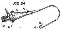
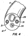
殆どの気管支鏡の事例は、単に、気管支にアクセスするため(肺への経鼻、経口又は経気管アクセス)、また、可能であれば、隣接する病理組織の異常な色を視覚化するためのツールとして気管支鏡を使用する。殆どの気管支鏡利用のバイオプシーは、気管支幹の外側に位置する組織をターゲットとする。このため、介入術者は、バイオプシーシステムの先端を配置し且つターゲットを横断することを確実にすべく送り出し方向を確認するため、外部の映像による案内を必要とする。気管支鏡は、大きく(直径5mm)、また、極めて可撓性ではないため、アクセス深さが制限される。これらの気管支鏡は、僅か2.0mmの作業通路を有しており、このため、ユーザは、通すことのできる装置を選択する点にて制約される。しかし、これらの気管支鏡は、ステア可能であり、所要位置に係止することができ、また、実質的に剛性であり座屈(病変部を横断するためのばね針の圧縮のような)し易い装置を支持することができる。1つの型式の従来の可撓性の気管支鏡は、その開示内容を参考として引用し本明細書に含めた、米国特許明細書4,880,015号に記載されている。図1ないし図4に示すように、気管支鏡10は、長さ790mmであり、また、作業用ヘッド14及び挿入管11という、2つの主要な部分を有している。作業ヘッドは、接眼鏡15と、ジオプター調節リング25を有する接眼レンズと、吸引管24、吸引弁21、及び冷ハロゲン光源16、18の取り付け部と、色々な装置及び流体を作業通路29内に通し且つ気管支鏡の末端から出すときに通るアクセスポート又はバイオプシー入口19とを保持している。作業ヘッド部は、典型的に、長さ580mm及び直径6.3mmの挿入管に取り付けられる。挿入管は、光ファイバ束(末端の先端12で対物レンズ30にて終わる)と、2つの導光路31と、作業通路29とを保持する。
Most bronchoscopic cases are simply for accessing the bronchus (nasal, oral or tracheal access to the lungs) and, if possible, for visualizing abnormal colors in adjacent pathological tissues Use a bronchoscope as a tool. Most bronchoscopic biopsies target tissue located outside the bronchial trunk. For this reason, the interventionist needs guidance with an external image to locate the tip of the biopsy system and to confirm the delivery direction to ensure that the target is traversed. Bronchoscopes are large (5 mm in diameter) and are not very flexible, limiting the access depth. These bronchoscopes have a working path of only 2.0 mm, which limits the user in selecting a device that can be threaded. However, these bronchoscopes are steerable, can be locked in place, and are substantially rigid and buckled (such as compression of a spring needle to cross the lesion). Easy equipment can be supported. One type of conventional flexible bronchoscope is described in US Pat. No. 4,880,015, the disclosure of which is incorporated herein by reference. As shown in FIGS. 1 to 4, the
気管支鏡の末端は、前方及び後方にのみ曲がる能力を備え、その正確な偏向角度は、使用される器具に依存する。一般的な曲げ範囲は、図3A及び図3Bの要素13にて示すように、前方に160°及び後方に90°にて合計250°である。曲りは、術者が作業ヘッド部の角度係止レバー22及び角度レバー23を調節することにより制御される。呼吸器科医は、かかる気管支鏡を使用して肺の内部を検査すると共に、多岐に亙る術を実行する。バイオプシー鉗子及びブラシのような装置は、作業通路を介して気管支鏡の長さを通じて患者の肺内に通し組織の試料を得ることができる。例えば、米国特許明細書4,766,906号(その特許の開示内容は、参考として引用し本明細書に含めてある)に記載されたもののようなバイオプシー針を可撓性気管支鏡の作業通路を介して患者の肺内部に挿入することができる。針が気管支の末端の所要位置に配置されたならば、呼吸器科医は、針を使用して、例えば、気管支鏡が配置された気管支に隣接する縦隔空間内のリンパ節をパイオブシー検査することができる。第’906号特許に記載されたように、呼吸器科医は、気管支鏡及び針にて突き刺し動作を行い、気管支壁及びリンパ節に侵入する。気管支鏡の作業通路を介して使用されるバイオプシー針の他の例は、その開示内容を参考として引用し本明細書に含めた、米国特許明細書5,056,529号、米国特許明細書4,532,935号及び米国特許明細書4,702,260号に見ることができる。
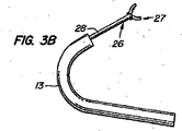
The end of the bronchoscope has the ability to bend only forward and backward, the exact deflection angle of which depends on the instrument used. The typical bending range is a total of 250 ° at 160 ° forward and 90 ° backward, as shown by
従来の気管支鏡の作業通路の寸法は、気管支鏡の末端にて患者の肺を視認し、パイオブシー検査し又は治療するため作業通路に沿って下方に進めることのできる器具の寸法を制限する。例えば、肺の内部又は肺を通じて試料採取する現在の針バイオプシー装置は、従来の気管支鏡の直径2.0mmの通路に適合しなければならない。更に、気管支鏡の作業通路は、バイオプシー針を送り出すため使用されるため、気管支鏡はターゲット組織を固定するといったような、その他の目的のため同時に使用することはできない。本発明は、気管支鏡の作業通路の寸法上の制限を解消する、気管支鏡と共に使用されるアクセス付属品を提供するものである。 The dimensions of a conventional bronchoscope working channel limit the size of the instrument that can be viewed along the working channel for viewing and treating the patient's lungs at the distal end of the bronchoscope and performing a pyobsi examination or treatment. For example, current needle biopsy devices that sample inside or through the lung must fit into the 2.0 mm diameter passage of a conventional bronchoscope. Furthermore, since the bronchoscope's working channel is used to deliver the biopsy needle, the bronchoscope cannot be used simultaneously for other purposes, such as fixing the target tissue. The present invention provides an access accessory for use with a bronchoscope that overcomes the dimensional limitations of the working path of the bronchoscope.
従って、1つの実施の形態において、本発明は、肺のアクセス組立体を提供する。該組立体は、(a)基端と、末端とを有し、該末端は被験者の肺内部に配置し得るようにされ、上記基端は上記被験者の肺の外側に配置し得るようにされた、映像装置と、(b)器具を上記映像装置の外側にて上記被験者の肺まで送り出すのを導き得るように、上記映像装置の上記末端と作用可能に接続された案内要素とを備えている。また、被験者の肺のアクセス組立体を備えるキットとすることも考えられる。 Accordingly, in one embodiment, the present invention provides a pulmonary access assembly. The assembly has (a) a proximal end and a distal end, the distal end being adapted to be placed inside the subject's lung, and the proximal end being adapted to be placed outside the subject's lung. An imaging device; and (b) a guide element operably connected to the distal end of the imaging device so as to guide delivery of the instrument to the lungs of the subject outside the imaging device. Yes. It is also conceivable to have a kit with the subject's lung access assembly.
別の実施の形態において、本発明は、被験者の肺のアクセス組立体を使用する方法を提供する。特に、この実施の形態には、被験者の肺又は周囲の組織まで案内されたアクセスを実現する方法が含まれる。この方法は、被験者の肺のアクセス組立体を肺又は取り囲む組織の内側部分に位置決めするステップと、組立体の映像装置の外側又は、映像装置が1つの作業通路を有するならば、その作業通路の外側に配置された器具によって案内されたアクセスを実現すべく組立体内に保持された案内要素を制御するステップとを含む。 In another embodiment, the present invention provides a method of using a subject's lung access assembly. In particular, this embodiment includes a method of providing guided access to a subject's lungs or surrounding tissue. The method includes the steps of positioning a subject's lung access assembly on an inner portion of the lung or surrounding tissue and, if the imaging device has one working channel, outside the imaging device of the assembly, of the working channel. Controlling guide elements held within the assembly to provide access guided by instruments disposed on the outside.
本発明によって被験者の肺又は肺の取り囲む組織内のターゲット箇所の治療又は診断を実行する方法が更に提供される。この方法は、(a)被験者の肺のアクセス組立体であって、(i)作業通路を有する映像装置を備える上記アクセス組立体を肺又は取り囲む組織の内側部分内に送り出すステップと、(b)組立体の映像装置の視認の下、案内要素を制御するステップと、(c)案内要素と作用可能に接続されたバルーンを拡張して肺又は取り囲む組織内のターゲット箇所まで送り出すべき器具を固定するステップと、(d)固定したターゲット箇所にて器具によって所望の治療又は診断を実行するステップとを含む。 The present invention further provides a method for performing treatment or diagnosis of a target site in a subject's lungs or surrounding tissue of the lungs. The method includes: (a) delivering the subject's lung access assembly, comprising: (i) delivering the access assembly comprising an imaging device having a working channel into the lung or an inner portion of the surrounding tissue; and (b) Controlling the guide element under visual recognition of the imaging device of the assembly; and (c) expanding a balloon operatively connected to the guide element to secure the device to be delivered to a target location in the lung or surrounding tissue. And (d) performing a desired treatment or diagnosis with the instrument at a fixed target location.
一部の実施の形態において、気管支鏡が患者の体内に挿入される前に、アクセス付属品を気管支鏡の作業通路内に逆に装填する。アクセス付属品は、その末端に取り付けられた1つ又はより多くの要素(ガイドワイヤー、カニューレ等)を有しており、このため、要素は、気管支鏡と共に、喉を通して下方に且つ、気管支内に、下降させていく。気管支鏡を配置した後、外科医は、大型の視覚化光ファイバ束、スクレーパ、縫合糸又は縫合針を操作する器具、レーザ光ファイバ、光ステッキ、光管、バイオプシー位置マーカの送り出しシステム、腫瘍除去器具、プラグ、超音波プローブ、血管内視鏡、又は、患者の喉、気道、気管、気管支、肺、縦隔領域、リンパ節、及び腫瘍を治療し又はその形状又はその状態を改変するその他の装置のような装置をガイドワイヤーの外側にてカニューレを通して導入等することができる。これらの装置は、気管支鏡が依然として所要位置にあり又は気管支鏡を除去した状態にて、患者の肺内部に送り出し、アクセス付属品を所要位置に残すことができる。アクセス付属品は、気管支鏡を使用してその他の装置の位置の制御を助けることもできる。 In some embodiments, the access accessory is reversely loaded into the bronchoscope's working channel before the bronchoscope is inserted into the patient's body. The access accessory has one or more elements (guidewire, cannula, etc.) attached to its distal end, so that the element, along with the bronchoscope, is down through the throat and into the bronchi , Descend. After placing the bronchoscope, the surgeon can use a large visualization optical fiber bundle, scraper, suture or suture needle instrument, laser optical fiber, optical stick, light tube, biopsy position marker delivery system, tumor removal instrument , Plugs, ultrasound probes, vascular endoscopes, or other devices that treat or alter the shape or condition of a patient's throat, airways, trachea, bronchi, lungs, mediastinal region, lymph nodes, and tumors Can be introduced through the cannula, etc. outside the guidewire. These devices can be delivered into the patient's lungs with the bronchoscope still in place or with the bronchoscope removed, leaving the access accessory in place. Access accessories can also use a bronchoscope to help control the position of other devices.
更に、気管支鏡が肺に向かう途中に通らなければならない患者の声帯のような解剖学的形態部は、気管支鏡の直径を増大させることのできる程度を制限し、また、ツールを気管支鏡と共に、但し気管支鏡の外側にて同時に送り出すことを妨害する可能性がある。本発明のアクセス付属品は、器具を気管支鏡と共に同時に又は気管支鏡に沿って送り出すことを必要とせずに、器具を気管支の外側にて末気管支鏡の末端まで送り出すことを可能にすることにより、こうした解剖学的な寸法上の制限を解消する方法を提供する。 Furthermore, the anatomical features such as the patient's vocal cords that the bronchoscope must pass on the way to the lungs limit the extent to which the diameter of the bronchoscope can be increased, and the tool together with the bronchoscope, However, it may interfere with simultaneous delivery outside the bronchoscope. The access accessory of the present invention allows the instrument to be delivered to the distal end of the endobronchoscope outside the bronchus, without requiring the instrument to be delivered with or along the bronchoscope. A method is provided to overcome these anatomical dimensional limitations.
特定の実施の形態において、当該組立体の映像装置は、気管支鏡であり、案内要素はガイドワイヤーである。1つ以上の案内要素を組立体内に組み込むことができる。典型的に、案内要素は、末端と、基端とを有する一方、末端は被験者の体内に配置し得る設計とされ且つ上記器具と接続され、また、基端は器具の送り出しを導き得るように被験者の身体の外側に配置される。特定の実施の形態において、案内要素の少なくとも一部分は映像装置の作業通路内に配設される。1つの好ましい形態において、案内要素は、バルーン軸の内部又はバルーン軸を横断する位置に配置され、バルーンを肺の上記内側部分又は肺の取り囲む組織まで送り出す。別の好ましい形態において、バルーン軸は、案内要素が通るのを許容し得るよう少なくとも1つの側部ポートを保持している。案内要素は、器具の一体的部分とすることができる。案内要素は、分離可能な取り付け装置を介して器具と接続してもよい(例えば、図27Aないし図27D参照)。好ましい分離可能な取り付け装置は、被験者の肺又は取り囲む組織内に配置されたとき、案内要素を器具から制御状態にて解放することを許容する。かかる分離可能な取り付け装置は、クリップ、接着剤、ストラップ及びスリーブを含むが、これらにのみ限定されるものではない。望まれる場合、分離可能な取り付け装置は、作業通路を更に備えることができる。 In a particular embodiment, the imaging device of the assembly is a bronchoscope and the guide element is a guide wire. One or more guide elements can be incorporated into the assembly. Typically, the guide element has a distal end and a proximal end, while the distal end is designed to be placed in the subject's body and connected to the instrument, and the proximal end may guide the delivery of the instrument. Located outside the subject's body. In certain embodiments, at least a portion of the guide element is disposed in the working path of the imaging device. In one preferred form, the guide element is placed inside or across the balloon axis and delivers the balloon to the inner part of the lung or to the tissue surrounding the lung. In another preferred form, the balloon shaft retains at least one side port to allow the guide element to pass through. The guiding element can be an integral part of the instrument. The guide element may be connected to the instrument via a separable attachment device (see, eg, FIGS. 27A-27D). Preferred separable attachment devices permit the controlled release of the guide element from the instrument when placed in the subject's lungs or surrounding tissue. Such separable attachment devices include, but are not limited to, clips, adhesives, straps and sleeves. If desired, the separable attachment device can further comprise a working channel.
一部の実施の形態において、案内要素と接続される器具は、映像装置の作業通路の末端内に又は末端を横断する位置に配置される。一部の実施の形態において、器具は映像装置の外側に配置される。多岐に亙る器具を当該組立体と共に使用することができる。これらの器具は、バイオプシーを実行し得るようにされた器具、身体組織を映像化し得るようにされた器具及び(又は)医薬組成物を肺に送り出し得るようにされた器具を含むが、これらにのみ限定されるものではない。好ましい器具は、バルーンと接続されたカテーテルを備えている。別の好ましい器具は、針案内部を備えている。所望である場合、針案内部は、案内要素が通るのを許容するよう側部ポートを保持することができる。 In some embodiments, the instrument connected to the guide element is located within or across the end of the working path of the imaging device. In some embodiments, the instrument is located outside the imaging device. A wide variety of instruments can be used with the assembly. These devices include devices adapted to perform biopsies, devices adapted to image bodily tissues and / or devices adapted to deliver pharmaceutical compositions to the lung, including It is not limited only. A preferred device comprises a catheter connected to a balloon. Another preferred instrument includes a needle guide. If desired, the needle guide can hold a side port to allow the guide element to pass through.
本発明の別の形態は、バイオプシーのためターゲット組織を固定することに関する。肺を通してパイオブシー検査したリンパ節又はその他の組織の均一性は、流体からバイオプシー針にて押したとき、経路外に転がり出る硬いゴム状塊の程度のものとすることができる。このため、本発明は、バイオプシーを行う前に、アクセス付属品を使用して固定装置を送り出し且つ制御する等によって、ターゲット組織を固定する方法を提供する。本発明はまた、より大形のバイオプシー針を送り出し、先行の肺バイオプシーシステムにより許容されるよりも大きい組織試料を引き出すことをも可能にする。 Another aspect of the invention relates to fixing a target tissue for biopsy. The homogeneity of lymph nodes or other tissues examined through the lungs with a pyopsy can be as much as a hard rubbery mass that rolls out of the path when pushed with a biopsy needle from the fluid. Thus, the present invention provides a method for fixing a target tissue, such as by using an access accessory to deliver and control the fixation device prior to performing a biopsy. The present invention also makes it possible to deliver larger biopsy needles and withdraw larger tissue samples than allowed by previous lung biopsy systems.
従って、本発明は、治療又は診断のため肺の組織を固定する方法を提供する。この方法は、(a)肺のアクセス組立体であって、(i)作業通路を有する映像装置と、(ii)上記映像装置の視認の下、観察可能な案内要素であって、その少なくとも一部分が上記作業通路内に配設され複数の固定器具にて肺の上記内側部分又は取り囲む組織への案内されたアクセスを実現し、また、上記複数の器具の少なくとも1つの固定器具は案内要素と作用可能に固定された上記案内要素を備える上記肺のアクセス組立体を送り出すステップと、(b)上記複数の固定器具と接触して上記肺の組織を固定するステップとを含む。この実施の形態の1つの形態において、固定器具は、複数の針をその内部に保持した針案内部を備えている。好ましい針は膨張可能なスリーブにより覆われている。 Accordingly, the present invention provides a method of fixing lung tissue for treatment or diagnosis. The method comprises: (a) a lung access assembly, (i) an imaging device having a working channel, and (ii) a guide element observable under viewing of the imaging device, at least a portion thereof. Disposed within the working channel to provide guided access to the inner portion of the lung or surrounding tissue with a plurality of fixation devices, and at least one fixation device of the plurality of devices interacts with the guide element. Delivering the pulmonary access assembly with the guide element secured thereto; and (b) contacting the plurality of fixation devices to fix the lung tissue. In one form of this embodiment, the fixture includes a needle guide that holds a plurality of needles therein. A preferred needle is covered by an inflatable sleeve.
本発明は、身体器官又は組織にアクセスするための空間形成装置を更に提供する。装置は、次の構成要素、すなわち、(a)末端と、基端と、貫通する管腔とを有する細長いアクセス装置であって、上記管腔を貫通して伸びることのできる送り出し要素を保持する上記細長いアクセス装置と、(b)上記細長いアクセス装置の上記末端を取り囲む、開放端部で且つ伸長可能なスリーブであって、上記送り出し要素にて上記身体器官又は組織にアクセスするため作業空間を膨張させる設計とされた上記伸長可能なスリーブとを備えている。 The present invention further provides a space forming device for accessing a body organ or tissue. The device retains a delivery element that has the following components: (a) a distal end, a proximal end, and a lumen therethrough, which can extend through the lumen. An elongate access device and (b) an open end and extendable sleeve surrounding the distal end of the elongate access device, wherein the delivery element expands a working space for accessing the body organ or tissue And an extensible sleeve designed to be adapted.
本発明には、被験者の上記身体器官又は組織にアクセスするための作業空間を提供する方法も含まれる。この方法は、(a)空間を形成する装置であって、(i)末端と、基端と、貫通する管腔とを有する細長いアクセス装置であって、上記管腔を通って伸びることのできる送り出し要素を保持する上記細長いアクセス装置と、(ii)上記細長いアクセス装置の上記末端を取り囲む開放端部で且つ伸長可能なスリーブとを有する上記空間を形成する装置を上記被験者の上記身体器官又は組織内に位置決めするステップと、(b)上記伸長可能なスリーブを膨張させて作業空間を膨張させ、上記送り出し要素にて上記身体器官又は組織にアクセスするステップとを含む。 The present invention also includes a method of providing a working space for accessing the body organ or tissue of a subject. The method includes: (a) a device for forming a space, (i) an elongated access device having a distal end, a proximal end, and a lumen therethrough, which can extend through the lumen. An apparatus for forming said space comprising said elongated access device holding a delivery element; and (ii) an open end surrounding said distal end of said elongated access device and an extendable sleeve; Positioning within, and (b) inflating the expandable sleeve to inflate a working space and accessing the body organ or tissue with the delivery element.
本発明の一部の形態において、スリーブは、半径方向に伸長可能である。別の形態において、スリーブは、上記身体器官又は組織の解剖学的形態部を押し拡げ易くする少なくとも1つの翼状構造体を備えている。本発明のその他の有利な効果は、以下の特定の実施の形態の説明から明らかになるであろう。 In some forms of the invention, the sleeve is extensible in the radial direction. In another form, the sleeve includes at least one winged structure that facilitates expanding the anatomical form of the body organ or tissue. Other advantageous effects of the present invention will become apparent from the following description of specific embodiments.
図5には、その内部に針案内部44が挿入された作業通路42を有する可撓性の気管支鏡40が示されている。気管支鏡40を患者の体内に挿入する前、ガイドワイヤー46のような、アクセス付属品が針案内部44の末端48内に挿入される。ガイドワイヤー46は、曲がって、基端50が気管支鏡40の長さに沿った位置にあるようにする。気管支鏡40が患者の肺内に挿入されたとき、ガイドワイヤー46の基端50は患者の外部に止まるであろう。次に、ガイドワイヤー46を使用して、診断、治療又はパイオプシー用ツールを作業通路42に通すことを必要とせずにかかるツールを気管支鏡40の末端まで送り出す。かかるツールは、気管支鏡と共に同時に、又は気管支鏡が患者の肺内の選んだ箇所に配置された後の何れかにて、送り出すことができる。
FIG. 5 shows a
また、ガイドワイヤー46を使用して、気管支鏡の末端41を位置決めし且つステアすることもできる。ガイドワイヤー46を基端方向に引っ張ると、針案内部44及び気管支鏡40の末端41は、その方向に曲がり、これにより気管支鏡の末端に対するユーザの制御状態を向上させる。
A
1つの代替的な実施の形態において、ガイドワイヤーは、針案内部に取り付けるか又は針案内部と一体化することができる。
図6には、気管支鏡40、針案内部44及びガイドワイヤー46が図5の配置にある、鈍角な拡張器52の使用状態が示されている。拡張器52を使用して、気管支又はその他の肺通路の壁に対し外傷を生じさせることなく、システムの前進を助けることができる。
In one alternative embodiment, the guide wire can be attached to or integrated with the needle guide.
FIG. 6 shows the
図7及び図8には、気管支鏡40の作業通路42内にて針案内部を使用することを不要にする本発明の1つの実施の形態が示されている。この実施の形態において、ガイドワイヤー46は、気管支鏡の末端41にて作業通路42内に配置され、このため、一端51は、作業通路を通って基端方向に伸び、また、他端(図示せず)は、その長さに沿って気管支鏡の外側にて基端方向に、患者の外部まで伸びている。気管支鏡が患者の肺内に挿入されたとき、ガイドワイヤー46を使用して、ツールを治療箇所まで送り出すことができる。図7A及び図7Bに示した実施例において、針案内部54がガイドワイヤー46の外側部分上にある間に、針案内部54をガイドワイヤー46に沿って末端方向に押すか又はガイドワイヤーの基端51を基端方向に引き抜き或いは、これら2つの動作の組み合わせにより、ガイドワイヤー46を使用して針案内ブ46をバイオプシー箇所まで送り出す。針案内部の末端56が気管支鏡の末端41にあるとき、針案内部の末端56は、ガイドワイヤー46のカーブに従って、バイオプシー箇所に向けて曲がる。ガイドワイヤー46の基端方向張力を使用して針案内部の曲がり程度を制御することができる。次に、針案内部54内にて可撓性のバイオプシー針58を使用して気管支壁に侵入し、組織の試料を採取することができる。
7 and 8 illustrate one embodiment of the present invention that eliminates the need to use a needle guide within the working
図9ないし図12には、側部ポート62を有する鈍角な先端の針案内部60が気管支鏡40の作業通路42内に配設された、気管支鏡及びガイドワイヤーの配置が示されている。ガイドワイヤー46は、気管支鏡40を患者の体内に挿入する前に、針案内部60の末端にてガイドワイヤーのポート64内に固定され、このため、ガイドワイヤー46は、気管支鏡40の長さに沿って伸びてガイドワイヤー46の基端50を患者の外部に配置する。ガイドワイヤー46を使用して針案内部60を回転させ且つ(又は)曲げて側部ポート62を望ましいように向き決めすることができる。その他の実施の形態におけるように、気管支鏡40を患者の肺内に挿入した後、ガイドワイヤー46を使用してツール(掴み器具、光源、腫瘍固定具、鉗子のような)を気管支鏡の末端まで送り出すことができ、気管支鏡の制限された寸法の作業通路を通る必要はない。その他の送り出しツールは、代理人事件番号30689.703.201、30689.703.301、30689.703.304による当該出願人の特許出願明細書に記載されている。これら特許出願の内容は、その全体を参考として引用し本明細書に含めてある。
FIGS. 9-12 show the bronchoscope and guidewire arrangement in which an obtuse
例えば、図10ないし図12には、気管支鏡システムを使用してバルーン66をバイオプシー箇所まで送り出す状態が示されている。バルーン66と連通したカテーテル68は、図10に示すように、ガイドワイヤー46と同軸状にすることができ、また、これと代替的に、ガイドワイヤー46は、図11に示すように、側部ポート69を介してバルーンの軸66から出るようにしてもよい。バルーンの拡張器70は、ガイドワイヤー46の基端50にて患者の外部に配設される。ガイドワイヤー46を使用して側部ポート62を向き決めした後、バルーン66を拡張させ側部ポート62をバイオプシー箇所にて気管支壁72に向け又は気管支壁72に対して動かすと共に、針案内部の末端60をこの位置にて保持する。次に、バイオプシー針74及び中央ワイヤー76を、針案内部60、気管支壁72の側部ポート64を通してバイオプシー箇所まで前進させることができる。吸引注射器78を使用して組織試料をコアバイオプシー針74内に吸引するとき、中央ワイヤー76を使用して疑わしい腫瘍を所要位置に固定することができる。組織試料を得た後、針及び中央ワイヤーを引き抜き、バルーン66を萎縮させることができる。
For example, FIGS. 10 to 12 show a state where the
図13には、図9ないし図12のものと同様の気管支鏡システムが示されている。しかし、図13のシステムは、図9ないし図12の側部ポート針案内部に代えて、直線状の通路針案内部44を使用する。図9ないし図12のシステムと同様に、バイオプシー術を行う前、且つバイオプシー術を行う間、バルーン66を使用して針案内部44を向き決めし且つ支持することができる。
FIG. 13 shows a bronchoscope system similar to that of FIGS. However, the system of FIG. 13 uses a straight
図14には、多数のガイドワイヤー46a、46b、46cが気管支鏡40の作業通路を通って伸びる針案内部44内に配設される、気管支鏡システムが示されている。その他の実施の形態におけるように、ガイドワイヤーの各々の一端は気管支鏡を患者の体内に挿入する前、針案内部の末端48内に挿入され、ガイドワイヤーが気管支鏡40の長さに沿って伸びて、それらの他端50a、50b、50cを患者の外部に配置する。内視鏡の作業通路を使用せずに、側部ポートバルーン66のようなツールをガイドワイヤーに沿って気管支鏡40の末端まで送り出すことができる。1つの代替的な実施の形態において、ガイドワイヤーは、針案内部に取り付け又は針案内部と一体に形成することができる。
FIG. 14 shows a bronchoscope system in which a number of
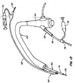
図15には、ステア可能な気管支鏡及び(又は)カメラ80をガイドワイヤー46に沿って伸びるレール81を介してバイオプシー箇所まで送り出すため、図5のものと同様のガイドワイヤーの気管支鏡システムを使用する状態が示されている。気管支鏡80を誘導可能な末端82は、疑わしい腫瘍又はリンパ節84に隣接する気管支壁83に侵入しバイオプシーすべき組織を視認することができる。バイオプシー針は、針案内部44を介してバイオプシー箇所まで送り出すことができる。気管支鏡の末端82は、案内信号(EMF、光、磁気、音、高周波)を放出してバイオプシー針を疑わしい腫瘍又はリンパ節84内に案内することができる。
15 uses a steerable bronchoscope and / or a guidewire bronchoscope system similar to that of FIG. 5 to deliver the camera 80 to the biopsy site via a
図16及び図17にて、針案内部44は、気管支鏡40の末端41に取り付けられ、バイオプシー針又はその他の器具を気管支鏡作業通路42の外部の治療箇所まで案内部44を介して送り出すことができる。図16において、針案内部44は、鼻クリップ90又はストラップ、接着剤等のような、その他の取り付け機構によって気管支鏡に取り付けられる。
16 and 17, the
図17には、クリップ、ヒンジ又は接着剤92を使用して、気管支鏡40と針案内部44との間に関節式接続部を形成する状態が示されている。ガイドワイヤー46は、気管支鏡の作業通路42を通って患者の外部まで、また、以前の実施の形態におけるように、針案内部の側部ポート(図示せず)を通って伸びている。気管支鏡及び針案内部は、その他の実施の形態におけるように肺内に同時に挿入され、また、ガイドワイヤー46を使用して針案内部44の末端48を動かし、位置決めし且つ保持することができる。この配置は、針案内部の可撓性及び操縦可能性を気管支鏡の長手方向剛性と組み合わせる。
FIG. 17 illustrates the use of a clip, hinge or adhesive 92 to form an articulated connection between the
図18において、針案内部44は腫瘍固定装置100と共に縫付けられ又は腫瘍固定装置100にその他の方法にて取り付けられる。針案内部44は、気管支鏡の末端の作業通路42内に挿入され、腫瘍固定装置100の軸102は、気管支鏡の長さに沿って伸びている。気管支鏡が患者の肺内に挿入されると、1つ又はより多くの固定針104は、気管支鏡の末端におけるバイオプシー箇所にあり、制御機構106は患者の外部にて装置の基端にある。制御機構106を作動させると、針104はバイオプシーすべき組織内に及びその組織の回りに挿入される。次に、バイオプシー針74及び中央ワイヤー76を上述したように使用して組織の試料を得ることができる。
In FIG. 18, the
図19には、幾つかのバイオプシー針機構を有する気管支鏡システムが示されている。主要な針案内部44aは、気管支鏡40を患者の肺内に挿入する前に、気管支鏡40の作業通路42の末端に配設される。第二の針案内部44b、44cの末端は、縫い付け又はその他の手段によって、主要な針案内部44aの末端に取り付けられる。針案内部の各々は、図示するように、既知の態様にて作動させ組織の試料を得ることのできる中央ワイヤー及びバイオプシー針を有している。
FIG. 19 shows a bronchoscope system having several biopsy needle mechanisms. The main needle guide 44a is disposed at the end of the working
図20には、バルーンカテーテル110、側部ポート針案内部60、バイオプシー針74及び中央ワイヤー76が全て気管支鏡の作業通路内に配設された、気管支鏡システムが示されている。バルーン110は、針案内部60の側部ポート62の反対側に配設されている。注射器70を使用してバルーン軸112を介してバルーン110を拡張させ、側部ポート62を気管支壁に対して押し針74及び中央ワイヤー76を疑わしい腫瘍84内に確実に押すことができる。
FIG. 20 shows a bronchoscope system in which the
図21には、上述したもののような気管支鏡システムと共に使用されるバイオプシー針が示されている。図21Aに示すように、スリーブ120は、針を送り出し且つ位置決めする間、針122及び中央ワイヤー124を覆う。スリーブ120は、患者の外部にて基端方向に伸びて戻り、また、針122、124と共に気管支鏡の作業通路内に又は気管支鏡の外部に配設された針案内部を通して前進させる。組織試料を集めるため針を使用すべきとき、針122及び中央ワイヤー124が末端方向に前進する間、スリーブ120は静止状態に保持され(又は、針122及び中央ワイヤー124が静止状態に保持されている間、スリーブ120を基端方向に引き出す)、このため、針122及び中央ワイヤー124は、図21Bに示すように、スリーブ120に穴を開け、次に、針122及び中央ワイヤー124を使用して組織試料を集める。
FIG. 21 shows a biopsy needle for use with a bronchoscope system such as that described above. As shown in FIG. 21A, the
図22において、半径方向に膨張可能なスリーブ126は、上記の図21Aの実施の形態におけるように、気管支鏡システムを介して送り出し且つ位置決めする間、針122及び中央ワイヤー又は針124を覆っている。バイオプシー箇所にて、スリーブ126の末端は図22に示すように半径方向に膨張させ、針122及び中央ワイヤー又は針124がスリーブ126の末端を通過するのを許容する。スリーブ126が開放したとき、スリーブの末端における半径方向矢羽根状部128は、幾つかの機能を奏することができる。矢羽根状部128は、気管支壁に対して配設されたとき、整合機能を提供し、このため針122及び中央ワイヤー又は針124を制御距離だけ壁内に及び壁を超えて前進させることができる。また、スリーブ126が組織に対して配置されている間、半径方向矢羽根状部128が開放されたならば、矢羽根状部の動きは、組織を横方向に動かし且つ保持して作業領域を開放し、また、装置を組織に対して固定することができる。半径方向矢羽根状部はまた、組織の試料を得た後、閉塞した位置まで動かし組織の試料を掴み且つ保持するのを助けることもできる。
In FIG. 22, a radially
図23ないし図25には、気管支鏡システムを介して送り出し、気管支鏡を介して実行されるバイオプシー又はその他の術用のための作業空間を提供することのできる空間形成装置の他の実施の形態が示されている。図23Aにおいて、膨張ツール130は、開放端部のスリーブ132内に閉塞した形態に配設された状態で示されている。気管支鏡システムを介して肺内の治療箇所まで送り出された後、膨張ツール132を作動させて開放した形態にし、スリーブ132の開放した端部134を膨張させることができる。この実施の形態において、ツール130の回動アーム136は、図23Bに示すように、スリーブ132の内部に対して動かし分離させる。スリーブ132が膨張した後、膨張ツール130を除去してその他のツールをスリーブ132を介して治療箇所まで送り出すのを許容することができる。膨張したスリーブ132を使用して治療箇所における作業領域を安定化させ、治療箇所における解剖学的形態を押し拡げ且つ(又は)組織の試料を保持するのを助けることができる。
FIGS. 23-25 show another embodiment of a space forming device that can be delivered through a bronchoscope system and provide a working space for a biopsy or other surgical procedure performed through the bronchoscope. It is shown. In FIG. 23A, the
図24A及び図24Bには、空間形成装置の別の実施の形態が示されている。図24Aにおいて、膨張ツール140は、気管支鏡40の作業通路42を介して治療箇所まで送り出されつつある。膨張ツール140は、スパイラルとして形成されている。膨張ツールが作業通路42の末端から出たならば、膨張ツール140のスパイラルは、図24Bに示すように、巻き戻されツールの末端に作業空間を形成する。これと代替的に、膨張ツール140は、スリーブ内にて送り出し、ツール140を使用して、図23の実施の形態におけるように、スリーブの末端を膨張させてもよい。
24A and 24B show another embodiment of the space forming device. In FIG. 24A, the inflation tool 140 is being delivered to the treatment site via the working
図25には、空間形成装置の更に別の実施の形態が示されている。前述の実施の形態と同様に、膨張ツール150は、気管支鏡140の作業通路42を介して治療箇所に送り出される。通路外となったならば、ツール150の膨張部分152は、巻き戻し且つ、半径方向に膨張して、ツールの末端に作業空間を形成する。その他の実施の形態におけるように、膨張ツール150は、スリーブ内にて送り出すことができ、また、ツール150を使用してスリーブの末端を膨張させることができる。
FIG. 25 shows still another embodiment of the space forming device. Similar to the previous embodiment, the
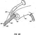
図26において、分岐カニューレ160は、気管支鏡40を患者の体内に挿入する前、気管支鏡40の作業通路42の末端に挿入されている。カニューレ160は、ツールを末端側ポート166を介して同一の治療箇所に前進させる2つの枝部分162、164を提供する。図示するように、枝部分162は、作業通路42に沿って基端方向に伸びる一方、枝部分164は、気管支鏡40の長さに沿って伸びている。ポート166を取り囲む先端168は、組織に侵入し得るよう鋭角にすることができる。例えば、ユーザが管腔の1つに配設された気管支鏡を介してカニューレの動きを観察する間、末端の先端168を、気管支壁に押し込むことができる。次に、カメラを引き戻して、バイオプシー器具をその位置まで前進させることができる。バイオプシー試料を採取する前、採取する間又は採取した後に、マーカを配置することもできる。
In FIG. 26, the
上述した実施の形態によるバイオプシー針の使用の1つの代替例として、側部採取カッタツールを使用して組織の試料を採取することができる。
映像化に関し、殆どの肺学研究所は、方向及び深さを確認し得るよう迅速に90°回転させることのできる実時間蛍光透視法を採用している。これと代替的に、バイオプシー又はその他の術を行う前、実時間CATスキャンを使用して、患者の肺及び縦隔領域の断面像を得ることができる。像スライスが3ないし5mmと狭小であり、また、その像スライスが胸部を横断する面を切るならば、ユーザは、針がその平面に入るときを、及びターゲットに対する二次元座標を視覚化することができる。残念なことに、実時間CT装置は一般的ではない。このため、本発明の別の形態において、極く少量の放射線不透過性染料マーカは又は金属の放射線不透過性マーカを細い経胸部針を介して送り出し、その位置を確認し且つマーカを明確な指標として使用し、経気管支針吸引システムを気管支鏡を通して導くことができる。経胸部送り出しは、全体として、遥かにより正確ではあるが、装置の輪郭外形は、肺壁の損傷を避け得るように小さくなければならないであろう。
As an alternative to the use of biopsy needles according to the embodiments described above, a tissue sample may be taken using a side harvesting cutter tool.
For imaging, most pulmonary laboratories employ real-time fluoroscopy that can be quickly rotated 90 ° to confirm direction and depth. Alternatively, real-time CAT scans can be used to obtain cross-sectional images of the patient's lungs and mediastinum region before performing a biopsy or other procedure. If the image slice is as narrow as 3-5 mm, and if the image slice cuts across the plane crossing the chest, the user can visualize when the needle enters the plane and the two-dimensional coordinates relative to the target. Can do. Unfortunately, real-time CT devices are not common. For this reason, in another aspect of the present invention, a very small amount of radiopaque dye marker or metal radiopaque marker is delivered through a thin transthoracic needle to confirm its position and define the marker. Used as an indicator, a transbronchial needle aspiration system can be guided through a bronchoscope. Transthoracic delivery as a whole is much more accurate, but the profile of the device will have to be small to avoid damaging the lung wall.
本発明の更に別の形態は、例えば、組織の試料を得るため、縦隔空間に入るべくアクセス付属品と共に、内視鏡を使用することである。本発明は、気胸の場合、接着剤又はプラグ(例えば、接着剤を保持する自然膨張セメント、コラーゲンプラグ、ポリマープラグ、シアノアクリレート、グルタルアルデヒドフォーミレーション、ポリエチレンバルーン等)を使用して肺壁又は隣接する組織の穴を閉塞することを含む。特定のバイオプシー針の実施の形態において、針の案内要素又は通路は、中央又は側部ポートを貫通する連続的な通路を有し、また、バイオプシー装置を正確に送り出すため、一定の管腔の直径及び密着嵌めするポートの寸法を有している。放射線不透過性マーカバンドをポート開口部に追加して、ターゲット組織に対するバイオプシー装置の映像化を容易にすることができる。 Yet another form of the invention is the use of an endoscope with access accessories to enter the mediastinum, for example, to obtain a tissue sample. In the case of pneumothorax, the present invention uses an adhesive or plug (eg, natural expansion cement holding the adhesive, collagen plug, polymer plug, cyanoacrylate, glutaraldehyde formation, polyethylene balloon, etc.) Occlusion of adjacent tissue holes. In certain biopsy needle embodiments, the needle guide element or passage has a continuous passage through the central or side port and a constant lumen diameter for accurate delivery of the biopsy device. And the size of the close fitting port. A radiopaque marker band can be added to the port opening to facilitate imaging of the biopsy device to the target tissue.
Claims (19)
(a)基端と、末端と、作業通路(42)とを有し、前記末端は、被験者の肺内部(206)に配置し得るようにされ、前記基端は、前記被験者の肺の外側に配置し得るようにされた、映像装置(40)と、
(b)被験者の身体の外側に配置された基端を有し且つ前記映像装置(40)の外部においてその末端方向へ該映像装置(40)の前記末端まで伸びる案内要素(44、60、46)であって、器具(58、74、76、66、68)を前記映像装置(40)の外側にて前記被験者の肺まで送り出すのを導く前記案内要素(44、60、46)とを具備し、
該案内要素(44、60、46)の一部分が、前記映像装置(40)の前記末端から基端方向へ前記作業通路(42)内部を伸びる、肺のアクセス組立体。In the lung access assembly,
(A) having a proximal end, a distal end, and a working passageway (42) , the distal end being adapted to be disposed within the subject's lungs (206), wherein the proximal end is external to the subject's lungs; A video device (40) adapted to be arranged in
(B) Guide elements (44, 60, 46) having a proximal end disposed outside the body of the subject and extending outwardly of the imaging device (40) toward the distal end of the imaging device (40). The guide element (44, 60, 46) for guiding the instrument (58, 74, 76, 66, 68) to the lungs of the subject outside the imaging device (40). And
A lung access assembly , wherein a portion of the guide element (44, 60, 46) extends within the working channel (42) from the distal end of the imaging device (40) proximally .
前記映像装置(40)は気管支鏡である、肺のアクセス組立体。The pulmonary access assembly of claim 1.
The lung access assembly, wherein the imaging device (40) is a bronchoscope.
前記案内要素(44、60、46)はガイドワイヤー(46)である、肺のアクセス組立体。The pulmonary access assembly of claim 1.
Lung access assembly, wherein the guide element (44, 60, 46) is a guide wire (46).
前記案内要素(44、60、46)は、基端を有し、
前記基端は、前記器具(58、74、76、66、68)の送り出しを導き得るよう前記被験者の身体の外側に配置し得るようにされた、肺のアクセス組立体。The pulmonary access assembly of claim 1.
The guide element (44, 60, 46) has a proximal end;
A pulmonary access assembly, wherein the proximal end is adapted to be positioned outside the subject's body to guide delivery of the instrument (58, 74, 76, 66, 68).
前記器具(58、74、76、66、68)は、前記作業通路(42)の前記末端の内部に又は前記作業通路(42)の前記末端を横断する位置に配置される前記案内要素(44、60、46)と接続される、肺のアクセス組立体。The pulmonary access assembly according to claim 4 .
The instrument (58, 74, 76, 66, 68) is disposed within the end of the working channel (42) or at a position transverse to the end of the working channel (42). 60, 46), a pulmonary access assembly.
前記案内要素(44、60、46)は針案内部(44、60)を備える、肺のアクセス組立体。The pulmonary access assembly according to claim 4 .
Lung access assembly, wherein the guide element (44, 60, 46) comprises a needle guide (44, 60).
前記案内要素(44、60、46)は、前記器具(58、74、76、66、68)の一体部分である、肺のアクセス組立体。The pulmonary access assembly according to claim 4 .
Lung access assembly, wherein the guide element (44, 60, 46) is an integral part of the instrument (58, 74, 76, 66, 68).
前記針案内部(44、60)は側部ポート(62)を備える、肺のアクセス組立体。The lung access assembly of claim 6 .
Lung access assembly, wherein the needle guide (44, 60) comprises a side port (62).
前記作業通路(42)の外側に配置される前記案内要素(44、60、46)と接続された器具(58、74、76、66、68)を更に備える、肺のアクセス組立体。The pulmonary access assembly of claim 1.
A lung access assembly further comprising an instrument (58, 74, 76, 66, 68) connected to the guide element (44, 60, 46) disposed outside the working channel (42).
前記器具(58、74、76)は、バイオプシー(biopsy)を実行し得るようにされる、肺のアクセス組立体。The pulmonary access assembly of claim 9 .
Lung access assembly, wherein the device (58, 74, 76) is adapted to perform a biopsy.
前記器具(58、74、76、66、68)は、身体組織を映像化し得るようにされる、肺のアクセス組立体。The pulmonary access assembly of claim 9 .
A pulmonary access assembly, wherein the device (58, 74, 76, 66, 68) is adapted to image body tissue.
前記器具(58、74、76、66、68)は、医薬組成物を肺に送り出し得るようにされた、肺のアクセス組立体。The pulmonary access assembly of claim 9 .
The device (58, 74, 76, 66, 68) is a pulmonary access assembly adapted to deliver a pharmaceutical composition to the lung.
前記器具(66)はバルーン(66)である、肺のアクセス組立体。The pulmonary access assembly of claim 9 .
A lung access assembly, wherein the device (66) is a balloon (66).
前記バルーン(66)は、少なくとも1つの側部ポート(62)を有する管腔を備える、肺のアクセス組立体。The pulmonary access assembly of claim 13 .
The balloon access assembly comprises a lumen having at least one side port (62).
前記器具(66、68)は、バルーン(66)と接続されたカテーテル(68)を備える、肺のアクセス組立体。The pulmonary access assembly of claim 9 .
The device (66, 68) comprises a catheter (68) connected to a balloon (66) and a pulmonary access assembly.
前記案内要素(44、60、46)は、分離可能な取り付け装置を介して前記器具(58、74、76、66、68)装置と接続される、肺のアクセス組立体。The pulmonary access assembly of claim 9 .
Lung access assembly, wherein the guide element (44, 60, 46) is connected to the instrument (58, 74, 76, 66, 68) device via a separable attachment device.
前記分離可能な取り付け装置は、クリップ、接着剤、ストラップ及びスリーブから成る群から選ばれる、肺のアクセス組立体。The pulmonary access assembly of claim 16 .
The detachable attachment device is a lung access assembly selected from the group consisting of clips, adhesives, straps and sleeves.
前記分離可能な取り付け装置は作業通路(42)を更に備える、肺のアクセス組立体。The pulmonary access assembly of claim 16 .
The pulmonary access assembly, wherein the separable attachment device further comprises a working channel (42).
1つ以上の案内要素(44、60、46)を備える、肺のアクセス組立体。The pulmonary access assembly of claim 1.
A pulmonary access assembly comprising one or more guide elements (44, 60, 46).
Applications Claiming Priority (3)
| Application Number | Priority Date | Filing Date | Title |
|---|---|---|---|
| US57990504P | 2004-06-14 | 2004-06-14 | |
| US60/579,905 | 2004-06-14 | ||
| PCT/US2005/020967 WO2005122870A2 (en) | 2004-06-14 | 2005-06-14 | Lung access device |
Publications (2)
| Publication Number | Publication Date |
|---|---|
| JP2008513043A JP2008513043A (en) | 2008-05-01 |
| JP4767252B2 true JP4767252B2 (en) | 2011-09-07 |
Family
ID=35510224
Family Applications (1)
| Application Number | Title | Priority Date | Filing Date |
|---|---|---|---|
| JP2007516645A Expired - Fee Related JP4767252B2 (en) | 2004-06-14 | 2005-06-14 | Lung access device |
Country Status (3)
| Country | Link |
|---|---|
| US (2) | US7670282B2 (en) |
| JP (1) | JP4767252B2 (en) |
| WO (1) | WO2005122870A2 (en) |
Cited By (1)
| Publication number | Priority date | Publication date | Assignee | Title |
|---|---|---|---|---|
| KR101742882B1 (en) * | 2015-09-10 | 2017-06-01 | (의료)길의료재단 | Microendoscope for the vocal cords |
Families Citing this family (179)
| Publication number | Priority date | Publication date | Assignee | Title |
|---|---|---|---|---|
| DE10105592A1 (en) | 2001-02-06 | 2002-08-08 | Achim Goepferich | Placeholder for drug release in the frontal sinus |
| WO2003088833A1 (en) | 2002-04-22 | 2003-10-30 | The Johns Hopkins University | Apparatus for insertion of a medical device during a medical imaging process |
| US8317816B2 (en) | 2002-09-30 | 2012-11-27 | Acclarent, Inc. | Balloon catheters and methods for treating paranasal sinuses |
| US7811274B2 (en) | 2003-05-07 | 2010-10-12 | Portaero, Inc. | Method for treating chronic obstructive pulmonary disease |
| US7426929B2 (en) | 2003-05-20 | 2008-09-23 | Portaero, Inc. | Intra/extra-thoracic collateral ventilation bypass system and method |
| US7252086B2 (en) | 2003-06-03 | 2007-08-07 | Cordis Corporation | Lung reduction system |
| US7377278B2 (en) | 2003-06-05 | 2008-05-27 | Portaero, Inc. | Intra-thoracic collateral ventilation bypass system and method |
| US7682332B2 (en) | 2003-07-15 | 2010-03-23 | Portaero, Inc. | Methods to accelerate wound healing in thoracic anastomosis applications |
| US8308682B2 (en) | 2003-07-18 | 2012-11-13 | Broncus Medical Inc. | Devices for maintaining patency of surgically created channels in tissue |
| JP2005143756A (en) * | 2003-11-13 | 2005-06-09 | Scalar Corp | Oral airway and airway securing auxiliary instrument |
| US8277373B2 (en) * | 2004-04-14 | 2012-10-02 | Usgi Medical, Inc. | Methods and apparaus for off-axis visualization |
| US8146400B2 (en) | 2004-04-21 | 2012-04-03 | Acclarent, Inc. | Endoscopic methods and devices for transnasal procedures |
| US9399121B2 (en) | 2004-04-21 | 2016-07-26 | Acclarent, Inc. | Systems and methods for transnasal dilation of passageways in the ear, nose or throat |
| US7654997B2 (en) | 2004-04-21 | 2010-02-02 | Acclarent, Inc. | Devices, systems and methods for diagnosing and treating sinusitus and other disorders of the ears, nose and/or throat |
| US9089258B2 (en) | 2004-04-21 | 2015-07-28 | Acclarent, Inc. | Endoscopic methods and devices for transnasal procedures |
| US20060004323A1 (en) | 2004-04-21 | 2006-01-05 | Exploramed Nc1, Inc. | Apparatus and methods for dilating and modifying ostia of paranasal sinuses and other intranasal or paranasal structures |
| US7361168B2 (en) | 2004-04-21 | 2008-04-22 | Acclarent, Inc. | Implantable device and methods for delivering drugs and other substances to treat sinusitis and other disorders |
| US9554691B2 (en) | 2004-04-21 | 2017-01-31 | Acclarent, Inc. | Endoscopic methods and devices for transnasal procedures |
| US20190314620A1 (en) | 2004-04-21 | 2019-10-17 | Acclarent, Inc. | Apparatus and methods for dilating and modifying ostia of paranasal sinuses and other intranasal or paranasal structures |
| US20070208252A1 (en) | 2004-04-21 | 2007-09-06 | Acclarent, Inc. | Systems and methods for performing image guided procedures within the ear, nose, throat and paranasal sinuses |
| US9351750B2 (en) | 2004-04-21 | 2016-05-31 | Acclarent, Inc. | Devices and methods for treating maxillary sinus disease |
| US7803150B2 (en) | 2004-04-21 | 2010-09-28 | Acclarent, Inc. | Devices, systems and methods useable for treating sinusitis |
| US20070167682A1 (en) | 2004-04-21 | 2007-07-19 | Acclarent, Inc. | Endoscopic methods and devices for transnasal procedures |
| US8894614B2 (en) | 2004-04-21 | 2014-11-25 | Acclarent, Inc. | Devices, systems and methods useable for treating frontal sinusitis |
| US7410480B2 (en) | 2004-04-21 | 2008-08-12 | Acclarent, Inc. | Devices and methods for delivering therapeutic substances for the treatment of sinusitis and other disorders |
| US8702626B1 (en) | 2004-04-21 | 2014-04-22 | Acclarent, Inc. | Guidewires for performing image guided procedures |
| US9101384B2 (en) | 2004-04-21 | 2015-08-11 | Acclarent, Inc. | Devices, systems and methods for diagnosing and treating sinusitis and other disorders of the ears, Nose and/or throat |
| US8764729B2 (en) | 2004-04-21 | 2014-07-01 | Acclarent, Inc. | Frontal sinus spacer |
| US20060063973A1 (en) | 2004-04-21 | 2006-03-23 | Acclarent, Inc. | Methods and apparatus for treating disorders of the ear, nose and throat |
| US8747389B2 (en) | 2004-04-21 | 2014-06-10 | Acclarent, Inc. | Systems for treating disorders of the ear, nose and throat |
| US7559925B2 (en) | 2006-09-15 | 2009-07-14 | Acclarent Inc. | Methods and devices for facilitating visualization in a surgical environment |
| US7462175B2 (en) | 2004-04-21 | 2008-12-09 | Acclarent, Inc. | Devices, systems and methods for treating disorders of the ear, nose and throat |
| US8932276B1 (en) | 2004-04-21 | 2015-01-13 | Acclarent, Inc. | Shapeable guide catheters and related methods |
| US7419497B2 (en) | 2004-04-21 | 2008-09-02 | Acclarent, Inc. | Methods for treating ethmoid disease |
| US10188413B1 (en) | 2004-04-21 | 2019-01-29 | Acclarent, Inc. | Deflectable guide catheters and related methods |
| JP4767252B2 (en) | 2004-06-14 | 2011-09-07 | ヌームアールエックス・インコーポレーテッド | Lung access device |
| US7678767B2 (en) | 2004-06-16 | 2010-03-16 | Pneumrx, Inc. | Glue compositions for lung volume reduction |
| US7468350B2 (en) | 2004-06-16 | 2008-12-23 | Pneumrx, Inc. | Glue composition for lung volume reduction |
| US7766891B2 (en) | 2004-07-08 | 2010-08-03 | Pneumrx, Inc. | Lung device with sealing features |
| US7766938B2 (en) | 2004-07-08 | 2010-08-03 | Pneumrx, Inc. | Pleural effusion treatment device, method and material |
| US8409167B2 (en) * | 2004-07-19 | 2013-04-02 | Broncus Medical Inc | Devices for delivering substances through an extra-anatomic opening created in an airway |
| US8220460B2 (en) | 2004-11-19 | 2012-07-17 | Portaero, Inc. | Evacuation device and method for creating a localized pleurodesis |
| WO2006058195A2 (en) | 2004-11-23 | 2006-06-01 | Pneumrx, Inc. | Steerable device for accessing a target site and methods |
| US7824366B2 (en) | 2004-12-10 | 2010-11-02 | Portaero, Inc. | Collateral ventilation device with chest tube/evacuation features and method |
| WO2006078805A2 (en) * | 2005-01-18 | 2006-07-27 | The Regents Of The University Of California | Endoscopic tube delivery system |
| US20120143341A1 (en) * | 2006-12-12 | 2012-06-07 | Arthrodisc, L.L.C. | Devices and methods for visual differentiation of intervertebral spaces |
| US8951225B2 (en) | 2005-06-10 | 2015-02-10 | Acclarent, Inc. | Catheters with non-removable guide members useable for treatment of sinusitis |
| US7618413B2 (en) | 2005-06-22 | 2009-11-17 | Boston Scientific Scimed, Inc. | Medical device control system |
| US8104474B2 (en) | 2005-08-23 | 2012-01-31 | Portaero, Inc. | Collateral ventilation bypass system with retention features |
| US8114113B2 (en) | 2005-09-23 | 2012-02-14 | Acclarent, Inc. | Multi-conduit balloon catheter |
| JP4772446B2 (en) * | 2005-09-30 | 2011-09-14 | オリンパスメディカルシステムズ株式会社 | Endoscope insertion aid and endoscope apparatus |
| WO2007063904A1 (en) * | 2005-12-01 | 2007-06-07 | Olympus Medical Systems Corp. | Guiding long medical member and long medical device |
| US7406963B2 (en) | 2006-01-17 | 2008-08-05 | Portaero, Inc. | Variable resistance pulmonary ventilation bypass valve and method |
| WO2007100846A2 (en) * | 2006-02-28 | 2007-09-07 | Emphasys Medical, Inc. | Endoscopic tool |
| US20070208256A1 (en) * | 2006-03-03 | 2007-09-06 | Medtronic Vascular, Inc. | Multiple Branch Tubular Prosthesis and Methods |
| US9801637B2 (en) | 2006-03-13 | 2017-10-31 | Pneumrx, Inc. | Genetically-associated chronic obstructive pulmonary disease treatment |
| US9402633B2 (en) | 2006-03-13 | 2016-08-02 | Pneumrx, Inc. | Torque alleviating intra-airway lung volume reduction compressive implant structures |
| US8157837B2 (en) | 2006-03-13 | 2012-04-17 | Pneumrx, Inc. | Minimally invasive lung volume reduction device and method |
| US8888800B2 (en) | 2006-03-13 | 2014-11-18 | Pneumrx, Inc. | Lung volume reduction devices, methods, and systems |
| US8521257B2 (en) * | 2006-03-14 | 2013-08-27 | The Johns Hopkins University | Apparatus for insertion of a medical device within a body during a medical imaging process |
| US20070265494A1 (en) * | 2006-05-10 | 2007-11-15 | Boston Scientific Scimed Inc. | Flexible and retractable endoscope elevator |
| US8190389B2 (en) | 2006-05-17 | 2012-05-29 | Acclarent, Inc. | Adapter for attaching electromagnetic image guidance components to a medical device |
| US8062211B2 (en) * | 2006-06-13 | 2011-11-22 | Intuitive Surgical Operations, Inc. | Retrograde instrument |
| US9820688B2 (en) | 2006-09-15 | 2017-11-21 | Acclarent, Inc. | Sinus illumination lightwire device |
| US20080103441A1 (en) * | 2006-10-26 | 2008-05-01 | Cook Incorporated | Inside out t-fastener system |
| US8747304B2 (en) * | 2006-10-31 | 2014-06-10 | Ethicon Endo-Surgery, Inc. | Attachment apparatus for an endoscope |
| US9084621B2 (en) | 2006-12-01 | 2015-07-21 | Boston Scientific Scimed, Inc. | Guide tube systems and methods |
| US8439687B1 (en) | 2006-12-29 | 2013-05-14 | Acclarent, Inc. | Apparatus and method for simulated insertion and positioning of guidewares and other interventional devices |
| US8372000B2 (en) * | 2007-01-03 | 2013-02-12 | Boston Scientific Scimed, Inc. | Method and apparatus for biliary access and stone retrieval |
| US7655004B2 (en) | 2007-02-15 | 2010-02-02 | Ethicon Endo-Surgery, Inc. | Electroporation ablation apparatus, system, and method |
| WO2008109760A2 (en) * | 2007-03-06 | 2008-09-12 | Broncus Technologies, Inc. | Blood vessel sensing catheter having working lumen for medical appliances |
| WO2008124787A2 (en) | 2007-04-09 | 2008-10-16 | Acclarent, Inc. | Ethmoidotomy system and implantable spacer devices having therapeutic substance delivery capability for treatment of paranasal sinusitis |
| US8118757B2 (en) | 2007-04-30 | 2012-02-21 | Acclarent, Inc. | Methods and devices for ostium measurement |
| US8485199B2 (en) | 2007-05-08 | 2013-07-16 | Acclarent, Inc. | Methods and devices for protecting nasal turbinate during surgery |
| US8163034B2 (en) | 2007-05-11 | 2012-04-24 | Portaero, Inc. | Methods and devices to create a chemically and/or mechanically localized pleurodesis |
| US7931641B2 (en) | 2007-05-11 | 2011-04-26 | Portaero, Inc. | Visceral pleura ring connector |
| US8062315B2 (en) | 2007-05-17 | 2011-11-22 | Portaero, Inc. | Variable parietal/visceral pleural coupling |
| EP4233962A3 (en) | 2007-05-18 | 2023-09-06 | Boston Scientific Scimed, Inc. | Medical drive systems |
| US8187172B2 (en) * | 2007-10-22 | 2012-05-29 | Cook Medical Technologies Llc | Endoscope cap with aperture |
| US20090157099A1 (en) * | 2007-12-18 | 2009-06-18 | Wilson-Cook Medical, Inc. | Device and method for placement of tissue anchors |
| US10206821B2 (en) | 2007-12-20 | 2019-02-19 | Acclarent, Inc. | Eustachian tube dilation balloon with ventilation path |
| EP2242527A4 (en) | 2008-02-19 | 2011-07-13 | Portaero Inc | Devices and methods for delivery of a therapeutic agent through a pneumostoma |
| US8336540B2 (en) | 2008-02-19 | 2012-12-25 | Portaero, Inc. | Pneumostoma management device and method for treatment of chronic obstructive pulmonary disease |
| US8475389B2 (en) | 2008-02-19 | 2013-07-02 | Portaero, Inc. | Methods and devices for assessment of pneumostoma function |
| CA2717053A1 (en) * | 2008-02-27 | 2009-09-03 | Peter C. Belafsky | Feeding tube system |
| US8182432B2 (en) | 2008-03-10 | 2012-05-22 | Acclarent, Inc. | Corewire design and construction for medical devices |
| US8145293B2 (en) * | 2008-06-16 | 2012-03-27 | Siemens Medical Solutions Usa, Inc. | Adaptive medical image acquisition system and method |
| US9179832B2 (en) | 2008-06-27 | 2015-11-10 | Intuitive Surgical Operations, Inc. | Medical robotic system with image referenced camera control using partitionable orientational and translational modes |
| US8932207B2 (en) | 2008-07-10 | 2015-01-13 | Covidien Lp | Integrated multi-functional endoscopic tool |
| US8888792B2 (en) | 2008-07-14 | 2014-11-18 | Ethicon Endo-Surgery, Inc. | Tissue apposition clip application devices and methods |
| EP2306886B1 (en) | 2008-07-30 | 2018-10-31 | Acclarent, Inc. | Paranasal ostium finder devices |
| JP5015087B2 (en) * | 2008-08-04 | 2012-08-29 | オリンパスメディカルシステムズ株式会社 | Transendoscopic medical device |
| US8632605B2 (en) | 2008-09-12 | 2014-01-21 | Pneumrx, Inc. | Elongated lung volume reduction devices, methods, and systems |
| WO2010033629A1 (en) | 2008-09-18 | 2010-03-25 | Acclarent, Inc. | Methods and apparatus for treating disorders of the ear nose and throat |
| US8157834B2 (en) | 2008-11-25 | 2012-04-17 | Ethicon Endo-Surgery, Inc. | Rotational coupling device for surgical instrument with flexible actuators |
| US8425406B2 (en) | 2008-12-19 | 2013-04-23 | Boston Scientific Scimed, Inc. | Systems and methods for directing instruments to varying positions at the distal end of a guide tube |
| US8347881B2 (en) | 2009-01-08 | 2013-01-08 | Portaero, Inc. | Pneumostoma management device with integrated patency sensor and method |
| US8361066B2 (en) | 2009-01-12 | 2013-01-29 | Ethicon Endo-Surgery, Inc. | Electrical ablation devices |
| US8518053B2 (en) | 2009-02-11 | 2013-08-27 | Portaero, Inc. | Surgical instruments for creating a pneumostoma and treating chronic obstructive pulmonary disease |
| US20100241155A1 (en) | 2009-03-20 | 2010-09-23 | Acclarent, Inc. | Guide system with suction |
| US7978742B1 (en) | 2010-03-24 | 2011-07-12 | Corning Incorporated | Methods for operating diode lasers |
| US8435290B2 (en) | 2009-03-31 | 2013-05-07 | Acclarent, Inc. | System and method for treatment of non-ventilating middle ear by providing a gas pathway through the nasopharynx |
| US9254123B2 (en) | 2009-04-29 | 2016-02-09 | Hansen Medical, Inc. | Flexible and steerable elongate instruments with shape control and support elements |
| CN104622599B (en) | 2009-05-18 | 2017-04-12 | 纽姆克斯股份有限公司 | Cross-sectional modification during deployment of an elongate lung volume reduction device |
| WO2011010229A2 (en) * | 2009-07-20 | 2011-01-27 | The Adelman Research Ltd | Surgical device |
| US20110098704A1 (en) | 2009-10-28 | 2011-04-28 | Ethicon Endo-Surgery, Inc. | Electrical ablation devices |
| US9028483B2 (en) | 2009-12-18 | 2015-05-12 | Ethicon Endo-Surgery, Inc. | Surgical instrument comprising an electrode |
| CN102869296B (en) * | 2009-12-18 | 2015-09-09 | 库克医学技术有限责任公司 | Endoscope sheath |
| JP2013521016A (en) | 2010-02-26 | 2013-06-10 | ボード オブ トラスティーズ オブ ザ レランド スタンフォード ジュニア ユニバーシティ | System and method for intraluminal valve generation |
| JP5416029B2 (en) * | 2010-05-18 | 2014-02-12 | オリンパスメディカルシステムズ株式会社 | Ultrasonic probe, ultrasonic transducer part defining member used in this ultrasonic probe, and endoscope apparatus |
| US20120053485A1 (en) * | 2010-09-01 | 2012-03-01 | Salient Surgical Technologies, Inc. | Catheter Having Needle And Expandable Support Member And Methods Of Use |
| US20120071894A1 (en) | 2010-09-17 | 2012-03-22 | Tanner Neal A | Robotic medical systems and methods |
| US9155492B2 (en) | 2010-09-24 | 2015-10-13 | Acclarent, Inc. | Sinus illumination lightwire device |
| KR101786832B1 (en) * | 2010-11-11 | 2017-10-18 | 메드로보틱스 코포레이션 | Introduction devices for highly articulated robotic probes and methods of production and use of such probes |
| JP5718077B2 (en) * | 2011-02-03 | 2015-05-13 | オリンパス株式会社 | Endoscope |
| US9254169B2 (en) | 2011-02-28 | 2016-02-09 | Ethicon Endo-Surgery, Inc. | Electrical ablation devices and methods |
| US9233241B2 (en) | 2011-02-28 | 2016-01-12 | Ethicon Endo-Surgery, Inc. | Electrical ablation devices and methods |
| US9049987B2 (en) | 2011-03-17 | 2015-06-09 | Ethicon Endo-Surgery, Inc. | Hand held surgical device for manipulating an internal magnet assembly within a patient |
| EP2699169B1 (en) | 2011-04-20 | 2018-02-14 | The Board of Trustees of The Leland Stanford Junior University | Systems for endoluminal valve creation |
| US8709034B2 (en) | 2011-05-13 | 2014-04-29 | Broncus Medical Inc. | Methods and devices for diagnosing, monitoring, or treating medical conditions through an opening through an airway wall |
| US9345532B2 (en) | 2011-05-13 | 2016-05-24 | Broncus Medical Inc. | Methods and devices for ablation of tissue |
| US9788755B2 (en) * | 2011-05-26 | 2017-10-17 | Covidien Lp | Illumination systems and devices for tracheal tubes |
| US20130030363A1 (en) | 2011-07-29 | 2013-01-31 | Hansen Medical, Inc. | Systems and methods utilizing shape sensing fibers |
| WO2013078235A1 (en) | 2011-11-23 | 2013-05-30 | Broncus Medical Inc | Methods and devices for diagnosing, monitoring, or treating medical conditions through an opening through an airway wall |
| EP3628247B1 (en) | 2012-02-07 | 2022-08-10 | Intervene, Inc. | System for endoluminal valve creation |
| US9427255B2 (en) | 2012-05-14 | 2016-08-30 | Ethicon Endo-Surgery, Inc. | Apparatus for introducing a steerable camera assembly into a patient |
| US9078662B2 (en) | 2012-07-03 | 2015-07-14 | Ethicon Endo-Surgery, Inc. | Endoscopic cap electrode and method for using the same |
| US9545290B2 (en) | 2012-07-30 | 2017-01-17 | Ethicon Endo-Surgery, Inc. | Needle probe guide |
| US9572623B2 (en) | 2012-08-02 | 2017-02-21 | Ethicon Endo-Surgery, Inc. | Reusable electrode and disposable sheath |
| US10314649B2 (en) | 2012-08-02 | 2019-06-11 | Ethicon Endo-Surgery, Inc. | Flexible expandable electrode and method of intraluminal delivery of pulsed power |
| US9277957B2 (en) | 2012-08-15 | 2016-03-08 | Ethicon Endo-Surgery, Inc. | Electrosurgical devices and methods |
| WO2014039945A1 (en) | 2012-09-10 | 2014-03-13 | Acclarent, Inc. | Inflator for dilation of anatomical passageway |
| US20140155693A1 (en) * | 2012-12-05 | 2014-06-05 | Izhak Fabian | Endoscopic delivery system |
| US9192402B2 (en) | 2012-12-14 | 2015-11-24 | Gyrus Acmi, Inc. | Retrieval basket apparatus |
| US10433821B2 (en) | 2013-01-08 | 2019-10-08 | Sanovas Intellectual Property, Llc | Precision directed medical instruments |
| WO2014110460A1 (en) | 2013-01-10 | 2014-07-17 | Intervene, Inc. | Systems and methods for endoluminal valve creation |
| US10098527B2 (en) * | 2013-02-27 | 2018-10-16 | Ethidcon Endo-Surgery, Inc. | System for performing a minimally invasive surgical procedure |
| US10149720B2 (en) | 2013-03-08 | 2018-12-11 | Auris Health, Inc. | Method, apparatus, and a system for facilitating bending of an instrument in a surgical or medical robotic environment |
| US8668654B1 (en) | 2013-03-13 | 2014-03-11 | Sanovas, Inc. | Cytological brushing system |
| US9629684B2 (en) | 2013-03-15 | 2017-04-25 | Acclarent, Inc. | Apparatus and method for treatment of ethmoid sinusitis |
| AU2014233907B2 (en) | 2013-03-15 | 2016-07-21 | Pneumrx, Inc. | Torque alleviating intra-airway lung volume reduction compressive implant structures |
| US10376672B2 (en) | 2013-03-15 | 2019-08-13 | Auris Health, Inc. | Catheter insertion system and method of fabrication |
| US9433437B2 (en) | 2013-03-15 | 2016-09-06 | Acclarent, Inc. | Apparatus and method for treatment of ethmoid sinusitis |
| US20140358089A1 (en) * | 2013-06-04 | 2014-12-04 | Boston Scientific Scimed, Inc. | Vacuum-assisted pancreaticobiliary cannulation |
| JP6177021B2 (en) * | 2013-06-20 | 2017-08-09 | オリンパス株式会社 | Endoscope |
| USD716441S1 (en) * | 2013-09-09 | 2014-10-28 | Acclarent, Inc. | Inflator |
| WO2015048565A2 (en) | 2013-09-27 | 2015-04-02 | Intervene, Inc. | Visualization devices, systems, and methods for informing intravascular procedures on blood vessel valves |
| CN111166274A (en) | 2013-10-24 | 2020-05-19 | 奥瑞斯健康公司 | Robotically-assisted endoluminal surgical systems and related methods |
| WO2015061790A2 (en) | 2013-10-25 | 2015-04-30 | Pneumrx, Inc. | Genetically-associated chronic obstructive pulmonary disease treatment |
| USD753296S1 (en) * | 2014-01-31 | 2016-04-05 | Deka Products Limited Partnership | Endoscope |
| EP3122266B1 (en) | 2014-03-24 | 2018-06-06 | Intervene, Inc. | Devices and systems for controlled hydrodissection of vessel walls |
| US10098650B2 (en) * | 2014-06-09 | 2018-10-16 | Boston Scientific Scimed, Inc. | Systems and methods for treating atherosclerotic plaque |
| US9561083B2 (en) | 2014-07-01 | 2017-02-07 | Auris Surgical Robotics, Inc. | Articulating flexible endoscopic tool with roll capabilities |
| US10792464B2 (en) | 2014-07-01 | 2020-10-06 | Auris Health, Inc. | Tool and method for using surgical endoscope with spiral lumens |
| US9744335B2 (en) | 2014-07-01 | 2017-08-29 | Auris Surgical Robotics, Inc. | Apparatuses and methods for monitoring tendons of steerable catheters |
| CN106572859B (en) * | 2014-08-15 | 2019-08-09 | 纽姆克斯股份有限公司 | The coordination delivering of COPD treatment |
| US10390838B1 (en) | 2014-08-20 | 2019-08-27 | Pneumrx, Inc. | Tuned strength chronic obstructive pulmonary disease treatment |
| US10045758B2 (en) * | 2014-11-26 | 2018-08-14 | Visura Technologies, LLC | Apparatus, systems and methods for proper transesophageal echocardiography probe positioning by using camera for ultrasound imaging |
| EP3223713B1 (en) | 2014-11-26 | 2023-08-16 | Visura Technologies, Inc. | Apparatus for proper transesophageal echocardiography probe positioning by using camera for ultrasound imaging |
| JP6728181B2 (en) | 2014-12-16 | 2020-07-22 | インタービーン・インコーポレイテッドINTERVENE, Incorporated | Endovascular devices, systems and methods for controlled dissection of body cavities |
| US20160178519A1 (en) * | 2014-12-23 | 2016-06-23 | Boston Scientific Scimed, Inc. | Marker For Detection And Confirmation Of Peripheral Lung Nodules |
| US11819636B2 (en) | 2015-03-30 | 2023-11-21 | Auris Health, Inc. | Endoscope pull wire electrical circuit |
| USD795424S1 (en) | 2015-09-01 | 2017-08-22 | Deka Products Limited Partnership | Endoscope |
| USD841160S1 (en) | 2016-04-01 | 2019-02-19 | Deka Products Limited Partnership | Endoscope |
| US10646247B2 (en) | 2016-04-01 | 2020-05-12 | Intervene, Inc. | Intraluminal tissue modifying systems and associated devices and methods |
| US10463439B2 (en) | 2016-08-26 | 2019-11-05 | Auris Health, Inc. | Steerable catheter with shaft load distributions |
| BR112019006145A2 (en) | 2016-09-30 | 2019-06-18 | Pneumrx Inc | medical device containers |
| EP3519032A1 (en) | 2016-09-30 | 2019-08-07 | PneumRx, Inc. | Guidewire |
| US11123509B2 (en) | 2017-05-12 | 2021-09-21 | Provincial Health Services Authority | Respiratory treatment apparatus |
| AU2018270785B2 (en) * | 2017-05-17 | 2023-11-23 | Auris Health, Inc. | Exchangeable working channel |
| JP7253562B2 (en) * | 2017-11-24 | 2023-04-06 | ナショナル ユニバーシティ ホスピタル (シンガポール) プライベイト リミテッド | Balloon retention biopsy device |
| EP3494861B1 (en) | 2017-12-05 | 2024-02-28 | Erbe Elektromedizin GmbH | Device with a working channel guide element |
| CN110831480B (en) | 2018-03-28 | 2023-08-29 | 奥瑞斯健康公司 | Medical device with variable bending stiffness profile |
| WO2020033318A1 (en) | 2018-08-07 | 2020-02-13 | Auris Health, Inc. | Combining strain-based shape sensing with catheter control |
| US11179212B2 (en) | 2018-09-26 | 2021-11-23 | Auris Health, Inc. | Articulating medical instruments |
| US11617627B2 (en) | 2019-03-29 | 2023-04-04 | Auris Health, Inc. | Systems and methods for optical strain sensing in medical instruments |
| KR20220050151A (en) | 2019-08-15 | 2022-04-22 | 아우리스 헬스, 인코포레이티드 | Medical device having multiple bend sections |
| CN110680408A (en) * | 2019-10-17 | 2020-01-14 | 常州朗合医疗器械有限公司 | Bronchus puncture wall breaking device |
| EP4084717A4 (en) | 2019-12-31 | 2024-02-14 | Auris Health Inc | Dynamic pulley system |
Citations (3)
| Publication number | Priority date | Publication date | Assignee | Title |
|---|---|---|---|---|
| JP2000033071A (en) * | 1998-07-17 | 2000-02-02 | Olympus Optical Co Ltd | Endoscope therapeutic device |
| JP2002537011A (en) * | 1999-02-15 | 2002-11-05 | エフ ヘルマン,インゴ | Deformable fiberscope with movable aid |
| JP3521910B1 (en) * | 2003-05-29 | 2004-04-26 | 清輝 司馬 | External forceps channel device for endoscope |
Family Cites Families (289)
| Publication number | Priority date | Publication date | Assignee | Title |
|---|---|---|---|---|
| US188171A (en) * | 1877-03-06 | Improvement in razor-strop cases | ||
| US138074A (en) * | 1873-04-22 | Improvement in oiling millstone-spindles | ||
| US211434A (en) * | 1879-01-14 | Improvement in hog-traps | ||
| US61322A (en) * | 1867-01-22 | crighton | ||
| US75170A (en) * | 1868-03-03 | Improvement in the manufacture of soap | ||
| US176801A (en) * | 1876-05-02 | Improvement in churns | ||
| US73191A (en) * | 1868-01-07 | Improvement in endless-chain power | ||
| US159700A (en) * | 1875-02-09 | Improvement in lamps | ||
| US161399A (en) * | 1875-03-30 | Improvement in sewing-machines | ||
| US111620A (en) * | 1871-02-07 | Improvement in devices for enlarging wells | ||
| US42564A (en) * | 1864-05-03 | Improvement in harvesters | ||
| US4400A (en) * | 1846-03-07 | Improvement in manufacture of soap | ||
| US237966A (en) * | 1881-02-22 | Process of stitching and tying pamphlets and papers | ||
| US100921A (en) * | 1870-03-15 | Improvement in handles for table cutlery | ||
| US176893A (en) * | 1876-05-02 | Improvement in wagon bodies and frames | ||
| US52850A (en) * | 1866-02-27 | Improvement in gold and silver washing apparatus | ||
| US9748A (en) * | 1853-05-24 | Stanislas millet | ||
| US225254A (en) * | 1880-03-09 | williams | ||
| US31494A (en) * | 1861-02-19 | Improvement in guides for laying cords | ||
| US77593A (en) * | 1868-05-05 | Samuel f | ||
| US191495A (en) * | 1877-05-29 | Improvement in fire-extinguishers for car-stoves | ||
| US78054A (en) * | 1868-05-19 | cape on | ||
| US73155A (en) * | 1868-01-07 | baylet and john mccluskey | ||
| US212337A (en) * | 1879-02-18 | Improvement in speed-governor and friction-brake for machinery | ||
| US212412A (en) * | 1879-02-18 | Improvement in apparatus for distributing poison on plants | ||
| US172058A (en) * | 1876-01-11 | Improvement in saddle-cloths | ||
| US73241A (en) * | 1868-01-14 | ai f ji hi jit | ||
| US158515A (en) * | 1875-01-05 | Improvement in gas-retorts | ||
| US112729A (en) * | 1871-03-14 | Improvement in dress-swords | ||
| US63613A (en) * | 1867-04-09 | chester | ||
| US42565A (en) * | 1864-05-03 | Improved step-ladder | ||
| US127090A (en) * | 1872-05-21 | Improvement in the manufacture of heel-stiffeners for boots and shoes | ||
| US73201A (en) * | 1868-01-07 | Improved beige-machine | ||
| US62120A (en) * | 1867-02-19 | Smith dtar | ||
| US3559652A (en) * | 1968-08-05 | 1971-02-02 | Minnesota Mining & Mfg | Method of adhesively repairing body tissue with alkoxyalkyl 2-cyanoacrylate |
| US4013080A (en) * | 1974-10-03 | 1977-03-22 | Froning Edward C | Cannula connector and direction indicator means for injection system |
| US4245624A (en) * | 1977-01-20 | 1981-01-20 | Olympus Optical Co., Ltd. | Endoscope with flexible tip control |
| US4153058A (en) * | 1977-07-05 | 1979-05-08 | Nehme Alexander E | Pleural decompression catheter |
| US4233984A (en) | 1978-06-30 | 1980-11-18 | Walling Peter T | Respiratory ventilating device |
| US4479792A (en) | 1980-08-22 | 1984-10-30 | Harrison Lazarus | Peritoneal fluid treatment apparatus, package and method |
| US4766906A (en) * | 1981-05-06 | 1988-08-30 | Ko Pen Wang | Bronchoscopic needle assembly |
| US5370675A (en) * | 1992-08-12 | 1994-12-06 | Vidamed, Inc. | Medical probe device and method |
| US4532935A (en) | 1982-11-01 | 1985-08-06 | Wang Ko P | Bronchoscopic needle assembly |
| US5190546A (en) * | 1983-10-14 | 1993-03-02 | Raychem Corporation | Medical devices incorporating SIM alloy elements |
| US4769017A (en) | 1985-04-04 | 1988-09-06 | Fath John J | Self-sealing infusion manifold and catheter connector |
| US4702260A (en) | 1985-04-16 | 1987-10-27 | Ko Pen Wang | Flexible bronchoscopic needle assembly |
| US4750488A (en) | 1986-05-19 | 1988-06-14 | Sonomed Technology, Inc. | Vibration apparatus preferably for endoscopic ultrasonic aspirator |
| US4739760A (en) * | 1986-06-06 | 1988-04-26 | Thomas J. Fogarty | Vein valve cutter apparatus |
| US4880015A (en) | 1988-06-03 | 1989-11-14 | Nierman David M | Biopsy forceps |
| US5108368A (en) * | 1990-01-04 | 1992-04-28 | Pilot Cardiovascular System, Inc. | Steerable medical device |
| US5415169A (en) * | 1989-11-21 | 1995-05-16 | Fischer Imaging Corporation | Motorized mammographic biopsy apparatus |
| US5240011A (en) | 1991-11-27 | 1993-08-31 | Fischer Imaging Corporation | Motorized biopsy needle positioner |
| US5916210A (en) | 1990-01-26 | 1999-06-29 | Intraluminal Therapeutics, Inc. | Catheter for laser treatment of atherosclerotic plaque and other tissue abnormalities |
| US5056529A (en) | 1990-04-03 | 1991-10-15 | Groot William J De | Apparatus and method for performing a transbroncheal biopsy |
| US5186167A (en) * | 1990-10-31 | 1993-02-16 | The United States Of America As Represented By The Department Of Health And Human Services | Catheter tip for intratracheal ventilation and intratracheal pulmonary ventilation |
| US5165420A (en) | 1990-12-21 | 1992-11-24 | Ballard Medical Products | Bronchoalveolar lavage catheter |
| US5354287A (en) | 1991-01-16 | 1994-10-11 | Senetek Plc | Injector for delivering fluid to internal target tissue |
| US5219895A (en) | 1991-01-29 | 1993-06-15 | Autogenesis Technologies, Inc. | Collagen-based adhesives and sealants and methods of preparation and use thereof |
| US5084012A (en) * | 1991-03-22 | 1992-01-28 | Kelman Charles D | Apparatus and method for irrigation and aspiration of interior regions of the human eye |
| JPH04293631A (en) * | 1991-03-22 | 1992-10-19 | Stanley Electric Co Ltd | Four-lamp type headlight |
| US5452733A (en) | 1993-02-22 | 1995-09-26 | Stanford Surgical Technologies, Inc. | Methods for performing thoracoscopic coronary artery bypass |
| US6770066B1 (en) * | 1992-05-11 | 2004-08-03 | Ballard Medical Products | Multi-lumen endoscopic catheter |
| EP0579863A1 (en) * | 1992-07-24 | 1994-01-26 | Koyo Technical Engineering Corporation | Slurry removal device |
| WO1994005200A1 (en) * | 1992-09-01 | 1994-03-17 | Adair Edwin Lloyd | Sterilizable endoscope with separable disposable tube assembly |
| US5352503A (en) * | 1992-09-21 | 1994-10-04 | Rexham Graphics Inc. | Recording paper for ink jet recording processes |
| CN1091315A (en) | 1992-10-08 | 1994-08-31 | E·R·斯奎布父子公司 | Fibrin sealant compositions and using method thereof |
| AU5665694A (en) * | 1992-11-04 | 1994-05-24 | Denver Biomaterials Inc. | Apparatus for removal of pleural effusion fluid |
| US5261889A (en) | 1992-11-24 | 1993-11-16 | Boston Scientific Corporation | Injection therapy catheter |
| US5315992A (en) | 1993-03-10 | 1994-05-31 | Dalton William J | Triple cuff endobronchial tube with selective multiple outlets served by a single airflow passage |
| US5312329A (en) * | 1993-04-07 | 1994-05-17 | Valleylab Inc. | Piezo ultrasonic and electrosurgical handpiece |
| US5312331A (en) | 1993-04-15 | 1994-05-17 | Knoepfler Dennis J | Method and apparatus for talc pleurodesis |
| US5526821A (en) | 1993-06-03 | 1996-06-18 | Medical Biopsy, Inc. | Biopsy needle with sample retaining means |
| US5549904A (en) * | 1993-06-03 | 1996-08-27 | Orthogene, Inc. | Biological adhesive composition and method of promoting adhesion between tissue surfaces |
| US5423830A (en) * | 1993-07-07 | 1995-06-13 | Schneebaum; Cary W. | Polyp retrieval method and associated instrument assembly |
| US5405742A (en) | 1993-07-16 | 1995-04-11 | Cyromedical Sciences, Inc. | Solutions for tissue preservation and bloodless surgery and methods using same |
| DE4329898A1 (en) | 1993-09-04 | 1995-04-06 | Marcus Dr Besson | Wireless medical diagnostic and monitoring device |
| US5536267A (en) | 1993-11-08 | 1996-07-16 | Zomed International | Multiple electrode ablation apparatus |
| US5599345A (en) | 1993-11-08 | 1997-02-04 | Zomed International, Inc. | RF treatment apparatus |
| US5522819A (en) * | 1994-05-12 | 1996-06-04 | Target Therapeutics, Inc. | Dual coil medical retrieval device |
| US20030109866A1 (en) | 1994-06-24 | 2003-06-12 | Neomend, Inc. | Vascular sealing device with microwave antenna |
| US7175619B2 (en) * | 1994-10-07 | 2007-02-13 | Boston Scientific Scimed, Inc. | Loop structures for positioning a diagnostic or therapeutic element on the epicardium or other organ surface |
| US6312428B1 (en) | 1995-03-03 | 2001-11-06 | Neothermia Corporation | Methods and apparatus for therapeutic cauterization of predetermined volumes of biological tissue |
| US5660185A (en) | 1995-04-13 | 1997-08-26 | Neovision Corporation | Image-guided biopsy apparatus with enhanced imaging and methods |
| US5762070A (en) * | 1995-04-28 | 1998-06-09 | Olympus Optical Co., Ltd. | Treatment tool for endoscope, having openable and closable treatment members and guide means therefore |
| US6080150A (en) | 1995-08-15 | 2000-06-27 | Rita Medical Systems, Inc. | Cell necrosis apparatus |
| US5660175A (en) | 1995-08-21 | 1997-08-26 | Dayal; Bimal | Endotracheal device |
| US5716321A (en) * | 1995-10-10 | 1998-02-10 | Conceptus, Inc. | Method for maintaining separation between a falloposcope and a tubal wall |
| US5697365A (en) | 1996-01-18 | 1997-12-16 | Pell; Donald M. | Endotracheal tube construction and method for intubating a patient |
| US5895417A (en) * | 1996-03-06 | 1999-04-20 | Cardiac Pathways Corporation | Deflectable loop design for a linear lesion ablation apparatus |
| US6549800B1 (en) * | 1996-04-25 | 2003-04-15 | Johns Hopkins Unversity School Of Medicine | Methods for in vivo magnetic resonance imaging |
| US6311692B1 (en) * | 1996-10-22 | 2001-11-06 | Epicor, Inc. | Apparatus and method for diagnosis and therapy of electrophysiological disease |
| US6332880B1 (en) * | 1996-12-19 | 2001-12-25 | Ep Technologies, Inc. | Loop structures for supporting multiple electrode elements |
| US5938635A (en) | 1996-12-30 | 1999-08-17 | Kuhle; William G. | Biopsy needle with flared tip |
| US6733515B1 (en) | 1997-03-12 | 2004-05-11 | Neomend, Inc. | Universal introducer |
| US20030191496A1 (en) | 1997-03-12 | 2003-10-09 | Neomend, Inc. | Vascular sealing device with microwave antenna |
| US6371975B2 (en) | 1998-11-06 | 2002-04-16 | Neomend, Inc. | Compositions, systems, and methods for creating in situ, chemically cross-linked, mechanical barriers |
| US20040176801A1 (en) | 1997-03-12 | 2004-09-09 | Neomend, Inc. | Pretreatment method for enhancing tissue adhesion |
| US6273907B1 (en) | 1997-04-07 | 2001-08-14 | Broncus Technologies, Inc. | Bronchial stenter |
| US6200333B1 (en) * | 1997-04-07 | 2001-03-13 | Broncus Technologies, Inc. | Bronchial stenter |
| US6283988B1 (en) | 1997-04-07 | 2001-09-04 | Broncus Technologies, Inc. | Bronchial stenter having expandable electrodes |
| US5972026A (en) | 1997-04-07 | 1999-10-26 | Broncus Technologies, Inc. | Bronchial stenter having diametrically adjustable electrodes |
| US6634363B1 (en) * | 1997-04-07 | 2003-10-21 | Broncus Technologies, Inc. | Methods of treating lungs having reversible obstructive pulmonary disease |
| US7027869B2 (en) | 1998-01-07 | 2006-04-11 | Asthmatx, Inc. | Method for treating an asthma attack |
| US6488673B1 (en) | 1997-04-07 | 2002-12-03 | Broncus Technologies, Inc. | Method of increasing gas exchange of a lung |
| US6083255A (en) | 1997-04-07 | 2000-07-04 | Broncus Technologies, Inc. | Bronchial stenter |
| US6411852B1 (en) | 1997-04-07 | 2002-06-25 | Broncus Technologies, Inc. | Modification of airways by application of energy |
| US5846235A (en) | 1997-04-14 | 1998-12-08 | Johns Hopkins University | Endoscopic cryospray device |
| GB2324729B (en) | 1997-04-30 | 2002-01-02 | Bradford Hospitals Nhs Trust | Lung treatment device |
| US6752812B1 (en) | 1997-05-15 | 2004-06-22 | Regent Of The University Of Minnesota | Remote actuation of trajectory guide |
| US6066090A (en) * | 1997-06-19 | 2000-05-23 | Yoon; Inbae | Branched endoscope system |
| US5957919A (en) | 1997-07-02 | 1999-09-28 | Laufer; Michael D. | Bleb reducer |
| US5954636A (en) | 1997-07-15 | 1999-09-21 | Schwartz; Roy E. | Pediatric endotracheal tube with bronchial blocker and method for selectively blocking respiratory airflow to a pediatric patient's lung |
| JP3730757B2 (en) * | 1997-07-30 | 2006-01-05 | オリンパス株式会社 | Endoscopic treatment tool |
| US6080113A (en) | 1998-09-11 | 2000-06-27 | Imagyn Medical Technologies California, Inc. | Incisional breast biopsy device |
| US5954766A (en) | 1997-09-16 | 1999-09-21 | Zadno-Azizi; Gholam-Reza | Body fluid flow control device |
| JPH11221229A (en) * | 1997-09-24 | 1999-08-17 | Eclipse Surgical Technol Inc | Catheter |
| US5964770A (en) | 1997-09-30 | 1999-10-12 | Litana Ltd. | High strength medical devices of shape memory alloy |
| JP3139986B2 (en) | 1997-12-26 | 2001-03-05 | 三鷹光器株式会社 | Biopsy device |
| US6022324A (en) | 1998-01-02 | 2000-02-08 | Skinner; Bruce A. J. | Biopsy instrument |
| US5978697A (en) | 1998-01-05 | 1999-11-02 | Galil Medical Ltd. | System and method for MRI-guided cryosurgery |
| US5916212A (en) | 1998-01-23 | 1999-06-29 | Cryomedical Sciences, Inc. | Hand held cyrosurgical probe system |
| US6514522B2 (en) * | 1998-04-08 | 2003-02-04 | Chondros, Inc. | Polymer constructs |
| US6200328B1 (en) | 1998-05-01 | 2001-03-13 | Sub Q, Incorporated | Device and method for facilitating hemostasis of a biopsy tract |
| NZ507786A (en) * | 1998-05-06 | 2002-10-25 | Bristol Myers Squibb Co | Directional endoscopic delivery of material |
| US6997189B2 (en) | 1998-06-05 | 2006-02-14 | Broncus Technologies, Inc. | Method for lung volume reduction |
| US6174323B1 (en) * | 1998-06-05 | 2001-01-16 | Broncus Technologies, Inc. | Method and assembly for lung volume reduction |
| US6599311B1 (en) | 1998-06-05 | 2003-07-29 | Broncus Technologies, Inc. | Method and assembly for lung volume reduction |
| US7198635B2 (en) | 2000-10-17 | 2007-04-03 | Asthmatx, Inc. | Modification of airways by application of energy |
| US20020022588A1 (en) | 1998-06-23 | 2002-02-21 | James Wilkie | Methods and compositions for sealing tissue leaks |
| US6110129A (en) | 1998-07-13 | 2000-08-29 | Medical Device Technologies, Inc. | Biopsy needle and surgical instrument |
| US6352503B1 (en) * | 1998-07-17 | 2002-03-05 | Olympus Optical Co., Ltd. | Endoscopic surgery apparatus |
| US6152943A (en) * | 1998-08-14 | 2000-11-28 | Incept Llc | Methods and apparatus for intraluminal deposition of hydrogels |
| US6936014B2 (en) | 2002-10-16 | 2005-08-30 | Rubicor Medical, Inc. | Devices and methods for performing procedures on a breast |
| US6440147B1 (en) * | 1998-09-03 | 2002-08-27 | Rubicor Medical, Inc. | Excisional biopsy devices and methods |
| DE19840978A1 (en) | 1998-09-08 | 2000-03-09 | Gip Medizin Technik Gmbh | Device for receiving and actuating a biopsy needle |
| US6478730B1 (en) | 1998-09-09 | 2002-11-12 | Visionscope, Inc. | Zoom laparoscope |
| US6588387B2 (en) | 1998-10-20 | 2003-07-08 | Eaton Corporation | Rocker arm device for simultaneous control of valve lift and relative timing in a combustion engine |
| US7279001B2 (en) | 1998-11-06 | 2007-10-09 | Neomend, Inc. | Systems, methods, and compositions for achieving closure of vascular puncture sites |
| US6830756B2 (en) | 1998-11-06 | 2004-12-14 | Neomend, Inc. | Systems, methods, and compositions for achieving closure of vascular puncture sites |
| JP3739592B2 (en) | 1998-12-02 | 2006-01-25 | 株式会社モリタ製作所 | Laparoscopic device |
| KR20010040761A (en) | 1998-12-09 | 2001-05-15 | 쿡 인코포레이티드 | Hollow, Curved, Superelastic Medical Needle |
| US6402701B1 (en) * | 1999-03-23 | 2002-06-11 | Fna Concepts, Llc | Biopsy needle instrument |
| US6306132B1 (en) | 1999-06-17 | 2001-10-23 | Vivant Medical | Modular biopsy and microwave ablation needle delivery apparatus adapted to in situ assembly and method of use |
| US6287290B1 (en) | 1999-07-02 | 2001-09-11 | Pulmonx | Methods, systems, and kits for lung volume reduction |
| WO2001008737A1 (en) | 1999-07-29 | 2001-02-08 | Scope Medical, Inc. | Steerable medical device |
| US7422563B2 (en) | 1999-08-05 | 2008-09-09 | Broncus Technologies, Inc. | Multifunctional tip catheter for applying energy to tissue and detecting the presence of blood flow |
| US6712812B2 (en) * | 1999-08-05 | 2004-03-30 | Broncus Technologies, Inc. | Devices for creating collateral channels |
| US7022088B2 (en) | 1999-08-05 | 2006-04-04 | Broncus Technologies, Inc. | Devices for applying energy to tissue |
| US7422584B2 (en) | 2002-07-05 | 2008-09-09 | Broncus Technologies, Inc. | Extrapleural airway device and method |
| US7175644B2 (en) | 2001-02-14 | 2007-02-13 | Broncus Technologies, Inc. | Devices and methods for maintaining collateral channels in tissue |
| US20040073155A1 (en) | 2000-01-14 | 2004-04-15 | Broncus Technologies, Inc. | Methods and devices for maintaining patency of surgically created channels in tissue |
| US20030070676A1 (en) | 1999-08-05 | 2003-04-17 | Cooper Joel D. | Conduits having distal cage structure for maintaining collateral channels in tissue and related methods |
| US6749606B2 (en) | 1999-08-05 | 2004-06-15 | Thomas Keast | Devices for creating collateral channels |
| US20030130657A1 (en) | 1999-08-05 | 2003-07-10 | Tom Curtis P. | Devices for applying energy to tissue |
| EP1151729A1 (en) * | 1999-08-05 | 2001-11-07 | Broncus Technologies, Inc. | Methods and devices for creating collateral channels in the lungs |
| US7462162B2 (en) * | 2001-09-04 | 2008-12-09 | Broncus Technologies, Inc. | Antiproliferative devices for maintaining patency of surgically created channels in a body organ |
| US6310166B1 (en) | 1999-08-12 | 2001-10-30 | Closure Medical Corporation | Sterilized cyanoacrylate solutions containing thickeners |
| US6610043B1 (en) | 1999-08-23 | 2003-08-26 | Bistech, Inc. | Tissue volume reduction |
| US6416554B1 (en) | 1999-08-24 | 2002-07-09 | Spiration, Inc. | Lung reduction apparatus and method |
| US6293951B1 (en) | 1999-08-24 | 2001-09-25 | Spiration, Inc. | Lung reduction device, system, and method |
| US6328689B1 (en) | 2000-03-23 | 2001-12-11 | Spiration, Inc., | Lung constriction apparatus and method |
| AU7828800A (en) * | 1999-09-14 | 2001-04-17 | Papillon Surgical | Over the wire breast biopsy system |
| JP2003509362A (en) * | 1999-09-15 | 2003-03-11 | クライオライフ、インコーポレイテッド | Vascular coating composition |
| US6183498B1 (en) * | 1999-09-20 | 2001-02-06 | Devore Dale P. | Methods and products for sealing a fluid leak in a tissue |
| AU7985700A (en) * | 1999-10-14 | 2001-04-23 | Trustees Of Boston University | Variable peak pressure ventilation method and system |
| US6287304B1 (en) | 1999-10-15 | 2001-09-11 | Neothermia Corporation | Interstitial cauterization of tissue volumes with electrosurgically deployed electrodes |
| US6402754B1 (en) | 1999-10-20 | 2002-06-11 | Spiration, Inc. | Apparatus for expanding the thorax |
| US6398775B1 (en) | 1999-10-21 | 2002-06-04 | Pulmonx | Apparatus and method for isolated lung access |
| AU1658601A (en) * | 1999-11-15 | 2001-05-30 | The Texas A & M University System | Wound sealant formed in situ |
| KR100350675B1 (en) | 2000-01-26 | 2002-08-28 | 삼성전자 주식회사 | method of manufacturing semiconductor memory device and thereof structure |
| US6402781B1 (en) * | 2000-01-31 | 2002-06-11 | Mitralife | Percutaneous mitral annuloplasty and cardiac reinforcement |
| US6679264B1 (en) * | 2000-03-04 | 2004-01-20 | Emphasys Medical, Inc. | Methods and devices for use in performing pulmonary procedures |
| US20030070683A1 (en) | 2000-03-04 | 2003-04-17 | Deem Mark E. | Methods and devices for use in performing pulmonary procedures |
| US6817973B2 (en) | 2000-03-16 | 2004-11-16 | Immersion Medical, Inc. | Apparatus for controlling force for manipulation of medical instruments |
| US6770070B1 (en) | 2000-03-17 | 2004-08-03 | Rita Medical Systems, Inc. | Lung treatment apparatus and method |
| AU5113401A (en) | 2000-03-31 | 2001-10-15 | Rita Medical Systems Inc | Tissue biopsy and treatment apparatus and method |
| US6514290B1 (en) * | 2000-03-31 | 2003-02-04 | Broncus Technologies, Inc. | Lung elastic recoil restoring or tissue compressing device and method |
| US6690976B2 (en) * | 2000-04-13 | 2004-02-10 | Celsion Corporation | Thermotherapy method for treatment and prevention of breast cancer and cancer in other organs |
| US6443944B1 (en) * | 2000-05-19 | 2002-09-03 | Rajiv Doshi | Surgical devices comprising articulated members and methods for using the same |
| JP2001340468A (en) * | 2000-05-30 | 2001-12-11 | Olympus Optical Co Ltd | Medical guide wire |
| US6494844B1 (en) | 2000-06-21 | 2002-12-17 | Sanarus Medical, Inc. | Device for biopsy and treatment of breast tumors |
| GB0015433D0 (en) | 2000-06-24 | 2000-08-16 | Victrex Mfg Ltd | Bio-compatible polymeric materials |
| GB0015424D0 (en) | 2000-06-24 | 2000-08-16 | Victrex Mfg Ltd | Bio-compatible polymeric materials |
| GB0015430D0 (en) | 2000-06-24 | 2000-08-16 | Victrex Mfg Ltd | Bio-compatible polymeric materials |
| US6568387B2 (en) | 2000-07-19 | 2003-05-27 | University Of Florida | Method for treating chronic obstructive pulmonary disorder |
| US6921361B2 (en) * | 2000-07-24 | 2005-07-26 | Olympus Corporation | Endoscopic instrument for forming an artificial valve |
| AU2001279026B2 (en) | 2000-07-25 | 2005-12-22 | Angiodynamics, Inc. | Apparatus for detecting and treating tumors using localized impedance measurement |
| US6485436B1 (en) | 2000-08-10 | 2002-11-26 | Csaba Truckai | Pressure-assisted biopsy needle apparatus and technique |
| US6390967B1 (en) | 2000-09-14 | 2002-05-21 | Xoft Microtube, Inc. | Radiation for inhibiting hyperplasia after intravascular intervention |
| US6638275B1 (en) | 2000-10-05 | 2003-10-28 | Medironic, Inc. | Bipolar ablation apparatus and method |
| US6730044B2 (en) | 2000-10-13 | 2004-05-04 | Ethicon Endo-Surgery, Inc. | Firing mechanism for use in a surgical biopsy device |
| US6540694B1 (en) * | 2000-10-16 | 2003-04-01 | Sanarus Medical, Inc. | Device for biopsy tumors |
| US7104987B2 (en) | 2000-10-17 | 2006-09-12 | Asthmatx, Inc. | Control system and process for application of energy to airway walls and other mediums |
| WO2002034304A1 (en) * | 2000-10-23 | 2002-05-02 | Tissuemed Limited | Self-adhesive hydratable matrix for topical therapeutic use |
| US6527761B1 (en) * | 2000-10-27 | 2003-03-04 | Pulmonx, Inc. | Methods and devices for obstructing and aspirating lung tissue segments |
| US6585639B1 (en) | 2000-10-27 | 2003-07-01 | Pulmonx | Sheath and method for reconfiguring lung viewing scope |
| US6758824B1 (en) | 2000-11-06 | 2004-07-06 | Suros Surgical Systems, Inc. | Biopsy apparatus |
| US6840952B2 (en) * | 2000-12-07 | 2005-01-11 | Mark B. Saker | Tissue tract sealing device |
| AU2002239709B2 (en) | 2000-12-21 | 2007-02-15 | Insulet Corporation | Medical apparatus remote control and method |
| EP1363700A4 (en) | 2001-01-11 | 2005-11-09 | Rita Medical Systems Inc | Bone-treatment instrument and method |
| WO2002056794A2 (en) | 2001-01-18 | 2002-07-25 | The Brigham And Women's Hospital, Inc. | Prosthetic device for respiratory patients |
| US20020176893A1 (en) | 2001-02-02 | 2002-11-28 | Wironen John F. | Compositions, implants, methods, and kits for closure of lumen openings, repair of ruptured tissue, and for bulking of tissue |
| US6685626B2 (en) * | 2001-02-02 | 2004-02-03 | Regeneration Technologies, Inc. | Compositions, devices, methods, and kits for induction of adhesions |
| US20020112729A1 (en) | 2001-02-21 | 2002-08-22 | Spiration, Inc. | Intra-bronchial obstructing device that controls biological interaction with the patient |
| US6676141B1 (en) * | 2001-03-01 | 2004-01-13 | Ray Hadley | Alternative bin cart |
| US7798147B2 (en) | 2001-03-02 | 2010-09-21 | Pulmonx Corporation | Bronchial flow control devices with membrane seal |
| US7186264B2 (en) | 2001-03-29 | 2007-03-06 | Viacor, Inc. | Method and apparatus for improving mitral valve function |
| US6558337B2 (en) | 2001-04-17 | 2003-05-06 | Wisconsin Alumni Research Foundation | Positioner for medical devices such as biopsy needles |
| US6537195B2 (en) * | 2001-05-07 | 2003-03-25 | Xoft, Microtube, Inc. | Combination x-ray radiation and drug delivery devices and methods for inhibiting hyperplasia |
| JP2003000536A (en) | 2001-06-26 | 2003-01-07 | Pentax Corp | Electronic endoscope |
| US6860847B2 (en) * | 2001-07-10 | 2005-03-01 | Spiration, Inc. | Constriction device viewable under X ray fluoroscopy |
| US6491706B1 (en) | 2001-07-10 | 2002-12-10 | Spiration, Inc. | Constriction device including fixation structure |
| US6942627B2 (en) | 2001-07-19 | 2005-09-13 | Ethicon Endo-Surgery, Inc. | Surgical biopsy device having a flexible cutter |
| US6709408B2 (en) * | 2001-08-09 | 2004-03-23 | Biopsy Sciences, Llc | Dual action aspiration biopsy needle |
| US6808525B2 (en) | 2001-08-27 | 2004-10-26 | Gyrus Medical, Inc. | Bipolar electrosurgical hook probe for cutting and coagulating tissue |
| US6552172B2 (en) * | 2001-08-30 | 2003-04-22 | Habto Biotech, Inc. | Fibrin nanoparticles and uses thereof |
| US20050060042A1 (en) | 2001-09-04 | 2005-03-17 | Broncus Technologies, Inc. | Methods and devices for maintaining surgically created channels in a body organ |
| US7708712B2 (en) | 2001-09-04 | 2010-05-04 | Broncus Technologies, Inc. | Methods and devices for maintaining patency of surgically created channels in a body organ |
| US20050060041A1 (en) * | 2001-09-04 | 2005-03-17 | Broncus Technologies, Inc. | Methods and devices for maintaining surgically created channels in a body organ |
| EP1435833B1 (en) * | 2001-09-10 | 2014-05-21 | Pulmonx | Apparatus for endobronchial diagnosis |
| AU2002331850A1 (en) * | 2001-09-11 | 2003-03-24 | Pulmonx | Methods of endobronchial diagnosis using imaging |
| US20030050648A1 (en) * | 2001-09-11 | 2003-03-13 | Spiration, Inc. | Removable lung reduction devices, systems, and methods |
| AU2002337596A1 (en) | 2001-09-27 | 2003-04-07 | Galil Medical Ltd. | Cryoplasty apparatus and method |
| US6632239B2 (en) | 2001-10-02 | 2003-10-14 | Spiration, Inc. | Constriction device including reinforced suture holes |
| JP3772107B2 (en) * | 2001-10-12 | 2006-05-10 | オリンパス株式会社 | Endoscope system |
| US6589161B2 (en) * | 2001-10-18 | 2003-07-08 | Spiration, Inc. | Constriction device including tear resistant structures |
| US6592594B2 (en) | 2001-10-25 | 2003-07-15 | Spiration, Inc. | Bronchial obstruction device deployment system and method |
| US20030127090A1 (en) | 2001-11-14 | 2003-07-10 | Emphasys Medical, Inc. | Active pump bronchial implant devices and methods of use thereof |
| US6723095B2 (en) | 2001-12-28 | 2004-04-20 | Hemodynamics, Inc. | Method of spinal fixation using adhesive media |
| US6695791B2 (en) * | 2002-01-04 | 2004-02-24 | Spiration, Inc. | System and method for capturing body tissue samples |
| US6827086B2 (en) | 2002-02-01 | 2004-12-07 | Spiration, Inc. | Device and method of isolating deleterious body tissue located within healthy body tissue |
| US6736822B2 (en) | 2002-02-20 | 2004-05-18 | Mcclellan Scott B. | Device and method for internal ligation of tubular structures |
| US20030154988A1 (en) | 2002-02-21 | 2003-08-21 | Spiration, Inc. | Intra-bronchial device that provides a medicant intra-bronchially to the patient |
| US6929637B2 (en) | 2002-02-21 | 2005-08-16 | Spiration, Inc. | Device and method for intra-bronchial provision of a therapeutic agent |
| US6648839B2 (en) | 2002-02-28 | 2003-11-18 | Misonix, Incorporated | Ultrasonic medical treatment device for RF cauterization and related method |
| RU2004130293A (en) | 2002-03-11 | 2005-04-20 | Аэрис Терапьютикс, Инк. (Us) | METHODS AND COMPOSITIONS FOR TREATING EMPHYSEMA |
| US6797001B2 (en) | 2002-03-11 | 2004-09-28 | Cardiac Dimensions, Inc. | Device, assembly and method for mitral valve repair |
| US8002713B2 (en) | 2002-03-19 | 2011-08-23 | C. R. Bard, Inc. | Biopsy device and insertable biopsy needle module |
| US20030181922A1 (en) | 2002-03-20 | 2003-09-25 | Spiration, Inc. | Removable anchored lung volume reduction devices and methods |
| JP4131011B2 (en) | 2002-04-09 | 2008-08-13 | Hoya株式会社 | Endoscopic sputum treatment device |
| US20030195511A1 (en) | 2002-04-16 | 2003-10-16 | Spiration, Inc. | Device for and method of removing deleterious body tissue from a site within a patient |
| US6752767B2 (en) | 2002-04-16 | 2004-06-22 | Vivant Medical, Inc. | Localization element with energized tip |
| US20030195385A1 (en) | 2002-04-16 | 2003-10-16 | Spiration, Inc. | Removable anchored lung volume reduction devices and methods |
| US7329414B2 (en) | 2002-05-03 | 2008-02-12 | Biopsy Sciences, Llc | Biodegradable polymer for marking tissue and sealing tracts |
| US20030212412A1 (en) | 2002-05-09 | 2003-11-13 | Spiration, Inc. | Intra-bronchial obstructing device that permits mucus transport |
| US20040234575A1 (en) | 2002-05-09 | 2004-11-25 | Roland Horres | Medical products comprising a haemocompatible coating, production and use thereof |
| US7207946B2 (en) | 2002-05-09 | 2007-04-24 | Spiration, Inc. | Automated provision of information related to air evacuation from a chest cavity |
| US6852108B2 (en) * | 2002-05-14 | 2005-02-08 | Spiration, Inc. | Apparatus and method for resecting and removing selected body tissue from a site inside a patient |
| US6918881B2 (en) | 2002-05-31 | 2005-07-19 | Promex Technologies, Llc | Biopsy needle with integrated guide pin |
| US6840948B2 (en) * | 2002-06-06 | 2005-01-11 | Ethicon-Endo Surgery, Inc. | Device for removal of tissue lesions |
| FR2840796B1 (en) | 2002-06-13 | 2004-09-10 | Novatech Inc | BRONCHICAL SHUTTER |
| US20040059263A1 (en) | 2002-09-24 | 2004-03-25 | Spiration, Inc. | Device and method for measuring the diameter of an air passageway |
| US20040010209A1 (en) * | 2002-07-15 | 2004-01-15 | Spiration, Inc. | Device and method for measuring the diameter of an air passageway |
| US7086398B2 (en) * | 2002-07-31 | 2006-08-08 | Cordis Corporation | Long term oxygen therapy system |
| US20040052850A1 (en) | 2002-09-13 | 2004-03-18 | Kemal Schankereli | Proteinaceous hemostatic tissue sealant |
| EP3915541A1 (en) * | 2002-08-06 | 2021-12-01 | BAXTER INTERNATIONAL INC. (a Delaware corporation) | Biocompatible phase invertible proteinaceous compositions and methods for making and using the same |
| US6886558B2 (en) | 2002-08-28 | 2005-05-03 | Cordis Corporation | Collateral ventilation bypass trap system |
| US6807446B2 (en) | 2002-09-03 | 2004-10-19 | Celsion Corporation | Monopole phased array thermotherapy applicator for deep tumor therapy |
| US6789545B2 (en) | 2002-10-04 | 2004-09-14 | Sanarus Medical, Inc. | Method and system for cryoablating fibroadenomas |
| US20040073241A1 (en) * | 2002-10-11 | 2004-04-15 | Spiration, Inc. | Implantable tissue constriction device and method for suppressing leakage of fluid from resectioned body tissue |
| US6902526B2 (en) | 2002-10-23 | 2005-06-07 | Orthopaedic Development, Llc | Visualizing ablation device and procedure |
| US7351202B2 (en) * | 2002-12-05 | 2008-04-01 | Ethicon Endo-Surgery, Inc. | Medical device with track and method of use |
| US7662128B2 (en) | 2002-12-23 | 2010-02-16 | Salcudean Septimiu E | Steerable needle |
| US7371345B2 (en) | 2002-12-23 | 2008-05-13 | Closure Medical Corporation | Sterilization of medical adhesive kits |
| US6790185B1 (en) | 2002-12-31 | 2004-09-14 | Biopsy Sciences, Llc | Sealant plug delivery methods |
| US20040133124A1 (en) | 2003-01-06 | 2004-07-08 | Cook Incorporated. | Flexible biopsy needle |
| DE10302310A1 (en) | 2003-01-20 | 2004-07-29 | Freitag, Lutz, Dr. | Patient lung reduction method, e.g. for treating pulmonary emphysema, whereby a bronchial catheter is inserted into an over-swollen lung area and the supplying bronchopulmonary closed in synchronism with patient breathing |
| US20050004599A1 (en) * | 2003-06-30 | 2005-01-06 | Mcnally-Heintzelman Karen M. | Non-light activated adhesive composite, system, and methods of use thereof |
| US20040210248A1 (en) | 2003-03-12 | 2004-10-21 | Spiration, Inc. | Apparatus, method and assembly for delivery of intra-bronchial devices |
| DE20305093U1 (en) | 2003-03-29 | 2003-09-11 | Heske Norbert F | Coaxial cannula with sealing element |
| US7811274B2 (en) | 2003-05-07 | 2010-10-12 | Portaero, Inc. | Method for treating chronic obstructive pulmonary disease |
| US7426929B2 (en) | 2003-05-20 | 2008-09-23 | Portaero, Inc. | Intra/extra-thoracic collateral ventilation bypass system and method |
| US7533667B2 (en) | 2003-05-29 | 2009-05-19 | Portaero, Inc. | Methods and devices to assist pulmonary decompression |
| US7252086B2 (en) | 2003-06-03 | 2007-08-07 | Cordis Corporation | Lung reduction system |
| US7377278B2 (en) | 2003-06-05 | 2008-05-27 | Portaero, Inc. | Intra-thoracic collateral ventilation bypass system and method |
| JP4767252B2 (en) | 2004-06-14 | 2011-09-07 | ヌームアールエックス・インコーポレーテッド | Lung access device |
| US20050288684A1 (en) | 2004-06-16 | 2005-12-29 | Aronson Nathan A | Method of reducing collateral flow in a portion of a lung |
| US20050281796A1 (en) | 2004-06-16 | 2005-12-22 | Glen Gong | Targeting damaged lung tissue |
| US7468350B2 (en) | 2004-06-16 | 2008-12-23 | Pneumrx, Inc. | Glue composition for lung volume reduction |
| US20050281739A1 (en) | 2004-06-16 | 2005-12-22 | Glen Gong | Imaging damaged lung tissue using compositions |
| US7678767B2 (en) | 2004-06-16 | 2010-03-16 | Pneumrx, Inc. | Glue compositions for lung volume reduction |
| US7553810B2 (en) | 2004-06-16 | 2009-06-30 | Pneumrx, Inc. | Lung volume reduction using glue composition |
| US20050281740A1 (en) | 2004-06-16 | 2005-12-22 | Glen Gong | Imaging damaged lung tissue |
| US20050281798A1 (en) | 2004-06-16 | 2005-12-22 | Glen Gong | Targeting sites of damaged lung tissue using composition |
| US7608579B2 (en) | 2004-06-16 | 2009-10-27 | Pneumrx, Inc. | Lung volume reduction using glue compositions |
| US7766891B2 (en) * | 2004-07-08 | 2010-08-03 | Pneumrx, Inc. | Lung device with sealing features |
| US7766938B2 (en) | 2004-07-08 | 2010-08-03 | Pneumrx, Inc. | Pleural effusion treatment device, method and material |
| WO2006058195A2 (en) | 2004-11-23 | 2006-06-01 | Pneumrx, Inc. | Steerable device for accessing a target site and methods |
| US8157837B2 (en) | 2006-03-13 | 2012-04-17 | Pneumrx, Inc. | Minimally invasive lung volume reduction device and method |
-
2005
- 2005-06-14 JP JP2007516645A patent/JP4767252B2/en not_active Expired - Fee Related
- 2005-06-14 US US11/153,296 patent/US7670282B2/en not_active Expired - Fee Related
- 2005-06-14 WO PCT/US2005/020967 patent/WO2005122870A2/en active Application Filing
- 2005-06-14 US US11/153,295 patent/US7775968B2/en not_active Expired - Fee Related
Patent Citations (3)
| Publication number | Priority date | Publication date | Assignee | Title |
|---|---|---|---|---|
| JP2000033071A (en) * | 1998-07-17 | 2000-02-02 | Olympus Optical Co Ltd | Endoscope therapeutic device |
| JP2002537011A (en) * | 1999-02-15 | 2002-11-05 | エフ ヘルマン,インゴ | Deformable fiberscope with movable aid |
| JP3521910B1 (en) * | 2003-05-29 | 2004-04-26 | 清輝 司馬 | External forceps channel device for endoscope |
Cited By (1)
| Publication number | Priority date | Publication date | Assignee | Title |
|---|---|---|---|---|
| KR101742882B1 (en) * | 2015-09-10 | 2017-06-01 | (의료)길의료재단 | Microendoscope for the vocal cords |
Also Published As
| Publication number | Publication date |
|---|---|
| US7775968B2 (en) | 2010-08-17 |
| US20050288549A1 (en) | 2005-12-29 |
| JP2008513043A (en) | 2008-05-01 |
| WO2005122870A3 (en) | 2008-01-03 |
| US20050288550A1 (en) | 2005-12-29 |
| WO2005122870A2 (en) | 2005-12-29 |
| US7670282B2 (en) | 2010-03-02 |
Similar Documents
| Publication | Publication Date | Title |
|---|---|---|
| JP4767252B2 (en) | Lung access device | |
| US9968414B2 (en) | Apparatus and methods for performing brain surgery | |
| US4386602A (en) | Intracranial surgical operative apparatus | |
| US9307960B2 (en) | Tissue sampling devices, systems and methods | |
| JP5989809B2 (en) | Pulmonary nodule access device and method of use thereof | |
| JP6017030B2 (en) | Endoscope access device having a detachable handle | |
| JP5498056B2 (en) | Medical device introduction device | |
| US8668654B1 (en) | Cytological brushing system | |
| WO2006050047A2 (en) | Apparatus and methods for performing brain surgery | |
| JPH1176403A (en) | Surgical treatment instrument | |
| JPH10508504A (en) | Method and apparatus for identifying and marking tissue | |
| KR20110055564A (en) | Paranasal ostium finder devices and methods | |
| US20220008098A1 (en) | Pulmonary nodule access devices and methods of using the same | |
| US20170189002A1 (en) | Adjustable resection device and related methods of use | |
| US9949630B2 (en) | Medical instrument system and method for manipulating target tissue | |
| US20230025475A1 (en) | Systems and methods for real-time sampling | |
| US20170245841A1 (en) | Systems and methods for improved tissue sampling | |
| WO2016145259A1 (en) | Devices and methods for analysis of tissues | |
| CN112545635A (en) | Devices, systems, and methods for delivering instruments through narrow passages in the body | |
| US20230329542A1 (en) | Percutaneous renal access system | |
| JP2022126612A (en) | Real-time sampling device | |
| Sheldon et al. | Intracranial surgical operative apparatus |
Legal Events
| Date | Code | Title | Description |
|---|---|---|---|
| A621 | Written request for application examination |
Free format text: JAPANESE INTERMEDIATE CODE: A621 Effective date: 20080516 |
|
| A131 | Notification of reasons for refusal |
Free format text: JAPANESE INTERMEDIATE CODE: A131 Effective date: 20101210 |
|
| A521 | Written amendment |
Free format text: JAPANESE INTERMEDIATE CODE: A523 Effective date: 20110309 |
|
| TRDD | Decision of grant or rejection written | ||
| A01 | Written decision to grant a patent or to grant a registration (utility model) |
Free format text: JAPANESE INTERMEDIATE CODE: A01 Effective date: 20110520 |
|
| A61 | First payment of annual fees (during grant procedure) |
Free format text: JAPANESE INTERMEDIATE CODE: A61 Effective date: 20110614 |
|
| R150 | Certificate of patent or registration of utility model |
Ref document number: 4767252 Country of ref document: JP Free format text: JAPANESE INTERMEDIATE CODE: R150 Free format text: JAPANESE INTERMEDIATE CODE: R150 |
|
| FPAY | Renewal fee payment (event date is renewal date of database) |
Free format text: PAYMENT UNTIL: 20140624 Year of fee payment: 3 |
|
| R250 | Receipt of annual fees |
Free format text: JAPANESE INTERMEDIATE CODE: R250 |
|
| R250 | Receipt of annual fees |
Free format text: JAPANESE INTERMEDIATE CODE: R250 |
|
| R250 | Receipt of annual fees |
Free format text: JAPANESE INTERMEDIATE CODE: R250 |
|
| R250 | Receipt of annual fees |
Free format text: JAPANESE INTERMEDIATE CODE: R250 |
|
| R250 | Receipt of annual fees |
Free format text: JAPANESE INTERMEDIATE CODE: R250 |
|
| R250 | Receipt of annual fees |
Free format text: JAPANESE INTERMEDIATE CODE: R250 |
|
| LAPS | Cancellation because of no payment of annual fees |