JP4401752B2 - 様々な線源−画像間距離におけるx線源と検出器とを位置合わせさせる方法及び装置 - Google Patents
様々な線源−画像間距離におけるx線源と検出器とを位置合わせさせる方法及び装置 Download PDFInfo
- Publication number
- JP4401752B2 JP4401752B2 JP2003398741A JP2003398741A JP4401752B2 JP 4401752 B2 JP4401752 B2 JP 4401752B2 JP 2003398741 A JP2003398741 A JP 2003398741A JP 2003398741 A JP2003398741 A JP 2003398741A JP 4401752 B2 JP4401752 B2 JP 4401752B2
- Authority
- JP
- Japan
- Prior art keywords
- source
- detector
- image distance
- offset amount
- radiation source
- Prior art date
- Legal status (The legal status is an assumption and is not a legal conclusion. Google has not performed a legal analysis and makes no representation as to the accuracy of the status listed.)
- Expired - Fee Related
Links
- 238000000034 method Methods 0.000 title claims description 62
- 230000005855 radiation Effects 0.000 claims description 32
- 238000003384 imaging method Methods 0.000 claims description 26
- 238000002059 diagnostic imaging Methods 0.000 claims description 15
- 230000008569 process Effects 0.000 claims description 7
- 238000002601 radiography Methods 0.000 claims description 6
- 238000009434 installation Methods 0.000 description 8
- 238000012545 processing Methods 0.000 description 7
- 238000013519 translation Methods 0.000 description 7
- 238000010586 diagram Methods 0.000 description 5
- 230000006870 function Effects 0.000 description 4
- 239000004020 conductor Substances 0.000 description 3
- 238000012937 correction Methods 0.000 description 3
- 238000007689 inspection Methods 0.000 description 3
- 238000012986 modification Methods 0.000 description 3
- 230000004048 modification Effects 0.000 description 3
- 230000001105 regulatory effect Effects 0.000 description 3
- 238000000926 separation method Methods 0.000 description 3
- 239000010409 thin film Substances 0.000 description 3
- 210000003484 anatomy Anatomy 0.000 description 2
- 230000008859 change Effects 0.000 description 2
- 230000001276 controlling effect Effects 0.000 description 2
- 238000013480 data collection Methods 0.000 description 2
- 238000005516 engineering process Methods 0.000 description 2
- 238000001914 filtration Methods 0.000 description 2
- 238000005259 measurement Methods 0.000 description 2
- 238000009966 trimming Methods 0.000 description 2
- 238000002083 X-ray spectrum Methods 0.000 description 1
- 230000004913 activation Effects 0.000 description 1
- 229910021417 amorphous silicon Inorganic materials 0.000 description 1
- 238000013528 artificial neural network Methods 0.000 description 1
- 230000000712 assembly Effects 0.000 description 1
- 238000000429 assembly Methods 0.000 description 1
- 239000012141 concentrate Substances 0.000 description 1
- 238000001514 detection method Methods 0.000 description 1
- 238000003745 diagnosis Methods 0.000 description 1
- 230000000694 effects Effects 0.000 description 1
- 230000003993 interaction Effects 0.000 description 1
- 239000011159 matrix material Substances 0.000 description 1
- 230000003287 optical effect Effects 0.000 description 1
- 239000013307 optical fiber Substances 0.000 description 1
- 239000000758 substrate Substances 0.000 description 1
- 238000012360 testing method Methods 0.000 description 1
Images


Classifications
-
- A—HUMAN NECESSITIES
- A61—MEDICAL OR VETERINARY SCIENCE; HYGIENE
- A61B—DIAGNOSIS; SURGERY; IDENTIFICATION
- A61B6/00—Apparatus or devices for radiation diagnosis; Apparatus or devices for radiation diagnosis combined with radiation therapy equipment
- A61B6/08—Auxiliary means for directing the radiation beam to a particular spot, e.g. using light beams
-
- A—HUMAN NECESSITIES
- A61—MEDICAL OR VETERINARY SCIENCE; HYGIENE
- A61B—DIAGNOSIS; SURGERY; IDENTIFICATION
- A61B6/00—Apparatus or devices for radiation diagnosis; Apparatus or devices for radiation diagnosis combined with radiation therapy equipment
- A61B6/44—Constructional features of apparatus for radiation diagnosis
- A61B6/4429—Constructional features of apparatus for radiation diagnosis related to the mounting of source units and detector units
- A61B6/4452—Constructional features of apparatus for radiation diagnosis related to the mounting of source units and detector units the source unit and the detector unit being able to move relative to each other
-
- A—HUMAN NECESSITIES
- A61—MEDICAL OR VETERINARY SCIENCE; HYGIENE
- A61B—DIAGNOSIS; SURGERY; IDENTIFICATION
- A61B6/00—Apparatus or devices for radiation diagnosis; Apparatus or devices for radiation diagnosis combined with radiation therapy equipment
- A61B6/44—Constructional features of apparatus for radiation diagnosis
- A61B6/4429—Constructional features of apparatus for radiation diagnosis related to the mounting of source units and detector units
- A61B6/4464—Constructional features of apparatus for radiation diagnosis related to the mounting of source units and detector units the source unit or the detector unit being mounted to ceiling
-
- A—HUMAN NECESSITIES
- A61—MEDICAL OR VETERINARY SCIENCE; HYGIENE
- A61B—DIAGNOSIS; SURGERY; IDENTIFICATION
- A61B6/00—Apparatus or devices for radiation diagnosis; Apparatus or devices for radiation diagnosis combined with radiation therapy equipment
- A61B6/54—Control of apparatus or devices for radiation diagnosis
- A61B6/547—Control of apparatus or devices for radiation diagnosis involving tracking of position of the device or parts of the device
-
- A—HUMAN NECESSITIES
- A61—MEDICAL OR VETERINARY SCIENCE; HYGIENE
- A61B—DIAGNOSIS; SURGERY; IDENTIFICATION
- A61B6/00—Apparatus or devices for radiation diagnosis; Apparatus or devices for radiation diagnosis combined with radiation therapy equipment
- A61B6/58—Testing, adjusting or calibrating thereof
- A61B6/587—Alignment of source unit to detector unit
Landscapes
- Health & Medical Sciences (AREA)
- Life Sciences & Earth Sciences (AREA)
- Medical Informatics (AREA)
- Engineering & Computer Science (AREA)
- Radiology & Medical Imaging (AREA)
- Biomedical Technology (AREA)
- Biophysics (AREA)
- Nuclear Medicine, Radiotherapy & Molecular Imaging (AREA)
- Optics & Photonics (AREA)
- Pathology (AREA)
- Physics & Mathematics (AREA)
- High Energy & Nuclear Physics (AREA)
- Heart & Thoracic Surgery (AREA)
- Molecular Biology (AREA)
- Surgery (AREA)
- Animal Behavior & Ethology (AREA)
- General Health & Medical Sciences (AREA)
- Public Health (AREA)
- Veterinary Medicine (AREA)
- Apparatus For Radiation Diagnosis (AREA)
Description
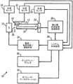
このオフセット値は制御装置28によって使用され、モーター制御装置26を作動させて、以下のような線源12の物理的オフセット量を得る。
物理的オフセット位置が決定されると、さらに、既知のSID位置間で適用されるべき適正オフセット量を、例えば直線補間法を使用して求める。線源12の検出器22に対する実際の位置合わせ位置を推定するために、多項式、ニューラルネットワーク法、及び既知の補正データに基づく結果を予測及び計算する他の方法を含めて、他の数学的手法もまた使用することができる。
12 線源
22 検出器
24 電源/制御回路/移動制御
26 検出器制御装置移動制御
28 システム制御装置
30 ディスプレイ/プリンタ
32 オペレータ・ワークステーション
36 検出器制御回路
40 基準/調整器回路
Claims (8)
- 医療用イメージングシステムにおいて、該システム10での機械的又は物理的位置ずれに起因するばらつきを考慮するために検出器(22)に対する線源(12)の位置合わせを較正する方法であって、前記方法が、
既知の線源−画像間距離での前記線源(12)及び前記検出器(22)の各々における基準位置を求め、
前記線源(12)を第2の線源−画像間距離の位置(55)まで移動させ、
前記第2の線源−画像間距離の位置(55)での前記線源(12)の予想位置と、該線源(12)の実際の位置とのオフセット量を求め、
動作中に、前記第2の線源−画像間距離の位置(55)で前記オフセット量に等しい距離だけ前記線源(12)を移動させて、該線源(12)と前記検出器(22)とを位置合わせする、
各段階を含む方法。 - デジタルX線撮影システムにおける線源(12)と検出器(22)とを較正する方法であって、前記方法が、
第1の線源−画像間距離の位置(53)を選択し、
前記線源(12)と前記検出器(22)との位置合わせを較正し、縦、横、垂直方向の少なくともひとつにおける基準位置を前記線源(12)と前記検出器(22)の各々について求め、
前記基準位置を格納し、
前記線源(12)を少なくとも第2の線源−画像間距離の位置(55)まで移動させ、
前記第2の線源−画像間距離の位置(55)での前記線源(12)と前記検出器(22)との位置ずれからオフセット量を決定し、
前記第1の線源−画像間距離の位置(53)と前記第2の線源−画像間距離の位置(55)の間の位置で、前記線源(12)を前記検出器(22)に位置合わせするための前記線源(12)の移動量を前記オフセット量に基づいて計算し、
動作中、前記間の位置で、前記位置合わせするための前記線源(12)の移動量だけ前記線源(12)を移動させて、該線源(12)と前記検出器(22)とを位置合わせする、
各段階を含む方法。 - 前記線源(12)はX線室の天井に設けられたレールに取り付けられており、
第1の線源−画像間距離の位置(53)と前記第2の線源−画像間距離の位置(55)の間の各位置で適用されるべきオフセット量が直線補間法により求められ、
前記検出器(22)の位置を少なくとも一つの方向で調整する段階をさらに含む請求項2に記載の方法。 - 前記オフセット量が、較正工程中に求められることを特徴とする請求項1乃至3のいずれかに記載の方法。
- 前記オフセット量が、前記システム(10)のリアルタイム動作中に求められることを特徴とする、請求項1乃至3のいずれかに記載の方法。
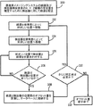
- デジタルX線撮影イメージングシステム(10)を較正する方法であって、前記方法が、
(a)既知の第1の線源−画像間距離の位置(53)で定位置を較正する段階と、
(b)前記線源(12)を第2の線源−画像間距離の位置(55)に移動させる段階と、
(c)前記線源(12)を前記検出器(22)に配向する段階と、
(d)前記線源(12)を前記検出器(22)と位置合わせする位置合わせ位置まで移動させる段階と、
(e)前記位置合わせされた位置の関数としてオフセット量を計算する段階と、
(f)前記オフセット量を格納する段階と、
(g)前記段階(c)から段階(f)を繰り返し、前記デジタルX線撮影システム(10)のオフセット量のマップを作成する段階と、
を含む方法。 - 動作中、前期オフセット量を現在位置に相関付けて、該オフセット量を読み出し、前記線源(12)を移動させて前記現在位置で前記検出器(22)と位置合わせさせる段階をさらに含む請求項6に記載の方法。
- (h)動作中、現在の線源−画像間距離を求める段階と、
(i)前記現在の線源−画像間距離の前後の位置で第1及び第2のオフセット量を求める段階と、
(j)前記現在の線源−画像間距離でオフセット量を得るために補間する段階と、
をさらに含む請求項6に記載の方法。
Applications Claiming Priority (1)
| Application Number | Priority Date | Filing Date | Title |
|---|---|---|---|
| US10/307,131 US6935779B2 (en) | 2002-11-29 | 2002-11-29 | Method and apparatus for aligning an X-ray source and detector at various source to image distances |
Publications (3)
| Publication Number | Publication Date |
|---|---|
| JP2004181239A JP2004181239A (ja) | 2004-07-02 |
| JP2004181239A5 JP2004181239A5 (ja) | 2007-01-18 |
| JP4401752B2 true JP4401752B2 (ja) | 2010-01-20 |
Family
ID=32312199
Family Applications (1)
| Application Number | Title | Priority Date | Filing Date |
|---|---|---|---|
| JP2003398741A Expired - Fee Related JP4401752B2 (ja) | 2002-11-29 | 2003-11-28 | 様々な線源−画像間距離におけるx線源と検出器とを位置合わせさせる方法及び装置 |
Country Status (3)
| Country | Link |
|---|---|
| US (1) | US6935779B2 (ja) |
| JP (1) | JP4401752B2 (ja) |
| DE (1) | DE10355384A1 (ja) |
Families Citing this family (54)
| Publication number | Priority date | Publication date | Assignee | Title |
|---|---|---|---|---|
| US7082185B2 (en) * | 2003-02-12 | 2006-07-25 | The Regents Of The University Of California | Portable imaging system method and apparatus |
| DE10319305A1 (de) * | 2003-04-29 | 2004-11-25 | Siemens Ag | Patiententisch zur Strahlungsbildaufnahme |
| EP1729647A1 (en) * | 2004-03-23 | 2006-12-13 | Koninklijke Philips Electronics N.V. | X-ray examination apparatus and method |
| US7182511B2 (en) * | 2004-11-24 | 2007-02-27 | General Electric Company | Ceiling mounted x-ray tube support |
| US7581885B2 (en) * | 2004-11-24 | 2009-09-01 | General Electric Company | Method and system of aligning x-ray detector for data acquisition |
| DE102005020124B4 (de) * | 2005-04-29 | 2011-07-14 | Siemens AG, 80333 | Röntgensystem, enthaltend einen zugeordneten, mobilen Festkörperdetektor und Verfahren zur Aufnahme und Anzeige eines Röntgenbildes |
| US7344304B2 (en) * | 2005-06-14 | 2008-03-18 | Varian Medical Systems Technologies, Inc. | Self-alignment of radiographic imaging system |
| US7341376B2 (en) * | 2006-03-23 | 2008-03-11 | General Electric Company | Method for aligning radiographic inspection system |
| CN100512759C (zh) * | 2006-04-13 | 2009-07-15 | Ge医疗系统环球技术有限公司 | 台装备识别方法和医疗成像装备 |
| US7575375B2 (en) * | 2007-01-11 | 2009-08-18 | General Electric Company | Systems and methods for reducing movement of an object |
| JP2009226188A (ja) | 2007-07-27 | 2009-10-08 | Fujifilm Corp | 放射線画像撮影システム |
| JP5398133B2 (ja) * | 2007-10-23 | 2014-01-29 | キヤノン株式会社 | X線撮影装置、x線撮影装置の制御方法、プログラム及び記憶媒体 |
| CN101491444B (zh) * | 2008-01-25 | 2012-12-12 | Ge医疗系统环球技术有限公司 | X射线探测台和x射线成像设备 |
| JP2009226197A (ja) * | 2008-02-29 | 2009-10-08 | Fujifilm Corp | 放射線画像撮影システム及び放射線画像撮影方法 |
| US7686511B2 (en) * | 2008-03-06 | 2010-03-30 | Moshe Ein-Gal | Angular irradiation in an upright treatment system |
| JP2010094209A (ja) * | 2008-10-15 | 2010-04-30 | Fujifilm Corp | 放射線画像撮影装置 |
| JP5408952B2 (ja) * | 2008-10-16 | 2014-02-05 | キヤノン株式会社 | X線撮影装置 |
| US8077328B2 (en) * | 2009-07-06 | 2011-12-13 | Gammex, Inc. | Variable color incoherent alignment line and cross-hair generator |
| JP5438493B2 (ja) * | 2009-12-22 | 2014-03-12 | 富士フイルム株式会社 | 放射線撮影システム及びその補助装置 |
| US8827554B2 (en) | 2010-04-13 | 2014-09-09 | Carestream Health, Inc. | Tube alignment for mobile radiography system |
| US8867705B2 (en) | 2010-04-13 | 2014-10-21 | Carestream Health, Inc. | Display of AEC sensor location |
| US8873712B2 (en) | 2010-04-13 | 2014-10-28 | Carestream Health, Inc. | Exposure control using digital radiography detector |
| US8821017B2 (en) | 2010-04-13 | 2014-09-02 | Carestream Health, Inc. | Projector as collimator light |
| US8821015B2 (en) | 2011-03-08 | 2014-09-02 | Carestream Health, Inc. | Alignment apparatus for X-ray imaging system |
| US9414792B2 (en) * | 2011-06-17 | 2016-08-16 | The Board Of Trustees Of The Leland Stanford Junior University | Computed tomography system with dynamic bowtie filter |
| US9521982B2 (en) | 2011-06-17 | 2016-12-20 | The Board Of Trustees Of The Leland Stanford Junior University | Computed tomography system with dynamic bowtie filter |
| CN103957799B (zh) * | 2011-12-01 | 2016-09-21 | 皇家飞利浦有限公司 | 用于提供x 射线图像的医疗成像系统和方法 |
| US20140369459A1 (en) | 2012-02-22 | 2014-12-18 | Carestream Health, Inc. | Mobile radiographic apparatus/methods with tomosynthesis capability |
| AU2013252899A1 (en) * | 2012-04-24 | 2014-10-30 | Portavision Medical Llc | Mobile imaging system and method |
| US9364189B2 (en) * | 2012-12-03 | 2016-06-14 | Nanofocusray Co., Ltd. | Portable x-ray image system and operating table using the same |
| EP2762081B1 (en) * | 2013-02-04 | 2018-01-17 | Agfa Healthcare | Method for accurately generating a radiation image of a region of interest. |
| KR102121721B1 (ko) * | 2013-03-04 | 2020-06-26 | 삼성전자주식회사 | 이동형 엑스선 영상 장치 및 그 제어 방법 |
| EP3097856B1 (en) * | 2014-01-23 | 2020-03-11 | Vatech Co., Ltd. | X-ray photographing device comprising variable type arm |
| JP6400307B2 (ja) * | 2014-03-10 | 2018-10-03 | キヤノンメディカルシステムズ株式会社 | X線画像診断装置 |
| TWI535421B (zh) * | 2014-05-14 | 2016-06-01 | Automatic identification and adjustment of the selected parts of the medical equipment | |
| JP2017528202A (ja) | 2014-08-19 | 2017-09-28 | コーニンクレッカ フィリップス エヌ ヴェKoninklijke Philips N.V. | X線イメージング装置 |
| KR101768520B1 (ko) * | 2015-11-30 | 2017-09-08 | 연세대학교 원주산학협력단 | 흉부의 디지털 x선 일반촬영 및 디지털 단층영상합성의 영상을 통합적 및 연속적으로 획득하기 위한 디지털 x선 촬영 시스템의 제어방법 |
| EP3235430B1 (en) * | 2016-04-19 | 2019-06-12 | Agfa Nv | Radiation image capturing system and method |
| CN106419941A (zh) * | 2016-09-09 | 2017-02-22 | 沈阳东软医疗系统有限公司 | 一种实现对中运动的修正方法及装置 |
| WO2018053262A1 (en) * | 2016-09-15 | 2018-03-22 | Micro C, LLC | Improved imaging systems and methods |
| DE102016013315B4 (de) * | 2016-11-08 | 2024-07-11 | RayScan Technologies GmbH | Messsystem und Verfahren zum Betreiben eines Messsystems |
| US10743827B2 (en) * | 2017-01-31 | 2020-08-18 | General Electric Company | Robotic arm with X-ray source |
| DE102017205113A1 (de) * | 2017-03-27 | 2018-09-27 | Siemens Aktiengesellschaft | Ermitteln der Pose einer Röntgeneinheit relativ zu einem Objekt anhand eines digitalen Modells des Objekts |
| CN108742669B (zh) * | 2018-06-29 | 2020-08-18 | 上海联影医疗科技有限公司 | X光机的卧位成像校正方法以及x光机 |
| EP3829444B1 (en) | 2018-08-01 | 2026-03-04 | OXOS Medical, Inc. | Improved imaging systems and methods |
| US10799206B2 (en) * | 2018-09-28 | 2020-10-13 | General Electric Company | System and method for calibrating an imaging system |
| EP3660542A1 (en) * | 2018-11-29 | 2020-06-03 | Koninklijke Philips N.V. | Hybrid x-ray and optical detector |
| US12324630B2 (en) | 2019-03-12 | 2025-06-10 | Oxos Medical, Inc. | Method of fluoroscopic surgical registration |
| CN111256743A (zh) * | 2019-12-25 | 2020-06-09 | 苏州英诺威视图像有限公司 | 一种xy平台标定系统 |
| EP3960088A1 (en) * | 2020-08-28 | 2022-03-02 | Koninklijke Philips N.V. | Optical arrangement for an x-ray system for determining a patient position and/or patient rotation |
| US20220326165A1 (en) * | 2021-04-07 | 2022-10-13 | Jst Power Equipment, Inc. | Rapid x-ray radiation imaging system and mobile imaging system |
| US11382582B1 (en) | 2021-08-02 | 2022-07-12 | Oxos Medical, Inc. | Imaging systems and methods |
| CN113892959B (zh) * | 2021-09-26 | 2024-03-26 | 有方(合肥)医疗科技有限公司 | X射线成像系统 |
| EP4472516A1 (en) | 2022-03-22 | 2024-12-11 | OXOS Medical, Inc. | Improved imaging systems and methods |
Family Cites Families (1)
| Publication number | Priority date | Publication date | Assignee | Title |
|---|---|---|---|---|
| DE19638145A1 (de) * | 1996-09-18 | 1998-03-26 | Siemens Ag | Röntgendiagnostikgerät |
-
2002
- 2002-11-29 US US10/307,131 patent/US6935779B2/en not_active Expired - Lifetime
-
2003
- 2003-11-25 DE DE10355384A patent/DE10355384A1/de not_active Withdrawn
- 2003-11-28 JP JP2003398741A patent/JP4401752B2/ja not_active Expired - Fee Related
Also Published As
| Publication number | Publication date |
|---|---|
| US20040105526A1 (en) | 2004-06-03 |
| US6935779B2 (en) | 2005-08-30 |
| DE10355384A1 (de) | 2004-06-09 |
| JP2004181239A (ja) | 2004-07-02 |
Similar Documents
| Publication | Publication Date | Title |
|---|---|---|
| JP4401752B2 (ja) | 様々な線源−画像間距離におけるx線源と検出器とを位置合わせさせる方法及び装置 | |
| US6435716B1 (en) | Method and system for determining a source-to-image distance in a digital imaging system | |
| US9541509B2 (en) | Radiation imaging apparatus, radiation imaging method, body movement measuring method, and body movement measuring program | |
| US8300764B2 (en) | Method and device for detecting placement error of an imaging plane of a radiographic image detector, as well as method and device for correcting images | |
| US6478462B2 (en) | Methodology for determining x-ray to light field decentering on digital radiographic image systems | |
| CN1915169B (zh) | 对用于产生3d体积图像的x-射线系统中的对准误差进行检测和校正的方法和装置 | |
| EP1000582B1 (en) | Apparatus and method for imaging | |
| US8344327B2 (en) | Radiation image detector time dependent degradation determination method and apparatus | |
| KR20010112950A (ko) | 레이저 보정 장치 및 방법 | |
| CN113812971B (zh) | 一种多自由度四维双能锥束ct成像系统及方法 | |
| WO2012008706A2 (en) | Radiography apparatus and control method thereof | |
| JP2004511288A (ja) | 撮影テーブル水平化装置 | |
| JP2004180846A (ja) | X線ct装置 | |
| JPH08299323A (ja) | 撓み補正装置 | |
| US9606072B2 (en) | Radiation inspecting apparatus | |
| US5657498A (en) | Methods and apparatus for acquiring table elevation information | |
| US6402373B1 (en) | Method and system for determining a source-to-image distance in a digital imaging system | |
| US6402374B1 (en) | Method and system for determining a source-to-image distance in a digital radiographic imaging system | |
| US6375354B1 (en) | Method and system for determining a variable lateral center-to-center setpoint for a digital imaging system | |
| CN103006248A (zh) | 数字x射线成像装置及其系统非均匀性校正方法 | |
| JPS61155845A (ja) | 断層撮影装置 | |
| JP5855251B2 (ja) | 口腔外撮像のための柱高さの感知 | |
| US5982848A (en) | X-ray diagnosis machine having displaceable measurement field | |
| JP2006051216A (ja) | 放射線治療装置、放射線治療装置用治療台、及び放射線治療装置の座標校正方法 | |
| US12569217B2 (en) | X-ray imaging apparatus and X-ray imaging method |
Legal Events
| Date | Code | Title | Description |
|---|---|---|---|
| A521 | Request for written amendment filed |
Free format text: JAPANESE INTERMEDIATE CODE: A523 Effective date: 20061124 |
|
| A621 | Written request for application examination |
Free format text: JAPANESE INTERMEDIATE CODE: A621 Effective date: 20061124 |
|
| A131 | Notification of reasons for refusal |
Free format text: JAPANESE INTERMEDIATE CODE: A131 Effective date: 20090630 |
|
| A521 | Request for written amendment filed |
Free format text: JAPANESE INTERMEDIATE CODE: A523 Effective date: 20090825 |
|
| RD02 | Notification of acceptance of power of attorney |
Free format text: JAPANESE INTERMEDIATE CODE: A7422 Effective date: 20090825 |
|
| RD04 | Notification of resignation of power of attorney |
Free format text: JAPANESE INTERMEDIATE CODE: A7424 Effective date: 20090825 |
|
| A521 | Request for written amendment filed |
Free format text: JAPANESE INTERMEDIATE CODE: A523 Effective date: 20090904 |
|
| TRDD | Decision of grant or rejection written | ||
| A01 | Written decision to grant a patent or to grant a registration (utility model) |
Free format text: JAPANESE INTERMEDIATE CODE: A01 Effective date: 20091006 |
|
| A01 | Written decision to grant a patent or to grant a registration (utility model) |
Free format text: JAPANESE INTERMEDIATE CODE: A01 |
|
| A61 | First payment of annual fees (during grant procedure) |
Free format text: JAPANESE INTERMEDIATE CODE: A61 Effective date: 20091028 |
|
| R150 | Certificate of patent or registration of utility model |
Ref document number: 4401752 Country of ref document: JP Free format text: JAPANESE INTERMEDIATE CODE: R150 Free format text: JAPANESE INTERMEDIATE CODE: R150 |
|
| FPAY | Renewal fee payment (event date is renewal date of database) |
Free format text: PAYMENT UNTIL: 20121106 Year of fee payment: 3 |
|
| FPAY | Renewal fee payment (event date is renewal date of database) |
Free format text: PAYMENT UNTIL: 20121106 Year of fee payment: 3 |
|
| FPAY | Renewal fee payment (event date is renewal date of database) |
Free format text: PAYMENT UNTIL: 20131106 Year of fee payment: 4 |
|
| R250 | Receipt of annual fees |
Free format text: JAPANESE INTERMEDIATE CODE: R250 |
|
| R250 | Receipt of annual fees |
Free format text: JAPANESE INTERMEDIATE CODE: R250 |
|
| R250 | Receipt of annual fees |
Free format text: JAPANESE INTERMEDIATE CODE: R250 |
|
| R250 | Receipt of annual fees |
Free format text: JAPANESE INTERMEDIATE CODE: R250 |
|
| R250 | Receipt of annual fees |
Free format text: JAPANESE INTERMEDIATE CODE: R250 |
|
| R250 | Receipt of annual fees |
Free format text: JAPANESE INTERMEDIATE CODE: R250 |
|
| R250 | Receipt of annual fees |
Free format text: JAPANESE INTERMEDIATE CODE: R250 |
|
| R250 | Receipt of annual fees |
Free format text: JAPANESE INTERMEDIATE CODE: R250 |
|
| LAPS | Cancellation because of no payment of annual fees |