JP4239166B2 - 多層観察型光学顕微鏡及び多層観察ユニット - Google Patents
多層観察型光学顕微鏡及び多層観察ユニット Download PDFInfo
- Publication number
- JP4239166B2 JP4239166B2 JP2003337105A JP2003337105A JP4239166B2 JP 4239166 B2 JP4239166 B2 JP 4239166B2 JP 2003337105 A JP2003337105 A JP 2003337105A JP 2003337105 A JP2003337105 A JP 2003337105A JP 4239166 B2 JP4239166 B2 JP 4239166B2
- Authority
- JP
- Japan
- Prior art keywords
- phase
- observation
- sample
- optical
- observed
- Prior art date
- Legal status (The legal status is an assumption and is not a legal conclusion. Google has not performed a legal analysis and makes no representation as to the accuracy of the status listed.)
- Expired - Fee Related
Links
- 230000003287 optical effect Effects 0.000 title claims description 103
- 239000000523 sample Substances 0.000 description 49
- 238000007689 inspection Methods 0.000 description 16
- 210000001519 tissue Anatomy 0.000 description 14
- 238000000034 method Methods 0.000 description 13
- 238000010586 diagram Methods 0.000 description 7
- 239000000463 material Substances 0.000 description 5
- 238000002360 preparation method Methods 0.000 description 4
- 238000005452 bending Methods 0.000 description 3
- 238000006073 displacement reaction Methods 0.000 description 3
- 230000002093 peripheral effect Effects 0.000 description 3
- 239000004065 semiconductor Substances 0.000 description 3
- 230000005374 Kerr effect Effects 0.000 description 2
- 230000005697 Pockels effect Effects 0.000 description 2
- XUIMIQQOPSSXEZ-UHFFFAOYSA-N Silicon Chemical compound [Si] XUIMIQQOPSSXEZ-UHFFFAOYSA-N 0.000 description 2
- 238000004113 cell culture Methods 0.000 description 2
- 238000007796 conventional method Methods 0.000 description 2
- 239000013078 crystal Substances 0.000 description 2
- 239000003814 drug Substances 0.000 description 2
- 210000005240 left ventricle Anatomy 0.000 description 2
- 238000004519 manufacturing process Methods 0.000 description 2
- 238000005259 measurement Methods 0.000 description 2
- 238000002156 mixing Methods 0.000 description 2
- 229910052710 silicon Inorganic materials 0.000 description 2
- 239000010703 silicon Substances 0.000 description 2
- 229910003327 LiNbO3 Inorganic materials 0.000 description 1
- 210000000709 aorta Anatomy 0.000 description 1
- 238000003491 array Methods 0.000 description 1
- 210000004351 coronary vessel Anatomy 0.000 description 1
- 230000000694 effects Effects 0.000 description 1
- 230000005684 electric field Effects 0.000 description 1
- 238000005530 etching Methods 0.000 description 1
- 230000005284 excitation Effects 0.000 description 1
- 238000002474 experimental method Methods 0.000 description 1
- 210000002837 heart atrium Anatomy 0.000 description 1
- 210000005003 heart tissue Anatomy 0.000 description 1
- 238000003384 imaging method Methods 0.000 description 1
- 239000007788 liquid Substances 0.000 description 1
- GQYHUHYESMUTHG-UHFFFAOYSA-N lithium niobate Chemical compound [Li+].[O-][Nb](=O)=O GQYHUHYESMUTHG-UHFFFAOYSA-N 0.000 description 1
- 208000010125 myocardial infarction Diseases 0.000 description 1
- 239000013074 reference sample Substances 0.000 description 1
- 239000000758 substrate Substances 0.000 description 1
- 230000001360 synchronised effect Effects 0.000 description 1
- 230000002194 synthesizing effect Effects 0.000 description 1
- 238000012360 testing method Methods 0.000 description 1
Images


Classifications
-
- G—PHYSICS
- G02—OPTICS
- G02B—OPTICAL ELEMENTS, SYSTEMS OR APPARATUS
- G02B21/00—Microscopes
- G02B21/06—Means for illuminating specimens
- G02B21/08—Condensers
- G02B21/14—Condensers affording illumination for phase-contrast observation
-
- G—PHYSICS
- G02—OPTICS
- G02B—OPTICAL ELEMENTS, SYSTEMS OR APPARATUS
- G02B21/00—Microscopes
- G02B21/36—Microscopes arranged for photographic purposes or projection purposes or digital imaging or video purposes including associated control and data processing arrangements
-
- G—PHYSICS
- G02—OPTICS
- G02B—OPTICAL ELEMENTS, SYSTEMS OR APPARATUS
- G02B26/00—Optical devices or arrangements for the control of light using movable or deformable optical elements
- G02B26/06—Optical devices or arrangements for the control of light using movable or deformable optical elements for controlling the phase of light
Landscapes
- Physics & Mathematics (AREA)
- General Physics & Mathematics (AREA)
- Optics & Photonics (AREA)
- Chemical & Material Sciences (AREA)
- Analytical Chemistry (AREA)
- Engineering & Computer Science (AREA)
- Multimedia (AREA)
- Microscoopes, Condenser (AREA)
Description
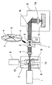
2 二重回転円板式高速共焦点スキャナー
3 高速CCDカメラ
4、6 視準化レンズ
5 収束レンズ
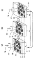
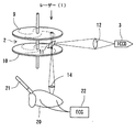
7 多層観察ユニット (位相変更手段)
8 蛍光位相差顕微鏡ユニット
9 マイクロレンズアレーディスク
10 ニポウディスク
11 ダイクロイックミラー
12 凸レンズ
13 駆動用モーター
14 対物レンズ
15 細胞・組織試料
16 光路折り曲げ鏡
17 レーザービーム
18 走査線
19 焦点
Claims (5)
- 光源と、
前記光源からの照射光を被観察試料内に集光する対物レンズと、
前記光源から前記対物レンズに対し照射光線を入射させる光軸中に配置された収束/視準化レンズ対と、
前記収束/視準化レンズ対の間に配置され、透過光の位相を光軸横断面の所定範囲内で変化させることによって、前記対物レンズの観察面を段階的に深度調節するための位相変更手段と、
前記光源および前記収束/視準化レンズ対の間の光軸中に共焦点スキャナーと、を備え、
前記共焦点スキャナーは、
前記光源側に配置されたマイクロレンズアレーディスクと、
前記収束/視準化ディスク側に、前記マイクロレンズアレーディスクと同軸にこれに対向配置された複数のピンホールを有するニポウディスクと、
前記マイクロレンズアレーディスクおよび前記ニポウディスクの間に配置され、光源からの照射光を透過する一方、前記被観察試料から返ってきた蛍光を反射するダイクロイックミラーと、を備え、
前記位相変更手段が段階的に光学的特性の異なる複数の位相板セグメントを配列し、各セグメントが順次光軸を横切るように設置された回転板からなることを特徴とする多層観察型光学顕微鏡。 - 前記位相変更手段が各位相板セグメントの要素をなす等方性透明膜の厚さを段階的に変化させたことにより、それらの光学的特性を異ならしめたことを特徴とする請求項1に記載の多層観察型光学顕微鏡。
- 前記位相変更手段が各位相板セグメントの要素をなす等方性透明膜の屈折率を段階的に変化させたことにより、それらの光学的特性を異ならしめたことを特徴とする請求項1に記載の多層観察型光学顕微鏡。
- 光学顕微鏡の試料台の二次元走査と、前記位相変更手段の位相走査とを同期させることにより、被観察試料の三次元動態を観察できるようにしたことを特徴とする請求項1に記載の多層観察型光学顕微鏡。
- 前記位相変更手段は、表面上に複数の位相板セグメントを備えた回転板からなり、前記複数の位相板セグメントは、それぞれ、段階的に互いに異なる光学的特性を有するとともに、前記回転板の円周方向に順次隣接するように配列されていることを特徴とする請求項1に記載の多層観察型光学顕微鏡。
Priority Applications (4)
| Application Number | Priority Date | Filing Date | Title |
|---|---|---|---|
| JP2003337105A JP4239166B2 (ja) | 2002-12-27 | 2003-09-29 | 多層観察型光学顕微鏡及び多層観察ユニット |
| AU2003296100A AU2003296100A1 (en) | 2002-12-27 | 2003-12-24 | Multilayer observation optical microscope and multilayer observation unit |
| PCT/JP2003/016641 WO2004061515A1 (ja) | 2002-12-27 | 2003-12-24 | 多層観察型光学顕微鏡及び多層観察ユニット |
| US10/540,600 US7366394B2 (en) | 2002-12-27 | 2003-12-24 | Multilayer observation optical microscope and multilayer observation unit |
Applications Claiming Priority (2)
| Application Number | Priority Date | Filing Date | Title |
|---|---|---|---|
| JP2002379869 | 2002-12-27 | ||
| JP2003337105A JP4239166B2 (ja) | 2002-12-27 | 2003-09-29 | 多層観察型光学顕微鏡及び多層観察ユニット |
Publications (2)
| Publication Number | Publication Date |
|---|---|
| JP2004219987A JP2004219987A (ja) | 2004-08-05 |
| JP4239166B2 true JP4239166B2 (ja) | 2009-03-18 |
Family
ID=32716315
Family Applications (1)
| Application Number | Title | Priority Date | Filing Date |
|---|---|---|---|
| JP2003337105A Expired - Fee Related JP4239166B2 (ja) | 2002-12-27 | 2003-09-29 | 多層観察型光学顕微鏡及び多層観察ユニット |
Country Status (4)
| Country | Link |
|---|---|
| US (1) | US7366394B2 (ja) |
| JP (1) | JP4239166B2 (ja) |
| AU (1) | AU2003296100A1 (ja) |
| WO (1) | WO2004061515A1 (ja) |
Families Citing this family (23)
| Publication number | Priority date | Publication date | Assignee | Title |
|---|---|---|---|---|
| JP4797425B2 (ja) * | 2005-04-12 | 2011-10-19 | カシオ計算機株式会社 | 光源ユニット及び投影装置 |
| WO2006127967A2 (en) | 2005-05-25 | 2006-11-30 | Massachusetts Institute Of Technology | Multifocal scanning microscopy systems and methods |
| DE102005036486A1 (de) * | 2005-07-20 | 2007-01-25 | Leica Microsystems (Schweiz) Ag | Optisches Gerät mit erhöhter Schärfentiefe |
| JP2007079278A (ja) * | 2005-09-15 | 2007-03-29 | Univ Of Tokyo | 物質状態測定装置 |
| EP1936422A4 (en) * | 2005-10-13 | 2013-01-16 | Nikon Corp | MICROSCOPE |
| US8537461B2 (en) * | 2007-11-26 | 2013-09-17 | Carl Zeiss Microimaging Gmbh | Method and configuration for the optical detection of an illuminated specimen |
| WO2009081305A1 (en) * | 2007-12-19 | 2009-07-02 | Koninklijke Philips Electronics N.V. | Detection system and method |
| JP5242304B2 (ja) * | 2008-09-04 | 2013-07-24 | オリンパスメディカルシステムズ株式会社 | 観測システム |
| JP5504881B2 (ja) * | 2009-12-25 | 2014-05-28 | ソニー株式会社 | 演算装置、演算方法、演算プログラム及び顕微鏡 |
| JP5056871B2 (ja) * | 2010-03-02 | 2012-10-24 | 横河電機株式会社 | 共焦点顕微鏡システム |
| JP5221614B2 (ja) * | 2010-09-17 | 2013-06-26 | 独立行政法人科学技術振興機構 | 3次元共焦点観察用装置及び観察焦点面変位・補正ユニット |
| ITTO20110298A1 (it) * | 2011-04-01 | 2012-10-02 | St Microelectronics Srl | Rilevatore ottico confocale, schiera di rilevatori e relativo procedimento di fabbricazione |
| US9104027B2 (en) * | 2012-04-27 | 2015-08-11 | Manufacturing Techniques, Inc. | Optical instrument for the simulation of atmospheric turbulence |
| US9696264B2 (en) * | 2013-04-03 | 2017-07-04 | Kla-Tencor Corporation | Apparatus and methods for determining defect depths in vertical stack memory |
| JP6318358B2 (ja) * | 2013-04-08 | 2018-05-09 | 株式会社ニューフレアテクノロジー | 照明装置および検査装置 |
| WO2015164844A1 (en) * | 2014-04-24 | 2015-10-29 | Vutara, Inc. | Super resolution microscopy |
| CN104101993B (zh) * | 2014-07-10 | 2017-04-19 | 深圳职业技术学院 | 傅立叶显微镜装置及信息共享系统及其信息共享方法 |
| US10317597B2 (en) * | 2014-08-26 | 2019-06-11 | The Board Of Trustees Of The Leland Stanford Junior University | Light-field microscopy with phase masking |
| EP3268715B1 (en) | 2015-03-11 | 2024-09-25 | TissueVision, Inc. | System and methods for serial staining and imaging |
| US10495446B2 (en) * | 2015-06-29 | 2019-12-03 | Kla-Tencor Corporation | Methods and apparatus for measuring height on a semiconductor wafer |
| CN110192094A (zh) | 2016-11-18 | 2019-08-30 | 迪术斐迅公司 | 自动化组织切片捕获、索引和储存系统及方法 |
| KR101900254B1 (ko) * | 2017-04-25 | 2018-09-19 | 충북대학교 산학협력단 | 홀로그램 광학소자 마이크로 렌즈 어레이를 이용한 집적영상 현미경 시스템 |
| CN115437134A (zh) * | 2022-08-30 | 2022-12-06 | 宁波礼达先导生物技术有限公司 | 一种全自动智能显微镜及图像处理方法 |
Family Cites Families (6)
| Publication number | Priority date | Publication date | Assignee | Title |
|---|---|---|---|---|
| JPH08136810A (ja) | 1994-11-08 | 1996-05-31 | Hamamatsu Photonics Kk | 共焦点顕微鏡 |
| US5952562A (en) * | 1995-11-22 | 1999-09-14 | Olympus Optical Co., Ltd. | Scanning probe microscope incorporating an optical microscope |
| JP3917731B2 (ja) * | 1996-11-21 | 2007-05-23 | オリンパス株式会社 | レーザ走査顕微鏡 |
| JPH1138324A (ja) | 1997-07-23 | 1999-02-12 | Nikon Corp | レーザ走査顕微鏡 |
| JP4481397B2 (ja) * | 1999-09-07 | 2010-06-16 | オリンパス株式会社 | 光学装置及び顕微鏡 |
| DE10105391B4 (de) | 2001-02-06 | 2004-11-25 | Leica Microsystems Heidelberg Gmbh | Scanmikroskop und Modul für ein Scanmikroskop |
-
2003
- 2003-09-29 JP JP2003337105A patent/JP4239166B2/ja not_active Expired - Fee Related
- 2003-12-24 US US10/540,600 patent/US7366394B2/en not_active Expired - Fee Related
- 2003-12-24 AU AU2003296100A patent/AU2003296100A1/en not_active Abandoned
- 2003-12-24 WO PCT/JP2003/016641 patent/WO2004061515A1/ja not_active Ceased
Also Published As
| Publication number | Publication date |
|---|---|
| JP2004219987A (ja) | 2004-08-05 |
| US7366394B2 (en) | 2008-04-29 |
| WO2004061515A1 (ja) | 2004-07-22 |
| US20060147176A1 (en) | 2006-07-06 |
| AU2003296100A1 (en) | 2004-07-29 |
Similar Documents
| Publication | Publication Date | Title |
|---|---|---|
| JP4239166B2 (ja) | 多層観察型光学顕微鏡及び多層観察ユニット | |
| US9885859B2 (en) | Structured illumination microscopy apparatus and method | |
| CN103743714B (zh) | 一种倾斜宽场光切片扫描成像显微系统及其成像方法 | |
| EP2041613B1 (en) | Device and method for wide- field and high resolution imaging of tissue | |
| US9383568B2 (en) | Objective-coupled selective plane illumination microscopy | |
| JP5221614B2 (ja) | 3次元共焦点観察用装置及び観察焦点面変位・補正ユニット | |
| US10094784B2 (en) | Systems and methods for in-operating-theatre imaging of fresh tissue resected during surgery for pathology assessment | |
| US10352819B2 (en) | Method of measuring transmission characteristics of optical transfer medium and image acquisition device using the same | |
| JP2000126116A (ja) | 光診断システム | |
| WO2004036284A1 (ja) | 共焦点顕微鏡、共焦点顕微鏡を用いた蛍光測定方法及び偏光測定方法 | |
| CN103054554B (zh) | 一种沿轴向进行深度扫描的光学成像装置、方法及其应用 | |
| CN114527102A (zh) | 一种基于激光扫描的近红外二区显微成像系统及方法 | |
| CN110062603A (zh) | 用于垂直横截面成像的多光子内窥显微镜 | |
| JP2005121796A (ja) | レーザー顕微鏡 | |
| NL2008873C2 (en) | Method and apparatus for multiple points of view three-dimensional microscopy. | |
| US20070091425A1 (en) | Microscope examination apparatus and microscope examination method | |
| JP2024500089A (ja) | 光子の再割り当てを行う共焦点顕微鏡 | |
| JP2000126115A (ja) | 光走査プローブ装置 | |
| JP2021519438A (ja) | 被験体を非侵襲的に検査するためのマルチモード撮像システムおよび方法 | |
| CN105043988A (zh) | 基于扫描振镜的单点去卷积显微系统与成像方法 | |
| CN111855582A (zh) | 一种基于光纤延时的并行多尺度光声显微成像方法 | |
| CN117631249A (zh) | 线扫共聚焦扫描光场显微成像装置及方法 | |
| JP2002301018A (ja) | 光走査プローブ装置 | |
| EP1806575B1 (en) | Examination apparatus | |
| JP3144513B2 (ja) | 蛍光顕微鏡 |
Legal Events
| Date | Code | Title | Description |
|---|---|---|---|
| A621 | Written request for application examination |
Free format text: JAPANESE INTERMEDIATE CODE: A621 Effective date: 20050513 |
|
| A131 | Notification of reasons for refusal |
Free format text: JAPANESE INTERMEDIATE CODE: A131 Effective date: 20080618 |
|
| A521 | Written amendment |
Free format text: JAPANESE INTERMEDIATE CODE: A523 Effective date: 20080813 |
|
| RD02 | Notification of acceptance of power of attorney |
Free format text: JAPANESE INTERMEDIATE CODE: A7422 Effective date: 20080813 |
|
| A131 | Notification of reasons for refusal |
Free format text: JAPANESE INTERMEDIATE CODE: A131 Effective date: 20080903 |
|
| A521 | Written amendment |
Free format text: JAPANESE INTERMEDIATE CODE: A523 Effective date: 20081029 |
|
| TRDD | Decision of grant or rejection written | ||
| A01 | Written decision to grant a patent or to grant a registration (utility model) |
Free format text: JAPANESE INTERMEDIATE CODE: A01 Effective date: 20081125 |
|
| A01 | Written decision to grant a patent or to grant a registration (utility model) |
Free format text: JAPANESE INTERMEDIATE CODE: A01 |
|
| A61 | First payment of annual fees (during grant procedure) |
Free format text: JAPANESE INTERMEDIATE CODE: A61 Effective date: 20081211 |
|
| FPAY | Renewal fee payment (event date is renewal date of database) |
Free format text: PAYMENT UNTIL: 20120109 Year of fee payment: 3 |
|
| R150 | Certificate of patent or registration of utility model |
Free format text: JAPANESE INTERMEDIATE CODE: R150 |
|
| FPAY | Renewal fee payment (event date is renewal date of database) |
Free format text: PAYMENT UNTIL: 20120109 Year of fee payment: 3 |
|
| S111 | Request for change of ownership or part of ownership |
Free format text: JAPANESE INTERMEDIATE CODE: R313113 |
|
| FPAY | Renewal fee payment (event date is renewal date of database) |
Free format text: PAYMENT UNTIL: 20120109 Year of fee payment: 3 |
|
| R350 | Written notification of registration of transfer |
Free format text: JAPANESE INTERMEDIATE CODE: R350 |
|
| FPAY | Renewal fee payment (event date is renewal date of database) |
Free format text: PAYMENT UNTIL: 20130109 Year of fee payment: 4 |
|
| FPAY | Renewal fee payment (event date is renewal date of database) |
Free format text: PAYMENT UNTIL: 20140109 Year of fee payment: 5 |
|
| R250 | Receipt of annual fees |
Free format text: JAPANESE INTERMEDIATE CODE: R250 |
|
| R250 | Receipt of annual fees |
Free format text: JAPANESE INTERMEDIATE CODE: R250 |
|
| R250 | Receipt of annual fees |
Free format text: JAPANESE INTERMEDIATE CODE: R250 |
|
| LAPS | Cancellation because of no payment of annual fees |