JP2017522603A - 試料を顕微鏡検査する方法及び装置 - Google Patents
試料を顕微鏡検査する方法及び装置 Download PDFInfo
- Publication number
- JP2017522603A JP2017522603A JP2017503524A JP2017503524A JP2017522603A JP 2017522603 A JP2017522603 A JP 2017522603A JP 2017503524 A JP2017503524 A JP 2017503524A JP 2017503524 A JP2017503524 A JP 2017503524A JP 2017522603 A JP2017522603 A JP 2017522603A
- Authority
- JP
- Japan
- Prior art keywords
- sample
- illumination
- objective lens
- light beam
- interface
- Prior art date
- Legal status (The legal status is an assumption and is not a legal conclusion. Google has not performed a legal analysis and makes no representation as to the accuracy of the status listed.)
- Granted
Links
- 238000000034 method Methods 0.000 title claims abstract description 44
- 238000005286 illumination Methods 0.000 claims abstract description 209
- 238000001514 detection method Methods 0.000 claims abstract description 88
- 230000003287 optical effect Effects 0.000 claims description 30
- 238000007689 inspection Methods 0.000 claims description 19
- 239000012780 transparent material Substances 0.000 claims description 12
- 230000008859 change Effects 0.000 claims description 8
- 238000000386 microscopy Methods 0.000 claims description 6
- 239000000523 sample Substances 0.000 description 159
- 239000006059 cover glass Substances 0.000 description 27
- 230000008901 benefit Effects 0.000 description 8
- 239000007788 liquid Substances 0.000 description 8
- 235000015097 nutrients Nutrition 0.000 description 8
- 230000000694 effects Effects 0.000 description 6
- 238000003384 imaging method Methods 0.000 description 5
- 238000007654 immersion Methods 0.000 description 4
- 238000012856 packing Methods 0.000 description 4
- 230000010287 polarization Effects 0.000 description 4
- 230000005284 excitation Effects 0.000 description 3
- 230000035515 penetration Effects 0.000 description 3
- 230000015572 biosynthetic process Effects 0.000 description 2
- 230000008878 coupling Effects 0.000 description 2
- 238000010168 coupling process Methods 0.000 description 2
- 238000005859 coupling reaction Methods 0.000 description 2
- 230000000644 propagated effect Effects 0.000 description 2
- 238000000492 total internal reflection fluorescence microscopy Methods 0.000 description 2
- XLYOFNOQVPJJNP-UHFFFAOYSA-N water Substances O XLYOFNOQVPJJNP-UHFFFAOYSA-N 0.000 description 2
- 239000012472 biological sample Substances 0.000 description 1
- 230000005540 biological transmission Effects 0.000 description 1
- 230000007547 defect Effects 0.000 description 1
- 238000010586 diagram Methods 0.000 description 1
- 230000005672 electromagnetic field Effects 0.000 description 1
- 238000000799 fluorescence microscopy Methods 0.000 description 1
- 230000003993 interaction Effects 0.000 description 1
- 238000002360 preparation method Methods 0.000 description 1
- 230000002459 sustained effect Effects 0.000 description 1
Images


Classifications
-
- G—PHYSICS
- G02—OPTICS
- G02B—OPTICAL ELEMENTS, SYSTEMS OR APPARATUS
- G02B21/00—Microscopes
- G02B21/16—Microscopes adapted for ultraviolet illumination ; Fluorescence microscopes
-
- G—PHYSICS
- G01—MEASURING; TESTING
- G01N—INVESTIGATING OR ANALYSING MATERIALS BY DETERMINING THEIR CHEMICAL OR PHYSICAL PROPERTIES
- G01N21/00—Investigating or analysing materials by the use of optical means, i.e. using sub-millimetre waves, infrared, visible or ultraviolet light
- G01N21/62—Systems in which the material investigated is excited whereby it emits light or causes a change in wavelength of the incident light
- G01N21/63—Systems in which the material investigated is excited whereby it emits light or causes a change in wavelength of the incident light optically excited
- G01N21/64—Fluorescence; Phosphorescence
- G01N21/645—Specially adapted constructive features of fluorimeters
- G01N21/6456—Spatial resolved fluorescence measurements; Imaging
- G01N21/6458—Fluorescence microscopy
-
- G—PHYSICS
- G01—MEASURING; TESTING
- G01N—INVESTIGATING OR ANALYSING MATERIALS BY DETERMINING THEIR CHEMICAL OR PHYSICAL PROPERTIES
- G01N21/00—Investigating or analysing materials by the use of optical means, i.e. using sub-millimetre waves, infrared, visible or ultraviolet light
- G01N21/62—Systems in which the material investigated is excited whereby it emits light or causes a change in wavelength of the incident light
- G01N21/63—Systems in which the material investigated is excited whereby it emits light or causes a change in wavelength of the incident light optically excited
- G01N21/64—Fluorescence; Phosphorescence
- G01N21/645—Specially adapted constructive features of fluorimeters
- G01N21/648—Specially adapted constructive features of fluorimeters using evanescent coupling or surface plasmon coupling for the excitation of fluorescence
-
- G—PHYSICS
- G02—OPTICS
- G02B—OPTICAL ELEMENTS, SYSTEMS OR APPARATUS
- G02B21/00—Microscopes
- G02B21/0004—Microscopes specially adapted for specific applications
- G02B21/002—Scanning microscopes
-
- G—PHYSICS
- G02—OPTICS
- G02B—OPTICAL ELEMENTS, SYSTEMS OR APPARATUS
- G02B21/00—Microscopes
- G02B21/06—Means for illuminating specimens
-
- G—PHYSICS
- G02—OPTICS
- G02B—OPTICAL ELEMENTS, SYSTEMS OR APPARATUS
- G02B27/00—Optical systems or apparatus not provided for by any of the groups G02B1/00 - G02B26/00, G02B30/00
- G02B27/56—Optics using evanescent waves, i.e. inhomogeneous waves
Abstract
Description
a.試料を、この試料よりも高い屈折率を有する光学的に透明な媒体に接触させるステップと、
b.照明光束を形成するステップと、
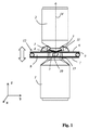
c.照明光束をフォーカシングする照明対物レンズを通して、照明光束を案内するステップと、
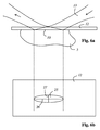
d.照明対物レンズを通過した照明光束を、検出対物レンズに配置された偏向手段によって、照明光束が光学的に透明な媒体と試料との界面へ入射してそこでの全反射により試料のエバネセント照明を生じさせるよう、検査すべき試料の方向へ偏向するステップと、
e.例えば蛍光の光エネルギに比例する電気信号を形成する検出器により、試料から出て検出対物レンズを通って走行する蛍光を検出するステップと
を含む方法により、解決される。
2 検出対物レンズ
3 偏向手段
4 錐台状の鏡面
5 試料
6 第1のカバーガラス
7 第2のカバーガラス
8 液状栄養媒体
9 パッキン
10 界面
11 浸漬オイル
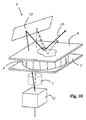
12,20 光学的に透明な媒体
13 照明光束
14 検出光
15,21 鏡面
16,22 外面
17 容器
18 第1の偏向手段
19 別の偏向手段
23 第2の鏡面
24 固定装置
25 入射面
26 長半軸
27 短半軸
28 入射位置
29 ファセット鏡
30 ファセット面
31 錐台状の鏡
32 調整可能なビーム偏向装置
33 平坦な鏡
Claims (19)
- 試料(5)を顕微鏡検査する方法において、
a.前記試料(5)を、該試料(5)よりも高い屈折率を有する光学的に透明な媒体(12)に接触させるステップと、
b.照明光束(13)を形成するステップと、
c.照明光束(13)をフォーカシングする照明対物レンズ(1)を通して、照明光束(13)を案内するステップと、
d.前記照明対物レンズ(1)を通過した照明光束(13)を、検出対物レンズ(2)に配置された偏向手段によって、該照明光束(13)が前記光学的に透明な媒体(12)と前記試料(5)との界面(10)へ入射してそこでの全反射により前記試料(5)のエバネセント照明を生じさせるよう、検査すべき前記試料(5)の方向へ偏向するステップと、
e.前記試料(5)から出て前記検出対物レンズ(2)を通って走行する蛍光を検出するステップと、
を含む、
ことを特徴とする方法。 - a.前記試料(5)と前記光学的に透明な媒体(12)との前記界面(10)を、前記照明対物レンズ(1)の光軸及び/又は前記検出対物レンズ(2)の光軸に対して0°とは異なる角度をなすように配向する、又は、
b.前記試料(5)と前記光学的に透明な媒体(12)との前記界面(10)を、前記照明対物レンズ(1)の光軸及び/又は前記検出対物レンズ(2)の光軸に対して垂直となるように配向する、
請求項1記載の方法。 - a.偏向後の照明光束(13)を、前記照明対物レンズ(1)の光軸に対して0°とは異なる角度を有する平面に走行させる、及び/又は、
b.照明光束(13)を、前記試料(5)と前記光学的に透明な媒体(12)との前記界面(10)に55°から70°までの領域の入射角で入射するように偏向する、及び/又は、
c.照明光束(13)を、前記試料(5)と前記光学的に透明な媒体(12)との前記界面(10)に60°から64°までの領域の入射角で入射するように偏向する、
請求項1又は2記載の方法。 - a.前記試料(5)と前記光学的に透明な媒体(12)との前記界面(10)への照明光束(13)の入射位置(28)及び/又は入射角及び/又は入射方向を変化させる、及び/又は、
b.前記試料(5)と前記光学的に透明な媒体(12)との前記界面(10)への照明光束(13)の入射位置(28)を、走査路に沿って連続的に変化させる、及び/又は、
c.前記試料(5)と前記光学的に透明な媒体(12)との前記界面(10)への照明光束(13)の入射位置(28)及び/又は入射角及び/又は入射方向を、偏向角を調整可能な、照明光束(13)に作用するビーム偏向装置によって変化させる、及び/又は、
d.前記試料(5)と前記光学的に透明な媒体(12)との前記界面(10)への照明光束(13)の入射位置(28)及び/又は入射角及び/又は入射方向を、前記照明対物レンズ(1)に対して前記試料(5)を相対的に運動させることにより変化させる、及び/又は、
e.前記試料(5)と前記光学的に透明な媒体(12)との前記界面(10)への照明光束(13)の入射位置(28)及び/又は入射角及び/又は入射方向を、前記偏向手段を運動させることにより変化させる、及び/又は、
f.前記偏向手段(3)を複数のファセット面を有する鏡として構成し、前記試料(5)と前記光学的に透明な媒体(12)との前記界面(10)への照明光束(13)の入射位置(28)及び/又は入射角及び/又は入射方向を、連続する種々のファセット面を照明することによって変化させる、
請求項1から3までのいずれか1項記載の方法。 - a.各入射位置(28)に試料領域を対応させる、及び/又は、
b.各入射位置(28)に対し、入射角及び/又は前記試料(5)の屈折率及び/又は前記光学的に透明な媒体(12)の屈折率及び/又は照明光束(13)の波長及び/又は入射位置(28)の直径を考慮して、試料領域を求める、
請求項4記載の方法。 - 各入射位置(28)及び/又は対応する各試料領域に、各入射位置(28)の照明中に前記試料(5)から出る検出光を検出することで得られた結像を1つずつ割り当てる、
請求項4又は5記載の方法。 - それぞれ同じ入射位置(28)を時間的に連続して種々の方向から照明する、
請求項1から6までのいずれか1項記載の方法。 - 照明光束(13)の断面を円形とする、又は、
照明光束(13)が光条もしくは擬似光条の形状を有するようにする、
請求項1から7までのいずれか1項記載の方法。 - 前記照明対物レンズ(1)の光軸及び前記検出対物レンズ(2)の光軸を相互に平行に又は共軸に配向する、及び/又は、
前記検出対物レンズ(2)及び前記照明対物レンズ(1)を相互に対向させ、相互に反対向きとなるように配向する、
請求項1から8までのいずれか1項記載の方法。 - a.同じ試料(5)を、該試料(5)の照明が照明光束(13)によって直接に前記界面(10)での全反射なしに行われるさらなる検査にかける、及び/又は、
b.同じ試料(5)を、該試料(5)の照明がSPIM検査(Single Plane Illumination Microscopy)のために照明光束(13)によって直接に前記界面(10)での全反射なしに行われるさらなる検査にかける、及び/又は、
c.同じ試料(5)を、該試料(5)の照明が光条もしくは擬似光条として形成された照明光束(13)によって直接に前記界面(10)での全反射なしに行われるさらなる検査にかける、
請求項1から9までのいずれか1項記載の方法。 - 照明光束(13)が前記照明対物レンズ(1)を通過した後、該照明光束(13)を、前記さらなる検査のために、前記検出対物レンズ(2)に配置された別の偏向手段によって前記試料(5)へ偏向する、
請求項10記載の方法。 - 照明光束(13)が前記照明対物レンズ(1)の中心を外れて走行するように、該照明光束(13)を前記照明対物レンズ(1)へ入力する、
請求項1から11までのいずれか1項記載の方法。 - 請求項1から12までのいずれか1項記載の方法を実行する装置であって、
照明対物レンズ(1)と検出対物レンズ(2)とが設けられており、
光学的に透明な媒体(12)と試料(5)との界面(10)へ照明光束(13)を偏向する偏向手段(3)が前記検出対物レンズ(2)に配置されている、
装置。 - 前記光学的に透明な媒体(12)は前記偏向手段(3,18)であるか又は前記偏向手段(3,18)の一部である、
請求項13記載の装置。 - a.前記偏向手段(3,18)は、透明材料から形成されるブロック、特にプリズムを有する、及び/又は、
b.前記偏向手段(3,18)が、特には直接に、前記検出対物レンズ(2)の前部レンズに結合されているか、又は、前記偏向手段が、前記検出対物レンズ(2)の前部レンズを含み、及び/又は、
c.前記偏向手段(3,18)が透明材料から形成されるブロックを有し、該ブロックの少なくとも1つの外面が鏡として形成されている、及び/又は、
d.前記偏向手段(3,18)が透明材料から形成されるブロックを有し、いずれかの外面が照明光束(13)の入力窓として構成及び配置されている、
請求項13又は14記載の装置。 - a.別の偏向手段(19)が、前記試料(5)の照明光束(13)による照明を直接にかつ前記界面(10)での全反射なしに行うさらなる検査のために設けられている、及び/又は、
b.別の偏向手段(19)が、前記試料(5)の照明光束(13)による照明を直接にかつ前記検出対物レンズ(2)が配置された前記界面(10)での全反射なしに行うさらなる検査のために設けられている、
請求項13から15までのいずれか1項記載の装置。 - a.前記偏向手段(3)もしくは前記別の偏向手段が、鏡もしくはファセット鏡(29)として構成されている、又は、
b.前記偏向手段(3)もしくは前記別の偏向手段が、少なくとも1つの鏡もしくはファセット鏡(29)を有する、又は、
c.前記偏向手段もしくは前記別の偏向手段が、錐台形状の鏡面を有する鏡を有する、
請求項13から16までのいずれか1項記載の装置。 - 前記偏向手段(3,18)及び/又は前記別の偏向手段(19)が可動に前記検出対物レンズ(2)に固定されている、
請求項13から17までのいずれか1項記載の装置。 - 前記界面(10)への照明光束(13)の入射位置(28)及び/又は入射角及び/又は入射方向を変化させるために、偏向角を調整可能なビーム偏向装置(32)が設けられている、
請求項13から18までのいずれか1項記載の装置。
Applications Claiming Priority (3)
| Application Number | Priority Date | Filing Date | Title |
|---|---|---|---|
| LU92505A LU92505B1 (de) | 2014-07-22 | 2014-07-22 | Verfahren und vorrichtung zum mikroskopischen untersuchen einer probe |
| LULU92505 | 2014-07-22 | ||
| PCT/EP2015/066799 WO2016012518A1 (de) | 2014-07-22 | 2015-07-22 | Verfahren und vorrichtung zum mikroskopischen untersuchen einer probe |
Publications (2)
| Publication Number | Publication Date |
|---|---|
| JP2017522603A true JP2017522603A (ja) | 2017-08-10 |
| JP6676613B2 JP6676613B2 (ja) | 2020-04-08 |
Family
ID=51390151
Family Applications (1)
| Application Number | Title | Priority Date | Filing Date |
|---|---|---|---|
| JP2017503524A Expired - Fee Related JP6676613B2 (ja) | 2014-07-22 | 2015-07-22 | 試料を顕微鏡検査する方法及び装置 |
Country Status (6)
| Country | Link |
|---|---|
| US (1) | US10281705B2 (ja) |
| EP (2) | EP3172610B1 (ja) |
| JP (1) | JP6676613B2 (ja) |
| CN (1) | CN106574899B (ja) |
| LU (1) | LU92505B1 (ja) |
| WO (1) | WO2016012518A1 (ja) |
Cited By (2)
| Publication number | Priority date | Publication date | Assignee | Title |
|---|---|---|---|---|
| JP2019148733A (ja) * | 2018-02-28 | 2019-09-05 | 浜松ホトニクス株式会社 | ライトシート顕微鏡及び試料観察方法 |
| JP2021515910A (ja) * | 2018-03-05 | 2021-06-24 | ライカ マイクロシステムズ シーエムエス ゲゼルシャフト ミット ベシュレンクテル ハフツングLeica Microsystems CMS GmbH | 大型試料を走査するための光学装置、光学モジュールおよび顕微鏡 |
Families Citing this family (11)
| Publication number | Priority date | Publication date | Assignee | Title |
|---|---|---|---|---|
| DE102013213781A1 (de) * | 2013-03-20 | 2014-09-25 | Leica Microsystems Cms Gmbh | Verfahren und optische Anordnung zum Manipulieren und Abbilden einer mikroskopischen Probe |
| DE102013226277A1 (de) * | 2013-12-17 | 2015-06-18 | Leica Microsystems Cms Gmbh | Verfahren und Vorrichtung zum Untersuchen einer Probe mittels optischer Projektionstomografie |
| WO2018024786A1 (de) * | 2016-08-02 | 2018-02-08 | Leica Microsystems Cms Gmbh | Mikroskop, insbesondere lichtscheiben- oder konfokalmikroskop und nachrüstsatz für ein mikroskop |
| EP3494419A4 (en) * | 2016-08-07 | 2020-03-18 | Ramot at Tel-Aviv University Ltd. | METHOD AND SYSTEM FOR IMAGING AN INTERNAL MEDIUM |
| US10908072B2 (en) | 2016-12-15 | 2021-02-02 | The Board Of Regents Of The University Of Texas System | Total internal reflection and transmission illumination fluorescence microscopy imaging system with improved background suppression |
| DE102017107733B4 (de) * | 2017-04-10 | 2019-01-31 | Leica Microsystems Cms Gmbh | Lichtblattmikroskop und Nachrüstsatz hierfür |
| WO2018222816A1 (en) * | 2017-06-01 | 2018-12-06 | The United States Of America, As Represented By The Secretary, Department Of Health And Human Services | Systems and methods for three-dimensional fluorescence polarization via multiview imaging |
| DE102017217389A1 (de) * | 2017-09-29 | 2019-04-04 | Carl Zeiss Microscopy Gmbh | Optische Linse zur Verwendung in einer Medienzuführungsvorrichtung sowie Objektiv, Medienzuführungsvorrichtung und Mikroskop |
| EP3963380A4 (en) * | 2019-05-01 | 2023-01-25 | The University of Melbourne | EVANESCENT FIELD RESONANCE IMAGING MICROSCOPY APPARATUS AND METHOD |
| US11347039B2 (en) | 2019-05-22 | 2022-05-31 | The Boeing Company | Optical imaging and scanning of holes |
| CN111708160B (zh) * | 2020-06-03 | 2022-07-15 | 沈天童 | 一种基于三维标本透镜整合体的极简式激光显微镜 |
Citations (25)
| Publication number | Priority date | Publication date | Assignee | Title |
|---|---|---|---|---|
| US1613583A (en) * | 1924-01-17 | 1927-01-04 | Firm Ernst Leitz Optische Werk | Illuminator for microscopes |
| JPS60217325A (ja) * | 1984-04-13 | 1985-10-30 | Nippon Kogaku Kk <Nikon> | エピダ−ク用対物レンズ |
| US4626079A (en) * | 1984-04-13 | 1986-12-02 | Nippon Kogaku K.K. | Dark field illumination apparatus for epi-illumination system |
| JPH02232614A (ja) * | 1989-03-06 | 1990-09-14 | Res Dev Corp Of Japan | 暗視野顕微鏡の照明方法とその装置 |
| JPH1096861A (ja) * | 1996-07-29 | 1998-04-14 | Olympus Optical Co Ltd | 対物レンズ |
| US5796487A (en) * | 1996-06-28 | 1998-08-18 | Polaroid Corporation | Dark field, photon tunneling imaging systems and methods for optical recording and retrieval |
| JP2004021222A (ja) * | 2002-06-20 | 2004-01-22 | Nikon Corp | 顕微鏡標本の照明方法とこれを用いた照明装置を有する顕微鏡 |
| JP2004061211A (ja) * | 2002-07-26 | 2004-02-26 | Shimadzu Corp | 蛍光検出方法及び装置 |
| US20040196457A1 (en) * | 2003-04-04 | 2004-10-07 | Olympus Corproataion | Total internal reflection fluorescence microscope |
| JP2004318133A (ja) * | 2003-04-04 | 2004-11-11 | Olympus Corp | 全反射蛍光顕微鏡 |
| US20040240046A1 (en) * | 2001-09-05 | 2004-12-02 | Christian Tischer | Microscope |
| JP2005117596A (ja) * | 2003-10-06 | 2005-04-28 | Sogo Keibi Hosho Co Ltd | 監視システム |
| CN2716854Y (zh) * | 2004-06-15 | 2005-08-10 | 孙亮 | 三维体视显微镜 |
| US20050179903A1 (en) * | 2004-02-09 | 2005-08-18 | Olympus Corporation | Total internal reflection fluorescence microscope |
| JP2005234279A (ja) * | 2004-02-20 | 2005-09-02 | Olympus Corp | 暗視野照明装置 |
| JP2006189741A (ja) * | 2004-02-09 | 2006-07-20 | Olympus Corp | 全反射蛍光顕微鏡 |
| US20060245047A1 (en) * | 2003-09-25 | 2006-11-02 | Leica Microsystems Cms Gmbh | Illumination module for evanescent illumination and microscope |
| JP2007506955A (ja) * | 2003-09-25 | 2007-03-22 | ライカ マイクロシステムス ツェーエムエス ゲーエムベーハー | エバネッセント波照明を備えた走査顕微鏡 |
| JP2007193213A (ja) * | 2006-01-20 | 2007-08-02 | Olympus Corp | 全反射蛍光顕微鏡 |
| JP2010156556A (ja) * | 2008-12-26 | 2010-07-15 | Horiba Ltd | 入射光学系及びラマン散乱光測定装置 |
| JP2010181440A (ja) * | 2009-02-03 | 2010-08-19 | Nikon Corp | 顕微鏡装置 |
| CN202735586U (zh) * | 2012-09-03 | 2013-02-13 | 李颂 | Led光电多功能显微镜 |
| WO2014026683A1 (de) * | 2012-08-16 | 2014-02-20 | Leica Microsystems Cms Gmbh | Optische anordnung und ein mikroskop |
| US20140126046A1 (en) * | 2011-03-04 | 2014-05-08 | The Government of the America, as represented by the Secretary, Department of Health | Optomechanical module for converting a microscope to provide selective plane illumination microscopy |
| US20160048012A1 (en) * | 2013-03-20 | 2016-02-18 | Leica Microsystems Cms Gmbh | Method and Optical Arrangement for Manipulating and Imaging a Microscopic Sample |
Family Cites Families (10)
| Publication number | Priority date | Publication date | Assignee | Title |
|---|---|---|---|---|
| US2844992A (en) * | 1953-06-13 | 1958-07-29 | Zeiss Carl | Microscope objectives for epi-microscopes |
| JP3217097B2 (ja) * | 1991-12-18 | 2001-10-09 | 科学技術振興事業団 | 高分解能顕微鏡 |
| DE19923563C2 (de) | 1999-05-21 | 2002-09-19 | Stiftung Fuer Lasertechnologie | Vorrichtung zur tiefenauflösenden Totalreflexionsfluorometrie mikroskopischer Proben |
| ATE422246T1 (de) * | 2003-09-25 | 2009-02-15 | Leica Microsystems | Mikroskop mit evaneszenter beleuchtung |
| DE102005040833A1 (de) * | 2005-08-25 | 2007-03-08 | Carl Zeiss Jena Gmbh | TIRF-Beleuchtung für Mikroskope |
| JP4251456B2 (ja) * | 2005-09-22 | 2009-04-08 | 学校法人慶應義塾 | 全反射試料照明装置及び照明方法 |
| DE102006039976A1 (de) * | 2006-08-25 | 2008-02-28 | Carl Zeiss Microimaging Gmbh | Beleuchtungsoptik für ein optisches Beobachtungsgerät |
| JP2009145078A (ja) * | 2007-12-11 | 2009-07-02 | Olympus Corp | 蛍光観察又は蛍光測光システム、及び蛍光観察又は蛍光測光方法 |
| DE102011054914A1 (de) | 2011-10-28 | 2013-05-02 | Leica Microsystems Cms Gmbh | Verfahren und Anordnung zur Beleuchtung einer Probe |
| DE102013211426A1 (de) * | 2013-06-18 | 2014-12-18 | Leica Microsystems Cms Gmbh | Verfahren und optische Vorrichtung zum mikroskopischen Untersuchen einer Vielzahl von Proben |
-
2014
- 2014-07-22 LU LU92505A patent/LU92505B1/xx active
-
2015
- 2015-07-22 EP EP15739615.1A patent/EP3172610B1/de active Active
- 2015-07-22 JP JP2017503524A patent/JP6676613B2/ja not_active Expired - Fee Related
- 2015-07-22 US US15/327,391 patent/US10281705B2/en active Active
- 2015-07-22 WO PCT/EP2015/066799 patent/WO2016012518A1/de active Application Filing
- 2015-07-22 CN CN201580039212.3A patent/CN106574899B/zh active Active
- 2015-07-22 EP EP15177958.4A patent/EP2977810A1/de not_active Withdrawn
Patent Citations (29)
| Publication number | Priority date | Publication date | Assignee | Title |
|---|---|---|---|---|
| US1613583A (en) * | 1924-01-17 | 1927-01-04 | Firm Ernst Leitz Optische Werk | Illuminator for microscopes |
| JPS60217325A (ja) * | 1984-04-13 | 1985-10-30 | Nippon Kogaku Kk <Nikon> | エピダ−ク用対物レンズ |
| US4626079A (en) * | 1984-04-13 | 1986-12-02 | Nippon Kogaku K.K. | Dark field illumination apparatus for epi-illumination system |
| JPH02232614A (ja) * | 1989-03-06 | 1990-09-14 | Res Dev Corp Of Japan | 暗視野顕微鏡の照明方法とその装置 |
| US5796487A (en) * | 1996-06-28 | 1998-08-18 | Polaroid Corporation | Dark field, photon tunneling imaging systems and methods for optical recording and retrieval |
| US5859727A (en) * | 1996-07-29 | 1999-01-12 | Olympus Optical Co., Ltd. | Objective unit |
| JPH1096861A (ja) * | 1996-07-29 | 1998-04-14 | Olympus Optical Co Ltd | 対物レンズ |
| US20040240046A1 (en) * | 2001-09-05 | 2004-12-02 | Christian Tischer | Microscope |
| JP2004021222A (ja) * | 2002-06-20 | 2004-01-22 | Nikon Corp | 顕微鏡標本の照明方法とこれを用いた照明装置を有する顕微鏡 |
| JP2004061211A (ja) * | 2002-07-26 | 2004-02-26 | Shimadzu Corp | 蛍光検出方法及び装置 |
| US20040196457A1 (en) * | 2003-04-04 | 2004-10-07 | Olympus Corproataion | Total internal reflection fluorescence microscope |
| JP2004318133A (ja) * | 2003-04-04 | 2004-11-11 | Olympus Corp | 全反射蛍光顕微鏡 |
| US20060245047A1 (en) * | 2003-09-25 | 2006-11-02 | Leica Microsystems Cms Gmbh | Illumination module for evanescent illumination and microscope |
| JP2007506955A (ja) * | 2003-09-25 | 2007-03-22 | ライカ マイクロシステムス ツェーエムエス ゲーエムベーハー | エバネッセント波照明を備えた走査顕微鏡 |
| JP2005117596A (ja) * | 2003-10-06 | 2005-04-28 | Sogo Keibi Hosho Co Ltd | 監視システム |
| US20050179903A1 (en) * | 2004-02-09 | 2005-08-18 | Olympus Corporation | Total internal reflection fluorescence microscope |
| JP2006189741A (ja) * | 2004-02-09 | 2006-07-20 | Olympus Corp | 全反射蛍光顕微鏡 |
| JP2005234279A (ja) * | 2004-02-20 | 2005-09-02 | Olympus Corp | 暗視野照明装置 |
| CN2716854Y (zh) * | 2004-06-15 | 2005-08-10 | 孙亮 | 三维体视显微镜 |
| JP2007193213A (ja) * | 2006-01-20 | 2007-08-02 | Olympus Corp | 全反射蛍光顕微鏡 |
| JP2010156556A (ja) * | 2008-12-26 | 2010-07-15 | Horiba Ltd | 入射光学系及びラマン散乱光測定装置 |
| JP2010181440A (ja) * | 2009-02-03 | 2010-08-19 | Nikon Corp | 顕微鏡装置 |
| US20140126046A1 (en) * | 2011-03-04 | 2014-05-08 | The Government of the America, as represented by the Secretary, Department of Health | Optomechanical module for converting a microscope to provide selective plane illumination microscopy |
| US20150205087A1 (en) * | 2012-08-16 | 2015-07-23 | Leica Microsystems Cms Gmbh | Optical arrangement and a microscope |
| WO2014026683A1 (de) * | 2012-08-16 | 2014-02-20 | Leica Microsystems Cms Gmbh | Optische anordnung und ein mikroskop |
| JP2015526764A (ja) * | 2012-08-16 | 2015-09-10 | ライカ ミクロジュステムス ツェーエムエス ゲーエムベーハー | 光学装置及び顕微鏡 |
| CN202735586U (zh) * | 2012-09-03 | 2013-02-13 | 李颂 | Led光电多功能显微镜 |
| US20160048012A1 (en) * | 2013-03-20 | 2016-02-18 | Leica Microsystems Cms Gmbh | Method and Optical Arrangement for Manipulating and Imaging a Microscopic Sample |
| JP2016519331A (ja) * | 2013-03-20 | 2016-06-30 | ライカ マイクロシステムズ ツェーエムエス ゲーエムベーハー | 顕微鏡試料の撮像および操作方法、ならびに光学配置 |
Cited By (3)
| Publication number | Priority date | Publication date | Assignee | Title |
|---|---|---|---|---|
| JP2019148733A (ja) * | 2018-02-28 | 2019-09-05 | 浜松ホトニクス株式会社 | ライトシート顕微鏡及び試料観察方法 |
| JP7085364B2 (ja) | 2018-02-28 | 2022-06-16 | 浜松ホトニクス株式会社 | ライトシート顕微鏡及び試料観察方法 |
| JP2021515910A (ja) * | 2018-03-05 | 2021-06-24 | ライカ マイクロシステムズ シーエムエス ゲゼルシャフト ミット ベシュレンクテル ハフツングLeica Microsystems CMS GmbH | 大型試料を走査するための光学装置、光学モジュールおよび顕微鏡 |
Also Published As
| Publication number | Publication date |
|---|---|
| CN106574899A (zh) | 2017-04-19 |
| WO2016012518A1 (de) | 2016-01-28 |
| EP3172610B1 (de) | 2021-02-24 |
| US10281705B2 (en) | 2019-05-07 |
| LU92505B1 (de) | 2016-01-25 |
| EP3172610A1 (de) | 2017-05-31 |
| CN106574899B (zh) | 2020-04-07 |
| EP2977810A1 (de) | 2016-01-27 |
| US20170160531A1 (en) | 2017-06-08 |
| JP6676613B2 (ja) | 2020-04-08 |
Similar Documents
| Publication | Publication Date | Title |
|---|---|---|
| JP6676613B2 (ja) | 試料を顕微鏡検査する方法及び装置 | |
| JP6685977B2 (ja) | 顕微鏡 | |
| JP5087745B2 (ja) | 複数の観察手法を用いた顕微鏡細胞観察・検査システム | |
| JP5259916B2 (ja) | 照明方向に対して垂直な観察方向を有する顕微鏡 | |
| US9110301B2 (en) | Microscope with a sheet of light | |
| US11300770B2 (en) | Inclination measurement and correction of the cover glass in the optical path of a microscope | |
| US10634888B2 (en) | Light microscope with inner focusing objective and microscopy method for examining a plurality of microscopic objects | |
| JP2018533769A (ja) | 広視野高分解能顕微鏡 | |
| TW201205114A (en) | Linear chromatic confocal microscope system | |
| JP2015537236A (ja) | Spim顕微鏡法のための顕微鏡および方法 | |
| JP2010540996A (ja) | 試料の検査のための方法および光学装置 | |
| JP2016535861A5 (ja) | ||
| US20090086211A1 (en) | Optical measurement apparatus | |
| JP2016540989A (ja) | 光学的投影トモグラフィを用いて試料を検査する方法および装置 | |
| US11194149B2 (en) | Method for examining a sample by means of light sheet microscopy, and light sheet microscope | |
| JP2017215546A (ja) | 共焦点顕微鏡 | |
| US10288860B2 (en) | Method and device for analysing an object, in particular a microscopic sample | |
| JP2020046670A (ja) | 調整可能な角度付照明を備えたハイスループット光シート顕微鏡 | |
| JP5107003B2 (ja) | エバネッセント波発生装置及びそれを用いた観察装置 | |
| JP7094225B2 (ja) | 試料を検査する方法および顕微鏡 | |
| KR102253124B1 (ko) | 광학 현미경 | |
| JP6602855B2 (ja) | ミラーデバイス | |
| JP2021515910A (ja) | 大型試料を走査するための光学装置、光学モジュールおよび顕微鏡 | |
| JP7331106B2 (ja) | 大きい試料を顕微鏡検査するための顕微鏡および方法 | |
| EP3961192A1 (en) | Device, method and use for optically determining at least one property of a sample positioned on a sample stage |
Legal Events
| Date | Code | Title | Description |
|---|---|---|---|
| A521 | Request for written amendment filed |
Free format text: JAPANESE INTERMEDIATE CODE: A821 Effective date: 20170120 |
|
| A621 | Written request for application examination |
Free format text: JAPANESE INTERMEDIATE CODE: A621 Effective date: 20180706 |
|
| A977 | Report on retrieval |
Free format text: JAPANESE INTERMEDIATE CODE: A971007 Effective date: 20190617 |
|
| A131 | Notification of reasons for refusal |
Free format text: JAPANESE INTERMEDIATE CODE: A131 Effective date: 20190805 |
|
| A521 | Request for written amendment filed |
Free format text: JAPANESE INTERMEDIATE CODE: A523 Effective date: 20191016 |
|
| TRDD | Decision of grant or rejection written | ||
| A01 | Written decision to grant a patent or to grant a registration (utility model) |
Free format text: JAPANESE INTERMEDIATE CODE: A01 Effective date: 20200212 |
|
| A61 | First payment of annual fees (during grant procedure) |
Free format text: JAPANESE INTERMEDIATE CODE: A61 Effective date: 20200312 |
|
| R150 | Certificate of patent or registration of utility model |
Ref document number: 6676613 Country of ref document: JP Free format text: JAPANESE INTERMEDIATE CODE: R150 |
|
| LAPS | Cancellation because of no payment of annual fees |