EP4136457B1 - Nachweistests für coronavirusneutralisierende antikörper - Google Patents
Nachweistests für coronavirusneutralisierende antikörper Download PDFInfo
- Publication number
- EP4136457B1 EP4136457B1 EP21727275.6A EP21727275A EP4136457B1 EP 4136457 B1 EP4136457 B1 EP 4136457B1 EP 21727275 A EP21727275 A EP 21727275A EP 4136457 B1 EP4136457 B1 EP 4136457B1
- Authority
- EP
- European Patent Office
- Prior art keywords
- cov
- sars
- vsv
- glycoprotein
- protein
- Prior art date
- Legal status (The legal status is an assumption and is not a legal conclusion. Google has not performed a legal analysis and makes no representation as to the accuracy of the status listed.)
- Active
Links
Images










Classifications
-
- C—CHEMISTRY; METALLURGY
- C07—ORGANIC CHEMISTRY
- C07K—PEPTIDES
- C07K14/00—Peptides having more than 20 amino acids; Gastrins; Somatostatins; Melanotropins; Derivatives thereof
- C07K14/005—Peptides having more than 20 amino acids; Gastrins; Somatostatins; Melanotropins; Derivatives thereof from viruses
-
- A—HUMAN NECESSITIES
- A61—MEDICAL OR VETERINARY SCIENCE; HYGIENE
- A61K—PREPARATIONS FOR MEDICAL, DENTAL OR TOILETRY PURPOSES
- A61K39/00—Medicinal preparations containing antigens or antibodies
- A61K39/12—Viral antigens
-
- C—CHEMISTRY; METALLURGY
- C12—BIOCHEMISTRY; BEER; SPIRITS; WINE; VINEGAR; MICROBIOLOGY; ENZYMOLOGY; MUTATION OR GENETIC ENGINEERING
- C12N—MICROORGANISMS OR ENZYMES; COMPOSITIONS THEREOF; PROPAGATING, PRESERVING, OR MAINTAINING MICROORGANISMS; MUTATION OR GENETIC ENGINEERING; CULTURE MEDIA
- C12N15/00—Mutation or genetic engineering; DNA or RNA concerning genetic engineering, vectors, e.g. plasmids, or their isolation, preparation or purification; Use of hosts therefor
- C12N15/09—Recombinant DNA-technology
- C12N15/10—Processes for the isolation, preparation or purification of DNA or RNA
- C12N15/1034—Isolating an individual clone by screening libraries
- C12N15/1055—Protein x Protein interaction, e.g. two hybrid selection
-
- C—CHEMISTRY; METALLURGY
- C12—BIOCHEMISTRY; BEER; SPIRITS; WINE; VINEGAR; MICROBIOLOGY; ENZYMOLOGY; MUTATION OR GENETIC ENGINEERING
- C12N—MICROORGANISMS OR ENZYMES; COMPOSITIONS THEREOF; PROPAGATING, PRESERVING, OR MAINTAINING MICROORGANISMS; MUTATION OR GENETIC ENGINEERING; CULTURE MEDIA
- C12N15/00—Mutation or genetic engineering; DNA or RNA concerning genetic engineering, vectors, e.g. plasmids, or their isolation, preparation or purification; Use of hosts therefor
- C12N15/09—Recombinant DNA-technology
- C12N15/63—Introduction of foreign genetic material using vectors; Vectors; Use of hosts therefor; Regulation of expression
- C12N15/79—Vectors or expression systems specially adapted for eukaryotic hosts
- C12N15/85—Vectors or expression systems specially adapted for eukaryotic hosts for animal cells
- C12N15/86—Viral vectors
-
- G—PHYSICS
- G01—MEASURING; TESTING
- G01N—INVESTIGATING OR ANALYSING MATERIALS BY DETERMINING THEIR CHEMICAL OR PHYSICAL PROPERTIES
- G01N33/00—Investigating or analysing materials by specific methods not covered by groups G01N1/00 - G01N31/00
- G01N33/48—Biological material, e.g. blood, urine; Haemocytometers
- G01N33/50—Chemical analysis of biological material, e.g. blood, urine; Testing involving biospecific ligand binding methods; Immunological testing
- G01N33/53—Immunoassay; Biospecific binding assay; Materials therefor
- G01N33/569—Immunoassay; Biospecific binding assay; Materials therefor for microorganisms, e.g. protozoa, bacteria, viruses
- G01N33/56983—Viruses
-
- H—ELECTRICITY
- H04—ELECTRIC COMMUNICATION TECHNIQUE
- H04W—WIRELESS COMMUNICATION NETWORKS
- H04W24/00—Supervisory, monitoring or testing arrangements
- H04W24/02—Arrangements for optimising operational condition
-
- H—ELECTRICITY
- H04—ELECTRIC COMMUNICATION TECHNIQUE
- H04W—WIRELESS COMMUNICATION NETWORKS
- H04W76/00—Connection management
- H04W76/10—Connection setup
-
- A—HUMAN NECESSITIES
- A61—MEDICAL OR VETERINARY SCIENCE; HYGIENE
- A61K—PREPARATIONS FOR MEDICAL, DENTAL OR TOILETRY PURPOSES
- A61K39/00—Medicinal preparations containing antigens or antibodies
- A61K2039/51—Medicinal preparations containing antigens or antibodies comprising whole cells, viruses or DNA/RNA
- A61K2039/525—Virus
- A61K2039/5256—Virus expressing foreign proteins
-
- C—CHEMISTRY; METALLURGY
- C12—BIOCHEMISTRY; BEER; SPIRITS; WINE; VINEGAR; MICROBIOLOGY; ENZYMOLOGY; MUTATION OR GENETIC ENGINEERING
- C12N—MICROORGANISMS OR ENZYMES; COMPOSITIONS THEREOF; PROPAGATING, PRESERVING, OR MAINTAINING MICROORGANISMS; MUTATION OR GENETIC ENGINEERING; CULTURE MEDIA
- C12N2760/00—MICROORGANISMS OR ENZYMES; COMPOSITIONS THEREOF; PROPAGATING, PRESERVING, OR MAINTAINING MICROORGANISMS; MUTATION OR GENETIC ENGINEERING; CULTURE MEDIA ssRNA viruses negative-sense
- C12N2760/00011—Details
- C12N2760/20011—Rhabdoviridae
- C12N2760/20211—Vesiculovirus, e.g. vesicular stomatitis Indiana virus
- C12N2760/20222—New viral proteins or individual genes, new structural or functional aspects of known viral proteins or genes
-
- C—CHEMISTRY; METALLURGY
- C12—BIOCHEMISTRY; BEER; SPIRITS; WINE; VINEGAR; MICROBIOLOGY; ENZYMOLOGY; MUTATION OR GENETIC ENGINEERING
- C12N—MICROORGANISMS OR ENZYMES; COMPOSITIONS THEREOF; PROPAGATING, PRESERVING, OR MAINTAINING MICROORGANISMS; MUTATION OR GENETIC ENGINEERING; CULTURE MEDIA
- C12N2760/00—MICROORGANISMS OR ENZYMES; COMPOSITIONS THEREOF; PROPAGATING, PRESERVING, OR MAINTAINING MICROORGANISMS; MUTATION OR GENETIC ENGINEERING; CULTURE MEDIA ssRNA viruses negative-sense
- C12N2760/00011—Details
- C12N2760/20011—Rhabdoviridae
- C12N2760/20211—Vesiculovirus, e.g. vesicular stomatitis Indiana virus
- C12N2760/20241—Use of virus, viral particle or viral elements as a vector
- C12N2760/20243—Use of virus, viral particle or viral elements as a vector viral genome or elements thereof as genetic vector
-
- C—CHEMISTRY; METALLURGY
- C12—BIOCHEMISTRY; BEER; SPIRITS; WINE; VINEGAR; MICROBIOLOGY; ENZYMOLOGY; MUTATION OR GENETIC ENGINEERING
- C12N—MICROORGANISMS OR ENZYMES; COMPOSITIONS THEREOF; PROPAGATING, PRESERVING, OR MAINTAINING MICROORGANISMS; MUTATION OR GENETIC ENGINEERING; CULTURE MEDIA
- C12N2770/00—MICROORGANISMS OR ENZYMES; COMPOSITIONS THEREOF; PROPAGATING, PRESERVING, OR MAINTAINING MICROORGANISMS; MUTATION OR GENETIC ENGINEERING; CULTURE MEDIA ssRNA viruses positive-sense
- C12N2770/00011—Details
- C12N2770/20011—Coronaviridae
- C12N2770/20022—New viral proteins or individual genes, new structural or functional aspects of known viral proteins or genes
-
- C—CHEMISTRY; METALLURGY
- C12—BIOCHEMISTRY; BEER; SPIRITS; WINE; VINEGAR; MICROBIOLOGY; ENZYMOLOGY; MUTATION OR GENETIC ENGINEERING
- C12N—MICROORGANISMS OR ENZYMES; COMPOSITIONS THEREOF; PROPAGATING, PRESERVING, OR MAINTAINING MICROORGANISMS; MUTATION OR GENETIC ENGINEERING; CULTURE MEDIA
- C12N2770/00—MICROORGANISMS OR ENZYMES; COMPOSITIONS THEREOF; PROPAGATING, PRESERVING, OR MAINTAINING MICROORGANISMS; MUTATION OR GENETIC ENGINEERING; CULTURE MEDIA ssRNA viruses positive-sense
- C12N2770/00011—Details
- C12N2770/20011—Coronaviridae
- C12N2770/20034—Use of virus or viral component as vaccine, e.g. live-attenuated or inactivated virus, VLP, viral protein
-
- G—PHYSICS
- G01—MEASURING; TESTING
- G01N—INVESTIGATING OR ANALYSING MATERIALS BY DETERMINING THEIR CHEMICAL OR PHYSICAL PROPERTIES
- G01N2333/00—Assays involving biological materials from specific organisms or of a specific nature
- G01N2333/005—Assays involving biological materials from specific organisms or of a specific nature from viruses
- G01N2333/08—RNA viruses
- G01N2333/145—Rhabdoviridae, e.g. rabies virus, Duvenhage virus, Mokola virus or vesicular stomatitis virus
-
- G—PHYSICS
- G01—MEASURING; TESTING
- G01N—INVESTIGATING OR ANALYSING MATERIALS BY DETERMINING THEIR CHEMICAL OR PHYSICAL PROPERTIES
- G01N2333/00—Assays involving biological materials from specific organisms or of a specific nature
- G01N2333/005—Assays involving biological materials from specific organisms or of a specific nature from viruses
- G01N2333/08—RNA viruses
- G01N2333/165—Coronaviridae, e.g. avian infectious bronchitis virus
-
- G—PHYSICS
- G01—MEASURING; TESTING
- G01N—INVESTIGATING OR ANALYSING MATERIALS BY DETERMINING THEIR CHEMICAL OR PHYSICAL PROPERTIES
- G01N2469/00—Immunoassays for the detection of microorganisms
- G01N2469/20—Detection of antibodies in sample from host which are directed against antigens from microorganisms
Definitions
- rhabdovirus particles e.g., vesicular stomatitis virus (VSV) particles, wherein (i) the rhabdovirus glycoprotein (G), e.g., the VSV glycoprotein (G), is replaced by a Severe Acute Respiratory Syndrome coronavirus 2 (SARS-CoV-2) spike (S) glycoprotein or a fragment of the SARS-CoV-2 S glycoprotein comprising the receptor binding domain thereof, wherein said SARS-CoV-2 S glycoprotein or fragment is capable of mediating infection of a target cell, and wherein (ii) the rhabdovirus particle comprises a nucleic acid molecule comprising a nucleic acid sequence encoding a reporter protein, wherein the nucleic acid sequence encoding the reporter protein is inserted between a nucleic acid sequence
- VSV vesicular stomatitis virus
- SARS-CoV Severe Acute Respiratory Syndrome coronavirus
- MERS-CoV Middle East Respiratory Syndrome coronavirus
- SARS-CoV-2 Severe Acute Respiratory Syndrome coronavirus 2
- COVID-19 symptoms include fever, cough, shortness of breath, pneumonia, acute respiratory distress syndrome (ARDS), acute lung syndrome, loss of sense of smell, loss of sense of taste, sore throat, nasal discharge, gastro-intestinal symptoms (e.g., diarrhea), organ failure (e.g., kidney failure and renal dysfunction), septic shock and death in severe cases.
- ARDS acute respiratory distress syndrome
- SARS-CoV-2 also sometimes referenced as nCov-2019, Wuhan coronavirus, or SARS nCoV19. While some individuals are asymptomatic or experience only mild illness, many experience severe symptoms and require hospitalization. SARS-CoV-2 spread quickly around the globe and was declared a global pandemic on March 11, 2020 by the World Health Organization. As of May 20, 2020, the virus has infected almost 5 million people worldwide causing more than 328,000 deaths.
- SARS-CoV-2 is highly contagious and can be spread by asymptomatic carriers.
- PCR assays that detect active SARS-CoV-2 infection are playing an important role in tracking disease spread, while serological tests that detect antibodies against SARS-CoV-2 are being used to detect and measure previous infections, identify individuals who are likely immune to SARS-CoV-2 and evaluate the efficacy of vaccines and therapies. How well the available serological tests accurately reflect antibody-mediated immunity is still poorly understood.
- virus-neutralizing antibodies is essential for blocking subsequent viral infections, and the presence of neutralizing antibodies correlates with protective immunity following vaccination (Koff 2013). Yet typically, only a small subset of virus-specific antibodies are neutralizing. It is not currently understood how total antibody levels relate to neutralizing antibody levels for SARS-CoV-2, and improved testing of SARS-CoV-2 neutralizing antibody responses is urgently needed. Early data from convalescent plasma therapy trials have shown promising results (Casadevall 2020), but screening of plasma donors is inadequate if acceptance criteria are based on total anti-SARS2-CoV-2 antibody levels and not specifically on neutralizing antibody levels. Likewise, efficacy studies of new SARS-CoV-2 vaccines must consider not only total antibody responses, but neutralizing antibody responses.
- Nie et al. discloses a VSV pseudovirus-based neutralization assay for detecting the presence of SARS-CoV-2 neutralizing antibodies in convalescing patients.
- a replication competent recombinant rhabdovirus particle wherein (i) the rhabdovirus glycoprotein (G) is replaced by a Severe Acute Respiratory Syndrome coronavirus 2 (SARS-CoV-2) spike (S) glycoprotein or a fragment of the SARS-CoV-2 S glycoprotein comprising the receptor binding domain thereof, wherein said SARS-CoV-2 S glycoprotein or fragment is capable of mediating infection of a target cell, and wherein (ii) the rhabdovirus particle comprises a nucleic acid molecule comprising a nucleic acid sequence encoding a reporter protein, wherein the nucleic acid sequence encoding the reporter protein is inserted between a nucleic acid sequence encoding the SARS-CoV-2 S glycoprotein or fragment thereof, and a nucleic acid sequence encoding rhabdovirus large (L) protein.
- SARS-CoV-2 Severe Acute Respiratory Syndrome coronavirus 2
- S Severe Acute Respiratory Syndrome
- the present disclosure also provides assays for determining the presence of coronavirus neutralizing antibodies in a sample as well as associated compositions and kits.
- the seropositivity assays of the disclosure use replication competent recombinant VSV particles as described above. The methods are used for determining the presence of SARS-CoV-2 neutralizing antibodies.
- a method for determining the presence of a SARS-CoV-2 neutralizing antibody in a sample comprising:
- the target cell is a Vero cell (including Vero- ⁇ His cell), Vero-Ace-2 cell, Vero-TRMPSS2 cell, or Vero-E6 cell.
- the reporter protein comprises a luciferase.
- useful luciferase include, e.g., Renilla luciferase, RLuc8 mutant Renilla luciferase, (dCpG)Luciferase, NanoLuc reporter, firefly luciferase, Gaussia luciferase (gLuc), MetLuc, Vibrio fischeri lumazine protein, Vibrio harveyi luminaze protein, inoflagellate luciferase, firefly luciferase YY5 mutant, firefly luciferase LGR mutant, firefly luciferase mutant E, and fragments or derivatives thereof.
- the reporter protein comprises a fluorescent protein.
- useful fluorescent proteins include, e.g., green fluorescent protein (GFP), GFP-like fluorescent proteins, (GFP-like), enhanced green fluorescent protein (EGFP), yellow fluorescent protein (YFP), enhanced yellow fluorescent protein (EYFP), blue fluorescent protein (BFP), enhanced blue fluorescent protein (EBFP), cyan fluorescent protein (CFP), enhanced cyan fluorescent protein (ECFP); red fluorescent protein, superfolder GFP, superfolder YFP, orange fluorescent protein, red fluorescent protein, small ultrared fluorescent protein, FMN-binding fluorescent protein, dsRed, qFP611, Dronpa, TagRFP, KFP, EosFP, IrisFP, Dendra, Kaede, KikGr1, emerald fluorescent protein, Azami Green, mWasabi, TagGFP, TurboGFP, AcGFP, ZsGreen, T-Sapphire, and fragments or derivatives thereof.
- the method comprises adding a reporter protein substrate for obtaining the reporter signal.
- reporter protein substrates for luciferases include, e.g., Luciferin (e.g., d-luciferin), EnduRen, and coelenterazine luciferase substrates.
- the SARS-CoV-2 S glycoprotein is a full-length SARS-CoV-2 S glycoprotein. In one specific embodiment, the SARS-CoV-2 S glycoprotein comprises the amino acid sequence of SEQ ID NO: 1. In one specific embodiment, the SARS-CoV-2 S glycoprotein consists of the amino acid sequence of SEQ ID NO: 1. In another embodiment, the SARS-CoV-2 S glycoprotein fragment is a SARS-CoV-2 S glycoprotein fragment lacking 19 C-terminal amino acids. In one specific embodiment, the SARS-CoV-2 S glycoprotein fragment comprises the amino acid sequence of SEQ ID NO: 3. In one specific embodiment, the SARS-CoV-2 S glycoprotein fragment consists of the amino acid sequence of SEQ ID NO: 3.
- the SARS-CoV-2 S glycoprotein fragment has at least 77% amino acid sequence identity to the amino acid sequence of SEQ ID NO: 1. In one embodiment, the SARS-CoV-2 S glycoprotein fragment has at least 64% amino acid sequence identity to S1 subunit of the amino acid sequence of SEQ ID NO: 1. In one embodiment, the SARS-CoV-2 S glycoprotein fragment has at least 64% amino acid sequence identity to amino acids 14-684 of the amino acid sequence of SEQ ID NO: 1. In one embodiment, the SARS-CoV-2 S glycoprotein fragment has at least 74% amino acid sequence identity to RBD domain of the amino acid sequence of SEQ ID NO: 1.
- the SARS-CoV-2 S glycoprotein fragment has at least 74% amino acid sequence identity to amino acids 319-541 of the amino acid sequence of SEQ ID NO: 1.
- the coronavirus S glycoprotein fragment results in a more fusogenic recombinant rhabdovirus particle as compared to a comparable recombinant rhabdovirus comprising the rhabdovirus genome expressing a full-length wild-type SARS-CoV-2 S glycoprotein inserted in the same location of the rhabdovirus genome.
- the SARS-CoV-2 S glycoprotein, or fragment thereof may comprise or consist of an insertion, deletion, and/or substitution of 1, 2, 3, 4, 5, 6, 7, 8, 9, 10, 11, 12, 13, 14, 15, 16, 17, 18, 19, 20, 21, 22, 23, 24, 25, 26, 27, 28, 29, 30, 31, 32, 33, 34, 35, 36, 37, 38, or 39 residues of the SARS-CoV-2 S glycoprotein.
- Non-limiting examples of amino acids for potential deletion include, e.g., a tyrosine at position (145), an asparagine at position (679), a serine at position (680), proline at position (681), an arginine at position (682), an arginine at position (683), an alanine at position (684), and/or an arginine at position (685), positions as denoted in SEQ ID NO: 1, or the equivalent amino acid residue in a mutant SARS-CoV-2 S glycoprotein sequence.
- Non-limiting examples of amino acids for potential substitution include, e.g., a leucine changed to a phenylalanine at position (5) a tyrosine changed to an asparagine at position (28), a threonine changed to an isoleucine at position (29), a histidine changed to a tyrosine at position (49), a leucine changed to a phenylalanine at position (54), an asparagine changed to a lysine at position (74), a glutamic acid changed to an aspartic acid at position (96), an aspartic acid changed to an asparagine at position (111), a phenylalanine changed to a leucine at position (157), a glycine changed to a valine at position (181), a serine changed to a tryptophan at position (221), a serine changed to an arginine at position (247), an alanine changed to a threonine at position
- amino acid residue positions for insertion, deletion, and/or substitution include those as listed in Tables 8 and 9 (amino acid residue positions are denoted using SEQ ID NO: 1 as a reference sequence, which can be used as a reference for identifying the equivalent amino acid residue in any SARS-CoV-2 S glycoprotein sequence (same as above)).
- SEQ ID NO: 1 amino acid residue positions are denoted using SEQ ID NO: 1 as a reference sequence, which can be used as a reference for identifying the equivalent amino acid residue in any SARS-CoV-2 S glycoprotein sequence (same as above)).
- Each residue modification listed in Table 8 can separately be used alone or in combination with others to generate variants of a recombinant vesicular stomatitis virus (VSV) particle.
- VSV vesicular stomatitis virus
- the SARS-CoV-2 S glycoprotein, or fragment thereof differs in amino acid sequence from the reference peptide or polypeptide (e.g., wild-type SARS-CoV-2 spike protein) by changing a serine to an arginine at position (247), an aspartic acid to an asparagine at position (614), and/or an arginine to a glutamine at position (685), positions as denoted in SEQ ID NO: 1, or the equivalent amino acid residue in a mutant SARS-CoV-2 S glycoprotein sequence.
- the SARS-CoV-2 S glycoprotein, or fragment thereof differs in amino acid sequence from the reference peptide or polypeptide by changing a serine to an arginine at position (247).
- the SARS-CoV-2 S glycoprotein, or fragment thereof differs in amino acid sequence from the reference peptide or polypeptide by changing an aspartic acid to an asparagine at position (614). In certain embodiments, the SARS-CoV-2 S glycoprotein, or fragment thereof, differs in amino acid sequence from the reference peptide or polypeptide by changing an arginine to a glutamine at position (685). In certain embodiments, the SARS-CoV-2 S glycoprotein, or fragment thereof, differs in amino acid sequence from the reference peptide or polypeptide by changing a serine to an arginine at position (247) and an aspartic acid to an asparagine at position (614).
- the SARS-CoV-2 S glycoprotein, or fragment thereof differs in amino acid sequence from the reference peptide or polypeptide by changing a serine to an arginine at position (247) and an arginine to a glutamine at position (685). In certain embodiments, the SARS-CoV-2 S glycoprotein, or fragment thereof, differs in amino acid sequence from the reference peptide or polypeptide by changing an aspartic acid to an asparagine at position (614) and an arginine to a glutamine at position (685).
- the SARS-CoV-2 S glycoprotein, or fragment thereof differs in amino acid sequence from the reference peptide or polypeptide by changing a serine to an arginine at position (247), an aspartic acid to an asparagine at position (614), and an arginine to a glutamine at position (685).
- the SARS-CoV-2 S glycoprotein, or fragment thereof result in a more lytic phenotype.
- the SARS-CoV-2 S glycoprotein fragment may comprise the amino acid sequence of SEQ ID NO: 42, or a sequence at least 50%, 60%, 70%, 80%, 81%, 82%, 83%, 84%, 85%, 86%, 87%, 88%, 89%, 90%, 91%, 92%, 93%, 94%, 95%, 96%, 97%, 98%, 99%, or 99.5% identical to the amino acid sequence of SEQ ID NO: 42.
- the SARS-CoV-2 S glycoprotein fragment may be encoded by a codon optimized nucleotide sequence.
- SARS-CoV-2 S glycoprotein fragment may be encoded by the polynucleotide sequence of SEQ ID NO: 43 or a sequence that has at least 50%, 60%, 70%, 80%, 81%, 82%, 83%, 84%, 85%, 86%, 87%, 88%, 89%, 90%, 91%, 92%, 93%, 94%, 95%, 96%, 97%, 98%, 99%, or 99.5% polynucleotide sequence identity to SEQ ID NO: 43.
- the SARS-CoV-2 S glycoprotein fragment may comprise the amino acid sequence of SEQ ID NO: 44, or a sequence at least 50%, 60%, 70%, 80%, 81%, 82%, 83%, 84%, 85%, 86%, 87%, 88%, 89%, 90%, 91%, 92%, 93%, 94%, 95%, 96%, 97%, 98%, 99%, or 99.5% identical to the amino acid sequence of SEQ ID NO: 44.
- the SARS-CoV-2 S glycoprotein, or fragment thereof differs in amino acid sequence from the reference peptide or polypeptide ( a wild-type SARS-CoV-2 spike protein) by changing an asparagine to a tyrosine at position (501), and/or a glutamic acid to a lysine at position (484), and/or an aspartic acid to a glycine at position (614), and/or deletion of residues 69-70, positions as denoted in SEQ ID NO: 1, or the equivalent amino acid residue in a mutant SARS-CoV-2 S glycoprotein sequence.
- the SARS-CoV-2 S glycoprotein, or fragment thereof differs in amino acid sequence from the reference peptide or polypeptide by changing an asparagine to a tyrosine at position (501). In certain embodiments, the SARS-CoV-2 S glycoprotein, or fragment thereof, differs in amino acid sequence from the reference peptide or polypeptide by changing a glutamic acid to a lysine at position (484). In certain embodiments, the SARS-CoV-2 S glycoprotein, or fragment thereof, differs in amino acid sequence from the reference peptide or polypeptide by changing an aspartic acid to a glycine at position (614).
- the SARS-CoV-2 S glycoprotein, or fragment thereof differs in amino acid sequence from the reference peptide or polypeptide by deletion of residues 69-70. In certain embodiments, the SARS-CoV-2 S glycoprotein, or fragment thereof, differs in amino acid sequence from the reference peptide or polypeptide by changing an asparagine to a tyrosine at position (501) and a glutamic acid to a lysine at position (484).
- the SARS-CoV-2 S glycoprotein, or fragment thereof differs in amino acid sequence from the reference peptide or polypeptide by changing an asparagine to a tyrosine at position (501) and an aspartic acid to a glycine at position (614). In certain embodiments, the SARS-CoV-2 S glycoprotein, or fragment thereof, differs in amino acid sequence from the reference peptide or polypeptide by changing an asparagine to a tyrosine at position (501) and deletion of residues 69-70.
- the SARS-CoV-2 S glycoprotein, or fragment thereof differs in amino acid sequence from the reference peptide or polypeptide by changing a glutamic acid to a lysine at position (484) and an aspartic acid to a glycine at position (614). In certain embodiments, the SARS-CoV-2 S glycoprotein, or fragment thereof, differs in amino acid sequence from the reference peptide or polypeptide by changing a glutamic acid to a lysine at position (484) and deletion of residues 69-70.
- the SARS-CoV-2 S glycoprotein, or fragment thereof differs in amino acid sequence from the reference peptide or polypeptide by changing an aspartic acid to a glycine at position (614) and deletion of residues 69-70. In certain embodiments, the SARS-CoV-2 S glycoprotein, or fragment thereof, differs in amino acid sequence from the reference peptide or polypeptide by changing an asparagine to a tyrosine at position (501), a glutamic acid to a lysine at position (484), and an aspartic acid to a glycine at position (614).
- the SARS-CoV-2 S glycoprotein, or fragment thereof differs in amino acid sequence from the reference peptide or polypeptide by changing an asparagine to a tyrosine at position (501), changing a glutamic acid to a lysine at position (484), and deletion of residues 69-70.
- the SARS-CoV-2 S glycoprotein, or fragment thereof differs in amino acid sequence from the reference peptide or polypeptide by changing an asparagine to a tyrosine at position (501), changing an aspartic acid to a glycine at position (614), and deletion of residues 69-70.
- the SARS-CoV-2 S glycoprotein, or fragment thereof differs in amino acid sequence from the reference peptide or polypeptide by changing a glutamic acid to a lysine at position (484), changing an aspartic acid to a glycine at position (614), and deletion of residues 69-70.
- the SARS-CoV-2 S glycoprotein, or fragment thereof differs in amino acid sequence from the reference peptide or polypeptide by changing an asparagine to a tyrosine at position (501), changing a glutamic acid to a lysine at position (484), changing an aspartic acid to a glycine at position (614) and deletion of residues 69-70.
- the SARS-CoV-2 S glycoprotein, or fragment thereof differs in amino acid sequence from the reference peptide or polypeptide (a wild-type SARS-CoV-2 spike protein) by inactivating the furin cleavage site within the spike protein.
- the SARS-CoV-2 S glycoprotein, or fragment thereof differs in amino acid sequence from the reference peptide or polypeptide (a wild-type SARS-CoV-2 spike protein) by changing Q 677 TNSPRRARSV 687 (SEQ ID NO: 65), as denoted in SEQ ID NO: 1, or the equivalent amino acid residue in a mutant SARS-CoV-2 S glycoprotein sequence, to QTII,RSV (SEQ ID NO: 66) or to QTNSPGSASSV (SEQ ID NO: 67).
- the SARS-CoV-2 S glycoprotein, or fragment thereof result in a monobasic furin cleavage site in the S1/S2 interface (QTII,RSV (SEQ ID NO: 66)) or deletion of the furin cleavage site (QTNSPGSASSV (SEQ ID NO: 67)) phenotype.
- the alteration to the furin cleavage site can lead to a spike stabilized pseudoparticles. See Hansen et. al., "Studies in humanized mice and convalescent humans yield a SARS-CoV-2 antibody cocktail" Science, published online June 15, 2020 .
- step (a) comprises contacting the sample (e.g., simultaneously or sequentially) with two or more different recombinant rhabdovirus particles, wherein said two or more different replication competent recombinant rhabdovirus particles comprise different SARS-CoV-2 S glycoproteins or fragments thereof comprising the receptor binding domain, wherein said SARS-CoV-2 S glycoproteins or fragments are capable of mediating infection of a target cell.
- At least one of the two or more different SARS-CoV-2 S glycoproteins, or fragments thereof comprises the amino acid sequence selected from SEQ ID NO: 1, SEQ ID NO: 3 and SEQ ID NO: 44, or comprises one or more amino acid insertions, deletions, and/or substitutions listed in Tables 8 and 9, wherein the positions of said insertions, deletions, and/or substitutions are specified in relation to SEQ ID NO: 1.
- the two or more different replication competent recombinant rhabdovirus particles comprise different nucleic acid sequences encoding different reporter proteins.
- the two or more different recombinant rhabdovirus particles comprise the same nucleic acid sequences encoding said reporter protein.
- a Severe Acute Respiratory Syndrome coronavirus 2 (SARS-CoV-2) neutralizing antibody comprising:
- the full-length SARS-CoV-2 S glycoprotein consists of the amino acid sequence of SEQ ID NO: 1.
- the SARS-CoV-2 S glycoprotein fragment lacking 19 C-terminal amino acids consists of the amino acid sequence of SEQ ID NO: 3.
- step (a) comprises contacting the sample (e.g., simultaneously or sequentially) with two or more different recombinant VSV particles, wherein said two or more different recombinant VSV particles comprise different full-length SARS-CoV-2 S glycoproteins or fragments thereof lacking 19 C-terminal amino acids.
- At least one of the two or more different full-length SARS-CoV-2 S glycoproteins or fragments thereof lacking 19 C-terminal amino acids comprises the amino acid sequence selected from SEQ ID NO: 1, SEQ ID NO: 3 and SEQ ID NO: 44, or comprises one or more amino acid insertions, deletions, and/or substitutions listed in Tables 8 and 9, wherein the positions of said insertions, deletions, and/or substitutions are specified in relation to SEQ ID NO: 1.
- the two or more different recombinant VSV particles comprise different nucleic acid sequences encoding different reporter proteins.
- the two or more different recombinant VSV particles comprise the same nucleic acid sequences encoding said reporter protein.
- step (b) and before step (c) cells are exposed to trypsin.
- the replication competent recombinant rhabdovirus particle comprises a mutant rhabdovirus matrix (M) protein.
- the genome of the recombinant rhabdovirus particle encodes a mutant rhabdovirus M protein.
- the recombinant rhabdovirus particle is a recombinant VSV particle comprising the mutant VSV M protein which comprises a mutation at methionine 51.
- the mutation at methionine 51 is from methionine (M) to arginine (R).
- the mutant VSV M protein comprises the amino acid sequence of SEQ ID NO: 7.
- the mutant VSV M protein consists of the amino acid sequence of SEQ ID NO: 7.
- the recombinant rhabdovirus particle comprises a wild-type rhabdovirus matrix (M) protein.
- the genome of the recombinant rhabdovirus particle encodes a wild-type rhabdovirus M protein.
- the recombinant rhabdovirus particle is a recombinant VSV particle comprising the wild-type VSV M protein which comprises the amino acid sequence of SEQ ID NO: 9.
- the wild-type VSV M protein consists of the amino acid sequence of SEQ ID NO: 9.
- the sample is serum or plasma. In one embodiment of any of the above methods, the sample is saliva. In one embodiment of any of the above methods, the sample is a dried bloodspot. In one embodiment, the method further comprises diluting the sample by a factor of about 1:10 to about 1 :320. For example, the sample may be diluted by a factor of about 1:10, about 1:16, about 1:20, about 1:32, about 1:64, about 1:80, about 1:100, about 1:128, or about 1:160. In one embodiment the method further comprises diluting the sample by a factor of about 1:100. In one embodiment the method further comprises diluting the sample by a factor of about 1:20.
- the method further comprises diluting the recombinant rhabdovirus particle to about 200-800 pfu/well.
- the recombinant rhabdovirus particle may be diluted to about 200, about 300, about 400, about 500, about 600, about 700, about 720, or about 800 pfu/well.
- the sample is heat-inactivated. In another embodiment, the sample is not heat-inactivated.
- the method further comprises treating the sample with an antibiotic and/or filtering the sample to prevent bacterial contamination.
- control is the reporter signal obtained with a control sample not comprising the coronavirus neutralizing antibodies
- method comprises concluding that the tested sample comprises the coronavirus neutralizing antibodies when the reporter signal obtained in step (c) is reduced as compared to the control.
- method comprises concluding that the tested sample comprises SARS-CoV-2 neutralizing antibodies when the reporter signal obtained in step (c) is reduced by more than 50% as compared to the control.
- the method further comprises comparing the reporter signal obtained in step (c) with the reporter signal obtained with a control sample comprising a SARS-CoV-2 neutralizing antibody, or a molecule that blocks interaction of the SARS-CoV-2 S glycoprotein with a protein with which it interacts on the target cell, or a molecule that blocks target cell fusion, or any combination thereof.
- the method comprises determining the concentration of SARS-CoV-2 neutralizing antibodies in the tested sample by comparing the reporter signal to a calibration curve determined from a serial dilution of the control sample comprising a SARS-CoV-2 neutralizing antibody, or a molecule that blocks interaction of the SARS-CoV-2 S glycoprotein with a protein with which it interacts on the target cell, or any combination thereof.
- the control sample comprises mAb10914.
- the serial dilution of the control sample comprises about 0.01 ⁇ g/mL to about 3 ⁇ g/mL mAb10914.
- the control sample comprises mAb10922.
- the serial dilution of the control sample comprises about 0.01 ⁇ g/mL to about 3 ⁇ g/mL mAb10922.
- the report signal has been corrected to remove background signal measured from a control sample not contacted with the recombinant rhabdovirus particle.
- the reporter signal is measured between about 18 to 30 hours after step (b). In one embodiment of any of the above methods, the reporter signal is measured between about 24 to 30 hours after step (b).
- step (a) the sample is contacted with the recombinant rhabdovirus particle for about 30 minutes at room temperature.
- the method is conducted in a high throughput format. In one specific embodiment, the method is conducted in a 96-well plate. In one specific embodiment, when the method is conducted in a 96-well plate, density of the first target cell and the second target cell is about 6 ⁇ 10 4 cells/well.
- the recombinant rhabdovirus particle is a recombinant vesiculovirus particle. In one embodiment, the recombinant vesiculovirus particle is a recombinant vesicular stomatitis virus (VSV) particle.
- VSV vesicular stomatitis virus
- a polynucleotide encoding the replication competent recombinant rhabdovirus particle described above, wherein the polynucleotide comprises a nucleic acid sequence encoding a rhabdovirus nucleoprotein (N), a nucleic acid sequence encoding a rhabdovirus phosphoprotein (P), and the nucleic acid sequence encoding the rhabdovirus large protein (L), encoding the SARS-CoV-2 S glycoprotein or fragment for expression on the viral envelope of a recombinant rhabdovirus particle, and the nucleic acid sequence encoding the reporter protein inserted as specified above.
- N rhabdovirus nucleoprotein
- P a nucleic acid sequence encoding a rhabdovirus phosphoprotein
- L rhabdovirus large protein
- SARS-CoV-2 S glycoprotein or fragment for expression on the viral envelope of a recombinant rhabdovirus particle and the nucle
- the polynucleotide further comprises a Kozak sequence 3' to the sequence encoding SARS-CoV-2 S glycoprotein or a fragment thereof.
- the Kozak sequence is a wild-type Kozak sequence.
- the wild-type Kozak sequence comprises SEQ ID NO: 11.
- the Kozak sequence is an optimized Kozak sequence.
- the optimized Kozak sequence comprises SEQ ID NO: 12.
- kits for determining the presence of a coronavirus neutralizing antibody in a sample comprising:
- kits for determining the presence of a coronavirus neutralizing antibody in a sample comprising:
- the target cell is a Vero cell (including Vero- ⁇ His cell), Vero-Ace-2 cell, Vero-TRMPSS2 cell, or Vero-E6 cell.
- the reporter protein comprises a luciferase and the substrate for the reporter protein comprises Luciferin (e.g., d-luciferin), coelenterazine, or EnduRen luciferase substrate.
- Luciferin e.g., d-luciferin
- coelenterazine e.g., d-luciferin
- EnduRen luciferase substrate e.g., EnduRen luciferase substrate
- control sample comprising a coronavirus neutralizing antibody is a control sample comprising mAb10914. In one specific embodiment of any of the above kits, the control sample comprising a coronavirus neutralizing antibody is a control sample comprising mAb 10922.
- the term "about” or “approximately” includes being within a statistically meaningful range of a value. Such a range can be within an order of magnitude, preferably within 50%, more preferably within 20%, still more preferably within 10%, and even more preferably within 5% of a given value or range.
- the allowable variation encompassed by the term “about” or “approximately” depends on the particular system under study, and can be readily appreciated by one of ordinary skill in the art.
- Antibody encompasses polyclonal and monoclonal antibodies and refers to immunoglobulin molecules of classes IgA (e.g., IgA1 or IgA2), IgD, IgE, IgG (e.g., IgG1, IgG2, IgG3 and IgG4) or IgM, or fragments, or derivatives thereof, including without limitation Fab, F(ab')2, Fd, single chain antibodies, diabodies, bispecific antibodies, bifunctional antibodies, humanized antibodies, and various derivatives thereof.
- IgA immunoglobulin molecules of classes IgA
- IgA1 or IgA2 immunoglobulin molecules of classes IgA (e.g., IgA1 or IgA2), IgD, IgE, IgG (e.g., IgG1, IgG2, IgG3 and IgG4) or IgM, or fragments, or derivatives thereof, including without limitation Fab, F(ab')2, Fd, single chain antibodies
- neutralizing antibody refers to an antibody that binds to a pathogen (e.g., a virus) and interferes with its ability to infect a cell.
- pathogen e.g., a virus
- neutralizing antibodies include antibodies that bind to a viral particle and inhibit successful transduction, e.g., one or more steps selected from binding, entry, trafficking to the nucleus, and transcription of the viral genome. Some neutralizing antibodies may block a virus at the post-entry step.
- immune response refers to a response of a cell of the immune system (e.g., a B-cell, T-cell, macrophage or polymorphonucleocyte) to a stimulus such as an antigen (e.g., a viral antigen).
- an antigen e.g., a viral antigen.
- Active immune responses can involve differentiation and proliferation of immunocompetent cells, which leads to synthesis of antibodies or the development of cell-mediated reactivity, or both.
- An active immune response can be mounted by the host after exposure to an antigen (e.g., by infection or by vaccination).
- Active immune response can be contrasted with passive immunity, which can be acquired through the transfer of substances such as, e.g., an antibody, transfer factor, thymic graft, and/or cytokines from an actively immunized host to a non-immune host.
- passive immunity can be acquired through the transfer of substances such as, e.g., an antibody, transfer factor, thymic graft, and/or cytokines from an actively immunized host to a non-immune host.
- the terms "protective immune response” or “protective immunity” refer to an immune response that that confers some benefit to the subject in that it prevents or reduces the infection or prevents or reduces the development of a disease associated with the infection.
- the presence of SARS-CoV-2 neutralizing antibodies in a subject can indicate the presence of a protective immune response in the subject.
- immunogenic composition refers to a composition comprising at least one immunogenic and/or antigenic component that induces an immune response in a subject (e.g., humoral and/or cellular response).
- the immune response is a protective immune response.
- a vaccine may be administered for the prevention or treatment of a disease, such as an infectious disease.
- a vaccine composition may include, for example, live or killed infectious agents, recombinant infectious agents (e.g., recombinant viral particles, virus-like particles, nanoparticles, liposomes, or cells expressing immunogenic and/or antigenic components), antigenic proteins or peptides, nucleic acids, etc.
- Vaccines may be administered with an adjuvant to boost the immune response.
- operably linked includes a linkage of nucleic acid elements in a functional relationship.
- a nucleic acid sequence is "operably linked” when it is placed into a functional relationship with another nucleic acid sequence.
- a promoter or enhancer, or a 5' regulatory region containing a promoter or enhancer is operably linked to a coding sequence if it effects the transcription of the coding sequence.
- promoter means a genetic sequence generally in cis and located upstream of a gene, and which facilitates the transcription of the gene. Promoters can be regulated (developmental, tissue specific, or inducible (chemical, temperature)) or constitutively active.
- the promoter is a constitutive mammalian promoter, such as the ubiquitin C promoter (see Schorpp et al., Nucl. Acids Res. 24(9): 1787-1788, 1996 ); Byun et al., Biochem. Biophys. Res. Comm 332(2): 518-523, 2005 ) or the CMV-IE promoter (see Addison et al., J. Gen. Virol.
- a promoter can comprise one or more specific transcriptional regulatory sequences to further enhance expression and/or to alter the spatial expression and/or temporal expression of same.
- a promoter can also comprise distal enhancer or repressor elements, which can be located as much as several thousand base pairs from the start site of transcription.
- a promoter can be derived from sources including viral, bacterial, fungal, plants, insects, and animals.
- a promoter can regulate the expression of a gene component constitutively or differentially with respect to cell, the tissue or organ in which expression occurs or, with respect to the developmental stage at which expression occurs, or in response to external stimuli such as physiological stresses, pathogens, metal ions, or inducing agents.
- promoters include the bacteriophage T7 promoter, bacteriophage T3 promoter, SP6 promoter, lac operator-promoter, tac promoter, SV40 late promoter, SV40 early promoter, RSV-LTR promoter, CMV IE promoter, SV40 early promoter or SV40 late promoter and the CMV IE promoter.
- Signal peptide and “leader sequence” are used interchangeably herein and refer to an amino acid sequence that can direct localization of a polypeptide.
- a signal peptide may be capable of directing the polypeptide into a cell's secretory pathway. Signal peptides are often located at the N-terminus of the polypeptides and are often cleaved from the remainder of the polypeptide (often referred to as the "mature protein"), upon secretion from the cell.
- derivative and “variant” are used herein interchangeably to refer to an entity that has significant structural identity with a reference entity but differs structurally from the reference entity in the presence or level of one or more chemical moieties as compared with the reference entity.
- a derivative also differs functionally from its reference entity.
- whether a particular entity is properly considered to be a “derivative" of a reference entity is based on its degree of structural identity with the reference entity.
- any biological or chemical reference entity has certain characteristic structural elements.
- a derivative by definition, is a distinct entity that shares one or more such characteristic structural elements.
- a small molecule may have a characteristic core structural element (e.g., a macrocycle core) and/or one or more characteristic pendent moieties so that a derivative of the small molecule is one that shares the core structural element and the characteristic pendent moieties but differs in other pendent moieties and/or in types of bonds present (single vs double, E vs Z, etc.) within the core.
- a characteristic core structural element e.g., a macrocycle core
- characteristic pendent moieties e.g., one or more characteristic pendent moieties so that a derivative of the small molecule is one that shares the core structural element and the characteristic pendent moieties but differs in other pendent moieties and/or in types of bonds present (single vs double, E vs Z, etc.) within the core.
- Derivatives which are nucleic acids and polypeptides/proteins encompass mutants.
- a derivative nucleic acid may have a
- the nucleic acid sequence of a derivative may be 50%, 60%, 70%, 80%, 81%, 82%, 83%, 84%, 85%, 86%, 87%, 88%, 89%, 90%, 91%, 92%, 93%, 94%, 95%, 96%, 97%, 98%, 99%, 99.5% or more identical over the full length of the reference sequence or a fragment thereof.
- a derivative peptide or polypeptide may have a characteristic sequence element comprised of a plurality of amino acids having designated positions relative to one another in linear or three-dimensional space and/or contributing to a particular biological function.
- Derivative peptides and polypeptides include peptides and polypeptides that differ in amino acid sequence from the reference peptide or polypeptide by the insertion, deletion, and/or substitution of one or more amino acids, but retain at least one biological activity of such reference peptide or polypeptide (e.g., the ability to mediate cell infection by a virus, the ability to mediate membrane fusion, the ability to be bound by a specific antibody or to promote an immune response, etc.).
- a derivative peptide or polypeptide shows the sequence identity over the full length with the reference peptide or polypeptide (or a fragment thereof) that is at least 50%, 60%, 70%, 80%, 81%, 82%, 83%, 84%, 85%, 86%, 87%, 88%, 89%, 90%, 91%, 92%, 93%, 94%, 95%, 96%, 97%, 98%, 99%, 99.5% or more.
- a derivative peptide or polypeptide may differ from a reference peptide or polypeptide as a result of one or more and/or one or more differences in chemical moieties attached to the polypeptide backbone (e.g., in glycosylation, phosphorylation, acetylation, myristoylation, palmitoylation, oxidation, formylation, amidation, polyglutamylation, ADP-ribosylation, pegylation, biotinylation, etc.).
- a derivative peptide or polypeptide lacks one or more of the biological activities of the reference polypeptide or has a reduced or increased level of one or more biological activities as compared with the reference polypeptide.
- Derivatives of a particular peptide or polypeptide may be found in nature or may be synthetically or recombinantly produced.
- the term "derivative” or “variant” also encompassed various chimeric, fusion proteins and conjugates, including fusions or conjugates with detection tags (e.g., HA tag, histidine tag, biotin, fusions with fluorescent or luminescent domains, etc.), dimerization/multimerization sequences, Fc, signaling sequences, etc.
- coronavirus refers to the subfamily Coronavirinae within the family Coronaviridae, within the order Nidovirales. Based on the phylogenetic relationships and genomic structures, this subfamily consists of four genera: Alphacoronavirus, Betacoronavirus, Gammacoronavirus and Deltacoronavirus. The alphacoronaviruses and betacoronaviruses infect only mammals. The gammacoronaviruses and deltacoronaviruses infect birds, but some of them can also infect mammals. Alphacoronaviruses and betacoronaviruses usually cause respiratory illness in humans and gastroenteritis in animals.
- the three highly pathogenic viruses, SARS-CoV-2, SARS-CoV-1 and MERS-CoV cause severe respiratory syndrome in humans.
- the other four human coronaviruses, HCoV-NL63, HCoV-229E, HCoV-OC43 and HKU1 induce only mild upper respiratory diseases in immunocompetent hosts, although some of them can cause severe infections in infants, young children and elderly individuals.
- coronaviruses include transmissible gastroenteritis coronavirus (TGEV), porcine respiratory coronavirus, canine coronavirus, feline enteric coronavirus, feline infectious peritonitis virus, rabbit coronavirus, murine hepatitis virus, sialodacryoadenitis virus, porcine hemagglutinating encephalomyelitis virus, bovine coronavirus, avian infectious bronchitis virus, and turkey coronavirus.
- TGEV transmissible gastroenteritis coronavirus
- porcine respiratory coronavirus canine coronavirus
- feline enteric coronavirus feline infectious peritonitis virus
- rabbit coronavirus murine hepatitis virus, sialodacryoadenitis virus, porcine hemagglutinating encephalomyelitis virus
- bovine coronavirus avian infectious bronchitis virus
- turkey coronavirus Reviewed in Cu
- Coronaviruses form enveloped and spherical particles of 80-160 nm in diameter. They contain a positive-sense, non-segmented, single-stranded RNA (ssRNA) genome of 27-32 kb in size. The 5'-terminal two-thirds of the genome encodes polyproteins, pp1a and pp1ab. The 3' terminus encodes structural proteins, including envelope glycoproteins spike (S), envelope (E), membrane (M) and nucleocapsid (N). The genomic RNA is 5'-capped and 3'-polyadenylated and contains multiple open reading frames (ORFs).
- ORFs open reading frames
- the invariant gene order is 5'-replicase-S-E-M-N-3', with numerous small ORFs (encoding accessory proteins) scattered among the structural genes.
- the coronavirus replicase is encoded by two large overlapping ORFs (ORF 1a and ORF1b) occupying about two-thirds of the genome and is directly translated from the genomic RNA (gRNA).
- ORF 1a and ORF1b are translated from subgenomic RNAs (sgRNAs) generated during genome transcription/replication.
- sgRNAs subgenomic RNAs
- the genomic RNA serves as the template for translation of polyproteins pp1a and pp 1 ab, which are cleaved to form nonstructural proteins (nsps). Nsps induce the rearrangement of cellular membrane to form double-membrane vesicles (DMVs), where the viral replication transcription complexes (RTCs) are anchored.
- DMVs double-membrane vesicles
- RTCs viral replication transcription complexes
- Full-length gRNA is replicated via a negative-sense intermediate, and a nested set of subgenomic RNA (sgRNA) species are synthesized by discontinuous transcription. These sgRNAs encode viral structural and accessory proteins. Particle assembly occurs in the ER-Golgi intermediate complex (ERGIC), and mature virions are released in smooth-walled vesicles via the secretory pathway.
- ERGIC ER-Golgi intermediate complex
- S glycoprotein transmembrane spike
- S glycoprotein transmembrane spike
- S glycoprotein forms homotrimers protruding from the viral surface.
- S glycoprotein comprises two functional subunits responsible for binding to the host cell receptor (S1 subunit) and fusion of the viral and cellular membranes (S2 subunit).
- S1 subunit host cell receptor
- S2 subunit fusion of the viral and cellular membranes
- S glycoprotein is cleaved at the boundary between the S1 and S2 subunits, which remain non-covalently bound in the prefusion conformation.
- the distal S1 subunit comprises the receptor-binding domain(s) (RBD) and contributes to stabilization of the prefusion state of the membrane-anchored S2 subunit that contains the fusion machinery.
- R receptor-binding domain
- S is further cleaved by host proteases at the S2' site located immediately upstream of the fusion peptide. This cleavage has been proposed to activate the protein for membrane fusion via conformational changes. Walls et al., Cell, published online March 9, 2020; available at doi.org/10.1016/j.cell.2020.02.058 .
- SARS-CoV-1 and SARS-CoV-2 interact directly with angiotensin-converting enzyme 2 (ACE2) to enter target cells and may employ the cellular serine protease, transmembrane protease, serine 2 (TMPRSS2) for S protein priming ( Hoffmann et al., Cell, 2020, 181:1-10; available at doi.org/10.1016/j.cell.2020.02.052 ).
- SARS-CoV-S and SARS-CoV-2-S share 76% amino acid identity.
- the receptor binding domain (RBD) in the S glycoprotein is involved in binding to the ACE2 receptor.
- Six RBD amino acids have been shown to be critical for binding to the ACE2 receptor and for determining the host range of SARS-CoV-like viruses.
- SARS-CoV-2 Y442, L472, N479, D480, T487 and Y4911 in SARS-CoV, which correspond to L455, F486, Q493, S494, N501 and Y505 in SARS-CoV-2 ( Andersen et al., Nature Medicine, 2020; available at doi.org/10.1038/s41591-020-0820-9 ).
- rhabdovirus refers to Rhabdoviridae family of viruses in the order Mononegavirales encompassing more than 150 viruses of vertebrates, invertebrates and plants.
- examples of rhabdoviruses include rabies virus (RABV) from the Lyssavirus genus, vesiculoviruses from Vesiculovirus genus, the viral hemorrhagic septicemia virus (VHSV) and infectious hematopoietic necrosis virus, both from the Novirhabdovirus genus.
- RABV rabies virus
- VHSV viral hemorrhagic septicemia virus
- infectious hematopoietic necrosis virus both from the Novirhabdovirus genus.
- Rhabdoviruses are bullet-shaped enveloped viruses with negative-sense single-stranded RNA genome 11-15 kb in length.
- the genome of rhabdoviruses comprises up to ten genes among which only five are common to all members of the family. These genes encode the nucleoprotein (N), the phosphoprotein (P), the matrix protein (M), the glycoprotein (G) and the large protein (L). The genome is associated with N, L and P to form the nucleocapsid, which is condensed by the M protein into a tightly coiled helical structure. The condensed nucleocapsid is surrounded by a lipid bilayer containing the viral glycoprotein G that constitutes the spikes that protrude from the viral surface. Rhabdoviruses enter the cell via the endocytic pathway and subsequently fuse with the cellular membrane within the acidic environment of the endosome.
- G transmembrane viral glycoprotein
- vesiculovirus refers to any virus in the Vesiculovirus genus.
- Non-limiting examples of vesiculoviruses include, e.g., Vesicular Stomatitis Virus (VSV) (e.g., VSV-New Jersey, VSV-Indiana), Alagoas vesiculovirus, Cocal vesiculovirus, Jurona vesiculovirus, Carajas vesiculovirus, Maraba vesiculovirus, Piry vesiculovirus, Calchaqui vesiculovirus, Yug Bogdanovac vesiculovirus, Isfahan vesiculovirus, Chandipura vesiculovirus, Perinct vesiculovirus, Porton-S vesiculovirus.
- VSV Vesicular Stomatitis Virus
- Alagoas vesiculovirus Cocal vesiculovirus
- Jurona vesiculovirus e.g
- VSV Vesicular Stomatitis Virus
- N nucleoprotein
- P phosphoprotein
- M matrix protein
- G glycoprotein
- L large protein
- G monomers associate to form trimeric spikes anchored in the viral membrane. Reviewed in, e.g., Sun et al., Future Virol., 2010, 5(1):85-96 and A carefullie et al., Viruses 2012, 4:117-139 .
- non-essential portion(s) of the recombinant VSV genome refers to a region of the VSV genome that can be modified without affecting the development and/or growth of the virus in vitro and/or in vivo and without affecting the virus's functions required to act as an immunogenic composition or vaccine.
- lipid envelope molecules e.g., proteins, glycoproteins, etc
- a viral particle is pseudotyped such that it recognizes, binds and/or infects a target (ligand or cell) that is different to that of the reference virus.
- a viral particle is pseudotyped such that it does not recognize, bind, and/or infect a target (ligand or cell) of the reference virus.
- fusogen or "fusogenic molecule” is used herein to refer to any molecule that can trigger membrane fusion when present on the surface of a virus particle.
- a fusogen can be, for example, a protein (e.g., a viral glycoprotein) or a fragment or derivative thereof.
- replication-competent is used herein to refer to viruses (including wild-type and recombinant viral particles) that are capable of infecting and propagating within a susceptible cell.
- encoding can refer to encoding from either the (+) or (-) sense strand of the polynucleotide for expression in the virus particle.
- specific binding refers to binding to a target with an affinity that is at least 10, 50, 100, 250, 500, or 1000 times greater than the affinity for a non-target. In certain embodiments, this affinity is determined by an affinity ELISA assay. In certain embodiments, affinity is determined by a BIAcore assay. In certain embodiments, affinity is determined by a kinetic method. In certain embodiments, affinity is determined by an equilibrium/solution method.
- reporter protein refers to any protein which produces a detectable quantifiable signal when present in a cell.
- reporter proteins useful in the assays of the present disclosure include, e.g., luciferases (including but not limited to, Renilla luciferase, mutant Renilla luciferase RLuc 8, (dCpG)Luciferase, NanoLuc reporter, firefly luciferase, Gaussia luciferase (gLuc), MetLuc, Vibrio fischeri lumazine protein, Vibrio harveyi luminaze protein, inoflagellate luciferase, firefly luciferase YY5 mutant, firefly luciferase LGR mutant, firefly luciferase mutant E, and derivatives thereof) and fluorescent proteins (including but not limited to, green fluorescent protein (GFP) [e.g., Aequoria victoria GFP, ReniA, ReniA, ReniA, Reni
- the term "effective" applied to dose or amount refers to that quantity of a compound (e.g., a recombinant virus) or composition (e.g., pharmaceutical, vaccine or immunogenic composition) that is sufficient to result in a desired activity upon administration to a subject in need thereof.
- a compound e.g., a recombinant virus
- composition e.g., pharmaceutical, vaccine or immunogenic composition
- the effective amount of the combination may or may not include amounts of each ingredient that would have been effective if administered individually.
- the exact amount required will vary from subject to subject, depending on the species, age, and general condition of the subject, the severity of the condition being treated, the particular drug or drugs employed, the mode of administration, and the like.
- the terms “treat”, “treatment”, and the like mean to relieve or alleviate at least one symptom associated with such condition, or to slow or reverse the progression of such condition.
- the term “treat” also denotes to arrest, delay the onset (i.e., the period prior to clinical manifestation of a disease) and/or reduce the risk of developing or worsening a disease.
- a state, disorder or condition may also include (1) preventing or delaying the appearance of at least one clinical or sub-clinical symptom of the state, disorder or condition developing in a subject that may be afflicted with or predisposed to the state, disorder or condition but does not yet experience or display clinical or subclinical symptoms of the state, disorder or condition; or (2) inhibiting the state, disorder or condition, i.e., arresting, reducing or delaying the development of the disease or a relapse thereof (in case of maintenance treatment) or at least one clinical or sub-clinical symptom thereof; or (3) relieving the disease, i.e., causing regression of the state, disorder or condition or at least one of its clinical or sub-clinical symptoms.
- Non-limiting examples of the symptoms of the COVID-19 disease include, without limitation, fever, cough, shortness of breath, pneumonia, acute respiratory distress syndrome (ARDS), acute lung syndrome, loss of sense of smell, loss of sense of taste, sore throat, nasal discharge, gastro-intestinal symptoms (e.g., diarrhea), organ failure (e.g., kidney failure and renal dysfunction), septic shock, and death.
- ARDS acute respiratory distress syndrome
- the terms "prevent”, “preventing” or “prevention” refer to prevention of spread of infection in a subject exposed to the virus, e.g., prevention of the virus from entering the subject's cells.
- the claimed invention though does not cover methods of treatment by therapy.
- the terms "individual” or “subject” or “patient” or “animal” refers to humans, veterinary animals (e.g., cats, dogs, cows, horses, sheep, pigs, etc.), veterinary avian species, and experimental animal models of diseases (e.g., mice, rats, ferrets, monkeys, etc.). In a preferred embodiment, the subject is a human.
- nucleic acid refers to DNA and RNA, including positive- and negative- stranded, single- and double-stranded, unless specified otherwise.
- compositions described herein refers to molecular entities and other ingredients of such compositions that are physiologically tolerable and do not typically produce untoward reactions when administered to a subject (e.g., a human).
- pharmaceutically acceptable means approved by a regulatory agency of the Federal or a state government or listed in the U.S. Pharmacopeia or other generally recognized pharmacopeia for use in mammals, and more particularly in humans.
- VSV vesicular stomatitis virus
- G the VSV glycoprotein (G) is replaced by a SARS-CoV-2 S glycoprotein or a fragment of the SARS-CoV-2 S glycoprotein comprising the receptor binding domain thereof, wherein said SARS-CoV-2 S glycoprotein or fragment is capable of mediating infection of a target cell.
- Recombinant VSV particles of the disclosure can be produced using methods known in the art, e.g., by providing in an appropriate host cell: (a) DNA that can be transcribed to encode VSV antigenomic (+) RNA (complementary to the VSV genome), (b) a recombinant source of VSV nucleoprotein (N) protein, (c) a recombinant source of VSV phosphoprotein (P) protein, (d) a recombinant source of VSV large protein (L), and (e) foreign DNA; under conditions such that the DNA is transcribed to produce the antigenomic RNA, and a VSV is produced that contains genomic RNA complementary to the antigenomic RNA produced and foreign RNA, which is not naturally a part of the VSV genome, from the DNA.
- reporter proteins include, e.g., luciferases (including but not limited to, Renilla luciferase, RLuc8 mutant Renilla luciferase, (dCpG)Luciferase, NanoLuc reporter, firefly luciferase, Gaussia luciferase (gLuc), MetLuc, Vibrio fischeri lumazine protein, Vibrio harveyi luminaze protein, inoflagellate luciferase, firefly luciferase YY5 mutant, firefly luciferase LGR mutant, firefly luciferase mutant E, and derivatives thereof) and fluorescent proteins (including but not limited to, green fluorescent protein (GFP) [e.g., Aequoria victoria GFP, Renilla muelleri
- GFP green fluorescent protein
- the replication competent recombinant VSV particles can be used for seropositivity assays for determining the presence of a coronavirus neutralizing antibody (e.g., a SARS-CoV-2 neutralizing antibody) in a sample from a subject.
- a coronavirus neutralizing antibody e.g., a SARS-CoV-2 neutralizing antibody
- Such seropositivity assays can be used, for example, for determining whether or not the subject was previously infected by said coronavirus and has produced neutralizing antibodies protecting such subject from the infection (and thus allowing such subject to return to work, etc.).
- Seropositivity assays of the disclosure can be also used, for example, for determining effectiveness of vaccination, for development of therapeutic antibodies, for selecting best donors for convalescent plasma therapy, for patient contact tracing, for identifying the viral reservoir hosts, for determining the burden of disease, for determining the rate of asymptomatic infections, for identifying the extent of virus spread in households, communities, and specific settings, etc.
- the seropositivity assays of the disclosure use more than one type of replication competent recombinant VSV particles.
- such assays use a mixture of two or more replication competent recombinant VSV particles encoding different SARS-CoV-2 S glycoproteins originating from different viral strains, variants or mutants.
- the current disclosure provides cells for production of the replication competent recombinant VSV particles of the disclosure.
- Exemplary cells include, but are not limited to, any cell in which VSV grows, e.g., mammalian cells and some insect (e.g., Drosophila) cells.
- a vast number of primary cells and cell lines commonly known in the art can be used as host cells.
- useful cell lines include but are not limited to BHK (baby hamster kidney) cells, CHO (Chinese hamster ovary) cells, HeLA (human) cells, mouse L cells, Vero (monkey) cells (including Vero- ⁇ His cells), Vero-Ace-2 cells, Vero-TRMPSS2 cells, Vero-E6 cells, ESK-4, PK-15, EMSK cells, MDCK (Madin-Darby canine kidney) cells, MDBK (Madin-Darby bovine kidney) cells, 293 (human) cells, Hep-2 cells, primary chick embryo fibroblasts, primary chick embryo fibroblasts, quasi-primary continuous cell lines (e.g. AGMK-African green monkey kidney cells), human diploid primary cell lines (e.g. WI-38 and MRC5 cells), and Monkey Diploid Cell Line (e.g. FRhL-Fetal Rhesus Lung cells).
- BHK baby hamster kidney
- CHO Choinese hamster
- VSV Vesicular Stomatitis Virus
- G glycoprotein
- L large protein
- P phosphoprotein
- M matrix protein
- N nucleoprotein
- the nucleocapsid protein encapsidates the RNA genome.
- Two proteins that form a polymerase complex are bound to the nucleocapsid.
- a matrix (M) protein is associated with the nucleocapsid and the membrane.
- a single (transmembrane) envelope spike glycoprotein (G) extends from the viral envelope.
- the G protein functions to bind virus to a cellular receptor and to catalyze fusion of the viral membrane with cellular membranes to initiate the infectious cycle.
- the size of the VSV genome is about 11 kilobases.
- VSV can be transmitted to a variety of mammalian hosts, generally cattle, horses, swine and rodents. VSV infection of humans is uncommon, and in general is either asymptomatic or characterized by mild flu-like symptoms that resolve in three to eight days without complications. VSV is not considered a human pathogen and pre-existing immunity to VSV is rare in the human population making VSV an attractive viral vector for vaccine and therapeutic applications.
- VSV beneficial characteristics include, but are not limited to, (i) ability to replicate robustly in cell culture, (ii) inability to either integrate into host cell DNA or undergo genetic recombination, (iii) multiple serotypes can allow for prime-boost immunization strategies, and (iv) foreign genes of interest can be inserted into the VSV genome and expressed abundantly by the viral transcriptase.
- rhabdoviruses e.g., VSV
- VSV rhabdoviruses
- vesiculoviruses are known in the art and can be made recombinant according to the methods disclosed herein. Non-limiting examples of such vesiculoviruses are listed below: Virus Source of virus in nature VSV-New Jersey Mammals, mosquitoes, midges, blackflies, houseflies VSV-Indiana Mammals, mosquitoes, sandflies Alagoas Mammals, sandflies Cocal Mammals, mosquitoes, mites Jurona Mosquitoes Carajas Sandflies Maraba Sandflies Piry Mammals Calchaqui Mosquitoes Yug Bogdanovac Sandflies Isfahan Sandflies, ticks Chandipura Mammals, sandflies Perinct Mosquitoes, sandflies Porton-S Mosquitoes
- VSVs are the vesiculoviruses used to make the replication competent recombinant viruses of the disclosure.
- the recombinant VSV is a recombinant VSV-New Jersey or VSV-Indiana.
- the recombinant VSV is a recombinant VSV-New Jersey.
- VSV is used as an example in the disclosure below, and this disclosure can also be used for other vesiculoviruses.
- VSV antigenomic (+) RNA can be used for the construction of a recombinant DNA containing foreign DNA encoding a heterologous (foreign) protein or peptide, for use in producing the replication competent recombinant VSV particles of the disclosure.
- the VSV vector can be genetically modified to include one or more mutations or "mutation classes" in the genome.
- Mutation class “mutation classes” or “classes of mutation” are used interchangeably, and refer to mutations known in the art, when used singly, to attenuate VSV.
- Exemplary mutation classes include, but are not limited to, a VSV temperature-sensitive N gene mutation (hereinafter, "N(ts)”), a temperature-sensitive L gene mutation (hereinafter, “L(ts)”), a point mutation, a G-stem mutation (hereinafter, “G(stem)”), a non-cytopathic M gene mutation (hereinafter, "M(ncp)”), a gene shuffling or rearrangement mutation, a truncated G gene mutation (hereinafter, "G(ct)”), an ambisense RNA mutation, a G gene insertion mutation, a gene deletion mutation and the like. Mutations can be insertions, deletions, substitutions, gene rearrangement or shuffling modifications.
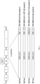
- VSV particle wherein (i) the VSV glycoprotein (G) is replaced by a Severe Acute Respiratory Syndrome coronavirus 2 (SARS-CoV-2) spike (S) glycoprotein or a fragment of the SARS-CoV-2 S glycoprotein comprising the receptor binding domain thereof, wherein said SARS-CoV-2 S glycoprotein or fragment is capable of mediating infection of a target cell and wherein (ii) the VSV particle comprises a nucleic acid molecule comprising a nucleic acid sequence encoding said reporter protein, wherein the nucleic acid sequence encoding the reporter protein is inserted between the nucleic acid sequence encoding the SARS-CoV-2 S glycoprotein and the nucleic acid sequence encoding VSV large (L) protein.
- SARS-CoV-2 Severe Acute Respiratory Syndrome coronavirus 2
- S Severe Acute Respiratory Syndrome coronavirus 2
- S Severe Acute Respiratory Syndrome coronavirus 2
- S Severe Acute Respir
- the mutations can attenuate the infectivity, virulence or pathogenic effects of VSV.
- the attenuation can be additive or synergistic. With synergistic attenuation, the level of VSV attenuation is greater than additive. Synergistic attenuation of VSV can arise from combining at least two classes of mutation in the same VSV genome, thereby resulting in a reduction of VSV pathogenicity much greater than an additive attenuation level observed for each VSV mutation class alone.
- a synergistic attenuation of VSV can provide for an LD50 at least greater than the additive attenuation level observed for each mutation class alone (i.e., the sum of the two mutation classes), where attenuation levels (i.e., the LD50) are determined in a small animal neurovirulence model.
- the VSV M gene encodes the virus matrix (M) protein, and two smaller in-frame polypeptides (M2 and M3).
- the M2 and M3 polypeptides can be translated from the same open reading frame (ORF) as the M protein and lack the first 33 and 51 amino acids, respectively.
- a recombinant VSV vector comprising non-cytopathic M gene mutations i.e., VSV vectors that also do not express M2 and M3 proteins
- the replication competent recombinant VSV particles of the disclosure comprise a non-cytopathic mutation in the M gene.
- the VSV (Indiana serotype) M gene encodes a 229 amino acid M (matrix) protein in which the first thirty amino acids of the NH2-terminus comprise a proline-rich PPPY (PY) motif (SEQ ID NO: 68).
- the PY motif of VSV M protein is located at amino acid positions 24-27 in both VSV Indiana (Genbank Accession Number X04452) and New Jersey (Genbank Accession Number M14553) serotypes.
- the VSV may comprise mutations in the PY motif (e.g., APPY (SEQ ID NO: 69), AAPY (SEQ ID NO: 70), PPAY (SEQ ID NO: 71), APPA (SEQ ID NO: 72), AAPA (SEQ ID NO: 73) and PPPA (SEQ ID NO: 74)).
- the VSV can comprise any of various amino acid mutations (e.g., deletions, substitutions, insertions, etc.) into the M protein PSAP (PS) motif (SEQ ID NO: 75). These and other mutations in the PY motif may be effective to reduce virus yield by blocking a late stage in virus budding.
- the replication competent recombinant VSV particles of the disclosure may comprise one or more M gene mutations.
- M protein mutations include, e.g., a glycine changed to a glutamic acid at position (21), a leucine changed to a phenylalanine at position (111), a methionine changed to an arginine at position (51), a glycine changed to a glutamic acid at position (22), a methionine changed to an arginine at position (48), a leucine changed to a phenylalanine at position (110), a methionine changed to an alanine at position (51), and a methionine changed to an alanine at position (33).
- the genome of the recombinant VSV encodes a mutant VSV matrix M protein comprising the M51R mutation. Mutation M51R eliminates M protein's ability to block cellular nucleo-cytoplasmic transport and thus substantially attenuates VSV infectivity.
- the DNA that can be transcribed to encode VSV antigenomic (+) RNA comprises a gene that encodes a VSV M protein.
- the VSV M protein used in the methods and compositions described herein may comprise the amino acid sequence of SEQ ID NO: 9, or a sequence at least 50%, 60%, 70%, 80%, 81%, 82%, 83%, 84%, 85%, 86%, 87%, 88%, 89%, 90%, 91%, 92%, 93%, 94%, 95%, 96%, 97%, 98%, 99%, 99.5% or more identical to the amino acid sequence of SEQ ID NO: 9.
- the VSV M protein used in the vaccines or methods described herein may consist of the amino acid sequence of SEQ ID NO: 9, or a sequence at least 50%, 60%, 70%, 80%, 81%, 82%, 83%, 84%, 85%, 86%, 87%, 88%, 89%, 90%, 91%, 92%, 93%, 94%, 95%, 96%, 97%, 98%, 99%, 99.5% or more identical to the amino acid sequence of SEQ ID NO: 9.
- the mutated VSV M protein used in the vaccines or methods described herein may comprise the amino acid sequence of SEQ ID NO: 7, or a sequence at least 50%, 60%, 70%, 80%, 81%, 82%, 83%, 84%, 85%, 86%, 87%, 88%, 89%, 90%, 91%, 92%, 93%, 94%, 95%, 96%, 97%, 98%, 99%, 99.5% or more identical to the amino acid sequence of SEQ ID NO: 7.
- the mutant VSV M protein used in the vaccines or methods described herein may consist of the amino acid sequence of SEQ ID NO: 7, or a sequence at least 50%, 60%, 70%, 80%, 81%, 82%, 83%, 84%, 85%, 86%, 87%, 88%, 89%, 90%, 91%, 92%, 93%, 94%, 95%, 96%, 97%, 98%, 99%, 99.5% or more identical to the amino acid sequence of SEQ ID NO: 7.
- VSV (-) DNA DNA that can be transcribed to produce VSV (for example) antigenomic (+) RNA (such DNA being referred to herein as "VSV (-) DNA") is available in the art and/or can be obtained by standard methods.
- Genbank VSVCG Accession No. J02428; NCBI Seq ID 335873; and is published in Rose and Schubert, 1987, in The Viruses: The Rhabdoviruses, Plenum Press, NY, pp. 129-166 .
- VSV(-) DNA that is contained in plasmid pVSVFL(+)is shown, e.g., in U.S. Patent No. 7,153,510 . Sequences of other vesiculovirus genomes have been published and are available in the art.
- VSV (-) DNA if not already available, can be prepared by standard methods, as follows: VSV genomic RNA can be purified from virus preparations, and reverse transcription with long distance polymerase chain reaction used to generate the v (-) DNA. Alternatively, after purification of genomic RNA, VSV mRNA can be synthesized in vitro, and cDNA prepared by standard methods, followed by insertion into cloning vectors (see, e.g., Rose and Gallione, 1981, J. Virol. 39(2):519-528 ).
- VSV RNA Individual cDNA clones of VSV RNA can be joined by use of small DNA fragments covering the gene junctions, generated by use of reverse transcription and polymerase chain reaction (RT-PCR) ( Mullis and Faloona, 1987, Meth. Enzymol. 155:335-350 ) from VSV genomic RNA (see Section 6, infra). VSV and other vesiculoviruses are available in the art.
- RT-PCR reverse transcription and polymerase chain reaction
- one or more, usually unique, restriction sites are introduced into the VSV (-) DNA, in intergenic regions, or 5' of the sequence complementary to the 3' end of the VSV genome, or 3' of the sequence complementary to the 5' end of the VSV genome, to facilitate insertion of the foreign DNA.
- the VSV (-) DNA is constructed so as to have a promoter operatively linked thereto.
- the promoter should be capable of initiating transcription of the (-) DNA in an animal or insect cell in which it is desired to produce the recombinant VSV.
- Promoters which may be used include, but are not limited to, the SV40 early promoter region ( Bernoist and Chambon, 1981, Nature 290:304-310 ), the promoter contained in the 3' long terminal repeat of Rous sarcoma virus ( Yamamoto, et al., 1980, Cell 22:787-797 ), the herpes thymidine kinase promoter ( Wagner et al., 1981, Proc. Natl. Acad. Sci.
- mice mammary tumor virus control region which is active in testicular, breast, lymphoid and mast cells
- Leder et al., 1986, Cell 45:485-495 albumin gene control region which is active in liver
- albumin gene control region which is active in liver
- alpha-fetoprotein gene control region which is active in liver
- Krumlauf et al., 1985, Mol. Cell. Biol. 5:1639-1648 Hammer et al., 1987, Science 235:53-58
- alpha 1-antitrypsin gene control region which is active in the liver
- beta-globin gene control region which is active in myeloid cells ( Mogram et al., 1985, Nature 315:338-340 ; Kollias et al., 1986, Cell 46:89-94 ; myelin basic protein gene control region which is active in oligodendrocyte cells in the brain ( Readhead et al., 1987, Cell 48:703-712 ); and myosin light chain-2 gene control region which is active in skeletal muscle ( Sani, 1985, Nature 314:283-286 ).
- the promoter is an RNA polymerase promoter, preferably a bacteriophage or viral or insect RNA polymerase promoter, including but not limited to the promoters for T7 RNA polymerase, SP6 RNA polymerase, and T3 RNA polymerase. If an RNA polymerase promoter is used in which the RNA polymerase is not endogenously produced by the host cell in which it is desired to produce the recombinant VSV, a recombinant source of the RNA polymerase must also be provided in the host cell.
- the VSV (-) DNA can be operably linked to a promoter before or after insertion of foreign DNA.
- a transcriptional terminator is situated downstream of the VSV (-) DNA.
- a DNA sequence that can be transcribed to produce a ribozyme sequence is situated at the immediate 3' end of the VSV (-) DNA, prior to the transcriptional termination signal, so that upon transcription a self-cleaving ribozyme sequence is produced at the 3' end of the antigenomic RNA, which ribozyme sequence will autolytically cleave (after a U) this fusion transcript to release the exact 3' end of the VSV antigenomic (+) RNA.
- Any ribozyme sequence known in the art may be used, as long as the correct sequence is recognized and cleaved.
- HDV hepatitis delta virus
- VSV(-) DNA for use, for insertion of foreign DNA, can thus comprises (in 5' to 3' order) the following operably linked components: the T7 RNA polymerase promoter, VSV (-) DNA, a DNA sequence that is transcribed to produce an HDV ribozyme sequence (immediately downstream of the VSV (-) DNA), and a T7 RNA polymerase transcription termination site.
- plasmids examples include, pVSVFL(+) or pVSVSS1.
- the G gene of VSV in the VSV (-) DNA of plasmid pVSVFL(+) can be excised and replaced, by cleavage at the NheI and MluI sites flanking the G gene and insertion of the desired sequence encoding the SARS-CoV-2 S glycoprotein or the fragment thereof.
- the SARS-CoV-2 S protein forms a part of the VSV envelope and thus is surface-displayed in the VSV particle.
- S glycoprotein transmembrane spike
- S glycoprotein transmembrane spike
- S glycoprotein forms homotrimers protruding from the viral surface.
- S glycoprotein comprises two functional subunits responsible for binding to the host cell receptor (S1 subunit) and fusion of the viral and cellular membranes (S2 subunit).
- S1 subunit host cell receptor
- S2 subunit fusion of the viral and cellular membranes
- S glycoprotein is cleaved at the boundary between the S1 and S2 subunits, which remain non-covalently bound in the prefusion conformation.
- the distal S1 subunit comprises the receptor-binding domain(s) (RBD) and contributes to stabilization of the prefusion state of the membrane-anchored S2 subunit that contains the fusion machinery.
- R receptor-binding domain
- S is further cleaved by host proteases at the S2' site located immediately upstream of the fusion peptide. This cleavage has been proposed to activate the protein for membrane fusion via conformational changes. Walls et al., Cell, published online March 9, 2020; available at doi.org/10.1016/j.cell.2020.02.058 .
- SARS-CoV-1 and SARS-CoV-2 can interact directly with angiotensin-converting enzyme 2 (ACE2) to enter target cells and may also employ the cellular serine protease, transmembrane protease, serine 2 (TMPRSS2) for S protein priming ( Hoffmann et al., Cell, 2020, 181:1-10; available at doi.org/10.1016/j.cell.2020.02.052 ).
- TMPRSS2 serine 2
- SARS-CoV-S und SARS-CoV-2-S share about 76% amino acid identity.
- the receptor binding domain (RBD) in the S glycoprotein is the most variable part of the coronavirus genome.
- RBD amino acids have been shown to be critical for binding to ACE2 receptors and for determining the host range of SARS-CoV-like viruses. They are Y442, L472, N479, D480, T487 and Y4911 in SARS-CoV, which correspond to L455, F486, Q493, S494, N501 and Y505 in SARS-CoV-2 ( Andersen et al., Nature Medicine, 2020; available at doi.org/10.1038/s41591-020-0820-9 ).
- the SARS-CoV-2 S glycoprotein may be a full-length SARS-CoV-2 S glycoprotein (comprising or consisting of SEQ ID NO: 1) or a fragment thereof that has at least 77%, 78%, 79%, 80%, 81%, 82%, 83%, 84%, 85%, 86%, 87%, 88%, 89%, 90%, 91%, 92%, 93%, 94%, 95%, 96%, 97%, 98%, 99%, 99.5% or more amino acid sequence identity to SEQ ID NO: 1, wherein the SARS-CoV-2 S glycoprotein or the fragment comprises the receptor binding domain and is capable of mediating infection of a target cell.
- the wild-type coronavirus S glycoprotein comprises an S1 subunit that facilitates binding of the coronavirus to cell surface proteins. Without wishing to be bound by theory, the S1 subunit of the wildtype S glycoprotein controls which cells are infected by the coronavirus.
- the wild-type S glycoprotein also comprises a S2 subunit, which is a transmembrane subunit that facilitates viral and cellular membrane fusion.
- a fragment of SARS-CoV-2 S glycoprotein can comprise the S1 subunit of the SARS-CoV-2 S glycoprotein or a fragment that has at least 65%, 66%, 67%, 68%, 69%, 70%, 71%, 72%, 73%, 74%, 75%, 76%, 77%, 78%, 79%, 80%, 81%, 82%, 83%, 84%, 85%, 86%, 87%, 88%, 89%, 90%, 91%, 92%, 93%, 94%, 95%, 96%, 97%, 98%, 99%, 99.5% or more amino acid sequence identity to the S1 subunit of the SARS-CoV-2 S glycoprotein.
- a fragment of SARS-CoV-2 S glycoprotein can comprise a sequence that has at least 65%, 66%, 67%, 68%, 69%, 70%, 71%, 72%, 73%, 74%, 75%, 76%, 77%, 78%, 79%, 80%, 81%, 82%, 83%, 84%, 85%, 86%, 87%, 88%, 89%, 90%, 91%, 92%, 93%, 94%, 95%, 96%, 97%, 98%, 99%, 99.5% or more amino acid sequence identity to amino acids 14-684 of SEQ ID NO: 1.
- a fragment of SARS-CoV-2 S glycoprotein comprises the S2 subunit of the SARS-CoV-2 S glycoprotein or a fragment that has at least 91%, 92%, 93%, 94%, 95%, 96%, 97%, 98%, 99%, 99.5% or more amino acid sequence identity to the S2 subunit of the SARS-CoV-2 S glycoprotein.
- the wild-type coronavirus S glycoprotein comprises a receptor binding domain (RBD) that facilitates binding of the coronavirus to its receptor on the host cell.
- RBD receptor binding domain
- S SARS-CoV-2 spike glycoprotein
- a fragment of SARS-CoV-2 S glycoprotein comprises the RBD of the SARS-CoV-2 S glycoprotein that has at least 74%, 75%, 76%, 77%, 78%, 80%, 81%, 82%, 83%, 84%, 85%, 86%, 87%, 88%, 89%, 90%, 91%, 92%, 93%, 94%, 95%, 96%, 97%, 98%, 99%, 99.5% or more amino acid sequence identity to the RBD of the SARS-CoV-2 S glycoprotein, e.g., amino acids 319-541 of SEQ ID NO: 1.
- the SARS-CoV-2 S glycoprotein, or fragment thereof may comprise or consist of an insertion, deletion, and/or substitution of 1, 2, 3, 4, 5, 6, 7, 8, 9, 10, 11, 12, 13, 14, 15, 16, 17, 18, 19, 20, 21, 22, 23, 24, 25, 26, 27, 28, 29, 30, 31, 32, 33, 34, 35, 36, 37, 38, or 39 residues of the SARS-CoV-2 S glycoprotein.
- Non-limiting examples of amino acids for potential deletion include, e.g., a tyrosine at position (145), an asparagine at position (679), a serine at position (680), proline at position (681), an arginine at position (682), an arginine at position (683), an alanine at position (684), and/or an arginine at position (685), positions as denoted in SEQ ID NO: 1, or the equivalent amino acid residue in a mutant SARS-CoV-2 S glycoprotein sequence.
- Non-limiting examples of amino acids for potential substitution include, e.g., a leucine changed to a phenylalanine at position (5) a tyrosine changed to an asparagine at position (28), a threonine changed to an isoleucine at position (29), a histidine changed to a tyrosine at position (49), a leucine changed to a phenylalanine at position (54), an asparagine changed to a lysine at position (74), a glutamic acid changed to an aspartic acid at position (96), an aspartic acid changed to an asparagine at position (111), a phenylalanine changed to a leucine at position (157), a glycine changed to a valine at position (181), a serine changed to a tryptophan at position (221), a serine changed to an arginine at position (247), an alanine changed to a threonine at position
- amino acid residue positions for insertion, deletion, and/or substitution include those as listed in Tables 8 and 9 (amino acid residue positions are denoted using SEQ ID NO: 1 as a reference sequence, which can be used as a reference for identifying the equivalent amino acid residue in any SARS-CoV-2 S glycoprotein sequence (same as above)).
- SEQ ID NO: 1 amino acid residue positions are denoted using SEQ ID NO: 1 as a reference sequence, which can be used as a reference for identifying the equivalent amino acid residue in any SARS-CoV-2 S glycoprotein sequence (same as above)).
- Each residue modification listed in Table 8 can separately be used alone or in combination with others to generate variants of a replication competent recombinant vesicular stomatitis virus (VSV) particle.
- VSV vesicular stomatitis virus
- the SARS-CoV-2 S glycoprotein, or fragment thereof differs in amino acid sequence from the reference peptide or polypeptide (a wild-type SARS-CoV-2 spike protein) by changing a serine to an arginine at position (247), an aspartic acid to an asparagine at position (614), and/or an arginine to a glutamine at position (685), positions as denoted in SEQ ID NO: 1, or the equivalent amino acid residue in a mutant SARS-CoV-2 S glycoprotein sequence.
- the SARS-CoV-2 S glycoprotein, or fragment thereof differs in amino acid sequence from the reference peptide or polypeptide by changing a serine to an arginine at position (247).
- the SARS-CoV-2 S glycoprotein, or fragment thereof differs in amino acid sequence from the reference peptide or polypeptide by changing an aspartic acid to an asparagine at position (614). In certain embodiments, the SARS-CoV-2 S glycoprotein, or fragment thereof, differs in amino acid sequence from the reference peptide or polypeptide by changing an arginine to a glutamine at position (685). In certain embodiments, the SARS-CoV-2 S glycoprotein, or fragment thereof, differs in amino acid sequence from the reference peptide or polypeptide by changing a serine to an arginine at position (247) and an aspartic acid to an asparagine at position (614).
- the SARS-CoV-2 S glycoprotein, or fragment thereof differs in amino acid sequence from the reference peptide or polypeptide by changing a serine to an arginine at position (247) and an arginine to a glutamine at position (685). In certain embodiments, the SARS-CoV-2 S glycoprotein, or fragments thereof, differs in amino acid sequence from the reference peptide or polypeptide by changing an aspartic acid to an asparagine at position (614) and an arginine to a glutamine at position (685).
- the SARS-CoV-2 S glycoprotein, or fragment thereof differs in amino acid sequence from the reference peptide or polypeptide by changing a serine to an arginine at position (247), an aspartic acid to an asparagine at position (614), and an arginine to a glutamine at position (685).
- the SARS-CoV-2 S glycoprotein, or fragment thereof result in a more lytic phenotype.
- the SARS-CoV-2 S glycoprotein fragment may comprise the amino acid sequence of SEQ ID NO: 42, or a sequence at least 50%, 60%, 70%, 80%, 81%, 82%, 83%, 84%, 85%, 86%, 87%, 88%, 89%, 90%, 91%, 92%, 93%, 94%, 95%, 96%, 97%, 98%, 99%, or 99.5% identical to the amino acid sequence of SEQ ID NO: 42.
- the SARS-CoV-2 S glycoprotein fragment may be encoded by a codon optimized nucleotide sequence.
- the SARS-CoV-2 S glycoprotein fragment may be encoded by the polynucleotide sequence of SEQ ID NO: 43 or a sequence that has at least 50%, 60%, 70%, 80%, 81%, 82%, 83%, 84%, 85%, 86%, 87%, 88%, 89%, 90%, 91%, 92%, 93%, 94%, 95%, 96%, 97%, 98%, 99%, or 99.5% polynucleotide sequence identity to SEQ ID NO: 43.
- the SARS-CoV-2 S glycoprotein fragment may comprise the amino acid sequence of SEQ ID NO: 44, or a sequence at least 50%, 60%, 70%, 80%, 81%, 82%, 83%, 84%, 85%, 86%, 87%, 88%, 89%, 90%, 91%, 92%, 93%, 94%, 95%, 96%, 97%, 98%, 99%, or 99.5% identical to the amino acid sequence of SEQ ID NO: 44.
- the SARS-CoV-2 S glycoprotein, or fragments thereof differs in amino acid sequence from the reference peptide or polypeptide (a wild-type SARS-CoV-2 spike protein) by changing an asparagine to a tyrosine at position (501), and/or a glutamic acid to a lysine at position (484), and/or an aspartic acid to a glycine at position (614), and/or deletion of residues 69-70, positions as denoted in SEQ ID NO: 1, or the equivalent amino acid residue in a mutant SARS-CoV-2 S glycoprotein sequence.
- the reference peptide or polypeptide a wild-type SARS-CoV-2 spike protein
- the SARS-CoV-2 S glycoprotein, or fragment thereof differs in amino acid sequence from the reference peptide or polypeptide by changing an asparagine to a tyrosine at position (501). In certain embodiments, the SARS-CoV-2 S glycoprotein, or fragment thereof, differs in amino acid sequence from the reference peptide or polypeptide by changing a glutamic acid to a lysine at position (484). In certain embodiments, the SARS-CoV-2 S glycoprotein, or fragment thereof, differs in amino acid sequence from the reference peptide or polypeptide by changing an aspartic acid to a glycine at position (614).
- the SARS-CoV-2 S glycoprotein, or fragment thereof differs in amino acid sequence from the reference peptide or polypeptide by deletion of residues 69-70. In certain embodiments, the SARS-CoV-2 S glycoprotein, or fragment thereof, differs in amino acid sequence from the reference peptide or polypeptide by changing an asparagine to a tyrosine at position (501) and a glutamic acid to a lysine at position (484).
- the SARS-CoV-2 S glycoprotein, or fragment thereof differs in amino acid sequence from the reference peptide or polypeptide by changing an asparagine to a tyrosine at position (501) and an aspartic acid to a glycine at position (614). In certain embodiments, the SARS-CoV-2 S glycoprotein, or fragment thereof, differs in amino acid sequence from the reference peptide or polypeptide by changing an asparagine to a tyrosine at position (501) and deletion of residues 69-70.
- the SARS-CoV-2 S glycoprotein, or fragment thereof differs in amino acid sequence from the reference peptide or polypeptide by changing a glutamic acid to a lysine at position (484) and an aspartic acid to a glycine at position (614). In certain embodiments, the SARS-CoV-2 S glycoprotein, or fragment thereof, differs in amino acid sequence from the reference peptide or polypeptide by changing a glutamic acid to a lysine at position (484) and deletion of residues 69-70.
- the SARS-CoV-2 S glycoprotein, or fragment thereof differs in amino acid sequence from the reference peptide or polypeptide by changing an aspartic acid to a glycine at position (614) and deletion of residues 69-70. In certain embodiments, the SARS-CoV-2 S glycoprotein, or fragment thereof, differs in amino acid sequence from the reference peptide or polypeptide by changing an asparagine to a tyrosine at position (501), a glutamic acid to a lysine at position (484), and an aspartic acid to a glycine at position (614).
- the SARS-CoV-2 S glycoprotein, or fragment thereof differs in amino acid sequence from the reference peptide or polypeptide by changing an asparagine to a tyrosine at position (501), changing a glutamic acid to a lysine at position (484), and deletion of residues 69-70.
- the SARS-CoV-2 S glycoprotein, or fragment thereof differs in amino acid sequence from the reference peptide or polypeptide by changing an asparagine to a tyrosine at position (501), changing an aspartic acid to a glycine at position (614), and deletion of residues 69-70.
- the SARS-CoV-2 S glycoprotein, or fragment thereof differs in amino acid sequence from the reference peptide or polypeptide by changing a glutamic acid to a lysine at position (484), changing an aspartic acid to a glycine at position (614), and deletion of residues 69-70.
- the SARS-CoV-2 S glycoprotein, or fragment thereof differs in amino acid sequence from the reference peptide or polypeptide by changing an asparagine to a tyrosine at position (501), changing a glutamic acid to a lysine at position (484), changing an aspartic acid to a glycine at position (614) and deletion of residues 69-70.
- the SARS-CoV-2 S glycoprotein, or fragment thereof differs in amino acid sequence from the reference peptide or polypeptide (a wild-type SARS-CoV-2 spike protein) by inactivating the furin cleavage site within the spike protein.
- the SARS-CoV-2 S glycoprotein, or fragment thereof differs in amino acid sequence from the reference peptide or polypeptide (a, wild-type SARS-CoV-2 spike protein) by changing Q 677 TNSPRRARSV 687 (SEQ ID NO: 65), as denoted in SEQ ID NO: 1, or the equivalent amino acid residue in a mutant SARS-CoV-2 S glycoprotein sequence, to QTII,RSV (SEQ ID NO: 66) or to QTNSPGSASSV (SEQ ID NO: 67).
- the SARS-CoV-2 S glycoprotein, or fragment thereof result in a monobasic furin cleavage site in the S1/S2 interface (QTII,RSV (SEQ ID NO: 66)) or deletion of the furin cleavage site (QTNSPGSASSV (SEQ ID NO: 67)) phenotype.
- the alteration to the furin cleavage site can lead to a spike stabilized pseudoparticles. See Hansen et. al., "Studies in humanized mice and convalescent humans yield a SARS-CoV-2 antibody cocktail" Science, published online June 15, 2020 .
- the SARS-CoV-2 S glycoprotein fragment lacks one or more C-terminal residues of the full-length SARS-CoV-2 S glycoprotein.
- the SARS-CoV-2 S glycoprotein fragment may lack 1, 2, 3, 4, 5, 6, 7, 8, 9, 10, 11, 12, 13, 14, 15, 16, 17, 18, 19, 20, 21, 22, 23, 24, 25, 26, 27, 28, 29, 30, 31, 32, 33, 34, 35, 36, 37, 38, 39 of the C-terminal residues of the SARS-CoV-2 S glycoprotein.
- the SARS-CoV-2 S glycoprotein fragment lacks the 19 C-terminal residues of the SARS-CoV-2 S glycoprotein.
- the SARS-CoV-2 S glycoprotein fragment may consist of the amino acid sequence of SEQ ID NO: 3, or a sequence at least 78%, 79%, 80%, 81%, 82%, 83%, 84%, 85%, 86%, 87%, 88%, 89%, 90%, 91%, 92%, 93%, 94%, 95%, 96%, 97%, 98%, 99%, 99.5% or more identical to the amino acid sequence of SEQ ID NO: 3.
- the SARS-CoV-2 S glycoprotein is a chimeric or fusion molecule which comprises fusogen sequences from viruses other than SARS-CoV-2.
- such chimeras comprise S1 or RBD sequences of SARS-CoV-2 S glycoprotein.
- the fusion protein is a fusion between the extracellular and transmembrane sequence of SARS-CoV-2 S glycoprotein, or a fragment thereof and a cytoplasmic domain of a non-SARS-CoV-2 fusogen or a fragment thereof.
- the fusion protein is a fusion between the extracellular sequence of SARS-CoV-2 S glycoprotein, or a fragment thereof and transmembrane and cytoplasmic domains of a non-SARS-CoV-2 fusogen or a fragment thereof.
- Non-limiting examples of non-SARS-CoV-2 fusogens which can be used in the above chimeric and fusion molecules include, for example, coronavirus fusogens (e.g., from SARS-CoV-1 or MERS-CoV), fusogens from VSV or other vesiculoviruses or other viruses from the Rhabdoviridae family, viruses from the Retroviridae family (e.g., human immunodeficiency virus (HIV), murine leukemia virus (MLV), Avian sarcoma leukosis virus (ASLV), Jaagsiekte sheep retrovirus (JSRV)), viruses from the Paramyxoviridae family (e.g., parainfluenza virus 5 (PIV5)), viruses from the Herpesviridae family (e.g., herpes simplex virus (HSV)), viruses from the Togaviridae family (e.g., Semliki Forest virus (SFV), Rubella virus), viruses from the Flavivi
- the SARS-CoV-2 S glycoprotein or fragment thereof can comprise a consensus sequence derived from two or more different strains, mutants or variants of SARS-CoV-2.
- the methods of the disclosure use a mixture of SARS-CoV-2 S glycoproteins (or fragments thereof) from two or more different strains, mutants or variants of SARS-CoV-2.
- Polynucleotide molecules encoding SARS-CoV-2 S glycoprotein or a fragment thereof can comprise a consensus sequence and/or modification(s) for improved expression of the SARS-CoV-2 S glycoprotein or the fragment thereof.
- Modification can include codon optimization, the addition of a Kozak sequence or modified (e.g., optimized) Kozak sequence for increased translation initiation, and/or the addition of a signal peptide/leader sequence (e.g., an immunoglobulin signal peptide such as, e.g., IgE or IgG signal peptide).
- the Kozak sequence or modified (e.g., optimized) Kozak sequence is 3' to the foreign gene.
- the Kozak sequence or modified (e.g., optimized) Kozak sequence is immediately 3' to the foreign gene. In certain embodiments, the Kozak sequence or modified (e.g., optimized) Kozak sequence is 5' to the foreign gene. In certain embodiments, the Kozak sequence or modified (e.g., optimized) Kozak sequence is immediately 5' to the foreign gene.
- the SARS-CoV-2 S glycoprotein or fragment thereof comprises a fusions or conjugate with a detection tag (e.g., HA tag, histidine tag, biotin), , dimerization/multimerization sequences, Fc, signaling sequences, etc.
- a detection tag e.g., HA tag, histidine tag, biotin
- dimerization/multimerization sequences e.g., Fc, signaling sequences, etc.
- reporter proteins include, e.g., luciferases (including but not limited to, Renilla luciferase, RLuc8 mutant Renilla luciferase, (dCpG)Luciferase, NanoLuc reporter, firefly luciferase, Gaussia luciferase (gLuc), MetLuc, Vibrio fischeri lumazine protein, Vibrio harveyi luminaze protein, inoflagellate luciferase, firefly luciferase YY5 mutant, firefly luciferase LGR mutant, firefly luciferase mutant E, and derivatives thereof) and fluorescent proteins (including but not limited to, green fluorescent protein (GFP) [e.g., Aequoria victoria GFP, Renilla muelleri GFP, Renilla reniformis GFP, Renilla ptilosarcus GFP], GFP-like fluorescent proteins, (GFP-like),
- GFP
- Replication competent recombinant VSV particles are used as an example in the disclosure below, but this disclosure can also be used for other vesiculoviruses.
- the replication competent recombinant VSV particles of the disclosure are produced by providing in an appropriate host cell: VSV (-) DNA, in which regions non-essential for replication have been inserted into or replaced by a foreign DNA comprising a sequence encoding a coronavirus S glycoprotein or a fragment thereof and optionally other sequences discussed above, and recombinant sources of VSV N protein, P protein, L protein and any additional desired VSV protein (e.g., M protein and/or G glycoprotein).
- the production is preferably in vitro (e.g., in cell culture).
- the host cell used for recombinant VSV production can be any cell in which VSVs grows.
- Non-limiting sources of host cells include, prokaryotic cells or a eukaryotic cells, vertebrate cells, mammalian cells, some insect (e.g., Drosophila) cells, primary cells (e.g., primary chick embryo fibroblasts), or cell lines (e.g., BHK (baby hamster kidney) cells, CHO (Chinese hamster ovary) cells, HeLA (human) cells, mouse L cells, Vero (monkey) cells (including Vero- ⁇ His cells), Vero-Ace-2 cells, Vero-TRMPSS2 cells, Vero-E6 cells, ESK-4, PK-15, EMSK cells, MDCK (Madin-Darby canine kidney) cells, MDBK (Madin-Darby bovine kidney) cells, 293 (human) cells, Hep-2 cells, Human Diploid Primary Cell Lines (e.g.
- WI-38 and MRC5 cells Monkey Diploid Cell Line (e.g. FRhL-Fetal Rhesus Lung cells), and Quasi-Primary Continues Cell Line (e.g. AGMK-African green monkey kidney cells), etc.).
- Monkey Diploid Cell Line e.g. FRhL-Fetal Rhesus Lung cells
- Quasi-Primary Continues Cell Line e.g. AGMK-African green monkey kidney cells
- the sources of N, P, and L proteins and any additional desired VSV protein can be the same or can be different recombinant nucleic acid(s), encoding and capable of expressing these proteins in the host cell in which it is desired to produce recombinant VSVs.
- the nucleic acids encoding the N, P and L proteins and any additional desired VSV protein can be obtained by any means available in the art.
- the VSV N, P, L, M and G-encoding nucleic acid sequences have been disclosed and can be used. For example, see Genbank accession no.
- N, P and L genes can also be obtained, for example, from plasmid pVSVFL(+), deposited with the ATCC and assigned accession no. 97134, e.g., by PCR amplification of the desired gene (see also U.S. Pat. Nos. 4,683,202 , 4,683,195 and 4,889,818 ; Gyllenstein et al., 1988, Proc. Natl. Acad. Sci.
- the clone can be obtained by use of standard recombinant DNA methodology.
- the DNA may be obtained by standard procedures known in the art such as, e.g., by purification of RNA from VSV virions followed by reverse transcription and PCR ( Mullis and Faloona, 1987, Methods in Enzymology 155:335-350 ).
- Alternatives include, but are not limited to, chemically synthesizing the gene sequence itself. Other methods are also possible.
- Nucleic acids that encode fragments and derivatives of VSV N, P, L, and M genes, as well as fragments and derivatives of the VSV (-) DNA can also be used in the present disclosure, as long as such fragments and derivatives retain the ability to produce replication competent VSV particles which can be used in one or more methods of the disclosure.
- derivatives can be made by altering sequences by substitutions, additions, or deletions.
- other DNA sequences which encode substantially the same or a functionally equivalent amino acid sequence may be used in the practice of the methods of the disclosure. Amino acid substitutions may be made on the basis of similarity in polarity, charge, solubility, hydrophobicity, hydrophilicity and/or the amphipathic nature of the residues involved.
- the desired N/P/L/M-encoding nucleic acid can be inserted into an appropriate expression vector, i.e., a vector which contains the necessary elements for the transcription and translation of the inserted protein-coding sequence in the host cell in which it is desired to produce replication competent recombinant VSV particles, to create a vector that functions to direct the synthesis of the VSV proteins that will subsequently assemble with the VSV genomic RNA (e.g., produced in the host cell from antigenomic VSV (+) RNA produced, e.g., by transcription of the VSV (-) DNA).
- an appropriate expression vector i.e., a vector which contains the necessary elements for the transcription and translation of the inserted protein-coding sequence in the host cell in which it is desired to produce replication competent recombinant VSV particles, to create a vector that functions to direct the synthesis of the VSV proteins that will subsequently assemble with the VSV genomic RNA (e.g., produced in the host cell from antigenomic VSV (+) RNA produced
- a variety of vector systems may be utilized to express the N, P and L VSV proteins and any additional desired VSV protein (e.g., M), as well as to transcribe the VSV (-) DNA (e.g., comprising a foreign DNA), as long as the vector is functional in the host cell and compatible with any other vector present.
- the expression elements of vectors vary in their strengths and specificities. Any one of a number of suitable transcription and translation elements may be used, as long as they are functional in the host cell.
- Standard recombinant DNA methods may be used to construct expression vectors containing DNA encoding the VSV proteins, and the VSV (-) DNA containing the foreign DNA, comprising appropriate transcriptional/translational control signals (see, e.g., Sambrook et al., 1989, supra, and methods described hereinabove).
- Expression may be controlled by any promoter/enhancer element known in the art. Promoters which may be used to control expression can be constitutive or inducible. In a specific embodiment, the promoter is an RNA polymerase promoter.
- Transcription termination signals downstream of the gene, and selectable markers are preferably also included in the expression vector.
- expression vectors for the N, P, L, and any additionally desired VSV proteins, as well as the SARS-CoV-2 S glycoprotein may contain specific initiation signals for efficient translation of the inserted sequences, e.g., a ribosome binding site.
- Specific initiation signals maybe required for efficient translation of the protein coding sequences. These signals include the ATG initiation codon and adjacent sequences. In cases where the entire N, P, L, or other (e.g., M) VSV gene, including its own initiation codon and adjacent sequences, are inserted into the appropriate vectors, no additional translational control signals may be needed. However, in cases where only a portion of the gene sequence is inserted, exogenous translational control signals, including the ATG initiation codon, must be provided. The initiation codon must furthermore be in phase with the reading frame of the protein coding sequences to ensure translation of the entire insert. These exogenous translational control signals and initiation codons can be of a variety of origins, both natural and synthetic.
- a recombinant expression vector provided herein, encoding an N, P, L, and other (e.g., M) protein or functional derivative thereof comprises the following operatively linked components: a promoter which controls the expression of the VSV N, P, L proteins and optionally the M protein, the SARS-CoV-2 spike glycoprotein or a fragment thereof, a translation initiation signal, a DNA sequence encoding the VSV proteins or functional fragment thereof, and a transcription termination signal, e.g., as shown in Fig. 1 .
- the genes encoding the N, P, L, and optional M VSV proteins are inserted downstream of the T7 RNA polymerase promoter from phage T7 gene 10, situated with an A in the -3 position.
- a T7 RNA polymerase terminator and a replicon can be also included in the expression vector.
- T7 RNA polymerase can be provided to transcribe the VSV protein sequence.
- the T7 RNA polymerase can be produced from a chromosomally integrated sequence or an episomal vector.
- T7 RNA polymerase can be provided by intracellular expression from a recombinant vaccinia virus vector encoding the T7 RNA polymerase.
- the N, P, L, and optional M VSV proteins are each encoded by a DNA sequence operably linked to a promoter in an expression plasmid, containing the necessary regulatory signals for transcription and translation of the encoded proteins.
- an expression plasmid preferably includes a promoter, the coding sequence, and a transcription termination/polyadenylation signal, and optionally, a selectable marker (e.g., ⁇ -galactosidase).
- the N, P, L, and optional M proteins can be encoded by the same or different plasmids, or a combination thereof. In other embodiments, one or more of the N, P, L, and optional M VSV proteins can be expressed intrachromosomally.
- the cloned sequences comprising the VSV (-) DNA containing the foreign DNA, and the cloned sequences comprising sequences encoding the VSV and foreign proteins can be introduced into the desired host cell by any method known in the art, e.g., transfection, electroporation, infection (when the sequences are contained in, e.g., a viral vector), microinjection, etc.
- a transfection facilitating reagent is added to increase DNA uptake by cells.
- these reagents are known in the art (e.g., calcium phosphate; Lipofectace (Life Technologies, Gaithersburg, Md.), and Effectene (Qiagen, Valencia, Calif.) are non-limiting examples).
- DNA comprising VSV (-) DNA containing foreign DNA encoding a SARS-CoV-2 S glycoprotein or a fragment thereof, operably linked to an RNA polymerase promoter (e.g., a bacteriophage RNA polymerase promoter); DNA encoding N, operably linked to the same RNA polymerase promoter; DNA encoding P, operably linked to the same polymerase promoter; and DNA encoding L, operably linked to the same polymerase promoter; are all introduced (e.g., by transfection) into the same host cell, in which host cell the RNA polymerase has been cytoplasmically provided.
- an RNA polymerase promoter e.g., a bacteriophage RNA polymerase promoter
- DNA encoding N operably linked to the same RNA polymerase promoter
- DNA encoding P operably linked to the same polymerase promoter
- DNA encoding L operably linked to the same polymerase promoter
- the RNA polymerase is cytoplasmically provided by expression from a recombinant virus vector that replicates in the cytoplasm and expresses the RNA polymerase, most preferably a vaccinia virus vector, that has been introduced (e.g., by infection) into the same host cell.
- RNA polymerase can be used, as this will result in cytoplasmic transcription and processing, of the VSV (-) DNA comprising the foreign DNA and of the N, P, L, and other (e.g., M and/or G protein) VSV proteins, avoiding splicing machinery in the cell nucleus, and, thereby, maximizing proper processing and production of N, P, L, and other (e.g., M and/or G protein) VSV proteins, and resulting assembly of the recombinant VSVs.
- Vaccinia virus vectors also cytoplasmically provide enzymes for processing (capping and polyadenylation) of mRNA, facilitating proper translation.
- T7 RNA polymerase promoters are employed, and a cytoplasmic source of T7 RNA polymerase is provided by also introducing into the host cell a recombinant vaccinia virus vector encoding T7 RNA polymerase into the host cell.
- vaccinia virus vector can be obtained by well-known methods.
- a recombinant vaccinia virus vector such as vTF7-3 ( Fuerst et al., 1986, Proc. Natl. Acad. Sci. U.S.A. 83:8122-8126 ) can be used.
- the RNA polymerase (e.g., T7 RNA polymerase) can be provided by use of a host cell that expresses T7 RNA polymerase from a chromosomally integrated sequence (e.g., originally inserted into the chromosome by homologous recombination), optionally constitutively, or that expresses T7 RNA polymerase episomally, from a plasmid.
- a host cell that expresses T7 RNA polymerase from a chromosomally integrated sequence (e.g., originally inserted into the chromosome by homologous recombination), optionally constitutively, or that expresses T7 RNA polymerase episomally, from a plasmid.
- the VSV (-) DNA encoding a SARS-CoV-2 S glycoprotein or a fragment thereof, operably linked to a promoter can be transfected into a host cell that stably recombinantly expresses the N, P, L, and optional M VSV proteins from chromosomally integrated sequences.
- the cells are cultured and replication competent recombinant VSV particles can be recovered, e.g., using standard methods.
- cells and medium can be collected, freeze-thawed, and the lysates clarified to yield virus preparations.
- the cells and medium can be collected and simply cleared of cells and debris by low-speed centrifugation.
- genomic RNA can be obtained from the VSV by SDS phenol extraction from virus preparations, and can be subjected to reverse transcription (and/or PCR), followed by e.g., sequencing, Southern hybridization using a probe specific to the foreign DNA, or restriction enzyme mapping, etc.
- the virus can be used to infect host cells, which can then be assayed for expression of the desired protein by standard immunoassay techniques using an antibody to the protein (e.g., Western blotting), or by assays based on functional activity of the protein. Other techniques are known in the art and can be used.
- VSVs are used as an example in the disclosure below, and this disclosure can also be used for other vesiculoviruses.
- Virus from a single plaque ( ⁇ 10 5 pfu) is recovered and used to infect ⁇ 10 7 cells (e.g., BHK cells), to yield, generally, 10 ml at a titer of 10 9 -10 10 pfu/ml for a total of approximately 10 11 pfu.
- Infection of ⁇ 10 12 cells can then be carried out (with a multiplicity of infection of e.g., 0.1), and the cells can be grown in suspension culture, large dishes, or roller bottles by standard methods known to those in the art.
- Virus for vaccine preparations can then be collected from culture supernatants, and the supernatants clarified to remove cellular debris.
- one method of isolating and concentrating the virus that can be employed is by passage of the supernatant through a tangential flow membrane concentration. The harvest can be further reduced in volume by pelleting through a glycerol cushion and by concentration on a sucrose step gradient.
- An alternate method of concentration is affinity column purification ( Daniel et al., 1988, Int. J. Cancer 41:601-608 ).
- affinity column purification Daniel et al., 1988, Int. J. Cancer 41:601-608
- other methods can also be used for purification (see, e.g., Arthur et al., 1986, J. Cell. Biochem. Suppl. 10A:226 ), and any possible modifications of the above procedure will be readily recognized by one skilled in the art. Purification should be as gentle as possible, so as to maintain the integrity of the virus particle.
- the disclosure provides a method for determining the presence of a coronavirus (i.e. SARS-CoV-2) neutralizing antibody in a sample, wherein the sample is contacted with a replication competent recombinant vesicular stomatitis virus (VSV) particle where (i) the VSV glycoprotein (G) is replaced by a SARS-CoV-2 S glycoprotein or a fragment comprising the receptor binding domain thereof, wherein said S glycoprotein or fragment is capable of mediating infection of a target cell, and wherein (ii) the VSV particle comprises a nucleic acid molecule comprising a nucleic acid sequence encoding a reporter protein, wherein the nucleic acid sequence encoding the reporter protein is inserted between a nucleic acid sequence encoding the SARS-CoV-2 S glycoprotein or fragment thereof, and a nucleic acid sequence encoding rhabdovirus large (L) protein.
- VSV replication competent recombinant vesicular stomatitis
- the replication competent recombinant VSV particle is then contacted with the target cell.
- the reporter signal is then measured and compared with a control.
- the target cell may be a Vero cell (including Vero- ⁇ His cell), Vero-Ace-2 cell, Vero-TRMPSS2 cell, Vero-E6 cell, or any other cell comprising an angiotensin-converting enzyme 2 (ACE2).
- ACE2 angiotensin-converting enzyme 2
- the sample used in the above method may be, e.g., serum or plasma (e.g., heat-inactivated serum or plasma).
- the first step in which the sample is contacted with the replication competent recombinant VSV particle may be conducted for about 1 hour at about 37°C.
- the second step in which the replication competent recombinant VSV particle with the target cell may be conducted for 1-12, 1-3, 2-4, 3-5, 4-6, 5-7, 6-8, 7-9, or 8-10 hours at about 37°C.
- the method comprises adding the reporter protein substrate for obtaining the reporter signal.
- the reporter protein may be a luciferase and the reporter protein substrate may be Luciferin (e.g., d-luciferin), coelenterazine, or EnduRen luciferase substrate.
- the SARS-CoV-2 S protein is a full-length SARS-CoV-2 S protein (e.g., a protein comprising or consisting of the amino acid sequence of SEQ ID NO: 1). In certain embodiments, the SARS-CoV-2 S protein is a SARS-CoV-2 S protein lacking 19 C-terminal amino acids (e.g., a protein comprising or consisting of the amino acid sequence of SEQ ID NO: 3). In certain embodiments, the SARS-CoV-2 S protein, fragment has at least 77% amino acid sequence identity to the amino acid sequence of SEQ ID NO: 1.
- the SARS-CoV-2 S protein, fragment has at least 64% amino acid sequence identity to the S1 subunit of the amino acid sequence of SEQ ID NO: 1. In certain embodiments, the SARS-CoV-2 S protein, fragment has at least 64% amino acid sequence identity to amino acids 14-684 of the amino acid sequence of SEQ ID NO: 1. In certain embodiments, the SARS-CoV-2 S protein, fragment has at least 74% amino acid sequence identity to the RBD domain of the amino acid sequence of SEQ ID NO: 1. In certain embodiments, the SARS-CoV-2 S protein, fragment has at least 74% amino acid sequence identity to amino acids 319-541 of the amino acid sequence of SEQ ID NO: 1.
- the recombinant VSV particle comprises a mutant VSV matrix (M) protein.
- the genome of the recombinant VSV encodes a mutant VSV matrix M protein.
- the mutant matrix M protein comprises a mutation at methionine (M) 51 (e.g., a change from methionine (M) to arginine (R)).
- the mutant VSV matrix M protein comprises or consists of the amino acid sequence of SEQ ID NO: 7.
- control is the reporter signal obtained with a control sample not comprising coronavirus neutralizing antibodies and the method comprises concluding that the tested sample comprises SARS-CoV-2 neutralizing antibodies when the reporter signal obtained in the sample is reduced as compared to the control.
- control sample comprises a VSV particle without the SARS-CoV-2 S glycoprotein or a VSV particle with a mutant SARS-CoV-2 S glycoprotein which does not mediate infection of target cells.
- the method further comprises comparing the reporter signal obtained in the sample with the reporter signal obtained with a control sample comprising coronavirus neutralizing antibodies or a molecule that blocks interaction of the SARS-CoV-2 S glycoprotein with a protein with which it interacts on the target cell.
- the method is conducted in a high throughput format.
- a Severe Acute Respiratory Syndrome coronavirus 2 (SARS-CoV-2) neutralizing antibody comprising:
- the cells used in any of the methods disclosed comprise angiotensin-converting enzyme 2 (ACE2).
- ACE2 is a functional receptor for coronaviruses, in particular SARS-CoV-1 and SARS-CoV-2.
- the cells used in any of the methods disclosed herein may further comprise transmembrane protease, serine 2 (TMPRSS2).
- TMPRSS2 transmembrane protease, serine 2
- susceptible target cells which can be used in the methods of the disclosure include, e.g., Vero green monkey kidney epithelial cells (including Vero- ⁇ His cells), Vero-Ace-2 cells, Vero-TRMPSS2 cells, Vero-E6 cells, avian erythrocytes, and Madin-Darby canine kidney epithelial cells.
- addition of trypsin to a virus and cell mixture may be used to increase virus-mediated cell fusion. Trypsin may be added before the reporter signal is measured in the cells. Trypsin may be added after a virus and cell mixture are incubated with one another, e.g., 2-6 hours after incubation starts, 3-5 hours after incubation starts, about 4 hours after incubation starts, or 4 hours after incubation starts. Trypsin does not need to be removed from the virus and cell mixture after addition. As shown in the data below, addition of trypsin can promote cell fusion.
- the concentration of trypsin used can range from 0.1 to 4.0 ⁇ L trypsin/mL media (e.g., OptiMEM), or from 0.1 to 0.6 ⁇ L trypsin/mL media, 0.2 to 0.7 ⁇ L trypsin/mL media, 0.3 to 1.2 ⁇ L trypsin/mL media, 0.5 to 2.0 ⁇ L trypsin/mL media, 1.0 to 2.5 ⁇ L trypsin/mL media, 1.5 to 3.0 ⁇ L trypsin/mL media, or from 2.0 to 4.0 ⁇ L trypsin/mL media. Trypsin addition to the media during the fusion assay may make the assay more sensitive. Additional trypsin may allow for cell fusion to occur in less time. Additional trypsin may allow for the assay readout (e.g., luciferase activity) to occur more quickly than with less or no trypsin.
- the assay readout e.g., lucifera
- trypsin is not used in the assay. If trypsin is excluded, an additional wash step may not be required even if the serum/virus mixes, or plasma/virus mixes, are removed and replaced with fresh serum media, or media suitable for plasma.
- the reporter protein may be a luciferase.
- luciferase enzymes known to those of ordinary skill in the art may be used, with exemplary luciferases including but not limited to, Renilla luciferase, RLuc8 mutant Renilla luciferase, (dCpG)Luciferase, NanoLuc reporter, firefly luciferase, Gaussia luciferase (gLuc), MetLuc, Vibrio fischeri lumazine protein, Vibrio harveyi luminaze protein, inoflagellate luciferase, firefly luciferase mutant YY5 mutant, firefly luciferase LGR mutant, firefly luciferase mutant E, and derivatives thereof.
- the reporter protein may comprise a fluorescent protein.
- the fluorescent protein may be green fluorescent protein (GFP), GFP-like fluorescent proteins (GFP-like), enhanced green fluorescent protein (EGFP).
- the fluorescent protein is yellow fluorescent protein (YFP), an enhanced yellow fluorescent protein (EYFP), a blue fluorescent protein (BFP), an enhanced blue fluorescent protein (EBFP), a cyan fluorescent protein (CFP), an enhanced cyan fluorescent protein (ECFP), a red fluorescent protein (dsRED) superfolder GFP, superfolder YFP, orange fluorescent protein, red fluorescent protein, small ultrared fluorescent protein, FMN-binding fluorescent protein, dsRed, qFP611, Dronpa, TagRFP, KFP, EosFP, IrisFP, Dendra, Kaede, KikGr1, and derivatives thereof, or any other natural or genetically engineered fluorescent protein of those listed above.
- the reconstituted fluorescent proteins may comprise of a mixture of fragments from the same or a combination any of the above listed fluorescent proteins.
- Green Fluorescent Protein is a 238 amino acid long protein derived from the jellyfish Aequorea Victoria.
- the engineered GFP used in the split constructs of the present disclosure is 231 amino acids long and consists of SEQ ID NO: 18.
- Fluorescent proteins have also been isolated from other members of the Coelenterata, such as the red fluorescent protein from Discosoma sp. ( Matz, M. V. et al. 1999, Nature Biotechnology 17: 969-973 ), GFP from Renilla reniformis, GFP from Renilla Muelleri or fluorescent proteins from other animals, fungi or plants. There are various modified forms of GFP.
- the blue fluorescent variant of GFP (BFP) is described by Heim et al., Proc. Natl.
- the yellow fluorescent variant of GFP comprises the S65G, S72A, and T203Y mutations, and is disclosed in International Patent Publication No. WO98/06737 .
- the cyan fluorescent variant of GFP comprises the Y66W color mutation and optionally the F64L, S65T, N1461, M153T, V163A folding/solubility mutations. CFP is described in Heim, R., Tsien, R. Y., Curr. Biol. 6, 1996, 178-182 ).
- the EGFP variant comprises the F64L and S65T mutations of GFP and insertion of one valine residue after the first methionine.
- EGFP is described in WO 97/11094 and WO96/23810 .
- the F64L mutation is the amino acid in position 1 upstream from the chromophore.
- a GFP or GFP variant containing this folding mutation provides an increase in fluorescence intensity when the GFP or GFP variant is expressed in cells at a temperature above about 30° C. ( WO 97/11094 ).
- GFPs include: Aequoria victoria GFP, Renilla muelleri GFP, Renilla reniformis GFP, Renilla ptilosarcus GFP, Emerald, Superfolder GFP, Azami Green, mWasabi, TagGFP, TurboGFP, AcGFP, ZsGreen, T-Sapphire.
- Non-limiting examples of BFPs include: EBFP2, Azurite, GFP2, GFP10, and mTagBFP.
- Non-limiting examples of CFPs include: mECFP, Cerulean, CyPet, AmCyan1, Midori-Ishi Cyan, TagCFP, mCFPmm, and mTFP1 (Teal).
- Non-limiting examples of YFPs include: Topaz, Venus, mCitrine, YPet, TagYFP, PhiYFP, ZsYellow1, and mBanana.
- Non-limiting examples of orange fluorescent proteins include: Kusabira Orange, Kusabira Orange2, mOrange, mOrange2, dTomato, dTomato-Tandem, TagRFP, TagRFP-T, DsRed, DsRed2, DsRed-Express (T1), DsRed-Monomer, and mTangerine
- Non-limiting examples of red fluorescent proteins include: mRuby, mApple, mStrawberry, AsRed2, mRFP1, JRed, mCherry, HcRed1, mRaspberry, dKeima-Tandem, HcRed-Tandem, mPlum, tdTomato, and AQ143.
- the fluorescent protein may detectable by one or more of: flow cytometry, fluorescence plate reader, fluorometer, microscopy, fluorescence resonance energy transfer (FRET), by the naked eye or by other methods known to persons skilled in the art.
- fluorescence may be detected using a florescence activated cell sorter (FACS) or time lapse microscopy.
- FACS florescence activated cell sorter
- fluorescence may be measured through the use of a flow cytometer or a bead array reader.
- aBioPlex-100, a BioPlex-200, a Luminex-100, or a Luminex-200 bead array reader may be used.
- plating media is removed and replaced with serum media (e.g., 50 ⁇ L/well OptiMEM). In other embodiments, before adding the virus/serum mixes to the cells, plating media is not changed.
- the replication competent recombinant VSV particle may be present in the serum as a virus/serum mixture. In other embodiments, before adding the virus/serum mixes to the cells, plating media is not changed.
- the replication competent recombinant VSV particle may be present in the plasma as a virus/plasma mixture. In other embodiments, before adding the virus/plasma mixes to the cells, plating media is not changed.
- the reporter signal in the cells is measured, and then compared with a control.
- the period of time for incubating the replication competent recombinant VSV particle with the virus/serum mixture, or virus/plasma mixture may be set as a time period appropriate for such an incubation, such as from about 30 minutes to 3 days, or from about 30 minutes to 75 minutes, from 45 minutes to 90 minutes, from 60 minutes to 2 hours, or from 90 minutes to 3 hours.
- An exemplary period of time may be about 1 hour.
- the cells with virus are further incubated for 2-6 hours, for about 4 hours, or for 4 hours. If serum or plasma is present after the incubation, the serum or plasma may be removed so as to enhance cell fusion or luciferase activity.
- the sample (e.g., serum or plasma) can be obtained from a subject, for example from humans exposed to SARS-CoV-2 infection or vaccinated with SARS-CoV-2 vaccine or another immunogenic composition.
- the sample may or may not contain some level of SARS-CoV-2 neutralizing antibodies.
- Antibody responses in COVID-19 patients are described, e.g., in Okba, N.M.A. et al., "SARS-CoV-2 specific antibody responses in COVID-19 patients" Emerg. Infect. Dis., 2020, 26(7) (available at doi.org/10.3201/eid2607.200841 ). It is desirable to quickly determine the level of neutralizing antibody response in a patient sample, and the methods described herein permit rapid evaluation of the presence and/or level of SARS-CoV-2 neutralizing antibodies.
- the kit comprises in one or more containers (e.g., separate containers): (a) a first recombinant DNA that can be transcribed in a suitable host cell to produce a VSV antigenomic (+) RNA in which a foreign RNA sequence has been inserted; (b) a second recombinant DNA comprising a sequence encoding a VSV N protein or functional fragment or derivative thereof; (c) a third recombinant DNA comprising a sequence encoding a VSV L protein or functional fragment or derivative thereof; and (d) a fourth recombinant DNA comprising a sequence encoding a VSV P protein or functional fragment or derivative thereof; and optionally (e) a fifth recombinant DNA comprising a sequence encoding a VSV M protein or functional fragment or derivative thereof.
- containers e.g., separate containers
- the second, third, fourth, and optionally fifth recombinant DNAs are part of the same DNA molecule.
- the sequences encoding the N, L, P, and other proteins are each operably linked to a promoter that controls expression of the N, L, P, and optional M proteins, respectively, in the suitable host cell.
- the kit can contain the various nucleic acids, e.g., plasmid expression vectors, described herein above for use in production of replication competent recombinant VSV particles.
- the disclosure further provides a kit for detecting coronavirus neutralizing antibodies in a sample (e.g., neutralizing antibodies that target the SARS-CoV-2 S glycoprotein).
- the kit may comprise:
- the reporter protein may be any of the enzymes (e.g., luciferase) or fluorescent proteins (e.g., GFP) described herein.
- a non-limiting example of a neutralizing antibody against the SARS-CoV-2 S glycoprotein is mAb10914 or mAb10922 (the amino acid and nucleotide sequences of mAb10914 and mAb10922 are provided in the Sequences section, below).
- Known amounts of a neutralizing antibody can be added to serum or plasma that does not comprise the neutralizing antibody so as to generate a positive control.
- the positive control may comprise a molecule that blocks the interaction between the SARS-CoV-2 spike (S) glycoprotein and a protein with which it interacts on the cell surface, e.g., ACE2 or TMPRSS2.
- Non-limiting examples of such molecules include, e.g., anti-ACE2 antibodies (e.g., CL4013, AC18F, 881CT16.4.4, MA5-31395, and OTI1D2 antibodies), soluble ACE2 proteins (e.g., as disclosed in Fukuski et al., Journal of General Virology, 2005, 86:2269-74 ), and anti-TMPRSS2 antibodies (e.g., H1H7017N antibody as described in International Patent Pub. No. WO2019/147831 ).
- the positive control may comprise a molecule (e.g., an antibody) that blocks cell-cell fusion.
- Non-limiting examples of such molecules include, e.g., MC5 and DL11 antibodies described in Saw, W.T. et al., Methods, 2015, 90:68-75 .
- the negative control can comprise serum or plasma that does not comprise the neutralizing antibody but comprises other antibodies, such as, an antibody that specifically targets a VSV glycoprotein (G), an antibody that specifically targets a VSV matrix protein (M), an antibody that specifically targets another coronavirus spike (S) glycoprotein different from that being tested in the method (e.g., an antibody that specifically targets the SARS-CoV-1 spike (S) glycoprotein but does not bind to the SARS-CoV-2 spike (S) glycoprotein), or monoclonal antibodies specific for HIV antigens (e.g., IgG1b12, 2G12, X5, 2F5, 4E10 as described in Montefiori, D.C., Current Protocols in Immunology, 2004, Chapter 12: Unit 12.11.1 ), or any isotype-control antibodies.
- G VSV glycoprotein
- M VSV matrix protein
- S coronavirus spike
- monoclonal antibodies specific for HIV antigens e.g., IgG1b12, 2G12, X5, 2F
- a cell growth media and/or fixative may optionally be included in any of the kits.
- VSV SARS-CoV-2 dG VSV-SARS-CoV-2-S; amino acid sequence SEQ ID NO: 1; codon optimized coding nucleotide sequence SEQ ID NO: 2);
- SARS-CoV-2 S glycoprotein sequence with a deletion of 19 amino acids KFDEDDSEPVLKGVKLHYT SEQ ID NO: 20 at the C terminus
- variant 2; VSV SARS-CoV-2 ⁇ 19CT dG VSV-SARS-CoV-2-S- ⁇ 19CT; amino acid sequence SEQ ID NO: 3; codon optimized
- variant 1-4 constructs One set of variant 1-4 constructs (constructs 1-4) was prepared that encoded wild-type VSV M protein (amino acid sequence SEQ ID NO: 9; nucleotide sequence SEQ ID NO: 10).
- the variant 1-4 recombinant viral particles were produced using standard published protocol using transfection with vaccinia-T7 virus (expressing T7 polymerase) followed by co-transfection with N, P and L expression plasmids (with respective genes under the control of T7 promoter) and the viral genome plasmid.
- a plasmid expressing VSV G was also transfected into the cells to facilitate rescue.
- the viruses were amplified and propagated in Vero cells.
- the amplified recombinant viruses do not have VSV (G) glycoprotein and depend on SARS-CoV-2 spike (S) glycoprotein for entry and infection.
- VSV-M51R-nCoV19-S ⁇ 19CT construct 6 VSV-M51R-nCoV19-S ⁇ 19CT virions was analyzed by Western blotting. The results are shown in Figure 3C .
- SARS-CoV-2-S- ⁇ 19CT S glycoprotein produced two bands corresponding to the full-length S1/S2 variant (180 kDa) and the proteolytically cleaved S2 variant (75 kDa) glycoprotein.
- the Western blot shows the presence of VSV N, M and G proteins in the VSV-GFP virus and the presence of VSV N and M proteins (but not VSV G glycoprotein) in the variant 2 VSV-SARS-CoV-2-S- ⁇ 19CT virus.
- the Western blot for variant 2 VSV-SARS-CoV-2-S- ⁇ 19CT virus also shows efficient incorporation of VSV-SARS-CoV-2-S- ⁇ 19CT S glycoprotein in place of the VSV G glycoprotein.
- Reference Example 2 Development of a high-throughput IMMUNO-COV v. 1 assay for detecting antibodies that neutralize SARS-CoV-2
- VSVs Vesicular stomatitis viruses
- the coronavirus spike is a major target of the host immune system (Corman 2016, Liu 2006, Nie 2004, Zhao 2017). Blocking spike interaction with ACE2 prevents virus entry, making spike the primary target of neutralizing antibodies (Walls 2020, Wu 2020). Therefore, the present inventors hypothesized that the detection of antibodies capable of neutralizing VSV expressing spike (S) glycoprotein should directly reflect the level of SARS-CoV-2 neutralizing antibodies present in a sample. Importantly, using spike-expressing VSV in place of SARS-CoV-2 significantly increases assay safety and scalability.
- the present assay exploits the fusion phenotype of SARS-CoV-2 spike glycoprotein-expressing VSV by using a dual split protein (DSP) luciferase reporter system to quantitate virus-induced cell fusion and thereby the level of virus neutralization ( Figure 2 ).
- DSP dual split protein
- the DSP system of the present embodiment uses a chimeric split engineered green fluorescent protein (GFP) and split Renilla luciferase mutant Rluc8. Fusion between two cell lines expressing complementary pieces of the split reporter facilitates reassociation of fully functional GFP and Rluc8 luciferase.
- GFP green fluorescent protein
- Rluc8 chimeric split engineered green fluorescent protein
- Fusion between two cell lines expressing complementary pieces of the split reporter facilitates reassociation of fully functional GFP and Rluc8 luciferase.
- luciferase activity can be used to measure virus-induced cell fusion in a high-throughput 96-well plate format.
- Described herein is the development of the assay, including optimization of assay conditions and validation of the final clinical assay. Excellent correlation was observed between the assay results, clinical symptoms, and results from other serological tests. In particular, quantitation of SARS-CoV-2 neutralizing antibody levels correlated closely with plaque reduction neutralization IC50 values from an assay using a clinical isolate of SARS-CoV-2. Thus, the present assay accurately quantitates SARS-CoV-2 neutralizing antibodies, making it a valuable tool for assessing plasma therapies and immune responses to new SARS-CoV-2 vaccines.
- the IMMUNO-COV v. 1 assay exhibited 100% specificity in validation tests, and across all tests no false positives were detected. In blinded analyses, assay results demonstrated near-perfect correlation (196/197) with available clinical data and qRT-PCR or other serological testing results (commercially available ELISA).
- a calibration curve was developed consisting of different concentrations of anti-SARS-CoV-2-spike monoclonal antibody spiked into pooled SARS-CoV-2 seronegative serum or plasma matrix. Using the calibration curve, neutralizing antibody levels were quantitated in the assay from a single 1:100 serum test dilution, determined to be the minimum recommended dilution.
- VNUs virus neutralization units
- SARS-CoV-2 neutralizing antibodies in samples (e.g., sera or plasma) in biosafety level 2 (BSL2), high-throughput format and can provide vital information for, e.g., evaluating donor eligibility for convalescent plasma therapy programs and participant eligibility for various clinical trials, as well assessing immune responses to candidate SARS-CoV-2 vaccines.
- BSL2 biosafety level 2
- Biosafety level 3 (BSL3) contaminant and practices are required to safely perform neutralization assays utilizing SARS-CoV-2, making widespread testing of patient samples using these assays impractical.
- the present inventors therefore sought to develop a safer, scalable assay that could be used to detect SARS-CoV-2 neutralizing antibodies in blood samples.
- the present inventors designed an assay in which syncytia formation in Vero cells, induced by infection with SARS-CoV-2 spike (S) glycoprotein-expressing VSV, is detected using a dual split protein (DSP) luciferase reporter ( Figure 2 ).
- VSV Vesicular stomatitis virus
- G VSV glycoprotein
- S SARS-CoV-2 spike glycoprotein
- Figures 1 and 3A Deletion of the last 19 amino acids in the C-terminal (cytoplasmic) domain of SARS-CoV-2 spike (S) glycoprotein improved virus rescue and amplification.
- the resulting virus variant 2 VSV-SARS-CoV-2-S- ⁇ 19CT
- induced syncytia formation in Vero cell monolayers which was enhanced in the presence of 0.4 ⁇ g/mL trypsin
- SARS-CoV-2- ⁇ 19CT spike glycoprotein Virion incorporation of SARS-CoV-2- ⁇ 19CT spike glycoprotein was confirmed in viral supernatants by immunoblot analysis ( Figure 3C ). SARS-CoV-2- ⁇ 19CT spike glycoprotein, but not VSV-G, was detected, confirming efficient replacement of VSV-G with SARS-CoV-2-S- ⁇ 19CT in the recombinant virus.
- Vero cells were infected by VSV-SARS-CoV-2-S- ⁇ 19CT and express both ACE2 and TMPRSS2 ( Figures 3D and E ), they were chosen as the reporter cell line for the assay.
- Parental Vero cells were transduced with self-inactivating lentiviral vectors SFFV-DSP 1-7 -P2A-Puro or SFFV-DSP 8-11 -P2A-Puro expressing Rluc8 155-156DSP1-7 and Rluc8 155-156DSP8-11, respectively, under control of the spleen focus forming virus (SFFV) promoter and linked to the puromycin resistance gene via a P2A cleavage peptide.
- SFFV spleen focus forming virus
- Vero-DSP1 cells express Rluc8 155-156DSP1-7 luciferase-GFP fusion protein (SEQ ID NO: 14) comprising RLuc8 mutant Renilla luciferase fragment amino acids 1-155 and engineered GFP fragment amino acids 1-156.
- Vero-DSP2 cells express Rluc8 155-156DSP8-11 luciferase-GFP fusion protein (SEQ ID NO: 16) comprising RLuc8 mutant Renilla luciferase fragment amino acids 157-311 and engineered GFP fragment amino acids 157-231.
- RLuc8 mutant Renilla luciferase contains the mutations A55T, C124A, S130A, K136R, A143M, M185V, M253L, and S287L (see SEQ ID NO: 19).
- the sequence of engineered GFP is provided in SEQ ID NO: 18.
- EnduRen TM Live Cell Substrate is an engineered "pro-substrate", which must be cleaved by cellular esterases to produce the luciferase substrate coelenterazine.
- luciferase activity can be measured repeatedly at any time between 2 and 24 hours after EnduRen TM addition to cells.
- luciferase activity in infected Vero-DSP1/Vero-DSP2 monolayers was readily distinguished from background signal in mock-infected cells ( Figure 4A ). Luciferase activity continued to rise, reaching peak levels around 26 hours after infection. Thus, optimal luciferase readout in the assay was between 24 to 30 hours after infection.
- Vero-DSP1/Vero-DSP2 cells were seeded at increasing cell densities and after 24 hours the monolayers were infected with increasing concentrations of VSV-SARS-CoV-2-S- ⁇ 19CT. At all virus dilutions, increasing cell density improved luciferase signal ( Figure 4B ). As expected, syncytia formation and luciferase activity also increased when more virus was used to inoculate the monolayers. It was concluded that 6x10 4 cells/well should be used for the assay. It was also noted that for the best results cells should be seeded from flasks that are "under-confluent" and that cells can be seeded directly from cryopreserved stocks if given 2 days to recover.
- VSV-SARS-CoV-2-S- ⁇ 19CT neutralization by plasma acquired from a SARS-CoV-2 convalescing individual was also detected.
- heat-inactivated NL1 plasma neutralized VSV-SARS-CoV-2-S- ⁇ 19CT in a dose-dependent manner ( Figure 5B ).
- the present assay was able to detect VSV-SARS-CoV-2-S- ⁇ 19CT-neutralizing antibodies in convalescing plasma.
- mAb10914 a monoclonal antibody that binds to the SARS-CoV-2 spike (S) glycoprotein, were spiked into pooled SARS-CoV-2 seronegative serum or media alone.
- isotype antibody was also used.
- mAb10914 caused dose-dependent neutralization of VSV-SARS-CoV-2-S- ⁇ 19CT in both media and pooled negative sera ( Figure 7B and C ).
- the isotype control antibody did not significantly alter virus-induced luciferase activity, indicating that neutralization by mAb 10914 was specific to spike binding.
- a total of nine 3-fold serial dilutions of mAb 10914 were tested in duplicate on three separate runs performed by three separate analysts. Because of variability in raw RLU (relative light unit) values between plates, results were normalized as percent luciferase signal relative to the luciferase signal in control wells containing pooled SARS-CoV-2 seronegative sera.
- the average (mean) percent luciferase signal at each concentration was plotted and a non-linear regression curve was generated ( Figure 7D ).
- the linear range of the assay was between approximately 10% to 80% signal or 0.01 to 0.6 ⁇ g/mL mAb10914. 2 ⁇ g/mL mAb10914 was selected as a contrived positive control high (CPC High), which consistently reduces signal to ⁇ 20%. Based on a 95% confidence of the response curve, the concentration of mAb10914 that should elicit a signal below 50% was calculated as 0.18 ⁇ g/mL, which thus represents an approximate limit of detection of the assay.
- CPC High value was used to define assay specificity and sensitivity.
- 30 presumptive SARS-CoV-2 seronegative samples were tested alone or spiked with 2 ⁇ g/mL (CPC High) of mAb10914. This evaluation was conducted to verify that at the optimized 1:100 serum dilution no false positives were obtained and that all contrived positive controls showed virus neutralization. In two separate analytical runs performed by different analysts, no false positives or negatives were observed (Table 1). One negative sample was qualified as indeterminant in the first assay due to one replicate having signal ⁇ 50%. However, in the second assay the sample was correctly identified as negative. Thus, in this validation IMMUNO-COV v. 1 assay sensitivity and specificity was 100%.
- Serum samples were acquired from negative donors (symptomless, negative for SARS-CoV-2 by PCR, or SARS-CoV-2 seronegative by ELISA), positive donors (positive for SARS-CoV-2 by PCR or SARS-CoV-2 seropositive by ELISA), and contact positive donors (not tested, but in direct contact with someone positive for SARS-CoV-2 by PCR).
- negative donors symptomless, negative for SARS-CoV-2 by PCR, or SARS-CoV-2 seronegative by ELISA
- positive donors positive for SARS-CoV-2 by PCR or SARS-CoV-2 seropositive by ELISA
- contact positive donors not tested, but in direct contact with someone positive for SARS-CoV-2 by PCR.
- VNUs virus neutralizing units
- VNU virus neutralization titer
- Figure 8C depicts representative VNUs from PCR-positive samples showing that convalescent samples exhibit a range of VNUs.
- VNU values Correlation between VNU values and symptom severity was investigated for 31 positive samples for which clinical symptoms were self-reported ( Figure 8D ).
- FIG. 9 A non-limiting example of a 96-well plate layout for the assay is shown in Figure 9 .
- Each sample and control is prepared in singlet in u-well plates or tubes and then transferred to duplicate wells of the Vero-DSP1/DSP2 mixed cells in 96-well black-walled, clear-bottom plates.
- Samples S1 to S41 are shown.
- Each sample is assayed at 1:100 in OptiMEM. It is possible to change the plate location of the samples and controls. The important part is to ensure that all of the controls are included on each plate.
- the controls include BC, NC, and Standards (St) 1 to 5.
- IMMUNO-COV v. 1 assay for the detection of SARS-CoV-2 neutralizing antibodies that is high-throughput, performed under lower biosafety (BSL2) conditions, scalable, and quantitative.
- BSL2 biosafety
- the present assay exploits the fusion phenotype of recombinant VSV-SARS-CoV-2-S- ⁇ 19CT (or other similar constructs such as VSV-SARS-CoV-2-S) to produce quantitative luciferase signal in a 96-well plate format ( Figures 2 , 3 , 9 , and 10 ).
- Virus neutralization by test serum samples is then detected as a reduction in luciferase activity.
- the RBD of spike is likely the primary target of neutralizing antibodies, yet additional studies are needed to determine whether the assay, which only measures disruption of RBD-ACE2 binding, will accurately quantitate total SARS-CoV-2 neutralizing antibodies.
- the RBD domain of coronaviruses constantly switches between a standing-up and lying-down position (Yuan 2017 and Gui 2017), suggesting that some neutralizing antibody targeting may be context dependent.
- proteolytic activation of spike is also required for membrane fusion and virus entry into cells the S1/S2 cleavage boundary may also be a target for neutralizing antibodies.
- Several recently developed assays use viral vectors pseudotyped with SARS-CoV-2 spike glycoprotein. However, scalability of pseudotyped viruses is more challenging than with live-replicating viruses (as used in the present assay), offering the present assay an advantage for widespread diagnostic testing.
- Another advantage of the present assay is that the level of SARS-CoV-2 neutralizing antibodies is quantitated without the need for a full dilution series of the test samples. A minimum recommended dilution of 1:100 was determined for the assay. At this dilution, about 16.9% of samples fell above the linear range of the calibration curve. For samples containing high levels of SARS-CoV-2 neutralizing antibodies, a second dilution (e.g., 1: 1000) can facilitate a more precise quantitation of antibody levels.
- Vero-DSP1 and Vero-DSP2 cell lines can be passaged for at least three weeks without impact on assay performance and that the SARS-CoV-2 neutralizing antibodies detected using this assay are stable under a number of storage conditions, including refrigeration for up to one week and multiple freeze-thaw cycles.
- Experiments with NL1 and a small pool of presumptive SARS-CoV-2 seronegative plasma suggest the assay is also compatible with plasma samples.
- African green monkey Vero cells, Vero- ⁇ His, and baby hamster kidney BHK-21 cells were maintained in high-glucose DMEM supplemented with 5% fetal bovine serum and 1X penicillin/streptomycin (complete media) at 37°C/5% CO 2 .
- Vero-DSP1 (Vero-DSP-1-Puro; CLR-73) and Vero-DSP2 (Vero-DSP-2-Puro; CLR-74) cells were generated by transducing Vero cells (African green monkey-derived kidney epithelial cells) with self-inactivating lentiviral vectors SFFV-DSP 1-7 -P2A-Puro or SFFV-DSP 8-11 -P2A-Puro expressing Rluc8 155-156DSP1-7 and Rluc8 155-156DSP8-11, respectively, under control of the spleen focus forming virus (SFFV) promoter and linked to the puromycin resistance gene via a P2A cleavage peptide. Transduced cells were selected using 10 ⁇ g/mL puromycin. Following selection Vero-DSP cells were maintained in complete media supplemented with 5 ⁇ g/mL puromycin. Puromycin was excluded removed when cells were seeded for assays.
- Vero-DSP1 cells express Rluc8 155-156DSP1-7 luciferase-GFP fusion protein (SEQ ID NO: 14) comprising RLuc8 mutant Renilla luciferase fragment amino acids 1-155 and engineered GFP fragment amino acids 1-156.
- Vero-DSP2 cells express Rluc8 155-156DSP8-11 luciferase-GFP fusion protein (SEQ ID NO: 16) comprising RLuc8 mutant Renilla luciferase fragment amino acids 157-311 and engineered GFP fragment amino acids 157-231.
- RLuc8 mutant Renilla luciferase contains the mutations A55T, C124A, S130A, K136R, A143M, M185V, M253L, and S287L (see SEQ ID NO: 19).
- the sequence of engineered GFP is provided in SEQ ID NO: 18.
- S SARS-CoV-2 spike glycoprotein
- NC_045512.2 Full-length human codon-optimized SARS-CoV-2 spike (S) glycoprotein
- the plasmid was used as a PCR template to generate a cDNA encoding SARS-CoV-2-S with a deletion in the nucleotides encoding the C-terminal 19 amino acids (S- ⁇ 19CT) and 5' MluI and 3' NheI restriction sites.
- S- ⁇ 19CT C-terminal 19 amino acids
- 5' MluI and 3' NheI restriction sites To generate the viral genome, the amplified PCR product containing S- ⁇ 19CT was cloned into pVSV-M5 1R in place of VSV-G using the MluI and NheI restriction sites ( Figures 1 and 3A ).
- Plasmid was sequence verified and used for infectious virus rescue on BHK-21 cells as previously described. VSV-G was co-transfected into the BHK-21 cells to facilitate rescue but was not present in subsequent passages of the virus.
- the virus was propagated in Vero- ⁇ His cells by inoculating 80% confluent monolayers in 10-cm plates with 1 mL of virus. Viruses were harvested 48 hours after inoculation, aliquoted, and stored at -80°C until use. Aliquots were used for tissue culture infectious dose 50 (TCID 50 ) assay on Vero- ⁇ His cells.
- Vero monolayers in 6-well plates were inoculated with 2 mL of passage 3 VSV-SARS-CoV-2-S- ⁇ 19CT in OptiMEM or OptiMEM alone (mock). After 4 hours at 37°C/5% CO 2 , the inoculums were removed and the cells were rinsed once with OptiMEM. Fresh OptiMEM alone or OptiMEM containing 4 ⁇ g/mL trypsin were added to wells. Cells were returned to the 37°C/5% CO 2 incubator until 20 hours after infection, when they were photographed at 100x magnification using an inverted microscope.
- EnduRen TM live cell substrate was prepared according to manufacturer's instructions (Promega #E6482).
- mouse anti-SARS-CoV-2 spike antibody GeneTex #GTX632604
- affinity-purified polyclonal anti-human-ACE2 antibody
- recombinant human ACE2 R&D Systems #933-ZN-010 were diluted in OptiMEM to the desired concentrations.
- Luciferase Assays were performed in 96-well black-walled plates with clear bottoms. EnduRen TM substrate was added to wells (final concentration of 7.5 ⁇ M) at various times after infection, but at least 2 hours before reading bioluminescence on a Tecan Infinity or Tecan M Plex instrument (2000 ms integration, 200 ms settle time). For kinetic experiments, repeated bioluminescence reads were performed without the addition of more EnduRen TM for up to 24 hours after initial EnduRen TM addition; at later time points additional EnduRen TM was added to wells at least 2 hours prior to reading bioluminescence.
- PRNT Assay Sera was heat-inactivated for 30 minutes at 56°C and serially diluted in X.
- SARS-CoV-2 was diluted to 800 PFU/mL and mixed with an equal volume of diluted sera (final dilutions of serum with virus 1:20, 1:40, 1:80, 1:160, 1:320, 1:640, 1:1280, 1:2560, 1:5120, 1:10240, 1:20480, 1:40960).
- Virus mixed with an equal volume of media alone was used as a control. After a 1 hour incubation at 37°C, 250 ⁇ L of virus/serum or virus/media mixes were used to inoculate Vero-E6 monolayers in 6-well plates.
- Inoculations proceeded for 1 hour at 37°C with occasional rocking, before monolayers were overlaid with 3 mL of 0.4% low-melting agarose in Minimal Essential Media supplemented with 2% fetal bovine serum and 1X penicillin/streptomycin. Plates were incubated at 37°C until plaques appeared, then fixed with 10% formaldehyde, and stained with 0.25% crystal violet. Plaques were counted and the number of plaques at each dilution was plotted and used to determine the PRNT EC50 value for each sample by a nonlinear regression model. Plaque counts greater than 30 were too numerous to count and were considered as equivalent to the virus/media control. The PRNT50% titer was determined as the highest dilution (represented as the reciprocal) of serum that inhibited 50% of plaques relative to the virus/media control well.
- Vero-DSP1 cells were dislodged using Versene, counted, and transferred to microcentrifuge tubes (5 ⁇ 10 5 cells/tube was used for ACE2 staining and 1.5 ⁇ 10 6 cells/tube was used for TMPRSS2 staining).
- ACE2 staining cells were pelleted and resuspended in 100 ⁇ L FACS buffer (2% FBS in DPBS) containing 0.2 ⁇ g goat-anti-human ACE2 (R&D Systems #AF933). After 30 minutes on ice, cells were rinsed with 1 mL FACS buffer and resuspended in 100 ⁇ L FACS buffer containing 5 ⁇ E donkey-anti-goat IgG-PE secondary antibody.
- Proteins were transferred to nitrocellulose membranes using a Power Blotter XL.
- Membranes were blocked in 5% non-fat dry milk in TBST, washed three times with TBST, and incubated for 1 hour at room temperature with primary antibody mouse anti-SARS-CoV-2 spike (1:1000, GeneTex #GTX632604) or rabbit polyclonal anti-VSV (1:5000, Imanis Life Sciences #REA005).
- Membranes were washed three times with TBST and incubated for 1 hour at room temperature with secondary antibody goat anti-mouse IgG-HRP (Prometheus #20-304) or goat anti-rabbit IgG-HRP (Prometheus #20-303) at 1:20,000.
- Membranes were washed three times with TBST, and protein bands were developed for 2 minutes at room temperature using ProSignal ® Dura ECL Reagent (Prometheus #20-301). Protein bands were imaged using a BioRad ChemiDoc Imaging System.
- RLU relative light unit
- VNUs Virus neutralizing titers
- Table 1 Assay specificity and sensitivity Analyst A Analyst B Diluted Matrix CPC spike* Diluted Matrix CPC spike Total Tested 30 30 30 30 Number Positive 0 30 0 30 Number Negative 29 0 30 0 Number Indeterminant 1 ⁇ 30 0 0 * CPC spike: mAb10914 spiked into diluted matrix sample at 2 ⁇ g/mL. t Sample identified as indeterminant due to one replicate with signal ⁇ 50%.
- Table 3 Clinical correlation - blinded studies Total tested Number Negative Results Number Positive Results Negatives 125 125 0 Presumptive (not tested) 83 83 0 Negative by ELISA* 42 42 0 Positives 72 3t 69 Positive by PCR 34 3t 31 Positive by ELISA 38 0 38 Contact Positives $ 33 20 13 $ Donors were not tested but had contact with someone confirmed positive by PCR. t One sample received only 8 days after positive PCR result. * Samples also tested by EPITOPE IgG ELISA (clinically validated Table 4: Correlation between IMMUNO-COV v.
- Core specificity is tested as follows. At least 30 sera samples from assumed SARS-CoV-2 seronegative individuals are analyzed on the assay in duplicate at their MRD. Additionally, for each serum sample analyzed, a corresponding CPCH sample is prepared by spiking into each sample at the concentration determined in the above section on optimized sample dilution, and then analyzed in duplicate. This assessment should be completed on at least two assay runs by two separate analysts. The mean response and %CV for each replicate dilution is reported.
- a false positive rate is determined by the percentage of control matrix samples that generate a response below 50%. Acceptance criteria for false positive rate is below 5%.
- a false negative rate is determined by the percentage of CPCH samples that generate a response above 50%. The false negative rate is reported in the final assay validation summary but does not have acceptance criteria.
- Clinical agreement is determined as follows. At least three sera samples from convalescent patients who previously tested positive for SARS-CoV-2 by a molecular diagnostic test or another serological method, or were declared presumed positive by a medical professional due to onset of symptoms is tested on the assay. Information from presumed-positive patients is self-reported. This assessment is completed by plating the diluted samples in duplicate on at least one assay run, performed by one analyst. If possible, due to available sample volumes, this analysis is completed with three assay runs by at least two analysts. submission of samples is conducted single-blinded to the analyst(s) with a mix a presumed positive and presumed negative samples. Presumed positive samples is analyzed by another serological method to allow for an accuracy assessment.
- the mean response and %CV for each replicate dilution is reported. Clinical agreement is reported if response is observed below 50% at the MRD for all presumed positive samples tested. Following the initial screen, the samples are analyzed in a dilution series starting at the MRD, and determining the dilution titer result may be performed. Obtaining a dilution titer result is optional, and is not required for the assay validation. There should be 90% agreement for positive versus negative determination for samples compared to results obtained by the alternative serological method.
- Intra-assay precision is determined as follows. The intra-operator and intra-assay precision is tested by preparing CPCH, CPCL, and untreated serum matrix pool for use as quality control (QC) samples. Twenty replicates of each QC sample are tested by one operator within a single assay run. An assay run may consist of sample plates due to sample capacity. All twenty replicates for each QC sample may be analyzed on the same assay plate, but this is not required.
- the mean response, SD, and %CV are reported.
- the acceptance for each QC sample is based on the %CV, with an acceptance level of 20%.
- acceptance criteria for the CPCH sample, acceptance criteria of 30% CV or within a reference range of Mean Response ⁇ 3SD is accepted. This measure is chosen due to the high %CV expected in low response outputs. An error rate of up to 10% is accepted.
- Inter-assay precision and inter-analyst precision are determined as follows. CPCH, CPCL and the untreated serum matrix pool is prepared and analyzed in duplicate for at least three assay runs by at least two analysts across multiple days.
- the mean response, SD, and %CV is reported.
- the acceptance for each QC sample is based on the %CV, with an acceptance level of 20%.
- acceptance criteria of 30% CV or within a reference range of Mean Response ⁇ 3SD is accepted. This measure was chosen due to the high %CV expected in low response outputs. An error rate of up to 10% is accepted.
- Inter-laboratory precision is determined as follows. CPCH, CPCL and untreated serum matrix pool is prepared at the testing facility. Sample levels is single-blinded and sent with reagents to complete the assay at an alternate test site. At least one assay run should be completed with the samples analyzed in duplicate. The alternate test site and responsible personnel for the alternate test site is identified in the final assay validation report.
- Mean response, SD, and %CV are reported. Acceptance for each QC sample is based on the %CV, with an acceptance level of 520%. For the CPCH sample, acceptance criteria of 530% CV or within a reference range of Mean Response ⁇ 3SD is accepted. This measure was chosen due to the high %CV expected in low response outputs. An error rate of up to 10% is accepted.
- the robustness of stability of a first cell line is determined as follows. Analysis of ACE-2 expression on Vero-DSP1 and Vero-DSP2 cell lines is determined by flow cytometry. Based on the anticipated use of the cells, each cell line is tested at multiple intervals to determine consistent receptor expression throughout the anticipated use of the cells. Cells initially are tested within 48 hours after plating from frozen stocks at an initial passage after thawing. Repeat analyses take place at multiple passages out to a minimum of 6 passages or to a minimum time length of 3 weeks from thaw. The specific intervals tested and determined interval for acceptable stability is indicated in the final assay validation report. A single analysis run consisting of an anti-ACE-2 stained sample and an appropriate flow cytometry negative control sample is obtained at each interval tested.
- the median fluorescence intensity (MFI) and Half-Peak %CV is reported for both the ACE-2 stained cells, and for the negative staining control.
- the staining index is calculated at each interval and reported in the final assay validation report. Acceptance criteria for the flow cytometry assessment is described as follows.
- MFI median fluorescence intensity
- Half-Peak %CV The median fluorescence intensity (MFI) and Half-Peak %CV is reported for both the ACE-2 stained cells, and for the negative staining control.
- the acceptance range is calculated from data collected at the first cell passage, within 48-hours of the cells being thawed.
- %CVhp is defined as the half-peak %CV from the flow cytometer instrument. All analyses conducted at other intervals should fall within MFI range established from this calculation for both the ACE-2 stained cells, and for the negative staining control.
- the robustness of stability of a second cell line is determined as follows. Analysis of functional DSP on Vero-DSP1 and Vero-DSP2 cell lines is determined by level of luminescence generated in presence of CPCH, CPCL, and CM. An assay run is conducted on cells thawed from a cryovial aliquot, allowed to expand for two days, and then passaged for use in the assay. The time from thawing of cells to reading of the assay plate would be 4 total days. The assay run should include CPCH, CPCL and untreated matrix pool prepared and analyzed in duplicate. For each successive split of the cells, assay plates are prepared, and an assay run is completed including CPCH, CPCL and untreated matrix pool prepared and analyzed in duplicate.
- the specific number of passages and assay runs is not known, but cells are carried for a period of at least three weeks from when the cells initially thawed, and at each split, an assay plate is plated to conduct an assay run, setup on the next day, and read two days after plating.
- Mean response, SD, and %CV are reported. Performance at each level is compared to the mean response obtained from the first assay run on the cells (read 4 days cell thaw). Percent Relative Error (%RE) from the first assay run on the cell is calculated. Acceptance criteria are defined as ⁇ 30%RE obtained from the CPCL sample. The CPCL sample was chosen to evaluate stability when it is suspected the CPCL response falls within the linear range of the assay.
- a virus stock tube is thawed for use in the assay, and then stored on wet ice or refrigerated conditions (2 to 8°C) for up to 24-hours.
- At least three assay runs are prepared from this single tube at multiple intervals. Each assay run includes CPCH, CPCL and untreated matrix pool plated in duplicate.
- the specific timed intervals tested includes an assay plate prepared within 1 hour of thawing ("fresh virus"), an assay plate prepared between 22 and 24 hours after thawing, and at least one intermediate time interval (i.e. within 12 hours of thawing).
- Mean response, SD, and %CV are reported. Performance at each level is compared to the mean response obtained from the less than one hour interval. The percent relative error (%RE) from the less than one hour interval is calculated. Acceptance criteria are defined as ⁇ 30%RE obtained from the CPCL sample. The CPCL sample was chosen to evaluate stability as it is suspected the CPCL response falls within the linear range of the assay.
- sample matrix stability is determined as follows. From the minimum three samples obtained above, analysis is conducted as quickly as possible following collection. Duration of time from collection to analysis is reported. During the analysis procedure, additional matrix aliquots is prepared to be used for stability assessment. Stability is conducted for samples stored in a freezer set to maintain a temperature -20°C, as well as the other conditions described.
- Time/Temperature Due to variability in sample volume, the specific number of aliquots able be prepared is unknown but is reported in the validation assay report. Minimum stability assessments are prepared at the following intervals as shown in Table 6, below. Table 6 Refrigerator Freezer Benchtop Time 1-week 1-week 1-month hours Temperature 2 to 8°C 5-20°C 5-20°C 19 to 28°C
- a freeze-thaw (FT) cycle is defined as allowing frozen sample to thaw completely (>_15 minutes removal from freezer, and visual determination of sample appearance as completely liquid state), and then returning sample to freezer 6 hours prior to thawing again to conduct analysis. Three FT cycles are performed.
- Samples are analyzed in duplicate at each of the described intervals listed above. The mean response, SD, and %CV are reported. Performance at each level is compared to the mean response obtained from the initial analysis result. Percent Relative Error (%RE) from the initial analysis is calculated. Acceptance criteria are defined as ⁇ 30%RE.
- a negative threshold determination is performed as follows. Biological outliers are identified using a Tukey Boxplot1. The first quartile, the third quartile, interquartile range or IQR (third quartile - first quartile), upper fence (third quartile + k*IQR), and lower fence (first quartile - k*IQR) are calculated. The constant k are defined as equal to 3. Biological outliers are excluded from further calculation of the negative threshold determination.
- Non-excluded data from all runs are pooled. Variance in normality is analyzed using an appropriate statistical test such as the Shapiro-Wilk test or the Anderson-Darling test. See, John W. Tukey, Journal of Computational and Graphical Statistics, 1993, 2:1-33 and Shapiro, S.S. and Wilk, M.B., Biometrika, 1965, 52(3-4): 591-611 . See, Anderson, T.W. and Darling, D.A., A Test of Goodness of Fit. Journal of the American Statistical Association, 1954, 49(268):765-769 .
- Negative Threshold Determination 95th percentile of the data
- the flow cytometry cell analysis acceptance criteria is as follows.
- the median fluorescence intensity (MFI) and Half-Peak %CV is reported for both the ACE-2 stained cells, and for the negative staining control.
- the acceptance range is calculated from data collected at the first cell passage, within 48-hours of the cells being thawed.
- the %CVhp is defined as the half-peak %CV from the flow cytometer instrument. All analyses conducted at other intervals should fall within MFI range established from this calculation for both the ACE-2 stained cells, and for the negative staining control.
- Example 3 The use of IMMUNO-COV v. 1 assay to detect SRS-CoV-2-neturalizing antibodies in plasma
- the present Example demonstrates compatibility of plasma with the IMMUNO-COV v. 1 assay of the invention.
- Figures 11A-C show luciferase activity in plasma and serum of three subjects at different dilutions. Plasma and serum from three COVID-19 convalescing individuals were heat inactivated for 30 min at 56°C. Serial dilutions were then prepared and incubated with VSV-SARS-CoV-2-S- ⁇ 19CT for 30 minutes at room temperature before being overlaid onto Vero-DSP1/DPS2 monolayers. Luciferase activity was measured after approximately 24 hours. The figures show that virus neutralizing titers are similar between plasma and sera.
- VSV-SARS-CoV-2 seronegative plasma (not heat-inactivated) containing stepped concentrations of anti-SARS-CoV-2 spike antibody mAb10914 was mixed with VSV-SARS-CoV2 v1.0 (VSV-SARS-CoV-2-S- ⁇ 19CT). After a 30-minute incubation at room temperature, the plasma/virus mixes were overlaid onto Vero-DSP1/DSP2 monolayers. Luciferase activity was measured after approximately 24 hours.
- Example 4 The use of IMMUNO-COV v. 1 assay to detect SRS-CoV-2-neturalizing antibodies in saliva
- the final concentrations of the Ab/Am components in the saliva were as follows: 0.2 mg/mL Ampicillin, 0.1 mg/mL Gentamicin sulfate, 4 ⁇ g/mL Amphotericin B.
- Contrived positive control samples with various concentrations of mAb 10914 were prepared by adding the mAb 10914 to the saliva before the addition of Ab/Am or filtration, to ensure that the filtration did not remove inhibitory antibody from the saliva samples.
- the filtered samples were further diluted serially in OptiMEM before being mixed with VSV-SARS-CoV2 v.1.0 virus (VSV-SARS-CoV-2-S- ⁇ 19CT). After a 30-minute incubation at room temperature the saliva/virus mixes were overlaid onto Vero-DSP1/Vero-DSP2 monolayers. Luciferase activity was measured approximately 24 hours later.
- the concentration of mAb10914 in the samples after mix with virus at the 1:100 dilution shown were: 2 ⁇ g/mL (QC-High), 0.6 ⁇ g/mL (QC-Mid), and 0.4 ⁇ g/mL (QC-Low). At higher saliva concentrations sample interference was observed.
- the IMMUNO-COV v. 1 assay works well with saliva samples.
- version 2 (v. 2) of the IMMUNO-COV assay uses a recombinant VSV encoding the SARS-CoV-2 spike glycoprotein (e.g., VSV-SARS-CoV-2-S- ⁇ 19CT or VSV-SARS-CoV-2-S) in place of the VSV-G glycoprotein in addition to a reporter gene (e.g., firefly luciferase (Luc2/Fluc) gene or Gaussia luciferase (gLuc) gene). Because the virus itself encodes the reporter, Vero-DSP cells are no longer used for the assay.
- VSV-SARS-CoV-2 spike glycoprotein e.g., VSV-SARS-CoV-2-S- ⁇ 19CT or VSV-SARS-CoV-2-S
- a reporter gene e.g., firefly luciferase (Luc2/Fluc) gene or Gaussia luciferase (gLuc) gene. Because the virus itself encodes the reporter, Ver
- the assay principle is as follows: the recombinant virus (VSV-SARS-CoV-2-S- ⁇ 19CT-luc or VSV-SARS-CoV-2-S-luc) infects Vero cells, delivering a luciferase gene, which causes the cells to make luciferase protein.
- the presence of luciferase can be detected using a substrate such as, e.g., d-luciferin for Flue (available at Gold Biotechnology www.goldbio.com) or coelenterazine for gLuc (Cat. #3031, NanoLight Technology; nanolight.com/product/coelenterazine-in-vivo/).
- Gaussia luciferase is a secreted form of luciferase that uses coelenterazine as a substrate in an ATP independent reaction.
- IMMUNO-COV assay v. 2 serum or plasma samples are incubated with the virus (e.g., VSV-SARS-CoV-2-S- ⁇ 19CT-luc or VSV-SARS-CoV-2-S-luc), and the mixtures are then combined with or overlaid onto Vero cells (or other susceptible cells).
- the virus e.g., VSV-SARS-CoV-2-S- ⁇ 19CT-luc or VSV-SARS-CoV-2-S-luc
- Vero cells or other susceptible cells.
- reporter e.g., luciferase
- Controls such as pooled SARS-CoV-2 seronegative serum/plasma or serum/plasma alone or serum/plasma mixed with stepped concentrations of anti-SARS-CoV-2 spike monoclonal antibody mAb10914 are used to quantitate the level of the neutralizing response.
- the IMMUNO-COV assay v. 2 workflow is shown in Figure 13B .
- VSV-SARS-CoV-2-S- ⁇ 19CT-Fluc encoding a mutant Luc2 variant of firefly luciferase (GenBank Accession No. AY738222) was incubated with pooled negative sera (at 1:80 dilution), COVID-19 convalescing sera (at 1:80 dilution), or pooled negative sera (at 1:80 dilution) mixed with 10, 2, or 0.2 ⁇ g/mL mAb10914. After a 45-minute incubation at room temperature, the virus/sera mixes were overlaid onto Vero cell monolayers (an alternative incubation time can be 30 minutes).
- d-luciferin substrate was added to wells and luciferase activity (RLUs) were measured using a luminometer.
- RLUs luciferase activity
- Coelenterazine (20 ⁇ L of a 5 ⁇ M stock) was added to each well using an injector in the TECAN (luminescence plate reader) that added the substrate and proceeded to immediately measure luminescence.
- the results shown in Figure 14B indicate that luciferase activity can be detected in Vero-Ace-2 cells that are infected (24 hour previously) with VSV-SARS-CoV-2-S- ⁇ 19CT-gLuc in the presence of pooled SARS-CoV-2 seronegative sera.
- the luciferase activity can be reduced by the addition of mAb10914 to the pooled sera.
- VSV-SARS-CoV-2-S- ⁇ 19CT-Fluc While mAb10914 was able to inhibit luciferase activity, the level of inhibition was slightly lower than that observed when using VSV-SARS-CoV-2-S- ⁇ 19CT-Fluc. However, the main advantage of VSV-SARS-CoV-2-S- ⁇ 19CT-gLuc is that the gLuc gene is significantly smaller than the Flue gene, which may result in increased stability and manufacturability of the gLuc-containing virus.
- Additional experiments were directed to optimizing IMMUNO-COV assay v. 2 conditions, including determining whether Vero- ⁇ His, Vero-E6, or engineered Vero cells that overexpress Ace-2 and/or TRMPSS2 work best in the assay.
- plasmids with Ace-2 (GenBank Accession No. BC039902) and/or TMPRSS2 (GenBank Accession No. BC051839) were purchased from ABM (Cat. # 1116701; abmgood.com/ACE2-ORF-Vector-1116701.html#Ace2 and Cat. # 471480110000; abmgood.com/TMPRSS2-ORF-Vector-4714801.html#471480110000).
- the Ace-2 and TMPRSS2 were subcloned into a lentiviral vector construct. Vero cells were then transduced with the lentiviral vector(s) and selected with appropriate selection antibiotic.
- FIGs 15A-B demonstrate that Vero-Ace-2 cells are particularly effective in IMMUNO-COV assay v. 2.
- VSV-SARS-CoV-2-S- ⁇ 19CT-Fluc or media alone was incubated with pooled SARS-CoV-2 seronegative sera (at a dilution of 1:80) mixed with 2 or 0.2 ⁇ g/mL of mAb10914. After a 30-minute room temperature incubation, the virus/sera mixes were overlaid onto monolayers of cells (Vero- ⁇ Hs, Vero-E6 or Vero-Ace-2).
- d-luciferin substrate was added to the wells and luciferase activity (RLUs) was measured using a luminometer.
- RLUs luciferase activity
- Vero-Ace-2 and Vero-Ace2/TMPRSS2 cells gave similarly high levels of luciferase expression in the presence of pooled negative sera, luciferase signal and therefore virus was only inhibited by mAb10914 effectively in the Vero-Ace2 cell line.
- luciferase signal is substantially lower when cells are co-plated with virus (as compared to when cells are pre-plated), but significantly recovers when given a 4 hour recovery time.
- virus was incubated with pooled SARS-CoV-2 seronegative sera (1:80) for 30 minutes at room temperature then overlaid onto Vero cell monolayers or mixed with Vero cells at a similar density. At the indicated times after cell overlay, luciferase activity was measured.
- Figure 16B virus was incubated with pooled SARS-CoV-2 seronegative sera (1:80) for 30 minutes at room temperature then overlaid onto Vero cell monolayers plated either 4 or 24 hours prior. After an additional 24 hours luciferase activity was measured.
- FIGS 17A-B tested the effects of cell density and virus TCID50 on luciferase activity.
- Virus was diluted in OptiMEM to the indicated TCID50 per well and overlaid onto Vero cell monolayers plated at the indicated cells/well the day before. After an additional 16, 20, and 24 hours luciferase activity was measured. Data in Figure 17A are from 24 hours.
- Figure 17A shows that luciferase activity increases with increased cell density.
- Figure 17B shows that luciferase activity increases with increased virus quantity/well.
- IMMUNO-COV assay v. 2 is highly sensitive allowing to detect weak neutralizing antibody responses and shows dose-dependent inhibition of VSV-SARS-CoV-2-S- ⁇ 19CT-Fluc.
- Virus was incubated with pooled negative serum containing stepped concentrations of mAb10914 for 30 minutes at room temperature. The virus/serum mixes were then overlaid onto Vero cell monolayers. Luciferase activity was measured after an additional 24 hours.
- Figure 19 shows kinetic curve of Fluc activity in VSV-SARS-CoV-2-S- ⁇ 19CT-Fluc-infected Vero-Ace-2 cells following d-luciferin addition. Because Flue exhibits flash kinetics, the expected peak luminescence signal is expected to be 0.5 sec after the addition of d-luciferin to wells. This was confirmed to be true for VSV-SARS-CoV-2-S- ⁇ 19CT-Fluc-infected Vero-Ace-2 cells as shown in Figure 19 . For this experiment, Vero-Ace-2 cell monolayers (seeded at 1e4 cells/well the day before infection) were infected with 4800 TCID50 units of VSV-SARS-CoV-2-S- ⁇ 19CT-Fluc.
- the plate was loaded in the TECAN instrument (luminescence plate reader) that was fitted with an injector.
- the injector was programmed to inject the desired quantity of substrate (in this experiment 20 ⁇ L of a 3.75 mg/mL solution of d-luciferin) into each well, and then after 0.5 sec read luminescence (1000 ms integration time). Additional luciferase reads were performed every 5 seconds for 3 min to generate a kinetic curve of luciferase activity (two reads done, d-luciferin added (final concentration 0.44 mg/mL in well) at 9 sec (arrow), additional reads done through 3 min). As expected, activity peaked immediately (0.5 sec) after the addition of d-luciferin.
- the SARS-CoV-2 spike glycoprotein mutants were human codon optimized and synthesized with a deletion in the nucleotides encoding the C-terminal 19 amino acids (S- ⁇ 19CT).
- the variants of SARS-CoV-2 were cloned into a plasmid encoding the VSV genome using the restriction sites MluI and NheI.
- the plasmid was sequence verified and used for infectious virus rescue on BHK-21 cells.
- VSV-G was co-transfected into the BHK-21 cells to facilitate rescue but was not present in subsequent passages of the virus.
- Table 8 Non-Limiting Examples of Amino Acid Residue Positions for Insertion, Deletion, and/or Substitution to Generate Recombinant VSV Particle Variants Mutations Location Phenotypes References COVID VARIANT: B.1.1.7 lineage, (20I/501Y.V1 or VOC 202012/01) Origin: UK hCoV-19/England/SHEF-10C8326/2021 N501Y RBD One of the six key amino acids interacting with ACE-2 receptor. Horby P, Huntley C, Davies N, Edmunds J, Ferguson N, Medley G, et al.
- Wu K, Werner AP, Moliva JI, et al. mRNA-1273 vaccine induces neutralizing antibodies against spike mutants from global SARS-CoV-2 variants. [Preprint Posted January 25, 2021] Associated with increased transmissibility (more efficient/rapid transmissibility).
- GenBank MW487270.1 P681H Near S1/S2 furin cleavage site Associated with increased transmissibility (more efficient/rapid transmissibility).
- Xie X Zou J
- Fontes-Garfias CR et al. Neutralization of N501Y mutant SARS-CoV-2 by BNT162b2 vaccine-elicited sera.
- E484K may affect neutralization by some polyclonal and mAb, potentially by disrupting the immunodominant B cell epitope, and is thought to be the mutation that drives immune escape.
- N501Y RBD Resistant to neutralizing antibodies, increased transmissibility.
- D614G A701V L18F NTD D80A NTD
- D215G NTD L242-244 del NTD R246I NTD
- N5-loop large, solvent exposed loop in NTD
- Origin Brazil K417T
- altered transmissibility and antigenic profile which may affect ability of Ab generated through previous natural infection or vaccination to recognize and neutralize virus Resende PC, Bezerra JF, de Vasconcelos RHT, at al.
- COVID VARIANT 20E (EU1) A22V D614G COVID VARIANT: 20A.EU2 S477N D614G COVID VARIANT: N439K-D614G N439K D614G COVID VARIANT: Mink Cluster 5 variant H69 del V70 del Y453F RBD Increased binding affinity for mink Ace2.
- DBS Dried Bloodspot
- the present example provides a IMMUNO-COV V2 Bloodspot Assay Setup and Bloodspot Assay Maps, which describe the Dried Bloodspot Assay Sample Analysis protocol. Specifically, the procedure describes assaying of 6 bloodspot samples using 6 dilutions beginning at a 1:4 and diluting serial 2-fold out to a 1:128 dilution. Exemplar Bloodspot Assay Maps showing various bloodspot sample dilutions are described for a U-well (or U-bottom) Plate ( Figure 23 ) and an Assay Plate ( Figure 24 ).
- IMMUNO-COV V2 Bloodspot Assay Setup the following steps were performed: Cells were seeded according to IMMUNO-COV V2 Cell Preparation procedures of the present disclosure. Cells were seeded 16 to 24 hours before being used for assay. 1. Dried Bloodspot (DBS) sample processing was started approximately 1.5 hours before assay plating began. 2. For each DBS sample, a 1 ⁇ 4 inch (6mm) hole punch was used to punch a single punch/spot into an appropriately labeled 1.7mL microcentrifuge tube. Bloodspot collection cards were viewed from the back when punching to ensure the punchout location was being taken from a location where capillary whole blood had saturated through the entire area of the filter paper being sampled. 3.
- DBS Dried Bloodspot
- 6mm 1 ⁇ 4 inch
- the filter paper punch was pushed to the side with the pipette tip, and the extracted whole blood eluent was easily collected. 6.
- a brief centrifugation step (1000 ⁇ g for approximately 30 seconds) was performed before sampling bloodspot eluent to reduce the frequency of small filter paper debris from transferring to the U-bottom plate during assay setup.
- 120 ⁇ L OptiMEM was placed in wells of U-bottom suspension 96-well culture plates indicated by grey shading according to plate layout maps, as shown in Figure 23 .
- a working stock of mAb10914 antibody was prepared at 0. ⁇ g/ ⁇ L by diluting 10 ⁇ L mAb10914 stock in 98 ⁇ L OptiMEM. The working stock was mixed by pipetting. 9.
- ST1 (standard) was prepare at 1.6/ ⁇ g/mL mAb10914 (0.8 ⁇ g/mL after virus addition) as follows: For 2 plates: 9.6 ⁇ L working stock mAb10914 was combined with 590.4 ⁇ L OptiMEM. 10. Working stock of mAb 10922 antibody was prepared at 0.01 ⁇ g/ ⁇ L by diluting 5 ⁇ L mAb 10922 stock in 245 ⁇ L OptiMEM. The working stock was mixed by pipetting. 11. Assay controls (NC, negative control; PC, positive control) were prepared as follows, mixed by pipetting: Mix Pooled negative serum Working stock mAb10922 OptiMEM NC 8 ⁇ L None 312 ⁇ L PC 12.5 ⁇ L 3.1 ⁇ L 484.4 ⁇ L 12.
- the assay was reviewed and reviewer details and date were recorded.
- FIG. 21 shows a graphical summary of exemplar percent relative luciferase response data generated by extracting bloodspots using OptiMEM across various sample dilutions in accordance with above-described methods.
- SARS-CoV-2 full-length S glycoprotein SEQ ID NO: 1; variant 1 and variant 4; NCBI Reference Sequence: YP_009724390.1; S1 subunit is underlined; RBD site is shown in bold) Codon Optimized Nucleotide Sequence of SARS-CoV-2 full-length S glycoprotein (SEQ ID NO: 2; variant 1 and variant 4) Amino Acid Sequence of SARS-CoV-2 ⁇ 19CT S glycoprotein (SEQ ID NO: 3; variant 2; S1 subunit is underlined; RBD site is shown in bold) Codon Optimized Nucleotide Sequence of SARS-CoV-2 ⁇ 19CT S glycoprotein (SEQ ID NO: 4; variant 2) Amino Acid Sequence of SARS-CoV-2 VSV G CT glycoprotein (SEQ ID NO: 5; variant 3; the sequence of VSV G cytoplasmic tail is shown in bold, underline) Codon Optimized Nucleotide Sequence of SARS-Co
Landscapes
- Health & Medical Sciences (AREA)
- Life Sciences & Earth Sciences (AREA)
- Chemical & Material Sciences (AREA)
- Engineering & Computer Science (AREA)
- Genetics & Genomics (AREA)
- Biomedical Technology (AREA)
- Molecular Biology (AREA)
- Virology (AREA)
- Organic Chemistry (AREA)
- Immunology (AREA)
- Biotechnology (AREA)
- General Health & Medical Sciences (AREA)
- Biochemistry (AREA)
- Microbiology (AREA)
- Zoology (AREA)
- General Engineering & Computer Science (AREA)
- Bioinformatics & Cheminformatics (AREA)
- Wood Science & Technology (AREA)
- Physics & Mathematics (AREA)
- Medicinal Chemistry (AREA)
- Urology & Nephrology (AREA)
- Hematology (AREA)
- Biophysics (AREA)
- Plant Pathology (AREA)
- Food Science & Technology (AREA)
- Pathology (AREA)
- General Physics & Mathematics (AREA)
- Analytical Chemistry (AREA)
- Cell Biology (AREA)
- Tropical Medicine & Parasitology (AREA)
- Proteomics, Peptides & Aminoacids (AREA)
- Gastroenterology & Hepatology (AREA)
- Crystallography & Structural Chemistry (AREA)
- Bioinformatics & Computational Biology (AREA)
- Animal Behavior & Ethology (AREA)
- Pharmacology & Pharmacy (AREA)
- Epidemiology (AREA)
- Mycology (AREA)
- Public Health (AREA)
- Veterinary Medicine (AREA)
Claims (15)
- Replikationskompetentes, rekombinantes Rhabdovirus-Partikel, wobei (i) das Rhabdovirus-Glykoprotein (G) durch ein Spike-(S-)Glykoprotein des schweren akuten Atemwegssyndrom-Coronavirus 2 (SARS-CoV-2) oder ein Fragment des SARS-CoV-2-S-Glykoproteins, das die Rezeptorbindungsdomäne davon umfasst, ersetzt ist, wobei das SARS-CoV-2-S-Glykoprotein oder Fragment in der Lage ist, eine Infektion einer Zielzelle zu vermitteln, und wobei (ii) das Rhabdovirus-Partikel ein Nukleinsäuremolekül umfasst, das eine Nukleinsäuresequenz umfasst, die für ein Reporterprotein kodiert, wobei die Nukleinsäuresequenz, die für das Reporterprotein kodiert, zwischen einer Nukleinsäuresequenz, die für das SARS-CoV-2-S-Glykoprotein oder ein Fragment davon kodiert, und einer Nukleinsäuresequenz, die für das große (L) Rhabdovirus-Protein kodiert, eingefügt ist.
- Replikationskompetentes, rekombinantes Rhabdovirus-Partikel nach Anspruch 1, wobei das Reporterprotein eine Luciferase oder ein fluoreszierendes Protein ist.
- Replikationskompetentes, rekombinantes Rhabdovirus-Partikel nach Anspruch 1 oder 2, wobei das rekombinante Rhabdovirus-Partikel ein rekombinantes Vesiculovirus-Partikel, optional ein rekombinantes vesikuläres Stomatitisvirus-(VSV-)Partikel ist.
- Replikationskompetentes, rekombinantes Rhabdovirus-Partikel nach einem der Ansprüche 1-3, wobei:(i) das SARS-CoV-2-S-Glykoprotein ein SARS-CoV-2-S-Glykoprotein voller Länge ist; oder(ii) das Fragment ein SARS-CoV-2-S-Glykoprotein ist, dem die 19 C-terminalen Aminosäuren fehlen, wie in Bezug auf die Aminosäuresequenz des SARS-CoV-2-S-Glykoproteins voller Länge mit der SEQ ID NO: 1 spezifiziert; oder(iii) das SARS-CoV-2-S-Glykoprotein eine oder mehrere Aminosäureinsertionen, - deletionen und/oder -substitutionen umfasst, die in Tabelle 8 und 9 aufgeführt sind, wobei die Positionen der Insertionen, Deletionen und/oder Substitutionen in Bezug auf die Aminosäuresequenz des SARS-CoV-2-S-Glykoproteins voller Länge mit der SEQ ID NO: 1 spezifiziert sind.
- Replikationskompetentes, rekombinantes Rhabdovirus-Partikel nach einem der vorhergehenden Ansprüche, wobei das rekombinante Rhabdovirus-Partikel ein mutiertes Rhabdovirusmatrix-(M-)Protein umfasst, wobei das rekombinante Rhabdovirus-Partikel optional ein rekombinantes VSV-Partikel ist, welches das mutierte VSV-M-Protein umfasst, das eine Mutation bei Methionin 51 umfasst, optional von Methionin (M) zu Arginin (R).
- Verfahren zum Bestimmen des Vorhandenseins eines SARS-CoV-2-neutralisierenden Antikörpers in einer Probe,
das Verfahren umfassend:a) Inkontaktbringen der Probe mit einem replikationskompetenten, rekombinanten Rhabdovirus-Partikel gemäß einem der Ansprüche 1-5;b) nach Schritt (a), Inkontaktbringen des replikationskompetenten, rekombinanten Rhabdovirus-Partikels mit der Zielzelle;c) Messen eines Reportersignals in der Zelle nach Schritt (b), undd) Vergleichen des in Schritt (c) gemessenen Reportersignals mit einer Kontrolle. - Verfahren nach Anspruch 6, wobei die Zielzelle eine Vero-Zelle oder Vero-E6-Zelle ist.
- Verfahren nach Anspruch 6 oder 7, ferner umfassend das Hinzufügen eines Reporterproteinsubstrats zum Erhalten des Reportersignals, wobei das Reporterprotein optional eine Luciferase umfasst und das Reporterproteinsubstrat ein Luciferin- oder EnduRen-Luciferasesubstrat umfasst.
- Verfahren nach einem der Ansprüche 6-8, wobei die Probe Folgendes ist:(i) Serum oder Plasma; und/oder(ii) hitzeinaktiviert ist.
- Verfahren nach einem der Ansprüche 6-9, wobei das Verfahren ferner das Verdünnen der Probe um einen Faktor von 1:10 bis 1:320 umfasst.
- Verfahren nach einem der Ansprüche 6-10, wobei die Kontrolle das Reportersignal ist, das bei einer Kontrollprobe erhalten wurde, die keine Coronavirus-neutralisierenden Antikörper umfasst, und das Verfahren das Schlussfolgern umfasst, dass die getestete Probe die SARS-CoV-2-neutralisierenden Antikörper umfasst, wenn das in Schritt (c) erhaltene Reportersignal im Vergleich zur Kontrolle reduziert ist, wobei das Verfahren optional das Bestimmen der Konzentration von SARS-CoV-2-neutralisierenden Antikörpern in der getesteten Probe durch Vergleichen des Reportersignals mit einer Kalibrierkurve, die aus einer seriellen Verdünnung der Kontrollprobe bestimmt wurde, die einen SARS-CoV-2-neutralisierenden Antikörper umfasst, oder ein Molekül, das eine Interaktion des SARS-CoV-2-S-Glykoproteins mit einem Protein blockiert, mit dem das Coronavirus-S-Glykoprotein auf der Zielzelle interagiert, oder eine beliebige Kombination davon umfasst.
- Verfahren nach einem der Ansprüche 6-11, wobei:(i) das Reportersignal 24 bis 30 Stunden nach Schritt (b) gemessen wird; und/oder(ii) die Probe in Schritt (a) 30 Minuten lang bei Raumtemperatur mit dem rekombinanten Rhabdovirus-Partikel in Kontakt gebracht wird.
- Verfahren nach einem der Ansprüche 6-12, wobei das Verfahren in einem Hochdurchsatzformat durchgeführt wird.
- Polynukleotid, das für das replikationskompetente, rekombinante Rhabdovirus-Partikel gemäß einem der Ansprüche 1-5 kodiert, wobei das Polynukleotid eine Nukleinsäuresequenz, die für ein Rhabdovirus-Nukleoprotein (N) kodiert, eine Nukleinsäuresequenz, die für ein Rhabdovirus-Phosphoprotein (P) kodiert, die Nukleinsäuresequenz, die für das Rhabdovirus-L-Protein kodiert, die Nukleinsäuresequenz, die für das SARS-CoV-2-S-Glykoprotein oder ein Fragment davon kodiert, und die Nukleinsäuresequenz umfasst, die für das Reporterprotein kodiert, das wie in Anspruch 1 spezifiziert eingefügt ist.
- Kit zum Bestimmen des Vorhandenseins eines SARS-CoV-2-neutralisierenden Antikörpers in einer Probe,
umfassend:a) ein replikationskompetentes, rekombinantes Rhabdovirus-Partikel gemäß den Ansprüchen 1-5 oder das Polynukleotid nach Anspruch 14;b) eine Zielzelle;c) optional eine Kontrollprobe, die keine Coronavirus-neutralisierenden Antikörper umfasst;d) optional eine Kontrollprobe, die einen SARS-CoV-2-neutralisierenden Antikörper oder ein Molekül, das eine Interaktion des SARS-CoV-2-S-Glykoproteins mit einem Protein blockiert, mit dem es auf der Zielzelle interagiert, oder eine beliebige Kombination davon umfasst;e) optional ein Substrat für das Reporterprotein undf) optional Gebrauchsanweisungen.
Applications Claiming Priority (7)
| Application Number | Priority Date | Filing Date | Title |
|---|---|---|---|
| US202063012066P | 2020-04-17 | 2020-04-17 | |
| US202063012074P | 2020-04-17 | 2020-04-17 | |
| US202063020445P | 2020-05-05 | 2020-05-05 | |
| US202063029267P | 2020-05-22 | 2020-05-22 | |
| US202063078214P | 2020-09-14 | 2020-09-14 | |
| US202163151623P | 2021-02-19 | 2021-02-19 | |
| PCT/US2021/027909 WO2021212096A1 (en) | 2020-04-17 | 2021-04-19 | Detection assays for coronavirus neutralizing antibodies |
Publications (2)
| Publication Number | Publication Date |
|---|---|
| EP4136457A1 EP4136457A1 (de) | 2023-02-22 |
| EP4136457B1 true EP4136457B1 (de) | 2025-02-19 |
Family
ID=76059954
Family Applications (1)
| Application Number | Title | Priority Date | Filing Date |
|---|---|---|---|
| EP21727275.6A Active EP4136457B1 (de) | 2020-04-17 | 2021-04-19 | Nachweistests für coronavirusneutralisierende antikörper |
Country Status (10)
| Country | Link |
|---|---|
| US (1) | US12529699B2 (de) |
| EP (1) | EP4136457B1 (de) |
| JP (1) | JP7617140B2 (de) |
| KR (1) | KR102841387B1 (de) |
| CN (1) | CN116075725A (de) |
| AU (1) | AU2021256894B2 (de) |
| CA (1) | CA3180508A1 (de) |
| IL (1) | IL297237B2 (de) |
| MX (1) | MX2022013030A (de) |
| WO (1) | WO2021212096A1 (de) |
Families Citing this family (4)
| Publication number | Priority date | Publication date | Assignee | Title |
|---|---|---|---|---|
| CA3174276A1 (en) * | 2020-04-13 | 2021-10-21 | A. Scott Muerhoff | Methods and kits for detecting or determining an amount of an anti-b-coronavirus antibody in a sample |
| AU2022330710A1 (en) * | 2021-08-17 | 2024-01-18 | Monash University | Vaccine compositions |
| CN115453113A (zh) * | 2022-04-13 | 2022-12-09 | 中国农业科学院哈尔滨兽医研究所(中国动物卫生与流行病学中心哈尔滨分中心) | 一种广谱性通用型新型冠状病毒双抗原夹心elisa抗体检测试剂盒及其应用 |
| CN114836392B (zh) * | 2022-04-27 | 2023-08-25 | 佛山科学技术学院 | 一种prrsv弱病毒及其制备方法和应用 |
Citations (2)
| Publication number | Priority date | Publication date | Assignee | Title |
|---|---|---|---|---|
| WO2021168318A1 (en) * | 2020-02-21 | 2021-08-26 | International Aids Vaccine Initiative Inc. | Vaccine compositions for preventing coronavirus disease |
| WO2021211704A1 (en) * | 2020-04-14 | 2021-10-21 | Washington University | Sars cov-2 vaccines and high throughput screening assays based on vesicular stomatitis virus vectors |
Family Cites Families (48)
| Publication number | Priority date | Publication date | Assignee | Title |
|---|---|---|---|---|
| US4683202A (en) | 1985-03-28 | 1987-07-28 | Cetus Corporation | Process for amplifying nucleic acid sequences |
| US4683195A (en) | 1986-01-30 | 1987-07-28 | Cetus Corporation | Process for amplifying, detecting, and/or-cloning nucleic acid sequences |
| US4889818A (en) | 1986-08-22 | 1989-12-26 | Cetus Corporation | Purified thermostable enzyme |
| US5071743A (en) | 1989-10-27 | 1991-12-10 | Her Majesty The Queen In Right Of Canada, As Represented By The National Research Council Of Canada | Process for conducting site-directed mutagenesis |
| US5702931A (en) | 1991-07-01 | 1997-12-30 | Berlex Laboratories, Inc. | Mutagenesis methods and compositions |
| US5625048A (en) | 1994-11-10 | 1997-04-29 | The Regents Of The University Of California | Modified green fluorescent proteins |
| US7153510B1 (en) | 1995-05-04 | 2006-12-26 | Yale University | Recombinant vesiculoviruses and their uses |
| ATE184613T1 (de) | 1995-09-22 | 1999-10-15 | Novo Nordisk As | Varianten des grünen fluoreszenzproteins, gfp |
| US5789166A (en) | 1995-12-08 | 1998-08-04 | Stratagene | Circular site-directed mutagenesis |
| US5888727A (en) | 1996-05-23 | 1999-03-30 | Wisconsin Alumni Research Foundation | Method of inhibition of nucleo-cytoplasmic transport by M protein of vesicular stomatitis virus |
| JP2000512142A (ja) | 1996-06-07 | 2000-09-19 | マサチューセッツ インスティチュート オブ テクノロジー | プログラムされた連続的変異誘発 |
| US5780270A (en) | 1996-07-17 | 1998-07-14 | Promega Corporation | Site-specific mutagenesis and mutant selection utilizing antibiotic-resistant markers encoding gene products having altered substrate specificity |
| US6124128A (en) | 1996-08-16 | 2000-09-26 | The Regents Of The University Of California | Long wavelength engineered fluorescent proteins |
| US6294330B1 (en) | 1997-01-31 | 2001-09-25 | Odyssey Pharmaceuticals Inc. | Protein fragment complementation assays for the detection of biological or drug interactions |
| CA2196496A1 (en) | 1997-01-31 | 1998-07-31 | Stephen William Watson Michnick | Protein fragment complementation assay for the detection of protein-protein interactions |
| US7081243B1 (en) | 1997-07-11 | 2006-07-25 | Yale University | Rhabdoviruses with reengineered coats |
| US6797462B1 (en) | 1998-06-23 | 2004-09-28 | Uab Research Foundation | Cell-based assay for immunodeficiency virus infectivity and sensitivity |
| WO2001087919A2 (en) | 2000-05-12 | 2001-11-22 | Yale University | Methods of detecting interactions between proteins, peptides or libraries thereof using fusion proteins |
| JP3772193B2 (ja) | 2000-07-26 | 2006-05-10 | 独立行政法人科学技術振興機構 | 蛋白質−蛋白質相互作用解析用プローブ及びそれを利用した蛋白質−蛋白質相互作用の解析方法 |
| US6653103B2 (en) | 2001-03-30 | 2003-11-25 | Wisconsin Alumni Research Foundation | Inhibition of nucleocytoplasmic transport by vesicular stomatitis virus M protein-like polypeptides |
| EP1427856B1 (de) | 2001-09-18 | 2011-05-11 | Carnegie Institution Of Washington | Fusionsproteine zur detektion von analyten |
| JP4024525B2 (ja) | 2001-11-21 | 2007-12-19 | 独立行政法人科学技術振興機構 | 異なる2つの選択マーカーからなる融合タンパク質 |
| AU2003250680A1 (en) | 2002-07-26 | 2004-02-16 | Her Majesty, The Queen In Right Of Canada, As Represented By The Minister Of Health | Recombinant vesicular stomatitis virus vaccines for viral hemorrhagic fevers |
| US7731974B2 (en) | 2003-03-27 | 2010-06-08 | Ottawa Hospital Research Institute | Mutant vesicular stomatitis viruses and uses thereof |
| WO2004104222A1 (ja) | 2003-05-22 | 2004-12-02 | Japan Science And Technology Agency | 蛋白質相互作用解析用プローブ及びそれを用いた蛋白質相互作用の解析方法 |
| SG128680A1 (en) | 2003-07-22 | 2007-01-30 | Crucell Holland Bv | Binding molecules against sars-coronavirus and uses thereof |
| WO2005015161A2 (en) | 2003-08-06 | 2005-02-17 | Washington University In St. Louis | Imaging regulated protein-protein interactions in cells and living animals by enhanced luciferase protein fragment complementation |
| AU2004315485A1 (en) | 2003-10-24 | 2005-08-18 | The Regents Of The University Of California | Self-assembling split-fluorescent protein systems |
| KR101255016B1 (ko) | 2004-04-09 | 2013-04-17 | 와이어쓰 엘엘씨 | 소포성 구내염 바이러스의 상승적 감쇠, 그의 벡터 및 그의 면역원성 조성물 |
| WO2007027919A2 (en) | 2005-09-01 | 2007-03-08 | Washington University In St. Louis | Complementation assays utilizing complexes of heteroproteins |
| US7939649B2 (en) | 2005-09-06 | 2011-05-10 | Stanford University | Polynucleotide encoding luciferase |
| KR20080070838A (ko) | 2005-10-27 | 2008-07-31 | 트러스티스 오브 보스턴 유니버시티 | 활성화된 분절폴리펩티드 및 이의 생산 방법 및 이의 용도 |
| US20070254311A1 (en) | 2006-04-26 | 2007-11-01 | Cardiogenics Inc. | Covalent modification and conjugation of luciferase |
| US20120121650A1 (en) | 2006-08-18 | 2012-05-17 | Johnston Robert E | Chimeric Virus Vaccines |
| JP5258084B2 (ja) | 2008-01-07 | 2013-08-07 | 株式会社ProbeX | タンパク質間相互作用の検出方法 |
| WO2010066113A1 (zh) | 2008-12-11 | 2010-06-17 | 国立大学法人东京大学 | 拆分荧光蛋白的融合蛋白组合、其表达载体,稳定表达哺乳动物细胞系及筛选方法 |
| CN101747438A (zh) | 2008-12-11 | 2010-06-23 | 国立大学法人东京大学 | 拆分荧光蛋白的融合蛋白的组合及其表达载体和应用 |
| JP4849698B2 (ja) | 2009-05-29 | 2012-01-11 | 国立大学法人 東京大学 | タンパク質間相互作用の高感度検出方法 |
| JP5675789B2 (ja) | 2009-06-08 | 2015-02-25 | ザ ユニバーシティー オブ ウェスタン オンタリオThe University Of Western Ontario | 免疫接種のための発現ベクターとしての水疱性口内炎ウイルスの異なる複数の抗原型 |
| KR101989978B1 (ko) | 2011-12-23 | 2019-06-17 | 더 유니버시티 오브 웨스트 온타리오 | 프라임 부스트 백신용 수포성 구내염 바이러스 |
| WO2013158263A1 (en) | 2012-04-18 | 2013-10-24 | Mayo Foundation For Medical Education And Research | Replication-competent vesicular stomatitis viruses |
| US20140271564A1 (en) | 2013-03-13 | 2014-09-18 | Mayo Foundation For Medical Education And Research | Vesicular stomatitis viruses containing a maraba virus glycoprotein polypeptide |
| KR102236137B1 (ko) | 2013-06-26 | 2021-04-05 | 더 유니버시티 오브 웨스트 온타리오 | 수포성 구내염 바이러스의 변형된 기질 단백질 |
| EP3045181B1 (de) | 2015-01-19 | 2018-11-14 | Ludwig-Maximilians-Universität München | Neuartiger Impfstoff gegen das Nahost-Atemwegssyndrom-Coronavirus (MERS-CoV) |
| CN111936517B (zh) | 2018-01-26 | 2025-04-18 | 瑞泽恩制药公司 | 抗tmprss2抗体和抗原结合片段 |
| WO2020010474A1 (en) | 2018-07-13 | 2020-01-16 | UNIVERSITé LAVAL | The ebola virus glycoprotein as a tool to stimulate an immune response |
| KR20220116459A (ko) * | 2019-11-22 | 2022-08-23 | 더 칠드런스 호스피탈 오브 필라델피아 | 아데노(adeno)-관련 바이러스 벡터 변이체 |
| US10953089B1 (en) * | 2020-01-27 | 2021-03-23 | Novavax, Inc. | Coronavirus vaccine formulations |
-
2021
- 2021-04-19 EP EP21727275.6A patent/EP4136457B1/de active Active
- 2021-04-19 MX MX2022013030A patent/MX2022013030A/es unknown
- 2021-04-19 US US17/919,133 patent/US12529699B2/en active Active
- 2021-04-19 KR KR1020227040217A patent/KR102841387B1/ko active Active
- 2021-04-19 AU AU2021256894A patent/AU2021256894B2/en active Active
- 2021-04-19 WO PCT/US2021/027909 patent/WO2021212096A1/en not_active Ceased
- 2021-04-19 IL IL297237A patent/IL297237B2/en unknown
- 2021-04-19 CA CA3180508A patent/CA3180508A1/en active Pending
- 2021-04-19 CN CN202180042425.7A patent/CN116075725A/zh active Pending
- 2021-04-19 JP JP2022562981A patent/JP7617140B2/ja active Active
Patent Citations (2)
| Publication number | Priority date | Publication date | Assignee | Title |
|---|---|---|---|---|
| WO2021168318A1 (en) * | 2020-02-21 | 2021-08-26 | International Aids Vaccine Initiative Inc. | Vaccine compositions for preventing coronavirus disease |
| WO2021211704A1 (en) * | 2020-04-14 | 2021-10-21 | Washington University | Sars cov-2 vaccines and high throughput screening assays based on vesicular stomatitis virus vectors |
Non-Patent Citations (2)
| Title |
|---|
| LI HONGYUE ET AL: "Establishment of replication-competent vesicular stomatitis virus-based recombinant viruses suitable for SARS-CoV-2 entry and neutralization assays", EMERGING MICROBES & INFECTIONS, vol. 9, no. 1, 1 October 2020 (2020-10-01), pages 2269 - 2277, XP055841933, DOI: 10.1080/22221751.2020.1830715 * |
| RENQIANG LIU ET AL: "A recombinant VSV- vectored MERS-CoVvaccine induces neutralizing antibody and T cell responses in rhesus monkeys after single dose immunization", ANTIVIRAL RESEARCH, vol. 150, 12 December 2017 (2017-12-12), NL, pages 30 - 38, XP055681795, ISSN: 0166-3542, DOI: 10.1016/j.antiviral.2017.12.007 * |
Also Published As
| Publication number | Publication date |
|---|---|
| US20230184765A1 (en) | 2023-06-15 |
| WO2021212096A1 (en) | 2021-10-21 |
| CA3180508A1 (en) | 2021-10-21 |
| AU2021256894A1 (en) | 2022-11-03 |
| JP2023524200A (ja) | 2023-06-09 |
| KR20230020960A (ko) | 2023-02-13 |
| NZ793042A (en) | 2025-02-28 |
| MX2022013030A (es) | 2023-03-09 |
| EP4136457A1 (de) | 2023-02-22 |
| KR102841387B1 (ko) | 2025-08-01 |
| US12529699B2 (en) | 2026-01-20 |
| IL297237A (en) | 2022-12-01 |
| IL297237B1 (en) | 2025-04-01 |
| CN116075725A (zh) | 2023-05-05 |
| JP7617140B2 (ja) | 2025-01-17 |
| IL297237B2 (en) | 2025-08-01 |
| AU2021256894B2 (en) | 2025-02-27 |
Similar Documents
| Publication | Publication Date | Title |
|---|---|---|
| EP4136457B1 (de) | Nachweistests für coronavirusneutralisierende antikörper | |
| EP2363415B1 (de) | H5-Subtyp-spezifische Bindungsproteine für die Diagnose und Überwachung der H5-Vogelgrippe | |
| CN107973850B (zh) | 重组猪瘟病毒e2蛋白猪源化单克隆抗体及其制备方法和应用 | |
| US10632185B2 (en) | Chimeric west nile/zika viruses and methods of use | |
| US11135280B2 (en) | Modified ebolavirus glycoproteins comprising mutations in the head and base domains that increase antibody cross-reactivity | |
| US20230149536A1 (en) | Compositions for treating and/or preventing coronavirus infections | |
| Reed et al. | Design, immunogenicity and efficacy of a Pan-SARS-CoV-2 synthetic DNA vaccine | |
| US20230393135A1 (en) | Cell-Free Method For The Quantitative Measurement Of Virus Neutralizing Antibodies | |
| HK40088918B (en) | Detection assays for coronavirus neutralizing antibodies | |
| HK40088918A (en) | Detection assays for coronavirus neutralizing antibodies | |
| Xu et al. | Techniques for developing and assessing immune responses induced by synthetic DNA vaccines for emerging infectious diseases | |
| WO2022010860A2 (en) | Infectious recombinant vesicular stomatitis virus (rvsv) bearing the spike glycoprotein s of sars-cov-2 and uses thereof | |
| Daniels | The development of a sars-cov-2 pseudoparticle assay for the detection of neutralizing antibodies in sera from covid-19 patients | |
| Da Silva | Studies on the fusion and attachment envelope glycoproteins of Mojiang henipavirus: Function, structure and serologic applications in rodents | |
| Sheehan | Characterization of Neutralizing Antibody Responses in COVID-19 Following Vaccination and Infection | |
| CN118043348A (zh) | 蛋白抗原结合分子 | |
| CN116867893A (zh) | 杂合α病毒-SARS-CoV-2粒子及其制备和使用方法 | |
| Burkard | Host Factors Invovled in the Entry of Coronaviruses into Mammalian Cells | |
| Bishop-Lilly | Refining the Mechanisms of Heniparvirus-Mediated Membrane Fusion Through Mutagenesis of Hendra virus Envelope Glycoproteins | |
| HK1161725B (en) | H5 subtype-specific binding proteins useful for h5 avian influenza diagnosis and surveillance | |
| CA2586302A1 (en) | H5 pseudotyped viruses and uses thereof |
Legal Events
| Date | Code | Title | Description |
|---|---|---|---|
| STAA | Information on the status of an ep patent application or granted ep patent |
Free format text: STATUS: UNKNOWN |
|
| STAA | Information on the status of an ep patent application or granted ep patent |
Free format text: STATUS: THE INTERNATIONAL PUBLICATION HAS BEEN MADE |
|
| PUAI | Public reference made under article 153(3) epc to a published international application that has entered the european phase |
Free format text: ORIGINAL CODE: 0009012 |
|
| STAA | Information on the status of an ep patent application or granted ep patent |
Free format text: STATUS: REQUEST FOR EXAMINATION WAS MADE |
|
| 17P | Request for examination filed |
Effective date: 20221026 |
|
| AK | Designated contracting states |
Kind code of ref document: A1 Designated state(s): AL AT BE BG CH CY CZ DE DK EE ES FI FR GB GR HR HU IE IS IT LI LT LU LV MC MK MT NL NO PL PT RO RS SE SI SK SM TR |
|
| P01 | Opt-out of the competence of the unified patent court (upc) registered |
Effective date: 20230330 |
|
| DAV | Request for validation of the european patent (deleted) | ||
| DAX | Request for extension of the european patent (deleted) | ||
| REG | Reference to a national code |
Ref country code: HK Ref legal event code: DE Ref document number: 40088918 Country of ref document: HK |
|
| STAA | Information on the status of an ep patent application or granted ep patent |
Free format text: STATUS: EXAMINATION IS IN PROGRESS |
|
| 17Q | First examination report despatched |
Effective date: 20231010 |
|
| GRAP | Despatch of communication of intention to grant a patent |
Free format text: ORIGINAL CODE: EPIDOSNIGR1 |
|
| STAA | Information on the status of an ep patent application or granted ep patent |
Free format text: STATUS: GRANT OF PATENT IS INTENDED |
|
| INTG | Intention to grant announced |
Effective date: 20240916 |
|
| RAP3 | Party data changed (applicant data changed or rights of an application transferred) |
Owner name: VYRIAD, INC. Owner name: REGENERON PHARMACEUTICALS, INC. |
|
| GRAS | Grant fee paid |
Free format text: ORIGINAL CODE: EPIDOSNIGR3 |
|
| GRAA | (expected) grant |
Free format text: ORIGINAL CODE: 0009210 |
|
| STAA | Information on the status of an ep patent application or granted ep patent |
Free format text: STATUS: THE PATENT HAS BEEN GRANTED |
|
| AK | Designated contracting states |
Kind code of ref document: B1 Designated state(s): AL AT BE BG CH CY CZ DE DK EE ES FI FR GB GR HR HU IE IS IT LI LT LU LV MC MK MT NL NO PL PT RO RS SE SI SK SM TR |
|
| REG | Reference to a national code |
Ref country code: GB Ref legal event code: FG4D |
|
| REG | Reference to a national code |
Ref country code: CH Ref legal event code: EP |
|
| REG | Reference to a national code |
Ref country code: IE Ref legal event code: FG4D |
|
| REG | Reference to a national code |
Ref country code: DE Ref legal event code: R096 Ref document number: 602021026378 Country of ref document: DE |
|
| REG | Reference to a national code |
Ref country code: NL Ref legal event code: MP Effective date: 20250219 |
|
| PG25 | Lapsed in a contracting state [announced via postgrant information from national office to epo] |
Ref country code: RS Free format text: LAPSE BECAUSE OF FAILURE TO SUBMIT A TRANSLATION OF THE DESCRIPTION OR TO PAY THE FEE WITHIN THE PRESCRIBED TIME-LIMIT Effective date: 20250519 |
|
| PG25 | Lapsed in a contracting state [announced via postgrant information from national office to epo] |
Ref country code: FI Free format text: LAPSE BECAUSE OF FAILURE TO SUBMIT A TRANSLATION OF THE DESCRIPTION OR TO PAY THE FEE WITHIN THE PRESCRIBED TIME-LIMIT Effective date: 20250219 |
|
| PG25 | Lapsed in a contracting state [announced via postgrant information from national office to epo] |
Ref country code: PL Free format text: LAPSE BECAUSE OF FAILURE TO SUBMIT A TRANSLATION OF THE DESCRIPTION OR TO PAY THE FEE WITHIN THE PRESCRIBED TIME-LIMIT Effective date: 20250219 |
|
| PGFP | Annual fee paid to national office [announced via postgrant information from national office to epo] |
Ref country code: DE Payment date: 20250319 Year of fee payment: 5 |
|
| PG25 | Lapsed in a contracting state [announced via postgrant information from national office to epo] |
Ref country code: ES Free format text: LAPSE BECAUSE OF FAILURE TO SUBMIT A TRANSLATION OF THE DESCRIPTION OR TO PAY THE FEE WITHIN THE PRESCRIBED TIME-LIMIT Effective date: 20250219 |
|
| REG | Reference to a national code |
Ref country code: LT Ref legal event code: MG9D |
|
| PG25 | Lapsed in a contracting state [announced via postgrant information from national office to epo] |
Ref country code: IS Free format text: LAPSE BECAUSE OF FAILURE TO SUBMIT A TRANSLATION OF THE DESCRIPTION OR TO PAY THE FEE WITHIN THE PRESCRIBED TIME-LIMIT Effective date: 20250619 Ref country code: NO Free format text: LAPSE BECAUSE OF FAILURE TO SUBMIT A TRANSLATION OF THE DESCRIPTION OR TO PAY THE FEE WITHIN THE PRESCRIBED TIME-LIMIT Effective date: 20250519 |
|
| PG25 | Lapsed in a contracting state [announced via postgrant information from national office to epo] |
Ref country code: NL Free format text: LAPSE BECAUSE OF FAILURE TO SUBMIT A TRANSLATION OF THE DESCRIPTION OR TO PAY THE FEE WITHIN THE PRESCRIBED TIME-LIMIT Effective date: 20250219 |
|
| PG25 | Lapsed in a contracting state [announced via postgrant information from national office to epo] |
Ref country code: HR Free format text: LAPSE BECAUSE OF FAILURE TO SUBMIT A TRANSLATION OF THE DESCRIPTION OR TO PAY THE FEE WITHIN THE PRESCRIBED TIME-LIMIT Effective date: 20250219 |
|
| PG25 | Lapsed in a contracting state [announced via postgrant information from national office to epo] |
Ref country code: LV Free format text: LAPSE BECAUSE OF FAILURE TO SUBMIT A TRANSLATION OF THE DESCRIPTION OR TO PAY THE FEE WITHIN THE PRESCRIBED TIME-LIMIT Effective date: 20250219 Ref country code: PT Free format text: LAPSE BECAUSE OF FAILURE TO SUBMIT A TRANSLATION OF THE DESCRIPTION OR TO PAY THE FEE WITHIN THE PRESCRIBED TIME-LIMIT Effective date: 20250620 |
|
| PG25 | Lapsed in a contracting state [announced via postgrant information from national office to epo] |
Ref country code: BG Free format text: LAPSE BECAUSE OF FAILURE TO SUBMIT A TRANSLATION OF THE DESCRIPTION OR TO PAY THE FEE WITHIN THE PRESCRIBED TIME-LIMIT Effective date: 20250219 Ref country code: GR Free format text: LAPSE BECAUSE OF FAILURE TO SUBMIT A TRANSLATION OF THE DESCRIPTION OR TO PAY THE FEE WITHIN THE PRESCRIBED TIME-LIMIT Effective date: 20250520 |
|
| REG | Reference to a national code |
Ref country code: AT Ref legal event code: MK05 Ref document number: 1768724 Country of ref document: AT Kind code of ref document: T Effective date: 20250219 |
|
| PG25 | Lapsed in a contracting state [announced via postgrant information from national office to epo] |
Ref country code: SE Free format text: LAPSE BECAUSE OF FAILURE TO SUBMIT A TRANSLATION OF THE DESCRIPTION OR TO PAY THE FEE WITHIN THE PRESCRIBED TIME-LIMIT Effective date: 20250219 |
|
| PG25 | Lapsed in a contracting state [announced via postgrant information from national office to epo] |
Ref country code: SM Free format text: LAPSE BECAUSE OF FAILURE TO SUBMIT A TRANSLATION OF THE DESCRIPTION OR TO PAY THE FEE WITHIN THE PRESCRIBED TIME-LIMIT Effective date: 20250219 |
|
| PG25 | Lapsed in a contracting state [announced via postgrant information from national office to epo] |
Ref country code: DK Free format text: LAPSE BECAUSE OF FAILURE TO SUBMIT A TRANSLATION OF THE DESCRIPTION OR TO PAY THE FEE WITHIN THE PRESCRIBED TIME-LIMIT Effective date: 20250219 |
|
| PG25 | Lapsed in a contracting state [announced via postgrant information from national office to epo] |
Ref country code: IT Free format text: LAPSE BECAUSE OF FAILURE TO SUBMIT A TRANSLATION OF THE DESCRIPTION OR TO PAY THE FEE WITHIN THE PRESCRIBED TIME-LIMIT Effective date: 20250219 |
|
| PG25 | Lapsed in a contracting state [announced via postgrant information from national office to epo] |
Ref country code: AT Free format text: LAPSE BECAUSE OF FAILURE TO SUBMIT A TRANSLATION OF THE DESCRIPTION OR TO PAY THE FEE WITHIN THE PRESCRIBED TIME-LIMIT Effective date: 20250219 |
|
| PG25 | Lapsed in a contracting state [announced via postgrant information from national office to epo] |
Ref country code: EE Free format text: LAPSE BECAUSE OF FAILURE TO SUBMIT A TRANSLATION OF THE DESCRIPTION OR TO PAY THE FEE WITHIN THE PRESCRIBED TIME-LIMIT Effective date: 20250219 Ref country code: CZ Free format text: LAPSE BECAUSE OF FAILURE TO SUBMIT A TRANSLATION OF THE DESCRIPTION OR TO PAY THE FEE WITHIN THE PRESCRIBED TIME-LIMIT Effective date: 20250219 |
|
| PG25 | Lapsed in a contracting state [announced via postgrant information from national office to epo] |
Ref country code: RO Free format text: LAPSE BECAUSE OF FAILURE TO SUBMIT A TRANSLATION OF THE DESCRIPTION OR TO PAY THE FEE WITHIN THE PRESCRIBED TIME-LIMIT Effective date: 20250219 |
|
| PG25 | Lapsed in a contracting state [announced via postgrant information from national office to epo] |
Ref country code: SK Free format text: LAPSE BECAUSE OF FAILURE TO SUBMIT A TRANSLATION OF THE DESCRIPTION OR TO PAY THE FEE WITHIN THE PRESCRIBED TIME-LIMIT Effective date: 20250219 |
|
| REG | Reference to a national code |
Ref country code: DE Ref legal event code: R097 Ref document number: 602021026378 Country of ref document: DE |
|
| REG | Reference to a national code |
Ref country code: CH Ref legal event code: H13 Free format text: ST27 STATUS EVENT CODE: U-0-0-H10-H13 (AS PROVIDED BY THE NATIONAL OFFICE) Effective date: 20251125 |
|
| PG25 | Lapsed in a contracting state [announced via postgrant information from national office to epo] |
Ref country code: LU Free format text: LAPSE BECAUSE OF NON-PAYMENT OF DUE FEES Effective date: 20250419 |
|
| PG25 | Lapsed in a contracting state [announced via postgrant information from national office to epo] |
Ref country code: MC Free format text: LAPSE BECAUSE OF FAILURE TO SUBMIT A TRANSLATION OF THE DESCRIPTION OR TO PAY THE FEE WITHIN THE PRESCRIBED TIME-LIMIT Effective date: 20250219 |
|
| PLBE | No opposition filed within time limit |
Free format text: ORIGINAL CODE: 0009261 |
|
| STAA | Information on the status of an ep patent application or granted ep patent |
Free format text: STATUS: NO OPPOSITION FILED WITHIN TIME LIMIT |
|
| REG | Reference to a national code |
Ref country code: BE Ref legal event code: MM Effective date: 20250430 |
|
| PG25 | Lapsed in a contracting state [announced via postgrant information from national office to epo] |
Ref country code: BE Free format text: LAPSE BECAUSE OF NON-PAYMENT OF DUE FEES Effective date: 20250430 |
|
| PG25 | Lapsed in a contracting state [announced via postgrant information from national office to epo] |
Ref country code: CH Free format text: LAPSE BECAUSE OF NON-PAYMENT OF DUE FEES Effective date: 20250430 |
|
| 26N | No opposition filed |
Effective date: 20251120 |
|
| PGFP | Annual fee paid to national office [announced via postgrant information from national office to epo] |
Ref country code: GB Payment date: 20260319 Year of fee payment: 6 |
|
| PG25 | Lapsed in a contracting state [announced via postgrant information from national office to epo] |
Ref country code: IE Free format text: LAPSE BECAUSE OF NON-PAYMENT OF DUE FEES Effective date: 20250419 |
|
| PGFP | Annual fee paid to national office [announced via postgrant information from national office to epo] |
Ref country code: FR Payment date: 20260319 Year of fee payment: 6 |