EP4033964B1 - Verfahren, vorrichtung und system zur synchronisation zwischen einem dreidimensionalen gefässmodell und einer bildgebungsvorrichtung - Google Patents
Verfahren, vorrichtung und system zur synchronisation zwischen einem dreidimensionalen gefässmodell und einer bildgebungsvorrichtung Download PDFInfo
- Publication number
- EP4033964B1 EP4033964B1 EP20781104.3A EP20781104A EP4033964B1 EP 4033964 B1 EP4033964 B1 EP 4033964B1 EP 20781104 A EP20781104 A EP 20781104A EP 4033964 B1 EP4033964 B1 EP 4033964B1
- Authority
- EP
- European Patent Office
- Prior art keywords
- dimensional model
- imaging device
- medical imaging
- processor
- dimensional
- Prior art date
- Legal status (The legal status is an assumption and is not a legal conclusion. Google has not performed a legal analysis and makes no representation as to the accuracy of the status listed.)
- Active
Links
Images


Classifications
-
- G—PHYSICS
- G06—COMPUTING OR CALCULATING; COUNTING
- G06T—IMAGE DATA PROCESSING OR GENERATION, IN GENERAL
- G06T19/00—Manipulating 3D models or images for computer graphics
- G06T19/20—Editing of 3D images, e.g. changing shapes or colours, aligning objects or positioning parts
-
- A—HUMAN NECESSITIES
- A61—MEDICAL OR VETERINARY SCIENCE; HYGIENE
- A61B—DIAGNOSIS; SURGERY; IDENTIFICATION
- A61B5/00—Measuring for diagnostic purposes; Identification of persons
- A61B5/0033—Features or image-related aspects of imaging apparatus, e.g. for MRI, optical tomography or impedance tomography apparatus; Arrangements of imaging apparatus in a room
- A61B5/0035—Features or image-related aspects of imaging apparatus, e.g. for MRI, optical tomography or impedance tomography apparatus; Arrangements of imaging apparatus in a room adapted for acquisition of images from more than one imaging mode, e.g. combining MRI and optical tomography
-
- A—HUMAN NECESSITIES
- A61—MEDICAL OR VETERINARY SCIENCE; HYGIENE
- A61B—DIAGNOSIS; SURGERY; IDENTIFICATION
- A61B34/00—Computer-aided surgery; Manipulators or robots specially adapted for use in surgery
- A61B34/10—Computer-aided planning, simulation or modelling of surgical operations
-
- A—HUMAN NECESSITIES
- A61—MEDICAL OR VETERINARY SCIENCE; HYGIENE
- A61B—DIAGNOSIS; SURGERY; IDENTIFICATION
- A61B5/00—Measuring for diagnostic purposes; Identification of persons
- A61B5/0033—Features or image-related aspects of imaging apparatus, e.g. for MRI, optical tomography or impedance tomography apparatus; Arrangements of imaging apparatus in a room
- A61B5/004—Features or image-related aspects of imaging apparatus, e.g. for MRI, optical tomography or impedance tomography apparatus; Arrangements of imaging apparatus in a room adapted for image acquisition of a particular organ or body part
- A61B5/0044—Features or image-related aspects of imaging apparatus, e.g. for MRI, optical tomography or impedance tomography apparatus; Arrangements of imaging apparatus in a room adapted for image acquisition of a particular organ or body part for the heart
-
- A—HUMAN NECESSITIES
- A61—MEDICAL OR VETERINARY SCIENCE; HYGIENE
- A61B—DIAGNOSIS; SURGERY; IDENTIFICATION
- A61B5/00—Measuring for diagnostic purposes; Identification of persons
- A61B5/74—Details of notification to user or communication with user or patient; User input means
- A61B5/742—Details of notification to user or communication with user or patient; User input means using visual displays
- A61B5/7425—Displaying combinations of multiple images regardless of image source, e.g. displaying a reference anatomical image with a live image
-
- A—HUMAN NECESSITIES
- A61—MEDICAL OR VETERINARY SCIENCE; HYGIENE
- A61B—DIAGNOSIS; SURGERY; IDENTIFICATION
- A61B6/00—Apparatus or devices for radiation diagnosis; Apparatus or devices for radiation diagnosis combined with radiation therapy equipment
- A61B6/44—Constructional features of apparatus for radiation diagnosis
- A61B6/4429—Constructional features of apparatus for radiation diagnosis related to the mounting of source units and detector units
- A61B6/4435—Constructional features of apparatus for radiation diagnosis related to the mounting of source units and detector units the source unit and the detector unit being coupled by a rigid structure
- A61B6/4441—Constructional features of apparatus for radiation diagnosis related to the mounting of source units and detector units the source unit and the detector unit being coupled by a rigid structure the rigid structure being a C-arm or U-arm
-
- A—HUMAN NECESSITIES
- A61—MEDICAL OR VETERINARY SCIENCE; HYGIENE
- A61B—DIAGNOSIS; SURGERY; IDENTIFICATION
- A61B6/00—Apparatus or devices for radiation diagnosis; Apparatus or devices for radiation diagnosis combined with radiation therapy equipment
- A61B6/46—Arrangements for interfacing with the operator or the patient
- A61B6/461—Displaying means of special interest
- A61B6/463—Displaying means of special interest characterised by displaying multiple images or images and diagnostic data on one display
-
- A—HUMAN NECESSITIES
- A61—MEDICAL OR VETERINARY SCIENCE; HYGIENE
- A61B—DIAGNOSIS; SURGERY; IDENTIFICATION
- A61B6/00—Apparatus or devices for radiation diagnosis; Apparatus or devices for radiation diagnosis combined with radiation therapy equipment
- A61B6/46—Arrangements for interfacing with the operator or the patient
- A61B6/467—Arrangements for interfacing with the operator or the patient characterised by special input means
-
- A—HUMAN NECESSITIES
- A61—MEDICAL OR VETERINARY SCIENCE; HYGIENE
- A61B—DIAGNOSIS; SURGERY; IDENTIFICATION
- A61B6/00—Apparatus or devices for radiation diagnosis; Apparatus or devices for radiation diagnosis combined with radiation therapy equipment
- A61B6/50—Apparatus or devices for radiation diagnosis; Apparatus or devices for radiation diagnosis combined with radiation therapy equipment specially adapted for specific body parts; specially adapted for specific clinical applications
- A61B6/504—Apparatus or devices for radiation diagnosis; Apparatus or devices for radiation diagnosis combined with radiation therapy equipment specially adapted for specific body parts; specially adapted for specific clinical applications for diagnosis of blood vessels, e.g. by angiography
-
- A—HUMAN NECESSITIES
- A61—MEDICAL OR VETERINARY SCIENCE; HYGIENE
- A61B—DIAGNOSIS; SURGERY; IDENTIFICATION
- A61B6/00—Apparatus or devices for radiation diagnosis; Apparatus or devices for radiation diagnosis combined with radiation therapy equipment
- A61B6/52—Devices using data or image processing specially adapted for radiation diagnosis
- A61B6/5211—Devices using data or image processing specially adapted for radiation diagnosis involving processing of medical diagnostic data
- A61B6/5217—Devices using data or image processing specially adapted for radiation diagnosis involving processing of medical diagnostic data extracting a diagnostic or physiological parameter from medical diagnostic data
-
- A—HUMAN NECESSITIES
- A61—MEDICAL OR VETERINARY SCIENCE; HYGIENE
- A61B—DIAGNOSIS; SURGERY; IDENTIFICATION
- A61B6/00—Apparatus or devices for radiation diagnosis; Apparatus or devices for radiation diagnosis combined with radiation therapy equipment
- A61B6/52—Devices using data or image processing specially adapted for radiation diagnosis
- A61B6/5211—Devices using data or image processing specially adapted for radiation diagnosis involving processing of medical diagnostic data
- A61B6/5229—Devices using data or image processing specially adapted for radiation diagnosis involving processing of medical diagnostic data combining image data of a patient, e.g. combining a functional image with an anatomical image
-
- G—PHYSICS
- G06—COMPUTING OR CALCULATING; COUNTING
- G06T—IMAGE DATA PROCESSING OR GENERATION, IN GENERAL
- G06T7/00—Image analysis
- G06T7/30—Determination of transform parameters for the alignment of images, i.e. image registration
- G06T7/32—Determination of transform parameters for the alignment of images, i.e. image registration using correlation-based methods
-
- G—PHYSICS
- G06—COMPUTING OR CALCULATING; COUNTING
- G06T—IMAGE DATA PROCESSING OR GENERATION, IN GENERAL
- G06T7/00—Image analysis
- G06T7/60—Analysis of geometric attributes
- G06T7/62—Analysis of geometric attributes of area, perimeter, diameter or volume
-
- G—PHYSICS
- G06—COMPUTING OR CALCULATING; COUNTING
- G06T—IMAGE DATA PROCESSING OR GENERATION, IN GENERAL
- G06T7/00—Image analysis
- G06T7/70—Determining position or orientation of objects or cameras
-
- A—HUMAN NECESSITIES
- A61—MEDICAL OR VETERINARY SCIENCE; HYGIENE
- A61B—DIAGNOSIS; SURGERY; IDENTIFICATION
- A61B34/00—Computer-aided surgery; Manipulators or robots specially adapted for use in surgery
- A61B34/10—Computer-aided planning, simulation or modelling of surgical operations
- A61B2034/101—Computer-aided simulation of surgical operations
- A61B2034/105—Modelling of the patient, e.g. for ligaments or bones
-
- A—HUMAN NECESSITIES
- A61—MEDICAL OR VETERINARY SCIENCE; HYGIENE
- A61B—DIAGNOSIS; SURGERY; IDENTIFICATION
- A61B6/00—Apparatus or devices for radiation diagnosis; Apparatus or devices for radiation diagnosis combined with radiation therapy equipment
- A61B6/04—Positioning of patients; Tiltable beds or the like
- A61B6/0407—Supports, e.g. tables or beds, for the body or parts of the body
-
- A—HUMAN NECESSITIES
- A61—MEDICAL OR VETERINARY SCIENCE; HYGIENE
- A61B—DIAGNOSIS; SURGERY; IDENTIFICATION
- A61B6/00—Apparatus or devices for radiation diagnosis; Apparatus or devices for radiation diagnosis combined with radiation therapy equipment
- A61B6/54—Control of apparatus or devices for radiation diagnosis
- A61B6/547—Control of apparatus or devices for radiation diagnosis involving tracking of position of the device or parts of the device
-
- G—PHYSICS
- G06—COMPUTING OR CALCULATING; COUNTING
- G06T—IMAGE DATA PROCESSING OR GENERATION, IN GENERAL
- G06T2207/00—Indexing scheme for image analysis or image enhancement
- G06T2207/10—Image acquisition modality
- G06T2207/10116—X-ray image
-
- G—PHYSICS
- G06—COMPUTING OR CALCULATING; COUNTING
- G06T—IMAGE DATA PROCESSING OR GENERATION, IN GENERAL
- G06T2207/00—Indexing scheme for image analysis or image enhancement
- G06T2207/30—Subject of image; Context of image processing
- G06T2207/30004—Biomedical image processing
- G06T2207/30101—Blood vessel; Artery; Vein; Vascular
-
- G—PHYSICS
- G06—COMPUTING OR CALCULATING; COUNTING
- G06T—IMAGE DATA PROCESSING OR GENERATION, IN GENERAL
- G06T2210/00—Indexing scheme for image generation or computer graphics
- G06T2210/41—Medical
-
- G—PHYSICS
- G06—COMPUTING OR CALCULATING; COUNTING
- G06T—IMAGE DATA PROCESSING OR GENERATION, IN GENERAL
- G06T2219/00—Indexing scheme for manipulating 3D models or images for computer graphics
- G06T2219/20—Indexing scheme for editing of 3D models
- G06T2219/2004—Aligning objects, relative positioning of parts
-
- G—PHYSICS
- G06—COMPUTING OR CALCULATING; COUNTING
- G06T—IMAGE DATA PROCESSING OR GENERATION, IN GENERAL
- G06T2219/00—Indexing scheme for manipulating 3D models or images for computer graphics
- G06T2219/20—Indexing scheme for editing of 3D models
- G06T2219/2016—Rotation, translation, scaling
Definitions
- the present invention in some embodiments thereof, relates to vascular imaging and assessment, and more particularly, but not exclusively, to synchronization between a three-dimensional vascular model and an imaging device during a patient-catheterized imaging procedure.
- US 2009/171321 A1 describes an imaging system including a display, an x-ray based image acquisition device, a workstation, and a medical device, e.g., a catheter.
- the image acquisition device is configured to obtain real-time two-dimensional images of a portion of a patient's body that is disposed within the field of view of the image acquisition device.
- the workstation is configured to integrate and register a three-dimensional model of an anatomic structure with the two-dimensional image, and to provide the display with a real-time image of the integrated image. As the viewing angle of image acquisition device is adjusted, the model rotates in synchrony with two-dimensional image, and vice versa.
- the example methods, apparatus, and system disclosed herein are configured to synchronize a three-dimensional model of a patient's coronary arteries with a medical imaging device, such as a C-arm.
- a medical imaging device such as a C-arm.
- the methods, apparatus, and system disclosed herein register a model of a patient's coronary arteries with a coordinate system of a C-arm medical imaging device.
- the example system, apparatus, and methods register the model with the C-arm by referencing or assigning coordinates of the C-arm to corresponding rotational orientations of the model.
- the registration corresponds to a direction an image intensifier of the C-arm is facing, where the image intensifier faces one (two-dimensional) side of the three- dimensional model.
- an apparatus for synchronizing a three-dimensional model of a patient's coronary arteries with an orientation of a medical imaging device includes a memory device storing a three-dimensional model of a patient's coronary arteries.
- the three-dimensional model includes a centerline through each of the coronary arteries. Each sample point along the respective centerline is defined in a three-dimensional coordinate system and is associated with vascular geometric information.
- the apparatus also includes a processor communicatively coupled to the memory device.
- the angular coordinates for the three-dimensional model include coordinates along a roll axis and a pitch axis.
- the processor is further configured to identify coronary arteries in the medical image, determine centerlines through the identified coronary arteries, determine sample points along the centerlines in the three-dimensional coordinate system, determine vascular geometric information for the sample points along the centerline, determine a correspondence between the coronary arteries in the medical image and the three-dimensional model using at least the centerlines of the medical image and the centerlines of the three-dimensional model, and update the three-dimensional model with the determined vascular geometric information from the medical image.
- the processor is configured to display the medical image in conjunction with the three-dimensional model.
- a method for synchronizing a three-dimensional model of a patient's coronary arteries with an orientation of a medical imaging device includes storing, in a memory device, a three-dimensional model of a patient's coronary arteries, the three-dimensional model including a centerline through each of the coronary arteries, each sample point along the respective centerline being defined in a three-dimensional coordinate system and being associated with vascular geometric information.
- the example method also includes determining, via a processor communicatively coupled to the memory device, an orientation of the three-dimensional model that corresponds to a zero-degree starting position of a medical imaging device.
- the method further includes receiving, in the processor, potential rotational angulation positions of the medical imaging device, determining, via the processor, angular coordinates for the three-dimensional model that correspond to the potential rotational angulation positions of the medical imaging device, and storing, to the memory device via the processor, a correlation between the determined angular coordinates for the three-dimensional model to the potential rotational angulation positions of the medical imaging device.
- the method includes determining, via the processor, a current viewpoint of the three-dimensional model displayed in a user interface, and using, via the processor, the current viewpoint of the three-dimensional model and the correlation between the angular coordinates for the three-dimensional model and the potential rotational angulation positions of the medical imaging device to cause the medical image device to rotate to a corresponding view angle orientation.
- the medical imaging device includes a C-arm and the method is performed in a catheterization laboratory during at least one of a stent placement, a percutaneous coronary intervention, or an FFR determination.
- the potential rotational angulation positions of the medical imaging device include RAO angulation positions, LAO angulation positions, cranial angulation positons, and caudal angulation positions
- the angular coordinates for the three-dimensional model include coordinates along a roll axis and a pitch axis.
- the methods, apparatus, and system provide synchronization during stent placement or any other catheterization laboratory procedure to inflate diseased locations of a patient's coronary arteries.
- a clinician may track stent placement using the three-dimensional model.
- the example methods, system, and apparatus cause a medical imaging device to rotate in a corresponding manner.
- the clinician provides an instruction, which causes the example methods, system, and apparatus to record an image using the medical imaging device.
- the image maybe displayed in conjunction with the model and/or may be used to update the model.
- This configuration enables a clinician to obtain additional two-dimensional angiographic images only as needed during treatment, where the medical imaging device is already in position to acquire an image when a clinician determines that an image is needed.
- the medical imaging device 10 is communicatively coupled to the workstation 20 via a link 21, which may include any wireless or wired link.
- the link 21 may include a network, such as a Wi-Fi network, an Ethernet network, a local area network, or a wide area network, such as the Internet.
- the link 21 enables the medical imaging device 10 to transmit acquired images 22 recorded by the image intensifier 14 to the workstation 20.
- the link 21 also enables the medical imaging device 10 to transmit position information, such as rotational coordinates 24 of the support structure 16 or image intensifier 14.
- the example processor 30 is also configured to provide for synchronization between the medical imaging device 10 and one or more three-dimensional models 34.
- the synchronization may be provided to improve a quality of a three-dimensional model where a first set of medical images may not have captured certain vascular features correctly.
- the synchronization may also be used during a medical treatment, such as stent placement, where updated medical images are needed to access or track placement of the stent.
- the correlation file 40 shown in Fig. 4 relates a degree of angulation to a degree of roll or pitch of the three-dimensional model. In some instances, there is a one-to-one correspondence. In other instances, the relationship in degrees may be non-linear.
- the processor 30 may receive rotational coordinates 24 from the medical imaging device 10 indicative of rotation of the support structure 16. Specifically, the rotational coordinates 24 may specify cranial 10° and ROA 10°.
- the processor 30 uses the correlation file 40 to determine that a selected three-dimensional model displayed in a user interface on the display screen 28 is to be rotated -11° along a pitch axis and 12° along the roll axis.
- the processor 30 determines or receives potential rotational angulation positions for the medical imaging device 10 (block 56).
- the potential rotational angulation positions for the medical imaging device 10 are specified in the correlation file 40.
- the potential rotational angulation positions for the medical imaging device 10 may be expressed within a transfer function.
- the potential rotational angulation positions for the medical imaging device 10 may be input by a clinician or other operator.
- the example processor 30 next determines if a lock feature is enabled (block 64).
- the lock feature may be displayed in a user interface with the three-dimensional model, and when selected, causes real-time rotation of the model to be synchronized with the medical imaging device 10. If the lock feature is enabled, the processor 30 uses the correlation file 40 (or transfer function) to determine corresponding rotation for the medical imaging device 10 based on the degrees of roll/pitch rotation of the three-dimensional model (block 66).
- the processor 30 determines, for example, rotational angulation positions for the support structure 16 of the medical imaging device 10 (e.g., a RAO angulation, a LAO angulation, a cranial angulation, and/or a caudal angulation).
- the processor 30 determines machine instructions or a command for the determined rotational angulation positions (block 68).
- the command includes a digital message or an analog signal that is indicative of the determined rotational angulation positions. For instance the command may specify a number of degrees certain motors are to rotate. In other embodiments, the command may include just the determined rotational angulation positions.
- the processor 30 transmits the command to the medical imaging device 10 (block 70), causing the device to rotate as instructed.
- Fig. 7 is a diagram that is illustrative of a synchronization between a three dimensional model 34 and the medical imaging device 10, according to an example embodiment of the present disclosure.
- the synchronization may be provided by the processor 30 using the procedure 60 described above.
- a clinician rotates the three-dimensional model 34 to a first orientation 90.
- the processor 30 detects this rotation and determines a corresponding rotational angulation position for the medical imaging device 10 (LAO 29°, Cranial 21°).
- the processor 30 transmits instructions causing the medical imaging device 10 to rotate accordingly.
- the commands or instructions cause the medical imaging device 10 to move such that the image intensifier 14 faces or otherwise projects to a region of the patient's coronary arteries that corresponds to the orientation 90 of the three-dimensional model 34 shown in the user interface.
- the example processor 30 enables the clinician to rotate or otherwise maneuver the three dimensional model 34 to investigate a geometry of the vessel tree. Movement of the three dimensional model 34 reduces the number of x-ray images needed since two or three images could be used to construct a complete model, providing views that were not imaged using the medical imaging device. This reduces a radiation exposure of the patient and the amount of contrast used. Moreover, the three dimensional model 34, being a virtual object, can be rotated to viewpoints that are not possible for the medical imaging device to reach, enabling a clinician to investigate the vascular tree structure in greater detail.
- a clinician rotates the three-dimensional model 34 to a second orientation 92.
- the processor 30 causes the medical imaging device 10 to rotate in a synchronized manner (RAO 16°, Cranial 25°).
- a clinician rotates the three-dimensional model 34 to a third orientation 94.
- the processor 30 causes the medical imaging device 10 to rotate in a synchronized manner (RAO 17°, Caudal 22°).
- the processor 30 determines if an indication to record an image has been received (block 80). If an indication has been received, the processor 30 proceeds to cause the medical imaging device 10 to rotate for synchronization with the model. This includes using the correlation file 40 (or transfer function) to determine corresponding rotation for the medical imaging device 10 based on the degrees of roll/pitch rotation of the three-dimensional model (block 82). The processor 30 then determines machine instructions or a command for the determined rotational angulation positions (block 84). The processor 30 transmits the command to the medical imaging device 10 (block 86), causing the device to rotate as instructed.
- the processor 30 next determines, using the correlation file 40 (or transfer function), corresponding rotation for the synchronized three-dimensional medical image (block 104). The processor 30 then applies the determined rotation to the three-dimensional model such that it is rotated along its pitch and/or roll axes (block 106). The processor 30 also determines if an indication to record an image has been received (block 108). If an indication has not been received, the example processor 30 returns to block 102 for detecting additional rotation and/or movement of the medical imaging device 10.
- Fig. 9 is a flow diagram of an example procedure 120 that uses synchronization between a medical imaging device and a three-dimensional model to generate a physiological index that is indicative of a functional significance of coronary lesions, according to an example embodiment of the present disclosure.
- the procedure 120 is described with reference to the flow diagram illustrated in Fig. 9 , it should be appreciated that many other methods of performing the steps associated with the procedure 120 may be used. For example, the order of many of the blocks may be changed, certain blocks may be combined with other blocks, and many of the blocks described may be optional. In an embodiment, the number of blocks may be changed based on how the synchronization is configured or the physiological index is determined.
- the actions described in the procedure 120 are specified by one or more instructions that are stored in the memory device 32, and may be performed among multiple devices including, for example the processor 30 and/or the medical imaging device 10.
- two or more 2-D projections of a patient's vessels for example heart vessels
- Deviations between the two-dimensional projections might arise from cardiac, and/or respiratory and/or patient motions between the two-dimensional projection frames.
- an ECG output is used by the processor 30 to select a same cardiac phase in the two-dimensional projections frames.
- two-dimensional projection frames are selected to be at an end of the diastole phase of the cardiac cycle.
- the example processor 30 extracts centerlines of a vascular tree from the acquired two-dimensional images.
- image filtering by anisotropic diffusion comprises a portion of the processing operations which precede centerline extraction.
- Anisotropic diffusion of two-dimensional gray-scale images reduces image noise while preserving region edges-smoothing along the image edges, and removing gaps due to noise.
- another registration scenario for example, pairwise registration, is performed.
- the processor 30 After providing image compensation, the processor 30 identifies homologies of the different image centerlines for creating a three-dimensional model. In an embodiment, the processor selects a base two-dimensional image for homology determination.
- the vascular centerlines in one of the remaining images is projected by the processor 30 into the plane of the base image.
- exemplary vascular centerline 503 of Fig. 13 is from a base image having a base coordinate system 504.
- Vascular centerline 501 taken from another image having a different coordinate system 502 is shown transformed into coordinate system 504 (translated in one direction for clarity in Fig. 13 ) as centerline 501B.
- the two centerlines are shown overlaid, illustrating their general similarity, and some artificial differences where they diverge.
- a point P 1 on a vascular centerline (corresponding to some homologous group of centerline points P) is selected from a first base image.
- Other points P 2 ... P N are then selected from the homologous group P to be paired with P 1 to find a position in three-dimensional space.
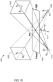
- Fig. 16 shows a schematic representation of epipolar determination of three-dimensional target locations from two-dimensional image locations and their geometrical relationships in space, according to an example embodiment of the present disclosure.
- a point P 1 , associated with image plane 410 is matched to a point P 2 to determine a location P 1,2 in three-dimensional space, using principles of epipolar geometry.
- the processor 30 may next estimate a starting vascular width (a radius, for example).
- the starting width is determined, for example, by generating an orthogonal profile to the centerline and identifying a peak of a weighted sum of the first and second derivatives of the image intensity along the profile.
- the processor 30 builds an orthogonal profile for points along the centerline; for example, for points sampled at intervals approximately equivalent to the vascular starting width.
- the precise choice of interval is not critical: using the radius as the interval is generally appropriate to provide a sufficient resolution for diameter estimation.
- Orthogonal profiles for sampled points are assembled by the processor 30 in a rectangular frame, somewhat as though the convolutions of the three-dimensional centerline were straightened, bringing the orthogonal profiles through the centerline into parallel alignment.
- the example processor 30 may then determine connected routes along vascular edges. For instance, a first side (vascular edge) is chosen for route tracing. In some embodiments, a route is found by the processor 30 along the edge at approximately the distance of the initial radius, for example, by minimizing the energy that corresponds to a weighted sum of the first and second horizontal derivatives, optionally with the aid of an algorithm of the Dijkstra algorithm family.
- the processor 30 may reset centerline to the middle of the two vascular walls just determined. At this point, a three-dimensional model of a vascular tree includes centerlines specified by three-dimensional coordinates and diameters or other vascular geometric information.
- the processor 30 may divide the three-dimensional model into segments or branches, where a branch is defined as a section of a vessel (along the frame of reference established by the vascular centerline, for example) between bifurcations.
- the branches are numbered, for example, according to their generation in the tree.
- Branch points in some embodiments, are determinable from points of the skeletal centerline representation which connect in more than two directions.
- branch topology comprises division of a vascular tree model into distinct branches along the branch structure.
- branch topology comprises recombination of branches, for example, due to collateral and/or shunting blood vessels.
- the tree model is a reduced tree, limited to a single segment of a vessel, between two consecutive bifurcations of the vascular system. In some embodiments, the reduction is to a region of a bifurcation, optionally comprising a stenosis.
- the FFR index comprises a ratio of flow in model comprising a potentially stenotic vascular segment to a model wherein said segment is replaced by a lower flow-resistance segment, and/or the resistance to flow due to said segment is removed.
- a potential advantage of this ratio determination is that the index comprises an expression of the effect of a potential therapeutic treatment to a vasculature, for example, an opening of a vascular region by percutaneous coronary intervention ("PCI") such as stent implantation.
- PCI percutaneous coronary intervention
- Another potential advantage of this ratio is that it measures a parameter (fractional flow reserve) which, though well-accepted as providing an indication of a need for revascularization, is commonly determined in the art by invasive pressure measurements requiring direct access to both sides of stenotic lesion.
- a second model is constructed from the three-dimensional model.
- the second model optionally describes an at least partially healthier vascular system corresponding to the three-dimensional model model.
- the second model is constructed by changing a stenosis in the first model to be more open, as it would be if a stent were to open the stenosis; and in some embodiments the second model is constructed by choosing a section of a patient's vascular system which includes a healthy vessel similar to the problem vessel of the first model, and using it to replace a stenosed vessel.
- a smoothing or normalization of the entire three-dimensional model is carried out such that specific targeting of a stenosis region is not necessary.
- a tree model without stenosis is optionally calculated by replacing a stenosed vessel by an inflated vessel, that is, geometric measurements of a stenosed vessel section are replaced by measurements appropriate for an inflated vessel.
- geometric data (diameter and/or cross-sectional area) which is used for the inflated vessel is a maximum of the geometric data of the unstenosed vessel at a location just proximal to the stenosed location and at a location just distal to the stenosed location.
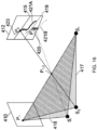
- stream lines are defined from the tree origin, branch 0 to each outlet.
- branches which constitute each stream line are listed in a combination matrix.
- defined stream lines are also numbered, as shown in Fig. 17 .
- a tree model 1100 of a vascular system is provided with tags 1101 to 1105 numbering outlets of the tree model 1100, produced according to an example embodiment of the invention, the tags corresponding to stream lines.
- the flow rate in a mother branch is the sum of flow rates of daughter branches.
- fluid pressure measurements are made, for example blood pressure measurements.
- a vector DP is defined, and Q i is calculated:
- Q ⁇ A ⁇ ⁇ ⁇ 1 ⁇ DP ⁇
- the example embodiment just described potentially provides a minimally-invasive physiological index indicative of functional significance of coronary lesions.
- the example method is optionally performed during a coronary angiography procedure, and calculations are optionally performed during the coronary angiography procedure, such that the minimally-invasive physiological index is provided in real-time.
- flow rate through a segment of interest Q s , [mL/s] is optionally derived from a concentration of iodine contrast material, based on an analysis of concentration-distance-time curves, and a geometric description of the segment of interest, including diameter d(l) [cm], and/or volume V(l) [ml] as a function of segment length.
Landscapes
- Health & Medical Sciences (AREA)
- Engineering & Computer Science (AREA)
- Life Sciences & Earth Sciences (AREA)
- Medical Informatics (AREA)
- Physics & Mathematics (AREA)
- Nuclear Medicine, Radiotherapy & Molecular Imaging (AREA)
- Surgery (AREA)
- General Health & Medical Sciences (AREA)
- Heart & Thoracic Surgery (AREA)
- Animal Behavior & Ethology (AREA)
- Molecular Biology (AREA)
- Public Health (AREA)
- Veterinary Medicine (AREA)
- Biomedical Technology (AREA)
- Radiology & Medical Imaging (AREA)
- Biophysics (AREA)
- Pathology (AREA)
- Optics & Photonics (AREA)
- High Energy & Nuclear Physics (AREA)
- Computer Vision & Pattern Recognition (AREA)
- General Physics & Mathematics (AREA)
- Theoretical Computer Science (AREA)
- Human Computer Interaction (AREA)
- Computer Hardware Design (AREA)
- Software Systems (AREA)
- General Engineering & Computer Science (AREA)
- Geometry (AREA)
- Robotics (AREA)
- Computer Graphics (AREA)
- Architecture (AREA)
- Cardiology (AREA)
- Physiology (AREA)
- Vascular Medicine (AREA)
- Dentistry (AREA)
- Oral & Maxillofacial Surgery (AREA)
- Apparatus For Radiation Diagnosis (AREA)
Claims (15)
- Vorrichtung (1) zum Synchronisieren eines dreidimensionalen Modells der Koronararterien eines Patienten mit einer Ausrichtung einer medizinischen Bildgebungsvorrichtung (10), wobei die Vorrichtung Folgendes umfasst:
eine Arbeitsstation (20), die kommunikativ mit der medizinischen Bildgebungsvorrichtung (10) gekoppelt ist, wobei die Arbeitsstation (20) Folgendes umfasst:einen Anzeigebildschirm (28);eine Speichervorrichtung (32), die ein dreidimensionales Modell der Koronararterien eines Patienten speichert, wobei das dreidimensionale Modell eine Mittellinie durch jede der Koronararterien beinhaltet, wobei jede Mittellinie Abtastpunkte aufweist, wobei jeder Abtastpunkt entlang der jeweiligen Mittellinie in einem dreidimensionalen Koordinatensystem definiert ist und mit vaskulären geometrischen Informationen assoziiert ist; undeinen Prozessor (30), der kommunikativ mit der Speichervorrichtung (32) gekoppelt ist,wobei der Prozessor (30) für Folgendes konfiguriert ist:Empfangen einer Anweisung zum Registrieren des dreidimensionalen Modells mit einer medizinischen Bildgebungsvorrichtung (10),Bestimmen einer Ausrichtung des dreidimensionalen Modells, die einer Null-Grad-Startposition der medizinischen Bildgebungsvorrichtung (10) entspricht,Empfangen von potenziellen Rotationsangulationspositionen der medizinischen Bildgebungsvorrichtung (10),Bestimmen von Winkelkoordinaten für das dreidimensionale Modell, die den potenziellen Rotationsangulationspositionen der medizinischen Bildgebungsvorrichtung (10) entsprechen,Speichern einer Korrelation zwischen den bestimmten Winkelkoordinaten für das dreidimensionale Modell und den potenziellen Rotationsangulationspositionen der medizinischen Bildgebungsvorrichtung (10) in der Speichervorrichtung (32),Bestimmen einer aktuellen Blickwinkelausrichtung der medizinischen Bildgebungsvorrichtung (10),Verwenden der Korrelation zwischen den bestimmten Winkelkoordinaten für das dreidimensionale Modell und den potenziellen Rotationsangulationspositionen der medizinischen Bildgebungsvorrichtung (10), um das dreidimensionale Modell unter Verwendung der aktuellen Blickwinkelausrichtung der medizinischen Bildgebungsvorrichtung (10) zu drehen, undAnzeigen des gedrehten dreidimensionalen Modells in einer ersten Benutzeroberfläche in einer Blickpunktausrichtung, die mit der aktuellen Blickwinkelausrichtung der medizinischen Bildgebungsvorrichtung (10) übereinstimmt,wobei der Prozessor (30) ferner für Folgendes konfiguriert ist:Übertragen von Anweisungen (26) an die medizinische Bildgebungsvorrichtung (10) zum Aufzeichnen eines medizinischen Bilds als Reaktion auf eine Angabe zum Aufzeichnen des medizinischen Bilds, die von einer zweiten Benutzeroberfläche empfangen wird, und Anzeigen des medizinischen Bilds in Verbindung mit dem dreidimensionalen Modell; undwenn eine Sperrsteuerung einer dritten Benutzeroberfläche aktiviert ist, Bewirken, dass sich die medizinische Bildgebungsvorrichtung (10) in Echtzeit synchronisiert mit dem dreidimensionalen Modell dreht, und wenn die Sperrsteuerung nicht aktiviert ist, Ermöglichen, dass sich das dreidimensionale Modell bewegt, ohne zu bewirken, dass sich die medizinische Bildgebungsvorrichtung (10) in Echtzeit bewegt. - Vorrichtung nach Anspruch 1, wobei der Prozessor ferner dazu konfiguriert ist, die Ausrichtung des dreidimensionalen Modells durch Identifizieren einer zweidimensionalen Fläche oder einer Ebene des dreidimensionalen Modells zu bestimmen, die mit einem Blickwinkel an der Null-Grad-Startposition eines Bildverstärkers der medizinischen Bildgebungsvorrichtung (10) ausgerichtet ist, wobei die identifizierte zweidimensionale Fläche oder die Ebene des dreidimensionalen Modells vorzugsweise einer Draufsicht auf die Koronararterien des Patienten entspricht, wenn der Patient auf dem Rücken liegt.
- Vorrichtung nach Anspruch 1, wobei die gefäßgeometrischen Informationen mindestens eines von einem Gefäßdurchmesser, einem Gefäßradius, einer Querschnittsfläche, einem Querschnittsprofil, einer Gefäßwandkrümmung oder einer Gefäßverzweigung umfassen.
- Vorrichtung nach Anspruch 1, wobei die medizinische Bildgebungsvorrichtung (10) einen C-Bogen umfasst, der zum Aufzeichnen von Röntgenangiographiebildern konfiguriert ist.
- Vorrichtung nach Anspruch 1 oder 4, wobei die potenziellen Rotationsangulationspositionen der medizinischen Bildgebungsvorrichtung (10) RAO-Angulationspositionen, LAO-Angulationspositionen, kraniale Angulationspositionen und kaudale Angulationspositionen umfassen.
- Vorrichtung nach Anspruch 5, wobei die Winkelkoordinaten für das dreidimensionale Modell Koordinaten entlang einer Rollachse und einer Nickachse umfassen.
- Vorrichtung nach Anspruch 5, wobei die potenziellen Rotationsangulationspositionen der medizinischen Bildgebungsvorrichtung (10) mindestens eines von in der Speichervorrichtung (32) gespeichert, von der medizinischen Bildgebungsvorrichtung (10) empfangen oder über Benutzereingaben über eine Schnittstelle empfangen werden.
- Vorrichtung nach Anspruch 1, wobei der Prozessor ferner für Folgendes konfiguriert ist:Empfangen einer Nachricht von der medizinischen Bildgebungsvorrichtung (10), die mindestens eines von (i) einer relativen Positionsänderung von der Null-Grad-Startposition der medizinischen Bildgebungsvorrichtung (10), die in einer Rotationsangulationsposition bereitgestellt wird, oder (ii) einer absoluten Position der medizinischen Bildgebungsvorrichtung (10), die in einer Rotationsangulationsposition bereitgestellt wird, angibt;Bestimmen einer neuen Blickpunktausrichtung für das dreidimensionale Modell basierend auf dem mindestens einen von (i) oder (ii) und der Korrelation zwischen den bestimmten Winkelkoordinaten für das dreidimensionale Modell und den potenziellen Rotationsangulationspositionen der medizinischen Bildgebungsvorrichtung (10);Drehen des dreidimensionalen Modells in die neue Blickpunktausrichtung; undAnzeigen des gedrehten dreidimensionalen Modells in der ersten Benutzeroberfläche.
- Vorrichtung nach Anspruch 1, wobei der Prozessor ferner für Folgendes konfiguriert ist:Identifizieren von Koronararterien in dem medizinischen Bild;Bestimmen von Mittellinien durch die identifizierten Koronararterien;Bestimmen von Abtastpunkten entlang der Mittellinien in dem dreidimensionalen Koordinatensystem;Bestimmen von vaskulären geometrischen Informationen für die Abtastpunkte entlang der Mittellinie;Bestimmen einer Korrespondenz zwischen den Koronararterien in dem medizinischen Bild und dem dreidimensionalen Modell unter Verwendung mindestens der Mittellinien des medizinischen Bilds und der Mittellinien des dreidimensionalen Modells; undAktualisieren des dreidimensionalen Modells mit den bestimmten vaskulären geometrischen Informationen aus dem medizinischen Bild.
- Vorrichtung nach Anspruch 1, wobei der Prozessor zum Berechnen und Anzeigen von Werten der fraktionellen Flussreserve ("FFR") für das dreidimensionale Modell in der ersten Benutzeroberfläche konfiguriert ist.
- Verfahren zum Synchronisieren eines dreidimensionalen Modells der Koronararterien eines Patienten mit einer Ausrichtung einer medizinischen Bildgebungsvorrichtung (10), wobei das Verfahren Folgendes umfasst:Empfangen eines dreidimensionalen Modells der Koronararterien eines Patienten in einem Prozessor von einer Speichervorrichtung (32), wobei das dreidimensionale Modell eine Mittellinie durch jede der Koronararterien beinhaltet, wobei jede Mittellinie Abtastpunkte beinhaltet, wobei jeder Abtastpunkt entlang der jeweiligen Mittellinie in einem dreidimensionalen Koordinatensystem definiert ist und mit vaskulären geometrischen Informationen assoziiert ist;Bestimmen einer Ausrichtung des dreidimensionalen Modells, die einer Null-Grad-Startposition einer medizinischen Bildgebungsvorrichtung (10) entspricht, über den Prozessor;Empfangen von potenziellen Rotationsangulationspositionen der medizinischen Bildgebungsvorrichtung (10) in dem Prozessor;Bestimmen von Winkelkoordinaten für das dreidimensionale Modell, die den potenziellen Rotationsangulationspositionen der medizinischen Bildgebungsvorrichtung (10) entsprechen, über den Prozessor;Speichern einer Korrelation zwischen den bestimmten Winkelkoordinaten für das dreidimensionale Modell und den potenziellen Rotationsangulationspositionen der medizinischen Bildgebungsvorrichtung (10) in der Speichervorrichtung (32) über den Prozessor;Bestimmen eines aktuellen Blickpunkts des dreidimensionalen Modells, das in einer Benutzeroberfläche angezeigt wird, über den Prozessor; undVerwenden des aktuellen Blickpunkts des dreidimensionalen Modells und der Korrelation zwischen den Winkelkoordinaten für das dreidimensionale Modell und den potenziellen Rotationsangulationspositionen der medizinischen Bildgebungsvorrichtung (10) über den Prozessor, um zu bewirken, dass sich die medizinische Bildgebungsvorrichtung in Echtzeit synchronisiert mit dem dreidimensionalen Modell dreht, unter der Bedingung, dass eine Sperrsteuerung einer weiteren Benutzeroberfläche aktiviert ist, wobei, wenn die Sperrsteuerung der weiteren Benutzeroberfläche nicht aktiviert ist, der Prozessor ermöglicht, dass sich das dreidimensionale Modell bewegt, ohne zu bewirken, dass sich die medizinische Bildgebungsvorrichtung in Echtzeit bewegt.
- Verfahren nach Anspruch 11, wobei die medizinische Bildgebungsvorrichtung (10) einen C-Bogen umfasst und das Verfahren in einem Katheterisierungslabor während einer Stentplatzierung und/oder einer perkutanen Koronarintervention und/oder einer FFR-Bestimmung durchgeführt wird.
- Verfahren nach Anspruch 11 oder 12, das ferner Folgendes umfasst:Empfangen von mindestens zwei medizinischen Bildern, die in verschiedenen Blickwinkeln in Bezug auf den Patienten aufgezeichnet wurden, für die medizinische Bildgebungsvorrichtung (10) in dem Prozessor, wobei die mindestens zwei medizinischen Bilder Abbildungen der Koronararterien des Patienten umfassen;Identifizieren der Koronararterien in den mindestens zwei medizinischen Bildern über den Prozessor;Bestimmen von Mittellinien durch die identifizierten Koronararterien über den Prozessor;Bestimmen von Abtastpunkten entlang der Mittellinien in dem dreidimensionalen Koordinatensystem über den Prozessor;Bestimmen von vaskulären geometrischen Informationen für die Abtastpunkte entlang der Mittellinien über den Prozessor;Erstellen des dreidimensionalen Modells unter Verwendung der Mittellinien, der Abtastpunkte entlang der Mittellinien in dem dreidimensionalen Koordinatensystem und der vaskulären geometrischen Informationen; undSpeichern des dreidimensionalen Modells in der Speichervorrichtung (32).
- Verfahren nach Anspruch 11 oder 12, wobei der Prozessor bewirkt, dass sich die medizinische Bildgebungsvorrichtung durch Übertragen mindestens einer Anweisungsnachricht an die medizinische Bildgebungsvorrichtung dreht, wobei die mindestens eine Anweisungsnachricht mindestens eines von (i) einer relativen Positionsänderung von der Null-Grad-Startposition der medizinischen Bildgebungsvorrichtung (10) zu der entsprechenden Blickwinkelausrichtung, die in einer Rotationsangulationsposition bereitgestellt wird, oder (ii) einer absoluten Position der medizinischen Bildgebungsvorrichtung (10) für die entsprechende Blickwinkelausrichtung, die in einer Rotationsangulationsposition bereitgestellt wird, umfasst.
- Verfahren nach Anspruch 11 oder 12, wobei die potenziellen Rotationsangulationspositionen der medizinischen Bildgebungsvorrichtung (10) RAO-Angulationspositionen, LAO-Angulationspositionen, kraniale Angulationspositionen und kaudale Angulationspositionen umfassen, und
wobei die Winkelkoordinaten für das dreidimensionale Modell Koordinaten entlang einer Rollachse und einer Nickachse umfassen.
Priority Applications (1)
| Application Number | Priority Date | Filing Date | Title |
|---|---|---|---|
| EP25169121.8A EP4555935A3 (de) | 2019-09-23 | 2020-09-23 | Verfahren, vorrichtung und system zur synchronisation zwischen einem dreidimensionalen gefässmodell und einer bildgebungsvorrichtung |
Applications Claiming Priority (2)
| Application Number | Priority Date | Filing Date | Title |
|---|---|---|---|
| US201962904147P | 2019-09-23 | 2019-09-23 | |
| PCT/IB2020/058901 WO2021059165A1 (en) | 2019-09-23 | 2020-09-23 | Methods, apparatus, and system for synchronization between a three-dimensional vascular model and an imaging device |
Related Child Applications (1)
| Application Number | Title | Priority Date | Filing Date |
|---|---|---|---|
| EP25169121.8A Division EP4555935A3 (de) | 2019-09-23 | 2020-09-23 | Verfahren, vorrichtung und system zur synchronisation zwischen einem dreidimensionalen gefässmodell und einer bildgebungsvorrichtung |
Publications (2)
| Publication Number | Publication Date |
|---|---|
| EP4033964A1 EP4033964A1 (de) | 2022-08-03 |
| EP4033964B1 true EP4033964B1 (de) | 2025-04-09 |
Family
ID=72659840
Family Applications (2)
| Application Number | Title | Priority Date | Filing Date |
|---|---|---|---|
| EP20781104.3A Active EP4033964B1 (de) | 2019-09-23 | 2020-09-23 | Verfahren, vorrichtung und system zur synchronisation zwischen einem dreidimensionalen gefässmodell und einer bildgebungsvorrichtung |
| EP25169121.8A Pending EP4555935A3 (de) | 2019-09-23 | 2020-09-23 | Verfahren, vorrichtung und system zur synchronisation zwischen einem dreidimensionalen gefässmodell und einer bildgebungsvorrichtung |
Family Applications After (1)
| Application Number | Title | Priority Date | Filing Date |
|---|---|---|---|
| EP25169121.8A Pending EP4555935A3 (de) | 2019-09-23 | 2020-09-23 | Verfahren, vorrichtung und system zur synchronisation zwischen einem dreidimensionalen gefässmodell und einer bildgebungsvorrichtung |
Country Status (3)
| Country | Link |
|---|---|
| US (2) | US12039685B2 (de) |
| EP (2) | EP4033964B1 (de) |
| WO (1) | WO2021059165A1 (de) |
Families Citing this family (12)
| Publication number | Priority date | Publication date | Assignee | Title |
|---|---|---|---|---|
| US10210956B2 (en) | 2012-10-24 | 2019-02-19 | Cathworks Ltd. | Diagnostically useful results in real time |
| IL263065B2 (en) | 2016-05-16 | 2024-08-01 | Cathworks Ltd | Vascular evaluation system |
| EP4663131A2 (de) | 2019-04-01 | 2025-12-17 | Cathworks Ltd. | Verfahren und vorrichtung zur angiographischen bildauswahl |
| WO2021059165A1 (en) | 2019-09-23 | 2021-04-01 | Cathworks Ltd. | Methods, apparatus, and system for synchronization between a three-dimensional vascular model and an imaging device |
| EP4319641A4 (de) | 2021-04-09 | 2025-01-22 | Pulmera, Inc. | Medizinische bildgebungssysteme sowie zugehörige vorrichtungen und verfahren |
| IL307943A (en) * | 2021-05-05 | 2023-12-01 | Heartflow Inc | Systems and methods for processing electronic images to determine planar mapping |
| WO2023004303A1 (en) * | 2021-07-20 | 2023-01-26 | Pulmera, Inc. | Image guidance for medical procedures |
| US12315076B1 (en) | 2021-09-22 | 2025-05-27 | Cathworks Ltd. | Four-dimensional motion analysis of a patient's coronary arteries and myocardial wall |
| CN118985005A (zh) | 2022-02-10 | 2024-11-19 | 凯思沃克斯有限公司 | 用于基于机器学习的传感器分析和血管树分割的系统和方法 |
| US12446965B2 (en) | 2023-08-09 | 2025-10-21 | Cathworks Ltd. | Enhanced user interface and crosstalk analysis for vascular index measurement |
| US12462469B2 (en) * | 2023-12-08 | 2025-11-04 | See All AI Inc. | Wire-based calibration apparatus for X-ray imaging systems |
| US12499646B1 (en) | 2024-06-12 | 2025-12-16 | Cathworks Ltd. | Three-dimensional sizing tool for cardiac assessment |
Citations (1)
| Publication number | Priority date | Publication date | Assignee | Title |
|---|---|---|---|---|
| US20170224418A1 (en) * | 2015-08-05 | 2017-08-10 | New York Society For The Ruptured And Crippled Maintaining The Hospital For Special Surgery | A fluoroscopy-based technique to measure intraoperative cup anteversion |
Family Cites Families (317)
| Publication number | Priority date | Publication date | Assignee | Title |
|---|---|---|---|---|
| US5150292A (en) | 1989-10-27 | 1992-09-22 | Arch Development Corporation | Method and system for determination of instantaneous and average blood flow rates from digital angiograms |
| JPH08131429A (ja) | 1994-11-11 | 1996-05-28 | Toshiba Corp | 管状体像再生方法およびその装置 |
| US5638823A (en) | 1995-08-28 | 1997-06-17 | Rutgers University | System and method for noninvasive detection of arterial stenosis |
| US6047080A (en) | 1996-06-19 | 2000-04-04 | Arch Development Corporation | Method and apparatus for three-dimensional reconstruction of coronary vessels from angiographic images |
| US6236878B1 (en) | 1998-05-22 | 2001-05-22 | Charles A. Taylor | Method for predictive modeling for planning medical interventions and simulating physiological conditions |
| AU5580799A (en) | 1998-08-20 | 2000-03-14 | Apple Computer, Inc. | Graphics processor with pipeline state storage and retrieval |
| US6605053B1 (en) | 1999-09-10 | 2003-08-12 | Percardia, Inc. | Conduit designs and related methods for optimal flow control |
| AU7443200A (en) | 1999-09-22 | 2001-04-24 | Florence Medical Ltd. | A method and system for determination of ffr based on flow rate measurements |
| JP2003514600A (ja) | 1999-11-19 | 2003-04-22 | ゼネラル・エレクトリック・カンパニイ | 管状のボリュメトリック物体を再フォーマットするための方法及び装置 |
| US6842638B1 (en) | 2001-11-13 | 2005-01-11 | Koninklijke Philips Electronics N.V. | Angiography method and apparatus |
| US6589176B2 (en) | 2001-12-05 | 2003-07-08 | Koninklijke Philips Electronics N.V. | Ultrasonic image stabilization system and method |
| EP1513449B1 (de) | 2002-06-04 | 2009-08-26 | Koninklijke Philips Electronics N.V. | Hybride dreidimensionale rekonstruktion der koronararterien mittels rotationsangiographie |
| US7020510B2 (en) | 2002-07-25 | 2006-03-28 | Koninklijke Philips Electronics, N.V. | Optimal view map V.0.01 |
| ATE298595T1 (de) | 2002-09-05 | 2005-07-15 | Gambro Lundia Ab | Steuerung für eine blutbehandlungsvorrichtung |
| US7113623B2 (en) | 2002-10-08 | 2006-09-26 | The Regents Of The University Of Colorado | Methods and systems for display and analysis of moving arterial tree structures |
| US7697972B2 (en) * | 2002-11-19 | 2010-04-13 | Medtronic Navigation, Inc. | Navigation system for cardiac therapies |
| US7574026B2 (en) | 2003-02-12 | 2009-08-11 | Koninklijke Philips Electronics N.V. | Method for the 3d modeling of a tubular structure |
| US7155046B2 (en) | 2003-02-12 | 2006-12-26 | Pie Medical Imaging Bv | Method of determining physical parameters of bodily structures |
| JP4421203B2 (ja) | 2003-03-20 | 2010-02-24 | 株式会社東芝 | 管腔状構造体の解析処理装置 |
| WO2005001769A2 (en) | 2003-06-25 | 2005-01-06 | Siemens Medical Solutions Usa, Inc. | Automated regional myocardial assessment for cardiac imaging |
| EA009382B1 (ru) | 2003-08-21 | 2007-12-28 | Искем Корпорейшн | Автоматизированные способы и системы для обнаружения и анализа сосудистых бляшек |
| EP1685538B1 (de) | 2003-08-21 | 2011-03-02 | Philips Intellectual Property & Standards GmbH | Vorrichtung und verfahren zur erzeugung eines dreidimensionalen gefässmodelles |
| WO2005023086A2 (en) | 2003-08-25 | 2005-03-17 | University Of North Carolina At Chapel Hill | Systems, methods, and computer program products for analysis of vessel attributes for diagnosis, disease staging, and surgical planning |
| JP4177217B2 (ja) | 2003-09-24 | 2008-11-05 | アロカ株式会社 | 超音波診断装置 |
| WO2005058137A2 (en) * | 2003-12-12 | 2005-06-30 | University Of Washington | Catheterscope 3d guidance and interface system |
| US8010175B2 (en) * | 2004-05-05 | 2011-08-30 | Siemens Medical Solutions Usa, Inc. | Patient-specific coronary territory mapping |
| US20060036167A1 (en) | 2004-07-03 | 2006-02-16 | Shina Systems Ltd. | Vascular image processing |
| US7339585B2 (en) | 2004-07-19 | 2008-03-04 | Pie Medical Imaging B.V. | Method and apparatus for visualization of biological structures with use of 3D position information from segmentation results |
| US20080020362A1 (en) | 2004-08-10 | 2008-01-24 | Cotin Stephane M | Methods and Apparatus for Simulaton of Endovascular and Endoluminal Procedures |
| CN101065776B (zh) | 2004-11-29 | 2011-08-10 | 皇家飞利浦电子股份有限公司 | 分割解剖学结构的三维数字表示的方法和工作站 |
| JP5268365B2 (ja) | 2005-02-04 | 2013-08-21 | コーニンクレッカ フィリップス エレクトロニクス エヌ ヴィ | 脈管幾何形状及び流れ特性を決定するシステム |
| EP1903944B1 (de) | 2005-06-24 | 2017-04-19 | Volcano Corporation | Ko-aufzeichnung grafischer bilddaten mit darstellung dreidimensionaler gefässmerkmale |
| US7711165B2 (en) | 2005-07-28 | 2010-05-04 | Siemens Medical Solutions Usa, Inc. | System and method for coronary artery segmentation of cardiac CT volumes |
| CN101317194A (zh) | 2005-08-17 | 2008-12-03 | 皇家飞利浦电子股份有限公司 | 用于自动4d冠脉建模和运动矢量场估计的方法和设备 |
| US7970187B2 (en) | 2005-09-06 | 2011-06-28 | Pie Medical Imaging B.V. | Method, apparatus and computer program for contour detection of vessels using x-ray densitometry |
| EP1960965A2 (de) | 2005-12-09 | 2008-08-27 | Koninklijke Philips Electronics N.V. | Auf modellen basierende flussanalyse und visualisierung |
| US8538508B2 (en) | 2005-12-09 | 2013-09-17 | Siemens Aktiengesellschaft | Method and apparatus for ECG-synchronized optically-based image acquisition and transformation |
| US7864997B2 (en) | 2006-04-28 | 2011-01-04 | Pie Medical Imaging B.V. | Method, apparatus and computer program product for automatic segmenting of cardiac chambers |
| JP5379960B2 (ja) | 2006-05-12 | 2013-12-25 | 株式会社東芝 | 3次元画像処理装置及び再構成領域指定方法 |
| EP2074585B1 (de) | 2006-10-03 | 2018-07-11 | Koninklijke Philips N.V. | Modellbasierte ortung der koronaren zentrallinie |
| US7953266B2 (en) | 2007-02-06 | 2011-05-31 | Siemens Medical Solutions Usa, Inc. | Robust vessel tree modeling |
| US11064964B2 (en) | 2007-03-08 | 2021-07-20 | Sync-Rx, Ltd | Determining a characteristic of a lumen by measuring velocity of a contrast agent |
| US9968256B2 (en) | 2007-03-08 | 2018-05-15 | Sync-Rx Ltd. | Automatic identification of a tool |
| WO2010058398A2 (en) | 2007-03-08 | 2010-05-27 | Sync-Rx, Ltd. | Image processing and tool actuation for medical procedures |
| US8331314B2 (en) | 2007-04-20 | 2012-12-11 | Telefonaktiebolaget L M Ericsson (Publ) | Dormant session management associated with handover |
| US8073224B2 (en) | 2007-07-09 | 2011-12-06 | Siemens Aktiengesellschaft | System and method for two-dimensional visualization of temporal phenomena and three dimensional vessel reconstruction |
| CN102172330B (zh) | 2007-07-10 | 2013-03-27 | 株式会社东芝 | X射线摄影装置以及图像处理显示装置 |
| US8086000B2 (en) | 2007-08-27 | 2011-12-27 | Pie Medical Imaging B.V. | Method, apparatus and computer program for quantitative bifurcation analysis on angiographic images |
| EP2219520B1 (de) | 2007-10-31 | 2012-12-12 | Cabra Technology A/S | Verfahren zur berechnung von drücken in einem flüssigkeitsstrom durch einen röhrenförmigen abschnitt, insbesondere ein blutgefäss mit atherosklerotischem plaque |
| US9445772B2 (en) * | 2007-12-31 | 2016-09-20 | St. Jude Medical, Atrial Fibrillatin Division, Inc. | Reduced radiation fluoroscopic system |
| EP2259247B1 (de) | 2008-03-28 | 2019-05-22 | Terumo Corporation | Dreidimensionales körpergewebemodell und herstellungsverfahren dafür |
| US8079711B2 (en) | 2008-04-24 | 2011-12-20 | Carl Zeiss Meditec, Inc. | Method for finding the lateral position of the fovea in an SDOCT image volume |
| US8145293B2 (en) | 2008-06-16 | 2012-03-27 | Siemens Medical Solutions Usa, Inc. | Adaptive medical image acquisition system and method |
| US8062513B2 (en) | 2008-07-09 | 2011-11-22 | Baxter International Inc. | Dialysis system and machine having therapy prescription recall |
| US8200466B2 (en) | 2008-07-21 | 2012-06-12 | The Board Of Trustees Of The Leland Stanford Junior University | Method for tuning patient-specific cardiovascular simulations |
| US8155411B2 (en) | 2008-07-22 | 2012-04-10 | Pie Medical Imaging B.V. | Method, apparatus and computer program for quantitative bifurcation analysis in 3D using multiple 2D angiographic images |
| US8582854B2 (en) | 2008-09-15 | 2013-11-12 | Siemens Aktiengesellschaft | Method and system for automatic coronary artery detection |
| EP2163272B1 (de) | 2008-09-15 | 2014-06-25 | B. Braun Avitum AG | Vorrichtung zur frühen Vorhersage des Kt/V-Parameters in Nierenersatzbehandlungen |
| WO2010033971A1 (en) | 2008-09-22 | 2010-03-25 | Dtherapeutics, Llc | Devices, systems, and methods for determining fractional flow reserve |
| CN102170821B (zh) | 2008-10-01 | 2013-08-07 | 株式会社Irumedi | 心血管分析装置 |
| JP5739812B2 (ja) | 2008-10-10 | 2015-06-24 | コーニンクレッカ フィリップス エヌ ヴェ | 血管造影画像取得装置の作動方法、コリメータ制御ユニット、血管造影画像取得装置及びコンピュータソフトウェア |
| US10362962B2 (en) | 2008-11-18 | 2019-07-30 | Synx-Rx, Ltd. | Accounting for skipped imaging locations during movement of an endoluminal imaging probe |
| US20100125197A1 (en) | 2008-11-18 | 2010-05-20 | Fishel Robert S | Method and apparatus for addressing vascular stenotic lesions |
| WO2010071896A2 (en) | 2008-12-19 | 2010-06-24 | Piedmont Healthcare, Inc. | System and method for lesion-specific coronary artery calcium quantification |
| WO2010099016A1 (en) | 2009-02-25 | 2010-09-02 | Worcester Polytechnic Institute | Automatic vascular model generation based on fluid-structure interactions (fsi) |
| US20120150048A1 (en) | 2009-03-06 | 2012-06-14 | Bio-Tree Systems, Inc. | Vascular analysis methods and apparatus |
| US8560968B1 (en) | 2009-03-26 | 2013-10-15 | Vinod Nair | Method and apparatus for evaluating a heart patient |
| US9679389B2 (en) | 2009-05-19 | 2017-06-13 | Algotec Systems Ltd. | Method and system for blood vessel segmentation and classification |
| ES2660570T3 (es) | 2009-09-23 | 2018-03-23 | Lightlab Imaging, Inc. | Sistemas, aparatos y métodos de recopilación de datos de medición de resistencia vascular y morfología luminal |
| US12426789B2 (en) | 2009-09-23 | 2025-09-30 | Lightlab Imaging, Inc. | Blood vessel lumen morphology and minimum lumen area measurements data collection by intravascular imaging systems for stenosis or stent planning |
| US8315355B2 (en) | 2009-10-28 | 2012-11-20 | Siemens Aktiengesellschaft | Method for operating C-arm systems during repeated angiographic medical procedures |
| US8934686B2 (en) | 2009-11-26 | 2015-01-13 | Algotec Systems Ltd. | User interface for selecting paths in an image |
| EP2333914A3 (de) | 2009-12-09 | 2017-12-13 | Canon Kabushiki Kaisha | Lichtquelle und ihre Verwendung in einer Bildaufnahmevorrichtung |
| US8224056B2 (en) | 2009-12-15 | 2012-07-17 | General Electronic Company | Method for computed tomography motion estimation and compensation |
| JP5539778B2 (ja) | 2010-03-31 | 2014-07-02 | 富士フイルム株式会社 | 血管表示制御装置、その作動方法およびプログラム |
| US20120177275A1 (en) | 2010-04-20 | 2012-07-12 | Suri Jasjit S | Coronary Artery Disease Prediction using Automated IMT |
| US20140142398A1 (en) | 2010-06-13 | 2014-05-22 | Angiometrix Corporation | Multifunctional guidewire assemblies and system for analyzing anatomical and functional parameters |
| US8867801B2 (en) * | 2010-07-13 | 2014-10-21 | Siemens Aktiengesellschaft | Method for determining properties of a vessel in a medical image |
| US8315812B2 (en) | 2010-08-12 | 2012-11-20 | Heartflow, Inc. | Method and system for patient-specific modeling of blood flow |
| US8157742B2 (en) | 2010-08-12 | 2012-04-17 | Heartflow, Inc. | Method and system for patient-specific modeling of blood flow |
| KR101014879B1 (ko) | 2010-08-13 | 2011-02-15 | 김지훈 | 그물조직사를 함유하는 휨 방지용 고무 매트 및 그 제조 방법 |
| WO2012028190A1 (en) | 2010-09-02 | 2012-03-08 | Pie Medical Imaging Bv | Method and apparatus for quantitative analysis of a tree of recursively splitting tubular organs |
| US9119540B2 (en) | 2010-09-16 | 2015-09-01 | Siemens Aktiengesellschaft | Method and system for non-invasive assessment of coronary artery disease |
| US9107639B2 (en) | 2011-03-15 | 2015-08-18 | Medicinsk Bildteknik Sverige Ab | System for synchronously visualizing a representation of first and second input data |
| US10186056B2 (en) | 2011-03-21 | 2019-01-22 | General Electric Company | System and method for estimating vascular flow using CT imaging |
| EP2525328B1 (de) | 2011-05-19 | 2017-10-18 | Pie Medical Imaging BV | Verfahren und Vorrichtung zur Bestimmung optimaler Projektionsbilder |
| CN103732132A (zh) | 2011-06-13 | 2014-04-16 | 安吉奥梅特里克斯公司 | 用于分析解剖学参数和功能参数的多功能导丝组件和系统 |
| US9314584B1 (en) | 2011-06-27 | 2016-04-19 | Bayer Healthcare Llc | Method and apparatus for fractional flow reserve measurements |
| US9974508B2 (en) | 2011-09-01 | 2018-05-22 | Ghassan S. Kassab | Non-invasive systems and methods for determining fractional flow reserve |
| RU2595805C2 (ru) | 2011-09-13 | 2016-08-27 | Конинклейке Филипс Н.В. | Аннотатор сосудов |
| EP2570079B1 (de) | 2011-09-13 | 2017-06-14 | Pie Medical Imaging BV | Verfahren und Vorrichtung zur Bestimmung der optimalen 3D-Rekonstruktion eines Objekts |
| US8948487B2 (en) | 2011-09-28 | 2015-02-03 | Siemens Aktiengesellschaft | Non-rigid 2D/3D registration of coronary artery models with live fluoroscopy images |
| JP5748636B2 (ja) | 2011-10-26 | 2015-07-15 | 富士フイルム株式会社 | 画像処理装置および方法並びにプログラム |
| US10162932B2 (en) | 2011-11-10 | 2018-12-25 | Siemens Healthcare Gmbh | Method and system for multi-scale anatomical and functional modeling of coronary circulation |
| EP2779907B1 (de) | 2011-11-16 | 2022-06-15 | Philips Image Guided Therapy Corporation | Medizinisches messsystem und -verfahren |
| US20130158476A1 (en) * | 2011-12-15 | 2013-06-20 | Eric S. Olson | System and method for synchronizing physical and visualized movements of a medical device and viewing angles among imaging systems |
| EP2800516B1 (de) | 2012-01-06 | 2017-11-15 | Koninklijke Philips N.V. | Echtzeitdarstellung von gefässstrukturansichten für optimale vorrichtungsnavigation |
| US10034614B2 (en) | 2012-02-29 | 2018-07-31 | General Electric Company | Fractional flow reserve estimation |
| US10373700B2 (en) | 2012-03-13 | 2019-08-06 | Siemens Healthcare Gmbh | Non-invasive functional assessment of coronary artery stenosis including simulation of hyperemia by changing resting microvascular resistance |
| US8548778B1 (en) | 2012-05-14 | 2013-10-01 | Heartflow, Inc. | Method and system for providing information from a patient-specific model of blood flow |
| US20130324842A1 (en) | 2012-05-29 | 2013-12-05 | The Johns Hopkins University | Method for Estimating Pressure Gradients and Fractional Flow Reserve from Computed Tomography Angiography: Transluminal Attenuation Flow Encoding |
| US9466117B2 (en) | 2012-06-01 | 2016-10-11 | Koninklijke Philips N.V. | Segmentation highlighter |
| CA2875346A1 (en) | 2012-06-26 | 2014-01-03 | Sync-Rx, Ltd. | Flow-related image processing in luminal organs |
| US9247918B2 (en) | 2012-07-09 | 2016-02-02 | Siemens Aktiengesellschaft | Computation of hemodynamic quantities from angiographic data |
| EP3298959B2 (de) | 2012-08-03 | 2022-09-28 | Philips Image Guided Therapy Corporation | Vorrichtungen und systeme zur beurteilung eines gefässes |
| CN104582572B (zh) | 2012-08-16 | 2018-04-13 | 东芝医疗系统株式会社 | 图像处理装置、医用图像诊断装置以及血压监视器 |
| US10398386B2 (en) | 2012-09-12 | 2019-09-03 | Heartflow, Inc. | Systems and methods for estimating blood flow characteristics from vessel geometry and physiology |
| EP2709059B1 (de) | 2012-09-17 | 2014-11-05 | Pie Medical Imaging BV | Verfahren und Vorrichtung für quantitative Messungen in Bildsequenzen, insbesondere in angiographischen Bildern |
| US20140086461A1 (en) | 2012-09-25 | 2014-03-27 | The Johns Hopkins University | Method and system for determining time-based index for blood circulation from angiographic imaging data |
| US20140100454A1 (en) | 2012-10-05 | 2014-04-10 | Volcano Corporation | Methods and systems for establishing parameters for three-dimensional imaging |
| US11272845B2 (en) | 2012-10-05 | 2022-03-15 | Philips Image Guided Therapy Corporation | System and method for instant and automatic border detection |
| US9675301B2 (en) | 2012-10-19 | 2017-06-13 | Heartflow, Inc. | Systems and methods for numerically evaluating vasculature |
| WO2014064702A2 (en) | 2012-10-24 | 2014-05-01 | Cathworks Ltd. | Automated measurement system and method for coronary artery disease scoring |
| US9814433B2 (en) | 2012-10-24 | 2017-11-14 | Cathworks Ltd. | Creating a vascular tree model |
| US10595807B2 (en) | 2012-10-24 | 2020-03-24 | Cathworks Ltd | Calculating a fractional flow reserve |
| US10210956B2 (en) | 2012-10-24 | 2019-02-19 | Cathworks Ltd. | Diagnostically useful results in real time |
| US9858387B2 (en) | 2013-01-15 | 2018-01-02 | CathWorks, LTD. | Vascular flow assessment |
| JP6334902B2 (ja) | 2012-11-30 | 2018-05-30 | キヤノンメディカルシステムズ株式会社 | 医用画像処理装置 |
| WO2014084398A1 (ja) | 2012-11-30 | 2014-06-05 | 株式会社 東芝 | 医用画像診断装置 |
| EP2757528B1 (de) | 2013-01-22 | 2015-06-24 | Pie Medical Imaging BV | Verfahren und Vorrichtung zum Nachverfolgen von Objekten in einem Zielbereich eines sich bewegenden Organs |
| US9042613B2 (en) | 2013-03-01 | 2015-05-26 | Heartflow, Inc. | Method and system for determining treatments by modifying patient-specific geometrical models |
| US9424395B2 (en) | 2013-03-04 | 2016-08-23 | Heartflow, Inc. | Method and system for sensitivity analysis in modeling blood flow characteristics |
| US8824752B1 (en) | 2013-03-15 | 2014-09-02 | Heartflow, Inc. | Methods and systems for assessing image quality in modeling of patient anatomic or blood flow characteristics |
| US9406141B2 (en) | 2013-05-31 | 2016-08-02 | Siemens Aktiengesellschaft | Segmentation of a structure |
| US9430827B2 (en) | 2013-05-31 | 2016-08-30 | Siemens Aktiengesellschaft | Segmentation of a calcified blood vessel |
| CN104282009B (zh) | 2013-07-02 | 2017-10-27 | 上海联影医疗科技有限公司 | 一种冠脉的提取方法 |
| US9195800B2 (en) | 2013-08-16 | 2015-11-24 | Heartflow, Inc. | Systems and methods for identifying personalized vascular implants from patient-specific anatomic data |
| US9805463B2 (en) | 2013-08-27 | 2017-10-31 | Heartflow, Inc. | Systems and methods for predicting location, onset, and/or change of coronary lesions |
| US9589349B2 (en) | 2013-09-25 | 2017-03-07 | Heartflow, Inc. | Systems and methods for controlling user repeatability and reproducibility of automated image annotation correction |
| EP3954298A3 (de) | 2013-10-24 | 2022-03-16 | Cathworks Ltd. | Bestimmung vaskulärer eigenschaften mit korrespondenzmodellierung eines gefässbaums |
| EP2873371B1 (de) | 2013-11-13 | 2022-12-21 | Pie Medical Imaging BV | Verfahren und system zur registrierung von intravaskulären bildern |
| EP3076854B1 (de) | 2013-12-04 | 2022-04-20 | Koninklijke Philips N.V. | Lokale ffr-schätzung und visualisierung für verbesserte funktionelle stenoseanalyse |
| DE102014201134B4 (de) | 2014-01-22 | 2017-04-06 | Siemens Healthcare Gmbh | Verfahren und Vorrichtung zur Erzeugung eines 2-D-Projektionsbildes eines Gefäßsystems nebst korrespondierenden Gegenständen |
| NL2012324C2 (en) | 2014-02-25 | 2015-08-26 | Medis Associated B V | Method and device for determining a geometrical parameter of a blood vessel. |
| US9501622B2 (en) | 2014-03-05 | 2016-11-22 | Heartflow, Inc. | Methods and systems for predicting sensitivity of blood flow calculations to changes in anatomical geometry |
| JP6262027B2 (ja) | 2014-03-10 | 2018-01-17 | 東芝メディカルシステムズ株式会社 | 医用画像処理装置 |
| US8917925B1 (en) | 2014-03-28 | 2014-12-23 | Heartflow, Inc. | Systems and methods for data and model-driven image reconstruction and enhancement |
| EP3125764B1 (de) | 2014-03-31 | 2023-03-22 | Koninklijke Philips N.V. | Verarbeitungsvorrichtung zur verarbeitung von herzdaten eines lebewesens |
| US9087147B1 (en) | 2014-03-31 | 2015-07-21 | Heartflow, Inc. | Systems and methods for determining blood flow characteristics using flow ratio |
| US9773219B2 (en) | 2014-04-01 | 2017-09-26 | Heartflow, Inc. | Systems and methods for using geometry sensitivity information for guiding workflow |
| US9058692B1 (en) | 2014-04-16 | 2015-06-16 | Heartflow, Inc. | Systems and methods for image-based object modeling using multiple image acquisitions or reconstructions |
| US9514530B2 (en) | 2014-04-16 | 2016-12-06 | Heartflow, Inc. | Systems and methods for image-based object modeling using multiple image acquisitions or reconstructions |
| EP3140757B1 (de) | 2014-05-05 | 2020-06-24 | Siemens Healthcare GmbH | Verfahren und system zur nichtinvasiven funktionellen beurteilung von koronararterienstenose anhand von strömungsberechnungen bei modellen basierend auf erkrankten patienten und hypothetisch normalen anatomischen modellen |
| CN106456016B (zh) | 2014-05-06 | 2020-11-24 | 皇家飞利浦有限公司 | 用于血管估计的设备、系统和方法 |
| US9754082B2 (en) | 2014-05-30 | 2017-09-05 | Heartflow, Inc. | Systems and methods for reporting blood flow characteristics |
| DE102014210591B4 (de) | 2014-06-04 | 2022-09-22 | Siemens Healthcare Gmbh | Fluiddynamische Analyse eines Gefäßbaums mittels Angiographie |
| WO2015193055A1 (en) | 2014-06-19 | 2015-12-23 | Koninklijke Philips N.V. | Determining an effective cross-sectional area of a cardiovascular structure |
| DE102014213408B4 (de) | 2014-07-10 | 2018-10-25 | Siemens Healthcare Gmbh | Verfahren zur Ermittlung eines dreidimensionalen Modelldatensatzes eines wenigstens ein Gefäßsegment umfassenden Blutgefäßsystems |
| JP6778174B2 (ja) | 2014-07-18 | 2020-10-28 | コーニンクレッカ フィリップス エヌ ヴェKoninklijke Philips N.V. | 狭窄評価 |
| US9888968B2 (en) | 2014-07-22 | 2018-02-13 | Siemens Healthcare Gmbh | Method and system for automated therapy planning for arterial stenosis |
| US9195801B1 (en) | 2014-08-05 | 2015-11-24 | Heartflow, Inc. | Systems and methods for treatment planning based on plaque progression and regression curves |
| US10658085B2 (en) | 2014-08-29 | 2020-05-19 | Knu-Industry Coorporation Foundation | Method for determining patient-specific blood vessel information |
| JP6692809B2 (ja) | 2014-11-14 | 2020-05-13 | コーニンクレッカ フィリップス エヌ ヴェKoninklijke Philips N.V. | 経皮的冠動脈インターベンション計画インタフェース、並びに関連するデバイス、システム、及び方法 |
| US9349178B1 (en) | 2014-11-24 | 2016-05-24 | Siemens Aktiengesellschaft | Synthetic data-driven hemodynamic determination in medical imaging |
| WO2016087396A1 (en) | 2014-12-02 | 2016-06-09 | Koninklijke Philips N.V. | Fractional flow reserve determination |
| WO2016092420A1 (en) | 2014-12-08 | 2016-06-16 | Koninklijke Philips N.V. | Devices, systems, and methods for vessel assessment and intervention recommendation |
| US9646361B2 (en) | 2014-12-10 | 2017-05-09 | Siemens Healthcare Gmbh | Initialization independent approaches towards registration of 3D models with 2D projections |
| US9713424B2 (en) | 2015-02-06 | 2017-07-25 | Richard F. Spaide | Volume analysis and display of information in optical coherence tomography angiography |
| US10478130B2 (en) | 2015-02-13 | 2019-11-19 | Siemens Healthcare Gmbh | Plaque vulnerability assessment in medical imaging |
| EP3062248A1 (de) | 2015-02-27 | 2016-08-31 | Pie Medical Imaging BV | Verfahren und Vorrichtung zur quantitativen Durchflussanalyse |
| JP2018515167A (ja) | 2015-04-02 | 2018-06-14 | ハートフロー, インコーポレイテッド | 血管網と潅流組織の機能的関連性を特定し可視化するシステム及び方法 |
| JP2016198262A (ja) | 2015-04-09 | 2016-12-01 | 東芝メディカルシステムズ株式会社 | X線診断装置 |
| EP3937186B1 (de) | 2015-04-10 | 2025-06-11 | HeartFlow, Inc. | System und verfahren zur gefässbaumerzeugung unter verwendung einer gemeinsamen vorabinformation |
| WO2016168474A1 (en) | 2015-04-17 | 2016-10-20 | Heartflow, Inc. | Systems and methods for assessment of tissue function based on vascular disease |
| US9839483B2 (en) | 2015-04-21 | 2017-12-12 | Heartflow, Inc. | Systems and methods for risk assessment and treatment planning of arterio-venous malformation |
| EP3086287B1 (de) | 2015-04-24 | 2017-10-11 | Pie Medical Imaging BV | Durchflussanalyse in 4d-mr-bilddaten |
| US10636146B2 (en) * | 2015-05-12 | 2020-04-28 | Singapore Health Services Pte Ltd | Medical image processing methods and systems |
| US9934566B2 (en) | 2015-07-14 | 2018-04-03 | Siemens Healthcare Gmbh | 3-D vessel tree surface reconstruction method |
| US9785748B2 (en) | 2015-07-14 | 2017-10-10 | Heartflow, Inc. | Systems and methods for estimating hemodynamic forces acting on plaque and monitoring patient risk |
| EP3128481B1 (de) | 2015-08-04 | 2019-12-18 | Pie Medical Imaging BV | Verfahren und vorrichtung zur verbesserung einer 3d+zeit-rekonstruktion |
| US11031136B2 (en) | 2015-08-05 | 2021-06-08 | Koninklijke Philips N.V. | Assistance device and method for an interventional hemodynamic measurement |
| WO2017046288A1 (en) | 2015-09-16 | 2017-03-23 | Koninklijke Philips N.V. | Apparatus for vessel characterization |
| US10517678B2 (en) | 2015-10-02 | 2019-12-31 | Heartflow, Inc. | System and method for diagnosis and assessment of cardiovascular disease by comparing arterial supply capacity to end-organ demand |
| JP6918794B2 (ja) | 2015-11-10 | 2021-08-11 | ハートフロー, インコーポレイテッド | 解剖モデリングシステム及びその作動方法 |
| CN105326486B (zh) | 2015-12-08 | 2017-08-25 | 博动医学影像科技(上海)有限公司 | 血管压力差与血流储备分数的计算方法及系统 |
| JP2019511931A (ja) | 2015-12-29 | 2019-05-09 | コーニンクレッカ フィリップス エヌ ヴェKoninklijke Philips N.V. | 輪郭シグネチャを用いた手術画像獲得デバイスの位置合わせ |
| DE112016006137T5 (de) | 2015-12-30 | 2018-09-06 | Koninklijke Philips N.V. | Synthetische Darstellung einer Gefäßstruktur |
| JP6878439B2 (ja) * | 2015-12-30 | 2021-05-26 | コーニンクレッカ フィリップス エヌ ヴェKoninklijke Philips N.V. | 体部の3次元モデル |
| US10733792B2 (en) | 2016-02-11 | 2020-08-04 | Pie Medical Imaging B.V. | Method and apparatus for user guidance for the choice of a two-dimensional angiographic projection |
| EP3206183B1 (de) | 2016-02-11 | 2025-10-08 | Pie Medical Imaging BV | Verfahren und vorrichtung zur benutzerführung zur auswahl einer zweidimensionalen angiografischen projektion |
| JP6829262B2 (ja) | 2016-02-26 | 2021-02-10 | ハートフロー, インコーポレイテッド | 画像ベースの患者固有の血流力学的モデルにおいて解像されていない血管を特定してモデル化するためのシステム及び方法 |
| KR102447741B1 (ko) | 2016-03-16 | 2022-09-29 | 하트플로우, 인크. | 관상 동맥 내의 건강한 내강 지름을 추정하고 협착증 정량화하기 위한 시스템 및 방법 |
| EP3242142B1 (de) | 2016-05-02 | 2025-12-17 | Pie Medical Imaging BV | Verfahren und vorrichtung zur automatischen bestimmung von geschwindigkeitskodierrichtungen |
| US20170325770A1 (en) | 2016-05-13 | 2017-11-16 | General Electric Company | Methods for personalizing blood flow models |
| EP3461253B1 (de) | 2016-05-16 | 2023-08-09 | Cathworks Ltd. | Auswahl von blutgefässpfaden aus bildern |
| IL263065B2 (en) | 2016-05-16 | 2024-08-01 | Cathworks Ltd | Vascular evaluation system |
| NL2016787B1 (en) | 2016-05-17 | 2017-11-21 | Medis Ass B V | Method and apparatus for reconstructing a three-dimensional representation of a target volume inside an animal or human body. |
| US20170337682A1 (en) | 2016-05-18 | 2017-11-23 | Siemens Healthcare Gmbh | Method and System for Image Registration Using an Intelligent Artificial Agent |
| DE102016210003B4 (de) | 2016-06-07 | 2018-03-08 | Siemens Healthcare Gmbh | Erstellung eines dreidimensionalen Abbilds eines Körperteils durch ein Röntgengerät und hierfür geeignetes Röntgengerät |
| US10363018B2 (en) | 2016-07-19 | 2019-07-30 | Toshiba Medical Systems Corporation | Medical processing apparatus and medical processing method |
| EP3293652B1 (de) | 2016-09-13 | 2024-10-30 | Ebit srl | Strukturierter berichterstellungsablauf mit anatomischem atlas für interventionelle radiologie |
| JP7100627B2 (ja) | 2016-09-20 | 2022-07-13 | ハートフロー, インコーポレイテッド | 次数低減モデル及び/または機械学習を使用して血流特性を推定するためのシステム及び方法 |
| CN109843161B (zh) | 2016-09-30 | 2022-07-12 | 皇家飞利浦有限公司 | 用于确定针对狭窄评估的功能指数的装置 |
| DE102016222102A1 (de) | 2016-11-10 | 2018-05-17 | Siemens Healthcare Gmbh | Behandlungsplanung für eine Stenose in einem Gefäßsegment anhand einer virtuellen hämodynamischen Analyse |
| CN109996495B (zh) | 2016-11-22 | 2023-04-28 | 皇家飞利浦有限公司 | 用于生物物理模拟的血管树标准化和/或用于经修剪的部分的扩展模拟 |
| US10163209B2 (en) | 2016-11-23 | 2018-12-25 | Toshiba Medical Systems Corporation | Medical image processing apparatus, medical image processing method, and X-ray CT apparatus |
| US11202612B2 (en) | 2016-11-28 | 2021-12-21 | Canon Medical Systems Corporation | Medical image-processing apparatus, X-ray CT apparatus, and medical image-processing method performing fluid analysis to switch displayed color information |
| US10600181B2 (en) | 2016-12-23 | 2020-03-24 | Heartflow, Inc. | Systems and methods for probabilistic segmentation in anatomical image processing |
| JP7077319B2 (ja) | 2016-12-23 | 2022-05-30 | ハートフロー, インコーポレイテッド | 解剖学的モデルパラメータの機械学習 |
| EP3375364B1 (de) | 2017-01-23 | 2024-08-28 | Shanghai United Imaging Healthcare Co., Ltd. | Verfahren und systeme zur analyse von durchblutungsstörungen |
| EP4445835A3 (de) | 2017-02-24 | 2025-01-22 | HeartFlow, Inc. | Systeme und verfahren zur identifizierung anatomisch relevanter blutflusseigenschaften in einem patienten |
| US11017531B2 (en) | 2017-03-09 | 2021-05-25 | Cathworks Ltd | Shell-constrained localization of vasculature |
| EP3378403A1 (de) | 2017-03-20 | 2018-09-26 | Koninklijke Philips N.V. | Kontrastmittelinjektionsabbildung |
| WO2018172990A1 (en) | 2017-03-24 | 2018-09-27 | Pie Medical Imaging B.V. | Method and system for assessing vessel obstruction based on machine learning |
| US12089977B2 (en) | 2017-03-24 | 2024-09-17 | Pie Medical Imaging B.V. | Method and system for assessing vessel obstruction based on machine learning |
| EP3382641A1 (de) | 2017-03-30 | 2018-10-03 | Koninklijke Philips N.V. | Kontrastinjektionsabbildung |
| EP3602485B1 (de) | 2017-03-31 | 2023-10-11 | Koninklijke Philips N.V. | Interaktionsüberwachung von nichtinvasiver bildgebungsbasierter ffr |
| CN110506293B (zh) | 2017-03-31 | 2023-11-07 | 皇家飞利浦有限公司 | 谱成像ffr |
| EP3384850A1 (de) | 2017-04-05 | 2018-10-10 | Koninklijke Philips N.V. | Verfahren und vorrichtung zur bestimmung physiologischer funktioneller parameter |
| EP3607558A1 (de) | 2017-04-06 | 2020-02-12 | Koninklijke Philips N.V. | Personalisierung, kalibrierung und/oder training von simulationsparametern von fraktionierten strömungsreserven |
| JP7300392B2 (ja) | 2017-04-06 | 2023-06-29 | コーニンクレッカ フィリップス エヌ ヴェ | 標準化された冠動脈疾患メトリック |
| WO2018184779A1 (en) | 2017-04-06 | 2018-10-11 | Koninklijke Philips N.V. | Coronary artery disease metric based on estimation of myocardial microvascular resistance from ecg signal |
| CN111095261A (zh) | 2017-04-27 | 2020-05-01 | 视网膜病答案有限公司 | 眼底图像自动分析系统和方法 |
| WO2018208927A1 (en) | 2017-05-09 | 2018-11-15 | Heartflow, Inc. | Systems and methods for anatomic structure segmentation in image analysis |
| EP3403582B1 (de) | 2017-05-15 | 2023-06-07 | Pie Medical Imaging BV | Verfahren und vorrichtung zur bestimmung der blutgeschwindigkeit in röntgenangiographiebildern |
| EP3644857B1 (de) | 2017-06-30 | 2025-02-26 | Koninklijke Philips N.V. | Spektrale ffr-ct mit maschinenlernen |
| WO2019012066A1 (en) | 2017-07-13 | 2019-01-17 | Pie Medical Imaging Bv | METHODS AND SYSTEMS FOR GUIDING IN CARDIAC RESYNCHRONIZATION THERAPY |
| US11589924B2 (en) | 2017-08-01 | 2023-02-28 | Siemens Healthcare Gmbh | Non-invasive assessment and therapy guidance for coronary artery disease in diffuse and tandem lesions |
| US11341631B2 (en) | 2017-08-09 | 2022-05-24 | Shenzhen Keya Medical Technology Corporation | System and method for automatically detecting a physiological condition from a medical image of a patient |
| EP3457404A1 (de) | 2017-09-18 | 2019-03-20 | Koninklijke Philips N.V. | Beurteilung des flusses zu gefässverzweigungen zur simulierten hämodynamik |
| US10909825B2 (en) | 2017-09-18 | 2021-02-02 | Skybell Technologies Ip, Llc | Outdoor security systems and methods |
| US20230252628A1 (en) | 2017-09-18 | 2023-08-10 | Koninklijke Philips N.V. | Estimating flow to vessel bifurcations for simulated hemodynamics |
| CN109378043A (zh) | 2017-10-13 | 2019-02-22 | 北京昆仑医云科技有限公司 | 基于患者的医学图像生成诊断报告的系统和方法及介质 |
| US10762637B2 (en) * | 2017-10-27 | 2020-09-01 | Siemens Healthcare Gmbh | Vascular segmentation using fully convolutional and recurrent neural networks |
| EP3477551B1 (de) | 2017-10-30 | 2022-03-23 | Siemens Healthcare GmbH | Maschinenerlernte vorhersage der unsicherheit oder empfindlichkeit für hämodynamische quantifizierung in der medizinischen bildgebung |
| US10783640B2 (en) | 2017-10-30 | 2020-09-22 | Beijing Keya Medical Technology Co., Ltd. | Systems and methods for image segmentation using a scalable and compact convolutional neural network |
| EP3477324B1 (de) | 2017-10-31 | 2025-03-26 | Pie Medical Imaging BV | Verbesserung der segmentierung der linken herzkammer in kontrastverstärkten cine-mrt-datensätzen |
| EP3489893B1 (de) | 2017-11-22 | 2020-06-24 | Siemens Healthcare GmbH | Verfahren und system zur bewertung eines hämodynamischen parameters |
| US20220036646A1 (en) | 2017-11-30 | 2022-02-03 | Shenzhen Keya Medical Technology Corporation | Methods and devices for performing three-dimensional blood vessel reconstruction using angiographic image |
| EP3503026A1 (de) | 2017-12-20 | 2019-06-26 | Koninklijke Philips N.V. | Vorrichtung, system und verfahren zur interaktion mit gefässbildern |
| US11786202B2 (en) | 2018-01-08 | 2023-10-17 | Shenzhen Keya Medical Technology Corporation | Method, system, and medium for analyzing image sequence of periodic physiological activity |
| US10580526B2 (en) | 2018-01-12 | 2020-03-03 | Shenzhen Keya Medical Technology Corporation | System and method for calculating vessel flow parameters based on angiography |
| CN112074866B (zh) | 2018-01-24 | 2024-05-24 | 帕伊医疗成像有限公司 | 4d mr图像数据中的流分析 |
| CN110226923B (zh) | 2018-03-05 | 2021-12-14 | 苏州润迈德医疗科技有限公司 | 一种无需血管扩张剂测量血流储备分数的方法 |
| EP3537447A1 (de) | 2018-03-07 | 2019-09-11 | Koninklijke Philips N.V. | Darstellung medizinischer bilddaten |
| WO2019174971A1 (en) | 2018-03-14 | 2019-09-19 | Koninklijke Philips N.V. | Alternative anatomical borders of blood vessels and associated devices, systems, and methods |
| US10699407B2 (en) | 2018-04-11 | 2020-06-30 | Pie Medical Imaging B.V. | Method and system for assessing vessel obstruction based on machine learning |
| US11389130B2 (en) | 2018-05-02 | 2022-07-19 | Siemens Healthcare Gmbh | System and methods for fast computation of computed tomography based fractional flow reserve |
| CN108830848B (zh) | 2018-05-25 | 2022-07-05 | 深圳科亚医疗科技有限公司 | 利用计算机确定血管上的血管状况参数的序列的装置和系统 |
| CN109949300B (zh) | 2018-06-03 | 2020-07-17 | 北京昆仑医云科技有限公司 | 解剖树结构分析的方法、系统和计算机可读介质 |
| US10431328B1 (en) | 2018-06-03 | 2019-10-01 | Beijing Curacloud Technology Co., Ltd. | Method and system for anatomical tree structure analysis |
| WO2019238754A1 (en) | 2018-06-15 | 2019-12-19 | Pie Medical Imaging Bv | Method and apparatus for quantitative hemodynamic flow analysis |
| CN109065170B (zh) | 2018-06-20 | 2021-11-19 | 博动医学影像科技(上海)有限公司 | 获取血管压力差的方法及装置 |
| CN109009001B (zh) | 2018-07-02 | 2019-07-09 | 博动医学影像科技(上海)有限公司 | 血管压力差修正方法、装置和设备 |
| CN113012109B (zh) | 2018-07-24 | 2024-02-20 | 上海博动医疗科技股份有限公司 | 血管图像的处理方法、装置及成像设备 |
| CN110490927B (zh) | 2018-08-23 | 2022-04-12 | 深圳科亚医疗科技有限公司 | 用于为图像中的对象生成中心线的方法、装置和系统 |
| EP3624132A1 (de) | 2018-09-13 | 2020-03-18 | Koninklijke Philips N.V. | Berechnung von randbedingungen für virtuelle ffr- und ifr-berechnung auf basis von myokardkontrastierungeigenschaften |
| CN110490974B (zh) | 2018-09-19 | 2023-04-28 | 科亚医疗科技股份有限公司 | 用于确定血管中血流速度的方法、系统、装置和介质 |
| CN109770888A (zh) | 2019-03-19 | 2019-05-21 | 苏州润迈德医疗科技有限公司 | 基于压力传感器和造影图像计算瞬时无波形比率的方法 |
| CN109979593B (zh) | 2018-09-24 | 2021-04-13 | 北京科亚方舟医疗科技股份有限公司 | 血管路径的健康半径的预测方法、血管路径的候选狭窄处的预测方法、血管狭窄度预测装置 |
| WO2020064090A1 (en) | 2018-09-25 | 2020-04-02 | Siemens Healthcare Gmbh | Endovascular implant decision support in medical imaging |
| US10854329B2 (en) | 2018-10-16 | 2020-12-01 | Canon Medical Systems Corporation | Motion estimation method and apparatus |
| KR20210076944A (ko) | 2018-10-17 | 2021-06-24 | 하트플로우, 인크. | 지방 조직으로부터 심장혈관 질환 및 치료 유효성을 평가하기 위한 시스템들 및 방법들 |
| CN111227821B (zh) | 2018-11-28 | 2022-02-11 | 苏州润迈德医疗科技有限公司 | 基于心肌血流量和ct图像的微循环阻力指数计算方法 |
| EP3644274A1 (de) | 2018-10-26 | 2020-04-29 | Koninklijke Philips N.V. | Orientierungsdektion für blutgefässsegmentierung |
| EP3881758B1 (de) | 2018-11-13 | 2024-09-18 | Suzhou Rainmed Medical Technology Co., Ltd. | Verfahren, vorrichtung und system zur erfassung eines vaskulären bewertungsparameters auf basis angiographischer bilder |
| US10964017B2 (en) | 2018-11-15 | 2021-03-30 | General Electric Company | Deep learning for arterial analysis and assessment |
| US11406339B2 (en) | 2018-11-16 | 2022-08-09 | Wisconsin Alumni Research Foundation | System and method for determining vascular velocity using medical imaging |
| EP3657393A1 (de) | 2018-11-20 | 2020-05-27 | Koninklijke Philips N.V. | Bestimmung eines weiteren bearbeitungsorts in der magnetresonanzbildgebung |
| EP3659505A1 (de) | 2018-11-28 | 2020-06-03 | Koninklijke Philips N.V. | Relevanteste röntgenbildauswahl für hämodynamische simulation |
| CN111227822B (zh) | 2018-11-28 | 2022-02-11 | 苏州润迈德医疗科技有限公司 | 基于心肌血流量和ct图像的冠状动脉血流储备分数计算方法 |
| NL2022109B1 (en) | 2018-11-30 | 2020-06-26 | Medis Ass B V | Heartbeat based selection of images for cardiovascular model |
| CN112022137B (zh) | 2018-11-30 | 2021-07-13 | 博动医学影像科技(上海)有限公司 | 建立血管截面函数和血管应力的方法及装置 |
| EP3671649B1 (de) | 2018-12-19 | 2025-07-02 | Siemens Healthineers AG | Verfahren und computersystem zur erzeugung einer kombinierten gewebe-gefäss-darstellung |
| US11707242B2 (en) | 2019-01-11 | 2023-07-25 | Pie Medical Imaging B.V. | Methods and systems for dynamic coronary roadmapping |
| EP3683773A1 (de) | 2019-01-17 | 2020-07-22 | Koninklijke Philips N.V. | Verfahren zur visualisierung einer dynamischen anatomischen struktur |
| EP3941358B1 (de) | 2019-03-19 | 2025-03-05 | Koninklijke Philips N.V. | Quantifizierung und messung dreidimensionaler volumenströme |
| CN109805949B (zh) | 2019-03-19 | 2020-05-22 | 苏州润迈德医疗科技有限公司 | 基于压力传感器和造影图像计算血流储备分数的方法 |
| US11308362B2 (en) | 2019-03-26 | 2022-04-19 | Shenzhen Keya Medical Technology Corporation | Method and system for generating a centerline for an object, and computer readable medium |
| EP4663131A2 (de) | 2019-04-01 | 2025-12-17 | Cathworks Ltd. | Verfahren und vorrichtung zur angiographischen bildauswahl |
| JP7227400B2 (ja) | 2019-04-15 | 2023-02-21 | 上海博動医療科技股▲分▼有限公司 | 冠血流量及び血流速度の取得方法並びに装置 |
| EP3751580B1 (de) | 2019-06-11 | 2024-04-03 | Siemens Healthineers AG | Hämodynamische analyse von gefässen unter verwendung eines wiederkehrenden neuronalen netzes |
| CN111754476B (zh) | 2019-06-19 | 2024-07-19 | 科亚医疗科技股份有限公司 | 用于解剖树结构的疾病量化建模的方法及系统 |
| EP3756547A1 (de) | 2019-06-28 | 2020-12-30 | Koninklijke Philips N.V. | Automatisierte koronarangiografieanalyse |
| EP3756549A1 (de) | 2019-06-28 | 2020-12-30 | Koninklijke Philips N.V. | Blutgefässregistrierung mit hilfe von funktionalen informationen |
| CN114072838B (zh) | 2019-07-17 | 2024-11-05 | 西门子医疗股份公司 | 根据2d医学图像进行3d血管中心线重建 |
| US20220273180A1 (en) | 2019-07-19 | 2022-09-01 | Cathworks Ltd. | Functional impact of vascular lesions |
| CN110522465B (zh) | 2019-07-22 | 2024-09-06 | 通用电气精准医疗有限责任公司 | 基于图像数据估计血液动力学参数的方法和系统 |
| WO2021014181A1 (en) | 2019-07-22 | 2021-01-28 | Siemens Healthcare Gmbh | Assessment of coronary artery calcification in angiographic images |
| EP3790023A1 (de) | 2019-09-03 | 2021-03-10 | Koninklijke Philips N.V. | Verfahren zur analyse von medizinischen bilddaten in einer virtuellen mehrbenutzerkollaboration, computerprogramm, benutzeroberfläche und system |
| EP4026491A4 (de) | 2019-09-05 | 2023-09-06 | Suzhou Rainmed Medical Technology Co., Ltd. | Vereinfachtes verfahren, vorrichtung und system zur messung von parametern zur beurteilung des zustands der kornararteriengefässe |
| EP4026492A4 (de) | 2019-09-05 | 2023-10-11 | Suzhou Rainmed Medical Technology Co., Ltd. | Verfahren, vorrichtung und system zum bequemen und schnellen messen des bewertungsparameters einer koronararterie |
| WO2021046990A1 (zh) | 2019-09-09 | 2021-03-18 | 苏州润迈德医疗科技有限公司 | 基于影像和压力传感器计算微循环指标的方法装置及系统 |
| CN114340481B (zh) | 2019-09-09 | 2025-08-29 | 美德胡布有限公司 | 用于确定流量储备分数的对图像数据的自动分析 |
| WO2021059165A1 (en) | 2019-09-23 | 2021-04-01 | Cathworks Ltd. | Methods, apparatus, and system for synchronization between a three-dimensional vascular model and an imaging device |
| CN110786841B (zh) | 2019-11-04 | 2021-05-25 | 苏州润迈德医疗科技有限公司 | 基于微循环阻力指数调节最大充血状态流速的方法及装置 |
| CN110786840B (zh) | 2019-11-04 | 2021-06-08 | 苏州润迈德医疗科技有限公司 | 基于生理参数获取血管评定参数的方法、装置及存储介质 |
| CN112116615B (zh) | 2019-11-19 | 2023-12-05 | 苏州润迈德医疗科技有限公司 | 根据血管中心线获取血管轮廓线的方法和装置 |
| EP4276751A3 (de) | 2019-11-28 | 2024-01-10 | Siemens Healthcare GmbH | Computerimplementiertes verfahren zur bewertung eines ct-datensatzes hinsichtlich perivaskulärem gewebe, bewertungsvorrichtung, computerprogramm und elektronisch lesbares speichermedium |
| CN112151180B (zh) | 2019-12-05 | 2024-03-08 | 苏州润迈德医疗科技有限公司 | 具有狭窄病变的血管数学模型的合成方法和装置 |
| WO2021117043A1 (en) | 2019-12-10 | 2021-06-17 | Medhub Ltd | Automatic stenosis detection |
| EP3852054A1 (de) | 2020-01-16 | 2021-07-21 | Koninklijke Philips N.V. | Verfahren und system zur automatischen erkennung von anatomischen strukturen in einem medizinischen bild |
| KR102375775B1 (ko) | 2020-02-10 | 2022-03-21 | 주식회사 메디픽셀 | 혈관 영상에 기초한 주요 혈관 영역 추출 방법 및 장치 |
| EP3866176A1 (de) | 2020-02-17 | 2021-08-18 | Siemens Healthcare GmbH | Maschinenbasierte risikovorhersage für peri-prozedurale myokardinfarkte oder komplikationen aus medizinischen daten |
| CN111369519B (zh) | 2020-03-02 | 2021-09-17 | 博动医学影像科技(上海)有限公司 | 冠状动脉的血流速度的计算方法、装置及电子设备 |
| US12048575B2 (en) | 2020-03-10 | 2024-07-30 | GE Precision Healthcare LLC | Systems and methods for registration of angiographic projections with computed tomographic data |
| EP3884868B1 (de) | 2020-03-26 | 2025-09-03 | Pie Medical Imaging BV | Verfahren und system zur registrierung von intraobjektdaten mit extraobjektdaten |
| US20230113721A1 (en) | 2020-03-26 | 2023-04-13 | Medhub Ltd | Functional measurements of vessels using a temporal feature |
| CN112164020B (zh) | 2020-03-31 | 2024-01-23 | 苏州润迈德医疗科技有限公司 | 精确提取血管中心线的方法、装置、分析系统和存储介质 |
| CN111709925B (zh) | 2020-05-26 | 2023-11-03 | 深圳科亚医疗科技有限公司 | 用于血管斑块分析的装置、系统及介质 |
| KR102399510B1 (ko) | 2020-06-02 | 2022-05-19 | 주식회사 메디픽셀 | 사용자 입력에 기초하여 혈관 영상을 처리하는 방법 및 장치 |
| CN113837985B (zh) | 2020-06-24 | 2023-11-07 | 上海博动医疗科技股份有限公司 | 用于血管造影图像处理的训练方法和装置、自动处理方法和装置 |
| CN111815589B (zh) | 2020-06-29 | 2022-08-05 | 苏州润迈德医疗科技有限公司 | 基于ct序列图像获取无干扰冠脉树图像的方法和系统 |
| CN111815583B (zh) | 2020-06-29 | 2022-08-05 | 苏州润迈德医疗科技有限公司 | 基于ct序列图像获取主动脉中心线的方法和系统 |
| WO2022000976A1 (zh) | 2020-06-29 | 2022-01-06 | 苏州润迈德医疗科技有限公司 | 基于深度学习获取主动脉的方法和存储介质 |
| CN111815585B (zh) | 2020-06-29 | 2022-08-05 | 苏州润迈德医疗科技有限公司 | 基于ct序列图像获取冠脉树和冠脉入口点的方法和系统 |
| CN111815587A (zh) | 2020-06-29 | 2020-10-23 | 苏州润心医疗器械有限公司 | 基于ct序列图像拾取主动脉中心线上的点的方法和系统 |
| EP3933765A1 (de) | 2020-07-02 | 2022-01-05 | Koninklijke Philips N.V. | Stenoselokalisierung |
| NL2026137B1 (en) | 2020-07-24 | 2022-03-28 | Medis Ass B V | Method and device for determining a coronary microvascular resistance score |
| EP3945469A1 (de) | 2020-07-28 | 2022-02-02 | Siemens Healthcare GmbH | Computerimplementiertes verfahren zur parametrisierung einer funktion zur bewertung eines datensatzes eines medizinischen bildes |
| EP3951705A1 (de) | 2020-08-04 | 2022-02-09 | Siemens Healthcare GmbH | Verfahren, vorrichtung und system zur bestimmung von abnormalität im myokardbereich |
| CN116096330A (zh) | 2020-08-26 | 2023-05-09 | 新加坡保健服务集团有限公司 | 用于分析冠状动脉狭窄的医学图像处理方法和系统 |
| US20230237652A1 (en) | 2020-09-01 | 2023-07-27 | Koninklijke Philips N.V. | Venous compression site identification and stent deployment guidance, and associated devices, systems, and methods |
| EP3975199A1 (de) | 2020-09-24 | 2022-03-30 | Siemens Healthcare GmbH | Verfahren und system zur bestimmung einer signifikanzbewertung in verbindung mit einem medizinischen bildgebungsdatensatz eines patienten |
| EP3975115A1 (de) | 2020-09-25 | 2022-03-30 | Siemens Healthcare GmbH | Verfahren und system zur analyse von mehreren miteinander verbundenen blutgefässen |
| WO2022069303A2 (en) | 2020-09-29 | 2022-04-07 | Philips Image Guided Therapy Corporation | Mapping between computed tomography and angiography for co-registration of intravascular data and blood vessel metrics with computed tomography-based three-dimensional model |
| WO2022069208A1 (en) | 2020-09-29 | 2022-04-07 | Koninklijke Philips N.V. | Ultrasound image-based patient-specific region of interest identification, and associated devices, systems, and methods |
-
2020
- 2020-09-23 WO PCT/IB2020/058901 patent/WO2021059165A1/en not_active Ceased
- 2020-09-23 EP EP20781104.3A patent/EP4033964B1/de active Active
- 2020-09-23 US US17/762,681 patent/US12039685B2/en active Active
- 2020-09-23 EP EP25169121.8A patent/EP4555935A3/de active Pending
-
2024
- 2024-07-03 US US18/763,402 patent/US20250069347A1/en active Pending
Patent Citations (1)
| Publication number | Priority date | Publication date | Assignee | Title |
|---|---|---|---|---|
| US20170224418A1 (en) * | 2015-08-05 | 2017-08-10 | New York Society For The Ruptured And Crippled Maintaining The Hospital For Special Surgery | A fluoroscopy-based technique to measure intraoperative cup anteversion |
Also Published As
| Publication number | Publication date |
|---|---|
| US20250069347A1 (en) | 2025-02-27 |
| EP4555935A2 (de) | 2025-05-21 |
| US12039685B2 (en) | 2024-07-16 |
| EP4033964A1 (de) | 2022-08-03 |
| EP4555935A3 (de) | 2025-07-30 |
| US20220254131A1 (en) | 2022-08-11 |
| WO2021059165A1 (en) | 2021-04-01 |
Similar Documents
| Publication | Publication Date | Title |
|---|---|---|
| EP4033964B1 (de) | Verfahren, vorrichtung und system zur synchronisation zwischen einem dreidimensionalen gefässmodell und einer bildgebungsvorrichtung | |
| US12217872B2 (en) | Diagnostically useful results in real time | |
| US12193793B2 (en) | Method and apparatus for quantitative hemodynamic flow analysis | |
| US20230309943A1 (en) | Methods and systems for dynamic coronary roadmapping | |
| US11406337B2 (en) | Calculating a fractional flow reserve | |
| US10470730B2 (en) | Creating a vascular tree model | |
| EP3753494A1 (de) | Berechnung einer fraktionierten strömungsreserve | |
| US12315076B1 (en) | Four-dimensional motion analysis of a patient's coronary arteries and myocardial wall | |
| HK40120507A (en) | Methods, apparatus, and system for synchronization between a three-dimensional vascular model and an imaging device | |
| HK40077862A (en) | Methods, apparatus, and system for synchronization between a three-dimensional vascular model and an imaging device | |
| HK40077862B (en) | Methods, apparatus, and system for synchronization between a three-dimensional vascular model and an imaging device |
Legal Events
| Date | Code | Title | Description |
|---|---|---|---|
| STAA | Information on the status of an ep patent application or granted ep patent |
Free format text: STATUS: UNKNOWN |
|
| STAA | Information on the status of an ep patent application or granted ep patent |
Free format text: STATUS: THE INTERNATIONAL PUBLICATION HAS BEEN MADE |
|
| PUAI | Public reference made under article 153(3) epc to a published international application that has entered the european phase |
Free format text: ORIGINAL CODE: 0009012 |
|
| STAA | Information on the status of an ep patent application or granted ep patent |
Free format text: STATUS: REQUEST FOR EXAMINATION WAS MADE |
|
| 17P | Request for examination filed |
Effective date: 20220419 |
|
| AK | Designated contracting states |
Kind code of ref document: A1 Designated state(s): AL AT BE BG CH CY CZ DE DK EE ES FI FR GB GR HR HU IE IS IT LI LT LU LV MC MK MT NL NO PL PT RO RS SE SI SK SM TR |
|
| 111Z | Information provided on other rights and legal means of execution |
Free format text: AL AT BE BG CH CY CZ DE DK EE ES FI FR GB GR HR HU IE IS IT LT LU LV MC MK MT NL NO PL PT RO RS SE SI SK SM TR Effective date: 20220805 |
|
| DAV | Request for validation of the european patent (deleted) | ||
| DAX | Request for extension of the european patent (deleted) | ||
| REG | Reference to a national code |
Ref country code: HK Ref legal event code: DE Ref document number: 40077862 Country of ref document: HK |
|
| STAA | Information on the status of an ep patent application or granted ep patent |
Free format text: STATUS: EXAMINATION IS IN PROGRESS |
|
| 17Q | First examination report despatched |
Effective date: 20230904 |
|
| GRAP | Despatch of communication of intention to grant a patent |
Free format text: ORIGINAL CODE: EPIDOSNIGR1 |
|
| STAA | Information on the status of an ep patent application or granted ep patent |
Free format text: STATUS: GRANT OF PATENT IS INTENDED |
|
| INTG | Intention to grant announced |
Effective date: 20241115 |
|
| GRAS | Grant fee paid |
Free format text: ORIGINAL CODE: EPIDOSNIGR3 |
|
| GRAA | (expected) grant |
Free format text: ORIGINAL CODE: 0009210 |
|
| STAA | Information on the status of an ep patent application or granted ep patent |
Free format text: STATUS: THE PATENT HAS BEEN GRANTED |
|
| AK | Designated contracting states |
Kind code of ref document: B1 Designated state(s): AL AT BE BG CH CY CZ DE DK EE ES FI FR GB GR HR HU IE IS IT LI LT LU LV MC MK MT NL NO PL PT RO RS SE SI SK SM TR |
|
| REG | Reference to a national code |
Ref country code: GB Ref legal event code: FG4D |
|
| REG | Reference to a national code |
Ref country code: CH Ref legal event code: EP |
|
| REG | Reference to a national code |
Ref country code: DE Ref legal event code: R096 Ref document number: 602020049168 Country of ref document: DE |
|
| REG | Reference to a national code |
Ref country code: IE Ref legal event code: FG4D |
|
| REG | Reference to a national code |
Ref country code: NL Ref legal event code: FP |
|
| PGFP | Annual fee paid to national office [announced via postgrant information from national office to epo] |
Ref country code: NL Payment date: 20250805 Year of fee payment: 6 |
|
| REG | Reference to a national code |
Ref country code: AT Ref legal event code: MK05 Ref document number: 1782751 Country of ref document: AT Kind code of ref document: T Effective date: 20250409 |
|
| PG25 | Lapsed in a contracting state [announced via postgrant information from national office to epo] |
Ref country code: PT Free format text: LAPSE BECAUSE OF FAILURE TO SUBMIT A TRANSLATION OF THE DESCRIPTION OR TO PAY THE FEE WITHIN THE PRESCRIBED TIME-LIMIT Effective date: 20250811 Ref country code: ES Free format text: LAPSE BECAUSE OF FAILURE TO SUBMIT A TRANSLATION OF THE DESCRIPTION OR TO PAY THE FEE WITHIN THE PRESCRIBED TIME-LIMIT Effective date: 20250409 Ref country code: FI Free format text: LAPSE BECAUSE OF FAILURE TO SUBMIT A TRANSLATION OF THE DESCRIPTION OR TO PAY THE FEE WITHIN THE PRESCRIBED TIME-LIMIT Effective date: 20250409 |
|
| PGFP | Annual fee paid to national office [announced via postgrant information from national office to epo] |
Ref country code: DE Payment date: 20250806 Year of fee payment: 6 |
|
| REG | Reference to a national code |
Ref country code: LT Ref legal event code: MG9D |
|
| PG25 | Lapsed in a contracting state [announced via postgrant information from national office to epo] |
Ref country code: GR Free format text: LAPSE BECAUSE OF FAILURE TO SUBMIT A TRANSLATION OF THE DESCRIPTION OR TO PAY THE FEE WITHIN THE PRESCRIBED TIME-LIMIT Effective date: 20250710 Ref country code: NO Free format text: LAPSE BECAUSE OF FAILURE TO SUBMIT A TRANSLATION OF THE DESCRIPTION OR TO PAY THE FEE WITHIN THE PRESCRIBED TIME-LIMIT Effective date: 20250709 |
|
| PG25 | Lapsed in a contracting state [announced via postgrant information from national office to epo] |
Ref country code: PL Free format text: LAPSE BECAUSE OF FAILURE TO SUBMIT A TRANSLATION OF THE DESCRIPTION OR TO PAY THE FEE WITHIN THE PRESCRIBED TIME-LIMIT Effective date: 20250409 |
|
| PG25 | Lapsed in a contracting state [announced via postgrant information from national office to epo] |
Ref country code: BG Free format text: LAPSE BECAUSE OF FAILURE TO SUBMIT A TRANSLATION OF THE DESCRIPTION OR TO PAY THE FEE WITHIN THE PRESCRIBED TIME-LIMIT Effective date: 20250409 |
|
| PGFP | Annual fee paid to national office [announced via postgrant information from national office to epo] |
Ref country code: GB Payment date: 20250805 Year of fee payment: 6 |
|
| PG25 | Lapsed in a contracting state [announced via postgrant information from national office to epo] |
Ref country code: HR Free format text: LAPSE BECAUSE OF FAILURE TO SUBMIT A TRANSLATION OF THE DESCRIPTION OR TO PAY THE FEE WITHIN THE PRESCRIBED TIME-LIMIT Effective date: 20250409 |
|
| PG25 | Lapsed in a contracting state [announced via postgrant information from national office to epo] |
Ref country code: AT Free format text: LAPSE BECAUSE OF FAILURE TO SUBMIT A TRANSLATION OF THE DESCRIPTION OR TO PAY THE FEE WITHIN THE PRESCRIBED TIME-LIMIT Effective date: 20250409 |
|
| PG25 | Lapsed in a contracting state [announced via postgrant information from national office to epo] |
Ref country code: RS Free format text: LAPSE BECAUSE OF FAILURE TO SUBMIT A TRANSLATION OF THE DESCRIPTION OR TO PAY THE FEE WITHIN THE PRESCRIBED TIME-LIMIT Effective date: 20250709 |
|
| PG25 | Lapsed in a contracting state [announced via postgrant information from national office to epo] |
Ref country code: IS Free format text: LAPSE BECAUSE OF FAILURE TO SUBMIT A TRANSLATION OF THE DESCRIPTION OR TO PAY THE FEE WITHIN THE PRESCRIBED TIME-LIMIT Effective date: 20250809 |
|
| PG25 | Lapsed in a contracting state [announced via postgrant information from national office to epo] |
Ref country code: LV Free format text: LAPSE BECAUSE OF FAILURE TO SUBMIT A TRANSLATION OF THE DESCRIPTION OR TO PAY THE FEE WITHIN THE PRESCRIBED TIME-LIMIT Effective date: 20250409 |