EP3671649B1 - Verfahren und computersystem zur erzeugung einer kombinierten gewebe-gefäss-darstellung - Google Patents
Verfahren und computersystem zur erzeugung einer kombinierten gewebe-gefäss-darstellung Download PDFInfo
- Publication number
- EP3671649B1 EP3671649B1 EP18214023.6A EP18214023A EP3671649B1 EP 3671649 B1 EP3671649 B1 EP 3671649B1 EP 18214023 A EP18214023 A EP 18214023A EP 3671649 B1 EP3671649 B1 EP 3671649B1
- Authority
- EP
- European Patent Office
- Prior art keywords
- vessel
- tissue
- representation
- parameter
- imaging data
- Prior art date
- Legal status (The legal status is an assumption and is not a legal conclusion. Google has not performed a legal analysis and makes no representation as to the accuracy of the status listed.)
- Active
Links
Images


Classifications
-
- G—PHYSICS
- G06—COMPUTING OR CALCULATING; COUNTING
- G06T—IMAGE DATA PROCESSING OR GENERATION, IN GENERAL
- G06T17/00—Three dimensional [3D] modelling, e.g. data description of 3D objects
-
- G—PHYSICS
- G06—COMPUTING OR CALCULATING; COUNTING
- G06T—IMAGE DATA PROCESSING OR GENERATION, IN GENERAL
- G06T11/00—2D [Two Dimensional] image generation
- G06T11/20—Drawing from basic elements, e.g. lines or circles
- G06T11/206—Drawing of charts or graphs
-
- G—PHYSICS
- G06—COMPUTING OR CALCULATING; COUNTING
- G06T—IMAGE DATA PROCESSING OR GENERATION, IN GENERAL
- G06T7/00—Image analysis
- G06T7/0002—Inspection of images, e.g. flaw detection
- G06T7/0012—Biomedical image inspection
-
- A—HUMAN NECESSITIES
- A61—MEDICAL OR VETERINARY SCIENCE; HYGIENE
- A61B—DIAGNOSIS; SURGERY; IDENTIFICATION
- A61B6/00—Apparatus or devices for radiation diagnosis; Apparatus or devices for radiation diagnosis combined with radiation therapy equipment
- A61B6/02—Arrangements for diagnosis sequentially in different planes; Stereoscopic radiation diagnosis
- A61B6/03—Computed tomography [CT]
- A61B6/032—Transmission computed tomography [CT]
-
- A—HUMAN NECESSITIES
- A61—MEDICAL OR VETERINARY SCIENCE; HYGIENE
- A61B—DIAGNOSIS; SURGERY; IDENTIFICATION
- A61B6/00—Apparatus or devices for radiation diagnosis; Apparatus or devices for radiation diagnosis combined with radiation therapy equipment
- A61B6/50—Apparatus or devices for radiation diagnosis; Apparatus or devices for radiation diagnosis combined with radiation therapy equipment specially adapted for specific body parts; specially adapted for specific clinical applications
- A61B6/503—Apparatus or devices for radiation diagnosis; Apparatus or devices for radiation diagnosis combined with radiation therapy equipment specially adapted for specific body parts; specially adapted for specific clinical applications for diagnosis of the heart
-
- A—HUMAN NECESSITIES
- A61—MEDICAL OR VETERINARY SCIENCE; HYGIENE
- A61B—DIAGNOSIS; SURGERY; IDENTIFICATION
- A61B6/00—Apparatus or devices for radiation diagnosis; Apparatus or devices for radiation diagnosis combined with radiation therapy equipment
- A61B6/50—Apparatus or devices for radiation diagnosis; Apparatus or devices for radiation diagnosis combined with radiation therapy equipment specially adapted for specific body parts; specially adapted for specific clinical applications
- A61B6/504—Apparatus or devices for radiation diagnosis; Apparatus or devices for radiation diagnosis combined with radiation therapy equipment specially adapted for specific body parts; specially adapted for specific clinical applications for diagnosis of blood vessels, e.g. by angiography
-
- G—PHYSICS
- G06—COMPUTING OR CALCULATING; COUNTING
- G06T—IMAGE DATA PROCESSING OR GENERATION, IN GENERAL
- G06T7/00—Image analysis
- G06T7/97—Determining parameters from multiple pictures
-
- G—PHYSICS
- G06—COMPUTING OR CALCULATING; COUNTING
- G06T—IMAGE DATA PROCESSING OR GENERATION, IN GENERAL
- G06T2207/00—Indexing scheme for image analysis or image enhancement
- G06T2207/20—Special algorithmic details
- G06T2207/20212—Image combination
-
- G—PHYSICS
- G06—COMPUTING OR CALCULATING; COUNTING
- G06T—IMAGE DATA PROCESSING OR GENERATION, IN GENERAL
- G06T2207/00—Indexing scheme for image analysis or image enhancement
- G06T2207/30—Subject of image; Context of image processing
- G06T2207/30004—Biomedical image processing
- G06T2207/30048—Heart; Cardiac
-
- G—PHYSICS
- G06—COMPUTING OR CALCULATING; COUNTING
- G06T—IMAGE DATA PROCESSING OR GENERATION, IN GENERAL
- G06T2207/00—Indexing scheme for image analysis or image enhancement
- G06T2207/30—Subject of image; Context of image processing
- G06T2207/30004—Biomedical image processing
- G06T2207/30101—Blood vessel; Artery; Vein; Vascular
-
- G—PHYSICS
- G06—COMPUTING OR CALCULATING; COUNTING
- G06T—IMAGE DATA PROCESSING OR GENERATION, IN GENERAL
- G06T2210/00—Indexing scheme for image generation or computer graphics
- G06T2210/41—Medical
Definitions
- the parameter of the vessel is a hemodynamic parameter, particularly fractional flow reserve, blood pressure, blood velocity, another blood flow parameter, wall stress, wall tension or strain.
- the quantitative information may be obtained by calculating values of the parameter of the vessel based on the imaging data, for example, using numerical simulation, particularly fluid simulation, and/or a machine learning algorithm.
- Plaque types may be, for example, calcified plaque, noncalcified plaque and mixed plaque.
- Plaque components may be, for example, fibrous tissue, fibro-fatty (fibro-lipid) tissue, cholesterol, necrotic core, and dense calcium.
- the tissue representation comprises, for each position of a plurality of positions across the tissue, color-encoded quantitative information indicative of a value of a parameter of the tissue at that position.
- the plurality of positions may be distributed across the tissue, particularly distributed two-dimensionally across the tissue.
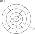
- the planar polar plot of the tissue may be obtained by projecting the tissue onto a plane based on the imaging data of the tissue and a segment model of the tissue, for example the AHA-17 segment model of the left ventricle of the heart.
- the AHA-17 segment model has been established by the American Heart Association (AHA) and defines 17 segments of the left ventricle of the human heart.
- AHA-17 plot obtained by projecting the tissue of the left ventricle onto a plane based on the imaging data of the tissue and the AHA-17 segment model will be referred to as AHA-17 plot.
- Coronary arteries may be projected onto a reference plane using a parameterization of the left ventricle based on a cylindrical coordinate system, using the cardiac long axis as the cylindrical axis.
- the reference plane is perpendicular to the cylindrical axis and in-plane with the planar polar plot.
- the tissue is heart tissue, particularly tissue of the left ventricle of the heart, and/or the vessel is a coronary artery, particularly a coronary artery supplying the left ventricle of the heart.
- the heart tissue may be tissue of the left ventricle of the heart, tissue of the right ventricle of the heart, tissue of the left chamber of the heart, tissue of the right chamber of the heart or a combination thereof.
- the method may be applied to anatomical structures other than heart, for example to brain, liver or lung.
- the invention relates to a computer system for generating a combined tissue-vessel representation, comprising:
- the computer system is configured to implement the method according to one or more of the disclosed aspects.
- the invention relates to a computer program product comprising program elements which induce a computer system to carry out the steps of the method according to one or more of the disclosed aspects, when the program elements are loaded into a memory of the computer system.
- the invention relates to a computer-readable medium on which program elements are stored that can be read and executed by a computer system, in order to perform the steps of the method according to one or more of the disclosed aspects, when the program elements are executed by the computer system.
- the combined tissue-vessel representation facilitates a combined visualization which integrates quantitative information about the coronary arteries with the quantitative information about the heart.
- the proposed solution provides comprehensive visualization of quantitative information regarding coronary arteries, particularly about stenosis within coronary arteries, together with quantitative information regarding the physiology of the left ventricle.
- Such an integrated view can help the radiologist to establish correspondence between the vessel, particularly a coronary artery, and the tissue, particularly a portion of the tissue supplied by that coronary artery. This can help to better correlate the location of infarcted or ischemic areas in heart tissue to the location in the coronaries that have caused the respective infarction or ischemia, thus contributing to an easier and less error-prone diagnostic process.
- any of the algorithms mentioned herein may be based on one or more of the following architectures: convolutional neural networks, deep belief networks, deep residual learning, deep reinforcement learning, recurrent neural networks, Siamese networks, generative adversarial networks or auto-encoders.
- the trained machine learning algorithm for determining the fat distribution information may be embodied as a deep learning algorithm and/or as a convolutional neural network.
- any of the computer system components mentioned herein or any interface between the computer system components may be embodied in form of hardware and/or software.
- an interface may be embodied in form of at least one of a PCI-Bus, a USB or a Firewire.
- a computer system component may comprise hardware elements and/or software elements, for example a microprocessor, a field programmable gate array (an acronym is "FPGA”) or an application specific integrated circuit (an acronym is "ASIC").
- the computer system may, for example, comprise and/or be a part of at least one of a cloud-computing system, a distributed computing system, a computer network, a computer, a tablet computer, a smartphone or the like.
- the computer system may comprise hardware and/or software.
- the hardware may be, for example, a processor system, a memory system and combinations thereof.
- the hardware may be configurable by the software and/or be operable by the software. Calculations for performing steps of a method and/or for training an algorithm may be carried out in a processor.
- Data particularly the imaging data of the tissue and/or the imaging data of the vessel, may be received, for example, by receiving a signal that carries the data and/or by reading the data from a computer-readable medium.
- Data in particular, the combined tissue-vessel representation, may be provided, for example, by transmitting a signal that carries the data and/or by writing the data into a computer-readable medium and/or by displaying the data, for example in form of an image, on a display.
- the computer program product may be, for example, a computer program or comprise another element apart from the computer program.
- This other element may be hardware, for example a memory device, on which the computer program is stored, a hardware key for using the computer program and the like, and/or software, for example, documentation or a software key for using the computer program.
- a computer-readable medium may be embodied as non-permanent main memory (e.g. random access memory) or as permanent mass storage (e.g. hard disk, USB stick, SD card, solid state disk).
- a computer-readable medium on which program elements are stored that can be read and executed by an imaging data processing unit in order to perform the steps of the method according to one or more of the disclosed aspects, when the program elements are executed by the imaging data processing unit.
- Fig. 1 shows an arrangement A17 of segments according to the AHA-17 segment model of the left ventricle. The numbers shown within the segments are used for identifying the segments.
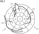
- Fig. 2 shows a representation of coronary arteries overlaid over the arrangement A17 of segments according to the AHA-17 segment model.
- the representation of coronary arteries comprises a representation LAD of the left anterior descending artery, a representation RCA of the right coronary artery, and a representation LCX of the left circumflex artery.
- the vessel representation V comprises a representation of a wall of the vessel.
- the segments 1, 2, 7, 8, 13, 14 and 17 may be assigned to the left anterior descending artery.
- the segments 3, 4, 9, 10 and 15 may be assigned to the right coronary artery.
- the segments 5, 6, 11, 12 and 16 may be assigned to the left circumflex artery.
- Fig. 8 shows a diagram illustrating a computer-implemented method for generating a combined tissue-vessel representation C, comprising:
Landscapes
- Engineering & Computer Science (AREA)
- Health & Medical Sciences (AREA)
- Life Sciences & Earth Sciences (AREA)
- Physics & Mathematics (AREA)
- Medical Informatics (AREA)
- Theoretical Computer Science (AREA)
- General Physics & Mathematics (AREA)
- General Health & Medical Sciences (AREA)
- Radiology & Medical Imaging (AREA)
- Nuclear Medicine, Radiotherapy & Molecular Imaging (AREA)
- Veterinary Medicine (AREA)
- Public Health (AREA)
- Biophysics (AREA)
- Optics & Photonics (AREA)
- Pathology (AREA)
- High Energy & Nuclear Physics (AREA)
- Biomedical Technology (AREA)
- Heart & Thoracic Surgery (AREA)
- Molecular Biology (AREA)
- Surgery (AREA)
- Animal Behavior & Ethology (AREA)
- Computer Vision & Pattern Recognition (AREA)
- Dentistry (AREA)
- Oral & Maxillofacial Surgery (AREA)
- Quality & Reliability (AREA)
- Cardiology (AREA)
- Vascular Medicine (AREA)
- Pulmonology (AREA)
- Computer Graphics (AREA)
- Geometry (AREA)
- Software Systems (AREA)
- Apparatus For Radiation Diagnosis (AREA)
- Magnetic Resonance Imaging Apparatus (AREA)
Claims (13)
- Computerimplementiertes Verfahren zur Erzeugung einer kombinierten Gewebe-Gefäß-Darstellung (C), umfassend:- Empfangen (RT) von Bildgebungsdaten eines Gewebes,- Empfangen (RV) von Bildgebungsdaten eines Gefäßes,- Erzeugen (GT) einer Gewebedarstellung (T) basierend auf den Bildgebungsdaten des Gewebes, wobei die Gewebedarstellung (T) einen planaren Polarplot des Gewebes umfasst,- Erzeugen (GV) einer Gefäßdarstellung (V) basierend auf den Bildgebungsdaten des Gefäßes, wobei die Gefäßdarstellung (V) eine Projektion des Gefäßes auf eine Referenzebene umfasst, wobei die Referenzebene in einer Ebene mit dem planaren Polarplot des Gewebes liegt,- Erzeugen (GC) einer kombinierten Gewebe-Gefäß-Darstellung (C) basierend auf der Gefäßdarstellung (V) und der Gewebedarstellung (T), wobei die Gefäßdarstellung (V) der Gewebedarstellung (T) überlagert ist,- dadurch gekennzeichnet, dass die Gefäßdarstellung (V) ein lokales Merkmal des Gefäßes umfasst, wobei das lokale Merkmal des Gefäßes farbcodierte quantitative Informationen ist, die eine Plaque-Zusammensetzung angeben, und dass die Gewebedarstellung (T) für jede Position einer Vielzahl von Positionen über dem Gewebe farbcodierte quantitative Informationen umfasst, die einen Wert eines geometrischen Parameters des Gewebes an dieser Position angeben, wobei der geometrische Parameter des Gewebes eine Ventrikelwandverdickung ist.
- Verfahren nach Anspruch 1,- wobei die Gefäßdarstellung (V) eine Mittellinie des Gefäßes umfasst.
- Verfahren nach Anspruch 1 oder 2,- wobei die Gefäßdarstellung (V) eine Darstellung einer Innenwand des Gefäßes und/oder eine Darstellung einer Außenwand des Gefäßes umfasst.
- Verfahren nach einem der Ansprüche 1 bis 3,- wobei die Gefäßdarstellung (V) für jede Position einer Vielzahl von Positionen entlang des Gefäßes farbcodierte quantitative Informationen umfasst, die einen Wert eines Parameters des Gefäßes an dieser Position angeben.
- Verfahren nach Anspruch 4,- wobei der Parameter des Gefäßes ein geometrischer Parameter ist, insbesondere ein Lumendurchmesser oder eine Wanddicke.
- Verfahren nach Anspruch 4,- wobei der Parameter des Gefäßes ein Röntgendämpfungsparameter, insbesondere ein HU-Wert oder ein HU-Gradient, ist.
- Verfahren nach Anspruch 4,- wobei der Parameter des Gefäßes ein hämodynamischer Parameter ist, insbesondere fraktionelle Flussreserve, Blutgeschwindigkeit, Blutdruck, Wandbelastung, Wandspannung oder -verformung.
- Verfahren nach Anspruch 4,- wobei der Parameter des Gefäßes ein Plaque-Parameter ist.
- Verfahren nach Anspruch 8,- wobei der Plaque-Parameter ein Anteil einer oder mehrerer Plaque-Komponenten am Gesamtplaque-Volumen ist.
- Verfahren nach einem der Ansprüche 1 bis 9,- wobei das Gewebe Herzgewebe ist, insbesondere Gewebe des linken Ventrikels des Herzens, und- wobei das Gefäß eine Koronararterie ist, insbesondere eine Koronararterie, die den linken Ventrikel des Herzens versorgt.
- Computersystem (S) zur Erzeugung einer kombinierten Gewebe-Gefäß-Darstellung (C), umfassend:- einen Gewebebildgebungsdatenempfänger (RT-U) zum Empfangen (RT) von Bildgebungsdaten eines Gewebes,- einen Gefäßbildgebungsdatenempfänger (RV-U) zum Empfangen (RV) von Bildgebungsdaten eines Gefäßes,- einen Gewebedarstellungsgenerator (GT-U) zum Erzeugen (GT) einer Gewebedarstellung (T) basierend auf den Bildgebungsdaten des Gewebes, wobei die Gewebedarstellung (T) einen planaren Polarplot des Gewebes umfasst,- einen Gefäßdarstellungsgenerator (GV-U) zum Erzeugen (GV) einer Gefäßdarstellung (V) basierend auf den Bildgebungsdaten des Gefäßes, wobei die Gefäßdarstellung (V) eine Projektion des Gefäßes auf eine Referenzebene umfasst, wobei die Referenzebene in einer Ebene mit dem planaren Polarplot des Gewebes liegt,- einen Gewebe-Gefäß-Darstellungsgenerator (GC-U) zum Erzeugen (GC) einer kombinierten Gewebe-Gefäß-Darstellung (C) basierend auf der Gefäßdarstellung (V) und der Gewebedarstellung (T), wobei die Gefäßdarstellung (V) der Gewebedarstellung (T) überlagert ist,- dadurch gekennzeichnet, dass die Gefäßdarstellung (V) ein lokales Merkmal des Gefäßes umfasst, wobei das lokale Merkmal des Gefäßes farbcodierte quantitative Informationen ist, die eine Plaque-Zusammensetzung angeben, und dass die Gewebedarstellung (T) für jede Position einer Vielzahl von Positionen über dem Gewebe farbcodierte quantitative Informationen umfasst, die einen Wert eines geometrischen Parameters des Gewebes an dieser Position angeben, wobei der geometrische Parameter des Gewebes eine Ventrikelwandverdickung ist.
- Computerprogrammprodukt, umfassend Programmelemente, die induzieren, dass ein Computersystem (S) die Schritte des Verfahrens gemäß einem der Ansprüche 1 bis 10 ausführt, wenn die Programmelemente in einen Speicher des Computersystems (S) geladen werden.
- Computerlesbares Medium, auf dem Programmelemente gespeichert sind, die von einem Computersystem (S) gelesen und ausgeführt werden können, um die Schritte des Verfahrens gemäß einem der Ansprüche 1 bis 10 durchzuführen, wenn die Programmelemente von dem Computersystem (S) ausgeführt werden.
Priority Applications (3)
| Application Number | Priority Date | Filing Date | Title |
|---|---|---|---|
| EP18214023.6A EP3671649B1 (de) | 2018-12-19 | 2018-12-19 | Verfahren und computersystem zur erzeugung einer kombinierten gewebe-gefäss-darstellung |
| US16/710,151 US11810290B2 (en) | 2018-12-19 | 2019-12-11 | Method and computer system for generating a combined tissue-vessel representation |
| CN201911310670.2A CN111340934A (zh) | 2018-12-19 | 2019-12-18 | 用于生成组合式组织-血管表示的方法和计算机系统 |
Applications Claiming Priority (1)
| Application Number | Priority Date | Filing Date | Title |
|---|---|---|---|
| EP18214023.6A EP3671649B1 (de) | 2018-12-19 | 2018-12-19 | Verfahren und computersystem zur erzeugung einer kombinierten gewebe-gefäss-darstellung |
Publications (3)
| Publication Number | Publication Date |
|---|---|
| EP3671649A1 EP3671649A1 (de) | 2020-06-24 |
| EP3671649B1 true EP3671649B1 (de) | 2025-07-02 |
| EP3671649C0 EP3671649C0 (de) | 2025-07-02 |
Family
ID=65009542
Family Applications (1)
| Application Number | Title | Priority Date | Filing Date |
|---|---|---|---|
| EP18214023.6A Active EP3671649B1 (de) | 2018-12-19 | 2018-12-19 | Verfahren und computersystem zur erzeugung einer kombinierten gewebe-gefäss-darstellung |
Country Status (3)
| Country | Link |
|---|---|
| US (1) | US11810290B2 (de) |
| EP (1) | EP3671649B1 (de) |
| CN (1) | CN111340934A (de) |
Families Citing this family (9)
| Publication number | Priority date | Publication date | Assignee | Title |
|---|---|---|---|---|
| US10210956B2 (en) | 2012-10-24 | 2019-02-19 | Cathworks Ltd. | Diagnostically useful results in real time |
| EP3457930B1 (de) | 2016-05-16 | 2023-11-15 | Cathworks Ltd. | System zur beurteilung von blutgefässen |
| WO2020201942A1 (en) | 2019-04-01 | 2020-10-08 | Cathworks Ltd. | Methods and apparatus for angiographic image selection |
| US12039685B2 (en) | 2019-09-23 | 2024-07-16 | Cathworks Ltd. | Methods, apparatus, and system for synchronization between a three-dimensional vascular model and an imaging device |
| US12315076B1 (en) | 2021-09-22 | 2025-05-27 | Cathworks Ltd. | Four-dimensional motion analysis of a patient's coronary arteries and myocardial wall |
| EP4170579A1 (de) * | 2021-10-22 | 2023-04-26 | Koninklijke Philips N.V. | Bildverarbeitungsvorrichtung, system zur medizinischen bildgebung und computerprogrammelement |
| WO2023152688A1 (en) | 2022-02-10 | 2023-08-17 | Cathworks Ltd. | System and method for machine-learning based sensor analysis and vascular tree segmentation |
| US12440180B2 (en) | 2022-03-10 | 2025-10-14 | Cleerly, Inc. | Systems, devices, and methods for non-invasive image-based plaque analysis and risk determination |
| US12446965B2 (en) | 2023-08-09 | 2025-10-21 | Cathworks Ltd. | Enhanced user interface and crosstalk analysis for vascular index measurement |
Citations (1)
| Publication number | Priority date | Publication date | Assignee | Title |
|---|---|---|---|---|
| EP2538361A2 (de) * | 2010-08-12 | 2012-12-26 | Heartflow, Inc. | Verfahren und System zur patientenspezifischen Modellierung der Durchblutung |
Family Cites Families (24)
| Publication number | Priority date | Publication date | Assignee | Title |
|---|---|---|---|---|
| US6471655B1 (en) * | 1999-06-29 | 2002-10-29 | Vitalwave Corporation | Method and apparatus for the noninvasive determination of arterial blood pressure |
| JP4346297B2 (ja) | 2002-10-22 | 2009-10-21 | 株式会社東芝 | X線コンピュータ断層撮影装置、画像処理装置及び画像処理方法 |
| US7657299B2 (en) | 2003-08-21 | 2010-02-02 | Ischem Corporation | Automated methods and systems for vascular plaque detection and analysis |
| US7649981B2 (en) * | 2003-10-15 | 2010-01-19 | Varian Medical Systems, Inc. | Multi-energy x-ray source |
| US7957570B2 (en) * | 2007-05-03 | 2011-06-07 | General Electric Company | System and method to generate an illustration of a cardiac region of interest |
| DE602008005346D1 (de) | 2007-09-03 | 2011-04-14 | Koninkl Philips Electronics Nv | Visualisierung von voxel-daten |
| WO2010020933A2 (en) * | 2008-08-20 | 2010-02-25 | Koninklijke Philips Electronics N.V. | Processing cardiac data for personalized aha diagram |
| WO2010061335A1 (en) | 2008-11-28 | 2010-06-03 | Koninklijke Philips Electronics N.V. | Processing myocardial perfusion data |
| DE102009032442B4 (de) | 2009-07-09 | 2017-02-16 | Siemens Healthcare Gmbh | Verfahren und Vorrichtung zur Nachbearbeitung von mittels eines bildgebenden Systems erfassten Messwerten |
| WO2011069120A1 (en) | 2009-12-03 | 2011-06-09 | Cedars-Sinai Medical Center | Method and system for plaque characterization |
| US8854355B2 (en) * | 2009-12-11 | 2014-10-07 | General Electric Company | System and method of visualizing features in an image |
| JP4981938B2 (ja) * | 2010-03-08 | 2012-07-25 | 富士フイルム株式会社 | 診断支援装置、冠動脈解析プログラムおよび冠動脈解析方法 |
| WO2014084398A1 (ja) * | 2012-11-30 | 2014-06-05 | 株式会社 東芝 | 医用画像診断装置 |
| US9788807B2 (en) * | 2013-09-06 | 2017-10-17 | Koninklijke Philips N.V. | Processing apparatus for processing cardiac data |
| CN105096388B (zh) | 2014-04-23 | 2019-02-05 | 北京冠生云医疗技术有限公司 | 基于计算流体力学的冠状动脉血流仿真系统和方法 |
| US10478130B2 (en) | 2015-02-13 | 2019-11-19 | Siemens Healthcare Gmbh | Plaque vulnerability assessment in medical imaging |
| US9996921B2 (en) | 2015-05-17 | 2018-06-12 | LIGHTLAB IMAGING, lNC. | Detection of metal stent struts |
| US10176408B2 (en) | 2015-08-14 | 2019-01-08 | Elucid Bioimaging Inc. | Systems and methods for analyzing pathologies utilizing quantitative imaging |
| CN108475428B (zh) | 2015-12-22 | 2022-04-29 | 皇家飞利浦有限公司 | 心脏模型引导的冠状动脉分割的系统及方法 |
| DE102017201543A1 (de) | 2017-01-31 | 2018-08-02 | Siemens Healthcare Gmbh | Quantifizierung von Blutverlust auf Basis einer Computertomographie mit einem direkt konvertierenden Detektor |
| JP7134704B2 (ja) * | 2017-06-01 | 2022-09-12 | キヤノン株式会社 | 画像処理装置、画像処理方法、及びプログラム |
| US10565709B2 (en) | 2017-06-13 | 2020-02-18 | Canon Medical Systems Coporation | Image processing apparatus and image processing method |
| US10643319B2 (en) * | 2018-01-30 | 2020-05-05 | Canon Medical Systems Corporation | Apparatus and method for context-oriented blending of reconstructed images |
| JP7467026B2 (ja) * | 2018-10-10 | 2024-04-15 | キヤノンメディカルシステムズ株式会社 | 医用情報処理装置、医用情報処理プログラム、医用情報処理システム |
-
2018
- 2018-12-19 EP EP18214023.6A patent/EP3671649B1/de active Active
-
2019
- 2019-12-11 US US16/710,151 patent/US11810290B2/en active Active
- 2019-12-18 CN CN201911310670.2A patent/CN111340934A/zh active Pending
Patent Citations (1)
| Publication number | Priority date | Publication date | Assignee | Title |
|---|---|---|---|---|
| EP2538361A2 (de) * | 2010-08-12 | 2012-12-26 | Heartflow, Inc. | Verfahren und System zur patientenspezifischen Modellierung der Durchblutung |
Also Published As
| Publication number | Publication date |
|---|---|
| EP3671649A1 (de) | 2020-06-24 |
| US11810290B2 (en) | 2023-11-07 |
| US20200202522A1 (en) | 2020-06-25 |
| CN111340934A (zh) | 2020-06-26 |
| EP3671649C0 (de) | 2025-07-02 |
Similar Documents
| Publication | Publication Date | Title |
|---|---|---|
| EP3671649B1 (de) | Verfahren und computersystem zur erzeugung einer kombinierten gewebe-gefäss-darstellung | |
| US9323887B2 (en) | Device and computed tomography scanner for determining and visualizing the perfusion of the myocardial muscle | |
| JP4884528B2 (ja) | 腔部の画像を評定する方法、装置ならびにコンピュータプログラム製品 | |
| JP5629272B2 (ja) | 生物学的構造における時変パラメータの視覚化 | |
| US11364002B2 (en) | Medical-image processing apparatus and medical-image diagnostic apparatus | |
| Liu et al. | Extraction of coronary atherosclerotic plaques from computed tomography imaging: a review of recent methods | |
| US8755575B2 (en) | Transmural perfusion gradient image analysis | |
| EP3116382A2 (de) | Verfahren zur bestimmung von strömungsmengen und druckgradienten in arteriellen netzwerken von patientenspezifischen auf computertomografie-angiogramm basierenden kontrastverteilungsinformationen | |
| WO2020179234A1 (ja) | 画像診断支援装置および画像処理方法 | |
| Oeltze et al. | Integrated visualization of morphologic and perfusion data for the analysis of coronary artery disease. | |
| US8542894B2 (en) | Method and device for post-processing measured values registered by way of an imaging system | |
| US9905001B2 (en) | Image processing apparatus and image processing method | |
| Nieman et al. | Advanced cardiac imaging | |
| Koszegi et al. | Holistic polar map for integrated evaluation of cardiac imaging results | |
| CN116369961A (zh) | 提供脉管壁相关数据 | |
| van Driest et al. | Correlation between quantification of myocardial area at risk and ischemic burden at cardiac computed tomography | |
| Beliveau et al. | Computation of coronary perfusion territories from CT angiography | |
| Fukushima et al. | Software-based analysis for computed tomography coronary angiography: current status and future aspects | |
| Silva et al. | Left-Ventricle Global and Regional Functional Analysis from MDCT Images | |
| JP6411072B2 (ja) | 医用画像処理装置、医用画像処理方法およびプログラム | |
| Silva et al. | Exploring different parameters to assess left ventricle global and regional functional analysis from coronary ct angiography | |
| Flohr et al. | Cardiothoracic Image Postprocessing 3 | |
| Flohr et al. | Cardiothoracic Image Postprocessing | |
| Khélifa et al. | Assessment of local and global cardiac function based on AHA standardization | |
| HK1233469B (en) | A method for estimating flow rates and pressure gradients in arterial networks from patient specific computed tomography angiogram-based contrast distribution data |
Legal Events
| Date | Code | Title | Description |
|---|---|---|---|
| PUAI | Public reference made under article 153(3) epc to a published international application that has entered the european phase |
Free format text: ORIGINAL CODE: 0009012 |
|
| STAA | Information on the status of an ep patent application or granted ep patent |
Free format text: STATUS: THE APPLICATION HAS BEEN PUBLISHED |
|
| AK | Designated contracting states |
Kind code of ref document: A1 Designated state(s): AL AT BE BG CH CY CZ DE DK EE ES FI FR GB GR HR HU IE IS IT LI LT LU LV MC MK MT NL NO PL PT RO RS SE SI SK SM TR |
|
| AX | Request for extension of the european patent |
Extension state: BA ME |
|
| STAA | Information on the status of an ep patent application or granted ep patent |
Free format text: STATUS: REQUEST FOR EXAMINATION WAS MADE |
|
| 17P | Request for examination filed |
Effective date: 20201223 |
|
| RBV | Designated contracting states (corrected) |
Designated state(s): AL AT BE BG CH CY CZ DE DK EE ES FI FR GB GR HR HU IE IS IT LI LT LU LV MC MK MT NL NO PL PT RO RS SE SI SK SM TR |
|
| STAA | Information on the status of an ep patent application or granted ep patent |
Free format text: STATUS: EXAMINATION IS IN PROGRESS |
|
| 17Q | First examination report despatched |
Effective date: 20220715 |
|
| RAP1 | Party data changed (applicant data changed or rights of an application transferred) |
Owner name: SIEMENS HEALTHINEERS AG |
|
| GRAP | Despatch of communication of intention to grant a patent |
Free format text: ORIGINAL CODE: EPIDOSNIGR1 |
|
| STAA | Information on the status of an ep patent application or granted ep patent |
Free format text: STATUS: GRANT OF PATENT IS INTENDED |
|
| INTG | Intention to grant announced |
Effective date: 20250205 |
|
| GRAS | Grant fee paid |
Free format text: ORIGINAL CODE: EPIDOSNIGR3 |
|
| GRAA | (expected) grant |
Free format text: ORIGINAL CODE: 0009210 |
|
| STAA | Information on the status of an ep patent application or granted ep patent |
Free format text: STATUS: THE PATENT HAS BEEN GRANTED |
|
| AK | Designated contracting states |
Kind code of ref document: B1 Designated state(s): AL AT BE BG CH CY CZ DE DK EE ES FI FR GB GR HR HU IE IS IT LI LT LU LV MC MK MT NL NO PL PT RO RS SE SI SK SM TR |
|
| REG | Reference to a national code |
Ref country code: GB Ref legal event code: FG4D |
|
| REG | Reference to a national code |
Ref country code: CH Ref legal event code: EP |
|
| REG | Reference to a national code |
Ref country code: DE Ref legal event code: R096 Ref document number: 602018083137 Country of ref document: DE |
|
| REG | Reference to a national code |
Ref country code: IE Ref legal event code: FG4D |
|
| U01 | Request for unitary effect filed |
Effective date: 20250722 |
|
| U07 | Unitary effect registered |
Designated state(s): AT BE BG DE DK EE FI FR IT LT LU LV MT NL PT RO SE SI Effective date: 20250728 |