US8560968B1 - Method and apparatus for evaluating a heart patient - Google Patents
Method and apparatus for evaluating a heart patient Download PDFInfo
- Publication number
- US8560968B1 US8560968B1 US12/730,560 US73056010A US8560968B1 US 8560968 B1 US8560968 B1 US 8560968B1 US 73056010 A US73056010 A US 73056010A US 8560968 B1 US8560968 B1 US 8560968B1
- Authority
- US
- United States
- Prior art keywords
- report
- findings
- view
- input
- primary reporting
- Prior art date
- Legal status (The legal status is an assumption and is not a legal conclusion. Google has not performed a legal analysis and makes no representation as to the accuracy of the status listed.)
- Active, expires
Links
Images


Classifications
-
- G—PHYSICS
- G16—INFORMATION AND COMMUNICATION TECHNOLOGY [ICT] SPECIALLY ADAPTED FOR SPECIFIC APPLICATION FIELDS
- G16H—HEALTHCARE INFORMATICS, i.e. INFORMATION AND COMMUNICATION TECHNOLOGY [ICT] SPECIALLY ADAPTED FOR THE HANDLING OR PROCESSING OF MEDICAL OR HEALTHCARE DATA
- G16H15/00—ICT specially adapted for medical reports, e.g. generation or transmission thereof
-
- G—PHYSICS
- G06—COMPUTING; CALCULATING OR COUNTING
- G06F—ELECTRIC DIGITAL DATA PROCESSING
- G06F3/00—Input arrangements for transferring data to be processed into a form capable of being handled by the computer; Output arrangements for transferring data from processing unit to output unit, e.g. interface arrangements
- G06F3/01—Input arrangements or combined input and output arrangements for interaction between user and computer
- G06F3/048—Interaction techniques based on graphical user interfaces [GUI]
- G06F3/0481—Interaction techniques based on graphical user interfaces [GUI] based on specific properties of the displayed interaction object or a metaphor-based environment, e.g. interaction with desktop elements like windows or icons, or assisted by a cursor's changing behaviour or appearance
- G06F3/04817—Interaction techniques based on graphical user interfaces [GUI] based on specific properties of the displayed interaction object or a metaphor-based environment, e.g. interaction with desktop elements like windows or icons, or assisted by a cursor's changing behaviour or appearance using icons
-
- G—PHYSICS
- G06—COMPUTING; CALCULATING OR COUNTING
- G06F—ELECTRIC DIGITAL DATA PROCESSING
- G06F3/00—Input arrangements for transferring data to be processed into a form capable of being handled by the computer; Output arrangements for transferring data from processing unit to output unit, e.g. interface arrangements
- G06F3/01—Input arrangements or combined input and output arrangements for interaction between user and computer
- G06F3/048—Interaction techniques based on graphical user interfaces [GUI]
- G06F3/0481—Interaction techniques based on graphical user interfaces [GUI] based on specific properties of the displayed interaction object or a metaphor-based environment, e.g. interaction with desktop elements like windows or icons, or assisted by a cursor's changing behaviour or appearance
- G06F3/0482—Interaction with lists of selectable items, e.g. menus
-
- G—PHYSICS
- G16—INFORMATION AND COMMUNICATION TECHNOLOGY [ICT] SPECIALLY ADAPTED FOR SPECIFIC APPLICATION FIELDS
- G16H—HEALTHCARE INFORMATICS, i.e. INFORMATION AND COMMUNICATION TECHNOLOGY [ICT] SPECIALLY ADAPTED FOR THE HANDLING OR PROCESSING OF MEDICAL OR HEALTHCARE DATA
- G16H30/00—ICT specially adapted for the handling or processing of medical images
- G16H30/40—ICT specially adapted for the handling or processing of medical images for processing medical images, e.g. editing
-
- G—PHYSICS
- G16—INFORMATION AND COMMUNICATION TECHNOLOGY [ICT] SPECIALLY ADAPTED FOR SPECIFIC APPLICATION FIELDS
- G16H—HEALTHCARE INFORMATICS, i.e. INFORMATION AND COMMUNICATION TECHNOLOGY [ICT] SPECIALLY ADAPTED FOR THE HANDLING OR PROCESSING OF MEDICAL OR HEALTHCARE DATA
- G16H40/00—ICT specially adapted for the management or administration of healthcare resources or facilities; ICT specially adapted for the management or operation of medical equipment or devices
- G16H40/60—ICT specially adapted for the management or administration of healthcare resources or facilities; ICT specially adapted for the management or operation of medical equipment or devices for the operation of medical equipment or devices
- G16H40/63—ICT specially adapted for the management or administration of healthcare resources or facilities; ICT specially adapted for the management or operation of medical equipment or devices for the operation of medical equipment or devices for local operation
-
- G—PHYSICS
- G16—INFORMATION AND COMMUNICATION TECHNOLOGY [ICT] SPECIALLY ADAPTED FOR SPECIFIC APPLICATION FIELDS
- G16H—HEALTHCARE INFORMATICS, i.e. INFORMATION AND COMMUNICATION TECHNOLOGY [ICT] SPECIALLY ADAPTED FOR THE HANDLING OR PROCESSING OF MEDICAL OR HEALTHCARE DATA
- G16H50/00—ICT specially adapted for medical diagnosis, medical simulation or medical data mining; ICT specially adapted for detecting, monitoring or modelling epidemics or pandemics
- G16H50/70—ICT specially adapted for medical diagnosis, medical simulation or medical data mining; ICT specially adapted for detecting, monitoring or modelling epidemics or pandemics for mining of medical data, e.g. analysing previous cases of other patients
-
- G—PHYSICS
- G16—INFORMATION AND COMMUNICATION TECHNOLOGY [ICT] SPECIALLY ADAPTED FOR SPECIFIC APPLICATION FIELDS
- G16H—HEALTHCARE INFORMATICS, i.e. INFORMATION AND COMMUNICATION TECHNOLOGY [ICT] SPECIALLY ADAPTED FOR THE HANDLING OR PROCESSING OF MEDICAL OR HEALTHCARE DATA
- G16H70/00—ICT specially adapted for the handling or processing of medical references
- G16H70/20—ICT specially adapted for the handling or processing of medical references relating to practices or guidelines
Definitions
- the present invention relates to cardiology and more particularly to a method of evaluating a heart patient wherein a computer has a database and a user interface connected thereto that includes a display screen.
- the present invention relates to a method of evaluating a heart patient that employs a computer and a display screen, the display screen having multiple images that are displayed to a user, each image being an anatomical representation of a portion of a human heart, wherein multiple of the images depict different portions of the human heart and the display screen simultaneously displays data next to one or more of the images, the data being specific to the patient in the display data relating to at least one of the portions of the human heart that is represented by the display.
- the current state of the practice of cardiology includes: (1) Era of increasing demand; (2) Time constraints; (3) More complex procedures; (4) Increased imaging requirements; (5) Decrease allotted time per patient visit; and (6) Decreased reimbursement. Cardiologists can average of 8-10 minutes per patient.
- cardiologists have been increasingly burdened by the need to quickly and accurately generate cardiology reports after reviewing the results of heart studies.
- cardiologists must fill out forms for submission to insurance companies and provide information to regulatory agencies. To gather and produce all of this information, cardiologists must spend a significant portion of their work day dictating the needed information. Additionally, cardiologists must maintain a staff to transcribe the information into reports and to fill out required forms.
- a cardiologists typically dictates a report reviewing the results of a heart study, which, subsequently, must be typed by a transcriber. This process is time consuming and repetitive. Using traditional manual methods of record keeping, patient data is not readily available for fast and easy review. A patient's medical record cannot be easily combined with other studies and/or reports, and/or other patient data for analysis and reporting.
- One embodiment provides an integrated software solution for cardiac procedures encompassing reporting, storing, data mining, retrieving and online analysis.
- the method of the present invention enables capture of predefined data elements and cardiac testing modalities.
- the method and apparatus automates the report generation process by providing a graphical user interface incorporating a relational database with user selectable reporting components in a graphical environment.
- a method of clinical evaluation of a cardiology patient includes the providing of a computer having a database.
- a user interface is provided which is connected to the computer and which includes a display screen.
- one or more images are displayed on the display screen which are anatomical representations, each of a different portion of a human heart.
- the display can include patient data that is displayed on the display screen.
- patient data relates specifically to the anatomical representation of the portion of the human heart that is being viewed on the display screen.
- multiple images can be displayed as optional parts of the human heart. In one embodiment one or more of those optional portions can be selectively enlarged for more easy inspection by a physician or other operator.
- the method includes entering data that relates specifically to the displayed image.
- the method can include an interpreting cardiologist reviewing the data as part of a medical evaluation of a patient.
- the multiple images can include images of human heart chambers.
- the data displayed includes test results.
- multiple images are displayed on the display screen including multiple small images simultaneously displayed, each small image depicting a different part of the human heart.
- multiple images are displayed on the display screen, and wherein some of the images displayed are smaller images and wherein one or more of the displayed images is a larger image.
- a physician or operator can selectively enlarge one image or another image and by switching (e.g. clicking with computer mouse, touch screen) from one small image to another.
- the enlarged image selection generates data on the display screen that corresponds to the selected and enlarged image.
- a physician or operator can change from one image displayed to another image displayed without typing on a keyboard.
- the method and apparatus provides a graphical display where a user has access to a first plurality of reporting segments.
- the plurality of reporting segments are a plurality of geographical portions of a human organ. In one embodiment the segments can be portions of a human heart.
- the multiple anatomical images/views of the human heart can be displayed including: parasternal long axis view, short axis of left ventricle view, short axis view at the level of the aortic valve, apical two chamber view, and four chamber view.
- the average time from review of the testing results to generation of a report by the cardiologist is less than 15, 14, 13, 12, 11, 10, 9, 8, 7, 6, 5, 4, and 3 minutes. In various embodiments the average time is between about any to the above specified time limits. In one embodiment the cardiologist generates more than 10, 15, 20, 25, 30, 35, 40, 50, 60, 70, 80, 90, and 100 cardiology reports a day. In various embodiments the number of reports generated varies between about any to the above specified daily number of reports.
- the report generation method and apparatus incorporates previously defined reporting text strings or components, and includes a graphical user interface for selecting phrases to be inserted in the template.
- the system will further comprise a graphics engine for display of graphical expressions of selection analysis.
- a heart study can be conducted which will contain many basic elements common to all patients, but examination data will vary for each patient. Examination data can contain any number of variable responses, and each variable within the input can offer any number of different options from which to choose. In addition, cardiologists can personalize the report to suit a particular situation.
- the examination and/or report input is electronically stored for possible future use in data mining, reporting, and/or analysis.
- One embodiment enables a report on a cardiology study to be generated from a single graphical input screen.
- a graphical input display having images of various views of the human heart for data entry.
- report text is generated by making selections on a graphical input screen.
- a set of a plurality of suggested cardiology report data string conclusions is indexed to possible selections made on the graphical input screen.
- a plurality of the suggested conclusions for the cardiology report based on input by cardiologist automatically show up based on the graphical selections made by the user of the method and apparatus.
- One embodiment provides an interface with existing cardiology testing devices.
- One embodiment enables an interface with billing and hospital admit systems. Examples of reports and studies can include (as examples): (1) Transthoracic echocardiography; (2) Transesophagial echocardiography; (3) Exercise treadmill test; (4) Stress echocardiogram; (5) Chemical stress echocardiogram; (6) Exercise myocardial perfusion imaging; (7) Chemical myocardial perfusion imaging; (8) Holier monitoring; (9) Coronary angiography; (10) Peripheral angiography; (11) Coronary computerized tomographic angiography, and (12) Peripheral computerized tomographic angiography.
- FIG. 1 is a screen shot showing a input display for generating a report on an echocardiogram, the display being used for reporting on one of the Primary Reporting Structures of an echocardiogram test (in this case the left Atrium), the display generally having a plurality of input sections for features or properties related to such Primary Reporting Structure.
- FIG. 2 is a screen shot showing the option of viewing pop up graphical comparison data for prior reports on the current report data.
- the pop up graph can be accessed by selecting the icon located next to the numerical data to be compared.
- Also in this figure is the current version of the text string for the findings on this primary reporting attribute which current version changes based on the input submitted in the display.
- FIG. 3 is a screen shot showing the input screen for the left atrium primary reporting structure where the “Thrombus” property or attribute has been selected for inputting report information.
- FIG. 4 is a screen shot showing the input screen for the left atrium primary reporting structure where the “Catheter” has been selected for inputting report information.
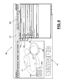
- FIG. 5 is a screen shot showing the input screen for the left atrium primary reporting structure where the “Spontaneous Echo Contrast” has been selected for inputting report information.
- FIG. 6 is a screen shot showing the input screen for the left atrium primary reporting structure where the “Miscellaneous” has been selected for inputting report information.
- FIG. 7 is a screen shot showing the input screen for the left atrium primary reporting structure where the “Remarks” has been selected for inputting report information.
- Remarks section allow the user to input custom text in the findings section of the report for the specific Primary Reporting Property being reporting on.
- FIG. 8 is a screen shot showing a “Conclusion” input screen where conclusion text strings 1 and 2 are shown.
- FIG. 9 is a screen shot showing the findings input screen for the left atrium primary reporting structure where the smaller image of the short axis left ventricle view has been selected thereby showing an enlarged image/view of this smaller image/view and also showing the various input areas for properties or attributes of this primary reporting structure.
- FIG. 10 is a screen shot showing the findings input screen for the left atrium primary reporting structure where the smaller image of the short axis left ventricle has been selected thereby showing an enlarged image/view of this smaller image/view and also showing the various input areas for properties or attributes of this primary reporting structure.
- FIG. 11 is a screen shot showing the findings input screen for the left atrium primary reporting structure where the smaller image of the subcostal view has been selected thereby showing an enlarged image/view of this smaller image/view and also showing the various input areas for properties or attributes of this primary reporting structure.
- FIG. 12 is a screen shot showing the findings input screen for the left atrium primary reporting structure where the smaller image of the two chamber view has been selected thereby displaying an enlarged image/view of this smaller image/view, and also showing the various input areas for properties or attributes of this primary reporting structure.
- FIG. 13 is a screen shot showing the findings input screen for the left atrium primary reporting structure where the smaller image of the short axis basil has been selected thereby showing an enlarged image/view of this smaller image/view and also showing the various input areas for properties or attributes of this primary reporting structure.
- FIG. 14 is a screen shot showing the findings input screen for the right atrium primary reporting structure where the smaller image of the short axis apical has been selected thereby showing an enlarged image/view of this smaller image/view and also showing the various input areas for properties or attributes of this primary reporting structure.
- FIG. 15 is a screen shot showing the findings input screen for the left ventricle primary reporting structure where the smaller image of the Parasternal long axis view has been selected thereby showing an enlarged image/view of this smaller image/view (with the left ventricle highlighted) and also showing the various input areas for properties or attributes of this primary reporting structure with no property or attribute actually being selected (and also showing the current text string for the findings on this primary reporting structure).
- FIG. 16 is a screen shot showing the Wall Motion Analysis property or attribute selected along with the Short A Basal selected/highlighted and a small image/view of the short axis left ventricle displayed with this small image/view being broken into a plurality of reporting segments for entering input for a wall motion calculation.
- FIG. 17 is a screen shot showing the Wall Motion Analysis property or attribute selected along with the Long Axis selected/highlighted and a small image/view of the Parasternal long axis image/view displayed with this small image/view being broken into a plurality of reporting segments for entering input for a wall motion calculation.
- FIG. 18 is a screen shot showing the Wall Motion Analysis property or attribute selected along with the Short Axis Mid selected/highlighted and a small image/view of the short axis left ventricle image/view displayed with this small image/view being broken into a plurality of reporting segments for entering input for a wall motion calculation.
- FIG. 19 is a screen shot showing the Wall Motion Analysis property or attribute selected along with the 4 Chamber view selected/highlighted and a small image/view of the subcostal image/view displayed with this small image/view being broken into a plurality of reporting segments for entering input for a wall motion calculation.
- FIG. 20 is a screen shot showing the Wall Motion Analysis property or attribute selected along with the Short A Apical view selected/highlighted and a small image/view of the short axis left ventricle image/view displayed with this small image/view being broken into a plurality of reporting segments for entering input for a wall motion calculation.
- FIG. 21 is a screen shot showing the Wall Motion Analysis property or attribute selected along with the 2 Chamber view selected/highlighted and a small image/view of the two chamber image/view displayed with this small image/view being broken into a plurality of reporting segments for entering input for a wall motion calculation.
- FIG. 22 is a screen shot showing the findings input screen for the right ventricle primary reporting structure where the smaller image of the Parasternal long axis view has been selected thereby showing an enlarged image/view of this smaller image/view (with the right ventricle highlighted) and also showing the various input areas for properties or attributes of this primary reporting structure with no property or attribute actually being selected (and also showing the current text string for the findings on this primary reporting structure).
- FIG. 23 is a screen shot showing the findings input screen for the atrial septum primary reporting structure where the smaller image of the short axis aorta valve view has been selected thereby showing an enlarged image/view of this smaller image/view (with the atrial septum highlighted) and also showing the various input areas for properties or attributes of this primary reporting structure with no property or attribute actually being selected (and also showing the current text string for the findings on this primary reporting structure).
- FIG. 24 is a screen shot showing the findings input screen for the ventricular septum primary reporting structure where the smaller image of the subcostal view has been selected thereby showing an enlarged image/view of this smaller image/view (with the ventricular septum highlighted) and also showing the various input areas for properties or attributes of this primary reporting structure with no property or attribute actually being selected (and also showing the current text string for the findings on this primary reporting structure).
- FIG. 25 is a screen shot showing the findings input screen for the pulmonic valve primary reporting structure where the smaller image of the short axis aorta valve view has been selected thereby showing an enlarged image/view of this smaller image/view (with the pulmonic valve highlighted) and also showing the various input areas for properties or attributes of this primary reporting structure with no property or attribute actually being selected (and also showing the current text string for the findings on this primary reporting structure).
- FIG. 26 is a screen shot showing the findings input screen for the pulmonary artery primary reporting structure where the smaller image of the short axis aorta valve view has been selected thereby showing an enlarged image/view of this smaller image/view (with the pulmonary artery highlighted) and also showing the various input areas for properties or attributes of this primary reporting structure with no property or attribute actually being selected (and also showing the current text string for the findings in this primary reporting structure).
- FIG. 27 is a screen shot showing the findings input screen for the pulmonary vein primary reporting structure where the smaller image of the subcostal view has been selected thereby showing an enlarged image/view of this smaller image/view (with the pulmonary vein highlighted) and also showing the various input areas for properties or attributes of this primary reporting structure with no property or attribute actually being selected (and also showing the current text string for the findings on this primary reporting structure).
- FIG. 28 is a screen shot showing the findings input screen for the mitral valve primary reporting structure where the smaller image of the subcostal view has been selected thereby showing an enlarged image/view of this smaller image/view (with the mitral valve highlighted) and also showing the various input areas for properties or attributes of this primary reporting structure with no property or attribute actually being selected (and also showing the current text string for the findings on this primary reporting structure).
- FIG. 29 is a screen shot showing the findings input screen for the inferior vena cava primary reporting structure where the smaller image of the subcostal view has been selected thereby showing an enlarged image/view of this smaller image/view (with the inferior vena cava highlighted) and also showing the various input areas for properties or attributes of this primary reporting structure with no property or attribute actually being selected (and also showing the current text string for the findings on this primary reporting structure).
- FIG. 30 is a screen shot showing the findings input screen for the aorta primary reporting structure where the smaller image of the Parasternal long axis view has been selected thereby showing an enlarged image/view of this smaller image/view (with the aorta highlighted) and also showing the various input areas for properties or attributes of this primary reporting structure with no property or attribute actually being selected (and also showing the current text string for the findings on this primary reporting structure).
- FIGS. 31A and 31B are screen shots showing the findings input screen for the aortic valve primary reporting structure where the smaller image of the Parasternal long axis view has been selected thereby showing an enlarged image/view of this smaller image/view (with the aortic valve highlighted) and also showing the various input areas for properties or attributes of this primary reporting structure with no property or attribute actually being selected (and also showing the current text string for the findings on this primary reporting structure).
- FIG. 32 is a screen shot showing the findings input screen for the pericardium primary reporting structure where the smaller image of the short axis aorta valve view has been selected thereby showing an enlarged image/view of this smaller image/view (with the pericardium highlighted) and also showing the various input areas for properties or attributes of this primary reporting structure with no property or attribute actually being selected (and also showing the current text string for the findings on this primary reporting structure).
- FIG. 33 is a screen shot showing the findings input screen for the tricuspid valve primary reporting structure where the smaller image of the short axis aorta valve view has been selected thereby showing an enlarged image/view of this smaller image/view (with the tricuspid valve highlighted) and also showing the various input areas for properties or attributes of this primary reporting structure with no property or attribute actually being selected (and also showing the current text string for the findings on this primary reporting structure).
- FIG. 34 is a screen shot showing a “Preview of Report” input screen where findings are selected for the primary reporting structure of the Left Atrium.
- the report can have five sections: (a) Cardiologist/clinic; (b) Patient Demographic; c) Conclusions/final impressions along with Comparisons; (d) Findings on the Primary Reporting Structures; and (e) Measurement Tables.
- FIG. 35 is a screen shot showing a “Preview of Report” input screen where Conclusions is selected and showing conclusion text strings 1 and 2 .
- the report can have five sections: (a) Cardiologist/clinic; (b) Patient Demographic; c) Conclusions/final impressions along with Comparisons; (d) Findings on the Primary Reporting Structures; and (e) Measurement Tables.
- FIG. 36 is an overall schematic diagram of one embodiment of the method and apparatus.
- FIGS. 37-40 show one embodiment in which method and apparatus 10 can enforce appropriateness guidelines in scheduling a study.
- FIGS. 41-45 illustrate one embodiment of automatic scheduling and study notification.
- FIGS. 46-50 illustrate one embodiment for the angiogram reporting modality.
- One embodiment provides an integrated software solution for cardiac procedures encompassing reporting, storing, data mining, retrieving and online analysis.
- the method of the present invention enables capture of predefined data elements and cardiac testing modalities.
- a document generation system for enhancing or replacing the dictation and transcription process. More particularly, a computer-based documentation system is provided which processes document templates in conjunction with pre-defined character strings to generate cardiology reports.
- FIGS. 1-36 go through one example of generating a report on testing data for a cardiology test (in this case an echocardiogram).
- FIG. 1 is a screen shot showing a input display 400 for generating a report 3010 on an echocardiogram, the display 100 being used for reporting on one of the Primary Reporting Structures of an echocardiogram test (in this case the left Atrium), the display generally having a plurality of input sections for features or properties related to such Primary Reporting Structure, and having the “Size” 402 property or attribute selected revealing a second tier of sections and/or information: Chamber size, Dimension, Volume, and Volume Index.
- FIG. 2 is a screen shot showing the option of viewing pop up graphical comparison data 402 ′′ for prior reports on the current report data.
- the pop up graph can be accessed by selecting the icon 402 ′ located next to the numerical data to be compared.
- the current version 2020 of the text string for the findings on this primary reporting attribute which current version changes based on the input submitted in the display.
- FIG. 3 is a screen shot showing the input screen 400 for the left atrium primary reporting structure where the “Thrombus” 403 property or attribute has been selected for inputting report information revealing a second tier of sections and/or information: Size, Location, Shape, Texture, Mobility, Height, and Width.
- FIG. 4 is a screen shot showing the input screen 400 for the left atrium primary reporting structure where the “Catheter” has been selected for inputting report information revealing a second tier of sections and/or information: Location.
- FIG. 5 is a screen shot showing the input 400 screen for the left atrium primary reporting structure where the “Spontaneous Echo Contrast” has been selected for inputting report information revealing a second tier of sections and/or information: Location and Severity.
- FIG. 6 is a screen shot showing the input screen 400 for the left atrium primary reporting structure where the “Miscellaneous” has been selected for inputting report information revealing a second tier of sections and/or information: Cor Triatriatum.
- FIG. 7 is a screen shot showing the input screen 400 for the left atrium primary reporting structure where the “Remarks” has been selected for inputting report information.
- Remarks section allow the user 90 to add his own text to the text string inserted by the method and apparatus (based on the testing input) and include this user added text to the findings section of the report for the specific Primary Reporting Property being reporting on.
- FIG. 8 is a screen shot showing a “Conclusion” input screen 400 where conclusion text strings 1 (item 1510 ) and 2 (item 1520 ) are shown.
- FIG. 9 is a screen shot showing the findings input screen 400 for the left atrium primary reporting structure where the smaller image 310 of the short axis left ventricle view 310 has been selected thereby showing an enlarged image/view 312 of this smaller image/view and also showing the various input areas for properties or attributes of this primary reporting structure.
- prior findings for reports which have been previously entered or imported into method and apparatus 10 can be accessed. The prior findings can be accessed by selecting the prior study 1200 tab.
- prior findings are available for intramodality comparisons, and in one embodiment the only the prior findings for the particular prior reporting structure being reported upon are viewed. For example, in the Left Atrium 400 input screen prior findings for the left atrium of prior intra modality tests/reports can be reviewed. Similarly, when other primary reporting structures are being reported on (e.g., right atrium 410 ), prior findings from intra modalities from prior reports on intra modality tests can be viewed.
- FIGS. 10-14 illustrate the ability to show different enlarged anatomical images or views of an organ on the input screen for the same primary reporting structure.
- FIG. 10 is a screen shot showing the findings input screen 400 for the left atrium primary reporting structure where the smaller image of the short axis left ventricle view 320 has been selected (from the plurality 300 of smaller anatomical images/views) thereby displaying an enlarged image/view 322 of this smaller image/view 320 , and also showing the various input areas for properties or attributes of this primary reporting structure (items 401 through 408 ).
- FIG. 11 is a screen shot showing the findings input screen 400 for the left atrium primary reporting structure where the smaller image of the short axis at the level of the aortic valve 330 has been selected thereby displaying an enlarged image/view 332 of this smaller image/view 330 , and also showing the various input areas for properties or attributes of this primary reporting structure (items 401 through 408 ).
- the particular primary reporting structure (left atrium 400 ) for which the input screen is being displayed is highlighted in the enlarged image/view 332 (here circle 400 ′ tells the user 90 that the left atrium input screen 400 is being displayed on the display 100 ).
- Various means of highlighting the primary reporting structure in the enlarged anatomical image/view can be used, such as a different color, bolding, shading, enlargement, along with any combination of the highlighting methods.
- FIG. 10 no highlighting of the enlarged anatomical image view is shown because the particular primary reporting structure (left atrium 400 ) for which the input page is displayed is not found on the enlarged anatomical image/view.
- FIG. 12 is a screen shot showing the findings input screen for the left atrium primary reporting structure where the smaller image of the two chamber view 340 has been selected thereby displaying an enlarged image/view 342 of this smaller image/view 340 , and also showing the various input areas for properties or attributes of this primary reporting structure (items 401 through 408 ).
- the particular primary reporting structure (left atrium 400 ) for which the input screen is being displayed is highlighted in the enlarged image/view 342 (here circle 400 ′ tells the user 90 that the left atrium input screen 400 is being displayed on the display 100 ).
- FIG. 13 is a screen shot showing the findings input screen for the left atrium primary reporting structure where the smaller image of the subcostal view 350 has been selected thereby displaying an enlarged image/view 352 of this smaller image/view 340 , and also showing the various input areas for properties or attributes of this primary reporting structure (items 401 through 408 ).
- the particular primary reporting structure (left atrium 400 ) for which the input screen is being displayed is highlighted in the enlarged image/view 352 (here circle 400 ′ tells the user 90 that the left atrium input screen 400 is being displayed on the display 100 ).
- the data displayed includes a collection of prior studies relating to the patient.
- FIG. 14 is a screen shot showing the findings input screen for the right atrium 410 primary reporting structure where the smaller image of the short axis at the level of the aortic valve 330 has been selected thereby showing an enlarged image/view 332 of this smaller image/view and also showing the various input areas for properties or attributes of this primary reporting structure (items 411 through 419 ). Also shown on this input screen is a pop up version 1250 of the findings text from a previous intra modal study (which report had been previously entered into the method and apparatus 10 ), for this particular primary reporting structure (in this case the right atrium whose right atrium 410 input screen is displayed).
- This previous findings version 1250 will pop up a pre-set period of time and then disappear. If the user 90 desires to see any of the prior studies he merely selects the prior studies tab 1200 and will be directed to a page where all prior studies are available (see FIG. 9 for the Left Atrium) for the particular primary reporting structure whose report input screen is currently being displayed (in this case the right atrium 410 primary reporting structure input screen). Also shown in this input screen is a pop up version of the current text on the right atrium as being the primary reporting structure whose input screen is currently being displayed.
- FIG. 15 is a screen shot showing the findings input screen 420 for the left ventricle primary reporting structure where the smaller image 310 of the Parasternal long axis view has been selected thereby showing an enlarged image/view 312 of this smaller image/view (with the left ventricle highlighted as 422 ′) and also showing the various input areas for properties or attributes of this primary reporting structure with no property or attribute actually being selected (items 421 through 431 ). Also shown is pop up display 2000 is the current text string 2030 for the findings on this primary reporting structure—left ventricle).
- FIGS. 16-21 illustrate screen shots in inputting information for the method and apparatus 10 to calculate a wall motion analysis.
- anatomical images/views of the heart related to septal motion can be provided on the display 100 which are selectable (such as by smaller anatomical images/views or through pull down menu choices), and such anatomical images/views are themselves broken down into a plurality of selectable segmental portions.
- the anatomical images/views for wall motion analysis can include long and short axis, along with two and four chamber views.
- WMSI can be calculated by the software based on input made in selecting one, more, or all of the smaller segmental portions, with each smaller segmental portion providing the option to select from a set of gradations of wall motion.
- segmental portions for the individual sections of the heart can be selected graphically by clicking on the particular segmental section of the heart can and then selecting an indicator from the set of 1, 2, 3, and 4 (such as toggling through such indicators).
- the WMSI based on conventionally available formula can be is automatically calculated by the software based on such input, and included in the findings section of the generated report.
- FIG. 16 is a screen shot showing the Wall Motion Analysis 425 property or attribute selected along with the Short A Basal 432 selected/highlighted and a small image/view 320 ′ of the short axis left ventricle displayed with this small image/view being broken into a plurality of reporting segments 320 ′′ for entering input for a wall motion calculation.
- FIG. 17 is a screen shot showing the Wall Motion Analysis 425 property or attribute selected along with the Long Axis 433 selected/highlighted and a small image/view of the Parasternal long axis image/view 310 ′ displayed with this small image/view being broken into a plurality of reporting segments 310 ′′ for entering input for a wall motion calculation.
- FIG. 18 is a screen shot showing the Wall Motion Analysis 425 property or attribute selected along with the Short Axis Mid 434 selected/highlighted and a small image/view of the short axis left ventricle image/view 320 ′ displayed with this small image/view being broken into a plurality of reporting segments 320 ′′ for entering input for a wall motion calculation.
- FIG. 19 is a screen shot showing the Wall Motion Analysis 425 property or attribute selected along with the 4 Chamber view 435 selected/highlighted and a small image/view 350 ′ of the subcostal image/view displayed with this small image/view being broken into a plurality of reporting segments 350 ′′ for entering input for a wall motion calculation.
- FIG. 20 is a screen shot showing the Wall Motion Analysis 425 property or attribute selected along with the Short A Apical view 436 selected/highlighted and a small image/view 320 ′ of the short axis left ventricle image/view displayed with this small image/view being broken into a plurality of reporting segments 320 ′′ for entering input for a wall motion calculation.
- FIG. 21 is a screen shot showing the Wall Motion Analysis 425 property or attribute selected along with the 2 Chamber view 437 selected/highlighted and a small image/view 340 ′ of the two chamber image/view displayed with this small image/view being broken into a plurality of reporting segments 340 ′′ for entering input for a wall motion calculation.
- FIG. 22 is a screen shot showing the findings input screen 440 for the right ventricle primary reporting structure where the smaller image 310 of the Parasternal long axis view has been selected thereby showing an enlarged image/view 312 of this smaller image/view (with the right ventricle highlighted as 440 ′), and also showing the various input areas for properties or attributes of this primary reporting structure with no property or attribute actually being selected (items 441 through 446 ). Also shown is pop up display 2000 is the current text string 2030 for the findings on this primary reporting structure—right ventricle).
- FIG. 23 is a screen shot showing the findings input screen 450 for the atrial septum primary reporting structure where the smaller image 330 of the short axis aorta valve view has been selected thereby showing an enlarged image/view 332 of this smaller image/view (with the atrial septum highlighted as 450 ′), and also showing the various input areas for properties or attributes of this primary reporting structure with no property or attribute actually being selected (items 451 through 455 ). Also shown is pop up display 2000 is the current text string 2030 for the findings on this primary reporting structure—atrial septum).
- FIG. 24 is a screen shot showing the findings input screen 460 for the ventricular septum primary reporting structure where the smaller image 350 of the subcostal view has been selected thereby showing an enlarged image/view 352 of this smaller image/view (with the ventricular septum highlighted as 460 ′), and also showing the various input areas for properties or attributes of this primary reporting structure with no property or attribute actually being selected (items 461 through 463 ). Also shown is pop up display 2000 is the current text string 2030 for the findings on this primary reporting structure—ventricular septum).
- FIG. 25 is a screen shot showing the findings input screen 470 for the pulmonic valve primary reporting structure where the smaller image 330 of the short axis aorta valve view has been selected thereby showing an enlarged image/view 332 of this smaller image/view (with the pulmonic valve highlighted as 470 ′), and also showing the various input areas for properties or attributes of this primary reporting structure with no property or attribute actually being selected (items 471 through 479 ). Also shown is pop up display 2000 is the current text string 2030 for the findings on this primary reporting structure—pulmonic valve).
- FIG. 26 is a screen shot showing the findings input screen 480 for the pulmonary artery primary reporting structure where the smaller image 330 of the short axis aorta valve view has been selected thereby showing an enlarged image/view 332 of this smaller image/view (with the pulmonary artery highlighted as 480 ′), and also showing the various input areas for properties or attributes of this primary reporting structure with no property or attribute actually being selected (items 481 through 487 ). Also shown is pop up display 2000 is the current text string 2030 for the findings on this primary reporting structure—pulmonary artery).
- FIG. 27 is a screen shot showing the findings input screen 490 for the pulmonary vein primary reporting structure where the smaller image 350 of the subcostal view has been selected thereby showing an enlarged image/view 352 of this smaller image/view (with the pulmonary vein highlighted as 490 ′), and also showing the various input areas for properties or attributes of this primary reporting structure with no property or attribute actually being selected (items 491 through 496 ). Also shown is pop up display 2000 is the current text string 2030 for the findings on this primary reporting structure—pulmonary vein).
- FIG. 28 is a screen shot showing the findings input screen 500 for the mitral valve primary reporting structure where the smaller image 350 of the subcostal view has been selected thereby showing an enlarged image/view 352 of this smaller image/view (with the mitral valve highlighted as 500 ′), and also showing the various input areas for properties or attributes of this primary reporting structure with no property or attribute actually being selected (items 501 through 517 ). Also shown is pop up display 2000 is the current text string 2030 for the findings on this primary reporting structure—mitral valve).
- FIG. 29 is a screen shot showing the findings input screen 520 for the inferior vena cava primary reporting structure where the smaller image of the subcostal view 350 has been selected thereby showing an enlarged image/view 352 of this smaller image/view (with the inferior vena cava highlighted as 520 ′), and also showing the various input areas for properties or attributes of this primary reporting structure with no property or attribute actually being selected (items 521 through 525 ). Also shown is pop up display 2000 is the current text string 2030 for the findings on this primary reporting structure—inferior vena cava).
- FIG. 30 is a screen shot showing the findings input screen 530 for the aorta primary reporting structure where the smaller image 310 of the Parasternal long axis view has been selected thereby showing an enlarged image/view 312 of this smaller image/view (with the aorta highlighted as 530 ′), and also showing the various input areas for properties or attributes of this primary reporting structure with no property or attribute actually being selected (items 531 through 542 ). Also shown is pop up display 2000 is the current text string 2030 for the findings on this primary reporting structure—aorta).
- FIGS. 31A and 31B are screen shots showing the findings input screen 550 for the aortic valve primary reporting structure where the smaller image 310 of the Parasternal long axis view has been selected thereby showing an enlarged image/view 312 of this smaller image/view (with the aortic valve highlighted as 550 ′), and also showing the various input areas for properties or attributes of this primary reporting structure with no property or attribute actually being selected (items 551 through 562 ). Also shown is pop up display 2000 is the current text string 2030 for the findings on this primary reporting structure—aortic valve).
- FIG. 32 is a screen shot showing the findings input screen 570 for the pericardium primary reporting structure where the smaller image 330 of the short axis aorta valve view has been selected thereby showing an enlarged image/view 332 of this smaller image/view (with the pericardium highlighted as 570 ′), and also showing the various input areas for properties or attributes of this primary reporting structure with no property or attribute actually being selected (items 571 through 576 ). Also shown is pop up display 2000 is the current text string 2030 for the findings on this primary reporting structure—pericardium).
- FIG. 33 is a screen shot showing the findings input screen 580 for the tricuspid valve primary reporting structure where the smaller image 330 of the short axis aorta valve view has been selected thereby showing an enlarged image/view 332 of this smaller image/view (with the tricuspid valve highlighted as 580 ′), and also showing the various input areas for properties or attributes of this primary reporting structure with no property or attribute actually being selected (items 581 through 591 ). Also shown is pop up display 2000 is the current text string 2030 for the findings on this primary reporting structure—tricuspid valve).
- FIG. 34 is a screen shot showing a “Preview of Report” input screen 400 for the primary reporting structure of the Left Atrium.
- the user 90 can be provided the option to view a preview version 1610 of the report as it currently exists based on input to the method and apparatus 10 .
- the user can select a report view icon 1600 which then displays the current version of the report 1610 .
- the preview version 1610 of the report is shown in place of the enlarged anatomical image or view currently being displayed on the display (e.g., 312 ).
- a user can switch between a current version of the report being generated and the enlarged anatomical image or view (by clicking on the graphical image icon 1700 ).
- a user can select a image view icon 1700 to again display the enlarged anatomical image or view (e.g., 312 ).
- the report preview button 1600 has been selected causing the display (on the left side of the display 100 ) a preview 1610 of the interim report 3010 .
- the Findings tab 1100 has also been selected showing on the right hand side of display 100 the attributes or properties of the currently selecting primary reporting structure (in this case the left atrium input screen 400 ).
- the interim report 1610 can have five sections: (a) Cardiologist/clinic; (b) Demographic 1620 ; (c) Conclusions/final impressions along 1660 ; (d) Comparisons 1800 ; (e) Findings on the Primary Reporting Structures 1630 ; and (f) Measurement Tables 1900 .
- Preview 1610 can be obtained at any time by selecting the report preview button 1600 (while in any one of the input screens— 400 , 410 , 420 , 440 , 450 , 460 , 470 , 480 , 500 , 520 , 530 , 550 , 570 , and/or 580 ).
- the report text shown in the interim report 1610 includes all of the primary reporting structures for which input has been received by the method and apparatus 10 .
- FIG. 35 is a screen shot showing a “Preview of Report” input screen where Conclusions tab 1500 has been selected and showing conclusion text strings 1 and 2 .
- the preview report tab 1600 has also been selected showing a preview of the interim report on the left hand side of display 100 , which interim report can have five sections: (a) Cardiologist/clinic; (b) Demographic 1620 ; (c) Conclusions/final impressions along 1660 ; (d) Comparisons 1800 ; (e) Findings on the Primary Reporting Structures 1630 ; and (f) Measurement Tables 1900 .
- Conclusions which are incorporated into section 1660 of the report (and finally into the final report 3010 ) can be obtained by selectively importing any one of the findings or impressions on a primary reporting structure which appear in a pop up findings window 2000 on one of the input screens—to import the findings from a particular primary reporting structure the user 90 simply selects the conclusion importation icon 2030 which appears in the current findings pop up box 2000 for any one of the primary reporting structure input screens. Clicking on the icon 2030 imports the findings (as the findings currently exist at the time of importation) for the particular primary reporting structure. In one embodiment any imported current findings on a particular primary reporting structure which findings are subsequently changed or modified, the imported version of such findings in the conclusion section will likewise be changed. In one embodiment no change is made after importation even where the specifically imported findings are changed.
- the user when selecting the conclusions tab 1500 , has a free ability to change and/or modify any imported conclusion text string. For example, in FIG. 35 on the conclusions input display 1502 is shown imported text string 1 ( 1510 ) along with imported conclusion text string 2 ( 1520 ). If desired the user can reorder string 1510 and 1520 . To include string 1 in the conclusions section 1660 of the report the user can select include button 1512 . Similarly, to include string 2 in the conclusions section 1660 of the report the user can select include button 1522 . The user 90 has the option to modify the text of any conclusion text string (such as by right clicking on the string and selecting edit). The user can also delete one or more of the conclusion text strings.
- a user 90 is provided an option for the method and apparatus 10 to make a comparison between one or more of the findings or results of a first cardiology test or procedure with one or more the findings or results of a second cardiology test or procedure.
- FIGS. 34 and 35 also illustrate a comparison feature of method and apparatus 10 .
- the method and apparatus 10 can automatically compare one or more items (attributes or properties of one or more of the primary reporting structures) of the currently reported test results to one or more prior tests results.
- a user 90 is given an option of which particular test components are to be compared and what factors will trigger a comparison (e.g., test data varies by greater than X percent).
- a comparison is made if the variance between one or more of the findings or results of a first cardiology test or procedure with one or more of the results or findings of a second cardiology test or procedure falls within a predefined limit. For example, if the variance exceeds a specified percentage. In one embodiment the user is provided with the option of specifying the range or percentage.
- the following example reporting language can be used for the comparison: (1) Compared to the previous study dated Oct. 12, 1999, the left ventricle and left atrium is dilated; (2) Compared to the previous study dated Oct. 12, 1999, the severity of Aortic Regurgitation appears to be unchanged
- FIGS. 34 and 35 both show comparison sections 1800 in the preview report 1510 which include report comparison text strings 1810 , 1820 , and 1830 (examples of comparison text strings are included in this paragraph).
- the comparison will appear in the pop up comparison screen having the comparison strings for the particular primary reporting structure being inputted at the time.
- the user 90 is provided with the option of deleting one or more automatically generated comparisons from the final version of the report.
- the method and apparatus 10 automatically creates and inserts the comparison text strings and inserts them into the comparison 1800 section based on the comparison text string rules set up for method and apparatus 10 .
- the comparison is made in the findings for a primary reporting structure. In one embodiment the comparison is made for an attribute or property of a primary reporting structure.
- a user is provided with the option to remove the automatic comparison from the generated report on the cardiology examination.
- comparisons to one or more attributes or properties of a primary reporting structure can be added to the combined findings text string. In one embodiment such comparison is done only for the last report or test data included in method and apparatus 10 .
- a user can choose from a set of possible comparisons and/or sets of report or test data.
- a user can select one or more cut off variances for one or more attributes or properties of primary reporting structures before a comparison is automatically included in the combined findings text string.
- different variances for different properties or attributes can be selected.
- intra modal comparisons can be selected for one or more attributes or properties or one or more primary reporting structures.
- inter modal comparisons can be selected for one or more attributes or properties or one or more primary reporting structures.
- inter and intra modal comparisons can be selected for one or more attributes or properties or one or more primary reporting structures.
- a database report attributes can be provided and populated with findings and/or data from previous reports.
- FIG. 36 is a schematic diagram of one embodiment of the overall method and apparatus 10 .
- One embodiment provides a method of clinical evaluation of a cardiology patient.
- the method includes the providing of a computer having a database.
- a user interface is provided which is connected to the computer and which includes a display screen.
- one or more images are displayed on the display screen which are anatomical representations, each of a different portion of a human heart.
- the display can include patient data that is displayed on the display screen.
- patient data relates specifically to the anatomical representation of the portion of the human heart that is being viewed on the display screen.
- multiple images can be displayed as optional parts of the human heart. In one embodiment one or more of those optional portions can be selectively enlarged for more easy inspection by a physician or other operator.
- the method includes entering data that relates specifically to the displayed image.
- the method can include an interpreting cardiologist reviewing the data as part of a medical evaluation of a patient.
- the method and apparatus 10 can comprise a report generation system 10 .
- Report generation system 10 can comprise a computing device 20 , graphic input interface 100 , and a relational database 200 .
- computing device 20 can comprise a display 30 , keyboard 40 , and memory 50 .
- display 30 can include a touch input 60 .
- computing device 20 can include pointing device 70 , which can be used an alternative input device within report generation system 10 .
- Pointing device 70 can comprise a mouse, trackball, light pen, bar-code scanner, digitizing pad, or other pointing device.
- findings for report generation will be through a graphical navigation interface 100 pictorially representing multiple sections of a body organ such as the heart.
- input will be pictorially and graphically selected (without keyboard use and having to switch between dialog boxes, tabs or windows).
- a report on the entire study can be generated from a single screen 100 .
- One embodiment enables a report on a cardiology study to be generated from a single graphical input screen 100 .
- a graphical input display 100 having images of various anatomical images or views of the human heart for data entry (images/views 300 ).
- an echocardiogram by convention can be interpreted in standard five views; these views are available for the cardiologist to review (view 1 -View 5 ).
- these standard views are provided in the plurality of smaller anatomical images/views 300 .
- the selected view (from plurality 300 ) is enlarged and is available for detailed reporting by the cardiologist selecting from such enlarged view the primary reporting structure on which he currently desires to report and then be navigated to the input screen for such primary reporting structure.
- Graphical selection from enlarged views avoids the cardiologist being required to search through multiple selections and screens to locate region of interest before reporting on such primary reporting structure.
- a report 3010 can be generated by the user using an input screen to graphically select reporting segments (e.g., primary reporting structures), and then selecting on the display reporting indices (e.g., reporting properties or attributes of the primary reporting structures).
- reporting segments e.g., primary reporting structures
- indices e.g., reporting properties or attributes of the primary reporting structures
- multiple anatomical images/views are displayed on the display screen 100 with the option of displaying in an enlarged format one of the multiple small anatomical images/views simultaneously (each small anatomical image/view being an anatomical representation/view depicting different portions and/or views of a human heart).
- multiple anatomical images/views 300 are displayed on the display screen 100 , and the ability to select from one of this set 300 to display a larger version of the selected anatomical image/view.
- a subset of a plurality of anatomical images/views 300 are displayed on a display screen 100 and the ability is provided of scrolling, and revealing additional selectable anatomical images/views from the plurality of images/views 300 .
- a user 90 can change from one anatomical image/view already displayed to another anatomical image/view to be displayed without typing on a keyboard (for example, merely selecting a smaller anatomical image/view from a plurality 300 of anatomical images/views).
- a plurality of the following views 300 are provided: Parasternal long axis view 310 ; Short axis of left ventricle view 320 ; short axis view at the level of aortic valve 330 ; apical two chamber view 340 ; four chamber view 350 ; subcostal view; short axis basil 320 ′; and short axis apical 320 ′.
- the user 90 can select a smaller anatomical image/view on display screen 100 and have a larger version of this displayed on the display screen (e.g., in FIG. 1 selecting small image 310 displaying larger image 312 ) while the input area for reporting on the primary reporting structure remains the same (the left atrium reporting page 400 remains the same).
- the user after selecting the different anatomical image/view to be displayed as an enlarged image, the user has the option to select any primary reporting structure on such enlarged view and be shown a findings input screen for such new primary reporting structure (see FIGS. 10-14 ).
- the user can access a findings input screen for any of the primary reporting structures from a set of icons for such primary reporting structures—called the tabular navigation bar 1000 .
- a preferred graphical anatomical image or view can be selected by the method and apparatus 10 based on a selected primary reporting structure (such as by selecting a particular primary reporting structure from the tabular navigation bar 1000 having a listing of primary reporting structures 1000 ).
- a physician or operator can selectively enlarge one image or another image and by switching (e.g. clicking with computer mouse 70 or touch screen) from one small image to another (clicking on one of the plurality of smaller images 300 as shown in FIGS. 10-14 ).
- the user can select a primary reporting structure in which to submit report input and be directed to a reporting input screen for such primary reporting structure.
- a primary reporting structure in which to submit report input and be directed to a reporting input screen for such primary reporting structure.
- the input screen for any of the primary reporting structures can be entered by selecting such primary reporting structure from the enlarged image/view on such input screen (or if the primary reporting structure is not shown in the enlarged image/view selecting a new enlarged image/view from the plurality of images/view 300 actually showing the primary reporting structure desired, and then selecting such primary reporting structure from the new enlarged image view).
- the primary reporting structure for which a report input screen is displayed can be highlighted (for example, 400 ′ in FIG. 1 ) in the enlarged anatomical image/view displayed on the input screen.
- the user has the option to switch reporting input screens for primary reporting structures by selecting a particular primary reporting structure on the displayed enlarged anatomical representation/view. For example, in FIG. 1 , the user can select on enlarged image 312 the Left Ventricle section and navigate to input screen 420 for the left Ventricle ( FIG. 15 ) wherein the left ventricle will be highlighted ( 422 ′) in the enlarged image/view 312 .
- FIG. 1 shows a highlighted left atrium 400 ′ in the enlarged anatomical image/view displayed for the left atrium input screen 400 .
- the user has the option to switch reporting input screens (for input on primary reporting structures) by selecting a particular primary reporting structure on the displayed enlarged anatomical image/view. For example, the user can select from the enlarged image 312 the Left Ventricle (shown as highlighted section 422 ′ in FIG. 15 ) and navigate to input screen 420 for the left Ventricle ( FIG. 15 ) wherein the left ventricle will be highlighted ( 422 ′) in the enlarged image/view 312 .
- Certain of the enlarged anatomical images/views may not include every one of the primary reporting structures (for example, FIG. 10 shows enlarged image 322 with no highlighted portion when in the left atrium input screen 400 because the left atrium is not visible with the short axis left ventricle view 320 ).
- a user 90 may have to select a second enlarged anatomical image/view to display enlarged (which second image/view does include the desired primary reporting structure such as the parasternal 310 ), and then select the primary reporting structure on the second enlarged view (which selected primary reporting structure in such second enlarged view displays the findings input reporting screen for the desired primary reporting structure).
- the user can always select non-graphically the desired primary reporting structure from the tabular navigation bar 1000 which contains selection tabs for each of the primary reporting structures and is accessible from each of the input pages for each of the primary reporting structures.
- the “LA” tab would be selected to return to the Left Atrium input page 400 .
- the ability to select from a set of smaller anatomical images/views e.g., as shown in FIG. 1 images 300 including 310 , 320 , 330 , 340 , and 350 ) displayed on the display 100 to display a larger anatomical image/view (e.g., image 312 on FIG. 1 ) on the display screen 100 , wherein selection can be achieved by clicking with computer mouse 70 , or selecting on a touch screen 100 ′.
- the ability to display another selected larger anatomical image/view from that currently being displayed on the display screen 100 is provided by selecting a different smaller anatomical image/view from the set of smaller multiple anatomical images or views (e.g., FIGS. 10 through 14 ).
- the following primary reporting structures can be provided for reporting on an echocardiagram test: Left Atrium 400 ; Right Atrium 410 ; Left Ventricle 420 ; Right Ventricle 440 ; Atrial Septum 450 ; Ventricular Septum 460 ; Pulmonic Valve 470 ; Pulmonary Artery 480 ; Pulmonary Veins 490 ; Mitral Valve 500 ; Inferior Vena Cava 520 ; Aorta 530 ; Aortic Valve 550 ; Pericardium 570 ; and Tricuspid Valve 580 .
- the user has the option to graphically navigate from the graphical interface graphically showing anatomical images or views to iconic reporting tabs for the primary reporting structures of the heart.
- the primary reporting structures can be correlated to one or more quick access icons.
- the quick access icons can be in tab form.
- each of the tabs or buttons have indicia indicating which anatomical image or view will be displayed.
- a plurality of the anatomical images/views is divided into sets of graphically selectable primary reporting structures (each structure corresponding anatomically to the anatomical portion of the anatomical image or view in which the primary reporting structure is found).
- the selected primary reporting structure of the displayed anatomical image/view is highlighted and/or contrasted compared to the remainder of the displayed anatomical image/view.
- selecting (or clicking on) the highlighted primary reporting structure will toggle through the set of possible anatomical images/views displaying the primary reporting structure (but in different anatomical views) with the selected primary reporting structure being highlighted and/or contrasted compared to the remaining anatomical image/view on the display for the newly displayed anatomical image/display.
- the primary reporting structures can be a plurality of the following: (1) Basal anterior; (2) Basal anteroseptal; (3) Basal inferoseptal; (4) Basal inferior; (5) Basal inferolateral; (6) Basal anterolateral; (7) Mid anterior; (8) Mid anteroseptal; (9) Mid inferoseptal; (10) Mid inferior; (11) Mid inferolateral; (12) Mid anterolateral; (13) Apical anterior; (14) Apical septal; (15) Apical inferior; (16) Apical lateral; and/or (17) Apex.
- selection of particular input options for the provided attributes or properties for the primary reporting structure will automatically create a combined findings text string (For example, text string 2020 in FIG. 1 ) which will be included in a final report 3010 , such as in the findings 3030 section, for the particular primary reporting structure.
- One or more text clauses can be included in the combined findings text string on the particular primary reporting structure depending on the particular input (either by input to the method and apparatus 10 from testing data and/or selections/input made by the user with the display) for the primary reporting structure and/or particular reporting attributes or properties for the primary reporting structure will automatically create a text string to be included in a report section on the primary reporting structure. This is because one or more of the selections are correlated with a database of text string conclusions for such selections.
- Table 1 Echocardiogram and Caratid Custom Text includes, for the Echocardiogram and Caratid Reporting Functions of the method and apparatus 10 list of menu choices and text string findings correlated with the menu choices.
- the left hand column includes menu choices of first, second, third, and additional tiers of menu options with such tiers of options being separated by a forward slash.
- Custom text correlated with the particular choices are provided on the right hand column.
- Table 3 includes the menu tree for each of the listed reporting modalities along with the first, second, and additional tiers of choices provided. Numerical measurement input options are provided in the sub-tables included in each section of this table. Business rules and calculations performed by method and apparatus for each of the reporting modalities are also included under the specific reporting modality.
- the input display can automatically display the current version of the combined findings text strings for the particular primary reporting structure depending on the particular input (either by input to the method and apparatus 10 from testing data and/or selections/input made by the user with the display) for the primary reporting structure and/or particular reporting attributes or properties for the primary reporting structure (such as through pop up findings window 2000 , or by clicking on report preview tab 1600 . Displaying the current version of the findings allows the user to conveniently see in real time the current version of the findings for the particular primary reporting structure based on the current input.
- the display 100 can include a quick view option for previewing the current version of the overall report based on the input as currently existing. This can be quick preview tab 1600 .
- One feature is a graphical highlighting indication feature allowing users 90 to see quickly which of the primary reporting structures are being currently reported on.
- the display can include a graphical indication on the display showing what primary reporting structures have been reported on for a particular report.
- the graphical display can be the same iconic tabs (tabular navigation bar 1000 ) which can be used to navigate to the various input pages for the primary reporting structures for a report.
- One feature is a graphical highlighting indication feature allowing users 90 to see quickly which of the primary reporting structures are being currently reported on.
- the method and apparatus has a tabular navigation bar 1000 listing a plurality of the primary reporting structures, and a highlighting feature allowing users to quickly identify which of the primary reporting structures have received input for reporting (such as by input in their respective reporting pages), and conversely, which have not yet received input.
- a user 90 is provided with a graphical indication that a particular primary reporting structure has been (or has not been reported on). This graphical indication provides the user with a quick way of determining what primary reporting structures have, and what primary reporting structures have not received report input, or have been and have not been reported upon For example, in FIG.
- 1 tabular navigation bar 1000 has LA, MV, and AV highlighted (by being bolded compared to the remaining items in the listing) indicating that the left atrium, mitral valve, and aortic valve primary reporting structures each have inputted data for these in their respective input pages (LA— 400 ; MV— 500 ; and AV— 550 ).
- each of the primary reporting structures are further broken down into a plurality of reporting attributes or properties. In one embodiment each of the primary reporting structures are correlated with a set of reporting selections.
- a report is possible report is broken down into a first plurality of reporting segments. In one embodiment one or more of these reporting segments are broken down into a plurality of reporting indices. In one embodiment one or more of these reporting indices are correlated with predefined reporting text.
- a branching function is provided to the graphical user interface in which selection of a first tier of options will cause branching to a second tier of options, and selection of one or more of the second tier cause character strings to be inserted in the cardiology report.
- three or more tiers of options can be used.
- the displayed larger anatomical image/view can be coordinated with a plurality of reporting segments for such selected anatomical image/view.
- selection of a selectable portion (for a primary reporting structure) of anatomical image/view displays a findings input screen for entering findings on one or more reporting attributes or properties correlated with the selected primary reporting structure.
- a plurality of reporting attributes or structures relating specifically to the primary reporting structure are displayed along with the larger anatomical image/view including the primary reporting structure.
- one or more reporting attributes or properties for a primary reporting structure each have a plurality of selectable reporting options for such attribute or property.
- a plurality of reportable conditions are correlated with one or more of the reporting attributes or properties.
- a plurality of reportable conditions are correlated with a custom text reporting string for such reportable conditions, attribute or properties, and primary reporting structure.
- the input screen includes a plurality of tiered menu selections for properties or attributes of a primary reporting structure.
- a particular selection can include first, second and third tiers of menus.
- a second tier of reporting selections or indices for a particular property or attribute can be indexed to a particular findings data string and when selected will be incorporated into the cardiology report.
- one or more of the first or second tier of reporting indices include data which has been obtained from testing data.
- size 402 provides access to dimension which includes a dimensional size of 3.7 cm which has been imported to the method and apparatus 10 from prior testing of the patient.
- the method and apparatus 10 includes a pop-up findings segment 2000 on the current primary reporting segment.
- the pop up can display the current text 2020 of finding segment for the current primary reporting structure for the input page on the primary reporting structure. This allows the user 90 to see in real time how his selections made on the input screen 100 impacts the findings for such primary reporting structure.
- dimension includes a graphical box 402 ′ which if selected shows a graph 402 ′′ ( FIG. 2 ) comparing this size from one or more previous studies.
- the popping up of this graph with a prior study can allow the user 90 to easily make a historical selection regarding this reporting tier such as “decreased” where the size has decreased from another study.
- selection of first tier thrombus 403 provides access to the following second tier of reporting indices: size, location, shape, texture, mobility, height, and width.
- Selection of second tier size provides access to third tier reporting indices “small”, “moderate”, and/or “large”. Selection of one of these third tier of reporting indices will actually insert associated report text to be inserted into the report along with such report text being included in summary box 2000 . In certain circumstances first tier selections actually insert report text in the report and summary box 2000 .
- the graphical interface includes a pop up display of the findings segment on the primary reporting structure whose input screen is currently shown on input display 100 which pop up includes a snap shot of the current report text for the particularly selected geographic/component of the heart being reported on.
- the data summary 2000 can include an area 2010 describing the geographic/component area along with a second area 2020 for the snap shot of report text.
- the data summary box 2000 can also include an include symbol 2030 which can include/import text in the second area 2020 in the conclusions section of the generated report.
- report text is generated by making selections on a graphical input screen.
- a set of suggested cardiology report data string conclusions is indexed to selections made on the graphical input screen.
- a plurality of the suggested conclusions for the cardiology report based on input by cardiologist automatically show up.
- a user is provided the option to customize one or more of the suggested conclusions. This can be changed from interpreting physician to interpreting physician. For example a conclusion of “severely dilated” can be changed to “severely enlarged.”
- a plurality of users each have their own custom database of conclusions when each user customized the default set of suggested conclusions.
- every person who logs in has a customer database of the sentences used (assuming they changed at least one suggested sentence).
- the custom text and reportable conditions can be shown in the Table 1 which includes a database of Reportable Conditions and Custom Reporting Text Correlated With Such Reportable Conditions.
- a user is provided with the option of changing one or more of the custom texts.
- the current version of the reporting text for the primary reporting structure is displayed on the input screen when reporting on the particular primary reporting structure.
- the display having an enlarged anatomical image/view of the heart (in FIG. 1 a graphical Parasternal long axis view 312 is shown with the currently selected primary reporting structure (left ventricle 400 ), and such currently selected primary reporting structure is highlighted (schematically shown by 400 ′) on the enlarged anatomical image/view 312 to provide the user 90 with an image/graphical indication of which of the plurality of primary reporting structures such findings input screen will be inputted.
- the display 100 also includes a set 300 of smaller anatomical images/views of the heart (in this case five ( 5 ) views including the Parasternal long axis 310 , short axis of left ventricle 320 , short axis at the level of the aortic valve 330 , two chamber view 340 , and subcostal view 350 ).
- a set 300 of smaller anatomical images/views of the heart in this case five ( 5 ) views including the Parasternal long axis 310 , short axis of left ventricle 320 , short axis at the level of the aortic valve 330 , two chamber view 340 , and subcostal view 350 ).
- the display 100 also has a textual listing 1000 of the plurality of primary reporting structures such findings input screen will be inputted.
- a user can navigate between reporting input screens for each of the plurality of primary reporting structures.
- the plurality of primary reporting structure input screens can include: Left Atrium 400 ; Right Atrium 410 ; Left Ventricle 420 ; Right Ventricle 440 ; Atrial Septum 450 ; Ventricular Septum 460 ; Pulmonic Valve 470 ; Pulmonary Artery 480 ; Pulmonary Veins 490 ; Mitral Valve 500 ; Inferior Vena Cava 520 ; Aorta 530 ; Aortic Valve 550 ; Pericardium 570 ; and Tricuspid Valve 580 .
- the display 100 input screen for left atrium 400 includes a plurality of tabs 1000 (having acronyms of the Primary Reporting Structures) which the user can select to navigate to a reporting input screen for a particular Primary Reporting Structure. Also in this figure is the current version 2000 of the text string for the findings on this primary reporting structure. The user has the option to navigate between each of the primary reporting structure to enter input screens such primary reporting attributes.
- the user can navigate between input screens for each of the primary reporting structures in two manners: (1) selecting a specific primary reporting structure from the selection of primary reporting structure tabs, and (2) selecting a particular primary structure from the enlarged image or view shown in the current input screen for the currently selected primary reporting structure.
- One feature is a graphical indication of which primary reporting properties have been reported on and which have not for the current report.
- a user is provided with a graphical indication that a particular primary reporting structure has been (or has not been reported on). This graphical indication provides the user with a quick way of determining what primary reporting structures have, and what primary reporting structures have not received report input, or have been and have not been reported upon.
- Each findings input screen for a primary reporting structure can include a plurality of selectable reporting attributes or properties for such primary reporting structure.
- the particular listings of selectable reporting attributes or properties on the primary reporting structure input screens have been selected for ease of use by users in making reports on the particular tests for which findings are to be made by cardiologists.
- Each findings input screen for a primary reporting structure can include an overall normal findings quick selection for the primary reporting structure.
- the following overall normal selections are available on the input screen for their respective primary reporting structure: Left Atrium 401 ; Right Atrium 411 ; Left Ventricle 421 ; Right Ventricle 441 ; Atrial Septum 451 ; Ventricular Septum 461 ; Pulmonic Valve 471 ; Pulmonary Artery 481 ; Pulmonary Veins 491 ; Mitral Valve 501 ; Inferior Vena Cava 521 ; Aorta 531 ; Aortic Valve 551 ; Pericardium 571 ; and Tricuspid Valve 581 . Selecting such overall normal findings allows the user to avoid having to individually input findings for the plurality of attributes or properties for such primary reporting structure.
- the user 90 is provided the option to select “not overall normal for primary reporting structure” and enter individual reporting selections for the provided reporting attributes or properties on such primary reporting structure.
- the user is also provided the option of selecting “overall normal for primary reporting structure” after entering particular reporting attributes or properties on such primary reporting structure and have the findings on such primary reporting structure revert to an “all normal for primary reporting structure” findings custom text report findings string.
- a plurality of the reporting attributes or properties for the particular primary reporting structure can each include an overall normal findings quick selection for reporting findings on the particular attribute or property. Selecting such quick normal findings allows the user to avoid having to individually input findings for the particular attribute or property. For example, in FIG. 1 for the Left Atrium 400 input screen the following overall normal selections are available: size 402 ; Thrombus 403 ′; Mass 404 ; Catheter 405 ′; and Spontaneous Echo Contrast 406 .
- the reporting attribute or property “size” 402 has been selected revealing a menu of further reporting selections on the selected attribute or property: Chamber Size, Dimension, Volume, and Volume Index.
- the left ventricle has been highlighted on the enlarged image/view 312 to indicate that such primary property is being reported on in the current input screen (schematic circle 400 ′ around the left ventricle indicates that the reporting screen for the left ventricle is being shown).
- various of the findings for properties or attributes of the primary reporting structures can be imported into the method and apparatus 10 from either the testing equipment and/or technicians (schematically indicated in FIG. 36 by the arrow from test data 85 ). Accordingly, various of the properties or attributes may be pre-inputted when a user 90 is inputting findings in the display 100 .
- the chamber size dimension of 3.7 cm has been imported into the method and apparatus 10 from the testing equipment (or from a technician inputting the data from testing output).
- Certain of the findings may be based on calculations from output of testing results. Such calculations may be based on conventional formulas, equations, or algorithms.
- the Volume Index (m 1 /m 2 ) can be calculated automatically based on input to the method and apparatus 10 and using formulas, equations, or algorithms.
- a database 200 of a plurality of custom text report findings are provided which are correlated to particular inputs for reporting attributes or properties for particular primary reporting structures, or correlated with particular inputs for primary reporting structures.
- Table 1 includes a database of reporting choices correlated with custom text findings to be incorporated into a report.
- Table 1 provides one embodiment where custom text findings strings are correlated to particular reporting attributes or properties for particular primary reporting structures, or text findings correlated to particular selections on primary reporting structures.
- a user is provided the option to customize one or more of the suggested conclusions.
- the conclusions can be changed from interpreting physician to interpreting physician. For example a conclusion of “severely dilated” can be changed to “severely enlarged.”
- a plurality of users each have their own custom database of conclusions when each user customized the default set of suggested conclusions.
- every person who logs in has a customer database of the sentences used (assuming they changed at least one suggested sentence).
- an electronic patient record is generated from the discreet data elements.
- the method and apparatus 10 will compare one or more inputted values from test data and/or findings to a set of default values and provide the user with a suggested findings (or text string findings) for one or more primary reporting structures and/or one or more attributes or properties of a primary reporting structure.
- the method and apparatus 10 analyzes inputted test data to a set of predefined criteria, and suggests or chooses a custom text reporting string for a reportable condition, attribute or property, and primary reporting structure.
- the set of predefined criteria are set by a user. In one embodiment a set of default predefined criteria are provided.
- the method and apparatus 10 includes a set of default reference values for one or more cardiology tests for one or more primary reporting structures and/or one or more attributes or properties of a primary reporting structure.
- the following cardiology tests, primary reporting structures, and/or attributes or properties of primary reporting structures have default reference values which can be compared.
- the user is provided the option to change one or more of these.
- any subset of the following list can be provided and compared.
- women and men are provided with separate default or reference values for one of more of the cardiology tests, primary reporting structures, and/or attributes or properties of primary reporting structures
- the method includes generating a graph which illustrates a pattern relating to some of the patient data that is a part of the database of the computer.
- previous study parameters can be retrieved while a study is being interpreted. For example, a history of the particular structure being reported will thus be available for an interpreting cardiologist. For example, if the left atrium (one of the cardiac chambers) of a heart patient is being evaluated by echocardiogram, previous reports made on the left atrium can be retrieved not only from the echocardiogram study but also from other reported test modalities (including for example CT angiogram), such prior reports being retrieved for review by input selection of the user.
- other reported test modalities including for example CT angiogram
- a current study can be compared with previous studies, that comparison data being part of a new report.
- a user can select various conditions in which an automatic comparison will be made. Automatic text comparison between historical studies. Automatic presentation of “significant” differences. In one embodiment the user can program the trigger points for automatic comparison.
- the generated report there can be a plurality of hyperlinks where discreet data can be compared to earlier studies (either intra modality and/or intra modality) for trends. This can help the referring physician evaluate the report and determine a treatment course for the patient. Additionally, when generating the report the cardiologist for any numerical value which is inputted where there is an earlier study containing a measurement for the same item, the cardiologist can pull up a graph which graphically plots the trend.
- the method and apparatus 10 can plot graphs for one or more of the following discreet data fields showing trends: (1) LA Dimension; (2) RA Dimension; (3) LV End diastolic dimension; (4) LV End systolic dimension; (5) LV End diastolic septal thickness; (6) LV End diastolic postero basal free wall thickness; (7) LV Mass; (8) LV Mass index; (9) LV Ejection Fraction; (10) LV Peak Velocity; (11) LV Peak gradient; (12) LV Mean velocity; (13) LV Mean gradient; (14) LV Cardiac output; (15) LV Cardiac index; (16) LV Diastolic dimension; (17) Pulmonary Artery Systolic Pressure; (18) MV Area by planimetry; (19) MV Area by PHT; (20) MV Deceleration Time; (21) MV E velocity; (22) MV A velocity; (23) Mitral annular E′ velocity; (24) MV E/E′; (25) Aorta Ascending
- the user has the option when a graph pops up graphs, to select the graph to display additional information regarding past test variable.
- the method and apparatus 10 can automatically pull up the portion of a prior study (if a prior study is available) when the user clicks on the segment of the graphical depiction of the heart—the portion of the prior study pertinent to this section will show up on screen for the cardiologist to review (it will disappear after a set period of time—e.g., one minute).
- This prior portion of the earlier report can be obtained by clicking on the prior study tab of the method and apparatus 10 .
- the method and apparatus 10 provides the user with information on prior report findings and/or test data for the same individual. For example, for individual reporting attributes or properties for a primary reporting structure a pop up graph can be accessed.
- FIG. 2 shows a pop up graph on the display comparing prior data for the chamber size attribute or property for the left atrium primary reporting structure.
- the pop up graph icon can be selected.
- pop up graph icons are highlighted when comparison data is available.
- a trend comparison can be made between the current reporting data/findings and one or more of the prior data contained in the database of discrete reporting attributes.
- a graphical comparison on a particular reporting attribute of a primary reporting structure is displayed on the display for the input screen when inputting findings for a particular primary reporting structure.
- the method and apparatus 10 can automatically make a comparison between one or more of the results or findings of a first cardiology test or procedure with one or more of the results or findings of a second cardiology test or procedure and include such comparison in the report (or text or report, or insert a comparison text string in the report) and include such comparison in a comparison text string and include the text string in the report.
- the data displayed includes a collection of prior studies relating to the patient.
- the method and apparatus 10 can automatically compare one or more items of the currently reported test results to one or more prior tests results.
- a user is given an option of which particular test components are to be compared and what factors will trigger a comparison (e.g., test data varies by greater than X percent).
- the following example reporting language can be used for the comparison: (1) Compared to the previous study dated Oct. 12, 1999, the left ventricle and left atrium is dilated; (2) Compared to the previous study dated Oct. 12, 1999, the severity of Aortic Regurgitation appears to be unchanged
- the comparison will appear in the pop up screen having the findings for the particular primary reporting structure being inputted at the time.
- the user is provided with the option of deleting one or more automatically generated comparisons from the final version of the report.
- a user is provided an option for the method and apparatus 10 to make a comparison between one or more of the findings or results of a first cardiology test or procedure with one or more the findings or results of a second cardiology test or procedure.
- a comparison is made if the variance between one or more of the findings or results of a first cardiology test or procedure with one or more of the results or findings of a second cardiology test or procedure falls within a predefined limit. For example, if the variance exceeds a specified percentage. In one embodiment the user is provided with the option of specifying the range or percentage.
- the comparison is made in the findings for a primary reporting structure. In one embodiment the comparison is made for an attribute or property of a primary reporting structure.
- a user is provided with the option to remove the automatic comparison from the generated report on the cardiology examination.
- comparisons to one or more attributes or properties of a primary reporting structure can be added to the combined findings text string. In one embodiment such comparison is done only for the last report or test.
- a user can choose from a set of possible comparisons.
- a user can select one or more cut off variances for one or more attributes or properties of primary reporting structures before a comparison is automatically included in the combined findings text string.
- different variances for different properties or attributes can be selected.
- intra modal comparisons can be selected for one or more attributes or properties or one or more primary reporting structures.
- inter modal comparisons can be selected for one or more attributes or properties or one or more primary reporting structures.
- inter and intra modal comparisons can be selected for one or more attributes or properties or one or more primary reporting structures.
- a database report attributes can be provided and populated with findings and/or data from previous reports.
- the reporting attributes can include one or more of the attributes provided in the Table Of Comparisons For Discrete Reporting Attributes. In one embodiment the following choices can be provided to a user (note that the “X” can be a different number for any of the possible selections). In one embodiment a user is provided with the option(s) for the method and apparatus 10 to automatically to make one or more of the following comparisons and include such comparison in the generated report.
- this comparison can be intra modality.
- Chamber Size Valvular Stenosis (Mitral, Aortic, Pulmonary and Tricuspid)—Report even if no change;
- Valvular Regurgitation Mitral, Aortic, Pulmonary and Tricuspid;
- Ejection Fraction An Ejection Fraction difference of 5% or More should be considered to be significant (In the settings page this value should be able to be changed); and/or (4) Wall Motion Abnormality.
- this comparison can be inter modality. In one embodiment this comparison can be both inter and intra modality.
- findings from one or more various prior studies can be displayed on the display for comparison by the user when preparing the current report.
- data from intra modal studies e.g., same type of studies on the same individual such as an echocardiogram to echocardiogram
- data from intra modal studies can be displayed on the display.
- the method and apparatus 10 when reporting on a primary reporting structure, the method and apparatus 10 will display on the input screen the particular combined findings string for the primary reporting structure from the prior report.
- a portion of the text of a prior report is displayed on the display for the input screen when inputting findings for a particular primary reporting structure.
- a portion of the text of a prior report is automatically displayed on the display for a predetermined period of time on the input screen when inputting findings for a particular primary reporting structure.
- this particular combined findings string for the primary reporting structure automatically pops up on the display (see FIG. 4 ) for a predefined period of time (in one embodiment the user can select the predefined period of time, in another embodiment the findings text string remains on during the entire time the input display screen for the particular primary reporting structure is displayed on the display.
- the findings of a prior report on the same primary reporting structure is displayed on the display screen when inputting the findings for the current report on the selected primary reporting structure.
- the method and apparatus 10 provides the option is to display the prior report's findings related to the chosen primary reporting structure. In one embodiment the option is provided to display the whole prior report. In one embodiment where multiple prior reports have been performed, the option is provided to select and display any one of the selected prior report's findings related to the chosen primary reporting structure. In one embodiment multiple prior report findings related to the chose primary reporting structure can be simultaneously displayed. In one embodiment entire multiple prior reports can be accessed from the findings input screen for the chosen primary reporting structure.
- the user is provided with the option to view reports on prior studies.
- this option is selectable on the display screen be selecting a prior studies icon or tab.
- a database report attributes can be provided and populated with findings and/or data from previous reports.
- the reporting attributes can include one or more of the attributes provided in the Table 2 Of Discrete Reporting Attributes or Properties.
- a portion of the text of a prior report is automatically displayed on the display for a predetermined period of time on the input screen when inputting findings for a particular primary reporting structure.
- a portion of the text of a prior report is displayed on the display for the input screen when inputting findings for a particular primary reporting structure.
- the findings of a prior report on the same primary reporting structure is displayed on the display screen when inputting the findings for the current report on the selected primary reporting structure.
- the method and apparatus 10 provides the option to display the prior report's findings related to the chosen primary reporting structure.
- the option is provided to display the whole prior report.
- the option is provided to select and display any one of the selected prior report's findings related to the chosen primary reporting structure.
- multiple prior report findings related to the chosen primary reporting structure can be simultaneously displayed.
- entire multiple prior reports can be accessed from the findings input screen for the chosen primary reporting structure.
- a report can include a plurality of findings on particular primary reporting structures.
- a report can also include the cardiologists conclusions or final impressions. With the method and apparatus 10 the user can automatically include particular text strings from the reported findings on the particular primary reporting structure.
- the input display allows the user to select one or more findings for a particular primary reporting structure to be included into the conclusions section of the report.
- a quick incorporation feature is provided where the user viewing the currently displayed current version of the combined findings text string into the conclusions section.
- the text included in the current display is incorporated into the conclusions section even if the ultimate text of the conclusions section is changed based on input after selecting the option to incorporate the findings text string into the conclusions section.
- the final version of the text incorporated into the conclusions section changes automatically with changes based on input after selecting the option to incorporate the findings text string into the conclusions section.
- only correlated findings text strings from already selected attributes or properties are included in the conclusion even where additional findings text strings from additionally selected attributes or properties are added to the combined findings text string.
- the user is provided option to add additional conclusions to the one or more findings text strings previously imported into the conclusions section.
- the user is provided the option to add text to one of more of the imported findings text strings.
- the added text is added to the end of the imported findings text string (in another embodiment the added text is added at the beginning).
- the user cannot change the actual imported findings text strings other than to add remarks to the beginning or end of the imported findings text strings.
- the individual imported findings text strings are listing in the conclusion section in order of timing of importation.
- the user is provided the option to re-order the order of one or more of the imported findings text strings from one or more of the primary reporting structures.
- the ability to view in the electronic version of the final report pop up graphing comparisons of historical testing parameters when viewing the report, such as by clicking on hypertext in the report.
- FIGS. 2 , 9 , and 34 - 35 are screen shots illustrating the insertion of primary reporting structure findings into the Conclusions/Impressions of the Report by checking off the boxes.
- icon 2030 can be selected to import the findings into a text string for the conclusions (as text string 1210 ).
- icon 1212 can be selected to include text string 1210 in the conclusions section of the final report (schematically shown as text string 1662 of section 1660 in the preview screen of FIG. 34 ). In the final report shown in FIG. 36 such would be conclusion text string 3062 and findings text string 3035 .
- the method and apparatus 10 can analyze the input data in the various discreet fields and compare this data with a database of practice guidelines for cardiologists to determine whether one or more guidelines are pertinent to a patient. For example, there may be an applicable guideline for aortic stenosis in a patient with symptoms. In this vein the method and apparatus 10 automatically reviews a universe of guidelines which can be included in the method and apparatus 10 .
- the universe of guidelines included in the method and apparatus 10 for review can be periodically updated based on publication of new and/or modified guidelines.
- Such an automatic review eliminates human error where the cardiologist and/or requesting physician must try to manually remember the guidelines and their applicable to a particular patient.
- the method and apparatus 10 undergoes a review process for determining a set of possible applicable guidelines, and where the method and apparatus 10 determines that a patient fits a particular guideline, the method and apparatus 10 can display recommendations from the guideline (such as on the input screen along with including the guideline and/or recommendation).
- a hyperlink to the applicable guideline can be provided where selecting the hyperlink provides access to a description of the guideline. In the report itself clicking on the hypertext will show a portion of the guideline which is appropriate to the factors which caused the guideline to be connected to the report.
- access to the full guideline can be provided to the individual reviewing the report (who can be the referring physician, cardiologist, or another individual reviewing the report).
- the method and apparatus 10 in determining applicability of a particular guideline may request additional information some guidelines may require additional input by the requesting physician and/or reporting cardiologist to determine applicability of the guideline and appropriate recommendation from the guideline.
- the method and apparatus 10 will perform a first review from the input to the report, which review will temporarily flag one or more potential guidelines (meaning the data input to the cardiology report meets the initial requirements of one or more particular guidelines for applicability).
- the method and apparatus 10 can require a set of additional information/input (e.g., is the patient experience chest pains or is he symptomatic). Based on the additional input, applicability of the particular guidelines are determined. For one or more of the guidelines which are determined to be applicable after input of requested clinical information, the method and apparatus 10 can generate recommendations based on the guidelines which can be included in the report.
- the method and apparatus 10 can use such electronically available clinical information to determine applicability of one or more particular guidelines without the need to request/require additional information. In this case the method and apparatus 10 can generate recommendations based on the guidelines determined to be applicable which can be included in the report.
- some of guidelines in the method and apparatus 10 can flag possible data inconsistency, or a failure to report on one or more items that a guideline recommends should be reported.
- the method and apparatus 10 can review the input data and compare such data to the universe of guidelines. The review can be based on certain input items/information/data triggering the guidelines.
- the method and apparatus 10 determines potential and/or actual applicability of one or more of the following Practice Guidelines and/or Quality Standards listed in the Table of Practice Guidelines and Standards. In one embodiment a subset of the list is reviewed for applicability by the method and apparatus 10 . In one embodiment the set is a comprehensive collection of all ACC evidence-based clinical documents in the field of cardiovascular medicine. These documents provide recommendations for the practice of cardiology as well as strategies for cardiovascular quality improvement.
- the method and apparatus 10 can include predefined text in the generated report and provide in the report a link to the applicable practice guideline, or a hyperlink in an electronic version of the report to access a copy of the guideline electronically such as through the internet.
- FIG. 31B shows one example of the method and apparatus 10 determining that a particular guideline is applicable.
- the aortic valve input screen 550 has been selected to input findings.
- the Aortic valve is shown 0.9 by Planimetry method and 0.8 square centimeters in the continuity equation method (either entered manually by the technician, physician, or automatically calculated by method and apparatus).
- the findings pop up screen 2000 the following text will be generated for the aortic valve primary reporting structure based on the input—“Noted evidence of aortic stenosis. Severe aortic stenosis noted . . . [GUIDELINE ICON INSERTED HERE AS HYPERLINK]”.
- method and apparatus 10 based on the input and comparison to guidelines entered in its database, identified applicable guidelines for this patent and generated text showing that severe aortic stenosis is present along with a “Guideline” icon is included within the generated text. A user clicking on this icon will show a pop up summary version 4000 of the applicable guideline—which pop up version is also shown in FIG. 31B .
- one or more of the following guidelines are programmed into method and apparatus 10 , so that it compares the inputted data and identifies applicable guidelines based on the inputted data, along with generating text and an icon that an applicable guideline has been identified.
- ECG Recommendations for the Standardization and Interpretation of the Electrocardiogram
- ST-Elevation and Non-ST-Elevation Myocardial Infarction Performance Measures for Adults With
- Sleep Apnea and Cardiovascular Disease (4) Atrial Fibrillation, Key Data Elements and Definitions for Measuring the Clinical Management and Outcomes of Patients With
- Device-Based Therapy of Cardiac Rhythm Abnormalities Guidelines for (6) Atrial Fibrillation: Performance Measures for Management of Patients With Nonvalvular Atrial Fibrillation or Atrial Flutter; (7) Ventricular Arrhythmias and the Prevention of Sudden Cardiac Death: Guidelines for Management of Patients With; (8) Evaluation of Syncope; (9) Signal-Averaged Electrocardiography; (10) Tilt Table Testing for Assessing Syncope; (11) Invasive Electrophysiology Studies, Catheter Ablation, and Cardioversion: (12) Update of the Clinical Competence Statement On; (13) Atrial Fibrillation: 2006 Guidelines for Management of
- BC 33 (20) Assessment of Cardiovascular Risk by Use of Multiple-Risk Factor Assessment Equations; (21) Coronary Artery Calcium Scoring By Computed Tomography in Global Cardiovascular Risk Assessment and in Evaluation of Patients With Chest Pain; (22) Preventive Cardiology and Atherosclerotic Disease; (23) Smoking as a Health Hazard; (24) Matching the Intensity of Risk Factor Management with the Hazard for Coronary Disease Events: BC 27;
- the user has the option to graphically navigate from the graphical interface graphically showing component parts of the heart to reporting tabs for the component parts of the heart.
- the method and apparatus 10 uses a web based request and/or reporting based system.
- testing data is stored in a remote server.
- generated reports are stored in a remote server.
- anatomical images/views of the heart related to septal motion can be provided on the display which are selectable (such as by smaller anatomical images/views or through pull down menu choices), and such anatomical images/views are themselves broken down into a plurality of selectable segmental portions.
- the anatomical images/views for wall motion analysis can include long and short axis, along with two and four chamber views.
- WMSI can be calculated by the software based on input made in selecting one, more, or all of the smaller segmental portions, with each smaller segmental portion providing the option to select from a set of gradations of wall motion.
- segmental portions for the individual sections of the heart can be selected graphically by clicking on the particular segmental section of the heart can and then selecting an indicator from the set of 1, 2, 3, and 4 (such as toggling through such indicators).
- the WMSI based on conventionally available formula can be is automatically calculated by the software based on such input, and included in the findings section of the generated report.
- the method and apparatus 10 uses one or more of the following formulas or business rules in reporting, and automatically calculates one or more of the following properties or attributes for one or more of the following primary reporting structures for reporting on one or more cardiology tests or procedures.
- LA ⁇ ⁇ Volume ( LA ⁇ ⁇ Area ⁇ ⁇ 1 ⁇ LA ⁇ ⁇ Area ⁇ ⁇ 2 ⁇ 0.85 ) LA ⁇ ⁇ Length LV Calculations
- LV Fractional Shortening (FS) FS ( LVEDD ⁇ LVESD/LVEDD ) ⁇ 100
- LV Mass(g) (1.04[( LVEDST+LVEDD+LVEDPWT )3 ⁇ LVED] ⁇ 14)
- LVMassIndex ⁇ ( ⁇ g/m 2 ⁇ ) ( 1.04 ⁇ [ ( LVEDST + LVEDD + LVEDPWT ) ⁇ 3 - LVEDD ] - 14 ⁇ ⁇ g ) Body ⁇ ⁇ ⁇ surface ⁇ ⁇ ⁇ area . ⁇ ( Devereux ' ⁇ s ⁇ ⁇ formula )
- the method and apparatus 10 can include a test procedure appointment scheduling option which can provide: (1) a suggested date for test and (2) listing of available tests.
- the listing of available tests to be scheduled includes sublistings for indicators which would allow such tests (such as according to the ACC/AHA Appropriateness Guidelines—American Cardiology).
- the referring physician can request multiple tests in same request (such as by clicking on an “add” button).
- the data displayed includes referring physician order. In one embodiment, the data displayed includes scheduling information. In one embodiment, the data displayed includes study parameters.
- notices to referring physician can be optional notices to referring physician: (a) when study scheduled with patient; and/or (b) when study actually reported.
- the notices can be through email or other notification modality.
- the notice will not disclose patient name (to be HIPPA compliant) and can be “generic without name X studies scheduled and/or Y studies reported” inviting the user to log into the system to get more information.
- each referring physician will have a unique username and password which will be linked to a set of patient data.
- a referring physician sends in request to schedule a study he is provided with an option to print out a receipt from the cardiology office website that gives the patient the date of the scheduled test and a confirmation number for the appointment.
- the appointment can be used to track the appointment beyond the patient's name.
- the receipt can also give the phone number and/or address of the location for the scheduled tests.
- a individualized text message can be provided in the receipt for the cardiology office regarding information that can be conveyed to the patient (e.g., we will contact you within one working day or you should call us). However, an actual appointment will not actually be scheduled until the patient is contacted directly by the clinic.
- the referring physician when scheduling an appointment the referring physician has the option to select date, time, technician, and cardiologist reading and reporting the test results. In one embodiment the method and apparatus 10 queries regarding whether “additional tests should be scheduled for the same day for a single patient.”
- the method and apparatus 10 uses a web based request and/or reporting based system.
- testing data is stored in a remote server.
- generated reports are stored in a remote server.
- a method and apparatus to enforce appropriateness guidelines for scheduling a cardiology procedure In one embodiment is provided a method and apparatus to enforce appropriateness guidelines for scheduling a cardiology procedure.
- a physician 82 can request a cardiology test be performed.
- physician 82 can provide physician 82 with a plurality of appropriate reasons for scheduling/prescribing the specific cardiology test, and require that one or more of the provided reasons be selected before allowing the scheduling of the test.
- the provided reasons are ACC/AHA appropriate guidelines.
- FIGS. 37-40 show one embodiment in which method and apparatus 10 can enforce appropriateness guidelines in scheduling a study.
- FIG. 37 shows a screen shot of a study request input section 4500 wherein a physician 82 can request a new study be performed by a cardiology clinic. It is envisioned that new studies will usually be ordered by the referring physician 82 . A study can be requested after entering the patient's 80 demographic information.
- FIG. 38 shows the screen shot of FIG. 37 wherein the patient's 80 demographic information is inputted.
- FIG. 38 shows selection tab of study from set of studies 4510 , selection of indicator from set of acceptable indicators for study to be scheduled 4520 , and Add Study button 4530 .
- FIG. 39 shows an enlarged section of the screen shot of FIG. 37 wherein the Study Details 4500 section has been enlarged before selecting a particular study to schedule using the “Study Requested” tab 4510 selection. chosen study to be scheduled 4512
- FIG. 40 shows the screen shot of FIG. 39 wherein a particular study (Myocardial Perfusion Imaging) 4512 from the set of possible studies contained in the “Study Requested” tab 4510 (such as the set of studies for which method and apparatus can be used to generate reports on.
- a new study is selected (Myocardial perfusion imaging), and the method and apparatus 10 now displays the appropriate indication based on ACC/AHA appropriateness guidelines (http://www.asnc.org/imageuploads/AUCCardiacRadionuclideImaging2009).
- the physician 82 requesting the study will be required to select one or more of the conditions from a set of appropriate conditions (listed in 4550 ) in method and apparatus 10 for patient 80 which selected condition is considered appropriate for performing a particular study before a particular study can be scheduled.
- the set of appropriate conditions for a plurality of cardiology studies are contained in a database which method and apparatus 10 can access.
- the set of appropriate conditions are compiled from published ACC/AHA appropriateness guidelines for a particular study.
- sets of appropriate conditions are compiled for each of a plurality of cardiology studies and contained in a database which method and apparatus 10 can access.
- the Table of Appropriateness Criteria include criteria which can populate the database accessible to method and apparatus 10 .
- Symptoms potentially due to suspected cardiac etiology including but not limited to dyspnea, shortness of breath, lightheadedness, syncope, TIA, cerebrovascular events 2.
- Prior testing that is concerning for heart disease i.e., chest X-ray, baseline scout images for stress echocardiogram, ECG, elevation of serum BNP) 3.
- Patients who have sustained or nonsustained SVT or VT 4.
- Initial evaluation of LV function following acute MI 5.
- Re-evaluation of LV function following MI during recovery phase when results will guide therapy 6. Evaluation of known or suspected pulmonary hypertension 7. Evaluation of hypotension or hemodynamic instability of uncertain or suspected cardiac etiology 8.
- pericardial conditions including but not limited to pericardial mass, effusion, constrictive pericarditis, effusive- constrictive conditions, patients post-cardiac surgery, or suspected pericardial tamponade 28.
- Known or suspected Marfan disease for evaluation of proximal aortic root and/or mitral valve 29.
- Initial evaluation of suspected hypertensive heart disease 30. Routine evaluation of patients with systemic hypertension without suspected hypertensive heart disease 31.
- Heart Failure 32.
- Pacing Device Evaluation 35 35.
- Evaluation for dyssynchrony in a patient being considered for CRT 36 Patient with known implanted pacing device with symptoms possibly due to suboptimal pacing device settings to re-evaluate for dyssynchrony and/or revision of pacing device settings 37.
- Guidance during percutaneous noncoronary cardiac interventions including but not limited to septal ablation in patients with hypertrophic cardiomyopathy, mitral valvuloplasty, PFO/ASD closure, radio frequency ablation 44.
- To diagnose/manage endocarditis with a moderate or high pre-test probability e.g., bacteremia, especially staph bacteremia or fungemia
- Acute chest pain Intermediate pre-test probability without ST elevation and negative initial cardiac enzymes.
- Worsening Symptoms prior abnormal coronary angiogram or SPECT
- the “Add Study” tab 4530 can be selected and the study will be set in line.for scheduling.
- method and apparatus 10 provides a referring physician 82 , after a cardiology procedure has been scheduled, with an appointment request receipt 4600 .
- the appointment request receipt 4600 includes the name of patient, the test procedure 4560 , and requested date of procedure 4562 .
- FIGS. 39-45 illustrate one embodiment of this method.
- method and apparatus 10 examines each procedure, facilities required for such procedure, and time required, and determines if there is a conflict in scheduling the procedures on the same day and/or at the same cardiology facility before allowing a requested date to be listed for a requested study.
- method and apparatus 10 for a particular referring physician, provides a database of scheduled procedures by such physician.
- method and apparatus 10 after a report is entered on a procedure for a patient, method and apparatus 10 notifies electronically referring physician such as by email or text message. In one embodiment referring physician has access to completed report by logging into method and apparatus 10 and viewing the report (See FIGS. 41 through 44 ).
- FIG. 41 a New Study requisition screen 5000 with the “Study Details” 4500 section enlarged.
- Method and apparatus 10 provides an option for the referring physician 82 to be notified (box 4570 ) when the study is scheduled by the cardiology facility performing the study and/or when the study has been reported on (box 4580 ).
- method and apparatus 10 indicates to the referring physician 82 that the listed study 4560 has been requested (by the referring physician), but that no appointment for the study has been scheduled at this time 5100 .
- method and apparatus 10 indicates to the referring physician 82 that the listed study has been requested (by the referring physician), and that an appointment for the study has been scheduled at this time (with the appointment date and time listed 5200 ′).
- method and apparatus 10 indicates to the referring physician 82 that the listed study has been requested (by the referring physician), an appointment for the study has been scheduled at this time (with the appointment date and time listed 5200 ′), and that a report has been made on the study which can be viewed by the referring physician by clicking on the “Report” icon in the screen shot 5300 ′.
- FIG. 45 shows a patient receipt 4600 with the study requested to be scheduled 4560 and requested study date 4562 .
- This receipt can be given to the patient by referring physician to remind patient that a study has requested to be scheduled for him.
- method and apparatus 10 can perform one or more of the following functions:
- one or more of the following searches and default views are provided in the method and apparatus 10 .
- One embodiment includes data mining capabilities including predefined data elements, demographics and trend plotting of multiple components reported in any created under method and apparatus 10 .
- data mining can occur under any of numerical discrete items for the attached listing of discrete data elements.
- data mining can occur under any of the numerical discrete data elements.
- Security access to stored data preferably is provided by the medical report generation method and apparatus 10 .
- Security access may be provided by distinguishing between each user's data and reports through the use of a user name.
- the method includes providing a second computer with a second display screen and making the report available to the second computer on the second computer display screen.
- the computer and display screen are touch screen operated.
- a method and apparatus 10 for report generation in which the contents of a relational database are used to generate a cardiology report.
- the database can include a plurality of option-text variables for use in preparing a cardiology report, a plurality of variable records for storing pre-defined character strings to replace the option-text variables.
- report generation can be by touch input screen such as by commercially available touch input screens.
- touch input screen such as by commercially available touch input screens.
- the Apple IPHONE® touch input screen For example, the Apple IPHONE® touch input screen.
- the method and apparatus 10 includes a reporting function to the technicians which are performing tests to show technicians what tests are scheduled for them for particular days and times. From this list the technician clicks on the particular scheduled test and a reporting screen comes up where various information about patient has already been filled out (name, ssn, etc.). For an Echo test the technician puts in height and weight and method and apparatus 10 automatically calculate body mass index (BMI) and body surface area (BSA). These calculations are based on formulas.
- BMI body mass index
- BSA body surface area
- the method and apparatus 10 will provide a display for technician input having input locations for the values which are to be obtained for the specific test.
- the values were handwritten or typed in on paper reports.
- the input values can be selected from tables of possible input values without any typing by the technician—such as by the technician pointing and clicking a mouse on the selected input value. Point and click from tree type menu system. However, technician has the option to key in input values.
- the method and apparatus 10 can automatically calculate such value from the inputs.
- inputs for calculated values cannot be inputted by the technician—the method and apparatus 10 calculates these values instead from the relevant inputs.
- the data displayed includes referring physician order. In one embodiment, the data displayed includes scheduling information. In one embodiment, the data displayed includes study parameters. In one embodiment, the data displayed includes a collection of prior studies relating to the patient.
- a physician reading the reported data on the display can add an interpretation of the data by selecting one of multiple displayed options.
- one of the options can include for example “normal” or “abnormal”.
- an operator or physician can input new data that relates to the image displayed on the display screen.
- the computer automatically generates a report that contains at least in part any new patient data that was input by a physician or operator.
- the method includes providing a second computer with a second display screen and making the report available to the second computer on the second computer display screen.
- the display data can be modified by a physician who is operating the computer.
- test data from one or more of the following tests is displayed on the display screen: echocardiogram, halter monitor, perfusion test, stress echocardiogram, stress test, adenosine myocardial perfusion.
- the method includes generating a graph which illustrates a pattern relating to some of the patient data that is a part of the database of the computer.
- the computer and display screen are touch screen operated.
- a document generation system for enhancing or replacing the dictation and transcription process. More particularly, a computer-based documentation system is provided which processes document templates in conjunction with pre-defined character strings to generate cardiology reports.
- a method and apparatus 10 for report generation in which the contents of a relational database are used to generate a cardiology report.
- the database can include a plurality of option-text variables for use in preparing a cardiology report, a plurality of variable records for storing pre-defined character strings to replace the option-text variables.
- a branching function is provided to the graphical user interface in which selection of a first tier of options will cause branching to a second tier of options, and selection of one or more of the second tier cause character strings to be inserted in the cardiology report.
- three or more tiers of options can be used.
- One embodiment will include a method and apparatus 10 which is an integrated solution for cardiac procedures encompassing one or more of the following: reporting, storing, data mining, retrieving and online-analysis.
- data entry for report generation will be through a graphical interface pictorially representing multiple sections of a body organ such as the heart.
- data input will be pictorially and graphically selected (without keyboard use and having to switch between dialog boxes, tabs or windows).
- a report on the entire study can be generated from a single screen.
- report generation e.g., report detail component selection
- report detail component selection can be by voice activated commands.
- report generation can be by touch input screen such as by commercially available touch input screens.
- touch input screen such as by commercially available touch input screens.
- the Apple IPHONE® touch input screen For example, the Apple IPHONE® touch input screen.
- the method and apparatus 10 will enable capture of predefined data elements in Echocardiography.
- related fields in the same and different study modalities can be automatically compared.
- a plurality of guidelines for Echocardiogram and/or Nuclear Cardiology are automatically incorporated into the report.
- hypertext is provided in the report which will direct one to such guidelines.
- such guidelines are periodically updated in newer versions of the method and apparatus
- a report is possible report is broken down into a first plurality of reporting segments. In one embodiment one or more of these reporting segments are broken down into a plurality of reporting indices. In one embodiment one or more of these reporting indices are correlated with predefined reporting text.
- selecting a particular reporting segment provides access to a plurality of reporting indices or choices. In one embodiment a plurality of reporting indices or choices are correlated with predefined reporting text.
- a report can be generated by the user using an input screen to graphically select reporting segments, and then graphically selecting reporting indices.
- various measurement data can be inputted and migrated to the method and apparatus 10 via another's measurements.
- method and apparatus 10 can be used to report on an coronary angiogram test.
- FIGS. 46 through 50 shown this embodiment.
- a plurality of findings text strings are correlated with actions performed on a three dimensional image of the heart 6100 which is displayed on input screen 600 and articulable by the user submitting the input.
- the reporting is performed in a coronary angiogram modality having a three dimensional rotatable and articulable image 6100 of heart (which is see thru) by dragging and dropping various elements representing objects used in a procedure (or a current condition of the heart) on a three dimensional image of the heart.
- Objects that can be dragged and dropped into the heart can include include: balloon catheters, intra-coronary stents, bypass graft collateral vessels, and branches of the vessel.
- the coronary anatomy (the characteristics of the coronary artery) can be changed by direct interaction with the three dimensional image.
- a report can dynamically generated based on the selections actions performed in input screen 6100 .
- FIG. 47 shows an enlarged view of three dimensional image 6100 of the heart with the baseline coronary arteries 6200 .
- This image 6100 can be graphically manipulated to provide different views to the user 90 .
- image 6100 can be rotated by left clicking on mouse 70 and dragging (schematically indicated by arrows 6120 and 6130 ).
- Image 6100 can be magnified by right clicking and dragging on image 6100 .
- This image 6100 can be rotated by left clicking on the mouse 70 and dragging.
- the image can be magnified and shrunk by right clicking and dragging on the image (schematically indicated by enlargement and shrinking arrows 6135 ).
- On the left side tool bar 6300 (vertical) represents the objects that can be used to perform manipulation of the baseline coronary artery 3200 structure. From top to bottom the icons include refresh 6310 ; add stenosis (coronary obstruction) 6320 ; add balloon catheter 6330 ; add intracoronary stent 6340 ; Add button for bypass graft and branches 6350 ; subtract button for bypass graft and branches 6360 ; and add piercing 6370 .
- FIG. 48 shows input screen 6000 with a view of three dimensional image 6100 of the heart is rotated by left clicking and dragging the mouse so that Left Circumflex artery can be well visualized (such rotation is schematically indicated by arrow 6130 ′).
- FIG. 49 shows input screen 6000 with image 6100 having a “stenosis object” dragged and dropped (e.g., by clicking on button 6320 and dragging) on to Mid Left Anterior Descending Artery (location identified by the arrow).
- the stenosis value is set to 80%. Dragging and dropping would would automatically generate the following findings text in the angiogram report.
- FIG. 50 shows input screen 6000 with a bypass graft was added after selecting (+) button 6350 and choosing add bypass graft (in a drop down menu provide by the method and apparatus). After this action the start point and end point of the graft was added, and this results in the bypass graft to be drawn using three dimensional co-ordinates (i.e., added to the base initial branch set up or baseline arteries 6200 ). This action will generate a modified set of arteries 6200 ′ on image 6100 along with generating a findings text string in the report specifying the change.
- a coronary angiogram can be reported by dragging and dropping objects on the three dimensional images.
- Tabs 1100 , 1200 , 15 , 00 , 1600 , and 1700 work as in other embodiments.
- the module incorporates intra, intermodality comparison, trending and guideline based clinical support.
- reporting attributes or properties for this primary reporting structure can include Informed Consent 6004 ; Conscious Sedation 6006 ; Procedure Details 6008 ; Aortic Root Angiography 6010 ; Ascending Aortic Angiography 6012 ; and Prior Procedures 6014 .
- the following reporting selections are available for input report screen 6000 with attributes or properties for the primary reporting structure.
- Custom Text String to be added to Report Findings Echocardiogram Custom Text Primary Reporting Structure - Left Atrium Overall Normal - LeftAtriumIsNormal Left atrium appears to be normal in size and dimension ChamberSize/NotAssessed Left atrium size could not be assessed ChamberSize/Normal Left atrium is normal in size ChamberSize/MildlyIncreased Left atrium is mildly increased in size ChamberSize/ModeratelyIncreased Left atrium is moderately increased in size ChamberSize/SeverelyIncreased Left atrium is severely increased in size ChamberSize/Decreased Left atrium is decreased in size LeftAtriumDimension [INPUTTED VALUE] Thrombus/Present Noted an echo density in the left atrium suggestive of thrombus Thrombus/NotPresent No evidence of intracavity thrombus noted ThrombusP/ThrombusSize/Small Thrombus is small in size ThrombusP/ThrombusSize/Moderate Thrombus
- Echocardiogram Database Fields Demographics 1. First Name 2. Last Name 3. DOB 4. Sex 5. Medical Record Number 6. Social Security Number 7. Race 8. IsActive 9. Physicians 10. Last Name 11. First Name 12. Degree 13. PhysicianType 14. Signature 15. Contact 16. Phone Office 17. Phone Home 18. Phone Mobile 19. Fax 20. Email 21. Address 22. City 23. ZipCode 24. State Schedule 25. 26. First Name 27. Last Name 28. DOB 29. Schedule 30. NotifyScheduled 31. NotifyReported 32. SendTime 33. ViewedTime 34. IsActive 35. InsertedDate 36. IsNotified 37. NotifiedDate 38. NotifiedBy 39. Sex Study 40. Study Date 41. erpretation Date 42. Study Start Time 43. Study End Time 44. Study Duration 45.
- ThrombusShape 109 ThrombusTexture 110. ThrombusMobility 111. ThrombusHeight 112. ThrombusWidth 113. Mass 114. MassSize 115. MassShape 116. MassLocation 117. MassAttachment 118. MassMobility 119. MassHeight 120. MassWidth 121. MassType 122. Catheter 123. CatheterLocation 124. SpontaneousEchoContrast 125. SpontaneousEchoContrastLocation 126. SpontaneousEchoContrastSeverity 127. CorTriatriatum 128. NotVisualized Right Atrium 129. IsNormal 130.
- LV ThrombusHeight 201 LV ThrombusWidth 202. LVMassPresent 203. LV Mass Size 204. LV Mass Shape 205. LV Mass Location 206. LV Mass Mobility 207. LV Mass Height 208. LV Mass Width 209. LV Mass Texture 210. LVElevatedDiastolicFillingPressure 211. LVDiastolicFillingPressure 212. LV Outflow Tract Diameter (LVOTD) 213. LV Outflow Tract VTI (LVOTVTI) 214. LVOT Peak Velocity (LVOTPV) 215. LVOT Peak Gradient (LVOTPG) 216. LVOT Mean Velocity (LVOTMV) 217.
- LVOT Mean Gradient (LVOTMG) 218. Cardiac Output (LVCO) 219. Cardiac Index (LVCI) 220. SegmentBasalAS 221. SegmentBasalAnt 222. SegmentBasalAntLat 223. SegmentBasalPostLat 224. SegmentBasalInf 225. SegmentBasalIS 226. SegmentMidAS 227. SegmentMidAnt 228. SegmentMidAntLat 229. SegmentMidPostLat 230. SegmentMidInf 231. SegmentMidIS 232. SegmentApicalAS 233. SegmentApicalAnt 234. SegmentApicalAntLat 235.
- SegmentApicalPostLat 236 SegmentApicalInf 237. SegmentApicalIS 238. E/A 239. VP 240. Valsalva 241. NotVisualized Right Ventricle 242. RV Diastolic Dimension 243. RVSystolicPressure 244. IsNormal 245. Cavity Size 246. Hypertrophy 247. Hypertrophy Severity 248. RV Global Function 249. RV Freewall Segmental Abnormality 250. RV Septum Segmental Abnormality 251. RV Lateralwall Segmental Abnormality 252. RV CorPulmonale 253. RV Dysplasia 254. RV Infarction 255. NotVisualized Aortic Valve 256. IsNormal 257. Is Prosthetic 258.
- VegetationPresent 336 VegetationLocation 337. VegetationMobility 338. Vegetation Size 339. Vegetation Height 340. Vegetation Width 341. Abscess 342. Abscess Location 343. Abscess Size 344. Abscess Height 345. Abscess Width 346. Cleft 347. Cleft Leaflet 348. Cleft Severity 349. SAM 350. SAM Components 351. SAM Severity 352. DilatedAnnulus 353. DilatedAnnulusHeight 354. DilatedAnnulusWidth 355. IncreasedEPoSeptalSeperation 356. PreSystolicClosure 357. BNotch 358.
- Diastolic Fluttering 359. DiastolicFluttering Leaflet 360. Prosthetic Valve 361. Prosthetic Valve Material Type 362. Prosthetic Valve Functions Normally 363. Metallic Prosthetic Valve Type 364. BioProsthetic Valve Type 365. Valve Manufacturer 366. Prosthetic Valve Dysfunction Type 367. Ring 368. Commissurotomy 369. Stenosis 370. Stenosis Severity 371. Area ByPlanimetry(cm) 372. Area ByPressureHalfTime(cm) 373. Mean TransMitral Velocity(m/s) 374. Mean TransMitral Gradient(mm Hg) 375. Regurgitation 376. Regurgitation Severity 377. Regurgitation Jet Direction 378.
- QpQsRatio 530 NotVisualized Pulmonary Artery 531. IsNormal 532. Is Dilated 533. DilatationSeverity 534. PulmonaryArterySystolicPressure(mm Hg) 535. SuspectPE 536. PELocation 537. PulmonaryHypertension 538. PulmonaryArteryHypoplasia 539. PulmonaryBranchStenosis 540. PatentDuctusArteriosus 541. RVOT Diameter 542. Main Pulmonary Artery Diameter 543. NotVisualized Pulmonary Vein 544. IsNormal 545. PulmonaryVenousFlowPattern 546. PulmonaryVehrombusPresent 547.
- Pulmonary Vein Atrial Reversal Duration (PVARDUR) 560. NotVisualized IVC/Hepatic Vein 561. IsNormal 562. IVC Size 563. IvcRespiratoryChange 564. IVCFlowPattern 565. IVCMass 566. IVCMassSize 567. IVCMassHeight 568. IVCMassWidth 569. IVCArtifact 570. IVCCongenitalAnomaly 571. NotVisualized Carotid artery ultrasound Common Carotid Artery (Right and Left) 572. Segment 573. IsNormal 574. GSStenosisPresent 575. PriorCarotidEndarterectomy 576.
- GSDissection 596 PeakSystolicVelocity 597. PeakDiastolicVelocity 598. SpectralBroadening 599. Stent 600. Origin 601. ExtendsTo 602. InstentRestenosisSeverity 603. InstentRestenosis 604. InstentRestenosisPercentage 605. RemarkId External Carotid Artery (Right and Left) 606. IsNormal 607. GSStenosisPresent 608. PriorCarotidEndarterectomy 609. IsPatent 610. GSStenosisSeverity 611. GSDissection 612. PeakSystolicVelocity 613. PeakDiastolicVelocity 614.
- SpectralBroadening 615 Stent 616. Origin 617. ExtendsTo 618. InstentRestenosisSeverity 619. InstentRestenosis 620. InstentRestenosisPercentage Vertebral Artery (Right and Left) 621. VertebralArteryId 622. StudyId 623. PatientId 624. IsNormal 625. Stenosis 626. SegmentOfStenosis 627. Aneurysm 628. AneurysmalSegment 629. FlowDirection 630. Dissection 631. SegmentOfDissection 632. SubclavianStenosis 633. InnominateArteryStenosis 634.
- PreviousCardiacTests 819.
- LVEDV 832 LVESV 833. RestLVEF 834. RestLVEDV 835. RestLVESV 836. AdenosineDose 837. RVEF 838. BasalAnterior 839. BasalAnteroseptal 840. BasalInferoseptal 841. BasalInferior 842. BasalInferolateral 843. BasalAnterolateral 844. MidAnterior 845. MidAnteroseptal 846. MidInferoseptal 847. MidInferior 848. MidInferolateral 849. MidAnterolateral 850. ApicalAnterior 851. ApicalSeptal 852. ApicalInferior 853. ApicalLateral 854.
- TypeofPerfusionDefect 875 RVMyocardialUptake 876. GlobalLVFunction 877. LVVolume 878. RegionalWallMotionSummary 879. Artifacts 880. ExtraCardiacActivity 881. RVUptake 882. GlobalRVfunction 883. RVVolume 884. NumberofDiseasedVessels 885. PreviousCardiacEvents 886. PreviousCardiacProcedure 887. ExtentofPerfusionDefect 888. RestGlobalLVfunction 889. RestLVVolume 890. RestRegionalWallMotionSummary 891. RemarkId 892. StressThickeningId 893.
- STSegmentConfiguration 968 Conduction 969. Arrhythmias 970. ReasonForTermination 971. STSegmentLocation 972. STSegmentChange 973. STSegmentDepression 974. DukeTreadmillScore 975. DTScore 976. METS 977. FunctionalCapacity 978. HeartRateRecovery 979. STDepressionAmount 980. Lead 981. Lead 982. Lead 983. AVR 984. AVL 985. AVF 986. V1 987. V2 988. V3 989. V4 990. V5 991. V6 992. ExerciseDuration 993. ExerciseMinutes 994. Time 995. HR 996. SBP 997. DBP 998.
- ApicalInferior 1089 ApicalLateral 1090. Apex 1091. RemarkId 1092. Thickening 1093. IsNormal 1094. BasalAnterior 1095. BasalAnteroseptal 1096. BasalInferoseptal 1097. BasalInferior 1098. BasalInferolateral 1099. BasalAnterolateral 1100. MidAnterior 1101. MidAnteroseptal 1102. MidInferoseptal 1103. MidInferior 1104. MidInferolateral 1105. MidAnterolateral 1106. ApicalAnterior 1107. ApicalSeptal 1108. ApicalInferior 1109. ApicalLateral 1110. Apex Angiogram 1111. Consent 1112.
- AorticSystolicPressure Right Heart Catheterization 1179. RhcId 1180. SheathId 1181. SheathText 1182. StudyId 1183. PatientId 1184. RHCCatheterId 1185. RHCCatheterText 1186. RHCSheathSize 1187. CatheterSize 1188. OxygenSaturation 1189. CardiacIO 1190. RightHeartPressure 1191. RhcFemoralVein 1192. Advance 1193. CardiaMethod 1194. CardiacOutput 1195. CardiacIndex 1196. PASystolicPressure 1197. PADiastolicPressure 1198. OxygenRA 1199.
- OxygenRV 1200 OxygenPA 1201. OxygenLV 1202. OxygenCA 1203. RAMeanPressure 1204. PAMeanPressure 1205. PulmonaryCapillaryMeanPressure Aortic Root Angiography 1206. AorticRootId 1207. StudyId 1208. PatientId 1209. Size 1210. Regurgitation 1211. Aneursymal 1212. Ascending Aortic Angiography 1213. AscendingAorticId 1214. StudyId 1215. PatientId 1216. Size 1217. Aneursymal 1218. Lesion 1219. StenosisSeverity 1220. StentedSegment 1221. MultipleStents 1222. Tourtousity 1223. Bypass Graft 1224.
- DilatationPressure 1251 Numberofdilatations 1252. Balloon 1253. BalloonId 1254. StudyId 1255. PatientId 1256. Diameter 1257. Length 1258. BalloonManufacturerNameId 1259. BalloonNameId 1260. BalloonNameText 1261. BalloonManufacturerNameText 1262. BalloonDilatationPressure 1263. Numberofdilatations 1264. BallooninflationDuration 1265. ManufacturerId 1266. ManufacturerName 1267. Coronaryervention 1268. CoronaryerventionId 1269. DimensionId 1270. Dimension 1271. ResidualStenosis 1272. Vessel 1273. VesselSize 1274.
- Coronaryervention 1314 Name 1315. IsActive 1316. Disposed 1317. Manufacturer 1318. Id 1319. Name 1320. IsActive 1321. Disposed Peripheral Vascular Interventions 1322. Consent 1323. ConsentId 1324. StudyId 1325. PatientId 1326. Obtained 1327. ObtainedText 1328. EmergencyNatureoftheProcedure 1329.
- PatientUnabletoGiveConsent 1330 LegalGuardianNotAvailable 1331. ObtainedFrom 1332. NotObtained 1333. Sedation 1334. SedationId 1335. SedationText 1336. StudyId 1337. PatientId 1338. Dose 1339. UnitofDose 1340. AdviserEvent 1341. AdviserEventText 1342. ProcedureDetails 1343. PciProcedureId 1344. StudyId 1345. PatientId 1346. LocalAnestheticId 1347. LocalAnestheticText 1348. AccessSiteDetail 1349. PeripherialAngiogramDetail 1350. GradeDetail 1351. IndicationsDetail 1352. Lesion 1353.
- SilentMyocardialInfarction 1455 HistoryofUnstableAngina 1456. StableAnginaPectoris 1457. HistoryofPCIorCABG 1458. SymptomaticCarotidArteryDisease 1459. PeripheralArterialDisease 1460. AbdominalAorticAneurysm 1461. TenYearRisk 1462. Cad 1463. AbdominalObesity 1464. TGgreaterthan 1465. HDLlessthan 1466. FBSgreaterthan 1467. Medications Lab Values 1468. LipidId 1469. PatientId 1470. Received 1471. LDL 1472. HDL 1473. TC 1474. TG 1475. SGOT 1476. SGPT 1477.
- Echocardiogram Left Atrium Size 1. Chamber Size 2. Dimension 3. Volume Echocardiogram Measurements 4. Volume Index Formulas Thrombus 1. Size Size 2. Location LAThrombusLocation 3. Shape EchoDensityShape 4. Texture EchoDensityTexture 5. Mobility EchoDensityMobility 6. Height Echocardiogram Measurements 7. Width Echocardiogram Measurements Mass 1. Size Size 2. Shape EchoDensityShape 3. Location LAMassLocation 4. Attachment Site LA EchoDensityAttachmentSite 5. Mobility EchoDensityMobility 6. Height Echocardiogram Measurements 7. Width Echocardiogram Measurements 8. Mass Type EchoMassType Catheter 1. Location AtrialArtifactLocation Spontaneous Echo Contrast 1.
- AtrialSpontaneousEchoLocation Severity Severity Miscellaneous 1. CorTriatriatum Right Atrium Size 1. Chamber Size ChamberSize 2. Dimension Echocardiogram Measurements Thrombus 1. Size Size 2. Location RAThrombusLocation 3. Shape EchoDensityShape 4. Texture EchoDensityTexture 5. Mobility EchoDensityMobility 6. Height Echocardiogram Measurements 7. Width Echocardiogram Measurements Mass 1. Size Size 2. Shape EchoDensityShape 3. Location RAMassLocation 4. Mobility EchoDensityMobility 5. Height Echocardiogram Measurements 6. Width Echocardiogram Measurements 7. Mass Type EchoMassType Catheter 1. Location AtrialArtifactLocation Pacemaker 1.
- AtrialArtifactLocation Spontaneous Echo Contrast 1.
- Location AtrialSpontaneousEchoLocation Severity Severity Miscellaneous 1.
- Dilated Coronary Sinus 2.
- Dilated Hepatic Veins 3.
- Dilated IVC With Poor Inspiratory Collapse 4.
- Pressure Elevated 6.
- Prominent Eustachian Valve 7.
- Prominent Chiari Network Hyoplastic Right Atrium 9.
- Cardiac Transplant Appearance Left Ventricle Size 1. Chamber Size ChamberSize 2.
- End Diastolic Dimension Echocardiogram Measurements 3.
- End Systolic Dimension Echocardiogram Measurements 4.
- SegmentBasalPostLat WallMotionSeoreIndex 6. SegmentBasalInf WallMotionScoreIndex 7. SegmentBasalIS WallMotionScoreIndex 8. SegmentMidAS WallMotionSeoreIndex 9. SegmentMidAnt WallMotionScoreIndex 10. SegmentMidAntLat WallMotionScoreIndex 11. SegmentMidPostLat WallMotionScoreIndex 12. SegmentMidInf WallMotionScoreIndex 13. SegmentMidIS WallMotionScoreIndex 14. SegmentApicalAS WallMotionScoreIndex 15. SegmentApicalAnt WallMotionSeoreIndex 16.
- SegmentApicalAntLat WallMotionScoreIndex 17. SegmentApicalPostLat WallMotionScoreIndex 18. SegmentApicalInf WallMotionScoreIndex 19. SegmentApicalIS WallMotionScoreIndex Hypertrophy 1. Severity Severity 2. Hypertrophy Type HypertrophyType Thrombus 1. Size Size 2. Location LVEchoDensityLocation 3. Shape EchoDensityShape 4. Texture EchoDensityTexture 5. Mobility EchoDensityMobility 6. Height 7. Width Mass 1. Size Size 2. Shape EchoDensityShape 3. Location LVEchoDensityLocation 4. Mobility EchoDensityMobility 5. Height Echocardiogram Measurements 6.
- Width Echocardiogram Measurements 7. Texture EchoDensityTexture Pseudo Aneurysm 1. Type PseudoAneurysmType Miscellaneous 1. LV Outflow Tract Diameter Echocardiogram Measurements 2. LV Outflow Tract VTI Echocardiogram Measurements 3. Peak Velocity Echocardiogram Measurements 4. Peak Gradient Formulas 5. Mean Velocity Echocardiogram Measurements 6. Mean Gradient Formulas 7. Cardiac Output Echocardiogram Measurements 8. Cardiac Index Echocardiogram Measurements Right Ventricle Size 1. Chamber Size ChamberSize Systolic Function 1. Global Function Global Function 2. Freewall Segmental Abnormality SegmentalMotionAbnormality 3. Lateralwall Segmental Abnormality SegmentalMotionAbnormality 4.
- Pulmonary Vein Atrial Echocardiogram Measurements Reversal Duration Thrombus 1. Location PulmonaryVeinComponents Mass 1. Location PulmonaryVeinComponents Miscellaneous 1. Pulmonary Venous Flow Pattern VenousFlowPattern 2. Pulmonary Vein Left Partial Anomalous Return 3. Pulmonary Vein Right Partial Anomalous Return 4. Pulmonary Venous Hypoplasia Mitral Valve Structure 1. Anterior Leaflet Mobility LeafletMobility 2.
- MR Jet LA area ratio Echocardiogram Measurements Doppler 1. Pressure Half Time Echocardiogram Measurements 2. Deceleration Time Echocardiogram Measurements 3. E - Velocity Echocardiogram Measurements 4. A - Velocity Echocardiogram Measurements 5. Mitral Annular e′ velocity Echocardiogram Measurements 6. E/e′ Echocardiogram Measurements 7. Pulmonary Venous Flow Pattern PulmonaryVenousFlowPattern MMode Findings 1. DiastolicFluttering Leaflet MitralLeaflets 2. IncreasedEPointSeptalSeperation 3. PreSystolicClosure 4. BNotch Vegetation 1. Size Size 2. Location MitralLeaflets 3. Mobility Vegetation Mobility 4. Height Echocardiogram Measurements 5. Width Echocardiogram Measurements Abscess 1. Size Size 2.
- SAM Components SAMComponents Prosthetic Valve 1.
- DissectionEntryPoint AorticDissectionEntryPoint 4. DissectionExitPoint AorticDissectionExitPoint Intramural Hematoma 1. Location AorticDissectionLocation False Lumen 1. False Lumen Has Thrombus 2. False Lumen Compresses SVC 3. False Lumen Compresses true lumen 4. False Lumen Thrombus and compresses true lumen Aortic Graft 1. GraftType GraftType 2. GraftLocation AorticGraftLocation Aortic Coarctation 1. Aortic Coarctation Echocardiogram Measurements minimum Diameter 2. Aortic Coarctation Peak Velocity Echocardiogram Measurements 3. Aortic Coarctation Peak Gradient Formulas Miscellaneous 1.
- Aorta TGA Aorta Corrected TGA
- Aortic Valve Structure 1. Number Of Leaflets NumberOfLeaflets 2. Abnormal Leaflets 3.
- Aortic Cusp Seperation Stenosis 1.
- Aortic Valve Area Echocardiogram Measurements Continuity Equation Regurgitation 1.
- Perforation AVPerforation Doppler 1.
- Trans Aortic Peak Velocity Echocardiogram Measurements 2.
- Trans Aortic Peak gradient Formulas Trans Aortic Mean Velocity Echocardiogram Measurements 4.
- Trans Aortic Mean Gradient Formulas Aortic Regurgitation Echocardiogram Measurements Deceleration Time 6.
- Aortic Valve VTI Echocardiogram Measurements Holodiastolic flow reversal AVDiastolicFlowReversal Vegetation 1. Size Size 2. Location AVVegetationLocation 3. Mobility Vegetation Mobility 4. Height Echocardiogram Measurements 5. Width Echocardiogram Measurements Abscess 1. Size Size 2. Location AVAbscessLocation 3. Height Echocardiogram Measurements 4. Width Echocardiogram Measurements Mass 1. Size Size 2. Location AVMassLocation Prosthetic Valve 1. Prosthetic Valve Functions Normally 2. Prosthetic Valve Material Type Metallic ProstheticValveType 3. Valve Manufacturer MetallicValveManufacturer 4. Dysfunction Type ProstheticValveDysfunction Miscellaneous 1.
- LVOT Diameter Echocardiogram Measurements Valvular Calcification 1. Calcification CalcificationSeverity Pericardium Pericardial Effusion 1. Size Size 2. Location Pericardial Effusion Location 3. Content Pericardial Effusion Content 4. Length Echocardiogram Measurements 5. Pericardial Tamponade Pericardial Mass 1. Height Echocardiogram Measurements 2. Width Echocardiogram Measurements Excessive Respiratory Variation 1. Type RespiratoryVariation Miscellaneous 1. Pleural Effusion Size Size 2. Pleural Effusion Location Pleural Effusion Location Pleural Effusion Location 3. AscitesSeverity Severity 4. PericardialThickening 5. Pericardial Constriction 6. Pericardial EffusoConstrictive 7. FatPad 8.
- Enum AorticDissectionExitPoint 1 AorticRoot 2.
- AorticArch AorticArch 3.
- AscendingAorta DescendingAorta 5.
Abstract
A method of clinical evaluation of a cardiology patient includes the providing of a computer having a database and a user interface that includes a display screen. One or more images are displayed on the display screen, each being an anatomical representation of a part of a human heart which are a portion of a human heart, there is displayed on the screen patient data that corresponds to the part that is displayed on the screen. Such data can be for example prior or present test data. One embodiment includes a graphical medical generation system for replacing the dictation and transcription process of a cardiology health care professionals. A graphical display is provided which processes user input in conjunction with pre-defined cardiology reporting options to generate user-defined cardiology reports. A method and apparatus 10 is described for controlling, via a relational databases, the selection of pre-defined character strings to be inserted into the generated cardiology report. An option is provided for the user to customize character strings for insertion into the cardiology report. Another option is provided for the method and apparatus 10 to include comparisons with earlier cardiology reports and data testing to assist the user in generating a conclusion.
Description
U.S. Provisional Patent Application Ser. No. 61/163,620, filed Mar. 26, 2009, is incorporated herein by reference.
Priority of U.S. Provisional Patent Application Ser. No. 61/163,620, filed Mar. 26, 2009, is hereby claimed.
Not applicable
Not applicable
The present invention relates to cardiology and more particularly to a method of evaluating a heart patient wherein a computer has a database and a user interface connected thereto that includes a display screen.
Even more particularly, the present invention relates to a method of evaluating a heart patient that employs a computer and a display screen, the display screen having multiple images that are displayed to a user, each image being an anatomical representation of a portion of a human heart, wherein multiple of the images depict different portions of the human heart and the display screen simultaneously displays data next to one or more of the images, the data being specific to the patient in the display data relating to at least one of the portions of the human heart that is represented by the display.
Currently, the practice of cardiology in the United States uses disparate modalities and systems for documentation. This includes for example handwritten notes, scanned images, document management systems, and fully integrated electronic medical systems.
The current state of the practice of cardiology includes: (1) Era of increasing demand; (2) Time constraints; (3) More complex procedures; (4) Increased imaging requirements; (5) Decrease allotted time per patient visit; and (6) Decreased reimbursement. Cardiologists can average of 8-10 minutes per patient.
Traditionally, the practice of dictating and transcribing cardiology reports has been used by cardiologists. Cardiologists have been increasingly burdened by the need to quickly and accurately generate cardiology reports after reviewing the results of heart studies.
Additionally, cardiologists must fill out forms for submission to insurance companies and provide information to regulatory agencies. To gather and produce all of this information, cardiologists must spend a significant portion of their work day dictating the needed information. Additionally, cardiologists must maintain a staff to transcribe the information into reports and to fill out required forms.
A cardiologists typically dictates a report reviewing the results of a heart study, which, subsequently, must be typed by a transcriber. This process is time consuming and repetitive. Using traditional manual methods of record keeping, patient data is not readily available for fast and easy review. A patient's medical record cannot be easily combined with other studies and/or reports, and/or other patient data for analysis and reporting.
There is a need to automate the report generation process to free up more time in the practice of cardiologists.
One embodiment provides an integrated software solution for cardiac procedures encompassing reporting, storing, data mining, retrieving and online analysis. For example, the method of the present invention enables capture of predefined data elements and cardiac testing modalities.
In one embodiment the method and apparatus automates the report generation process by providing a graphical user interface incorporating a relational database with user selectable reporting components in a graphical environment.
In one embodiment is provided a method of clinical evaluation of a cardiology patient. The method includes the providing of a computer having a database. A user interface is provided which is connected to the computer and which includes a display screen. As part of the method, one or more images are displayed on the display screen which are anatomical representations, each of a different portion of a human heart.
In one embodiment the display can include patient data that is displayed on the display screen. In one embodiment the patient data relates specifically to the anatomical representation of the portion of the human heart that is being viewed on the display screen.
In one embodiment, multiple images can be displayed as optional parts of the human heart. In one embodiment one or more of those optional portions can be selectively enlarged for more easy inspection by a physician or other operator.
In one embodiment, the method includes entering data that relates specifically to the displayed image.
In one embodiment the method can include an interpreting cardiologist reviewing the data as part of a medical evaluation of a patient.
In one embodiment, the multiple images can include images of human heart chambers.
In one embodiment, the data displayed includes test results.
In one embodiment, multiple images are displayed on the display screen including multiple small images simultaneously displayed, each small image depicting a different part of the human heart.
In one embodiment, multiple images are displayed on the display screen, and wherein some of the images displayed are smaller images and wherein one or more of the displayed images is a larger image.
In one embodiment, a physician or operator can selectively enlarge one image or another image and by switching (e.g. clicking with computer mouse, touch screen) from one small image to another. The enlarged image selection generates data on the display screen that corresponds to the selected and enlarged image.
In one embodiment, a physician or operator can change from one image displayed to another image displayed without typing on a keyboard.
In one embodiment the method and apparatus provides a graphical display where a user has access to a first plurality of reporting segments.
In one embodiment the plurality of reporting segments are a plurality of geographical portions of a human organ. In one embodiment the segments can be portions of a human heart.
In one embodiment, the multiple anatomical images/views of the human heart can be displayed including: parasternal long axis view, short axis of left ventricle view, short axis view at the level of the aortic valve, apical two chamber view, and four chamber view.
In one embodiment the average time from review of the testing results to generation of a report by the cardiologist is less than 15, 14, 13, 12, 11, 10, 9, 8, 7, 6, 5, 4, and 3 minutes. In various embodiments the average time is between about any to the above specified time limits. In one embodiment the cardiologist generates more than 10, 15, 20, 25, 30, 35, 40, 50, 60, 70, 80, 90, and 100 cardiology reports a day. In various embodiments the number of reports generated varies between about any to the above specified daily number of reports.
In various embodiments the report generation method and apparatus incorporates previously defined reporting text strings or components, and includes a graphical user interface for selecting phrases to be inserted in the template. Preferably, the system will further comprise a graphics engine for display of graphical expressions of selection analysis.
For instance, a heart study can be conducted which will contain many basic elements common to all patients, but examination data will vary for each patient. Examination data can contain any number of variable responses, and each variable within the input can offer any number of different options from which to choose. In addition, cardiologists can personalize the report to suit a particular situation.
In one embodiment the examination and/or report input is electronically stored for possible future use in data mining, reporting, and/or analysis.
One embodiment enables a report on a cardiology study to be generated from a single graphical input screen. In one embodiment is provided a graphical input display having images of various views of the human heart for data entry.
In one embodiment report text is generated by making selections on a graphical input screen. In one embodiment a set of a plurality of suggested cardiology report data string conclusions is indexed to possible selections made on the graphical input screen. In one embodiment a plurality of the suggested conclusions for the cardiology report based on input by cardiologist automatically show up based on the graphical selections made by the user of the method and apparatus.
One embodiment provides an interface with existing cardiology testing devices. One embodiment enables an interface with billing and hospital admit systems. Examples of reports and studies can include (as examples): (1) Transthoracic echocardiography; (2) Transesophagial echocardiography; (3) Exercise treadmill test; (4) Stress echocardiogram; (5) Chemical stress echocardiogram; (6) Exercise myocardial perfusion imaging; (7) Chemical myocardial perfusion imaging; (8) Holier monitoring; (9) Coronary angiography; (10) Peripheral angiography; (11) Coronary computerized tomographic angiography, and (12) Peripheral computerized tomographic angiography.
While certain novel features of this invention shown and described below are pointed out in the annexed claims, the invention is not intended to be limited to the details specified, since a person of ordinary skill in the relevant art will understand that various omissions, modifications, substitutions and changes in the forms and details of the device illustrated and in its operation may be made without departing in any way from the spirit of the present invention. No feature of the invention is critical or essential unless it is expressly stated as being “critical” or “essential.”
For a further understanding of the nature, objects, and advantages of the present invention, reference should be had to the following detailed description, read in conjunction with the following drawings, wherein like reference numerals denote like elements and wherein:
Detailed descriptions of one or more preferred embodiments are provided herein. It is to be understood, however, that the present invention may be embodied in various forms. Therefore, specific details disclosed herein are not to be interpreted as limiting, but rather as a basis for the claims and as a representative basis for teaching one skilled in the art to employ the present invention in any appropriate system, structure or manner.
One embodiment provides an integrated software solution for cardiac procedures encompassing reporting, storing, data mining, retrieving and online analysis. For example, the method of the present invention enables capture of predefined data elements and cardiac testing modalities.
In one embodiment is provided a document generation system for enhancing or replacing the dictation and transcription process. More particularly, a computer-based documentation system is provided which processes document templates in conjunction with pre-defined character strings to generate cardiology reports.
In one embodiment, the data displayed includes a collection of prior studies relating to the patient. FIG. 14 is a screen shot showing the findings input screen for the right atrium 410 primary reporting structure where the smaller image of the short axis at the level of the aortic valve 330 has been selected thereby showing an enlarged image/view 332 of this smaller image/view and also showing the various input areas for properties or attributes of this primary reporting structure (items 411 through 419). Also shown on this input screen is a pop up version 1250 of the findings text from a previous intra modal study (which report had been previously entered into the method and apparatus 10), for this particular primary reporting structure (in this case the right atrium whose right atrium 410 input screen is displayed). This previous findings version 1250 will pop up a pre-set period of time and then disappear. If the user 90 desires to see any of the prior studies he merely selects the prior studies tab 1200 and will be directed to a page where all prior studies are available (see FIG. 9 for the Left Atrium) for the particular primary reporting structure whose report input screen is currently being displayed (in this case the right atrium 410 primary reporting structure input screen). Also shown in this input screen is a pop up version of the current text on the right atrium as being the primary reporting structure whose input screen is currently being displayed.
In one embodiment, when selecting the conclusions tab 1500, the user has a free ability to change and/or modify any imported conclusion text string. For example, in FIG. 35 on the conclusions input display 1502 is shown imported text string 1 (1510) along with imported conclusion text string 2 (1520). If desired the user can reorder string 1510 and 1520. To include string 1 in the conclusions section 1660 of the report the user can select include button 1512. Similarly, to include string 2 in the conclusions section 1660 of the report the user can select include button 1522. The user 90 has the option to modify the text of any conclusion text string (such as by right clicking on the string and selecting edit). The user can also delete one or more of the conclusion text strings.
In one embodiment a user 90 is provided an option for the method and apparatus 10 to make a comparison between one or more of the findings or results of a first cardiology test or procedure with one or more the findings or results of a second cardiology test or procedure. FIGS. 34 and 35 also illustrate a comparison feature of method and apparatus 10. In one embodiment the method and apparatus 10 can automatically compare one or more items (attributes or properties of one or more of the primary reporting structures) of the currently reported test results to one or more prior tests results. In one embodiment a user 90 is given an option of which particular test components are to be compared and what factors will trigger a comparison (e.g., test data varies by greater than X percent). In one embodiment a comparison is made if the variance between one or more of the findings or results of a first cardiology test or procedure with one or more of the results or findings of a second cardiology test or procedure falls within a predefined limit. For example, if the variance exceeds a specified percentage. In one embodiment the user is provided with the option of specifying the range or percentage.
In one embodiment the following example reporting language can be used for the comparison: (1) Compared to the previous study dated Oct. 12, 1999, the left ventricle and left atrium is dilated; (2) Compared to the previous study dated Oct. 12, 1999, the severity of Aortic Regurgitation appears to be unchanged|Worsened|Improved; and (3) Compared to the previous study dated Oct. 12, 1999, the left ventricular Ejection Fraction is unchanged|Detoriated|Improved. FIGS. 34 and 35 both show comparison sections 1800 in the preview report 1510 which include report comparison text strings 1810, 1820, and 1830 (examples of comparison text strings are included in this paragraph).
In one embodiment the comparison will appear in the pop up comparison screen having the comparison strings for the particular primary reporting structure being inputted at the time. In one embodiment the user 90 is provided with the option of deleting one or more automatically generated comparisons from the final version of the report. In one embodiment the method and apparatus 10 automatically creates and inserts the comparison text strings and inserts them into the comparison 1800 section based on the comparison text string rules set up for method and apparatus 10.
In one embodiment the comparison is made in the findings for a primary reporting structure. In one embodiment the comparison is made for an attribute or property of a primary reporting structure.
In one embodiment a user is provided with the option to remove the automatic comparison from the generated report on the cardiology examination.
In one embodiment comparisons to one or more attributes or properties of a primary reporting structure can be added to the combined findings text string. In one embodiment such comparison is done only for the last report or test data included in method and apparatus 10. In one embodiment a user can choose from a set of possible comparisons and/or sets of report or test data. In one embodiment a user can select one or more cut off variances for one or more attributes or properties of primary reporting structures before a comparison is automatically included in the combined findings text string. In one embodiment different variances for different properties or attributes can be selected. On one embodiment intra modal comparisons can be selected for one or more attributes or properties or one or more primary reporting structures. On one embodiment inter modal comparisons can be selected for one or more attributes or properties or one or more primary reporting structures. On one embodiment inter and intra modal comparisons can be selected for one or more attributes or properties or one or more primary reporting structures. In one embodiment a database report attributes can be provided and populated with findings and/or data from previous reports.
Overall Method
One embodiment provides a method of clinical evaluation of a cardiology patient. The method includes the providing of a computer having a database. A user interface is provided which is connected to the computer and which includes a display screen. As part of the method, one or more images are displayed on the display screen which are anatomical representations, each of a different portion of a human heart.
The display can include patient data that is displayed on the display screen. In one embodiment the patient data relates specifically to the anatomical representation of the portion of the human heart that is being viewed on the display screen.
In one embodiment, multiple images can be displayed as optional parts of the human heart. In one embodiment one or more of those optional portions can be selectively enlarged for more easy inspection by a physician or other operator.
In one embodiment, the method includes entering data that relates specifically to the displayed image.
In one embodiment the method can include an interpreting cardiologist reviewing the data as part of a medical evaluation of a patient.
In one embodiment the method and apparatus 10 can comprise a report generation system 10. Report generation system 10 can comprise a computing device 20, graphic input interface 100, and a relational database 200.
In a preferred embodiment, computing device 20 can comprise a display 30, keyboard 40, and memory 50. In an alternative embodiment display 30 can include a touch input 60. In an alternate embodiment, computing device 20 can include pointing device 70, which can be used an alternative input device within report generation system 10. Pointing device 70 can comprise a mouse, trackball, light pen, bar-code scanner, digitizing pad, or other pointing device.
In one embodiment the method includes the steps of:
(1) obtaining a plurality of test data 85 on a patient 80
(2) inputting test data into a database 200
(3) accessing plurality of custom text findings related to primary reporting structures
(4) providing
-
- (A) User interface(s) 40, 60, 70
- and
- (B) Display(s) 100
- and
- (C) Database(s) 200
- (i) report findings on primary reporting structures
- (ii) based on input method and apparatus generating custom report text for each such finding.
- (iii) based on input method and apparatus determining applicable guidelines (ACC/AHA Appropriateness Guidelines—American Cardiology)
- (iv) based on input method and apparatus making calculations
- (A) User interface(s) 40, 60, 70
(5) preparing a report comprising
-
- (A) findings on primary reporting structures
- (B) including trend data on properties or attributes of primary reporting structure
View Based Navigation
In one embodiment findings for report generation will be through a graphical navigation interface 100 pictorially representing multiple sections of a body organ such as the heart. In one embodiment input will be pictorially and graphically selected (without keyboard use and having to switch between dialog boxes, tabs or windows). In one embodiment a report on the entire study can be generated from a single screen 100.
One embodiment enables a report on a cardiology study to be generated from a single graphical input screen 100. In one embodiment is provided a graphical input display 100 having images of various anatomical images or views of the human heart for data entry (images/views 300).
In one embodiment an echocardiogram, by convention can be interpreted in standard five views; these views are available for the cardiologist to review (view 1-View 5). In the method and apparatus these standard views are provided in the plurality of smaller anatomical images/views 300. The selected view (from plurality 300) is enlarged and is available for detailed reporting by the cardiologist selecting from such enlarged view the primary reporting structure on which he currently desires to report and then be navigated to the input screen for such primary reporting structure. Graphical selection from enlarged views avoids the cardiologist being required to search through multiple selections and screens to locate region of interest before reporting on such primary reporting structure.
In one embodiment a report 3010 can be generated by the user using an input screen to graphically select reporting segments (e.g., primary reporting structures), and then selecting on the display reporting indices (e.g., reporting properties or attributes of the primary reporting structures).
In one embodiment, multiple anatomical images/views are displayed on the display screen 100 with the option of displaying in an enlarged format one of the multiple small anatomical images/views simultaneously (each small anatomical image/view being an anatomical representation/view depicting different portions and/or views of a human heart). In one embodiment, multiple anatomical images/views 300 are displayed on the display screen 100, and the ability to select from one of this set 300 to display a larger version of the selected anatomical image/view. In one embodiment a subset of a plurality of anatomical images/views 300 are displayed on a display screen 100 and the ability is provided of scrolling, and revealing additional selectable anatomical images/views from the plurality of images/views 300. In one embodiment, a user 90 can change from one anatomical image/view already displayed to another anatomical image/view to be displayed without typing on a keyboard (for example, merely selecting a smaller anatomical image/view from a plurality 300 of anatomical images/views).
In one embodiment, when generating a report for a particular test multiple anatomical images/views for reporting can be displayed on a display 100. In one embodiment, for reporting on an echocardiogram test, a plurality of the following views 300 are provided: Parasternal long axis view 310; Short axis of left ventricle view 320; short axis view at the level of aortic valve 330; apical two chamber view 340; four chamber view 350; subcostal view; short axis basil 320′; and short axis apical 320′.
In one embodiment, the user 90 can select a smaller anatomical image/view on display screen 100 and have a larger version of this displayed on the display screen (e.g., in FIG. 1 selecting small image 310 displaying larger image 312) while the input area for reporting on the primary reporting structure remains the same (the left atrium reporting page 400 remains the same). In one embodiment, after selecting the different anatomical image/view to be displayed as an enlarged image, the user has the option to select any primary reporting structure on such enlarged view and be shown a findings input screen for such new primary reporting structure (see FIGS. 10-14 ).
In one embodiment the user can access a findings input screen for any of the primary reporting structures from a set of icons for such primary reporting structures—called the tabular navigation bar 1000.
Users can navigate between different findings input screens for different primary reporting structures in two manners: (1) selecting the primary reporting structure from its area in the displayed enlarged anatomical image or view and/or (2) selecting the primary reporting structure from the listing of icons of primary reporting structures—tabular navigation bar 1000.
In one embodiment a preferred graphical anatomical image or view can be selected by the method and apparatus 10 based on a selected primary reporting structure (such as by selecting a particular primary reporting structure from the tabular navigation bar 1000 having a listing of primary reporting structures 1000).
In one embodiment, a physician or operator can selectively enlarge one image or another image and by switching (e.g. clicking with computer mouse 70 or touch screen) from one small image to another (clicking on one of the plurality of smaller images 300 as shown in FIGS. 10-14 ). In one embodiment, from the enlarged image or view the user can select a primary reporting structure in which to submit report input and be directed to a reporting input screen for such primary reporting structure. One example of this would be, while in the Pulmonic valve input screen 470 (shown in FIG. 25 ), to select the tricuspid valve structure (highlighted as 580′) from the enlarged view 332, which will navigate the user to the input screen 580 for the tricuspid valve primary reporting structure (FIG. 33 ). Using the graphical interface in this manner the input screen for any of the primary reporting structures can be entered by selecting such primary reporting structure from the enlarged image/view on such input screen (or if the primary reporting structure is not shown in the enlarged image/view selecting a new enlarged image/view from the plurality of images/view 300 actually showing the primary reporting structure desired, and then selecting such primary reporting structure from the new enlarged image view).
In one embodiment the primary reporting structure for which a report input screen is displayed can be highlighted (for example, 400′ in FIG. 1 ) in the enlarged anatomical image/view displayed on the input screen. In one embodiment the user has the option to switch reporting input screens for primary reporting structures by selecting a particular primary reporting structure on the displayed enlarged anatomical representation/view. For example, in FIG. 1 , the user can select on enlarged image 312 the Left Ventricle section and navigate to input screen 420 for the left Ventricle (FIG. 15 ) wherein the left ventricle will be highlighted (422′) in the enlarged image/view 312.
As another example of graphically navigating between input screens for different primary reporting structures, FIG. 1 shows a highlighted left atrium 400′ in the enlarged anatomical image/view displayed for the left atrium input screen 400. The user has the option to switch reporting input screens (for input on primary reporting structures) by selecting a particular primary reporting structure on the displayed enlarged anatomical image/view. For example, the user can select from the enlarged image 312 the Left Ventricle (shown as highlighted section 422′ in FIG. 15 ) and navigate to input screen 420 for the left Ventricle (FIG. 15 ) wherein the left ventricle will be highlighted (422′) in the enlarged image/view 312.
Certain of the enlarged anatomical images/views may not include every one of the primary reporting structures (for example, FIG. 10 shows enlarged image 322 with no highlighted portion when in the left atrium input screen 400 because the left atrium is not visible with the short axis left ventricle view 320). For graphical navigation between input screens for different primary reporting structures, if in a particular reporting input screen (e.g., left ventricle input screen 420) with the short axis left ventricle view 320 enlarged, if the left atrium input screen 420 is desired to be entered, a user 90 may have to select a second enlarged anatomical image/view to display enlarged (which second image/view does include the desired primary reporting structure such as the parasternal 310), and then select the primary reporting structure on the second enlarged view (which selected primary reporting structure in such second enlarged view displays the findings input reporting screen for the desired primary reporting structure). The user can always select non-graphically the desired primary reporting structure from the tabular navigation bar 1000 which contains selection tabs for each of the primary reporting structures and is accessible from each of the input pages for each of the primary reporting structures. For the left ventricle, the “LA” tab would be selected to return to the Left Atrium input page 400.
In one embodiment, is provided the ability to select from a set of smaller anatomical images/views (e.g., as shown in FIG. 1 images 300 including 310, 320, 330, 340, and 350) displayed on the display 100 to display a larger anatomical image/view (e.g., image 312 on FIG. 1 ) on the display screen 100, wherein selection can be achieved by clicking with computer mouse 70, or selecting on a touch screen 100′. In one embodiment, the ability to display another selected larger anatomical image/view from that currently being displayed on the display screen 100 is provided by selecting a different smaller anatomical image/view from the set of smaller multiple anatomical images or views (e.g., FIGS. 10 through 14 ).
In one embodiment the following primary reporting structures can be provided for reporting on an echocardiagram test: Left Atrium 400; Right Atrium 410; Left Ventricle 420; Right Ventricle 440; Atrial Septum 450; Ventricular Septum 460; Pulmonic Valve 470; Pulmonary Artery 480; Pulmonary Veins 490; Mitral Valve 500; Inferior Vena Cava 520; Aorta 530; Aortic Valve 550; Pericardium 570; and Tricuspid Valve 580.
In one embodiment the user has the option to graphically navigate from the graphical interface graphically showing anatomical images or views to iconic reporting tabs for the primary reporting structures of the heart. In one embodiment the primary reporting structures can be correlated to one or more quick access icons. In one embodiment the quick access icons can be in tab form. In one embodiment, is provided a set of selectable tabs correlated to the set of primary reporting structures wherein selection of a tab for one of the primary reporting structures displays one of the larger anatomical images/views. In one embodiment each of the tabs or buttons have indicia indicating which anatomical image or view will be displayed.
In one embodiment, a plurality of the anatomical images/views is divided into sets of graphically selectable primary reporting structures (each structure corresponding anatomically to the anatomical portion of the anatomical image or view in which the primary reporting structure is found).
In one embodiment the selected primary reporting structure of the displayed anatomical image/view is highlighted and/or contrasted compared to the remainder of the displayed anatomical image/view. In one embodiment selecting (or clicking on) the highlighted primary reporting structure will toggle through the set of possible anatomical images/views displaying the primary reporting structure (but in different anatomical views) with the selected primary reporting structure being highlighted and/or contrasted compared to the remaining anatomical image/view on the display for the newly displayed anatomical image/display.
In one embodiment the primary reporting structures can be a plurality of the following: (1) Basal anterior; (2) Basal anteroseptal; (3) Basal inferoseptal; (4) Basal inferior; (5) Basal inferolateral; (6) Basal anterolateral; (7) Mid anterior; (8) Mid anteroseptal; (9) Mid inferoseptal; (10) Mid inferior; (11) Mid inferolateral; (12) Mid anterolateral; (13) Apical anterior; (14) Apical septal; (15) Apical inferior; (16) Apical lateral; and/or (17) Apex.
Report Generation Based on Inputted Selections and Pre-Programmed Rules
In the particular report input screens for the individual primary reporting structures, selection of particular input options for the provided attributes or properties for the primary reporting structure will automatically create a combined findings text string (For example, text string 2020 in FIG. 1 ) which will be included in a final report 3010, such as in the findings 3030 section, for the particular primary reporting structure. One or more text clauses can be included in the combined findings text string on the particular primary reporting structure depending on the particular input (either by input to the method and apparatus 10 from testing data and/or selections/input made by the user with the display) for the primary reporting structure and/or particular reporting attributes or properties for the primary reporting structure will automatically create a text string to be included in a report section on the primary reporting structure. This is because one or more of the selections are correlated with a database of text string conclusions for such selections.
Table 1—Echocardiogram and Caratid Custom Text includes, for the Echocardiogram and Caratid Reporting Functions of the method and apparatus 10 list of menu choices and text string findings correlated with the menu choices. The left hand column includes menu choices of first, second, third, and additional tiers of menu options with such tiers of options being separated by a forward slash. Custom text correlated with the particular choices are provided on the right hand column.
Table 3 includes the menu tree for each of the listed reporting modalities along with the first, second, and additional tiers of choices provided. Numerical measurement input options are provided in the sub-tables included in each section of this table. Business rules and calculations performed by method and apparatus for each of the reporting modalities are also included under the specific reporting modality.
The input display can automatically display the current version of the combined findings text strings for the particular primary reporting structure depending on the particular input (either by input to the method and apparatus 10 from testing data and/or selections/input made by the user with the display) for the primary reporting structure and/or particular reporting attributes or properties for the primary reporting structure (such as through pop up findings window 2000, or by clicking on report preview tab 1600. Displaying the current version of the findings allows the user to conveniently see in real time the current version of the findings for the particular primary reporting structure based on the current input.
Tab Based Navigation
Overall Report View
In one embodiment the display 100 can include a quick view option for previewing the current version of the overall report based on the input as currently existing. This can be quick preview tab 1600.
One feature is a graphical highlighting indication feature allowing users 90 to see quickly which of the primary reporting structures are being currently reported on.
Highlighted Progress Bar
Progress Bar Showing Primary Reporting Structures Reported On And/or Completed
In one embodiment the display can include a graphical indication on the display showing what primary reporting structures have been reported on for a particular report. In one embodiment the graphical display can be the same iconic tabs (tabular navigation bar 1000) which can be used to navigate to the various input pages for the primary reporting structures for a report.
One feature is a graphical highlighting indication feature allowing users 90 to see quickly which of the primary reporting structures are being currently reported on.
In one embodiment the method and apparatus has a tabular navigation bar 1000 listing a plurality of the primary reporting structures, and a highlighting feature allowing users to quickly identify which of the primary reporting structures have received input for reporting (such as by input in their respective reporting pages), and conversely, which have not yet received input. In one embodiment a user 90 is provided with a graphical indication that a particular primary reporting structure has been (or has not been reported on). This graphical indication provides the user with a quick way of determining what primary reporting structures have, and what primary reporting structures have not received report input, or have been and have not been reported upon For example, in FIG. 1 tabular navigation bar 1000 has LA, MV, and AV highlighted (by being bolded compared to the remaining items in the listing) indicating that the left atrium, mitral valve, and aortic valve primary reporting structures each have inputted data for these in their respective input pages (LA—400; MV—500; and AV—550).
Individual Input Screens for Primary Reporting Structures
In one embodiment each of the primary reporting structures are further broken down into a plurality of reporting attributes or properties. In one embodiment each of the primary reporting structures are correlated with a set of reporting selections.
In one embodiment a report is possible report is broken down into a first plurality of reporting segments. In one embodiment one or more of these reporting segments are broken down into a plurality of reporting indices. In one embodiment one or more of these reporting indices are correlated with predefined reporting text.
In one embodiment a branching function is provided to the graphical user interface in which selection of a first tier of options will cause branching to a second tier of options, and selection of one or more of the second tier cause character strings to be inserted in the cardiology report. In one embodiment three or more tiers of options can be used.
In one embodiment, the displayed larger anatomical image/view can be coordinated with a plurality of reporting segments for such selected anatomical image/view.
In one embodiment selection of a selectable portion (for a primary reporting structure) of anatomical image/view displays a findings input screen for entering findings on one or more reporting attributes or properties correlated with the selected primary reporting structure.
In one embodiment a plurality of reporting attributes or structures relating specifically to the primary reporting structure are displayed along with the larger anatomical image/view including the primary reporting structure.
In one embodiment one or more reporting attributes or properties for a primary reporting structure each have a plurality of selectable reporting options for such attribute or property. In one embodiment a plurality of reportable conditions are correlated with one or more of the reporting attributes or properties. In one embodiment a plurality of reportable conditions are correlated with a custom text reporting string for such reportable conditions, attribute or properties, and primary reporting structure.
In one embodiment the input screen includes a plurality of tiered menu selections for properties or attributes of a primary reporting structure. For example, a particular selection can include first, second and third tiers of menus. For example, a second tier of reporting selections or indices for a particular property or attribute can be indexed to a particular findings data string and when selected will be incorporated into the cardiology report. In one embodiment one or more of the first or second tier of reporting indices include data which has been obtained from testing data. For example, size 402 provides access to dimension which includes a dimensional size of 3.7 cm which has been imported to the method and apparatus 10 from prior testing of the patient.
In one embodiment the method and apparatus 10 includes a pop-up findings segment 2000 on the current primary reporting segment. The pop up can display the current text 2020 of finding segment for the current primary reporting structure for the input page on the primary reporting structure. This allows the user 90 to see in real time how his selections made on the input screen 100 impacts the findings for such primary reporting structure.
In this example (FIGS. 1 and 2 ), dimension includes a graphical box 402′ which if selected shows a graph 402″ (FIG. 2 )comparing this size from one or more previous studies. The popping up of this graph with a prior study can allow the user 90 to easily make a historical selection regarding this reporting tier such as “decreased” where the size has decreased from another study.
As another example, selection of first tier thrombus 403 provides access to the following second tier of reporting indices: size, location, shape, texture, mobility, height, and width. Selection of second tier size provides access to third tier reporting indices “small”, “moderate”, and/or “large”. Selection of one of these third tier of reporting indices will actually insert associated report text to be inserted into the report along with such report text being included in summary box 2000. In certain circumstances first tier selections actually insert report text in the report and summary box 2000.
In one embodiment the graphical interface includes a pop up display of the findings segment on the primary reporting structure whose input screen is currently shown on input display 100 which pop up includes a snap shot of the current report text for the particularly selected geographic/component of the heart being reported on. The data summary 2000 can include an area 2010 describing the geographic/component area along with a second area 2020 for the snap shot of report text. The data summary box 2000 can also include an include symbol 2030 which can include/import text in the second area 2020 in the conclusions section of the generated report.
In one embodiment report text is generated by making selections on a graphical input screen. In one embodiment a set of suggested cardiology report data string conclusions is indexed to selections made on the graphical input screen. In one embodiment a plurality of the suggested conclusions for the cardiology report based on input by cardiologist automatically show up. In one embodiment a user is provided the option to customize one or more of the suggested conclusions. This can be changed from interpreting physician to interpreting physician. For example a conclusion of “severely dilated” can be changed to “severely enlarged.”
In one embodiment a plurality of users each have their own custom database of conclusions when each user customized the default set of suggested conclusions. In one embodiment every person who logs in has a customer database of the sentences used (assuming they changed at least one suggested sentence).
In one embodiment the custom text and reportable conditions can be shown in the Table 1 which includes a database of Reportable Conditions and Custom Reporting Text Correlated With Such Reportable Conditions. In one embodiment a user is provided with the option of changing one or more of the custom texts.
In one embodiment the current version of the reporting text for the primary reporting structure is displayed on the input screen when reporting on the particular primary reporting structure.
The display having an enlarged anatomical image/view of the heart (in FIG. 1 a graphical Parasternal long axis view 312 is shown with the currently selected primary reporting structure (left ventricle 400), and such currently selected primary reporting structure is highlighted (schematically shown by 400′) on the enlarged anatomical image/view 312 to provide the user 90 with an image/graphical indication of which of the plurality of primary reporting structures such findings input screen will be inputted.
The display 100 also includes a set 300 of smaller anatomical images/views of the heart (in this case five (5) views including the Parasternal long axis 310, short axis of left ventricle 320, short axis at the level of the aortic valve 330, two chamber view 340, and subcostal view 350).
The display 100 also has a textual listing 1000 of the plurality of primary reporting structures such findings input screen will be inputted.
A user can navigate between reporting input screens for each of the plurality of primary reporting structures. With echocardiograms the plurality of primary reporting structure input screens can include: Left Atrium 400; Right Atrium 410; Left Ventricle 420; Right Ventricle 440; Atrial Septum 450; Ventricular Septum 460; Pulmonic Valve 470; Pulmonary Artery 480; Pulmonary Veins 490; Mitral Valve 500; Inferior Vena Cava 520; Aorta 530; Aortic Valve 550; Pericardium 570; and Tricuspid Valve 580.
In FIG. 1 the display 100 input screen for left atrium 400 includes a plurality of tabs 1000 (having acronyms of the Primary Reporting Structures) which the user can select to navigate to a reporting input screen for a particular Primary Reporting Structure. Also in this figure is the current version 2000 of the text string for the findings on this primary reporting structure. The user has the option to navigate between each of the primary reporting structure to enter input screens such primary reporting attributes.
The user can navigate between input screens for each of the primary reporting structures in two manners: (1) selecting a specific primary reporting structure from the selection of primary reporting structure tabs, and (2) selecting a particular primary structure from the enlarged image or view shown in the current input screen for the currently selected primary reporting structure.
One feature is a graphical indication of which primary reporting properties have been reported on and which have not for the current report. In one embodiment a user is provided with a graphical indication that a particular primary reporting structure has been (or has not been reported on). This graphical indication provides the user with a quick way of determining what primary reporting structures have, and what primary reporting structures have not received report input, or have been and have not been reported upon.
Each findings input screen for a primary reporting structure can include a plurality of selectable reporting attributes or properties for such primary reporting structure. The particular listings of selectable reporting attributes or properties on the primary reporting structure input screens have been selected for ease of use by users in making reports on the particular tests for which findings are to be made by cardiologists.
With an echocardiogram the following table lists one possible set of primary reporting structures and first level of reporting attributes or properties.
Below is a listing of Echocardiograph Primary Reporting Structures and First Level of Reporting Attributes or Properties (the Primary Reporting Structures are underlined and the Attributes or Properties are placed in paragraphs and separated by semicolons):
Table—Echocardiograph Primary Reporting Structures and First Level of Reporting Attributes or Properties
-
-
Size 402;Thrombus 403;Mass 404;Catheter 405;Spontaneous Echo Contrast 406; Miscellaneous 407; andRemarks 408
-
-
- Size 412;
Thrombus 413;Mass 414;Catheter 415;Spontaneous Echo Contrast 416; Miscellaneous 417; andRemarks 418
- Size 412;
-
-
Size 422;Systolic Function 423;Diastolic Function 424;Wall Motion Analysis 425;Hypertrophy 426;Thrombus 427;Mass 428;PseudoAneurysm 429; Miscellaneous 430; andRemarks 431
-
-
-
Size 442;Systolic Function 443;Hypertrophy 444; Miscellaneous 445; andRemarks 446
-
-
-
Atrial Septum Defect 452;Patent Foramen Ovale 453; Miscellaneous 454; and Remarks 456
-
-
-
Ventricular Septum Defect 462; andRemarks 463
-
-
-
Structure 472;Stenosis 473;Regurgitation 474;Doppler 475;Vegetation 476;Prosthetic Valve 477;Valvular Calcification 478; andRemarks 479
-
-
-
Structure 482;Dilation 483; SuspectedPulmonary Embolism 484;Pulmonary Artery Pressure 485; Miscellaneous 486; andRemarks 487
-
-
-
Structure 492;Thrombus 493;Mass 494; Miscellaneous 495; andRemarks 496
-
-
-
Structure 502;Stenosis 503;Regurgitation 504;Doppler 505;M Mode Findings 506;Vegetation 507;Abscess 508;Mitral Valve Annulus 509; MitralValvular Apparatus 510;Mitral Valve Prolapse 511;Flail 512;Cleft 513; Systolic Anterior Motion ofMitral Leaflets 514;Prosthetic Valve 515;Valvular Calcification 516; andRemarks 517
-
-
-
IVC Size 522;IVC Mass 523; Miscellaneous 524; andRemarks 525
-
-
-
Structure 532;Dilation 533;Aortic Plaque 534;Aortic Aneurysm 535;Aortic Dissection 536;Intramural Hematoma 537;False Lumen 538;Aortic Graft 539;Aortic Coarctation 540; Miscellaneous 541; andRemarks 542
-
-
-
Structure 552;Stenosis 553;Regurgitation 554;Doppler 555;Vegetation 556;Abscess 557;Mass 558;Prosthetic Valve 559;Valvular Calcification 560; Miscellaneous 561; andRemarks 562
-
-
-
Pericardial Effusion 572;Pericardial Mass 573; ExcessiveRespiratory Variation 574; Miscellaneous 575;Remarks 576
-
-
-
Structure 582;Stenosis 583;Regurgitation 584;Prolapse 585; RupturedChordae 586;Doppler 587;Vegetation 588;Prosthetic Valve 589;Valvular Calcification 590; and Remarks 591.
-
Each findings input screen for a primary reporting structure can include an overall normal findings quick selection for the primary reporting structure. The following overall normal selections are available on the input screen for their respective primary reporting structure: Left Atrium 401; Right Atrium 411; Left Ventricle 421; Right Ventricle 441; Atrial Septum 451; Ventricular Septum 461; Pulmonic Valve 471; Pulmonary Artery 481; Pulmonary Veins 491; Mitral Valve 501; Inferior Vena Cava 521; Aorta 531; Aortic Valve 551; Pericardium 571; and Tricuspid Valve 581. Selecting such overall normal findings allows the user to avoid having to individually input findings for the plurality of attributes or properties for such primary reporting structure.
The user 90 is provided the option to select “not overall normal for primary reporting structure” and enter individual reporting selections for the provided reporting attributes or properties on such primary reporting structure. The user is also provided the option of selecting “overall normal for primary reporting structure” after entering particular reporting attributes or properties on such primary reporting structure and have the findings on such primary reporting structure revert to an “all normal for primary reporting structure” findings custom text report findings string.
A plurality of the reporting attributes or properties for the particular primary reporting structure can each include an overall normal findings quick selection for reporting findings on the particular attribute or property. Selecting such quick normal findings allows the user to avoid having to individually input findings for the particular attribute or property. For example, in FIG. 1 for the Left Atrium 400 input screen the following overall normal selections are available: size 402; Thrombus 403′; Mass 404; Catheter 405′; and Spontaneous Echo Contrast 406.
In FIG. 1 the reporting attribute or property “size” 402 has been selected revealing a menu of further reporting selections on the selected attribute or property: Chamber Size, Dimension, Volume, and Volume Index. Also in this figure the left ventricle has been highlighted on the enlarged image/view 312 to indicate that such primary property is being reported on in the current input screen (schematic circle 400′ around the left ventricle indicates that the reporting screen for the left ventricle is being shown).
Because the report findings are made on a test/examination which includes testing instruments providing numeral values, various of the findings for properties or attributes of the primary reporting structures can be imported into the method and apparatus 10 from either the testing equipment and/or technicians (schematically indicated in FIG. 36 by the arrow from test data 85). Accordingly, various of the properties or attributes may be pre-inputted when a user 90 is inputting findings in the display 100. In FIG. 1 , for example, the chamber size dimension of 3.7 cm has been imported into the method and apparatus 10 from the testing equipment (or from a technician inputting the data from testing output).
Certain of the findings may be based on calculations from output of testing results. Such calculations may be based on conventional formulas, equations, or algorithms. For example, in FIG. 1 , the Volume Index (m1/m2) can be calculated automatically based on input to the method and apparatus 10 and using formulas, equations, or algorithms.
In one embodiment a database 200 of a plurality of custom text report findings are provided which are correlated to particular inputs for reporting attributes or properties for particular primary reporting structures, or correlated with particular inputs for primary reporting structures. Table 1 includes a database of reporting choices correlated with custom text findings to be incorporated into a report. Table 1 provides one embodiment where custom text findings strings are correlated to particular reporting attributes or properties for particular primary reporting structures, or text findings correlated to particular selections on primary reporting structures.
In one embodiment a user is provided the option to customize one or more of the suggested conclusions. In one embodiment the conclusions can be changed from interpreting physician to interpreting physician. For example a conclusion of “severely dilated” can be changed to “severely enlarged.” In one embodiment a plurality of users each have their own custom database of conclusions when each user customized the default set of suggested conclusions. In one embodiment every person who logs in has a customer database of the sentences used (assuming they changed at least one suggested sentence).
In one embodiment an electronic patient record is generated from the discreet data elements.
Automatic Suggestion of Findings Based on Inputted Test Data being Compared to Specified Default Values
In one embodiment the method and apparatus 10 will compare one or more inputted values from test data and/or findings to a set of default values and provide the user with a suggested findings (or text string findings) for one or more primary reporting structures and/or one or more attributes or properties of a primary reporting structure.
In one embodiment the method and apparatus 10 analyzes inputted test data to a set of predefined criteria, and suggests or chooses a custom text reporting string for a reportable condition, attribute or property, and primary reporting structure. In one embodiment the set of predefined criteria are set by a user. In one embodiment a set of default predefined criteria are provided.
In one embodiment the method and apparatus 10 includes a set of default reference values for one or more cardiology tests for one or more primary reporting structures and/or one or more attributes or properties of a primary reporting structure. In one embodiment the following cardiology tests, primary reporting structures, and/or attributes or properties of primary reporting structures have default reference values which can be compared. In one embodiment the user is provided the option to change one or more of these. In one embodiment any subset of the following list can be provided and compared. In one embodiment women and men are provided with separate default or reference values for one of more of the cardiology tests, primary reporting structures, and/or attributes or properties of primary reporting structures
Table of Reference Values/Ranges for Primary Reporting Structures, Attributes or Physical Properties of Primary Reporting Structures
Echocardiogram
Left Atrial Diameter; Left Atrial Volume; Right Atrial Diameter; Left Ventricle Diastolic Dimension; Left Ventricle Diastolic Volume; Left Ventricle Diastolic Volume; Left Ventricle Mass; Left Ventricle Septal Thickness; Posterior wall thickness; Basal RV diameter; Mid RV diameter; RVOT diameter; PA diameter; Ejection Fraction
| Men (values) and separate list |
| of values for Women |
| Reference Range | From | To | ||
| Mildly Abnormal | __ | __ | ||
| Moderately Abnormal | __ | __ | ||
| Severely Abnormal | __ | |||
Nuclear
LV Ejection Fraction; LV End Diastolic Volume; LV End Systolic Volume; RV Ejection Fraction; Rest LV Ejection Fraction; Rest LV End Diastolic Volume; Rest LV End Systolic Volume
| Men (values) and separate list |
| of values for Women |
| Reference Range | From | To | ||
| Normal | __ | __ | ||
| Mildly Reduced | __ | __ | ||
| Moderately Reduced | __ | __ | ||
| Severely Reduced | < | __ | ||
| Hyperkinetic | >= | __ | ||
Stress Echocardiogram
Ejection Fraction
| Men (values) and separate list |
| of values for Women |
| Reference Range | From | To | ||
| Normal | __ | __ | ||
| Borderline | __ | __ | ||
| Mildly Decreased | __ | __ | ||
| Moderately Decreased | __ | __ | ||
| Severely Decreased | < | __ | ||
| Hyperdynamic | >= | __ | ||
Auto Compare Historical Changes
In one embodiment, the method includes generating a graph which illustrates a pattern relating to some of the patient data that is a part of the database of the computer.
In one embodiment previous study parameters (across different test modalities) can be retrieved while a study is being interpreted. For example, a history of the particular structure being reported will thus be available for an interpreting cardiologist. For example, if the left atrium (one of the cardiac chambers) of a heart patient is being evaluated by echocardiogram, previous reports made on the left atrium can be retrieved not only from the echocardiogram study but also from other reported test modalities (including for example CT angiogram), such prior reports being retrieved for review by input selection of the user.
In one embodiment a current study can be compared with previous studies, that comparison data being part of a new report. In one embodiment is allowed the display of predefined data elements such as demographics and trend plotting. In one embodiment is enabled related fields in the same and different study modalities to be displayed such as for example ejection fraction from echocardiogram to be compared with ejection fraction from nuclear perfusion scan. Such related fields and different modalities could be easily compared.
In one embodiment a user can select various conditions in which an automatic comparison will be made. Automatic text comparison between historical studies. Automatic presentation of “significant” differences. In one embodiment the user can program the trigger points for automatic comparison.
Historical Data Comparisons
In the generated report there can be a plurality of hyperlinks where discreet data can be compared to earlier studies (either intra modality and/or intra modality) for trends. This can help the referring physician evaluate the report and determine a treatment course for the patient. Additionally, when generating the report the cardiologist for any numerical value which is inputted where there is an earlier study containing a measurement for the same item, the cardiologist can pull up a graph which graphically plots the trend.
Pop Up Graphs
In one embodiment the method and apparatus 10 can plot graphs for one or more of the following discreet data fields showing trends: (1) LA Dimension; (2) RA Dimension; (3) LV End diastolic dimension; (4) LV End systolic dimension; (5) LV End diastolic septal thickness; (6) LV End diastolic postero basal free wall thickness; (7) LV Mass; (8) LV Mass index; (9) LV Ejection Fraction; (10) LV Peak Velocity; (11) LV Peak gradient; (12) LV Mean velocity; (13) LV Mean gradient; (14) LV Cardiac output; (15) LV Cardiac index; (16) LV Diastolic dimension; (17) Pulmonary Artery Systolic Pressure; (18) MV Area by planimetry; (19) MV Area by PHT; (20) MV Deceleration Time; (21) MV E velocity; (22) MV A velocity; (23) Mitral annular E′ velocity; (24) MV E/E′; (25) Aorta Ascending aorta diameter; (26) AV Aortic cusp separation; (27) AV Aortic valve area (planimetry); (28) AV Aortic valve area (Continuity Equation); (29) AV Trans aortic peak velocity; and/or (30) AV Trans aortic mean velocity. In various embodiments any combination of the above referenced can be graphed on the display.
In one embodiment the user has the option when a graph pops up graphs, to select the graph to display additional information regarding past test variable.
In one embodiment the method and apparatus 10 can automatically pull up the portion of a prior study (if a prior study is available) when the user clicks on the segment of the graphical depiction of the heart—the portion of the prior study pertinent to this section will show up on screen for the cardiologist to review (it will disappear after a set period of time—e.g., one minute). This prior portion of the earlier report can be obtained by clicking on the prior study tab of the method and apparatus 10.
Trending Comparisons with Prior Attributes or Properties
In one embodiment the method and apparatus 10 provides the user with information on prior report findings and/or test data for the same individual. For example, for individual reporting attributes or properties for a primary reporting structure a pop up graph can be accessed. FIG. 2 shows a pop up graph on the display comparing prior data for the chamber size attribute or property for the left atrium primary reporting structure. To access the pop up comparison graph the pop up graph icon can be selected. In one embodiment pop up graph icons are highlighted when comparison data is available.
In one embodiment a trend comparison can be made between the current reporting data/findings and one or more of the prior data contained in the database of discrete reporting attributes.
In one embodiment a graphical comparison on a particular reporting attribute of a primary reporting structure is displayed on the display for the input screen when inputting findings for a particular primary reporting structure.
Intra and Intermodality Comparisons Automatically Included in Generated Report
In one embodiment the method and apparatus 10 can automatically make a comparison between one or more of the results or findings of a first cardiology test or procedure with one or more of the results or findings of a second cardiology test or procedure and include such comparison in the report (or text or report, or insert a comparison text string in the report) and include such comparison in a comparison text string and include the text string in the report.
Automatic Comparison in Generated Report of Historical Testing
In one embodiment, the data displayed includes a collection of prior studies relating to the patient.
In one embodiment the method and apparatus 10 can automatically compare one or more items of the currently reported test results to one or more prior tests results. In one embodiment a user is given an option of which particular test components are to be compared and what factors will trigger a comparison (e.g., test data varies by greater than X percent).
In one embodiment the following example reporting language can be used for the comparison: (1) Compared to the previous study dated Oct. 12, 1999, the left ventricle and left atrium is dilated; (2) Compared to the previous study dated Oct. 12, 1999, the severity of Aortic Regurgitation appears to be unchanged|Worsened|Improved; and (3) Compared to the previous study dated Oct. 12, 1999, the left ventricular Ejection Fraction is unchanged|Detoriated|Improved.
In one embodiment the comparison will appear in the pop up screen having the findings for the particular primary reporting structure being inputted at the time. In one embodiment the user is provided with the option of deleting one or more automatically generated comparisons from the final version of the report.
In one embodiment a user is provided an option for the method and apparatus 10 to make a comparison between one or more of the findings or results of a first cardiology test or procedure with one or more the findings or results of a second cardiology test or procedure.
In one embodiment a comparison is made if the variance between one or more of the findings or results of a first cardiology test or procedure with one or more of the results or findings of a second cardiology test or procedure falls within a predefined limit. For example, if the variance exceeds a specified percentage. In one embodiment the user is provided with the option of specifying the range or percentage.
In one embodiment the comparison is made in the findings for a primary reporting structure. In one embodiment the comparison is made for an attribute or property of a primary reporting structure.
In one embodiment a user is provided with the option to remove the automatic comparison from the generated report on the cardiology examination.
In one embodiment comparisons to one or more attributes or properties of a primary reporting structure can be added to the combined findings text string. In one embodiment such comparison is done only for the last report or test. In one embodiment a user can choose from a set of possible comparisons. In one embodiment a user can select one or more cut off variances for one or more attributes or properties of primary reporting structures before a comparison is automatically included in the combined findings text string. In one embodiment different variances for different properties or attributes can be selected. On one embodiment intra modal comparisons can be selected for one or more attributes or properties or one or more primary reporting structures. On one embodiment inter modal comparisons can be selected for one or more attributes or properties or one or more primary reporting structures. On one embodiment inter and intra modal comparisons can be selected for one or more attributes or properties or one or more primary reporting structures. In one embodiment a database report attributes can be provided and populated with findings and/or data from previous reports.
In one embodiment the reporting attributes can include one or more of the attributes provided in the Table Of Comparisons For Discrete Reporting Attributes. In one embodiment the following choices can be provided to a user (note that the “X” can be a different number for any of the possible selections). In one embodiment a user is provided with the option(s) for the method and apparatus 10 to automatically to make one or more of the following comparisons and include such comparison in the generated report.
| TABLE OF COMPARISONS FOR |
| DISCRETE REPORTING ATTRIBUTES |
| Echocardiogram Comparisons |
| Compare with Previous Echocardiogram | |
| __ Chamber Size (LA, RA, LV, RV) | |
| __ Ejection Fraction | Report variance |
| greater than X% | |
| __ Left Ventricular Mass | |
| __ Left Ventricular Mass Index | |
| __ Wall Motion | |
| __ Pericardial Effusion Size | |
| __ Stenosis Severity (MV, AV, TV, PV) | |
| __ Regurgitation Severity (MV, AV, TV, PV) | |
| __ Area by Planimetry (MV, AV) | Report variance |
| greater than X% | |
| __ Area by Pressure Half Time (MV, AV) | Report variance |
| greater than X% | |
| __ Area by Planimetry - Continuity Equation (AV) | Report variance |
| greater than X% | |
| __ Aorta - Aortic Root Diameter | Report variance |
| greater than X% | |
| Compare with Previous Nuclear (Rest) | |
| __ Ejection Fraction | Report variance |
| greater than X% | |
| __ Wall Motion | |
| Compare with Previous Stress Echocardiogram (Rest) | |
| __ Ejection Fraction | Report variance |
| greater than X% | |
| __ Wall Motion | |
| Stress Echocardiogram Comparison |
| Compare with Previous Stress Echo (Stress) | |
| vs. Stress Echo (Stress) | |
| __ Ejection Fraction | Report variance |
| greater than X% | |
| __ Wall Motion | |
| Compare with Previous Nuclear (Rest) | |
| vs. Stress Echo (Rest) | |
| __ Ejection Fraction | Report variance |
| greater than X% | |
| __ Wall Motion | |
| Compare with Previous Nuclear (Stress) | |
| vs. Stress Echo (Stress) | |
| __ Ejection Fraction | Report variance |
| greater than X% | |
| __ Wall Motion | |
| Compare with Previous Echocardiogram | |
| vs. Stress Echo (Rest) | |
| __ Ejection Fraction | Report variance |
| greater than X% | |
| __ Wall Motion | |
| Nuclear Comparison |
| Compare with Previous Nuclear (Stress) | |
| vs. Nuclear (Rest) | |
| __ Ejection Fraction | Report variance |
| greater than X% | |
| __ LV End Diastolic Volume | Report variance |
| greater than X% | |
| __ LV End Systolic Volume | Report variance |
| greater than X% | |
| __ TCD/TID Value | Report variance |
| greater than X% | |
| __ Wall Motion | |
| Compare with Previous Echocardiogram | |
| vs. Nuclear (Rest) | |
| __ Ejection Fraction | Report variance |
| greater than X% | |
| __ Wall Motion | |
| Compare with Previous Stress Echocardiogram | |
| (Rest) vs. Nuclear (Rest) | |
| __ Ejection Fraction | Report variance |
| greater than X% | |
| __ Wall Motion | |
| Compare with Previous Stress Echocardiogram | |
| (Stress) vs. Nuclear (Stress) | |
| __ Ejection Fraction | Report variance |
| greater than X% | |
| __ Wall Motion | |
Carotid
Compare with Previous Caratoid
-
- Stenosis—segment by segment (for example, “Compared with previous study on Dec. 20, 1999, right internal caratid artery Senosis remains unchanged/changed/is not seen in this study/improved).
Exercise Treadmill Test - ST Depression (for example, “Both Normal: this study when compared with the previous study dated Jan. 1, 2009, there is no interval change.”; “Previous normal, current abnormal: this study when compared with the previous study dated Jan. 1, 2009, new ST segment depression is noted in leads V4, V5 and V6.”; “Previous Abnormal, current abnormal: this study when compared with the previous study dated Jan. 1, 2009, persistent ST segment depression is noted in leads V4, V5 and V6.; “Previous Abnormal, current normal: this study when compared with the previous study dated Jan. 1, 2009, ST segment depression is noted in leads V4, V5 and V6 is not evident. This is suggestive of resolved ischemia.”
- Stenosis—segment by segment (for example, “Compared with previous study on Dec. 20, 1999, right internal caratid artery Senosis remains unchanged/changed/is not seen in this study/improved).
In one embodiment this comparison can be intra modality. Example: Compare Echocardiogram with a previous echocardiogram for: (1) Chamber Size (LA,RA,LV,RV); (2) Ejection Fraction; (3) Left Ventricular Mass; (4) Left Ventricular Mass Index; (5) Wall Motion; (6) Pericardial Effusion Size; (7) Stenosis Severity (MV,AV,TV,PV); (8) Regurgitation Severity (MV,AV,TV,PV); (9) Area By Planimetry (with deviation)(MV,AV); (10) Area By Pressure Half Time (with deviation)(MV); (11) Area By Planimetry—Continuity Equation (with deviation)(AV); and/or (12) Aorta—Aortic Root Diameter (with deviation). As another example, compare based on predetermined events: (1) Chamber Size—Valvular Stenosis (Mitral, Aortic, Pulmonary and Tricuspid)—Report even if no change; (2) Valvular Regurgitation (Mitral, Aortic, Pulmonary and Tricuspid); and/or (3) Ejection Fraction: An Ejection Fraction difference of 5% or More should be considered to be significant (In the settings page this value should be able to be changed); and/or (4) Wall Motion Abnormality.
In one embodiment this comparison can be inter modality. In one embodiment this comparison can be both inter and intra modality. Example: Compare Echocardiography findings to Nuclear Stress Testing for: (1) EF—Compare EF in Echo with EF in Nuclear; (2) EF—Compare EF in Echo with EF in Cath; (2) Wall Motion Abnormalities—Segment/Segment Echo and Nuclear; (3) Echocardiogram with Nuclear (Rest) for (a) Ejection Fraction and (b) Wall Motion. Compare Echocardiogram with Stress Echocardiogram (Rest) for (a) Ejection Fraction and (2) Wall Motion.
Pop Up Text Box of Findings with Prior Intra Modality Studies
In one embodiment findings from one or more various prior studies can be displayed on the display for comparison by the user when preparing the current report.
In one embodiment data from intra modal studies (e.g., same type of studies on the same individual such as an echocardiogram to echocardiogram) can be displayed on the display.
In one embodiment if a prior report is in the method and apparatus 10, when reporting on a primary reporting structure, the method and apparatus 10 will display on the input screen the particular combined findings string for the primary reporting structure from the prior report.
In one embodiment a portion of the text of a prior report is displayed on the display for the input screen when inputting findings for a particular primary reporting structure.
In one embodiment a portion of the text of a prior report is automatically displayed on the display for a predetermined period of time on the input screen when inputting findings for a particular primary reporting structure. In one embodiment this particular combined findings string for the primary reporting structure automatically pops up on the display (see FIG. 4 ) for a predefined period of time (in one embodiment the user can select the predefined period of time, in another embodiment the findings text string remains on during the entire time the input display screen for the particular primary reporting structure is displayed on the display.
In one embodiment the findings of a prior report on the same primary reporting structure is displayed on the display screen when inputting the findings for the current report on the selected primary reporting structure.
In one embodiment, where prior studies have been performed, the method and apparatus 10 provides the option is to display the prior report's findings related to the chosen primary reporting structure. In one embodiment the option is provided to display the whole prior report. In one embodiment where multiple prior reports have been performed, the option is provided to select and display any one of the selected prior report's findings related to the chosen primary reporting structure. In one embodiment multiple prior report findings related to the chose primary reporting structure can be simultaneously displayed. In one embodiment entire multiple prior reports can be accessed from the findings input screen for the chosen primary reporting structure.
In one embodiment the user is provided with the option to view reports on prior studies. In one embodiment this option is selectable on the display screen be selecting a prior studies icon or tab.
In one embodiment a database report attributes can be provided and populated with findings and/or data from previous reports. In one embodiment the reporting attributes can include one or more of the attributes provided in the Table 2 Of Discrete Reporting Attributes or Properties.
In one embodiment a portion of the text of a prior report is automatically displayed on the display for a predetermined period of time on the input screen when inputting findings for a particular primary reporting structure.
In one embodiment a portion of the text of a prior report is displayed on the display for the input screen when inputting findings for a particular primary reporting structure.
In one embodiment the findings of a prior report on the same primary reporting structure is displayed on the display screen when inputting the findings for the current report on the selected primary reporting structure.
In one embodiment, where prior studies have been performed, the method and apparatus 10 provides the option to display the prior report's findings related to the chosen primary reporting structure. In one embodiment the option is provided to display the whole prior report. In one embodiment where multiple prior reports have been performed, the option is provided to select and display any one of the selected prior report's findings related to the chosen primary reporting structure. In one embodiment multiple prior report findings related to the chosen primary reporting structure can be simultaneously displayed. In one embodiment entire multiple prior reports can be accessed from the findings input screen for the chosen primary reporting structure.
Conclusions
A report can include a plurality of findings on particular primary reporting structures. A report can also include the cardiologists conclusions or final impressions. With the method and apparatus 10 the user can automatically include particular text strings from the reported findings on the particular primary reporting structure.
The input display allows the user to select one or more findings for a particular primary reporting structure to be included into the conclusions section of the report. A quick incorporation feature is provided where the user viewing the currently displayed current version of the combined findings text string into the conclusions section. In one embodiment only the text included in the current display is incorporated into the conclusions section even if the ultimate text of the conclusions section is changed based on input after selecting the option to incorporate the findings text string into the conclusions section. In one embodiment the final version of the text incorporated into the conclusions section changes automatically with changes based on input after selecting the option to incorporate the findings text string into the conclusions section. In one embodiment only correlated findings text strings from already selected attributes or properties are included in the conclusion even where additional findings text strings from additionally selected attributes or properties are added to the combined findings text string.
In one embodiment the user is provided option to add additional conclusions to the one or more findings text strings previously imported into the conclusions section. In one embodiment the user is provided the option to add text to one of more of the imported findings text strings. In one embodiment the added text is added to the end of the imported findings text string (in another embodiment the added text is added at the beginning). In one embodiment the user cannot change the actual imported findings text strings other than to add remarks to the beginning or end of the imported findings text strings. In one embodiment the individual imported findings text strings are listing in the conclusion section in order of timing of importation. In one embodiment the user is provided the option to re-order the order of one or more of the imported findings text strings from one or more of the primary reporting structures.
Option to extract current version of findings for a primary reporting structure into the conclusions/final impressions section of the generated report.
Option to edit/delete any extracted findings.
Option to switch order.
Option to add customized findings.
In an input screen for reporting findings on a primary reporting structure.
In one embodiment is provided the ability to view in the electronic version of the final report pop up graphing comparisons of historical testing parameters when viewing the report, such as by clicking on hypertext in the report.
Automatic Review of Applicable Cardiology Guidelines and Automatic Determination of Applicability
In one embodiment the method and apparatus 10 can analyze the input data in the various discreet fields and compare this data with a database of practice guidelines for cardiologists to determine whether one or more guidelines are pertinent to a patient. For example, there may be an applicable guideline for aortic stenosis in a patient with symptoms. In this vein the method and apparatus 10 automatically reviews a universe of guidelines which can be included in the method and apparatus 10.
In one embodiment the universe of guidelines included in the method and apparatus 10 for review can be periodically updated based on publication of new and/or modified guidelines. Such an automatic review eliminates human error where the cardiologist and/or requesting physician must try to manually remember the guidelines and their applicable to a particular patient.
In one embodiment the method and apparatus 10 undergoes a review process for determining a set of possible applicable guidelines, and where the method and apparatus 10 determines that a patient fits a particular guideline, the method and apparatus 10 can display recommendations from the guideline (such as on the input screen along with including the guideline and/or recommendation). In one embodiment in the generated report a hyperlink to the applicable guideline can be provided where selecting the hyperlink provides access to a description of the guideline. In the report itself clicking on the hypertext will show a portion of the guideline which is appropriate to the factors which caused the guideline to be connected to the report. In one embodiment access to the full guideline can be provided to the individual reviewing the report (who can be the referring physician, cardiologist, or another individual reviewing the report).
Because some of the guidelines require both clinical information (e.g., evaluations) along with data from the cardiology tests being reported on, the method and apparatus 10 in determining applicability of a particular guideline may request additional information some guidelines may require additional input by the requesting physician and/or reporting cardiologist to determine applicability of the guideline and appropriate recommendation from the guideline. In this instance the method and apparatus 10 will perform a first review from the input to the report, which review will temporarily flag one or more potential guidelines (meaning the data input to the cardiology report meets the initial requirements of one or more particular guidelines for applicability). For each of these temporarily flagged guidelines, the method and apparatus 10 can require a set of additional information/input (e.g., is the patient experience chest pains or is he symptomatic). Based on the additional input, applicability of the particular guidelines are determined. For one or more of the guidelines which are determined to be applicable after input of requested clinical information, the method and apparatus 10 can generate recommendations based on the guidelines which can be included in the report.
In one embodiment where clinical information is also part of the entire medical record, the method and apparatus 10 can use such electronically available clinical information to determine applicability of one or more particular guidelines without the need to request/require additional information. In this case the method and apparatus 10 can generate recommendations based on the guidelines determined to be applicable which can be included in the report.
In one embodiment some of guidelines in the method and apparatus 10 can flag possible data inconsistency, or a failure to report on one or more items that a guideline recommends should be reported. The method and apparatus 10 can review the input data and compare such data to the universe of guidelines. The review can be based on certain input items/information/data triggering the guidelines.
In one embodiment the method and apparatus 10, based on the inputted data, determines potential and/or actual applicability of one or more of the following Practice Guidelines and/or Quality Standards listed in the Table of Practice Guidelines and Standards. In one embodiment a subset of the list is reviewed for applicability by the method and apparatus 10. In one embodiment the set is a comprehensive collection of all ACC evidence-based clinical documents in the field of cardiovascular medicine. These documents provide recommendations for the practice of cardiology as well as strategies for cardiovascular quality improvement.
In various embodiments the method and apparatus 10 can include predefined text in the generated report and provide in the report a link to the applicable practice guideline, or a hyperlink in an electronic version of the report to access a copy of the guideline electronically such as through the internet.
In this case method and apparatus 10, based on the input and comparison to guidelines entered in its database, identified applicable guidelines for this patent and generated text showing that severe aortic stenosis is present along with a “Guideline” icon is included within the generated text. A user clicking on this icon will show a pop up summary version 4000 of the applicable guideline—which pop up version is also shown in FIG. 31B .
In various embodiments one or more of the following guidelines are programmed into method and apparatus 10, so that it compares the inputted data and identifies applicable guidelines based on the inputted data, along with generating text and an icon that an applicable guideline has been identified.
Table of Practice Guidelines and Standards
Arrhythmias
(1) ECG: Recommendations for the Standardization and Interpretation of the Electrocardiogram; (2) ST-Elevation and Non-ST-Elevation Myocardial Infarction: Performance Measures for Adults With; (3) Sleep Apnea and Cardiovascular Disease; (4) Atrial Fibrillation, Key Data Elements and Definitions for Measuring the Clinical Management and Outcomes of Patients With; (5) Device-Based Therapy of Cardiac Rhythm Abnormalities Guidelines for; (6) Atrial Fibrillation: Performance Measures for Management of Patients With Nonvalvular Atrial Fibrillation or Atrial Flutter; (7) Ventricular Arrhythmias and the Prevention of Sudden Cardiac Death: Guidelines for Management of Patients With; (8) Evaluation of Syncope; (9) Signal-Averaged Electrocardiography; (10) Tilt Table Testing for Assessing Syncope; (11) Invasive Electrophysiology Studies, Catheter Ablation, and Cardioversion: (12) Update of the Clinical Competence Statement On; (13) Atrial Fibrillation: 2006 Guidelines for Management of Patients With; (14) Hypertrophic Cardiomyopathy.
Cardiovascular Surgery
(1) Device-Based Therapy of Cardiac Rhythm Abnormalities: Guidelines for; (2) Perioperative Cardiovascular Evaluation for Noncardiac Surgery: 2007 Guidelines; (3) Cardiac Rehabilitation for Referral to and Delivery of Cardiac Rehabilitation/Secondary Prevention Services: Performance Measures on; (4) Coronary Artery Bypass Graft Surgery (CABG): Guideline Update for.
Congenital Heart Disease
(1) Adults With Congenital Heart Disease: Guidelines for the Management of (2) Care of the Patient with Adult Congenital Heart Disease: Bethesda Conference 32; (3) Congenital Heart Disease After Pediatrics: An Expanding Patient Population: BC 22; (4) Atherosclerosis Imaging Techniques: BC 34; (5) Hypertrophic Cardiomyopathy;
General Cardiology
(1) Appropriateness Criteria for Coronary Revascularization; (2) Intensive Glycemic Control and the Prevention of Cardiovascular Events: Implications of the ACCORD, ADVANCE, and VA Diabetes Trials; (3) Women in Cardiology 2008: Decade of Change; (4) Multimodality Noninvasive Cardiovascular Imaging; (5) Structured Reporting in Cardiovascular Imaging; (6) Key Data Elements and Definitions for Cardiac Imaging; (7) Clinical Data Standards; (8) Classification of Care Metrics; (9) Reperfusion Therapy: Statement on Performance Measurement and ST-Elevation and Non-ST-Elevation Myocardial Infarction: Performance Measures for Adults; (10) Adults With Congenital Heart Disease: Guidelines for the Management of; (11) Standards for Measures Used for Public Reporting of Efficiency in Health Care; (12) Reducing the Gastrointesinal Risks of Antiplatelet Therapy and NSAID Use; (13) Valvular Heart Disease: 2008 Focused Update Incorporated Into the ACC/AHA 2006 Guidelines for the Management of Patients With; (14) Noninvasive Risk Stratification Techniques for Identifying Patients at Risk for Sudden Cardiac Death; (15) Sleep Apnea and Cardiovascular Disease; (16) Atrial Fibrillation, Key Data Elements and Definitions for Measuring the Clinical Management and Outcomes of Patients With; (17) Public Reporting: Principles for Public Reporting of Physician Data; (18) Lipoprotein Management in Patients With Cardiometabolic Risk; (19) Stress Echocardiography: Appropriateness Criteria for; (20) COCATS 3: Recommendations for Training in Adult Cardiovascular Medicine Core Cardiology Training (Revision of the 2002 COCATS Training Statement); (21) ST-Elevation Myocardial Infarction: Guidelines for the Management of Patients with; (22) Chronic Stable Angina: 2002 Guideline Update for Management of Patients with; (23) Endomyocardial Biopsy in the Management of Cardiovascular Disease: The Role of; (24) Universal Definition of Myocardial Infarction; (25) Perioperative Cardiovascular Evaluation for Noncardiac Surgery: 2007 Guidelines; (26) Vascular Imaging With Computed Tomography and Magnetic Resonance; (27) Unstable Angina/Non-ST-Segment Elevation Myocardial Infarction: Guidelines for; (28) Cardiac Interventional Procedures; (29) Echocardiography, Appropriateness Criteria for Transthoracic and Transesophageal; (30) Emerging Infectious Diseases and Biological Terrorism Threats Conference Report on: The Clinical and Public Health Implications for the Prevention and Control of Cardiovascular Diseases; (31) Cardiovascular Disease Prevention in Women: Evidence-Based Guidelines for: 2007 Update; (32) Clinical Data Standards: Methodology for the Development of; (33) Pay For Performance: American College of Cardiology 2006 Principles to Guide Physician Pay-for-Performance Programs; (34) Cardiac Computed Tomography and Cardiac Magnetic Resonance Imaging: Appropriateness Criteria for; (35) Evaluation of Syncope; (36) Diagnosis and Management of Chronic Heart Failure in the Adult; (37) Single-Photon Emission Computed Tomography (SPECT MPI): Appropriateness Criteria for; (38) Training in Pediatric Cardiology: Recommendations for; (39) Chronic Stable Coronary Artery Disease; (40) Integrating Complementary Medicine Into Cardiovascular Medicine; (41) Beta Blocker Use for MI Within 24 Hours of Hospital Arrival: Commitment to Respond to; Exercise Testing: 2002 Guideline Update for; (42) Cardiology's Workforce Crisis: A Pragmatic Approach: BC35; (43) Implications of Recent Clinical Trials for the National Cholesterol Education; (44) Program Adult Treatment Panel III Guidelines; (45) Professionalism and Ethics; (46) Hypertension, ACC/AHA/Physician Consortium for Performance Improvement; (47) Emergency Cardiac Care: BC 31; (48) Myocardial Infarction Redefined; (49) Ethics in Cardiovascular Medicine: Bethesda Conference 29; (50) Practice Guidelines and Quality of Care: BC 28; (51) Access to Cardiovascular Care: BC 23; (52) Future Personnel Needs for Cardiovascular Health Care: BC 25; (53) Eligibility Recommendations for Competitive Athletes With Cardiovascular Abnormalities: BC 36 (Revision of BC 16 & 26); (54) Acute Coronary Syndromes (ACS); (55) Preventing Heart Attack and Death in Patients with Atherosclerotic Cardiovascular Disease Guidelines for; (56) Alpha Blockers for Hypertension; Safety of Statins; (57) Statins: Frequently Asked Questions; (58) Challenges and Opportunities in Quantifying the Quality of Care for Acute Myocardial Infarction; (59) Guide to Warfarin Therapy; (60) Radiation Safety in the Practice of Cardiology; (61) Access to Cardiovascular Care; (62) Industry Relations; (63) Echocardiography in Emergency Medicine; (64) Ethical Coding and Billing Practices for Cardiovascular Medicine Specialists; (65) Future of Academic Cardiology: Bethesda. Conference 30; (66) Therapeutic Substitution;
Heart Failure/Transplant
(1) Endomyocardial Biopsy in the Management of Cardiovascular Disease: The Role of; (2) Ventricular Arrhythmias and the Prevention of Sudden Cardiac Death: Guidelines for Management of Patients With; (3) Chronic Heart Failure: ACC/AHA Clinical Performance Measures for Adults With; (4) Chronic Heart Failure: ACC/AHA Key Data Elements and Definitions for Measuring the Clinical Management and Outcomes of Patients With; (5) Beta Blocker Use for MI Within 24 Hours of Hospital Arrival: Commitment to Respond to COMMIT/CCS-2 Trial Results; (6) Mechanical Cardiac Support 2000: Current Applications and Future Trial Design; (7) Cardiac Transplantation: BC 24; (8) Atherosclerosis Imaging Techniques: BC 34; (9) Hypertrophic Cardiomyopathy;
Interventional Cardiology
(1) Reperfusion Therapy: Statement on Performance Measurement and; (2) Atrial Fibrillation, Key Data Elements and Definitions for Measuring the Clinical Management and Outcomes of Patients With; (3) Percutaneous Coronary Intervention (PCI): 2005 Guideline Update for; (4) Unstable Angina/Non-ST-Segment Elevation Myocardial Infarction: Guidelines for the Management of Patients With; (5) Cardiac Interventional Procedures; (6) Thienopyridines: Science Advisory: Prevention of Premature Discontinuation of Dual Antiplatelet Therapy in Patients With Coronary Artery Stents; (7) Standards for Acquisition, Measurement, and Reporting of Intravascular Ultrasound Studies (IVUS); (8) Cardiac Catheterization Laboratory Standards; (9) Data Definitions, Cardiac Catheterization Module v3.0; (10) Vascular Medicine and Catheter-Based Peripheral Vascular Interventions; (11) Fluoroscopically Guided Invasive Cardiovascular Procedures, Physician Knowledge to;
Non-Invasive Cardiology
(1) Appropriateness Criteria for Coronary Revascularization; (2) Multimodality Noninvasive Cardiovascular Imaging; (3) Structured Reporting in Cardiovascular Imaging; (4) Key Data Elements and Definitions for Cardiac Imaging; (5) Adults With Congenital Heart Disease: Guidelines for the Management of; (6) Noninvasive Risk Stratification Techniques for Identifying Patients at Risk for Sudden Cardiac Death; (7) Stress Echocardiography: Appropriateness Criteria for; (8) Atrial Fibrillation: Performance Measures for Management of Patients With Nonvalvular Atrial Fibrillation or Atrial Flutter; (9) Endomyocardial Biopsy in the Management of Cardiovascular Disease: The Role of; (10) Cardiac Rehabilitation for Referral to and Delivery of Cardiac Rehabilitation/Secondary Prevention Services: Performance Measures on; (11) Vascular Imaging With Computed Tomography and Magnetic Resonance; (12) Cardiac Interventional Procedures; (13) Echocardiography, Appropriateness Criteria for Transthoracic and Transesophageal; (14) Emerging Infectious Diseases and Biological Terrorism Threats Conference Report on: The Clinical and Public Health Implications for the Prevention and Control of Cardiovascular Diseases; (15) Electrophysiological Studies and Procedures: Key Data Elements and Definitions for; (16) Cardiac Computed Tomography and Cardiac Magnetic Resonance Imaging: Appropriateness Criteria for; (17) Single-Photon Emission Computed Tomography (SPECT MPI): Appropriateness Criteria for; (18) Cardiac Imaging With Computed Tomography and Magnetic Resonance; (19) Electrocardiography and Ambulatory Electrocardiography; (20) Exercise Testing: 2002 Guideline Update for; (21) Signal-Averaged Electrocardiography; (22) Ambulatory Blood Pressure Monitoring; (23) Stress Testing; (24) Echocardiography in Emergency Medicine; (25) Coronary Artery Calcium Scoring By Computed Tomography in Global Cardiovascular Risk Assessment and in Evaluation of Patients With Chest Pain; (26) Standards for Acquisition, Measurement, and Reporting of Intravascular Ultrasound Studies (IVUS); (27) Echocardiography;
Prevention/Vascular
(1) Appropriateness Criteria for Coronary Revascularization; (2) Intensive Glycemic Control and the Prevention of Cardiovascular Events: Implications of the ACCORD, ADVANCE, and VA Diabetes Trials; (3) Valvular Heart Disease: 2008 Focused Update Incorporated Into the ACC/AHA 2006 Guidelines for the Management of Patients With; (4) Stress Echocardiography: Appropriateness Criteria for; (5) Atrial Fibrillation: Performance Measures for Management of Patients With; (6) Nonvalvular Atrial Fibrillation or Atrial Flutter; (7) Cardiac Rehabilitation for Referral to and Delivery of Cardiac Rehabilitation/Secondary Prevention Services: Performance Measures on; (8) Vascular Imaging With Computed Tomography and Magnetic Resonance; (9) Unstable Angina/Non-ST-Segment Elevation Myocardial Infarction: Guidelines for the Management of Patients With; (10) Echocardiography, Appropriateness Criteria for Transthoracic and Transesophageal; (11) Emerging Infectious Diseases and Biological Terrorism Threats: Conference Report on: The Clinical and Public Health Implications for the Prevention and Control of Cardiovascular Diseases; (12) Secondary Prevention for Patients With Coronary and Other Atherosclerotic Vascular Disease: 2006 Update AHA/ACC Guidelines for; (13) Evaluation of Syncope; (14) Peripheral Arterial Disease (Lower Extremity, Renal, Mesenteric, and Abdominal Aortic): Guidelines for the Management of Patients With; (15) Chronic Stable Coronary Artery Disease; (16) Vascular Medicine and Catheter-Based Peripheral Vascular Interventions; (17) Preventing Heart Attack and Death in Patients with Atherosclerotic Cardiovascular Disease: Guidelines for; (18) Atherosclerosis Imaging Techniques: BC 34; (19) Preventive Cardiology: How Can We Do Better? BC 33; (20) Assessment of Cardiovascular Risk by Use of Multiple-Risk Factor Assessment Equations; (21) Coronary Artery Calcium Scoring By Computed Tomography in Global Cardiovascular Risk Assessment and in Evaluation of Patients With Chest Pain; (22) Preventive Cardiology and Atherosclerotic Disease; (23) Smoking as a Health Hazard; (24) Matching the Intensity of Risk Factor Management with the Hazard for Coronary Disease Events: BC 27;
Each of the above referenced printed publications are incorporated herein by reference.
Additionally, the user has the option to graphically navigate from the graphical interface graphically showing component parts of the heart to reporting tabs for the component parts of the heart.
In one embodiment the method and apparatus 10 uses a web based request and/or reporting based system. In one embodiment testing data is stored in a remote server. In one embodiment the generated reports are stored in a remote server.
Automatic Calculation of Certain Properties or Attributes
Wall Motion Index Calculation
In one embodiment anatomical images/views of the heart related to septal motion can be provided on the display which are selectable (such as by smaller anatomical images/views or through pull down menu choices), and such anatomical images/views are themselves broken down into a plurality of selectable segmental portions. The anatomical images/views for wall motion analysis can include long and short axis, along with two and four chamber views. In one embodiment WMSI can be calculated by the software based on input made in selecting one, more, or all of the smaller segmental portions, with each smaller segmental portion providing the option to select from a set of gradations of wall motion. For example, segmental portions for the individual sections of the heart can be selected graphically by clicking on the particular segmental section of the heart can and then selecting an indicator from the set of 1, 2, 3, and 4 (such as toggling through such indicators). The WMSI based on conventionally available formula can be is automatically calculated by the software based on such input, and included in the findings section of the generated report.
In one embodiment the method and apparatus 10 uses one or more of the following formulas or business rules in reporting, and automatically calculates one or more of the following properties or attributes for one or more of the following primary reporting structures for reporting on one or more cardiology tests or procedures.
Left Atrium
LA Volume:
-
- Prolate Ellipse Method:
LA Volume=D1×D2×D3×0.523 - BiPlane Area Length Method:
- Prolate Ellipse Method:
LV Calculations
LV Ejection Fraction
EF=((LVEDV−LVESV)/LVEDV)×100
EF=((LVEDV−LVESV)/LVEDV)×100
LV Fractional Shortening (FS)
FS=(LVEDD−LVESD/LVEDD)×100
FS=(LVEDD−LVESD/LVEDD)×100
Left Ventricular Mass(g/m2):
LVMass(g)=(1.04[(LVEDST+LVEDD+LVEDPWT)3−LVED]−14)
LVMass(g)=(1.04[(LVEDST+LVEDD+LVEDPWT)3−LVED]−14)
Left Ventricular Wall Motion Score Index
WMSI=Sum of wall motion scores/number of Visualized segments
WMSI=Sum of wall motion scores/number of Visualized segments
Left Ventricular End Diastolic Pressure
LVEDP=DBP−(AR EDV)2×4
LVEDP=DBP−(AR EDV)2×4
M-Mode Measurements
-
- Doppler
Pressure Gradient=4×(v 2)2
LA Systolic pressure=SBP−4×MRV2
LVEDP=DBP−(AR EDV)2×4 - Continuity Equation:
A 2 =A×TVI 1 /TVI 2 - Pressure Half Time (PHT)
PHT=0.29×DT - Mitral Valve Area (MVA)
MVA=220/PHT - Mitral Regurgitant Volume (MRV):
MRV=Mitral inflow volume(Forward stroke volume(FSV))−Systemic Stroke volume
MRV=(Mitral annulus Area×Mitral TVI)−(LVOT Area×LVOT TVI) - Mitral Regurgitant Fraction (MRF)
MRF=MRV/FSV×100
Method of Ordering/Scheduling a Test
- Doppler
One embodiment includes one or more of the following steps:
(1) Enforcing Appropriateness in test ordering
(2) Automatic Scheduling and study notification
(3) Appointment Request Receipt
In one embodiment the method and apparatus 10 can include a test procedure appointment scheduling option which can provide: (1) a suggested date for test and (2) listing of available tests. In one embodiment the listing of available tests to be scheduled includes sublistings for indicators which would allow such tests (such as according to the ACC/AHA Appropriateness Guidelines—American Cardiology). In one embodiment the referring physician can request multiple tests in same request (such as by clicking on an “add” button).
In one embodiment, the data displayed includes referring physician order. In one embodiment, the data displayed includes scheduling information. In one embodiment, the data displayed includes study parameters.
In one embodiment there can be optional notices to referring physician: (a) when study scheduled with patient; and/or (b) when study actually reported. In one embodiment the notices can be through email or other notification modality. In one embodiment the notice will not disclose patient name (to be HIPPA compliant) and can be “generic without name X studies scheduled and/or Y studies reported” inviting the user to log into the system to get more information.
In one embodiment each referring physician will have a unique username and password which will be linked to a set of patient data.
In one embodiment when a referring physician sends in request to schedule a study he is provided with an option to print out a receipt from the cardiology office website that gives the patient the date of the scheduled test and a confirmation number for the appointment. The appointment can be used to track the appointment beyond the patient's name. The receipt can also give the phone number and/or address of the location for the scheduled tests. A individualized text message can be provided in the receipt for the cardiology office regarding information that can be conveyed to the patient (e.g., we will contact you within one working day or you should call us). However, an actual appointment will not actually be scheduled until the patient is contacted directly by the clinic.
In one embodiment when scheduling an appointment the referring physician has the option to select date, time, technician, and cardiologist reading and reporting the test results. In one embodiment the method and apparatus 10 queries regarding whether “additional tests should be scheduled for the same day for a single patient.”
In one embodiment the method and apparatus 10 uses a web based request and/or reporting based system. In one embodiment testing data is stored in a remote server. In one embodiment the generated reports are stored in a remote server.
Enforcing Appropriateness in Test Ordering
In one embodiment is provided a method and apparatus to enforce appropriateness guidelines for scheduling a cardiology procedure.
In this embodiment a physician 82 can request a cardiology test be performed. In response to this request method and apparatus 10 can provide physician 82 with a plurality of appropriate reasons for scheduling/prescribing the specific cardiology test, and require that one or more of the provided reasons be selected before allowing the scheduling of the test. In one embodiment the provided reasons are ACC/AHA appropriate guidelines.
As indicated in FIG. 40 the physician 82 requesting the study will be required to select one or more of the conditions from a set of appropriate conditions (listed in 4550) in method and apparatus 10 for patient 80 which selected condition is considered appropriate for performing a particular study before a particular study can be scheduled. In one embodiment the set of appropriate conditions for a plurality of cardiology studies are contained in a database which method and apparatus 10 can access. In one embodiment the set of appropriate conditions are compiled from published ACC/AHA appropriateness guidelines for a particular study. In one embodiment sets of appropriate conditions are compiled for each of a plurality of cardiology studies and contained in a database which method and apparatus 10 can access.
The Table of Appropriateness Criteria include criteria which can populate the database accessible to method and apparatus 10.
| TABLE OF APPROPRIATENESS CRITERIA |
| Appropriateness Criteria by |
| 1. | Symptoms potentially due to suspected cardiac etiology, including |
| but not limited to dyspnea, shortness of breath, lightheadedness, | |
| syncope, TIA, | |
| 2. | Prior testing that is concerning for heart disease (i.e., chest X-ray, |
| baseline scout images for stress echocardiogram, ECG, elevation | |
| of serum BNP) | |
| 3. | Patients who have sustained or nonsustained SVT or |
| 4. | Initial evaluation of LV function following acute MI |
| 5. | Re-evaluation of LV function following MI during recovery phase |
| when results will guide therapy | |
| 6. | Evaluation of known or suspected pulmonary hypertension |
| 7. | Evaluation of hypotension or hemodynamic instability of |
| uncertain or suspected cardiac etiology | |
| 8. | Evaluation of acute chest pain with suspected myocardial ischemia |
| in patients with nondiagnostic laboratory markers and | |
| 9. | Evaluation of suspected complication of myocardial ischemia/ |
| | |
| 10. | Evaluation of respiratory failure with suspected cardiac etiology |
| 11. | Evaluation of patient with known or suspected acute pulmonary |
| embolism to guide therapy (i.e., thrombectomy and thrombolytics) | |
| 12. | Initial evaluation of murmur in patients for whom there is a |
| reasonable suspicion of valvular or structural heart disease | |
| 13. | Initial evaluation of patient with suspected mitral valve prolapse |
| 14. | Initial evaluation of known or suspected native valvular stenosis |
| 15. | Routine (yearly) evaluation of an asymptomatic patient with severe |
| native valvular stenosis | |
| 16. | Re-evaluation of a patient with native valvular stenosis who has |
| had a change in clinical status | |
| 17. | Initial evaluation of known or suspected native valvular regurgitation |
| 18. | Routine (yearly) re-evaluation of an asymptomatic patient with |
| severe native valvular regurgitation with no change in clinical status | |
| 19. | Re-evaluation of native valvular regurgitation in patients with a |
| change in clinical status Prosthetic Valve | |
| 20. | Initial evaluation of prosthetic valve for establishment of baseline |
| after placement | |
| 21. | Re-evaluation of patients with prosthetic valve with suspected |
| dysfunction or thrombosis or a change in clinical status A (9) | |
| Infective Endocarditis (Native or Prosthetic Valves) | |
| 22. | Initial evaluation of suspected infective endocarditis (native and/or |
| prosthetic valve) with positive blood cultures or a new murmur | |
| 23. | Evaluation of native and/or prosthetic valves in patients with |
| transient fever but without evidence of bacteremia or new murmur | |
| 24. | Re-evaluation of infective endocarditis in patients with any of the |
| following: virulent organism, severe hemodynamic lesion, aortic | |
| involvement, persistent bacteremia, a change in clinical status, or | |
| symptomatic deterioration | |
| 25. | Evaluation for cardiovascular source of embolic event (PFO/ASD, |
| thrombus, neoplasm) | |
| 26. | Evaluation of cardiac mass (suspected tumor or thrombus) |
| 27. | Evaluation of pericardial conditions including but not limited to |
| pericardial mass, effusion, constrictive pericarditis, effusive- | |
| constrictive conditions, patients post-cardiac surgery, or suspected | |
| pericardial tamponade | |
| 28. | Known or suspected Marfan disease for evaluation of proximal |
| aortic root and/or mitral valve | |
| 29. | Initial evaluation of suspected |
| 30. | Routine evaluation of patients with systemic hypertension without |
| suspected hypertensive heart disease | |
| 31. | Heart Failure |
| 32. | Initial evaluation of known or suspected heart failure (systolic or |
| diastolic) | |
| 33. | Re-evaluation of known heart failure (systolic or diastolic) to guide |
| therapy in a patient with a change in clinical status | |
| 34. | Pacing Device Evaluation |
| 35. | Evaluation for dyssynchrony in a patient being considered for CRT |
| 36. | Patient with known implanted pacing device with symptoms |
| possibly due to suboptimal pacing device settings to re-evaluate for | |
| dyssynchrony and/or revision of pacing device settings | |
| 37. | Initial evaluation of known or suspected hypertrophic |
| cardiomyopathy | |
| 38. | Re-evaluation of known hypertrophic cardiomyopathy in a patient |
| with a change in clinical status to guide or evaluate therapy | |
| 39. | Evaluation of suspected restrictive, infiltrative, or genetic |
| cardiomyopathy | |
| 40. | Screening study for structure and function in first-degree relatives |
| of patients with inherited | |
| 41. | Baseline and serial re-evaluations in patients undergoing therapy |
| with cardiotoxic agents | |
| 42. | Evaluation of suspected acute aortic pathology including |
| dissection/transsection | |
| 43. | Guidance during percutaneous noncoronary cardiac interventions |
| including but not limited to septal ablation in patients with | |
| hypertrophic cardiomyopathy, mitral valvuloplasty, PFO/ASD | |
| closure, radio frequency ablation | |
| 44. | To determine mechanism of regurgitation and determine suitability |
| of valve repair | |
| 45. | To diagnose/manage endocarditis with a moderate or high pre-test |
| probability (e.g., bacteremia, especially staph bacteremia or | |
| fungemia) | |
| 46. | Persistent fever in patient with intracardiac device |
| 47. | Use of TEE as the Initial Test*-Common Uses-Atrial |
| Fibrillation/Flutter | |
| 48. | Evaluation of patient with atrial fibrillation/flutter to facilitate |
| clinical decision-making with regards to anticoagulation and/or | |
| cardioversion and/or radiofrequency ablation | |
| 49. | Evaluation for cardiovascular source of embolic |
Appropriateness Guidelines For Nuclear Perfusion Test
Detection of Coronary Artery Disease:
a. Symptomatic patient with chest pain
i. Intermediate CAD probability, ECG interpretable and able to exercise
ii. Intermediate CAD probability, ECG uninterpretable OR unable to exercise
iii. High pre-test probability of CAD, ECG interpretable and able to exercise
iv. High pretest probability of CAD, ECG uninterpretable OR unable to exercise.
b. Acute chest pain: Intermediate pre-test probability without ST elevation and negative initial cardiac enzymes.
c. New-onset heart failure with chest pain
d. New onset heart failure without chest pain
e. Asymptomatic New-Onset Atrial fibrillation
f. Asymptomatic Ventricular Tachycardia
g. Risk assessment (asymptomatic): Moderate to high risk (Framingham)
h. Risk Assessment (Asymptomatic): High risk population
i. Risk Assessment: Prior normal SPECT Repeat SPECT after 2 years
j. Risk Assessment: Known CAD on catheterization OR prior SPECT MPI study in patients who have not had revascularization procedure. Greater than or equal to 2 years to evaluate worsening disease
k. Risk Assessment: Worsening Symptoms—prior abnormal coronary angiogram or SPECT
l. Risk Assessment With Prior Test Results: UA/NSTEMI, STEMI, or Chest Pain Syndrome-Coronary Angiogram
m. Risk Assessment With Prior Test Results—Intermediate Duke Treadmill Score, intermediate CI-ID
n. Risk Assessment: Preoperative Evaluation for Non-Cardiac Surgery-Intermediate-Risk Surgery—Intermediate pre-operative risk and poor exercise tolerance.
o. Risk Assessment: Preoperative Evaluation for Non-Cardiac Surgery—High-Risk Surgery—Minor pre-operative risk and poor exercise tolerance.
p. Risk Assessment: Following Acute Coronary Syndrome—STEMI-Hemodynamically Stable
q. Risk Assessment: Following Acute Coronary Syndrome-UA/NSTEMI-No Recurrent Ischemia OR No Signs of HF
r. Risk Assessment: Preoperative Evaluation for Non-Cardiac Surgery—High-Risk Surgery
s. Risk Assessment: Post-Revascularization (PCI or CABG)-
t. Symptomatic
u. Risk Assessment With Prior Test Results: Asymptomatic-Prior Coronary Calcium Agatston Score (>400).
Once the appropriate conditions for scheduling the requested study, the “Add Study” tab 4530 can be selected and the study will be set in line.for scheduling.
Automatic Scheduling and Study Notification
In one embodiment method and apparatus 10 provides a referring physician 82, after a cardiology procedure has been scheduled, with an appointment request receipt 4600. The appointment request receipt 4600 includes the name of patient, the test procedure 4560, and requested date of procedure 4562.
In one embodiment, where multiple cardiology procedures are requested for a patient, method and apparatus 10 examines each procedure, facilities required for such procedure, and time required, and determines if there is a conflict in scheduling the procedures on the same day and/or at the same cardiology facility before allowing a requested date to be listed for a requested study.
In one embodiment method and apparatus 10, for a particular referring physician, provides a database of scheduled procedures by such physician.
In one embodiment, after a report is entered on a procedure for a patient, method and apparatus 10 notifies electronically referring physician such as by email or text message. In one embodiment referring physician has access to completed report by logging into method and apparatus 10 and viewing the report (See FIGS. 41 through 44 ).
In FIG. 41 is shown a New Study requisition screen 5000 with the “Study Details” 4500 section enlarged. Method and apparatus 10 provides an option for the referring physician 82 to be notified (box 4570) when the study is scheduled by the cardiology facility performing the study and/or when the study has been reported on (box 4580).
In FIG. 42 method and apparatus 10 indicates to the referring physician 82 that the listed study 4560 has been requested (by the referring physician), but that no appointment for the study has been scheduled at this time 5100.
In FIG. 43 method and apparatus 10 indicates to the referring physician 82 that the listed study has been requested (by the referring physician), and that an appointment for the study has been scheduled at this time (with the appointment date and time listed 5200′).
In FIG. 44 method and apparatus 10 indicates to the referring physician 82 that the listed study has been requested (by the referring physician), an appointment for the study has been scheduled at this time (with the appointment date and time listed 5200′), and that a report has been made on the study which can be viewed by the referring physician by clicking on the “Report” icon in the screen shot 5300′.
In one embodiment method and apparatus 10 can perform one or more of the following functions:
Reports
-
- Ability to view report by the referring physician over web
- Patients per diagnosis over a time span
- Patients per insurance type over a time span
- Report of referring physicians statistics (number of patients referred by each referring physician over a time period)
- Report of Insurance providers over a time span
- Report of interpreting physicians statistics
In one embodiment one or more of the following searches and default views are provided in the method and apparatus 10.
Searches
-
- Last Name
- First Name
- Full Name (Last name, First name)
- Date
- Medical Record Number
- Insurance name
- Date range
Default Views - Sorted by Date of Study
- Sorted by Interpreting Physicians
- Sorted by Referring Physicians
Data Mining Capabilities
One embodiment includes data mining capabilities including predefined data elements, demographics and trend plotting of multiple components reported in any created under method and apparatus 10. In one embodiment data mining can occur under any of numerical discrete items for the attached listing of discrete data elements. In one embodiment data mining can occur under any of the numerical discrete data elements.
Miscellaneous
Security access to stored data preferably is provided by the medical report generation method and apparatus 10. Security access may be provided by distinguishing between each user's data and reports through the use of a user name.
In one embodiment, the method includes providing a second computer with a second display screen and making the report available to the second computer on the second computer display screen.
In one embodiment, the computer and display screen are touch screen operated.
In one embodiment is provided a method and apparatus 10 for report generation in which the contents of a relational database are used to generate a cardiology report. The database can include a plurality of option-text variables for use in preparing a cardiology report, a plurality of variable records for storing pre-defined character strings to replace the option-text variables.
In one embodiment report generation can be by touch input screen such as by commercially available touch input screens. For example, the Apple IPHONE® touch input screen.
In one embodiment the method and apparatus 10 includes a reporting function to the technicians which are performing tests to show technicians what tests are scheduled for them for particular days and times. From this list the technician clicks on the particular scheduled test and a reporting screen comes up where various information about patient has already been filled out (name, ssn, etc.). For an Echo test the technician puts in height and weight and method and apparatus 10 automatically calculate body mass index (BMI) and body surface area (BSA). These calculations are based on formulas.
In one embodiment the method and apparatus 10 will provide a display for technician input having input locations for the values which are to be obtained for the specific test. In the prior art the values were handwritten or typed in on paper reports. In one embodiment the input values can be selected from tables of possible input values without any typing by the technician—such as by the technician pointing and clicking a mouse on the selected input value. Point and click from tree type menu system. However, technician has the option to key in input values.
Additionally, for the input screen where there is a possible input that is a calculation from two or more other inputs the method and apparatus 10 can automatically calculate such value from the inputs. In one embodiment inputs for calculated values cannot be inputted by the technician—the method and apparatus 10 calculates these values instead from the relevant inputs.
General Method
In one embodiment, the data displayed includes referring physician order. In one embodiment, the data displayed includes scheduling information. In one embodiment, the data displayed includes study parameters. In one embodiment, the data displayed includes a collection of prior studies relating to the patient.
In one embodiment, a physician reading the reported data on the display can add an interpretation of the data by selecting one of multiple displayed options. As an example, one of the options can include for example “normal” or “abnormal”. In one embodiment, an operator or physician can input new data that relates to the image displayed on the display screen.
In one embodiment, the computer automatically generates a report that contains at least in part any new patient data that was input by a physician or operator.
In one embodiment, the method includes providing a second computer with a second display screen and making the report available to the second computer on the second computer display screen.
In one embodiment, the display data can be modified by a physician who is operating the computer.
In one embodiment, test data from one or more of the following tests is displayed on the display screen: echocardiogram, halter monitor, perfusion test, stress echocardiogram, stress test, adenosine myocardial perfusion.
In one embodiment, the method includes generating a graph which illustrates a pattern relating to some of the patient data that is a part of the database of the computer.
In one embodiment, the computer and display screen are touch screen operated.
In one embodiment is provided a document generation system for enhancing or replacing the dictation and transcription process. More particularly, a computer-based documentation system is provided which processes document templates in conjunction with pre-defined character strings to generate cardiology reports.
In one embodiment is provided a method and apparatus 10 for report generation in which the contents of a relational database are used to generate a cardiology report. The database can include a plurality of option-text variables for use in preparing a cardiology report, a plurality of variable records for storing pre-defined character strings to replace the option-text variables.
In one embodiment a branching function is provided to the graphical user interface in which selection of a first tier of options will cause branching to a second tier of options, and selection of one or more of the second tier cause character strings to be inserted in the cardiology report. In one embodiment three or more tiers of options can be used.
One embodiment will include a method and apparatus 10 which is an integrated solution for cardiac procedures encompassing one or more of the following: reporting, storing, data mining, retrieving and online-analysis.
In one embodiment data entry for report generation will be through a graphical interface pictorially representing multiple sections of a body organ such as the heart. In one embodiment data input will be pictorially and graphically selected (without keyboard use and having to switch between dialog boxes, tabs or windows). In one embodiment a report on the entire study can be generated from a single screen.
In one embodiment report generation (e.g., report detail component selection) can be by voice activated commands.
In one embodiment report generation can be by touch input screen such as by commercially available touch input screens. For example, the Apple IPHONE® touch input screen.
In one embodiment the method and apparatus 10 will enable capture of predefined data elements in Echocardiography.
In one embodiment related fields in the same and different study modalities (example: ejection fraction from echocardiogram to be compared with ejection fraction from nuclear perfusion scan) can be automatically compared.
In one embodiment a plurality of guidelines for Echocardiogram and/or Nuclear Cardiology are automatically incorporated into the report. In one embodiment hypertext is provided in the report which will direct one to such guidelines. In one embodiment such guidelines are periodically updated in newer versions of the method and apparatus
In one embodiment a report is possible report is broken down into a first plurality of reporting segments. In one embodiment one or more of these reporting segments are broken down into a plurality of reporting indices. In one embodiment one or more of these reporting indices are correlated with predefined reporting text.
In one embodiment selecting a particular reporting segment provides access to a plurality of reporting indices or choices. In one embodiment a plurality of reporting indices or choices are correlated with predefined reporting text.
In one embodiment a report can be generated by the user using an input screen to graphically select reporting segments, and then graphically selecting reporting indices.
In one embodiment various measurement data can be inputted and migrated to the method and apparatus 10 via another's measurements.
In one embodiment the method will include the following steps:
(1) Studies will be initiated by the referring physician
-
- (a) Option regarding time/date of the study
- (b) Indication for the study
- (c) Insurance information
- (d) If a recent study has been ordered notify the referring physician and verify that the study is needed
- (e) Look at the appropriateness guideline and prompt the physician if not appropriate and to change the diagnosis
(2) Scheduling component will schedule the study and print a ticket for patient
-
- (a) Date and time of the study
- (b) Preparations for the study
- (c) Provide a phone number/web address if there is a question
(3) The system will notify the patient the previous day regarding the study by a phone call
-
- (a) To-do: Dialing phone number
- (b) To-do: Text to speech
(4) If patients no shows for the study, a letter will be generated notifying the patient of the absence and urging the patient to reschedule the study. A second letter will be generated for the referring physician notifying the no show.
-
- (a) Security of emailing the referring physician—HIPAA Rules (OMS Website)
(5) Study is performed on specified date.
-
- (a) Technician authenticates—group security policies
- (b) Retrieve previous studies; for review of the technician
- (c) If a value that is different from the last study is being entered, cue the technician
- (d) Based on the values entered, generate conclusion automatically (using Business Rules)
(6) Study is forwarded to the interpreting physician
-
- (a) Authenticates
- (b) Retrieves previous studies, normal values for each fields
- (c) Data entry.
- (d) Highlights specific areas that are important for the study based on the diagnosis
- (e) If there is a discrepancy between the values entered by this study and last study prompts the physician.
- (f) Plots graphs of the values
- (g) Highlights abnormal values
- (h) Does calculations as needed based on the values entered.
- (i) Auto-generates conclusions
(7) Interpreting physician reads the study and completes reporting.
(8) Study is forwarded to the referring physician.
Coronary Angiogram Modality
In one embodiment method and apparatus 10 can be used to report on an coronary angiogram test. FIGS. 46 through 50 shown this embodiment. In this embodiment a plurality of findings text strings are correlated with actions performed on a three dimensional image of the heart 6100 which is displayed on input screen 600 and articulable by the user submitting the input.
The reporting is performed in a coronary angiogram modality having a three dimensional rotatable and articulable image 6100 of heart (which is see thru) by dragging and dropping various elements representing objects used in a procedure (or a current condition of the heart) on a three dimensional image of the heart.
Objects that can be dragged and dropped into the heart can include include: balloon catheters, intra-coronary stents, bypass graft collateral vessels, and branches of the vessel.
Additionally, the coronary anatomy (the characteristics of the coronary artery) can be changed by direct interaction with the three dimensional image.
A report can dynamically generated based on the selections actions performed in input screen 6100.
For example, image 6100 can be rotated by left clicking on mouse 70 and dragging (schematically indicated by arrows 6120 and 6130). Image 6100 can be magnified by right clicking and dragging on image 6100. This image 6100 can be rotated by left clicking on the mouse 70 and dragging. The image can be magnified and shrunk by right clicking and dragging on the image (schematically indicated by enlargement and shrinking arrows 6135).
On the left side tool bar 6300 (vertical) represents the objects that can be used to perform manipulation of the baseline coronary artery 3200 structure. From top to bottom the icons include refresh 6310; add stenosis (coronary obstruction) 6320; add balloon catheter 6330; add intracoronary stent 6340; Add button for bypass graft and branches 6350; subtract button for bypass graft and branches 6360; and add piercing 6370.
“Left Anterior Descending Artery
-
- The left anterior descending artery arises from the left aortic cusp.
It gives rise to 2 diagonal branches and 2 septals. Mid LAD is noted to have 80% stenosis.”
“Bypass Grafts:
-
- 1 bypass graft visualized.
- Saphenous venous bypass graft to the Mid LAD. The graft is widely patent.’
Using the input screen 6000 and modifications to image 6100 are articulated by the user in input screen, a coronary angiogram can be reported by dragging and dropping objects on the three dimensional images. Tabs 1100, 1200, 15,00, 1600, and 1700 work as in other embodiments. In addition to this, the module incorporates intra, intermodality comparison, trending and guideline based clinical support.
In this embodiment there is one primary reporting structure—Cardiac Catherization lab Results of the heart. Reporting attributes or properties for this primary reporting structure can include Informed Consent 6004; Conscious Sedation 6006; Procedure Details 6008; Aortic Root Angiography 6010; Ascending Aortic Angiography 6012; and Prior Procedures 6014.
In one embodiment, the following reporting selections are available for input report screen 6000 with attributes or properties for the primary reporting structure.
| Findings 1100 | ||||
| Informed Consent 6004 | ||||
| Obtained | ||||
| Obtained from | ||||
| Patient | ||||
| Family | ||||
| Not obtained | ||||
| Emergency Nature of Procedure | ||||
| Patient Unable to Give Consent | ||||
| Legal Guardian Not Available | ||||
| Conscious Sedation 6006 | ||||
| IV Sedation | ||||
| Angio Elements | ||||
| Element Type | ||||
| Sedation Drug | ||||
| Local Anesthetic | ||||
| Catheter | ||||
| Sheath | ||||
| Element Name | ||||
| Dose | ||||
| ml | ||||
| mg | ||||
| Adverse Event | ||||
| Comment | ||||
| Procedure Details 6008 | ||||
| Access Site | ||||
| Right Radial | ||||
| Right Brachial | ||||
| Right Common Femoral Artery | ||||
| Left Radial | ||||
| Left Brachial | ||||
| Left Common Femoral ARtery | ||||
| Sheath Size | ||||
| Local Anesthetic | ||||
| Angio Elements | ||||
| Element Type | ||||
| Sedation Drug | ||||
| Local Anesthetic | ||||
| Catheter | ||||
| Sheath | ||||
| Element Name | ||||
| Left Coronary Angiography | ||||
| Angio Elements | ||||
| Element Type | ||||
| Sedation Drug | ||||
| Local Anesthetic | ||||
| Catheter | ||||
| Sheath | ||||
| Element Name | ||||
| French Size | ||||
| Right Coronary Angiography | ||||
| Angio Elements | ||||
| Element Type | ||||
| Sedation Drug | ||||
| Local Anesthetic | ||||
| Catheter | ||||
| Sheath | ||||
| Element Name | ||||
| Left Heart Catheterization | ||||
| Right Heart Catheterization | ||||
| Left Ventriculogram | ||||
| Closure Device | ||||
| Device Name | ||||
| Angio Seal | ||||
| Bypass Graft | ||||
| Saphenous Venous | ||||
| Angio Elements | ||||
| Element Type | ||||
| Sedation Drug | ||||
| Local Anesthetic | ||||
| Catheter | ||||
| Sheath | ||||
| Element Name | ||||
| LIMA Cannulation | ||||
| Angio Elements | ||||
| Element Type | ||||
| Sedation Drug | ||||
| Local Anesthetic | ||||
| Catheter | ||||
| Sheath | ||||
| Element Name | ||||
| RIMA Cannulation | ||||
| Angio Elements | ||||
| Element Type | ||||
| Sedation Drug | ||||
| Local Anesthetic | ||||
| Catheter | ||||
| Sheath | ||||
| Element Name | ||||
| Left Heart Catheterization | ||||
| LV Gram Catheter | ||||
| Aortic Valve | ||||
| Left Ventriculography | ||||
| LV Systolic Pressure_mmHG | ||||
| LV Diastolic Pressure_mmHG | ||||
| Aortic Systolic Pressure_mmHG | ||||
| Aortic Diastolic Pressure_mmHG | ||||
| Trams-Aortic Pressure_mmHG | ||||
| Right Heart Catheterization | ||||
| Femoral Vein | ||||
| Left Common Femoral Vein | ||||
| Right Common Femoral Vein | ||||
| Advance | ||||
| With Difficulty | ||||
| Without Difficulty | ||||
| Catheter | ||||
| Angio Elements | ||||
| Element Type | ||||
| Sedation Drug | ||||
| Local Anesthetic | ||||
| Catheter | ||||
| Sheath | ||||
| Element Name | ||||
| Catheter Size | ||||
| Oxygen Saturation Measured | ||||
| Cardiac o/p, i/p Measured | ||||
| Right Atrial_% | ||||
| Right Ventricular_% | ||||
| Pulmonary Arterial_% | ||||
| Left Ventricular_% | ||||
| Central Aortic_% | ||||
| Cardiac o/p, i/p Measured | ||||
| Method | ||||
| Thermodilution Technique | ||||
| Flicks Method | ||||
| Cardiac Output_Lit/Min | ||||
| Cardiac Index_Lit/Min/Meters2 | ||||
| Right Atrial Mean Pressure_mmHG | ||||
| PA Systolic Pressure_mmHG | ||||
| PA Diastolic Pressure_mmHG | ||||
| PA Mean Pressure_mmHG | ||||
| Pulmonary Capillary Mean Pressure_mmHG | ||||
| Aortic Root Angiography 6010 | ||||
| Aortic Root Size | ||||
| Normal | ||||
| Dilated | ||||
| Aortic Regurgitation | ||||
| Mild | ||||
| Moderate | ||||
| Severe | ||||
| Aneurysmal | ||||
| Ascending Aortic Angiography 6012 | ||||
| Ascending Aortic Size | ||||
| Normal | ||||
| Mildly Dilated | ||||
| Moderately Dilated | ||||
| Severely Dilated | ||||
| Aneurysmal | ||||
| Prior Procedures 6014 | ||||
| CABG | ||||
| Area for description | ||||
| Valve Surgery | ||||
| Area for description | ||||
The following is a list of reference numerals:
| LIST FOR REFERENCE NUMERALS |
| (Reference No.) | (Description) |
| 10 | report generation system |
| 20 | |
| 30 | display |
| 40 | keyboard |
| 50 | memory |
| 60 | |
| 70 | |
| 80 | |
| 82 | referring |
| 85 | |
| 86 | display of cardiology test results |
| 90 | |
| 100 | graphic input interface |
| 200 | |
| 300 | plurality of anatomical images or |
| 310 | parasternal |
| 312 | |
| 320 | short axis of |
| 330 | short axis view at the level of the |
| 340 | apical two |
| 350 | four chamber view. |
| 360 | subcostal view |
| 370 | short axis basil |
| 380 | short axis apical |
| 400 | Left Atrium |
| 401 | overall normal |
| 402 | Size |
| 403 | Thrombus |
| 404 | Mass |
| 405 | Catheter |
| 406 | Spontaneous Echo Contrast |
| 407 | Miscellaneous |
| 408 | Remarks |
| 410 | Right Atrium |
| 411 | overall normal |
| 412 | Size |
| 413 | Thrombus |
| 415 | Mass |
| 416 | Catheter |
| 417 | Spontaneous Echo Contrast |
| 418 | Miscellaneous |
| 419 | Remarks |
| 420 | Left Ventricle |
| 421 | overall normal |
| 422 | Size |
| 423 | Systolic Function |
| 424 | Diastolic Function |
| 425 | Wall Motion Analysis |
| 426 | Hypertrophy |
| 427 | Thrombus |
| 428 | Mass |
| 429 | PseudoAneurysm |
| 430 | Miscellaneous |
| 431 | Remarks |
| 432 | Short A Basal |
| 433 | Long Axis |
| 434 | Short Axis Mid |
| 435 | Chamber |
| 436 | Short A Apical |
| 437 | Chamber |
| 440 | Right Ventricle |
| 441 | overall normal |
| 442 | Size |
| 443 | Systolic Function |
| 444 | Hypertrophy |
| 445 | Miscellaneous |
| 446 | Remarks |
| 450 | Atrial Septum |
| 451 | overall normal |
| 452 | Atrial Septum Defect |
| 453 | Patent Foramen Ovale |
| 454 | Miscellaneous |
| 455 | Remarks |
| 460 | Ventricular Septum |
| 461 | overall normal |
| 462 | Ventricular Septum Defect |
| 463 | Remarks |
| 470 | Pulmonic Valve |
| 471 | overall normal |
| 472 | Structure |
| 473 | Stenosis |
| 474 | Regurgitation |
| 475 | Doppler |
| 476 | Vegetation |
| 477 | Prosthetic Valve |
| 478 | Valvular Calcification |
| 479 | Remarks |
| 480 | Pulmonary Artery |
| 481 | overall normal |
| 482 | Structure |
| 483 | Dilation |
| 484 | Suspected Pulmonary Embolism |
| 485 | Pulmonary Artery Pressure |
| 486 | Miscellaneous |
| 487 | Remarks |
| 490 | Pulmonary Veins |
| 491 | overall normal |
| 492 | Structure |
| 493 | Thrombus |
| 494 | Mass |
| 495 | Miscellaneous |
| 496 | Remarks |
| 500 | Mitral Valve |
| 501 | overall normal |
| 502 | Structure |
| 503 | Stenosis |
| 504 | Regurgitation |
| 505 | Doppler |
| 506 | M Mode Findings |
| 507 | Vegetation |
| 508 | Abscess |
| 509 | Mitral Valve Annulus |
| 510 | Mitral Valvular Apparatus |
| 511 | Mitral Valve Prolapse |
| 512 | Flail |
| 513 | Cleft |
| 514 | Systolic Anterior Motion of Mitral Leaflets |
| 515 | Prosthetic Valve |
| 516 | Valvular Calcification |
| 517 | Remarks |
| 520 | Inferior Vena Cava |
| 521 | overall normal |
| 522 | IVC Size |
| 523 | IVC Mass |
| 524 | Miscellaneous |
| 525 | Remarks |
| 530 | Aorta |
| 531 | overall normal |
| 532 | Structure |
| 533 | Dilation |
| 534 | Aortic Plaque |
| 535 | Aortic Aneurysm |
| 536 | Aortic Dissection |
| 537 | Intramural Hematoma |
| 538 | False Lumen |
| 539 | Aortic Graft |
| 540 | Aortic Coarctation |
| 541 | Miscellaneous |
| 542 | Remarks |
| 550 | Aortic Valve |
| 551 | overall normal |
| 552 | Structure |
| 553 | Stenosis |
| 554 | Regurgitation |
| 555 | Doppler |
| 556 | Vegetation |
| 557 | Abscess |
| 558 | Mass |
| 559 | Prosthetic Valve |
| 560 | Valvular Calcification |
| 561 | Miscellaneous |
| 562 | Remarks |
| 570 | Pericardium |
| 571 | overall normal |
| 572 | Pericardial Effusion |
| 573 | Pericardial Mass |
| 574 | Excessive Respiratory Variation |
| 575 | Miscellaneous |
| 576 | Remarks |
| 580 | Tricuspid Valve |
| 581 | overall normal |
| 582 | Structure |
| 583 | Stenosis |
| 584 | Regurgitation |
| 585 | Prolapse |
| 586 | Ruptured Chordae |
| 587 | Doppler |
| 588 | Vegetation |
| 589 | Prosthetic Valve |
| 590 | Valvular Calcification |
| 591 | Remarks |
| 1000 | tabular navigation bar |
| 1100 | findings tab |
| 1200 | prior studies tab |
| 1202 | conclusion input display |
| 1210 | text string 1 |
| 1212 | select area for text string 1 |
| 1220 | text string 2 |
| 1222 | select area for text string 2 |
| 1250 | pop up graphic showing text from previous study for |
| subject primary reporting structure for same modality | |
| 1500 | conclusions tab |
| 1502 | conclusions input display |
| 1510 | text string 1 |
| 1512 | select area for text string 1 |
| 1520 | text string 2 |
| 1522 | select area for text string 2 |
| 1600 | report view tab |
| 1610 | preview screen for report |
| 1620 | Demographic information |
| 1630 | Findings |
| 1635 | Findings String for PRS1 |
| 1637 | Text String for PRS1 |
| 1640 | Findings String for PRS2 |
| 1642 | Text String for PRS2 |
| 1645 | Findings String for PRS3 |
| 1647 | Text String for PRS3 |
| 1650 | Findings String for PRS4 |
| 1652 | Text String for PRS4 |
| 1660 | Conclusions Section |
| 1662 | First Imported Conclusion |
| 1668 | Second Imported Conclusion |
| 1670 | Free Drafted Conclusion |
| 1700 | anatomical image/view |
| 1800 | Comparison String Section |
| 1810 | Comparison String 1 |
| 1820 | Comparison String 2 |
| 1830 | Comparison String 3 |
| 1900 | Measurement Section of Report |
| 2000 | pop up findings box |
| 2010 | first area |
| 2020 | second area |
| 2030 | symbol |
| 3000 | preview screen for report |
| 3010 | final report |
| 3020 | Demographic information |
| 3030 | Findings |
| 3035 | Findings String for PRS1 |
| 3037 | Text String for PRS1 |
| 3040 | Findings String for PRS2 |
| 3042 | Text String for PRS2 |
| 3045 | Findings String for PRS3 |
| 3047 | Text String for PRS3 |
| 3050 | Findings String for PRS4 |
| 3052 | Text String for PRS4 |
| 3100 | Comparison |
| 3110 | Comparison String 1 |
| 3120 | Comparison String 2 |
| 3130 | Comparison String 3 |
| 4000 | Pop up summary version of identified applicable guideline |
| 4500 | study details section of screen for appointment scheduling |
| 4510 | selection of study from set of studies |
| 4512 | chosen study to be scheduled |
| 4520 | selection of indicator from set of acceptable indicators for |
| study to be scheduled | |
| 4530 | Add Study button |
| 4550 | set of acceptable indicators for study requested to be |
| scheduled | |
| 4560 | name of study requested to be scheduled |
| 4562 | requested date for scheduling study |
| 4570 | request to be notified when study is actually scheduled |
| 4580 | request to be notified when a report on study is completed |
| 4600 | patient receipt for a study that has been requested to be |
| scheduled | |
| 5000 | study scheduling inputting screen |
| 5100 | Indication of whether Appointment for study has actually |
| been scheduled | |
| 5200 | appointment time |
| 5300 | indicator that report on study has been made and is |
| available | |
| 6000 | input screen |
| 6002 | Cardiac Catherization lab Results |
| 6004 | Informed Consent |
| 6006 | Conscious Sedation |
| 6008 | Procedure Details |
| 6010 | Aortic Root Angiography |
| 6012 | Ascending Aortic Angiography |
| 6014 | Prior Procedures |
| 6100 | three dimensional rotatable and articulable image of heart |
| which is see thru | |
| 6110 | see thru portion |
| 6120 | rotation arrow |
| 6130 | rotation arrow |
| 6135 | enlargement and shrinking arrows |
| 6200 | initial branch set up (baseline coronary arteries) |
| 6300 | plurality of modification options |
| 6310 | refresh |
| 6320 | add stenosis (coronary obstruction) |
| 6330 | add balloon catheter |
| 6340 | add intracoronary stent |
| 6350 | add button for bypass graft and branches |
| 6360 | subtract button for bypass graft and branches |
| 6370 | add piercing |
| Table of Abbreviations |
| The following is a list of abbreviations which are used in this application. |
| Abbreviation | Description |
| A | Late diastolic filling due to atrial contractions |
| A′ | Late diastolic velocity of the mitral annulus |
| Ao | Aorta |
| CHF | Congestive heart failure |
| CI | Cardiac Index |
| CO | Cardiac output |
| D | Diastolic forward flow velocity |
| DT | Deceleration time |
| E | Peak velocity of early diastolic filling of mitral inflow |
| E′ | Peak early diastolic velocity of the mitral valve |
| Ea | Mitral annulus early diastolic velocity (same as E) |
| E/A | ratio of E and A velocities |
| ECG | Electrocardiogram |
| IVC | Inferior vena cava |
| IVRT | Isovolumetric contraction time |
| IVCT | Isovolumetric contraction time |
| IAS: | Inter-atrial septum |
| IVS: | Inter-ventricular septum |
| LA: | Left atrium |
| LV: | Left ventricle |
| LVOT: | Left ventricular outflow tract |
| MV | Mitral valve |
| PFO | Patent Foramenovale |
| PHT | Pressure half-time |
| PISA | Proximal isovelocity surface area |
| PW | Posterior wall |
| S | Systolic forward flow velocity |
| S′ | Systolic velocity of the mitral annulus |
| SV | Stroke volume |
| SVC | Superior vena cava |
| TEE | Transesophagial echocardiography |
| TTE | Transthoracic echocardiography |
| TVI | Time velocity integral |
| 2D | Two-dimensional |
| VS | Ventricular septum |
| LVEDP: | Left Ventricular End Diastolic Pressure |
| LVH: | Left Ventricular Hypertrophy |
| RA: | Right atrium |
| RV: | Right ventricle |
| PHT: | Pressure Half time |
| SAM: | Systolic Anterior Motion of the Mitral Valve |
| ERO: | Effective Regurgitation Area |
| TGA: | Transposition of the great arteries |
It will be understood that each of the elements described above, or two or more together may also find a useful application in other types of methods differing from the type described above. Without further analysis, the foregoing will so fully reveal the gist of the present invention that others can, by applying current knowledge, readily adapt it for various applications without omitting features that, from the standpoint of prior art, fairly constitute essential characteristics of the generic or specific aspects of this invention set forth in the appended claims. The foregoing embodiments are presented by way of example only; the scope of the present invention is to be limited only by the following claims
All measurements disclosed herein are at standard temperature and pressure, at sea level on Earth, unless indicated otherwise. All materials used or intended to be used in a human being are biocompatible, unless indicated otherwise.
The foregoing embodiments are presented by way of example only; the scope of the present invention is to be limited only by the following claims.
| Attribute or Property Selection | Custom Text String to be added to Report Findings |
| Echocardiogram Custom Text |
| Primary Reporting Structure - Left Atrium |
| Overall Normal - LeftAtriumIsNormal | Left atrium appears to be normal in size and |
| dimension | |
| ChamberSize/NotAssessed | Left atrium size could not be assessed |
| ChamberSize/Normal | Left atrium is normal in size |
| ChamberSize/MildlyIncreased | Left atrium is mildly increased in size |
| ChamberSize/ModeratelyIncreased | Left atrium is moderately increased in size |
| ChamberSize/SeverelyIncreased | Left atrium is severely increased in size |
| ChamberSize/Decreased | Left atrium is decreased in size |
| LeftAtriumDimension | [INPUTTED VALUE] |
| Thrombus/Present | Noted an echo density in the left atrium |
| suggestive of thrombus | |
| Thrombus/NotPresent | No evidence of intracavity thrombus noted |
| ThrombusP/ThrombusSize/Small | Thrombus is small in size |
| ThrombusP/ThrombusSize/Moderate | Thrombus is moderate in size |
| ThrombusP/ThrombusSize/Large | Thrombus is large in size |
| ThrombusP/ThrombusLocation/Superior | It is located in the superior aspect of left atrium |
| cavity | |
| ThrombusP/ThrombusLocation/Inferior | It is located in the inferior aspect of left atrium |
| cavity | |
| ThrombusP/ThrombusLocation/Lateral | It is located in the lateral aspect of left atrium |
| cavity | |
| ThrombusP/ThrombusLocation/AtrialSeptum | It is located in the proximity of the atrial septum |
| ThrombusP/ThrombusLocation/FossaOvails | It is located in the proximity of the fossa ovalis |
| ThrombusP/ThrombusLocation/Appendage | It is located in the left atrial appendage |
| ThrombusP/ThrombusShape/Flat | The echodensity is flat in shape |
| ThrombusP/ThrombusShape/Protruding | The echodensity protruding in shape |
| ThrombusP/ThrombusShape/Pedunculated | The echodensity pedunculated |
| ThrombusP/ThrombusShape/Papillary | The echodensity papillary in shape |
| ThrombusP/ThrombusShape/Spherical | The echodensity spherical in shape |
| ThrombusP/ThrombusShape/Regular | The echodensity regular in shape |
| ThrombusP/ThrombusShape/Irregular | The echodensity irregular in shape |
| ThrombusP/ThrombusShape/Multilobular | The echodensity multilobular in shape |
| ThrombusP/ThrombusShape/Infilterating | The echodensity infilterating |
| ThrombusP/ThrombusShape/Frondlike | The echodensity appears to be is frondlike in |
| shape | |
| ThrombusP/ThrombusTexture/Calcified | It is of calcified texture |
| ThrombusP/ThrombusTexture/Echogenic | It is of echogenic texture |
| ThrombusP/ThrombusTexture/Hypoechoic | It is of hypoechoic texture |
| ThrombusP/Thrombus/ThrombusTexture/Layered | It is of layered texture |
| ThrombusP/ThrombusTexture/Solid | It is of solid texture |
| ThrombusP/ThrombusMobility/Fixed | The echodensity is fixed |
| ThrombusP/ThrombusMobility/Mobile | The echodensity is mobile |
| Mass/Present | Noted an echo density in the left atrium |
| suggestive of mass | |
| Mass/NotPresent | No evidence of intracavity mass noted |
| MassP/MassSize/Small | It is small in size |
| MassP/MassSize/Moderate | It is moderate in size |
| MassP/MassSize/Large | It is large in size |
| MassP/MassLocation/Superior | It is located superior aspect of the left atrial cavity |
| MassP/MassLocation/Inferior | It is located inferior aspect of the left atrial cavity |
| MassP/MassLocation/Lateral | It is located lateral aspect of the left atrial cavity |
| MassP/MassLocation/AtrialSeptum | It is located in the proximity of the atrial septum |
| MassP/MassLocation/FossaOvails | It is located in the proximity of the fossa ovalis |
| MassP/MassLocation/Appendage | It is located in the left atrial appendage |
| MassP/MassShape/Flat | It is flat in shape |
| MassP/MassShape/Protruding | It is protruding in shape |
| MassP/MassShape/Pedunculated | It is pedunculated |
| MassP/MassShape/Papillary | It is papillary in shape |
| MassP/MassShape/Spherical | It is spherical in shape |
| MassP/MassShape/Regular | It is regular in shape |
| MassP/MassShape/Irregular | It is irregular in shape |
| MassP/MassShape/Multilobular | It is multilobular |
| MassP/MassShape/Infilterating | It is infilterating |
| MassP/MassShape/Frondlike | It appears to be frondlike in shape |
| MassP/MassSite/AtrialSeptum | The echodensity is attached to the atrial septum |
| MassP/MassSite/FossaOvails | The echodensity attached to the fossa ovalis |
| MassP/MassSite/Body | The echodensity to the body of left atrium |
| MassP/MassSite/MitralValve | The echodensity to the mitral valve |
| MassP/MassMobility/Fixed | It is fixed |
| MassP/MassMobility/Mobile | It is mobile |
| MassP/MassType/Myxoma | The appearance of the echodensity is suggestive |
| of myxoma | |
| MassP/MassType/Papilloma | The appearance of the echodensity is suggestive |
| of papilloma | |
| MassP/MassType/Fibroelastoma | The appearance of the echodensity suggestive of |
| fibroelastoma | |
| CatheterP/CatheterLocation/Cavity | It is noted to be in the left atrial cavity |
| CatheterP/CatheterLocation/Appendage | It is noted to be in the left atrial appendage |
| Catheter/Present | Noted a linear echo density in the left atrium, |
| which is suggestive of a catheter | |
| Catheter/NotPresent | No evidence of catheter noted, suggestive of |
| spontaneous echo-contrast | |
| SpontaneousEcho/Present | An echogenic swirling pattern noted |
| SpontaneousEcho/NotPresent | No evidence of spontaneous echo contrast noted |
| SpontaneousEchoP/SpontaneousLocation/Cavity | Spontaneous echo contrast is noted in the left |
| atrial cavity | |
| SpontaneousEchoP/SpontaneousLocation/Appendage | Spontaneous echo contrast is noted in the left |
| atrial appendage | |
| SpontaneousEchoP/SpontaneousCavityAndAppendage | Spontaneous echo contrast is noted in the left |
| atrial cavity and appendage | |
| SpontaneousEchoP/SpontaneousSeverity/Mild | The echo-contrast is mild in severity |
| SpontaneousEchoP/SpontaneousSeverity/MildToModerate | The echo-contrast is mild to moderate in severity |
| SpontaneousEchoP/SpontaneousSeverity/Moderate | The echo-contrast is moderate in severity |
| SpontaneousEchoP/SpontaneousSeverity/ModerateToSevere | Moderately severe echo-contrast is noted |
| SpontaneousEchoP/SpontaneousSeverity/Severe | Severe echo-contrast is noted |
| Miscellaneous/Cortriatriatum | Left atrial appearance of cor-triatriatum |
| Primary Reporting Structure - Right Atrium |
| Overall Normal - RightAtriumisNormal | Right atrium is Normal |
| ChamberSize/NotAssessed | Right atrial size could not be assessed |
| ChamberSize/Normal | Right atrium is normal in size |
| ChamberSize/MildlyIncreased | Right atrium is mildly increased in size |
| ChamberSize/ModeratelyIncreased | Right atrium is moderately increased in size |
| ChamberSize/SeverelyIncreased | Right atrium is severely increased in size |
| ChamberSize/Decreased | Right atrium is decreased in size |
| Chamber Size/RightAtriumDimension | [INPUTTED VALUE] |
| Thrombus/Present | Noted an echo density in the right atrium |
| suggestive of thrombus | |
| Thrombus/NotPresent | No evidence of Intracavity thrombus noted |
| ThrombusP/ThrombusSize/Small | The echodensity is small in size |
| ThrombusP/ThrombusSize/Moderate | The echodensity is moderate in size |
| ThrombusP/ThrombusSize/Large | The echodensity is large in size |
| ThrombusP/ThrombusLocation/Extenting | It is noted to extend from Inferior vena cava |
| ThrombusP/ThrombusLocation/Cavity | It is noted in the right atrial cavity |
| ThrombusP/ThrombusLocation/Appendage | It is noted in the right atrial appendage |
| ThrombusP/ThrombusShape/Flat | The echodensity is flat in shape |
| ThrombusP/ThrombusShape/Protruding | The echodensity is protruding in shape |
| ThrombusP/ThrombusShape/Pedunculated | The echodensity is pedunculated |
| ThrombusP/ThrombusShape/Papillary | The echodensity is papillary in shape |
| ThrombusP/ThrombusShape/Spherical | The echodensity is spherical in shape |
| ThrombusP/ThrombusP/ThrombusShape/Regular | The echodensity is regular in shape |
| ThrombusP/ThrombusShape/Irregular | The echodensity is irregular in shape |
| ThrombusP/ThrombusShape/Multilobular | The echodensity is multilobular |
| ThrombusP/ThrombusShape/Infilterating | The echodensity is infilterating |
| ThrombusP/ThrombusShape/Frondlike | The echodensity appears to be is frondlike |
| ThrombusP/ThrombusTexture/Calcified | It is of calcified texture |
| ThrombusP/ThrombusTexture/Echogenic | It appears to be echogenic |
| ThrombusP/ThrombusTexture/Hypoechoic | It appears to be hypoechoic |
| ThrombusP/ThrombusTexture/Layered | It appears to be layered |
| ThrombusP/ThrombusTexture/Solid | It appears to be solid |
| ThrombusP/ThrombusMobility/Fixed | The echo-density is fixed |
| ThrombusP/ThrombusMobility/Mobile | The echo-density is mobile |
| Mass/Present | Noted an echo density in the right atrium |
| suggestive of mass | |
| Mass/NotPresent | No evidence of intracavity mass noted |
| MassP/MassSize/Small | The echodensity is small in size |
| MassP/MassSize/Moderate | The echodensity is moderate in size |
| MassP/MassSize/Large | The echodensity is large in size |
| MassP/MassLocation/Extenting | It extends from Inferior vena cava |
| MassP/MassLocation/Cavity | It is located in the right atrial cavity |
| MassP/MassLocation/Appendage | It is located in the right atrial appendage |
| MassP/MassShape/Flat | It is flat in shape |
| MassP/MassShape/Protruding | It appears to be protruding |
| MassP/MassShape/Pedunculated | It appears to be pedunculated |
| MassP/MassShape/Papillary | It appears to be papillary |
| MassP/MassShape/Spherical | It appears to be spherical |
| MassP/MassShape/Regular | It appears to be regular in shape |
| MassP/MassShape/Irregular | It appears to be irregular in shape |
| MassP/MassShape/Multilobular | It appears to be multilobular |
| MassP/MassShape/Infilterating | It appears to be infilterating |
| MassP/MassShape/Frondlike | It appears to be frondlike |
| MassP/MassMobility/Fixed | The echodensity is fixed |
| MassP/MassMobility/Mobile | The echodensity is mobile |
| MassP/MassType/Myxoma | The appearance is suggestive of myxoma |
| MassP/MassType/Papilloma | The appearance is suggestive of papilloma |
| MassP/MassType/Fibroelastoma | The appearance is suggestive of fibroelastoma |
| CatheterP/CatheterLocation/Cavity | It is noted to be in the right atrial cavity |
| CatheterP/CatheterLocation/Appendage | It is noted to be in the right atrial appendage |
| Catheter/Present | Noted a linear echo density in the right atrium, |
| which is suggestive of a catheter | |
| Catheter/NotPresent | No evidence of catheter noted |
| Pacemaker/PacemakerLocation/Cavity | Noted a linear echo density in the right atrial |
| cavity, which is suggestive of pacemaker lead | |
| Pacemaker/PacemakerLocation/Appendage | Noted a linear echo density in the right atrial |
| appendage, which is suggestive of pacemaker lead | |
| SpontaneousEcho/Present | An echogenic swirling pattern noted, suggestive |
| of spontaneous echo-contrast | |
| SpontaneousEcho/NotPresent | No evidence of spontaneous echo contrast noted |
| SpontaneousEchoP/SpontaneousLocation/Cavity | It is noted in the right atrial cavity |
| SpontaneousEchoP/SpontaneousLocation/Appendage | It is noted in the right atrial appendage |
| SpontaneousEchoP/SpontaneousCavityAndAppendage | It is noted in the right atrial cavity and appendage |
| SpontaneousEchoP/SpontaneousSeverity/Mild | It is mild in severity |
| SpontaneousEchoP/SpontaneousSeverity/MildToModerate | It is mild to moderate in severity |
| SpontaneousEchoP/SpontaneousSeverity/Moderate | It is moderate in severity |
| SpontaneousEchoP/SpontaneousSeverity/ModerateToSevere | It is moderate to severe in severity |
| SpontaneousEchoP/SpontaneousSeverity/Severe | It is severe in severity |
| Miscellaneous/DilatedCoronarySinus | Noted dilated coronary sinus, this is consistent |
| with elevated right atrial pressure or a | |
| persistent left superior vena cava | |
| Miscellaneous/DilatedHepaticVeins | Noted dilated hepatic with is increased to right |
| atrial pressure | |
| Miscella neous/DilatedIVC | Noted dilated inferior vena cava with poor |
| inspiratory collapse, this is consistent with | |
| elevated right atrial pressure | |
| Miscellaneous/InterAtrialSeptum | Noted bowing of the inter atrial septum to the |
| left, this is consistant with increased right | |
| atrial pressure | |
| Miscellaneous/PressureElevated | Noted elevated right atrial pressure |
| Miscellaneous/ProminantEustachian | Noted prominant eustachian valve |
| Miscellaneous/ProminantChiari | Noted prominant chiari network |
| Miscellaneous/HyoplasticRightAtrium | Noted hyoplastic right atrium |
| Miscellaneous/CardiacTransplantAppearance | Noted cardiac transplant appearance |
| Primary Reporting Structure - Left Ventricle |
| Overall Normal - LeftVentricleIsNormal | Left ventricle appears to be |
| normal in size and dimension | |
| ChamberSize/NotAssessed | Left ventricle size could not be |
| assessed | |
| ChamberSize/Normal | Left ventricular size is normal |
| ChamberSize/MildlyIncreased | Left ventricle is mildly increased in |
| size | |
| ChamberSize/ModeratlyIncreased | Left ventricle is moderately |
| increased in size | |
| ChamberSize/SeverelyIncreased | Left ventricle is severely increased |
| in size | |
| ChamberSize/Decreased | Left ventricle is decreased in size |
| ChamberSize/LVEndDiastolicDimension | [INPUTTED VALUE] |
| ChamberSize/LVEndSystolicDimension | [INPUTTED VALUE] |
| ChamberSize/LVEndDiastolicSeptalThickness | [INPUTTED VALUE] |
| ChamberSize/LVEndDiastolicPosteroBasalFreeWallThickness | [INPUTTED VALUE] |
| ChamberSize/LeftVentricular/Mass | [INPUTTED VALUE] |
| ChamberSize/LeftVentricular/MassIndex | [CALCULATED VALUE] |
| Systolic Function/Global/Borderline | Borderline left ventricular systolic |
| dysfunction is noted | |
| Systolic Function/Global/Hyperdynamic | Hyperdynamic left ventricular |
| systolic function is noted | |
| Systolic Function/Global/MildlyDecreased | Left ventricular systolic function is |
| mildly decreased | |
| Systolic Function/Global/ModeratelyDecreased | Left ventricular systolic function is |
| moderately decreased | |
| Systolic Function/Global/Normal | Left ventricular systolic function is |
| normal | |
| Systolic Function/Global/SeverelyDecreased | Left ventricular systolic function is |
| severely decreased | |
| SystolicFunction/Present | |
| SystolicFunction/NotPresent | |
| LVFractionalShortening | |
| LVEjectionFraction | |
| WallMotionAbnormallty/Present | |
| WallMotionAbnormality/NotPresent | |
| LVWallMotionScoreIndex | Calculated Wall Motion Score |
| Index (WMSI) is | |
| LVAbnorma l/SeptalMotion/ExcessiveRespiratoryChange | Noted evidence of excessive |
| respiratory change of the septum | |
| LVAbnormal/SeptalMotion/FlattenedInDiastole | Left ventricular septum flattens in |
| diastoly | |
| LVAbnormal/SeptalMotion/FlattenedInSystole | Left ventricular septum flattens in |
| systoly | |
| LVAbnormal/SeptalMotion/FlattenedInSystoleAndDiastole | Left ventricular septum flattens in |
| both systoly and diastoly | |
| LVAbnormal/SeptalMotion/Paradoxical/PreExcitation | The ventricular septum exhibits |
| paradoxical motion, suggestive of | |
| pre excitation | |
| LVAbnormal/SeptalMotion/Paradoxical/RVPacemaker | The ventricular septum exhibits |
| paradoxical motion, suggestive of | |
| left ventricular pacemaker | |
| LVAbnormal/SeptalMotion/Paradoxical/RVVolumeOrPressureOverload | The ventricular septum exhibits |
| paradoxical motion, suggestive of | |
| left ventricular pressure and or | |
| volume overload | |
| LVAbnormal/SeptalMotion/SeptalBounce | Noted evidence of septal bounce |
| LVAbnormal/SeptalMotion/Paradoxical/LeftBundleBranchBlock | The ventricular septum exhibits |
| paradoxical motion, suggestive of | |
| left bundle branch block | |
| LVAbnormal/SeptalMotion/Paradoxical/PostOperativeStatus | The ventricular septum exhibits |
| paradoxical motion, suggestive of | |
| post operative status | |
| LVHypertrophy/Present | Noted left ventricular hypertrophy |
| LVHypertrophy/NotPresent | No evidence of left ventricular |
| hypertrophy | |
| LVAsymmetric/AnteriorHypertrophy | Asymmetric anterior left ventricular |
| hypertrophy is noted | |
| LVAsymmetric/ApicalHypertrophy | Asymmetric apical left ventricular |
| hypertrophy is noted | |
| LVAsymmetric/BasalHypertrophy | Asymmetric basal left ventricular |
| hypertrophy is noted | |
| LVAsymmetric/LateralHypertophy | Asymmetric lateral left ventricular |
| hypertrophy is noted | |
| LVAsymmetric/PosteriorHypertrophy | Asymmetric posterior left |
| ventricular hypertrophy is noted | |
| LVAsymmetric/SeptalHypertrophy | Asymmetric septal left ventricular |
| hypertrophy is noted | |
| LVEccentricHypertrophy | Eccentric left ventricular |
| hypertrophy is noted | |
| LVConcentricHypertrophy | Concentric left ventricular |
| hypertrophy is noted | |
| Hypertrophy/SeverityMild | Hypertrophy is mild in severity |
| HypertrophySeverity/MildToModerate | Hypertrophy is mild to moderate in |
| severity | |
| HypertrophySeverity/Moderate | Hypertrophy is moderate in |
| severity | |
| HypertrophySeverity/ModerateToSevere | Moderate to severe hypertrophy is |
| noted | |
| HypertrophySeverity/Severe | Severe hypertrophy is noted |
| LVThrombus/Present | Noted an echo density in the left |
| ventricle suggestive of thrombus | |
| LVThrombus/NotPresent | No evidence of Intracavity |
| thrombus noted | |
| LVThrombusSize/Small | The echo-density is small in size |
| LVThrombusSize/Moderate | The echo-density is moderate in |
| size | |
| LVThrombusSize/Large | The echo-density is severe in size |
| LVThrombusLocation/Apical | It is located at the apex of left |
| ventricle | |
| LVThrombusLocation/Basal | It is located at the base of left |
| ventricle | |
| LVThrombusLocation/Lateral | It is noted along the left ventricle |
| lateral wall | |
| LVThrombusLocation/Septal | It is noted along the left ventricular |
| septum | |
| LVThrombusShape/Flat | The echo-density is flat |
| LVThrombusShape/Protruding | The echo-density is protruding |
| LVThrombusShape/Pedunculated | The echo-density is pedunculated |
| LVThrombusShape/Papillary | The echo-density is papillary |
| LVThrombusShape/Spherical | The echo-density is spherical |
| LVThrombusShape/Regular | The echo-density is regular in |
| shape | |
| LVThrombusShape/Irregular | The echo-density is irregular in |
| shape | |
| LVThrombusShape/Multilobular | The echo-density is multilobular |
| LVThrombusShape/Infilterating | The echo-density appears to be |
| infilterating | |
| LVThrombusShape/Frondlike | The echo-density appears to be |
| frondlike | |
| LVThrombusTexture/Calcified | The echo-density is of calcified |
| LVThrombusTexture/Echogenic | It is echogenic |
| LVThrombusTexture/Hypoechoic | It is hypoechoic |
| LVThrombusTexture/Layered | The echo-density appears to be |
| layered | |
| LVThrombusTexture/Solid | The echo-density appears to be |
| solid | |
| LVThrombusMobility/Fixed | The echo-density is fixed |
| LVThrombusMobility/Mobile | The echo-density is mobile |
| LVMass/Present | Noted an echo density in the left |
| ventricle suggestive of mass | |
| LVMass/NotPresent | No evidence of intracavity mass |
| noted | |
| LVMass/LVMassSize/Small | The echo-density is small in size |
| LVMassSize/Moderate | The echo-density is moderate in |
| size | |
| LVMass/LVMassSize/Large | The echo-density is large in size |
| LVMass/LVMassLocation/Apical | It is located at the apex of left |
| ventricle | |
| LVMass/LVMassLocation/Basal | It is located at the base of left |
| ventricle | |
| LVMass/LVMassLocation/Lateral | It is noted along the left ventricle |
| lateral wall | |
| LVMass/LVMassLocation/Septal | It is noted along the left ventricular |
| septum | |
| LVMass/LVMassShape/Flat | The echo-density appears to be flat |
| LVMass/LVMassShape/Protruding | The echo-density appears to be |
| protruding | |
| LVMass/LVMassShape/Pedunculated | The echo-density appears to be |
| pedunculated | |
| LVMass/LVMassShape/Papillary | The echo-density appears to be |
| papillary | |
| LVMass/LVMassShape/Spherical | The echo-density appears to be is |
| spherical | |
| LVMass/LVMassShape/Regular | The echo-density appears to be |
| regular in shape | |
| LVMass/LVMassShape/Irregular | The echo-density appears to be |
| irregular in shape | |
| LVMass/LVMassShape/Multilobular | The echo-density appears to be |
| multilobular | |
| LVMass/LVMassShape/Infilterating | The echo-density appears to be |
| infilterating | |
| LVMass/LVMassShape/Frondlike | The echo-density appears to be |
| frondlike | |
| LVMass/LVMassMobility/Fixed | The echo-density is fixed |
| LVMass/LVMassMobility/Mobile | The echo-density is mobile |
| LVMass/LVMassTexture/Calcified | It appears to be calcified |
| LVMass/LVMassTexture/Echogenic | It appears to be echogenic |
| LVMass/LVMassTexture/Hypoechoic | It appears to be hypoechoic |
| LVMass/LVMassTexture/Layered | The echo-density is layered |
| LVMass/LVMassTexture/Solid | The echo-density is solid |
| LVPseudoAneurysm/Present | Noted left ventricular pseudo |
| aneurysm | |
| LVPseudoAneurysm/NotPresent | No evidence of left ventricular |
| pseudo aneurysm | |
| LVPseudoAneurysm/LVPseudoAneurysmType/Apical | Pseudoaneurysm of the left |
| ventricular apical segment is noted | |
| LVPseudoAneurysm/LVPseudoAneurysmType/Anterior | Pseudoaneurysm of the anterior |
| left ventricular wall is noted | |
| LVPseudoAneurysm/LVPseudoAneurysmType/Basal | Pseudoaneurysm of the left |
| ventricular basal segment is noted | |
| LVPseudoAneurysm/LVPseudoAneurysmType/Inferior | Pseudoaneurysm of the inferior |
| left ventricular wall is noted | |
| LVPseudoAneurysm/LVPseudoAneurysmType/Lateral | Pseudoaneurysm of the left |
| ventricular lateral segment is | |
| noted | |
| LVPseudoAneurysm/LVPseudoAneurysmType/Posterior | Pseudoaneurysm of the posterior |
| left ventricular wall is noted | |
| LVPseudoAneurysm/LVPseudoAneurysmType/Septal | Pseudoaneurysm of the left |
| ventricular septum is noted |
| Primary Reporting Structure - Right Ventricle |
| Overall Normal - IsNormal | Right ventricle appears to be normal |
| in size and dimension | |
| ChamberSize/NotAssessed | Right ventricle size could not be |
| assessed | |
| ChamberSize/Normal | Right ventricle is normal in size |
| ChamberSize/MildlyIncreased | Right ventricle is mildly increased in |
| size | |
| ChamberSize/ModeratelyIncreased | Right ventricle is moderately |
| increased in size | |
| ChamberSize/SeverelyIncreased | Right ventricle is severely increased |
| in size | |
| ChamberSize/Decreased | Right ventricle is decreased in size |
| SystolicFunction/Present | |
| SystolicFunction/NotPresent | |
| SystolicFunctionP/RightVentricleGlobal/Borderline | Borderline right ventricular systolic |
| function is noted | |
| SystolicFunctionP/RightVentricleGlobal/Hyperdynamic | Hyperdynamic right ventricular |
| systolic function is noted | |
| SystolicFunctionP/RightVentricleGlobal/MildlyDecreased | Mildly decreased right ventricular |
| systolic function is noted | |
| SystolicFunctionP/RightVentricleGlobal/ModeratelyDecreased | Moderately decreased right |
| ventricular systolic function is noted | |
| SystolicFunctionP/RightVentricleGlobal/Normal | Right ventricular systolic function is |
| normal | |
| SystolicFunctionP/RightVentricleGlobal/SeverelyDecreased | Severely decreased right ventricular |
| systolic function is noted | |
| SystolicFunctionP/SeptumSegmentalAbnormality/Excessive RespiratoryChange | Noted evidence of excessive |
| respiratory change of the septum | |
| SystolicFunctionP/SeptumSegmentalAbnormality/FlattenedInDiastole | Right ventricular septum flattens in |
| diastoly | |
| SystolicFunctionP/SeptumSegmentalAbnormality/FlattenedInSystole | Right ventricular septum flattens in |
| systoly | |
| SystolicFunctionP/SeptumSegmentalAbnormality/FlattenedInSystoleAndDiastole | Right ventricular septum flattens in |
| both systoly and diastoly | |
| SystolicFunctionP/SeptumSegmentalAbnormality/ParadoxicalPreExcitation | The ventricular septum exhibits |
| paradoxical motion, suggestive of | |
| pre excitation | |
| SystoticFunctionP/SeptumSegmentalAbnormality/ParadoxicalRVPacemaker | The ventricular septum exhibits |
| paradoxical motion, suggestive of | |
| right ventricular pacemaker | |
| SystolicFunctionP/SeptumSegmentalAbnormality/ | The ventricular septum exhibits |
| ParadoxicalRVVolumeOrPressureOverload | paradoxical motion, suggestive of |
| right ventricular pressure and or | |
| volume overload | |
| SystolicFunctionP/SeptumSegmentalAbnormality/SeptalBounce | Noted evidence of septal bounce, |
| suggestive of left bundle branch | |
| block | |
| SystolicFunctionP/SeptumSegmentalAbnormality/ | The ventricular septum exhibits |
| ParadoxicalLeftBundleBranchBlock | paradoxical motion, suggestive of |
| post operative status | |
| SystolicFunctionP/SeptumSegmentalAbnormality/ | The ventricular septum exhibits |
| ParadoxicalPostOperativeStatus | paradoxical motion |
| SystolicFunctionP/FreewallSegmentalAbnormality/Akinesis | Right ventricular freewall is akinetic |
| SystolicFunctionP/FreewallSegmentalAbnormality/Aneurysmal | Right ventricular freewall is |
| aneurysmal | |
| SystolicFunctionP/FreewallSegmentalAbnormality/Dyskinesis | Dyskinesis of the right ventricular |
| free wall is noted | |
| SystolicFunctionP/FreewallSegmentalAbnormality/MildHypokinesis | Right ventricular freewall is mildly |
| hypokinetic | |
| SystolicFunctionP/FreewallSegmentalAbnormality/ModerateHypoldnesis | Right ventricular freewall is |
| moderately hypokinetic | |
| SystolicFunctionP/FreewallSegmentalAbnormality/SevereHypokinesis | Right ventricular freewall is severely |
| hypokinetic | |
| SystolicFunctionP/LaterwallSegmentalAbnormality/Akinesis | Akinesis of the right ventricular |
| lateral wall is noted | |
| SystolicFunctionP/LaterwallSegmentalAbnormality/Aneurysmal | Right ventricular lateral wall is |
| aneurysmal | |
| SystolicFunctionP/LaterwallSegmentalAbnormality/Dyskinesis | Dyskinesis of the right ventricular |
| lateral wall is noted | |
| SystolicFunctionP/LaterwallSegmentalAbnormality/MildHypokinesis | Mild hypokinesis of the right |
| ventricular lateral wall is noted | |
| SystolicFunctionP/LaterwallSegmentalAbnormality/ModerateHypokinesis | Moderate hypokinesis of the right |
| ventricular lateral wall is noted | |
| SystolicFunctionP/LaterwallSegmentalAbnormality/SevereHypokinesis | Severe hypokinesis of the right |
| ventricular lateral wall is noted | |
| HypertrophySeverity/Mild | Mild RV hypertrophy noted |
| HypertrophySeverityMildToModerate | Mild to moderate RV hypertrophy |
| noted | |
| HypertrophySeverity/Moderate | Moderate RV hypertrophy noted |
| HypertrophySeverity/ModerateToSevere | Moderate to severe RV hypertrophy |
| noted | |
| HypertrophySeverity/Severe | Severe RV hypertrophy noted |
| Miscellaneous/RVCorPulmonale | Noted evidence of cor-pulmonale |
| MIscellaneous/RVDysplasia | Noted evidence of RV dysplasia |
| Miscellaneous/RVInfarction | RV Dilatataion with evidence of |
| hypokinesis of the RV freewall is | |
| noted and this is suggestive of RV | |
| infarction |
| Primary Reporting Structure - Mitral Valve |
| Overall Normal - IsNormal | Mitral valve is normal |
| Prolapse/Present | Noted prolapse of the mitral valve |
| Prolapse/NotPresent | No evidence of prolapse in mitral valve noted |
| Prolapse/Leaflets/Anterior | Prolapse of the anterior leaflet is noted |
| Prolapse/Leaflets/Posterior | Prolapse of the posterior leaflet is noted |
| Prolapse/Leaflets/AnteriorAndPosterior | Both anterior and posterior leaflet is noted |
| Prolapse/Severity/Mild | Mild prolapse of the mitral valve is noted |
| Prolapse/Severity/MildToModerate | Mild to moderate prolapse of the mitral valve is |
| noted | |
| Prolapse/Severity/ModerateToSevere | Moderate to severe prolapse of the mitral valve is |
| noted | |
| Prolapse/Severity/Moderate | Moderate prolapse of the mitral valve is noted |
| Prolapse/Severity/Severe | Severe prolapse of the mitral valve is noted |
| Prolapse/Phase/Holosystolic | The prolapse is holosystolic |
| Prolapse/Phase/LateSystolic | The prolapse is latesystolic |
| Prolapsed/Anterior/MedialSegment | The anterior medial segment is involved in the |
| prolapse | |
| Prolapsed/Anterior/MiddleSegment | The anterior middle segment is involved in the |
| prolapse | |
| Prolapsed/Anterior/LateralSegment | The anterior lateral segment is involved in the |
| prolapse | |
| Prolapsed/Posterior/MedialScallop | The posterior medial scallop segment is involved |
| in the prolapse | |
| Prolapsed/Posterior/MiddleScallop | The posterior middle scallop segment is involved |
| in the prolapse | |
| Prolapsed/Posterior/LateralScallop | The posterior lateral scallop segment is involved |
| in the prolapse | |
| Flail/Present | Flail present |
| Flail/NotPresent | Flail not present |
| Flail/Leaflets/Anterior | The anterior leaflet is flailing |
| Flail/Leaflets/Posterior | The posterior leaflet is flailing |
| Flail/Leaflets/AnteriorAndPosterior | Both anterior and posterior leaflet is flailing |
| Flail/Severity/Mild | Mild flailing of the mitral leaflet noted |
| Flall/Severity/MildToModerate | Mild to moderate flailing of the mitral leaflet |
| noted | |
| Flail/Severity/Moderate | Moderate flailing of the mitral leaflet noted |
| Flail/Severity/ModerateToSevere | Moderate to severe flailing of the mitral leaflet |
| noted | |
| Flail/Severity/Severe | Severe flailing of the mitral leaflet noted |
| Flail/Anterior/MedialSegment | The anterior medial segment is involved in the flail |
| Flail/Anterior/MiddleSegment | The anterior middle segment is involved in the |
| flail | |
| Flail/Anterior/LateralSegment | The anterior lateral segment is involved in the flail |
| Flail/Posterior/MedialScallop | The posterior medial scallop segment is involved |
| in the flail | |
| Flail/Posterior/MiddleScallop | The posterior middle scallop segment is involved |
| in the flail | |
| Flail/Posterior/Lateral Scallop | The posterior lateral scallop segment is involved |
| in the flail | |
| Ruptured/Present | Ruptured chordae present |
| Ruptured/NotPresent | Ruptured chordae not present |
| Ruptured/Leaflets/Anterior | Noted ruptured of the anterior chordae |
| Ruptured/Leaflets/Posterior | Noted ruptured of the posterior chordae |
| Ruptured/Leaflets/Ante riorAndPosterior | Noted ruptured of the anterior and posterior |
| chordae | |
| Chordal/Shortening | Noted evidence of chordal shortening |
| Chordal/Fusion | Noted evidence of chordal fusion |
| Leaflet/Elongation/Present | Leaflet elongation present |
| Leaflet/Elongatlon/NotPresent | Leaflet elongation not present |
| Elongated/Leaflets/Posterior | Posterior leaflet is elongated |
| Elongated/Leaflets/Anterior | Anterior leaflet is elongated |
| Elongated/Leaflets/AnteriorAndPosterior | Both anterior and posterior leaflet is elongated |
| Elongated/Severity/Mild | Anterior leaflet is mildly elongated |
| Elongated/Severity/MildToModerate | Anterior leaflet is mild to moderately elongated |
| Elongated/Severity/Moderate | Anterior leaflet is moderately elongated |
| Elongated/SeverityModerateToSevere | Anterior leaflet is moderate to severely elongated |
| Elongated/Severity/Severe | Anterior leaflet is severely elongated |
| Anterior/Leaflet/Thickening/Present | Anterior leaflet thickening present |
| Anterior/Leaflet/Thickening/NotPresent | Anterior leaflet thickening not present |
| Anterior/Leaflet/Mobility/Normal | Anterior mitral leaflet has normal mobility |
| Anterior/Leaflet/Mobility/MildlyDecreased | Anterior mitral leaflet has mildly decreased |
| mobility | |
| Anterior/Leaflet/Mobility/ModeratelyDecreased | Anterior mitral leaflet has moderately decreased |
| mobility | |
| Anterior/Leaflet/Mobility/Immobile | Anterior mitral leaflet is immobile |
| Anterior/Leaflet/Thickening/Severity/Mild | Anterior leaflet is mildly thickened |
| Anterior/Leaflet/Thickening/Severity/MildToModerate | Anterior leaflet is mild to moderately thickened |
| Anterior/Leaflet/Thickening/Severity/Moderate | Anterior leaflet is moderately thickened |
| Anterior/Leaflet/Thickening/Severity/ModerateToSevere | Anterior leaflet is moderate to Severely thickened |
| Anterior/Leaflet/Thickening/Severity/Severe | Anterior leaflet is severely thickened |
| Posterior/Leaflet/Thickening/Present | Posterior leaflet thickening present |
| Posterior/Leafiet/Thickening/NotPresent | Posterior leaflet thickening not present |
| Posterior/Leaflet/Mobility/Normal | Posterior mitral leaflet has normal mobility |
| Posterior/Leaflet/Mobility/MildlyDecreased | Posterior mitral leaflet has mildly decreased |
| mobility | |
| Posterior/Leaflet/Mobility/ModeratelyDecreased | Posterior mitral leaflet has moderately decreased |
| mobility | |
| Posterior/Leaflet/Mobility/Immobile | Posterior mitral leaflet is immobile |
| Posterior/Leaflet/Thickening/Severity/Mild | Posterior leaflet is mildly thickened |
| Posterior/Leaflet/Thickening/Severity/MildToModerate | Posterior leaflet is mild to moderately thickened |
| Posterior/Leaflet/Thickening/Severity/Moderate | Posterior leaflet is moderately thickened |
| Posterior/Leaflet/Thickening/Severity/ModerateToSevere | Posterior leaflet is moderate to severely thickened |
| Posterior/Leaflet/Thickening/Severity/Severe | Posterior leaflet is severely thickened |
| AnnularCalcification/Present | Annular calcification present |
| AnnularCalcification/NotPresent | Annular calcification not present |
| AnnularCalcification/Severity/Mild | Noted mild mitral annular calcification |
| AnnularCalcification/Severity/MildToModerate | Noted mild to moderate mitral annular |
| calcification | |
| AnnularCalcification/Severity/Moderate | Noted moderate mitral annular calcification |
| AnnularCalcification/Severity/ModerateToSevere | Noted moderate to severe mitral annular |
| calcification | |
| AnnularCalcification/Severity/Severe | Noted severe mitral annular calcification |
| SubvalvularCalcification/Present | Subvalvular calcification present |
| SubvalvularCalcification/NotPresent | Subvalvular calcification not present |
| SubvalvularCalcification/Severity/Mild | Noted mild calcification of the mitral subvalvular |
| apparatus | |
| SubvalvularCalcification/Severity/MildToModerate | Noted mild to moderate calcification of the mitral |
| subvalvular apparatus | |
| SubvalvularCalcification/Severity/Moderate | Noted moderate calcification of the mitral |
| subvalvular apparatus | |
| SubvalvularCalciflcation/Severity/ModerateToSevere | Noted moderate to severe calcification of the |
| mitral subvalvular apparatus | |
| SubvalvularCalcification/Severity/Severe | Noted severe calcification of the mitral |
| subvalvular apparatus | |
| SubvalvularThickening/Present | Subvalvular thickening present |
| SubvalvularThickening/NotPresent | Subvalvular thickening not present |
| SubvalvularThickening/Severity/Mild | Noted mild thickening of the mitral subvalvular |
| apparatus | |
| SubvalvularThickening/Severity/MildtoModerate | Noted mild to moderate thickening of the mitral |
| subvalvular apparatus | |
| SubvalvularThickening/Severity/Moderate | Noted moderate thickening of the mitral |
| subvalvular apparatus | |
| SubvalvularThickening/Severity/ModerateToSevere | Noted moderate to severe thickening of the mitral |
| subvalvular apparatus | |
| SubvalvularThickening/Severity/Severe | Noted severe thickening of the mitral subvalvular |
| apparatus | |
| AnnularDilatation/Present | Annular calcification present |
| AnnularDilatation/NotPresent | Annular calcification not present |
| AnnularDilatation/Severity/Mild | Mild dilatation of the mitral annulus is noted |
| AnnularDilatation/Severity/MildToModerate | Mild to moderate dilatation of the mitral annulus |
| is noted | |
| AnnularDilatation/Severity/Moderate | Moderate dilatation of the mitral annulus is noted |
| AnnularDilatation/Severity/ModerateToSevere | Moderate to severe dilatation of the mitral |
| annulus is noted | |
| AnnularDilatation/Severity/Severe | Severe dilatation of the mitral annulus is noted |
| Vegetation/Present | Noted an echo density in the mitral valve |
| suggestive of vegetation | |
| Vegetation/NotPresent | No evidence of vegetation noted |
| Vegetation/Location/Posterior | Noted vegetation to the posterior mitral leaflet |
| Vegetation/Location/Anterior | Noted vegetation to the anterior mitral leaflet |
| Vegetation/Location/AnteriorAndPosterior | Noted vegetation to the anterior and posterior |
| mitral leaflet | |
| Vegetation/Mobility/Mobile | It is mobile |
| Vegetation/Mobility/Fixed | It is fixed |
| Vegetation/Mobility/PedunculatedAndMobile | It is mobile and pedunculated |
| Vegetation/Size/Small | It is small in size |
| Vegetation/Size/Moderate | It is moderate in size |
| Vegetation/Size/Large | It is large in size |
| Vegetation/ISNormal | It is normal |
| Abscess/Location/AnteriorLeaflet | It is located adjacent to an anterior leaflet |
| Abscess/LocatIon/PosteriorLeaflet | It is located adjacent to an posterior leaflet |
| Abscess/Location/AnteriorLeafletAndAnnulus | It is located adjacent to an anterior leaflet and |
| annulus | |
| Abscess/Location/PosteriorLeafletAndAnnulus | It is located adjacent to an posterior leaflet and |
| annulus | |
| Abscess/Location/IntravalvularFibrosa | It is located adjacent to an intravalvular fibrosa |
| Abscess/Size/Small | Noted as small abscess at the level of mitral valve |
| Abscess/Size/Moderate | Noted as moderate abscess at the level of mitral |
| valve | |
| Abscess/Size/Large | Noted as large abscess at the level of mitral valve |
| Cleft/Present | Cleft present |
| Cleft/NotPresent | Cleft not present |
| Cleft/Leaflets/Posterior | It involves the posterior leaflets |
| Cleft/Leaflets/Anterior | It involves the anterior leaflets |
| Cleft/Leaflets/AnteriorAndPosterior | It involves the anterior and posterior leaflets |
| Cleft/Severity/Mild | Mild cleft mitral leaflet is noted |
| CleftSeverity/MildToModerate | Mild to moderate cleft mitral leaflet is noted |
| Cleft/Severity/Moderate | Moderate cleft mitral leaflet is noted |
| Cleft/Severity/ModerateToSevere | Moderate to severe cleft mitral leaflet is noted |
| Cleft/Severity/Severe | Severe cleft mitral leaflet is noted |
| Sam/Present | Systolic anterior motion present |
| Sam/NotPresent | Systolic anterior motion not present |
| Sam/Components/AnteriorLeaflet | It involves the anterior leaflets |
| Sam/Components/PosteriorLeaflet | It involves the posterior leaflets |
| Sam/Components/Chordae | It involves the chordae leaflets |
| Sam/Severity/Mild | Mild systolic anterior motion of the mitral leaflet |
| noted | |
| Sam/Severity/MildToModerate | Mild to moderate systolic anterior motion of the |
| mitral leaflet noted | |
| Sam/Severity/Moderate | Moderate systolic anterior motion of the mitral |
| leaflet noted | |
| Sam/Severity/ModerateToSevere | Moderate to severe systolic anterior motion of |
| the mitral leaflet noted | |
| Sam/Severity/Severe | Severe systolic anterior motion of the mitral |
| leaflet noted | |
| DilatedAnnulus/Present | Dilated mitral valve annulus is noted |
| DilatedAnnulus/NotPresent | No evidence of dilated mitral valve annulus is |
| noted | |
| DiastolicFluttering/Present | Diastolic fluttering present |
| DiastolicFluttering/NotPresent | Diastolic fluttering not present |
| DiastolicFluttering/Leaflets/Posterior | Noted diastolic fluttering of the posterior leaflets |
| DiastolicFluttering/Leaflets/Anterior | Noted diastolic fluttering of the anterior leaflets |
| DiastolicFluttering/Leaflets/AnteriorAndPosterior | Noted diastolic fluttering of the anterior and |
| posterior leaflets | |
| ProstheticValve/Present | Prosthetic valve present |
| ProstheticValve/NotPresent | Prosthetic valve not present |
| ProstheticValve/MaterialType/Bioprosthetic | Bioprosthetic valve is noted at the mitral location |
| ProstheticValve/MaterialType/Metallic | Metallic valve is noted at the mitral location |
| ProstheticValve/FunctionsNormally | Noted prosthetic valve functions normally |
| MetallicProstheticValve/Type/Tilting Disk | Metallic prosthetic valve is tilting disk type |
| MetallicProstheticValve/Type/Bileaflet | Metallic prosthetic valve is bileaflet type |
| MetallicProstheticValve/Type/BallAndCage | Metallic prosthetic valve is ball and cage type |
| BioProstheticValve/Type/Porcine | Bioprosthetic valve is porcine valve |
| BioProstheticValve/Type/Homograft | Bioprosthetic valve is homograft type |
| BioProstheticValve/Type/Pericardial | Bioprosthetic valve is pericardial valve |
| BioProstheticValve/Type/NativePulmonic | Bioprosthetic valve is native pulmonic valve |
| MetallicValveManufacturer/StarrEdwa rds | It appears to be starr edwards valve |
| MetallicValveManufacturer/BjorkShiley | It appears to be bjorkshiley valve |
| MetallicValveManufacturer/MedronicHall | It appears to be medronic hall valve |
| MetallicValveManufacturer/Omniscience | It appears to be omniscience valve |
| MetallicValveManufacturer/StJude | It appears to be stjude valve |
| MetallicValveManufacturer/Carbomedics | It appears to be carbomedics valve |
| MetallicValveManufacturer/EdwardsDuromedics | It appears to be edwards duromedics valve |
| ProstheticValveDysfunction//Type/Rocking | Noted rocking of the prosthetic valve |
| ProstheticValveDysfunctionType/Vegetation | Noted vegetation of the prosthetic valve |
| ProstheticValveDysfunctionType/Thrombus | Noted thrombus of the prosthetic valve |
| ProstheticValveDysfunctionType/Mass | Noted mass of the prosthetic valve |
| ProstheticValveDysfunctionType/Dehiscence | Noted dehiscence of the prosthetic valve |
| ProstheticValveDysfunctionType/Stenosis | Noted stenosis of the prosthetic valve |
| ProstheticValveDysfunctionType/PhysiologicRegurgitation | Noted physiologic regurgitation of the prosthetic |
| valve | |
| ProstheticValveDysfunctionType/ProstheticRegurgitation | Noted prosthetic regurgitation of the prosthetic |
| valve | |
| ProstheticValveDysfunctionType/PeriProstheticRegurgitation | Noted peri prosthetic regurgitation of the |
| prosthetic valve | |
| ProstheticValveDysfunctionType/Abscess | Noted abscess of the prosthetic valve |
| ProstheticValveDysfunctionType/Pannus | Noted pannus of the prosthetic valve |
| ProstheticValveDysfunctionType/Fistula | Noted fistula of the prosthetic valve |
| ProstheticValveDysfunctionType/Fracture | Noted fracture of the prosthetic valve |
| ProstheticValveDysfunctionType/Perforation | Noted perforation of the prosthetic valve |
| Stenosis/Present | Noted evidence of mitral stenosis |
| Stenosis/NotPresent | No evidence of mitral stenosis noted |
| Stenosis/Severity/Mild | Mild stenosis noted |
| Stenosis/Severity/MildTo Moderate | Mild to moderate stenosis noted |
| Stenosis/Severity/Moderate | Moderate stenosis noted |
| Stenosis/Severity/ModerateTo Severe | Moderate to severe stenosis noted |
| Stenosis/Severity/Severe | Severe stenosis noted |
| MeanTransMitral/Velocity | Mean transmitral velocity |
| MeanTransMitral/Gradient | Mean transmitral gradient |
| Regurgitation/Present | Noted evidence of mitral regurgitation |
| Regurgitation/NotPresent | No evidence of mitral regurgitation noted |
| Regurgitation/Severity/Mild | Mild mitral regurgitation is noted |
| Regurgitation/Severity/MildToModerate | Mild to moderate mitral regurgitation is noted |
| Regurgitation/Severity/Moderate | Moderate mitral regurgitation is noted |
| Regurgitation/Severity/ModerateToSevere | Moderate to severe mitral regurgitation is noted |
| Regurgitation/Severity/Severe | Severe mitral regurgitation is noted |
| Regurgitation/JetDirection1/Anterior | Regurgitant jet is directed towards the anterior |
| aspect of left atrium | |
| Regurgitation/JetDirection1/Posterior | Regurgitant jet is directed towards the posterior |
| aspect of left atrium | |
| Regurgitation/JetDirection/Central | Regurgitant jet is central |
| Regurgitation/JetDirection/ImpingingOnWall | Regurgitant jet impinges on wall of left atrium |
| Regurgitation/JetDirection/ImpingingOnPulmonaryVeins | Regurgitant jet impinges on pulmonary veins |
| DiastolicRegurgitation | Diastolic mitral regurgitation is noted |
| MrVolumePulse/DopplerMethod | Doppler method mr volume pulse |
| MrVolumeColor/DopplerMethod | Doppler method mr volume color |
| MrFractionPulse/DopplerMethod | Doppler method mr fraction pulse |
| MrFractionColor/DopplerMethod | Doppler method mr fraction color |
| ErOAreaPulse/DopplerMethod | Doppler method Er oarea pulse |
| ErOAreaColor/DopplerMethod | Doppler method Er oarea color |
| IsProsthetic | Miscellaneous Isprosthetic |
| IsRheumatic | Noted to have rheumatic changes |
| IsMyxomatous | Noted myxomatous degeneration of the mitral |
| leaflets | |
| IncreasedEPointSeptalSeperation | Noted increased E-point septal seperation |
| PreSystolicClosure | Noted pre systolic closure of the mitral valve |
| Bnotch | Noted BNotch on the mitral valve |
| Ring | Noted evidence of mitral ring |
| Commissurotomy | Noted evidence of prior mitral valve |
| commissurotomy | |
| MrJetLAarearatio | Miscellaneous Mr Jet LA Area Ratio |
| PulmonaryVenousFlowPattern/Normal | Pulmonary venous flow is normal |
| PulmonaryVenousFlowPattern/BluntedSystolicFlow | Pulmonary venous flow show systolic flow |
| blunting | |
| PulmonaryVenousFlowPattern/SystolicFlowReversal | Pulmonary venous flow show systolic flow |
| reversal | |
| ValvularCalcification/Mild | Mild mitral valvular calcification is noted |
| ValvularCalcification/Moderate | Moderate mitral valvular calcification is noted |
| ValvularCalcification/ModeratelySevere | Moderately severe mitral valvular calcification is |
| noted | |
| ValvularCalcification/Severe | Severe mitral valvular calcification is noted |
| Primary Reporting Structure - Aortic valve |
| Overall Normal - IsNormal | Aortic valve is normal |
| AbnormalLeaflets/DiffuseThickeningWithNormalExcursion | Diffuse thickened aortic leaflet with normal excursion |
| noted | |
| AbnormalLeaflets/Diffusethickeningwithreducedexcursion | Diffuse thickened aortic leaflet with reduced |
| excursion noted | |
| AbnormalLeaflets/Doming | Doming of aortic leaflet noted |
| AbnormalLeaflets/FocalThickening | Focal thickening of the aortic leaflet is noted |
| ProstheticValveMaterialType/Bioprosthetic | Bio prosthetic valve is noted at the level of aortic |
| valve | |
| ProstheticValveMaterialType/Metallic | Metallic valve is noted at the level of aortic valve |
| ProstheticValveFunctions/Normally | Prosthetic valve functions normally |
| MetallicProstheticType/BallAndCage | Metallic prosthetic valve is ball and cage type |
| MetallicProstheticType/Bileaflet | Metallic prosthetic valve is bileaflet type |
| MetallicProstheticType/TiltingDisk | Metallic prosthetic valve is tilting disk type |
| BioProstheticType/Homograft | Bio prosthetic valve is homograft type |
| BloProstheticType/NativePulmonic | Bio prosthetic valve is native pulmonic valve |
| BioProstheticType/Pericardial | Bio prosthetic valve is pericardial valve |
| BioProstheticType/Porcine | Bio prosthetic valve is porcine valve |
| MetallicValveManufacturer/BjorkShiley | It appears to be bjork shiley valve |
| MetallicValveManufacturer/Carbomedics | It appears to be carbomedics valve |
| MetallicValveManufacturer/EdwardsDuromedics | It appears to be edwards duromedics valve |
| MetallicValveManufacturer/MedronicHall | It appears to be medronic hall valve |
| MetallicValveManufacturer/Omniscience | It appears to be omniscience valve |
| MetallicValveManufacturer/StarrEdwards | It appears to be starr edwards valve |
| MetallicValveManufacturer/StJude | It appears to be st jude valve |
| DysfunctionType/Abscess | Noted abscess of the prosthetic valve |
| DysfunctionType/Dehiscence | Noted dehiscence of the prosthetic valve |
| DysfunctionType/Fistula | Noted fistula of the prosthetic valve |
| DysfunctionType/Fracture | Noted fracture of the prosthetic valve |
| DysfunctionType/Mass | Noted mass of the prosthetic valve |
| DysfunctionType/Pannus | Noted pannus of the prosthetic valve |
| DysfunctionType/Perforation | Noted perforation of the prosthetic valve |
| DysfunctionType/PeriProstheticRegurgitation | Noted peri prosthetic regurgitation of the prosthetic |
| valve | |
| DysfunctionType/PhysiologicRegurgitation | Noted physiologic regurgitation of the prosthetic |
| valve | |
| DysfunctionType/ProstheticRegurgitation | Noted prosthetic regurgitation of the prosthetic |
| valve | |
| DysfunctionType/Rocking | Noted rocking of the prosthetic valve |
| DysfunctionType/Stenosis | Noted stenosis of the prosthetic valve |
| DysfunctionType/Thrombus | Noted thrombus of the prosthetic valve |
| DysfunctionType/Vegetation | Noted vegetation of the prosthetic valve |
| Vegetation/Present | Noted an echo density in the aortic valve suggestive |
| of vegetation | |
| Vegetation/NotPresent | No evidence of vegetation noted |
| VegetationLocation/LeftCoronaryCusp | The echodensity appears to be attached to the left |
| coronary cusp | |
| VegetationLocation/NonCoronaryAndLeftCoronaryCusp | The echodensity appears to be attached to the non |
| coronary and left coronary cusp | |
| VegetationLocation/NonCoronaryCusp | The echodensity appears to be attached to the non |
| coronary cusp | |
| VegetationLocation/RightCoronaryAndLeftCoronaryCusp | The echodensity appears to be attached to the right |
| coronary and left coronary cusp | |
| Vegetationlocation/RightCoronaryAndNonCoronaryCusp | The echodensity appears to be attached to the right |
| coronary and non coronary cusp | |
| VegetationLocation/RightCoronaryCusp | The echodensity appears to be attached to the right |
| coronary cusp | |
| VegetationLocation/RightLeftAndNonCoronaryCusp | The echodensity appears to be attached to the right |
| left and non coronary cusp | |
| VegetationMobility/Mobile | The echodensity is mobile |
| VegetationMobility/Fixed | The echodensity is fixed |
| VegetationMobility/PedunculatedAndMobile | The echodensity is mobile and pedunculated |
| VegetationSize/Small | A small echodensity is noted on the aortic valve, |
| suggestive of vegetation | |
| VegetationSize/Moderate | A moderate sized echodensity is noted on the aortic |
| valve, suggestive of vegetation | |
| VegetationSize/Large | A large echodensity is noted on the aortic valve, |
| suggestive of vegetation | |
| AbscessLocation/Annulus | The abscess is located at the level of the aortic |
| annulus | |
| AbscessLocation/LeftCoronaryCusp | The abscess is located at the level of left coronary |
| cusp of aortic valve | |
| AbscessLocation/NonCoronaryCusp | The abscess is located at the level of non coronary |
| cusp of aortic valve | |
| AbscessLocation/RightCoronaryCusp | The abscess is located at the level of right coronary |
| cusp of aortic valve | |
| AbscessSize/Small | A small echo-lucent area suggestive of an abscess is |
| noted at the level of the aortic valve | |
| AbscessSize/Moderate | A moderate sized echo-lucent area suggestive of an |
| abscess is noted at the level of the aortic valve | |
| AbscessSizeLarge | A large echo-lucent area suggestive of an abscess is |
| noted at the level of the aortic valve | |
| Mass/Present | Noted an echo density in the aortic valve, suggestive |
| of a mass | |
| Mass/NotPresent | No evidence of intracavity mass noted |
| MassLocation/LeftCoronaryCusp | The mass is noted on the left coronary cusp |
| MassLocation/RightCoronaryCusp | The mass is noted on the right coronary cusp |
| MassLocation/NonCoronaryCusp | The mass is noted on the non coronary cusp |
| MassSize/Small | A small echo-density is noted at the level of the |
| aortic valve, the appearance is suggestive of a mass | |
| MassSize/Moderate | A moderate echo-density is noted at the level of the |
| aortic valve, the appearance is suggestive of a mass | |
| MassSize/Large | A large echo-density is noted at the level of the |
| aortic valve, the appearance is suggestive of a mass | |
| Stenosis/Present | Noted evidence of aortic stenosis |
| Stenosis/NotPresent | No evidence of aortic stenosis noted |
| StenosisSeverity/Mild | Mild aortic stenosis noted |
| StenosisSeverity/MildToModerate | Mild to moderate aortic stenosis noted |
| StenosisSeverity/Moderate | Moderate aortic stenosis noted |
| StenosisSeverity/ModerateToSevere | Moderate to severe aortic stenosis noted |
| StenosisSeverity/Severe | Severe aortic stenosis noted |
| Regurgitation/Present | Noted evidence of aortic regurgitation |
| Regurgitation/NotPresent | No evidence of aortic regurgitation noted |
| AvSeverity/Mild | Mild aortic regurgitation is noted |
| AvSeverityMildToModerate | Mild to moderate aortic regurgitation is noted |
| AvSeverity/Moderate | Moderate aortic regurgitation is noted |
| AvSeverity/ModerateToSevere | Moderate to severe aortic regurgitation is noted |
| AvSeverity/Severe | Severe aortic regurgitation is noted |
| Pht | Pressure half time |
| AvDiastolicFlowReversal/AbdominalAorta | Noted holodiastolic flow reversal in the abdominal |
| aorta | |
| AvDiastolicFlowReversal/Both | Noted holodiastolic flow reversal in the both |
| abdominal and descending aorta | |
| AvDiastolicFlowReversal/DescendingAorta | Noted holodiastolic flow reversal in the descending |
| aorta | |
| Perforation/Annulus | Noted perforation of annulus |
| Perforation/LeftCoronaryCusp | Noted perforation of the left coronary cusp of aortic |
| valve | |
| Perforation/NonCoronaryCusp | Noted perforation of the non coronary cusp of aortic |
| valve | |
| Perforation/RightCoronaryCusp | Noted perforation of the right coronary cusp of |
| aortic valve | |
| NumberOfLeaflets/Bicuspid | Aortic valve is bicuspid |
| NumberOfLeaflets/Quadricuspid | Noted quadricuspid aortic leaflets |
| NumberOfLeaflets/Tricuspid | Aortic valve is tricuspid |
| NumberOfLeaflets/Unicuspid | Aortic valve is unicuspid |
| ValvularCalcification/Mild | Mild aortic valvular calcification is noted |
| ValvularCalcification/Moderate | Moderate aortic valvular calcification is noted |
| ValvularCalcification/Severe | Severe aortic valvular calcification is noted |
| Primary Reporting Structure - Pulmonic valve |
| Overall Normal - IsNormal | Pulmonary vein is normal |
| Thrombus/Present | Noted an evidence of echo density in the pulmonary |
| vein suggestive of thrombus | |
| Thrombus/NotPresent | No evidence of intracavity thrombus noted |
| ThrombusLocation/FourPulmonaryVeins | It is noted in the four pulmonary veins |
| ThrombusLocation/LeftLower | It is noted in the left lower pulmonary vein |
| ThrombusLocation/LeftUpper | It is noted in the left upper pulmonary vein |
| ThrombusLocation/LeftUpperAndLeftLower | It is noted in the left upper and in the left lower |
| pulmonary vein | |
| ThrombusLocation/LeftUpperAndRightLower | It is noted in the left upper and in the right lower |
| pulmonary vein | |
| ThrombusLocation/RightLower | It is noted in the right lower pulmonary vein |
| ThrombusLocationRightUpper | It is noted in the right upper pulmonary vein |
| ThrombusLocation/RightUpperAndLeftLower | It is noted in the right upper and in the left lower |
| pulmonary vein | |
| ThrombusLocation/RightUpperAndRightLower | It is noted in the right upper and in the right lower |
| pulmonary vein | |
| ThrombusLocationThree PulmonaryVeins | It is noted in the three pulmonary veins |
| Mass/Present | Noted an evidence of echo density in the pulmonary |
| vein suggestive of mass | |
| Mass/NotPresent | No evidence of intracavity mass noted |
| MassLocation/FourPulmonaryVeins | It is noted in the four pulmonary veins |
| MassLocation/LeftLower | It is noted in the left lower pulmonary vein |
| MassLocation/LeftUpper | It is noted in the left upper pulmonary vein |
| MassLocation/LeftUpperAndLeftLower | It is noted in the left upper and in the left lower |
| pulmonary vein | |
| MassLocation/LeftUpperAndRightLower | It is noted in the left upper and in the right lower |
| pulmonary vein | |
| MassLocation/RightLower | It is noted in the right lower pulmonary vein |
| MassLocation/RightUpper | It is noted in the right upper pulmonary vein |
| MassLocation/RightUpperAndLeftLower | It is noted in the right upper and in the left lower |
| pulmonary vein | |
| MassLocation/RightUpperAndRightLower | It is noted in the right upper and in the right lower |
| pulmonary vein | |
| MassLocation/ThreePulmonaryVeins | It is noted in the three pulmonary veins |
| PulmonaryVein/LeftPartialAnomalousReturn | Noted pulmonary vein left partial anomalous return |
| PulmonaryVein/RightPartialAnomalousReturn | Noted pulmonary vein right partial anomalous return |
| PulmonaryVenousHypoplasia | Noted pulmonary venous hypoplasia |
| PulmonaryVenousFlowPattern/Normal | Pulmonary venous flow is normal |
| PulmonaryVenousFlowPattern/SystolicBlunting | Pulmonary venous flow show systolic blunting |
| PulmonaryVenousFlowPattern/SystolicFlowReversal | Pulmonary venous flow show systolic flow reversal |
| Primary Reporting Structure - Tricuspid Valve |
| Overall Normal - IsNormal | Tricuspid valve is normal |
| Prolapse/Present | Noted prolapse of the tricuspid valve |
| Prolapse/NotPresent | No evidence of prolapse in tricuspid valve noted |
| ProlapseLeaflets/Anterior | Prolapse of anterior leaflet is noted |
| ProlapseLeaflets/Posterior | Prolapse of posterior leaflet is noted |
| ProlapseLeaflets/Septal | Prolapse of septal leaflet is noted |
| ProlapseSeverity/Mild | It is mild in grade |
| ProlapseSeverity/MildToModerate | It is mild to moderate in grade |
| ProlapseSeverity/ModerateToSevere | It is moderate to severe in grade |
| ProlapseSeverity/Moderate | It is moderate in grade |
| ProlapseSeverity/Severe | It is severe in grade |
| ProlapsePhase/Holosystolic | The prolapse is holosystolic in timing |
| ProlapsePhase/LateSystolic | The prolapse is late systolic in timing |
| RupturedChordae/Present | |
| RupturedChordae/NotPresent | |
| RupturedChordaeFlailLeaflets/Anterior | Noted ruptured chordae to the anterior tricuspid |
| leaflet | |
| RupturedChordaeFlailLeaflets/Septal | Noted ruptured chordae to the septal tricuspid |
| leaflet | |
| RupturedChordaeFlailLeaflets/Posterior | Noted ruptured chordae to the posterior tricuspid |
| leaflet | |
| Vegetation/Present | Noted an echo density in the tricuspid valve |
| suggestive of vegetation | |
| Vegetation/NotPresent | No evidence of vegetation noted |
| VegetationSize/Small | It is small in size |
| VegetationSize/Mode rate | It is moderate in size |
| VegetationSize/Large | It is large in size |
| VegetationMobility/Mobile | It is mobile |
| VegetationMobllity/PedunculatedAndMobile | It is mobile and pedunculated |
| VegetationMobility/Fixed | It is fixed |
| VegetationLocation/Anterior | Noted vegetation to the anterior tricuspid leaflet |
| VegetationLocation/Posterior | Noted vegetation to the posterior tricuspid leaflet |
| VegetationLocation/Septal | Noted vegetation to the septal tricuspid leaflet |
| DilatedAnnulus/Present | Noted dilated tricuspid annulus |
| DilatedAnnulus/NotPresent | No evidence of dilated tricuspid annulus noted |
| DilatedSize/Mild | Tricuspid annulus is mildly dilated |
| DilatedSize/Moderate | Tricuspid annulus is moderately dilated |
| DilatedSize/Severe | Tricuspid annulus is severely dilated |
| Stenosis/Present | Noted an evidence of tricuspid stenosis |
| Stenosis/NotPresent | No evidence of tricuspid stenosis noted |
| StenosisSeverity/Mild | It is mild in grade |
| StenosisSeverity/MildToModerate | It is mild to moderate in grade |
| StenosisSeverity/ModerateToSevere | It is moderate to severe in grade |
| StenosisSeverity/Severe | It is severe in grade |
| StenosisSeverity/Moderate | It is moderate in grade |
| Regurgitation/Present | Noted evidence of regurgitation |
| Regurgitation/NotPresent | No evidence of regurgitation noted |
| Regurgitation/Severity/Mild | Tricuspid regurgitation is mild in grade |
| Regurgitation/Severity/MildToModerate | Tricuspid regurgitation is mild to moderate in |
| grade | |
| Regurgitation/Severity/Moderate | Tricuspid regurgitation is moderate in grade |
| Regurgitation/Severity/ModerateToSevere | Tricuspid regurgitation is moderate to severe in |
| grade | |
| Regurgitation/Severity/Severe | Tricuspid regurgitation is severe in grade |
| RegurgitationJetDirection/Central | The regurgitant jet is directed towards central to |
| the dome | |
| RegurgitationJetDirection/Eccentric | The regurgitant jet is directed towards eccentric |
| to the dome | |
| RegurgitationJetDirection/ExtendingToDome | The regurgitant jet is directed towards extending |
| to the dome | |
| RegurgitationJetDirection/ImpingingOnTheWall | The regurgitant jet is directed towards impinging |
| on the wall to the dome | |
| RegurgitationJetDirection/RAFreeWall | The regurgitant jet is directed towards right atrial |
| free wall | |
| RegurgitationJetDirection/Septum | The regurgitant jet is directed towards septum |
| Doppler/Present | |
| Doppler/NotPresent | |
| HepaticVenousFlowPattern/Normal | Hepatic flow pattern is normal |
| HepaticVenousFlowPattern/Blunted | Hepatic flow pattern is blunted |
| HepaticVenousFlowPattern/Reversed | Hepatic flow pattern is reversed |
| Atresia | Noted evidence of atresia |
| EbsteinsAnomaly | Noted evidence of ebsteins anomaly |
| IsRheumatic | Noted evidence of rheumatic |
| ProstheticValve/MaterialType/Bioprosthetic | Bioprosthetic valve is noted at the level of |
| tricuspid valve | |
| ProstheticValve/MaterialType/Metallic | Metallic valve is noted at the level of tricuspid |
| valve | |
| ProstheticValve/FunctionsNormally | Prosthetic valve functions normally |
| MetallicProstheticValveType/TiltingDisk | Metallic prosthetic valve is tilting disk type |
| MetallicProstheticValveType/Bileaflet | Metallic prosthetic valve is bileaflet type |
| MetallicProstheticValveType/BallAndCage | Metallic prosthetic valve is ball and cage type |
| BioProstheticValveType/Porcine | Bio prosthetic valve is porcine valve |
| BioProstheticValveType/Homograft | Bio prosthetic valve is homograft type |
| BioProstheticValveType/Pericardial | Bio prosthetic valve is pericardial valve |
| BioProstheticValveType/NativePulmonic | Bio prosthetic valve is native pulmonic valve |
| MetallicValveManufacturer/StarrEdwards | It appears to be starr edwards valve |
| MetallicValveManufacturer/BjorkShiley | It appears to be bjork shiley valve |
| MetallicValveManufacturer/MedronicHall | It appears to be medronic hall valve |
| MetallicValveManufacturer/Omniscience | It appears to be omniscience valve |
| MetallicValveManufacturer/StJude | It appears to be st jude valve |
| MetallicValveManufacturer/Carbomedics | It appears to be carbomedics valve |
| MetallicValveManufacturer/EdwardsDuromedics | It appears to be edwards duromedics valve |
| ProstheticValveDysfunctionType/Rocking | Noted rocking of the prosthetic valve |
| ProstheticValveDysfunctionType/Vegetation | Noted vegetation of the prosthetic valve |
| ProstheticValveDysfunctionType/Thrombus | Noted thrombus of the prosthetic valve |
| ProstheticValveDysfunctionType/Mass | Noted mass of the prosthetic valve |
| ProstheticValveDysfunctionType/Dehiscence | Noted dehiscence of the prosthetic valve |
| ProstheticValveDysfunctionType/Stenosis | Noted stenosis of the prosthetic valve |
| ProstheticValveDysfunctionType/PhysiologicRegurgitation | Noted physiologic regurgitation of the prosthetic |
| valve | |
| ProstheticValveDysfunctionType/ProstheticRegurgitation | Noted prosthetic regurgitation of the prosthetic |
| valve | |
| ProstheticValveDysfunctionType/PeriProstheticRegurgitation | Noted peri prosthetic regurgitation of the |
| prosthetic valve | |
| ProstheticValveDysfunctionType/Abscess | Noted abscess of the prosthetic valve |
| ProstheticValveDysfunctionType/Pannus | Noted pannus of the prosthetic valve |
| ProstheticValveDysfunctionType/Fistula | Noted fistula of the prosthetic valve |
| ProstheticValveDysfunctionType/Fracture | Noted fracture of the prosthetic valve |
| ProstheticValveDysfunctionType/Perforation | Noted perforation of the prosthetic valve |
| ValvularCalcification/Mild | Mild tricuspid valvular calcification is noted |
| ValvularCalcification/Moderate | Moderate tricuspid valvular calcification is noted |
| ValvularCalcification/Severe | Severe tricuspid valvular calcification is noted |
| Primary Reporting Structure - Atrial Septum |
| Overall Normal - IsNormal | Atrial septum appears to be normal |
| AtrialSeptalDefect/Present | Atrial septal defect is noted |
| AtrialSeptal Defect/NotPresent | No evidence of atrial septal defect |
| AtrialSeptalSize/Small | Atrial septal defect is small in size |
| AtrialSeptalSize/Moderate | Atrial septal defect is moderate in size |
| AtrialSeptalSize/Large | Atrial septal defect is large in size |
| AtrialSeptalLocation/Primum | The location of the ASD is consistent with primum type |
| AtrialSeptalLocation/Secondum | The location of the ASD is consistent with secundum type |
| AtrialSeptalLocation/SinusVenosus | The location of the ASD is consistent with sinus venosus type |
| AtrialSeptalShuntDirection/LeftToRight | Left to right shunt noted across the atrial septum |
| AtrialSeptalShuntDirection/RightToLeft | Right to left shunt noted across the atrial septum |
| AtrialSeptalShuntDirection/Bidirectional | bidirectional shunt noted across the atrial septum |
| PFOShuntDirection/LeftToRight | Left to right shunt is noted across the patent foramen ovale |
| PFOShuntDirection/RightToLeft | Right to left shunt is noted across the patent foramen ovale |
| PFOShuntDirection/Bidirectional | Bidirectional shunt is noted across the patent foramen ovale |
| BowingofAtrialSeptumtoleft | Bowing of the atrial septum to the left is noted, suggestive of |
| increased right atrial pressure | |
| Cortriatriatum | Noted evidence of cortriatriatum |
| HyoplasticRightAtrium | Noted evidence of hyoplastic right atrium |
| CardiacTransplantAppearance | Noted evidence of cardiac transplant |
| Primary Reporting Structure - Ventricular Septum |
| Overall Normal - IsNormal | Ventricular septum is normal |
| Vsd/Present | Ventricular septal defect is noted |
| Vsd/NotPresent | No evidence of ventricular septal defect |
| VsdlocationIn/fundibular | It is located in infundibular |
| Vsdlocation/Inlet | It is noted at the septal inlet |
| Vsdlocation/Membraneous | It is located in membraneous septum |
| Vsdlocation/Muscular | It is noted at the muscular portion of the septum |
| VsdSize/Small | It is small in size |
| VsdSize/Moderate | It is moderate in size |
| VsdSize/Large | It is large in size |
| VsdShuntDirection/RightToLeft | Right to left shunt noted across the ventrucular septum |
| VsdShuntDirection/LeftToRight | Left to right shunt noted across the ventrucular septum |
| VsdShuntDirection/Bidirectional | Bidirectional shunt noted across the ventrucular septum |
| Primary Reporting Structure - Aorta |
| Overall Normal - IsNormal | Aorta is normal |
| StructurePresent | Aorta appears to be normal in size and |
| dimension | |
| AorticRoot | Dilatation of the aortic root |
| AorticRoot/AndAscendingAorta | Dilatation of the aortic root and ascending |
| aorta | |
| AorticRoot/AscendingAndDescendingAorta | Dilatation of the aortic root ascending and |
| descending aorta | |
| AorticRoot/AscendingAndTransverseAorta | Dilatation of the aortic root ascending and |
| transverse aorta | |
| AorticRoot/LimitedToSinusOfValsalva | Dilatation of the aortic root limited to sinus |
| of valsalva | |
| AorticRoot/SinusAndAscendingAorta | Dilatation of the aortic root sinus and |
| ascending aorta | |
| AorticRoot/SinusAscendingAndTransverseAorta | Dilatation of the aortic root sinus ascending |
| and transverse aorta | |
| AorticRoot/TransverseDescendingAndAscendingAorta | Dilatation of the aortic root transverse |
| descending and ascending aorta | |
| AscendingAndDescendingAorta | Dilatation of the ascending and descending |
| aorta | |
| AscendingAndTransverseAorta | Dilatation of the ascending and transverse |
| aorta | |
| AscendingAorta | Dilatation of the ascending aorta |
| AscendingAortaAndSinus | Dilatation of the ascending aorta and sinus |
| AscendingTransverseAndDescendingAorta | Dilatation of the ascending transverse and |
| descending aorta | |
| DescendingAorta | Dilatation of the descending aorta |
| AneurysmLocation/AscendingAndDescendingAorta | Aneurysmal dilatation of the ascending and |
| descending aorta | |
| AneurysmLocation/AscendingAndTransverseAorta | Aneurysmal dilatation of the ascending and |
| transverse aorta | |
| AneurysmLocation/AscendingAorta | Aneurysmal dilatation of the ascending |
| aorta | |
| AneurysmLocation/AscendingTransverseAndDescendingAorta | Aneurysmal dilatation of the ascending |
| transverse and descending aorta | |
| AneurysmLocation/DescendingAorta | Aneurysmal dilatation of the descending |
| aorta | |
| AneurysmLocation/TransverseAnddescendingAorta | Aneurysmal dilatation of the transverse and |
| descending aorta | |
| AneurysmLocation/TransverseAorta | Aneurysmal dilatation of the transverse |
| aorta | |
| PlaqueLocation/AscendingAndDescendingAorta | In the ascending and descending aorta |
| PlaqueLocation/AscendingAndTransverseAorta | In the ascending and transverse aorta |
| PlaqueLocation/AscendingAorta | In the ascending aorta |
| PlaqueLocation/AscendingTransverseAndDescendingAorta | In the ascending transverse and descending |
| aorta | |
| PlaqueLocation/DescendingAorta | In the descending aorta |
| PlaqueLocation/TransverseAnddescendingAorta | In the transverse and descending aorta |
| PlaqueLocation/TransverseAorta | In the transverse aorta |
| EcholucentCenter | It is noted to have echolucent center |
| Layered | It is noted to be layered |
| LayeredAndProtruding | It is noted to be layered and protruding |
| Multilobular | It is noted to be multilobular |
| Protruding | It is noted to be protruding |
| Small | Small aortic plaque is noted |
| Moderate | Moderate aortic plaque is noted |
| Large | Large aortic plaque is noted |
| AorticPlaque/Mobile | It is mobile |
| AorticGraftType/Homograft | Echocardiographic appearance suggestive of |
| aortic homograft | |
| AorticGraftType/Prosthetic | Echocardiographic appearance suggestive of |
| aortic prosthetic | |
| GraftLocation/AscendingAndTransverseAorta | The graft is noted in the ascending and |
| transverse aorta | |
| GraftLocation/AscendingAorta | The graft is noted in the ascending aorta |
| GraftLocation/AscendingTransverseAndDescendingaorta | The graft is noted in the ascending |
| transverse and descending aorta | |
| GraftLocation/DescendingAorta | The graft is noted in the descending aorta |
| GraftLocation/TransverseAortaAndDescendingAorta | The graft is noted in the transverse aorta |
| and descending aorta | |
| AorticArch/ToDescendingAorta | Noted evidence of aortic dissection |
| originating in the aortic arch and extending | |
| to descending aorta | |
| AorticRoot/ToArch | Noted evidence of aortic dissection |
| originating in the aortic root and extending | |
| to arch | |
| AorticRoot/ToAscendingAorta | Noted evidence of aortic dissection |
| originating in the aortic root and extending | |
| to ascending aorta | |
| AorticRoot/ToDescendingAorta | Noted evidence of aortic dissection |
| originating in the aortic root and extending | |
| to descending aorta | |
| AscendingAorta/ToAorticArch | Noted evidence of aortic dissection |
| originating in the ascending aorta and | |
| extending to aortic arch | |
| AscendingAorta/ToDescendingAorta | Noted evidence of aortic dissection |
| originating in the ascending aorta and | |
| extending to descending aorta | |
| AorticLocation/DescendingAorta | Noted evidence of aortic dissection |
| originating in the aortic location descending | |
| aorta | |
| DissectionEntryPoint/AorticArch | The entry point is aortic arch |
| DissectionEntryPoint/AorticRoot | The entry point is aortic root |
| DissectionEntryPoint/AscendingAorta | The entry point is ascending aorta |
| DissectionEntryPoint/DescendingAorta | The entry point is descending aorta |
| DissectionExitPoint/AorticArch | The exit point is aortic arch |
| DissectionExitPoint/AorticRoot | The exit point is aortic root |
| DissectionExitPoint/AscendingAorta | The exit point is ascending aorta |
| DissectionExitPoint/DescendingAorta | The exit point is descending aorta |
| DissectionExitPoint/Multiple | The exit point is multiple |
| FalseLumen/HasThrombus | False lumen noted to have thrombus |
| FalseLumen/CompressesSVC | False lumen noted to have compresses |
| superior vena cava | |
| FalseLumen/Compressestruelumen | False lumen noted to have compresses true |
| lumen | |
| FalseLumen/Thrombusandcompressestruelumen | False lumen noted to have thrombus and |
| compresses true lumen | |
| IntramuralHematomaLocation/AorticArchToDescendingAorta | Noted evidence of intramural hematoma |
| extending from aortic arch to descending | |
| aorta | |
| IntramuralHematomaLocation/AorticRootToArch | Noted evidence of intramural hematoma |
| extending from aortic root to arch | |
| IntramuralHematomaLocation/AorticRootToAscendingAorta | Noted evidence of intramural hematoma |
| extending from aortic root to ascending | |
| aorta | |
| IntramuralHematomaLocation/AorticRootToDescendingAorta | Noted evidence of intramural hematoma |
| extending from aortic root to descending | |
| aorta | |
| IntramuralHematomaLocation/AscendingAortaToAorticArch | Noted evidence of intramural hematoma |
| extending from ascending aorta to aortic | |
| arch | |
| IntramuralHematomaLocation/AscendingAortaToDescendingAorta | Noted evidence of intramural hematoma |
| extending from ascending aorta to | |
| descending aorta | |
| IntramuralHematomaLocation/DescendingAorta | Noted evidence of intramural hematoma |
| extending to descending aorta | |
| Aorta/TGA | Noted evidence of transposition of the great |
| arteries | |
| Aorta/CorrectedTGA | Noted evidence of corrected transposition |
| of the great arteries |
| Primary Reporting Structure - Inferior vena cava |
| Overall Normal - IvcIsNormal | Inferior vena cava is normal |
| ChamberSize/NotAssessed | Inferior vena cava size could not be assessed |
| ChamberSize/Normal | Inferior vena cava is normal in size |
| ChamberSize/MildlyIncreased | Inferior vena cava is mildly increased in size |
| ChamberSize/ModeratelyIncreased | Inferior vena cava is moderately increased in |
| size | |
| ChamberSize/SeverelyIncreased | Inferior vena cava is severely increased in size |
| ChamberSize/Decreased | Inferior vena cava is decreased in size |
| IvcMass/Present | Noted an echo density in the inferior vena-cava, |
| this is suggestive of thrombus | |
| IvcMass/NotPresent | No evidence of intracavity mass noted |
| IvcMassSize/Small | It is small in size |
| IvcMassSize/Moderate | It is moderate in size |
| IvcMassSize/Large | It is large in size |
| IvcRespiratoryChange/GreaterThan50Percent | Inferior vena caval diameter changes greater |
| than 50 percent during respiration | |
| IvcRespiratoryChange/LesserThan50Percent | Inferior vena caval diameter changes less than |
| 50 percent during respiration | |
| IvcRespiratoryChange/Plethora | Noted inferior vena caval plethora |
| IvcRespiratoryChange/DilatedIVCAndPoorInspiratoryCollapse | Noted dilated inferior vena cava with poor |
| inspiratory collapse | |
| IvcFlowPattern/Normal | Inferior vena caval flow is normal |
| IvcFlowPattern/SystolicFlowReversal | Systolic flow reversal of the inferior vena cava |
| flow is seen | |
| IvcFlowPattern/SystolicBlunting | Systolic blunting of the inferior vena cava flow is |
| seen | |
| IvcArtifact/PacerWire | There is an echo density in the inferior vena cava |
| which is suggestive of pacemaker wire | |
| IvcArtifact/VenousCatheter | There is an echo density in the inferior vena cava |
| which is suggestive of catheter | |
| IvcCongenitalAnomaly/AzygosContinuation/ToLeftSVC | Noted azygos continuation to left superior vena |
| cava | |
| IvcCongenitalAnomaly/AzygosContinuation/ToRightSVC | Noted azygos continuation to right superior |
| vena cava |
| Primary Reporting Structure - Pericardium |
| Overall Normal - PericardiamIsNormal | Pericardium is normal |
| PericardialEffusion/Present | Noted evidence of pericardial effusion |
| PericardialEffusion/NotPresent | No evidence of pericardial effusion noted |
| PericardialEffusionSize/Small | Pericardial effusion is small in size |
| PericardialEffusionSizeModerate | Pericardial effusion is moderate in size |
| PericardialEffusionSize/Large | Pericardial effusion is large in size |
| PericardialEffusionLocation/Anterior | Anterior pericardial effusion is noted |
| PericardialEffusionLocation/Posterior | Posterior pericardial effusion is noted |
| PericardialEffusionLocation/AnteriorAndPosterior | Circumferencial pericardial effusion is |
| noted | |
| PericardialEffusionContent/EffusiveConstrictive | The echo density within the pericardial sac |
| is suggestive of effusive constrictive | |
| PericardialEffusionContent/Fibrinous | The echo density within the pericardial sac |
| is suggestive of fibrinous | |
| PericardialEffusionContent/Fluid | The echo density within the pericardial sac |
| is suggestive of fluid content | |
| PericardialEffusionContent/FocalStrands | The echo density within the pericardial sac |
| is suggestive of focal strands | |
| PericardialTamponade | Noted echocardiac evidence of pericardial |
| tamponade | |
| Mass/Present | Noted an echo density attached to the |
| pericardium suggestive of pericardial mass | |
| Mass/NotPresent | No evidence of pericardial mass is noted |
| PleuralEffusion/Present | Noted evidence of pleural effusion |
| PleuralEffusion/NotPresent | No evidence of pleural effusion noted |
| PleuralEffusion/SizeSmall | It is small in size |
| PleuralEffusionSize/Moderate | It is moderate in size |
| PleuralEffusionSize/Large | It is large in size |
| PleuralEffusionLocation/Left | It is noted around left pericardium |
| PleuralEffusionLocation/Right | It is noted around right pericardium |
| PleuralEffusionLocation/RightAndLeft | It is noted around right and left |
| pericardium | |
| ExcessiveRespiratoryVariation/Present | Excessive respiratory variation noted |
| Excessive RespiratoryVariation/NotPresent | No evidence of excessive respiratory |
| variation noted | |
| ExcessiveRespiratoryVariationType/AorticDopplerFlowVelocities | Exaggerated respiratory variation of aortic |
| doppler flow velocity | |
| ExcessiveRespiratoryVariationType/HepaticDopplerFlowVelocities | Exaggerated respiratory variation of hepatic |
| doppler flow velocity | |
| Excessive RespiratoryVariationType/MitralDopplerFlowVelocities | Exaggerated respiratory variation of mitral |
| doppler flow velocity | |
| ExcessiveRespiratoryVariationType/MitralValveSlope | Exaggerated respiratory variation of mitral |
| inflow slope is noted | |
| ExcessiveRespiratoryVariationType/PulmonicDopplerFlowVelocities | Exaggerated respiratory variation of |
| pulmonary doppler flow velocity | |
| ExcessiveRespiratoryVariationType/TricuspidDopplerFlowVelocities | Exaggerated respiratory variation of |
| tricuspid doppler flow velocity | |
| Excessive RespiratoryVarlationType/Ventricles | Exaggerated respiratory variation of mitral |
| and tricuspid inflow is noted | |
| Ascites/Present | Noted evidence of ascites |
| Ascites/NotPresent | No evidence of ascites noted |
| AscitesSeverity/Mild | It is mild in severity |
| AscitesSeverity/MildToModerate | It is mild to moderate in severity |
| AscitesSeverityModerate | It is moderate in severity |
| AscitesSeverity/ModerateToSevere | It is moderate to severe in severity |
| AscitesSeverity/Severe | It is severe in severity |
| PericardialThickening | Noted evidence of pericardial thickening |
| PericardialConstriction | Noted evidence of constrictive pericarditis |
| PericardialEffusoConstrictive | Noted evidence of pericardial effuso |
| constrictive | |
| SeptalBounce | Noted evidence of septal bounce |
| FatPad | Noted evidence of pericardial fatpad |
| Primary Reporting Structure - Pulmonary Artery |
| Overall Normal - IsNormal | Pulmonary Artery appears to be normal |
| Dilatation/Present | Noted evidence of regurgitation in pulmonary artery |
| Dilatation/NotPresent | No evidence of regurgitation in pulmonary artery noted |
| DilatationSeverity/Mild | Pulmonary artery is noted to mildly dilated |
| DilatationSeverity/MildToModerate | Pulmonary artery is noted to mild to moderately dilated |
| DilatationSeverity/Moderate | Pulmonary artery is noted to moderately dilated |
| DilatationSeverity/ModerateToSevere | Pulmonary artery is noted to moderately to severely dilated |
| DilatationSeverity/Severe | Pulmonary artery is noted to severely dilated |
| SuspectPE/Present | Noted evidence of pulmonary embolism |
| SuspectPE/NotPresent | No evidence of pulmonary embolism noted |
| SuspectPELocation/LeftPulmonaryArtery | A mass noted in the left pulmonary artery which is |
| suggestive of pulmonary embolism | |
| SuspectPELocation/MainPulmonaryArtery | A mass noted in the main pulmonary artery which is |
| suggestive of pulmonary embolism | |
| SuspectPELocation/RightPulmonaryArtery | A mass noted in the right pulmonary artery which is |
| suggestive of pulmonary embolism | |
| PulmonaryArteryHypoplasia | Noted evidence of pulmonary artery hypoplasia |
| PulmonaryHypertension | Noted evidence of pulmonary hypertension |
| PatentDuctusArteriosus | Noted evidence of patent ductus arteriosus |
| PulmonaryBranchStenosis/LeftPulmonaryArtery | Noted evidence of left pulmonary artery stenosis |
| PulmonaryBranchStenosis/MainPulmonaryArtery | Noted evidence of main pulmonary artery stenosis |
| PulmonaryBranchStenosis/RightPulmonaryArtery | Noted evidence of right pulmonary artery stenosis |
| PulmonaryArterySystolicPressure |
| Primary Reporting Structure - Pulmonary Vein |
| Overall Normal - IsNormal | Pulmonary vein is normal |
| Thrombus/Present | Noted an evidence of echo density in the pulmonary vein |
| suggestive of thrombus | |
| Thrombus/NotPresent | No evidence of intracavity thrombus noted |
| ThrombusLocation/FourPulmonaryVeins | It is noted in the four pulmonary veins |
| ThrombusLocation/LeftLower | It is noted in the left lower pulmonary vein |
| ThrombusLocation/LeftUpper | It is noted in the left upper pulmonary vein |
| ThrombusLocation/LeftUpperAndLeftLower | It is noted in the left upper and in the left lower pulmonary |
| vein | |
| ThrombusLocationLeftUpperAndRightLower | It is noted in the left upper and in the right lower pulmonary |
| vein | |
| ThrombusLocation/RightLower | It is noted in the right lower pulmonary vein |
| ThrombusLocation/RightUpper | It is noted in the right upper pulmonary vein |
| ThrombusLocation/RightUpperAndLeftLower | It is noted in the right upper and in the left lower pulmonary |
| vein | |
| ThrombusLocation/RightUpperAndRightLower | It is noted in the right upper and in the right lower |
| pulmonary vein | |
| ThrombusLocation/ThreePulmonaryVeins | It is noted in the three pulmonary veins |
| Mass/Present | Noted an evidence of echo density in the pulmonary vein |
| suggestive of mass | |
| Mass/NotPresent | No evidence of intracavity mass noted |
| MassLocation/FourPulmonaryVeins | It is noted in the four pulmonary veins |
| MassLocation/LeftLower | It is noted in the left lower pulmonary vein |
| MassLocation/LeftUpper | It is noted in the left upper pulmonary vein |
| MassLocation/LeftUpperAndLeftLower | It is noted in the left upper and in the left lower pulmonary |
| vein | |
| MassLocation/LeftUpperAndRightLower | It is noted in the left upper and in the right lower pulmonary |
| vein | |
| MassLocation/RightLower | It is noted in the right lower pulmonary vein |
| MassLocation/RightUpper | It is noted in the right upper pulmonary vein |
| MassLocation/RightUpperAndLeftLower | It is noted in the right upper and in the left lower pulmonary |
| vein | |
| MassLocation/RightUpperAndRightLower | It is noted in the right upper and in the right lower |
| pulmonary vein | |
| MassLocation/ThreePulmonaryVeins | It is noted in the three pulmonary veins |
| PulmonaryVeinLeftPartialAnomalousReturn | Noted pulmonary vein left partial anomalous return |
| PulmonaryVeinRightPartialAnomalousReturn | Noted pulmonary vein right partial anomalous return |
| PulmonaryVenousHypoplasia | Noted pulmonary venous hypoplasia |
| PulmonaryVenousFlowPattern/Normal | Pulmonary venous flow is normal |
| PulmonaryVenousFlowPattern/SystolicBlunting | Pulmonary venous flow show systolic blunting |
| PulmonaryVenousFlowPattern/SystolicFlowReversal | Pulmonary venous flow show systolic flow reversal |
| Carotid Custom Text |
| Primary Reporting Structure - Proximal right common carotid artery |
| Overall Normal - IsNormal | Proximal right common carotid artery is normal |
| GsStenosisSeverity/Mild | Mild plaque is noted in proximal right common carotid artery |
| GsStenosisSeverity/MildToModerate | Mild to moderate plaque is noted in proximal right common |
| carotid artery | |
| GsStenosisSeverity/Moderate | Moderate plaque is noted in proximal right common carotid |
| artery | |
| GsStenosisSeverity/ModerateToSevere | Moderate to severe plaque is noted in proximal right common |
| carotid artery | |
| GsStenosisSeverity/Severe | Severe plaque is noted in proximal right common carotid artery |
| GsDissection | Noted evidence of dissection in the proximal right common |
| carotid artery | |
| SpectralBroadening | Noted evidence of spectral broadening in the proximal right |
| common carotid artery | |
| ExtendsTo/ProximalCommonCarotidArtery | A metallic stent in the proximal right common carotid artery |
| ExtendsTo/MidCommonCarotidArtery | A metallic stent in the proximal right common carotid artery |
| extending to mid right common carotid artery | |
| ExtendsTo/DistalCommonCarotidArtery | A metallic stent in the proximal right common carotid artery |
| extending to distal right common carotid artery | |
| ExtendsTo/BifurcationCommonCarotidArtery | A metallic stent in the proximal right common carotid artery |
| extending to bifurcation right common carotid artery | |
| ExtendsTo/ProximalInternalCarotidArtery | A metallic stent in the proximal right common carotid artery |
| extending to proximal right internal carotid artery | |
| ExtendsTo/MidInternalCarotidArtery | A metallic stent in the proximal right common carotid artery |
| extending to mid right internal carotid artery | |
| ExtendsTo/DistalInternalCarotidArtety | A metallic stent in the proximal right common carotid artery |
| extending to distal right internal carotid artery | |
| ExtendsTo/ExternalCarotidArtery | A metallic stent in the proximal right common carotid artery |
| extending to right external carotid artery | |
| InstentRestenosisSeverity/Mild | Mild instent restenosis of the proximal right common carotid |
| artery is noted | |
| InstentRestenosisSeverity/MildToModerate | Mild to moderate instent restenosis of the proximal right |
| common carotid artery is noted | |
| InstentRestenosisSeverity/Moderate | Moderate instent restenosis of the proximal right common |
| carotid artery is noted | |
| InstentRestenosisSeverity/ModerateToSevere | Moderate to severe instent restenosis of the proximal right |
| common carotid artery is noted | |
| InstentRestenosisSeverity/Severe | Severe instent restenosis of the proximal right common carotid |
| artery is noted |
| Primary Reporting Structure - Mid right common carotid artery |
| Overall Normal - IsNormal | Mid right common carotid artery is normal |
| GsStenosisSeverity/Mild | Noted mild plaque in the mid right common carotid artery |
| GsStenosisSeverity/MildToModerate | Noted mild to moderate plaque in the mid right common carotid |
| artery | |
| GsStenosisSeverity/Moderate | Noted moderate plaque in the mid right common carotid artery |
| GsStenosisSeverity/ModerateToSevere | Noted moderate to severe plaque in the mid right common |
| carotid artery | |
| GsStenosisSeverity/Severe | Noted severe plaque in the mid right common carotid artery |
| GsDissection | Noted evidence of dissection in the mid right common carotid |
| artery | |
| SpectralBroadening | Noted evidence of spectral broadening in the mid right common |
| carotid artery | |
| ExtendsTo/ProximalCommonCarotidArtery | Noted evidence of metallic stent in the mid right common carotid |
| artery extending to proximal right common carotid artery | |
| ExtendsTo/MidCommonCarotidArtery | Noted evidence of metallic stent in the mid right common carotid |
| artery | |
| ExtendsTo/DistalCommonCarotidArtery | Noted evidence of metallic stent in the mid right common carotid |
| artery extending to distal right common carotid artery | |
| ExtendsTo/BifurcationCommonCarotidArtery | Noted evidence of metallic stent in the mid right common carotid |
| artery extending to bifurcation right common carotid artery | |
| ExtendsTo/ProximalInternalCarotidArtery | Noted evidence of metallic stent in the mid right common carotid |
| artery extending to proximal right internal carotid artery | |
| ExtendsTo/MidInternalCarotidArtery | Noted evidence of metallic stent in the mid right common carotid |
| artery extending to mid right internal carotid artery | |
| ExtendsTo/DistalInternalCarotidArtery | Noted evidence of metallic stent in the mid right common carotid |
| artery extending to distal right internal carotid artery | |
| ExtendsTo/ExternalCarotidArtery | Noted evidence of metallic stent in the mid right common carotid |
| artery extending to right external carotid artery | |
| InstentRestenosisSeverity/Mild | Noted mild instent restenosis of the mid right common carotid |
| artery | |
| InstentRestenosisSeverity/MildToModerate | Noted mild to moderate instent restenosis of the mid right |
| common carotid artery | |
| InstentRestenosisSeverity/Moderate | Noted moderate instent restenosis of the mid right common |
| carotid artery | |
| InstentRestenosisSeverity/ModerateToSevere | Noted moderate to severe instent restenosis of the mid right |
| common carotid artery | |
| InstentRestenosisSeverity/Severe | Noted severe instent restenosis of the mid right common carotid |
| artery |
| Primary Reporting Structure - Distal right common carotid artery |
| Overall Normal - IsNormal | Distal right common carotid artery is normal |
| GsStenosisSeverity/Mild | Noted mild plaque in the distal right common carotid artery |
| GsStenosisSeverity/MildToModerate | Noted mild to moderate plaque in the distal right common |
| carotid artery | |
| GsStenosisSeverity/Moderate | Noted moderate plaque in the distal right common carotid artery |
| GsStenosisSeverity/ModerateToSevere | Noted moderate to severe plaque in the distal right common |
| carotid artery | |
| GsStenosisSeverity/Severe | Noted severe plaque in the distal right common carotid artery |
| GsDissection | Noted evidence of dissection in the distal right common carotid |
| artery | |
| SpectralBroadening | Noted evidence of spectral broadening in the distal right |
| common carotid artery | |
| ExtendsTo/ProximalCommonCarotidArtery | Noted evidence of metallic stent in the distal right common |
| carotid artery extending to proximal right common carotid artery | |
| ExtendsTo/MidCommonCarotidArtery | Noted evidence of metallic stent in the distal right common |
| carotid artery extending to mid right common carotid artery | |
| ExtendsTo/DistalCommonCarotidArtery | Noted evidence of metallic stent in the distal right common |
| carotid artery | |
| ExtendsTo/BifurcationCommonCarotidArtery | Noted evidence of metallic stent in the distal right common |
| carotid artery extending to bifurcation right common carotid | |
| artery | |
| ExtendsTo/ProximalInternalCarotidArtery | Noted evidence of metallic stent in the distal right common |
| carotid artery extending to proximal right internal carotid artery | |
| ExtendsTo/MidInternalCarotidArtery | Noted evidence of metallic stent in the distal right common |
| carotid artery extending to mid right internal carotid artery | |
| ExtendsTo/DistalInternalCarotidArtery | Noted evidence of metallic stent in the distal right common |
| carotid artery extending to distal right internal carotid artery | |
| ExtendsTo/ExternalCarotidArtery | Noted evidence of metallic stent in the distal right common |
| carotid artery extending to right external carotid artery | |
| InstentRestenosisSeverity/Mild | Noted mild instent restenosis of the distal right common carotid |
| artery | |
| InstentRestenosisSeverity/MildToModerate | Noted mild to moderate instent restenosis of the distal right |
| common carotid artery | |
| InstentRestenosisSeverity/Moderate | Noted moderate instent restenosis of the distal right common |
| carotid artery | |
| InstentRestenosisSeverity/ModerateToSevere | Noted moderate to severe instent restenosis of the distal right |
| common carotid artery | |
| InstentRestenosisSeverity/Severe | Noted severe instent restenosis of the distal right common |
| carotid artery |
| Primary Reporting Structure - Bifurcation right common carotid artery |
| Overall Normal - IsNormal | Bifurcation right common carotid artery is normal |
| GsStenosisSeverity/Mild | Noted mild plaque in the bifurcation right common carotid artery |
| GsStenosisSeverity/MildToModerate | Noted mild to moderate plaque in the bifurcation right common |
| carotid artery | |
| GsStenosisSeverity/Moderate | Noted moderate plaque in the bifurcation right common carotid |
| artery | |
| GsStenosisSeverity/ModerateToSevere | Noted moderate to severe plaque in the bifurcation right |
| common carotid artery | |
| GsStenosisSeverity/Severe | Noted severe plaque in the bifurcation right common carotid |
| artery | |
| GsDissection | Noted evidence of dissection in the bifurcation right common |
| carotid artery | |
| SpectralBroadening | Noted evidence of spectral broadening in the bifurcation right |
| common carotid artery | |
| ExtendsTo/ProximalCommonCarotidArtery | Noted evidence of metallic stent in the bifurcation right common |
| carotid artery extending to proximal right common carotid artery | |
| ExtendsTo/MidCommonCarotidArtery | Noted evidence of metallic stent in the bifurcation right common |
| carotid artery extending to mid right common carotid artery | |
| ExtendsTo/DistalCommonCarotidArtery | Noted evidence of metallic stent in the bifurcation right common |
| carotid artery extending to distal right common carotid artery | |
| ExtendsTo/BifurcationCommonCarotidArtery | Noted evidence of metallic stent in the bifurcation right common |
| carotid artery | |
| ExtendsTo/ProximalInternalCarotidArtery | Noted evidence of metallic stent in the bifurcation right common |
| carotid artery extending to proximal right internal carotid artery | |
| ExtendsTo/MidInternalCarotidArtery | Noted evidence of metallic stent in the bifurcation right common |
| carotid artery extending to mid right internal carotid artery | |
| ExtendsTo/DistalInternalCarotidArtery | Noted evidence of metallic stent in the bifurcation right common |
| carotid artery extending to distal right internal carotid artery | |
| ExtendsTo/ExternalCarotidArtery | Noted evidence of metallic stent in the bifurcation right common |
| carotid artery extending to right external carotid artery | |
| InstentRestenosisSeverity/Mild | Noted mild instent restenosis of the bifurcation right common |
| carotid artery | |
| InstentRestenosisSeverity/MildToModerate | Noted mild to moderate instent restenosis of the bifurcation |
| right common carotid artery | |
| InstentRestenosisSeverity/Moderate | Noted moderate instent restenosis of the bifurcation right |
| common carotid artery | |
| InstentRestenosisSeverity/ModerateToSevere | Noted moderate to severe instent restenosis of the bifurcation |
| right common carotid artery | |
| InstentRestenosisSeverity/Severe | Noted severe instent restenosis of the bifurcation right common |
| carotid artery |
| Primary Reporting Structure - Proximal left common carotid artery |
| Overall Normal - IsNormal | Proximal left common carotid artery is normal |
| GsStenosisSeverity/Mild | Noted mild plaque in the left proximal common carotid artery |
| GsStenosisSeverity/MildToModerate | Noted mild to moderate plaque in the proximal left common |
| carotid artery | |
| GsStenosisSeverity/Moderate | Noted moderate plaque in the proximal left common carotid |
| artery | |
| GsStenosisSeverity/ModerateToSevere | Noted moderate to severe plaque in the proximal left common |
| carotid artery | |
| GsStenosisSeverity/Severe | Noted severe plaque in the proximal left common carotid artery |
| GsDissection | Noted evidence of dissection in the proximal left common carotid |
| artery | |
| SpectralBroadening | Noted evidence of spectral broadening in the proximal left |
| common carotid artery | |
| ExtendsTo/ProximalCommonCarotidArtery | Noted evidence of metallic stent in the proximal left common |
| carotid artery | |
| ExtendsTo/MidCommonCarotidArtery | Noted evidence of metallic stent in the proximal left common |
| carotid artery extending to mid left common carotid artery | |
| ExtendsTo/DistalCommonCarotidArtery | Noted evidence of metallic stent in the proximal left common |
| carotid artery extending to distal left common carotid artery | |
| ExtendsTo/BifurcationCommonCarotidArtery | Noted evidence of metallic stent in the proximal left common |
| carotid artery extending to bifurcation left common carotid | |
| artery | |
| ExtendsTo/ProximalInternalCarotidArtery | Noted evidence of metallic stent in the proximal left common |
| carotid artery extending to proximal left internal carotid artery | |
| ExtendsTo/MidInternalCarotidArtery | Noted evidence of metallic stent in the proximal left common |
| carotid artery extending to mid left internal carotid artery | |
| ExtendsTo/DistalInternalCarotidArtery | Noted evidence of metallic stent in the proximal left common |
| carotid artery extending to distal left internal carotid artery | |
| ExtendsTo/ExternalCarotidArtery | Noted evidence of metallic stent in the proximal left common |
| carotid artery extending to left external carotid artery | |
| InstentRestenosisSeverity/Mild | Noted mild instent restenosis of the proximal left common |
| carotid artery | |
| InstentRestenosisSeverity/MildToModerate | Noted mild to moderate instent restenosis of the proximal left |
| common carotid artery | |
| InstentRestenosisSeverity/Moderate | Noted moderate instent restenosis of the proximal left common |
| carotid artery | |
| InstentRestenosisSeverity/ModerateToSevere | Noted moderate to severe instent restenosis of the proximal left |
| common carotid artery | |
| InstentRestenosisSeverity/Severe | Noted severe instent restenosis of the proximal left common |
| carotid artery |
| Primary Reporting Structure - Mid left common carotid artery |
| Overall Normal - IsNormal | Mid left common carotid artery is normal |
| GsStenosisSeverity/Mild | Mild plaque is noted in mid left common carotid artery |
| GsStenosisSeverity/MildToModerate | Mild to moderate plaque is noted in mid left common carotid |
| artery | |
| GsStenosisSeverity/Moderate | Moderate plaque is noted in mid left common carotid artery |
| GsStenosisSeverity/ModerateToSevere | Moderate to severe plaque is noted in mid left common carotid |
| artery | |
| GsStenosisSeverity/Severe | Severe plaque is noted in mid left common carotid artery |
| GsDissection | Noted evidence of dissection in the mid left common carotid |
| artery | |
| SpectralBroadening | Noted evidence of spectral broadening in the mid left common |
| carotid artery | |
| ExtendsTo/ProximalCommonCarotidArtery | A metallic stent is noted in the mid left common carotid artery |
| extending to proximal left common carotid artery | |
| ExtendsTo/MidCommonCarotidArtery | A metallic stent is noted in the mid left common carotid artery |
| ExtendsTo/DistalCommonCarotid Artery | A metallic stent is noted in the mid left common carotid artery |
| extending to distal left common carotid artery | |
| ExtendsTo/BifurcationCommonCarotidArtery | A metallic stent is noted in the mid left common carotid artery |
| extending to bifurcation left common carotid artery | |
| ExtendsTo/ProximalInternalCarotidArtery | A metallic stent is noted in the mid left common carotid artery |
| extending to proximal left internal carotid artery | |
| ExtendsTo/MidInternalCarotidArtery | A metallic stent is noted in the mid left common carotid artery |
| extending to mid left internal carotid artery | |
| ExtendsTo/DistalInternalCarotidArtery | A metallic stent is noted in the mid left common carotid artery |
| extending to distal left internal carotid artery | |
| ExtendsTo/ExternalCarotidArtery | A metallic stent is noted in the mid left common carotid artery |
| extending to left external carotid artery | |
| InstentRestenosisSeverity/Mild | Noted mild instent restenosis |
| InstentRestenosisSeverity/MildToModerate | Noted mild to moderate instent restenosis |
| InstentRestenosisSeverity/Moderate | Noted moderate instent restenosis |
| InstentRestenosisSeverity/ModerateToSevere | Noted moderate to severe instent restenosis |
| InstentRestenosisSeverity/Severe | Noted severe instent restenosis |
| Primary Reporting Structure - Distal left common carotid artery |
| Overall Normal - IsNormal | Distal left common carotid artery is normal |
| GsStenosisSeverity/Mild | Noted mild plaque in the distal left common carotid artery |
| GsStenosisSeverity/MildToModerate | Noted mild to moderate plaque in the distal left common carotid |
| artery | |
| GsStenosisSeverity/Moderate | Noted moderate plaque in the distal left common carotid artery |
| GsStenosisSeverity/ModerateToSevere | Noted moderate to severe plaque in the distal left common |
| carotid artery | |
| GsStenosisSeverity/Severe | Noted severe plaque in the distal left common carotid artery |
| GsDissection | Noted evidence of dissection in the distal left common carotid |
| artery | |
| SpectralBroadening | Noted evidence of spectral broadening in the distal left common |
| carotid artery | |
| ExtendsTo/ProximalCommonCarotidArtery | Noted evidence of metallic stent in the distal left common carotid |
| artery extending to proximal left common carotid artery | |
| ExtendsTo/MidCommonCarotidArtery | Noted evidence of metallic stent in the distal left common carotid |
| artery extending to mid left common carotid artery | |
| ExtendsTo/DistalCommonCarotidArtery | Noted evidence of metallic stent in the distal left common carotid |
| artery | |
| ExtendsTo/BifurcationCommonCarotidArtery | Noted evidence of metallic stent in the distal left common carotid |
| artery extending to bifurcation left common carotid artery | |
| ExtendsTo/ProximalInternalCarotidArtery | Noted evidence of metallic stent in the distal left common carotid |
| artery extending to proximal left internal carotid artery | |
| ExtendsTo/MidInternalCarotidArtery | Noted evidence of metallic stent in the distal left common carotid |
| artery extending to mid left internal carotid artery | |
| ExtendsTo/DistalInternalCarotidArtery | Noted evidence of metallic stent in the distal left common carotid |
| artery extending to distal left internal carotid artery | |
| ExtendsTo/ExternalCarotidArtery | Noted evidence of metallic stent in the distal left common carotid |
| artery extending to external left carotid artery | |
| InstentRestenosisSeverity/Mild | Noted mild instent restenosis of the distal left common carotid |
| artery | |
| InstentRestenosisSeverity/MildToModerate | Noted mild to moderate instent restenosis of the distal left |
| common carotid artery | |
| InstentRestenosisSeverity/Moderate | Noted moderate instent restenosis of the distal left common |
| carotid artery | |
| InstentRestenosisSeverity/ModerateToSevere | Noted moderate to severe instent restenosis of the distal left |
| common carotid artery | |
| InstentRestenosisSeverity/Severe | Noted severe instent restenosis of the distal left common carotid |
| artery |
| Primary Reporting Structure - Bifurcation left common carotid artery |
| Overall Normal - IsNormal | Bifurcation left common carotid artery is normal |
| GsStenosisSeverity/Mild | Noted mild plaque in the bifurcation left common carotid artery |
| GsStenosisSeverity/MildToModerate | Noted mild to moderate plaque in the bifurcation left common |
| carotid artery | |
| GsStenosisSeverity/Moderate | Noted moderate plaque in the bifurcation left common carotid |
| artery | |
| GsStenosisSeverity/ModerateToSevere | Noted moderate to severe plaque in the bifurcation left common |
| carotid artery | |
| GsStenosisSeverity/Severe | Noted severe plaque in the bifurcation left common carotid |
| artery | |
| GsDissection | Noted evidence of dissection in the bifurcation left common |
| carotid artery | |
| SpectralBroadening | Noted evidence of spectral broadening in the bifurcation left |
| common carotid artery | |
| ExtendsTo/ProximalCommonCarotidArtery | Noted evidence of metallic stent in the bifurcation left common |
| carotid artery extending to proximal left common carotid artery | |
| ExtendsTo/MidCommonCarotidArtery | Noted evidence of metallic stent in the bifurcation left common |
| carotid artery extending to mid left common carotid artery | |
| ExtendsTo/DistalCommonCarotidArtery | Noted evidence of metallic stent in the bifurcation left common |
| carotid artery extending to distal left common carotid artery | |
| ExtendsTo/BifurcationCommonCarotidArtery | Noted evidence of metallic stent in the bifurcation left common |
| carotid artery | |
| ExtendsTo/ProximalInternalCarotidArtery | Noted evidence of metallic stent in the bifurcation left common |
| carotid artery extending to proximal left internal carotid artery | |
| ExtendsToMidInternalCarotidArtery | Noted evidence of metallic stent in the bifurcation left common |
| carotid artery extending to mid left internal carotid artery | |
| ExtendsTo/DistalInternalCarotidArtery | Noted evidence of metallic stent in the bifurcation left common |
| carotid artery extending to distal left internal carotid artery | |
| ExtendsTo/ExternalCarotidArtery | Noted evidence of metallic stent in the bifurcation left common |
| carotid artery extending to left external carotid artery | |
| InstentRestenosisSeverity/Mild | Noted mild instent restenosis of the bifurcation left common |
| carotid artery | |
| InstentRestenosisSeverity/MildToModerate | Noted mild to moderate instent restenosis of the bifurcation left |
| common carotid artery | |
| InstentRestenosisSeverity/Moderate | Noted moderate instent restenosis of the bifurcation left |
| common carotid artery | |
| InstentRestenosisSeverity/ModerateToSevere | Noted moderate to severe instent restenosis of the bifurcation |
| left common carotid artery | |
| InstentRestenosisSeverity/Severe | Noted severe instent restenosis of the bifurcation left common |
| carotid artery |
| Primary Reporting Structure - Proximal right internal carotid artery |
| Overall Normal - IsNormal | Proximal right internal carotid artery is normal |
| GsStenosisSeverity/Mild | Mild plaque is noted in the proximal right internal carotid artery |
| GsStenosisSeverity/MildToModerate | Mild to moderate plaque is noted in the proximal right internal |
| carotid artery | |
| GsStenosisSeverity/Moderate | Moderate plaque is noted in the proximal right internal carotid |
| artery | |
| GsStenosisSeverity/ModerateToSevere | Moderate to severe plaque is noted in the proximal right internal |
| carotid artery | |
| GsStenosisSeverity/Severe | Severe plaque is noted in the proximal right internal carotid |
| artery | |
| GsDissection | Noted evidence of dissection in the proximal right internal |
| carotid artery | |
| SpectralBroadening | Noted evidence of spectral broadening in the proximal right |
| internal carotid artery | |
| ExtendsTo/ProximalCommonCarotidArtery | A metallic stent is noted in the proximal right internal carotid |
| artery extending to proximal right common carotid artery | |
| ExtendsTo/MidCommonCarotidArtery | A metallic stent is noted in the proximal right internal carotid |
| artery extending to mid right common carotid artery | |
| ExtendsTo/DistalCommonCarotidArtery | A metallic stent is noted in the proximal right internal carotid |
| artery extending to distal right common carotid artery | |
| ExtendsTo/BifurcationCommonCarotidArtery | A metallic stent is noted in the proximal right internal carotid |
| artery extending to bifurcation right common carotid artery | |
| ExtendsTo/ProximalInternalCarotidArtery | A metallic stent is noted in the proximal right internal carotid |
| artery | |
| ExtendsTo/MidInternalCarotidArtery | A metallic stent is noted in the proximal right internal carotid |
| artery extending to mid right internal carotid artery | |
| ExtendsTo/DistalInternalCarotidArtery | A metallic stent is noted in the proximal right internal carotid |
| artery extending to distal right internal carotid artery | |
| ExtendsTo/ExternalCarotidArtery | A metallic stent is noted in the proximal right internal carotid |
| artery extending to right external carotid artery | |
| InstentRestenosisSeverity/Mild | Noted mild instent restenosis of the proximal right internal |
| carotid artery | |
| InstentRestenosisSeverity/MildToModerate | Noted mild to moderate instent restenosis of the proximal right |
| internal carotid artery | |
| InstentRestenosisSeverity/Moderate | Noted moderate instent restenosis of the proximal right internal |
| carotid artery | |
| InstentRestenosisSeverity/ModerateToSevere | Noted moderate to severe instent restenosis of the proximal |
| right internal carotid artery | |
| InstentRestenosisSeverity/Severe | Noted severe instent restenosis of the proximal right internal |
| carotid artery |
| Primary Reporting Structure - Mid right internal carotid artery |
| Overall Normal - IsNormal | Mid right internal carotid artery is normal |
| GsStenosisSeverity/Mild | Mild plaque is noted in the mid right internal carotid artery |
| GsStenosisSeverity/MildToModerate | Mild to moderate plaque is noted in the mid right internal carotid |
| artery | |
| GsStenosisSeverity/Moderate | Moderate plaque is noted in the mid right internal carotid artery |
| GsStenosisSeverity/ModerateToSevere | Moderate to severe plaque is noted in the mid right internal |
| carotid artery | |
| GsStenosisSeverity/Severe | Severe plaque is noted in the mid right internal carotid artery |
| GsDissection | Noted evidence of dissection in the mid right internal carotid |
| artery | |
| SpectralBroadening | Noted evidence of spectral broadening in the mid right internal |
| carotid artery | |
| ExtendsTo/ProximalCommonCarotidArtery | A metallic stent is noted in the mid right internal carotid artery |
| extending to proximal right common carotid artery | |
| ExtendsTo/MidCommonCarotidArtery | A metallic stent is noted in the mid right internal carotid artery |
| extending to mid right common carotid artery | |
| ExtendsTo/DistalCommonCarotidArtery | A metallic stent is noted in the mid right internal carotid artery |
| extending to distal right common carotid artery | |
| ExtendsTo/BifurcationCommonCarotidArtery | A metallic stent is noted in the mid right internal carotid artery |
| extending to bifurcation right common carotid artery | |
| ExtendsTo/ProximalInternalCarotidArtery | A metallic stent is noted in the mid right internal carotid artery |
| extending to proximal right internal carotid artery | |
| ExtendsTo/Mid InternalCarotidArtery | A metallic stent is noted in the mid right internal carotid artery |
| ExtendsTo/DistalInternalCarotidArtery | A metallic stent is noted in the mid right internal carotid artery |
| extending to distal right internal carotid artery | |
| ExtendsTo/ExternalCarotidArtery | A metallic stent is noted in the mid right internal carotid artery |
| extending to right external carotid artery | |
| InstentRestenosisSeverity/Mild | Noted mild instent restenosis |
| InstentRestenosisSeverity/MildToModerate | Noted mild to moderate instent restenosis |
| InstentRestenosisSeverity/Moderate | Noted moderate instent restenosis |
| InstentRestenosisSeverity/ModerateToSevere | Noted moderate to severe instent restenosis |
| InstentRestenosisSeverity/Severe | Noted severe instent restenosis |
| Primary Reporting Structure - Distal right internal carotid artery |
| Overall Normal - IsNormal | Distal right internal carotid artery is normal |
| GsStenosisSeverity/Mild | Noted mild plaque in the distal right internal carotid artery |
| GsStenosisSeverity/MildToModerate | Noted mild to moderate plaque in the distal right internal carotid |
| artery | |
| GsStenosisSeverity/Moderate | Noted moderate plaque in the distal right internal carotid artery |
| GsStenosisSeverity/ModerateToSevere | Noted moderate to severe plaque in the distal right internal |
| carotid artery | |
| GsStenosisSeverity/Severe | Noted severe plaque in the distal right internal carotid artery |
| GsDissection | Noted evidence of dissection in the distal right internal carotid |
| artery | |
| SpectralBroadening | Noted evidence of spectral broadening in the distal right internal |
| carotid artery | |
| ExtendsTo/ProximalCommonCarotidArtery | Noted evidence of metallic stent in the distal right internal carotid |
| artery extending to proximal right common carotid artery | |
| ExtendsTo/MidCommonCarotidArtery | Noted evidence of metallic stent in the distal right internal carotid |
| artery extending to mid right common carotid artery | |
| ExtendsTo/DistalCommonCarotidArtery | Noted evidence of metallic stent in the distal right internal carotid |
| artery extending to distal right common carotid artery | |
| ExtendsTo/BifurcationCommonCarotidArtery | Noted evidence of metallic stent in the distal right internal carotid |
| artery extending to bifurcation right common carotid artery | |
| ExtendsTo/ProximalInternalCarotidArtery | Noted evidence of metallic stent in the distal right internal carotid |
| artery extending to proximal right internal carotid artery | |
| ExtendsTo/MidInternalCarotidArtery | Noted evidence of metallic stent in the distal right internal carotid |
| artery extending to mid right internal carotid artery | |
| ExtendsTo/DistalInternalCarotidArtery | Noted evidence of metallic stent in the distal right internal carotid |
| artery | |
| ExtendsTo/ExternalCarotidArtery | Noted evidence of metallic stent in the distal right internal carotid |
| artery extending to right external carotid artery | |
| InstentRestenosisSeverity/Mild | Noted mild instent restenosis of the distal right internal carotid |
| artery | |
| InstentRestenosisSeverity/MildToModerate | Noted mild to moderate instent restenosis of the distal right |
| internal carotid artery | |
| InstentRestenosisSeverity/Moderate | Noted moderate instent restenosis of the distal right internal |
| carotid artery | |
| InstentRestenosisSeverity/ModerateToSevere | Noted moderate to severe instent restenosis of the distal right |
| internal carotid artery | |
| InstentRestenosisSeverity/Severe | Noted severe instent restenosis of the distal right internal carotid |
| artery |
| Primary Reporting Structure - Proximal left internal carotid artery |
| Overall Normal - IsNormal | Proximal left internal carotid artery is normal |
| GsStenosisSeverity/Mild | Noted mild plaque in the proximal left internal carotid artery |
| GsStenosisSeverity/MildToModerate | Noted mild to moderate plaque in the proximal left internal |
| carotid artery | |
| GsStenosisSeverity/Moderate | Noted moderate plaque in the proximal left internal carotid |
| artery | |
| GsStenosisSeverity/ModerateToSevere | Noted moderate to severe plaque in the proximal left internal |
| carotid artery | |
| GsStenosisSeverity/Severe | Noted severe plaque in the proximal left internal carotid artery |
| GsDissection | Noted evidence of dissection in the proximal left internal carotid |
| artery | |
| SpectralBroadening | Noted evidence of spectral broadening in the proximal left |
| internal carotid artery | |
| ExtendsTo/ProximalCommonCarotidArtery | Noted evidence of metallic stent in the proximal left internal |
| carotid artery extending to proximal left common carotid artery | |
| ExtendsTo/MidCommonCarotidArtery | Noted evidence of metallic stent in the proximal left internal |
| carotid artery extending to mid left common carotid artery | |
| ExtendsTo/DistalCommonCarotidArtery | Noted evidence of metallic stent in the proximal left internal |
| carotid artery extending to distal left common carotid artery | |
| ExtendsTo/BifurcationCommonCarotidArtery | Noted evidence of metallic stent in the proximal left internal |
| carotid artery extending to bifurcation left common carotid artery | |
| ExtendsTo/ProximalInternalCarotidArtery | Noted evidence of metallic stent in the proximal left internal |
| carotid artery | |
| ExtendsTo/MidInternalCarotidArtery | Noted evidence of metallic stent in the proximal left internal |
| carotid artery extending to mid left internal carotid artery | |
| ExtendsTo/DistalInternalCarotidArtery | Noted evidence of metallic stent in the proximal left internal |
| carotid artery extending to distal left internal carotid artery | |
| ExtendsTo/ExternalCarotidArtery | Noted evidence of metallic stent in the proximal left internal |
| carotid artery extending to left external carotid artery | |
| InstentRestenosisSeverity/Mild | Noted mild instent restenosis of the proximal left internal carotid |
| artery | |
| InstentRestenosisSeverity/MildToModerate | Noted mild to moderate instent restenosis of the proximal left |
| internal carotid artery | |
| InstentRestenosisSeverity/Moderate | Noted moderate instent restenosis of the proximal left internal |
| carotid artery | |
| InstentRestenosisSeverity/ModerateToSevere | Noted moderate to severe instent restenosis of the proximal left |
| internal carotid artery | |
| InstentRestenosisSeverity/Severe | Noted severe instent restenosis of the proximal left internal |
| carotid artery |
| Primary Reporting Structure - Mid left internal carotid artery |
| Overall Normal - IsNormal | Mid left internal carotid artery is normal |
| GsStenosisSeverity/Mild | Noted mild plaque in the mid left internal carotid artery |
| GsStenosisSeverity/MildToModerate | Noted mild to moderate plaque in the mid left internal carotid |
| artery | |
| GsStenosisSeverity/Moderate | Noted moderate plaque in the mid left internal carotid artery |
| GsStenosisSeverity/ModerateToSevere | Noted moderate to severe plaque in the mid left internal carotid |
| artery | |
| GsStenosisSeverity/Severe | Noted severe plaque in the mid left internal carotid artery |
| GsDissection | Noted evidence of dissection in the mid left internal carotid |
| artery | |
| SpectralBroadening | Noted evidence of spectral broadening in the mid left internal |
| carotid artery | |
| ExtendsTo/ProximalCommonCarotid Artery | Noted evidence of metallic stent in the mid left internal carotid |
| artery extending to proximal left common carotid artery | |
| ExtendsTo/MidCommonCarotidArtery | Noted evidence of metallic stent in the mid left internal carotid |
| artery extending to mid left common carotid artery | |
| ExtendsTo/DistalCommonCarotid Artery | Noted evidence of metallic stent in the mid left internal carotid |
| artery extending to distal left common carotid artery | |
| ExtendsTo/BifurcationCommonCarotidArtery | Noted evidence of metallic stent in the mid left internal carotid |
| artery extending to bifurcation left common carotid artery | |
| ExtendsTo/ProximalInternalCarotidArtery | Noted evidence of metallic stent in the mid left internal carotid |
| artery extending to proximal left internal carotid artery | |
| ExtendsTo/MidInternalCarotidArtery | Noted evidence of metallic stent in the mid left internal carotid |
| artery | |
| ExtendsTo/DistalInternalCarotidArtery | Noted evidence of metallic stent in the mid left internal carotid |
| artery extending to distal left internal carotid artery | |
| ExtendsTo/ExternalCarotidArtery | Noted evidence of metallic stent in the mid left internal carotid |
| artery extending to left external carotid artery | |
| InstentRestenosisSeverity/Mild | Noted mild instent restenosis of the mid left internal carotid |
| artery | |
| InstentRestenosisSeverity/MildToModerate | Noted mild to moderate instent restenosis of the mid left |
| internal carotid artery | |
| InstentRestenosisSeverity/Moderate | Noted moderate instent restenosis of the mid left internal |
| carotid artery | |
| InstentRestenosisSeverity/ModerateToSevere | Noted moderate to severe instent restenosis of the mid left |
| internal carotid artery | |
| InstentRestenosisSeverity/Severe | Noted severe instent restenosis of the mid left internal carotid |
| artery |
| Primary Reporting Structure - Distal left internal carotid artery |
| Overall Normal - IsNormal | Distal left internal carotid artery is normal |
| GsStenosisSeverity/Mild | Noted mild plaque in the distal left internal carotid artery |
| GsStenosisSeverity/MildToModerate | Noted mild to moderate plaque in the distal left internal carotid |
| artery | |
| GsStenosisSeverityModerate | Noted moderate plaque in the distal left internal carotid artery |
| GsStenosisSeverity/ModerateToSevere | Noted moderate to severe plaque in the distal left internal |
| carotid artery | |
| GsStenosisSeverity/Severe | Noted severe plaque in the distal left internal carotid artery |
| GsDissection | Noted evidence of dissection in the distal left internal carotid |
| artery | |
| SpectralBroadening | Noted evidence of spectral broadening in the distal left internal |
| carotid artery | |
| ExtendsTo/ProximalCommonCarotidArtery | Noted evidence of metallic stent in the distal left internal carotid |
| artery extending to proximal left common carotid artery | |
| ExtendsTo/MidCommonCarotidArtery | Noted evidence of metallic stent in the distal left internal carotid |
| artery extending to mid left common carotid artery | |
| ExtendsTo/DistalCommonCarotidArtery | Noted evidence of metallic stent in the distal left internal carotid |
| artery extending to distal left common carotid artery | |
| ExtendsTo/BifurcationCommonCarotidArtery | Noted evidence of metallic stent in the distal left internal carotid |
| artery extending to bifurcation left common carotid artery | |
| ExtendsTo/ProximalInternalCarotidArtery | Noted evidence of metallic stent in the distal left internal carotid |
| artery extending to proximal left internal carotid artery | |
| ExtendsTo/MidInternalCarotidArtery | Noted evidence of metallic stent in the distal left internal carotid |
| artery extending to mid internal carotid artery | |
| ExtendsTo/DistalInternalCarotidArtery | Noted evidence of metallic stent in the distal left internal carotid |
| artery | |
| ExtendsTo/ExternalCarotidArtery | Noted evidence of metallic stent in the distal left internal carotid |
| artery extending to left external carotid artery | |
| InstentRestenosisSeverity/Mild | Noted mild instent restenosis of the distal left internal carotid |
| artery | |
| InstentRestenosisSeverity/MildToModerate | Noted mild to moderate instent restenosis of the distal left |
| internal carotid artery | |
| InstentRestenosisSeverity/Moderate | Noted moderate instent restenosis of the distal left internal |
| carotid artery | |
| InstentRestenosisSeverity/ModerateToSevere | Noted moderate to severe instent restenosis of the distal left |
| internal carotid artery | |
| InstentRestenosisSeverity/Severe | Noted severe instent restenosis of the distal left internal carotid |
| artery |
| Primary Reporting Structure - Right external carotid artery |
| Overall Normal - IsNormal | Right external carotid artery is normal |
| GsStenosisSeverity/Mild | Noted mild plaque in the right external carotid artery |
| GsStenosisSeverity/MildToModerate | Noted mild to moderate plaque in the right external carotid |
| artery | |
| GsStenosisSeverity/Moderate | Noted moderate plaque in the right external carotid artery |
| GsStenosisSeverity/ModerateToSevere | Noted moderate to severe plaque in the right external carotid |
| artery | |
| GsStenosisSeverity/Severe | Noted severe plaque in the right external carotid artery |
| GsDissection | Noted evidence of dissection in the right external carotid artery |
| SpectralBroadening | Noted evidence of spectral broadenirig in the right external |
| carotid artery | |
| ExtendsTo/ProximalCommonCarotidArtery | Noted evidence of metallic stent in the right external carotid |
| artery extending to proximal right common carotid artery | |
| ExtendsTo/MidCommonCarotidArtery | Noted evidence of metallic stent in the right external carotid |
| artery extending to mid right common carotid artery | |
| ExtendsTo/DistalCommonCarotidArtery | Noted evidence of metallic stent in the right external carotid |
| artery extending to distal right common carotid artery | |
| ExtendsTo/BifurcationCommonCarotidArtery | Noted evidence of metallic stent in the right external carotid |
| artery extending to bifurcation right common carotid artery | |
| ExtendsTo/ProximalInternalCarotidArtery | Noted evidence of metallic stent in the right external carotid |
| artery extending to proximal right internal carotid artery | |
| ExtendsTo/MidInternalCarotidArtery | Noted evidence of metallic stent in the right external carotid |
| artery extending to mid right internal carotid artery | |
| ExtendsTo/DistalInternalCarotidArtery | Noted evidence of metallic stent in the right external carotid |
| artery extending to distal right internal carotid artery | |
| ExtendsTo/ExternalCarotidArtery | Noted evidence of metallic stent in the right external carotid |
| artery | |
| InstentRestenosisSeverity/Mild | Noted mild instent restenosis of the right external carotid artery |
| InstentRestenosisSeverity/MildToModerate | Noted mild to moderate instent restenosis of the right external |
| carotid artery | |
| InstentRestenosisSeverity/Moderate | Noted moderate instent restenosis of the right external carotid |
| artery | |
| InstentRestenosisSeverity/ModerateToSevere | Noted moderate to severe instent restenosis of the right external |
| carotid artery | |
| InstentRestenosisSeverity/Severe | Noted severe instent restenosis of the right external carotid |
| artery |
| Primary Reporting Structure - Left external carotid artery |
| Overall Normal - IsNormal | Left external carotid artery is normal |
| GsStenosisSeverity/Mild | Noted mild plaque in the left external carotid artery |
| GsStenosisSeverity/MildToModerate | Noted mild to moderate plaque in the left external carotid artery |
| GsStenosisSeverity/Moderate | Noted moderate plaque in the left external carotid artery |
| GsStenosisSeverity/ModerateToSevere | Noted moderate to severe plaque in the left external carotid |
| artery | |
| GsStenosisSeverity/Severe | Noted severe plaque in the left external carotid artery |
| GsDissection | Noted evidence of dissection in the left external carotid artery |
| SpectralBroadening | Noted evidence of spectral broadening in the left external carotid |
| artery | |
| ExtendsTo/ProximalCommonCarotidArtery | Noted evidence of metallic stent in the left external carotid artery |
| extending to proximal left common carotid artery | |
| ExtendsTo/MidCommonCarotidArtery | Noted evidence of metallic stent in the left external carotid artery |
| extending to mid left common carotid artery | |
| ExtendsTo/DistalCommonCarotidArtery | Noted evidence of metallic stent in the left external carotid artery |
| extending to distal left common carotid artery | |
| ExtendsTo/BifurcationCommonCarotidArtery | Noted evidence of metallic stent in the left external carotid artery |
| extending to bifurcation left common carotid artery | |
| ExtendsTo/ProximalInternalCarotidArtery | Noted evidence of metallic stent in the left external carotid artery |
| extending to proximal left internal carotid artery | |
| ExtendsTo/MidInternalCarotidArtery | Noted evidence of metallic stent in the left external carotid artery |
| extending to mid left internal carotid artery | |
| ExtendsTo/DistalInternalCarotidArtery | Noted evidence of metallic stent in the left external carotid artery |
| extending to distal left internal carotid artery | |
| ExtendsTo/ExternalCarotidArtery | Noted evidence of metallic stent in the left external carotid artery |
| InstentRestenosisSeverity/Mild | Noted mild instent restenosis of the left external carotid artery |
| InstentRestenosisSeverity/MildToModerate | Noted mild to moderate instent restenosis of the left external |
| carotid artery | |
| InstentRestenosisSeverity/Moderate | Noted moderate instent restenosis of the left external carotid |
| artery | |
| InstentRestenosisSeverity/ModerateToSevere | Noted moderate to severe instent restenosis of the left external |
| carotid artery | |
| InstentRestenosisSeverity/Severe | Noted severe instent restenosis of the left external carotid artery |
| Primary Reporting Structure - Right vertebral artery |
| Overall Normal - IsNormal | Right vertebral artery is normal |
| SegmentOfStenosis/V1 | Noted evidence of stenosis of the V1 segment of the right |
| vertebral artery | |
| SegmentOfStenosis/V2 | Noted evidence of stenosis of the V2 segment of the right |
| vertebral artery | |
| SegmentOfStenosis/V3 | Noted evidence of stenosis of the V3 segment of the right |
| vertebral artery | |
| SegmentOfStenosis/V4 | Noted evidence of stenosis of the V4 segmentof the right |
| vertebral artery | |
| InnominateArteryStenosis | Right vertebral arterial flow is suggestive of innominate artery |
| stenosis | |
| AneurysmalSegment/V1 | Aneursym of the V1 segment of the right vertebral artery is |
| noted | |
| AneurysmalSegment/V2 | Aneursym of the V2 segment of the right vertebral artery is |
| noted | |
| AneurysmalSegment/V3 | Aneursym of the V3 segment of the right vertebral artery is |
| noted | |
| AneurysmalSegment/V4 | Aneursym of the V4 segment of the right vertebral artery is |
| noted | |
| FlowDirection/Antegrade | Right vertebral artery shows antegrade flow |
| FlowDirection/Retrograde | Right vertebral artery shows retrograde flow |
| FlowDirection/Bi-directional | Bi-directional flow direction in the right vertebral artery |
| SegmentOfDissection/V1 | Dissection of the V1 segment of the right vertebral artery is |
| noted | |
| SegmentOfDissection/V2 | Dissection of the V2 segment of the right vertebral artery is |
| noted | |
| SegmentOfDissection/V3 | Dissection of the V3 segment of the right vertebral artery is |
| noted | |
| SegmentOfDissection/V4 | Dissection of the V4 segment of the right vertebral artery is |
| noted | |
| EvidenceOfHypoplasia | Right vertebral artery appears to be hypoplasia |
| InstentRestenosisSeverity/Mild | Mild instent restenosis is noted |
| InstentRestenosisSeverity/MildToModerate | Mild to moderate instent restenosis is noted |
| InstentRestenosisSeverity/Moderate | Moderate instent restenosis is noted |
| InstentRestenosisSeverity/ModerateToSevere | Moderate to severe instent restenosis is noted |
| InstentRestenosisSeverity/Severe | Severe instent restenosis is noted |
| Primary Reporting Structure - Left vertebral artery |
| Overall Normal - IsNormal | Left vertebral artery is normal |
| SegmentOfStenosis/V1 | Noted evidence of stenosis of the V1 segment of the left |
| vertebral artery | |
| SegmentOfStenosis/V2 | Noted evidence of stenosis of the V2 segment of the left |
| vertebral artery | |
| SegmentOfStenosis/V3 | Noted evidence of stenosis of the V3 segment of the left |
| vertebral artery | |
| SegmentOfStenosis/V4 | Noted evidence of stenosis of the V4 segment of the left |
| vertebral artery | |
| SubclavianStenosis | Left vertebral arterial flow is suggestive of subclavian artery |
| stenosis | |
| AneurysmalSegment/V1 | Aneursym of the V1 segment of the left vertebral artery is noted |
| AneurysmalSegment/V2 | Aneursym of the V2 segment of the left vertebral artery is noted |
| AneurysmalSegment/V3 | Aneursym of the V3 segment of the left vertebral artery is noted |
| AneurysmalSegment/V4 | Aneursym of the V4 segment of the left vertebral artery is noted |
| FlowDirection/Antegrade | Left vertebral artery shows antegrade flow |
| FlowDirection/Retrograde | Left vertebral artery shows retrograde flow |
| FlowDirection/Bi-directional | Bi-directional flow direction in the right vertebral artery |
| SegmentOfDissection/V1 | Dissection of the V1 segment of the left vertebral artery is noted |
| SegmentOfDissection/V2 | Dissection of the V2 segment of the left vertebral artery is noted |
| SegmentOfDissection/V3 | Dissection of the V3 segment of the left vertebral artery is noted |
| SegmentOfDissection/V4 | Dissection of the V4 segment of the left vertebral artery is noted |
| EvidenceOfHypoplasia | Noted hypoplasia in the left vertebral artery |
| InstentRestenosisSeverity/Mild | Noted mild instent restenosis |
| InstentRestenosisSeverity/MildToModerate | Noted mild to moderate instent restenosis |
| InstentRestenosisSeverity/Moderate | Noted moderate instent restenosis |
| InstentRestenosisSeverity/ModerateToSevere | Noted moderate to severe instent restenosis |
| InstentRestenosisSeverity/Severe | Noted severe instent restenosis |
| Echocardiogram |
| Database Fields: |
| 1. | |
| 2. | |
| 3. | DOB |
| 4. | Sex |
| 5. | Medical Record Number |
| 6. | Social Security Number |
| 7. | Race |
| 8. | IsActive |
| 9. | |
| 10. | Last Name |
| 11. | First Name |
| 12. | Degree |
| 13. | PhysicianType |
| 14. | Signature |
| 15. | Contact |
| 16. | Phone Office |
| 17. | Phone Home |
| 18. | Phone Mobile |
| 19. | Fax |
| 20. | |
| 21. | Address |
| 22. | City |
| 23. | ZipCode |
| 24. | State |
| Schedule |
| 25. | |
| 26. | First Name |
| 27. | Last Name |
| 28. | DOB |
| 29. | |
| 30. | NotifyScheduled |
| 31. | NotifyReported |
| 32. | SendTime |
| 33. | ViewedTime |
| 34. | IsActive |
| 35. | InsertedDate |
| 36. | IsNotified |
| 37. | NotifiedDate |
| 38. | NotifiedBy |
| 39. | Sex |
| Study |
| 40. | |
| 41. | erpretation Date |
| 42. | Study Start Time |
| 43. | Study End Time |
| 44. | Study Duration |
| 45. | Study Report Time |
| 46. | Technician |
| 47. | Type |
| 48. | Quality |
| 49. | erpretingPhysician |
| 50. | PerformedPhysician |
| 51. | Urgenterpretationneeded |
| 52. | Components |
| 53. | ReportGenerator |
| 54. | Location |
| 55. | Remarks |
| 56. | StudyDiagnosis |
| 57. | Study |
| 58. | StudyTypeDiagnosis |
| 59. | StudyType |
| 60. | Study Type |
| Diagnosis |
| 61. | |
| 62. | ICD |
| 63. | Notify |
| 64. | NotifyId |
| 65. | NotifyDate |
| 66. | Notifyby |
| 67. | NotifyWhom |
| 68. | Comments |
| Facility |
| 69. | |
| 70. | FacilityName |
| 71. | FacilityAddress |
| 72. | FacilityPhone |
| 73. | FacilityFax |
| 74. | State |
| 75. | FacilityStartTime |
| 76. | FacilityEndTime |
| 77. | StudyDurationshort |
| Insurance |
| 78. | InsuranceID |
| 79. | |
| 80. | IsActive |
| 81. | |
| 82. | Patient InsuranceID |
| 83. | InsuranceID |
| 84. | |
| Vitals |
| 85. | |
| 86. | DBPshort |
| 87. | PRshort |
| 88. | ECGRhythm |
| 89. | |
| 90. | Weight |
| 91. | BSA |
| 92. | BMI |
| 93. | PRshort |
| Users |
| 94. | UserId |
| 95. | UserName |
| 96. | Password |
| 97. | FullName |
| 98. | UserRole |
| 99. | IsActive |
| Database Fields: |
| Left Atrium |
| 100. | IsNormal |
| 101. | Size |
| 102. | Dimension |
| 103. | Volume (Normal Values) (Calculation) |
| 104. | VolumeIndex |
| 105. | Thrombus |
| 106. | ThrombusSize |
| 107. | ThrombusLocation |
| 108. | ThrombusShape |
| 109. | ThrombusTexture |
| 110. | ThrombusMobility |
| 111. | ThrombusHeight |
| 112. | ThrombusWidth |
| 113. | Mass |
| 114. | MassSize |
| 115. | MassShape |
| 116. | MassLocation |
| 117. | MassAttachment |
| 118. | MassMobility |
| 119. | MassHeight |
| 120. | MassWidth |
| 121. | MassType |
| 122. | Catheter |
| 123. | CatheterLocation |
| 124. | SpontaneousEchoContrast |
| 125. | SpontaneousEchoContrastLocation |
| 126. | SpontaneousEchoContrastSeverity |
| 127. | CorTriatriatum |
| 128. | NotVisualized |
| Right Atrium |
| 129. | IsNormal |
| 130. | RASize |
| 131. | RA Dimension |
| 132. | RAThrombus |
| 133. | RA Thrombus Size |
| 134. | RA Thrombus Location |
| 135. | RAThrombus Shape |
| 136. | RAThrombus Texture |
| 137. | RAThrombus Mobility |
| 138. | RAThrombus Height |
| 139. | RAThrombusWidth |
| 140. | RAMass |
| 141. | RAMass Size |
| 142. | RAMass Shape |
| 143. | RAMass Location |
| 144. | RAMass Mobility |
| 145. | RAMass Height |
| 146. | RAMass Width |
| 147. | RAMass Type |
| 148. | RACatheter |
| 149. | RAPacemaker |
| 150. | RACatheter Location |
| 151. | RAPacemaker Location |
| 152. | RASpontaneous Echo Contrast |
| 153. | RASpontaneous Echo Contrast Location |
| 154. | RASpontaneous Echo Contrast Severity |
| 155. | DilatedCoronarySinus |
| 156. | DilatedHepaticVeins |
| 157. | DilatedIVCWithPoorInspiratoryCollapse |
| 158. | erAtrialSeptumBowedLeft |
| 159. | RAPressureElevated |
| 160. | ProminantEustachianValve |
| 161. | ProminantChiariNetwork |
| 162. | HyoplasticRightAtrium |
| 163. | CardiacTransplantAppearance |
| 164. | NotVisualized |
| Atrial Septum |
| 165. | IsNormal |
| 166. | Bowing of Atrial Septum to Left |
| 167. | AtrialSeptalDefect (ASD) |
| 168. | Location |
| 169. | ASD Size |
| 170. | ASDShuntDirection |
| 171. | QpQsRatio |
| 172. | PatentForamenOvale (PFO) |
| 173. | PFOShuntDirection |
| 174. | NotVisualized |
| Left Ventricle |
| 175. | LVIsNormal |
| 176. | CavitySize |
| 177. | EndDiastolic Dimension (LVEDD) |
| 178. | EndSystolic Dimension (LVESD) |
| 179. | EndDiastolic posterobasal free wall thickness(LVEDPWT) |
| 180. | LVHSeverity |
| 181. | LeftVentricularMass (LVM) |
| 182. | LeftVentricularMassIndex (LVMI) |
| 183. | EndDiastolic SeptalThickness (LVEDST) |
| 184. | WallMotionAbnormality |
| 185. | WallMotionScoreIndex (WMSI) |
| 186. | EjectionFraction (EF) |
| 187. | GlobalFunction |
| 188. | FractionalShortening (FS) |
| 189. | LVHypertrophy |
| 190. | LVHypertrophyType |
| 191. | LVPseudoaneurysm |
| 192. | LVPseudoaneurysmType |
| 193. | AbnormalSeptalMotion |
| 194. | LV Thrombus |
| 195. | LV Thrombus Size |
| 196. | LV Thrombus Location |
| 197. | LV ThrombusShape |
| 198. | LV Thrombus Texture |
| 199. | LV ThrombusMobility |
| 200. | LV ThrombusHeight |
| 201. | LV ThrombusWidth |
| 202. | LVMassPresent |
| 203. | LV Mass Size |
| 204. | LV Mass Shape |
| 205. | LV Mass Location |
| 206. | LV Mass Mobility |
| 207. | LV Mass Height |
| 208. | LV Mass Width |
| 209. | LV Mass Texture |
| 210. | LVElevatedDiastolicFillingPressure |
| 211. | LVDiastolicFillingPressure |
| 212. | LV Outflow Tract Diameter (LVOTD) |
| 213. | LV Outflow Tract VTI (LVOTVTI) |
| 214. | LVOT Peak Velocity (LVOTPV) |
| 215. | LVOT Peak Gradient (LVOTPG) |
| 216. | LVOT Mean Velocity (LVOTMV) |
| 217. | LVOT Mean Gradient (LVOTMG) |
| 218. | Cardiac Output (LVCO) |
| 219. | Cardiac Index (LVCI) |
| 220. | SegmentBasalAS |
| 221. | SegmentBasalAnt |
| 222. | SegmentBasalAntLat |
| 223. | SegmentBasalPostLat |
| 224. | SegmentBasalInf |
| 225. | SegmentBasalIS |
| 226. | SegmentMidAS |
| 227. | SegmentMidAnt |
| 228. | SegmentMidAntLat |
| 229. | SegmentMidPostLat |
| 230. | SegmentMidInf |
| 231. | SegmentMidIS |
| 232. | SegmentApicalAS |
| 233. | SegmentApicalAnt |
| 234. | SegmentApicalAntLat |
| 235. | SegmentApicalPostLat |
| 236. | SegmentApicalInf |
| 237. | SegmentApicalIS |
| 238. | E/A |
| 239. | VP |
| 240. | Valsalva |
| 241. | NotVisualized |
| Right Ventricle |
| 242. | RV Diastolic Dimension |
| 243. | RVSystolicPressure |
| 244. | IsNormal |
| 245. | Cavity Size |
| 246. | Hypertrophy |
| 247. | Hypertrophy Severity |
| 248. | RV Global Function |
| 249. | RV Freewall Segmental Abnormality |
| 250. | RV Septum Segmental Abnormality |
| 251. | RV Lateralwall Segmental Abnormality |
| 252. | RV CorPulmonale |
| 253. | RV Dysplasia |
| 254. | RV Infarction |
| 255. | NotVisualized |
| Aortic Valve |
| 256. | IsNormal |
| 257. | Is Prosthetic |
| 258. | AVNumber Of Leaflets |
| 259. | AVAbnormal Leaflet |
| 260. | Abnormal LeafletsType |
| 261. | Aortic Prosthetic Valve |
| 262. | Aortic Prosthetic Valve Material Type |
| 263. | Aortic Prosthetic Valve Functions |
| Normally |
| 264. | Aortic Metallic Prosthetic Valve Type |
| 265. | BioProsthetic Valve Type |
| 266. | Aortic Valve Manufacturer |
| 267. | Aortic Prosthetic Valve Dysfunction |
| Type |
| 268. | Vegetation Present |
| 269. | Vegetation Location |
| 270. | AV Vegetation Mobility |
| 271. | Vegetation Size |
| 272. | Vegetation Height |
| 273. | Vegetation Width |
| 274. | Abscess |
| 275. | Abscess Location |
| 276. | Abscess Size |
| 277. | Abscess Height |
| 278. | Abscess Width |
| 279. | Perforation |
| 280. | Mass |
| 281. | Mass Location |
| 282. | Mass Size |
| 283. | Aortic Stenosis |
| 284. | Aortic Stenosis Severity |
| 285. | Trans Aortic Peak Velocity |
| 286. | Trans Aortic Peak gradient |
| 287. | Trans Aortic Mean Velocity |
| 288. | Trans Aortic Mean Gradient |
| 289. | LVOT Diameter |
| 290. | Aortic Valve Area Planimetry |
| 291. | Aortic Regurgitation |
| 292. | AV Regurgitation Severity |
| 293. | AreaByPHT |
| 294. | Holodiastolic flow reversal |
| 295. | AorticCuspSeperation |
| 296. | AorticValveAreaContinuityEquation |
| 297. | AorticValveVTI |
| 298. | AorticRegurgitationDecelerationTime |
| 299. | |
| 300. | NotVisualized |
| Mitral Valve |
| 301. | Is Prosthetic |
| 302. | Is Normal |
| 303. | Is Rheumatic |
| 304. | Is Myxomatous |
| 305. | Prolapse |
| 306. | ProlapsedLeaflets |
| 307. | ProlapseSeverity |
| 308. | MV ProlapsePhase |
| 309. | |
| 310. | Flail |
| 311. | |
| 312. | FlailSeverity |
| 313. | FlailedSegment |
| 314. | RupturedChordae |
| 315. | RupturedChordaeLeaflets |
| 316. | ChordalShortening |
| 317. | ChordalFusion |
| 318. | LeafletEation |
| 319. | |
| 320. | LeafletEationSeverity |
| 321. | |
| 322. | PosteriorLeafletMobility |
| 323. | AnteriorLeafletThickeningPresent |
| 324. | AnteriorLeafletThickeningSeverity |
| 325. | PosteriorLeatletThickeningPresent |
| 326. | PosteriorLeafletThickeningSeverity |
| 327. | AnnularCalcificationPresent |
| 328. | AnnularCalcificationSeverity |
| 329. | |
| 330. | SubvalvularCalcificationSeverity |
| 331. | |
| 332. | SubvalvularThickeningSeverity |
| 333. | AnnularDilatationPresent |
| 334. | AnnularDilatationSeverity |
| 335. | VegetationPresent |
| 336. | VegetationLocation |
| 337. | VegetationMobility |
| 338. | Vegetation Size |
| 339. | |
| 340. | Vegetation Width |
| 341. | |
| 342. | Abscess Location |
| 343. | Abscess Size |
| 344. | Abscess Height |
| 345. | Abscess Width |
| 346. | Cleft |
| 347. | Cleft Leaflet |
| 348. | Cleft Severity |
| 349. | |
| 350. | SAM Components |
| 351. | |
| 352. | DilatedAnnulus |
| 353. | DilatedAnnulusHeight |
| 354. | DilatedAnnulusWidth |
| 355. | IncreasedEPoSeptalSeperation |
| 356. | PreSystolicClosure |
| 357. | BNotch |
| 358. | Diastolic Fluttering |
| 359. | DiastolicFluttering Leaflet |
| 360. | Prosthetic Valve |
| 361. | Prosthetic Valve Material Type |
| 362. | Prosthetic Valve Functions Normally |
| 363. | Metallic Prosthetic Valve Type |
| 364. | BioProsthetic Valve Type |
| 365. | Valve Manufacturer |
| 366. | Prosthetic Valve Dysfunction Type |
| 367. | Ring |
| 368. | Commissurotomy |
| 369. | Stenosis |
| 370. | Stenosis Severity |
| 371. | Area ByPlanimetry(cm) |
| 372. | Area ByPressureHalfTime(cm) |
| 373. | Mean TransMitral Velocity(m/s) |
| 374. | Mean TransMitral Gradient(mm Hg) |
| 375. | Regurgitation |
| 376. | Regurgitation Severity |
| 377. | Regurgitation Jet Direction |
| 378. | Diastolic Regurgitation |
| 379. | MR Jet/LA area ratio |
| 380. | MVPulmonary Venous Flow Pattern |
| 381. | MR Volume Pulse Doppler Method(ml) |
| 382. | MR Volume Color Doppler Method(PISA)(ml) |
| 383. | MR Fraction Pulse Doppler Method(%) |
| 384. | MR Fraction Color Doppler Method(PISA)(%) |
| 385. | ERO Area Pulse Doppler Method(cm) |
| 386. | ERO Area Color Doppler Method(PISA)(cm) |
| 387. | Pressure Half Time (PHT) |
| 388. | Deceleration Time (DT) |
| 389. | E - Velocity (e) |
| 390. | A - Velocity (a) |
| 391. | Mitral Annular e′ velocity |
| 392. | E/e′ |
| 393. | Calcification |
| Tricuspid Valve |
| 394. | NotVisualized |
| 395. | TVId |
| 396. | IsNormal |
| 397. | IsRheumatic |
| 398. | Prolapse |
| 399. | |
| 400. | |
| 401. | |
| 402. | |
| 403. | |
| 404. | |
| 405. | |
| 406. | |
| 407. | |
| 408. | VegetationMobility |
| 409. | |
| 410. | |
| 411. | Vegetation Width |
| 412. | |
| 413. | |
| 414. | |
| 415. | |
| 416. | Peak Tricuspid Velocity(m/s) |
| 417. | Peak TransTricuspid Gradient(mm Hg) |
| 418. | Mean Tricuspid Velocity(m/s) |
| 419. | Mean TransTricuspid Gradient(mm Hg) |
| 420. | Area(cm) |
| 421. | |
| 422. | |
| 423. | |
| 424. | Hepatic |
| 425. | |
| 426. | Prosthetic |
| 427. | Prosthetic Valve Functions |
| 428. | Metallic |
| 429. | |
| 430. | |
| 431. | Prosthetic |
| 432. | |
| 433. | |
| Pulmonic Valve |
| 434. | |
| 435. | Is |
| 436. | |
| 437. | Excursion |
| 438. | Dilated Annulus |
| 439. | |
| 440. | |
| 441. | |
| 442. | |
| 443. | |
| 444. | |
| 445. | |
| 446. | Stenosis |
| 447. | Stenosis Severity |
| 448. | Stenosis Level |
| 449. | Peak Velocity (m/s) |
| 450. | Peak TransPulmonic gradient(mm Hg) |
| 451. | Mean Velocity(m/s) |
| 452. | Mean TransPulmonic gradient(mm Hg) |
| 453. | Pulmonary Artery Diastolic Pressure(mm Hg) |
| 454. | Pulmonary Systolic Pressure(mm Hg) |
| 455. | Prosthetic Valve |
| 456. | Prosthetic Valve Material Type |
| 457. | Prosthetic Valve Functions Normally |
| 458. | Metallic Prosthetic Valve Type |
| 459. | |
| 460. | |
| 461. | Prosthetic |
| 462. | |
| 463. | NotVisualized |
| Pericardium |
| 464. | IsNormal |
| 465. | PericardialThickening |
| 466. | PericardialMass |
| 467. | PericardialMassHeight |
| 468. | PericardialMassWidth |
| 469. | |
| 470. | |
| 471. | |
| 472. | |
| 473. | |
| 474. | |
| 475. | |
| 476. | |
| 477. | |
| 478. | |
| 479. | |
| 480. | |
| 481. | Excessive |
| 482. | Excessive |
| 483. | |
| 484. | |
| 485. | |
| 486. | |
| Aorta |
| 487. | AorticRootDiameter |
| 488. | AscendingAortaDiameter |
| 489. | |
| 490. | |
| 491. | |
| 492. | |
| 493. | |
| 494. | |
| 495. | |
| 496. | AorticAneurysmMaximumDiameter |
| 497. | AorticPlaque |
| 498. | PlaqueLocation |
| 499. | |
| 500. | |
| 501. | |
| 502. | |
| 503. | |
| 504. | |
| 505. | |
| 506. | |
| 507. | |
| 508. | |
| 509. | False |
| 510. | False |
| 511. | False Lumen Compresses |
| 512. | False Lumen Thrombus and compresses |
| 513. | |
| 514. | |
| 515. | |
| 516. | |
| 517. | Aortic Coarctation minimum Diameter |
| 518. | Aortic Coarctation Peak Velocity |
| 519. | Aortic Coarctation Peak Gradient (mm hg) |
| 520. | |
| 521. | Aorta Corrected |
| 522. | |
| Ventricular Septum |
| 523. | |
| 524. | |
| 525. | VSD |
| 526. | Location |
| 527. | VSDSize |
| 528. | VSDShunt |
| 529. | |
| 530. | NotVisualized |
| |
| 531. | |
| 532. | Is |
| 533. | |
| 534. | PulmonaryArterySystolicPressure(mm Hg) |
| 535. | |
| 536. | |
| 537. | |
| 538. | |
| 539. | |
| 540. | |
| 541. | |
| 542. | Main Pulmonary Artery Diameter |
| 543. | NotVisualized |
| Pulmonary Vein |
| 544. | IsNormal |
| 545. | PulmonaryVenousFlowPattern |
| 546. | PulmonaryVehrombusPresent |
| 547. | PulmonaryVehrombusLocation |
| 548. | PulmonaryVeinMassPresent |
| 549. | |
| 550. | |
| 551. | |
| 552. | |
| 553. | Right Upper Pulmonary vein Diameter (RUPVD) |
| 554. | Right Lower Pulmonary vein Diameter (RLPVD) |
| 555. | Left Upper Pulmonary vein Diameter (LUPVD) |
| 556. | Left Lower Pulmonary vein Diameter (LLPVD) |
| 557. | Systolic Velocity (PVSV) |
| 558. | Diastolic Velocity (PVDV) |
| 559. | Pulmonary Vein Atrial Reversal Duration (PVARDUR) |
| 560. | NotVisualized |
| IVC/ |
| 561. | |
| 562. | IVC Size |
| 563. | IvcRespiratoryChange |
| 564. | IVCFlowPattern |
| 565. | IVCMass |
| 566. | IVCMassSize |
| 567. | IVCMassHeight |
| 568. | IVCMassWidth |
| 569. | |
| 570. | |
| 571. | NotVisualized |
| Carotid artery ultrasound |
| Common Carotid Artery (Right and Left) |
| 572. | |
| 573. | |
| 574. | |
| 575. | |
| 576. | IsPatent |
| 577. | GSStenosisSeverity |
| 578. | GSDissection |
| 579. | |
| 580. | |
| 581. | |
| 582. | |
| 583. | |
| 584. | |
| 585. | |
| 586. | |
| 587. | |
| 588. | RemarkId |
| Internal Carotid Artery (Right and Left) |
| 589. | |
| 590. | |
| 591. | GSStenosisPresent |
| 592. | PriorCarotidEndarterectomy |
| 593. | IsPatent |
| 594. | GSStenosisSeverity |
| 595. | GSDissection |
| 596. | PeakSystolicVelocity |
| 597. | PeakDiastolicVelocity |
| 598. | SpectralBroadening |
| 599. | Stent |
| 600. | Origin |
| 601. | ExtendsTo |
| 602. | InstentRestenosisSeverity |
| 603. | InstentRestenosis |
| 604. | InstentRestenosisPercentage |
| 605. | RemarkId |
| External Carotid Artery (Right and Left) |
| 606. | IsNormal |
| 607. | GSStenosisPresent |
| 608. | PriorCarotidEndarterectomy |
| 609. | IsPatent |
| 610. | GSStenosisSeverity |
| 611. | GSDissection |
| 612. | PeakSystolicVelocity |
| 613. | PeakDiastolicVelocity |
| 614. | SpectralBroadening |
| 615. | Stent |
| 616. | Origin |
| 617. | ExtendsTo |
| 618. | InstentRestenosisSeverity |
| 619. | InstentRestenosis |
| 620. | InstentRestenosisPercentage |
| Vertebral Artery (Right and Left) |
| 621. | VertebralArteryId |
| 622. | StudyId |
| 623. | PatientId |
| 624. | IsNormal |
| 625. | Stenosis |
| 626. | SegmentOfStenosis |
| 627. | Aneurysm |
| 628. | AneurysmalSegment |
| 629. | FlowDirection |
| 630. | Dissection |
| 631. | SegmentOfDissection |
| 632. | SubclavianStenosis |
| 633. | InnominateArteryStenosis |
| 634. | EvidenceOfHypoplasia |
| 635. | Diameter |
| 636. | Peak Systolic Velocity |
| 637. | Stent |
| 638. | Origin |
| 639. | ExtendsTo |
| 640. | InstentRestenosisSeverity |
| 641. | InstentRestenosis |
| 642. | InstentRestenosisPercentage |
| 643. | RemarkId |
| Exercise Treadmill Test |
| 644. | REKGId |
| 645. | StudyId |
| 646. | PatientId |
| 647. | IsNormal |
| 648. | HeartRate |
| 649. | Rhythm |
| 650. | SBP |
| 651. | DBP |
| 652. | Conduction |
| 653. | Arrhythmias |
| 654. | StudyIndication |
| 655. | Lead |
| 656. | Lead |
| 657. | Lead |
| 658. | AVR |
| 659. | AVL |
| 660. | AVF |
| 661. | V1 |
| 662. | V2 |
| 663. | V3 |
| 664. | V4 |
| 665. | V5 |
| 666. | V6 |
| Stress EKG |
| 667. | IsNormal |
| 668. | MaximumPredictedHR |
| 669. | MaximumAchievedHR |
| 670. | Protocols |
| 671. | Rhythm |
| 672. | STSegmentConfiguration |
| 673. | Conduction |
| 674. | Arrhythmias |
| 675. | ReasonForTermination |
| 676. | STSegmentLocation |
| 677. | STSegmentChange |
| 678. | STSegmentDepression |
| 679. | ComparisonStressSTChange |
| 680. | ComparisonETT |
| 681. | DukeTreadmillScore |
| 682. | DTScore |
| 683. | HeartRateRecovery |
| 684. | STDepressionAmount |
| 685. | Lead |
| 686. | Lead |
| 687. | Lead |
| 688. | AVR |
| 689. | AVL |
| 690. | AVF |
| 691. | V1 |
| 692. | V2 |
| 693. | V3 |
| 694. | V4 |
| 695. | V5 |
| 696. | V6 |
| 697. | ExerciseDuration |
| 698. | ExerciseMinutes |
| 699. | Time |
| 700. | HR |
| 701. | SBP |
| 702. | DBP |
| 703. | Comments |
| 704. | HRResponse |
| 705. | BPResponse |
| 706. | METS |
| 707. | MaximumSTDepressionChange |
| 708. | MaximumSTElevationChange |
| 709. | STElevationAmount |
| 710. | AnginalSymptoms |
| 711. | NonAnginalSymptoms |
| 712. | FunctionalCapacity |
| 713. | EKG Result |
| Nuclear |
| 714. | Resting EKG |
| 715. | REKGId |
| 716. | StudyId |
| 717. | PatientId |
| 718. | IsNormal |
| 719. | HeartRate |
| 720. | Rhythm |
| 721. | SBP |
| 722. | DBP |
| 723. | Conduction |
| 724. | Arrhythmias |
| 725. | Lead1 |
| 726. | Lead2 |
| 727. | Lead3 |
| 728. | AVR |
| 729. | AVL |
| 730. | AVF |
| 731. | V1 |
| 732. | V2 |
| 733. | V3 |
| 734. | V4 |
| 735. | V5 |
| 736. | V6 |
| Stress EKG |
| 737. | IsNormal |
| 738. | MaximumPredictedHR |
| 739. | MaximumAchievedHR |
| 740. | Protocols |
| 741. | Rhythm |
| 742. | STSegmentConfiguration |
| 743. | Conduction |
| 744. | Arrhythmias |
| 745. | ReasonForTermination |
| 746. | STSegmentLocation |
| 747. | STSegmentChange |
| 748. | STSegmentDepression |
| 749. | DukeTreadmillScore |
| 750. | DTScore |
| 751. | HeartRateRecovery |
| 752. | STDepressionAmount |
| 753. | Lead1 |
| 754. | Lead2 |
| 755. | Lead3 |
| 756. | AVR |
| 757. | AVL |
| 758. | AVF |
| 759. | V1 |
| 760. | V2 |
| 761. | V3 |
| 762. | V4 |
| 763. | V5 |
| 764. | V6 |
| 765. | ExerciseDuration |
| 766. | ExerciseMinutes |
| 767. | Time |
| 768. | HR |
| 769. | SBP |
| 770. | DBP |
| 771. | Comments |
| 772. | HRResponse |
| 773. | BPResponse |
| 774. | METS |
| 775. | MaximumSTDepressionChange |
| 776. | MaximumSTElevationChange |
| 777. | STElevationAmount |
| 778. | AnginalSymptoms |
| 779. | NonAnginalSymptoms |
| 780. | FunctionalCapacity |
| 781. | StressEKGResult |
| Nuclear Rest Test |
| 782. | IsNormal |
| 783. | IsLungUptake |
| 784. | IsRvUptake |
| 785. | SRS |
| 786. | PercentSRS |
| 787. | BasalAnterior |
| 788. | BasalAnteroseptal |
| 789. | BasalInferoseptal |
| 790. | BasalInferior |
| 791. | BasalInferolateral |
| 792. | BasalAnterolateral |
| 793. | MidAnterior |
| 794. | MidAnteroseptal |
| 795. | MidInferoseptal |
| 796. | MidInferior |
| 797. | MidInferolateral |
| 798. | MidAnterolateral |
| 799. | ApicalAnterior |
| 800. | ApicalSeptal |
| 801. | ApicalInferior |
| 802. | ApicalLateral |
| 803. | Apex |
| 804. | RemarkId |
| 805. | RestThickeningId |
| 806. | RestWallMotionId |
| Nuclear Stress Test |
| 807. | IsNormal |
| 808. | IsTidPresent |
| 809. | IsLungUptake |
| 810. | IsRvUptake |
| 811. | StressOnly |
| 812. | RestandStress |
| 813. | GatedStress |
| 814. | GatedRestandStress |
| 815. | Scatter |
| 816. | InjectDuration |
| 817. | RecoveryDuration |
| 818. | PreviousCardiacTests |
| 819. | RestDose |
| 820. | StressDose |
| 821. | PercentSSS |
| 822. | PercentSDS |
| 823. | TcdValue |
| 824. | RestDate |
| 825. | StressDate |
| 826. | StressInjectionTime |
| 827. | StressEndTime |
| 828. | SSS |
| 829. | SDS |
| 830. | LVEF |
| 831. | LVEDV |
| 832. | LVESV |
| 833. | RestLVEF |
| 834. | RestLVEDV |
| 835. | RestLVESV |
| 836. | AdenosineDose |
| 837. | RVEF |
| 838. | BasalAnterior |
| 839. | BasalAnteroseptal |
| 840. | BasalInferoseptal |
| 841. | BasalInferior |
| 842. | BasalInferolateral |
| 843. | BasalAnterolateral |
| 844. | MidAnterior |
| 845. | MidAnteroseptal |
| 846. | MidInferoseptal |
| 847. | MidInferior |
| 848. | MidInferolateral |
| 849. | MidAnterolateral |
| 850. | ApicalAnterior |
| 851. | ApicalSeptal |
| 852. | ApicalInferior |
| 853. | ApicalLateral |
| 854. | Apex |
| 855. | CardiacHistory |
| 856. | CardiacRiskFactors |
| 857. | ImagingProtocol |
| 858. | ImagingPosition |
| 859. | RestRadioPharmaceutical |
| 860. | RegionalWallMotion |
| 861. | RegionalWallMotionLocation |
| 862. | ScanSignificance |
| 863. | StressTestSummary |
| 864. | LVPerfusionSummary |
| 865. | DiseasedVessels |
| 866. | PharmacologicalStressAgent |
| 867. | ReasonofPharmacologicalAgent |
| 868. | StressRadioPharmaceutical |
| 869. | Medications |
| 870. | TestType |
| 871. | PerfusionDefectLocation |
| 872. | PerfusionDefectSize |
| 873. | PerfusionDefectSeverity |
| 874. | TypeofPerfusionDefect |
| 875. | RVMyocardialUptake |
| 876. | GlobalLVFunction |
| 877. | LVVolume |
| 878. | RegionalWallMotionSummary |
| 879. | Artifacts |
| 880. | ExtraCardiacActivity |
| 881. | RVUptake |
| 882. | GlobalRVfunction |
| 883. | RVVolume |
| 884. | NumberofDiseasedVessels |
| 885. | PreviousCardiacEvents |
| 886. | PreviousCardiacProcedure |
| 887. | ExtentofPerfusionDefect |
| 888. | RestGlobalLVfunction |
| 889. | RestLVVolume |
| 890. | RestRegionalWallMotionSummary |
| 891. | RemarkId |
| 892. | StressThickeningId |
| 893. | StressWallMotionId |
| Nuclear Wall Motion (REST AND STRESS) |
| 894. | NWMId |
| 895. | StudyId |
| 896. | PatientId |
| 897. | IsNormal |
| 898. | BasalAnterior |
| 899. | BasalAnteroseptal |
| 900. | BasalInferoseptal |
| 901. | BasalInferior |
| 902. | BasalInferolateral |
| 903. | BasalAnterolateral |
| 904. | MidAnterior |
| 905. | MidAnteroseptal |
| 906. | MidInferoseptal |
| 907. | MidInferior |
| 908. | MidInferolateral |
| 909. | MidAnterolateral |
| 910. | ApicalAnterior |
| 911. | ApicalSeptal |
| 912. | ApicalInferior |
| 913. | ApicalLateral |
| 914. | Apex |
| Nuclear Thickening (REST AND STRESS) |
| 915. | NFTId |
| 916. | StudyId |
| 917. | PatientId |
| 918. | IsNormal |
| 919. | BasalAnterior |
| 920. | BasalAnteroseptal |
| 921. | BasalInferoseptal |
| 922. | BasalInferior |
| 923. | BasalInferolateral |
| 924. | BasalAnterolateral |
| 925. | MidAnterior |
| 926. | MidAnteroseptal |
| 927. | MidInferoseptal |
| 928. | MidInferior |
| 929. | MidInferolateral |
| 930. | MidAnterolateral |
| 931. | ApicalAnterior |
| 932. | ApicalSeptal |
| 933. | ApicalInferior |
| 934. | ApicalLateral |
| 935. | Apex |
| Stress Echocardiogram |
| 936. | Resting EKG |
| 937. | REKGId |
| 938. | StudyId |
| 939. | PatientId |
| 940. | IsNormal |
| 941. | HeartRate |
| 942. | Rhythm |
| 943. | SBP |
| 944. | DBP |
| 945. | Conduction |
| 946. | Arrhythmias |
| 947. | StudyIndication |
| 948. | Lead1 |
| 949. | Lead2 |
| 950. | Lead3 |
| 951. | AVR |
| 952. | AVL |
| 953. | AVF |
| 954. | V1 |
| 955. | V2 |
| 956. | V3 |
| 957. | V4 |
| 958. | V5 |
| 959. | V6 |
| Stress EKG |
| 960. | StudyId |
| 961. | PatientId |
| 962. | IsNormal |
| 963. | MaximumPredictedHR |
| 964. | MaximumAchievedHR |
| 965. | Protocols |
| 966. | Rhythm |
| 967. | STSegmentConfiguration |
| 968. | Conduction |
| 969. | Arrhythmias |
| 970. | ReasonForTermination |
| 971. | STSegmentLocation |
| 972. | STSegmentChange |
| 973. | STSegmentDepression |
| 974. | DukeTreadmillScore |
| 975. | DTScore |
| 976. | METS |
| 977. | FunctionalCapacity |
| 978. | HeartRateRecovery |
| 979. | STDepressionAmount |
| 980. | Lead |
| 981. | Lead |
| 982. | Lead |
| 983. | AVR |
| 984. | AVL |
| 985. | AVF |
| 986. | V1 |
| 987. | V2 |
| 988. | V3 |
| 989. | V4 |
| 990. | V5 |
| 991. | V6 |
| 992. | ExerciseDuration |
| 993. | ExerciseMinutes |
| 994. | Time |
| 995. | HR |
| 996. | SBP |
| 997. | DBP |
| 998. | Comments |
| 999. | |
| 1000. | BPResponse |
| 1001. | MaximumSTDepressionChange |
| 1002. | MaximumSTElevationChange |
| 1003. | STElevationAmount |
| 1004. | AnginalSymptoms |
| 1005. | NonAnginalSymptoms |
| 1006. | StressEKGResult |
| Stress Echocardiogram & Rest Echocardiogram |
| 1007. | SEId |
| 1008. | StudyId |
| 1009. | PatientId |
| 1010. | IsStress |
| 1011. | WallMotionId |
| 1012. | ThickeningId |
| 1013. | LASizeOfChamber |
| 1014. | RASizeOfChamber |
| 1015. | LVSizeOfChamber |
| 1016. | LVGlobalFunction |
| 1017. | EjectionFraction |
| 1018. | LVHypertrophy |
| 1019. | RVSizeOfChamber |
| 1020. | RVGlobalFunction |
| 1021. | RVHypertrophy |
| 1022. | AVstenosis |
| 1023. | AVstenosisSeverity |
| 1024. | AVregurgitation |
| 1025. | AVRegurgitationSeverity |
| 1026. | AVPeakVelocity (m/s) |
| 1027. | AVMeanVelocity (m/s) |
| 1028. | AVMeanGradient (mmHg) |
| 1029. | AVPeakGradient (mmHg) |
| 1030. | MVregurgitation |
| 1031. | MVregurgitationSeverity |
| 1032. | MVregurgitationJetDirection |
| 1033. | MVdiastolicRegurgitation |
| 1034. | MVstenosis |
| 1035. | MVstenosisSeverity |
| 1036. | MVPeakVelocity (m/s) |
| 1037. | MVMeanVelocity (m/s) |
| 1038. | MVMeanGradient (mmHg) |
| 1039. | MVPeakGradient (mmHg) |
| 1040. | PVregurgitation |
| 1041. | PVregurgitationSeverity |
| 1042. | PVstenosis |
| 1043. | PVstenosisSeverity |
| 1044. | PVstenosisLevel |
| 1045. | PVPeakVelocity (m/s) |
| 1046. | PVMeanVelocity (m/s) |
| 1047. | PVMeanGradient (mmHg) |
| 1048. | PVPeakGradient (mmHg) |
| 1049. | TVstenosis |
| 1050. | TVstenosisSeverity |
| 1051. | TVregurgitation |
| 1052. | TVregurgitationSeverity |
| 1053. | TVregurgitationJetDirection |
| 1054. | PAsystolicPressure |
| 1055. | TVPeakVelocity (m/s) |
| 1056. | TVMeanVelocity (m/s) |
| 1057. | TVMeanGradient (mmHg) |
| 1058. | TVPeakGradient (mmHg) |
| 1059. | TissueHarmonicImaging |
| 1060. | ContrastStressEchocardiography |
| 1061. | RemarkId |
| Stress Echocardiogram Clinical Details |
| 1062. | ClinicalId |
| 1063. | StudyId |
| 1064. | PatientId |
| 1065. | CardiacHistory |
| 1066. | CardiacRiskFactors |
| 1067. | Medications |
| 1068. | PreviousCardiacTests |
| 1069. | AtropineSulphateDose Wall Motion |
| 1070. | WallMotionId |
| 1071. | StudyId |
| 1072. | PatientId |
| 1073. | IsNormal |
| 1074. | BasalAnterior |
| 1075. | BasalAnteroseptal |
| 1076. | BasalInferoseptal |
| 1077. | BasalInferior |
| 1078. | BasalInferolateral |
| 1079. | BasalAnterolateral |
| 1080. | MidAnterior |
| 1081. | MidAnteroseptal |
| 1082. | MidInferoseptal |
| 1083. | MidInferior |
| 1084. | MidInferolateral |
| 1085. | MidAnterolateral |
| 1086. | ApicalAnterior |
| 1087. | ApicalSeptal |
| 1088. | ApicalInferior |
| 1089. | ApicalLateral |
| 1090. | Apex |
| 1091. | RemarkId |
| 1092. | Thickening |
| 1093. | IsNormal |
| 1094. | BasalAnterior |
| 1095. | BasalAnteroseptal |
| 1096. | BasalInferoseptal |
| 1097. | BasalInferior |
| 1098. | BasalInferolateral |
| 1099. | |
| 1100. | MidAnterior |
| 1101. | MidAnteroseptal |
| 1102. | MidInferoseptal |
| 1103. | MidInferior |
| 1104. | MidInferolateral |
| 1105. | MidAnterolateral |
| 1106. | ApicalAnterior |
| 1107. | ApicalSeptal |
| 1108. | ApicalInferior |
| 1109. | ApicalLateral |
| 1110. | Apex |
| Angiogram |
| 1111. | Consent |
| 1112. | ConsentId |
| 1113. | StudyId |
| 1114. | PatientId |
| 1115. | Obtained |
| 1116. | ObtainedText |
| 1117. | EmergencyNatureoftheProcedure |
| 1118. | PatientUnabletoGiveConsent |
| 1119. | LegalGuardianNotAvailable |
| 1120. | ObtainedFrom |
| 1121. | NotObtained |
| 1122. | Sedation |
| 1123. | SedationId |
| 1124. | SedationText |
| 1125. | StudyId |
| 1126. | PatientId |
| 1127. | Dose |
| 1128. | UnitofDose |
| 1129. | AdviserEvent |
| 1130. | AdviserEventText |
| Detail(ProcedureDetail) |
| 1131. | ProcedureId |
| 1132. | CatheterId |
| 1133. | CatheterText |
| 1134. | StudyId |
| 1135. | PatientId |
| 1136. | BypassCatheterId |
| 1137. | BypassCatheterText |
| 1138. | LIMACatheterId |
| 1139. | LIMACatheterText |
| 1140. | RIMACatheterId |
| 1141. | RIMACatheterText |
| 1142. | SheathSize |
| 1143. | LHC |
| 1144. | LeftVentriculogram |
| 1145. | RHC |
| 1146. | BypassGraft |
| 1147. | Saphenous Venous |
| 1148. | LIMA |
| 1149. | RIMA |
| 1150. | AccessSiteDetail |
| 1151. | LocalAnestheticId |
| 1152. | LocalAnestheticText |
| 1153. | LeftCoronarycatheterId |
| 1154. | LeftCoronarycatheterText |
| 1155. | RightCoronarycatheterId |
| 1156. | RightCoronarycatheterText |
| 1157. | LeftCatheterSize |
| 1158. | RightCatheterSize |
| 1159. | LVCatheterSize |
| 1160. | CABG |
| 1161. | CABGText |
| 1162. | ValveSurgery |
| 1163. | ValveSurgeryText |
| Left Heart Catheterization |
| 1164. | LhcId |
| 1165. | StudyId |
| 1166. | PatientId |
| 1167. | LVGramId |
| 1168. | LVGramText |
| 1169. | AorticValve |
| 1170. | LeftVentriculography |
| 1171. | LVPressure |
| 1172. | AorticDiastolicPressure |
| 1173. | TransAorticVelocity |
| 1174. | TransAorticGradient |
| 1175. | TransMitralGradient |
| 1176. | TransMitralVelocity |
| 1177. | LVDiastoilcPressure |
| 1178. | AorticSystolicPressure |
| Right Heart Catheterization |
| 1179. | RhcId |
| 1180. | SheathId |
| 1181. | SheathText |
| 1182. | StudyId |
| 1183. | PatientId |
| 1184. | RHCCatheterId |
| 1185. | RHCCatheterText |
| 1186. | RHCSheathSize |
| 1187. | CatheterSize |
| 1188. | OxygenSaturation |
| 1189. | CardiacIO |
| 1190. | RightHeartPressure |
| 1191. | RhcFemoralVein |
| 1192. | Advance |
| 1193. | CardiaMethod |
| 1194. | CardiacOutput |
| 1195. | CardiacIndex |
| 1196. | PASystolicPressure |
| 1197. | PADiastolicPressure |
| 1198. | OxygenRA |
| 1199. | |
| 1200. | OxygenPA |
| 1201. | |
| 1202. | OxygenCA |
| 1203. | RAMeanPressure |
| 1204. | PAMeanPressure |
| 1205. | PulmonaryCapillaryMeanPressure |
| Aortic Root Angiography |
| 1206. | AorticRootId |
| 1207. | StudyId |
| 1208. | PatientId |
| 1209. | |
| 1210. | Regurgitation |
| 1211. | |
| 1212. | Ascending Aortic Angiography |
| 1213. | AscendingAorticId |
| 1214. | StudyId |
| 1215. | PatientId |
| 1216. | Size |
| 1217. | Aneursymal |
| 1218. | Lesion |
| 1219. | |
| 1220. | StentedSegment |
| 1221. | |
| 1222. | Tourtousity |
| 1223. | Bypass Graft |
| 1224. | BypassId |
| 1225. | StudyId |
| 1226. | PatientId |
| 1227. | DistalTargetNameId |
| 1228. | DistalTargetNameText |
| 1229. | GraftName |
| 1230. | GraftSegments |
| 1231. | StenosisPercent |
| 1232. | NumberOfBypassGrafts |
| 1233. | Stent |
| 1234. | Stenttype |
| 1235. | Stentname |
| 1236. | Stentdiameter |
| 1237. | Length |
| 1238. | Stenosisseverity |
| 1239. | Percentage |
| 1240. | Stenosisextend |
| 1241. | Stentyesno |
| 1242. | Dateofdeployment |
| 1243. | ManufacturerId |
| 1244. | ManufacturerName |
| 1245. | Coronaryervention |
| 1246. | CoronaryerventionId |
| 1247. | DimensionId |
| 1248. | Dimension |
| 1249. | |
| 1250. | DilatationPressure |
| 1251. | Numberofdilatations |
| 1252. | Balloon |
| 1253. | BalloonId |
| 1254. | StudyId |
| 1255. | PatientId |
| 1256. | Diameter |
| 1257. | Length |
| 1258. | BalloonManufacturerNameId |
| 1259. | BalloonNameId |
| 1260. | BalloonNameText |
| 1261. | BalloonManufacturerNameText |
| 1262. | BalloonDilatationPressure |
| 1263. | Numberofdilatations |
| 1264. | BallooninflationDuration |
| 1265. | ManufacturerId |
| 1266. | ManufacturerName |
| 1267. | Coronaryervention |
| 1268. | CoronaryerventionId |
| 1269. | DimensionId |
| 1270. | Dimension |
| 1271. | ResidualStenosis |
| 1272. | Vessel |
| 1273. | VesselSize |
| 1274. | Diameter |
| 1275. | BloodFlow |
| 1276. | CalcificationSeverity |
| 1277. | CalcificationExtend |
| 1278. | StenosisSeverity |
| 1279. | StenosisPercentage |
| 1280. | OriginatingSegment |
| 1281. | EndingSegment |
| 1282. | StentedSegment |
| 1283. | MultipleStents |
| 1284. | Numberofstents |
| 1285. | Tourtousity |
| 1286. | Branches |
| 1287. | BranchName |
| 1288. | Angio Element |
| 1289. | AngioElementId |
| 1290. | AngioElementName |
| 1291. | AngioElementtype |
| 1292. | IsActive |
| 1293. | IsValid |
| 1294. | ValidationSummary |
| 1295. | Disposed |
| 1296. | Dimension |
| 1297. | Dimension |
| 1298. | IsActive |
| 1299. | Disposed |
| 1300. | CI Element |
| 1301. | CIElementId |
| 1302. | ManufacturerId |
| 1303. | ManufacturerName |
| 1304. | Coronaryervention |
| 1305. | CoronaryerventionId |
| 1306. | DimensionId |
| 1307. | Dimension |
| 1308. | CIElementtype |
| 1309. | IsActive |
| 1310. | IsValid |
| 1311. | ValidationSummary |
| 1312. | Disposed |
| 1313. | Coronary Inervention |
| 1314. | Name |
| 1315. | IsActive |
| 1316. | Disposed |
| 1317. | Manufacturer |
| 1318. | Id |
| 1319. | Name |
| 1320. | IsActive |
| 1321. | Disposed |
| Peripheral Vascular Interventions |
| 1322. | Consent |
| 1323. | ConsentId |
| 1324. | StudyId |
| 1325. | PatientId |
| 1326. | Obtained |
| 1327. | ObtainedText |
| 1328. | EmergencyNatureoftheProcedure |
| 1329. | PatientUnabletoGiveConsent |
| 1330. | LegalGuardianNotAvailable |
| 1331. | ObtainedFrom |
| 1332. | NotObtained |
| 1333. | Sedation |
| 1334. | SedationId |
| 1335. | SedationText |
| 1336. | StudyId |
| 1337. | PatientId |
| 1338. | Dose |
| 1339. | UnitofDose |
| 1340. | AdviserEvent |
| 1341. | AdviserEventText |
| 1342. | ProcedureDetails |
| 1343. | PciProcedureId |
| 1344. | StudyId |
| 1345. | PatientId |
| 1346. | LocalAnestheticId |
| 1347. | LocalAnestheticText |
| 1348. | AccessSiteDetail |
| 1349. | PeripherialAngiogramDetail |
| 1350. | GradeDetail |
| 1351. | IndicationsDetail |
| 1352. | Lesion |
| 1353. | StenosisDegree |
| 1354. | StenosisPercentage |
| 1355. | StenosisExtend |
| 1356. | Bypass Graft |
| 1357. | BypassId |
| 1358. | StudyId |
| 1359. | PatientId |
| 1360. | BypassName |
| 1361. | Stent |
| 1362. | StentType |
| 1363. | Stentname |
| 1364. | Stentdiameter |
| 1365. | Length |
| 1366. | SegmentStentOriginates |
| 1367. | SegmentStentends |
| 1368. | Percentage |
| 1369. | PostPercentage |
| 1370. | ManufacturerId |
| 1371. | ManufacturerName |
| 1372. | Coronaryervention |
| 1373. | CoronaryerventionId |
| 1374. | DimensionId |
| 1375. | Dimension |
| 1376. | InflationDuration |
| 1377. | DilatationPressure |
| 1378. | Numberofdilatations |
| 1379. | InterventionsOutcome |
| 1380. | InterventionsOutcomeReason |
| 1381. | InterventionsComplication |
| 1382. | Balloon |
| 1383. | BalloonId |
| 1384. | StudyId |
| 1385. | PatientId |
| 1386. | Diameter |
| 1387. | Length |
| 1388. | BalloonManufacturerNameId |
| 1389. | BalloonNameId |
| 1390. | BalloonNameText |
| 1391. | BalloonManufacturerNameText |
| 1392. | BalloonDilatationPressure |
| 1393. | Numberofdilatations |
| 1394. | BallooninflationDuration |
| 1395. | ManufacturerId |
| 1396. | ManufacturerName |
| 1397. | Coronaryervention |
| 1398. | CoronaryerventionId |
| 1399. | DimensionId |
| 1400. | Dimension |
| 1401. | ResidualStenosis |
| 1402. | TypeofBalloon |
| 1403. | InterventionsOutcome |
| 1404. | InterventionsOutcomeReason |
| 1405. | InterventionsComplication |
| 1406. | Vessel |
| 1407. | VesselSize |
| 1408. | Diameter |
| 1409. | Detail |
| 1410. | DetailsId |
| 1411. | StudyId |
| 1412. | PatientId |
| 1413. | Dilatation |
| 1414. | Calcification |
| 1415. | Tortousity |
| 1416. | AorticRegurgitation |
| 1417. | Dissection |
| 1418. | ByPassSurgery |
| 1419. | StenosisPercentage |
| 1420. | Diameter |
| 1421. | Report |
| Venous Ultrasound |
| 1422. | Segment |
| 1423. | LEVUId |
| 1424. | DetailsId |
| 1425. | SegmentName varchar(MAX) |
| 1426. | Visualized |
| 1427. | Compressibility |
| 1428. | Phasic |
| 1429. | Augmentation |
| 1430. | Thrombus |
| 1431. | HypoechoicDensity |
| 1432. | HyperechoicDensity |
| 1433. | SeverityOfOcclusion |
| 1434. | Reflux |
| 1435. | RemarkId |
| 1436. | SegmentDetails |
| 1437. | DetailsId |
| 1438. | StudyId |
| 1439. | PatientId |
| Lipid Profile |
| 1440. | Clinical History |
| 1441. | HistoryId |
| 1442. | PatientId |
| 1443. | CigaretteSmoking |
| 1444. | PatientAge |
| 1445. | Hypertension |
| 1446. | LowHDLCholesterol |
| 1447. | FamilyHistoryofPrematureCHD |
| 1448. | Diabetes |
| 1449. | Hypothyroidism |
| 1450. | NephroticSyndrome |
| 1451. | ObstructiveLiverDisease |
| 1452. | ChronicRenalFailure |
| 1453. | AcuteMyocardialInfarction |
| 1454. | SilentMyocardialInfarction |
| 1455. | HistoryofUnstableAngina |
| 1456. | StableAnginaPectoris |
| 1457. | HistoryofPCIorCABG |
| 1458. | SymptomaticCarotidArteryDisease |
| 1459. | PeripheralArterialDisease |
| 1460. | AbdominalAorticAneurysm |
| 1461. | TenYearRisk |
| 1462. | Cad |
| 1463. | AbdominalObesity |
| 1464. | TGgreaterthan |
| 1465. | HDLlessthan |
| 1466. | FBSgreaterthan |
| 1467. | Medications |
| Lab Values |
| 1468. | LipidId |
| 1469. | PatientId |
| 1470. | Received |
| 1471. | LDL |
| 1472. | HDL |
| 1473. | TC |
| 1474. | TG |
| 1475. | SGOT |
| 1476. | SGPT |
| 1477. | ALP |
| 1478. | TSH |
| 1479. | T4 |
| Lab Values Details |
| 1480. | LabDetailsId |
| 1481. | LipidId |
| 1482. | StudyId |
| 1483. | PatientId |
| 1484. | PhysicianId |
| 1485. | TargetReason |
| 1486. | Allergies |
| Echocardiogram |
| |
| Size |
| 1. |
||
| 2. |
||
| 3. | Echocardiogram Measurements | |
| 4. Volume | Formulas |
| Thrombus |
| 1. | Size | |
| 2. | LAThrombusLocation | |
| 3. | EchoDensityShape | |
| 4. Texture | EchoDensityTexture | |
| 5. Mobility | EchoDensityMobility | |
| 6. Height | Echocardiogram Measurements | |
| 7. Width | |
| Mass |
| 1. | Size | |
| 2. | EchoDensityShape | |
| 3. | LAMassLocation | |
| 4. Attachment Site | LA EchoDensityAttachmentSite | |
| 5. Mobility | EchoDensityMobility | |
| 6. Height | Echocardiogram Measurements | |
| 7. Width | Echocardiogram Measurements | |
| 8. Mass | EchoMassType |
| Catheter |
| 1. Location | AtrialArtifactLocation |
| |
| 1. | AtrialSpontaneousEchoLocation | |
| 2. | Severity |
| Miscellaneous |
| 1. CorTriatriatum |
| |
| Size |
| 1. | ChamberSize | |
| 2. Dimension | |
| Thrombus |
| 1. | Size | |
| 2. | RAThrombusLocation | |
| 3. | EchoDensityShape | |
| 4. Texture | EchoDensityTexture | |
| 5. Mobility | EchoDensityMobility | |
| 6. Height | Echocardiogram Measurements | |
| 7. Width | |
| Mass |
| 1. | Size | |
| 2. | EchoDensityShape | |
| 3. | RAMassLocation | |
| 4. Mobility | EchoDensityMobility | |
| 5. Height | Echocardiogram Measurements | |
| 6. Width | Echocardiogram Measurements | |
| 7. Mass | EchoMassType |
| Catheter |
| 1. | AtrialArtifactLocation |
| Pacemaker |
| 1. Location | AtrialArtifactLocation |
| |
| 1. | AtrialSpontaneousEchoLocation | |
| 2. | Severity |
| Miscellaneous |
| 1. |
|
| 2. |
|
| 3. Dilated IVC With | |
| Inspiratory Collapse | |
| 4. Inter Atrial Septum Bowed Left | |
| 5. Pressure Elevated | |
| 6. Prominent Eustachian Valve | |
| 7. Prominent Chiari Network | |
| 8. |
|
| 9. Cardiac Transplant Appearance |
| |
| Size |
| 1. | ChamberSize | |
| 2. End Diastolic | Echocardiogram Measurements | |
| 3. End Systolic | Echocardiogram Measurements | |
| 4. End Diastolic Septal Thickness | Echocardiogram Measurements | |
| 5. End Diastolic posterobasal | Echocardiogram Measurements | |
| free wall thickness | ||
| 6. Left Ventricular Mass | Echocardiogram Measurements | |
| 7. Left Ventricular Mass Index | Echocardiogram Measurements |
| |
| 1. | GlobalFunction | |
| 2. | Formulas | |
| 3. Ejection Fraction | Echocardiogram Measurements |
| |
| 1. Deceleration | Echocardiogram Measurements | |
| 2. E/ |
||
| 3. E′ | Echocardiogram Measurements | |
| 4. E/E′ | Echocardiogram Measurements | |
| 5. Vp | Echocardiogram Measurements | |
| 6. Valsalva | Valsalva |
| |
| 1. Abnormal | SeptalMotion | |
| 2. | WallMotionScoreIndex | |
| 3. | WallMotionScoreIndex | |
| 4. SegmentBasalAntLat | WallMotionScoreIndex | |
| 5. SegmentBasalPostLat | WallMotionSeoreIndex | |
| 6. SegmentBasalInf | WallMotionScoreIndex | |
| 7. SegmentBasalIS | WallMotionScoreIndex | |
| 8. | WallMotionSeoreIndex | |
| 9. | WallMotionScoreIndex | |
| 10. SegmentMidAntLat | WallMotionScoreIndex | |
| 11. SegmentMidPostLat | WallMotionScoreIndex | |
| 12. SegmentMidInf | WallMotionScoreIndex | |
| 13. SegmentMidIS | WallMotionScoreIndex | |
| 14. SegmentApicalAS | WallMotionScoreIndex | |
| 15. SegmentApicalAnt | WallMotionSeoreIndex | |
| 16. SegmentApicalAntLat | WallMotionScoreIndex | |
| 17. SegmentApicalPostLat | WallMotionScoreIndex | |
| 18. SegmentApicalInf | WallMotionScoreIndex | |
| 19. | WallMotionScoreIndex |
| Hypertrophy |
| 1. | Severity | |
| 2. Hypertrophy | HypertrophyType |
| Thrombus |
| 1. | Size | |
| 2. | LVEchoDensityLocation | |
| 3. | EchoDensityShape | |
| 4. Texture | EchoDensityTexture | |
| 5. Mobility | EchoDensityMobility | |
| 6. Height | ||
| 7. |
| Mass |
| 1. | Size | |
| 2. | EchoDensityShape | |
| 3. | LVEchoDensityLocation | |
| 4. Mobility | EchoDensityMobility | |
| 5. Height | Echocardiogram Measurements | |
| 6. Width | Echocardiogram Measurements | |
| 7. Texture | |
| Pseudo Aneurysm |
| 1. | PseudoAneurysmType |
| Miscellaneous |
| 1. LV Outflow Tract | Echocardiogram Measurements | |
| 2. LV Outflow Tract | Echocardiogram Measurements | |
| 3. Peak | Echocardiogram Measurements | |
| 4. Peak Gradient | Formulas | |
| 5. Mean Velocity | Echocardiogram Measurements | |
| 6. Mean Gradient | Formulas | |
| 7. Cardiac Output | Echocardiogram Measurements | |
| 8. Cardiac Index | Echocardiogram Measurements |
| |
| Size |
| 1. Chamber Size | |
| Systolic Function |
| 1. Global | Global Function | |
| 2. Freewall | SegmentalMotionAbnormality | |
| 3. Lateralwall | SegmentalMotionAbnormality | |
| 4. Septum Segmental | SeptalMotion |
| Hypertrophy |
| 1. | Severity |
| Miscellaneous |
| 1. Diastolic | Echocardiogram Measurements | |
| 2. Systolic | Echocardiogram Measurements | |
| 3. |
||
| 4. Dysplasia | ||
| 5. Infarction |
| Atrial Septum |
| Atrial |
| 1. | Size | |
| 2. | ASDLocation | |
| 3. | ShuntDirection | |
| 4. QpQs Ratio | Echocardiogram Measurements |
| |
| 1. Shunt | ShuntDirection |
| Miscellaneous |
| 1. Bowing of Atrial Septum to Left |
| Ventricular Septum |
| |
| 1. | Size | |
| 2. | VSDLocation | |
| 3. | ShuntDirection | |
| 4. QpQs Ratio | Echocardiogram Measurements |
| |
| Structure |
| 1. | ValveExcursion | |
| 2. |
||
| 3. |
||
| 4. |
| Stenosis |
| 1. | Severity | |
| 2. | PulmonaryStenosisLevel |
| Regurgitation |
| 1. | Severity |
| Doppler |
| 1. Peak | Echocardiogram Measurements | |
| 2. Mean | Echocardiogram Measurements | |
| 3. Peak Trans | Formulas | |
| 4. Mean Trans Pulmonic gradient | Formulas | |
| 5. Pulmonary Systolic Pressure | Echocardiogram Measurements | |
| 6. Pulmonary Artery Diastolic Pressure | |
| Vegetation |
| 1. | Size | |
| 2. | VegetationMobility | |
| 3. | Echocardiogram Measurements | |
| 4. Width | Echocardiogram Measurements |
| |
| 1. Prosthetic Valve | ||
| Functions |
||
| 2. Prosthetic Valve Material | ProstheticValve MaterialType | |
| 3. | MetalicValveManufacturer | |
| 4. Dysfunction Type | |
| Valvular Calcification |
| 1. Calcification | CalcificationSeverity |
| |
| Structure |
| 1. RVOT | Echocardiogram Measurements | |
| 2. Main Pulmonary Artery Diameter | |
| Dilatation |
| 1. Severity | Severity |
| Suspected |
| 1. Location | PAComponents |
| |
| 1. Pulmonary Artery Systolic | Echocardiogram Measurements | |
| 2. |
| Miscellaneous |
| 1. Pulmonary | PAComponents | |
| 2. |
||
| 3. Patent Ductus Arteriosus |
| |
| Structure |
| 1. Right Upper Pulmonary | Echocardiogram |
| vein Diameter | |
| 2. Right Lower Pulmonary | Echocardiogram |
| vein Diameter | |
| 3. Left Upper Pulmonary | Echocardiogram |
| vein Diameter | |
| 4. Left Lower Pulmonary | Echocardiogram Measurements |
| vein Diameter | |
| 5. Systolic Velocity | Echocardiogram Measurements |
| 6. Diastolic Velocity | Echocardiogram Measurements |
| 7. Pulmonary Vein Atrial | Echocardiogram Measurements |
| |
| Thrombus |
| 1. | PulmonaryVeinComponents |
| Mass |
| 1. | PulmonaryVeinComponents |
| Miscellaneous |
| 1. Pulmonary Venous | VenousFlowPattern | |
| 2. Pulmonary Vein Left Partial | ||
| |
||
| 3. Pulmonary Vein Right Partial | ||
| |
||
| 4. Pulmonary Venous Hypoplasia |
| |
| Structure |
| 1. Anterior | LeafletMobility | |
| 2. Posterior | LeafletMobility | |
| 3. | MitralValve Severity | |
| 4. PosteriorLeafletThickeningSeverity | MitralValve Severity | |
| 5. Elongated Leaflet | MitralLeaflets | |
| 6. LeafletElongationSeverity | Severity | |
| 7. AnnularCalcificationSeverity | Severity | |
| 8. | Severity | |
| 9. | MitralValve Severity | |
| 10. RupturedChordaeLeaflets | MitralLeaflets | |
| 11. Valvular Calcification | CalcificationSeverity | |
| 12. Myxomatous | ||
| 13. Rheumatic | ||
| 14. ChordalShortening | ||
| 15. |
| Stenosis |
| 1. | Severity | |
| 2. Area | Echocardiogram Measurements | |
| 3. | Formulas | |
| 4. Mean TransMitral Velocity | Echocardiogram Measurements | |
| 5. Mean TransMitral Gradient | Formulas | |
| 6. Evidence Of |
| Regurgitation |
| 1. | Severity | |
| 2. Regurgitation | MRJetDirection | |
| 3. |
||
| 4. MR Volume Color Doppler Method | Echocardiogram Measurements | |
| 5. MR Volume Pulse Doppler Method | Echocardiogram Measurements | |
| 6. MR Fraction Color Doppler Method | Echocardiogram Measurements | |
| 7. MR Fraction Pulse Doppler Method | Echocardiogram Measurements | |
| 8. ERO Area Color Doppler | Echocardiogram Measurements | |
| 9. ERO Area Pulse Doppler | Echocardiogram Measurements | |
| 10. MR Jet LA area ratio | |
| Doppler |
| 1. Pressure Half | Echocardiogram Measurements | |
| 2. Deceleration | Echocardiogram Measurements | |
| 3. E - | Echocardiogram Measurements | |
| 4. A - Velocity | Echocardiogram Measurements | |
| 5. Mitral Annular e′ velocity | Echocardiogram Measurements | |
| 6. E/e′ | Echocardiogram Measurements | |
| 7. Pulmonary Venous Flow Pattern | |
| MMode Findings |
| 1. | MitralLeaflets | |
| 2. |
||
| 3. |
||
| 4. |
| Vegetation |
| 1. | Size | |
| 2. | MitralLeaflets | |
| 3. | Vegetation Mobility | |
| 4. Height | Echocardiogram Measurements | |
| 5. Width | |
| Abscess |
| 1. | Size | |
| 2. | MVAbscessLocation | |
| 3. | Echocardiogram Measurements | |
| 4. Width | Echocardiogram Measurements |
| |
| 1. | Severity | |
| 2. | Echocardiogram Measurements | |
| 3. DilatedAnnulusWidth | Echocardiogram Measurements |
| |
| 1. | Severity | |
| 2. Leaflets | |
|
| 3. | ProlapsePhase | |
| 4. | MitralLeafletSegment |
| Flail |
| 1. | Severity | |
| 2. Leaflets | |
|
| 3. | MitralLeafletSegment |
| Cleft |
| 1. | Severity | |
| 2. Leaflet | MitralLeaflets |
| Systolic Anterior Motion of |
| 1. | Severity | |
| 2. SAM Components | SAMComponents |
| |
| 1. Prosthetic Valve Functions |
||
| 2. |
||
| 3. Prosthetic Valve | ProstheticValveDysfunction | |
| 4. Valve Manufacturer | MetalicValveManufacturer | |
| 5. Dysfunction Type | ProstheticValveDysfunction |
| IVC/Hepatic |
| IVC Size |
| 1. Chamber Size | Chamber |
| IVC Mass |
| 1. | Size | |
| 2. | Echocardiogram Measurements | |
| 3. Width | |
| Miscellaneous |
| 1. | IVCRespiratoryChange | |
| 2. | VenousFlowPattern | |
| 3. | VenousArtifact | |
| 4. CongenitalAnomaly | |
| Aorta |
| Structure |
| 1. Aortic Root | Echocardiogram Measurements | |
| 2. Ascending Aorta | Echocardiogram Measurements | |
| 3. Descending Aorta | Echocardiogram Measurements | |
| 4. Aortic Arch Diameter | |
| Dilatation |
| 1. Dilatation Location | |
| Aortic Plaque |
| 1. Aortic | Size | |
| 2. | AorticPlaqueLocation | |
| 3. | AorticPlaqueCharacter | |
| 4. |
| Aortic Aneurysm |
| 1. | AorticAneurysmLocation | |
| 2. AneurysmMaximumDiameter | Echocardiogram Measurements |
| |
| 1. | AorticDissectionLocation | |
| 2. | AorticDissectionClass | |
| 3. | AorticDissectionEntryPoint | |
| 4. DissectionExitPoint | |
| Intramural Hematoma |
| 1. Location | AorticDissectionLocation |
| |
| 1. False |
|
| 2. False |
|
| 3. False Lumen Compresses |
|
| 4. False Lumen Thrombus and | |
| compresses true |
| Aortic Graft |
| 1. | GraftType | |
| 2. GraftLocation | |
| Aortic Coarctation |
| 1. Aortic Coarctation | Echocardiogram Measurements | |
| |
||
| 2. Aortic Coarctation Peak | Echocardiogram Measurements | |
| 3. Aortic Coarctation Peak | Formulas |
| Miscellaneous |
| 1. |
|
| 2. Aorta Corrected TGA |
| |
| Structure |
| 1. Number Of | NumberOfLeaflets | |
| 2. |
||
| 3. Aortic |
| Stenosis |
| 1. | Severity | |
| 2. Aortic Valve Area | Echocardiogram Measurements | |
| 3. Aortic Valve Area | Echocardiogram Measurements | |
| |
| Regurgitation |
| 1. | Severity | |
| 2. | AVPerforation |
| Doppler |
| 1. Trans Aortic Peak | Echocardiogram Measurements | |
| 2. Trans Aortic | Formulas | |
| 3. Trans Aortic Mean | Echocardiogram Measurements | |
| 4. Trans Aortic Mean Gradient | Formulas | |
| 5. Aortic Regurgitation | Echocardiogram Measurements | |
| Deceleration Time | ||
| 6. Area By PHT | Echocardiogram Measurements | |
| 7. Aortic Valve VTI | Echocardiogram Measurements | |
| 8. Holodiastolic flow | AVDiastolicFlowReversal |
| Vegetation |
| 1. | Size | |
| 2. | AVVegetationLocation | |
| 3. | Vegetation Mobility | |
| 4. Height | Echocardiogram Measurements | |
| 5. Width | |
| Abscess |
| 1. | Size | |
| 2. | AVAbscessLocation | |
| 3. | Echocardiogram Measurements | |
| 4. Width | |
| Mass |
| 1. | Size | |
| 2. Location | AVMassLocation |
| |
| 1. Prosthetic Valve | ||
| Functions |
||
| 2. Prosthetic Valve Material Type | |
|
| 3. | MetallicValveManufacturer | |
| 4. Dysfunction | ProstheticValveDysfunction |
| Miscellaneous |
| 1. LVOT Diameter | Echocardiogram |
| Valvular Calcification |
| 1. Calcification | CalcificationSeverity |
| |
| Pericardial Effusion |
| 1. | Size | |
| 2. Location | |
|
| 3. Content | |
|
| 4. Length | Echocardiogram Measurements | |
| 5. Pericardial |
| Pericardial Mass |
| 1. | Echocardiogram Measurements | |
| 2. Width | Echocardiogram Measurements |
| Excessive |
| 1. | RespiratoryVariation |
| Miscellaneous |
| 1. Pleural | Size | |
| 2. Pleural Effusion Location | |
|
| 3. | Severity | |
| 4. PericardialThickening | ||
| 5. Pericardial Constriction | ||
| 6. Pericardial EffusoConstrictive | ||
| 7. FatPad | ||
| 8. SeptalBounce |
| |
| Structure |
| 1. DilatedAnnulus | DilatedAnnulus Size | |
| 2. |
||
| 3. |
||
| 4. |
| Stenosis |
| 1. | Severity |
| Regurgitation |
| 1. | Severity | |
| 2. Jet | TRJetDirection |
| Prolapse |
| 1. | Severity | |
| 2. Leaflets | |
|
| 3. Prolapse Phase | ProlapsePhase |
| Ruptured |
| 1. | TricuspidLeaflets |
| Doppler |
| 1. Peak Tricuspid | Echocardiogram Measurements | |
| 2. Peak | Formulas | |
| 3. Mean Tricuspid | Echocardiogram Measurements | |
| 4. Mean TransTricuspid Gradient | Formulas | |
| 5. Hepatic Venous Flow | HepaticVenousFlowPattern |
| Vegetation |
| 1. | Size | |
| 2. | TricuspidLeaflets | |
| 3. | Vegetation Mobility | |
| 4. Height | Echocardiogram Measurements | |
| 5. Width | Echocardiogram Measurements |
| |
| 1. Prosthetic Valve | ||
| Functions |
||
| 2. Prosthetic Valve Material | ProstheticValve MaterialType | |
| 3. | MetalicValveManufacturer | |
| 4. Dysfunction Type | |
| Valvular Calcification |
| 1. | CalcificationSeverity |
| TEE |
| 1. | Position | |
| 2. IV | Echocardiogram Measurements | |
| 3. | text | |
| 4. Consious Sedation | Sedation | |
| 5. Study Difficulty | Difficulty | |
| 6. Comments | text | |
| 7. Complicated Study | Comlicated Study | |
| 8. Comments | text | |
Enum's
Enum ChamberSize
1. NotAssesed
2. Normal
3. Midlylncreased
4. Moderatelylncreased
5. Severelylncreased
6. Decreased
Enum LAThrombusLocation
1. LACavitySuperior
2. LACavityInferior
3. LACavityLateral
4. LACavityAtrialSeptum
5. LACavityFossaOvalls
6. LACavityAppendage
Enum LAEchopensityAttachmentSite
1. AtrialSeptum
2. FossaOvalls
3. Body
4. MitralValve
Enum EchopensityShape
1. Flat
2. Protruding
3. Pedunculated
4. Papillary
5. Spherical
6. Regular
7. Irregular
8. Multilobular
9. Infilterating
10. Frondlike
Enum EchopensityTexture
1. Solid
2. Layered
3. Hypoechoic
4. Echogenic
5. Calcified
Enum EchoDensityMobility
1. Mobile
2. Fixed
Enum LAMassLocation
1. LACavitySuperior
2. LACavityInferior
3. LACavityLateral
4. LAAtrialSeptum
5. LAFossaOvalis
6. LAAppendage
Enum EchoMassType
1. SuggestiveOfMyxoma
2. SuggestiveOfPapilloma
3. SuggestiveOfFibroelastoma
Enum AtrialArtifactLocation
1. Cavity
2. Appendage
Enum AtrialSpontaneousEchoLocation
1. Cavity
2. Appendage
3. CavityAndAppendage
Enum ASDLocation
1. Primum
2. Secondum
3. SinusVenosus
Enum ShuntDirection
1. LeftToRight
2. RightToLeft
3. Bidirectional
Enum RAThrombusLocation
1. Cavity
2. Appendage
3. ExtentingFromIVC
Enum RAMassLocation
1. Cavity
2. Appendage
3. ExtentingFromIVC
Enum Severity
1. Mild
2. MildtoModerate
3. Moderate
4. ModeratetoSevere
5. Severe
Enum CalcificationSeverity
1. Mild
2. Moderate
3. Severe
Enum GlobalFunction
1. Normal
2. Borderline
3. MildlyDecreased
4. ModeratelyDecreased
5. SeverelyDecreased
6. Hyperdynamic
Enum SeptalMotion
1. ParadoxicalRVVolumeOrPressureOverload
2. ParadoxicalPostOperativeStatus
3. ParadoxicalLeftBundleBranchBlock
4. ParadoxicalRVPacemaker
5. ParadoxicalPreExcitation
6. FlattenedInDiastole
7. FlattenedInSystole
8. FlattenedInSystoleAndDiastole
9. SeptalBounce
10. ExcessiveRespiratoryChange
Enum AbnormalDiastolicFilling
1. ImparedRelaxation
2. PseudoNormal
3. Restrictive
Enum Size
1. Small
2. Moderate
3. Large
Enum LVThrombusLocation
1. Apical
2. Basal
3. Lateral
4. Septal
Enum HypertrophyType
1. ConcentricHypertrophy
2. AsymmetrkAnteriorHypertrophy
3. AsymmetricPosteriorHypertrophy
4. AsymmetricSeptalHypertrophy
5. AsymmetricLateralHypertrophy
6. AsymmetricApicalHypertrophy
7. AsymmetrkBasalHypertrophy
8. EccentricHypertrophy
Enum PseudoAneurysmLocation
1. Anterior
2. Posterior
3. Inferior
4. Septal
5. Lateral
6. Apical
7. Basal
Enum LVEchopensityLocation
1. Apical
2. Basal
3. Lateral
4. Septal
Enum RAMassLocation
1. RACavity
2. RAAppendage
3. ExtendingFromIVC
Enum RAThrombusLocation
1. RACavity
2. RAAppendage
3. ExtendingFromIVC
Enum DiastolicFillingPattern
1. Normal
2. ImpairedRelaxation
3. Pseudonormal
4. Restrictive
Enum LVDiastolicFillingPressure
1. Normal
2. ElevatedLaPressure
3. ElevatedLvedp
4. ElevatedLaPressureAndLvedp
Enum SegmentalMotionAbnormality
1. MildHypokinesis
2. ModerateHypokinesis
3. SevereHypokinesis
4. Akinesis
5. Aneurysmal
6. Dyskinesis
Enum NumberOfLeaflets
1. Unicuspid
2. Bicuspid
3. Tricuspid
4. Quadricuspid
Enum AbnormalLeaflets
1. Doming
2. Focal Thickening
3. Diffuse thickening with normal excursion
4. Diffuse thickening with reduced excursion
Enum Valsalva
1. No change in E and A
2. Decrease E and A
Enum ProstheticValve_MaterialType
1. Bioprosthetic
2. Metallic
Enum Metallic_ProstheticValve Type
1. TiltingDisk
2. Bileaflet
3. BallAndCage
Enum MetallicValveManufacturer
1. StarrEdwards® (Ball Valve)
2. BjorkShiley® (Single Leaflet)
3. MedronicHall® (Single Leaflet)
4. Omniscience® (single Leaflet)
5. St. Jude® (Bileaflet)
6. Carbomedics (Bileaflet)
7. EdwardsDuromedics® (Bileaflet)
Enum ProstheticValveDysfunction
1. Rocking
2. Vegetation
3. Thrombus
4. Mass
5. DEHISCENCE
6. Stenosis
7. Physiologic Regurgitation
8. Prosthetic Regurgitation
9. PeriProsthetic Regurgitation
10. Abscess
11. Pannus
12. Fistula
13. Fracture
14. Perforation
1. Porcine
2. Homograft
3. Pericardial
4. NativePulmonic
Enum AVVegetationLocation
1. RightCoronaryCusp
2. LeftCoronaryCusp
3. NonCoronaryCusp
4. RightCoronaryAndNonCoronaryCusp
5. RightCoronaryAndLeftCoronaryCusp
6. NonCoronaryAndLeftCoronaryCusp
7. RightLeftAndNonCoronaryCusp
Enum Vegetation_Mobility
1. Mobile
2. Fixed
3. PedunculatedAndMobile
Enum AVAbscessLocation
1. RightCoronaryCusp
2. LeftCoronaryCusp
3. NonCoronaryCusp
4. Annulus
Enum AVPerforation
1. RightCoronaryCusp
2. LeftCoronaryCusp
3. NonCoronaryCusp
4. Annulus
Enum AVMassLocation
1. RightCoronaryCusp
2. LeftCoronaryCusp
3. NonCoronaryCusp
Enum AVBestWindowPeakVelocity
1. Apical
2. Suprasternal
3. RightParasternal
4. RightSupraclavicular
Enum AVDiastolicFlowReversal
1. DescendingAorta
2. AbdominalAorta
3. Both
Enum MitralLeaflets
1. Anterior
2. Posterior
3. Anterior and posterior
//Should be able to choose multiple segments
Enum MitralLeafletSegments
1. AnteriorMedialSegment
2. AnteriorMiddleSegment
3. AnteriorLateralSegment
4. PosteriorMedialScallop
5. PosteriorMiddleScallop
6. PosteriorLateralScallop
Enum LeafletMobility
1. Normal
2. MildlyDecreased
3. ModeratelyDecreased
4. Immobile
Enum ProlapsePhase
1. Holosystolic
2. LateSystolic
Enum MVAbscessLocation
1. AnteriorLeaflet
2. PosteriorLeaflet
3. AnteriorLeafletAndAnnulus
4. PosteriorLeafletAndAnnulus
S. IntravalvularFibrosa
Enum SAM_Components
1. AnteriorLeaflet
2. Posteriorleaflet
3. Chordae
Enum MRJetDirection
1. Anterior
2. Posterior
3. Central
4. ImpingingOnWall
5. ImpingIngOnPulmonaryVeins
Enum TricuspidLeaflets
1. Anterior
2. Posterior
3. Septal
Enum PulmonaryVenousFlowPattern
1. Normal
2. BluntedSystolicFlow
3. SystolicFlowReversal
Enum TRJetDirection
1. Septum
2. RAFreeWall
3. Central
4. Eccentric
5. ImpingingOnTheWall
6. ExtendingToDome
Enum HepaticVenousFlowPattern
1. Normal
2. Blunted
3. Reversed
Enum ValveExcursion
1. Good
2. Mildly Decreased
3. ModeratelyDecreased
4. SeverelyDecreased
Enum PulmonaryStenosisLevel
1. Valvular
2. Infundibular
3. ValvularAndInfundibular
4. SupraValvular
5. LeftMainPulmonaryArtery
6. RightMainPulmonaryArtery
Enum Pericardial_Effusion_Location
1. Anterior
2. Posterior
3. AnteriorAndPosterior
Enum Pericardial_Effusion_Content
1. Fluid
2. Fibrinous
3. FocalStrands
4. EffusiveConstrictive
Enum Pleural_Effusion_Location
1. Right
2. Left
3. RightAndLeft
Enum RespiratoryVariation
1. Ventricles
2. MitralValveSlope
3. MitralDopplerFlowVelocities
4. TricuspidDopplerFlowVelocities
5. AorticDopplerFlowVelocities
6. PulmonicDopplerFlowVelocities
7. Hepatic DopplerFlowVelocities
Enum AorticDilatationLocations
1. Aortic Root
2. AorticRootAndAscendingAorta
3. AorticRootLimitedToSinusOfValsalva
4. AorticRootAscendingAndDescendingAorta
5. AorticRootSinusAndAscendingAorta
6. AorticRootAscendingAndTransverseAorta
7. AorticRootTransverseDescendingAndAscendingAorta
8. AorticRootSinusAscendingAndTransverseAorta
9. AscendingAorta
10. AscendingAndDescendingAorta
11. AscendingAndTransverseAorta
12. AscendingAortaAndSinus
13. AscendingTransverseAndDescendingAorta
14. DescendingAorta
Enum AorticAneurysmLocation
1. AscendingAorta
2. TransverseAorta
3. DescendingAorta
4. AscendingAndTransverseAorta
5. AscendingTransverseAndDescendingAorta
6. AscendingAndDescendingAorta
7. TransverseAnddescendingAorta
Enum AorticPlaqueLocation
1. AscendingAorta
2. TransverseAorta
3. DescendingAorta
4. AscendingAndTransverseAorta
5. AscendingTransverseAndDescendingAorta
6. AscendingAndDescendingAorta
7. TransverseAndDescendingAorta
Enum AorticPlaqueCharacter
1. Layered
2. Protruding
3. LayeredAndProtruding
4. Multilobular
5. EcholucentCenter
Enum GraftType
1. Prosthetic
2. Homograft
Enum AorticGraftLocation
1. AscendingAorta
2. AscendingAndTransverseAorta
3. DescendingAorta
4. TransverseAortaAndDescendingAorta
5. AscendingTransverseAndDescendingaorta
Enum AorticDissectionLocation
1. AorticRootToArch
2. AorticRootToAscendingAorta
3. AorticRootToDescendingAorta
4. AscendingAortaToAorticArch
5. AscendingAortaToDescendingAorta
6. AorticArchToDescendingAorta
7. DescendingAorta
Enum AorticDissectionEntryPoint
1. AorticRoot
2. AorticArch
3. AscendingAorta
4. DescendingAorta
| |
| 1. | |||
| 2. | |||
| 3. | |||
| 4. DescendingAorta | |||
| 5. Multiple | //May have to describe - free text | ||
Enum AorticDissectionClass
1. StanfordTypeA
2. StanfordTypeB
3. DebakeyType1
4. DebakeyType2
5. DebakeyType3
Enum VSDLocation
1. Membraneous
2. Infundlbular
3. Inlet
4. Muscular
Enum PAComponents
1. MainPulmonaryArtery
2. LeftPulmonaryArtery
3. RightPulmonaryArtery
Enum VenousFlowPattern
1. Normal
2. SystolicBlunting
3. SystolicFlowReversal
Enum PulmonaryVeinComponents
1. LeftUpper
2. LeftLower
3. RightUpper
4. RightLower
S. LeftUpperAndLeftLower
6. RightUpperAndRightLower
7. LeftUpperAndRightLower
8. RightUpperAndLeftLower
9. ThreePulmonaryVeins
10. FourPulmonaryVeins
Enum IVCRespiratoryChange
1. <50%
2. >50%
3. Plethora
4. DilatedIVCAndPoorInspiratoryCollapse
Enum IVCCongenitalAnomaly
1. AzygosContinuationToLeftSVC
2. AzygosContinuationToRightSVC
Enum PseudoAneurysmType
1. Anterior
2. Posterior
3. Inferior
4. Septal
5. Lateral
6. Apical
7. Basal
Enum VenousArtifact
1. PacerWIre
2. VenousCatheter
Enum DilatedAnnulusSize
1. Mild
2. Moderate
3. Severe
Enum MitralValveSeverity
1. Mild
2. Moderate
3. Moderately Severe
4. Severe
Enum WallMotionScoreIndex
1. Normal
2. Hypokinesis
3. Akinesis
4. Dyskinesis
Enum StudyPosition
1. Leftlateral
2. RightLateral
3. Supine
Enum ConsiousSedation
1. Attained
2. NotAttained
Enum ComplicatedStudy
1. Yes
2. No
Enum Difficulty
1. WithDifficulty
2. WithoutDifficulty
Formulas
Volume Index
Volume Index=LAVolume/BSA
Fractional Shortening
FS=(LvEdd LvEsd)/LvEdd
LvEdd—LeftVentricle end diastolic dimension
LvEsd—LeftVentricle end systolic dimension
Gradient
Gradient=4 Velocity*Velocity)
PHT
MV Area by PHT=220/MV PHT; PHT—Pressure Half Time
| Echocardiogram Measurements: |
| Findings | Minimum | Maximum | Incremental | Unit |
| Left Atrium | ||||
| Diameter | 1 | 10 | 0.1 | Cm(s) |
| Volume | 20 | 100 | 1 | ml |
| Mass Height and | 0.1 | 5 | 0.1 | Cm(s) |
| Width | ||||
| Thrombus Height and | 0.1 | 5 | 0.1 | Cm(s) |
| Width | ||||
| Right Atrium | ||||
| Thrombus Height and | 0.1 | 5 | 0.1 | Cm(s) |
| Width | ||||
| Mass Height | 0.1 | 5 | 0.1 | Cm(s) |
| and Width | ||||
| Diameter | 1 | 10 | 0.1 | Cm(s) |
| Left Ventricle | ||||
| End Diastolic | 1 | 10 | 0.1 | Cm(s) |
| Diameter | ||||
| End Systolic Diameter | 1 | 10 | 0.1 | Cm(s) |
| PosteriorBasal Free | 0.5 | 3 | 0.1 | Cm(s) |
| Wall Thickness | ||||
| End Diastolic Septal | 0.5 | 3 | 0.1 | Cm(s) |
| Thickness | ||||
| Ejection Fraction | 10 | 100 | 5 | % |
| Deceleration Time | 100 | 400 | 10 | ms |
| E′ Velocity | 0.1 | 2 | 0.1 | m/s |
| E/E′ | 2 | 10 | 0.1 | |
| Cardiac Output | 1 | 10 | 0.1 | L/Mins |
| Cardiac Index | 1 | 5 | 0.1 | L/Min/M2 |
| Mass Height and | 0.1 | 5 | 0.1 | Cm(s) |
| Width | ||||
| Thrombus Height | 0.1 | 5 | 0.1 | Cm(s) |
| and Width | ||||
| Vp | 1 | 200 | 1 | m/s |
| Left Ventricle Mass | 50 | 350 | 1 | gm |
| Left Ventricle Mass | 30 | 300 | 1 | gm/m2 |
| Index | ||||
| Lv Out Flow Tract | 0.5 | 3 | 0.1 | cm |
| Diameter | ||||
| Lv Out Flow Tract | 1 | 100 | 1 | cm |
| VTI | ||||
| Peak Velocity | 0.5 | 10 | 0.1 | m/s |
| Mean Velocity | 0.5 | 10 | 0.1 | m/s |
| Right Ventricle | ||||
| Diastolic Diameter | 1 | 10 | 0.1 | Cm(s) |
| Systolic Pressure | 10 | 150 | 1 | mmHg |
| Ventricular Septum | ||||
| QpQsRatio | 0.1 | 5 | 0.1 | |
| Pericardium | ||||
| Pericardial Effusion | 0.1 | 5 | 0.1 | Cm(s) |
| Length | ||||
| Mass Height and | 0.1 | 5 | 0.1 | Cm(s) |
| Width | ||||
| Mitral Valve | ||||
| Pressure Half Time | 100 | 700 | 10 | ms |
| Deceleration Time | 100 | 400 | 10 | ms |
| E Velocity | 0.1 | 5 | 0.1 | m/s |
| A Velocity | 0.1 | 5 | 0.1 | m/s |
| Mitral Annular | 0.1 | 2 | 0.1 | m/s |
| e′ Velocity | ||||
| E/e′ | 2 | 10 | 0.1 | |
| Mean Trans Mitral | 1 | 10 | 0.1 | m/s |
| Velocity | ||||
| Area By Planimetry | 0.1 | 5 | 0.1 | cm2 |
| MR Volume Color | 10 | 100 | 1 | ml/beat |
| Doppler Method | ||||
| MR Volume Pulse | 10 | 100 | 1 | ml/beat |
| Doppler Method | ||||
| MR Fraction Color | 1 | 100 | 1 | ml/beat |
| Doppler Method | ||||
| |
1 | 100 | 1 | ml/beat |
| Doppler Method | ||||
| MR |
1 | 100 | 1 | |
| Ratio | ||||
| ERO Area Color | 0.1 | 1 | 0.1 | cm2 |
| Doppler Method | ||||
| ERO Area Pulse | 0.1 | 1 | 0.1 | cm2 |
| Doppler Method | ||||
| Abscess Height | 0.1 | 5 | 0.1 | Cm(s) |
| and Width | ||||
| Dilated Annulus | 0.1 | 5 | 0.1 | Cm(s) |
| Height and Width | ||||
| Vegetation Height | 0.1 | 5 | 0.1 | Cm(s) |
| and Width | ||||
| Atrial Septum | ||||
| QpQsRatio | 0.1 | 5 | 0.1 | |
| Aorta | ||||
| Aorta Ascending | 2 | 10 | 0.1 | Cm(s) |
| Diameter | ||||
| Aorta Descending | 1 | 10 | 0.1 | Cm(s) |
| Diameter | ||||
| |
1 | 10 | 0.1 | Cm(s) |
| |
2 | 10 | 0.1 | Cm(s) |
| |
1 | 10 | 0.1 | Cm(s) |
| Diameter | ||||
| |
1 | 10 | 0.1 | Cm(s) |
| Diameter | ||||
| Aortic Coarctation | 0.5 | 10 | 0.1 | m/s |
| Peak Velocity | ||||
| Aortic Valve | ||||
| Trans Aortic Peak | 1 | 10 | 0.1 | m/s |
| Velocity | ||||
| Trans |
1 | 10 | 0.1 | m/s |
| Velocity | ||||
| Area By PHT | 0.1 | 5 | 0.1 | cm2 |
| Area By Planimetry | 0.1 | 5 | 0.1 | cm2 |
| Area By Continuity | 0.1 | 5 | 0.1 | cm2 |
| Equation | ||||
| Aortic Valve VTI | 1 | 10 | 0.1 | Cm(s) |
| Aortic Regurgitation | 50 | 400 | 10 | m/s |
| Deceleration | ||||
| Time | ||||
| LVOT Diameter | 0.5 | 5 | 0.1 | Cm(s) |
| Vegetation Height | 0.1 | 5 | 0.1 | Cm(s) |
| and Width | ||||
| Abscess Height | 0.1 | 5 | 0.1 | Cm(s) |
| and Width | ||||
| Aortic Cusp | 0.1 | 4 | 0.1 | Cm(s) |
| Seperation | ||||
| Tricuspid Valve | ||||
| Peak Velocity | 0.5 | 10 | 0.1 | m/s |
| Mean Velocity | 0.5 | 10 | 0.1 | m/s |
| Vegetation Height | 0.1 | 5 | 0.1 | Cm(s) |
| and Width | ||||
| Pulmonic Valve | ||||
| Peak Velocity | 0.5 | 10 | 0.1 | m/s |
| Mean Velocity | 0.5 | 10 | 0.1 | m/s |
| |
10 | 150 | 5 | mmHg |
| Pressure | ||||
| |
10 | 100 | 5 | mmHg |
| Diastolic pressure | ||||
| Vegetation Height | 0.1 | 5 | 0.1 | Cm(s) |
| and Width | ||||
| Pulmonary Artery | ||||
| RVOT Diameter | 0.5 | 3 | 0.1 | Cm(s) |
| Main Pulmonary | 0.5 | 10 | 0.1 | Cm(s) |
| Artery Diameter | ||||
| |
10 | 200 | 1 | mmHg |
| Pressure | ||||
| Pulmonary Vein | ||||
| Right Upper | 1 | 10 | 1 | Cm(s) |
| Diameter | ||||
| Right Lower | 1 | 10 | 1 | Cm(s) |
| Diameter | ||||
| Left Upper | 1 | 10 | 1 | Cm(s) |
| Diameter | ||||
| Left Lower | 1 | 10 | 1 | Cm(s) |
| Diameter | ||||
| Systolic Velocity | 0.5 | 10 | 1 | m/s |
| Diastolic Velocity | 0.5 | 10 | 1 | m/s |
| PV |
1 | 300 | 1 | ms |
| Duration | ||||
| IVC | ||||
| Mass Height | 0.1 | 5 | 0.1 | Cm(s) |
| Mass Width | 0.1 | 5 | 0.1 | Cm(s) |
| TEE | ||||
| IV |
10 | 100 | 1 | Cc/hr |
| EXERCISE TREADMILL TEST |
| Resting EKG: | ||
| 1. HeartRate | ETTMeasurements | |
| 2. SBP | ETTMeasurements | |
| 3. DBP | ETTMeasurements | |
| 4. Rhythm | Rhythm | |
| 5. Lead1 | Repolarization | |
| 6. Lead2 | Repolarization | |
| 7. Lead3 | Repolarization | |
| 8. AVR | | |
| 9. AVL | | |
| 10. AVF | Repolarization | |
| 11. V1 | Repolarization | |
| 12. V2 | Repolarization | |
| 13. V3 | Repolarization | |
| 14. V4 | Repolarization | |
| 15. V5 | Repolarization | |
| 16. V6 | Repolarization | |
| 17. Conduction | RestingConduction | |
| 18. Arrhythmias | RestingArrhythmias | |
| 19. | ||
| Stress EKG | ||
| 1. | Protocols | |
| 2. MaximumPredictedHR | MaxHR | |
| 3. | ||
| 4. Exercise Time Measurements Table | ||
| 5. Lead1 | Repolarization | |
| 6. Lead2 | Repolarization | |
| 7. Lead3 | Repolarization | |
| 8. AVR | | |
| 9. AVL | | |
| 10. AVF | Repolarization | |
| 11. V1 | Repolarization | |
| 12. V2 | Repolarization | |
| 13. V3 | Repolarization | |
| 14. V4 | Repolarization | |
| 15. V5 | Repolarization | |
| 16. V6 | Repolarization | |
| 17. Rhythm | Rhythm | |
| 18. STSegmentDepression | STSegmentDepression | |
| 19. FunctionalCapacity | FunctionalCapacity | |
| 20. STSegmentLocation | STSegmentLocation | |
| 21. BPResponse | BPResponse | |
| 22. ReasonForTermination | ReasonForTermination | |
| 23. HRResponse | HRResponse | |
| 24. DukeTreadmillScore | DukeTreadmillScore | |
| 25. DTScore | DTS | |
| 26. AnginalSymptoms | AnginalSymptoms | |
| 27. HeartRateRecovery | HeartRateRecovery | |
| 28. NonAnginalSymptoms | NonAnginalSymptoms | |
| 29. STChangeComparison | ComparisonStressSTChange | |
| 30. METS | ||
| 31. ComparisonETT | ComparisonETT | |
| 32. StressEKGResult | StressEKGResult | |
| 33. Conduction | StressConduction | |
| 34. Arrhythmias | StressArrhythmias | |
| 35. Remarks | ||
| | ||
| 1. | ||
| 2. | ||
| 3. | ||
| 4. HR | ETTMeasurements | |
| 5. SBP | ETTMeasurements | |
| 6. DBP | ETTMeasurements | |
| 7. Comments | CommentsSymptoms | |
| ST | ||
| 1. STDepressionAmount | ||
| 2. | STSegmentConfiguration | |
| 3. | STSegmentChange | |
| 4. MaximumSTDepressionChange | ||
ST Elevation
1. STElevationAmount
2. MaximumSTElevationChange
Enums:
Rhythm
1. SinusRhythm,
2. SinusBradycardia,
3. SinusTachycardia,
4. JunctionalRhythm,
5. SVT,
6. EctopicAtrialRhythm,
7. AtrialFibrillation,
8. AtrialPaced,
9. VentricularPaced,
10. AVSequentialPaced
RestingConduction
1. Normal,
2. IVCD,
3. LBBB,
4. RBBB,
5. IncompleteRBBB,
6. IncompleteLBBB,
7. LAFB,
8. LPFB,
9. FirstDegreeAVBlock,
10. SecondDegreeAVBlock,
11. ThirdDegreeAVBlock,
12. PreExcitation
RestingArrhythmias
1. APC,
2. VPC,
3. AtrialFibrillation,
4. SVT,
5. NSVT
StressEKGResult
1. Normal,
2. Abnormal,
3. Indeterminate
StudyIndication
1. PreoperativeEvaluation,
2. CAD,
3. HeartFailure,
4. CoronaryRiskFactors,
5. Dyspnea,
6. HistoryofPTCA,
7. HistoryofCABG,
8. AbnormalStressTest,
9. AbnormalElectrocardiogram,
10. Arrhythmia,
11. AnginaPectoris,
12. Hypertension,
13. Palpitations,
14. SVT,
15. Syncope
Repolarization
1. Normal,
2. EarlyRepolarization,
3. NonspecificSTandTChanges,
4. STDepression,
5. STElevation,
6. SecondarySTandTChanges
Protocols
1. Bruce,
2. Modified Bruce,
3. Naughton
STSegmentConfiguration
1. Upsloping,
2. Downsloping,
3. Elevation
StressConduction
1. Normal,
2. IVCD,
3. LBBB,
4. RBBB,
5. IncompleteRBBB,
6. IncompleteLBBB,
7. BifascicularBlock
8. LAFB,
9. LPFB,
10. FirstDegreeAVBlock,
11. SecondDegreeAVBlock,
12. ThirdDegreeAVBlock
StressArrhythmias
1. APC,
2. VPC,
3. AtrialFibrillation,
4. VentricularTachycardia,
5. SVT,
6. NSVT,
7. VentricularFibrillation
ReasonForTermination
1. ChestPain,
2. ECGChanges,
3. Fatigue,
4. Dyspnea,
5. AchievementOfTargetHR,
6. Hypotensionwithevidenceofischemia,
7. Hypotensionwithoutevidenceofischemia,
8. Hypertension,
9. Claudication,
10. EndOfProtocol,
11. ModeratetoSevereAngina,
12. Ataxia,
13. Dizziness,
14. NearSyncope,
15. CyanosisandPallor,
16. UnabletomonitorBPorHR,
17. Patientsdesiretostop,
18. SVT,
19. STelevationgreaterthanonemm,
20. ExcessiveSTorTwavechanges,
21. ExcessiveAxisChange,
22. ShortnessofBreath,
23. NewBundleBranchBlock,
24. VT,
25. NSVT,
26. AtrialFibrillation,
27. AtrialFlutter
STSegmentLocation
1. Anterior,
2. Inferior,
3. Lateral,
4. Inferolateral,
5. Anteriorlateral,
6. Septal
STSegmentChange
1. NondiagnosticLowHeartRate,
2. NondiagnosticVPacing,
3. LBBB,
4. MildlyPositive,
5. ModeratelyPositive,
6. StronglyPositive,
7. StronglyPositiveSTElevation
STSegmentDepression
1. StressOnly,
2. StressandRecovery,
3. RecoveryOnly
ComparisonStressSTChange
1. Lower,
2. Higher
ComparisonETT
1. NoChange,
2. NewIschemia,
3. ResolutionOfIschemia,
4. MoreIschemiaThanPrior
DukeTredmillScore
1. Low Risk,
2. Moderate Risk,
3. High Risk
HeartRateRecovery
1. Normal,
2. Abnormal,
3. MarkedlyAbnormal
HRResponse
1. Normal,
2. Blunted,
3. Accentuated
BPResponse
1. Normal,
2. Hypotensive,
3. Hypertensive,
4. Blunted
AnginalSymptoms
1. Atypical angina,
2. Nonanginal chest pain,
3. Anginal equivalent,
4. No chest pain
NonAnginalSymptoms
1. Dyspnea,
2. Claudication,
3. Syncope,
4. Flushing,
5. Nausea,
6. Dizziness,
7. Fatigue
FunctionalCapacity
1. Average,
2. Below average,
3. Above average
CommentsSymptoms
1. No chest pain
2. Chest pain
3. Nonanginal chest pain,
4. Atypical chest pain
5. Typical Chest Pain for ischemia
6. Syncope
7. Dyspnea
8. Fatigue
9. Dizziness
10. Nausea
11. Flushing
12. Claudication
13. Anginal Equivalent
Formulas
DTS
DTS=Exercise time−(5*ST deviation)−(4*Treadmill angina)
MaxPredictedHR
MaxPredictedHR=220 Patient Age
DTS=Exercise time−(5*ST deviation)−(4*Treadmill angina)
MaxPredictedHR
MaxPredictedHR=220 Patient Age
| ETTMeasurements: |
| Findings | Minimum | Maximum | Incremental | Unit |
| Resting | ||||
| Heart Rate | ||||
| 30 | 250 | 1 | beats/min | |
| SBP | 50 | 300 | 1 | mmHg |
| DBP | 20 | 200 | 1 | mmHg |
| Stress EKG | ||||
| ST Depression | 0.5 | 10 | 0.5 | mm |
| Amount | ||||
| ST Elevation | 0.5 | 10 | 0.5 | mm |
| Amount | ||||
| Maximum ST | 0.5 | 10 | 0.5 | mm |
| Depression Change | ||||
| Maximum ST | 0.5 | 10 | 0.5 | mm |
| | ||||
| METS | ||||
| 1 | 25 | 0.1 | ||
Holter Monitoring
1. Days
2. Hrs
3. Mins
4. Rhythm
5. MaxHeartRate
6. Occurred At
7. MinHeartRate
8. Occurred At
9. EpisodesofPauses
10. MaxPauseDuration
11. Conduction
12. Arrhythmias
-
- a. APC
- b. VPC
- c. Atrialfibrillation
- d. Atrialflutter
- e. SVT
- f. VT
- g. Bigemini
- h. Trigemini
- i. Couplets
13. Remarks
Enum's
Rhythm
1. Sinus Rhythm
2. Sinus Bradycardia
3. Sinus Tachycardia
4. Junctional Rhythm
5. SVT
6. Ectopic Atrial Rhythm
7. Atrial Fibrillation
8. Atrial Paced
9. Ventricular Paced
10. AV Sequential Paced
RestingConduction
1. Normal
2. Intra ventricular conduction defect
3. Left bundle branch block
4. Right bundle branch block
5. Incomplete right bundle branch block
6. Incomplete left bundle branch block
7. Left anterior fassicular block
8. Left posterior fassicular block
9. First degree atrio ventricular block
10. Second degree atrio ventricular block
11. Third degree atrio ventricular block
12. Pre excitation
ArrhythmiaFrequency
1. Rare
2. Frequent
3. Occasional
| HolterMeasurements: |
| Findings | Minimum | Maximum | | Unit |
| Days |
| 1 | 31 | 1 | ||
| |
1 | 12 | 1 | |
| |
1 | 60 | 1 | |
| |
30 | 250 | 1 | |
| |
30 | 250 | 1 | |
| |
1 | 100 | 1 | |
| |
1 | 60 | 0.5 | |
| EXERCISE NUCLEAR MYOCARDIAL PERFUSION IMAGING |
| Resting EKG : | ||
| 1. |
||
| 2. | NuclearMeasurements | |
| 3. | NuclearMeasurements | |
| 4. Rhythm | Rhythm | |
| 5. Lead1 | ||
| 6. Lead2 | ||
| 7. Lead3 | ||
| 8. |
||
| 9. |
||
| 10. AVF | ||
| 11. V1 | ||
| 12. V2 | ||
| 13. V3 | ||
| 14. V4 | ||
| 15. V5 | ||
| 16. V6 | ||
| 17. Conduction | RestingConduction | |
| 18. Arrhythmias | RestingArrhythmias | |
| 19. | ||
| Stress EKG | ||
| 1. | Protocols | |
| 2. | Formulas | |
| 3. |
||
| 4. Exercise Time Measurements Table | ||
| 5. Lead1 | ||
| 6. Lead2 | ||
| 7. Lead3 | ||
| 8. |
||
| 9. |
||
| 10. AVF | ||
| 11. V1 | ||
| 12. V2 | ||
| 13. V3 | ||
| 14. V4 | ||
| 15. V5 | ||
| 16. V6 | ||
17. Rhythm
18. STSegmentDepression
19. FunctionalCapacity
20. STSegmentLocation
21. BPResponse
22. ReasonForTermination
23. HRResponse
24. DukeTreadmillScore
25. DTScore
26. AnginalSymptoms
27. HeartRateRecovery
28. NonAnginalSymptoms
29. STChangeComparison
30. METS
31. ComparisonETT
32. StressEKGResult
33. Conduction
34. Arrhythmias
35. Remarks
Exercise Time Measurements
1. Time
2. Exercise Duration
3. ExerciseMinutes
4. HR
5. SBP
6. DBP
7. Comments
ST Depression
1. STDepressionAmount
2. STSegmentConfiguration
3. STSegmentChange
4. MaximumSTDepressionChange
ST Elevation
1. STElevationAmount
2. MaximumSTElevationChange
Clinical History
1. CardiacHistory
2. PreviousCardiacTests
3. PreviousCardiacEvents
4. PreviousCardiacProcedure
5. CardiacRiskFactors
6. Medications
Study Details
1. StudyQuality
2. StudyType
3. ImagingProtocol
4. ImagingPosition
5. PharmacologicalStressAgent
6. ReasonforPharmacologicalStudy
7. RestRadioPharmaceutical
8. StressRadioPharmaceutical
9. RestDose
10. StressDose
11. RestDate
12. StressDate
Image Acquisition
1. Scatter Correction
2. GatedStress
3. GatedRestandStress
Perfusion Imaging
1. ScanSignificance
2. StressTestSummary
3. NumberofDiseasedVessels
4. Diseased Vessels
5. TcdValue
6. SSS
7. SRS
8. SDS
9. % MyoStress
10. % MyoFixed
11. % Myoischemic
12. LVPerfusionSummary
LV Functional Analysis
1. StressGlobalFunction
2. StressLVVolume
3. RegionalWallMotionSummary
4. StressEjectionFraction
5. StressEndSystolicVolume
6. StressEndDiastolicVolume
LungUptake
1. StressOnly
2. RestandStress
RV Myocardial Uptake
1. StressOnly
2. RestandStress
Artifacts
1. StudyArtifacts
2. ExtraCardiacActivity
RVPerfusionAndFunction
1. GlobalRVfunction
2. RVVolume
3. StressEjectionFraction
Remarks
1. FreeText
Nuclear Rest and Stress MPI
1. BasalAnterior
2. BasalAnteroseptal
3. BasalInferoseptal
4. Basalinferior
5. BasalInferolateral
6. BasalAnterolateral
7. MidAnterior
8. MidAnteroseptal
9. MidInferoseptal
10. Midinferior
11. MidInferolateral
12. MidAnterolateral
13. ApicalAnteriori
14. ApicalSeptal
15. ApicalInferior
16. ApicalLateral
17. Apex
Nuclear Wall Motion (REST AND STRESS)
1. BasalAnterior
2. BasalAnteroseptal
3. BasalInferoseptal
4. Basalinferior
5. BasalInferolateral
6. BasalAnterolateral
7. MidAnterior
8. MidAnteroseptal
9. MidInferoseptal
10. MidInferior
11. MidInferolateral
12. MidAnterolateral
13. ApicalAnterior
14. ApicalSeptal
15. ApicalInferior
16. ApicalLateral
17. Apex
18. Remarks
Nuclear Thickening (REST AND STRESS)
1. BasalAnterior
2. BasalAnteroseptal
3. BasalInferoseptal
4. BasalInferior
5. BasalInferolateral
6. BasalAnterolateral
7. MidAnterior
8. MidAnteroseptal
9. MidInferoseptal
10. MidInferior
11. MidInferolateral
12. MidAnterolateral
13. ApicalAnterior
14. ApicalSeptal
15. ApicalInferior
16. ApicalLateral
19. Apex
20. Remarks
Enums
Rhythm
1. SinusRhythm,
2. SinusBradycardia,
3. SinusTachycardia,
4. JunctionalRhythm,
5. SVT,
6. EctopicAtrialRhythm,
7. AtrialFibrillation,
8. AtrialPaced,
9. VentricularPaced,
10. AVSequentialPaced
RestingConduction
1. Normal,
2. IVCD,
3. LBBB,
4. RBBB,
5. IncompleteRBBB,
6. IncompleteLBBB,
7. LAFB,
8. LPFB,
9. FirstDegreeAVBlock,
10. SecondDegreeAVBlock,
11. ThirdDegreeAVBlock,
12. PreExcitation
RestingArrhythmias
1. Atrial Fibrillation,
2. APC,
3. VPC,
4. SVT,
5. NSVT
1. Normal,
2. Early,
3. NonspecificSTandTChanges,
4. STDepression,
5. STElevation,
6. SecondarySTandTChanges
Protocols
1. Bruce,
2. ModifiedBruce,
3. Naughton
STSegmentConfiguration
1. Upsloping,
2. Downsloping,
3. Elevation
StressConduction
1. Normal,
2. IVCD,
3. LBBB,
4. RBBB,
5. IncompleteRBBB,
6. IncompleteLBBB,
7. LAFB,
8. LPFB,
9. FirstDegreeAVBlock,
10. SecondDegreeAVBlock,
11. ThirdDegreeAVBlock,
12. BifascicularBlock
StressArrhythmias
1. APC,
2. VPC,
3. AtrialFibrillation,
4. VentricularTachycardia,
5. SVT,
6. NSVT,
7. VentricularFibrillation
ReasonForTermination
1. ChestPain,
2. ECGChanges,
3. Fatigue,
4. Dyspnea,
5. AchievementOfTargetHR,
6. Hypotensionwithevidenceofischemia,
7. Hypotensionwithoutevidenceofischemia,
8. Hypertension,
9. Claudication,
10. EndOfProtocol,
11. ModeratetoSevereAngina,
12. Ataxia,
13. Dizziness,
14. NearSyncope,
15. CyanosisandPallor,
16. UnabletomonitorBPorHR,
17. Patientsdesiretostop,
18. SVT,
19. STelevationgreaterthanonemm,
20. ExcessiveSTorTwavechanges,
21. ExcessiveAxisChange,
22. ShortnessofBreath,
23. NewBundleBranchBlock,
24. VT,
25. NSVT,
26. AtrialFibrillation,
27. AtrialFlutter
STSegmentLocation
1. Anterior,
2. Inferior,
3. Lateral,
4. Inferolateral,
5. Anteriorlateral,
6. Septal
STSegmentChange
1. NondiagnosticLowHeartRate,
2. NondiagnosticVPacing,
3. LBBB,
4. MildlyPositive,
5. ModeratelyPositive,
6. StronglyPositive,
7. StronglyPositiveSTElevation
STSegmentDepression
1. StressOnly,
2. StressandRecovery,
3. RecoveryOnly
DukeTredmillScore
1. Low Risk,
2. Moderate Risk,
3. High Risk
HeartRateRecovery
1. Normal,
2. Abnormal,
3. MarkedlyAbnormal
HRResponse
1. Normal,
2. Blunted,
3. Accentuated
BPResponse
1. Normal,
2. Hypotensive,
3. Hypertensive,
4. Blunted
AnginalSymptoms
1. Atypical angina,
2. Nonanginal chest pain,
3. Anginal equivalent,
4. No chest pain
NonAnginalSymptoms
1. Dyspnea,
2. Claudication,
3. Syncope,
4. Flushing,
5. Nausea,
6. Dizziness,
7. Fatigue
FunctionalCapacity
1. Average,
2. Below average,
3. Above average
EKGResult
1. Normal
2. Abnormal
3. Inderminate
TypeofDefect
1. Normal,
2. Mild Defect,
3. Moderate Defect,
4. SevereDefect,
5. AbsentUptake
PreviousCardiacTests
1. ExerciseTreadmillTest,
2. ExerciseMyocardialPerfusionImaging,
3. Echocardiogram,
4. StressEchocardiogram,
5. CoronaryAngiogram,
6. HolterMonitor
CardiacHistory
1. S/p PTCA/stent,
2. S/p CABG,
3. S/p MI,
4. Historyofperipheralvascular,
5. Disease,
6. PerfusionImaging,
7. StressEcho,
8. Catheterization,
9. MRI,
10. CT
CardiacRiskFactors
1. Diabetes,
2. Hypercholesterolemia,
3. FamilyHistory,
4. Smoking,
5. Obesity,
6. MetabolicSyndrome,
7. PeripheralVascularDisease
ImagingProtocol
1. StressThallium,
2. RestThallium/StressTechnetium,
3. RestTechnetium/StressTechnetium 1 day,
4. RestTechnetium/StressTechnetium 2 day,
5. StressTechnetium/RestTechnetium 1 day,
6. ViabilityStudy
ImagingPosition
1. Supine,
2. Prone,
3. SupineandProne
OverallStudyQuality
1. Poor,
2. Fair,
3. Good,
4. Excellent
StudyType
1. Exercise,
2. Pharmacologic,
3. Pharmacologic/Exercise
RestRadioPharmaceutical
4. Thallium,
5. Tc99 mSestamibi,
6. Tc99 mTetrofosmin
RegionalWallMotion
1. Normal,
2. MildHypokinesis,
3. ModerateHypokinesis,
4. SevereHypokinesis,
5. Akinesis,
6. Dyskinesis
RegionalWallMotionLocation
1. BasalAnterior,
2. BasalInferior,
3. BasalAnteroSeptal,
4. BasalInferoSeptal,
5. BasalInferoLateral,
6. BasalAnterolateral,
7. MidAnterior,
8. MidAnteroSeptal,
9. MidInferoSeptal,
10. MidInferior,
11. MidInferolateral,
12. MidAnterolateral,
13. ApicalAnterior,
14. ApicalSeptal,
15. ApicalInferior,
16. ApicalLateral,
17. Apex
ScanSignificance
1. Normal,
2. LowRisk,
3. LowtoModerateRisk,
4. ModerateRisk,
5. ModeratetoHighRisk,
6. HighRisk,
7. UncertainRisk
StressTestSummary
1. Normal,
2. AbNormal,
3. NonDiagnostic
LVPerfusionSummary
1. Normal,
2. Equivocal,
3. Abnormal,
4. Ischemia,
5. Infarction,
6. NegativeforIschemia
DiseasedVessels
1. LeftAnteriorDescending,
2. RightCoronary,
3. LeftCircumflex,
4. LeftMain
PharmacologicalStressAgent
1. Dipyridamole,
2. Dobutamine,
3. Adenosine,
4. Atropine
ReasonofPharmacologicalAgent
1. LBBB,
2. Pacemaker,
3. InabilitytoExercise
StressRadioPharmaceutical
1. Thallium,
2. Tc99 mSestamibi,
3. Tc99 mTetrofosmin
Medications
1. CaChannelblocker,
2. Beta Blockers,
3. Nitrates,
4. Digoxin,
5. ACE/ARB,
6. Diuretics
TestType
1. Exercise,
2. Pharmacologic,
3. PharmacologicExercise
PerfusionDefectLocation
1. BasalAnterior,
2. BasalInferior,
3. BasalAnteroseptal,
4. BasalInferoseptal,
5. BasalInferolateral,
6. BasalAnterolateral,
7. MidAnterior,
8. MidAnteroseptal,
9. MidInferoseptal,
10. Midinferior,
11. MidInferolateral,
12. MidAnterolateral,
13. ApicalAnterior,
14. ApicalSeptal,
15. ApicalInferior,
16. ApicalLateral,
17. Apex
PerfusionDefectSize
1. Small,
2. Medium,
3. Large
PerfusionDefectSeverity
1. Mild,
2. Moderate,
3. Severe
TypeofPerfusionDefect
1. Reversible,
2. Fixed,
3. Mixed
RVMyocardialUptake
1. Normal,
2. Increased
GlobalLVFunction
1. Normal,
2. MildlyReduced,
3. Moderately Reduced,
4. SeverelyReduced,
5. Hyperkinetic
LVVolume
1. Normal,
2. MildlyEnlarged,
3. ModeratelyEnlarged,
4. MarkedlyEnlarged
RegionalWallMotionSummary
1. ConsistentwithPerfusion,
2. InconsistentwithPerfusion
Artifacts
1. PatientMotion,
2. BreastAttenuation,
3. DiaphragmaticAttenuation,
4. SubdiaphragmaticActivity
ExtraCardiacActivity
1. Normal,
2. IncreasedLungUptake
3. InsufficientMyocardialActivity
RVUptake
1. Normal,
2. Increased
GlobalRVfunction
1. Normal,
2. Hyperkinetic
3. MildlyReduced,
4. ModeratelyReduced,
5. Severely Reduced
RVVolume
1. Normal,
2. MildlyEnlarged,
3. ModeratelyEnlarged,
4. SeverelyEnlarged
NumberofDiseasedVessels
1. One,
2. Two,
3. Three
PreviousCardiacEvents
1. MyocardialInfarction,
2. Angina,
3. Arrhythmias,
4. CardioMyopathy
PreviousCardiacProcedure
1. CoronaryAngioplasty,
2. CoronaryStenting,
3. CoronaryAtherectomy,
4. CoronaryArteryBypassSurgery
ExtentofPerfusionDefect
1. Small,
2. Moderate,
3. LargeAreaofIschemia,
4. Infarction
| |
| 1. Normal | EF >55-65% | ||
| 2. Mildly reduced | EF 45-55% | ||
| 3. Moderately reduced | EF 35-44% | ||
| 4. Severely reduced | EF <35% | ||
| 5. Hyperkinetic | EF = >65 | ||
RestLVVolume
1. Normal,
2. Mildly enlarged,
3. Moderately enlarged,
4. Severely enlarged
RestRegionalWallMotion
1. Normal,
2. Mild hypokinesis,
3. Moderate hypokinesis,
4. Severe hypokinesis,
5. Akinesis,
6. Dyskinesis
RestRegionalWallMotionLocation
1. Basal anterior
2. Basal anteroseptal
3. Basal inferoseptal
4. Basal inferior
5. Basal inferolateral
6. Basal anterolateral
7. Mid anterior
8. Mid anteroseptal
9. Mid inferoseptal
10. Mid inferior
11. Mid inferolateral
12. Mid anterolateral
13. Apical anterior
14. Apical septal
15. Apical inferior
16. Apical lateral
17. Apex
RestRegionalWallMotionSummary
1. Consistent with perfusion,
2. Inconsistent with perfusion
NuclearThickening
1. Normal,
2. EquivocalReduction,
3. DefiniteReduction,
4. NoThickening
Formulas
DTS
DTS=Exercise time−(5*ST deviation)−(4*Treadmill angina)
MaxPredictedHR
MaxPredictedHR=220 Patient Age
17segment
% of myoStress=SSS/68×100%
% of myoFixed=SRS/68×100%
% of myoIschemic=SDS/68×100
SSS=sum of 17 segment stress score
SRS=sum of 17 segment rest score
SDS=SSS−SRS
DTS=Exercise time−(5*ST deviation)−(4*Treadmill angina)
MaxPredictedHR
MaxPredictedHR=220 Patient Age
17segment
% of myoStress=SSS/68×100%
% of myoFixed=SRS/68×100%
% of myoIschemic=SDS/68×100
SSS=sum of 17 segment stress score
SRS=sum of 17 segment rest score
SDS=SSS−SRS
| NuclearMeasurements: |
| Findings | Minimum | Maximum | Incremental | Unit |
| Resting | ||||
| Heart Rate | ||||
| 30 | 250 | 1 | beats/min | |
| SBP | 50 | 300 | 1 | mmHg |
| DBP | 20 | 200 | 1 | mmHg |
| Stress EKG | ||||
| ST Depression Amount | 0.5 | 10 | 0.5 | mm |
| ST Elevation Amount | 0.5 | 10 | 0.5 | mm |
| Maximum ST | 0.5 | 10 | 0.5 | mm |
| Depression Change | ||||
| Maximum ST | 0.5 | 10 | 0.5 | mm |
| | ||||
| METS | ||||
| 1 | 25 | 0.1 | ||
| Nuclear Perfusion | ||||
| | ||||
| Adenosine Dose | ||||
| 10 | 70 | 1 | ||
| Rest Dose | 7 | 15 | 0.1 | mCi |
| Stress Dose | 25 | 35 | 1 | mCi |
| TCD/TID Value | 0.5 | 3 | 0.1 | |
| Left | ||||
| Ejection Fraction | ||||
| 10 | 100 | 1 | ||
| |
10 | 300 | 1 | |
| |
10 | 300 | 1 | |
| Right | ||||
| Ejection Fraction | ||||
| 10 | 100 | 1 | ||
Resting EKG:
1. HeartRate
2. SBP
3. DBP
4. Rhythm
5. Lead1
6. Lead2
7. Lead3
8. AVR
9. AVL
10. AVF
11. V1
12. V2
13. V3
14. V4
15. V5
16. V6
17. Conduction
18. Arrhythmias
19. Remarks
Stress EKG
1. Protocols
2. MaximumPredictedHR
3. MaximumAchievedHR
4. Exercise Time Measurements Table
5. Lead1
6. Lead2
7. Lead3
8. AVR
9. AVL
10. AVF
11. V1
12. V2
13. V3
14. V4
15. V5
16. V6
17. Rhythm
18. STSegmentDepression
19. FunctionalCapacity
20. STSegmentLocation
21. BPResponse
22. ReasonForTermination
23. HRResponse
24. DukeTreadmillScore
25. DTScore
26. AnginalSymptoms
27. HeartRateRecovery
28. NonAnginalSymptoms
29. STChangeComparison
30. METS
31. ComparisonETT
32. StressEKGResult
33. Conduction
34. Arrhythmias
35. Remarks
Exercise Time Measurements
1. Time
2. ExerciseDuration
3. ExerciseMinutes
4. HR
5. SBP
6. DBP
7. Comments
ST Depression
1. STDepressionAmount
2. STSegmentConfiguration
3. STSegmentChange
4. MaximumSTDepressionChange
ST Elevation
1. STElevationAmount
2. MaximumSTElevationChange
Clinical History
1. CardiacHistory
2. PreviousCardiacTests
3. CardiacRiskFactors
4. Medications
Miscelleneous
1. TissueHarmonicimaging
2. ContrastStressEchocardiography
Stress Echocardiogram & Rest Echocardiogram
Left Atrium
1. ChamberSize
Right Atrium
1. ChamberSize
Left Ventricle
1. ChamberSize
2. Global Function
3. Ejection Fraction
4. Hypertrophy
Right Ventricle
1. ChamberSize
2. Global Function
3. Hypertrophy
Mitral Valve
1. RegurgitationSeverity
2. RegurgitationJetDirection
3. StenosisSeverity
4. PeakVelocity
5. MeanVelocity
6. MeanGradient
7. PeakGradient
Aortic Valve
1. RegurgitationSeverity
2. StenosisSeverity
3. PeakVelocity
4. MeanVelocity
5. MeanGradient
6. PeakGradient
Tricuspid Valve
1. StenosisSeverity
2. RegurgitationSeverity
3. RegurgitationJetDirection
4. PAsystolicPressure
5. PeakVelocity
6. MeanVelocity
7. MeanGradient
8. PeakGradient
Pulmonic Valve
1. StenosisSeverity
2. StenosisLevel
3. RegurgitationSeverity
4. PeakVelocity
5. MeanVelocity
6. MeanGradient
7. PeakGradient
Remarks
1. Free Text
Stress Echo Wall Motion (REST AND STRESS)
1. BasalAnterior
2. BasalAnteroseptal
3. BasalInferoseptal
4. BasalInferior
5. BasalInferolateral
6. BasalAnterolateral
7. MidAnterior
8. MidAnteroseptal
9. MidInferoseptal
10. MidInferior
11. MidInferolateral
12. MidAnterolateral
13. ApicalAnterior
14. ApicalSeptal
15. ApicalInferior
16. ApicalLateral
17. Apex
18. Remarks
Stress Echo (REST AND STRESS)
1. BasalAnterior
2. BasalAnteroseptal
3. BasalInferoseptal
4. BasalInferior
5. BasalInferolateral
6. BasalAnterolateral
7. MidAnterior
8. MidAnteroseptal
9. MidInferoseptal
10. MidInferior
11. MidInferolateral
12. MidAnterolateral
13. ApicalAnterior
14. ApicalSeptal
15. ApicalInferior
16. ApicalLateral
19. Apex
20. Remarks
Enums
Rhythm
1. SinusRhythm,
2. SinusBradycardia,
3. SinusTachycardia,
4. JunctionalRhythm,
5. SVT,
6. EctopicAtrialRhythm,
7. AtrialFibrillation,
8. AtrialPaced,
9. VentricularPaced,
10. AVSequentialPaced
RestingConduction
1. Normal,
2. IVCD,
3. LBBB,
4. RBBB,
5. IncompleteRBBB,
6. IncompleteLBBB,
7. LAFB,
8. LPFB,
9. FirstDegreeAVBlock,
10. SecondDegreeAVBlock,
11. ThirdDegreeAVBlock,
12. Pre Excitation
RestingArrhythmias
1. APC,
2. VPC,
3. AtrialFibrillation,
4. SVT,
5. NSVT
StudyIndication
1. PreoperativeEvaluation,
2. CAD,
3. HeartFailure,
4. CoronaryRiskFactors,
5. Dyspnea,
6. HistoryofPTCA,
7. HistoryofCABG,
8. AbnormalStressTest,
9. AbnormalElectrocardiogram,
10. Arrhythmia,
11. AnginaPectoris,
12. Hypertension,
13. Palpitations,
14. SVT,
15. Syncope
1. Normal,
2. Early,
3. NonspecificSTandTChanges,
4. STDepression,
5. STElevation,
6. SecondarySTandTChanges
Protocols
1. Bruce (Exercise Stress Echocardiogram),
2. Bicycle Upright Ergometer (Exercise Stress Echocardiogram),
3. Bicycle Supine Ergometer (Exercise Stress Echocardiogram),
4. Dobutamine (Chemical Stress Echocardiogram)
STSegmentConfiguration
1. Upsloping,
2. Downsloping,
3. Elevation
StressConduction
1. Normal,
2. IVCD,
3. LBBB,
4. RBBB,
5. IncompleteRBBB,
6. IncompleteLBBB,
7. LAFB,
8. LPFB,
9. FirstDegreeAVBlock,
10. SecondDegreeAVBlock,
11. ThirdDegreeAVBlock,
12. BifascicularBlock
StressArrhythmias
1. APC,
2. VPC,
3. Atrial Fibrillation,
4. VentricularTachycardia,
5. SVT,
6. NSVT,
7. VentricularFibrillation
ReasonForTermination
1. ChestPain,
2. ECGChanges,
3. Fatigue,
4. Dyspnea,
5. AchievementOfTargetHR,
6. Hypotensionwithevidenceofischemia,
7. Hypotensionwithoutevidenceofischemia,
8. Hypertension,
9. Claudication,
10. EndOfProtocol,
11. ModeratetoSevereAngina,
12. Ataxia,
13. Dizziness,
14. NearSyncope,
15. CyanosisandPallor,
16. UnabletomonitorBPorHR,
17. Patientsdesiretostop,
18. SVT,
19. STelevationgreaterthanonemm,
20. ExcessiveSTorTwavechanges,
21. ExcessiveAxisChange,
22. ShortnessofBreath,
23. NewBundleBranchBlock,
24. VT,
25. NSVT,
26. AtrialFibrillation,
27. AtrialFlutter
STSegmentLocation
1. Anterior,
2. Inferior,
3. Lateral,
4. Inferolateral,
5. Anteriorlateral,
6. Septal
STSegmentChange
1. NondiagnosticLowHeartRate,
2. NondiagnosticVPacing,
3. LBBB,
4. MildlyPositive,
5. ModeratelyPositive,
6. StronglyPositive,
7. StronglyPositiveSTElevation
STSegmentDepression
1. StressOnly,
2. StressandRecovery,
3. RecoveryOnly
DukeTredmillScore
1. Low Risk,
2. Moderate Risk,
3. High Risk
HeartRateRecovery
1. Normal,
2. Abnormal,
3. MarkedlyAbnormal
HRResponse
1. Normal,
2. Blunted,
3. Accentuated
BPResponse
1. Normal,
2. Hypotensive,
3. Hypertensive,
4. Blunted
AnginalSymptoms
1. Atypical angina,
2. Nonanginal chest pain,
3. Anginal equivalent,
4. No chest pain
NonAnginalSymptoms
1. Dyspnea,
2. Claudication,
3. Syncope,
4. Flushing,
5. Nausea,
6. Dizziness,
7. Fatigue
FunctionalCapacity
1. Average,
2. Below average,
3. Above average
TypeofDefect
1. Normal,
2. Mild Defect,
3. ModerateDefect,
4. SevereDefect,
5. AbsentUptake
PreviousCardiacTests
1. ExerciseTreadmillTest,
2. ExerciseMyocardialPerfusionImaging,
3. Echocardiogram,
4. StressEchocardiogram,
5. CoronaryAngiogram,
6. HolterMonitor
CardiacHistory
1. S/p PTCA/stent,
2. S/p CABG,
3. S/p MI,
4. Historyofperipheralvascular,
5. Disease,
6. PerfusionImaging,
7. StressEcho,
8. Catheterization,
9. MRI,
10. CT
Medications
1. CaChannelblocker,
2. Beta Blockers,
3. Nitrates,
4. Digoxin,
5. ACEARB,
6. Diuretics
CardiacRiskFactors
1. Diabetes,
2. Hypercholesterolemia,
3. FamilyHistory,
4. Smoking,
5. Obesity,
6. MetabolicSyndrome,
7. PeripheralVascularDisease
Size
1. NotAssesed,
2. Normal,
3. Midlylncreased,
4. Moderately Increased,
5. Severelylncreased,
6. Decreased
GlobalFunction
1. Normal,
2. Borderline,
3. MildlyDecreased,
4. ModeratelyDecreased,
5. Severely Decreased,
6. Hyperdynamic
Severity
1. Mild,
2. MildtoModerate,
3. Moderate,
4. ModeratetoSevere,
5. Severe
MVRegurgitationJetDirection
1. Anterior,
2. Posterior,
3. Central,
4. ImpingingOnWall,
5. ImpingingOnPulmonaryVeins
StenosisLevel
1. Valvular,
2. Infundibular,
3. ValvularAndInfundibular,
4. SupraValvular,
5. LeftMainPulmonaryArtery,
6. RightMainPulmonaryArtery
TVRegurgitationJetDirection
1. Septum,
2. RAFreeWall,
3. Central,
4. Eccentric,
5. ImpingingOnTheWall,
6. ExtendingToDome
EKGResult
1. Normal
2. Abnormal
3. Inderminate
1. Normal
2. Hyperdynamic
3. Hypokinesis
4. Akinesis
5. Dyskinesis
1. Normal
2. Thinning
PASystolicPressure
1. Less than 35 Normal
2. 3545 mmHg Mild
3. 4555 mmHg Moderate
4. >55 mmHg Severe
Formulas
DTS
DTS=Exercise time−(5*ST deviation)−(4*Treadmill angina)
MaxPredictedHR
MaxPredictedHR=220 Patient Age
Gradient
Gradient=4*(Velocity*Velocity)
DTS=Exercise time−(5*ST deviation)−(4*Treadmill angina)
MaxPredictedHR
MaxPredictedHR=220 Patient Age
Gradient
Gradient=4*(Velocity*Velocity)
| StressEchocardiogramMeasurements: |
| Findings | Minimum | Maximum | Incremental | Unit |
| Resting | ||||
| Heart Rate | ||||
| 30 | 250 | 1 | beats/ | |
| min | ||||
| SBP | 50 | 300 | 1 | mmHg |
| DBP | 20 | 200 | 1 | mmHg |
| Stress EKG | ||||
| ST Depression Amount | 0.5 | 10 | 0.5 | mm |
| ST Elevation Amount | 0.5 | 10 | 0.5 | mm |
| Maximum ST Depression | 0.5 | 10 | 0.5 | mm |
| Change | ||||
| Maximum ST Elevation | 0.5 | 10 | 0.5 | |
| Change | ||||
| METS | ||||
| 1 | 25 | 0.1 | ||
| | ||||
| Ejection Fraction | ||||
| 10 | 100 | 5 | % | |
| |
1 | 100 | 1 | mmHg |
| Peak Velocity | 0.5 | 10 | 0.1 | m/s |
| Mean Velocity | 0.5 | 10 | 0.1 | m/s |
Common Carotid Artery (Right and Left)
Segments: Proximal, Mid, Distal, Bifurcation (Both Right and Left)
Grayscale:
1. StenosisSeverity
2. PriorCarotidEndarterectomy
3. PriorCarotidEndarterectomy with patent
4. Dissection
Duplex Parameter:
1. PeakSystolicVelocity
2. PeakDiastolicVelocity
3. SpectralBroadening
Stent:
1. Origin
2. Extends To
3. InstentRestenosisSeverity
4. InstentRestenosisPercentage
5. Remarks:
1. Free Text
Internal Carotid Artery (Right and Left)
Segments: Proximal, Mid, Distal (Both Right and Left)
Grayscale:
1. StenosisSeverity
2. PriorCarotidEndarterectomy
3. PriorCarotidEndarterectomy with patent
4. Dissection
Duplex Parameter:
1. PeakSystolicVelocity
2. PeakDiastolicVelocity
3. SpectralBroadening
Stent:
1. Origin
2. Extends To
3. InstentRestenosisSeverity
4. InstentRestenosisPercentage
Remarks:
1. Free Text
External Carotid Artery (Right and Left)
Grayscale:
1. StenosisSeverity
2. PriorCarotidEndarterectomy
3. PriorCarotidEndarterectomy with patent
4. Dissection
Duplex Parameter:
1. PeakSystolicVelocity
2. PeakDiastolicVelocity
3. SpectralBroadening
Stent:
1. Origin
2. Extends To
3. InstentRestenosisSeverity
4. InstentRestenosisPercentage
Remarks:
1. Free Text
Vertebral Artery (Right and Left)
Doppler:
1. Flow Direction
2. PeakSystolicVelocity
Stenosis:
1. SegmentStenosed
2. SubclavianStenosis (For Left VA)
3. InnominateArteryStenosis (For Right VA)
Stent:
1. Origin
2. InstentRestenosisSeverity
3. InstentRestenosisPercentage
Miscellaneous:
1. Diameter
2. SegmentofAneurysma
3. SegmentOfDissection
4. EvidenceOfHypoplasia
Remarks:
1. Free Text
Enum's:
VertebralArterySegments
1. V1
2. V2
3. V3
4. V4
VertebralArteryFlowDirection
1. Antegrade
2. Bidirectional
3. Retrograde
StenosisSeverity
1. Mild
2. Mild to Moderate
3. Moderate
4. Moderate to Severe
5. Severe
6. Totally Occluded
Severity
1. Mild
2. Mild to Moderate
3. Moderate
4. Moderate to Severe
5. Severe
CarotidArterySegment
1. Proximal Common Carotid Artery
2. Mid Common Carotid Artery
3. Distal Common Carotid Artery
4. Bifurcation Common Carotid Artery
5. Proximal Internal Carotid Artery
6. Mid Internal Carotid Artery
7. Distal Internal Carotid Artery
8. External Carotid Artery
| Findings | Minimum | Maximum | Incremental | Unit |
| PeakSystolic Velocity | 0 | 1000 | 10 | Cm/sec |
| PeakDiastolic Velocity | 0 | 1000 | 10 | Cm/ |
| Instent Restenosis | ||||
| 10 | 100 | 10 | % | |
| Percentage | ||||
| Vertebral Artery | ||||
| PeakSystolic Velocity | 20 | 100 | 10 | Cm/ |
| Instent Restenosis | ||||
| 10 | 100 | 10 | % | |
| Percentage | ||||
| Diameter | 0.5 | 10 | 0.1 | Cm |
Intra Renal Evaluation (Right and Left)
Region: Upper, Middle, Lower (Both Right and Left)
1. Peak systolic velocity
2. Peak diastolic velocity
3. AccelerationTime
4. NormalSystolicUpstroke
5. EarlySystolicCompliancePeak
6. TardusParvusWaveform
7. ResistiveIndex
8. RenalArteryToAortaRatio
9. Stenosis
10. RenalAneursym
11. RenalAneursymHeight
12. RenalAneursymWidth
13. CalcificationOfTheWall
14. Thrombus
15. ThrombusDetails
16. AVMalformationAndFistula
17. AVMalformationAndFistulaDetails
Extra Renal Evaluation (Right and Left)
Segment: Ostium, Mid, Distal (Both Right and Left)
1. Peak systolic velocity
2. Peak diastolic velocity
3. Turbulence
4. ResistiveIndex
5. RenalArteryToAortaRatio
6. Stenosis
Renal Vascular
Grayscale:
1. RightKidneyVisualized
2. RightPoleLength
3. LeftKidneyVisualized
4. LeftPoleLength
Mid Aorta:
1. PeakSystolicVelocity
Remarks:
1. Free Text
| RenalMeasurements: |
| Findings | Minimum | Maximum | Incremental | Unit |
| PeakSystolic Velocity | Cm/sec | |||
| PeakDiastolic Velocity | Cm/sec | |||
| AccelerationTime | ms | |||
| RenalAneursymHeight | mm | |||
| RenalAneursymWidth | mm | |||
| LeftPoleLength | cm | |||
| RightPoleLength | cm | |||
Formula's:
Resistive Index (RI):
(Peak systolic velocity−end diastolic velocity)/peak systolic velocity
Renal Artery to Aorta Ratio (RAR):
-
- Peak systolic velocity in the renal artery/peak systolic velocity in the mid aorta at the level of the renal artery
| Stenosis: |
| 059% | RAR <3.5 without significant turbulent flow |
| 6099% | PSV >200 cm/sec; RAR >3.5 with significant turbulent flow. |
| >80% | EDV >150 cm/sec. |
Segments of interest: one for each side.
1. Common femoral Artery
2. Profunda femoral Artery
3. Superficial femoral Artery
-
- a. Proximal
- b. Mid
- c. Distal
4. Popliteal artery
5. Anterior tibial artery
6. Posterior tibial artery
7. Peroneal artery
For each segments:
| Segment Details: |
| 1. Visualized | |
| 2. | |
| 3. | |
| 4. Augmentation | |
| 5. Thrombus | |
| 6. Hypoechoic density | |
| 7. Hyperechoic density |
| 8. Severity of occlusion | − | |
| 9. Reflux | (Value 1-60) |
| 10. Remarks | ||
Enum's:
Enum SeverityOfOcclusion
1. Occlusive Thrombus (Vein is fully occluded with thrombus)
2. Partially Occlusive Thrombus (Vein is partially occluded with thrombus)
Segments of Interest
1. Common femoral Artery
2. Profunda femoral Artery
3. Superficial femoral Artery
-
- a. Proximal
- b. Mid
- c. Distal
4. Popliteal artery
5. Anterior tibial artery
6. Posterior tibial artery
7. Peroneal artery
For Each Segments:
Details:
1. SpectralBroadening
2. Peaksystolicvelocity
3. Peakdiastolicvelocity
4. Stenosis
Bypass:
1. GraftName
Proximal
1. SpectralBroadening
2. Peaksystolicvelocity
3. Peakdiastolicvelocity
4. Stenosis
Mid
1. SpectralBroadening
2. Peaksystolicvelocity
3. Peakdiastolicvelocity
4. Stenosis
Distal
1. SpectralBroadening
2. Peaksystolicvelocity
3. Peakdiastolicvelocity
4. Stenosis
Enum's:
Spectral Broadening
1. None
2. Minimal
3. Prominent
4. Extensive
GraftName
1. LeftAxilloFemoralBypass
2. AortoLeftFemoralArtery
3. LeftFemeroPoplitealBypass
4. LeftFemeroBelowKneeBypass
5. RightAxilloFemoralBypass
6. AortoRightFemoralArtery
7. RightFemeroPoplitealBypass
8. RightFemeroBelowKneeBypass
Stenosis
1. Normal
2.10% 29%
3.30% 49%
4.50% 75%
5. >75%
6. Occlusion
| Arterial Measurements: |
| Findings | Minimum | Maximum | Incremental | Unit |
| PeakSystolic Velocity | Cm/sec | |||
| PeakDiastolic Velocity | Cm/sec | |||
Claims (5)
1. A method of enabling a physician to evaluate the health of the heart of a patient, comprising the steps of:
a) providing a computer having a data base, the database including a first set of cardiology guidelines;
b) providing a user interface connected to the computer that includes a display screen, and the computer receiving test data from testing of a patient for one or more cardiac testing modalities;
c) simultaneously displaying multiple icons on the display screen that are each designations of different anatomical portions of a human heart;
d) enabling the physician to select one of the icons displayed and automatically displaying both an anatomical representation of a portion of the patient's heart and patient data relating to the selected portion of the patient's heart responsive to a selection of an icon;
e) wherein in step “d” the automatically displayed data corresponds to at least one of the portions of the human heart that is represented by the icon selected and that is displayed as the anatomical representation of that portion of the human heart; and
f) wherein in step “d” the automatically displayed data includes one or more menus that provide selections of data input which are selectable by the physician;
g) based at least in part on the test data received in step “b”, the computer determining a second set of cardiology guidelines which are applicable to the patient which second set cardiology guidelines are a subset of the first set of cardiology guidelines, and providing an option to the physician of displaying on the display screen the text of one or more of the cardiology guidelines contained in the second set of cardiology guidelines;
h) based on the second set of cardiology guidelines of step “g”, the computer requesting guideline specific input from the physician;
i) based on the input from the physician in step “h”, the computer determining a third set of cardiology guidelines which third set is a subset of the second set of cardiology guidelines;
j) automatically generating a report that is visible on the display screen and responsive to the input by the physician in steps “d” and “i”.
2. The method of claim 1 , wherein the physician is provided the option of displaying one or more of the guidelines identified in steps “g” or “i”, and displaying on the display screen one or more of the cardiology guidelines identified in steps “g” or “i”.
3. The method of claim 1 , wherein the first set of cardiology guidelines of step “a” are periodically updated, and the data of steps “d” and “e” includes study parameters.
4. The method of claim 1 , wherein the data of steps “d” and “e” includes a collection of various studies relating to the patient.
5. The method of claim 1 , wherein a physician reading reported data on the display can add an interpretation of the data by selecting one of multiple displayed options.
Priority Applications (2)
| Application Number | Priority Date | Filing Date | Title |
|---|---|---|---|
| US12/730,560 US8560968B1 (en) | 2009-03-26 | 2010-03-24 | Method and apparatus for evaluating a heart patient |
| US15/940,555 US10971272B1 (en) | 2009-03-26 | 2018-03-29 | Method and apparatus for evaluating a heart patient |
Applications Claiming Priority (2)
| Application Number | Priority Date | Filing Date | Title |
|---|---|---|---|
| US16362009P | 2009-03-26 | 2009-03-26 | |
| US12/730,560 US8560968B1 (en) | 2009-03-26 | 2010-03-24 | Method and apparatus for evaluating a heart patient |
Related Child Applications (1)
| Application Number | Title | Priority Date | Filing Date |
|---|---|---|---|
| US201314052788A Continuation | 2009-03-26 | 2013-10-14 |
Publications (1)
| Publication Number | Publication Date |
|---|---|
| US8560968B1 true US8560968B1 (en) | 2013-10-15 |
Family
ID=49321638
Family Applications (2)
| Application Number | Title | Priority Date | Filing Date |
|---|---|---|---|
| US12/730,560 Active 2032-04-28 US8560968B1 (en) | 2009-03-26 | 2010-03-24 | Method and apparatus for evaluating a heart patient |
| US15/940,555 Active US10971272B1 (en) | 2009-03-26 | 2018-03-29 | Method and apparatus for evaluating a heart patient |
Family Applications After (1)
| Application Number | Title | Priority Date | Filing Date |
|---|---|---|---|
| US15/940,555 Active US10971272B1 (en) | 2009-03-26 | 2018-03-29 | Method and apparatus for evaluating a heart patient |
Country Status (1)
| Country | Link |
|---|---|
| US (2) | US8560968B1 (en) |
Cited By (28)
| Publication number | Priority date | Publication date | Assignee | Title |
|---|---|---|---|---|
| US20120065511A1 (en) * | 2010-09-10 | 2012-03-15 | Silicon Valley Medical Instruments, Inc. | Apparatus and method for medical image searching |
| US20150227501A1 (en) * | 2014-02-07 | 2015-08-13 | Arthrex, Inc. | Graphical operative notes |
| US20150265234A1 (en) * | 2014-03-21 | 2015-09-24 | Yiannis Kyriakou | Determination of Physiological Cardiac Parameters as a Function of the Heart Rate |
| US20150335304A1 (en) * | 2012-10-24 | 2015-11-26 | Cath Works Ltd. | Diagnostically useful results in real time |
| USD748137S1 (en) * | 2013-08-20 | 2016-01-26 | Biosense Webster (Israel) Ltd. | Portion of a computer screen with an icon |
| US20160124581A1 (en) * | 2014-05-07 | 2016-05-05 | Lifetrack Medical Systems, Inc. | Characterizing states of subject |
| US20160299664A1 (en) * | 2015-04-07 | 2016-10-13 | Olympus America, Inc. | Diagram based visual procedure note writing tool |
| US9480448B2 (en) | 2014-07-23 | 2016-11-01 | General Electric Company | System and method for use in mapping a radiation dose applied in an angiography imaging procedure of a patient |
| USD771089S1 (en) * | 2014-07-23 | 2016-11-08 | General Electric Company | Display screen or portion thereof with graphical user interface for a radiation dose mapping system |
| US20180000346A1 (en) * | 2014-12-19 | 2018-01-04 | Koninklijke Philips N.V. | Medical bracelet standard |
| US20180039761A1 (en) * | 2016-08-05 | 2018-02-08 | Algotec Systems Ltd. | Method and system for automatically adding connectors during generation of a report |
| CN108140424A (en) * | 2015-10-02 | 2018-06-08 | 皇家飞利浦有限公司 | For result of study to be mapped to the system of associated ultrasonic cardiogram cycle |
| CN108230329A (en) * | 2017-12-18 | 2018-06-29 | 孙颖 | Semantic segmentation method based on multiple dimensioned convolutional neural networks |
| US10376165B2 (en) | 2016-05-16 | 2019-08-13 | Cathworks Ltd | System for vascular assessment |
| US10441235B2 (en) | 2016-05-16 | 2019-10-15 | Cathworks Ltd. | Vascular selection from images |
| US10470730B2 (en) | 2012-10-24 | 2019-11-12 | Cathworks Ltd. | Creating a vascular tree model |
| US10650267B2 (en) * | 2015-01-20 | 2020-05-12 | Canon Medical Systems Corporation | Medical image processing apparatus |
| IT201900007239A1 (en) * | 2019-05-27 | 2020-11-27 | Quidup S R L | Method for the automatic reporting of an echocardiographic examination |
| CN112967805A (en) * | 2021-02-03 | 2021-06-15 | 北京好欣晴移动医疗科技有限公司 | Epidemic prevention doctor-patient mental health screening system |
| CN113139961A (en) * | 2021-05-28 | 2021-07-20 | 沈阳先进医疗设备技术孵化中心有限公司 | Method and device for determining dominant coronary artery type |
| US20210233661A1 (en) * | 2018-08-31 | 2021-07-29 | Koninklijke Philips N.V. | System and method for creating guideline-based summaries for imaging exams |
| CN113712528A (en) * | 2021-09-06 | 2021-11-30 | 复旦大学附属中山医院 | Method for evaluating blood flow state of artificial cavity after aortic dissection intraluminal repair |
| CN113782202A (en) * | 2021-08-31 | 2021-12-10 | 拓微摹心数据科技(南京)有限公司 | Transverse heart risk assessment method based on virtual annulus plane included angle of aortic valve root |
| US11350910B2 (en) * | 2017-03-31 | 2022-06-07 | Canon Medical Systems Corporation | Ultrasound diagnosis apparatus, medical image processing apparatus, and medical image processing method |
| US11475212B2 (en) * | 2017-04-06 | 2022-10-18 | Otsuka Pharmaceutical Development & Commercialization, Inc. | Systems and methods for generating and modifying documents describing scientific research |
| US20230016097A1 (en) * | 2021-07-19 | 2023-01-19 | GE Precision Healthcare LLC | Imaging System and Method Employing Visual Skin Markers |
| US11707196B2 (en) | 2012-10-24 | 2023-07-25 | Cathworks Ltd. | Automated measurement system and method for coronary artery disease scoring |
| US11816837B2 (en) | 2013-10-24 | 2023-11-14 | Cathworks Ltd. | Vascular characteristic determination with correspondence modeling of a vascular tree |
Families Citing this family (2)
| Publication number | Priority date | Publication date | Assignee | Title |
|---|---|---|---|---|
| EP3727133A1 (en) * | 2017-12-20 | 2020-10-28 | Koninklijke Philips N.V. | Location based ecg analysis criteria tuning in ecg equipment |
| US20200234802A1 (en) * | 2019-01-17 | 2020-07-23 | Flatiron Health, Inc. | Systems and methods for providing clinical trial status information for patients |
Citations (9)
| Publication number | Priority date | Publication date | Assignee | Title |
|---|---|---|---|---|
| US5911133A (en) * | 1997-10-22 | 1999-06-08 | Rush-Presbyterian -St. Luke's Medical Center | User interface for echocardiographic report generation |
| US5987345A (en) * | 1996-11-29 | 1999-11-16 | Arch Development Corporation | Method and system for displaying medical images |
| US6346124B1 (en) * | 1998-08-25 | 2002-02-12 | University Of Florida | Autonomous boundary detection system for echocardiographic images |
| US20020082825A1 (en) * | 2000-12-22 | 2002-06-27 | Ge Medical Systems Information Technologies, Inc. | Method for organizing and using a statement library for generating clinical reports and retrospective queries |
| US20020131625A1 (en) * | 1999-08-09 | 2002-09-19 | Vining David J. | Image reporting method and system |
| US20080250070A1 (en) * | 2007-03-29 | 2008-10-09 | Abdulla Abdulla M | Creating a report having computer generated narrative text |
| US20090132588A1 (en) * | 2007-11-21 | 2009-05-21 | Prakash Mahesh | Integrated and intuitive display of clinical information |
| US20090171225A1 (en) * | 2005-08-09 | 2009-07-02 | Gopal Gadodia | System and Method for Automated Medical Diagnostic Interpretation and Report Generation |
| US7822627B2 (en) * | 2005-05-02 | 2010-10-26 | St Martin Edward | Method and system for generating an echocardiogram report |
Family Cites Families (31)
| Publication number | Priority date | Publication date | Assignee | Title |
|---|---|---|---|---|
| US5103823A (en) * | 1989-09-19 | 1992-04-14 | Scinticor Incorporated | Motion correction in cardiology inspection systems |
| US7758503B2 (en) * | 1997-01-27 | 2010-07-20 | Lynn Lawrence A | Microprocessor system for the analysis of physiologic and financial datasets |
| US5713367A (en) * | 1994-01-26 | 1998-02-03 | Cambridge Heart, Inc. | Measuring and assessing cardiac electrical stability |
| US5889524A (en) * | 1995-09-11 | 1999-03-30 | University Of Washington | Reconstruction of three-dimensional objects using labeled piecewise smooth subdivision surfaces |
| US5958012A (en) * | 1996-07-18 | 1999-09-28 | Computer Associates International, Inc. | Network management system using virtual reality techniques to display and simulate navigation to network components |
| US6674879B1 (en) * | 1998-03-30 | 2004-01-06 | Echovision, Inc. | Echocardiography workstation |
| US6801916B2 (en) * | 1998-04-01 | 2004-10-05 | Cyberpulse, L.L.C. | Method and system for generation of medical reports from data in a hierarchically-organized database |
| US6461875B1 (en) * | 1998-10-02 | 2002-10-08 | Ischemia Technologies, Inc. | Test for rapid evaluation of ischemic states and kit |
| US7282369B2 (en) * | 1998-10-02 | 2007-10-16 | Ischemia Technologies, Inc. | Tests for the rapid evaluation of ischemic states and kits |
| US6492179B1 (en) * | 1998-10-02 | 2002-12-10 | Ischemia Techologies, Inc. | Test for rapid evaluation of ischemic states and kit |
| US7449338B2 (en) * | 1998-10-02 | 2008-11-11 | Ischemia Technologies, Inc. | Tests for the rapid evaluation of ischemic states and kits |
| US6963907B1 (en) * | 2000-03-27 | 2005-11-08 | Cardiobeat.Com | Internet device operation for medical testing |
| US6392355B1 (en) * | 2000-04-25 | 2002-05-21 | Mcnc | Closed-loop cold cathode current regulator |
| US6650932B1 (en) * | 2000-05-15 | 2003-11-18 | Boston Medical Technologies, Inc. | Medical testing telemetry system |
| US6709399B1 (en) * | 2000-10-20 | 2004-03-23 | Cardiotran Lcc | Method and system for the detection of heart disease |
| EP1377938B1 (en) * | 2001-04-02 | 2018-12-26 | Koninklijke Philips N.V. | Heart modeling using a template |
| WO2004010849A2 (en) * | 2002-07-29 | 2004-02-05 | Wake Forest University | Cardiac diagnostics using time compensated stress test cardiac mri imaging and systems for cardiac diagnostics |
| AU2003256925A1 (en) * | 2002-07-29 | 2004-02-16 | William Gregory Hundley | Cardiac diagnostics using wall motion and perfusion cardiac mri imaging and systems for cardiac diagnostics |
| US6978184B1 (en) * | 2002-07-29 | 2005-12-20 | Marcus Frank I | Optimization method for cardiac resynchronization therapy |
| ES2339117T5 (en) * | 2003-11-17 | 2019-04-10 | Btg Int Ltd | Procedures for preparing a foam comprising a sclerosing agent |
| US20060161211A1 (en) * | 2004-12-31 | 2006-07-20 | Todd Thompson | Implantable accelerometer-based cardiac wall position detector |
| US7604601B2 (en) * | 2005-04-26 | 2009-10-20 | Biosense Webster, Inc. | Display of catheter tip with beam direction for ultrasound system |
| US20070016442A1 (en) * | 2005-06-27 | 2007-01-18 | Richard Stroup | System and method for collecting, organizing, and presenting patient-oriented medical information |
| US7828735B2 (en) * | 2006-05-11 | 2010-11-09 | The Trustees Of Columbia In The City Of New York | Methods for providing diagnostic information using endocardial surface data for a patient's heart |
| US20080009733A1 (en) * | 2006-06-27 | 2008-01-10 | Ep Medsystems, Inc. | Method for Evaluating Regional Ventricular Function and Incoordinate Ventricular Contraction |
| US8457707B2 (en) * | 2006-09-20 | 2013-06-04 | Masimo Corporation | Congenital heart disease monitor |
| US7995819B2 (en) * | 2006-10-30 | 2011-08-09 | General Electric Company | Methods for displaying a location of a point of interest on a 3-D model of an anatomical region |
| US8718944B2 (en) * | 2007-05-22 | 2014-05-06 | Worcester Polytechnic Institute | Patient-specific image-based computational modeling and techniques for human heart surgery optimization |
| US8591430B2 (en) * | 2007-09-14 | 2013-11-26 | Corventis, Inc. | Adherent device for respiratory monitoring |
| US20100311756A1 (en) * | 2009-01-22 | 2010-12-09 | Takeda Pharmaceuticals North America, Inc. | Methods for delaying the progression of at least one of cardiac hypertrophy, cardiac remodeling or left ventricular function or the onset of heart failure in subjects in need of treatment thereof |
| DE102011076885B4 (en) * | 2011-06-01 | 2013-08-29 | Siemens Aktiengesellschaft | Method for controlling a medical device, device with a medical device and data carrier |
-
2010
- 2010-03-24 US US12/730,560 patent/US8560968B1/en active Active
-
2018
- 2018-03-29 US US15/940,555 patent/US10971272B1/en active Active
Patent Citations (9)
| Publication number | Priority date | Publication date | Assignee | Title |
|---|---|---|---|---|
| US5987345A (en) * | 1996-11-29 | 1999-11-16 | Arch Development Corporation | Method and system for displaying medical images |
| US5911133A (en) * | 1997-10-22 | 1999-06-08 | Rush-Presbyterian -St. Luke's Medical Center | User interface for echocardiographic report generation |
| US6346124B1 (en) * | 1998-08-25 | 2002-02-12 | University Of Florida | Autonomous boundary detection system for echocardiographic images |
| US20020131625A1 (en) * | 1999-08-09 | 2002-09-19 | Vining David J. | Image reporting method and system |
| US20020082825A1 (en) * | 2000-12-22 | 2002-06-27 | Ge Medical Systems Information Technologies, Inc. | Method for organizing and using a statement library for generating clinical reports and retrospective queries |
| US7822627B2 (en) * | 2005-05-02 | 2010-10-26 | St Martin Edward | Method and system for generating an echocardiogram report |
| US20090171225A1 (en) * | 2005-08-09 | 2009-07-02 | Gopal Gadodia | System and Method for Automated Medical Diagnostic Interpretation and Report Generation |
| US20080250070A1 (en) * | 2007-03-29 | 2008-10-09 | Abdulla Abdulla M | Creating a report having computer generated narrative text |
| US20090132588A1 (en) * | 2007-11-21 | 2009-05-21 | Prakash Mahesh | Integrated and intuitive display of clinical information |
Cited By (52)
| Publication number | Priority date | Publication date | Assignee | Title |
|---|---|---|---|---|
| US9526473B2 (en) | 2010-09-10 | 2016-12-27 | Acist Medical Systems, Inc. | Apparatus and method for medical image searching |
| US9351703B2 (en) * | 2010-09-10 | 2016-05-31 | Acist Medical Systems, Inc. | Apparatus and method for medical image searching |
| US20120065511A1 (en) * | 2010-09-10 | 2012-03-15 | Silicon Valley Medical Instruments, Inc. | Apparatus and method for medical image searching |
| US10210956B2 (en) * | 2012-10-24 | 2019-02-19 | Cathworks Ltd. | Diagnostically useful results in real time |
| US11615894B2 (en) | 2012-10-24 | 2023-03-28 | CathWorks, LTD. | Diagnostically useful results in real time |
| US20150335304A1 (en) * | 2012-10-24 | 2015-11-26 | Cath Works Ltd. | Diagnostically useful results in real time |
| US11081237B2 (en) * | 2012-10-24 | 2021-08-03 | Cathworks Ltd. | Diagnostically useful results in real time |
| US10470730B2 (en) | 2012-10-24 | 2019-11-12 | Cathworks Ltd. | Creating a vascular tree model |
| US10559388B2 (en) * | 2012-10-24 | 2020-02-11 | Cathworks Ltd. | Diagnostically useful results in real time |
| US11707196B2 (en) | 2012-10-24 | 2023-07-25 | Cathworks Ltd. | Automated measurement system and method for coronary artery disease scoring |
| US11728037B2 (en) | 2012-10-24 | 2023-08-15 | Cathworks Ltd. | Diagnostically useful results in real time |
| US20190180880A1 (en) * | 2012-10-24 | 2019-06-13 | Cathworks Ltd. | Diagnostically useful results in real time |
| USD748137S1 (en) * | 2013-08-20 | 2016-01-26 | Biosense Webster (Israel) Ltd. | Portion of a computer screen with an icon |
| US11816837B2 (en) | 2013-10-24 | 2023-11-14 | Cathworks Ltd. | Vascular characteristic determination with correspondence modeling of a vascular tree |
| US20150227501A1 (en) * | 2014-02-07 | 2015-08-13 | Arthrex, Inc. | Graphical operative notes |
| US9427200B2 (en) * | 2014-03-21 | 2016-08-30 | Siemens Aktiengesellschaft | Determination of physiological cardiac parameters as a function of the heart rate |
| US20150265234A1 (en) * | 2014-03-21 | 2015-09-24 | Yiannis Kyriakou | Determination of Physiological Cardiac Parameters as a Function of the Heart Rate |
| US20160124581A1 (en) * | 2014-05-07 | 2016-05-05 | Lifetrack Medical Systems, Inc. | Characterizing states of subject |
| US10777307B2 (en) * | 2014-05-07 | 2020-09-15 | Lifetrack Medical Systems Private Ltd. | Characterizing states of subject |
| US11189369B2 (en) * | 2014-05-07 | 2021-11-30 | Lifetrack Medical Systems Private Ltd. | Characterizing states of subject |
| US20160210441A1 (en) * | 2014-05-07 | 2016-07-21 | Lifetrack Medical Systems, Inc. | Characterizing States of Subject |
| USD771089S1 (en) * | 2014-07-23 | 2016-11-08 | General Electric Company | Display screen or portion thereof with graphical user interface for a radiation dose mapping system |
| US9480448B2 (en) | 2014-07-23 | 2016-11-01 | General Electric Company | System and method for use in mapping a radiation dose applied in an angiography imaging procedure of a patient |
| US10736510B2 (en) * | 2014-12-19 | 2020-08-11 | Koninklijke Philips N.V. | Medical bracelet standard |
| US20180000346A1 (en) * | 2014-12-19 | 2018-01-04 | Koninklijke Philips N.V. | Medical bracelet standard |
| US10650267B2 (en) * | 2015-01-20 | 2020-05-12 | Canon Medical Systems Corporation | Medical image processing apparatus |
| US11354007B2 (en) * | 2015-04-07 | 2022-06-07 | Olympus America, Inc. | Diagram based visual procedure note writing tool |
| US20160299664A1 (en) * | 2015-04-07 | 2016-10-13 | Olympus America, Inc. | Diagram based visual procedure note writing tool |
| US20180286503A1 (en) * | 2015-10-02 | 2018-10-04 | Koninklijke Philips N.V. | System for mapping findings to pertinent echocardiogram loops |
| JP2018534029A (en) * | 2015-10-02 | 2018-11-22 | コーニンクレッカ フィリップス エヌ ヴェKoninklijke Philips N.V. | A system for mapping findings to relevant echocardiographic loops |
| CN108140424A (en) * | 2015-10-02 | 2018-06-08 | 皇家飞利浦有限公司 | For result of study to be mapped to the system of associated ultrasonic cardiogram cycle |
| US10930379B2 (en) * | 2015-10-02 | 2021-02-23 | Koniniklijke Philips N.V. | System for mapping findings to pertinent echocardiogram loops |
| EP3356971B1 (en) * | 2015-10-02 | 2024-03-13 | Koninklijke Philips N.V. | System for mapping findings to pertinent echocardiogram loops |
| US11937963B2 (en) | 2016-05-16 | 2024-03-26 | Cathworks Ltd. | Vascular selection from images |
| US11076770B2 (en) | 2016-05-16 | 2021-08-03 | Cathworks Ltd. | System for vascular assessment |
| US11160524B2 (en) | 2016-05-16 | 2021-11-02 | Cathworks Ltd. | Vascular path editing using energy function minimization |
| US11666236B2 (en) | 2016-05-16 | 2023-06-06 | Cathworks Ltd. | System for vascular assessment |
| US10441235B2 (en) | 2016-05-16 | 2019-10-15 | Cathworks Ltd. | Vascular selection from images |
| US10376165B2 (en) | 2016-05-16 | 2019-08-13 | Cathworks Ltd | System for vascular assessment |
| US20180039761A1 (en) * | 2016-08-05 | 2018-02-08 | Algotec Systems Ltd. | Method and system for automatically adding connectors during generation of a report |
| US11350910B2 (en) * | 2017-03-31 | 2022-06-07 | Canon Medical Systems Corporation | Ultrasound diagnosis apparatus, medical image processing apparatus, and medical image processing method |
| US11475212B2 (en) * | 2017-04-06 | 2022-10-18 | Otsuka Pharmaceutical Development & Commercialization, Inc. | Systems and methods for generating and modifying documents describing scientific research |
| CN108230329A (en) * | 2017-12-18 | 2018-06-29 | 孙颖 | Semantic segmentation method based on multiple dimensioned convolutional neural networks |
| CN108230329B (en) * | 2017-12-18 | 2021-09-21 | 孙颖 | Semantic segmentation method based on multi-scale convolution neural network |
| US20210233661A1 (en) * | 2018-08-31 | 2021-07-29 | Koninklijke Philips N.V. | System and method for creating guideline-based summaries for imaging exams |
| IT201900007239A1 (en) * | 2019-05-27 | 2020-11-27 | Quidup S R L | Method for the automatic reporting of an echocardiographic examination |
| CN112967805A (en) * | 2021-02-03 | 2021-06-15 | 北京好欣晴移动医疗科技有限公司 | Epidemic prevention doctor-patient mental health screening system |
| CN113139961A (en) * | 2021-05-28 | 2021-07-20 | 沈阳先进医疗设备技术孵化中心有限公司 | Method and device for determining dominant coronary artery type |
| US20230016097A1 (en) * | 2021-07-19 | 2023-01-19 | GE Precision Healthcare LLC | Imaging System and Method Employing Visual Skin Markers |
| CN113782202A (en) * | 2021-08-31 | 2021-12-10 | 拓微摹心数据科技(南京)有限公司 | Transverse heart risk assessment method based on virtual annulus plane included angle of aortic valve root |
| CN113782202B (en) * | 2021-08-31 | 2023-08-01 | 拓微摹心数据科技(南京)有限公司 | Transverse heart risk assessment method for virtual annulus plane included angle based on aortic valve root |
| CN113712528A (en) * | 2021-09-06 | 2021-11-30 | 复旦大学附属中山医院 | Method for evaluating blood flow state of artificial cavity after aortic dissection intraluminal repair |
Also Published As
| Publication number | Publication date |
|---|---|
| US10971272B1 (en) | 2021-04-06 |
Similar Documents
| Publication | Publication Date | Title |
|---|---|---|
| US10971272B1 (en) | Method and apparatus for evaluating a heart patient | |
| Bonow et al. | 2020 focused update of the 2017 ACC expert consensus decision pathway on the management of mitral regurgitation: a report of the American College of Cardiology Solution Set Oversight Committee | |
| US11424038B2 (en) | Systems and methods for anatomical modeling using information obtained from a medical procedure | |
| Douglas et al. | 2019 ACC/AHA/ASE key data elements and definitions for transthoracic echocardiography: a report of the American college of cardiology/American heart association task force on clinical data standards (writing committee to develop clinical data standards for transthoracic echocardiography) and the American Society of Echocardiography | |
| Dennis | Transthoracic echocardiography in obstetric anaesthesia and obstetric critical illness | |
| Hendel et al. | Accf/acr/scct/scmr/asnc/nasci/scai/sir 2006 appropriateness criteria for cardiac computed tomography and cardiac magnetic resonance imaging: A report of the american college of cardiology foundation quality strategic directions committee appropriateness criteria working group, american college of radiology, society of cardiovascular computed tomography, society for cardiovascular magnetic resonance, american society of nuclear cardiology, north american society for cardiac imaging, society for cardiovascular angiography and interventions, and society of interventional radiology | |
| Douglas et al. | ACCF/ASE/ACEP/ASNC/SCAI/SCCT/SCMR 2007 appropriateness criteria for transthoracic and transesophageal echocardiography: a report of the American College of cardiology Foundation quality strategic directions Committee appropriateness criteria Working group, American Society of echocardiography, American College of emergency physicians, American Society of nuclear cardiology, Society for cardiovascular angiography and interventions, society of cardiovascular computed tomography, and the Society for cardiovascular magnetic resonance endorsed by the American College of chest physicians and the Society of critical care medicine | |
| US10902941B2 (en) | Interventional radiology structured reporting workflow utilizing anatomical atlas | |
| US20100119131A1 (en) | Expert system and method with registry for interpreting advanced visualization images | |
| Picano et al. | The clinical use of stress echocardiography in chronic coronary syndromes and beyond coronary artery disease: a clinical consensus statement from the European Association of Cardiovascular Imaging of the ESC | |
| JP2018181340A (en) | Medical data presentation apparatus and medical data presentation program | |
| Shah et al. | Cardiac output monitoring: A comparative prospective observational study of the conventional cardiac output monitor Vigileo™ and the new smartphone-based application Capstesia™ | |
| US11049595B2 (en) | Interventional radiology structured reporting workflow | |
| Al Saikhan et al. | Imaging protocol, feasibility, and reproducibility of cardiovascular phenotyping in a large tri-ethnic population-based study of older people: the southall and brent revisited (SABRE) study | |
| Motta et al. | Transesophageal echocardiography in tetralogy of Fallot | |
| Schaff et al. | How to build a successful hypertrophic cardiomyopathy team and ensure training the next generation of myectomy surgeons | |
| Dennie et al. | Canadian Association of Radiologists: consensus guidelines and standards for cardiac CT | |
| American College of Radiology et al. | ACCF/ACR/SCCT/SCMR/ASNC/NASCI/SCAI/SIR 2006 Appropriateness Criteria for Cardiac Computed Tomography and Cardiac Magnetic Resonance Imaging: A Report of the American College of Cardiology Foundation Quality Strategic Directions Committee Appropriateness Criteria Working Group | |
| Hall | Suspected mitral valve disease: clinical assessment | |
| US20200043598A1 (en) | System and method for interpreting an echocardiogram | |
| Christopher et al. | Hybrid Imaging in the Evaluation of a Patient With TIA and AWMI | |
| Root et al. | Prenatal sonographic detection of Ebstein’s anomaly | |
| CN114903517A (en) | Apparatus for ultrasonic image measurement and method of performing ultrasonic image measurement | |
| Brydie et al. | Canadian Association of Radiologists/Canadian Cardiovascular Society consensus training standards for cardiac computed tomography | |
| Hendel et al. | Data Standards |
Legal Events
| Date | Code | Title | Description |
|---|---|---|---|
| STCF | Information on status: patent grant |
Free format text: PATENTED CASE |
|
| FPAY | Fee payment |
Year of fee payment: 4 |
|
| MAFP | Maintenance fee payment |
Free format text: PAYMENT OF MAINTENANCE FEE, 8TH YR, SMALL ENTITY (ORIGINAL EVENT CODE: M2552); ENTITY STATUS OF PATENT OWNER: SMALL ENTITY Year of fee payment: 8 |