EP3940065A1 - Procédé de production d'une molécule de liaison à l'antigène dans lequel on utilise un phage auxiliaire modifié - Google Patents
Procédé de production d'une molécule de liaison à l'antigène dans lequel on utilise un phage auxiliaire modifié Download PDFInfo
- Publication number
- EP3940065A1 EP3940065A1 EP21176241.4A EP21176241A EP3940065A1 EP 3940065 A1 EP3940065 A1 EP 3940065A1 EP 21176241 A EP21176241 A EP 21176241A EP 3940065 A1 EP3940065 A1 EP 3940065A1
- Authority
- EP
- European Patent Office
- Prior art keywords
- antigen
- polypeptide
- binding molecule
- binding
- polypeptides
- Prior art date
- Legal status (The legal status is an assumption and is not a legal conclusion. Google has not performed a legal analysis and makes no representation as to the accuracy of the status listed.)
- Withdrawn
Links
Images


Classifications
-
- C—CHEMISTRY; METALLURGY
- C12—BIOCHEMISTRY; BEER; SPIRITS; WINE; VINEGAR; MICROBIOLOGY; ENZYMOLOGY; MUTATION OR GENETIC ENGINEERING
- C12N—MICROORGANISMS OR ENZYMES; COMPOSITIONS THEREOF; PROPAGATING, PRESERVING, OR MAINTAINING MICROORGANISMS; MUTATION OR GENETIC ENGINEERING; CULTURE MEDIA
- C12N15/00—Mutation or genetic engineering; DNA or RNA concerning genetic engineering, vectors, e.g. plasmids, or their isolation, preparation or purification; Use of hosts therefor
- C12N15/09—Recombinant DNA-technology
- C12N15/10—Processes for the isolation, preparation or purification of DNA or RNA
- C12N15/1034—Isolating an individual clone by screening libraries
- C12N15/1037—Screening libraries presented on the surface of microorganisms, e.g. phage display, E. coli display
-
- C—CHEMISTRY; METALLURGY
- C07—ORGANIC CHEMISTRY
- C07K—PEPTIDES
- C07K16/00—Immunoglobulins [IGs], e.g. monoclonal or polyclonal antibodies
-
- C—CHEMISTRY; METALLURGY
- C12—BIOCHEMISTRY; BEER; SPIRITS; WINE; VINEGAR; MICROBIOLOGY; ENZYMOLOGY; MUTATION OR GENETIC ENGINEERING
- C12N—MICROORGANISMS OR ENZYMES; COMPOSITIONS THEREOF; PROPAGATING, PRESERVING, OR MAINTAINING MICROORGANISMS; MUTATION OR GENETIC ENGINEERING; CULTURE MEDIA
- C12N15/00—Mutation or genetic engineering; DNA or RNA concerning genetic engineering, vectors, e.g. plasmids, or their isolation, preparation or purification; Use of hosts therefor
- C12N15/09—Recombinant DNA-technology
- C12N15/63—Introduction of foreign genetic material using vectors; Vectors; Use of hosts therefor; Regulation of expression
- C12N15/79—Vectors or expression systems specially adapted for eukaryotic hosts
- C12N15/85—Vectors or expression systems specially adapted for eukaryotic hosts for animal cells
- C12N15/86—Viral vectors
-
- C—CHEMISTRY; METALLURGY
- C12—BIOCHEMISTRY; BEER; SPIRITS; WINE; VINEGAR; MICROBIOLOGY; ENZYMOLOGY; MUTATION OR GENETIC ENGINEERING
- C12N—MICROORGANISMS OR ENZYMES; COMPOSITIONS THEREOF; PROPAGATING, PRESERVING, OR MAINTAINING MICROORGANISMS; MUTATION OR GENETIC ENGINEERING; CULTURE MEDIA
- C12N7/00—Viruses; Bacteriophages; Compositions thereof; Preparation or purification thereof
-
- C—CHEMISTRY; METALLURGY
- C07—ORGANIC CHEMISTRY
- C07K—PEPTIDES
- C07K2317/00—Immunoglobulins specific features
- C07K2317/30—Immunoglobulins specific features characterized by aspects of specificity or valency
- C07K2317/31—Immunoglobulins specific features characterized by aspects of specificity or valency multispecific
-
- C—CHEMISTRY; METALLURGY
- C12—BIOCHEMISTRY; BEER; SPIRITS; WINE; VINEGAR; MICROBIOLOGY; ENZYMOLOGY; MUTATION OR GENETIC ENGINEERING
- C12N—MICROORGANISMS OR ENZYMES; COMPOSITIONS THEREOF; PROPAGATING, PRESERVING, OR MAINTAINING MICROORGANISMS; MUTATION OR GENETIC ENGINEERING; CULTURE MEDIA
- C12N2795/00—Bacteriophages
- C12N2795/00011—Details
- C12N2795/00051—Methods of production or purification of viral material
-
- C—CHEMISTRY; METALLURGY
- C12—BIOCHEMISTRY; BEER; SPIRITS; WINE; VINEGAR; MICROBIOLOGY; ENZYMOLOGY; MUTATION OR GENETIC ENGINEERING
- C12N—MICROORGANISMS OR ENZYMES; COMPOSITIONS THEREOF; PROPAGATING, PRESERVING, OR MAINTAINING MICROORGANISMS; MUTATION OR GENETIC ENGINEERING; CULTURE MEDIA
- C12N2795/00—Bacteriophages
- C12N2795/00011—Details
- C12N2795/14011—Details ssDNA Bacteriophages
- C12N2795/14111—Inoviridae
- C12N2795/14131—Uses of virus other than therapeutic or vaccine, e.g. disinfectant
-
- C—CHEMISTRY; METALLURGY
- C12—BIOCHEMISTRY; BEER; SPIRITS; WINE; VINEGAR; MICROBIOLOGY; ENZYMOLOGY; MUTATION OR GENETIC ENGINEERING
- C12N—MICROORGANISMS OR ENZYMES; COMPOSITIONS THEREOF; PROPAGATING, PRESERVING, OR MAINTAINING MICROORGANISMS; MUTATION OR GENETIC ENGINEERING; CULTURE MEDIA
- C12N2795/00—Bacteriophages
- C12N2795/00011—Details
- C12N2795/14011—Details ssDNA Bacteriophages
- C12N2795/14111—Inoviridae
- C12N2795/14141—Use of virus, viral particle or viral elements as a vector
- C12N2795/14143—Use of virus, viral particle or viral elements as a vector viral genome or elements thereof as genetic vector
-
- C—CHEMISTRY; METALLURGY
- C40—COMBINATORIAL TECHNOLOGY
- C40B—COMBINATORIAL CHEMISTRY; LIBRARIES, e.g. CHEMICAL LIBRARIES
- C40B50/00—Methods of creating libraries, e.g. combinatorial synthesis
- C40B50/06—Biochemical methods, e.g. using enzymes or whole viable microorganisms
Definitions
- the present invention relates to, for example, a method for preparing a bacteriophage displaying an antigen-binding molecule.
- Non Patent Literatures 1 and 2 various techniques have been developed as techniques applicable to second generation antibody drugs. For example, techniques of improving effector functions, the ability to bind to antigens, pharmacokinetics, or stability or reducing immunogenic risks have been reported (Non Patent Literature 3).
- BsAbs bispecific antibodies
- epitopes two different antigenic determinants
- BsAb typically comprises two types of H chains and two types of L chains.
- a problem associated with the production of BsAb is that when these H chains and L chains are transferred to one cell and expressed therein, immunoglobulin H chains are combined with immunoglobulin L chains at random, possibly producing 10 different types of antibody molecules (Non Patent Literature 4 and Patent Literature 1).
- an antibody having desired bispecificity is only one type of antibody constituted by a combination of two H chain-L chain pairs differing in binding specificity in which each H chain is correctly combined with each L chain.
- Non Patent Literature 5 and Patent Literature 2 Methods for efficiently heterodimerizing produced H chains are known as methods to solve such a problem. Examples of such known methods include: a method which involves introducing structures sterically complementary to each other to two CH3 domains (Non Patent Literature 5 and Patent Literature 2); a method which exploits the properties of IgG and IgA CH3 domains of not binding to each other and involves converting two CH3 domains only to a desired heterodimer by interdigitating an IgG-derived sequence and an IgA-derived sequence (SEEDbodies: Non Patent Literature 6); and a method which involves promoting heterodimerization through the use of the charge interaction between two H chains by introducing a mutation to their CH3 domains (Patent Literature 3).
- Patent Literature 4 a method for obtaining common L chains by preparing a library of L chains, sequentially combining each L chain of the library with H chains of two antibodies, and screening for an antibody capable of binding to their respective antigens
- Patent Literature 7 and Patent Literature 1 a method which involves preparing antibodies binding to different antigens from an antibody library having a limited repertoire of L chains, and selecting antibodies having identical L chains from among the obtained antibodies
- Non Patent Literature 8 a method which involves preparing chimeric L chains by the shuffling of CDRs of two types of antibody L chains, and screening for common L chains capable of binding to both antigens
- Patent Literature 8 a method for obtaining an antibody having common L chains by immunizing a transgenic mouse harboring a particular L chain gene
- Patent Literature 3 a method for promoting selective heterodimerization by altering H chain and L chain constant regions
- VH/VL H chain variable region/L chain variable region
- CH1/CL H chain constant region
- Patent Literature 8 a method for preparing a bispecific antibody by preparing two types of antibodies, followed by in vitro disulfide bond isomerization
- antibodies that recognize different epitopes on the same antigen are obtained and may be used in a bispecific antibody (particularly, biparatopic antibody).
- biparatopic antibody Upon antigen binding of the biparatopic antibody, even single antigens can be cross-linked by the biparatopic antibody to form an immune complex (IC).
- IC immune complex
- the in vivo formation of this immune complex is expected to offer the rapid clearance of the immune complex from blood (Patent Literature 9).
- phage display technology is increasingly adopted widely as one of methods for obtaining antigen-binding molecules.
- the phage display technology is a technique of displaying, for example, H chain variable regions and L chain variable regions of antibodies on the particles of bacteriophages.
- a population of many bacteriophages displaying antibodies differing in sequence was prepared by use of this technique, and an antibody binding to an arbitrary antigen can be selected (picked) from the library to obtain an antibody specifically binding to the desired antigen.
- the phages used in the phage display technology are typically filamentous phages M13.
- the antibody display on phage particles can usually be carried out by inserting an antibody H chain variable region gene and L chain variable region gene linked to a gene encoding a phage coat protein such as g3p to phagemid vectors, and transferring the phagemid vectors to E. coli, which is then infected with a helper phage.
- the antibody library is mixed with an immobilized antigen, and a phage displaying an antibody capable of binding to the antigen can be selected (picked) by binding, washing and elution procedures (panning).
- the recovered phage can be amplified by the infection of a host such as E. coli.
- the phage thus amplified can be used in repeated panning to thereby enhance the ratio of the antibody specifically binding to the antigen (Non Patent Literature 9).
- an antibody library is usually prepared in the form of a fusion protein of Fab or single-chain Fv (scFv) and a phage coat protein.
- phage vectors containing the whole gene information of bacteriophages were initially used, current methods generally employ phagemid vectors.
- the phagemid vectors are plasmid vectors smaller in size than phage vectors.
- a gene encoding a protein to be displayed is linked to the end (which corresponds to the N terminus) of a gene encoding a phage coat protein, such as gene 3 or gene 8, and the resulting gene is inserted to phagemid vectors.
- the gene encoding a protein to be displayed must be packaged in a phage particle. Therefore, a phage packaging signal needs to reside on the phagemid vectors.
- phage production from E. coli containing the phagemid vector requires infecting the E. coli with a helper phage, such as M13KO7 or VCSM13, which supplies a phage structural protein or the like.
- Chain shuffling may be used as a method for identifying an antibody fragment having high affinity for a target antigen using the phage antibody library thus prepared.
- a polynucleotide encoding an antigen-binding site (e.g., L chain variable region) of an antibody is diversified by random or site-directed mutagenesis, while a polynucleotide encoding another antigen-binding site (e.g., H chain variable region) of the antibody is fixed.
- the H chain variable region is first fixed, while the L chain variable regions are shuffled.
- Examples of methods for affinity maturation of an antibody using such L chain shuffling may include: an approach using dual-vector system-III (DVS-III) composed of a set of a pLf1T-3 (L chain) phagemid vector and pHg3A-3 (H chain-gene 3) plasmid (Non Patent Literature 15); and an approach which involves carrying out panning operation for an antigen using a phage display library of H chain variable regions, and then carrying out panning operation again using the H chain variable regions thus enriched by panning operation in combination with VL genes in a library (Non Patent Literature 16).
- DVD-III dual-vector system-III
- H chain-gene 3 pHg3A-3
- the phage display method is also used as means to humanize a non-human animal-derived antibody binding to a target antigen.
- human-derived antibody L chains are obtained by panning operation for an antigen using fixed H chains of an antibody obtained by mouse immunization and a human naive-derived L chain antibody library in combination. Subsequently, a human-derived antibody H chain can be further obtained by panning operation again for the antigen using the fixed L chains and a human naive-derived H chain antibody library in combination. In this way, a human antibody can be obtained on the basis of the non-human animal-derived antibody by the sequential replacement with the human antibody libraries (Non Patent Literature 17).
- Non Patent Literature 10 Hyper phage
- CT helper phage Non Patent Literature 11
- Ex-phage Non Patent Literature 12
- the transfer of a gene encoding a substance inhibiting a drug resistance gene has been reported as an example of the transfer of a foreign gene to the genome of a bacteriophage (Non Patent Literature 13 and Patent Literature 10).
- Non Patent Literature 13 and Patent Literature 10 none of the previous reports disclose the construction of a novel phage display method suitable for obtaining antibodies having common L chains or H chains by the alteration of a helper phage.
- an object of the present invention is to provide a novel method for efficiently obtaining a plurality of antigen-binding molecules each comprising two polypeptides, one of which is common polypeptides (first polypeptides) and the other of which is polypeptides (second polypeptides) different among the antigen-binding molecules.
- the present inventor has conducted diligent studies on a method for efficiently preparing a plurality of antigen-binding molecules comprising common first polypeptides, and consequently found that, surprisingly, a bacteriophage displaying an antigen-binding molecule constituted by a first polypeptide and a second polypeptide can be prepared by preparing a helper phage capable of expressing the first polypeptide, and a bacterium capable of expressing the second polypeptide and infecting the bacterium with the helper phage.
- a population of bacteriophages displaying antigen-binding molecules comprising first polypeptides having common amino acid sequences and second polypeptides differing in amino acid sequence can be prepared by preparing a bacterium population capable of expressing a plurality of second polypeptides differing in amino acid sequence and infecting the bacterium population with the helper phage capable of expressing the first polypeptide.
- the present inventor has further found that an antigen-binding molecule specifically binding to a desired antigen can be obtained from the antigen-binding molecule display library thus prepared.
- antigen-binding molecules specifically binding to a plurality of antigens can each be obtained from the antigen-binding molecule display library, whereby a multispecific antigen-binding molecule specifically binding to the plurality of antigens can be prepared such that the multispecific antigen-binding molecule comprises antigen-binding molecules having common first polypeptides.
- the present invention relates to a method for preparing a bacteriophage displaying an antigen-binding molecule, the method comprising the step of contacting a helper phage capable of expressing a first polypeptide with a bacterium capable of expressing a second polypeptide.
- the first polypeptide and the second polypeptide according to the present invention associate with each other to form one antigen-binding molecule. It is desirable that the helper phage should infect the bacterium as a result of contacting the helper phage with the bacterium.
- the helper phage is one kind of bacteriophage (also simply referred to as a phage) and refers to a bacteriophage having the function of helping other bacteriophages replicate.
- bacteriophages also simply referred to as a phage
- phage particles virions of the bacteriophages are constructed within the host cells. The genomes are further packaged in the phage particles so that bacteriophages are reconstructed and eventually released from the cells.
- a feature of the helper phage is that the genome of the helper phage has a defect in the replication origin of the genome or a packaging signal and is therefore less likely to be packaged in a phage particle than the genome of a wild-type bacteriophage ( Methods Enzymol (1987) 153, 3-11 ). Therefore, even the incomplete phage DNA as mentioned above can be preferentially packaged in a phage particle rather than the genome of the helper phage as long as the incomplete phage DNA has usual packaging ability (e.g., phagemid vector). As a result, even the phage DNA that cannot reconstruct a bacteriophage in itself becomes able to reconstruct a bacteriophage in a form containing it in the inside.
- helper phages usually used belong to filamentous phages that infect gram-negative bacteria.
- Ff phage f1, fd, M13, etc.
- the genome of the Ff phage is composed of circular single-stranded DNA and known to encode 11 proteins.
- phage particle structural proteins g3p (also called gene 3 protein or pIII; the same holds true for the description below), g6p, g7p, g8p, and g9p), proteins involved in phage DNA replication (g2p, g5p, and g10p), and proteins involved in phage particle construction and secretion (g1p, g4p, and g11p), all of which are reportedly necessary for phage growth.
- the genome of the helper phage according to the present invention may encode unmutated 11 proteins, as in the wild-type genome, or may carry some mutation in these proteins.
- Such a mutation is usually introduced for the purpose of enhancing display efficiency in the preparation of an antigen-binding molecule display library mentioned later or for the purpose of enhancing selection efficiency in the selection (picking) of a desired antigen-binding molecule from the antigen-binding molecule display library.
- Examples of such a mutation include the partial or complete deletion of a g3p-encoding gene (gene 3 or III), the introduction of an amber mutation to gene 3, the introduction of a rare codon to gene 3, the introduction of a mutation to the ribosomal binding site of gene 3, the introduction of an amber mutation to a g9p-encoding gene (gene 9 or IX), and the introduction of a protease (e.g., trypsin) cleavage site to g3p.
- a protease e.g., trypsin
- examples of the helper phage used in the present invention can include M13KO7, R408, VCSM13, KM13 ( Res Microbiol (2001) 152, 187-191 ), M13MDD3.2 ( FEMS Microbiol Lett (1995) 125, 317-321 ), R408d3 ( Gene (1997) 198, 99-103 ), VCSM13d3 ( Gene (1997) 198, 99-103 ), Hyperphage ( Nat Biotechnol (2001) 19, 75-78 ), CT helper phage ( Nucleic Acids Res (2003) 31, e59 ), Ex-phage ( Nucleic Acids Res (2002) 30, e18 ), Phaberge ( J Immunol Methods (2003) 274, 233-244 ), XP5 ( J Immunol Methods (2012) 376, 46-54 ), and DeltaPhage ( Nucleic Acids Res (2012) 40, e120 ).
- M13-series helper phages are preferred. Particularly preferred examples thereof can include M13-serie
- the bacterium according to the present invention is not particularly limited as long as the cell can be infected by the helper phage.
- the bacterium according to the present invention is usually a gram-negative bacterium and is preferably E. coli (e.g., TG1, XL1-Blue, XL1-Blue MRF', and ER2738).
- E. coli e.g., TG1, XL1-Blue, XL1-Blue MRF', and ER2738.
- the Ff phage can infect any E. coli having F factor.
- the helper phage or the bacterium capable of expressing a first polypeptide or a second polypeptide according to the present invention means a helper phage or a bacterium having the ability to express the polypeptide under certain conditions.
- the helper phage for example, needs only to have the ability to express the polypeptide when infecting the bacterium, and is not necessarily required to express the polypeptide when existing alone.
- the bacterium may always express the polypeptide or may not express the polypeptide under usual growth conditions in the absence of a certain expression-inducing substance as long as the bacterium has the ability to express the polypeptide under conditions in the presence of the expression-inducing substance.
- the helper phage capable of expressing a first polypeptide infects the bacterium capable of expressing a second polypeptide.
- the first polypeptide and the second polypeptide contained therein are expressed in the bacterium so that the first polypeptide and the second polypeptide associate with each other to form an antigen-binding molecule.
- the antigen-binding molecule is incorporated in a phage particle reconstructed from the helper phage.
- a bacteriophage displaying the antigen-binding molecule is produced.
- a polynucleotide encoding the second polypeptide derived from the bacterium is packaged in the reconstructed phage particle to transduce gene information on the second polypeptide to the newly formed bacteriophage.
- the polynucleotide encoding the second polypeptide preferably has the property of being packaged more efficiently, through insertion in a phagemid vector or the like, in the phage particle than the genome of the helper phage, though the polynucleotide according to the present invention is not limited thereto.
- helper phage for the helper phage according to the present invention, it is preferred that a polynucleotide encoding the first polypeptide should be inserted in the genome thereof.
- the position at which the polynucleotide encoding the first polypeptide is inserted in the genome of the helper phage is not particularly limited.
- the polynucleotide encoding the first polypeptide is inserted in a noncoding region, which does not encode phage proteins, in the genome without influencing the original functions of the helper phage.
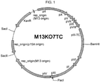
- the helper phage is M13KO7
- specific examples of such a preferred position can include a SacI site positioned between a kanamycin resistance gene and p15A ori, and a SacII site positioned between p15A ori and M13 ori.
- the polynucleotide encoding the first polypeptide may be inserted at a position that allows this polynucleotide to be linked in frame with a polynucleotide encoding the phage coat protein in the genome.
- the polynucleotide encoding the first polypeptide according to the present invention is functionally linked to a promoter.
- the promoter refers to a polynucleotide sequence that can bind to RNA polymerase in a cell to start the transcription of the downstream (3' direction) sequence.
- the phrase "functionally linked to a promoter” may mean that the promoter is located at a position appropriate for a certain sequence so as to be capable of controlling the transcription of the sequence.
- the position of the promoter may be a position physically distant from the sequence.
- the promoter used in the present invention may be a constitutive promoter or may be an inducible promoter. A wide range of promoters can be used.
- promoter suitable for prokaryotic cells can include: ⁇ -lactamase (bla) promoter, lactose (lac) promoter, tryptophan (trp) promoter, hybrid promoters such as tac promoter; tetracycline (tet) promoter, arabinose promoter, ⁇ phage promoter, T7 phage promoter, and T5 phage promoter.
- the polynucleotide encoding the first polypeptide according to the present invention is preferably linked to a ribosomal binding site (RBS) such as a Shine-Dalgarno (SD) sequence.
- RBS ribosomal binding site
- SD Shine-Dalgarno
- the ribosomal binding site located at an appropriate position promotes the translation of a polynucleotide positioned downstream thereof.
- the ribosomal binding site can be located between the promoter and the polynucleotide sequence placed under the control of the promoter.
- the first polypeptide according to the present invention is preferably linked to a signal sequence.
- the signal sequence refers to a peptide chain that is involved in the localization of a protein after intracellular expression of the protein.
- a sequence encoding the signal sequence can be located adjacent to a sequence encoding the protein.
- the signal sequence used in the present invention preferably localizes the protein to the periplasmic space of the host bacterium. Examples of such a signal sequence can include pelB signal sequence, gene III signal sequence, OmpA signal sequence, phoA signal sequence, malE signal sequence, dsbA signal sequence, E. coli heat-stable enterotoxin signal sequence, and beta lactamase signal sequence.
- the first polypeptide according to the present invention may be fused with a phage coat protein.
- the fusion of the first polypeptide with the phage coat protein can be carried out by linking the polynucleotide encoding the first polypeptide in frame with a polynucleotide encoding the phage coat protein.
- the phage coat protein may be a structural protein such as g3p, g6p, g7p, g8p, or g9p.
- the coat protein to be fused with the first polypeptide is preferably g3p or g8p, more preferably g3p.
- the fusion with the coat protein is carried out for the purpose of displaying the first polypeptide on the surface of a phage particle. Therefore, the first polypeptide is preferably fused at the N terminus or C terminus of the coat protein.
- the coat protein may have a full length or may lack a portion such as the N terminus or C terminus.
- the fusion may be carried out directly or may be carried out via an arbitrary linker peptide.
- the linker peptide can contain a tag sequence such as 6 x His tag, Myc tag, or FLAG tag.
- the linker peptide may contain a protease recognition sequence for a protease such as trypsin or chymotrypsin.
- the tag sequence is useful for the detection, etc., of the fusion protein.
- the protease recognition sequence is useful because the antigen-binding molecule formed by the association of the first polypeptide with the second polypeptide can be separated and recovered from the phage coat protein by the digestion of the fusion protein with the protease.
- the number or type of the first polypeptide that can be expressed by the helper phage in the present invention is not particularly limited and can be usually only one type.
- the helper phage may be capable of expressing two or more types of first polypeptides differing in amino acid sequence.
- the helper phage of the present invention is usually capable of expressing only one (first polypeptide) of the polypeptides constituting the antigen-binding molecule.
- the helper phage of the present invention may be capable of expressing the first polypeptide with the other polypeptide (second polypeptide).
- the bacterium according to the present invention preferably comprises a polynucleotide encoding the second polypeptide.
- the bacterium comprising a polynucleotide desirably means the bacterium transformed with the polynucleotide.
- the polynucleotide is functionally linked to a promoter.
- the promoter may be a constitutive promoter or may be an inducible promoter. A wide range of promoters can be used.
- promoter suitable for prokaryotic cells can include: ⁇ -lactamase (bla) promoter, lactose (lac) promoter, tryptophan (trp) promoter, hybrid promoters such as tac promoter; tetracycline (tet) promoter, arabinose promoter, ⁇ phage promoter, T7 phage promoter, and T5 phage promoter.
- the transcription of the polynucleotide encoding the first polypeptide and the polynucleotide encoding the second polypeptide may be controlled in different manners by using different types of promoters as the promoter to be linked to the polynucleotide encoding the first polypeptide and the promoter to be linked to the polynucleotide encoding the second polypeptide in such a way that, for example, the expression of one of the polynucleotides is promoted while the expression of the other polynucleotide is suppressed.
- the polynucleotide encoding the second polypeptide according to the present invention is preferably linked to a ribosomal binding site (RBS) such as a Shine-Dalgarno (SD) sequence.
- RBS ribosomal binding site
- SD Shine-Dalgarno
- the ribosomal binding site located at an appropriate position promotes the translation of a polynucleotide positioned downstream thereof.
- the ribosomal binding site can be located between the promoter and the polynucleotide sequence placed under the control of the promoter.
- the second polypeptide according to the present invention is preferably linked to a signal sequence.
- a sequence encoding the signal sequence can be located adjacent to a sequence encoding the protein.
- the signal sequence used in the present invention preferably localizes the protein to the periplasmic space of the host bacterium. Examples of such a signal sequence can include pelB signal sequence, gene III signal sequence, OmpA signal sequence, phoA signal sequence, malE signal sequence, dsbA signal sequence, E. coli heat-stable enterotoxin signal sequence, and beta lactamase signal sequence.
- the polynucleotide encoding the second polypeptide according to the present invention is preferably inserted in a phagemid vector.
- the phagemid vector is a plasmid vector prepared so as to contain a portion of a phage genome, and contains a replication origin (e.g., ColE1) for bacteria and a replication origin derived from the genome of a bacteriophage (e.g., M13, f1, and fd).
- the phagemid vector has the property of being amplified in the host bacterium, as with plasmid vectors, and also has the property of being packaged in the phage particle of a bacteriophage.
- the phagemid vector when a bacterium transformed with the phagemid vector is infected with the helper phage, the phagemid vector can be preferentially packaged in a reconstructed phage particle rather than the original genome of the helper phage.
- the phagemid vector can include pHEN1, pComb3, pCANTAB5E, and pCES1.
- the second polypeptide according to the present invention may be fused with a phage coat protein.
- the fusion of the second polypeptide with the phage coat protein can be carried out by linking the polynucleotide encoding the second polypeptide in frame with a polynucleotide encoding the phage coat protein.
- the phage coat protein may be a structural protein such as g3p, g6p, g7p, g8p, or g9p.
- the coat protein to be fused with the second polypeptide is preferably g3p or g8p, more preferably g3p.
- the fusion with the coat protein is carried out for the purpose of displaying the second polypeptide on the surface of a phage particle. Therefore, the second polypeptide is preferably fused at the N terminus or C terminus of the coat protein.
- the coat protein may have a full length or may lack a portion such as the N terminus or C terminus.
- the fusion may be carried out directly or may be carried out via an arbitrary linker peptide.
- the linker peptide can contain a tag sequence such as 6 ⁇ His tag or Myc tag.
- the linker peptide may contain a protease recognition sequence for a protease such as trypsin or chymotrypsin.
- the tag sequence is useful for the detection, etc., of the fusion protein.
- the protease recognition sequence is useful because the antigen-binding molecule formed by the association of the first polypeptide with the second polypeptide can be separated and recovered from the phage coat protein by the digestion of the fusion protein with the protease.
- the antigen-binding molecule formed from the first polypeptide and the second polypeptide on the bacteriophage it is preferred that at least one of the first polypeptide and the second polypeptide should be fused with a phage coat protein.
- the coat proteins are preferably selected from the same types of coat proteins (e.g., g3p, g6p, g7p, g8p, and g9p).
- a gene in which the polynucleotide encoding the first polypeptide is linked to the end (which corresponds to the N terminus or C terminus) of a gene encoding the phage coat protein such as g3p or g8p may be inserted to the helper phage, while the polynucleotide encoding the second polypeptide may be inserted to the phagemid vector without being linked to a gene encoding the phage coat protein; or the polynucleotide encoding the first polypeptide may be inserted to the helper phage without being linked to a gene encoding the phage coat protein, while a gene in which the polynucleotide encoding the second polypeptide is linked to the end (which corresponds to the N terminus or C terminus) of a gene encoding the phage coat protein such as g3p or g8p may be inserted to the
- the phrase "displaying X on Y” means that X is bound with the surface of Y with the original functions of X maintained.
- the phrase "displaying an antigen-binding molecule on a bacteriophage” may mean that the antigen-binding molecule is bound with the surface of the bacteriophage particle while its ability to bind to the antigen is maintained. This binding may be carried out through a covalent bond or may be carried out through a noncovalent bond.
- X and Y are polypeptides
- X can be preferably bound with Y by preparing a fusion protein of X and Y.
- At least one of the first polypeptide and the second polypeptide is preferably fused with a phage coat protein.
- a method for displaying an antigen-binding molecule on a bacteriophage via a disulfide bond is also known ( WO01/005950 ). The display may be carried out by use of such a method.
- the number or type of the second polypeptide that can be expressed by the bacterium in the present invention is not particularly limited.
- the present invention relates to an antigen-binding molecule display library comprising a large number of antigen-binding molecules having common first polypeptides and different second polypeptides.
- a plurality of bacteria capable of expressing different types of second polypeptides are necessary for preparing such an antigen-binding molecule display library.
- the individual bacteria used in the present invention are preferably a population of bacteria capable of expressing second polypeptides differing in amino acid sequence from each other and capable of expressing a plurality of diverse second polypeptides when viewed as a whole.
- the bacterium of the present invention is usually capable of expressing only one (second polypeptide) of the polypeptides constituting the antigen-binding molecule. In some cases, the bacterium of the present invention may be capable of expressing the second polypeptide with the other polypeptide (first polypeptide).
- the antigen-binding molecule according to the present invention is not particularly limited as long as the molecule is formed in a form comprising two polypeptides (first polypeptide and second polypeptide) and has the ability to specifically bind to a certain antigen.
- the first polypeptide and the second polypeptide are preferably polypeptides differing in amino acid sequence from each other.
- Preferred examples of the antigen-binding molecule can include antibodies, Fab, F(ab') 2 , diabody ( Nature Nanotechnology (2007) 2, pp.
- antibody variable regions antibody variable regions, antibody fragments containing antibody variable regions, receptor proteins, Fc proteins, antibody fragments containing Fc proteins, Fc fusion proteins, and functional fragments thereof (fragments having antigen-binding sites and having functions thereof) and functional equivalents thereof (equivalents having antigen-binding sites and functions thereof, such as sugar chain-modified forms thereof).
- the antigen-binding molecule according to the present specification may be derived from any animal species (e.g., humans; or non-human animals such as mice, rats, hamsters, rabbits, monkeys, cynomolgus monkeys, rhesus monkeys, hamadryas baboon, chimpanzees, goats, sheep, dogs, cattle, and camels) or any bird.
- animal species e.g., humans; or non-human animals such as mice, rats, hamsters, rabbits, monkeys, cynomolgus monkeys, rhesus monkeys, hamadryas baboon, chimpanzees, goats, sheep, dogs, cattle, and camels
- the antigen-binding molecule when the antigen-binding molecule according to the present specification is an antibody (immunoglobulin) or a molecule derived from therefrom, the antibody or the molecule may be of any isotype (e.g., IgG, IgM, IgA, IgD, and IgE) and subclass (e.g., human IgG1, IgG2, IgG3, IgG4, IgA1, and IgA2, and mouse IgG1, IgG2a, IgG2b, and IgG3) or may be derived therefrom.
- isotype e.g., IgG, IgM, IgA, IgD, and IgE
- subclass e.g., human IgG1, IgG2, IgG3, IgG4, IgA1, and IgA2, and mouse IgG1, IgG2a, IgG2b, and IgG3
- the H chains of the antibody or the molecule derived therefrom may be, for example, any of ⁇ chain, ⁇ chain, ⁇ chain, ⁇ chain, and ⁇ chain or may be derived therefrom.
- the L chains of the antibody or the molecule derived therefrom may be, for example, any of ⁇ chain and ⁇ chain or may be derived therefrom.
- the antibody or the molecule derived therefrom may be an engineered antibody, for example, a chimeric antibody, a humanized antibody, or an affinity-matured antibody, or a molecule derived therefrom.
- the antibody comprises first polypeptides which are two identical polypeptides comprising (or consisting of) L chains, and second polypeptides which are two identical polypeptides comprising (or consisting of) H chains; or comprises first polypeptides which are two identical polypeptides comprising (or consisting of) H chains, and second polypeptides which are two identical polypeptides comprising (or consisting of) L chains.
- the first polypeptide and the second polypeptide are each preferably selected from the group consisting of the two polypeptides comprising (or consisting of) L chains and the two polypeptides comprising (or consisting of) H chains, and differ from each other.
- a phage library using such antibodies is generally known to those skilled in the art, as described in, for example, FEBS J. 2010 May; 277 (10): 2291-303 and WO2011062859 .
- the antibody can be used as the antigen-binding molecule of the present invention.
- the F(ab') 2 is known as an antigen-binding molecule that can be prepared by digestion of an IgG antibody with pepsin.
- the F(ab') 2 is a divalent molecule having two antigen-binding sites and having a structure in which two Fab' molecules are linked through two disulfide bonds without the Fc regions of the antibody (two Fab' molecules + hinge regions).
- the F(ab') 2 comprises first polypeptides which are two identical polypeptides comprising L chain variable regions, and second polypeptides which are two identical polypeptides comprising H chain variable regions; or comprises first polypeptides which are two identical polypeptides comprising H chain variable regions, and second polypeptides which are two identical polypeptides comprising L chain variable regions.
- the first polypeptide and the second polypeptide are each preferably selected from the group consisting of the two polypeptides comprising L chain variable regions and the two polypeptides comprising H chain variable regions, and differ from each other.
- a phage library using such F(ab') 2 molecules is generally known to those skilled in the art, as described in, for example, J Immunol Methods. 2004 Jan; 284 (1-2): 119-32 .
- the F(ab') 2 can be used as the antigen-binding molecule of the present invention.
- Fab'-zip- was displayed on a phage by the insertion of a dimerization domain, consisting of an IgG1 hinge region and a homodimerizing leucine zipper, between Fab and M13 bacteriophage g3p (gene 3 protein) so that F(ab') 2 was formed on the phage (“Fab'-zip-phage”) to construct a phage library displaying divalent Fab with high avidity similar to that of an IgG antibody.
- the diabody is a dimer prepared by the binding of two fragments each containing a variable region and a variable region linked via a linker or the like (e.g., single-chain antibodies (scFvs)) (hereinafter, referred to as diabody-constituting fragments).
- the diabody usually comprises two H chain variable regions and two L chain variable regions and has two antigen-binding sites ( P. Holliger et al., Proc. Natl. Acad. Sci.
- Each diabody-constituting fragment is preferably an H chain variable region (or its fragment) and an L chain variable region (or its fragment) linked.
- the linker that links the variable region and the variable region is not particularly limited.
- a linker short enough not to cause a noncovalent bond between the variable regions in the same fragment is preferably used.
- the length of such a linker can be appropriately determined by those skilled in the art and is usually 2 to 14 amino acids, preferably 3 to 9 amino acids, particularly preferably 4 to 6 amino acids.
- the H chain variable region (or its fragment) and the L chain variable region (or its fragment) encoded on the same fragment do not cause a noncovalent bond therebetween on the same chain because of the short linker between the H chain variable region (or its fragment) and the L chain variable region (or its fragment).
- this diabody-constituting fragment can form a dimer with another fragment without forming a single-chain V region fragment.
- the binding between the diabody-constituting fragments may be a noncovalent bond (e.g., hydrogen bond, electrostatic interaction, or van der Waals force) or a covalent bond (e.g., disulfide bond), or both of a covalent bond and a noncovalent bond.
- a noncovalent bond e.g., hydrogen bond, electrostatic interaction, or van der Waals force
- a covalent bond e.g., disulfide bond
- the first polypeptide is an H chain variable region (or its fragment) and an L chain variable region (or its fragment) linked via a linker
- the second polypeptide is an L chain variable region (or its fragment) and an H chain variable region (or its fragment) linked via a linker
- the first polypeptide is an L chain variable region (or its fragment) and an H chain variable region (or its fragment) linked via a linker
- the second polypeptide is an H chain variable region (or its fragment) and an L chain variable region (or its fragment) linked via a linker.
- the first polypeptide and the second polypeptide are each preferably selected from the group consisting of the H chain variable region (or its fragment) and the L chain variable region (or its fragment) linked via a linker, and the L chain variable region (or its fragment) and the H chain variable region (or its fragment) linked via a linker, and differ from each other.
- a phage library using such diabodies is generally known to those skilled in the art, as described in, for example, Nat Biotechnol. 1996 Sep; 14 (9): 1149-54 ; and US 20070036789 .
- the diabody can be used as the antigen-binding molecule of the present invention.
- a receptor protein that is formed in a form comprising two polypeptides and specifically binds to a certain ligand can also be included in the antigen-binding molecule of the present invention.
- the receptor protein is preferably a heteromeric receptor protein constituted by two polypeptides differing in amino acid sequence from each other.
- the receptor protein may be, for example, an extracellular region of the receptor protein, a ligand-binding region of the receptor protein, or a fusion protein thereof with an antibody Fc region.
- the antigen-binding molecule is a receptor protein
- the antigen refers to a ligand for the receptor protein.
- heteromeric receptor examples include IL-2 receptor, IL-3 receptor, IL-4 receptor, IL-5 receptor, IL-6 receptor, IL-7 receptor, IL-9 receptor, IL-10 receptor, IL-11 receptor, IL-12 receptor, IL-13 receptor, IL-15 receptor, IL-17 receptor, IL-23 receptor, IL-31 receptor, GM-CSF receptor, IFN- ⁇ receptor, IFN- ⁇ receptor, IFN- ⁇ receptor, CNTF receptor, LIF receptor, OSM receptor, and CT-1 receptor.
- an Fc protein that is formed in a form comprising two polypeptides and specifically binds to a certain Fc receptor can also be included in the antigen-binding molecule of the present invention.
- the Fc protein refers to a region composed of hinges or a portion thereof and CH2 and CH3 domains of an antibody molecule and generally referred to an amino acid sequence from EU numbering position 226 to the C terminus or from EU numbering position 230 to the C terminus.
- the Fc protein may be composed of CH2 and CH3 domains, or only CH3 domains.
- the Fc protein is preferably an altered Fc protein having some amino acid mutation added to a naturally occurring Fc protein and is preferably constituted by two polypeptides differing in amino acid sequence from each other.
- a heteromeric Fc protein can include Fc proteins described in, for example, WO98/50431 , WO2006/106905 , WO2007/114325 , WO2011/078332 , and WO2013/002362 .
- WO98/50431 states that the amino acid sequence of one of the polypeptides constituting the heteromeric Fc protein is fixed, and the amino acid sequence of the other polypeptide is altered, whereby a combination of two polypeptides most compatible with each other can be selected (picked) from among diverse sequences.
- the antigen-binding molecule is an Fc protein
- the antigen refers to any of various Fc receptors (e.g., FcyRI, FcyRIIa, FcyRIIb, FcyRIII, and FcRn).
- an amino acid in the antibody Fc region can be altered to thereby enhance binding to FcRn (neonatal Fc receptor) under neutral pH conditions ( WO2011/122011 ) or enhance binding to an Fc ⁇ receptor under neutral pH conditions ( WO2013/047752 ).
- FcRn nonatal Fc receptor
- Fc ⁇ receptor neutral pH conditions
- WO2013/047752 the antigen can reportedly be removed rapidly from blood.
- an Fc fusion protein in which the Fc protein is fused with a protein (e.g., a cytokine or a receptor extracellular domain) or a peptide can also be included in the antigen-binding molecule of the present invention.
- the Fc fusion protein may contain an antibody hinge region and/or a linker. Soluble Fc fusion proteins are widely used in in vitro and in vivo experiments and can have many advantages over non-fusion proteins ( Meg L et al., Methods in Molecular Biology 378: 33-52, 2007 ). In addition, the soluble Fc fusion proteins can eliminate many immunological problems in the production of human antibody preparations, while maintaining antigen specificity.
- soluble Fc fusion human antibody preparations include Etanercept (Amgen Inc.), a therapeutic drug for autoimmune disease, which has been produced by fusing soluble TNF receptor 2 with Fc of human IgG1.
- Etanercept Amgen Inc.
- the Fc fusion protein can be appropriately produced by use of a method generally known to those skilled in the art, as described in, for example, WO2009/136568 , WO2007/048122 , and WO2011/115323 , and used in a phage library.
- the first polypeptide is a polypeptide comprising (or consisting of) an L chain variable region
- the second polypeptide is a polypeptide comprising (or consisting of) an H chain variable region
- the first polypeptide is a polypeptide comprising (or consisting of) an H chain variable region
- the second polypeptide is a polypeptide comprising (or consisting of) an L chain variable region.
- first polypeptide and the second polypeptide are preferably each selected from the group consisting of the polypeptide comprising (or consisting of) an L chain variable region and the polypeptide comprising (or consisting of) an H chain variable region, and differ from each other.
- an object of the present invention is to provide a combination of a helper phage suitable for preparing a plurality of antigen-binding molecules comprising common first polypeptides, and a bacterium infectible by the helper phage.
- a polypeptide having an arbitrary amino acid sequence can be selected as such a first polypeptide as long as the first polypeptide is one of the polypeptides constituting each antigen-binding molecule.
- the antigen-binding molecule has antibody variable regions and the first polypeptide is a polypeptide comprising an L chain variable region or a polypeptide comprising an H chain variable region
- the L chain variable region or the H chain variable region can be selected from among L chain variable regions or H chain variable regions having arbitrary amino acid sequences.
- a plurality of antigen-binding molecules here, antibody variable regions
- the selected one as common first polypeptides can be prepared.
- the L chain variable region or the H chain variable region may be selected from among L chain variable regions or H chain variable regions contained in antibodies binding to particular antigens, or may be selected from among L chain variable regions or H chain variable regions contained in naive antibodies before immunization with the particular antigens.
- the antibody binding to a particular antigen can be prepared by a hybridoma method ( Nature (1975) 256, 495 ) or a phage antibody library method ( Nature (1991) 352, 624-628 , J Mol Biol (1991) 222, 581-597 ) generally known to those skilled in the art.
- the amino acid sequence of the L chain variable region or the H chain variable region of the antibody prepared by the hybridoma method can be identified by amplifying a gene encoding the L chain or the H chain contained in a hybridoma producing the antibody by PCR using primers specific for the antibody gene, and analyzing the sequence ( J Mol Biol (1991) 222, 581-597 ; and Mol Immunol (1992) 29, 193-203 ).
- amino acid sequence of the L chain variable region or the H chain variable region of the antibody prepared by the phage antibody library method can be identified by isolating a vector contained in a phage displaying the antibody, and analyzing the sequence of the gene encoding the L chain or the H chain inserted therein.
- the amino acid sequence of the L chain variable region or the H chain variable region contained in the naive antibody before immunization with the particular antigen can be identified at a large scale by: preparing, for example, peripheral blood mononuclear cells, bone marrow cells, or spleen cells producing such antibodies from humans or other animals, etc., amplifying genes encoding L chains or H chains contained in these cells by PCR using primers specific for the antibody gene, and analyzing the sequences. Therefore, the L chain variable region or the H chain variable region can be arbitrarily selected, for use, from among the L chain variable regions or the H chain variable regions thus identified ( J Mol Biol (1991) 222, 581-597 ; and Mol Immunol (1992) 29, 193-203 ).
- the polypeptide when the first polypeptide or the second polypeptide according to the present invention is a polypeptide comprising an L chain variable region or a polypeptide comprising an H chain variable region, the polypeptide may further comprise an L chain constant region or an H chain constant region. If the first polypeptide comprises no constant region, it is preferred that the second polypeptide should comprise no constant region. If the first polypeptide comprises a constant region, it is preferred that the second polypeptide should also comprise a constant region.
- the H chain constant region is particularly preferably an H chain constant region CH1 domain.
- the H chain constant region CH1 domain refers to a region from the beginning of the H chain constant region to immediately before the hinge region and generally refers to an amino acid sequence from EU numbering positions 118 to 225.
- these constant regions are contained in a form linked immediately after the variable regions.
- the L chain constant region may be a constant region derived from any of ⁇ chain and ⁇ chain.
- the H chain constant region may be a constant region derived from any of ⁇ chain, ⁇ chain, ⁇ chain, ⁇ chain, and ⁇ chain.
- these constant regions may have a full length or may lack a portion.
- these constant regions may be altered by the substitution, deletion, insertion, etc., of a portion of their amino acids.
- a preferred example of the antigen-binding molecule is Fab.
- the present invention relates to a method for preparing an antigen-binding molecule display library comprising common first polypeptides, wherein the method comprises:
- the plurality of bacteria are preferably a bacterium population in which the individual bacteria are bacteria capable of expressing second polypeptides differing in amino acid sequence from each other and are capable of expressing a plurality of diverse second polypeptides when viewed as a whole.
- Such a plurality of bacteria can be infected with the helper phages, respectively, capable of expressing first polypeptides having identical amino acid sequences to prepare a plurality of bacteriophages displaying antigen-binding molecules. All of these antigen-binding molecules comprise the common first polypeptides and the second polypeptides differing from each other.
- a plurality of bacteriophages displaying the antigen-binding molecules thus prepared can be recovered and mixed to prepare an antigen-binding molecule display library comprising the common first polypeptides.
- the library means an assembly of a plurality of components having diverse repertoires.
- the library mainly refers to a bacteriophage library (phage library) constituted by an assembly of a plurality of bacteriophages.
- the antigen-binding molecule display library means a library having, as components, bacteriophages displaying antigen-binding molecules on their surface.
- the antigen-binding molecules contained therein preferably have diverse repertoires. A larger number of components in the library (larger size of the library) is more preferred.
- the library size is preferably, for example, 10 6 or more, 10 7 or more, 10 8 or more, 10 9 or more, 10 10 or more, 10 11 or more, 10 12 or more, 10 13 or more, or 10 14 or more.
- the number of a plurality of bacteria capable of expressing second polypeptides, used in the step is equal to the number of components in the library. Therefore, the bacterium population used in the step preferably contains, for example, 10 6 or more, 10 7 or more, 10 8 or more, 10 9 or more, 10 10 or more, 10 11 or more, 10 12 or more, 10 13 or more, or 10 14 or more bacteria.
- a bacterium population capable of expressing a plurality of second polypeptides is cultured in a mixed state, while a plurality of helper phages capable of expressing identical first polypeptides can be allowed to collectively infect the bacterium population.
- each helper phage may be allowed to individually infect a small scale of a bacterium population containing one or more bacteria.
- the bacteriophages may be recovered by merely separating the culture supernatant by the centrifugation or the like of the culture solution of the bacteria after the helper phage infection, or may be recovered by an additional step of isolating and purifying the bacteriophages, for example, by a method for precipitating the bacteriophages by the addition of polyethylene glycol (PEG) thereto (PEG precipitation method).
- PEG polyethylene glycol
- the second polypeptide is a polypeptide comprising an L chain variable region or a polypeptide comprising an H chain variable region
- genes encoding a plurality of L chain variable regions or H chain variable regions differing in amino acid sequence from each other can be obtained, for example, by isolating a large number of naturally occurring antibody genes (e.g., antibody genes found in vivo ).
- antibody-producing cells such as peripheral blood mononuclear cells, bone marrow cells, or spleen cells are prepared from humans or other animals, etc.
- RT-PCR reverse transcription-polymerase chain reaction
- naive antibody-producing cells before immunization with a particular antigen are preferably used from the viewpoint of obtaining high diversity.
- biased antibody-producing cells after immunization with the particular antigen may be used.
- the genes can also be obtained by synthesizing a large number of genes diversified, for example, by the artificial mutation of a gene encoding a certain L chain variable region or H chain variable region.
- Such genes may be prepared by artificially inducing a mutation using an approach, for example, Error prone PCR or may be prepared by the total synthesis of genes having sequences designed so as to have desired diversity.
- the present invention also encompasses an antigen-binding molecule display library prepared by the method for preparing an antigen-binding molecule display library according to the present invention.
- the present invention relates to a method for obtaining an antigen-binding molecule specifically binding to a predetermined antigen, wherein the method comprises:
- the antigen-binding molecule display library of the present invention comprises a plurality of diverse antigen-binding molecules differing in sequence from each other, and is therefore a population of antigen-binding molecules capable of binding to various types of antigens when viewed as a whole. Accordingly, the antigen-binding molecule display library of the present invention can be screened to select (pick) an antigen-binding molecule specifically binding to the desired antigen. Specifically, the antigen is contacted with the antigen-binding molecule display library of the present invention so that an antigen-binding molecule capable of specifically binding to the antigen in the library binds to the antigen to form a complex.
- the antigen-binding molecule complexed with the antigen among a plurality of antigen-binding molecules contained in the library, can be separated from antigen-unbound antigen-binding molecules by some method generally known to those skilled in the art to select (pick) only the antigen-binding molecule specifically binding to the antigen.
- the method for separating the antigen-binding molecule complexed with the antigen can involve, for example, contacting the antigen biotinylated in advance with the antigen-binding molecule display library, and then allowing the biotinylated antigen to bind to avidin or streptavidin immobilized on a carrier such as beads or a plate to recover only the antigen-binding molecule complexed with the antigen onto the beads or the plate. Then, the beads or the plate is washed so that antigen-unbound antigen-binding molecules can be removed from the antigen-binding molecule display library to separate the antigen-binding molecule complexed with the antigen from the antigen-unbound antigen-binding molecules.
- the aforementioned operation of selecting an antigen-binding molecule specifically binding to the antigen may be repeated a plurality of times.
- antigen-binding molecules having the weak ability to bind to the antigen and antigen-binding molecules having the strong ability to bind to the antigen seem to coexist in an antigen-binding molecule group separated by the first selecting operation. Therefore, the abundance of the antigen-binding molecules having the strong ability to bind to the antigen can be gradually enhanced by repeating the selecting operation.
- bacteriophages displaying the antigen-binding molecules separated by the first selecting operation are allowed to temporarily infect host bacteria, followed by the culture of the bacteria for growth.
- polynucleotide encoding the second polypeptides are usually packaged in the bacteriophages prepared in the present invention, the polynucleotides encoding the second polypeptides are present in the bacteria infected with the bacteriophages.
- the bacteria in this state are bacteria capable of expressing the second polypeptides. Therefore, the bacteria are infected with the same helper phages (i.e., the helper phages capable of expressing the same first polypeptides) as in the preparation of the initial antigen-binding molecule display library.
- helper phages i.e., the helper phages capable of expressing the same first polypeptides
- the thus-obtained bacteriophages displaying the antigen-binding molecules can be used as starting materials again in the repeated selection operation to form an antigen-binding molecule population comprising a large number of only antigen-binding molecules having the strong ability to bind to the antigen.
- the antigen-binding molecules contained in the antigen-binding molecule display library are present in a state displayed on the bacteriophages. Only the antigen-binding molecules may be obtained by some method. For example, when each antigen-binding molecule is fused with a phage coat protein via protease (e.g., trypsin) cleavage site introduced therebetween, the antigen-binding molecule can be separated from the bacteriophage through the reaction of the protease with the bacteriophage displaying the antigen-binding molecule to isolate only the antigen-binding molecule.
- protease e.g., trypsin
- sequence information on the antigen-binding molecule can be identified from this polynucleotide and the polynucleotide encoding the first polypeptide contained in the helper phage of the present invention.
- the antigen-binding molecule can be separately prepared by a genetic engineering approach.
- the antigen according to the present invention is not particularly limited as long as the antigen is a compound containing a structure that can serve as an antigenic determinant (epitope).
- the antigen may be a low-molecular compound or may be a high-molecular compound.
- General examples of the antigen can include polypeptides, polynucleotides, sugar chains, lipids, and molecules composed of combinations thereof. These antigens may be prepared by isolation from naturally occurring materials or may be prepared by artificial synthesis. When the antigen is, for example, a polypeptide, the polypeptide can be prepared by a genetic engineering approach.
- a polynucleotide encoding the amino acid sequence of the polypeptide is prepared by an approach generally known to those skilled in the art, such as a gene cloning method or a nucleic acid synthesis method, and this polynucleotide can be inserted to an expression vector or the like known in the art, which is then transferred to appropriate host cells to prepare the polypeptide.
- the expressed polypeptide can be purified by a usual method such as ion chromatography or affinity chromatography.
- the "antigen-binding molecule specifically binding to the antigen” means that the binding activity of the antigen-binding molecule against the particular antigen is, for example, preferably 2 or more times, 3 or more times, or 5 or more times, more preferably 10 or more times, 20 or more times, or 30 or more times, further preferably 50 or more times or 100 or more times higher than its binding activity against other antigens.
- the binding activity of the antigen-binding molecule against the antigen can be measured and compared by a method generally known to those skilled in the art, such as ELISA, FACS, or Biacore.
- the antigen defined above may be used interchangeably with an epitope.
- the antigen-binding molecule specifically binding to the antigen means that the binding activity of the antigen-binding molecule against the particular epitope is, for example, preferably 2 or more times, 3 or more times, or 5 or more times, more preferably 10 or more times, 20 or more times, or 30 or more times, further preferably 50 or more times or 100 or more times higher than its binding activity against other epitopes.
- the present invention relates to a method for preparing a multispecific antigen-binding molecule comprising common first polypeptides, wherein the method comprises:
- the aforementioned method of the present invention may be a method for preparing a multispecific antigen-binding molecule comprising common first polypeptides, wherein the method comprises:
- the antigen-binding molecule obtained by the method for obtaining an antigen-binding molecule specifically binding to a predetermined antigen according to the present invention absolutely comprises the first polypeptide. Therefore, all of the plurality of antigen-binding molecules obtained as a result of carrying out the method for a plurality of antigens comprise the common first polypeptides and the second polypeptides differing from each other.
- the first polypeptides and the plurality of second polypeptides thus obtained are combined such that the plurality of second polypeptides associate with the first polypeptides, respectively, to form the plurality of antigen-binding molecules.
- the plurality of antigen-binding molecules thus prepared are reconstructed so as to form one molecule in which the antigen-binding molecules are linked.
- the multispecific antigen-binding molecule comprising the common first polypeptides can be easily prepared.
- the multispecific antigen-binding molecule may be prepared by use of a genetic engineering approach. Specifically, polynucleotides encoding the first polypeptides and polynucleotides encoding the plurality of second polypeptides are separately prepared. These polynucleotides are transferred to a host cell, and the host cell is cultured under conditions that permit expression of the polynucleotides. The plurality of second polypeptides expressed from the polynucleotides associate with the first polypeptides, respectively, to form the plurality of antigen-binding molecules.
- the plurality of antigen-binding molecules thus prepared are reconstructed so as to form one molecule in which the antigen-binding molecules are linked.
- the multispecific antigen-binding molecule comprising the common first polypeptides can be easily expressed.
- the multispecific antigen-binding molecule extracellularly expressed by the host cell may be recovered by recovering the culture supernatant by the centrifugation of the culture solution of the host cell or may be recovered by preparing the cell extract of the host cell.
- the step of isolating and purifying the multispecific antigen-binding molecule therefrom may be further added to the method ( Nat Biotechnol. 1998 Jul; 16 (7): 677-81 ).
- the polynucleotides encoding the first polypeptides and the polynucleotides encoding the plurality of second polypeptides are preferably inserted in some expression vector. Each polynucleotide may be individually inserted to the expression vector, or these polynucleotides may be collectively inserted to the same expression vector. Examples of the expression vector can include pET for E. coli and pcDNA3 for mammalian cells.
- Examples of the host cell to which the polynucleotides encoding the first polypeptides and the polynucleotides encoding the plurality of second polypeptides are transferred can include E. coli cells JM109, DH5 ⁇ , HB101, and XL1-Blue, and mammalian cells CHO, COS, and HEK293.
- the transfer of the polynucleotides to the host cell can be carried out by use of an approach generally known to those skilled in the art, such as a calcium phosphate method, a DEAE dextran method, an electroporation method, a lipofection method, or a microinjection method.
- the multispecific antigen-binding molecule recovered from the host cell may be isolated and purified by a method known in the art, for example, centrifugation, ammonium sulfate fractionation, salting out, dialysis, ultrafiltration, affinity chromatography, ion-exchange chromatography, or gel filtration chromatography.
- the multispecific antigen-binding molecule means a molecule containing, in one molecule, a plurality of antigen-binding molecules specifically binding to a plurality of antigens, respectively.
- the antigen-binding molecules can be linked to each other in some manner to form one molecule. This linking may be carried out through a covalent bond (e.g., peptide bond or disulfide bond) or may be carried out through a noncovalent bond.
- the antigen-binding molecules may be connected directly or may be connected via a linker molecule such as a linker peptide.
- examples of the multispecific antigen-binding molecule can include a molecule in which a plurality of H chain variable regions and L chain variable regions are connected either directly or through a peptide bond via a linker peptide, and a plurality of antibody variable regions are formed by the appropriate intramolecular association between the H chain variable regions and the L chain variable regions (e.g., diabody, triabody, and single-chain diabody).
- Another example thereof can include a molecule in which H chain variable regions and L chain variable regions are connected to H chain constant regions and L chain constant regions, respectively, through a peptide bond, while these H chain constant regions are connected through a disulfide bond or the like, and a plurality of antibody variable regions are formed by the appropriate intramolecular association between the H chain variable regions and the L chain variable regions (e.g., antibody (immunoglobulin) molecules such as IgG, IgM, IgA, IgD, and IgE).
- antibody (immunoglobulin) molecules such as IgG, IgM, IgA, IgD, and IgE).
- the respective antigen-binding molecules contained in the multispecific antigen-binding molecule may be antigen-binding molecules binding to their distinctive antigens or may be antigen-binding molecules binding to different antigenic determinants (epitopes) contained in the same antigen. In some cases, the respective antigen-binding molecules contained in the multispecific antigen-binding molecule may be antigen-binding molecules binding to identical epitopes in identical antigens. The number of the antigen-binding molecules contained in the multispecific antigen-binding molecule can be increased to 2, 3, 4, etc., to thereby prepare a bispecific antigen-binding molecule, a trispecific antigen-binding molecule, a tetraspecific antigen-binding molecule, etc., respectively.
- the multispecific antigen-binding molecule according to the present invention is preferably a bispecific antigen-binding molecule (e.g., bispecific antibody).
- the multispecific antigen-binding molecule can be used for various purposes. It has already been known that the multispecific antigen-binding molecule can be used as an active ingredient for a pharmaceutical composition in the treatment of a disease for one of the purposes. For example, in the treatment of a cancer, a bispecific antigen-binding molecule comprising an antigen-binding molecule binding to a tumor antigen and an antigen-binding molecule binding to a molecule inducing cytotoxic activity is useful as a molecule that can induce cytotoxicity specific for the tumor cells.
- tumor antigen examples include CD15, p185 (HER2), p97, OVCAR-3, L-D1, EGFR, CAMA1, CD19, MoV18, NCAM, FBP, AMOC-31, Id-1, CD22, CD7, CD38, CEA, and CD30.
- molecule inducing cytotoxic activity examples include FcyRI, FcyRIII (CD16), and CD3.
- a bispecific antigen-binding molecule comprising an antigen-binding molecule binding to a virus and an antigen-binding molecule binding to a molecule inducing cytotoxic activity is useful as a molecule that can induce cytotoxicity specific for the virus-infected cells.
- viruses can include herpes simplex virus (HSV), influenza virus, and human immunodeficiency virus (HIV).
- HSV herpes simplex virus
- HAV human immunodeficiency virus
- a bispecific antigen-binding molecule comprising an antigen-binding molecule binding to fibrin and an antigen-binding molecule binding to a plasminogen activator is useful as a thrombolytic drug.
- the plasminogen activator can include tissue plasminogen activator (tPA) and urokinase plasminogen activator (uPA).
- an agonist molecule of a cytokine can be obtained from among bispecific antigen-binding molecules each comprising antigen-binding molecules binding to polypeptide chains constituting a heteromeric receptor for the cytokine, respectively ( WO2004/060919 ).
- the cytokine having a heteromeric receptor can include IL-2, IL-3, IL-4, IL-5, IL-6, IL-7, IL-9, IL-10, IL-11, IL-12, IL-13, IL-15, IL-17, IL-23, IL-31, GM-CSF, IFN- ⁇ , IFN- ⁇ , IFN- ⁇ , CNTF, LIF, OSM, and CT-1.
- a functional molecule that can serve as an alternative to the effects of a cofactor enhancing enzymatic reaction can be obtained from among bispecific antigen-binding molecules each comprising an antigen-binding molecule binding to the enzyme and an antigen-binding molecule binding to a substrate of the enzyme ( WO2005/035754 ).
- Examples of such an enzyme-substrate-cofactor combination can include blood coagulation factor IX (FIXa)-blood coagulation factor X (FX)-blood coagulation factor VIII (FVIII/FVIIIa) combination, protein Z-dependent protein inhibitor (ZPI)-blood coagulation factor X (FX/FXa)-protein Z (PZ) combination, and thrombin-thrombin-activatable fibrinolysis inhibitor (TAFI)-thrombomodulin (TM) combination.
- FIXa blood coagulation factor IX
- FX blood coagulation factor X
- FVIII/FVIIIa blood coagulation factor VIII
- ZPI protein Z-dependent protein inhibitor
- TAFI thrombin-thrombin-activatable fibrinolysis inhibitor
- TM thrombomodulin
- the multispecific antigen-binding molecule can reportedly be used in antifungal therapy ( Japanese Patent Laid-Open No. 5-199894 ), immune response induction (National Publication of International Patent Application No. 1998-511085 ), immunochemistry ( R.R. Suresh et al., (1986) Proc. Natl. Acad. Sci. USA 83: 7989-7993 ; and C. Milstein and A.C. Cuello (1983) Nature 305: 537-540 ), etc.
- the multispecific antigen-binding molecule according to the present invention is a bispecific antibody (e.g., IgG) having common L chains
- alteration to introduce structures sterically complementary to each other to the CH3 domains of two types of H chains Ridgway et al., (1996) Protein Eng. 9: 617-21 ; and WO96/27011
- alteration to convert the CH3 domains of two types of H chains to a heterodimer by interdigitating an IgG-derived sequence and an IgA-derived sequence SEEDbodies: Protein Eng Des Sel. 2010 Apr; 23 (4): 195-202
- alteration to introduce a mutation so as to cause charge interaction between the CH3 domains of two types of H chains have already been known as such alterations.
- the present invention relates to a method for producing an antigen-binding molecule, wherein the method comprises:
- This method may further comprise:
- the method for producing an antigen-binding molecule may be the method further comprising:
- This method may further comprise:
- the physical properties in (c) or (f) may mean, but are not limited to, for example, isoelectric points, heat stability, chemical stability, solubility, viscosity, glycosylation status, the homogeneity of the antigen-binding molecule itself, immunogenicity, and/or affinity or binding specificity for the antigen ( J Biol Chem 2005; 280: 24880-7 ).
- the method for producing an antigen-binding molecule provides an antigen-binding molecule having excellent affinity or binding specificity for the antigen, an antigen-binding molecule having excellent heat stability or chemical stability, an antigen-binding molecule having improved solubility, an antigen-binding molecule free from a glycosylated amino acid sequence, a molecule improved in terms of the homogeneity of the antigen-binding molecule itself, an antigen-binding molecule having reduced immunogenicity (or immunogenic risks), and/or an antigen-binding molecule having a changed isoelectric point or viscosity, as compared with the reference antigen-binding molecule.
- this method relates to a method for affinity-maturing an antigen-binding molecule.
- this method is advantages because even if an antibody inferior in physical properties has been obtained by the method, this antibody can be used in, for example, the humanization of a non-human animal-derived antibody ( J Mol Biol. 2000 Feb 25; 296 (3): 833-49 ).
- human-derived second polypeptides can be obtained by panning operation for an antigen using fixed non-human animal-derived first polypeptides and a human-derived second polypeptide library in combination.
- a human-derived first polypeptide can be obtained by panning operation for the antigen using the fixed second polypeptides and a human-derived first polypeptide library in combination.
- a human antibody can be obtained on the basis of the non-human animal-derived antibody by the sequential replacement with the human antibody libraries.
- the stability, isoelectric point, etc., of an antibody can be changed by the method for producing an antigen-binding molecule, thereby prolonging or shortening the half-life in blood or average residence time in blood of the antibody or reducing or improving its clearance in blood, for example.
- the method for obtaining an antigen-binding molecule is not particularly limited as long as the method is generally known to those skilled in the art.
- antigen-binding molecules each capable of specifically binding to the antigen may be selected through contact with this antigen, and then, an antigen-binding molecule group having the desired physical properties can be evaluated (e.g., assayed or predicted) for the physical properties without panning operation ( MAbs. 2011 May-Jun; 3 (3): 243-52 ).
- the antigen-binding molecules may be screened for (narrowed down to) candidates by panning operation, and then, the candidates can be evaluated for the physical properties.
- the isoelectric points can be calculated (predicted) on the basis of the amino acid sequences of the obtained antigen-binding molecules using commercially available software generally known to those skilled in the art, such as Genetyx.
- the isoelectric points can be predicted for the amino acid sequences of antigen-binding molecules having the adequate ability to specifically bind to the antigen to select a molecule having the desired isoelectric point.
- the isoelectric points may be actually measured using isoelectric focusing (IEF) or the like ( Protein Eng Des Sel. 2010 May; 23 (5): 385-92 ).
- the present invention relates to a combination of an altered helper phage and a bacterium infectible by the helper phage, wherein the helper phage is a helper phage capable of expressing a first polypeptide, and the bacterium is a bacterium capable of expressing a second polypeptide.
- the first polypeptide and the second polypeptide according to the present invention associate with each other to form one antigen-binding molecule.
- the combination of a helper phage and a bacterium according to the present invention refers to every combination comprising the helper phage and the bacterium as components on the premise that the helper phage is allowed to infect the bacterium.
- the present invention also encompasses a combination in which the helper phage and the bacterium exist separately before mixing and thus are not yet ready for infection.
- the present invention also encompasses a combination in which the helper phage and the bacterium exist in a mixture after mixing and are thus ready for infection.
- the present invention also encompasses a method for producing the combination of an altered helper phage and a bacterium infectible by the helper phage according to the present invention.
- the altered helper phage of the present invention can be prepared by inserting the polynucleotide encoding the first polypeptide to the genomic DNA of a helper phage through the use of a restriction site, and transferring the genomic DNA of the altered helper phage thus prepared to a host bacterium.
- the restriction site preferred for the helper phage M13KO7 can include a SacI site positioned between a kanamycin resistance gene and p15A ori, and a SacII site positioned between p15A ori and M13 ori.
- the bacterium thus harboring the genomic DNA of the altered helper phage produces a phage particle, and the genomic DNA of the altered helper phage is further packaged therein to reconstruct the altered helper phage.
- the bacterium infectible by the helper phage can be prepared by transferring the polynucleotide encoding the second polypeptide to a bacterium.
- the present invention also encompasses an altered helper phage which is included in the combination of an altered helper phage and a bacterium infectible by the helper phage according to the present invention.
- the present invention relates to an altered helper phage capable of expressing a certain polypeptide, wherein the polypeptide is any one of two polypeptides that associate with each other to form an antigen-binding molecule.
- the present invention also relates to an altered helper phage capable of expressing a first polypeptide, wherein the first polypeptide is capable of associating with a different second polypeptide to form one antigen-binding molecule.
- the present invention also encompasses a kit comprising the altered helper phage of the present invention.
- the present invention relates to a kit comprising an altered helper phage capable of expressing a certain polypeptide, wherein the polypeptide is any one of two polypeptides that associate with each other to form an antigen-binding molecule.
- the present invention also relates to a kit comprising an altered helper phage capable of expressing a first polypeptide, wherein the first polypeptide is capable of associating with a different second polypeptide to form one antigen-binding molecule.
- kits can each be a kit for preparing a bacteriophage displaying an antigen-binding molecule, a kit for preparing an antigen-binding molecule display library comprising common first polypeptides, a kit for obtaining an antigen-binding molecule specifically binding to a predetermined antigen, a kit for preparing a multispecific antigen-binding molecule comprising common first polypeptides, or a kit for producing an antigen-binding molecule having physical properties different from those of a reference antigen-binding molecule.
- the kit of the present invention may further comprise a bacterium infectible by the helper phage, wherein the bacterium is capable of expressing the remaining one polypeptide of the two polypeptides that form an antigen-binding molecule, or a bacterium infectible by the helper phage, wherein the bacterium is capable of expressing the second polypeptide.
- first or second are used for expressing various factors. However, these factors are understood to be not limited by these terms. These terms are used merely for differentiating one factor from the other factors. For example, the first factor may be described as the second factor, and vice versa, without departing from the scope of the present invention.
- numeric values described herein are understood as values having given ranges according to the common technical knowledge of those skilled in the art, unless inconsistent to the context.
- 1 mg is understood to represent “approximately 1 mg” and is understood to include a given variation.
- 1 to 5" described herein is understood to concretely describe the individual values of "1, 2, 3, 4, and 5", unless inconsistent to the context.
- a promoter, a signal sequence gene, an antibody L chain gene, etc., were incorporated to the genome of a helper phage to construct an L chain-expressing helper phage.
- the antibody L chain can be expressed from E. coli infected with this helper phage.
- an E. coli strain XL1-Blue was infected with a helper phage (M13KO7; Invitrogen Corp.) and shake-cultured overnight, followed by the genome extraction of the helper phage (QIAprep Spin Miniprep Kit; Qiagen N.V.).
- the helper phage genome has a BamHI site at the N2 domain of gene 3 and a PacI site at gene 1.
- helper phage genome was cleaved with BamHI and PacI, then electrophoresed on 0.6% agarose gel, and purified by gel extraction (Wizard SV Gel and PCR Clean-Up system; Promega Corp.) to prepare each of a DNA fragment from gene 3 to gene 1 and a DNA fragment of the remaining genome.
- the prepared DNA fragment from gene 3 to gene 1 was used as a template in PCR to newly prepare a DNA fragment having an insert of a DNA encoding a trypsin cleavage sequence between the N2 domain and the CT domain of gene 3.
- the amino acid sequence encoded by the gene 3 before the alteration is shown in SEQ ID NO: 1, and the amino acid sequence encoded by the altered gene 3 is shown in SEQ ID NO: 2.
- helper phage M13KO7TC having an insert of the trypsin cleavage sequence between the N2 domain and the CT domain of the pIII protein on the helper phage (see National Publication of International Patent Application No. 2002-514413).
- An E . coli strain ER2738 was infected with the helper phage M13KO7TC and shake-cultured overnight, followed by the genome extraction of the helper phage M13KO7TC from the infected E. coli (NucleoBond Xtra Midi Plus).
- a SacI site positioned between a kanamycin resistance gene and p15A ori was selected as the site to which the L chain expression unit was inserted ( Figure 1 ).
- the insertion site is not limited to this site and may be, for example, a SacII site positioned between p15A ori and M13 ori without problems.
- the genome of the helper phage M13KO7TC purified by the aforementioned method was cleaved with SacI, then electrophoresed on 0.6% agarose gel, and purified by gel extraction (Wizard SV Gel and PCR Clean-Up system; Promega Corp.) to obtain the DNA fragment (M13KO7TC/SacI) of interest.
- the L chain of an anti-human IL-6R antibody PF1 was used as the antibody L chain (VL and CL) to be introduced.
- the substitution of the C-terminal Cys of the L chain constant region by Ala is known to be advantageous for Fab expression in E. coli ( J Biol Chem. 2003 Oct 3; 278 (40): 38194-38205 ). Therefore, such a sequence was used.
- lac promoter - pelB signal sequence gene - PF1 L chain gene was inserted to M13KO7TC/SacI by the in-fusion method (In-Fusion HD Cloning Kit; Clontech Laboratories, Inc.), which was then transferred to an E. coli strain ER2738 by the electroporation method.
- the nucleic acid sequence of the lac promoter is shown in SEQ ID NO: 3.
- the amino acid sequence of the pelB signal sequence and the nucleic acid sequence encoding it are shown in SEQ ID NO: 4 and SEQ ID NO: 5, respectively.
- the amino acid sequence of the PF1 L chain and the nucleic acid sequence encoding it are shown in SEQ ID NO: 6 and SEQ ID NO: 7, respectively.
- the obtained E. coli was cultured. 2.5 M NaCl/10% PEG was added to the culture supernatant, and the helper phage was purified by the PEG precipitation method. The titer of the obtained helper phage M13KO7TC-PF1L was confirmed by the general plaque formation method.
- a phagemid vector for expressing an antibody H chain on phage surface was constructed.
- the phagemid vector was prepared by functionally inserting a packaging signal gene for phage particles, a promoter, a signal sequence gene, an antibody H chain gene, a linker gene, gene 3, etc., to a plasmid vector.
- the antibody H chain was fused with the gene 3 protein (g3p) via the linker peptide.
- the H chain of an anti-human IL-6R antibody PF1 was used as the antibody H chain (Fd consisting of VH and CH1) to be introduced.
- the amino acid sequence of the PF1 H chain is shown in SEQ ID NO: 8.
- the constructed phagemid vector was transferred to an E. coli strain ER2738 by the electroporation method to construct E. coli ER2738/pAG-PF1H carrying the PF1 H chain-expressing phagemid vector.
- the E. coli ER2738/pAG-PF1H was cultured until OD reached around 0.5, and then infected with the helper phage M13KO7TC-PF1L or M13KO7TC. After medium replacement, the E. coli was cultured overnight at 30°C, and the culture supernatant was recovered. 2.5 M NaCl/10% PEG was added to the E. coli culture solution containing the produced phage to precipitate the phage, which was then dissolved in TBS to obtain a phage solution. The titer of the obtained phage was confirmed by the general colony formation method.
- the phage ELISA method was carried out to confirm Fab display on the produced phage and to confirm the ability to bind to the antigen.
- StreptaWell 96-well microtiter plate (F. Hoffmann-La Roche, Ltd.) was coated by the addition of 100 ⁇ L of PBS containing Goat anti-Human Kappa Biotin antibody (EY Laboratories, Inc.) or biotinylated human IL-6R. Each well of the plate was washed with 0.1 ⁇ TBST (0.1 ⁇ TBS containing 0.1% Tween 20) to remove the antigen.
- the plate was blocked for 1 hour or longer by the addition of 250 ⁇ L of 0.02% skim milk-0.1 ⁇ TBS (0.1 ⁇ TBS containing 0.02% skim milk) to the well.
- the phage solution diluted with 0.02% skim milk-0.1 ⁇ TBS was added to each well, and the plate was left standing at 37°C for 1 hour so that the antibody displayed on the phage bound to the Goat anti-Human Kappa Biotin antibody or the biotinylated human IL-6R.
- an HRP-conjugated anti-M13 antibody (Amersham Pharmacia Biotech) diluted with 0.1 ⁇ TBST was added to each well, and the plate was incubated for 1 hour. After washing with 0.1 ⁇ TBST, TMB single solution (Zymed Laboratories Inc.) was added to each well. The color reaction of the solution was further terminated by the addition of sulfuric acid. Then, the absorbance was measured at 450 nm.
- Naive H chain variable region genes were amplified by PCR using poly-A RNA prepared form human peripheral blood mononuclear cells (PBMCs), commercially available human poly-A RNA, or the like as a template. These genes were inserted to phagemid vectors, and the constructed phagemid vectors were transferred to an E. coli strain ER2738 by the electroporation method. Consequently, approximately 1.1 ⁇ 10 10 colonies were obtained.
- PBMCs peripheral blood mononuclear cells
- E. coli colonies were infected with the helper phage M13KO7TC-PF1L constructed in Example 1 and cultured to construct a human antibody phage display library (NH-PF1L library) displaying Fabs comprising naive H chains and PF1 L chains.
- NH-PF1L library human antibody phage display library
- hPlexin A1 The extracellular region of a single-pass transmembrane protein human Plexin A1 (hPlexin A1) was prepared as follows: hPlexin A1 gene synthesized on the basis of the amino acid sequence of NCBI Reference Sequence NP_115618 (SEQ ID NO: 9) was altered to encode a protein lacking the presumed transmembrane region starting at alanine at position 1245 and subsequent regions and instead having an added FLAG tag sequence (SEQ ID NO: 13). The signal peptide (SEQ ID NO: 14) from positions 1 to 26 was further substituted by an artificial signal peptide HMM+38 (SEQ ID NO: 15).
- the prepared gene encoding the altered hPlexin A1 (SEQ ID NO: 10) was integrated into an expression vector for animal cells, which was then transferred to FreeStyle 293 cells (Invitrogen Corp.) using 293Fectin (Invitrogen Corp.). In this operation, the cells were cotransfected with the expression vector and a gene encoding EBNA1 (SEQ ID NO: 17) in order to improve the expression efficiency of the gene of interest.
- the cells transfected according to the aforementioned procedures were cultured at 37°C for 6 days in an 8% CO 2 environment so that the protein of interest was secreted into the culture supernatant.
- the cell culture solution containing the hPlexin A1 of interest was filtered through a 0.22 ⁇ m bottle-top filter to obtain a culture supernatant.
- the culture supernatant was applied to Anti-FLAG Antibody M2 Agarose (Sigma-Aldrich Corp.) equilibrated with D-PBS(-) (Wako Pure Chemical Industries, Ltd.).
- D-PBS containing a FLAG peptide dissolved therein was added thereto to elute the hPlexin A1 of interest.
- the fraction containing the hPlexin A1 was separated by gel filtration chromatography using Superdex 200 (GE Healthcare Japan Corp.) equilibrated with D-PBS(-).
- EZ-Link NHS-PEG4-Biotin (Thermo Fisher Scientific Inc.) was used for the hPlexin A1 thus prepared to prepare biotinylated hPlexin A1.
- a gene fragment encoding a specific sequence (AviTag sequence, SEQ ID NO: 16) for biotin ligase-mediated biotinylation was linked via a linker to downstream of a gene fragment encoding mIgA-Fc.
- the gene fragment encoding a protein containing the mIgA-Fc and the AviTag sequence linked was integrated to a vector for expression in animal cells, and the constructed plasmid vector was transferred to FreeStyle 293 cells (Invitrogen Corp.) using 293Fectin (Invitrogen Corp.).
- the cells were cotransfected with the expression vector and a gene encoding EBNA1 (SEQ ID NO: 17) and a gene encoding biotin ligase (BirA, SEQ ID NO: 18), and biotin was further added for the purpose of biotinylating mIgA-Fc.
- the cells transfected according to the aforementioned procedures were cultured at 37°C for 6 days in an 8% CO 2 environment so that the protein of interest was secreted into the culture supernatant.
- the cell culture solution containing the mIgA-Fc of interest was filtered through a 0.22 ⁇ m bottle-top filter to obtain a culture supernatant.
- the culture supernatant diluted with 20 mM Tris-HCl (pH 7.4) was applied to HiTrap Q HP (GE Healthcare Japan Corp.) equilibrated with 20 mM Tris-HCl (pH 7.4).
- the mIgA-Fc of interest was eluted by the concentration gradient of NaCl.
- the HiTrap Q HP eluate diluted with 50 mM Tris-HCl (pH 8.0) was applied to SoftLink Avidin column (Promega Corp.) equilibrated with 50 mM Tris-HCl (pH 8.0).
- the mIgA-Fc of interest was eluted with 5 mM biotin, 150 mM NaCl, and 50 mM Tris-HCl (pH 8.0). Then, undesired impurities mIgA-Fc associates were removed by gel filtration chromatography using Superdex 200 (GE Healthcare Japan Corp.) to obtain purified mIgA-Fc with the buffer replaced with 20 mM histidine-HCl and 150 mM NaCl (pH 6.0).
- a gene fragment encoding a specific sequence (AviTag sequence, SEQ ID NO: 16) for biotin ligase-mediated biotinylation was linked via a linker to downstream of a gene fragment encoding soluble hIL-6R.
- the gene fragment encoding a protein containing the soluble hIL-6R and the AviTag sequence linked was integrated to a vector for expression in animal cells, and the constructed plasmid vector was transferred to FreeStyle 293 cells (Invitrogen Corp.) using 293Fectin (Invitrogen Corp.).
- the cells were cotransfected with the expression vector and a gene encoding EBNA1 (SEQ ID NO: 17) and a gene encoding biotin ligase (BirA, SEQ ID NO: 18), and biotin was further added for the purpose of biotinylating soluble hIL-6R.
- the cells transfected according to the aforementioned procedures were cultured at 37°C in an 8% CO 2 environment so that the protein of interest was secreted into the culture supernatant.
- Biotinylated hIL-6R was obtained by purification in the same way as in the paragraph (3-2) from the cell culture solution containing the soluble hIL-6R of interest.
- the antibody library comprising PF1 L chains constructed in Example 2 was screened for antibody fragments binding to various antigens (hPlexin A1, mIgA-Fc, and hIL-6R) with the ability to bind to each antigen as an index.
- the E. coli carrying the phagemid vectors having inserts of the human naive H chain genes was infected with the helper phage M13KO7TC-PF1L and cultured to construct a human antibody phage display library (NH-PF1L library) displaying Fabs comprising human antibody H chains and PF1 L chains.
- NH-PF1L library human antibody phage display library
- 2.5 M NaCl/10% PEG was added to the E. coli culture solution containing the produced phages to precipitate the phages, which were then diluted with TBS to obtain a phage library solution.
- the phage library solution was blocked by the addition of BSA (final concentration: 4%) to the phage library solution.
- the panning method was used with reference to a general panning method using antigens immobilized on magnetic beads ( J. Immunol. Methods. (2008) 332 (1-2), 2-9 ; J. Immunol. Methods. (2001) 247 (1-2), 191-203 ; Biotechnol. Prog. (2002) 18 (2), 212-220 ; and Mol. Cell Proteomics (2003) 2 (2), 61-69 ).
- the magnetic beads used were NeutrAvidin coated beads (Sera-Mag SpeedBeads NeutrAvidin-coated) or Streptavidin coated beads (Dynabeads M-280 Streptavidin).
- each biotinylated antigen (biotinylated hPlexin A1, biotinylated mIgA-Fc, and biotinylated hIL-6R) was added to the prepared phage library solution, and the antigen was contacted with the phage library solution at room temperature for 60 minutes.
- the biotinylated antigen was used at 250 pmol for the first panning, 40 pmol for the second panning, and 10 pmol for the third panning.
- the magnetic beads blocked with a BSA solution were added thereto, and the magnetic beads were allowed to bind to the antigen-phage complexes at room temperature for 15 minutes.
- phages were recovered from the culture solution to prepare a phage library solution. This operation was defined as one round of panning, and a total of 3 rounds of panning was repetitively carried out.
- Phage production was performed according to a routine method ( Methods Mol. Biol. (2002) 178, 133-145 ) from the E. coli single colony obtained after the completion of the 2 or 3 rounds of panning carried out in the paragraph (3-4).
- a phage-containing culture supernatant was recovered.
- M13KO7TC-PF1L was used as a helper phage.
- the culture supernatant was subjected to ELISA by the following procedures.
- StreptaWell 96-well microtiter plate (F. Hoffmann-La Roche, Ltd.) was coated overnight with 100 ⁇ L of PBS containing or not containing each biotinylated antigen (hPlexin A1, mIgA-Fc, and hIL-6R). Each well of the plate was washed with 0.1 ⁇ TBST (0.1 ⁇ TBS containing 0.1% Tween 20) to remove the antigen. Then, each well was blocked for 1 hour or longer with 250 ⁇ L of 0.02% skim milk-0.1 ⁇ TBS (0.1 ⁇ TBS containing 0.02% skim milk).
- the phage culture supernatant was added to each well, and the plate was left standing at 37°C for 1 hour so that the antibody displayed on the phage bound to the biotinylated antigen present in each well.
- an HRP-conjugated anti-M13 antibody (Amersham Pharmacia Biotech) diluted with 0.1 ⁇ TBST was added to each well, and the plate was incubated for 1 hour.
- TMB single solution (Zymed Laboratories Inc.) was added to each well. The color reaction of the solution was further terminated by the addition of sulfuric acid. Then, the absorbance of each well was measured at 450 nm.
- a clone was confirmed to specifically bind to the antigen when the coloring ratio of the antigen-coated plate to the antigen-uncoated plate was 2 or more times and the color developed by the antigen-coated plate was 0.2 or more.
- the clone confirmed to specifically bind to the antigen was further analyzed for the nucleotide sequence of the antibody fragment gene.
- the results of the phage ELISA are shown in Table 1.
- R2 represents the results about clones after the completion of 2 rounds of panning
- R3 represents the results about clones after the completion of 3 rounds of panning.
- All of these antibodies are antibodies having PF1 L chains (light chain: SEQ ID NO: 67) as light chains.
- An anti-IL-6R antibody PF1 antibody (heavy chain: SEQ ID NO: 68; light chain: SEQ ID NO: 67) was also expressed as a control using the method given below, and this antibody was purified.
- 3 mL of a human embryonic kidney cell-derived FreeStyle 293-F line (Invitrogen Corp.) suspended in FreeStyle 293 Expression Medium (Invitrogen Corp.) was inoculated at a cell density of 1.33 ⁇ 10 6 cells/mL to each well of a 6-well plate. The prepared plasmids were transferred to the cells by the lipofection method.
- the cells were cultured for 4 days in a CO 2 incubator (37°C, 8% CO 2 , 90 rpm).
- Each antibody was purified from the culture supernatant thus obtained by use of a method generally known to those skilled in the art using rProtein A Sepharose(TM) Fast Flow (Amersham Biosciences Corp.).
- the absorbance of the purified antibody solution was measured at 280 nm using a spectrophotometer. From the obtained measurement value, the antibody concentration was calculated by use of an extinction coefficient calculated by PACE (Protein Science (1995) 4, 2411-2423 ).
- Each antibody (6RNH-2_02, 6RNH-2_37, 6RNH-3(2)_32, and 6RNH-2_42) obtained in the paragraph (4-1) was evaluated for its binding activity against soluble human IL-6R using Octet RED384 (forteBIO).
- the binding evaluation was conducted using HBS-EP+ Buffer (GE Healthcare Japan Corp.) as a buffer.
- soluble human IL-6R was allowed to interact with the antibody on the biosensor through contact therebetween for 120 seconds, and subsequently contacted with the buffer for 120 seconds to measure the antibody-antigen interaction. Then, the biosensor was regenerated through contact with 10 mmol/L glycine-HCl (pH 1.5). The measurement was conducted at 30°C. The obtained sensorgram is shown in Figure 4 . All of the antibodies 6RNH-2_02, 6RNH-2_37, 6RNH-3(2)_32, and 6RNH-2_42 were found to bind to soluble human IL-6R.
- Each antibody (PANH-2_52, PANH-2_68, and PANH-3_10) obtained in the paragraph (4-1) or an anti-human IL-6R antibody PF1 antibody was evaluated for its binding activity against soluble human plexin A1 and soluble human IL-6R using Octet RED384 (forteBIO).
- the binding evaluation was conducted using HBS-EP+ Buffer (GE Healthcare Japan Corp.) as a buffer.
- soluble human plexin A1 or soluble human IL-6R was allowed to interact with the antibody on the biosensor through contact therebetween for 120 seconds, and subsequently contacted with the buffer for 120 seconds to measure the antibody-antigen interaction. Then, the biosensor was regenerated through contact with 10 mmol/L glycine-HCl (pH 1.5). The measurement was conducted at 30°C. The obtained sensorgram is shown in Figure 5 . All of the antibodies PANH-2_52, PANH-2_68, and PANH-3_10 were found to bind to soluble human plexin A1 without binding to soluble human IL-6R.
- Each antibody (mIANH-2_27 and mIANH-3_79) obtained in the paragraph (4-1) or a PF1 antibody was evaluated for its binding activity against mouse IgA or soluble human IL-6R using Octet RED384 (forteBIO).
- the binding evaluation was conducted using HBS-EP+ Buffer (GE Healthcare Japan Corp.) as a buffer.
- mouse IgA or soluble human IL-6R was allowed to interact with the antibody on the biosensor through contact therebetween for 120 seconds, and subsequently contacted with the buffer for 120 seconds to measure the antibody-antigen interaction. Then, the biosensor was regenerated through contact with 10 mmol/L glycine-HCl (pH 1.5). The measurement was conducted at 30°C. The obtained sensorgram is shown in Figure 6 . All of the antibodies mIANH-2_27 and mIANH-3_79 were found to bind to mouse IgA without binding to soluble human IL-6R.
- Example 1 The method described in Example 1 was carried out to construct M13KO7TC-PAL as a helper phage expressing the L chain (SEQ ID NO: 30) of an anti-human plexin A1 antibody hPANKB2-3#135, M13KO7TC-IAL as a helper phage expressing the L chain (SEQ ID NO: 31) of an anti-mouse IgA antibody mIANMIgL_095, and M13KO7TC-CEL as a helper phage expressing the L chain L0000 (SEQ ID NO: 32) of a humanized anti-human CD3 antibody CE115.
- the E. coli harboring the phagemid library comprising naive H chains described in Example 2 was infected with each helper phage described above to construct a human antibody phage display library (NH-PAL library) displaying Fabs comprising naive H chains and anti-plexin A1 antibody L chains, a human antibody phage display library (NH-IAL library) displaying Fabs comprising naive H chains and anti-mouse IgA antibody L chains, and a human antibody phage display library (NH-CEL library) displaying Fabs comprising naive H chains and anti-CD3 antibody L chains.
- 2.5 M NaCl/10% PEG was added to the E. coli culture solutions containing the produced phages to precipitate the phages, which were then diluted with TBS to obtain phage library solutions.
- the phage library solution of each fixed L chain antibody library (NH-PAL library, NH-IAL library, and NH-CEL library) constructed in the paragraph (5-1) was screened for antibody fragments binding to human IL-6R with the ability to bind to human IL-6R as an index.
- Each phage library solution was blocked by the addition of BSA (final concentration: 4%) to the phage library solution.
- the panning method was used with reference to a general panning method using antigens immobilized on magnetic beads ( J. Immunol. Methods. (2008) 332 (1-2), 2-9 ; J. Immunol. Methods. (2001) 247 (1-2), 191-203 ; Biotechnol. Prog. (2002) 18 (2), 212-220 ; and Mol. Cell Proteomics (2003) 2 (2), 61-69 ).
- the magnetic beads used were NeutrAvidin coated beads (Sera-Mag SpeedBeads NeutrAvidin-coated) or Streptavidin coated beads (Dynabeads M-280 Streptavidin).
- biotinylated antigen biotinylated hIL-6R
- biotinylated hIL-6R biotinylated hIL-6R
- the magnetic beads blocked with a BSA solution were added thereto, and the magnetic beads were allowed to bind to the antigen-phage complexes at room temperature for 15 minutes.
- the recovered beads were washed with 1 mL of TBST (TBS containing 0.1% Tween 20) and 1 mL of TBS.
- the beads were separated using a magnetic stand to recover the phage solution in the supernatant.
- the E. coli was cultured by mild stirring at 37°C for 1 hour and thereby infected with the phage.
- the infected E. coli was inoculated to a 225 mm x 225 mm plate.
- the inoculated E. coli was recovered and cultured. Then, the E.
- coli was infected with the helper phage (M13KO7TC-PAL, M13KO7TC-IAL, or M13KO7TC-CEL) and cultured to produce phages displaying Fabs comprising anti-plexin A1 antibody L chains, anti-mouse IgA antibody L chains, or anti-CD3 antibody L chains.
- the phages were recovered from the culture solution to prepare a phage library solution. This operation was defined as one round of panning, and a total of 3 rounds of panning was repetitively carried out.
- Phage production was performed according to a routine method ( Methods Mol. Biol. (2002) 178, 133-145 ) from the E. coli single colony obtained after the completion of the 2 or 3 rounds of panning carried out in the paragraph (5-2).
- a phage-containing culture supernatant was recovered.
- M13KO7TC-PAL, M13KO7TC-IAL, or M13KO7TC-CEL was used as a helper phage according to the phage library used.
- the culture supernatant was subjected to ELISA by the following procedures.
- StreptaWell 96-well microtiter plate (F. Hoffmann-La Roche, Ltd.) was coated overnight with 100 ⁇ L of PBS containing or not containing the biotinylated antigen (biotinylated hIL-6R). Each well of the plate was washed with 0.1 ⁇ TBST (0.1 ⁇ TBS containing 0.1% Tween 20) to remove the antigen. Then, each well was blocked for 1 hour or longer with 250 ⁇ L of 0.02% skim milk-0.1 ⁇ TBS (0.1 ⁇ TBS containing 0.02% skim milk).
- the phage culture supernatant was added to each well, and the plate was left standing for 1 hour so that the antibody displayed on the phage bound to the biotinylated antigen present in each well.
- an HRP-conjugated anti-M13 antibody (Amersham Pharmacia Biotech) diluted with 0.1 ⁇ TBST was added to each well, and the plate was incubated for 1 hour.
- TMB single solution (Zymed Laboratories Inc.) was added to each well. The color reaction of the solution was further terminated by the addition of sulfuric acid. Then, the absorbance of each well was measured at 450 nm.
- a clone was confirmed to specifically bind to the antigen when the coloring ratio of the antigen-coated plate to the antigen-uncoated plate was 2 or more times and the color developed by the antigen-coated plate was 0.2 or more.
- the clone confirmed to specifically bind to the antigen was further analyzed for the nucleotide sequence of the antibody fragment gene.
- hCD3e human CD3e
- hCD3e gene synthesized on the basis of the amino acid sequence of NCBI Reference Sequence NP_000724 (SEQ ID NO: 33) was altered to encode a protein lacking the presumed transmembrane region starting at valine at position 130 and subsequent regions and instead having an added FLAG tag sequence (SEQ ID NO: 13).
- An expression vector having an insert of the prepared gene encoding the altered hCD3e (SEQ ID NO: 34) was prepared.
- the prepared expression vector was transferred to FreeStyle 293-F cells (Invitrogen Corp.) to transiently express hCD3e.
- the obtained culture supernatant was added to Q Sepharose FF column (GE Healthcare Japan Corp.) equilibrated with 20 mM Tris-HCl (pH 7.4), and the column was washed, followed by elution with the concentration gradient of sodium chloride.
- the fraction containing hCD3e was added to macro-Prep Ceramic Hydroxyapatite Type-I, 20 ⁇ m column (Bio-Rad Laboratories, Inc.) equilibrated with a 10 mM sodium phosphate buffer solution (pH 7.4), and the column was washed, followed by elution with the concentration gradient of a sodium phosphate buffer solution.
- the fraction containing hCD3e was concentrated through an ultrafiltration membrane. Then, the concentrate was added to Superdex 200 column (GE Healthcare Japan Corp.) equilibrated with D-PBS(-). Only the hCD3e fraction was recovered from the eluate to obtain purified hCD3e.
- 6RmIAB3(2)_02 (heavy chain: SEQ ID NO: 35; light chain: SEQ ID NO: 65), 6RmIAB3(2)_06 (heavy chain: SEQ ID NO: 36; light chain: SEQ ID NO: 65), and 6RmIAB3(2)_16 (heavy chain: SEQ ID NO: 37; light chain: SEQ ID NO: 65) among the antibodies obtained as human IL-6R-binding antibodies having the L chain of the anti-mouse IgA antibody mIANMIgL_095 in Example 5 were expressed using the method given below, and their culture supernatants were recovered.
- a human embryonic kidney cell-derived FreeStyle 293-F line (Invitrogen Corp.) suspended in FreeStyle 293 Expression Medium (Invitrogen Corp.) was inoculated at a cell density of 8.0 ⁇ 10 5 cells/mL to each well of a 96-well deep well plate.
- the prepared plasmids were transferred to the cells by the lipofection method.
- the cells were cultured for 4 days in a CO 2 incubator (37°C, 8% CO 2 , 450 rpm).
- One antibody 6RPAB3_03 (heavy chain: SEQ ID NO: 38; light chain: SEQ ID NO: 64) among the antibodies obtained as human IL-6R-binding antibodies having the L chain of the anti-plexin A1 antibody hPANKB2-3#135, and one antibody 6RhCEB3(2)_10 (heavy chain: SEQ ID NO: 39; light chain: SEQ ID NO: 66) among the antibodies obtained as human IL-6R-binding antibodies having the L chain of the humanized anti-CD3 antibody CE115 in Example 5 were expressed using the method given below, and these antibodies were purified.
- the antibody (6RPAB3_03) obtained in the paragraph (6-2) or an anti-plexin A1 antibody hPANKB2-3#135 was evaluated for its binding activity against soluble human IL-6R and soluble human plexin A1 using Octet RED384 (forteBIO).
- the binding evaluation was conducted using HBS-EP+ Buffer (GE Healthcare Japan Corp.) as a buffer.
- soluble human IL-6R or soluble human plexin A1 was allowed to interact with the antibody on the biosensor through contact therebetween for 120 seconds, and subsequently contacted with the buffer for 120 seconds to measure the antibody-antigen interaction. Then, the biosensor was regenerated through contact with 10 mmol/L glycine-HCl (pH 1.5). The measurement was conducted at 30°C. The obtained sensorgram is shown in Figure 7 .
- the 6RPAB3_03 antibody was found to bind to soluble human IL-6R without binding to soluble human plexin A1.
- soluble human IL-6R or mouse IgA was allowed to interact with the antibody on the biosensor through contact therebetween for 120 seconds, and subsequently contacted with the buffer for 120 seconds to measure the antibody-antigen interaction. Then, the biosensor was regenerated through contact with 10 mmol/L glycine-HCl (pH 1.5). The measurement was conducted at 30°C. The obtained sensorgram is shown in Figure 8 .
- the antibody (6RhCEB3(2)_10) obtained in the paragraph (6-2) or an anti-CD3 antibody hCE115HA/L0000 was evaluated for its binding activity against soluble human IL-6R and human CD3e (hCD3e) using Octet RED384 (forteBIO).
- the binding evaluation was conducted using HBS-EP+ Buffer (GE Healthcare Japan Corp.) as a buffer.
- soluble human IL-6R or human CD3e was allowed to interact with the antibody on the biosensor through contact therebetween for 120 seconds, and subsequently contacted with the buffer for 120 seconds to measure the antibody-antigen interaction. Then, the biosensor was regenerated through contact with 10 mmol/L glycine-HCl (pH 1.5). The measurement was conducted at 30°C. The obtained sensorgram is shown in Figure 9 .
- the 6RhCEB3(2)_10 antibody was found to bind to soluble human IL-6R without binding to human CD3e.
- the antibody H chain (Fd consisting of VH and CH1)-gene 3 can be expressed from E. coli infected with this helper phage.
- an E. coli strain ER2738 was infected with the helper phage M13KO7TC and shake-cultured overnight, followed by the genome extraction of the helper phage M13KO7TCfrom the infected E. coli (NucleoBond Xtra Midi Plus).
- a SacI site positioned between a kanamycin resistance gene (KanR) and p15A ori was selected as the site to which the H chain-gene 3 expression unit was inserted ( Figure 1 ).
- the insertion site is not limited to this site and may be, for example, a SacII site positioned between p15A ori and M13 ori without problems.
- the genome of the helper phage M13KO7TCpurified by the aforementioned method was cleaved with SacI, then electrophoresed on 0.6% agarose gel, and purified by gel extraction (Wizard SV Gel and PCR Clean-Up system; Promega Corp.) to obtain the DNA fragment (M13KO7TC/SacI) of interest.
- the H chain of an anti-human IL-6R antibody PF1 was used as the antibody H chain (Fd consisting of VH and CH1) to be introduced.
- the amino acid sequence of the PF1 H chain is shown in SEQ ID NO: 8, and the nucleotide sequence encoding it is shown in SEQ ID NO: 43.
- the antibody H chain (Fd consisting of VH and CH1) was fused with the gene 3 protein (g3p) via the linker peptide.
- araC repressor - araBAD altered promoter- malE signal sequence gene - PF1 H chain gene - gene 3 was inserted to M13KO7TC/SacI by the in-fusion method (In-Fusion HD Cloning Kit; Clontech Laboratories, Inc.), which was then transferred to an E. coli strain ER2738 by the electroporation method.
- the nucleic acid sequence of the araC repressor is shown in SEQ ID NO: 44.
- the nucleic acid sequence of the araBAD altered promoter is shown in SEQ ID NO: 45.
- the amino acid sequence of the malE signal sequence and the nucleic acid sequence encoding it are shown in SEQ ID NO: 46 and SEQ ID NO: 47, respectively.
- the gene 3 used had a nucleic acid sequence (SEQ ID NO: 48) different from that of the gene 3 present in the helper phage.
- the obtained E. coli was cultured. 2.5 M NaCl/10% PEG was added to the culture supernatant, and the helper phage was purified by the PEG precipitation method. The titer of the obtained helper phage M13KO7AG-PF1H was confirmed by the general plaque formation method.
- a phagemid vector for expressing an antibody L chain was constructed.
- the phagemid vector was prepared by functionally inserting a packaging signal gene for phage particles, a promoter, a signal sequence gene, an antibody L chain gene, etc., to a plasmid vector.
- the substitution of the C-terminal Cys of the L chain constant region by Ala is known to be advantageous for Fab expression in E. coli ( J Biol Chem. 2003 Oct 3; 278 (40): 38194-38205 ). Therefore, such a sequence was used.
- the constructed phagemid vector was transferred to an E. coli strain ER2738 by the electroporation method to construct E. coli ER2738/pL-PF1L carrying the PF1 L chain-expressing phagemid vector.
- the E. coli ER2738/pL-PF1L was cultured until OD reached around 0.5, and then infected with the helper phage M13KO7TC-PF1H or M13KO7TC. After medium replacement with a medium containing 25 ⁇ M(micro M) IPTG and 0.2% arabinose, the E. coli was cultured overnight at 30°C, and the culture supernatant was recovered. 2.5 M NaCl/10% PEG was added to the E. coli culture supernatant solution containing the produced phage to precipitate the phage, which was then dissolved in TBS to obtain a phage solution. The titer of the obtained phage was confirmed by the general colony formation method.
- the phage ELISA method was carried out to confirm Fab display on the produced phage and to confirm the ability to bind to the antigen.
- StreptaWell 96-well microtiter plate (F. Hoffmann-La Roche, Ltd.) was coated by the addition of 100 ⁇ L of PBS containing Goat anti-Human Kappa Biotin antibody (EY Laboratories, Inc.) or biotinylated human IL-6R. Each well of the plate was washed with 0.1 x TBST (0.1 x TBS containing 0.1% Tween 20) to remove the antigen.
- the plate was blocked for 1 hour or longer by the addition of 250 ⁇ L of 0.02% skim milk-0.1 ⁇ TBS (0.1 ⁇ TBS containing 0.02% skim milk) to the well.
- the phage solution diluted with 0.02% skim milk-0.1 ⁇ TBS was added to each well, and the plate was left standing for 1 hour so that the antibody displayed on the phage bound to the Goat anti-Human Kappa Biotin antibody or the biotinylated human IL-6R.
- an HRP-conjugated anti-M13 antibody (Amersham Pharmacia Biotech) diluted with 0.1 ⁇ TBST was added to each well, and the plate was incubated for 1 hour. After washing with 0.1 ⁇ TBST, TMB single solution (Zymed Laboratories Inc.) was added to each well. The color reaction of the solution was further terminated by the addition of sulfuric acid. Then, the absorbance was measured at 450 nm.
- Example 8 Construction of phagemid library comprising naive L chains and production of Fab phage library comprising naive L chains and anti-plexin A1 antibody H chains (8-1) Construction of phagemid library comprising naive L chains
- Naive L chain genes were amplified by PCR using poly-A RNA prepared from human peripheral blood mononuclear cells (PBMCs), commercially available human poly-A RNA, or the like as a template. These genes were inserted to phagemid vectors, and the constructed phagemid vectors were transferred to an E. coli strain ER2738 by the electroporation method. Consequently, approximately 6.5 ⁇ 10 6 colonies were obtained.
- PBMCs peripheral blood mononuclear cells
- Helper phages (M13KO7AG-hPNL264H, M13KO7AG-hPNL342H, and M13KO7AG-hPNL359H) expressing the H chain (SEQ ID NO: 49) of an anti-plexin A1 antibody hPANLB2-3_264, the H chain (SEQ ID NO: 50) of an anti-plexin A1 antibody hPANLB2-3_342, and the H chain (SEQ ID NO: 51) of an anti-plexin A1 antibody hPANLB2-3_359, respectively, were each constructed by the method described in Example 7.
- the E. coli harboring the phagemid library comprising naive L chains described in the paragraph (8-1) was infected with each helper phage (M13KO7AG-hPNL264H, M13KO7AG-hPNL342H, and M13KO7AG-hPNL359H) described above to construct human antibody phage display libraries (264H-NL library, 342H-NL library, and 359H-NL library) displaying Fabs comprising naive L chains and their respective anti-plexin A1 antibody H chains.
- 2.5 M NaCl/10% PEG was added to the E. coli culture solutions containing the produced phages to precipitate the phages, which were then diluted with TBS to obtain phage library solutions.
- the phage library solution of each fixed H chain antibody library (264H-NL library, 342H-NL library, and 359H-NL library) constructed in Example 8 was screened for antibody fragments binding to human plexin A1 with the ability to bind to human plexin A1 as an index.
- Each phage library solution was blocked by the addition of BSA (final concentration: 4%) to the phage library solution.
- the panning method was used with reference to a general panning method using antigens immobilized on magnetic beads ( J. Immunol. Methods. (2008) 332 (1-2), 2-9 ; J. Immunol. Methods. (2001) 247 (1-2), 191-203 ; Biotechnol. Prog. (2002) 18 (2), 212-220 ; and Mol. Cell Proteomics (2003) 2 (2), 61-69 ).
- the magnetic beads used were NeutrAvidin coated beads (Sera-Mag SpeedBeads NeutrAvidin-coated) or Streptavidin coated beads (Dynabeads M-280 Streptavidin).
- biotinylated antigen biotinylated hPlexin A1
- biotinylated hPlexin A1 was added to the prepared phage library solution, and the antigen was contacted with the phage library solution at room temperature for 60 minutes.
- the biotinylated antigen was used at 10 pmol for the first panning and 1 pmol for the second or later panning.
- an unlabeled antigen soluble human plexin A1 was added in an amount of 100 times the amount of the biotinylated antigen used, and allowed to compete therewith for 10 minutes.
- the magnetic beads blocked with a BSA solution were added thereto, and the magnetic beads were allowed to bind to the antigen-phage complexes at room temperature for 15 minutes.
- coli was inoculated to a 225 mm ⁇ 225 mm plate. Next, the inoculated E. coli was recovered and cultured. Then, the E. coli was infected with each helper phage carrying the anti-plexin A1 antibody H chain gene constructed in the paragraph (8-2), and cultured to produce phages displaying Fabs comprising various anti-plexin A1 antibody H chains. The phages were recovered from the culture solution to prepare a phage library solution. This operation was defined as one round of panning, and a total of 4 rounds of panning was repetitively carried out.
- Phage production was performed according to a standard method ( Methods Mol. Biol. (2002) 178, 133-145 ) from the E. coli single colony obtained after the completion of the 2, 3, or 4 rounds of panning carried out in the paragraph (9-1).
- a phage-containing culture supernatant was recovered.
- M13KO7AG-hPNL264H, M13KO7AG-hPNL342H, or M13KO7AG-hPNL359H was used as a helper phage according to the phage library used.
- the culture supernatant was subjected to ELISA by the following procedures.
- StreptaWell 96-well microtiter plate (F. Hoffmann-La Roche, Ltd.) was coated overnight with 100 ⁇ L of PBS containing or not containing the biotinylated antigen (biotinylated hPlexin A1). Each well of the plate was washed with 0.1 ⁇ TBST (0.1 ⁇ TBS containing 0.1% Tween 20) to remove the antigen. Then, each well was blocked for 1 hour or longer with 250 ⁇ L of 0.02% skim milk-0.1 ⁇ TBS (0.1 ⁇ TBS containing 0.02% skim milk).
- the phage culture supernatant was added to each well, and the plate was left standing for 1 hour so that the antibody displayed on the phage bound to the biotinylated antigen present in each well.
- an HRP-conjugated anti-M13 antibody (Amersham Pharmacia Biotech) diluted with 0.1 ⁇ TBST was added to each well, and the plate was incubated for 1 hour.
- TMB single solution (Zymed Laboratories Inc.) was added to each well. The color reaction of the solution was further terminated by the addition of sulfuric acid. Then, the absorbance of each well was measured at 450 nm.
- a clone was confirmed to specifically bind to the antigen when the coloring ratio of the antigen-coated plate to the antigen-uncoated plate was 2 or more times and the color developed by the antigen-coated plate was 0.2 or more.
- the clone confirmed to specifically bind to the antigen was further analyzed for the nucleotide sequence of the antibody fragment gene.
- the results of the phage ELISA are shown in Table 3.
- R2 represents the results about clones after the completion of 2 rounds of panning
- R3 represents the results about clones after the completion of 3 rounds of panning
- R4 represents the results about clones after the completion of 4 rounds of panning.
- hPANLB2-3_264 (heavy chain: SEQ ID NO: 58, light chain: SEQ ID NO: 61)
- hPANLB2-3_342 (heavy chain: SEQ ID NO: 59, light chain: SEQ ID NO: 62)
- hPANLB2-3_359 (heavy chain: SEQ ID NO: 60, light chain: SEQ ID NO: 63) were also expressed as controls by the method given below, and these antibodies were purified.
- the antibody was analyzed using Biacore T200 (GE Healthcare Japan Corp.).
- the anti-human plexin A1 antibody was immobilized onto the surface of Sensor Chip CM4 using Recombinant Protein A/G (Thermo Fisher Scientific Inc.) and Amine Coupling Kit (GE Healthcare Japan Corp.).
- the antigen used was a human plexin A1 protein sema domain (from glutamic acid at position 28 to serine at position 514) tagged at the C terminus of the protein with FLAG tag.
- the antigen was prepared as follows: an expression vector carrying a cDNA corresponding to the human plexin A1 sema domain was transferred to FreeStyle 293 cells.
- the obtained culture solution was passed through an anti-FLAG-M2 antibody-immobilized affinity column.
- a fraction eluted with the FALG peptide was purified by gel filtration.
- the obtained antigen was serially diluted with 20 mM ACES, 150 mM NaCl, 0.05% polysorbate 20, and 1.2 mM CaCl 2 (pH 7.4) and added to the sensor chip at a flow rate of 30 ⁇ L/min.
- the dissociation constant (KD) between the human plexin A1 protein and the anti-human plexin A1 antibody was calculated using data analysis software (BIA T200 Evaluation software ver. 2). The results are shown in Table 4.
- Antibodies having the enhanced ability to bind were successfully obtained, as compared with the antibodies before the L chain reselection.
- Human-derived antibody L chains can be obtained by panning operation for an antigen using fixed H chains of the non-human animal-derived antibody and a human naive-derived L chain antibody library in combination. Subsequently, a human-derived antibody H chain can be obtained by panning operation for the antigen using the fixed L chains and a human naive-derived H chain antibody library in combination.
- Relative KD improvement represents a value showing how many times the ability to bind was enhanced in terms of KD as compared with the parent antibody.
- the antibodies having the enhanced affinity relative to their parent antibodies were obtained.
- the present invention provides a method for efficiently preparing a plurality of antigen-binding molecules comprising common first polypeptides.
- the conventional phage display technology may also prepare an antigen-binding molecule display library having fixed sequences of first polypeptides.
- an antigen-binding molecule display library having fixed sequences of first polypeptides.
- it is very difficult to change later the sequences of the first polypeptides in a temporarily prepared library because a library of bacteria each capable of expressing both of the first polypeptide and a second polypeptide at the same time is prepared.
- it is very difficult to prepare a plurality of libraries having fixed sequences of the first polypeptides because the conventional preparation of the antigen-binding molecule display library usually requires enormous time and energy.
- an antigen-binding molecule display library having fixed sequences of first polypeptides can be conveniently prepared one after another by changing only the first polypeptides contained in helper phages. Therefore, operational efficiency can be drastically increased.
- the present invention is very useful as novel phage display technology.
- One example of the application of the present invention can include the development of a multispecific antibody comprising common L chains or H chains.
- the present invention further provides the following items 1 to 21:
- a method for preparing a bacteriophage displaying an antigen-binding molecule comprising contacting a helper phage capable of expressing a first polypeptide with a bacterium capable of expressing a second polypeptide, wherein the first polypeptide and the second polypeptide associate with each other to form the antigen-binding molecule.
- first polypeptide and the second polypeptide are each selected from the group consisting of a polypeptide comprising an L chain variable region and a polypeptide comprising an H chain variable region, and differ from each other.
- polypeptide comprising an L chain variable region is the polypeptide further comprising an L chain constant region
- polypeptide comprising an H chain variable region is the polypeptide further comprising an H chain constant region
- a method for preparing an antigen-binding molecule display library comprising common first polypeptides comprising:
- An antigen-binding molecule display library prepared by a method according to item 12.
- a method for obtaining an antigen-binding molecule specifically binding to a predetermined antigen comprising:
- a method for preparing a multispecific antigen-binding molecule comprising common first polypeptides comprising:
- a method for preparing a multispecific antigen-binding molecule comprising common first polypeptides comprising:
- a method for producing an antigen-binding molecule comprising:
- helper phage is a helper phage capable of expressing a first polypeptide and the bacterium is a bacterium capable of expressing a second polypeptide, and the first polypeptide and the second polypeptide associate with each other to form an antigen-binding molecule.
- helper phage capable of expressing a certain polypeptide, wherein the polypeptide is any one of two polypeptides that associate with each other to form an antigen-binding molecule.
Applications Claiming Priority (3)
| Application Number | Priority Date | Filing Date | Title |
|---|---|---|---|
| JP2013203528 | 2013-09-30 | ||
| EP14849352.1A EP3037525B1 (fr) | 2013-09-30 | 2014-09-30 | Procédé de production d'une molécule de liaison à l'antigène dans lequel on utilise un phage auxiliaire modifié |
| PCT/JP2014/076001 WO2015046554A1 (fr) | 2013-09-30 | 2014-09-30 | Procédé de production d'une molécule de liaison à l'antigène dans lequel on utilise un phage auxiliaire modifié |
Related Parent Applications (1)
| Application Number | Title | Priority Date | Filing Date |
|---|---|---|---|
| EP14849352.1A Division EP3037525B1 (fr) | 2013-09-30 | 2014-09-30 | Procédé de production d'une molécule de liaison à l'antigène dans lequel on utilise un phage auxiliaire modifié |
Publications (1)
| Publication Number | Publication Date |
|---|---|
| EP3940065A1 true EP3940065A1 (fr) | 2022-01-19 |
Family
ID=52743675
Family Applications (2)
| Application Number | Title | Priority Date | Filing Date |
|---|---|---|---|
| EP14849352.1A Active EP3037525B1 (fr) | 2013-09-30 | 2014-09-30 | Procédé de production d'une molécule de liaison à l'antigène dans lequel on utilise un phage auxiliaire modifié |
| EP21176241.4A Withdrawn EP3940065A1 (fr) | 2013-09-30 | 2014-09-30 | Procédé de production d'une molécule de liaison à l'antigène dans lequel on utilise un phage auxiliaire modifié |
Family Applications Before (1)
| Application Number | Title | Priority Date | Filing Date |
|---|---|---|---|
| EP14849352.1A Active EP3037525B1 (fr) | 2013-09-30 | 2014-09-30 | Procédé de production d'une molécule de liaison à l'antigène dans lequel on utilise un phage auxiliaire modifié |
Country Status (10)
| Country | Link |
|---|---|
| US (1) | US10501737B2 (fr) |
| EP (2) | EP3037525B1 (fr) |
| JP (2) | JP6841596B2 (fr) |
| KR (1) | KR102282691B1 (fr) |
| CN (1) | CN105765063B (fr) |
| BR (1) | BR112016006502A2 (fr) |
| CA (1) | CA2922950A1 (fr) |
| EA (1) | EA201600303A1 (fr) |
| MX (1) | MX2016003617A (fr) |
| WO (1) | WO2015046554A1 (fr) |
Families Citing this family (22)
| Publication number | Priority date | Publication date | Assignee | Title |
|---|---|---|---|---|
| US10011858B2 (en) | 2005-03-31 | 2018-07-03 | Chugai Seiyaku Kabushiki Kaisha | Methods for producing polypeptides by regulating polypeptide association |
| DK3056568T3 (da) | 2006-03-31 | 2021-11-01 | Chugai Pharmaceutical Co Ltd | Fremgangsmåder til kontrollering af antistoffers blodfarmakokinetik |
| CN106519025B (zh) | 2007-09-26 | 2021-04-23 | 中外制药株式会社 | 利用cdr的氨基酸取代来改变抗体等电点的方法 |
| AR084053A1 (es) | 2010-11-30 | 2013-04-17 | Chugai Pharmaceutical Co Ltd | Agente terapeutico que induce citotoxicidad |
| BR112016006197B1 (pt) | 2013-09-27 | 2023-04-11 | Chugai Seiyaku Kabushiki Kaisha | Método para produzir um anticorpo biespecífico de polipeptídeos |
| MA40764A (fr) | 2014-09-26 | 2017-08-01 | Chugai Pharmaceutical Co Ltd | Agent thérapeutique induisant une cytotoxicité |
| EP3279216A4 (fr) | 2015-04-01 | 2019-06-19 | Chugai Seiyaku Kabushiki Kaisha | Procédé pour la production d'un hétéro-oligomère polypeptidique |
| JP6841754B2 (ja) * | 2015-05-13 | 2021-03-10 | 中外製薬株式会社 | 多重抗原結合分子融合体、医薬組成物、線状エピトープの同定方法、および多重抗原結合分子融合体の製造方法 |
| WO2017002919A1 (fr) | 2015-06-30 | 2017-01-05 | 国立大学法人大阪大学 | Anticorps agoniste d'antiplexine a1 |
| MY196747A (en) * | 2016-03-14 | 2023-05-03 | Chugai Pharmaceutical Co Ltd | Cell injury inducing therapeutic drug for use in cancer therapy |
| TW202342540A (zh) * | 2016-03-14 | 2023-11-01 | 日商中外製藥股份有限公司 | 用於癌之治療的細胞傷害誘導治療劑 |
| AU2017364817B2 (en) | 2016-11-28 | 2023-11-09 | Chugai Seiyaku Kabushiki Kaisha | Antigen-binding domain, and polypeptide including conveying section |
| CN109651506B (zh) * | 2017-10-11 | 2021-12-07 | 上海交通大学 | 一种快速获得抗原特异性抗体的方法 |
| BR112020010450A2 (pt) | 2017-11-28 | 2020-11-24 | Chugai Seiyaku Kabushiki Kaisha | polipeptídeo que inclui domínio de ligação a antígeno e seção transportadora |
| AU2018393076A1 (en) | 2017-12-19 | 2020-07-02 | Surrozen Operating, Inc. | Anti-LRP5/6 antibodies and methods of use |
| JP7330977B2 (ja) | 2017-12-19 | 2023-08-22 | スロゼン オペレーティング, インコーポレイテッド | Wntサロゲート分子及びその使用 |
| WO2019160007A1 (fr) | 2018-02-14 | 2019-08-22 | 中外製薬株式会社 | Molécule de liaison à l'antigène et combinaison |
| WO2020032230A1 (fr) | 2018-08-10 | 2020-02-13 | 中外製薬株式会社 | Molécule de liaison à l'antigène anti-cd137 et utilisation associée |
| AU2020235896A1 (en) * | 2019-03-11 | 2021-09-30 | Surrozen Operating, Inc. | Modulation of WNT signaling in gastrointestinal disorders |
| US20220153875A1 (en) | 2019-03-19 | 2022-05-19 | Chugai Seiyaku Kabushiki Kaisha | Antigen-binding molecule containing antigen-binding domain of which binding activity to antigen is changed depending on mta, and library for obtaining said antigen-binding domain |
| TW202144395A (zh) | 2020-02-12 | 2021-12-01 | 日商中外製藥股份有限公司 | 用於癌症之治療的抗cd137抗原結合分子 |
| AR125753A1 (es) * | 2021-05-04 | 2023-08-09 | Agenus Inc | Anticuerpos anti-tigit, anticuerpos anti-cd96 y métodos de uso de estos |
Citations (31)
| Publication number | Priority date | Publication date | Assignee | Title |
|---|---|---|---|---|
| EP0404097A2 (fr) | 1989-06-22 | 1990-12-27 | BEHRINGWERKE Aktiengesellschaft | Récepteurs mono- et oligovalents, bispécifiques et oligospécifiques, ainsi que leur production et application |
| WO1992020791A1 (fr) * | 1990-07-10 | 1992-11-26 | Cambridge Antibody Technology Limited | Methode de production de chainons de paires de liaison specifique |
| WO1993011161A1 (fr) | 1991-11-25 | 1993-06-10 | Enzon, Inc. | Proteines multivalentes de fixation aux antigenes |
| JPH05199894A (ja) | 1990-08-20 | 1993-08-10 | Takeda Chem Ind Ltd | 二重特異性抗体および抗体含有薬剤 |
| WO1996027011A1 (fr) | 1995-03-01 | 1996-09-06 | Genentech, Inc. | Procede d'obtention de polypeptides heteromultimeriques |
| WO1998050431A2 (fr) | 1997-05-02 | 1998-11-12 | Genentech, Inc. | Procede de preparation d'anticorps multispecifiques presentant des composants heteromultimeres |
| WO1998051085A1 (fr) | 1997-05-07 | 1998-11-12 | Siemens Aktiengesellschaft | Procede et dispositif de codage et de decodage d'une image numerisee |
| WO2002051413A2 (fr) | 2000-12-27 | 2002-07-04 | Shire Biochem Inc. | Composes antiviraux macrocycliques |
| WO2004060919A1 (fr) | 2002-12-26 | 2004-07-22 | Chugai Seiyaku Kabushiki Kaisha | Anticorps agoniste dirigé contre un hétérorécepteur |
| WO2004065611A1 (fr) | 2003-01-21 | 2004-08-05 | Chugai Seiyaku Kabushiki Kaisha | Procede de selection par criblage d'une chaine legere d'anticorps |
| WO2005035754A1 (fr) | 2003-10-14 | 2005-04-21 | Chugai Seiyaku Kabushiki Kaisha | Anticorps a double specificite de substitution de proteine fonctionnelle |
| WO2006106905A1 (fr) | 2005-03-31 | 2006-10-12 | Chugai Seiyaku Kabushiki Kaisha | Procede pour la production de polypeptide au moyen de la regulation d’un ensemble |
| US20070036789A1 (en) | 2003-11-11 | 2007-02-15 | National Cancer Center | Neutralizable Epitope of HGF and Neutralizing Antibody Binding to the Same |
| WO2007048122A2 (fr) | 2005-10-21 | 2007-04-26 | Genzyme Corporation | Therapie a base d'anticorps a activite adcc renforcee |
| WO2007114325A1 (fr) | 2006-03-31 | 2007-10-11 | Chugai Seiyaku Kabushiki Kaisha | Procédé de modification d'anticorps pour purifier un anticorps bispécifique |
| WO2007114319A1 (fr) | 2006-03-31 | 2007-10-11 | Chugai Seiyaku Kabushiki Kaisha | Procédé de régulation de la cinétique sanguine d'un anticorps |
| WO2008119353A1 (fr) | 2007-03-29 | 2008-10-09 | Genmab A/S | Anticorps bispécifiques et procédés de production de ceux-ci |
| WO2009041643A1 (fr) | 2007-09-26 | 2009-04-02 | Chugai Seiyaku Kabushiki Kaisha | Procédé permettant de modifier le point isoélectrique d'un anticorps par substitution d'acide aminé dans une région déterminant la complémentarité (cdr) |
| WO2009080251A1 (fr) | 2007-12-21 | 2009-07-02 | F. Hoffmann-La Roche Ag | Anticorps bivalents bispécifiques |
| WO2009108406A2 (fr) | 2008-01-10 | 2009-09-03 | Trustees Of Boston University | Bactériophage modifié en tant qu’adjuvants pour des agents antimicrobiens et compositions et procédés d’utilisation de ceux-ci |
| WO2009136568A1 (fr) | 2008-05-08 | 2009-11-12 | 味の素株式会社 | Procédé de reformage d’une protéine |
| US20100146647A1 (en) | 2008-06-27 | 2010-06-10 | Merus B. V. | Antibody producing non-human mammals |
| WO2011062859A1 (fr) | 2009-11-17 | 2011-05-26 | Centocor Ortho Biotech Inc. | Présentation de protéines dimères à liaison disulfure sur phage filamenteux |
| WO2011078332A1 (fr) | 2009-12-25 | 2011-06-30 | 中外製薬株式会社 | Procédé de modification de polypeptide pour purifier un multimère de polypeptide |
| WO2011097603A1 (fr) | 2010-02-08 | 2011-08-11 | Regeneron Pharmaceuticals, Inc. | Souris à chaîne légère commune |
| WO2011115323A1 (fr) | 2010-03-16 | 2011-09-22 | 한국생명공학연구원 | Composition pour inhiber les métastases cancéreuses contenant une protéine hybride dlk1-fc en tant qu'ingrédient actif |
| WO2011122011A2 (fr) | 2010-03-30 | 2011-10-06 | Chugai Seiyaku Kabushiki Kaisha | Molécules de liaison à l'antigène favorisant la clairance des antigènes |
| WO2012023053A2 (fr) | 2010-08-16 | 2012-02-23 | Novimmune S.A. | Procédé de production d'anticorps multispécifiques et multivalents |
| WO2013002362A1 (fr) | 2011-06-30 | 2013-01-03 | 中外製薬株式会社 | Polypeptide hétérodimérisé |
| WO2013047752A1 (fr) | 2011-09-30 | 2013-04-04 | 中外製薬株式会社 | Molécule de liaison aux antigènes pour favoriser la perte d'antigènes |
| WO2013081143A1 (fr) | 2011-11-30 | 2013-06-06 | 中外製薬株式会社 | Support contenant des médicaments dans une cellule pour former un complexe immunitaire |
Family Cites Families (6)
| Publication number | Priority date | Publication date | Assignee | Title |
|---|---|---|---|---|
| GB9015198D0 (en) * | 1990-07-10 | 1990-08-29 | Brien Caroline J O | Binding substance |
| CN1064052C (zh) * | 1997-09-29 | 2001-04-04 | 中国科学院微生物研究所 | 甲状旁腺素相关蛋白人源化抗体及其制备方法 |
| JP2002517186A (ja) * | 1998-06-03 | 2002-06-18 | ザ チルドレンズ メディカル センター コーポレイション | エンドスタチンタンパク質を含むタンパク質オリゴマー組成物およびその使用方法 |
| AU2001234125A1 (en) | 2000-02-22 | 2001-09-03 | Medical And Biological Laboratories Co., Ltd. | Antibody library |
| DE60137421D1 (de) | 2000-06-29 | 2009-03-05 | Abbott Lab | Antikörper mit zwei spezifizitäten und verfahren zur deren herstellung und verwendung |
| EP1691835A1 (fr) * | 2003-11-06 | 2006-08-23 | Icos Corporation | Procede de traitement de douleur chronique utilisant des compositions qui lient specifiquement l'integrine cd11d (alpha-d) |
-
2014
- 2014-09-30 BR BR112016006502A patent/BR112016006502A2/pt not_active IP Right Cessation
- 2014-09-30 MX MX2016003617A patent/MX2016003617A/es unknown
- 2014-09-30 WO PCT/JP2014/076001 patent/WO2015046554A1/fr active Application Filing
- 2014-09-30 EA EA201600303A patent/EA201600303A1/ru unknown
- 2014-09-30 CA CA2922950A patent/CA2922950A1/fr not_active Abandoned
- 2014-09-30 KR KR1020167010140A patent/KR102282691B1/ko active IP Right Grant
- 2014-09-30 EP EP14849352.1A patent/EP3037525B1/fr active Active
- 2014-09-30 US US15/024,692 patent/US10501737B2/en active Active
- 2014-09-30 JP JP2015539448A patent/JP6841596B2/ja active Active
- 2014-09-30 EP EP21176241.4A patent/EP3940065A1/fr not_active Withdrawn
- 2014-09-30 CN CN201480064608.9A patent/CN105765063B/zh active Active
-
2020
- 2020-09-24 JP JP2020159216A patent/JP7036878B2/ja active Active
Patent Citations (31)
| Publication number | Priority date | Publication date | Assignee | Title |
|---|---|---|---|---|
| EP0404097A2 (fr) | 1989-06-22 | 1990-12-27 | BEHRINGWERKE Aktiengesellschaft | Récepteurs mono- et oligovalents, bispécifiques et oligospécifiques, ainsi que leur production et application |
| WO1992020791A1 (fr) * | 1990-07-10 | 1992-11-26 | Cambridge Antibody Technology Limited | Methode de production de chainons de paires de liaison specifique |
| JPH05199894A (ja) | 1990-08-20 | 1993-08-10 | Takeda Chem Ind Ltd | 二重特異性抗体および抗体含有薬剤 |
| WO1993011161A1 (fr) | 1991-11-25 | 1993-06-10 | Enzon, Inc. | Proteines multivalentes de fixation aux antigenes |
| WO1996027011A1 (fr) | 1995-03-01 | 1996-09-06 | Genentech, Inc. | Procede d'obtention de polypeptides heteromultimeriques |
| WO1998050431A2 (fr) | 1997-05-02 | 1998-11-12 | Genentech, Inc. | Procede de preparation d'anticorps multispecifiques presentant des composants heteromultimeres |
| WO1998051085A1 (fr) | 1997-05-07 | 1998-11-12 | Siemens Aktiengesellschaft | Procede et dispositif de codage et de decodage d'une image numerisee |
| WO2002051413A2 (fr) | 2000-12-27 | 2002-07-04 | Shire Biochem Inc. | Composes antiviraux macrocycliques |
| WO2004060919A1 (fr) | 2002-12-26 | 2004-07-22 | Chugai Seiyaku Kabushiki Kaisha | Anticorps agoniste dirigé contre un hétérorécepteur |
| WO2004065611A1 (fr) | 2003-01-21 | 2004-08-05 | Chugai Seiyaku Kabushiki Kaisha | Procede de selection par criblage d'une chaine legere d'anticorps |
| WO2005035754A1 (fr) | 2003-10-14 | 2005-04-21 | Chugai Seiyaku Kabushiki Kaisha | Anticorps a double specificite de substitution de proteine fonctionnelle |
| US20070036789A1 (en) | 2003-11-11 | 2007-02-15 | National Cancer Center | Neutralizable Epitope of HGF and Neutralizing Antibody Binding to the Same |
| WO2006106905A1 (fr) | 2005-03-31 | 2006-10-12 | Chugai Seiyaku Kabushiki Kaisha | Procede pour la production de polypeptide au moyen de la regulation d’un ensemble |
| WO2007048122A2 (fr) | 2005-10-21 | 2007-04-26 | Genzyme Corporation | Therapie a base d'anticorps a activite adcc renforcee |
| WO2007114325A1 (fr) | 2006-03-31 | 2007-10-11 | Chugai Seiyaku Kabushiki Kaisha | Procédé de modification d'anticorps pour purifier un anticorps bispécifique |
| WO2007114319A1 (fr) | 2006-03-31 | 2007-10-11 | Chugai Seiyaku Kabushiki Kaisha | Procédé de régulation de la cinétique sanguine d'un anticorps |
| WO2008119353A1 (fr) | 2007-03-29 | 2008-10-09 | Genmab A/S | Anticorps bispécifiques et procédés de production de ceux-ci |
| WO2009041643A1 (fr) | 2007-09-26 | 2009-04-02 | Chugai Seiyaku Kabushiki Kaisha | Procédé permettant de modifier le point isoélectrique d'un anticorps par substitution d'acide aminé dans une région déterminant la complémentarité (cdr) |
| WO2009080251A1 (fr) | 2007-12-21 | 2009-07-02 | F. Hoffmann-La Roche Ag | Anticorps bivalents bispécifiques |
| WO2009108406A2 (fr) | 2008-01-10 | 2009-09-03 | Trustees Of Boston University | Bactériophage modifié en tant qu’adjuvants pour des agents antimicrobiens et compositions et procédés d’utilisation de ceux-ci |
| WO2009136568A1 (fr) | 2008-05-08 | 2009-11-12 | 味の素株式会社 | Procédé de reformage d’une protéine |
| US20100146647A1 (en) | 2008-06-27 | 2010-06-10 | Merus B. V. | Antibody producing non-human mammals |
| WO2011062859A1 (fr) | 2009-11-17 | 2011-05-26 | Centocor Ortho Biotech Inc. | Présentation de protéines dimères à liaison disulfure sur phage filamenteux |
| WO2011078332A1 (fr) | 2009-12-25 | 2011-06-30 | 中外製薬株式会社 | Procédé de modification de polypeptide pour purifier un multimère de polypeptide |
| WO2011097603A1 (fr) | 2010-02-08 | 2011-08-11 | Regeneron Pharmaceuticals, Inc. | Souris à chaîne légère commune |
| WO2011115323A1 (fr) | 2010-03-16 | 2011-09-22 | 한국생명공학연구원 | Composition pour inhiber les métastases cancéreuses contenant une protéine hybride dlk1-fc en tant qu'ingrédient actif |
| WO2011122011A2 (fr) | 2010-03-30 | 2011-10-06 | Chugai Seiyaku Kabushiki Kaisha | Molécules de liaison à l'antigène favorisant la clairance des antigènes |
| WO2012023053A2 (fr) | 2010-08-16 | 2012-02-23 | Novimmune S.A. | Procédé de production d'anticorps multispécifiques et multivalents |
| WO2013002362A1 (fr) | 2011-06-30 | 2013-01-03 | 中外製薬株式会社 | Polypeptide hétérodimérisé |
| WO2013047752A1 (fr) | 2011-09-30 | 2013-04-04 | 中外製薬株式会社 | Molécule de liaison aux antigènes pour favoriser la perte d'antigènes |
| WO2013081143A1 (fr) | 2011-11-30 | 2013-06-06 | 中外製薬株式会社 | Support contenant des médicaments dans une cellule pour former un complexe immunitaire |
Non-Patent Citations (63)
| Title |
|---|
| "NCBI", Database accession no. 000724 |
| ANTICANCER DRUGS, vol. 21, no. 10, November 2010 (2010-11-01), pages 907 - 16 |
| ATWELL ET AL., MOL. IMMUNOL., vol. 33, 1996, pages 1301 - 1312 |
| BIOTECHNOL. PROG., vol. 18, no. 2, 2002, pages 212 - 220 |
| C. MILSTEINA.C. CUELLO, NATURE, vol. 305, 1983, pages 537 - 540 |
| EUR J PHARM BIOPHARM, vol. 59, 2005, pages 389 - 396 |
| FEBS J., vol. 277, no. 10, May 2010 (2010-05-01), pages 2291 - 303 |
| FEMS MICROBIOL LETT, vol. 125, 1995, pages 317 - 321 |
| GENE, vol. 198, 1997, pages 99 - 103 |
| GUO Y ET AL: "Construction of bifunctional phage display for biological analysis and immunoassay", ANALYTICAL BIOCHEMISTRY, ELSEVIER, AMSTERDAM, NL, vol. 396, no. 1, 1 January 2010 (2010-01-01), pages 155 - 157, XP026771212, ISSN: 0003-2697, [retrieved on 20090821], DOI: 10.1016/J.AB.2009.08.026 * |
| HOLLIGER ET AL., PROC. NATL. ACAD. SCI. USA., vol. 90, 1993, pages 6444 - 6448 |
| HOLLIGER ET AL., PROTEIN ENGINEERING, vol. 9, 1996, pages 299 - 305 |
| IMMUNOL LETT., vol. 132, no. 1-2, 16 August 2010 (2010-08-16), pages 24 - 30 |
| J BIOL CHEM, vol. 280, 2005, pages 24880 - 7 |
| J BIOL CHEM., vol. 278, no. 40, 3 October 2003 (2003-10-03), pages 38194 - 38205 |
| J BIOL CHEM., vol. 285, no. 27, 2 July 2010 (2010-07-02), pages 20850 - 9 |
| J IMMUNOL METHODS, vol. 274, 2003, pages 233 - 244 |
| J IMMUNOL METHODS, vol. 376, 2012, pages 46 - 54 |
| J IMMUNOL METHODS., vol. 284, no. 1-2, January 2004 (2004-01-01), pages 119 - 32 |
| J MOL BIOL, vol. 222, 1991, pages 581 - 597 |
| J MOL BIOL., vol. 296, no. 3, 25 February 2000 (2000-02-25), pages 833 - 49 |
| J MOL BIOL., vol. 325, no. 3, 17 January 2003 (2003-01-17), pages 531 - 53 |
| J MOL BIOL., vol. 413, no. 1, 14 October 2011 (2011-10-14), pages 261 - 78 |
| J. IMMUNOL. METHODS., vol. 247, no. 1-2, 2001, pages 191 - 203 |
| J. IMMUNOL. METHODS., vol. 332, no. 1-2, 2008, pages 2 - 9 |
| JOHN ET AL., PROTEIN ENGINEERING, vol. 12, no. 7, 1999, pages 597 - 604 |
| JOHNSON ET AL., METHOD IN ENZYMOLOGY, vol. 203, 1991, pages 88 - 98 |
| MABS, vol. 3, no. 3, March 2011 (2011-03-01), pages 243 - 52 |
| MEG L ET AL., METHODS IN MOLECULAR BIOLOGY, vol. 378, 2007, pages 33 - 52 |
| MERCHANT A MARGARET ET AL: "An efficient route to human bispecific IgG", NATURE BIOTECHNOLOGY, GALE GROUP INC, US, vol. 16, no. 7, 1 July 1998 (1998-07-01), pages 677 - 681, XP002141015, ISSN: 1087-0156, DOI: 10.1038/NBT0798-677 * |
| METHODS ENZYMOL, vol. 121, 1986, pages 210 - 228 |
| METHODS ENZYMOL, vol. 153, 1987, pages 3 - 11 |
| METHODS ENZYMOL, vol. 217, 1993, pages 228 - 257 |
| METHODS MOL BIOL, vol. 907, 2012, pages 123 - 44 |
| METHODS MOL. BIOL., vol. 178, 2002, pages 133 - 145 |
| MOL CELLS, vol. 20, 2005, pages 17 - 29 |
| MOL IMMUNOL, vol. 29, 1992, pages 193 - 203 |
| MOL. CELL PROTEOMICS, vol. 2, no. 2, 2003, pages 61 - 69 |
| NAT BIOTECHNOL, vol. 16, 1998, pages 677 - 681 |
| NAT BIOTECHNOL, vol. 19, 2001, pages 75 - 78 |
| NAT BIOTECHNOL, vol. 23, 2005, pages 1073 - 1078 |
| NAT BIOTECHNOL., vol. 14, no. 9, September 1996 (1996-09-01), pages 1149 - 54 |
| NAT BIOTECHNOL., vol. 16, no. 7, July 1998 (1998-07-01), pages 677 - 81 |
| NATURE NANOTECHNOLOGY, vol. 2, 2007, pages 751 - 760 |
| NATURE, vol. 256, 1975, pages 495 |
| NATURE, vol. 352, 1991, pages 624 - 628 |
| NUCLEIC ACIDS RES, vol. 30, 2002, pages e18 |
| NUCLEIC ACIDS RES, vol. 31, 2003, pages e59 |
| NUCLEIC ACIDS RES, vol. 40, 2012, pages e120 |
| P. HOLLIGER ET AL., PROC. NATL. ACAD. SCI. USA, vol. 90, 1993, pages 6444 - 6448 |
| PERISIC ET AL., STRUCTURE, vol. 2, 1994, pages 1217 - 1226 |
| PLOS ONE, vol. 8, 2013, pages e57479 |
| PROC NATL ACAD SCI USA, vol. 106, 2009, pages 4629 - 4634 |
| PROTEIN ENG DES SEL, vol. 23, 2010, pages 195 - 202 |
| PROTEIN ENG DES SEL, vol. 23, no. 5, May 2010 (2010-05-01), pages 385 - 92 |
| PROTEIN ENG DES SEL., vol. 23, no. 4, April 2010 (2010-04-01), pages 195 - 202 |
| PROTEIN ENG DES SEL., vol. 24, no. 9, September 2011 (2011-09-01), pages 691 - 700 |
| PROTEIN SCI., vol. 19, no. 5, May 2010 (2010-05-01), pages 954 - 66 |
| PROTEIN SCIENCE, vol. 4, 1995, pages 2411 - 2423 |
| R.R. SURESH ET AL., PROC. NATL. ACAD. SCI. USA, vol. 83, 1986, pages 7989 - 7993 |
| RES MICROBIOL, vol. 152, 2001, pages 187 - 191 |
| RIDGWAY ET AL., PROTEIN ENG, vol. 9, 1996, pages 617 - 621 |
| WANG K C ET AL: "Adapter-Directed Display: A Modular Design for Shuttling Display on Phage Surfaces", JOURNAL OF MOLECULAR BIOLOGY, ACADEMIC PRESS, UNITED KINGDOM, vol. 395, no. 5, 5 February 2010 (2010-02-05), pages 1088 - 1101, XP026852126, ISSN: 0022-2836, [retrieved on 20091204], DOI: 10.1016/J.JMB.2009.11.068 * |
Also Published As
| Publication number | Publication date |
|---|---|
| JPWO2015046554A1 (ja) | 2017-03-09 |
| MX2016003617A (es) | 2016-07-21 |
| US20160264960A1 (en) | 2016-09-15 |
| JP7036878B2 (ja) | 2022-03-15 |
| JP2021019597A (ja) | 2021-02-18 |
| CN105765063A (zh) | 2016-07-13 |
| JP6841596B2 (ja) | 2021-03-10 |
| WO2015046554A1 (fr) | 2015-04-02 |
| EP3037525A4 (fr) | 2017-05-03 |
| EP3037525A1 (fr) | 2016-06-29 |
| KR20160062050A (ko) | 2016-06-01 |
| BR112016006502A2 (pt) | 2017-09-19 |
| EP3037525B1 (fr) | 2021-06-02 |
| US10501737B2 (en) | 2019-12-10 |
| CN105765063B (zh) | 2021-05-07 |
| EA201600303A1 (ru) | 2016-11-30 |
| CA2922950A1 (fr) | 2015-04-02 |
| KR102282691B1 (ko) | 2021-07-27 |
Similar Documents
| Publication | Publication Date | Title |
|---|---|---|
| EP3037525B1 (fr) | Procédé de production d'une molécule de liaison à l'antigène dans lequel on utilise un phage auxiliaire modifié | |
| JP6409034B2 (ja) | 多重特異性多価抗体の生成方法 | |
| KR101732552B1 (ko) | 안정화된 면역글로불린가변도메인 선별방법 및 선별된 도메인의 응용 | |
| Farajnia et al. | Development trends for generation of single-chain antibody fragments | |
| EP3526330B1 (fr) | Triple vecteur pour exprimer des molécules d'anticorps dans un format thérapeutique complet | |
| Shim | Antibody phage display | |
| KR102194203B1 (ko) | 항체 나이브 라이브러리의 생성 방법, 상기 라이브러리 및 그 적용(들) | |
| CN106574270B (zh) | 多肽表达系统 | |
| JP2014504503A (ja) | Pdzドメインを用いた細胞表面提示 | |
| Kurosawa et al. | Chimeric antibodies | |
| CA3101501A1 (fr) | Banque d'anticorps et procede de criblage d'anticorps l'utilisant | |
| Gueneau et al. | Round optimization for improved discovery of native bispecific antibodies | |
| Zilkens et al. | Check for updates Chapter 2 Construction of Human Immune and Naive scFv Phage Display Libraries Maximilian Ruschig, Philip Alexander Heine, Viola Fühner | |
| JP6293829B2 (ja) | 安定化された免疫グロブリン可変ドメイン選別方法及び選別されたドメインの応用 | |
| Vellalore Maruthachalam | NEXT-GENERATION SEQUENCING AND MOTIF GRAFTING APPLICATIONS IN SYNTHETIC ANTIBODY DISCOVERY | |
| KR20230128290A (ko) | 가변 중쇄 전용 라이브러리, 이의 제조 방법, 및 이의용도 | |
| Salema et al. | Advances in Antibody Phage Display–A review | |
| Tiller | Synthetic Antibodies |
Legal Events
| Date | Code | Title | Description |
|---|---|---|---|
| PUAI | Public reference made under article 153(3) epc to a published international application that has entered the european phase |
Free format text: ORIGINAL CODE: 0009012 |
|
| STAA | Information on the status of an ep patent application or granted ep patent |
Free format text: STATUS: THE APPLICATION HAS BEEN PUBLISHED |
|
| AC | Divisional application: reference to earlier application |
Ref document number: 3037525 Country of ref document: EP Kind code of ref document: P |
|
| AK | Designated contracting states |
Kind code of ref document: A1 Designated state(s): AL AT BE BG CH CY CZ DE DK EE ES FI FR GB GR HR HU IE IS IT LI LT LU LV MC MK MT NL NO PL PT RO RS SE SI SK SM TR |
|
| STAA | Information on the status of an ep patent application or granted ep patent |
Free format text: STATUS: THE APPLICATION IS DEEMED TO BE WITHDRAWN |
|
| 18D | Application deemed to be withdrawn |
Effective date: 20220720 |