WO2020032230A1 - Molécule de liaison à l'antigène anti-cd137 et utilisation associée - Google Patents
Molécule de liaison à l'antigène anti-cd137 et utilisation associée Download PDFInfo
- Publication number
- WO2020032230A1 WO2020032230A1 PCT/JP2019/031554 JP2019031554W WO2020032230A1 WO 2020032230 A1 WO2020032230 A1 WO 2020032230A1 JP 2019031554 W JP2019031554 W JP 2019031554W WO 2020032230 A1 WO2020032230 A1 WO 2020032230A1
- Authority
- WO
- WIPO (PCT)
- Prior art keywords
- amino acid
- seq
- acid sequence
- hvr
- antigen
- Prior art date
Links
Images


Classifications
-
- C—CHEMISTRY; METALLURGY
- C12—BIOCHEMISTRY; BEER; SPIRITS; WINE; VINEGAR; MICROBIOLOGY; ENZYMOLOGY; MUTATION OR GENETIC ENGINEERING
- C12N—MICROORGANISMS OR ENZYMES; COMPOSITIONS THEREOF; PROPAGATING, PRESERVING, OR MAINTAINING MICROORGANISMS; MUTATION OR GENETIC ENGINEERING; CULTURE MEDIA
- C12N15/00—Mutation or genetic engineering; DNA or RNA concerning genetic engineering, vectors, e.g. plasmids, or their isolation, preparation or purification; Use of hosts therefor
- C12N15/09—Recombinant DNA-technology
- C12N15/10—Processes for the isolation, preparation or purification of DNA or RNA
- C12N15/1034—Isolating an individual clone by screening libraries
- C12N15/1037—Screening libraries presented on the surface of microorganisms, e.g. phage display, E. coli display
-
- C—CHEMISTRY; METALLURGY
- C07—ORGANIC CHEMISTRY
- C07K—PEPTIDES
- C07K16/00—Immunoglobulins [IGs], e.g. monoclonal or polyclonal antibodies
- C07K16/18—Immunoglobulins [IGs], e.g. monoclonal or polyclonal antibodies against material from animals or humans
- C07K16/28—Immunoglobulins [IGs], e.g. monoclonal or polyclonal antibodies against material from animals or humans against receptors, cell surface antigens or cell surface determinants
- C07K16/2878—Immunoglobulins [IGs], e.g. monoclonal or polyclonal antibodies against material from animals or humans against receptors, cell surface antigens or cell surface determinants against the NGF-receptor/TNF-receptor superfamily, e.g. CD27, CD30, CD40, CD95
-
- A—HUMAN NECESSITIES
- A61—MEDICAL OR VETERINARY SCIENCE; HYGIENE
- A61K—PREPARATIONS FOR MEDICAL, DENTAL OR TOILETRY PURPOSES
- A61K39/00—Medicinal preparations containing antigens or antibodies
- A61K39/395—Antibodies; Immunoglobulins; Immune serum, e.g. antilymphocytic serum
-
- A—HUMAN NECESSITIES
- A01—AGRICULTURE; FORESTRY; ANIMAL HUSBANDRY; HUNTING; TRAPPING; FISHING
- A01K—ANIMAL HUSBANDRY; CARE OF BIRDS, FISHES, INSECTS; FISHING; REARING OR BREEDING ANIMALS, NOT OTHERWISE PROVIDED FOR; NEW BREEDS OF ANIMALS
- A01K67/00—Rearing or breeding animals, not otherwise provided for; New breeds of animals
- A01K67/027—New breeds of vertebrates
- A01K67/0275—Genetically modified vertebrates, e.g. transgenic
- A01K67/0278—Humanized animals, e.g. knockin
-
- A—HUMAN NECESSITIES
- A61—MEDICAL OR VETERINARY SCIENCE; HYGIENE
- A61K—PREPARATIONS FOR MEDICAL, DENTAL OR TOILETRY PURPOSES
- A61K47/00—Medicinal preparations characterised by the non-active ingredients used, e.g. carriers or inert additives; Targeting or modifying agents chemically bound to the active ingredient
- A61K47/50—Medicinal preparations characterised by the non-active ingredients used, e.g. carriers or inert additives; Targeting or modifying agents chemically bound to the active ingredient the non-active ingredient being chemically bound to the active ingredient, e.g. polymer-drug conjugates
- A61K47/51—Medicinal preparations characterised by the non-active ingredients used, e.g. carriers or inert additives; Targeting or modifying agents chemically bound to the active ingredient the non-active ingredient being chemically bound to the active ingredient, e.g. polymer-drug conjugates the non-active ingredient being a modifying agent
- A61K47/62—Medicinal preparations characterised by the non-active ingredients used, e.g. carriers or inert additives; Targeting or modifying agents chemically bound to the active ingredient the non-active ingredient being chemically bound to the active ingredient, e.g. polymer-drug conjugates the non-active ingredient being a modifying agent the modifying agent being a protein, peptide or polyamino acid
- A61K47/64—Drug-peptide, drug-protein or drug-polyamino acid conjugates, i.e. the modifying agent being a peptide, protein or polyamino acid which is covalently bonded or complexed to a therapeutically active agent
-
- A—HUMAN NECESSITIES
- A61—MEDICAL OR VETERINARY SCIENCE; HYGIENE
- A61K—PREPARATIONS FOR MEDICAL, DENTAL OR TOILETRY PURPOSES
- A61K47/00—Medicinal preparations characterised by the non-active ingredients used, e.g. carriers or inert additives; Targeting or modifying agents chemically bound to the active ingredient
- A61K47/50—Medicinal preparations characterised by the non-active ingredients used, e.g. carriers or inert additives; Targeting or modifying agents chemically bound to the active ingredient the non-active ingredient being chemically bound to the active ingredient, e.g. polymer-drug conjugates
- A61K47/51—Medicinal preparations characterised by the non-active ingredients used, e.g. carriers or inert additives; Targeting or modifying agents chemically bound to the active ingredient the non-active ingredient being chemically bound to the active ingredient, e.g. polymer-drug conjugates the non-active ingredient being a modifying agent
- A61K47/68—Medicinal preparations characterised by the non-active ingredients used, e.g. carriers or inert additives; Targeting or modifying agents chemically bound to the active ingredient the non-active ingredient being chemically bound to the active ingredient, e.g. polymer-drug conjugates the non-active ingredient being a modifying agent the modifying agent being an antibody, an immunoglobulin or a fragment thereof, e.g. an Fc-fragment
- A61K47/6835—Medicinal preparations characterised by the non-active ingredients used, e.g. carriers or inert additives; Targeting or modifying agents chemically bound to the active ingredient the non-active ingredient being chemically bound to the active ingredient, e.g. polymer-drug conjugates the non-active ingredient being a modifying agent the modifying agent being an antibody, an immunoglobulin or a fragment thereof, e.g. an Fc-fragment the modifying agent being an antibody or an immunoglobulin bearing at least one antigen-binding site
- A61K47/6849—Medicinal preparations characterised by the non-active ingredients used, e.g. carriers or inert additives; Targeting or modifying agents chemically bound to the active ingredient the non-active ingredient being chemically bound to the active ingredient, e.g. polymer-drug conjugates the non-active ingredient being a modifying agent the modifying agent being an antibody, an immunoglobulin or a fragment thereof, e.g. an Fc-fragment the modifying agent being an antibody or an immunoglobulin bearing at least one antigen-binding site the antibody targeting a receptor, a cell surface antigen or a cell surface determinant
-
- A—HUMAN NECESSITIES
- A61—MEDICAL OR VETERINARY SCIENCE; HYGIENE
- A61P—SPECIFIC THERAPEUTIC ACTIVITY OF CHEMICAL COMPOUNDS OR MEDICINAL PREPARATIONS
- A61P35/00—Antineoplastic agents
-
- C—CHEMISTRY; METALLURGY
- C07—ORGANIC CHEMISTRY
- C07K—PEPTIDES
- C07K16/00—Immunoglobulins [IGs], e.g. monoclonal or polyclonal antibodies
- C07K16/18—Immunoglobulins [IGs], e.g. monoclonal or polyclonal antibodies against material from animals or humans
- C07K16/28—Immunoglobulins [IGs], e.g. monoclonal or polyclonal antibodies against material from animals or humans against receptors, cell surface antigens or cell surface determinants
- C07K16/2803—Immunoglobulins [IGs], e.g. monoclonal or polyclonal antibodies against material from animals or humans against receptors, cell surface antigens or cell surface determinants against the immunoglobulin superfamily
- C07K16/2809—Immunoglobulins [IGs], e.g. monoclonal or polyclonal antibodies against material from animals or humans against receptors, cell surface antigens or cell surface determinants against the immunoglobulin superfamily against the T-cell receptor (TcR)-CD3 complex
-
- C—CHEMISTRY; METALLURGY
- C07—ORGANIC CHEMISTRY
- C07K—PEPTIDES
- C07K16/00—Immunoglobulins [IGs], e.g. monoclonal or polyclonal antibodies
- C07K16/18—Immunoglobulins [IGs], e.g. monoclonal or polyclonal antibodies against material from animals or humans
- C07K16/28—Immunoglobulins [IGs], e.g. monoclonal or polyclonal antibodies against material from animals or humans against receptors, cell surface antigens or cell surface determinants
- C07K16/2803—Immunoglobulins [IGs], e.g. monoclonal or polyclonal antibodies against material from animals or humans against receptors, cell surface antigens or cell surface determinants against the immunoglobulin superfamily
- C07K16/2818—Immunoglobulins [IGs], e.g. monoclonal or polyclonal antibodies against material from animals or humans against receptors, cell surface antigens or cell surface determinants against the immunoglobulin superfamily against CD28 or CD152
-
- C—CHEMISTRY; METALLURGY
- C07—ORGANIC CHEMISTRY
- C07K—PEPTIDES
- C07K16/00—Immunoglobulins [IGs], e.g. monoclonal or polyclonal antibodies
- C07K16/18—Immunoglobulins [IGs], e.g. monoclonal or polyclonal antibodies against material from animals or humans
- C07K16/28—Immunoglobulins [IGs], e.g. monoclonal or polyclonal antibodies against material from animals or humans against receptors, cell surface antigens or cell surface determinants
- C07K16/2866—Immunoglobulins [IGs], e.g. monoclonal or polyclonal antibodies against material from animals or humans against receptors, cell surface antigens or cell surface determinants against receptors for cytokines, lymphokines, interferons
-
- C—CHEMISTRY; METALLURGY
- C12—BIOCHEMISTRY; BEER; SPIRITS; WINE; VINEGAR; MICROBIOLOGY; ENZYMOLOGY; MUTATION OR GENETIC ENGINEERING
- C12N—MICROORGANISMS OR ENZYMES; COMPOSITIONS THEREOF; PROPAGATING, PRESERVING, OR MAINTAINING MICROORGANISMS; MUTATION OR GENETIC ENGINEERING; CULTURE MEDIA
- C12N15/00—Mutation or genetic engineering; DNA or RNA concerning genetic engineering, vectors, e.g. plasmids, or their isolation, preparation or purification; Use of hosts therefor
- C12N15/09—Recombinant DNA-technology
- C12N15/63—Introduction of foreign genetic material using vectors; Vectors; Use of hosts therefor; Regulation of expression
-
- C—CHEMISTRY; METALLURGY
- C12—BIOCHEMISTRY; BEER; SPIRITS; WINE; VINEGAR; MICROBIOLOGY; ENZYMOLOGY; MUTATION OR GENETIC ENGINEERING
- C12N—MICROORGANISMS OR ENZYMES; COMPOSITIONS THEREOF; PROPAGATING, PRESERVING, OR MAINTAINING MICROORGANISMS; MUTATION OR GENETIC ENGINEERING; CULTURE MEDIA
- C12N5/00—Undifferentiated human, animal or plant cells, e.g. cell lines; Tissues; Cultivation or maintenance thereof; Culture media therefor
- C12N5/10—Cells modified by introduction of foreign genetic material
-
- A—HUMAN NECESSITIES
- A01—AGRICULTURE; FORESTRY; ANIMAL HUSBANDRY; HUNTING; TRAPPING; FISHING
- A01K—ANIMAL HUSBANDRY; CARE OF BIRDS, FISHES, INSECTS; FISHING; REARING OR BREEDING ANIMALS, NOT OTHERWISE PROVIDED FOR; NEW BREEDS OF ANIMALS
- A01K2227/00—Animals characterised by species
- A01K2227/10—Mammal
- A01K2227/105—Murine
-
- A—HUMAN NECESSITIES
- A01—AGRICULTURE; FORESTRY; ANIMAL HUSBANDRY; HUNTING; TRAPPING; FISHING
- A01K—ANIMAL HUSBANDRY; CARE OF BIRDS, FISHES, INSECTS; FISHING; REARING OR BREEDING ANIMALS, NOT OTHERWISE PROVIDED FOR; NEW BREEDS OF ANIMALS
- A01K2267/00—Animals characterised by purpose
- A01K2267/03—Animal model, e.g. for test or diseases
- A01K2267/0393—Animal model comprising a reporter system for screening tests
-
- A—HUMAN NECESSITIES
- A61—MEDICAL OR VETERINARY SCIENCE; HYGIENE
- A61K—PREPARATIONS FOR MEDICAL, DENTAL OR TOILETRY PURPOSES
- A61K39/00—Medicinal preparations containing antigens or antibodies
- A61K2039/505—Medicinal preparations containing antigens or antibodies comprising antibodies
-
- A—HUMAN NECESSITIES
- A61—MEDICAL OR VETERINARY SCIENCE; HYGIENE
- A61K—PREPARATIONS FOR MEDICAL, DENTAL OR TOILETRY PURPOSES
- A61K39/00—Medicinal preparations containing antigens or antibodies
- A61K2039/545—Medicinal preparations containing antigens or antibodies characterised by the dose, timing or administration schedule
-
- C—CHEMISTRY; METALLURGY
- C07—ORGANIC CHEMISTRY
- C07K—PEPTIDES
- C07K2317/00—Immunoglobulins specific features
- C07K2317/30—Immunoglobulins specific features characterized by aspects of specificity or valency
- C07K2317/33—Crossreactivity, e.g. for species or epitope, or lack of said crossreactivity
-
- C—CHEMISTRY; METALLURGY
- C07—ORGANIC CHEMISTRY
- C07K—PEPTIDES
- C07K2317/00—Immunoglobulins specific features
- C07K2317/50—Immunoglobulins specific features characterized by immunoglobulin fragments
- C07K2317/52—Constant or Fc region; Isotype
-
- C—CHEMISTRY; METALLURGY
- C07—ORGANIC CHEMISTRY
- C07K—PEPTIDES
- C07K2317/00—Immunoglobulins specific features
- C07K2317/50—Immunoglobulins specific features characterized by immunoglobulin fragments
- C07K2317/52—Constant or Fc region; Isotype
- C07K2317/524—CH2 domain
-
- C—CHEMISTRY; METALLURGY
- C07—ORGANIC CHEMISTRY
- C07K—PEPTIDES
- C07K2317/00—Immunoglobulins specific features
- C07K2317/50—Immunoglobulins specific features characterized by immunoglobulin fragments
- C07K2317/52—Constant or Fc region; Isotype
- C07K2317/526—CH3 domain
-
- C—CHEMISTRY; METALLURGY
- C07—ORGANIC CHEMISTRY
- C07K—PEPTIDES
- C07K2317/00—Immunoglobulins specific features
- C07K2317/50—Immunoglobulins specific features characterized by immunoglobulin fragments
- C07K2317/56—Immunoglobulins specific features characterized by immunoglobulin fragments variable (Fv) region, i.e. VH and/or VL
- C07K2317/567—Framework region [FR]
-
- C—CHEMISTRY; METALLURGY
- C07—ORGANIC CHEMISTRY
- C07K—PEPTIDES
- C07K2317/00—Immunoglobulins specific features
- C07K2317/70—Immunoglobulins specific features characterized by effect upon binding to a cell or to an antigen
- C07K2317/73—Inducing cell death, e.g. apoptosis, necrosis or inhibition of cell proliferation
-
- C—CHEMISTRY; METALLURGY
- C07—ORGANIC CHEMISTRY
- C07K—PEPTIDES
- C07K2317/00—Immunoglobulins specific features
- C07K2317/70—Immunoglobulins specific features characterized by effect upon binding to a cell or to an antigen
- C07K2317/75—Agonist effect on antigen
-
- C—CHEMISTRY; METALLURGY
- C07—ORGANIC CHEMISTRY
- C07K—PEPTIDES
- C07K2317/00—Immunoglobulins specific features
- C07K2317/70—Immunoglobulins specific features characterized by effect upon binding to a cell or to an antigen
- C07K2317/76—Antagonist effect on antigen, e.g. neutralization or inhibition of binding
-
- C—CHEMISTRY; METALLURGY
- C07—ORGANIC CHEMISTRY
- C07K—PEPTIDES
- C07K2317/00—Immunoglobulins specific features
- C07K2317/90—Immunoglobulins specific features characterized by (pharmaco)kinetic aspects or by stability of the immunoglobulin
- C07K2317/92—Affinity (KD), association rate (Ka), dissociation rate (Kd) or EC50 value
Definitions
- This disclosure relates to anti-CD137 antigen binding molecules and methods of using the same.
- Non-Patent Document 1 Anti-CTLA-4 antibodies that suppress the function of CTLA-4, which suppresses the immune response, and thus promote T cell activation, have shown a potential cure for unresectable malignant melanoma (Non-Patent Reference 2).
- T cell receptor T cell receptor
- MHC major histocompatibility complex
- TNFRSF tumor necrosis factor receptor superfamily
- TNFRSF includes molecules such as CD137, CD40, OX40, RANK, GITR, and the like. It has been reported that CD137 is expressed not only on the surface of T cells but also on the surface of other immune cells such as dendritic cells (DC), B cells, NK cells, macrophages, and neutrophils (Non-Patent Document 5). . It has already been demonstrated in mouse models that CD137 agonist antibodies exhibit antitumor effects, and it has been experimentally shown in mouse models that they mainly depend on activation of CD8-positive T cells and NK cells. (Non-Patent Document 6).
- Non-Patent Documents 7 and 8 side effects due to non-specific hepatotoxicity of the CD137 agonist antibody in clinical and non-clinical situations have become a problem, and drug development has not progressed as expected.
- the main cause of this side effect has been suggested to be activation of immune cells in non-tumor non-immune tissues such as the liver, which involve binding to the Fc ⁇ receptor via the antibody constant region (Non-Patent Document 9).
- Fc ⁇ RII-expressing cells cross-linking of antibodies by Fc ⁇ receptor-expressing cells (Fc ⁇ RII-expressing cells) is necessary for agonistic antibodies of receptors belonging to the TNF receptor superfamily to exhibit agonist activity in vivo ( Non-patent document 10).
- both the antitumor effect of the CD137 agonist antibody and the side effects such as hepatotoxicity involve the binding of the antibody to the Fc ⁇ receptor, an increase in the binding of the antibody to the Fc ⁇ receptor is expected to improve the efficacy.
- hepatotoxic side effects also increase, and if the binding between the antibody and the Fc ⁇ receptor is reduced, it is thought that the side effects are reduced but the drug effect is also reduced. Not reported.
- the antitumor effect of the CD137 agonist antibody itself is not strong at all, and it is desired to avoid toxicity and further increase the drug effect. Therefore, development of a new drug capable of inducing an antitumor immune response while suppressing such side effects is desired.
- a therapeutic antibody When a therapeutic antibody is administered to a living body, it is desirable that the target antigen is specifically expressed only in the lesion site, but in many cases, the same antigen is expressed in normal tissues that are non-lesion sites. Which can cause undesirable side effects from a therapeutic point of view.
- an antibody against a tumor antigen may show a cytotoxic activity against tumor cells by ADCC or the like, but may also damage normal cells if the same antigen is expressed in normal tissues.
- This disclosure relates to anti-CD137 antigen binding molecules and methods of using the same.
- the present disclosure has anti-CD137 antigen-binding molecules that have an activating effect of immune cells, a cytotoxic activity, or an antitumor activity, have a low effect on non-tumor tissues such as normal tissues, and have few side effects, and use thereof.
- the present invention provides an anti-CD137 antigen-binding molecule having a characteristic that binding activity to CD137 is changed depending on various substances (for example, low molecular weight compounds) in a target tissue (for example, tumor tissue);
- the present invention provides a method of use, a pharmaceutical preparation, and the like.
- the anti-CD137 antigen-binding molecule of the present disclosure has less side effects, and thus can increase the dose without fear of side effects, and consequently exerts a stronger drug effect (cytotoxic activity or antitumor activity). It is possible. That is, the present disclosure specifically provides an anti-CD137 antigen-binding molecule exemplified below, a method for using the same, a pharmaceutical preparation, and the like. [1] An anti-CD137 antigen-binding molecule having CD137-binding activity dependent on a low-molecular compound.
- the KD value for CD137 in the presence of a low molecular compound of 10 ⁇ M or more is 5 ⁇ 10 -7
- KD value for CD137 in the absence of low molecular weight compound is 1x10 -6
- the KD value for CD137 in a solution prepared so that the concentration of the low-molecular compound is 10 ⁇ M or more is 5 ⁇ 10 -7 M or less, and the KD value for CD137 in a solution to which no low-molecular compound is added is 1 ⁇ 10 -6
- the KD value for CD137 in the solution prepared so that the concentration of the low-molecular compound is 10 ⁇ M or more, and the KD value for CD137 in the solution to which the low-molecular compound is not added, respectively, are anti-CD137 and CD137 in the solution.
- An anti-CD137 antigen-binding molecule comprising a combination of any of HVR-H1, HVR-H2, HVR-H3, HVR-L1, HVR-L2, and HVR-L3 selected from the following (a) to (m): : (a) HVR-H1 containing the amino acid sequence of SEQ ID NO: 7, HVR-H2 containing the amino acid sequence of SEQ ID NO: 8, HVR-H3 containing the amino acid sequence of SEQ ID NO: 17, and amino acid sequence of SEQ ID NO: 21 HVR-L1, including the amino acid sequence of SEQ ID NO: 26, and HVR-L3 including the amino acid sequence of SEQ ID NO: 27; (b) HVR-H1 including the amino acid sequence of SEQ ID NO: 7, HVR-H2 including the amino acid sequence of SEQ ID NO: 9, HVR-H3 including the amino acid sequence of SEQ ID NO: 17, and amino acid sequence of SEQ ID NO: 22 HVR-L1, including the amino acid sequence of SEQ ID NO
- ⁇ Five ⁇ (a) VH having at least 95% sequence identity to the amino acid sequence of any one of SEQ ID NOs: 43 to 53; or (b) an anti-CD137 antigen-binding molecule comprising a VL having at least 95% sequence identity to the amino acid sequence of any one of SEQ ID NOs: 54 to 60; (5.1) An anti-CD137 antigen-binding molecule comprising any combination of VH and VL selected from the following (a) to (m): (a) VH having at least 95% sequence identity with the amino acid sequence of SEQ ID NO: 43, and VL having at least 95% sequence identity with the amino acid sequence of SEQ ID NO: 54; (b) a VH having at least 95% sequence identity with the amino acid sequence of SEQ ID NO: 44, and a VL having at least 95% sequence identity with the amino acid sequence of SEQ ID NO: 55; (c) a VH having at least 95% sequence identity with the amino acid sequence of SEQ ID NO: 45, and a VL having
- Anti-CD137 antigen-binding molecule comprising a combination of any of VH and VL selected from the following (a) to (m): (a) a VH comprising the amino acid sequence of SEQ ID NO: 43 and a VL comprising the amino acid sequence of SEQ ID NO: 54; (b) a VH comprising the amino acid sequence of SEQ ID NO: 44, and a VL comprising the amino acid sequence of SEQ ID NO: 55; (c) a VH comprising the amino acid sequence of SEQ ID NO: 45, and a VL comprising the amino acid sequence of SEQ ID NO: 55; (d) a VH comprising the amino acid sequence of SEQ ID NO: 46 and a VL comprising the amino acid sequence of SEQ ID NO: 54; (e) a VH comprising the amino acid sequence of SEQ ID NO: 47 and a VL comprising the amino acid sequence of SEQ ID NO: 54; (f) a VH comprising the amino acid sequence of SEQ ID NO
- an anti-CD137 antigen-binding molecule which is the same as or larger than the above, wherein the reference antigen-binding molecule is HVR-H1 comprising the amino acid sequence of SEQ ID NO: 7, HVR-H2 comprising the amino acid sequence of SEQ ID NO: 8 HVR-H3 comprising the amino acid sequence of SEQ ID NO: 17, HVR-L1 comprising the amino acid sequence of SEQ ID NO: 21, HVR-L2 comprising the amino acid sequence of SEQ ID NO: 26, and comprising the amino acid sequence of SEQ ID NO: 27
- An anti-CD137 antigen-binding molecule which is a combination of HVR-L3; (5.4) The anti-CD137 antigen according to [5.3], wherein the reference antigen-binding molecule is an anti-CD137 antigen-binding molecule comprising a combination of VH including the amino acid sequence of SEQ ID NO: 43 and VL including the amino acid sequence of SEQ ID NO: 54.
- Binding molecule (5.5) The value of [binding activity (KD) for CD137 in the presence of 1 ⁇ M low-molecular compound] / [binding activity (KD) for CD137 in the presence of 10 ⁇ M or more of the low-molecular compound] is equal to that of the reference antigen-binding molecule.
- an anti-CD137 antigen-binding molecule of the same or greater size is HVR-H1, which includes the amino acid sequence of SEQ ID NO: 7, HVR-H2, which includes the amino acid sequence of SEQ ID NO: 8, HVR-H3, which includes the amino acid sequence of SEQ ID NO: 17, SEQ ID NO: HVR-L1 comprising the amino acid sequence of SEQ ID NO: 26, and HVR-L3 comprising the amino acid sequence of SEQ ID NO: 27; and HVR-L3 comprising the amino acid sequence of SEQ ID NO: 27.
- Anti-CD137 antigen binding molecule is HVR-H1, which includes the amino acid sequence of SEQ ID NO: 7, HVR-H2, which includes the amino acid sequence of SEQ ID NO: 8, HVR-H3, which includes the amino acid sequence of SEQ ID NO: 17, SEQ ID NO: HVR-L1 comprising the amino acid sequence of SEQ ID NO: 26, and HVR-L3 comprising the amino acid sequence of SEQ ID NO: 27; and HVR-L3 comprising
- the anti-CD137 antigen-binding molecule according to any of [1] to [5.10] which is a full-length IgG1 antibody.
- At least one amino acid comprises a modified Fc region in which the modified Fc region has an increased binding activity to Fc ⁇ RIIb compared to a parent Fc region not containing the amino acid modification, (1) to (5.11). ]
- the anti-CD137 antigen-binding molecule according to any of [1].
- the binding activity of the modified Fc to Fc ⁇ RIIb is the same as or higher than that of the reference Fc region, where the reference Fc is a human IgG1 Fc region containing a combination of G236N / H268D / A330K amino acid substitutions based on EU numbering (5.12). ]
- the anti-CD137 antigen-binding molecule according to [1].
- the anti-CD137 antigen-binding molecule according to any one of [5.12] to [5.16], wherein the parent Fc region is derived from a human IgG1 Fc region.
- At least one amino acid comprises a modified Fc region modified, the isoelectric point (pI) is increased as compared to a parent anti-CD137 antigen-binding molecule comprising a parent Fc region not containing the amino acid modification, (1) Or the anti-CD137 antigen-binding molecule according to any one of [5.17].
- the at least one amino acid modification is (i) a modification in which an amino acid residue having a negative charge in at least one side chain of the parent Fc region is replaced with an amino acid residue having no charge in the side chain, (ii) a modification in which an amino acid residue having no charge in at least one side chain of the parent Fc region is substituted with an amino acid residue having a positive charge in the side chain, and / or (iii) a modification in which an amino acid residue having a negative charge in at least one side chain of the parent Fc region is substituted with an amino acid residue having a positive charge in the side chain,
- the anti-CD137 antigen-binding molecule according to [5.18] or [5.19].
- the at least one amino acid modification is a combination of a plurality of amino acid substitutions, and the plurality of amino acid substitutions are located at positions sterically close to each other, according to any one of (5.18) to (5.20).
- Anti-CD137 antigen binding molecule (5.22) The anti-CD137 antigen-binding molecule according to any one of [5.18] to [5.21], wherein the binding activity of the modified Fc region to an Fc ⁇ receptor (Fc ⁇ R) is not substantially reduced as compared to the parent Fc region.
- the anti-CD137 antigen according to any one of (5.18) to (5.23), wherein the at least one amino acid modification is at least one amino acid substitution selected from the group consisting of Q311R, P343R, and D413K based on EU numbering. Binding molecule.
- the at least one amino acid modification is a combination of (i) an amino acid substitution of P343R, (ii) an amino acid substitution of Q311R / P343R, or (iii) an amino acid substitution of Q311R / D413K based on EU numbering, (5.18) to The anti-CD137 antigen-binding molecule according to any one of [5.24].
- the modified Fc region is based on EU numbering, and comprises a combination of any one of the following amino acid modifications selected from the following; (1) to (5.25); molecule: L235W / G236N / H268D / Q295L / K326T / A330K / P343R / D413K; K214R / L235W / G236N / H268D / Q295L / K326T / A330K / P343R / D413K; L234Y / P238D / T250V / V264I / T307P / A330K / P343R / D413K; L234Y / P238D / V264I / A330K / P343R / D413K; L234Y / G237D / P238D / T250V / T307P / A31K / P343R / D
- An anti-CD137 antigen-binding molecule comprising any combination of VH, VL, CH and CL selected from the following (i) to (xxxviii): (i) a VH containing the amino acid sequence of SEQ ID NO: 43, a CH containing the amino acid sequence of SEQ ID NO: 64, a VL containing the amino acid sequence of SEQ ID NO: 54, and a CL containing the amino acid sequence of SEQ ID NO: 63; (ii) a VH containing the amino acid sequence of SEQ ID NO: 43, a CH containing the amino acid sequence of SEQ ID NO: 66, a VL containing the amino acid sequence of SEQ ID NO: 54, and a CL containing the amino acid sequence of SEQ ID NO: 63; (iii) a VH containing the amino acid sequence of SEQ ID NO: 43, a CH containing the amino acid sequence of SEQ ID NO: 67, a VL containing the amino acid sequence of SEQ ID NO: 54,
- [8] An isolated nucleic acid encoding the anti-CD137 antigen-binding molecule according to any one of [1] to [7.1].
- a vector comprising the nucleic acid according to [8].
- ⁇ Ten ⁇ A host cell comprising the nucleic acid according to [8] or the vector according to [9].
- a method for producing an anti-CD137 antigen-binding molecule comprising culturing the host cell according to [10] such that an anti-CD137 antigen-binding molecule is produced.
- An immunoconjugate comprising the anti-CD137 antigen-binding molecule according to any one of [1] to [7.1] and a cytotoxic agent.
- a pharmaceutical preparation comprising the anti-CD137 antigen-binding molecule of any one of [1] to [7.1] or the immunoconjugate of [12]; and a pharmaceutically acceptable carrier.
- ⁇ 14 ⁇ The anti-CD137 antigen-binding molecule according to any one of [1] to [7.1] or the immunoconjugate according to [12] for use as a pharmaceutical.
- (14.1) The anti-CD137 antigen-binding molecule according to any one of [1] to [7.1], the immunoconjugate according to [12], or the pharmaceutical preparation according to [13] for use in treating a tumor.
- the tumor is a solid tumor in which B cells, dendritic cells, natural killer cells, macrophages, and / or CD8 positive T cells have infiltrated, the anti-CD137 antigen-binding molecule according to (14.1), an immunoconjugate, or Pharmaceutical preparations.
- the anti-CD137 antigen-binding molecule according to any one of (1) to (7.1), the immunoconjugate according to (12), or the pharmaceutical preparation according to (13). .
- the anti-CD137 antigen-binding molecule according to any one of [1] to [7.1], the immunoconjugate according to [12], or the pharmaceutical preparation according to [13] for use in cell injury.
- [16] Compared with an anti-CD137 antigen-binding molecule having no CD137-binding activity dependent on a low-molecular compound, the level of activation of immunity in non-tumor tissues is low, the anti-CD137 according to any one of (1) to (7.1).
- Agonist activity on CD137 in the presence of 10 ⁇ M, 50 ⁇ M, 100 ⁇ M, 150 ⁇ M, 200 ⁇ M, or 250 ⁇ M of a low-molecular compound is at least two times higher than that of CD137 in the absence of the low-molecular compound.
- the anti-CD137 antigen-binding molecule according to [18].
- PBMC human peripheral blood mononuclear cell
- PBMC human peripheral blood mononuclear cell
- the anti-CD137 according to [18.8], which is evaluated by the amount of production of IL-2, IFN- ⁇ , and / or IL-6 measured within 72 hours after contacting the expression cells with the anti-CD137 antigen-binding molecule. Antigen binding molecule.
- the anti-CD137 antigen-binding molecule according to any one of [19] to [19.4], wherein the agonist activity against CD137 is evaluated by the amount of IL-2 and / or IFN- ⁇ produced by CD137-expressing cells.
- PBMC human peripheral blood mononuclear cell
- PBMC human peripheral blood mononuclear cell
- the anti-CD137 according to [19.8], which is evaluated by the amount of production of IL-2, IFN- ⁇ , and / or IL-6 measured within 72 hours after contacting the expression cells with the anti-CD137 antigen-binding molecule. Antigen binding molecule.
- the anti-CD137 antigen-binding molecule according to any one of [19] to [19.11], wherein the low-molecular compound is ATP.
- An agonistic antigen-binding molecule comprising a modified Fc region, wherein the modified Fc region comprises at least one amino acid modification that results in an increase in isoelectric point (pI) compared to a parent agonistic antigen-binding molecule comprising a parental Fc region.
- An agonist-antigen binding molecule having an increased agonist activity as compared to a parent agonist-antigen binding molecule.
- the at least one amino acid modification is (i) a modification in which an amino acid residue having a negative charge in at least one side chain of the parent Fc region is replaced with an amino acid residue having no charge in the side chain, (ii) a modification in which an amino acid residue having no charge in at least one side chain of the parent Fc region is substituted with an amino acid residue having a positive charge in the side chain, and / or (iii) a modification in which an amino acid residue having a negative charge in at least one side chain of the parent Fc region is substituted with an amino acid residue having a positive charge in the side chain,
- Agonist antigen binding molecule (20.4) The agonist-antigen binding molecule according to any one of [20] to [20.3], wherein the binding activity of the modified Fc region to an Fc ⁇ receptor is not substantially reduced as compared to the parent Fc region. (20.5) The agonist-antigen binding molecule according to [20.4], wherein the Fc ⁇ receptor is Fc ⁇ RIIb. (20.6) The agonist antigen binding according to any one of (20) to (20.4), wherein the at least one amino acid modification is based on EU numbering, and is at least one amino acid substitution selected from the group consisting of Q311R, P343R, and D413K. molecule.
- the at least one amino acid modification is based on EU numbering, (i) P343R / D413K, (ii) Q311R / P343R, (iii) P343R, (iv) D413K, (v) Q311R, or (vi)
- a method for producing an agonist antigen-binding molecule comprising a modified Fc region Including introducing at least one amino acid modification in the parent Fc region that results in an increase in isoelectric point (pI) as compared to the parent agonist antigen-binding molecule comprising the parent Fc region; The method wherein the agonist activity of the agonistic antigen-binding molecule comprising the modified Fc region is increased as compared to the parent agonistic antigen-binding molecule.
- pI isoelectric point
- Agonist antigen-binding molecule the agonist activity on an antigen in the presence of a low-molecular compound of 50 ⁇ M or more, compared to the agonist activity on the antigen in the absence of the low-molecular compound, at least two times higher, (21) or The method according to [21.1].
- (21.8) further, (i) obtaining an expression vector containing an appropriate promoter operably linked to the gene encoding the agonist antigen-binding molecule prepared by the method according to any one of (21) to (21.7), (ii) introducing the vector into a host cell, culturing the host cell to produce the agonist antigen binding molecule, (iii) recovering the agonist-antigen binding molecule from the host cell culture,
- the method according to any one of [21] to [21.7] comprising: (21.9) The method according to any one of [21] to [21.8], wherein the agonist antigen-binding molecule is an anti-CD137 antigen-binding molecule.
- (21.10) The method according to any one of [21] to [21.9], wherein the agonist antigen-binding molecule is an anti-CD137 antibody.
- (21.11) The method according to any one of [21.1] to [21.10], wherein the low-molecular compound is an adenosine-containing compound.
- (21.12) The method according to any one of [21.1] to [21.11], wherein the low-molecular compound is ATP.
- Agonist antigen-binding molecule, 10 ⁇ M, 50 ⁇ M, 100 ⁇ M, 150 ⁇ M, 200 ⁇ M, or agonist activity on the antigen in the presence of 250 ⁇ M of the low-molecular compound, compared with the agonist activity on the antigen in the absence of the low-molecular compound The method according to [22], which is at least twice as high.
- Agonist antigen-binding molecule the agonist activity on an antigen in the presence of a low-molecular compound of 250 ⁇ M or more, compared to the agonist activity on the antigen in the absence of the low-molecular compound, at least twice as high, (22) or The method according to [22.1].
- Agonist antigen-binding molecule 10 ⁇ M, 50 ⁇ M, 100 ⁇ M, 150 ⁇ M, 200 ⁇ M, or agonist activity on the antigen in the presence of a low-molecular compound of 250 ⁇ M, compared to the agonist activity on the antigen in the absence of the low-molecular compound The method according to [23], which is at least twice as high.
- Agonist antigen-binding molecule the agonist activity on the antigen in the presence of 10 ⁇ M or more low molecular weight compound, compared to the agonist activity on the antigen in the absence of the low molecular weight compound, more than twice higher, (23) or The method according to [23.1].
- Agonist antigen-binding molecule the agonist activity on an antigen in the presence of a low-molecular compound of 50 ⁇ M or more, compared to the agonist activity on the antigen in the absence of the low-molecular compound, at least twice as high, (23) or The method according to [23.1].
- a method for screening an antigen-binding domain or an antigen-binding molecule having an antigen-binding activity depending on a low-molecular compound (A) in the presence of a low-molecular compound, a fusion molecule per unit of fusion partner molecule, a fusion molecule fused with two or more antigens, contact the antigen-binding domain or antigen-binding molecule or a library thereof, (b) placing the antigen-binding domain or antigen-binding molecule bound to the antigen in the fusion molecule in the step (a) in the absence or presence of the low-molecular-weight compound, and (c) isolating the antigen-binding domain or antigen-binding molecule dissociated in the step (b), And a screening method.
- a method for screening an antigen-binding domain or an antigen-binding molecule having an antigen-binding activity depending on two or more different low-molecular compounds (a) in the presence of a first small molecule compound, contacting the antigen with an antigen-binding domain or antigen-binding molecule or a library thereof, (b) placing the antigen-binding domain or antigen-binding molecule bound to the antigen in the step (a) in the absence or at a low concentration of the first low-molecular compound, (c) isolating the antigen-binding domain or antigen-binding molecule dissociated in the step (b), (d) contacting the antigen-binding domain or antigen-binding molecule isolated in the step (c) with the antigen in the presence of a second low-molecular compound, (e) placing the antigen-binding domain or antigen-binding molecule bound to the antigen in step (d) in the absence or presence of a low concentration of
- a method for screening an antigen-binding domain or an antigen-binding molecule having an antigen-binding activity depending on a low-molecular compound (a) in the presence of a low molecular weight compound, contacting the antigen with an antigen-binding domain or a library of antigen-binding molecules, (b) placing the antigen-binding domain or antigen-binding molecule bound to the antigen in the step (a) in the absence or presence of the low-molecular compound, and (c) isolating the antigen-binding domain or antigen-binding molecule dissociated in the step (b), Including
- a screening method wherein the library is a library containing a phage deficient in a helper phage-derived pIII gene.
- a method for screening an antigen-binding domain or an antigen-binding molecule having an antigen-binding activity depending on a low-molecular compound (a) in the presence of a low molecular weight compound, contacting the antigen with an antigen-binding domain or a library of antigen-binding molecules, (b) placing the antigen-binding domain or antigen-binding molecule bound to the antigen in the step (a) in the absence or presence of the low-molecular compound, and (c) isolating the antigen-binding domain or antigen-binding molecule dissociated in the step (b), Including
- the library is prepared by increasing the expression of an antigen-binding domain or an antigen-binding molecule by a small molecule additive that increases the amount of expression from a promoter that controls the expression of the antigen-binding domain or the antigen-binding molecule.
- a screening method which is a library containing the selected phages.
- IPTG isopropyl- ⁇ -thiogalactopyranoside
- the low-molecular compound is an adenosine-containing compound.
- the low-molecular compound is ATP.
- the antigen-binding molecule according to [29] which is not more than M.
- KD value in the absence of the compound is 1 ⁇ 10 -6
- the Fc region is a mutant Fc region containing an amino acid modification, and the mutant Fc region has a binding activity to at least one Fc ⁇ receptor selected from the group consisting of Fc ⁇ RIa, Fc ⁇ RIIa, Fc ⁇ RIIb, and Fc ⁇ RIIIa, as compared to the native Fc region.
- the antigen-binding molecule according to [29.7], wherein (29.9) The antigen-binding molecule according to any one of [29] to [29.8], wherein the antigen-binding molecule is an antibody or an antibody fragment.
- a pharmaceutical preparation comprising the antigen-binding molecule of any one of [29] to [29.9] and a pharmaceutically acceptable carrier.
- (30.1) The pharmaceutical preparation according to [30], for use in treating a tumor.
- (30.2) [30.1]
- the pharmaceutical preparation according to [30.1] which has a lower cytotoxic activity in a non-tumor tissue as compared to a pharmaceutical preparation containing an antigen-binding molecule as a control.
- the pharmaceutical preparation according to [30.1] or [30.2] wherein the level of side effects is lower than that of a pharmaceutical preparation containing a control antigen-binding molecule.
- KD value in the presence of 1 ⁇ M of the compound is 2 ⁇ 10 -7
- the antigen-binding molecule according to [33] which is at least M.
- a KD value of 1 ⁇ 10 in the presence of a sufficient amount of the compound -7 The antigen-binding molecule according to [33] or [33.1], which has an M or less.
- the antigen-binding molecule according to [33.5] wherein the control antigen-binding molecule has no antigen-binding activity depending on the concentration of the target tissue-specific compound.
- the antigen-binding molecule according to any one of [33] to [33.6], wherein the antigen-binding molecule is an antibody or an antibody fragment.
- a pharmaceutical preparation comprising the antigen-binding molecule of any one of [33] to [33.7] and a pharmaceutically acceptable carrier.
- a method for producing an antigen-binding molecule having a high plasma retention property and / or a low plasma antigen-accumulating ability as compared to a control antigen-binding molecule comprising: (a) the concentration of a target tissue-specific compound; (B) producing an antigen-binding molecule whose antigen-binding activity increases as the antigen-binding activity increases, and (b) measuring the plasma retentivity and / or the antigen-accumulating ability of the antigen-binding molecule produced in (a) in plasma. Including, methods.
- a method for measuring the ATP concentration in a solution comprising: (i) contacting split Luc / HEK293 cells expressing P2Y11 with the solution; and (ii) measuring luciferase activity in the cells, Method.
- step (i) is a step of transplanting split Luc / HEK293 cells expressing P2Y11 into a tissue in vivo.
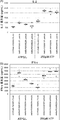
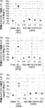
- the figure which shows the agonist activity of various anti-CD137 antibodies in the presence or absence of ATP tested using Jurkat cells The X-axis shows the antibody concentration ( ⁇ g / mL), and the Y-axis shows the relative luminescence.
- the figure which shows the agonist activity of various anti-CD137 antibodies in the presence or absence of ADP tested using Jurkat cells The X-axis shows the antibody concentration ( ⁇ g / mL), and the Y-axis shows the relative luminescence.
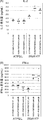
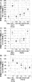
- the X axis shows the antibody concentration ( ⁇ g / mL), and the Y axis shows the amount of IFN ⁇ production (ng / mL).
- the Y axis shows the S / N ratio of absorbance in the presence / absence of ATP
- the X axis shows the S / N ratio in the presence / absence of antigen.
- the upper row shows the binding activity to human CD137 in the absence of ATP
- the lower row shows the binding activity to human CD137 in the presence of ATP.
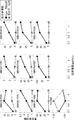
- FIG. 2 is a graph showing the agonist activity of a switch anti-CD137 antibody).
- A shows the test results in the absence of ADPbetaS
- B shows the test results in the presence of ADPbetaS.
- the X axis shows the antibody concentration ( ⁇ g / mL), and the Y axis shows the amount of IFN ⁇ production (ng / mL).
- (A) shows the test results in the absence of ATP
- (B) shows the test results in the presence of ATP.
- (A) shows the agonist activity measured using the amount of IL-2 produced as an index
- (B) shows the agonist activity measured using the amount of produced IFN- ⁇ as an index.
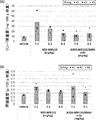
- various switch anti-CD137 antibodies were tested using human peripheral blood mononuclear cells to increase the binding activity of the heavy chain constant region to the Fc ⁇ receptor or increase the pI of the heavy chain constant region.
- (A) shows the agonist activity measured using the amount of IL-2 produced as an index
- (B) shows the agonist activity measured using the amount of produced IFN- ⁇ as an index.
- FIG. (A) shows the agonist activity measured using the amount of IL-2 produced as an index
- (B) shows the agonist activity measured using the amount of produced IFN- ⁇ as an index.
- FIG. (A) shows the agonist activity measured using the amount of IL-2 produced as an index
- (B) shows the agonist activity measured using the amount of produced IFN- ⁇ as an index.
- FIG. (A) shows the agonist activity measured using the amount of IL-2 produced as an index
- (B) shows the agonist activity measured using the amount of produced IFN- ⁇ as an index.
- FIG. (A) shows the agonist activity measured using the amount of IL-2 produced as an index
- (B) shows the agonist activity measured using the amount of produced IFN- ⁇ as an index.
- FIG. (A) shows the agonist activity measured using the amount of IL-2 produced as an index
- (B) shows the agonist activity measured using the amount of produced IFN- ⁇ as an index.
- (A) shows the agonist activity measured using the amount of IL-2 produced as an index
- (B) shows the agonist activity measured using the amount of produced IFN- ⁇ as an index.
- (A) shows the agonist activity measured using the amount of IL-2 produced as an index
- (B) shows the agonist activity measured using the amount of produced IFN- ⁇ as an index.
- (A) shows the agonist activity measured using the amount of IL-2 produced as an index
- (B) shows the agonist activity measured using the amount of produced IFN- ⁇ as an index.
- FIG. 1 shows the agonist activity measured using the amount of IL-2 produced as an index
- FIG. 2 shows the agonist activity measured using the amount of produced IFN- ⁇ as an index.
- (A) shows the agonist activity measured using the amount of IL-2 produced as an index
- (B) shows the agonist activity measured using the amount of produced IFN- ⁇ as an index.
- Increased agonist activity of various switch anti-CD137 antibodies in the presence or absence of ATP due to increased binding activity of heavy chain constant region to Fc ⁇ receptor, tested using human peripheral blood mononuclear cells
- (A) shows the agonist activity measured using the amount of IL-2 produced as an index
- (B) shows the agonist activity measured using the amount of produced IFN- ⁇ as an index.
- ⁇ Fc is mIgG1.
- Fc is MB110.
- Fc is MB492.
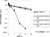
- the figure which shows the antitumor effect of A375-mIgG1 / B167-ml0r in the mouse model which transplanted MC38 cell. Each point indicates the average value of the tumor volume of one group (n 5).
- the diagram (A) shows the weight of the lymph nodes, and the diagram (B) shows the weight of the spleen.
- the diagram (A) shows the ratio of PD-1-positive T cells of CD8-positive T cells
- the diagram (B) shows the ratio of ICOS-positive T cells of CD8-positive T cells
- the diagram (C) shows CD8 The ratio of GranzymeB positive T cells of the positive T cells is shown.
- the diagram (A) shows the ratio of PD-1-positive T cells to CD8-positive T cells
- the diagram (B) shows the ratio of ICOS-positive T cells to CD8-positive T cells
- the graph (A) shows the ratio of PD-1-positive T cells of CD8-positive T cells
- the graph (B) shows the ratio of GranzymeB-positive T cells of CD8-positive T cells.
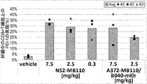
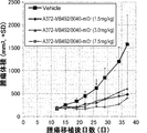
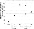
- the figure which shows the antitumor effect of A356-MB110 / B040-ml0r in the mouse model which transplanted MC38 cell line. Each point indicates the average value of the tumor volume of one group (n 5).
- the diagram (A) shows the weight of the lymph nodes
- the diagram (B) shows the weight of the spleen.
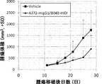
- the figure which shows the anti-tumor effect of A372-mIgG1 / B040-ml0r in the mouse model which transplanted the MC38 cell line. ⁇ Each point indicates the average value of the tumor volume of one group ⁇ n 5 ⁇ .
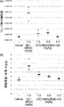
- FIG. 4 shows the number of lymph node cells (fraction (A)) and the weight of spleen (fraction (B)) of A38-mIgG1 / B040-ml0r administration in a mouse model transplanted with the MC38 cell line.
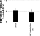
- FIG. 4 shows the degree of T cell activation in the liver (the ratio of CD8 + T cells to PD-1 + T cells) by NS2-MB110 or A372-MB110 / B040-ml0r administration in a mouse model transplanted with the MC38 cell line.
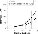
- FIG. 4 The figure which shows the antitumor effect of A372-MB492 / B040-ml0r in the mouse model which transplanted MC38 cell line.
- Plot (A) shows lymph node cell counts
- plot (B) shows spleen organ weight.
- the figure which shows the antitumor effect of A486-MB492 / B167-ml0r or A488-MB492 / B226-ml0r in the mouse model which transplanted the MC38 cell line. Each point indicates the average value of the tumor volume of one group (n 5).
- the figure (A) shows the number of cells per lymph node, and the figure (B) shows the spleen weight.
- the graph (A) shows the antitumor effect of A548-mIgG1 / B256-ml0r
- the graph (B) shows the antitumor effect of A551-mIgG1 / B256-ml0r.
- the diagram (A) shows the weight of the lymph nodes, and the diagram (B) shows the weight of the spleen.
- the graph (A) shows the ratio of PD-1 positive T cells to CD8-positive T cells
- the graph (B) shows the ratio of Granzyme ⁇ B-positive T cells to CD8-positive T cells.
- the diagram (A) shows the weight of the lymph nodes
- the diagram (B) shows the weight of the spleen.
- the diagram (C) shows CD8 3 shows the ratio of Granzyme B-positive T cells among positive T cells.
- the diagram (A) shows the ratio of PD-1-positive T cells to CD8-positive T cells
- the diagram (B) shows the ratio of ICOS-positive T cells to CD8-positive T cells
- the diagram (C) shows CD8 3 shows the ratio of Granzyme B-positive T cells among positive T cells.
- the X-axis shows the antibody concentration ( ⁇ g / mL), and the Y-axis shows the relative luminescence.
- ATP or ADP low molecular compound
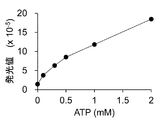
- FIG. 4 is a graph showing ATP responsiveness (ATP concentration-dependent luciferin emission) of P2Y11 split Luc / HEK293 cells prepared for measuring extracellular ATP levels.
- FIG. 4 is a diagram showing ATP responsiveness (luciferin luminescence depending on ATP concentration) in vivo when P2Y11 split Luc / HEK293 cells were implanted subcutaneously in mice.
- FIG. 9 shows the results of luminescence imaging measurements of mice transplanted subcutaneously with a predetermined concentration of ATP and P2Y11 split Luc / HEK293 cells and FM3A tumor-bearing mice transplanted subcutaneously with P2Y11 split Luc / HEK293 cells.
- the mark on the ventral part of the mouse indicates the detected light emission.
- FIG. 4 is a graph showing the binding activity (KD value) to hIL6R depending on the amount of hIL6R.
- KD value binding activity
- FIG. 4 is a graph showing the binding activity (KD value) to hIL6R depending on the amount of hIL6R.
- FIG. 3 is a graph showing ADCC activity depending on the activity of the present invention.
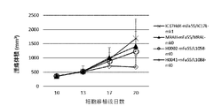
- FIG. 4 is a view showing the antitumor activity of IC17Hdk-mFa55 / IC17L-mk1 is a negative control antibody.
- FIG. 2 is a graph showing a comparison of the plasma kinetics of an anti-hIL6R antibody MRAH-mFa55 / MRAL-mk0 (control antibody) in normal mice and hIL6R transgenic mice.
- FIG. 4 shows a comparison of the plasma kinetics of H0002-mFa55 / L1058-ml0 (switch antibody), which is an anti-hIL6R antibody, in normal mice and hIL6R transgenic mice.
- the vertical axis of the graph indicates the plasma concentration of the antibody.
- FIG. 4 shows a comparison of the plasma kinetics of H0041-mFa55 / L1088-ml0 (switch antibody), which is an anti-hIL6R antibody, in normal mice and hIL6R transgenic mice.
- the vertical axis of the graph indicates the plasma concentration of the antibody.
- FIG. 9 shows comparison of plasma kinetics of H0052-mFa55 / L1083-ml0 (switch antibody), an anti-hIL6R antibody, in normal mice and hIL6R transgenic mice.
- the vertical axis of the graph indicates the plasma concentration of the antibody.
- MRAH-mFa55 / MRAL-mk0 which is an anti-hIL6R0non-switch antibody (control antibody)
- H0002-mFa55 / L1058-ml0 which is an anti-hIL6R switch antibody
- H0041-mFa55 / L1088-ml0 H0052-mFa55 / L1083-ml0
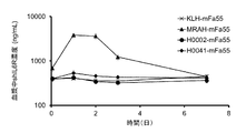
- FIG. 3 shows the accumulation of antigen in hIL6R transgenic mice after administration of each (all switch antibodies).
- the vertical axis of the graph indicates the plasma concentration of soluble hIL6R.
- IC17Hdk-mFa55 / IC17L-mk1 (denoted as KLH-mFa55 in the figure) is used as a negative control antibody.
- MRAH-mFa55 / MRAL-mk0 control antibody
- H0002-mFa55 / L1058-ml0 anti-hIL6R non-switch antibody
- H0041-mFa55 / L1088-ml0 all are switch antibodies
- IC17Hdk-mFa55 / IC17L-mk1 is a negative control antibody.
- Plasma of MRAH-mFa55 / MRAL-mk0 which is an anti-hIL6Rswitchnon-switch antibody (control antibody)
- the vertical axis of the graph indicates the plasma concentration of the antibody.
- FIG. 4 is a diagram showing accumulation of antigen after administration. The vertical axis of the graph indicates the plasma concentration of soluble hIL6R.
- IC17Hdk-mFa55 / IC17L-mk1 (denoted as KLH-mFa55 in the figure) is used as a negative control antibody.
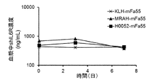
- MRAH-mFa55 / MRAL-mk0 which is an anti-hIL6R non-switch antibody (control antibody)
- H0041-mFa55 / L1088-ml0 which is an anti-hIL6RLswitch antibody
- H0052-mFa55 / L1083-ml0 all are switch antibodies
- IC17Hdk-mFa55 / IC17L-mk1 is a negative control antibody.
- FIG. 4 is a graph showing a comparison of plasma kinetics between MRAH-mFa55 / MRAL-mk0 (anti-hIL6R non-switch antibody) (control antibody) and H0052-mFa55 / L1083-ml0 (switch antibody), anti-hIL6R switch antibody.
- the vertical axis of the graph indicates the plasma concentration of the antibody.
- FIG. 9 is a diagram showing the accumulation of antigen after administration of MRAH-mFa55 / MRAL-mk0 (anti-hIL6R-non-switch antibody) (control antibody) and H0052-mFa55 / L1083-ml0 (switch antibody) of anti-hIL6R-switch antibody, respectively. is there.
- FIG. 3 is a graph showing the ATP concentration-dependent PD-1 / PDL-1 binding inhibitory activity of anti-PD1 antibodies mPD1F2VH-mF18 / mPD1F2VL-mk1 (control antibody) and H5029-mFa31 / L3021-ml0 (switch antibody).
- FIG. 3 is a graph showing the ATP concentration-dependent PD-1 / PDL-1 binding inhibitory activity of anti-PD1 antibodies mPD1F2VH-mF18 / mPD1F2VL-mk1 (control antibody) and H5029-mFa31 / L3021-ml0 (switch antibody).
- FIG. 3 is a diagram showing the ATP concentration-dependent PD-1 / PDL-1 binding inhibitory activity of anti-PD1 antibodies mPD1F2VH-mF18 / mPD1F2VL-mk1 (control antibody) and H5041-mFa31 / L3021-ml0 (switch antibody).
- H5029-mFa31 / L3021-ml0, H5041-mFa31 / L3021-ml0 all switch antibodies depending on AMP concentration It is a figure which shows activity.
- FIG. 4 is a view showing the antitumor activity of mPD1F2VH-mFa55 / mPD1F2VL-mk1 (control antibody) and H5041-mFa55 / L3023-ml0 (switch antibody), which are anti-PD1 antibodies, in vivo.
- IC17Hdk-mFa55 / IC17L-mk1 is a negative control antibody.
- the anti-PD1 antibodies mPD1F2VH-mFa55 / mPD1F2VL-mk1 (control antibody) and H5041-mFa55 / L3023-ml0 (switch antibody) show the activity of removing PD-1 expressing cells in (A) tumor and (B) spleen.
- FIG. 2 is a view showing a mode of binding between H0041L1088 ⁇ Fab fragment which is an anti-hIL6R ⁇ switch antibody and ATP.
- ATP is represented by a ball-and-stick model
- amino acid residues that form an interaction with ATP are represented by a stick model.
- the dashed line indicates a hydrogen bond between the antibody and ATP.
- FIG. 2 is a diagram in which an epitope of an anti-hIL6R switch antibody, H0041L1088, is mapped on the amino acid sequence of the hIL6R extracellular domain (shIL6R).
- the amino acid residues shaded in gray are the amino acid residues of shIL6R containing one or more non-hydrogen atoms located within a distance of 4.2 mm from either H0041L1088 ⁇ Fab or ATP) in the crystal structure (epitope residues). Is shown.
- FIG. 4 is a diagram showing details of the binding between ATP-bound H0041L1088 ⁇ Fab fragment and shIL6R. In the figure, the heavy chain of the antibody is drawn in black, the light chain is drawn in gray, and shIL6R is drawn in white.
- FIG. 89 is a view showing a structure in a case where the structure of FIG. 86 is rotated by 180 degrees (as viewed from the back).
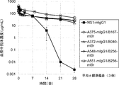
- FIG. 3 is a graph showing a comparison of the kinetics in plasma of A375-SCF041aPh / B167-Lamlib and A375-MY201aPh / B167-Lamlib, which are anti-CD137 switch antibodies.
- the vertical axis of the graph indicates the plasma concentration of the antibody.
- binding activity refers to the non-covalent interaction of one or more binding sites on a molecule (eg, an antibody) with a binding partner (eg, an antigen) of the molecule. It refers to the total strength.
- binding activity is not strictly limited to a 1: 1 interaction between members of a binding pair (eg, antibody and antigen). For example, if a member of a binding pair reflects a monovalent 1: 1 interaction, this binding activity is specifically referred to as intrinsic binding affinity ("affinity"). If a member of a binding pair is capable of both monovalent and multivalent binding, the avidity will be the sum of these avidities.
- binding activity of a molecule X to its partner Y can generally be represented by a dissociation constant ⁇ (KD) ⁇ or "amount of analyte bound per unit ligand amount” (hereinafter sometimes referred to as "binding amount”).
- KD dissociation constant
- Binding activity can be measured by conventional methods known in the art, including those described herein. Specific illustrative and exemplary embodiments for measuring binding activity are described below.
- An "avidity-matured” antigen-binding molecule or antibody, or an “increased avidity (enhanced)" antigen-binding molecule or antibody has one or more antigen-binding molecules or antibodies as compared to a parent antigen-binding molecule or parent antibody without modification.
- anti-CD137 antigen-binding molecule antigen-binding molecule
- anti-CD137 antibody or antigen that binds to CD137
- antibody that binds to CD137 are antigen-binding molecules or antibodies that can bind to CD137 with sufficient binding activity.
- the anti-CD137 antibody binds to a conserved epitope of CD137 between CD137 from different species.
- ⁇ having a CD137-binding activity dependent on a low-molecular compound '' an anti-CD137 antigen-binding molecule or an anti-CD137 antibody has a binding activity to CD137 in the presence of the low-molecular compound in the absence of the low-molecular compound.
- “in the presence of a low-molecular compound” refers to a condition in which the low-molecular compound is present at 10 ⁇ M or more, 50 ⁇ M or more, 100 ⁇ M or more, 150 ⁇ M or more, 200 ⁇ M or more, or 250 ⁇ M or more.
- the degree of avidity of an anti-CD137 antigen binding molecule or antibody to an irrelevant non-CD137 protein in the presence of a small molecule compound is determined by, for example, radioimmunoassay (RIA) or surface plasmon resonance analysis. Less than about 10% of the binding of the antigen-binding molecule or antibody to CD137 as measured by the method of surface plasmon resonance (SPR).
- RIA radioimmunoassay
- SPR surface plasmon resonance
- the anti-CD137 antigen binding molecule or antibody is ⁇ 1 ⁇ M, ⁇ 100 nM, ⁇ 10 nM, ⁇ 1 nM, ⁇ 0.1 nM, ⁇ 0.01 nM, or ⁇ 0.001 nM (eg, -6 M or less, 10 -7 M or less, 10 -8 M or less, 10 -9 M or less, 10 -10 M or less, for example, 10 -6 M to 10 -10 M, 10 -7 M to 10 -9 M , 10 -7 M to 10 -8 M).
- the term “antigen binding molecule” is used in its broadest sense and refers to a molecule that specifically binds to an antigenic determinant.
- the antigen binding molecule is an antibody, antibody fragment, or antibody derivative.
- an “agonist antigen-binding molecule” or “agonist antibody” is an antigen-binding molecule or antibody that significantly induces or enhances the biological activity of the antigen (eg, CD137, CD3) to which it binds. . Therefore, for example, when the antigen is CD137, the antigen-binding molecule or antibody having such an agonistic action is referred to as “CD137 agonist-antigen binding molecule” or “CD137 agonist antibody”, respectively. Similarly, for example, when the antigen is CD3, the antigen-binding molecule or antibody having such an agonistic effect is referred to as “CD3 agonist-antigen binding molecule” or “CD3 agonist antibody”, respectively.
- antibody is used in the broadest sense, and is not limited to monoclonal antibodies, polyclonal antibodies, multispecific antibodies (e.g., And various antibody structures, including bispecific antibodies) and antibody fragments.
- Antibody fragment refers to a molecule other than a complete antibody, including a portion of the complete antibody that binds to the antigen to which the complete antibody binds.
- Examples of antibody fragments include, but are not limited to, Fv, Fab, Fab ', Fab'-SH, F (ab') 2 ; diabodies; linear antibodies; single-chain antibody molecules (eg, scFv ); And multispecific antibodies formed from antibody fragments.
- a ⁇ antigen binding molecule that binds to the same epitope '' or a ⁇ antibody that binds to the same epitope '' as the reference antigen-binding molecule or reference antibody is 50% less likely to be bound by the reference antibody or reference antigen-binding molecule to its own antigen in a competition assay.
- An antibody or antigen-binding molecule that blocks the above, or conversely, a reference antibody blocks the binding of said antibody to its antigen in a competition assay by 50% or more.
- An exemplary competition assay is provided herein. In one embodiment, when the reference antigen-binding molecule or reference antibody has an antigen-binding activity dependent on the small molecule compound, the competition assay is performed in the presence of the small molecule compound.
- chimeric antibody refers to a portion of the heavy and / or light chain derived from a particular source or species, while the remainder of the heavy and / or light chain is derived from a different source or species. Refers to an antibody.
- the “class” of an antibody refers to the type of constant domain or constant region provided in the heavy chain of the antibody.
- the heavy-chain constant domains that correspond to the different classes of immunoglobulins are called ⁇ , ⁇ , ⁇ , ⁇ , and ⁇ , respectively.
- Antibody function refers to a biological activity attributable to the Fc region of an antibody that varies depending on the isotype of the antibody.
- Examples of antibody effector functions include: C1q binding and complement-dependent cytotoxicity (CDC); Fc receptor binding; antibody-dependent cell-mediated cytotoxicity (antibody-dependent cell) -mediated cytotoxicity: ADCC); phagocytosis; downregulation of cell surface receptors (eg, B cell receptors); and B cell activation.
- Cytotoxic activity refers to an activity that inhibits or prevents the function of cells and / or causes death or destruction of cells. Cytotoxic activity includes, for example, antibody-dependent cell-mediated cytotoxicity (ADCC) activity, complement-dependent cytotoxicity (CDC) activity, and cytotoxic activity by T cells. And may be caused by a cytotoxic agent (for example, a radioisotope or a chemotherapeutic agent) such as an immunoconjugate.
- a cytotoxic agent for example, a radioisotope or a chemotherapeutic agent
- Fc region is used herein to define the C-terminal region of an immunoglobulin heavy chain that includes at least a portion of the constant region.
- the term includes native sequence Fc regions and variant Fc regions.
- the human IgG heavy chain Fc region extends from Cys226 or from Pro230 to the carboxyl terminus of the heavy chain.
- C-terminal lysine ⁇ (Lys447) ⁇ or glycine-lysine (Gly446-Lys447) at the Fc region may or may not be present.
- numbering of amino acid residues in the Fc region or constant region is based on Kabat et al., Sequences of Proteins of Immunological Interest, 5th Ed. Public Health Service, National Institutes of Health, Bethesda, According to the EU numbering system (also called the EU index) described in MD ⁇ 1991 ⁇ .
- the term "mutant Fc region" includes an amino acid sequence that differs from that of the native sequence Fc region by at least one amino acid modification, preferably one or more amino acid substitutions.
- the mutated Fc region has at least one amino acid substitution in the native sequence Fc region or in the Fc region of the parent polypeptide, eg, from about 1 to about 100, relative to the native sequence Fc region or the parent polypeptide Fc region. It has about 10 amino acid substitutions, preferably about 1 to about 5 amino acid substitutions.
- the variant Fc region herein is preferably at least about 80% homologous to the native sequence Fc region and / or the Fc region of the parent polypeptide, most preferably at least about 90% homologous thereto, more preferably Have at least about 95% homology with them.
- an amino acid modification or substitution in the Fc region or constant region may be represented by a combination of the EU numbering system and an amino acid.
- S424N represents the substitution of serine (Ser) at position 424 of the EU numbering with asparagine (Asn).
- EU424N represents substitution of the amino acid at position 424 (any type) with asparagine (Asn).
- the term "Fc region-containing antibody” refers to an antibody containing an Fc region.
- the C-terminal lysine of the Fc region (residue 447 according to the EU numbering system) or the C-terminal glycine-lysine of the Fc region (residues 446-447) can be, for example, during purification of the antibody or in the nucleic acid encoding the antibody. It can be removed by a recombination operation.
- a composition comprising an antibody having an Fc region according to the present disclosure may be an antibody with G446-K447, an antibody without G447 with G446, an antibody with G446-K447 completely removed, or an antibody of the above three types. May be included.
- full-length antibody “complete antibody,” and “whole antibody” are used interchangeably herein and have a structure substantially similar to the native antibody structure, or are defined herein.
- a “human antibody” is an antibody comprising an amino acid sequence corresponding to the amino acid sequence of an antibody produced by a human or human cell or an antibody derived from a non-human source using the human antibody repertoire or other human antibody coding sequences. This definition of a human antibody specifically excludes a humanized antibody containing non-human antigen binding residues.
- variable domain residues other than the hypervariable region ⁇ (HVR) ⁇ residues.
- the variable domain FRs usually consist of four FR domains: FR1, FR2, FR3, and FR4. Accordingly, HVR and FR sequences usually appear in VH (or VL) in the following order: FR1-H1 (L1) -FR2-H2 (L2) -FR3-H3 (L3) -FR4.
- An ⁇ acceptor human framework '' for the purposes of this specification is a light chain variable domain ⁇ (VL) ⁇ framework or heavy chain variable domain (VH) derived from a human immunoglobulin framework or human consensus framework as defined below.
- a framework containing the amino acid sequence of the framework Acceptor human frameworks "derived from" the human immunoglobulin framework or the human consensus framework may include those same amino acid sequences, or may include amino acid sequence alterations. In some embodiments, the number of amino acid changes is 10 or less, 9 or less, 8 or less, 7 or less, 6 or less, 5 or less, 4 or less, 3 or less, or 2 or less.
- the VL acceptor human framework is identical in sequence to a VL human immunoglobulin framework sequence or a human consensus framework sequence.
- Human consensus framework is a framework that indicates the most commonly occurring amino acid residues in a selected group of human immunoglobulin VL or VH framework sequences. Usually, the selection of human immunoglobulin VL or VH sequences is from a subgroup of variable domain sequences. Usually, the subgroups of the sequences are the subgroups in Kabat et al., Sequences of Proteins of Immunology Interest, Fifth Edition, NIH Publication 91-3242, Bethesda MD (1991), vols. 1-3. In one embodiment, for VL, the subgroup is subgroup ⁇ I by Kabat et al., Supra. In one embodiment, for VH, the subgroup is subgroup III by Kabat et al., Supra.
- Humanized antibody refers to a chimeric antibody comprising amino acid residues from a non-human HVR and amino acid residues from a human FR.
- a humanized antibody comprises substantially all of at least one, and typically two, variable domains in which all or substantially all HVRs (eg, CDRs) are non-human. All or substantially all FRs correspond to those of a human antibody and correspond to those of a human antibody.
- the humanized antibody may optionally include at least a portion of an antibody constant region derived from a human antibody.
- a “humanized form” of an antibody (eg, a non-human antibody) refers to an antibody that has undergone humanization.
- variable region refers to the domain of an antibody heavy or light chain that is involved in binding an antibody to an antigen.
- the variable domains of the heavy and light chains of the native antibody are generally similar, with each domain containing four conserved framework regions ⁇ (FR) ⁇ and three hypervariable regions ⁇ (HVR) ⁇ . Having a structure. (See, eg, Kindt et al. Kubin Immunology, 6th Ed., WH. Freeman and Co., page 91 (2007).)
- One VH or VL domain will be sufficient to confer antigen binding specificity.
- antibodies that bind to a particular antigen may be isolated by screening a complementary library of VL or VH domains, respectively, using VH or VL domains from the antibody that binds to the particular antigen. See, for example, Portolano et al., J. Immunol. 150: 880-887 (1993); Clarkson et al., Nature 352: 624-628 (1991).
- hypervariable region is hypervariable in sequence (“complementarity determining region” or “CDR” (complementarity determining region)) and / or structurally defined Refers to each region of the variable domain of an antibody that forms a loop ("hypervariable loop") and / or contains antigen contact residues ("antigen contact”).
- CDR complementarity determining region
- antigen contact usually, antibodies contain six HVRs: three for VH (H1, H2, H3) and three for VL (L1, L2, L3).
- Exemplary HVRs herein include the following: (a) at amino acid residues 26-32 (L1), 50-52 (L2), 91-96 (L3), 26-32 (H1), 53-55 (H2), and 96-101 (H3) The resulting hypervariable loop (Chothia and Lesk, J. Mol. Biol. 196: 901-917 (1987)); (b) at amino acid residues 24-34 (L1), 50-56 (L2), 89-97 (L3), 31-35b (H1), 50-65 (H2), and 95-102 (H3) Resulting CDRs (Kabat et al., Sequences of Proteins of Immunological Interest, 5th Ed.
- HVR residues and other residues in variable domains are numbered herein according to Kabat et al., Supra.
- HVR residues and other residues in the variable domain can be represented by a combination of the Kabat numbering system and the amino acids.
- N99 represents asparagine (Asn) at position 99 of Kabat numbering
- N99A represents substitution of asparagine (Asn) at position 99 of Kabat with alanine (Ala).
- Immunoconjugate is an antibody conjugated to one or more heterologous molecules, including, but not limited to, cytotoxic agents.
- cytotoxic agent refers to a substance that inhibits or prevents the function of cells and / or causes death or destruction of cells.
- Cytotoxic agents include, but are not limited to, radioisotopes (eg, 211 At, 131 I, 125 I, 90 Y, 186 Re, 188 Re, 153 Sm, 212 Bi, 32 P, 212 Pb And Lu radioisotopes); chemotherapeutic or chemotherapeutic agents (eg, methotrexate, adriamycin, vinca alkaloids (vincristine, vinblastine, etoposide), doxorubicin, melphalan, mitomycin C, chlorambucil, daunorubicin, or other intercalates) Growth inhibitors; enzymes such as nucleases and fragments thereof; antibiotics; for example, small molecule toxins or enzymatically active toxins of bacterial, fungal, plant, or animal origin (fragments and / or mutations
- Isolated antibody is one that is separated from components of its original environment.
- the antibody is, for example, electrophoretically (eg, SDS-PAGE, isoelectric focusing (IEF), capillary electrophoresis) or chromatograph (eg, ion exchange or reverse phase HPLC). Measured and purified to greater than 95% or 99% purity.
- electrophoretically eg, SDS-PAGE, isoelectric focusing (IEF), capillary electrophoresis
- chromatograph eg, ion exchange or reverse phase HPLC
- isolated nucleic acid refers to a nucleic acid molecule that has been separated from components of its original environment.
- An isolated nucleic acid includes a nucleic acid molecule contained within a cell that normally contains the nucleic acid molecule, but the nucleic acid molecule is extrachromosomal or located on a chromosome that differs from its original chromosomal location. Exists in position.
- vector refers to a nucleic acid molecule that can propagate another nucleic acid to which it has been linked.
- the term includes vectors as self-replicating nucleic acid structures, as well as vectors that integrate into the genome of a host cell into which they are introduced. Certain vectors can provide for the expression of a nucleic acid to which they are operatively linked. Such vectors are also referred to herein as "expression vectors”.
- nucleic acid encoding an anti-CD137 antigen binding molecule refers to one or more nucleic acid molecules encoding a polypeptide that constitutes an antigen binding molecule.
- isolated nucleic acid encoding an anti-CD137 antibody refers to one or more nucleic acid molecules that encode the heavy and light chains (or fragments thereof) of an antibody and may be contained in one vector or separate vectors. Riding nucleic acid molecules, and nucleic acid molecules that are present at one or more locations in a host cell.
- host cell refers to cells into which a foreign nucleic acid has been introduced, including the progeny of such cells.
- Host cells include “transformants” and “transformed cells,” which include the primary transformed cell and progeny derived therefrom without regard for the number of transfers. Progeny may not be completely identical in nucleic acid content to the parent cell, but may contain mutations. Mutant progeny that have the same function or biological activity as those used when the originally transformed cells were screened or selected are also included herein.
- the term "monoclonal antibody” as used herein refers to an antibody obtained from a substantially homogeneous population of antibodies. That is, the individual antibodies that make up the population are likely to be mutated antibodies (eg, mutated antibodies containing naturally occurring mutations, or mutated antibodies generated during the preparation of monoclonal antibody preparations. In the same amount and / or binds to the same epitope. In contrast to polyclonal antibody preparations, which typically include different antibodies for different determinants (epitopes), each monoclonal antibody in a monoclonal antibody preparation is for a single determinant on the antigen.
- mutated antibodies eg, mutated antibodies containing naturally occurring mutations, or mutated antibodies generated during the preparation of monoclonal antibody preparations. In the same amount and / or binds to the same epitope.
- polyclonal antibody preparations typically include different antibodies for different determinants (epitopes)
- monoclonal indicates the character of the antibody as being obtained from a substantially homogeneous population of antibodies, and is not to be construed as requiring production of the antibody by any particular method.
- monoclonal antibodies used in accordance with the present disclosure include, but are not limited to, hybridoma methods, recombinant DNA methods, phage display methods, transgenic animals containing all or part of a human immunoglobulin locus.
- Such methods and other exemplary methods for making monoclonal antibodies can be made by various techniques, including those that make use of the methods described herein.
- naked antibody refers to an antibody that is not conjugated to a heterologous moiety (eg, a cytotoxic moiety) or a radiolabel. Naked antibodies may be present in a pharmaceutical formulation.
- “Native antibody” refers to an immunoglobulin molecule with various structures that occur in nature.
- a native IgG antibody is a heterotetrameric glycoprotein of about 150,000 daltons composed of two identical disulfide-linked light chains and two identical heavy chains. From the N-terminus to the C-terminus, each heavy chain has a variable region ⁇ (VH) ⁇ , also called a variable heavy chain domain or heavy chain variable domain, followed by three constant domains (CH1, CH2, and CH3). Similarly, from the N-terminus to the C-terminus, each light chain has a variable region ⁇ (VL) ⁇ , also referred to as a variable light chain domain or a light chain variable domain, followed by a constant light chain ⁇ (CL) ⁇ domain.
- the light chain of an antibody may be assigned to one of two types, called kappa ( ⁇ ) and lambda ( ⁇ ), based on the amino acid sequence of its constant domain.
- Percent (%) amino acid sequence identity refers to the sequence alignment to obtain maximum percent sequence identity and, if necessary, introducing gaps and any conservative substitutions in the sequence. It is defined as the percentage of amino acid residues in the candidate sequence that are identical to amino acid residues in the reference polypeptide sequence when not considered part of the identity. Alignments for the purpose of determining percent amino acid sequence identity can be obtained by various methods within the skill in the art, for example, BLAST, BLAST-2, ALIGN, Megalign (DNASTAR) software, or GENETYX® (stock) This can be achieved by using publicly available computer software, such as the company Genetics. One skilled in the art can determine the appropriate parameters for aligning the sequences, including any algorithms necessary to achieve maximum alignment over the entire length of the sequences being compared.
- the ALIGN-2 sequence comparison computer program is the work of Genentech, the source code of which has been submitted to the United States Copyright Office (US Copyright Office, Wasington DC, 20559) along with user documentation and has U.S. copyright registration number TXU510087. It is registered.
- the ALIGN-2 program is publicly available from Genentech, Inc., South San Francisco, California or may be compiled from source code.
- the ALIGN-2 program is compiled for use on UNIX operating systems, including Digital UNIX V4.0D. All sequence comparison parameters are set by the ALIGN-2 program and do not vary.
- the percent amino acid sequence identity of a given amino acid sequence A to, to, or to a given amino acid sequence B (or alternatively, to a given amino acid sequence) A given amino acid sequence A having or including some% amino acid sequence identity to, with, or to B) can be calculated as follows: fraction of X / Y Hundredfold. Where X is the number of amino acid residues scored by the sequence alignment program ALIGN-2 as being identical in the alignment of A and B of the program, and Y is the total number of amino acid residues in B .
- pharmaceutical formulation is a preparation in a form such that the biological activity of the active ingredient contained therein can exert its effect and is unacceptable to the subject to whom the formulation is administered.
- “Pharmaceutically acceptable carrier” refers to a component other than the active ingredient in a pharmaceutical formulation that is nontoxic to a subject.
- Pharmaceutically acceptable carriers include, but are not limited to, buffers, excipients, stabilizers, or preservatives.
- ⁇ An effective amount "of an agent refers to an amount, at the required dosage, and over a required period of time, that is effective to achieve the desired therapeutic or prophylactic result.
- “Individual” or “subject” is a mammal. Mammals include, but are not limited to, domestic animals (eg, cows, sheep, cats, dogs, horses), primates (eg, humans and non-human primates such as monkeys), rabbits, and And rodents (eg, mice and rats). In certain aspects, the individual or subject is a human.
- CD137 refers to any source from any vertebrate source, including mammals such as primates (eg, humans) and rodents (eg, mice and rats). Means natural CD137.
- the term encompasses CD137 that has not been processed "full-length,” as well as any form of CD137 that results from processing in the cell.
- the term also includes naturally occurring variants of CD137, for example, splice variants and allelic variants.
- SEQ ID NO: 1 NCBI Reference Sequence: NP_001552.2
- amino acid sequence of the extracellular region of exemplary human CD137 is shown in SEQ ID NO: 2.
- exemplary mouse CD137 is shown in SEQ ID NO: 3 (NCBI Reference Sequence: NP_035742.1), and the amino acid sequence of the extracellular region of exemplary mouse CD137 is shown in SEQ ID NO: 4.
- the full length amino acid sequence of an exemplary monkey CD137 is shown in SEQ ID NO: 5 (NCBI Reference Sequence: ABY47575.1), and the amino acid sequence of the extracellular region of the exemplary monkey CD137 is shown in SEQ ID NO: 6.
- CD137 is a member of the tumor necrosis factor (TNF) receptor family. Its alternative names are tumor necrosis factor receptor superfamily member 9 (TNFRSF9), 4-1BB, ILA.
- CD137 In addition to its expression on activated CD4 + and CD8 + T cells, CD137 also expresses B cells, dendritic cells, natural killer (NK) and NK-T cells, macrophages, monocytes, neutrophils, It is expressed on CD4 + CD25 + regulatory T cells and vascular endothelial cells. It has also been shown to be expressed in cancer cells (Labiano et al., Oncoimmunology, 24: e1062967 (2015)). Its natural ligand, CD137L, has been shown on antigen presenting cells such as B cells, monocytes / macrophages, and dendritic cells (Watts et al., Annu. Rev. Immunol. 23: 23-68 ( Year 2005)).
- CD137 results in increased TCR-induced T cell proliferation, cytokine production, functional maturation, suppression of apoptosis, and prolonged CD8 + T cell survival (Nam et al., Curr. Cancer Drug Drug). Targets, 5: 357-363 (2005), Watts et al., Annu. Rev. Immunol., 23: 23-68 (2005)).
- cancer refers to or describe the physiological condition in mammals that is typically characterized by unregulated cell growth / proliferation.
- tumor refers to all neoplastic cell growth and proliferation, whether malignant or benign, and all precancerous and cancerous cells and tissues.
- cancer refers to all neoplastic cell growth and proliferation, whether malignant or benign, and all precancerous and cancerous cells and tissues.
- cancer refers to all neoplastic cell growth and proliferation, whether malignant or benign, and all precancerous and cancerous cells and tissues.
- cancer refers to all neoplastic cell growth and proliferation, whether malignant or benign, and all precancerous and cancerous cells and tissues.
- cell proliferative disorder and “proliferative disorder” refer to disorders associated with some degree of abnormal cell proliferation.
- the cell proliferative disorder is a cancer.
- treatment refers to a clinical trial intended to alter the natural history of the treated individual. It refers to intervention and can be performed both for prevention and during the course of the clinical condition. Desirable effects of treatment include, but are not limited to, preventing the occurrence or recurrence of a disease, reducing symptoms, attenuating any direct or indirect pathological effects of the disease, preventing metastasis, Includes a reduction in the rate of progression, amelioration or amelioration of the disease state, and remission or improved prognosis.
- the antibodies of the present disclosure are used to slow the onset of the disease or to slow the progression of the disease.
- compositions and methods (anti-CD137 agonist antigen binding molecules)
- the present disclosure is based on anti-CD137 agonist antigen binding molecules, and some uses thereof.
- an antibody that binds to CD137 is provided. Since the antibody of the present disclosure can exert an activating action, a cytotoxic activity, or an antitumor activity of immune cells, it is useful, for example, for diagnosis or treatment of cancer.
- the disclosure provides an isolated antigen binding molecule or antibody that binds CD137.
- the anti-CD137 antigen binding molecule or antibody is ⁇ Has CD137-binding activity dependent on low-molecular compounds; Binds to the extracellular region of CD137; Forming a tripartite complex with low molecular weight compounds and CD137; Binds to CD137 from humans and monkeys; -An agonist of CD137 activity; Exhibits an agonistic activity against CD137 in the presence of low molecular weight compounds; -Low agonist activity on CD137 in the absence of low molecular weight compound; and / or substantially no agonistic activity on CD137 in absence of low molecular weight compound.
- the binding activity of an antigen binding molecule or antibody is ⁇ 1 ⁇ M, ⁇ 100 nM, ⁇ 10 nM, ⁇ 1 nM, ⁇ 0.1 nM, ⁇ 0.1 nM, ⁇ 0.01 nM or ⁇ 0.001 nM (eg, 10 ⁇ 6 M or less, 10 ⁇ 7 M or less, 10 ⁇ 8 M or less, 10 ⁇ 9 M or less, 10 ⁇ 10 M or less, eg, 10 ⁇ 6 M to 10 ⁇ 10 M , 10 ⁇ 7 M to 10 ⁇ 9 M, for example, 10 ⁇ 7 M to 10 ⁇ 8 M).
- the binding activity of an antigen binding molecule or antibody is measured by a radiolabeled antigen binding assay (RIA) and is expressed as KD.
- the RIA is performed using a Fab version of the antibody of interest and its antigen.
- the solution binding affinity of a Fab to an antigen is achieved by equilibrating the Fab with a minimal concentration of ( 125I ) labeled antigen in the presence of an increasing series of unlabeled antigen, and then coating the bound antigen with an anti-Fab antibody. It is measured by capturing with a plate. (See, for example, Chen et al., J. Mol. Biol. 293: 865-881 (1999)).
- MICROTITER® multi-well plates were coated overnight with 5 ⁇ g / ml capture anti-Fab antibody (Cappel Labs) in 50 mM sodium carbonate (pH 9.6), followed by Block with 2% (w / v) bovine serum albumin in PBS for 2-5 hours at room temperature (approximately 23 ° C.).
- a non-adsorption plate (Nunc # 269620) 100 pM or 26 pM of [ 125 I] -antigen was added to the anti-VEGF antibody (for example, Presta et al., Cancer Res. Mix with serial dilutions of the desired Fab (as in the evaluation of 12).
- the Fab of interest is then incubated overnight, which may be continued for a longer time (eg, about 65 hours) to ensure that equilibrium is achieved. Thereafter, the mixture is transferred to a capture plate for incubation at room temperature (eg, 1 hour). The solution is then removed and the plate is washed eight times with 0.1% polysorbate 20 in PBS (TWEEN-20®). When the plates have dried, 150 ⁇ l / well of scintillant (MICROSCINT-20 TM, Packard) is added and the plates are counted on a TOPCOUNT TM gamma counter (Packard) for 10 minutes. The concentration of each Fab that gives less than 20% of the maximum binding is selected for use in the competition binding assay.
- MICROSCINT-20 TM MICROSCINT-20 TM, Packard
- the binding activity of the antibody is determined by surface plasmon resonance analysis, eg, ligand capture using BIACORE® T200 or BIACORE® 4000 (GE Healthcare, Uppsala, Sweden). Method is used. BIACORE (registered trademark) Control @ Software is used for device operation.
- BIACORE registered trademark
- an amine coupling kit (GE Healthcare, Uppsala, Sweden) is used according to the supplier's instructions, and a carboxymethyl dextran-coated sensor chip (GE Healthcare, Uppsala, Sweden) is loaded onto a ligand-capturing molecule, such as an anti-tag.
- An antibody, anti-IgG antibody, protein A, etc. are immobilized.
- the ligand capture molecule is diluted with a 10 mM mM sodium acetate solution at the appropriate pH and injected at the appropriate flow rate and injection time.
- a buffer containing 0.05% polysorbate 20 Teween (registered trademark) -20
- the flow rate was 10- ⁇ 30 ⁇ ⁇ L / min
- the measurement temperature was preferably 25 ° C. Measured at 37 ° C.
- the measurement results are analyzed using BIACORE® Evaluation Software.
- the kinetic parameter calculation is performed by simultaneously fitting the binding and dissociation sensorgrams using a 1: 1 binding model, and the binding rate (kon or ka) and dissociation rate (koff or kd) ,
- the equilibrium dissociation constant (KD) can be calculated.
- the Steady state model may be used to calculate the equilibrium dissociation constant (KD).
- the “analyte binding amount per unit ligand amount” is also calculated by dividing the binding amount (resonance unit: RU) of the analyte at a specific concentration by the ligand capture amount (RU). obtain.
- the anti-CD137 antigen-binding molecule or antibody has CD137-binding activity dependent on a small molecule compound.
- the anti-CD137 antigen-binding molecule or antibody has a higher binding activity to CD137 in the presence of the small molecule compound than the binding activity of CD137 in the absence of the small molecule compound.
- the anti-CD137 antigen binding molecule or antibody has a binding activity to CD137 in the presence of a high concentration of the low molecular compound that is lower than the binding activity of CD137 in the presence of a low concentration of the low molecular compound. high.
- the anti-CD137 antigen-binding molecule or antibody has a binding activity to CD137 in the presence of the low-molecular compound, which is at least twice the binding activity of CD137 in the absence of the low-molecular compound, 3 times or more, 5 times or more, 10 times or more, 15 times or more, 20 times or more, 25 times or more, 30 times or more, 50 times or more, 100 times or more, 200 times or more, 300 times or more, 500 times or more, 1 ⁇ 10 3 times or more, 2 ⁇ 10 3 times or more, top, 3 ⁇ 10 3 times or more, 5 ⁇ 10 3 times or more, 1 ⁇ 10 4 times or more, 2 ⁇ 10 4 times or more, 3 ⁇ 10 4 times or more, 5 ⁇ 10 4 times or more, or 1 ⁇ 10 5 times or more.
- the anti-CD137 antigen-binding molecule or antibody has a binding activity to CD137 in the presence of the small molecule compound of more than twice that of CD137 in the absence of the small molecule compound. High, 3 times higher, 5 times higher, 10 times higher, 15 times higher, 20 times higher, 25 times higher, 30 times higher, 50 times higher, 100 times higher, 200 times higher High, higher than 300 times, higher than 500 times, higher than 1 ⁇ 10 3 times, higher than 2 ⁇ 10 3 times, upper, higher than 3 ⁇ 10 3 times, higher than 5 ⁇ 10 3 times, 1 ⁇ 10 4 2x10 4 times higher, 3x10 4 times higher, 5x10 4 times higher, or 1x10 5 times higher.
- the concentration of the low-molecular compound any concentration can be selected as long as a difference in the binding activity of the anti-CD137 antigen-binding molecule or the antibody is detected.
- the concentration of the low-molecular compound in "in the presence of a low-molecular compound” and / or "in the presence of a high-concentration low-molecular compound” is, for example, 100 nM or more, 500 nM or more, 1 ⁇ M or more, 3 ⁇ M or more, 5 ⁇ M or more , 10 ⁇ M or more, 50 ⁇ M or more, 100 ⁇ M or more, 150 ⁇ M or more, 200 ⁇ M or more, 250 ⁇ M or more, 300 ⁇ M or more, 400 ⁇ M or more, 500 ⁇ M or more, or 1 mM or more.
- the concentration may be a sufficient amount such that each anti-CD137 antigen-binding molecule or antibody exhibits the maximum binding activity.
- the concentration of the low-molecular compound in the ⁇ in the presence of a low-concentration low-molecular compound '' is, for example, 500 ⁇ M or less, 250 ⁇ M or less, 200 ⁇ M or less, 150 ⁇ M or less, 100 ⁇ M or less, 50 ⁇ M or less, 10 ⁇ M or less, 1 ⁇ M or less , 500 nM or less, 100 nM or less, 50 nM or less, 10 nM or 1 nM or less.
- concentration of the low-molecular compound is zero or the substantial concentration is zero can be selected as one embodiment of the low concentration.
- substantially concentration is cello refers to, for example, a very small concentration at which a low-molecular-weight compound exists, but the concentration cannot be detected by current technology.
- the binding activity to CD137 in the presence of 10 ⁇ M, 50 ⁇ M, 100 ⁇ M, 150 ⁇ M, 200 ⁇ M, or 250 ⁇ M of the low-molecular compound is at least twice the binding activity to CD137 in the absence of the low-molecular compound. , 5 times or more, 10 times or more, 15 times or more, 16 times or more, 17 times or more, 18 times or more, 19 times or more, or 20 times or more.
- the anti-CD137 antigen-binding molecule or antibody has a binding activity to CD137 in the presence of a low-molecular compound of 10 ⁇ M or more, more than twice the binding activity to CD137 in the absence of the low-molecular compound, 5 times or more, 10 times or more, 15 times or more, 16 times or more, 17 times or more, 18 times or more, 19 times or more, or 20 times or more.
- the anti-CD137 antigen-binding molecule or antibody has a binding activity to CD137 in the presence of a low-molecular compound of 100 ⁇ M or more, more than twice the binding activity to CD137 in the absence of the low-molecular compound, 5 times or more, 10 times or more, 15 times or more, 16 times or more, 17 times or more, 18 times or more, 19 times or more, or 20 times or more.
- the anti-CD137 antigen binding molecule or an antibody, binding activity to CD137 in the presence of more low molecular compound 10 [mu] M (KD) is, 9 ⁇ 10 -7 M or less, 8x10 -7 M or less, 7x10 -7 M or less , 6x10 -7 M or less, 5x10 -7 M or less, or 4x10 is -7 M following dissociation constant (KD), preferably a 5x10 -7 M following dissociation constant (KD).
- the anti-CD137 antigen-binding molecule or antibody has a binding activity (KD) to CD137 in the absence of the low-molecular-weight compound that is too large (weak binding activity) to be calculated by Biacore.
- the anti-CD137 antigen binding molecule or an antibody, binding activity to CD137 in the presence of more low molecular compound 100 [mu] M is, 9 ⁇ 10 -7 M or less, 8x10 -7 M or less, 7x10 -7 M or less, 6x10 -7 M or less, 5x10 -7 M or less, 4x10 -7 M or less, 3x10 -7 M or less, a 2x10 -7 M or less, or 1x10 -7 M or less in the dissociation constant (KD), preferably , 2 ⁇ 10 ⁇ 7 M or less.
- the anti-CD137 antigen-binding molecule or antibody further has a binding activity (KD) to CD137 in the absence of the low-molecular-weight compound, which is too large to be calculated by Biacore (weak binding activity) ) or, 1x10 -7 M or more, 5x10 -7 M or more, 7x10 -7 M or more, 8x10 -7 M or more, 9 ⁇ 10 -7 M or more, 1x10 -6 M or more, 2x10 -6 M or more, 3x10 -6 M or more or a 4x10 -6 M or more dissociation constant (KD), is preferably a 1x10 -6 M or more dissociation constant (KD).
- KD binding activity
- the anti-CD137 antigen-binding molecule or antibody has a dissociation constant (KD) of 8 ⁇ 10 ⁇ 8 M or less for CD137 in the presence of a low-molecular compound of 10 ⁇ M or more.
- KD dissociation constant
- the binding activity (KD) to CD137 in the absence of the compound is so large that Biacore cannot calculate (weak binding activity).
- the anti-CD137 antigen-binding molecule or antibody has a binding activity (KD) to CD137 in the presence of a low-molecular compound of 100 ⁇ M or more having a dissociation constant (KD) of 2 ⁇ 10 ⁇ 8 M or less,
- KD binding activity
- the binding activity (KD) to CD137 in the absence of a low-molecular compound is so large that Biacore cannot calculate (weak binding activity).
- the present disclosure provides [binding activity (binding amount) to CD137 in the presence of a low-molecular compound of 10 ⁇ M or more] / [binding activity (binding amount) to CD137 in the absence of the low-molecular compound] Provides an anti-CD137 antigen binding molecule or antibody whose value is equal to or greater than the reference anti-CD137 antigen binding molecule.
- the present disclosure provides a method of [binding activity (binding amount) to CD137 in the presence of a low-molecular compound of 100 ⁇ M or more] / [binding activity (binding amount) to CD137 in the absence of the low-molecular compound] Has a value equal to or greater than the reference anti-CD137 antigen-binding molecule.
- the reference anti-CD137 antigen binding molecule is A375 / B167, A372 / B040, A356 / B040, A486 / B167, A487 / B167, A488 / B226, A489 / B223, A548 / described in Table 17.
- the reference anti-CD137 antigen binding molecule is A375 / B167, A372 / B040, A356 / B040, A486 / B167, A486 / B167, A487 / B167, as set forth in Table 17, as a heavy chain variable region / light chain variable region combination.
- An antibody comprising the amino acid sequence of A488 / B226, A489 / B223, A548 / B376, A551 / B256, A551 / B379, A555 / B379, A548 / B256, or A549 / B167.
- the reference antigen-binding molecule has the same HVR-H1, HVR as the amino acid sequence of HVR-H1, HVR-H2, -HVR-H3, HVR-L1, HVR-L2 and HVR-L3 contained in A375 / B167.
- the reference anti-CD137 antigen binding molecule is an anti-CD137 antibody comprising A375 / B167 as a heavy / light chain variable region combination.
- the reference anti-CD137 antigen-binding molecule has the same HVR-amino acid sequence as HVR-H1, HVR-H2, HVR-H3, HVR-L1, HVR-L2 and HVR-L3 contained in A551 / B379. It is an anti-CD137 antibody containing H1, ⁇ HVR-H2, ⁇ HVR-H3, ⁇ HVR-L1, ⁇ HVR-L2 and HVR-L3.
- the reference anti-CD137 antigen binding molecule is an anti-CD137 antibody comprising A551 / B379 as a heavy / light chain variable region combination.
- the reference antigen-binding molecule comprises a human heavy chain constant region and a light chain constant region (for example, G1T3 (SEQ ID NO: 138) as the heavy chain constant region, and a human ⁇ chain Lamlib (sequence as the light chain constant region). No .: 63)).
- the present disclosure provides a method for detecting the presence or absence of a low-molecular compound that has the same or lower binding activity (binding amount) to CD137 as a reference anti-CD137 antigen-binding molecule in the absence of the low-molecular compound, and 10 ⁇ M or more.
- a method for detecting the presence or absence of a low-molecular compound that has the same or lower binding activity (binding amount) to CD137 as a reference anti-CD137 antigen-binding molecule in the absence of the low-molecular compound, and 10 ⁇ M or more Provide an anti-CD137 antigen-binding molecule or antibody whose binding activity (binding amount) to CD137 under the same conditions is higher than or equal to the binding activity of the reference anti-CD137 antigen-binding molecule to CD137 under the same conditions.
- the present disclosure provides that the binding activity to CD137 in the absence of a small molecule compound is the same or lower than that of a reference anti-CD137 antigen-binding molecule, and in the presence of 10 ⁇ M or more of the small molecule compound.
- the present invention provides an anti-CD137 antigen-binding molecule or antibody having a binding activity (binding amount) to CD137 equal to or higher than that of a reference anti-CD137 antigen-binding molecule under the same conditions (binding amount).
- the reference anti-CD137 antigen binding molecule is A375 / B167, A372 / B040, A356 / B040, A486 / B167, A487 / B167, A488 / B226, A489 / B223, A548 / described in Table 17.
- the reference anti-CD137 antigen binding molecule is A375 / B167, A372 / B040, A356 / B040, A486 / B167, A486 / B167, A487 / B167, as set forth in Table 17, as a heavy chain variable region / light chain variable region combination.
- An anti-CD137 antibody comprising the amino acid sequence of A488 / B226, A489 / B223, A548 / B376, A551 / B256, A551 / B379, A555 / B379, A548 / B256, or A549 / B167.
- the reference anti-CD137 antigen binding molecule has the same HVR-H1 as the amino acid sequence of HVR-H1, HVR-H2, HVR-H3, HVR-L1, HVR-L2 and HVR-L3 contained in A375 / B167. , HVR-H2, HVR-H3, HVR-L1, HVR-L2 and HVR-L3.
- the reference anti-CD137 antigen binding molecule is an anti-CD137 antibody comprising A375 / B167 as a heavy / light chain variable region combination.
- the reference anti-CD137 antigen-binding molecule has the same HVR-amino acid sequence as HVR-H1, HVR-H2, HVR-H3, HVR-L1, HVR-L2 and HVR-L3 contained in A551 / B379. It is an anti-CD137 antibody containing H1, ⁇ HVR-H2, ⁇ HVR-H3, ⁇ HVR-L1, ⁇ HVR-L2 and HVR-L3.
- the reference anti-CD137 antigen binding molecule is an anti-CD137 antibody comprising A551 / B379 as a heavy / light chain variable region combination.
- the reference antigen-binding molecule comprises a human heavy chain constant region and a light chain constant region (for example, G1T3 (SEQ ID NO: 138) as the heavy chain constant region, and a human ⁇ chain Lamlib (sequence as the light chain constant region). No .: 63)).
- the present disclosure provides [binding activity (KD) to CD137 in the presence of 1 ⁇ M low-molecular compound (KD)] / [binding activity (CD) to CD137 in the presence of 10 ⁇ M or more of the low-molecular compound] Provide an anti-CD137 antigen binding molecule or antibody whose value is equal to or greater than the reference antigen binding molecule.
- the present disclosure provides a method of [binding activity to CD137 in the presence of 1 ⁇ M low molecular compound (KD)] / [binding activity to CD137 in the presence of 100 ⁇ M or more of the low molecular compound (KD)] Provides an anti-CD137 antigen-binding molecule or antibody having a value equal to or greater than the reference antigen-binding molecule.
- the reference antigen binding molecule is A375 / B167, A372 / B040, A356 / B040, A486 / B167, A487 / B167, A488 / B226, A489 / B223, A548 / B376, described in Table 17.
- HVR-H1, HVR-H2, HVR-H3, HVR-L1, HVR-L2 and HVR-L3 contained in A551 / B256, A551 / B379, A555 / B379, A548 / B256, or A549 / B167 HVR-H1, HVR-H2, HVR-H3, HVR-L1, HVR-L2 and HVR-L3.
- the reference antigen binding molecule is A375 / B167, A372 / B040, A356 / B040, A486 / B167, A486 / B167, A487 / B167, A488 / listed in Table 17 as a heavy chain variable region / light chain variable region combination.
- An antibody comprising the amino acid sequence of B226, A489 / B223, A548 / B376, A551 / B256, A551 / B379, A555 / B379, A548 / B256, or A549 / B167.
- the reference antigen-binding molecule has the same HVR-H1, HVR as the amino acid sequence of HVR-H1, HVR-H2, -HVR-H3, HVR-L1, HVR-L2 and HVR-L3 contained in A375 / B167. -H2, HVR-H3, HVR-L1, HVR-L2 and HVR-L3.
- the reference antigen binding molecule is an antibody comprising A375 / B167 as a heavy / light chain variable region combination.
- the reference antigen binding molecule has the same HVR-H1, HVR-H1, HVR-H2, HVR-H3, HVR-L1, HVR-L2 and HVR-L3 amino acid sequence contained in A551 / B379. It is an antibody containing HVR-H2, HVR-H3, HVR-L1, HVR-L2 and HVR-L3. In a further embodiment, the reference antigen binding molecule is an antibody comprising A551 / B379 as a heavy / light chain variable region combination.
- the reference antigen-binding molecule comprises a human heavy chain constant region and a light chain constant region (for example, G1T3 (SEQ ID NO: 138) as the heavy chain constant region, and a human ⁇ chain Lamlib (sequence as the light chain constant region). No .: 63)).
- the binding activity of an anti-CD137 antibody to CD137 in the presence, absence, high concentration and / or low concentration of a low-molecular compound is measured by surface plasmon resonance analysis, for example, It is measured by a ligand capture method using BIACORE (registered trademark) T200.
- the binding activity of the anti-CD137 antibody to CD137 is assessed by BIACORE® T200. In a preferred embodiment, this measurement is performed at 37 ° C. using 20 mM ACES (pH 7.4), 150 mM NaCl, 2 mM MgCl 2 , 0.05% Tween20 as a running buffer. In one embodiment, in this measurement, the measurement is performed by causing the antibody for ligand capture to be captured by the ligand capture molecule.
- an antibody solution prepared in a running buffer is allowed to interact with a chip in which Sure Protein A (GE Healthcare) is immobilized on a Series S Sensor Chip CM3 (GE Healthcare), so that an appropriate amount (for example, 100RU, 200RU, 300RU, 400RU or 500RU) of the antibody is captured.
- Sure Protein A GE Healthcare
- CM3 Series S Sensor Chip
- a CD137 solution prepared with a running buffer to which a low-molecular compound was added so as to have a target concentration (for example, 1 ⁇ M, 10 ⁇ M, 50 ⁇ M, or 100 ⁇ M), or a running buffer containing no low-molecular compound was prepared.
- a target concentration for example, 1 ⁇ M, 10 ⁇ M, 50 ⁇ M, or 100 ⁇ M
- a running buffer containing no low-molecular compound was prepared.
- the concentration of CD137 in the CD137 solution can be determined as appropriate, for example, when hCD137-HisBAP (see Example 1-1) is used as an antigen, the antigen concentration is 0 M, 15.625 M, 62.5 M, 250 M, and 1000 nM. The measurement is performed respectively.
- the dissociation constant (KD) of the anti-CD137 antibody to human CD137 is calculated using Biacore ⁇ T200 ⁇ Evaluation ⁇ Software ⁇ 2.0 ⁇ . Specifically, the binding rate constant ka (L / mol / s) and the dissociation rate constant kd (1 / s) are calculated by subjecting the sensorgram obtained by the measurement to global fitting with a 1: 1 Langmuir binding model. The dissociation constant KD ⁇ (mol / L) ⁇ is calculated from the value.
- the details of a further exemplary method for measuring the binding activity of an anti-CD137 antibody to CD137 are described below.
- the binding of the anti-CD137 antibody to human CD137 is assessed on a Biacore T200.
- the binding measurement to human CD137 was carried out at 37 ° C. using 20 mM ACES (pH 7.4), 150 mM NaCl, 2 mM MgCl 2 , 0.05% Tween20 as a running buffer.
- an antibody solution prepared in a running buffer is allowed to interact with a chip in which Sure Protein A (GE Healthcare) is immobilized on a Series S Sensor Chip CM3 (GE Healthcare), whereby about 250 to 400 RU of antibody is captured. You.
- a human CD137 solution prepared with a running buffer to which ATP was added to a target concentration (for example, 1 ⁇ M, 10 ⁇ M, 50 ⁇ M, or 100 ⁇ M) or a human CD137 solution prepared with a running buffer containing no ATP was used.
- a target concentration for example, 1 ⁇ M, 10 ⁇ M, 50 ⁇ M, or 100 ⁇ M
- Human CD137 solution prepared with a running buffer containing no ATP was used.
- Human CD137 as an antigen is measured using hCD137-HisBAP prepared by the method of Example (1-1) at antigen concentrations of 0 nM, 15.625 nM, 62.5 nM, 250 nM, and 1000 nM, respectively.
- the chip is regenerated using 25 mM NaOH and 10 mM Glycine-HCl (pH 1.5), and the antibody is repeatedly captured and measured.
- the dissociation constant of each antibody for human CD137 is calculated using Biacore T200 Evaluation Software 2.0. Specifically, the binding rate constant ka (L / mol / s) and the dissociation rate constant kd (1 / s) are calculated by globally fitting the sensorgram obtained by the measurement with a 1: 1 Langmuir binding model, The dissociation constant KD (mol / L) is calculated from the value.
- the binding activity of the anti-CD137 antibody to CD137 can also be represented by “the amount of CD137 bound per unit antibody amount”. Specifically, using the sensorgram obtained by the above-described measurement method using BIACORE (registered trademark) T200, the binding amount (RU) of CD137 to the antibody is divided by the captured amount (RU) of the antibody. Thereby, “the amount of CD137 bound per unit amount of antibody” is calculated. In one embodiment, the binding activity of the anti-CD137 antibody to CD137 (preferably human CD137) can also be measured by the method described in Example 5-3 or 6-2.
- small molecule and “small molecule compound” in the present specification refer to a naturally occurring chemical substance or a non-naturally occurring chemical substance other than the “biopolymer” present in a living body.
- the compound is a target tissue-specific compound or a non-natural compound, but is not limited thereto.
- the “small molecule compound” in the present disclosure is a “cancer tissue-specific compound” or a “cancer tissue-specific metabolite”.
- cancer tissue-specific compound (cancer tissue-specific compound)” in the present disclosure refers to a compound that is differentially present in a cancer tissue as compared to a non-cancerous tissue.
- cancer is generally used to describe a malignant neoplasm, which may be metastatic or non-metastatic.
- metabolism refers to a chemical change that occurs in the tissues of an organism, and includes “anabolism” and “catabolism.” Assimilation refers to the biosynthesis or accumulation of a molecule, and catabolism refers to the degradation of a molecule.
- Metabolites are intermediates or products resulting from metabolism.
- target tissue means any tissue in a living body to which the antigen-binding molecule of the present invention is to be delivered.
- the target tissue may be a histologically distinguished tissue such as various organs, or a pathologically distinguishable tissue such as a healthy tissue and a diseased tissue.
- the target tissue is a tumor tissue.
- non-target tissue means a tissue other than the target tissue in a living body.
- tumor tissue refers to a tissue containing at least one tumor cell.
- Tumor tissue usually consists of a population (parenchyma) of tumor cells that constitute the main body of the tumor, and connective tissues and blood vessels (stromal) that exist between and support the tumor. In some cases, the distinction between the two is clear, and in others, the two are mixed. Immune cells may infiltrate the tumor tissue.
- non-tumor tissue means a tissue other than a tumor tissue in a living body. Healthy / normal tissues that are not in a disease state are representative examples of non-tumor tissues.
- At least one compound selected from the compounds described in detail below is preferred.
- At least one compound means that the binding activity to an antigen by the same antigen-binding domain described later is dependent on one kind of cancer tissue-specific compound or a cancer tissue-specific metabolite, and a plurality of types of cancer tissue-specific Or depending on the specific compound or cancer tissue-specific metabolite.
- target tissue-specific compound refers to a compound that is differentially present in a target tissue as compared to a non-target tissue.
- the target tissue-specific compound is qualitative target tissue specificity, such as present in the target tissue but not in the non-target tissue, or present in the non-target tissue but not in the target tissue. May be a compound defined by
- the target tissue-specific compound is a compound defined by a quantitative target tissue specificity, such as being present in the target tissue at a different concentration (eg, a high or low concentration) compared to a non-target tissue.
- the target tissue-specific compound is, for example, at least 1.05 times, at least 1.1 times, at least 1.15 times, at least 1.2 times, at least 1.25 times, at least 1.3 times, at least 1.35 times, at least 1.4 times, relative to the non-target tissue.
- the target tissue-specific compound is, for example, 1.05 or more, 1.1 or more, 1.15 or more, 1.2 or more, 1.25 or more, 1.3 or more, 1.35 or more, 1.4 or more, compared to the target tissue. , 1.45 times or more, 1.5 times or more, 1.55 times or more, 1.6 times or more, 1.65 times or more, 1.7 times or more, 1.75 times or more, 1.8 times or more, 1.85 times or more, 1.9 times or more, 1.95 times or more, 2 times or more, 2.1 2 times or more, 2.2 times or more, 2.3 times or more, 2.4 times or more, 2.5 times or more, 3 times or more, 5 times or more, 10 times or more, 50 times or more, 100 times or more, 10 3 times or more, 10 4 times or more, 10 5 times or more, 10 6 times or more, or more high density, present in non-target tissues.
- the target tissue-specific compound is determined using a statistically significantly higher or lower concentration (ie, using either Welch's t-test or Wilcoxon's rank sum test) as compared to the non-target tissue. (P value is less than 0.05 and / or q value is less than 0.10) and is present in the target tissue.
- the target tissue-specific compound is a tumor tissue-specific compound.
- the tumor tissue specific compound is a metabolite produced by metabolism specific to tumor cells.
- the metabolite may be a product produced by metabolism essential for life activity (primary metabolite) or a product produced by metabolism not necessarily required for life activity (secondary metabolite) Good.
- primary metabolites include sugars, proteins, lipids, nucleic acids, and the like.
- secondary metabolites include antibiotics and pigments.
- the metabolite may be a biopolymer or a small molecule.
- the biopolymer is a molecule composed of one or more types of repeating units and having a molecular weight of about 5000 or more, and includes, for example, polysaccharides, polypeptides, and polynucleotides.
- a small molecule is a molecule having a molecular weight of about 500 or less, and is a chemical that is present in a living organism.
- the tumor tissue-specific compound is a small metabolite that is specifically produced in tumor cells (Eva Gottfried, Katrin Peter Peter and Marina P. Kreutz, From Molecular to Modular Tumor Therapy (2010) 3 (2 ), 111-132).
- the tumor tissue-specific compound specifically differentiates cells that infiltrate the tumor tissue (eg, immune cells) and stromal cells (such as cancer stromal fibroblasts (CAF)) present in the tumor tissue. It is a metabolite produced.
- Immune cells that infiltrate tumor tissues include dendritic cells, suppressive dendritic cells, regulatory T cells (regulatory T cells), exhausted T cells (exhausted T cells), myeloma derived suppressor cells, MDSC ) are exemplified.
- a metabolite produced by cells present in tumor tissue eg, tumor cells, immune cells, stromal cells, etc.
- tumor tissue eg, tumor cells, immune cells, stromal cells, etc.
- Metabolites released to the tumor tissue-specific compounds of the present disclosure may also be included.
- HPLC high performance liquid chromatography
- NMR nuclear magnetic resonance
- MS nuclear magnetic resonance
- MS mass Analytical methods
- MS mass Analytical methods
- the tumor tissue-specific compound is selected from the group consisting of nucleosides having a purine ring structure, amino acids and metabolites thereof, lipids and metabolites, primary metabolites of sugar metabolism, and nicotinamide and metabolites thereof. At least one compound.
- the tumor tissue-specific compound is at least one compound selected from the following (1) to (6): (1) nucleosides having a purine ring structure, such as adenosine (ADO), adenosine triphosphate (ATP), adenosine diphosphate (ADP), adenosine monophosphate (AMP), and inosine; (2) amino acids such as alanine, glutamic acid and aspartic acid; (3) metabolites of amino acids such as kynurenine, anthranilic acid, 3-hydroxykynurenine, kynurenic acid, (4) metabolites of arachidonic acid such as prostaglandin E2, (5) Primary metabolites of glycolytic or Krebs cycle such as lactic acid, succinic acid and citric acid, and (6) metabolites of nicotinamide such as 1-methylnicotinamide.
- ADO adenosine
- ATP adenosine triphosphate
- ADP adenosine diphosphate
- a nucleoside tumor cell having a purine ring structure such as adenosine (ADO), adenosine triphosphate (ATP), adenosine diphosphate (ADP), adenosine monophosphate (AMP), and inosine
- ADO adenosine
- ATP adenosine triphosphate
- ADP adenosine diphosphate
- AMP adenosine monophosphate
- inosine the intracellular Large amounts of ATP are known to leak out of cells. Therefore, the ATP concentration in tumor tissue is significantly higher than in normal tissue (PLoS One. (2008) 3, e2599).
- AMP is metabolized by cell surface enzymes such as extracellular 5'-nucleotidase (CD73) (Resta and Thompson (Immunol. Rev. (1998) 161, 95-109). And Sadej et al. (Melanoma Res.
- Adenosine is a purine nucleoside that is constitutively present in the extracellular environment at low concentrations, but significant increases in extracellular adenosine concentrations have been reported in hypoxic tissues found in solid tumors (Blay and Hoskin (Cancer Res. (1997) 57, 2602-2605)).
- CD73 is expressed on the surface of tumors and immune cells (Kobie et al. (J. Immunol. (2006) 177, 6780-6786)), and breast cancer (Canbolat et al. (Breast Cancer Res. Treat. (1996) 37, 189- 193)), gastric cancer (Durak et al. (Cancer Lett.
- adenosine accumulation in tumor tissue may be due to increased AMP dephosphorylation by cytoplasmic 5'-nucleotidase (Headrick and Willis (Biochem. J. (1989) 261). , 541-550)).
- regulatory T cells infiltrating tumor tissue also express ATPase and produce adenosine (Proc. Natl. Acad. Sci.
- adenosine makes the tumor tissue an immunosuppressive environment via adenosine receptors such as A2A receptor (Curr. Med. Chem. (2011) 18, 5217-5223).
- ATP, ADP, AMP, adenosine, and the like which are considered to be accumulated at high concentrations in tumor tissues due to purine nucleotide metabolism, are mentioned as examples of tumor tissue-specific compounds used in the present disclosure.
- adenosine is decomposed into inosine by adenosine deaminase, and thus inosine accumulates at a high concentration.
- nucleosides having a purine ring structure include adenosine-containing compounds.
- adenosine-containing compounds include, for example, adenosine (ADO), adenosine triphosphate (ATP), adenosine diphosphate (ADP), adenosine monophosphate (AMP), cyclic adenosine monophosphate (cAMP) , Deoxyadenosine (dADO), deoxyadenosine triphosphate (dATP), deoxyadenosine diphosphate (dADP), deoxyadenosine monophosphate (dAMP), adenosine ⁇ -thiotriphosphate (ATP ⁇ S) and the like.
- ADO adenosine
- ATP adenosine triphosphate
- ADP adenosine diphosphate
- AMP adenosine monophosphate
- cAMP cyclic adenosine monophosphate
- the nucleoside having a purine ring structure comprises inosine, a metabolite of adenosine. Further, in a specific embodiment, the nucleoside having a purine ring structure also includes a commercially available nucleoside having a purine ring structure such as ADPbetaS (Sigma).
- glutaminolysis In amino acid tumor cells such as alanine, glutamic acid, and aspartic acid , the uptake rate of glutamine acting as a nitrogen carrier in the living body is increased, and the uptake of glutamine and the resulting glutamic acid and lactic acid
- glutaminolysis is believed to be a characteristic of tumor cells (Mazurek and Eigenbrodt (Anticancer Res. (2003) 23, 1149-1154, and Mazurek et al., J. Cell. Physiol. (1999) 181, 136-146))
- plasma glutamine levels In cancer patients, plasma glutamine levels have decreased while glutamate levels have increased (Droge et al. (Immunobiology (1987) 174, 473-479)).
- IDO Indoleamine 2,3- dioxygenase
- kynurenine a metabolite of amino acids such as kynurenine, anthranilic acid, 3-hydroxykynurenine and kynurenic acid
- IDO catalyzes the conversion of tryptophan to kynurenine.
- TDO liver tryptophan 2,3-dioxygenase
- IDO is also expressed on dendritic cells infiltrating tumor tissue, and dendritic cells also produce kynurenine (J. Immunol. (2008) 181, 5396-5404). IDO is also expressed on bone marrow-derived suppressor cells (MDSC) in tumor tissue, and MDSC also produces kynurenine (Yu et al. (J. Immunol. (2013) 190, 3783-3797)). Kynurenine is converted to anthranilic acid by kynurenidase and to 3-hydroxykynurenine by kynurenine 3-hydroxylase.
- MDSC bone marrow-derived suppressor cells
- kynurenine and its metabolites are examples of tumor tissue-specific compounds used in the present disclosure, particularly tumor cell-specific metabolites. No.
- Prostaglandin E2 a metabolite of arachidonic acid, such as prostaglandin E2, promotes the growth of colon cancer cells and suppresses their apoptosis (Sheng et al. (Cancer Res. (1998) 58, 362-366)).
- PGE2 Prostaglandin E2
- COX-1 is constitutively expressed in almost all tissues
- COX-2 is mainly induced by certain inflammatory cytokines and oncogenes in tumors. (Warner and Mitchell (FASEB J. (2004) 18, 790-804)).
- Overexpression of COX-2 impairs the prognosis of breast cancer (Denkert et al. (Clin.
- ovarian cancer (2004) 4, 428-433)
- Regulatory T cells infiltrating tumor tissue also produce PGE2
- metabolites of arachidonic acid such as PGE2 are examples of tumor tissue-specific compounds used in the present disclosure, particularly tumor cell-specific metabolites and immune cell-specific metabolites infiltrating tumor tissue It is listed as.
- TXA2 thromboxane A2
- Above glycolytic (Embden-Myerhof pathway) enzymes such as pyruvate kinase, hexokinase, and lactate dehydrogenase (LDH) such as lactate, succinic acid, and citric acid or primary metabolites of Krebs cycle.
- Glycolytic phenotypes characterized as modulation (up-regulation) have previously been known to be characteristic of solid tumors as the Warburg effect. It is known that lactic acid, which is the end product of glycolysis, and succinic acid and citric acid generated by the Krebs cycle accumulate in tumor tissues (Teresa et al. (Mol. Cancer (2009) 8, 41-). 59)).
- nicotinamide such as 1-methylnicotinamide
- Nikontinamide N-methyltransferase is known to be highly expressed in a plurality of human tumor tissues. It is known that 1-methylnicotinamide, a stable metabolite of nicotinamide by this enzyme, is secreted extracellularly in tumor cells (Yamada et al. (J. Nutr. Sci. Vitaminol. (2010) 56, 83-86)). From the above, 1-methylnicotinamide and the like, which are considered to be accumulated at a high concentration in tumor tissue due to metabolism of nicotinamide, are mentioned as examples of the tumor tissue-specific compound used in the present disclosure.
- The“ antigen-binding molecule ”of the present disclosure includes an“ antigen-binding domain ”.
- the “antigen-binding domain” a domain having any structure can be used as long as it binds to an antigen of interest.
- the antigen-binding domain of the present disclosure includes, for example, an Avimer comprising a variable region of an antibody heavy chain and / or light chain, a module (A domain) of about 35 amino acids contained in various cell membrane proteins in vivo.
- WO 2004/044011, WO 2005/040229 Adnectin containing 10Fn3 domain in fibronectin, a glycoprotein expressed in cell membrane (WO 2002/032925), and IgG binding domain consisting of 58 amino acids of ProteinProA as scaffold Affibody (International Publication WO 1995/001937), DARPins (Designed Ankyrin Repeat Proteins) (International Patent Publication WO2002 / 020565) containing an ankyrin repeat (AR), which is a repeating sequence of 33 amino acids, as a backbone (International Publication WO2002 / 020565), Anticalin (International Publication WO2003 / 029462) containing lipocalin as a skeleton such as neutrophil gelatinase-associated lipocalin (NGAL), lamprey, A protein that functions in the acquired immune system of jawless species such as eel, and contains a leucine-rich-repeat (LRR) module and a variable lymphocyte receptor (VLR) (variable lymph
- the antigen binding domains of the present disclosure comprise the variable regions of the heavy and light chains of an antibody.
- the antigen binding domain of the present disclosure includes, for example, scFv (single chain Fv), single chain antibody (single chain antibody), Fv, scFv2 (single chain Fv 2), Fab, or F (ab ′) 2 and the like. Is mentioned.
- the present disclosure provides an HVR-H1 comprising the amino acid sequence of SEQ ID NO: 7; (b) selected from SEQ ID NOs: 8, 9, 10, 11, 12, 13, 14, 15, and HVR-H2 comprising any one of the following amino acid sequences: and (c) HVR-H3 comprising any one of the amino acid sequences selected from SEQ ID NO: 17, 18, 19, or 20;
- An anti-CD137 antigen binding molecule or antibody comprising at least one, at least two, or all three VH HVR sequences is provided.
- the anti-CD137 antigen binding molecule or antibody comprises: (a) HVR-H1 comprising the amino acid sequence of SEQ ID NO: 7; (b) SEQ ID NOs: 8, 9, 10, 11, 12, 13, 14, HVR-H2 including any one amino acid sequence selected from 15, and 16; and (c) HVR-H3 including any one amino acid sequence selected from SEQ ID NO: 17, 18, 19, or 20. And
- the anti-CD137 antigen binding molecule is A375 / B167, A372 / B040, A356 / B040, A486 / B167, A486 / B167, A487 / B167, A488 described in Table 17 as a heavy chain variable region / light chain variable region combination.
- the anti-CD137 antigen-binding molecule has the same amino acid sequence as HVR-H1, HVR-H2, HVR-H3, HVR-L1, HVR-L2 and HVR-L3 contained in A375 / B167. It is an anti-CD137 antibody containing HVR-H2, HVR-H3, HVR-L1, HVR-L2 and HVR-L3. In a further embodiment, the anti-CD137 antigen binding molecule is an anti-CD137 antibody comprising A375 / B167 as a heavy chain variable region / light chain variable region combination.
- the anti-CD137 antigen-binding molecule has the same HVR-H1 as the amino acid sequence of HVR-H1, HVR-H2, HVR-H3, HVR-L1, HVR-L2 and HVR-L3 contained in A551 / B379. , HVR-H2, HVR-H3, HVR-L1, HVR-L2 and HVR-L3.
- the anti-CD137 antigen binding molecule or antibody comprises: (a) HVR-H1 comprising the amino acid sequence of SEQ ID NO: 7; (b) HVR-H2 comprising the amino acid sequence of SEQ ID NO: 8; (c) HVR-H3 comprising the amino acid sequence of SEQ ID NO: 17.
- the anti-CD137 antigen binding molecule or antibody comprises: (a) HVR-H1 comprising the amino acid sequence of SEQ ID NO: 7; (b) HVR-H2 comprising the amino acid sequence of SEQ ID NO: 9; and (c) ) HVR-H3 comprising the amino acid sequence of SEQ ID NO: 17.
- the anti-CD137 antigen binding molecule or antibody comprises: (a) HVR-H1 comprising the amino acid sequence of SEQ ID NO: 7; (b) HVR-H2 comprising the amino acid sequence of SEQ ID NO: 10; and (c) ) HVR-H3 comprising the amino acid sequence of SEQ ID NO: 17.
- the anti-CD137 antigen binding molecule or antibody comprises: (a) HVR-H1 comprising the amino acid sequence of SEQ ID NO: 7; (b) HVR-H2 comprising the amino acid sequence of SEQ ID NO: 11; and (c) ) HVR-H3 comprising the amino acid sequence of SEQ ID NO: 18.
- the anti-CD137 antigen binding molecule or antibody comprises (a) HVR-H1 comprising the amino acid sequence of SEQ ID NO: 7; (b) HVR-H2 comprising the amino acid sequence of SEQ ID NO: 8; and (c) ) HVR-H3 comprising the amino acid sequence of SEQ ID NO: 18.
- the anti-CD137 antigen binding molecule or antibody comprises: (a) HVR-H1 comprising the amino acid sequence of SEQ ID NO: 7; (b) HVR-H2 comprising the amino acid sequence of SEQ ID NO: 12; and (c) ) HVR-H3 comprising the amino acid sequence of SEQ ID NO: 18.
- the anti-CD137 antigen binding molecule or antibody comprises: (a) HVR-H1 comprising the amino acid sequence of SEQ ID NO: 7; (b) HVR-H2 comprising the amino acid sequence of SEQ ID NO: 13; and (c) ) HVR-H3 comprising the amino acid sequence of SEQ ID NO: 18.
- the anti-CD137 antigen binding molecule or antibody comprises: (a) HVR-H1 comprising the amino acid sequence of SEQ ID NO: 7; (b) HVR-H2 comprising the amino acid sequence of SEQ ID NO: 14; and (c) ) HVR-H3 comprising the amino acid sequence of SEQ ID NO: 19.
- the anti-CD137 antigen binding molecule or antibody comprises: (a) HVR-H1 comprising the amino acid sequence of SEQ ID NO: 7; (b) HVR-H2 comprising the amino acid sequence of SEQ ID NO: 15; and (c) ) HVR-H3 comprising the amino acid sequence of SEQ ID NO: 20.
- the anti-CD137 antigen binding molecule or antibody comprises: (a) HVR-H1 comprising the amino acid sequence of SEQ ID NO: 7; (b) HVR-H2 comprising the amino acid sequence of SEQ ID NO: 16; and (c) ) HVR-H3 comprising the amino acid sequence of SEQ ID NO: 20.
- the anti-CD137 antigen binding molecule or antibody comprises: (a) HVR-H1 comprising the amino acid sequence of SEQ ID NO: 7; (b) HVR-H2 comprising the amino acid sequence of SEQ ID NO: 14; and (c) ) HVR-H3 comprising the amino acid sequence of SEQ ID NO: 17.
- the disclosure provides (a) HVR-L1 comprising any one amino acid sequence selected from SEQ ID NOs: 21, 22, 23, 24, and 25; (b) amino acids of SEQ ID NO: 26 HVR-L2 comprising a sequence; and (c) at least one, at least two or three selected from HVR-L3 comprising any one amino acid sequence selected from SEQ ID NOs: 27, 28 and 29
- An anti-CD137 antigen binding molecule or antibody comprising all VL HVR sequences.
- the anti-CD137 antigen-binding molecule or antibody comprises (a) HVR-L1 comprising any one amino acid sequence selected from SEQ ID NOs: 21, 22, 23, 24, and 25; and (b) a sequence.
- HVR-L2 comprising the amino acid sequence of SEQ ID NO: 26; and (c) HVR-L3 comprising the amino acid sequence of any one selected from SEQ ID NOs: 27, 28 and 29.
- the anti-CD137 antigen binding molecule or antibody comprises: (a) HVR-L1 comprising the amino acid sequence of SEQ ID NO: 21; (b) H HVR-L2 comprising the amino acid sequence of SEQ ID NO: 26; and (c) ) HVR-L3 comprising the amino acid sequence of SEQ ID NO: 27.
- the anti-CD137 antigen binding molecule or antibody comprises (a) HVR-L1 comprising the amino acid sequence of SEQ ID NO: 22; (b) HVR-L2 comprising the amino acid sequence of SEQ ID NO: 26; and (c) ) HVR-L3 comprising the amino acid sequence of SEQ ID NO: 27.
- the anti-CD137 antigen binding molecule or antibody comprises: (a) HVR-L1 comprising the amino acid sequence of SEQ ID NO: 21; (b) H HVR-L2 comprising the amino acid sequence of SEQ ID NO: 26; and (c) ) HVR-L3 comprising the amino acid sequence of SEQ ID NO: 28.
- the anti-CD137 antigen binding molecule or antibody comprises: (a) HVR-L1 comprising the amino acid sequence of SEQ ID NO: 21; (b) H HVR-L2 comprising the amino acid sequence of SEQ ID NO: 26; and (c) ) HVR-L3 comprising the amino acid sequence of SEQ ID NO: 29.
- the anti-CD137 antigen binding molecule or antibody comprises (a) HVR-L1 comprising the amino acid sequence of SEQ ID NO: 23; (b) H HVR-L2 comprising the amino acid sequence of SEQ ID NO: 26; and (c) ) HVR-L3 comprising the amino acid sequence of SEQ ID NO: 27.
- the anti-CD137 antigen binding molecule or antibody comprises (a) HVR-L1 comprising the amino acid sequence of SEQ ID NO: 24; (b) HVR-L2 comprising the amino acid sequence of SEQ ID NO: 26; and (c) ) HVR-L3 comprising the amino acid sequence of SEQ ID NO: 27.
- the anti-CD137 antigen binding molecule or antibody comprises: (a) HVR-L1 comprising the amino acid sequence of SEQ ID NO: 25; (b) H HVR-L2 comprising the amino acid sequence of SEQ ID NO: 26; and (c) ) HVR-L3 comprising the amino acid sequence of SEQ ID NO: 27.
- the anti-CD137 antigen binding molecule or antibody of the present disclosure comprises (a) a VH domain, (i) HVR-H1 comprising the amino acid sequence of SEQ ID NO: 7; (ii) SEQ ID NO: 8, HVR-H2 comprising any one amino acid sequence selected from 9, 10, 11, 12, 13, 14, 15, and 16; and (iii) selected from SEQ ID NO: 17, 18, 19, or 20
- a VH domain comprising at least one, at least two, or all three VH HVR sequences selected from HVR-H3 comprising any one of the following amino acid sequences: (i) HVR-L1 comprising any one amino acid sequence selected from SEQ ID NOs: 21, 22, 23, 24 and 25; (ii) HVR-L2 comprising the amino acid sequence of SEQ ID NO: 26; and ( iii) at least one selected from HVR-L3 comprising any one amino acid sequence selected from SEQ ID NOs: 27, 28 and 29; And a VL domain comprising two or all three VL HVR sequences.
- the present disclosure provides (a) HVR-H1 comprising the amino acid sequence of SEQ ID NO: 7; ⁇ (b) ⁇ HVR-H2 comprising the amino acid sequence of SEQ ID NO: 8; ⁇ (c) SEQ ID NO: 17 HVR-H3 comprising the amino acid sequence of SEQ ID NO: 21; HVR-L1 comprising the amino acid sequence of SEQ ID NO: 21; HVR-L2 comprising the amino acid sequence of SEQ ID NO: 26; and (f) SEQ ID NO: And HVR-L3 comprising the amino acid sequence of SEQ ID NO: 27.
- the present disclosure provides (a) HVR-H1 comprising the amino acid sequence of SEQ ID NO: 7; ⁇ (b) ⁇ HVR-H2 comprising the amino acid sequence of SEQ ID NO: 9; ⁇ (c) SEQ ID NO: 17 HVR-H3 comprising the amino acid sequence of SEQ ID NO: 22; HVR-L1 comprising the amino acid sequence of SEQ ID NO: 22; HVR-L2 comprising the amino acid sequence of SEQ ID NO: 26; and (f) SEQ ID NO: And HVR-L3 comprising the amino acid sequence of SEQ ID NO: 27.
- the present disclosure provides (a) HVR-H1 comprising the amino acid sequence of SEQ ID NO: 7; ⁇ (b) ⁇ HVR-H2 comprising the amino acid sequence of SEQ ID NO: 10; ⁇ (c) SEQ ID NO: 17 HVR-H3 comprising the amino acid sequence of SEQ ID NO: 22; HVR-L1 comprising the amino acid sequence of SEQ ID NO: 22; HVR-L2 comprising the amino acid sequence of SEQ ID NO: 26; and (f) SEQ ID NO: And HVR-L3 comprising the amino acid sequence of SEQ ID NO: 27.
- the present disclosure provides (a) HVR-H1 comprising the amino acid sequence of SEQ ID NO: 7; ⁇ (b) ⁇ HVR-H2 comprising the amino acid sequence of SEQ ID NO: 11; HVR-H3 comprising the amino acid sequence of SEQ ID NO: 21; HVR-L1 comprising the amino acid sequence of SEQ ID NO: 21; HVR-L2 comprising the amino acid sequence of SEQ ID NO: 26; and (f) SEQ ID NO: And HVR-L3 comprising the amino acid sequence of SEQ ID NO: 27.
- the present disclosure provides (a) HVR-H1 comprising the amino acid sequence of SEQ ID NO: 7; ⁇ (b) ⁇ HVR-H2 comprising the amino acid sequence of SEQ ID NO: 8; HVR-H3 comprising the amino acid sequence of SEQ ID NO: 21; HVR-L1 comprising the amino acid sequence of SEQ ID NO: 21; HVR-L2 comprising the amino acid sequence of SEQ ID NO: 26; and (f) SEQ ID NO: And HVR-L3 comprising the amino acid sequence of SEQ ID NO: 27.
- the present disclosure provides (a) HVR-H1 comprising the amino acid sequence of SEQ ID NO: 7; ⁇ (b) ⁇ HVR-H2 comprising the amino acid sequence of SEQ ID NO: 12; ⁇ (c) SEQ ID NO: 18 HVR-H3 comprising the amino acid sequence of SEQ ID NO: 21; HVR-L1 comprising the amino acid sequence of SEQ ID NO: 21; HVR-L2 comprising the amino acid sequence of SEQ ID NO: 26; and (f) SEQ ID NO: : An anti-CD137 antigen-binding molecule or antibody comprising HVR-L3 comprising an amino acid sequence of 28.
- the present disclosure provides (a) HVR-H1 comprising the amino acid sequence of SEQ ID NO: 7; ⁇ (b) ⁇ HVR-H2 comprising the amino acid sequence of SEQ ID NO: 13; ⁇ (c) SEQ ID NO: 18 HVR-H3 comprising the amino acid sequence of ⁇ (d) HVR-L1 comprising the amino acid sequence of SEQ ID NO: 21; (e) HVR-L2 comprising the amino acid sequence of SEQ ID NO: 26; (f) SEQ ID NO: Anti-CD137 antigen-binding molecules or antibodies comprising HVR-L3 comprising 29 amino acid sequences.
- the present disclosure provides (a) HVR-H1 comprising the amino acid sequence of SEQ ID NO: 7; ⁇ (b) ⁇ HVR-H2 comprising the amino acid sequence of SEQ ID NO: 14; HVR-H3 comprising the amino acid sequence of SEQ ID NO: 23; HVR-L1 comprising the amino acid sequence of SEQ ID NO: 23; HVR-L2 comprising the amino acid sequence of SEQ ID NO: 26; and (f) SEQ ID NO: Anti-CD137 antigen-binding molecule or antibody comprising HVR-L3 comprising an amino acid sequence of 27.
- the present disclosure provides (a) HVR-H1 comprising the amino acid sequence of SEQ ID NO: 7; ⁇ (b) ⁇ HVR-H2 comprising the amino acid sequence of SEQ ID NO: 15; ⁇ (c) SEQ ID NO: 20 HVR-H3 containing the amino acid sequence of SEQ ID NO: 24; HVR-L1 containing the amino acid sequence of SEQ ID NO: 24; HVR-L2 containing the amino acid sequence of SEQ ID NO: 26; and (f) SEQ ID NO: Anti-CD137 antigen-binding molecule or antibody comprising HVR-L3 comprising an amino acid sequence of 27.
- the present disclosure provides (a) HVR-H1 comprising the amino acid sequence of SEQ ID NO: 7; ⁇ (b) ⁇ HVR-H2 comprising the amino acid sequence of SEQ ID NO: 15; ⁇ (c) SEQ ID NO: 20 HVR-H3 comprising the amino acid sequence of SEQ ID NO: 25; HVR-L1 comprising the amino acid sequence of SEQ ID NO: 25; HVR-L2 comprising the amino acid sequence of SEQ ID NO: 26; and (f) SEQ ID NO: Anti-CD137 antigen-binding molecule or antibody comprising HVR-L3 comprising an amino acid sequence of 27.
- the present disclosure provides (a) HVR-H1 comprising the amino acid sequence of SEQ ID NO: 7; ⁇ (b) ⁇ HVR-H2 comprising the amino acid sequence of SEQ ID NO: 16; HVR-H3 comprising the amino acid sequence of SEQ ID NO: 25; HVR-L1 comprising the amino acid sequence of SEQ ID NO: 25; HVR-L2 comprising the amino acid sequence of SEQ ID NO: 26; and (f) SEQ ID NO: Anti-CD137 antigen-binding molecule or antibody comprising HVR-L3 comprising an amino acid sequence of 27.
- the present disclosure provides (a) HVR-H1 comprising the amino acid sequence of SEQ ID NO: 7; ⁇ (b) ⁇ HVR-H2 comprising the amino acid sequence of SEQ ID NO: 14; HVR-H3 containing the amino acid sequence of SEQ ID NO: 24; HVR-L1 containing the amino acid sequence of SEQ ID NO: 24; HVR-L2 containing the amino acid sequence of SEQ ID NO: 26; and (f) SEQ ID NO: Anti-CD137 antigen-binding molecule or antibody comprising HVR-L3 comprising an amino acid sequence of 27.
- the present disclosure provides (a) HVR-H1 comprising the amino acid sequence of SEQ ID NO: 7; ⁇ (b) ⁇ HVR-H2 comprising the amino acid sequence of SEQ ID NO: 14; HVR-H3 comprising the amino acid sequence of ⁇ (d) HVR-L1 comprising the amino acid sequence of SEQ ID NO: 21; (e) HVR-L2 comprising the amino acid sequence of SEQ ID NO: 26; (f) SEQ ID NO: Anti-CD137 antigen-binding molecule or antibody comprising HVR-L3 comprising an amino acid sequence of 27.
- any one or more amino acids of the above-described anti-CD137 antibodies have been substituted at the following HVR positions: In -HVR-H2 (SEQ ID NO: 30): positions 5, 6, 7, 10, 13, 14, and / or 17 -HVR-H3 (SEQ ID NO: 31): position 3, and / or 6 In -HVR-L1 (SEQ ID NO: 32): positions 4, 5, 9, and / or 11 In -HVR-L3 (SEQ ID NO: 33): positions 6, 7, and / or 8.
- substitutions provided herein are conservative substitutions.
- any one or more of the following substitutions may be made in any combination:
- In -HVR-H2 (SEQ ID NO: 8): K5H or S; S6G; T7S; E10Y; D13E; S14Q; V17G or L -HVR-H3 (SEQ ID NO: 17): A3P, K or I;
- F6E -HVR-L1 SEQ ID NO: 21): R4S; Y5T; Y9F; E11N -HVR-L3 (SEQ ID NO: 27): E6P; H7A; Q8I
- the anti-CD137 antigen binding molecule or antibody is humanized.
- the anti-CD137 antigen binding molecule or antibody comprises the HVR in any of the above embodiments, and further comprises an acceptor human framework (eg, a human immunoglobulin framework or a human consensus framework).
- the anti-CD137 antigen binding molecule or antibody comprises an HVR in any of the above embodiments, and further comprises a heavy chain variable region (VH) or light chain variable region (VL) comprising a framework (FR) sequence. )including.
- the heavy chain variable region FR1 comprises the amino acid sequence of SEQ ID NO: 35
- FR2 comprises the amino acid sequence of SEQ ID NO: 36
- FR3 comprises the amino acid sequence of SEQ ID NO: 37
- FR4 comprises the amino acid sequence of SEQ ID NO: 37.
- FR4 comprises the amino acid sequence of SEQ ID NO: 37.
- FR4 comprises the amino acid sequence of SEQ ID NO: 37.
- FR4 comprises the amino acid sequence of SEQ ID NO: 37.
- FR4 comprises the amino acid sequence of SEQ ID NO: 37.
- the anti-CD137 antigen binding molecule or antibody has at least 90%, 91% relative to the amino acid sequence of SEQ ID NOs: 43, 44, 45, 46, 47, 48, 49, 50, 51, 52, or 53. %, 92%, 93%, 94%, 95%, 96%, 97%, 98%, 99%, or 100% of the heavy chain variable domain ⁇ (VH) ⁇ sequence.
- a VH sequence having at least 90%, 91%, 92%, 93%, 94%, 95%, 96%, 97%, 98%, or 99% identity to a reference sequence.
- An anti-CD137 antigen binding molecule or antibody comprising the sequence, including, but not limited to, substitutions (eg, conservative substitutions), insertions, or deletions, retains the ability to bind CD137.
- substitutions eg, conservative substitutions
- insertions or deletions
- a total of 1 to 10 amino acids are substituted, inserted, and replaced in SEQ ID NO: 43, 44, 45, 46, 47, 48, 49, 50, 51, 52, or 53. And / or is deleted.
- the substitution, insertion, or deletion occurs in a region outside the HVR (ie, in the FR).
- the anti-CD137 antibody comprises a VH sequence in SEQ ID NO: 43, 44, 45, 46, 47, 48, 49, 50, 51, 52, or 53, including a post-translational modification of the sequence.
- the VH is from (a) HVR-H1 comprising the amino acid sequence of SEQ ID NO: 7; (b) from SEQ ID NO: 8, 9, 10, 11, 12, 13, 14, 15, and 16 HVR-H2 comprising any one selected amino acid sequence; and (c) selected from HVR-H3 including any one amino acid sequence selected from SEQ ID NO: 17, 18, 19 or 20 Includes one, two, or three HVRs.
- Post-translational modifications include, but are not limited to, modification of pyroglutamic acid by pyroglutamylation of glutamine or glutamic acid at the N-terminus of the heavy or light chain.
- Anti-CD137 antigen binding molecules or antibodies are provided that comprise a light chain variable domain ⁇ (VL) ⁇ having 97%, 98%, 99%, or 100% sequence identity.
- VL sequence having at least 90%, 91%, 92%, 93%, 94%, 95%, 96%, 97%, 98%, or 99% identity to a reference sequence.
- An anti-CD137 antigen binding molecule or antibody comprising the sequence, including, but not limited to, substitutions (eg, conservative substitutions), insertions, or deletions, retains the ability to bind CD137.
- substitutions eg, conservative substitutions
- insertions e.g., insertions, or deletions
- a total of 1 to 10 amino acids have been substituted, inserted, and / or deleted in SEQ ID NOs: 54, 55, 56, 57, 58, 59, or 60.
- the substitution, insertion, or deletion occurs in a region outside the HVR (ie, in the FR).
- the anti-CD137 antigen binding molecule or antibody comprises a VL sequence in SEQ ID NOs: 54, 55, 56, 57, 58, 59, or 60, including those including post-translational modifications of the sequence.
- VL comprises: (a) HVR-L1 comprising any one amino acid sequence selected from SEQ ID NOs: 21, 22, 23, 24, and 25; and (b) SEQ ID NO: 26 (C) one, two, or three HVRs selected from HVR-L3 including any one amino acid sequence selected from SEQ ID NOs: 27, 28, and 29; including.
- Post-translational modifications include, but are not limited to, modification of pyroglutamic acid by pyroglutamylation of glutamine or glutamic acid at the N-terminus of the heavy or light chain.
- an anti-CD137 antigen binding molecule or antibody comprising a VH in any of the above embodiments, and a VL in any of the above embodiments.
- the anti-CD137 antigen binding molecule or antibody comprises the VH and VL sequences of SEQ ID NO: 43 and SEQ ID NO: 54, respectively, including those containing post-translational modifications of the sequences.
- the anti-CD137 antigen binding molecule or antibody comprises the VH and VL sequences of SEQ ID NO: 44 and SEQ ID NO: 55, respectively, including those containing post-translational modifications of the sequences.
- the anti-CD137 antigen binding molecule or antibody comprises SEQ ID NO: 45 and SEQ ID NO: 55 VH and VL sequences, respectively, including those containing post-translational modifications of the sequences.
- the anti-CD137 antigen binding molecule or antibody comprises the VH and VL sequences of SEQ ID NO: 46 and SEQ ID NO: 54, respectively, including those containing post-translational modifications of the sequences.
- the anti-CD137 antigen binding molecule or antibody comprises the VH and VL sequences of SEQ ID NO: 47 and SEQ ID NO: 54, respectively, including those containing post-translational modifications of the sequences.
- the anti-CD137 antigen binding molecule or antibody comprises the VH and VL sequences of SEQ ID NO: 48 and SEQ ID NO: 56, respectively, including those containing post-translational modifications of the sequences. In one embodiment, the anti-CD137 antigen binding molecule or antibody comprises the VH and VL sequences of SEQ ID NO: 49 and SEQ ID NO: 57, respectively, including those containing post-translational modifications of the sequences. In one embodiment, the anti-CD137 antigen binding molecule or antibody comprises the VH and VL sequences of SEQ ID NO: 50 and SEQ ID NO: 58, respectively, including those containing post-translational modifications of the sequences.
- the anti-CD137 antigen binding molecule or antibody comprises the VH and VL sequences of SEQ ID NO: 51 and SEQ ID NO: 59, respectively, including those containing post-translational modifications of the sequences. In one embodiment, the anti-CD137 antigen binding molecule or antibody comprises the VH and VL sequences of SEQ ID NO: 51 and SEQ ID NO: 60, respectively, including those containing post-translational modifications of the sequences. In one embodiment, the anti-CD137 antigen binding molecule or antibody comprises the VH and VL sequences of SEQ ID NO: 52 and SEQ ID NO: 60, respectively, including those containing post-translational modifications of the sequences.
- the anti-CD137 antigen binding molecule or antibody comprises the VH and VL sequences of SEQ ID NO: 50 and SEQ ID NO: 59, respectively, including those containing post-translational modifications of the sequences.
- the anti-CD137 antigen binding molecule or antibody comprises the VH and VL sequences of SEQ ID NO: 53 and SEQ ID NO: 54, respectively, including those containing post-translational modifications of the sequences.
- the post-translational modifications described above include, but are not limited to, modification of pyroglutamic acid by pyroglutamylation of glutamine or glutamic acid at the N-terminus of the heavy or light chain.
- the amino acid at the N-terminal of the heavy or light chain of the anti-CD137 antigen-binding molecule or antibody provided herein is glutamine
- the amino acid may be replaced with glutamic acid.
- the amino acid at the N-terminal of the heavy or light chain of the anti-CD137 antibody provided herein is glutamic acid
- the amino acid may be substituted with glutamine.
- any of the above-described anti-CD137 antigen-binding molecules or antibodies containing the HVR, heavy chain variable region, and / or light chain variable region has a low-molecular compound-dependent binding activity to CD137 described above. .
- the anti-CD137 antigen binding molecule or antibody comprises a constant region.
- the constant region may be a heavy chain constant region (including the Fc region), a light chain constant region, or both.
- the anti-CD137 antigen binding molecule or antibody comprises an Fc region.
- the constant region is a native sequence constant region.
- Exemplary heavy chain constant regions derived from natural antibodies include, for example, heavy chain constant regions such as human IgG1 (SEQ ID NOs: 61 and 62), human IgG2, human IgG3, and human IgG4.
- Exemplary light chain constant regions derived from natural antibodies include, for example, human ⁇ chain and human ⁇ chain (eg, SEQ ID NO: 63).
- Parent constant region “or” parent Fc region “as used herein refers to the constant region or Fc region before introduction of the amino acid modification described herein.
- Parent antigen-binding molecule refers to an antigen-binding molecule that includes a parent constant region or a parent Fc region.
- the parent Fc region is a native sequence Fc region (or a native antibody Fc region).
- Antibodies include, for example, IgA (IgA1, IgA2), IgD, IgE, IgG (IgG1, IgG2, IgG3, IgG4), and IgM.
- Antibodies can be from humans or monkeys, such as cynomolgus monkeys, rhesus monkeys, marmosets, chimpanzees, or baboons. Naturally occurring antibodies may contain naturally occurring mutations. Multiple allotype sequences of IgGs due to genetic polymorphisms are described in "Sequences of protein proteins of immunological interest," NIH Publication No. 91-3242, any of which can be used in the present disclosure.
- the parent Fc region is an Fc region derived from the human IgG1 heavy chain constant region of SEQ ID NO: 61, 62 or 182.
- the anti-CD137 antigen-binding molecule or antibody has an increased isoelectric point (pI) as compared to an anti-CD137 antigen-binding molecule or antibody comprising a native sequence Fc region or a parent Fc region.
- the mutated Fc region comprises at least one amino acid modification.
- the amino acid modification increases the isoelectric point (pI) of the mutated Fc region as compared to the parent Fc region.
- an antigen-binding molecule or antibody with an increased pI is increased due to the increased pI, so that the antigen-binding molecule or antibody is an antigen with an increased pI.
- Physicochemical Coulomb interactions are more attractive to endothelial cell surfaces with a net negative charge compared to binding molecules or antibodies.
- an agonist-antigen binding molecule (or antibody), an agonist-antigen binding molecule (or antibody) bound to an antigen, and a surface of an Fc ⁇ receptor-expressing cell are brought closer to each other, and the binding of the antigen-binding molecule or antibody to the Fc ⁇ receptor-expressing cell is reduced. Can be elevated.
- the anti-CD137 agonist antigen-binding molecule or antibody in which the binding activity to the Fc ⁇ receptor contributes to the CD137 agonist activity is increased by amino acid modification to increase the pI or It is possible that the antibody shows higher CD137 agonist activity as compared to an anti-CD137 agonist antigen binding molecule or antibody that does not contain the amino acid modification that raises the pI.
- pI may be either theoretical pI or measured pI.
- the value of pI can be measured, for example, by an isoelectric point separation method known to those skilled in the art.
- the theoretical pI value can be calculated using, for example, gene and amino acid sequence analysis software (such as Genetyx).
- Genetyx gene and amino acid sequence analysis software
- the characteristics of the antibody may be reflected in the calculation formula. For example, (i) Normally, Cys conserved in the antibody forms a disulfide bond and has no side chain charge. Such Cys may be excluded from the calculation, and only the free Cys which does not form a disulfide bond may be added to the calculation.
- the charge state that is, the isoelectric point may be changed by post-translational modification.
- the formula may be modified as follows: (a) If the N-terminus of the heavy chain is Q (glutamine), pyroglutamylation occurs and Amino groups are excluded from the calculation. (B) If the C-terminus of the heavy chain is K (lysine), K (one residue) is excluded from the calculation assuming that cleavage occurs, and (c) general All of the C (cysteine) at the conserved positions are assumed to form disulfide bonds in the molecule, and these C side chains are excluded from the calculation. In a preferred embodiment, both (i) and (ii) above may be reflected in the calculation formula.
- the pI value is at least 0.01, 0.03, 0.05, 0.1, 0.2, 0.3, 0.4, 0.5, or more, at least 0.6, 0.7, 0.8, 0.9, or more, at least, as compared to before modification. It may rise by 1.0, 1.1, 1.2, 1.3, 1.4, 1.5 or more, or at least 1.6, 1.7, 1.8, 1.9, 2.0, 2.1, 2.2, 2.3, 2.4, 2.5, 3.0 or more.
- the amino acid modification for pi elevation and methods for increasing the pi of an antigen binding molecule or antibody are described in III.
- Compositions and methods (Agonist antigen-binding molecules comprising a mutated Fc region with increased isoelectric point (pI)).
- III. Any amino acid modifications and methods for increasing pI described in Compositions and Methods (Agonist Antigen Binding Molecules Containing Isoelectric Point (pI) Elevating Mutant Fc Region) can be applied to anti-CD137 antigen binding molecules or antibodies. It will be appreciated by those skilled in the art.
- the anti-CD137 antigen-binding molecule or antibody has a mutated Fc region with an increased pI
- the mutated Fc region is represented by EU numbering at positions 285, 311, 312, 315, 318, 333, 335, 337, 341, 342, 343, 384, 385, 388, 390, 399, 400, 401, 402, 413, 420, 422, and at least one amino acid modification at at least one position selected from the group consisting of 431.
- the mutated Fc region with increased pi comprises Arg or Lys at each of the selected positions.
- the anti-CD137 antigen binding molecule or antibody has a mutated Fc region with an increased pI, wherein the mutated Fc region is at least selected from the group consisting of positions 311, 343, and 413 represented by EU numbering. Including at least one amino acid modification at one position.
- the mutated Fc region having an increased pI comprises an amino acid modification at position 311, 343, or 413 as indicated by EU numbering.
- the mutated Fc region with increased pi comprises Arg or Lys at each of the selected positions.
- the present disclosure provides an anti-CD137 antigen-binding molecule or antibody comprising a mutated Fc region having an increased pI, comprising an amino acid modification of any one of the following (1) to (3): (1) positions 311 and 343; (2) positions 311 and 413; and (3) positions 343 and 413.
- the mutated Fc region with increased pi comprises Arg or Lys at each of the selected positions.
- an anti-CD137 antigen binding molecule or antibody of the present disclosure comprises a mutated Fc region comprising an amino acid modification described in Table 2 below.
- the anti-CD137 antigen-binding molecule or antibody comprises a mutated Fc region created by adding amino acid modifications to the native sequence Fc region.
- the mutant Fc region has a binding activity to at least one Fc ⁇ receptor selected from the group consisting of Fc ⁇ RIa, Fc ⁇ RIIa, Fc ⁇ RIIb, Fc ⁇ RIIIa, and Fc ⁇ RIIIb, as compared to the native sequence Fc region or the parent Fc region. It is rising.
- the mutant Fc region has an increased binding activity to Fc ⁇ RIIb as compared to the native sequence Fc region or the parent Fc region.
- an anti-CD137 antibody containing a mutant Fc region having an increased binding activity to Fc ⁇ RIIb has an increased agonist activity as compared to an anti-CD137 antibody containing an Fc region having a native sequence.
- the amino acid modification that increases the binding activity to Fc ⁇ RIIb may be, for example, the amino acid modification described in WO2012 / 115241, WO2014 / 030728, WO2014 / 163101, and / or WO2017 / 104783.
- the modification that increases the binding activity to Fc ⁇ RIIb is at least selected from the group consisting of positions 234, 235, 236, 237, 238, 264, 268, 295, 326, and 330 represented by EU numbering. Amino acid modification at one position.
- Fc ⁇ receptor refers to a receptor capable of binding to the Fc region of IgG1, IgG2, IgG3, and IgG4 monoclonal antibodies, Any member of the protein family encoded by the gene is effectively meant.
- this family includes isoforms Fc ⁇ RIa, Fc ⁇ RIb, and Fc ⁇ RI (CD64), including Fc ⁇ RIc; isoforms Fc ⁇ RIIa (including allotypes H131 (H-type) and R131 (R-type)), Fc ⁇ RIIb (Fc ⁇ RIIb-1 and Fc ⁇ RIIb Fc ⁇ RII (including allotypes V158 and F158) and Fc ⁇ RIIIb (including allotypes Fc ⁇ RIIIb-NA1 and Fc ⁇ RIIIb-NA2), and Fc ⁇ RIII (CD16), including Fc ⁇ RII (including allotypes V158 and F158); , Including, but not limited to, any human Fc ⁇ R, Fc ⁇ R isoform or allotype that has not been discovered.
- Fc ⁇ RIIb1 and Fc ⁇ RIIb2 have been reported as splicing variants of human Fc ⁇ RIIb. Furthermore, a splicing variant called Fc ⁇ RIIb3 has been reported (J Exp Med, 1989, 170: 1369-1385).
- human Fc ⁇ RIIb has AAI46679.1 registered with the NCBI, and all splicing variants registered with the NCBI, NP_001002273.1, NP_001002274.1, NP_001002275.1, NP_001177757.1, And NP_003992.3.
- Fc ⁇ RIIa has two allotypes, one in which the amino acid at position 131 of Fc ⁇ RIIa is histidine (H-type) and the other in which the amino acid at position 131 is substituted with arginine (R-type) (Warrmerdam, J . Exp. Med. 172: 19-25 (1990)).
- Fc ⁇ Rs may be derived from any organism, including but not limited to Fc ⁇ Rs from human, mouse, rat, rabbit, and monkey.
- Mouse Fc ⁇ Rs include, but are not limited to, Fc ⁇ RI (CD64), Fc ⁇ RII (CD32), Fc ⁇ RIII (CD16), and Fc ⁇ RIII-2 (CD16-2), and any mouse Fc ⁇ R or Fc ⁇ R isoforms Absent.
- the present disclosure provides an anti-CD137 antigen-binding molecule or antibody comprising a mutant Fc region having an increased binding activity to Fc ⁇ RIIb, comprising an amino acid modification of any one of the following (1) to (8): (1) positions 234, 238, 264, and 330; (2) positions 234, 238, and 330; (3) positions 234, 237, 238, and 330; (4) position 236, represented by EU numbering. 268, and 330; (5) positions 235, 236, 268, 295, 326, and 330;
- an anti-CD137 antigen binding molecule or antibody of the present disclosure comprises a mutated Fc region comprising an amino acid modification described in Table 3 below.
- the anti-CD137 antigen binding molecule or antibody of the present disclosure may further comprise, in addition to the amino acid modifications described in Table 2 (amino acid modifications accompanied by an increase in Fc pI), any of the amino acid modifications described in Table 3 below.
- the present disclosure provides a modified Fc region comprising a modified Fc region in which at least one amino acid has been modified, wherein the modified Fc region has the same or higher binding activity to FcgammaRIIb as a reference Fc region.
- the reference Fc region is an Fc region comprising any combination of the amino acid modifications listed in Table 3 above.
- the reference Fc region is contained in the heavy chain constant region TT14 (SEQ ID NO: 149), TT16 (SEQ ID NO: 150), MY201 (SEQ ID NO: 153), or MY518 (SEQ ID NO: 154).
- the Fc region is an Fc region contained in the heavy chain constant region MY201 (SEQ ID NO: 153) or MY518 (SEQ ID NO: 154).
- the present disclosure provides an isolated agonist-antigen binding molecule or antibody comprising a mutant Fc region with increased Fc ⁇ receptor (preferably Fc ⁇ RIIb) binding activity and increased pI.
- the mutated Fc region described herein comprises at least two amino acid alterations in the parent Fc region.
- an antigen-binding molecule or antibody having an increased pI is more strongly attracted to the surface of a negatively charged endothelial cell by a physicochemical Coulomb interaction than an antigen-binding molecule or antibody having an increased pI.
- an amino acid modification that increases the Fc ⁇ receptor preferably Fc ⁇ RIIb
- an amino acid that increases the pI By combining the modifications, it is possible to increase the agonist activity of the antigen-binding molecule or antibody.
- the anti-CD137 antigen-binding molecule or antibody has a mutation comprising both an amino acid modification that increases binding activity to an Fc ⁇ receptor (eg, Fc ⁇ RIIb) and an amino acid modification that increases the isoelectric point (pI), as described above. Includes Fc region.
- Fc ⁇ receptor eg, Fc ⁇ RIIb
- pI isoelectric point
- an anti-CD137 agonist-antigen binding molecule or an antibody in which the binding activity to an Fc ⁇ receptor (preferably Fc ⁇ RIIb) contributes to CD137 agonist activity amino acid modification to increase the Fc ⁇ receptor (preferably Fc ⁇ RIIb) and pI It is possible to increase the agonist activity of the anti-CD137 antigen-binding molecule or antibody by combining the amino acid modification to be increased.
- the present disclosure provides (a) at least one position selected from the group consisting of positions 234, 235, 236, 237, 238, 264, 268, 295, 326, and 330 represented by EU numbering
- At least one amino acid modification comprising: (b) at least two amino acid modifications at least two positions selected from the group consisting of positions 311, 343, and 413 represented by EU numbering
- a polypeptide comprising a mutant Fc region accompanied by an increase in Fc ⁇ RIIb binding activity and an increase in pI.
- the present disclosure provides a polypeptide comprising a mutant Fc region with increased Fc ⁇ RIIb binding activity and increased pI, comprising an amino acid modification of any one of the following (1) to (26): EU Expressed by numbering, (1) positions 235, 236, 268, 295, 326, 330, 343, and 413; (2) positions 214, 235, 236, 268, 295, 326, 330, 343, and 413; (3) positions 234, 238, 250, 264, 307, 330, 343, and 413; (4) positions 234, 238, 264, 330, 343, and 413; (5) positions 234, 237, 238, 250, 307, 330, 343, and 413; (6) positions 234, 237, 238, 330, 343, and 413; (7) positions 235, 236, 268, 295, 326, 330, 311, and 343; (8) positions 234, 238, 250, 264, 307, 330, 311, and 343; (9) positions 234, 238, 264, 264
- the mutated Fc region of the present disclosure comprises any combination of amino acid modifications set forth in Table 4 below.
- the mutant Fc region containing any of the combinations of amino acid modifications shown in Table 4 above has the deletion of the amino acid at position 447 based on EU numbering.
- the mutated Fc region containing any of the combinations of amino acid modifications described in Table 4 above has deletion of the amino acids at positions 446 and 447 based on EU numbering.
- amino acid modifications performed for other purposes can be combined in the mutated Fc region described herein.
- amino acid substitutions that increase FcRn binding activity Hinton et al., J. mmImmunol. 176 (1): 346-356 (2006); Dall'Acqua et al., J. Biol. Chem. 281 (33): 23514-23524 (2006); Petkova et al., Intl. Immunol. 18 (12): 1759-1769 (2006); Zalevsky et al., Nat. Biotechnol.
- a polypeptide having the property of repeatedly binding to a plurality of antigen molecules described in WO2009 / 125825, WO2012 / 073992, or WO2013 / 047752 can be combined with the mutant Fc region described herein.
- the amino acid modifications disclosed in EP1752471 and EP1772465 may be combined in CH3 of the mutated Fc region described herein for the purpose of imparting a binding activity to another antigen.
- an anti-CD137 antigen binding molecule or antibody of the present disclosure comprises a heavy chain constant region comprising any one amino acid sequence selected from SEQ ID NOs: 64-85.
- an anti-CD137 antigen binding molecule or antibody of the present disclosure comprises a heavy chain constant region comprising the amino acid sequence of SEQ ID NO: 75 or 82.
- the above-described anti-CD137 antigen-binding molecule or antibody containing the mutant Fc region has a binding activity to CD137 that depends on the above-described low-molecular compound.
- an anti-CD137 antigen binding molecule or antibody of the present disclosure comprises the following variable and constant regions: a variable region comprising an HVR, a heavy chain variable region, and / or a light chain variable region as described above; Mutant Fc region.
- the anti-CD137 antigen binding molecule or antibody of the present disclosure may be any one of the antibodies listed in Table 52.
- the present disclosure provides the present invention, in the presence of a small molecule compound (for example, 10 ⁇ M or more, 50 ⁇ M or more, 100 ⁇ M or more, 150 ⁇ M or more, 200 ⁇ M or more, or 250 ⁇ M or more of a small molecule compound).
- a small molecule compound for example, 10 ⁇ M or more, 50 ⁇ M or more, 100 ⁇ M or more, 150 ⁇ M or more, 200 ⁇ M or more, or 250 ⁇ M or more of a small molecule compound.
- combinations of heavy chain variable region / light chain variable region include A375 / B167, A372 / B040, A356 / B040, A486 / B167, A487 / B167, A488 / B226, A489 described in Table 17.
- an anti-CD137 antigen-binding molecule or antibody having a CD137-binding activity that depends on an antigen-binding activity that depends on a small molecule compound of the present disclosure is a complex of an antigen (eg, CD137) and a small molecule compound (eg, ATP). Recognizes epitopes formed by the body.
- an antigen eg, CD137
- a small molecule compound eg, ATP
- the present disclosure provides binding to CD137 in the presence of a small molecule compound (eg, 10 ⁇ M or more, 50 ⁇ M or more, 100 ⁇ M or more, 150 ⁇ M or more, 200 ⁇ M or more, or 250 ⁇ M or more of a small molecule compound).
- a small molecule compound eg, 10 ⁇ M or more, 50 ⁇ M or more, 100 ⁇ M or more, 150 ⁇ M or more, 200 ⁇ M or more, or 250 ⁇ M or more of a small molecule compound.
- an antigen-binding molecule or antibody that competes with the anti-CD137 antigen-binding molecule or antibody provided herein.
- the anti-CD137 antigen binding molecule or antibody according to any of the above embodiments is a monoclonal antibody, including chimeric, humanized, or human antibodies.
- the anti-CD137 antibody is an antibody fragment, such as, for example, an Fv, Fab, Fab ', scFv, diabody, or F (ab') 2 fragment.
- the antibody is a full-length antibody, for example, a whole IgG1 antibody or another antibody class or isotype as defined herein.
- an anti-CD137 antigen binding molecule or antibody may incorporate any of the features described in items 1-7 below.
- the anti-CD137 antigen binding molecule or antibody of the present disclosure has CD137 agonist activity.
- CD137 signaling not only stimulates NK cell IFN- ⁇ secretion and proliferation (Buechele et al., 2012; Lin et al., 2008; Melero et al., 1998), but also increases their survival and cytokines It is known to promote DC activation as indicated by secretion and up-regulation of costimulatory molecules (Choi et al., 2009; Futagawa et al., 2002; Wilcox et al., 2002).
- CD137 is best characterized as a costimulatory molecule that regulates TCR-induced activation in both the CD4 + and CD8 + subsets of T cells.
- anti-CD137 agonistic antibodies enhance T cell proliferation, stimulate lymphokine secretion, and reduce T lymphocyte sensitivity to activation-induced cell death (reviewed in Snell et al., 2011). ).
- TRAF2 TRAF1
- NF-kappaB NF-kappaB
- JNK NF-kappaB
- Erk Akt
- survivin It is mediated by Bcl-XL and / or Bcl-2 and the like (Ward-Kavanagh et al., Immunity, 44: 1005 (2016)).
- the “anti-CD137 agonist antigen-binding molecule” or “anti-CD137 agonist antibody” transmits CD137 signal by binding to CD137, and increases IFN- ⁇ secretion, proliferation and survival of NK cells; secretion of cytokines Antigen-binding molecules or antibodies that significantly induce or enhance DC activation; TCR induction; T cell proliferation; and / or lymphokine secretion as indicated by upregulation of costimulatory molecules.
- the ⁇ anti-CD137 agonist antigen binding molecule '' or ⁇ anti-CD137 agonist antibody '' transmits a CD137 signal by binding to CD137 on a T cell and significantly activates NF-kappaB in the T cell.
- An antigen-binding molecule or antibody to induce An antigen-binding molecule or antibody to induce.
- the expression that the antigen-binding molecule or antibody exhibits CD137 agonist activity means that any of the above-described physiological phenomena is observed when the antigen-binding molecule or antibody binds to CD137.
- the method for measuring the CD137 agonist activity is described in detail in the section “C. Measurement method (assay)” described below.
- an anti-CD137 antigen binding molecule or antibody of the present disclosure has a small molecule compound dependent CD137 agonist activity.
- the anti-CD137 antigen-binding molecule or antibody has a higher CD137 agonist activity on CD137 in the presence of the small molecule compound than the CD137 agonist activity of CD137 in the absence of the small molecule compound.
- the anti-CD137 antigen binding molecule or antibody has a higher CD137 agonist activity in the presence of a high concentration of the small molecule compound than a CD137 agonist activity in the presence of a low concentration of the small molecule compound.
- the anti-CD137 antigen-binding molecule or antibody has a CD137 agonistic activity in the presence of the small molecule compound that is at least two times, three times or more the CD137 agonist activity in the absence of the small molecule compound. 5 times or more, 10 times or more, 20 times or more, 30 times or more, 50 times or more, 100 times or more, 200 times or more, 300 times or more, 500 times or more, 1 ⁇ 10 3 times or more, 2 ⁇ 10 3 times Above, 3 ⁇ 10 3 times or more, 5 ⁇ 10 3 times or more, 1 ⁇ 10 4 times or more, 2 ⁇ 10 4 times or more, 3 ⁇ 10 4 times or more, 5 ⁇ 10 4 times or more, or 1 ⁇ 10 5 times or more.
- any concentration can be selected as long as a difference in the binding activity of the anti-CD137 antigen-binding molecule or the antibody is detected.
- the anti-CD137 antigen binding molecule or antibody transmits a CD137 signal by binding to CD137 on the cell surface. Therefore, it will be understood by those skilled in the art that an anti-CD137 antigen-binding molecule or antibody having a CD137-binding activity dependent on a low-molecular compound has a CD137 agonist activity depending on the low-molecular compound.
- the concentration of the low-molecular compound at which a difference is detected for the binding activity may vary (e.g., the CD137 binding activity in the presence of 10 [mu] M small molecule compound is more than twice the CD137 binding activity in the absence of the small molecule compound).
- the CD137 agonist activity (measured value) in the presence of a low-molecular compound at 10 ⁇ M is less than twice the CD137 agonist activity (measured value) in the absence of the low-molecular compound. There may be.) It will be understood by those skilled in the art that the determination of the agonist activity may differ depending on the method for measuring CD137 agonist activity (see C. Measurement method (assay)).
- the anti-CD137 antigen-binding molecule or antibody exhibits (i) agonist activity against CD137 in the presence of 10 ⁇ M, 50 ⁇ M, 100 ⁇ M, 150 ⁇ M, 200 ⁇ M, or 250 ⁇ M of a low-molecular compound; and In the absence of the molecular compound, the compound does not substantially exhibit an agonist activity on CD137, or has a low agonist activity on CD137 in the absence of the low-molecular compound (compared to the presence of the low-molecular compound).
- the agonist activity of the anti-CD137 antigen binding molecule or antibody is assessed in a) Agonist Activity Assay (PBMC) as detailed in C. Assays (Assays). Shows (i) agonist activity against CD137 in the presence of 250 ⁇ M low molecular compound, and (ii) agonist activity against CD137 in the absence of the low molecular compound (compared to the presence of the low molecular compound And) low.
- the anti-CD137 antigen binding molecule or antibody (i) exhibits agonist activity against CD137 in the presence of 250 ⁇ M of the small molecule, and (ii) binds to CD137 in the absence of the small molecule. Does not substantially exhibit agonist activity.
- the anti-CD137 antigen binding molecule or antibody agonist activity is assessed in b) Agonist activity assay (reporter gene assay) as detailed in Assays (Assays).
- the antibody exhibits (i) agonist activity on CD137 in the presence of a low-molecular compound of 10 ⁇ M, 50 ⁇ M, 100 ⁇ M, 150 ⁇ M, 200 ⁇ M, or 250 ⁇ M, and (ii) reacts with CD137 in the absence of the low-molecular compound. No substantial or no agonist activity (compared to the presence of the low molecular weight compound).
- the antibody concentration in the reporter gene assay can be chosen arbitrarily, for example, the final concentration of the antibody is 0, 0.001, 0.01, 0.1, 1, or 10 ⁇ g / mL. In one preferred embodiment, the final concentration of the antibody is 0.1 ⁇ g / mL or 1 ⁇ g / mL.
- agonist activity measurement method reporter gene assay
- the CD137 agonist activity (relative luminescence) of the anti-CD137 antigen-binding molecule or antibody in the presence is (ii) 2 times or more the CD137 agonist activity (relative luminescence) in the absence of the low-molecular compound. , 3 or more, 5 or more, 10 or more, 20 or more, 30 or more, 50 or more, 60 or more, 70 or more, 80 or more, or 90 or more high.
- agonist activity measurement method when the final concentration of the antibody is 0.1 ⁇ g / mL, (i) a low molecular compound of about 100 ⁇ M
- the CD137 agonist activity (relative luminescence) of the anti-CD137 antigen-binding molecule or antibody in the presence is (ii) 2 times or more the CD137 agonist activity (relative luminescence) in the absence of the low-molecular compound. , 3 or more, 5 or more, 10 or more, 20 or more, 30 or more, 50 or more, 60 or more, 70 or more, 80 or more, or 90 or more high.
- agonist activity measurement method when the final concentration of the antibody is 0.1 ⁇ g / mL, (i) a low molecular compound of 250 ⁇ M
- the CD137 agonist activity (relative luminescence) of the anti-CD137 antigen-binding molecule or antibody in the presence is (ii) 2 times or more the CD137 agonist activity (relative luminescence) in the absence of the low-molecular compound. , 3 or more, 5 or more, 10 or more, 20 or more, 30 or more, 50 or more, 60 or more, 70 or more, 80 or more, or 90 or more high.
- the 0.1 ⁇ g / mL anti-CD137 antigen-binding molecule or antibody further does not substantially exhibit CD137 agonist activity in the absence of the low-molecular compound.
- the final concentration of the antibody is 1 ⁇ g / mL in the agonist activity measurement method (reporter gene assay) described in detail in C. Measurement method (assay)
- the CD137 agonist activity (relative luminescence) of the anti-CD137 antigen-binding molecule or antibody under (ii) CD CD137 agonist activity (relative luminescence) in the absence of the low-molecular compound is at least twice as large, More than 3 times, more than 5 times, more than 10 times, more than 20 times, more than 30 times, more than 50 times, more than 60 times, more than 70 times, more than 80 times, or more than 90 times higher.
- agonist activity measurement method when the final concentration of the antibody is 0.1 ⁇ g / mL, (i) a low molecular compound of about 100 ⁇ M
- the CD137 agonist activity (relative luminescence) of the anti-CD137 antigen-binding molecule or antibody in the presence is (ii) 2 times or more the CD137 agonist activity (relative luminescence) in the absence of the low-molecular compound. , 3 or more, 5 or more, 10 or more, 20 or more, 30 or more, 50 or more, 60 or more, 70 or more, 80 or more, or 90 or more high.
- the final concentration of the antibody is 0.1 ⁇ g / mL in the agonist activity measurement method (reporter gene assay) described in detail in C. Measurement method (assay)
- the CD137 agonist activity (relative luminescence) of the anti-CD137 antigen-binding molecule or antibody in the presence is (ii) 2 times or more the CD137 agonist activity (relative luminescence) in the absence of the low-molecular compound. , 3 or more, 5 or more, 10 or more, 20 or more, 30 or more, 50 or more, 60 or more, 70 or more, 80 or more, or 90 or more high.
- the 1 ⁇ g / mL anti-CD137 antigen-binding molecule or antibody further does not substantially exhibit CD137 agonist activity in the absence of the low-molecular compound.
- the antibodies provided herein are antibody fragments.
- Antibody fragments include, but are not limited to, Fab, Fab ', Fab'-SH, F (ab') 2 , Fv, and scFv fragments, and other fragments described below.
- Fab fragment antigen
- Fab' fragment antigen binding domain
- F (ab') 2 fragment antigen fragment fragment
- Fv fragment antigen Vv fragment antigen fragment fragment fragment fragment fragments
- scFv fragments see, for example, Pluckthun, in The Pharmacology of Monoclonal Antibodies, vol. 113, Rosenburg and Moore eds., (Springer-Verlag, New York),
- Diabodies are antibody fragments with two antigen-binding sites, which may be bivalent or bispecific.
- Triabodies and tribodies are also described in Hudson et al., Nat. Med. 9: 129-134 (2003).
- Single domain antibodies are antibody fragments that contain all or part of the antibody heavy chain variable domain or all or part of the light chain variable domain.
- the single domain antibody is a human single domain antibody (Domantis, Inc., Waltham, MA; see, eg, US Pat. No. 6,248,516 B1).
- Antibody fragments can be used for various purposes, including, but not limited to, proteolytic digestion of whole antibodies, including production by recombinant host cells (eg, E. coli or phage) as described herein. It can be made by the method of.
- recombinant host cells eg, E. coli or phage
- a chimeric antibody comprises a non-human variable region (eg, a variable region from a non-human primate such as a mouse, rat, hamster, rabbit, or monkey) and a human constant region.
- a chimeric antibody is a "class switched" antibody that has been altered in class or subclass from that of the parent antibody.
- a chimeric antibody also includes an antigen-binding fragment thereof.
- the chimeric antibody is a humanized antibody.
- non-human antibodies are humanized in order to reduce the immunogenicity to humans while maintaining the specificity and affinity of the parent non-human antibody.
- a humanized antibody comprises one or more variable domains, in which the HVRs (eg, CDRs (or portions thereof)) are derived from non-human antibodies and the FRs (or portions thereof) are derived from human antibody sequences. Comes from.
- the humanized antibody optionally contains at least a portion of a human constant region.
- some FR residues in the humanized antibody are non-human antibodies (e.g., antibodies from which HVR residues are derived, e.g., to restore or improve antibody specificity or affinity). )).
- Human framework regions that can be used for humanization include, but are not limited to: selected using the “best fit” method (see Sims et al. J. Immunol. 151: 2296 (1993)). Framework regions derived from human antibody consensus sequences of specific subgroups of light or heavy chain variable regions (Carter et al. Proc. Natl. Acad. Sci. USA, 89: 4285 (1992); and Presta et al. J. Immunol., 151: 2623 (1993)); human mature (somatic mutation) framework region or human germline framework region (eg, Almagro and Fransson, sonFront. Biosci.
- the antibodies provided herein are human antibodies.
- Human antibodies can be produced by various techniques known in the art. Human antibodies are reviewed in van Dijk and van de Winkel, Curr. Opin. Pharmacol. 5: 368-74 (2001) and Lonberg, Curr. Opin. Immunol. 20: 450-459 (2008).
- Human antibodies may be prepared by administering an immunogen to a transgenic animal that has been modified to produce fully human antibodies or complete antibodies with human variable regions in response to antigen challenge.
- Such animals typically contain all or a portion of a human immunoglobulin locus, wherein all or a portion of the human immunoglobulin locus replaces an endogenous immunoglobulin locus or is extrachromosomal or It is present in a state of being randomly incorporated into the chromosome of the animal.
- the endogenous immunoglobulin loci are usually inactivated.
- the human variable regions from whole antibodies produced by such animals may be further modified, for example, in combination with a different human constant region.
- Human antibodies can also be made by hybridoma-based methods. Human myeloma and mouse-human heteromyeloma cell lines for the production of human monoclonal antibodies have already been described. (Eg, Kozbor J. Immunol., 133: 3001 (1984); Brodeur et al., Monoclonal Antibody Production Techniques and Applications, pp. 51-63 (Marcel Dekker, Inc., New York, 1987); and Boerner et al. ., J. Immunol., 147: 86 (1991)). Human antibodies produced via human B-cell hybridoma technology are also available from Li et al., Proc. Natl. Acad. Sci. USA, 103: 3557-. 3562 (2006).
- Additional methods include, for example, US Patent No. 7,189,826 (which describes the production of monoclonal human IgM antibodies from hybridoma cell lines) and Ni, ⁇ Xiandai ⁇ Mianyixue, ⁇ 26 (4): 265-268 ⁇ (2006) (human -Describes human hybridomas).
- Human hybridoma technology (trioma technology) is also described in Vollmers and Brandlein, Histology and Histopathology, -20 (3): 927-937 (2005) and Vollmers and Brandlein, Methods and Findings in Experimental and Clinical Pharmacology, 27 (3): 185 (-) (2005).
- Human antibodies can also be generated by isolating Fv clone variable domain sequences selected from human-derived phage display libraries. Such variable domain sequences can then be combined with the desired human constant domains. The technique for selecting human antibodies from an antibody library is described below.
- the antibodies of the present disclosure may be isolated by screening combinatorial libraries for antibodies with the desired activity or activities. For example, various methods are known in the art for generating phage display libraries and for screening such libraries for antibodies with desired binding properties. Such methods are reviewed in Hoogenboom et al. In Methods in Molecular Biology 178: 1-37 (O'Brien et al., Ed., Human Press, Totowa, NJ, 2001) and further described, for example, in McCafferty. et al., Nature 348: 552-554; Clackson et al., Nature 352: 624-628 (1991); Marks et al., J. Mol. Biol.
- phage display methods the repertoires of VH and VL genes are separately cloned by the polymerase chain reaction (PCR) and recombined randomly in a phage library, and the phage library is ., Ann. Rev. Immunol., 12: 433-455 (1994). Phage typically display antibody fragments either as single-chain Fv (scFv) fragments or as Fab fragments. Libraries from immunized sources provide high affinity antibodies to the immunogen without the need to build hybridomas.
- PCR polymerase chain reaction
- the na ⁇ ⁇ ve repertoire can be cloned (eg, from a human) without extensive immunization without extensive immunization.
- a single source of antibodies to the autoantigen can also be provided.
- a naive library was used to clone pre-rearranged V-gene segments from stem cells, as described in Hoogenboom and Winter, J. Mol. Biol., .227: 381-388 (1992) It can also be made synthetically by using PCR primers that encode the region and contain random sequences to achieve reconstitution in vitro.
- Patent documents describing human antibody phage libraries include, for example: US Patent No. 5,750,373 and US Patent Application Publication Nos. 2005/0079574, 2005/0119455, 2005/0266000, 2007/0117126, 2007 / 0160598, 2007/0237764, 2007/0292936, and 2009/0002360.
- Antibodies or antibody fragments isolated from human antibody libraries are considered herein as human antibodies or human antibody fragments.
- Antigen-binding molecules or antibodies having an antigen-binding activity depending on a low-molecular compound of the present disclosure may be selected by screening from a library of antigen-binding molecules.
- the library the above combinatorial library may be used.
- the library of antigen-binding molecules may be a library in which the repertoire of antigen-binding molecules is not biased (naive library) or a library in which the repertoire of antigen-binding molecules is biased.
- An example of the latter library is a library of antigen-binding molecules to which binding activity to a predetermined compound has been previously given.
- the library of antigen-binding molecules is a library of antigen-binding molecules into which an amino acid modification for imparting a binding activity to a predetermined compound has been introduced in advance.
- libraries reference may be made, for example, to the libraries described in WO 2015/083764
- the antibodies provided herein are multispecific antibodies (eg, bispecific antibodies).
- Multispecific antibodies are monoclonal antibodies that have binding specificities at at least two different sites.
- one of the binding specificities is for CD137 and the other is for any other antigen.
- bispecific antibodies may bind to two different epitopes on CD137.
- Bispecific antibodies may be used to localize cytotoxic agents to cells that express CD137.
- Bispecific antibodies can be prepared as full length antibodies or as antibody fragments.
- the anti-CD137 antigen-binding molecule or antibody of the present disclosure is a bispecific wherein one Arm has small molecule-dependent CD137 binding activity and the other Arm binds to a different antigen than CD137.
- Antibodies The structure of the “antigen” different from CD137 is not limited to a specific structure. In another sense, the antigen can be inorganic or organic. Exemplary antigens are disclosed herein (e.g., "IV. Compositions and methods (antigen-binding molecules whose binding activity to antigens varies with compound concentration, B. antigens)").
- the antigen is preferably an antigen expressed on cancer cells, immune cells, stromal cells, or the like in a cancer tissue or an inflammatory tissue.
- Multispecific antibodies include, but are not limited to, recombinant co-expression of two immunoglobulin heavy-light chain pairs with different specificities (Milstein and Cuello, Nature 305: 537 (1983), WO 93/08829, and Traunecker et al., ⁇ EMBO ⁇ J. ⁇ 10: ⁇ 3655 ⁇ (1991) ⁇ ), and knob-in-hole technology (see, eg, US Patent No. 5,731,168).
- Multispecific antibodies manipulate electrostatic steering effects ⁇ electrostatic steering effects ⁇ to produce Fc heterodimer molecules (WO2009 / 089004A1); cross-linking two or more antibodies or fragments (US Patent No.
- Antibodies or fragments herein also include “dual-acting Fabs" or “DAFs,” which include one antigen-binding site that binds CD137 to another different antigen (e.g., U.S. Patent Application Publication No. 2008/0069820). No.).
- amino acid sequence variants of the antibodies provided herein are also contemplated. For example, it may be desirable to improve the binding affinity and / or other biological properties of the antibody.
- Amino acid sequence variants of the antibody may be prepared by introducing appropriate modifications into the nucleotide sequence encoding the antibody, or by peptide synthesis. Such modifications include, for example, deletions from, and / or insertions into, the amino acid sequence of the antibody, and / or substitutions of residues in the amino acid sequence of the antibody. Given that the final construct has the desired characteristics (eg, antigen binding), any combination of deletions, insertions, and substitutions can be made to arrive at the final construct.
- antibody variants having one or more amino acid substitutions are provided.
- Target sites for substitutional mutagenesis include HVR and FR.
- Conservative substitutions are shown in Table 1 under the heading “Preferred substitutions”. More substantial changes are provided in Table 1 under the heading "Exemplary Substitutions" and are detailed below with reference to the class of amino acid side chains.
- Amino acid substitutions may be introduced into the antibody of interest, and the product may be screened for a desired activity, such as, for example, retained / improved antigen binding, reduced immunogenicity, or improved ADCC or CDC. Good.
- Amino acids can be divided into groups according to common side chain properties: (1) Hydrophobic: norleucine, methionine (Met), alanine (Ala), valine (Val), leucine (Leu), isoleucine (Ile); (2) Neutral hydrophilicity: cysteine (Cys), serine (Ser), threonine (Thr), asparagine (Asn), glutamine (Gln); (3) acidic: aspartic acid (Asp), glutamic acid (Glu); (4) Basic: histidine (His), lysine (Lys), arginine (Arg); (5) Residues affecting chain orientation: glycine (Gly), proline (Pro); (6) Aromaticity: tryptophan (Trp), tyrosine (Tyr), phenylalanine (Phe). Non-conservative substitutions will entail exchanging a member of one of these classes for another class.
- substitutional variant involves substituting one or more hypervariable region residues of a parent antibody (eg, a humanized or human antibody). Usually, the resulting mutants selected for further study will have modifications (eg, improved) in certain biological properties (eg, increased affinity, decreased immunogenicity, as compared to the parent antibody). ) And / or substantially retain certain biological properties of the parent antibody.
- An exemplary substitutional variant is an affinity maturation antibody, which can be made as appropriate using, for example, phage display-based affinity maturation techniques (eg, those described herein). Briefly, one or more HVR residues are mutated, and the mutated antibody is displayed on a phage and screened for a particular biological activity (eg, binding affinity).
- Modifications can be made in the HVR, for example, to improve antibody affinity.
- modifications can be attributed to HVR “hot spots”, ie, residues encoded by codons that frequently mutate during the somatic maturation process (eg, Chowdhury, Methods Mol. Biol. 207: 179-196 (2008)) and / or at residues that contact the antigen, and the resulting mutant VH or VL can be tested for binding affinity.
- Affinity maturation by construction and reselection from secondary libraries has been described, for example, in Hoogenboom et al.
- affinity maturation diversity is introduced into the variable gene selected for maturation by any of a variety of methods (eg, error-prone PCR, chain shuffling, or oligonucleotide-directed mutagenesis). Then, a secondary library is created. This library is then screened to identify any antibody variants with the desired affinity.
- Another way to introduce diversity involves an HVR-directed approach that randomizes several HVR residues (eg, 4-6 residues at a time). HVR residues involved in antigen binding can be specifically identified using, for example, alanine scanning mutagenesis or modeling. In particular, CDR-H3 and CDR-L3 are often targeted.
- substitutions, insertions, or deletions may be made in one or more HVRs, as long as such modifications do not substantially reduce the ability of the antibody to bind to the antigen.
- conservative modifications that do not substantially reduce binding affinity eg, conservative substitutions as provided herein
- modifications can be, for example, outside the antigen contacting residues of the HVR.
- each HVR is unmodified or contains only one, two, or three amino acid substitutions.
- a useful method to identify antibody residues or regions that can be targeted for mutagenesis is ⁇ alanine scanning mutagenesis, '' described by Cunningham and Wells (1989) Science, 244: 1081-1085. What is called.
- a residue or group of target residues eg, charged residues such as arginine, aspartic acid, histidine, lysine, and glutamic acid
- neutral or negatively charged amino acids eg, alanine or Polyalanine
- the crystal structure of the antigen-antibody complex may be analyzed to identify contact points between the antibody and the antigen. Such contact residues and neighboring residues may be targeted for, or excluded from, candidate substitutions. Mutants can be screened to determine if they contain the desired properties.
- Insertion of an amino acid sequence can range from 1 residue to 100 or more amino acid residues at the amino and / or carboxyl terminus, as well as insertion of single or multiple amino acid residues within the sequence. Including fusion. Examples of terminal insertions include antibodies with a methionyl residue at the N-terminus. Other insertional variants of the antibody molecule include those fused to the N- or C-terminus of the antibody with an enzyme (eg, for ADEPT) or a polypeptide that increases the plasma half-life of the antibody.
- an enzyme eg, for ADEPT
- the antibodies provided herein have been modified to increase or decrease the extent to which the antibodies are glycosylated.
- the addition or deletion of glycosylation sites to an antibody can be conveniently achieved by altering the amino acid sequence to create or remove one or more glycosylation sites.
- the carbohydrate added thereto may be modified.
- Naturally occurring antibodies produced by mammalian cells typically contain branched, biantennary oligosaccharides, which are usually attached by N-linkage to Asn297 of the CH2 domain of the Fc region.
- Oligosaccharides include, for example, various carbohydrates such as mannose, N-acetylglucosamine (GlcNAc), galactose, and sialic acid, as well as fucose added to GlcNAc in the “trunk” of the biantennary oligosaccharide structure.
- modifications of the oligosaccharides in the antibodies of the present disclosure may be made to create antibody variants with certain improved properties.
- an antibody variant having a carbohydrate structure lacking fucose added (directly or indirectly) to an Fc region.
- the amount of fucose in such an antibody can be 1% to 80%, 1% to 65%, 5% to 65% or 20% to 40%.
- the amount of fucose is determined by the sum of all saccharide structures (eg, complex, hybrid, and high mannose structures) added to Asn297, as measured by MALDI-TOF mass spectrometry, for example, as described in WO2008 / 077546. Is determined by calculating the average amount of fucose in the sugar chain in Asn297.
- Asn297 represents an asparagine residue located around position 297 of the Fc region (EU numbering of Fc region residues). However, due to the slight sequence diversity between the antibodies, Asn297 could be located ⁇ 3 amino acids upstream or downstream of position 297, ie between positions 294 and 300. Such fucosylated variants may have improved ADCC function. See, eg, US Patent Application Publication No. 2003/0157108 ⁇ Presta, ⁇ L.) ⁇ ; 2004/0093621 ⁇ Kyowa ⁇ Hakko ⁇ Kogyo ⁇ Co., Ltd. ⁇ .
- Examples of cell lines capable of producing defucosylated antibodies include Lec13 CHO cells lacking protein fucosylation (Ripka et al. Arch. Biochem. Biophys. 249: 533-545 (1986); U.S. Patent Application Publication No. US2003 / 0157108 A1, Presta, L; and WO2004 / 056312 A1, Adams et al., Especially Example 11) and knockout cell lines, such as the alpha-1,6-fucosyltransferase gene FUT8 knockout CHO cells (eg, Yamane-Ohnuki et. al. Biotech. Bioeng. 87: 614 (2004); see Kanda, Y. et al., Biotechnol. Bioeng., 94 (4): 680-688 (2006); and WO2003 / 085107). .
- an antibody variant having a bisected oligosaccharide for example, wherein a bisected oligosaccharide added to the Fc region of the antibody is bisected by GlcNAc.
- Such antibody variants may have reduced fucosylation and / or improved ADCC function. Examples of such antibody variants are described, for example, in WO 2003/011878 ⁇ Jean-Mairet et al.); U.S. Patent No. 6,602,684 ⁇ Umana et al.); And US 2005/0123546 ⁇ Umana et al.) ⁇ . I have.
- Antibody variants having at least one galactose residue in the oligosaccharide added to the Fc region are also provided.
- Such an antibody variant may have improved CDC function.
- Such antibody variants are described, for example, in WO1997 / 30087 ⁇ (Patel et al.); WO1998 / 58964 ⁇ (Raju, ⁇ S.); ⁇ and WO1999 / 22764 ⁇ (Raju, ⁇ S.) ⁇ .
- Fc region variants In certain embodiments, one or more amino acid modifications are introduced into the Fc region of the antibodies provided herein, thereby terming the Fc region variant (or "modified Fc region"). May be generated).
- An Fc region variant may include a human Fc region sequence (eg, a human IgG1, IgG2, IgG3, or IgG4 Fc region) that includes an amino acid modification (eg, a substitution) at one or more amino acid positions.
- antibody variants with some, but not all, effector functions are also within the contemplation of the present disclosure, wherein such effector functions may be useful if the antibody's in vivo half-life is important, Certain effector functions (such as complement and ADCC) make them desirable candidates for applications where they are unnecessary or harmful.
- In vitro and / or in vivo cytotoxicity assays can be performed to confirm reduction / depletion of CDC and / or ADCC activity.
- Fc receptor (FcR) binding assays can be performed to confirm that the antibody lacks Fc ⁇ R binding (and thus is more likely to lack ADCC activity) while retaining FcRn binding activity.
- NK cells the primary cells that mediate ADCC, express only Fc ⁇ RIII, whereas monocytes express Fc ⁇ RI, Fc ⁇ RII, and Fc ⁇ RIII.
- the expression of FcR on hematopoietic cells is summarized in Table 3 on page 464 of Ravetch and Kinet, Annu. Rev. Immunol. 9: 457-492 (1991).
- a non-limiting example of an in vitro assay for assessing ADCC activity of a molecule of interest is described in US Pat. No. 5,500,362 (eg, Hellstrom, I. et al. Proc. Nat'l Acad. Sci. USA) 83: 7059-7063 (1986)) and Helstrom, I et al., Proc.
- the ADCC activity of the molecule of interest is assessed in vivo, for example in an animal model as described in Clynes et al. Proc. Nat'l Acad. Sci. USA 95: 652-656 (1998). May be done.
- a C1q binding assay may be performed to confirm that the antibody cannot bind to C1q and thus lack CDC activity. See, for example, the C1q and C3c binding ELISAs of WO2006 / 029879 ⁇ and ⁇ WO2005 / 100402.
- CDC measurement may be performed (for example, Gazzano-Santoro et al., J. Immunol.
- Antibodies with reduced effector function include those with one or more substitutions of Fc region residues 238, 265, 269, 270, 297, 327, and 329 (US Pat. No. 6,737,056).
- Fc variants include amino acids at positions 265, 269, 270, 297, and 327, including the so-called "DANA" Fc variant with a substitution of residues 265 and 297 for alanine (US Pat. No. 7,332,581).
- the antibody variant has one or more amino acid substitutions that improve ADCC (eg, substitutions at positions 298, 333, and / or 334 (residues in EU numbering) of the Fc region). Includes the accompanying Fc region.
- FcRn neonatal Fc receptors
- Such Fc variants include Fc region residues: 238, 256, 265, 272, 286, 303, 305, 307, 311, 312, 317, 340, 356, 360, 362, 376, 378, 380, 382. , 413, 424, or 434 (eg, replacement of Fc region residue 434 (US Pat. No. 7,371,826)).
- the binding activity of an antibody Fc region (including a mutant Fc region; the same applies hereinafter) to each human Fc ⁇ receptor (Fc ⁇ R) is measured by surface plasmon resonance analysis, for example, BIACORE (registered trademark) T200. It is measured by a ligand capture method using
- the binding activity of the antibody Fc region to Fc ⁇ R is assessed by BIACORE® T200. In one preferred embodiment, this measurement is carried out at 25 ° C. using 50 mM phosphoric acid, 150 mM mM NaCl, 0.05% w / v% -P20, pH 7.4 as a measuring buffer.
- an antibody containing a modified Fc region is captured at about 1000 RU using a sensor chip on which CaptureSelect (trademark) Human Fab-lambda Kinetics Biotin Conjugate (Thermo Fisher scientific) is immobilized as a ligand capturing molecule.
- Human Fc ⁇ R is a buffer for measurement
- Fc ⁇ RIa is diluted to 8 nM
- other Fc ⁇ Rs are diluted to 1000 nM and allowed to bind to the captured antibody.
- the binding activity of each antibody to each Fc ⁇ R is evaluated by calculating the Fc ⁇ R binding amount (RU) per unit antibody amount using Biacore T200 Evaluation Software 2.0.
- the binding activity of the antibody Fc region to each human Fc ⁇ receptor (Fc ⁇ R) can be measured by the method described in Example 7-4.
- the Fc ⁇ R used in the above measurement method may be an extracellular domain of Fc ⁇ R prepared by the following method. First, synthesis of the gene of the extracellular domain of Fc ⁇ R is performed by a method known to those skilled in the art. At that time, the sequence of each Fc ⁇ R is prepared based on the information registered in NCBI.
- Fc ⁇ RI NCBI accession # NM_000566.3 sequence
- Fc ⁇ RIIa NCBI accession # NM_001136219.1 sequence
- Fc ⁇ RIIb NCBI accessionNC # NM_004001.3 sequence
- Fc ⁇ RIIIa NCBI accession for Fc ⁇ RIIIa It is created based on the sequence of # ⁇ NM_001127593.1, and a His tag is added to the C-terminal.
- polymorphic site of Fc ⁇ RIIa refer to J. Exp. Med., ⁇ 1990, 172, 19-25
- polymorphic site of Fc ⁇ RIIIa refer to J. Clin. Invest., 1997, 100, 1059-1070. Is done.
- An expression vector is prepared by inserting the obtained gene fragment into an animal cell expression vector.
- the produced expression vector is transiently introduced into FreeStyle293 cells (Invitrogen) derived from human fetal kidney cancer cells, and the target protein is expressed. After the culture supernatant is collected, it is passed through a 0.22 ⁇ m filter and purified in principle by the following four steps.
- the first step is cation exchange column chromatography (SP Sepharose FF)
- the second step is affinity column chromatography for His tag (HisTrap HP)
- the third step is gel filtration column chromatography (Superdex200)
- the fourth step is sterile Filtration is performed.
- anion exchange column chromatography using Q sepharose FF is performed in the first step.
- the concentration of the purified protein is calculated by measuring the absorbance at 280 nm using a spectrophotometer and using the extinction coefficient calculated by a method such as PACE from the obtained value (Protein Science, 1995). , 4, 24
- the binding activity of the antibody Fc region to human FcRn is measured by a ligand capture method using surface plasmon resonance analysis, for example, using BIACORE (registered trademark) T200.
- the binding activity of the antibody Fc region to human FcRn is assessed by BIACORE® T200.
- the measurement is performed at 25 ° C. using 50 mM phosphate, 150 mM NaCl, 0.05% w / v% -P20, pH 6.0 as a measurement buffer.
- CaptureSelect (trademark) Human Fab-lambda Kinetics Biotin Conjugate (ThermoFisher scientific) is immobilized as a ligand capturing molecule
- an antibody containing an Fc region is captured by about 400 RU
- the human FcRn diluted in the measurement buffer is bound.
- the binding activity of each antibody to FcRn is evaluated by calculating KD (M) using a Steady state model using Biacore T200 Evaluation Software 2.0.
- the human FcRn protein used for this measurement is prepared by the method described in Reference Example 2 of WO2010107110.
- the binding activity of the antibody Fc region to human FcRn can be measured by the method described in Example 7-5.
- cysteine engineered antibodies eg, "thioMAbs”
- cysteine engineered antibodies eg, "thioMAbs”
- the residue that undergoes substitution occurs at an accessible site in the antibody.
- a reactive thiol group is placed at an accessible site on the antibody, and the reactive thiol group confines the antibody to another moiety (drug moiety or linker-drug moiety).
- drug moiety or linker-drug moiety a reactive thiol group
- any one or more of the following residues may be substituted for cysteine: V205 for the light chain (Kabat numbering); A118 for the heavy chain (EU numbering); and S400 for the heavy chain Fc region. (EU numbering).
- Cysteine engineered antibodies may be generated, for example, as described in US Patent No. 7,521,541.
- the antibodies provided herein may be further modified to include additional non-protein moieties known in the art and readily available. Suitable moieties for derivatization of antibodies include, but are not limited to, water-soluble polymers.
- Non-limiting examples of water-soluble polymers include, but are not limited to, polyethylene glycol (PEG), ethylene glycol / propylene glycol copolymers, carboxymethyl cellulose, dextran, polyvinyl alcohol, polyvinyl pyrrolidone, poly 1,3 Dioxolane, poly1,3,6, trioxane, ethylene / maleic anhydride copolymer, polyamino acid (either homopolymer or random copolymer), and dextran or poly (n-vinylpyrrolidone) polyethylene glycol, polypropylene glycol homopolymer, Includes polypropylene oxide / ethylene oxide copolymers, polyoxyethylated polyols (eg, glycerol), polyvinyl alcohol, and mixtures thereof.
- PEG polyethylene glycol
- ethylene glycol / propylene glycol copolymers carboxymethyl cellulose
- dextran polyvinyl alcohol
- polyvinyl pyrrolidone
- Polyethylene glycol propionaldehyde may be advantageous in production due to its stability to water.
- the polymer may be of any molecular weight and may be branched or unbranched.
- the number of polymers added to an antibody can vary, and if more than one polymer is added, they can be the same molecule or different molecules. In general, the number and / or type of polymers used for derivatization are not limited to, but include, but are not limited to, the particular properties or functions of the antibody to be improved, It can be determined based on considerations such as whether to be used for therapy or not.
- a conjugate of an antibody and a non-protein moiety that can be selectively heated by exposure to radiation is provided.
- the non-protein moiety is a carbon nanotube (Kam et al, Proc. Natl. Acad. Sci. USA 102: 11600-11605 (2005)).
- the radiation may be of any wavelength and is not limited to such that it heats the non-protein portion to a temperature that does not harm normal cells but kills cells in proximity to the antibody-non-protein portion. Including wavelength.
- nucleic acid encoding an anti-CD137 antigen binding molecule or antibody described herein.
- nucleic acids may encode an amino acid sequence comprising the VL and / or an amino acid sequence comprising the VH of the antibody (eg, the light and / or heavy chains of the antibody).
- vectors eg, expression vectors
- a host cell comprising such a nucleic acid.
- the host cell comprises (1) a vector comprising a nucleic acid encoding an amino acid sequence comprising the VL of the antibody and an amino acid sequence comprising the VH of the antibody, or (2) an amino acid comprising the VL of the antibody. It includes a first vector comprising a nucleic acid encoding the sequence and a second vector comprising a nucleic acid encoding an amino acid sequence comprising the VH of the antibody (eg, has been transformed).
- the host cell is eukaryotic (eg, Chinese hamster ovary (CHO) cells) or lymphoid cells (eg, Y0, NS0, Sp2 / 0 cells).
- culturing a host cell comprising a nucleic acid encoding the antibody as described above under conditions suitable for expression of the anti-CD137 antigen binding molecule or antibody, and optionally, transferring the antibody to a host cell (or host).
- a cell culture medium the method comprising producing an anti-CD137 antigen-binding molecule or antibody.
- nucleic acid encoding the antibody (e.g., as described above) is isolated and isolated for further cloning and / or expression in a host cell. Insert into multiple vectors.
- nucleic acids will be readily isolated and sequenced using conventional procedures (eg, using an oligonucleotide probe capable of specifically binding to the genes encoding the heavy and light chains of the antibody). By using).
- Suitable host cells for cloning or expressing the vector encoding the antibody include prokaryotic or eukaryotic cells described herein.
- antibodies may be produced in bacteria, particularly where glycosylation and Fc effector functions are not required. See, e.g., U.S. Patent Nos. 5,648,237, 5,789,199, and 5,840,523 for expression of antibody fragments and polypeptides in bacteria. (In addition, see also Charlton, Methods in Molecular Biology, Vol. 248 (BKC Lo, Ed., Humana Press, Totowa, NJ, 2003, pp. 245-254) which describes the expression of antibody fragments in E. coli. 2.) After expression, the antibodies may be isolated in a soluble fraction from the bacterial cell paste and can be further purified.
- Nuclear microorganisms are a suitable cloning or expression host for antibody-encoding vectors. See Gerngross, Nat. Biotech. 22: 1409-1414 (2004) and Li et al., Nat. Biotech. 24: 210-215 (2006).
- invertebrate and vertebrate are also suitable host cells for the expression of glycosylated antibodies.
- invertebrate cells include plant and insect cells. Numerous baculovirus strains have been identified that are used for conjugation with insect cells, particularly for transformation of Spodoptera frugiperda cells.
- Plant cell cultures can also be used as hosts. See, for example, U.S. Patent Nos. 5,959,177, 6,040,498, 6,420,548, 7,125,978, and 6,417,429, which describe the PLANTIBODIES (TM) technology for producing antibodies in transgenic plants.
- TM PLANTIBODIES
- Vertebrate cells can also be used as hosts.
- a mammalian cell line adapted to grow in suspension would be useful.
- Other examples of useful mammalian host cell lines are the monkey kidney CV1 strain transformed with SV40 (COS-7); the human fetal kidney strain (Graham et al., J. Gen. Virol. 36:59 (1977). ) Or 293 cells); baby hamster kidney cells (BHK); mouse Sertoli cells (TM4 cells as described in Mother, ⁇ Biol. ⁇ Reprod.
- monkey kidney cells ⁇ (CV1 ); African green monkey kidney cells (VERO-76); human cervical cancer cells (HELA); dog kidney cells (MDCK); Buffalo rat hepatocytes (BRL 3A); human lung cells (W138); human hepatocytes ( Hep G2); mouse breast cancer (MMT 060562); TRI cells (for example, described in Mather et al., Annals NY Acad. Sci. 383: 44-68 (1982) ;); MRC5 cells; and FS4 cells.
- Other useful mammalian host cell lines are Chinese hamster ovary (CHO) cells, including DHFR- CHO cells (Urlaub et al., Proc. Natl. Acad. Sci.
- Measurement method The anti-CD137 antigen binding molecules or antibodies provided herein are identified, screened, or have physical / chemical properties and / or biological activity by various assays known in the art. May be clarified.
- the antigen binding molecules or antibodies of the disclosure are tested for their antigen binding activity by known methods, eg, ELISA, Western blot, and the like.
- a heavy chain for binding to CD137 in the presence of a small molecule compound eg, 10 ⁇ M or more, 50 ⁇ M or more, 100 ⁇ M or more, 150 ⁇ M or more, 200 ⁇ M or more, or 250 ⁇ M or more of a small molecule compound
- a small molecule compound eg, 10 ⁇ M or more, 50 ⁇ M or more, 100 ⁇ M or more, 150 ⁇ M or more, 200 ⁇ M or more, or 250 ⁇ M or more of a small molecule compound
- a small molecule compound eg, 10 ⁇ M or more, 50 ⁇ M or more, 100 ⁇ M or more, 150 ⁇ M or more, 200 ⁇ M or more, or 250 ⁇ M or more of a small molecule compound
- such a competitive antigen binding molecule or antibody is a heavy chain variable region / light chain variable region combination of A375 / B167, A372 / B040, A356 / B040, A486 / B167, as set forth in Table 17.
- Bound by anti-CD137 antigen binding molecules or antibodies, including A487 / B167, A488 / B226, A489 / B223, A548 / B376, A551 / B256, A551 / B379, A555 / B379, A548 / B256, and / or A549 / B167 Binds to the same epitope (eg, a linear or conformational epitope).
- an anti-CD137 antigen-binding molecule or antibody having a CD137-binding activity that depends on an antigen-binding activity that depends on a small molecule compound of the present disclosure is a complex of an antigen (eg, CD137) and a small molecule compound (eg, ATP). Recognizes epitopes formed by the body.
- the immobilized CD137 is exposed to a low molecular compound in the presence of a low molecular compound (for example, 10 ⁇ M or more, 50 ⁇ M or more, 100 ⁇ M or more, 150 ⁇ M or more, 200 ⁇ M or more, or 250 ⁇ M or more).
- a low molecular compound for example, 10 ⁇ M or more, 50 ⁇ M or more, 100 ⁇ M or more, 150 ⁇ M or more, 200 ⁇ M or more, or 250 ⁇ M or more.
- the first labeled antibody that binds to CD137 (eg, A375 / B167, A372 / B040, A356 / B040, as described in Table 17 as a heavy chain variable region / light chain variable region combination)
- Anti-CD137 antibodies including A486 / B167, A487 / B167, A488 / B226, A489 / B223, A548 / B376, A551 / B256, A551 / B379, A555 / B379, A548 / B256, and / or A549 / B167) and CD137 Incubated in a solution containing a second unlabeled antibody that is tested for its ability to compete with the first antibody for binding to.
- the second antibody may be present in the hybridoma supernatant.
- immobilized CD137 is incubated in a solution containing the first labeled antibody but no second unlabeled antibody. After incubation under conditions that allow binding of the first antibody to CD137, excess unbound antibody is removed and the amount of label bound to immobilized CD137 is measured. If the amount of label bound to immobilized CD137 is substantially reduced in the test sample compared to the control sample, it is that the second antibody is competing with the first antibody for binding to CD137 It indicates that. See Harlow and Lane (1988) Antibodies: A Laboratory Manual Ch.14 (Cold Spring Harbor Laboratory, Cold Spring Harbor, NY). It will be appreciated by those skilled in the art that the assay can be performed on antigen binding molecules other than antibodies as well.
- assays for identifying an anti-CD137 antigen binding molecule or antibody having biological activity are provided.
- Biological activity may include, for example, CD137 agonist activity; plasma half-life; anti-tumor activity; and low or suppressed systemic reactions in non-tumor tissues.
- antigen binding molecules or antibodies having such biological activity in vivo and / or in vitro are provided.
- antigen binding molecules eg., anti-CD137 antigen binding molecules
- antibodies of the disclosure are tested for such biological activity.
- the agonist activity for CD137 is measured by contacting a CD137-expressing cell with an anti-CD137 antigen-binding molecule or antibody in a solution to which a low-molecular compound has been added or not.
- the agonist activity on CD137 in the solution to which the low-molecular compound is added, and the agonist activity on CD137 in the solution without the low-molecular compound are, respectively, the CD137-expressing cells and the anti-CD137 antigen in the solution.
- Cytokine production eg, IL-2, IFN- ⁇ , and / or IL-6 production measured within 18, 24, 36, 48, or 72 hours after contacting the binding molecule or antibody. Amount).
- the solution to which the low-molecular compound is added is adjusted so that the adjusted low-molecular compound concentration is 10 ⁇ M, 50 ⁇ M, 100 ⁇ M, 150 ⁇ M, 200 ⁇ M, or 250 ⁇ M.
- the CD137-expressing cells used are isolated human peripheral blood mononuclear cells (PBMCs) or T cells expanded from isolated human PBMCs.
- human PBMC used is isolated by centrifugation at 400 ⁇ g for 30 minutes at room temperature from a blood sample of a healthy individual.
- human PBMC isolated in the following two steps is used.
- Leucosep grey bio-one
- Ficoll-Paque PLUS Ficoll-Paque PLUS
- the buffy coat is collected from the tube after centrifugation, and then washed with 60 mL of PBS (Wako).
- the isolated human PBMC is diluted to a cell density of 5 ⁇ 10 6 / mL in medium (5% human serum (SIGMA), 95% AIM-V (Thermo Fischer Scientific)).
- human PBMC are which are brought into contact with the anti-human CD3 ⁇ antibody and / or anti-human CD28 antibody.
- SIGMA human serum
- AIM-V Thermo Fischer Scientific
- the isolated human PBMC (cell density 5x10 6 50 ⁇ L of 0.04 ⁇ g / mL anti-human CD3 ⁇ antibody (BD, clone SP34) and 20 ⁇ g / mL anti-human CD28 antibody (BD, clone: CD28.2) diluted with the medium .
- the human PBMC to which the anti-human CD3 ⁇ antibody and / or anti-human CD28 antibody has been added is further added with (i) a medium containing or not containing ATP;
- the medium containing or not containing ATP is preferably added with 25 ⁇ L.
- 25 ⁇ L of the anti-CD137 antigen-binding molecule or antibody is preferably added at 40 ⁇ g / mL. More preferably, (i) and (ii) are added about 6 hours after contacting the anti-human CD3 ⁇ antibody and / or the anti-human CD28 antibody with human PBMC.
- the amount of IL-2 production is measured before the amount of IFN- ⁇ production.
- the amount of IL-2 production is measured within about 24 hours after contacting the human PBMC with the anti-human CD3 ⁇ antibody and / or the anti-human CD28 antibody.
- the amount of IL-2 produced is about 24 hours after contacting the human PBMC with the anti-human CD3 ⁇ antibody and / or the anti-human CD28 antibody, and about 18 hours after the addition of the anti-CD137 antigen-binding molecule or antibody. Measured after hours.
- the amount of IFN- ⁇ production is measured within about 48 hours after contacting the human PBMC with the anti-human CD3 ⁇ antibody and / or the anti-human CD28 antibody.
- the amount of IFN- ⁇ production is about 48 hours after contacting the human PBMC with the anti-human CD3 ⁇ antibody and / or the anti-human CD28 antibody, and about 42 hours after the addition of the anti-CD137 antigen-binding molecule or antibody. Measured after hours.
- the amount of IL-2 production and / or the amount of IFN- ⁇ production is measured in the collected culture supernatant.
- human PBMCs to which anti-human CD3 ⁇ and / or anti-human CD28 antibodies have been added are placed at 37 ° C. in a 5% CO 2 incubator until all measurements are completed.
- Isolated human PBMC the culture medium (5% human serum (SIGMA), is diluted to a cell density 5x10 6 / mL in 95% AIM-V (Thermo Fischer Scientific). Thereafter, human PBMC cell density 5x10 6 100 ⁇ L each is seeded in a 96-well multiple well plate with a flat bottom lid (Corning), followed by an operation to induce CD137 expression in human PBMC, for example, 0.04 ⁇ g / mL diluted in a medium.
- SIGMA human serum
- AIM-V Thermo Fischer Scientific
- CD137 expression is induced in human PBMC.
- the plates are shaken and placed in a 5% CO 2 incubator at 37 ° C. for 6 hours. Thereafter, 25 ⁇ L of 2 mM ATP (SIGMA) diluted with the medium or 25 ⁇ L of each antibody at 40 ⁇ g / mL alone added to the medium was added to each well, and the plate was shaken and then incubated in a 5% CO 2 incubator. Let stand at 18 ° C for 18 hours.
- SIGMA 2 mM ATP
- a part of the culture supernatant is collected, and the amount of IL-2 contained in the culture supernatant is determined using the culture supernatant to be a Human IL-2 DuoSet ELISA kit (R & D systems) or a Human IL-2 ELISA Set (BD Biosciences). Quantified using After collecting the culture supernatant, the plate is again allowed to stand at 37 ° C. for 24 hours in a 5% CO 2 incubator. Thereafter, a part of the culture supernatant is recovered, and the amount of IFN- ⁇ contained in the culture supernatant is quantified using a Human IFN- ⁇ DuoSet ELISA kit (R & D systems) or a Human IFN- ⁇ ELISA Development Kit (PeproTech).
- ELISA is basically performed according to the protocol attached to the kit.
- Human IL-2 DuoSet ELISA kit R & D systems
- Human IFN- ⁇ DuoSet ELISA kit R & D systems
- a substrate solution R & D systems
- the coloring and the stopping of the coloring are performed according to the protocol.
- color development is stopped using 1N H 2 SO 4 (Wako).
- the CD137 agonist activity can be represented by a fold change in the amount of IL-2 and IFN- ⁇ in the culture supernatant relative to a negative control antibody (an antibody that does not bind to CD137). In one embodiment, CD137 agonist activity can be measured by the methods described in Examples 5-5-1 and 5-5-2.
- an anti-CD137 antigen-binding molecule or antibody when agonist activity on CD137 is assessed by cytokine production (eg, IL-2, IFN- ⁇ , and / or IL-6 production) in a human PBMC assay.
- cytokine production eg, IL-2, IFN- ⁇ , and / or IL-6 production
- the cytokine production amount when adding a negative control antibody 1.01 times or more, 1.02 1.0 times or more, 1.07 times or more, 1.07 times or more, 1.08 times or more, 1.09 times or more, 1.1 times or more, 1.11 times or more, 1.12 times or more, 1.13 times or more, 1.14 times or more or 1.15 times or more
- the ratio is 1.5 times or more, 2 times or more, or 3 times or more, the anti-CD137 antigen-binding molecule or antibody can be evaluated as showing an agonistic activity against CD137 in the presence of the low-molecular compound.
- agonist activity on CD137 is assessed by the amount of IL-2 produced in a human PBMC assay, 10 ⁇ M, 50 ⁇ M, 100 ⁇ M, 150 ⁇ M, 200 ⁇ M, or 250 ⁇ M when an anti-CD137 antigen-binding molecule or antibody is added.
- the amount of IL-2 production in the presence of a low molecular compound of the IL-2 production amount when a negative control antibody is added is 1.01 times or more, 1.02 times or more, 1.03 times or more or 1.05 times or more, In a preferred embodiment, when the ratio is 1.05 times or more, the anti-CD137 antigen-binding molecule or antibody can be evaluated as showing an agonistic activity against CD137 in the presence of the low-molecular compound.
- agonist activity on CD137 when evaluated by the amount of IFN- ⁇ production in a human PBMC assay, 10 ⁇ M, 50 ⁇ M, 100 ⁇ M, 150 ⁇ M, 200 ⁇ M, or 250 ⁇ M when an anti-CD137 antigen-binding molecule or antibody is added.
- the ratio is 1.15-fold or more
- the anti-CD137 antigen-binding molecule or antibody can be evaluated as showing an agonistic activity against CD137 in the presence of the low-molecular compound.
- an anti-CD137 antigen-binding molecule or antibody that has been evaluated to exhibit agonist activity on CD137 in the presence of a small molecule compound in comparison to the negative control antibody described above, further comprising CD137 in the absence of the small molecule Antibodies that do not show agonist activity or are evaluated as having low CD137 agonist activity (compared to the presence of the small molecule compound) are determined. Specifically, when the following (ii) is larger than (i), the anti-CD137 antigen-binding molecule or antibody is evaluated as exhibiting no CD137 agonist activity or having low CD137 agonist activity in the absence of a small molecule.
- the CD137 agonistic activity of the first anti-CD137 antigen-binding molecule or antibody and the second anti-CD137 antigen-binding molecule or antibody is determined by cytokine production in a human PBMC assay (eg, IL-2, IFN- ⁇ , And / or IL-6 production), (i) adding the first anti-CD137 antigen-binding molecule or antibody in the presence of 10 ⁇ M, 50 ⁇ M, 100 ⁇ M, 150 ⁇ M, 200 ⁇ M, or 250 ⁇ M of a low molecular compound
- agonist activity on CD137 is assessed by the amount of IL-2 production in a human PBMC assay, (i) first in the presence of 10 ⁇ M, 50 ⁇ M, 100 ⁇ M, 150 ⁇ M, 200 ⁇ M, or 250 ⁇ M of the low molecular compound.
- the amount of cytokine production when the anti-CD137 antigen-binding molecule or antibody is added is (ii) at least 1.01 times the amount of IL-2 production when the second anti-CD137 antigen-binding molecule or antibody is added under the same conditions.
- the first anti-CD137 antigen-binding molecule or antibody, the second anti-CD137 antigen-binding molecule or antibody It can be evaluated to show higher agonist activity.
- agonist activity on CD137 is assessed by IFN- ⁇ production in a human PBMC assay, (i) first in the presence of 10 ⁇ M, 50 ⁇ M, 100 ⁇ M, 150 ⁇ M, 200 ⁇ M, or 250 ⁇ M of a low-molecular compound.
- the amount of cytokine production when the anti-CD137 antigen-binding molecule or antibody is added is (ii) 1.05 times or more, 1.06 times or more, 1.07 times that when the second anti-CD137 antigen-binding molecule or antibody is added under the same conditions.
- the first anti-CD137 antigen-binding molecule or antibody is an antigen-binding molecule or antibody comprising a parent Fc region and the second anti-CD137 antigen-binding molecule is an antigen-binding molecule or antibody comprising a mutant Fc region. is there.
- the method for measuring CD137 agonist activity using human PBMC can use the methods described in Examples 5-5-1 and 5-5-2, and is performed by expanding and culturing from isolated human PBMC.
- the method described in Example 2-6 can be used for measuring the CD137 agonist activity using the obtained T cells.
- the agonist activity on CD137 is a CD8-positive T cell isolated from human PBMC, or a CD4-positive T cell, in a solution to which a low-molecular compound is added or not, and an anti-CD137 antigen. It may be measured by contacting a binding molecule or an antibody. At this time, Fc ⁇ RIIb-expressing cells may be further added to the solution.
- the agonist activity against CD137 is measured by contacting a B cell (either isolated from human PBMC or a known B cell line) with an anti-CD137 antigen-binding molecule or antibody. You may.
- the agonist activity on CD137 is determined by measuring the amount of cytokine production (e.g., IL) measured after contacting an anti-CD137 antigen-binding molecule or antibody with a CD8 positive T cell, CD4 positive T cell, or B cell in a solution. -2, IFN- ⁇ production amount and / or IL-6 production amount).
- cytokine production e.g., IL
- the agonist activity measurement using human peripheral blood mononuclear cells is isolated from a blood sample of a healthy person. Therefore, it will be understood by those skilled in the art that in any of the above-described embodiments, the measurement results obtained by the measurement method may differ from one blood sample provider (donor) to another. In consideration of this point, antibodies that did not show CD137 agonist activity for some or the majority of human PBMCs isolated from multiple donors met the criteria for agonist activity in some other human PBMCs. However, it may not be determined that the CD137 agonist activity is exhibited.
- human PBMCs isolated from a plurality of donors may be measured by the above-described method, and if the majority of donors meet the criteria for CD137 agonist activity, it may be determined that CD137 agonist activity has been exhibited.
- human PBMCs isolated from a plurality of donors are measured by the above-described method, and the average or median of the measured values (eg, IL-2, IFN- ⁇ production amount and / or IL-6 production amount) is measured. May be used to determine the presence or absence of CD137 agonist activity.
- the number of donors is, for example, 2 or more, 3 or more, 4 or more, 5 or more, 10 or more, 15 or more. There may be more than 20 people or more than 20 people.
- agonist activity on CD137 is assessed by a reporter gene assay in a solution with or without the addition of small molecule compounds.
- the agonist activity on CD137 in the solution to which the low molecular compound is added, and the agonist activity on CD137 in the solution without the addition of the low molecular compound are respectively NF-kappaB-luciferase in the solution.
- the T cell expressing the reporter construct and CD137 is brought into contact with the anti-CD137 antigen-binding molecule, and then evaluated by a luciferase luminescence signal measured after standing for a certain period of time.
- the solution to which the low-molecular compound is added is adjusted so that the adjusted low-molecular compound concentration is 10 ⁇ M, 50 ⁇ M, 100 ⁇ M, 150 ⁇ M, 200 ⁇ M, or 250 ⁇ M.
- T cells expressing the NF-kappaB- luciferase reporter construct and CD137 are preferably, GloResponse TM NF-kappaB-Luc2 / 4-1BB Jurkat cell line (Promega, CS196004).
- Fc ⁇ RIIB-expressing cells are adjusted to a concentration of 5 ⁇ 10 4 / mL in a medium (CHO culture medium (90% Ham's F12, 10% FBS)), and then incubated at 37 ° C. in a 5% CO 2 incubator. And left overnight.
- a medium CHO culture medium (90% Ham's F12, 10% FBS)
- the FcgRIIB-expressing cells not only FcgRIIB-compulsory cells, but also cell lines that endogenously express FcgRIIB, such as B cell lines, and B cells isolated from a living body can be used.
- the Fc ⁇ RIIB-expressing cells are Fc ⁇ RIIB CHO-K1 Cells (Promega).
- the GloResponse TM NF-kappaB-Luc2 / 4-1BB Jurkat cell line (hereinafter, referred to as “2 ⁇ 10 6 / mL”) was prepared in a different medium (99% RPMI, 1% FBS). "4-1BB Jurkat”) is added.
- 25 ⁇ L of GloResponse TM NF-kappaB-Luc2 / 4-1BB Jurkat cell line prepared in a medium is added per 200 ⁇ L of Fc ⁇ RIIB-expressing cells prepared in a medium.
- the anti-CD137 antigen-binding molecule diluted with the medium (99% RPMI, 1% FBS) to a target concentration (for example, the final concentration is 0, 0.001, 0.01, 0.1, 1, 10 ⁇ g / mL).
- the medium 99% RPMI, 1% FBS
- a desired concentration for example, the final concentration was 0, 10, 50, 100, 150, 200, 250 ⁇ M.
- the luciferase luminescence signal is 2 hours or less, 4 hours or less, 6 hours or less, or 24 hours after addition of the anti-CD137 antigen binding molecule to 4-1BB Jurkat. It is measured after standing for a period of time or less. In one preferred embodiment, 4-1BB Jurkat is allowed to stand at 37 ° C. for 6 hours in a 5% CO 2 incubator. After standing, Bio-Glo reagent is added in an amount of 75 ⁇ L each, and the amount of luminescence is measured with a plate reader. In one preferred embodiment, the Bio-Glo reagent is a Bio-Glo Luciferase Assay System (Buffer and Substrate).
- the 4-1BB Jurkat may be left at room temperature for 5, 10, 15, or 20 minutes after removal from the incubator to equalize the temperature during the reaction. In one preferred embodiment, the 4-1BB Jurkat is left at room temperature for 15 minutes after being removed from the incubator. In one embodiment, a value obtained by dividing a luminescence value of 4-1BB Jurkat to which anti-CD137 antigen-binding molecule is added by a luminescence value of 4-1BB Jurkat to which no anti-CD137 antigen-binding molecule is added is Fold induction (relative luminescence). It can be used as an index for evaluating the CD137 agonist activity of each antigen-binding molecule.
- Assay medium (99% RPMI, 1% FBS) GloResponse TM prepared in 2 ⁇ 10 6 / mL in NF- ⁇ B-Luc2 / 4-1BB Jurkat cell line is the 25 ⁇ L is added to the wells. Subsequently, 25 ⁇ L of each antibody solution diluted with the Assay medium was added so that the final concentration was 0, 0.001, 0.01, 0.1, 1, 10 ⁇ g / mL, and finally, the final concentration was 0, 250 ⁇ M. 25 ⁇ L of ATP solution diluted with Assay medium is added. The plate is allowed to stand in a 5% CO 2 incubator at 37 ° C.
- Bio-Glo reagent for example, the Bio-Glo Luciferase Assay System (Buffer and Substrate) may be used. Thereafter, the amount of light emitted from each well is measured with a plate reader. The value obtained by dividing the luminescence value of each well by the luminescence value of the antibody-free well was defined as Fold induction.
- the CD137 agonist activity can be evaluated by a fold change (relative luminescence amount) of the luminescence amount of each antibody-added well to the luminescence amount of the antibody-free well.
- the CD137 agonist activity can be evaluated by the relative luminescence (also sometimes referred to as Luminescence fold or fold change) of the luminescence of the well to which each antibody is added to the luminescence of the well to which no antibody is added.
- the CD137 agonist activity can be evaluated by the relative luminescence (also sometimes referred to as Luminescence fold or fold change) of the luminescence of the well to which each antibody is added to the luminescence of the well to which no antibody is added.
- the agonist activity on CD137 is measured using a reporter gene assay, (i) agonist activity on CD137 (relative to CD137) in the presence of 10 ⁇ M, 50 ⁇ M, 100 ⁇ M, 150 ⁇ M, 200 ⁇ M, or 250 ⁇ M of a low-molecular compound (relative to (Ii) the CD137 agonist activity in the absence of the low-molecular compound (relative luminescence), 1.1 times or more, 1.2 times or more, 1.3 times or more, 1.5 times or more, 2 times or more, 3 or more, 5 or more, 10 or more, 20 or more, 30 or more, 50 or more, 60 or more, 70 or more, 80 or more, or 90 or more times higher, the anti-CD137 antigen binding molecule Alternatively, the antibody can be evaluated as exhibiting an agonistic activity on CD137 in the presence of the low-molecular compound.
- the final concentration of the antibody in (i) and (ii) is 0, 0.001, 0.01, 0.1, 1, or 10 ⁇ g / mL, and in a preferred embodiment, 0.1 ⁇ g / mL or 1 ⁇ g / mL. It is.
- the anti-CD137 antigen binding molecule or antibody at a particular concentration (eg, a final concentration of 0.001, 0.01, 0.1, 1, 10 ⁇ g / mL, in a preferred embodiment, 0.1 ⁇ g / mL or 1 ⁇ g / mL).
- the Foldinduction (relative luminescence) in the absence of the low molecular compound is 10 or less, 9 or less, 8 or less, and in a preferred embodiment, 5 or less.
- the anti-CD137 antigen-binding molecule or antibody is evaluated as not substantially exhibiting CD137 agonist activity in the absence of a low-molecular compound.
- Fold induction (relative luminescence amount) in the absence of a low molecular compound is one. , Less than 1.9 times, less than 1.8 times, less than 1.7 times, less than 1.6 times, less than 1.5 times, less than 1.4 times, less than 1.3 times, less than 1.2 times, or less than 1.1 times.
- the kinetics of the anti-CD137 antigen binding molecule or antibody of the present disclosure in blood is measured and / or compared using human CD137 knock-in mice.
- the human CD137 knock-in mouse is prepared by, for example, introducing a human CD137 gene replacement vector into mouse embryonic stem cells (ES cells), thereby replacing the mouse CD137 gene with the human CD137 gene.
- the CD137 antigen-binding molecule or antibody of the present disclosure is administered to a human CD137 knock-in mouse in a single intravenous dose, from immediately after administration to about 5, 10, 15, 20, 25, or 30 days. Blood is collected multiple times over time.
- the CD137 antigen binding molecule or antibody of the present disclosure is administered once intravenously to human CD137 knock-in mice, and blood is collected multiple times over time from 5 minutes to 28 days after administration. Plasma is rapidly separated from the collected blood, and the antibody concentration in the plasma is measured by an electrochemiluminescence (ECL) method. In one embodiment, the antibody concentration in plasma can be measured by the method described in Example 6-3-2.
- the anti-CD137 antigen-binding molecule or antibody shows disappearance from the plasma later than the reference molecule
- the anti-CD137 antigen-binding molecule or antibody is referred to as a reference. It can be evaluated to have improved blood kinetics than the molecule.
- an anti-CD137 antigen-binding molecule or antibody (a switch molecule or a switch antibody) having a binding activity dependent on a low-molecular compound is replaced with an anti-CD137 antigen-binding molecule or an antibody (a non-switch molecule) having no binding activity dependent on a low-molecular compound.
- the switch molecule or switch antibody
- the switch molecule does not bind to CD137 expressed on non-tumor tissues as compared to the non-switch molecule (or non-switch antibody) Can be evaluated.
- anti-CD137 antigen binding molecules or antibodies are tested for their ability to inhibit cell growth or proliferation in vivo.
- an anti-CD137 antigen binding molecule or antibody is tested for its ability to inhibit tumor growth in vivo.
- In vivo model systems such as allograft models or xenograft models, can be used for such tests.
- human tumor cells are introduced into a suitably immunodeficient non-human animal, such as an athymic "nude" mouse.
- the antibody of the present disclosure is administered to the animal. The ability of the antibody to inhibit or reduce tumor growth is measured.
- the human tumor cells are tumor cells from a human patient.
- a xenograft model is commercially available from Oncotest GmbH (Frieberg, Germany).
- human tumor cells are introduced into a suitably immunodeficient non-human animal by subcutaneous injection or by implantation into a suitable site, such as a breast fat pad.
- the anti-tumor activity of an anti-CD137 antigen-binding molecule or antibody of the present disclosure is measured and / or compared using the syngeneic tumor cell transplantation model using human CD137 knock-in mice described above.
- the cancer cell line used for the test may be appropriately selected, but is preferably a mouse colon cancer cell line MC38 cell line.
- MC38 cell line is transplanted into the abdominal skin of mice, and the model established when the tumor volume became about 50-300 mm 3. After the model is established, the MC38 cell line-transplanted mice are divided into groups, and then administered with various anti-CD137 antigen-binding molecules or antibodies.
- the anti-tumor activity of an anti-CD137 antigen binding molecule or antibody can be tested and evaluated as described in Example 6-4.
- the tumor volume in the anti-CD137 antigen-binding molecule or antibody administration group is smaller than that in the Vehicle group, or the increase in tumor volume in the anti-CD137 antigen-binding molecule or antibody administration group is smaller than that in the Vehicle group. In such a case, it can be evaluated that the anti-CD137 antigen-binding molecule or antibody exhibited antitumor activity.
- the systemic response of the anti-CD137 antigen binding molecule or antibody of the present disclosure is measured and / or compared using the syngeneic tumor cell transplantation model using human CD137 knock-in mice described above.
- the organ for measuring the systemic reaction may be appropriately selected, but is preferably a liver, a spleen, and / or a lymph node.
- the assessment of systemic response is performed by removing liver, spleen, and / or lymph nodes from a human CD137 knock-in mouse at an appropriate time after administration of the anti-CD137 antigen-binding molecule or antibody.
- the weight of these organs and / or the cell count of the lymphocyte fraction are measured.
- the lymphocyte fraction after hemolysis is used for the spleen
- the lymphocyte fraction obtained by grinding is used for the lymph nodes.
- the organ to be removed is a liver
- the cell count of the lymphocyte fraction obtained using a Liver dissociation kit, mouse (Milteny Biotec) is measured.
- T cell analysis using flow cytometry may be performed using lymphocyte fractions of various organs (liver, spleen, and / or lymph node).
- the FCM analysis for example, Granzyme B expression or PD-1 expression or ICOS expression in CD8 ⁇ -positive T cells, or the ratio of CD8 ⁇ -positive T cells to CD45-positive cells is used.
- the systemic response of the anti-CD137 antigen binding molecule or antibody can be tested and evaluated as described in Examples 6-4.
- the anti-CD137 antigen-binding molecule or antibody is: It can be evaluated that the systemic reaction and / or the activation of immune cells in a tissue other than the tumor (eg, liver, spleen and / or lymph node) are suppressed as compared to the reference molecule.
- the group administered with the anti-CD137 antigen-binding molecule or the antibody (switch molecule or switch antibody) having the binding activity dependent on the low-molecular compound has the binding activity dependent on the same amount of the low-molecular compound.
- the switch molecule When the value is lower than that of the non-CD137 antigen-binding molecule or antibody (non-switch molecule or non-switch antibody) administration group, the switch molecule (or switch antibody) is more effective than the non-switch molecule (or non-switch antibody). It can be evaluated that systemic reactions and / or activation of immune cells in tissues other than tumors (eg, liver, spleen and / or lymph nodes) were suppressed.
- tumors eg, liver, spleen and / or lymph nodes
- any of the above assays can be performed using the immunoconjugates of the present disclosure instead of or in addition to an anti-CD137 antigen binding molecule or antibody.
- the present disclosure also provides one or more cytotoxic agents (eg, chemotherapeutic or chemotherapeutic agents, growth inhibitors, toxins (eg, protein toxins of bacterial, fungal, plant or animal origin, enzymatically active
- cytotoxic agents eg, chemotherapeutic or chemotherapeutic agents, growth inhibitors, toxins (eg, protein toxins of bacterial, fungal, plant or animal origin, enzymatically active
- An immunoconjugate comprising an anti-CD137 antigen binding molecule or antibody herein conjugated to a toxin, or fragment thereof) or a radioisotope).
- the immunoconjugate is an antibody-drug conjugate (ADC) in which the antibody is conjugated to one or more drugs, including but not limited to:
- ADC antibody-drug conjugate
- drugs including but not limited to:
- maytansinoids see US Pat. Nos. 5,208,020, 5,416,064, and EP 0,425,235 B1
- MMAE and MMAF monomethylauristatin drug moieties DE and DF
- olistatin dolastatin
- calicheamicin or a derivative thereof US Pat. Nos.
- the immunoconjugate comprises an antibody described herein conjugated to an enzymatically active toxin or a fragment thereof, including, but not limited to: Diphtheria A chain , Non-binding active fragment of diphtheria toxin, exotoxin A chain (derived from Pseudomonas aeruginosa ⁇ ), ricin A chain, abrin A chain, modecin A chain, alpha-sarcin, ⁇ ⁇ (Aleurites fordii) ⁇ Protein, pokeweed pokeweed (Phytolacca americana) protein (PAPI, PAPII and PAP-S), turmeric Restrictocin, phenomycin, enomycin, and trichothecene.
- Diphtheria A chain Non-binding active fragment of diphtheria toxin
- exotoxin A chain derived from Pseudomonas aeruginosa ⁇
- ricin A chain derived from Pseudomonas aer
- the immunoconjugate comprises an antibody described herein conjugated to a radioactive atom to form a radioconjugate.
- Various radioisotopes are available for making radioconjugates. Examples include the radioactive isotopes of 211 At, 131 I, 125 I, 90 Y, 186 Re, 188 Re, 153 Sm, 212 Bi, 32 P, 212 Pb and Lu.
- radioactive conjugates When radioactive conjugates are used for detection, radioactive conjugates may be radioactive atoms for scintigraphic examination (eg, Tc-99m or 123 I) or nuclear magnetic resonance (NMR) imaging (magnetic resonance imaging, MRI (Also known as iodine-123, iodine-131, indium-111, fluorine-19, carbon-13, nitrogen-15, oxygen-17, gadolinium, manganese, or iron). .
- MRI magnetic resonance imaging
- Conjugates of antibodies and cytotoxic agents can be made using various bifunctional protein linking agents.
- N-succinimidyl-3- (2-pyridyldithio) propionate SPDP
- succinimidyl-4- N-maleimidomethyl) cyclohexane-1-carboxylate
- I iminothiolan
- a bifunctional derivative of imide ester Eg, dimethyl adipimidate HCl
- active ester eg, disuccinimidyl suberate
- aldehyde eg, glutaraldehyde
- bis-azide compound eg, bis (p-azidobenzoyl) hexanediamine
- bis- Diazonium derivatives eg, bis- (p-diazoniumbenzoyl) -ethylenediamine
- diisocyanates eg, toluene 2,6-diisocyanate
- bis-active fluorine compounds e
- a ricin immunotoxin can be prepared as described in Vitetta et al., ⁇ Science 238: 1098 ⁇ (1987).
- Carbon-14 labeled 1-isothiocyanatobenzyl-3-methyldiethylenetriaminepentaacetic acid ⁇ (MX-DTPA) ⁇ is an exemplary chelator for radionuclide conjugation to antibodies. See WO94 / 11026.
- the linker can be a "cleavable linker" that facilitates release of the cytotoxic agent within the cell.
- acid labile linkers, peptidase sensitive linkers, photolabile linkers, dimethyl linkers, or disulfide-containing linkers (Chari et al., Cancer Res. 52: 127-131 (1992); US Patent No. 5,208,020) are used. obtain.
- the immunoconjugates or ADCs herein are commercially available (eg, from Pierce Biotechnology, Inc., Rockford, IL, USA), BMPS, EMCS, GMBS, HBVS, LC-SMCC, MBS, MPBH, SBAP, SIA, SIAB, SMCC, SMPB, SMPH, Sulfo-EMCS, Sulfo-GMBS, Sulfo-KMUS, Sulfo-MBS, Sulfo-SIAB, Sulfo-SMCC, and Sulfo-SMPB, and SVSB (succinimidyl- (4-vinyl sulfone) Conjugates prepared with, but not limited to, cross-linking reagents, including but not limited to (benzoate) are explicitly contemplated.
- cross-linking reagents including but not limited to (benzoate) are explicitly contemplated.
- FIG. Methods and Compositions for Diagnosis and Detection Useful.
- the term “detection” as used herein includes quantitative or qualitative detection.
- the biological sample comprises cells or tissues.
- an anti-CD137 antigen binding molecule or antibody for use in a diagnostic or detection method.
- a method is provided for detecting the presence of CD137 in a biological sample.
- the method comprises contacting the biological sample with an anti-CD137 antigen-binding molecule or antibody described herein under conditions that permit binding of the anti-CD137 antigen-binding molecule or antibody to CD137. And detecting whether a complex has been formed between the anti-CD137 antigen binding molecule or antibody and CD137.
- Such a method can be an in vitro method or an in vivo method.
- the anti-CD137 antigen binding molecule or antibody is used to select a subject that is compatible with treatment with the anti-CD137 antigen binding molecule or antibody, e.g., where CD137 is a biomarker for selecting a patient. You.
- An exemplary disorder that can be diagnosed using the antibodies of the present disclosure is cancer.
- a labeled anti-CD137 antigen binding molecule or antibody is provided.
- Labels can be directly detected labels or moieties (eg, fluorescent labels, chromogenic labels, electron density labels, chemiluminescent labels, and radioactive labels) and indirectly detected, eg, through enzymatic reactions or intermolecular interactions. (Eg, enzymes or ligands).
- Exemplary labels include, but are not limited to, radioisotopes 32 P, 14 C, 125 I, 3 H and 131 I, fluorophores such as rare earth chelates or fluorescein and derivatives thereof.
- Rhodamine and its derivatives luciferases such as dansyl, umbelliferone, firefly luciferase and bacterial luciferase (US Pat. No. 4,737,456), luciferin, 2,3-dihydrophthalazinedione, horseradish peroxidase (HRP), alkaline Heterocyclic oxidases such as phosphatase, ⁇ -galactosidase, glucoamylase, lysozyme, monosaccharide oxidases (eg glucose oxidase, galactose oxidase and glucose-6-phosphate dehydrogenase), uricase and xanthine oxidase, water peroxide Linked to enzymes (eg HRP, lactoperoxidase, or microperoxidase) that oxidize dye precursors using nitrogen, biotin / avidin, spin labels, bacteriophage labels, stable free radicals
- compositions of the anti-CD137 antigen binding molecules or antibodies described herein can be used to convert antibodies having the desired purity into one or more pharmaceutically acceptable carriers (Remington's Pharmaceutical Sciences 16th edition). , Osol, A. Ed. (1980)) in the form of a lyophilized formulation or an aqueous solution.
- Pharmaceutically acceptable carriers are generally non-toxic to recipients at the dosages and concentrations employed, including but not limited to: phosphate, citrate Buffers such as acid salts, and other organic acids; antioxidants, including ascorbic acid and methionine; preservatives (octadecyldimethylbenzyl ammonium chloride; hexamethonium chloride; benzalkonium chloride; benzethonium chloride; phenol, butyl, Or benzyl alcohol; alkyl parabens such as methyl or propyl paraben; catechol; resorcinol; cyclohexanol; 3-pentanol; and m-cresol and the like); small molecules (less than about 10 residues) polypeptides; Proteins such as immunoglobulins; polyvinyl Hydrophilic polymers such as lolidone; amino acids such as glycine, glutamine, asparagine, histidine, arginine, or lysine;
- Exemplary pharmaceutically acceptable carriers herein also include human neutral soluble hyaluronidase glycoprotein (sHASEGP) (eg, rHuPH20 (HYLENEX®, Baxter International, Inc.) Includes interstitial drug dispersants such as PH-20 hyaluronidase glycoprotein).
- HASEGP human neutral soluble hyaluronidase glycoprotein
- rHuPH20 HYLENEX®, Baxter International, Inc.
- interstitial drug dispersants such as PH-20 hyaluronidase glycoprotein.
- Certain exemplary sHASEGPs and methods for their use are described in U.S. Patent Application Publication Nos. 2005/0260186 and 2006/0104968.
- sHASEGP is combined with one or more additional glycosaminoglycanases, such as chondroitinase.
- Aqueous antibody formulations include those described in US Pat. No. 6,171,586 and WO 2006/044908, the latter formulation containing a histidine-acetate buffer.
- the active ingredient is incorporated into microcapsules (eg, hydroxymethylcellulose or gelatin microcapsules, and poly (methyl methacrylate) microcapsules, respectively) prepared by, for example, a droplet formation (coacervation) technique or by interfacial polymerization. It may be incorporated into a colloidal drug delivery system (eg, liposomes, albumin microspheres, microemulsions, nanoparticles, and nanocapsules) or may be incorporated into a macroemulsion. Such a technique is disclosed in Remington's Pharmaceutical Sciences 16th edition, Osol, A. Ed. (1980).
- Sustained-release preparations may be prepared.
- a preferred example of a sustained release formulation includes a semi-permeable matrix of a solid hydrophobic polymer containing the antibody, which matrix is in the form of a shaped article such as a film or microcapsule.
- Formulations used for "in vivo" administration are usually sterile. Sterility is readily achieved, for example, by filtration through sterile filtration membranes.
- the disclosure provides an anti-CD137 antigen binding molecule or antibody for use as a medicament.
- the medicament specifically has an antitumor effect through activation of cells by binding of an anti-CD137 antigen-binding molecule or antibody to CD137 expressed on immune cells such as T cells.
- immune cells such as T cells.
- T cells those for inducing angiogenesis in a tumor, inhibiting tumor cell proliferation, depleting neoplastic B cells, and the like can be mentioned as examples.
- the disclosure provides an anti-CD137 antigen binding molecule or antibody for use in treating a tumor.
- an anti-CD137 antigen binding molecule or antibody for use in a method of treatment.
- the present disclosure provides an anti-CD137 antigen for use in a method of treating an individual having a tumor, the method comprising administering to the individual an effective amount of an anti-CD137 antigen binding molecule or antibody.
- a binding molecule or antibody is provided.
- the tumor is an example of a solid tumor infiltrating B cells, dendritic cells, natural killer cells, macrophages, CD8 positive T cells and / or regulatory T cells (Treg cells). It can be mentioned as.
- the present disclosure is a method for activating immune cells in an individual, the method comprising activating immune cells such as B cells, dendritic cells, natural killer cells, macrophages, and / or CD8 positive T cells (more specifically, Anti-CD137 antigen for use in a method comprising administering to the individual an effective amount of an anti-CD137 antigen binding molecule or antibody to activate those immune cells that have infiltrated tumor tissue.
- a binding molecule or antibody is provided.
- the present disclosure provides use of the method in a method for injury of cells (eg, tumor cells) in an individual, the method comprising administering to the individual an effective amount of an anti-CD137 antigen binding molecule or antibody.
- the tumor may include, by way of example, a solid tumor infiltrated by B cells, dendritic cells, natural killer cells, macrophages, CD8 positive T cells and / or regulatory T cells (Treg cells). It can.
- An "individual" according to any of the above aspects is preferably a human.
- the present disclosure provides the use of an anti-CD137 antigen binding molecule or antibody in the manufacture or preparation of a medicament.
- the medicament is for the treatment of a tumor (sometimes appropriate to be referred to as cancer; the same applies hereinafter).
- the medicament is for use in a method of treating a tumor (optionally referred to as cancer), comprising administering to an individual having a tumor (optionally referred to as cancer) an effective amount of the medicament. belongs to.
- the medicament is via activation of cells by binding of an anti-CD137 antigen binding molecule or antibody to CD137 expressed on immune cells such as T cells, for example, inhibiting angiogenesis in a tumor, It is for inducing antitumor effects such as inhibition of tumor cell growth and depletion of neoplastic B cells.
- the medicament is used in an individual via activation of cells by binding of an anti-CD137 antigen binding molecule or antibody to CD137 expressed on immune cells such as T cells, for example, angiogenesis in tumors For inhibiting tumor cell growth, depleting neoplastic B cells, and the like, for use in a method comprising administering an effective amount of a pharmaceutical to the individual. It is.
- An "individual" according to any of the above aspects may be a human.
- the present disclosure provides a method of treating a tumor.
- the method comprises administering to an individual having such a tumor an effective amount of an anti-CD137 antigen binding molecule or antibody.
- the tumor is an example of a solid tumor infiltrating B cells, dendritic cells, natural killer cells, macrophages, CD8 positive T cells and / or regulatory T cells (Treg cells). It can be mentioned as.
- the present disclosure provides a method for activation of immune cells in an individual. In one embodiment, the method comprises administering to the individual an effective amount of an anti-CD137 antigen binding molecule or antibody.
- the immune cells are immune cells such as B cells, dendritic cells, natural killer cells, macrophages, and / or CD8 positive T cells (more specifically, infiltrating tumor tissue. These immune cells).
- the present disclosure provides a method for damaging cells (specifically, tumor cells) in an individual. In one embodiment, the method comprises administering to the individual an effective amount of an anti-CD137 antigen binding molecule or antibody.
- the tumor may include, by way of example, a solid tumor infiltrated by B cells, dendritic cells, natural killer cells, macrophages, CD8 positive T cells and / or regulatory T cells (Treg cells). it can.
- An "individual" according to any of the above aspects may be a human.
- a pharmaceutical preparation comprising an anti-CD137 antigen-binding molecule or antibody for use as a medicament does not have a low-molecular compound-dependent CD137-binding activity.
- An effective amount of an anti-CD137 antigen binding molecule or an antibody thereof that has a lower level of immune activation in non-tumor tissue compared to an anti-CD137 antigen binding molecule can be included.
- the non-tumor tissue can include lymph nodes, spleen, and / or liver.
- a pharmaceutical formulation comprising an anti-CD137 antigen binding molecule or antibody for use in a method of treatment, therapeutic use, medicament as described above, wherein the anti-CD137 does not substantially bind to CD137 expressed on non-tumor tissue.
- An effective amount of an antigen binding molecule or antibody can be included.
- a pharmaceutical formulation comprising an anti-CD137 antigen-binding molecule or antibody for use in a therapeutic method, therapeutic use, medicament as described above, wherein the anti-CD137 antigen-binding molecule does not have CD137-binding activity dependent on a small molecule compound.
- an effective amount of an anti-CD137 antigen-binding molecule or antibody having an extended half-life in blood is
- a pharmaceutical formulation comprising an anti-CD137 antigen-binding molecule or antibody for use in a therapeutic method, therapeutic use, medicament as described above, wherein the anti-CD137 antigen-binding molecule does not have CD137-binding activity dependent on a small molecule compound.
- the side effects are increased AST, increased ALT, fever, nausea, acute hepatitis, liver damage, splenomegaly, enteritis, purulent inflammation of the skin, neutropenia, lymphopenia, thrombocytopenia, trans Aminoase expression and / or hyperbilirubinemia may be mentioned.
- the anti-CD137 antigen-binding molecule of the present disclosure has less side effects, and thus can increase the dose without fear of side effects, and consequently exerts a stronger drug effect (cytotoxic activity or antitumor activity). It is possible.
- the present disclosure provides a pharmaceutical formulation comprising any of the anti-CD137 antigen binding molecules or antibodies provided herein (eg, for use in any of the therapeutic methods described above). of).
- the pharmaceutical formulation comprises any of the anti-CD137 antigen binding molecules or antibodies provided herein and a pharmaceutically acceptable carrier.
- the antigen binding molecules or antibodies of the present disclosure can be administered by any suitable means, including parenteral, pulmonary, and nasal, and if desired for local treatment, intralesional administration.
- Parenteral infusions include intramuscular, intravenous, intraarterial, intraperitoneal, or subcutaneous administration. Dosing may be by any suitable route, eg, by injection, such as intravenous or subcutaneous injection, depending in part on whether the administration is brief or chronic.
- a variety of dosing schedules are contemplated herein, including, but not limited to, a single dose or multiple doses over various time points, a bolus dose, and a pulsed infusion.
- the antibodies of the present disclosure are formulated, dosed, and administered in a fashion consistent with good medical practice. Factors to be considered in this regard are the particular disorder being treated, the particular mammal being treated, the clinical condition of the individual patient, the cause of the disorder, the site at which the agent is delivered, the mode of administration, the administration Schedule and other factors known to healthcare professionals.
- the antibody is optionally, but not necessarily, formulated with one or more agents currently used to prevent or treat the disorder in question. Effective amounts of such other agents will depend on the amount of antibody present in the formulation, the type of disorder or treatment, and other factors discussed above. These will usually be at the same doses and routes of administration as described herein, or at about 1-99% of the doses described herein, or any dose deemed empirically / clinically appropriate. And by any route.
- the appropriate dose of the antibodies of the present disclosure will depend on the type of disease being treated, the type of antibody, the severity and course of the disease, whether the antibody is administered for prophylactic or therapeutic purposes. Will depend on the medication history, the patient's clinical history and response to the antibody, and the discretion of the attending physician.
- the antibody is suitably administered to the patient at one time or over a series of treatments.
- about 1 ⁇ g / kg to 15 mg / kg (eg, 0.1 mg / kg to 10 mg / kg) of antibody may be the first candidate dose for administration to a patient.
- One typical daily dosage might range from about 1 ⁇ g / kg to 100 mg / kg or more, depending on the factors mentioned above. In the case of repeated administrations over several days or longer, depending on the circumstances, the treatment is usually maintained until the desired suppression of disease symptoms occurs.
- One exemplary dose of the antibody is in the range from about 0.05 mg / kg to about $ 10 mg / kg.
- one or more doses of about 0.5 mg / kg, 2.0 mg / kg, 4.0 mg / kg, or 10 mg / kg (or any combination thereof) may be administered to the patient.
- Such doses may be administered intermittently, for example, every week or every three weeks (eg, such that the patient receives from about 2 to about 20, or such as about 6 doses of the antibody).
- One or more low doses may be administered after a high initial loading dose. The progress of this therapy is easily monitored by conventional techniques and assays.
- a product including equipment useful for treating, preventing, and / or diagnosing the above-mentioned disorders.
- the product comprises a container and a label on the container or a package insert accompanying the container.
- Preferred containers include, for example, bottles, vials, syringes, IV solution bags, and the like.
- the containers may be formed from various materials, such as glass and plastic.
- the container may hold the composition alone or may have a sterile access port (e.g., the container may be a solution for intravenous administration having a stopper pierceable by a hypodermic injection needle). Bags or vials).
- At least one active ingredient in the composition is the antibody of the present disclosure.
- the label or package insert indicates that the composition is used for treating the condition of choice.
- the product in this aspect of the present disclosure may further include a package insert indicating that the composition can be used to treat a particular condition.
- the product further comprises a second (or A third) container may be included. It may further include other commercial or user desirable equipment, such as other buffers, diluents, filters, needles, and syringes.
- package is commonly included in commercial packages of therapeutic products and includes information about indications, usage, dosages, modes of administration, combination therapies, contraindications, and / or warnings related to the use of such therapeutic products. Used to refer to instructions for use.
- any of the above products may comprise an immunoconjugate of the present disclosure instead of or in addition to an anti-CD137 antigen binding molecule or antibody.
- the present disclosure provides agonistic antigen-binding molecules or antibodies comprising an isoelectric point (pI) -elevating mutated Fc region, and methods of using the same.
- the polypeptide comprising a mutated Fc region having an increased pI comprises at least one amino acid modification in the parent Fc region.
- each of the amino acid alterations increases the isoelectric point (pI) of the mutated Fc region as compared to the parent Fc region.
- the pH of the biological fluid eg, plasma
- the net positive charge of an antigen-binding molecule or antibody with an increased pI is increased due to the increased pI, so that the antigen-binding molecule or antibody is an antigen with an increased pI.
- physicochemical Coulomb interactions are more attracted to endothelial cell surfaces with a net negative charge.
- the agonist-antigen binding molecule (or antibody) or the agonist-antigen binding molecule (or antibody) bound to the antigen comes closer to the surface of the Fc ⁇ receptor-expressing cell, and the antigen-binding molecule or antibody binds to the Fc ⁇ receptor-expressing cell.
- the antigen-binding molecule or antibody binds to the Fc ⁇ receptor-expressing cell.
- an agonist antigen-binding molecule or antibody in which the binding activity to an Fc ⁇ receptor contributes to an agonist activity an agonist antigen-binding molecule or an antibody in which the binding to an Fc ⁇ receptor-expressing cell is increased by an amino acid modification that increases the pI is the pI.
- the agonist antigen binding molecule is an anti-CD137 antigen binding molecule, or an anti-CD3 antigen binding molecule. In a further embodiment, the agonist antigen binding molecule is an anti-CD137 antibody, or an anti-CD3 antibody.
- the present disclosure provides a cytotoxic antigen-binding molecule or antibody comprising an isoelectric point (pI) -elevating mutant Fc region, and methods of using the same.
- pI isoelectric point
- the net positive charge of an antigen-binding molecule or antibody with an increased pI is increased due to the increased pI, so that the antigen-binding molecule or antibody is an antigen with an increased pI.
- Physicochemical Coulomb interactions are more attractive to endothelial cell surfaces with a net negative charge compared to binding molecules or antibodies.
- the cytotoxic antigen-binding molecule (or antibody) or the cytotoxic antigen-binding molecule (or antibody) bound to the antigen comes closer to the surface of the Fc ⁇ receptor-expressing cell, and the antigen-binding molecule or antibody expresses the Fc ⁇ receptor. Binding to cells may be increased.
- the cytotoxic antigen-binding molecule or antibody in which the binding activity to the Fc ⁇ receptor contributes to the cytotoxic activity the cytotoxic antigen-binding molecule or the binding to the Fc ⁇ receptor-expressing cell is increased by amino acid modification to increase the pI or Antibodies can exhibit higher agonist activity as compared to cytotoxic antigen binding molecules or antibodies that do not contain the amino acid modification that increases the pI.
- the cytotoxic activity of the cytotoxic antigen-binding molecule includes antibody-dependent cytotoxic activity (antibody-dependent cellular cytotoxicity; ADCC) and antibody-dependent cellular phagocytosis activity (antibody-dependent cellular phagocytosis; ADCP). As well as those caused by effector cells.
- pI may be either theoretical pI or measured pI.
- the value of pI can be measured, for example, by an isoelectric point separation method known to those skilled in the art.
- the theoretical pI value can be calculated using, for example, gene and amino acid sequence analysis software (such as Genetyx).
- Genetyx gene and amino acid sequence analysis software
- the characteristics of the antibody may be reflected in the calculation formula. For example, (i) Normally, Cys conserved in the antibody forms a disulfide bond and has no side chain charge. Such Cys may be excluded from the calculation, and only the free Cys which does not form a disulfide bond may be added to the calculation.
- the charge state that is, the isoelectric point may be changed by post-translational modification.
- the formula may be modified as follows: (a) If the N-terminus of the heavy chain is Q (glutamine), pyroglutamylation occurs and Amino groups are excluded from the calculation. (B) If the C-terminus of the heavy chain is K (lysine), K (one residue) is excluded from the calculation assuming that cleavage occurs, and (c) general All of the C (cysteine) at the conserved positions are assumed to form disulfide bonds in the molecule, and these C side chains are excluded from the calculation. In a preferred embodiment, both (i) and (ii) above may be reflected in the calculation formula.
- the pI value is at least 0.01, 0.03, 0.05, 0.1, 0.2, 0.3, 0.4, 0.5, or more, at least 0.6, 0.7, 0.8, 0.9, or more, at least, as compared to before modification. It may rise by 1.0, 1.1, 1.2, 1.3, 1.4, 1.5 or more, or at least 1.6, 1.7, 1.8, 1.9, 2.0, 2.1, 2.2, 2.3, 2.4, 2.5, 3.0 or more.
- the amino acids associated with increased pi may be exposed on the surface of the mutated Fc region.
- an amino acid that can be exposed on the surface generally refers to an amino acid residue located on the surface of a polypeptide constituting a mutant Fc region.
- An amino acid residue located on the surface of a polypeptide refers to an amino acid residue whose side chain can contact a solvent molecule (usually most are water molecules).
- the side chains do not necessarily have to be entirely in contact with the solvent molecules, and even if some of the side chains are in contact with the solvent molecules, Residues ".
- amino acid residues located on the surface of a polypeptide may be located near the surface, thereby affecting the effect of charge from another amino acid residue, even if its side chains are in contact with solvent molecules.
- Amino acid residues that may be One skilled in the art can generate homology models for polypeptides using, for example, commercially available software. Alternatively, methods known to those skilled in the art, such as X-ray crystallography, can be used. Amino acid residues that can be exposed on the surface are determined using coordinates from a three-dimensional model using a computer program such as the InsightII program (Accelrys). Sites that can be exposed to the surface can be determined by algorithms known in the art (eg, Lee and Richards (J. Mol. Biol.
- the sites that can be exposed on the surface can be determined using software and three-dimensional structural information suitable for protein modeling. Possible software includes, for example, SYBYL Biopolymer Module software (Tripos Associates) If the algorithm requires a user input size parameter, the "size" of the probe used in the calculation should be set to a radius of about 1.4 Angstroms ( ⁇ ) or less. Furthermore, a method for determining the area that can be exposed on a surface using software for a personal computer is described in Pacios (Comput. Chem. 18 (4): 377-386). J. Mol. Model. 1: 46-53 (1995)) Based on such information as described above, suitable amino acids located on the surface of the polypeptide constituting the mutant Fc region Residues can be selected.
- the method of increasing the pI by adding a single amino acid substitution to the antibody constant region is not particularly limited, but can be carried out, for example, by the method described in WO2014 / 145159.
- Amino acid substitutions introduced into the constant region include reducing the number of negatively charged amino acids (eg, aspartic acid or glutamic acid) while positively charging amino acids (eg, arginine or lysine), as in the variable region. It is preferable to introduce an amino acid substitution to increase the number of amino acids.
- the position of introducing the amino acid substitution in the constant region is preferably a position where the amino acid side chain can be exposed on the surface of the antibody molecule.
- Preferred examples include a method of introducing a combination of a plurality of amino acid substitutions into a site that can be exposed on the surface of such an antibody molecule.
- the plurality of amino acid substitutions to be introduced are preferably at positions sterically close to each other.
- the plurality of amino acid substitutions to be introduced are substitutions with amino acids having a positive charge such that a state having a plurality of positive charges at positions close to the three-dimensional structure may be brought about in some cases. Is preferred.
- ⁇ position close to three-dimensional structure '' in the present disclosure is not particularly limited, but is, for example, within 45 °, within 40 °, within 30 °, within 20 °, preferably within 15 °, or more preferably within 10 ° from each other.
- a state in which a single amino acid substitution or a plurality of amino acid substitutions have been introduced. Whether the target amino acid substitution is at a certain position exposed on the antibody molecule surface or whether a plurality of amino acid substitutions are at a close position can be determined by a known method such as X-ray crystal structure analysis.
- the mutated Fc region having an increased pI has a position 285, 311, 312, 315, 318, 333, 335, 337, 341, 342, 343, 384, 385, 388, 390, represented by EU numbering. At least one amino acid modification at at least one position selected from the group consisting of 399, 400, 401, 402, 413, 420, 422, and 431.
- the mutated Fc region with increased pi comprises Arg or Lys at each of the selected positions.
- the mutated Fc region having an increased pi comprises at least one amino acid modification at at least one position selected from the group consisting of positions 311, 343, and 413 represented by EU numbering.
- the mutated Fc region having an increased pI comprises an amino acid modification at position 311, 343, or 413 as indicated by EU numbering.
- the mutated Fc region with increased pi comprises Arg or Lys at each of the selected positions.
- the present disclosure provides a polypeptide comprising a mutant Fc region having an increased pI, comprising an amino acid modification of any one of the following (1) to (3): represented by EU numbering, (2) positions 311 and 413; and (3) positions 343 and 413.
- the mutated Fc region with increased pi comprises Arg or Lys at each of the selected positions.
- Methods for increasing the pI of a protein include, for example, reducing the number of negatively charged amino acids (eg, aspartic acid and glutamic acid) at neutral pH conditions, and / or Is to increase the number of amino acids (eg, arginine, lysine, and histidine) having Amino acids whose side chains have a negative charge have a negative charge designated as -1 at pH conditions well above their side chain pKa, a theory well known to those skilled in the art.
- the theoretical pKa of the side chain of aspartic acid is 3.9, and the side chain has a negative charge of -1 at neutral pH conditions (eg, in a solution at pH 7.0).
- amino acids whose side chain has a positive charge has a positive charge expressed as +1 at pH conditions well below its side chain pKa.
- the theoretical pKa of the side chain of arginine is 12.5, which has a positive charge at neutral pH conditions (eg, in a solution at pH 7.0) of +1.
- amino acids whose side chains have no charge under neutral pH conditions are the 15 natural amino acids: alanine, cysteine, phenylalanine, glycine, isoleucine, leucine, methionine. , Asparagine, proline, glutamine, serine, threonine, valine, tryptophan, and tyrosine. It is understood, of course, that the amino acids for raising the pI may be unnatural amino acids.
- methods for increasing the pI of a protein under neutral pH conditions include, for example, aspartic acid or glutamic acid (where the side chain is- By substituting (with one negative charge) with an uncharged amino acid in the side chain, a +1 charge modification can be imparted to the protein of interest.
- proteins can be given a +1 charge modification, for example, by replacing an uncharged amino acid in the side chain with arginine or lysine, whose side chain has a +1 positive charge.
- the protein has a +2 charge modification by replacing aspartic acid or glutamic acid, whose side chain has a negative charge of -1, with arginine or lysine, whose side chain has a positive charge of +1. Can be given at once.
- amino acids whose side chains are uncharged and / or preferably amino acids whose side chains have a positive charge can be added or inserted into the amino acid sequence of the protein to increase the pI of the protein, or Amino acids whose side chains have no charge and / or preferably those whose side chains have a negative charge can be deleted.
- the N-terminal and C-terminal amino acid residues of a protein have, in addition to the charge from their side chains, the charge from the main chain (NH 3 + of the amino group at the N-terminal and COO ⁇ of the carbonyl group at the C-terminal). It is understood to have therefore, any addition, deletion, substitution, or insertion to a functional group derived from the main chain can also increase the pI of a protein.
- the amino acid substitution for increasing the pI for example, in the amino acid sequence of the parent Fc region, the side chain of the amino acid having a negative charge is replaced with an amino acid whose side chain has no charge, the side chain has a charge Substituting an amino acid that does not have a side chain with an amino acid having a positive charge, and replacing a side chain having a negative charge with an amino acid having a side chain having a positive charge, these alone, Or it is implemented in combination as appropriate.
- insertion or addition of an amino acid to increase the pI for example, in the amino acid sequence of the parent Fc region, insertion or addition of an amino acid whose side chain has no charge, and / or amino acid whose side chain has a positive charge And these are carried out alone or in combination as appropriate.
- Deletion of an amino acid for increasing the pI includes, for example, deletion of an amino acid in which the side chain has no charge and / or deletion of an amino acid having a negative side chain in the amino acid sequence of the parent Fc region. And these may be performed alone or in any suitable combination.
- the natural amino acids used to increase the pi can be classified as follows: (a) the amino acid whose side chain has a negative charge is Glu (E) or Asp (D) (B) amino acids whose side chains have no charge include Ala (A), Asn (N), Cys (C), Gln (Q), Gly (G), His (H), Ile (I), Leu (L), Met (M), Phe (F), Pro (P), Ser (S), Thr (T), Trp (W), Tyr (Y), or Val (V); (C) The amino acid whose side chain has a positive charge can be His (H), Lys (K), or Arg (R). In one embodiment, the amino acid insertion or substitution after modification is Lys (K) or Arg (R).
- the present invention provides an isolated agonist-antigen binding molecule or antibody comprising a mutant Fc region accompanied by an increase in Fc ⁇ receptor (preferably Fc ⁇ RIIb) binding activity and an increase in pI.
- the mutated Fc region described herein comprises at least two amino acid alterations in the parent Fc region.
- an agonist-antigen binding molecule or an antibody in which the binding activity to an Fc ⁇ receptor (preferably Fc ⁇ RIIb) contributes to the agonist activity an amino acid modification that increases the Fc ⁇ receptor (preferably Fc ⁇ RIIb) and an amino acid that increases the pI
- Fc ⁇ receptor see II.
- the present disclosure provides (a) positions 231, 232, 233, 234, 235, 236, 237, 238, 239, 264, 266, 267, 268, 271 represented by EU numbering. 295, 298, 325, 326, 327, 328, 330, 331, 332, 334, and 396, at least one amino acid modification at at least one position selected from the group consisting of: (b) EU numbering From positions 285, 311, 312, 315, 318, 333, 335, 337, 341, 342, 343, 384, 385, 388, 390, 399, 400, 401, 402, 413, 420, 422, and 431
- a polypeptide comprising a mutated Fc region with increased Fc ⁇ RIIb binding activity and increased pI, comprising at least three amino acid modifications including at least two amino acid modifications at at least two positions selected from the group consisting of:
- the present disclosure provides (a) at least one position selected from the group consisting of positions 234, 235, 236, 237, 238, 264, 268, 295, 326, and 330 represented by EU numbering
- At least one amino acid modification comprising: (b) at least two amino acid modifications at least two positions selected from the group consisting of positions 311, 343, and 413 represented by EU numbering
- a polypeptide comprising a mutant Fc region accompanied by an increase in Fc ⁇ RIIb binding activity and an increase in pI.
- the present disclosure provides a polypeptide comprising a mutant Fc region with increased Fc ⁇ RIIb binding activity and increased pI, comprising an amino acid modification of any one of the following (1) to (26): EU Expressed by numbering, (1) positions 235, 236, 268, 295, 326, 330, 343, and 413; (2) positions 214, 235, 236, 268, 295, 326, 330, 343, and 413; (3) positions 234, 238, 250, 264, 307, 330, 343, and 413; (4) positions 234, 238, 264, 330, 343, and 413; (5) positions 234, 237, 238, 250, 307, 330, 343, and 413; (6) positions 234, 237, 238, 330, 343, and 413; (7) positions 235, 236, 268, 295, 326, 330, 311, and 343; (8) positions 234, 238, 250, 264, 307, 330, 311, and 343; (9) positions 234, 238, 264, 264
- the mutated Fc region of the present disclosure comprises the amino acid modifications described in Table 6 below.
- the mutated Fc region of the present disclosure further includes, in addition to the amino acid modifications described in Table 6 (amino acid modifications accompanied by an increase in Fc pI), the amino acid modifications described in Table 7 below.
- the mutated Fc region of the present disclosure comprises the amino acid modifications described in Table 8 below.
- the mutated Fc region of the present disclosure includes an amino acid deletion at position 447 in EU numbering in addition to the amino acid modifications described in Table 8 below.
- the mutated Fc region of the present disclosure comprises a deletion of the amino acids at positions 446 and 447 in EU numbering, in addition to the amino acid modifications described in Table 8 below.
- the agonist antigen binding molecule or antibody comprises both a mutated Fc region and an antigen binding domain.
- the antigen is a membrane antigen.
- the antigen is a cell surface expressed receptor.
- the parent Fc region is derived from human IgG1.
- the polypeptide is an antibody.
- the polypeptide is an Fc fusion protein.
- the present disclosure provides a method for producing an agonistic antigen-binding molecule comprising a modified Fc region (isoelectric point (pI) -elevated mutant Fc region) as described in detail above.
- Such methods of the present disclosure comprise at least one amino acid modification in the parent Fc region that results in an increase in isoelectric point (pI) as compared to the parent agonist antigen binding molecule comprising the parent Fc region (as described above). ), And identifying and isolating an agonist / antigen binding molecule in which the resulting agonist / antigen binding molecule containing the modified Fc region has an increased agonist activity as compared to the parent agonist / antigen binding molecule. I do.
- the method of the present disclosure obtains an expression vector comprising a suitable promoter operably linked to the gene encoding the agonist antigen binding molecule thus identified and isolated,
- the method is characterized in that a vector is introduced into a host cell, the host cell is cultured to produce the agonist-antigen binding molecule, and the agonist-antigen binding molecule is recovered from the host cell culture.
- the agonist activity on an antigen in the presence of 10 ⁇ M, 50 ⁇ M, 100 ⁇ M, 150 ⁇ M, 200 ⁇ M, or 250 ⁇ M of a low-molecular compound increases the antigen activity on the antigen in the absence of the low-molecular compound.
- 2 times or more, 3 times or more, 5 times or more, 10 times or more, 20 times or more, 30 times or more, 50 times or more, 60 times or more, 70 times or more, 80 times or more, or 90 times or more Higher agonist antigen binding molecules can be produced.
- the agonist activity on an antigen in the presence of a low-molecular compound of 10 ⁇ M or more compared to the agonist activity on the antigen in the absence of the low-molecular compound, at least two times, Producing an agonist antigen binding molecule that is 3 or more, 5 or more, 10 or more, 20 or more, 30 or more, 50 or more, 60 or more, 70 or more, 80 or more, or 90 or more high Can be.
- the agonist activity on an antigen in the presence of a low-molecular compound of 50 ⁇ M or more compared to the agonist activity on the antigen in the absence of the low-molecular compound, at least two times, Producing an agonist antigen binding molecule that is 3 or more, 5 or more, 10 or more, 20 or more, 30 or more, 50 or more, 60 or more, 70 or more, 80 or more, or 90 or more high Can be.
- the agonist activity on an antigen in the presence of a low-molecular compound of 250 ⁇ M or more compared to the agonist activity on the antigen in the absence of the low-molecular compound, two-fold or more, Producing an agonist antigen binding molecule that is 3 or more, 5 or more, 10 or more, 20 or more, 30 or more, 50 or more, 60 or more, 70 or more, 80 or more, or 90 or more high
- the agonist activity on the agonist-antigen binding molecule antigen can be evaluated by the amount of IL-2 and / or IFN- ⁇ produced by the antigen-expressing cells.
- the agonist activity on the agonist-antigen binding molecule antigen comprises IL-2 and IL-2 by isolated human peripheral blood mononuclear cells (PBMC) or T cells derived from human peripheral blood mononuclear cells (PBMC). And / or IFN- ⁇ production.
- PBMC peripheral blood mononuclear cells
- agonist activity on the agonist-antigen binding molecule antigen can be assessed by a reporter gene assay.
- composition and method antigen-binding molecule whose antigen-binding activity changes depending on the concentration of low-molecular compound
- the present invention provides an antigen-binding molecule whose antigen-binding activity changes according to the concentration of a low-molecular compound.
- the present molecule can also be referred to as an antigen-binding molecule that is dependent on a low-molecular compound (has an antigen-binding activity depending on the concentration of the low-molecular compound).
- the antigen binding molecule is an antibody.
- the antigen binding molecules of the invention increase in antigen binding activity as the concentration of the low molecular weight compound increases.
- the antigen-binding molecules of the invention decrease in antigen-binding activity as the concentration of the small molecule compound increases. In one embodiment, when comparing the binding activity of the antigen-binding molecule in the presence of the low-molecular compound with the binding activity of the antigen-binding molecule in the absence of the low-molecular compound, one value is higher than the other. In another embodiment, when comparing the binding activity of an antigen-binding molecule in the presence of a high concentration of a low-molecular compound with the binding activity of an antigen-binding molecule in the presence of a low-concentration of a low-molecular compound, one of the values is the other Higher than the value of. In a specific embodiment, the small molecule compound of the present invention is a target tissue-specific compound. In a further aspect, the small molecule compound of the present invention is a tumor tissue-specific compound.
- the present invention provides an antigen-binding molecule having high plasma retention.
- the antigen-binding molecule has an increased antigen-binding activity as the concentration of the low-molecular compound increases.
- the small molecule compound is a target tissue-specific compound.
- the antigen binding molecule has a higher antigen binding activity in the target tissue compared to the antigen binding activity in the non-target tissue.
- the antigen binding molecule is an antibody.
- the antigen-binding ability of the antigen-binding molecule in a tissue other than the target tissue decreases.
- the antigen-dependent disappearance (clearance) of the antigen-binding molecule in tissues other than the target tissue is reduced.
- Decreased antigen-dependent elimination (clearance) in most tissues (tissues other than the target tissue) in vivo leads to a high plasma retention of the antigen-binding molecule as a whole.
- the determination as to whether or not the antigen-binding molecule in the present invention has high plasma retention can be made by a relative comparison with a control antigen-binding molecule.
- an antigen-binding molecule whose antigen-binding activity increases as the concentration of the target tissue-specific compound increases has a higher plasma retention than a control antigen-binding molecule.
- the antigen-binding molecule serving as a control is an antigen-binding molecule having no antigen-binding activity depending on the concentration of the low-molecular compound.
- the antigen-binding molecule having no antigen-binding activity depending on the concentration of the low-molecular compound, the difference between the antigen-binding activity in the presence and absence of the low-molecular compound, for example, more than twice
- An antigen-binding molecule that is smaller, smaller than 1.8-fold, smaller than 1.5-fold, smaller than 1.3-fold, smaller than 1.2-fold, or smaller than 1.1-fold is meant. From the viewpoint of comparison, it is desirable that the antigen-binding molecule of the present invention and the antigen-binding molecule as a control have substantially the same antigen-binding activity in the presence of a sufficient amount of a low-molecular compound.
- the magnitude of the antigen-dependent disappearance of the antigen-binding molecule detected in the living body changes according to the quantitative balance between the antigen and the antigen-binding molecule present in plasma.
- the more antigens present in plasma / the fewer antigen-binding molecules the easier it is to detect antigen-dependent loss of antigen-binding molecules, and conversely, the less antigens present in plasma / antigens It is believed that the more binding molecules, the less likely antigen-dependent loss of antigen binding molecules is detected.
- the antigen-binding molecule of the present invention does not need to exhibit high plasma retention under all conditions, but may exhibit high plasma retention under appropriate conditions such that sufficient antigen-dependent disappearance is detected. . When the amount of antigen in plasma is small, the retention in plasma may be evaluated after increasing the amount of antigen by some artificial means.
- the present invention provides an antigen-binding molecule having a low ability to accumulate antigen in plasma.
- the antigen-binding molecule has an increased antigen-binding activity as the concentration of the low-molecular compound increases.
- the small molecule compound is a target tissue-specific compound.
- the antigen binding molecule has a higher antigen binding activity in the target tissue compared to the antigen binding activity in the non-target tissue.
- the antigen binding molecule is an antibody.
- the antigen-binding ability of the antigen-binding molecule in a tissue other than the target tissue decreases.
- the ability of the antigen-binding molecule to form an antigen-antibody complex in a tissue other than the target tissue is reduced.
- an antigen-binding molecule such as an antibody binds to an antigen, the clearance of the antigen decreases and the antigen concentration in plasma increases (the antigen accumulates).
- Decreased ability to form antigen-antibody complexes in most tissues in vivo indicates that the overall accumulation of antigens (in other words, antigens with low antigen-binding molecules) Storage capacity).
- the determination as to whether or not the antigen-binding molecule of the present invention has a low ability to accumulate antigen in plasma can be made by relative comparison with a control antigen-binding molecule.
- an antigen-binding molecule whose antigen-binding activity increases as the concentration of the target tissue-specific compound increases has a lower ability to accumulate antigen in plasma as compared to a control antigen-binding molecule.
- the antigen-binding molecule serving as a control is an antigen-binding molecule having no antigen-binding activity depending on the concentration of the low-molecular compound.
- the antigen-binding molecule having no antigen-binding activity depending on the concentration of the low-molecular compound the difference between the antigen-binding activity in the presence and absence of the low-molecular compound, for example, more than twice An antigen-binding molecule that is smaller, smaller than 1.8-fold, smaller than 1.5-fold, smaller than 1.3-fold, smaller than 1.2-fold, or smaller than 1.1-fold is meant. From the viewpoint of comparison, it is desirable that the antigen-binding molecule of the present invention and the antigen-binding molecule as a control have substantially the same antigen-binding activity in the presence of a sufficient amount of a low-molecular compound.
- the amount of the antigen-antibody complex formed in the living body is considered to depend on the amount of the antigen and antibody present in the plasma. In general, as the amount of antigen / antibody in plasma increases, the amount of formed antigen-antibody complex also increases, and conversely, as the amount of antigen / antibody in plasma decreases, the amount of antigen-antibody formed It is believed that the amount of complex is also reduced.
- the antigen-binding molecule of the present invention does not need to exhibit low plasma antigen accumulation ability under all conditions, but can exhibit low plasma antigen accumulation ability under appropriate conditions such that a sufficient antigen-antibody complex is formed. I just need. When the amount of antigen in plasma is small, the antigen accumulation capacity in plasma may be evaluated after increasing the amount of antigen by some artificial means.
- the difference in antigen-binding activity of the antigen-binding molecule of the present invention depending on the concentration of the low-molecular compound is, for example, 2 or more, 3 or more, 5 or more, 10 or more, 20 or more, 30 times or more, 50 times or more, 100 times or more, 200 times or more, 300 times or more, 500 times or more, 1 ⁇ 10 3 times or more, 2 ⁇ 10 3 times or more, 3 ⁇ 10 3 times or more, 5 ⁇ 10 3 times
- the above is 1 ⁇ 10 4 times or more, 2 ⁇ 10 4 times or more, 3 ⁇ 10 4 times or more, 5 ⁇ 10 4 times or more, or 1 ⁇ 10 5 times or more.
- the binding activity of an antigen-binding molecule can be represented by a KD (Dissociation constant) value.
- KD dissociation constant
- one value is smaller than the other.
- KD value of an antigen-binding molecule in the presence of a high concentration of a low-molecular compound with the KD value of an antigen-binding molecule in the presence of a low concentration of a low-molecular compound Less than one value.
- the difference in KD value of the antigen binding molecule is, for example, 2 or more, 3 or more, 5 or more, 10 or more, 20 or more, 30 or more, 50 or more, 100 or more, 200 or more times , 300 times or more, 500 times or more, 1 ⁇ 10 3 times or more, 2 ⁇ 10 3 times or more, 3 ⁇ 10 3 times or more, 5 ⁇ 10 3 times or more, 1 ⁇ 10 4 times or more, 2 ⁇ 10 4 times or more , 3 ⁇ 10 4 times or more, 5 ⁇ 10 4 times or more, or 1 ⁇ 10 5 times or more.
- the smaller KD value is, for example, 9 ⁇ 10 ⁇ 7 M or less, 8 ⁇ 10 ⁇ 7 M or less, 7 ⁇ 10 ⁇ 7 M or less, 6 ⁇ 10 ⁇ 7 M or less, 5 ⁇ 10 ⁇ 7 M or less, 4 ⁇ 10 -7 M or less, 3 ⁇ 10 -7 M or less, 2 ⁇ 10 -7 M or less, 1 ⁇ 10 -7 M or less, 9 ⁇ 10 -8 M or less, 8 ⁇ 10 -8 M or less, 7 ⁇ 10 -8 M or less, 6 ⁇ 10 -8 M or less, 5 ⁇ 10 -8 M or less, 4 ⁇ 10 -8 M or less, 3 ⁇ 10 -8 M or less, 2 ⁇ 10 -8 M or less, 1 ⁇ 10 -8 M or less, 9 ⁇ 10 -9 M or less, 8 ⁇ 10 -9 M or less, 7 ⁇ 10 -9 M or less, 6 ⁇ 10 -9 M or less, 5 ⁇ 10 -9 M or less, 4 ⁇ 10 -9 M or less
- the larger KD value is, for example, 1 ⁇ 10 ⁇ 8 M or more, 2 ⁇ 10 ⁇ 8 M or more, 3 ⁇ 10 ⁇ 8 M or more, 4 ⁇ 10 ⁇ 8 M or more, 5 ⁇ 10 ⁇ 8 M or more, 6 ⁇ 10 -8 M or more, 7 ⁇ 10 -8 M or more, 8 ⁇ 10 -8 M or more, 9 ⁇ 10 -8 M or more, 1 ⁇ 10 -7 M or more, 2 ⁇ 10 -7 M or more, 3 ⁇ 10 -7 M or more, 4 ⁇ 10 -7 M or more, 5 ⁇ 10 -7 M or more, 6 ⁇ 10 -7 M or more, 7 ⁇ 10 -7 M or more, 8 ⁇ 10 -7 M or more, 9 ⁇ 10 -7 M or more, 1 ⁇ 10 -6 M or more, 2 ⁇ 10 -6 M or more, 3 ⁇ 10 -6 M or more, 4 ⁇ 10 -6 M or more, 5 ⁇ 10 -6 M or more, 6 ⁇ 10 -6 M or more
- the binding activity of the antigen-binding molecule may be represented by a kd (dissociation rate constant) value instead of the KD value.
- the binding activity of the antigen-binding molecule may be represented by the amount of the antigen bound to the antigen-binding molecule.
- the binding amount of an antigen-binding molecule immobilized on a sensor chip and the binding amount of an antigen further bound thereto are measured as a response unit (RU).
- the antigen binding activity may be expressed using the antigen binding amount as an index, or a value obtained by dividing the antigen binding amount by the antigen binding molecule binding amount (that is, the antigen binding amount per unit amount of the antigen binding molecule). Amount) may be used as an index to represent the antigen binding activity. A specific method for measuring and calculating such a binding amount is described in Examples below.
- one value when comparing the amount of antigen binding in the presence of the low molecular compound with the amount of antigen binding in the absence of the low molecular compound, one value is greater than the other.
- the difference in the amount of antigen binding is, for example, 2 or more, 3 or more, 5 or more, 10 or more, 20 or more, 30 or more, 50 or more, 100 or more, 200 or more, 300 or more.
- the binding amount of the larger antigen is, for example, 0.01 or more, 0.02 or more, 0.03 or more, 0.04 or more, 0.05 or more, 0.06 or more, 0.07 or more, 0.08 or more, 0.09 or more, 0.1 or more, 0.2 or more, 0.3 or more, 0.4 or more, It can be 0.5 or more, 0.6 or more, 0.7 or more, 0.8 or more, 0.9 or more, 1 or more.
- the binding amount of the smaller antigen is, for example, 0.5 or less, 0.4 or less, 0.3 or less, 0.2 or less, 0.1 or less, 0.09 or less, 0.08 or less, 0.07 or less, 0.06 or less, 0.05 or less, 0.04 or less, 0.03 or less, 0.02 or less, It can be 0.01 or less, 0.009 or less, 0.008 or less, 0.007 or less, 0.006 or less, 0.005 or less, 0.004 or less, 0.003 or less, 0.002 or less, 0.001 or less.
- the KD value, kd value, binding amount value, etc., represented herein are measured or calculated by performing a surface plasmon resonance assay at 25 ° C. or 37 ° C. (eg, See examples herein).
- concentration of the low-molecular compound any concentration can be selected as long as a difference in the binding activity of the antigen-binding molecule is detected.
- high concentrations include, for example, 1 nM or higher, 3 nM or higher, 10 nM or higher, 30 nM or higher, 100 nM or higher. 300 nM or higher, 1 ⁇ M or higher, 3 ⁇ M or higher, 10 ⁇ M or higher, 30 ⁇ M or higher, 100 ⁇ M or higher, 300 ⁇ M or higher, 1 mM or higher, 3 mM or higher, 10 mM or higher, 30 mM or higher, 100 mM or higher, 300 mM or higher Higher concentrations, 1 M or higher, may be mentioned.
- the high concentration may be a sufficient amount so that each antigen-binding molecule exhibits the maximum binding activity.
- 1 ⁇ M, 10 ⁇ M, 100 ⁇ M, 1 mM, or a sufficient amount such that each antigen-binding molecule exhibits the maximum binding activity can be selected as the high concentration here.
- the low concentration includes, for example, 1 mM or lower, 300 ⁇ M or lower, 100 ⁇ M or lower, 30 ⁇ M or lower, 10 ⁇ M or lower.
- the concentration at which each antigen-binding molecule exhibits the minimum binding activity may be the low concentration here.
- 1 mM, 100 ⁇ M, 10 ⁇ M, 1 ⁇ M, the concentration at which each antigen-binding molecule shows the minimum binding activity, or the absence of a low-molecular compound is selected as the low concentration here Can be.
- the ratio of high to low concentration is, for example, 3 or more, 10 or more, 30 or more, 100 or more, 300 or more, 1 ⁇ 10 3 times or more, 3 ⁇ 10 3 times or more, 1 ⁇ 10 4 times or more, 3 ⁇ 10 4 times or more, 1 ⁇ 10 5 times or more, 3 ⁇ 10 5 times or more 1 ⁇ 10 6 times or more, 3 ⁇ 10 6 times or more, 1 ⁇ 10 7 times or more, 3 ⁇ 10 7 times or more, 1 ⁇ 10 8 times or more, 3 ⁇ 10 8 Times or more, 1 ⁇ 10 9 times or more, 3 ⁇ 10 9 times or more, 1 ⁇ 10 10 times or more, 3 ⁇ 10 10 times or more, 1 ⁇ 10 11 times or more, A value of 3 ⁇ 10 11 times or more, 1 ⁇ 10 12 times or more can be selected.
- the antigen-binding molecule of the present invention exhibits cytotoxic activity against cells expressing the antigen.
- the antigen When the antigen is expressed on the surface of a target cell and the antigen-binding molecule of the present invention binds thereto, the cell may be damaged.
- Damage to cells is caused by effector cells, such as antibody-dependent cellular cytotoxicity (ADCC) and antibody-dependent cellular phagocytosis (ADCP). It may be one caused by complement, such as complement-dependent cytotoxicity (CDC). Alternatively, it may be caused by a cytotoxic agent (for example, a radioisotope or a chemotherapeutic agent) such as an immunoconjugate.
- a cytotoxic agent for example, a radioisotope or a chemotherapeutic agent
- the cell injury may include an action of inducing cell death, an action of suppressing cell growth, an action of impairing cell function, and the like.
- the antigen-binding molecule of the present invention is present in a sufficient amount, for example, 10% or more, 15% or more, 20% or more, 25% or more, 30% or more, 35% or more, 40% or more, 45% or more, 50% Or more, 55% or more, 60% or more, 65% or more, 70% or more, 75% or more, 80% or more, 85% or more, 90% or more, or 95% or more of cells expressing the antigen are damaged. Can cause.
- Such measurement of the cytotoxic activity can be performed in the absence of the antigen-binding molecule of the present invention, or in comparison with the measurement in the presence of an antigen-binding molecule serving as a negative control.
- An exemplary cytotoxicity assay is provided herein.
- the antigen-binding molecule of the present invention exhibits a neutralizing activity on the antigen.
- Neutralizing activity means the activity of neutralizing (or inhibiting or preventing) any biological activity associated with the antigen.
- the biological activity is provided by binding of the ligand to the receptor.
- the antigen binding molecules of the invention inhibit the binding of the ligand to the receptor.
- the biological activity can be, for example, 10% or more, 15% or more, 20% or more, 25% or more, 30% or more, 35% or more, 40% or more, 45% or more.
- % Or more 50% or more, 55% or more, 60% or more, 65% or more, 70% or more, 75% or more, 80% or more, 85% or more, 90% or more, or 95% or more.
- the measurement of such inhibitory activity can be performed in comparison with the measurement in the absence of the antigen-binding molecule of the present invention or in the presence of an antigen-binding molecule serving as a negative control.
- a specific method for measuring neutralizing activity is provided herein.
- the antigen binding molecule of the present invention comprises an Fc region.
- an antigen binding molecule of the invention comprises a constant region.
- the constant region may be a heavy chain constant region (including the Fc region), a light chain constant region, or both.
- the Fc region is a native sequence Fc region.
- Exemplary heavy chain constant regions derived from naturally occurring antibodies include, for example, human IgG1 (SEQ ID NO: 219), human IgG2 (SEQ ID NO: 220), human IgG3 (SEQ ID NO: 221), human IgG4 (SEQ ID NO: 222). )).
- Other exemplary heavy chain constant regions include heavy chain constant regions such as SEQ ID NO: 215 and SEQ ID NO: 217.
- Exemplary light chain constant regions derived from native antibodies include, for example, human ⁇ chains (SEQ ID NO: 186, SEQ ID NO: 199, SEQ ID NO: 218), human ⁇ chains (SEQ ID NO: 189, SEQ ID NO: 216). And light chain constant regions.
- the Fc region is a mutated Fc region created by adding an amino acid modification to the native sequence Fc region.
- the mutant Fc region has enhanced binding activity to at least one Fc ⁇ receptor selected from the group consisting of Fc ⁇ RIa, Fc ⁇ RIIa, Fc ⁇ RIIb, and Fc ⁇ RIIIa, as compared to the Fc region of the native sequence.
- the antigen binding molecule of the invention is an antibody.
- the antibodies are monoclonal antibodies, including chimeric, humanized, or human antibodies.
- the antibody is an antibody fragment, such as, for example, an Fv, Fab, Fab ', scFv, diabody, or F (ab') 2 fragment.
- the antibody is a full-length antibody, such as, for example, a complete IgG1 antibody or a complete IgG4 antibody, other antibody classes or isotypes as defined herein.
- the antigen-binding molecule of the present invention forms a tripartite complex with a small molecule compound and an antigen.
- the antigen binding molecule is an antibody.
- the antigen binding molecule of the present invention binds to a low molecular weight compound via heavy chain CDR1, CDR2, CDR3.
- the antigen-binding molecule of the present invention has a binding motif for a small molecule compound.
- the binding motif for the low-molecular compound is, for example, the positions 33, 52, 52a, 53, 55, 56, 58, 95, 96, 98, 100a, and 100b represented by Kabat numbering. , At least one amino acid present at position 100c.
- the antigen binding molecule of the invention comprises, for example, positions 33, 52, 52a, 53, 55, 56, 58, 95, 96, 98, 100a represented by Kabat numbering. Binds to a low molecular weight compound via at least one amino acid selected from the group consisting of positions 100b, 100c.
- An antigen may further bind to a complex formed by binding the antigen-binding molecule of the present invention and a low molecular compound. Further, the low-molecular compound may be present at the interface where the antigen-binding molecule and the antigen interact with each other, and may bind to both of them.
- the antigen-binding molecule of the present invention forms a tripartite complex with a low molecular weight compound and an antigen can be confirmed by, for example, a technique such as crystal structure analysis described below (see Examples).
- the small molecule compound is an adenosine containing compound.
- the small molecule compound of the present disclosure is a “target tissue-specific compound”.
- a "target tissue-specific compound” is a tumor tissue-specific compound.
- Exemplary target tissue-specific compounds and tumor tissue-specific compounds are described herein (eg, “II. Compositions and Methods (Anti-CD137 Agonist Antigen Binding Molecules), A. Exemplary Anti-CD137 Antigen Binding Molecules or Antibodies, [Binding activity depending on low molecular compound] ".
- the structure of the “antigen” is not particularly limited as long as it includes an epitope to which the antigen-binding molecule of the present invention binds.
- the antigen may be inorganic or organic.
- the antigen comprises 17-IA, 4-1BB, 4Dc, 6-keto-PGF1a, 8-iso-PGF2a, 8-oxo-dG, A1 adenosine receptor, A33, ACE, ACE-2.
- the cytotoxic activity in the present invention includes, for example, antibody-dependent cell-mediated cytotoxicity (ADCC) activity, antibody-dependent cell phagocytosis activity (antibody- dependent cellular phagocytosis (ADCP), complement-dependent cytotoxicity (CDC) activity, and T-cell cytotoxicity.
- ADCC activity means cytotoxic activity by the complement system.
- ADCC activity refers to an activity in which an antigen-binding molecule binds to an antigen present on the cell surface of a target cell and further binds the effector cell to the antigen-binding molecule, thereby causing the effector cell to damage the target cell. means. Whether the antigen-binding molecule of interest has ADCC activity or whether it has CDC activity can be measured by a known method (for example, Current Protocols in Immunology, Chapter 7. Immunologic studies in humans, edited by Coligan et al. 1993)).
- the neutralizing activity in the present invention refers to an activity of inhibiting a biological activity by binding an antigen-binding molecule to a molecule involved in any biological activity.
- the biological activity is provided by binding of the ligand to the receptor.
- the binding of the ligand to the receptor is inhibited by binding of the antigen binding molecule to the ligand or receptor.
- the antigen-binding molecule is an antibody
- such an antigen-binding molecule having a neutralizing activity is called a neutralizing antibody.
- the neutralizing activity of a test substance can be measured by comparing the biological activity in the presence of the ligand between conditions in the presence or absence of the test substance.
- the present invention provides a method for producing an antigen-binding molecule whose antigen-binding activity changes according to the concentration of a compound.
- the compound is a target tissue-specific compound
- the present invention provides a method for producing an antigen-binding molecule that specifically acts on a target tissue.
- the target tissue is a tumor tissue.
- the present invention provides a method of producing an antigen binding molecule for use in treating a tumor.
- the production method includes a step of selecting an antigen-binding molecule having an antigen-binding activity in the presence of the compound different from that in the absence of the compound.
- the production method includes a step of selecting an antigen-binding molecule having an antigen-binding activity in the presence of a high concentration of the compound different from that in the presence of a low concentration of the compound.
- the production method comprises: (a) obtaining the antigen-binding activity of the antigen-binding molecule in the presence of the compound; (b) obtaining the antigen-binding activity of the antigen-binding molecule in the absence of the compound; And (c) selecting an antigen-binding molecule whose antigen-binding activity in the presence of the compound is different from the antigen-binding activity in the absence of the compound.
- the method comprises: (a) obtaining the antigen-binding activity of an antigen-binding molecule in the presence of a high concentration of the compound; (b) ⁇ antigen-binding of the antigen-binding molecule in the presence of a low concentration of the compound. Obtaining the activity, and (c) selecting an antigen-binding molecule whose antigen-binding activity in the presence of a high concentration of the compound is different from that in the presence of a low concentration of the compound.
- the present invention provides a method for producing an antigen-binding molecule whose antigen-binding activity increases as the concentration of the compound increases.
- the present invention provides a method for producing an antigen-binding molecule in which the antigen-binding activity decreases as the concentration of the compound increases.
- the production method includes a step of selecting an antigen-binding molecule whose antigen-binding activity in the presence of the compound is higher than that in the absence of the compound.
- the production method includes a step of selecting an antigen-binding molecule whose antigen-binding activity in the absence of the compound is lower than the antigen-binding activity in the presence of the compound.
- the production method includes a step of selecting an antigen-binding molecule whose antigen-binding activity in the absence of the compound is higher than the antigen-binding activity in the presence of the compound. In another embodiment, the production method includes a step of selecting an antigen-binding molecule whose antigen-binding activity in the presence of the compound is lower than that in the absence of the compound. In a further aspect, the production method includes a step of selecting an antigen-binding molecule whose antigen-binding activity in the presence of a high concentration of the compound is higher than that in the presence of a low concentration of the compound.
- the production method includes a step of selecting an antigen-binding molecule whose antigen-binding activity in the presence of a low concentration of the compound is lower than that in the presence of a high concentration of the compound. In another aspect, the production method includes a step of selecting an antigen-binding molecule whose antigen-binding activity in the presence of a low concentration of the compound is higher than that in the presence of a high concentration of the compound. In another embodiment, the production method includes a step of selecting an antigen-binding molecule whose antigen-binding activity in the presence of a high concentration of the compound is lower than that in the presence of a low concentration of the compound.
- the present invention provides a method for producing an antigen-binding molecule having high plasma retention.
- the method includes producing an antigen-binding molecule whose antigen-binding activity increases as the concentration of the compound increases.
- the production method comprises: (a) a step of producing an antigen-binding molecule whose antigen-binding activity increases as the concentration of the compound increases; and (b) retention of the antigen-binding molecule produced in (a) in plasma. Measuring the sex.
- the compound is a target tissue-specific compound.
- the compound is a tumor tissue specific compound.
- the determination as to whether or not the antigen-binding molecule in the present invention has high plasma retention can be made by a relative comparison with a control antigen-binding molecule.
- an antigen-binding molecule whose antigen-binding activity increases as the concentration of the target tissue-specific compound increases has a higher plasma retention than a control antigen-binding molecule.
- the control antigen-binding molecule is an antigen-binding molecule that does not have a concentration-dependent antigen-binding activity.
- the antigen-binding molecule having no antigen-binding activity depending on the concentration of the compound, the difference between the antigen-binding activity in the presence and absence of the compound, for example, less than 2 times, 1.8 times
- An antigen-binding molecule that is smaller, smaller than 1.5-fold, smaller than 1.3-fold, smaller than 1.2-fold, or smaller than 1.1-fold is meant. From the viewpoint of comparison, it is desirable that the antigen-binding molecule of the present invention and the antigen-binding molecule as a control have substantially the same antigen-binding activity in the presence of a sufficient amount of the compound.
- the present invention provides a method for producing an antigen-binding molecule having low ability to accumulate antigen in plasma.
- the method includes producing an antigen-binding molecule whose antigen-binding activity increases as the concentration of the compound increases.
- the production method comprises: (a) producing an antigen-binding molecule whose antigen-binding activity increases as the concentration of the compound increases; and (b) plasma antigen of the antigen-binding molecule produced in (a). Measuring the accumulation ability.
- the compound is a target tissue-specific compound. In a further aspect, the compound is a tumor tissue specific compound.
- the determination as to whether or not the antigen-binding molecule of the present invention has a low ability to accumulate antigen in plasma can be made by relative comparison with a control antigen-binding molecule.
- an antigen-binding molecule whose antigen-binding activity increases as the concentration of the target tissue-specific compound increases has a lower ability to accumulate antigen in plasma as compared to a control antigen-binding molecule.
- the control antigen-binding molecule is an antigen-binding molecule that does not have a concentration-dependent antigen-binding activity.
- the antigen-binding molecule having no antigen-binding activity depending on the concentration of the compound, the difference between the antigen-binding activity in the presence and absence of the compound, for example, less than 2 times, 1.8 times
- An antigen-binding molecule that is smaller, smaller than 1.5-fold, smaller than 1.3-fold, smaller than 1.2-fold, or smaller than 1.1-fold is meant. From the viewpoint of comparison, it is desirable that the antigen-binding molecule of the present invention and the antigen-binding molecule as a control have substantially the same antigen-binding activity in the presence of a sufficient amount of the compound.
- the difference in antigen-binding activity of the antigen-binding molecule produced by the method of the present invention depending on the concentration of the compound is, for example, 2 or more, 3 or more, 5 or more, 10 or more, 20 or more. More than double, more than 30 times, more than 50 times, more than 100 times, more than 200 times, more than 300 times, more than 500 times, more than 1 ⁇ 10 3 times, more than 2 ⁇ 10 3 times, more than 3 ⁇ 10 3 times, 5 ⁇ 10 3 times or more, 1 ⁇ 10 4 times or more, 2 ⁇ 10 4 times or more, 3 ⁇ 10 4 times or more, 5 ⁇ 10 4 times or more, or 1 ⁇ 10 5 times or more.
- the antigen binding activity may be expressed using a KD (Dissociation constant: dissociation constant) value or a kd (Dissociation rate constant: dissociation rate constant) value.
- KD dissociation constant
- kd dissociation rate constant
- it may be expressed using the amount of antigen bound to the antigen-binding molecule as described above.
- the cytotoxic activity or neutralizing activity of the antigen-binding molecule may be used as an index instead of the antigen-binding activity.
- the higher antigen-binding activity includes, for example, a KD value of 9 ⁇ 10 ⁇ 7 M or less, 8 ⁇ 10 ⁇ 7 M or less, 7 ⁇ 10 ⁇ 7 M or less, 6 ⁇ 10 ⁇ 7 M 5 ⁇ 10 -7 M or less, 4 ⁇ 10 -7 M or less, 3 ⁇ 10 -7 M or less, 2 ⁇ 10 -7 M or less, 1 ⁇ 10 -7 M or less, 9 ⁇ 10 -8 M or less, 8 ⁇ 10 -8 M or less, 7 ⁇ 10 -8 M or less, 6 ⁇ 10 -8 M or less, 5 ⁇ 10 -8 M or less, 4 ⁇ 10 -8 M or less, 3 ⁇ 10 -8 M or less, 2 ⁇ 10 -8 M or less, 1 ⁇ 10 -8 M or less, 9 ⁇ 10 -9 M or less, 8 ⁇ 10 -9 M or less, 7 ⁇ 10 -9 M or less, 6 ⁇ 10 -9 M or less, 5 ⁇ 10 ⁇ 9 M or
- the lower antigen-binding activity includes, for example, a KD value of 1 ⁇ 10 ⁇ 8 M or more, 2 ⁇ 10 ⁇ 8 M or more, 3 ⁇ 10 ⁇ 8 M or more, 4 ⁇ 10 ⁇ 8 M 5 ⁇ 10 -8 M or more, 6 ⁇ 10 -8 M or more, 7 ⁇ 10 -8 M or more, 8 ⁇ 10 -8 M or more, 9 ⁇ 10 -8 M or more, 1 ⁇ 10 -7 M or more, 2 ⁇ 10 -7 M or more, 3 ⁇ 10 -7 M or more, 4 ⁇ 10 -7 M or more, 5 ⁇ 10 -7 M or more, 6 ⁇ 10 -7 M or more, 7 ⁇ 10 -7 M or more, 8 ⁇ 10 -7 M or more, 9 ⁇ 10 -7 M or more, 1 ⁇ 10 -6 M or more, 2 ⁇ 10 -6 M or more, 3 ⁇ 10 -6 M or more, 4 ⁇ 10 -6 M or more, 5 ⁇ 10 ⁇ 6 M or more,
- the concentration of the compound any concentration can be selected as long as a difference in the binding activity of the antigen-binding molecule is detected.
- concentrations high and low are listed herein.
- the high concentration is, for example, 1 ⁇ M or higher, 3 ⁇ M or higher, 10 ⁇ M or higher, 30 ⁇ M or higher, 100 ⁇ M or higher. , 300 ⁇ M or higher, 1 ⁇ M or higher.
- the high concentration may be a sufficient amount so that each antigen-binding molecule exhibits the maximum binding activity.
- the low concentration is, for example, 1 mM or lower, 300 ⁇ M or lower, 100 ⁇ M or lower, 30 ⁇ M or lower, 10 ⁇ M or lower.
- the concentration at which each antigen-binding molecule exhibits the minimum binding activity may be the low concentration here.
- a case where the substantial concentration is zero (in the absence of the compound) may be an embodiment of the low concentration.
- the production method includes a step of selecting, from a library of antigen-binding molecules, an antigen-binding molecule whose antigen-binding activity in the presence of the compound is different from that in the absence of the compound.
- the library of antigen-binding molecules may be a library in which the repertoire of antigen-binding molecules is not biased (naive library) or may be a library in which the repertoire is biased.
- An example of the latter library is a library of antigen-binding molecules to which binding ability to a predetermined compound has been previously given.
- the library of antigen-binding molecules is a library of antigen-binding molecules into which an amino acid modification for imparting the binding ability to a predetermined compound has been introduced in advance.
- the production method further comprises: (d) obtaining a nucleic acid encoding the antigen-binding molecule selected in (c), (e) introducing the nucleic acid according to (d) into a host cell, And (f) culturing the cell according to (e) such that the antigen-binding molecule is expressed.
- the nucleic acid described in (d) may be one or more nucleic acids, or may be included in one or more vectors (eg, an expression vector).
- the production method further comprises a step of recovering the antigen-binding molecule from the cell culture described in (g) and (f).
- Antigen-binding molecules produced by the production method of the present invention are also included in the present invention.
- the present invention provides a pharmaceutical formulation comprising the antigen binding molecule provided herein.
- the pharmaceutical formulation further comprises a pharmaceutically acceptable carrier.
- the invention provides a pharmaceutical formulation for use in treating a tumor.
- the present invention provides a method for producing a pharmaceutical formulation, comprising the step of mixing the antigen binding molecule provided herein with a pharmaceutically acceptable carrier. In a further aspect, the present invention provides a method of manufacturing a pharmaceutical formulation for use in treating a tumor.
- the antigen-binding molecule when the antigen-binding molecule provided herein is administered in vivo, the antigen-binding molecule exhibits stronger antigen-binding activity in tumor tissue as compared to non-tumor tissue. Such differences in response need not be observed at all doses of the antigen binding molecule, but may be observed at a particular range of doses.
- target cells cells that express the antigen
- target cells are more injured in tumor tissue than in non-tumor tissue.
- target cells are damaged at lower doses in tumor tissue compared to non-tumor tissue.
- the therapeutic effect is observed at a lower dose than the side effect is observed.
- the therapeutic effect is an expression of an antitumor effect (for example, tumor regression, induction of cell death or suppression of growth of tumor cells), and a side effect is an adverse event in normal tissues (for example, damage to normal tissues). Etc.).
- the degree of pharmaceutically effect provided by the antigen-binding molecules provided herein is whether or not the compound has a compound-dependent (ie, varies depending on the concentration of the compound) antigen-binding activity. It depends.
- an antigen-binding molecule of the invention is an antigen-binding molecule whose antigen-binding activity increases as the concentration of the compound increases.
- the control antigen binding molecule is an antigen binding molecule that does not have a concentration dependent antigen binding activity of the compound.
- the compound is a tumor tissue specific compound.
- the antigen-binding molecule having no antigen-binding activity depending on the concentration of the compound, the difference between the antigen-binding activity in the presence and absence of the compound, for example, less than 2 times, 1.8 times
- An antigen-binding molecule that is smaller, smaller than 1.5-fold, smaller than 1.3-fold, smaller than 1.2-fold, or smaller than 1.1-fold is meant. It is desirable that the antigen-binding molecule of the present invention and the antigen-binding molecule serving as a control have substantially the same antigen-binding activity in the presence of a sufficient amount of the compound.
- the antigen-binding molecule of the present invention and the antigen-binding molecule serving as a control have different effects as pharmaceuticals, respectively.
- tissues having low concentrations of the compound have different medicinal effects.
- Tissues with low compound concentrations include, for example, non-tumor tissues such as normal tissues.
- the antigen binding molecule can be provided as a pharmaceutical preparation containing it.
- the antigen-binding molecule of the present invention has a lower activity of damaging target cells (antigen-expressing cells) in a tissue with a lower concentration of the compound, as compared to a control antigen-binding molecule. .
- the antigen-binding molecule of the present invention requires a higher dose to kill cells in tissues where the concentration of the compound is low, as compared to a control antigen-binding molecule.
- the levels of side effects are lower in the antigen binding molecules of the invention, as compared to a control antigen binding molecule, in tissues where the concentration of the compound is low.
- the antigen-binding molecule of the invention has a higher dose at which side effects are observed in tissues with low concentrations of the compound, as compared to a control antigen-binding molecule. Such a difference in the reaction does not need to be observed in all tissues (for example, all tissues where the concentration of the compound is low), but may be observed in some tissues.
- a side effect is an adverse event in normal tissue (eg, damage to normal tissue, etc.).
- the antigen-binding molecule of the present invention and the antigen-binding molecule serving as a control have substantially the same effect as a pharmaceutical product.
- the effect as a medicament is substantially equal in tissues where the concentration of the compound is high. Tissue having a high compound concentration includes, for example, tumor tissue.
- the antigen binding molecule can be provided as a pharmaceutical preparation containing it.
- the antigen-binding molecule of the present invention and the control antigen-binding molecule have substantially the same activity of damaging target cells (antigen-expressing cells) in tissues having a high concentration of the compound. .
- the antigen binding molecule of the invention and the control antigen binding molecule require substantially the same dose to kill the cells.
- the levels of therapeutic effect of the antigen-binding molecule of the invention and the control antigen-binding molecule in tissues with high concentrations of the compound are substantially equal.
- the antigen binding molecule of the invention and the control antigen binding molecule have substantially the same dose at which a therapeutic effect is observed.
- the therapeutic effect is an anti-tumor effect (eg, tumor regression, induction of cell death or inhibition of growth of tumor cells, etc.).
- an antigen binding molecule for use as a medicament. In a further aspect, there is provided an antigen binding molecule for use in treating a tumor. In a further aspect, there is provided the use of an antigen binding molecule in the manufacture of a medicament. In a further aspect, there is provided the use of an antigen binding molecule for treating a tumor. In a further aspect, there is provided a method of treating a tumor, comprising administering to a tumor-bearing individual an effective amount of an antigen binding molecule.
- An "individual" according to any of the above aspects is preferably a human. ⁇ Method for measuring ATP concentration>
- the present invention provides a method for measuring ATP concentration in a solution.
- the method comprises the steps of (a) contacting a split Luc / HEK293 cell expressing a P2Y receptor with the solution, and ⁇ (b) measuring luciferase activity in the cell.
- the P2Y receptor is a cell surface receptor that uses extracellular purine nucleotides (ATP, ADP), pyrimidine nucleotides (UTP, UDP), sugar nucleotides, etc. as endogenous ligands. Type receptor II (GPCR).
- the P2Y receptor is P2Y1 (Accession No. U42029), P2Y2 (Accession No.
- the P2Y receptor is P2Y11 (Accession No. AF030335).
- split Luc / HEK293 cells are recombinant cells produced using the split luciferase technology owned by ProbeX (Misawa et al., Anal. Chem.
- the method further comprises contacting a solution comprising a luciferase substrate with the cells.
- the solution containing the luciferase substrate may be brought into contact with the cells before step (a), or may be brought into contact with the cells after step (a).
- the solution for which the ATP concentration is measured may be an in vitro solution or a bodily fluid in an invivo tissue.
- the body fluid is blood, lymph, tissue fluid (interstitial fluid, intercellular fluid, interstitial fluid), bodily cavity fluid (serosal cavity fluid, pleural effusion, ascites fluid, pericardial fluid), cerebrospinal fluid (cerebrospinal fluid), joint Fluid (synovial fluid) and aqueous humor (aqueous humor).
- the bodily fluid is an intercellular fluid.
- the tissue may be a healthy / normal tissue or a diseased tissue (eg, a tumor tissue, etc.).
- step (i) comprises splitting the P2Y receptor-expressing splitLuc / HEK293 cells into the invivo tissue. It may be a step of transplanting.
- luciferase activity can be measured by detecting the light emitted as it reacts with a substrate such as luciferin.
- a substrate such as luciferin.
- luminescence detection device such as a plate reader.
- the solution is a bodily fluid in an in vivo vivo tissue
- luminescence can be measured using an in vivo vivo luminescence imaging device or the like.
- the measurement value can be quantified by, for example, measuring a solution containing a predetermined concentration of ATP and preparing a calibration curve showing a correlation between the ATP concentration and the luminescence amount, as a method well known to those skilled in the art.
- the present disclosure relates to one molecule (one unit) of a fusion partner molecule.
- a fusion partner molecule obtained by fusing two or more molecules (two units) of antigen, an antigen-binding domain or an antigen-binding molecule having an antigen-binding activity depending on the molecular compound is screened with high efficiency and high accuracy. Provides a method for identification and acquisition.
- the method of the present disclosure is a method for screening an antigen-binding domain or an antigen-binding molecule having an antigen-binding activity depending on a molecular compound, (a) In the presence of a low-molecular compound, a fusion molecule obtained by fusing two or more units of antigen per unit of fusion partner molecule is contacted with an antigen-binding domain or an antigen-binding molecule or a library thereof, (b) placing the antigen-binding domain or antigen-binding molecule bound to the antigen in the fusion molecule in the step (a) in the absence or at a low concentration of the compound, and (c) isolating the antigen-binding domain or antigen-binding molecule dissociated in the step (b), It is characterized by including.
- the fusion partner molecule that fuses two or more molecules (two units) of the antigen is a dimeric Fc region.
- the Fc region comprises a first Fc subunit and a second Fc subunit, one antigen for each of the first and second Fc subunits. It is characterized by being fused.
- the method of the present disclosure is characterized in that the antigens are fused one by one to the N-terminals of the first and second Fc subunits.
- the method of the present disclosure is characterized in that the library of antigen-binding domains or antigen-binding molecules is a phage library.
- the phage included in the phage library is a phage displaying two or more antigen-binding domains or antigen-binding molecules on the surface thereof.
- the method of the present disclosure is characterized in that the phage contained in the phage library is a phage deficient in a helper phage-derived pIII gene.
- the antigen may be any antigen as described above, for example, as described in “IV. Compositions and Methods (binding to antigens depending on compound concentration). Antigen-binding molecule whose activity is changed), and various antigens described in B. Antigen.
- a membrane-type antigen eg, a costimulatory molecule such as CD137, CTLA4, CD40, OX40, RANK, GITR, and ICOS
- a fusion between an Fc region (Fc subunit) and an antigen can be prepared by a conventional method using a genetic recombination technique as described above.
- the low molecular weight compound may be any of the various compounds described above (eg, adenosine (ADO), adenosine triphosphate (ATP), adenosine diphosphate (ADP), Adenosine monophosphate (AMP), inosine, and amino acids such as nucleosides having a purine ring structure, such as commercially available ADPbetaS (manufactured by Sigma), alanine, glutamic acid, and aspartic acid, kynurenine, anthranilic acid, 3-hydroxykynurenine, and kynulene Metabolites of amino acids such as acids; metabolites of arachidonic acid such as prostaglandin E2; primary metabolites of glycolytic or Krebs cycle such as lactic acid, succinic acid and citric acid; nicotinamide such as 1-methylnicotinamide Tumor tissue-specific compounds such as metabolites).
- ADO adenosine
- ATP adenos
- an antigen eg, a fusion molecule of an Fc region and an antigen
- an antigen-binding domain or a phage library of an antigen-binding molecule the antigen-binding domain bound to the antigen.
- the isolation of the antigen-binding molecule, the assay in the presence or absence of the isolated antigen-binding domain or low-molecular-weight compound of the antigen-binding molecule according to the methods described above and the methods described in the Examples below, The method can be performed with reference to a method known in the technical field to which the present invention belongs. In one aspect, such a method of the present disclosure can include the method described in Example 2.
- a method for screening an antigen-binding domain or an antigen-binding molecule having an antigen-binding activity dependent on two or more different low-molecular compounds provides an antigen-binding activity dependent on two or more different low-molecular compounds.
- a method for screening an antigen-binding domain or an antigen-binding molecule having the same is provided.
- the method of the present disclosure comprises: (a) in the presence of a first small molecule compound, contacting the antigen with an antigen-binding domain or antigen-binding molecule or a library thereof, (b) placing the antigen-binding domain or antigen-binding molecule bound to the antigen in the step (a) in the absence or presence of a low concentration of the first compound, (c) isolating the antigen-binding domain or antigen-binding molecule dissociated in the step (b), (d) contacting the antigen-binding domain or antigen-binding molecule isolated in the step (c) with the antigen in the presence of a second low-molecular compound, (e) placing the antigen-binding domain or antigen-binding molecule bound to the antigen in step (d) in the absence or low concentration of the second compound, (f) isolating the antigen-binding domain or antigen-binding molecule dissociated in the step (e), Including, between (c) and (
- the method of the present disclosure is characterized in that the library of antigen-binding domains or antigen-binding molecules is a phage library.
- the antigen may be any antigen as described above, preferably a membrane-type antigen (eg, CD137, CTLA4, CD40, OX40, RANK, GITR, ICOS Etc.).
- the low molecular weight compound is selected from various compounds as described above (eg, adenosine (ADO), adenosine triphosphate (ATP), adenosine diphosphate (ADP), Adenosine monophosphate (AMP), nucleosides having a purine ring structure such as inosine, amino acids such as alanine, glutamic acid, and aspartic acid; metabolites of amino acids such as kynurenine, anthranilic acid, 3-hydroxykynurenine, and kynurenic acid; and prostaglandins Tumor tissue-specific compounds such as metabolites of arachidonic acid such as E2, primary metabolites of glycolytic or Krebs cycle such as lactic acid, succinic acid and citric acid, metabolites of nicotinamide such as 1-methylnicotinamide) Can be mentioned.
- ADO adenosine
- ATP adenosine triphosphate
- ADP adenosine diphosphat
- the first low molecular weight compound and the second low molecular weight compound may be any two different compounds selected from the various low molecular weight compounds described above.
- such methods of the present disclosure include contacting the antigen with an antigen binding domain or antigen binding molecule to a phage library; isolating the antigen binding domain or antigen binding molecule bound to the antigen; Assays in the presence or absence of a first small molecule compound and an antigen-binding domain or antigen-binding molecule in the presence or absence of a second small molecule compound are performed using the methods described above and the following: According to the method described in Examples, the method can be carried out while referring to a method known in the technical field to which the present invention belongs. In one aspect, such a method of the present disclosure may include the method described in Example 9.
- the present disclosure provides a human lymphocyte (for example, , Human PBMC) using a naive library consisting of multiple phage that display antigen-binding domains or antigen-binding molecules (eg, Fab domains) of different human antibody sequences from each other, A method for screening an antigen-binding domain or an antigen-binding molecule having an antigen-binding activity is provided.
- a human lymphocyte for example, , Human PBMC
- a naive library consisting of multiple phage that display antigen-binding domains or antigen-binding molecules (eg, Fab domains) of different human antibody sequences from each other
- the method of the present disclosure comprises: (a) in the presence of a low molecular weight compound, contacting the antigen with a naive library of antigen-binding domains or antigen-binding molecules; (b) placing the antigen-binding domain or antigen-binding molecule bound to the antigen in step (a) in the absence or presence of the compound at a low concentration, and (c) isolating the antigen-binding domain or antigen-binding molecule dissociated in the step (b), It is characterized by including.
- the method of the present disclosure is characterized in that the naive library is a phage library including phages displaying two or more antigen-binding domains or antigen-binding molecules on the surface thereof.
- the method of the present disclosure is characterized in that the naive library is a library containing a phage having a defect in a helper phage-derived pIII gene.
- the naive library is used to determine the expression level of the antigen-binding domain or the antigen-binding molecule from the promoter that controls the expression of the antigen-binding domain or the antigen-binding molecule.
- the library is characterized by being a library containing phages prepared by augmenting with a low-molecular-weight additive.
- the small molecule additive is isopropyl- ⁇ -thiogalactopyranoside (IPTG) or arabinose.
- the antigen may be any antigen as described above, preferably a membrane-type antigen (eg, CD137, CTLA4, CD40, OX40, RANK, GITR, ICOS Etc.).
- a membrane-type antigen eg, CD137, CTLA4, CD40, OX40, RANK, GITR, ICOS Etc.
- the low molecular weight compound is selected from various compounds as described above (eg, adenosine (ADO), adenosine triphosphate (ATP), adenosine diphosphate (ADP), Adenosine monophosphate (AMP), nucleosides having a purine ring structure such as inosine, amino acids such as alanine, glutamic acid, and aspartic acid; metabolites of amino acids such as kynurenine, anthranilic acid, 3-hydroxykynurenine, and kynurenic acid; and prostaglandins Tumor tissue-specific compounds such as metabolites of arachidonic acid such as E2, primary metabolites of glycolytic or Krebs cycle such as lactic acid, succinic acid and citric acid, metabolites of nicotinamide such as 1-methylnicotinamide) Can be mentioned.
- ADO adenosine
- ATP adenosine triphosphate
- ADP adenosine diphosphat
- the method comprises contacting the antigen with an antigen-binding domain or an antigen-binding molecule to a phage library, isolating the antigen-binding domain or antigen-binding molecule bound to the antigen, and isolating the isolated antigen.
- the assay in the presence or absence of a small molecule of a binding domain or an antigen-binding molecule is performed according to the method described above and the method described in the following Examples, referring to a method known in the art to which the present invention pertains. Can be done while.
- such a method of the present disclosure may include the method described in Example 10.
- Example 1 Preparation of antigen (1-1) Preparation of extracellular region of human CD137
- the extracellular region of human CD137 (also referred to as hCD137) was prepared by a method known to those skilled in the art. Specifically, a gene fragment encoding a histidine tag and a gene fragment encoding a specific sequence to which biotin is added (AviTag sequence, SEQ ID NO: 86) downstream of a gene fragment encoding the extracellular region of human CD137 was concatenated.
- the gene fragment encoding the extracellular region of human CD137, a protein in which a histidine tag and Avitag were linked was incorporated into an animal cell expression vector.
- the constructed plasmid vector was introduced into FreeStyle293 cells (Invitrogen) using 293fectin (Invitrogen). At this time, a plasmid vector containing a gene expressing EBNA1 (SEQ ID NO: 88) was simultaneously introduced.
- Cells into which the gene was introduced according to the above-described procedure were cultured at 37 ° C. and 8% CO 2 , and the target protein (human CD137 extracellular region) was secreted into the culture supernatant.
- the cell culture was filtered through a 0.22 ⁇ m filter to obtain a culture supernatant.
- the culture supernatant was applied to HisTrap-HP (GE healthcare), and the extracellular region of human CD137 was bound to the column.
- the extracellular region of human CD137 was eluted using a solution of 20 mM sodium phosphate, 500 mM sodium chloride, 500 mM imidazole, pH 7.5. Next, the aggregate was removed by gel filtration chromatography using Superdex 200 26/600 (GE healthcare) to obtain a purified extracellular region of human CD137.
- the concentration of human CD137 was calculated by the method of Pace et al. Based on the amino acid sequence (SEQ ID NO: 183) except for the signal sequence deduced from SEQ ID NO: 87. (Pace, CN, et al. Protein Science 1995; 4; 2411-2423)
- the constructed plasmid vector was introduced into FreeStyle293 cells (Invitrogen) using 293fectin (Invitrogen). At this time, a plasmid vector containing a gene expressing EBNA1 (SEQ ID NO: 88) was simultaneously introduced. Cells into which the gene was introduced according to the above-described procedure were cultured at 37 ° C. and 8% CO 2 , and hCD137-F-Fc was secreted into the culture supernatant. This cell culture was filtered through a 0.22 ⁇ m filter, and the culture supernatant was recovered.
- protease inhibitor (DNS-GGACK, @calbiochem, @ Cat # 251700) was added to stop the reaction.
- 5M sodium chloride was added to the protease reaction solution, and the entire amount of the prepared solution was applied to HiTrap ⁇ Benzamidine (GE Healthcare, # Cat ## 17-5143-01).
- the solution passed through the column was further applied to a Protein-A (MabSelect-SuRe, GE Healthcare) column, and a pass fraction was collected.
- the pass fraction was subjected to gel filtration chromatography using Superdex ⁇ 200 ⁇ Increase, 10/30 ⁇ (GE Healthcare, Cat # 28990944) to remove aggregates, and a purified human CD137 extracellular region was obtained.
- Biotinylated Fc-fused human CD137 (also referred to as “biotinylated hCD137-Fc” or “bio-hCD137-Fc”, hCD137-Fc-Bio) is known to those skilled in the art.
- Biotinylated hCD137-Fc also referred to as “biotinylated hCD137-Fc” or “bio-hCD137-Fc”, hCD137-Fc-Bio)
- AviTag sequence SEQ ID NO: 86
- a gene fragment encoding a protein (Fc-fused human CD137, SEQ ID NO: 90) in which the extracellular region of human CD137, the constant region of the antibody and Avitag were linked was incorporated into an animal cell expression vector.
- the constructed plasmid vector was introduced into FreeStyle293 cells (Invitrogen) using 293fectin (Invitrogen).
- a gene expressing EBNA1 (SEQ ID NO: 88) and a gene expressing biotin ligase (BirA, SEQ ID NO: 91) were simultaneously introduced, and further, biotin was added for the purpose of biotinylating Fc-fused human CD137. .
- the cells into which the gene was introduced according to the above-described procedure were cultured at 37 ° C. and 8% CO 2 , and the target protein (biotinylated Fc-fused human CD137) was secreted into the culture supernatant.
- the cell culture was filtered through a 0.22 ⁇ m filter to obtain a culture supernatant.
- the culture supernatant was applied to a column filled with Protein A (MabSelect SuRe, GE Healthcare), and biotinylated Fc-fused human CD137 was bound to the column.
- Biotinylated Fc-fused human CD137 was eluted using a 50 mM acetic acid solution.
- the aggregate was removed by gel filtration chromatography using Superdex 200, 26/600 (GE healthcare), and purified biotinylated Fc-fused human CD137 was obtained.
- Fc-fused human CD137 (also referred to as hCD137-Fc) was prepared by a method known to those skilled in the art. Specifically, it encodes a specific fragment (AviTag sequence, SEQ ID NO: 86) in which biotin is added to a gene fragment encoding the constant region of the antibody downstream of a gene fragment encoding the extracellular region of human CD137. The gene fragments were ligated. A gene fragment encoding a protein (Fc-fused human CD137, SEQ ID NO: 90) in which the extracellular region of human CD137, the constant region of the antibody and Avitag were linked was incorporated into an animal cell expression vector.
- the constructed plasmid vector was introduced into FreeStyle293F cells (Invitrogen) using 293fectin (Invitrogen). At this time, a gene expressing a gene expressing EBNA1 (SEQ ID NO: 88) was simultaneously introduced.
- Cells into which the gene was introduced according to the above-described procedure were cultured at 37 ° C. and 8% CO 2 , and the target protein (Fc-fused human CD137) was secreted into the culture supernatant.
- the cell culture was filtered through a 0.22 ⁇ m filter to obtain a culture supernatant.
- the culture supernatant was applied to a column filled with Protein A (MabSelect SuRe, GE Healthcare), and Fc-fused human CD137 was bound to the column.
- Fc-fused human CD137 was eluted using a 50 mM acetic acid solution. Next, the aggregate was removed by gel filtration chromatography using Superdex 200, 26/600 (GE healthcare), and purified Fc-fused human CD137 was obtained.
- Biotinylated Fc-fused monkey CD137 (also referred to as “cyCD137-Fc-BAP”) was prepared by a method known to those skilled in the art. Specifically, it encodes a specific fragment (AviTag sequence, SEQ ID NO: 86) in which biotin is added to a gene fragment encoding the constant region of the antibody downstream of a gene fragment encoding the extracellular region of monkey CD137. The gene fragments were ligated.
- a gene fragment encoding a protein (Fc-fused monkey CD137, SEQ ID NO: 92) in which the extracellular region of monkey CD137, the constant region of the antibody and Avitag were linked was incorporated into an animal cell expression vector.
- the constructed plasmid vector was introduced into FreeStyle293 cells (Invitrogen) using 293fectin (Invitrogen).
- a gene expressing EBNA1 (SEQ ID NO: 88) and a gene expressing biotin ligase (BirA, SEQ ID NO: 91) were simultaneously introduced, and biotin was added for the purpose of biotin labeling Fc-fused monkey CD137. .
- Example 2 Acquisition of ATP-dependent CD137 antibody (2-1) Acquisition of an antibody (small molecule switch antibody) having small molecule-dependent antigen binding activity from a rational design library using ATP (1) (2-1-1) An antibody showing an antigen-binding activity in the presence of adenosine 5'-triphosphate (ATP) is obtained from the rational design antibody phage display library constructed in the panning prior patent WO2015 / 083764 Was done.
- ATP adenosine 5'-triphosphate
- an antibody having a low-molecule-dependent antigen (eg, CD137) binding activity is referred to as a “switch antibody” or a “small molecule switch antibody”, and an antibody having an ATP-dependent antigen (eg, CD137) binding activity is referred to as “ Sometimes referred to as "switch antibody” or "ATP switch antibody”.
- switch antibody an antibody having an ATP-dependent antigen (eg, CD137) binding activity
- ATP switch antibody ATP switch antibody
- ⁇ ⁇ ⁇ ⁇ Phage was produced by a general method from Escherichia coli carrying the constructed phagemid for phage display. Specifically, E. coli carrying the constructed phagemid vector was infected with M13KO7 ⁇ pIII (referred to as “hyperphage”) (PROGEN Biotechnik), and phage was recovered from the supernatant cultured at 30 ° C. overnight. . A phage library solution was obtained by diluting the phage population precipitated by adding 2.5 M NaCl / 10% PEG to the Escherichia coli culture in which phage production was performed with Tris Buffered Saline (TBS). Next, BSA was added to the phage library solution to a final concentration of 4%.
- TBS Tris Buffered Saline
- Panning was performed using the antigen immobilized on the magnetic beads.
- magnetic beads Sera-Mag NeutrAvidin beads (Thermo Fisher Scientific) or Dynabeads M-280 StreptAvidin (Life Technologies) were used.
- biotinylated adenosine triphosphate (adenosine 5'-triphosphate; ATP) purchased from Jena Bioscience, and hCD137-Fc-Bio prepared in Examples 1-2 and 1-3 (SEQ ID NO: 90) or bio-hCD137 (SEQ ID NO: 89) obtained by biotinylation of hCD137 (FXa digested) using No. weight Premeasured NHS-PEO4-Biotin (PIERCE) was used.
- ⁇ ⁇ Panning was carried out to efficiently obtain small molecule-dependent switch antibodies that can serve as a switch in cancer tissues. Specifically, panning for binding an antibody that binds to an antigen in the presence of ATP, which is a small molecule, and does not bind to an antigen in the absence of ATP was performed with reference to the method described in the prior patent WO2015 / 083764.
- biotinylated hCD137 bio-hCD137
- hCD137-Fc-Bio hCD137-Fc-Bio
- biotinylated ATP biotinylated ATP
- Bio-ATP in the absence of ATP as a low molecular compound, panning for concentrating an antibody capable of binding to an antigen (Bio-ATP) was performed with reference to the method described in the above-mentioned prior patent WO2015 / 083764. .
- hCD137-Fc-Bio non-biotinylated human IgG1 Fc region 4 nmol was added to remove antibodies that bind to the Fc region.
- the recovered phage was added to E. coli strain ER2738 to infect the phage with E. coli, and then the recovered E. coli was infected with hyperphage, and the phage was recovered from the supernatant cultured at 30 ° C. overnight.
- ⁇ Purified phage to which TBS or ATP / TBS was added was subjected to ELISA according to the following procedure. 384 wells Microplates Streptavidin-coated (Greiner) were coated overnight with 10 ⁇ L of TBS containing the biotinylated antigen (bio-hCD137, ⁇ hCD137-Fc-Bio and bio-Fc) prepared in Example 1. After washing the wells of the plate with Tris Buffered Saline with Tween 20 (TBST) to remove the biotinylated antigen that did not bind to the plate, the wells were washed with 80 ⁇ L of 2% SkimMilk-TBS. Blocked for more than an hour.
- TBS Tris Buffered Saline with Tween 20
- Table 9 shows the results of phage ELISA using clones after Panning Rounds 4 and 5.
- a clone whose absorbance in the presence of ATP was 0.2 or more and whose S / N ratio of the absorbance in the presence / absence of the antigen was higher than 2 was determined as a positive clone.
- a clone having an S / N of absorbance in the presence / absence of ATP higher than 2. was determined to be a clone having an ATP-dependent antigen-binding activity (switch clone).
- switch clone (2-1-3) Sequence analysis of a switch antibody whose antigen-binding activity changes depending on the presence / absence of ATP
- switch clone a clone having a specific ATP-dependent antigen-binding activity (switch clone) was identified.
- the nucleotide sequence of the gene amplified using the primers pBAD-F and G1seq-R was analyzed.
- a nucleotide sequence of a clone determined to bind to human CD137 in the presence of ATP and not to human CD137 in the absence of ATP was obtained.
- ⁇ ⁇ ⁇ ⁇ Phage was produced by a general method from Escherichia coli carrying the constructed phagemid for phage display. Specifically, Escherichia coli carrying the constructed phagemid vector was infected with M13KO7TC (WO2015046554A1) or M13KO7 ⁇ pIII (hyperphage) (PROGENnikBiotechnik), and phage was recovered from the supernatant cultured at 30 ° C. overnight. Was. A phage library solution was obtained by diluting the phage population precipitated by adding 2.5 M NaCl / 10% PEG to the culture solution of E. coli in which phage production was performed, with TBS.
- BSA was added to the phage library solution to a final concentration of 4%. Panning was performed using the antigen immobilized on the magnetic beads.
- NeutrAvidin-coated beads (Sera-Mag-SpeedBeads-NeutrAvidin-coated), NeutrAvidin-beads (TAMAGAWA-SEIKI) or Dynabeads-MyOne-StreptAvidin-T1 (Thermo-Fisher-Scientific) were used as magnetic beads.
- hCD137-Fc-Bio or bio-hCD137 prepared in Example 1 was used as an antigen.
- ⁇ ⁇ Panning was carried out to efficiently obtain small molecule-dependent switch antibodies that can serve as a switch in cancer tissues. Specifically, panning that binds to an antigen in the presence of adenosine triphosphate (ATP), which is a small molecule, and that does not bind to an antigen in the absence of ATP is described in the prior patent WO2015 / 083764. This was carried out with reference to the method indicated by.
- ATP adenosine triphosphate
- bea method of adding a phage library solution prepared after solidifying the biotinylated antigen on magnetic beads referred to as “bead solid phase method”
- a method of adding magnetic beads after mixing the prepared phage library solutions referred to as “liquid phase method”.
- unbiotinylated human IgG1 Fc region 4 nmol was added to remove antibodies that bind to the Fc region, and panning was performed only by the liquid phase method.
- the recovered phage was added to E. coli strain ER2738 to infect the phage with E. coli, and then the recovered E. coli was infected with M13KO7TC (WO2015046554A1) or M13KO7 ⁇ pIII (hyperphage) (PROGEN Biotechnik) at 30 ° C. overnight. Phage was recovered from the cultured supernatant. The same panning was repeated until Round 5.
- the NucleoFast 96 with 100 ⁇ L of H 2 O added to each well was washed again by centrifugation (4,500 g, 30 minutes). Finally, 100 ⁇ L of TBS was added, and the phage solution contained in the supernatant of each well of the NucleoFast 96 that had been allowed to stand at room temperature for 5 minutes was collected.
- phage to which TBS or ATP / TBS was added was subjected to ELISA according to the following procedure.
- a StreptaWell 96 microtiter plate (Roche) was immobilized overnight with 100 ⁇ L of TBS containing the biotinylated antigen (hCD137-Fc-Bio or bio-hCD137) prepared in Example 1. After washing the wells of the plate with TBST to remove the biotinylated antigen that did not bind to the plate, the wells were blocked with 250 ⁇ L of 2% SkimMilk-TBS for 1 hour or more.
- Table 10 shows the results of phage ELISA using clones after panning Round 5.
- a clone having an absorbance of 0.2 or more in the presence of ATP and an S / N ratio of absorbance in the presence / absence of antigen of higher than 2 was determined to be a positive clone.
- those having an S / N ratio of absorbance in the presence / absence of ATP higher than 2 were determined to be clones having an ATP-dependent antigen-binding activity (switch clones).
- switch clone a specific clone from a clone having ATP-dependent antigen-binding activity (switch clone) was identified.
- the nucleotide sequences of clones determined to bind to human CD137 in the presence of ATP and not to human CD137 in the absence of ATP were obtained.
- Antibodies were purified from culture supernatants cultured in a CO 2 incubator (37 ° C., 8% CO 2 , 90 rpm) for 4 days using rProtein A Sepharose TM Fast Flow (Amersham Biosciences) by a method known to those skilled in the art. .
- the absorbance at 280 nm of the purified antibody solution was measured using a spectrophotometer.
- the concentration of the purified antibody was calculated from the measured values using the extinction coefficient calculated by the PACE method (Protein Science (1995) 4, 2411-2423).
- the antibody was expressed and purified using methods known to those skilled in the art.
- the absorbance at 280 nm of the purified antibody solution was measured using a spectrophotometer.
- the concentration of the purified antibody was calculated from the obtained measured values using the extinction coefficient calculated by the PACE method (Protein Science (1995) 4, 2411-2423).
- hCD137 After capturing the anti-CD137 antibody suspended in TBS, 500 nM hCD137 (FXa digest) was injected into each flow cell at a flow rate of 10 ⁇ L / min for 3 minutes. The three minutes were used as the binding phase of hCD137 (FXa digest), and after the completion of the binding phase, the two minutes that were switched to the running buffer were used as the dissociation phase of hCD137 (FXa digest). After completion of the dissociation phase, the regenerating solution was injected at a flow rate of 30 ⁇ l / min for 30 seconds. The above is the cycle for measuring the binding activity of the anti-CD137 antibody.
- the amount of hCD137 (FXa digest) bound to the anti-CD137 antibody in the binding phase was corrected for the amount of the captured antibody.
- the binding amount (RU) per RU of capture ligand was displayed, and the numerical values of the amount of captured antibody (the amount of captured antibody) and the amount of bound antigen were obtained.
- the antigen binding amount is shown in Table 13. Since the amount of antigen binding reflects the binding activity, if the value of the presence of a small molecule (ATP, ADP or AMP) is higher than the value without a small molecule, the dependence on each small molecule is recognized. I can say. In particular, it can be said that the larger the difference is, the higher the low molecule dependence is.
- the plate to which the antibody was added was shaken at 600 rpm for 1 hour. After washing with Wash Buffer containing 1 mM ADP final concentration (TBS containing 0.1% Tween 20), AP-conjugated anti-human lambda antibody (BETHYL) diluted with TBS containing final concentration 1 mM ADP was added to each well. Was. After incubation for 1 hour, the plate was washed with Wash Buffer containing a final concentration of 1 mM ATP, and then BluePhosphosphate substrate (KPL) was added. The color development was measured by absorbance at 600 nm. Table 14 shows the rate of increase with respect to the absorbance when the antibody concentration was 0 ⁇ g / mL. A sample in which the ratio of absorbance increases in a concentration-dependent manner can be said to be bound to monkey CD137.
- Assay medium (99% RPMI, 1% FBS) GloResponse prepared to 2 ⁇ 10 6 / mL in TM NF- ⁇ B-Luc2 / 4-1BB Jurkat cell line was added 10 [mu] L to each well .
- ADP has a final concentration of 50 ⁇ M and ATP has a final concentration of 50 ⁇ M.
- the plate was allowed to stand at 37 ° C. for 6 hours in a 5% CO 2 incubator, then at room temperature for 15 minutes, and 30 ⁇ L of Bio-Glo reagent was added to each well.
- Bio-Glo Luciferase Assay System Buffer and Substrate was used for Bio-Glo reagent.
- a buffy coat was collected from the tube after centrifugation, and then washed with 60 mL of PBS (Wako). Thereafter, T cells were expanded and cultured using T cell activation / expansion kit / human (MACS Miltenyi biotec).
- CD137 agonist activity in in vitro using is a control antibody / IC17L-k0, NS1-P253 which is an antibody having ATP-independent human CD137 binding activity (may be referred to as “non-switch antibody” or “non-switch CD137 antibody” throughout the Examples), or Table 12.
- ADPbetaS is an analog of ADP that is less susceptible to hydrolysis than ADP. This indicated that these human CD137 switch antibodies may exhibit human CD137 agonist activity in a small molecule-dependent manner such as ATP, ADP, and AMP.
- NS1-P253 and dBBAT119-P253, 10, 2, 0.4, 0.08, 0.016, 0.0032, and 0.00064 ⁇ g / mL were evaluated. After shaking, the plate was allowed to stand at 37 ° C. for 72 hours in a 5% CO 2 incubator. Thereafter, the culture supernatant was collected, and the amount of IFN- ⁇ contained in the culture supernatant was quantified using the Human IFN- ⁇ ELISA Development Kit (PeproTech). ELISA was performed according to the instructions of the kit manufacturer (PeproTech). The measurement of the absorbance was performed by EnVision (PerkinElmer). FIG. 4 shows the results.
- dBBAT119-P253 was confirmed to exhibit human CD137 agonist activity in the presence of ADP ⁇ S. This indicated that dBBAT119-P253 may exhibit human CD137 agonist activity in a small molecule-dependent manner such as ATP, ADP, and AMP.
- Example 3 Improving the binding activity of an antibody that binds to an antigen in the presence of a small molecule using a rational design light chain and heavy chain library (3-1) Construction of a library for improving the binding activity using the rational design light chain library About the antibody library containing a large number of antibodies having ATP-dependent antigen-binding activity recovered in Example 2-2-1 The binding activity was improved by reconstituting the antibody light chain into a library.
- the light chain region and the heavy chain region of the rational design antibody phage display library constructed in the prior patent WO2015 / 083764 were used to construct an antibody light chain library and an antibody heavy chain library for improving avidity. It was introduced into the light chain region or heavy chain region of the light chain library or the phagemid vector library recovered in Example 2-2-1, and introduced into E. coli strain ER2738 by electroporation.
- ⁇ ⁇ Panning was carried out to efficiently obtain small molecule-dependent switch antibodies that can serve as a switch in cancer tissues. Specifically, panning that binds to an antigen in the presence of adenosine triphosphate (ATP), which is a small molecule, and that does not bind to an antigen in the absence of ATP is described in the prior patent WO2015 / 083764. This was carried out with reference to the method indicated by.
- ATP adenosine triphosphate
- Example 4 Production of modified CD137 antibody and evaluation of its activity (4-1) Increase in binding activity due to modification of dBBAT119H-P253 / dBBAT119L-LamLib It is a heavy chain variable region of the anti-CD137 antibody (clone name: dBBAT119H-P253 / dBBAT119L-LamLib) obtained in Example 2-4.
- Modified forms of dBBAT119H and dBBAT119L, which is a light chain variable region were prepared by a method known to those skilled in the art such as PCR.
- D10 of dBBAT119H refers to aspartic acid (Asp) at position 10 (Kabat number)) and G17 (refers to glycine (Gly) at position 17 (Kabat number)) are glycine (Gly), respectively.
- N99 of dBBAT119H010 referring to asparagine (Asn) at position 99 (Kabat number)
- M100a referring to methionine (Met) at position 100a (Kabat number)
- N100b 100b
- D27b of dBBAT119L010 (Aspartic acid (Asp) at position 27b (Kabat number)) obtained by substituting F87 of dBBAT119L (referring to phenylalanine (Phe) at position 87 (Kabat number) with tyrosine) is used.
- N31 refers to asparagine (Asn) at position 31 (Kabat number)
- D94 refers to aspartic acid (Asp) at position 94 (Kabat number).
- the binding activity to human CD137 was measured using a surface plasmon resonance analyzer, BiacoreT200 (GE Healthcare).
- the antibody was captured by interacting the purified variant with a chip in which Protein G (CALBIOCHEM) was immobilized on a Series S Sensor Chip CM3 (GE Healthcare).
- a human CD137 (FXa digested) solution to which ATP was added or a human CD137 (FXa digested) solution to which ATP had not been added was interacted with a variant in the presence and absence of ATP and human CD137 (FXa digested).
- the measurement was performed at 25 ° C.
- the L100a variant (a variant in which methionine (Met) at position 100a (Kabat number) was replaced with leucine (Leu)) had improved binding activity to human CD137 only in the presence of ATP (FIG. 6).
- the variable region of the heavy chain of the L100a variant is dBBATk119H024 (SEQ ID NO: 132).
- the binding amount of human CD137 was corrected by the captured amount (1000 RU) of each variant.
- the binding activity to human CD137 was measured by BiacoreT200 under the same conditions as above.
- the E94 variant a variant in which the aspartic acid (Asp) at position 94 (Kabat number) was replaced with glutamic acid (Glu)
- the light chain variable region of the E94 variant is dBBATk119L020 (SEQ ID NO: 133).
- the heavy chain variable region of this antibody is dBBAT119H
- the heavy chain constant region is P253
- the light chain variable region is dBBAT119L
- the light chain constant region is LamLib.
- IFN- ⁇ production in medium without ADPbetaS was also evaluated. The results are shown in FIG.
- the modified dBBATk119H024-P253 / dBBATk119L020-LamLib showed stronger ADPbetaS-dependent agonist activity than dBBAT119H-P253 / dBBAT119L-LamLib. This indicated that dBBATk119H024-P253 / dBBATk119L020-LamLib may exhibit a stronger activity of human CD137 agonists, such as ATP, ADP, and AMP, which is smaller than that of dBBAT119H-P253 / dBBAT119L-LamLib.
- human CD137 agonists such as ATP, ADP, and AMP
- Example 5 Further modification of the CD137 antibody (5-1)
- the heavy chain variable region of the anti-CD137 antibody prepared in Example 4-1 is a dBBATk119H024 Amino acid modifications were comprehensively introduced into the light chain variable region, dBBATk119L020.
- a method known to those skilled in the art such as PCR, variants in which all the amino acid residues constituting the CDRs of dBBATk119H024 and dBBATk119L020 were substituted with all 18 amino acids excluding cysteine were produced.
- the measurement of binding of about 1200 variants produced to human CD137 was performed using Biacore4000.
- the antibody was captured by allowing the culture supernatant of the variant to interact with a chip in which Protein G (CALBIOCHEM) was immobilized on a Series S Sensor Chip CM3 (GE Healthcare).
- a human CD137 solution to which small molecules (ATP) have been added or a human CD137 solution to which no small molecules have been added interact was evaluated.
- As a running buffer a solution obtained by adding CaCl 2 to 20 mM ACES, 150 mM NaCl, 0.02% Tween 20, 2 mM MgCl 2 , and pH 7.4 was used, and the measurement was performed at 25 ° C.
- Example 5-1 having ABAT- binding increased heavy chain variable region as dBBATk119H024 (SEQ ID NO: 132), S267E / L328F modification was introduced into human IgG1 as heavy chain constant region, and C-terminal Gly and The modification of increasing the binding activity to human CD137 in the presence of a small molecule found in Example 5-1 was combined with the gene of dBBATk119H024-P253 having P253 (SEQ ID NO: 93) from which Lys had been removed. The antibody heavy chain gene A002-P253 (SEQ ID NO: 134) was produced.
- the antibody light chain dBBATk119L020-Lamlib having dBBATk119L020 (SEQ ID NO: 133) as a light chain variable region and human ⁇ chain Lamlib (SEQ ID NO: 63) as a light chain constant region was prepared according to Example 5-1.
- the antibody light chain gene B040-Lamlib (SEQ ID NO: 135) was prepared by combining the modification found in (1) to increase the binding activity to human CD137 in the presence of a small molecule.
- Antibodies were expressed and purified by combining these genes by a method known to those skilled in the art to prepare the desired anti-CD137 antibody, A002-P253 / B040-Lamlib.
- A002-P253 / B040-Lamlib heavy chain variable region is A002 (SEQ ID NO: 136), light chain variable region is B040 (SEQ ID NO: 137), heavy chain constant region is P253 (SEQ ID NO: 93), light chain constant The region is human ⁇ chain Lamlib (SEQ ID NO: 63).
- the 53rd, 54th or 55th amino acid in Kabat number is another amino acid.
- Table 15 shows the amino acid modification (Kabat number) from A002 in the heavy chain variable region of the prepared antibody.
- the binding activity to ATP and the binding activity to human CD137 of the produced variants were evaluated by Biacore T200.
- the measurement of the binding activity to ATP was carried out at 37 ° C. using 20 mM ACES (pH 7.4), 150 mM NaCl, 2 mM MgCl 2 , 0.05% Tween20 as a running buffer.
- an antibody was captured by allowing an antibody solution prepared in a running buffer to interact with a chip having Sure Protein A (GE Healthcare) immobilized on a Series S Sensor Chip CM3 (GE Healthcare).
- the ATP solution prepared with the running buffer was allowed to interact with each other to evaluate the binding activity to the antibody.
- the chip was regenerated using 25 mM NaOH and 10 mM Glycine-HCl (pH 1.5), and the antibody was repeatedly captured and measured.
- the amount of ATP bound to each antibody was calculated by correcting the amount of ATP injected at a concentration of 100 nM by the amount of antibody captured on the chip surface, and calculating the amount of ATP bound per unit antibody.
- the binding activity to human CD137 was measured at 37 ° C. using 20 mM ACES (pH 7.4), 150 mM NaCl, 2 mM MgCl 2 , 0.05% Tween20 as a running buffer.
- an antibody was captured by allowing an antibody solution prepared in a running buffer to interact with a chip having Sure Protein A (GE Healthcare) immobilized on a Series S Sensor Chip CM3 (GE Healthcare).
- a human CD137 solution to which 100 ⁇ M ATP was added as a small molecule was allowed to interact, and the binding activity to human CD137 was evaluated.
- the binding rate constant ka (L / mol / s) and the dissociation rate constant kd (1 / s) are calculated by globally fitting the sensorgram obtained by the measurement with a 1: 1 Langmuir binding model, From these values, the dissociation constant KD (mol / L) was calculated. Table 16 shows the results of these measurements.
- variants in which the 53rd, 54th, 54th or 55th amino acids in the Kabat number of the heavy chain variable region were substituted with another amino acid except A146-P253 / B040-Lamlib and A160-P253 / B040-Lamlib. All the variants had increased ATP binding activity compared to the antibody A002-P253 / B040-Lamlib before the introduction of the modification.
- Example 5-3 Increase the binding activity to human CD137 in the presence of the low molecule found in Example 5-1 in order to produce an anti-CD137 antibody superior to the increase in binding activity by introducing a comprehensive modification Modifications and modifications that reduce binding to human CD137 in the absence of small molecules, and increase the ATP binding activity found in Example 5-2, increasing the binding rate constant to human CD137 These modifications were combined to create an anti-human CD137 antibody with a better profile.
- G1T3 having A002 (SEQ ID NO: 136) as a heavy chain variable region, modification of K214R / Q419E to human IgG1 as a heavy chain constant region, and removal of C-terminal Gly and Lys (SEQ ID NO: 138) to the antibody heavy chain A002-G1T3 gene having the modifications found in Examples 5-1 and 5-2 to produce an antibody heavy chain gene.
- the antibody light chain B040-Lamilib having B040 (SEQ ID NO: 137) as the light chain variable region and human ⁇ chain Lamlib as the light chain constant region is combined with the modification found in Example 5-1. Antibody light chain gene was generated.
- it has a heavy chain variable region MOR-7480.1H (SEQ ID NO: 142) constituting the existing anti-CD137 antibody MOR-7480.1 described in US8337850 and P253 (SEQ ID NO: 93) as a heavy chain constant region.
- Antibody light chain MOR combining antibody heavy chain MOR-7480.1H-P253 gene, light chain variable region MOR-7480.1L (SEQ ID NO: 143) and human ⁇ chain lam (SEQ ID NO: 63) as light chain constant region
- the gene of -7480.1L-lam was prepared (the human ⁇ chain Lamlib and lam have the same amino acid sequence (SEQ ID NO: 63).
- Antibodies were expressed and purified by combining these genes by methods known to those skilled in the art, and the desired anti-CD137 antibody was prepared.
- Table 17 lists the SEQ ID NOs of the heavy chain variable region, light chain variable region, heavy chain constant region, light chain constant region, and hypervariable region (HVR or CDR) of the prepared antibody. is there.
- the binding of the produced variants to ATP and to human CD137 was evaluated by Biacore T200.
- the binding measurement to human CD137 was carried out at 37 ° C. using 20 mM ACES (pH 7.4), 150 mM NaCl, 2 mM MgCl 2 , 0.05% Tween20 as a running buffer.
- 250-400 RU of antibody was captured by interacting an antibody solution prepared with a running buffer with a chip in which Sure Protein A (GE Healthcare) was immobilized on a Series S Sensor Chip CM3 (GE Healthcare). .
- human CD137 As an antigen, hCD137-HisBAP prepared in Example (1-1) was used, and KD values were measured at antigen concentrations of 0, 15.625, 62.5, 250, and 1000 nM. Regarding the evaluation of the amount of binding, the measurement was performed at an antigen concentration of 0,1000 nM.
- the chip was regenerated using 25 mM NaOH and 10 mM Glycine-HCl (pH 1.5), and the antibody was repeatedly captured and measured.
- the dissociation constant of each antibody for human CD137 was calculated using Biacore T200 Evaluation Software 2.0. Specifically, the binding rate constant ka (L / mol / s) and the dissociation rate constant kd (1 / s) are calculated by globally fitting the sensorgram obtained by the measurement with a 1: 1 Langmuir binding model, From these values, the dissociation constant KD (mol / L) was calculated. Table 18 shows the results of these measurements.
- Assay medium (99% RPMI, 1% FBS) GloResponse TM prepared in 2 ⁇ 10 6 / mL in NF- ⁇ B-Luc2 / 4-1BB Jurkat cell line is the 25 ⁇ L was added to the wells. Subsequently, 25 ⁇ L of each antibody solution diluted with the Assay medium is added so that the final concentration becomes 0, 0.001, 0.01, 0.1, 1, 10 ⁇ g / mL, and finally, the final concentration becomes 0, 250 ⁇ M. As described above, 25 ⁇ L of the ATP solution diluted with the Assay medium was added. The plate was allowed to stand at 37 ° C.
- PBMC peripheral blood mononuclear cells
- the antibodies determined to exhibit CD137 agonist activity under the condition of ⁇ ATP ⁇ 250 ⁇ M are shown in Table 19. This indicated that CD137 agonist activity was enhanced by combining P587 and TT16 with increased binding activity of heavy chain constant region to Fc ⁇ RIIb (FIGS. 9 and 10). It is considered that the agonist activity was enhanced by the increase in the binding activity to the Fc ⁇ RIIb-expressing cell, which serves as a cross-linking scaffold, required when the antibody exhibits the agonist activity on CD137-expressing cells. (Refer to Protein Eng Des Sel. 2013 Oct; 26 (10): 589-598.Published online 2013 Jun 5.doi: 10.1093 / protein)
- any of the IL-2 and IFN- ⁇ in the culture supernatant under the conditions without the addition of ATP were negative.
- Antibodies whose fold change for the control antibody (IC17HdK-P253 / IC17L-k0, IC17HdK-P587 / IC17L-k0) addition group is smaller than the fold change for the negative control in the presence of 250 ⁇ M ATP, under the absence of ATP
- the CD137 agonist activity was determined to be low.
- Antibodies that exhibited CD137 agonist activity in the presence of 250 ⁇ M of ATP and were judged to have low CD137 agonist activity in the absence of ATP are shown in Table 20 (FIG. 11, FIG. 12, FIG. 13, FIG. 14 and FIG. 15). These antibodies were determined to have exhibited ATP-dependent CD137 agonist activity.
- variable region (A375 / B167), (A356 / B040), (A372 / B040), (A486 / B167), (A486 / B167), as a combination of (heavy chain variable region / light chain variable region) (A488 / B226), (A489 / B223), (A551 / B256), (A548 / B256), and (A551 / B379) were confirmed to exhibit CD137 agonist activity in an ATP-dependent manner.
- Table 21 shows the name of the evaluated antibody into which the amino acid modification that raises pI has been introduced and the name of the antibody before the introduction of the corresponding amino acid modification that raises pI. The production of the antibodies described in Table 21 is described in Example 7-2.
- the interaction with the negatively charged Fc ⁇ RIIb-expressing cell surface is enhanced, and the antibody or antigen ⁇
- the binding to the Fc ⁇ RIIb-expressing cells, which serves as a cross-linking scaffold required for the antibody to exhibit agonistic activity on CD137-expressing cells is further increased This suggested that the agonist activity was further enhanced.
- Table 23 shows the names of the evaluated antibodies into which the amino acid modification that raises pI was introduced and the names of the antibodies before the introduction of the corresponding amino acid modification that raised pI. The preparation of the antibodies described in Table 23 is described
- a negative control antibody (IC17HdK-MY518a / IC17L-k0, IC17HdK-MY201aPh / IC17L-k0, IC17HdK-MY201 / IC17L-k0, IC17HdK-G4d / IC17L-k0, IC17HdK-MY518 / IC17L -k0, or IC17HdK-TT16 / IC17L-k0) compared to the amount of IL-2 and IFN- ⁇ in the culture supernatant when added, the addition of the antibody resulted in the IL-2 in the culture supernatant It was determined that the antibody whose CD137 agonist activity was confirmed to have increased by 1.05 times or more and the amount of IFN- ⁇ increased by 1.15 times or more was exhibited.
- PBMC peripheral blood mononuclear cells
- Example 6 Human CD137 knock-in mouse administration test of anti-human CD137 switch antibody (6-1) Preparation of Antibodies for Human CD137 Knock-In Mouse Administration Test
- anti-human CD137 switch antibodies and non-switch antibodies having mouse constant regions were produced.
- an anti-human CD137 non-switch antibody (20H4.9-mIgG1 / 20H4.9LC-mk0 abbreviation: NS1-mIgG1, 20H4.9-MB110 / 20H4.9LC-mk0 abbreviation: NS1-MB110, 20H4.
- the heavy chain of the NS1-mIgG1, NS1-MB110, and NS1-MB492 antibodies has a heavy chain variable region 20H4.9 (SEQ ID NO: 139) as a heavy chain constant region, (i) mIgG1 (SEQ ID NO: 144), which is a heavy chain constant region of mouse IgG1, (ii) MB110 (SEQ ID NO: 145) described in WO2014030750, or (iii) MB492 described in WO2014030750 (SEQ ID NO: 146)
- An antibody heavy chain gene was prepared by combining any of the above. That is, 20H4.9-mIgG1, 20H4.9-MB110, and 20H4.9-MB492 genes were produced.
- the light chain of the NS1-mIgG1, NS1-MB110 and NS1-MB492 antibodies was obtained by combining the light chain variable region 20H4.9LC (SEQ ID NO: 140) with the mouse ⁇ chain mk0 (SEQ ID NO: 147) as the light chain constant region.
- the gene for the antibody light chain 20H4.9LC-mk0 was generated. By combining these heavy and light chain genes, each antibody was expressed and purified by a method known to those skilled in the art.
- the heavy chain of the NS2-MB110 and NS2-MB492 antibodies is derived from the heavy chain variable region MOR-7480.1H (SEQ ID NO: 142) and the heavy chain constant region as (i) MB110 (SEQ ID NO: 145) or (ii) MB492 (SEQ ID NO: 146)
- An antibody heavy chain gene was prepared by combining any of the above. That is, the genes of MOR-7480.1H-MB110 and MOR-7480.1H-MB492 were prepared.
- the light chain of the NS2-MB110 and NS2-MB492 antibodies is obtained by combining the light chain variable region MOR-7480.1L (SEQ ID NO: 143) with the mouse ⁇ chain ml0r (SEQ ID NO: 148) as the light chain constant region.
- each antibody was expressed and purified by a method known to those skilled in the art.
- antibody heavy chain and antibody light chain genes shown in Tables 26 and 27 below were prepared, and by combining these genes, each antibody was expressed and purified by a method known to those skilled in the art.
- the binding rate constant ka (L / mol / s) and the dissociation rate constant kd (1 / s) are calculated by globally fitting the sensorgram obtained by the measurement with a 1: 1 Langmuir binding model, From these values, the dissociation constant KD (mol / L) was calculated. Table 28 shows the results of these measurements.
- both the anti-human CD137 non-switch antibody containing the mouse constant region and the anti-human CD137 switch antibody prepared above bind to human CD137.
- All of the anti-human CD137 switch antibodies were shown to bind to human CD137 in an ATP concentration-dependent manner.
- the non-switch antibody such ATP concentration-dependent binding to human CD137 was not observed, and almost the same binding was shown at any ATP concentration.
- 6-3-2 Measurement of anti-human CD137 antibody concentration in plasma in hCD137KI mouse model After production of hCD137KI mouse in Example 6-3-1, as shown in Table 29, each antibody was applied to hCD137KI mouse. A single intravenous dose was administered.
- NS1-mIgG1, NS1-MB110 and NS1-MB492 are all non-switch antibodies, and the others are switch antibodies.
- Blood was collected multiple times over time from 5 minutes to 28 days after administration. The obtained blood was centrifuged and the plasma was separated. Plasma was stored in a freezer set at -20 ° C or below until measurement.
- each switch antibody in plasma was measured by electrochemiluminescence (ECL). Specifically, hCD137 (Sino Biological Inc.) was diluted with PBS (-) and added to MULTI-ARRAY 96-well Plate (Meso Scale Diagnostics, LLC). The plate to which hCD137 was added was shaken at room temperature for 1 hour, and hCD137 was immobilized on the plate. Thereafter, for blocking, a PBS solution containing 1% BSA and ⁇ 0.05% ⁇ Tween 20 was added, and shaken at room temperature for 1 hour. The calibration curve of each switch antibody was adjusted to a plasma concentration of 64, 32, 16, 16, 8, 4, 2, 1 ng / mL.
- a plasma sample and a calibration curve sample diluted with 2 volumes of PBS solution containing 1% BSA, 0.05% Tween20 are added.
- a plasma sample and a calibration curve sample diluted with 2 volumes of PBS solution containing 1% BSA, 0.05% Tween20 are added.
- the plate was shaken at room temperature for 1 hour, and then a biotinylated anti-mouse IgG antibody (Jackson ImmunoResearch Laboratories, Inc.) was added. Further, the plate was shaken at room temperature for 1 hour, and then SULFO-TAG ⁇ Labeled ⁇ Streptavidin ⁇ (Meso Scale Diagnostics, LLC) ⁇ was added.
- the plate was shaken for 1 hour at room temperature, and then Read buffer (T) (Meso Scale Diagnostics, LLC) diluted 2-fold with a PBS solution containing 2 mM ADP, 1% BSA, 0.05% Tween 20 was added.
- T Read buffer
- the measurement of each antibody concentration in hCD137KI mouse plasma was performed by detecting SULFO-TAG using SECTOR ⁇ Imager ⁇ (Meso Scale Diagnostics, LLC).
- the calculation of each antibody concentration in mouse plasma was performed using SOFTmax @ PRO (Molecular @ Devices).
- each non-switch antibody in plasma was measured by electrochemiluminescence (ECL). Specifically, hCD137 (Sino Biological Inc.) was diluted with PBS (-) and added to MULTI-ARRAY 96-well Plate (Meso Scale Diagnostics, LLC). The plate to which hCD137 was added was shaken at room temperature for 1 hour, and hCD137 was immobilized on the plate. Thereafter, for blocking, a PBS solution containing 1% BSA and ⁇ 0.05% ⁇ Tween 20 was added, and shaken at room temperature for 1 hour. The calibration curve of each non-switch antibody was adjusted to 32, ⁇ 16, 8, 4, 2, 1, 0.5 ng / mL as plasma concentration.
- ECL electrochemiluminescence
- a plasma sample and a calibration curve sample diluted with a PBS solution containing 1% BSA and ⁇ 0.05% ⁇ Tween 20 were added to the hCD137 solid phase plate. Thereafter, the plate was shaken at room temperature for 1 hour, and then a biotinylated anti-mouse IgG antibody (Jackson ImmunoResearch Laboratories, Inc.) was added. Further, the plate was shaken for 1 hour at room temperature, and then SULFO-TAG ⁇ Labeled ⁇ Streptavidin ⁇ (Meso Scale Diagnostics, LLC) ⁇ was added. Further, the plate was shaken for 1 hour at room temperature, and then 2-fold diluted Read buffer T (Meso Scale Diagnostics, LLC) was added. The measurement of each antibody concentration in hCD137KI mouse plasma was performed in the same manner as the above switch antibody.
- FIGS. 24, 25 and 26 The results are shown in FIGS. 24, 25 and 26.
- an antibody whose Fc is mIgG1 (NS1-mIgG1 as a non-switch antibody, A375-mIgG1 / B167-ml0r as a switch antibody, A372-mIgG1 / B040-ml0r, A548-mIgG1 / B256-
- FIG. 24 shows changes in the average plasma concentration of ml0r and A551-mIgG1 / B256-ml0r) in hCD137 KI mice.
- FIG. 25 shows changes in plasma concentration.
- FIG. 26 shows the change of the mean plasma concentration in hCD137 KI mouse in FIG. 26.
- A488-MB492 / B226-ml0r was determined to be the average value of two cases because a rapid decrease in the change in plasma concentration suspected to be due to ADA was confirmed in one case.
- the switch antibody showed a slower disappearance from plasma than the non-switch antibody in all Fc. This is presumably because the non-switch antibody bound to CD137 expressed in the body as compared to the switch antibody and disappeared from plasma earlier. It was also suggested that the extracellular ATP concentration in normal tissues was sufficiently low that the switch antibody used in this experiment did not show binding to CD137. This suggests that the use of a switch antibody can reduce the binding to an antigen expressed in the whole body, thereby producing an antibody with good blood kinetics.
- Tumor volume measurements were performed 1-2 times a week for antitumor efficacy evaluation.
- the tumor volume was calculated by the following formula.
- Tumor volume (mm3) major axis (mm) x minor axis (mm) x minor axis (mm) / 2
- Systemic reactions were evaluated at appropriate times after antibody administration, such as removal of liver, spleen, or lymph nodes, by organ weight, lymphocyte fraction cell count, or T cell analysis using flow cytometry (FCM) .
- FCM flow cytometry
- the target switch antibody showed a lower index indicating a systemic reaction than the same amount of a non-switch antibody (an anti-human CD137 control antibody having no ATP-dependent human CD137 binding activity).
- a value was assessed as reduced systemic response and / or T cell activation in tissues other than tumors (eg, lymph nodes, spleen and liver).
- the antibodies prepared in Example 6-1 Preparation of antibodies for human CD137 knock-in mouse administration test were used in various tests.
- Example 6-4-2 Extraction of Various Organs from MC38 Cell Line Transplanted Mice and Preparation of Lymphocyte Fraction.
- spleen and lymph nodes three organs were pooled, and FCM analysis was performed by the method shown in Example 6-4-3 (FCM analysis using lymphocyte fractions of various organs).
- the systemic effects of the administered antibodies were evaluated.
- organ weight evaluation of lymph nodes and spleen an increase in organ weight was observed at 0.3 mg / kg when NS1-mIgG1 was administered, and a stronger organ hypertrophy was observed at 1.5 mg / kg and 7.5 mg / kg.
- organ hypertrophy was not observed at any of the 1.5 mg / kg, 7.5 mg / kg, and 37.5 mg / kg doses (FIG. 28).
- organ weight and T cell activation markers were correlated in lymph nodes and spleen, and in subsequent antibody evaluation, lymph node and spleen-derived immune cell activation
- the organ weight was mainly used as an index for (1).
- Example 6-4-1 shows the MC38- transplanted mouse model created in Example 33, shown in Table 33.
- Vehicle, NS2-MB110 (non-switch antibody) and A356-MB110 / B040-ml0r were administered as described.
- Antibody administration was performed twice via the tail vein route on the day of grouping and three days after grouping.
- the vehicle used was PBS containing 0.05% Tween-20.
- Example 6-4-2 Extraction of various organs and preparation of lymphocyte fraction from mice transplanted with MC38 cell line. The spleen and lymph nodes were measured only for organ weight.
- the systemic effects of the administered antibodies were evaluated.
- hypertrophy was observed at the time of administration of 2.5 mg / kg when NS2-MB110 was administered.
- A356-MB110 / B040-ml0r was administered, suppression of lymph node hypertrophy was also observed at 7.5 mg / kg, and spleen was inhibited at 2.5 mg / kg after administration (FIG. 33).
- Example 6-4-1 shows the MC38- transplanted mouse model created in Example 35, and is shown in Table 35.
- Vehicle and A372-mIgG1 / B040-ml0r were administered as described.
- Antibody administration was performed via the tail vein route three times, on the day of grouping, and three days after and three days after grouping.
- the vehicle used was PBS containing 0.05% Tween-20.
- Example 6-4-2 Extraction of various organs from MC38 cell line-transplanted mice and preparation of lymphocyte fraction. In addition, only the organ weight of the spleen was measured, and the lymph nodes were pooled from three animals, and then only the cell count of the lymphocyte fraction was measured.
- Table 37 shows the results of antibody administration and sampling for evaluation of systemic effects of A372-MB110 / B040-ml0r, and preparation of a mouse model of MC38 transplantation in Example 6-4-1 of FCM analysis.
- Vehicle, NS2-MB110 (non-switch antibody) and A372-MB110 / B040-ml0r were administered as described.
- Antibody administration was performed twice via the tail vein route on the day of grouping and three days after grouping.
- the vehicle used was PBS containing 0.05% Tween-20.
- Seven days after the initial administration, the liver, lymph nodes, and spleen were excised as shown in Example 6-4-2 (extraction of various organs and preparation of lymphocyte fraction from mice transplanted with MC38 cell line). The spleen and lymph nodes were measured only for weight.
- the systemic effects of the administered antibodies were evaluated. Evaluation of the organ weights of the lymph nodes and spleen showed that the administration of NS2-MB110 increased the organ weight at 2.5 mg / kg and 7.5 mg / kg. On the other hand, when A372-MB110 / B040-ml0r was administered, suppression of organ hypertrophy was observed at both 2.5 mg / kg and 7.5 mg / kg doses (FIG. 39). As a result of evaluating T cell activation in the liver, increased expression of PD-1 was observed upon administration of NS2-MB110. This was also observed when A372-MB110 / B040-ml0r was administered (FIG. 40). As described above, it was confirmed that the switch antibody suppressed the activation of T cells in lymph nodes and spleen as compared with the non-switch antibody.
- Example 6-4-12 Pharmaceutical Effect (Anti-Tumor Effect) Test on A372-MB492 / B040-ml0r Antibody
- Vehicle and A372- at the doses shown in Table 38 MB492 / B040-ml0r was administered.
- Antibody administration was performed twice via the tail vein route on the day of grouping and three days after grouping.
- the vehicle used was PBS containing 0.05% Tween-20.
- Example 6-4-13 Antibody administration and sampling for evaluation of systemic effect of A372-MB492 / B040-ml0r antibody, and FCM analysis
- Example 6-4-1 shows the MC38- transplanted mouse model prepared in Example 39, shown in Table 39.
- Vehicle, NS1-MB492 (non-switch antibody) and A372-MB492 / B040-ml0r were administered as directed.
- Antibody administration was performed twice via the tail vein route on the day of grouping and three days after grouping.
- the vehicle used was PBS containing 0.05% Tween-20.
- Example 6-4-2 extraction of various organs and preparation of lymphocyte fraction from mice transplanted with the MC38 cell line.
- the spleen was measured only for organ weight, and the lymph node was measured only for the number of cells after preparation of the lymphocyte fraction.
- the systemic effects of the administered antibodies were evaluated. From the organ weight of the spleen, the organ weight increased when NS1-MB492 was administered, and the lymphocyte fraction increased when NS1-MB492 was administered by cell count measurement. On the other hand, when A372-MB492 / B040-ml0r was administered, suppression of organ hypertrophy was observed at any dose of 1.5 mg / kg, 3.0 mg / kg, and 7.5 mg / kg compared to NS1-MB492 (Fig. 42). As a result of T cell activation evaluation in the liver, increased expression of Granzyme B was observed even when NS1-MB492 and A372-MB492 / B040-ml0r were administered as compared to Vehicle (FIG. 43). As described above, it was confirmed that the switch antibody suppressed the activation of T cells in lymph nodes and spleen as compared with the non-switch antibody.
- Example 6-4-2 Extraction of various organs from MC38 cell line transplanted mice and preparation of lymphocyte fraction. The spleen was measured only for organ weight, and the lymph node was measured only for the lymphocyte fraction cell count.
- Example 6-4-16 Test of A489-MB492 / B223-ml0r antibody (antitumor effect) test After the MC38- transplanted mouse model of Example 6-4-1 was prepared, Vehicle and A489- were used at the doses shown in Table 42. MB492 / B223-ml0r was administered. Antibodies were administered from the tail vein route four times in total, four days after the grouping and four days, seven days, and ten days after the grouping. The vehicle used was PBS containing 0.05% Tween-20.
- Example 6-4-2 extraction of various organs and preparation of lymphocyte fraction from mice transplanted with the MC38 cell line. In the spleen and lymph nodes, only the cell count after preparation of the lymphocyte fraction was performed.
Abstract
Priority Applications (17)
| Application Number | Priority Date | Filing Date | Title |
|---|---|---|---|
| EA202190451A EA202190451A1 (ru) | 2018-08-10 | 2019-08-09 | Анти-cd137 антигенсвязывающие молекулы и их применение |
| AU2019318031A AU2019318031A1 (en) | 2018-08-10 | 2019-08-09 | Anti-CD137 antigen-binding molecule and utilization thereof |
| SG11202003531WA SG11202003531WA (en) | 2018-08-10 | 2019-08-09 | Anti-cd137 antigen-binding molecule and utilization thereof |
| JP2019544934A JP6718560B1 (ja) | 2018-08-10 | 2019-08-09 | 抗cd137抗原結合分子およびその使用 |
| CA3108369A CA3108369A1 (fr) | 2018-08-10 | 2019-08-09 | Molecule de liaison a l'antigene anti-cd137 et utilisation associee |
| KR1020197030194A KR102259473B1 (ko) | 2018-08-10 | 2019-08-09 | 항cd137 항원 결합 분자 및 그의 사용 |
| KR1020217015957A KR20210065204A (ko) | 2018-08-10 | 2019-08-09 | 항cd137 항원 결합 분자 및 그의 사용 |
| EP19847615.2A EP3835321A4 (fr) | 2018-08-10 | 2019-08-09 | Molécule de liaison à l'antigène anti-cd137 et utilisation associée |
| BR112021002037-3A BR112021002037A2 (pt) | 2018-08-10 | 2019-08-09 | molécula de ligação de antígeno anti-cd137 e uso da mesma |
| MX2021001431A MX2021001431A (es) | 2018-08-10 | 2019-08-09 | Molecula de union al antigeno anti grupo de diferenciacion 137 (cd137) y su uso. |
| PE2021000178A PE20210343A1 (es) | 2018-08-10 | 2019-08-09 | Molecula de union al antigeno anti grupo de diferenciacion 137 (cd137) y su uso |
| US17/266,024 US20210324099A1 (en) | 2018-08-10 | 2019-08-09 | Anti-cd137 antigen-binding molecule and utilization thereof |
| CR20210127A CR20210127A (es) | 2018-08-10 | 2019-08-09 | Molécula de unión al antigeno anti grupo de diferenciación 137 (cd137) y su uso |
| CN201980066890.7A CN112839960A (zh) | 2018-08-10 | 2019-08-09 | 抗cd137抗原结合分子及其应用 |
| IL280720A IL280720A (en) | 2018-08-10 | 2021-02-08 | Antigen binding molecule anti-CD137 and use thereof |
| PH12021500011A PH12021500011A1 (en) | 2018-08-10 | 2021-02-09 | Anti-cd137 antigen-binding molecule and utilization thereof |
| CONC2021/0003098A CO2021003098A2 (es) | 2018-08-10 | 2021-03-09 | Molécula de unión al antígeno anti grupo de diferenciación 137 (cd137) y su uso |
Applications Claiming Priority (2)
| Application Number | Priority Date | Filing Date | Title |
|---|---|---|---|
| JP2018-152126 | 2018-08-10 | ||
| JP2018152126 | 2018-08-10 |
Publications (1)
| Publication Number | Publication Date |
|---|---|
| WO2020032230A1 true WO2020032230A1 (fr) | 2020-02-13 |
Family
ID=69415566
Family Applications (1)
| Application Number | Title | Priority Date | Filing Date |
|---|---|---|---|
| PCT/JP2019/031554 WO2020032230A1 (fr) | 2018-08-10 | 2019-08-09 | Molécule de liaison à l'antigène anti-cd137 et utilisation associée |
Country Status (20)
| Country | Link |
|---|---|
| US (1) | US20210324099A1 (fr) |
| EP (1) | EP3835321A4 (fr) |
| JP (2) | JP6718560B1 (fr) |
| KR (2) | KR102259473B1 (fr) |
| CN (1) | CN112839960A (fr) |
| AR (1) | AR114550A1 (fr) |
| AU (1) | AU2019318031A1 (fr) |
| BR (1) | BR112021002037A2 (fr) |
| CA (1) | CA3108369A1 (fr) |
| CL (2) | CL2021000327A1 (fr) |
| CO (1) | CO2021003098A2 (fr) |
| CR (1) | CR20210127A (fr) |
| EA (1) | EA202190451A1 (fr) |
| IL (1) | IL280720A (fr) |
| MX (1) | MX2021001431A (fr) |
| PE (1) | PE20210343A1 (fr) |
| PH (1) | PH12021500011A1 (fr) |
| SG (2) | SG10202106830VA (fr) |
| TW (3) | TW202315892A (fr) |
| WO (1) | WO2020032230A1 (fr) |
Cited By (4)
| Publication number | Priority date | Publication date | Assignee | Title |
|---|---|---|---|---|
| US10961530B2 (en) | 2013-12-04 | 2021-03-30 | Chugai Seiyaku Kabushiki Kaisha | Antigen-binding molecules, the antigen-binding activity of which varies according to the concentration of compounds, and libraries of said molecules |
| WO2021162020A1 (fr) | 2020-02-12 | 2021-08-19 | 中外製薬株式会社 | Molécule de liaison à l'antigène anti-cd137 pour utilisation dans le traitement du cancer |
| US11267868B2 (en) | 2013-04-02 | 2022-03-08 | Chugai Seiyaku Kabushiki Kaisha | Fc region variant |
| US11673947B2 (en) | 2012-05-30 | 2023-06-13 | Chugai Seiyaku Kabushiki Kaisha | Target tissue-specific antigen-binding molecule |
Families Citing this family (1)
| Publication number | Priority date | Publication date | Assignee | Title |
|---|---|---|---|---|
| SG10202106830VA (en) * | 2018-08-10 | 2021-08-30 | Chugai Pharmaceutical Co Ltd | Anti-cd137 antigen-binding molecule and utilization thereof |
Citations (128)
| Publication number | Priority date | Publication date | Assignee | Title |
|---|---|---|---|---|
| US4676980A (en) | 1985-09-23 | 1987-06-30 | The United States Of America As Represented By The Secretary Of The Department Of Health And Human Services | Target specific cross-linked heteroantibodies |
| US4737456A (en) | 1985-05-09 | 1988-04-12 | Syntex (U.S.A.) Inc. | Reducing interference in ligand-receptor binding assays |
| US4816567A (en) | 1983-04-08 | 1989-03-28 | Genentech, Inc. | Recombinant immunoglobin preparations |
| EP0404097A2 (fr) | 1989-06-22 | 1990-12-27 | BEHRINGWERKE Aktiengesellschaft | Récepteurs mono- et oligovalents, bispécifiques et oligospécifiques, ainsi que leur production et application |
| WO1993001161A1 (fr) | 1991-07-11 | 1993-01-21 | Pfizer Limited | Procede de preparation d'intermediaires de sertraline |
| US5208020A (en) | 1989-10-25 | 1993-05-04 | Immunogen Inc. | Cytotoxic agents comprising maytansinoids and their therapeutic use |
| WO1993008829A1 (fr) | 1991-11-04 | 1993-05-13 | The Regents Of The University Of California | Compositions induisant la destruction de cellules infectees par l'hiv |
| WO1993016185A2 (fr) | 1992-02-06 | 1993-08-19 | Creative Biomolecules, Inc. | Proteine de liaison biosynthetique pour marqueur de cancer |
| WO1994011026A2 (fr) | 1992-11-13 | 1994-05-26 | Idec Pharmaceuticals Corporation | Application therapeutique d'anticorps chimeriques et radio-marques contre l'antigene a differentiation restreinte des lymphocytes b humains pour le traitement du lymphome des cellules b |
| WO1994029351A2 (fr) | 1993-06-16 | 1994-12-22 | Celltech Limited | Anticorps |
| WO1995001937A1 (fr) | 1993-07-09 | 1995-01-19 | Association Gradient | Procede de traitement de residus de combustion et installation de mise en ×uvre dudit procede |
| US5500362A (en) | 1987-01-08 | 1996-03-19 | Xoma Corporation | Chimeric antibody with specificity to human B cell surface antigen |
| EP0425235B1 (fr) | 1989-10-25 | 1996-09-25 | Immunogen Inc | Agents cytotoxiques contenant des maytansinoides et leur application thérapeutique |
| US5571894A (en) | 1991-02-05 | 1996-11-05 | Ciba-Geigy Corporation | Recombinant antibodies specific for a growth factor receptor |
| US5587458A (en) | 1991-10-07 | 1996-12-24 | Aronex Pharmaceuticals, Inc. | Anti-erbB-2 antibodies, combinations thereof, and therapeutic and diagnostic uses thereof |
| US5624821A (en) | 1987-03-18 | 1997-04-29 | Scotgen Biopharmaceuticals Incorporated | Antibodies with altered effector functions |
| US5635483A (en) | 1992-12-03 | 1997-06-03 | Arizona Board Of Regents Acting On Behalf Of Arizona State University | Tumor inhibiting tetrapeptide bearing modified phenethyl amides |
| US5648237A (en) | 1991-09-19 | 1997-07-15 | Genentech, Inc. | Expression of functional antibody fragments |
| WO1997030087A1 (fr) | 1996-02-16 | 1997-08-21 | Glaxo Group Limited | Preparation d'anticorps glycosyles |
| US5712374A (en) | 1995-06-07 | 1998-01-27 | American Cyanamid Company | Method for the preparation of substantiallly monomeric calicheamicin derivative/carrier conjugates |
| US5714586A (en) | 1995-06-07 | 1998-02-03 | American Cyanamid Company | Methods for the preparation of monomeric calicheamicin derivative/carrier conjugates |
| US5731168A (en) | 1995-03-01 | 1998-03-24 | Genentech, Inc. | Method for making heteromultimeric polypeptides |
| US5739116A (en) | 1994-06-03 | 1998-04-14 | American Cyanamid Company | Enediyne derivatives useful for the synthesis of conjugates of methyltrithio antitumor agents |
| US5750373A (en) | 1990-12-03 | 1998-05-12 | Genentech, Inc. | Enrichment method for variant proteins having altered binding properties, M13 phagemids, and growth hormone variants |
| US5770701A (en) | 1987-10-30 | 1998-06-23 | American Cyanamid Company | Process for preparing targeted forms of methyltrithio antitumor agents |
| US5770710A (en) | 1987-10-30 | 1998-06-23 | American Cyanamid Company | Antitumor and antibacterial substituted disulfide derivatives prepared from compounds possessing a methlytrithio group |
| US5770429A (en) | 1990-08-29 | 1998-06-23 | Genpharm International, Inc. | Transgenic non-human animals capable of producing heterologous antibodies |
| US5780588A (en) | 1993-01-26 | 1998-07-14 | Arizona Board Of Regents | Elucidation and synthesis of selected pentapeptides |
| US5789199A (en) | 1994-11-03 | 1998-08-04 | Genentech, Inc. | Process for bacterial production of polypeptides |
| US5821337A (en) | 1991-06-14 | 1998-10-13 | Genentech, Inc. | Immunoglobulin variants |
| US5840523A (en) | 1995-03-01 | 1998-11-24 | Genetech, Inc. | Methods and compositions for secretion of heterologous polypeptides |
| WO1998058964A1 (fr) | 1997-06-24 | 1998-12-30 | Genentech, Inc. | Procedes et compositions concernant des glycoproteines galactosylees |
| US5869046A (en) | 1995-04-14 | 1999-02-09 | Genentech, Inc. | Altered polypeptides with increased half-life |
| WO1999022764A1 (fr) | 1997-10-31 | 1999-05-14 | Genentech, Inc. | Compositions renfermant des glycoformes de glycoproteine et methodes afferentes |
| US5959177A (en) | 1989-10-27 | 1999-09-28 | The Scripps Research Institute | Transgenic plants expressing assembled secretory antibodies |
| WO1999051642A1 (fr) | 1998-04-02 | 1999-10-14 | Genentech, Inc. | Variants d'anticorps et fragments de ceux-ci |
| US6040498A (en) | 1998-08-11 | 2000-03-21 | North Caroline State University | Genetically engineered duckweed |
| US6075181A (en) | 1990-01-12 | 2000-06-13 | Abgenix, Inc. | Human antibodies derived from immunized xenomice |
| WO2000061739A1 (fr) | 1999-04-09 | 2000-10-19 | Kyowa Hakko Kogyo Co., Ltd. | Methode de regulation de l'activite d'une molecule immunologiquement fonctionnelle |
| US6150584A (en) | 1990-01-12 | 2000-11-21 | Abgenix, Inc. | Human antibodies derived from immunized xenomice |
| US6171586B1 (en) | 1997-06-13 | 2001-01-09 | Genentech, Inc. | Antibody formulation |
| US6194551B1 (en) | 1998-04-02 | 2001-02-27 | Genentech, Inc. | Polypeptide variants |
| WO2001029246A1 (fr) | 1999-10-19 | 2001-04-26 | Kyowa Hakko Kogyo Co., Ltd. | Procede de production d'un polypeptide |
| US6248516B1 (en) | 1988-11-11 | 2001-06-19 | Medical Research Council | Single domain ligands, receptors comprising said ligands methods for their production, and use of said ligands and receptors |
| US6267958B1 (en) | 1995-07-27 | 2001-07-31 | Genentech, Inc. | Protein formulation |
| WO2002020565A2 (fr) | 2000-09-08 | 2002-03-14 | Universität Zürich | Groupes de proteines a domaines de repetition comprenant des modules de repetition |
| WO2002031140A1 (fr) | 2000-10-06 | 2002-04-18 | Kyowa Hakko Kogyo Co., Ltd. | Cellules produisant des compositions d'anticorps |
| WO2002032925A2 (fr) | 2000-10-16 | 2002-04-25 | Phylos, Inc. | Echafaudages proteiniques internes pour analogues d'anticorps et autres proteines de liaison |
| US6420548B1 (en) | 1999-10-04 | 2002-07-16 | Medicago Inc. | Method for regulating transcription of foreign genes |
| US20020164328A1 (en) | 2000-10-06 | 2002-11-07 | Toyohide Shinkawa | Process for purifying antibody |
| WO2003011878A2 (fr) | 2001-08-03 | 2003-02-13 | Glycart Biotechnology Ag | Variants de glycosylation d'anticorps presentant une cytotoxicite cellulaire accrue dependante des anticorps |
| WO2003029462A1 (fr) | 2001-09-27 | 2003-04-10 | Pieris Proteolab Ag | Muteines de la lipocaline neutrophile humaine associee a la gelatinase et de proteines apparentees |
| US20030115614A1 (en) | 2000-10-06 | 2003-06-19 | Yutaka Kanda | Antibody composition-producing cell |
| US6602684B1 (en) | 1998-04-20 | 2003-08-05 | Glycart Biotechnology Ag | Glycosylation engineering of antibodies for improving antibody-dependent cellular cytotoxicity |
| US20030157108A1 (en) | 2001-10-25 | 2003-08-21 | Genentech, Inc. | Glycoprotein compositions |
| US6630579B2 (en) | 1999-12-29 | 2003-10-07 | Immunogen Inc. | Cytotoxic agents comprising modified doxorubicins and daunorubicins and their therapeutic use |
| WO2003084570A1 (fr) | 2002-04-09 | 2003-10-16 | Kyowa Hakko Kogyo Co., Ltd. | Medicament contenant une composition d'anticorps appropriee au patient souffrant de polymorphisme fc$g(g)riiia |
| WO2003085107A1 (fr) | 2002-04-09 | 2003-10-16 | Kyowa Hakko Kogyo Co., Ltd. | Cellules à génome modifié |
| WO2003085119A1 (fr) | 2002-04-09 | 2003-10-16 | Kyowa Hakko Kogyo Co., Ltd. | Procede d'amelioration de l'activite d'une composition d'anticorps de liaison avec le recepteur fc$g(g) iiia |
| US20040093621A1 (en) | 2001-12-25 | 2004-05-13 | Kyowa Hakko Kogyo Co., Ltd | Antibody composition which specifically binds to CD20 |
| US6737056B1 (en) | 1999-01-15 | 2004-05-18 | Genentech, Inc. | Polypeptide variants with altered effector function |
| WO2004044011A2 (fr) | 2002-11-06 | 2004-05-27 | Avidia Research Institute | Bibliotheques combinatoires de domaines de monomere |
| US20040109865A1 (en) | 2002-04-09 | 2004-06-10 | Kyowa Hakko Kogyo Co., Ltd. | Antibody composition-containing medicament |
| US20040110282A1 (en) | 2002-04-09 | 2004-06-10 | Kyowa Hakko Kogyo Co., Ltd. | Cells in which activity of the protein involved in transportation of GDP-fucose is reduced or lost |
| WO2004056312A2 (fr) | 2002-12-16 | 2004-07-08 | Genentech, Inc. | Variants d'immunoglobuline et utilisations |
| US20040132140A1 (en) | 2002-04-09 | 2004-07-08 | Kyowa Hakko Kogyo Co., Ltd. | Production process for antibody composition |
| US20050014934A1 (en) | 2002-10-15 | 2005-01-20 | Hinton Paul R. | Alteration of FcRn binding affinities or serum half-lives of antibodies by mutagenesis |
| US20050079574A1 (en) | 2003-01-16 | 2005-04-14 | Genentech, Inc. | Synthetic antibody phage libraries |
| WO2005035586A1 (fr) | 2003-10-08 | 2005-04-21 | Kyowa Hakko Kogyo Co., Ltd. | Composition proteique hybride |
| WO2005035778A1 (fr) | 2003-10-09 | 2005-04-21 | Kyowa Hakko Kogyo Co., Ltd. | Procede permettant de produire une composition d'anticorps par inhibition par l'arn de la fonction de $g(a)1,6-fucosyltransferase |
| WO2005040229A2 (fr) | 2003-10-24 | 2005-05-06 | Avidia, Inc. | Multimeres et monomeres comprenant des domaines de recepteur de lipoproteines de basse densite de classe a et egf |
| US20050119455A1 (en) | 2002-06-03 | 2005-06-02 | Genentech, Inc. | Synthetic antibody phage libraries |
| US20050123546A1 (en) | 2003-11-05 | 2005-06-09 | Glycart Biotechnology Ag | Antigen binding molecules with increased Fc receptor binding affinity and effector function |
| WO2005053742A1 (fr) | 2003-12-04 | 2005-06-16 | Kyowa Hakko Kogyo Co., Ltd. | Medicament contenant une composition a base d'anticorps |
| WO2005100402A1 (fr) | 2004-04-13 | 2005-10-27 | F.Hoffmann-La Roche Ag | Anticorps anti-p-selectine |
| US20050260186A1 (en) | 2003-03-05 | 2005-11-24 | Halozyme, Inc. | Soluble glycosaminoglycanases and methods of preparing and using soluble glycosaminoglycanases |
| US20050266000A1 (en) | 2004-04-09 | 2005-12-01 | Genentech, Inc. | Variable domain library and uses |
| US6982321B2 (en) | 1986-03-27 | 2006-01-03 | Medical Research Council | Altered antibodies |
| US20060025576A1 (en) | 2000-04-11 | 2006-02-02 | Genentech, Inc. | Multivalent antibodies and uses therefor |
| WO2006019447A1 (fr) | 2004-07-15 | 2006-02-23 | Xencor, Inc. | Variantes genetiques de fc optimisees |
| WO2006029879A2 (fr) | 2004-09-17 | 2006-03-23 | F.Hoffmann-La Roche Ag | Anticorps anti-ox40l |
| WO2006044908A2 (fr) | 2004-10-20 | 2006-04-27 | Genentech, Inc. | Formulations d'anticorps |
| US7041870B2 (en) | 2000-11-30 | 2006-05-09 | Medarex, Inc. | Transgenic transchromosomal rodents for making human antibodies |
| WO2006053301A2 (fr) | 2004-11-12 | 2006-05-18 | Xencor, Inc. | Variants fc presentant une liaison modifiee au fcrn |
| US7087409B2 (en) | 1997-12-05 | 2006-08-08 | The Scripps Research Institute | Humanization of murine antibody |
| US7125978B1 (en) | 1999-10-04 | 2006-10-24 | Medicago Inc. | Promoter for regulating expression of foreign genes |
| EP1752471A1 (fr) | 2005-01-05 | 2007-02-14 | f-star Biotechnologische Forschungs- und Entwicklungsges.m.b.H. | Domaines immunoglubiline synthétiques a propriétés de liaison elaborés dans des régions de la molécule différentes des régions de détermination de complementarité |
| US7189826B2 (en) | 1997-11-24 | 2007-03-13 | Institute For Human Genetics And Biochemistry | Monoclonal human natural antibodies |
| US20070061900A1 (en) | 2000-10-31 | 2007-03-15 | Murphy Andrew J | Methods of modifying eukaryotic cells |
| US20070117126A1 (en) | 1999-12-15 | 2007-05-24 | Genentech, Inc. | Shotgun scanning |
| US20070160598A1 (en) | 2005-11-07 | 2007-07-12 | Dennis Mark S | Binding polypeptides with diversified and consensus vh/vl hypervariable sequences |
| US20070237764A1 (en) | 2005-12-02 | 2007-10-11 | Genentech, Inc. | Binding polypeptides with restricted diversity sequences |
| US20070292936A1 (en) | 2006-05-09 | 2007-12-20 | Genentech, Inc. | Binding polypeptides with optimized scaffolds |
| WO2008016854A2 (fr) | 2006-08-02 | 2008-02-07 | The Uab Research Foundation | Procédés et compositions apparentés à des récepteurs de lymphocytes variables monoclonaux solubles d'une spécificité antigénique définie |
| US20080069820A1 (en) | 2006-08-30 | 2008-03-20 | Genentech, Inc. | Multispecific antibodies |
| US7371826B2 (en) | 1999-01-15 | 2008-05-13 | Genentech, Inc. | Polypeptide variants with altered effector function |
| WO2008077546A1 (fr) | 2006-12-22 | 2008-07-03 | F. Hoffmann-La Roche Ag | Anticorps contre le récepteur du facteur de croissance i de type insuline et leurs utilisations |
| US20090002360A1 (en) | 2007-05-25 | 2009-01-01 | Innolux Display Corp. | Liquid crystal display device and method for driving same |
| US7498298B2 (en) | 2003-11-06 | 2009-03-03 | Seattle Genetics, Inc. | Monomethylvaline compounds capable of conjugation to ligands |
| WO2009041613A1 (fr) | 2007-09-26 | 2009-04-02 | Chugai Seiyaku Kabushiki Kaisha | Région constante d'anticorps modifié |
| US7521541B2 (en) | 2004-09-23 | 2009-04-21 | Genetech Inc. | Cysteine engineered antibodies and conjugates |
| US7527791B2 (en) | 2004-03-31 | 2009-05-05 | Genentech, Inc. | Humanized anti-TGF-beta antibodies |
| WO2009086320A1 (fr) | 2007-12-26 | 2009-07-09 | Xencor, Inc | Variants de fc avec une liaison altérée à fcrn |
| WO2009089004A1 (fr) | 2008-01-07 | 2009-07-16 | Amgen Inc. | Méthode de fabrication de molécules hétérodimères fc d'anticorps utilisant les effets de conduite électrostatique |
| WO2009125825A1 (fr) | 2008-04-11 | 2009-10-15 | 中外製薬株式会社 | Molécule de liaison à l’antigène capable de se lier à deux molécules d’antigène ou plus de manière répétée |
| WO2010107110A1 (fr) | 2009-03-19 | 2010-09-23 | 中外製薬株式会社 | Variant d'une région constante d'anticorps |
| WO2011122011A2 (fr) | 2010-03-30 | 2011-10-06 | Chugai Seiyaku Kabushiki Kaisha | Molécules de liaison à l'antigène favorisant la clairance des antigènes |
| US8137667B2 (en) | 2003-10-10 | 2012-03-20 | Bristol-Myers Squibb Company | Fully human antibodies against human 4-1BB |
| WO2012073992A1 (fr) | 2010-11-30 | 2012-06-07 | 中外製薬株式会社 | Molécule de liaison à l'antigène, apte à se lier de façon répétée à une pluralité de molécules d'antigène |
| WO2012115241A1 (fr) | 2011-02-25 | 2012-08-30 | 中外製薬株式会社 | Anticorps fc spécifique de fcγriib |
| WO2012132067A1 (fr) | 2011-03-30 | 2012-10-04 | 中外製薬株式会社 | Rétention de molécules de liaison d'antigène dans le plasma sanguin et procédé pour modifier l'immunogénicité |
| US8337850B2 (en) | 2010-09-09 | 2012-12-25 | Pfizer Inc. | 4-1BB binding molecules |
| WO2013047752A1 (fr) | 2011-09-30 | 2013-04-04 | 中外製薬株式会社 | Molécule de liaison aux antigènes pour favoriser la perte d'antigènes |
| WO2013046704A2 (fr) | 2011-09-30 | 2013-04-04 | Chugai Seiyaku Kabushiki Kaisha | Molécule thérapeutique de liaison à un antigène comprenant un domaine de liaison au fcrn favorisant la clairance des antigènes |
| WO2013125667A1 (fr) | 2012-02-24 | 2013-08-29 | 中外製薬株式会社 | MOLÉCULE DE LIAISON D'ANTIGÈNE DESTINÉE À FAVORISER LA DISPARITION D'ANTIGÈNE VIA LE RÉCEPTEUR FcγRIIB |
| WO2013180201A1 (fr) | 2012-05-30 | 2013-12-05 | 中外製薬株式会社 | Molécule de liaison d'antigène supprimant un antigène associé |
| WO2013180200A1 (fr) | 2012-05-30 | 2013-12-05 | 中外製薬株式会社 | Molécule de liaison d'antigène spécifique à un tissu cible |
| WO2014030750A1 (fr) | 2012-08-24 | 2014-02-27 | 中外製薬株式会社 | ANTICORPS Fc SPÉCIFIQUE À FcyRII DE SOURIS |
| WO2014030728A1 (fr) | 2012-08-24 | 2014-02-27 | 中外製薬株式会社 | Variant de la région fc spécifique à fcyriib |
| WO2014145159A2 (fr) | 2013-03-15 | 2014-09-18 | Permeon Biologics, Inc. | Anticorps à charge génétiquement modifiée ou compositions de protéines de ciblage à pénétration améliorée et méthodes d'utilisation |
| WO2014163101A1 (fr) | 2013-04-02 | 2014-10-09 | 中外製薬株式会社 | Variant de région fc |
| WO2015046554A1 (fr) | 2013-09-30 | 2015-04-02 | 中外製薬株式会社 | Procédé de production d'une molécule de liaison à l'antigène dans lequel on utilise un phage auxiliaire modifié |
| WO2015083764A1 (fr) | 2013-12-04 | 2015-06-11 | 中外製薬株式会社 | Molécules de liaison à un antigène, dont l'activité de liaison à un antigène varie en fonction de la concentration en composés et bibliothèques desdites molécules |
| WO2015190538A1 (fr) * | 2014-06-11 | 2015-12-17 | Idacセラノスティクス株式会社 | Procédé de réduction des effets secondaires d'un agent de contrôle de point de contrôle immunitaire |
| WO2016194992A1 (fr) * | 2015-06-05 | 2016-12-08 | 中外製薬株式会社 | Utilisation combinée d'activateurs immunitaires |
| WO2017046994A1 (fr) | 2015-09-18 | 2017-03-23 | Chugai Seiyaku Kabushiki Kaisha | Anticorps de liaison à l'il-8 et leurs utilisations |
| WO2017104783A1 (fr) | 2015-12-18 | 2017-06-22 | Chugai Seiyaku Kabushiki Kaisha | Anticorps anti-myostatine, polypeptides contenant des variants de régions fc, et procédés d'utilisation |
| JP2018517674A (ja) * | 2015-04-22 | 2018-07-05 | キュアバック アーゲー | 癌疾患の処置のための、rna含有組成物 |
Family Cites Families (3)
| Publication number | Priority date | Publication date | Assignee | Title |
|---|---|---|---|---|
| JP6352634B2 (ja) * | 2011-09-30 | 2018-07-04 | 中外製薬株式会社 | 標的抗原に対する免疫応答を誘導する抗原結合分子 |
| GB201619648D0 (en) * | 2016-11-21 | 2017-01-04 | Alligator Bioscience Ab | Novel antibodies and uses thereof |
| SG10202106830VA (en) * | 2018-08-10 | 2021-08-30 | Chugai Pharmaceutical Co Ltd | Anti-cd137 antigen-binding molecule and utilization thereof |
-
2019
- 2019-08-09 SG SG10202106830VA patent/SG10202106830VA/en unknown
- 2019-08-09 SG SG11202003531WA patent/SG11202003531WA/en unknown
- 2019-08-09 PE PE2021000178A patent/PE20210343A1/es unknown
- 2019-08-09 KR KR1020197030194A patent/KR102259473B1/ko active IP Right Grant
- 2019-08-09 CA CA3108369A patent/CA3108369A1/fr active Pending
- 2019-08-09 EP EP19847615.2A patent/EP3835321A4/fr active Pending
- 2019-08-09 CN CN201980066890.7A patent/CN112839960A/zh active Pending
- 2019-08-09 US US17/266,024 patent/US20210324099A1/en active Pending
- 2019-08-09 AU AU2019318031A patent/AU2019318031A1/en active Pending
- 2019-08-09 MX MX2021001431A patent/MX2021001431A/es unknown
- 2019-08-09 AR ARP190102285A patent/AR114550A1/es unknown
- 2019-08-09 EA EA202190451A patent/EA202190451A1/ru unknown
- 2019-08-09 JP JP2019544934A patent/JP6718560B1/ja active Active
- 2019-08-09 WO PCT/JP2019/031554 patent/WO2020032230A1/fr unknown
- 2019-08-09 BR BR112021002037-3A patent/BR112021002037A2/pt unknown
- 2019-08-09 CR CR20210127A patent/CR20210127A/es unknown
- 2019-08-09 KR KR1020217015957A patent/KR20210065204A/ko not_active Application Discontinuation
- 2019-08-12 TW TW111150330A patent/TW202315892A/zh unknown
- 2019-08-12 TW TW108128527A patent/TWI734166B/zh active
- 2019-08-12 TW TW110124891A patent/TWI792392B/zh active
-
2020
- 2020-06-12 JP JP2020102071A patent/JP2020146058A/ja active Pending
-
2021
- 2021-02-08 CL CL2021000327A patent/CL2021000327A1/es unknown
- 2021-02-08 IL IL280720A patent/IL280720A/en unknown
- 2021-02-09 PH PH12021500011A patent/PH12021500011A1/en unknown
- 2021-03-09 CO CONC2021/0003098A patent/CO2021003098A2/es unknown
-
2022
- 2022-12-15 CL CL2022003595A patent/CL2022003595A1/es unknown
Patent Citations (137)
| Publication number | Priority date | Publication date | Assignee | Title |
|---|---|---|---|---|
| US4816567A (en) | 1983-04-08 | 1989-03-28 | Genentech, Inc. | Recombinant immunoglobin preparations |
| US4737456A (en) | 1985-05-09 | 1988-04-12 | Syntex (U.S.A.) Inc. | Reducing interference in ligand-receptor binding assays |
| US4676980A (en) | 1985-09-23 | 1987-06-30 | The United States Of America As Represented By The Secretary Of The Department Of Health And Human Services | Target specific cross-linked heteroantibodies |
| US6982321B2 (en) | 1986-03-27 | 2006-01-03 | Medical Research Council | Altered antibodies |
| US5500362A (en) | 1987-01-08 | 1996-03-19 | Xoma Corporation | Chimeric antibody with specificity to human B cell surface antigen |
| US5648260A (en) | 1987-03-18 | 1997-07-15 | Scotgen Biopharmaceuticals Incorporated | DNA encoding antibodies with altered effector functions |
| US5624821A (en) | 1987-03-18 | 1997-04-29 | Scotgen Biopharmaceuticals Incorporated | Antibodies with altered effector functions |
| US5770701A (en) | 1987-10-30 | 1998-06-23 | American Cyanamid Company | Process for preparing targeted forms of methyltrithio antitumor agents |
| US5770710A (en) | 1987-10-30 | 1998-06-23 | American Cyanamid Company | Antitumor and antibacterial substituted disulfide derivatives prepared from compounds possessing a methlytrithio group |
| US6248516B1 (en) | 1988-11-11 | 2001-06-19 | Medical Research Council | Single domain ligands, receptors comprising said ligands methods for their production, and use of said ligands and receptors |
| EP0404097A2 (fr) | 1989-06-22 | 1990-12-27 | BEHRINGWERKE Aktiengesellschaft | Récepteurs mono- et oligovalents, bispécifiques et oligospécifiques, ainsi que leur production et application |
| US5208020A (en) | 1989-10-25 | 1993-05-04 | Immunogen Inc. | Cytotoxic agents comprising maytansinoids and their therapeutic use |
| US5416064A (en) | 1989-10-25 | 1995-05-16 | Immunogen, Inc. | Cytotoxic agents comprising maytansinoids and their therapeutic use |
| EP0425235B1 (fr) | 1989-10-25 | 1996-09-25 | Immunogen Inc | Agents cytotoxiques contenant des maytansinoides et leur application thérapeutique |
| US5959177A (en) | 1989-10-27 | 1999-09-28 | The Scripps Research Institute | Transgenic plants expressing assembled secretory antibodies |
| US6417429B1 (en) | 1989-10-27 | 2002-07-09 | The Scripps Research Institute | Transgenic plants expressing assembled secretory antibodies |
| US6075181A (en) | 1990-01-12 | 2000-06-13 | Abgenix, Inc. | Human antibodies derived from immunized xenomice |
| US6150584A (en) | 1990-01-12 | 2000-11-21 | Abgenix, Inc. | Human antibodies derived from immunized xenomice |
| US5770429A (en) | 1990-08-29 | 1998-06-23 | Genpharm International, Inc. | Transgenic non-human animals capable of producing heterologous antibodies |
| US5750373A (en) | 1990-12-03 | 1998-05-12 | Genentech, Inc. | Enrichment method for variant proteins having altered binding properties, M13 phagemids, and growth hormone variants |
| US5571894A (en) | 1991-02-05 | 1996-11-05 | Ciba-Geigy Corporation | Recombinant antibodies specific for a growth factor receptor |
| US5821337A (en) | 1991-06-14 | 1998-10-13 | Genentech, Inc. | Immunoglobulin variants |
| WO1993001161A1 (fr) | 1991-07-11 | 1993-01-21 | Pfizer Limited | Procede de preparation d'intermediaires de sertraline |
| US5648237A (en) | 1991-09-19 | 1997-07-15 | Genentech, Inc. | Expression of functional antibody fragments |
| US5587458A (en) | 1991-10-07 | 1996-12-24 | Aronex Pharmaceuticals, Inc. | Anti-erbB-2 antibodies, combinations thereof, and therapeutic and diagnostic uses thereof |
| WO1993008829A1 (fr) | 1991-11-04 | 1993-05-13 | The Regents Of The University Of California | Compositions induisant la destruction de cellules infectees par l'hiv |
| WO1993016185A2 (fr) | 1992-02-06 | 1993-08-19 | Creative Biomolecules, Inc. | Proteine de liaison biosynthetique pour marqueur de cancer |
| WO1994011026A2 (fr) | 1992-11-13 | 1994-05-26 | Idec Pharmaceuticals Corporation | Application therapeutique d'anticorps chimeriques et radio-marques contre l'antigene a differentiation restreinte des lymphocytes b humains pour le traitement du lymphome des cellules b |
| US5635483A (en) | 1992-12-03 | 1997-06-03 | Arizona Board Of Regents Acting On Behalf Of Arizona State University | Tumor inhibiting tetrapeptide bearing modified phenethyl amides |
| US5780588A (en) | 1993-01-26 | 1998-07-14 | Arizona Board Of Regents | Elucidation and synthesis of selected pentapeptides |
| WO1994029351A2 (fr) | 1993-06-16 | 1994-12-22 | Celltech Limited | Anticorps |
| WO1995001937A1 (fr) | 1993-07-09 | 1995-01-19 | Association Gradient | Procede de traitement de residus de combustion et installation de mise en ×uvre dudit procede |
| US5739116A (en) | 1994-06-03 | 1998-04-14 | American Cyanamid Company | Enediyne derivatives useful for the synthesis of conjugates of methyltrithio antitumor agents |
| US5773001A (en) | 1994-06-03 | 1998-06-30 | American Cyanamid Company | Conjugates of methyltrithio antitumor agents and intermediates for their synthesis |
| US5767285A (en) | 1994-06-03 | 1998-06-16 | American Cyanamid Company | Linkers useful for the synthesis of conjugates of methyltrithio antitumor agents |
| US5877296A (en) | 1994-06-03 | 1999-03-02 | American Cyanamid Company | Process for preparing conjugates of methyltrithio antitumor agents |
| US5789199A (en) | 1994-11-03 | 1998-08-04 | Genentech, Inc. | Process for bacterial production of polypeptides |
| US5840523A (en) | 1995-03-01 | 1998-11-24 | Genetech, Inc. | Methods and compositions for secretion of heterologous polypeptides |
| US5731168A (en) | 1995-03-01 | 1998-03-24 | Genentech, Inc. | Method for making heteromultimeric polypeptides |
| US5869046A (en) | 1995-04-14 | 1999-02-09 | Genentech, Inc. | Altered polypeptides with increased half-life |
| US5714586A (en) | 1995-06-07 | 1998-02-03 | American Cyanamid Company | Methods for the preparation of monomeric calicheamicin derivative/carrier conjugates |
| US5712374A (en) | 1995-06-07 | 1998-01-27 | American Cyanamid Company | Method for the preparation of substantiallly monomeric calicheamicin derivative/carrier conjugates |
| US6267958B1 (en) | 1995-07-27 | 2001-07-31 | Genentech, Inc. | Protein formulation |
| WO1997030087A1 (fr) | 1996-02-16 | 1997-08-21 | Glaxo Group Limited | Preparation d'anticorps glycosyles |
| US6171586B1 (en) | 1997-06-13 | 2001-01-09 | Genentech, Inc. | Antibody formulation |
| WO1998058964A1 (fr) | 1997-06-24 | 1998-12-30 | Genentech, Inc. | Procedes et compositions concernant des glycoproteines galactosylees |
| WO1999022764A1 (fr) | 1997-10-31 | 1999-05-14 | Genentech, Inc. | Compositions renfermant des glycoformes de glycoproteine et methodes afferentes |
| US7189826B2 (en) | 1997-11-24 | 2007-03-13 | Institute For Human Genetics And Biochemistry | Monoclonal human natural antibodies |
| US7087409B2 (en) | 1997-12-05 | 2006-08-08 | The Scripps Research Institute | Humanization of murine antibody |
| US6194551B1 (en) | 1998-04-02 | 2001-02-27 | Genentech, Inc. | Polypeptide variants |
| WO1999051642A1 (fr) | 1998-04-02 | 1999-10-14 | Genentech, Inc. | Variants d'anticorps et fragments de ceux-ci |
| US6602684B1 (en) | 1998-04-20 | 2003-08-05 | Glycart Biotechnology Ag | Glycosylation engineering of antibodies for improving antibody-dependent cellular cytotoxicity |
| US6040498A (en) | 1998-08-11 | 2000-03-21 | North Caroline State University | Genetically engineered duckweed |
| US7371826B2 (en) | 1999-01-15 | 2008-05-13 | Genentech, Inc. | Polypeptide variants with altered effector function |
| US6737056B1 (en) | 1999-01-15 | 2004-05-18 | Genentech, Inc. | Polypeptide variants with altered effector function |
| US7332581B2 (en) | 1999-01-15 | 2008-02-19 | Genentech, Inc. | Polypeptide variants with altered effector function |
| WO2000061739A1 (fr) | 1999-04-09 | 2000-10-19 | Kyowa Hakko Kogyo Co., Ltd. | Methode de regulation de l'activite d'une molecule immunologiquement fonctionnelle |
| US7125978B1 (en) | 1999-10-04 | 2006-10-24 | Medicago Inc. | Promoter for regulating expression of foreign genes |
| US6420548B1 (en) | 1999-10-04 | 2002-07-16 | Medicago Inc. | Method for regulating transcription of foreign genes |
| WO2001029246A1 (fr) | 1999-10-19 | 2001-04-26 | Kyowa Hakko Kogyo Co., Ltd. | Procede de production d'un polypeptide |
| US20070117126A1 (en) | 1999-12-15 | 2007-05-24 | Genentech, Inc. | Shotgun scanning |
| US6630579B2 (en) | 1999-12-29 | 2003-10-07 | Immunogen Inc. | Cytotoxic agents comprising modified doxorubicins and daunorubicins and their therapeutic use |
| US20060025576A1 (en) | 2000-04-11 | 2006-02-02 | Genentech, Inc. | Multivalent antibodies and uses therefor |
| WO2002020565A2 (fr) | 2000-09-08 | 2002-03-14 | Universität Zürich | Groupes de proteines a domaines de repetition comprenant des modules de repetition |
| US20020164328A1 (en) | 2000-10-06 | 2002-11-07 | Toyohide Shinkawa | Process for purifying antibody |
| US20030115614A1 (en) | 2000-10-06 | 2003-06-19 | Yutaka Kanda | Antibody composition-producing cell |
| WO2002031140A1 (fr) | 2000-10-06 | 2002-04-18 | Kyowa Hakko Kogyo Co., Ltd. | Cellules produisant des compositions d'anticorps |
| WO2002032925A2 (fr) | 2000-10-16 | 2002-04-25 | Phylos, Inc. | Echafaudages proteiniques internes pour analogues d'anticorps et autres proteines de liaison |
| US20070061900A1 (en) | 2000-10-31 | 2007-03-15 | Murphy Andrew J | Methods of modifying eukaryotic cells |
| US7041870B2 (en) | 2000-11-30 | 2006-05-09 | Medarex, Inc. | Transgenic transchromosomal rodents for making human antibodies |
| WO2003011878A2 (fr) | 2001-08-03 | 2003-02-13 | Glycart Biotechnology Ag | Variants de glycosylation d'anticorps presentant une cytotoxicite cellulaire accrue dependante des anticorps |
| WO2003029462A1 (fr) | 2001-09-27 | 2003-04-10 | Pieris Proteolab Ag | Muteines de la lipocaline neutrophile humaine associee a la gelatinase et de proteines apparentees |
| US20030157108A1 (en) | 2001-10-25 | 2003-08-21 | Genentech, Inc. | Glycoprotein compositions |
| US20040093621A1 (en) | 2001-12-25 | 2004-05-13 | Kyowa Hakko Kogyo Co., Ltd | Antibody composition which specifically binds to CD20 |
| US20040109865A1 (en) | 2002-04-09 | 2004-06-10 | Kyowa Hakko Kogyo Co., Ltd. | Antibody composition-containing medicament |
| WO2003084570A1 (fr) | 2002-04-09 | 2003-10-16 | Kyowa Hakko Kogyo Co., Ltd. | Medicament contenant une composition d'anticorps appropriee au patient souffrant de polymorphisme fc$g(g)riiia |
| US20040110704A1 (en) | 2002-04-09 | 2004-06-10 | Kyowa Hakko Kogyo Co., Ltd. | Cells of which genome is modified |
| WO2003085119A1 (fr) | 2002-04-09 | 2003-10-16 | Kyowa Hakko Kogyo Co., Ltd. | Procede d'amelioration de l'activite d'une composition d'anticorps de liaison avec le recepteur fc$g(g) iiia |
| US20040110282A1 (en) | 2002-04-09 | 2004-06-10 | Kyowa Hakko Kogyo Co., Ltd. | Cells in which activity of the protein involved in transportation of GDP-fucose is reduced or lost |
| WO2003085107A1 (fr) | 2002-04-09 | 2003-10-16 | Kyowa Hakko Kogyo Co., Ltd. | Cellules à génome modifié |
| US20040132140A1 (en) | 2002-04-09 | 2004-07-08 | Kyowa Hakko Kogyo Co., Ltd. | Production process for antibody composition |
| US20050119455A1 (en) | 2002-06-03 | 2005-06-02 | Genentech, Inc. | Synthetic antibody phage libraries |
| US20050014934A1 (en) | 2002-10-15 | 2005-01-20 | Hinton Paul R. | Alteration of FcRn binding affinities or serum half-lives of antibodies by mutagenesis |
| WO2004044011A2 (fr) | 2002-11-06 | 2004-05-27 | Avidia Research Institute | Bibliotheques combinatoires de domaines de monomere |
| WO2004056312A2 (fr) | 2002-12-16 | 2004-07-08 | Genentech, Inc. | Variants d'immunoglobuline et utilisations |
| US20050079574A1 (en) | 2003-01-16 | 2005-04-14 | Genentech, Inc. | Synthetic antibody phage libraries |
| US20050260186A1 (en) | 2003-03-05 | 2005-11-24 | Halozyme, Inc. | Soluble glycosaminoglycanases and methods of preparing and using soluble glycosaminoglycanases |
| WO2005035586A1 (fr) | 2003-10-08 | 2005-04-21 | Kyowa Hakko Kogyo Co., Ltd. | Composition proteique hybride |
| WO2005035778A1 (fr) | 2003-10-09 | 2005-04-21 | Kyowa Hakko Kogyo Co., Ltd. | Procede permettant de produire une composition d'anticorps par inhibition par l'arn de la fonction de $g(a)1,6-fucosyltransferase |
| US8137667B2 (en) | 2003-10-10 | 2012-03-20 | Bristol-Myers Squibb Company | Fully human antibodies against human 4-1BB |
| WO2005040229A2 (fr) | 2003-10-24 | 2005-05-06 | Avidia, Inc. | Multimeres et monomeres comprenant des domaines de recepteur de lipoproteines de basse densite de classe a et egf |
| US20050123546A1 (en) | 2003-11-05 | 2005-06-09 | Glycart Biotechnology Ag | Antigen binding molecules with increased Fc receptor binding affinity and effector function |
| US7498298B2 (en) | 2003-11-06 | 2009-03-03 | Seattle Genetics, Inc. | Monomethylvaline compounds capable of conjugation to ligands |
| WO2005053742A1 (fr) | 2003-12-04 | 2005-06-16 | Kyowa Hakko Kogyo Co., Ltd. | Medicament contenant une composition a base d'anticorps |
| US7527791B2 (en) | 2004-03-31 | 2009-05-05 | Genentech, Inc. | Humanized anti-TGF-beta antibodies |
| US20050266000A1 (en) | 2004-04-09 | 2005-12-01 | Genentech, Inc. | Variable domain library and uses |
| WO2005100402A1 (fr) | 2004-04-13 | 2005-10-27 | F.Hoffmann-La Roche Ag | Anticorps anti-p-selectine |
| WO2006019447A1 (fr) | 2004-07-15 | 2006-02-23 | Xencor, Inc. | Variantes genetiques de fc optimisees |
| WO2006029879A2 (fr) | 2004-09-17 | 2006-03-23 | F.Hoffmann-La Roche Ag | Anticorps anti-ox40l |
| US7521541B2 (en) | 2004-09-23 | 2009-04-21 | Genetech Inc. | Cysteine engineered antibodies and conjugates |
| WO2006044908A2 (fr) | 2004-10-20 | 2006-04-27 | Genentech, Inc. | Formulations d'anticorps |
| WO2006053301A2 (fr) | 2004-11-12 | 2006-05-18 | Xencor, Inc. | Variants fc presentant une liaison modifiee au fcrn |
| EP1772465A1 (fr) | 2005-01-05 | 2007-04-11 | f-star Biotechnologische Forschungs- und Entwicklungsges.m.b.H. | Domaines immunoglobuline synthétiques a propriétés de liaison élaborés dans des régions de la molécule différentes des régions de détermination de complementarité |
| EP1752471A1 (fr) | 2005-01-05 | 2007-02-14 | f-star Biotechnologische Forschungs- und Entwicklungsges.m.b.H. | Domaines immunoglubiline synthétiques a propriétés de liaison elaborés dans des régions de la molécule différentes des régions de détermination de complementarité |
| US20070160598A1 (en) | 2005-11-07 | 2007-07-12 | Dennis Mark S | Binding polypeptides with diversified and consensus vh/vl hypervariable sequences |
| US20070237764A1 (en) | 2005-12-02 | 2007-10-11 | Genentech, Inc. | Binding polypeptides with restricted diversity sequences |
| US20070292936A1 (en) | 2006-05-09 | 2007-12-20 | Genentech, Inc. | Binding polypeptides with optimized scaffolds |
| WO2008016854A2 (fr) | 2006-08-02 | 2008-02-07 | The Uab Research Foundation | Procédés et compositions apparentés à des récepteurs de lymphocytes variables monoclonaux solubles d'une spécificité antigénique définie |
| US20080069820A1 (en) | 2006-08-30 | 2008-03-20 | Genentech, Inc. | Multispecific antibodies |
| WO2008077546A1 (fr) | 2006-12-22 | 2008-07-03 | F. Hoffmann-La Roche Ag | Anticorps contre le récepteur du facteur de croissance i de type insuline et leurs utilisations |
| US20090002360A1 (en) | 2007-05-25 | 2009-01-01 | Innolux Display Corp. | Liquid crystal display device and method for driving same |
| WO2009041613A1 (fr) | 2007-09-26 | 2009-04-02 | Chugai Seiyaku Kabushiki Kaisha | Région constante d'anticorps modifié |
| WO2009086320A1 (fr) | 2007-12-26 | 2009-07-09 | Xencor, Inc | Variants de fc avec une liaison altérée à fcrn |
| WO2009089004A1 (fr) | 2008-01-07 | 2009-07-16 | Amgen Inc. | Méthode de fabrication de molécules hétérodimères fc d'anticorps utilisant les effets de conduite électrostatique |
| WO2009125825A1 (fr) | 2008-04-11 | 2009-10-15 | 中外製薬株式会社 | Molécule de liaison à l’antigène capable de se lier à deux molécules d’antigène ou plus de manière répétée |
| WO2010107110A1 (fr) | 2009-03-19 | 2010-09-23 | 中外製薬株式会社 | Variant d'une région constante d'anticorps |
| WO2011122011A2 (fr) | 2010-03-30 | 2011-10-06 | Chugai Seiyaku Kabushiki Kaisha | Molécules de liaison à l'antigène favorisant la clairance des antigènes |
| US8337850B2 (en) | 2010-09-09 | 2012-12-25 | Pfizer Inc. | 4-1BB binding molecules |
| WO2012073992A1 (fr) | 2010-11-30 | 2012-06-07 | 中外製薬株式会社 | Molécule de liaison à l'antigène, apte à se lier de façon répétée à une pluralité de molécules d'antigène |
| WO2012115241A1 (fr) | 2011-02-25 | 2012-08-30 | 中外製薬株式会社 | Anticorps fc spécifique de fcγriib |
| WO2012132067A1 (fr) | 2011-03-30 | 2012-10-04 | 中外製薬株式会社 | Rétention de molécules de liaison d'antigène dans le plasma sanguin et procédé pour modifier l'immunogénicité |
| WO2013046704A2 (fr) | 2011-09-30 | 2013-04-04 | Chugai Seiyaku Kabushiki Kaisha | Molécule thérapeutique de liaison à un antigène comprenant un domaine de liaison au fcrn favorisant la clairance des antigènes |
| WO2013047752A1 (fr) | 2011-09-30 | 2013-04-04 | 中外製薬株式会社 | Molécule de liaison aux antigènes pour favoriser la perte d'antigènes |
| WO2013125667A1 (fr) | 2012-02-24 | 2013-08-29 | 中外製薬株式会社 | MOLÉCULE DE LIAISON D'ANTIGÈNE DESTINÉE À FAVORISER LA DISPARITION D'ANTIGÈNE VIA LE RÉCEPTEUR FcγRIIB |
| WO2013180201A1 (fr) | 2012-05-30 | 2013-12-05 | 中外製薬株式会社 | Molécule de liaison d'antigène supprimant un antigène associé |
| WO2013180200A1 (fr) | 2012-05-30 | 2013-12-05 | 中外製薬株式会社 | Molécule de liaison d'antigène spécifique à un tissu cible |
| WO2014030750A1 (fr) | 2012-08-24 | 2014-02-27 | 中外製薬株式会社 | ANTICORPS Fc SPÉCIFIQUE À FcyRII DE SOURIS |
| WO2014030728A1 (fr) | 2012-08-24 | 2014-02-27 | 中外製薬株式会社 | Variant de la région fc spécifique à fcyriib |
| WO2014145159A2 (fr) | 2013-03-15 | 2014-09-18 | Permeon Biologics, Inc. | Anticorps à charge génétiquement modifiée ou compositions de protéines de ciblage à pénétration améliorée et méthodes d'utilisation |
| WO2014163101A1 (fr) | 2013-04-02 | 2014-10-09 | 中外製薬株式会社 | Variant de région fc |
| WO2015046554A1 (fr) | 2013-09-30 | 2015-04-02 | 中外製薬株式会社 | Procédé de production d'une molécule de liaison à l'antigène dans lequel on utilise un phage auxiliaire modifié |
| WO2015083764A1 (fr) | 2013-12-04 | 2015-06-11 | 中外製薬株式会社 | Molécules de liaison à un antigène, dont l'activité de liaison à un antigène varie en fonction de la concentration en composés et bibliothèques desdites molécules |
| WO2015190538A1 (fr) * | 2014-06-11 | 2015-12-17 | Idacセラノスティクス株式会社 | Procédé de réduction des effets secondaires d'un agent de contrôle de point de contrôle immunitaire |
| JP2018517674A (ja) * | 2015-04-22 | 2018-07-05 | キュアバック アーゲー | 癌疾患の処置のための、rna含有組成物 |
| WO2016194992A1 (fr) * | 2015-06-05 | 2016-12-08 | 中外製薬株式会社 | Utilisation combinée d'activateurs immunitaires |
| WO2017046994A1 (fr) | 2015-09-18 | 2017-03-23 | Chugai Seiyaku Kabushiki Kaisha | Anticorps de liaison à l'il-8 et leurs utilisations |
| WO2017104783A1 (fr) | 2015-12-18 | 2017-06-22 | Chugai Seiyaku Kabushiki Kaisha | Anticorps anti-myostatine, polypeptides contenant des variants de régions fc, et procédés d'utilisation |
Non-Patent Citations (145)
| Title |
|---|
| "Current Protocols in Immunology", 1993, article "Immunologic studies in humans" |
| "NCBI", Database accession no. NP _001177757.1 |
| "Remington's Pharmaceutical Sciences", 1980 |
| AHRAM ET AL., MOL. CARCINOG., vol. 33, 2002, pages 9 - 15 |
| ALMAGROFRANSSON, FRONT. BIOSCI., vol. 13, 2008, pages 1619 - 1633 |
| ARTHRITIS RHEUM, vol. 48, 2003, pages 3242 - 3252 |
| ASCIERTO, SEMIN ONCOL, vol. 37, 2010, pages 508 - 16 |
| BACA ET AL., J. BIOL. CHEM., vol. 272, 1997, pages 10678 - 10684 |
| BARDOT ET AL., BR. J. CANCER, vol. 70, 1994, pages 212 - 218 |
| BOERNER ET AL., J. IMMUNOL., vol. 147, 1991, pages 60 |
| BRENNAN ET AL., SCIENCE, vol. 229, 1985, pages 81 |
| BRINDLE ET AL., J. MOL. RECOGNIT., vol. 10, 1997, pages 182 - 187 |
| BRUGGEMANN, M. ET AL., J. EXP. MED., vol. 166, 1987, pages 1351 - 1361 |
| CANBOLAT ET AL., BREAST CANCER RES. TREAT., vol. 37, 1996, pages 189 - 193 |
| CARTER ET AL., PROC. NATL. ACAD. SCI. USA, vol. 89, 1992, pages 4285 |
| CHARI ET AL., CANCER RES., vol. 52, 1992, pages 127 - 131 |
| CHEN ET AL., J. MOL. BIOL., vol. 293, 1999, pages 865 - 881 |
| CHOTHIALESK, J. MOL. BIOL., vol. 196, 1987, pages 901 - 917 |
| CHOWDHURY, METHODS MOL. BIOL., vol. 207, 2008, pages 179 - 196 |
| CLACKSON ET AL., NATURE, vol. 352, 1991, pages 624 - 628 |
| CLYNES ET AL., PROC. NAT'LACAD. SCI. USA, vol. 95, 1998, pages 652 - 656 |
| CONNOLLY, J. APPL. CRYST., vol. 16, 1983, pages 548 - 558 |
| CRAGG, M.S. ET AL., BLOOD, vol. 101, 2003, pages 1045 - 1052 |
| CRAGG, M.S.M.J. GLENNIE, BLOOD, vol. 103, 2004, pages 2738 - 2743 |
| CUNNINGHAMWELLS, SCIENCE, vol. 244, 1989, pages 1081 - 1085 |
| CURR. MED. CHEM., vol. 18, 2011, pages 5217 - 5223 |
| DALL'ACQUA ET AL., J. BIOL. CHEM., vol. 281, no. 33, 2006, pages 23514 - 23524 |
| DALL'ACQUA ET AL., METHODS, vol. 36, 2005, pages 61 - 68 |
| DENKER ET AL., MOD. PATHOL., vol. 19, 2006, pages 1261 - 1269 |
| DENKERT ET AL., CLIN. BREAST CANCER, vol. 4, 2004, pages 428 - 433 |
| DHANASEKARAN ET AL., NATURE, vol. 41, no. 2, 2001, pages 822 - 826 |
| DROGE ET AL., IMMUNOBIOLOGY, vol. 174, 1987, pages 473 - 479 |
| DUBOWCHIK ET AL., BIOORG. & MED. CHEM. LETTERS, vol. 12, 2002, pages 1529 - 1532 |
| DUBROT, CANCER IMMUNOL IMMUNOTHER, vol. 59, 2010, pages 1223 - 33 |
| DURAK ET AL., CANCER LETT, vol. 84, 1994, pages 199 - 202 |
| EVA GOTTFRIEDKATRIN PETERMARINA P. KREUTZ, FROM MOLECULAR AND MODULAR TUMOR THERAPY, vol. 3, no. 2, 2010, pages 111 - 132 |
| FLATMAN ET AL., J. CHROMATOGR. B, vol. 848, 2007, pages 79 - 87 |
| FLOCKEMANNHERZ, BIOCHIM. BIOPHYS. ACTA, vol. 1076, 1991, pages 273 - 281 |
| GATESSWEELEY, CLIN. CHEM., vol. 24, 1978, pages 1663 - 1673 |
| GAZZANO-SANTORO ET AL., J. IMMUNOL. METHODS, vol. 202, 1996, pages 163 |
| GERNGROSS, NAT. BIOTECH., vol. 22, 2004, pages 1409 - 1414 |
| GRAHAM ET AL., J. GEN VIROL., vol. 36, 1977, pages 59 |
| GRIFFITHS ET AL., EMBO J, vol. 12, 1993, pages 725 - 734 |
| GRUBER ET AL., J. IMMUNOL., vol. 152, 1994, pages 5368 |
| GUYER ET AL., J. IMMUNOL., vol. 117, 1976, pages 587 |
| HAMID, EXPERT OPIN. BIOL. THER., vol. 6, 2013, pages 847 - 61 |
| HANAHAN, CELL, vol. 144, 2011, pages 646 - 74 |
| HARLOWLANE: "Antibodies: A Laboratory Manual", 1988, COLD SPRING HARBOR LABORATORY |
| HEADRICKWILLIS, BIOCHEM. J., vol. 261, 1989, pages 541 - 550 |
| HELLSTROM, I ET AL., PROC. NAT'LACAD. SCI. USA, vol. 82, 1985, pages 1499 - 1502 |
| HELLSTROM, I. ET AL., PROC. NAT'LACAD. SCI. USA, vol. 83, 1986, pages 7059 - 7063 |
| HINMAN ET AL., CANCER RES., vol. 53, 1993, pages 3336 - 3342 |
| HINTON ET AL., J. IMMUNOL., vol. 176, no. 1, 2006, pages 6780 - 6786 |
| HOLLINGER ET AL., PROC. NATL. ACAD. SCI. USA, vol. 90, 1993, pages 6444 - 6448 |
| HOOD ET AL., MOL. CELL. PROTEOMICS, vol. 4, 2005, pages 1741 - 1753 |
| HOOGENBOOMWINTER, J. MOL. BIOL., vol. 227, 1992, pages 381 - 388 |
| HOUOT, BLOOD, vol. 114, 2009, pages 3431 - 8 |
| HUDSON ET AL., NAT. MED, vol. 9, 2003, pages 129 - 134 |
| IDUSOGIE ET AL., J. IMMUNOL., vol. 164, 2000, pages 4178 - 4184 |
| J EXP MED, vol. 170, 1989, pages 1369 - 1385 |
| J MOL BIOL, vol. 226, no. 3, 5 August 1992 (1992-08-05), pages 889 - 96 |
| J. CLIN. INVEST., vol. 100, 1997, pages 1059 - 1070 |
| J. IMMUNOL., vol. 181, 2008, pages 5396 - 5404 |
| J. LAB. CLIN. MED., vol. 122, 1993, pages 518 - 523 |
| J. MOL. MODEL., vol. 1, 1995, pages 46 - 53 |
| JEFFREY ET AL., BIOORGANIC &MED. CHEM. LETTERS, vol. 16, 2006, pages 358 - 362 |
| KAM ET AL., PROC. NATL. ACAD. SCI. USA, vol. 102, 2005, pages 11600 - 11605 |
| KANDA, Y. ET AL., BIOTECHNOL. BIOENG., vol. 94, no. 4, 2006, pages 680 - 688 |
| KING ET AL., J. MED. CHEM., vol. 45, 2002, pages 4336 - 4343 |
| KLIMKA ET AL., BR. J. CANCER, vol. 83, 2000, pages 252 - 260 |
| KONO ET AL., HUM. MOL. GENET., vol. 14, 2005, pages 2881 - 2892 |
| KOSTELNY ET AL., J. IMMUNOL., vol. 148, no. 5, 1992, pages 1547 - 1553 |
| KOZBOR, J. IMMUNOL., vol. 133, 1984, pages 3001 |
| KRATZ ET AL., CURRENTMED. CHEM., vol. 13, 2006, pages 477 - 523 |
| KYOGOJU ET AL., ARTHRITIS RHEUM, vol. 46, 2002, pages 1242 - 1254 |
| LABIANO ET AL., ONCOIMMUNOLOGY, vol. 24, 2015, pages e1062967 |
| LAPOINTE ET AL., PROC. NATL. ACAD. SCI. USA, vol. 101, no. 34, 2004, pages 12467 - 12472 |
| LEE ET AL., J. IMMUNOL. METHODS, vol. 284, no. 1-2, 2004, pages 119 - 132 |
| LEERICHARDS, J. MOL. BIOL., vol. 55, 1971, pages 379 - 400 |
| LI ET AL., NAT. BIOTECH., vol. 24, 2006, pages 210 - 215 |
| LI ET AL., PROC. NATL. ACAD. SCI. USA, vol. 103, no. 35, 2006, pages 13132 - 13137 |
| LI, PROC NATL ACAD SCI USA, vol. 110, no. 48, 2013, pages 19501 - 6 |
| LODE ET AL., CANCER RES., vol. 58, 1998, pages 2925 - 2928 |
| LONBERG, CURR. OPIN. IMMUNOL., vol. 20, 2008, pages 450 - 459 |
| LONBERG, NAT. BIOTECH., vol. 23, 2005, pages 1117 - 1125 |
| MACCALLUM ET AL., J. MOL. BIOL., vol. 262, 1996, pages 732 - 745 |
| MATHER ET AL., ANNALS N.Y. ACAD. SCI., vol. 383, 1982, pages 44 - 68 |
| MATHER, BIOL. REPROD., vol. 23, 1980, pages 243 - 251 |
| MAZUREK ET AL., J. CELL. PHYSIOL., vol. 181, 1999, pages 136 - 146 |
| MAZUREKEIGENBRODT, ANTICANCER RES, vol. 23, 2003, pages 1149 - 1154 |
| MCCAFFERTY ET AL., NATURE, vol. 305, 1983, pages 537 - 554 |
| METHODS MOL. BIOL., vol. 178, 2002, pages 133 - 145 |
| MISAWA ET AL., ANAL. CHEM., vol. 82, 2010, pages 2552 - 2560 |
| MORRISON ET AL., PROC. NATL. ACAD. SCI. USA, vol. 81, 1984, pages 6851 - 6855 |
| NAGY ET AL., PROC. NATL. ACAD. SCI. USA, vol. 97, 2000, pages 829 - 834 |
| NAM ET AL., CURR. CANCER DRUG TARGETS, vol. 5, 2005, pages 357 - 363 |
| NATURE BIOTECHNOLOGY, vol. 28, 2010, pages 1203 - 1207 |
| NATURE IMMUNOLOGY, vol. 9, 2008, pages 1261 - 1269 |
| NI, XIANDAI MIANYIXUE, vol. 26, no. 4, 2006, pages 265 - 268 |
| OKAZAKI ET AL., J. MOL. BIOL., vol. 336, no. 5, 2004, pages 1239 - 1249 |
| OPITZ ET AL., NATURE, vol. 478, no. 7368, 2011, pages 197 - 203 |
| PACE, C.N. ET AL., PROTEIN SCIENCE, vol. 4, 1995, pages 2411 - 2423 |
| PACIOS, COMPUT. CHEM., vol. 18, no. 4, 1994, pages 377 - 386 |
| PADLAN, MOL. IMMUNOL., vol. 28, 1991, pages 489 - 498 |
| PEROU ET AL., NATURE, vol. 406, 2000, pages 747 - 752 |
| PETKOVA ET AL., INTL. IMMUNOL., vol. 18, no. 12, 2006, pages 1759 - 1769 |
| PETKOVA, S.B. ET AL., 7/RF7. IMMUNOL., vol. 18, no. 12, 2006, pages 1759 - 1769 |
| PORTOLANO ET AL., J. IMMUNOL., vol. 151, 1993, pages 2623 - 887 |
| PRESTA ET AL., CANCER RES, vol. 57, no. 260, 1997, pages 4593 - 2605 |
| PRIETO, CLIN CANCER RES, vol. 18, 2012, pages 2039 - 47 |
| PROC NATL ACAD SCI U.S.A., vol. 92, no. 11, 23 May 1995 (1995-05-23), pages 4862 - 4866 |
| PROTEIN ENG DES SEL, vol. 26, no. 10, 5 June 2013 (2013-06-05), pages 589 - 598 |
| QUEEN ET AL., PROC. NAT'LACAD. SCI. USA, vol. 86, 1989, pages 10029 - 10033 |
| RAVETCHKINET, ANNU. REV. IMMUNOL., vol. 9, 1991, pages 457 - 492 |
| RESTATHOMPSON, IMMUNOL. REV., vol. 161, 1998, pages 95 - 109 |
| RIECHMANN ET AL., NATURE, vol. 322, 1988, pages 738 - 329 |
| RIPKA ET AL., ARCH. BIOCHEM. BIOPHYS., vol. 249, 1986, pages 533 - 545 |
| ROSOK ET AL., J. BIOL. CHEM., vol. 271, 1996, pages 22611 - 22618 |
| SADEJ ET AL., MELANOMA RES, vol. 16, no. 21, 2006, pages 3 - 222 |
| SCHABOWSKY, VACCINE, vol. 28, 2009, pages 512 - 22 |
| See also references of EP3835321A4 |
| SHENG ET AL., CANCER RES, vol. 58, 1998, pages 362 - 366 |
| SHIELDS ET AL., J. BIOL. CHEM., vol. 9, no. 2, 2001, pages 6591 - 6604 |
| SUMMERS, NAT REV IMMUNOL, vol. 12, 2012, pages 339 - 51 |
| TERESA ET AL., MOL. CANCER, vol. 8, 2009, pages 41 - 59 |
| TORGOV ET AL., BIOCONJ. CHEM., vol. 16, 2005, pages 717 - 721 |
| TRAUNECKER ET AL., EMBO J, vol. 10, 1991, pages 3655 |
| URLAUB ET AL., PROC. NATL. ACAD. SCI. USA, vol. 77, 1980, pages 4216 |
| UYTTENHOVE ET AL., NAT. MED., vol. 9, 2003, pages 1269 - 1274 |
| VAN DIJKVAN DE WINKEL, CURR. OPIN. PHARMACOL., vol. 5, 2001, pages 368 - 74 |
| VINAY, CELLULAR & MOLECULAR IMMUNOLOGY, vol. 8, 2011, pages 281 - 284 |
| VITETTA ET AL., SCIENCE, vol. 238, 1987, pages 1098 |
| VOLLMERSBRANDLEIN, HISTOLOGY AND HISTOPATHOLOGY, vol. 20, no. 3, 2005, pages 927 - 937 |
| VOLLMERSBRANDLEIN, METHODS AND FINDINGS IN EXPERIMENTAL AND CLINICAL PHARMACOLOGY, vol. 27, no. 3, 2005, pages 185 - 91 |
| WARD-KAVANAGH ET AL., IMMUNITY, vol. 44, 2016, pages 1005 |
| WARNERMITCHELL, FASEB J, vol. 18, 2004, pages 790 - 804 |
| WARRMERDAM, J. EXP. MED., vol. 172, 1990, pages 19 - 25 |
| WATTS ET AL., ANNU. REV. IMMUNOL., vol. 23, 2005, pages 23 - 68 |
| WINTER ET AL., ANN. REV. IMMUNOL., vol. 113, 1994, pages 433 - 455 |
| WRIGHT ET AL., TIBTECH, vol. 15, 1997, pages 26 - 32 |
| YAMADA ET AL., J. NUTR. SCI. VITAMINOL., vol. 56, 2010, pages 83 - 86 |
| YAMANE-OHNUKI ET AL., BIOTECH. BIOENG., vol. 87, 2004, pages 614 |
| YAZAKIWU: "Methods in Molecular Biology", vol. 248, 1996, HUMANA PRESS, article "Epitope Mapping Protocols", pages: 255 - 268 |
| YU ET AL., J. IMMUNOL., vol. 190, 2013, pages 3783 - 3797 |
| ZALEVSKY ET AL., NAT. BIOTECHNOL., vol. 28, no. 2, 2010, pages 157 - 159 |
Cited By (6)
| Publication number | Priority date | Publication date | Assignee | Title |
|---|---|---|---|---|
| US11673947B2 (en) | 2012-05-30 | 2023-06-13 | Chugai Seiyaku Kabushiki Kaisha | Target tissue-specific antigen-binding molecule |
| US11267868B2 (en) | 2013-04-02 | 2022-03-08 | Chugai Seiyaku Kabushiki Kaisha | Fc region variant |
| US10961530B2 (en) | 2013-12-04 | 2021-03-30 | Chugai Seiyaku Kabushiki Kaisha | Antigen-binding molecules, the antigen-binding activity of which varies according to the concentration of compounds, and libraries of said molecules |
| US11912989B2 (en) | 2013-12-04 | 2024-02-27 | Chugai Seiyaku Kabushiki Kaisha | Antigen-binding molecules, the antigen-binding activity of which varies according to the concentration of compounds, and libraries of said molecules |
| WO2021162020A1 (fr) | 2020-02-12 | 2021-08-19 | 中外製薬株式会社 | Molécule de liaison à l'antigène anti-cd137 pour utilisation dans le traitement du cancer |
| EP4104857A4 (fr) * | 2020-02-12 | 2024-04-17 | Chugai Pharmaceutical Co Ltd | Molécule de liaison à l'antigène anti-cd137 pour utilisation dans le traitement du cancer |
Also Published As
| Publication number | Publication date |
|---|---|
| MX2021001431A (es) | 2021-05-12 |
| SG10202106830VA (en) | 2021-08-30 |
| TW202028236A (zh) | 2020-08-01 |
| KR20200091340A (ko) | 2020-07-30 |
| TWI734166B (zh) | 2021-07-21 |
| JP2020146058A (ja) | 2020-09-17 |
| IL280720A (en) | 2021-03-25 |
| KR102259473B1 (ko) | 2021-06-02 |
| KR20210065204A (ko) | 2021-06-03 |
| AR114550A1 (es) | 2020-09-16 |
| CA3108369A1 (fr) | 2020-02-13 |
| PH12021500011A1 (en) | 2021-09-13 |
| CN112839960A (zh) | 2021-05-25 |
| AU2019318031A8 (en) | 2021-04-08 |
| JP6718560B1 (ja) | 2020-07-08 |
| CR20210127A (es) | 2021-04-19 |
| EP3835321A4 (fr) | 2022-11-02 |
| PE20210343A1 (es) | 2021-02-23 |
| SG11202003531WA (en) | 2020-05-28 |
| TW202138391A (zh) | 2021-10-16 |
| BR112021002037A2 (pt) | 2021-05-04 |
| EP3835321A1 (fr) | 2021-06-16 |
| JPWO2020032230A1 (ja) | 2020-08-20 |
| AU2019318031A1 (en) | 2021-02-25 |
| CO2021003098A2 (es) | 2021-07-30 |
| TW202315892A (zh) | 2023-04-16 |
| CL2022003595A1 (es) | 2023-06-30 |
| US20210324099A1 (en) | 2021-10-21 |
| CL2021000327A1 (es) | 2021-07-23 |
| TWI792392B (zh) | 2023-02-11 |
| EA202190451A1 (ru) | 2021-07-13 |
Similar Documents
| Publication | Publication Date | Title |
|---|---|---|
| JP7049392B2 (ja) | 抗ミオスタチン抗体、変異Fc領域を含むポリペプチド、および使用方法 | |
| TWI605057B (zh) | 抗肌抑素抗體、含變異fc區之多肽及使用方法 | |
| KR102456739B1 (ko) | 항-마이오스타틴 항체 및 사용 방법 | |
| JP6718560B1 (ja) | 抗cd137抗原結合分子およびその使用 | |
| WO2021162020A1 (fr) | Molécule de liaison à l'antigène anti-cd137 pour utilisation dans le traitement du cancer | |
| EA046075B1 (ru) | Анти-cd137 антигенсвязывающие молекулы и их применение |
Legal Events
| Date | Code | Title | Description |
|---|---|---|---|
| ENP | Entry into the national phase |
Ref document number: 2019544934 Country of ref document: JP Kind code of ref document: A |
|
| 121 | Ep: the epo has been informed by wipo that ep was designated in this application |
Ref document number: 19847615 Country of ref document: EP Kind code of ref document: A1 |
|
| ENP | Entry into the national phase |
Ref document number: 3108369 Country of ref document: CA |
|
| NENP | Non-entry into the national phase |
Ref country code: DE |
|
| REG | Reference to national code |
Ref country code: BR Ref legal event code: B01A Ref document number: 112021002037 Country of ref document: BR |
|
| ENP | Entry into the national phase |
Ref document number: 2019318031 Country of ref document: AU Date of ref document: 20190809 Kind code of ref document: A |
|
| ENP | Entry into the national phase |
Ref document number: 2019847615 Country of ref document: EP Effective date: 20210310 |
|
| ENP | Entry into the national phase |
Ref document number: 112021002037 Country of ref document: BR Kind code of ref document: A2 Effective date: 20210203 |