EP3715367B1 - Fusion protein comprising il-2 protein and cd80 protein, and use thereof - Google Patents
Fusion protein comprising il-2 protein and cd80 protein, and use thereof Download PDFInfo
- Publication number
- EP3715367B1 EP3715367B1 EP19862449.6A EP19862449A EP3715367B1 EP 3715367 B1 EP3715367 B1 EP 3715367B1 EP 19862449 A EP19862449 A EP 19862449A EP 3715367 B1 EP3715367 B1 EP 3715367B1
- Authority
- EP
- European Patent Office
- Prior art keywords
- fusion protein
- seq
- amino acid
- cells
- variant
- Prior art date
- Legal status (The legal status is an assumption and is not a legal conclusion. Google has not performed a legal analysis and makes no representation as to the accuracy of the status listed.)
- Active
Links
Images


Classifications
-
- C—CHEMISTRY; METALLURGY
- C07—ORGANIC CHEMISTRY
- C07K—PEPTIDES
- C07K14/00—Peptides having more than 20 amino acids; Gastrins; Somatostatins; Melanotropins; Derivatives thereof
- C07K14/435—Peptides having more than 20 amino acids; Gastrins; Somatostatins; Melanotropins; Derivatives thereof from animals; from humans
- C07K14/52—Cytokines; Lymphokines; Interferons
- C07K14/54—Interleukins [IL]
- C07K14/55—IL-2
-
- C—CHEMISTRY; METALLURGY
- C12—BIOCHEMISTRY; BEER; SPIRITS; WINE; VINEGAR; MICROBIOLOGY; ENZYMOLOGY; MUTATION OR GENETIC ENGINEERING
- C12N—MICROORGANISMS OR ENZYMES; COMPOSITIONS THEREOF; PROPAGATING, PRESERVING, OR MAINTAINING MICROORGANISMS; MUTATION OR GENETIC ENGINEERING; CULTURE MEDIA
- C12N15/00—Mutation or genetic engineering; DNA or RNA concerning genetic engineering, vectors, e.g. plasmids, or their isolation, preparation or purification; Use of hosts therefor
- C12N15/09—Recombinant DNA-technology
- C12N15/11—DNA or RNA fragments; Modified forms thereof; Non-coding nucleic acids having a biological activity
- C12N15/62—DNA sequences coding for fusion proteins
-
- A—HUMAN NECESSITIES
- A61—MEDICAL OR VETERINARY SCIENCE; HYGIENE
- A61P—SPECIFIC THERAPEUTIC ACTIVITY OF CHEMICAL COMPOUNDS OR MEDICINAL PREPARATIONS
- A61P31/00—Antiinfectives, i.e. antibiotics, antiseptics, chemotherapeutics
- A61P31/12—Antivirals
-
- A—HUMAN NECESSITIES
- A61—MEDICAL OR VETERINARY SCIENCE; HYGIENE
- A61P—SPECIFIC THERAPEUTIC ACTIVITY OF CHEMICAL COMPOUNDS OR MEDICINAL PREPARATIONS
- A61P35/00—Antineoplastic agents
-
- A—HUMAN NECESSITIES
- A61—MEDICAL OR VETERINARY SCIENCE; HYGIENE
- A61P—SPECIFIC THERAPEUTIC ACTIVITY OF CHEMICAL COMPOUNDS OR MEDICINAL PREPARATIONS
- A61P35/00—Antineoplastic agents
- A61P35/02—Antineoplastic agents specific for leukemia
-
- C—CHEMISTRY; METALLURGY
- C07—ORGANIC CHEMISTRY
- C07K—PEPTIDES
- C07K14/00—Peptides having more than 20 amino acids; Gastrins; Somatostatins; Melanotropins; Derivatives thereof
- C07K14/435—Peptides having more than 20 amino acids; Gastrins; Somatostatins; Melanotropins; Derivatives thereof from animals; from humans
- C07K14/705—Receptors; Cell surface antigens; Cell surface determinants
- C07K14/70503—Immunoglobulin superfamily
- C07K14/70532—B7 molecules, e.g. CD80, CD86
-
- C—CHEMISTRY; METALLURGY
- C12—BIOCHEMISTRY; BEER; SPIRITS; WINE; VINEGAR; MICROBIOLOGY; ENZYMOLOGY; MUTATION OR GENETIC ENGINEERING
- C12N—MICROORGANISMS OR ENZYMES; COMPOSITIONS THEREOF; PROPAGATING, PRESERVING, OR MAINTAINING MICROORGANISMS; MUTATION OR GENETIC ENGINEERING; CULTURE MEDIA
- C12N15/00—Mutation or genetic engineering; DNA or RNA concerning genetic engineering, vectors, e.g. plasmids, or their isolation, preparation or purification; Use of hosts therefor
- C12N15/09—Recombinant DNA-technology
- C12N15/63—Introduction of foreign genetic material using vectors; Vectors; Use of hosts therefor; Regulation of expression
- C12N15/79—Vectors or expression systems specially adapted for eukaryotic hosts
- C12N15/85—Vectors or expression systems specially adapted for eukaryotic hosts for animal cells
-
- C—CHEMISTRY; METALLURGY
- C12—BIOCHEMISTRY; BEER; SPIRITS; WINE; VINEGAR; MICROBIOLOGY; ENZYMOLOGY; MUTATION OR GENETIC ENGINEERING
- C12N—MICROORGANISMS OR ENZYMES; COMPOSITIONS THEREOF; PROPAGATING, PRESERVING, OR MAINTAINING MICROORGANISMS; MUTATION OR GENETIC ENGINEERING; CULTURE MEDIA
- C12N5/00—Undifferentiated human, animal or plant cells, e.g. cell lines; Tissues; Cultivation or maintenance thereof; Culture media therefor
- C12N5/06—Animal cells or tissues; Human cells or tissues
- C12N5/0602—Vertebrate cells
- C12N5/0681—Cells of the genital tract; Non-germinal cells from gonads
- C12N5/0682—Cells of the female genital tract, e.g. endometrium; Non-germinal cells from ovaries, e.g. ovarian follicle cells
-
- A—HUMAN NECESSITIES
- A61—MEDICAL OR VETERINARY SCIENCE; HYGIENE
- A61K—PREPARATIONS FOR MEDICAL, DENTAL OR TOILETRY PURPOSES
- A61K38/00—Medicinal preparations containing peptides
-
- C—CHEMISTRY; METALLURGY
- C07—ORGANIC CHEMISTRY
- C07K—PEPTIDES
- C07K2319/00—Fusion polypeptide
-
- C—CHEMISTRY; METALLURGY
- C07—ORGANIC CHEMISTRY
- C07K—PEPTIDES
- C07K2319/00—Fusion polypeptide
- C07K2319/30—Non-immunoglobulin-derived peptide or protein having an immunoglobulin constant or Fc region, or a fragment thereof, attached thereto
-
- C—CHEMISTRY; METALLURGY
- C07—ORGANIC CHEMISTRY
- C07K—PEPTIDES
- C07K2319/00—Fusion polypeptide
- C07K2319/31—Fusion polypeptide fusions, other than Fc, for prolonged plasma life, e.g. albumin
-
- C—CHEMISTRY; METALLURGY
- C12—BIOCHEMISTRY; BEER; SPIRITS; WINE; VINEGAR; MICROBIOLOGY; ENZYMOLOGY; MUTATION OR GENETIC ENGINEERING
- C12N—MICROORGANISMS OR ENZYMES; COMPOSITIONS THEREOF; PROPAGATING, PRESERVING, OR MAINTAINING MICROORGANISMS; MUTATION OR GENETIC ENGINEERING; CULTURE MEDIA
- C12N2510/00—Genetically modified cells
-
- C—CHEMISTRY; METALLURGY
- C12—BIOCHEMISTRY; BEER; SPIRITS; WINE; VINEGAR; MICROBIOLOGY; ENZYMOLOGY; MUTATION OR GENETIC ENGINEERING
- C12N—MICROORGANISMS OR ENZYMES; COMPOSITIONS THEREOF; PROPAGATING, PRESERVING, OR MAINTAINING MICROORGANISMS; MUTATION OR GENETIC ENGINEERING; CULTURE MEDIA
- C12N2800/00—Nucleic acids vectors
- C12N2800/10—Plasmid DNA
- C12N2800/106—Plasmid DNA for vertebrates
- C12N2800/107—Plasmid DNA for vertebrates for mammalian
Definitions
- the present invention relates to a fusion protein comprising an IL-2 variant protein and a CD80 fragment, a dimer thereof, a polynucleotide encoding said fusion protein, a vector comprising the polynucleotide, a transformed cell into which the vector has been introduced, and a pharmaceutical composition thereof or fusion protein or dimer thereof for use in preventing or treating cancer or an infectious disease.
- Interleukin 2 also called T-cell growth factor (TCGF)
- TCGF T-cell growth factor
- IL-2 is a globular glycoprotein that plays a central role in lymphocyte production, survival, and homeostasis.
- IL-2 has a protein size of 15.5 kDa to 16 kDa and consists of 133 amino acids.
- IL-2 mediates various immune actions by binding to an IL-2 receptor composed of three distinct subunits.
- IL-2 is synthesized mainly by activated T cells, in particular by CD4+ helper T cells. IL-2 stimulates proliferation and differentiation of T cells, and induces production of cytotoxic T lymphocytes (CTLs) and differentiation of peripheral blood lymphocytes into cytotoxic cells and lymphokine-activated killer cells (LAK cells).
- CTLs cytotoxic T lymphocytes
- LAK cells lymphokine-activated killer cells
- IL-2 is involved in proliferation and differentiation of B cells, promotes immunoglobulin synthesis by B cells, and stimulates production, proliferation, and activation of natural killer cells (NK cells). Therefore, IL-2 is used as an anticancer agent, because it can increase lymphocyte populations and increase the function of the immune cells in the living body.
- therapy with IL-2 has been approved and used for patients with metastatic renal cell carcinoma and malignant melanoma.
- IL-2 has a dual function in immune responses in that it is important not only for mediating an increase in number of immune cells and activity thereof, but also for maintaining immune tolerance.
- IL-2 may not be optimal for inhibiting tumor growth. The reason is that in the presence of IL-2, activation-induced cell death (AICD) may occur in the resulting cytotoxic T lymphocytes and immune responses may be inhibited by IL-2-dependent regulatory T cells (Treg cells) ( Imai et al., Cancer Sci 98, 416-423, 2007 ).
- AICD activation-induced cell death
- CD80 also known as B7-1
- B7-1 is a member of the B7 family of membrane-bound proteins that are involved in immune regulation by binding to its ligand by way of delivering costimulatory responses and coinhibitory responses.
- CD80 is a transmembrane protein expressed on the surface of T cells, B cells, dendritic cells, and monocytes.
- CD80 is known to bind CD28, CTLA4 (CD152), and PD-L1.
- CD80, CD86, CTLA4, and CD28 are involved in a costimulatory-coinhibitory system. For example, they regulate activity of T cells and are involved in proliferation, differentiation, and survival thereof.
- CD80 and CD86 interact with CD28
- costimulatory signals are generated to activate T cells.
- CD80 binds to CTLA4 and stimulates CTLA4 to be upregulated.
- CD80 inhibits T cell responses prior to immune response activation caused by CD80/CD28 interaction. This feedback loop allows for fine regulation of immune responses.
- CD80 is known to bind PD-L1, another B7 family member, with affinity similar to that with which CD28 binds PD-L1.
- PD-L1 is known as one of two ligands for programmed death-1 (PD-1) protein, and PD-L1 is known to be involved in T cell regulation. Binding of CD80 to PD-L1 is another mechanism that can block PD-1/PD-L1 interaction, which may prevent inhibition of T cell responses in tumors.
- an increase in CD80 levels causes CD80 to bind to CD28 so that CTLA4 is induced, thereby inducing or inhibiting T cell responses.
- KONG LINGHONG ET AL "Expression of fusion IL2-B7.1(IgV+C) and effects on T lymphocytes", BIOCHEMISTRY AND CELL BIOLOGY BIOCHIMIE ET BIOLOGIE CELLULAIRE., vol. 85, no. 6, 1 December 2007 (2007-12-01), pages 685-695 discloses a fusion protein comprising IL-2 and the complete extracellular region of CD80 (B7.1) (IgV+C) with a flexible spacer region.
- CHAN ET AL "1131. Generation of Whole Cell Vaccines for Acute Myeloid Leukaemia by Lentivirus Mediated IL-2/CD80 Transduction", MOLECULAR THERAPY, NO LONGER PUBLISHED BY ELSEVIER, vol. 11, 15 August 2005 (2005-08-15), page 436 reports use of a self-inactivating lentiviral backbone to efficiently co-express CD80 and IL-2 as a single fusion protein in primary AML blasts.
- WO 2017/220989 A1 (KYMAB LIMITED) and its related application US 9 567 399 B1 disclose a fusion protein comprising an Fc region and an IL-2 cytokine.
- the present inventors have studied to develop IL-2 which is safe and effective. As a result, the present inventors have discovered that a novel fusion protein comprising, in one molecule, an IL-2 variant protein and a CD80 fragment can activate immune cells and effectively regulate Treg cells.
- a fusion protein comprising an IL-2 variant protein and a CD80 fragment, wherein the fusion protein consists of the following structural formula (I): N'-X-[linker (1)]n-Fc domain-[linker (2)]m-Y-C' (I) in the structural formula (I),
- the IL-2 variant is obtained by at least one substitution selected from the group consisting of R38A, F42A, Y45A, E61R, and L72G in the amino acid sequence of SEQ ID NO: 10.
- the IL-2 variant contains any one selected from the following substitution combinations (a) to (d) in the amino acid sequence of SEQ ID NO: 10:
- the IL-2 variant has the amino acid sequence of SEQ ID NO: 6, 22, 23, or 24.
- the Fc domain is a wild type or variant, preferably wherein the variant of the Fc domain has the amino acid sequence of SEQ ID NO: 12.
- the Fc domain has the amino acid sequence of SEQ ID NO: 4.
- the linker (1) consists of 5 to 80 contiguous amino acids and the linker (2) consists of 1 to 50 contiguous amino acids.
- the linker (1) is a peptide linker consisting of the amino acid sequence of SEQ ID NO: 3 and/or the linker (2) is a peptide linker consisting of the amino acid sequence of SEQ ID NO: 5.
- the fusion protein has the amino acid sequence of SEQ ID NO: 9, 26, 28, or 30.
- a fusion protein dimer obtained by attaching the two fusion proteins to each other, preferably wherein the fusion protein dimer is a homodimer.
- the polynucleotide has the nucleotide sequence of SEQ ID NO: 8, 25, 27 or 29.
- a pharmaceutical composition for use in preventing or treating cancer or an infectious disease wherein the pharmaceutical composition comprises as an active ingredient the fusion protein or the fusion protein dimer of any of the above aspects or embodiments.
- the pharmaceutical composition for use according to the aspect above further comprises a pharmaceutically acceptable carrier.
- the pharmaceutical composition is for use in preventing or treating any cancer selected from the group consisting of gastric cancer, liver cancer, lung cancer, colorectal cancer, breast cancer, prostate cancer, ovarian cancer, pancreatic cancer, cervical cancer, thyroid cancer, laryngeal cancer, acute myeloid leukemia, brain tumor, neuroblastoma, retinoblastoma, head and neck cancer, salivary gland cancer, and lymphoma.
- any cancer selected from the group consisting of gastric cancer, liver cancer, lung cancer, colorectal cancer, breast cancer, prostate cancer, ovarian cancer, pancreatic cancer, cervical cancer, thyroid cancer, laryngeal cancer, acute myeloid leukemia, brain tumor, neuroblastoma, retinoblastoma, head and neck cancer, salivary gland cancer, and lymphoma.
- the pharmaceutical composition is for use in preventing or treating any infectious disease selected from the group consisting of hepatitis B, hepatitis C, human papilloma virus infection, cytomegalovirus infection, viral respiratory disease, and influenza.
- the fusion protein or fusion protein dimer of any the above aspects or embodiments for use in treating cancer or an infectious disease subject matter referred to below as being ⁇ described herein' or as being ⁇ an example' is not part of the claimed invention as such, which is defined by the claims, but is considered as useful for understanding or interpreting the invention. Further described herein is provided a use of the fusion protein for manufacture of a medicament for treating cancer or an infectious disease.
- a fusion protein comprising an IL-2 protein and a CD80 protein can not only activate immune cells owing to IL-2, but also effectively regulate Treg cells owing to CD80. Therefore, the fusion protein can attack cancer cells in an efficient manner, and thus can be usefully employed for treatment of cancer or an infectious disease.
- references herein to methods of treatment are to be interpreted as references to the agents of the present invention for use in a method for treatment of the human (or animal) body by therapy (or for diagnosis).
- Fusion protein comprising IL-2 protein and CD80 protein
- a fusion protein comprising an IL-2 protein and a CD80 protein.
- IL-2 refers to any wild-type IL-2 obtained from any vertebrate source, including mammals, for example, primates (such as humans) and rodents (such as mice and rats).
- IL-2 may be obtained from animal cells, and also includes one obtained from recombinant cells capable of producing IL-2.
- IL-2 may be wild-type IL-2 or a variant thereof.
- IL-2 or a variant thereof may be collectively expressed by the term "IL-2 protein" or "IL-2 polypeptide.”
- IL-2, an IL-2 protein, an IL-2 polypeptide, and an IL-2 variant specifically bind to, for example, an IL-2 receptor. This specific binding may be identified by methods known to those skilled in the art.
- IL-2 may have the amino acid sequence of SEQ ID NO: 35 or SEQ ID NO: 36.
- IL-2 may also be in a mature form. Specifically, the mature IL-2 may not contain a signal sequence, and may have the amino acid sequence of SEQ ID NO: 10.
- IL-2 may be used under a concept encompassing a fragment of wild-type IL-2 in which a portion of N-terminus or C-terminus of the wild-type IL-2 is truncated.
- the fragment of IL-2 may be in a form in which 1, 2, 3, 4, 5, 6, 7, 8, 9, 10, 11, 12, 13, 14, 15, 16, 17, 18, 19, 20, 21, 22, 23, 24, or 25 contiguous amino acids are truncated from N-terminus of a protein having the amino acid sequence of SEQ ID NO: 35 or SEQ ID NO: 36.
- the fragment of IL-2 may be in a form in which 1, 2, 3, 4, 5, 6, 7, 8, 9, 10, 11, 12, 13, 14, 15, 16, 17, 18, 19, 20, 21, 22, 23, 24, or 25 contiguous amino acids are truncated from C-terminus of a protein having the amino acid sequence of SEQ ID NO: 35 or SEQ ID NO: 36.
- IL-2 variant refers to a form in which a portion of amino acids in the full-length IL-2 or the above-described fragment of IL-2 is substituted. That is, an IL-2 variant may have an amino acid sequence different from wild-type IL-2 or a fragment thereof. However, an IL-2 variant may have activity equivalent or similar to the wild-type IL-2.
- IL-2 activity may, for example, refer to specific binding to an IL-2 receptor, which specific binding can be measured by methods known to those skilled in the art.
- an IL-2 variant may be obtained by substitution of a portion of amino acids in the wild-type IL-2.
- An example of the IL-2 variant obtained by amino acid substitution may be obtained by substitution of at least one of the 38 th , 42 nd , 45 th , 61 st , and 72 nd amino acids in the amino acid sequence of SEQ ID NO: 10.
- the IL-2 variant may be obtained by substitution of at least one of the 38 th , 42 nd , 45 th , 61 st , or 72 nd amino acid in the amino acid sequence of SEQ ID NO: 10 with another amino acid.
- the amino acid at a position complementarily corresponding to that in the amino acid sequence of SEQ ID NO: 10 may be substituted with another amino acid.
- IL-2 when IL-2 has the amino acid sequence of SEQ ID NO: 35, its IL-2 variant may be obtained by substitution of at least one of 58 th , 62 nd , 65 th , 81 st , or 92 nd amino acid in the amino acid sequence of SEQ ID NO: 35 with another amino acid.
- These amino acid residues correspond to the 38 th , 42 nd , 45 th , 61 st , and 72 nd amino acid residues in the amino acid sequence of SEQ ID NO: 10, respectively.
- One, two, three, four, five, six, seven, eight, nine, or ten amino acids may be substituted as long as such IL-2 variant maintains IL-2 activity.
- One to five amino acids may be substituted.
- An IL-2 variant may be in a form in which two amino acids are substituted. Specifically, the IL-2 variant may be obtained by substitution of the 38 th and 42 nd amino acids in the amino acid sequence of SEQ ID NO: 10. In addition, the IL-2 variant may be obtained by substitution of the 38 th and 45 th amino acids in the amino acid sequence of SEQ ID NO: 10. In addition, the IL-2 variant may be obtained by substitution of the 38 th and 61 st amino acids in the amino acid sequence of SEQ ID NO: 10. In addition, the IL-2 variant may be obtained by substitution of the 38 th and 72 nd amino acids in the amino acid sequence of SEQ ID NO: 10.
- the IL-2 variant may be obtained by substitution of the 42 nd and 45 th amino acids in the amino acid sequence of SEQ ID NO: 10. In addition, the IL-2 variant may be obtained by substitution of the 42 nd and 61 st amino acids in the amino acid sequence of SEQ ID NO: 10. In addition, the IL-2 variant may be obtained by substitution of the 42 nd and 72 nd amino acids in the amino acid sequence of SEQ ID NO: 10. In addition, the IL-2 variant may be obtained by substitution of the 45 th and 61 st amino acids in the amino acid sequence of SEQ ID NO: 10. In addition, the IL-2 variant may be obtained by substitution of the 45 th and 72 nd amino acids in the amino acid sequence of SEQ ID NO: 10. In addition, the IL-2 variant may be obtained by substitution of the 61 st and 72 nd amino acids in the amino acid sequence of SEQ ID NO: 10.
- an IL-2 variant may be in a form in which three amino acids are substituted. Specifically, the IL-2 variant may be obtained by substitution of the 38 th , 42 nd , and 45 th amino acids in the amino acid sequence of SEQ ID NO: 10. In addition, the IL-2 variant may be obtained by substitution of the 38 th , 42 nd , and 61 st amino acids in the amino acid sequence of SEQ ID NO: 10. In addition, the IL-2 variant may be obtained by substitution of the 38 th , 42 nd , and 72 nd amino acids in the amino acid sequence of SEQ ID NO: 10.
- the IL-2 variant may be obtained by substitution of the 38 th , 45 th , and 61 st amino acids in the amino acid sequence of SEQ ID NO: 10. In addition, the IL-2 variant may be obtained by substitution of the 38 th , 45 th , and 72 nd amino acids in the amino acid sequence of SEQ ID NO: 10. In addition, the IL-2 variant may be obtained by substitution of the 38 th , 61 st , and 72 nd amino acids in the amino acid sequence of SEQ ID NO: 10. In addition, the IL-2 variant may be obtained by substitution of the 42 nd , 45 th , and 61 st amino acids in the amino acid sequence of SEQ ID NO: 10.
- the IL-2 variant may be obtained by substitution of the 42 nd , 45 th , and 72 nd amino acids in the amino acid sequence of SEQ ID NO: 10.
- the IL-2 variant may be obtained by substitution of the 45 th , 61 st , and 72 nd amino acids in the amino acid sequence of SEQ ID NO: 10.
- an IL-2 variant may be in a form in which four amino acids are substituted. Specifically, the IL-2 variant may be obtained by substitution of the 38 th , 42 nd , 45 th , and 61 st amino acids in the amino acid sequence of SEQ ID NO: 10. In addition, the IL-2 variant may be obtained by substitution of the 38 th , 42 nd , 45 th , and 72 nd amino acids in the amino acid sequence of SEQ ID NO: 10. In addition, the IL-2 variant may be obtained by substitution of the 38 th , 45 th , 61 st , and 72 nd amino acids in the amino acid sequence of SEQ ID NO: 10.
- the IL-2 variant may be obtained by substitution of the 38 th , 42 nd , 61 st , and 72 nd amino acids in the amino acid sequence of SEQ ID NO: 10.
- the IL-2 variant may be obtained by substitution of 42 nd , 45 th , 61 st , and 72 nd amino acids in the amino acid sequence of SEQ ID NO: 10.
- an IL-2 variant may be in a form in which five amino acids are substituted. Specifically, the IL-2 variant may be obtained by substitution of each of the 38 th , 42 nd , 45 th , 61 st , and 72 nd amino acids in the amino acid sequence of SEQ ID NO: 10 with another amino acid.
- the "another amino acid" introduced by the substitution may be any one selected from the group consisting of alanine, arginine, asparagine, aspartic acid, cysteine, glutamic acid, glutamine, histidine, isoleucine, leucine, lysine, methionine, phenylalanine, proline, serine, threonine, tryptophan, tyrosine, and valine.
- amino acid substitution for the IL-2 variant in the amino acid sequence of SEQ ID NO: 10, the 38 th amino acid cannot be substituted with arginine, the 42 nd amino acid cannot be substituted with phenylalanine, the 45 th amino acid cannot be substituted with tyrosine, the 61 st amino acid cannot be substituted with glutamic acid, and the 72 nd amino acid cannot be substituted with leucine.
- amino acid substitution for an IL-2 variant in the amino acid sequence of SEQ ID NO: 10, the 38 th amino acid, arginine, may be substituted with an amino acid other than arginine.
- amino acid substitution for an IL-2 variant in the amino acid sequence of SEQ ID NO: 10, the 38 th amino acid, arginine, may be substituted with alanine (R38A).
- the 42 nd amino acid, phenylalanine in the amino acid sequence of SEQ ID NO: 10, may be substituted with an amino acid other than phenylalanine.
- the 42 nd amino acid, phenylalanine in the amino acid sequence of SEQ ID NO: 10, may be substituted with alanine (F42A).
- the 45 th amino acid, tyrosine in the amino acid sequence of SEQ ID NO: 10, may be substituted with an amino acid other than tyrosine.
- the 45 th amino acid, tyrosine in the amino acid sequence of SEQ ID NO: 10, may be substituted with alanine (Y45A).
- the 61 st amino acid, glutamic acid may be substituted with an amino acid other than glutamic acid.
- the 61 st amino acid, glutamic acid may be substituted with arginine (E61R).
- the 72 nd amino acid, leucine may be substituted with an amino acid other than leucine.
- the 72 nd amino acid, leucine may be substituted with glycine (L72G).
- an IL-2 variant may be obtained by at least one substitution selected from the group consisting of R38A, F42A, Y45A, E61R, and L72G, in the amino acid sequence of SEQ ID NO: 10.
- an IL-2 variant may be obtained by amino acid substitutions at two, three, four, or five positions among the positions selected from the group consisting of R38A, F42A, Y45A, E61R, and L72G.
- an IL-2 variant may be in a form in which two amino acids are substituted. Specifically, an IL-2 variant may be obtained by the substitutions, R38A and F42A. In addition, an IL-2 variant may be obtained by the substitutions, R38A and Y45A. In addition, an IL-2 variant may be obtained by the substitutions, R38A and E61R. In addition, an IL-2 variant may be obtained by the substitutions, R38A and L72G. In addition, an IL-2 variant may be obtained by the substitutions, F42A and Y45A. In addition, an IL-2 variant may be obtained by the substitutions, F42A and E61R. In addition, an IL-2 variant may be obtained by the substitutions, F42A and L72G. In addition, an IL-2 variant may be obtained by the substitutions, E61R and L72G. In addition, an IL-2 variant may be obtained by the substitutions, E61R and L72G.
- an IL-2 variant may be in a form in which three amino acids are substituted. Specifically, an IL-2 variant may be obtained by the substitutions, R38A, F42A, and Y45A. In addition, an IL-2 variant may be obtained by the substitutions, R38A, F42A, and E61R. In addition, an IL-2 variant may be obtained by the substitutions, R38A, F42A, and L72G. In addition, an IL-2 variant may be obtained by the substitutions, R38A, Y45A, and E61R. In addition, an IL-2 variant may be obtained by the substitutions, R38A, Y45A, and L72G.
- an IL-2 variant may be obtained by the substitutions, F42A, Y45A, and E61R. In addition, an IL-2 variant may be obtained by the substitutions, F42A, Y45A, and L72G. In addition, an IL-2 variant may be obtained by the substitutions, F42A, E61R, and L72G. In addition, an IL-2 variant may be obtained by the substitutions, Y45A, E61R, and L72G.
- an IL-2 variant may be in a form in which four amino acids are substituted. Specifically, an IL-2 variant may be obtained by the substitutions, R38A, F42A, Y45A, and E61R. In addition, an IL-2 variant may be obtained by the substitutions, R38A, F42A, Y45A, and L72G. In addition, an IL-2 variant may be obtained by the substitutions, R38A, F42A, E61R, and L72G. In addition, an IL-2 variant may be obtained by the substitutions, R38A, Y45A, E61R, and L72G. In addition, an IL-2 variant may be obtained by the substitutions, F42A, Y45A, E61R, and L72G.
- an IL-2 variant may be obtained by the substitutions, R38A, F42A, Y45A, E61R, and L72G.
- the IL-2 variant may contain which are any one selected from the following substitution combinations (a) to (d) in the amino acid sequence of SEQ ID NO: 10:
- an amino acid substitution may be present at a position complementarily corresponding to that in the amino acid sequence of SEQ ID NO: 10.
- an amino acid substitution may be present at a position complementarily corresponding to that in the amino acid sequence of SEQ ID NO: 10.
- an IL-2 variant may have the amino acid sequence of SEQ ID NO: 6, 22, 23, or 24.
- an IL-2 variant may be characterized by having low in vivo toxicity.
- the low in vivo toxicity may be a side effect caused by binding of IL-2 to the IL-2 receptor alpha chain (IL-2R ⁇ ).
- IL-2R ⁇ IL-2 receptor alpha chain
- IL-2 variants have been developed to ameliorate the side effect caused by binding of IL-2 to IL-2R ⁇ , and such IL-2 variants may be those disclosed in US Patent No. 5,229,109 and Korean Patent No. 1667096 .
- IL-2 variants described in the present application have low binding ability for the IL-2 receptor alpha chain (IL-2R ⁇ ) and thus have lower in vivo toxicity than the wild-type IL-2.
- CD80 also called “B7-1”
- B7-1 is a membrane protein present in dendritic cells, activated B cells, and monocytes.
- CD80 provides costimulatory signals essential for activation and survival of T cells.
- CD80 is known as a ligand for the two different proteins, CD28 and CTLA-4, present on the surface of T cells.
- CD80 is composed of 288 amino acids, and may specifically have the amino acid sequence of SEQ ID NO: 11.
- CD80 protein refers to the full-length CD80 or a CD80 fragment.
- CD80 fragment refers to a cleaved form of CD80.
- the CD80 fragment may be an extracellular domain of CD80.
- An example of the CD80 fragment may be obtained by elimination of the 1 st to 34 th amino acids from N-terminus which are a signal sequence of CD80.
- the CD80 fragment may be a protein composed of the 35 th to 288 th amino acids in SEQ ID NO: 11.
- the CD80 fragment may be a protein composed of the 35 th to 242 nd amino acids in SEQ ID NO: 11.
- the CD80 fragment may be a protein composed of the 35 th to 232 nd amino acids in SEQ ID NO: 11.
- the CD80 fragment may be a protein composed of the 35 th to 139 th amino acids in SEQ ID NO: 11.
- the CD80 fragment may be a protein composed of the 142 nd to 242 nd amino acids in SEQ ID NO: 11.
- the CD80 fragment may have the amino acid sequence of SEQ ID NO: 2.
- the IL-2 protein and the CD80 protein may be attached to each other via a linker or a carrier.
- the IL-2 or a variant thereof and the CD80 (B7-1) or a fragment thereof may be attached to each other via a linker or a carrier.
- the linker and the carrier may be used interchangeably.
- the linker links two proteins.
- the linker may include 1 to 50 amino acids, albumin or a fragment thereof, an Fc domain of an immunoglobulin, or the like.
- the Fc domain of immunoglobulin refers to a protein that contains heavy chain constant region 2 (CH2) and heavy chain constant region 3 (CH3) of an immunoglobulin, and does not contain heavy and light chain variable regions and light chain constant region 1 (CH1) of an immunoglobulin.
- the immunoglobulin may be IgG, IgA, IgE, IgD, or IgM, and may preferably be IgG4.
- Fc domain of wild-type immunoglobulin G4 may have the amino acid sequence of SEQ ID NO: 4.
- the Fc domain of an immunoglobulin may be an Fc domain variant as well as wild-type Fc domain.
- the term "Fc domain variant" may refer to a form which is different from the wild-type Fc domain in terms of glycosylation pattern, has a high glycosylation as compared with the wild-type Fc domain, or has a low glycosylation as compared with the wild-type Fc domain, or a deglycosylated form.
- an aglycosylated Fc domain is included therein.
- the Fc domain or a variant thereof may be adapted to have an adjusted number of sialic acids, fucosylations, or glycosylations, through culture conditions or genetic manipulation of a host.
- glycosylation of the Fc domain of an immunoglobulin may be modified by conventional methods such as chemical methods, enzymatic methods, and genetic engineering methods using microorganisms.
- the Fc domain variant may be in a mixed form of respective Fc regions of immunoglobulins, IgG, IgA, IgE, IgD, and IgM.
- the Fc domain variant may be in a form in which some amino acids of the Fc domain are substituted with other amino acids.
- the Fc domain variant may have the amino acid sequence of SEQ ID NO: 12.
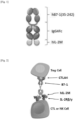
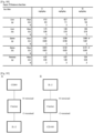
- the fusion protein may have a structure in which, using an Fc domain as a linker (or carrier), a CD80 protein and an IL-2 protein, or an IL-2 protein and a CD80 protein are linked to N-terminus and C-terminus of the linker or carrier, respectively ( Fig. 89 ). Linkage between N-terminus or C-terminus of the Fc domain and CD-80 or IL-2 may optionally be achieved by a linker peptide.
- a fusion protein may consist of the following structural formula (I) or (II): N'-X-[linker (1)] n -Fc domain-[linker (2)] m -Y-C' (I) N'-Y-[linker (1)] n -Fc domain-[linker (2)] m -X-C' (II)
- the fusion protein may consist of the structural formula (I).
- the IL-2 protein is as described above.
- the CD80 protein is as described above.
- the IL-2 protein may be an IL-2 variant with one to five amino acid substitutions as compared with the wild-type IL-2.
- the CD80 protein may be a fragment obtained by truncation of up to about 34 contiguous amino acid residues from the N-terminus or C-terminus of the wild-type CD80.
- the CD protein may be an extracellular immunoglobulin-like domain having the activity of binding to the T cell surface receptors CTLA-4 and CD28.
- the fusion protein may have the amino acid sequence of SEQ ID NO: 9, 26, 28, or 30.
- the fusion protein may include a polypeptide having a sequence identity of 85%, 86%, 87%, 88%, 89%, 90%, 91%, 92%, 93%, 94%, 95%, 96%, 97%, 98%, 99%, or 100% to the amino acid sequence of SEQ ID NO: 9, 26, 28, or 30.
- the identity is, for example, percent homology, and may be determined through homology comparison software such as BlastN software of the National Center of Biotechnology Information (NCBI).
- the peptide linker (1) may be included between the CD80 protein and the Fc domain.
- the peptide linker (1) may consist of 5 to 80 contiguous amino acids, 20 to 60 contiguous amino acids, 25 to 50 contiguous amino acids, or 30 to 40 contiguous amino acids.
- the peptide linker (1) may consist of 30 amino acids.
- the peptide linker (1) may contain at least one cysteine.
- the peptide linker (1) may contain one, two, or three cysteines.
- the peptide linker (1) may be derived from the hinge of an immunoglobulin.
- the peptide linker (1) may be a peptide linker consisting of the amino acid sequence of SEQ ID NO: 3.
- the peptide linker (2) may consist of 1 to 50 contiguous amino acids, 3 to 30 contiguous amino acids, or 5 to 15 contiguous amino acids.
- the peptide linker (2) may be (G4S) n (where n is an integer of 1 to 10).
- n may be 1, 2, 3, 4, 5, 6, 7, 8, 9, or 10.
- the peptide linker (2) may be a peptide linker consisting of the amino acid sequence of SEQ ID NO: 5.
- a dimer obtained by binding of two fusion proteins each of which comprises an IL-2 protein and a CD80 protein.
- the fusion protein comprising IL-2 or a variant thereof and CD80 or a fragment thereof is as described above.
- the binding between the fusion proteins constituting the dimer may be achieved by, but is not limited to, a disulfide bond formed by cysteines present in the linker.
- the fusion proteins constituting the dimer may be the same or different fusion proteins from each other.
- the dimer may be a homodimer.
- the fusion protein constituting the dimer may be a protein having the amino acid sequence of SEQ ID NO: 9.
- polynucleotide encoding a fusion protein comprising an IL-2 protein and a CD80 protein.
- the polynucleotide may contain the nucleotide sequence of SEQ ID NO: 8, 25, 27, or 29.
- the fusion protein comprising an IL-2 protein and a CD80 protein is as described above.
- one or more nucleotides may be altered by substitution, deletion, insertion, or a combination thereof.
- synthetic methods well known in the art may be used, such as those described in Engels and Uhlmann (Angew Chem IntEd Eng., 37: 73-127, 1988 ). Such methods may include triester, phosphite, phosphoramidite and H-phosphate methods, PCR and other autoprimer methods, oligonucleotide syntheses on solid supports, and the like.
- the polypeptide may contain a nucleic acid sequence having an identity, to SEQ ID NO: 8, 25, 27, or 29, of at least about 70%, at least about 75%, at least about 80%, at least about 85%, at least about 86%, at least about 87%, at least about 88%, at least about 89%, at least about 90%, at least about 91%, at least about 92%, at least about 93%, at least about 94%, at least about 95%, at least about 96%, at least about 97%, at least about 98%, at least about 99%, or at least about 100%.
- the polynucleotide may further contain a nucleic acid encoding a signal sequence or a leader sequence.
- signal sequence refers to a signal peptide that directs secretion of a target protein. The signal peptide is translated and then cleaved in a host cell. Specifically, the signal sequence is an amino acid sequence that initiates migration of a protein across the endoplasmic reticulum (ER) membrane. The signal sequence may have the amino acid sequence of SEQ ID NO: 1.
- Signal sequences are well known in the art for their characteristics. Such signal sequences typically contain 16 to 30 amino acid residues, and may contain more or fewer amino acid residues than such amino acid residues.
- a typical signal peptide is composed of three regions, that is, a basic N-terminal region, a central hydrophobic region, and a more polar C-terminal region.
- the central hydrophobic region contains 4 to 12 hydrophobic residues that cause the signal sequence to be immobilized during migration of an immature polypeptide through the membrane lipid bilayer.
- signal sequences are cleaved in the lumen of ER by cellular enzymes, commonly known as signal peptidases.
- the signal sequence may be a secretory signal sequence of tPa (tissue plasminogen activator), HSV gDs (signal sequence of Herpes simplex virus glycoprotein D), or a growth hormone.
- tPa tissue plasminogen activator
- HSV gDs signal sequence of Herpes simplex virus glycoprotein D
- a growth hormone e.gDs
- a secretory signal sequence used in higher eukaryotic cells including mammals and the like may be used.
- a signal sequence included in the wild-type IL-2 and/or CD-80 may be used, or a signal sequence that has been substituted with a codon having high expression frequency in a host cell may be used.
- the vector may be introduced into a host cell to be recombined with and inserted into the genome of the host cell.
- the vector is understood as nucleic acid means containing a polynucleotide sequence which is autonomously replicable as an episome.
- the vectors include linear nucleic acids, plasmids, phagemids, cosmids, RNA vectors, viral vectors, and analogs thereof.
- the viral vector include, but are not limited to, retroviruses, adenoviruses, and adeno-associated viruses.
- the vector may include plasmid DNA, phage DNA, and the like; and commercially developed plasmids (pUC18, pBAD, pIDTSAMRT-AMP, and the like), E. coli-derived plasmids (pYG601BR322, pBR325, pUC118, pUC119, and the like), Bacillus subtilis-derived plasmids (pUB 110, pTP5, and the like), yeast-derived plasmids (YEp13, YEp24, YCp50, and the like), phage DNA (Charon4A, Charon21A, EMBL3, EMBL4, ⁇ gt10, ⁇ gt11, ⁇ ZAP, and the like), animal viral vectors (retroviruses, adenoviruses, vaccinia viruses, and the like), insect viral vectors (baculoviruses and the like). Since the vector exhibits different expression levels and modification of a protein depending on plasmi
- the term "gene expression” or “expression” of a target protein is understood to mean transcription of DNA sequences, translation of mRNA transcripts, and secretion of fusion protein products or fragments thereof.
- a useful expression vector may be RcCMV (Invitrogen, Carlsbad) or a variant thereof. Expression vectors may further contain human cytomegalovirus (CMV) promoter for promoting continuous transcription of a target gene in mammalian cells, and a bovine growth hormone polyadenylation signal sequence for increasing the stability level of RNA after transcription.
- CMV human cytomegalovirus
- Host cells for the transformed cell may include, but are not limited to, prokaryotic cells, eukaryotic cells, and cells of mammalian, vegetable, insect, fungal, or bacterial origin.
- prokaryotic cells E. coli may be used.
- eukaryotic cells yeast may be used.
- mammalian cells CHO cells, F2N cells, CSO cells, BHK cells, Bowes melanoma cells, HeLa cells, 911 cells, AT1080 cells, A549 cells, HEK 293 cells, HEK293T cells, or the like may be used.
- the mammalian cells are not limited thereto, and any cells which are known to those skilled in the art to be usable as mammalian host cells may be used.
- CaCh precipitation for the introduction of an expression vector into the host cell, CaCh precipitation, Hanahan method whose efficiency has been increased efficiency by using a reducing agent such as dimethyl sulfoxide (DMSO) in CaCl 2 precipitation, electroporation, calcium phosphate precipitation, protoplast fusion, agitation using silicon carbide fiber, Agrobacteria-mediated transformation, transformation using PEG, dextran sulfate-, Lipofectamine-, or dry/inhibition-mediated transformation, or the like may be used.
- DMSO dimethyl sulfoxide
- glycosylation pattern of the fusion protein may be adjusted by manipulating, through methods known to those skilled in the art, glycosylation-related genes possessed by host cells.
- the production method may comprise i) culturing the transformed cells to obtain a culture; and ii) collecting the fusion protein from the culture.
- Culturing the transformed cells may be carried out using methods well known in the art. Specifically, the culture may be carried out in a batch process, or carried out continuously in a fed batch or repeated fed batch process.
- compositions for treating or preventing cancer or an infectious disease comprising, as an active ingredient, a fusion protein comprising an IL-2 protein and a CD80 protein or a fusion protein dimer where the two fusion proteins are attached.
- the fusion protein comprising an IL-2 protein and a CD80 protein, or the fusion protein dimer where the two fusion proteins are attached is as described above.
- the cancer may be selected from the group consisting of gastric cancer, liver cancer, lung cancer, colorectal cancer, breast cancer, prostate cancer, ovarian cancer, pancreatic cancer, cervical cancer, thyroid cancer, laryngeal cancer, acute myeloid leukemia, brain tumor, neuroblastoma, retinoblastoma, head and neck cancer, salivary gland cancer, and lymphoma.
- infectious disease may be any one selected from the group consisting of hepatitis B, hepatitis C, human papilloma virus (HPV) infection, cytomegalovirus infection, viral respiratory disease, and influenza.
- a preferred dose of the pharmaceutical composition varies depending on the patient's condition and body weight, severity of disease, form of drug, route and duration of administration and may be appropriately selected by those skilled in the art.
- the active ingredient may be contained in any amount (effective amount) depending on application, dosage form, blending purpose, and the like, as long as the active ingredient can exhibit anticancer activity or a therapeutic effect on an infectious disease.
- a conventional effective amount thereof will be determined within a range of 0.001% to 20.0% by weight, based on the total weight of the composition.
- the term "effective amount” refers to an amount of an active ingredient capable of inducing an anticancer effect or an infectious disease-treating effect. Such an effective amount can be experimentally determined within the scope of common knowledge of those skilled in the art.
- treatment may be used to mean both therapeutic and prophylactic treatment.
- prophylaxis may be used to mean that a pathological condition or disease of an individual is alleviated or mitigated.
- treatment may include both application or any form of administration for treating a disease in a mammal, including a human.
- the term includes inhibiting or slowing down a disease or disease progression; and includes meanings of restoring or repairing impaired or lost function so that a disease is partially or completely alleviated; stimulating inefficient processes; or alleviating a serious disease.
- the term “efficacy” refers to capacity that can be determined by one or parameters, for example, survival or disease-free survival over a certain period of time such as one year, five years, or ten years.
- the parameter may include inhibition of size of at least one tumor in an individual.
- “enhanced efficacy” may be due to enhanced pharmacokinetic parameters and improved efficacy, which may be measured by comparing clearance rate and tumor growth in test animals or human subjects, or by comparing parameters such as survival, recurrence, or disease-free survival.
- the term "therapeutically effective amount” or “pharmaceutically effective amount” refers to an amount of a compound or composition effective to prevent or treat the disease in question, which is sufficient to treat the disease at a reasonable benefit/risk ratio applicable to medical treatment and does not cause adverse effects.
- a level of the effective amount may be determined depending on factors including the patient's health condition, type and severity of disease, activity of drug, the patient's sensitivity to drug, mode of administration, time of administration, route of administration and excretion rate, duration of treatment, formulation or simultaneously used drugs, and other factors well known in the medical field.
- the therapeutically effective amount may mean an amount of drug effective to treat cancer.
- the pharmaceutical composition may further comprise a pharmaceutically acceptable carrier.
- the pharmaceutically acceptable carrier may be any carrier as long as the carrier is a non-toxic substance suitable for delivery to a patient. Distilled water, alcohol, fat, wax, and inert solid may be contained as the carrier. A pharmaceutically acceptable adjuvant (buffer, dispersant) may also be contained in the pharmaceutical composition.
- the pharmaceutical composition may be prepared into a parenteral formulation depending on its route of administration using conventional methods known in the art.
- pharmaceutically acceptable means that the carrier does not have more toxicity than the subject to be applied (prescribed) can adapt while not inhibiting activity of the active ingredient.
- the pharmaceutical composition When the pharmaceutical composition is prepared into a parenteral formulation, it may be made into preparations in the form of injections, transdermal patches, nasal inhalants, or suppositories with suitable carriers according to methods known in the art.
- suitable carriers sterile water, ethanol, polyol such as glycerol or propylene glycol, or a mixture thereof may be used as a suitable carrier; and an isotonic solution, such as Ringer's solution, phosphate buffered saline (PBS) containing triethanol amine or sterile water for injection, and 5% dextrose, or the like may preferably be used.
- PBS phosphate buffered saline
- Formulation of pharmaceutical compositions is known in the art, and reference may specifically be made to Remington's Pharmaceutical Sciences (19th ed., 1995 ) and the like. This document is considered part of the present description.
- a preferred dose of the pharmaceutical composition may range from 0.01 ⁇ g/kg to 10 g/kg, or 0.01 mg/kg to 1 g/kg, per day, depending on the patient's condition, body weight, sex, age, severity of the patient, and route of administration.
- the dose may be administered once a day or may be divided into several times a day. Such a dose should not be construed as limiting the scope of the present invention in any aspect.
- compositions of the present application are mammals and humans, with humans being particularly preferred.
- the pharmaceutical composition of the present application may further contain any compound or natural extract, which has already been validated for safety and is known to have anticancer activity or a therapeutic effect on an infectious disease, so as to boost or reinforce anticancer activity.
- a fusion protein comprising an IL-2 protein and a CD80 protein for treating cancer or an infectious disease.
- a fusion protein comprising an IL-2 protein and a CD80 protein for enhancing a therapeutic effect on cancer or an infectious disease.
- a fusion protein comprising an IL-2 protein and a CD80 protein for manufacture of a medicament for treating cancer or an infectious disease.
- a method for treating cancer or an infectious disease comprising administering, to a subject, a fusion protein comprising an IL-2 protein and a CD80 protein or a fusion protein dimer where the two fusion proteins are attached.
- the subject may be an individual suffering from cancer or an infectious disease.
- the subject may be a mammal, preferably a human.
- the fusion protein comprising an IL-2 protein and a CD80 protein, or the fusion protein dimer where the two fusion proteins are attached is as described above.
- Route of administration, dose, and frequency of administration of the fusion protein or fusion protein dimer may vary depending on the patient's condition and the presence or absence of side effects, and thus the fusion protein or fusion protein dimer may be administered to a subject in various ways and amounts.
- the optimal administration method, dose, and frequency of administration can be selected in an appropriate range by those skilled in the art.
- the fusion protein or fusion protein dimer may be administered in combination with other drugs or physiologically active substances whose therapeutic effect is known with respect to a disease to be treated, or may be formulated in the form of combination preparations with other drugs.
- the fusion protein described herein can activate immune cells such as natural killer cells.
- the fusion protein can be effectively used for cancer and infectious diseases.
- an IL-2 variant with two to five amino acid substitutions in particular, an IL-2 variant that contains amino acid substitutions at two, three, four, or five positions among the positions selected from the group consisting of R38A, F42A, Y45A, E61R, and L72G, has low binding ability for the IL-2 receptor alpha chain and thus exhibits improved characteristics with respect to pharmacological side effects of conventional IL-2.
- VLS vascular leakage syndrome
- a polynucleotide was synthesized through the Invitrogen GeneArt Gene Synthesis service of ThermoFisher Scientific.
- the polynucleotide contains a nucleotide sequence (SEQ ID NO: 8) which encodes a fusion protein that contains a signal peptide (SEQ ID NO: 1), a CD80 fragment (SEQ ID NO: 2), an Ig hinge (SEQ ID NO: 3), an Fc domain (SEQ ID NO: 4), a linker (SEQ ID NO: 5), and an IL-2 variant (2M) (R38A, F42A) (SEQ ID NO: 6) having two amino acid substitutions, in this order, from the N-terminus.
- the polynucleotide was inserted into pcDNA3_4 vector.
- the vector was introduced into CHO cells (Expi-CHO TM ) to express the fusion protein of SEQ ID NO: 9. After the vector was introduced, culture was performed for 7 days in an environment of 37°C, 125 RPM, and 8% CO 2 concentration. Then, the culture was harvested and the fusion protein was purified therefrom. The purified fusion protein was designated "GI101".
- fusion protein was bound thereto under a condition of 25 mM Tris, 25 mM NaCl, pH 7.4. Then, elution was performed with 100 mM NaCl, 100 mM acetic acid, pH 3. 20% 1 M Tris-HCl at pH 9 was placed in a collection tube, and then the fusion protein was collected. For the collected fusion protein, the buffer was exchanged through dialysis with PBS buffer for 16 hours.
- a polynucleotide was synthesized through the Invitrogen GeneArt Gene Synthesis service of ThermoFisher Scientific.
- the polynucleotide contains a nucleotide sequence (SEQ ID NO: 14) which encodes a fusion protein that contains a signal peptide (SEQ ID NO: 1), a mCD80 (SEQ ID NO: 13), an Ig hinge (SEQ ID NO: 3), an Fc domain (SEQ ID NO: 4), a linker (SEQ ID NO: 5), and an IL-2 variant (2M) (R38A, F42A) (SEQ ID NO: 6) with two amino acid substitutions, in this order, from the N-terminus.
- the polynucleotide was inserted into pcDNA3_4 vector.
- the vector was introduced into CHO cells (Expi-CHO TM ) to express the fusion protein of SEQ ID NO: 15. After the vector was introduced, culture was performed for 7 days in an environment of 37°C, 125 RPM, and 8% CO 2 concentration. Then, the culture was harvested and the fusion protein was purified therefrom. The purified fusion protein was designated "mGI101".
- the purification and collection of the fusion protein were carried out in the same manner as in Preparation Example 1.
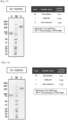
- the isolated and purified fusion protein was subjected to SDS-PAGE under reduced (R) or non-reduced (NR) condition and stained with Coomassie Blue to check its purity ( Fig. 9 ). It was found that the fusion protein was contained at a concentration of 1.95 mg/ml when detected by absorbance at 280 nm using NanoDrop.
- a polynucleotide was synthesized through the Invitrogen GeneArt Gene Synthesis service of ThermoFisher Scientific. Specifically, the polynucleotide contains a nucleotide sequence (SEQ ID NO: 16) which encodes a fusion protein that contains a signal peptide (SEQ ID NO: 1), a CD80 fragment (SEQ ID NO: 2), an Ig hinge (SEQ ID NO: 3), and an Fc domain (SEQ ID NO: 4). The polynucleotide was inserted into pcDNA3_4 vector.
- the vector was introduced into CHO cells (Expi-CHO TM ) to express the fusion protein of SEQ ID NO: 17. After the vector was introduced, culture was performed for 7 days in an environment of 37°C, 125 RPM, and 8% CO 2 concentration. Then, the culture was harvested and the fusion protein was purified therefrom. The purified fusion protein was designated "GI101C1".
- the purification and collection of the fusion protein were carried out in the same manner as in Preparation Example 1.
- the isolated and purified fusion protein was subjected to SDS-PAGE under reduced (R) or non-reduced (NR) condition and stained with Coomassie Blue to check its purity ( Fig. 10 ). It was observed that the fusion protein was contained at a concentration of 3.61 mg/ml when detected by absorbance at 280 nm using NanoDrop.
- a polynucleotide was synthesized through the Invitrogen GeneArt Gene Synthesis service of ThermoFisher Scientific. Specifically, the polynucleotide contains a nucleotide sequence (SEQ ID NO: 18) which encodes a fusion protein that contains a signal peptide (SEQ ID NO: 1), an Fc domain (SEQ ID NO: 4), a linker (SEQ ID NO: 5), and an IL-2 variant (2M) (R38A, F42A) (SEQ ID NO: 6) with two amino acid substitutions, in this order, from the N-terminus.
- SEQ ID NO: 18 which encodes a fusion protein that contains a signal peptide (SEQ ID NO: 1), an Fc domain (SEQ ID NO: 4), a linker (SEQ ID NO: 5), and an IL-2 variant (2M) (R38A, F42A) (SEQ ID NO: 6) with two amino acid substitutions, in this order, from the N-terminus
- the polynucleotide was inserted into pcDNA3_4 vector.
- the vector was introduced into CHO cells (Expi-CHO TM ) to express the fusion protein of SEQ ID NO: 19. After the vector was introduced, culture was performed for 7 days in an environment of 37°C, 125 RPM, and 8% CO 2 concentration. Then, the culture was harvested and the fusion protein was purified therefrom. The purified fusion protein was designated "GI101C2".
- the purification and collection of the fusion protein were carried out in the same manner as in Preparation Example 1.
- the isolated and purified fusion protein was subjected to SDS-PAGE under reduced (R) or non-reduced (NR) condition and stained with Coomassie Blue to check its purity ( Fig. 11 ). It was found that the fusion protein was contained at a concentration of 4.79 mg/ml when detected by absorbance at 280 nm using NanoDrop.
- a polynucleotide was synthesized through the Invitrogen GeneArt Gene Synthesis service of ThermoFisher Scientific. Specifically, the polynucleotide contains a nucleotide sequence (SEQ ID NO: 20) which encodes a fusion protein that contains a signal peptide (SEQ ID NO: 1), a mCD80 (SEQ ID NO: 13), an Ig hinge (SEQ ID NO: 3), and an Fc domain (SEQ ID NO: 4), in this order, from the N-terminus. The polynucleotide was inserted into pcDNA3_4 vector.
- the vector was introduced into CHO cells (Expi-CHO TM ) to express the fusion protein of SEQ ID NO: 21. After the vector was introduced, culture was performed for 7 days in an environment of 37°C, 125 RPM, and 8% CO 2 concentration. Then, the culture was harvested and the fusion protein was purified therefrom. The purified fusion protein was designated "mGI101C1".
- the purification and collection of the fusion protein were carried out in the same manner as in Preparation Example 1.
- the isolated and purified fusion protein was subjected to SDS-PAGE under reduced (R) or non-reduced (NR) condition and stained with Coomassie Blue to check its purity ( Fig. 12 ). It was observed that the fusion protein was contained at a concentration of 2.49 mg/ml when detected by absorbance at 280 nm using NanoDrop.
- a polynucleotide was synthesized through the Invitrogen GeneArt Gene Synthesis service of ThermoFisher Scientific.
- the polynucleotide conta is a nucleotide sequence (SEQ ID NO: 31) which encodes a fusion protein that contains a signal peptide (SEQ ID NO: 1), a CD80 fragment (SEQ ID NO: 2), an Ig hinge (SEQ ID NO: 3), an Fc domain (SEQ ID NO: 4), a linker (SEQ ID NO: 5), and mature human IL-2 (SEQ ID NO: 10), in this order, from the N-terminus.
- the polynucleotide was inserted into pcDNA3_4 vector.
- the vector was introduced into CHO cells (Expi-CHO TM ) to express the fusion protein of SEQ ID NO: 32.
- the purified fusion protein was designated "GI101w”. The purification and collection of the fusion protein were carried out in the same manner as in Preparation Example 1.
- a polynucleotide was synthesized through the Invitrogen GeneArt Gene Synthesis service of ThermoFisher Scientific.
- the polynucleotide contains a nucleotide sequence (SEQ ID NO: 25) which encodes a fusion protein that contains a signal peptide (SEQ ID NO: 1), a CD80 fragment (SEQ ID NO: 2), an Ig hinge (SEQ ID NO: 3), an Fc domain (SEQ ID NO: 4), a linker (SEQ ID NO: 5), and an IL-2 variant (SEQ ID NO: 22), in this order, from the N-terminus.
- the polynucleotide was inserted into pcDNA3_4 vector.
- the vector was introduced into CHO cells (Expi-CHO TM ) to express the fusion protein of SEQ ID NO: 26.
- the purified fusion protein was designated "GI102-M45".
- the purification and collection of the fusion protein were carried out in the same manner as in Preparation Example 1.
- the isolated and purified fusion protein was subjected to SDS-PAGE under reduced (R) or non-reduced (NR) condition and stained with Coomassie Blue to check its purity ( Fig. 13 ).
- a polynucleotide was synthesized through the Invitrogen GeneArt Gene Synthesis service of ThermoFisher Scientific.
- the polynucleotide contains a nucleotide sequence (SEQ ID NO: 27) which encodes a fusion protein that contains a signal peptide (SEQ ID NO: 1), a CD80 fragment (SEQ ID NO: 2), an Ig hinge (SEQ ID NO: 3), an Fc domain (SEQ ID NO: 4), a linker (SEQ ID NO: 5), and an IL-2 variant (SEQ ID NO: 23), in this order, from the N-terminus.
- the polynucleotide was inserted into pcDNA3_4 vector.
- the vector was introduced into CHO cells (Expi-CHO TM ) to express the fusion protein of SEQ ID NO: 28.
- the purified fusion protein was designated "GI102-M61".
- the purification and collection of the fusion protein were carried out in the same manner as in Preparation Example 1.
- the isolated and purified fusion protein was subjected to SDS-PAGE under reduced (R) or non-reduced (NR) condition and stained with Coomassie Blue to check its purity ( Fig. 14 ).
- a polynucleotide was synthesized through the Invitrogen GeneArt Gene Synthesis service of ThermoFisher Scientific.
- the polynucleotide contains a nucleotide sequence (SEQ ID NO: 29) which encodes a fusion protein that contains a signal peptide (SEQ ID NO: 1), a CD80 fragment (SEQ ID NO: 2), an Ig hinge (SEQ ID NO: 3), an Fc domain (SEQ ID NO: 4), a linker (SEQ ID NO: 5), and an IL-2 variant (SEQ ID NO: 24), in this order, from the N-terminus.
- the polynucleotide was inserted into pcDNA3_4 vector.
- the vector was introduced into CHO cells (Expi-CHO TM ) to express the fusion protein of SEQ ID NO: 30.
- the purified fusion protein was designated "GI102-M72".
- the purification and collection of the fusion protein were carried out in the same manner as in Preparation Example 1.
- the isolated and purified fusion protein was subjected to SDS-PAGE under reduced (R) or non-reduced (NR) condition and stained with Coomassie Blue to check its purity ( Fig. 15 ).
- a polynucleotide was synthesized through the Invitrogen GeneArt Gene Synthesis service of ThermoFisher Scientific.
- the polynucleotide contains a nucleotide sequence (SEQ ID NO: 33) which encodes a fusion protein that contains a signal peptide (SEQ ID NO: 1), a mCD80 fragment (SEQ ID NO: 13), an Ig hinge (SEQ ID NO: 3), an Fc domain (SEQ ID NO: 4), a linker (SEQ ID NO: 5), and an IL-2 variant (SEQ ID NO: 23), in this order, from the N-terminus.
- the polynucleotide was inserted into pcDNA3_4 vector.
- the vector was introduced into CHO cells (Expi-CHO TM ) to express the fusion protein of SEQ ID NO: 34.
- the purified fusion protein was designated "mGI102-M61".
- the binding affinity was measured using Octet RED 384.
- AR2G biosensor (Amine Reactive 2 nd gen, ForteBio, Cat: 18-5092) was previously hydrated with 200 ⁇ l of distilled water in a 96-well microplate (GreinerBio-one, Cat: 655209).
- a ligand (CTLA-4, Human CTLA-4/CD152, His tag, Sino Biological, Cat: 11159-H08H) to be attached to the AR2G biosensor was diluted with 10 mM acetate buffer (pH 5, AR2G reagent Kit, ForteBio, Cat: 18-5095) to a concentration of 5 ⁇ g/ml.
- GI101 to be attached to the ligand was diluted with 1X AR2G kinetic buffer (AR2G reagent Kit, ForteBio, Cat: 18-5095) to a concentration of 1,000 nM, 500 nM, 250 nM, 125 nM, or 62.5 nM.
- Activation buffer was prepared by mixing 20 mM EDC and 10 mM s-NHS (AR2G reagent Kit, ForteBio, Cat: 18-5095) in distilled water. 80 ⁇ l of each reagent was placed in a 384-well microplate (Greiner Bio-one, Cat: 781209) and the program was set up.
- Ni-NTA Ni-NTA Biosensors, ForteBio, 18-5101
- 1X Ni-NTA kinetic buffer 10X Kinetics buffer, ForteBio, 18-1042
- a ligand Human PD-L1/B7-H1 protein, His-tag, Sino biological, Cat: 10084-H08H
- 1X Ni-NTA kinetic buffer was diluted with 1X Ni-NTA kinetic buffer to a concentration of 5 ⁇ g/ml.
- GI101 to be attached to the ligand was diluted with 1X Ni-NTA kinetic buffer at 1,000 nM, 500 nM, 250 nM, 125 nM, or 62.5 nM.
- human PD-1/PDCD1 Human PD-1/PDCD1, Fc Tag, Sino Biological, Cat: 10377-H02H
- 1X Ni-NTA kinetic buffer was diluted with 1X Ni-NTA kinetic buffer to a concentration of 2,000 nM, 1,000 nM, 500 nM, 250 nM, or 125 nM.
- 80 ⁇ l of each reagent was placed in a 384-well microplate and the program was set up.
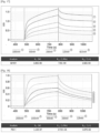
- the binding affinity between hPD-L1 and GI101 was measured as illustrated in Fig. 17 .
- the binding affinity between hPD-L1 and hPD-1 was measured as illustrated in Fig. 18 .
- mCTLA-4 The binding affinity between mCTLA-4 and mGIlOl was examined in the same manner as in Experimental Example 1.
- the equipment used is as follows: Biosensor: AR2G, Ligand: mCTLA-4 (Recombinant Mouse CTLA-4 Fc chimera, R&D Systems, Cat: 434-CT-200), Analyte: mGI101 (500 nM, 250 nM, 125 nM, 62.5 nM, 31.3 nM).
- the binding affinity between mPD-L1 and mGI 101 was identified in the same manner as in Experimental Example 1.
- the equipment used is as follows.
- Binding kinetics measurements were performed using the Octet RED 384 instrument (ForteBio, Pall Life Science) with agitation at 30°C and 1,000 rpm.
- the binding ability for CTLA-4 was measured using the Amine Reactive 2 generation (AR2G) biosensor chip, and the binding ability for PD-L1 was measured using the Nickel charged Tris-NTA (Ni-NTA) biosensor chip.
- the AR2G biosensor chip was activated with a combination of 400 mM EDC and 100 mM sulfo-NHS.
- Human CTLA-4-His Tag (Sino Biological, Cat: 11159-H08H) was diluted with 10 mM acetate buffer (pH 5) to 5 ⁇ g/ml, and loaded on the AR2G biosensor chip for 300 seconds and fixed.
- a blocking experiment was performed using the Octet RED 384 instrument (ForteBio, Pall Life Science) with agitation at 30°C and 1,000 rpm.
- Human PD-L1-His Tag (Sino biological, Cat: 10084-H08H) was diluted with 1XNi-NTA kinetic buffer to a concentration of 5 ⁇ g/ml, and loaded on the Ni-NTA biosensor chip for 600 seconds and fixed.
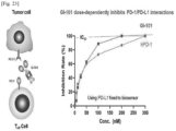
- hPD-L1 fixed on the biosensor chip was allowed to bind to GI-101 at various concentrations (300 nM, 100 nM, 50 nM, 25 nM, 12.5 nM, and 0 nM) for 600 seconds, and then again allowed to bind to the competitor human PD-1 (100 nM) for 600 seconds so as to measure how much more hPD-1 can bind thereto.
- hPD-L1 was allowed to bind to hPD-1 at various concentrations (300 nM, 100 nM, 50 nM, 25 nM, 12.5 nM, and 0 nM) for 600 seconds, and then again allowed to bind to the competitor GI-101 (100 nM) for 600 seconds so as to measure how much more GI-101 can bind thereto.
- the blocking experiment was analyzed using the epitope binning menu of Octet Data Analysis HT software ver. 10 provided by Pall Corporation. The results are illustrated in Fig. 23 .
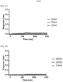
- the binding ability for IL-2R ⁇ was measured using the AR2G biosensor, and the binding ability for IL-2R ⁇ was measured using the Ni-NTA biosensors (Nickel charged Tris-NTA, Ni-NTA Biosensors, ForteBio, 18-5101).
- a ligand (IL-2R ⁇ -His Tag, Acro, Cat: ILA-H52H9) to be attached to the AR2G biosensor was diluted with 10 mM acetate buffer (pH 5, AR2G reagent Kit, ForteBio, Cat: 18-5095) to a concentration of 5 ⁇ g/ml.
- the AR2G biosensor was activated with a buffer prepared by mixing 400 mM EDC and 100 mM sulfo-NHS, and then the diluted ligand was loaded on the AR2G biosensor for 300 seconds and fixed.
- a ligand (IL-2R ⁇ -His Tag, Aero, Cat: CD2-H5221) to be attached to the Ni-NTA biosensor was diluted with 1X Ni-NTA kinetic buffer to a concentration of 5 ⁇ g/ml.
- the diluted ligand was loaded on the Ni-NTA biosensor for 600 seconds and fixed.
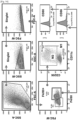
- GI101, GI101w, or Proleukin (Novartis, hIL-2), at various concentrations, to be attached to the ligand was loaded thereon for 300 seconds. Then, binding thereof was measured and dissociation thereof was also measured for 300 seconds. Binding kinetics analysis was performed using Octet Data Analysis HT software ver. 10 provided by Pall Corporation. The results are illustrated in Figs. 24 to 26 .
- GI101 has low binding ability for the IL-2 receptor alpha chain, IL-2R ⁇ , and high binding ability for IL-2R ⁇ , as compared with GIlOlw and Proleukin.
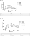
- binding affinity was measured using Octet RED 384.
- AR2G biosensor (Amine Reactive 2nd gen, ForteBio, Cat: 18-5092) was previously hydrated with 200 ⁇ l of distilled water (DW) in a 96-well microplate (GreinerBio-one, Cat: 655209).
- a ligand Human IL-2 R alpha protein, His Tag, Aero, ILA-H52H9 to be attached to the biosensor was diluted with 10 mM acetate buffer (pH 5) (AR2G reagent Kit, ForteBio, Cat: 18-5095) to a concentration of 5 ⁇ g/ml.
- An analyte (GI101-M45, GI101-M61, GI101-M72) to be attached to the ligand was diluted with 1X AR2G kinetic buffer (AR2G reagent Kit, ForteBio, Cat: 18-5095) to 500 nM, 250 nM, 125 nM, and 62.5 nM, respectively.
- Activation buffer was prepared by mixing 20 mM EDC and 10 mM s-NHS (AR2G reagent Kit, ForteBio, Cat: 18-5095) in DW. 80 ⁇ l of each reagent was placed in a 384-well microplate (Greiner Bio-one, Cat: 781209) and the program was set up.
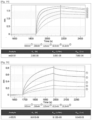
- the binding affinity between IL2 alpha receptor and GI101-M45 is illustrated in Fig. 27 .
- the binding affinity between IL2 alpha receptor and GI101-M61 is illustrated in Fig. 28
- the binding affinity between IL2 alpha receptor and GI101-M72 is illustrated in Fig. 29 .
- Ni-NTA Biosensors were previously hydrated with 200 ⁇ l of 1X Ni-NTA kinetic buffer (10X Kinetics buffer, ForteBio, 18-1042) in a 96-well microplate.
- a ligand Human IL-2 R beta protein, His-Tag, Acro, CD2-H5221
- 1X Ni-NTA kinetic buffer to be attached to the biosensor was diluted with 1X Ni-NTA kinetic buffer to a concentration of 2 ⁇ g/ml.
- GI102-M45, GI102-M61, or GI102-M72 to be attached to the ligand was diluted with 1X Ni-NTA kinetic buffer to a concentration of 500 nM, 250 nM, 125 nM, or 62.5 nM.
- 80 ⁇ l of each reagent was placed in a 384-well microplate and the program was set up.
- the binding affinity between IL-2R ⁇ and GI102-M45 was measured as illustrated in Fig. 30
- the binding affinity between IL-2R ⁇ and GI102-M61 was measured as illustrated in Fig. 31
- the binding affinity between IL-2R ⁇ and GI102-M72 was measured as illustrated in Fig. 32 .
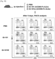
- PBMCs Peripheral blood mononuclear cells isolated from a human were labeled with carboxyfluorescein succinimidyl ester (CFSE) by being reacted with 1 ⁇ M CellTrace CFSE dye at 37°C for 20 minutes. CFSE not bound to the cells was removed by being reacted for 5 minutes with a culture medium having a 5-fold volume of the staining reaction solution and then by being centrifuged at 1,300 rpm for 5 minutes.
- CFSE carboxyfluorescein succinimidyl ester
- the CFB-labeled PBMCs were resuspended in the culture medium (RPMI1640 medium containing 10% FBS, 10 mM HEPES, 100 U/ml penicillin/streptomycin, 1 mM sodium pyruvate, 55 ⁇ M 2-mercaptoethanol, 1 mM non-essential amino acid, and 2 mM L-glutamine), and then added to a 96-well plate at 1 ⁇ 10 5 cells per well.
- PHA Lacin from Phaseolus Vulgaris, red kidney bean, Sigma-Aldrich, St. Louis, MO, USA, Cat. No.
- GI101, GI101C1, GI101C2, or IL-2 (Aldesleukin; human recombinant IL-2, Novartis) was performed and incubation was performed in a 5% CO 2 incubator at 37°C for 6 days.
- the treatment with GI101, GI101C1, GI101C2, and IL-2 was performed at a concentration of 1 nM, 10 nM, or 100 nM.
- the cells were analyzed by FACS, and human IFN- ⁇ present in the culture medium was measured using an ELISA kit (Biolegend, San Diego, CA, USA, Cat. No. 430103).
- the cell pellets obtained by removing the supernatant were washed with FACS buffer (3% FBS, 10 mM EDTA, 1M HEPES, 100 unit/mL Penicillin Streptomycin, 10 ⁇ g/ml, 1 mM sodium pyruvate), and then reacted with Fc blocker (Biolegend, Cat. No. 422302) at 4°C for 5 minutes. Then, treatment with APC anti-CD3 Ab (Biolegend, Cat. No. 300412) and PE anti-CD8a Ab (Biolegend, Cat. No. 300908) was performed and reaction was allowed to proceed at 4°C for 20 minutes. Then, the resultant was washed with FACS buffer. The cell pellets were resuspended in FACS buffer and then analyzed using BD LSR Fortessa (BD Biosciences, San Diego, CA, USA) and FlowJo software.
- FACS buffer 3% FBS, 10 mM EDTA, 1M HEPES, 100 unit/mL Pen
- the amount of human IFN- ⁇ secreted into the supernatant of each sample in which the cells had been cultured was measured using a human IFN- ⁇ ELISA kit (Biolegend, Cat. No. 430103). Briefly, anti-human-IFN- ⁇ antibodies were added to an ELISA plate, and reaction was allowed to proceed overnight at 4°C so that these antibodies were coated thereon. Then, blocking was performed at room temperature for 1 hour with a PBS solution to which 1% BSA had been added. Washing with a washing buffer (0.05% Tween-20 in PBS) was performed, and then a standard solution and each sample were properly diluted and added thereto. Then, reaction was allowed to proceed at room temperature for 2 hours.
- a washing buffer 0.05% Tween-20 in PBS
- PBMCs Peripheral blood mononuclear cells isolated from a human were labeled with CFSE by being reacted with 1 ⁇ M CellTrace CFSE dye at 37°C for 20 minutes. CFSE not bound to the cells was removed by being reacted for 5 minutes with a culture medium having a 5-fold volume of the staining reaction solution and then by being centrifuged at 1,300 rpm for 5 minutes.
- the CFB-labeled PBMCs were resuspended in the culture medium (RPMI1640 medium containing 10% FBS, 10 mM HEPES, 100 U/ml penicillin/streptomycin, 1 mM sodium pyruvate, 55 ⁇ M 2-mercaptoethanol, 1 mM non-essential amino acid, and 2 mM L-glutamine), and then added to a 96-well plate at 1 ⁇ 10 5 cells per well.
- the culture medium RPMI1640 medium containing 10% FBS, 10 mM HEPES, 100 U/ml penicillin/streptomycin, 1 mM sodium pyruvate, 55 ⁇ M 2-mercaptoethanol, 1 mM non-essential amino acid, and 2 mM L-glutamine
- GI101 activated proliferation of CD8+ T cells in vitro to a similar extent to the wild-type IL-2 Proleukin ( Figs. 35 and 36 ).
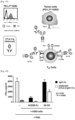
- Human PBMCs were purchased from Allcells (Lot # 3014928, USA). 1M CellTrace CFSE dye was used, which was reacted with the human PBMCs under a light-blocking condition at room temperature for 20 minutes. The cells were labeled with CFSE by being reacted with 1 ⁇ M CellTrace CFSE dye at 37°C for 20 minutes. CFSE not bound to the cells was removed by being reacted for 5 minutes with culture medium having a 5-fold volume of the staining reaction solution and then by being centrifuged at 1,300 rpm for 5 minutes.
- the CFB-labeled PBMCs were resuspended in the culture medium (RPMI1640 medium containing 10% FBS, 10 mM HEPES, 100 U/ml penicillin/streptomycin, 1 mM sodium pyruvate, 55 ⁇ M 2-mercaptoethanol, 1 mM non-essential amino acid, and 2 mM L-glutamine), and then added to a 96-well plate at 1 ⁇ 10 5 cells per well.
- the culture medium RPMI1640 medium containing 10% FBS, 10 mM HEPES, 100 U/ml penicillin/streptomycin, 1 mM sodium pyruvate, 55 ⁇ M 2-mercaptoethanol, 1 mM non-essential amino acid, and 2 mM L-glutamine
- the CFB-labeled PBMCs were subjected to treatment with 1 ⁇ g/ml of anti-CD3 ⁇ antibody (OKT3, eBioscience, USA), and GI101, GI101C1, GI101C2, or Proleukin (Novartis), and incubation was performed in a 5% CO 2 incubator at 37°C for 7 days.
- the cells were subjected to treatment with GI101, GI101C1, GI101C2, and IL-2 at a concentration of 10 ⁇ M.
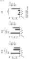
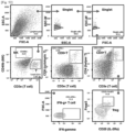
- the incubated cells were examined for their degree of proliferation by measuring, with FACS analysis using anti-human CD4-PE antibody (BioLegend, USA), anti-human CD8-PE/Cy7 antibody (BioLegend, USA), and anti-human FoxP3-APC antibody (BioLegend, USA), a proportion of CD8+ T cells that had not been labeled with CFSE.
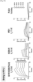
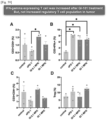
- the GI101, GI102M61, GI101C2, and Proleukin treatment groups exhibited a significant increase in proportion of CD8+ T cells, as compared with the control group (no stimulus), the anti-CD3 antibody alone treatment group, and the GI101C1 treatment group.
- the GI101, GI101C2, and Proleukin treatment groups exhibited a significant increase in proliferation of CD4+/FoxP3+ Treg cells, whereas the GI102 and GI101C1 treatment groups did not exhibit a significant increase in proliferation of CD4+/FoxP3+ Treg cells ( Fig. 37 ).
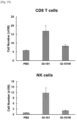
- mice 7-week-old C57BL/6 mice purchased from Orient Bio (Busan, Korea) were divided into 3 groups, each group containing 3 mice, and PBS, GI101, or GI101w was injected intraperitoneally thereinto.
- GI101 and GI101w were respectively prepared to be at 40.5 ⁇ g in 200 ⁇ l of PBS, and injected intraperitoneally thereinto.
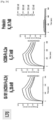
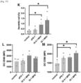
- the spleens were removed from the mice of each group. The cells were isolated therefrom, and the total number of cells was measured using a hematocytometer.
- Splenocytes were examined for proportions of CD8+ T cells and NK cells therein, with FACS analysis using staining with APC-CD3 ⁇ antibody (Biolegend; 145-2C11), PE-NK1.1 antibody (Biolegend; PK136), and Pacific blue-CD8a antibody (BD; 53-6.7). As such, the numbers of CD8+ T cells and NK cells present in the spleen were calculated.
- GI101 activated proliferation of CD8+ T cells and NK cells in vivo as compared with GIlOlw ( Figs. 38 and 39 ).
- CTLA-4 blockade bioassay kit Promega Cat. No. JA4005. The experiment is briefly described as follows. CTLA-4 effector cells kept in liquid nitrogen were thawed in a 37°C constant temperature water bath for 3 minutes, and 0.8 ml of CTLA-4 effector cells were mixed well with 3.2 ml of pre-warmed assay buffer (90% RPMI + 10% FBS). Then, the mixture was added to a 96-well white cell culture plate (SPL, Cat. No. 30196) at 25 ⁇ l per well. Then, 25 ⁇ l of GI101 at various concentrations was added thereto. For a negative control, 25 ⁇ l of assay buffer was added thereto. Then, the white plat cell culture plate was covered and placed at room temperature until aAPC/Raji cells were prepared.
- SPL 96-well white cell culture plate
- aAPC/Raji cells kept in liquid nitrogen were thawed in a 37°C constant temperature water bath for 3 minutes, and 0.8 ml of aAPC/Raji cells were mixed well with 3.2 ml of pre-warmed assay buffer. Then, 25 ⁇ l of the mixture was added to the plate at per well, and reaction was allowed to proceed in a 5% CO 2 incubator at 37°C for 16 hours. After the reaction was completed, the resultant was allowed to stand at room temperature for 15 minutes, and then the Bio-Glo reagent was added thereto while taking care to avoid bubbles. The Bio-Glo reagent was also added to three of the outermost wells and the wells were used as blanks to correct the background signal.
- GI101 attached to CTLA-4 expressed on effector T cells and activated the function of T cells rather than inhibiting the same ( Figs. 40 and 41 ).
- the group having received mGI101 or mGI102 (mGI102-M61) exhibited a significant increase in numbers of CD8+ T cells and NK cells at the time points from 3 days to 14 days after administration, as compared with the PBS administration group.
- the group having received mGI102 exhibited a significant increase in proportions of activated CD8+ T cells/Treg cells and NK cells/Treg cells at the time points from 3 days to 14 days after administration, as compared with the PBS administration group ( Fig. 42 ).
- NC1-H292 cancer cell line overexpressing PD-L1 was cultured for 3 hours in a culture medium containing 10 ⁇ g/ml Mitomycin C (Sigma), and then Mitomycin C was removed by washing with the culture medium. Thereafter, 5 ⁇ 10 4 cells of the Mitomycin C-treated NC1-H292 cancer cell line were incubated with 1 ⁇ 10 5 cells of human PBMCs in a 96-well plate. Here, treatment with 5 ⁇ g/ml of PHA (Sigma) was performed for T cell activity.
- GI101 effectively activated the immune response that had been inhibited by the cancer cell line overexpressing PD-L1.
- GI101 inhibited signaling of CTLA-4 expressed on effector T cells ( Figs. 43 and 44 ).
- mice 5 ⁇ 10 6 cells/0.05 ml of mouse-derived CT-26 cancer cell line was mixed with 0.05 ml Matrigel matrix phenol red-free (BD), and transplantation of 0.1 ml of the mixture was performed by subcutaneous administration in the right dorsal region of 6-week-old female BALB/c mice (Orient Bio). A certain period of time after the cancer cell transplantation, the tumor volume was measured and subjects that reached about 80 mm 3 to 120 mm 3 were separated. Then, the subjects were intravenously administered with 0.1 ml of GI101. A total of three administrations were given once every three days after the first administration, and PBS was given to a negative control group. The tumor size was measured daily to identify an anticancer effect.
- BD Matrigel matrix phenol red-free
- mice female, 7-week-old acquired from Orient Bio were subjected to an acclimation period of 7 days. Then, 5 ⁇ 10 6 cells of B16F10 cancer cell line (ATCC, USA) were mixed with 0.05 ml of Matrigel matrix phenol red-free (BD), and allotransplantation of the mixture was performed by subcutaneous administration at 0.1 ml in the right dorsal region of the mice. A certain period of time after the cancer cell transplantation, the tumor volume was measured and subjects that reached about 50 mm 3 to 120 mm 3 were selected, and then the selected mice were grouped evenly based on tumor size and body weight, each group containing 10 mice.
- B16F10 cancer cell line ATCC, USA
- BD Matrigel matrix phenol red-free
- hIgG4 was administered at a dose of 4 mg/kg to a negative control group, and an anti-PD-1 antibody was administered at a dose of 5 mg/kg to a positive control group.
- mGI101 at a dose of 1 mg/kg or 4 mg/kg was administered intravenously thereto.
- groups having received mGI101 at a dose of 4 mg/kg and an anti-PD-1 antibody at a dose of 5 mg/kg were also set as experimental groups. A total of three administrations were given once every three days after the first administration. The tumor size was measured daily.
- the initial tumor volume of all groups was 90 mm 3
- standard error (S.E.) of each group was 5 mm 3 to 6 mm 3
- a change in tumor volume was observed during the experimental period, in which the tumor volume increased from 90 mm 3 to 1,434 mm 3 up to 15 days after the administration.
- the tumor volume was observed to increase from 90 mm 3 to 885 mm 3 during the experimental period which is the same period as the negative control group, and a statistically significant inhibition of tumor growth was observed at some measurement time points (p-value: 0.5 on day 11, p-value ⁇ 0.01 on day 7, p-value ⁇ 0.001 on day 3).
- the tumor volume was observed to increase from 90 mm 3 to 748 mm 3 during the experimental period which is the same period as the negative control group, and a statistically significant inhibition of tumor growth was observed at some measurement time points (p-value: 0.5 on day 9, p-value ⁇ 0.01 on days 7 and 11).
- tumor growth inhibition rate was analyzed by using, as a reference, the group having received mIgG at a dose of 4 mg/kg and comparing this group with each of the other groups.
- mGI101 at a dose of 1 mg/kg
- growth inhibition rate of 36.5% was observed as compared with the negative control group, and no statistically significant difference (p-value: 0.5) was observed.
- p-value: 0.5 tumor growth inhibition rate was observed as compared with the negative control group.
- a total of two administrations were given once every three days after the first administration. The tumor size was measured daily.
- mGI101 had an effect of inhibiting tumor growth in a dose-dependent manner ( Figs. 47 and 48 ).
- mice female, 7-week-old acquired from Orient Bio were subjected to an acclimation period of 7 days. Then, 5 ⁇ 10 6 cells of CT-26 cancer cell line (ATCC, USA)were mixed with 0.05 ml of Matrigel matrix phenol red-free (BD), and allotransplantation of the mixture was performed by subcutaneous administration at 0.1 ml in the right dorsal region of the mice. A certain period of time after the cancer cell transplantation, the tumor volume was measured and subjects that reached about 28 mm 3 were selected, and then the selected mice were grouped evenly based on tumor size and body weight, each group containing 10 mice.
- CT-26 cancer cell line ATCC, USA
- BD Matrigel matrix phenol red-free
- hIgG4 was administered at a dose of 6 mg/kg to a negative control group.
- mGI101 at a dose of 3 mg/kg, 6 mg/kg, or 12 mg/kg was administered intravenously thereto.
- a total of three administrations were given once every three days after the first administration. The tumor size was measured daily.
- the experimental group having received mGI101 at a dose of 6 mg/kg or 12 mg/kg mGI101 exhibited significant inhibition of tumor growth at some measurement time points and at the end of the test, as compared with the negative control group ( Fig. 49 ).
- the experimental group having received mGI101 at a dose of 6 mg/kg exhibited significant improvement at some measurement time points and at the end of the test, as compared with the negative control group ( Fig. 50 ).
- mice female, 7-week-old acquired from Orient Bio were subjected to an acclimation period of 7 days. Then, 5 ⁇ 10 6 cells of CT-26 cancer cell line (ATCC, USA) were suspended in 0.1 ml PBS, and allotransplantation of the suspension was performed by subcutaneous administration at 0.1 ml in the right dorsal region of the mice. A certain period of time after the cancer cell transplantation, the tumor volume was measured and subjects that reached about 50 mm 3 to 200 mm 3 were selected, and then the selected mice were grouped evenly based on tumor size and body weight, each group containing 10 mice.
- CT-26 cancer cell line ATCC, USA
- mice of each group in Experimental Example 19.1 were sacrificed when the tumor volume reached an average of 200 mm 3 , and cancer tissues were collected. Thereafter, the cancer tissues were separated to a single-cell level to analyze immune cells therein, and then FACS analysis was performed on immune cells in the cancer tissues using the following antibodies: Anti-mouse-CD3 (Biolegend, Cat. No. 100320), Anti-mouse-CD4 (Biolegend, Cat. No. 100526), Anti-mouse-CD8 (Biolegend, Cat. No. 100750), Anti-mouse-FoxP3 (eBioscience, Cat. No. 12-5773-82), Anti-mouse-CD25 (Biolegend, Cat. No.
- Anti-mouse-CD44 (eBioscience, Cat. No. 61-0441-82), Anti-mouse-PD-1 (Biolegend, Cat. No. 135218), Anti-mouse-IFN-gamma (Biolegend, Cat. No. 505832), Anti-mouse-CD49b (Biolegend, Cat. No. 108906), Anti-mouse-H2 (Invitrogen, Cat. No. A15443), Anti-mouse-CD11c (Biolegend, Cat. No. 117343), Anti-mouse-CD80 (eBioscience, Cat. No. 47-4801-82), Anti-mouse-CD86 (Biolegend, Cat. No. 104729), Anti-mouse-F4/80 (eBioscience, Cat. No. 47-4801-82), and Anti-mouse-CD206 (eBioscience, Cat. No. 17-2061-80).
- the experimental group having received GI101 at a dose of 0.1 mg/kg exhibited a significant increase in CD8+ T cells, as compared with the positive control group having received anti-PD-1 antibody alone at a dose of 5 mg/kg (* p ⁇ 0.05, Figs. 52 and 53 ). Furthermore, all experimental groups having received GI101 exhibited a significantly increased level of expression of IFN- ⁇ in T cells, as compared with the negative control group (* p ⁇ 0.05, Figs. 52 and 53 ). In addition, the experimental group having received GI101 at a dose of 0.1 mg/kg exhibited an increase in M1 macrophages as compared with the negative control group and the positive control group having received anti-PD-1 antibody alone ( Figs. 54 and 55 ). In addition, all experimental groups having received GI101 exhibited an increased level of CD86 expression in macrophages and dendritic cells (* p ⁇ 0.05, Figs. 54 to 57 ).
- mice female, 7-week-old acquired from Orient Bio were subjected to an acclimation period of 7 days. Then, 5 ⁇ 10 6 cells of LLC2 cancer cell line (ATCC, USA) were suspended in 0.1 ml PBS, and allotransplantation of the suspension was performed by subcutaneous administration at 0.1 ml in the right dorsal region of the mice. A certain period of time after the cancer cell transplantation, the tumor volume was measured and subjects that reached about 50 mm 3 to 200 mm 3 were selected, and then the selected mice were grouped evenly based on tumor size and body weight, each group containing 10 mice.
- LLC2 cancer cell line ATCC, USA
- mice of each group in Experimental Example 20.1 were sacrificed when the tumor volume reached an average of 200 mm 3 , and cancer tissues were collected. Thereafter, FACS analysis was performed in the same manner as Experimental Example 19.2 to analyze immune cells in the cancer tissues.
- the experimental group having received GI101 at a dose of 0.1 mg/kg exhibited a significant increase in CD8+ T cells, as compared with the positive control group having received anti-PD-1 antibody alone (* p ⁇ 0.05, Fig. 59 ). Furthermore, all experimental groups having received GI101 exhibited a significantly increased level of expression of IFN- ⁇ , as compared with the negative control group (* p ⁇ 0.05, Fig. 59 ). In addition, all experimental groups having received GI101 exhibited an increased level of CD86 expression in macrophages and dendritic cells (* p ⁇ 0.05, Figs. 59 to 61 ).
- mice female, 7-week-old acquired from Orient Bio were subjected to an acclimation period of 7 days. Then, 5 ⁇ 10 6 cells of CT-26 cancer cell line (ATCC, USA) were mixed with 0.05 ml of Matrigel matrix phenol red-free (BD), and allotransplantation of the mixture was performed by subcutaneous administration at 0.1 ml in the right dorsal region of the mice. A certain period of time after the cancer cell transplantation, the tumor volume was measured and subjects that reached about 28 mm 3 were selected, and then the selected mice were grouped evenly based on tumor size and body weight, each group containing 10 mice.
- CT-26 cancer cell line ATCC, USA
- BD Matrigel matrix phenol red-free
- hIgG4 was administered at a dose of 6 mg/kg to a negative control group.
- mGI102-M61 at a dose of 3 mg/kg, 6 mg/kg, or 12 mg/kg was administered intravenously thereto.
- a total of three administrations were given once every three days after the first administration.
- the tumor size was measured daily.
- mice female, 7-week-old acquired from Orient Bio were subjected to an acclimation period of 7 days. Then, 5 ⁇ 10 6 cells of CT-26 cancer cell line (ATCC, USA) were mixed with 0.05 ml of Matrigel matrix phenol red-free (BD), and allotransplantation of the mixture was performed by subcutaneous administration at 0.1 ml in the right dorsal region of the mice. A certain period of time after the cancer cell transplantation, the tumor volume was measured and subjects that reached about 200 mm 3 to 250 mm 3 were selected, and then the selected mice were grouped evenly based on tumor size and body weight, each group containing 10 mice.
- CT-26 cancer cell line ATCC, USA
- BD Matrigel matrix phenol red-free
- hIgG4 was administered at a dose of 4 mg/kg to a negative control group.
- mGI101 at a dose of 1 mg/kg, 4 mg/kg, or 6 mg/kg was administered intravenously thereto.
- a group having simultaneously received mCD80 at 4.9 mg/kg and Fc-IL-2v (GI101C2) at 2.8 mg/kg was set as a control group.
- mGI101 In tumor volume measurement, it was identified that the group having received mGI101 at a dose of 6 mg/kg exhibited significant inhibition at some measurement time points and at the end of the test, as compared with the negative control. An excellent tumor growth inhibition rate was observed as compared with the group having received a combination of mCD80 and Fc-IL-2v (GI101C2) ( Figs. 64 and 65 ).
- mGI101 had tumor inhibitory efficacy under this test condition as compared with mCD80 and IL-2v single preparations; and it was identified that mGI101 exhibited excellent anticancer efficacy as compared with the group having received a combination of mCD80 and IL-2v ( Figs. 64 and 65 ).
- the group having received mGI101 at a dose of 6 mg/kg exhibited significant inhibition of tumor size, as compared with the negative control group and the group having received a combination of mCD80 and Fc-IL2v (GI101C2).
- the experiment was conducted from one day before drug administration up to 15 days after drug administration. Each monkey was observed around the cage, and the stool status was additionally checked. Body weights were measured using a digital scale (LDS-150H, Shimadzu Corporation) one day before drug administration, and on days 1, 8, and 15 after drug administration. In addition, the remaining amount of food was measured from one day before drug administration up to sacrifice of the monkeys.
- LDS-150H digital scale
- a disposable syringe 24G was filled with the drug GI101, and a total of two administrations were given via an intravenous route, each administration being made at a rate of 0.17 ml/sec.
- GI101 was given twice, at a week's interval, at a dose of 5 mg/kg/day or 10 mg/kg/day.
- a control group was administered PBS (pH 7.4) in the same manner.
- Blood was collected from the monkeys in Experimental Example 23.1 one day before drug administration, and on days 1, 8, and 15 after drug administration.
- the blood was collected via the femoral vein with a disposable syringe (22G).
- the collected blood was subjected to blood analysis using the Automated Hematology System XN-2000 (Sysmex Corporation) and the Automated Blood Coagulation Analyzer CA-510 (Sysmex Corporation) for the items listed in Table 2 below. [Table 2] Parameter Abbr.
- the group having received GI101 at a dose of 5 mg/kg/day or 10 mg/kg/day exhibited an increase in numbers of reticulocytes, leukocytes, and lymphocytes on day 15 ( Figs. 70 to 72 ).
- Blood was collected from the monkeys in Experimental Example 23.1 one day before drug administration, and on days 1, 8, and 15 after drug administration. Here, the blood was collected in the same manner as in Experimental Example 23.3. Using a flow cytometer (LSRFortessa X-20, Becton, Dickinson and Company), the collected blood was analyzed for the following items:
- proportions of immune cells which are Ki67+
- proportions of Ki67+ T cells, Ki67+ CD4+ T cells, Ki67+ CD8+ T cells, Ki67+ regulatory T cells, Ki67+ ICOS+ regulatory T cells, Ki67+ NK cells, and ICOS+ regulatory T cells increased.
- Ki67+ T cells, Ki67+ CD8+ T cells, and Ki67+ NK cells increased on days 3, 8, and 15; proportions of Ki67+ CD4+ T cells and Ki67+ regulatory T cells increased on days 3 and 8; and proportions of Ki67+ ICOS+ regulatory T cells and ICOS+ regulatory T cells increased only on day 8 ( Figs. 82 to 87 ).
- O - - O conducted -: Not conducted
- R&L Both the right and left organs/tissues were conducted.
- L Either the right or left organ/tissue (usually the left) was conducted.
- R Either the right or left organ/tissue (usually the right) was conducted
- the group treated with GI101 at a dose of 5 mg/kg/day or 10 mg/kg/day exhibited an increase in spleen weight ( Fig. 88 ). No significant changes were observed in the other tissues. In conclusion, in the groups having received GI101, some changes were observed but no toxicity was observed.
- mice transplanted with mouse-derived colorectal cancer cells 5 ⁇ 10 6 cells/0.05 ml of mouse-derived CT-26 cancer cell line were mixed with 0.05 ml Matrigel matrix phenol red-free (BD), and transplantation of the mixture was performed by subcutaneous administration at 0.1 ml in the right dorsal region of 6-week-old female BALB/c mice (Orient Bio). A certain period of time after the cancer cell transplantation, the tumor volume was measured and subjects that reached about 80 mm 3 to 120 mm3 were separated. Then, the subjects were intravenously administered 0.1 ml of GI102-M45. A total of three administrations were given once every three days after the first administration, and PBS was given for a negative control. The tumor size was measured daily to identify an anticancer effect. Activity of GI102-M45 was identified in the same manner as in Experimental Example 16.
- mice female, 7-week-old acquired from Orient Bio were subjected to an acclimation period of 7 days. Then, 5 ⁇ 10 6 cells of LLC2 cancer cell line (ATCC, USA) were suspended in 0.1 ml PBS, and allotransplantation of the suspension was performed by subcutaneous administration at 0.1 ml in the right dorsal region of the mice. A certain period of time after the cancer cell transplantation, the tumor volume was measured and subjects that reached about 50 mm 3 to 200 mm 3 were selected, and then the selected mice were grouped evenly based on tumor size and body weight, each group containing 10 mice.
- LLC2 cancer cell line ATCC, USA
- GI102-M45 at a dose of 0.1 mg/kg or 1 mg/kg was administered intravenously thereto. A total of three administrations were given once every three days after the first administration. The tumor size was measured daily. Activity of GI102-M45 was identified in the same manner as in Experimental Example 20.1.
- mice 5 ⁇ 10 6 cells/0.05 ml of mouse-derived CT-26 cancer cell line were mixed with 0.05 ml Matrigel matrix phenol red-free (BD), and transplantation of the mixture was performed by subcutaneous administration at 0.1 ml in the right dorsal region of 6-week-old female BALB/c mice (Orient Bio). A certain period of time after the cancer cell transplantation, the tumor volume was measured and subjects that reached about 80 mm 3 to 120 mm 3 were separated. Then, the subjects were intravenously administered 0.1 ml of GI102-M61. A total of three administrations were given once every three days after the first administration, and PBS was given to a negative control. The tumor size was measured daily to identify an anticancer effect. Activity of GI102-M61 was identified in the same manner as in Experimental Example 16.
- BD Matrigel matrix phenol red-free
- mice female, 7-week-old acquired from Orient Bio were subjected to an acclimation period of 7 days. Then, 5 ⁇ 10 6 cells of LLC2 cancer cell line (ATCC, USA) were suspended in 0.1 ml PBS, and allotransplantation of the suspension was performed by subcutaneous administration at 0.1 ml in the right dorsal region of the mice. A certain period of time after the cancer cell transplantation, the tumor volume was measured and subjects that reached about 50 mm 3 to 200 mm 3 were selected, and then the selected mice were grouped evenly based on tumor size and body weight, each group containing 10 mice.
- LLC2 cancer cell line ATCC, USA
- GI102-M61 at a dose of 0.1 mg/kg or 1 mg/kg was administered intravenously thereto. A total of three administrations were given once every three days after the first administration. The tumor size was measured daily. Activity of GI102-M61 was identified in the same manner as in Experimental Example 20.1.
- mice 5 ⁇ 10 6 cells/0.05 ml of mouse-derived CT-26 cancer cell line were mixed with 0.05 ml Matrigel matrix phenol red-free (BD), and transplantation of the mixture was performed by subcutaneous administration at 0.1 ml in the right dorsal region of 6-week-old female BALB/c mice (Orient Bio). A certain period of time after the cancer cell transplantation, the tumor volume was measured and subjects that reached about 80 mm 3 to 120 mm 3 were separated. Then, the subjects were intravenously administered 0.1 ml of GI102-M72. A total of three administrations were given once every three days after the first administration, and PBS was given to a negative control. The tumor size was measured daily to identify an anticancer effect. Activity of GI102-M72 was identified in the same manner as in Experimental Example 16.
- BD Matrigel matrix phenol red-free
- mice female, 7-week-old acquired from Orient Bio were subjected to an acclimation period of 7 days. Then, 5 ⁇ 10 6 cells of LLC2 cancer cell line (ATCC, USA) were suspended in 0.1 ml PBS, and allotransplantation of the suspension was performed by subcutaneous administration at 0.1 ml in the right dorsal region of the mice. A certain period of time after the cancer cell transplantation, the tumor volume was measured and subjects that reached about 50 mm 3 to 200 mm 3 were selected, and then the selected mice were grouped evenly based on tumor size and body weight, each group containing 10 mice.
- LLC2 cancer cell line ATCC, USA
Landscapes
- Health & Medical Sciences (AREA)
- Life Sciences & Earth Sciences (AREA)
- Chemical & Material Sciences (AREA)
- Organic Chemistry (AREA)
- Genetics & Genomics (AREA)
- General Health & Medical Sciences (AREA)
- Medicinal Chemistry (AREA)
- Zoology (AREA)
- Engineering & Computer Science (AREA)
- Molecular Biology (AREA)
- Immunology (AREA)
- Biochemistry (AREA)
- Biophysics (AREA)
- Proteomics, Peptides & Aminoacids (AREA)
- Biomedical Technology (AREA)
- Animal Behavior & Ethology (AREA)
- Pharmacology & Pharmacy (AREA)
- Public Health (AREA)
- Veterinary Medicine (AREA)
- Bioinformatics & Cheminformatics (AREA)
- Gastroenterology & Hepatology (AREA)
- Toxicology (AREA)
- Nuclear Medicine, Radiotherapy & Molecular Imaging (AREA)
- General Chemical & Material Sciences (AREA)
- Chemical Kinetics & Catalysis (AREA)
- Biotechnology (AREA)
- Wood Science & Technology (AREA)
- General Engineering & Computer Science (AREA)
- Cell Biology (AREA)
- Oncology (AREA)
- Microbiology (AREA)
- Virology (AREA)
- Communicable Diseases (AREA)
- Plant Pathology (AREA)
- Physics & Mathematics (AREA)
- Epidemiology (AREA)
- Hematology (AREA)
- Reproductive Health (AREA)
- Peptides Or Proteins (AREA)
- Medicines That Contain Protein Lipid Enzymes And Other Medicines (AREA)
Priority Applications (1)
| Application Number | Priority Date | Filing Date | Title |
|---|---|---|---|
| EP24175556.0A EP4403635A3 (en) | 2018-09-17 | 2019-09-16 | Fusion protein comprising il-2 protein and cd80 protein, and use thereof |
Applications Claiming Priority (5)
| Application Number | Priority Date | Filing Date | Title |
|---|---|---|---|
| KR20180110698 | 2018-09-17 | ||
| KR20190001867 | 2019-01-07 | ||
| US201962832013P | 2019-04-10 | 2019-04-10 | |
| KR20190053436 | 2019-05-08 | ||
| PCT/KR2019/011928 WO2020060122A1 (ko) | 2018-09-17 | 2019-09-16 | Il-2 단백질 및 cd80 단백질을 포함하는 융합단백질 및 이의 용도 |
Related Child Applications (1)
| Application Number | Title | Priority Date | Filing Date |
|---|---|---|---|
| EP24175556.0A Division EP4403635A3 (en) | 2018-09-17 | 2019-09-16 | Fusion protein comprising il-2 protein and cd80 protein, and use thereof |
Publications (3)
| Publication Number | Publication Date |
|---|---|
| EP3715367A1 EP3715367A1 (en) | 2020-09-30 |
| EP3715367A4 EP3715367A4 (en) | 2021-05-05 |
| EP3715367B1 true EP3715367B1 (en) | 2024-06-12 |
Family
ID=69887592
Family Applications (2)
| Application Number | Title | Priority Date | Filing Date |
|---|---|---|---|
| EP19862449.6A Active EP3715367B1 (en) | 2018-09-17 | 2019-09-16 | Fusion protein comprising il-2 protein and cd80 protein, and use thereof |
| EP24175556.0A Pending EP4403635A3 (en) | 2018-09-17 | 2019-09-16 | Fusion protein comprising il-2 protein and cd80 protein, and use thereof |
Family Applications After (1)
| Application Number | Title | Priority Date | Filing Date |
|---|---|---|---|
| EP24175556.0A Pending EP4403635A3 (en) | 2018-09-17 | 2019-09-16 | Fusion protein comprising il-2 protein and cd80 protein, and use thereof |
Country Status (22)
| Country | Link |
|---|---|
| US (4) | US11492384B2 (pl) |
| EP (2) | EP3715367B1 (pl) |
| JP (2) | JP7085644B2 (pl) |
| KR (2) | KR102201086B1 (pl) |
| CN (2) | CN111971295B (pl) |
| AU (1) | AU2019343850B2 (pl) |
| CA (1) | CA3086486A1 (pl) |
| CL (1) | CL2020001872A1 (pl) |
| DK (1) | DK3715367T3 (pl) |
| ES (1) | ES2985387T3 (pl) |
| FI (1) | FI3715367T3 (pl) |
| HU (1) | HUE068059T2 (pl) |
| IL (1) | IL275592B (pl) |
| MX (1) | MX2020007072A (pl) |
| PE (1) | PE20201418A1 (pl) |
| PH (1) | PH12020551026A1 (pl) |
| PL (1) | PL3715367T3 (pl) |
| PT (1) | PT3715367T (pl) |
| SG (1) | SG11202005873QA (pl) |
| TW (2) | TWI755630B (pl) |
| WO (1) | WO2020060122A1 (pl) |
| ZA (1) | ZA202003645B (pl) |
Families Citing this family (25)
| Publication number | Priority date | Publication date | Assignee | Title |
|---|---|---|---|---|
| JP7461741B2 (ja) * | 2016-06-20 | 2024-04-04 | カイマブ・リミテッド | 抗pd-l1およびil-2サイトカイン |
| KR102201086B1 (ko) * | 2018-09-17 | 2021-01-11 | (주)지아이이노베이션 | Il-2 단백질 및 cd80 단백질을 포함하는 융합단백질 및 이의 용도 |
| EP4063491B1 (en) | 2019-11-20 | 2024-11-13 | GI Cell, Inc. | Composition for culturing natural killer cells and method for preparing natural killer cells by using same |
| BR112022009521A2 (pt) * | 2019-11-20 | 2022-08-16 | Gi Cell Inc | Composição de meio para cultura de células t e método para cultura de células t usando a mesma |
| US20230014358A1 (en) * | 2019-11-27 | 2023-01-19 | Gi Cell, Inc. | Composition for anticancer treatment, comprising nk cells and fusion protein which comprises il-2 protein and cd80 protein |
| CN114746544A (zh) * | 2019-11-27 | 2022-07-12 | 吉爱希公司 | 用于治疗癌症的、包含含有il-2蛋白和cd80蛋白的融合蛋白与自然杀手细胞的药物组合物 |
| WO2021187922A1 (ko) * | 2020-03-18 | 2021-09-23 | (주)지아이이노베이션 | Il-2 단백질 및 cd80 단백질을 포함하는 융합단백질 및 항암제를 포함하는 암 치료용 약학 조성물 |
| CN115666522B (zh) | 2020-03-18 | 2025-05-27 | Gi医诺微新 | 包含il-2蛋白和cd80蛋白的融合蛋白制剂 |
| US20230139890A1 (en) * | 2020-03-18 | 2023-05-04 | Gi Innovation, Inc. | Fusion protein comprising il-2 protein and cd80 protein fragment or variant thereof, and uses thereof |
| KR102545250B1 (ko) * | 2020-03-31 | 2023-06-21 | 한미약품 주식회사 | 신규한 면역 활성 인터루킨 2 아날로그 |
| WO2021238904A1 (zh) * | 2020-05-25 | 2021-12-02 | 北京比洋生物技术有限公司 | Fc-CD80融合蛋白和其缀合物以及它们的用途 |
| KR102373965B1 (ko) | 2020-06-05 | 2022-03-15 | (주)지아이이노베이션 | Il-2 단백질 및 cd80 단백질을 포함하는 융합단백질을 포함하는 방사선 치료 증진용 약학적 조성물 |
| JP2023531876A (ja) * | 2020-06-30 | 2023-07-26 | ジーアイ イノベーション, インコーポレイテッド | 抗lag-3抗体とil-2とを含む融合タンパク質及びその使用 |
| MX2022016532A (es) * | 2020-07-02 | 2023-04-12 | Inhibrx Inc | Polipeptidos que comprenden polipeptidos de il-2 modificados y usos de los mismos. |
| EP4206219A1 (en) * | 2020-08-31 | 2023-07-05 | GI Cell, Inc. | Fusion protein including il15 protein, il15 receptor alpha protein, fc domain, and il2 protein, and use thereof |
| TWI815194B (zh) | 2020-10-22 | 2023-09-11 | 美商基利科學股份有限公司 | 介白素2-Fc融合蛋白及使用方法 |
| EP4259648A4 (en) * | 2020-12-08 | 2025-03-26 | Seattle Children's Hospital (DBA Seattle Children's Research Institute) | Chimeric anti-EGFR antigen receptors |
| US20240392004A1 (en) * | 2021-03-10 | 2024-11-28 | Immunowake Inc. | Immunomodulatory molecules and uses thereof |
| WO2022057696A2 (zh) * | 2021-09-08 | 2022-03-24 | 中南大学湘雅医院 | 一种重组蛋白的编码序列、重组蛋白及其单克隆抗体的制备方法 |
| WO2023234743A1 (ko) * | 2022-06-03 | 2023-12-07 | (주)지아이이노베이션 | 항-tigit 항체를 포함하는 이중 특이적 항체 및 이의 용도 |
| WO2023249425A1 (ko) | 2022-06-22 | 2023-12-28 | ㈜지아이이노베이션 | 항-cd73 항체 및 il-2를 포함하는 융합단백질 및 이의 용도 |
| EP4591873A1 (en) * | 2022-09-20 | 2025-07-30 | Gi Biome Inc. | Combined therapy of lactobacillus plantarum strain, and cancer treatment method using same |
| WO2024063546A1 (ko) | 2022-09-20 | 2024-03-28 | 주식회사 지아이바이옴 | 락토바실러스 플란타룸 균주의 병용 요법 및 이를 이용한 암 치료 방법 |
| CN115850436A (zh) * | 2022-10-14 | 2023-03-28 | 海徕科(北京)生物技术有限公司 | 白介素2突变体及其应用 |
| WO2025063755A1 (ko) * | 2023-09-20 | 2025-03-27 | 주식회사 에스엘바이젠 | 항암 dna 백신 및 면역반응-증진용 융합단백질을 포함하는 신규 암 치료제 |
Family Cites Families (26)
| Publication number | Priority date | Publication date | Assignee | Title |
|---|---|---|---|---|
| IL71275A0 (en) * | 1983-03-21 | 1984-06-29 | Sparamedica Ag | Human interleukin-2-and its preparation |
| CN86104525A (zh) * | 1985-07-31 | 1987-02-25 | 武田药品工业株式会社 | 人类白细胞介素-2的分析方法和试剂 |
| US5229109A (en) | 1992-04-14 | 1993-07-20 | Board Of Regents, The University Of Texas System | Low toxicity interleukin-2 analogues for use in immunotherapy |
| US6955807B1 (en) | 1998-05-15 | 2005-10-18 | Bayer Pharmaceuticals Corporation | IL-2 selective agonists and antagonists |
| DZ2788A1 (fr) * | 1998-05-15 | 2003-12-01 | Bayer Ag | Agonistes et antagonistes selectifs à IL-2. |
| GB9810999D0 (en) | 1998-05-21 | 1998-07-22 | Goken Joop | Materials and methods relating to the expression of genes |
| US7829084B2 (en) | 2001-01-17 | 2010-11-09 | Trubion Pharmaceuticals, Inc. | Binding constructs and methods for use thereof |
| WO2003015697A2 (en) | 2001-08-13 | 2003-02-27 | University Of Southern California | Interleukin-2 mutants with reduced toxicity |
| DK1454138T3 (da) | 2001-12-04 | 2012-02-13 | Merck Patent Gmbh | Immunocytokiner med moduleret selektivitet |
| CU23229A1 (es) | 2002-05-10 | 2007-09-26 | Ct Ingenieria Genetica Biotech | ANTAGONISTA QUIMéRICO ANTH1 |
| KR20070003934A (ko) * | 2004-03-05 | 2007-01-05 | 노바티스 백신즈 앤드 다이아그노스틱스 인코포레이티드 | 치료제의 환자 내약성을 예측하기 위한 시험관 내 테스트시스템 |
| ES2417153T5 (es) * | 2006-03-06 | 2024-04-04 | Amunix Operating Inc | Polímeros recombinantes no estructurados y usos de los mismos |
| JP2009542592A (ja) | 2006-07-06 | 2009-12-03 | メルク パテント ゲゼルシャフト ミット ベシュレンクテル ハフツング | Il−2媒介性免疫応答の有効性を高める組成物および方法 |
| BRPI0815416A2 (pt) * | 2007-08-15 | 2014-10-21 | Amunix Inc | Composições e métodos para modificar propriedades de polipeptídeos biologicamente ativos |
| CU23923B1 (es) | 2010-11-12 | 2013-07-31 | Ct De Inmunología Molecular | Polipéptidos derivados de la il-2 con actividad agonista |
| ES2694564T3 (es) | 2011-02-10 | 2018-12-21 | Roche Glycart Ag | Polipéptidos de interleucina-2 mutantes |
| EA201892619A1 (ru) | 2011-04-29 | 2019-04-30 | Роше Гликарт Аг | Иммуноконъюгаты, содержащие мутантные полипептиды интерлейкина-2 |
| US8956619B2 (en) | 2011-10-25 | 2015-02-17 | University Of Maryland, Baltimore County | Soluble CD80 as a therapeutic to reverse immune supression in cancer patients |
| US20140377253A1 (en) | 2013-03-15 | 2014-12-25 | Abbvie Biotherapeutics Inc. | Fc variants |
| DK3186283T3 (da) | 2014-08-29 | 2020-03-02 | Hoffmann La Roche | Kombinationsbehandling med tumormålrettede IL-2- immuncytokinervarianter og antistoffer mod humant PD-L1 |
| EP3371208B8 (en) * | 2015-11-02 | 2024-10-23 | Five Prime Therapeutics, Inc. | Cd80 extracellular domain polypeptides and their use in cancer treatment |
| CN115073581A (zh) * | 2016-05-04 | 2022-09-20 | 美国安进公司 | 用于扩增t调节性细胞的白细胞介素-2突变蛋白 |
| JP7461741B2 (ja) * | 2016-06-20 | 2024-04-04 | カイマブ・リミテッド | 抗pd-l1およびil-2サイトカイン |
| US9567399B1 (en) | 2016-06-20 | 2017-02-14 | Kymab Limited | Antibodies and immunocytokines |
| RU2761377C2 (ru) | 2017-04-03 | 2021-12-07 | Ф. Хоффманн-Ля Рош Аг | Иммуноконъюгаты антитела к pd-1 с мутантом il-2 или с il-15 |
| KR102201086B1 (ko) | 2018-09-17 | 2021-01-11 | (주)지아이이노베이션 | Il-2 단백질 및 cd80 단백질을 포함하는 융합단백질 및 이의 용도 |
-
2019
- 2019-09-16 KR KR1020190113659A patent/KR102201086B1/ko active Active
- 2019-09-16 WO PCT/KR2019/011928 patent/WO2020060122A1/ko not_active Ceased
- 2019-09-16 SG SG11202005873QA patent/SG11202005873QA/en unknown
- 2019-09-16 FI FIEP19862449.6T patent/FI3715367T3/fi active
- 2019-09-16 CA CA3086486A patent/CA3086486A1/en active Pending
- 2019-09-16 EP EP19862449.6A patent/EP3715367B1/en active Active
- 2019-09-16 PT PT198624496T patent/PT3715367T/pt unknown
- 2019-09-16 ES ES19862449T patent/ES2985387T3/es active Active
- 2019-09-16 HU HUE19862449A patent/HUE068059T2/hu unknown
- 2019-09-16 PL PL19862449.6T patent/PL3715367T3/pl unknown
- 2019-09-16 CN CN201980025699.8A patent/CN111971295B/zh active Active
- 2019-09-16 EP EP24175556.0A patent/EP4403635A3/en active Pending
- 2019-09-16 MX MX2020007072A patent/MX2020007072A/es unknown
- 2019-09-16 CN CN202410942368.3A patent/CN118725141A/zh active Pending
- 2019-09-16 AU AU2019343850A patent/AU2019343850B2/en active Active
- 2019-09-16 US US16/959,312 patent/US11492384B2/en active Active
- 2019-09-16 DK DK19862449.6T patent/DK3715367T3/da active
- 2019-09-16 PE PE2020000980A patent/PE20201418A1/es unknown
- 2019-09-16 JP JP2020562067A patent/JP7085644B2/ja active Active
- 2019-09-17 TW TW108133398A patent/TWI755630B/zh active
- 2019-09-17 TW TW110122590A patent/TWI824258B/zh active
-
2020
- 2020-06-17 ZA ZA2020/03645A patent/ZA202003645B/en unknown
- 2020-06-22 IL IL275592A patent/IL275592B/en unknown
- 2020-06-30 PH PH12020551026A patent/PH12020551026A1/en unknown
- 2020-07-14 CL CL2020001872A patent/CL2020001872A1/es unknown
-
2021
- 2021-01-05 KR KR1020210000775A patent/KR102558191B1/ko active Active
-
2022
- 2022-04-21 JP JP2022070213A patent/JP7569820B2/ja active Active
- 2022-08-01 US US17/878,664 patent/US12291555B2/en active Active
- 2022-08-01 US US17/878,703 patent/US12286464B2/en active Active
-
2025
- 2025-04-01 US US19/097,579 patent/US20250230212A1/en active Pending
Non-Patent Citations (3)
| Title |
|---|
| DATABASE COMPENDEX [online] ENGINEERING INFORMATION, INC., NEW YORK, NY, US; December 2007 (2007-12-01), KONG L ET AL: "Expression of fusion IL2-B7.1(IgV+C) and effects on T lymphocytes", Database accession no. E20080611089330 * |
| KONG L ET AL: "Expression of fusion IL2-B7.1(IgV+C) and effects on T lymphocytes", BIOCHEMISTRY AND CELL BIOLOGY DECEMBER 2007 NATIONAL RESEARCH COUNCIL OF CANADA; PUBLICATION RESEARCH PRESS CA, vol. 85, no. 6, December 2007 (2007-12-01), pages 685 - 695, XP055779111, DOI: 10.1139/O07-136 * |
| XIAOYING CHEN ET AL: "Fusion protein linkers: Property, design and functionality", NIH PUBLIC ACCESS AUTHOR MANUSCRIPT, 1 October 2013 (2013-10-01), pages 1 - 32, XP055341428, DOI: 10.1016/j.addr.2012.09.039 * |
Also Published As
Similar Documents
| Publication | Publication Date | Title |
|---|---|---|
| US12291555B2 (en) | Fusion protein comprising IL-2 protein and CD80 protein, and use thereof | |
| KR102400253B1 (ko) | Il-2 단백질 및 cd80 단백질을 포함하는 융합단백질 및 면역관문 억제제를 포함하는 암 치료용 약학 조성물 | |
| TWI855242B (zh) | 包含含有il-2蛋白與cd80蛋白之融合蛋白及抗癌藥物的用於治療癌症的藥學組成物 | |
| US20230104217A1 (en) | Fusion protein comprising il-2 protein and cd80 protein, and use thereof | |
| RU2811541C2 (ru) | Слитый белок, включающий белок il-2 и белок cd80, и его применение | |
| EP4122953A1 (en) | Fusion protein comprising il-2 protein and cd80 protein fragment or variant thereof, and uses thereof | |
| BR122024021342A2 (pt) | Proteína de fusão compreendendo il-2 e cd80, dímero da dita proteína, usos dos mesmos para tratar câncer ou uma doença infecciosa, bem como polinucleotídeo, vetor, célula transformada e composição farmacêutica | |
| BR112020015030B1 (pt) | Proteína de fusão compreendendo il-2 e cd80, dímero da dita proteína, usos dos mesmos para tratar câncer, bem como polinucleotídeo, vetor, microorganismo transformado e composição farmacêutica | |
| RU2828377C1 (ru) | Фармацевтическая композиция для лечения злокачественной опухоли, содержащая слитый белок, содержащий белок il-2 и белок cd80, и ингибитор иммунной точки контроля | |
| HK40032574A (en) | Fusion protein comprising il-2 protein and cd80 protein, and use thereof | |
| HK40032574B (zh) | 包含il-2蛋白和cd80蛋白的融合蛋白及其用途 | |
| RU2832136C1 (ru) | Композиция для противоракового лечения, содержащая nk-клетки и слитый белок, который содержит белок il-2 и белок cd80 |
Legal Events
| Date | Code | Title | Description |
|---|---|---|---|
| STAA | Information on the status of an ep patent application or granted ep patent |
Free format text: STATUS: THE INTERNATIONAL PUBLICATION HAS BEEN MADE |
|
| PUAI | Public reference made under article 153(3) epc to a published international application that has entered the european phase |
Free format text: ORIGINAL CODE: 0009012 |
|
| STAA | Information on the status of an ep patent application or granted ep patent |
Free format text: STATUS: REQUEST FOR EXAMINATION WAS MADE |
|
| 17P | Request for examination filed |
Effective date: 20200626 |
|
| AK | Designated contracting states |
Kind code of ref document: A1 Designated state(s): AL AT BE BG CH CY CZ DE DK EE ES FI FR GB GR HR HU IE IS IT LI LT LU LV MC MK MT NL NO PL PT RO RS SE SI SK SM TR |
|
| AX | Request for extension of the european patent |
Extension state: BA ME |
|
| A4 | Supplementary search report drawn up and despatched |
Effective date: 20210407 |
|
| RIC1 | Information provided on ipc code assigned before grant |
Ipc: C07K 14/55 20060101AFI20210330BHEP Ipc: C07K 14/705 20060101ALI20210330BHEP Ipc: A61P 31/12 20060101ALI20210330BHEP Ipc: A61P 35/00 20060101ALI20210330BHEP Ipc: A61K 38/00 20060101ALI20210330BHEP Ipc: C12N 15/62 20060101ALI20210330BHEP |
|
| DAV | Request for validation of the european patent (deleted) | ||
| DAX | Request for extension of the european patent (deleted) | ||
| STAA | Information on the status of an ep patent application or granted ep patent |
Free format text: STATUS: EXAMINATION IS IN PROGRESS |
|
| 17Q | First examination report despatched |
Effective date: 20220601 |
|
| GRAP | Despatch of communication of intention to grant a patent |
Free format text: ORIGINAL CODE: EPIDOSNIGR1 |
|
| STAA | Information on the status of an ep patent application or granted ep patent |
Free format text: STATUS: GRANT OF PATENT IS INTENDED |
|
| INTG | Intention to grant announced |
Effective date: 20240103 |
|
| GRAS | Grant fee paid |
Free format text: ORIGINAL CODE: EPIDOSNIGR3 |
|
| GRAA | (expected) grant |
Free format text: ORIGINAL CODE: 0009210 |
|
| STAA | Information on the status of an ep patent application or granted ep patent |
Free format text: STATUS: THE PATENT HAS BEEN GRANTED |
|
| P01 | Opt-out of the competence of the unified patent court (upc) registered |
Effective date: 20240430 |
|
| AK | Designated contracting states |
Kind code of ref document: B1 Designated state(s): AL AT BE BG CH CY CZ DE DK EE ES FI FR GB GR HR HU IE IS IT LI LT LU LV MC MK MT NL NO PL PT RO RS SE SI SK SM TR |
|
| REG | Reference to a national code |
Ref country code: GB Ref legal event code: FG4D |
|
| REG | Reference to a national code |
Ref country code: CH Ref legal event code: EP |
|
| REG | Reference to a national code |
Ref country code: DE Ref legal event code: R096 Ref document number: 602019053709 Country of ref document: DE |
|
| REG | Reference to a national code |
Ref country code: IE Ref legal event code: FG4D |
|
| REG | Reference to a national code |
Ref country code: FI Ref legal event code: FGE |
|
| REG | Reference to a national code |
Ref country code: PT Ref legal event code: SC4A Ref document number: 3715367 Country of ref document: PT Date of ref document: 20240725 Kind code of ref document: T Free format text: AVAILABILITY OF NATIONAL TRANSLATION Effective date: 20240718 |
|
| REG | Reference to a national code |
Ref country code: DK Ref legal event code: T3 Effective date: 20240722 |
|
| REG | Reference to a national code |
Ref country code: SE Ref legal event code: TRGR |
|
| REG | Reference to a national code |
Ref country code: NL Ref legal event code: FP |
|
| REG | Reference to a national code |
Ref country code: GR Ref legal event code: EP Ref document number: 20240401640 Country of ref document: GR Effective date: 20240819 |
|
| REG | Reference to a national code |
Ref country code: AT Ref legal event code: UEP Ref document number: 1694171 Country of ref document: AT Kind code of ref document: T Effective date: 20240612 |
|
| PG25 | Lapsed in a contracting state [announced via postgrant information from national office to epo] |
Ref country code: HR Free format text: LAPSE BECAUSE OF FAILURE TO SUBMIT A TRANSLATION OF THE DESCRIPTION OR TO PAY THE FEE WITHIN THE PRESCRIBED TIME-LIMIT Effective date: 20240612 |
|
| REG | Reference to a national code |
Ref country code: LT Ref legal event code: MG9D |
|
| PG25 | Lapsed in a contracting state [announced via postgrant information from national office to epo] |
Ref country code: LV Free format text: LAPSE BECAUSE OF FAILURE TO SUBMIT A TRANSLATION OF THE DESCRIPTION OR TO PAY THE FEE WITHIN THE PRESCRIBED TIME-LIMIT Effective date: 20240612 |
|
| PG25 | Lapsed in a contracting state [announced via postgrant information from national office to epo] |
Ref country code: LV Free format text: LAPSE BECAUSE OF FAILURE TO SUBMIT A TRANSLATION OF THE DESCRIPTION OR TO PAY THE FEE WITHIN THE PRESCRIBED TIME-LIMIT Effective date: 20240612 Ref country code: HR Free format text: LAPSE BECAUSE OF FAILURE TO SUBMIT A TRANSLATION OF THE DESCRIPTION OR TO PAY THE FEE WITHIN THE PRESCRIBED TIME-LIMIT Effective date: 20240612 Ref country code: RS Free format text: LAPSE BECAUSE OF FAILURE TO SUBMIT A TRANSLATION OF THE DESCRIPTION OR TO PAY THE FEE WITHIN THE PRESCRIBED TIME-LIMIT Effective date: 20240912 |
|
| REG | Reference to a national code |
Ref country code: ES Ref legal event code: FG2A Ref document number: 2985387 Country of ref document: ES Kind code of ref document: T3 Effective date: 20241105 |
|
| REG | Reference to a national code |
Ref country code: HU Ref legal event code: AG4A Ref document number: E068059 Country of ref document: HU |
|
| PG25 | Lapsed in a contracting state [announced via postgrant information from national office to epo] |
Ref country code: EE Free format text: LAPSE BECAUSE OF FAILURE TO SUBMIT A TRANSLATION OF THE DESCRIPTION OR TO PAY THE FEE WITHIN THE PRESCRIBED TIME-LIMIT Effective date: 20240612 |
|
| PG25 | Lapsed in a contracting state [announced via postgrant information from national office to epo] |
Ref country code: IS Free format text: LAPSE BECAUSE OF FAILURE TO SUBMIT A TRANSLATION OF THE DESCRIPTION OR TO PAY THE FEE WITHIN THE PRESCRIBED TIME-LIMIT Effective date: 20241012 |
|
| PG25 | Lapsed in a contracting state [announced via postgrant information from national office to epo] |
Ref country code: SK Free format text: LAPSE BECAUSE OF FAILURE TO SUBMIT A TRANSLATION OF THE DESCRIPTION OR TO PAY THE FEE WITHIN THE PRESCRIBED TIME-LIMIT Effective date: 20240612 |
|
| PG25 | Lapsed in a contracting state [announced via postgrant information from national office to epo] |
Ref country code: SM Free format text: LAPSE BECAUSE OF FAILURE TO SUBMIT A TRANSLATION OF THE DESCRIPTION OR TO PAY THE FEE WITHIN THE PRESCRIBED TIME-LIMIT Effective date: 20240612 |
|
| PGFP | Annual fee paid to national office [announced via postgrant information from national office to epo] |
Ref country code: ES Payment date: 20241008 Year of fee payment: 6 |
|
| PG25 | Lapsed in a contracting state [announced via postgrant information from national office to epo] |
Ref country code: SM Free format text: LAPSE BECAUSE OF FAILURE TO SUBMIT A TRANSLATION OF THE DESCRIPTION OR TO PAY THE FEE WITHIN THE PRESCRIBED TIME-LIMIT Effective date: 20240612 Ref country code: SK Free format text: LAPSE BECAUSE OF FAILURE TO SUBMIT A TRANSLATION OF THE DESCRIPTION OR TO PAY THE FEE WITHIN THE PRESCRIBED TIME-LIMIT Effective date: 20240612 Ref country code: IS Free format text: LAPSE BECAUSE OF FAILURE TO SUBMIT A TRANSLATION OF THE DESCRIPTION OR TO PAY THE FEE WITHIN THE PRESCRIBED TIME-LIMIT Effective date: 20241012 Ref country code: EE Free format text: LAPSE BECAUSE OF FAILURE TO SUBMIT A TRANSLATION OF THE DESCRIPTION OR TO PAY THE FEE WITHIN THE PRESCRIBED TIME-LIMIT Effective date: 20240612 |
|
| PGFP | Annual fee paid to national office [announced via postgrant information from national office to epo] |
Ref country code: CH Payment date: 20241015 Year of fee payment: 6 |
|
| REG | Reference to a national code |
Ref country code: DE Ref legal event code: R097 Ref document number: 602019053709 Country of ref document: DE |
|
| PLBE | No opposition filed within time limit |
Free format text: ORIGINAL CODE: 0009261 |
|
| STAA | Information on the status of an ep patent application or granted ep patent |
Free format text: STATUS: NO OPPOSITION FILED WITHIN TIME LIMIT |
|
| PG25 | Lapsed in a contracting state [announced via postgrant information from national office to epo] |
Ref country code: MC Free format text: LAPSE BECAUSE OF FAILURE TO SUBMIT A TRANSLATION OF THE DESCRIPTION OR TO PAY THE FEE WITHIN THE PRESCRIBED TIME-LIMIT Effective date: 20240612 |
|
| PG25 | Lapsed in a contracting state [announced via postgrant information from national office to epo] |
Ref country code: LU Free format text: LAPSE BECAUSE OF NON-PAYMENT OF DUE FEES Effective date: 20240916 |
|
| 26N | No opposition filed |
Effective date: 20250313 |
|
| PGFP | Annual fee paid to national office [announced via postgrant information from national office to epo] |
Ref country code: NL Payment date: 20250716 Year of fee payment: 7 |
|
| REG | Reference to a national code |
Ref country code: CH Ref legal event code: U11 Free format text: ST27 STATUS EVENT CODE: U-0-0-U10-U11 (AS PROVIDED BY THE NATIONAL OFFICE) Effective date: 20251001 |
|
| PGFP | Annual fee paid to national office [announced via postgrant information from national office to epo] |
Ref country code: FI Payment date: 20250912 Year of fee payment: 7 Ref country code: PT Payment date: 20250916 Year of fee payment: 7 |
|
| PGFP | Annual fee paid to national office [announced via postgrant information from national office to epo] |
Ref country code: DK Payment date: 20250911 Year of fee payment: 7 Ref country code: DE Payment date: 20250722 Year of fee payment: 7 |
|
| PGFP | Annual fee paid to national office [announced via postgrant information from national office to epo] |
Ref country code: NO Payment date: 20250909 Year of fee payment: 7 Ref country code: GR Payment date: 20250818 Year of fee payment: 7 |
|
| PGFP | Annual fee paid to national office [announced via postgrant information from national office to epo] |
Ref country code: IT Payment date: 20250825 Year of fee payment: 7 Ref country code: PL Payment date: 20250716 Year of fee payment: 7 Ref country code: TR Payment date: 20250910 Year of fee payment: 7 |
|
| PGFP | Annual fee paid to national office [announced via postgrant information from national office to epo] |
Ref country code: BE Payment date: 20250818 Year of fee payment: 7 Ref country code: HU Payment date: 20250813 Year of fee payment: 7 Ref country code: BG Payment date: 20250715 Year of fee payment: 7 Ref country code: GB Payment date: 20250724 Year of fee payment: 7 |
|
| PGFP | Annual fee paid to national office [announced via postgrant information from national office to epo] |
Ref country code: FR Payment date: 20250708 Year of fee payment: 7 Ref country code: AT Payment date: 20250827 Year of fee payment: 7 |
|
| PGFP | Annual fee paid to national office [announced via postgrant information from national office to epo] |
Ref country code: SE Payment date: 20250710 Year of fee payment: 7 |
|
| PGFP | Annual fee paid to national office [announced via postgrant information from national office to epo] |
Ref country code: IE Payment date: 20250709 Year of fee payment: 7 Ref country code: CZ Payment date: 20250905 Year of fee payment: 7 |
|
| PGFP | Annual fee paid to national office [announced via postgrant information from national office to epo] |
Ref country code: RO Payment date: 20250812 Year of fee payment: 7 |