EP3661528B1 - Reagenzien zur expansion von zellen, die rekombinante rezeptoren exprimieren - Google Patents
Reagenzien zur expansion von zellen, die rekombinante rezeptoren exprimieren Download PDFInfo
- Publication number
- EP3661528B1 EP3661528B1 EP18759009.6A EP18759009A EP3661528B1 EP 3661528 B1 EP3661528 B1 EP 3661528B1 EP 18759009 A EP18759009 A EP 18759009A EP 3661528 B1 EP3661528 B1 EP 3661528B1
- Authority
- EP
- European Patent Office
- Prior art keywords
- cells
- antigen
- instances disclosed
- binding
- disclosed
- Prior art date
- Legal status (The legal status is an assumption and is not a legal conclusion. Google has not performed a legal analysis and makes no representation as to the accuracy of the status listed.)
- Active
Links
Images


Classifications
-
- A—HUMAN NECESSITIES
- A61—MEDICAL OR VETERINARY SCIENCE; HYGIENE
- A61K—PREPARATIONS FOR MEDICAL, DENTAL OR TOILETRY PURPOSES
- A61K35/00—Medicinal preparations containing materials or reaction products thereof with undetermined constitution
- A61K35/12—Materials from mammals; Compositions comprising non-specified tissues or cells; Compositions comprising non-embryonic stem cells; Genetically modified cells
- A61K35/14—Blood; Artificial blood
- A61K35/17—Lymphocytes; B-cells; T-cells; Natural killer cells; Interferon-activated or cytokine-activated lymphocytes
-
- A—HUMAN NECESSITIES
- A61—MEDICAL OR VETERINARY SCIENCE; HYGIENE
- A61K—PREPARATIONS FOR MEDICAL, DENTAL OR TOILETRY PURPOSES
- A61K40/00—Cellular immunotherapy
- A61K40/10—Cellular immunotherapy characterised by the cell type used
- A61K40/11—T-cells, e.g. tumour infiltrating lymphocytes [TIL] or regulatory T [Treg] cells; Lymphokine-activated killer [LAK] cells
-
- A—HUMAN NECESSITIES
- A61—MEDICAL OR VETERINARY SCIENCE; HYGIENE
- A61K—PREPARATIONS FOR MEDICAL, DENTAL OR TOILETRY PURPOSES
- A61K40/00—Cellular immunotherapy
- A61K40/30—Cellular immunotherapy characterised by the recombinant expression of specific molecules in the cells of the immune system
- A61K40/31—Chimeric antigen receptors [CAR]
-
- A—HUMAN NECESSITIES
- A61—MEDICAL OR VETERINARY SCIENCE; HYGIENE
- A61K—PREPARATIONS FOR MEDICAL, DENTAL OR TOILETRY PURPOSES
- A61K40/00—Cellular immunotherapy
- A61K40/40—Cellular immunotherapy characterised by antigens that are targeted or presented by cells of the immune system
- A61K40/41—Vertebrate antigens
- A61K40/42—Cancer antigens
- A61K40/4202—Receptors, cell surface antigens or cell surface determinants
- A61K40/421—Immunoglobulin superfamily
- A61K40/4211—CD19 or B4
-
- A—HUMAN NECESSITIES
- A61—MEDICAL OR VETERINARY SCIENCE; HYGIENE
- A61K—PREPARATIONS FOR MEDICAL, DENTAL OR TOILETRY PURPOSES
- A61K40/00—Cellular immunotherapy
- A61K40/40—Cellular immunotherapy characterised by antigens that are targeted or presented by cells of the immune system
- A61K40/41—Vertebrate antigens
- A61K40/42—Cancer antigens
- A61K40/4202—Receptors, cell surface antigens or cell surface determinants
- A61K40/421—Immunoglobulin superfamily
- A61K40/4212—CD22, BL-CAM, siglec-2 or sialic acid binding Ig-related lectin 2
-
- A—HUMAN NECESSITIES
- A61—MEDICAL OR VETERINARY SCIENCE; HYGIENE
- A61K—PREPARATIONS FOR MEDICAL, DENTAL OR TOILETRY PURPOSES
- A61K40/00—Cellular immunotherapy
- A61K40/40—Cellular immunotherapy characterised by antigens that are targeted or presented by cells of the immune system
- A61K40/41—Vertebrate antigens
- A61K40/42—Cancer antigens
- A61K40/4202—Receptors, cell surface antigens or cell surface determinants
- A61K40/4214—Receptors for cytokines
- A61K40/4215—Receptors for tumor necrosis factors [TNF], e.g. lymphotoxin receptor [LTR], CD30
-
- C—CHEMISTRY; METALLURGY
- C07—ORGANIC CHEMISTRY
- C07K—PEPTIDES
- C07K14/00—Peptides having more than 20 amino acids; Gastrins; Somatostatins; Melanotropins; Derivatives thereof
-
- C—CHEMISTRY; METALLURGY
- C07—ORGANIC CHEMISTRY
- C07K—PEPTIDES
- C07K16/00—Immunoglobulins [IGs], e.g. monoclonal or polyclonal antibodies
- C07K16/18—Immunoglobulins [IGs], e.g. monoclonal or polyclonal antibodies against material from animals or humans
- C07K16/28—Immunoglobulins [IGs], e.g. monoclonal or polyclonal antibodies against material from animals or humans against receptors, cell surface antigens or cell surface determinants
- C07K16/2803—Immunoglobulins [IGs], e.g. monoclonal or polyclonal antibodies against material from animals or humans against receptors, cell surface antigens or cell surface determinants against the immunoglobulin superfamily
- C07K16/2806—Immunoglobulins [IGs], e.g. monoclonal or polyclonal antibodies against material from animals or humans against receptors, cell surface antigens or cell surface determinants against the immunoglobulin superfamily against CD2
-
- C—CHEMISTRY; METALLURGY
- C07—ORGANIC CHEMISTRY
- C07K—PEPTIDES
- C07K16/00—Immunoglobulins [IGs], e.g. monoclonal or polyclonal antibodies
- C07K16/18—Immunoglobulins [IGs], e.g. monoclonal or polyclonal antibodies against material from animals or humans
- C07K16/28—Immunoglobulins [IGs], e.g. monoclonal or polyclonal antibodies against material from animals or humans against receptors, cell surface antigens or cell surface determinants
- C07K16/2803—Immunoglobulins [IGs], e.g. monoclonal or polyclonal antibodies against material from animals or humans against receptors, cell surface antigens or cell surface determinants against the immunoglobulin superfamily
- C07K16/2809—Immunoglobulins [IGs], e.g. monoclonal or polyclonal antibodies against material from animals or humans against receptors, cell surface antigens or cell surface determinants against the immunoglobulin superfamily against the T-cell receptor (TcR)-CD3 complex
-
- C—CHEMISTRY; METALLURGY
- C07—ORGANIC CHEMISTRY
- C07K—PEPTIDES
- C07K16/00—Immunoglobulins [IGs], e.g. monoclonal or polyclonal antibodies
- C07K16/18—Immunoglobulins [IGs], e.g. monoclonal or polyclonal antibodies against material from animals or humans
- C07K16/28—Immunoglobulins [IGs], e.g. monoclonal or polyclonal antibodies against material from animals or humans against receptors, cell surface antigens or cell surface determinants
- C07K16/2803—Immunoglobulins [IGs], e.g. monoclonal or polyclonal antibodies against material from animals or humans against receptors, cell surface antigens or cell surface determinants against the immunoglobulin superfamily
- C07K16/2818—Immunoglobulins [IGs], e.g. monoclonal or polyclonal antibodies against material from animals or humans against receptors, cell surface antigens or cell surface determinants against the immunoglobulin superfamily against CD28 or CD152
-
- C—CHEMISTRY; METALLURGY
- C07—ORGANIC CHEMISTRY
- C07K—PEPTIDES
- C07K16/00—Immunoglobulins [IGs], e.g. monoclonal or polyclonal antibodies
- C07K16/18—Immunoglobulins [IGs], e.g. monoclonal or polyclonal antibodies against material from animals or humans
- C07K16/28—Immunoglobulins [IGs], e.g. monoclonal or polyclonal antibodies against material from animals or humans against receptors, cell surface antigens or cell surface determinants
- C07K16/2878—Immunoglobulins [IGs], e.g. monoclonal or polyclonal antibodies against material from animals or humans against receptors, cell surface antigens or cell surface determinants against the NGF-receptor/TNF-receptor superfamily, e.g. CD27, CD30, CD40, CD95
-
- C—CHEMISTRY; METALLURGY
- C07—ORGANIC CHEMISTRY
- C07K—PEPTIDES
- C07K16/00—Immunoglobulins [IGs], e.g. monoclonal or polyclonal antibodies
- C07K16/42—Immunoglobulins [IGs], e.g. monoclonal or polyclonal antibodies against immunoglobulins
- C07K16/4208—Immunoglobulins [IGs], e.g. monoclonal or polyclonal antibodies against immunoglobulins against an idiotypic determinant on Ig
-
- C—CHEMISTRY; METALLURGY
- C12—BIOCHEMISTRY; BEER; SPIRITS; WINE; VINEGAR; MICROBIOLOGY; ENZYMOLOGY; MUTATION OR GENETIC ENGINEERING
- C12N—MICROORGANISMS OR ENZYMES; COMPOSITIONS THEREOF; PROPAGATING, PRESERVING, OR MAINTAINING MICROORGANISMS; MUTATION OR GENETIC ENGINEERING; CULTURE MEDIA
- C12N5/00—Undifferentiated human, animal or plant cells, e.g. cell lines; Tissues; Cultivation or maintenance thereof; Culture media therefor
- C12N5/06—Animal cells or tissues; Human cells or tissues
- C12N5/0602—Vertebrate cells
- C12N5/0634—Cells from the blood or the immune system
- C12N5/0636—T lymphocytes
-
- G—PHYSICS
- G01—MEASURING; TESTING
- G01N—INVESTIGATING OR ANALYSING MATERIALS BY DETERMINING THEIR CHEMICAL OR PHYSICAL PROPERTIES
- G01N33/00—Investigating or analysing materials by specific methods not covered by groups G01N1/00 - G01N31/00
- G01N33/48—Biological material, e.g. blood, urine; Haemocytometers
- G01N33/50—Chemical analysis of biological material, e.g. blood, urine; Testing involving biospecific ligand binding methods; Immunological testing
- G01N33/68—Chemical analysis of biological material, e.g. blood, urine; Testing involving biospecific ligand binding methods; Immunological testing involving proteins, peptides or amino acids
- G01N33/6872—Intracellular protein regulatory factors and their receptors, e.g. including ion channels
-
- C—CHEMISTRY; METALLURGY
- C07—ORGANIC CHEMISTRY
- C07K—PEPTIDES
- C07K2317/00—Immunoglobulins specific features
- C07K2317/60—Immunoglobulins specific features characterized by non-natural combinations of immunoglobulin fragments
- C07K2317/62—Immunoglobulins specific features characterized by non-natural combinations of immunoglobulin fragments comprising only variable region components
- C07K2317/622—Single chain antibody (scFv)
-
- C—CHEMISTRY; METALLURGY
- C07—ORGANIC CHEMISTRY
- C07K—PEPTIDES
- C07K2319/00—Fusion polypeptide
- C07K2319/01—Fusion polypeptide containing a localisation/targetting motif
- C07K2319/03—Fusion polypeptide containing a localisation/targetting motif containing a transmembrane segment
-
- C—CHEMISTRY; METALLURGY
- C07—ORGANIC CHEMISTRY
- C07K—PEPTIDES
- C07K2319/00—Fusion polypeptide
- C07K2319/30—Non-immunoglobulin-derived peptide or protein having an immunoglobulin constant or Fc region, or a fragment thereof, attached thereto
-
- C—CHEMISTRY; METALLURGY
- C07—ORGANIC CHEMISTRY
- C07K—PEPTIDES
- C07K2319/00—Fusion polypeptide
- C07K2319/33—Fusion polypeptide fusions for targeting to specific cell types, e.g. tissue specific targeting, targeting of a bacterial subspecies
-
- C—CHEMISTRY; METALLURGY
- C12—BIOCHEMISTRY; BEER; SPIRITS; WINE; VINEGAR; MICROBIOLOGY; ENZYMOLOGY; MUTATION OR GENETIC ENGINEERING
- C12N—MICROORGANISMS OR ENZYMES; COMPOSITIONS THEREOF; PROPAGATING, PRESERVING, OR MAINTAINING MICROORGANISMS; MUTATION OR GENETIC ENGINEERING; CULTURE MEDIA
- C12N2510/00—Genetically modified cells
Definitions
- compositions and methods for stimulating, enriching, expanding, and/or activating engineered cells that express a recombinant receptor e.g., a chimeric antigen receptor.
- the provided methods include the ex vivo or in vitro stimulation, enrichment, expansion, and/or activation of cells by incubation with a particle, e.g., a bead particle, with an attached binding molecule that recognizes or binds to the recombinant receptor.
- the attached binding molecule is a polypeptide, e.g., a polypeptide antigen or an anti-idiotype antibody that binds to the recombinant receptor.
- the provided compositions can be used in methods to prepare cells, e.g., genetically engineered T cells, for of adoptive immunotherapy.
- WO2016166544A1 describes modified gamma delta cells and uses thereof.
- WO2017059796A1 describes activation and expansion of T cells.
- WO2018023100A2 describes anti-idiotypic antibodies and related methods.
- Pelletier Marc et al Journal of Biological Chemistry, American Society for Biochemistry and Molecular Biology, US, (20030829), vol. 278, no. 35, doi:10.1074/JBC.M305754200, ISSN 0021-9258, pages 33127 - 33133 describes the comparison of soluble decoy IgG fusion proteins of BAFF-R and BCMA as antagonists for BAFF. Lina Cui et al, Bioconjugate Chemistry (20110420), vol. 22, no.
- the invention provides an in vitro or ex vivo method of expanding cells, comprising incubating an input composition, said input composition comprising cells expressing a chimeric antigen receptor comprising an extracellular antigen-binding domain that specifically binds or recognizes an antigen, with a plurality of particles that are or comprise beads, wherein the plurality of particles comprise a mean diameter of between or between about 2 ⁇ m and 5 ⁇ m and have attached a binding molecule that specifically binds to or recognizes the antigen-binding domain, wherein:
- the antigen-binding domain contains an antibody or antigen-binding fragment thereof. In some cases, the antigen-binding fragment is or contains a single chain antibody fragment. In some embodiments, the antigen-binding fragment thereof contains antibody variable regions joined by a flexible linker. In some of any such embodiments, the antigen-binding fragment thereof is or contains an scFv.
- the binding molecule does not bind or recognize a linker or spacer region of the recombinant antigen receptor, said linker or spacer region connecting the antigen-binding domain to the transmembrane domain of the antigen receptor.
- the binding molecule is an anti-idiotypic antibody or antigen-binding fragment thereof that specifically binds to the antigen-binding domain.
- a method of expanding cells comprising incubating an input composition, said input composition comprising cells expressing a chimeric antigen receptor comprising an extracellular antigen-binding domain that specifically binds or recognizes an antigen, with a plurality of particles that are or comprise beads having attached a binding molecule that specifically binds to or recognizes the antigen-binding domain, wherein (i) the plurality of particles are from a composition having a concentration of the binding molecule of between or between about 0.5 ⁇ g/mL and 500 ⁇ g/mL, inclusive, and, during the incubating, the ratio of total cells present in the input composition to the plurality of particles is from or from about 5:1 to 1:5, inclusive; and (ii) binding of the binding molecule to the antigen-binding domain induces expansion of the cells comprising the chimeric antigen receptor, thereby producing an output composition comprising expanded cells.
- the antigen-binding domain of the antigen receptor is or contains antibody SJ25C1 or an antigen-binding fragment thereof. In some of any such embodiments, the antigen-binding domain of the antigen receptor is or contains antibody FMC63 or an antigen-binding fragment thereof.
- the antigen-binding fragment is or contains an scFv.
- the antigen is human.
- the anti-idiotypic antibody or antigen-binding fragment thereof contains at least a portion of an immunoglobulin constant region.
- the at least a portion of an immunoglobulin constant region contains an Fc region or a portion of the Fc containing the CH2 and CH3 domains.
- the constant region or Fc region is derived from human IgG.
- the anti-idiotypic antibody or antigen-binding fragment thereof is an intact antibody or full-length antibody.
- the binding molecule is covalently or non-covalently attached to the particles, e.g., beads.
- the binding molecule is attached to each of the plurality of particles, e.g., beads, at or near the C-terminal amino acid residue of the binding molecule and/or attachment of the binding molecule to each of the plurality of particles, e.g., beads, is carried out such that the region or epitope of the binding molecule recognized by the antigen-binding domain of the antigen receptor is oriented so that it is capable of being recognized by the antigen receptor.
- the particles that are or comprise beads are synthetic particles, insoluble particles, solid particles or are non-cellular particles.
- the particles are or comprise beads.
- the plurality of particles contains beads.
- the particles are or comprise one or more polymers or oligomers and/or are polymeric and/or oligomeric.
- the plurality of particles comprise a mean diameter of between or between about 2 ⁇ m and 5 ⁇ m.
- the plurality of particles, that are or comprise beads includes a mean diameter of about 2.8 ⁇ m.
- the plurality of particles, that are or comprise beads includes a mean diameter of about 4.5 ⁇ m.
- the plurality of particles, that are or comprise beads is monodisperse.
- the binding molecule is covalently attached to the particles.
- the particle contains a surface exposed functional group for attachment of the binding molecule and/or wherein the binding molecule is covalently attached to the particle via a surface exposed functional group.
- the surface exposed functional group is an amino group, a carboxyl group, a thiol group, an aldehyde group, a chloromethyl group, an epoxy group, a hydroxyl group, a tosyl group or a hydrazine group. In some embodiments, the surface exposed functional group is a tosyl group.
- the plurality of particles, that are or comprise beads are biocompatible or non-toxic to cells.
- the plurality of particles, that are or comprise beads contains particles including glass, silica, polyesters of hydroxy carboxylic acids, polyanhydrides of dicarboxylic acids, copolymers of hydroxy carboxylic acids, copolymers dicarboxylic acids, or metal.
- the particles, that are or comprise beads contain a surface including a polymer, a polysaccharide, a silica, a fatty acid, a carbon or a combination thereof.
- the polymer is polyethylene glycol, poly(lactic-co-glycolic acid), polyglutaraldehyde, polyurethane, polystyrene, and polyvinyl alcohol or combinations thereof.
- the plurality of particles contain particles including a hydrophobic surface.
- the plurality of particles, that are or comprise beads contain particles including a polystyrene surface.
- the plurality of particles, that are or comprise beads contains particles that are magnetic and/or include a magnetic core, a paramagnetic core or a superparamagnetic core.
- the particles are from a composition having a concentration of the binding molecule is between or between about 0.5 ⁇ g/mL and 500 ⁇ g/mL, 1 ⁇ g/mL and 200 ⁇ g/mL or 5 ⁇ g/mL and 100 ⁇ g/mL, inclusive. In some of any such embodiments, the particles are from a composition having a concentration of the binding molecule is at least or at least about 1 ⁇ g/mL, 5 ⁇ g/mL, 10 ⁇ g/mL, 25 ⁇ g/mL, 50 ⁇ g/mL, 100 ⁇ g/mL or 200 ⁇ g/mL. In some of any such embodiments, each of the plurality of particles, e.g., beads, contains at least or about at least 10 copies, 10 2 copies, 10 3 copies, 10 4 copies, 10 5 copies or 10 6 copies of the binding molecule.
- At least a portion of the incubation is performed in the presence of an agent that specifically binds to an additional molecule on the cell to provide an accessory signal and/or to block an inhibitory signal.
- the agent is provided together with the particles, that are or comprise beads, optionally the agent is contained by each of the plurality of particles or a subset thereof. In some of any such embodiments, the agent is provided separately from the plurality of particles, that are or comprise beads.
- the particles, that are or comprise beads further include an agent that specifically binds to an additional molecule on the cell to provide an accessory signal and/or to block an inhibitory signal.
- the agent is a ligand or is an antibody or antigen-binding fragment thereof.
- the molecule is a costimulatory molecule or is an activating co-receptor. In some of any such embodiments, the costimulatory molecule or activating co-receptor is OX-40, ICOS, DAP10, CD28 or 4-1BB.
- the ratio, optionally molar or weight ratio, of the binding molecule and the agent contained by the particles, that are or comprise beads is or is about 1:1. In some of any such embodiments, the ratio of total cells present in the input composition to particles, that are or comprise beads, is from or from about 5:1 to 1:5, 3:1 to 1:3 or 2:1 to 1:2. In some of any such embodiments, the ratio of total cells present in the input composition to particles, that are or comprise beads, is from or from about 1:0.1 to 1:5. In some of any such embodiments, the ratio of total cells present in the input composition to particles, that are or comprise beads, is or is about 1: 1.
- the incubation is carried out for at least or greater than or greater than about 2 hours, 4 hours, 8 hours, 12 hours, 24 hours, 2 days, 3 days, 4 days, 5 days, 6 days, 7 days, 8 days, 9 days, 10 days, 11 days, 12 days, 13 days, or 14 days. In some of any such embodiments, the incubation is carried out at a temperature between or between about 30°C and 39 °C, inclusive. In some of any such embodiments, the incubation is carried out at a temperature of 37 ° ⁇ 2.0 °C.
- the cells include immune cells or induced pluripotent stem cells (iPSC).
- the immune cell is a T cell or an NK cell.
- the cells contain CD4+ and/or CD8+ T cells.
- the ratio of the CD4+ cells to the CD8+ cells is or is about 1: 1, 1:2, 2: 1, 1:3 or 3: 1.
- the cells are primary cells obtained from a subject, optionally a human subject. In some of any such embodiments, the cells are human.
- the input composition is produced by a method including contacting a composition of cells with a nucleic acid molecule encoding the recombinant antigen receptor under conditions to introduce the nucleic acid molecule into one or more cells in the composition.
- Also provided herein is a method of genetically engineering a cell, including contacting a composition of cells with a nucleic acid molecule encoding a recombinant antigen receptor under conditions to introduce the nucleic acid molecule into one or more cells in the composition, thereby producing an input composition; and incubating cells of the input composition according to the claimed methods. In some embodiments, at least a portion of the contacting and incubating are carried out simultaneously.
- the nucleic acid molecule is included in a viral vector, an episomal vector or a transposon.
- the contacting is carried out by transposon/transposase gene transfer.
- the contacting is carried out by transduction with a viral vector.
- the viral vector is a retrovirus, which optionally is a gamma-retroviral vector or a lentiviral vector.
- the contacting includes a step of spinoculating the viral vector with the composition of cells.
- spinoculating includes rotating, in an internal cavity of a centrifugal chamber, the viral vector particles and composition of cells, wherein the rotation is at a relative centrifugal force at an internal surface of the side wall of the cavity that is between or between about 500 g and 2500 g, 500 g and 2000 g, 500 g and 1600 g, 500 g an 1000 g, 600 g and 1600 g, 600 g and 1000 g, 1000 g and 2000 g or 1000 g and 1600 g, each inclusive; or at least or at least about 600 g, 800 g, 1000 g, 1200 g, 1600 g, or 2000 g.
- spinoculating is for a time that is greater than or about 5 minutes, greater than or about 10 minutes, greater than or about 15 minutes, greater than or about 20 minutes, greater than or about 30 minutes, greater than or about 45 minutes, greater than or about 60 minutes, greater than or about 90 minutes or greater than or about 120 minutes; or between or between about 5 minutes and 60 minutes, 10 minutes and 60 minutes, 15 minutes and 60 minutes, 15 minutes and 45 minutes, 30 minutes and 60 minutes or 45 minutes and 60 minutes, each inclusive.
- the contacting is carried out in the presence of a transduction adjuvant.
- the composition of cells contains a plurality of T cells and, prior to the contacting, the method does not include stimulating or activating the T cells.
- the composition of cells contains a plurality of T cells and, prior to the contacting, the method does not include incubating the composition in the presence of an agent or agents capable of inducing a signal through a TCR complex and/or incubation in the presence of an agent or agents capable of inducing proliferation of T cells, CD4+ T cells, and/or CD8+ T cells; and/or CD3-binding molecules, CD28-binding molecules, recombinant IL-2, recombinant IL-15, and recombinant IL-7 or a combination thereof.
- the method does not include stimulating the T cells in the presence of an anti-CD3 antibody and/or an anti-CD28 antibody.
- the composition of cells contains a plurality of T cells, said plurality of cells having been obtained from a sample from a subject, wherein the contacting is initiated no more than 24 hours after obtaining the sample from the subject; and/or prior to the contacting, the T cells have not been subjected to a temperature greater than or greater than about 15° C, about 18 ° C, about 22 ° C or about 25 ° C for a duration of more than 1 hour, 2 hours, 4 hours, 6 hours, 8 hours, 12 hours, or 24 hours after obtaining the sample from the subject; and/or prior to the contacting, the T cells have not been subjected to a temperature of, of about, greater than, or greater than about 37 ° ⁇ 2.0 °C for a duration of more than 15 minutes, 30 minutes, 1 hour or 2 hours after obtaining the sample from the subject.
- no more than 5 %, 10 %, 20 %, 30 %, or 40 % of the T cells are activated cells, express a surface marker selected from the group consisting of HLA-DR, CD25, CD69, CD71, CD40L and 4-1BB; include intracellular expression of a cytokine selected from the group consisting of IL-2, IFN-gamma, TNF-alpha, are in the G1 or later phase of the cell cycle and/or are capable of proliferating.
- the method further includes, prior to the incubation or the contacting, obtaining a biological sample from the subject containing the cells and, optionally, selecting or enriching the cells, optionally T cells, from the sample.
- the percent of cells expressing the recombinant antigen receptor in the input composition is less than or less than about 75%, 70%, 60%, 50%, 40%, 30%, 20%, 15%, 10% or less.
- the composition of cells or the input composition contains at least or at least about 1 ⁇ 10 2 cells, 1 ⁇ 10 3 cells, 1 ⁇ 10 4 cells, 1 ⁇ 10 5 cells, 1 ⁇ 10 6 cells or 1 ⁇ 10 7 cells.
- the surface expression of an activation marker or exhaustion marker of cells present in the output composition is less than surface expression of the marker in a composition of cells produced after a similar incubation but in the presence of polyclonal stimulatory molecule capable of activating one or more intracellular signaling domains of one or more components of a TCR complex.
- the exhaustion marker is an inhibitory receptor.
- the exhaustion marker is PD-1, CTLA-4, TIM-3, LAG-3, BTLA or TIGIT.
- the activation marker is HLA-DR, CD25, CD69, CD71, CD40L or 4-1BB.
- the surface expression is at least or at least about 1.2-fold, 1.5-fold, 2.0-fold, 3.0-fold, 4.0-fold, 5.0-fold, 10.0-fold or more.
- the number of cells in the output composition is substantially the same or is greater than the number of cells in a composition of cells produced by a similar incubation but in the presence of a polyclonal stimulatory molecule capable of activating one or more intracellular signaling domains of one or more components of a TCR complex.
- the polyclonal stimulatory molecule contains an anti-CD3 antibody or fragment and/or an anti-CD28 antibody or fragment.
- the number of the cells in the output composition is greater than the number of the cells in the input composition by greater than or greater than about 1.2-fold, 1.5-fold, 2.0-fold, 3.0-fold, 4.0-fold, 5.0-fold, 10.0-fold, 25-fold, 50-fold, 100-fold or more.
- the percent of cells containing the recombinant antigen receptor in the output composition is greater than or greater than about 50%, 60%, 70%, 80%, 90%, 95% or more.
- the number of cells in the output composition containing the recombinant antigen receptor is increased or enriched by 1.2-fold, 2.0-fold, 3.0-fold, 4.0-fold, 5.0-fold, 6.0-fold, 7.0-fold, 8.0-fold, 9.0-fold, 10-fold or more compared to the number of the cells containing the antigen receptor in the input composition.
- At least a portion of the incubation is carried out in the presence of one or more additional agents that modulate cell expansion or activity.
- the one or more additional agent is lenalidomide.
- the method is performed in vitro or ex vivo.
- the CAR further comprises an intracellular signaling domain containing an ITAM.
- the intracellular signaling domain contains an intracellular domain of a CD3-zeta (CD3 ⁇ ) chain.
- the CAR further contains a costimulatory signaling region.
- the costimulatory signaling region contains a signaling domain of CD28 or 4-1BB.
- the costimulatory domain is CD28.
- the method further includes removing the plurality of particles, e.g., beads, from the output composition.
- composition of cells produced by any of the claimed methods
- compositions and methods for enriching, expanding, and/or activating genetically engineered cells that express a recombinant receptor e.g., a chimeric antigen receptor (CAR).
- the disclosed methods include the ex vivo or in vitro enrichment, expansion, and/or activation of cells by incubation with a particle, e.g., a bead particle, with an attached binding molecule that recognizes or binds to the recombinant receptor.
- the attached binding molecule is a polypeptide, e.g., a polypeptide antigen or an anti-idiotype antibody that binds to the recombinant receptor.
- compositions and methods disclosed herein possess one or more advantages over the existing means of expanding, enriching, or activating cells that express a recombinant receptor, e.g., a CAR.
- a recombinant receptor e.g., a CAR.
- Many existing protocols for ex vivo or in vitro expansion rely on incubating the cells, e.g., T cells, with one or more polyclonal stimulatory molecules capable of activating components of the TCR complex.
- a common protocol for expanding cells is to incubate the cells with anti-CD3 and anti-CD28 antibodies, such as by incubating the cells with anti-CD3 and anti-CD28 antibodies that are attached to paramagnetic beads.
- One drawback of this method is that all or most of the cells of a given composition, e.g., cultured T cells, will be contacted and stimulated by the antibodies. Thus, in some instances disclosed herein, for a given composition of cells contacted with anti-CD3 and anti-CD28 antibodies, all or most of the cells become activated regardless of whether or not they express the recombinant receptor.
- the binding molecules of the particles, e.g., beads directly bind to the recombinant receptor, thus resulting in a greater stimulation, activation, proliferation, and/or expansion in the cells expressing the recombinant receptor as compared to the cells that lack the receptor.
- incubating the cells that include cells that express a recombinant receptor with the particles, e.g., beads, described herein increases the portion, fraction, or subset of the cells that express the recombinant receptor.
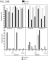
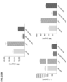
- one advantage of expanding, enriching, and/or activating the cells with the particles, e.g., beads, described herein as compared to other methods, e.g., stimulation with anti-CD3 and anti-CD28 antibodies, is that incubation with the particles disclosed herein results in less activation and/or exhaustion as opposed to cells that are expanded, enriched, and/or activated by other methods.
- cells incubated with the particles disclosed herein express lower levels or markers associated with activation, e.g., surface expression of CD25, or exhaustion, e.g., expression of PD1, as compared to cells that are incubated with polyclonal stimulatory molecules capable of activating components of the TCR complex, e.g., anti-CD3 and anti-CD28 antibodies.
- markers associated with activation e.g., surface expression of CD25
- exhaustion e.g., expression of PD1
- polyclonal stimulatory molecules capable of activating components of the TCR complex e.g., anti-CD3 and anti-CD28 antibodies.
- transducing or transfecting cells with a viral or non-viral vector to deliver a nucleic acid that encodes a recombinant receptor or CAR to cells while they are contacted, treated, or incubated with the particles, e.g., beads, described herein at least for the portion of the transduction or transfection process.
- the particles e.g., beads
- one advantage of transducing or transfecting the cells in the presence of the particles, e.g., beads allows for the transfection or transduction of cells that have not been previously activated with polyclonal stimulatory molecules, e.g., anti-CD3 and anti-CD28 antibodies.
- polyclonal stimulatory molecules e.g., anti-CD3 and anti-CD28 antibodies.
- Particular instances disclosed herein contemplate that cells that are transduced or transfected without prior activation with polyclonal molecules will exhibit increased persistence and/or reduced exhaustion as compared to cells that were activated prior to the transfection or transducing.
- particular recombinant antigens, or fragments thereof, attached to particles, e.g., beads, as described may be particularly suited for uses to specifically stimulate a CAR containing an antigen-binding domain that recognizes the antigen.
- certain recombinant antigens e.g. BCMA
- BCMA may have few or no non-specific interactions, such as non-specific protein-protein interactions or non-specific interactions with cells that prevents or minimizes the antigen non-specifically sticking to the cells or getting soaked up by the cells. In some cases, this can improve the quality of stimulation of the CAR-expressing cells.
- attachment to beads or particles results in an increased, enhanced, consistent, and/or more reliable stimulation, activation, or expansion of cells as compared to when the recombinant antigen is unbound or bound to a different solid support, such as the surface of a plate or dish.
- the binding molecule is or includes recombinant BCMA or a fragment thereof.
- particles or beads containing attached recombinant BCMA are used in conjunction with the disclosed methods to activate, stimulate, or expand cells of a cell composition.
- the particles or beads containing the recombinant BCMA selectively activate, stimulate, or expand the cells expressing a recombinant receptor containing an antigen-binding domain that binds to or recognizes BCMA, e.g., an anti-BCMA CAR.
- the activation, stimulation, or expansion of the cells is greater or increased as compared to activation, stimulation, or expansion by other reagents, such as for example anti-CD3/anti-CD28 antibody conjugated bead reagents.
- the activation, stimulation, or expansion of the cells is more indicative of activation, stimulation, or expansion of the cells in vivo and/or in response to endogenous antigen as compared to activation, stimulation, or expansion by other reagents, such as for example anti-CD3/anti-CD28 antibody conjugated bead reagents.
- the degree to which the cells are activated, stimulated, or expanded by the particles or beads particles or beads containing attached recombinant BCMA can be adjusted, modified or controlled by altering the amount of recombinant BCMA attached to the particles or beads or by altering the ratio of particles or beads to cells.
- activation, stimulation, or expansion of cells, e.g., anti-BCMA CAR expressing cells, by recombinant BCMA is more effective when the recombinant BCMA is attached to particles or beads than when the recombinant BCMA is free floating or unattached, or than when the recombinant BCMA is attached to a different surface, e.g., a culture dish or plate.
- the binding molecule is an anti-idiotypic antibody (anti-ID) that binds to or recognizes a recombinant receptor, e.g., a CAR.
- anti-ID anti-idiotypic antibody
- particles or beads containing attached recombinant anti-IDs are used in conjunction with the disclosed methods to activate, stimulate, or expand cells of a cell composition.
- the particles or beads containing anti-IDs selectively activate, stimulate, or expand the cells of the composition, such as cells expressing the recombinant receptor, e.g., a CAR.
- the activation, stimulation, or expansion of the cells is greater or increased as compared to activation, stimulation, or expansion by other reagents, such as for example anti-CD3/anti-CD28 antibody conjugated bead reagents.
- the degree to which the cells are activated, stimulated, or expanded by the particles or beads particles or beads containing anti-IDs can be adjusted, modified or controlled by altering the amount of anti-IDs attached to the particles or beads or by altering the ratio of particles or beads to cells.
- the anti-ID is an anti-CD19 antibody anti-ID.
- the activation, stimulation, or expansion of anti-CD19 CAR expressing cells by particles or beads containing anti-CD19 antibody anti-IDs is greater or increased as compared to activation, stimulation, or expansion by other reagents, such as for example, anti-CD3/anti-CD28 antibody conjugated bead reagents.
- compositions and methods expand and activate genetically engineered T cells that express recombinant receptors or CARs, that, when administered to a subject, exhibit increased persistence and/or reduced exhaustion as compared to genetically engineered T cells that are expanded by other techniques.
- genetically engineered T cells with increased persistence and/or reduced exhaustion exhibit better potency in a subject to which they are administered.
- particles such as beads, that are conjugated or otherwise attached to a binding molecule that binds or is recognized by an antigen-binding domain of a recombinant receptor, such as a chimeric antigen receptor (CAR) and methods for use thereof.
- a recombinant receptor such as a chimeric antigen receptor (CAR)
- the particles e.g., beads, are non-cell particles.
- a binding molecule that binds to or is recognized by an antigen-binding domain of a recombinant receptor such as a chimeric antigen receptor (CAR)
- a particle e.g., bead particles
- the particle is a non-cell particle.
- the particle may include a colloidal particle, a microsphere, nanoparticle, a bead, such as a magnetic bead, or the like.
- the particles or beads are biocompatible, i.e. non-toxic.
- the particles or beads are non-toxic to cultured cells, e.g., cultured T cells.
- the particles are monodisperse.
- “monodisperse” encompasses particles (e.g., bead particles) with size dispersions having a standard deviation of less than 5%, e.g., having less than a 5% standard deviation in diameter.
- a particle described herein provides a solid support or matrix to which a binding molecule, such as a binding molecule described herein (e.g., an antigen or an antibody), can be bound or attached in a manner that permits an interaction between the binding molecule and a cell, in particular binding between the binding molecule and a recombinant receptor, e.g., a CAR, expressed on the surface of the cell.
- a binding molecule such as a binding molecule described herein (e.g., an antigen or an antibody)
- a recombinant receptor e.g., a CAR
- the interaction between the conjugated or attached binding molecule and the cell can be used in methods to facilitate enrichment, activation, stimulation and/or expansion of one or more cell types in a cell population based on expression or expression level of one or more recombinant receptors on the surface of a cell.
- the particle e.g., a bead particle
- the particle or bead is biocompatible, i.e., composed of a material that is suitable for biological use.
- the particles, e.g., beads are non-toxic to cultured cells, e.g., cultured T cells.
- the particles, e.g., beads may be any particles which are capable of attaching binding molecules in a manner that permits an interaction between the binding molecule and a cell.
- the particles, e.g., beads may be any particles that can be modified, e.g., surface functionalized, to allow for the attachment of a binding molecule at the surface of the particle.
- the particles e.g., beads
- the particles are composed of glass, silica, polyesters of hydroxy carboxylic acids, polyanhydrides of dicarboxylic acids, or copolymers of hydroxy carboxylic acids and dicarboxylic acids.
- the particles may be composed of or at least partially composed of polyesters of straight chain or branched, substituted or unsubstituted, saturated or unsaturated, linear or cross-linked, alkanyl, haloalkyl, thioalkyl, aminoalkyl, aryl, aralkyl, alkenyl, aralkenyl, heteroaryl, or alkoxy hydroxy acids, or polyanhydrides of straight chain or branched, substituted or unsubstituted, saturated or unsaturated, linear or cross-linked, alkanyl, haloalkyl, thioalkyl, aminoalkyl, aryl, aralkyl, alkenyl, aralkenyl, heteroaryl, or alkoxy dicarboxylic acids.
- particles e.g., beads
- particles can be quantum dots, or composed of quantum dots, such as quantum dot polystyrene particles, e.g., beads.
- Particles, e.g., beads, including mixtures of ester and anhydride bonds (e.g., copolymers of glycolic and sebacic acid) may also be employed.
- particles may comprise materials including polyglycolic acid polymers (PGA), polylactic acid polymers (PLA), polysebacic acid polymers (PSA), poly(lactic-co-glycolic) acid copolymers (PLGA), [rho]poly(lactic-co-sebacic) acid copolymers (PLSA), poly(glycolic-co-sebacic) acid copolymers (PGSA), etc.
- PGA polyglycolic acid polymers
- PLA polylactic acid polymers
- PSA polysebacic acid polymers
- PLA poly(lactic-co-glycolic) acid copolymers
- PLSA poly(lactic-co-glycolic) acid copolymers
- PGSA poly(glycolic-co-sebacic) acid copolymers
- polymers that particles, e.g., beads, may be composed of include polymers or copolymers of caprolactones, carbonates, amides, amino acids, orthoesters, acetals, cyanoacrylates and degradable urethanes, as well as copolymers of these with straight chain or branched, substituted or unsubstituted, alkanyl, haloalkyl, thioalkyl, aminoalkyl, alkenyl, or aromatic hydroxy- or dicarboxylic acids.
- the biologically important amino acids with reactive side chain groups such as lysine, arginine, aspartic acid, glutamic acid, serine, threonine, tyrosine and cysteine, or their enantiomers, may be included in copolymers with any of the aforementioned materials to provide reactive groups for conjugating to binding molecules such as polypeptide antigen or antibodies.
- the particles or beads have a diameter of greater than 0.001 ⁇ m, greater than 0.01 ⁇ m, greater than 0.05 ⁇ m, greater than 0.1 ⁇ m, greater than 0.2 ⁇ m, greater than 0.3 ⁇ m, greater than 0.4 ⁇ m, greater than 0.5 ⁇ m, greater than 0.6 ⁇ m, greater than 0.7 ⁇ m, greater than 0.8 ⁇ m, greater than 0.9 ⁇ m, greater than 1 ⁇ m, greater than 2 ⁇ m, greater than 3 ⁇ m, greater than 4 ⁇ m, greater than 5 ⁇ m, greater than 6 ⁇ m, greater than 7 ⁇ m, greater than 8 ⁇ m, greater than 9 ⁇ m, greater than 10 ⁇ m, greater than 20 ⁇ m, greater than 30 ⁇ m, greater than 40 ⁇ m, greater than 50 ⁇ m, greater than 100 ⁇ m, greater than 500 ⁇ m, and/or greater than 1,000 ⁇ m.
- the particles or beads have a diameter of between or between about 0.001 ⁇ m and 1,000 ⁇ m, 0.01 ⁇ m and 100 ⁇ m, 0.1 ⁇ m and 10, ⁇ m, 0.1 ⁇ m and 100 ⁇ m, 0.1 ⁇ m and 10 ⁇ m, 0.001 ⁇ m and 0.01 ⁇ m, 0.01 ⁇ m and 0.1 ⁇ m, 0.1 ⁇ m and 1 ⁇ m, 1 ⁇ m and 10 ⁇ m, 1 ⁇ m and 2 ⁇ m, 2 ⁇ m and 3 ⁇ m, 3 ⁇ m and 4 ⁇ m, 4 ⁇ m and 5 ⁇ m, 1 ⁇ m and 5 ⁇ m, and/or 5 ⁇ m and 10 ⁇ m, each inclusive.
- the particles or beads have a mean diameter of 1 ⁇ m and 10 ⁇ m, each inclusive. In certain instances disclosed herein, the particles, e.g., beads, have a diameter of or of about 1 ⁇ m. In particular instances disclosed herein, the particles, e.g., beads, have a mean diameter of or of about 2.8 ⁇ m. In some instances disclosed herein, the particles, e.g., beads, have a diameter of or of about 4.8 ⁇ m.
- a plurality of the particles has a uniform particle size.
- a uniform particle size comprises a diameter standard deviation of less than 10%, less than 5%, or less than 1% of the mean diameter of the plurality.
- the plurality of the particles, e.g., beads has a diameter standard deviation of less than 10%, less than 5%, or less than 1% of the mean diameter of the plurality.
- the particles are uniformly shaped.
- the particles, e.g., beads are spherical.
- the particles, e.g., beads are non-spherical.
- the particles e.g., beads
- the particles have a density of greater than 0.001 g/cm 3 , greater than 0.01 g/cm 3 , greater than 0.05 g/cm 3 , greater than 0.1 g/cm 3 , greater than 0.5 g/cm 3 , greater than 0.6 g/cm 3 , greater than 0.7 g/cm 3 , greater than 0.8 g/cm 3 , greater than 0.9 g/cm 3 , greater than 1 g/cm 3 , greater than 1.1 g/cm 3 , greater than 1.2 g/cm 3 , greater than 1.3 g/cm 3 , greater than 1.4 g/cm 3 , greater than 1.5 g/cm 3 , greater than 2 g/cm 3 , greater than 3 g/cm 3 , greater than 4 g/cm 3 , or greater than 5g/cm 3 .
- the particles or beads have a density of between or between about 0.001 g/cm 3 and 100 g/cm 3 , 0.01 g/cm 3 and 50 g/cm 3 , 0.1 g/cm 3 and 10 g/cm 3 , 0.1 g/cm 3 and 0.5 g/cm 3 , 0.5 g/cm 3 and 1 g/cm 3 , 0.5 g/cm 3 and 1.5 g/cm 3 , 1 g/cm 3 and 1.5 g/cm 3 , 1 g/cm 3 and 2 g/cm 3 , or 1 g/cm 3 and 5 g/cm 3 .
- the particles or beads have a density of, of at least, or of about 0.5 g/cm 3 , 0.5 g/cm 3 , t 0.6 g/cm 3 , 0.7 g/cm 3 , a0.8 g/cm 3 , 0.9 g/cm 3 , 1.0 g/cm 3 , 1.1 g/cm 3 , 1.2 g/cm 3 , 1.3 g/cm 3 , 1.4 g/cm 3 , 1.5 g/cm 3 , 1.6 g/cm 3 , 1.7 g/cm 3 , 1.8 g/cm 3 , 1.9 g/cm 3 , or 2.0 g/cm 3 , each inclusive.
- the beads or particles have a density of or of about 1.6 g/cm 3 . In particular instances disclosed herein, the beads or particles have a density of or of about 1.5 g/cm 3 . In certain instances disclosed herein, the particles, e.g., beads, have a density of or of about 1.3 g/cm 3 .
- a plurality of the particles or beads has a uniform density.
- a uniform density comprises a density standard deviation of less than 10%, less than 5%, or less than 1% of the mean particle density.
- the particles or beads have a surface area of between or between about 0.001 m 2 per each gram of particles, e.g., beads, (m 2 /g) and 1,000 m 2 /g,0.010 m 2 /g and 100 m 2 /g, 0.1 m 2 /g and 10 m 2 /g, 0.1 m 2 /g and 1 m 2 /g, 1 m 2 /g and 10 m 2 /g, 10 m 2 /g and 100 m 2 /g, 0.5 m 2 /g and 20 m 2 /g, 0.5 m 2 /g and 5 m 2 /g, or 1 m 2 /g and 4 m 2 /g, each inclusive.
- the particles or beads have a surface area of or of about 1 m 2 /g to 4 m 2 /g.
- the particles e.g., bead particles
- a specific gravity i.e. the ratio of the density of the particles, e.g., beads, to the density of water, of between or between about 0.01 and 100, 0.1 and 10, 0.5 and 5, 1 and 10, 1 and 2, 1.1 and 1.8, or 1.2 and 1.5, each inclusive.
- the particles, e.g., beads have a specific gravity of 1.2 and 1.5.
- the particles, e.g., beads are monodisperse and the specific gravity is uniform.
- particles e.g., beads
- a uniform specific gravity have a specific gravity standard deviation of less than 10%, less than 5%, or less than 1%.
- the particles are monodisperse and have a specific gravity with a standard deviation of less than 10%, less than 5%, or less than 1%.
- the particle surface comprises attached biomolecules that can bind or attach binding molecules.
- the biomolecules are polypeptides.
- the particles, e.g., beads comprise surface exposed protein A, protein G, or biotin.
- the particle contains one or more coats or coatings such as one or more coats or coatings on the surface of the particle (e.g., a surface coating).
- the one or more coats or coatings provide a material for conjugation or coupling to a binding molecule, e.g., a coat that is or is capable of being surface functionalized.
- the coat comprises, or is capable of attaching, surface exposed functional groups.
- the coating comprises, or is capable of attaching, surface-exposed carboxyl groups, amino groups, hydroxyl groups, tosyl groups, epoxy groups, chloromethyl groups, or combinations thereof.
- the coat or coating is hydrophobic. In particular instances disclosed herein the coat or coating is non-hydrophobic.
- the particle reacts in a magnetic field.
- the particle is a magnetic particle (e.g., magnetic bead particle).
- the magnetic particle is paramagnetic.
- the magnetic particle is superparamagnetic.
- the particles, e.g., beads do not display any magnetic properties unless they are exposed to a magnetic field.
- the particle can be a composite particle containing an inner core.
- the inner core is a magnetic core, a paramagnetic core or a superparamagnetic core.
- the inner core e.g., magnetic core
- the metal can be, but is not limited to, iron, nickel, copper, cobalt, gadolinium, manganese, tantalum, zinc, zirconium or any combinations thereof.
- Suitable substances that may be included in an inner core described herein includes, but is not limited to, metal oxides (e.g., iron oxides), ferrites (e.g., manganese ferrites, cobalt ferrites, nickel ferrites, etc.), hematite and metal alloys (e.g., CoTaZn).
- the inner core comprises one or more of a ferrite, a metal, a metal alloy, an iron oxide, or chromium dioxide.
- the inner core comprises elemental iron or a compound thereof.
- the inner core comprises one or more of magnetite (Fe3O4), maghemite ( ⁇ Fe2O3), or greigite (Fe3S4).
- the inner core comprises an iron oxide (e.g., Fe 3 O 4 ).
- the particle contains a magnetic, paramagnetic, and/or superparamagnetic core that is covered by a surface functionalized coat or coating. In some instances disclosed herein, the particle comprises surface exposed tosyl groups.
- the coat can contain a material that can include, but is not limited to, a polymer, a polysaccharide, a silica, a fatty acid, a protein, a carbon, or a combination thereof.
- the polymer can be a polyethylene glycol, poly (lactic-co-glycolic acid), polyglutaraldehyde, polyurethane, polystyrene, or a polyvinyl alcohol.
- the outer coat or coating comprises polystyrene.
- the coat contains or includes a material that is or includes a protein that is an albumin ( e.g ., human serum albumin), Protein A, and Protein G.
- the carbon is an acrylamide or maleic acid.
- the material is coupled, linked or conjugated to a binding molecule described herein.
- a particle described herein can have an inner core and a coat (e.g., protective coat) wherein the coat contains one or more material described herein.
- the coat is hydrophobic.
- the coat is hydrophilic.
- a particle described herein e.g., a bead particle
- has a metal oxide core e.g., an iron oxide inner core
- a coat e.g., a protective coat
- the particles (e.g., bead particles) used in the methods described herein can be produced or obtained commercially.
- Particles, e.g., beads, including methods of producing particles, e.g., beads, are well known in the art. See, for example, U.S. Pat. Nos. 6,074,884 ; 5,834,121 ; 5,395,688 ; 5,356,713 ; 5,318,797 ; 5,283,079 ; 5,232,782 ; 5,091,206 ; 4,774,265 ; 4,654,267 ; 4,554,088 ; 4,490,436 ; 4,452,773 ; U.S. Patent Application Publication No.
- particles include, but are not limited to, ProMagTM (PolySciences, Inc.); COMPELTM (PolySciences, Inc.); BioMag ® (PolySciences, Inc.), including BioMag ® Plus (PolySciences, Inc.) and BioMag ® Maxi (Bang Laboratories, Inc.); M-PVA (Cehmagen Biopolymer Technologie AG); SiMAG (Chemicell GmbH); beadMAG (Chemicell GmbH); MagaPhase ® (Cortex Biochem); Dynabeads ® (Invitrogen), including Dynabeads ® M-280 Sheep Anti-rabbit IgG (Invitrogen), Dynabeads ® FlowCompTM (e.g., Dynabeads ® FlowCompTMHuman
- the particle is monodisperse, superparamagnetic bead particles comprising a superparamagnetic iron core, e.g., a magnetite (Fe 3 O 4 ) or maghemite ( ⁇ Fe 2 O 3 ) core, a polystyrene coat or coating, and a functionalized surface comprising exposed tosyl groups.
- the particles e.g., beads, have a density of about 1.5 g/cm 3 and a surface area of about 1 m 2 /g to about 4 m 2 /g.
- the particles are monodisperse superparamagnetic particles, e.g., beads, that have a diameter of about 4.5 ⁇ m and a density of about 1.5 g/cm 3 .
- the particles, e.g., beads the particles are monodisperse superparamagnetic particles that have a mean diameter of about 2.8 ⁇ m and a density of about 1.3 g/cm 3 .
- the particle is an oligomer or polymer. In some instances disclosed herein, the particle is an oligomer or a polymer composed of proteins, e.g., streptavidin. In some instances disclosed herein, the particle is an oligomer or polymer that can be generated by linking directly or indirectly individual molecules of a protein, e.g., streptavidin or a variant thereof, as it exists naturally, either by linking directly or indirectly individual molecules of a monomer or a complex of subunits that make up an individual molecule (e.g., linking directly or indirectly dimers, trimers, tetramers, etc. of a protein as it exists naturally).
- a protein e.g., streptavidin or a variant thereof
- the reagent is a multimer, or the oligomeric reagent is a multimeric reagent.
- the particle is an oligomer or polymer that comprises streptavidin or mutein of streptavidin (e.g., STREP-TACTIN ® or STREP-TACTIN ® XT streptavidin muteins)
- the particle is an oligomer or polymer that is obtained by crosslinking individual molecules or a complex of subunits that make up an individual molecule in the presence of a polysaccharide.
- the particles are oligomers or polymers that can be prepared by the introduction of carboxyl residues into a polysaccharide, e.g., dextran.
- individual molecules of the particle e.g., monomers, tetramers
- the coupling reaction is performed at a molar ratio of about 60 moles of individual molecules of the reagent (e.g., monomers, tetramers) per mole of dextran.
- the particle is an oligomer or a polymer of one or more streptavidin or avidin or of any analog or mutein of streptavidin (e.g., Strep-Tactin ® or Strep-Tactin ® XT) or analog or mutein of avidin (e.g., neutravidin).
- the avidin or streptavidin comprises a binding site Z that is a natural biotin binding site of avidin or streptavidin for which there can be up to four binding sites in an individual molecule (e.g., a tetramer contains four binding sites Z), whereby a homo-tetramer can contain up to 4 binding sites that are the same, i.e.
- the oligomer is generated or produced from a plurality of individual molecules (e.g., a plurality of homo-tetramers) of the same streptavidin, streptavidin mutein, avidin or avidin mutein, in which case each binding site Z, e.g., Z1, of the oligomer is the same.
- an oligomer can contain a plurality of binding sites Z1, such as at least 2, 3, 4, 5, 6, 7, 8, 9, 10, 11, 12, 13, 14, 15, 16, 17, 18, 19, 20, 21, 22, 23, 24, 25, 26, 27, 28, 29, 30, 31, 32, 33, 34, 35, 40, 45, 50 or more binding sites Z1.
- the oligomer is generated or produced from a plurality of individual molecules that can be hetero-tetramers of a streptavidin, streptavidin mutein, avidin or avidin mutein and/or from a plurality of two or more different individual molecules (e.g., different homo-tetramers) of streptavidin, streptavidin mutein, avidin or avidin mutein that differ in their binding sites Z, e.g., Z1 and Z2, in which case a plurality of different binding sites Z, e.g., Z1 and Z2, may be present in the oligomer.
- an oligomer can contain a plurality of binding sites Z1 and a plurality of binding sites Z, which, in combination, can include at least 2, 3, 4, 5, 6, 7, 8, 9, 10, 11, 12, 13, 14, 15, 16, 17, 18, 19, 20, 21, 22, 23, 24, 25, 26, 27, 28, 29, 30, 31, 32, 33, 34, 35, 40, 45, 50 or more combined binding sites Z1 and Z2.
- the particle is an oligomer or polymer is obtained by crosslinking individual molecules or a complex of subunits that make up an individual molecule using a bifunctional linker or other chemical linker, such as avidin or by other methods known in the art.
- a bifunctional linker or other chemical linker such as avidin or by other methods known in the art.
- cross-linked oligomers or polymers of streptavidin or avidin or of any mutein or analog of streptavidin or avidin may be obtained by crosslinking individual streptavidin or avidin molecules via bifunctional molecules, serving as a linker, such as glutaraldehyde or by other methods described in the art.
- oligomers of streptavidin muteins by introducing thiol groups into the streptavidin mutein (this can, for example, be done by reacting the streptavidin mutein with 2-iminothiolan (Traut's reagent) and by activating, for example in a separate reaction, amino groups available in the streptavidin mutein.
- 2-iminothiolan Traut's reagent
- this activation of amino groups can be achieved by reaction of the streptavidin mutein with a commercially available heterobifunctional crosslinker such as sulfosuccinimidyl 4-(N-maleimidomethyl)cyclohexane-1-carboxylate (sulfo SMCC) or Succinimidyl-6-[((3-maleimidopropionamido)hexanoate (SMPH).
- sulfo SMCC N-maleimidomethyl)cyclohexane-1-carboxylate
- SMPH Succinimidyl-6-[((3-maleimidopropionamido)hexanoate
- the two reaction products so obtained are mixed together, typically leading to the reaction of the thiol groups contained in the one batch of modified streptavidin mutein with the activated (such as by maleimide functions) amino acids of the other batch of modified streptavidin mutein.
- multimers/oligomers of the streptavidin mutein are formed.
- These oligomers can have any suitable number of individual molecules, such as at least 2, 3, 4, 5, 6, 7, 8, 9, 10, 11, 12, 13, 14, 15, 16, 17, 18, 19, 20, 21, 22, 23, 24, 25, 26, 27, 28, 29, 30, 31, 32, 33, 34, 35, 40, 45, 50 or more, and the oligomerization degree can be varied according to the reaction condition.
- the oligomeric or polymeric reagent can be isolated via size exclusion chromatography and any desired fraction can be used as the particle.
- the oligomeric or polymeric reagent after reacting the modified streptavidin mutein in the presence of 2-iminothiolan and a heterobifunctional crosslinker such as sulfo SMCC, the oligomeric or polymeric reagent can be isolated via size exclusion chromatography and any desired fraction can be used as the particle.
- the oligomers do not have (and do not need to have) a single molecular weight but they may observe a statistical weight distribution such as Gaussian distribution.
- any oligomer with more than three streptavidin or mutein tetramers can be used as a soluble particle, such as generally 3 to 50 tetramers, e.g., homotetramers or heterotetramers, 10 to 40 tetramers, e.g., homotetramers or heterotetramers, or 25 to 35 tetramers, e.g., homotetramers or heterotetramers.
- the oligomers might have, for example, from 3 to 25 streptavidin mutein tetramers, e.g., homotetramers or heterotetramers.
- the soluble oligomers can have a molecular weight from or from about 150 kDa to 2000 kDa, 150 kDa to 1500 kDa, 150 kDa to 1250 kDa, 150 kDa to 1000 kDa, 150 kDa to 500 kDa or 150 kDa to 300 kDa, 300 kDa to 2000 kDa, 300 kDa to 1500 kDa, 300 kDa to 1250 kDa, 300 kDa to 1000 kDa, 300 kDa to 500 kDa, 500 kDa to 2000 kDa, 500 kDa to 1500 kDa, 500 kDa to 1250 kDa, 500 kDa to 1000 kDa, 1000 kDa to 1500 kDa, 1000 kDa to 1500 kDa, 1000 kDa to 1500 kDa, 1000 kDa to 1500 kDa, 1000 kD
- binding molecules e.g., polypeptide antigens or antibodies, that bind or are recognized by an antigen-binding domain of a recombinant receptor, such as a CAR, that are conjugated or attached to a particle, e.g., a bead particle.
- a recombinant receptor such as a CAR
- the binding molecule is a polypeptide.
- the binding molecule includes any polypeptide for which a recombinant receptor, such as antigen receptor, e.g. CAR, can be designed to bind or recognize.
- the binding molecule is an anti-idiotype antibody that binds to or recognizes the antigen binding domain of a recombinant receptor, such as an antigen receptor, e.g. a CAR.
- the binding molecule is a polypeptide that is bound by or is recognized by the antigen binding domain of a recombinant receptor, such an antigen receptor, e.g. a CAR.
- the binding molecule is a polypeptide that is bound by or is recognized by the antigen binding domain of a CAR.
- the binding molecule binds to or is recognized by a CAR that does not comprise a transmembrane domain or an intracellular signaling domain of a killer cell immunoglobulin-like receptor (KIR).
- KIR killer cell immunoglobulin-like receptor
- the binding molecule binds to or is recognized by a CAR that does not comprise a transmembrane domain or an intracellular domain from any of KIR2DS2, KIR2DL3, KIR2DL1, KIR2DL2, KIR2DL4, KIR2DL5A, KIR2DL5B, KIR2DS1, KIR2DS3, KIR2DS4, KIR2DS5, KIR3DL1, KIR3DS1, KIR3DL2, KIR3DL3, KIR2DP1 and KIR3DP1.
- the binding molecule is an antigen, e.g., a recombinant antigen or fragment thereof.
- the antigen is a polypeptide, or a portion of a polypeptide, that is associated with a disease, e.g., a cancer.
- the antigen is a polypeptide, or a variant or fragment of a polypeptide that is expressed on the surface of a cell that is associated with a disease, for example, a cancer cell and/or a tumor cell.
- the antigen is or includes ⁇ v ⁇ 6 integrin (avb6 integrin), B cell maturation antigen (BCMA), B7-H3, B7-H6, carbonic anhydrase 9 (CA9, also known as CAIX or G250), a cancer-testis antigen, cancer/testis antigen 1B (CTAG, also known as NY-ESO-1 and LAGE-2), carcinoembryonic antigen (CEA), a cyclin, cyclin A2, C-C Motif Chemokine Ligand 1 (CCL-1), CD19, CD20, CD22, CD23, CD24, CD30, CD33, CD38, CD44, CD44v6, CD44v7/8, CD123, CD133, CD138, CD171, chondroitin sulfate proteoglycan 4 (CSPG4), epidermal growth factor protein (EGFR), type III epidermal growth factor receptor mutation (EGFR vIII), epithelial glycoprotein 2 (EPG-2), epithelial glycoprotein 2
- Antigens targeted by the receptors in some instances disclosed herein include antigens associated with a B cell malignancy, such as any of a number of known B cell marker.
- the antigen is or includes CD20, CD19, CD22, ROR1, CD45, CD21, CD5, CD33, Igkappa, Iglambda, CD79a, CD79b or CD30.
- the binding molecule comprises a portion of a polypeptide antigen that is recognized by or bound by a recombinant receptor and/or a CAR.
- the binding molecule comprises an epitope that is recognized by or bound by a recombinant receptor and/or a CAR.
- the portion of the polypeptide antigen contains, about, or contains at least 10, 15, 20, 25, 30, 35, 40, 50, 55, 60, 65, 70, 75, 80, 85, 90, 95, 100, 110, 120, 130, 140, 150, 160, 170, 180, 190, 200, 250, 300, 400, or 500 amino acids, in some cases contiguous amino acids, of the polypeptide that is recognized by or bound by a recombinant receptor and or a CAR.
- the polypeptide portion comprises an amino acid sequence of the epitope that is recognized by the recombinant receptor and/or CAR.
- the binding molecule is a polypeptide variant that contains, contains about, or contains at least 50%, 55%, 60%, 65%, 70%, 75%, 80%, 85%, 90%, 95%, 97%, 98%, 99%, or 99.5% amino acid sequence identity to a polypeptide that is bound by and/or recognized by recombinant receptor and/or CAR.
- a particle is bound to a binding molecule comprising a portion of the polypeptide antigen, wherein the portion is a portion of an antigen that is bound by and/or recognized by, or can potentially be bound by or recognized by a recombinant receptor, i.e. a CAR.
- the binding molecule comprises a portion of the antigen containing the epitope that is recognized by the recombinant receptor.
- the portion of the antigen is a portion of In some instances disclosed herein, the antigen is or includes ⁇ v ⁇ 6 integrin (avb6 integrin), B cell maturation antigen (BCMA), B7-H3, B7-H6, carbonic anhydrase 9 (CA9, also known as CAIX or G250), a cancer-testis antigen, cancer/testis antigen 1B (CTAG, also known as NY-ESO-1 and LAGE-2), carcinoembryonic antigen (CEA), a cyclin, cyclin A2, C-C Motif Chemokine Ligand 1 (CCL-1), CD19, CD20, CD22, CD23, CD24, CD30, CD33, CD38, CD44, CD44v6, CD44v7/8, CD123, CD133, CD138, CD171, chondroitin sulfate proteoglycan 4 (CSPG4), epidermal growth factor protein (EGFR), type III epidermal growth factor receptor mutation (EGFR), type III epiderma
- Antigens targeted by the receptors in some instances disclosed herein include antigens associated with a B cell malignancy, such as any of a number of known B cell marker.
- the antigen is or includes CD20, CD19, CD22, ROR1, CD45, CD21, CD5, CD33, Igkappa, Iglambda, CD79a, CD79b or CD30.
- the particle is bound to a binding molecule comprising a portion of a BCMA polypeptide. In some instances disclosed herein, the particle is bound to a binding molecule comprising a portion of a CD22 polypeptide. In certain instances disclosed herein, the particle is bound to a binding molecule comprising a portion of a ROR1 polypeptide.
- the portion of the polypeptide antigen is a portion of a BCMA polypeptide.
- the polypeptide antigen contains, contains about, or contains at least 50%, 55%, 60%, 65%, 70%, 75%, 80%, 85%, 90%, 95%, 97%, 98%, 99%, or 99.5% amino acid sequence identity to at least 10, at least 15, at least 20, at least 25, at least 30, at least 35, at least 40, at least 50, at least 55, at least 60, at least 65, at least 70, at least 75, at least 80, at least 85, at least 90, at least 95, at least 100, at least 110, at least 120, at least 130, at least 140, at least 150, at least 160, at least 170, or at least 180 contiguous amino acids of a BCMA polypeptide.
- the BCMA polypeptide variant comprises an amino acid sequence that is, is about, or is at least 70%, 75%, 80%, 85%, 90%, 95%, 97%, or 98% identical to the amino acid sequence of a BCMA epitope that is bound by and/or recognized by a recombinant receptor and/or a CAR.
- the antigen is BCMA, or a portion or variant thereof.
- the BCMA polypeptide is a mammalian BCMA polypeptide.
- the BCMA polypeptide is a human BCMA polypeptide.
- the BCMA antigen is or comprises an extracellular domain of BCMA or a portion thereof comprising an epitope recognized by an antigen receptor, e.g. CAR.
- the BCMA antigen is or comprises a polypeptide with an amino acid sequence with at least 70%, 75%, 80%, 85%, 86%, 87%, 88%, 89%, 90%, 91%, 92%, 93%, 94%, 95%, 96%, 97%, 98%, 99% or more sequence identity to SEQ ID NO: 1 or a fragment thereof containing at least 50, at least 55, at least 60, at least 65, at least 70, at least 75, at least 80, at least 85, at least 90, at least 95, at least 100, at least 110, at least 120, at least 130, at least 140, at least 150, at least 160, at least 170, or at least 180 contiguous amino acids of SEQ ID NO: 1.
- the BCMA antigen is or includes the sequence set forth in SEQ ID NO:1 or a portion thereof that is or contains an epitope recognized by an antigen receptor, e.g. CAR.
- the antigen is ROR1, or a portion or variant thereof.
- the ROR1 polypeptide is mammalian. In particular instances disclosed herein, the ROR1 polypeptide is human.
- the ROR1 antigen is or comprises an extracellular domain of ROR1 or a portion thereof comprising an epitope recognized by an antigen receptor, e.g. CAR.
- the ROR1 antigen is a polypeptide with an amino acid sequence with at least 70%, 75%, 80%, 85%, 86%, 87%, 88%, 89%, 90%, 91%, 92%, 93%, 94%, 95%, 96%, 97%, 98%, 99% or more sequence identity to SEQ ID NO: 31 or a fragment thereof containing at least 50, at least 55, at least 60, at least 65, at least 70, at least 75, at least 80, at least 85, at least 90, at least 95, at least 100, at least 110, at least 120, at least 130, at least 140, at least 150, at least 160, at least 170, or at least 180 contiguous amino acids of SEQ ID NO: 31.
- the ROR antigen comprises the sequence set forth in SEQ ID NO :31 or a portion thereof comprising an epitope recognized by an antigen receptor, e.g. CAR.
- the antigen is CD22, or a portion or variant thereof.
- the CD22 polypeptide is mammalian. In particular instances disclosed herein, the CD22 polypeptide is human.
- the antigen is an extracellular domain of CD22 or a portion thereof comprising an epitope recognized by an antigen receptor, e.g. CAR.
- the antigen is a polypeptide with an amino acid sequence with at least 70%, 75%, 80%, 85%, 86%, 87%, 88%, 89%, 90%, 91%, 92%, 93%, 94%, 95%, 96%, 97%, 98%, 99% or more sequence identity to SEQ ID NO: 29 or a fragment thereof containing at least 50, at least 55, at least 60, at least 65, at least 70, at least 75, at least 80, at least 85, at least 90, at least 95, at least 100, at least 110, at least 120, at least 130, at least 140, at least 150, at least 160, at least 170, or at least 180 contiguous amino acids of SEQ ID NO: 29.
- the CD22 antigen comprises the sequence set forth in SEQ ID NO :29 or a portion thereof comprising an epitope recognized by an antigen receptor, e.g. CAR.
- the portion of the polypeptide antigen is a portion of a BCMA polypeptide. In certain instances disclosed herein, the portion of the polypeptide antigen is a portion of a CD22 polypeptide. In particular instances disclosed herein, the portion of the polypeptide antigen is a portion of a ROR1 polypeptide.
- the portion of the polypeptide contains at least 10, at least 15, at least 20, at least 25, at least 30, at least 35, at least 40, at least 50, at least 55, at least 60, at least 65, at least 70, at least 75, at least 80, at least 85, at least 90, at least 95, at least 100, at least 110, at least 120, at least 130, at least 140, at least 150, at least 160, at least 170, or at least 180 contiguous amino acids of the amino acid sequence set forth in SEQ ID NOS: 1, 29, or 31.
- the cell expresses a CAR that binds to or recognizes a universal tag that can be fused to an antibody or a fragment or variant thereof.
- cells expressing such CARs are able to specifically recognize and kill target cells, for example tumor cells, that have been bound by antibodies that have been fused with the universal tag.
- target cells for example tumor cells
- anti-FITC CAR expressing T cells can bind to and/or recognize various human cancer cells when those cells are bound by cancer-reactive FITC-labeled antibodies.
- a particle e.g., a bead particle
- a particle comprises a surface exposed binding molecule that comprises an antigen, or a portion thereof, that binds or is recognized by an antigen receptor, e.g. CAR, that binds to a universal tag.
- the binding molecule is a universal tag or a portion thereof bound or recognized by the antigen receptor, e.g. CAR.
- a particle is bound to a binding molecule that comprises a universal tag, or a portion thereof, selected from the group consisting of: FITC, streptavidin, biotin, histidine, dinitrophenol, peridinin chlorophyll protein complex, green fluorescent protein, PE, HRP, palmitoylation, nitrosylation, alkalanine phosphatase, glucose oxidase, and maltose binding protein.
- the binding molecule is a multimer, e.g. a dimer, comprising two or more polypeptide antigens, or portion or variant thereof, that is recognized and/or bound by a recombinant receptor, such as an antigen receptor (e.g. a CAR).
- a recombinant receptor such as an antigen receptor (e.g. a CAR).
- the polypeptide antigen, or portion thereof are identical.
- the polypeptide antigen is linked, directly or indirectly, to a region or domain, e.g. a multimerization domain, that promotes or stabilizes interaction between two or more polypeptide antigens via complementary interactions between the domains or regions.
- providing the polypeptide antigen as a multimer e.g.
- an increased avidity may favor stimulatory or agonist activity of antigen receptor, e.g. CAR, by the binding molecule conjugated to the bead.
- a polypeptide is joined directly or indirectly to a multimerization domain.
- multimerization domains include the immunoglobulin sequences or portions thereof, leucine zippers, hydrophobic regions, hydrophilic regions, and compatible protein-protein interaction domains.
- the multimerization domain can be an immunoglobulin constant region or domain, such as, for example, the Fc domain or portions thereof from IgG, including IgG1, IgG2, IgG3 or IgG4 subtypes, IgA, IgE, IgD and IgM and modified forms thereof.
- the polypeptide antigen is linked, directly or indirectly, to an Fc domain.
- the polypeptide is a fusion polypeptide comprising the polypeptide antigen or portion thereof and the Fc domain.
- a binding molecule is a fusion polypeptide that comprises an Fc domain.
- the Fc domain is composed of the second and third constant domains (i.e., CH2 and CH3 domains) of the heavy chain of a IgG, IgA or IgD isotype, e.g. CH2 or CH3 of IgG, IgA and IgD isotypes.
- the Fc domain is composed of three heavy chain constant domain (i.e., CH2, CH3, and CH4 domains) of an IgM or IgE isotype.
- the Fc domain may further include a hinge sequence or portion thereof.
- the Fc domain contains part or all of a hinge domain of an immunoglobulin molecule plus a CH2 and a CH3 domain. In some cases, the Fc domain can form a dimer of two polypeptide chains joined by one or more disulfide bonds.
- the Fc domain is derived from an immunoglobulin (e.g., IgG, IgA, IgM, or IgE) of a suitable mammal (e.g., human, mouse, rat, goat, sheep, or monkey).
- the Fc domain comprises C H 2 and C H 3 domains of IgG.
- the Fc domain is fused to the C-terminal of the polypeptide antigen. In particular instances disclosed herein, the Fc domain is fused to the N-terminal of the polypeptide antigen.
- the Fc domain is an IgG Fc domain, or a portion or variant thereof.
- the Fc domain is a human IgG Fc domain, or a portion or a variant thereof, that comprises an amino acid sequence set forth in SEQ ID NO: 2 or an amino acid sequence that is at least 70%, 75%, 80%, 85%, 86%, 87%, 88%, 89%, 90%, 91%, 92%, 93%, 94%, 95%, 96%, 97%, 98%, 99% sequence identity to the sequence set forth in SEQ ID NO: 2.
- the Fc domain is a wild-type human IgG Fc domain, or a portion or variant thereof.
- the Fc domain is a variant of the wild-type human IgG1 Fc domain.
- the fusion polypeptide comprises a variant Fc domain.
- the variant human IgG Fc domain contains a mutation, e.g., a substitution, deletion, or insertion, that reduces, decreases, and/or diminishes pairing between the Fc domain and a light chain.
- the variant human IgG Fc domain contains a mutation that reduces the binding affinity between the Fc domain and an Fc Receptor.
- the variant human IgG Fc domain contains a mutation that reduces, decreases, and/or diminishes the interactions, or the probabiltity or likelihood of an interaction, between the Fc domain and an Fc Receptor.
- the variant human IgG Fc domain contains a mutation that reduces the binding affinity between the Fc domain and a protein of the complement system.
- the variant human IgG Fc domain contains a mutation that reduces, decreases, and/or diminishes the interactions, or the probabiltity or likelihood of an interaction, between the Fc domain and a protein of the complement system.
- the binding molecule comprises a variant human IgG1 Fc domain.
- the variant human IgG Fc domain contains a cystine to serine substitution in the hinge region of the Fc domain.
- the variant human IgG Fc domain contains a leucine to alanine substitution in the hinge region of the Fc domain.
- the variant human IgG Fc domain contains a glycine to alanine substitution in the hinge region.
- the variant human IgG Fc domain contains an alanine to a serine substitution in the CH2 region of the Fc domain.
- the variant human IgG Fc domain comprises a proline to serine substitution in the CH2 region of the Fc domain. In some instances disclosed herein, the variant human IgG Fc domain comprises an amino acid sequence as set forth by SEQ ID NO: 28.
- the binding molecule comprises a fusion polypeptide comprising an Fc domain, wherein the Fc domain is present at the C-terminus of the fusion polypeptide.
- the binding molecule is a fusion polypeptide comprising a BCMA polypeptide, or a portion thereof, and an Fc domain.
- the BCMA polypeptide or portion thereof comprises an amino acid sequence set forth in SEQ ID NO: 1 or a sequence of amino acids that exhibits at least 70%, 75%, 80%, 85%, 86%, 87%, 88%, 89%, 90%, 91%, 92%, 93%, 94%, 95%, 96%, 97%, 98%, 99% to SEQ ID NO: 1 and contains an epitope recognized by the antigen receptor, e.g. the CAR.
- the Fc domain comprises an amino acid sequence set forth in SEQ ID NO: 2 or a sequence of amino acids that exhibits at least 70%, 75%, 80%, 85%, 86%, 87%, 88%, 89%, 90%, 91%, 92%, 93%, 94%, 95%, 96%, 97%, 98%, 99% sequence identity to SEQ ID NO:2.
- the Fc domain comprises an amino acid sequence set forth in SEQ ID NO: 28 or a sequence of amino acids that exhibits at least 70%, 75%, 80%, 85%, 86%, 87%, 88%, 89%, 90%, 91%, 92%, 93%, 94%, 95%, 96%, 97%, 98%, 99% to SEQ ID NO:28.
- the BCMA antigen includes BCMA, or a portion or variant thereof, and a tag or a fusion domain, e.g., an Fc domain.
- the BCMA antigen contains all or a portion of the amino acid sequence set forth in SEQ ID NO: 35 or a sequence of amino acids that exhibits at least 70%, 75%, 80%, 85%, 86%, 87%, 88%, 89%, 90%, 91%, 92%, 93%, 94%, 95%, 96%, 97%, 98%, 99% to SEQ ID NO:35, and that comprises an epitope recognize by an antigen receptor, e.g. CAR.
- an antigen receptor e.g. CAR.
- the binding molecule is a fusion polypeptide comprising an amino acid sequence set forth in SEQ ID NO: 33 or a sequence of amino acids that exhibits at least 70%, 75%, 80%, 85%, 86%, 87%, 88%, 89%, 90%, 91%, 92%, 93%, 94%, 95%, 96%, 97%, 98%, 99% to SEQ ID NO:33 and that comprises an epitope recognize by an antigen receptor, e.g. CAR.
- an antigen receptor e.g. CAR.
- the binding molecule is a fusion polypeptide comprising a CD22 polypeptide set forth in SEQ ID NO:29 or portion thereof or a sequence of amino acids that exhibits at least 70%, 75%, 80%, 85%, 86%, 87%, 88%, 89%, 90%, 91%, 92%, 93%, 94%, 95%, 96%, 97%, 98%, 99% to SEQ ID NO:29 and comprising an epitope recognized by the antigen receptor, e.g. CAR, and an Fc domain.
- the antigen receptor e.g. CAR
- the fusion polypeptide comprises an amino acid sequence set forth in SEQ ID NO: 34 or a sequence of amino acids that exhibits at least 70%, 75%, 80%, 85%, 86%, 87%, 88%, 89%, 90%, 91%, 92%, 93%, 94%, 95%, 96%, 97%, 98%, 99% to SEQ ID NO:34 and that comprises an epitope recognize by an antigen receptor, e.g. CAR.
- an antigen receptor e.g. CAR.
- the antigen and the multimerization domain are connected by a linker, such as an amino acid linker.
- the antigen is fused to the N-terminus of an amino acid linker
- the multimerization domain such as Fc domain
- the linker length may be relatively short (e.g., ten or fewer amino acids) to reduce interactions between the linked domains.
- the amino acid composition of the linker also may be adjusted to reduce the number of amino acids with bulky side chains or amino acids likely to introduce secondary structure.
- the binding molecule is a dimer formed by two Fc fusion polypeptides containing a polypeptide antigen or portion thereof an Fc domain, e.g. BCMA-Fc, ROR1-Fc or CD22-Fc.
- nucleic acid molecules encoding any of the Fc fusion polypeptides.
- vectors including expression vectors, encoding the nucleic acid molecules.
- the binding molecule can be produced in cells by expression in a suitable host cell.
- the host cell is a mammalian cell line. Exemplary of mammalian cells for recombinant expression of proteins include HEK293 cells or CHO cells or derivatives thereof.
- the binding molecule further includes a signal peptide for secretion from the cell.
- the signal peptide is CD33 (e.g. set forth in SEQ ID NO: 30).
- the resulting polypeptide antigen-Fc fusion protein e.g. BCMA-Fc, ROR1-Fc or CD22-Fc, can be expressed in host cells, e.g. transformed with the expression vectors, whereby assembly between Fc domains can occurs by interchain disulfide bonds formed between the Fc moieties to yield a dimeric, such as divalent, polypeptide antigen fusion protein.
- the fusion polypeptide comprises a portion of human BCMA comprising an amino acid sequence set forth in SEQ ID NO: 1 and a variant human IgG1 Fc domain comprising an amino acid set forth in SEQ ID NO: 29.
- fusion polypeptide comprises a linker that joins the BCMA polypeptide to the Fc domain.
- the linker has an amino acid sequence set forth in SEQ ID NO: 27.
- the binding molecule is a fusion polypeptide comprising a human BCMA polypeptide (or a portion or a variant thereof) and a C-terminal Fc domain and is surface conjugated or otherwise attached to a particle (e.g., bead particle).
- a particle e.g., bead particle.
- the fusion polypeptide is attached to the particle at a site within in the Fc domain.
- the particle further comprises a surface conjugated antibody or a fragment or variant thereof, that binds to or recognizes CD28.
- the binding molecule conjugated to a particle, e.g., bead, as disclosed herein is an anti-idiotype antibody, or an antigen binding fragment thereof, ("anti-ID") that binds to a target antigen receptor, e.g. CAR, or an active fragment thereof.
- the biding molecule is an anti-ID that binds to the antigen binding domain of the CAR.
- the antigen binding domain of the CAR comprises an scFv domain.
- the binding molecule is an anti-ID that binds to a CAR at the antigen binding domain, and does not bind to the linker or spacer region of the CAR that connects the antigen binding domain, e.g., an scFv, with a transmembrane domain of the CAR.
- the anti-ID is an anti-idiotype antibody or antigen-binding fragments that specifically recognizes an antibody that binds to a target antigen, such as a target antigen associated with or expressed on a cell or tissue of a disease or condition, e.g. cancer.
- the anti-ID is an anti-idiotype antibody or antigen-binding fragment thereof that specifically recognizes a target antibody that binds the antigen that is or includes
- the antigen is or includes ⁇ v ⁇ 6 integrin (avb6 integrin), B cell maturation antigen (BCMA), B7-H3, B7-H6, carbonic anhydrase 9 (CA9, also known as CAIX or G250), a cancer-testis antigen, cancer/testis antigen 1B (CTAG, also known as NY-ESO-1 and LAGE-2), carcinoembryonic antigen (CEA), a cyclin, cyclin A2, C-C Motif Chemokine Ligand 1 (CCL-1), CD19, CD20, CD22, CD23, CD24, CD30, CD33, CD38, CD44, CD44v6, CD44v7/8, CD123, CD133, CD138, CD171, chondroitin sulfate proteoglycan
- Antigens targeted by the receptors in some instances disclosed herein include antigens associated with a B cell malignancy, such as any of a number of known B cell marker.
- the antigen is or includes CD20, CD19, CD22, ROR1, CD45, CD21, CD5, CD33, Igkappa, Iglambda, CD79a, CD79b or CD30.
- the anti-ID is an anti-idiotype antibody or antigen-binding fragments that specifically recognizes a target antibody that binds ROR1, B cell maturation antigen (BCMA), carbonic anhydrase 9 (CAIX), Her2/neu (receptor tyrosine kinase erbB2), Ll-CAM, CD19, CD20, CD22, mesothelin, CEA, and hepatitis B surface antigen, anti-folate receptor, CD23, CD24, CD30, CD33, CD38, CD44, EGFR, epithelial glycoprotein 2 (EPG-2), epithelial glycoprotein 40 (EPG-40), EPHa2, erb-B2, erb-B3, erb-B4, erbB dimers, EGFR vIII, folate binding protein (FBP), FCRL5, FCRH5, fetal acetylcholine receptor, GD2, GD3, HMW-MAA, IL-22R-
- FBP B cell
- the anti-ID is an anti-idiotype antibody or antigen-binding fragments ("anti-IDs") that specifically recognizes a target anti-CD19 antibody moiety.
- the disclosed antibodies recognize a target anti-CD19 antibody that is SJ25C1 or an antigen-binding fragment thereof or is an antibody or antigen-binding fragment derived from SJ25C1.
- the disclosed antibodies recognize a target anti-CD19 antibody that is FMC63 or an antigen-binding fragment thereof is an antibody or antigen-binding fragment derived from FMC63.
- SJ25C1 is a mouse monoclonal IgG1 antibody raised against Nalm-1 and -16 cells expressing CD19 of human origin ( Ling, N. R., et al. (1987). Leucocyte typing III. 302 ).
- the SJ25C1 antibody comprises CDRH1, H2 and H3 set forth in SEQ ID NOS: 40-42, respectively, and CDRL1, L2 and L3 sequences set forth in SEQ ID NOS: 43-45, respectively.
- the SJ25C1 antibody comprises the heavy chain variable region (V H ) comprising the amino acid sequence of SEQ ID NO: 36 and the light chain variable region (V L ) comprising the amino acid sequence of SEQ ID NO: 37.
- the target antibody is SJ25C1 or an antibody-derived from SJ25C1.
- the antibody derived from SJ25C1 is an antibody or antigen-binding fragment that comprises the V H and/or V L of SJ25C1, the idiotype of SJ25C1, the paratope of SJ25C1, or one or more complementarity determining regions (CDRs) of SJ25C1.
- the target antibody that is SJ25C1 or an antibody-derived from SJ25C1 is an antibody or antigen-binding fragment comprises the V H of SJ25C1 set forth in SEQ ID NO: 36, or a variant thereof having at least 90% sequence identity set forth in SEQ ID NO:36, and/or the V L of SJ25C1 set forth in SEQ ID NO:37, or a variant thereof having at least 90% sequence identity to SEQ ID NO:37.
- the antibody or antigen-binding fragment comprises the V H of SJ25C1 set forth in SEQ ID NO:36, or a variant thereof having at least 90% sequence identity to SEQ ID NO:36, and the V L of SJ25C1 set forth in SEQ ID NO:37, or a variant thereof having at least 90% sequence identity to SEQ ID NO:37.
- the antibody or antigen-binding fragment comprises the V H and V L of SJ25C1 set forth in SEQ ID NO:36 and SEQ ID NO:37, respectively.
- the variant has at least 91%, 92%, 93%, 94%, 95%, 96%, 97%, 98%, 99% or more sequence identity to SEQ ID NO:36 and/or SEQ ID NO:37.
- the target antibody that is SJ25C1 or an antibody-derived from SJ25C1 is an antibody or antigen-binding fragment comprises one or more heavy chain CDRs (CDR-H) of SJ25C1 V H set forth in SEQ ID NO:36, such as set forth in SEQ ID NOS: 40-42 and/or one or more light chain CDRs (CDR-Ls) of SJ25C1 V L set forth in SEQ ID NO:37, such as set forth in SEQ ID NOS: 43-45.
- the antibody or antigen-binding fragment comprises CDR-H3 of SJ25C1 (e.g.
- the antibody or antigen-binding fragment comprises CDR-H3 and CDR-L3 of SJ25C1 (e.g. set forth in SEQ ID NO:42 and SEQ ID NO:43, respectively). In some instances disclosed herein, the antibody or antigen-binding fragment comprises one or more of CDR-H1, CDR-H2, and CDR-H3 of SJ25C1 (e.g.
- the antibody or antigen-binding fragment comprises CDR-H1, CDR-H2, and CDR-H3 of SJ25C1 (e.g. set forth in SEQ ID NOS: 40, 41, 42, respectively) and/or CDR-L1, CDR-L2, and CDR-L3 of SJ25C1 (e.g. set forth in SEQ ID NOS: 43, 44, 45, respectively).
- the antibody or antigen-binding fragment comprises CDR-H1, CDR-H2, and CDR-H3 of SJ25C1 (e.g. set forth in SEQ ID NOS: 40, 41, 42, respectively) and CDR-L1, CDR-L2, and CDR-L3 of SJ25C1 (e.g. set forth in SEQ ID NOS: 43, 44, 45, respectively).
- the antibody or antigen-binding fragment comprises an antigen-binding fragment, such as a fragment antigen-binding (Fab), a F(ab')2, a Fab', a fragment variable (Fv), or a single chain Fv (scFv). See for example Bejcek, B. E., et al. (1995). Cancer research. 55(11): 2346-2351 .
- FMC63 is a mouse monoclonal IgG1 antibody raised against JVM3 cells expressing CD19 of human origin ( Nicholson., et al. (1997). Molecular Immunology. 34(16-17):1157-1165 ).
- the FMC63 antibody comprises CDRH1, H2 and H3 set forth in SEQ ID NOS: 46-48, respectively, and CDRL1, L2 and L3 sequences set forth in SEQ ID NOS: 49-51, respectively.
- the FMC63 antibody comprises the heavy chain variable region (V H ) comprising the amino acid sequence of SEQ ID NO: 38 and the light chain variable region (V L ) comprising the amino acid sequence of SEQ ID NO: 39.
- the target antibody is FMC63 or an antibody-derived from FMC63.
- the antibody derived from FMC63 is an antibody or antigen-binding fragment that comprises the V H and/or V L of FMC63, the idiotype of FMC63, the paratope of FMC63, or one or more complementarity determining regions (CDRs) of FMC63.
- the antibody or antigen-binding fragment comprises the V H of FMC63 set forth in SEQ ID NO:38, or a variant thereof having at least 90% sequence identity to SEQ ID NO:38, and the V L of FMC63 set forth in SEQ ID NO:39, or a variant thereof having at least 90% sequence identity to SEQ ID NO:39.
- the antibody or antigen-binding fragment comprises the V H and V L of FMC63 set forth in SEQ ID NO:38 and 39, respectively.
- the variant has at least 91%, 92%, 93%, 94%, 95%, 96%, 97%, 98%, 99% or more sequence identity to SEQ ID NO:38 and/or SEQ ID NO:39
- the target antibody that is FMC63 or an antibody-derived from FMC63 is an antibody or antigen-binding fragment comprises one or more heavy chain CDRs (CDR-H) of FMC63 V H set forth in SEQ ID NO:38, such as set forth in SEQ ID NOS: 46-48 and/or one or more light chain CDRs (CDR-Ls) of FMC63V L set forth in SEQ ID NO:39, such as set forth in SEQ ID NOS: 49-51.
- the target antibody that is FMC63 or an antibody-derived from FMC63 is an antibody or antigen-binding fragment comprises CDR-H3 of FMC63 (e.g.
- the antibody or antigen-binding fragment comprises CDR-H3 (e.g. set forth in SEQ ID NO: 48) and CDR-L3 of FMC63 (e.g. set forth in SEQ ID NO: 51).
- the target antibody that is FMC63 or an antibody-derived from FMC63 is an antibody or antigen-binding fragment comprises one or more of CDR-H1, CDR-H2, and CDR-H3 of FMC63 (e.g.
- the target antibody that is FMC63 or an antibody-derived from FMC63 is an antibody or antigen-binding fragment comprises CDR-H1, CDR-H2, and CDR-H3 of FMC63 (e.g. set forth in SEQ ID NOS: 46, 47, 48, respectively) and/or CDR-L1, CDR-L2, and CDR-L3 of FMC63 (e.g. set forth in SEQ ID NOS: 49, 50, 51, respectively).
- the target antibody that is FMC63 or an antibody-derived from FMC63 is an antibody or antigen-binding fragment comprises CDR-H1, CDR-H2, and CDR-H3 of FMC63 (e.g. set forth in SEQ ID NOS: 46, 47, 48, respectively) and CDR-L1, CDR-L2, and CDR-L3 of FMC63 (e.g. set forth in SEQ ID NOS: 49, 50, 51, respectively).
- the antibody or antigen-binding fragment comprises an antigen-binding fragment, such as a fragment antigen-binding (Fab), a F(ab')2, a Fab', a fragment variable (Fv), or a single chain Fv (scFv).
- Fab fragment antigen-binding
- Fv fragment variable
- scFv single chain Fv
- the disclosed anti-idiotype antibodies include antibodies that specifically bind to a variable domain (Fv), such as a single chain Fv (scFv), derived from SJ25C1 or FMC63.
- Fv variable domain
- the anti-idiotype antibodies specifically bind to a particular epitope or region of an Fv, generally an epitope or region comprising one or more complementarity determining regions.
- the anti-idiotype antibodies specifically bind to an epitope or region overlapping an Fv paratope.
- the disclosed anti-idiotype antibodies include those that specifically bind to an anti-CD19 moiety derived from SJ25C1 or FMC63 that is contained as part of the extracellular domain of a target chimeric antigen receptor (CAR).
- the target CAR contains an antigen-binding portion that contains the SJ25C1 or FMC63 antibody molecule or antigen-binding fragment or portion of the SJ25C1 or FMC63 antibody.
- the target CAR includes an antigen-binding domain that is an scFv derived from the VH and VL chains of the antibody SJ25C1 or FMC63.
- there is disclosed an anti-idiotype antibody that specifically binds to an anti-CD19 CAR that contains an scFv derived from antibody SJ25C1 or FMC63. Exemplary features of CARs are described further below.
- antibody herein is used in the broadest sense and includes polyclonal and monoclonal antibodies, including intact antibodies and functional (antigen-binding) antibody fragments, including fragment antigen binding (Fab) fragments, F(ab') 2 fragments, Fab' fragments, Fv fragments, recombinant IgG (rIgG) fragments, single chain antibody fragments, including single chain variable fragments (scFv), and single domain antibodies (e.g., sdAb, sdFv, nanobody) fragments.
- Fab fragment antigen binding
- rIgG recombinant IgG
- scFv single chain variable fragments
- single domain antibodies e.g., sdAb, sdFv, nanobody
- the term encompasses genetically engineered and/or otherwise modified forms of immunoglobulins, such as intrabodies, peptibodies, chimeric antibodies, fully human antibodies, humanized antibodies, and heteroconjugate antibodies, multispecific, e.g., bispecific, antibodies, diabodies, triabodies, and tetrabodies, tandem di-scFv, tandem tri-scFv.
- antibody should be understood to encompass functional antibody fragments thereof.
- the term also encompasses intact or full-length antibodies, including antibodies of any class or sub-class, including IgG and sub-classes thereof, IgM, IgE, IgA, and IgD.
- anti-idiotype antibody refers to an antibody, including antigen-binding fragments thereof, that specifically recognizes, is specifically targeted to, and/or specifically binds to an idiotope of an antibody, such as an antigen-binding fragment.
- the idiotopes of an antibody may include, but are not necessarily limited to, residues within one or more of complementarity determining region(s) (CDRs) of the antibody, variable regions of the antibody, and/or partial portions or portions of such variable regions and/or of such CDRs, and/or any combination of the foregoing.
- CDR may be one or more selected from the group consisting of CDR-H1, CDR-H2, CDR-H3, CDR-L1, CDR-L2, and CDR-L3.
- variable regions of the antibody may be heavy chain variable regions, light chain variable regions, or a combination of the heavy chain variable regions and the light chain variable regions.
- the partial fragments or portions of the heavy chain variable regions and/or the light chain variable regions of the antibody may be fragments including 2 or more, 5 or more, or 10 or more contiguous amino acids, for example, from or from about 2 to 100, 5 to 100, 10 to 100, 2 to 50, 5 to 50, or 10 to 50 contiguous amino acids within the heavy chain variable regions or the light chain variable regions of the antibody; the idiotope may include multiple non-contiguous stretches of amino acids.
- the partial fragments of the heavy chain variable regions and the light chain variable regions of the antibody may be fragments including 2 or more, 5 or more, or 10 or more contiguous amino acids, for example, from or from 2 to 00, 5 to 100, 10 to 100, 2 to 50, 5 to 50, or 10 to 50 contiguous amino acids within the variable regions, and in some instances disclosed herein contain one or more CDRs or CDR fragments.
- the CDR fragments may be consecutive or non-consecutive 2 or more, or 5 or more amino acids within the CDR.
- the idiotopes of the antibody may be from or from about 2 to 100, 5 to 100, 10 to 100, 2 to 50, 5 to 50, or 10 to 50 contiguous amino acids containing one or more CDR or one or more CDR fragments within the heavy chain variable regions or the light chain variable regions of the antibody.
- the idiotopes may be a single amino acid which is located at the variable regions of the antibody, for example, CDR sites.
- the idiotope is any single antigenic determinant or epitope within the variable portion of an antibody. In some cases it can overlap the actual antigen-binding site of the antibody, and in some cases it may comprise variable region sequences outside of the antigen-binding site of the antibody.
- the set of individual idiotopes of an antibody is in some instances disclosed herein referred to as the "idiotype" of such antibody.
- CDR complementarity determining region
- HVR hypervariable region
- FR-H1, FR-H2, FR-H3, and FR-H4 there are four FRs in each full-length heavy chain variable region (FR-H1, FR-H2, FR-H3, and FR-H4), and four FRs in each full-length light chain variable region (FR-L1, FR-L2, FR-L3, and FR-L4).
- the boundaries of a given CDR or FR may vary depending on the scheme used for identification.
- the Kabat scheme is based structural alignments
- the Chothia scheme is based on structural information. Numbering for both the Kabat and Chothia schemes is based upon the most common antibody region sequence lengths, with insertions accommodated by insertion letters, for example, "30a,” and deletions appearing in some antibodies. The two schemes place certain insertions and deletions ("indels") at different positions, resulting in differential numbering.
- the Contact scheme is based on analysis of complex crystal structures and is similar in many respects to the Chothia numbering scheme.
- Table 1 lists exemplary position boundaries of CDR-L1, CDR-L2, CDR-L3 and CDR-H1, CDR-H2, CDR-H3 as identified by Kabat, Chothia, and Contact schemes, respectively.
- residue numbering is listed using both the Kabat and Chothia numbering schemes.
- FRs are located between CDRs, for example, with FR-L1 located between CDR-L1 and CDR-L2, and so forth. It is noted that because the shown Kabat numbering scheme places insertions at H35A and H35B, the end of the Chothia CDR-H1 loop when numbered using the shown Kabat numbering convention varies between H32 and H34, depending on the length of the loop.
- a “CDR” or “complementary determining region,” or individual specified CDRs (e.g., “CDR-H1, CDR-H2), of a given antibody or region thereof, such as a variable region thereof, should be understood to encompass a (or the specific) complementary determining region as defined by any of the aforementioned schemes.
- a particular CDR e.g., a CDR-H3
- a CDR-H3 contains the amino acid sequence of a corresponding CDR in a given V H or V L amino acid sequence
- such a CDR has a sequence of the corresponding CDR (e.g., CDR-H3) within the variable region, as defined by any of the aforementioned schemes.
- specified CDR sequences are specified.
- FR or individual specified FR(s) e.g., FR-H1, FR-H2
- FR-H1, FR-H2 FR-H2
- FR-H1 FR-H2
- FR-H2 FR-H2
- FR-H2 FR-H2
- FR-H2 FR-H2
- FR-H2 FR-H2
- FR-H2 FR-H1
- FR-H2 FR-H2
- the scheme for identification of a particular CDR, FR, or FRs or CDRs is specified, such as the CDR as defined by the Kabat, Chothia, or Contact method.
- the particular amino acid sequence of a CDR or FR is given.
- variable region refers to the domain of an antibody heavy or light chain that is involved in binding the antibody to antigen.
- the variable domains of the heavy chain and light chain (V H and V L , respectively) of a native antibody generally have similar structures, with each domain comprising four conserved framework regions (FRs) and three CDRs.
- FRs conserved framework regions
- a single V H or V L domain may be sufficient to confer antigen-binding specificity.
- antibodies that bind a particular antigen may be isolated using a V H or V L domain from an antibody that binds the antigen to screen a library of complementary V L or V H domains, respectively. See, e.g., Portolano et al., J. Immunol. 150: 880-887 (1993 ); Clarkson et al., Nature 352: 624-628 (1991 ).
- antibody fragments refers to a molecule other than an intact antibody that comprises a portion of an intact antibody that binds the antigen to which the intact antibody binds.
- antibody fragments include but are not limited to Fv, Fab, Fab', Fab'-SH, F(ab') 2 ; diabodies; linear antibodies; single-chain antibody molecules (e.g. scFv); and multispecific antibodies formed from antibody fragments.
- the antibodies are single-chain antibody fragments comprising a variable heavy chain region and/or a variable light chain region, such as scFvs.
- Single-domain antibodies are antibody fragments comprising all or a portion of the heavy chain variable domain or all or a portion of the light chain variable domain of an antibody.
- a single-domain antibody is a human single-domain antibody.
- Antibody fragments can be made by various techniques, including but not limited to proteolytic digestion of an intact antibody as well as production by recombinant host cells.
- the antibodies are recombinantly produced fragments, such as fragments comprising arrangements that do not occur naturally, such as those with two or more antibody regions or chains joined by synthetic linkers, e.g., peptide linkers, and/or that are may not be produced by enzyme digestion of a naturally-occurring intact antibody.
- the antibody fragments are scFvs.
- a “humanized” antibody is an antibody in which all or substantially all CDR amino acid residues are derived from non-human CDRs and all or substantially all FR amino acid residues are derived from human FRs.
- a humanized antibody optionally may include at least a portion of an antibody constant region derived from a human antibody.
- a "humanized form" of a non-human antibody refers to a variant of the non-human antibody that has undergone humanization, typically to reduce immunogenicity to humans, while retaining the specificity and affinity of the parental non-human antibody.
- some FR residues in a humanized antibody are substituted with corresponding residues from a non-human antibody (e.g., the antibody from which the CDR residues are derived), e.g., to restore or improve antibody specificity or affinity.
- a non-human antibody e.g., the antibody from which the CDR residues are derived
- human antibodies are human antibodies.
- a "human antibody” is an antibody with an amino acid sequence corresponding to that of an antibody produced by a human or a human cell, or non-human source that utilizes human antibody repertoires or other human antibody-encoding sequences, including human antibody libraries.
- the term excludes humanized forms of non-human antibodies comprising non-human antigen-binding regions, such as those in which all or substantially all CDRs are non-human.
- Human antibodies may be prepared by administering an immunogen to a transgenic animal that has been modified to produce intact human antibodies or intact antibodies with human variable regions in response to antigenic challenge. Such animals typically contain all or a portion of the human immunoglobulin loci, which replace the endogenous immunoglobulin loci, or which are present extrachromosomally or integrated randomly into the animal's chromosomes. In such transgenic animals, the endogenous immunoglobulin loci have generally been inactivated. Human antibodies also may be derived from human antibody libraries, including phage display and cell-free libraries, containing antibody-encoding sequences derived from a human repertoire.
- monoclonal antibodies including monoclonal antibody fragments.
- the term "monoclonal antibody” as used herein refers to an antibody obtained from or within a population of substantially homogeneous antibodies, i.e., the individual antibodies comprising the population are identical, except for possible variants containing naturally occurring mutations or arising during production of a monoclonal antibody preparation, such variants generally being present in minor amounts.
- polyclonal antibody preparations which typically include different antibodies directed against different epitopes
- each monoclonal antibody of a monoclonal antibody preparation is directed against a single epitope on an antigen.
- a monoclonal antibody may be made by a variety of techniques, including but not limited to generation from a hybridoma, recombinant DNA methods, phage-display and other antibody display methods.
- the anti-idiotype antibody is specific to a target anti-CD19 antibody that is or is derived from antibody SJ25C1 or an antigen-binding fragment thereof.
- the anti-idiotype antibodies or antigen-binding fragments thereof includes a heavy chain variable (V H ) region comprising at least 90% sequence identity to the V H region amino acid sequence set forth in SEQ ID NO: 52, such as at least 91%, 92%, 93%, 94%, 95%, 96%, 97%, 98%, or 99% sequence identity thereto.
- V H heavy chain variable
- the anti-idiotype antibody or antigen-binding fragments thereof includes a heavy chain variable (VH) region containing a heavy chain complementarity determining region 3 (CDR-H3) having the amino acid sequence set forth in SEQ ID NO: 53 or 54 and/or a CDR-H3 contained within the heavy chain variable (V H ) sequence set forth in SEQ ID NO: 52.
- VH heavy chain variable
- CDR-H3 heavy chain complementarity determining region 3
- the V H region includes a heavy chain complementarity determining region 1 (CDR-H1) comprising the amino acid sequence set forth in SEQ ID NO: 55, 56, 57, or 58, and/or a CDR-H1 contained within the V H sequence set forth in SEQ ID NO: 52; and/or a heavy chain complementarity determining region 2 (CDR-H2) comprising the amino acid sequence set forth in SEQ ID NO: 59, 60, 61, or 62 and/or a CDR-H2 contained within the V H sequence set forth in SEQ ID NO: 52.
- CDR-H1 comprising the amino acid sequence set forth in SEQ ID NO: 55, 56, 57, or 58
- CDR-H1 contained within the V H sequence set forth in SEQ ID NO: 52
- CDR-H2 heavy chain complementarity determining region 2
- the anti-idiotype antibody or antigen-binding fragment thereof includes a heavy chain variable (VH) region comprising a heavy chain complementarity determining region 1 (CDR-H1), CDR-H2, and CDR-H3, wherein the CDR-H1 comprises the amino acid sequence set forth in SEQ ID NO: 55, 56, 57, or 58; the CDR-H2 comprises the amino acid sequence set forth in SEQ ID NO: 59, 60, 61, or 62; and/or the CDR-H3 comprises the amino acid sequence set forth in SEQ ID NO: 53 or 54.
- VH heavy chain variable
- CDR-H1 comprises the amino acid sequence set forth in SEQ ID NO: 55, 56, 57, or 58
- the CDR-H2 comprises the amino acid sequence set forth in SEQ ID NO: 59, 60, 61, or 62
- the CDR-H3 comprises the amino acid sequence set forth in SEQ ID NO: 53 or 54.
- antibodies or antigen-binding fragments thereof that include a CDR-H1 having the amino acid sequence set forth in SEQ ID NO: 55, 56, 57, or 58; a CDR-H2 having the amino acid sequence set forth in SEQ ID NO: 59, 60, 61, or 62; and a CDR-H3 having the amino acid sequence set forth in SEQ ID NO: 53 or 54.
- the anti-idiotype antibody or antigen-binding fragment thereof that includes a heavy chain complementarity determining region 1 (CDR-H1), a CDR-H2, and a CDR-H3, respectively, comprising the amino acid sequences of a CDR-H1, a CDR-H2, and a CDR-H3 contained within the V H region amino acid sequence set forth in SEQ ID NO: 52.
- CDR-H1 heavy chain complementarity determining region 1
- CDR-H2 heavy chain complementarity determining region 1
- CDR-H3 the anti-idiotype antibody or antigen-binding fragment thereof that includes a heavy chain complementarity determining region 1 (CDR-H1), a CDR-H2, and a CDR-H3, respectively, comprising the amino acid sequences of a CDR-H1, a CDR-H2, and a CDR-H3 contained within the V H region amino acid sequence set forth in SEQ ID NO: 52.
- the V H region contains a framework region 1 (FR1), a FR2, a FR3, and/or a FR4 sequence having at least 90% sequence identity, respectively, to a FR1, FR2, FR3, and/or FR4 of the amino acid sequence set forth in SEQ ID NO: 52.
- FR1 framework region 1
- FR2 FR3
- FR4 FR4 sequence having at least 90% sequence identity, respectively, to a FR1, FR2, FR3, and/or FR4 of the amino acid sequence set forth in SEQ ID NO: 52.
- the V H region contains a framework region 1 (FR1), a FR2, a FR3, and/or a FR4 sequence having at least 91%, 92%, 93%, 94%, 95%, 96%, 97%, 98%, 98%, 99% sequence identity, respectively, to a FR1, FR2, FR3, and/or FR4 of the amino acid sequence set forth in SEQ ID NO: 52.
- FR1 framework region 1
- FR2 FR2
- FR3 FR4 sequence having at least 91%, 92%, 93%, 94%, 95%, 96%, 97%, 98%, 98%, 99% sequence identity, respectively, to a FR1, FR2, FR3, and/or FR4 of the amino acid sequence set forth in SEQ ID NO: 52.
- the V H region contains a framework region 1 (FR1), a FR2, a FR3, and a FR4 sequence having at least 90%, 91%, 92%, 93%, 94%, 95%, 96%, 97%, 98%, 98%, 99% sequence identity, respectively, to a FR1, FR2, FR3, and FR4 of the amino acid sequence set forth in SEQ ID NO: 52.
- FR1 framework region 1
- FR2 FR2
- FR3 FR4 sequence having at least 90%, 91%, 92%, 93%, 94%, 95%, 96%, 97%, 98%, 98%, 99% sequence identity, respectively, to a FR1, FR2, FR3, and FR4 of the amino acid sequence set forth in SEQ ID NO: 52.
- the V H region has the sequence of amino acids set forth in SEQ ID NO: 52.
- the anti-idiotype antibody or antigen-binding fragment is a heavy chain only, a VH-only, and/or does not include a VL or antigen-binding portion thereof and/or the antigen-binding site of the anti-idiotype antibody or fragment includes residues from the heavy chain only and/or does not include residues from a light chain.
- the anti-idiotype antibody or fragment does not contain a light chain variable (V L ) region, does not contain a CDR-L1, CDR-L2, and/or CDR-L3, and/or is a single-domain antibody (sdAb) containing only the V H region.
- the antibody or fragment is a sdAb that only contains a VH region from any as described.
- the anti-idiotype antibody or fragment further comprises a light chain variable (V L ) region.
- V L light chain variable
- the V L region has at least 90% sequence identity to the V L region amino acid sequence set forth in SEQ ID NO: 63, such as at least 91%, 92%, 93%, 94%, 95%, 96%, 97%, 98%, or 99% sequence identity to the V L region amino acid sequence set forth in SEQ ID NO: 63.
- the V L region comprises a light chain complementarity determining region 3 (CDR-L3) comprising the amino acid sequence set forth in SEQ ID NO: 64 or 65. In some of any such instances disclosed herein, the V L region comprises a light chain complementarity determining region 3 (CDR-L3) having the amino acid sequence set forth in SEQ ID NO: 64 or 65.
- CDR-L3 light chain complementarity determining region 3
- the V L region comprises a light chain complementarity determining region 1 (CDR-L1) comprising the amino acid sequence set forth in SEQ ID NO: 66 or 67, and/or a CDR-L1 contained within the V L sequence set forth in SEQ ID NO: 63; and/or a light chain complementarity determining region 2 (CDR-L2) comprising the amino acid sequence set forth in SEQ ID NO: 68 or 69, and/or a CDR-L2 contained within the V L sequence set forth in SEQ ID NO: 63.
- CDR-L1 comprising the amino acid sequence set forth in SEQ ID NO: 66 or 67
- CDR-L1 contained within the V L sequence set forth in SEQ ID NO: 63
- CDR-L2 light chain complementarity determining region 2
- the V L region comprises a light chain complementarity determining region 1 (CDR-L1) having the amino acid sequence set forth in SEQ ID NO: 66 or 67, and/or a CDR-L1 contained within the V L sequence set forth in SEQ ID NO: 63; and/or a light chain complementarity determining region 2 (CDR-L2) having the amino acid sequence set forth in SEQ ID NO: 68 or 69, and/or a CDR-L2 contained within the V L sequence set forth in SEQ ID NO: 63.
- CDR-L1 having the amino acid sequence set forth in SEQ ID NO: 66 or 67
- CDR-L1 contained within the V L sequence set forth in SEQ ID NO: 63
- CDR-L2 light chain complementarity determining region 2
- the V L region comprises a CDR-L1 containing the amino acid sequence set forth in SEQ ID NO: 66 or 67; a CDR-L2 containing the amino acid sequence set forth in SEQ ID NO: 68 or 69; and a CDR-L3 containing the amino acid sequence set forth in SEQ ID NO: 64 or 65.
- the V L region comprises the CDR-L1, CDR-L2, and CDR-L3, respectively, comprising the amino acid sequences of a CDR-L1, a CDR-L2, and a CDR-L3 contained within the V L region amino acid sequence set forth in SEQ ID NO: 63.
- the V L region comprises a framework region 1 (FR1), a FR2, a FR3, and/or a FR4 having at least 90% sequence identity, respectively, to the FR1, FR2, FR3, and/or FR4 of the amino acid sequence set forth in SEQ ID NO: 63.
- FR1 framework region 1
- FR2 FR3
- FR4 having at least 90% sequence identity, respectively, to the FR1, FR2, FR3, and/or FR4 of the amino acid sequence set forth in SEQ ID NO: 63.
- the V L region comprises a framework region 1 (FR1), a FR2, a FR3, and/or a FR4 sequence having at least 91%, 92%, 93%, 94%, 95%, 96%, 97%, 98%, 98%, 99% sequence identity, respectively, to a FR1, FR2, FR3, and/or FR4 of the amino acid sequence set forth in SEQ ID NO: 63.
- FR1 framework region 1
- FR2 FR2
- FR3 FR4 sequence having at least 91%, 92%, 93%, 94%, 95%, 96%, 97%, 98%, 98%, 99% sequence identity, respectively, to a FR1, FR2, FR3, and/or FR4 of the amino acid sequence set forth in SEQ ID NO: 63.
- the V L region comprises a framework region 1 (FR1), a FR2, a FR3, and a FR4 sequence having at least 90%, 91%, 92%, 93%, 94%, 95%, 96%, 97%, 98%, 98%, 99% sequence identity, respectively, to a FR1, FR2, FR3, and FR4 of the amino acid sequence set forth in SEQ ID NO: 63.
- FR1 framework region 1
- FR2 FR2
- FR3 FR4 sequence having at least 90%, 91%, 92%, 93%, 94%, 95%, 96%, 97%, 98%, 98%, 99% sequence identity, respectively, to a FR1, FR2, FR3, and FR4 of the amino acid sequence set forth in SEQ ID NO: 63.
- the V L region has the amino acid sequence set forth in SEQ ID NO: 63.
- the anti-idiotype antibody or antigen-binding fragment thereof comprises the amino acid sequences of CDR-H1, CDR-H2, and CDR-H3 sequences contained within the V H region amino acid sequence set forth in SEQ ID NO: 52; and/or comprise the amino acid sequences of CDR-L1, CDR-L2, and CDR-L3 sequences contained within the light chain variable (VL) region amino acid sequence set forth in SEQ ID NO: 63.
- the anti-idiotype antibody or antigen-binding fragment thereof includes the V H and V L regions having amino acid sequences having at least 90 %, 91%, 92%, 93%, 94%, 95%, 96%, 97%, 98% or 99% identity to SEQ ID NOS: 52 and 63, respectively.
- anti-idiotype antibodies or antigen-binding fragments thereof that include the V H and V L regions having amino acid sequences set forth in SEQ ID NOS: 52 and 63, respectively.
- V H and V L regions include the amino acid sequences of SEQ ID NOS: 52 and 63, respectively.
- the anti-idiotype antibody specific to antibody SJ25C1 or an antigen-binding fragment thereof is a single-chain antibody fragment, such as an scFv or diabody.
- the single-chain antibody includes one or more linkers joining two antibody domains or regions, such as a variable heavy chain (V H ) region and a variable light chain (V L ).
- the linker typically is a peptide linker, e.g., a flexible and/or soluble peptide linker.
- the linkers are those rich in glycine and serine and/or in some cases threonine.
- the linkers further include charged residues such as lysine and/or glutamate, which can improve solubility.
- the linkers further include one or more proline.
- the anti-idiotype antibody is an intact antibody or full-length antibody.
- the anti-ID may contain at least a portion of an immunoglobulin constant region, such as one or more constant region domains.
- the constant regions include a light chain constant region (CL) and/or a heavy chain constant region 1 (CH1).
- the anti-ID includes a CH2 and/or CH3 domain, such as an Fc region.
- the Fc region is an Fc region of a human IgG, such as IgG1 or IgG4.
- the anti-idiotype antibody contains the CH domain set forth in SEQ ID NO:115 or a sequence of amino acids that exhibits at least 85%, 86%, 87%, 88%, 89%, 90%, 91%, 92%, 93%, 94%, 95%, 96%, 97%, 98%, or 99% sequence identity to SEQ ID NO:115.
- the anti-idiotype antibody contains the CL domain set forth in SEQ ID NO:118 or a portion thereof or a sequence of amino acids that exhibits at least 85%, 86%, 87%, 88%, 89%, 90%, 91%, 92%, 93%, 94%, 95%, 96%, 97%, 98%, or 99% sequence identity to SEQ ID NO:118 or a portion thereof.
- the anti-idiotype antibody specific for SJ25C1 comprises the heavy chain sequence set forth in SEQ ID NO:116 or a sequence that exhibits at least 85%, 86%, 87%, 88%, 89%, 90%, 91%, 92%, 93%, 94%, 95%, 96%, 97%, 98%, or 99% sequence identity to SEQ ID NO:116 and/or comprises the light chain sequence set forth in SEQ ID NO:119 or a sequence that exhibits at least 85%, 86%, 87%, 88%, 89%, 90%, 91%, 92%, 93%, 94%, 95%, 96%, 97%, 98%, or 99% sequence identity to SEQ ID NO:119.
- the anti-idiotype antibody specific for SJ25C1 comprises the heavy chain sequence set forth in SEQ ID NO:116 and/or the light chain sequence set forth in SEQ ID NO:119.
- the heavy chain and/or light chain of the anti-idiotype antibody further comprises a signal peptide.
- the signal peptide has the sequence set forth in SEQ ID NO:117 or SEQ ID NO:120.
- the anti-idiotype antibody is an antigen-binding fragment.
- the antigen-binding fragment is selected from the group consisting of fragment antigen binding (Fab) fragments, F(ab') 2 fragments, Fab' fragments, Fv fragments, a single chain variable fragment (scFv) or a single domain antibody.
- single-chain antibody fragments such as scFvs and diabodies, particularly human single-chain fragments, typically comprising linker(s) joining two anti-idiotype antibody domains or regions, such V H and V L domains.
- the linker typically is a peptide linker, e.g., a flexible and/or soluble peptide linker, such as one rich in glycine and serine.
- the linkers rich in glycine and serine (and/or threonine) include at least 80, 85, 90, 91, 92, 93, 94, 95, 96, 97, 98, or 99% such amino acid(s). In some instances disclosed herein, they include at least at or about 50%, 55%, 60%, 70%, or 75%, glycine, serine, and/or threonine. In some instances disclosed herein, the linker is comprised substantially entirely of glycine, serine, and/or threonine.
- the linkers generally are between or between about 5 and50 amino acids in length, typically between at or about 10 and at or about 30, e.g., 10, 11, 12, 13, 14, 15, 16, 17, 18, 19, 20, 21, 22, 23, 24, 25, 26, 27, 28, 29, or 30, and in some instances disclosed herein between 10 and 25 amino acids in length.
- Exemplary linkers include linkers having various numbers of repeats of the sequence GGGS (3GS; SEQ ID NO: 70) or GGGGS (4GS; SEQ ID NO: 27), such as between 2, 3, 4, and 5 repeats of such a sequence.
- Exemplary linkers include those having or consisting of a sequence set forth in SEQ ID NO: 71 (GGGGSGGGGSGGGGS).
- Exemplary linkers further include those having or consisting of the sequence set forth in SEQ ID NO: 72 (GSTSGSGKPGSGEGSTKG).
- the anti-idiotype antibodies include isolated antibodies.
- the anti-ID is humanized, recombinant, and/or monoclonal. In some instances disclosed herein, the anti-ID is human.
- the anti-idiotype antibody is specific to a target anti-CD19 antibody that is or is derived from antibody FMC63 or an antigen-binding fragment thereof.
- the anti-idiotype antibody or antigen-binding fragment thereof includes a heavy chain variable (V H ) region comprising at least 90% sequence identity to the V H region amino acid sequence set forth in SEQ ID NO: 73 or 74, such as at least 91%, 92%, 93%, 94%, 95%, 96%, 97%, 98%, or 99% sequence identity thereto.
- V H heavy chain variable
- the antibody or antigen-binding fragment thereof includes a VH region having a heavy chain complementarity determining region 1 (CDR-H1) containing the amino acid sequence of GYX 3 FX 5 X 6 YX 8 MX 10 (SEQ ID NO: 95), wherein X 3 is T or S, X 5 is T or S, X 6 is D or R, X 8 is Y or W, and X 10 is K or N; and/or a heavy chain complementarity determining region 2 (CDR-H2) containing the amino acid sequence of WIGX 4 IX 6 PX 8 X 9 X 10 X 11 TX 13 X 14 NQX 17 FKX 20 (SEQ ID NO: 96), wherein X 4 is D or M, X 6 is N or H, X 8 is N or S, X 9 is N or D, X 10 is G or S, X 11 is G or E, X 13 is D or R, X 14 is Y or L, X
- the anti-idiotype antibody or antigen-binding fragment thereof includes a heavy chain variable (VH) region containing a heavy chain complementarity determining region 3 (CDR-H3) having the amino acid sequence set forth in SEQ ID NO: 75, 76, 77, or 78 and/or a CDR-H3 contained within the heavy chain variable (V H ) sequence set forth in SEQ ID NO: 73 or 74.
- VH heavy chain variable
- CDR-H3 heavy chain complementarity determining region 3
- the V H region includes a heavy chain complementarity determining region 1 (CDR-H1) comprising the amino acid sequence set forth in SEQ ID NO: 79, 80, 81, 82, 83, 84, 85, or 86, and/or a CDR-H1 contained within the V H sequence set forth in SEQ ID NO: 73 or 74; and/or a heavy chain complementarity determining region 2 (CDR-H2) comprising the amino acid sequence set forth in SEQ ID NO: 87, 88, 89, 90, 91, 92, 93, or 94, and/or a CDR-H2 contained within the V H sequence set forth in SEQ ID NO: 73 or 74.
- CDR-H1 comprising the amino acid sequence set forth in SEQ ID NO: 79, 80, 81, 82, 83, 84, 85, or 86
- CDR-H1 contained within the V H sequence set forth in SEQ ID NO: 73 or 74
- CDR-H2 compris
- the anti-idiotype antibody or antigen-binding fragment thereof includes a heavy chain variable (VH) region comprising a heavy chain complementarity determining region 1 (CDR-H1), CDR-H2, and CDR-H3, wherein the CDR-H1 comprises the amino acid sequence set forth in SEQ ID NO: 79, 80, 81, 82, 83, 84, 85, or 86; the CDR-H2 comprises the amino acid sequence set forth in SEQ ID NO: 87, 88, 89, 90, 91, 92, 93, or 94; and/or the CDR-H3 comprises the amino acid sequence set forth in SEQ ID NO: 75, 76, 77, or 78.
- VH heavy chain variable
- CDR-H1 comprises the amino acid sequence set forth in SEQ ID NO: 79, 80, 81, 82, 83, 84, 85, or 86
- the CDR-H2 comprises the amino acid sequence set forth in SEQ ID NO: 87, 88,
- antibodies or antigen-binding fragments thereof that include a CDR-H1 having the amino acid sequence set forth in SEQ ID NO: 79, 80, 81, 82, 83, 84, 85, or 86; a CDR-H2 having the amino acid sequence set forth in SEQ ID NO: 87, 88, 89, 90, 91, 92, 93, or 94; and a CDR-H3 having the amino acid sequence set forth in SEQ ID NO: 75, 76, 77, or 78.
- the anti-idiotype antibody or antigen-binding fragments thereof includes a heavy chain complementarity determining region 1 (CDR-H1), a CDR-H2, and a CDR-H3, respectively, comprising the amino acid sequences of a CDR-H1, a CDR-H2, and a CDR-H3 contained within the V H region amino acid sequence set forth in SEQ ID NO: 73 or 74.
- CDR-H1 heavy chain complementarity determining region 1
- CDR-H2 CDR-H2
- CDR-H3 the anti-idiotype antibody or antigen-binding fragments thereof.
- the anti-idiotype antibody or antigen-binding fragment thereof includes a CDR-H1 set forth in SEQ ID NOS: 79, 81, 82, 83; a CDR-H2 set forth in SEQ ID NOS: 87, 89, 90, 91; and/or a CDR-H3 set forth in SEQ ID NO: 71 or 77.
- the anti-idiotype antibody or antigen-binding fragment thereof includes a CDR-H1 set forth in SEQ ID NO: 80, 84, 85, 86; a CDR-H2 set forth in SEQ ID NO: 88, 92, 93, 94; and/or a CDR-H3 set forth in SEQ ID NO: 76, 78, respectively.
- the V H region contains a framework region 1 (FR1), a FR2, a FR3, and/or a FR4 sequence having at least 90% sequence identity, respectively, to a FR1, FR2, FR3, and/or FR4 of the amino acid sequence set forth in SEQ ID NO: 73 or 74.
- FR1 framework region 1
- FR2 FR3
- FR4 FR4 sequence having at least 90% sequence identity, respectively, to a FR1, FR2, FR3, and/or FR4 of the amino acid sequence set forth in SEQ ID NO: 73 or 74.
- the V H region contains a framework region 1 (FR1), a FR2, a FR3, and/or a FR4 sequence having at least 91%, 92%, 93%, 94%, 95%, 96%, 97%, 98%, 98%, 99% sequence identity, respectively, to a FR1, FR2, FR3, and/or FR4 of the amino acid sequence set forth in SEQ ID NO: 73 or 74.
- FR1 framework region 1
- FR2 a FR2, a FR3, and/or a FR4 sequence having at least 91%, 92%, 93%, 94%, 95%, 96%, 97%, 98%, 98%, 99% sequence identity, respectively, to a FR1, FR2, FR3, and/or FR4 of the amino acid sequence set forth in SEQ ID NO: 73 or 74.
- the V H region contains a framework region 1 (FR1), a FR2, a FR3, and a FR4 sequence having at least 90%, 91%, 92%, 93%, 94%, 95%, 96%, 97%, 98%, 98%, 99% sequence identity, respectively, to a FR1, FR2, FR3, and FR4 of the amino acid sequence set forth in SEQ ID NO: 73 or 74.
- FR1 framework region 1
- FR2 a FR2, a FR3, and a FR4 sequence having at least 90%, 91%, 92%, 93%, 94%, 95%, 96%, 97%, 98%, 98%, 99% sequence identity, respectively, to a FR1, FR2, FR3, and FR4 of the amino acid sequence set forth in SEQ ID NO: 73 or 74.
- the V H region has the sequence of amino acids set forth in SEQ ID NO: 73 or 74.
- the anti-idiotype antibody or antigen-binding fragment is a heavy chain only, a VH-only, and/or does not include a VL or antigen-binding portion thereof and/or the antigen-binding site of the anti-idiotype antibody or fragment includes residues from the heavy chain only and/or does not include residues from a light chain.
- the anti-idiotype antibody or fragment does not contain a light chain variable (V L ) region, does not contain a CDR-L1, CDR-L2, and/or CDR-L3, and/or is a single-domain antibody (sdAb) containing only the V H region.
- the antibody or fragment is a sdAb that only contains a VH region from any as described.
- the anti-idiotype antibody or fragment further comprises a light chain variable (V L ) region.
- V L light chain variable
- the V L region has at least 90% sequence identity to the V L region amino acid sequence set forth in SEQ ID NO: 98 or 99, such as at least 91%, 92%, 93%, 94%, 95%, 96%, 97%, 98%, or 99% sequence identity to the V L region amino acid sequence set forth in SEQ ID NO: 98 or 99.
- antibodies or antigen-binding fragments thereof that include a VH region having a light chain complementarity determining region 1 (CDR-L1) containing the amino acid sequence of X 1 AX 3 X 4 X 5 X 6 X 7 X 8 YX 10 X 11 WY (SEQ ID NO: 112), wherein X 1 is S or R, X 3 is S or R, X 4 is S or G, X 5 is G or N, X 6 is V or I, X 7 is I or H, X 8 is N or null, X 10 is M or L, and X 11 is Y or A; and/or a light chain complementarity determining region 2 (CDR-L2) containing the amino acid sequence of X 1 X 2 X 3 YX 5 X 6 X 7 X 8 LAX 11 (SEQ ID NO: 113), wherein X 1 is P or L, X 2 is W or L, X 3 is I or V, X 5
- the V L region comprises a light chain complementarity determining region 3 (CDR-L3) comprising the amino acid sequence set forth in SEQ ID NO: 100, 101, 102, or 103. In some of any such instances disclosed herein, the V L region comprises a light chain complementarity determining region 3 (CDR-L3) having the amino acid sequence set forth in SEQ ID NO: 100, 101, 102, or 103.
- CDR-L3 light chain complementarity determining region 3
- the V L region comprises a light chain complementarity determining region 1 (CDR-L1) comprising the amino acid sequence set forth in SEQ ID NO: 104, 105, 106, or 107, and/or a CDR-L1 contained within the V L sequence set forth in SEQ ID NO: 98 or 99; and/or a light chain complementarity determining region 2 (CDR-L2) comprising the amino acid sequence set forth in SEQ ID NO: 108, 109, 110, or 111, and/or a CDR-L2 contained within the V L sequence set forth in SEQ ID NO: 98 or 99.
- CDR-L1 comprising the amino acid sequence set forth in SEQ ID NO: 104, 105, 106, or 107
- CDR-L1 contained within the V L sequence set forth in SEQ ID NO: 98 or 99
- CDR-L2 light chain complementarity determining region 2
- the V L region comprises a light chain complementarity determining region 1 (CDR-L1) having the amino acid sequence set forth in SEQ ID NO: 104, 105, 106, or 107, and/or a CDR-L1 contained within the V L sequence set forth in SEQ ID NO: 98 or 99; and/or a light chain complementarity determining region 2 (CDR-L2) having the amino acid sequence set forth in SEQ ID NO: 108, 109, 110, or 111, and/or a CDR-L2 contained within the V L sequence set forth in SEQ ID NO: 98 or 99.
- CDR-L1 light chain complementarity determining region 1
- CDR-L2 light chain complementarity determining region 2
- the V L region comprises a CDR-L1 containing the amino acid sequence set forth in SEQ ID NO: 104, 105, 106, or 107; a CDR-L2 containing the amino acid sequence set forth in SEQ ID NO: 108, 109, 110, or 111; and a CDR-L3 containing the amino acid sequence set forth in SEQ ID NO: 100, 101, 102, or 103.
- the V L region comprises the CDR-L1, CDR-L2, and CDR-L3, respectively, comprising the amino acid sequences of a CDR-L1, a CDR-L2, and a CDR-L3 contained within the V L region amino acid sequence set forth in SEQ ID NO: 98 or 99.
- the anti-idiotype antibody or antigen-binding fragment thereof includes a CDR-L1 set forth in SEQ ID NOS: 104 or 106; a CDR-L2 set forth in SEQ ID NOS: 108 or 110; and/or a CDR-L3 set forth in SEQ ID NO: 100 or 101.
- the anti-idiotype antibody or antigen-binding fragment thereof includes a CDR-L1 set forth in SEQ ID NO: 105 or 07; a CDR-L2 set forth in SEQ ID NO: 109 or 111; and/or a CDR-L3 set forth in SEQ ID NO: 102 or 103, respectively.
- the V L region comprises a framework region 1 (FR1), a FR2, a FR3, and/or a FR4 having at least 90% sequence identity, respectively, to the FR1, FR2, FR3, and/or FR4 of the amino acid sequence set forth in SEQ ID NO: 98 or 99.
- FR1 framework region 1
- FR2 FR3
- FR4 having at least 90% sequence identity, respectively, to the FR1, FR2, FR3, and/or FR4 of the amino acid sequence set forth in SEQ ID NO: 98 or 99.
- the V L region comprises a framework region 1 (FR1), a FR2, a FR3, and/or a FR4 sequence having at least 91%, 92%, 93%, 94%, 95%, 96%, 97%, 98%, 98%, 99% sequence identity, respectively, to a FR1, FR2, FR3, and/or FR4 of the amino acid sequence set forth in SEQ ID NO: 98 or 99.
- FR1 framework region 1
- FR2 a FR2, a FR3, and/or a FR4 sequence having at least 91%, 92%, 93%, 94%, 95%, 96%, 97%, 98%, 98%, 99% sequence identity, respectively, to a FR1, FR2, FR3, and/or FR4 of the amino acid sequence set forth in SEQ ID NO: 98 or 99.
- the V L region comprises a framework region 1 (FR1), a FR2, a FR3, and a FR4 sequence having at least 90%, 91%, 92%, 93%, 94%, 95%, 96%, 97%, 98%, 98%, 99% sequence identity, respectively, to a FR1, FR2, FR3, and FR4 of the amino acid sequence set forth in SEQ ID NO: 98 or 99.
- FR1 framework region 1
- FR2 FR2
- FR3 FR4 sequence having at least 90%, 91%, 92%, 93%, 94%, 95%, 96%, 97%, 98%, 98%, 99% sequence identity, respectively, to a FR1, FR2, FR3, and FR4 of the amino acid sequence set forth in SEQ ID NO: 98 or 99.
- the V L region has the amino acid sequence set forth in SEQ ID NO: 98 or 99.
- the anti-idiotype antibody or antigen-binding fragment thereof comprises the amino acid sequences of CDR-H1, CDR-H2, and CDR-H3 sequences contained within the V H region amino acid sequence set forth in SEQ ID NO: 73 or 74; and/or comprise the amino acid sequences of CDR-L1, CDR-L2, and CDR-L3 sequences contained within the light chain variable (VL) region amino acid sequence set forth in SEQ ID NO: 98 or 99.
- the anti-idiotype antibody or antigen-binding fragments thereof includes the V H region having amino acid sequences having at least 90 %, 91%, 92%, 93%, 94%, 95%, 96%, 97%, 98% or 99% identity to SEQ ID NOS: 73 or 74 and V L region having amino acid sequences having at least 90 %, 91%, 92%, 93%, 94%, 95%, 96%, 97%, 98% or 99% identity to SEQ ID NOS: 98 or 99.
- anti-idiotype antibodies or antigen-binding fragments thereof that include the V H region having amino acid sequences set forth in SEQ ID NOS: 73 or 74 and V L region having amino acid sequences set forth in SEQ ID NOS: 98 or 99.
- the disclosed antibody contains the V H region set forth in SEQ ID NO: 73 and the V L region set forth in SEQ ID NO: 98.
- the disclosed antibody contains the V H region set forth in SEQ ID NO: 74 and the V L region set forth in SEQ ID NOS: 99.
- the anti-idiotype antibody is a single-chain antibody fragment, such as an scFv or diabody.
- the single-chain antibody includes one or more linkers joining two antibody domains or regions, such as a variable heavy chain (V H ) region and a variable light chain (V L ).
- the linker typically is a peptide linker, e.g., a flexible and/or soluble peptide linker.
- the linkers are those rich in glycine and serine and/or in some cases threonine.
- the linkers further include charged residues such as lysine and/or glutamate, which can improve solubility.
- the linkers further include one or more proline.
- the anti-idiotype antibody is an intact antibody or full-length antibody.
- the anti-ID may contain at least a portion of an immunoglobulin constant region, such as one or more constant region domains.
- the constant regions include a light chain constant region (CL) and/or a heavy chain constant region 1 (CH1).
- the anti-ID includes a CH2 and/or CH3 domain, such as an Fc region.
- the Fc region is an Fc region of a human IgG, such as IgG1 or IgG4.
- the anti-idiotype antibody contains the CH domain set forth in SEQ ID NO: 121 or 127 or a portion thereof or a sequence of amino acids that exhibits at least 85%, 86%, 87%, 88%, 89%, 90%, 91%, 92%, 93%, 94%, 95%, 96%, 97%, 98%, or 99% sequence identity to SEQ ID NO: 121 or 127 or a portion thereof.
- the anti-idiotype antibody contains the CL domain set forth in SEQ ID NO: 124 or 130 or a portion thereof or a sequence of amino acids that exhibits at least 85%, 86%, 87%, 88%, 89%, 90%, 91%, 92%, 93%, 94%, 95%, 96%, 97%, 98%, or 99% sequence identity to SEQ ID NO:124 or 130 or a portion thereof.
- the anti-idiotype antibody specific for SJ25C1 comprises the heavy chain sequence set forth in SEQ ID NO:122 or 128 or a sequence that exhibits at least 85%, 86%, 87%, 88%, 89%, 90%, 91%, 92%, 93%, 94%, 95%, 96%, 97%, 98%, or 99% sequence identity to SEQ ID NO:122 or 128 and/or comprises the light chain sequence set forth in SEQ ID NO:125 or 131 or a sequence that exhibits at least 85%, 86%, 87%, 88%, 89%, 90%, 91%, 92%, 93%, 94%, 95%, 96%, 97%, 98%, or 99% sequence identity to SEQ ID NO:125 o 131.
- the anti-idiotype antibody specific for SJ25C1 comprises the heavy chain sequence set forth in SEQ ID NO:122 and/or the light chain sequence set forth in SEQ ID NO:131. In some instances disclosed herein, the anti-idiotype antibody specific for SJ25C1 comprises the heavy chain sequence set forth in SEQ ID NO:128 and/or the light chain sequence set forth in SEQ ID NO:131. In some instances disclosed herein, the heavy chain and/or light chain of the anti-idiotype antibody further comprises a signal peptide. In some cases, the signal peptide has the sequence set forth in SEQ ID NO:123, 126, 129 or 132.
- the anti-idiotype antibody is an antigen-binding fragment.
- the antigen-binding fragment is selected from the group consisting of fragment antigen binding (Fab) fragments, F(ab') 2 fragments, Fab' fragments, Fv fragments, a single chain variable fragment (scFv) or a single domain antibody.
- single-chain antibody fragments such as scFvs and diabodies, particularly human single-chain fragments, typically comprising linker(s) joining two anti-idiotype antibody domains or regions, such V H and V L domains.
- the linker typically is a peptide linker, e.g., a flexible and/or soluble peptide linker, such as one rich in glycine and serine.
- the linkers rich in glycine and serine (and/or threonine) include at least 80, 85, 90, 91, 92, 93, 94, 95, 96, 97, 98, or 99% such amino acid(s). In some instances disclosed herein, they include at least at or about 50%, 55%, 60%, 70%, or 75%, glycine, serine, and/or threonine. In some instances disclosed herein, the linker is comprised substantially entirely of glycine, serine, and/or threonine.
- the linkers generally are between or between about 5 and 50 amino acids in length, typically between at or about 10 and at or about 30, e.g., 10, 11, 12, 13, 14, 15, 16, 17, 18, 19, 20, 21, 22, 23, 24, 25, 26, 27, 28, 29, or 30, and in some instances disclosed herein between 10 and 25 amino acids in length.
- Exemplary linkers include linkers having various numbers of repeats of the sequence GGGS (3GS; SEQ ID NO: 70) or GGGGS (4GS; SEQ ID NO: 27), such as between 2, 3, 4, and 5 repeats of such a sequence.
- Exemplary linkers include those having or consisting of a sequence set forth in SEQ ID NO: 71 (GGGGSGGGGSGGGGS).
- Exemplary linkers further include those having or consisting of the sequence set forth in SEQ ID NO: 72 (GSTSGSGKPGSGEGSTKG).
- the anti-idiotype antibodies include isolated antibodies.
- the anti-ID is humanized, recombinant, and/or monoclonal. In some instances disclosed herein, the anti-ID is human.
- particles e.g., bead particles
- a surface conjugated or otherwise attached binding molecule that binds or is recognized by an antigen-binding domain of a recombinant receptor, e.g., a CAR, and one or more additional agents that is capable of binding to and/or recognizing to an additional molecule on the cell.
- the one or more additional agent is an additional binding molecule.
- the one or more additional binding molecules are antibodies (or fragments or variants thereof) that bind to polypeptides (e.g., glycoproteins) that present on the surface of cells that express the recombinant receptor.
- the one or more additional binding molecules are polypeptides or portions thereof that bind to a cell surface molecule, such as a receptor, e.g. activating, costimulatory or co-receptor, expressed on the surface of a cell, such as a cell that expresses the recombinant receptor.
- the one or more additional binding molecules are ligands or portions thereof that bind to polypeptides that are present on the surface of cells, such as cells that express the recombinant receptor.
- the one or more additional agents are exposed on the particle surface.
- the one or more additional agent can modulate a function of the cells, e.g., expansion, by binding to the additional molecule on the surface of the cell.
- the agent binds to the additional molecule on the cell and activates an accessory signal on the cell, i.e., the binding of the additional agent to the additional molecule on the cell surface has the same or similar effects on one or more cell functions, e.g., expansion, as the binding of an accessory molecule to the additional molecule on the surface of the cell.
- the additional agent is an antibody or a fragment thereof that binds to or recognizes the additional molecule on the cell surface.
- the agent is a ligand or a portion thereof that binds to or recognizes the additional molecule on the surface of the cell.
- the molecule recognized by or bound by the one or more additional agent is CD2, CD3, CD4, CD5, CD8, CD25, CD27, CD28, CD29, CD31, CD44, CD45RA, CD45RO, CD54 (ICAM-1), CD127, MHCI, MHCII, CTLA-4, ICOS, ICOSL, PD-1 (CD279), PD-L1 (CD274, B7-H1), PDL2 (CD273, B7-DC), OX40 (CD134, TNFRSF4), OX-40L, DAP10, CD27L (CD70), 4-1BB (CD137), 4-1BBL, CD30L, LIGHT, IL-2R, IL-12R, IL-1R, IL-15R; IFN-gammaR, TNF-alphaR, IL-4R, IL- 10R, CD18/CDl la (LFA-1), CD62L (L-selectin), CD29/CD49d (VLA-4), Notch lig
- the particles comprise a surface conjugated or otherwise attached to a binding molecule that binds or is recognized by an antigen-binding domain of a recombinant receptor, e.g., a CAR, and one or more additional binding molecules that binds to a checkpoint molecule, such as an inhibitory receptor or activating receptor or ligand thereof,
- a recombinant receptor e.g., a CAR
- additional binding molecules that binds to a checkpoint molecule, such as an inhibitory receptor or activating receptor or ligand thereof
- the binding molecule contains the extracellular domain or binding portion thereof of an inhibitory receptor or ligand thereof or activating receptor or ligand thereof.
- the binding molecule is fused to a multimerization domain, such as an Fc region or domain.
- the particles e.g., beads
- the particles comprise a surface conjugated or otherwise attached binding molecule that that binds or is recognized by an antigen-binding domain of a recombinant receptor, e.g., a CAR, and one or more additional binding molecules that bind to OX-40, ICOS, DAP10, CD28 or 4-1BB, CTLA-4, PD-1, LAG-3, Tim-3, BTLA or TIGIT.
- a recombinant receptor e.g., a CAR
- additional binding molecules that bind to OX-40, ICOS, DAP10, CD28 or 4-1BB, CTLA-4, PD-1, LAG-3, Tim-3, BTLA or TIGIT.
- the particles e.g., beads
- the particles comprise a surface conjugated or otherwise attached binding molecule that that binds or is recognized by an antigen-binding domain of a recombinant receptor, e.g., a CAR, and one or more additional binding molecules that bind to OX-40L, ICOSL, B7-1, B7-2 or 4-1BBL, PD-L1, PD-L2, CD155, CD112 or LIGHT.
- a recombinant receptor e.g., a CAR
- additional binding molecules that bind to OX-40L, ICOSL, B7-1, B7-2 or 4-1BBL, PD-L1, PD-L2, CD155, CD112 or LIGHT.
- the particles e.g., beads
- the particles comprise a surface conjugated or otherwise attached binding molecule that that binds or is recognized by an antigen-binding domain of a recombinant receptor, e.g., a CAR, and one or more additional binding molecules, e.g., antibodies or fragments or variants thereof, that bind to CD3 and/or CD28.
- a recombinant receptor e.g., a CAR
- additional binding molecules e.g., antibodies or fragments or variants thereof, that bind to CD3 and/or CD28.
- the one or more additional molecules bind to CD2 or CD28.
- the particles e.g., beads
- the particles comprise a surface conjugated or otherwise attached binding molecule that that binds or is recognized by an antigen-binding domain of a recombinant receptor, e.g., a CAR, and one or more additional binding molecules that bind to CD28.
- a recombinant receptor e.g., a CAR
- the particle contains a surface conjugated binding molecule and an additional surface conjugated agent that binds to CD2.
- the particle contains a surface conjugated binding molecule and a surface conjugated anti-CD2 antibody.
- the particle contains a surface conjugated binding molecule and an additional surface conjugated agent that binds to CD28.
- the particle contains a surface conjugated binding molecule and a surface conjugated anti-CD28 antibody.
- the particle contains a surface conjugated binding molecule and additional surface conjugated agents that binds to CD2 and CD28.
- the particle contains a surface conjugated binding molecule and surface conjugated anti-CD2 and anti-CD28 antibodies.
- the binding molecule is or contains a BCMA, ROR1, or CD22 antigen or epitope containing fragment.
- the binding molecule is an anti-ID that binds to or recognizes a recombinant receptor, e.g., a CAR.
- the particle contains a surface conjugated anti-ID, e.g., an anti-ID that binds to or recognizes a CAR such as an anti_CD19 CAR, and a surface conjugated anti-CD2 antibody.
- the particle contains a surface conjugated anti-ID, e.g., an anti-ID that binds to or recognizes a CAR such as an anti_CD19 CAR, and a surface conjugated anti-CD28 antibody.
- the particle contains a surface conjugated anti-ID, e.g., an anti-ID that binds to or recognizes a CAR such as an anti_CD19 CAR, and surface conjugated anti CD2 and anti-CD28 antibodies.
- the ratio, such as the molar or weight ratio, of the binding molecule and the additional agent or molecule is from or from about 1:10 to 10:1, such as 1:5 to 5:1 or 1:2 or 2:1, or is about 1:1. In certain instances disclosed herein, the ratio, such as the molar or weight ratio, of the binding molecule to the two additional agents is or is about 1:1:1.
- particles e.g., bead particles
- binding molecules e.g., polypeptide antigen or antibody
- a recombinant receptor e.g., a CAR
- binding molecules e.g., antigen peptide or protein
- a method of conjugation is selected that conjugates the C-terminal region of the binding molecule, e.g., a polypeptide binding molecule, to the particle.
- conjugation may depend upon the nature of the particle material, the functional groups exposed at the surface of the particle, the presence or absence of C-terminal fusions to the binding molecule, and the presence or absence of conjugating moieties. Whichever chemistry is chosen, it is important that the conjugation method does not alter the function and/or structural confirmation of the binding molecule. For example, if the binding molecule is an antigen, the method selected for conjugating the antigen to the particle must not prevent the antigen from being recognized by its conjugate receptor. In certain instances disclosed herein, if the binding molecule is an antibody, e.g. an anti-ID, the conjugation method must not prevent the antibody from binding its target antigen.
- the binding molecule e.g., a polypeptide antigen or anti-ID
- the particle e.g., a bead particle
- Attachment by this method typically relies on hydrophobic interactions that bind the binding molecule to the bead. While this method relatively simple, in some instances disclosed herein, the method provides little control over the final orientation of the attached molecule with respect to other techniques of attaching the binding molecule, such as any of the techniques discussed herein, such as in Section I-C.
- binding molecules are bound to the carrier via a covalent chemical bond.
- the binding molecule is a polypeptide and a reactive group or moiety of an amino acid is conjugated directly to a reactive group or moiety on the surface of the particle by a direct chemical reaction.
- an amino acid carboxyl group e.g., a C-terminal carboxyl group
- hydroxyl, thiol, or amine group such as an amino acid side chain group
- a conjugating moiety conjugates, e.g., covalently binds, to both the binding molecule and the particle, thereby linking them together.
- the surface of the particle comprises chemical moieties and/or functional groups that allow attachment (e.g., covalent, non-covalent) of the binding molecule (e.g., polypeptide antigen or antibody).
- the number, orientation, spacing, etc. of chemical moieties and/or functional groups on the particle vary according to particle chemistry and the properties of the binding molecule.
- the particles, e.g., beads have introduced modified surfaces.
- the particle surfaces contain exposed functional groups. Suitable surface exposed functional groups include, but are not limited to, carboxyl, amino, hydroxyl, sulfate groups, tosyl, epoxy, and chloromethyl groups.
- the binding molecule is a polypeptide and is conjugated to the surface-exposed functional groups.
- the surface exposed functional group must be activated, i.e., it must undergo a chemical reaction to yield an intermediate product capable of directly binding a polypeptide.
- activation of surface exposed functional groups on a particle to allow for conjugation of a binding molecule is a matter of routine skill in the art, and that one of skill would readily identify suitable reagents and protocols to perform any necessary activation steps to conjugate a binding molecule to a particle.
- the surface exposed carboxyl groups of the particle require activation by contacting the functional group with an agent that will yield an intermediate ester that is capable of directly binding an amine group.
- Reactive (i.e. activated) carboxyl groups on the surface of a particle may be conjugated to free amines (e.g., from Lys residues) on the polypeptide.
- Suitable agents include, for example but not limited to, carbodiimide (EDC), ethylene carbodiimide (ECDI), hexamethylene diisocyanate, propyleneglycol di-glycidylether which contain 2 epoxy residues, epichlorohydrin, N-Hydroxysuccinimide (NHS) or sulfo-NHS or ethyl (dimethylaminopropyl).
- EDC carbodiimide
- EDC ethylene carbodiimide
- EDC ethylene carbodiimide
- hexamethylene diisocyanate propyleneglycol di-glycidylether which contain 2 epoxy residues, epichlorohydrin, N-Hydroxysuccinimide (NHS) or sulfo-NHS or ethyl (dimethylaminopropyl).
- a polypeptide binding molecule is covalently attached to a particle at a surface-exposed carb
- a functional group of the polypeptide binding molecule is activated prior to conjugating the polypeptide to a surface-exposed functional group.
- a carboxyl group of the polypeptide molecule may be activated with the agents described above to generate intermediate esters capable of directly binding to surface exposed amino groups of the particle.
- a polypeptide binding molecule is conjugated to the particle at a surface exposed amine group.
- a carboxyl group of the polypeptide binding molecule is contacted with an agent to generate an intermediate ester prior to covalently attaching the particle to the surface exposed carboxyl group of the particle.
- free amine groups on the surface of a carrier may be covalently bound to antigen peptides and proteins, or antigen peptide or protein fusion proteins, using sulfosuccinimidyl (4-iodoacetyl)aminobenzoate (sulfo-SIAB) chemistry.
- sulfo-SIAB sulfosuccinimidyl (4-iodoacetyl)aminobenzoate
- a polypeptide binding molecule is covalently attached to the particle, e.g., a bead particle, at a surface exposed functional group that does not require activation by an agent prior to forming a covalent attachment.
- functional groups include, but are not limited to, tosyl, epoxy, and chloromethyl groups.
- covalent attachment can be performed in the presence of specific buffers, at specific pH ranges, at specific temperature ranges, and for specific amounts of time that can readily be identified and performed for any such functional group by one of skill in the art.
- tosyl groups on the particle surface bind to amino or to sulfhydryl groups of a binding molecule (e.g., a polypeptide antigen or antibody) depending on pH.
- a binding molecule e.g., a polypeptide antigen or antibody
- Neutral pH is used bind sulfhydryl groups of a polypeptide to the tosyl group
- a more basic pH is used for binding amino groups of a polypeptide to the tosyl group.
- a polypeptide binding molecule e.g., an antigen or antibody
- a particle e.g., a bead particle
- the polypeptide binding molecule is attached to a tosylated particle (i.e. a particle comprising surface exposed tosyl groups).
- a non-covalent bond between a ligand bound to the antigen peptide or protein and an anti-ligand attached to the carrier may conjugate the antigen to the carrier.
- a biotin ligase recognition sequence tag may be joined to the C-terminus of an antigen peptide or protein, and this tag may be biotinylated by biotin ligase. The biotin may then serve as a ligand to non-covalently conjugate the antigen peptide or protein to avidin or streptavidin which is adsorbed or otherwise bound to the surface of the carrier as an anti-ligand.
- the Fc domain may act as a ligand
- protein A either covalently or non-covalently bound to the surface of the carrier, may serve as the anti-ligand to non-covalently conjugate the antigen peptide or protein to the carrier.
- antigen peptides and proteins may be employed to non-covalently conjugate antigen peptides and proteins to carriers, including metal ion chelation techniques (e.g., using a poly-His tag at the C-terminus of the antigen peptide or protein or antigen peptide or protein fusion proteins, and a Ni -coated carrier), and these methods may be substituted for those described here.
- metal ion chelation techniques e.g., using a poly-His tag at the C-terminus of the antigen peptide or protein or antigen peptide or protein fusion proteins, and a Ni -coated carrier
- the binding molecule is conjugated to the particle by a linker.
- the linkers can include, but are not limited to, a variety of bifunctional protein coupling agents such as N-succinimidyl-3-(2-pyridyldithio)propionate (SPDP), succinimidyl-4-(N- maleimidomethyl)cyclohexane-l-carboxylate, iminothiolane (IT), bifunctional derivatives of imidoesters (such as dimethyl adipimidate HCL), active esters (such as disuccinimidyl suberate), aldehydes (such as glutareldehyde), bis-azido compounds (such as bis (p- azidobenzoyl)hexanediamine), bis-diazonium derivatives (such as bis-(p-diazoniumbenzoyl)- ethylenediamine), diisocyanates (such as toluene 2,
- SPDP N-succinimid
- Particular coupling agents include N- succinimidyl-3-(2-pyridyldithio)propionate (SPDP) and N-succinimidyl-4-(2- pyridylthio)pentanoate (SPP) to provide for a disulfide linkage.
- SPDP N- succinimidyl-3-(2-pyridyldithio)propionate
- SPP N-succinimidyl-4-(2- pyridylthio)pentanoate
- the binding molecule is reversibly attached or otherwise associated with a particle, e.g., a bead particle.
- the binding molecule is reversibly attached, or otherwise associated with, a reagent that is attached to the particle.
- the reagent is exposed on the particle surface.
- the reagent contains a plurality of binding sites capable of reversibly binding to the binding molecule.
- the reagent is a multimerization reagent.
- the binding interaction between the binding molecule and the reagent is a non-covalent interaction.
- the binding interaction between the binding molecule and the reagent e.g., a non-covalent binding interaction is reversible.
- the binding molecule is reversibly attached to a particle that is an oligomer or a polymer comprised of proteins, e.g., streptavidin.
- the reversible association between the binding molecule and the reagent can be mediated in the presence of a substance, such as a competition reagent (also called an eluent reagent), that is or contains a binding site that also is able to bind to the same binding site (or sites) of the reagent that is bound by the binding molecule.
- a competition reagent also called an eluent reagent
- the substance e.g., competition reagent
- the affinity of the substance (e.g., competition reagent) for the at least one binding site on the reagent is greater than the affinity of the binding molecule for the at least one binding site on the reagent.
- the bond between the binding of the reagent and the binding molecule can be disrupted by addition of the substance (e.g., competition reagent), thereby rendering the association of the agent (e.g., receptor-binding agent or selection agent) and reagent reversible.
- Reagents that can be used in such reversible systems are described and known in the art, see e.g., U.S. Patent Nos. 5,168,049 ; 5,506,121 ; 6,103,493 ; 7,776,562 ; 7,981,632 ; 8,298,782 ; 8,735,540 ; 9,023,604 ; and International published PCT Appl. Nos. WO2013/124474 and WO2014/076277 .
- Non-limiting examples of reagents and binding partners capable of forming a reversible interaction, as well as substances (e.g., competition reagents) capable of reversing such binding are described below.
- the reagent is attached to and exposed on the surface of a particle, e.g., a bead particle, and contains a plurality of binding sites that are able to specifically bind to the binding molecule, such that the reagent is capable of reversibly binding to a plurality of binding molecules, e.g., is a multimerization reagent.
- the reagent is an oligomer or polymer of individual molecules (e.g., monomers) or complexes that make up an individual molecule (e.g., tetramer) that are attached to the surface of a particle, each containing at least one binding site capable of binding to the binding molecule.
- particle is composed of the reagent, i.e. the particle is an oligomer or a polymer of the reagent.
- the reagent is a streptavidin, a streptavidin mutein or analog, avidin, an avidin mutein or analog (such as neutravidin) or a mixture thereof, in which such reagent contains one or more binding sites capable of a reversible association with a binding molecule.
- the binding molecule comprises a biotin, a biotin derivative or analog, or a streptavidin-binding peptide or other molecule that is able to specifically bind to the reagent.
- the binding molecule comprises a biotin, a biotin derivative or analog, or a streptavidin-binding peptide or other molecule that is able to specifically bind to streptavidin, a streptavidin mutein or analog, avidin or an avidin mutein or analog.
- binding molecule is a polypeptide, e.g., a polypeptide antigen or antibody, that comprises a fusion domain that is a biotin, a biotin derivative or analog, or a streptavidin-binding peptide or other molecule that is able to specifically bind to streptavidin, a streptavidin mutein or analog, avidin or an avidin mutein or analog.
- the binding molecule is a polypeptide that comprises a fusion domain that is a biotin, a biotin derivative or analog, or a streptavidin-binding peptide or other molecule that is able to specifically bind to streptavidin, a streptavidin mutein or analog, avidin or an avidin mutein or analog, and the reagent is or comprises streptavidin, avidin, an analog or mutein of streptavidin, or an analog or mutein or avidin that reversibly binds biotin, a biotin analog or a biologically active fragment thereof.
- the fusion domain is located at the C-terminus of the binding molecule.
- the reagent is or comprises an analog or mutein of streptavidin or an analog or mutein of avidin that reversibly binds a streptavidin-binding peptide.
- the substance e.g., competitive reagent
- fusion domain of the binding molecule and the substance are different, and the substance (e.g., competitive reagent) exhibits a higher binding affinity for the reagent as compared to the affinity of the binding molecule to the reagent.
- the fusion domain and the substance, e.g., competitive reagent are the same.
- the streptavidin can be wild-type streptavidin, streptavidin muteins or analogs, such as streptavidin-like polypeptides.
- avidin in some instances disclosed herein, includes wild-type avidin or muteins or analogs of avidin such as neutravidin, a deglycosylated avidin with modified arginines that typically exhibits a more neutral isoelectric point and is available as an alternative to native avidin.
- deglycosylated, neutral forms of avidin include those commercially available forms such as "Extravidin", available through Sigma Aldrich, or "NeutrAvidin” available from Thermo Scientific or Invitrogen, for example.
- the reagent is a streptavidin or a streptavidin mutein or analog.
- wild-type streptavidin has the amino acid sequence disclosed by Argarana et al, Nucleic Acids Res. 14 (1986) 1871-1882 (SEQ ID NO: 21).
- streptavidin naturally occurs as a tetramer of four identical subunits, i.e. it is a homo-tetramer, where each subunit contains a single binding site for biotin, a biotin derivative or analog or a biotin mimic.
- streptavidin subunit is the sequence of amino acids set forth in SEQ ID NO: 21, but such a sequence also can include a sequence present in homologs thereof from other Streptomyces species.
- each subunit of streptavidin may exhibit a strong binding affinity for biotin with an equilibrium dissociation constant (K D ) on the order of about 10 -14 M.
- streptavidin can exist as a monovalent tetramer in which only one of the four binding sites is functional ( Howarth et al. (2006) Nat. Methods, 3:267-73 ; Zhang et al. (2015) Biochem. Biophys. Res.
- the binding molecule comprises a fusion domain that is a Strep-tag, e.g., such as disclosed in U.S. Pat. No. 5,506,121 , which is a peptide sequence that acts as biotin mimics and demonstrate a binding affinity for streptavidin.
- the binding affinity can be further improved by making a mutation within the streptavidin molecule, see e.g., U.S. Pat. No. 6,103,493 or International published PCT App. No. WO2014/076277 .
- binding affinity can be determined by methods known in the art.
- the reagent such as a streptavidin or streptavidin mutein, exhibits binding affinity for a fusion domain of the binding molecule
- the fusion domain comprises a peptide sequence contains a sequence with the general formula set forth in SEQ ID NO: 11, such as contains the sequence set forth in SEQ ID NO: 12.
- the peptide sequence has the general formula set forth in SEQ ID NO: 13, such as set forth in SEQ ID NO: 43.
- the peptide sequence is set forth in SEQ ID NO: 9.
- the peptide sequence is set forth in SEQ ID NO: 10, also known as STREP-TAG ® II streptavidin polypeptide.
- the peptide ligand contains a sequential arrangement of at least two streptavidin-binding modules, wherein the distance between the two modules is at least 0 and not greater than 50 amino acids, wherein one binding module has 3 to 8 amino acids and contains at least the sequence His-Pro-Xaa (SEQ ID NO: 11), where Xaa is glutamine, asparagine, or methionine, and wherein the other binding module has the same or different streptavidin peptide ligand, such as set forth in SEQ ID NO: 13 (see e.g., International Published PCT Appl.
- the peptide ligand contains a sequence having the formula set forth in any of SEQ ID NO: 15 or 16. In some instances disclosed herein, the peptide ligand has the sequence of amino acids set forth in any of SEQ ID NOS: 15-22.
- the reagent is a streptavidin or streptavidin mutein (or a portion thereof) that binds to or recognizes to other streptavidin ligands, such as but not limited to, biotin, iminobiotin, lipoic acid, desthiobiotin, diaminobiotin, HABA (hydroxyazobenzene-benzoic acid) and/or dimethyl-HABA.
- streptavidin or streptavidin mutein or a portion thereof that binds to or recognizes to other streptavidin ligands, such as but not limited to, biotin, iminobiotin, lipoic acid, desthiobiotin, diaminobiotin, HABA (hydroxyazobenzene-benzoic acid) and/or dimethyl-HABA.
- the streptavidin mutein exhibits a binding affinity for another streptavidin ligand, such as biotin or desthiobiotin, that is greater than the binding affinity of the streptavidin mutein for a biotin mimic peptide ligand, such as set forth in any of SEQ ID NOS: 7-19.
- biotin or a biotin analog or derivative e.g., desthiobiotin
- the interaction of a mutein streptavidin designated Strep-tactin ® e.g., containing the sequence set forth in SEQ ID NO: 23.
- the reagent comprises at least two chelating groups K that may be capable of binding to a transition metal ion.
- the reagent may be capable of binding to an oligohistidine affinity tag, a glutathione-S-transferase, calmodulin or an analog thereof, calmodulin binding peptide (CBP), a FLAG-peptide, an HA-tag, maltose binding protein (MBP), an HSV epitope, a myc epitope, and/or a biotinylated carrier protein.
- the binding molecule is bound or attached to the particle, e.g., a bead particle, in an orientation that is optimized to allow for an interaction between the binding molecule and a cell that expresses a recombinant receptor, e.g., a CAR, that is bound by or recognized by the binding molecule.
- the optimized orientation allows for binding between the binding molecule and the recombinant receptor with minimal or no interference by the particle to which the binding molecule is bound.
- the relative position of the particle with respect to the region of the binding molecule that binds to or recognizes the CAR, e.g., the antigen or anti-ID does not prevent access for this region to contact cells that express the recombinant receptor, e.g., the CAR.
- the binding molecule is attached to the particle at a different region, e.g., a fusion domain, than the region that binds to or recognizes the recombinant receptor of the target cell, e.g., the antigen or anti-ID. .
- the binding molecule is not attached to the particle at the region that binds to or recognizes the recombinant receptor.
- the binding molecule is less likely to be attached to the particle at the region that binds to or recognizes the recombinant receptor as compared to another region of the binding molecule.
- a binding molecule when a binding molecule is bound to a particle, e.g., a bead particle, at one end of the binding molecule, e.g., at the C-terminus, this provides an optimal orientation for the binding molecule to bind to the recombinant receptor.
- the binding molecule is bound to the particle at the opposite end from the region that that binds or recognizes the recombinant receptor.
- the binding molecule is attached to the particle so that the region that binds to the recombinant receptor, e.g., the antigen or anti-ID, is positioned away from the particle.
- the binding molecule is attached to the particle at one end, e.g., at its C-terminus, and the region that binds to the recombinant receptor is at the opposite end, e.g., the N-terminus, of the binding molecule.
- the binding molecule is attached to the particle so that the antigen or anti-ID of the binding molecule is positioned in an outward orientation in relation to the particle.
- the binding molecule e.g., a polypeptide
- the binding molecule comprises a region that binds to or recognizes the recombinant receptor of the target cell, e.g., and antigen or anti-ID, and is attached or bound to the particle at a separate region, e.g., a fusion domain.
- the region that binds to or recognizes the recombinant receptor and the separate region, e.g., the fusion domain are on opposite ends of the binding molecule.
- the binding molecule comprises a region that binds to or recognizes the recombinant receptor of the target cell which is at or near the N-terminus of the binding molecule and a separate region, e.g., a fusion domain, which is at or near the C-terminal domain.
- the binding molecule comprises a region that binds to or recognizes the recombinant receptor of the target cell which is at or near the C-terminus of the binding molecule and the separate region, e.g., a fusion domain, which is at or near the N-terminal domain.
- a region of a binding molecule is near the N-terminus or the C-terminus if the region is located within one, two, three, four, five, six, seven, eight, nine, ten, fifteen, twenty, twenty-five, thirty, thirty-five, forty, forty-five, fifty, sixty, seventy, eighty, ninety, or one hundred amino acids from the N-terminus or the C-terminus.
- the binding molecule is a polypeptide, e.g., a polypeptide antigen or antibody, that comprises a fusion domain that binds to the particle. In some instances disclosed herein, the binding molecule binds to the particle at one or more sites within the fusion domain. In certain instances disclosed herein, the binding molecule is more likely to bind to the particle at one or more sites within the fusion domain than at one or more sites of the binding molecule that fall outside of the fusion domain.
- the binding molecule is at least 1%, at least 5%, at least 10%, at least 15%, at least 20%, at least 25%, at least 30%, at least 35%, at least 40%, at least 45%, at least 50%, at least 55%, at least 60%, at least 65%, at least 70%, at least 75%, at least 80%, at least 85%, at least 90%, at least 95%, at least 99%, at least 99.9% or at least 100% more likely to bind to the particle at one or more sites within the fusion domain than at one or more sites of the binding molecule that fall outside of the fusion domain.
- the binding molecule is at least 1-fold, at least 2-fold, at least 3-fold, at least 4-fold, at least 5-fold, at least 6-fold, at least 7-fold, at least 8-fold, at least 9-fold, at least 10-fold, at least 20-fold, at least 30-fold, at least 40-fold, at least 50-fold, at least 100-fold, at least 500-fold or at least 1,000-fold more likely to bind to the particle at one or more sites within the fusion domain than at one or more sites of the binding molecule that fall outside of the fusion domain.
- the binding molecule is linked to a fusion domain or tag.
- the binding molecule is or contains an antigen or an anti-ID that is recognized and/or bound by a recombinant receptor, such as an antigen receptor or a CAR, is linked, directly or indirectly, to a fusion domain or tag.
- the binding molecule is a fusion polypeptide containing the antigen or portion thereof and fusion domain or tag.
- the binding molecule is conjugated to the particle at a site (e.g., a side chain of an amino acid) at or within the fusion domain or tag.
- attachment of the binding molecule to the particle at the fusion domain or tag results in an optimal orientation for the binding molecule that is conjugated to the particle to be bound or recognized by the recombinant receptor or CAR.
- a binding molecule comprising an antigen or anti-ID that is recognized by an antigen receptor, e.g. CAR is linked to a fusion domain or tag at the C-terminus, in which case, conjugation to the particle via the fusion domain or tag preferentially orients or exposes the N-terminal region of the bound binding molecule for interactions with the antigen receptor, e.g. CAR.
- Fusion domains or tags are well known in the art, and can be linked to an antigen in the disclosed binding molecules to confer a desired property, e.g., isolation of the fusion polypeptide by affinity chromatography, or covalent attachment to a functional group on a particle surface.
- fusion domains include, but are not limited to, polyhistidine, Glu-Glu, avidin, glutathione S transferase (GST), thioredoxin, protein A, protein G, an immunoglobulin heavy chain constant region (Fc), maltose binding protein (MBP), or human serum albumin.
- the fusion domain is a polypeptide tag.
- polypeptide tags include, but are not limited to, AviTag (SEQ ID NO: 3), a Calmodulin-tag (SEQ ID NO: 4), a polyglutamate tag (SEQ ID NO: 5), a FLAG-tag (SEQ ID NO: 6), an HA-tag (SEQ ID NO: 7), a His-tag, (5-10 histidines), a Myc-tag (SEQ ID NO: 8) ,and fluorescent protein-tags (e.g., EGFP).
- AviTag SEQ ID NO: 3
- Calmodulin-tag SEQ ID NO: 4
- polyglutamate tag SEQ ID NO: 5
- FLAG-tag SEQ ID NO: 6
- an HA-tag SEQ ID NO: 7
- His-tag 5-10 histidines
- Myc-tag SEQ ID NO: 8
- fluorescent protein-tags e.g., EGFP
- the fusion domain or tag comprises a streptavidin-binding peptide sequence.
- the sequence is a STREP-TAG ® streptavidin-binding peptide sequence as exemplified by SEQ ID NOS: 9 and 10.
- the fusion domain comprises a streptavidin-binding peptide sequence as exemplified by the amino acid sequences set forth in SEQ ID NOS: 11-19.
- the binding molecule comprises a GST fusion domain which binds to a surface exposed glutathione of the particle.
- the GST fusion domain is located at or near the C-terminus of the binding molecule.
- the binding molecule comprises a strep-tag fusion domain, which binds to a surface exposed biotin or streptavidin ligand of the particle.
- the strep-tag fusion domain is at or near the C-terminus of the binding molecule.
- the binding molecule comprises a Protein A fusion domain which binds to an Fc polypeptide attached to the surface of the particle.
- the Protein A fusion domain is at or near the C-terminus of the binding molecule.
- the binding molecule comprises a Protein G fusion domain which binds to an albumin or an Fc polypeptide attached to the surface of the particle.
- the Protein A fusion domain is at or near the C-terminus of the binding molecule.
- the binding molecule contains a hydrophobic region or a region that is more hydrophobic than the rest of the binding molecule. In some instances disclosed herein, the binding molecule comprises a region that is more hydrophobic than the sites of the binding molecule that fall outside of the hydrophobic region. In some instances disclosed herein, the hydrophobic region is a fusion domain. In particular instances disclosed herein, the hydrophobic region is an Fc domain. In certain instances disclosed herein, the binding molecule is more likely to bind or attach to the particle, at one more sites in a hydrophobic region than in a different region of the binding molecule if the surface of the particle is hydrophobic. In various instances disclosed herein, the hydrophobic region of the binding molecule binds a surface exposed functional group of the particle. In some instances disclosed herein, the function group is or contains a tosyl group.
- the binding molecule comprises an Fc domain, wherein the Fc domain is present at the C-terminus of the fusion polypeptide.
- the binding molecule is an antibody, e.g., and anti-idiotype antibody, comprising an Fc domain.
- the Fc domain binds or attaches to a surface exposed Protein G or Protein A of the particle.
- one or more functional groups of one or more amino acids present on the Fc domain binds to a functional group of the particle.
- the Fc domain of the fusion polypeptide or antibody is more hydrophobic than the antigen or antibody region of the binding molecule, and sites within the Fc domain are more likely to bind to a particle, e.g., at surface exposed function groups, when the particle surface is hydrophobic than sites of the binding molecule that are outside of the Fc domain.
- a binding molecule comprising an Fc domain is bound to a particle at one or more sites within the Fc region. Binding of a binding molecule at a site located on the Fc region to a particle, e.g., a bead particle, may readily be performed through standard techniques in the art. For example, Fc domains tend to be hydrophobic, and in where the Fc domain is more hydrophobic than the other domains of the binding molecule (e.g., the polypeptide antigen), then binding between amino acid side chains located within the Fc domain and surface exposed funtional groups located on the particle (e.g., tosyl groups) may occur if the particle surface is hydrophobic. In some instances disclosed herein, the surface of the particle is hydrophobic. In some instances disclosed herein, the surface of the particle is non-hydrophilic. In particular instances disclosed herein, the surface of the particle is hydrophobic and comprises surface exposed tosyl groups.
- the particle is non-hydrophobic. In certain instances disclosed herein, the particle is hydrophilic. In some instances disclosed herein, the surface of the particle is non-hydrophobic. In some instances disclosed herein, the surface of the particle is hydrophilic.
- a binding molecule e.g., a polypeptide antigen or antibody
- a particle e.g., a bead particle.
- the polypeptide is covalently attached to the particle.
- the polypeptide is non-covalently attached to the particle.
- at least 1 binding molecule, at least 10 binding molecules, at least 10 2 binding molecules, at least 10 3 binding molecules, at least 10 4 binding molecules, at least 10 5 binding molecules, at least 10 6 binding molecules, at least 10 7 binding molecules, at least 10 8 binding molecules, or at least 10 9 binding molecules are attached to each particle.
- between or between about 10 2 binding molecules and 10 9 binding molecules, t 10 2 binding molecules and 10 7 binding molecules, 10 3 binding molecules and 10 9 binding molecules, 10 3 binding molecules and 10 8 binding molecules, or 10 3 binding molecules and 10 6 binding molecules are covalently attached to each particle, each inclusive.
- between or between about 10 3 binding molecules and 10 6 binding molecules, inclusive are covalently attached to each particle.
- between or between about 10 4 binding molecules and 10 6 binding molecules, inclusive are covalently attached to each particle.
- between or between about 10 4 binding molecules and 10 5 binding molecules are covalently attached to each particle.
- between about 10 5 binding molecules and 10 6 binding, inclusive molecules are covalently attached to each particle.
- the binding molecule is covalently attached to the particle, e.g., bead particle.
- an amount of at least 0.001 ⁇ g, at least 0.01 ⁇ g, at least 0. 1 ⁇ g, at least 0.5 ⁇ g, at least 1 ⁇ g, at least 1.5 ⁇ g, at least 2 ⁇ g, at least 2.5 ⁇ g, at least 3 ⁇ g, at least 3.5 ⁇ g, at least 4 ⁇ g, at least 4.5 ⁇ g, at least 5 ⁇ g, at least 6 ⁇ g, at least 7 ⁇ g, at least 8 ⁇ g, at least 9 ⁇ g, at least 10 ⁇ g, or at least 50 ⁇ g of the polypeptide binding molecules are covalently attached to the particles, e.g., beads, for every 10 7 particles.
- between or between about 0.001 ⁇ g and 100 ⁇ g, 0.01 ⁇ g and 50 ⁇ g, 0.1 ⁇ g and 10 ⁇ g, 0.5 ⁇ g and 10 ⁇ g, 0.1 ⁇ g and 1 ⁇ g, 1 ⁇ g and 10 ⁇ g, 0.5 ⁇ g and 5 ⁇ g, or 1 ⁇ g and 5 ⁇ g, each inclusive, of the polypeptide binding molecules are covalently attached to the particles, e.g., beads, for every 10 7 particles.
- between or between about 1 ⁇ g and about 10 ⁇ g, inclusive, of the polypeptide binding molecules are covalently attached to the particles, e.g., beads, for every 10 7 particles.
- the binding molecule is bound or attached to the particle, e.g., bead particle.
- between or between about 0.0001 pg and about 10 pg, 0.001 pg and 1 pg, 0.001 pg and 1 pg, 0.001 ⁇ g and 0.1 pg, 0.01 pg and 0.1 pg, 1 pg and 10 pg, 0.5 pg and 5 pg, or b 1 pg and 5 pg, each inclusive, of the binding molecules are bound or attached to the particle.
- the particle has a diameter of about 2.8 ⁇ m and between or between about .001 and 0.1 pg, inclusive, of the binding molecules are bound or attached to the particle. In particular instances disclosed herein, the particle has a diameter of about 4.5 ⁇ m and between or between about .01 and about 1 pg, inclusive, of the binding molecules are bound or attached to the particle.
- the binding molecule is bound or attached to the particle, e.g., bead particle, in an amount of at least 10 -16 mol, at least 10 -15 mol, at least 10 -14 mol, at least 10 -13 mol, at least 10 -12 mol, at least 10 -11 mol, at least 10 -10 mol, at least 10 -9 mol, at least 10 -8 mol, at least 10 -7 mol, at least 10 -6 mol, at least 10 -5 mol, at least 10 -4 mol, at least 10 -3 mol, at least 10 -2 mol, at least 10 -1 mol, or at least 1 mol for every 10 7 particles.
- the binding molecule is bound or attached to the particle, e.g., bead particle, in an amount of at least 10 -20 mol, at least 10 -19 mol, at least 10 -18 mol, at least 10 -17 mol, at least 10 -16 mol, at least 10 -15 mol, at least 10 -14 mol, at least 10 -13 mol, at least 10 -12 mol, at least 10 -11 mol, or at least 10 -10 mol to each particle.
- between or between about 10 -21 mol and 10 -10 mol, 10 -20 mol and 10 -18 mol, 10 -19 mol and 10 -17 mol, or 10 -21 mol and 10 -19 mol, each inclusive, are covalently attached to the particles, e.g., beads, for each particle.
- the particles, e.g., beads have a diameter of about 2.8 ⁇ m and the binding molecule is bound or attached to the particle in an amount of between or between about 10 -20 mol and 10 -18 mol, inclusive.
- the particles, e.g., beads have a diameter about 4.5 ⁇ m and the binding molecule is bound or attached to the particle in an amount of between or between about 10 -19 mol and 10 -17 mol, inclusive.
- the binding molecule is incubated with particles, e.g., beads, e.g., tosylactivated beads, to attach and/or conjugate the binding molecules to the particles, e.g., beads.
- the binding molecules and the particles, e.g., beads are incubated at a concentration of or of about 1 ⁇ g, 2 ⁇ g, 2.5 ⁇ g, 5 ⁇ g, 10 ⁇ g, 20 ⁇ g, 25 ⁇ g, 30 ⁇ g, 40 ⁇ g, 50 ⁇ g, 60 ⁇ g, 70 ⁇ g, 75 ⁇ g, 100 ⁇ g, 125 ⁇ g, 150 ⁇ g, 175 ⁇ g, or 200 ⁇ g of binding molecules per between 1 ⁇ 10 8 and 1 ⁇ 10 10 particles, inclusive, and the binding molecules are attached and/or conjugated to the particles, e.g., beads, with an efficiency of or of about or greater than 50%, 60%, 70%, 80%, 90%, 95%, 97%
- the binding molecules and the particles are incubated at a concentration of or of about 1 ⁇ g, 2 ⁇ g, 2.5 ⁇ g, 5 ⁇ g, 10 ⁇ g, 20 ⁇ g, 25 ⁇ g, 30 ⁇ g, 40 ⁇ g, 50 ⁇ g, 60 ⁇ g, 70 ⁇ g, 75 ⁇ g, 100 ⁇ g, 125 ⁇ g, 150 ⁇ g, 175 ⁇ g, or 200 ⁇ g of binding molecules per between 4 ⁇ 10 8 and 5 ⁇ 10 8 particles, inclusive, and the binding molecules are attached and/or conjugated to the particles, e.g., beads, with an efficiency of or of about or greater than 50%, 60%, 70%, 80%, 90%, 95%, 97%, 98%, 99%, 99.9% or of or of about 100%.
- the binding molecules and the particles are incubated at a concentration of or of about 1 ⁇ g, 2 ⁇ g, 2.5 ⁇ g, 5 ⁇ g, 10 ⁇ g, 20 ⁇ g, 25 ⁇ g, 30 ⁇ g, 40 ⁇ g, 50 ⁇ g, 60 ⁇ g, 70 ⁇ g, 75 ⁇ g, 100 ⁇ g, 125 ⁇ g, 150 ⁇ g, 175 ⁇ g, or 200 ⁇ g of binding molecules per between 3.5 ⁇ 10 9 and 4.5 ⁇ 10 9 particles, inclusive, and the binding molecules are attached and/or conjugated to the particles with an efficiency of or of about or greater than 50%, 60%, 70%, 80%, 90%, 95%, 97%, 98%, 99%, 99.9% or of or of about 100%.
- between 1 ⁇ g and 5 ⁇ g, between 5 ⁇ g and 25 ⁇ g, between 1 ⁇ g and 50 ⁇ g, between 10 ⁇ g and 50 ⁇ g, 0.1 ⁇ g and 10 ⁇ g, or 50 ⁇ g and 200 ⁇ g, each inclusive, of the binding molecule are incubated with between or between about 4 ⁇ 10 8 and 5 ⁇ 10 8 particles or between 3.5 ⁇ 10 9 and 4.5x10° particles, each inclusive, and the binding molecules are attached and/or conjugated to the particles, e.g., beads, with an efficiency of or of about or greater than 50%, 60%, 70%, 80%, 90%, 95%, 97%, 98%, 99%, 99.9% or of or of about 100%.
- the average amount of binding molecule conjugated or attached to the particle is, is about, or is at least 2 ⁇ 10 -16 g/particle, 1 ⁇ 10 -15 g/particle, 2 ⁇ 10 -15 g/particle, 5 ⁇ 10 -15 g/particle, 1 ⁇ 10 -14 g/particle, 2 ⁇ 10 -14 g/particle, 5 ⁇ 10 -14 g/particle, 1 ⁇ 10 -13 g/particle, 2 ⁇ 10 -13 g/particle, 5 ⁇ 10 -13 g/particle, 1 ⁇ 10 -13 g/particle, and 2 ⁇ 10 -13 g/particle ⁇ 50%, ⁇ 40%, ⁇ 30%, ⁇ 25%, ⁇ 20%, ⁇ 10%, ⁇ 5%, or ⁇ 1%.
- the average amount of binding molecule conjugated or attached to the particle is or is about 2 ⁇ 10 -16 g/particle ⁇ 50%, ⁇ 40%, ⁇ 30%, ⁇ 25%, ⁇ 20%, ⁇ 10%, ⁇ 5%, or ⁇ 1%. In particular instances disclosed herein, the average amount of binding molecule conjugated or attached to the particle is or is about 1 ⁇ 10 -15 g/particle ⁇ 50%, ⁇ 40%, ⁇ 30%, ⁇ 25%, ⁇ 20%, ⁇ 10%, ⁇ 5%, or ⁇ 1%.
- the average amount of binding molecule conjugated or attached to the particle is or is about 2 ⁇ 10 -15 g/particle ⁇ 50%, ⁇ 40%, ⁇ 30%, ⁇ 25%, ⁇ 20%, ⁇ 10%, ⁇ 5%, or ⁇ 1%. In particular instances disclosed herein, the average amount of binding molecule conjugated or attached to the particle is or is about 1 ⁇ 10 -14 g/particle ⁇ 50%, ⁇ 40%, ⁇ 30%, ⁇ 25%, ⁇ 20%, ⁇ 10%, ⁇ 5%, or ⁇ 1%.
- the average amount of binding molecule conjugated or attached to the particle is equal to or less than 5 ⁇ 10 -13 g/particle, 2 ⁇ 10 -13 g/particle, 1 ⁇ 10 -13 g/particle, 5 ⁇ 10 -14 g/particle, 2 ⁇ 10 -14 g/particle, 1 ⁇ 10 -14 g/particle, 5 ⁇ 10 -15 g/particle, 2 ⁇ 10 -15 g/particle, 1 ⁇ 10 -15 g/particle, or 2 ⁇ 10 -16 g/particle.
- the particles comprise binding molecules that are anti-idiotypic antibodies, or active fragments thereof, that bind to the antigen-binding domain of a CAR.
- the anti-idiotype anti-CAR antibody, or active fragment thereof is bound to a surface exposed tosyl group of the particle at a site (e.g., a side chain amino group or a sulfhydryl group of an amino acid) located within the Fc domain of the antibody.
- the particles, e.g., beads are monodisperse and superparamagnetic.
- the particles e.g., beads
- the particles have a diameter of about 2.8 ⁇ m, between about 10 4 and about 10 6 copies, inclusive, of the binding molecule, i.e., an anti-idiotypic anti-CAR antibody, per particle.
- the particles, e.g., beads have a diameter of about 4.5 ⁇ m, and comprise about between about 5 ⁇ 10 5 and about 5 ⁇ 10 6 copies, inclusive, of the anti-idiotypic anti-CAR antibody per particle.
- the particles comprise a binding molecule that is or contains BCMA polypeptide antigen.
- the binding molecule comprises a human BCMA extracellular domain and an Fc domain, which, in some cases, is a C-terminal Fc domain.
- the particles, e.g., beads have a diameter of about 280 ⁇ m, and comprise about 10 5 copies of the binding molecule, e.g., the BCMA fusion polypeptide, per particle.
- the BCMA fusion polypeptide is bound to a surface exposed tosyl group of the particle at a site (e.g., a side chain amino group or a sulfhydryl group of an amino acid) located within the Fc domain of the fusion polypeptide.
- the particles, e.g., beads are monodisperse and superparamagnetic.
- the particles, e.g., beads and have a diameter of about 2.8 ⁇ m, between about 10 4 and about 10 6 copies, inclusive, of the binding molecule, i.e., a BCMA fusion polypeptide, per particle.
- the particles, e.g., beads have a diameter of about 4.5 ⁇ m, and comprise between or between about 5 ⁇ 10 5 and 5 ⁇ 10 6 copies, inclusive, of the BCMA fusion polypeptide per particle.
- a method of stimulating or expanding cells that express a recombinant receptor comprising incubating an input composition comprising cells expressing a CAR with particles, e.g., beads, that comprise a binding molecule that specifically binds or recognizes the recombinant receptor, e.g. bead-conjugated reagents as disclosed, such as anti-ID conjugated beads or BCMA-conjugated beads.
- a binding molecule that specifically binds or recognizes the recombinant receptor
- binding between the binding molecule and the recombinant receptor e.g., a CAR
- induces expansion of the cells expressing the CAR thereby producing an output composition comprising expanded cells.
- the recombinant receptor is a CAR.
- the binding molecule is an anti-idiotype antibody or antigen-binding fragment thereof that binds to the antigen-binding fragment of the recombinant receptor.
- the binding molecule is an antigen that binds to or is recognized by the CAR.
- particles e.g., beads, comprising a binding molecule are contacted to or incubated with an input composition comprising one or more cells to generate an output composition.
- the input composition or input cells refer to a composition and/or a plurality of cells desired to be treated, incubated, or contacted under conditions that will stimulate, activate, cause, generate, and/or produce one or more changes to at least a portion of the cells of the input composition, thereby converting the input composition into an output composition.
- the input cells are a composition of immune cells, for example, a composition of T cells that contain cells expressing a recombinant receptor, e.g., a CAR.
- at least a portion of the cells in the input composition are activated, expanded, and/or enriched in the generated output composition by practice of the disclosed methods.
- the particles, e.g., beads, comprising a binding molecule are contacted to or incubated with cells from an input composition to generate an output composition by expanding, enriching, and/or activating at least a portion, e.g., a subgroup or a fraction, of the input cells.
- the cells from the input composition comprise at least a portion of cells that express a recombinant receptor, e.g., a CAR, and/or at least a portion of cells that contain a heterologous nucleic acid molecule that encodes a recombinant receptor.
- the input composition comprises cells that are to be treated, contacted, or incubated with a particle comprising a binding molecule, wherein the treatment, contact, or incubation with the agent will alter at least a portion of the cells of the input composition, thereby generating an output composition.
- Such alterations may include, but are not limited to, expanding and/or enriching a portion of cells, activation of at least a portion of the cells, and/or increasing or decreasing the expression of at least one gene of at least a portion of the cells.
- the output composition comprises at least a portion of cells from the input composition that has undergone an alteration following the alteration by the treatment, contact or incubation with the agent.
- the expansion, enrichment, stimulation, and/or activation achieved by incubating cells with particles, e.g., beads, comprising a binding molecule may be titrated, adjusted, and/or controlled by selecting particles, e.g., beads, with a particular number, level, or amount of binding molecules that are conjugated or otherwise attached to each particle.
- increasing the number of binding molecules conjugated or otherwise attached to each particle increases the expansion, enrichment, stimulation, and/or activation achieved by incubating the cells with the particles, e.g., beads.
- decreasing the number binding molecules conjugated or otherwise attached to each particle decreases the expansion, enrichment, stimulation, and/or activation achieved by incubating the cells with the particles, e.g., beads.
- expansion, enrichment, stimulation, and/or activation is measured by any suitable known means.
- an increase in expansion, enrichment, stimulation, and/or activation is a statistically significant increase and/or at least or at least about a 1%, 5%, 10%, 20%, 30%, 40%, 50%, 60%, 70%, 80%, 90%, 100%, 1-fold, 2-fold, 3-fold, 5-fold, 10-fold, 100-fold increase, e.g., as compared to a control and/or incubation under different conditions, of a measurement associated with expansion, enrichment, stimulation, and/or activation taken of the cells during or following incubation with the particles, e.g., beads.
- a decrease in expansion, enrichment, stimulation, and/or activation is a statistically significant decrease and/or at least or at least about a 1%, 5%, 10%, 20%, 30%, 40%, 50%, 60%, 70%, 80%, 90%, 95%, 99% or 99.9% decrease of a measurement associated with expansion, enrichment, stimulation, and/or activation taken of the cells during or following incubation with the particles, e.g., beads.
- the input composition comprises eukaryotic cells, such as mammalian cells. In certain instances disclosed herein, the input composition comprises human cells. In some instances disclosed herein, the input composition comprises cells that are derived from the blood, bone marrow, lymph, or lymphoid organs. In particular instances disclosed herein, the input composition comprises cells of the immune system, i.e., cells of innate or adaptive immunity, e.g., myeloid or lymphoid cells, including lymphocytes, typically T cells and/or NK cells. In some instances disclosed herein, the input composition comprises stem cells, such as multipotent and pluripotent stem cells, including induced pluripotent stem cells (iPSCs).
- iPSCs induced pluripotent stem cells
- the input composition comprises CD3 + cells.
- the output composition comprises CD4 + cells.
- the input composition comprises CD8 + cells.
- the input composition comprises CD3+ cells that also express low levels or no CD28.
- the binding molecule stimulates activation and/or expansion of cells expressing low levels of CD28 or cells that are CD28 negative.
- the cells are not contacted with anti-CD3/anti-CD28 conjugated reagents prior to contacting the cells with the binding molecule.
- bead-conjugated reagents as disclosed are advantageous in being able to stimulate or achieve a desired effect such as activation or proliferation of cells that are low or negative for CD28 or other natural signaling molecule.
- signaling through the CAR by stimulation with an anti-ID antibody results in both a primary and secondary (costimulatory) signal via the CAR using only the single reagent.
- the input composition comprises CD3+ cells that express low levels of CD28 or other endogenous signaling molecule.
- the input composition comprises CD3+ cells that are CD28 negative or are negative for other endogenous signaling molecule.
- particles e.g., beads, comprising a binding molecule (e.g. e.g. bead-conjugated reagents as disclosed, such as anti-ID conjugated beads or BCMA-conjugated beads) stimulate activation and/or expansion of cells expressing low levels of CD28 or cells that are CD28 negative.
- a binding molecule e.g. e.g. bead-conjugated reagents as disclosed, such as anti-ID conjugated beads or BCMA-conjugated beads
- the input composition is or includes primary human T cells. In some instances disclosed herein, the input composition is or includes enriched CD4+ T cells. In some instances disclosed herein, the input composition is or includes enriched CD8+ T cells. In certain instances disclosed herein, the input composition comprises CD4 + cells and CD8 + cells.
- the ratio of the CD4 + cells to the CD8 + cells in the input composition is greater than about 50:1, or between or between about 25:1 and 50:1, 10: and 25:1, 5:1 and 10:1, 3:1 and 5:1, 2:1 and 3:1, 1:1 and 2:1, 1:1 and 1:2, 2:1 and 1:2, 1:2 and 1:3, 1:3 and 1:5, 1:5 and 1:10, 1:10 and 1:25, or 1:25 and 1:50, each inclusive.
- an input composition comprises a population of cells that have been transduced or transfected, or cells that are derived from cells that have been transduced or transfected, with one or more nucleic acids encoding a recombinant receptor, e.g., a CAR, that is bound by or recognized by the binding molecule of the particles, e.g., beads.
- the input composition comprises a population of cells that have been transduced or transfected, or cells that are derived from cells that have been transduced or transfected with one or more nucleic acids encoding a recombinant receptor, that is bound by or recognized by the binding molecule.
- the cells from the input composition have been transfected or transduced by any method as described herein, e.g., in Section III.
- the one or more nucleic acids have been transfected into the cell with a virus as described herein, e.g., in Section III.
- the one or more nucleic acids have been transfected into the cell with a non-viral plasmid, e.g., an episome or a transposon, as described herein, e.g., in Section III.
- an input composition comprising cells that express a recombinant receptor, e.g., a CAR, at the cell surface that is bound by or recognized by the binding molecule of the particles, e.g., beads.
- the input composition comprises a cell that expresses a recombinant receptor, e.g., a CAR, such as one that is derived from any of the methods described herein, e.g., in Section III.
- a recombinant receptor e.g., a CAR
- an input composition does not initially comprise cells that express a recombinant receptor e.g., a CAR.
- the cells of an input composition that do not initially comprise cells that express an recombinant receptor are contacted, incubated or treated with particles, e.g., beads, comprising a binding molecule at least during a portion of a transfection or transduction of the cells with one or more nucleic acids that comprises a gene expressing recombinant receptor that is bound by or recognized by the binding molecule.
- the transfection or transduction is performed as described herein, e.g., in Section III.
- the input composition comprises both cells that do and cells that do not express the recombinant receptor, e.g., a CAR, that is bound by or recognized by the binding molecule.
- treating, contacting, or incubating the cells of the input composition with the particles, e.g., beads, comprising the binding molecule will stimulate activation and/or expansion of cells expressing the recombinant receptor, e.g., a CAR, but will not stimulate activation and/or expansion of cells that do not express the recombinant receptor.
- treating, contacting, or incubating the cells of the input composition with the particles, e.g., beads, comprising the binding molecule that binds to or recognizes the recombinant receptor will stimulate activation and/or expansion of cells that do not express the recombinant receptor to a lesser degree than cells that express the recombinant receptor.
- a portion of the cells of the input composition express, or contain heterologous DNA that encodes, a recombinant receptor, e.g., a CAR.
- a recombinant receptor e.g., a CAR.
- less than 0.01%, less than 0.1%, less than 1%, less than 5%, less than 10%, less than 20%, less than 25%, less than 30%, less than 35%, less than 40%, less than 45%, less than 50%, less than 55%, less than 60%, less than 65%, less than 70%, less than 80%, or less than 90% of the cells express, or contain one or more nucleic acids that contain a gene encoding, a recombinant receptor.
- Cells that express the recombinant receptor or comprises heterologous DNA that encodes a recombinant receptor may be identified by any means known in the art, for example but not limited to detection of an mRNA encoding the recombinant receptor in the cell, directly detecting the heterologous receptor, e.g., by detecting binding of a labeled probe or antibody that binds to the recombinant receptor, or by detection of a surrogate marker that is encoded by the heterologous DNA.
- cells that express the recombinant receptor identified by detecting a surrogate marker.
- the surrogate marker is a truncated EGFR polypeptide.
- the input cells e.g., cells that are contacted, incubated, and/or treated with particles, e.g., beads, containing a binding molecule
- particles e.g., beads, containing a binding molecule
- the input cells are cells that have been transduced with a lower amount of viral particles, ratio of copies of the viral vector particles, e.g., beads, to cells, and/or IU, than input cells that are expanded and/or enriched by polyclonal stimulation, e.g., particles coated with anti-CD3 and/or anti-CD28 antibody.
- the input composition that is incubated with the particles, e.g., beads, containing binding molecules is generated from cells that were transduced with or with at least 0.5, 1, 2, 3, 4, 5, 10, 15, 20, 30, 40, 50, or 60 fewer IU per cell than an input composition that is expanded and/or enriched by polyclonal stimulation.
- the input composition that is incubated with the particles, e.g., beads, containing a binding molecule is generated from cells that were transduced with a titer of viral vector particles, e.g., beads, with or with at least 1 ⁇ 10 5 IU/mL, 5 ⁇ 10 5 IU/mL, 1 ⁇ 10 6 IU/mL, 5 ⁇ 10 6 IU/mL, 6 ⁇ 10 6 IU/mL, 7 ⁇ 10 6 IU/mL, 8 10 6 IU/mL, 9 ⁇ 10 6 IU/mL, or 1 ⁇ 10 7 IU/mL less than the input composition that is expanded and/or enriched by polyclonal stimulation.
- a titer of viral vector particles e.g., beads
- transducing cells with a high IU/cell will lead to high transduction efficiency but, in some instances disclosed herein, may also lead to transfected cells with a high vector copy number (VCN), which can present safety risks and may not meet regulatory standards.
- VCN vector copy number
- lowering the IU/cell that cells are transduced with will reduce transduction efficiency but will lower VCN.
- increasing the IU/cell that cells are transduced with will increase transduction efficiency but will also increase VCN.
- the incubation, contacting, or treatment of cells from the input composition with the particles, e.g., beads, comprising a binding molecule is performed under conditions for stimulation, expansion, and/or activation of cells which conditions can include one or more of particular media, temperature, oxygen content, carbon dioxide content, time, agents, e.g., nutrients, amino acids, antibiotics, ions, and/or stimulatory factors, such as cytokines, chemokines, antigens, binding partners, fusion proteins, recombinant soluble receptors, and any other agents designed to activate the cells.
- agents e.g., nutrients, amino acids, antibiotics, ions, and/or stimulatory factors, such as cytokines, chemokines, antigens, binding partners, fusion proteins, recombinant soluble receptors, and any other agents designed to activate the cells.
- the one or more nucleic acids are a transposon.
- the cells of the input composition are treated, incubated, or contacted with the particles, e.g., beads, comprising a binding molecule at least portion of the time that the one or more nucleic acids contacts the cells of the input composition.
- the cells are contacted with the one or more nucleic acids as described herein, e.g., in Section III.
- the cells of the input composition are treated, incubated, or contacted with the particles, e.g., beads, comprising a binding molecule after the cells have been transduced or transfected with the one or more nucleic acids as described herein, e.g., in Section III.
- the particles e.g., beads
- cells of the input composition have been cryofrozen, and the cells are contacted, incubated, or treated with the particles, e.g., beads, comprising a binding molecule within or within about 1 minute, 5 minutes, 30 minutes, 1 hour, 2 hours, 4 hours, 6 hours, 8 hours, 12 hours, 24 hours, 2 days, 3 days, 4 days, 5 days, 6 days, 1 week, 2 weeks, 3 weeks, 4 weeks, 5 weeks, or t 6 weeks after the cells are thawed.
- the particles e.g., beads, comprising a binding molecule within or within about 1 minute, 5 minutes, 30 minutes, 1 hour, 2 hours, 4 hours, 6 hours, 8 hours, 12 hours, 24 hours, 2 days, 3 days, 4 days, 5 days, 6 days, 1 week, 2 weeks, 3 weeks, 4 weeks, 5 weeks, or t 6 weeks after the cells are thawed.
- the cells of the input composition are contacted, incubated, or treated with the particles, e.g., beads, at a ratio of total cells of the input composition to particles, e.g., beads, from between or between about 100:1 to 1:100, 50:1 to 1:50, 25:1 to 1:25, 10:1 to 1:10, 5:1 to 1:5, 3:1 to 1:3, or 2:1 to 1:2.
- the ratio is from or from about 1:0.1 to about 1:5.
- the ratio of total cells of the input composition to particles, e.g., beads is about 1:1.
- between or between about 10 2 and 10 12 , 10 3 and 10 10 , 10 4 and 10 9 , 10 3 and 10 6 , 10 4 and 10 8 , 10 2 and 10 4 , 10 3 and 10 5 , 10 4 and 10 6 , 10 5 and 10 7 , or 10 6 and 10 8 cells, each inclusive, from the input composition are contacted, incubated, or treated with the particles, e.g., beads, comprising a binding molecule.
- less than about 10 2 or between or between about 10 2 and 10 3 , 10 3 and 10 4 , 10 4 and 10 5 , 10 5 and 10 6 , 10 6 and 10 7 , 10 7 and 10 8 , 10 8 and 10 9 ,10 9 and 10 10 , 10 10 and 10 11 , or 10 11 and 10 12 cells from the input composition, each inclusive, are contacted, incubated, or treated with the particles, e.g., beads, comprising a binding molecule.
- the particles e.g., beads
- the amount of particles, e.g., beads, added to the input composition is an amount to provide between about 10 4 binding molecules and about 10 6 binding molecules for each cell in the input composition during the incubation or contacting. In some instances disclosed herein, the amount of particles, e.g., beads, contains about 10 5 binding molecules for each cell in the input composition during the incubation or contacting.
- the amount of the particles, e.g., beads, comprising the binding molecule added to the input composition during the incubation or contacting is an amount of particles, e.g., beads, that contains between or between about 0.0001 pg and about 10 pg, 0.001 pg and 1 pg, 0.001 pg and 1 pg, 0.001 pg and 0.1 pg, 0.005 pg and 0.05 pg, 0.005 pg and 0.02 pg, or 0.01 pg and 0.05 pg of binding molecules, each inclusive, for each cell in the in the input composition during the incubation or contacting.
- the total amount of particles, e.g., beads, added to the cells of the input composition is an amount to provide between or between about 0.0001 ⁇ g and 10 ⁇ g, 0.001 ⁇ g and 1 ⁇ g, 0.001 ⁇ g and 1 ⁇ g, 0.001 ⁇ g and 0.1 ⁇ g, 0.005 ⁇ g and 0.05 ⁇ g, 0.005 ⁇ g and 0.02 ⁇ g, or 0.01 ⁇ g and 0.05 ⁇ g of binding molecules, each inclusive.
- the amount of particles, e.g., beads, comprising the binding molecule added to the input composition during the incubation or contacting is an amount of particles, e.g., beads, that contains at least 10 -20 mol, at least 10 -19 mol, at least 10 -18 mol, at least 10 -17 mol, at least 10 -16 mol, at least 10 -15 mol, at least 10 -14 mol, at least 10 -13 mol, at least 10 -12 mol, at least 10 -11 mol, or at least 10 -10 mol of binding molecules for each cell in the input composition during the incubation or contacting.
- the amount contains between or between about 10 -21 mol and 10 -10 mol, 10 -20 mol and 10 -18 mol, 10 -19 mol and 10 -17 mol, or 10 -21 mol and 10 -19 mol of binding molecules, each inclusive, for each cell in the input composition during the incubation or contacting.
- the total amount of particles, e.g., beads, added to the cells of the input composition is an amount to provide at least 10 -16 mol, at least 10 -15 mol, at least 10 -14 mol, at least 10 -13 mol, at least 10 -12 mol, at least 10 -11 mol, at least 10 -10 mol, at least 10 -9 mol, at least 10 -8 mol, at least 10 -7 mol, at least 10 -6 mol, at least 10 -5 mol, at least 10 -4 mol, at least 10 -3 mol, at least 10 -2 mol, at least 10 -1 mol, or at least 1 mol of binding molecules.
- the contacting or incubation of the particles, e.g., beads, comprising a binding molecule with the input composition is carried out in a volume that is between or between about 0.01 mL and 100 mL, such as 0.01 mL and 50 mL, 0.01 mL and 25 mL, 0.01 mL and 10 mL, 0.01 mL and 5 mL, 0.01 mL and 1 mL, 0.01 mL and 0.5 mL, 0.01 mL and 0.1 mL, 0.01 mL and 0.05 mL, 0.05 mL and 100 mL, 0.05 mL and 50 mL, 0.05 mL and 25 mL, 0.05 mL and 10 mL, 0.05 mL and 5 mL, 0.05 mL and 1 mL, 0.05 mL and 0.5 mL, 0.05 mL and 0.1 mL, 0.1 mL and 100 mL,
- the particles, e.g., beads, comprising the binding molecule are from a composition of particles, e.g., beads, that have a concentration of binding molecules of at least 0.001 ⁇ g/ml, at least 0.01 ⁇ g/ml, at least 0.1 ⁇ g/ml, at least 0.5 ⁇ g/ml, at least 1 ⁇ g/ml, at least 1.5 ⁇ g/ml, at least 2 ⁇ g/ml, at least 3 ⁇ g/ml, at least 4 ⁇ g/ml, at least 5 ⁇ g/ml, at least 6 ⁇ g/ml, at least 7 ⁇ g/ml, at least 8 ⁇ g/ml, at least 9 ⁇ g/ml, at least 10 ⁇ g/ml, at least 50 ⁇ g/ml, at least 100 ⁇ g/ml, at least 500 ⁇ g/ml, at least 1 mg/ml, or at least 10 mg/ml.
- the composition of particles contains between or between about 5 ⁇ 10 7 and 1 ⁇ 10 10 particles or beads per ml, inclusive.
- the particles, e.g., beads, comprising the binding molecule are from a composition of particles, e.g., beads, that have a concentration of binding molecules of between or between about 0.001 ⁇ g/ml to 10 mg/ml, 0.001 ⁇ g/ml and 1 ⁇ g/ml, 0.001 ⁇ g/ml and 1 ⁇ g/ml, 0.001 ⁇ g/ml and 0.1 ⁇ g/ml, 0.01 ⁇ g/ml and 1 ⁇ g/ml, 1 ⁇ g/ml and 10 ⁇ g/ml, 0.5 ⁇ g/ml and 5 ⁇ g/ml, or 1 ⁇ g/ml and 5 ⁇ g/ml.
- the composition of particles contains between or between about 5 ⁇ 10 7 and 1 ⁇ 10 10 , 1 ⁇
- the amount of particles, e.g., beads, comprising the binding molecule added to the input composition during the incubation or contacting is an amount of particles, e.g., beads, that provides a concentration of 10 -13 M, at least 10 -12 M, at least 10 -11 M, at least 10 -10 M, at least 10 -9 M, at least 10 -8 M, at least 10 -7 M, at least 10 -6 M, at least 10 -5 M, at least 10 -4 M, at least 10 -3 M, at least 10 -2 M, at least 0.1 M, at least 1 M, or at least 10 M of binding molecule.
- the amount of particles, e.g., beads, comprising the binding molecule added to the input composition during the incubation or contacting is an amount of particles, e.g., beads, that provides a concentration between or between about 10 -10 M and 10 -3 M, 10 -9 M and 10 -6 M, 10 -10 M and 10 -7 M, or 10 -9 M and about 10 -6 M, inclusive.
- the cells from the input composition are contacted, incubated, or treated with the particles, e.g., beads, comprising the binding molecule for at least or at least about 2 hours, 4 hours, 6 hours, 12 hours, 24 hours, 2 days, 3 days, 4 days, 5 days, 6 days, 7 days, 8 days, 9 days, 10 days, 11 days, 12 days, 13 days, 14 days, 3 weeks, or 4 weeks.
- the particles e.g., beads
- the cells from the input composition are contacted, incubated, or treated with the particles, e.g., beads, comprising the binding molecule for less than or less than about 30 minutes, 1 hour, 2 hours, 6 hours, 12 hours, 1 day, 2 days, 3 days, 4 days, 5 days, 6 days, or less than or about 12 days.
- the cells from the input composition are contacted, incubated, or treated with the particles, e.g., beads, comprising the binding molecule for between or between about 1 day and about 14 days, 3 days and 7 days, or 4 days and 6 days, inclusive.
- the cells from the input composition are contacted, incubated, or treated with the particles, e.g., beads, comprising the binding molecule for about 5 days.
- the particles, e.g., beads, comprising a binding molecule are contacted or incubated with cells from the input composition, e.g. comprising cells that express a recombinant receptor, e.g., a CAR, for an amount of time to expand one or more cells of the input composition, such as to expand cells of the input composition that express the recombinant receptor.
- a recombinant receptor e.g., a CAR
- the particles e.g., beads, comprising a binding molecule are contacted or incubated with cells from the input composition, e.g. comprising cells that express a recombinant receptor, e.g., a CAR, for an amount of time to chronically input composition
- a recombinant receptor e.g., a CAR
- cells from an input composition e.g. comprising cells that express a recombinant receptor, e.g., a CAR
- a recombinant receptor e.g., a CAR
- particles e.g., beads
- the treatment, incubation, or contacting is performed at a temperature greater than about 25 °C, such as generally greater than or greater than about 32 °C, 35 °C or 37 °C.
- the treatment, contacting, or incubation is performed at a temperature of at or about 37 °C ⁇ 2 °C, such as at a temperature of at or about 37 °C.
- the cells of the input composition e.g. comprising cells expressing a recombinant receptor, e.g., a CAR
- the particles e.g., beads, containing the binding molecule and one or more additional agents (e.g., stimulatory and/or accessory agents), e.g., ligand, which is capable of activating an intracellular signaling domain of a TCR complex.
- additional agents e.g., stimulatory and/or accessory agents
- the additional agent is attached to the surface of the particle.
- the one or more additional agents are separate from the particles, e.g., beads.
- the one or more additional agents are one or more cytokines.
- the one or more additional agents are or include one or more recombinant cytokines.
- the one or more cytokines are human recombinant cytokines.
- the one or more cytokines bind to and/or are capable of binding to receptors that are expressed by and/or are endogenous to T cells.
- the one or more cytokines are or include one or more members of the 4-alpha-helix bundle family of cytokines.
- the one or more cytokines may include, but are not limited to, interleukin-2 (IL-2), interleukin-4 (IL-4), interleukin-7 (IL-7), interleukin-9 (IL-9), interleukin 12 (IL-12), interleukin 15 (IL-15), granulocyte colony-stimulating factor (G-CSF), and granulocyte-macrophage colony-stimulating factor (GM-CSF).
- the one or more cytokines is or includes one or more of IL-2, IL-7, and IL-15.
- the cells of the input composition express a CAR
- the cells are incubated, treated, or contacted with particles, e.g., beads, with attached binding molecules, wherein the binding molecules are anti-idiotypic antibodies, or active fragments thereof, that bind to the antigen-binding domain of the CAR.
- the cells are incubated for between about 3-7 days, inclusive.
- the particles e.g., beads
- the particles have a diameter of about 280 ⁇ m
- the particles, e.g., beads comprise about 10 5 copies of the binding molecules, i.e., an anti-idiotypic anti-CAR antibody, per particle
- cells are incubated, contacted, and/or treated with the cells at a ratio of about 1: 1 particles, e.g., beads, to cells.
- the particles e.g., beads
- the particles have a diameter of about 450 ⁇ m
- the particles, e.g., beads comprise about between about 1 ⁇ 10 6 and about 2 ⁇ 10 6 copies of the anti-idiotypic anti-CAR antibody, per particle
- cells are incubated, contacted, and/or treated with the cells at a ratio of about 1:1 particles, e.g., beads, to cells.
- the particles e.g., beads
- the particles have a diameter of about 280 ⁇ m
- the particles, e.g., beads comprise about 10 5 copies of the binding molecules, i.e., BCMA fusion polypeptides, per particle
- cells are incubated, contacted, and/or treated with the cells at a ratio of about 1:1 particles, e.g., beads, to cells.
- the particles e.g., beads
- the particles have a diameter of about 450 ⁇ m
- the particles, e.g., beads comprise about between about 1 ⁇ 10 6 and about 2 ⁇ 10 6 copies of the binding molecules, i.e., BCMA fusion polypeptides, per particle
- cells are incubated, contacted, and/or treated with the cells at a ratio of about 1: 1 particles, e.g., beads, to cells.
- the cells from the input composition are contacted, incubated, or treated with the particles, e.g., beads, comprising the binding molecule for an extended period of time to carry out a long-term stimulation, e.g., longer than 10 days.
- the cells expressing a recombinant receptor are incubated, such as are incubated continuosuly or without disruption, with the particles or beads containing the binding molecule for at least 10 days, 11 days, 12 days, 13 days, 14 days, 15 days or 16 days.
- the length of time chosen for the incubation is a time at which one or more functions or activities of cells of the composition exhibits features of chronically stimulated cells or cells having prolonged exposure to antigen at the termination or end of the incubation.
- the cells expressing a recombinant receptor are incubated with the particles or beads to perform a long term stimulation method, such as to model features of chronically stimulated cells or cells having prolonged exposure to antigen.
- contacting cells of the input composition with particles, e.g., beads, comprising a binding molecule results in an output composition comprising at least one property e.g., total cell number, the portion or subset of cells that express a recombinant receptor, rate of cell proliferation, and/or expression of a gene such as an activation marker or exhaustion marker, that is different from a corresponding cell of an input composition.
- a binding molecule e.g. bead-conjugated reagents as disclosed, such as anti-ID conjugated beads or BCMA-conjugated beads
- the methods result in proliferation, activation, stimulation, cytokine release, or other functional outcome such as upregulation of an activation marker or cytokine release or production, of cells expressing the chimeric receptor such as the CAR recognized by the binding molecule.
- proliferation or other functional response or readout is induced in such cells to a degree that is similar to or greater than that induced by incubation of the cells with an agent and/or conditions that stimulates proliferation of T cells, such as anti-CD3/CD28 beads and/or crosslinked anti-CD3.
- the particles, e.g., beads, comprising a binding molecule are removed from the output composition.
- the particles, e.g., beads are magnetizable or magnetically responsive e.g., paramagnetic or superparamagnetic particles, e.g., beads.
- the particles, e.g., beads are streptavidin oligomers. Methods for removing particles, e.g., beads, (e.g., bead particles, e.g., beads, or magnetizable particles) from cells are known.
- competing non-labeled antibodies can be used, which, for example, bind to the binding molecule and alter its affinity for its recombinant receptor on the cell, thereby permitting for gentle detachment of the binding molecule and, therefore, the particle.
- the competing antibodies may remain associated with binding molecule of the particle while the unreacted unlabeled antibody is or may be washed away, and the cell is free of antibody and the binding molecule.
- DETACaBEAD riedl et al. 1995; Entschladen et al. 1997.
- particles can be removed in the presence of a cleavable linker (e.g., DNA linker), whereby the particle-bound antibodies are conjugated to the linker (e.g., CELLection, Dynal).
- the linker region provides a cleavable site to remove the particles (e.g., bead particles) from the cells after isolation, for example, by the addition of DNase or other releasing buffer.
- other enzymatic methods can also be employed for release of a particle (e.g., bead particle) from cells.
- the particles e.g., bead particles or magnetizable particles
- the cells from the output composition that were incubated, contacted, or treated with the particles, e.g., beads, comprising the binding molecule are compared to an output composition that was derived from an identical input composition, i.e., an input composition with the same cellular makeup, that was incubated, treated, or contacted, with one or more polyclonal stimulatory molecules capable of activating one or more intracellular signaling domains of one or more components of a TCR complex.
- the identical input composition was not treated with particles, e.g., beads, comprising binding molecules.
- the cells from the identical input composition were contacted, incubated, or treated with the one or more polyclonal stimulatory molecules under the same conditions (e.g., same media, same temperature, same duration, and the same amount and/or concentration of the one or more polyclonal stimulatory molecules as the amount and/or concentration of the binding molecules) as the cells from the input composition that were treated with the particles, e.g., beads, comprising the binding molecule.
- the cells of the identical input composition were contacted with one or more polyclonal stimulatory molecules that are attached to a surface.
- the cells of the identical input composition were contacted with one or more polyclonal stimulatory molecules that are attached to a surface of a particle (e.g., a bead particle).
- the cells of the identical input composition were contacted with one or more polyclonal stimulatory molecules that are anti-CD3 and anti-CD28 antibodies.
- the number of cells in the output composition is at least about 1%, at least about 5%, at least about 10%, at least about 15%, at least about 20%, at least about 25%, at least about 30%, at least about 35%, at least about 40%, at least about 45%, at least about 50%, at least about 55%, at least about 60%, at least about 65%, at least about 70%, at least about 75%, at least about 80%, at least about 85%, at least about 95%, at least about 100%, at least about 2-fold, at least about 3-fold, at least about 4-fold, at least about 5-fold, at least about 6-fold, at least about 7-fold, at least about 8-fold, at least about 9-fold, at least about 10-fold, at least about 15-fold, at least about 20-fold, at least about 25-fold, at least about 50-fold, or at least about 100-fold greater than the number of cells in the input composition.
- the output composition comprises a greater number of cells following the incubation, contacting, or treatment with the particles, e.g., beads, comprising the binding molecule than an output composition that was derived from an identical input composition, i.e., an input composition with the same cellular makeup, that was incubated, treated, or contacted, with one or more polyclonal stimulatory molecules capable of activating one or more intracellular signaling domains of one or more components of a TCR complex e.g., anti-CD3 and anti-CD28 antibodies.
- the number of cells of the output composition that was treated with particles, e.g., beads, comprising the binding molecule is at least or at least about 1%, 5%, 10%, 15%, 20%, 25%, 30%, 35%, 40%, 45%, 50%, 55%, 60%, 65%, 70%, 75%, 80%, 85%, 95%, 100%, 2-fold, 3-fold, 4-fold,5-fold, 6-fold, 7-fold, 8-fold, 9-fold, 10-fold, 20-fold, 50-fold, or 100-fold greater than the number of cells from the output composition from the identical input composition that was incubated, treated, or contacted with the one or more polyclonal stimulatory molecules.
- the cells of an input composition are treated, contacted, or incubated with particles, e.g., beads, comprising a binding molecule that binds to or is recognized by the recombinant receptor expressed by a subset of the cells of an input composition, thereby resulting in an output composition comprising a greater subset of cells expressing the recombinant receptor than in the input composition.
- particles e.g., beads
- the subset of cells expressing the recombinant receptor is at least or at least about 1%, 5%, 10%, 15%, 20%, 25%, 30%, 35%, 40%, 45%, 50%, 55%, 60%, 65%, 70%, 75%, 80%, 85%, 95%, 100%, 2-fold, 3-fold, 4-fold,5-fold, 6-fold, 7-fold, 8-fold, 9-fold, 10-fold, 20-fold, 50-fold, or 100-fold greater in the output composition than in the input composition.
- the output composition comprises a larger subset of cells expressing the recombinant receptor following incubation, contacting, or treatment with the particles, e.g., beads, comprising the binding molecule than the input composition prior to the incubation, contacting, or treatment with the particles, e.g., beads, comprising the binding molecule.
- the output composition comprises a larger subset of cells expressing the recombinant receptor that is bound by or recognized by the binding molecule, following the incubation, contacting, or treatment with the particles, e.g., beads, comprising the binding molecule than the subset of cells expressing the recombinant receptor in an output composition that was derived from an identical input composition, that was incubated, treated, or contacted with one or more polyclonal stimulatory molecules capable of activating one or more components of a TCR complex, e.g., anti-CD3 and anti-CD28 antibodies.
- the subset of cells expressing the recombinant receptor in an output composition of cells that were treated with particles, e.g., beads, comprising the binding molecule is at least or at least about 1%, 5%, 10%, 15%, 20%, 25%, 30%, 35%, 40%, 45%, 50%, 55%, 60%, 65%, 70%, 75%, 80%, 85%, 95%, 100%, 2-fold, 3-fold, 4-fold,5-fold, 6-fold, 7-fold, 8-fold, 9-fold, 10-fold, 20-fold, 50-fold, or 100-fold larger than the subset of cells expressing the recombinant receptor from the output composition that was derived from the identical input composition that was incubated, treated, or contacted with the one or more polyclonal stimulatory molecules.
- the cells that express the recombinant receptor, e.g., the CAR, in the output composition that was incubated, treated, and/or contacted with the particles, e.g., beads, containing a binding molecule contain at least or at least about 1%, 5%, 10%, 15%, 20%, 25%, 30%, 35%, 40%, 45%,50%, 55%, 60%, 65%, 70%, 75%, 80%, 85%, 95%, 98%, 99%, or 99.9% lower VCN than cells of an output composition that received polyclonal stimulation, e.g., incubation with anti-CD3 and/or anti-CD28 antibodies.
- the average VCN of CAR expressing cells of the output no more than at or about 10, 5, 4, 2.5, 1.5, or 1.
- the output composition comprises a greater subset of activated cells following incubation, contacting, or treatment with the particles, e.g., beads, comprising the binding molecule than the input composition prior to the incubation, contacting, or treatment with the particles, e.g., beads, comprising the binding molecule.
- the activated cells are cells that express at least one activation marker, i.e., a polynucleotide or a polypeptide associated with activation.
- the activated cells express at least one, at least two, at least three, at least four, at least five, at least six, at least seven, at least eight, at least nine, at least ten, or greater than ten different activation markers.
- the activation marker is Human Leukocyte Antigen - antigen D Related (HLA-DR), CD25, CD69, CD71, CD40L or 4-1BB (CD137).
- the activation marker is IL-2, IFN gamma, or TNF-alpha.
- the activation marker is intracellular expression of IL-2, IFN gamma, or TNF-alpha.
- the activation marker is secreted IL-2, IFN gamma, or TNF-alpha.
- the subset of cells of expressing the activation marker is at least or at least about 1%, 5%, 10%, 15%, 20%, 25%, 30%, 35%, 40%, 45%, 50%, 55%, 60%, 65%, 70%, 75%, 80%, 85%, 95%, 100%, 2-fold, 3-fold, 4-fold,5-fold, 6-fold, 7-fold, 8-fold, 9-fold, 10-fold, 20-fold, 50-fold, or 100-fold greater in the output composition than in the input composition.
- the output composition comprises a greater number of cell activated cells following incubation, contacting, or treatment with the particles, e.g., beads, comprising the binding molecule than the input composition prior to the incubation, contacting, or treatment with the particles, e.g., beads, comprising the binding molecule.
- at least or at least about 1%, 5%, 10%, 15%, 20%, 25%, 30%, 35%, 40%, 45%,50%, 55%, 60%, 65%, 70%, 75%, 80%, 85%, 95%, 98%, 99%, or 99.9% of the cells of the output composition are activated.
- less than or less than about 1%, 5%, 10%, 15%, 20%, 25%, 30%, 35%, 40%, 45%, 50%, 55%, 60%, 65%, or 70% of the cells are activated.
- the output composition comprises a smaller subset of activated cells following the incubation, contacting, or treatment with the particles, e.g., beads, comprising the binding molecule than an output composition that was derived from an identical input composition, i.e., an input composition with the same cellular makeup, that was incubated, treated, or contacted, with one or more polyclonal stimulatory molecules capable of activating one or more intracellular signaling domains of one or more components of a TCR complex.
- the subset of activated cells of the output composition that was treated with particles, e.g., beads, comprising the binding molecule is less than or less than about 1%, 5%, 10%, 15%, 20%, 1 25%, 30%, 35%, 40%, 45%, 50%, 55%, 60%, 65%, 70%, 75%, 80%, 85%, 95%, 99%,or 99.9% of the subset of activated cells of the output composition derived from the identical input composition that was incubated, treated, or contacted with the one or more polyclonal stimulatory molecules.
- cells of the output composition are more activated following incubation, contacting, or treatment with the particles, e.g., beads, comprising the binding molecule than the input composition prior to the incubation, contacting, or treatment with the particles, e.g., beads, comprising the binding molecule.
- the cells are more activated when they express a greater level of at least one activation marker than cells that are less activated or cells that are not activated.
- the more activated cells express at least one, at least two, at least three, at least four, at least five, at least six, at least seven, at least eight, at least nine, at least ten, or greater than ten different activation markers at levels greater than cells that are less activated or not activated.
- the activated cells express a greater amount of at least one of Human Leukocyte Antigen-antigen D Related (HLA-DR), CD25, CD69, CD71, CD40L or 4-1BB (CD137) than cells from the input composition.
- HLA-DR Human Leukocyte Antigen-antigen D Related
- the activated cells express a greater level of IL-2, IFN gamma, or TNF-alpha, e.g., mRNA or polypeptides. In some instances disclosed herein, the activated cells express a greater amount of intracellular of IL-2, IFN gamma, or TNF-alpha. In certain instances disclosed herein, the activated cells secrete a greater amount of IL-2, IFN gamma, or TNF-alpha.
- the activated cells express an amount of an activation marker that is at least or at least about 1%, 5%, 10%, 15%, 20%, 25%, 30%, 35%, 40%, 45%, 50%, 55%, 60%, 65%, 70%, 75%, 80%, 85%, 95%, 100%, 2-fold, 3-fold, 4-fold,5-fold, 6-fold, 7-fold, 8-fold, 9-fold, 10-fold, 20-fold, 50-fold, or 100-fold greater than the amount of the activation marker expressed by cells from the input composition.
- the output composition comprises cells that are less activated following the incubation, contacting, or treatment with the particles, e.g., beads, comprising the binding molecule than an output composition that was derived from an identical input composition, i.e., an input composition with the same cellular makeup, that was incubated, treated, or contacted, with one or more polyclonal stimulatory molecules capable of activating one or more intracellular signaling domains of one or more components of a TCR complex.
- the expression of at least one activation marker in the cells of the output composition that was treated with particles, e.g., beads, comprising the binding molecule is less than or less than about 1%, 5%, 10%, 15%, 20%, 1 25%, 30%, 35%, 40%, 45%, 50%, 55%, 60%, 65%, 70%, 75%, 80%, 85%, 95%, 99%, or 99.9% of the expression of at least one activation marker in cells from the output composition from the identical input composition that was incubated, treated, or contacted with the one or more polyclonal stimulatory molecules.
- a greater subset of cells expressing the recombinant receptor (that is recognized by the binding molecule) from the output composition are activated following incubation, contacting, or treatment with the particles, e.g., beads, comprising the binding molecule than the cells expressing the recombinant receptor of the input composition prior to the incubation, contacting, or treatment with the particles, e.g., beads, comprising the binding molecule.
- the subset of cells of expressing the recombinant receptor that are activated is at least or at least about 1%, 5%, 10%, 15%, 20%, 25%, 30%, 35%, 40%, 45%, 50%, 55%, 60%, 65%, 70%, 75%, 80%, 85%, 95%, 100%, 2-fold, 3-fold, 4-fold,5-fold, 6-fold, 7-fold, 8-fold, 9-fold, 10-fold, 20-fold, 50-fold, or 100-fold greater in the output composition than the subset of activated cells expressing the recombinant receptor from the input composition.
- the cells expressing the recombinant receptor (that is bound by or recognized by the binding molecule) of the output composition comprises a greater subset of activated cells following incubation, contacting, or treatment with the particles, e.g., beads, comprising the binding molecule than the input composition prior to the incubation, contacting, or treatment with the particles, e.g., beads, comprising the binding molecule.
- At least or at least about 1%, 5%, 10%, 15%, 20%, 25%, 30%, 35%, 40%, 45%, 50%, 55%, 60%, 65%, 70%, 75%, 80%, 85%, 95%, 98%, 99%, or 99.9% of the cells expressing the recombinant receptor from the output composition are activated.
- a smaller subset of the cells expressing the recombinant receptor (that binds to or is recognized by the binding molecule) from the output composition is activated following the incubation, contacting, or treatment with the particles, e.g., beads, comprising the binding molecule as compared to the cells expressing the recombinant receptor from an output composition that was derived from an identical input composition, i.e., an input composition with the same cellular makeup, that was incubated, treated, or contacted, with one or more polyclonal stimulatory molecules capable of activating one or more intracellular signaling domains of one or more components of a TCR complex.
- the subset of cells expressing the recombinant receptor from the output composition that are activated is less than or less than about 1%, 5%, 10%, 15%, 20%, 25%, 30%, 35%, 40%, 45%, 50%, 55%, 60%, 65%, 70%, 75%, 80%, 85%, 95%, 99%,or 99.9% than the subset of cells expressing the recombinant receptor from the output composition derived from the identical input composition that was incubated, treated, or contacted with the one or more polyclonal stimulatory molecules.
- cells expressing the recombinant receptor (that binds or is or recognized by the binding molecule) of the output composition are more activated following incubation, contacting, or treatment with the particles, e.g., beads, comprising the binding molecule than the cells expressing the recombinant receptor from the input composition prior to the incubation, contacting, or treatment with the particles, e.g., beads, comprising the binding molecule.
- cells that express the recombinant receptor from the output composition express a greater amount of at least one activation marker as compared to the cells that express the recombinant receptor from the input composition.
- cells that express the recombinant receptor from the output composition express a greater amount of at least one of Human Leukocyte Antigen-antigen D Related (HLA-DR), CD25, CD69, CD71, CD40L or 4-1BB (CD137) than cells expressing the recombinant receptor from the input composition.
- HLA-DR Human Leukocyte Antigen-antigen D Related
- the cells expressing the recombinant receptor express a greater level of IL-2, IFN gamma, or TNF-alpha. In some instances disclosed herein, the cells expressing the recombinant receptor express a greater amount of intracellular of IL-2, IFN gamma, or TNF-alpha. In certain instances disclosed herein, the cells expressing the recombinant receptor secrete a greater amount of IL-2, IFN gamma, or TNF-alpha.
- the cells that express the recombinant receptor from the output composition express an amount of an activation marker that is at least or at least about 1%, 5%, 10%, 15%, 20%, 25%, 30%, 35%, 40%, 45%, 50%, 55%, 60%, 65%, 70%, 75%, 80%, 85%, 95%, 100%, 2-fold, 3-fold, 4-fold,5-fold, 6-fold, 7-fold, 8-fold, 9-fold, 10-fold, 20-fold, 50-fold, or 100-fold greater than the amount of the activation marker expressed by cells expressing the recombinant receptor from the input composition.
- the output composition comprises cells that express the recombinant receptor (that is bound by or recognized by the binding molecule) that are less activated following the incubation, contacting, or treatment with the particles, e.g., beads, comprising the binding molecule than cells expressing the recombinant receptor from an output composition that was derived from an identical input composition, i.e., an input composition with the same cellular makeup, that was incubated, treated, or contacted, with one or more polyclonal stimulatory molecules capable of activating one or more intracellular signaling domains of one or more components of a TCR complex.
- the expression of at least one activation marker in the cells that express the recombinant receptor from the output composition that was treated with particles, e.g., beads, comprising the binding molecule is less than or less than about 1%, 5%, 10%, 15%, 20%, 1 25%, 30%, 35%, 40%, 45%, 50%, 55%, 60%, 65%, 70%, 75%, 80%, 85%, 95%, 99%,or 99.9% of the expression of the at least one activation marker in cells expressing the recombinant receptor from the output composition derived from the identical input composition that was incubated, treated, or contacted with the one or more polyclonal stimulatory molecules.
- the cells of the output composition that were incubated, contacted, or treated with particles, e.g., beads, comprising a binding molecule display a reduced or lower level of exhaustion as compared to cells expressing the recombinant receptor from an output composition that was derived from an identical input composition, i.e., an input composition with the same cellular makeup, that was incubated, treated, or contacted, under the same conditions, with one or more polyclonal stimulatory molecules capable of activating one or more intracellular signaling domains of one or more components of a TCR complex.
- the cells of the output composition that were incubated, contacted, or treated with a particles, e.g., beads, comprising a binding molecule display a reduced or lower expression of an exhaustion marker as compared to cells from an output composition that was derived from an identical input composition that was incubated, treated, or contacted with one or more polyclonal stimulatory molecules.
- the exhaustion marker is a polypeptide that is associated with exhaustion, or is a polynucleotide that encodes a polypeptide that is associated with exhaustion.
- expression of an exhaustion marker comprises surface expression of an exhaustion marker polypeptide.
- the exhaustion maker is an inhibitory receptor, i.e., a receptor that, when bound by its corresponding ligand, reduces at least one immune or cellular activity of the cell.
- the exhaustion marker is PD-1, CTLA-4, TIM-3, LAG-3, BTLA or TIGIT.
- cells from the output composition were was contacted, treated, or incubated with the particles, e.g., beads, comprising the binding molecule have is less than or less than about 1%, 5%, 10%, 15%, 20%, 25%, 30%, 35%, 40%, 45%, 50%, 55%, 60%, 65%, 70%, 75%, 80%, 85%, 95%, 99%,or 99.9% of the expression of at least one exhaustion marker than cells from an output composition that was contacted, treated, or incubated with one or more polyclonal stimulatory molecules.
- the cells expressing the recombinant receptor of the output composition that were incubated, contacted, or treated with particles, e.g., beads, comprising a binding molecule display a reduced or lower level of exhaustion as compared to cells expressing the recombinant receptor from an output composition that was derived from an identical input composition that was incubated, treated, or contacted with one or more polyclonal stimulatory molecules capable of activating one or more intracellular signaling domains of one or more components of a TCR complex.
- particles e.g., beads
- the cells expressing the recombinant receptor of the output composition that were incubated, contacted, or treated with a particles, e.g., beads, comprising a binding molecule display a reduced or lower expression of an exhaustion marker as compared to cells expressing the recombinant receptor from an output composition that was derived from an identical input composition that was incubated, treated, or contacted, under the same conditions, with one or more polyclonal stimulatory molecules capable of activating one or more intracellular signaling domains of one or more components of a TCR complex.
- cells that express a recombinant receptor from the output composition that was contacted, treated, or incubated with the particles, e.g., beads, comprising the binding molecule have less than or less than about 1%, 5%, 10%, 15%, 20%, 1 25%, 30%, 35%, 40%, 45%, 50%, 55%, 60%, 65%, 70%, 75%, 80%, 85%, 95%, 99%,or 99.9% of the expression of the at least one exhaustion marker than cells that express a recombinant receptor from an output composition that was contacted, treated, or incubated with one or more polyclonal stimulatory molecules.
- long-term stimulation method also referred to herein as a method for long term stimulation
- a cell composition e.g., a cell composition.
- long-term stimulation method is or includes incubating a cell composition, e.g., an input composition containing cells expressing a recombinant receptor such as a CAR, under conditions to stimulate a recombinant receptor-dependent activity (e.g., CAR-dependent activity) in the cells.
- the cell composition is or includes incubating a cell composition, e.g., an input composition containing cells expressing a recombinant receptor such as a CAR, under conditions to stimulate a recombinant receptor-dependent activity (e.g., CAR-dependent activity) in the cells.
- the cell composition e.g., an input composition containing cells expressing a recombinant receptor such as a CAR
- the input composition contains T cells expressing a recombinant receptor (e.g., a CAR) comprising an extracellular antigen-binding domain that specifically binds or recognizes an antigen.
- a recombinant receptor e.g., a CAR
- the incubation results in a T cell composition, e.g., an output compositions, containing cells that exhibit features of chronically stimulated cells or of cells having prolonged exposure to antigen.
- the cell composition e.g., the input composition, contains T cells expressing a recombinant receptor (e.g., a CAR) comprising an extracellular antigen-binding domain that specifically binds or recognizes an antigen.
- a recombinant receptor e.g., a CAR
- the incubation results in a T cell composition, e.g., an output compositions, containing cells that exhibit features of chronically stimulated cells or of cells having prolonged exposure to antigen. In certain instances disclosed herein, the incubation is performed continuously without interruption.
- the methods for long term stimulation disclosed herein are useful, inter alia, to identify cell compositions that may have desirable features when administered in vivo, such as a maintained or extended persistence, viability, or activity.
- the methods are performed on two or more different cell compositions to identify differences that may enhance or prolong persistence, activity, or viability, or decrease exhaustion or differentiation, such as when cells are administered in vivo.
- the binding molecule is an antigen (such as a recombinant antigen or fragment thereof) that is bound by or is recognized by the recombinant receptor.
- the binding molecule is an anti-ID that binds to or recognizes the recombinant receptor.
- such features may include evidence of decreased viability, activity, or persistence or increased exhaustion or differentiation.
- the cells are incubated with the binding molecules in the presence of a media without additional agents that promote cell division, growth, expansion, or activation.
- the cells are incubated with the particles for an extended amount of time. e.g., 14 days, without any additional manipulations, e.g., media changes, bead replacement, or splitting or replating the cells.
- the long term stimulation methods disclosed herein are useful, inter alia, for modeling ex vivo the conditions that cells of a cell therapy undergo when administered to a subject, e.g., a human subject, in vivo.
- the disclosed assay is useful to identify cell compositions that may have desirable features when administered in vivo, such as but not limited a maintained or extended persistence, viability, or activity.
- the long term stimulation method is performed in two or more cell compositions, e.g., input compositions, to identify cell compositions that increase or maintain viability, activity, or persistence, or increase or maintain expression of markers e.g., biomarkers, indicative of increased viability, activity, or persistence.
- the long term stimulation method is performed in two or more cell compositions to identify differences in cell compositions that decrease or prevent exhaustion or differentiation (e.g., such as differentiation to a senescent state), or decrease expression of markers indicative of increased exhaustion or differentiation.
- the binding molecule is attached or conjugated to a solid surface, such as a surface of a cell culture plate or dish.
- the binding molecules are conjugated or attached to particles, e.g., beads.
- the particle is a particle described herein, e.g., in Section I-A.
- the binding molecule is a binding molecule described herein, e.g., in Section I-B.
- the binding molecule binds to or recognizes the recombinant receptor.
- the input composition is incubated with the binding molecule, such as with particles or beads that contain the binding molecule, for, for about, or for at least 10 days, 11 days, 12 days, 13 days, 14 days, 15 days, 16 days, 17 days, 18 days, 19 days, 20 days, or 21 days.
- the input composition is incubated with the binding molecule, e.g., particles or beads that contain the binding molecule, for between or between about 10 days and 21 days, 12 days and 18 days, or 14 days and 16 days, inclusive.
- the cells are incubated with the binding molecule, e.g., particles or beads that contain the binding molecule, for, for about, or for at least 14 days.
- the cells of the cell composition are incubated with the binding molecule at temperatures greater than room temperature.
- the incubation is performed at a temperature greater than about 25 °C, such as generally greater than or greater than about 32 °C, 35 °C or 37 °C.
- the incubation is performed at a temperature of at or about 37 °C ⁇ 2 °C, such as at a temperature of at or about 37 °C.
- the cells are incubated with the binding molecules, such as with particles or beads containing binding molecules, in the presence of a media without additional agents that promote cell, e.g., T cell, division, growth, expansion, or activation.
- the cells are incubated with the binding molecules in the absence of any recombinant cytokines.
- the incubation takes place without interruption, e.g., without replating, media replacement, or addition of fresh binding molecule, etc.
- the incubation takes place under static conditions.
- the incubation takes place without any perfusion, mixing, rocking, or shaking.
- the particles e.g., beads, containing a binding molecule are present for the entire duration of the incubation.
- the binding molecules are not changed or replaced during the incubation.
- the media does not contain any recombinant cytokines, and cytokines present in the media during the incubation would have been produced by the cells, e.g., in response to an interaction between the recombinant receptor of the cell and the binding molecule of the particle.
- the disclosed methods for long term stimulation may be used to compare different cells or cell compositions.
- two or more cell compositions that each contain cells expressing the same recombinant receptor e.g., a same CAR
- the two or more cell compositions are generated by different engineering processes, such as processes involving different conditions, different timings and durations, or different equipment or reagents.
- the cell compositions are incubated with particles, e.g., beads, containing a binding molecule in the presence of one or more test agents or compounds, such as agents suspected of reducing exhaustion or promoting persistence.
- the cell compositions may be compared to a control or reference cell composition.
- control or reference cell compositions may include, but are not limited to, cell compositions that do not undergo any incubation, cell compositions that are not incubated in the presence of particles, e.g., beads, containing a binding molecule, cell compositions that do not contain cells expressing the recombinant receptor, cell compositions that are generated from a different engineering process, and/or cell compositions that are incubated in the presence of a vehicle or control compound.
- particles e.g., beads, containing a binding molecule
- cell compositions that do not contain cells expressing the recombinant receptor cell compositions that are generated from a different engineering process
- cell compositions that are incubated in the presence of a vehicle or control compound may include, but are not limited to, cell compositions that do not undergo any incubation, cell compositions that are not incubated in the presence of particles, e.g., beads, containing a binding molecule, cell compositions that do not contain cells expressing the recombinant receptor, cell compositions that
- the long term stimulation method is performed in the presence of one or more test agents or compounds.
- the test agent or compound is an agent suspected of or is a candidate for changing or influencing one or more characteristics, phenotypes, or activities of cells of the cell composition, e.g., cells expressing the recombinant receptor during or following the incubation.
- the test agent or compound increases or decreases, or is suspected of increasing or decreasing, one or more phenotypes, characteristics, or activities of T cells, such as growth, division, expansion, division, persistence, differentiation, activation, or exhaustion, or activities, such as antigen-stimulated activities including but not limited to cytolytic activity or cytokine production.
- a test agent or compound is a natural or chemically modified polypeptide, an antibody, a natural or chemically modified small oligopeptide, a natural, unnatural, or chemically modified amino acid, a polynucleotide, a natural or chemically modified oligonucleotide, RNAi, shRNA, siRNA, a small nucleotide, a natural or chemically modified mononucleotide, a lipopeptide, an antimicrobial, a small molecule, or a pharmaceutical molecule.
- two or more cell compositions that each contain cells expressing the different recombinants receptor may be compared by incubating the cells with binding molecules, such as with particles or beads containing different binding molecules that bind to or recognizes the different recombinant receptors.
- the output composition is composed of cells that have undergone all or a portion of an long term stimulation method, e.g., an long term stimulation method described herein.
- cells undergoing the long term stimulation method are assessed at different time points during the incubation.
- a phenotype, characteristic, or activity of cells from one or more cell compositions are assessed at an intermediate time point, such as a time point that occurs at, at about, or at least a 5%, 10%, 20%, 25%, 30%, 40%, 50%, 60%, 70%, 75%, 80%, 90%, completion of the incubation.
- the cells are assessed once, twice, three times, four times, five times, six times, seven times, eight times, nine times, ten times or more than ten times during the incubation. In certain instances disclosed herein, the cells are assessed following different intervals during the incubation, such as interval of, of about, or of at least 6 hours, 12 hours, 18 hours, 24 hours, 36 hours, 48 hours, 1 day, 2 days, 3 days, 4 days, 5 days, 6 days, 7 days, or 8 days. In some instances disclosed herein, the cells are assessed every day. In some instances disclosed herein, the cells are assessed every three days. In certain instances disclosed herein, the cells are assessed every 7 days.
- cells of the output composition are assessed for an activity, e.g., an antigen simulated activity, a phenotype, or a characteristic.
- an activity e.g., an antigen simulated activity, a phenotype, or a characteristic.
- antigen stimulated activity is assessed in cells during or after the method is completed, e.g., during or after the incubation with the binding molecules.
- results of the assessment are compared to an assessment of the same or a similar activity measured in cells from a different cell composition, e.g., a control or reference cell composition.
- the activity is an antigen-stimulated activity.
- the antigen-stimulated activity of cells such as T cells expressing a recombinant receptor, can be assessed by any of a number of known suitable techniques.
- the production of one or more cytokines is measured, detected, and/or quantified by intracellular cytokine staining.
- intracellular cytokine staining (ICS) by flow cytometry is a technique well-suited for studying cytokine production at the single-cell level.
- ICS is useful to detect the production and accumulation of cytokines within the endoplasmic reticulum after cell stimulation, such as with an cell expressing the antigen or a particle, e.g., a bead particle, with a conjugated antigen, allowing for the identification of cell populations that are positive or negative for production of a particular cytokine or for the separation of high producing and low producing cells based on a threshold.
- ICS can also be used in combination with other flow cytometry protocols for immunophenotyping using cell surface markers, e.g., CD4 or CD8, to access cytokine production in a particular subgroup of cells.
- Other single-cell techniques for measuring or detecting cytokine production include, but are not limited to ELISPOT, limiting dilution, and T cell cloning.
- the antigen-stimulated activity is the production of one or more cytokines.
- Cytokines that may be produced in response to antigen stimulation may include, but are not limited to, IL-1, IL-1 ⁇ , IL-2, sIL-2Ra, IL-3, IL-5, IL-6, IL-7, IL-8, IL-10, IL-12, IL-13, IL 27, IL-33, IL-35, TNF, TNF alpha, CXCL2, CCL2, CCL3, CCL5, CCL17, CCL24, PGD2, LTB4, interferon gamma (IFN- ⁇ ), granulocyte macrophage colony stimulating factor (GM-CSF), macrophage inflammatory protein (MIP)-1a, MIP-1b, Flt-3L, fracktalkine, and/or IL-5.
- IFN- ⁇ interferon gamma
- GM-CSF granulocyte macrophage colony stimulating factor
- MIP macrophage inflammatory protein
- the one or more cytokines are or include one or more of IL-2, IFN-gamma, or TNF-alpha.
- cytokine secretion is assessed by measuring, detecting, or quantifying the amount or concentration of extracellular cytokines following a co-culture with antigen expressing cells or following an incubation of particles, e.g., beads, containing attached antigen or antigen fragments.
- the antigen-stimulated activity is cytolytic (cytotoxic) activity.
- cytolytic activity is assessed by exposing, incubating, and/or contacting the cells of the composition, e.g., cells expressing the recombinant receptor, with a target cell that expresses the antigen or an epitope that is recognized by the recombinant receptor.
- the cytolytic activity can be measured by directly or indirectly measuring the target cell number over time.
- the target cells may be incubated with a detectable marker prior to being incubated with recombinant receptor expressing cells, such a marker that is detectable when the target cell is lysed, or a detectable marker that is only detectable in viable target cells.
- Suitable methods for performing cytolytic assays include, but are not limited to chromium-51 release assays, non-radioactive chromium assays, flow cytometric assays that use fluorescent dyes such as carboxyfluorescein succinimidyl ester (CFSE), PKH-2, and PKH-26.
- CFSE carboxyfluorescein succinimidyl ester
- cells e.g., cells expressing the recombinant receptor, of the composition are assessed for one or more characteristics or phenotypes during or after the assay, e.g., during or after an incubation with particles, e.g., beads, containing a binding receptor.
- the one or more characteristics or phenotypes are or relate to one or more of activation, exhaustion, or differentiation.
- the one or more phenotypes or characteristics are assessed by measuring, detecting, or quantifying the presence, absence, amount, or level of one or more markers.
- the expression of a marker is or includes assessing, measuring, determining, and/or quantifying a level, amount, or concentration of a marker in the sample.
- the marker is a gene product, e.g., any biomolecule that is assembled, generated, and/or synthesized with information encoded by a gene, and may include polynucleotides and/or polypeptides.
- the level, amount, or concentration of the marker may be transformed (e.g., normalized) or directly analyzed (e.g., raw).
- the marker is a protein.
- the marker is a polynucleotide, e.g., an mRNA or a protein, that is encoded by the gene.
- the amount or level of a marker that is a polynucleotide may be assessed, measured, determined, and/or quantified by any suitable known means.
- the amount or level of a polynucleotide marker can be assessed, measured, determined, and/or quantified by polymerase chain reaction (PCR), including reverse transcriptase (rt) PCR, droplet digital PCR, real-time and quantitative PCR methods (including, e.g., TAQMAN ® , molecular beacon, LIGHTUP TM , SCORPION TM , SIMPLEPROBES ® ; see, e.g., U.S. Pat.
- PCR polymerase chain reaction
- the levels of nucleic acid gene products are measured by qRT-PCR.
- the qRT-PCR uses three nucleic acid sets for each gene, where the three nucleic acids comprise a primer pair together with a probe that binds between the regions of a target nucleic acid where the primers bind- known commercially as a TAQMAN ® assay.
- a multiplex PCR e.g., a multiplex rt-PCR assessing, measuring, determining, and/or quantifying the level, amount, or concentration of two or more gene products.
- microarrays e.g., AFFYMETRIX ® , AGILENT ® and ILLUMINA ® -style arrays
- microarrays are used for assessing, measuring, determining, and/or quantifying the level, amount, or concentration of two or more gene products.
- microarrays are used for assessing, measuring, determining, and/or quantifying the level, amount, or concentration of a cDNA polynucleotide that is derived from an RNA gene product.
- the expression of one or more polynucleotide markers is determined by sequencing a marker mRNA or a cDNA polynucleotide that is derived from the marker mRNA.
- the sequencing is performed by a non-Sanger sequencing method and/or a next generation sequencing (NGS) technique.
- Next Generation Sequencing techniques include, but are not limited to Massively Parallel Signature Sequencing (MPSS), Polony sequencing, pyrosequencing, Reversible dye-terminator sequencing, SOLiD sequencing, Ion semiconductor sequencing, DNA nanoball sequencing, Helioscope single molecule sequencing, Single molecule real time (SMRT) sequencing, Single molecule real time (RNAP) sequencing, and Nanopore DNA sequencing.
- the NGS technique is RNA sequencing (RNA-Seq).
- RNA-Seq also called whole transcriptome shotgun sequencing determines the presence and quantity of RNA in a sample.
- RNA sequencing methods have been adapted for the most common DNA sequencing platforms [HiSeq systems (Illumina), 454 Genome Sequencer FLX System (Roche), Applied Biosystems SOLiD (Life Technologies), IonTorrent (Life Technologies)). These platforms require initial reverse transcription of RNA into cDNA.
- RNA-seq is assessed, measured, determined, and/or quantified by RNA-seq.
- the RNA-seq is a tag-based RNA-seq or a shotgun RNA-seq.
- the marker is a protein or fragment thereof.
- one or more protein markers are measured by any suitable means known in the art. Suitable methods for assessing, measuring, determining, and/or quantifying the level, amount, or concentration or more or more protein markers include, but are not limited to, detection with immunoassays, nucleic acid-based or protein-based aptamer techniques, HPLC (high precision liquid chromatography), peptide sequencing (such as Edman degradation sequencing or mass spectrometry (such as MS/MS), optionally coupled to HPLC), and microarray adaptations of any of the foregoing (including nucleic acid, antibody or protein-protein (i.e., non- antibody) arrays).
- the immunoassay is or includes methods or assays that detect proteins based on an immunological reaction, e.g., by detecting the binding of an antibody or antigen binding antibody fragment to a gene product.
- Immunoassays include, but are not limited to, quantitative immunocytochemistry or immunohistochemistry, ELISA (including direct, indirect, sandwich, competitive, multiple and portable ELISAs (see, e.g., U.S. Patent No. 7,510,687 ), western blotting (including one, two or higher dimensional blotting or other chromatographic means, optionally including peptide sequencing), enzyme immunoassay (EIA), RIA (radioimmunoassay), and SPR (surface plasmon resonance).
- EIA enzyme immunoassay
- RIA radioimmunoassay
- SPR surface plasmon resonance
- the population of cells may be the total or total viable cells of the cell composition or from a subset of cells from the cell composition, e.g., cells positive for the recombinant receptor, CD4+ T cells, or CD8+ T cells.
- the marker is positively associated or correlated with activation or an activation-like state.
- the marker is or includes CD25, CD26, CD27, CD28, CD30, CD71, CD154, CD40L, CD127, LAG3, or Ki67.
- the marker is positively associated or correlated with exhaustion or a condition related to exhaustion.
- the marker is or includes one or more of CTLA-4, FOXP3, PD-1, TIGIT, LAB-3, 2B4, BTLA, TIM3, VISTA, or CD96.
- the marker is associated with differentiation of a T cell.
- the marker is or includes one or more of CD25, CD45RO, CD56, KLRG1, CD95 and/or one or more of CD45RA, CD27, CD28, CD62L, and CCR7.
- cells e.g., cells of the output composition, are assessed for cells that are surface positive for a T cell activation marker selected from the group consisting of CD45RA, CD27, CD28, CD62L, and CCR7; and/or that are surface negative for a marker selected from the group consisting of CD25, CD45RO, CD56, KLRG1; and/or have low expression of CD95; and/or are negative for intracellular expression of a cytokine selected from the group consisting of IL-2, IFN- ⁇ , IL-4, IL-10.
- the output composition is assessed for cells that are CD45RA+, CD27+, CCR7+, and/or CD45RO-.
- the cells of a cell composition display features of cells that have undergone prolonged or chronic stimulation following the long term stimulation method, e.g., following an incubation with the particles, e.g., beads, containing a binding molecule.
- the cells expressing the recombinant receptor of a cell composition e.g., a reference or control cell composition, display features of cells that have undergone prolonged or chronic stimulation following the assay, e.g., following an incubation with the particles, e.g., beads, containing a binding molecule.
- cells from output compositions that have undergone the long term stimulation method display reduced antigen-stimulated activity as compared to cells of the composition that did not undergo the long term stimulation method.
- the antigen-stimulated activity is reduced by, by about, or by at least 10%, 15%, 20%, 25%, 30%, 35%, 40%, 45%, 50%, 60%, 70%, 80%, 90%, 95%, 99%, 99.9% or by or by about 100% as compared to cells of the composition that did not undergo the long term stimulation method.
- the antigen-stimulated activity is reduced by a detectable, statistically significant amount as compared to cells of the composition that did not undergo the long term stimulation method.
- the antigen-stimulated cytokine production is reduced as compared to cells of the composition that did not undergo the long term stimulation method.
- the antigen-stimulated cytokine secretion is reduced as compared to cells of the composition that did not undergo the long term stimulation method.
- the antigen stimulated cytolytic activity is reduced as compared to cells of the composition that did not undergo the long term stimulation method.
- cells from output compositions such as cells from or control or reference compositions, display a reduced level, expression, or amount of cells positive for a marker associated with naive-like T cells, as compared to cells of the composition that did not undergo the long term stimulation method.
- the amount of cells positive for the marker such as a marker, e.g., having a detectable amount of the marker or an amount or expression above a threshold level or amount of staining, is reduced by, by about, or by at least 10%, 15%, 20%, 25%, 30%, 35%, 40%, 45%, 50%, 60%, 70%, 80%, 90%, 95%, 99%, 99.9% or by or by about 100% as compared to cells of the composition that did not undergo the long term stimulation method.
- the amount of cells positive for the marker is reduced by a detectable, statistically significant amount as compared to cells of the composition that did not undergo the long term stimulation method.
- the average, mean, or median expression, amount, or level of a marker such as a marker associated with activation or a marker positively associated naive-like state of the cells or a subset of the cells of the output composition is reduced by, by about, or by at least 10%, 15%, 20%, 25%, 30%, 35%, 40%, 45%, 50%, 60%, 70%, 80%, 90%, 95%, 99%, 99.9% or by or by about 100% as compared to cells of the composition that did not undergo the long term stimulation method.
- the amount of cells positive for the marker associated with activation is reduced by a detectable, statistically significant amount as compared to cells of the composition that did not undergo the long term stimulation method.
- cells from output compositions e.g., output compositions or control or reference compositions, that have undergone the long term stimulation method, e.g., an incubation with the particles, e.g., beads, containing a binding molecule
- display an increased level, expression, or amount of cells positive for a marker such as a marker positively associated with exhaustion or a marker positively associated with differentiated T cells, as compared to cells of the composition that did not undergo the long term stimulation method.
- the amount of cells positive for the marker is increased by, by about, or by at least 10%, 15%, 20%, 25%, 30%, 35%, 40%, 45%, 50%, 60%, 70%, 80%, 90%, 95%, 100%, 150%, 200%, 1-fold, 2-fold, 3-fold, 4-fold, 5-fold, 10-fold, 20-fold, 50-fold, or 100-fold as compared to cells of the composition that did not undergo the long term stimulation method.
- the amount of cells positive for the marker is increased by a detectable, statistically significant amount as compared to cells of the composition that did not undergo the long term stimulation method.
- the average, mean, or median expression, amount, or level of a marker such as a marker positively associated with exhaustion or a marker positively associated with differentiated T cells, of the cells or a subset of the cells that underwent the long term stimulation method is increased by, by about, or by at least 10%, 15%, 20%, 25%, 30%, 35%, 40%, 45%, 50%, 60%, 70%, 80%, 90%, 95%, 100%, 150%, 200%, 1-fold, 2-fold, 3-fold, 4-fold, 5-fold, 10-fold, 20-fold, 50-fold, or 100-fold as compared to cells of the composition that did not undergo the long term stimulation method.
- the amount of cells positive for the marker positively associated with exhaustion is increased by a detectable, statistically significant amount as compared to cells of the composition that did not undergo the long term stimulation method.
- a cell composition containing cells expressing a recombinant receptor is incubated in the presence of a particle containing a binding molecule for between 7 days and 21 days.
- cells of a cell composition containing CAR expressing cells are incubated with particles, e.g., beads, containing a binding molecule for at least 14 days in the absence of recombinant cytokines.
- particles e.g., beads
- the cells or a subset of the cells e.g., CAR+ cells, are assessed for an antigen-stimulated activity, such as cytokine production or cytolytic activity.
- the cells or a subset of the cells of the composition are assessed for the expression of one or more markers.
- two or more cell compositions that undergo the long term stimulation method are compared to identify variations or reagents that improve persistence, viability, or activity of cell compositions when they are administered to a subject in vivo.
- the disclosed methods are carried out on an input composition comprising cells expressing a recombinant receptor and/or in connection with methods for genetically engineering cells with a recombinant receptor.
- genetically engineering a cell with a recombinant receptor involves contacting a composition of cells with a nucleic acid molecule, e.g., a virus or a non-viral plasmid, that contains a gene encoding a recombinant receptor, e.g., a CAR, under conditions to deliver the nucleic acid molecule into one or more cells of the composition.
- a nucleic acid molecule e.g., a virus or a non-viral plasmid
- the cells include one or more nucleic acids introduced via genetic engineering in accord with the disclosed methods, and thereby express recombinant or genetically engineered products of such nucleic acids.
- the nucleic acids are heterologous, i.e., normally not present in a cell or sample obtained from the cell, such as one obtained from another organism or cell, which for example, is not ordinarily found in the cell being engineered and/or an organism from which such cell is derived.
- the nucleic acids are not naturally occurring, such as a nucleic acid not found in nature, including one comprising chimeric combinations of nucleic acids encoding various domains from multiple different cell types.
- the nucleic acids contain a gene that encodes a recombinant receptor, e.g., a CAR.
- the disclosed methods may be carried out simultaneously, sequentially or concurrently with one or more processing steps for manufacturing or preparing genetically engineered cells.
- the processing steps of the methods may include any one or more of a number of cell processing steps, alone or in combination.
- the processing steps include transduction or transfection of the cells with one or more nucleic acids, e.g., a heterologous polynucleotide comprising a gene encoding a recombinant receptor.
- cells are transduced with viral vector particles containing a retroviral vector, such as one encoding a recombinant product for expression in the cells.
- the cells are transfected with one or more non-viral nucleic acids, e.g., an episomal plasmid or a transposon.
- the methods may further and/or alternatively include other processing steps, such as steps for the isolation, separation, selection, washing, suspension, dilution, concentration, and/or formulation of the cells.
- the methods also can include an ex vivo step for cultivation, stimulation or expansion of cells (e.g., stimulation of the cells, for example, to induce their proliferation and/or activation), which, in some cases, can be carried out in accord with the disclosed methods.
- the methods include isolating cells from the subject, preparing, processing, culturing, and/or engineering them, and re-introducing them into the same subject, before or after cryopreservation.
- the method includes processing steps carried out in an order in which: cells, e.g., primary cells, are first isolated, such as selected or separated, from a biological sample; selected cells are incubated with viral vector particles for transduction; and transduced cells are formulated in a composition.
- transduced cells are activated, expanded or propagated ex vivo, such as by stimulation in the presence of a stimulation reagent, such as in accord with the disclosed methods.
- the method can include one or more processing steps from among washing, suspending, diluting and/or concentrating cells, which can occur prior to, during, or simultaneous with or subsequent to one or more of the isolation, such as separation or selection, transduction, stimulation, and/or formulation steps.
- the cells to be transfected or transduced are not isolated, selected, or enriched prior to contact with the one or more nucleic acids. In some instances disclosed herein, the cells are not selected prior to contacting the cells with the one or more nucleic acids. In some instances disclosed herein, the cells to be transfected or transduced are not enriched prior to contacting the cells with the one or more nucleic acids.
- one or more or all of the processing steps e.g., isolation, selection and/or enrichment, processing, incubation in connection with transduction and engineering, stimulation and/or activation and formulation steps is carried out using a system, device, or apparatus in an integrated or self-contained system, and/or in an automated or programmable fashion.
- the system or apparatus includes a computer and/or computer program in communication with the system or apparatus, which allows a user to program, control, assess the outcome of, and/or adjust various aspects of the processing, isolation, engineering, and formulation steps.
- the system is a system as described in International Patent Application, Publication Number WO2009/072003 , or US 20110003380 A1 .
- the system is a system as described in International Publication Number WO2016/073602 .
- one or more of the cell processing steps in connection with preparing, processing and/or incubating cells in connection with the disclosed method can be carried out in the internal cavity of a centrifugal chamber, such as a substantially rigid chamber that is generally cylindrical in shape and rotatable around an axis of rotation, which can provide certain advantages compared to other available methods.
- a centrifugal chamber such as a substantially rigid chamber that is generally cylindrical in shape and rotatable around an axis of rotation, which can provide certain advantages compared to other available methods.
- all processing steps are carried out in the same centrifugal chamber.
- one or more processing steps are carried out in different centrifugal chambers, such as multiple centrifugal chambers of the same type.
- Such methods include any of those as described in International Publication Number WO2016/073602 .
- Exemplary centrifugal chambers include those produced and sold by Biosafe SA, including those for use with the Sepax ® and Sepax ® 2 system, including an A-200/F and A-200 centrifugal chambers and various kits for use with such systems.
- Exemplary chambers, systems, and processing instrumentation and cabinets are described, for example, in US Patent No. 6,123,655 , US Patent No. 6,733,433 and Published U.S. Patent Application, Publication No.: US 2008/0171951 , and published international patent application, publication no. WO 00/38762 .
- kits for use with such systems include, but are not limited to, single-use kits sold by BioSafe SA under product names CS-430.1, CS-490.1, CS-600.1 or CS-900.2.
- the system is included with and/or placed into association with other instrumentation, including instrumentation to operate, automate, control and/or monitor aspects of the various processing steps performed in the system.
- This instrumentation in some instances disclosed herein is contained within a cabinet.
- the instrumentation includes a cabinet, which includes a housing containing control circuitry, a centrifuge, a cover, motors, pumps, sensors, displays, and a user interface.
- a cabinet which includes a housing containing control circuitry, a centrifuge, a cover, motors, pumps, sensors, displays, and a user interface.
- An exemplary device is described in US Patent No. 6,123,655 , US Patent No. 6,733,433 and US 2008/0171951 .
- the system comprises a series of containers, e.g., bags, tubing, stopcocks, clamps, connectors, and a centrifuge chamber.
- the containers, such as bags include one or more containers, such as bags, containing the cells to be transduced or transfected and the vector particles, e.g., viral vector particles or non-viral plasmids, in the same container or separate containers, such as the same bag or separate bags.
- the system further includes one or more containers, such as bags, containing medium, such as diluent and/or wash solution, which is pulled into the chamber and/or other components to dilute, resuspend, and/or wash components and/or compositions during the methods.
- the containers can be connected at one or more positions in the system, such as at a position corresponding to an input line, diluent line, wash line, waste line and/or output line.
- the system such as a closed system, is sterile.
- all connections of components of the system such as between tubing line and a container via a connector, are made under sterile conditions.
- connections are made under laminar flow.
- connections are made using a sterile connection device that produces sterile connections, such as sterile welds, between a tubing and a container.
- a sterile connection device effects connection under thermal condition high enough to maintain sterility, such as temperatures of at least 200 °C, such as at least 260 °C or 300 ° C.
- the system may be disposable, such as a single-use kit.
- a single-use kit can be utilized in a plurality of cycles of a process or processes, such as at least 2, 3, 4, 5 or more times, for example, in processes that occur in a continuous or a semi-continuous manner.
- the system such as a single-use kit, is employed for processing of cells from a single patient.
- the processes need not be performed in the same closed system, such as in the same centrifugal chamber, but can be performed under a different closed system, such as in a different centrifugal chamber; in some instances disclosed herein, such different centrifugal chambers are at the respective points in the methods placed in association with the same system, such as placed in association with the same centrifuge. In some instances disclosed herein, all processing steps are performed in a closed system, in which all or a subset of each one or more processing step is performed in the same or a different centrifugal chamber.
- compositions including engineered cells that contain a recombinant receptor. Also disclosed are population of such cells and compositions containing such cells.
- input compositions containing cells in which one or more cells is known or likely or will express a recombinant receptor capable of being recognized or bound by a binding molecule present on one or more particles to which the cells are incubated or contacted.
- compositions produced by the disclosed methods including output compositions in which is contained stimulated or expanded cells, including compositions enriched for cells containing a recombinant receptor bound or recognized by the binding molecule of the particle, such as in which cells expressing the recombinant receptor, e.g.
- chimeric receptor make up at least 50, 60, 70, 80, 90, 91, 92, 93, 94, 95, 96, 97, 98, 99, or more percent of the total cells in the composition or cells of a certain type such as T cells or CD8+ or CD4+ cells.
- a recombinant receptor e.g., CARs.
- compositions are pharmaceutical compositions and formulations for administration, such as for adoptive cell therapy. Also disclosed are methods for engineering, producing or generating such cells, therapeutic methods for administering the cells and compositions to subjects, e.g., patients, and methods for detecting, selecting, isolating or separating such cells.
- the cells generally are eukaryotic cells, such as mammalian cells, and typically are human cells.
- the cells are derived from the blood, bone marrow, lymph, or lymphoid organs, are cells of the immune system, such as cells of the innate or adaptive immunity, e.g ., myeloid or lymphoid cells, including lymphocytes, typically T cells and/or NK cells.
- Other exemplary cells include stem cells, such as multipotent and pluripotent stem cells, including induced pluripotent stem cells (iPSCs).
- the cells typically are primary cells, such as those isolated directly from a subject and/or isolated from a subject and frozen.
- the cells include one or more subsets of T cells or other cell types, such as whole T cell populations, CD4+ cells, CD8+ cells, and subpopulations thereof, such as those defined by function, activation state, maturity, potential for differentiation, expansion, recirculation, localization, and/or persistence capacities, antigen-specificity, type of antigen receptor, presence in a particular organ or compartment, marker or cytokine secretion profile, and/or degree of differentiation.
- the cells may be allogeneic and/or autologous.
- the methods include off-the-shelf methods.
- the cells are pluripotent and/or multipotent, such as stem cells, such as induced pluripotent stem cells (iPSCs).
- T N naive T
- T EFF effector T cells
- memory T cells and sub-types thereof such as stem cell memory T (T SCM ), central memory T (T CM ), effector memory T (T EM ), or terminally differentiated effector memory T cells, tumor-infiltrating lymphocytes (TIL), immature T cells, mature T cells, helper T cells, cytotoxic T cells, mucosa-associated invariant T (MAIT) cells, naturally occurring and adaptive regulatory T (Treg) cells, helper T cells, such as TH1 cells, TH2 cells, TH3 cells, TH17 cells, TH9 cells, TH22 cells, follicular helper T cells, alpha/beta T cells, and delta/gamma T cells.
- T N naive T
- T SCM stem cell memory T
- T CM central memory T
- T EM effector memory T
- T EM tumor-infiltrating lymphocytes
- TIL tumor-infiltra
- the cells are natural killer (NK) cells.
- the cells are monocytes or granulocytes, e.g., myeloid cells, macrophages, neutrophils, dendritic cells, mast cells, eosinophils, and/or basophils.
- the cells are derived from cell lines, e.g., T cell lines.
- the cells in some instances disclosed herein are obtained from a xenogeneic source, for example, from mouse, rat, non-human primate, and pig.
- the cells may be isolated from a sample, such as a biological sample, e.g., one obtained from or derived from a subject.
- a sample such as a biological sample, e.g., one obtained from or derived from a subject.
- the subject from which the cell is isolated is one having the disease or condition or in need of a cell therapy or to which cell therapy will be administered.
- the subject in some instances disclosed herein is a human in need of a particular therapeutic intervention, such as the adoptive cell therapy for which cells are being isolated, processed, and/or engineered.
- the cells in some instances disclosed herein are primary cells, e.g., primary human cells.
- the samples include tissue, fluid, and other samples taken directly from the subject, as well as samples resulting from one or more processing steps, such as separation, centrifugation, genetic engineering (e.g., transduction with viral vector), washing, and/or incubation.
- the biological sample can be a sample obtained directly from a biological source or a sample that is processed.
- Biological samples include, but are not limited to, body fluids, such as blood, plasma, serum, cerebrospinal fluid, synovial fluid, urine and sweat, tissue and organ samples, including processed samples derived therefrom.
- cells from the circulating blood of a subject are obtained, e.g., by apheresis or leukapheresis.
- the blood cells contain lymphocytes, including T cells, monocytes, granulocytes, B cells, other nucleated white blood cells, red blood cells, and/or platelets, and in some instances disclosed herein contain cells other than red blood cells and platelets.
- lymphocytes including T cells, monocytes, granulocytes, B cells, other nucleated white blood cells, red blood cells, and/or platelets, and in some instances disclosed herein contain cells other than red blood cells and platelets.
- the sample or the cells in the sample can be rested or held prior to further processing steps.
- the sample is maintained at or held at a temperature of from or from about 2° C to 8° C for up to 48 hours, such as for up to 12 hours, 24 hours or 36 hours.
- the cells are not selected and/or enriched prior to contacting the cells with the one or more nucleic acids.
- the sample or the cells can be rested or held prior to contacting or incubating the cells with one or more nucleic acids.
- the sample is maintained at or held at a temperature of from or from about 2° C to 8° C for up to 48 hours, such as for up to 12 hours, 24 hours or 36 hours prior to contacting or incubating the cells with one or more nucleic acids.
- the preparation methods include steps for freezing, e.g., cryopreserving, the cells, either before or after isolation, selection and/or enrichment, incubation for transduction and engineering and/or stimulation, activation and/or expansion of cells.
- the cells are suspended in a freezing solution, e.g., following a washing step to remove plasma and platelets. Any of a variety of known freezing solutions and parameters in some instances disclosed herein may be used.
- a freezing solution e.g., following a washing step to remove plasma and platelets.
- Any of a variety of known freezing solutions and parameters in some instances disclosed herein may be used.
- One instance disclosed herein involves using PBS containing 20% DMSO and 8% human serum albumin (HSA), or other suitable cell freezing media. This is then diluted 1:1 with media so that the final concentration of DMSO and HSA are 10% and 4%, respectively.
- the cells are generally then frozen to -80° C. at a rate of 1° per minute and stored in the vapor phase of a liquid nitrogen storage
- isolation of the cells includes one or more preparation and/or non-affinity based cell separation steps.
- cells are washed, centrifuged, and/or incubated in the presence of one or more reagents, for example, to remove unwanted components, enrich for desired components, lyse or remove cells sensitive to particular reagents.
- cells are separated based on one or more property, such as density, adherent properties, size, sensitivity and/or resistance to particular components.
- the methods include density-based cell separation methods, such as the preparation of white blood cells from peripheral blood by lysing the red blood cells and centrifugation through a Percoll or Ficoll gradient.
- the isolation methods include the separation of different cell types based on the expression or presence in the cell of one or more specific molecules, such as surface markers, e.g., surface proteins, intracellular markers, or nucleic acid. In some instances disclosed herein, any known method for separation based on such markers may be used. In some instances disclosed herein, the separation is affinity- or immunoaffinity-based separation.
- the isolation in some instances disclosed herein includes separation of cells and cell populations based on the cells' expression or expression level of one or more markers, typically cell surface markers, for example, by incubation with an antibody or binding partner that specifically binds to such markers, followed generally by washing steps and separation of cells having bound the antibody or binding partner, from those cells having not bound to the antibody or binding partner.
- Such separation steps can be based on positive selection, in which the cells having bound the reagents are retained for further use, and/or negative selection, in which the cells having not bound to the antibody or binding partner are retained. In some instances disclosed herein, both fractions are retained for further use. In some instances disclosed herein, negative selection can be particularly useful where no antibody is available that specifically identifies a cell type in a heterogeneous population, such that separation is best carried out based on markers expressed by cells other than the desired population.
- the separation need not result in 100% enrichment or removal of a particular cell population or cells expressing a particular marker.
- positive selection of or enrichment for cells of a particular type refers to increasing the number or percentage of such cells, but need not result in a complete absence of cells not expressing the marker.
- negative selection, removal, or depletion of cells of a particular type refers to decreasing the number or percentage of such cells, but need not result in a complete removal of all such cells.
- multiple rounds of separation steps are carried out, where the positively or negatively selected fraction from one step is subjected to another separation step, such as a subsequent positive or negative selection.
- a single separation step can deplete cells expressing multiple markers simultaneously, such as by incubating cells with a plurality of antibodies or binding partners, each specific for a marker targeted for negative selection.
- multiple cell types can simultaneously be positively selected by incubating cells with a plurality of antibodies or binding partners expressed on the various cell types.
- T cells such as cells positive or expressing high levels of one or more surface markers, e.g., CD28 + , CD62L + , CCR7 + , CD27 + , CD127 + , CD4 + , CD8 + , CD45RA + , and/or CD45RO + T cells, are isolated by positive or negative selection techniques.
- surface markers e.g., CD28 + , CD62L + , CCR7 + , CD27 + , CD127 + , CD4 + , CD8 + , CD45RA + , and/or CD45RO + T cells.
- CD3 + , CD28 + T cells can be positively selected using anti-CD3/anti-CD28 conjugated magnetic beads (e.g., DYNABEADS ® M-450 CD3/CD28 T Cell Expander).
- anti-CD3/anti-CD28 conjugated magnetic beads e.g., DYNABEADS ® M-450 CD3/CD28 T Cell Expander
- cells are contacted with anti-CD3/anti-CD28 conjugated magnetic beads (e.g., DYNABEADS ® M-450 CD3/CD28 T Cell Expander) to expand CD3 + , CD28 + T cells prior to contacting the cells with the one or more nucleic acids.
- the cells are not contacted with anti-CD3/anti-CD28 conjugated magnetic beads prior to contacting the cells with the one or more nucleic acids.
- isolation is carried out by enrichment for a particular cell population by positive selection, or depletion of a particular cell population, by negative selection.
- positive or negative selection is accomplished by incubating cells with one or more antibodies or other binding agent that specifically bind to one or more surface markers expressed or expressed (marker + ) at a relatively higher level (marker high ) on the positively or negatively selected cells, respectively.
- T cells are separated from a PBMC sample by negative selection of markers expressed on non-T cells, such as B cells, monocytes, or other white blood cells, such as CD14.
- a CD4 + or CD8 + selection step is used to separate CD4 + helper and CD8 + cytotoxic T cells.
- Such CD4 + and CD8 + populations can be further sorted into sub-populations by positive or negative selection for markers expressed or expressed to a relatively higher degree on one or more naive, memory, and/or effector T cell subpopulations.
- CD8 + cells are further enriched for or depleted of naive, central memory, effector memory, and/or central memory stem cells, such as by positive or negative selection based on surface antigens associated with the respective subpopulation.
- enrichment for central memory T (T CM ) cells is carried out to increase efficacy, such as to improve long-term survival, expansion, and/or engraftment following administration, which in some instances disclosed herein is particularly robust in such sub-populations. See Terakura et al. (2012) Blood. 1:72-82 ; Wang et al. (2012) J Immunother. 35(9):689-701 .
- combining T CM -enriched CD8 + T cells and CD4 + T cells further enhances efficacy.
- memory T cells are present in both CD62L + and CD62L - subsets of CD8 + peripheral blood lymphocytes.
- PBMC can be enriched for or depleted of CD62L - CD8 + and/or CD62L + CD8 + fractions, such as using anti-CD8 and anti-CD62L antibodies.
- the enrichment for central memory T (T CM ) cells is based on positive or high surface expression of CD45RO, CD62L, CCR7, CD28, CD3, and/or CD 127; in some instances disclosed herein, it is based on negative selection for cells expressing or highly expressing CD45RA and/or granzyme B.
- isolation of a CD8 + population enriched for T CM cells is carried out by depletion of cells expressing CD4, CD14, CD45RA, and positive selection or enrichment for cells expressing CD62L.
- enrichment for central memory T (T CM ) cells is carried out starting with a negative fraction of cells selected based on CD4 expression, which is subjected to a negative selection based on expression of CD14 and CD45RA, and a positive selection based on CD62L.
- Such selections in some instances disclosed herein are carried out simultaneously and in other instances disclosed herein are carried out sequentially, in either order.
- the same CD4 expression-based selection step used in preparing the CD8 + cell population or subpopulation also is used to generate the CD4 + cell population or subpopulation, such that both the positive and negative fractions from the CD4-based separation are retained and used in subsequent steps of the methods, optionally following one or more further positive or negative selection steps.
- a sample of PBMCs or other white blood cell sample is subjected to selection of CD4 + cells, where both the negative and positive fractions are retained.
- the negative fraction then is subjected to negative selection based on expression of CD14 and CD45RA or CD19, and positive selection based on a marker characteristic of central memory T cells, such as CD62L or CCR7, where the positive and negative selections are carried out in either order.
- CD4 + T helper cells are sorted into na ⁇ ve, central memory, and effector cells by identifying cell populations that have cell surface antigens.
- CD4 + lymphocytes can be obtained by standard methods.
- naive CD4 + T lymphocytes are CD45RO - , CD45RA + , CD62L + , and CD4 + T cells.
- central memory CD4 + cells are CD62L + and CD45RO + .
- effector CD4 + cells are CD62L - and CD45RO - .
- a monoclonal antibody cocktail typically includes antibodies to CD14, CD20, CD11b, CD16, HLA-DR, and CD8.
- the antibody or binding partner is bound to a solid support or matrix, such as a magnetic bead or paramagnetic bead, to allow for separation of cells for positive and/or negative selection.
- the cells and cell populations are separated or isolated using immunomagnetic (or affinitymagnetic) separation techniques (reviewed in Methods in Molecular Medicine, vol. 58 : Metastasis Research Protocols, Vol. 2 : Cell Behavior In Vitro and In Vivo, p 17-25 Edited by: S. A. Brooks and U. Schumacher ⁇ Humana Press Inc., Totowa, NJ ).
- the sample or composition of cells to be separated is incubated with small, magnetizable or magnetically responsive material, such as magnetically responsive particles or microparticles, such as paramagnetic beads (e.g., such as Dynalbeads or MACS beads).
- the magnetically responsive material, e.g., particle generally is directly or indirectly attached to a binding partner, e.g., an antibody, that specifically binds to a molecule, e.g., surface marker, present on the cell, cells, or population of cells that it is desired to separate, e.g., that it is desired to negatively or positively select.
- the magnetic particle or bead comprises a magnetically responsive material bound to a specific binding member, such as an antibody or other binding partner.
- a specific binding member such as an antibody or other binding partner.
- Suitable magnetic particles include those described in Molday, U.S. Pat. No. 4,452,773 , and in European Patent Specification EP 452342 B .
- Colloidal sized particles such as those described in Owen U.S. Pat. No. 4,795,698 , and Liberti et al., U.S. Pat. No. 5,200,084 are other examples.
- the incubation generally is carried out under conditions whereby the antibodies or binding partners, or molecules, such as secondary antibodies or other reagents, which specifically bind to such antibodies or binding partners, which are attached to the magnetic particle or bead, specifically bind to cell surface molecules if present on cells within the sample.
- the antibodies or binding partners, or molecules, such as secondary antibodies or other reagents which specifically bind to such antibodies or binding partners, which are attached to the magnetic particle or bead, specifically bind to cell surface molecules if present on cells within the sample.
- the sample is placed in a magnetic field, and those cells having magnetically responsive or magnetizable particles attached thereto will be attracted to the magnet and separated from the unlabeled cells.
- positive selection cells that are attracted to the magnet are retained; for negative selection, cells that are not attracted (unlabeled cells) are retained.
- negative selection cells that are not attracted (unlabeled cells) are retained.
- a combination of positive and negative selection is performed during the same selection step, where the positive and negative fractions are retained and further processed or subject to further separation steps.
- the magnetically responsive particles are coated in primary antibodies or other binding partners, secondary antibodies, lectins, enzymes, or streptavidin.
- the magnetic particles are attached to cells via a coating of primary antibodies specific for one or more markers.
- the cells, rather than the beads are labeled with a primary antibody or binding partner, and then cell-type specific secondary antibody- or other binding partner (e.g., streptavidin)-coated magnetic particles, are added.
- streptavidin-coated magnetic particles are used in conjunction with biotinylated primary or secondary antibodies.
- the magnetically responsive particles are left attached to the cells that are to be subsequently incubated, cultured and/or engineered; in some instances disclosed herein, the particles are left attached to the cells for administration to a patient.
- the magnetizable or magnetically responsive particles are removed from the cells. Methods for removing magnetizable particles from cells are known and include, e.g., the use of competing non-labeled antibodies, and magnetizable particles or antibodies conjugated to cleavable linkers. In some instances disclosed herein, the magnetizable particles are biodegradable.
- the affinity-based selection is via magnetic-activated cell sorting (MACS) (Miltenyi Biotec, Auburn, CA). Magnetic Activated Cell Sorting (MACS) systems are capable of high-purity selection of cells having magnetized particles attached thereto.
- MACS operates in a mode wherein the non-target and target species are sequentially eluted after the application of the external magnetic field. That is, the cells attached to magnetized particles are held in place while the unattached species are eluted. Then, after this first elution step is completed, the species that were trapped in the magnetic field and were prevented from being eluted are freed in some manner such that they can be eluted and recovered.
- the non-target cells are labelled and depleted from the heterogeneous population of cells.
- the isolation or separation is carried out using a system, device, or apparatus that carries out one or more of the isolation, cell preparation, separation, processing, incubation, culture, and/or formulation steps of the methods.
- the system is used to carry out each of these steps in a closed or sterile environment, for example, to minimize error, user handling and/or contamination.
- the system is a system as described in International Patent Application, Publication Number WO2009/072003 , or US 20110003380 A1 .
- the system is a system as described in International Publication Number WO2016/073602 .
- the separation and/or other steps is carried out using CliniMACS system (Miltenyi Biotec), for example, for automated separation of cells on a clinical-scale level in a closed and sterile system.
- Components can include an integrated microcomputer, magnetic separation unit, peristaltic pump, and various pinch valves.
- the integrated computer in some instances disclosed herein controls all components of the instrument and directs the system to perform repeated procedures in a standardized sequence.
- the magnetic separation unit in some instances disclosed herein includes a movable permanent magnet and a holder for the selection column.
- the peristaltic pump controls the flow rate throughout the tubing set and, together with the pinch valves, ensures the controlled flow of buffer through the system and continual suspension of cells.
- the CliniMACS system in some instances disclosed herein uses antibody-coupled magnetizable particles that are supplied in a sterile, non-pyrogenic solution.
- the cells after labelling of cells with magnetic particles the cells are washed to remove excess particles.
- a cell preparation bag is then connected to the tubing set, which in turn is connected to a bag containing buffer and a cell collection bag.
- the tubing set consists of preassembled sterile tubing, including a pre-column and a separation column, and are for single use only. After initiation of the separation program, the system automatically applies the cell sample onto the separation column. Labelled cells are retained within the column, while unlabeled cells are removed by a series of washing steps.
- the cell populations for use with the methods described herein are unlabeled and are not retained in the column. In some instances disclosed herein, the cell populations for use with the methods described herein are labeled and are retained in the column. In some instances disclosed herein, the cell populations for use with the methods described herein are eluted from the column after removal of the magnetic field, and are collected within the cell collection bag.
- the CliniMACS Prodigy system in some instances disclosed herein is equipped with a cell processing unity that permits automated washing and fractionation of cells by centrifugation.
- the CliniMACS Prodigy system can also include an onboard camera and image recognition software that determines the optimal cell fractionation endpoint by discerning the macroscopic layers of the source cell product. For example, peripheral blood is automatically separated into erythrocytes, white blood cells and plasma layers.
- the CliniMACS Prodigy system can also include an integrated cell cultivation chamber which accomplishes cell culture protocols such as, e.g., cell differentiation and expansion, antigen loading, and long-term cell culture.
- Input ports can allow for the sterile removal and replenishment of media and cells can be monitored using an integrated microscope. See, e.g., Klebanoff et al. (2012) J Immunother. 35(9): 651-660 , Terakura et al. (2012) Blood. 1:72-82 , and Wang et al. (2012) J Immunother. 35(9):689-701 .
- a cell population described herein is collected and enriched (or depleted) via flow cytometry, in which cells stained for multiple cell surface markers are carried in a fluidic stream.
- a cell population described herein is collected and enriched (or depleted) via preparative scale (FACS)-sorting.
- FACS preparative scale
- a cell population described herein is collected and enriched (or depleted) by use of microelectromechanical systems (MEMS) chips in combination with a FACS-based detection system (see, e.g., WO 2010/033140 , Cho et al. (2010) Lab Chip 10, 1567-1573 ; and Godin et al. (2008) J Biophoton. 1(5):355-376 .
- MEMS microelectromechanical systems
- the antibodies or binding partners are labeled with one or more detectable marker, to facilitate separation for positive and/or negative selection.
- separation may be based on binding to fluorescently labeled antibodies.
- separation of cells based on binding of antibodies or other binding partners specific for one or more cell surface markers are carried in a fluidic stream, such as by fluorescence-activated cell sorting (FACS), including preparative scale (FACS) and/or microelectromechanical systems (MEMS) chips, e.g., in combination with a flow-cytometric detection system.
- FACS fluorescence-activated cell sorting
- MEMS microelectromechanical systems
- the cells are activated or stimulated prior to one or more steps for genetically engineering the cells, e.g., by transducing or transfecting the cells.
- the cells are cultured or incubated under stimulatory condition prior to one or more steps for genetically engineering the cells.
- the stimulating conditions or agents include one or more agents, e.g., ligands, capable of activating an intracellular signaling domain of a TCR complex.
- the agent turns on or initiates TCR/CD3 intracellular signaling cascade in a T cell.
- agents can include antibodies, such as those specific for a TCR, e.g. anti-CD3.
- the stimulating conditions include one or more agent, e.g. ligand, which is capable of stimulating a costimulatory receptor, e.g., anti-CD28.
- agent e.g. ligand
- such agents and/or ligands may be, bound to solid support such as a bead, and/or one or more cytokines.
- the activation or stimulation may further comprise the step of adding anti-CD3 and/or anti CD28 antibody to the culture medium (e.g., at a concentration of at least about 0.5 ng/ml).
- the stimulating agents include IL-2, IL-15 and/or IL-7. In some instances disclosed herein, the IL-2 concentration is at least about 10 units/mL.
- the vector contains the nucleic acid encoding the recombinant receptor.
- the vector is a viral vector a non-viral vector.
- the vector is a viral vector, such as a retroviral vector, e.g., a lentiviral vector or a gammaretroviral vector.
- the cells are contacted with the vector without prior activation of the cells and/or within a time period that is initiated no more than 24 hours after obtaining the cells from a subject and/or in which the cells have not been subjected to a temperature greater than 15°C to 25° C, such as greater than or greater than about for more than 37 ° ⁇ 2.0 °C, for no more than 24 hours after the cells are obtained from a subject.
- the vector is contacted to the cells without first, i.e. prior to the genetic engineering (e.g., transduction or transfection) , activating and/or stimulating the T cells with an ex vivo stimulation reagent (e.g.
- the vectors include viral vectors, e.g., retroviral or lentiviral, non-viral vectors or transposons, e.g.
- Sleeping Beauty transposon system vectors derived from simian virus 40 (SV40), adenoviruses, adeno-associated virus (AAV), lentiviral vectors or retroviral vectors, such as gamma-retroviral vectors, retroviral vector derived from the Moloney murine leukemia virus (MoMLV), myeloproliferative sarcoma virus (MPSV), murine embryonic stem cell virus (MESV), murine stem cell virus (MSCV), spleen focus forming virus (SFFV) or adeno-associated virus (AAV).
- MoMLV Moloney murine leukemia virus
- MPSV myeloproliferative sarcoma virus
- MMV murine embryonic stem cell virus
- MSCV murine stem cell virus
- SFFV spleen focus forming virus
- AAV adeno-associated virus
- the viral vector or the non-viral DNA contains a nucleic acid that encodes a heterologous recombinant protein.
- the heterologous recombinant molecule is or includes a recombinant receptor, e.g., an antigen receptor, SB-transposons, e.g., for gene silencing, capsid-enclosed transposons, homologous double stranded nucleic acid, e.g., for genomic recombination or reporter genes (e.g., fluorescent proteins, such as GFP) or luciferase).
- a recombinant receptor e.g., an antigen receptor
- SB-transposons e.g., for gene silencing, capsid-enclosed transposons
- homologous double stranded nucleic acid e.g., for genomic recombination or reporter genes (e.g., fluorescent proteins, such as GFP) or luciferase).
- the viral vector or the non-viral DNA contains a nucleic acid that encodes a recombinant receptor and/or chimeric receptor, such as a heterologous receptor protein.
- the recombinant receptor such as heterologous receptor, may include antigen receptors, such as functional non-TCR antigen receptors, including chimeric antigen receptors (CARs), and other antigen-binding receptors such as transgenic T cell receptors (TCRs).
- CARs chimeric antigen receptors
- TCRs transgenic T cell receptors
- the receptors may also include other receptors, such as other chimeric receptors, such as receptors that bind to particular ligands and having transmembrane and/or intracellular signaling domains similar to those present in a CAR.
- the nucleic acid is inserted or located in a region of a viral vector, such as generally in a non-essential region of the viral genome. In some instances disclosed herein, the nucleic acid is inserted into the viral genome in the place of certain viral sequences to produce a virus that is replication defective. In certain instances disclosed herein, the nucleic acid is located within a non-viral DNA, e.g., within a transposon.
- Antigen receptors including CARs and recombinant TCRs, and production and introduction thereof, in some instances disclosed herein include those described, for example, in international patent application publication numbers WO200014257 , WO2013126726 , WO2012/129514 , WO2014031687 , WO2013/166321 , WO2013/071154 , WO2013/123061 U.S. patent application publication numbers US2002131960 , US2013287748 , US20130149337 , U.S.
- engineered cells such as T cells, are disclosed that express a CAR with specificity for a particular antigen (or marker or ligand), such as an antigen expressed on the surface of a particular cell type.
- the antigen is a polypeptide. In some instances disclosed herein, it is a carbohydrate or other molecule.
- the antigen is selectively expressed or overexpressed on cells of the disease or condition, e.g., the tumor or pathogenic cells, as compared to normal or non-targeted cells or tissues. In other instances disclosed herein, the antigen is expressed on normal cells and/or is expressed on the engineered cells.
- the chimeric receptor further contains an extracellular ligand-binding domain that specifically binds to a ligand (e.g. antigen) antigen.
- a ligand e.g. antigen
- the chimeric receptor is a CAR that contains an extracellular antigen-recognition domain that specifically binds to an antigen.
- the ligand such as an antigen, is a protein expressed on the surface of cells.
- the CAR is a TCR-like CAR and the antigen is a processed peptide antigen, such as a peptide antigen of an intracellular protein, which, like a TCR, is recognized on the cell surface in the context of a major histocompatibility complex (MHC) molecule.
- MHC major histocompatibility complex
- Exemplary antigen receptors including CARs, and methods for engineering and introducing such receptors into cells, include those described, for example, in international patent application publication numbers WO200014257 , WO2013126726 , WO2012/129514 , WO2014031687 , WO2013/166321 , WO2013/071154 , WO2013/123061 U.S. patent application publication numbers US2002131960 , US2013287748 , US20130149337 , U.S.
- the antigen receptors include a CAR as described in U.S. Patent No.: 7,446,190 , and those described in International Patent Application Publication No.: WO/2014055668 A1 .
- Examples of the CARs include CARs as disclosed in any of the aforementioned publications, such as WO2014031687 , US 8,339,645 , US 7,446,179 , US 2013/0149337 , U.S.
- Patent No.: 7,446,190 US Patent No.: 8,389,282 , Kochenderfer et al., 2013, Nature Reviews Clinical Oncology, 10, 267-276 (2013 ); Wang et al. (2012) J. Immunother. 35(9): 689-701 ; and Brentjens et al., Sci Transl Med. 2013 5(177 ). See also WO2014031687 , US 8,339,645 , US 7,446,179 , US 2013/0149337 , U.S. Patent No.: 7,446,190 , and US Patent No.: 8,389,282 .
- the CAR is constructed with a specificity for a particular antigen (or marker or ligand), such as an antigen expressed in a particular cell type to be targeted by adoptive therapy, e.g., a cancer marker, and/or an antigen intended to induce a dampening response, such as an antigen expressed on a normal or non-diseased cell type.
- a particular antigen or marker or ligand
- the CAR typically includes in its extracellular portion one or more antigen binding molecules, such as one or more antigen-binding fragment, domain, or portion, or one or more antibody variable domains, and/or antibody molecules.
- the CAR includes an antigen-binding portion or portions of an antibody molecule, such as a single-chain antibody fragment (scFv) derived from the variable heavy (VH) and variable light (VL) chains of a monoclonal antibody (mAb).
- an antibody molecule such as a single-chain antibody fragment (scFv) derived from the variable heavy (VH) and variable light (VL) chains of a monoclonal antibody (mAb).
- the antibody or antigen-binding portion thereof is expressed on cells as part of a recombinant receptor, such as an antigen receptor.
- a recombinant receptor such as an antigen receptor.
- the antigen receptors are functional non-TCR antigen receptors, such as chimeric antigen receptors (CARs).
- CARs chimeric antigen receptors
- a CAR containing an antibody or antigen-binding fragment that exhibits TCR-like specificity directed against peptide-MHC complexes also may be referred to as a TCR-like CAR.
- the extracellular antigen binding domain specific for an MHC-peptide complex of a TCR-like CAR is linked to one or more intracellular signaling components, in some instances disclosed herein via linkers and/or transmembrane domain(s).
- such molecules can typically mimic or approximate a signal through a natural antigen receptor, such as a TCR, and, optionally, a signal through such a receptor in combination with a costim
- the recombinant receptor such as a chimeric receptor (e.g. CAR)
- a chimeric receptor e.g. CAR
- the recombinant receptor includes a ligand-binding domain that binds, such as specifically binds, to an antigen (or a ligand).
- an antigen or a ligand
- the antigens targeted by the chimeric receptors are those expressed in the context of a disease, condition, or cell type to be targeted via the adoptive cell therapy.
- diseases and conditions are proliferative, neoplastic, and malignant diseases and disorders, including cancers and tumors, including hematologic cancers, cancers of the immune system, such as lymphomas, leukemias, and/or myelomas, such as B, T, and myeloid leukemias, lymphomas, and multiple myelomas.
- cancers and tumors including hematologic cancers, cancers of the immune system, such as lymphomas, leukemias, and/or myelomas, such as B, T, and myeloid leukemias, lymphomas, and multiple myelomas.
- the antigen is a polypeptide. In some instances disclosed herein, it is a carbohydrate or other molecule. In some instances disclosed herein, the antigen (or a ligand) is selectively expressed or overexpressed on cells of the disease or condition, e.g., the tumor or pathogenic cells, as compared to normal or non-targeted cells or tissues. In other instances disclosed herein, the antigen is expressed on normal cells and/or is expressed on the engineered cells.
- the antigen is or includes ⁇ v ⁇ 6 integrin (avb6 integrin), B cell maturation antigen (BCMA), B7-H3, B7-H6, carbonic anhydrase 9 (CA9, also known as CAIX or G250), a cancer-testis antigen, cancer/testis antigen 1B (CTAG, also known as NY-ESO-1 and LAGE-2), carcinoembryonic antigen (CEA), a cyclin, cyclin A2, C-C Motif Chemokine Ligand 1 (CCL-1), CD19, CD20, CD22, CD23, CD24, CD30, CD33, CD38, CD44, CD44v6, CD44v7/8, CD123, CD133, CD138, CD171, chondroitin sulfate proteoglycan 4 (CSPG4), epidermal growth factor protein (EGFR), type III epidermal growth factor receptor mutation (EGFR vIII), epithelial glycoprotein 2 (EPG-2), epithelial glycoprotein 2
- Antigens targeted by the receptors in some instances disclosed herein include antigens associated with a B cell malignancy, such as any of a number of known B cell marker.
- the antigen is or includes CD20, CD19, CD22, ROR1, CD45, CD21, CD5, CD33, Igkappa, Iglambda, CD79a, CD79b or CD30.
- the antigen is a pathogen-specific antigen.
- the antigen is a viral antigen (such as a viral antigen from HIV, HCV, HBV, etc.), bacterial antigens, and/or parasitic antigens.
- the CAR contains a TCR-like antibody, such as an antibody or an antigen-binding fragment (e.g. scFv) that specifically recognizes an intracellular antigen, such as a tumor-associated antigen, presented on the cell surface as a MHC-peptide complex.
- an antibody or antigen-binding portion thereof that recognizes an MHC-peptide complex can be expressed on cells as part of a recombinant receptor, such as an antigen receptor.
- the antigen receptors are functional non-TCR antigen receptors, such as chimeric antigen receptors (CARs).
- CARs chimeric antigen receptors
- a CAR containing an antibody or antigen-binding fragment that exhibits TCR-like specificity directed against peptide-MHC complexes also may be referred to as a TCR-like CAR.
- MHC class II molecules deliver peptides originating in the vesicular system to the cell surface, where they are typically recognized by CD4 + T cells.
- MHC molecules are encoded by a group of linked loci, which are collectively termed H-2 in the mouse and human leukocyte antigen (HLA) in humans.
- HLA human leukocyte antigen
- typically human MHC can also be referred to as human leukocyte antigen (HLA).
- MHC-peptide complex refers to a complex or association of a peptide antigen and an MHC molecule, such as, generally, by non-covalent interactions of the peptide in the binding groove or cleft of the MHC molecule.
- the MHC-peptide complex is present or displayed on the surface of cells.
- the MHC-peptide complex can be specifically recognized by an antigen receptor, such as a TCR, TCR-like CAR or antigen-binding portions thereof.
- a peptide, such as a peptide antigen or epitope, of a polypeptide can associate with an MHC molecule, such as for recognition by an antigen receptor.
- the peptide is derived from or based on a fragment of a longer biological molecule, such as a polypeptide or protein.
- the peptide typically is about 8 to about 24 amino acids in length.
- a peptide has a length of from or from about 9 to 22 amino acids for recognition in the MHC Class II complex.
- a peptide has a length of from or from about 8 to 13 amino acids for recognition in the MHC Class I complex.
- the antigen receptor such as TCR or TCR-like CAR
- TCR TCR or TCR-like CAR
- a TCR-like antibody or antigen-binding portion are known or can be produced by methods known in the art (see e.g. US Published Application Nos. US 2002/0150914 ; US 2003/0223994 ; US 2004/0191260 ; US 2006/0034850 ; US 2007/00992530 ; US20090226474 ; US20090304679 ; and International PCT Publication No. WO 03/068201 ).
- an antibody or antigen-binding portion thereof that specifically binds to a MHC-peptide complex can be produced by immunizing a host with an effective amount of an immunogen containing a specific MHC-peptide complex.
- the peptide of the MHC-peptide complex is an epitope of antigen capable of binding to the MHC, such as a tumor antigen, for example a universal tumor antigen, myeloma antigen or other antigen as described below.
- an effective amount of the immunogen is then administered to a host for eliciting an immune response, wherein the immunogen retains a three-dimensional form thereof for a period of time sufficient to elicit an immune response against the three-dimensional presentation of the peptide in the binding groove of the MHC molecule.
- Serum collected from the host is then assayed to determine if desired antibodies that recognize a three-dimensional presentation of the peptide in the binding groove of the MHC molecule is being produced.
- the produced antibodies can be assessed to confirm that the antibody can differentiate the MHC-peptide complex from the MHC molecule alone, the peptide of interest alone, and a complex of MHC and irrelevant peptide. The desired antibodies can then be isolated.
- an antibody or antigen-binding portion thereof that specifically binds to an MHC-peptide complex can be produced by employing antibody library display methods, such as phage antibody libraries.
- phage display libraries of mutant Fab, scFv or other antibody forms can be generated, for example, in which members of the library are mutated at one or more residues of a CDR or CDRs. See e.g. US published application No. US20020150914 , US2014/0294841 ; and Cohen CJ. et al. (2003) J Mol. Recogn. 16:324-332 .
- antibody herein is used in the broadest sense and includes polyclonal and monoclonal antibodies, including intact antibodies and functional (antigen-binding) antibody fragments, including fragment antigen binding (Fab) fragments, F(ab') 2 fragments, Fab' fragments, Fv fragments, recombinant IgG (rIgG) fragments, variable heavy chain (V H ) regions capable of specifically binding the antigen, single chain antibody fragments, including single chain variable fragments (scFv), and single domain antibodies (e.g., sdAb, sdFv, nanobody) fragments.
- Fab fragment antigen binding
- rIgG fragment antigen binding
- V H variable heavy chain
- the term encompasses genetically engineered and/or otherwise modified forms of immunoglobulins, such as intrabodies, peptibodies, chimeric antibodies, fully human antibodies, humanized antibodies, and heteroconjugate antibodies, multispecific, e.g., bispecific, antibodies, diabodies, triabodies, and tetrabodies, tandem di-scFv, tandem tri-scFv.
- antibody should be understood to encompass functional antibody fragments thereof.
- the term also encompasses intact or full-length antibodies, including antibodies of any class or sub-class, including IgG and sub-classes thereof, IgM, IgE, IgA, and IgD.
- the antigen-binding proteins, antibodies and antigen binding fragments thereof specifically recognize an antigen of a full-length antibody.
- the heavy and light chains of an antibody can be full-length or can be an antigen-binding portion (a Fab, F(ab')2, Fv or a single chain Fv fragment (scFv)).
- the antibody heavy chain constant region is chosen from, e.g., IgG1, IgG2, IgG3, IgG4, IgM, IgA1, IgA2, IgD, and IgE, particularly chosen from, e.g., IgG1, IgG2, IgG3, and IgG4, more particularly, IgG1 ( e.g. , human IgG1).
- the antibody light chain constant region is chosen from, e.g., kappa or lambda, particularly kappa.
- antibody fragments refers to a molecule other than an intact antibody that comprises a portion of an intact antibody that binds the antigen to which the intact antibody binds.
- antibody fragments include but are not limited to Fv, Fab, Fab', Fab'-SH, F(ab') 2 ; diabodies; linear antibodies; variable heavy chain (V H ) regions, single-chain antibody molecules such as scFvs and single-domain V H single antibodies; and multispecific antibodies formed from antibody fragments.
- the antibodies are single-chain antibody fragments comprising a variable heavy chain region and/or a variable light chain region, such as scFvs.
- variable region refers to the domain of an antibody heavy or light chain that is involved in binding the antibody to antigen.
- the variable domains of the heavy chain and light chain (V H and V L , respectively) of a native antibody generally have similar structures, with each domain comprising four conserved framework regions (FRs) and three CDRs.
- FRs conserved framework regions
- a single V H or V L domain may be sufficient to confer antigen-binding specificity.
- antibodies that bind a particular antigen may be isolated using a V H or V L domain from an antibody that binds the antigen to screen a library of complementary V L or V H domains, respectively. See, e.g ., Portolano et al., J. Immunol. 150:880-887 (1993 ); Clarkson et al., Nature 352:624-628 (1991 ).
- Single-domain antibodies are antibody fragments comprising all or a portion of the heavy chain variable domain or all or a portion of the light chain variable domain of an antibody.
- a single-domain antibody is a human single-domain antibody.
- the CAR comprises an antibody heavy chain domain that specifically binds the antigen, such as a cancer marker or cell surface antigen of a cell or disease to be targeted, such as a tumor cell or a cancer cell, such as any of the target antigens described herein or known in the art.
- Antibody fragments can be made by various techniques, including but not limited to proteolytic digestion of an intact antibody as well as production by recombinant host cells.
- the antibodies are recombinantly-produced fragments, such as fragments comprising arrangements that do not occur naturally, such as those with two or more antibody regions or chains joined by synthetic linkers, e.g., peptide linkers, and/or that are may not be produced by enzyme digestion of a naturally-occurring intact antibody.
- the antibody fragments are scFvs.
- a “humanized” antibody is an antibody in which all or substantially all CDR amino acid residues are derived from non-human CDRs and all or substantially all FR amino acid residues are derived from human FRs.
- a humanized antibody optionally may include at least a portion of an antibody constant region derived from a human antibody.
- a "humanized form" of a non-human antibody refers to a variant of the non-human antibody that has undergone humanization, typically to reduce immunogenicity to humans, while retaining the specificity and affinity of the parental non-human antibody.
- some FR residues in a humanized antibody are substituted with corresponding residues from a non-human antibody (e.g., the antibody from which the CDR residues are derived), e.g., to restore or improve antibody specificity or affinity.
- a non-human antibody e.g., the antibody from which the CDR residues are derived
- the chimeric antigen receptor includes an extracellular portion containing an antibody or antibody fragment.
- the antibody or fragment includes an scFv.
- the chimeric antigen receptor includes an extracellular portion containing the antibody or fragment and an intracellular signaling region.
- the intracellular signaling region comprises an intracellular signaling domain.
- the intracellular signaling domain is or comprises a primary signaling domain, a signaling domain that is capable of inducing a primary activation signal in a T cell, a signaling domain of a T cell receptor (TCR) component, and/or a signaling domain comprising an immunoreceptor tyrosine-based activation motif (ITAM).
- TCR T cell receptor
- ITAM immunoreceptor tyrosine-based activation motif
- the recombinant receptor such as the CAR, such as the antibody portion thereof, further includes a spacer, which may be or include at least a portion of an immunoglobulin constant region or variant or modified version thereof, such as a hinge region, e.g., an IgG4 hinge region, and/or a C H 1/C L and/or Fc region.
- the recombinant receptor further comprises a spacer and/or a hinge region.
- the constant region or portion is of a human IgG, such as IgG4 or IgG1.
- the portion of the constant region serves as a spacer region between the antigen-recognition component, e.g., scFv, and transmembrane domain.
- the spacer can be of a length that provides for increased responsiveness of the cell following antigen binding, as compared to in the absence of the spacer. In some instances disclosed herein, the spacer is at or about 12 amino acids in length or is no more than 12 amino acids in length.
- Exemplary spacers include those having at least about 10 to 229 amino acids, about 10 to 200 amino acids, about 10 to 175 amino acids, about 10 to 150 amino acids, about 10 to 125 amino acids, about 10 to 100 amino acids, about 10 to 75 amino acids, about 10 to 50 amino acids, about 10 to 40 amino acids, about 10 to 30 amino acids, about 10 to 20 amino acids, or about 10 to 15 amino acids, and including any integer between the endpoints of any of the listed ranges.
- a spacer region has about 12 amino acids or less, about 119 amino acids or less, or about 229 amino acids or less.
- Exemplary spacers include IgG4 hinge alone, IgG4 hinge linked to CH2 and CH3 domains, or IgG4 hinge linked to the CH3 domain.
- Exemplary spacers include, but are not limited to, those described in Hudecek et al. (2013) Clin. Cancer Res., 19:3153 or international patent application publication number WO2014031687 .
- the spacer has the sequence set forth in SEQ ID NO: 133, and is encoded by the sequence set forth in SEQ ID NO: 134.
- the spacer has the sequence set forth in SEQ ID NO: 135.
- the spacer has the sequence set forth in SEQ ID NO: 136.
- the constant region or portion is of IgD.
- the spacer has the sequence set forth in SEQ ID NO: 137.
- the spacer has a sequence of amino acids that exhibits at least 85%, 86%, 87%, 88%, 89%, 90%, 91%, 92%, 93%, 94%, 95%, 96%, 97%, 98%, 99% or more sequence identity to any of SEQ ID NOS: 133, 135, 136 and 137.
- the antigen recognition domain generally is linked to one or more intracellular signaling components, such as signaling components that mimic activation through an antigen receptor complex, such as a TCR complex, in the case of a CAR, and/or signal via another cell surface receptor.
- the antigen binding component e.g., antibody
- the transmembrane domain is fused to the extracellular domain.
- a transmembrane domain that naturally is associated with one of the domains in the receptor e.g., CAR
- the transmembrane domain is selected or modified by amino acid substitution to avoid binding of such domains to the transmembrane domains of the same or different surface membrane proteins to minimize interactions with other members of the receptor complex.
- the transmembrane domain in some instances disclosed herein is derived either from a natural or from a synthetic source. Where the source is natural, the domain in some instances disclosed herein is derived from any membrane-bound or transmembrane protein.
- Transmembrane regions include those derived from (i.e. comprise at least the transmembrane region(s) of) the alpha, beta or zeta chain of the T-cell receptor, CD28, CD3 epsilon, CD45, CD4, CD5, CD8, CD9, CD 16, CD22, CD33, CD37, CD64, CD80, CD86, CD 134, CD137, CD 154.
- the transmembrane domain in some instances disclosed herein is synthetic.
- the synthetic transmembrane domain comprises predominantly hydrophobic residues such as leucine and valine. In some instances disclosed herein, a triplet of phenylalanine, tryptophan and valine will be found at each end of a synthetic transmembrane domain. In some instances disclosed herein, the linkage is by linkers, spacers, and/or transmembrane domain(s).
- oligo- or polypeptide linker for example, a linker of between 2 and 10 amino acids in length, such as one containing glycines and serines, e.g., glycine-serine doublet, is present and forms a linkage between the transmembrane domain and the cytoplasmic signaling domain of the CAR.
- the receptor e.g., the CAR
- the receptor generally includes at least one intracellular signaling component or components.
- the receptor includes an intracellular component of a TCR complex, such as a TCR CD3 chain that mediates T-cell activation and cytotoxicity, e.g., CD3 zeta chain.
- the antigen-binding domain of the CAR is linked to one or more cell signaling modules.
- cell signaling modules include CD3 transmembrane domain, CD3 intracellular signaling domains, and/or other CD transmembrane domains.
- the receptor e.g., CAR
- the receptor further includes a portion of one or more additional molecules such as Fc receptor ⁇ , CD8, CD4, CD25, or CD16.
- the CAR includes a chimeric molecule between CD3-zeta (CD3- ⁇ ) or Fc receptor ⁇ and CD8, CD4, CD25 or CD16.
- the cytoplasmic domain or intracellular signaling region of the CAR activates at least one of the normal effector functions or responses of the immune cell, e.g., T cell engineered to express the CAR.
- the CAR induces a function of a T cell such as cytolytic activity or T-helper activity, such as secretion of cytokines or other factors.
- a truncated portion of an intracellular signaling region of an antigen receptor component or costimulatory molecule is used in place of an intact immunostimulatory chain, for example, if it transduces the effector function signal.
- the intracellular signaling regions include the cytoplasmic sequences of the T cell receptor (TCR), and in some instances disclosed herein also those of co-receptors that in the natural context act in concert with such receptor to initiate signal transduction following antigen receptor engagement, and/or any derivative or variant of such molecules, and/or any synthetic sequence that has the same functional capability.
- TCR T cell receptor
- full activation In the context of a natural TCR, full activation generally requires not only signaling through the TCR, but also a costimulatory signal. Thus, in some instances disclosed herein, to promote full activation, a component for generating secondary or co-stimulatory signal is also included in the CAR. In other instances disclosed herein, the CAR does not include a component for generating a costimulatory signal. In some instances disclosed herein, an additional CAR is expressed in the same cell and provides the component for generating the secondary or costimulatory signal.
- T cell activation is in some instances disclosed herein described as being mediated by two classes of cytoplasmic signaling sequences: those that initiate antigen-dependent primary activation through the TCR (primary cytoplasmic signaling sequences), and those that act in an antigen-independent manner to provide a secondary or co-stimulatory signal (secondary cytoplasmic signaling sequences).
- primary cytoplasmic signaling sequences those that initiate antigen-dependent primary activation through the TCR
- secondary cytoplasmic signaling sequences secondary cytoplasmic signaling sequences
- the CAR includes one or both of such signaling components.
- the CAR includes a primary cytoplasmic signaling sequence that regulates primary activation of the TCR complex.
- Primary cytoplasmic signaling sequences that act in a stimulatory manner may contain signaling motifs which are known as immunoreceptor tyrosine-based activation motifs or ITAMs.
- ITAM containing primary cytoplasmic signaling sequences include those derived from TCR or CD3 zeta, FcR gamma or FcR beta.
- cytoplasmic signaling molecule(s) in the CAR contain(s) a cytoplasmic signaling domain, portion thereof, or sequence derived from CD3 zeta.
- the CAR includes a signaling region and/or transmembrane portion of a costimulatory receptor, such as CD28, 4-1BB, OX40, DAP10, and ICOS. In some instances disclosed herein, the same CAR includes both the signaling region and costimulatory components.
- the signaling region is included within one CAR, whereas the costimulatory component is provided by another CAR recognizing another antigen.
- the CARs include activating or stimulatory CARs, and costimulatory CARs, both expressed on the same cell ( see WO2014/055668 ).
- the intracellular signaling region comprises a CD28 transmembrane and signaling domain linked to a CD3 (e.g., CD3-zeta) intracellular domain.
- the intracellular signaling region comprises a chimeric CD28 and CD137 (4-1BB, TNFRSF9) co-stimulatory domains, linked to a CD3 zeta intracellular domain.
- the CAR encompasses one or more, e.g., two or more, costimulatory domains and an activation domain, e.g., primary activation domain, in the cytoplasmic portion.
- exemplary CARs include intracellular components of CD3-zeta, CD28, and 4-1BB.
- CARs are referred to as first, second, and/or third generation CARs.
- a first generation CAR is one that solely provides a CD3-chain induced signal upon antigen binding;
- a second-generation CARs is one that provides such a signal and costimulatory signal, such as one including an intracellular signaling domain from a costimulatory receptor such as CD28 or CD137;
- a third generation CAR is one that includes multiple costimulatory domains of different costimulatory receptors.
- the chimeric antigen receptor includes an extracellular portion containing the antibody or fragment described herein. In some instances disclosed herein, the chimeric antigen receptor includes an extracellular portion containing the antibody or fragment described herein and an intracellular signaling domain. In some instances disclosed herein, the antibody or fragment includes an scFv or a single-domain V H antibody and the intracellular domain contains an ITAM. In some instances disclosed herein, the intracellular signaling domain includes a signaling domain of a zeta chain of a CD3-zeta (CD3 ⁇ ) chain. In some instances disclosed herein, the chimeric antigen receptor includes a transmembrane domain disposed between the extracellular domain and the intracellular signaling region.
- the transmembrane domain contains a transmembrane portion of CD28.
- the extracellular domain and transmembrane can be linked directly or indirectly.
- the extracellular domain and transmembrane are linked by a spacer, such as any described herein.
- the chimeric antigen receptor contains an intracellular domain of a T cell costimulatory molecule, such as between the transmembrane domain and intracellular signaling domain.
- the T cell costimulatory molecule is CD28 or 4-1BB.
- the CAR contains an antibody, e.g., an antibody fragment, a transmembrane domain that is or contains a transmembrane portion of CD28 or a functional variant thereof, and an intracellular signaling domain containing a signaling portion of CD28 or functional variant thereof and a signaling portion of CD3 zeta or functional variant thereof.
- the CAR contains an antibody, e.g., antibody fragment, a transmembrane domain that is or contains a transmembrane portion of CD28 or a functional variant thereof, and an intracellular signaling domain containing a signaling portion of a 4-1BB or functional variant thereof and a signaling portion of CD3 zeta or functional variant thereof.
- the receptor further includes a spacer containing a portion of an Ig molecule, such as a human Ig molecule, such as an Ig hinge, e.g. an IgG4 hinge, such as a hinge-only spacer.
- an Ig molecule such as a human Ig molecule
- an Ig hinge e.g. an IgG4 hinge, such as a hinge-only spacer.
- the transmembrane domain of the receptor e.g., the CAR is a transmembrane domain of human CD28 or variant thereof, e.g., a 27-amino acid transmembrane domain of a human CD28 (Accession No.: P10747.1), or is a transmembrane domain that comprises the sequence of amino acids set forth in SEQ ID NO: 138 or a sequence of amino acids that exhibits at least 85%, 86%, 87%, 88%, 89%, 90%, 91%, 92%, 93%, 94%, 95%, 96%, 97%, 98%, 99% or more sequence identity to SEQ ID NO:138; in some instances disclosed herein, the transmembrane-domain containing portion of the recombinant receptor comprises the sequence of amino acids set forth in SEQ ID NO: 139 or a sequence of amino acids having at least at or about 85%, 86%, 87%, 88%, 89%, 90%, 91%, 92%, 9
- the chimeric antigen receptor contains an intracellular domain of a T cell costimulatory molecule.
- the T cell costimulatory molecule is CD28 or 4-1BB.
- the intracellular signaling region comprises an intracellular costimulatory signaling domain of human CD28 or functional variant or portion thereof, such as a 41 amino acid domain thereof and/or such a domain with an LL to GG substitution at positions 186-187 of a native CD28 protein.
- the intracellular signaling domain can comprise the sequence of amino acids set forth in SEQ ID NO: 140 or 141 or a sequence of amino acids that exhibits at least 85%, 86%, 87%, 88%, 89%, 90%, 91%, 92%, 93%, 94%, 95%, 96%, 97%, 98%, 99% or more sequence identity to SEQ ID NO: 140 or 141.
- the intracellular region comprises an intracellular costimulatory signaling domain of 4-1BB or functional variant or portion thereof, such as a 42-amino acid cytoplasmic domain of a human 4-1BB (Accession No. Q07011.1) or functional variant or portion thereof, such as the sequence of amino acids set forth in SEQ ID NO: 142 or a sequence of amino acids that exhibits at least 85%, 86%, 87%, 88%, 89%, 90%, 91%, 92%, 93%, 94%, 95%, 96%, 97%, 98%, 99% or more sequence identity to SEQ ID NO: 142.
- 4-1BB intracellular costimulatory signaling domain of 4-1BB or functional variant or portion thereof, such as a 42-amino acid cytoplasmic domain of a human 4-1BB (Accession No. Q07011.1) or functional variant or portion thereof, such as the sequence of amino acids set forth in SEQ ID NO: 142 or a sequence of amino acids that exhibits at least 85%, 86%, 87%,
- the intracellular signaling region comprises a human CD3 chain, optionally a CD3 zeta stimulatory signaling domain or functional variant thereof, such as an 112 AA cytoplasmic domain of isoform 3 of human CD3 ⁇ (Accession No.: P20963.2) or a CD3 zeta signaling domain as described in U.S. Patent No.: 7,446,190 or U.S. Patent No. 8,911,993 .
- the intracellular signaling region comprises the sequence of amino acids set forth in SEQ ID NO: 143, 144 or 145 or a sequence of amino acids that exhibits at least 85%, 86%, 87%, 88%, 89%, 90%, 91%, 92%, 93%, 94%, 95%, 96%, 97%, 98%, 99% or more sequence identity to SEQ ID NO: 143, 144 or 145.
- the spacer contains only a hinge region of an IgG, such as only a hinge of IgG4 or IgG1, such as the hinge only spacer set forth in SEQ ID NO:133.
- the spacer is an Ig hinge, e.g., and IgG4 hinge, linked to a C H 2 and/or C H 3 domains.
- the spacer is an Ig hinge, e.g., an IgG4 hinge, linked to C H 2 and C H 3 domains, such as set forth in SEQ ID NO:135.
- the spacer is an Ig hinge, e.g., an IgG4 hinge, linked to a C H 3 domain only, such as set forth in SEQ ID NO:136.
- the spacer is or comprises a glycine-serine rich sequence or other flexible linker such as known flexible linkers.
- the recombinant receptor e.g. CAR or other antigen receptor further includes a marker, such as a cell surface marker, which may be used to confirm transduction or engineering of the cell to express the receptor, such as a truncated version of a cell surface receptor, such as truncated EGFR (tEGFR).
- a marker such as a cell surface marker, which may be used to confirm transduction or engineering of the cell to express the receptor, such as a truncated version of a cell surface receptor, such as truncated EGFR (tEGFR).
- the marker includes all or part (e.g., truncated form) of CD34, a NGFR, or epidermal growth factor receptor (e.g., tEGFR).
- the tEGFR contains an amino acid sequence that is set forth in SEQ ID NO: 147 or 148.
- the tEGFR contains an amino acid sequence with or with about or at least 75%, 80%, 85%, 90%, 95%, 96%, 97%, 98%, 99%, or 99% identity to the sequence set forth in SEQ ID NO: 147 or 148.
- the nucleic acid encoding the marker is operably linked to a polynucleotide encoding for a linker sequence, such as a cleavable linker sequence, e.g., T2A.
- a marker, and optionally a linker sequence can be any as disclosed in published patent application No. WO2014031687 .
- the marker can be a truncated EGFR (tEGFR) that is, optionally, linked to a linker sequence, such as a T2A cleavable linker sequence.
- each of the polypeptide chains can be encoded by a separate nucleic acid molecule.
- two separate nucleic acids are provided, and each can be individually transferred or introduced into the cell for expression in the cell.
- the coding sequences encoding each of the different polypeptide chains can be operatively linked to a promoter, which can be the same or different.
- the nucleic acid molecule can contain a promoter that drives the expression of two or more different polypeptide chains.
- such nucleic acid molecules can be multicistronic (bicistronic or tricistronic, see e.g., U.S. Patent No. 6,060,273 ).
- transcription units can be engineered as a bicistronic unit containing an IRES (internal ribosome entry site), which allows coexpression of gene products (( e.g . encoding the PSMA or modified form thereof and encoding the recombinant receptor) by a message from a single promoter.
- IRES internal ribosome entry site
- a single promoter may direct expression of an RNA that contains, in a single open reading frame (ORF), two or three genes (e.g .
- the ORF thus encodes a single polypeptide, which, either during (in the case of 2A) or after translation, is processed into the individual proteins.
- the peptide such as a T2A, can cause the ribosome to skip (ribosome skipping) synthesis of a peptide bond at the C-terminus of a 2A element, leading to separation between the end of the 2A sequence and the next peptide downstream ( see, for example, de Felipe.
- 2A sequences that can be used in the methods and system disclosed herein, without limitation, 2A sequences from the foot-and-mouth disease virus (F2A, e.g., SEQ ID NO: 32), equine rhinitis A virus (E2A, e.g., SEQ ID NO: 153), Thosea asigna virus (T2A, e.g., SEQ ID NO: 146 or 149), and porcine teschovirus-1 (P2A, e.g., SEQ ID NO: 151 or 152) as described in U.S. Patent Publication No. 20070116690 .
- F2A foot-and-mouth disease virus
- E2A equine rhinitis A virus
- T2A e.g., SEQ ID NO: 146 or 149
- P2A porcine teschovirus-1
- TCRs T Cell Receptors
- engineered cells such as T cells, are disclosed that express a T cell receptor (TCR) or antigen-binding portion thereof that recognizes an peptide epitope or T cell epitope of a target polypeptide, such as an antigen of a tumor, viral or autoimmune protein.
- TCR T cell receptor
- a "T cell receptor” or “TCR” is a molecule that contains a variable ⁇ and ⁇ chains (also known as TCR ⁇ and TCR ⁇ , respectively) or a variable ⁇ and ⁇ chains (also known as TCR ⁇ and TCR ⁇ , respectively), or antigen-binding portions thereof, and which is capable of specifically binding to a peptide bound to an MHC molecule.
- the TCR is in the ⁇ form.
- TCRs that exist in ⁇ and ⁇ forms are generally structurally similar, but T cells expressing them may have distinct anatomical locations or functions.
- a TCR can be found on the surface of a cell or in soluble form.
- a TCR is found on the surface of T cells (or T lymphocytes) where it is generally responsible for recognizing antigens bound to major histocompatibility complex (MHC) molecules.
- MHC major histocompatibility complex
- TCR should be understood to encompass full TCRs as well as antigen-binding portions or antigen-binding fragments thereof.
- the TCR is an intact or full-length TCR, including TCRs in the ⁇ form or ⁇ form.
- the TCR is an antigen-binding portion that is less than a full-length TCR but that binds to a specific peptide bound in an MHC molecule, such as binds to an MHC-peptide complex.
- an antigen-binding portion or fragment of a TCR can contain only a portion of the structural domains of a full-length or intact TCR, but yet is able to bind the peptide epitope, such as MHC-peptide complex, to which the full TCR binds.
- an antigen-binding portion contains the variable domains of a TCR, such as variable ⁇ chain and variable ⁇ chain of a TCR, sufficient to form a binding site for binding to a specific MHC-peptide complex.
- the variable chains of a TCR contain complementarity determining regions involved in recognition of the peptide, MHC and/or MHC-peptide complex.
- variable domains of the TCR contain hypervariable loops, or complementarity determining regions (CDRs), which generally are the primary contributors to antigen recognition and binding capabilities and specificity.
- CDRs complementarity determining regions
- a CDR of a TCR or combination thereof forms all or substantially all of the antigen-binding site of a given TCR molecule.
- the various CDRs within a variable region of a TCR chain generally are separated by framework regions (FRs), which generally display less variability among TCR molecules as compared to the CDRs (see, e.g., Jores et al., Proc. Nat'l Acad. Sci. U.S.A.
- CDR3 is the main CDR responsible for antigen binding or specificity, or is the most important among the three CDRs on a given TCR variable region for antigen recognition, and/or for interaction with the processed peptide portion of the peptide-MHC complex.
- the CDR1 of the alpha chain can interact with the N-terminal part of certain antigenic peptides.
- CDR1 of the beta chain can interact with the C-terminal part of the peptide.
- CDR2 contributes most strongly to or is the primary CDR responsible for the interaction with or recognition of the MHC portion of the MHC-peptide complex.
- the variable region of the ⁇ -chain can contain a further hypervariable region (CDR4 or HVR4), which generally is involved in superantigen binding and not antigen recognition ( Kotb (1995) Clinical Microbiology Reviews, 8:411-426 ).
- a TCR also can contain a constant domain, a transmembrane domain and/or a short cytoplasmic tail (see, e.g., Janeway et al., Immunobiology: The Immune System in Health and Disease, 3rd Ed., Current Biology Publications, p. 4:33, 1997 ).
- each chain of the TCR can possess one N-terminal immunoglobulin variable domain, one immunoglobulin constant domain, a transmembrane region, and a short cytoplasmic tail at the C-terminal end.
- a TCR is associated with invariant proteins of the CD3 complex involved in mediating signal transduction.
- a TCR chain contains one or more constant domain.
- the extracellular portion of a given TCR chain e.g., ⁇ -chain or ⁇ -chain
- a constant domain e.g., ⁇ -chain constant domain or C ⁇ , typically positions 117 to 259 of the chain based on Kabat numbering or ⁇ chain constant domain or C ⁇ , typically positions 117 to 295 of the chain based on Kabat
- the extracellular portion of the TCR formed by the two chains contains two membrane-proximal constant domains, and two membrane-distal variable domains, which variable domains each contain CDRs.
- the constant domain of the TCR may contain short connecting sequences in which a cysteine residue forms a disulfide bond, thereby linking the two chains of the TCR.
- a TCR may have an additional cysteine residue in each of the ⁇ and ⁇ chains, such that the TCR contains two disulfide bonds in the constant domains.
- the TCR chains contain a transmembrane domain. In some instances disclosed herein, the transmembrane domain is positively charged. In some cases, the TCR chain contains a cytoplasmic tail. In some cases, the structure allows the TCR to associate with other molecules like CD3 and subunits thereof. For example, a TCR containing constant domains with a transmembrane region may anchor the protein in the cell membrane and associate with invariant subunits of the CD3 signaling apparatus or complex.
- the intracellular tails of CD3 signaling subunits e.g. CD3 ⁇ , CD3 ⁇ , CD3 ⁇ and CD3 ⁇ chains
- the TCR may be a heterodimer of two chains ⁇ and ⁇ (or optionally ⁇ and ⁇ ) or it may be a single chain TCR construct. In some instances disclosed herein, the TCR is a heterodimer containing two separate chains ( ⁇ and ⁇ chains or ⁇ and ⁇ chains) that are linked, such as by a disulfide bond or disulfide bonds.
- the TCR can be generated from a known TCR sequence(s), such as sequences of V ⁇ , ⁇ chains, for which a substantially full-length coding sequence is readily available. Methods for obtaining full-length TCR sequences, including V chain sequences, from cell sources are well known.
- nucleic acids encoding the TCR can be obtained from a variety of sources, such as by polymerase chain reaction (PCR) amplification of TCR-encoding nucleic acids within or isolated from a given cell or cells, or synthesis of publicly available TCR DNA sequences.
- PCR polymerase chain reaction
- the TCR is obtained from a biological source, such as from cells such as from a T cell (e.g. cytotoxic T cell), T-cell hybridomas or other publicly available source.
- the T-cells can be obtained from in vivo isolated cells.
- the TCR is a thymically selected TCR.
- the TCR is a neoepitope-restricted TCR.
- the T- cells can be a cultured T-cell hybridoma or clone.
- the TCR or antigen-binding portion thereof or antigen-binding fragment thereof can be synthetically generated from knowledge of the sequence of the TCR.
- the TCR is generated from a TCR identified or selected from screening a library of candidate TCRs against a target polypeptide antigen, or target T cell epitope thereof.
- TCR libraries can be generated by amplification of the repertoire of V ⁇ and V ⁇ from T cells isolated from a subject, including cells present in PBMCs, spleen or other lymphoid organ.
- T cells can be amplified from tumor-infiltrating lymphocytes (TILs).
- TCR libraries can be generated from CD4+ or CD8+ cells.
- the TCRs can be amplified from a T cell source of a normal of healthy subject, i.e. normal TCR libraries.
- the TCRs can be amplified from a T cell source of a diseased subject, i.e. diseased TCR libraries.
- degenerate primers are used to amplify the gene repertoire of V ⁇ and V ⁇ , such as by RT-PCR in samples, such as T cells, obtained from humans.
- scTv libraries can be assembled from naive V ⁇ and V ⁇ libraries in which the amplified products are cloned or assembled to be separated by a linker.
- the libraries can be HLA allele-specific.
- TCR libraries can be generated by mutagenesis or diversification of a parent or scaffold TCR molecule.
- the TCRs are subjected to directed evolution, such as by mutagenesis, e.g., of the ⁇ or ⁇ chain.
- particular residues within CDRs of the TCR are altered.
- selected TCRs can be modified by affinity maturation.
- antigen-specific T cells may be selected, such as by screening to assess CTL activity against the peptide.
- TCRs, e.g. present on the antigen-specific T cells may be selected, such as by binding activity, e.g., particular affinity or avidity for the antigen.
- the TCR or antigen-binding portion thereof is one that has been modified or engineered.
- directed evolution methods are used to generate TCRs with altered properties, such as with higher affinity for a specific MHC-peptide complex.
- directed evolution is achieved by display methods including, but not limited to, yeast display ( Holler et al. (2003) Nat Immunol, 4, 55-62 ; Holler et al. (2000) Proc Natl Acad Sci U S A, 97, 5387-92 ), phage display ( Li et al. (2005) Nat Biotechnol, 23, 349-54 ), or T cell display ( Chervin et al.
- display approaches involve engineering, or modifying, a known, parent or reference TCR.
- a wild-type TCR can be used as a template for producing mutagenized TCRs in which in one or more residues of the CDRs are mutated, and mutants with an desired altered property, such as higher affinity for a desired target antigen, are selected.
- peptides of a target polypeptide for use in producing or generating a TCR of interest are known or can be readily identified by a skilled artisan.
- peptides suitable for use in generating TCRs or antigen-binding portions can be determined based on the presence of an HLA-restricted motif in a target polypeptide of interest, such as a target polypeptide described below.
- peptides are identified using available computer prediction models.
- such models include, but are not limited to, ProPred1 ( Singh and Raghava (2001) Bioinformatics 17(12): 1236-1237 , and SYFPEITHI (see Schuler et al. (2007) Immunoinformatics Methods in Molecular Biology, 409(1): 75-93 2007 ).
- the MHC-restricted epitope is HLA-A0201, which is expressed in approximately 39-46% of all Caucasians and therefore, represents a suitable choice of MHC antigen for use preparing a TCR or other MHC-peptide binding molecule.
- HLA-A0201-binding motifs and the cleavage sites for proteasomes and immune-proteasomes using computer prediction models are known to those of skill in the art.
- models include, but are not limited to, ProPred1 (described in more detail in Singh and Raghava, ProPred: prediction of HLA-DR binding sites. BIOINFORMATICS 17(12):1236-1237 2001 ), and SYFPEITHI (see Schuler et al. SYFPEITHI, Database for Searching and T-Cell Epitope Prediction. in Immunoinformatics Methods in Molecular Biology, vol. 409(1): 75-93 2007 )
- the TCR or antigen binding portion thereof may be a recombinantly produced natural protein or mutated form thereof in which one or more property, such as binding characteristic, has been altered.
- a TCR may be derived from one of various animal species, such as human, mouse, rat, or other mammal.
- a TCR may be cell-bound or in soluble form.
- the TCR is in cell-bound form expressed on the surface of a cell.
- the TCR is a full-length TCR. In some instances disclosed herein, the TCR is an antigen-binding portion. In some instances disclosed herein, the TCR is a dimeric TCR (dTCR). In some instances disclosed herein, the TCR is a single-chain TCR (sc-TCR). In some instances disclosed herein, a dTCR or scTCR have the structures as described in WO 03/020763 , WO 04/033685 , WO2011/044186 .
- the TCR contains a sequence corresponding to the transmembrane sequence. In some instances disclosed herein, the TCR does contain a sequence corresponding to cytoplasmic sequences. In some instances disclosed herein, the TCR is capable of forming a TCR complex with CD3. In some instances disclosed herein, any of the TCRs, including a dTCR or scTCR, can be linked to signaling domains that yield an active TCR on the surface of a T cell. In some instances disclosed herein, the TCR is expressed on the surface of cells.
- a dTCR contains a first polypeptide wherein a sequence corresponding to a TCR ⁇ chain variable region sequence is fused to the N terminus of a sequence corresponding to a TCR ⁇ chain constant region extracellular sequence, and a second polypeptide wherein a sequence corresponding to a TCR ⁇ chain variable region sequence is fused to the N terminus a sequence corresponding to a TCR ⁇ chain constant region extracellular sequence, the first and second polypeptides being linked by a disulfide bond.
- the bond can correspond to the native inter-chain disulfide bond present in native dimeric ⁇ TCRs.
- the interchain disulfide bonds are not present in a native TCR.
- one or more cysteines can be incorporated into the constant region extracellular sequences of dTCR polypeptide pair.
- both a native and a non-native disulfide bond may be desirable.
- the TCR contains a transmembrane sequence to anchor to the membrane.
- a dTCR contains a TCR ⁇ chain containing a variable ⁇ domain, a constant ⁇ domain and a first dimerization motif attached to the C-terminus of the constant ⁇ domain, and a TCR ⁇ chain comprising a variable ⁇ domain, a constant ⁇ domain and a first dimerization motif attached to the C-terminus of the constant ⁇ domain, wherein the first and second dimerization motifs easily interact to form a covalent bond between an amino acid in the first dimerization motif and an amino acid in the second dimerization motif linking the TCR ⁇ chain and TCR ⁇ chain together.
- the TCR is a scTCR.
- a scTCR can be generated using methods known to those of skill in the art, See e.g., Soo Hoo, W. F. et al. PNAS (USA) 89, 4759 (1992 ); Wiilfing, C. and Plückthun, A., J. Mol. Biol. 242, 655 (1994 ); Kurucz, I. et al. PNAS (USA) 90 3830 (1993 ); International published PCT Nos.
- a scTCR contains an introduced non-native disulfide interchain bond to facilitate the association of the TCR chains (see e.g. International published PCT No. WO 03/020763 ).
- a scTCR is a non-disulfide linked truncated TCR in which heterologous leucine zippers fused to the C-termini thereof facilitate chain association (see e.g. International published PCT No. WO99/60120 ).
- a scTCR contain a TCR ⁇ variable domain covalently linked to a TCR ⁇ variable domain via a peptide linker (see e.g., International published PCT No. WO99/18129 ).
- a scTCR contains a first segment constituted by an amino acid sequence corresponding to a TCR ⁇ chain variable region, a second segment constituted by an amino acid sequence corresponding to a TCR ⁇ chain variable region sequence fused to the N terminus of an amino acid sequence corresponding to a TCR ⁇ chain constant domain extracellular sequence, and a linker sequence linking the C terminus of the first segment to the N terminus of the second segment.
- a scTCR contains a first segment constituted by an ⁇ chain variable region sequence fused to the N terminus of an ⁇ chain extracellular constant domain sequence, and a second segment constituted by a ⁇ chain variable region sequence fused to the N terminus of a sequence ⁇ chain extracellular constant and transmembrane sequence, and, optionally, a linker sequence linking the C terminus of the first segment to the N terminus of the second segment.
- a scTCR contains a first segment constituted by a TCR ⁇ chain variable region sequence fused to the N terminus of a ⁇ chain extracellular constant domain sequence, and a second segment constituted by an ⁇ chain variable region sequence fused to the N terminus of a sequence ⁇ chain extracellular constant and transmembrane sequence, and, optionally, a linker sequence linking the C terminus of the first segment to the N terminus of the second segment.
- the linker of a scTCRs that links the first and second TCR segments can be any linker capable of forming a single polypeptide strand, while retaining TCR binding specificity.
- the linker sequence may, for example, have the formula -P-AA-P- wherein P is proline and AA represents an amino acid sequence wherein the amino acids are glycine and serine.
- the first and second segments are paired so that the variable region sequences thereof are orientated for such binding.
- the linker has a sufficient length to span the distance between the C terminus of the first segment and the N terminus of the second segment, or vice versa, but is not too long to block or reduces bonding of the scTCR to the target ligand.
- the linker can contain from or from about 10 to 45 amino acids, such as 10 to 30 amino acids or 26 to 41 amino acids residues, for example 29, 30, 31 or 32 amino acids.
- the linker has the formula -PGGG-(SGGGG)5-P- wherein P is proline, G is glycine and S is serine (SEQ ID NO:28).
- the linker has the sequence GSADDAKKDAAKKDGKS (SEQ ID NO:29)
- the scTCR contains a covalent disulfide bond linking a residue of the immunoglobulin region of the constant domain of the ⁇ chain to a residue of the immunoglobulin region of the constant domain of the ⁇ chain.
- the interchain disulfide bond in a native TCR is not present.
- one or more cysteines can be incorporated into the constant region extracellular sequences of the first and second segments of the scTCR polypeptide. In some cases, both a native and a non-native disulfide bond may be desirable.
- the native disulfide bonds are not present.
- the one or more of the native cysteines forming a native interchain disulfide bonds are substituted to another residue, such as to a serine or alanine.
- an introduced disulfide bond can be formed by mutating non-cysteine residues on the first and second segments to cysteine. Exemplary non-native disulfide bonds of a TCR are described in published International PCT No. WO2006/000830 .
- the TCR or antigen-binding fragment thereof exhibits an affinity with an equilibrium binding constant for a target antigen of between or between about 10-5 and 10-12 M and all individual values and ranges therein.
- the target antigen is an MHC-peptide complex or ligand.
- nucleic acid or nucleic acids encoding a TCR can be amplified by PCR, cloning or other suitable means and cloned into a suitable expression vector or vectors.
- the expression vector can be any suitable recombinant expression vector, and can be used to transform or transfect any suitable host. Suitable vectors include those designed for propagation and expansion or for expression or both, such as plasmids and viruses.
- the vector can a vector of the pUC series (Fermentas Life Sciences), the pBluescript series (Stratagene, LaJolla, Calif.), the pET series (Novagen, Madison, Wis.), the pGEX series (Pharmacia Biotech, Uppsala, Sweden), or the pEX series (Clontech, Palo Alto, Calif.).
- bacteriophage vectors such as ⁇ G10, ⁇ GT11, ⁇ ZapII (Stratagene), ⁇ EMBL4, and ⁇ NM1149, also can be used.
- plant expression vectors can be used and include pBI01, pBI101.2, pBI101.3, pBI121 and pBIN19 (Clontech).
- animal expression vectors include pEUK-Cl, pMAM and pMAMneo (Clontech).
- a viral vector is used, such as a retroviral vector.
- the recombinant expression vectors can be prepared using standard recombinant DNA techniques.
- vectors can contain regulatory sequences, such as transcription and translation initiation and termination codons, which are specific to the type of host (e.g., bacterium, fungus, plant, or animal) into which the vector is to be introduced, as appropriate and taking into consideration whether the vector is DNA- or RNA-based.
- the vector can contain a nonnative promoter operably linked to the nucleotide sequence encoding the TCR or antigen-binding portion (or other MHC-peptide binding molecule).
- the promoter can be a non-viral promoter or a viral promoter, such as a cytomegalovirus (CMV) promoter, an SV40 promoter, an RSV promoter, and a promoter found in the long-terminal repeat of the murine stem cell virus.
- CMV cytomegalovirus
- SV40 SV40 promoter
- RSV RSV promoter
- promoter found in the long-terminal repeat of the murine stem cell virus a promoter found in the long-terminal repeat of the murine stem cell virus.
- Other known promoters also are contemplated.
- the ⁇ and ⁇ chains are PCR amplified from total cDNA isolated from a T cell clone expressing the TCR of interest and cloned into an expression vector.
- the ⁇ and ⁇ chains are cloned into the same vector.
- the ⁇ and ⁇ chains are cloned into different vectors.
- the generated ⁇ and ⁇ chains are incorporated into a retroviral, e.g. lentiviral, vector.
- recombinant receptors e.g., CARs or TCRs
- exemplary methods include those for transfer of nucleic acids encoding the polypeptides or receptors, including via viral vectors, e.g., retroviral or lentiviral, non-viral vectors or transposons, e.g. Sleeping Beauty transposon system.
- Methods of gene transfer can include transduction, electroporation or other method that results into gene transfer into the cell.
- gene transfer is accomplished by first stimulating the cell, such as by combining it with a stimulus that induces a response such as proliferation, survival, and/or activation, e.g., as measured by expression of a cytokine or activation marker, followed by transduction of the activated cells, and expansion in culture to numbers sufficient for clinical applications.
- a stimulus such as proliferation, survival, and/or activation, e.g., as measured by expression of a cytokine or activation marker
- the engineered cells include gene segments that cause the cells to be susceptible to negative selection in vivo, such as upon administration in adoptive immunotherapy.
- the cells are engineered so that they can be eliminated as a result of a change in the in vivo condition of the patient to which they are administered.
- the negative selectable phenotype may result from the insertion of a gene that confers sensitivity to an administered agent, for example, a compound.
- Negative selectable genes include the Herpes simplex virus type I thymidine kinase (HSV-I TK) gene ( Wigler et al., Cell 2 :223, I977 ) which confers ganciclovir sensitivity; the cellular hypoxanthine phosphribosyltransferase (HPRT) gene, the cellular adenine phosphoribosyltransferase (APRT) gene, bacterial cytosine deaminase, ( Mullen et al., Proc. Natl. Acad. Sci. USA. 89:33 (1992 )).
- HSV-I TK Herpes simplex virus type I thymidine kinase
- HPRT hypoxanthine phosphribosyltransferase
- APRT cellular adenine phosphoribosyltransferase
- the cells may be transfected either during or after expansion, e.g. with nucleic acids encoding recombinant receptor, e.g., a T cell receptor (TCR) or a chimeric antigen receptor (CAR).
- TCR T cell receptor
- CAR chimeric antigen receptor
- This transfection for the introduction of the gene of the desired polypeptide or receptor can be carried out with any suitable gene-based transfer system, including via retroviral vector.
- the genetically engineered cell population can, concurrently, simultaneously or subsequently, be stimulated with an antigenic stimulus e.g. via the de novo introduced receptor, such as in accord with the disclosed methods.
- the binding molecule present on the disclosed particles provides an antigenic stimulus, such as provided in the form of a the cognate (cross-linking) antigen or ligand of the genetically introduced receptor (e.g. natural ligand of a CAR) or an anti-idiotyptic antibody that directly binds or recognizes the recombinant receptor.
- an antigenic stimulus such as provided in the form of a the cognate (cross-linking) antigen or ligand of the genetically introduced receptor (e.g. natural ligand of a CAR) or an anti-idiotyptic antibody that directly binds or recognizes the recombinant receptor.
- genes for introduction are those to improve the efficacy of therapy, such as by promoting viability and/or function of transferred cells; genes to provide a genetic marker for selection and/or evaluation of the cells, such as to assess in vivo survival or localization; genes to improve safety, for example, by making the cell susceptible to negative selection in vivo as described by Lupton S. D. et al., Mol. and Cell Biol., 11:6 (1991 ); and Riddell et al., Human Gene Therapy 3:319-338 (1992 ); see also the publications of PCT/US91/08442 and PCT/US94/05601 by Lupton et al.
- the cells are incubated and/or cultured prior to or in connection with genetic engineering.
- the incubation steps can include culture, cultivation, stimulation, activation, propagation and/or freezing for preservation, e.g. cryopreservation.
- recombinant nucleic acids are transferred into cells using recombinant infectious virus particles, such as, e.g., vectors derived from simian virus 40 (SV40), adenoviruses, adeno-associated virus (AAV).
- recombinant nucleic acids are transferred into T cells using recombinant lentiviral vectors or retroviral vectors, such as gamma-retroviral vectors (see, e.g., Koste et al. (2014) Gene Therapy 2014 Apr 3. doi: 10.1038/gt.2014.25 ; Carlens et al.
- the retroviral vector has a long terminal repeat sequence (LTR), e.g., a retroviral vector derived from the Moloney murine leukemia virus (MoMLV), myeloproliferative sarcoma virus (MPSV), murine embryonic stem cell virus (MESV), murine stem cell virus (MSCV), spleen focus forming virus (SFFV), or adeno-associated virus (AAV).
- LTR long terminal repeat sequence
- MoMLV Moloney murine leukemia virus
- MPSV myeloproliferative sarcoma virus
- MMV murine embryonic stem cell virus
- MSCV murine stem cell virus
- SFFV spleen focus forming virus
- AAV adeno-associated virus
- retroviral vectors are derived from murine retroviruses.
- the retroviruses include those derived from any avian or mammalian cell source.
- the retroviruses typically are amphotropic, meaning that they are capable of
- the gene to be expressed replaces the retroviral gag, pol and/or env sequences.
- retroviral systems e.g., U.S. Pat. Nos. 5,219,740 ; 6,207,453 ; 5,219,740 ; Miller and Rosman (1989) BioTechniques 7:980-990 ; Miller, A. D. (1990) Human Gene Therapy 1:5-14 ; Scarpa et al. (1991) Virology 180:849-852 ; Burns et al. (1993) Proc. Natl. Acad. Sci. USA 90:8033-8037 ; and Boris-Lawrie and Temin (1993) Cur. Opin. Genet. Develop. 3:102-109 .
- the viral vector particles contain a genome derived from a retroviral genome based vector, such as derived from a lentiviral genome based vector.
- the heterologous nucleic acid encoding a recombinant receptor, such as an antigen receptor, such as a CAR is contained and/or located between the 5' LTR and 3' LTR sequences of the vector genome.
- the viral vector genome is a lentivirus genome, such as an HIV-1 genome or an SIV genome.
- lentiviral vectors have been generated by multiply attenuating virulence genes, for example, the genes env, vif, vpu and nef can be deleted, making the vector safer for therapeutic purposes.
- Lentiviral vectors are known. See Naldini et al., (1996 and 1998); Zufferey et al., (1997); Dull et al., 1998, U.S. Pat. Nos. 6,013,516 ; and 5,994,136 ).
- these viral vectors are plasmid-based or virus-based, and are configured to carry the essential sequences for incorporating foreign nucleic acid, for selection, and for transfer of the nucleic acid into a host cell.
- Known lentiviruses can be readily obtained from depositories or collections such as the American Type Culture Collection ("ATCC"; 10801 University Boulevard., Manassas, Va. 20110-2209), or isolated from known sources using commonly available techniques.
- Non-limiting examples of lentiviral vectors include those derived from a lentivirus, such as Human Immunodeficiency Virus 1 (HIV-1), HIV-2, an Simian Immunodeficiency Virus (SIV), Human T-lymphotropic virus 1 (HTLV-1), HTLV-2 or equine infection anemia virus (E1AV).
- lentiviral vectors have been generated by multiply attenuating the HIV virulence genes, for example, the genes env, vif, vpr, vpu and nef are deleted, making the vector safer for therapeutic purposes.
- Lentiviral vectors are known in the art, see Naldini et al., (1996 and 1998); Zufferey et al., (1997); Dull et al., 1998, U.S. Pat. Nos. 6,013,516 ; and 5,994,136 ).
- these viral vectors are plasmid-based or virus-based, and are configured to carry the essential sequences for incorporating foreign nucleic acid, for selection, and for transfer of the nucleic acid into a host cell.
- Known lentiviruses can be readily obtained from depositories or collections such as the American Type Culture Collection ("ATCC"; 10801 University Boulevard., Manassas, Va. 20110-2209), or isolated from known sources using commonly available techniques.
- ATCC American Type Culture Collection
- the viral genome vector can contain sequences of the 5' and 3' LTRs of a retrovirus, such as a lentivirus.
- the viral genome construct may contain sequences from the 5' and 3' LTRs of a lentivirus, and in particular can contain the R and U5 sequences from the 5' LTR of a lentivirus and an inactivated or self-inactivating 3' LTR from a lentivirus.
- the LTR sequences can be LTR sequences from any lentivirus from any species. For example, they may be LTR sequences from HIV, SIV, FIV or BIV. Typically, the LTR sequences are HIV LTR sequences.
- the nucleic acid of a viral vector lacks additional transcriptional units.
- the vector genome can contain an inactivated or self-inactivating 3' LTR ( Zufferey et al. J Virol 72: 9873, 1998 ; Miyoshi et al., J Virol 72:8150, 1998 ).
- deletion in the U3 region of the 3' LTR of the nucleic acid used to produce the viral vector RNA can be used to generate self-inactivating (SIN) vectors. This deletion can then be transferred to the 5' LTR of the proviral DNA during reverse transcription.
- a self-inactivating vector generally has a deletion of the enhancer and promoter sequences from the 3' long terminal repeat (LTR), which is copied over into the 5' LTR during vector integration.
- LTR long terminal repeat
- enough sequence can be eliminated, including the removal of a TATA box, to abolish the transcriptional activity of the LTR. This can prevent production of full-length vector RNA in transduced cells.
- the U3 element of the 3' LTR contains a deletion of its enhancer sequence, the TATA box, Sp1, and NF-kappa B sites.
- the self-inactivating 3' LTR can be constructed by any method known in the art. In some instances disclosed herein, this does not affect vector titers or the in vitro or in vivo properties of the vector.
- the U3 sequence from the lentiviral 5' LTR can be replaced with a promoter sequence in the viral construct, such as a heterologous promoter sequence.
- a promoter sequence in the viral construct such as a heterologous promoter sequence.
- An enhancer sequence can also be included. Any enhancer/promoter combination that increases expression of the viral RNA genome in the packaging cell line may be used.
- the CMV enhancer/promoter sequence is used ( U.S. Pat. No. 5,385,839 and U.S. Pat. No. 5,168,062 ).
- the risk of insertional mutagenesis can be minimized by constructing the retroviral vector genome, such as lentiviral vector genome, to be integration defective.
- retroviral vector genome such as lentiviral vector genome
- a variety of approaches can be pursued to produce a non-integrating vector genome.
- a mutation(s) can be engineered into the integrase enzyme component of the pol gene, such that it encodes a protein with an inactive integrase.
- the vector genome itself can be modified to prevent integration by, for example, mutating or deleting one or both attachment sites, or making the 3' LTR-proximal polypurine tract (PPT) non-functional through deletion or modification.
- PPT 3' LTR-proximal polypurine tract
- non-genetic approaches are available; these include pharmacological agents that inhibit one or more functions of integrase.
- the approaches are not mutually exclusive; that is, more than one of them can be used at a time.
- both the integrase and attachment sites can be non-functional, or the integrase and PPT site can be non-functional, or the attachment sites and PPT site can be non-functional, or all of them can be non-functional.
- Such methods and viral vector genomes are known and available (see Philpott and Thrasher, Human Gene Therapy 18:483, 2007 ; Engelman et al.
- the vector contains sequences for propagation in a host cell, such as a prokaryotic host cell.
- the nucleic acid of the viral vector contains one or more origins of replication for propagation in a prokaryotic cell, such as a bacterial cell.
- vectors that include a prokaryotic origin of replication also may contain a gene whose expression confers a detectable or selectable marker such as drug resistance.
- the viral vector genome is typically constructed in a plasmid form that can be transfected into a packaging or producer cell line. Any of a variety of known methods can be used to produce retroviral particles whose genome contains an RNA copy of the viral vector genome.
- at least two components are involved in making a virus-based gene delivery system: first, packaging plasmids, encompassing the structural proteins as well as the enzymes necessary to generate a viral vector particle, and second, the viral vector itself, i.e., the genetic material to be transferred. Biosafety safeguards can be introduced in the design of one or both of these components.
- the packaging plasmid can contain all retroviral, such as HIV-1, proteins other than envelope proteins (Naldini et al., 1998).
- viral vectors can lack additional viral genes, such as those that are associated with virulence, e.g., vpr, vif, vpu and nef, and/or Tat, a primary transactivator of HIV.
- lentiviral vectors such as HIV-based lentiviral vectors, comprise only three genes of the parental virus: gag, pol and rev, which reduces or eliminates the possibility of reconstitution of a wild-type virus through recombination.
- the viral vector genome is introduced into a packaging cell line that contains all the components necessary to package viral genomic RNA, transcribed from the viral vector genome, into viral particles.
- the viral vector genome may comprise one or more genes encoding viral components in addition to the one or more sequences, e.g., recombinant nucleic acids, of interest.
- endogenous viral genes required for replication are removed and provided separately in the packaging cell line.
- a packaging cell line is transfected with one or more plasmid vectors containing the components necessary to generate the particles.
- a packaging cell line is transfected with a plasmid containing the viral vector genome, including the LTRs, the cis-acting packaging sequence and the sequence of interest, i.e. a nucleic acid encoding an antigen receptor, such as a CAR; and one or more helper plasmids encoding the virus enzymatic and/or structural components, such as Gag, pol and/or rev.
- multiple vectors are utilized to separate the various genetic components that generate the retroviral vector particles.
- providing separate vectors to the packaging cell reduces the chance of recombination events that might otherwise generate replication competent viruses.
- a single plasmid vector having all of the retroviral components can be used.
- the retroviral vector particle such as lentiviral vector particle
- a retroviral vector particle such as a lentiviral vector particle
- a packaging cell line is transfected with a plasmid or polynucleotide encoding a non-native envelope glycoprotein, such as to include xenotropic, polytropic or amphotropic envelopes, such as Sindbis virus envelope, GALV or VSV-G.
- the packaging cell line provides the components, including viral regulatory and structural proteins, that are required in trans for the packaging of the viral genomic RNA into lentiviral vector particles.
- the packaging cell line may be any cell line that is capable of expressing lentiviral proteins and producing functional lentiviral vector particles.
- suitable packaging cell lines include 293 (ATCC CCL X), 293T, HeLA (ATCC CCL 2), D17 (ATCC CCL 183), MDCK (ATCC CCL 34), BHK (ATCC CCL-10) and Cf2Th (ATCC CRL 1430) cells.
- the packaging cell line stably expresses the viral protein(s).
- a packaging cell line containing the gag, pol, rev and/or other structural genes but without the LTR and packaging components can be constructed.
- a packaging cell line can be transiently transfected with nucleic acid molecules encoding one or more viral proteins along with the viral vector genome containing a nucleic acid molecule encoding a heterologous protein, and/or a nucleic acid encoding an envelope glycoprotein.
- the viral vectors and the packaging and/or helper plasmids are introduced via transfection or infection into the packaging cell line.
- the packaging cell line produces viral vector particles that contain the viral vector genome. Methods for transfection or infection are well known. Non-limiting examples include calcium phosphate, DEAE-dextran and lipofection methods, electroporation and microinjection.
- the packaging sequences may permit the RNA transcript of the recombinant plasmid to be packaged into viral particles, which then may be secreted into the culture media.
- the media containing the recombinant retroviruses in some instances disclosed herein is then collected, optionally concentrated, and used for gene transfer.
- the viral vector particles are recovered from the culture media and titered by standard methods used by those of skill in the art.
- a retroviral vector such as a lentiviral vector
- a packaging cell line such as an exemplary HEK 293T cell line, by introduction of plasmids to allow generation of lentiviral particles.
- a packaging cell is transfected and/or contains a polynucleotide encoding gag and pol, and a polynucleotide encoding a recombinant receptor, such as an antigen receptor, for example, a CAR.
- the packaging cell line is optionally and/or additionally transfected with and/or contains a polynucleotide encoding a rev protein.
- the packaging cell line is optionally and/or additionally transfected with and/or contains a polynucleotide encoding a non-native envelope glycoprotein, such as VSV-G.
- a non-native envelope glycoprotein such as VSV-G.
- the cell supernatant contains recombinant lentiviral vectors, which can be recovered and titered.
- Recovered and/or produced retroviral vector particles can be used to transduce target cells using the methods as described.
- the viral RNA is reverse-transcribed, imported into the nucleus and stably integrated into the host genome.
- the expression of the recombinant protein e.g., antigen receptor, such as CAR, can be detected.
- the disclosed methods involve methods of transducing cells by contacting, e.g., incubating, a cell composition comprising a plurality of cells with a viral particle.
- the cells to be transfected or transduced are or comprise primary cells obtained from a subject, such as cells enriched and/or selected from a subject.
- the concentration of cells to be transduced of the composition is from or from about 1.0 ⁇ 10 5 cells/mL to 1.0 ⁇ 10 8 cells/mL, such as at least or about at least or about 1.0 ⁇ 10 5 cells/mL, 5 ⁇ 10 5 cells/mL, 1 ⁇ 10 6 cells/mL, 5 ⁇ 10 6 cells/mL, 1 ⁇ 10 7 cells/mL, 5 ⁇ 10 7 cells/mL or 1 ⁇ 10 8 cells/mL.
- the viral particles are provided at a certain ratio of copies of the viral vector particles or infectious units (IU) thereof, per total number of cells to be transduced (IU/cell).
- the viral particles are present during the contacting at or about or at least at or about 0.5, 1, 2, 3, 4, 5, 10, 15, 20, 30, 40, 50, or 60 IU of the viral vector particles per one of the cells.
- the titer of viral vector particles is between or between about 1 ⁇ 10 6 IU/mL and 1 ⁇ 10 8 IU/mL, such as between or between about 5 ⁇ 10 6 IU/mL and 5 ⁇ 10 7 IU/mL, such as at least 6 ⁇ 10 6 IU/mL, 7 ⁇ 10 6 IU/mL, 8 ⁇ 10 6 IU/mL, 9 ⁇ 10 6 IU/mL, 1 ⁇ 10 7 IU/mL, 2 ⁇ 10 7 IU/mL, 3 ⁇ 10 7 IU/mL, 4 ⁇ 10 7 IU/mL, or 5 ⁇ 10 7 IU/mL.
- transduction can be achieved at a multiplicity of infection (MOI) of less than 100, such as generally less than 60, 50, 40, 30, 20, 10, 5 or less.
- MOI multiplicity of infection
- the method involves contacting or incubating, the cells with the viral particles.
- the contacting is for 30 minutes to 72 hours, such as 30 minute to 48 hours, 30 minutes to 24 hours or 1 hour to 24 hours, such as at least or about at least 30 minutes, 1 hour, 2 hours, 6 hours, 12 hours, 24 hours, 36 hours or more.
- contacting is performed in solution.
- the cells and viral particles are contacted in a volume of from or from about 0.5 mL to 500 mL, such as from or from about 0.5 mL to 200 mL, 0.5 mL to 100 mL, 0.5 mL to 50 mL, 0.5 mL to 10 mL, 0.5 mL to 5 mL, 5 mL to 500 mL, 5 mL to 200 mL, 5 mL to 100 mL, 5 mL to 50 mL, 5 mL to 10 mL, 10 mL to 500 mL, 10 mL to 200 mL, 10 mL to 100 mL, 10 mL to 50 mL, 50 mL to 500 mL, 50 mL to 200 mL, 50 mL to 100 mL, 100 mL to 500 mL, 100 mL to 200 mL or 200 mL to
- the composition containing cells, viral particles and reagent can be rotated, generally at relatively low force or speed, such as speed lower than that used to pellet the cells, such as from or from about 600 rpm to 1700 rpm (e.g., at or about or at least 600 rpm, 1000 rpm, or 1500 rpm or 1700 rpm).
- the rotation is carried at a force, e.g., a relative centrifugal force, of from or from about 100 g to 3200 g (e.g., at or about or at least at or about 100 g, 200 g, 300 g, 400 g, 500 g, 1000 g, 1500 g, 2000 g, 2500 g, 3000 g or 3200 g), as measured for example at an internal or external wall of the chamber or cavity.
- a force e.g., a relative centrifugal force, of from or from about 100 g to 3200 g (e.g., at or about or at least at or about 100 g, 200 g, 300 g, 400 g, 500 g, 1000 g, 1500 g, 2000 g, 2500 g, 3000 g or 3200 g), as measured for example at an internal or external wall of the chamber or cavity.
- RCF relative centrifugal force
- RCF relative centrifugal force
- the value may be determined using well-known formulas, taking into account the gravitational force, rotation speed and the radius of rotation (distance from the axis of rotation and the object, substance, or particle at which RCF is being measured).
- the input cells are treated, incubated, or contacted with particles, e.g., beads, that comprise binding molecules that bind to or recognize the recombinant receptor that is encoded by the viral DNA.
- particles e.g., beads
- the incubation of the cells with the viral vector particles results in or produces an output composition comprising cells transduced with the viral vector particles.
- recombinant nucleic acids are transferred into T cells via electroporation (see, e.g., Chicaybam et al, (2013) PLoS ONE 8(3): e60298 and Van Tedeloo et al. (2000) Gene Therapy 7(16): 1431-1437 ).
- recombinant nucleic acids are transferred into T cells via transposition (see, e.g., Manuri et al. (2010) Hum Gene Ther 21(4): 427-437 ; Sharma et al. (2013) Molec Ther Nucl Acids 2, e74 ; and Huang et al. (2009) Methods Mol Biol 506: 115-126 ).
- transposons transposable elements
- Transposable elements are mobile segments of DNA that can move from one locus to another within genomes. These elements move via a conservative, "cut-and-paste" mechanism: the transposase catalyzes the excision of the transposon from its original location and promotes its reintegration elsewhere in the genome.
- Transposase-deficient elements can be mobilized if the transposase is provided by another transposase gene.
- transposons can be utilized to incorporate a foreign DNA into a host genome without the use of a viral transduction system. Examples of transposons suitable for use with mammalian cells, e.g., human primary leukocytes, include but are not limited to Sleeping Beauty and Piggybac.
- Transposon-based transfection is a two-component system consisting of a transposase and a transposon.
- the system comprises a transposon is engineered to comprise a foreign DNA (also referred herein as cargo DNA), e.g., a gene encoding a recombinant receptor, that is flanked by inverted repeat/direct repeat (IR/DR) sequences that are recognized by an accompanying tranposase.
- a non-viral plasmid encodes a transposase under the control of a promoter.
- Transfection of the plasmid into a host cell results in a transitory expression of the transposase, thus for an initial period following transfection, the transposase is expressed at sufficiently levels to integrate the transposon into the genomic DNA.
- the transposase itself is not integrated into the genomic DNA, and therefor expression of the transposase decreases over time.
- the transposase expression is expressed by the host cell at levels sufficient to integrate a corresponding transposon for less than about 4 hours, less than about 8 hours, less than about 12 hours, less than about 24 hours, less than about 2 days, less than about 3 days, less than about 4 days, less than about 5 days, less than about 6 days, less than about 7 days, less than about 2 weeks, less than about 3 weeks, less than about 4 weeks, less than about weeks, or less than about 8 weeks.
- the cargo DNA that is introduced into the host's genome is not subsequently removed from the host's genome, at least because the host dose not express an endogenous transposase capable of excising the cargo DNA.
- SB Sleeping Beauty
- IR/DR inverted repeat/direct repeat
- the SB transposase mediates integration of the transposon, a mobile element encoding a cargo sequence flanked on both sides by inverted terminal repeats that harbor binding sites for the catalytic enzyme (SB). Stable expression results when SB inserts gene sequences into vertebrate chromosomes at a TA target dinucleotide through a cut-and-paste mechanism.
- This system has been used to engineer a variety of vertebrate cell types, including primary human peripheral blood leukocytes.
- the cells are contacted, incubated, and/or treated with an SB transposon comprising a cargo gene, e.g., a gene encoding a recombinant receptor or a CAR, flanked by SB IR sequences.
- the cells to be transfected are contacted, incubated, and/or treated with a plasmid comprising an SB transposon comprising a cargo gene, e.g., a gene encoding a CAR, flanked by SB IR sequences.
- the plasmid further comprises a gene encoding an SB transposase that is not flanked by SB IR sequences.
- An exemplary SB IR sequence is provided by SEQ ID NO: 26.
- An exemplary polynucleotide sequence encoding an SB transposase, and an amino acid sequence of an exemplary SB transposase are provided by SEQ ID NOS: 24, and 25, respectively.
- PiggyBac is another transposon system that can be used to integrate cargo DNA into a host's, e.g., a human's, genomic DNA.
- the PB transposase recognizes PB transposon-specific inverted terminal repeat sequences (ITRs) located on both ends of the transposon and efficiently moves the contents from the original sites and efficiently integrates them into TTAA chromosomal sites.
- ITRs inverted terminal repeat sequences
- the PB transposon system enables genes of interest between the two ITRs in the PB vector to be mobilized into target genomes.
- the PB system has been used to engineer a variety of vertebrate cell types, including primary human cells.
- the cells to be transfected are contacted, incubated, and/or treated with an PB transposon comprising a cargo gene, e.g., a gene encoding a CAR, flanked by PB IR sequences.
- the cells to be transfected are contacted, incubated, and/or treated with a plasmid comprising a PB transposon comprising a cargo gene, e.g., a gene encoding a CAR, flanked by PB IR sequences.
- the plasmid further comprises a gene encoding an SB transposase that is not flanked by PB IR sequences.
- An exemplary PB IR sequence is illustrated by SEQ ID NO: 27.
- An exemplary polynucleotide sequence encoding a PB transposase is illustrated by SEQ ID NO: 28.
- An amino acid sequence of an exemplary PB transposase is illustrated by SEQ ID NO: 29.
- the various elements of the transposon/transposase the employed in the subject methods may be produced by standard methods of restriction enzyme cleavage, ligation and molecular cloning.
- One protocol for constructing the subject vectors includes the following steps. First, purified nucleic acid fragments containing desired component nucleotide sequences as well as extraneous sequences are cleaved with restriction endonucleases from initial sources, e.g., a vector comprising the transposase gene.
- Fragments containing the desired nucleotide sequences are then separated from unwanted fragments of different size using conventional separation methods, e.g., by agarose gel electrophoresis.
- the desired fragments are excised from the gel and ligated together in the appropriate configuration so that a circular nucleic acid or plasmid containing the desired sequences, e.g., sequences corresponding to the various elements of the subject vectors, as described above is produced.
- the circular molecules so constructed are then amplified in a prokaryotic host, e.g., E. coli.
- transduction with transposons is performed with a plasmid that comprises a transposase gene and a plasmid that comprises a transposon that contains a cargo DNA sequence that is flanked by inverted repeat/direct repeat (IR/DR) sequences that are recognized by the transposase.
- the cargo DNA sequence encodes a heterologous protein, e.g., a recombinant T cell receptor or a CAR.
- the plasmid comprises transposase and the transposon.
- the transposase is under control of a ubiquitous promoter, or any promoter suitable to drive expression of the transposase in the target cell.
- Ubiquitous promoters include, but are not limited to, EF1a, CMB, SV40, PGK1, Ubc, human ⁇ -actin, CAG, TRE, UAS, Ac5, CaMKIIa, and U6.
- the cargo DNA comprises a selection cassette allowing for the selection of cells with stable integration of the cargo DNA into the genomic DNA.
- Suitable selection cassettes include, but are not limited to, selection cassettes encoding a kanamycin resistance gene, spectinomycin resistance gene, streptomycin resistance gene, ampicillin resistance gene, carbenicillin resistance gene, hygromycin resistance gene, bleomycin resistance gene, erythromycin resistance gene, and polymyxin B resistance gene.
- the components for transduction with a transposon are introduced into the target cell.
- a transposon e.g., plasmids comprising an SB transposase and SB transposon
- the protocol may provide for in vitro or in vivo introduction of the system components into the target cell, depending on the location of the target cell.
- the system may be introduced directly into the cell under cell culture conditions permissive of viability of the target cell, e.g., by using standard transformation techniques.
- Such techniques include, but are not necessarily limited to: viral infection, transformation, conjugation, protoplast fusion, electroporation, particle gun technology, calcium phosphate precipitation, direct microinjection, viral vector delivery, and the like.
- the choice of method is generally dependent on the type of cell being transformed and the circumstances under which the transformation is taking place (i.e. in vitro, ex vivo, or in vivo).
- a general discussion of these methods can be found in Ausubel, et al, Short Protocols in Molecular Biology, 3rd ed., Wiley & Sons, 1995 .
- the SB transposon and the SB transposase source are introduced into a target cell of a multicellular organism, e.g., a mammal or a human, under conditions sufficient for excision of the inverted repeat flanked nucleic acid from the vector carrying the transposon and subsequent integration of the excised nucleic acid into the genome of the target cell.
- Some instances disclosed herein further comprise a step of ensuring that the requisite transposase activity is present in the target cell along with the introduced transposon.
- the method may further include introducing a second vector into the target cell which encodes the requisite transposase activity.
- the amount of vector nucleic acid comprising the transposon and the amount of vector nucleic acid encoding the transposase that is introduced into the cell is sufficient to provide for the desired excision and insertion of the transposon nucleic acid into the target cell genome.
- the amount of vector nucleic acid introduced should provide for a sufficient amount of transposase activity and a sufficient copy number of the nucleic acid that is desired to be inserted into the target cell.
- the amount of vector nucleic acid that is introduced into the target cell varies depending on the efficiency of the particular introduction protocol that is employed, e.g., the particular ex vivo administration protocol that is employed.
- the nucleic acid region of the vector that is flanked by inverted repeats i.e. the vector nucleic acid positioned between the Sleeping Beauty transposase recognized inverted repeats, is excised from the vector via the provided transposase and inserted into the genome of the targeted cell.
- introduction of the vector DNA into the target cell is followed by subsequent transposase mediated excision and insertion of the exogenous nucleic acid carried by the vector into the genome of the targeted cell.
- the vector is integrated into the genomes of at least 1%, at least 2%, at least 3%, at least 4%, at least 5%, at least 6% at least 7% at least 8%, at least 9%, at least 10%, at least 15%, or at least 20% of the cells that are transfected with the SB transposon and/or SB transposase.
- integration of the nucleic acid into the target cell genome is stable, i.e., the vector nucleic acid remains present in the target cell genome for more than a transient period of time and is passed on a part of the chromosomal genetic material to the progeny of the target cell.
- the transposons are used to integrate nucleic acids, i.e. polynucleotides, of various sizes into the target cell genome.
- the size of DNA that is inserted into a target cell genome using the subject methods ranges from about 0.1 kb to 200 kb, from about 0.5 kb to 100 kb, from about 1.0 kb to about 8.0 kb, from about 1.0 to about 200 kb, from about 1.0 to about 10 kb, from about 10 kb to about 50 kb, from about 50 kb to about 100 kb, or from about 100 kb to about 200 kb.
- the size of DNA that is inserted into a target cell genome using the subject methods ranges from about from about 1.0 kb to about 8.0 kb. In some instances disclosed herein, the size of DNA that is inserted into a target cell genome using the subject methods ranges from about 1.0 to about 200 kb. In particular instances disclosed herein, the size of DNA that is inserted into a target cell genome using the subject methods ranges from about 1.0 kb to about 8.0 kb.
- a chimeric antigen receptor comprising an extracellular antigen-binding domain that is an anti-BCMA antibody by contacting a composition or biological sample containing cells express expressing the CAR (CAR-expressing cells) with a BCMA-Fc fusion protein.
- the method further includes detecting whether a complex is formed between the BCMA-Fc fusion protein and the CAR expressed by or on the cells in the composition or biological sample, such as detecting the presence or absence or level of such binding.
- the detecting comprises detecting cells bound with the BCMA-Fc fusion.
- the BCMA-Fc fusion is directly or indirectly labeled for detection.
- the BCMA antigen of the BCMA-Fc fusion protein is or comprises an extracellular domain of BCMA or a portion thereof comprising an epitope recognized by an antigen receptor, e.g. CAR.
- the recombinant BCMA-Fc fusion protein contains a BCMA antigen that contains the extracellular domain of a human BCMA or a portion thereof that is or contains an epitope recognized by the CAR.
- the BCMA antigen of the BCMA-Fc fusion does not contain a transmembrane domain and cytoplasmic domain of native human BCMA.
- the BCMA antigen of the BCMA-Fc fusion is, consists or consists essentially of the extracellular domain or a portion thereof of a native human BCMA.
- the BCMA antigen of the BCMA-Fc fusion proteion is or comprises, in some cases consists or consists essentially of, a polypeptide with an amino acid sequence with at least 70%, 75%, 80%, 85%, 86%, 87%, 88%, 89%, 90%, 91%, 92%, 93%, 94%, 95%, 96%, 97%, 98%, 99% or more sequence identity to SEQ ID NO: 1 or is a fragment thereof containing at least 50, at least 55, at least 60, at least 65, at least 70, at least 75, at least 80, at least 85, at least 90, at least 95, at least 100, at least 110, at least 120, at least 130, at least 140, at least 150, at least 160, at least 170, or at least 180 contiguous amino acids
- the BCMA antigen is or comprises, in some cases consists or consists essentially of, the sequence set forth in SEQ ID NO:1 or a portion thereof that is or contains an epitope recognized by an antigen receptor, e.g. CAR.
- the recombinant BCMA antigen or portion thereof is fused directly or indirectly to an immunoglobulin constant region or domain, such as, for example, the Fc domain or portions thereof from IgG, including IgG1, IgG2, IgG3 or IgG4 subtypes, IgA, IgE, IgD and IgM and modified forms thereof.
- the BCMA antigen is linked, directly or indirectly, to an Fc domain.
- the polypeptide is a fusion polypeptide comprising the BCMA antigen or portion thereof and the Fc domain.
- the Fc domain is composed of the second and third constant domains (i.e., CH2 and CH3 domains) of the heavy chain of a IgG, IgA or IgD isotype, e.g. CH2 or CH3 of IgG, IgA and IgD isotypes.
- the Fc domain is composed of three heavy chain constant domain (i.e., CH2, CH3, and CH4 domains) of an IgM or IgE isotype.
- the Fc domain may further include a hinge sequence or portion thereof.
- the Fc domain contains part or all of a hinge domain of an immunoglobulin molecule plus a CH2 and a CH3 domain. In some cases, the Fc domain can form a dimer of two polypeptide chains joined by one or more disulfide bonds.
- the Fc domain is derived from an immunoglobulin (e.g., IgG, IgA, IgM, or IgE) of a suitable mammal (e.g., human, mouse, rat, goat, sheep, or monkey).
- the Fc domain comprises C H 2 and C H 3 domains of IgG.
- the Fc domain is fused to the C-terminal of the BCMA antigen. In particular instances disclosed herein, the Fc domain is fused to the N-terminal of the BCMA antigen.
- the Fc domain is an IgG Fc domain, or a portion or variant thereof.
- the Fc domain is a human IgG Fc domain, or a portion or a variant thereof, that comprises an amino acid sequence set forth in SEQ ID NO: 2 or an amino acid sequence that is at least 70%, 75%, 80%, 85%, 86%, 87%, 88%, 89%, 90%, 91%, 92%, 93%, 94%, 95%, 96%, 97%, 98%, 99% sequence identity to the sequence set forth in SEQ ID NO: 2.
- the Fc domain is a wild-type human IgG Fc domain, or a portion or variant thereof.
- the Fc domain is a variant of the wild-type human IgG1 Fc domain.
- the fusion polypeptide comprises a variant Fc domain.
- the variant human IgG Fc domain contains a mutation, e.g., a substitution, deletion, or insertion, that reduces, decreases, and/or diminishes pairing between the Fc domain and a light chain.
- the variant human IgG Fc domain contains a mutation that reduces the binding affinity between the Fc domain and an Fc Receptor.
- the variant human IgG Fc domain contains a mutation that reduces, decreases, and/or diminishes the interactions, or the probabiltity or likelihood of an interaction, between the Fc domain and an Fc Receptor.
- the variant human IgG Fc domain contains a mutation that reduces the binding affinity between the Fc domain and a protein of the complement system.
- the variant human IgG Fc domain contains a mutation that reduces, decreases, and/or diminishes the interactions, or the probabiltity or likelihood of an interaction, between the Fc domain and a protein of the complement system.
- the BCMA-Fc comprises a variant human IgG1 Fc domain.
- the variant human IgG Fc domain contains a cystine to serine substitution in the hinge region of the Fc domain.
- the variant human IgG Fc domain contains a leucine to alanine substitution in the hinge region of the Fc domain.
- the variant human IgG Fc domain contains a glycine to alanine substitution in the hinge region.
- the variant human IgG Fc domain contains an alanine to a serine substitution in the CH2 region of the Fc domain.
- the variant human IgG Fc domain comprises a proline to serine substitution in the CH2 region of the Fc domain. In some instances disclosed herein, the variant human IgG Fc domain comprises an amino acid sequence as set forth by SEQ ID NO: 28.
- the BCMA-Fc comprises a fusion polypeptide comprising an Fc domain, wherein the Fc domain is present at the C-terminus of the fusion polypeptide.
- the BCMA-Fc is a fusion polypeptide comprising a BCMA polypeptide, or a portion thereof, and an Fc domain.
- the BCMA polypeptide or portion thereof comprises an amino acid sequence set forth in SEQ ID NO: 1 or a sequence of amino acids that exhibits at least 70%, 75%, 80%, 85%, 86%, 87%, 88%, 89%, 90%, 91%, 92%, 93%, 94%, 95%, 96%, 97%, 98%, 99% to SEQ ID NO:1 and contains an epitope recognized by the antigen receptor, e.g. the CAR.
- the Fc domain comprises an amino acid sequence set forth in SEQ ID NO: 2 or a sequence of amino acids that exhibits at least 70%, 75%, 80%, 85%, 86%, 87%, 88%, 89%, 90%, 91%, 92%, 93%, 94%, 95%, 96%, 97%, 98%, 99% sequence identity to SEQ ID NO:2.
- the Fc domain comprises an amino acid sequence set forth in SEQ ID NO: 28 or a sequence of amino acids that exhibits at least 70%, 75%, 80%, 85%, 86%, 87%, 88%, 89%, 90%, 91%, 92%, 93%, 94%, 95%, 96%, 97%, 98%, 99% to SEQ ID NO:28.
- the BCMA antigen includes BCMA, or a portion or variant thereof, and a tag or a fusion domain, e.g., an Fc domain.
- the BCMA antigen contains all or a portion of the amino acid sequence set forth in SEQ ID NO: 35 or a sequence of amino acids that exhibits at least 70%, 75%, 80%, 85%, 86%, 87%, 88%, 89%, 90%, 91%, 92%, 93%, 94%, 95%, 96%, 97%, 98%, 99% to SEQ ID NO:35, and that comprises an epitope recognize by an antigen receptor, e.g. CAR.
- an antigen receptor e.g. CAR.
- the BCMA-F is a dimer comprising two or more BCMA antigens as described, that each are recognized and/or bound by a CAR. In some instances disclosed herein, the BCMA antigens are identical.
- the methods for detecting CAR-expressing cells with a BCMA-Fc fusion described herein are used to assess the CAR-expressing cells in a subject.
- methods of use for the BCMA-Fc fusion for assessing, measuring, and/or quantifying the in vivo pharmacokinetics, expansion, and/or persistence of a CAR expressing cells of a therapeutic cell composition.
- CAR-T cell therapy in the methods disclosed herein, can be measured with the BCMA-Fc fusion protein.
- the pharmacokinetics, expansion, and/or persistence of the CAR expressing cells are measured or assessed by detecting, the presence and/or amount of cells expressing the CAR in the subject and/or in a biological sample obtained from the subject following the administration of the therapeutic cell composition during and/or after the administration of the therapy.
- the BCMA-Fc fusion is used with flow cytometry to assess the quantity of CAR-expressing cells in the blood or serum or organ or tissue sample (e.g., disease site, e.g., tumor sample) of the subject.
- persistence is quantified as the number of CAR-expressing cells per microliter of the sample, e.g., of blood or serum, or per total number of peripheral blood mononuclear cells (PBMCs) or white blood cells or T cells per microliter of the sample.
- PBMCs peripheral blood mononuclear cells
- expansion is quantified as the increase in the number of CAR-expressing cells per microliter between samples, e.g., of blood or serum, or per total number of peripheral blood mononuclear cells (PBMCs) or white blood cells or T cells per microliter of the samples over time.
- PBMCs peripheral blood mononuclear cells
- the pharmacokinetics, expansion, and/or persistence are measured or assessed by detecting the amount of CAR expressing cells in the subject and/or in samples collected from the subject at multiple time points.
- the one or more samples are collected, obtained, and/or taken from the subject within 24 hours, 48 hours, 72 hours, 4 days, 5 days, 6 days, 7 days, 10 days, 14 days, 21 days, 3 weeks, 4 weeks, 5 weeks, 6 weeks, 7 weeks, 8 weeks, 9 weeks, 10 weeks, 11 weeks, 12 weeks, 3 months, 4 months, 6 months, one year, or over one year after the therapeutic cell composition is administered.
- a method of evaluating an anti-BCMA CAR T cell therapy in an individual wherein the CAR comprises an anti-BCMA antibody or an antigen-binding fragment thereof, and the method comprises incubating a sample from the subject with a BCMA-Fc fusion protein and determining the amount of T cells bound with the BCMA-Fc fusion protein.
- the BCMA-Fc fusion is labeled, and BCMA-Fc fusion-bound T cells are assayed by flow cytometry.
- the sample is a blood-derived sample, or is or is derived from an apheresis or leukapheresis product.
- the administration is carried out following initiation of a first dose of the therapy.
- an BCMA-Fc is used to determine whether adjustment to a CAR T cell therapy in an individual is necessary, e.g. where low levels of the CAR T cells in the individual indicate the need to adjust the therapy.
- the therapy is adjusted (i) if the number of cells of the T cell therapy detectable in the blood or other biological sample, after having been detectable, is not detectable or is reduced, optionally reduced compared to a preceding time point after administration of the T cell therapy; (ii) the number of cells of the T cell therapy detectable in the blood or other biological sample is decreased by or more than 1.5-fold, 2.0-fold, 3.0-fold, 4.0-fold, 5.0-fold, 10-fold or more the peak or maximum number cells of the T cell therapy detectable in the blood or the biological sample of the subject after initiation of administration of the T cell therapy, optionally the first, second or subsequent dose; (iii) at a time after a peak or maximum level of the cells of the T cell therapy are detectable in the blood
- the therapy is adjusted by administering one or more additional doses of the CAR-T cell therapy, administering an increased dose of the CAR-T cell therapy, administering an alternative CAR-T cell therapy specific for the same or different antigen, administering one or more immunomodulatory agent or other agent for promoting or increasing expansion or persistence of the CAR-T cells.
- the method includes flow cytometry or an immunoassay.
- An indicator moiety, or label group can be attached to the BCMA-Fc fusion and is selected so as to meet the needs of various uses of the method which are often dictated by the availability of assay equipment and compatible immunoassay procedures.
- the label is a chosen from fluorescent moieties or proteins (e.g., fluorescein, rhodamine, phycoerythrin, GFP, or BFP), or luminescent moieties (e.g., Qdot TM nanoparticles supplied by the Quantum Dot Corporation, Palo Alto, Calif.).
- fluorescent moieties or proteins e.g., fluorescein, rhodamine, phycoerythrin, GFP, or BFP
- luminescent moieties e.g., Qdot TM nanoparticles supplied by the Quantum Dot Corporation, Palo Alto, Calif.
- the BCMA-Fc fusions need not be labeled, and the presence thereof can be detected using a labeled antibody which binds to the BCMA-Fc fusion.
- the labeled antibody is specific to the Fc portion of the molecule.
- the BCMA-Fc fusion is immobilized or bound to a solid support, wherein a composition or biological sample containing one or more target cells comprising CAR-T cells are contacted with the solid support.
- the solid support is a bead.
- the solid support is the surface of a well or plate, e.g., a cell culture plate.
- the solid support is a resin or matrix present in or contained within a chromatography column, for example, to permit chromatographic isolation or selection of CAR+ T cells.
- the BCMA-Fc fusion protein is or is capable of being reversibly bound to a solid support.
- the solid support is an affinity chromatography matrix comprising one or more binding sites capable of binding, e.g. reversibly binding, to a binding partner present in the BCMA-Fc fusion protein.
- the BCMA-Fc fusion protein comprises a streptavidin-binding peptide or other streptavidin binding moiety capable of binding to a streptavidin or streptavidin mutein molecule present on or immobilized on the solid support, which, in some cases, can be dissociated in the presence of a competition substance, such as biotin.
- a competition substance such as biotin.
- kits useful in performing the disclosed methods.
- articles of manufacture such as kits or devices, containing the particles, e.g., beads, described herein, i.e., particles, e.g., beads, with attached binding molecules.
- the binding molecules comprise an antigen or an antibody that binds to or recognizes a recombinant receptor or a CAR.
- the kits can be used in methods for expanding, selecting, and/or enriching cells, such as in accord with preparing genetically engineered cells for adoptive cell therapy.
- the genetically engineered cells express a recombinant receptor, e.g., a CAR, that is bound by or recognized by attached the binding molecules.
- the articles of manufacture include one or more containers, typically a plurality of containers, packaging material, and a label or package insert on or associated with the container or containers and/or packaging, generally including instructions for expanding cells, such as cells from a subject that are transduced or transfected with one or more nucleic acids e.g., one or more nucleic acids comprising a gene encoding a recombinant receptor or CAR.
- containers typically a plurality of containers, packaging material, and a label or package insert on or associated with the container or containers and/or packaging, generally including instructions for expanding cells, such as cells from a subject that are transduced or transfected with one or more nucleic acids e.g., one or more nucleic acids comprising a gene encoding a recombinant receptor or CAR.
- the kit further comprises instructions for using the particles, e.g., beads, described herein for selecting or enriching cells expressing a recombinant receptor from a population of cells.
- the kit comprises instructions for using the particles, e.g., beads, described herein to enrich cells that express a CAR.
- the kit comprises instructions for incubating, contacting, or treating a composition comprising cells with the particles, e.g., beads, described herein, where the less than all of the cells express the CAR.
- the kit comprises instructions for activating, expanding, and or enriching cells that express a CAR.
- the kit comprises instructions for activating, expanding, and or enriching cells that express a CAR from a population of cells where less than 0.01%, less than 0.1%, less than 1%, less than 5%, less than 10%, less than 15%, less than 20%, less than 25%, less than 30%, less than 35%, less than 40%, less than 45%, less than 50%, less than 55%, less than 60%, less than 65%, less than 70%, less than 80%, or less than 90% of the cells express the CAR.
- the kit further contains ⁇ instructions for transfecting or transducing cells with one or more nucleic acids that encode a recombinant receptor, e.g., a CAR, that have not been expanded, activated, and/or enriched prior to the transfection or transduction, by contacting, incubating, or treating the cells with the particles, e.g., beads, described herein.
- a recombinant receptor e.g., a CAR
- a "corresponding form" of an antibody means that when comparing a property or activity of two antibodies, the property is compared using the same form of the antibody. For example, if it is stated that an antibody has greater activity compared to the activity of the corresponding form of a first antibody, that means that a particular form, such as a scFv of that antibody, has greater activity compared to the scFv form of the first antibody.
- nucleotides or amino acid positions "correspond to" nucleotides or amino acid positions in a disclosed sequence refers to nucleotides or amino acid positions identified upon alignment with the disclosed sequence to maximize identity using a standard alignment algorithm, such as the GAP algorithm.
- aligning the sequences one skilled in the art can identify corresponding residues, for example, using conserved and identical amino acid residues as guides.
- sequences of amino acids are aligned so that the highest order match is obtained (see, e.g.
- Antibody effector functions refer to those biological activities attributable to the Fc region of an antibody, which vary with the antibody isotype. Examples of antibody effector functions include: C1q binding and complement dependent cytotoxicity (CDC); Fc receptor binding; antibody-dependent cell-mediated cytotoxicity (ADCC); phagocytosis; down regulation of cell surface receptors (e.g. B cell receptor); and B cell activation.
- Fc region herein is used to define a C-terminal region of an immunoglobulin heavy chain that contains at least a portion of the constant region.
- the term includes native sequence Fc regions and variant Fc regions.
- a human IgG heavy chain Fc region extends from Cys226, or from Pro230, to the carboxyl-terminus of the heavy chain.
- the C-terminal lysine (Lys447) of the Fc region may or may not be present.
- numbering of amino acid residues in the Fc region or constant region is according to the EU numbering system, also called the EU index, as described in Kabat et al., Sequences of Proteins of Immunological Interest, 5th Ed. Public Health Service, National Institutes of Health, Bethesda, MD, 1991 .
- full length antibody “intact antibody,” and “whole antibody” are used herein interchangeably to refer to an antibody having a structure substantially similar to a native antibody structure or having heavy chains that contain an Fc region as defined herein.
- an “isolated” antibody is one which has been separated from a component of its natural environment.
- an antibody is purified to greater than 95% or 99% purity as determined by, for example, electrophoretic (e.g., SDS-PAGE, isoelectric focusing (IEF), capillary electrophoresis) or chromatographic (e.g., ion exchange or reverse phase HPLC).
- electrophoretic e.g., SDS-PAGE, isoelectric focusing (IEF), capillary electrophoresis
- chromatographic e.g., ion exchange or reverse phase HPLC
- nucleic acid refers to a nucleic acid molecule that has been separated from a component of its natural environment.
- An isolated nucleic acid includes a nucleic acid molecule contained in cells that ordinarily contain the nucleic acid molecule, but the nucleic acid molecule is present extrachromosomally or at a chromosomal location that is different from its natural chromosomal location.
- isolated nucleic acid encoding an anti-idiotype antibody refers to one or more nucleic acid molecules encoding antibody heavy and light chains (or fragments thereof), including such nucleic acid molecule(s) in a single vector or separate vectors, and such nucleic acid molecule(s) present at one or more locations in a host cell.
- host cell refers to cells into which exogenous nucleic acid has been introduced, including the progeny of such cells.
- Host cells include “transformants” and “transformed cells,” which include the primary transformed cell and progeny derived therefrom without regard to the number of passages. Progeny may not be completely identical in nucleic acid content to a parent cell, but may contain mutations. Mutant progeny that have the same function or biological activity as screened or selected for in the originally transformed cell are included herein.
- percent (%) amino acid sequence identity and “percent identity” when used with respect to an amino acid sequence (reference polypeptide sequence) is defined as the percentage of amino acid residues in a candidate sequence (e.g., the subject antibody or fragment) that are identical with the amino acid residues in the reference polypeptide sequence, after aligning the sequences and introducing gaps, if necessary, to achieve the maximum percent sequence identity, and not considering any conservative substitutions as part of the sequence identity. Alignment for purposes of determining percent amino acid sequence identity can be achieved in various ways that are within the skill in the art, for instance, using publicly available computer software such as BLAST, BLAST-2, ALIGN or Megalign (DNASTAR) software. Those skilled in the art can determine appropriate parameters for aligning sequences, including any algorithms needed to achieve maximal alignment over the full length of the sequences being compared.
- amino acid substitution may include replacement of one amino acid in a polypeptide with another amino acid.
- the substitution may be a conservative amino acid substitution or a non-conservative amino acid substitution
- Amino acid substitutions may be introduced into a binding molecule, e.g., antibody, of interest and the products screened for a desired activity, e.g ., retained/improved antigen binding, decreased immunogenicity, or improved ADCC or CDC.
- Amino acids generally can be grouped according to the following common side-chain properties:
- conservative substitutions can involve the exchange of a member of one of these classes for another member of the same class.
- non-conservative amino acid substitutions can involve exchanging a member of one of these classes for another class.
- Non-conservative amino acid substitutions will involve exchanging a member of one of these classes for another class.
- vector refers to a nucleic acid molecule capable of propagating another nucleic acid to which it is linked.
- the term includes the vector as a self-replicating nucleic acid structure as well as the vector incorporated into the genome of a host cell into which it has been introduced.
- Certain vectors are capable of directing the expression of nucleic acids to which they are operatively linked. Such vectors are referred to herein as "expression vectors.”
- polypeptide and protein are used interchangeably to refer to a polymer of amino acid residues, and are not limited to a minimum length.
- Polypeptides including the provided antibodies and antibody chains and other peptides, e.g., linkers, may include amino acid residues including natural and/or non-natural amino acid residues.
- the terms also include post-expression modifications of the polypeptide, for example, glycosylation, sialylation, acetylation, phosphorylation, and the like.
- the polypeptides may contain modifications with respect to a native or natural sequence, as long as the protein maintains the desired activity. These modifications may be deliberate, as through site-directed mutagenesis, or may be accidental, such as through mutations of hosts which produce the proteins or errors due to PCR amplification.
- composition refers to any mixture of two or more products, substances, or compounds, including cells. It may be a solution, a suspension, liquid, powder, a paste, aqueous, non-aqueous or any combination thereof.
- a statement that a cell or population of cells is "positive" for a particular marker refers to the detectable presence on or in the cell of a particular marker, typically a surface marker.
- a surface marker refers to the presence of surface expression as detected by flow cytometry, for example, by staining with an antibody that specifically binds to the marker and detecting said antibody, wherein the staining is detectable by flow cytometry at a level substantially above the staining detected carrying out the same procedure with an isotype-matched control under otherwise identical conditions and/or at a level substantially similar to that for cell known to be positive for the marker, and/or at a level substantially higher than that for a cell known to be negative for the marker.
- a statement that a cell or population of cells is "negative" for a particular marker refers to the absence of substantial detectable presence on or in the cell of a particular marker, typically a surface marker.
- a surface marker refers to the absence of surface expression as detected by flow cytometry, for example, by staining with an antibody that specifically binds to the marker and detecting said antibody, wherein the staining is not detected by flow cytometry at a level substantially above the staining detected carrying out the same procedure with an isotype-matched control under otherwise identical conditions, and/or at a level substantially lower than that for cell known to be positive for the marker, and/or at a level substantially similar as compared to that for a cell known to be negative for the marker.
- BCMA B cell maturation antigen
- beads by covalently coupling a BCMA-Fc fusion polypeptide, containing soluble human BCMA fused at its C-terminus to an Fc region of IgG, to the surface of commercially available tosyl-activated magnetic beads (ThermoFisher, Waltham MA).
- the beads are superparamagnetic, non-porous, monodisperse, tosylactivated beads that covalently bind primary amino and sulfhydryl groups. Conjugation was performed using beads having a diameter of approximately 2.8 ⁇ m (designated M-280) or 4.5 ⁇ m (designated M-450).
- the BCMA-Fc (SEQ ID NO: 35) contained the extracellular domain of human BCMA (GenBank No. NP_001183.2) and a human IgG1 Fc connected with a linker as follows:
- the clone encoding the recombinant human BCMA-Fc fusion construct with the N-terminal CD33 leader sequence (SEQ ID NO: 30) was inserted into an expression vector and expressed in HEK 293 cells. Supernatant from the transiently transfected HEK cells was harvested after 5 days and the BCMA-FC fusion protein was purified by protein A affinity chromatography and size exclusion chromatography (SEC). The resulting BCMA-Fc fusion protein was determined to have a purity of greater than 95% as assessed by gel permeation chromatography. To test binding, the BCMA-Fc fusion protein was incubated with T cells expressing anti-BCMA CARs and T cells expressing CARs that do not bind to BCMA. Results from flow cytometry indicated that the BCMA-Fc fusion protein specifically bound to anti-BCMA CAR expressing T cells.
- tosylactivated beads e.g. containing about 4 ⁇ 10 9 tosylactivated beads having a diameter of 2.8 ⁇ m or about 4 ⁇ 10 8 tosylactivated beads having a diameter of 4.5 ⁇ m.
- Covalent coupling was performed by overnight incubation at 37°C in phosphate buffered solution (PBS) containing 0.1% human serum albumin (HSA). Beads were washed and resuspended in 1 mL PBS with 0.1% HSA. Prior to and after conjugation, the bead concentration was determined using a Cellometer.
- PBS phosphate buffered solution
- HSA human serum albumin
- the percentage of BCMA-Fc antigen that coupled to the beads was determined SDS page analysis
- the BCMA-conjugated beads used in various studies are referred to with reference either to the amount of BCMA-Fc antigen added per mL or the antigen concentration ( ⁇ g/mL) during the conjugation, e.g. 5 ⁇ g or 5 ⁇ g/mL; 50 ⁇ g or 50 ⁇ g/mL; 200 ⁇ g or 200 ⁇ g/mL and so on.
- the beads were dual conjugated with BCMA-Fc and an anti-CD28 antibody at a 1:1 ratio using the procedures substantially as described above.
- BCMA-conjugated beads (diameter of 4.5 ⁇ m), generated as described in Example 1, were incubated with anti-BCMA CAR-expressing T cells.
- T cells were isolated by immunoaffinity-based enrichment from leukapheresis samples from healthy donors.
- Isolated T cells were transduced with a viral vector encoding one of two anti-BCMA CARs, each containing a different scFv antigen-binding domain specific for BCMA, a spacer, a CD28 transmembrane region, a 4-1BB costimulatory signaling region and a CD3-zeta derived intracellular signaling domain (designated as anti-BCMA CAR #1 and anti-BCMA CAR #2, differing only in the scFv portion).
- the viral vector construct further encoded a truncated EGFR (EGFRt), which served as a surrogate marker for CAR expression; the EGFRt-coding region was separated from the CAR sequence by a T2A skip sequence.
- EGFRt truncated EGFR
- the transduction efficiency of the anti-BCMA CAR #1 and anti-BCMA CAR#2-expressing T cell compositions was either 45% or 56%, respectively, as determined using an anti-EGFR antibody for detection of the EGFRt surrogate marker.
- the T cell composition containing anti-BCMA-CAR+ T cells was thawed and beads from a 100 ⁇ g/mL BCMA-conjugated bead composition were added to cells at a 1:1 bead:cell ratio and incubated for five days.
- beads from a 100 ⁇ g/mL BCMA-conjugated bead composition were added to cells at a 1:1 bead:cell ratio and incubated for five days.
- As a positive control cells were cultured with anti-CD3/anti-CD28 beads at a 1:1 bead:cell ratio for five days.
- T cells that did not express a CAR also were assessed after incubation with the BCMA-conjugated beads.
- the cell composition containing anti-BCMA CAR-expressing T cells was labeled with the proliferation marker dye CELLTRACE VIOLET (CTV; ThermoFisher Scientific, Waltham MA) in accordance with the manufacturer's protocol prior to incubation with the BCMA-conjugated beads.
- CTV proliferation marker dye
- Exemplary results shown in FIG. 1 demonstrated that incubation with BCMA-conjugated beads resulted in decreased fluorescent intensity of the CTV stain in CD3+ T cells, indicating a dilution of the proliferation dye representative of the degree of proliferation according to the number of cell divisions.
- For mock cells very few CD3+ T cells survived after the incubation and no decrease in the CTV intensity was observed for mock cells that did not express an anti-BCMA CAR.
- CAR+ T cells were specifically enriched following the incubation with BCMA-conjugated beads.
- cells of the T cell composition were stained for surface expression of CD4 or CD8 and for expression of the EGFRt transduction marker using an anti-EGFR antibody, and analyzed by flow cytometry.
- the extent of enrichment of CAR+ T cells (as detected by expression of the EGFRt surrogate marker) following incubation with BCMA-conjugated beads was compared to enrichment after polyclonal stimulation using the anti-CD3/anti-CD28 beads.
- the degree of cell proliferation (as determined by dilution in staining intensity of the CTV proliferation dye) and the degree of activation (as determined by surface expression of CD25 or PD-1) also was compared in the T cell compositions incubated with either BCMA-conjugated beads or the control anti-CD3/anti-CD28 beads.
- Table E1 Percentage of EGFRt+ cells following incubation with BCMA-conjugated beads CAR T cells Cell Type Antibody-Conjugated beads Percent EGFRt + cells anti-BCMA CAR #1 T cell composition CD4 + BCMA 76.6 Anti-CD3/CD28 63.5 CD8 + BCMA 55.3 Anti-CD3/CD28 34.7 anti-BCMA CAR #2 T cell composition CD4 + BCMA 86.3 Anti-CD3/CD28 68.2 CD8 + BCMA 58.3 Anti-CD3/CD28 39.9
- FIG. 2A As shown in FIG. 2A , as compared to incubation with anti-CD3/anti-CD28 antibody-conjugated beads, incubation of the T cell composition containing anti-BCMA-CAR-expressing T cells with BCMA-conjugated beads for five days resulted in reduced expression of CD25, but a somewhat greater proliferative capacity as evidenced by a greater decrease in intensity of the CTV proliferation dye.
- FIG. 2B provides a histogram plot in which the x axis shows the intensity of the CTV stain and the y axis shows the number of cells normalized to depict the data in terms of "% of max," according to the FlowJo software (designated "normalized to mode").
- the % of max denotes the number of cells in each bin (the numerical ranges for the parameter on the x axis) divided by the number of cells in the bin that contains the largest number of cells.
- FlowJo uses 256 bins, and each graph was scaled to the percentage of its maximum bin. As shown, a slightly decreased intensity of CTV staining (greater dilution evidencing cell proliferation) was observed in at least a subset of the cells compared to T cells incubated with anti-CD3/anti-CD28-conjugated beads. In addition, as shown in FIG.
- T cell compositions comprising T cells derived from three different donors engineered to express an anti-BCMA CAR were incubated with BCMA-conjugated beads. Cells were analyzed by flow cytometry following four, seven, or fourteen days of incubation (cells were replated at 7 days). As shown in FIG. 3A , the percentage of anti-BCMA CAR expressing T cells increased over time in all three T cell compositions . CD8+ CAR+ cells from each composition were analyzed by flow cytometry for surface expression of CD25, CD27, or CCR7 following four, seven, or fourteen days of incubation. As shown in FIG. 3B , the levels of CD25 and CCR7 surface expression decreased during the incubation as compared to measurements at day 4. By contrast, surface expression of CD27 in the CD8+ CAR+ cells remained relatively consistent.
- a T cell composition containing anti-BCMA CAR+ CD4/CD8 T cells was generated substantially as described in Example 2, except that the resulting formulated composition contained approximately 74% CD8+ cells and 21% CD4+ cells, in which 82% of the total cells were positive for the anti-BCMA CAR (as detected using the soluble BCMA-FC) and 68% were positive for EGFRt as determined using an anti-EGFR antibody.
- the composition containing anti-BCMA CAR+ T cells was frozen by cryopreservation and thawed before use in the following studies.
- BCMA-conjugated beads (diameter of 4.5 ⁇ m; including beads from a 50 ⁇ g/ml, 25 ⁇ g/ml, 10 ⁇ g/ml, or 5 ⁇ g/ml BCMA-conjugated bead composition), prepared as described in Example 1, were added at a 1:1 bead:cell ratio to the cell composition containing anti-BCMA CAR-expressing T cells, and then incubated for 4 days.
- the T cells which had been labeled with the CTV proliferation marker dye, were monitored by flow cytometry for intensity of the CTV signal as an indicator of cell proliferation.
- Cells also were assessed for surface expression of CD3, CD4, CD8, CD25, PD1 and the EGFRt surrogate marker by flow cytometry.
- FIG. 4A depicts bar graphs of the levels of surface CD25 staining in CD4+ and CD8+ cells
- FIG. 4B depicts histogram plots, normalized to mode as described above, for surface expression of PD-1 in CD4+ or CD8+ T cells after incubation with the indicated concentrations of BCMA-conjugated beads.
- incubation with BCMA-conjugated beads resulted in dose-dependent increase in surface expression of CD25 (top panels) and PD-1 (bottom panels) in both CD4+ and CD8+ cells.
- FIG. 5A and FIG. 5B there was no dose-dependent difference in total T cell counts as assessed by number of CD3+ cells ( FIG. 5A ), however, there was a slight dose-dependent decrease in CTV mean fluorescent intensity (MFI) in CD4+ cells ( FIG. 5B ) following incubation with BCMA-conjugated beads.
- MFI mean fluorescent intensity
- FIG. 6 incubation with BCMA-conjugated beads resulted in a dose-dependent increase in CD25 surface expression in CD4+ cells.
- Example 4 Assessment of T cell Markers on anti-BCMA CAR+ T cell Stimulated with BCMA-Conjugated Beads
- BCMA-conjugated beads (diameter of 4.5 ⁇ m) conjugated with various amounts of BCMA antigen as described in Example 1 were incubated with anti-BCMA CAR+ T cells, and the expression of T cell markers were assessed.
- cryofrozen anti-BCMA CAR T cells produced substantially as described in Example 2 and formulated at a 1:1 ratio of CD4+ and CD8+ T cells, were thawed and seeded at 1.5 ⁇ 10 6 cells per well of a 12 well plate.
- the cells were incubated for three days in the presence of beads from a 50 ⁇ g/ml, 100 ⁇ g/ml or 200 ⁇ g/ml BCMA-conjugated bead composition.
- the incubation was carried out at a ratio of 1:1 T cells to beads.
- Analysis of CD25 surface expression on CD4+ and CD8+ cells by flow cytometry showed a similar induction of CD25 for all concentrations of BCMA-conjugated beads ( FIG. 7A ).
- CAR+ T cells prepared as described above, were added to wells of a 12-well plate and were incubated with beads from a 200 ⁇ g BCMA-conjugated bead composition at a ratio of CAR+ T cell to BCMA-conjugated bead of 1:0.3, 1:1 or 1:3 (approximately 0.5 ⁇ 10 6 , 1.5 ⁇ 10 6 , and 4.5 ⁇ 10 6 beads per well, respectively).
- 5 ⁇ g/mL anti-CD3 antibody was coated to wells (sub-optimal concentration for stimulation) or cells were seeded in the absence of any agent (no stimulation control). Each condition was incubated in the presence or absence of 5 ⁇ M lenalidomide.
- the cells were incubated for four days and then analyzed by flow cytometry for surface expression of CD4, CD8, Tim3, PD-1, CD25 and CD69.
- Incubation of anti-BCMA CAR+ T cells with BCMA-conjugated beads increased surface expression of CD25 ( FIG. 7B ), CD69 ( FIG. 7C ) and Tim3 ( FIG. 7D ) on CD4+ and CD8+ T cells.
- Cells that were stimulated in the presence of the greatest amount of beads provided at the ratio of CAR+ T cells to beads of 1:3 exhibited the greatest increase in expression of these markers. As shown in FIG.
- the presence of 5 ⁇ M lenalidomide increased the proliferative capacity of T cells (observed by decrease in intensity of CTV dye) following incubation for three days with beads conjugated with 200 ⁇ g BCMA antigen at a ratio of 1:1 T cells to beads compared to incubation with the beads in the absence lenalidomide (vehicle control).
- the presence of lenalidomide during the incubation further increased the extent of surface expression of CD25 in CD4+ and CD8+ T cells induced after incubation of anti-BCMA CAR+ T cells with BCMA-conjugated beads ( FIG. 7G ) or anti-CD3 stimulation ( FIG. 7H ).
- anti-BCMA CAR-T cell compositions produced substantially as described in Example 1 were plated in 96-well plates at a density of 5 ⁇ 10 5 cells per well.
- the tested CAR-T cell compositions contained, on average, approximately 45% anti-BCMA CAR+ cells at plating.
- Cells from each composition were incubated for 18 hours in the presence of beads from a 5 ⁇ g/ml, 50 ⁇ g/ml, or 200 ⁇ g/ml BCMA-conjugated bead composition at a ratio of 1:1 T cells to beads.
- As a control cells were incubated with anti-CD3/anti-CD28 antibody-conjugated beads (positive control) or no added agent (negative control).
- the incubations were carried out in the absence of lenalidomide or in the presence of 0.5 ⁇ M or 5 ⁇ M lenalidomide. Following the incubation, the cells were treated with reagents that allowed for extracellular and intracellular antibody staining by flow cytometry for the transcription factors Blimp1, EOMES, GATA-3, ikaros, helios, and Tbet and markers CD25, CD31, and PD-1.
- FIG. 8A Levels of markers after the incubation of a CAR+ T cell composition from one exemplary donor are shown for BLIMP-1 ( FIG. 8A ), CD25 ( FIG. 8B ), CD31 ( FIG. 8C ), PD-1 ( FIG. 8D ), Tbet ( FIG. 8E ), and EOMES ( FIG. 8F ), GATA-3 ( FIG. 8G ) Helios ( FIG. 8H ), and Ikaros ( FIG. 8I ).
- expression of a number of the assessed T effector cell-associated transcription factors and activation markers were increased following stimulation with the BCMA-conjugated beads. For many of the assessed markers, the extent of increased expression was similar to the expression induced by stimulation with anti-CD3/anti-CD28 beads.
- the degree of stimulation with BCMA-conjugated beads was greatest in the presence of 5 ⁇ g beads.
- the expression level of Ikaros was decreased in the presence of lenalidomide in all conditions. Similar results were observed from a CAR+ T cell composition generated from a second donor, except that no change in Helios expression was observed from cells from this donor when stimulated under the tested conditions.
- Cryofrozen anti-BCMA CAR T cells produced substantially as described in Example 2 and formulated at a 1:1 ratio of CD4+ and CD8+ T cells, were thawed.
- beads (diameter about 4.5 ⁇ m) from a 5 ⁇ g/ml or 50 ⁇ g/ml BCMA-conjugated bead composition, generated as described in Example 1, were added to the wells at a ratio of T cells to beads of 1:2 in the presence or absence of 5 ⁇ M lenalidomide.
- Cells were incubated up to 14 days and analyzed at various time points for cytokine secretion, cell expansion by flow cytometry for the EGFRt surrogate marker, and for cytolytic activity.
- two different anti-BCMA CAR T cell compositions were generated from different donors, each containing T cells expressing the same anti-BCMA CAR.
- the cells were thawed and incubated with beads (diameter about 4.5 ⁇ m) from a 5 ⁇ g/mL or 200 ⁇ g/mL BCMA-conjugated bead composition generated as described in Example 1.
- the incubation was carried out at a ratio of T cells to beads of 1:1 in the presence or absence of 1 ⁇ M or 5 ⁇ M lenalidomide.
- Twenty four hours after addition of BCMA-conjugated beads IL-2 production by the anti-BCMA CAR+ T cells was assessed in culture supernatants. As shown in FIG.
- Anti-BCMA CAR+ T cells were incubated in the presence of 1 ⁇ M lenalidomide or a vehicle and 50 ⁇ g/mL BCMA-Fc conjugated beads for 2 hours, and cells were assessed by flow cytometry for phosphorylated STAT 5.
- anti-BCMA CAR+ T cells were incubated in the presence of 0.1 ⁇ M or 1 ⁇ M lenalidomide or a vehicle and 5 ⁇ g/mL, 50 ⁇ g/mL or 200 ⁇ g/mL BCMA-Fc conjugated beads for 24 hours. The cells were gated on transduced, live CD3+ cells, and assessed by flow cytometry for intracellular cytokine accumulation of IFN ⁇ and TNF ⁇ in CD4+ and CD8+ cells.
- FIG. 9E a 2-hour stimulation with antigen increased the percent of cells positive for phosphor-STATS compared to the no stimulation control (shown with the dotted line).
- Results for intracellular cytokine levels of IFN ⁇ and TNF ⁇ from anti-BCMA CAR T cells generated from a representative normal CAR-T cell donor are shown in FIG. 9F .
- anti-BCMA CAR-T cell cytokine production was increased by lenalidomide across a wide range of antigen levels and concentrations.
- the cells containing anti-BCMA CAR-expressing T cells were labeled with the proliferation marker dye CELLTRACE VIOLET (CTV; ThermoFisher Scientific, Waltham MA) in accordance with the manufacturer's protocol prior to incubation with the BCMA-conjugated beads.
- CTV proliferation marker dye
- Proliferation was assessed by dye dilution using flow cytometry on cells that were stimulated with beads from the 50 ⁇ g/mL BCMA-conjugated bead composition.
- CTV proliferation marker dye
- the incubated cells were stained with CD4 or CD8 and with an anti-EGFR antibody to determine the percentage of cells positive for EGFRt as a surrogate for CAR+ T cells.
- CD4 or CD8 an anti-EGFR antibody to determine the percentage of cells positive for EGFRt as a surrogate for CAR+ T cells.
- 26% of the CD4+ cells expressed anti-BCMA CAR and 39% of the CD8+ cells expressed anti-BCMA CAR as determined by staining with BCMA-Fc.
- the percent of EGFRt+CD4+ T cells increased from about 26% at the initiation of the incubation to greater than 40% by day 4 ( FIG. 9J ) and greater than 60% by day 7 ( FIG.
- Cytolytic activity of CAR+ T cells after incubation with BCMA-conjugated beads was assessed by incubation with the BCMA-expressing target cell line RPMI-8226, which is a BCMA+ multiple myeloma cell line.
- RPMI-8226 which is a BCMA+ multiple myeloma cell line.
- the beads were removed from the cultures and the cells were plated with the RPMI-8226 target cells at a ratio of effector cells to target cells of 3:1 or 1:1 in the further presence or absence of 5 ⁇ M lenalidomide.
- the target RPMI-8226 cells were labeled with NucLight Red (NLR) to permit tracking of target cells by microscopy. Cytolytic activity was assessed by measuring the loss of viable target cells over a period of four days, as determined by red fluorescent signal (using the INCUCYTE ® Live Cell Analysis System, Essen Bioscience). The number of viable cells was normalized to cells at day 0 prior to incubation with the RPMI-8226 target cells.
- NLR NucLight Red
- FIG. 9L Exemplary results at the 1:1 effector to target cell ratio are shown in FIG. 9L .
- the anti-BCMA CAR+ T cells demonstrated effective killing in the assay.
- Anti-BCMA CAR+ T cells that were stimulated with beads from the 5 ⁇ g/ml BCMA-conjugated bead composition were slightly less efficient at cell killing than anti-BCMA CAR+ T cells that were stimulated with beads from the 5 ⁇ g/ml BCMA-conjugated bead composition.
- preincubation with lenalidomide during the seven day incubation prior to the killing assay increased cytolytic activity of the CAR+ T cells.
- the presence of lenalidomide during the cell killing assay did not substantially affect killing activity. No cell killing was observed when RPMI 8226 cells were cultured alone or in the presence of lenalidomide, demonstrating that lenalidomide did not directly influence target cell viability in this assay.
- anti-BCMA CAR+ T cells were untreated, or were incubated for 24 hours or for seven days with beads (diameter about 4.5 ⁇ m) from a 50 ⁇ g/mL BCMA-conjugated bead composition generated as described in Example 1 at a ratio of beads to cells of 1:1.
- the anti-BCMA CAR T+ cells were removed from the cultures and incubated with RPMI-8226 target cells at a 0.3:1 or 1:1 effector cell to target cell ratio.
- IFN gamma levels were measured in the supernatant. As shown in FIG.
- anti-BCMA CAR+ T cells that were incubated for 24 hours with the BCMA-conjugated beads produced greater IFN-gamma compared to the other tested cells when cultured with RPMI-8226 target cells at either ratio.
- anti-BCMA CAR T cells demonstrated effective killing after incubation with BCMA-conjugated beads for 24 hours or 7 days demonstrating that the cells retained function after stimulation with the beads.
- Anti-BCMA CAR T cell compositions were generated from three different donors, each containing T cells expressing the same anti-BCMA CAR, thawed, and were incubated for seven days with beads (diameter about 4.5 ⁇ m) from a 50 ⁇ g/mL BCMA-conjugated bead composition generated as described in Example 1 at a 1:1 ratio of beads to cells. The incubation was carried out in the presence of 5 ⁇ M lenalidomide or in the absence of lenalidomide (vehicle control). Cells were harvested after 7 days and replated for three further rounds up to 28 days, each round involving resetting to initial seeding density and incubating for an additional 7 days in the presence of the same concentration of lenalidomide.
- cytolytic activity was assessed by incubation with the BCMA-expressing target cell line RPMI-8226 (labeled with NucLight Red (NLR)) at an effector to target cell (E:T) ratio of 1:1 in the further presence of or absence of lenalidomide. Cytolytic activity was assessed by measuring the loss of viable target cells over a period of up to 80-150 hours, as determined by red fluorescent signal (using the IncuCyte ® Live Cell Analysis System, Essen Bioscience). Cells from each condition were plated in triplicate. The % cell killing compared to vehicle only control (set at 100%) was determined.
- FIG. 10A show results for cytolytic activity of anti-BCMA CAR+ T cells from an exemplary donor after pretreatment for 7 days, 14 days or 21 days.
- anti-BCMA CAR+ T cells that were preincubated with lenalidomide for 7 days or 14 days exhibited greater cytolytic activity compared to cells that were not preincubated in the presence of lenalidomide.
- an overall decrease in killing efficacy was observed by anti-BCMA CAR+ T cells that were preincubated with lenalidomide for 14 or 21 days compared to day 7.
- Human leukapheresis samples enriched in mononuclear cells were obtained from a whole blood sample from a subject using a leukapheresis collection system. On the same day as leukapheresis, cells of the leukapheresis sample were washed and resuspended in a buffer for use in affinity-based selection, the buffer containing Phosphate Buffered Saline (PBS), EDTA and human serum albumin. For immunoaffinity-based selection of T cells, the washed cells in the selection buffer were incubated for 30 minutes at room temperature with magnetic beads coupled to monoclonal antibodies specific for CD4 and CD8, and were enriched by positive selection using a magnetic separation column.
- PBS Phosphate Buffered Saline
- EDTA human serum albumin
- the enriched CD4+ and CD8+ cells were resuspended in cryopreservation media and cells were cryopreserved in a controlled rate freezer and stored in liquid nitrogen until further use. Cryopreserved cells from 4 different donors were used.
- Cryopreserved CD4+ and CD8+ T cells were thawed, washed and then contacted with lentiviral vector particles without prior activation of the cells with a T cell activation agent, e.g. without incubating the cells with anti-CD3/anti-CD28.
- a T cell activation agent e.g. without incubating the cells with anti-CD3/anti-CD28.
- approximately 1 ⁇ 10 6 of the thawed T cells were added to individual wells of a 96-well plate.
- Lentiviral vector particles, that had been premixed with a polycation transduction adjuvant in this case, 100 ⁇ g/ml protamine sulfate
- the lentiviral vector particles contained a nucleic acid encoding a transgene encoding an anti-BCMA CAR and a truncated EGFR (EGFRt) sequence for use as a transduction marker, separated from the CAR sequence by a self-cleaving T2A sequence.
- the final volume per well was adjusted to 100 ⁇ l.
- Five different composition containing cells engineered to express anti-BCMA CARs were prepared using the procedures described above.
- Beads conjugated with BMCA-Fc and anti-CD28 (designated BCMA/anti-CD28-conjugated beads) were prepared substantially as described in Example 1. Immediately after the transduction, approximately 50 ⁇ L of the cells generated from each donor were added to 500 ⁇ L of media containing BCMA/anti-CD28-conjugated beads or anti-CD3/ anti-CD28 conjugated beads at a ratio of 1:1 beads per cell and incubated for 12 days.
- Expansion of control mock T cells that were not transduced, but incubated with the anti-CD3/anti-CD28 conjugated beads also exhibited about 65-fold expansion at day 12 compared to the start of the incubation.
- the fold-expansion of T cell compositions that were incubated with BCMA/anti-CD28 beads immediately after transduction was about 1.0-fold to 7.0-fold by day 12.
- the T cells compositions that were incubated in the presence of anti-CD3/anti-CD28 exhibited high expression of the EGFRt transduction marker, even without activation of cells prior to transduction with the viral particles. This result is consistent with immediate stimulation being associated with surface CAR expression.
- T cells stimulated with BCMA/anti-CD28 beads exhibited longer kinetics to achieve the same percentage of cells positive for EGFRt, although by day 12 between about 60% and 90% of the cells were positive for EGFRt.
Landscapes
- Health & Medical Sciences (AREA)
- Chemical & Material Sciences (AREA)
- Life Sciences & Earth Sciences (AREA)
- Immunology (AREA)
- Organic Chemistry (AREA)
- General Health & Medical Sciences (AREA)
- Genetics & Genomics (AREA)
- Biochemistry (AREA)
- Molecular Biology (AREA)
- Medicinal Chemistry (AREA)
- Veterinary Medicine (AREA)
- Epidemiology (AREA)
- Animal Behavior & Ethology (AREA)
- Public Health (AREA)
- Proteomics, Peptides & Aminoacids (AREA)
- Biophysics (AREA)
- Engineering & Computer Science (AREA)
- Biomedical Technology (AREA)
- Biotechnology (AREA)
- Cell Biology (AREA)
- Zoology (AREA)
- Hematology (AREA)
- Wood Science & Technology (AREA)
- Bioinformatics & Cheminformatics (AREA)
- Microbiology (AREA)
- General Engineering & Computer Science (AREA)
- Urology & Nephrology (AREA)
- Gastroenterology & Hepatology (AREA)
- Physics & Mathematics (AREA)
- Developmental Biology & Embryology (AREA)
- Virology (AREA)
- Food Science & Technology (AREA)
- Pharmacology & Pharmacy (AREA)
- Analytical Chemistry (AREA)
- General Physics & Mathematics (AREA)
- Pathology (AREA)
- Micro-Organisms Or Cultivation Processes Thereof (AREA)
- Peptides Or Proteins (AREA)
- Measuring Or Testing Involving Enzymes Or Micro-Organisms (AREA)
- Immobilizing And Processing Of Enzymes And Microorganisms (AREA)
Claims (14)
- In-vitro- oder Ex-vivo-Verfahren zur Expansion von Zellen, umfassend Inkubieren einer Input-Zusammensetzung, wobei die Input-Zusammensetzung Zellen umfasst, die einen chimären Antigenrezeptor exprimieren, der eine extrazelluläre antigenbindende Domäne umfasst, die ein Antigen spezifisch bindet oder erkennt, mit mehreren Partikeln, bei denen es sich um Perlen handelt oder die Perlen umfassen, wobei die mehreren Partikel einen mittleren Durchmesser zwischen oder zwischen etwa 2 µm und 5 µm umfassen und ein Bindemolekül gebunden haben, das an die antigenbindende Domäne spezifisch bindet oder diese spezifisch erkennt, wobei:es sich bei dem Antigen um CD19 handelt;es sich bei dem Bindemolekül um einen anti-idiotypischen Antikörper bzw. ein antigenbindendes Fragment davon handelt, der bzw. das an die antigenbindende Domäne spezifisch bindet; undBinden des Bindemoleküls an die antigenbindende Domäne Expansion der den chimären Antigenrezeptor umfassenden Zellen induziert, wodurch eine Output-Zusammensetzung erzeugt wird, die expandierte Zellen umfasst.
- Verfahren nach Anpruch 1, wobei das Bindemolekül keine Linker- oder Spacerregion des chimären Antigenrezeptors bindet oder erkennt, wobei die Linker- oder Spacerregion die antigenbindende Domäne mit der Transmembrandomäne des Antigenrezeptors verbindet.
- Verfahren nach Anspruch 1 oder Anspruch 2, wobei es sich bei der antigenbindenden Domäne des chimären Antigenrezeptors um Folgendes handelt oder sie Folgendes umfasst:(a) Antikörper SJ25C1 oder ein antigenbindendes Fragment davon; oder(b) Antikörper FMC63 oder ein antigenbindendes Fragment davon.
- Verfahren nach Anspruch 3, wobei es sich bei dem Antigen bindenden Fragment um scFv handelt oder es scFv umfasst.
- Verfahren nach Anspruch 3 oder Anspruch 4, wobei der anti-idiotypische Antikörper bzw. das antigenbindende Fragment davon:(a) mindestens einen Teil einer Immunglobulinkonstante-Region, gegebenenfalls eine Fc-Region oder einen die CH2- und CH3-Domänen umfassenden Teil der Fc umfasst; und/oder(b) ein intakter Antikörper oder Antikörper in voller Länge ist.
- Verfahren nach einem der Ansprüche 1-5, wobei das Bindemolekül:(a) an das Partikel am oder nahe beim C-terminalen Aminosäurerest des Bindemoleküls gebunden ist;(b) an die Partikel kovalent gebunden ist; und/oder(c) an das Partikel über eine oberflächenexponierte funktionelle Gruppe kovalent gebunden ist, gegebenenfalls wobei es sich bei der oberflächenexponierten funktionellen Gruppe um eine Aminogruppe, eine Carboxylgruppe, eine Thiolgruppe, eine Aldehydgruppe, eine Chlormethylgruppe, eine Epoxidgruppe, eine Hydroxylgruppe, eine Tosylgruppe oder eine Hydrazingruppe handelt, gegebenenfalls wobei es sich bei der oberflächenexponierten funktionellen Gruppe um eine Tosylgruppe handelt.
- Verfahren nach einem der Ansprüche 1-6, wobei das Partikel:(a) ein synthetisches Partikel, unlösliches Partikel, festes Partikel oder ein nichtzelluläres Partikel ist;(b) einen Durchmesser von oder von etwa 2,8 µm oder 4,5 µm aufweist;(c) Glas, Siliciumdioxid, Polyester von Hydroxycarbonsäuren, Polyanhydride von Dicarbonsäuren, Copolymere von Hydroxycarbonsäuren, Copolymere - Dicarbonsäuren oder ein Metall ist oder umfasst;(d) eine Oberfläche umfasst, die ein Polymer, ein Polysaccharid, ein Siliciumdioxid, eine Fettsäure, einen Kohlenstoff oder eine Kombination davon umfasst, gegebenenfalls wobei es sich bei dem Polymer um Polyethylenglykol, Poly(milch-co-glykolsäure), Polyglutaraldehyd, Polyurethan, Polystyrol und Polyvinylalkohol oder Kombinationen davon handelt;(e) eine hydrophobe Oberfläche umfasst;(f) eine Polystyrol-Oberfläche umfasst;(g) magnetisch ist und/oder einen magnetischen Kern, einen paramagnetischen Kern oder einen superparamagnetischen Kern umfasst; und/oder(h) mindestens oder etwa mindestens 10 Kopien, 102 Kopien, 103 Kopien, 104 Kopien, 105 Kopien oder 106 Kopien des Bindemoleküls umfasst.
- Verfahren nach einem der Ansprüche 1-7, wobei das Partikel ferner mindestens ein Agens umfasst, das an die Oberfläche des Partikels gebunden ist, wobei das mindestens eine Agens spezifisch an ein weiteres Molekül auf einer Zelle bindet, so dass ein zusätzliches Signal bereitgestellt und/oder ein Hemmsignal geblockt wird, gegebenenfalls wobei es sich bei dem mindestens einen Agens um einen Liganden oder einen Antikörper bzw. ein antigenbindendes Fragment davon handelt und/oder wobei das mindestens eine Agens kovalent an das Partikel gebunden ist.
- Verfahren nach Anspruch 8, wobei es sich bei dem weiteren Molekül:(a) um ein costimulierendes Molekül oder einen aktivierenden Corezeptor, gegebenenfalls OX-40, ICOS, DAP10, B7-1, B7-2, CD28 oder 4-1BB, handelt; oder(b) um einen hemmenden Rezeptor, gegebenenfalls CTLA-4, PD-1, LAG-3, Tim-3, BTLA oder TIGIT, handelt.
- Verfahren nach einem der Ansprüche 8-9, wobei das Verhältnis, gegebenenfalls Mol- oder Gewichtsverhältnis, des Bindemoleküls und des mindestens einen vom Partikel umfassten Agens 1:1 oder etwa 1:1 beträgt.
- Verfahren nach einem der Ansprüche 1-10, wobei:die Konzentration des Bindemoleküls in der Zusammensetzung zwischen oder zwischen etwa 0,5 µg/ml und 500 µg/ml, 1 µg/ml und 200 µg/ml oder 5 µg/ml und 100 µg/ml liegt; oderdie Konzentration des Bindemoleküls in der Zusammensetzung mindestens oder mindestens etwa 1 µg/ml, 5 µg/ml, 10 µg/ml, 25 µg/ml, 50 µg/ml, 100 µg/ml oder 200 µg/ml beträgt.
- Verfahren nach einem der Ansprüche 1-11, wobei die mehreren Partikel monodispers sind.
- Verfahren nach einem der Ansprüche 1-12, wobei es sich bei den Zellen um T-Zellen handelt, die den chimären Antigenrezeptor exprimieren, gegebenenfalls wobei es sich bei den T-Zellen um ganze T-Zellpopulationen, CD4+-Zellen, CD8+-Zellen und/oder Subpopulationen davon handelt.
- Verfahren nach einem der Ansprüche 1-13, wobei:
die Inkubation länger als oder länger als etwa 2 Stunden, 4 Stunden, 8 Stunden, 12 Stunden, 24 Stunden, 2 Tage, 3 Tage, 4 Tage, 5 Tage, 6 Tage, 7 Tage, 8 Tage, 9 Tage, 10 Tage, 11 Tage oder 12 Tage durchgeführt wird; und/oder die Inkubation bei einer Temperatur zwischen oder zwischen etwa 30°C und 39°C durchgeführt wird, gegebenenfalls wobei die Inkubation bei einer Temperatur von 37°C ± 2,0°C durchgeführt wird.
Priority Applications (1)
| Application Number | Priority Date | Filing Date | Title |
|---|---|---|---|
| EP25151557.3A EP4549554A3 (de) | 2017-07-29 | 2018-07-28 | Reagenzien zur expansion von zellen, die rekombinante rezeptoren exprimieren |
Applications Claiming Priority (5)
| Application Number | Priority Date | Filing Date | Title |
|---|---|---|---|
| US201762538671P | 2017-07-29 | 2017-07-29 | |
| US201762596742P | 2017-12-08 | 2017-12-08 | |
| US201862628889P | 2018-02-09 | 2018-02-09 | |
| US201862665468P | 2018-05-01 | 2018-05-01 | |
| PCT/US2018/044263 WO2019027850A1 (en) | 2017-07-29 | 2018-07-28 | CELL EXPANSION REAGENTS EXPRESSING RECOMBINANT RECEPTORS |
Related Child Applications (1)
| Application Number | Title | Priority Date | Filing Date |
|---|---|---|---|
| EP25151557.3A Division EP4549554A3 (de) | 2017-07-29 | 2018-07-28 | Reagenzien zur expansion von zellen, die rekombinante rezeptoren exprimieren |
Publications (2)
| Publication Number | Publication Date |
|---|---|
| EP3661528A1 EP3661528A1 (de) | 2020-06-10 |
| EP3661528B1 true EP3661528B1 (de) | 2025-01-15 |
Family
ID=63312464
Family Applications (2)
| Application Number | Title | Priority Date | Filing Date |
|---|---|---|---|
| EP18759009.6A Active EP3661528B1 (de) | 2017-07-29 | 2018-07-28 | Reagenzien zur expansion von zellen, die rekombinante rezeptoren exprimieren |
| EP25151557.3A Pending EP4549554A3 (de) | 2017-07-29 | 2018-07-28 | Reagenzien zur expansion von zellen, die rekombinante rezeptoren exprimieren |
Family Applications After (1)
| Application Number | Title | Priority Date | Filing Date |
|---|---|---|---|
| EP25151557.3A Pending EP4549554A3 (de) | 2017-07-29 | 2018-07-28 | Reagenzien zur expansion von zellen, die rekombinante rezeptoren exprimieren |
Country Status (14)
| Country | Link |
|---|---|
| US (1) | US20230242654A1 (de) |
| EP (2) | EP3661528B1 (de) |
| JP (2) | JP7337773B2 (de) |
| KR (1) | KR20200064060A (de) |
| CN (1) | CN111246861A (de) |
| AU (1) | AU2018310452A1 (de) |
| BR (1) | BR112020001719A2 (de) |
| CA (1) | CA3070573A1 (de) |
| ES (1) | ES3009018T3 (de) |
| IL (1) | IL272063A (de) |
| MX (1) | MX2020000900A (de) |
| PH (1) | PH12020500160A1 (de) |
| SG (1) | SG11202000606TA (de) |
| WO (1) | WO2019027850A1 (de) |
Families Citing this family (15)
| Publication number | Priority date | Publication date | Assignee | Title |
|---|---|---|---|---|
| CN111544585B (zh) * | 2019-02-11 | 2024-12-27 | 北京卡替医疗技术有限公司 | 一种可助推免疫细胞在体内扩增的佐剂 |
| EP3956363A1 (de) * | 2019-04-19 | 2022-02-23 | Allogene Therapeutics, Inc. | Antikörper gegen 4g7-abgeleitete chimäre antigenrezeptoren |
| KR20200132147A (ko) * | 2019-05-15 | 2020-11-25 | 의료법인 성광의료재단 | 자연 살해 세포의 배양용 조성물 및 이를 이용한 방법 |
| WO2021100585A1 (ja) * | 2019-11-20 | 2021-05-27 | 国立大学法人東海国立大学機構 | キメラ抗原受容体遺伝子改変リンパ球の調製方法 |
| MX2022013530A (es) | 2020-04-28 | 2023-02-01 | Juno Therapeutics Inc | Combinación de terapia de células t dirigida a bcma y un compuesto inmunomodulador. |
| CN112143707A (zh) * | 2020-09-29 | 2020-12-29 | 广东先康达生物科技有限公司 | 一种治疗自身免疫细胞的免疫细胞及其应用 |
| US12421305B2 (en) | 2020-11-30 | 2025-09-23 | Cho Pharma, Inc. | Antibody for enrichment of cells |
| KR102497904B1 (ko) * | 2021-02-10 | 2023-02-10 | 재단법인 아산사회복지재단 | 세포 원심분리 및 농축을 이용한 면역세포 검출 방법 및 면역세포 검출용 칩 |
| JP2024511420A (ja) | 2021-03-22 | 2024-03-13 | ジュノー セラピューティクス インコーポレイテッド | ウイルスベクター粒子の効力を評価する方法 |
| EP4314814A1 (de) | 2021-03-22 | 2024-02-07 | Juno Therapeutics, Inc. | Verfahren zur bestimmung der wirksamkeit einer therapeutischen zellzusammensetzung |
| CN113388042B (zh) * | 2021-06-28 | 2022-02-01 | 北京鼎成肽源生物技术有限公司 | 一种重组蛋白、重组表达载体、重组细胞和nk细胞激活磁珠及其制备方法和应用 |
| CN116286659A (zh) * | 2021-10-25 | 2023-06-23 | 黄�俊 | 多聚体在car表达细胞检测和细胞制备中的应用 |
| US20250041413A1 (en) * | 2021-12-01 | 2025-02-06 | Emory University | Multi-Cytokine Surface Loaded Particles and Uses in Stimulating Immune Cells for Therapeutic Applications |
| IL322815A (en) * | 2023-02-28 | 2025-10-01 | Juno Therapeutics Inc | Cell therapy for the treatment of systemic autoimmune diseases |
| WO2024243365A2 (en) * | 2023-05-23 | 2024-11-28 | Juno Therapeutics, Inc. | Activation markers of t cells and method for assessing t cell activation |
Family Cites Families (91)
| Publication number | Priority date | Publication date | Assignee | Title |
|---|---|---|---|---|
| JPS5876435A (ja) | 1981-10-30 | 1983-05-09 | Japan Synthetic Rubber Co Ltd | 重合体粒子 |
| US4452773A (en) | 1982-04-05 | 1984-06-05 | Canadian Patents And Development Limited | Magnetic iron-dextran microspheres |
| NO155316C (no) | 1982-04-23 | 1987-03-11 | Sintef | Fremgangsmaate for fremstilling av magnetiske polymerpartikler. |
| US4554088A (en) | 1983-05-12 | 1985-11-19 | Advanced Magnetics Inc. | Magnetic particles for use in separations |
| WO1986002077A1 (en) | 1984-10-02 | 1986-04-10 | Meade Harry M | Production of streptavidin-like polypeptides |
| US5168062A (en) | 1985-01-30 | 1992-12-01 | University Of Iowa Research Foundation | Transfer vectors and microorganisms containing human cytomegalovirus immediate-early promoter-regulatory DNA sequence |
| US4795698A (en) | 1985-10-04 | 1989-01-03 | Immunicon Corporation | Magnetic-polymer particles |
| US5219740A (en) | 1987-02-13 | 1993-06-15 | Fred Hutchinson Cancer Research Center | Retroviral gene transfer into diploid fibroblasts for gene therapy |
| US5091206A (en) | 1987-10-26 | 1992-02-25 | Baxter Diagnostics Inc. | Process for producing magnetically responsive polymer particles and application thereof |
| US5395688A (en) | 1987-10-26 | 1995-03-07 | Baxter Diagnostics Inc. | Magnetically responsive fluorescent polymer particles |
| WO1990007380A2 (en) | 1988-12-28 | 1990-07-12 | Stefan Miltenyi | Methods and materials for high gradient magnetic separation of biological materials |
| FR2645160B1 (de) | 1989-03-31 | 1992-10-02 | Rhone Poulenc Chimie | |
| FR2656318B1 (fr) | 1989-12-27 | 1994-02-04 | Rhone Poulenc Chimie | Microspheres "core-shell" magnetisables a base d'organopolysiloxane reticule, leur procede de preparation et leur application en biologie. |
| US5318797A (en) | 1990-06-20 | 1994-06-07 | Clarkson University | Coated particles, hollow particles, and process for manufacturing the same |
| US5200084A (en) | 1990-09-26 | 1993-04-06 | Immunicon Corporation | Apparatus and methods for magnetic separation |
| DE4228458A1 (de) | 1992-08-27 | 1994-06-01 | Beiersdorf Ag | Multicistronische Expressionseinheiten und deren Verwendung |
| DE4237113B4 (de) | 1992-11-03 | 2006-10-12 | "Iba Gmbh" | Peptide und deren Fusionsproteine, Expressionsvektor und Verfahren zur Herstellung eines Fusionsproteins |
| US5925517A (en) | 1993-11-12 | 1999-07-20 | The Public Health Research Institute Of The City Of New York, Inc. | Detectably labeled dual conformation oligonucleotide probes, assays and kits |
| US5538848A (en) | 1994-11-16 | 1996-07-23 | Applied Biosystems Division, Perkin-Elmer Corp. | Method for detecting nucleic acid amplification using self-quenching fluorescence probe |
| US5827642A (en) | 1994-08-31 | 1998-10-27 | Fred Hutchinson Cancer Research Center | Rapid expansion method ("REM") for in vitro propagation of T lymphocytes |
| WO1996013593A2 (en) | 1994-10-26 | 1996-05-09 | Procept, Inc. | Soluble single chain t cell receptors |
| WO1996018105A1 (en) | 1994-12-06 | 1996-06-13 | The President And Fellows Of Harvard College | Single chain t-cell receptor |
| US20020150914A1 (en) | 1995-06-30 | 2002-10-17 | Kobenhavns Universitet | Recombinant antibodies from a phage display library, directed against a peptide-MHC complex |
| US6013516A (en) | 1995-10-06 | 2000-01-11 | The Salk Institute For Biological Studies | Vector and method of use for nucleic acid delivery to non-dividing cells |
| US5834121A (en) | 1996-01-16 | 1998-11-10 | Solid Phase Sciences Corp. | Composite magnetic beads |
| DE19608753C1 (de) | 1996-03-06 | 1997-06-26 | Medigene Gmbh | Transduktionssystem und seine Verwendung |
| US6451995B1 (en) | 1996-03-20 | 2002-09-17 | Sloan-Kettering Institute For Cancer Research | Single chain FV polynucleotide or peptide constructs of anti-ganglioside GD2 antibodies, cells expressing same and related methods |
| DE69605062T2 (de) | 1996-04-24 | 2000-07-13 | Claude Fell | Zelltrennungsvorrichtung für biologische flüssigkeiten wie blut |
| SE506700C2 (sv) | 1996-05-31 | 1998-02-02 | Mikael Kubista | Sond och förfaranden för analys av nukleinsyra |
| ATE318327T1 (de) | 1996-06-04 | 2006-03-15 | Univ Utah Res Found | Fluoreszenz-donor-akzeptor paar |
| DE19641876B4 (de) | 1996-10-10 | 2011-09-29 | Iba Gmbh | Streptavidinmuteine |
| AU745049B2 (en) | 1997-03-11 | 2002-03-07 | Regents Of The University Of Minnesota | DNA-based transposon system for the introduction of nucleic acid into DNA of a cell |
| JP2001519143A (ja) | 1997-10-02 | 2001-10-23 | スノル・モレキュラー・コーポレーション | 可溶性の一本鎖t細胞レセプタータンパク質 |
| US6074884A (en) | 1997-10-09 | 2000-06-13 | Coulter International Corp. | Stable protein-nickel particles and methods of production and use thereof |
| CA2309000A1 (en) | 1997-11-13 | 1999-05-27 | Regents Of The University Of Minnesota | Tc1-based transposon vectors |
| US5994136A (en) | 1997-12-12 | 1999-11-30 | Cell Genesys, Inc. | Method and means for producing high titer, safe, recombinant lentivirus vectors |
| WO1999060120A2 (en) | 1998-05-19 | 1999-11-25 | Avidex Limited | Soluble t cell receptor |
| GB9812768D0 (en) | 1998-06-13 | 1998-08-12 | Zeneca Ltd | Methods |
| JP2002524081A (ja) | 1998-09-04 | 2002-08-06 | スローン − ケッタリング インスティチュート フォー キャンサー リサーチ | 前立腺−特異的膜抗原に特異的な融合受容体およびその使用 |
| US6410319B1 (en) | 1998-10-20 | 2002-06-25 | City Of Hope | CD20-specific redirected T cells and their use in cellular immunotherapy of CD20+ malignancies |
| ES2224726T3 (es) | 1998-12-24 | 2005-03-01 | Biosafe S.A. | Sistema de separacion sanguinea indicado en particular para la concentracion de celulas madre hematopoyeticas. |
| US20040191260A1 (en) | 2003-03-26 | 2004-09-30 | Technion Research & Development Foundation Ltd. | Compositions capable of specifically binding particular human antigen presenting molecule/pathogen-derived antigen complexes and uses thereof |
| WO2001094944A2 (en) | 2000-06-02 | 2001-12-13 | Memorial Sloan-Kettering Cancer Center | Artificial antigen presenting cells and methods of use thereof |
| CA2417986C (en) | 2000-08-11 | 2013-11-26 | University Of Utah Research Foundation | Single-labeled oligonucleotide probes |
| US7446179B2 (en) | 2000-11-07 | 2008-11-04 | City Of Hope | CD19-specific chimeric T cell receptor |
| EP1227321A1 (de) | 2000-12-28 | 2002-07-31 | Institut für Bioanalytik GmbH | Funktionelle Reinigung von Antigen spezifischen T Lymphozyten durch Peptide-MHC Multimer Färbung |
| DE10113776B4 (de) | 2001-03-21 | 2012-08-09 | "Iba Gmbh" | Isoliertes streptavidinbindendes, kompetitiv eluierbares Peptid, dieses umfassendes Fusionspeptid, dafür codierende Nukleinsäure, Expressionsvektor, Verfahren zur Herstellung eines rekombinanten Fusionsproteins und Verfahren zum Nachweis und/oder zur Gewinnung des Fusionsproteins |
| US7070995B2 (en) | 2001-04-11 | 2006-07-04 | City Of Hope | CE7-specific redirected immune cells |
| US20090257994A1 (en) | 2001-04-30 | 2009-10-15 | City Of Hope | Chimeric immunoreceptor useful in treating human cancers |
| PL208712B1 (pl) | 2001-08-31 | 2011-05-31 | Avidex Ltd | Rozpuszczalny receptor komórek T (sTCR), rozpuszczalna αβ-postać receptora komórek T (sTCR), wielowartościowy kompleks receptora komórek T (TCR), sposób wykrywania kompleksów MHC-peptyd, środek farmaceutyczny zawierający sTCR i/lub wielowartościowy kompleks TCR, cząsteczka kwasu nukleinowego, wektor, komórka gospodarz, sposób otrzymywania całości lub części łańcucha α TCR albo całości lub części łańcucha β TCR, sposób otrzymywania rozpuszczalnego receptora komórek T (sTCR), sposób otrzymywania rozpuszczalnej αβ-postaci receptora komórek T (sTCR) oraz sposób wykrywania kompleksów MHC-peptyd |
| US7939059B2 (en) | 2001-12-10 | 2011-05-10 | California Institute Of Technology | Method for the generation of antigen-specific lymphocytes |
| US6992176B2 (en) | 2002-02-13 | 2006-01-31 | Technion Research & Development Foundation Ltd. | Antibody having a T-cell receptor-like specificity, yet higher affinity, and the use of same in the detection and treatment of cancer, viral infection and autoimmune disease |
| EP1485075A4 (de) | 2002-02-20 | 2006-04-26 | Dyax Corp | Mhc-peptid-komplexbindende liganden |
| ITCZ20020002A1 (it) | 2002-04-11 | 2003-10-13 | Parco Scient E Tecnologico Del | Dispositivo e metodo per il rilevamento simultaneo di differenti anticorpi e antigeni in campioni clinici, alimentari ed ambientali |
| US7446190B2 (en) | 2002-05-28 | 2008-11-04 | Sloan-Kettering Institute For Cancer Research | Nucleic acids encoding chimeric T cell receptors |
| NZ539225A (en) | 2002-10-09 | 2006-09-29 | Avidex Ltd | Single chain recombinant T cell receptors |
| US20050129671A1 (en) | 2003-03-11 | 2005-06-16 | City Of Hope | Mammalian antigen-presenting T cells and bi-specific T cells |
| US20090226474A1 (en) | 2004-05-27 | 2009-09-10 | Weidanz Jon A | Antibodies as T cell receptor mimics, methods of production and uses thereof |
| AU2005247950B2 (en) | 2004-05-27 | 2012-02-02 | Receptor Logic, Inc. | Antibodies as T cell receptor mimics, methods of production and uses thereof |
| US20090304679A1 (en) | 2004-05-27 | 2009-12-10 | Weidanz Jon A | Antibodies as T cell receptor mimics, methods of production and uses thereof |
| US8361794B2 (en) | 2004-06-29 | 2013-01-29 | Immunocore Limited | Cells expressing a modified T cell receptor |
| MX2007011218A (es) | 2005-03-23 | 2007-10-17 | Biosafe Sa | Sistema integrado para colectar, procesar y transplantar subconjuntos de celulas, incluyendo celulas madre adultas para medicina regenerativa. |
| ATE553144T1 (de) | 2006-12-21 | 2012-04-15 | Invitrogen Dynal As | Partikel und ihre verwendung in einem verfahren zur nukleinsäureisolierung oder einem verfahren zur phosphorproteinisolierung |
| NO2856876T3 (de) | 2007-03-30 | 2018-06-30 | ||
| ES2764077T3 (es) | 2007-12-07 | 2020-06-02 | Miltenyi Biotec Bv & Co Kg | Sistemas y métodos para el procesamiento de muestras |
| US8479118B2 (en) | 2007-12-10 | 2013-07-02 | Microsoft Corporation | Switching search providers within a browser search box |
| JP5749014B2 (ja) | 2007-12-11 | 2015-07-15 | ザ ユニバーシティ オブ ノース カロライナ アット チャペル ヒルThe University Of North Carolina At Chapel Hill | ポリプリントラクト改変レトロウイルスベクター |
| US20120164718A1 (en) | 2008-05-06 | 2012-06-28 | Innovative Micro Technology | Removable/disposable apparatus for MEMS particle sorting device |
| JP5173594B2 (ja) | 2008-05-27 | 2013-04-03 | キヤノン株式会社 | 管理装置、画像形成装置及びそれらの処理方法 |
| WO2011044186A1 (en) | 2009-10-06 | 2011-04-14 | The Board Of Trustees Of The University Of Illinois | Human single-chain t cell receptors |
| PH12013501201A1 (en) | 2010-12-09 | 2013-07-29 | Univ Pennsylvania | Use of chimeric antigen receptor-modified t cells to treat cancer |
| ES2841983T3 (es) | 2011-03-23 | 2021-07-12 | Hutchinson Fred Cancer Res | Método y composiciones para inmunoterapia celular |
| MY160662A (en) | 2011-04-01 | 2017-03-15 | Memorial Sloan Kettering Cancer Center | T cell receptor-like antibodies specific for a wt1 peptide presented by hla-a2 |
| US8398282B2 (en) | 2011-05-12 | 2013-03-19 | Delphi Technologies, Inc. | Vehicle front lighting assembly and systems having a variable tint electrowetting element |
| SI2734538T2 (sl) | 2011-07-18 | 2024-10-30 | Iba Lifesciences Gmbh | Postopek za reverzibilno obarvanje tarčne celice |
| JP6368243B2 (ja) | 2011-11-11 | 2018-08-08 | フレッド ハッチンソン キャンサー リサーチ センター | がんのためのサイクリンa1に標的化されたt細胞免疫療法 |
| US9447194B2 (en) | 2012-02-13 | 2016-09-20 | Seattle Children's Hospital | Bispecific chimeric antigen receptors and encoding polynucleotides thereof |
| WO2013126726A1 (en) | 2012-02-22 | 2013-08-29 | The Trustees Of The University Of Pennsylvania | Double transgenic t cells comprising a car and a tcr and their methods of use |
| CN104254780B (zh) | 2012-02-23 | 2017-04-12 | 朱诺治疗有限公司 | 细胞和其它复杂生物材料的色谱分离 |
| KR102276888B1 (ko) | 2012-05-03 | 2021-07-14 | 프레드 헛친슨 켄서 리서치 센터 | 증강된 친화성 t 세포 수용체 및 이의 제조 방법 |
| IL269270B (en) | 2012-08-20 | 2022-07-01 | Hutchinson Fred Cancer Res | Method and preparations for cellular immunotherapy |
| EP2903637B1 (de) | 2012-10-02 | 2019-06-12 | Memorial Sloan-Kettering Cancer Center | Zusammensetzungen und verfahren für die immuntherapie |
| CN105073770B (zh) | 2012-11-16 | 2019-08-06 | Iba股份有限公司 | 链霉亲和素突变蛋白及其使用方法 |
| US9405601B2 (en) | 2012-12-20 | 2016-08-02 | Mitsubishi Electric Corporation | In-vehicle apparatus and program |
| US9108442B2 (en) | 2013-08-20 | 2015-08-18 | Ricoh Company, Ltd. | Image forming apparatus |
| GB201506423D0 (en) * | 2015-04-15 | 2015-05-27 | Tc Biopharm Ltd | Gamma delta T cells and uses thereof |
| CN107109438B (zh) | 2014-11-05 | 2021-09-03 | 朱诺治疗学股份有限公司 | 用于转导及细胞处理的方法 |
| US10094822B2 (en) * | 2014-12-22 | 2018-10-09 | Fujifilm Wako Pure Chemical Corporation | Method for separating biotinylated nucleic acid |
| JP6607486B2 (ja) * | 2015-06-24 | 2019-11-20 | 国立研究開発法人情報通信研究機構 | 細胞内膜構造形成方法および細胞内膜構造観察方法 |
| CN108138148B (zh) * | 2015-10-08 | 2021-10-29 | 上海斯丹赛生物技术有限公司 | T细胞的激活和扩增 |
| US20240018268A1 (en) * | 2016-07-29 | 2024-01-18 | Juno Therapeutics, Inc. | Anti-idiotypic antibodies against anti-cd19 antibodies |
-
2018
- 2018-07-28 EP EP18759009.6A patent/EP3661528B1/de active Active
- 2018-07-28 BR BR112020001719-1A patent/BR112020001719A2/pt not_active IP Right Cessation
- 2018-07-28 WO PCT/US2018/044263 patent/WO2019027850A1/en not_active Ceased
- 2018-07-28 ES ES18759009T patent/ES3009018T3/es active Active
- 2018-07-28 KR KR1020207006167A patent/KR20200064060A/ko not_active Ceased
- 2018-07-28 MX MX2020000900A patent/MX2020000900A/es unknown
- 2018-07-28 EP EP25151557.3A patent/EP4549554A3/de active Pending
- 2018-07-28 AU AU2018310452A patent/AU2018310452A1/en not_active Abandoned
- 2018-07-28 SG SG11202000606TA patent/SG11202000606TA/en unknown
- 2018-07-28 CN CN201880062467.5A patent/CN111246861A/zh active Pending
- 2018-07-28 JP JP2020504326A patent/JP7337773B2/ja active Active
- 2018-07-28 CA CA3070573A patent/CA3070573A1/en active Pending
- 2018-07-28 US US16/634,497 patent/US20230242654A1/en active Pending
-
2020
- 2020-01-15 IL IL272063A patent/IL272063A/en unknown
- 2020-01-22 PH PH12020500160A patent/PH12020500160A1/en unknown
-
2022
- 2022-09-30 JP JP2022157444A patent/JP2022185055A/ja active Pending
Also Published As
| Publication number | Publication date |
|---|---|
| AU2018310452A1 (en) | 2020-02-13 |
| BR112020001719A2 (pt) | 2020-07-21 |
| CA3070573A1 (en) | 2019-02-07 |
| IL272063A (en) | 2020-03-31 |
| PH12020500160A1 (en) | 2020-09-28 |
| JP7337773B2 (ja) | 2023-09-04 |
| EP3661528A1 (de) | 2020-06-10 |
| SG11202000606TA (en) | 2020-02-27 |
| RU2020108657A3 (de) | 2022-04-12 |
| US20230242654A1 (en) | 2023-08-03 |
| KR20200064060A (ko) | 2020-06-05 |
| CN111246861A (zh) | 2020-06-05 |
| ES3009018T3 (en) | 2025-03-25 |
| EP4549554A2 (de) | 2025-05-07 |
| RU2020108657A (ru) | 2021-09-02 |
| JP2022185055A (ja) | 2022-12-13 |
| JP2020528753A (ja) | 2020-10-01 |
| WO2019027850A1 (en) | 2019-02-07 |
| MX2020000900A (es) | 2021-01-08 |
| EP4549554A3 (de) | 2025-08-06 |
Similar Documents
| Publication | Publication Date | Title |
|---|---|---|
| EP3661528B1 (de) | Reagenzien zur expansion von zellen, die rekombinante rezeptoren exprimieren | |
| EP3877054B1 (de) | Verfahren zur herstellung von genetisch veränderten t-zellen | |
| EP3615653B1 (de) | Oligomere partikelreagenzien und verfahren zur verwendung davon | |
| US20210163893A1 (en) | Processes for generating engineered cells and compositions thereof | |
| EP3704229B1 (de) | Verfahren zur herstellung einer t-zell-zusammensetzung | |
| JP2025170348A (ja) | T細胞形質導入のための方法 | |
| US20230181641A1 (en) | Process for producing donor-batched cells expressing a recombinant receptor | |
| WO2023230581A1 (en) | Methods of manufacturing t cell therapies | |
| RU2783956C2 (ru) | Реагенты для размножения клеток, экспрессирующих рекомбинантные рецепторы |
Legal Events
| Date | Code | Title | Description |
|---|---|---|---|
| STAA | Information on the status of an ep patent application or granted ep patent |
Free format text: STATUS: UNKNOWN |
|
| STAA | Information on the status of an ep patent application or granted ep patent |
Free format text: STATUS: THE INTERNATIONAL PUBLICATION HAS BEEN MADE |
|
| PUAI | Public reference made under article 153(3) epc to a published international application that has entered the european phase |
Free format text: ORIGINAL CODE: 0009012 |
|
| STAA | Information on the status of an ep patent application or granted ep patent |
Free format text: STATUS: REQUEST FOR EXAMINATION WAS MADE |
|
| 17P | Request for examination filed |
Effective date: 20200220 |
|
| AK | Designated contracting states |
Kind code of ref document: A1 Designated state(s): AL AT BE BG CH CY CZ DE DK EE ES FI FR GB GR HR HU IE IS IT LI LT LU LV MC MK MT NL NO PL PT RO RS SE SI SK SM TR |
|
| AX | Request for extension of the european patent |
Extension state: BA ME |
|
| STAA | Information on the status of an ep patent application or granted ep patent |
Free format text: STATUS: EXAMINATION IS IN PROGRESS |
|
| REG | Reference to a national code |
Ref country code: HK Ref legal event code: DE Ref document number: 40031670 Country of ref document: HK |
|
| 17Q | First examination report despatched |
Effective date: 20210310 |
|
| GRAP | Despatch of communication of intention to grant a patent |
Free format text: ORIGINAL CODE: EPIDOSNIGR1 |
|
| STAA | Information on the status of an ep patent application or granted ep patent |
Free format text: STATUS: GRANT OF PATENT IS INTENDED |
|
| INTG | Intention to grant announced |
Effective date: 20240322 |
|
| GRAJ | Information related to disapproval of communication of intention to grant by the applicant or resumption of examination proceedings by the epo deleted |
Free format text: ORIGINAL CODE: EPIDOSDIGR1 |
|
| STAA | Information on the status of an ep patent application or granted ep patent |
Free format text: STATUS: EXAMINATION IS IN PROGRESS |
|
| GRAP | Despatch of communication of intention to grant a patent |
Free format text: ORIGINAL CODE: EPIDOSNIGR1 |
|
| STAA | Information on the status of an ep patent application or granted ep patent |
Free format text: STATUS: GRANT OF PATENT IS INTENDED |
|
| INTC | Intention to grant announced (deleted) | ||
| INTG | Intention to grant announced |
Effective date: 20240806 |
|
| GRAS | Grant fee paid |
Free format text: ORIGINAL CODE: EPIDOSNIGR3 |
|
| GRAA | (expected) grant |
Free format text: ORIGINAL CODE: 0009210 |
|
| STAA | Information on the status of an ep patent application or granted ep patent |
Free format text: STATUS: THE PATENT HAS BEEN GRANTED |
|
| P01 | Opt-out of the competence of the unified patent court (upc) registered |
Free format text: CASE NUMBER: APP_62599/2024 Effective date: 20241125 |
|
| AK | Designated contracting states |
Kind code of ref document: B1 Designated state(s): AL AT BE BG CH CY CZ DE DK EE ES FI FR GB GR HR HU IE IS IT LI LT LU LV MC MK MT NL NO PL PT RO RS SE SI SK SM TR |
|
| REG | Reference to a national code |
Ref country code: CH Ref legal event code: EP Ref country code: GB Ref legal event code: FG4D |
|
| REG | Reference to a national code |
Ref country code: DE Ref legal event code: R096 Ref document number: 602018078515 Country of ref document: DE |
|
| REG | Reference to a national code |
Ref country code: IE Ref legal event code: FG4D |
|
| REG | Reference to a national code |
Ref country code: NL Ref legal event code: FP |
|
| REG | Reference to a national code |
Ref country code: ES Ref legal event code: FG2A Ref document number: 3009018 Country of ref document: ES Kind code of ref document: T3 Effective date: 20250325 |
|
| PG25 | Lapsed in a contracting state [announced via postgrant information from national office to epo] |
Ref country code: RS Free format text: LAPSE BECAUSE OF FAILURE TO SUBMIT A TRANSLATION OF THE DESCRIPTION OR TO PAY THE FEE WITHIN THE PRESCRIBED TIME-LIMIT Effective date: 20250415 |
|
| PG25 | Lapsed in a contracting state [announced via postgrant information from national office to epo] |
Ref country code: FI Free format text: LAPSE BECAUSE OF FAILURE TO SUBMIT A TRANSLATION OF THE DESCRIPTION OR TO PAY THE FEE WITHIN THE PRESCRIBED TIME-LIMIT Effective date: 20250115 |
|
| PG25 | Lapsed in a contracting state [announced via postgrant information from national office to epo] |
Ref country code: PL Free format text: LAPSE BECAUSE OF FAILURE TO SUBMIT A TRANSLATION OF THE DESCRIPTION OR TO PAY THE FEE WITHIN THE PRESCRIBED TIME-LIMIT Effective date: 20250115 |
|
| PGFP | Annual fee paid to national office [announced via postgrant information from national office to epo] |
Ref country code: GB Payment date: 20250605 Year of fee payment: 8 |
|
| REG | Reference to a national code |
Ref country code: LT Ref legal event code: MG9D |
|
| PG25 | Lapsed in a contracting state [announced via postgrant information from national office to epo] |
Ref country code: IS Free format text: LAPSE BECAUSE OF FAILURE TO SUBMIT A TRANSLATION OF THE DESCRIPTION OR TO PAY THE FEE WITHIN THE PRESCRIBED TIME-LIMIT Effective date: 20250515 Ref country code: NO Free format text: LAPSE BECAUSE OF FAILURE TO SUBMIT A TRANSLATION OF THE DESCRIPTION OR TO PAY THE FEE WITHIN THE PRESCRIBED TIME-LIMIT Effective date: 20250415 |
|
| PGFP | Annual fee paid to national office [announced via postgrant information from national office to epo] |
Ref country code: NL Payment date: 20250613 Year of fee payment: 8 Ref country code: BE Payment date: 20250619 Year of fee payment: 8 |
|
| REG | Reference to a national code |
Ref country code: AT Ref legal event code: MK05 Ref document number: 1759412 Country of ref document: AT Kind code of ref document: T Effective date: 20250115 |
|
| PG25 | Lapsed in a contracting state [announced via postgrant information from national office to epo] |
Ref country code: HR Free format text: LAPSE BECAUSE OF FAILURE TO SUBMIT A TRANSLATION OF THE DESCRIPTION OR TO PAY THE FEE WITHIN THE PRESCRIBED TIME-LIMIT Effective date: 20250115 |
|
| PG25 | Lapsed in a contracting state [announced via postgrant information from national office to epo] |
Ref country code: PT Free format text: LAPSE BECAUSE OF FAILURE TO SUBMIT A TRANSLATION OF THE DESCRIPTION OR TO PAY THE FEE WITHIN THE PRESCRIBED TIME-LIMIT Effective date: 20250515 Ref country code: LV Free format text: LAPSE BECAUSE OF FAILURE TO SUBMIT A TRANSLATION OF THE DESCRIPTION OR TO PAY THE FEE WITHIN THE PRESCRIBED TIME-LIMIT Effective date: 20250115 |
|
| PGFP | Annual fee paid to national office [announced via postgrant information from national office to epo] |
Ref country code: FR Payment date: 20250610 Year of fee payment: 8 |
|
| PG25 | Lapsed in a contracting state [announced via postgrant information from national office to epo] |
Ref country code: BG Free format text: LAPSE BECAUSE OF FAILURE TO SUBMIT A TRANSLATION OF THE DESCRIPTION OR TO PAY THE FEE WITHIN THE PRESCRIBED TIME-LIMIT Effective date: 20250115 Ref country code: GR Free format text: LAPSE BECAUSE OF FAILURE TO SUBMIT A TRANSLATION OF THE DESCRIPTION OR TO PAY THE FEE WITHIN THE PRESCRIBED TIME-LIMIT Effective date: 20250416 |
|
| PG25 | Lapsed in a contracting state [announced via postgrant information from national office to epo] |
Ref country code: AT Free format text: LAPSE BECAUSE OF FAILURE TO SUBMIT A TRANSLATION OF THE DESCRIPTION OR TO PAY THE FEE WITHIN THE PRESCRIBED TIME-LIMIT Effective date: 20250115 |
|
| PGFP | Annual fee paid to national office [announced via postgrant information from national office to epo] |
Ref country code: IE Payment date: 20250610 Year of fee payment: 8 |
|
| PG25 | Lapsed in a contracting state [announced via postgrant information from national office to epo] |
Ref country code: SE Free format text: LAPSE BECAUSE OF FAILURE TO SUBMIT A TRANSLATION OF THE DESCRIPTION OR TO PAY THE FEE WITHIN THE PRESCRIBED TIME-LIMIT Effective date: 20250115 |
|
| PG25 | Lapsed in a contracting state [announced via postgrant information from national office to epo] |
Ref country code: SM Free format text: LAPSE BECAUSE OF FAILURE TO SUBMIT A TRANSLATION OF THE DESCRIPTION OR TO PAY THE FEE WITHIN THE PRESCRIBED TIME-LIMIT Effective date: 20250115 |
|
| PGFP | Annual fee paid to national office [announced via postgrant information from national office to epo] |
Ref country code: ES Payment date: 20250807 Year of fee payment: 8 |
|
| PG25 | Lapsed in a contracting state [announced via postgrant information from national office to epo] |
Ref country code: DK Free format text: LAPSE BECAUSE OF FAILURE TO SUBMIT A TRANSLATION OF THE DESCRIPTION OR TO PAY THE FEE WITHIN THE PRESCRIBED TIME-LIMIT Effective date: 20250115 |
|
| PGFP | Annual fee paid to national office [announced via postgrant information from national office to epo] |
Ref country code: DE Payment date: 20250604 Year of fee payment: 8 |
|
| PGFP | Annual fee paid to national office [announced via postgrant information from national office to epo] |
Ref country code: IT Payment date: 20250623 Year of fee payment: 8 |
|
| VS25 | Lapsed in a validation state [announced via postgrant information from nat. office to epo] |
Ref country code: MD Free format text: LAPSE BECAUSE OF FAILURE TO SUBMIT A TRANSLATION OF THE DESCRIPTION OR TO PAY THE FEE WITHIN THE PRESCRIBED TIME-LIMIT Effective date: 20250115 |
|
| REG | Reference to a national code |
Ref country code: DE Ref legal event code: R097 Ref document number: 602018078515 Country of ref document: DE |
|
| PGFP | Annual fee paid to national office [announced via postgrant information from national office to epo] |
Ref country code: CH Payment date: 20250801 Year of fee payment: 8 |
|
| PG25 | Lapsed in a contracting state [announced via postgrant information from national office to epo] |
Ref country code: CZ Free format text: LAPSE BECAUSE OF FAILURE TO SUBMIT A TRANSLATION OF THE DESCRIPTION OR TO PAY THE FEE WITHIN THE PRESCRIBED TIME-LIMIT Effective date: 20250115 Ref country code: EE Free format text: LAPSE BECAUSE OF FAILURE TO SUBMIT A TRANSLATION OF THE DESCRIPTION OR TO PAY THE FEE WITHIN THE PRESCRIBED TIME-LIMIT Effective date: 20250115 |
|
| PG25 | Lapsed in a contracting state [announced via postgrant information from national office to epo] |
Ref country code: RO Free format text: LAPSE BECAUSE OF FAILURE TO SUBMIT A TRANSLATION OF THE DESCRIPTION OR TO PAY THE FEE WITHIN THE PRESCRIBED TIME-LIMIT Effective date: 20250115 |
|
| PG25 | Lapsed in a contracting state [announced via postgrant information from national office to epo] |
Ref country code: SK Free format text: LAPSE BECAUSE OF FAILURE TO SUBMIT A TRANSLATION OF THE DESCRIPTION OR TO PAY THE FEE WITHIN THE PRESCRIBED TIME-LIMIT Effective date: 20250115 |
|
| PLBE | No opposition filed within time limit |
Free format text: ORIGINAL CODE: 0009261 |
|
| STAA | Information on the status of an ep patent application or granted ep patent |
Free format text: STATUS: NO OPPOSITION FILED WITHIN TIME LIMIT |