EP3606942B1 - Self-assembling protein nanostructures displaying paramyxovirus and/or pneumovirus f proteins and their use - Google Patents
Self-assembling protein nanostructures displaying paramyxovirus and/or pneumovirus f proteins and their use Download PDFInfo
- Publication number
- EP3606942B1 EP3606942B1 EP18723094.1A EP18723094A EP3606942B1 EP 3606942 B1 EP3606942 B1 EP 3606942B1 EP 18723094 A EP18723094 A EP 18723094A EP 3606942 B1 EP3606942 B1 EP 3606942B1
- Authority
- EP
- European Patent Office
- Prior art keywords
- seq
- nanostructure
- polypeptides
- cav1
- paramyxovirus
- Prior art date
- Legal status (The legal status is an assumption and is not a legal conclusion. Google has not performed a legal analysis and makes no representation as to the accuracy of the status listed.)
- Active
Links
Images


Classifications
-
- A—HUMAN NECESSITIES
- A61—MEDICAL OR VETERINARY SCIENCE; HYGIENE
- A61K—PREPARATIONS FOR MEDICAL, DENTAL OR TOILETRY PURPOSES
- A61K38/00—Medicinal preparations containing peptides
-
- A—HUMAN NECESSITIES
- A61—MEDICAL OR VETERINARY SCIENCE; HYGIENE
- A61K—PREPARATIONS FOR MEDICAL, DENTAL OR TOILETRY PURPOSES
- A61K39/00—Medicinal preparations containing antigens or antibodies
- A61K39/12—Viral antigens
-
- A—HUMAN NECESSITIES
- A61—MEDICAL OR VETERINARY SCIENCE; HYGIENE
- A61P—SPECIFIC THERAPEUTIC ACTIVITY OF CHEMICAL COMPOUNDS OR MEDICINAL PREPARATIONS
- A61P31/00—Antiinfectives, i.e. antibiotics, antiseptics, chemotherapeutics
- A61P31/12—Antivirals
- A61P31/14—Antivirals for RNA viruses
-
- B—PERFORMING OPERATIONS; TRANSPORTING
- B82—NANOTECHNOLOGY
- B82Y—SPECIFIC USES OR APPLICATIONS OF NANOSTRUCTURES; MEASUREMENT OR ANALYSIS OF NANOSTRUCTURES; MANUFACTURE OR TREATMENT OF NANOSTRUCTURES
- B82Y5/00—Nanobiotechnology or nanomedicine, e.g. protein engineering or drug delivery
-
- C—CHEMISTRY; METALLURGY
- C07—ORGANIC CHEMISTRY
- C07K—PEPTIDES
- C07K14/00—Peptides having more than 20 amino acids; Gastrins; Somatostatins; Melanotropins; Derivatives thereof
- C07K14/005—Peptides having more than 20 amino acids; Gastrins; Somatostatins; Melanotropins; Derivatives thereof from viruses
-
- C—CHEMISTRY; METALLURGY
- C07—ORGANIC CHEMISTRY
- C07K—PEPTIDES
- C07K14/00—Peptides having more than 20 amino acids; Gastrins; Somatostatins; Melanotropins; Derivatives thereof
- C07K14/195—Peptides having more than 20 amino acids; Gastrins; Somatostatins; Melanotropins; Derivatives thereof from bacteria
-
- C—CHEMISTRY; METALLURGY
- C12—BIOCHEMISTRY; BEER; SPIRITS; WINE; VINEGAR; MICROBIOLOGY; ENZYMOLOGY; MUTATION OR GENETIC ENGINEERING
- C12N—MICROORGANISMS OR ENZYMES; COMPOSITIONS THEREOF; PROPAGATING, PRESERVING, OR MAINTAINING MICROORGANISMS; MUTATION OR GENETIC ENGINEERING; CULTURE MEDIA
- C12N7/00—Viruses; Bacteriophages; Compositions thereof; Preparation or purification thereof
-
- A—HUMAN NECESSITIES
- A61—MEDICAL OR VETERINARY SCIENCE; HYGIENE
- A61K—PREPARATIONS FOR MEDICAL, DENTAL OR TOILETRY PURPOSES
- A61K39/00—Medicinal preparations containing antigens or antibodies
- A61K2039/57—Medicinal preparations containing antigens or antibodies characterised by the type of response, e.g. Th1, Th2
- A61K2039/575—Medicinal preparations containing antigens or antibodies characterised by the type of response, e.g. Th1, Th2 humoral response
-
- C—CHEMISTRY; METALLURGY
- C07—ORGANIC CHEMISTRY
- C07K—PEPTIDES
- C07K2319/00—Fusion polypeptide
- C07K2319/70—Fusion polypeptide containing domain for protein-protein interaction
- C07K2319/735—Fusion polypeptide containing domain for protein-protein interaction containing a domain for self-assembly, e.g. a viral coat protein (includes phage display)
-
- C—CHEMISTRY; METALLURGY
- C12—BIOCHEMISTRY; BEER; SPIRITS; WINE; VINEGAR; MICROBIOLOGY; ENZYMOLOGY; MUTATION OR GENETIC ENGINEERING
- C12N—MICROORGANISMS OR ENZYMES; COMPOSITIONS THEREOF; PROPAGATING, PRESERVING, OR MAINTAINING MICROORGANISMS; MUTATION OR GENETIC ENGINEERING; CULTURE MEDIA
- C12N2760/00—MICROORGANISMS OR ENZYMES; COMPOSITIONS THEREOF; PROPAGATING, PRESERVING, OR MAINTAINING MICROORGANISMS; MUTATION OR GENETIC ENGINEERING; CULTURE MEDIA ssRNA viruses negative-sense
- C12N2760/00011—Details
- C12N2760/18011—Paramyxoviridae
- C12N2760/18022—New viral proteins or individual genes, new structural or functional aspects of known viral proteins or genes
-
- C—CHEMISTRY; METALLURGY
- C12—BIOCHEMISTRY; BEER; SPIRITS; WINE; VINEGAR; MICROBIOLOGY; ENZYMOLOGY; MUTATION OR GENETIC ENGINEERING
- C12N—MICROORGANISMS OR ENZYMES; COMPOSITIONS THEREOF; PROPAGATING, PRESERVING, OR MAINTAINING MICROORGANISMS; MUTATION OR GENETIC ENGINEERING; CULTURE MEDIA
- C12N2760/00—MICROORGANISMS OR ENZYMES; COMPOSITIONS THEREOF; PROPAGATING, PRESERVING, OR MAINTAINING MICROORGANISMS; MUTATION OR GENETIC ENGINEERING; CULTURE MEDIA ssRNA viruses negative-sense
- C12N2760/00011—Details
- C12N2760/18011—Paramyxoviridae
- C12N2760/18034—Use of virus or viral component as vaccine, e.g. live-attenuated or inactivated virus, VLP, viral protein
-
- C—CHEMISTRY; METALLURGY
- C12—BIOCHEMISTRY; BEER; SPIRITS; WINE; VINEGAR; MICROBIOLOGY; ENZYMOLOGY; MUTATION OR GENETIC ENGINEERING
- C12N—MICROORGANISMS OR ENZYMES; COMPOSITIONS THEREOF; PROPAGATING, PRESERVING, OR MAINTAINING MICROORGANISMS; MUTATION OR GENETIC ENGINEERING; CULTURE MEDIA
- C12N2760/00—MICROORGANISMS OR ENZYMES; COMPOSITIONS THEREOF; PROPAGATING, PRESERVING, OR MAINTAINING MICROORGANISMS; MUTATION OR GENETIC ENGINEERING; CULTURE MEDIA ssRNA viruses negative-sense
- C12N2760/00011—Details
- C12N2760/18011—Paramyxoviridae
- C12N2760/18311—Metapneumovirus, e.g. avian pneumovirus
- C12N2760/18322—New viral proteins or individual genes, new structural or functional aspects of known viral proteins or genes
-
- C—CHEMISTRY; METALLURGY
- C12—BIOCHEMISTRY; BEER; SPIRITS; WINE; VINEGAR; MICROBIOLOGY; ENZYMOLOGY; MUTATION OR GENETIC ENGINEERING
- C12N—MICROORGANISMS OR ENZYMES; COMPOSITIONS THEREOF; PROPAGATING, PRESERVING, OR MAINTAINING MICROORGANISMS; MUTATION OR GENETIC ENGINEERING; CULTURE MEDIA
- C12N2760/00—MICROORGANISMS OR ENZYMES; COMPOSITIONS THEREOF; PROPAGATING, PRESERVING, OR MAINTAINING MICROORGANISMS; MUTATION OR GENETIC ENGINEERING; CULTURE MEDIA ssRNA viruses negative-sense
- C12N2760/00011—Details
- C12N2760/18011—Paramyxoviridae
- C12N2760/18311—Metapneumovirus, e.g. avian pneumovirus
- C12N2760/18334—Use of virus or viral component as vaccine, e.g. live-attenuated or inactivated virus, VLP, viral protein
-
- C—CHEMISTRY; METALLURGY
- C12—BIOCHEMISTRY; BEER; SPIRITS; WINE; VINEGAR; MICROBIOLOGY; ENZYMOLOGY; MUTATION OR GENETIC ENGINEERING
- C12N—MICROORGANISMS OR ENZYMES; COMPOSITIONS THEREOF; PROPAGATING, PRESERVING, OR MAINTAINING MICROORGANISMS; MUTATION OR GENETIC ENGINEERING; CULTURE MEDIA
- C12N2760/00—MICROORGANISMS OR ENZYMES; COMPOSITIONS THEREOF; PROPAGATING, PRESERVING, OR MAINTAINING MICROORGANISMS; MUTATION OR GENETIC ENGINEERING; CULTURE MEDIA ssRNA viruses negative-sense
- C12N2760/00011—Details
- C12N2760/18011—Paramyxoviridae
- C12N2760/18511—Pneumovirus, e.g. human respiratory syncytial virus
- C12N2760/18522—New viral proteins or individual genes, new structural or functional aspects of known viral proteins or genes
-
- C—CHEMISTRY; METALLURGY
- C12—BIOCHEMISTRY; BEER; SPIRITS; WINE; VINEGAR; MICROBIOLOGY; ENZYMOLOGY; MUTATION OR GENETIC ENGINEERING
- C12N—MICROORGANISMS OR ENZYMES; COMPOSITIONS THEREOF; PROPAGATING, PRESERVING, OR MAINTAINING MICROORGANISMS; MUTATION OR GENETIC ENGINEERING; CULTURE MEDIA
- C12N2760/00—MICROORGANISMS OR ENZYMES; COMPOSITIONS THEREOF; PROPAGATING, PRESERVING, OR MAINTAINING MICROORGANISMS; MUTATION OR GENETIC ENGINEERING; CULTURE MEDIA ssRNA viruses negative-sense
- C12N2760/00011—Details
- C12N2760/18011—Paramyxoviridae
- C12N2760/18511—Pneumovirus, e.g. human respiratory syncytial virus
- C12N2760/18534—Use of virus or viral component as vaccine, e.g. live-attenuated or inactivated virus, VLP, viral protein
Definitions
- WO 2015/048149 A1 discloses nanoparticles and compositions of various constructs that combine meta-stable viral proteins and self-assembling molecules into structures different from those of the present invention.
- nanostructures comprising:
- the one or more paramyxovirus and/or pneumovirus F proteins, or antigenic fragments thereof comprise a polypeptide having at least 90% identity to a polypeptide selected from the group consisting of SEQ ID NOS: 53, 61-68, and 101.
- the one or more paramyxovirus and/or pneumovirus F proteins, or antigenic fragments thereof are expressed as a fusion protein with the first polypeptides.
- the plurality of first assemblies each comprise identical fusion proteins; in another such embodiment, the plurality of first assemblies in total comprise two or more paramyxovirus and/or pneumovirus F proteins, or antigenic fragments thereof. In another embodiment, only a subset of the first polypeptides comprise a fusion protein with an F protein or antigenic fragment thereof.
- the fusion protein comprises an amino acid linker positioned between the first polypeptide and the paramyxovirus and/or pneumovirus F proteins, or antigenic fragment thereof. In one such embodiment, the fusion protein comprises an amino acid linker positioned between the first polypeptide and the paramyxovirus F proteins, or antigenic fragment thereof. In one embodiment the amino acid linker sequence comprises a Gly-Ser linker.
- recombinant nucleic acids expressing the first polypeptide expressing the first polypeptide
- recombinant vectors comprising the recombinant nucleic acids operatively linked to a promoter comprising the recombinant expression vectors are provided.
- immunogenic compositions comprising the nanostructure of any embodiment or combination of embodiments disclosed herein, and a pharmaceutically acceptable carrier.
- the immunogenic compositions may further comprise an adjuvant.
- the nanostructure or immunogenic composition or recombinant expression vector of an embodiment disclosed herein is provided for use in generating an immune response to a paramyxovirus and/or pneumovirus F protein in a subject or for use in the treatment or prevention of a paramyxovirus and/or pneumovirus infection in the subject.
- amino acid residues are abbreviated as follows: alanine (Ala; A), asparagine (Asn; N), aspartic acid (Asp; D), arginine (Arg; R), cysteine (Cys; C), glutamic acid (Glu; E), glutamine (Gln; Q), glycine (Gly; G), histidine (His; H), isoleucine (Ile; I), leucine (Leu; L), lysine (Lys; K), methionine (Met; M), phenylalanine (Phe; F), proline (Pro; P), serine (Ser; S), threonine (Thr; T), tryptophan (Trp; W), tyrosine (Tyr; Y), and valine (Val; V).
- the disclosure provides nanostructures, comprising:
- Self-assembling polypeptide nanostructures are disclosed herein that multivalently display paramyxovirus and/or pneumovirus F proteins on the nanostructure exteriors. Multiple copies of pairs of first and second polypeptides are able to self-assemble to form nanostructures, such as icosahedral nanostructures.
- the nanostructures include symmetrically repeated, non-natural, non-covalent polypeptide-polypeptide interfaces that orient a first assembly and a second assembly into a nanostructure, such as one with an icosahedral symmetry.
- the nanostructures of the invention are synthetic, in that they are not naturally occurring.
- the first polypeptides and the second polypeptides are non-naturally occurring proteins that can be produced by any suitable means, including recombinant production or chemical synthesis.
- Each member of the plurality of first polypeptides is identical to each other (though when the first polypeptide is present as a fusion polypeptide with one or more paramyxovirus and/or pneumovirus F proteins, or antigenic fragments thereof, the F protein or antigenic fragment thereof may differ from one first polypeptide to another), and each member of the plurality of second polypeptides is identical to each other.
- the first proteins and the second proteins are different.
- a plurality (2, 3, 4, 5, 6, or more) of first polypeptides self-assemble to form a first assembly
- a plurality (2, 3, 4, 5, 6, or more) of second polypeptides self-assemble to form a second assembly.
- a plurality of these first and second assemblies then self-assemble non-covalently via the designed interfaces to produce the nanostructures.
- the number of first polypeptides in the first assemblies may be the same or different than the number of second polypeptides in the second assemblies.
- the first assembly comprises trimers of the first polypeptides
- the second assembly comprises pentamers of the second polypeptides.
- the first and second polypeptides may be of any suitable length for a given purpose of the resulting nanostructure.
- the first polypeptides and the second polypeptides are between 150-250, 150-225, 150-200, and 150-175 amino acids in length.
- the isolated polypeptides of SEQ ID NOS: 1-51 were designed for their ability to self-assemble in pairs to form nanostructures, such as icosahedral nanostructures.
- the design involved design of suitable interface residues for each member of the polypeptide pair that can be assembled to form the nanostructure.
- the nanostructures so formed include symmetrically repeated, non-natural, non-covalent polypeptide-polypeptide interfaces that orient a first assembly and a second assembly into a nanostructure, such as one with an icosahedral symmetry. In each case, the N-terminal methionine residue is optional.
- I53-34A SEQ ID NO:1 28,32,36,37,186,188,191,192,195
- I53-34B I53-34B SEQ ID NO:2 19,20,23,24,27,109,113,116,117, 120,124,148
- I53-40A SEQ ID NO:3 20,23,24,27,28,109,112,113,116, 120,124 I53-40B 153-40B: 47,51,54,58,74,102
- SEQ ID NO:5 I53-47B I53-47B SEQ ID NO:6 28,31,35,36,39,131,132,135,139, 146
- I53-50A I53-50A: 25,29,33,54,57
- SEQ ID NO:7 I53-50B I53-50B SEQ ID NO:8 24,28,36
- Table 1 provides exemplary amino acid sequences; the right hand column in Table 1 identifies the residue numbers in each exemplary polypeptide that were identified as present at the interface of resulting assembled nanostructures (i.e.: "identified interface residues"). As can be seen, the number of interface residues for the exemplary polypeptides of SEQ ID NO:1-34 range from 4-13.
- the polypeptides are expected to tolerate some variation in the designed sequences without disrupting subsequent assembly into nanostructures: particularly when such variation comprises conservative amino acid substitutions.
- conservative amino acid substitution means that: hydrophobic amino acids (Ala, Cys, Gly, Pro, Met, See, Sme, Val, Ile, Leu) can only be substituted with other hydrophobic amino acids; hydrophobic amino acids with bulky side chains (Phe, Tyr, Trp) can only be substituted with other hydrophobic amino acids with bulky side chains; amino acids with positively charged side chains (Arg, His, Lys) can only be substituted with other amino acids with positively charged side chains; amino acids with negatively charged side chains (Asp, Glu) can only be substituted with other amino acids with negatively charged side chains; and amino acids with polar uncharged side chains (Ser, Thr, Asn, Gln) can only be substituted with other amino acids with polar uncharged side chains.
- Table 2 lists surface amino acid residue numbers for each exemplary polypeptide denoted by SEQ ID NOS: 1-34.
- 1 or more at least 1, 2, 3, 4, 5, 6, 7, 8, 9, 10, 11, 12, 13, 14, 15, 16, 17, 18, 19, 20, 21, 22, 23, 24, 25, 26, 27 28, 29, 30, 31, 32, 33, 34, 35, 36, 37, 38, 39, 40, 41, 42, 43, 44, 45, 46, 47, 48, 49, 50, or more
- Residues in parentheses are optional.
- I53-34A SEQ ID NO:1 6,8,9,12,14,22,25,48,49,50,52,53, 56,73,74,81,94,95,101,102,103, 104,119,122,137,140,143,147, 150,151,153,161,162,163,164, 166,167,170,172,184,193,198, 199,200,202
- I53-34B I53-34B
- I53-34B SEQ ID NO:2 3,12,31,33,35,36,51,54,55,56,59, 69,70,71,74,93,103,106,107,108, 131,132,133,134,138,142,153
- I53-40A I53-40A: SEQ ID NO:3 3,4,31,33,35,36,37,51,54,55,56, 57,59,69,70,71,74,93,103,106, 118,127,128,
- the first polypeptides and the second polypeptides comprise polypeptides with the amino acid sequence selected, or modified versions thereof (i.e.: permissible modifications as disclosed for the polypeptides of the invention: isolated polypeptides comprising an amino acid sequence that is at least 90%, 91%, 92%, 93%, 94%, 95%, 96%, 97%, 98%, 99%, or 100% identical to the amino acid sequence indicated by the SEQ ID NO.): SEQ ID NO:7 and SEQ ID NO:34 (I53-50A and I53-50B.4PosT1).
- the one or more paramyxovirus and/or pneumovirus F proteins, or antigenic fragments thereof are fused to the N-terminus of the first polypeptides.
- This preference for the presence of the paramyxovirus and/or pneumovirus F protein at the N terminus of the fusion protein derives from the location of the C terminus of the paramyxovirus and/or pneumovirus F proteins at one extreme (the "bottom") of the F protein trimer; by locating the genetic fusion at this point, the majority of the F protein structure will be displayed and accessible on the nanostructure exterior.
- the nanostructures comprise one or more copies of a fusion protein comprising at least two domains-a paramyxovirus and/or pneumovirus F protein, or an antigenic fragment thereof, and a trimeric assembly domain (i.e.: each first assembly is a homotrimer of the first polypeptide)-and one or more copies of a second oligomeric block (i.e.: each second assembly is an oligomer of two or more copies of the second polypeptide).
- the first polypeptides may be modified to permit the one or more paramyxovirus and/or pneumovirus F proteins, or antigenic fragments thereof, to be covalently linked to the first polypeptides.
- the first polypeptides can be modified, such as by introduction of various cysteine residues at defined positions to facilitate linkage of one or more paramyxovirus and/or pneumovirus F proteins, or antigenic fragments thereof.
- the one or more paramyxovirus and/or pneumovirus F proteins, or antigenic fragments thereof are attached to the first polypeptides via any suitable technique, including but not limited to covalent chemical cross-linking (via any suitable cross-linking technique) and non-covalent attachment including engineered electrostatic interactions.
- a trimeric assembly that comprises a trimeric paramyxovirus and/or pneumovirus F protein, or antigenic fragments thereof
- the paramyxovirus and/or pneumovirus F protein, or antigenic fragment thereof is genetically fused to the first polypeptides that self-assemble into the trimeric assembly.
- the trimeric assembly comprises a protein-protein interface that induces three copies of the first polypeptides to self-associate to form trimeric building blocks.
- Each copy of the first polypeptides further comprises a surface-exposed interface that interacts with a complementary surface-exposed interface on a second assembly domain.
- the complementary protein-protein interface between the trimeric assembly domain and second assembly domain drives the assembly of multiple copies of the trimeric assembly domain and second assembly domain to a target nanostructure.
- each copy of the trimeric assembly domains of the nanostructure bears a paramyxovirus and/or pneumovirus F proteins, or antigenic fragment thereof, as a genetic fusion; these nanostructures display the F proteins at full valency.
- the nanostructures of the invention comprise one or more copies of trimeric assembly domains bearing paramyxovirus and/or pneumovirus F proteins, or antigenic fragments thereof as genetic fusions as well as one or more trimeric assembly domains that do not bear F proteins as genetic fusions; these nanostructures display the F proteins at partial valency.
- the trimeric assembly domain can be any polypeptide sequence that forms a trimer and interacts with a second assembly domain to drive assembly to a target nanostructure.
- the first polypeptides comprise polypeptides having at least 90%, 91%, 92%, 93%, 94%, 95%, 96%, 97%, 98%, 99%, or 100% identity to I53-50A (SEQ ID NO:7)
- the second polypeptides comprise polypeptides having at least 90%, 91%, 92%, 93%, 94%, 95%, 96%, 97%, 98%, 99%, or 100% identity to I53-50B.4PosT1 (SEQ ID NO:34).
- the nanostructures of the invention display multiple copies (i.e.: 2, 3, or more) of one or more paramyxovirus and/or pneumovirus F proteins, or antigenic fragments thereof, on an exterior of the nanostructure.
- exemplary paramyxovirus and/or pneumovirus include, but are not limited to, respiratory syncytial virus (RSV) and Human metapneumovirus (hMPV).( C. L. Afonso et al., Taxonomy of the order Mononegavirales: update 2016. Arch. Virol. 161, 2351-2360 (2016 )).
- an antigenic portion of the one or more paramyxovirus and/or pneumovirus F proteins, or antigenic fragments thereof, must be accessible for binding by B cell receptors, antibodies, or antibody fragments and not buried within the nanostructure.
- the one or more paramyxovirus and/or pneumovirus F proteins, or antigenic fragments thereof may comprise any suitable native F proteins, post-fusion, or pre-fusion (preF) antigens, or mutants thereof capable of inducing an immune response that will generate antibodies that bind to paramyxovirus and/or pneumovirus F proteins.
- a nanostructure may display more than one F protein; thus, in some embodiments the one or more paramyxovirus and/or pneumovirus F proteins, or antigenic fragments thereof comprise 1, 2, 3, 4, or more F proteins or antigenic fragments thereof.
- the one or more paramyxovirus and/or pneumovirus F proteins, or antigenic fragments thereof may be as defined in patent publication number US 2016/0046675 A1 .
- the one or more paramyxovirus and/or pneumovirus F proteins, or antigenic fragments thereof are selected from the group consisting of SEQ ID NOS: 1-350, 370-382, 389-693, 698-1026, 1429-1442, 1456-1468, and 1474-1478 as disclosed in US published patent application 2016/0046675 .
- the one or more paramyxovirus and/or pneumovirus F proteins, or antigenic fragments thereof may be as defined in WO2012158613 , US 20160102123 , US20140141037 , WO2014079842 , WO2014160463 , US20140271699 , EP2970393 , WO2014174018 , US20140271699 , US20160176932 , US20160122398 , WO2017040387 , WO2017109629 , WO2017172890 , WO2017207477 , Krarup et al. (2015) Nature Communications 6:8143 , and WO2017207480 .
- the one or more paramyxovirus and/or pneumovirus F proteins, or antigenic fragments thereof comprise a polypeptide having at least 95%, 96%, 97%, 98%, 99%, or 100% identity to the amino acid sequence of DS-Cav1 shown below (residues in parentheses are optional; note that the N-terminal residues in parentheses are cleaved from the protein during secretion-the mature N terminus begins with QNITEEF... (SEQ ID NO:52)).
- DS-Cav1 comprises a prefusion-stabilized form of the fusion (F) glycoprotein, which elicits improved protective responses against respiratory syncytial virus (RSV) in mice and macaques compared to postfusion RSV F ( McLellan et al. (2013) Science 342:592-8 ).
- DS-Cav1 (SEQ ID NO:53):
- the F protein may comprise a polypeptide having at least 75%, 80%, 85%, 90%, 91%, 92%, 93%, 94%, 95%, 96%, 97%, 98%, 99%, or 100% identity along its full length to a polypeptide selected from:
- SEQ ID NO:61-62 represent second-generation stabilized DS-Cav1 immunogens; mutations relative to DS-Cav1 are noted and it should be noted that the present disclosure contemplates the use of DS-Cav1 mutants that differ by a single one of the noted amino acid substitutions in SEQ ID NO:61 or 62 above, or two or more of the amino acid substitutions noted.
- the F protein may comprise one or more of the following, each of which may additionally include 1, 2, or more of the noted amino acid substitutions in SEQ ID NO:61 or 62 above:
- the one or more paramyxovirus and/or pneumovirus F proteins, or antigenic fragments thereof may comprise a polypeptide having at least 75%, 80%, 85%, 90%, 91%, 92%, 93%, 94%, 95%, 96%, 97%, 98%, 99%, or 100% identity along its full length to an RSV F protein or mutant thereof selected from the group consisting of SEQ ID NO:53 and 61-64, wherein the polypeptide includes one or more of the following residues: 671, 149C, 458C, 46G, 465Q, 215P, 92D, and 487Q.
- the one or more paramyxovirus and/or pneumovirus F proteins, or antigenic fragments thereof may comprise a polypeptide having at least 75%, 80%, 85%, 90%, 91%, 92%, 93%, 94%, 95%, 96%, 97%, 98%, 99%, or 100% identity along its full length to an MPV F protein or mutant thereof selected from the group consisting of SEQ ID NO:65-68 and 101, wherein the polypeptide includes one or more of the following residues: 113C, 120C, 339C, 160F, 177L, 185P, and 426C.
- the F protein and the trimeric assembly domain may be genetically fused such that they are both present in a single polypeptide.
- the linkage between the F protein and the trimeric assembly domain allows the F protein, or antigenic fragment thereof, to be displayed on the exterior of the nanostructures of the invention.
- the point of connection to the trimeric assembly domain should be on the exterior of the nanostructure formed by the trimeric assembly domain and the second assembly domain in the absence of any F protein.
- polypeptide sequences can be used to link the paramyxovirus and/or pneumovirus F proteins, or antigenic fragments thereof and the trimeric assembly domain.
- linkers Any suitable linker can be used; there is no amino acid sequence requirement to serve as an appropriate linker. There is no requirement that the linker impose a rigid relative orientation of the F protein or antigenic fragment thereof to the trimeric assembly domain beyond enabling the F protein or antigenic fragment thereof to be displayed on the exterior of the nanostructures of the invention.
- the linker includes additional trimerization domains (e.g., the foldon domain of T4 fibritin) that assist in stabilizing the trimeric form of the F protein.
- T4 fibritin foldon domain (optional in the linker region) (SEQ ID NO:54) GYIPEAPRDGQAYVRKDGEWVLLSTFL
- the linker may comprise a Gly-Ser linker (i.e.: a linker consisting of glycine and serine residues) of any suitable length.
- the Gly-Ser linker may be 3, 4, 5, 6, 7, 8, 9, 10, 11, 12, 13, 14, 15, 16, 17, 18, 19, 20, or more amino acids in length.
- the Gly-Ser linker may comprise or consist of the amino acid sequence of GSGGSGSGSGGSGSG (SEQ ID NO:55), GGSGGSGS (SEQ ID NO:56) or GSGGSGSG (SEQ ID NO:57).
- the linker may comprise a helical extension domain that may serve to extend the N-terminal helix of the first polypeptide, when expressed as a fusion polypeptide with the one or more paramyxovirus and/or pneumovirus F proteins, or antigenic fragments thereof, so that it is located at the exterior of the nanostructure surface.
- the helical extension may be present in combination with the other linker components described herein, or may be absent.
- the helical extension may be of any suitable length (i.e.: 7, 8, 9, 10, 11, 12, or more amino acids) and comprise any suitable primary amino acid sequence.
- the helical extension may comprise or consist of the amino acid sequence EKAAKAEEAAR (SEQ ID NO:58).
- the F protein-linker sequence may comprise the following (exemplified by DS-Cav1 as the F protein in these non-limiting embodiments). Residues in parentheses are optional and the amino acid sequence MELLILKANAITTILTAVTFCFASG (SEQ ID NO:59) represents the N-terminal DS-Cav1 signal peptide that is cleaved during processing: DS-Cav1-foldon (SEQ ID NO:60):
- the first polypeptides comprise or consist of fusion polypeptides of first polypeptides fused to an F protein, where the fusion protein has a sequence selected from the following (optional residues in parentheses):
- the nanostructures of the invention comprises multiple copies of a trimeric first assembly and multiple copies of a pentameric second assembly.
- the second assembly comprises a protein-protein interface that induces multiple copies of the second polypeptide to self-associate to form the second assemblies. Multiple oligomeric states of the second assembly may be compatible with nanostructure formation.
- Each copy of the second assembly further comprises a surface-exposed interface that interacts with a complementary surface-exposed interface on a trimeric assembly domain.
- the complementary interface between the trimeric assembly domain and second assembly domain drives the assembly of multiple copies of the trimeric assembly domain and second assembly domain to a target nanostructure.
- each second polypeptide is I53-50B (SEQ ID NO:8), I53-50B.1 (SEQ ID NO:32), 153-50B.1NegT2 (SEQ ID NO:33), or 153-50B.4PosT1 (SEQ ID NO:34).
- each trimeric first assembly of the nanostructure bears an identical prefusion conformation F protein as a genetic fusion; these nanostructures display the F protein at full (100%) valency.
- Such nanostructures are produced from purified first polypeptides and second polypeptides in a process called in vitro assembly. Purified trimeric first polypeptides comprising an F protein, are mixed with appropriate second polypeptides in an approximately 1:1 molar ratio in aqueous conditions (see Fig. 1 ). The second assembly interacts with the trimeric first assembly in order to drive assembly of the target nanostructure.
- Successful assembly of the target nanostructure can be confirmed by analyzing the in vitro assembly reaction by common biochemical or biophysical methods used to assess the physical size of proteins or protein assemblies, including but not limited to size exclusion chromatography, native (non-denaturing) gel electrophoresis, dynamic light scattering, multi-angle light scattering, analytical ultracentrifugation, negative stain electron microscopy, cryo-electron microscopy, or X-ray crystallography. If necessary, the assembled nanostructure can be purified from other species or molecules present in the in vitro assembly reaction using preparative techniques commonly used to isolate proteins by their physical size, including but not limited to size exclusion chromatography, preparative ultracentrifugation, tangential flow filtration, or preparative gel electrophoresis.
- the presence of the F protein in the nanostructure can be assessed by techniques commonly used to determine the identity of protein molecules in aqueous solutions, including but not limited to SDS-PAGE, mass spectrometry, protein sequencing, or amino acid analysis.
- the accessibility of the F protein on the exterior of the particle, as well as its conformation or antigenicity, can be assessed by techniques commonly used to detect the presence and conformation of an antigen, including but not limited to binding by monoclonal antibodies, conformation-specific monoclonal antibodies, or anti-sera specific to the antigen.
- the nanostructures of the invention comprise one or more copies of trimeric first assemblies bearing a prefusion conformation F proteins as genetic fusions as well as one or more trimeric first assemblies that do not bear F proteins as genetic fusions; these nanostructures display the F proteins at partial valency.
- These partial valency nanostructures are produced by performing in vitro assembly with mixtures of first polypeptides in which the fraction of trimeric first assemblies bearing an F protein as a genetic fusion is equal to the desired valency of the antigen in the resulting nanostructure.
- the in vitro assembly reaction typically contains an approximately 1:1 molar ratio of total first polypeptides to total second polypeptides.
- performing an in vitro assembly reaction with a mixture of trimeric assemblies in which one half of the first polypeptides bear an F protein as a genetic fusion would yield an assembled nanostructure with an F protein valency of 50%. That is, 50% of the possible sites for F protein display on the nanostructure would be occupied.
- the nanostructure is a 120-subunit assembly with icosahedral symmetry, the nanostructure comprises 20 total trimeric building blocks, and a 50% valency nanostructure displays 10 of the possible 20 F protein trimers.
- the ratio of F protein-bearing first polypeptides to first polypeptides lacking F proteins in an in vitro assembly reaction can be used to precisely tune the F protein valency of the resulting nanostructures. It will be understood by those of skill in the art that it is the average valency that can be tuned in this manner; the valency of individual nanostructures in the mixture will be a distribution centered around the average.
- Successful assembly of such partial valency nanostructures can be assessed using the techniques described above for evaluating full-valency nanostructures, and, if necessary, the partial valency nanostructures can be purified using the methods described for purifying full-valency nanostructures.
- the average valency of F protein-bearing first polypeptides in a given sample can be assessed by quantitative analysis using the techniques described above for evaluating the presence of F proteins in full-valency nanostructures.
- the nanostructures of the invention comprise two or more distinct first polypeptides bearing different prefusion conformation F proteins as genetic fusions; these nanostructures co-display multiple different F proteins on the same nanostructure.
- These multi-antigen nanostructures are produced by performing in vitro assembly with mixtures of first polypeptides in which each first polypeptide bears one of two or more distinct F proteins as a genetic fusion. The fraction of each first polypeptide in the mixture determines the average valency of each F protein in the resulting nanostructures.
- the in vitro assembly reaction typically contains an approximately 1:1 molar ratio of total trimeric first polypeptides to total second polypeptides.
- the presence and average valency of each F protein-bearing first polypeptides in a given sample can be assessed by quantitative analysis using the techniques described above for evaluating the presence of F proteins in full-valency nanostructures.
- the nanostructures are between about 20 nanometers (nm) to about 40 nm in diameter, with interior lumens between about 15 nm to about 32 nm across and pore sizes in the protein shells between about 1 nm to about 14 nm in their longest dimensions.
- the nanostructure has icosahedral symmetry.
- the nanostructure may comprise 60 copies of the first polypeptide and 60 copies of the second polypeptide.
- the number of identical first polypeptides in each first assembly is different than the number of identical second polypeptides in each second assembly.
- the nanostructure comprises twelve first assemblies and twenty second assemblies. All of these embodiments are capable of forming synthetic nanomaterials with regular icosahedral symmetry.
- the oligomeric states of the first and second polypeptides are as follows: I53-50A: trimer + I53-50B: pentamer.
- the nanostructure of any embodiment or combination of embodiments of the invention has one or more of the following characteristics, each as demonstrated in the examples that follow:
- the present invention provides isolated nucleic acids encoding the first polypeptide of the fusion protein of the present invention.
- the isolated nucleic acid sequence may comprise RNA or DNA.
- isolated nucleic acids are those that have been removed from their normal surrounding nucleic acid sequences in the genome or in cDNA sequences.
- Such isolated nucleic acid sequences may comprise additional sequences useful for promoting expression and/or purification of the encoded protein, including but not limited to polyA sequences, modified Kozak sequences, and sequences encoding epitope tags, export signals, and secretory signals, nuclear localization signals, and plasma membrane localization signals. It will be apparent to those of skill in the art, based on the teachings herein, what nucleic acid sequences will encode the proteins of the invention.
- the present invention provides recombinant expression vectors comprising the isolated nucleic acid of any embodiment or combination of embodiments of the invention operatively linked to a suitable control sequence.
- Recombinant expression vector includes vectors that operatively link a nucleic acid coding region or gene to any control sequences capable of effecting expression of the gene product.
- Control sequences operably linked to the nucleic acid sequences of the invention are nucleic acid sequences capable of effecting the expression of the nucleic acid molecules. The control sequences need not be contiguous with the nucleic acid sequences, so long as they function to direct the expression thereof.
- intervening untranslated yet transcribed sequences can be present between a promoter sequence and the nucleic acid sequences and the promoter sequence can still be considered "operably linked" to the coding sequence.
- Other such control sequences include, but are not limited to, polyadenylation signals, termination signals, and ribosome binding sites.
- Such expression vectors can be of any type known in the art, including but not limited to plasmid and viral-based expression vectors.
- control sequence used to drive expression of the disclosed nucleic acid sequences in a mammalian system may be constitutive (driven by any of a variety of promoters, including but not limited to, CMV, SV40, RSV, actin, EF) or inducible (driven by any of a number of inducible promoters including, but not limited to, tetracycline, ecdysone, steroid-responsive).
- inducible promoters including, but not limited to, tetracycline, ecdysone, steroid-responsive.
- the expression vector must be replicable in the host organisms either as an episome or by integration into host chromosomal DNA.
- the expression vector comprises a plasmid.
- the invention is intended to include other expression vectors that serve equivalent functions, such as viral vectors.
- the present invention provides host cells that have been transfected with the recombinant expression vectors disclosed herein, wherein the host cells can be either prokaryotic or eukaryotic, such as mammalian cells.
- the cells can be transiently or stably transfected.
- transfection of expression vectors into prokaryotic and eukaryotic cells can be accomplished via any technique known in the art, including but not limited to standard bacterial transformations, calcium phosphate co-precipitation, electroporation, or liposome mediated-, DEAE dextran mediated-, polycationic mediated- , or viral mediated transfection.
- a method of producing a polypeptide according to the invention is an additional part of the invention.
- the method comprises the steps of (a) culturing a host according to this aspect of the invention under conditions conducive to the expression of the polypeptide, and (b) optionally, recovering the expressed polypeptide.
- the invention provides an immunogenic composition comprising an effective amount of the nanostructure of any embodiment or combination of embodiments of the invention and a pharmaceutically acceptable carrier.
- the composition may comprise (a) a lyoprotectant; (b) a surfactant; (c) a bulking agent; (d) a tonicity adjusting agent; (e) a stabilizer; (f) a preservative and/or (g) a buffer.
- the buffer in the pharmaceutical composition is a Tris buffer, a histidine buffer, a phosphate buffer, a citrate buffer or an acetate buffer.
- the composition may also include a lyoprotectant, e.g. sucrose, sorbitol or trehalose.
- the composition includes a preservative e.g.
- the composition includes a bulking agent, like glycine.
- the composition includes a surfactant e.g., polysorbate-20, polysorbate-40, polysorbate- 60, polysorbate-65, polysorbate-80 polysorbate-85, poloxamer-188, sorbitan monolaurate, sorbitan monopalmitate, sorbitan monostearate, sorbitan monooleate, sorbitan trilaurate, sorbitan tristearate, sorbitan trioleaste, or a combination thereof.
- the composition may also include a tonicity adjusting agent, e.g., a compound that renders the formulation substantially isotonic or isoosmotic with human blood.
- Exemplary tonicity adjusting agents include sucrose, sorbitol, glycine, methionine, mannitol, dextrose, inositol, sodium chloride, arginine and arginine hydrochloride.
- the composition additionally includes a stabilizer, e.g., a molecule which substantially prevents or reduces chemical and/or physical instability of the nanostructure, in lyophilized or liquid form.
- Exemplary stabilizers include sucrose, sorbitol, glycine, inositol, sodium chloride, methionine, arginine, and arginine hydrochloride.
- the nanostructure may be the sole active agent in the composition, or the composition may further comprise one or more other agents suitable for an intended use, including but not limited to adjuvants to stimulate the immune system generally and improve immune responses overall. Any suitable adjuvant can be used.
- adjuvant refers to a compound or mixture that enhances the immune response to an antigen.
- Exemplary adjuvants include, but are not limited to, Adju-Phos TM , Adjumer TM , albumin-heparin microparticles, Algal Glucan, Algammulin, Alum, Antigen Formulation, AS-2 adjuvant, autologous dendritic cells, autologous PBMC, Avridine TM , B7-2, BAK, BAY R1005, Bupivacaine, Bupivacaine-HCl, BWZL, Calcitriol, Calcium Phosphate Gel, CCR5 peptides, CFA, Cholera holotoxin (CT) and Cholera toxin B subunit (CTB), Cholera toxin A1-subunit-Protein A D-fragment fusion protein, CpG, CRL1005, Cytokine-containing Liposomes, D-Murapalmitine, DDA, DHEA, Diphtheria toxoid, DL-PGL, DMPC, DMPG, DOC
- the invention provides the immunogenic composition, the nanostructure, or the recombinant expression vector of any embodiment of the invention for use in generating an immune response to paramyxovirus and/or pneumovirus F protein in a subject, comprising administering to the subject an effective amount of the nanostructure or immunogenic composition to generate the immune response.
- the invention provides the immunogenic composition, the nanostructure, or the recombinant expression vector of any embodiment of the invention for treating or preventing a paramyxovirus and/or pneumovirus infection in a subject.
- the paramyxovirus and/or pneumovirus comprises respiratory syncytial virus.
- "Respiratory Syncytial Virus” and “RSV” refer to a negative-sense, single-stranded RNA virus that causes a respiratory disease, especially in children.
- the immunogenic compositions are administered to a subject that has already been infected with the RSV, and/or who is suffering from symptoms (including but not limited to lower respiratory tract infections, upper respiratory tract infections, bronchiolitis, pneumonia, fever, listlessness, diminished appetite, recurrent wheezing, and asthma) indicating that the subject is likely to have been infected with the RSV.
- treat or “treating” includes, but is not limited to accomplishing one or more of the following: (a) reducing paramyxovirus and/or pneumovirus titer in the subject; (b) limiting any increase of paramyxovirus and/or pneumovirus titer in the subject; (c) reducing the severity of paramyxovirus and/or pneumovirus symptoms; (d) limiting or preventing development of paramyxovirus and/or pneumovirus symptoms after infection; (e) inhibiting worsening of paramyxovirus and/or pneumovirus symptoms; (f) limiting or preventing recurrence of paramyxovirus and/or pneumovirus symptoms in subjects that were previously symptomatic for paramyxovirus and/or pneumovirus infection; and/or promoting maternal transmission of paramyxovirus and/or pneumovirus antibodies to infants (after maternal immunization).
- the immunogenic compositions are administered prophylactically to a subject that is not known to be infected, but may be at risk of exposure to the paramyxovirus and/or pneumovirus.
- limiting means to limit RSV infection in subjects at risk of RSV infection. Groups at particularly high risk include children under age 18 (particularly infants 3 years or younger), adults over the age of 65, and individuals suffering from any type of immunodeficiency.
- an "effective amount” refers to an amount of the immunogenic composition that is effective for treating and/or limiting RSV infection.
- the immunogenic compositions are typically formulated as a pharmaceutical composition, such as those disclosed above, and can be administered via any suitable route, including orally, parentally, by inhalation spray, rectally, or topically in dosage unit formulations containing conventional pharmaceutically acceptable carriers, adjuvants, and vehicles.
- parenteral as used herein includes, subcutaneous, intravenous, intra-arterial, intramuscular, intrasternal, intratendinous, intraspinal, intracranial, intrathoracic, infusion techniques or intraperitoneally.
- Polypeptide compositions may also be administered via microspheres, liposomes, immune-stimulating complexes (ISCOMs), or other microparticulate delivery systems or sustained release formulations introduced into suitable tissues (such as blood). Dosage regimens can be adjusted to provide the optimum desired response (e.g., a therapeutic or prophylactic response).
- a suitable dosage range may, for instance, be 0.1 ug/kg-100 mg/kg body weight of the F protein or antigenic fragment thereof.
- the composition can be delivered in a single bolus, or may be administered more than once (e.g., 2, 3, 4, 5, or more times) as determined by attending medical personnel.
- the administering results in production of paramyxovirus and/or pneumovirus neutralizing antibodies in the subject.
- the neutralizing antibodies are present in sera of the subject at a titer (1/ID 50 ) of at least 9,400; in other embodiments, the neutralizing antibodies are present in sera of the subject at a titer of 20,000 or 30,500.
- Transfected Expi293F cells were fixed and permeabilized with BD cytofix/cytoperm (BD Biosciences), incubated with human Palivizumab, MPE8, and D25 monoclonal antibodies, and stained with Alexa Fluor 647-conjugated anti-human IgG antibody (Jackson ImmunoResearch). Stained cells were counted with a FACS Fortessa TM flow cytometer (BD Biosciences). Analysis was performed with FlowJo TM software. Cell lines were routinely tested for mycoplasma contamination.
- PEI polyethyleneimine
- Lentivirus was harvested 48 h post-transfection and concentrated 100-fold by low-speed centrifugation at 8000g for 18 h. Transduction of the target cell line was carried out in 125 mL shake flasks containing 10 ⁇ 10 6 cells in 10 mL of growth media. 100 uL of 100x lentivirus was added to the flask and the cells were incubated with shaking (225 rpm) at 37 °C, in 8% CO 2 for 4-6 h. 20 mL of growth media was added to the shake flask after 4-6 h.
- Transduced cells were expanded every other day to a density of 1 ⁇ 10 6 cells/ml until a final culture size of 4 L was reached.
- the media was harvested after 17 days of total incubation after measuring final cell concentration ( ⁇ 5 ⁇ 10 6 cells/mL) and viability ( ⁇ 90% viable).
- Culture supernatant was harvested by low-speed centrifugation to remove cells from the supernatant. NaCl and NaN 3 were added to final concentrations of 250 mM and 0.02%, respectively.
- the supernatant was loaded over one 5 mL HisTrap TM FF Crude column (GE Healthsciences) at 5 ml/min by an AKTA Pure TM (GE Healthsciences).
- the nickel elution was applied to a HiLoad TM 16/600 Superdex 200 pg column (GE Healthsciences) to further purify the target protein by size-exclusion chromatography.
- the size-exclusion purified target protein was snap frozen in liquid nitrogen and stored at -80 °C.
- valency particles (20 DS-Cav1 trimers per icosahedral nanostructure) were prepared by mixing DS-Cav1-foldon-I53-50A trimers and I53-50B.4PT1 pentamers at 50 ⁇ M each and incubating with rocking overnight at 4 °C.
- assembled nanostructures were purified from excess components remaining in the in vitro assembly reaction using a GE Sephacryl S-500 HR 16/60 column in a buffer comprising 25 mM Tris pH 8, 250 mM NaCl, 5% glycerol.
- Sample load and SEC fractions were analyzed by SDS-PAGE in the presence and absence of reducing agent. Peak fractions were pooled, concentrated using a GE Vivaspin TM 20 30kDa MWCO centrifugal filter, and quantified using an Agilent 8454 spectrophotometer.
- valency particles ( ⁇ 14 DS-Cav1 trimers per icosahedral nanostructure) were prepared by mixing DS-Cav1-foldon-I53-50A trimers, I53-50A trimers, and I53-50B.4PosT1 pentamers at 50, 25, and 75 ⁇ M, respectively.
- 33% valency particles ( ⁇ 7 DS-Cav1 trimers per icosahedral nanostructure) were prepared by mixing DS-Cav1-foldon-I53-50A trimers, I53-50A trimers, and I53-50B.4PosT1 pentamers at 25, 50, and 75 ⁇ M, respectively.
- in vitro assembly reactions were allowed to incubate with rocking overnight at 4 °C.
- assembled nanostructures were purified from excess components remaining in the in vitro assembly reaction using a GE Sephacryl TM S-500 HR 16/60 column in a buffer comprising 25 mM Tris pH 8, 250 mM NaCl, 5% glycerol.
- Sample load and SEC fractions were analyzed by SDS-PAGE in the presence and absence of reducing agent. Peak fractions were pooled, concentrated using a GE Vivaspin TM 20 30kDa MWCO centrifugal filter, and quantified using an Agilent 8454 spectrophotometer after centrifuging at ⁇ 21,000 g for 10 minutes at 4 °C.
- Samples were then transferred to cryogenic tubes in 1 mL aliquots at 1.1 mg/mL for the 33% valency particles and 0.6 mg/mL for the 66% valency particles, flash frozen in liquid nitrogen, and stored at -80°C.
- Samples were prepared for negative stain EM by diluting to 0.01 mg/mL using 25 mM Tris pH 8, 250 mM NaCl, 5% glycerol and 3.5 ⁇ L was incubated on a glow-discharged, copper, carbon-coated grid for 20 seconds before blotting away the liquid with a piece of Whatman No. 1 filter paper. Within seconds of blotting away the sample, a 3.5 ⁇ L droplet of stain (2% w/v uranyl formate) was deposited and blotted away immediately, and then a second cycle of staining/blotting was performed.
- CD spectra from F proteins (0.5 mg ml -1 ) were recorded on a Chirascan TM spectropolarimeter (Applied Photophysics) over the wavelength range of 195 to 260 nm at a bandwidth of 1 nm, step size of 0.5 nm, and 1 s per step.
- the spectra in the far-ultraviolet region required an average of three scans and were subtracted from blank spectra performed with buffer.
- Thermal denaturation was monitored by performing scans at intervals of 1 °C, after equilibration for 1 min at each temperature. Data were fitted to a simple first order curve.
- the values of ⁇ A222 are represented on the y axis as the percentage of the values recorded at 20 °C.
- 96-well MaxiSorp TM plates were coated with serial dilutions of tissue culture supernatants from cells expressing trimeric building blocks comprising F proteins and a trimeric assembly domain or 2 ⁇ g ml -1 of the following purified proteins: Ds-Cav1 with foldon, Ds-Cav1 fused to a trimeric first polypeptide or DS-Cav1-displaying nanostructures.
- the experiments were carried out at 25 °C on a ProteON TM XPR-36 instrument (Bio-Rad Laboratories) in a PBS buffer (Gibco, Invitrogen), 0.05% Tween-20.
- the D25 mAb was immobilized on a GLM sensor chip surface through amine coupling at 1000 response units (RU) and a blank surface with no protein was created under identical coupling conditions for use as a reference.
- Monoclonal antibodies (D25, MPE8, Palivizumab and 131-2a) were injected at a flow rate of 100 ⁇ l/min, at concentrations of 50 nM in different sensor channels.
- the data were processed using Proteon software and double referenced by subtraction of the blank surface and buffer only injection before local fitting of the data.
- mice 6-9 weeks of age were obtained from ENVIGO Laboratories (Italy). All proteins were formulated with AddaVax TM adjuvant (Invivogen) according to the manufacturer's instruction. Mice were immunized subcutaneously (s.c) with a total protein dose corresponding to 5 ⁇ g of the DS-Cav1 antigen equivalent on day 0, 14, and 28 in 50% AddaVax TM in PBS. Mice were bled on day 24 and 40. Recovered sera were used to measure binding and neutralizing titers. Binding titers were measured by coating 3 ⁇ g/ml of DS-Cav1, 153-50 nanostructures or 153-50 nanostructure subunits.
- Analyte proteins (soluble DS-Cav1, soluble DS-Cav1-I53-50A and DS-Cav1-foldon-I53-50 nanostructures), heat stressed at different temperatures (20, 50, 70 or 80°C) for 1 h, were injected at a flow rate of 100 ⁇ l/ min, at a concentration of 50 nM in the different sensor channels.
- Data were processed using Proteon software and double referenced by subtraction of the blank surface and buffer-only injection before local fitting of the data.
- Trimeric DS-Cav1, DS-Cav1-I53-50A, DS-Cav1-I53-50, 153-50, trimeric I53-50A, or pentameric I53-50B.4PT1 was diluted to a final concentration of 2.5 ⁇ M in 25 mM Tris pH 8, 250 mM NaCl, 5% glycerol with varying concentrations of guanidine hydrochloride, ranging from 0 M to 6.5 M, increasing in 0.25 M increments. Samples were prepared in triplicate and incubated for 16 hours at ambient temperature. On a Cary Eclipse Fluorescence Spectrophotometer, intrinsic fluorescence was measured for each guanidine hydrochloride concentration of each protein and of each replicate.
- a Peltier controller was used in the cell holder to maintain a temperature of 25 °C throughout all experiments. Using a 10 mm cell (Agilent Cuvette, part # 6610021600), fluorescence spectra were collected, exciting at 290 nm and scanning emission from 310 nm to 510 nm at a rate of 60 nm/ minute with a bandpass of 1 nm.
- Trimeric building blocks comprising an F protein and a trimeric assembly domain
- Several trimeric building blocks each comprising an F protein genetically fused to a trimeric assembly domain, were found to be secreted from HEK293F cells with their F proteins in a well-folded, prefusion conformation as judged by prefusion-specific monoclonal antibody binding in ELISA assays.
- Fig. 2 shows an example of ELISA data analyzing the supernatant of HEK293F cells expressing DS-Cav1-foldon, DS-Cav1-foldon-T33-31A, and DS-Cav1-T33-31A.
- Several other trimeric building blocks yielded detectable secretion of well-folded, prefusion F proteins.
- a lentiviral vector encoding DS-Cav1-foldon-I53-50A was used to transduce HEK293F cells for large-scale expression.
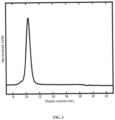
- the secreted protein was purified from tissue culture supernatants by immobilized metal affinity chromatography and size exclusion chromatography. Size exclusion chromatograms ( Fig. 3 ) indicated that the purified protein formed a single, monodisperse species.
- 153-50B.4PT1 a pentameric protein comprising a second assembly domain that interacts with the trimeric assembly domain in 153-50A or DS-Cav1-foldon-153-50A to drive assembly of icosahedral 153-50-based nanostructures, was expressed and purified as described in Bale et al. and patent publication US20160122392 A1 .
- 153-50 is a 120-subunit two-component nanostructure with icosahedral symmetry comprising 20 trimeric (153-50A) and 12 pentameric (I53-50B) building blocks, as recently described by Bale et al.
- the N terminus of 153-50A is exposed on the exterior of the 153-50 nanostructure, which enables the display of antigens on the nanostructure exterior through genetic fusion to the 153-530A N terminus.
- Purified DS-Cav1-foldon-153-50A and I53-50B.4PT1 were assembled in vitro to form 120-subunit icosahedral nanostructures displaying various amounts of DS-Cav1 on the nanostructure exteriors by mixing the two purified proteins in various molar ratios.
- nanostructures displaying DS-Cav1 at valencies of 100% (20 trimers), 66% ( ⁇ 14 trimers), and 33% ( ⁇ 7 trimers) were prepared as described above.
- the species present in the in vitro assembly reactions after overnight incubation were assessed by several techniques, including size exclusion chromatography-multi-angle light scattering (SEC-MALS), dynamic light scattering, and UV/vis spectroscopy.
- SEC-MALS size exclusion chromatography-multi-angle light scattering
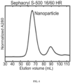
- 120-subunit nanostructures were purified from the in vitro assembly reactions using size exclusion chromatography (an example chromatogram obtained using the 100% valency nanostructures is presented in Fig. 4 ).
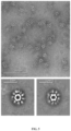
- the purified nanostructures were characterized by negative stain electron microscopy, which revealed fields of monodisperse particles in which DS-Cav1 was clearly visible as spikes projecting outward from the core icosahedral 153-50 assembly (an example micrograph obtained using the 100% valency particles is presented in Fig. 5 ).
- ELISA assays using monoclonal antibodies specific to the prefusion conformation confirmed that the DS-Cav1 thus displayed on the nanostructure exteriors was well-folded and antigenically intact ( Fig. 6 ).
- mice The DS-Cav1-foldon-I53-50 nanostructures displaying DS-Cav1 at 33%, 66%, and 100% valency were injected into mice using a prime-boost strategy as described above. Additional groups of mice were injected with trimeric DS-Cav1-foldon as a benchmark for the humoral immune response induced against DS-Cav1 by the nanostructures or 153-50 nanostructures lacking displayed DS-Cav1 as negative controls for a DS-Cav1 specific response. ELISA assays of serum extracted from the mice at defined time points after the injections were used to measure DS-Cav1 specific antibody titers present in the sera of the injected animals ( Fig. 7 ).
- DS-Cav1-specific titers were roughly 2.5-fold higher on average in mice injected with 100% valency DS-Cav1-foldon-I53-50 nanostructures compared to DS-Cav1.
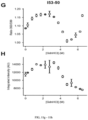
- the sera from the mice injected with the series of immunogens described above was also evaluated for the presence of neutralizing antibody titers using the standard neutralization assay in HEp-2 cells ( Fig. 8 ).
- the trend in serum neutralizing antibody titers correlated highly with the trend observed in DS-Cav1-specific binding antibody titers.
- Sera from animals injected with the 153-50 nanostructures lacking displayed DS-Cav1 did not neutralize virus, consistent with the lack of DS-Cav1-specific antibodies in these sera.
- the DS-Cav1-foldon-I53-50 nanostructures were also injected into Rhesus macaques to evaluate their immunogenicity in a primate immune system.
- the animals were injected intramuscularly at weeks 0 and 4 with either free DS-Cav1 trimer or DS-Cav1-foldon-I53-50 nanostructures displaying DS-Cav1 at 100% valency.
- the dose of DS-Cav1 antigen was 50 ⁇ g, and the immunogens were formulated with the MF59-like, squalene-based oil-in-water emulsion adjuvant SWE.
- Sera obtained from the animals at weeks 6 and 16 were evaluated for anti-DS-Cav1 antibody titers and RSV-neutralizing antibody titers ( Fig. 9 ). The results mirrored those obtained in mice.
- the mean anti-DS-Cav1 antibody titer was 4-fold higher in animals injected with the DS-Cav1-foldon-I53-50 nanostructure compared to animals injected with trimeric DS-Cav1.
- the mean RSV-neutralizing antibody titer at week 16 was 16-fold higher in animals injected with the DS-Cav1-foldon-I53-50 nanostructure compared to animals injected with trimeric DS-Cav1.
- prefusion F Given the key antigenic properties of prefusion F, we used two orthogonal approaches to measure the physical stability of DS-Cav1 when fused to 153-50A and/or when further assembled into the icosahedral nanostructure.
- the first assay measured the retention of binding by a prefusion-specific mAb (D25) after thermal stress, an approach that has been used previously to characterize prefusion F stability (McLellan et al. 2013; Joyce et al. 2016; Krarup et al. 2015).
- Plasmids capable of expressing the relevant constructs were transformed into NEB 5a E . coli cells and selected on LB + carbenicillin agar plates. 1 mL cultures were prepared by inoculating TB media with a bacterial colony and again selecting with 50 ug/mL carbenicillin. A Qiagen Mini Prep kit was used to purify plasmid from the E. coli cultures in accordance with their protocol.
- Expi293F TM Cells were cultured in Expi293 TM Expression Medium (ThermoFisher) supplemented with penicillin (100 u/mL) and streptomycin (100 ⁇ g/mL) at 8% CO 2 , 37° C, and 125 rpm shaking.
- the 1 mL cultures were harvested 5 days post-transfection, and the cells were pelleted from the supernatant by centrifugation at 1,500xg for 5 minutes at 4° C. Supernatants were filtered through a 0.45 ⁇ M filter with a PVDF membrane.
- Filtered supernatants containing DS-Cav1-153-50A constructs were denatured and boiled for 10 minutes at 95° C for 10 minutes in 2x Laemmli buffer with 2-mercaptoethanol. SDS-PAGE separated the sample fractions, which were then transferred to a nitrocellulose membrane and probed with palivizumab, followed with a secondary antibody, anti-human conjugated to HRP. Blot was imaged using Clarity Western ECL Blotting Substrate (Bio-Rad).
- Filtered supernatants containing DS-Cav1-153-50A constructs were bound to Nunc MaxiSorp TM 96-well plates in a two-fold dilution series.
- the pre-fusion conformation-specific antibody D25 was used to detect DS-Cav1-153-50A, followed by a secondary anti-human antibody conjugated to HRP. Protein yield was determined colorimetrically via the substrate TMB and absorbances were collected at 450 nm.
Landscapes
- Health & Medical Sciences (AREA)
- Chemical & Material Sciences (AREA)
- Life Sciences & Earth Sciences (AREA)
- Organic Chemistry (AREA)
- General Health & Medical Sciences (AREA)
- Medicinal Chemistry (AREA)
- Virology (AREA)
- Engineering & Computer Science (AREA)
- Genetics & Genomics (AREA)
- Molecular Biology (AREA)
- Biochemistry (AREA)
- Bioinformatics & Cheminformatics (AREA)
- Biophysics (AREA)
- Pharmacology & Pharmacy (AREA)
- Proteomics, Peptides & Aminoacids (AREA)
- Gastroenterology & Hepatology (AREA)
- Public Health (AREA)
- Veterinary Medicine (AREA)
- Animal Behavior & Ethology (AREA)
- Immunology (AREA)
- Wood Science & Technology (AREA)
- Zoology (AREA)
- Biotechnology (AREA)
- Microbiology (AREA)
- General Engineering & Computer Science (AREA)
- Nanotechnology (AREA)
- Communicable Diseases (AREA)
- Oncology (AREA)
- Chemical Kinetics & Catalysis (AREA)
- General Chemical & Material Sciences (AREA)
- Nuclear Medicine, Radiotherapy & Molecular Imaging (AREA)
- Epidemiology (AREA)
- Biomedical Technology (AREA)
- Medical Informatics (AREA)
- Crystallography & Structural Chemistry (AREA)
- Mycology (AREA)
- Peptides Or Proteins (AREA)
- Micro-Organisms Or Cultivation Processes Thereof (AREA)
- Medicinal Preparation (AREA)
- Medicines Containing Antibodies Or Antigens For Use As Internal Diagnostic Agents (AREA)
Priority Applications (5)
| Application Number | Priority Date | Filing Date | Title |
|---|---|---|---|
| EP25152898.0A EP4559869A3 (en) | 2017-04-04 | 2018-04-03 | Self-assembling protein nanostructures displaying paramyxovirus and/or pneumovirus f proteins and their use |
| SI201831259T SI3606942T1 (sl) | 2017-04-04 | 2018-04-03 | Samo-sestavljajoče se proteinske nanostrukture, ki prikazujejo paramiksovirusne in/ali pnevmovirusne f proteine, in njihova uporaba |
| SM20250317T SMT202500317T1 (it) | 2017-04-04 | 2018-04-03 | Nanostrutture proteiche auto-assemblanti che presentano proteine f di paramyxovirus e/o pneumovirus e loro uso |
| HRP20251060TT HRP20251060T1 (hr) | 2017-04-04 | 2018-04-03 | Samosklapajuće proteinske nanostrukture koje prikazuju f proteine paramiksovirusa i/ili pneumovirusa i njihova uporaba |
| RS20250807A RS67190B1 (sr) | 2017-04-04 | 2018-04-03 | Samosklapajuće proteinske nanostrukture koje prikazuju f proteine paramiksovirusa i/ili pneumovirusa i njihova upotreba |
Applications Claiming Priority (2)
| Application Number | Priority Date | Filing Date | Title |
|---|---|---|---|
| US201762481331P | 2017-04-04 | 2017-04-04 | |
| PCT/US2018/025880 WO2018187325A1 (en) | 2017-04-04 | 2018-04-03 | Self-assembling protein nanostructures displaying paramyxovirus and/or pneumovirus f proteins and their use |
Related Child Applications (2)
| Application Number | Title | Priority Date | Filing Date |
|---|---|---|---|
| EP25152898.0A Division-Into EP4559869A3 (en) | 2017-04-04 | 2018-04-03 | Self-assembling protein nanostructures displaying paramyxovirus and/or pneumovirus f proteins and their use |
| EP25152898.0A Division EP4559869A3 (en) | 2017-04-04 | 2018-04-03 | Self-assembling protein nanostructures displaying paramyxovirus and/or pneumovirus f proteins and their use |
Publications (2)
| Publication Number | Publication Date |
|---|---|
| EP3606942A1 EP3606942A1 (en) | 2020-02-12 |
| EP3606942B1 true EP3606942B1 (en) | 2025-06-11 |
Family
ID=62116943
Family Applications (2)
| Application Number | Title | Priority Date | Filing Date |
|---|---|---|---|
| EP18723094.1A Active EP3606942B1 (en) | 2017-04-04 | 2018-04-03 | Self-assembling protein nanostructures displaying paramyxovirus and/or pneumovirus f proteins and their use |
| EP25152898.0A Pending EP4559869A3 (en) | 2017-04-04 | 2018-04-03 | Self-assembling protein nanostructures displaying paramyxovirus and/or pneumovirus f proteins and their use |
Family Applications After (1)
| Application Number | Title | Priority Date | Filing Date |
|---|---|---|---|
| EP25152898.0A Pending EP4559869A3 (en) | 2017-04-04 | 2018-04-03 | Self-assembling protein nanostructures displaying paramyxovirus and/or pneumovirus f proteins and their use |
Country Status (23)
| Country | Link |
|---|---|
| US (4) | US11192926B2 (enExample) |
| EP (2) | EP3606942B1 (enExample) |
| JP (3) | JP7168938B2 (enExample) |
| KR (2) | KR20240161683A (enExample) |
| CN (3) | CN110603262A (enExample) |
| AU (3) | AU2018249533C1 (enExample) |
| BR (1) | BR112019020661A2 (enExample) |
| CA (1) | CA3058794A1 (enExample) |
| DK (1) | DK3606942T3 (enExample) |
| ES (1) | ES3040563T3 (enExample) |
| FI (1) | FI3606942T3 (enExample) |
| HR (1) | HRP20251060T1 (enExample) |
| IL (2) | IL322023A (enExample) |
| LT (1) | LT3606942T (enExample) |
| MX (2) | MX2019011869A (enExample) |
| PH (1) | PH12019502276A1 (enExample) |
| PL (1) | PL3606942T3 (enExample) |
| PT (1) | PT3606942T (enExample) |
| RS (1) | RS67190B1 (enExample) |
| SG (1) | SG11201908999QA (enExample) |
| SI (1) | SI3606942T1 (enExample) |
| SM (1) | SMT202500317T1 (enExample) |
| WO (1) | WO2018187325A1 (enExample) |
Families Citing this family (24)
| Publication number | Priority date | Publication date | Assignee | Title |
|---|---|---|---|---|
| US9630994B2 (en) * | 2014-11-03 | 2017-04-25 | University Of Washington | Polypeptides for use in self-assembling protein nanostructures |
| JP7168938B2 (ja) * | 2017-04-04 | 2022-11-10 | ユニヴァーシティ オブ ワシントン | パラミクソウイルスおよび/またはニューモウイルスのfタンパク質を表示する自己アセンブリするタンパク質ナノ構造およびその使用 |
| WO2019148101A1 (en) | 2018-01-29 | 2019-08-01 | Modernatx, Inc. | Rsv rna vaccines |
| CA3095216A1 (en) | 2018-02-28 | 2019-09-06 | University Of Washington | Self-asssembling nanostructure vaccines |
| EP3921332A2 (en) | 2019-02-08 | 2021-12-15 | The USA, as represented by The Secretary, Department of Health and Human Services | Nanoparticle-based influenza virus vaccines and uses thereof |
| US20220306697A1 (en) * | 2019-09-04 | 2022-09-29 | University Of Washington | Self-Assembling Protein Nanostructures Displaying Paramyxovirus and/or Pneumovirus F Proteins and Their Use |
| CN110878128A (zh) * | 2019-11-04 | 2020-03-13 | 中国疾病预防控制中心病毒病预防控制所 | 一种人呼吸道合胞病毒粘膜疫苗及其制备方法和应用 |
| KR20220133911A (ko) * | 2020-01-30 | 2022-10-05 | 모더나티엑스, 인크. | 호흡기 바이러스 면역화 조성물 |
| WO2021163481A1 (en) * | 2020-02-14 | 2021-08-19 | University Of Washington | Polypeptides and their use |
| AU2021221139A1 (en) * | 2020-02-14 | 2022-09-01 | University Of Washington | Polypeptides, compositions, and their use to treat or limit development of an infection |
| EP4161943A4 (en) * | 2020-06-09 | 2025-01-01 | Icosavax, Inc. | Method of making virus-like particle |
| MX2022015593A (es) * | 2020-06-09 | 2023-04-03 | Icosavax Inc | Metodo de ensamblaje de particulas similares a virus de dos componentes. |
| CN116615235A (zh) | 2020-10-09 | 2023-08-18 | 得克萨斯州大学系统董事会 | 融合前稳定的hmpv f蛋白 |
| CN111991556B (zh) * | 2020-10-29 | 2021-03-02 | 中山大学 | SARS-CoV-2 RBD共轭纳米颗粒疫苗 |
| AU2021380755A1 (en) | 2020-11-13 | 2023-06-22 | Icosavax, Inc. | Protein-based nanoparticle vaccine for metapneumovirus |
| CN112778404B (zh) * | 2021-01-28 | 2022-11-29 | 中山大学 | 一种含EB病毒gHgL蛋白的自组装纳米颗粒及其制备方法与应用 |
| JP2024531150A (ja) * | 2021-08-10 | 2024-08-29 | アイコサバックス, インコーポレイテッド | 呼吸器多核体ウイルスに対するウイルス様粒子ワクチン |
| CN119816322A (zh) | 2022-05-17 | 2025-04-11 | Icosavax股份有限公司 | 针对副粘病毒的多价疫苗及其用途 |
| EP4626470A2 (en) * | 2022-11-29 | 2025-10-08 | The Scripps Research Institute | Vaccines containing novel nanoparticle scaffolds |
| WO2024131862A1 (zh) * | 2022-12-22 | 2024-06-27 | 北京吉诺卫生物科技有限公司 | 一种rsv疫苗及其制备方法与应用 |
| CN118108812B (zh) * | 2023-05-04 | 2025-04-15 | 国药中生生物技术研究院有限公司 | Rsv f蛋白的突变体 |
| US20250041400A1 (en) * | 2023-07-18 | 2025-02-06 | Astrazeneca Ab | Rsv vaccine |
| CN119930762A (zh) * | 2023-11-03 | 2025-05-06 | 广东蓝玉生物科技有限公司 | 非洲猪瘟病毒ep153r蛋白的免疫原性组合物及其应用 |
| WO2025093041A1 (zh) * | 2023-11-03 | 2025-05-08 | 广东蓝玉生物科技有限公司 | 非洲猪瘟病毒CD2v蛋白的免疫原性组合物及其应用 |
Citations (1)
| Publication number | Priority date | Publication date | Assignee | Title |
|---|---|---|---|---|
| WO2019169120A1 (en) * | 2018-02-28 | 2019-09-06 | University Of Washington | Self-asssembling nanostructure vaccines |
Family Cites Families (62)
| Publication number | Priority date | Publication date | Assignee | Title |
|---|---|---|---|---|
| DE4238904A1 (de) | 1992-11-19 | 1994-05-26 | Basf Ag | Riboflavin-Synthese in Hefen |
| JP2002505353A (ja) | 1998-03-04 | 2002-02-19 | エクソンモービル・ケミカル・パテンツ・インク | 高温オレフィン重合法 |
| US6303767B1 (en) | 1998-11-05 | 2001-10-16 | Kosan Biosciences, Inc. | Nucleic acids encoding narbonolide polyketide synthase enzymes from streptomyces narbonensis |
| JP2000152791A (ja) | 1998-11-11 | 2000-06-06 | E I Du Pont De Nemours & Co | ルマジンシンターゼおよびリボフラビンシンターゼ |
| US20040005672A1 (en) | 2002-02-22 | 2004-01-08 | Santi Daniel V. | Heterologous production of polyketides |
| WO2004024744A2 (en) | 2002-07-31 | 2004-03-25 | The Board Of Trustees Of The Leland Stanford Junior University | Production of glycosylated macrolides in e. coli |
| EP1773303A2 (en) | 2004-05-25 | 2007-04-18 | Chimeracore, Inc. | Self-assembling nanoparticle drug delivery system |
| EP3073267A1 (en) | 2004-09-21 | 2016-09-28 | Medimmune, Inc. | Antibodies against and methods for producing vaccines for respiratory syncytial virus |
| SI2222710T1 (sl) | 2007-12-24 | 2016-11-30 | Id Biomedical Corporation Of Quebec | Rekombinatni RSV antigeni |
| WO2010019725A2 (en) | 2008-08-12 | 2010-02-18 | Salemme Francis R | Node polypeptides for nanostructure assembly |
| US9102526B2 (en) | 2008-08-12 | 2015-08-11 | Imiplex Llc | Node polypeptides for nanostructure assembly |
| GB0817507D0 (en) | 2008-09-24 | 2008-10-29 | Ucl Business Plc | Protein cages |
| DK2370099T3 (en) | 2008-12-09 | 2016-08-01 | Novavax Inc | Rsv f modified proteins and methods of their use |
| US20110200560A1 (en) | 2009-05-26 | 2011-08-18 | Massachusetts Institute Of Technology | Non-ionic self-assembling peptides and uses thereof |
| WO2011017835A1 (en) | 2009-08-11 | 2011-02-17 | Nanjing University | Preparation method of protein or peptide nanoparticles for in vivo drug delivery by unfolding and refolding |
| WO2011056899A2 (en) | 2009-11-03 | 2011-05-12 | Ligocyte Pharmaceuticals, Inc. | Chimeric rsv-f polypeptide and lentivirus or alpha-retrovirus gag-based vlps |
| AU2011276251B2 (en) | 2010-07-07 | 2015-02-05 | Artifical Cell Technologies, Inc. | Respiratory syncytial virus antigenic compositions and methods |
| BR112013029169B1 (pt) | 2011-05-13 | 2021-09-28 | Novartis Ag | Polipeptídeo f pré-fusão do vírus sincicial respiratório (rsv), composição imunogênica e uso da mesma |
| CA2849822C (en) | 2011-09-23 | 2021-05-11 | Gary J. Nabel, M.D. | Novel influenza hemagglutinin protein-based vaccines |
| AU2013201495B2 (en) | 2011-09-30 | 2015-12-03 | Novavax, Inc. | Recombinant nanoparticle RSV F vaccine for respiratory syncytial virus |
| EP2766030A4 (en) | 2011-10-12 | 2015-09-16 | Univ Washington | MANIPULATED OUTER DOMAIN OF HV-GP120 AND MUTANTS THEREOF |
| EP2836505B1 (en) | 2012-04-10 | 2019-01-23 | The Trustees Of The University Of Pennsylvania | Human respiratory syncytial virus concensus antigens, nucleic acid constructs and vaccines made thereform, and methods of using same |
| US8969521B2 (en) | 2012-04-11 | 2015-03-03 | University Of Washington Through Its Center For Commercialization | General method for designing self-assembling protein nanomaterials |
| WO2014005643A1 (en) | 2012-07-05 | 2014-01-09 | Okairos Ag | Novel prime-boosting regimens involving immunogenic polypeptides encoded by polynucleotides |
| CN104853771A (zh) | 2012-07-06 | 2015-08-19 | 诺华股份有限公司 | 巨细胞病毒蛋白的复合物 |
| IN2015DN01448A (enExample) | 2012-08-01 | 2015-07-03 | Bavarian Nordic As | |
| US20140141037A1 (en) | 2012-11-20 | 2014-05-22 | Novartis Ag | Rsv f prefusion trimers |
| EP2922570A1 (en) | 2012-11-20 | 2015-09-30 | GlaxoSmithKline Biologicals SA | Rsv f prefusion trimers |
| WO2014124301A1 (en) * | 2013-02-07 | 2014-08-14 | University Of Washington Through Its Center For Commercialization | Self-assembling protein nanostructures |
| US9738689B2 (en) | 2013-03-13 | 2017-08-22 | The United States Of America, As Represented By The Secretary, Department Of Health And Human Services | Prefusion RSV F proteins and their use |
| AU2014243756B2 (en) | 2013-03-13 | 2018-09-27 | The United States Of America, As Represented By The Secretary, Department Of Health And Human Services | Prefusion RSV F proteins and their use |
| US9060975B2 (en) | 2013-03-14 | 2015-06-23 | Mucosis Bv | Heat-stable respiratory syncytial virus F protein oligomers and their use in immunological compositions |
| AU2014231357B2 (en) | 2013-03-15 | 2018-06-14 | The United States Of America, As Represented By The Secretary, Department Of Health And Human Services | Epitope of RSV fusion protein and antibody identifying same |
| US10899800B2 (en) | 2013-04-25 | 2021-01-26 | Janssen Vaccines & Prevention B.V. | Stabilized soluble pre-fusion RSV F polypeptides |
| EP3010931B1 (en) | 2013-06-17 | 2018-06-13 | Janssen Vaccines & Prevention B.V. | Stabilized soluble pre-fusion rsv f polypeptides |
| CN104436202B (zh) | 2013-09-12 | 2017-07-28 | 中国科学院深圳先进技术研究院 | 聚合物纳米颗粒、其制备方法及疫苗组合物、疫苗制剂及其制备方法 |
| EP3049107A1 (en) | 2013-09-24 | 2016-08-03 | Massachusetts Institute of Technology | Self-assembled nanoparticle vaccines |
| US20150166610A1 (en) | 2013-10-14 | 2015-06-18 | Glaxosmithkline Biologicals, S.A. | Recombinant rsv antigens |
| JP6652500B2 (ja) | 2014-01-09 | 2020-02-26 | アルファ−オ ペプティデス アクツィエンゲゼルシャフト | ワクチンプラットフォームとしてのフラジェリン含有タンパク質ナノ粒子 |
| EP2974739A1 (en) | 2014-07-15 | 2016-01-20 | Novartis AG | RSVF trimerization domains |
| JP7094103B2 (ja) | 2014-07-10 | 2022-07-01 | ヤンセン ファッシンズ アンド プリベンション ベーフェー | インフルエンザウイルスワクチンおよびその使用 |
| US9630994B2 (en) | 2014-11-03 | 2017-04-25 | University Of Washington | Polypeptides for use in self-assembling protein nanostructures |
| KR102713707B1 (ko) | 2014-12-31 | 2024-10-04 | 더 유나이티드 스테이츠 오브 어메리카, 애즈 리프리젠티드 바이 더 세크러테리, 디파트먼트 오브 헬쓰 앤드 휴먼 서비씨즈 | 신규 다가 나노입자 기반 백신 |
| WO2016138525A1 (en) | 2015-02-27 | 2016-09-01 | University Of Washington | Polypeptide assemblies and methods for the production thereof |
| US10744193B2 (en) | 2015-03-30 | 2020-08-18 | The United States Of America, As Represented By The Secretary, Department Of Health And Human Services | Immunogenic RSV polypeptides |
| KR102638978B1 (ko) | 2015-07-07 | 2024-02-22 | 얀센 백신스 앤드 프리벤션 비.브이. | Rsv에 대한 백신 |
| KR20180026734A (ko) | 2015-07-07 | 2018-03-13 | 얀센 백신스 앤드 프리벤션 비.브이. | 안정화된 가용성 융합-전 rsv f 폴리펩티드 |
| US10024198B2 (en) | 2015-08-13 | 2018-07-17 | Echogen Power Systems, Llc | Heat engine system including an integrated cooling circuit |
| CA2996762A1 (en) * | 2015-08-31 | 2017-03-09 | Technovax, Inc. | Human respiratory syncytial virus (hrsv) virus-like particles (vlps) based vaccine |
| CN114617959A (zh) | 2015-09-03 | 2022-06-14 | 诺瓦瓦克斯股份有限公司 | 具有改进的稳定性和免疫原性的疫苗组合物 |
| WO2017066484A2 (en) | 2015-10-13 | 2017-04-20 | Carter Daniel C | Nsp10 self-assembling fusion proteins for vaccines, therapeutics, diagnostics and other nanomaterial applications |
| EP3718565B1 (en) | 2015-10-22 | 2022-04-27 | ModernaTX, Inc. | Respiratory virus vaccines |
| EP3368547A4 (en) | 2015-10-29 | 2019-06-05 | Emory University | CHIMERIC RSV, IMMUNOGENIC COMPOSITIONS AND METHOD FOR USE THEREOF |
| PE20181354A1 (es) | 2015-12-23 | 2018-08-22 | Pfizer | Mutantes de proteina f de rsv |
| CN109069611B (zh) | 2016-03-29 | 2022-11-04 | 美国政府(由卫生和人类服务部的部长所代表) | 取代修饰的融合前rsv f蛋白及其用途 |
| KR20220041966A (ko) | 2016-04-05 | 2022-04-01 | 얀센 백신스 앤드 프리벤션 비.브이. | 안정화된 용해성 융합-전 rsv f 단백질 |
| WO2017207477A1 (en) | 2016-05-30 | 2017-12-07 | Janssen Vaccines & Prevention B.V. | Stabilized pre-fusion rsv f proteins |
| WO2017207480A1 (en) | 2016-05-30 | 2017-12-07 | Janssen Vaccines & Prevention B.V. | Stabilized pre-fusion rsv f proteins |
| US10961283B2 (en) | 2016-06-27 | 2021-03-30 | The United States Of America, As Represented By The Secretary, Department Of Health And Human Services | Self-assembling insect ferritin nanoparticles for display of co-assembled trimeric antigens |
| WO2018109220A2 (en) | 2016-12-16 | 2018-06-21 | Institute For Research In Biomedicine | Novel recombinant prefusion rsv f proteins and uses thereof |
| JP7168938B2 (ja) | 2017-04-04 | 2022-11-10 | ユニヴァーシティ オブ ワシントン | パラミクソウイルスおよび/またはニューモウイルスのfタンパク質を表示する自己アセンブリするタンパク質ナノ構造およびその使用 |
| CN107157933B (zh) | 2017-05-04 | 2021-05-11 | 同济大学 | 一种蛋白自组装新型纳米疫苗及其制备方法 |
-
2018
- 2018-04-03 JP JP2019554692A patent/JP7168938B2/ja active Active
- 2018-04-03 DK DK18723094.1T patent/DK3606942T3/da active
- 2018-04-03 IL IL322023A patent/IL322023A/en unknown
- 2018-04-03 CN CN201880023734.8A patent/CN110603262A/zh active Pending
- 2018-04-03 FI FIEP18723094.1T patent/FI3606942T3/fi active
- 2018-04-03 AU AU2018249533A patent/AU2018249533C1/en active Active
- 2018-04-03 ES ES18723094T patent/ES3040563T3/es active Active
- 2018-04-03 LT LTEPPCT/US2018/025880T patent/LT3606942T/lt unknown
- 2018-04-03 BR BR112019020661-2A patent/BR112019020661A2/pt unknown
- 2018-04-03 MX MX2019011869A patent/MX2019011869A/es unknown
- 2018-04-03 SI SI201831259T patent/SI3606942T1/sl unknown
- 2018-04-03 EP EP18723094.1A patent/EP3606942B1/en active Active
- 2018-04-03 EP EP25152898.0A patent/EP4559869A3/en active Pending
- 2018-04-03 US US16/500,331 patent/US11192926B2/en active Active
- 2018-04-03 KR KR1020247035024A patent/KR20240161683A/ko active Pending
- 2018-04-03 PT PT187230941T patent/PT3606942T/pt unknown
- 2018-04-03 SM SM20250317T patent/SMT202500317T1/it unknown
- 2018-04-03 KR KR1020197029815A patent/KR102774312B1/ko active Active
- 2018-04-03 CA CA3058794A patent/CA3058794A1/en active Pending
- 2018-04-03 CN CN202310044111.1A patent/CN115779077A/zh active Pending
- 2018-04-03 CN CN202310044118.3A patent/CN115947873A/zh active Pending
- 2018-04-03 HR HRP20251060TT patent/HRP20251060T1/hr unknown
- 2018-04-03 SG SG11201908999Q patent/SG11201908999QA/en unknown
- 2018-04-03 RS RS20250807A patent/RS67190B1/sr unknown
- 2018-04-03 WO PCT/US2018/025880 patent/WO2018187325A1/en not_active Ceased
- 2018-04-03 PL PL18723094.1T patent/PL3606942T3/pl unknown
-
2019
- 2019-09-25 IL IL26964319A patent/IL269643A/en unknown
- 2019-10-02 PH PH12019502276A patent/PH12019502276A1/en unknown
- 2019-10-02 MX MX2023004704A patent/MX2023004704A/es unknown
-
2021
- 2021-11-10 US US17/523,174 patent/US11732011B2/en active Active
-
2022
- 2022-04-27 AU AU2022202787A patent/AU2022202787C1/en active Active
- 2022-10-19 JP JP2022167524A patent/JP7541701B2/ja active Active
-
2023
- 2023-06-20 US US18/337,881 patent/US12275757B2/en active Active
-
2024
- 2024-08-08 JP JP2024132065A patent/JP2024160321A/ja active Pending
- 2024-09-26 AU AU2024220089A patent/AU2024220089A1/en active Pending
-
2025
- 2025-03-12 US US19/077,674 patent/US20250263445A1/en active Pending
Patent Citations (1)
| Publication number | Priority date | Publication date | Assignee | Title |
|---|---|---|---|---|
| WO2019169120A1 (en) * | 2018-02-28 | 2019-09-06 | University Of Washington | Self-asssembling nanostructure vaccines |
Also Published As
Similar Documents
| Publication | Publication Date | Title |
|---|---|---|
| US12275757B2 (en) | Self-assembling protein nanostructures displaying paramyxovirus and/or pneumovirus F proteins and their use | |
| US20230265127A1 (en) | Stabilized soluble pre-fusion rsv f polypeptides | |
| US11759514B2 (en) | Stabilized pre-fusion RSV F proteins | |
| JP2025172875A (ja) | パラミクソウイルスおよび/またはニューモウイルスのfタンパク質を提示する自己組織化タンパク質ナノ構造体ならびにその使用 | |
| RU2844177C2 (ru) | Самосборные белковые наноструктуры, экспонирующие f-белки парамиксовируса и/или пневмовируса, и их применение | |
| HK40014567B (en) | Self-assembling protein nanostructures displaying paramyxovirus and/or pneumovirus f proteins and their use | |
| HK40014567A (en) | Self-assembling protein nanostructures displaying paramyxovirus and/or pneumovirus f proteins and their use |
Legal Events
| Date | Code | Title | Description |
|---|---|---|---|
| REG | Reference to a national code |
Ref country code: HR Ref legal event code: TUEP Ref document number: P20251060T Country of ref document: HR |
|
| STAA | Information on the status of an ep patent application or granted ep patent |
Free format text: STATUS: UNKNOWN |
|
| STAA | Information on the status of an ep patent application or granted ep patent |
Free format text: STATUS: THE INTERNATIONAL PUBLICATION HAS BEEN MADE |
|
| PUAI | Public reference made under article 153(3) epc to a published international application that has entered the european phase |
Free format text: ORIGINAL CODE: 0009012 |
|
| STAA | Information on the status of an ep patent application or granted ep patent |
Free format text: STATUS: REQUEST FOR EXAMINATION WAS MADE |
|
| 17P | Request for examination filed |
Effective date: 20191030 |
|
| AK | Designated contracting states |
Kind code of ref document: A1 Designated state(s): AL AT BE BG CH CY CZ DE DK EE ES FI FR GB GR HR HU IE IS IT LI LT LU LV MC MK MT NL NO PL PT RO RS SE SI SK SM TR |
|
| AX | Request for extension of the european patent |
Extension state: BA ME |
|
| DAV | Request for validation of the european patent (deleted) | ||
| DAX | Request for extension of the european patent (deleted) | ||
| REG | Reference to a national code |
Ref country code: HK Ref legal event code: DE Ref document number: 40014567 Country of ref document: HK |
|
| STAA | Information on the status of an ep patent application or granted ep patent |
Free format text: STATUS: EXAMINATION IS IN PROGRESS |
|
| 17Q | First examination report despatched |
Effective date: 20210903 |
|
| P01 | Opt-out of the competence of the unified patent court (upc) registered |
Effective date: 20230516 |
|
| GRAP | Despatch of communication of intention to grant a patent |
Free format text: ORIGINAL CODE: EPIDOSNIGR1 |
|
| STAA | Information on the status of an ep patent application or granted ep patent |
Free format text: STATUS: GRANT OF PATENT IS INTENDED |
|
| INTG | Intention to grant announced |
Effective date: 20231103 |
|
| GRAJ | Information related to disapproval of communication of intention to grant by the applicant or resumption of examination proceedings by the epo deleted |
Free format text: ORIGINAL CODE: EPIDOSDIGR1 |
|
| STAA | Information on the status of an ep patent application or granted ep patent |
Free format text: STATUS: EXAMINATION IS IN PROGRESS |
|
| GRAP | Despatch of communication of intention to grant a patent |
Free format text: ORIGINAL CODE: EPIDOSNIGR1 |
|
| STAA | Information on the status of an ep patent application or granted ep patent |
Free format text: STATUS: GRANT OF PATENT IS INTENDED |
|
| INTG | Intention to grant announced |
Effective date: 20240318 |
|
| GRAJ | Information related to disapproval of communication of intention to grant by the applicant or resumption of examination proceedings by the epo deleted |
Free format text: ORIGINAL CODE: EPIDOSDIGR1 |
|
| STAA | Information on the status of an ep patent application or granted ep patent |
Free format text: STATUS: EXAMINATION IS IN PROGRESS |
|
| GRAP | Despatch of communication of intention to grant a patent |
Free format text: ORIGINAL CODE: EPIDOSNIGR1 |
|
| STAA | Information on the status of an ep patent application or granted ep patent |
Free format text: STATUS: GRANT OF PATENT IS INTENDED |
|
| GRAJ | Information related to disapproval of communication of intention to grant by the applicant or resumption of examination proceedings by the epo deleted |
Free format text: ORIGINAL CODE: EPIDOSDIGR1 |
|
| STAA | Information on the status of an ep patent application or granted ep patent |
Free format text: STATUS: EXAMINATION IS IN PROGRESS |
|
| INTC | Intention to grant announced (deleted) | ||
| RIN1 | Information on inventor provided before grant (corrected) |
Inventor name: HSIA, YANG Inventor name: FALLAS, JORGE Inventor name: MARCANDALLI, JESSICA Inventor name: LANZAVECCHIA, ANTONIO Inventor name: PEREZ, LAURENT Inventor name: STEWART, LANCE, JOSEPH Inventor name: NICKERSON, BROOKE Inventor name: BAKER, DAVID Inventor name: KING, NEIL, P. |
|
| GRAJ | Information related to disapproval of communication of intention to grant by the applicant or resumption of examination proceedings by the epo deleted |
Free format text: ORIGINAL CODE: EPIDOSDIGR1 |
|
| GRAP | Despatch of communication of intention to grant a patent |
Free format text: ORIGINAL CODE: EPIDOSNIGR1 |
|
| GRAP | Despatch of communication of intention to grant a patent |
Free format text: ORIGINAL CODE: EPIDOSNIGR1 |
|
| STAA | Information on the status of an ep patent application or granted ep patent |
Free format text: STATUS: GRANT OF PATENT IS INTENDED |
|
| INTG | Intention to grant announced |
Effective date: 20240816 |
|
| INTC | Intention to grant announced (deleted) | ||
| INTG | Intention to grant announced |
Effective date: 20240916 |
|
| GRAJ | Information related to disapproval of communication of intention to grant by the applicant or resumption of examination proceedings by the epo deleted |
Free format text: ORIGINAL CODE: EPIDOSDIGR1 |
|
| STAA | Information on the status of an ep patent application or granted ep patent |
Free format text: STATUS: EXAMINATION IS IN PROGRESS |
|
| GRAP | Despatch of communication of intention to grant a patent |
Free format text: ORIGINAL CODE: EPIDOSNIGR1 |
|
| STAA | Information on the status of an ep patent application or granted ep patent |
Free format text: STATUS: GRANT OF PATENT IS INTENDED |
|
| INTG | Intention to grant announced |
Effective date: 20250113 |
|
| GRAS | Grant fee paid |
Free format text: ORIGINAL CODE: EPIDOSNIGR3 |
|
| GRAA | (expected) grant |
Free format text: ORIGINAL CODE: 0009210 |
|
| STAA | Information on the status of an ep patent application or granted ep patent |
Free format text: STATUS: THE PATENT HAS BEEN GRANTED |
|
| AK | Designated contracting states |
Kind code of ref document: B1 Designated state(s): AL AT BE BG CH CY CZ DE DK EE ES FI FR GB GR HR HU IE IS IT LI LT LU LV MC MK MT NL NO PL PT RO RS SE SI SK SM TR |
|
| REG | Reference to a national code |
Ref country code: GB Ref legal event code: FG4D |
|
| REG | Reference to a national code |
Ref country code: CH Ref legal event code: EP |
|
| REG | Reference to a national code |
Ref country code: IE Ref legal event code: FG4D |
|
| REG | Reference to a national code |
Ref country code: DE Ref legal event code: R096 Ref document number: 602018082526 Country of ref document: DE |
|
| REG | Reference to a national code |
Ref country code: FI Ref legal event code: FGE |
|
| REG | Reference to a national code |
Ref country code: PT Ref legal event code: SC4A Ref document number: 3606942 Country of ref document: PT Date of ref document: 20250901 Kind code of ref document: T Free format text: AVAILABILITY OF NATIONAL TRANSLATION Effective date: 20250827 |
|
| REG | Reference to a national code |
Ref country code: DK Ref legal event code: T3 Effective date: 20250902 |
|
| REG | Reference to a national code |
Ref country code: NL Ref legal event code: FP |
|
| REG | Reference to a national code |
Ref country code: SE Ref legal event code: TRGR |
|
| REG | Reference to a national code |
Ref country code: EE Ref legal event code: FG4A Ref document number: E025222 Country of ref document: EE Effective date: 20250826 |
|
| REG | Reference to a national code |
Ref country code: SK Ref legal event code: T3 Ref document number: E 47048 Country of ref document: SK |
|
| REG | Reference to a national code |
Ref country code: GR Ref legal event code: EP Ref document number: 20250401784 Country of ref document: GR Effective date: 20251009 |
|
| REG | Reference to a national code |
Ref country code: ES Ref legal event code: FG2A Ref document number: 3040563 Country of ref document: ES Kind code of ref document: T3 Effective date: 20251103 |
|
| REG | Reference to a national code |
Ref country code: HR Ref legal event code: T1PR Ref document number: P20251060 Country of ref document: HR |