EP3578664B1 - Collagen iv neo-epitopfibrose-assay - Google Patents
Collagen iv neo-epitopfibrose-assay Download PDFInfo
- Publication number
- EP3578664B1 EP3578664B1 EP19174151.1A EP19174151A EP3578664B1 EP 3578664 B1 EP3578664 B1 EP 3578664B1 EP 19174151 A EP19174151 A EP 19174151A EP 3578664 B1 EP3578664 B1 EP 3578664B1
- Authority
- EP
- European Patent Office
- Prior art keywords
- seq
- fibrosis
- neo
- collagen
- epitope
- Prior art date
- Legal status (The legal status is an assumption and is not a legal conclusion. Google has not performed a legal analysis and makes no representation as to the accuracy of the status listed.)
- Active
Links
Images







Classifications
-
- G—PHYSICS
- G01—MEASURING; TESTING
- G01N—INVESTIGATING OR ANALYSING MATERIALS BY DETERMINING THEIR CHEMICAL OR PHYSICAL PROPERTIES
- G01N33/00—Investigating or analysing materials by specific methods not covered by groups G01N1/00 - G01N31/00
- G01N33/48—Biological material, e.g. blood, urine; Haemocytometers
- G01N33/50—Chemical analysis of biological material, e.g. blood, urine; Testing involving biospecific ligand binding methods; Immunological testing
- G01N33/68—Chemical analysis of biological material, e.g. blood, urine; Testing involving biospecific ligand binding methods; Immunological testing involving proteins, peptides or amino acids
- G01N33/6893—Chemical analysis of biological material, e.g. blood, urine; Testing involving biospecific ligand binding methods; Immunological testing involving proteins, peptides or amino acids related to diseases not provided for elsewhere
-
- C—CHEMISTRY; METALLURGY
- C12—BIOCHEMISTRY; BEER; SPIRITS; WINE; VINEGAR; MICROBIOLOGY; ENZYMOLOGY; MUTATION OR GENETIC ENGINEERING
- C12Q—MEASURING OR TESTING PROCESSES INVOLVING ENZYMES, NUCLEIC ACIDS OR MICROORGANISMS; COMPOSITIONS OR TEST PAPERS THEREFOR; PROCESSES OF PREPARING SUCH COMPOSITIONS; CONDITION-RESPONSIVE CONTROL IN MICROBIOLOGICAL OR ENZYMOLOGICAL PROCESSES
- C12Q1/00—Measuring or testing processes involving enzymes, nucleic acids or microorganisms; Compositions therefor; Processes of preparing such compositions
- C12Q1/34—Measuring or testing processes involving enzymes, nucleic acids or microorganisms; Compositions therefor; Processes of preparing such compositions involving hydrolase
- C12Q1/37—Measuring or testing processes involving enzymes, nucleic acids or microorganisms; Compositions therefor; Processes of preparing such compositions involving hydrolase involving peptidase or proteinase
-
- G—PHYSICS
- G01—MEASURING; TESTING
- G01N—INVESTIGATING OR ANALYSING MATERIALS BY DETERMINING THEIR CHEMICAL OR PHYSICAL PROPERTIES
- G01N33/00—Investigating or analysing materials by specific methods not covered by groups G01N1/00 - G01N31/00
- G01N33/48—Biological material, e.g. blood, urine; Haemocytometers
- G01N33/50—Chemical analysis of biological material, e.g. blood, urine; Testing involving biospecific ligand binding methods; Immunological testing
- G01N33/53—Immunoassay; Biospecific binding assay; Materials therefor
-
- G—PHYSICS
- G01—MEASURING; TESTING
- G01N—INVESTIGATING OR ANALYSING MATERIALS BY DETERMINING THEIR CHEMICAL OR PHYSICAL PROPERTIES
- G01N2333/00—Assays involving biological materials from specific organisms or of a specific nature
- G01N2333/435—Assays involving biological materials from specific organisms or of a specific nature from animals; from humans
- G01N2333/46—Assays involving biological materials from specific organisms or of a specific nature from animals; from humans from vertebrates
- G01N2333/47—Assays involving proteins of known structure or function as defined in the subgroups
- G01N2333/4701—Details
- G01N2333/4737—C-reactive protein
-
- G—PHYSICS
- G01—MEASURING; TESTING
- G01N—INVESTIGATING OR ANALYSING MATERIALS BY DETERMINING THEIR CHEMICAL OR PHYSICAL PROPERTIES
- G01N2333/00—Assays involving biological materials from specific organisms or of a specific nature
- G01N2333/435—Assays involving biological materials from specific organisms or of a specific nature from animals; from humans
- G01N2333/78—Connective tissue peptides, e.g. collagen, elastin, laminin, fibronectin, vitronectin, cold insoluble globulin [CIG]
-
- G—PHYSICS
- G01—MEASURING; TESTING
- G01N—INVESTIGATING OR ANALYSING MATERIALS BY DETERMINING THEIR CHEMICAL OR PHYSICAL PROPERTIES
- G01N2800/00—Detection or diagnosis of diseases
- G01N2800/08—Hepato-biliairy disorders other than hepatitis
- G01N2800/085—Liver diseases, e.g. portal hypertension, fibrosis, cirrhosis, bilirubin
-
- G—PHYSICS
- G01—MEASURING; TESTING
- G01N—INVESTIGATING OR ANALYSING MATERIALS BY DETERMINING THEIR CHEMICAL OR PHYSICAL PROPERTIES
- G01N2800/00—Detection or diagnosis of diseases
- G01N2800/32—Cardiovascular disorders
-
- G—PHYSICS
- G01—MEASURING; TESTING
- G01N—INVESTIGATING OR ANALYSING MATERIALS BY DETERMINING THEIR CHEMICAL OR PHYSICAL PROPERTIES
- G01N2800/00—Detection or diagnosis of diseases
- G01N2800/50—Determining the risk of developing a disease
Definitions
- the present invention relates to assays for biomarkers useful in the diagnosis of fibrosis disease and prognosis of its development, including biomarkers indicative of the risk of developing fibrosis after a chronic injury.
- biomarkers relating to degradation fragments of Collagen type I, III, IV, V, and VI, elastin, C-reactive protein, and proteoglycans including Biglycan, Decorin, Versican, and Perlecan are found to be useful.
- Fibrotic diseases are a leading cause of morbidity and mortality, e.g. cirrhosis with 800,000 death per year worldwide 1 .
- Table 1 Different fibrotic diseases 2 Tissue Examples of Causes Liver Viral hepatitis Schistosomiasis Steatohepatitis (Alcoholic or non-alcoholic) Lung Idiopathic pulmonary fibrosis (IPF) Systemic sclerosis (Scleroderma) Kidney Nephrogenic systemic fibrosis (NSF) Diabetes Untreated hypertension Heart Heart attack Hypertension Atherosclerosis Restenosis Eye Macular degeneration, retinal and vitreal retinopathy Skin Systemic sclerosis and scleroderma, keloids, hypertrophic scars, burns, genetic factors NFS Pancreas Autoimmune/hereditary causes Intestine Crohn's disease/inflammatory bowl disease Brain Alzheimer's disease, AIDS Bone marrow Cancer, ageing Multi-organ fibrosis Surgical complications, chemo
- a 'fibrotic disease' is any disease giving rise to fibrosis, whether as a main or a secondary symptom.
- Fibrosis is the end result of chronic inflammatory reactions induced by a variety of stimuli including persistent infections, autoimmune reactions, allergic responses, chemical insults, radiation, and tissue injury. Fibrosis is characterized by the accumulation and reorganization of the extracellular matrix (ECM). Despite having obvious etiological and clinical distinctions, most chronic fibrotic disorders have in common a persistent irritant that sustains the production of growth factors, proteolytic enzymes, angiogenic factors, and fibrogenic cytokines, which together stimulate the deposition of connective tissue elements, especially collagens and proteoglycans, which progressively remodel and destroy normal tissue architecture 3, 4 . Despite its enormous impact on human health, there are currently no approved treatments that directly target the mechanisms of fibrosis 5 .
- the key cellular mediator of fibrosis is the myofibroblast, which when activated serves as the primary collagen-producing cell.
- Fibrogenesis is a dynamic process involving complex cellular and molecular mechanisms that usually originates from tissue injury 6 . Fibrogenesis is the result of an imbalance in normal ECM regulation that alters the concentration of macromolecules leading to increased tissue size and density, with progressively impaired function. These macromolecules are mainly fibrous proteins with structural and adhesive functions, such as collagens and proteoglycans.
- Collagens are widely distributed in the human body, i.e. ⁇ 30% of the protein mass in the human body is composed of collagens. Collagens are responsible for the structural integrity of the ECM of most connective tissues. The ECM content results from a fine balance between synthesis and degradation tightly controlled through regulation of gene expression and protein secretion, but also through endogenous protease inhibition and protein degradation by metalloproteinases and cysteine proteases 7-9 .
- Table 2 lists the major collagen types with their major tissue distribution. Table 2. Major collagen types and their tissue distribution. Collagen type Tissue distribution I Most connective tissues II Cartilage, vitreous humor III Extensible connective tissues, e.g.
- Type I collagen is the most abundant collagen and is found in most connective tissues. It is especially important for the structure of bone and skin where the major collagenous components are type I and III collagens 10 .
- Collagen type I and III are the major components of liver and lung in a 1:1 ratio in healthy tissue.
- collagen type IV and VI are found in the basement membranes in most tissues.
- the most common localization of type V collagen is within the characteristic collagen fibrils, in association with the collagen type I and III 10 .
- Some collagens have a restricted tissue distribution: for example, type II, which is found almost exclusively in cartilage 11 .
- MMPs matrix metalloproteinases
- TIMPs tissue inhibitors
- MMPs are a large group of endopeptidases, capable of degrading most if not all components of the ECM. Presently, more than 25 MMPs have been found. MMPs are characterized by an active site containing a metal atom, typically zinc, and are secreted as zymogens. Different MMPs are expressed in different tissues. In Table 5 MMPs in the liver are shown. Table 5.
- MMPs in the liver 30-32 Family Protease Source Substrate Collagenases MMP-1 HSC I, II, III, VII, VIII, X, gelatin MMP-8 Neutrophil I, II, III, V, VII, X, gelatin MMP-13 HSC, MFB, KC I, II, III, VII, X, gelatin Stromelysins MMP-3 HSC III, IV, V, IX, X, XI, gelatin, laminin, fibronectin, proteoglycans, glycoproteins, elastin, pro-MMP-1/13 MMP-10 HSC III, IV, V, gelatin, elastin, aggrecan MMP-11 HC PAI-1, week activity against matrix proteins Gelatinases MMP-2 HSC, MBF I, II, III, IV, V, VII, X, XI, gelagin, elastin, laminin MMP-9 KC, HSC, HC I, II, III, IV, V, VII, X,
- TIMPs block MMPs' proteolytic activity by binding in a substrate- and tissue- specific manner to MMP and membrane-type 1 metalloproteinase in a trimolecular complex (Table 6). During fibrosis TIMP levels increase dramatically, and MMP levels increase modestly or remain relatively static (except MMP-2) which in all gives a decrease in degradation of collagens. Table 6.
- TIMPs in the liver 31 Name Sources Metalloproteinase inhibited TIMP-1 HSC, MFB, KC, HC Pro-MMP-9, MMP-1, MMP-2, MMP-3, MMP-13 TIMP-2 KC, HSC MT-MMP-1, MT-MMP-2, proMMP-2, MMP-3, MMP-13, MMP-7 TIMP-3 HC MT-MMP-1, MT-MMP-2, TACE, MMP-13
- biochemical markers have been suggested for fibrotic diseases, although not specific product of the disease.
- Table 7 is an example of biochemical markers of liver fibrosis used in clinical trial.
- biomarkers of other fibrotic diseases 12, 36-42 There are a lot of examples of biomarkers of other fibrotic diseases 12, 36-42 .
- Table 7 summarizes some of the known markers of liver fibrosis.
- United States Patent 6,060,255 describes a method for diagnosing the degree of liver fibrosis, comprising the steps of measuring the concentration of type IV collagen high molecular weight form in a sample using an antibody that specifically binds to type IV collagen, and relating the measurement to the degree of liver fibrosis. Again, no use is made of neo-epitopes produced by proteolytic enzymes acting in the body. The sample is actually digested with pepsin, which may obscure the natural pattern of collagen cleavage in the sample.
- the present invention provides a method of immunoassay as set out in the claims.
- the present invention now provides a method of diagnosis of fibrosis comprising, conducting an immunoassay as described in the claims to measure neo-epitope containing protein fragments naturally present in a patient biofluid sample, and associating an elevation of said measure in said patient above a normal level with the presence of fibrosis, wherein said immunoassay is conducted by a method comprising: contacting protein fragments naturally present in said sample with an immunological binding partner reactive with a neo-epitope formed by cleavage of a protein by a proteinase and measuring the extent of binding of peptide fragments to said immunological binding partner to measure therein protein fragments comprising said neo-epitope, and wherein said protein is collagen type IV,
- cardiovascular disease may not be regarded as fibrosis, or the fibrosis detected according to the invention may be other than fibrosis accompanying cardiovascular disease.
- an elevated result in an immunoassay according to this invention is associated with skin
- the method may comprise the preliminary step of obtaining a patient biofluid sample.
- the invention includes a method of immunoassay to measure neo-epitope containing protein fragments naturally present in body fluid sample, wherein said immunoassay is conducted by a method comprising: contacting protein fragments naturally present in said sample with an immunological binding partner reactive with a neo-epitope formed by cleavage of a protein by a proteinase and measuring the extent of binding of peptide fragments to said immunological binding partner to measure therein protein fragments comprising said neo-epitope, and wherein said protein is collagen type IV.
- Said immunological binding partner may have specific binding affinity for peptide fragments comprising a C-terminal neoepitope or an N-terminal neoepitope.
- neo-epitope sequence such as a sequence listed below, if the sequence is prolonged past the respective cleavage site.
- immunological binding partner includes polyclonal and monoclonal antibodies and also specific binding fragments of antibodies such as Fab or F(ab') 2 .
- said immunological binding partner may be a monoclonal antibody or a fragment of a monoclonal antibody having specific binding affinity.
- said peptide fragments are fragments of TypeIV .
- the connective tissue proteins are preferred.
- the neo-epitope sequence to which the immunological binding partner binds is not found in any other protein or is not found in any of the other proteins to which the method of the invention relates.
- SEQ ID NO2203 MMP12,Alpha3 .PGIPGTPGPPGLPGLQGPVGPPG.
- SEQ ID NO2204 MMP12,Alpha3 .PGDIVFRK.
- SEQ ID NO2205 MMP12,Alpha4 .GNKGDPASHFGPPGPKG.
- SEQ ID NO2206 MMP12,Alpha4 .PGPRGKPGM.
- SEQ ID NO2207 MMP12,Alpha5 .PGLPGQPGTRGL.
- SEQ ID NO2208 MMP12,Alpha5 .PGPPGPLGIPGRSGVPGLKGDDGLQGQPGLPGPTGEKGSK.
- the immunological binding partner may be one specifically reactive with a C-terminal or N-terminal neoepitope formed by cleavage of type IV collagen.
- Suitable immunological binding partners may therefore be specifically reactive with any of the following sequences at the N terminal of a peptide: Table 15. N-terminal sequences of protease generated peptide fragments of Collagen type IV. Collagen type IV IDGYRG SEQ ID NO609 MGPPGT SEQ ID NO610 DGLPGS SEQ ID NO611 PGSKGE SEQ ID NO612 RGFPGP SEQ ID NO613 PGPPGL SEQ ID NO614 GPPGPP SEQ ID NO100 GLPGSM SEQ ID NO615 GLPGQQ SEQ ID NO616 LGSKGE SEQ ID NO617 GIGPPG SEQ ID NO611 PGLPGI SEQ ID NO504 PGIGVQ SEQ ID NO618 SPGIPG SEQ ID NO619 PGMQGE SEQ ID NO620 PGPKGF SEQ ID NO621 KGQPGL SEQ ID NO622 RGPPGP SEQ ID NO148 PPSDEI SEQ ID NO623 PGLKGD SEQ ID NO624
- cleavage sites defining neo-epitopes that may be assayed in a similar manner can be identified by exposing collagen IV to any of the enzymes described herein and isolating and sequencing peptides thereby produced. Furthermore, assays may be based on the neo-epitopes generated adjacent the illustrated cleavage sites, i.e. in the C-terminal sequences that lead up to the N-terminal epitopes given above and the N-terminal sequences that connect to the C-terminal epitopes described.
- Assays for more than one of the peptides described above may be conducted separately and their results combined or more than one of the peptides described above may be measured together.
- the result of an assay according to the invention may be combined with one or more other measured biomarkers to form a composite index of diagnostic or prognostic value.
- immunoassay formats can be used in accordance with this invention including heterogeneous and homogeneous formats, sandwich assays, competition assays, enzyme linked assays, radio-immune assays and the like.
- said method is conducted as a competition immunoassay in which said immunological binding partner and a competition agent are incubated in the presence of said sample and the competition agent competes with the peptide fragments in the sample to bind to the immunological binding partner.
- Said competition agent may be (1) a synthetic peptide derived from the sequence of collagen type IV, peptide, or a competition agent derived from (2) a purified native collagen type IV, cleaved by proteases to reveal said neo-epitope.
- One suitable method could be a competition immunoassay using monoclonal antibodies or antibody binding fragments binding to neo-epitopes of collagen type IV.
- Appropriately selected synthetic peptides coated onto the solid surface of a microtitre plate could compete with the sample for binding to the monoclonal antibodies or binding fragments.
- purified, native collagen type IV fragments carrying the neo-epitope recognised by the monoclonal antibody or binding fragment could be used on the solid surface.
- Yet another alternative is to immobilise the monoclonal antibody or binding fragment on the solid surface and then co-incubate the sample with a synthetic peptide appropriately linked to a signal molecule, e.g. horseradish peroxidase or biotin.
- the sample may be a sample of serum, blood, plasma or other, e.g. fibrotic tissue biopsy.
- Assays may be conducted as sandwich assays using a first immunological binding partner specifically reactive with a said neo-epitope and a second immunological binding partner reactive with the relevant protein to which the neo-epitope belongs.
- said second immunological binding partner is directed to a second neo-epitope of the same protein.
- the method further comprises comparing the determined level of said binding of said peptide fragments with values characteristic of (a) comparable healthy individuals and/or (b) a pathological fibrotic condition and optionally associating a higher level of the measured peptide (normally indicated by a higher level of binding) with a more severe degree of a said condition.
- An aspect of the present invention relates to the development of monoclonal antibodies recognising neo-epitopes as described above, especially for collagen types IV.
- This can be achieved by immunising mice with synthetic peptides originating from the amino acid sequence of collagen type IV molecules (including the sequences listed above or sequences terminating therein), fusing the spleen-cells from selected mice to myeloma cells, and testing the monoclonal antibodies for binding to neo-epitopes on relevant synthetic peptides.
- neo-epitopes can be ensured by requiring reactivity with a synthetic peptide and a lack of reactivity with either a C-prolongated form of the immunising peptide (for a C-terminal neo-epitope) or an N-terminal prolongated form of the immunising peptide (for an N-terminal neo-epitope).
- Antibodies for neo-epitopes may also be evaluated to establish a lack of binding capacity to native collagen type IV.
- specificity for a neo-epitope can be ensured by requiring the reactivity of the antibody to be negatively dependent on the presence of biotin or other functional groups covalently linked to one of the terminal amino acids.
- the invention includes an immunological binding partner which is specifically immunoreactive with a neo-epitope formed by cleavage of collagen type IV by a protease at a end-site in any one of the partial sequences of collagen typeIV set out above, and may be for instance a monoclonal antibody or a binding fragment thereof.
- the invention includes a cell line producing a monoclonal antibody against a C-terminal or N-terminal neo-epitope formed by cleavage of collagen type IV at the end-sites of sequences in any one of the partial sequences of collagen type IV set out above.
- a peptide comprising a C-terminal or N-terminal neo-epitope formed by cleavage of collagen type I, III, IV, V, VI, CRP, vimentin, neurocan, brevican, fibromodulin, serglycins, syndecan, betaglycan, versican, lumican, decorin, perlecan and biglycan in any one of the partial sequences of these proteins set out above is also described, but does not form part of the invention.
- Such a peptide may be conjugated as a hapten to a carrier for producing an immune response to said peptide, or immobilised to a solid surface or conjugated to a detectable marker for use in an immunoassay.
- An isolated nucleic acid molecule coding for a peptide comprising a C-terminal or N-terminal neo-epitope formed by cleavage of collagen type IV in any one of the partial sequences of collagen type IV set out above is also described, but does not form part of the invention.
- a vector is also described, but does not form part of the invention.
- the vector comprises a nucleic acid sequence comprising an expression signal and a coding sequence which codes for the expression of a peptide comprising a C-terminal or N-terminal neo-epitope formed by cleavage of collagen type IV, in any one of the partial sequences of collagen type IV set out above and further includes a host cell transformed with such a vector and expressing a said peptide.
- kits which can be used conveniently for carrying out the methods described above are also described, but do not form part of the invention.
- Such kits may include (1) a microtitre plate coated with synthetic peptide; (2) a monoclonal antibody or antibody binding fragment of the invention reactive with said synthetic peptide; and (3) a labelled anti-mouse IgG immunoglobulin.
- kits may include (1) a microtitre plate coated with purified native collagen type IV fragments; (2) a monoclonal antibody recognising a neo-epitope on collagen type IV fragments and reactive with said purified collagen type IV fragments; and (3) a labelled anti-mouse IgG immunoglobulin.
- kits may include (1) a microtitre plate coated with streptavidin; (2) a synthetic peptide linked to biotin; (3) a monoclonal antibody recognising a neo-epitope on collagen type IV fragments and reactive with said synthetic peptide; and (4) a labelled anti-mouse IgG immunoglobulin.
- kits including (1) a microtitre plate coated with streptavidin; (2) a synthetic peptide linked to biotin; (3) a monoclonal antibody recognising a neo-epitope on collagen type IV fragments (and reactive with said synthetic peptide) and conjugated to horseradish peroxidase.
- the invention includes an immunoassay kit comprising an immunological binding partner as described herein, especially in respect of collagen types IV, and a competition agent which binds said immunological binding partner, and optionally one or more of a wash reagent, a buffer, a stopping reagent, an enzyme label, an enzyme label substrate, calibration standards, an anti-mouse antibody and instructions.
- the assays described herein are useful in the diagnosis of fibrosis in patients. In addition, the tests are useful for the assessment of disease progression, and the monitoring of response to therapy.
- the immunological binding partners of the invention may also be used in immunostaining to show the presence or location of collagen type IV cleavage products.
- Collagen type III isolated from human placenta was dissolved in 10 mM acetic acid (1 mg/ml). The protein solution was then passed through a filter (Microcon Ultracel YM-10) to remove fragment contaminations. MMP-9 was preactivated with 4-aminophenylmercuric acetate (APMA, Sigma) at 37°C for 3 hours. After activations, collagen type III and MMP-9 were mixed 100:1 and incubated shaking for 3 days at 37°C.
- APMA 4-aminophenylmercuric acetate
- the solution was analyzed by liquid chromatography/mass spectrometry (LC/MS) and the fragments were identified by performing Mascot Search.
- the peptide sequences were selected by homology search, ensuring no cross-reactivity to other or related proteins, as well as interspecies cross-reactivity.
- Antibody design The peptide sequences were synthesized and conjugated to ovalbumin (OVA). Mice were immunized ever 2-3 weeks, up to five. Antibody titers were checked by screening peptides, both selection and de-selection. When sufficient antibody titers were achieved, positive mice were selected for fusion, euthanized, and the spleen was disintegrated and B-cells were removed for fusion with myeloma cells. Selections of antibody producing cells were done by culturing and re-seeding the surviving chimera cells in single cell clones. Clones are selected by selection and de-selection peptides followed by native reactivity testing ( fig. 1 ), as neoepitopes are generated by synthetic small peptide sequences, which may not reflect the native proteins. An IgG subtype clone is selected for antibody production. Antibody purification is done by protein-G column.
- Rats Forty female Sprague-Dawley rats (6 months old) were housed at the animal research facilities at Nordic Bioscience. The experiments were approved by the Experimental Animal Committee of the Danish Ministry of Justice, and were performed according to the European Standard for Good Clinical Practice (2008/561-1450). The rats were housed in standard type III-H cages at 18-22°C with bedding and nest material (Altromin 1324; Altromin, Germany) and purified water (Milli-Q system; Millipore, Glostrup, Denmark) ad libitum. Rats were kept under conditions of a 12-hour light/dark cycle.
- Liver fibrosis was induced by common BDL.
- the rat was anaesthetized, the bile duct found, two ligations were performed around the bile duct followed by dissection between the ligations, the abdomen was closed. In sham operated rats, the abdomen was closed without bile duct ligation.
- the rats were divided into 2 groups: Group 1(10 BDL and 10 sham operated rats) were sacrificed after 2 weeks, and Group 2 (9 BDL and 10 sham operated rats) were sacrificed after 4 weeks. On completion of the study period (2, or 4 weeks), after at least 14 hours fasting, all surviving animals were asphyxiated by CO 2 and sacrificed by exsanguinations.
- Blood samples were taken from the retro-orbital sinus of at least 14 hours fasting rats under light CO 2 /O 2 anaesthesia at baseline and at termination. The blood were collected and left 30 minutes at room temperature to cloth, followed by centrifugation at 1500 g for 10 minutes. All clot-free liquid were transferred to new tubes and centrifuged again at 1500 g for 10 minutes. The serum were then transferred to clean tubes and stored at -80°C.
- COS levels were measured in serum from human with three different fibrotic diseases: Chronic obstructed pulmonary disease (COPD), Scleroderma, and Hepatitis virus C (HCV).
- COPD Chronic obstructed pulmonary disease
- HCV Hepatitis virus C
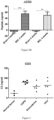
- the serum samples were retrieved from Sera Laboratories International Ltd (SLI Ltd), UK. COS levels were increased in the three different fibrotic diseases ( Figure 3 )
- Type III collagen (Abcam, Cambridge, UK) was degraded in vitro by activated MMP-9 (Merck KGaA, Darmstadt, Germany) for 2 days. Degradation fragments were sequenced by LS-MS/MS and identified by MASCOT search. A specific peptide sequence 610 KNGETGPQ was selected for antibody production. The N-terminal of this sequence is residue 610 of human collagen type III. The synthetic peptide was conjugated to ovalbumin prior to subcutaneous immunization of 4-6 week old Balb/C mice with about 200 ⁇ L emulsified antigen and 50 ⁇ g CO3-610C (KNGETGPQGPGGC-OVA).
- Consecutive immunizations were performed at two week intervals until stable sera titer levels were reached in Freund's incomplete adjuvant. The mice were bled from the second immunization on. At each bleeding, the serum titer was measured and the mouse with highest anti-serum titer was selected for fusion. After the fourth immunization, this mouse was rested for one month and then boosted intravenously with 50 ⁇ g CO3-610C in 100 ⁇ L 0.9% sodium chloride solution three days before isolation of the spleen for cell fusion.
- Monoclonal antibody producing clones were selected using a) immunogenic peptide: KNGETGPQGP-GGC-Ovalbumine (OVA) (807678), b) screening peptide KNGETGPQGP-PG-K-Biotin (807971), c) de-selection peptides KDGETGAAGPPGK-Biotin (118318) representing a type II collagen alpha 1 chain, KDGEAGAQGPPGK-Biotin representing a type I collagen alpha 1 chain degradation product, purchased from the Chinese Peptide Company, Beijing, China.
- the ELISA coat plate was obtained from NUNC (Thermofisher, Copenhagen, Denmark). Peptide conjugation reagents and buffers were produced by Pierce (Thermofisher, Copenhagen, Denmark).
- Buffer used for dissolving the coating peptide was composed of the following: 40 mM Na 2 HPO 4 , 12 H 2 O, 7 mM KH 2 PO 4 , 137 mM NaCl, 2,7 mM KCl, 25 mM EDTA, 0,1 % Tween 20, 1 % BSA, 10 % sorbitol, pH 7.
- buffer containing the following chemicals was used: 8 mM Na 2 HPO 4 , 12 H 2 O, 1,5 mM KH 2 PO 4 , 13,7 mM NaCl, 2,7 mM KCl, 0,1 % Tween 20, 1 % BSA, 0,003 % phenol red, pH 7,4.
- a different buffer used for a urine assay contained 400 mM TRIZMA, 0,05 % Tween 20, 0,1 % BSA, 0,36 % Bronidox L5, pH 8,0.
- a washing buffer composed of 25mM TRIZMA, 50 mM NaCl, 0,036 % Bronidox L5, 0,1 % Tween 20, and reaction-stopping buffer composed of 0,1 % H 2 SO 4 .
- ELISA-plates used for the assay development were Streptavidin-coated from Roche (Hvidovre, Denmark) cat.: 11940279. All ELISA plates were analyzed with the ELISA reader from Molecular Devices, SpectraMax M, (CA. USA).
- TMB tetramethylbenzinidine
- a standard curve was performed by serial dilution of biotinylated-NB51-32 CO3-610C for a serum assay, and biotinylated-NB51-134 CO3-610C for a urine assay. Standard concentrations were 0, 0.33, 1, 3, 9, 27, 81 and 162 ng/ml.
- mice 40 female Sprague-Dawley rats aged 6 months were housed at the animal research facilities at Nordic Bioscience, Copenhagen, Denmark. The experiments were approved by the Experimental Animal Committee of the Danish Ministry of Justice and were performed according to the European Standard for Good Clinical Practice (2008/561-1450). The rats were housed in standard type III-H cages at 18-22°C with bedding and nest material (Altromin 1324; Altromin, Germany) and water ad libitum. Rats were kept under conditions of a 12-hour light/dark cycle.
- liver fibrosis was induced by common BDL.
- the surgical procedure was performed under sterile conditions.
- the rat was anaesthetized, the bile duct localized and ligated in two places followed by dissection between the ligations, and the abdomen was closed.
- the other 20 rats were subjected to a sham operation, in which the abdomen was closed without bile duct ligation.
- the rats were then divided into 2 groups: Group 1 (10 BDL rats and 10 sham-operated rats) was sacrificed after 2 weeks and Group 2 (10 BDL and 10 sham-operated rats) was sacrificed after 4 weeks. On completion of the study period (2 or 4 weeks), after at least 14 hours fasting, all surviving animals were asphyxiated by CO 2 and sacrificed by exsanguinations.
- Blood samples were taken from the retro-orbital sinus of rats after at least 14 hours fasting, under light CO 2 /O 2 anaesthesia, at baseline and at termination. Blood was left 30 minutes at room temperature to clot, followed by centrifugation at 1500 g for 10 minutes. All clot-free liquid was transferred to fresh tubes and centrifuged again at 1500 g for 10 minutes. The serum was then transferred to clean tubes and stored at -80°C.
- the total collagen concentration was assayed using the commercial QuickZyme Collagen Assay (QuickZyme Bioscience, Leiden, The Netherlands).
- the concentration of CTX-II was assayed using the commercial Rat CTX-II kit (IDS Nordic, Herlev, Denmark). All samples were assayed in duplicate.
- Col3a1 The number of transcripts of type III collagen (Col3a1) in liver tissue samples was determined by quantitative real-time polymerase chain reaction (RT-PCR) using fluorescent reporter probes. The number of Col3a1 copies in the sample was extrapolated from a standard curve obtained using Col3a1 plasmid cDNA Image Clone 7097081 (Geneservice, Cambridge, UK) as dilution standard. Amounts of Col3a1 were normalized with those of housekeeping gene hypoxanthine phosphoribosyltransferase 1 (Hprt1).
- RT-PCR quantitative real-time polymerase chain reaction
- cDNA complementary DNA
- Tissue was pulverized in excess of liquid nitrogen in a steel mortar. Samples were then transferred into a 1.5 ml eppendorf tube and left shaking overnight at 4 °C in 0.5M Acetic Acid solution containing protease inhibitor cocktail (Roche). The samples were then sonicated with ultrasounds using 5 pulses at 60% amplitude (U50 control, IKA Labortechnik) and left for an additional 2 hours at 4 °C after which they were centrifuged for 5 minutes at 13,000 rpm. Supernatant was carefully removed, transferred in a new eppendorf and stored at -80 °C.
- tissue extract 20 ⁇ g was mixed with loading buffer (Invitrogen LDS 4x, NP0007) containing the reducing agent (NuPAGE, NP0004 from Invitrogen). Samples were then loaded into 4-12% Bis-Tris gradient gel (NP0332BOX from Invitrogen) and run for 52 minutes at 200V. Proteins were then transferred onto a nitrocellulose membrane using the i-Blot transfer system (Invitrogen), blocked with 5% milk in (? Need to spell out?) TTBS overnight at 4 degrees. Beta Actin antibody (AbCam ab8229, give company, city country?) was used as a loading control.
- loading buffer Invitrogen LDS 4x, NP0007
- reducing agent NuPAGE, NP0004 from Invitrogen
- livers of control animals showed normal gross morphology while livers of BDL animals were enlarged.
- the liver weights were significantly increased in BDL rats compared to the sham-operated controls (mean weights at sacrifice: 2 weeks post-surgery, sham 8.1 g; BDL 14.1 g; 4 weeks post-surgery, sham 9.0 g; BDL 19.4 g) ( Figure 4 panel A).
- Semi-quantitative scoring of liver sections using the 0-3 scale showed significantly more structural changes of the liver at 4 weeks compared with 2 weeks ( Figure 4 panel B).
- Figure 4 panel A shows liver weight in bile duct ligation (BDL)- or sham-operated rats.
- Panel C shows Sirius Red photomicrographs showing the hepatic structure in sham-operated rats, and in BDL rats 2 and 4 weeks post-surgery. The hepatic structure around the portal tract is clearly disrupted in BDL rats compared with the sham-operated rats. Collagens are highlighted in red. Original magnification was x40.
- panel C are shown CTX-II levels in BDL- or sham-operated rats. Data are shown as mean + standard error of mean.
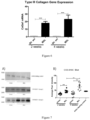
- Figure 7 shows changes in the expression of CO3-610C in the liver of rats in BDL- and sham-operated groups assessed by A) Western blot 2 and 4 weeks post-surgery and B) Bands from western blot quantified by densitometry.
- ECM remodelling is an integrated process of tissue development, maintenance and pathogenesis. Proteolytic activity is essential in this process for cell migration, removal of damaged tissue, and sequestering of new proteins, for the correct and optimal tissue orientation and quality (108:109).
- the specific matrix degradation products, neo-epitopes may be important for the identification of new biochemical markers of liver fibrosis matrix turnover and understanding fibrosis pathogenesis. At present there are no available measuring techniques, nor biochemical markers, that allow for assessment of ECM remodeling in the pathogenesis of fibrosis.
- the present data clearly demonstrate the neo-epitope COS - 610C from MMP-9 mediated collagen type III degradation is a diagnostic biochemical marker for liver fibrosis with an average increases in serum of up to 153% from sham to BDL- operated rats.
- liver fibrosis is not only an accumulation of ECM proteins, but also an accelerated turnover situation, in which both tissue formation and tissue degradation both are highly up regulated. Tissue formation outstrips tissue degradation, leading to an accumulation of scar tissue over time.
- Other matrix turnover proteins to assess liver fibrosis, one being the type III collagen formation marker N-terminal type III pro-collagen. This marker represents collagen type III formation and has shown to be increased in liver fibrosis in previous studies.
- mice were immunized with synthetic peptide KAFVFP (SEQ ID NO1167) conjugated to ovalbumin (KAFVFPKESD-GGC-OVA (SEQ ID NO1049)), spleen cells were used for fusion, and monoclonal antibodies tested for reactivity to biotinylated KAFVFP (SEQ ID NO 1167), i.e. (KAFVFPKESD-biotin(SEQ ID NO1049)) immobilized in wells of microtitre plates precoated with streptavidin.
- KAFVFPKESD-GGC-OVA SEQ ID NO1049
- the following material was used in 2-fold dilutions; (1) the synthetic KAFVFP (SEQ ID NO1167) peptide; (2) a nonsense peptide (KNEGTG) unrelated to CRP; (3) a pool of human serum samples; (4) CRP proteolytically cleaved with MMP3 for 7 days, subsequently stopped by addition of EDTA to block protease activity, and stored at -80°C until testing; (5) same as (4) but using MMP8 instead of MMP3; (6) same as (4) except using Cathepsin K (for 2 days) instead of MMP3 (and E64 as inhibitor to block Cathepsin K activity).
- Reference Example 8 CO3 in biological relevant samples: CO3 levels in Carbon tetrachloride (CCl4)-induced cirrhosis in rats.
- CCl4 Carbon tetrachloride
- liver sections (4 ⁇ m) were stained in 0.1% Sirius red F3B (Sigma-Aldrich, St. Louis, MO) in saturated picric acid (Sigma-Aldrich).
- Relative fibrosis area (expressed as a percentage of total liver area) was assessed by analyzing 36 fields of Sirius red-stained liver sections per animal. Each field was acquired at 10X magnification [E600 microscope (Nikon) and RT-Slider SPOT digital camera (Diagnostic Instruments, Inc., Sterling Heights, MI). Results were analyzed using a computerized Bioquant Life Science morphometry system. To evaluate the relative fibrosis area, the measured collagen area was divided by the net field area and then multiplied by 100. Subtraction of vascular luminal area from the total field area yielded the final calculation of the net fibrosis area. From each animal analyzed, the amount of fibrosis as percentage was measured and the average value presented.
- mice were classified into 4 different stages of fibrosis and cirrhosis (Group A: moderate fibrosis, group B: advanced fibrosis, Group C: moderate cirrhosis, and Group D: advanced cirrhosis) that were determined by the percentage of Sirius red positive liver area (Group A: ⁇ 5%, Group B: 5 to10%, Group C: 10 to 15% and Group D: >15%).
- control and fibrotic/cirrhotic rats were studied considering four different time points during the CCl4 treatment: 8, 12, 16 and 20 weeks after starting the cirrhosis induction program.
- Serum hyaluronan was measured using a sandwich ELISA kit (R&D Systems Inc., Minneapolis, MN, USA).
- mice included in this protocol were randomly assigned to one of the following groups: A/ eight weeks of CCl 4 treatment, B/ twelve weeks of CCl 4 treatment, C/ sixteen weeks of CCl 4 treatment and D/ twenty weeks of CCl 4 treatment.
- four control groups were studied at the same time points. Thirteen fibrotic rats and seven control rats were included in each group. At the end of the study, rats were placed in standard metabolic cages (Tecniplast GmbH, Hohenpeissenberg, Germany) during an adaptation period of 3 days before proceeding with the twenty-four-hour urine collection. Urinary volumes were determined gravimetrically. During the adaptation period, rats were allowed to get free access to tap water and food. Then, 24-hour urine samples were centrifuged for 5 min at 2,500 rpm and aliquoted into ten polypropylene tubes (400 ⁇ L each). Urine samples were stored at -80°C for subsequent analysis.
- rats were weighed, anesthetized with pentobarbital (50 mg/kl) and decapitated. Blood were collected and allowed to stand at room temperature for 20 min to allow clotting and then centrifuged for 10 min at 2500 rpm. Serum were collected in polypropylene tubes aliquots (400 ⁇ l each) and transferred via dry ice to a -80°C freezer. Collection of baseline blood samples at the beginning of the CCl 4 treatment was not considered in order to avoid additional intervention that may increase the risk of infection and/or introduce modifications in the experimental model that may compromise the evolution of the induced pathophysiological process.
- liver fibrotic area For histology and Sirius red staining, half of the left lobe of the liver were placed in 10% neutral buffered formalin for 16 hours, embedded in paraffin and sectioned into 4- ⁇ m-thick slices. After liver fibrosis quantification, the unused paraffin block material was preserved for biomarker quantification. The other half of the left lobe was flash-frozen in liquid nitrogen and stored for Western blot, RT-PCR or immunohistochemical analysis. Measurements of liver fibrotic area, serum and urine osmolality, Na + and K + , albumin, creatinine, alanine amino-transferase and lactate dehydrogenase were made according to the Material and Methods section.
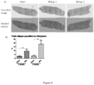
- Liver collagen was quantified in all study animals by Sirius red staining of liver slices. The final data for each animal was taken as the average of red staining observed in 36 consecutive microscope fields ( Figure 12 ).
- Figure 12 shows representative pictures from two sets of 36 images used to quantify collagen accumulation in liver in rat #1 (left) and rat #43 (right) treated with carbon tetrachloride for eight and twenty weeks respectively.
- the serum COS marker shows statistically significant increases in both fibrotic and cirrhotic rats compared to control rats. Animals were classified according to a fully automated syrius red staining of the liver procedure used to quantify fibrosis ( Figures 13 and 14 ).
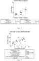
- Figure 13 shows serum CO3 levels in CCl 4 inhalation and control rats as performed in Hospital Clinic (Barcelona). Each point represents one animal. Rats were classified according a computerized image analysis method of syrius red staining of the liver used to quantify fibrosis.
- HA levels were quantified with a commercial ELISA kit and results show significant elevations of this ECM component in cirrhotic rats vs. fibrotic animals ( Figures 15 and 16 ).
- mice were treated by application to the skin of PBS or bleomycin. Increasing levels in urine of the MMP-9 mediated collagen III (CO3) degradation fragment CO3-610 were associated with skin fibrosis progression in mice.
- CO3 MMP-9 mediated collagen III
- Figure 18 shows a skin section from a PBS treated mouse at 8 weeks of treatment (panel A) and a skin section from Bleomycin treated mouse at 8 weeks of treatment (panel B).
- Skin width was calculated by Visiopharm software as an overall number per skin section instead of sampling pictures.
- Figure 19 shows CO3-610 urine assay results which demonstrate a significant increase throughout the time points of the study.
- 2 weeks P 0.0008, 4 weeks P ⁇ 0.0001, 6 weeks P ⁇ 0.0001, 8 weeks P ⁇ 0.0001 and overall P ⁇ 0.0001.
Landscapes
- Life Sciences & Earth Sciences (AREA)
- Health & Medical Sciences (AREA)
- Chemical & Material Sciences (AREA)
- Engineering & Computer Science (AREA)
- Immunology (AREA)
- Molecular Biology (AREA)
- Proteomics, Peptides & Aminoacids (AREA)
- Organic Chemistry (AREA)
- Hematology (AREA)
- Urology & Nephrology (AREA)
- Biomedical Technology (AREA)
- Biotechnology (AREA)
- Microbiology (AREA)
- Physics & Mathematics (AREA)
- Analytical Chemistry (AREA)
- Biochemistry (AREA)
- General Health & Medical Sciences (AREA)
- Wood Science & Technology (AREA)
- Zoology (AREA)
- Medicinal Chemistry (AREA)
- Pathology (AREA)
- General Physics & Mathematics (AREA)
- Food Science & Technology (AREA)
- Cell Biology (AREA)
- Biophysics (AREA)
- Bioinformatics & Cheminformatics (AREA)
- General Engineering & Computer Science (AREA)
- Genetics & Genomics (AREA)
- Peptides Or Proteins (AREA)
- Measuring Or Testing Involving Enzymes Or Micro-Organisms (AREA)
- Medicines That Contain Protein Lipid Enzymes And Other Medicines (AREA)
Claims (2)
- Immunoassayverfahren zum Messen von neoepitophaltigen Proteinfragmenten, die natürlich in einer Körperflüssigkeitsprobe vorliegen, wobei der Immunoassay durch ein Verfahren durchgeführt wird, umfassend: Inkontaktbringen von Proteinfragmenten, die natürlich in der Probe vorliegen, mit einem immunologischen Bindungspartner, der mit einem N-terminalen Neoepitop, das durch Spaltung eines Proteins durch eine Proteinase gebildet wird, oder einem C-terminalen Neoepitop, das durch Spaltung eines Proteins durch eine Proteinase gebildet wird, reaktiv ist, wobei dieser immunologische Bindungspartner mit dem intakten Protein, von dem das Epitop stammt, nicht reaktiv ist und mit einer verlängerten Version der Neoepitopsequenz nicht reaktiv ist, wobei die Neoepitopsequenz über die Spaltstelle hinaus verlängert ist, und Messen des Ausmaßes der Bindung von Peptidfragmenten an den immunologischen Bindungspartner, um darin Proteinfragmente, die das Neoepitop umfassen, zu messen, wobei der immunologische Bindungspartner spezifisch ein Neoepitop bindet, das von einer N-terminalen Aminosäuresequenz gebildet wird, die in Peptiden vorliegt, die durch Spaltung von Kollagen Typ IV produziert werden, wobei die Peptide eine N-terminale Sequenz umfassen, die ausgewählt ist aus der Gruppe bestehend aus:
Kollagen Typ IV IDGYRG... SEQ ID NO609 MGPPGT... SEQ ID NO610 DGLPGS... SEQ ID NO611 PGSKGE... SEQ ID NO612 RGFPGP... SEQ ID NO613 PGPPGL... SEQ ID NO614 GPPGPP... SEQ ID NO100 GLPGSM... SEQ ID NO615 GLPGQQ... SEQ ID NO616 PGIGVQ... SEQ ID NO618 GIGPPG... SEQ ID NO611 PGLPGI... SEQ ID NO504 PGPKGF... SEQ ID NO621 SPGIPG... SEQ ID NO619 PGMQGE... SEQ ID NO620 LGSKGE... SEQ ID NO617 KGQPGL... SEQ ID NO622 RGPPGP... SEQ ID NO148 IRGEPG... SEQ ID NO626 FPGPPG... SEQ ID NO627 GPLGEK... SEQ ID NO625 DGVIGM... SEQ ID NO629 PGNPGI... SEQ ID NO630 PGLKGD... SEQ ID NO624 IKGDKG... SEQ ID NO631 PGSPGC... SEQ ID NO632 PPSDEI... SEQ ID NO623 GPPGVP... SEQ ID NO633 DQGDQG... SEQ ID NO634 GAPGPQ... SEQ ID NO445 KGSIGI... SEQ ID NO635 GIPGAP... SEQ ID NO372 DRGPQG... SEQ ID NO442 ERGSPG... SEQ ID NO38 LQGIRG... SEQ ID NO628 DGGVPN... SEQ ID NO638 SGRDGL... SEQ ID NO639 PGPMGP... SEQ ID NO25 GPPGLM... SEQ ID NO643 DGYRGP... SEQ ID NO642 AEGLPG... SEQ ID NO641 LPGFAG... SEQ ID NO644 GIPGMP... SEQ ID NO645 PPGRLG... SEQ ID NO636 GPPGEK... SEQ ID NO640 EKGQKG... SEQ ID NO637 .LPGPDG SEQ ID NO2214 .PGIPGT SEQ ID NO2227 .ILGHVP SEQ ID NO2212 .LRGIPG SEQ ID NO760 .PGDIVF SEQ ID NO2215 .PGLPGQ SEQ ID NO2213 .PGFPGA SEQ ID NO2218 .GNKGDP SEQ ID NO2216 .SGYPGN SEQ ID NO2217 .QQGNRG SEQ ID NO2221 .PGPRGK SEQ ID NO2219 .VSGPPG SEQ ID NO2220 .VGQPGP SEQ ID NO2222 .PGPPGP SEQ ID NO458 .KRGPPG SEQ ID NO2223 .GEPGMQ SEQ ID NO2224 .SKGEKG SEQ ID NO2226 .LHGFPG SEQ ID NO2225 .GEPGPP SEQ ID NO675 Kollagen Typ IV ...RGPPGP SEQ ID NO148 ...SVDHGF SEQ ID NO646 ...PSVDHG SEQ ID NO647 ...VDHGFL SEQ ID NO648 ...PGQPGY SEQ ID NO649 ...QPGYTN SEQ ID NO650 ...PGLPGS SEQ ID NO651 ...GTPSVD SEQ ID NO652 ...SVGSPG SEQ ID NO653 ...LPGSMG SEQ ID NO654 ...GPPGVP SEQ ID NO633 ...PGFPGL SEQ ID NO655 ...PGLPGE SEQ ID NO656 ...GDPGPP SEQ ID NO373 ...HQGEMG SEQ ID NO657 ...GPPGLV SEQ ID NO658 ...PGIPGP SEQ ID NO659 ...PGFPGT SEQ ID NO660 ...PGLPGP SEQ ID NO58 ...DGIPGP SEQ ID NO661 ...SGPKGY SEQ ID NO662 ...GIG PPG SEQ ID NO611 ...PGPRGE SEQ ID NO663 ...GQGPPG SEQ ID NO664 ...PGAIGP SEQ ID NO668 ...KVDMGS SEQ ID NO665 ...PGIDGV SEQ ID NO666 ...GPKGPP SEQ ID NO671 ...PPGPPG SEQ ID NO119 ...KGHMGE SEQ ID NO667 ...GPKGLP SEQ ID NO670 ...SPGPPG SEQ ID NO672 ...KGLPGP SEQ ID NO669 ...GSVGYP SEQ ID NO673 ...PPGSPG SEQ ID NO674 ...GEPGPP SEQ ID NO675 ...AGNPGP SEQ ID NO676 ...FPGPQG SEQ ID NO679 ...PGYTNG SEQ ID NO678 ...SVDHGF SEQ ID NO646 ...LSGPPG SEQ ID NO680 ...GQGPPG SEQ ID NO664 ...GVMGTP SEQ ID NO681 ...PGIKGS SEQ ID NO677 ...PGPPGP SEQ ID NO458 ...GVSGPKSEQ ID NO682 HVPGML. SEQ ID NO2228 LPVPGQ. SEQ ID NO2229 LGPPGL. SEQ ID NO2230 GVPGQA. SEQ ID NO2231 VPGQAQ. SEQ ID NO2232 GPDGFL. SEQ ID NO2233 QEGPLG. SEQ ID NO2234 LPGEVL. SEQ ID NO2235 RGIPGF. SEQ ID NO2236 NRGLGF. SEQ ID NO2238 IPSDTL. SEQ ID NO2239 PAGEKG. SEQ ID NO2240 GEKGNK. SEQ ID NO2241 PVGPPG. SEQ ID NO2242 DIVFRK. SEQ ID NO2243 PPGPKG. SEQ ID NO167 RGKPGM. SEQ ID NO2244 PGTRGL. SEQ ID NO2245 GEKGSK. SEQ ID NO2246 EKGSKG. SEQ ID NO2247 SGQPGL. SEQ ID NO2248 AGIPQK. SEQ ID NO2237 - Verfahren nach einem vorhergehenden Anspruch, als ein Verfahren zur Diagnose oder Quantifizierung von Fibrose durchgeführt und umfassend ein Assoziieren eines Anstiegs des Messwerts von neoepitophaltigen Proteinfragmenten in der Körperflüssigkeitsprobe über einen normalen Spiegel bei dem Vorliegen oder Ausmaß der Fibrose.
Priority Applications (1)
| Application Number | Priority Date | Filing Date | Title |
|---|---|---|---|
| EP24200645.0A EP4467984A3 (de) | 2009-03-30 | 2010-03-29 | Fibrosebiomarkertest |
Applications Claiming Priority (6)
| Application Number | Priority Date | Filing Date | Title |
|---|---|---|---|
| US21146709P | 2009-03-30 | 2009-03-30 | |
| US28908109P | 2009-12-22 | 2009-12-22 | |
| EP10715752.1A EP2414844B1 (de) | 2009-03-30 | 2010-03-29 | Biomarker für fibrose |
| PCT/EP2010/054096 WO2010115749A2 (en) | 2009-03-30 | 2010-03-29 | Fibrosis biomarker assay |
| EP14169636.9A EP2770327B1 (de) | 2009-03-30 | 2010-03-29 | Fibrosebiomarkertest |
| EP16205956.2A EP3173792B1 (de) | 2009-03-30 | 2010-03-29 | Crp neo-epitop fibrose-assay |
Related Parent Applications (3)
| Application Number | Title | Priority Date | Filing Date |
|---|---|---|---|
| EP14169636.9A Division EP2770327B1 (de) | 2009-03-30 | 2010-03-29 | Fibrosebiomarkertest |
| EP10715752.1A Division EP2414844B1 (de) | 2009-03-30 | 2010-03-29 | Biomarker für fibrose |
| EP16205956.2A Division EP3173792B1 (de) | 2009-03-30 | 2010-03-29 | Crp neo-epitop fibrose-assay |
Related Child Applications (2)
| Application Number | Title | Priority Date | Filing Date |
|---|---|---|---|
| EP24200645.0A Division EP4467984A3 (de) | 2009-03-30 | 2010-03-29 | Fibrosebiomarkertest |
| EP24200645.0A Division-Into EP4467984A3 (de) | 2009-03-30 | 2010-03-29 | Fibrosebiomarkertest |
Publications (3)
| Publication Number | Publication Date |
|---|---|
| EP3578664A1 EP3578664A1 (de) | 2019-12-11 |
| EP3578664B1 true EP3578664B1 (de) | 2024-10-30 |
| EP3578664C0 EP3578664C0 (de) | 2024-10-30 |
Family
ID=42199024
Family Applications (5)
| Application Number | Title | Priority Date | Filing Date |
|---|---|---|---|
| EP16205956.2A Active EP3173792B1 (de) | 2009-03-30 | 2010-03-29 | Crp neo-epitop fibrose-assay |
| EP24200645.0A Pending EP4467984A3 (de) | 2009-03-30 | 2010-03-29 | Fibrosebiomarkertest |
| EP19174151.1A Active EP3578664B1 (de) | 2009-03-30 | 2010-03-29 | Collagen iv neo-epitopfibrose-assay |
| EP10715752.1A Active EP2414844B1 (de) | 2009-03-30 | 2010-03-29 | Biomarker für fibrose |
| EP14169636.9A Active EP2770327B1 (de) | 2009-03-30 | 2010-03-29 | Fibrosebiomarkertest |
Family Applications Before (2)
| Application Number | Title | Priority Date | Filing Date |
|---|---|---|---|
| EP16205956.2A Active EP3173792B1 (de) | 2009-03-30 | 2010-03-29 | Crp neo-epitop fibrose-assay |
| EP24200645.0A Pending EP4467984A3 (de) | 2009-03-30 | 2010-03-29 | Fibrosebiomarkertest |
Family Applications After (2)
| Application Number | Title | Priority Date | Filing Date |
|---|---|---|---|
| EP10715752.1A Active EP2414844B1 (de) | 2009-03-30 | 2010-03-29 | Biomarker für fibrose |
| EP14169636.9A Active EP2770327B1 (de) | 2009-03-30 | 2010-03-29 | Fibrosebiomarkertest |
Country Status (8)
| Country | Link |
|---|---|
| US (3) | US9206464B2 (de) |
| EP (5) | EP3173792B1 (de) |
| JP (2) | JP5978128B2 (de) |
| KR (1) | KR101700463B1 (de) |
| CN (1) | CN103238071B (de) |
| DK (3) | DK3173792T3 (de) |
| ES (2) | ES2529101T3 (de) |
| WO (1) | WO2010115749A2 (de) |
Families Citing this family (52)
| Publication number | Priority date | Publication date | Assignee | Title |
|---|---|---|---|---|
| WO2008054764A2 (en) * | 2006-10-31 | 2008-05-08 | George Mason Intellectual Properties, Inc. | Biomarkers for colorectal cancer |
| US9404932B2 (en) | 2007-11-05 | 2016-08-02 | Nordic Bioscience A/S | Pathology biomarker assay |
| EP2538222B1 (de) * | 2007-11-05 | 2021-08-25 | Nordic Bioscience A/S | Biochemische Marker zur Bewertung des CVD-Risikos |
| ES2529101T3 (es) | 2009-03-30 | 2015-02-16 | Nordic Bioscience A/S | Ensayo de biomarcadores para fibrosis |
| WO2012138223A2 (en) | 2011-04-05 | 2012-10-11 | Academisch Ziekenhuis Leiden H.O.D.N. Lumc | Compounds and methods for altering activin receptor-like kinase signalling |
| GB201107922D0 (en) * | 2011-05-12 | 2011-06-22 | Nordic Bioscience As | Biochemical markers for CVD risk assessment |
| WO2012163768A1 (en) * | 2011-05-27 | 2012-12-06 | Nordic Bioscience A/S | Detection of diagnostic peptides |
| EP2727520B1 (de) * | 2011-06-29 | 2019-08-21 | Inner Mongolia Furui Medical Science Co., Ltd | Vorrichtung und system zum nachweis von leberfibrose |
| GB201111361D0 (en) * | 2011-07-04 | 2011-08-17 | Nordic Bioscience As | Biochemical markers for neurodegenerative conditions |
| GB201111788D0 (en) * | 2011-07-11 | 2011-08-24 | Nordic Bioscience As | In vitro assessment of cardiovascular events by assay for neo-epitopes |
| JP5953035B2 (ja) * | 2011-11-28 | 2016-07-13 | 学校法人慶應義塾 | 病理診断支援装置、病理診断支援方法、及び病理診断支援プログラム |
| EP2839292A4 (de) * | 2012-04-19 | 2015-08-26 | Univ California | Zusammensetzungen und verfahren zum nachweis von instabiler arteriosklerotischer plaque |
| GB201306792D0 (en) * | 2013-04-15 | 2013-05-29 | Nordic Bioscience As | PIIINP Neo-epitope assay |
| GB201307387D0 (en) | 2013-04-24 | 2013-06-05 | Nordic Bioscience As | Collegen type VI alpha-1 assay |
| GB201400472D0 (en) | 2014-01-13 | 2014-02-26 | Nordic Bioscience As | Biochemical Markers for pulmonary and other diseases |
| GB201505654D0 (en) | 2015-04-01 | 2015-05-13 | Nordic Bioscience As | Immunoassay for collagen type VI sequence |
| US20170165447A1 (en) * | 2015-12-11 | 2017-06-15 | Geno Llc | Method and apparatus for administering gases including nitric oxide |
| IL322971A (en) | 2015-12-15 | 2025-10-01 | Takeda Pharmaceuticals Co | Method for evaluating the efficacy of a drug for a plasma kallikrein-mediated disorder |
| GB201601571D0 (en) * | 2016-01-28 | 2016-03-16 | Nordic Bioscience As | Collagen type Vll alpha 1 assay |
| KR102668479B1 (ko) * | 2016-02-03 | 2024-05-22 | 노르딕 바이오사이언스 에이/에스 | 섬유증에 대한 조합된 바이오마커 측정법 |
| CN105738632B (zh) * | 2016-02-24 | 2017-06-23 | 浙江理工大学 | 一种区分羊皮种类的检测方法 |
| CN109477127A (zh) * | 2016-03-14 | 2019-03-15 | R基因股份有限公司 | 超热稳定的赖氨酸-突变型ssDNA/RNA连接酶 |
| WO2017210147A1 (en) * | 2016-05-29 | 2017-12-07 | Wei Jia | Liver disease-related biomarkers and methods of use thereof |
| CN111183360B (zh) * | 2017-07-19 | 2024-10-18 | 生物辐射欧洲有限公司 | 同时评估非酒精性脂肪性肝炎和肝纤维化状态的生物标志物组合 |
| GB201717301D0 (en) * | 2017-10-20 | 2017-12-06 | Nordic Bioscience As | Type xvi collagen assay |
| US20190351031A1 (en) | 2018-05-16 | 2019-11-21 | Halozyme, Inc. | Methods of selecting subjects for combination cancer therapy with a polymer-conjugated soluble ph20 |
| US11732009B2 (en) | 2018-06-08 | 2023-08-22 | Glympse Bio, Inc. | Activity sensor with tunable analyte |
| US11028425B2 (en) * | 2018-06-08 | 2021-06-08 | Glympse Bio, Inc. | Diagnosis and monitoring of liver disease |
| EP4007920A4 (de) * | 2019-02-01 | 2024-07-17 | Glympse Bio, Inc. | Körperliche verarbeitung von aktivitätssensoren |
| US12338483B2 (en) | 2019-02-28 | 2025-06-24 | Georgia Tech Research Corporation | Compositions and methods for logic-gated profiling of biologic activity |
| TW202104254A (zh) * | 2019-04-12 | 2021-02-01 | 美商格爾托公司 | 重組彈性蛋白及其生產 |
| EP4009953A4 (de) * | 2019-08-07 | 2023-08-23 | Icahn School of Medicine at Mount Sinai | Verfahren zur behandlung von keloiden |
| WO2021034196A1 (en) * | 2019-08-20 | 2021-02-25 | Erasmus University Medical Center Rotterdam | Biomarkers for detecting secondary liver cancer |
| GB201917819D0 (en) | 2019-12-05 | 2020-01-22 | Nordic Bioscience As | A neo-epitope specific assay measuring protease mediated degradation of type iv collagen |
| CA3162431A1 (en) * | 2020-01-21 | 2021-07-29 | Augustinus Bader | Cosmetic formulation for topical administration comprising novel peptides that improve appearance and regeneration of skin |
| AU2021211721A1 (en) | 2020-01-24 | 2022-08-18 | Geltor, Inc. | Animal-free dietary collagen |
| US20240003906A1 (en) * | 2020-04-16 | 2024-01-04 | Nordic Bioscience A/S | Biomarker of Fibrosis |
| IL274696A (en) * | 2020-05-14 | 2021-12-01 | Yeda Res & Dev | Designed transmembrane polypeptides and their use in cellular immunotherapy |
| CN111705123A (zh) * | 2020-06-19 | 2020-09-25 | 上海市第六人民医院 | 一种衰老过程中心肌纤维化的生物标志物及其检测方法和应用 |
| JP2023535022A (ja) * | 2020-07-21 | 2023-08-15 | アムニクス ファーマシューティカルズ, インコーポレイテッド | 活性化可能な治療剤に関する組成物および方法 |
| CN116438456A (zh) | 2020-09-11 | 2023-07-14 | 格里姆普斯生物股份有限公司 | 用于疾病检测/诊断、分期、监测和治疗的离体蛋白酶活性检测 |
| CN112876556B (zh) * | 2021-03-23 | 2022-07-19 | 中国科学院理化技术研究所 | 一种促进成骨细胞矿化的牛骨胶原肽及其应用 |
| CN113388005B (zh) * | 2021-06-30 | 2022-09-27 | 北京泽勤生物医药有限公司 | 增强成骨细胞活性的多肽及其在治疗骨科疾病中的应用 |
| US20250138026A1 (en) * | 2022-02-01 | 2025-05-01 | Institut National de la Santé et de la Recherche Médicale | Method for diagnosing collagen degradation associated disease |
| CN114994317B (zh) * | 2022-04-15 | 2025-10-28 | 宁波市杭州湾医院 | 金属蛋白酶adamts4在制备早期肝纤维化药物中的应用 |
| CN115991732B (zh) * | 2022-11-21 | 2024-12-24 | 北海黑珍珠海洋生物科技有限公司 | 一种具有抗炎作用的珍珠贝活性肽及其应用 |
| GB202302820D0 (en) * | 2023-02-27 | 2023-04-12 | Keybioscience Ag | ProC3 and TGFBI in cancer |
| CN117343132B (zh) * | 2023-09-25 | 2024-03-12 | 山东省农业科学院 | 一种三文鱼鱼皮来源的ace抑制肽及制备方法 |
| WO2025115009A1 (en) * | 2023-11-28 | 2025-06-05 | Accurine Ltd. | Peptides for urinary diagnostics and antibodies for their detection |
| CN118146301B (zh) * | 2024-01-04 | 2024-12-31 | 广州白云山花城药业有限公司 | 一种促成骨细胞分化的活性肽及其应用 |
| GB202408410D0 (en) * | 2024-06-12 | 2024-07-24 | Nordic Bioscience As | immunoassay for detecting canstatin |
| GB202413581D0 (en) | 2024-09-16 | 2024-10-30 | Nordic Bioscience As | Immunoassay for detecting collagen i fragment |
Family Cites Families (31)
| Publication number | Priority date | Publication date | Assignee | Title |
|---|---|---|---|---|
| US4628027A (en) | 1982-05-19 | 1986-12-09 | Molecular Engineering Associates, Ltd. | Vitro diagnostic methods using monoclonal antibodies against connective tissue proteins |
| GB2208865B (en) * | 1987-08-19 | 1991-12-11 | Farmos Group Ltd | Purified propeptide of procollagen type iii, antibody to the propeptide and assay method using the antibody. |
| US5962639A (en) * | 1987-11-06 | 1999-10-05 | Washington Research Foundation | Synthetic peptides corresponding to telopeptide sequences of cross-linked type I collagen metabolites |
| US6124107A (en) * | 1988-06-10 | 2000-09-26 | Merck & Co., Inc. | Assay for marker of human polymorphonuclear leukocyte elastase activity |
| JPH04505857A (ja) * | 1989-06-27 | 1992-10-15 | ラッシュ―プレズビティリアン―セント ルークス メディカル センター | C反応性蛋白質に対するモノクローナル抗体 |
| US5387504A (en) | 1992-09-30 | 1995-02-07 | Merck & Co., Inc. | Monospecific antibodies and assay system for detecting stromelysin cleavage products |
| US6703219B1 (en) * | 1993-02-26 | 2004-03-09 | Immtech International Inc. | Mutant protein and methods and materials for making and using it |
| US6110689A (en) * | 1994-01-21 | 2000-08-29 | Osteometer A/S | Method of assaying collagen fragments in body fluids, a test kit and means for carrying out the method and use of the method to diagnose the presence of disorders associated with the metabolism of collagen |
| JPH08100000A (ja) * | 1994-09-30 | 1996-04-16 | Morinaga & Co Ltd | ヒトiv型コラーゲンに対する抗体及びその利用 |
| EP0718309B1 (de) * | 1994-12-23 | 1999-04-07 | Roche Diagnostics GmbH | Antigene und Antikörper zum Nachweis von Kollagen I |
| US6060255A (en) | 1997-10-03 | 2000-05-09 | Tosoh Corporation | Type IV collagen high molecular form and production and diagnostic use thereof |
| EP0913692A1 (de) | 1997-10-31 | 1999-05-06 | Bayer Ag | Immunassay zum Nachweis von prokollagen-III-C-terminalen Propeptid |
| US7541149B1 (en) | 1998-05-28 | 2009-06-02 | Siemens Healthcare Diagnostics, Inc. | Monoclonal antibody and assay for detecting PIIINP |
| US6916903B2 (en) * | 1998-06-19 | 2005-07-12 | Washington Research Foundation | Collagen type III synthetic peptides for collagen resorption assays |
| EP1150123B1 (de) * | 2000-04-28 | 2006-07-12 | Bayer Aktiengesellschaft | Diagnose von Leberfibrose mit Hilfe von Serummarkeralgorithmen |
| JP2002065283A (ja) | 2000-06-12 | 2002-03-05 | Wako Pure Chem Ind Ltd | ハイブリッド酵素およびその用途 |
| ES2259291T3 (es) | 2000-08-21 | 2006-10-01 | Assistance Publique, Hopitaux De Paris | Metodo de diagnostico de enfermedad fibrotica que utiliza marcadores bioquimicos. |
| AU2002251366A1 (en) | 2001-05-02 | 2002-11-11 | Oxford Glycosciences (Uk) Ltd | Proteins, genes and their use for diagnosis and treatment of breast cancer |
| US7390885B2 (en) * | 2001-11-26 | 2008-06-24 | Cell Matrix, Inc. | Humanized collagen antibodies and related methods |
| NZ546611A (en) * | 2003-09-18 | 2010-02-26 | Conforma Therapeutics Corp | Novel heterocyclic compounds as HSP90-inhibitors |
| WO2005050224A2 (en) | 2003-11-13 | 2005-06-02 | Epitome Biosystems Inc. | Small molecule and peptide arrays and uses thereof |
| US20060154318A1 (en) | 2004-06-09 | 2006-07-13 | Anderson Norman L | Stable isotope labeled polypeptide standards for protein quantitation |
| US7358370B2 (en) * | 2005-02-25 | 2008-04-15 | Serenex, Inc. | Benzene, pyridine, and pyridazine derivatives |
| EP1948686A4 (de) | 2005-10-26 | 2009-06-10 | Stc Unm | C-reaktives protein und dessen verwendung zur behandlung von systemischem lupuserythematodes und verwandten leiden |
| US7972802B2 (en) * | 2005-10-31 | 2011-07-05 | University Of Washington | Lipoprotein-associated markers for cardiovascular disease |
| US9404932B2 (en) * | 2007-11-05 | 2016-08-02 | Nordic Bioscience A/S | Pathology biomarker assay |
| EP2538222B1 (de) * | 2007-11-05 | 2021-08-25 | Nordic Bioscience A/S | Biochemische Marker zur Bewertung des CVD-Risikos |
| US20100023377A1 (en) * | 2008-07-23 | 2010-01-28 | Hr Solutions, Inc. | Systems and methods for managing human resource activities |
| ES2529101T3 (es) | 2009-03-30 | 2015-02-16 | Nordic Bioscience A/S | Ensayo de biomarcadores para fibrosis |
| WO2012163768A1 (en) * | 2011-05-27 | 2012-12-06 | Nordic Bioscience A/S | Detection of diagnostic peptides |
| GB201111788D0 (en) * | 2011-07-11 | 2011-08-24 | Nordic Bioscience As | In vitro assessment of cardiovascular events by assay for neo-epitopes |
-
2010
- 2010-03-29 ES ES10715752.1T patent/ES2529101T3/es active Active
- 2010-03-29 CN CN201080014281.6A patent/CN103238071B/zh active Active
- 2010-03-29 EP EP16205956.2A patent/EP3173792B1/de active Active
- 2010-03-29 KR KR1020117025776A patent/KR101700463B1/ko active Active
- 2010-03-29 JP JP2012502618A patent/JP5978128B2/ja not_active Expired - Fee Related
- 2010-03-29 DK DK16205956.2T patent/DK3173792T3/da active
- 2010-03-29 EP EP24200645.0A patent/EP4467984A3/de active Pending
- 2010-03-29 EP EP19174151.1A patent/EP3578664B1/de active Active
- 2010-03-29 DK DK14169636.9T patent/DK2770327T3/en active
- 2010-03-29 EP EP10715752.1A patent/EP2414844B1/de active Active
- 2010-03-29 ES ES14169636.9T patent/ES2635494T3/es active Active
- 2010-03-29 WO PCT/EP2010/054096 patent/WO2010115749A2/en not_active Ceased
- 2010-03-29 EP EP14169636.9A patent/EP2770327B1/de active Active
- 2010-03-29 DK DK10715752T patent/DK2414844T3/en active
- 2010-03-30 US US12/749,652 patent/US9206464B2/en active Active
-
2015
- 2015-01-22 JP JP2015010255A patent/JP6095702B2/ja active Active
- 2015-12-07 US US14/960,713 patent/US9606130B2/en active Active
-
2017
- 2017-03-01 US US15/446,070 patent/US9880177B2/en active Active
Non-Patent Citations (1)
| Title |
|---|
| ROBIN POOLE A ET AL: "The assessment of cartilage degradation in vivo: development of an immunoassay for the measurement in body fluids of type II collagen cleaved by collagenases", JOURNAL OF IMMUNOLOGICAL METHODS, ELSEVIER SCIENCE PUBLISHERS B.V.,AMSTERDAM, NL, vol. 294, no. 1-2, 1 November 2004 (2004-11-01), pages 145 - 153, XP004679763, ISSN: 0022-1759, DOI: 10.1016/J.JIM.2004.09.005 * |
Also Published As
| Publication number | Publication date |
|---|---|
| DK2414844T3 (en) | 2015-03-02 |
| WO2010115749A2 (en) | 2010-10-14 |
| EP4467984A3 (de) | 2024-12-04 |
| EP2770327A1 (de) | 2014-08-27 |
| WO2010115749A3 (en) | 2011-01-13 |
| EP4467984A2 (de) | 2024-11-27 |
| US20160091502A1 (en) | 2016-03-31 |
| JP5978128B2 (ja) | 2016-08-24 |
| DK3173792T3 (da) | 2019-08-05 |
| EP2770327B1 (de) | 2017-06-14 |
| CN103238071A (zh) | 2013-08-07 |
| JP2012522233A (ja) | 2012-09-20 |
| ES2529101T3 (es) | 2015-02-16 |
| KR20110137823A (ko) | 2011-12-23 |
| US9880177B2 (en) | 2018-01-30 |
| JP2015108635A (ja) | 2015-06-11 |
| EP2414844A2 (de) | 2012-02-08 |
| US20170176458A1 (en) | 2017-06-22 |
| CN103238071B (zh) | 2016-07-06 |
| KR101700463B1 (ko) | 2017-01-26 |
| DK2770327T3 (en) | 2017-08-28 |
| JP6095702B2 (ja) | 2017-03-15 |
| EP3173792A2 (de) | 2017-05-31 |
| EP3173792B1 (de) | 2019-06-12 |
| EP3578664A1 (de) | 2019-12-11 |
| US9606130B2 (en) | 2017-03-28 |
| US9206464B2 (en) | 2015-12-08 |
| EP2414844B1 (de) | 2014-12-10 |
| EP3173792A3 (de) | 2017-07-05 |
| US20100209940A1 (en) | 2010-08-19 |
| EP3578664C0 (de) | 2024-10-30 |
| ES2635494T3 (es) | 2017-10-04 |
Similar Documents
| Publication | Publication Date | Title |
|---|---|---|
| EP2414844B1 (de) | Biomarker für fibrose | |
| EP2734542B1 (de) | Pathologischer biomarkertest | |
| US7803553B2 (en) | Methods of use of antibodies which recognize a protease cleavage site of an LAP fragment of TGF-β | |
| US7863009B2 (en) | Mutants of the factor VII-activating protease and detection methods using specific antibodies | |
| US20260028423A1 (en) | Prediction of an increase of dpp3 in a patient with septic shock | |
| JP5355658B2 (ja) | カスパーゼ−14中間体による皮膚疾患の検出 | |
| JP2012230114A (ja) | 第vii因子活性化プロテアーゼの変異体および特異抗体を用いる検出方法 |
Legal Events
| Date | Code | Title | Description |
|---|---|---|---|
| PUAI | Public reference made under article 153(3) epc to a published international application that has entered the european phase |
Free format text: ORIGINAL CODE: 0009012 |
|
| STAA | Information on the status of an ep patent application or granted ep patent |
Free format text: STATUS: THE APPLICATION HAS BEEN PUBLISHED |
|
| AC | Divisional application: reference to earlier application |
Ref document number: 2770327 Country of ref document: EP Kind code of ref document: P Ref document number: 3173792 Country of ref document: EP Kind code of ref document: P Ref document number: 2414844 Country of ref document: EP Kind code of ref document: P |
|
| AK | Designated contracting states |
Kind code of ref document: A1 Designated state(s): AT BE BG CH CY CZ DE DK EE ES FI FR GB GR HR HU IE IS IT LI LT LU LV MC MK MT NL NO PL PT RO SE SI SK SM TR |
|
| STAA | Information on the status of an ep patent application or granted ep patent |
Free format text: STATUS: REQUEST FOR EXAMINATION WAS MADE |
|
| 17P | Request for examination filed |
Effective date: 20200504 |
|
| RBV | Designated contracting states (corrected) |
Designated state(s): AT BE BG CH CY CZ DE DK EE ES FI FR GB GR HR HU IE IS IT LI LT LU LV MC MK MT NL NO PL PT RO SE SI SK SM TR |
|
| STAA | Information on the status of an ep patent application or granted ep patent |
Free format text: STATUS: EXAMINATION IS IN PROGRESS |
|
| 17Q | First examination report despatched |
Effective date: 20221222 |
|
| GRAP | Despatch of communication of intention to grant a patent |
Free format text: ORIGINAL CODE: EPIDOSNIGR1 |
|
| STAA | Information on the status of an ep patent application or granted ep patent |
Free format text: STATUS: GRANT OF PATENT IS INTENDED |
|
| INTG | Intention to grant announced |
Effective date: 20240523 |
|
| GRAS | Grant fee paid |
Free format text: ORIGINAL CODE: EPIDOSNIGR3 |
|
| GRAA | (expected) grant |
Free format text: ORIGINAL CODE: 0009210 |
|
| STAA | Information on the status of an ep patent application or granted ep patent |
Free format text: STATUS: THE PATENT HAS BEEN GRANTED |
|
| AC | Divisional application: reference to earlier application |
Ref document number: 2414844 Country of ref document: EP Kind code of ref document: P Ref document number: 2770327 Country of ref document: EP Kind code of ref document: P Ref document number: 3173792 Country of ref document: EP Kind code of ref document: P |
|
| AK | Designated contracting states |
Kind code of ref document: B1 Designated state(s): AT BE BG CH CY CZ DE DK EE ES FI FR GB GR HR HU IE IS IT LI LT LU LV MC MK MT NL NO PL PT RO SE SI SK SM TR |
|
| REG | Reference to a national code |
Ref country code: GB Ref legal event code: FG4D |
|
| REG | Reference to a national code |
Ref country code: CH Ref legal event code: EP |
|
| REG | Reference to a national code |
Ref country code: DE Ref legal event code: R096 Ref document number: 602010069554 Country of ref document: DE |
|
| REG | Reference to a national code |
Ref country code: IE Ref legal event code: FG4D |
|
| U01 | Request for unitary effect filed |
Effective date: 20241111 |
|
| U07 | Unitary effect registered |
Designated state(s): AT BE BG DE DK EE FI FR IT LT LU LV MT NL PT RO SE SI Effective date: 20241203 |
|
| U20 | Renewal fee for the european patent with unitary effect paid |
Year of fee payment: 16 Effective date: 20250204 |
|
| PG25 | Lapsed in a contracting state [announced via postgrant information from national office to epo] |
Ref country code: HR Free format text: LAPSE BECAUSE OF FAILURE TO SUBMIT A TRANSLATION OF THE DESCRIPTION OR TO PAY THE FEE WITHIN THE PRESCRIBED TIME-LIMIT Effective date: 20241030 Ref country code: IS Free format text: LAPSE BECAUSE OF FAILURE TO SUBMIT A TRANSLATION OF THE DESCRIPTION OR TO PAY THE FEE WITHIN THE PRESCRIBED TIME-LIMIT Effective date: 20250228 |
|
| PG25 | Lapsed in a contracting state [announced via postgrant information from national office to epo] |
Ref country code: ES Free format text: LAPSE BECAUSE OF FAILURE TO SUBMIT A TRANSLATION OF THE DESCRIPTION OR TO PAY THE FEE WITHIN THE PRESCRIBED TIME-LIMIT Effective date: 20241030 |
|
| PG25 | Lapsed in a contracting state [announced via postgrant information from national office to epo] |
Ref country code: NO Free format text: LAPSE BECAUSE OF FAILURE TO SUBMIT A TRANSLATION OF THE DESCRIPTION OR TO PAY THE FEE WITHIN THE PRESCRIBED TIME-LIMIT Effective date: 20250130 |
|
| PG25 | Lapsed in a contracting state [announced via postgrant information from national office to epo] |
Ref country code: GR Free format text: LAPSE BECAUSE OF FAILURE TO SUBMIT A TRANSLATION OF THE DESCRIPTION OR TO PAY THE FEE WITHIN THE PRESCRIBED TIME-LIMIT Effective date: 20250131 |
|
| PG25 | Lapsed in a contracting state [announced via postgrant information from national office to epo] |
Ref country code: PL Free format text: LAPSE BECAUSE OF FAILURE TO SUBMIT A TRANSLATION OF THE DESCRIPTION OR TO PAY THE FEE WITHIN THE PRESCRIBED TIME-LIMIT Effective date: 20241030 |
|
| PG25 | Lapsed in a contracting state [announced via postgrant information from national office to epo] |
Ref country code: SM Free format text: LAPSE BECAUSE OF FAILURE TO SUBMIT A TRANSLATION OF THE DESCRIPTION OR TO PAY THE FEE WITHIN THE PRESCRIBED TIME-LIMIT Effective date: 20241030 |
|
| PG25 | Lapsed in a contracting state [announced via postgrant information from national office to epo] |
Ref country code: SK Free format text: LAPSE BECAUSE OF FAILURE TO SUBMIT A TRANSLATION OF THE DESCRIPTION OR TO PAY THE FEE WITHIN THE PRESCRIBED TIME-LIMIT Effective date: 20241030 |
|
| PG25 | Lapsed in a contracting state [announced via postgrant information from national office to epo] |
Ref country code: CZ Free format text: LAPSE BECAUSE OF FAILURE TO SUBMIT A TRANSLATION OF THE DESCRIPTION OR TO PAY THE FEE WITHIN THE PRESCRIBED TIME-LIMIT Effective date: 20241030 |
|
| PLBE | No opposition filed within time limit |
Free format text: ORIGINAL CODE: 0009261 |
|
| STAA | Information on the status of an ep patent application or granted ep patent |
Free format text: STATUS: NO OPPOSITION FILED WITHIN TIME LIMIT |
|
| 26N | No opposition filed |
Effective date: 20250731 |
|
| PG25 | Lapsed in a contracting state [announced via postgrant information from national office to epo] |
Ref country code: MC Free format text: LAPSE BECAUSE OF FAILURE TO SUBMIT A TRANSLATION OF THE DESCRIPTION OR TO PAY THE FEE WITHIN THE PRESCRIBED TIME-LIMIT Effective date: 20241030 |
|
| REG | Reference to a national code |
Ref country code: CH Ref legal event code: H13 Free format text: ST27 STATUS EVENT CODE: U-0-0-H10-H13 (AS PROVIDED BY THE NATIONAL OFFICE) Effective date: 20251024 |
|
| PG25 | Lapsed in a contracting state [announced via postgrant information from national office to epo] |
Ref country code: CH Free format text: LAPSE BECAUSE OF NON-PAYMENT OF DUE FEES Effective date: 20250331 |
|
| PG25 | Lapsed in a contracting state [announced via postgrant information from national office to epo] |
Ref country code: IE Free format text: LAPSE BECAUSE OF NON-PAYMENT OF DUE FEES Effective date: 20250329 |
|
| U20 | Renewal fee for the european patent with unitary effect paid |
Year of fee payment: 17 Effective date: 20260204 |
|
| PGFP | Annual fee paid to national office [announced via postgrant information from national office to epo] |
Ref country code: GB Payment date: 20260205 Year of fee payment: 17 |