EP3173792A2 - Crp neo-epitop fibrose-assay - Google Patents
Crp neo-epitop fibrose-assay Download PDFInfo
- Publication number
- EP3173792A2 EP3173792A2 EP16205956.2A EP16205956A EP3173792A2 EP 3173792 A2 EP3173792 A2 EP 3173792A2 EP 16205956 A EP16205956 A EP 16205956A EP 3173792 A2 EP3173792 A2 EP 3173792A2
- Authority
- EP
- European Patent Office
- Prior art keywords
- seq
- mmp9
- mmp
- neo
- fibrosis
- Prior art date
- Legal status (The legal status is an assumption and is not a legal conclusion. Google has not performed a legal analysis and makes no representation as to the accuracy of the status listed.)
- Granted
Links
Images








Classifications
-
- G—PHYSICS
- G01—MEASURING; TESTING
- G01N—INVESTIGATING OR ANALYSING MATERIALS BY DETERMINING THEIR CHEMICAL OR PHYSICAL PROPERTIES
- G01N33/00—Investigating or analysing materials by specific methods not covered by groups G01N1/00 - G01N31/00
- G01N33/48—Biological material, e.g. blood, urine; Haemocytometers
- G01N33/50—Chemical analysis of biological material, e.g. blood, urine; Testing involving biospecific ligand binding methods; Immunological testing
- G01N33/68—Chemical analysis of biological material, e.g. blood, urine; Testing involving biospecific ligand binding methods; Immunological testing involving proteins, peptides or amino acids
- G01N33/6893—Chemical analysis of biological material, e.g. blood, urine; Testing involving biospecific ligand binding methods; Immunological testing involving proteins, peptides or amino acids related to diseases not provided for elsewhere
-
- C—CHEMISTRY; METALLURGY
- C12—BIOCHEMISTRY; BEER; SPIRITS; WINE; VINEGAR; MICROBIOLOGY; ENZYMOLOGY; MUTATION OR GENETIC ENGINEERING
- C12Q—MEASURING OR TESTING PROCESSES INVOLVING ENZYMES, NUCLEIC ACIDS OR MICROORGANISMS; COMPOSITIONS OR TEST PAPERS THEREFOR; PROCESSES OF PREPARING SUCH COMPOSITIONS; CONDITION-RESPONSIVE CONTROL IN MICROBIOLOGICAL OR ENZYMOLOGICAL PROCESSES
- C12Q1/00—Measuring or testing processes involving enzymes, nucleic acids or microorganisms; Compositions therefor; Processes of preparing such compositions
- C12Q1/34—Measuring or testing processes involving enzymes, nucleic acids or microorganisms; Compositions therefor; Processes of preparing such compositions involving hydrolase
- C12Q1/37—Measuring or testing processes involving enzymes, nucleic acids or microorganisms; Compositions therefor; Processes of preparing such compositions involving hydrolase involving peptidase or proteinase
-
- G—PHYSICS
- G01—MEASURING; TESTING
- G01N—INVESTIGATING OR ANALYSING MATERIALS BY DETERMINING THEIR CHEMICAL OR PHYSICAL PROPERTIES
- G01N33/00—Investigating or analysing materials by specific methods not covered by groups G01N1/00 - G01N31/00
- G01N33/48—Biological material, e.g. blood, urine; Haemocytometers
- G01N33/50—Chemical analysis of biological material, e.g. blood, urine; Testing involving biospecific ligand binding methods; Immunological testing
- G01N33/53—Immunoassay; Biospecific binding assay; Materials therefor
-
- G—PHYSICS
- G01—MEASURING; TESTING
- G01N—INVESTIGATING OR ANALYSING MATERIALS BY DETERMINING THEIR CHEMICAL OR PHYSICAL PROPERTIES
- G01N2333/00—Assays involving biological materials from specific organisms or of a specific nature
- G01N2333/435—Assays involving biological materials from specific organisms or of a specific nature from animals; from humans
- G01N2333/46—Assays involving biological materials from specific organisms or of a specific nature from animals; from humans from vertebrates
- G01N2333/47—Assays involving proteins of known structure or function as defined in the subgroups
- G01N2333/4701—Details
- G01N2333/4737—C-reactive protein
-
- G—PHYSICS
- G01—MEASURING; TESTING
- G01N—INVESTIGATING OR ANALYSING MATERIALS BY DETERMINING THEIR CHEMICAL OR PHYSICAL PROPERTIES
- G01N2333/00—Assays involving biological materials from specific organisms or of a specific nature
- G01N2333/435—Assays involving biological materials from specific organisms or of a specific nature from animals; from humans
- G01N2333/78—Connective tissue peptides, e.g. collagen, elastin, laminin, fibronectin, vitronectin, cold insoluble globulin [CIG]
-
- G—PHYSICS
- G01—MEASURING; TESTING
- G01N—INVESTIGATING OR ANALYSING MATERIALS BY DETERMINING THEIR CHEMICAL OR PHYSICAL PROPERTIES
- G01N2800/00—Detection or diagnosis of diseases
- G01N2800/08—Hepato-biliairy disorders other than hepatitis
- G01N2800/085—Liver diseases, e.g. portal hypertension, fibrosis, cirrhosis, bilirubin
-
- G—PHYSICS
- G01—MEASURING; TESTING
- G01N—INVESTIGATING OR ANALYSING MATERIALS BY DETERMINING THEIR CHEMICAL OR PHYSICAL PROPERTIES
- G01N2800/00—Detection or diagnosis of diseases
- G01N2800/32—Cardiovascular disorders
-
- G—PHYSICS
- G01—MEASURING; TESTING
- G01N—INVESTIGATING OR ANALYSING MATERIALS BY DETERMINING THEIR CHEMICAL OR PHYSICAL PROPERTIES
- G01N2800/00—Detection or diagnosis of diseases
- G01N2800/50—Determining the risk of developing a disease
Definitions
- Hertig and coworkers investigated if epithelial-to-mesenchymal transition in renal tubular epithelial cells of subjects with chronic allograft nephropathy could predict the progression of fibrosis in the allograft and measured vimentin expression in 83 biopsies from these. They did find an association between elevated vimentin expression and the intestinal fibrosis score at 1 year after surgery.
- Proteoglycans are a diverse group of macromolecules, which covalently link a variable number of glycosaminoglycan (GAG) side chains to a core protein 16 .
- GAGs are polymers of disaccharide repeats (e.g. N-acetyl glucosamine or N-acetyl galactosamine), which are acidic (negatively charged) due to hydroxyl, carboxylated and sulfated side groups on the disaccharide units. This makes them highly hydrophilic, thus aiding the diffusion of water and positive ions (e.g. sodium from extracellular fluids) 17 .
- GAGs have the ability to form non-covalent links with for example hyaluronic acid chains to form even larger molecular complexes 16 .
- Table 4 lists the most studied proteoglycans associated with connective tissue. Table 4. Proteoglycans of the extracellular matrix of connective tissue Group Proteoglycans Origin Function Large extracellular proteoglycans ( aggregating and hyaluronan-binding) Aggrecan 18 Articular cartilage chondrocytes, intervertebral disc, nasal cartilage Extracellular matrix stability (hyaluronan binding) Versican 19, 20 Connective tissue: fibroblast, keratinocytes, smooth muscle cells, mesangial cells Cell-cell and cell-matrix interactions Binding of sugars in Ca-dependent manner Neurocan 21 Nervous tissue Binds to neural cell adhesion molecules Brevican 22 Nervous tissue Extracellular matrix stability Small Leucine-rich proteoglycans ( collagen-binding) Decorin 23 Connective tissue,
- TIMPs block MMPs' proteolytic activity by binding in a substrate- and tissue- specific manner to MMP and membrane-type 1 metalloproteinase in a trimolecular complex (Table 6). During fibrosis TIMP levels increase dramatically, and MMP levels increase modestly or remain relatively static (except MMP-2) which in all gives a decrease in degradation of collagens. Table 6.
- TIMPs in the liver 31 Name Sources Metalloproteinase inhibited TIMP-1 HSC, MFB, KC, HC Pro-MMP-9, MMP-1, MMP-2, MMP-3, MMP-13 TIMP-2 KC, HSC MT-MMP-1, MT-MMP-2, proMMP-2, MMP-3, MMP-13, MMP-7 TIMP-3 HC MT-MMP-1, MT-MMP-2, TACE, MMP-13
- Fibroblast Activation Protein alpha subunit is an integral membrane gelatinase belonging to the serine protease family.
- FAPa is the alpha subunit and DPP4 (CD26) the beta subunit of a heterodimeric membrane-bound proteinase complex also known as 170 kDa Melanoma Membrane Gelatinase, Integral Membrane Serine Proteinase and Seprase.
- DPP4 DPP4
- biochemical markers have been suggested for fibrotic diseases, although not specific product of the disease.
- Table 7 is an example of biochemical markers of liver fibrosis used in clinical trial.
- biomarkers of other fibrotic diseases 12 , 36-42 There are a lot of examples of biomarkers of other fibrotic diseases 12 , 36-42 .
- Table 7 summarizes some of the known markers of liver fibrosis.
- US5387504 describes the neo-epitope VDIPEN released by the action of stromelysin at the aggrecan site N 341 -F 342 and an RIA assay employing a monoclonal antibody specific for this neo-epitope. More generally the use of monospecific antibodies specific for fragments of aggrecan, generated by specific stromelysin cleavage are described. Elevations of stromelysin occur in osteoarthritis, rheumatoid arthritis, atherosclerotic lesions, gout, inflammatory bowel disease (IBD), idiopathic pulmonary fibrosis (IPF), certain cancers, joint injuries, and numerous inflammatory diseases.
- IBD inflammatory bowel disease
- IPF idiopathic pulmonary fibrosis
- Stromelysin is reported to be elevated in idiopathic pulmonary fibrosis, and it is alleged that the assay can be conducted on blood or other biological fluids to detect stromelysin cleavage products of aggrecan and that quantitation of such fragments can be used diagnostically in respect of IPF as well as other conditions.
- RIA assays have been commercially available for many years and no reports of their successful use in diagnosing or monitoring any fibrotic disease have appeared.
- United States Patent 7,225,080 discloses a method for diagnosis of an inflammatory, a fibrotic or a cancerous disease in a patient by measuring the values of at least four biochemical markers selected from the group consisting of ⁇ 2-macroglobulin, AST (aspartate aminotransferase), ALT (alanine aminotransferase), GGT (gammaglutamyl transpeptidase), ⁇ -globulin, total bilirubin, albumin, ⁇ 1-globulin, ⁇ 2-globulin, haptoglobin, ⁇ -globulin, apoA1, IL-10, TGF- ⁇ 1, apoA2, and apoB in the serum or plasma of said patient, and subsequently combining said values in order to determine the presence of liver fibrosis and/or liver necroinflammatory lesions in said patient.
- the patent does not teach the quantitative measurement of peptide fragment carrying neo-epitopes generated during fibrotic disease.
- United States Patent 6,060,255 describes a method for diagnosing the degree of liver fibrosis, comprising the steps of measuring the concentration of type IV collagen high molecular weight form in a sample using an antibody that specifically binds to type IV collagen, and relating the measurement to the degree of liver fibrosis. Again, no use is made of neo-epitopes produced by proteolytic enzymes acting in the body. The sample is actually digested with pepsin, which may obscure the natural pattern of collagen cleavage in the sample.
- United States Patent 4,628,027 discloses the production of antibodies specific for connective tissue proteins and, more particularly, the production of monoclonal antibodies by fused cell hybrids against human collagens and enzymes involved in collagen degradation. The use of monoclonal antibodies against connective tissue proteins to establish the collagen profile of histological, cytological and biological fluid samples is described. However, the patent does not describe the measurement of connective tissue proteins based on the binding of antibodies to neo-epitopes on said connective tissue proteins.
- NTX N-telopeptide of type I collagen
- C-telopeptide of type I collagen C-telopeptide of type I collagen
- PINP N-terminal pro-peptide of collagen type I
- the level of NTX, CTX and PINP were elevated in patients compared to controls and correlated with the histological stage of the disease.
- the antibodies employed in the NTX were raised against a cathepsin K cleaved site in the N-terminal of collagen type I and are dependent on the neoepitope JYDGKGVG ⁇ .
- the antibodies employed in the CTX were raised against a cathepsin K cleaved site in the C-terminal of collagen type I and are dependent on the neoepitope EKAHDGGR ⁇ . These markers are located in telopeptides of collagen type I and not in the internal part (the triple helical part) of collagen type I.
- the monoclonal antibodies employed for the PINP assay were raised against an internal epitope in the PINP sequence which is not a neo-epitope.
- ICTP C-terminal cross linked telopeptide of type I collagen
- the antibodies employed in the CTX were raised against a cathepsin K cleaved site in the C-terminal of collagen type I and are dependent on the neoepitope EKAHDGGR ⁇ . In contrast to the present invention, these antibodies were used for evaluation of the bone metabolism during invasion of bone metastases and not fibrosis.
- PIIINP and ICTP were measured in patients with fibrosis of the myocardium 106 .
- An index of these markers had a negative predictive value (100% certainty of absence of F2, F3, or F4) was obtained for scores ranging from zero to 0.10 (12% [41] of all patients), and high positive predictive value (>90% certainty of presence of F2, F3, or F4) for scores ranging from 0.60 to 1.00 (34% [115] of all patients).
- WO2009/059972 published on 14th May 2009 (after the priority date hereof) discloses assays for neo-epitopes of collagen III, but does not disclose that an elevated level of such a measure is to be associated with the presence or extent of fibrosis.
- an assay according to this invention is based on one of the proteins named above other than collagen Type III or if based on collagen Type III utilises an immunological binding partner against one of the neoepitopes formed at the cleavage sites PGIPGRNGDP* SEQ ID N01, *ESCPTGPQNY SEQ ID N02, or PKGDTGPRGP* SEQ ID NO3 (where * marks the cleavage site).
- the method may comprise the preliminary step of obtaining a patient biofluid sample.
- the invention includes a method of immunoassay to measure neo-epitope containing protein fragments naturally present in body fluid sample, wherein said immunoassay is conducted by a method comprising:
- Said immunological binding partner may have specific binding affinity for peptide fragments comprising a C-terminal neoepitope or an N-terminal neoepitope.
- neo-epitope sequence such as a sequence listed below, if the sequence is prolonged past the respective cleavage site.
- immunological binding partner includes polyclonal and monoclonal antibodies and also specific binding fragments of antibodies such as Fab or F(ab') 2 .
- said immunological binding partner may be a monoclonal antibody or a fragment of a monoclonal antibody having specific binding affinity.
- the immunological binding partner may be one specifically reactive with a C-terminal or N-terminal neoepitope formed by cleavage of type I collagen, excluding cleavage at a cathepsin K type I collagen site.
- Suitable immunological binding partners may therefore be specifically reactive with any of the following sequences at the N terminal of a peptide: Table 9. N-terminal sequences of protease generated peptide fragments of Collagen type I. (The symbol '.' Indicates the cleavage site) Collagen I, alpha1 .ISVPGP SEQ ID NO23 .VPGPMG SEQ ID NO24 .PGPMGP SEQ ID NO25 .LPGPGG SEQ ID NO26 .FQGPPG SEQ ID NO27 .KNGDDG SEQ ID NO28 .ARGLPG SEQ ID NO29 .SGLDGA SEQ ID NO30 .LDGAKG SEQ ID NO31 .LPGERG SEQ ID NO32 .VRGEPG SEQ ID NO33 .PGAKGA SEQ ID NO34 .GGPPGP SEQ ID NO35 .NSGEPG SEQ ID NO36 .DGVAGP SEQ ID NO37 .ERGSPG SEQ ID NO38 .LTGSPG SEQ ID NO39 .QD
- suitable immunological binding partners may be specifically reactive with any of the following sequences at the C terminal of a peptide: Table 10. C-terminal sequences of protease generated peptide fragments of Collagen type I (The symbol '.' Indicates the cleavage site). Collagen I, alpha1 QLSYGY. SEQ ID NO101 EKSTGG. SEQ ID NO102 PPGPQG. SEQ ID NO103 KGHRGF. SEQ ID NO104 PSGPRG. SEQ ID NO105 APGPQG. SEQ ID NO106 APGPAG. SEQ ID NO107 FPGAVG. SEQ ID NO108 SEGPQG. SEQ ID NO109 GANGAP. SEQ ID NO110 ANGAPG.
- SEQ ID NO46 SGPQGP. SEQ ID NO112 EPGPVG. SEQ ID NO113 EPGPTG. SEQ ID NO114 RGFPGA. SEQ ID NO115 KGPAGE. SEQ ID NO116 RGSPGP. SEQ ID NO74 LPGAKG. SEQ ID NO118 PPGPPG. SEQ ID NO119 PPGARG. SEQ ID NO120 PGKAGE. SEQ ID NO121 AVGPAG. SEQ ID NO122 PAGPAG. SEQ ID NO123 AGPAGE. SEQ ID NO124 APGPDG. SEQ ID NO125 RGERGF. SEQ ID NO126 PAGPRG. SEQ ID NO127 KDGVRG. SEQ ID NO128 PAGPTG. SEQ ID NO129 TGARGA. SEQ ID NO130 PGPAGF.
- Suitable immunological binding partners may therefore be specifically reactive with any of the following sequences at the N terminal of a peptide: Table 12.
- N-terminal sequences of protease generated peptide fragments of Collagen type III Collagen type III GIPGAP SEQ ID NO372 GDPGPP SEQ ID NO373 LAGPPG SEQ ID NO89 IAGITG SEQ ID NO375 IKGHRG SEQ ID NO376 RGLAGP SEQ ID NO377 TGARGL SEQ ID NO378 ITGARG SEQ ID NO379 VKGESG SEQ ID NO380 KGDAGQ SEQ ID NO381 LRGGAG SEQ ID NO382 ERGLPG SEQ ID NO385 GKSGDR SEQ ID NO383 LQGLPG SEQ ID NO384 AIGSPG SEQ ID NO143 DGTSGH SEQ ID NO135 VGGLAG SEQ ID NO155 LSGERG SEQ ID NO176 GPPGVA SEQ ID NO158 AGPPGM SEQ ID NO145 IGSPGP SEQ ID NO3
- the immunological binding partner may be one specifically reactive with a C-terminal or N-terminal neoepitope formed by cleavage of type v collagen.
- Suitable immunological binding partners may therefore be specifically reactive with any of the following sequences at the N terminal of a peptide: Table 15a. N-terminal sequences of protease generated peptide fragments of Collagen type V. Collagen type V GDPGPP SEQ ID NO373 LRGIPG SEQ ID NO760 IGPPGI SEQ ID NO761 LQGPPG SEQ ID NO62 IGSLGH SEQ ID NO762 IRGPPG SEQ ID NO763 ANGSPG SEQ ID NO764 LIGTPG SEQ ID NO765 LPGEPG SEQ ID NO766 I P GR P G SEQ ID NO767 G P DG P P SEQ ID NO768 Q P GPSG SEQ ID NO769 L K GNEG SEQ ID NO770 ERGH P G SEQ ID NO771 GP P GEQ SEQ ID NO772 F P G P G P KG SEQ ID NO491 PFRFGG SEQ ID NO773 ESQAQA SEQ ID NO774 LLGPKG SEQ ID NO
- the immunological binding partner may be one specifically reactive with a C-terminal or N-terminal neoepitope formed by cleavage of type v collagen.
- Suitable immunological binding partners may therefore be specifically reactive with any of the following sequences at the N terminal of a peptide: Table 15b.
- Collagen type VI YRGPEG SEQ ID NO883 PIGPKG SEQ ID NO865 GIGIGN SEQ ID NO885 ISGPRG SEQ ID NO886 PGPAGP SEQ ID NO887 VAAKPA SEQ ID NO888 GEPGPP SEQ ID NO675 RGPIGS SEQ ID NO889 PPPPQP SEQ ID NO890 AQGPAG SEQ ID NO891 LIGEQG SEQ ID NO892 PGLIGE SEQ ID NO893 GEPGLN SEQ ID NO894 IGPKGI SEQ ID NO895 VAVVQH SEQ ID NO896 FGPSAA SEQ ID NO897 GPKGET SEQ ID NO898 PGEPGP SEQ ID NO784 LGPMGV SEQ ID NO899 or with any
- said peptide fragments are fragments of proteoglycans versican, lumican, perlecan, biglycan and decorin, which are all identified in fibrotic tissue.
- Protease Biglycan MMP-3 SVPKEISPDTTLLDLQNNDISE SEQ ID NO916 MMP-3 KSVPKEISPDTTLLDLQNNDISE SEQ ID NO917 MMP-9 NSGFEPGAFDGLKLNYLRISEAK SEQ ID NO918 MMP-9 LKSVPKEISPDTTLLDLQNNDISE SEQ ID NO919 MMP-12 LRISEAKLTGIPKDLPET SEQ ID NO920 MMP-13 LKSVPKEISPDTTLLDLQNNDISE SEQ ID NO919 MMP-13 LTGIPKDLPETLNELHLDHNKIQAIE SEQ ID NO921 ADAMTS4 RISEAKLTGIPKDLPETLNE SEQ ID NO922 ADAMTS4 AIELEDLLRYSK SEQ ID NO923 ADAMTS4 AIELEDLLRY SEQ ID NO924 ADAMTS4 EAKLTGIPKDLPETLNE SEQ ID NO925 ADAMTS4 LKAVPKEISPDTTLLDLQNNDISE SEQ ID NO926 M
- the immunological binding partner may be one specifically reactive with a C-terminal or N-terminal neo-epitope formed by cleavage of type versican, lumican, decorin, perlecan, and biglycan.
- Protease/Protein Neo-epitope CRP + CatK K*ESDTSYVSLKAPLT*K SEQ ID NO1019 CRP + CatK G*GNFEGSQSLVGDIG*N SEQ ID NO1020 CRP + MMP9 A*LKYEVQGEVFTKPQ*L SEQ ID NO1021 CRP + MMP9 G*IVEFWVDGKPRV*R SEQ ID NO1022 CRP + MMP1/MMP3 R*KAFVFPKE*S SEQ ID NO1023 CRP + MMP3 K*YEVQGEVFTKPQLWP*- SEQ ID NO1024 CRP + MMP3 D*SFGGNFEGSQS*L SEQ ID NO1025 CRP + MMP3 D*FVLSPDEINT*I SEQ ID NO1026 CRP + MMP3 S*LKKGYTVGAEA*S SEQ ID NO1027 CRP + MMP3 A*FGQTDMSRKA*F SEQ ID NO1026 CRP + MMP3
- Protease Neoepitope Aminoacid Nos* MMP9 AFVFPK SEQ ID NO1033 026-031 MMP9 FGQTDMSR SEQ ID NO1034 017-024 MMP9 FGQTDMSRK SEQ ID NO1035 017-025 MMP9 FGQTDMSRKA SEQ ID NO1036 017-026 MMP9 FGQTDMSRKAF SEQ ID NO1037 017-027 MMP9 FGQTDMSRKAFVFPKE SEQ ID NO1038 017-032 MMP9 FGQTDMSRKAFVFPKESDTS SEQ ID NO1039 017-036 MMP9 FGQTDMSRKAFVFPKESDTSYV SEQ ID NO1040 017-038 MMP9 FGQTDMSRKAFVFPKESDTSYVS SEQ ID NO1041 017-039 MMP9 TDMSRKAFVFPKESDTSYV SEQ ID NO1042 020-038 MMP9 M
- said peptide fragments preferably comprise a neo-epitope formed by cleavage of CRP by a protease at a site marked by the sign * in any one of the above partial sequences of CRP in Table 20 or at either end of any partial sequence of CRP in Table 21.
- the immunological binding partner may be one specifically reactive with a C-terminal or N-terminal neo-epitope formed by cleavage of CRP.
- Suitable immunological binding partners may therefore be specifically reactive with any of the following sequences at the N terminal of a peptide: Table 22.
- CRP AFVFPK SEQ ID NO1033 SFGGNF SEQ ID NO1305 FGQTDM SEQ ID NO1306 VSLKAP SEQ ID NO1172 KAFVFP SEQ ID NO1167 EVFTKP SEQ ID NO1201 TDMSRK SEQ ID NO1307 MSRKAF SEQ ID NO1308 SRKAFV SEQ ID NO1309 VFPKES SEQ ID NO1310 FPKESD SEQ ID NO1311 KESDTS SEQ ID NO1312 LSSTRG SEQ ID NO1313 SSTRGY SEQ ID NO1314 STRGYS SEQ ID NO1080 KRQDNE SEQ ID NO1315 WSKDIG SEQ ID NO1316 SKDIGY SEQ ID NO1317 SIILGQ SEQ ID NO1318
- said peptide fragments preferably comprise a neo-epitope formed by cleavage of elastin by a protease at an N- or C-terminal site, or where indicated a site marked by the sign in any one of the partial sequences of elastin in Table 24.
- the immunological binding partner may be one specifically reactive with a C-terminal or N-terminal neo-epitope formed by cleavage of elastin.
- Suitable immunological binding partners may therefore be specifically reactive with any of the following sequences at the N terminal of a peptide: Table 25.
- Elastin GVPGAI SEQ ID NO1898 AIPGGV SEQ ID NO1899 GVPGGV SEQ ID N01900 ALGGGA SEQ ID NO1901 LGGGAL SEQ ID NO1902 GGALGP SEQ ID NO1903 PLKPVP SEQ ID NO1904 LKPVPG SEQ ID NO1905 GLAGAG SEQ ID NO1906 GLGAGL SEQ ID NO1907 LGAGLG SEQ ID NO1908 AGLGAF SEQ ID NO1909 LVPGGV SEQ ID NO1910 VADAAA SEQ ID NO1911 KAAKAG SEQ ID NO1912 PVGYPG SEQ ID NO1913 ARFPGV SEQ ID NO1914 RFPGVG SEQ ID NO1915 VGPFGG SEQ ID NO1916 GPQPGV SEQ ID NO
- Elastin PGGVPG SEQ ID NO2079 PGAGLG SEQ ID NO2080 GAGLGA SEQ ID NO1987 GALGGG SEQ ID NO2081 LGGGAL SEQ ID NO1902 GGGALG SEQ ID NO2082 GLGAFP SEQ ID NO2083 LGAFPA SEQ ID NO1988 LGAGLG SEQ ID NO1908 VPGGVA SEQ ID NO1974 ADAAAA SEQ ID NO2084 PGVLGG SEQ ID NO2085 RFPGVG SEQ ID NO1915 VGVLPG SEQ ID NO2086 VPTGAG SEQ ID NO2087 PKAPGV SEQ ID NO1493 GVGPFG SEQ ID NO2088 VPLGYP SEQ ID NO2089 LPYGYG SEQ ID NO1920 AAAAAK SEQ ID NO2090 IPGIGG SEQ ID NO2005 GIAGVG SEQ ID NO2007 AIPGIG SEQ ID NO1927 IGGIAG SEQ ID NO
- said peptide fragments preferably comprise a neo-epitope formed by cleavage of vimentin by a protease at an N- or C-terminal site, or where indicated a site marked by the sign in any one of the partial sequences of vimentin in Table 24.
- the immunological binding partner may be one specifically reactive with a C-terminal or N-terminal neo-epitope formed by cleavage of vimentin.
- Suitable immunological binding partners may therefore be specifically reactive with any of the following sequences at the N terminal of a peptide: Table 28.
- cleavage sites defining neo-epitopes that may be assayed in a similar manner can be identified by exposing collagens, elastin, CRP and proteoglycans or other fibrotic tissue proteins to any of the enzymes described herein and isolating and sequencing peptides thereby produced. Furthermore, assays may be based on the neo-epitopes generated adjacent the illustrated cleavage sites, i.e. in the C-terminal sequences that lead up to the N-terminal epitopes given above and the N-terminal sequences that connect to the C-terminal epitopes described.
- the invention includes an immunological binding partner which is specifically immunoreactive with a neo-epitope formed by cleavage of collagen type I, III, IV, V, VI, CRP, vimentin, neurocan, brevican, fibromodulin, serglycins, syndecan, betaglycan, versican, lumican, decorin, perlecan and biglycan by a protease at a end-site in any one of the partial sequences of collagen type I, III, IV, V, VI, CRP, vimentin, neurocan, brevican, fibromodulin, serglycins, syndecan, betaglycan, versican, lumican, decorin, perlecan and biglycan set out above, and may be for instance a monoclonal antibody or a binding fragment thereof.
- Tissue was pulverized in excess of liquid nitrogen in a steel mortar. Samples were then transferred into a 1.5 ml eppendorf tube and left shaking overnight at 4 °C in 0.5M Acetic Acid solution containing protease inhibitor cocktail (Roche). The samples were then sonicated with ultrasounds using 5 pulses at 60% amplitude (U50 control, IKA Labortechnik) and left for an additional 2 hours at 4 °C after which they were centrifuged for 5 minutes at 13,000 rpm. Supernatant was carefully removed, transferred in a new eppendorf and stored at -80 °C.
- Panel C shows Sirius Red photomicrographs showing the hepatic structure in sham-operated rats, and in BDL rats 2 and 4 weeks post-surgery. The hepatic structure around the portal tract is clearly disrupted in BDL rats compared with the sham-operated rats. Collagens are highlighted in red. Original magnification was x40.
- Type III collagen a1 chain mRNA increased significantly in both BDL groups compared with sham-operated rats.
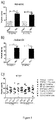
- Figure 7 shows changes in the expression of C03-610C in the liver of rats in BDL- and sham-operated groups assessed by A) Western blot 2 and 4 weeks post-surgery and B) Bands from western blot quantified by densitometry.
- liver fibrosis is not only an accumulation of ECM proteins, but also an accelerated turnover situation, in which both tissue formation and tissue degradation both are highly up regulated. Tissue formation outstrips tissue degradation, leading to an accumulation of scar tissue over time.
- Other matrix turnover proteins to assess liver fibrosis, one being the type III collagen formation marker N-terminal type III pro-collagen. This marker represents collagen type III formation and has shown to be increased in liver fibrosis in previous studies.
- Example 6 ELISA on human serum samples
- mice were immunized with synthetic peptide KAFVFP (SEQ ID N01167) conjugated to ovalbumin (KAFVFPKESD-GGC-OVA (SEQ ID N01049)), spleen cells were used for fusion, and monoclonal antibodies tested for reactivity to biotinylated KAFVFP (SEQ ID NO 1167), i.e. (KAFVFPKESD-biotin(SEQ ID N01049)) immobilized in wells of microtitre plates precoated with streptavidin.
- KAFVFPKESD-GGC-OVA SEQ ID N01049
- mice were classified into 4 different stages of fibrosis and cirrhosis (Group A: moderate fibrosis, group B: advanced fibrosis, Group C: moderate cirrhosis, and Group D: advanced cirrhosis) that were determined by the percentage of Sirius red positive liver area (Group A: ⁇ 5%, Group B: 5 to10%, Group C: 10 to 15% and Group D: >15%).
- control and fibrotic/cirrhotic rats were studied considering four different time points during the CCl4 treatment: 8, 12, 16 and 20 weeks after starting the cirrhosis induction program.
- Serum hyaluronan was measured using a sandwich ELISA kit (R&D Systems Inc., Minneapolis, MN, USA).
- mice included in this protocol were randomly assigned to one of the following groups: A/ eight weeks of CCl 4 treatment, B/ twelve weeks of CCl 4 treatment, C/ sixteen weeks of CCl 4 treatment and D/ twenty weeks of CCl 4 treatment.
- four control groups were studied at the same time points. Thirteen fibrotic rats and seven control rats were included in each group. At the end of the study, rats were placed in standard metabolic cages (Tecniplast GmbH, Hohenpeissenberg, Germany) during an adaptation period of 3 days before proceeding with the twenty-four-hour urine collection. Urinary volumes were determined gravimetrically. During the adaptation period, rats were allowed to get free access to tap water and food. Then, 24-hour urine samples were centrifuged for 5 min at 2,500 rpm and aliquoted into ten polypropylene tubes (400 ⁇ L each). Urine samples were stored at -80°C for subsequent analysis.
- rats were weighed, anesthetized with pentobarbital (50 mg/kl) and decapitated. Blood were collected and allowed to stand at room temperature for 20 min to allow clotting and then centrifuged for 10 min at 2500 rpm. Serum were collected in polypropylene tubes aliquots (400 ⁇ l each) and transferred via dry ice to a -80°C freezer. Collection of baseline blood samples at the beginning of the CCl 4 treatment was not considered in order to avoid additional intervention that may increase the risk of infection and/or introduce modifications in the experimental model that may compromise the evolution of the induced pathophysiological process.
- liver fibrotic area For histology and Sirius red staining, half of the left lobe of the liver were placed in 10% neutral buffered formalin for 16 hours, embedded in paraffin and sectioned into 4- ⁇ m-thick slices. After liver fibrosis quantification, the unused paraffin block material was preserved for biomarker quantification. The other half of the left lobe was flash-frozen in liquid nitrogen and stored for Western blot, RT-PCR or immunohistochemical analysis. Measurements of liver fibrotic area, serum and urine osmolality, Na + and K + , albumin, creatinine, alanine amino-transferase and lactate dehydrogenase were made according to the Material and Methods section.
- Liver collagen was quantified in all study animals by Sirius red staining of liver slices. The final data for each animal was taken as the average of red staining observed in 36 consecutive microscope fields ( Figure 12 ).
- Figure 12 shows representative pictures from two sets of 36 images used to quantify collagen accumulation in liver in rat #1 (left) and rat #43 (right) treated with carbon tetrachloride for eight and twenty weeks respectively.
- the serum C03 marker shows statistically significant increases in both fibrotic and cirrhotic rats compared to control rats. Animals were classified according to a fully automated syrius red staining of the liver procedure used to quantify fibrosis ( Figures 13 and 14 ).
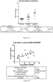
- Figure 13 shows serum C03 levels in CCl 4 inhalation and control rats as performed in Hospital Clinic (Barcelona). Each point represents one animal. Rats were classified according a computerized image analysis method of syrius red staining of the liver used to quantify fibrosis.
- HA levels were quantified with a commercial ELISA kit and results show significant elevations of this ECM component in cirrhotic rats vs. fibrotic animals ( Figures 15 and 16 ).
- Example 9 Bleomycin induced skin fibrosis in mice.
- mice were treated by application to the skin of PBS or bleomycin. Increasing levels in urine of the MMP-9 mediated collagen III (CO3) degradation fragment C03-610 were associated with skin fibrosis progression in mice.
- CO3 MMP-9 mediated collagen III
- Figure 18 shows a skin section from a PBS treated mouse at 8 weeks of treatment (panel A) and a skin section from Bleomycin treated mouse at 8 weeks of treatment (panel B).
- Skin width was calculated by Visiopharm software as an overall number per skin section instead of sampling pictures.
- Figure 19 shows C03-610 urine assay results which demonstrate a significant increase throughout the time points of the study.
- 2 weeks P 0.0008, 4 weeks P ⁇ 0.0001, 6 weeks P ⁇ 0.0001, 8 weeks P ⁇ 0.0001 and overall P ⁇ 0.0001.
- Figure 20 shows a C03-610 Western blots image with control C and Bleomycin B after 2 and 8 weeks treatment (panel A).
Landscapes
- Life Sciences & Earth Sciences (AREA)
- Health & Medical Sciences (AREA)
- Chemical & Material Sciences (AREA)
- Engineering & Computer Science (AREA)
- Immunology (AREA)
- Molecular Biology (AREA)
- Proteomics, Peptides & Aminoacids (AREA)
- Organic Chemistry (AREA)
- Hematology (AREA)
- Urology & Nephrology (AREA)
- Biomedical Technology (AREA)
- Biotechnology (AREA)
- Microbiology (AREA)
- Physics & Mathematics (AREA)
- Analytical Chemistry (AREA)
- Biochemistry (AREA)
- General Health & Medical Sciences (AREA)
- Wood Science & Technology (AREA)
- Zoology (AREA)
- Medicinal Chemistry (AREA)
- Pathology (AREA)
- General Physics & Mathematics (AREA)
- Food Science & Technology (AREA)
- Cell Biology (AREA)
- Biophysics (AREA)
- Bioinformatics & Cheminformatics (AREA)
- General Engineering & Computer Science (AREA)
- Genetics & Genomics (AREA)
- Peptides Or Proteins (AREA)
- Measuring Or Testing Involving Enzymes Or Micro-Organisms (AREA)
- Medicines That Contain Protein Lipid Enzymes And Other Medicines (AREA)
Priority Applications (2)
| Application Number | Priority Date | Filing Date | Title |
|---|---|---|---|
| EP24200645.0A EP4467984A3 (de) | 2009-03-30 | 2010-03-29 | Fibrosebiomarkertest |
| EP19174151.1A EP3578664B1 (de) | 2009-03-30 | 2010-03-29 | Collagen iv neo-epitopfibrose-assay |
Applications Claiming Priority (5)
| Application Number | Priority Date | Filing Date | Title |
|---|---|---|---|
| US21146709P | 2009-03-30 | 2009-03-30 | |
| US28908109P | 2009-12-22 | 2009-12-22 | |
| EP10715752.1A EP2414844B1 (de) | 2009-03-30 | 2010-03-29 | Biomarker für fibrose |
| PCT/EP2010/054096 WO2010115749A2 (en) | 2009-03-30 | 2010-03-29 | Fibrosis biomarker assay |
| EP14169636.9A EP2770327B1 (de) | 2009-03-30 | 2010-03-29 | Fibrosebiomarkertest |
Related Parent Applications (3)
| Application Number | Title | Priority Date | Filing Date |
|---|---|---|---|
| EP14169636.9A Division-Into EP2770327B1 (de) | 2009-03-30 | 2010-03-29 | Fibrosebiomarkertest |
| EP14169636.9A Division EP2770327B1 (de) | 2009-03-30 | 2010-03-29 | Fibrosebiomarkertest |
| EP10715752.1A Division EP2414844B1 (de) | 2009-03-30 | 2010-03-29 | Biomarker für fibrose |
Related Child Applications (2)
| Application Number | Title | Priority Date | Filing Date |
|---|---|---|---|
| EP19174151.1A Division EP3578664B1 (de) | 2009-03-30 | 2010-03-29 | Collagen iv neo-epitopfibrose-assay |
| EP24200645.0A Division EP4467984A3 (de) | 2009-03-30 | 2010-03-29 | Fibrosebiomarkertest |
Publications (3)
| Publication Number | Publication Date |
|---|---|
| EP3173792A2 true EP3173792A2 (de) | 2017-05-31 |
| EP3173792A3 EP3173792A3 (de) | 2017-07-05 |
| EP3173792B1 EP3173792B1 (de) | 2019-06-12 |
Family
ID=42199024
Family Applications (5)
| Application Number | Title | Priority Date | Filing Date |
|---|---|---|---|
| EP16205956.2A Active EP3173792B1 (de) | 2009-03-30 | 2010-03-29 | Crp neo-epitop fibrose-assay |
| EP24200645.0A Pending EP4467984A3 (de) | 2009-03-30 | 2010-03-29 | Fibrosebiomarkertest |
| EP19174151.1A Active EP3578664B1 (de) | 2009-03-30 | 2010-03-29 | Collagen iv neo-epitopfibrose-assay |
| EP10715752.1A Active EP2414844B1 (de) | 2009-03-30 | 2010-03-29 | Biomarker für fibrose |
| EP14169636.9A Active EP2770327B1 (de) | 2009-03-30 | 2010-03-29 | Fibrosebiomarkertest |
Family Applications After (4)
| Application Number | Title | Priority Date | Filing Date |
|---|---|---|---|
| EP24200645.0A Pending EP4467984A3 (de) | 2009-03-30 | 2010-03-29 | Fibrosebiomarkertest |
| EP19174151.1A Active EP3578664B1 (de) | 2009-03-30 | 2010-03-29 | Collagen iv neo-epitopfibrose-assay |
| EP10715752.1A Active EP2414844B1 (de) | 2009-03-30 | 2010-03-29 | Biomarker für fibrose |
| EP14169636.9A Active EP2770327B1 (de) | 2009-03-30 | 2010-03-29 | Fibrosebiomarkertest |
Country Status (8)
| Country | Link |
|---|---|
| US (3) | US9206464B2 (de) |
| EP (5) | EP3173792B1 (de) |
| JP (2) | JP5978128B2 (de) |
| KR (1) | KR101700463B1 (de) |
| CN (1) | CN103238071B (de) |
| DK (3) | DK3173792T3 (de) |
| ES (2) | ES2529101T3 (de) |
| WO (1) | WO2010115749A2 (de) |
Families Citing this family (52)
| Publication number | Priority date | Publication date | Assignee | Title |
|---|---|---|---|---|
| WO2008054764A2 (en) * | 2006-10-31 | 2008-05-08 | George Mason Intellectual Properties, Inc. | Biomarkers for colorectal cancer |
| US9404932B2 (en) | 2007-11-05 | 2016-08-02 | Nordic Bioscience A/S | Pathology biomarker assay |
| EP2538222B1 (de) * | 2007-11-05 | 2021-08-25 | Nordic Bioscience A/S | Biochemische Marker zur Bewertung des CVD-Risikos |
| ES2529101T3 (es) | 2009-03-30 | 2015-02-16 | Nordic Bioscience A/S | Ensayo de biomarcadores para fibrosis |
| WO2012138223A2 (en) | 2011-04-05 | 2012-10-11 | Academisch Ziekenhuis Leiden H.O.D.N. Lumc | Compounds and methods for altering activin receptor-like kinase signalling |
| GB201107922D0 (en) * | 2011-05-12 | 2011-06-22 | Nordic Bioscience As | Biochemical markers for CVD risk assessment |
| WO2012163768A1 (en) * | 2011-05-27 | 2012-12-06 | Nordic Bioscience A/S | Detection of diagnostic peptides |
| EP2727520B1 (de) * | 2011-06-29 | 2019-08-21 | Inner Mongolia Furui Medical Science Co., Ltd | Vorrichtung und system zum nachweis von leberfibrose |
| GB201111361D0 (en) * | 2011-07-04 | 2011-08-17 | Nordic Bioscience As | Biochemical markers for neurodegenerative conditions |
| GB201111788D0 (en) * | 2011-07-11 | 2011-08-24 | Nordic Bioscience As | In vitro assessment of cardiovascular events by assay for neo-epitopes |
| JP5953035B2 (ja) * | 2011-11-28 | 2016-07-13 | 学校法人慶應義塾 | 病理診断支援装置、病理診断支援方法、及び病理診断支援プログラム |
| EP2839292A4 (de) * | 2012-04-19 | 2015-08-26 | Univ California | Zusammensetzungen und verfahren zum nachweis von instabiler arteriosklerotischer plaque |
| GB201306792D0 (en) * | 2013-04-15 | 2013-05-29 | Nordic Bioscience As | PIIINP Neo-epitope assay |
| GB201307387D0 (en) | 2013-04-24 | 2013-06-05 | Nordic Bioscience As | Collegen type VI alpha-1 assay |
| GB201400472D0 (en) | 2014-01-13 | 2014-02-26 | Nordic Bioscience As | Biochemical Markers for pulmonary and other diseases |
| GB201505654D0 (en) | 2015-04-01 | 2015-05-13 | Nordic Bioscience As | Immunoassay for collagen type VI sequence |
| US20170165447A1 (en) * | 2015-12-11 | 2017-06-15 | Geno Llc | Method and apparatus for administering gases including nitric oxide |
| IL322971A (en) | 2015-12-15 | 2025-10-01 | Takeda Pharmaceuticals Co | Method for evaluating the efficacy of a drug for a plasma kallikrein-mediated disorder |
| GB201601571D0 (en) * | 2016-01-28 | 2016-03-16 | Nordic Bioscience As | Collagen type Vll alpha 1 assay |
| KR102668479B1 (ko) * | 2016-02-03 | 2024-05-22 | 노르딕 바이오사이언스 에이/에스 | 섬유증에 대한 조합된 바이오마커 측정법 |
| CN105738632B (zh) * | 2016-02-24 | 2017-06-23 | 浙江理工大学 | 一种区分羊皮种类的检测方法 |
| CN109477127A (zh) * | 2016-03-14 | 2019-03-15 | R基因股份有限公司 | 超热稳定的赖氨酸-突变型ssDNA/RNA连接酶 |
| WO2017210147A1 (en) * | 2016-05-29 | 2017-12-07 | Wei Jia | Liver disease-related biomarkers and methods of use thereof |
| CN111183360B (zh) * | 2017-07-19 | 2024-10-18 | 生物辐射欧洲有限公司 | 同时评估非酒精性脂肪性肝炎和肝纤维化状态的生物标志物组合 |
| GB201717301D0 (en) * | 2017-10-20 | 2017-12-06 | Nordic Bioscience As | Type xvi collagen assay |
| US20190351031A1 (en) | 2018-05-16 | 2019-11-21 | Halozyme, Inc. | Methods of selecting subjects for combination cancer therapy with a polymer-conjugated soluble ph20 |
| US11732009B2 (en) | 2018-06-08 | 2023-08-22 | Glympse Bio, Inc. | Activity sensor with tunable analyte |
| US11028425B2 (en) * | 2018-06-08 | 2021-06-08 | Glympse Bio, Inc. | Diagnosis and monitoring of liver disease |
| EP4007920A4 (de) * | 2019-02-01 | 2024-07-17 | Glympse Bio, Inc. | Körperliche verarbeitung von aktivitätssensoren |
| US12338483B2 (en) | 2019-02-28 | 2025-06-24 | Georgia Tech Research Corporation | Compositions and methods for logic-gated profiling of biologic activity |
| TW202104254A (zh) * | 2019-04-12 | 2021-02-01 | 美商格爾托公司 | 重組彈性蛋白及其生產 |
| EP4009953A4 (de) * | 2019-08-07 | 2023-08-23 | Icahn School of Medicine at Mount Sinai | Verfahren zur behandlung von keloiden |
| WO2021034196A1 (en) * | 2019-08-20 | 2021-02-25 | Erasmus University Medical Center Rotterdam | Biomarkers for detecting secondary liver cancer |
| GB201917819D0 (en) | 2019-12-05 | 2020-01-22 | Nordic Bioscience As | A neo-epitope specific assay measuring protease mediated degradation of type iv collagen |
| CA3162431A1 (en) * | 2020-01-21 | 2021-07-29 | Augustinus Bader | Cosmetic formulation for topical administration comprising novel peptides that improve appearance and regeneration of skin |
| AU2021211721A1 (en) | 2020-01-24 | 2022-08-18 | Geltor, Inc. | Animal-free dietary collagen |
| US20240003906A1 (en) * | 2020-04-16 | 2024-01-04 | Nordic Bioscience A/S | Biomarker of Fibrosis |
| IL274696A (en) * | 2020-05-14 | 2021-12-01 | Yeda Res & Dev | Designed transmembrane polypeptides and their use in cellular immunotherapy |
| CN111705123A (zh) * | 2020-06-19 | 2020-09-25 | 上海市第六人民医院 | 一种衰老过程中心肌纤维化的生物标志物及其检测方法和应用 |
| JP2023535022A (ja) * | 2020-07-21 | 2023-08-15 | アムニクス ファーマシューティカルズ, インコーポレイテッド | 活性化可能な治療剤に関する組成物および方法 |
| CN116438456A (zh) | 2020-09-11 | 2023-07-14 | 格里姆普斯生物股份有限公司 | 用于疾病检测/诊断、分期、监测和治疗的离体蛋白酶活性检测 |
| CN112876556B (zh) * | 2021-03-23 | 2022-07-19 | 中国科学院理化技术研究所 | 一种促进成骨细胞矿化的牛骨胶原肽及其应用 |
| CN113388005B (zh) * | 2021-06-30 | 2022-09-27 | 北京泽勤生物医药有限公司 | 增强成骨细胞活性的多肽及其在治疗骨科疾病中的应用 |
| US20250138026A1 (en) * | 2022-02-01 | 2025-05-01 | Institut National de la Santé et de la Recherche Médicale | Method for diagnosing collagen degradation associated disease |
| CN114994317B (zh) * | 2022-04-15 | 2025-10-28 | 宁波市杭州湾医院 | 金属蛋白酶adamts4在制备早期肝纤维化药物中的应用 |
| CN115991732B (zh) * | 2022-11-21 | 2024-12-24 | 北海黑珍珠海洋生物科技有限公司 | 一种具有抗炎作用的珍珠贝活性肽及其应用 |
| GB202302820D0 (en) * | 2023-02-27 | 2023-04-12 | Keybioscience Ag | ProC3 and TGFBI in cancer |
| CN117343132B (zh) * | 2023-09-25 | 2024-03-12 | 山东省农业科学院 | 一种三文鱼鱼皮来源的ace抑制肽及制备方法 |
| WO2025115009A1 (en) * | 2023-11-28 | 2025-06-05 | Accurine Ltd. | Peptides for urinary diagnostics and antibodies for their detection |
| CN118146301B (zh) * | 2024-01-04 | 2024-12-31 | 广州白云山花城药业有限公司 | 一种促成骨细胞分化的活性肽及其应用 |
| GB202408410D0 (en) * | 2024-06-12 | 2024-07-24 | Nordic Bioscience As | immunoassay for detecting canstatin |
| GB202413581D0 (en) | 2024-09-16 | 2024-10-30 | Nordic Bioscience As | Immunoassay for detecting collagen i fragment |
Citations (5)
| Publication number | Priority date | Publication date | Assignee | Title |
|---|---|---|---|---|
| US4628027A (en) | 1982-05-19 | 1986-12-09 | Molecular Engineering Associates, Ltd. | Vitro diagnostic methods using monoclonal antibodies against connective tissue proteins |
| US5387504A (en) | 1992-09-30 | 1995-02-07 | Merck & Co., Inc. | Monospecific antibodies and assay system for detecting stromelysin cleavage products |
| US6060255A (en) | 1997-10-03 | 2000-05-09 | Tosoh Corporation | Type IV collagen high molecular form and production and diagnostic use thereof |
| US7225080B2 (en) | 2000-08-21 | 2007-05-29 | Assistance Publique-Hopitaux De Paris (Ap-Hp) | Diagnosis method of inflammatory, fibrotic or cancerous disease using biochemical markers |
| WO2009059972A2 (en) | 2007-11-05 | 2009-05-14 | Nordic Bioscience A/S | Biochemical markers for cvd risk assessment |
Family Cites Families (26)
| Publication number | Priority date | Publication date | Assignee | Title |
|---|---|---|---|---|
| GB2208865B (en) * | 1987-08-19 | 1991-12-11 | Farmos Group Ltd | Purified propeptide of procollagen type iii, antibody to the propeptide and assay method using the antibody. |
| US5962639A (en) * | 1987-11-06 | 1999-10-05 | Washington Research Foundation | Synthetic peptides corresponding to telopeptide sequences of cross-linked type I collagen metabolites |
| US6124107A (en) * | 1988-06-10 | 2000-09-26 | Merck & Co., Inc. | Assay for marker of human polymorphonuclear leukocyte elastase activity |
| JPH04505857A (ja) * | 1989-06-27 | 1992-10-15 | ラッシュ―プレズビティリアン―セント ルークス メディカル センター | C反応性蛋白質に対するモノクローナル抗体 |
| US6703219B1 (en) * | 1993-02-26 | 2004-03-09 | Immtech International Inc. | Mutant protein and methods and materials for making and using it |
| US6110689A (en) * | 1994-01-21 | 2000-08-29 | Osteometer A/S | Method of assaying collagen fragments in body fluids, a test kit and means for carrying out the method and use of the method to diagnose the presence of disorders associated with the metabolism of collagen |
| JPH08100000A (ja) * | 1994-09-30 | 1996-04-16 | Morinaga & Co Ltd | ヒトiv型コラーゲンに対する抗体及びその利用 |
| EP0718309B1 (de) * | 1994-12-23 | 1999-04-07 | Roche Diagnostics GmbH | Antigene und Antikörper zum Nachweis von Kollagen I |
| EP0913692A1 (de) | 1997-10-31 | 1999-05-06 | Bayer Ag | Immunassay zum Nachweis von prokollagen-III-C-terminalen Propeptid |
| US7541149B1 (en) | 1998-05-28 | 2009-06-02 | Siemens Healthcare Diagnostics, Inc. | Monoclonal antibody and assay for detecting PIIINP |
| US6916903B2 (en) * | 1998-06-19 | 2005-07-12 | Washington Research Foundation | Collagen type III synthetic peptides for collagen resorption assays |
| EP1150123B1 (de) * | 2000-04-28 | 2006-07-12 | Bayer Aktiengesellschaft | Diagnose von Leberfibrose mit Hilfe von Serummarkeralgorithmen |
| JP2002065283A (ja) | 2000-06-12 | 2002-03-05 | Wako Pure Chem Ind Ltd | ハイブリッド酵素およびその用途 |
| AU2002251366A1 (en) | 2001-05-02 | 2002-11-11 | Oxford Glycosciences (Uk) Ltd | Proteins, genes and their use for diagnosis and treatment of breast cancer |
| US7390885B2 (en) * | 2001-11-26 | 2008-06-24 | Cell Matrix, Inc. | Humanized collagen antibodies and related methods |
| NZ546611A (en) * | 2003-09-18 | 2010-02-26 | Conforma Therapeutics Corp | Novel heterocyclic compounds as HSP90-inhibitors |
| WO2005050224A2 (en) | 2003-11-13 | 2005-06-02 | Epitome Biosystems Inc. | Small molecule and peptide arrays and uses thereof |
| US20060154318A1 (en) | 2004-06-09 | 2006-07-13 | Anderson Norman L | Stable isotope labeled polypeptide standards for protein quantitation |
| US7358370B2 (en) * | 2005-02-25 | 2008-04-15 | Serenex, Inc. | Benzene, pyridine, and pyridazine derivatives |
| EP1948686A4 (de) | 2005-10-26 | 2009-06-10 | Stc Unm | C-reaktives protein und dessen verwendung zur behandlung von systemischem lupuserythematodes und verwandten leiden |
| US7972802B2 (en) * | 2005-10-31 | 2011-07-05 | University Of Washington | Lipoprotein-associated markers for cardiovascular disease |
| US9404932B2 (en) * | 2007-11-05 | 2016-08-02 | Nordic Bioscience A/S | Pathology biomarker assay |
| US20100023377A1 (en) * | 2008-07-23 | 2010-01-28 | Hr Solutions, Inc. | Systems and methods for managing human resource activities |
| ES2529101T3 (es) | 2009-03-30 | 2015-02-16 | Nordic Bioscience A/S | Ensayo de biomarcadores para fibrosis |
| WO2012163768A1 (en) * | 2011-05-27 | 2012-12-06 | Nordic Bioscience A/S | Detection of diagnostic peptides |
| GB201111788D0 (en) * | 2011-07-11 | 2011-08-24 | Nordic Bioscience As | In vitro assessment of cardiovascular events by assay for neo-epitopes |
-
2010
- 2010-03-29 ES ES10715752.1T patent/ES2529101T3/es active Active
- 2010-03-29 CN CN201080014281.6A patent/CN103238071B/zh active Active
- 2010-03-29 EP EP16205956.2A patent/EP3173792B1/de active Active
- 2010-03-29 KR KR1020117025776A patent/KR101700463B1/ko active Active
- 2010-03-29 JP JP2012502618A patent/JP5978128B2/ja not_active Expired - Fee Related
- 2010-03-29 DK DK16205956.2T patent/DK3173792T3/da active
- 2010-03-29 EP EP24200645.0A patent/EP4467984A3/de active Pending
- 2010-03-29 EP EP19174151.1A patent/EP3578664B1/de active Active
- 2010-03-29 DK DK14169636.9T patent/DK2770327T3/en active
- 2010-03-29 EP EP10715752.1A patent/EP2414844B1/de active Active
- 2010-03-29 ES ES14169636.9T patent/ES2635494T3/es active Active
- 2010-03-29 WO PCT/EP2010/054096 patent/WO2010115749A2/en not_active Ceased
- 2010-03-29 EP EP14169636.9A patent/EP2770327B1/de active Active
- 2010-03-29 DK DK10715752T patent/DK2414844T3/en active
- 2010-03-30 US US12/749,652 patent/US9206464B2/en active Active
-
2015
- 2015-01-22 JP JP2015010255A patent/JP6095702B2/ja active Active
- 2015-12-07 US US14/960,713 patent/US9606130B2/en active Active
-
2017
- 2017-03-01 US US15/446,070 patent/US9880177B2/en active Active
Patent Citations (5)
| Publication number | Priority date | Publication date | Assignee | Title |
|---|---|---|---|---|
| US4628027A (en) | 1982-05-19 | 1986-12-09 | Molecular Engineering Associates, Ltd. | Vitro diagnostic methods using monoclonal antibodies against connective tissue proteins |
| US5387504A (en) | 1992-09-30 | 1995-02-07 | Merck & Co., Inc. | Monospecific antibodies and assay system for detecting stromelysin cleavage products |
| US6060255A (en) | 1997-10-03 | 2000-05-09 | Tosoh Corporation | Type IV collagen high molecular form and production and diagnostic use thereof |
| US7225080B2 (en) | 2000-08-21 | 2007-05-29 | Assistance Publique-Hopitaux De Paris (Ap-Hp) | Diagnosis method of inflammatory, fibrotic or cancerous disease using biochemical markers |
| WO2009059972A2 (en) | 2007-11-05 | 2009-05-14 | Nordic Bioscience A/S | Biochemical markers for cvd risk assessment |
Non-Patent Citations (110)
| Title |
|---|
| "Peducing Risks, Promoting Healthy Life", 2002, WORLD HEALTH ORGANIZATION, article "Reducing Risks, Promoting Healthy Life", pages: 1 - 230 |
| ACHARYA PS; ZUKAS A; CHANDAN V; KATZENSTEIN AL; PURE E.: "Fibroblast activation protein: a serine protease expressed at the remodeling interface in idiopathic pulmonary fibrosis.", HUM PATHOL, vol. 37, 2006, pages 352 - 360, XP005301569, DOI: doi:10.1016/j.humpath.2005.11.020 |
| ADAMS LA; BULSARA M; ROSSI E; DEBOER B; SPEERS D; GEORGE J; KENCH J; FARRELL G; MCCAUGHAN GW; JEFFREY GP.: "Hepascore: an accurate validated predictor of liver fibrosis in chronic hepatitis C infection", CLIN CHEM, vol. 51, 2005, pages 1867 - 1873 |
| AL-MOHRI H; COOPER C; MURPHY T; KLEIN MB.: "Validation of a simple model for predicting liver fibrosis in HIV/hepatitis C virus-coinfected patients.", HIV MED, vol. 6, 2005, pages 375 - 378 |
| ATTALLAH AM; TOSON EA; SHIHA GE; OMRAN MM; BDEL-AZIZ MM; EL-DOSOKY I.: "Evaluation of serum procollagen aminoterminal propeptide III, laminin, and hydroxyproline as predictors of severe fibrosis in patients with chronic hepatitis C", J IMMUNOASSAY IMMUNOCHEM, vol. 28, 2007, pages 199 - 211 |
| BARTLETT AH; HAYASHIDA K; PARK PW.: "Molecular and cellular mechanisms of syndecans in tissue injury and inflammation.", MOL CELLS, vol. 24, 2007, pages 153 - 166 |
| BENYON RC; ARTHUR MJ.: "Extracellular matrix degradation and the role of hepatic stellate cells.", SEMIN LIVER DIS, vol. 21, 2001, pages 373 - 384 |
| BLOCHBERGER TC; CORNUET PK; HASSELL JR.: "Isolation and partial characterization of lumican and decorin from adult chicken corneas. A keratan sulfate-containing isoform of decorin is developmentally regulated.", J BIOL CHEM, vol. 267, 1992, pages 20613 - 20619 |
| BOURLIERE M; PENARANDA G; RENOU C; BOTTA-FRIDLUND D; TRAN A; PORTAL I; LECOMTE L; CASTELLANI P; ROSENTHAL-ALLIERI MA; GEROLAMI R: "Validation and comparison of indexes for fibrosis and cirrhosis prediction in chronic hepatitis C patients: proposal for a pragmatic approach classification without liver biopsies.", J VIRAL HEPAT, vol. 13, 2006, pages 659 - 670 |
| CACOUB P; CARRAT F; BEDOSSA P; LAMBERT J; PENARANDA G; PERRONNE C; POL S; HALFON P.: "Comparison of non-invasive liver fibrosis biomarkers in HIV/HCV co-infected patients: the fibrovic study--ANRS HC02.", J HEPATOL, vol. 48, 2008, pages 765 - 773, XP022627179, DOI: doi:10.1016/j.jhep.2008.01.025 |
| CALES P; LAINE F; BOURSIER J; DEUGNIER Y; MOAL V; OBERTI F; HUNAULT G; ROUSSELET MC; HUBERT I; LAAFI J: "Comparison of blood tests for liver fibrosis specific or not to NAFLD", J HEPATOL, 2008 |
| CAMACHO VR; SILVEIRA TR; OLIVEIRA JR; BARROS SG; CERSKI CT.: "Relationship between serum concetrations of type III procollagen, hyluronic acid and histopathological findings in the liver of HCV-positive blood donors.", ARQ GASTROENTEROL, vol. 44, 2007, pages 118 - 122 |
| CARVALHO-FILHO RJ; SCHIAVON LL; NARCISO-SCHIAVON JL; SAMPAIO JP; LANZONI VP; FERRAZ ML; SILVA AE.: "Optimized cutoffs improve performance of the aspartate aminotransferase to platelet ratio index for predicting significant liver fibrosis in human immunodeficiency virus/hepatitis C virus co-infection.", LIVER INT, vol. 28, 2008, pages 486 - 493 |
| CASTERA L; VERGNIOL J; FOUCHER J; LE BB; CHANTELOUP E; HAASER M; DARRIET M; COUZIGOU P; DE L, V.: "Prospective comparison of transient elastography, Fibrotest, APRI, and liver biopsy for the assessment of fibrosis in chronic hepatitis C.", GASTROENTEROLOGY, vol. 128, 2005, pages 343 - 350, XP005313375, DOI: doi:10.1053/j.gastro.2004.11.018 |
| FARKKILA M; RAUTIAINEN H; KARKKAINEN P; KARVONEN AL; NURMI H; NIEMELA 0.: "Serological markers for monitoring disease progression in noncirrhotic primary biliary cirrhosis on ursodeoxycholic acid therapy.", LIVER INT, vol. 28, 2008, pages 787 - 797 |
| FISHER LW; TERMINE JD; YOUNG MF.: "Deduced protein sequence of bone small proteoglycan I (biglycan) shows homology with proteoglycan II (decorin) and several nonconnective tissue proteins in a variety of species", J BIOL CHEM, vol. 264, 1989, pages 4571 - 4576, XP055169896 |
| FORNS X; AMPURDANES S; LLOVET JM; APONTE J; QUINTO L; MARTINEZ-BAUER E; BRUGUERA M; SANCHEZ-TAPIAS JM; RODES J.: "Identification of chronic hepatitis C patients without hepatic fibrosis by a simple predictive model.", HEPATOLOGY, vol. 36, 2002, pages 986 - 992 |
| FRIEDMAN SL.: "Mechanisms of disease: Mechanisms of hepatic fibrosis and therapeutic implications", NAT CLIN PRACT GASTROENTEROL HEPATOL, vol. 1, 2004, pages 98 - 105 |
| GABAY C; KUSHNER I.: "Acute-phase proteins and other systemic responses to inflammation", N ENGL J MED, vol. 340, 1999, pages 448 - 454, XP008003139, DOI: doi:10.1056/NEJM199902113400607 |
| GAGLIANO N; AROSIO B; GRIZZI F; MASSON S; TAGLIABUE J; DIOGUARDI N; VERGANI C; ANNONI G.: "Reduced collagenolytic activity of matrix metalloproteinases and development of liver fibrosis in the aging rat.", MECH AGEING DEV, vol. 123, 2002, pages 413 - 425 |
| GARRONE R; LETHIAS C; LE GUELLEC D.: "Distribution of minor collagens during skin development.", MICROSC RES TECH, vol. 38, 1997, pages 407 - 412 |
| GELSE K; POSCHL E; AIGNER T.: "Collagens--structure, function, and biosynthesis.", ADV DRUG DELIV REV, vol. 55, 2003, pages 1531 - 1546 |
| GILLIAM AC.: "Scleroderma", CURR DIR AUTOIMMUN, vol. 10, 2008, pages 258 - 279 |
| GRESSNER AM; WEISKIRCHEN R.: "Modern pathogenetic concepts of liver fibrosis suggest stellate cells and TGF-beta as major players and therapeutic targets", J CELL MOL MED, vol. 10, 2006, pages 76 - 99 |
| GRESSNER OA; WEISKIRCHEN R; GRESSNER AM.: "Biomarkers of liver fibrosis: clinical translation of molecular pathogenesis or based on liver-dependent malfunction tests.", CLIN CHIM ACTA, vol. 381, 2007, pages 107 - 113, XP022072413, DOI: doi:10.1016/j.cca.2007.02.038 |
| GRESSNER OA; WEISKIRCHEN R; GRESSNER AM: "Biomarkers of hepatic fibrosis, fibrogenesis and genetic pre-disposition pending between fiction and reality", J CELL MOL MED, vol. 11, 2007, pages 1031 - 1051 |
| GRIGORESCU M; RUSU M; NECULOIU D; RADU C; SERBAN A; CATANAS M; GRIGORESCU MD.: "The FibroTest value in discriminating between insignificant and significant fibrosis in chronic hepatitis C patients. The Romanian experience", J GASTROINTESTIN LIVER DIS, vol. 16, 2007, pages 31 - 37 |
| GUANABENS N ET AL., J BONE MINER RES, 1998 |
| GUANABENS N; PARES A; ALVAREZ L; MARTINEZ DE OSABA MJ; MONEGAL A; PERIS P; BALLESTA AM; RODES J.: "Collagen-related markers of bone turnover reflect the severity of liver fibrosis in patients with primary biliary cirrhosis.", J BONE MINER RES, vol. 13, 1998, pages 731 - 738 |
| GUECHOT J; POUPON RE; GIRAL P; BALKAU B; GIBOUDEAU J; POUPON R.: "Relationship between procollagen III aminoterminal propeptide and hyaluronan serum levels and histological fibrosis in primary biliary cirrhosis and chronic viral hepatitis C", J HEPATOL, vol. 20, 1994, pages 388 - 393, XP029122232, DOI: doi:10.1016/S0168-8278(94)80013-8 |
| GUO J; FRIEDMAN SL.: "Hepatic fibrogenesis.", SEMIN LIVER DIS, vol. 27, 2007, pages 413 - 426 |
| HALFON P; BACQ Y; DE MA; PENARANDA G; BOURLIERE M; OUZAN D; TRAN A; BOTTA D; RENOU C; BRECHOT MC: "Comparison of test performance profile for blood tests of liver fibrosis in chronic hepatitis C.", J HEPATOL, vol. 46, 2007, pages 395 - 402, XP022040950, DOI: doi:10.1016/j.jhep.2006.09.020 |
| HALFON P; BOURLIERE M; DEYDIER R; BOTTA-FRIDLUND D; RENOU C; TRAN A; PORTAL I; ALLEMAND I; BERTRAND JJ; ROSENTHAL-ALLIERI A: "Independent prospective multicenter validation of biochemical markers (fibrotest-actitest) for the prediction of liver fibrosis and activity in patients with chronic hepatitis C: the fibropaca study", AM J GASTROENTEROL, vol. 101, 2006, pages 547 - 555 |
| HEINEGARD D; OLDBERG A.: "Structure and biology of cartilage and bone matrix noncollagenous macromolecules.", FASEB J, vol. 3, 1989, pages 2042 - 2051 |
| HERTIG ET AL., J AM SOC NEPHROL., vol. 19, no. 8, August 2008 (2008-08-01), pages 1584 - 91 |
| HONGBO L; XIAOHUI L; HONG K; WEI W; YONG Z.: "Assessing routine and serum markers of liver fibrosis in CHB patients using parallel and serial interpretation.", CLIN BIOCHEM, vol. 40, 2007, pages 562 - 566, XP022036491, DOI: doi:10.1016/j.clinbiochem.2007.01.022 |
| HUMMERS LK.: "Microvascular damage in systemic sclerosis: detection and monitoring with biomarkers.", CURR RHEUMATOL REP, vol. 8, 2006, pages 131 - 137 |
| IMBERT-BISMUT F; RATZIU V; PIERONI L; CHARLOTTE F; BENHAMOU Y; POYNARD T.: "Biochemical markers of liver fibrosis in patients with hepatitis C virus infection: a prospective study", LANCET, vol. 357, 2001, pages 1069 - 1075, XP005061312, DOI: doi:10.1016/S0140-6736(00)04258-6 |
| IREDALE JP; BENYON RC; ARTHUR MJ; FERRIS WF; ALCOLADO R; WINWOOD PJ; CLARK N; MURPHY G.: "Tissue inhibitor of metalloproteinase-1 messenger RNA expression is enhanced relative to interstitial collagenase messenger RNA in experimental liver injury and fibrosis", HEPATOLOGY, vol. 24, 1996, pages 176 - 184 |
| JACQUEMINET S; LEBRAY P; MORRA R; MUNTEANU M; DEVERS L; MESSOUS D; BERNARD M; HARTEMANN-HEURTIER A; IMBERT-BISMUT F; RATZIU V: "Screening for liver fibrosis by using a noninvasive biomarker in patients with diabetes.", CLIN GASTROENTEROL HEPATOL, vol. 6, 2008, pages 828 - 831, XP023436331, DOI: doi:10.1016/j.cgh.2008.03.005 |
| KIANI C; CHEN L; WU YJ; YEE AJ; YANG BB.: "Structure and function of aggrecan", CELL RES, vol. 12, 2002, pages 19 - 32 |
| KLAPPACHER G; FRANZEN P; HAAB D; MEHRABI M; BINDER M; PLESCH K; PACHER R; GRIMM M; PRIBILL I; EICHLER HG: "Measuring extracellular matrix turnover in the serum of patients with idiopathic or ischemic dilated cardiomyopathy and impact on diagnosis and prognosis", AM J CARDIOL, vol. 75, 1995, pages 913 - 918, XP005495667, DOI: doi:10.1016/S0002-9149(99)80686-9 |
| KODA M; MATUNAGA Y; KAWAKAMI M; KISHIMOTO Y; SUOU T; MURAWAKI Y.: "FibroIndex, a practical index for predicting significant fibrosis in patients with chronic hepatitis C.", HEPATOLOGY, vol. 45, 2007, pages 297 - 306, XP055020655, DOI: doi:10.1002/hep.21520 |
| KRUSIUS T; GEHLSEN KR; RUOSLAHTI E.: "A fibroblast chondroitin sulfate proteoglycan core protein contains lectin-like and growth factor-like sequences", J BIOL CHEM, vol. 262, 1987, pages 13120 - 13125 |
| LAURENT GJ.: "Dynamic state of collagen: pathways of collagen degradation in vivo and their possible role in regulation of collagen mass", AM J PHYSIOL, vol. 252, 1987, pages C1 - C9 |
| LEBENSZTEJN DM; SOBANIEC-LOTOWSKA ME; BAUER M; KACZMARSKI M; VOELKER M; SCHUPPAN D.: "Serum fibrosis markers as predictors of an antifibrotic effect of interferon alfa in children with chronic hepatitis B.", EUR J GASTROENTEROL HEPATOL, vol. 17, 2005, pages 843 - 848 |
| LEBENSZTEJN DM; SOBANIEC-LOTOWSKA ME; KACZMARSKI M; VOELKER M; SCHUPPAN D.: "Matrix-derived serum markers in monitoring liver fibrosis in children with chronic hepatitis B treated with interferon alpha.", WORLD J GASTROENTEROL, vol. 12, 2006, pages 3338 - 3343 |
| LEE KN; JACKSON KW; CHRISTIANSEN VJ; LEE CS; CHUN JG; MCKEE PA: "Antiplasmin-cleaving enzyme is a soluble form of fibroblast activation protein", BLOOD, vol. 107, 2006, pages 1397 - 1404, XP002394322, DOI: doi:10.1182/blood-2005-08-3452 |
| LEIN M ET AL., EUR UROL, 2007 |
| LEIN M; WIRTH M; MILLER K; EICKENBERG HU; WEISSBACH L; SCHMIDT K; HAUS U; STEPHAN C; MEISSNER S; LOENING SA: "Serial Markers of Bone Turnover in Men with Metastatic Prostate Cancer Treated with Zoledronic Acid for Detection of Bone Metastases Progression.", EUR UROL, 2007 |
| LEROY V; HALFON P; BACQ Y; BOURSIER J; ROUSSELET MC; BOURLIERE M; DE MA; STURM N; HUNAULT G; PENARANDA G: "Diagnostic accuracy, reproducibility and robustness of fibrosis blood tests in chronic hepatitis C: a meta-analysis with individual data", CLIN BIOCHEM, vol. 41, 2008, pages 1368 - 1376, XP025613176, DOI: doi:10.1016/j.clinbiochem.2008.06.020 |
| LEROY V; HILLERET MN; STURM N; TROCME C; RENVERSEZ JC; FAURE P; MOREL F; ZARSKI JP: "Prospective comparison of six non-invasive scores for the diagnosis of liver fibrosis in chronic hepatitis C.", J HEPATOL, vol. 46, 2007, pages 775 - 782, XP022024083, DOI: doi:10.1016/j.jhep.2006.12.013 |
| LEVY MT; MCCAUGHAN GW; MARINOS G; GORRELL MD.: "Intrahepatic expression of the hepatic stellate cell marker fibroblast activation protein correlates with the degree of fibrosis in hepatitis C virus infection", LIVER, vol. 22, 2002, pages 93 - 101, XP002352239, DOI: doi:10.1034/j.1600-0676.2002.01503.x |
| LIEBER CS; WEISS DG; PARONETTO F: "Value of fibrosis markers for staging liver fibrosis in patients with precirrhotic alcoholic liver disease.", ALCOHOL CLIN EXP RES, vol. 32, 2008, pages 1031 - 1039 |
| LIJNEN,H.R.: "Plasmin and matrix metalloproteinases in vascular remodeling.", THROMB. HAEMOST., vol. 86, 2001, pages 324 - 333 |
| LOPEZ-CASILLAS F; WRANA JL; MASSAGUE J.: "Betaglycan presents ligand to the TGF beta signaling receptor", CELL, vol. 73, 1993, pages 1435 - 1444 |
| LORENZO-ZUNIGA V; BARTOLI R; MASNOU H; MONTOLIU S; MORILLAS RM; PLANAS R.: "Serum concentrations of insulin-like growth factor-I (igf-I) as a marker of liver fibrosis in patients with chronic hepatitis", C. DIG DIS SCI, vol. 52, 2007, pages 3245 - 3250, XP019550430, DOI: doi:10.1007/s10620-006-9437-1 |
| MANOLAKOPOULOS S; BETHANIS S; LIAPI C; STRIPELI F; SKLAVOS P; MARGELI A; CHRISTIDOU A; KATSANIKA A; VOGIATZAKIS E; TZOURMAKLIOTIS: "An assessment of serum leptin levels in patients with chronic viral hepatitis: a prospective study", BMC GASTROENTEROL, vol. 7, no. 17, 2007 |
| MARCELLIN P; ASSELAH T; BOYER N.: "Fibrosis and disease progression in hepatitis C.", HEPATOLOGY, vol. 36, 2002, pages S47 - S56, XP002586773, DOI: doi:10.1053/JHEP.2002.36993 |
| MARIAT C.: "Diagnosis and follow-up of chronic kidney graft dysfunction: from DFG to new biomarkers", NEPHROL THER, vol. 4, no. 3, 2008, pages S204 - S207 |
| MARTINEZ-HERNANDEZ A; AMENTA PS.: "The hepatic extracellular matrix. II. Ontogenesis, regeneration and cirrhosis", VIRCHOWS ARCH A PATHOL ANAT HISTOPATHOL, vol. 423, 1993, pages 77 - 84 |
| MAYS PK; MCANULTY RJ; CAMPA JS; LAURENT GJ.: "Age-related changes in collagen synthesis and degradation in rat tissues. Importance of degradation of newly synthesized collagen in regulating collagen production.", BIOCHEM J, vol. 276, 1991, pages 307 - 313 |
| MCHUGH NJ; DISTLER 0; GIACOMELLI R; RIEMEKASTEN G.: "Non organ based laboratory markers in systemic sclerosis", CLIN EXP RHEUMATOL, vol. 21, 2003, pages S32 - S38 |
| MCHUTCHISON JG; BLATT LM; DE MEDINA M; CRAIG JR; CONRAD A; SCHIFF ER; TONG MJ.: "Measurement of serum hyaluronic acid in patients with chronic hepatitis C and its relationship to liver histology. Consensus Interferon Study Group", J GASTROENTEROL HEPATOL, vol. 15, 2000, pages 945 - 951 |
| MERIDEN ET AL., CLIN GASTRO & HEPATOL, vol. 8, 2010, pages 289 - 296 |
| METWALLY MA; ZEIN CO; ZEIN NN.: "Predictors and noninvasive identification of severe liver fibrosis in patients with chronic hepatitis C. D", IG DIS SCI, vol. 52, 2007, pages 582 - 588, XP019464792, DOI: doi:10.1007/s10620-006-9366-z |
| MEYER 0.: "Prognostic markers for systemic sclerosis", JOINT BONE SPINE, vol. 73, 2006, pages 490 - 494, XP028072867, DOI: doi:10.1016/j.jbspin.2006.01.022 |
| MOHAMADNEJAD M; MONTAZERI G; FAZLOLLAHI A; ZAMANI F; NASIRI J; NOBAKHT H; FOROUZANFAR MH; ABEDIAN S; TAVANGAR SM; MOHAMADKHANI A: "Noninvasive markers of liver fibrosis and inflammation in chronic hepatitis B-virus related liver disease.", AM J GASTROENTEROL, vol. 101, 2006, pages 2537 - 2545 |
| MOLLER S ET AL., GUT, 1999 |
| MOLLER S; HANSEN M; HILLINGSO J; JENSEN JE; HENRIKSEN JH.: "Elevated carboxy terminal cross linked telopeptide of type I collagen in alcoholic cirrhosis: relation to liver and kidney function and bone metabolism.", GUT, vol. 44, 1999, pages 417 - 423 |
| MULLER-QUERNHEIM J.: "Serum markers for the staging of disease activity of sarcoidosis and other interstitial lung diseases of unknown etiology.", SARCOIDOSIS VASC DIFFUSE LUNG DIS, vol. 15, 1998, pages 22 - 37 |
| MYERS RP; TAINTURIER MH; RATZIU V; PITON A; THIBAULT V; IMBERT-BISMUT F; MESSOUS D; CHARLOTTE F; DI M, V; BENHAMOU Y: "Prediction of liver histological lesions with biochemical markers in patients with chronic hepatitis B.", J HEPATOL, vol. 39, 2003, pages 222 - 230, XP002400897, DOI: doi:10.1016/S0168-8278(03)00171-5 |
| MYERS RP; TAINTURIER MH; RATZIU V; PITON A; THIBAULT V; IMBERT-BISMUT F; MESSOUS D; CHARLOTTE F; DI M, V; BENHAMOU Y: "Prediction of liver histological lesions with biochemical markers in patients with chronic hepatitis", B. J HEPATOL, vol. 39, 2003, pages 222 - 230, XP002400897, DOI: doi:10.1016/S0168-8278(03)00171-5 |
| NAVEAU S; RAYNARD B; RATZIU V; ABELLA A; IMBERT-BISMUT F; MESSOUS D; BEUZEN F; CAPRON F; THABUT D; MUNTEANU M: "Biomarkers for the prediction of liver fibrosis in patients with chronic alcoholic liver disease", CLIN GASTROENTEROL HEPATOL, vol. 3, 2005, pages 167 - 174, XP005120681, DOI: doi:10.1016/S1542-3565(04)00625-1 |
| NGO Y; MUNTEANU M; MESSOUS D; CHARLOTTE F; IMBERT-BISMUT F; THABUT D; LEBRAY P; THIBAULT V; BENHAMOU Y; MOUSSALLI J: "A prospective analysis of the prognostic value of biomarkers (FibroTest) in patients with chronic hepatitis C", CLIN CHEM, vol. 52, 2006, pages 1887 - 1896 |
| NUNES D; FLEMING C; OFFNER G; O'BRIEN M; TUMILTY S; FIX 0; HEEREN T; KOZIEL M; GRAHAM C; CRAVEN DE: "HIV infection does not affect the performance of noninvasive markers of fibrosis for the diagnosis of hepatitis C virus-related liver disease", J ACQUIR IMMUNE DEFIC SYNDR, vol. 40, 2005, pages 538 - 544 |
| NUNES D; FLEMING C; OFFNER G; O'BRIEN M; TUMILTY S; FIX 0; HEEREN T; KOZIEL M; GRAHAM C; CRAVEN DE: "HIV infection does not affect the performance of noninvasive markers of fibrosis for the diagnosis of hepatitis C virus-related liver disease.", J ACQUIR IMMUNE DEFIC SYNDR, vol. 40, 2005, pages 538 - 544 |
| OLSEN BR.: "Life without perlecan has its problems", J CELL BIOL, vol. 147, 1999, pages 909 - 912 |
| PAGGI S; COLLI A; FRAQUELLI M; VIGANO M; DEL PP; FACCIOTTO C; COLOMBO M; RONCHI G; CONTE D.: "A non-invasive algorithm accurately predicts advanced fibrosis in hepatitis C: a comparison using histology with internal-external validation", J HEPATOL, vol. 49, 2008, pages 564 - 571, XP025432163, DOI: doi:10.1016/j.jhep.2008.07.007 |
| PARISE ER; OLIVEIRA AC; FIGUEIREDO-MENDES C; LANZONI V; MARTINS J; NADER H; FERRAZ ML.: "Noninvasive serum markers in the diagnosis of structural liver damage in chronic hepatitis C virus infection", LIVER INT, vol. 26, 2006, pages 1095 - 1099 |
| PATEL K; GORDON SC; JACOBSON I; HEZODE C; OH E; SMITH KM; PAWLOTSKY JM; MCHUTCHISON JG.: "Evaluation of a panel of non-invasive serum markers to differentiate mild from moderate-to-advanced liver fibrosis in chronic hepatitis C patients.", J HEPATOL, vol. 41, 2004, pages 935 - 942, XP004776471, DOI: doi:10.1016/j.jhep.2004.08.008 |
| PATEL K; NELSON DR; ROCKEY DC; AFDHAL NH; SMITH KM; OH E; HETTINGER K; VALLEE M; DEV A; SMITH-RIGGS M: "Correlation of FIBROSpect II with histologic and morphometric evaluation of liver fibrosis in chronic hepatitis C.", CLIN GASTROENTEROL HEPATOL, vol. 6, 2008, pages 242 - 247, XP022472072, DOI: doi:10.1016/j.cgh.2007.11.009 |
| PHAN SH; THRALL RS.: "Lung Biology in Health and Disease.", 1995, MARCEL DEKKER, INC., article "Pulmonary Fibrosis" |
| POYNARD T; IMBERT-BISMUT F; RATZIU V; CHEVRET S; JARDEL C; MOUSSALLI J; MESSOUS D; DEGOS F.: "Biochemical markers of liver fibrosis in patients infected by hepatitis C virus: longitudinal validation in a randomized trial.", J VIRAL HEPAT, vol. 9, 2002, pages 128 - 133, XP055020656, DOI: doi:10.1046/j.1365-2893.2002.00341.x |
| POYNARD T; MORRA R; HALFON P; CASTERA L; RATZIU V; IMBERT-BISMUT F; NAVEAU S; THABUT D; LEBREC D; ZOULIM F: "Meta-analyses of FibroTest diagnostic value in chronic liver disease.", BMC GASTROENTEROL, vol. 7, 2007, pages 40, XP021034074 |
| POYNARD T; MUNTEANU M; IMBERT-BISMUT F; CHARLOTTE F; THABUT D; LE CS; MESSOUS D; THIBAULT V; BENHAMOU Y; MOUSSALLI J: "Prospective analysis of discordant results between biochemical markers and biopsy in patients with chronic hepatitis C", CLIN CHEM, vol. 50, 2004, pages 1344 - 1355 |
| POYNARD T; ZOULIM F; RATZIU V; DEGOS F; IMBERT-BISMUT F; DENY P; LANDAIS P; EL HA; SLAMA A; BLIN P: "Longitudinal assessment of histology surrogate markers (FibroTest-ActiTest) during lamivudine therapy in patients with chronic hepatitis B infection", AM J GASTROENTEROL, vol. 100, 2005, pages 1970 - 1980 |
| RATZIU V; MASSARD J; CHARLOTTE F; MESSOUS D; IMBERT-BISMUT F; BONYHAY L; TAHIRI M; MUNTEANU M; THABUT D; CADRANEL JF: "Diagnostic value of biochemical markers (FibroTest-FibroSURE) for the prediction of liver fibrosis in patients with non-alcoholic fatty liver disease.", BMC GASTROENTEROL, vol. 6, 2006, pages 6, XP021015515, DOI: doi:10.1186/1471-230X-6-6 |
| RAUCH U; KARTHIKEYAN L; MAUREL P; MARGOLIS RU; MARGOLIS RK.: "Cloning and primary structure of neurocan, a developmentally regulated, aggregating chondroitin sulfate proteoglycan of brain.", J BIOL CHEM, vol. 267, 1992, pages 19536 - 19547 |
| ROSEN HN ET AL., CALCIF TISSUE INT, 2004 |
| ROSEN HN; PARKER RA; GREENSPAN SL; ILOPUTAIFE ID; BOOKMAN L; CHAPIN D; PERLMUTTER I; KESSEL B; QVIST P; ROSENBLATT M.: "Evaluation of ability of biochemical markers of bone turnover to predict a response to increased doses of HRT.", CALCIF TISSUE INT, vol. 74, 2004, pages 415 - 423 |
| SEBASTIANI G; VARIO A; GUIDO M; NOVENTA F; PLEBANI M; PISTIS R; FERRARI A; ALBERTI A.: "Stepwise combination algorithms of non-invasive markers to diagnose significant fibrosis in chronic hepatitis C.", J HEPATOL, vol. 44, 2006, pages 686 - 693, XP025054421, DOI: doi:10.1016/j.jhep.2006.01.007 |
| SNYDER N; GAJULA L; XIAO SY; GRADY J; LUXON B; LAU DT; SOLOWAY R; PETERSEN J.: "APRI: an easy and validated predictor of hepatic fibrosis in chronic hepatitis C", J CLIN GASTROENTEROL, vol. 40, 2006, pages 535 - 542 |
| SNYDER N; NGUYEN A; GAJULA L; SOLOWAY R; XIAO SY; LAU DT; PETERSEN J.: "The APRI may be enhanced by the use of the FIBROSpect II in the estimation of fibrosis in chronic hepatitis C.", CLIN CHIM ACTA, vol. 381, 2007, pages 119 - 123, XP022072415, DOI: doi:10.1016/j.cca.2007.02.046 |
| SUZUKI,K.; ENGHILD,J.J.; MORODOMI,T.; SALVESEN,G.; NAGASE,H.: "Mechanisms of activation of tissue procollagenase by matrix metalloproteinase 3 (stromelysin", BIOCHEMISTRY, vol. 29, 1990, pages 10261 - 10270 |
| SVENSSON L; OLDBERG A; HEINEGARD D.: "Collagen binding proteins", OSTEOARTHRITIS AND CARTILAGE, vol. 9, 2001, pages S23 - S28 |
| TOMASEK JJ; GABBIANI G; HINZ B; CHAPONNIER C; BROWN RA.: "Myofibroblasts and mechano-regulation of connective tissue remodelling", NAT REV MOL CELL BIOL, vol. 3, 2002, pages 349 - 363 |
| TOYAMA-SORIMACHI N; SORIMACHI H; TOBITA Y; KITAMURA F; YAGITA H; SUZUKI K; MIYASAKA M.: "A novel ligand for CD44 is serglycin, a hematopoietic cell lineage-specific proteoglycan. Possible involvement in lymphoid cell adherence and activation", J BIOL CHEM, vol. 270, 1995, pages 7437 - 7444 |
| TRANG T; PETERSEN JR; SNYDER N.: "Non-invasive markers of hepatic fibrosis in patients co-infected with HCV and HIV: comparison of the APRI and FIB-4 index.", CLIN CHIM ACTA, vol. 397, 2008, pages 51 - 54, XP025408968, DOI: doi:10.1016/j.cca.2008.07.009 |
| TROCME C; LEROY V; STURM N; HILLERET MN; BOTTARI S; MOREL F; ZARSKI JP.: "Longitudinal evaluation of a fibrosis index combining MMP-1 and PIIINP compared with MMP-9, TIMP-1 and hyaluronic acid in patients with chronic hepatitis C treated by interferon-alpha and ribavirin", J VIRAL HEPAT, vol. 13, 2006, pages 643 - 651 |
| TSOCHATZIS E; PAPATHEODORIDIS GV; HADZIYANNIS E; GEORGIOU A; KAFIRI G; TINIAKOS DG; MANESIS EK; ARCHIMANDRITIS AJ.: "Serum adipokine levels in chronic liver diseases: association of resistin levels with fibrosis severity.", SCAND J GASTROENTEROL, vol. 43, 2008, pages 1128 - 1136 |
| ULRICH D; NOAH EM; VON HD; PALLUA N.: "TIMP-1, MMP-2, MMP-9, and PIIINP as serum markers for skin fibrosis in patients following severe burn trauma.", PLAST RECONSTR SURG, vol. 111, 2003, pages 1423 - 1431, XP009079248, DOI: doi:10.1097/01.PRS.0000049450.95669.07 |
| WONG VS; HUGHES V; TRULL A; WIGHT DG; PETRIK J; ALEXANDER GJ.: "Serum hyaluronic acid is a useful marker of liver fibrosis in chronic hepatitis C virus infection", J VIRAL HEPAT, vol. 5, 1998, pages 187 - 192, XP002430230, DOI: doi:10.1046/j.1365-2893.1998.00100.x |
| WYNN TA.: "Cellular and molecular mechanisms of fibrosis", J PATHOL, vol. 214, 2008, pages 199 - 210, XP002553819, DOI: doi:10.1002/path.2277 |
| WYNN TA.: "Common and unique mechanisms regulate fibrosis in various fibroproliferative diseases", J CLIN INVEST, vol. 117, 2007, pages 524 - 529, XP009137678 |
| YAMADA H; WATANABE K; SHIMONAKA M; YAMAGUCHI Y.: "Molecular cloning of brevican, a novel brain proteoglycan of the aggrecan/versican family", J BIOL CHEM, vol. 269, 1994, pages 10119 - 10126, XP002266635 |
| YANG BL; ZHANG Y; CAO L; YANG BB.: "Cell adhesion and proliferation mediated through the Gl domain of versican", J CELL BIOCHEM, vol. 72, 1999, pages 210 - 220 |
| YONEDA M; MAWATARI H; FUJITA K; IIDA H; YONEMITSU K; KATO S; TAKAHASHI H; KIRIKOSHI H; INAMORI M; NOZAKI Y: "High-sensitivity C-reactive protein is an independent clinical feature of nonalcoholic steatohepatitis (NASH) and also of the severity of fibrosis in NASH.", J GASTROENTEROL, vol. 42, 2007, pages 573 - 582, XP019543697, DOI: doi:10.1007/s00535-007-2060-x |
| ZAMAN A; ROSEN HR; INGRAM K; CORLESS CL; OH E; SMITH K.: "Assessment of FIBROSpect II to detect hepatic fibrosis in chronic hepatitis C patients", AM J MED, vol. 120, 2007, pages 280 - 14, XP005929973, DOI: doi:10.1016/j.amjmed.2006.06.044 |
| ZHENG M; CAI WM; WENG HL; LIU RH.: "ROC curves in evaluation of serum fibrosis indices for hepatic fibrosis", WORLD J GASTROENTEROL, vol. 8, 2002, pages 1073 - 1076 |
Also Published As
| Publication number | Publication date |
|---|---|
| DK2414844T3 (en) | 2015-03-02 |
| WO2010115749A2 (en) | 2010-10-14 |
| EP4467984A3 (de) | 2024-12-04 |
| EP2770327A1 (de) | 2014-08-27 |
| WO2010115749A3 (en) | 2011-01-13 |
| EP3578664B1 (de) | 2024-10-30 |
| EP4467984A2 (de) | 2024-11-27 |
| US20160091502A1 (en) | 2016-03-31 |
| JP5978128B2 (ja) | 2016-08-24 |
| DK3173792T3 (da) | 2019-08-05 |
| EP2770327B1 (de) | 2017-06-14 |
| CN103238071A (zh) | 2013-08-07 |
| JP2012522233A (ja) | 2012-09-20 |
| ES2529101T3 (es) | 2015-02-16 |
| KR20110137823A (ko) | 2011-12-23 |
| US9880177B2 (en) | 2018-01-30 |
| JP2015108635A (ja) | 2015-06-11 |
| EP2414844A2 (de) | 2012-02-08 |
| US20170176458A1 (en) | 2017-06-22 |
| CN103238071B (zh) | 2016-07-06 |
| KR101700463B1 (ko) | 2017-01-26 |
| DK2770327T3 (en) | 2017-08-28 |
| JP6095702B2 (ja) | 2017-03-15 |
| EP3173792B1 (de) | 2019-06-12 |
| EP3578664A1 (de) | 2019-12-11 |
| US9606130B2 (en) | 2017-03-28 |
| US9206464B2 (en) | 2015-12-08 |
| EP2414844B1 (de) | 2014-12-10 |
| EP3173792A3 (de) | 2017-07-05 |
| US20100209940A1 (en) | 2010-08-19 |
| EP3578664C0 (de) | 2024-10-30 |
| ES2635494T3 (es) | 2017-10-04 |
Similar Documents
| Publication | Publication Date | Title |
|---|---|---|
| US9880177B2 (en) | Fibrosis biomarker assay | |
| US9404932B2 (en) | Pathology biomarker assay | |
| US7803553B2 (en) | Methods of use of antibodies which recognize a protease cleavage site of an LAP fragment of TGF-β | |
| KR100884487B1 (ko) | 인자 ⅶ-활성화 프로테아제의 돌연변이체 및 특이적 항체를 포함하는 진단학적 조성물 | |
| EP1962091B1 (de) | Verfahren und Kits zur Erkennung und Messung von ADAMTS13/FX1-Komplexen | |
| EP1774327B1 (de) | Verfahren und kits zur messung von adamts13/fxi-komplexen | |
| JP5355658B2 (ja) | カスパーゼ−14中間体による皮膚疾患の検出 |
Legal Events
| Date | Code | Title | Description |
|---|---|---|---|
| PUAI | Public reference made under article 153(3) epc to a published international application that has entered the european phase |
Free format text: ORIGINAL CODE: 0009012 |
|
| STAA | Information on the status of an ep patent application or granted ep patent |
Free format text: STATUS: THE APPLICATION HAS BEEN PUBLISHED |
|
| AC | Divisional application: reference to earlier application |
Ref document number: 2770327 Country of ref document: EP Kind code of ref document: P Ref document number: 2414844 Country of ref document: EP Kind code of ref document: P |
|
| AK | Designated contracting states |
Kind code of ref document: A2 Designated state(s): AT BE BG CH CY CZ DE DK EE ES FI FR GB GR HR HU IE IS IT LI LT LU LV MC MK MT NL NO PL PT RO SE SI SK SM TR |
|
| PUAL | Search report despatched |
Free format text: ORIGINAL CODE: 0009013 |
|
| RIN1 | Information on inventor provided before grant (corrected) |
Inventor name: VASSILIADIS, EFSTATHIOS Inventor name: BARASCUK MICHAELSEN, NATASHA Inventor name: LEEMING, DIANA JULIE Inventor name: VEIDAL, SANNE Inventor name: SKJOT-ARKIL, HELENE Inventor name: KARSDAL, MORTEN Inventor name: SEGOVIA-SILVESTRE, ANTONIO |
|
| AK | Designated contracting states |
Kind code of ref document: A3 Designated state(s): AT BE BG CH CY CZ DE DK EE ES FI FR GB GR HR HU IE IS IT LI LT LU LV MC MK MT NL NO PL PT RO SE SI SK SM TR |
|
| RIC1 | Information provided on ipc code assigned before grant |
Ipc: G01N 33/68 20060101AFI20170526BHEP |
|
| STAA | Information on the status of an ep patent application or granted ep patent |
Free format text: STATUS: REQUEST FOR EXAMINATION WAS MADE |
|
| STAA | Information on the status of an ep patent application or granted ep patent |
Free format text: STATUS: EXAMINATION IS IN PROGRESS |
|
| 17P | Request for examination filed |
Effective date: 20171205 |
|
| RBV | Designated contracting states (corrected) |
Designated state(s): AT BE BG CH CY CZ DE DK EE ES FI FR GB GR HR HU IE IS IT LI LT LU LV MC MK MT NL NO PL PT RO SE SI SK SM TR |
|
| 17Q | First examination report despatched |
Effective date: 20180103 |
|
| GRAP | Despatch of communication of intention to grant a patent |
Free format text: ORIGINAL CODE: EPIDOSNIGR1 |
|
| STAA | Information on the status of an ep patent application or granted ep patent |
Free format text: STATUS: GRANT OF PATENT IS INTENDED |
|
| INTG | Intention to grant announced |
Effective date: 20190124 |
|
| GRAS | Grant fee paid |
Free format text: ORIGINAL CODE: EPIDOSNIGR3 |
|
| GRAA | (expected) grant |
Free format text: ORIGINAL CODE: 0009210 |
|
| STAA | Information on the status of an ep patent application or granted ep patent |
Free format text: STATUS: THE PATENT HAS BEEN GRANTED |
|
| AC | Divisional application: reference to earlier application |
Ref document number: 2414844 Country of ref document: EP Kind code of ref document: P Ref document number: 2770327 Country of ref document: EP Kind code of ref document: P |
|
| AK | Designated contracting states |
Kind code of ref document: B1 Designated state(s): AT BE BG CH CY CZ DE DK EE ES FI FR GB GR HR HU IE IS IT LI LT LU LV MC MK MT NL NO PL PT RO SE SI SK SM TR |
|
| REG | Reference to a national code |
Ref country code: GB Ref legal event code: FG4D |
|
| REG | Reference to a national code |
Ref country code: CH Ref legal event code: EP |
|
| REG | Reference to a national code |
Ref country code: AT Ref legal event code: REF Ref document number: 1143238 Country of ref document: AT Kind code of ref document: T Effective date: 20190615 |
|
| REG | Reference to a national code |
Ref country code: DE Ref legal event code: R096 Ref document number: 602010059485 Country of ref document: DE |
|
| REG | Reference to a national code |
Ref country code: IE Ref legal event code: FG4D |
|
| REG | Reference to a national code |
Ref country code: DK Ref legal event code: T3 Effective date: 20190801 |
|
| REG | Reference to a national code |
Ref country code: NL Ref legal event code: MP Effective date: 20190612 |
|
| REG | Reference to a national code |
Ref country code: LT Ref legal event code: MG4D |
|
| PG25 | Lapsed in a contracting state [announced via postgrant information from national office to epo] |
Ref country code: SE Free format text: LAPSE BECAUSE OF FAILURE TO SUBMIT A TRANSLATION OF THE DESCRIPTION OR TO PAY THE FEE WITHIN THE PRESCRIBED TIME-LIMIT Effective date: 20190612 Ref country code: HR Free format text: LAPSE BECAUSE OF FAILURE TO SUBMIT A TRANSLATION OF THE DESCRIPTION OR TO PAY THE FEE WITHIN THE PRESCRIBED TIME-LIMIT Effective date: 20190612 Ref country code: NO Free format text: LAPSE BECAUSE OF FAILURE TO SUBMIT A TRANSLATION OF THE DESCRIPTION OR TO PAY THE FEE WITHIN THE PRESCRIBED TIME-LIMIT Effective date: 20190912 Ref country code: LT Free format text: LAPSE BECAUSE OF FAILURE TO SUBMIT A TRANSLATION OF THE DESCRIPTION OR TO PAY THE FEE WITHIN THE PRESCRIBED TIME-LIMIT Effective date: 20190612 Ref country code: FI Free format text: LAPSE BECAUSE OF FAILURE TO SUBMIT A TRANSLATION OF THE DESCRIPTION OR TO PAY THE FEE WITHIN THE PRESCRIBED TIME-LIMIT Effective date: 20190612 |
|
| PG25 | Lapsed in a contracting state [announced via postgrant information from national office to epo] |
Ref country code: LV Free format text: LAPSE BECAUSE OF FAILURE TO SUBMIT A TRANSLATION OF THE DESCRIPTION OR TO PAY THE FEE WITHIN THE PRESCRIBED TIME-LIMIT Effective date: 20190612 Ref country code: BG Free format text: LAPSE BECAUSE OF FAILURE TO SUBMIT A TRANSLATION OF THE DESCRIPTION OR TO PAY THE FEE WITHIN THE PRESCRIBED TIME-LIMIT Effective date: 20190912 Ref country code: GR Free format text: LAPSE BECAUSE OF FAILURE TO SUBMIT A TRANSLATION OF THE DESCRIPTION OR TO PAY THE FEE WITHIN THE PRESCRIBED TIME-LIMIT Effective date: 20190913 |
|
| REG | Reference to a national code |
Ref country code: AT Ref legal event code: MK05 Ref document number: 1143238 Country of ref document: AT Kind code of ref document: T Effective date: 20190612 |
|
| PG25 | Lapsed in a contracting state [announced via postgrant information from national office to epo] |
Ref country code: PT Free format text: LAPSE BECAUSE OF FAILURE TO SUBMIT A TRANSLATION OF THE DESCRIPTION OR TO PAY THE FEE WITHIN THE PRESCRIBED TIME-LIMIT Effective date: 20191014 Ref country code: AT Free format text: LAPSE BECAUSE OF FAILURE TO SUBMIT A TRANSLATION OF THE DESCRIPTION OR TO PAY THE FEE WITHIN THE PRESCRIBED TIME-LIMIT Effective date: 20190612 Ref country code: SK Free format text: LAPSE BECAUSE OF FAILURE TO SUBMIT A TRANSLATION OF THE DESCRIPTION OR TO PAY THE FEE WITHIN THE PRESCRIBED TIME-LIMIT Effective date: 20190612 Ref country code: EE Free format text: LAPSE BECAUSE OF FAILURE TO SUBMIT A TRANSLATION OF THE DESCRIPTION OR TO PAY THE FEE WITHIN THE PRESCRIBED TIME-LIMIT Effective date: 20190612 Ref country code: RO Free format text: LAPSE BECAUSE OF FAILURE TO SUBMIT A TRANSLATION OF THE DESCRIPTION OR TO PAY THE FEE WITHIN THE PRESCRIBED TIME-LIMIT Effective date: 20190612 Ref country code: NL Free format text: LAPSE BECAUSE OF FAILURE TO SUBMIT A TRANSLATION OF THE DESCRIPTION OR TO PAY THE FEE WITHIN THE PRESCRIBED TIME-LIMIT Effective date: 20190612 Ref country code: CZ Free format text: LAPSE BECAUSE OF FAILURE TO SUBMIT A TRANSLATION OF THE DESCRIPTION OR TO PAY THE FEE WITHIN THE PRESCRIBED TIME-LIMIT Effective date: 20190612 |
|
| PG25 | Lapsed in a contracting state [announced via postgrant information from national office to epo] |
Ref country code: ES Free format text: LAPSE BECAUSE OF FAILURE TO SUBMIT A TRANSLATION OF THE DESCRIPTION OR TO PAY THE FEE WITHIN THE PRESCRIBED TIME-LIMIT Effective date: 20190612 Ref country code: SM Free format text: LAPSE BECAUSE OF FAILURE TO SUBMIT A TRANSLATION OF THE DESCRIPTION OR TO PAY THE FEE WITHIN THE PRESCRIBED TIME-LIMIT Effective date: 20190612 Ref country code: IS Free format text: LAPSE BECAUSE OF FAILURE TO SUBMIT A TRANSLATION OF THE DESCRIPTION OR TO PAY THE FEE WITHIN THE PRESCRIBED TIME-LIMIT Effective date: 20191012 Ref country code: IT Free format text: LAPSE BECAUSE OF FAILURE TO SUBMIT A TRANSLATION OF THE DESCRIPTION OR TO PAY THE FEE WITHIN THE PRESCRIBED TIME-LIMIT Effective date: 20190612 |
|
| REG | Reference to a national code |
Ref country code: DE Ref legal event code: R097 Ref document number: 602010059485 Country of ref document: DE |
|
| PG25 | Lapsed in a contracting state [announced via postgrant information from national office to epo] |
Ref country code: TR Free format text: LAPSE BECAUSE OF FAILURE TO SUBMIT A TRANSLATION OF THE DESCRIPTION OR TO PAY THE FEE WITHIN THE PRESCRIBED TIME-LIMIT Effective date: 20190612 |
|
| PLBE | No opposition filed within time limit |
Free format text: ORIGINAL CODE: 0009261 |
|
| STAA | Information on the status of an ep patent application or granted ep patent |
Free format text: STATUS: NO OPPOSITION FILED WITHIN TIME LIMIT |
|
| PG25 | Lapsed in a contracting state [announced via postgrant information from national office to epo] |
Ref country code: PL Free format text: LAPSE BECAUSE OF FAILURE TO SUBMIT A TRANSLATION OF THE DESCRIPTION OR TO PAY THE FEE WITHIN THE PRESCRIBED TIME-LIMIT Effective date: 20190612 |
|
| 26N | No opposition filed |
Effective date: 20200313 |
|
| PG25 | Lapsed in a contracting state [announced via postgrant information from national office to epo] |
Ref country code: SI Free format text: LAPSE BECAUSE OF FAILURE TO SUBMIT A TRANSLATION OF THE DESCRIPTION OR TO PAY THE FEE WITHIN THE PRESCRIBED TIME-LIMIT Effective date: 20190612 Ref country code: IS Free format text: LAPSE BECAUSE OF FAILURE TO SUBMIT A TRANSLATION OF THE DESCRIPTION OR TO PAY THE FEE WITHIN THE PRESCRIBED TIME-LIMIT Effective date: 20200224 |
|
| PG2D | Information on lapse in contracting state deleted |
Ref country code: IS |
|
| PG25 | Lapsed in a contracting state [announced via postgrant information from national office to epo] |
Ref country code: MC Free format text: LAPSE BECAUSE OF FAILURE TO SUBMIT A TRANSLATION OF THE DESCRIPTION OR TO PAY THE FEE WITHIN THE PRESCRIBED TIME-LIMIT Effective date: 20190612 |
|
| REG | Reference to a national code |
Ref country code: CH Ref legal event code: PL |
|
| REG | Reference to a national code |
Ref country code: BE Ref legal event code: MM Effective date: 20200331 |
|
| PG25 | Lapsed in a contracting state [announced via postgrant information from national office to epo] |
Ref country code: LU Free format text: LAPSE BECAUSE OF NON-PAYMENT OF DUE FEES Effective date: 20200329 |
|
| PG25 | Lapsed in a contracting state [announced via postgrant information from national office to epo] |
Ref country code: IE Free format text: LAPSE BECAUSE OF NON-PAYMENT OF DUE FEES Effective date: 20200329 Ref country code: CH Free format text: LAPSE BECAUSE OF NON-PAYMENT OF DUE FEES Effective date: 20200331 Ref country code: LI Free format text: LAPSE BECAUSE OF NON-PAYMENT OF DUE FEES Effective date: 20200331 |
|
| PG25 | Lapsed in a contracting state [announced via postgrant information from national office to epo] |
Ref country code: BE Free format text: LAPSE BECAUSE OF NON-PAYMENT OF DUE FEES Effective date: 20200331 |
|
| PG25 | Lapsed in a contracting state [announced via postgrant information from national office to epo] |
Ref country code: MT Free format text: LAPSE BECAUSE OF FAILURE TO SUBMIT A TRANSLATION OF THE DESCRIPTION OR TO PAY THE FEE WITHIN THE PRESCRIBED TIME-LIMIT Effective date: 20190612 Ref country code: CY Free format text: LAPSE BECAUSE OF FAILURE TO SUBMIT A TRANSLATION OF THE DESCRIPTION OR TO PAY THE FEE WITHIN THE PRESCRIBED TIME-LIMIT Effective date: 20190612 |
|
| PG25 | Lapsed in a contracting state [announced via postgrant information from national office to epo] |
Ref country code: MK Free format text: LAPSE BECAUSE OF FAILURE TO SUBMIT A TRANSLATION OF THE DESCRIPTION OR TO PAY THE FEE WITHIN THE PRESCRIBED TIME-LIMIT Effective date: 20190612 |
|
| PGFP | Annual fee paid to national office [announced via postgrant information from national office to epo] |
Ref country code: GB Payment date: 20260205 Year of fee payment: 17 |
|
| PGFP | Annual fee paid to national office [announced via postgrant information from national office to epo] |
Ref country code: DK Payment date: 20260313 Year of fee payment: 17 Ref country code: DE Payment date: 20260211 Year of fee payment: 17 |
|
| PGFP | Annual fee paid to national office [announced via postgrant information from national office to epo] |
Ref country code: FR Payment date: 20260209 Year of fee payment: 17 |