EP3551748B1 - Von perinatalem gewebe abgeleiteten mesenchymale stammzellen: verfahren zur herstellung und verwendungen davon - Google Patents
Von perinatalem gewebe abgeleiteten mesenchymale stammzellen: verfahren zur herstellung und verwendungen davon Download PDFInfo
- Publication number
- EP3551748B1 EP3551748B1 EP17832923.1A EP17832923A EP3551748B1 EP 3551748 B1 EP3551748 B1 EP 3551748B1 EP 17832923 A EP17832923 A EP 17832923A EP 3551748 B1 EP3551748 B1 EP 3551748B1
- Authority
- EP
- European Patent Office
- Prior art keywords
- cells
- mscs
- markers
- over
- disorder
- Prior art date
- Legal status (The legal status is an assumption and is not a legal conclusion. Google has not performed a legal analysis and makes no representation as to the accuracy of the status listed.)
- Active
Links
Images


Classifications
-
- C—CHEMISTRY; METALLURGY
- C12—BIOCHEMISTRY; BEER; SPIRITS; WINE; VINEGAR; MICROBIOLOGY; ENZYMOLOGY; MUTATION OR GENETIC ENGINEERING
- C12N—MICROORGANISMS OR ENZYMES; COMPOSITIONS THEREOF; PROPAGATING, PRESERVING, OR MAINTAINING MICROORGANISMS; MUTATION OR GENETIC ENGINEERING; CULTURE MEDIA
- C12N5/00—Undifferentiated human, animal or plant cells, e.g. cell lines; Tissues; Cultivation or maintenance thereof; Culture media therefor
- C12N5/06—Animal cells or tissues; Human cells or tissues
- C12N5/0602—Vertebrate cells
- C12N5/0652—Cells of skeletal and connective tissues; Mesenchyme
- C12N5/0662—Stem cells
- C12N5/0668—Mesenchymal stem cells from other natural sources
-
- A—HUMAN NECESSITIES
- A61—MEDICAL OR VETERINARY SCIENCE; HYGIENE
- A61K—PREPARATIONS FOR MEDICAL, DENTAL OR TOILETRY PURPOSES
- A61K35/00—Medicinal preparations containing materials or reaction products thereof with undetermined constitution
- A61K35/12—Materials from mammals; Compositions comprising non-specified tissues or cells; Compositions comprising non-embryonic stem cells; Genetically modified cells
- A61K35/28—Bone marrow; Haematopoietic stem cells; Mesenchymal stem cells of any origin, e.g. adipose-derived stem cells
-
- A—HUMAN NECESSITIES
- A61—MEDICAL OR VETERINARY SCIENCE; HYGIENE
- A61K—PREPARATIONS FOR MEDICAL, DENTAL OR TOILETRY PURPOSES
- A61K8/00—Cosmetics or similar toiletry preparations
- A61K8/02—Cosmetics or similar toiletry preparations characterised by special physical form
- A61K8/04—Dispersions; Emulsions
- A61K8/042—Gels
-
- A—HUMAN NECESSITIES
- A61—MEDICAL OR VETERINARY SCIENCE; HYGIENE
- A61K—PREPARATIONS FOR MEDICAL, DENTAL OR TOILETRY PURPOSES
- A61K8/00—Cosmetics or similar toiletry preparations
- A61K8/18—Cosmetics or similar toiletry preparations characterised by the composition
- A61K8/96—Cosmetics or similar toiletry preparations characterised by the composition containing materials, or derivatives thereof of undetermined constitution
- A61K8/98—Cosmetics or similar toiletry preparations characterised by the composition containing materials, or derivatives thereof of undetermined constitution of animal origin
- A61K8/981—Cosmetics or similar toiletry preparations characterised by the composition containing materials, or derivatives thereof of undetermined constitution of animal origin of mammals or bird
- A61K8/983—Blood, e.g. plasma
-
- A—HUMAN NECESSITIES
- A61—MEDICAL OR VETERINARY SCIENCE; HYGIENE
- A61K—PREPARATIONS FOR MEDICAL, DENTAL OR TOILETRY PURPOSES
- A61K9/00—Medicinal preparations characterised by special physical form
- A61K9/06—Ointments; Bases therefor; Other semi-solid forms, e.g. creams, sticks, gels
-
- A—HUMAN NECESSITIES
- A61—MEDICAL OR VETERINARY SCIENCE; HYGIENE
- A61P—SPECIFIC THERAPEUTIC ACTIVITY OF CHEMICAL COMPOUNDS OR MEDICINAL PREPARATIONS
- A61P1/00—Drugs for disorders of the alimentary tract or the digestive system
- A61P1/16—Drugs for disorders of the alimentary tract or the digestive system for liver or gallbladder disorders, e.g. hepatoprotective agents, cholagogues, litholytics
-
- A—HUMAN NECESSITIES
- A61—MEDICAL OR VETERINARY SCIENCE; HYGIENE
- A61P—SPECIFIC THERAPEUTIC ACTIVITY OF CHEMICAL COMPOUNDS OR MEDICINAL PREPARATIONS
- A61P11/00—Drugs for disorders of the respiratory system
-
- A—HUMAN NECESSITIES
- A61—MEDICAL OR VETERINARY SCIENCE; HYGIENE
- A61P—SPECIFIC THERAPEUTIC ACTIVITY OF CHEMICAL COMPOUNDS OR MEDICINAL PREPARATIONS
- A61P13/00—Drugs for disorders of the urinary system
- A61P13/12—Drugs for disorders of the urinary system of the kidneys
-
- A—HUMAN NECESSITIES
- A61—MEDICAL OR VETERINARY SCIENCE; HYGIENE
- A61P—SPECIFIC THERAPEUTIC ACTIVITY OF CHEMICAL COMPOUNDS OR MEDICINAL PREPARATIONS
- A61P15/00—Drugs for genital or sexual disorders; Contraceptives
-
- A—HUMAN NECESSITIES
- A61—MEDICAL OR VETERINARY SCIENCE; HYGIENE
- A61P—SPECIFIC THERAPEUTIC ACTIVITY OF CHEMICAL COMPOUNDS OR MEDICINAL PREPARATIONS
- A61P17/00—Drugs for dermatological disorders
-
- A—HUMAN NECESSITIES
- A61—MEDICAL OR VETERINARY SCIENCE; HYGIENE
- A61P—SPECIFIC THERAPEUTIC ACTIVITY OF CHEMICAL COMPOUNDS OR MEDICINAL PREPARATIONS
- A61P17/00—Drugs for dermatological disorders
- A61P17/02—Drugs for dermatological disorders for treating wounds, ulcers, burns, scars, keloids, or the like
-
- A—HUMAN NECESSITIES
- A61—MEDICAL OR VETERINARY SCIENCE; HYGIENE
- A61P—SPECIFIC THERAPEUTIC ACTIVITY OF CHEMICAL COMPOUNDS OR MEDICINAL PREPARATIONS
- A61P17/00—Drugs for dermatological disorders
- A61P17/10—Anti-acne agents
-
- A—HUMAN NECESSITIES
- A61—MEDICAL OR VETERINARY SCIENCE; HYGIENE
- A61P—SPECIFIC THERAPEUTIC ACTIVITY OF CHEMICAL COMPOUNDS OR MEDICINAL PREPARATIONS
- A61P17/00—Drugs for dermatological disorders
- A61P17/12—Keratolytics, e.g. wart or anti-corn preparations
-
- A—HUMAN NECESSITIES
- A61—MEDICAL OR VETERINARY SCIENCE; HYGIENE
- A61P—SPECIFIC THERAPEUTIC ACTIVITY OF CHEMICAL COMPOUNDS OR MEDICINAL PREPARATIONS
- A61P19/00—Drugs for skeletal disorders
- A61P19/02—Drugs for skeletal disorders for joint disorders, e.g. arthritis, arthrosis
-
- A—HUMAN NECESSITIES
- A61—MEDICAL OR VETERINARY SCIENCE; HYGIENE
- A61P—SPECIFIC THERAPEUTIC ACTIVITY OF CHEMICAL COMPOUNDS OR MEDICINAL PREPARATIONS
- A61P21/00—Drugs for disorders of the muscular or neuromuscular system
-
- A—HUMAN NECESSITIES
- A61—MEDICAL OR VETERINARY SCIENCE; HYGIENE
- A61P—SPECIFIC THERAPEUTIC ACTIVITY OF CHEMICAL COMPOUNDS OR MEDICINAL PREPARATIONS
- A61P3/00—Drugs for disorders of the metabolism
- A61P3/08—Drugs for disorders of the metabolism for glucose homeostasis
- A61P3/10—Drugs for disorders of the metabolism for glucose homeostasis for hyperglycaemia, e.g. antidiabetics
-
- A—HUMAN NECESSITIES
- A61—MEDICAL OR VETERINARY SCIENCE; HYGIENE
- A61P—SPECIFIC THERAPEUTIC ACTIVITY OF CHEMICAL COMPOUNDS OR MEDICINAL PREPARATIONS
- A61P37/00—Drugs for immunological or allergic disorders
- A61P37/02—Immunomodulators
- A61P37/06—Immunosuppressants, e.g. drugs for graft rejection
-
- A—HUMAN NECESSITIES
- A61—MEDICAL OR VETERINARY SCIENCE; HYGIENE
- A61P—SPECIFIC THERAPEUTIC ACTIVITY OF CHEMICAL COMPOUNDS OR MEDICINAL PREPARATIONS
- A61P7/00—Drugs for disorders of the blood or the extracellular fluid
- A61P7/06—Antianaemics
-
- A—HUMAN NECESSITIES
- A61—MEDICAL OR VETERINARY SCIENCE; HYGIENE
- A61P—SPECIFIC THERAPEUTIC ACTIVITY OF CHEMICAL COMPOUNDS OR MEDICINAL PREPARATIONS
- A61P9/00—Drugs for disorders of the cardiovascular system
- A61P9/04—Inotropic agents, i.e. stimulants of cardiac contraction; Drugs for heart failure
-
- A—HUMAN NECESSITIES
- A61—MEDICAL OR VETERINARY SCIENCE; HYGIENE
- A61P—SPECIFIC THERAPEUTIC ACTIVITY OF CHEMICAL COMPOUNDS OR MEDICINAL PREPARATIONS
- A61P9/00—Drugs for disorders of the cardiovascular system
- A61P9/10—Drugs for disorders of the cardiovascular system for treating ischaemic or atherosclerotic diseases, e.g. antianginal drugs, coronary vasodilators, drugs for myocardial infarction, retinopathy, cerebrovascula insufficiency, renal arteriosclerosis
-
- A—HUMAN NECESSITIES
- A61—MEDICAL OR VETERINARY SCIENCE; HYGIENE
- A61Q—SPECIFIC USE OF COSMETICS OR SIMILAR TOILETRY PREPARATIONS
- A61Q19/00—Preparations for care of the skin
-
- A—HUMAN NECESSITIES
- A61—MEDICAL OR VETERINARY SCIENCE; HYGIENE
- A61Q—SPECIFIC USE OF COSMETICS OR SIMILAR TOILETRY PREPARATIONS
- A61Q19/00—Preparations for care of the skin
- A61Q19/02—Preparations for care of the skin for chemically bleaching or whitening the skin
-
- A—HUMAN NECESSITIES
- A61—MEDICAL OR VETERINARY SCIENCE; HYGIENE
- A61Q—SPECIFIC USE OF COSMETICS OR SIMILAR TOILETRY PREPARATIONS
- A61Q19/00—Preparations for care of the skin
- A61Q19/08—Anti-ageing preparations
-
- C—CHEMISTRY; METALLURGY
- C12—BIOCHEMISTRY; BEER; SPIRITS; WINE; VINEGAR; MICROBIOLOGY; ENZYMOLOGY; MUTATION OR GENETIC ENGINEERING
- C12N—MICROORGANISMS OR ENZYMES; COMPOSITIONS THEREOF; PROPAGATING, PRESERVING, OR MAINTAINING MICROORGANISMS; MUTATION OR GENETIC ENGINEERING; CULTURE MEDIA
- C12N5/00—Undifferentiated human, animal or plant cells, e.g. cell lines; Tissues; Cultivation or maintenance thereof; Culture media therefor
- C12N5/06—Animal cells or tissues; Human cells or tissues
- C12N5/0602—Vertebrate cells
- C12N5/0652—Cells of skeletal and connective tissues; Mesenchyme
- C12N5/0662—Stem cells
- C12N5/0663—Bone marrow mesenchymal stem cells (BM-MSC)
-
- C—CHEMISTRY; METALLURGY
- C12—BIOCHEMISTRY; BEER; SPIRITS; WINE; VINEGAR; MICROBIOLOGY; ENZYMOLOGY; MUTATION OR GENETIC ENGINEERING
- C12N—MICROORGANISMS OR ENZYMES; COMPOSITIONS THEREOF; PROPAGATING, PRESERVING, OR MAINTAINING MICROORGANISMS; MUTATION OR GENETIC ENGINEERING; CULTURE MEDIA
- C12N2501/00—Active agents used in cell culture processes, e.g. differentation
- C12N2501/20—Cytokines; Chemokines
- C12N2501/23—Interleukins [IL]
- C12N2501/2301—Interleukin-1 (IL-1)
-
- C—CHEMISTRY; METALLURGY
- C12—BIOCHEMISTRY; BEER; SPIRITS; WINE; VINEGAR; MICROBIOLOGY; ENZYMOLOGY; MUTATION OR GENETIC ENGINEERING
- C12N—MICROORGANISMS OR ENZYMES; COMPOSITIONS THEREOF; PROPAGATING, PRESERVING, OR MAINTAINING MICROORGANISMS; MUTATION OR GENETIC ENGINEERING; CULTURE MEDIA
- C12N2501/00—Active agents used in cell culture processes, e.g. differentation
- C12N2501/20—Cytokines; Chemokines
- C12N2501/23—Interleukins [IL]
- C12N2501/2304—Interleukin-4 (IL-4)
-
- C—CHEMISTRY; METALLURGY
- C12—BIOCHEMISTRY; BEER; SPIRITS; WINE; VINEGAR; MICROBIOLOGY; ENZYMOLOGY; MUTATION OR GENETIC ENGINEERING
- C12N—MICROORGANISMS OR ENZYMES; COMPOSITIONS THEREOF; PROPAGATING, PRESERVING, OR MAINTAINING MICROORGANISMS; MUTATION OR GENETIC ENGINEERING; CULTURE MEDIA
- C12N2506/00—Differentiation of animal cells from one lineage to another; Differentiation of pluripotent cells
- C12N2506/02—Differentiation of animal cells from one lineage to another; Differentiation of pluripotent cells from embryonic cells
- C12N2506/025—Differentiation of animal cells from one lineage to another; Differentiation of pluripotent cells from embryonic cells from extra-embryonic cells, e.g. trophoblast, placenta
Definitions
- MSCs Mesenchymal stem cells
- FCS-supplemented DMEM [12], [13], [14]
- MSCs have a mesodermal, ectodermal and endodermal differentiation potential. MSCs also have immunosuppressive properties. MSCs inhibit or halt maturation of dendritic cells and proliferation of T cells, B cells and NK cells. Their immunomodulatory action is mediated by the cytokines and chemokines they secrete. Several groups have demonstrated that MSCs within placental tissue display multi-lineage developmental plasticity in vitro and in vivo.
- Placental tissue including umbilical cord, umbilical cord blood, placenta, chorion, amnion and amnion fluid
- Placental tissue-derived MSCs show positive expression of CD29, CD44, CD73, CD90 and CD105, negative expression for hematopoietic surface markers CD11b, CD19, CD34 and CD45, and negative expression for the endothelial surface marker CD31.
- placental tissue-derived MSCs are able to trans-differentiate into cells of all three germ layers in appropriate conditions (e.g. a complete media).
- Perinatal tissue-derived MSCs have also been shown to possess broad immunoregulatory capabilities and are capable of influencing both adaptive and innate immune responses. These MSCs inhibit immune cells proliferation and maturation and suppress immune reactions both in vitro and in vivo in a non-MHC restricted manner. Therefore, these MSCs are considered to be hypoimmunogenic, displaying low expression levels of HLA class I, no expression of HLA class II, and no expression of costimulatory molecules, including CD40, CD80, and CD86. Basically, these MSCs could exert widespread immunomodulatory effects on cells of both the innate and adaptive immune system.
- Ex-vivo expanded placenta MSCs have also been showed to suppress the activity of a broad range of immune cells, including T cells, natural killer T (NKT) cells, dendritic cells (DCs), B cells, neutrophils, monocytes, macrophages and so on.
- T cells natural killer T (NKT) cells
- DCs dendritic cells
- B cells neutrophils, monocytes, macrophages and so on.
- UC-MSCs umbilical cord derived MSCs
- ACLF acute-on-chronic liver failure
- HBV hepatitis B virus
- RA rheumatoid arthritis
- MSCs have been defined by using a combination of cell surface phenotypic protein markers, plastic adherent fibroblast-like growth and functional properties. Yet, there are several different subpopulations according to these cell surface markers and these different subpopulations show different biological characteristics, biological functions and are not all efficient in treatment protocols. For example, some MSC populations are stimulated by IL1 ⁇ ([12]), whereas other populations are not (the CD106 positive MSCs in [15]). Additionally, the proliferation of some MSC populations is inhibited by IL4 ([12]), whereas other are induced to proliferation in the presence of this cytokine ([13]).
- the present application fulfills all these needs and others, by proposing an optimal method for generating and purifying MSCs subpopulations of different origins (either from placental tissue or from umbilical cord) and ultimately expanding said subpopulation with the least possible passages and minimal number of population doublings.
- This method leads to the recovery, from placental and from umbilical cord tissues, of highly potent MSCs having a multilineage differentiation capacity.
- This method reproducibly provides with a considerable number of MSC cells that are suitable for clinical application.
- Example 1 discloses a preparation method for generating human placenta tissue derived CD106 high CD151 + Nestin + mesenchymal stem cells (MSCs) at industrial scale, enabling to obtain a yield of at least 1 ⁇ 10 5 cells/cm 2 MSCs which are prepared in a GMP-compliant facility and are suitable for allogenic use.
- Said placental tissue derived CD106 high CD151 + Nestin + mesenchymal stem cells (MSCs) comprise over 95% of cells which express the positive markers CD73, CD90, CD105 and CD166, and less than 2% of cells which express the negative markers CD45, CD34 and HLA-DR.
- This method is useful for generating cells that can be used in transplantation trials into a human subject or in an animal.
- the invention relates to an in vitro method to prepare CD106 high CD151 + Nestin + mesenchymal stem cells (MSCs) at industrial scale, said method comprising culturing a population of undifferentiated MSCs in a culture medium comprising between 1 and 100 ng/ml of added Interleukin 1 ⁇ and between 1 and 100 ng/ml of added Interleukin 4, for at least one day, preferably for two days.
- MSCs mesenchymal stem cells
- Said "population of undifferentiated MSCs” can be obtained by collecting the mononuclear cells present in a biological tissue or fluid and growing them in a first culture medium. These mononuclear cells can be obtained by any conventional means, e.g., by enzymatic digestion or explant culture of perinatal tissue pieces [10] or isolation from biological fluids [11].
- Explant culture is a particularly preferred process for deriving MSC from umbilical cords, as exposed in example 3 below.
- this process requires to remove the sample from the transport solution, to cut it in sections (roughly 2-3cm long), to disinfect them with antibiotics and antifungal agents that are rinsed afterwards, to recover the epithelial membrane and dispose pieces of said membrane in flasks for them to adhere (preferably without medium, at room temperature), before complete medium is added carefully on the adhered explants and keep incubated at 37°C for several days.
- the migrated cells are eventually collected with appropriate tools and maintained in culture in the appropriate first medium (see below) until they reach the target confluency.
- Said "first culture medium” can be any classical medium commonly used to favor growth of living primary cells. Preferably, it does not contain any growth factors nor any differentiation factors.
- first culture medium examples include DMEM, DMEM/F12, MEM, alpha-MEM (a-MEM), IMDM, or RPMI.
- said first culture medium is DMEM (Dulbecco's Modified Eagle's Medium) or DMEM/F12 (Dulbecco's Modified Eagle's Medium: Nutrient Mixture F-12).
- said first culture medium contains 2-20% or 2-10% of fetal bovine serum.
- said first culture medium may contain 1-5% platelet lysate.
- a most preferred medium contains 2-20% or 2-10% of fetal bovine serum and 1-5% platelet lysate.
- first culture medium a medium which is devoid of serum or platelet lysate, provided that it contains other appropriate agents favoring the growth of primary living cells.
- said "biological tissue” is any portion of a placental tissue, or of umbilical cord.
- it can include or consist in placental cotyledons, the amnion membrane or the chorionic membrane of the placenta.
- it can be the Wharton jelly found in the umbilical cord. It can include the veins and/or the arteries, or be deprived thereof.
- said "biological fluid” is a sample of umbilical cord blood, of placenta blood or of amniotic fluid, which have been harmlessly collected from a woman or a mammal in general.
- these tissues and fluids can be obtained after the delivery of a baby or an offspring, without any invasive proceedings.
- Said population of undifferentiated MSCs is preferentially a population of mesenchymal stem cells seeded on a plastic surface, which has been cultured in said first culture medium devoid of any growth factor until the cells reach a confluency of 85-90%.
- the cells are phenotypically characterized by FACS or any conventional means, in order to detect the level of the surface markers CD73, CD90, CD105, CD166, CD45, CD34 and HLA-DR.
- the cells are trypsinized and seeded again at a lower density, e.g. at a density of 1000 to 5000 MSCs per cm 2 into a second culture medium.
- said "second culture medium” is any classical medium commonly used to favor living primary cells growth. It can be the same medium as the "first culture medium", or it can be another one, chosen for example among DMEM, DMEM/F12, MEM, alpha-MEM (a-MEM), IMDM, or RPMI. More preferably, said second culture medium is DMEM (Dulbecco's Modified Eagle's Medium) or DMEM/F12 (Dulbecco's Modified Eagle's Medium: Nutrient Mixture F-12).
- DMEM Dulbecco's Modified Eagle's Medium
- DMEM/F12 Dulbecco's Modified Eagle's Medium: Nutrient Mixture F-12
- said second culture medium contains serum or platelet lysate, for example between 2-20% of fetal bovine serum and/or 1-5% platelet lysate.
- a most preferred second medium is DMEM containing 2-20% of fetal bovine serum and 1-5% platelet lysate. It is also possible to use as second culture medium a medium which is devoid of serum or platelet lysate, provided that it contains other appropriate agents favoring the growth of primary living cells.
- pro-inflammatory growth factors or inflammatory mediators are added to the second culture medium and the cells are cultured in said medium until they reach 90-95% confluency.
- Said "pro-inflammatory growth factors” are typically interleukins or chemokines that are known to have a pro-inflammatory effect.
- interleukins that can be added in the second culture medium include TNF ⁇ , IL1, IL4, IL12, IL18, and IFN ⁇ .
- chemokines that can be added in the second culture medium include CXCL8, CXCL10, CXCL1, CXCL2, CXCL3, CCL2, and CCL5.
- Other inflammatory mediators (such as anti-inflammatory agents) can be used.
- At least two pro-inflammatory growth factors are added in the second culture medium defined above. These at least two pro-inflammatory growth factors can be chosen in the group consisting of: TNF ⁇ , IL1, IL4, IL12, IL18, and IFN ⁇ . Said pro-inflammatory growth factors can be chosen among IL1, IL4, IL12, IL18.
- a typical concentration of growth factor(s) that can be added to the MSCs is comprised between 1-100 ng/mL, more preferably between 10-80 ng/mL.
- the culturing step of the MSCs with the growth factor(s) lasts for at least one day, more preferably for two days.
- IL1 herein designates any isoform of Interleukin 1, in particular, IL1 ⁇ and IL1 ⁇ .
- IL1 isoforms may be of various origins, depending on the intended application.
- animal IL1 may be used for veterinary applications.
- IL1 ⁇ is added in the second culture medium of the invention.
- the concentration of added Interleukin 1 ⁇ can be comprised between 1-100 ng/mL, preferably between 1-50 ng/mL, more preferably between 10-40 ng/mL.
- IL1 ⁇ Human IL1beta (IL1 ⁇ ) is referenced to as accession number NP_000567.1 (SEQ ID NO:6, 269 amino acids). Recombinant protein is commercially available in GMP conditions (RnD systems, Thermofisher, Cellgenix, Peprotech).
- IL4 herein designates any isoform of Interleukin 4.
- IL4 may be of various origins, depending on the intended application. For example, animal IL4 may be used for veterinary applications.
- Human IL4 is referenced to as accession number AAA59149 (SEQ ID NO:7, 153 amino acids).
- Recombinant protein is commercially available in GMP conditions (RnD systems, Thermofisher, Cellgenix, Peprotech).
- the second medium of the invention contains a mixture of IL1 ⁇ and IL4, as disclosed in the experimental part below.
- the added Interleukin 1 ⁇ has a concentration comprised between 1-100 ng/mL, preferably between 1-50 ng/mL, more preferably between 10-40 ng/mL. and the added IL4 has a concentration comprised between 1-100 ng/mL, preferably between 1-50 ng/mL, more preferably between 10-40 ng/mL.
- the culturing step with Interleukin 1 ⁇ and IL4 lasts for at least one day, more preferably for two days.
- the cells are phenotypically characterized by any conventional means, in order to detect the level of surface markers CD73, CD90, CD105, CD166, CD45, CD34 and HLA-DR. These markers are well-known in the art. Antibodies useful for detecting the expression level of these markers are all commercially available.
- cell surface markers may be notably assessed using well known technologies such as cell membrane staining using biotinylation or other equivalent techniques followed by immunoprecipitation with specific antibodies, flow cytometry, western blot, ELISA or ELISPOT, antibodies microarrays, or tissue microarrays coupled to immunohistochemistry.
- suitable techniques include FRET or BRET, single cell microscopic or histochemistry methods using single or multiple excitation wavelength and applying any of the adapted optical methods, such as electrochemical methods (voltametry and amperometry techniques), atomic force microscopy, and radio frequency methods, e.g.
- multipolar resonance spectroscopy confocal and non-confocal, detection of fluorescence, luminescence, chemiluminescence, absorbance, reflectance, transmittance, and birefringence or refractive index (e.g., surface plasmon resonance, ellipsometry, a resonant mirror method, a grating coupler waveguide method or interferometry), cell ELISA, , radioisotopic, magnetic resonance imaging, analysis by polyacrylamide gel electrophoresis (SDS-PAGE); HPLC-Mass Spectroscopy; Liquid Chromatography/Mass Spectrometry/Mass Spectrometry (LC-MS/MS).
- SDS-PAGE polyacrylamide gel electrophoresis
- HPLC-Mass Spectroscopy Liquid Chromatography/Mass Spectrometry/Mass Spectrometry
- the levels of cell surface markers are assessed by FACS.
- the method of the invention typically requires to :
- the collected cells may then be phenotypically characterized by FACS or any conventional means, in order to detect the level of surface markers CD73, CD90, CD105, CD166, CD45, CD34 and HLA-DR. Said first and second culture media have been described above.
- the typical concentration of added growth factor(s) is comprised between 1-100 ng/mL, more preferably between 10-80 ng/mL.
- the culturing step with growth factor(s) lasts for at least one day, more preferably for two days.
- the concentration of added Interleukin 1 ⁇ or IL4 is comprised between 1-100 ng/mL, preferably between 1-50 ng/mL, more preferably between 10-40 ng/mL
- the culturing step with Interleukin 1 ⁇ and IL4 lasts for at least one day, more preferably for two days.
- the final collected cells will be the "cell culture of the invention", or "CD106 high CD151 + Nestin + MSCs of the invention” or “MSCs of the invention”.
- This cell culture typically comprises over 60%, preferably between 60 and 70%, preferably over 70%, preferably over 80%, more preferably over 90% and even more preferably over 95% of cells expressing CD106. Moreover, it comprises over 98%, preferably over 99% of cells expressing CD151.
- it comprises over 98%, preferably over 99% of cells expressing Nestin, Finally, it comprises over 95%, preferably over 96%, preferably over 97%, preferably over 98% of cells expressing the positive markers CD73, CD90, CD105 and CD166, and comprises less than 2% cells expressing the negative markers CD45, CD34 and HLA-DR.
- CD106 also known as VCAM-1 for "vascular cell adhesion protein 1" is known to have three isoforms.
- NP_001069.1 (herein referred to as SEQ ID NO:1, 739 amino acids)
- NP_542413.1 (herein referred to as SEQ ID NO:2, 647 amino acids)
- NP_001186763.1 (herein referred to as SEQ ID NO:3, 677 amino acids) are the sequences of the isoforms a, b and c respectively.
- Antibodies to detect the level of expression of this particular biomarker are commercially available (for example by Thermofisher, Abcam, OriGen, etc.).
- the expression of this marker at the surface of the cells of the invention is very important, as it triggers pro-angiogenic activities that are essential for their therapeutic use.
- the nestin biomarker (herein referred to as SEQ ID NO:4, 1621 amino acids) is referenced under the number NP_006608.1 in humans.
- Antibodies to detect the level of expression of this particular biomarker are commercially available (for example by Thermofisher, Abcam, etc.).
- the CD151 biomarker (herein referred to as SEQ ID NO5, 253 amino acids) is referenced under the number NP_620599 in humans.
- Antibodies to detect the level of expression of this particular biomarker are commercially available (for example by Invitrogen, Sigma-Aldrich, Abcam, etc.).
- the culturing steps are performed on a plastic surface.
- the method of the invention may comprise the steps of:
- the present application therefore also relates to a method to enhance the CD106 expression level of undifferentiated MSCs, said method comprising culturing a population of undifferentiated MSCs in a culture medium comprising between 1 and 100 ng/ml of added Interleukin 1 ⁇ and between 1 and 100 ng/ml of added Interleukin 4, for at least one day, preferably for two days.
- a culture medium comprising between 1 and 100 ng/ml of added Interleukin 1 ⁇ and between 1 and 100 ng/ml of added Interleukin 4, for at least one day, preferably for two days.
- the invention relates to a cell culture obtained by the above-described method.
- This cell culture typically contains isolated CD106 high CD151 + Nestin + MSCs expressing the vascular cell adhesion molecule 1 (VCAM-1) marker at a detectably higher level than mesenchymal stem cells derived from adult bone marrow, adipose tissue, umbilical cord or placenta that have not been in contact with any growth factors during their preparation.
- Said cell culture contains :
- the cell culture of the invention is characterized in that:
- the said cell culture is characterized in that it contains over 98% of MSCs that do not express the markers CD11b, CD14, CD15, CD16, CD31, CD34, CD45, CD49f, CD102, CD104 and CD133 at a detectable level.
- markers are well-known in the art and antibodies detecting same are commercially available.
- the cell culture of the invention contains:
- a cell "expresses a marker at a detectable level” if said marker is present at a significant level on its surface, i.e., if the signal associated to the staining of said surface marker (typically obtained with an antibody recognizing said marker, said antibody being for example coupled to a fluorescent dye) which is measured for said cell is superior to the signal corresponding to the staining of one cell being known as not expressing said marker.
- the signal associated to the staining of said surface marker typically obtained with an antibody recognizing said marker, said antibody being for example coupled to a fluorescent dye
- results disclosed in the experimental part of the present application show that the cell culture obtained by the method of the invention is capable of inducing angiogenesis in vitro and in vivo. These results moreover show that administering said cells to individuals (human or animal subjects) suffering from an ischemic disease or from a disorder of the circulatory system results in a detectable improvement of one or more symptoms of said disease or disorder.
- CD106+ MSCs have been selected for their pro-angiogenic efficiency, and proposed as a treatment for hindlimb ischemia ([16], [17]).
- invention relates to the use of said cells as a medicament for treating an individual suffering from an ischemic disease or from a disorder of the circulatory system.
- the invention relates to the use of said cells for the manufacture of a medicament intended to be used for treating subjects suffering from an ischemic disease or from a disorder of the circulatory system.
- the medicament of the invention can also be applied to skin vascular capillary network and may include dermatological and cosmetic applications.
- any mammal may be treated by the cells of the invention.
- Said mammal can be a pet (a dog, a cat, a horse, etc.) or a cattle animal (a sheep, a goat, a cow, etc.).
- the initial undifferentiated MSCs will be obtained from a biological sample from the same animal species (allogenic graft) or from a similar species (heterologous graft), and the growth factors that are used in the second culture medium will correspond to those of the same animal species.
- the initial MSCs will be obtained from a perinatal tissue or biological fluid of a cat, and a cat IL1 ⁇ (recombinant or not) will be added in the second culture medium, optionally along with cat IL4.
- said mammal is a human being.
- the initial MSCs will be obtained from a perinatal tissue or from a biological fluid obtained from a woman, and human IL1 ⁇ (recombinant or not, e.g., SEQ ID NO:6) will be added to the second culture medium, optionally along with human IL4 (e.g., of SEQ ID NO:7).
- the cell culture of the invention may be transplanted or topically applied to said subject by any conventional means.
- the present invention is drawn to a method for treating a subject suffering from an ischemic disease, a disorder of the circulatory system, an immune disease, an organ injury or an organ function failure, said method comprising the step of transplanting the cell culture described above to said subject.
- This transplantation may be performed by using an implanted reservoir or by injecting the cells in situ in the muscle, or via intravenous injections or by any appropriate delivery system.
- the application may also be performed topically, by directly contacting the cells with skin or a mucous membrane, or by applying the cells with a device on the skin or on any mucous membrane, or by delivering the cells by any appropriate delivery system to the skin or mucous membrane.
- said disease or disorder is chosen in the group consisting of: type-1 diabetes mellitus, type-II diabetes, GVHD, aplastic anemia, multiple sclerosis, Duchenne muscular dystrophy, rheumatoid arthritis, cerebral stroke, idiopathic pulmonary fibrosis, dilated cardiomyopathy, osteoarthritis, cirrhosis, liver failure, kidney failure, peripheral arterial occlusive disease, critical limb ischemia, peripheral vascular disease, heart failure, diabetic ulcer or any degenerative disease, synechia, endometrial disorder or fibrotic disorder of the gastro-intestinal tract such as anal fistula.
- said disease or disorder is a peripheral arterial occlusive disease, a critical limb ischemia, a peripheral vascular disease, or a diabetic ulcer.
- said disease or disorder is a skin or a mucous membrane disease, including (but not limited to) a diabetic ulcer, an ulcer, a trauma, a burn, a scald, a wound or a wound healing problem, Decubitus ulcer, a wart, etc.
- the cell culture of the invention may more precisely be used in a dermatological preparation whose aim is to treat skin pathologies such as burns, wounds, ulcers, scars, warts, or other diseases such as synechia or fibrotic disorders of the gastro-intestinal tract (for example anal fistula).
- said disease or disorder is anal fistula or endometrial injury.
- the MSCs of the invention for dermatologic or cosmetic purposes, for example for regenerating the cells of the skin or of a mucosal membrane, improving the aspect of the skin or of a mucosal membrane, correcting a defect of the skin or of a mucosal membrane or for healing burning area of the skin or of the mucosal membrane.
- the cell culture of the invention may be mixed with any agent, composition of agents or other biologically compatible material or device.
- the cell culture of the invention may also be encapsulated or included in any appropriate delivery system or biocompatible material.
- the cells or preparation containing the cells may be applied with a medical device, such as a endoscope, a stent, or a syringe, for example. It can be also applied topically by contacting the cells with the skin or a mucosa.
- the present invention also targets a medical device containing the cell culture of the invention.
- medical device it is herein encompassed any instrument, apparatus, implement, machine, appliance, implant, reagent for administering a therapeutic composition.
- said medical device is, for example, a patch, a stent, an endoscope, or a syringe.
- the present invention also targets a delivery system containing the cell culture of the invention.
- delivery system it is herein encompassed any system (medium or carrier) for administering a pharmaceutical product to a patient. It can be an oral delivery or a controlled-release system.
- said delivery system is for example liposomes, proliposomes, microspheres, micro- or nano-vesicles of biopolymers, lipids or nanoparticles.
- the cells of the invention are included in an hydrogel or another biocompatible material or excipient.
- Said hydrogel may include notably alginate sodium hydrogel, hyaluronic acid hydrogel, chitosan hydrogel, collagen hydrogel, HPMC Hydrogel, Poly-L-lysine hydrogel, Poly-L-glutamic acid hydrogel, polyvinyl alcohol (PVA) hydrogel, polyacrylic acid hydrogel, polymethylacrylic acid hydrogel, polyacrylamide (PAM) hydrogel, and Poly N acrylamide (PNAM) hydrogel.
- PVA polyvinyl alcohol
- PAM polyacrylamide
- PNAM Poly N acrylamide
- the present invention also relates to a hydrogel containing the MSCs of the invention and possibly another biocompatible material or excipient.
- An alginate hydrogel is herein preferred, such as for the alginate hydrogel described in CN106538515 .
- biocompatible materials are those classically used in biomedical applications. They are for example metals (such as stainless steel, cobalt alloys, titanium alloys), ceramics (aluminium oxide, zirconia, calcium phosphates), polymers (silicones, poly(ethylene), poly(vinyl chloride), polyurethanes, polylactides) or natural polymers (alginate, collagen, gelatin, elastin, etc.). These materials may be synthetic or natural. Biocompatible excipients are well-known in the art and do therefore not need to be detailed.
- This hydrogel can be used for cosmetic or therapeutic purposes.
- the present invention also concerns a pharmaceutical or a veterinary composition containing the cell culture of the invention, as well as its use for treating the diseases and disorders mentioned above. It also concerns a dermatologic or cosmetic composition containing the cell culture of the invention.
- This pharmaceutical, veterinary or cosmetic composition may further contain other biocompatible agents (e.g., an hydrogel) as described above.
- biocompatible agents e.g., an hydrogel
- Said composition preferably contains at least about 1 to 5 ⁇ 10 6 cells.
- the method of the invention has been realized by:
- This method enabled to generate a subpopulation of placental-derived MSCs with a yield of at least 1 ⁇ 10 5 cells/cm 2 MSCs (5 ⁇ 10 6 MSCs in T75 cm 2 flasks when they are 90% confluent) that can be used in allogenic administrations.
- These cell cultures comprised over 95% cells which express positive markers CD73, CD90, CD105 and CD166, and less than 2% cells which express negative markers CD45, CD34 and HLA-DR.
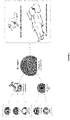
- the human perinatal tissue derived CD106 high CD151 + Nestin + MSCs were fibroblast-like and grow very well on plastic flask (cf. Fig 2 ).
- MSCs comprise over 95% cells which express the positive markers CD73, CD90, CD105 and CD166, and less than 2% cells which express the negative markers CD45, CD34 and HLA-DR. After this, MSCs keep growing till 90% cell confluent (see Tables 1 and 2 below), in practice during about 2 days.
- MSCs cell surface protein CD106, CD151 and Nestin increased significantly after the interleukin 1 and 4 were added.
- TABLE 1 shows the expression of CD11b, CD19, CD29, CD31, CD34, CD45, CD73, CD90, CD 105, CD106, CD151, Nestin and HLA-DR on placenta tissue derived MSCs before adding interleukin 1 and 4 obtained in placenta Example 1, 2, and 3.
- TABLE 2 shows the expression of CD11b, CD19, CD29, CD31, CD34, CD45, CD73, CD90, CD 105, CD106, CD151, Nestin and HLA-DR on the placenta tissue derived MSCs after adding interleukin 1 and 4 in Experiments 1, 2 and 3.
- the results show that CD106 and Nestin protein marker increased significantly 48 hours after adding the IL-1 and IL-4 in complete medium. Meanwhile, the CD106 high CD151 + Nestin + cells still have classical MSCs cellular surface phenotypic marker.
- Table 3 shows the related expression levels of growth factors secreted in the spent media by q-PCR. The results show that placenta tissue derived MSCs have more protein expression or secretion of IL-6, IL-8, IL-10, HGF, ANG, MMP2, VEGF-A and TGF- ⁇ at 48 hours after adding IL-1 and IL-4 in complete medium.
- the umbilical cord was removed from the transport solution and cut in 2-3cm long sections. To avoid contamination by adherent blood cells, each cord segment containing a blood clot that cannot be removed was discarded. The sections were then disinfected in a bath of antibiotics and antifungal agents composed of ⁇ MEM + Vancomycin 1 g/L + Amoxicillin 1 g/L + Amikacin 500 mg/L + Amphotericin B 50 mg/L for 30min at room temperature (RT). Antibiotics were extemporaneously dissolved in sterile water for injection.
- the sections of umbilical cord were removed from the bath and quickly rinsed in 1X PBS at RT.
- the epithelial membrane was slightly sectioned without touching the vessels.
- Each section was then detailed in slices of 0.5cm thickness and disposed at the bottom of a 150cm 2 plastic flask with lid. 6 to 10 slices per flask were disposed with at least a 1cm radius circle of free space around each slice, and left to adhere for 15min without medium at RT.
- the culture medium was changed after 5 to 7 days.
- the medium was removed and cells were washed with 30mL of 1X PBS per flask. Cells were then removed with Trypzean ® and collected with the old medium and centrifuged 10 min at 2500rpm. Supernatant was discarded and cells were then suspended in a cryopreservation solution consisting in ⁇ MEM + 100mg/mL HSA + 10% DMSO and cryopreserved.
- the umbilical cord was removed from the transport solution and cut in 2-3cm long sections. To avoid contamination by adherent blood cells, each segment containing a blood clot that cannot be removed was discarded,. The sections were then disinfected in a bath of antibiotics and antifungal agents composed of ⁇ DMEM + Vancomycin 1 g/L + Amoxicillin 1 g/L + Amikacin 500 mg/L + Amphotericin B 50 mg/L for 30min at room temperature (RT). Antibiotics were extemporaneously dissolved in sterile water for injection.
- the cord was then cut in small pieces and immersed in an enzymatic cocktail comprising 2.7 mg/mL collagenase type I and 0.7 mg/mL hyaluronidase, incubated for 3 h at 37°C with gentle agitation, followed by the addition of 2.5% trypsin and a further incubation for 30 min.
- an enzymatic cocktail comprising 2.7 mg/mL collagenase type I and 0.7 mg/mL hyaluronidase
- the digested suspension was diluted 1:2 with medium to reduce the viscosity of the suspension and passed through a nylon mesh to obtain single suspension. Cells were centrifuged at 300 ⁇ g for 20 min, and seeded at 10,000 cells/cm 2 with fresh medium.
- the culture medium was changed after 5 to 7 days and every 7 days after that.
- the medium was removed and cells were washed with 30mL of 1X PBS per flask. Cells were then removed with Trypzean ® and collected with the old medium and centrifuged 10 min at 2500rpm. Supernatant was discarded and cells were then suspended in a cryopreservation solution consisting in ⁇ MEM + 100mg/mL HSA + 10% DMSO and cryopreserved.
- the cells were suspended in preheated complete medium, and assessed for number and viability (blue trypan / Mallassez hemocytometer).
- the cells were seeded in two 75cm 2 plastic culture flasks in complete medium, and incubated (90% humidity, 5% CO 2 , 37°C).
- the cells were checked for confluency. When confluency reached 30 to 50%, the old medium was discarded and replaced either by fresh complete medium for unstimulated condition, or by fresh medium completed with 10ng/mL of IL-1 ⁇ and 10ng/mL of IL-4.
- the cells were checked for confluency. If confluency was up to 80%, the cells were harvested. Briefly, the old medium was discarded and the cells were washed with 1X DPBS. Trypsin EDTA was added and the cells were incubated 5 min at 37°C. Trypsin was neutralized with at least 2X the volume of medium, and the cell suspension was harvested and assessed for number and viability.
- the flow cytometry experiment required 1 ⁇ 10 6 cells, which were centrifuged and resuspended in 1X DPBS + 0.4% HSA.
- the cells were labelled for CD73, CD90 CD105, CD106, CD151 and CD31, CD34, CD45, HLA-DR according to the following protocol:
- Extracellular staining was performed for CD106 and CD151 markers as recommended by the FACS manufacturer and antibodies providers. Intracellular staining was performed for Nestin. For intracellular staining, the Fixation/Permeabilization solution called "BD Cytofix/Cytoperm kit" was used.
- a fixation step was performed, by contacting the cells with a 1XDPBS 0,5% formaldehyde solution.
- the cells were washed with 1X DPBS + 0.4% HSA and permeabilized for Nestin labelling.
- the labelled cells were analyzed with the Accuri C6+ BD Biosciences cytometer, and results were analyzed with the BD Accuri C6 Plus software.
- CD106 The expression of CD106 at the surface of said cells was measured by flow cytometry.
- Mesenchymal stem cells were isolated from umbilical cord using the two different methods exposed above (see 2.1. and 2.2.) : by explant isolation and by enzymatic digestion.
- All cells were cultivated in the same culture medium ( ⁇ MEM + 5% platelet lysate (LP)), stimulated at passage 3 according to the method of the invention and collected 2 days after stimulation. Cells viability is measured and cells were counted.
- CD73, CD90, CD105 are over 95% positive, while CD31, CD34, CD45 and HLA-DR were negative.
- CD151+ and Nestin were also both over 95% positive, independently of the stimulation or the isolation method.
- the MSCs isolated by enzymatic digestion expressed higher levels of CD106 before stimulation (53%) than the MSCs isolated with the explants methods (20%) They were however less sensitive to stimulation with the inflammatory cocktail: a smaller increase in CD106 has been observed ( +4% increase vs +60% increase).
- the explants method is therefore the preferred method of the invention for umbilical cord-derived MSCs.
- Table 5 viability and cell count of the MSCs obtained after the two experimental protocols exposed in 2.1. and 2.2.
- COR88Ex P3+1 MSCs isolated by the explants method Condition Non stimulated Stimulated Non stimulated Stimulated Viability 97.8% 95.8% 99.2% 97.1% Cell count 6.0 ⁇ 10 6 4.65 ⁇ 10 6 6.2 ⁇ 10 6 8.26 ⁇ 10 6
- Table 6 molecular markers of umbilical cord-derived MSCs obtained by the two experimental conditions exposed in 2.1. and 2.2.
- Mesenchymal stem cells were isolated from two umbilical cord by explant isolation (see 2.1.).
- All cells were cultivated in the same culture medium ( ⁇ MEM + 5% platelet lysate (LP)), stimulated at passage 4 according to the method of the invention with a mix of 10ng/mL of IL-1b and 10ng/mL of IL-4, or by each interleukin separately (10ng/mL each) and collected 2 days after stimulation. Cells viability is measured and cells were counted.
- ⁇ MEM + 5% platelet lysate (LP) platelet lysate
- CD73, CD90, CD105 are over 95% positive, while CD31, CD34, CD45 and HLA-DR were negative.
- CD151+ was also over 95% positive, independently of the stimulation.
- % of CD106 CORD 1 CORD 2 Unstimulated cells 11,60% 5,00% IL-1b + IL-4 stimulation 75,88% 54,29% IL-1b stimulation 32,19% 13,41% IL-4 stimulation 22,88% 15,77%
- This example focuses on the therapeutic neovascularization effect of placental derived CD106 high CD151 + Nestin + MSCs. It provides insights into their potential for clinical use as a cell-based therapy combined with insulin injection for treating critical hind limb ischemia in diabetes. Our results showed that Placenta derived CD106 high CD151 + Nestin + MSCs participate in angiogenesis and therapeutic vascularisation in order to improve ischemia and restore blood flow perfusion by directly differentiating into vascular cells. In addition, placenta derived CD106 high CD151 + Nestin + MSCs improved ischemia damage and functional recovery in diabetic rats.
- Immunodeficient male nude rats of six weeks of age were purchased from Vital River Laboratories (Charles River Laboratories suppler in China). Diabetes was induced with a single intraperitoneal injection of streptozotocin (70 mg/kg in Citrate buffer solution, only prepared immediately prior to injection) after overnight fasting. Fasting plasma glucose levels were measured every week and rats with Plasma glucose between 11 mM and 15 mM were considered to be diabetic. Age- and weight-matched nude rats receiving an intraperitoneal citrate buffer injection were used as non-diabetic controls (glycaemia between 5.5 and 8 mM).
- diabetic nude rats were anesthetized (60mg/kg pentobarbital intraperitoneally) and the left femoral artery were occluded by ligating it with 3-0 silk.
- the ligature was applied 0.5 cm proximally to the bifurcation of the saphenous and popliteal arteries. Lipiodol (1.5ml/kg) was used to induce an embolism intravascularly.
- a sham ligature was applied to the left femoral artery with the left hind limb remaining non ischemic.
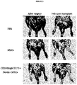
- the histological data on figure 4 show that the capillary density was increased in the cell transplant groups compared to the PBS group.
- more capillary numbers were observed in the CD106 high CD151 + Nestin + MSCs subpopulation cell transplant groups in diabetic rats and non-diabetic rats in comparison with the PBS group.
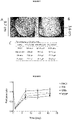
- Figure 5 showed the functional evidence of ischemia-induced changes using vascularisation LDPI.
- the image shows that blood flow was completely blocked in the left hind limb on the surgical day; this is indicated by the deep dark colour.
- the blood flow perfusion was restored to some degree in all the groups but did not return to normal in the PBS group where the ischemia/non-ischemia perfusion ratio only achieved 52%.
- the perfusion recovery in the cell transplant groups was significantly higher.
- the ratio was 81% in the P- MSCs group of the prior art (P ⁇ 0.05 vs PBS group) and 86% for the CD106 high CD151 + Nestin + MSCs subpopulation group (P ⁇ 0.01 vs PBS group).
- This example focuses on the therapeutic effect of placenta derived CD106 high CD151 + Nestin + MSCs of the invention to patients suffering from diabetes. It provides insights into their potential for a clinical use as a cell-based therapy diabetes.
- a total of 15 patients with diabetes were included in this clinical trial.
- the inclusion criteria were patients with type 2 diabetes diagnosed from November 2013 to November 2014 in Tianjin General Hospital.
- the patients were between 30 and 85 years of age, duration of diabetes ⁇ 3 years, requiring insulin for optimal glycemic control in a dose of ⁇ 0.7 U/kg/day at least for 1.5 year, having insulin dysfunction, poorly controlled blood glucose fluctuation with insulin-based treatment, and willingness to participate in the study.
- These 15 patients were between 42 and 67 years of age, with a median age of 59 years old; duration of diabetes from 3 years to 17 years, with an median of 8 years; daily insulin requirement from 38 units to 90 units, with an average of 58.7 units.
- the original general treatments were maintained. All of the patients were followed up after therapy for at least 6 months.
- This example focuses on the therapeutic effect of placenta derived MSCs of the invention to patients with aplastic anemia and provides insights into their potential for clinical use as a cell-based therapy for aplastic anemia.
- Aplastic anemia is mostly considered as an immune-mediated bone marrow (BM) failure syndrome, characterized by hypoplasia and pancytopenia with fatty BM and reduced angiogenesis.
- BM bone marrow
- Previous investigations have demonstrated that acquired aplastic anemia is manifested by abnormalities of HSCs/HPCs and hematopoietic microenvironment. Lots of evidences have hinted that aplastic anemia might be a syndrome characterized by stem/progenitor-cell disorders including HSCs/HPCs and MSCs.
- MSCs support hematopoiesis and regulate almost overall immune cells function to maintain the hematopoietic and immune homeostasis.
- MSCs can modulate the functions of the main immune cell including T cells, B cells, monocytes, DCs, NKTs and neutrophils [5].
- MSCs possess remarkable immunosuppressive properties on Th1, Th17 and CTLs. MSCs inhibit the proliferation of T cells, IFN- ⁇ and TNF- ⁇ secretion by Th1 cells while promoting IL-10 production by Th2 cells and the expansion of Treg cells.
- MSCs from aplastic anemia patients had poor proliferation and deficient immune suppression of MLR, PHA-induced T cell activation and IFN- ⁇ release [6,7].
- Our recent study showed that MSCs from aplastic anemia patients were reduced in suppressing the proliferation and clonogenic potential of CD4+ T cells while promoting Treg cells expansion.
- MSCs were also found defective in suppressing the production of TNF- ⁇ and IFN- ⁇ by CD4+ cells. However, there was no significant difference in regulating the production of IL-4, IL-10 and IL-17 [8]. In addition, our research also showed that MSCs from aplastic anemia patients showed aberrant morphology, decreased proliferation and clonogenic potential, and increased apoptosis as compared with BM-MSCs from healthy controls. MSCs from aplastic anemia patients were susceptible to be induced to differentiate into adipocytes but more difficult to differentiate into osteoblasts.
- MSCs is a promising therapeutic candidate for treating aplastic anemia due to the following two important facts:
- This example focuses on the therapeutic effect of placenta derived MSCs obtained according to the method of the invention (i.e., with IL1 and IL4 treatment) administered to patients suffering from liver diseases. It provides insights into their potential for clinical use as a cell-based therapy for liver diseases.
- the ultrasonic examination results showed an ascite level of 9.7 cm before MSCs were administered (see Fig. 7A ).
- the ascite level decreased to 3.6 cm one month after the administration of the MSCs ( Fig.7 B) .
- the results showed that the liver functions of patient with decompensated cirrhosis improved significantly after the administration of the MSCs.
- CA125 expression in serum decreased to normal level according to the clinical references.
- the placenta derived MSCs of the invention is a very good cytotherapeutic choice for liver diseases.
- the pro-angiogenic potential of the MSC of the invention has been further tested in a hindlimb ischemia murine model.
- mice Twelve 8-week-old NOD/SCID mice (Laboratoire Janvier) were anesthetized with a mix of ketamin and xylazine. The left proximal and distal parts of the femoral artery of the left leg was then ligated (6.0 silk suture, Ethicon, Issy-Les-Moulineaux, France) and the part between ligations excised. Mice that exhibit more than 80% of ischemia after surgery were intramuscularly injected in the gastrocnemius muscle of the ischemic limb with 2 doses of MSCs of the invention as obtained according to the protocols exposed in point 2.4. above (0.5 ⁇ 10 6 cells per animal in Group 2, 0.05. 10 6 cells per animal in Group 3). A saline solution injection was used in the Control Group 1. A dose of 20ng/mL of VEGF/mice was also injected in the Control Group 4.
- Hind limb blood flow was measured using a scanner-laser Doppler (Laser Doppler Perfusion system, Perimed PeriScan PIM III). The average perfusion of ischemic and non-ischemic limbs was determined before and after ischemia, and every 7 days until 21 days after ischemia. Blood flow-dependent changes in laser frequency were imaged using different colored pixels. Images were analyzed to quantify blood flow by using a blood perfusion analysis software (LPDIwin). Percentage of perfusion was expressed as the ratio of the ischemic to the non-ischemic hind limb.
- LPDIwin blood perfusion analysis software
- mice were euthanized at the end of the experiment (D21).
- Hind-limb gastrocnemius muscles were dissected and stained to visualize the distribution of the cells nuclei in the muscular fiber. Muscles isolated from the ischemic hind limb were fixed in paraffin and stained with hematoxylin/eosin.
- mice In the ischemic muscles of mice from Groups 1 (NaCl 0,9%) and 4 (VEGF 20ng/mL), 21 days after ischemia induction, cells exhibit very few nuclei in the periphery of the muscular fiber, since they are still regenerating.
- mice In the ischemic muscles of mice from Group 3 (0.05. 10 6 MSC cells or 50k), 21 days after ischemia induction, 50% of muscular fibers are regenerated and exhibit nuclei in the periphery of the fiber. The regeneration happens faster than in Groups 1 and 4.
- mice from Group 2 0.5 ⁇ 10 6 MSC cells or 500k
- 21 days after ischemia induction 100% of muscular fibers are regenerated and exhibit nuclei in the periphery of the fiber.
- the staining profile of the muscle is identical to the normal, non-ischemic, muscle.
- the 500k cells dose allows a complete regeneration of the muscle.
Landscapes
- Health & Medical Sciences (AREA)
- Life Sciences & Earth Sciences (AREA)
- Engineering & Computer Science (AREA)
- General Health & Medical Sciences (AREA)
- Veterinary Medicine (AREA)
- Animal Behavior & Ethology (AREA)
- Public Health (AREA)
- Chemical & Material Sciences (AREA)
- Bioinformatics & Cheminformatics (AREA)
- Organic Chemistry (AREA)
- Pharmacology & Pharmacy (AREA)
- Medicinal Chemistry (AREA)
- Nuclear Medicine, Radiotherapy & Molecular Imaging (AREA)
- General Chemical & Material Sciences (AREA)
- Chemical Kinetics & Catalysis (AREA)
- Biomedical Technology (AREA)
- Developmental Biology & Embryology (AREA)
- Zoology (AREA)
- Dermatology (AREA)
- Immunology (AREA)
- Biotechnology (AREA)
- Cell Biology (AREA)
- Epidemiology (AREA)
- Hematology (AREA)
- Wood Science & Technology (AREA)
- Genetics & Genomics (AREA)
- Rheumatology (AREA)
- Birds (AREA)
- Diabetes (AREA)
- Microbiology (AREA)
- Biochemistry (AREA)
- General Engineering & Computer Science (AREA)
- Cardiology (AREA)
- Virology (AREA)
- Dispersion Chemistry (AREA)
- Gerontology & Geriatric Medicine (AREA)
- Physical Education & Sports Medicine (AREA)
- Orthopedic Medicine & Surgery (AREA)
- Heart & Thoracic Surgery (AREA)
- Urology & Nephrology (AREA)
Claims (15)
- Verfahren zur Herstellung von mesenchymalen CD106high CD151+Nestin+ Stammzellen (MSZ), wobei das Verfahren die Kultivierung einer Population von undifferenzierten MSZ in einem Kulturmedium, das zwischen 1 und 100 ng/ml beigemengtes Interleukin 1β und zwischen 1 und 100 ng/ml beigemengtes Interleukin 4 umfasst, während mindestens eines Tages, vorzugsweise während zwei Tagen, umfasst.
- Verfahren zur Erhöhung des CD106-Expressionsniveaus von undifferenzierten MSZ, wobei das Verfahren die Kultivierung einer Population von undifferenzierten MSZ in einem Kulturmedium, das zwischen 1 und 100 ng/ml beigemengtes Interleukin 1β und zwischen 1 und 100 ng/ml beigemengtes Interleukin 4 umfasst, während mindestens eines Tages, vorzugsweise während zwei Tagen, umfasst.
- Verfahren nach einem der Ansprüche 1 und 2, das die folgenden Schritte umfasst:a) Bereitstellen der mononukleären Zellen, die in perinatalem biologischen Gewebe oder Fluid enthalten sind,b) Ermöglichen des Wachstums der mononukleären Zellen in ein erstes Kulturmedium ohne Wachstumsfaktor, bis sie 85 bis 90 % Konfluenz, vorzugsweise auf einer Kunststoffoberfläche, erreichen,c) nachdem 95 % der Zellen die positiven Marker CD73, CD90, CD105 und CD166 exprimiert haben und weniger als 2 % die negativen Marker CD45, CD34 und HLA-DR exprimiert haben, Säen der Zellen mit einer Dichte von 1000 bis 5000 MSZ pro cm2 in ein zweites Kulturmedium,d) nachdem die Zellen 40 bis 50 % Konfluenz erreicht haben, Beimengen von zwischen 1 und 100 ng/ml Interleukin 1β und zwischen 1 und 100 ng/ml Interleukin 4 während mindestens eines Tages,e) Sammeln der Zellen, wenn sie 90 bis 95 % Konfluenz erreichen.
- Verfahren nach einem der Ansprüche 1 bis 3, wobei die Population von im Schritt e) gesammelten Zellen enthält:- über 60 % Zellen, die CD106 auf einem nachweisbaren Niveau exprimieren,- über 98 % Zellen, die CD151 auf einem nachweisbaren Niveau exprimieren,- über 98 % Zellen, die Nestin auf einem nachweisbaren Niveau exprimieren,- über 95 % Zellen, die die Marker CD73, CD90, CD105 und CD166 auf einem nachweisbaren Niveau exprimieren, und- weniger als 2 % Zellen, die die Marker CD45, CD34 und HLA-DR auf einem nachweisbaren Niveau exprimieren.
- Zellkultur, welche die nach dem Verfahren nach Anspruch 1 bis 4 erhaltenen mesenchymalen CD106high CD151+Nestin+ Stammzellen (MSZ) enthält, wobei die Zellkultur enthält:- über 60 % Zellen, die CD106 auf einem nachweisbaren Niveau exprimieren,- über 98 % Zellen, die CD151 auf einem nachweisbaren Niveau exprimieren,- über 90 % Zellen, die Nestin auf einem nachweisbaren Niveau exprimieren,- über 95 % Zellen, die die Marker CD73, CD90, CD105 und CD166 auf einem nachweisbaren Niveau exprimieren, und- weniger als 2 % Zellen, die die Marker CD45, CD34 und HLA-DR auf einem nachweisbaren Niveau exprimieren.
- Zellkultur nach Anspruch 5 zur Verwendung bei der Behandlung von Patienten, die an einer ischämischen Erkrankung, an einer Störung des Kreislaufsystems, an einer Immunerkrankung, an einer Organverletzung oder - störung oder an einem Versagen einer Organfunktion leiden.
- Zellkultur zur Verwendung nach Anspruch 6, wobei die Erkrankung oder Störung aus der Gruppe ausgewählt ist, die besteht aus: Diabetes mellitus Typ 1, Diabetes Typ-II, GvHD, aplastischer Anämie, multipler Sklerose, Duchenne-Muskeldystrophie, rheumatoider Arthritis, Schlaganfall, idiopathischer Lungenfibrose, dilatativer Kardiomyopathie, Osteoarthritis, Zirrhose, Leberversagen, Nierenversagen, peripherer arterieller Verschlusskrankheit, kritischer Extremitätenischämie, peripherer Gefäßerkrankung, Herzversagen, diabetischem Ulcus, fibrotischen Störungen, Synechie, Störungen am Endometrium, diabetischem Ulcus, einem Geschwür, einem Trauma, einer Verbrennung, einer Verbrühung, einer Wunde oder einem Wundheilungsproblem, einem Dekubitalgeschwür, einer Warze, einer Synechie, einer Störung am Endometrium und einer fibrotischen Störung des Magen-Darm-Trakts, wie beispielsweise einer Analfistel.
- Pharmazeutische oder veterinäre Zusammensetzung, die die Zellkultur nach Anspruch 5 umfasst.
- Pharmazeutische Zusammensetzung nach Anspruch 8 zur Verwendung bei der Behandlung von Patienten, die an einer ischämischen Erkrankung, an einer Störung des Kreislaufsystems, an einer Immunerkrankung, an einer fibrotischen Störung, an einer Organverletzung oder an einem Versagen einer Organfunktion oder an einer Erkrankung oder Störung leiden, die aus der Gruppe ausgewählt ist, die besteht aus Diabetes mellitus Typ 1, Diabetes Typ-II, GvHD, aplastischer Anämie, multipler Sklerose, Duchenne-Muskeldystrophie, rheumatoider Arthritis, Schlaganfall, idiopathischer Lungenfibrose, dilatativer Kardiomyopathie, Osteoarthritis, Zirrhose, Leberversagen, Nierenversagen, peripherer arterieller Verschlusskrankheit, kritischer Extremitätenischämie, peripherer Gefäßerkrankung, Herzversagen, diabetischem Ulcus, fibrotischen Störungen, Synechie und Störungen am Endometrium.
- Dermatologische oder kosmetische Zusammensetzung, die die Zellkultur nach Anspruch 5 enthält.
- Verwendung der kosmetischen Zusammensetzung nach Anspruch 10 zur Regeneration der Haut oder der Schleimhautzellen, Verbessern des Aussehens der Haut oder Schleimhautmembran oder zur Korrektur eines Hautoder Schleimhautmembrandefekts, wie beispielsweise dunkler Flecken, Punkte, Akne, Falten, Trockenheit.
- Dermatologische Zusammensetzung nach Anspruch 10 für ihre Verwendung bei der Heilung einer Hautverletzung oder -störung, wie beispielsweise einer Verbrennung, einer Narbe, eines Angioms, eines Muttermals oder einer Warze.
- Medizinische Vorrichtung, die die Zellkultur nach Anspruch 5 enthält, wobei es sich vorzugsweise um einen Verband, ein Pflaster, einen Stent, ein Endoskop oder eine Spritze handelt.
- Verabreichungssystem, das die Zellkultur nach Anspruch 5 enthält, wobei es sich vorzugsweise um Mikrooder Nanovesikel von Biopolymeren, Lipiden oder Nanopartikeln handelt.
- Hydrogel, vorzugsweise Alginathydrogel, das die Zellkultur nach Anspruch 5, wahlweise mit einem anderen biokompatiblen Material oder Hilfsstoff, enthält.
Applications Claiming Priority (2)
| Application Number | Priority Date | Filing Date | Title |
|---|---|---|---|
| IB2016001936 | 2016-12-12 | ||
| PCT/EP2017/082316 WO2018108859A1 (en) | 2016-12-12 | 2017-12-12 | Perinatal tissue derived mesenchymal stem cells: method of preparation and uses thereof |
Publications (2)
| Publication Number | Publication Date |
|---|---|
| EP3551748A1 EP3551748A1 (de) | 2019-10-16 |
| EP3551748B1 true EP3551748B1 (de) | 2022-07-13 |
Family
ID=57995241
Family Applications (1)
| Application Number | Title | Priority Date | Filing Date |
|---|---|---|---|
| EP17832923.1A Active EP3551748B1 (de) | 2016-12-12 | 2017-12-12 | Von perinatalem gewebe abgeleiteten mesenchymale stammzellen: verfahren zur herstellung und verwendungen davon |
Country Status (9)
| Country | Link |
|---|---|
| US (1) | US12053492B2 (de) |
| EP (1) | EP3551748B1 (de) |
| JP (1) | JP7275441B2 (de) |
| KR (1) | KR102590455B1 (de) |
| CN (1) | CN110352240A (de) |
| CA (1) | CA3046576A1 (de) |
| ES (1) | ES2927461T3 (de) |
| PT (1) | PT3551748T (de) |
| WO (1) | WO2018108859A1 (de) |
Cited By (1)
| Publication number | Priority date | Publication date | Assignee | Title |
|---|---|---|---|---|
| EP3802794A1 (de) * | 2018-06-11 | 2021-04-14 | Health and Biotech France (H&B France) | Aus mesenchymalen stammzellen abgeleitete extrazelluläre vesikel |
Families Citing this family (16)
| Publication number | Priority date | Publication date | Assignee | Title |
|---|---|---|---|---|
| KR102246369B1 (ko) | 2011-11-30 | 2021-04-29 | 아스텔라스 인스티튜트 포 리제너러티브 메디슨 | 중간엽 간질 세포 및 이에 관련된 용도 |
| US8961956B2 (en) | 2011-11-30 | 2015-02-24 | Ocata Therapeutics, Inc. | Mesenchymal stromal cells and uses related thereto |
| US9745551B2 (en) | 2012-07-12 | 2017-08-29 | Imstem Biotechnology, Inc. | Mesenchymal-like stem cells derived from human embryonic stem cells, methods and uses thereof |
| GB201703058D0 (en) * | 2017-02-24 | 2017-04-12 | Ucl Business Plc | Biomarkers |
| JP7273421B2 (ja) | 2018-02-21 | 2023-05-15 | ボード オブ リージェンツ,ザ ユニバーシティ オブ テキサス システム | ナチュラルキラー細胞の活性化および拡大のための方法ならびにその使用 |
| CN108728408B (zh) * | 2018-05-28 | 2021-08-24 | 天津博雅秀岩生物技术有限公司 | 犬胎膜间充质干细胞及制备方法和使用的培养基 |
| CN113015535A (zh) * | 2018-10-05 | 2021-06-22 | 得克萨斯大学体系董事会 | 扩增间充质基质细胞的方法 |
| US12449413B2 (en) * | 2019-10-08 | 2025-10-21 | Cellresearch Corporation Pte. Ltd. | Method of assessing wound healing potency of a mesenchymal stem population and related methods of selecting mesenchymal stem cells and identifying tissue as starting material for producing a mesenchymal stem cell population |
| KR102268242B1 (ko) * | 2020-01-06 | 2021-06-23 | 에스씨엠생명과학 주식회사 | 줄기세포의 기능강화용 조성물 |
| EP4171591A4 (de) * | 2020-06-24 | 2024-08-21 | Board of Regents, The University of Texas System | Herstellung von exosomen aus vorbehandelten mesenchymalen stromazellen im grossmassstab zur klinischen verwendung |
| TWI782355B (zh) * | 2020-11-18 | 2022-11-01 | 國立陽明交通大學 | 經細胞介白素預刺激之人類臍帶間質幹細胞於治療類風濕性關節炎之組成及用途 |
| JP2024509124A (ja) * | 2021-02-26 | 2024-02-29 | アルグラ サイエンシズ エルエルシー | 絨毛間質細胞組成物及びその使用 |
| KR102717862B1 (ko) * | 2021-05-18 | 2024-10-15 | 에스씨엠생명과학 주식회사 | 기능강화 줄기세포를 포함하는 아토피 피부염 예방 또는 치료용 조성물 |
| CN114561349B (zh) * | 2022-03-16 | 2023-10-20 | 深圳市北科生物科技有限公司 | Il18,il12,il15处理的间质干细胞所产生的外泌体及抗病毒应用 |
| CN114736856B (zh) * | 2022-06-15 | 2022-08-26 | 天津百恩生物科技有限公司 | 一种犬胎盘间充质干细胞的制备方法及其应用 |
| CN118406642B (zh) * | 2023-04-04 | 2025-05-09 | 温州医科大学 | 人胎盘血管周干细胞及其对缺血性疾病的治疗作用 |
Family Cites Families (12)
| Publication number | Priority date | Publication date | Assignee | Title |
|---|---|---|---|---|
| ZA200401646B (en) * | 2003-03-12 | 2004-06-07 | Reliance Life Sciences Pvt Ltd | Derivation of terminally differentiated dopaminergic neurons from human embryonic stem cells. |
| US10046011B2 (en) | 2008-01-31 | 2018-08-14 | Rutgers, The State University Of New Jersey | Compositions for inducing or suppressing an immune response |
| EP3029061B1 (de) | 2008-06-18 | 2017-09-13 | The Texas A&M University System | Mesenchymale stammzellen, zusammensetzungen und verfahren zur behandlung von herzgewebeschäden |
| CN102703380B (zh) | 2008-12-17 | 2014-07-09 | 北京汉氏联合生物技术有限公司 | 亚全能干细胞、其制备方法及其用途 |
| CN101748096B (zh) * | 2008-12-17 | 2013-03-13 | 北京汉氏联合生物技术有限公司 | 亚全能干细胞、其制备方法及其用途 |
| KR101078419B1 (ko) * | 2009-03-20 | 2011-10-31 | 주식회사 스템메디언스 | 양수 내 태아 유래 중간엽줄기세포를 이용한 피부 상태개선용 조성물 |
| CN102539736B (zh) * | 2011-12-16 | 2015-04-29 | 北京汉氏联合生物技术有限公司 | 一种cd106阳性细胞、其鉴定、制备方法及应用 |
| WO2014052912A1 (en) | 2012-09-28 | 2014-04-03 | Scripps Health | Methods of differentiating stem cells into chondrocytes |
| CA2895148C (en) * | 2012-12-14 | 2023-02-28 | Yufang Shi | Methods modulating immunoregulatory effect of stem cells |
| CN104630140A (zh) * | 2013-11-06 | 2015-05-20 | 吉林济惠生物科技有限公司 | 一种胎盘间充质前体干细胞的分离培养方法 |
| CN105483206B (zh) * | 2016-01-06 | 2019-09-13 | 北京汉氏联合生物技术股份有限公司 | 一种用vcam-1检测msc促内皮细胞增殖能力的方法 |
| CN106538515A (zh) | 2016-11-03 | 2017-03-29 | 明光市大全甜叶菊专业合作社 | 一种甜叶菊种子保存方法 |
-
2017
- 2017-12-12 EP EP17832923.1A patent/EP3551748B1/de active Active
- 2017-12-12 ES ES17832923T patent/ES2927461T3/es active Active
- 2017-12-12 KR KR1020197019932A patent/KR102590455B1/ko active Active
- 2017-12-12 US US16/468,669 patent/US12053492B2/en active Active
- 2017-12-12 WO PCT/EP2017/082316 patent/WO2018108859A1/en not_active Ceased
- 2017-12-12 PT PT178329231T patent/PT3551748T/pt unknown
- 2017-12-12 CN CN201780086267.9A patent/CN110352240A/zh active Pending
- 2017-12-12 JP JP2019551740A patent/JP7275441B2/ja active Active
- 2017-12-12 CA CA3046576A patent/CA3046576A1/en active Pending
Cited By (1)
| Publication number | Priority date | Publication date | Assignee | Title |
|---|---|---|---|---|
| EP3802794A1 (de) * | 2018-06-11 | 2021-04-14 | Health and Biotech France (H&B France) | Aus mesenchymalen stammzellen abgeleitete extrazelluläre vesikel |
Also Published As
| Publication number | Publication date |
|---|---|
| PT3551748T (pt) | 2022-09-29 |
| US12053492B2 (en) | 2024-08-06 |
| JP7275441B2 (ja) | 2023-05-18 |
| KR102590455B1 (ko) | 2023-10-17 |
| RU2019118712A3 (de) | 2021-04-16 |
| EP3551748A1 (de) | 2019-10-16 |
| KR20190128622A (ko) | 2019-11-18 |
| ES2927461T3 (es) | 2022-11-07 |
| JP2020503375A (ja) | 2020-01-30 |
| CN110352240A (zh) | 2019-10-18 |
| US20200009193A1 (en) | 2020-01-09 |
| RU2019118712A (ru) | 2021-01-12 |
| WO2018108859A1 (en) | 2018-06-21 |
| CA3046576A1 (en) | 2018-06-21 |
Similar Documents
| Publication | Publication Date | Title |
|---|---|---|
| EP3551748B1 (de) | Von perinatalem gewebe abgeleiteten mesenchymale stammzellen: verfahren zur herstellung und verwendungen davon | |
| JP5599568B2 (ja) | 分娩後由来細胞を用いた末梢血管疾患の治療 | |
| JP5676253B2 (ja) | 生体内機能的細胞の高効率採取法 | |
| JP5937210B2 (ja) | 臍帯組織由来細胞を用いた末梢血管疾患の治療 | |
| US11000552B2 (en) | Pluripotent stem cell for treating diabetic skin ulcer | |
| JP2008504290A (ja) | 虚血の細胞ベースの治療 | |
| JP2010505849A (ja) | 糖尿病性創傷治癒に有用な方法および組成物 | |
| CN105112357A (zh) | 来源于胎盘的贴壁细胞及其在疾病治疗中的应用 | |
| CN110898078B (zh) | 一种间充质干细胞分泌因子的制备及应用 | |
| JP2018515211A (ja) | 間葉系幹細胞を含む組成物およびその使用 | |
| Tracy et al. | State of the field: cellular and exosomal therapeutic approaches in vascular regeneration | |
| US9855300B2 (en) | Composition and method to improve the therapeutic effect of stem cells | |
| JP6663445B2 (ja) | 細胞増殖の刺激のための方法及び組成物、ならびにfgf2アイソフォームの生物学的に活性な混合物の提供 | |
| US20090047257A1 (en) | Novel cell populations and uses thereof | |
| WO2021131261A1 (ja) | 細胞群及びその取得方法 | |
| RU2780179C2 (ru) | Происходящие из перинатальной ткани мезенхимальные стволовые клетки: способ их получения и применения | |
| WO2018051340A1 (en) | A process for retrieval of matrix dependent mesenchymal stromal cells from tissue | |
| Fukuta et al. | Synergistic effect of ex-vivo quality and quantity cultured mononuclear cells and mesenchymal stem cell therapy in ischemic hind limb model mice | |
| WO2023037358A1 (en) | Placental cell treatment for critical limb ischemia patient subpopulations | |
| AU2024344499A1 (en) | Mesenchymal lineage precursor or stem cells (mlpscs) for use in methods of treating congenital heart disease |
Legal Events
| Date | Code | Title | Description |
|---|---|---|---|
| STAA | Information on the status of an ep patent application or granted ep patent |
Free format text: STATUS: UNKNOWN |
|
| STAA | Information on the status of an ep patent application or granted ep patent |
Free format text: STATUS: THE INTERNATIONAL PUBLICATION HAS BEEN MADE |
|
| PUAI | Public reference made under article 153(3) epc to a published international application that has entered the european phase |
Free format text: ORIGINAL CODE: 0009012 |
|
| STAA | Information on the status of an ep patent application or granted ep patent |
Free format text: STATUS: REQUEST FOR EXAMINATION WAS MADE |
|
| 17P | Request for examination filed |
Effective date: 20190708 |
|
| AK | Designated contracting states |
Kind code of ref document: A1 Designated state(s): AL AT BE BG CH CY CZ DE DK EE ES FI FR GB GR HR HU IE IS IT LI LT LU LV MC MK MT NL NO PL PT RO RS SE SI SK SM TR |
|
| AX | Request for extension of the european patent |
Extension state: BA ME |
|
| STAA | Information on the status of an ep patent application or granted ep patent |
Free format text: STATUS: EXAMINATION IS IN PROGRESS |
|
| DAV | Request for validation of the european patent (deleted) | ||
| DAX | Request for extension of the european patent (deleted) | ||
| 17Q | First examination report despatched |
Effective date: 20200313 |
|
| GRAP | Despatch of communication of intention to grant a patent |
Free format text: ORIGINAL CODE: EPIDOSNIGR1 |
|
| STAA | Information on the status of an ep patent application or granted ep patent |
Free format text: STATUS: GRANT OF PATENT IS INTENDED |
|
| INTG | Intention to grant announced |
Effective date: 20220303 |
|
| RIN1 | Information on inventor provided before grant (corrected) |
Inventor name: WANG, TAO Inventor name: HAN, ZHIBO Inventor name: HAN, ZHONGCHAO |
|
| GRAS | Grant fee paid |
Free format text: ORIGINAL CODE: EPIDOSNIGR3 |
|
| GRAA | (expected) grant |
Free format text: ORIGINAL CODE: 0009210 |
|
| STAA | Information on the status of an ep patent application or granted ep patent |
Free format text: STATUS: THE PATENT HAS BEEN GRANTED |
|
| AK | Designated contracting states |
Kind code of ref document: B1 Designated state(s): AL AT BE BG CH CY CZ DE DK EE ES FI FR GB GR HR HU IE IS IT LI LT LU LV MC MK MT NL NO PL PT RO RS SE SI SK SM TR |
|
| REG | Reference to a national code |
Ref country code: CH Ref legal event code: EP |
|
| REG | Reference to a national code |
Ref country code: DE Ref legal event code: R096 Ref document number: 602017059506 Country of ref document: DE |
|
| REG | Reference to a national code |
Ref country code: AT Ref legal event code: REF Ref document number: 1504309 Country of ref document: AT Kind code of ref document: T Effective date: 20220815 |
|
| REG | Reference to a national code |
Ref country code: IE Ref legal event code: FG4D |
|
| REG | Reference to a national code |
Ref country code: PT Ref legal event code: SC4A Ref document number: 3551748 Country of ref document: PT Date of ref document: 20220929 Kind code of ref document: T Free format text: AVAILABILITY OF NATIONAL TRANSLATION Effective date: 20220923 |
|
| REG | Reference to a national code |
Ref country code: ES Ref legal event code: FG2A Ref document number: 2927461 Country of ref document: ES Kind code of ref document: T3 Effective date: 20221107 |
|
| REG | Reference to a national code |
Ref country code: LT Ref legal event code: MG9D |
|
| REG | Reference to a national code |
Ref country code: NL Ref legal event code: MP Effective date: 20220713 |
|
| PG25 | Lapsed in a contracting state [announced via postgrant information from national office to epo] |
Ref country code: SE Free format text: LAPSE BECAUSE OF FAILURE TO SUBMIT A TRANSLATION OF THE DESCRIPTION OR TO PAY THE FEE WITHIN THE PRESCRIBED TIME-LIMIT Effective date: 20220713 Ref country code: RS Free format text: LAPSE BECAUSE OF FAILURE TO SUBMIT A TRANSLATION OF THE DESCRIPTION OR TO PAY THE FEE WITHIN THE PRESCRIBED TIME-LIMIT Effective date: 20220713 Ref country code: NO Free format text: LAPSE BECAUSE OF FAILURE TO SUBMIT A TRANSLATION OF THE DESCRIPTION OR TO PAY THE FEE WITHIN THE PRESCRIBED TIME-LIMIT Effective date: 20221013 Ref country code: NL Free format text: LAPSE BECAUSE OF FAILURE TO SUBMIT A TRANSLATION OF THE DESCRIPTION OR TO PAY THE FEE WITHIN THE PRESCRIBED TIME-LIMIT Effective date: 20220713 Ref country code: LV Free format text: LAPSE BECAUSE OF FAILURE TO SUBMIT A TRANSLATION OF THE DESCRIPTION OR TO PAY THE FEE WITHIN THE PRESCRIBED TIME-LIMIT Effective date: 20220713 Ref country code: LT Free format text: LAPSE BECAUSE OF FAILURE TO SUBMIT A TRANSLATION OF THE DESCRIPTION OR TO PAY THE FEE WITHIN THE PRESCRIBED TIME-LIMIT Effective date: 20220713 Ref country code: FI Free format text: LAPSE BECAUSE OF FAILURE TO SUBMIT A TRANSLATION OF THE DESCRIPTION OR TO PAY THE FEE WITHIN THE PRESCRIBED TIME-LIMIT Effective date: 20220713 |
|
| REG | Reference to a national code |
Ref country code: AT Ref legal event code: MK05 Ref document number: 1504309 Country of ref document: AT Kind code of ref document: T Effective date: 20220713 |
|
| PG25 | Lapsed in a contracting state [announced via postgrant information from national office to epo] |
Ref country code: PL Free format text: LAPSE BECAUSE OF FAILURE TO SUBMIT A TRANSLATION OF THE DESCRIPTION OR TO PAY THE FEE WITHIN THE PRESCRIBED TIME-LIMIT Effective date: 20220713 Ref country code: IS Free format text: LAPSE BECAUSE OF FAILURE TO SUBMIT A TRANSLATION OF THE DESCRIPTION OR TO PAY THE FEE WITHIN THE PRESCRIBED TIME-LIMIT Effective date: 20221113 Ref country code: HR Free format text: LAPSE BECAUSE OF FAILURE TO SUBMIT A TRANSLATION OF THE DESCRIPTION OR TO PAY THE FEE WITHIN THE PRESCRIBED TIME-LIMIT Effective date: 20220713 Ref country code: GR Free format text: LAPSE BECAUSE OF FAILURE TO SUBMIT A TRANSLATION OF THE DESCRIPTION OR TO PAY THE FEE WITHIN THE PRESCRIBED TIME-LIMIT Effective date: 20221014 |
|
| REG | Reference to a national code |
Ref country code: DE Ref legal event code: R097 Ref document number: 602017059506 Country of ref document: DE |
|
| PG25 | Lapsed in a contracting state [announced via postgrant information from national office to epo] |
Ref country code: SM Free format text: LAPSE BECAUSE OF FAILURE TO SUBMIT A TRANSLATION OF THE DESCRIPTION OR TO PAY THE FEE WITHIN THE PRESCRIBED TIME-LIMIT Effective date: 20220713 Ref country code: RO Free format text: LAPSE BECAUSE OF FAILURE TO SUBMIT A TRANSLATION OF THE DESCRIPTION OR TO PAY THE FEE WITHIN THE PRESCRIBED TIME-LIMIT Effective date: 20220713 Ref country code: DK Free format text: LAPSE BECAUSE OF FAILURE TO SUBMIT A TRANSLATION OF THE DESCRIPTION OR TO PAY THE FEE WITHIN THE PRESCRIBED TIME-LIMIT Effective date: 20220713 Ref country code: CZ Free format text: LAPSE BECAUSE OF FAILURE TO SUBMIT A TRANSLATION OF THE DESCRIPTION OR TO PAY THE FEE WITHIN THE PRESCRIBED TIME-LIMIT Effective date: 20220713 Ref country code: AT Free format text: LAPSE BECAUSE OF FAILURE TO SUBMIT A TRANSLATION OF THE DESCRIPTION OR TO PAY THE FEE WITHIN THE PRESCRIBED TIME-LIMIT Effective date: 20220713 |
|
| PLBE | No opposition filed within time limit |
Free format text: ORIGINAL CODE: 0009261 |
|
| STAA | Information on the status of an ep patent application or granted ep patent |
Free format text: STATUS: NO OPPOSITION FILED WITHIN TIME LIMIT |
|
| PG25 | Lapsed in a contracting state [announced via postgrant information from national office to epo] |
Ref country code: SK Free format text: LAPSE BECAUSE OF FAILURE TO SUBMIT A TRANSLATION OF THE DESCRIPTION OR TO PAY THE FEE WITHIN THE PRESCRIBED TIME-LIMIT Effective date: 20220713 Ref country code: EE Free format text: LAPSE BECAUSE OF FAILURE TO SUBMIT A TRANSLATION OF THE DESCRIPTION OR TO PAY THE FEE WITHIN THE PRESCRIBED TIME-LIMIT Effective date: 20220713 |
|
| 26N | No opposition filed |
Effective date: 20230414 |
|
| PG25 | Lapsed in a contracting state [announced via postgrant information from national office to epo] |
Ref country code: AL Free format text: LAPSE BECAUSE OF FAILURE TO SUBMIT A TRANSLATION OF THE DESCRIPTION OR TO PAY THE FEE WITHIN THE PRESCRIBED TIME-LIMIT Effective date: 20220713 |
|
| PG25 | Lapsed in a contracting state [announced via postgrant information from national office to epo] |
Ref country code: SI Free format text: LAPSE BECAUSE OF FAILURE TO SUBMIT A TRANSLATION OF THE DESCRIPTION OR TO PAY THE FEE WITHIN THE PRESCRIBED TIME-LIMIT Effective date: 20220713 |
|
| PG25 | Lapsed in a contracting state [announced via postgrant information from national office to epo] |
Ref country code: HU Free format text: LAPSE BECAUSE OF FAILURE TO SUBMIT A TRANSLATION OF THE DESCRIPTION OR TO PAY THE FEE WITHIN THE PRESCRIBED TIME-LIMIT; INVALID AB INITIO Effective date: 20171212 |
|
| PG25 | Lapsed in a contracting state [announced via postgrant information from national office to epo] |
Ref country code: CY Free format text: LAPSE BECAUSE OF FAILURE TO SUBMIT A TRANSLATION OF THE DESCRIPTION OR TO PAY THE FEE WITHIN THE PRESCRIBED TIME-LIMIT Effective date: 20220713 |
|
| PG25 | Lapsed in a contracting state [announced via postgrant information from national office to epo] |
Ref country code: MK Free format text: LAPSE BECAUSE OF FAILURE TO SUBMIT A TRANSLATION OF THE DESCRIPTION OR TO PAY THE FEE WITHIN THE PRESCRIBED TIME-LIMIT Effective date: 20220713 |
|
| PG25 | Lapsed in a contracting state [announced via postgrant information from national office to epo] |
Ref country code: TR Free format text: LAPSE BECAUSE OF FAILURE TO SUBMIT A TRANSLATION OF THE DESCRIPTION OR TO PAY THE FEE WITHIN THE PRESCRIBED TIME-LIMIT Effective date: 20220713 |
|
| PG25 | Lapsed in a contracting state [announced via postgrant information from national office to epo] |
Ref country code: BG Free format text: LAPSE BECAUSE OF FAILURE TO SUBMIT A TRANSLATION OF THE DESCRIPTION OR TO PAY THE FEE WITHIN THE PRESCRIBED TIME-LIMIT Effective date: 20220713 |
|
| PG25 | Lapsed in a contracting state [announced via postgrant information from national office to epo] |
Ref country code: MT Free format text: LAPSE BECAUSE OF FAILURE TO SUBMIT A TRANSLATION OF THE DESCRIPTION OR TO PAY THE FEE WITHIN THE PRESCRIBED TIME-LIMIT Effective date: 20220713 |
|
| PGFP | Annual fee paid to national office [announced via postgrant information from national office to epo] |
Ref country code: CH Payment date: 20250101 Year of fee payment: 8 |
|
| PGFP | Annual fee paid to national office [announced via postgrant information from national office to epo] |
Ref country code: PT Payment date: 20251120 Year of fee payment: 9 |
|
| PGFP | Annual fee paid to national office [announced via postgrant information from national office to epo] |
Ref country code: LU Payment date: 20251127 Year of fee payment: 9 |
|
| REG | Reference to a national code |
Ref country code: CH Ref legal event code: U11 Free format text: ST27 STATUS EVENT CODE: U-0-0-U10-U11 (AS PROVIDED BY THE NATIONAL OFFICE) Effective date: 20260101 |
|
| PGFP | Annual fee paid to national office [announced via postgrant information from national office to epo] |
Ref country code: GB Payment date: 20251229 Year of fee payment: 9 |
|
| PGFP | Annual fee paid to national office [announced via postgrant information from national office to epo] |
Ref country code: MC Payment date: 20251126 Year of fee payment: 9 |
|
| PGFP | Annual fee paid to national office [announced via postgrant information from national office to epo] |
Ref country code: IT Payment date: 20251210 Year of fee payment: 9 |
|
| PGFP | Annual fee paid to national office [announced via postgrant information from national office to epo] |
Ref country code: FR Payment date: 20251113 Year of fee payment: 9 |
|
| PGFP | Annual fee paid to national office [announced via postgrant information from national office to epo] |
Ref country code: BE Payment date: 20251226 Year of fee payment: 9 |
|
| PGFP | Annual fee paid to national office [announced via postgrant information from national office to epo] |
Ref country code: IE Payment date: 20251126 Year of fee payment: 9 |
|
| PGFP | Annual fee paid to national office [announced via postgrant information from national office to epo] |
Ref country code: ES Payment date: 20260112 Year of fee payment: 9 |
|
| PGFP | Annual fee paid to national office [announced via postgrant information from national office to epo] |
Ref country code: DE Payment date: 20251231 Year of fee payment: 9 |