EP3461494A1 - Nouveaux peptides, combinaison de peptides et d'échafaudages destinés à être utilisés dans le traitement immunothérapeutique de divers cancers - Google Patents
Nouveaux peptides, combinaison de peptides et d'échafaudages destinés à être utilisés dans le traitement immunothérapeutique de divers cancers Download PDFInfo
- Publication number
- EP3461494A1 EP3461494A1 EP18201208.8A EP18201208A EP3461494A1 EP 3461494 A1 EP3461494 A1 EP 3461494A1 EP 18201208 A EP18201208 A EP 18201208A EP 3461494 A1 EP3461494 A1 EP 3461494A1
- Authority
- EP
- European Patent Office
- Prior art keywords
- cancer
- cell
- peptide
- cells
- tumor
- Prior art date
- Legal status (The legal status is an assumption and is not a legal conclusion. Google has not performed a legal analysis and makes no representation as to the accuracy of the status listed.)
- Pending
Links
- 108090000765 processed proteins & peptides Proteins 0.000 title claims abstract description 287
- 206010028980 Neoplasm Diseases 0.000 title claims abstract description 264
- 102000004196 processed proteins & peptides Human genes 0.000 title claims abstract description 155
- 238000011282 treatment Methods 0.000 title claims description 72
- 230000001024 immunotherapeutic effect Effects 0.000 title abstract description 14
- 210000004027 cell Anatomy 0.000 claims abstract description 159
- 201000011510 cancer Diseases 0.000 claims abstract description 101
- 108090000623 proteins and genes Proteins 0.000 claims abstract description 101
- 210000001744 T-lymphocyte Anatomy 0.000 claims abstract description 74
- 102000004169 proteins and genes Human genes 0.000 claims abstract description 48
- 108091008874 T cell receptors Proteins 0.000 claims abstract description 40
- 102000016266 T-Cell Antigen Receptors Human genes 0.000 claims abstract description 40
- 238000000034 method Methods 0.000 claims abstract description 28
- 150000007523 nucleic acids Chemical class 0.000 claims abstract description 23
- 102000039446 nucleic acids Human genes 0.000 claims abstract description 19
- 108020004707 nucleic acids Proteins 0.000 claims abstract description 19
- 208000026310 Breast neoplasm Diseases 0.000 claims description 69
- 206010006187 Breast cancer Diseases 0.000 claims description 67
- 239000000427 antigen Substances 0.000 claims description 56
- 108091007433 antigens Proteins 0.000 claims description 54
- 102000036639 antigens Human genes 0.000 claims description 54
- 150000001413 amino acids Chemical class 0.000 claims description 47
- 208000005718 Stomach Neoplasms Diseases 0.000 claims description 46
- 206010017758 gastric cancer Diseases 0.000 claims description 45
- 206010009944 Colon cancer Diseases 0.000 claims description 43
- 208000000236 Prostatic Neoplasms Diseases 0.000 claims description 43
- 208000001333 Colorectal Neoplasms Diseases 0.000 claims description 42
- 201000011549 stomach cancer Diseases 0.000 claims description 42
- 206010060862 Prostate cancer Diseases 0.000 claims description 41
- 208000002154 non-small cell lung carcinoma Diseases 0.000 claims description 38
- 208000029729 tumor suppressor gene on chromosome 11 Diseases 0.000 claims description 37
- 206010061535 Ovarian neoplasm Diseases 0.000 claims description 34
- 206010073071 hepatocellular carcinoma Diseases 0.000 claims description 33
- 206010061902 Pancreatic neoplasm Diseases 0.000 claims description 31
- 229920001184 polypeptide Polymers 0.000 claims description 31
- 208000010839 B-cell chronic lymphocytic leukemia Diseases 0.000 claims description 30
- 206010033128 Ovarian cancer Diseases 0.000 claims description 30
- 208000005017 glioblastoma Diseases 0.000 claims description 29
- 210000004443 dendritic cell Anatomy 0.000 claims description 28
- 231100000844 hepatocellular carcinoma Toxicity 0.000 claims description 28
- 208000015486 malignant pancreatic neoplasm Diseases 0.000 claims description 27
- 230000002018 overexpression Effects 0.000 claims description 27
- 208000008443 pancreatic carcinoma Diseases 0.000 claims description 27
- 208000006265 Renal cell carcinoma Diseases 0.000 claims description 26
- 125000003275 alpha amino acid group Chemical group 0.000 claims description 26
- 201000001441 melanoma Diseases 0.000 claims description 26
- 201000002528 pancreatic cancer Diseases 0.000 claims description 26
- 208000007097 Urinary Bladder Neoplasms Diseases 0.000 claims description 25
- 208000031422 Lymphocytic Chronic B-Cell Leukemia Diseases 0.000 claims description 24
- 208000032852 chronic lymphocytic leukemia Diseases 0.000 claims description 24
- 201000005112 urinary bladder cancer Diseases 0.000 claims description 24
- 208000000587 small cell lung carcinoma Diseases 0.000 claims description 22
- 206010041067 Small cell lung cancer Diseases 0.000 claims description 21
- 208000031261 Acute myeloid leukaemia Diseases 0.000 claims description 17
- 208000006990 cholangiocarcinoma Diseases 0.000 claims description 17
- 208000000461 Esophageal Neoplasms Diseases 0.000 claims description 16
- 238000000338 in vitro Methods 0.000 claims description 16
- 150000003839 salts Chemical class 0.000 claims description 16
- 208000033776 Myeloid Acute Leukemia Diseases 0.000 claims description 15
- 208000015914 Non-Hodgkin lymphomas Diseases 0.000 claims description 15
- 206010030155 Oesophageal carcinoma Diseases 0.000 claims description 15
- 201000004101 esophageal cancer Diseases 0.000 claims description 15
- 210000000612 antigen-presenting cell Anatomy 0.000 claims description 14
- 208000002495 Uterine Neoplasms Diseases 0.000 claims description 13
- 239000003814 drug Substances 0.000 claims description 13
- 239000013604 expression vector Substances 0.000 claims description 13
- 206010046766 uterine cancer Diseases 0.000 claims description 13
- 208000022072 Gallbladder Neoplasms Diseases 0.000 claims description 10
- 238000003745 diagnosis Methods 0.000 claims description 10
- 201000010175 gallbladder cancer Diseases 0.000 claims description 10
- 102000008949 Histocompatibility Antigens Class I Human genes 0.000 claims description 9
- 208000024313 Testicular Neoplasms Diseases 0.000 claims description 9
- 206010057644 Testis cancer Diseases 0.000 claims description 9
- 201000003120 testicular cancer Diseases 0.000 claims description 9
- 239000003446 ligand Substances 0.000 claims description 8
- 238000004519 manufacturing process Methods 0.000 claims description 8
- 210000000822 natural killer cell Anatomy 0.000 claims description 7
- 239000012528 membrane Substances 0.000 claims description 6
- 102000006354 HLA-DR Antigens Human genes 0.000 claims description 5
- 108010058597 HLA-DR Antigens Proteins 0.000 claims description 5
- 102000037865 fusion proteins Human genes 0.000 claims description 4
- 108020001507 fusion proteins Proteins 0.000 claims description 4
- 108010088652 Histocompatibility Antigens Class I Proteins 0.000 claims description 3
- 125000001429 N-terminal alpha-amino-acid group Chemical group 0.000 claims description 3
- 108010028930 invariant chain Proteins 0.000 claims description 3
- 239000008194 pharmaceutical composition Substances 0.000 claims description 3
- 238000012258 culturing Methods 0.000 claims description 2
- 239000001963 growth medium Substances 0.000 claims description 2
- 239000003085 diluting agent Substances 0.000 claims 2
- 239000012931 lyophilized formulation Substances 0.000 claims 2
- 239000004480 active ingredient Substances 0.000 claims 1
- 239000003937 drug carrier Substances 0.000 claims 1
- 239000000546 pharmaceutical excipient Substances 0.000 claims 1
- 239000003381 stabilizer Substances 0.000 claims 1
- 108700018351 Major Histocompatibility Complex Proteins 0.000 abstract description 35
- 230000020382 suppression by virus of host antigen processing and presentation of peptide antigen via MHC class I Effects 0.000 abstract description 35
- 230000027455 binding Effects 0.000 abstract description 32
- 238000009169 immunotherapy Methods 0.000 abstract description 30
- 229960005486 vaccine Drugs 0.000 abstract description 28
- 238000012546 transfer Methods 0.000 abstract description 15
- 230000005975 antitumor immune response Effects 0.000 abstract description 10
- 239000000203 mixture Substances 0.000 abstract description 7
- 239000008186 active pharmaceutical agent Substances 0.000 abstract description 3
- 230000014509 gene expression Effects 0.000 description 108
- 230000001105 regulatory effect Effects 0.000 description 63
- 235000001014 amino acid Nutrition 0.000 description 46
- 235000018102 proteins Nutrition 0.000 description 46
- 229940024606 amino acid Drugs 0.000 description 45
- 210000004881 tumor cell Anatomy 0.000 description 41
- 230000004083 survival effect Effects 0.000 description 40
- 210000001519 tissue Anatomy 0.000 description 40
- 208000000102 Squamous Cell Carcinoma of Head and Neck Diseases 0.000 description 39
- 238000013459 approach Methods 0.000 description 32
- 230000001965 increasing effect Effects 0.000 description 27
- 102100034523 Histone H4 Human genes 0.000 description 26
- 101001067880 Homo sapiens Histone H4 Proteins 0.000 description 26
- 208000037265 diseases, disorders, signs and symptoms Diseases 0.000 description 26
- 201000010099 disease Diseases 0.000 description 25
- 206010027476 Metastases Diseases 0.000 description 24
- 239000012071 phase Substances 0.000 description 23
- 230000009401 metastasis Effects 0.000 description 22
- 238000002560 therapeutic procedure Methods 0.000 description 22
- 238000002255 vaccination Methods 0.000 description 22
- 230000006870 function Effects 0.000 description 21
- 230000018109 developmental process Effects 0.000 description 20
- 238000011161 development Methods 0.000 description 19
- 208000020816 lung neoplasm Diseases 0.000 description 19
- -1 KCH3COO Chemical compound 0.000 description 18
- 206010058467 Lung neoplasm malignant Diseases 0.000 description 18
- 102100028972 HLA class I histocompatibility antigen, A alpha chain Human genes 0.000 description 17
- 108010075704 HLA-A Antigens Proteins 0.000 description 17
- 108091054437 MHC class I family Proteins 0.000 description 17
- 230000005867 T cell response Effects 0.000 description 17
- 125000000539 amino acid group Chemical group 0.000 description 17
- 239000000090 biomarker Substances 0.000 description 17
- 230000002596 correlated effect Effects 0.000 description 17
- 201000000459 head and neck squamous cell carcinoma Diseases 0.000 description 17
- 101000607318 Homo sapiens UL16-binding protein 3 Proteins 0.000 description 16
- 108091054438 MHC class II family Proteins 0.000 description 16
- 102100040011 UL16-binding protein 3 Human genes 0.000 description 16
- 230000028993 immune response Effects 0.000 description 16
- 201000005202 lung cancer Diseases 0.000 description 16
- 230000001225 therapeutic effect Effects 0.000 description 16
- 108020004414 DNA Proteins 0.000 description 15
- 230000000694 effects Effects 0.000 description 15
- 230000004044 response Effects 0.000 description 15
- 230000008685 targeting Effects 0.000 description 15
- 206010005003 Bladder cancer Diseases 0.000 description 14
- 201000009030 Carcinoma Diseases 0.000 description 14
- 229960000190 bacillus calmette–guérin vaccine Drugs 0.000 description 14
- 231100000504 carcinogenesis Toxicity 0.000 description 14
- 229960005386 ipilimumab Drugs 0.000 description 14
- 230000037361 pathway Effects 0.000 description 14
- 238000004393 prognosis Methods 0.000 description 14
- 230000035755 proliferation Effects 0.000 description 14
- 230000011664 signaling Effects 0.000 description 14
- 108700028369 Alleles Proteins 0.000 description 13
- 102100038449 Claudin-6 Human genes 0.000 description 13
- 102100035300 Cystine/glutamate transporter Human genes 0.000 description 13
- 101000882898 Homo sapiens Claudin-6 Proteins 0.000 description 13
- 241001467552 Mycobacterium bovis BCG Species 0.000 description 13
- 108091006241 SLC7A11 Proteins 0.000 description 13
- 108010040002 Tumor Suppressor Proteins Proteins 0.000 description 13
- 102000001742 Tumor Suppressor Proteins Human genes 0.000 description 13
- 230000036952 cancer formation Effects 0.000 description 13
- 238000002512 chemotherapy Methods 0.000 description 13
- 230000035772 mutation Effects 0.000 description 13
- 238000010837 poor prognosis Methods 0.000 description 13
- 238000006467 substitution reaction Methods 0.000 description 13
- 208000005623 Carcinogenesis Diseases 0.000 description 12
- 238000001727 in vivo Methods 0.000 description 12
- 229940023041 peptide vaccine Drugs 0.000 description 12
- 239000000047 product Substances 0.000 description 12
- 102000005962 receptors Human genes 0.000 description 12
- 108020003175 receptors Proteins 0.000 description 12
- 206010018338 Glioma Diseases 0.000 description 11
- 102000043129 MHC class I family Human genes 0.000 description 11
- 208000006105 Uterine Cervical Neoplasms Diseases 0.000 description 11
- 210000000481 breast Anatomy 0.000 description 11
- 201000010881 cervical cancer Diseases 0.000 description 11
- 230000003247 decreasing effect Effects 0.000 description 11
- 230000004069 differentiation Effects 0.000 description 11
- 230000002496 gastric effect Effects 0.000 description 11
- 239000003550 marker Substances 0.000 description 11
- 206010008342 Cervix carcinoma Diseases 0.000 description 10
- 108010019670 Chimeric Antigen Receptors Proteins 0.000 description 10
- 102100026097 Claudin-9 Human genes 0.000 description 10
- 108010017213 Granulocyte-Macrophage Colony-Stimulating Factor Proteins 0.000 description 10
- 102100039620 Granulocyte-macrophage colony-stimulating factor Human genes 0.000 description 10
- 101000912661 Homo sapiens Claudin-9 Proteins 0.000 description 10
- 108010038807 Oligopeptides Proteins 0.000 description 10
- 102000015636 Oligopeptides Human genes 0.000 description 10
- 108050003627 Wnt Proteins 0.000 description 10
- 230000003828 downregulation Effects 0.000 description 10
- 230000002829 reductive effect Effects 0.000 description 10
- 102100024090 Aldo-keto reductase family 1 member C3 Human genes 0.000 description 9
- 108060000903 Beta-catenin Proteins 0.000 description 9
- 102000015735 Beta-catenin Human genes 0.000 description 9
- 102100025064 Cellular tumor antigen p53 Human genes 0.000 description 9
- 102100022204 DNA-dependent protein kinase catalytic subunit Human genes 0.000 description 9
- 101000619536 Homo sapiens DNA-dependent protein kinase catalytic subunit Proteins 0.000 description 9
- 101001006776 Homo sapiens Kinesin-like protein KIFC1 Proteins 0.000 description 9
- 208000022361 Human papillomavirus infectious disease Diseases 0.000 description 9
- 102100027942 Kinesin-like protein KIFC1 Human genes 0.000 description 9
- 102000043131 MHC class II family Human genes 0.000 description 9
- 229910019142 PO4 Inorganic materials 0.000 description 9
- 108010065942 Prostaglandin-F synthase Proteins 0.000 description 9
- 102000013814 Wnt Human genes 0.000 description 9
- 239000012636 effector Substances 0.000 description 9
- 230000002163 immunogen Effects 0.000 description 9
- 230000009545 invasion Effects 0.000 description 9
- 210000004072 lung Anatomy 0.000 description 9
- 230000001394 metastastic effect Effects 0.000 description 9
- 206010061289 metastatic neoplasm Diseases 0.000 description 9
- 230000011987 methylation Effects 0.000 description 9
- 238000007069 methylation reaction Methods 0.000 description 9
- 201000002740 oral squamous cell carcinoma Diseases 0.000 description 9
- 208000032612 Glial tumor Diseases 0.000 description 8
- 102000018713 Histocompatibility Antigens Class II Human genes 0.000 description 8
- 101000721661 Homo sapiens Cellular tumor antigen p53 Proteins 0.000 description 8
- 101001050567 Homo sapiens Kinesin-like protein KIF2C Proteins 0.000 description 8
- 101001130243 Homo sapiens RAD51-associated protein 1 Proteins 0.000 description 8
- 102100037850 Interferon gamma Human genes 0.000 description 8
- 102100023424 Kinesin-like protein KIF2C Human genes 0.000 description 8
- 206010061309 Neoplasm progression Diseases 0.000 description 8
- 102100031535 RAD51-associated protein 1 Human genes 0.000 description 8
- 102100027974 Semaphorin-3A Human genes 0.000 description 8
- 108010090319 Semaphorin-3A Proteins 0.000 description 8
- 230000006907 apoptotic process Effects 0.000 description 8
- 230000015572 biosynthetic process Effects 0.000 description 8
- 210000001151 cytotoxic T lymphocyte Anatomy 0.000 description 8
- 229940029030 dendritic cell vaccine Drugs 0.000 description 8
- 239000012634 fragment Substances 0.000 description 8
- 230000005764 inhibitory process Effects 0.000 description 8
- 230000004048 modification Effects 0.000 description 8
- 238000012986 modification Methods 0.000 description 8
- 230000002611 ovarian Effects 0.000 description 8
- 102000040430 polynucleotide Human genes 0.000 description 8
- 108091033319 polynucleotide Proteins 0.000 description 8
- 239000002157 polynucleotide Substances 0.000 description 8
- 230000019491 signal transduction Effects 0.000 description 8
- 230000005751 tumor progression Effects 0.000 description 8
- 210000003171 tumor-infiltrating lymphocyte Anatomy 0.000 description 8
- 102100032187 Androgen receptor Human genes 0.000 description 7
- 102100038423 Claudin-3 Human genes 0.000 description 7
- 102100038447 Claudin-4 Human genes 0.000 description 7
- 102100032952 Condensin complex subunit 3 Human genes 0.000 description 7
- 102000004127 Cytokines Human genes 0.000 description 7
- 108090000695 Cytokines Proteins 0.000 description 7
- 102100024230 Dendritic cell-specific transmembrane protein Human genes 0.000 description 7
- 101000882908 Homo sapiens Claudin-3 Proteins 0.000 description 7
- 101000882890 Homo sapiens Claudin-4 Proteins 0.000 description 7
- 101000942622 Homo sapiens Condensin complex subunit 3 Proteins 0.000 description 7
- 101000832060 Homo sapiens Dendritic cell-specific transmembrane protein Proteins 0.000 description 7
- 101000971521 Homo sapiens Kinetochore scaffold 1 Proteins 0.000 description 7
- 101001121409 Homo sapiens Transcription factor Ovo-like 2 Proteins 0.000 description 7
- 101000636213 Homo sapiens Transcriptional activator Myb Proteins 0.000 description 7
- 102100021464 Kinetochore scaffold 1 Human genes 0.000 description 7
- 208000007433 Lymphatic Metastasis Diseases 0.000 description 7
- 206010025323 Lymphomas Diseases 0.000 description 7
- 102100026385 Transcription factor Ovo-like 2 Human genes 0.000 description 7
- 102100030780 Transcriptional activator Myb Human genes 0.000 description 7
- 102100027881 Tumor protein 63 Human genes 0.000 description 7
- 101710140697 Tumor protein 63 Proteins 0.000 description 7
- 108010080146 androgen receptors Proteins 0.000 description 7
- 230000008901 benefit Effects 0.000 description 7
- 239000013592 cell lysate Substances 0.000 description 7
- 208000029742 colonic neoplasm Diseases 0.000 description 7
- 230000000875 corresponding effect Effects 0.000 description 7
- 230000001419 dependent effect Effects 0.000 description 7
- 210000002865 immune cell Anatomy 0.000 description 7
- 230000003993 interaction Effects 0.000 description 7
- 230000001404 mediated effect Effects 0.000 description 7
- 238000002271 resection Methods 0.000 description 7
- 238000001356 surgical procedure Methods 0.000 description 7
- 230000004614 tumor growth Effects 0.000 description 7
- 108091032973 (ribonucleotides)n+m Proteins 0.000 description 6
- 102100028080 ATPase family AAA domain-containing protein 5 Human genes 0.000 description 6
- 102100036799 Adhesion G-protein coupled receptor V1 Human genes 0.000 description 6
- 108010022366 Carcinoembryonic Antigen Proteins 0.000 description 6
- 102100025475 Carcinoembryonic antigen-related cell adhesion molecule 5 Human genes 0.000 description 6
- 102100027995 Collagenase 3 Human genes 0.000 description 6
- 108010026653 Fanconi Anemia Complementation Group D2 protein Proteins 0.000 description 6
- 102000013601 Fanconi Anemia Complementation Group D2 protein Human genes 0.000 description 6
- 102100041006 Forkhead box protein J1 Human genes 0.000 description 6
- 101000789829 Homo sapiens ATPase family AAA domain-containing protein 5 Proteins 0.000 description 6
- 101000928167 Homo sapiens Adhesion G-protein coupled receptor V1 Proteins 0.000 description 6
- 101001130401 Homo sapiens E3 ubiquitin-protein ligase RAD18 Proteins 0.000 description 6
- 101000892910 Homo sapiens Forkhead box protein J1 Proteins 0.000 description 6
- 101001078133 Homo sapiens Integrin alpha-2 Proteins 0.000 description 6
- 101001005725 Homo sapiens Melanoma-associated antigen 10 Proteins 0.000 description 6
- 101000972276 Homo sapiens Mucin-5B Proteins 0.000 description 6
- 101000983140 Homo sapiens Protein associated with UVRAG as autophagy enhancer Proteins 0.000 description 6
- 101000577874 Homo sapiens Stromelysin-2 Proteins 0.000 description 6
- 101000716994 Homo sapiens Suppressor APC domain-containing protein 2 Proteins 0.000 description 6
- 101000795350 Homo sapiens Tripartite motif-containing protein 59 Proteins 0.000 description 6
- 229940076838 Immune checkpoint inhibitor Drugs 0.000 description 6
- 102100025305 Integrin alpha-2 Human genes 0.000 description 6
- 108010002350 Interleukin-2 Proteins 0.000 description 6
- 102000000588 Interleukin-2 Human genes 0.000 description 6
- 102100024997 MOB kinase activator 1B Human genes 0.000 description 6
- 102100025049 Melanoma-associated antigen 10 Human genes 0.000 description 6
- 102100022494 Mucin-5B Human genes 0.000 description 6
- 241000699670 Mus sp. Species 0.000 description 6
- 108091028043 Nucleic acid sequence Proteins 0.000 description 6
- 102100026827 Protein associated with UVRAG as autophagy enhancer Human genes 0.000 description 6
- 102000001170 RAD18 Human genes 0.000 description 6
- 102100028848 Stromelysin-2 Human genes 0.000 description 6
- 102100020923 Suppressor APC domain-containing protein 2 Human genes 0.000 description 6
- 102100029717 Tripartite motif-containing protein 59 Human genes 0.000 description 6
- 230000004913 activation Effects 0.000 description 6
- 230000000259 anti-tumor effect Effects 0.000 description 6
- 239000002585 base Substances 0.000 description 6
- 208000019065 cervical carcinoma Diseases 0.000 description 6
- 239000003795 chemical substances by application Substances 0.000 description 6
- 229910001914 chlorine tetroxide Inorganic materials 0.000 description 6
- 230000004927 fusion Effects 0.000 description 6
- SDUQYLNIPVEERB-QPPQHZFASA-N gemcitabine Chemical compound O=C1N=C(N)C=CN1[C@H]1C(F)(F)[C@H](O)[C@@H](CO)O1 SDUQYLNIPVEERB-QPPQHZFASA-N 0.000 description 6
- 229960005277 gemcitabine Drugs 0.000 description 6
- 238000003197 gene knockdown Methods 0.000 description 6
- 230000012010 growth Effects 0.000 description 6
- 210000002443 helper t lymphocyte Anatomy 0.000 description 6
- 239000012274 immune-checkpoint protein inhibitor Substances 0.000 description 6
- 230000001939 inductive effect Effects 0.000 description 6
- 238000011835 investigation Methods 0.000 description 6
- 208000014018 liver neoplasm Diseases 0.000 description 6
- 210000002540 macrophage Anatomy 0.000 description 6
- 238000013508 migration Methods 0.000 description 6
- 229960003301 nivolumab Drugs 0.000 description 6
- 238000001959 radiotherapy Methods 0.000 description 6
- 239000013598 vector Substances 0.000 description 6
- 102100022724 Alpha-1,4-N-acetylglucosaminyltransferase Human genes 0.000 description 5
- 108091026890 Coding region Proteins 0.000 description 5
- 102100036218 DNA replication complex GINS protein PSF2 Human genes 0.000 description 5
- 102100032297 Dynein axonemal heavy chain 17 Human genes 0.000 description 5
- 102000001301 EGF receptor Human genes 0.000 description 5
- 108060006698 EGF receptor Proteins 0.000 description 5
- 102100039545 Homeobox protein Hox-D11 Human genes 0.000 description 5
- 101000823037 Homo sapiens Alpha-1,4-N-acetylglucosaminyltransferase Proteins 0.000 description 5
- 101000736065 Homo sapiens DNA replication complex GINS protein PSF2 Proteins 0.000 description 5
- 101001016203 Homo sapiens Dynein axonemal heavy chain 17 Proteins 0.000 description 5
- 101000962591 Homo sapiens Homeobox protein Hox-D11 Proteins 0.000 description 5
- 101001044927 Homo sapiens Insulin-like growth factor-binding protein 3 Proteins 0.000 description 5
- 101001091231 Homo sapiens Kinesin-like protein KIF18A Proteins 0.000 description 5
- 101001017764 Homo sapiens Lipopolysaccharide-responsive and beige-like anchor protein Proteins 0.000 description 5
- 101000636209 Homo sapiens Matrix-remodeling-associated protein 5 Proteins 0.000 description 5
- 101000653374 Homo sapiens Methylcytosine dioxygenase TET2 Proteins 0.000 description 5
- 101000972282 Homo sapiens Mucin-5AC Proteins 0.000 description 5
- 101000972278 Homo sapiens Mucin-6 Proteins 0.000 description 5
- 101000766826 Homo sapiens Protein CIP2A Proteins 0.000 description 5
- 101000877861 Homo sapiens Protein FAM83B Proteins 0.000 description 5
- 101001062098 Homo sapiens RNA-binding protein 14 Proteins 0.000 description 5
- 101000667110 Homo sapiens Vacuolar protein sorting-associated protein 13B Proteins 0.000 description 5
- 101000744897 Homo sapiens Zinc finger homeobox protein 4 Proteins 0.000 description 5
- 102100022708 Insulin-like growth factor-binding protein 3 Human genes 0.000 description 5
- 108010074328 Interferon-gamma Proteins 0.000 description 5
- 229940126262 KIF18A Drugs 0.000 description 5
- 102100034895 Kinesin-like protein KIF18A Human genes 0.000 description 5
- XUJNEKJLAYXESH-REOHCLBHSA-N L-Cysteine Chemical compound SC[C@H](N)C(O)=O XUJNEKJLAYXESH-REOHCLBHSA-N 0.000 description 5
- 102100033353 Lipopolysaccharide-responsive and beige-like anchor protein Human genes 0.000 description 5
- 102100030776 Matrix-remodeling-associated protein 5 Human genes 0.000 description 5
- 208000002030 Merkel cell carcinoma Diseases 0.000 description 5
- 102100030803 Methylcytosine dioxygenase TET2 Human genes 0.000 description 5
- 102100022496 Mucin-5AC Human genes 0.000 description 5
- 102100022493 Mucin-6 Human genes 0.000 description 5
- 102100038895 Myc proto-oncogene protein Human genes 0.000 description 5
- 101710135898 Myc proto-oncogene protein Proteins 0.000 description 5
- 206010029266 Neuroendocrine carcinoma of the skin Diseases 0.000 description 5
- 206010035226 Plasma cell myeloma Diseases 0.000 description 5
- 102100028634 Protein CIP2A Human genes 0.000 description 5
- 102100035443 Protein FAM83B Human genes 0.000 description 5
- 102100030624 Proton myo-inositol cotransporter Human genes 0.000 description 5
- 102100029250 RNA-binding protein 14 Human genes 0.000 description 5
- 108091006309 SLC2A13 Proteins 0.000 description 5
- 108091006682 SLCO5A1 Proteins 0.000 description 5
- 102100021990 Solute carrier organic anion transporter family member 5A1 Human genes 0.000 description 5
- 102100039113 Vacuolar protein sorting-associated protein 13B Human genes 0.000 description 5
- 102100039968 Zinc finger homeobox protein 4 Human genes 0.000 description 5
- 239000002671 adjuvant Substances 0.000 description 5
- 239000011575 calcium Substances 0.000 description 5
- 230000004663 cell proliferation Effects 0.000 description 5
- 238000006243 chemical reaction Methods 0.000 description 5
- 239000003153 chemical reaction reagent Substances 0.000 description 5
- 210000001072 colon Anatomy 0.000 description 5
- 208000017763 cutaneous neuroendocrine carcinoma Diseases 0.000 description 5
- 229940079593 drug Drugs 0.000 description 5
- 230000007705 epithelial mesenchymal transition Effects 0.000 description 5
- 229940011871 estrogen Drugs 0.000 description 5
- 239000000262 estrogen Substances 0.000 description 5
- 201000006585 gastric adenocarcinoma Diseases 0.000 description 5
- 210000000987 immune system Anatomy 0.000 description 5
- 230000006698 induction Effects 0.000 description 5
- 230000002401 inhibitory effect Effects 0.000 description 5
- 208000032839 leukemia Diseases 0.000 description 5
- 230000003211 malignant effect Effects 0.000 description 5
- 239000000463 material Substances 0.000 description 5
- 230000007246 mechanism Effects 0.000 description 5
- 210000002752 melanocyte Anatomy 0.000 description 5
- 230000005012 migration Effects 0.000 description 5
- 239000002773 nucleotide Substances 0.000 description 5
- 125000003729 nucleotide group Chemical group 0.000 description 5
- 231100000590 oncogenic Toxicity 0.000 description 5
- 230000002246 oncogenic effect Effects 0.000 description 5
- 230000036961 partial effect Effects 0.000 description 5
- 102000054765 polymorphisms of proteins Human genes 0.000 description 5
- 210000002307 prostate Anatomy 0.000 description 5
- 230000028327 secretion Effects 0.000 description 5
- 238000002626 targeted therapy Methods 0.000 description 5
- 230000002103 transcriptional effect Effects 0.000 description 5
- 230000003827 upregulation Effects 0.000 description 5
- 102100036962 5'-3' exoribonuclease 1 Human genes 0.000 description 4
- 102100022476 Adenosylhomocysteinase 3 Human genes 0.000 description 4
- 206010001488 Aggression Diseases 0.000 description 4
- 108010004586 Ataxia Telangiectasia Mutated Proteins Proteins 0.000 description 4
- 208000003174 Brain Neoplasms Diseases 0.000 description 4
- 102100028622 Brefeldin A-inhibited guanine nucleotide-exchange protein 3 Human genes 0.000 description 4
- 102100036305 C-C chemokine receptor type 8 Human genes 0.000 description 4
- 102100040841 C-type lectin domain family 5 member A Human genes 0.000 description 4
- 102000015347 COP1 Human genes 0.000 description 4
- 108060001826 COP1 Proteins 0.000 description 4
- 101100326430 Caenorhabditis elegans bub-1 gene Proteins 0.000 description 4
- 102100025832 Centromere-associated protein E Human genes 0.000 description 4
- 102100039537 Claudin-14 Human genes 0.000 description 4
- 201000010915 Glioblastoma multiforme Diseases 0.000 description 4
- 101000804879 Homo sapiens 5'-3' exoribonuclease 1 Proteins 0.000 description 4
- 101000822527 Homo sapiens Adenosylhomocysteinase 3 Proteins 0.000 description 4
- 101000695868 Homo sapiens Brefeldin A-inhibited guanine nucleotide-exchange protein 3 Proteins 0.000 description 4
- 101000716063 Homo sapiens C-C chemokine receptor type 8 Proteins 0.000 description 4
- 101000749314 Homo sapiens C-type lectin domain family 5 member A Proteins 0.000 description 4
- 101000914247 Homo sapiens Centromere-associated protein E Proteins 0.000 description 4
- 101000888570 Homo sapiens Claudin-14 Proteins 0.000 description 4
- 101001076297 Homo sapiens IGF-like family receptor 1 Proteins 0.000 description 4
- 101000998810 Homo sapiens Insulin-like peptide INSL6 Proteins 0.000 description 4
- 101001050275 Homo sapiens Keratin, type I cuticular Ha1 Proteins 0.000 description 4
- 101001007039 Homo sapiens Keratin, type I cuticular Ha5 Proteins 0.000 description 4
- 101001027621 Homo sapiens Kinesin-like protein KIF20A Proteins 0.000 description 4
- 101001027602 Homo sapiens Kinesin-like protein KIF26B Proteins 0.000 description 4
- 101001121408 Homo sapiens L-amino-acid oxidase Proteins 0.000 description 4
- 101000874532 Homo sapiens Lactosylceramide 1,3-N-acetyl-beta-D-glucosaminyltransferase Proteins 0.000 description 4
- 101000619643 Homo sapiens Ligand-dependent nuclear receptor-interacting factor 1 Proteins 0.000 description 4
- 101000804792 Homo sapiens Protein Wnt-5a Proteins 0.000 description 4
- 101000994788 Homo sapiens Ras GTPase-activating-like protein IQGAP3 Proteins 0.000 description 4
- 101000580036 Homo sapiens Ras-specific guanine nucleotide-releasing factor RalGPS2 Proteins 0.000 description 4
- 101001094545 Homo sapiens Retrotransposon-like protein 1 Proteins 0.000 description 4
- 101001095487 Homo sapiens Telomere-associated protein RIF1 Proteins 0.000 description 4
- 101000800639 Homo sapiens Teneurin-1 Proteins 0.000 description 4
- 101000666331 Homo sapiens Teneurin-4 Proteins 0.000 description 4
- 101000771618 Homo sapiens WD repeat-containing protein 62 Proteins 0.000 description 4
- 101000818532 Homo sapiens Zinc finger and BTB domain-containing protein 20 Proteins 0.000 description 4
- 102100025958 IGF-like family receptor 1 Human genes 0.000 description 4
- 108091008026 Inhibitory immune checkpoint proteins Proteins 0.000 description 4
- 102000037984 Inhibitory immune checkpoint proteins Human genes 0.000 description 4
- 108090000723 Insulin-Like Growth Factor I Proteins 0.000 description 4
- 102100033235 Insulin-like peptide INSL6 Human genes 0.000 description 4
- 102100023131 Keratin, type I cuticular Ha1 Human genes 0.000 description 4
- 102100028354 Keratin, type I cuticular Ha5 Human genes 0.000 description 4
- 208000008839 Kidney Neoplasms Diseases 0.000 description 4
- 102100037694 Kinesin-like protein KIF20A Human genes 0.000 description 4
- 102100037692 Kinesin-like protein KIF26B Human genes 0.000 description 4
- 102100026388 L-amino-acid oxidase Human genes 0.000 description 4
- 102100035655 Lactosylceramide 1,3-N-acetyl-beta-D-glucosaminyltransferase Human genes 0.000 description 4
- KDXKERNSBIXSRK-UHFFFAOYSA-N Lysine Natural products NCCCCC(N)C(O)=O KDXKERNSBIXSRK-UHFFFAOYSA-N 0.000 description 4
- TWRXJAOTZQYOKJ-UHFFFAOYSA-L Magnesium chloride Chemical compound [Mg+2].[Cl-].[Cl-] TWRXJAOTZQYOKJ-UHFFFAOYSA-L 0.000 description 4
- 102100034256 Mucin-1 Human genes 0.000 description 4
- 101100519207 Mus musculus Pdcd1 gene Proteins 0.000 description 4
- 101710104492 NUP210 Proteins 0.000 description 4
- 208000002454 Nasopharyngeal Carcinoma Diseases 0.000 description 4
- 206010061306 Nasopharyngeal cancer Diseases 0.000 description 4
- 102100035570 Nuclear pore membrane glycoprotein 210 Human genes 0.000 description 4
- 206010061534 Oesophageal squamous cell carcinoma Diseases 0.000 description 4
- 206010048757 Oncocytoma Diseases 0.000 description 4
- 108010067244 Origin Recognition Complex Proteins 0.000 description 4
- 102100040591 Origin recognition complex subunit 1 Human genes 0.000 description 4
- 208000002063 Oxyphilic Adenoma Diseases 0.000 description 4
- WCUXLLCKKVVCTQ-UHFFFAOYSA-M Potassium chloride Chemical compound [Cl-].[K+] WCUXLLCKKVVCTQ-UHFFFAOYSA-M 0.000 description 4
- 108010029485 Protein Isoforms Proteins 0.000 description 4
- 102000001708 Protein Isoforms Human genes 0.000 description 4
- 108091008611 Protein Kinase B Proteins 0.000 description 4
- 102100034417 Ras GTPase-activating-like protein IQGAP3 Human genes 0.000 description 4
- 102100027535 Ras-specific guanine nucleotide-releasing factor RalGPS2 Human genes 0.000 description 4
- 206010038389 Renal cancer Diseases 0.000 description 4
- 108091006531 SLC28A3 Proteins 0.000 description 4
- FAPWRFPIFSIZLT-UHFFFAOYSA-M Sodium chloride Chemical compound [Na+].[Cl-] FAPWRFPIFSIZLT-UHFFFAOYSA-M 0.000 description 4
- 102100021470 Solute carrier family 28 member 3 Human genes 0.000 description 4
- 208000036765 Squamous cell carcinoma of the esophagus Diseases 0.000 description 4
- 102100040254 TBC1 domain family member 7 Human genes 0.000 description 4
- 102100033213 Teneurin-1 Human genes 0.000 description 4
- 102100038123 Teneurin-4 Human genes 0.000 description 4
- 101710150448 Transcriptional regulator Myc Proteins 0.000 description 4
- DTQVDTLACAAQTR-UHFFFAOYSA-N Trifluoroacetic acid Chemical compound OC(=O)C(F)(F)F DTQVDTLACAAQTR-UHFFFAOYSA-N 0.000 description 4
- 108060008682 Tumor Necrosis Factor Proteins 0.000 description 4
- 102000000852 Tumor Necrosis Factor-alpha Human genes 0.000 description 4
- 102100029478 WD repeat-containing protein 62 Human genes 0.000 description 4
- 208000008383 Wilms tumor Diseases 0.000 description 4
- 102000043366 Wnt-5a Human genes 0.000 description 4
- 102100021146 Zinc finger and BTB domain-containing protein 20 Human genes 0.000 description 4
- 230000002411 adverse Effects 0.000 description 4
- 230000003321 amplification Effects 0.000 description 4
- 230000001640 apoptogenic effect Effects 0.000 description 4
- 210000003719 b-lymphocyte Anatomy 0.000 description 4
- 229960000397 bevacizumab Drugs 0.000 description 4
- 230000033228 biological regulation Effects 0.000 description 4
- 230000000903 blocking effect Effects 0.000 description 4
- 210000004369 blood Anatomy 0.000 description 4
- 239000008280 blood Substances 0.000 description 4
- 238000002619 cancer immunotherapy Methods 0.000 description 4
- 229940022399 cancer vaccine Drugs 0.000 description 4
- 125000002915 carbonyl group Chemical group [*:2]C([*:1])=O 0.000 description 4
- 230000010261 cell growth Effects 0.000 description 4
- 230000004709 cell invasion Effects 0.000 description 4
- 230000012292 cell migration Effects 0.000 description 4
- 230000001413 cellular effect Effects 0.000 description 4
- 235000018417 cysteine Nutrition 0.000 description 4
- XUJNEKJLAYXESH-UHFFFAOYSA-N cysteine Natural products SCC(N)C(O)=O XUJNEKJLAYXESH-UHFFFAOYSA-N 0.000 description 4
- 238000001514 detection method Methods 0.000 description 4
- 208000007276 esophageal squamous cell carcinoma Diseases 0.000 description 4
- 230000002068 genetic effect Effects 0.000 description 4
- JYGXADMDTFJGBT-VWUMJDOOSA-N hydrocortisone Chemical compound O=C1CC[C@]2(C)[C@H]3[C@@H](O)C[C@](C)([C@@](CC4)(O)C(=O)CO)[C@@H]4[C@@H]3CCC2=C1 JYGXADMDTFJGBT-VWUMJDOOSA-N 0.000 description 4
- 238000001802 infusion Methods 0.000 description 4
- 239000003112 inhibitor Substances 0.000 description 4
- 230000002601 intratumoral effect Effects 0.000 description 4
- 230000003902 lesion Effects 0.000 description 4
- 210000004185 liver Anatomy 0.000 description 4
- 210000004379 membrane Anatomy 0.000 description 4
- 201000011216 nasopharynx carcinoma Diseases 0.000 description 4
- 238000003199 nucleic acid amplification method Methods 0.000 description 4
- 238000011275 oncology therapy Methods 0.000 description 4
- VLTRZXGMWDSKGL-UHFFFAOYSA-M perchlorate Chemical compound [O-]Cl(=O)(=O)=O VLTRZXGMWDSKGL-UHFFFAOYSA-M 0.000 description 4
- 210000005259 peripheral blood Anatomy 0.000 description 4
- 239000011886 peripheral blood Substances 0.000 description 4
- 238000002360 preparation method Methods 0.000 description 4
- 230000002062 proliferating effect Effects 0.000 description 4
- 230000009467 reduction Effects 0.000 description 4
- 238000006722 reduction reaction Methods 0.000 description 4
- 230000022983 regulation of cell cycle Effects 0.000 description 4
- 238000011301 standard therapy Methods 0.000 description 4
- 210000000130 stem cell Anatomy 0.000 description 4
- 239000000126 substance Substances 0.000 description 4
- 238000003786 synthesis reaction Methods 0.000 description 4
- 210000001550 testis Anatomy 0.000 description 4
- 238000013518 transcription Methods 0.000 description 4
- 230000035897 transcription Effects 0.000 description 4
- 230000003612 virological effect Effects 0.000 description 4
- MZOFCQQQCNRIBI-VMXHOPILSA-N (3s)-4-[[(2s)-1-[[(2s)-1-[[(1s)-1-carboxy-2-hydroxyethyl]amino]-4-methyl-1-oxopentan-2-yl]amino]-5-(diaminomethylideneamino)-1-oxopentan-2-yl]amino]-3-[[2-[[(2s)-2,6-diaminohexanoyl]amino]acetyl]amino]-4-oxobutanoic acid Chemical compound OC[C@@H](C(O)=O)NC(=O)[C@H](CC(C)C)NC(=O)[C@H](CCCN=C(N)N)NC(=O)[C@H](CC(O)=O)NC(=O)CNC(=O)[C@@H](N)CCCCN MZOFCQQQCNRIBI-VMXHOPILSA-N 0.000 description 3
- 108091007504 ADAM10 Proteins 0.000 description 3
- WEVYAHXRMPXWCK-UHFFFAOYSA-N Acetonitrile Chemical compound CC#N WEVYAHXRMPXWCK-UHFFFAOYSA-N 0.000 description 3
- 208000014697 Acute lymphocytic leukaemia Diseases 0.000 description 3
- 102100026446 Aldo-keto reductase family 1 member C1 Human genes 0.000 description 3
- 102100022005 B-lymphocyte antigen CD20 Human genes 0.000 description 3
- 108700020463 BRCA1 Proteins 0.000 description 3
- 102000036365 BRCA1 Human genes 0.000 description 3
- 101150072950 BRCA1 gene Proteins 0.000 description 3
- 102100021663 Baculoviral IAP repeat-containing protein 5 Human genes 0.000 description 3
- UHOVQNZJYSORNB-UHFFFAOYSA-N Benzene Chemical compound C1=CC=CC=C1 UHOVQNZJYSORNB-UHFFFAOYSA-N 0.000 description 3
- 101710149863 C-C chemokine receptor type 4 Proteins 0.000 description 3
- 102100032976 CCR4-NOT transcription complex subunit 6 Human genes 0.000 description 3
- 210000003359 CD4-positive helper T lymphocyte Anatomy 0.000 description 3
- 102100025570 Cancer/testis antigen 1 Human genes 0.000 description 3
- 108050005238 Collagenase 3 Proteins 0.000 description 3
- CMSMOCZEIVJLDB-UHFFFAOYSA-N Cyclophosphamide Chemical compound ClCCN(CCCl)P1(=O)NCCCO1 CMSMOCZEIVJLDB-UHFFFAOYSA-N 0.000 description 3
- RTZKZFJDLAIYFH-UHFFFAOYSA-N Diethyl ether Chemical compound CCOCC RTZKZFJDLAIYFH-UHFFFAOYSA-N 0.000 description 3
- 102100039673 Disintegrin and metalloproteinase domain-containing protein 10 Human genes 0.000 description 3
- 102100024284 Dynein axonemal assembly factor 10 Human genes 0.000 description 3
- 102100032245 Dynein axonemal heavy chain 2 Human genes 0.000 description 3
- 101150029707 ERBB2 gene Proteins 0.000 description 3
- 206010014759 Endometrial neoplasm Diseases 0.000 description 3
- 102100026406 G/T mismatch-specific thymine DNA glycosylase Human genes 0.000 description 3
- 102100037488 G2 and S phase-expressed protein 1 Human genes 0.000 description 3
- 208000002250 Hematologic Neoplasms Diseases 0.000 description 3
- 102100027703 Heterogeneous nuclear ribonucleoprotein H2 Human genes 0.000 description 3
- 101000718028 Homo sapiens Aldo-keto reductase family 1 member C1 Proteins 0.000 description 3
- 101000897405 Homo sapiens B-lymphocyte antigen CD20 Proteins 0.000 description 3
- 101000856237 Homo sapiens Cancer/testis antigen 1 Proteins 0.000 description 3
- 101000577887 Homo sapiens Collagenase 3 Proteins 0.000 description 3
- 101000831170 Homo sapiens Dynein axonemal assembly factor 10 Proteins 0.000 description 3
- 101001016199 Homo sapiens Dynein axonemal heavy chain 2 Proteins 0.000 description 3
- 101001026457 Homo sapiens G2 and S phase-expressed protein 1 Proteins 0.000 description 3
- 101001081143 Homo sapiens Heterogeneous nuclear ribonucleoprotein H2 Proteins 0.000 description 3
- 101000635408 Homo sapiens Inactive N-acetylated-alpha-linked acidic dipeptidase-like protein 2 Proteins 0.000 description 3
- 101001003169 Homo sapiens Insulin-like growth factor-binding protein-like 1 Proteins 0.000 description 3
- 101000599940 Homo sapiens Interferon gamma Proteins 0.000 description 3
- 101001027143 Homo sapiens Kelch domain-containing protein 7B Proteins 0.000 description 3
- 101000577881 Homo sapiens Macrophage metalloelastase Proteins 0.000 description 3
- 101001120864 Homo sapiens Meckelin Proteins 0.000 description 3
- 101001005719 Homo sapiens Melanoma-associated antigen 3 Proteins 0.000 description 3
- 101001036675 Homo sapiens Melanoma-associated antigen B6 Proteins 0.000 description 3
- 101001055106 Homo sapiens Metastasis-associated in colon cancer protein 1 Proteins 0.000 description 3
- 101000934338 Homo sapiens Myeloid cell surface antigen CD33 Proteins 0.000 description 3
- 101001095067 Homo sapiens PRAME family member 5 Proteins 0.000 description 3
- 101001097889 Homo sapiens Platelet-activating factor acetylhydrolase Proteins 0.000 description 3
- 101001064853 Homo sapiens Polyunsaturated fatty acid lipoxygenase ALOX15 Proteins 0.000 description 3
- 101000944004 Homo sapiens Potassium channel subfamily U member 1 Proteins 0.000 description 3
- 101000701518 Homo sapiens Probable phospholipid-transporting ATPase IM Proteins 0.000 description 3
- 101001103597 Homo sapiens RING finger protein 24 Proteins 0.000 description 3
- 101000633778 Homo sapiens SLAM family member 5 Proteins 0.000 description 3
- 101000711475 Homo sapiens Serpin B10 Proteins 0.000 description 3
- 101000914484 Homo sapiens T-lymphocyte activation antigen CD80 Proteins 0.000 description 3
- 101000891627 Homo sapiens TBC1 domain family member 7 Proteins 0.000 description 3
- 101000638427 Homo sapiens Tonsoku-like protein Proteins 0.000 description 3
- 101000667104 Homo sapiens Vacuolar protein sorting-associated protein 13C Proteins 0.000 description 3
- 101000941898 Homo sapiens Volume-regulated anion channel subunit LRRC8E Proteins 0.000 description 3
- 101000723920 Homo sapiens Zinc finger protein 40 Proteins 0.000 description 3
- 102100027735 Hyaluronan mediated motility receptor Human genes 0.000 description 3
- 102100031009 Inactive N-acetylated-alpha-linked acidic dipeptidase-like protein 2 Human genes 0.000 description 3
- 102100020781 Insulin-like growth factor-binding protein-like 1 Human genes 0.000 description 3
- 108010055717 JNK Mitogen-Activated Protein Kinases Proteins 0.000 description 3
- 102100037648 Kelch domain-containing protein 7B Human genes 0.000 description 3
- DCXYFEDJOCDNAF-REOHCLBHSA-N L-asparagine Chemical compound OC(=O)[C@@H](N)CC(N)=O DCXYFEDJOCDNAF-REOHCLBHSA-N 0.000 description 3
- ZDXPYRJPNDTMRX-VKHMYHEASA-N L-glutamine Chemical compound OC(=O)[C@@H](N)CCC(N)=O ZDXPYRJPNDTMRX-VKHMYHEASA-N 0.000 description 3
- GQYIWUVLTXOXAJ-UHFFFAOYSA-N Lomustine Chemical compound ClCCN(N=O)C(=O)NC1CCCCC1 GQYIWUVLTXOXAJ-UHFFFAOYSA-N 0.000 description 3
- 239000004472 Lysine Substances 0.000 description 3
- 108091054455 MAP kinase family Proteins 0.000 description 3
- 102000043136 MAP kinase family Human genes 0.000 description 3
- 101700059339 MOB1A Proteins 0.000 description 3
- 101700028414 MOB1B Proteins 0.000 description 3
- 102100027998 Macrophage metalloelastase Human genes 0.000 description 3
- 102100026047 Meckelin Human genes 0.000 description 3
- 102100028389 Melanoma antigen recognized by T-cells 1 Human genes 0.000 description 3
- 108050008953 Melanoma-associated antigen Proteins 0.000 description 3
- 102100025082 Melanoma-associated antigen 3 Human genes 0.000 description 3
- 102100039483 Melanoma-associated antigen B6 Human genes 0.000 description 3
- 102100026892 Metastasis-associated in colon cancer protein 1 Human genes 0.000 description 3
- 206010027480 Metastatic malignant melanoma Diseases 0.000 description 3
- 241001465754 Metazoa Species 0.000 description 3
- 208000034578 Multiple myelomas Diseases 0.000 description 3
- 102100025243 Myeloid cell surface antigen CD33 Human genes 0.000 description 3
- NWIBSHFKIJFRCO-WUDYKRTCSA-N Mytomycin Chemical compound C1N2C(C(C(C)=C(N)C3=O)=O)=C3[C@@H](COC(N)=O)[C@@]2(OC)[C@@H]2[C@H]1N2 NWIBSHFKIJFRCO-WUDYKRTCSA-N 0.000 description 3
- ZDZOTLJHXYCWBA-VCVYQWHSSA-N N-debenzoyl-N-(tert-butoxycarbonyl)-10-deacetyltaxol Chemical compound O([C@H]1[C@H]2[C@@](C([C@H](O)C3=C(C)[C@@H](OC(=O)[C@H](O)[C@@H](NC(=O)OC(C)(C)C)C=4C=CC=CC=4)C[C@]1(O)C3(C)C)=O)(C)[C@@H](O)C[C@H]1OC[C@]12OC(=O)C)C(=O)C1=CC=CC=C1 ZDZOTLJHXYCWBA-VCVYQWHSSA-N 0.000 description 3
- 108050006691 NEDD4-binding protein 2 Proteins 0.000 description 3
- 102100036542 NEDD4-binding protein 2 Human genes 0.000 description 3
- 206010029260 Neuroblastoma Diseases 0.000 description 3
- 102100023050 Nuclear factor NF-kappa-B p105 subunit Human genes 0.000 description 3
- 108700026244 Open Reading Frames Proteins 0.000 description 3
- 208000007571 Ovarian Epithelial Carcinoma Diseases 0.000 description 3
- 102000038030 PI3Ks Human genes 0.000 description 3
- 108091007960 PI3Ks Proteins 0.000 description 3
- 102100036979 PRAME family member 5 Human genes 0.000 description 3
- 102100037518 Platelet-activating factor acetylhydrolase Human genes 0.000 description 3
- 102100031950 Polyunsaturated fatty acid lipoxygenase ALOX15 Human genes 0.000 description 3
- 102100033525 Potassium channel subfamily U member 1 Human genes 0.000 description 3
- 102100030468 Probable phospholipid-transporting ATPase IM Human genes 0.000 description 3
- 108010072866 Prostate-Specific Antigen Proteins 0.000 description 3
- 102100038358 Prostate-specific antigen Human genes 0.000 description 3
- 102100039516 RING finger protein 24 Human genes 0.000 description 3
- 208000015634 Rectal Neoplasms Diseases 0.000 description 3
- 102100029216 SLAM family member 5 Human genes 0.000 description 3
- 108091006478 SLC25A43 Proteins 0.000 description 3
- 102100034012 Serpin B10 Human genes 0.000 description 3
- 102100032413 Solute carrier family 25 member 43 Human genes 0.000 description 3
- 108010002687 Survivin Proteins 0.000 description 3
- 230000024932 T cell mediated immunity Effects 0.000 description 3
- 102100027222 T-lymphocyte activation antigen CD80 Human genes 0.000 description 3
- 108010065917 TOR Serine-Threonine Kinases Proteins 0.000 description 3
- 102000013530 TOR Serine-Threonine Kinases Human genes 0.000 description 3
- 108010017842 Telomerase Proteins 0.000 description 3
- 102100031224 Tonsoku-like protein Human genes 0.000 description 3
- 208000003721 Triple Negative Breast Neoplasms Diseases 0.000 description 3
- 108700025716 Tumor Suppressor Genes Proteins 0.000 description 3
- 102000044209 Tumor Suppressor Genes Human genes 0.000 description 3
- 102100039112 Vacuolar protein sorting-associated protein 13C Human genes 0.000 description 3
- 102100032675 Volume-regulated anion channel subunit LRRC8E Human genes 0.000 description 3
- 230000004156 Wnt signaling pathway Effects 0.000 description 3
- 102100028440 Zinc finger protein 40 Human genes 0.000 description 3
- 230000000735 allogeneic effect Effects 0.000 description 3
- 102000013529 alpha-Fetoproteins Human genes 0.000 description 3
- 108010026331 alpha-Fetoproteins Proteins 0.000 description 3
- 230000001093 anti-cancer Effects 0.000 description 3
- 230000005809 anti-tumor immunity Effects 0.000 description 3
- 230000000890 antigenic effect Effects 0.000 description 3
- 125000003178 carboxy group Chemical group [H]OC(*)=O 0.000 description 3
- 230000022131 cell cycle Effects 0.000 description 3
- 238000002659 cell therapy Methods 0.000 description 3
- 210000000349 chromosome Anatomy 0.000 description 3
- 210000004081 cilia Anatomy 0.000 description 3
- DQLATGHUWYMOKM-UHFFFAOYSA-L cisplatin Chemical compound N[Pt](N)(Cl)Cl DQLATGHUWYMOKM-UHFFFAOYSA-L 0.000 description 3
- 229960004316 cisplatin Drugs 0.000 description 3
- 230000005757 colony formation Effects 0.000 description 3
- 201000010989 colorectal carcinoma Diseases 0.000 description 3
- 230000000295 complement effect Effects 0.000 description 3
- 239000002299 complementary DNA Substances 0.000 description 3
- 238000009109 curative therapy Methods 0.000 description 3
- 229960004397 cyclophosphamide Drugs 0.000 description 3
- 231100000433 cytotoxic Toxicity 0.000 description 3
- 230000001472 cytotoxic effect Effects 0.000 description 3
- 230000034994 death Effects 0.000 description 3
- 231100000517 death Toxicity 0.000 description 3
- 230000007423 decrease Effects 0.000 description 3
- 230000002950 deficient Effects 0.000 description 3
- 229960003668 docetaxel Drugs 0.000 description 3
- 230000013020 embryo development Effects 0.000 description 3
- 102000052116 epidermal growth factor receptor activity proteins Human genes 0.000 description 3
- 108700015053 epidermal growth factor receptor activity proteins Proteins 0.000 description 3
- 229960000390 fludarabine Drugs 0.000 description 3
- GIUYCYHIANZCFB-FJFJXFQQSA-N fludarabine phosphate Chemical compound C1=NC=2C(N)=NC(F)=NC=2N1[C@@H]1O[C@H](COP(O)(O)=O)[C@@H](O)[C@@H]1O GIUYCYHIANZCFB-FJFJXFQQSA-N 0.000 description 3
- 208000010749 gastric carcinoma Diseases 0.000 description 3
- 230000030279 gene silencing Effects 0.000 description 3
- 210000003714 granulocyte Anatomy 0.000 description 3
- HNDVDQJCIGZPNO-UHFFFAOYSA-N histidine Natural products OC(=O)C(N)CC1=CN=CN1 HNDVDQJCIGZPNO-UHFFFAOYSA-N 0.000 description 3
- 230000002209 hydrophobic effect Effects 0.000 description 3
- 230000001900 immune effect Effects 0.000 description 3
- 230000001771 impaired effect Effects 0.000 description 3
- 230000001976 improved effect Effects 0.000 description 3
- 238000011221 initial treatment Methods 0.000 description 3
- 230000000977 initiatory effect Effects 0.000 description 3
- 238000002347 injection Methods 0.000 description 3
- 239000007924 injection Substances 0.000 description 3
- 229960003130 interferon gamma Drugs 0.000 description 3
- 201000007450 intrahepatic cholangiocarcinoma Diseases 0.000 description 3
- 201000010982 kidney cancer Diseases 0.000 description 3
- 201000007270 liver cancer Diseases 0.000 description 3
- 238000004949 mass spectrometry Methods 0.000 description 3
- 239000011159 matrix material Substances 0.000 description 3
- 208000021039 metastatic melanoma Diseases 0.000 description 3
- 210000001616 monocyte Anatomy 0.000 description 3
- 230000004899 motility Effects 0.000 description 3
- YOHYSYJDKVYCJI-UHFFFAOYSA-N n-[3-[[6-[3-(trifluoromethyl)anilino]pyrimidin-4-yl]amino]phenyl]cyclopropanecarboxamide Chemical compound FC(F)(F)C1=CC=CC(NC=2N=CN=C(NC=3C=C(NC(=O)C4CC4)C=CC=3)C=2)=C1 YOHYSYJDKVYCJI-UHFFFAOYSA-N 0.000 description 3
- 230000009826 neoplastic cell growth Effects 0.000 description 3
- 102000027450 oncoproteins Human genes 0.000 description 3
- 108091008819 oncoproteins Proteins 0.000 description 3
- 210000002997 osteoclast Anatomy 0.000 description 3
- 201000008968 osteosarcoma Diseases 0.000 description 3
- 201000008129 pancreatic ductal adenocarcinoma Diseases 0.000 description 3
- 229960002621 pembrolizumab Drugs 0.000 description 3
- 230000026731 phosphorylation Effects 0.000 description 3
- 238000006366 phosphorylation reaction Methods 0.000 description 3
- 208000001095 pilomatrixoma Diseases 0.000 description 3
- 230000001817 pituitary effect Effects 0.000 description 3
- 125000002924 primary amino group Chemical group [H]N([H])* 0.000 description 3
- 102000003998 progesterone receptors Human genes 0.000 description 3
- 108090000468 progesterone receptors Proteins 0.000 description 3
- 239000000092 prognostic biomarker Substances 0.000 description 3
- 230000001737 promoting effect Effects 0.000 description 3
- 230000002441 reversible effect Effects 0.000 description 3
- 229960004641 rituximab Drugs 0.000 description 3
- 201000010106 skin squamous cell carcinoma Diseases 0.000 description 3
- 239000007790 solid phase Substances 0.000 description 3
- 230000004936 stimulating effect Effects 0.000 description 3
- 230000000638 stimulation Effects 0.000 description 3
- 201000000498 stomach carcinoma Diseases 0.000 description 3
- 230000001629 suppression Effects 0.000 description 3
- DTMHTVJOHYTUHE-UHFFFAOYSA-N thiocyanogen Chemical compound N#CSSC#N DTMHTVJOHYTUHE-UHFFFAOYSA-N 0.000 description 3
- 230000009466 transformation Effects 0.000 description 3
- 230000007704 transition Effects 0.000 description 3
- 206010044412 transitional cell carcinoma Diseases 0.000 description 3
- 230000005945 translocation Effects 0.000 description 3
- 208000022679 triple-negative breast carcinoma Diseases 0.000 description 3
- 210000003932 urinary bladder Anatomy 0.000 description 3
- VOXZDWNPVJITMN-ZBRFXRBCSA-N 17β-estradiol Chemical compound OC1=CC=C2[C@H]3CC[C@](C)([C@H](CC4)O)[C@@H]4[C@@H]3CCC2=C1 VOXZDWNPVJITMN-ZBRFXRBCSA-N 0.000 description 2
- NHJVRSWLHSJWIN-UHFFFAOYSA-N 2,4,6-trinitrobenzenesulfonic acid Chemical compound OS(=O)(=O)C1=C([N+]([O-])=O)C=C([N+]([O-])=O)C=C1[N+]([O-])=O NHJVRSWLHSJWIN-UHFFFAOYSA-N 0.000 description 2
- 102100022644 26S proteasome regulatory subunit 4 Human genes 0.000 description 2
- JVJFIQYAHPMBBX-UHFFFAOYSA-N 4-hydroxynonenal Chemical compound CCCCCC(O)C=CC=O JVJFIQYAHPMBBX-UHFFFAOYSA-N 0.000 description 2
- 102100036618 ATP-binding cassette sub-family A member 13 Human genes 0.000 description 2
- 102100028161 ATP-binding cassette sub-family C member 2 Human genes 0.000 description 2
- 102100022117 Abnormal spindle-like microcephaly-associated protein Human genes 0.000 description 2
- 108010085238 Actins Proteins 0.000 description 2
- 102000007469 Actins Human genes 0.000 description 2
- 208000024893 Acute lymphoblastic leukemia Diseases 0.000 description 2
- 208000010507 Adenocarcinoma of Lung Diseases 0.000 description 2
- 208000036764 Adenocarcinoma of the esophagus Diseases 0.000 description 2
- 230000007730 Akt signaling Effects 0.000 description 2
- 102100023180 Armadillo repeat-containing protein 5 Human genes 0.000 description 2
- DCXYFEDJOCDNAF-UHFFFAOYSA-N Asparagine Natural products OC(=O)C(N)CC(N)=O DCXYFEDJOCDNAF-UHFFFAOYSA-N 0.000 description 2
- 108010074708 B7-H1 Antigen Proteins 0.000 description 2
- 206010004446 Benign prostatic hyperplasia Diseases 0.000 description 2
- 102100027314 Beta-2-microglobulin Human genes 0.000 description 2
- 108010084313 CD58 Antigens Proteins 0.000 description 2
- 210000001266 CD8-positive T-lymphocyte Anatomy 0.000 description 2
- 102100033787 CMP-sialic acid transporter Human genes 0.000 description 2
- 108010021064 CTLA-4 Antigen Proteins 0.000 description 2
- 229940045513 CTLA4 antagonist Drugs 0.000 description 2
- 102100036431 Calcineurin subunit B type 1 Human genes 0.000 description 2
- UXVMQQNJUSDDNG-UHFFFAOYSA-L Calcium chloride Chemical compound [Cl-].[Cl-].[Ca+2] UXVMQQNJUSDDNG-UHFFFAOYSA-L 0.000 description 2
- VEXZGXHMUGYJMC-UHFFFAOYSA-M Chloride anion Chemical compound [Cl-] VEXZGXHMUGYJMC-UHFFFAOYSA-M 0.000 description 2
- 102100034330 Chromaffin granule amine transporter Human genes 0.000 description 2
- 208000000419 Chronic Hepatitis B Diseases 0.000 description 2
- 241000193468 Clostridium perfringens Species 0.000 description 2
- 108010041504 Costimulatory and Inhibitory T-Cell Receptors Proteins 0.000 description 2
- 102000000529 Costimulatory and Inhibitory T-Cell Receptors Human genes 0.000 description 2
- 102100021906 Cyclin-O Human genes 0.000 description 2
- 102100032857 Cyclin-dependent kinase 1 Human genes 0.000 description 2
- 101710106279 Cyclin-dependent kinase 1 Proteins 0.000 description 2
- 102100033270 Cyclin-dependent kinase inhibitor 1 Human genes 0.000 description 2
- 102100037147 Cytoplasmic dynein 2 heavy chain 1 Human genes 0.000 description 2
- 102100039077 Cytosolic 10-formyltetrahydrofolate dehydrogenase Human genes 0.000 description 2
- 102100039498 Cytotoxic T-lymphocyte protein 4 Human genes 0.000 description 2
- 102000053602 DNA Human genes 0.000 description 2
- 102100024810 DNA (cytosine-5)-methyltransferase 3B Human genes 0.000 description 2
- 101710123222 DNA (cytosine-5)-methyltransferase 3B Proteins 0.000 description 2
- 108010041986 DNA Vaccines Proteins 0.000 description 2
- 229940021995 DNA vaccine Drugs 0.000 description 2
- QSJXEFYPDANLFS-UHFFFAOYSA-N Diacetyl Chemical compound CC(=O)C(C)=O QSJXEFYPDANLFS-UHFFFAOYSA-N 0.000 description 2
- AOJJSUZBOXZQNB-TZSSRYMLSA-N Doxorubicin Chemical compound O([C@H]1C[C@@](O)(CC=2C(O)=C3C(=O)C=4C=CC=C(C=4C(=O)C3=C(O)C=21)OC)C(=O)CO)[C@H]1C[C@H](N)[C@H](O)[C@H](C)O1 AOJJSUZBOXZQNB-TZSSRYMLSA-N 0.000 description 2
- 206010059866 Drug resistance Diseases 0.000 description 2
- 102100032300 Dynein axonemal heavy chain 11 Human genes 0.000 description 2
- 101710146739 Enterotoxin Proteins 0.000 description 2
- 102000004190 Enzymes Human genes 0.000 description 2
- 108090000790 Enzymes Proteins 0.000 description 2
- 102100038904 GPI inositol-deacylase Human genes 0.000 description 2
- 102100036482 GTP-binding protein 10 Human genes 0.000 description 2
- 229940032072 GVAX vaccine Drugs 0.000 description 2
- 206010061968 Gastric neoplasm Diseases 0.000 description 2
- WHUUTDBJXJRKMK-UHFFFAOYSA-N Glutamic acid Natural products OC(=O)C(N)CCC(O)=O WHUUTDBJXJRKMK-UHFFFAOYSA-N 0.000 description 2
- 102000009465 Growth Factor Receptors Human genes 0.000 description 2
- 108010009202 Growth Factor Receptors Proteins 0.000 description 2
- 206010053759 Growth retardation Diseases 0.000 description 2
- 206010019043 Hair follicle tumour benign Diseases 0.000 description 2
- 108010004889 Heat-Shock Proteins Proteins 0.000 description 2
- 102000002812 Heat-Shock Proteins Human genes 0.000 description 2
- 208000028782 Hereditary disease Diseases 0.000 description 2
- 102100039869 Histone H2B type F-S Human genes 0.000 description 2
- 108010033040 Histones Proteins 0.000 description 2
- 241000282412 Homo Species 0.000 description 2
- 101000619137 Homo sapiens 26S proteasome regulatory subunit 4 Proteins 0.000 description 2
- 101000929660 Homo sapiens ATP-binding cassette sub-family A member 13 Proteins 0.000 description 2
- 101000900939 Homo sapiens Abnormal spindle-like microcephaly-associated protein Proteins 0.000 description 2
- 101000684964 Homo sapiens Armadillo repeat-containing protein 5 Proteins 0.000 description 2
- 101000714321 Homo sapiens Calcineurin subunit B type 1 Proteins 0.000 description 2
- 101000793825 Homo sapiens Capping protein, Arp2/3 and myosin-I linker protein 2 Proteins 0.000 description 2
- 101000897441 Homo sapiens Cyclin-O Proteins 0.000 description 2
- 101000881344 Homo sapiens Cytoplasmic dynein 2 heavy chain 1 Proteins 0.000 description 2
- 101000959030 Homo sapiens Cytosolic 10-formyltetrahydrofolate dehydrogenase Proteins 0.000 description 2
- 101001016208 Homo sapiens Dynein axonemal heavy chain 11 Proteins 0.000 description 2
- 101001099051 Homo sapiens GPI inositol-deacylase Proteins 0.000 description 2
- 101001071647 Homo sapiens GTP-binding protein 10 Proteins 0.000 description 2
- 101001035372 Homo sapiens Histone H2B type F-S Proteins 0.000 description 2
- 101001013150 Homo sapiens Interstitial collagenase Proteins 0.000 description 2
- 101001027140 Homo sapiens Kelch domain-containing protein 7A Proteins 0.000 description 2
- 101001091348 Homo sapiens Kelch-like protein 11 Proteins 0.000 description 2
- 101000590482 Homo sapiens Kinetochore protein Nuf2 Proteins 0.000 description 2
- 101001004623 Homo sapiens Lactase-like protein Proteins 0.000 description 2
- 101000591252 Homo sapiens Maestro heat-like repeat-containing protein family member 2A Proteins 0.000 description 2
- 101000578784 Homo sapiens Melanoma antigen recognized by T-cells 1 Proteins 0.000 description 2
- 101000896657 Homo sapiens Mitotic checkpoint serine/threonine-protein kinase BUB1 Proteins 0.000 description 2
- 101001133056 Homo sapiens Mucin-1 Proteins 0.000 description 2
- 101000623901 Homo sapiens Mucin-16 Proteins 0.000 description 2
- 101000721992 Homo sapiens Olfactomedin-like protein 2A Proteins 0.000 description 2
- 101000986765 Homo sapiens Oxytocin receptor Proteins 0.000 description 2
- 101000619793 Homo sapiens PRAME family member 15 Proteins 0.000 description 2
- 101001095091 Homo sapiens PRAME family member 9 Proteins 0.000 description 2
- 101000721646 Homo sapiens Phosphatidylinositol 3-kinase C2 domain-containing subunit gamma Proteins 0.000 description 2
- 101000923340 Homo sapiens Phospholipid-transporting ATPase VB Proteins 0.000 description 2
- 101000605625 Homo sapiens Polycystic kidney disease 2-like 1 protein Proteins 0.000 description 2
- 101000887210 Homo sapiens Probable cation-transporting ATPase 13A5 Proteins 0.000 description 2
- 101000881752 Homo sapiens Protein ELYS Proteins 0.000 description 2
- 101000801295 Homo sapiens Protein O-mannosyl-transferase TMTC3 Proteins 0.000 description 2
- 101000598778 Homo sapiens Protein OSCP1 Proteins 0.000 description 2
- 101000893100 Homo sapiens Protein fantom Proteins 0.000 description 2
- 101000654452 Homo sapiens Protein transport protein Sec16B Proteins 0.000 description 2
- 101000687459 Homo sapiens REST corepressor 3 Proteins 0.000 description 2
- 101000743242 Homo sapiens RNA-binding protein 4 Proteins 0.000 description 2
- 101001012157 Homo sapiens Receptor tyrosine-protein kinase erbB-2 Proteins 0.000 description 2
- 101000864831 Homo sapiens Serine/threonine-protein kinase Sgk3 Proteins 0.000 description 2
- 101000820589 Homo sapiens Succinate-hydroxymethylglutarate CoA-transferase Proteins 0.000 description 2
- 101000891649 Homo sapiens Transcription elongation factor A protein-like 1 Proteins 0.000 description 2
- 101000800498 Homo sapiens Transketolase-like protein 1 Proteins 0.000 description 2
- 101000648995 Homo sapiens Tripartite motif-containing protein 43 Proteins 0.000 description 2
- 101000763341 Homo sapiens Tripartite motif-containing protein 43B Proteins 0.000 description 2
- 101000955409 Homo sapiens Xyloside xylosyltransferase 1 Proteins 0.000 description 2
- 101000788678 Homo sapiens Zinc finger MYM-type protein 1 Proteins 0.000 description 2
- 101000873780 Homo sapiens m7GpppN-mRNA hydrolase Proteins 0.000 description 2
- 102000037982 Immune checkpoint proteins Human genes 0.000 description 2
- 108091008036 Immune checkpoint proteins Proteins 0.000 description 2
- 206010061218 Inflammation Diseases 0.000 description 2
- 102000004218 Insulin-Like Growth Factor I Human genes 0.000 description 2
- 102000014429 Insulin-like growth factor Human genes 0.000 description 2
- 108010064593 Intercellular Adhesion Molecule-1 Proteins 0.000 description 2
- 102100037877 Intercellular adhesion molecule 1 Human genes 0.000 description 2
- 102000006992 Interferon-alpha Human genes 0.000 description 2
- 108010047761 Interferon-alpha Proteins 0.000 description 2
- 102000003777 Interleukin-1 beta Human genes 0.000 description 2
- 108090000193 Interleukin-1 beta Proteins 0.000 description 2
- 102000013462 Interleukin-12 Human genes 0.000 description 2
- 108010065805 Interleukin-12 Proteins 0.000 description 2
- 102000019145 JUN kinase activity proteins Human genes 0.000 description 2
- 208000004527 Joubert syndrome 6 Diseases 0.000 description 2
- 239000007836 KH2PO4 Substances 0.000 description 2
- 102100037649 Kelch domain-containing protein 7A Human genes 0.000 description 2
- 102100034875 Kelch-like protein 11 Human genes 0.000 description 2
- 102100032431 Kinetochore protein Nuf2 Human genes 0.000 description 2
- 150000008575 L-amino acids Chemical class 0.000 description 2
- CKLJMWTZIZZHCS-REOHCLBHSA-N L-aspartic acid Chemical compound OC(=O)[C@@H](N)CC(O)=O CKLJMWTZIZZHCS-REOHCLBHSA-N 0.000 description 2
- WHUUTDBJXJRKMK-VKHMYHEASA-N L-glutamic acid Chemical compound OC(=O)[C@@H](N)CCC(O)=O WHUUTDBJXJRKMK-VKHMYHEASA-N 0.000 description 2
- FBOZXECLQNJBKD-ZDUSSCGKSA-N L-methotrexate Chemical compound C=1N=C2N=C(N)N=C(N)C2=NC=1CN(C)C1=CC=C(C(=O)N[C@@H](CCC(O)=O)C(O)=O)C=C1 FBOZXECLQNJBKD-ZDUSSCGKSA-N 0.000 description 2
- OUYCCCASQSFEME-QMMMGPOBSA-N L-tyrosine Chemical compound OC(=O)[C@@H](N)CC1=CC=C(O)C=C1 OUYCCCASQSFEME-QMMMGPOBSA-N 0.000 description 2
- 102100025640 Lactase-like protein Human genes 0.000 description 2
- 208000006552 Lewis Lung Carcinoma Diseases 0.000 description 2
- 108700001646 MCC Genes Proteins 0.000 description 2
- 101150067806 MCC gene Proteins 0.000 description 2
- 102100034103 Maestro heat-like repeat-containing protein family member 2A Human genes 0.000 description 2
- 102000000380 Matrix Metalloproteinase 1 Human genes 0.000 description 2
- 102000000440 Melanoma-associated antigen Human genes 0.000 description 2
- 208000024556 Mendelian disease Diseases 0.000 description 2
- AIJULSRZWUXGPQ-UHFFFAOYSA-N Methylglyoxal Chemical compound CC(=O)C=O AIJULSRZWUXGPQ-UHFFFAOYSA-N 0.000 description 2
- 108090000744 Mitogen-Activated Protein Kinase Kinases Proteins 0.000 description 2
- 102000004232 Mitogen-Activated Protein Kinase Kinases Human genes 0.000 description 2
- 102100021691 Mitotic checkpoint serine/threonine-protein kinase BUB1 Human genes 0.000 description 2
- 102100034085 Monocarboxylate transporter 14 Human genes 0.000 description 2
- YXOLAZRVSSWPPT-UHFFFAOYSA-N Morin Chemical compound OC1=CC(O)=CC=C1C1=C(O)C(=O)C2=C(O)C=C(O)C=C2O1 YXOLAZRVSSWPPT-UHFFFAOYSA-N 0.000 description 2
- 108010008707 Mucin-1 Proteins 0.000 description 2
- 102100023123 Mucin-16 Human genes 0.000 description 2
- 108010066419 Multidrug Resistance-Associated Protein 2 Proteins 0.000 description 2
- 241001529936 Murinae Species 0.000 description 2
- 241000699666 Mus <mouse, genus> Species 0.000 description 2
- 101000596402 Mus musculus Neuronal vesicle trafficking-associated protein 1 Proteins 0.000 description 2
- 101000800539 Mus musculus Translationally-controlled tumor protein Proteins 0.000 description 2
- 108010057466 NF-kappa B Proteins 0.000 description 2
- 239000007832 Na2SO4 Substances 0.000 description 2
- 208000034176 Neoplasms, Germ Cell and Embryonal Diseases 0.000 description 2
- 206010030137 Oesophageal adenocarcinoma Diseases 0.000 description 2
- 102100025404 Olfactomedin-like protein 2A Human genes 0.000 description 2
- 108091034117 Oligonucleotide Proteins 0.000 description 2
- 108700020796 Oncogene Proteins 0.000 description 2
- 102100028139 Oxytocin receptor Human genes 0.000 description 2
- 102100022074 PRAME family member 15 Human genes 0.000 description 2
- 102100037040 PRAME family member 9 Human genes 0.000 description 2
- 108010011536 PTEN Phosphohydrolase Proteins 0.000 description 2
- 102000014160 PTEN Phosphohydrolase Human genes 0.000 description 2
- 206010033701 Papillary thyroid cancer Diseases 0.000 description 2
- ISWSIDIOOBJBQZ-UHFFFAOYSA-N Phenol Chemical compound OC1=CC=CC=C1 ISWSIDIOOBJBQZ-UHFFFAOYSA-N 0.000 description 2
- 102100025063 Phosphatidylinositol 3-kinase C2 domain-containing subunit gamma Human genes 0.000 description 2
- 102100032666 Phospholipid-transporting ATPase VB Human genes 0.000 description 2
- 102100038330 Polycystic kidney disease 2-like 1 protein Human genes 0.000 description 2
- 208000006664 Precursor Cell Lymphoblastic Leukemia-Lymphoma Diseases 0.000 description 2
- 102100039912 Probable cation-transporting ATPase 13A5 Human genes 0.000 description 2
- 102100024216 Programmed cell death 1 ligand 1 Human genes 0.000 description 2
- 102100037113 Protein ELYS Human genes 0.000 description 2
- 102100033736 Protein O-mannosyl-transferase TMTC3 Human genes 0.000 description 2
- 102100037778 Protein OSCP1 Human genes 0.000 description 2
- 102100040970 Protein fantom Human genes 0.000 description 2
- 102100031481 Protein transport protein Sec16B Human genes 0.000 description 2
- 102100033810 RAC-alpha serine/threonine-protein kinase Human genes 0.000 description 2
- 102100033479 RAF proto-oncogene serine/threonine-protein kinase Human genes 0.000 description 2
- 102100024871 REST corepressor 3 Human genes 0.000 description 2
- 102100038153 RNA-binding protein 4 Human genes 0.000 description 2
- 102100030086 Receptor tyrosine-protein kinase erbB-2 Human genes 0.000 description 2
- 108010008281 Recombinant Fusion Proteins Proteins 0.000 description 2
- 102000007056 Recombinant Fusion Proteins Human genes 0.000 description 2
- 102100035123 Retrotransposon-like protein 1 Human genes 0.000 description 2
- 102100028029 SCL-interrupting locus protein Human genes 0.000 description 2
- 108091006615 SLC10A5 Proteins 0.000 description 2
- 108091006771 SLC16A14 Proteins 0.000 description 2
- 108091006772 SLC18A1 Proteins 0.000 description 2
- 108091006540 SLC35A1 Proteins 0.000 description 2
- 101000781972 Schizosaccharomyces pombe (strain 972 / ATCC 24843) Protein wos2 Proteins 0.000 description 2
- 101100100680 Schizosaccharomyces pombe (strain 972 / ATCC 24843) trp4 gene Proteins 0.000 description 2
- 102100030071 Serine/threonine-protein kinase Sgk3 Human genes 0.000 description 2
- PMZURENOXWZQFD-UHFFFAOYSA-L Sodium Sulfate Chemical compound [Na+].[Na+].[O-]S([O-])(=O)=O PMZURENOXWZQFD-UHFFFAOYSA-L 0.000 description 2
- 102100021731 Sodium/bile acid cotransporter 5 Human genes 0.000 description 2
- 102100035748 Squamous cell carcinoma antigen recognized by T-cells 3 Human genes 0.000 description 2
- 208000034254 Squamous cell carcinoma of the cervix uteri Diseases 0.000 description 2
- 102100021652 Succinate-hydroxymethylglutarate CoA-transferase Human genes 0.000 description 2
- 230000006044 T cell activation Effects 0.000 description 2
- 208000000389 T-cell leukemia Diseases 0.000 description 2
- 102000003622 TRPC4 Human genes 0.000 description 2
- MUMGGOZAMZWBJJ-DYKIIFRCSA-N Testostosterone Chemical compound O=C1CC[C@]2(C)[C@H]3CC[C@](C)([C@H](CC4)O)[C@@H]4[C@@H]3CCC2=C1 MUMGGOZAMZWBJJ-DYKIIFRCSA-N 0.000 description 2
- 206010062129 Tongue neoplasm Diseases 0.000 description 2
- 101001009610 Toxoplasma gondii Dense granule protein 5 Proteins 0.000 description 2
- 102000040945 Transcription factor Human genes 0.000 description 2
- 108091023040 Transcription factor Proteins 0.000 description 2
- 102100033108 Transketolase-like protein 1 Human genes 0.000 description 2
- 102100028018 Tripartite motif-containing protein 43 Human genes 0.000 description 2
- 102100026969 Tripartite motif-containing protein 43B Human genes 0.000 description 2
- 101150099990 Trpc4 gene Proteins 0.000 description 2
- 206010046799 Uterine leiomyosarcoma Diseases 0.000 description 2
- 102000009524 Vascular Endothelial Growth Factor A Human genes 0.000 description 2
- 108010073929 Vascular Endothelial Growth Factor A Proteins 0.000 description 2
- 102000005789 Vascular Endothelial Growth Factors Human genes 0.000 description 2
- 108010019530 Vascular Endothelial Growth Factors Proteins 0.000 description 2
- 108010067390 Viral Proteins Proteins 0.000 description 2
- 241000700605 Viruses Species 0.000 description 2
- 102100039027 Xyloside xylosyltransferase 1 Human genes 0.000 description 2
- 102100025100 Zinc finger MYM-type protein 1 Human genes 0.000 description 2
- ZPCCSZFPOXBNDL-ZSTSFXQOSA-N [(4r,5s,6s,7r,9r,10r,11e,13e,16r)-6-[(2s,3r,4r,5s,6r)-5-[(2s,4r,5s,6s)-4,5-dihydroxy-4,6-dimethyloxan-2-yl]oxy-4-(dimethylamino)-3-hydroxy-6-methyloxan-2-yl]oxy-10-[(2r,5s,6r)-5-(dimethylamino)-6-methyloxan-2-yl]oxy-5-methoxy-9,16-dimethyl-2-oxo-7-(2-oxoe Chemical compound O([C@H]1/C=C/C=C/C[C@@H](C)OC(=O)C[C@H]([C@@H]([C@H]([C@@H](CC=O)C[C@H]1C)O[C@H]1[C@@H]([C@H]([C@H](O[C@@H]2O[C@@H](C)[C@H](O)[C@](C)(O)C2)[C@@H](C)O1)N(C)C)O)OC)OC(C)=O)[C@H]1CC[C@H](N(C)C)[C@@H](C)O1 ZPCCSZFPOXBNDL-ZSTSFXQOSA-N 0.000 description 2
- 230000001594 aberrant effect Effects 0.000 description 2
- PAAZCQANMCYGAW-UHFFFAOYSA-N acetic acid;2,2,2-trifluoroacetic acid Chemical class CC(O)=O.OC(=O)C(F)(F)F PAAZCQANMCYGAW-UHFFFAOYSA-N 0.000 description 2
- 230000003213 activating effect Effects 0.000 description 2
- 208000009956 adenocarcinoma Diseases 0.000 description 2
- 125000001931 aliphatic group Chemical group 0.000 description 2
- 230000004075 alteration Effects 0.000 description 2
- 125000003368 amide group Chemical group 0.000 description 2
- 150000001408 amides Chemical class 0.000 description 2
- 238000004458 analytical method Methods 0.000 description 2
- 239000003098 androgen Substances 0.000 description 2
- 230000033115 angiogenesis Effects 0.000 description 2
- RDOXTESZEPMUJZ-UHFFFAOYSA-N anisole Chemical compound COC1=CC=CC=C1 RDOXTESZEPMUJZ-UHFFFAOYSA-N 0.000 description 2
- 230000002424 anti-apoptotic effect Effects 0.000 description 2
- 230000006023 anti-tumor response Effects 0.000 description 2
- 239000002246 antineoplastic agent Substances 0.000 description 2
- 125000000637 arginyl group Chemical group N[C@@H](CCCNC(N)=N)C(=O)* 0.000 description 2
- 235000009582 asparagine Nutrition 0.000 description 2
- 229960001230 asparagine Drugs 0.000 description 2
- 230000001363 autoimmune Effects 0.000 description 2
- TZCXTZWJZNENPQ-UHFFFAOYSA-L barium sulfate Chemical compound [Ba+2].[O-]S([O-])(=O)=O TZCXTZWJZNENPQ-UHFFFAOYSA-L 0.000 description 2
- 230000033590 base-excision repair Effects 0.000 description 2
- 230000009286 beneficial effect Effects 0.000 description 2
- 108010081355 beta 2-Microglobulin Proteins 0.000 description 2
- 210000000988 bone and bone Anatomy 0.000 description 2
- 210000001185 bone marrow Anatomy 0.000 description 2
- LYQFWZFBNBDLEO-UHFFFAOYSA-M caesium bromide Chemical compound [Br-].[Cs+] LYQFWZFBNBDLEO-UHFFFAOYSA-M 0.000 description 2
- AIYUHDOJVYHVIT-UHFFFAOYSA-M caesium chloride Chemical compound [Cl-].[Cs+] AIYUHDOJVYHVIT-UHFFFAOYSA-M 0.000 description 2
- FLJPGEWQYJVDPF-UHFFFAOYSA-L caesium sulfate Chemical compound [Cs+].[Cs+].[O-]S([O-])(=O)=O FLJPGEWQYJVDPF-UHFFFAOYSA-L 0.000 description 2
- 239000001110 calcium chloride Substances 0.000 description 2
- 229910001628 calcium chloride Inorganic materials 0.000 description 2
- ZCCIPPOKBCJFDN-UHFFFAOYSA-N calcium nitrate Chemical compound [Ca+2].[O-][N+]([O-])=O.[O-][N+]([O-])=O ZCCIPPOKBCJFDN-UHFFFAOYSA-N 0.000 description 2
- 238000009566 cancer vaccine Methods 0.000 description 2
- 230000015556 catabolic process Effects 0.000 description 2
- 230000006369 cell cycle progression Effects 0.000 description 2
- 230000007910 cell fusion Effects 0.000 description 2
- 229940030156 cell vaccine Drugs 0.000 description 2
- 230000003833 cell viability Effects 0.000 description 2
- 230000036755 cellular response Effects 0.000 description 2
- 210000003793 centrosome Anatomy 0.000 description 2
- 201000006612 cervical squamous cell carcinoma Diseases 0.000 description 2
- 229960005395 cetuximab Drugs 0.000 description 2
- 238000012512 characterization method Methods 0.000 description 2
- 230000000973 chemotherapeutic effect Effects 0.000 description 2
- 229960004630 chlorambucil Drugs 0.000 description 2
- JCKYGMPEJWAADB-UHFFFAOYSA-N chlorambucil Chemical compound OC(=O)CCCC1=CC=C(N(CCCl)CCCl)C=C1 JCKYGMPEJWAADB-UHFFFAOYSA-N 0.000 description 2
- 230000007012 clinical effect Effects 0.000 description 2
- 230000001054 cortical effect Effects 0.000 description 2
- 230000008878 coupling Effects 0.000 description 2
- 238000005859 coupling reaction Methods 0.000 description 2
- YPHMISFOHDHNIV-FSZOTQKASA-N cycloheximide Chemical compound C1[C@@H](C)C[C@H](C)C(=O)[C@@H]1[C@H](O)CC1CC(=O)NC(=O)C1 YPHMISFOHDHNIV-FSZOTQKASA-N 0.000 description 2
- XVOYSCVBGLVSOL-UHFFFAOYSA-N cysteic acid Chemical compound OC(=O)C(N)CS(O)(=O)=O XVOYSCVBGLVSOL-UHFFFAOYSA-N 0.000 description 2
- 210000004405 cytokine-induced killer cell Anatomy 0.000 description 2
- 230000009089 cytolysis Effects 0.000 description 2
- 230000007547 defect Effects 0.000 description 2
- 238000006731 degradation reaction Methods 0.000 description 2
- 238000012217 deletion Methods 0.000 description 2
- 239000005547 deoxyribonucleotide Substances 0.000 description 2
- 125000002637 deoxyribonucleotide group Chemical group 0.000 description 2
- 229950009791 durvalumab Drugs 0.000 description 2
- 230000008482 dysregulation Effects 0.000 description 2
- 230000002708 enhancing effect Effects 0.000 description 2
- 231100000655 enterotoxin Toxicity 0.000 description 2
- 239000000147 enterotoxin Substances 0.000 description 2
- 229940088598 enzyme Drugs 0.000 description 2
- 208000028653 esophageal adenocarcinoma Diseases 0.000 description 2
- 230000017188 evasion or tolerance of host immune response Effects 0.000 description 2
- 210000001156 gastric mucosa Anatomy 0.000 description 2
- 238000001415 gene therapy Methods 0.000 description 2
- 102000054766 genetic haplotypes Human genes 0.000 description 2
- 125000000291 glutamic acid group Chemical group N[C@@H](CCC(O)=O)C(=O)* 0.000 description 2
- ZDXPYRJPNDTMRX-UHFFFAOYSA-N glutamine Natural products OC(=O)C(N)CCC(N)=O ZDXPYRJPNDTMRX-UHFFFAOYSA-N 0.000 description 2
- 239000003102 growth factor Substances 0.000 description 2
- 230000009036 growth inhibition Effects 0.000 description 2
- 210000003780 hair follicle Anatomy 0.000 description 2
- 230000002440 hepatic effect Effects 0.000 description 2
- 208000002672 hepatitis B Diseases 0.000 description 2
- 208000006359 hepatoblastoma Diseases 0.000 description 2
- 230000002962 histologic effect Effects 0.000 description 2
- 108010003425 hyaluronan-mediated motility receptor Proteins 0.000 description 2
- 210000004408 hybridoma Anatomy 0.000 description 2
- 229960000890 hydrocortisone Drugs 0.000 description 2
- 125000001165 hydrophobic group Chemical group 0.000 description 2
- 230000006607 hypermethylation Effects 0.000 description 2
- 230000002519 immonomodulatory effect Effects 0.000 description 2
- 230000005746 immune checkpoint blockade Effects 0.000 description 2
- 230000003053 immunization Effects 0.000 description 2
- 238000002649 immunization Methods 0.000 description 2
- 230000005847 immunogenicity Effects 0.000 description 2
- 230000001506 immunosuppresive effect Effects 0.000 description 2
- 238000011293 immunotherapeutic strategy Methods 0.000 description 2
- 230000008595 infiltration Effects 0.000 description 2
- 238000001764 infiltration Methods 0.000 description 2
- 230000004054 inflammatory process Effects 0.000 description 2
- NOESYZHRGYRDHS-UHFFFAOYSA-N insulin Chemical compound N1C(=O)C(NC(=O)C(CCC(N)=O)NC(=O)C(CCC(O)=O)NC(=O)C(C(C)C)NC(=O)C(NC(=O)CN)C(C)CC)CSSCC(C(NC(CO)C(=O)NC(CC(C)C)C(=O)NC(CC=2C=CC(O)=CC=2)C(=O)NC(CCC(N)=O)C(=O)NC(CC(C)C)C(=O)NC(CCC(O)=O)C(=O)NC(CC(N)=O)C(=O)NC(CC=2C=CC(O)=CC=2)C(=O)NC(CSSCC(NC(=O)C(C(C)C)NC(=O)C(CC(C)C)NC(=O)C(CC=2C=CC(O)=CC=2)NC(=O)C(CC(C)C)NC(=O)C(C)NC(=O)C(CCC(O)=O)NC(=O)C(C(C)C)NC(=O)C(CC(C)C)NC(=O)C(CC=2NC=NC=2)NC(=O)C(CO)NC(=O)CNC2=O)C(=O)NCC(=O)NC(CCC(O)=O)C(=O)NC(CCCNC(N)=N)C(=O)NCC(=O)NC(CC=3C=CC=CC=3)C(=O)NC(CC=3C=CC=CC=3)C(=O)NC(CC=3C=CC(O)=CC=3)C(=O)NC(C(C)O)C(=O)N3C(CCC3)C(=O)NC(CCCCN)C(=O)NC(C)C(O)=O)C(=O)NC(CC(N)=O)C(O)=O)=O)NC(=O)C(C(C)CC)NC(=O)C(CO)NC(=O)C(C(C)O)NC(=O)C1CSSCC2NC(=O)C(CC(C)C)NC(=O)C(NC(=O)C(CCC(N)=O)NC(=O)C(CC(N)=O)NC(=O)C(NC(=O)C(N)CC=1C=CC=CC=1)C(C)C)CC1=CN=CN1 NOESYZHRGYRDHS-UHFFFAOYSA-N 0.000 description 2
- 108010045069 keyhole-limpet hemocyanin Proteins 0.000 description 2
- 229960004942 lenalidomide Drugs 0.000 description 2
- GOTYRUGSSMKFNF-UHFFFAOYSA-N lenalidomide Chemical compound C1C=2C(N)=CC=CC=2C(=O)N1C1CCC(=O)NC1=O GOTYRUGSSMKFNF-UHFFFAOYSA-N 0.000 description 2
- 231100000518 lethal Toxicity 0.000 description 2
- 230000001665 lethal effect Effects 0.000 description 2
- 210000000265 leukocyte Anatomy 0.000 description 2
- AMXOYNBUYSYVKV-UHFFFAOYSA-M lithium bromide Chemical compound [Li+].[Br-] AMXOYNBUYSYVKV-UHFFFAOYSA-M 0.000 description 2
- KWGKDLIKAYFUFQ-UHFFFAOYSA-M lithium chloride Chemical compound [Li+].[Cl-] KWGKDLIKAYFUFQ-UHFFFAOYSA-M 0.000 description 2
- IIPYXGDZVMZOAP-UHFFFAOYSA-N lithium nitrate Chemical compound [Li+].[O-][N+]([O-])=O IIPYXGDZVMZOAP-UHFFFAOYSA-N 0.000 description 2
- INHCSSUBVCNVSK-UHFFFAOYSA-L lithium sulfate Chemical compound [Li+].[Li+].[O-]S([O-])(=O)=O INHCSSUBVCNVSK-UHFFFAOYSA-L 0.000 description 2
- 201000005296 lung carcinoma Diseases 0.000 description 2
- 210000001165 lymph node Anatomy 0.000 description 2
- 210000004698 lymphocyte Anatomy 0.000 description 2
- 239000006166 lysate Substances 0.000 description 2
- 125000003588 lysine group Chemical group [H]N([H])C([H])([H])C([H])([H])C([H])([H])C([H])([H])C([H])(N([H])[H])C(*)=O 0.000 description 2
- 102100035860 m7GpppN-mRNA hydrolase Human genes 0.000 description 2
- 229910001629 magnesium chloride Inorganic materials 0.000 description 2
- 238000012423 maintenance Methods 0.000 description 2
- 230000036210 malignancy Effects 0.000 description 2
- 201000010893 malignant breast melanoma Diseases 0.000 description 2
- 229960000485 methotrexate Drugs 0.000 description 2
- 229960004857 mitomycin Drugs 0.000 description 2
- 108091005601 modified peptides Proteins 0.000 description 2
- 239000000178 monomer Substances 0.000 description 2
- 229910000402 monopotassium phosphate Inorganic materials 0.000 description 2
- UXOUKMQIEVGVLY-UHFFFAOYSA-N morin Natural products OC1=CC(O)=CC(C2=C(C(=O)C3=C(O)C=C(O)C=C3O2)O)=C1 UXOUKMQIEVGVLY-UHFFFAOYSA-N 0.000 description 2
- 235000007708 morin Nutrition 0.000 description 2
- 210000003205 muscle Anatomy 0.000 description 2
- 201000000050 myeloid neoplasm Diseases 0.000 description 2
- 210000005170 neoplastic cell Anatomy 0.000 description 2
- 201000008026 nephroblastoma Diseases 0.000 description 2
- 239000000101 novel biomarker Substances 0.000 description 2
- 206010030983 oral lichen planus Diseases 0.000 description 2
- 230000008520 organization Effects 0.000 description 2
- 229960001592 paclitaxel Drugs 0.000 description 2
- 229960001972 panitumumab Drugs 0.000 description 2
- 238000010647 peptide synthesis reaction Methods 0.000 description 2
- 238000009520 phase I clinical trial Methods 0.000 description 2
- OJUGVDODNPJEEC-UHFFFAOYSA-N phenylglyoxal Chemical compound O=CC(=O)C1=CC=CC=C1 OJUGVDODNPJEEC-UHFFFAOYSA-N 0.000 description 2
- 210000004180 plasmocyte Anatomy 0.000 description 2
- 230000004481 post-translational protein modification Effects 0.000 description 2
- 239000001103 potassium chloride Substances 0.000 description 2
- GNSKLFRGEWLPPA-UHFFFAOYSA-M potassium dihydrogen phosphate Chemical compound [K+].OP(O)([O-])=O GNSKLFRGEWLPPA-UHFFFAOYSA-M 0.000 description 2
- FGIUAXJPYTZDNR-UHFFFAOYSA-N potassium nitrate Chemical compound [K+].[O-][N+]([O-])=O FGIUAXJPYTZDNR-UHFFFAOYSA-N 0.000 description 2
- 230000008569 process Effects 0.000 description 2
- 238000012545 processing Methods 0.000 description 2
- 125000006239 protecting group Chemical group 0.000 description 2
- 230000009145 protein modification Effects 0.000 description 2
- 229940023143 protein vaccine Drugs 0.000 description 2
- 230000006337 proteolytic cleavage Effects 0.000 description 2
- 238000000746 purification Methods 0.000 description 2
- 238000011127 radiochemotherapy Methods 0.000 description 2
- 108010014186 ras Proteins Proteins 0.000 description 2
- 102000016914 ras Proteins Human genes 0.000 description 2
- 230000007420 reactivation Effects 0.000 description 2
- 206010038038 rectal cancer Diseases 0.000 description 2
- 201000001275 rectum cancer Diseases 0.000 description 2
- 230000010076 replication Effects 0.000 description 2
- 239000011347 resin Substances 0.000 description 2
- 229920005989 resin Polymers 0.000 description 2
- 210000003705 ribosome Anatomy 0.000 description 2
- 238000012216 screening Methods 0.000 description 2
- 238000009094 second-line therapy Methods 0.000 description 2
- 230000003248 secreting effect Effects 0.000 description 2
- 230000035945 sensitivity Effects 0.000 description 2
- 210000002966 serum Anatomy 0.000 description 2
- 238000009097 single-agent therapy Methods 0.000 description 2
- 229960000714 sipuleucel-t Drugs 0.000 description 2
- JHJLBTNAGRQEKS-UHFFFAOYSA-M sodium bromide Chemical compound [Na+].[Br-] JHJLBTNAGRQEKS-UHFFFAOYSA-M 0.000 description 2
- 239000011780 sodium chloride Substances 0.000 description 2
- VWDWKYIASSYTQR-UHFFFAOYSA-N sodium nitrate Chemical compound [Na+].[O-][N+]([O-])=O VWDWKYIASSYTQR-UHFFFAOYSA-N 0.000 description 2
- 229910052938 sodium sulfate Inorganic materials 0.000 description 2
- 239000007787 solid Substances 0.000 description 2
- 206010041823 squamous cell carcinoma Diseases 0.000 description 2
- 238000010561 standard procedure Methods 0.000 description 2
- RCINICONZNJXQF-MZXODVADSA-N taxol Chemical compound O([C@@H]1[C@@]2(C[C@@H](C(C)=C(C2(C)C)[C@H](C([C@]2(C)[C@@H](O)C[C@H]3OC[C@]3([C@H]21)OC(C)=O)=O)OC(=O)C)OC(=O)[C@H](O)[C@@H](NC(=O)C=1C=CC=CC=1)C=1C=CC=CC=1)O)C(=O)C1=CC=CC=C1 RCINICONZNJXQF-MZXODVADSA-N 0.000 description 2
- 208000030045 thyroid gland papillary carcinoma Diseases 0.000 description 2
- 231100000331 toxic Toxicity 0.000 description 2
- 230000002588 toxic effect Effects 0.000 description 2
- 230000001052 transient effect Effects 0.000 description 2
- 229940030325 tumor cell vaccine Drugs 0.000 description 2
- 230000005740 tumor formation Effects 0.000 description 2
- 210000004981 tumor-associated macrophage Anatomy 0.000 description 2
- 230000000381 tumorigenic effect Effects 0.000 description 2
- 230000034512 ubiquitination Effects 0.000 description 2
- 238000010798 ubiquitination Methods 0.000 description 2
- XLYOFNOQVPJJNP-UHFFFAOYSA-N water Substances O XLYOFNOQVPJJNP-UHFFFAOYSA-N 0.000 description 2
- DNXHEGUUPJUMQT-UHFFFAOYSA-N (+)-estrone Natural products OC1=CC=C2C3CCC(C)(C(CC4)=O)C4C3CCC2=C1 DNXHEGUUPJUMQT-UHFFFAOYSA-N 0.000 description 1
- YUXKOWPNKJSTPQ-AXWWPMSFSA-N (2s,3r)-2-amino-3-hydroxybutanoic acid;(2s)-2-amino-3-hydroxypropanoic acid Chemical compound OC[C@H](N)C(O)=O.C[C@@H](O)[C@H](N)C(O)=O YUXKOWPNKJSTPQ-AXWWPMSFSA-N 0.000 description 1
- FDKXTQMXEQVLRF-ZHACJKMWSA-N (E)-dacarbazine Chemical compound CN(C)\N=N\c1[nH]cnc1C(N)=O FDKXTQMXEQVLRF-ZHACJKMWSA-N 0.000 description 1
- 229910019670 (NH4)H2PO4 Inorganic materials 0.000 description 1
- DHBXNPKRAUYBTH-UHFFFAOYSA-N 1,1-ethanedithiol Chemical compound CC(S)S DHBXNPKRAUYBTH-UHFFFAOYSA-N 0.000 description 1
- 102100027518 1,25-dihydroxyvitamin D(3) 24-hydroxylase, mitochondrial Human genes 0.000 description 1
- ASOKPJOREAFHNY-UHFFFAOYSA-N 1-Hydroxybenzotriazole Chemical compound C1=CC=C2N(O)N=NC2=C1 ASOKPJOREAFHNY-UHFFFAOYSA-N 0.000 description 1
- DURPTKYDGMDSBL-UHFFFAOYSA-N 1-butoxybutane Chemical class CCCCOCCCC DURPTKYDGMDSBL-UHFFFAOYSA-N 0.000 description 1
- LMDZBCPBFSXMTL-UHFFFAOYSA-N 1-ethyl-3-(3-dimethylaminopropyl)carbodiimide Chemical compound CCN=C=NCCCN(C)C LMDZBCPBFSXMTL-UHFFFAOYSA-N 0.000 description 1
- 108010058566 130-nm albumin-bound paclitaxel Proteins 0.000 description 1
- VUCNQOPCYRJCGQ-UHFFFAOYSA-N 2-[4-(hydroxymethyl)phenoxy]acetic acid Chemical class OCC1=CC=C(OCC(O)=O)C=C1 VUCNQOPCYRJCGQ-UHFFFAOYSA-N 0.000 description 1
- JQPFYXFVUKHERX-UHFFFAOYSA-N 2-hydroxy-2-cyclohexen-1-one Natural products OC1=CCCCC1=O JQPFYXFVUKHERX-UHFFFAOYSA-N 0.000 description 1
- 102100034493 28S ribosomal protein S17, mitochondrial Human genes 0.000 description 1
- MWOOKDULMBMMPN-UHFFFAOYSA-N 3-(2-ethyl-1,2-oxazol-2-ium-5-yl)benzenesulfonate Chemical compound O1[N+](CC)=CC=C1C1=CC=CC(S([O-])(=O)=O)=C1 MWOOKDULMBMMPN-UHFFFAOYSA-N 0.000 description 1
- LJGHYPLBDBRCRZ-UHFFFAOYSA-N 3-(3-aminophenyl)sulfonylaniline Chemical compound NC1=CC=CC(S(=O)(=O)C=2C=C(N)C=CC=2)=C1 LJGHYPLBDBRCRZ-UHFFFAOYSA-N 0.000 description 1
- WHNPOQXWAMXPTA-UHFFFAOYSA-N 3-methylbut-2-enamide Chemical compound CC(C)=CC(N)=O WHNPOQXWAMXPTA-UHFFFAOYSA-N 0.000 description 1
- AOJJSUZBOXZQNB-VTZDEGQISA-N 4'-epidoxorubicin Chemical compound O([C@H]1C[C@@](O)(CC=2C(O)=C3C(=O)C=4C=CC=C(C=4C(=O)C3=C(O)C=21)OC)C(=O)CO)[C@H]1C[C@H](N)[C@@H](O)[C@H](C)O1 AOJJSUZBOXZQNB-VTZDEGQISA-N 0.000 description 1
- FWMNVWWHGCHHJJ-SKKKGAJSSA-N 4-amino-1-[(2r)-6-amino-2-[[(2r)-2-[[(2r)-2-[[(2r)-2-amino-3-phenylpropanoyl]amino]-3-phenylpropanoyl]amino]-4-methylpentanoyl]amino]hexanoyl]piperidine-4-carboxylic acid Chemical compound C([C@H](C(=O)N[C@H](CC(C)C)C(=O)N[C@H](CCCCN)C(=O)N1CCC(N)(CC1)C(O)=O)NC(=O)[C@H](N)CC=1C=CC=CC=1)C1=CC=CC=C1 FWMNVWWHGCHHJJ-SKKKGAJSSA-N 0.000 description 1
- 102100027337 40S ribosomal protein S26 Human genes 0.000 description 1
- 101150039504 6 gene Proteins 0.000 description 1
- 101150092476 ABCA1 gene Proteins 0.000 description 1
- 208000007934 ACTH-independent macronodular adrenal hyperplasia Diseases 0.000 description 1
- 102100024378 AF4/FMR2 family member 2 Human genes 0.000 description 1
- 102100033793 ALK tyrosine kinase receptor Human genes 0.000 description 1
- 101710168331 ALK tyrosine kinase receptor Proteins 0.000 description 1
- 102100023157 AT-rich interactive domain-containing protein 2 Human genes 0.000 description 1
- 101150069585 ATAD5 gene Proteins 0.000 description 1
- 102000000872 ATM Human genes 0.000 description 1
- 108700005241 ATP Binding Cassette Transporter 1 Proteins 0.000 description 1
- 102100026564 ATP synthase subunit f, mitochondrial Human genes 0.000 description 1
- 102100021501 ATP-binding cassette sub-family B member 5 Human genes 0.000 description 1
- QTBSBXVTEAMEQO-UHFFFAOYSA-M Acetate Chemical compound CC([O-])=O QTBSBXVTEAMEQO-UHFFFAOYSA-M 0.000 description 1
- 108010038798 Actin Depolymerizing Factors Proteins 0.000 description 1
- 102000015693 Actin Depolymerizing Factors Human genes 0.000 description 1
- 102100030891 Actin-associated protein FAM107A Human genes 0.000 description 1
- 102100021029 Activating signal cointegrator 1 complex subunit 3 Human genes 0.000 description 1
- 206010000890 Acute myelomonocytic leukaemia Diseases 0.000 description 1
- 102100034336 Acyl-coenzyme A synthetase ACSM1, mitochondrial Human genes 0.000 description 1
- 206010052747 Adenocarcinoma pancreas Diseases 0.000 description 1
- 208000009888 Adrenocortical Adenoma Diseases 0.000 description 1
- 102100040069 Aldehyde dehydrogenase 1A1 Human genes 0.000 description 1
- 101710150756 Aldehyde dehydrogenase, mitochondrial Proteins 0.000 description 1
- 102100039160 Amiloride-sensitive amine oxidase [copper-containing] Human genes 0.000 description 1
- NLXLAEXVIDQMFP-UHFFFAOYSA-N Ammonia chloride Chemical compound [NH4+].[Cl-] NLXLAEXVIDQMFP-UHFFFAOYSA-N 0.000 description 1
- 108010033760 Amphiregulin Proteins 0.000 description 1
- 102100038778 Amphiregulin Human genes 0.000 description 1
- 102100036819 Ankyrin repeat and EF-hand domain-containing protein 1 Human genes 0.000 description 1
- 101710145634 Antigen 1 Proteins 0.000 description 1
- 102100021569 Apoptosis regulator Bcl-2 Human genes 0.000 description 1
- 239000004475 Arginine Substances 0.000 description 1
- 206010003571 Astrocytoma Diseases 0.000 description 1
- 241000501754 Astronotus ocellatus Species 0.000 description 1
- 201000008271 Atypical teratoid rhabdoid tumor Diseases 0.000 description 1
- 102000004000 Aurora Kinase A Human genes 0.000 description 1
- 108090000461 Aurora Kinase A Proteins 0.000 description 1
- 238000012935 Averaging Methods 0.000 description 1
- 241000271566 Aves Species 0.000 description 1
- 102000019260 B-Cell Antigen Receptors Human genes 0.000 description 1
- 108010012919 B-Cell Antigen Receptors Proteins 0.000 description 1
- 102100021568 B-cell scaffold protein with ankyrin repeats Human genes 0.000 description 1
- 102100024222 B-lymphocyte antigen CD19 Human genes 0.000 description 1
- 208000032800 BCR-ABL1 positive blast phase chronic myelogenous leukemia Diseases 0.000 description 1
- 208000032791 BCR-ABL1 positive chronic myelogenous leukemia Diseases 0.000 description 1
- 102100021692 BTB/POZ domain-containing protein 16 Human genes 0.000 description 1
- 102100027515 Baculoviral IAP repeat-containing protein 6 Human genes 0.000 description 1
- 102100023054 Band 4.1-like protein 4A Human genes 0.000 description 1
- 208000023514 Barrett esophagus Diseases 0.000 description 1
- 208000023665 Barrett oesophagus Diseases 0.000 description 1
- 206010004146 Basal cell carcinoma Diseases 0.000 description 1
- 101150008012 Bcl2l1 gene Proteins 0.000 description 1
- 208000004860 Blast Crisis Diseases 0.000 description 1
- 102100038341 Blood group Rh(CE) polypeptide Human genes 0.000 description 1
- 201000006390 Brachial Plexus Neuritis Diseases 0.000 description 1
- 206010055113 Breast cancer metastatic Diseases 0.000 description 1
- 125000001433 C-terminal amino-acid group Chemical group 0.000 description 1
- 102100024217 CAMPATH-1 antigen Human genes 0.000 description 1
- 108010029697 CD40 Ligand Proteins 0.000 description 1
- 101150013553 CD40 gene Proteins 0.000 description 1
- 102100032937 CD40 ligand Human genes 0.000 description 1
- 108010065524 CD52 Antigen Proteins 0.000 description 1
- 210000001239 CD8-positive, alpha-beta cytotoxic T lymphocyte Anatomy 0.000 description 1
- 229940126074 CDK kinase inhibitor Drugs 0.000 description 1
- 101150061077 CERS6 gene Proteins 0.000 description 1
- 108050006947 CXC Chemokine Proteins 0.000 description 1
- 102000019388 CXC chemokine Human genes 0.000 description 1
- 101100220616 Caenorhabditis elegans chk-2 gene Proteins 0.000 description 1
- 101100314454 Caenorhabditis elegans tra-1 gene Proteins 0.000 description 1
- 102100036432 Calcineurin subunit B type 2 Human genes 0.000 description 1
- 102100034279 Calcium-binding mitochondrial carrier protein Aralar2 Human genes 0.000 description 1
- 102100030005 Calpain-6 Human genes 0.000 description 1
- 241000178270 Canarypox virus Species 0.000 description 1
- 102100031059 Cancer/testis antigen 55 Human genes 0.000 description 1
- 102100029876 Capping protein, Arp2/3 and myosin-I linker protein 2 Human genes 0.000 description 1
- 208000010667 Carcinoma of liver and intrahepatic biliary tract Diseases 0.000 description 1
- 102100040753 Casein kinase II subunit alpha' Human genes 0.000 description 1
- 102100026550 Caspase-9 Human genes 0.000 description 1
- 108090000566 Caspase-9 Proteins 0.000 description 1
- 102000011068 Cdc42 Human genes 0.000 description 1
- 108050001278 Cdc42 Proteins 0.000 description 1
- 229940123587 Cell cycle inhibitor Drugs 0.000 description 1
- 206010007953 Central nervous system lymphoma Diseases 0.000 description 1
- 102100031214 Centromere protein N Human genes 0.000 description 1
- 102100037635 Centromere protein U Human genes 0.000 description 1
- 102100024503 Centrosomal protein of 41 kDa Human genes 0.000 description 1
- 101710193262 Centrosomal protein of 41 kDa Proteins 0.000 description 1
- 102100035696 Centrosome-associated protein 350 Human genes 0.000 description 1
- 101710101089 Centrosome-associated protein 350 Proteins 0.000 description 1
- 102100035434 Ceramide synthase 6 Human genes 0.000 description 1
- 102100039505 Choline transporter-like protein 5 Human genes 0.000 description 1
- 208000006332 Choriocarcinoma Diseases 0.000 description 1
- 108010077544 Chromatin Proteins 0.000 description 1
- 208000037051 Chromosomal Instability Diseases 0.000 description 1
- 206010061764 Chromosomal deletion Diseases 0.000 description 1
- 208000031639 Chromosome Deletion Diseases 0.000 description 1
- 208000010833 Chronic myeloid leukaemia Diseases 0.000 description 1
- 108090000317 Chymotrypsin Proteins 0.000 description 1
- 102100034628 Cilia- and flagella-associated protein 206 Human genes 0.000 description 1
- 102100038135 Cilia- and flagella-associated protein 251 Human genes 0.000 description 1
- 102100040901 Circadian clock protein PASD1 Human genes 0.000 description 1
- 208000030808 Clear cell renal carcinoma Diseases 0.000 description 1
- 108020004705 Codon Proteins 0.000 description 1
- 102100034959 Coiled-coil domain-containing protein 160 Human genes 0.000 description 1
- 102100036571 Coiled-coil domain-containing protein 171 Human genes 0.000 description 1
- 201000003101 Coloboma Diseases 0.000 description 1
- 208000024678 Complex chromosomal rearrangement Diseases 0.000 description 1
- 206010010356 Congenital anomaly Diseases 0.000 description 1
- 206010053138 Congenital aplastic anaemia Diseases 0.000 description 1
- 102100039200 Constitutive coactivator of PPAR-gamma-like protein 2 Human genes 0.000 description 1
- 102100040498 Contactin-associated protein-like 3 Human genes 0.000 description 1
- 102100022053 Contactin-associated protein-like 3B Human genes 0.000 description 1
- 108091029523 CpG island Proteins 0.000 description 1
- 239000004971 Cross linker Substances 0.000 description 1
- JPVYNHNXODAKFH-UHFFFAOYSA-N Cu2+ Chemical compound [Cu+2] JPVYNHNXODAKFH-UHFFFAOYSA-N 0.000 description 1
- 208000037845 Cutaneous squamous cell carcinoma Diseases 0.000 description 1
- 102000005636 Cyclic AMP Response Element-Binding Protein Human genes 0.000 description 1
- 108010045171 Cyclic AMP Response Element-Binding Protein Proteins 0.000 description 1
- 108010058546 Cyclin D1 Proteins 0.000 description 1
- 108010025464 Cyclin-Dependent Kinase 4 Proteins 0.000 description 1
- 102100036252 Cyclin-dependent kinase 4 Human genes 0.000 description 1
- 102100034770 Cyclin-dependent kinase inhibitor 3 Human genes 0.000 description 1
- 108010037462 Cyclooxygenase 2 Proteins 0.000 description 1
- 102100026515 Cytochrome P450 2S1 Human genes 0.000 description 1
- 102100022034 Cytochrome P450 4Z1 Human genes 0.000 description 1
- 241000701022 Cytomegalovirus Species 0.000 description 1
- 150000008574 D-amino acids Chemical class 0.000 description 1
- 102100039823 DDB1- and CUL4-associated factor 13 Human genes 0.000 description 1
- 102100033462 DENN domain-containing protein 1B Human genes 0.000 description 1
- 230000003350 DNA copy number gain Effects 0.000 description 1
- 102100040262 DNA dC->dU-editing enzyme APOBEC-3B Human genes 0.000 description 1
- 102100040266 DNA dC->dU-editing enzyme APOBEC-3F Human genes 0.000 description 1
- 230000005778 DNA damage Effects 0.000 description 1
- 231100000277 DNA damage Toxicity 0.000 description 1
- 230000033616 DNA repair Effects 0.000 description 1
- 230000006820 DNA synthesis Effects 0.000 description 1
- 102000016928 DNA-directed DNA polymerase Human genes 0.000 description 1
- 108010014303 DNA-directed DNA polymerase Proteins 0.000 description 1
- 102000004163 DNA-directed RNA polymerases Human genes 0.000 description 1
- 108090000626 DNA-directed RNA polymerases Proteins 0.000 description 1
- 108010049207 Death Domain Receptors Proteins 0.000 description 1
- 102000009058 Death Domain Receptors Human genes 0.000 description 1
- 206010073135 Dedifferentiated liposarcoma Diseases 0.000 description 1
- 102100023933 Deoxyuridine 5'-triphosphate nucleotidohydrolase, mitochondrial Human genes 0.000 description 1
- QOSSAOTZNIDXMA-UHFFFAOYSA-N Dicylcohexylcarbodiimide Chemical compound C1CCCCC1N=C=NC1CCCCC1 QOSSAOTZNIDXMA-UHFFFAOYSA-N 0.000 description 1
- 102100034110 DnaJ homolog subfamily C member 16 Human genes 0.000 description 1
- 101710167313 Drebrin-like protein Proteins 0.000 description 1
- 102100036109 Dual specificity protein kinase TTK Human genes 0.000 description 1
- 102100024821 Dynamin-binding protein Human genes 0.000 description 1
- 102100031648 Dynein axonemal heavy chain 5 Human genes 0.000 description 1
- 208000000471 Dysplastic Nevus Syndrome Diseases 0.000 description 1
- 108050002772 E3 ubiquitin-protein ligase Mdm2 Proteins 0.000 description 1
- 102000012199 E3 ubiquitin-protein ligase Mdm2 Human genes 0.000 description 1
- 102100026245 E3 ubiquitin-protein ligase RNF43 Human genes 0.000 description 1
- 101710109241 E3 ubiquitin-protein ligase RNF43 Proteins 0.000 description 1
- 102100021838 E3 ubiquitin-protein ligase SIAH1 Human genes 0.000 description 1
- 102100029059 EF-hand domain-containing family member B Human genes 0.000 description 1
- 102100039578 ETS translocation variant 4 Human genes 0.000 description 1
- 102100033267 Early placenta insulin-like peptide Human genes 0.000 description 1
- 102100036516 Ectonucleoside triphosphate diphosphohydrolase 7 Human genes 0.000 description 1
- 102100030281 Ectopic P granules protein 5 homolog Human genes 0.000 description 1
- 201000009051 Embryonal Carcinoma Diseases 0.000 description 1
- 206010014733 Endometrial cancer Diseases 0.000 description 1
- 208000005431 Endometrioid Carcinoma Diseases 0.000 description 1
- 201000009273 Endometriosis Diseases 0.000 description 1
- 108010042407 Endonucleases Proteins 0.000 description 1
- 102000004533 Endonucleases Human genes 0.000 description 1
- 102000005593 Endopeptidases Human genes 0.000 description 1
- 108010059378 Endopeptidases Proteins 0.000 description 1
- 102100033933 Endoplasmic reticulum protein SC65 Human genes 0.000 description 1
- 102100030013 Endoribonuclease Human genes 0.000 description 1
- 102100029727 Enteropeptidase Human genes 0.000 description 1
- HTIJFSOGRVMCQR-UHFFFAOYSA-N Epirubicin Natural products COc1cccc2C(=O)c3c(O)c4CC(O)(CC(OC5CC(N)C(=O)C(C)O5)c4c(O)c3C(=O)c12)C(=O)CO HTIJFSOGRVMCQR-UHFFFAOYSA-N 0.000 description 1
- 102000007594 Estrogen Receptor alpha Human genes 0.000 description 1
- 108010007005 Estrogen Receptor alpha Proteins 0.000 description 1
- 102100038595 Estrogen receptor Human genes 0.000 description 1
- DNXHEGUUPJUMQT-CBZIJGRNSA-N Estrone Chemical compound OC1=CC=C2[C@H]3CC[C@](C)(C(CC4)=O)[C@@H]4[C@@H]3CCC2=C1 DNXHEGUUPJUMQT-CBZIJGRNSA-N 0.000 description 1
- 102100030341 Ethanolaminephosphotransferase 1 Human genes 0.000 description 1
- 108010007457 Extracellular Signal-Regulated MAP Kinases Proteins 0.000 description 1
- 108091008794 FGF receptors Proteins 0.000 description 1
- 201000004939 Fanconi anemia Diseases 0.000 description 1
- 102100039036 Feline leukemia virus subgroup C receptor-related protein 1 Human genes 0.000 description 1
- 102000044168 Fibroblast Growth Factor Receptor Human genes 0.000 description 1
- 102100030831 Fibrocystin-L Human genes 0.000 description 1
- 102000017177 Fibromodulin Human genes 0.000 description 1
- 108010013996 Fibromodulin Proteins 0.000 description 1
- 102100035831 Filensin Human genes 0.000 description 1
- 102000010451 Folate receptor alpha Human genes 0.000 description 1
- 108050001931 Folate receptor alpha Proteins 0.000 description 1
- 108010009306 Forkhead Box Protein O1 Proteins 0.000 description 1
- 102100020855 Forkhead box protein E3 Human genes 0.000 description 1
- 102100041001 Forkhead box protein I1 Human genes 0.000 description 1
- 102100040997 Forkhead box protein I3 Human genes 0.000 description 1
- 102100039818 Frizzled-5 Human genes 0.000 description 1
- 101710140951 Frizzled-5 Proteins 0.000 description 1
- 102100024165 G1/S-specific cyclin-D1 Human genes 0.000 description 1
- 102100035577 G2/M phase-specific E3 ubiquitin-protein ligase Human genes 0.000 description 1
- 102100033201 G2/mitotic-specific cyclin-B2 Human genes 0.000 description 1
- 102100033821 GDP-Man:Man(3)GlcNAc(2)-PP-Dol alpha-1,2-mannosyltransferase Human genes 0.000 description 1
- 102100030708 GTPase KRas Human genes 0.000 description 1
- 102400000921 Gastrin Human genes 0.000 description 1
- 108010052343 Gastrins Proteins 0.000 description 1
- 108700039691 Genetic Promoter Regions Proteins 0.000 description 1
- 208000034951 Genetic Translocation Diseases 0.000 description 1
- 102100039847 Globoside alpha-1,3-N-acetylgalactosaminyltransferase 1 Human genes 0.000 description 1
- 102100022767 Glutamate receptor ionotropic, kainate 3 Human genes 0.000 description 1
- 102100040000 Golgi to ER traffic protein 4 homolog Human genes 0.000 description 1
- 102000001398 Granzyme Human genes 0.000 description 1
- 108060005986 Granzyme Proteins 0.000 description 1
- 102100040735 Guanylate cyclase soluble subunit alpha-2 Human genes 0.000 description 1
- 102100030678 HEPACAM family member 2 Human genes 0.000 description 1
- 102100028976 HLA class I histocompatibility antigen, B alpha chain Human genes 0.000 description 1
- 102100028971 HLA class I histocompatibility antigen, C alpha chain Human genes 0.000 description 1
- 102100031547 HLA class II histocompatibility antigen, DO alpha chain Human genes 0.000 description 1
- 108010058607 HLA-B Antigens Proteins 0.000 description 1
- 108010008553 HLA-B*07 antigen Proteins 0.000 description 1
- 108010052199 HLA-C Antigens Proteins 0.000 description 1
- 101150079646 HOXD11 gene Proteins 0.000 description 1
- 108010037031 Hair-Specific Keratins Proteins 0.000 description 1
- 102000011733 Hair-Specific Keratins Human genes 0.000 description 1
- 102100031561 Hamartin Human genes 0.000 description 1
- 206010019375 Helicobacter infections Diseases 0.000 description 1
- 206010073069 Hepatic cancer Diseases 0.000 description 1
- 241000238631 Hexapoda Species 0.000 description 1
- 108010027412 Histocompatibility Antigens Class II Proteins 0.000 description 1
- 102100030994 Histone H2A.J Human genes 0.000 description 1
- 102100030649 Histone H2B type 1-J Human genes 0.000 description 1
- 102100021639 Histone H2B type 1-K Human genes 0.000 description 1
- 102100033068 Histone acetyltransferase KAT7 Human genes 0.000 description 1
- 208000017604 Hodgkin disease Diseases 0.000 description 1
- 208000021519 Hodgkin lymphoma Diseases 0.000 description 1
- 208000010747 Hodgkins lymphoma Diseases 0.000 description 1
- 102100025061 Homeobox protein Hox-B7 Human genes 0.000 description 1
- 101000639837 Homo sapiens 28S ribosomal protein S17, mitochondrial Proteins 0.000 description 1
- 101000862491 Homo sapiens 40S ribosomal protein S26 Proteins 0.000 description 1
- 101000833172 Homo sapiens AF4/FMR2 family member 2 Proteins 0.000 description 1
- 101000685261 Homo sapiens AT-rich interactive domain-containing protein 2 Proteins 0.000 description 1
- 101100324703 Homo sapiens ATAD5 gene Proteins 0.000 description 1
- 101000765664 Homo sapiens ATP synthase subunit f, mitochondrial Proteins 0.000 description 1
- 101000677872 Homo sapiens ATP-binding cassette sub-family B member 5 Proteins 0.000 description 1
- 101001063917 Homo sapiens Actin-associated protein FAM107A Proteins 0.000 description 1
- 101000784211 Homo sapiens Activating signal cointegrator 1 complex subunit 3 Proteins 0.000 description 1
- 101000780198 Homo sapiens Acyl-coenzyme A synthetase ACSM1, mitochondrial Proteins 0.000 description 1
- 101000928347 Homo sapiens Ankyrin repeat and EF-hand domain-containing protein 1 Proteins 0.000 description 1
- 101000971171 Homo sapiens Apoptosis regulator Bcl-2 Proteins 0.000 description 1
- 101000971155 Homo sapiens B-cell scaffold protein with ankyrin repeats Proteins 0.000 description 1
- 101000980825 Homo sapiens B-lymphocyte antigen CD19 Proteins 0.000 description 1
- 101000896692 Homo sapiens BTB/POZ domain-containing protein 16 Proteins 0.000 description 1
- 101000936081 Homo sapiens Baculoviral IAP repeat-containing protein 6 Proteins 0.000 description 1
- 101001049968 Homo sapiens Band 4.1-like protein 4A Proteins 0.000 description 1
- 101000666610 Homo sapiens Blood group Rh(CE) polypeptide Proteins 0.000 description 1
- 101000714348 Homo sapiens Calcineurin subunit B type 2 Proteins 0.000 description 1
- 101000793671 Homo sapiens Calpain-6 Proteins 0.000 description 1
- 101000922015 Homo sapiens Cancer/testis antigen 55 Proteins 0.000 description 1
- 101000892015 Homo sapiens Casein kinase II subunit alpha' Proteins 0.000 description 1
- 101000776412 Homo sapiens Centromere protein N Proteins 0.000 description 1
- 101000880512 Homo sapiens Centromere protein U Proteins 0.000 description 1
- 101000710053 Homo sapiens Cilia- and flagella-associated protein 206 Proteins 0.000 description 1
- 101000884598 Homo sapiens Cilia- and flagella-associated protein 251 Proteins 0.000 description 1
- 101000613559 Homo sapiens Circadian clock protein PASD1 Proteins 0.000 description 1
- 101000946687 Homo sapiens Coiled-coil domain-containing protein 160 Proteins 0.000 description 1
- 101000715241 Homo sapiens Coiled-coil domain-containing protein 171 Proteins 0.000 description 1
- 101000813315 Homo sapiens Constitutive coactivator of PPAR-gamma-like protein 2 Proteins 0.000 description 1
- 101000749881 Homo sapiens Contactin-associated protein-like 3 Proteins 0.000 description 1
- 101000900791 Homo sapiens Contactin-associated protein-like 3B Proteins 0.000 description 1
- 101000944380 Homo sapiens Cyclin-dependent kinase inhibitor 1 Proteins 0.000 description 1
- 101000945639 Homo sapiens Cyclin-dependent kinase inhibitor 3 Proteins 0.000 description 1
- 101000855328 Homo sapiens Cytochrome P450 2S1 Proteins 0.000 description 1
- 101000896935 Homo sapiens Cytochrome P450 4Z1 Proteins 0.000 description 1
- 101000885476 Homo sapiens DDB1- and CUL4-associated factor 13 Proteins 0.000 description 1
- 101000870914 Homo sapiens DENN domain-containing protein 1B Proteins 0.000 description 1
- 101000964385 Homo sapiens DNA dC->dU-editing enzyme APOBEC-3B Proteins 0.000 description 1
- 101000964377 Homo sapiens DNA dC->dU-editing enzyme APOBEC-3F Proteins 0.000 description 1
- 101000870184 Homo sapiens DnaJ homolog subfamily C member 16 Proteins 0.000 description 1
- 101000659223 Homo sapiens Dual specificity protein kinase TTK Proteins 0.000 description 1
- 101000909230 Homo sapiens Dynamin-binding protein Proteins 0.000 description 1
- 101000866368 Homo sapiens Dynein axonemal heavy chain 5 Proteins 0.000 description 1
- 101000616722 Homo sapiens E3 ubiquitin-protein ligase SIAH1 Proteins 0.000 description 1
- 101000840941 Homo sapiens EF-hand domain-containing family member B Proteins 0.000 description 1
- 101000813747 Homo sapiens ETS translocation variant 4 Proteins 0.000 description 1
- 101000998777 Homo sapiens Early placenta insulin-like peptide Proteins 0.000 description 1
- 101000852006 Homo sapiens Ectonucleoside triphosphate diphosphohydrolase 7 Proteins 0.000 description 1
- 101000938359 Homo sapiens Ectopic P granules protein 5 homolog Proteins 0.000 description 1
- 101000639962 Homo sapiens Endoplasmic reticulum protein SC65 Proteins 0.000 description 1
- 101001012451 Homo sapiens Enteropeptidase Proteins 0.000 description 1
- 101000938340 Homo sapiens Ethanolaminephosphotransferase 1 Proteins 0.000 description 1
- 101001029786 Homo sapiens Feline leukemia virus subgroup C receptor-related protein 1 Proteins 0.000 description 1
- 101000583237 Homo sapiens Fibrocystin-L Proteins 0.000 description 1
- 101000873715 Homo sapiens Filensin Proteins 0.000 description 1
- 101000931489 Homo sapiens Forkhead box protein E3 Proteins 0.000 description 1
- 101000892875 Homo sapiens Forkhead box protein I1 Proteins 0.000 description 1
- 101000892881 Homo sapiens Forkhead box protein I3 Proteins 0.000 description 1
- 101001000828 Homo sapiens G2/M phase-specific E3 ubiquitin-protein ligase Proteins 0.000 description 1
- 101000713023 Homo sapiens G2/mitotic-specific cyclin-B2 Proteins 0.000 description 1
- 101000779347 Homo sapiens GDP-Man:Man(3)GlcNAc(2)-PP-Dol alpha-1,2-mannosyltransferase Proteins 0.000 description 1
- 101000584612 Homo sapiens GTPase KRas Proteins 0.000 description 1
- 101000887519 Homo sapiens Globoside alpha-1,3-N-acetylgalactosaminyltransferase 1 Proteins 0.000 description 1
- 101000903337 Homo sapiens Glutamate receptor ionotropic, kainate 3 Proteins 0.000 description 1
- 101000886726 Homo sapiens Golgi to ER traffic protein 4 homolog Proteins 0.000 description 1
- 101001038749 Homo sapiens Guanylate cyclase soluble subunit alpha-2 Proteins 0.000 description 1
- 101000843667 Homo sapiens HEPACAM family member 2 Proteins 0.000 description 1
- 101000866278 Homo sapiens HLA class II histocompatibility antigen, DO alpha chain Proteins 0.000 description 1
- 101000795643 Homo sapiens Hamartin Proteins 0.000 description 1
- 101000843302 Homo sapiens Histone H2A.J Proteins 0.000 description 1
- 101001084678 Homo sapiens Histone H2B type 1-J Proteins 0.000 description 1
- 101000898898 Homo sapiens Histone H2B type 1-K Proteins 0.000 description 1
- 101000944166 Homo sapiens Histone acetyltransferase KAT7 Proteins 0.000 description 1
- 101001077539 Homo sapiens Homeobox protein Hox-B7 Proteins 0.000 description 1
- 101001046870 Homo sapiens Hypoxia-inducible factor 1-alpha Proteins 0.000 description 1
- 101001005331 Homo sapiens Immunoglobulin lambda variable 4-60 Proteins 0.000 description 1
- 101000975421 Homo sapiens Inositol 1,4,5-trisphosphate receptor type 2 Proteins 0.000 description 1
- 101000599951 Homo sapiens Insulin-like growth factor I Proteins 0.000 description 1
- 101001033788 Homo sapiens Integrator complex subunit 6 Proteins 0.000 description 1
- 101001033795 Homo sapiens Integrator complex subunit 6-like Proteins 0.000 description 1
- 101001002634 Homo sapiens Interleukin-1 alpha Proteins 0.000 description 1
- 101100020228 Homo sapiens KLHL31 gene Proteins 0.000 description 1
- 101001047047 Homo sapiens Kelch repeat and BTB domain-containing protein 8 Proteins 0.000 description 1
- 101000604590 Homo sapiens Keratin, type I cuticular Ha3-I Proteins 0.000 description 1
- 101001026996 Homo sapiens Keratin, type I cuticular Ha3-II Proteins 0.000 description 1
- 101001007036 Homo sapiens Keratin, type I cuticular Ha4 Proteins 0.000 description 1
- 101001008949 Homo sapiens Kinesin-like protein KIF14 Proteins 0.000 description 1
- 101000711455 Homo sapiens Kinetochore protein Spc25 Proteins 0.000 description 1
- 101001017857 Homo sapiens Leucine-rich repeat and IQ domain-containing protein 1 Proteins 0.000 description 1
- 101000777628 Homo sapiens Leukocyte antigen CD37 Proteins 0.000 description 1
- 101001036258 Homo sapiens Little elongation complex subunit 2 Proteins 0.000 description 1
- 101000917858 Homo sapiens Low affinity immunoglobulin gamma Fc region receptor III-A Proteins 0.000 description 1
- 101000917839 Homo sapiens Low affinity immunoglobulin gamma Fc region receptor III-B Proteins 0.000 description 1
- 101001018064 Homo sapiens Lysosomal-trafficking regulator Proteins 0.000 description 1
- 101001106413 Homo sapiens Macrophage-stimulating protein receptor Proteins 0.000 description 1
- 101000969827 Homo sapiens Maestro heat-like repeat-containing protein family member 1 Proteins 0.000 description 1
- 101000990902 Homo sapiens Matrix metalloproteinase-9 Proteins 0.000 description 1
- 101001005717 Homo sapiens Melanoma-associated antigen 12 Proteins 0.000 description 1
- 101001057156 Homo sapiens Melanoma-associated antigen C2 Proteins 0.000 description 1
- 101000588130 Homo sapiens Microsomal triglyceride transfer protein large subunit Proteins 0.000 description 1
- 101000578920 Homo sapiens Microtubule-actin cross-linking factor 1, isoforms 1/2/3/5 Proteins 0.000 description 1
- 101000794228 Homo sapiens Mitotic checkpoint serine/threonine-protein kinase BUB1 beta Proteins 0.000 description 1
- 101001133091 Homo sapiens Mucin-20 Proteins 0.000 description 1
- 101000584362 Homo sapiens Myosin-binding protein H Proteins 0.000 description 1
- 101001012535 Homo sapiens N(6)-adenine-specific methyltransferase METTL4 Proteins 0.000 description 1
- 101000962355 Homo sapiens NACHT, LRR and PYD domains-containing protein 11 Proteins 0.000 description 1
- 101000973473 Homo sapiens NADH dehydrogenase [ubiquinone] 1 alpha subcomplex subunit 11 Proteins 0.000 description 1
- 101001111244 Homo sapiens NADH dehydrogenase [ubiquinone] 1 alpha subcomplex subunit 2 Proteins 0.000 description 1
- 101001109501 Homo sapiens NKG2-D type II integral membrane protein Proteins 0.000 description 1
- 101001109472 Homo sapiens NKG2-F type II integral membrane protein Proteins 0.000 description 1
- 101000582005 Homo sapiens Neuron navigator 3 Proteins 0.000 description 1
- 101000634561 Homo sapiens Neuropeptide FF receptor 2 Proteins 0.000 description 1
- 101000689480 Homo sapiens Nonsense-mediated mRNA decay factor SMG8 Proteins 0.000 description 1
- 101000979342 Homo sapiens Nuclear factor NF-kappa-B p105 subunit Proteins 0.000 description 1
- 101001134126 Homo sapiens Nuclear pore membrane glycoprotein 210-like Proteins 0.000 description 1
- 101000979623 Homo sapiens Nucleoside diphosphate kinase B Proteins 0.000 description 1
- 101001137060 Homo sapiens Oligophrenin-1 Proteins 0.000 description 1
- 101001099597 Homo sapiens PDZ domain-containing protein 8 Proteins 0.000 description 1
- 101000736367 Homo sapiens PH and SEC7 domain-containing protein 3 Proteins 0.000 description 1
- 101000619785 Homo sapiens PRAME family member 11 Proteins 0.000 description 1
- 101001095074 Homo sapiens PRAME family member 4 Proteins 0.000 description 1
- 101000589784 Homo sapiens Pentatricopeptide repeat-containing protein 1, mitochondrial Proteins 0.000 description 1
- 101000591234 Homo sapiens Phosphatidylinositol phosphatase PTPRQ Proteins 0.000 description 1
- 101000602212 Homo sapiens Plasmanylethanolamine desaturase Proteins 0.000 description 1
- 101000596041 Homo sapiens Plastin-1 Proteins 0.000 description 1
- 101001001797 Homo sapiens Pleckstrin homology domain-containing family S member 1 Proteins 0.000 description 1
- 101001071348 Homo sapiens Probable G-protein coupled receptor 25 Proteins 0.000 description 1
- 101000782071 Homo sapiens Probable palmitoyltransferase ZDHHC24 Proteins 0.000 description 1
- 101000922698 Homo sapiens Probable polypeptide N-acetylgalactosaminyltransferase 8 Proteins 0.000 description 1
- 101000976219 Homo sapiens Probable ribonuclease ZC3H12C Proteins 0.000 description 1
- 101001129654 Homo sapiens Prohibitin-2 Proteins 0.000 description 1
- 101001095095 Homo sapiens Proline-rich acidic protein 1 Proteins 0.000 description 1
- 101000741721 Homo sapiens Proline-rich protein 29 Proteins 0.000 description 1
- 101001105486 Homo sapiens Proteasome subunit alpha type-7 Proteins 0.000 description 1
- 101000612692 Homo sapiens Proteasome subunit alpha-type 8 Proteins 0.000 description 1
- 101000721172 Homo sapiens Protein DBF4 homolog A Proteins 0.000 description 1
- 101000817237 Homo sapiens Protein ECT2 Proteins 0.000 description 1
- 101000813321 Homo sapiens Protein FAM124B Proteins 0.000 description 1
- 101001028905 Homo sapiens Protein FAM177B Proteins 0.000 description 1
- 101000977257 Homo sapiens Protein MMS22-like Proteins 0.000 description 1
- 101000573199 Homo sapiens Protein PML Proteins 0.000 description 1
- 101000740224 Homo sapiens Protein SCAI Proteins 0.000 description 1
- 101000924541 Homo sapiens Protein arginine N-methyltransferase 3 Proteins 0.000 description 1
- 101000780650 Homo sapiens Protein argonaute-1 Proteins 0.000 description 1
- 101000720958 Homo sapiens Protein artemis Proteins 0.000 description 1
- 101000613612 Homo sapiens Protein mono-ADP-ribosyltransferase PARP11 Proteins 0.000 description 1
- 101000842302 Homo sapiens Protein-cysteine N-palmitoyltransferase HHAT Proteins 0.000 description 1
- 101000824318 Homo sapiens Protocadherin Fat 1 Proteins 0.000 description 1
- 101000988234 Homo sapiens Protocadherin gamma-A9 Proteins 0.000 description 1
- 101000623058 Homo sapiens Putative 40S ribosomal protein S26-like 1 Proteins 0.000 description 1
- 101000896936 Homo sapiens Putative inactive cytochrome P450 family member 4Z2 Proteins 0.000 description 1
- 101000830237 Homo sapiens Putative tripartite motif-containing protein 61 Proteins 0.000 description 1
- 101000703608 Homo sapiens RIB43A-like with coiled-coils protein 2 Proteins 0.000 description 1
- 101001130556 Homo sapiens RNA-binding protein 12B Proteins 0.000 description 1
- 101001076728 Homo sapiens RNA-binding protein 34 Proteins 0.000 description 1
- 101000591236 Homo sapiens Receptor-type tyrosine-protein phosphatase R Proteins 0.000 description 1
- 101001092185 Homo sapiens Regulator of cell cycle RGCC Proteins 0.000 description 1
- 101001092190 Homo sapiens Ret finger protein-like 4B Proteins 0.000 description 1
- 101000575639 Homo sapiens Ribonucleoside-diphosphate reductase subunit M2 Proteins 0.000 description 1
- 101100094993 Homo sapiens SAPCD2 gene Proteins 0.000 description 1
- 101000633792 Homo sapiens SLAM family member 9 Proteins 0.000 description 1
- 101000709106 Homo sapiens SMC5-SMC6 complex localization factor protein 1 Proteins 0.000 description 1
- 101000709102 Homo sapiens SMC5-SMC6 complex localization factor protein 2 Proteins 0.000 description 1
- 101000864271 Homo sapiens Schlafen family member 13 Proteins 0.000 description 1
- 101000665140 Homo sapiens Scm-like with four MBT domains protein 2 Proteins 0.000 description 1
- 101000904787 Homo sapiens Serine/threonine-protein kinase ATR Proteins 0.000 description 1
- 101000582914 Homo sapiens Serine/threonine-protein kinase PLK4 Proteins 0.000 description 1
- 101000809308 Homo sapiens Serine/threonine-protein kinase ULK4 Proteins 0.000 description 1
- 101000614400 Homo sapiens Serine/threonine-protein phosphatase 2A regulatory subunit B'' subunit alpha Proteins 0.000 description 1
- 101000873927 Homo sapiens Squamous cell carcinoma antigen recognized by T-cells 3 Proteins 0.000 description 1
- 101000642613 Homo sapiens Sterol O-acyltransferase 2 Proteins 0.000 description 1
- 101000577877 Homo sapiens Stromelysin-3 Proteins 0.000 description 1
- 101000633424 Homo sapiens Structural maintenance of chromosomes protein 1B Proteins 0.000 description 1
- 101000626388 Homo sapiens Synaptotagmin-14 Proteins 0.000 description 1
- 101000658374 Homo sapiens T cell receptor alpha variable 12-3 Proteins 0.000 description 1
- 101000794368 Homo sapiens T cell receptor alpha variable 9-2 Proteins 0.000 description 1
- 101000606215 Homo sapiens T cell receptor beta variable 6-4 Proteins 0.000 description 1
- 101000831007 Homo sapiens T-cell immunoreceptor with Ig and ITIM domains Proteins 0.000 description 1
- 101000653461 Homo sapiens T-complex protein 1 subunit zeta-2 Proteins 0.000 description 1
- 101100480755 Homo sapiens TBC1D7 gene Proteins 0.000 description 1
- 101000763966 Homo sapiens TLC domain-containing protein 1 Proteins 0.000 description 1
- 101000835665 Homo sapiens TRPM8 channel-associated factor 1 Proteins 0.000 description 1
- 101000759409 Homo sapiens Tetratricopeptide repeat protein 39C Proteins 0.000 description 1
- 101000976959 Homo sapiens Transcription factor 4 Proteins 0.000 description 1
- 101000596771 Homo sapiens Transcription factor 7-like 2 Proteins 0.000 description 1
- 101000825060 Homo sapiens Transcription factor SOX-14 Proteins 0.000 description 1
- 101000798554 Homo sapiens Transmembrane protein 241 Proteins 0.000 description 1
- 101000638903 Homo sapiens U3 small nucleolar RNA-associated protein 25 homolog Proteins 0.000 description 1
- 101000746200 Homo sapiens Uncharacterized protein C21orf62 Proteins 0.000 description 1
- 101000945541 Homo sapiens Uncharacterized protein C5orf34 Proteins 0.000 description 1
- 101000768133 Homo sapiens Unhealthy ribosome biogenesis protein 2 homolog Proteins 0.000 description 1
- 101000622427 Homo sapiens Vang-like protein 1 Proteins 0.000 description 1
- 101000740755 Homo sapiens Voltage-dependent calcium channel subunit alpha-2/delta-1 Proteins 0.000 description 1
- 101000781356 Homo sapiens X-ray radiation resistance-associated protein 1 Proteins 0.000 description 1
- 101000649225 Homo sapiens XK-related protein 9 Proteins 0.000 description 1
- 101100268070 Homo sapiens ZBTB20 gene Proteins 0.000 description 1
- 101000976574 Homo sapiens Zinc finger protein 131 Proteins 0.000 description 1
- 101000915543 Homo sapiens Zinc finger protein 14 homolog Proteins 0.000 description 1
- 101000818806 Homo sapiens Zinc finger protein 264 Proteins 0.000 description 1
- 101000788892 Homo sapiens Zinc finger protein 280C Proteins 0.000 description 1
- 101000785708 Homo sapiens Zinc finger protein 511 Proteins 0.000 description 1
- 101000785689 Homo sapiens Zinc finger protein 519 Proteins 0.000 description 1
- 101000802337 Homo sapiens Zinc finger protein 543 Proteins 0.000 description 1
- 101000802316 Homo sapiens Zinc finger protein 552 Proteins 0.000 description 1
- 101000818842 Homo sapiens Zinc finger protein 607 Proteins 0.000 description 1
- 101000743806 Homo sapiens Zinc finger protein 679 Proteins 0.000 description 1
- 101000964739 Homo sapiens Zinc finger protein 713 Proteins 0.000 description 1
- 101000633054 Homo sapiens Zinc finger protein SNAI2 Proteins 0.000 description 1
- 101000782089 Homo sapiens Zinc finger protein ZFAT Proteins 0.000 description 1
- 206010071119 Hormone-dependent prostate cancer Diseases 0.000 description 1
- 241000714260 Human T-lymphotropic virus 1 Species 0.000 description 1
- 241000701806 Human papillomavirus Species 0.000 description 1
- 208000037147 Hypercalcaemia Diseases 0.000 description 1
- 206010020649 Hyperkeratosis Diseases 0.000 description 1
- 206010020751 Hypersensitivity Diseases 0.000 description 1
- 102100022875 Hypoxia-inducible factor 1-alpha Human genes 0.000 description 1
- 101150106931 IFNG gene Proteins 0.000 description 1
- 206010061598 Immunodeficiency Diseases 0.000 description 1
- 102100025862 Immunoglobulin lambda variable 4-60 Human genes 0.000 description 1
- 208000026350 Inborn Genetic disease Diseases 0.000 description 1
- 102100024037 Inositol 1,4,5-trisphosphate receptor type 2 Human genes 0.000 description 1
- 108091006081 Inositol-requiring enzyme-1 Proteins 0.000 description 1
- 102000004877 Insulin Human genes 0.000 description 1
- 108090001061 Insulin Proteins 0.000 description 1
- 102100037852 Insulin-like growth factor I Human genes 0.000 description 1
- 102100039133 Integrator complex subunit 6 Human genes 0.000 description 1
- 102100039129 Integrator complex subunit 6-like Human genes 0.000 description 1
- 102100020881 Interleukin-1 alpha Human genes 0.000 description 1
- 102000007482 Interleukin-13 Receptor alpha2 Subunit Human genes 0.000 description 1
- 108010085418 Interleukin-13 Receptor alpha2 Subunit Proteins 0.000 description 1
- 108090001005 Interleukin-6 Proteins 0.000 description 1
- 102000004889 Interleukin-6 Human genes 0.000 description 1
- 108091092195 Intron Proteins 0.000 description 1
- 206010069755 K-ras gene mutation Diseases 0.000 description 1
- 101710041042 KIAA0825 Proteins 0.000 description 1
- 101710024993 KIAA1109 Proteins 0.000 description 1
- 102100022830 Kelch repeat and BTB domain-containing protein 8 Human genes 0.000 description 1
- 102100038185 Keratin, type I cuticular Ha3-I Human genes 0.000 description 1
- 102100037376 Keratin, type I cuticular Ha3-II Human genes 0.000 description 1
- 102100028355 Keratin, type I cuticular Ha4 Human genes 0.000 description 1
- 102000010638 Kinesin Human genes 0.000 description 1
- 108010063296 Kinesin Proteins 0.000 description 1
- 102100027631 Kinesin-like protein KIF14 Human genes 0.000 description 1
- 101150048929 Kl gene Proteins 0.000 description 1
- 108010062028 L-BLP25 Proteins 0.000 description 1
- QNAYBMKLOCPYGJ-REOHCLBHSA-N L-alanine Chemical compound C[C@H](N)C(O)=O QNAYBMKLOCPYGJ-REOHCLBHSA-N 0.000 description 1
- AGPKZVBTJJNPAG-WHFBIAKZSA-N L-isoleucine Chemical compound CC[C@H](C)[C@H](N)C(O)=O AGPKZVBTJJNPAG-WHFBIAKZSA-N 0.000 description 1
- ROHFNLRQFUQHCH-YFKPBYRVSA-N L-leucine Chemical compound CC(C)C[C@H](N)C(O)=O ROHFNLRQFUQHCH-YFKPBYRVSA-N 0.000 description 1
- 239000005411 L01XE02 - Gefitinib Substances 0.000 description 1
- 239000005551 L01XE03 - Erlotinib Substances 0.000 description 1
- 239000002138 L01XE21 - Regorafenib Substances 0.000 description 1
- 108010000851 Laminin Receptors Proteins 0.000 description 1
- 102000002297 Laminin Receptors Human genes 0.000 description 1
- 241000134253 Lanka Species 0.000 description 1
- 208000032004 Large-Cell Anaplastic Lymphoma Diseases 0.000 description 1
- ROHFNLRQFUQHCH-UHFFFAOYSA-N Leucine Natural products CC(C)CC(N)C(O)=O ROHFNLRQFUQHCH-UHFFFAOYSA-N 0.000 description 1
- 102100033285 Leucine-rich repeat and IQ domain-containing protein 1 Human genes 0.000 description 1
- 102100031586 Leukocyte antigen CD37 Human genes 0.000 description 1
- 229910010951 LiH2 Inorganic materials 0.000 description 1
- 102100039420 Little elongation complex subunit 2 Human genes 0.000 description 1
- 102100029185 Low affinity immunoglobulin gamma Fc region receptor III-B Human genes 0.000 description 1
- 206010050017 Lung cancer metastatic Diseases 0.000 description 1
- 102000004083 Lymphotoxin-alpha Human genes 0.000 description 1
- 108090000542 Lymphotoxin-alpha Proteins 0.000 description 1
- 102100033472 Lysosomal-trafficking regulator Human genes 0.000 description 1
- 102000019149 MAP kinase activity proteins Human genes 0.000 description 1
- 108040008097 MAP kinase activity proteins Proteins 0.000 description 1
- 108091077621 MAPRE family Proteins 0.000 description 1
- 108010010995 MART-1 Antigen Proteins 0.000 description 1
- 229940124647 MEK inhibitor Drugs 0.000 description 1
- 102100021435 Macrophage-stimulating protein receptor Human genes 0.000 description 1
- 102100021343 Maestro heat-like repeat-containing protein family member 1 Human genes 0.000 description 1
- PEEHTFAAVSWFBL-UHFFFAOYSA-N Maleimide Chemical compound O=C1NC(=O)C=C1 PEEHTFAAVSWFBL-UHFFFAOYSA-N 0.000 description 1
- 206010025671 Malignant melanoma stage IV Diseases 0.000 description 1
- 241000124008 Mammalia Species 0.000 description 1
- 229910021380 Manganese Chloride Inorganic materials 0.000 description 1
- GLFNIEUTAYBVOC-UHFFFAOYSA-L Manganese chloride Chemical compound Cl[Mn]Cl GLFNIEUTAYBVOC-UHFFFAOYSA-L 0.000 description 1
- 102100030412 Matrix metalloproteinase-9 Human genes 0.000 description 1
- 201000008643 Meckel syndrome Diseases 0.000 description 1
- 208000000172 Medulloblastoma Diseases 0.000 description 1
- 102100025084 Melanoma-associated antigen 12 Human genes 0.000 description 1
- 102100027252 Melanoma-associated antigen C2 Human genes 0.000 description 1
- 108010052285 Membrane Proteins Proteins 0.000 description 1
- 206010027406 Mesothelioma Diseases 0.000 description 1
- 108091028695 MiR-224 Proteins 0.000 description 1
- 208000032818 Microsatellite Instability Diseases 0.000 description 1
- 102100031545 Microsomal triglyceride transfer protein large subunit Human genes 0.000 description 1
- 102000009664 Microtubule-Associated Proteins Human genes 0.000 description 1
- 102100028322 Microtubule-actin cross-linking factor 1, isoforms 1/2/3/5 Human genes 0.000 description 1
- 108091027766 Mir-143 Proteins 0.000 description 1
- 102100024193 Mitogen-activated protein kinase 1 Human genes 0.000 description 1
- 229930192392 Mitomycin Natural products 0.000 description 1
- 102100030144 Mitotic checkpoint serine/threonine-protein kinase BUB1 beta Human genes 0.000 description 1
- 229930191564 Monensin Natural products 0.000 description 1
- GAOZTHIDHYLHMS-UHFFFAOYSA-N Monensin A Natural products O1C(CC)(C2C(CC(O2)C2C(CC(C)C(O)(CO)O2)C)C)CCC1C(O1)(C)CCC21CC(O)C(C)C(C(C)C(OC)C(C)C(O)=O)O2 GAOZTHIDHYLHMS-UHFFFAOYSA-N 0.000 description 1
- 102100034242 Mucin-20 Human genes 0.000 description 1
- 241000699660 Mus musculus Species 0.000 description 1
- 101000794231 Mus musculus Mitotic checkpoint serine/threonine-protein kinase BUB1 beta Proteins 0.000 description 1
- 101100523539 Mus musculus Raf1 gene Proteins 0.000 description 1
- 241000186366 Mycobacterium bovis Species 0.000 description 1
- 241000187481 Mycobacterium phlei Species 0.000 description 1
- 208000033761 Myelogenous Chronic BCR-ABL Positive Leukemia Diseases 0.000 description 1
- 208000033835 Myelomonocytic Acute Leukemia Diseases 0.000 description 1
- 208000033833 Myelomonocytic Chronic Leukemia Diseases 0.000 description 1
- 102100030626 Myosin-binding protein H Human genes 0.000 description 1
- 102100029738 N(6)-adenine-specific methyltransferase METTL4 Human genes 0.000 description 1
- LPPJGTSPIBSYQO-YLNOTJRMSA-N N-Acetyl-S-(1,2-dichlorovinyl)-cysteine Chemical compound CC(=O)N[C@H](C(O)=O)CS\C(Cl)=C/Cl LPPJGTSPIBSYQO-YLNOTJRMSA-N 0.000 description 1
- 102100039241 NACHT, LRR and PYD domains-containing protein 11 Human genes 0.000 description 1
- 102100022200 NADH dehydrogenase [ubiquinone] 1 alpha subcomplex subunit 11 Human genes 0.000 description 1
- 102100023950 NADH dehydrogenase [ubiquinone] 1 alpha subcomplex subunit 2 Human genes 0.000 description 1
- 102000003945 NF-kappa B Human genes 0.000 description 1
- 229910003202 NH4 Inorganic materials 0.000 description 1
- 102100022680 NKG2-D type II integral membrane protein Human genes 0.000 description 1
- 102100022700 NKG2-F type II integral membrane protein Human genes 0.000 description 1
- 208000009905 Neurofibromatoses Diseases 0.000 description 1
- 102100030464 Neuron navigator 3 Human genes 0.000 description 1
- 102100029050 Neuropeptide FF receptor 2 Human genes 0.000 description 1
- 108010094052 Nik related kinase Proteins 0.000 description 1
- 102100034340 Nik-related protein kinase Human genes 0.000 description 1
- 102100024540 Nonsense-mediated mRNA decay factor SMG8 Human genes 0.000 description 1
- 102100034218 Nuclear pore membrane glycoprotein 210-like Human genes 0.000 description 1
- 102100023258 Nucleoside diphosphate kinase B Human genes 0.000 description 1
- 208000035327 Oestrogen receptor positive breast cancer Diseases 0.000 description 1
- 102100035592 Oligophrenin-1 Human genes 0.000 description 1
- 206010031112 Oropharyngeal squamous cell carcinoma Diseases 0.000 description 1
- 102000016978 Orphan receptors Human genes 0.000 description 1
- 108070000031 Orphan receptors Proteins 0.000 description 1
- 241000283973 Oryctolagus cuniculus Species 0.000 description 1
- 102100038533 PDZ domain-containing protein 8 Human genes 0.000 description 1
- 102100036231 PH and SEC7 domain-containing protein 3 Human genes 0.000 description 1
- 102000042685 PI3/PI4-kinase family Human genes 0.000 description 1
- 108091081974 PI3/PI4-kinase family Proteins 0.000 description 1
- 239000012828 PI3K inhibitor Substances 0.000 description 1
- 102100022088 PRAME family member 11 Human genes 0.000 description 1
- 102100036995 PRAME family member 4 Human genes 0.000 description 1
- 229930012538 Paclitaxel Natural products 0.000 description 1
- 208000031463 Palmoplantar Diffuse Keratoderma Diseases 0.000 description 1
- 208000009608 Papillomavirus Infections Diseases 0.000 description 1
- 241001645095 Parisis Species 0.000 description 1
- BUQLXKSONWUQAC-UHFFFAOYSA-N Parthenolide Natural products CC1C2OC(=O)C(=C)C2CCC(=C/CCC1(C)O)C BUQLXKSONWUQAC-UHFFFAOYSA-N 0.000 description 1
- 102100032227 Pentatricopeptide repeat-containing protein 1, mitochondrial Human genes 0.000 description 1
- 101800001442 Peptide pr Proteins 0.000 description 1
- 102000017794 Perilipin-2 Human genes 0.000 description 1
- 108010067163 Perilipin-2 Proteins 0.000 description 1
- 102100033616 Phospholipid-transporting ATPase ABCA1 Human genes 0.000 description 1
- 208000007913 Pituitary Neoplasms Diseases 0.000 description 1
- 102100037592 Plasmanylethanolamine desaturase Human genes 0.000 description 1
- 108010022233 Plasminogen Activator Inhibitor 1 Proteins 0.000 description 1
- 102100039418 Plasminogen activator inhibitor 1 Human genes 0.000 description 1
- 102100035181 Plastin-1 Human genes 0.000 description 1
- 102100036244 Pleckstrin homology domain-containing family S member 1 Human genes 0.000 description 1
- 239000004952 Polyamide Substances 0.000 description 1
- 208000006994 Precancerous Conditions Diseases 0.000 description 1
- 102100036932 Probable G-protein coupled receptor 25 Human genes 0.000 description 1
- 102100036604 Probable palmitoyltransferase ZDHHC24 Human genes 0.000 description 1
- 102100031440 Probable polypeptide N-acetylgalactosaminyltransferase 8 Human genes 0.000 description 1
- 102100023886 Probable ribonuclease ZC3H12C Human genes 0.000 description 1
- 102100031156 Prohibitin-2 Human genes 0.000 description 1
- 102000003946 Prolactin Human genes 0.000 description 1
- 108010057464 Prolactin Proteins 0.000 description 1
- 102100037034 Proline-rich acidic protein 1 Human genes 0.000 description 1
- 102100038775 Proline-rich protein 29 Human genes 0.000 description 1
- 102100038280 Prostaglandin G/H synthase 2 Human genes 0.000 description 1
- 102100021201 Proteasome subunit alpha type-7 Human genes 0.000 description 1
- 102100040972 Proteasome subunit alpha-type 8 Human genes 0.000 description 1
- 102100025198 Protein DBF4 homolog A Human genes 0.000 description 1
- 102100040437 Protein ECT2 Human genes 0.000 description 1
- 102100039198 Protein FAM124B Human genes 0.000 description 1
- 102100037218 Protein FAM177B Human genes 0.000 description 1
- 102000001253 Protein Kinase Human genes 0.000 description 1
- 102100023475 Protein MMS22-like Human genes 0.000 description 1
- 102100037197 Protein SCAI Human genes 0.000 description 1
- 102100034603 Protein arginine N-methyltransferase 3 Human genes 0.000 description 1
- 102100034183 Protein argonaute-1 Human genes 0.000 description 1
- 102100025918 Protein artemis Human genes 0.000 description 1
- 102100040850 Protein mono-ADP-ribosyltransferase PARP11 Human genes 0.000 description 1
- 102000004022 Protein-Tyrosine Kinases Human genes 0.000 description 1
- 108090000412 Protein-Tyrosine Kinases Proteins 0.000 description 1
- 102100030616 Protein-cysteine N-palmitoyltransferase HHAT Human genes 0.000 description 1
- 108010071563 Proto-Oncogene Proteins c-fos Proteins 0.000 description 1
- 102000007568 Proto-Oncogene Proteins c-fos Human genes 0.000 description 1
- 102100022095 Protocadherin Fat 1 Human genes 0.000 description 1
- 102100029257 Protocadherin gamma-A9 Human genes 0.000 description 1
- 102100023687 Putative 40S ribosomal protein S26-like 1 Human genes 0.000 description 1
- 102100022035 Putative inactive cytochrome P450 family member 4Z2 Human genes 0.000 description 1
- 102100025021 Putative tripartite motif-containing protein 61 Human genes 0.000 description 1
- 101710141955 RAF proto-oncogene serine/threonine-protein kinase Proteins 0.000 description 1
- 101150083042 RBM14 gene Proteins 0.000 description 1
- 102100030683 RIB43A-like with coiled-coils protein 2 Human genes 0.000 description 1
- 238000012228 RNA interference-mediated gene silencing Methods 0.000 description 1
- 230000004570 RNA-binding Effects 0.000 description 1
- 102100031382 RNA-binding protein 12B Human genes 0.000 description 1
- 102100025870 RNA-binding protein 34 Human genes 0.000 description 1
- 102100034101 Receptor-type tyrosine-protein phosphatase R Human genes 0.000 description 1
- 108020004511 Recombinant DNA Proteins 0.000 description 1
- 102100035542 Regulator of cell cycle RGCC Human genes 0.000 description 1
- 108010029031 Regulatory-Associated Protein of mTOR Proteins 0.000 description 1
- 102100040969 Regulatory-associated protein of mTOR Human genes 0.000 description 1
- 241000702263 Reovirus sp. Species 0.000 description 1
- 102100035543 Ret finger protein-like 4B Human genes 0.000 description 1
- 102100026006 Ribonucleoside-diphosphate reductase subunit M2 Human genes 0.000 description 1
- 108010034782 Ribosomal Protein S6 Kinases Proteins 0.000 description 1
- 102000009738 Ribosomal Protein S6 Kinases Human genes 0.000 description 1
- 101150077807 SERPINB10 gene Proteins 0.000 description 1
- 102100029196 SLAM family member 9 Human genes 0.000 description 1
- 108091006627 SLC12A9 Proteins 0.000 description 1
- 108091006172 SLC21 Proteins 0.000 description 1
- 108091006694 SLC23A3 Proteins 0.000 description 1
- 108091006418 SLC25A13 Proteins 0.000 description 1
- 108091006963 SLC35G1 Proteins 0.000 description 1
- 108091007564 SLC44A5 Proteins 0.000 description 1
- 102100032663 SMC5-SMC6 complex localization factor protein 1 Human genes 0.000 description 1
- 102100032662 SMC5-SMC6 complex localization factor protein 2 Human genes 0.000 description 1
- 102100029917 Schlafen family member 13 Human genes 0.000 description 1
- 102100038691 Scm-like with four MBT domains protein 2 Human genes 0.000 description 1
- 229940124639 Selective inhibitor Drugs 0.000 description 1
- 102100023921 Serine/threonine-protein kinase ATR Human genes 0.000 description 1
- 102100030267 Serine/threonine-protein kinase PLK4 Human genes 0.000 description 1
- 102100038455 Serine/threonine-protein kinase ULK4 Human genes 0.000 description 1
- 102100040446 Serine/threonine-protein phosphatase 2A regulatory subunit B'' subunit alpha Human genes 0.000 description 1
- 108091019659 Shq1 Proteins 0.000 description 1
- 102000034099 Shq1 Human genes 0.000 description 1
- 102100023776 Signal peptidase complex subunit 2 Human genes 0.000 description 1
- 208000036822 Small cell carcinoma of the ovary Diseases 0.000 description 1
- 102100034248 Solute carrier family 23 member 3 Human genes 0.000 description 1
- 102100032211 Solute carrier family 35 member G1 Human genes 0.000 description 1
- 101710185775 Squamous cell carcinoma antigen recognized by T-cells 3 Proteins 0.000 description 1
- 102100036673 Sterol O-acyltransferase 2 Human genes 0.000 description 1
- 102100028847 Stromelysin-3 Human genes 0.000 description 1
- 102100029543 Structural maintenance of chromosomes protein 1B Human genes 0.000 description 1
- 108700025695 Suppressor Genes Proteins 0.000 description 1
- 102100024612 Synaptotagmin-14 Human genes 0.000 description 1
- 108700005078 Synthetic Genes Proteins 0.000 description 1
- 102100034846 T cell receptor alpha variable 12-3 Human genes 0.000 description 1
- 102100030184 T cell receptor alpha variable 9-2 Human genes 0.000 description 1
- 102220468097 T cell receptor beta constant 1_C33A_mutation Human genes 0.000 description 1
- 102100039750 T cell receptor beta variable 6-4 Human genes 0.000 description 1
- 102100024834 T-cell immunoreceptor with Ig and ITIM domains Human genes 0.000 description 1
- 208000028530 T-cell lymphoblastic leukemia/lymphoma Diseases 0.000 description 1
- 102100030665 T-complex protein 1 subunit zeta-2 Human genes 0.000 description 1
- 102100026917 TLC domain-containing protein 1 Human genes 0.000 description 1
- 102100026351 TRPM8 channel-associated factor 1 Human genes 0.000 description 1
- BPEGJWRSRHCHSN-UHFFFAOYSA-N Temozolomide Chemical compound O=C1N(C)N=NC2=C(C(N)=O)N=CN21 BPEGJWRSRHCHSN-UHFFFAOYSA-N 0.000 description 1
- 208000033133 Testicular seminomatous germ cell tumor Diseases 0.000 description 1
- 102100023273 Tetratricopeptide repeat protein 39C Human genes 0.000 description 1
- 210000000068 Th17 cell Anatomy 0.000 description 1
- 108010022394 Threonine synthase Proteins 0.000 description 1
- 102000005497 Thymidylate Synthase Human genes 0.000 description 1
- 102000019347 Tob1 Human genes 0.000 description 1
- 108700038377 Tob1 Proteins 0.000 description 1
- 101150041680 Tob1 gene Proteins 0.000 description 1
- 102100027654 Transcription factor PU.1 Human genes 0.000 description 1
- 102100022431 Transcription factor SOX-14 Human genes 0.000 description 1
- 102100032493 Transmembrane protein 241 Human genes 0.000 description 1
- 102100025378 Transmembrane protein KIAA1109 Human genes 0.000 description 1
- UATJOMSPNYCXIX-UHFFFAOYSA-N Trinitrobenzene Chemical compound [O-][N+](=O)C1=CC([N+]([O-])=O)=CC([N+]([O-])=O)=C1 UATJOMSPNYCXIX-UHFFFAOYSA-N 0.000 description 1
- 108090000631 Trypsin Proteins 0.000 description 1
- 102000004142 Trypsin Human genes 0.000 description 1
- 108090000704 Tubulin Proteins 0.000 description 1
- 102000004243 Tubulin Human genes 0.000 description 1
- 108060008683 Tumor Necrosis Factor Receptor Proteins 0.000 description 1
- 102100040245 Tumor necrosis factor receptor superfamily member 5 Human genes 0.000 description 1
- 102100039094 Tyrosinase Human genes 0.000 description 1
- 108060008724 Tyrosinase Proteins 0.000 description 1
- 102100031542 U3 small nucleolar RNA-associated protein 25 homolog Human genes 0.000 description 1
- 102100039599 Uncharacterized protein C21orf62 Human genes 0.000 description 1
- 102100034822 Uncharacterized protein C5orf34 Human genes 0.000 description 1
- 102100025706 Uncharacterized protein KIAA0825 Human genes 0.000 description 1
- 102100028185 Unhealthy ribosome biogenesis protein 2 homolog Human genes 0.000 description 1
- 201000005969 Uveal melanoma Diseases 0.000 description 1
- 108091008605 VEGF receptors Proteins 0.000 description 1
- 241000700618 Vaccinia virus Species 0.000 description 1
- 102100023517 Vang-like protein 1 Human genes 0.000 description 1
- 108010053096 Vascular Endothelial Growth Factor Receptor-1 Proteins 0.000 description 1
- 108010053099 Vascular Endothelial Growth Factor Receptor-2 Proteins 0.000 description 1
- 102000009484 Vascular Endothelial Growth Factor Receptors Human genes 0.000 description 1
- 102100033178 Vascular endothelial growth factor receptor 1 Human genes 0.000 description 1
- 102100033177 Vascular endothelial growth factor receptor 2 Human genes 0.000 description 1
- 108010026102 Vitamin D3 24-Hydroxylase Proteins 0.000 description 1
- 102100037059 Voltage-dependent calcium channel subunit alpha-2/delta-1 Human genes 0.000 description 1
- 101150109862 WNT-5A gene Proteins 0.000 description 1
- 108700025700 Wilms Tumor Genes Proteins 0.000 description 1
- 208000026448 Wilms tumor 1 Diseases 0.000 description 1
- 102100022748 Wilms tumor protein Human genes 0.000 description 1
- 101710127857 Wilms tumor protein Proteins 0.000 description 1
- 102100033147 X-ray radiation resistance-associated protein 1 Human genes 0.000 description 1
- 102100027906 XK-related protein 9 Human genes 0.000 description 1
- 101150020014 ZBTB20 gene Proteins 0.000 description 1
- 102100039102 ZW10 interactor Human genes 0.000 description 1
- 101710177447 ZW10 interactor Proteins 0.000 description 1
- 102100023571 Zinc finger protein 131 Human genes 0.000 description 1
- 102100028616 Zinc finger protein 14 homolog Human genes 0.000 description 1
- 102100021367 Zinc finger protein 264 Human genes 0.000 description 1
- 102100025295 Zinc finger protein 280C Human genes 0.000 description 1
- 102100026315 Zinc finger protein 511 Human genes 0.000 description 1
- 102100026528 Zinc finger protein 519 Human genes 0.000 description 1
- 102100034658 Zinc finger protein 543 Human genes 0.000 description 1
- 102100034650 Zinc finger protein 552 Human genes 0.000 description 1
- 102100021412 Zinc finger protein 607 Human genes 0.000 description 1
- 102100039057 Zinc finger protein 679 Human genes 0.000 description 1
- 102100040723 Zinc finger protein 713 Human genes 0.000 description 1
- 102100029570 Zinc finger protein SNAI2 Human genes 0.000 description 1
- 102100036606 Zinc finger protein ZFAT Human genes 0.000 description 1
- JLCPHMBAVCMARE-UHFFFAOYSA-N [3-[[3-[[3-[[3-[[3-[[3-[[3-[[3-[[3-[[3-[[3-[[5-(2-amino-6-oxo-1H-purin-9-yl)-3-[[3-[[3-[[3-[[3-[[3-[[5-(2-amino-6-oxo-1H-purin-9-yl)-3-[[5-(2-amino-6-oxo-1H-purin-9-yl)-3-hydroxyoxolan-2-yl]methoxy-hydroxyphosphoryl]oxyoxolan-2-yl]methoxy-hydroxyphosphoryl]oxy-5-(5-methyl-2,4-dioxopyrimidin-1-yl)oxolan-2-yl]methoxy-hydroxyphosphoryl]oxy-5-(6-aminopurin-9-yl)oxolan-2-yl]methoxy-hydroxyphosphoryl]oxy-5-(6-aminopurin-9-yl)oxolan-2-yl]methoxy-hydroxyphosphoryl]oxy-5-(6-aminopurin-9-yl)oxolan-2-yl]methoxy-hydroxyphosphoryl]oxy-5-(6-aminopurin-9-yl)oxolan-2-yl]methoxy-hydroxyphosphoryl]oxyoxolan-2-yl]methoxy-hydroxyphosphoryl]oxy-5-(5-methyl-2,4-dioxopyrimidin-1-yl)oxolan-2-yl]methoxy-hydroxyphosphoryl]oxy-5-(4-amino-2-oxopyrimidin-1-yl)oxolan-2-yl]methoxy-hydroxyphosphoryl]oxy-5-(5-methyl-2,4-dioxopyrimidin-1-yl)oxolan-2-yl]methoxy-hydroxyphosphoryl]oxy-5-(5-methyl-2,4-dioxopyrimidin-1-yl)oxolan-2-yl]methoxy-hydroxyphosphoryl]oxy-5-(6-aminopurin-9-yl)oxolan-2-yl]methoxy-hydroxyphosphoryl]oxy-5-(6-aminopurin-9-yl)oxolan-2-yl]methoxy-hydroxyphosphoryl]oxy-5-(4-amino-2-oxopyrimidin-1-yl)oxolan-2-yl]methoxy-hydroxyphosphoryl]oxy-5-(4-amino-2-oxopyrimidin-1-yl)oxolan-2-yl]methoxy-hydroxyphosphoryl]oxy-5-(4-amino-2-oxopyrimidin-1-yl)oxolan-2-yl]methoxy-hydroxyphosphoryl]oxy-5-(6-aminopurin-9-yl)oxolan-2-yl]methoxy-hydroxyphosphoryl]oxy-5-(4-amino-2-oxopyrimidin-1-yl)oxolan-2-yl]methyl [5-(6-aminopurin-9-yl)-2-(hydroxymethyl)oxolan-3-yl] hydrogen phosphate Polymers Cc1cn(C2CC(OP(O)(=O)OCC3OC(CC3OP(O)(=O)OCC3OC(CC3O)n3cnc4c3nc(N)[nH]c4=O)n3cnc4c3nc(N)[nH]c4=O)C(COP(O)(=O)OC3CC(OC3COP(O)(=O)OC3CC(OC3COP(O)(=O)OC3CC(OC3COP(O)(=O)OC3CC(OC3COP(O)(=O)OC3CC(OC3COP(O)(=O)OC3CC(OC3COP(O)(=O)OC3CC(OC3COP(O)(=O)OC3CC(OC3COP(O)(=O)OC3CC(OC3COP(O)(=O)OC3CC(OC3COP(O)(=O)OC3CC(OC3COP(O)(=O)OC3CC(OC3COP(O)(=O)OC3CC(OC3COP(O)(=O)OC3CC(OC3COP(O)(=O)OC3CC(OC3COP(O)(=O)OC3CC(OC3COP(O)(=O)OC3CC(OC3CO)n3cnc4c(N)ncnc34)n3ccc(N)nc3=O)n3cnc4c(N)ncnc34)n3ccc(N)nc3=O)n3ccc(N)nc3=O)n3ccc(N)nc3=O)n3cnc4c(N)ncnc34)n3cnc4c(N)ncnc34)n3cc(C)c(=O)[nH]c3=O)n3cc(C)c(=O)[nH]c3=O)n3ccc(N)nc3=O)n3cc(C)c(=O)[nH]c3=O)n3cnc4c3nc(N)[nH]c4=O)n3cnc4c(N)ncnc34)n3cnc4c(N)ncnc34)n3cnc4c(N)ncnc34)n3cnc4c(N)ncnc34)O2)c(=O)[nH]c1=O JLCPHMBAVCMARE-UHFFFAOYSA-N 0.000 description 1
- 229950005186 abagovomab Drugs 0.000 description 1
- 230000002159 abnormal effect Effects 0.000 description 1
- 230000035508 accumulation Effects 0.000 description 1
- 238000009825 accumulation Methods 0.000 description 1
- 125000002777 acetyl group Chemical group [H]C([H])([H])C(*)=O 0.000 description 1
- 230000021736 acetylation Effects 0.000 description 1
- 238000006640 acetylation reaction Methods 0.000 description 1
- 230000002378 acidificating effect Effects 0.000 description 1
- 229940125666 actinium-225 Drugs 0.000 description 1
- QQINRWTZWGJFDB-YPZZEJLDSA-N actinium-225 Chemical compound [225Ac] QQINRWTZWGJFDB-YPZZEJLDSA-N 0.000 description 1
- 230000009471 action Effects 0.000 description 1
- 208000011912 acute myelomonocytic leukemia M4 Diseases 0.000 description 1
- 230000010933 acylation Effects 0.000 description 1
- 238000005917 acylation reaction Methods 0.000 description 1
- 239000000654 additive Substances 0.000 description 1
- 230000000996 additive effect Effects 0.000 description 1
- 208000025084 adenoid cystic breast carcinoma Diseases 0.000 description 1
- QNHQEUFMIKRNTB-UHFFFAOYSA-N aesculetin Natural products C1CC(=O)OC2=C1C=C(O)C(O)=C2 QNHQEUFMIKRNTB-UHFFFAOYSA-N 0.000 description 1
- GUAFOGOEJLSQBT-UHFFFAOYSA-N aesculetin dimethyl ether Natural products C1=CC(=O)OC2=C1C=C(OC)C(OC)=C2 GUAFOGOEJLSQBT-UHFFFAOYSA-N 0.000 description 1
- 108010081667 aflibercept Proteins 0.000 description 1
- 229960002833 aflibercept Drugs 0.000 description 1
- 230000016571 aggressive behavior Effects 0.000 description 1
- 208000012761 aggressive behavior Diseases 0.000 description 1
- 230000032683 aging Effects 0.000 description 1
- 239000000556 agonist Substances 0.000 description 1
- 235000004279 alanine Nutrition 0.000 description 1
- 229960000548 alemtuzumab Drugs 0.000 description 1
- 208000030961 allergic reaction Diseases 0.000 description 1
- 150000003862 amino acid derivatives Chemical class 0.000 description 1
- 125000003277 amino group Chemical group 0.000 description 1
- SWLVFNYSXGMGBS-UHFFFAOYSA-N ammonium bromide Chemical compound [NH4+].[Br-] SWLVFNYSXGMGBS-UHFFFAOYSA-N 0.000 description 1
- UKFWSNCTAHXBQN-UHFFFAOYSA-N ammonium iodide Chemical compound [NH4+].[I-] UKFWSNCTAHXBQN-UHFFFAOYSA-N 0.000 description 1
- 229910000148 ammonium phosphate Inorganic materials 0.000 description 1
- 229910052921 ammonium sulfate Inorganic materials 0.000 description 1
- BFNBIHQBYMNNAN-UHFFFAOYSA-N ammonium sulfate Chemical compound N.N.OS(O)(=O)=O BFNBIHQBYMNNAN-UHFFFAOYSA-N 0.000 description 1
- SOIFLUNRINLCBN-UHFFFAOYSA-N ammonium thiocyanate Chemical compound [NH4+].[S-]C#N SOIFLUNRINLCBN-UHFFFAOYSA-N 0.000 description 1
- 201000009414 amyotrophic neuralgia Diseases 0.000 description 1
- 238000009167 androgen deprivation therapy Methods 0.000 description 1
- AEMFNILZOJDQLW-QAGGRKNESA-N androst-4-ene-3,17-dione Chemical compound O=C1CC[C@]2(C)[C@H]3CC[C@](C)(C(CC4)=O)[C@@H]4[C@@H]3CCC2=C1 AEMFNILZOJDQLW-QAGGRKNESA-N 0.000 description 1
- 229960005471 androstenedione Drugs 0.000 description 1
- AEMFNILZOJDQLW-UHFFFAOYSA-N androstenedione Natural products O=C1CCC2(C)C3CCC(C)(C(CC4)=O)C4C3CCC2=C1 AEMFNILZOJDQLW-UHFFFAOYSA-N 0.000 description 1
- 239000004037 angiogenesis inhibitor Substances 0.000 description 1
- 230000006481 angiogenic pathway Effects 0.000 description 1
- 150000008064 anhydrides Chemical class 0.000 description 1
- 229910052925 anhydrite Inorganic materials 0.000 description 1
- 238000010171 animal model Methods 0.000 description 1
- 150000001450 anions Chemical class 0.000 description 1
- 230000003527 anti-angiogenesis Effects 0.000 description 1
- 230000003432 anti-folate effect Effects 0.000 description 1
- 239000000611 antibody drug conjugate Substances 0.000 description 1
- 230000005875 antibody response Effects 0.000 description 1
- 229940049595 antibody-drug conjugate Drugs 0.000 description 1
- 229940127074 antifolate Drugs 0.000 description 1
- 230000030741 antigen processing and presentation Effects 0.000 description 1
- 230000014102 antigen processing and presentation of exogenous peptide antigen via MHC class I Effects 0.000 description 1
- 230000005775 apoptotic pathway Effects 0.000 description 1
- 230000005756 apoptotic signaling Effects 0.000 description 1
- 239000008346 aqueous phase Substances 0.000 description 1
- ODKSFYDXXFIFQN-UHFFFAOYSA-N arginine Natural products OC(=O)C(N)CCCNC(N)=N ODKSFYDXXFIFQN-UHFFFAOYSA-N 0.000 description 1
- 125000003118 aryl group Chemical group 0.000 description 1
- 235000003704 aspartic acid Nutrition 0.000 description 1
- 210000001130 astrocyte Anatomy 0.000 description 1
- 101150051494 atr gene Proteins 0.000 description 1
- 230000003305 autocrine Effects 0.000 description 1
- 229940030547 autologous tumor cell vaccine Drugs 0.000 description 1
- 239000002774 b raf kinase inhibitor Substances 0.000 description 1
- 230000001580 bacterial effect Effects 0.000 description 1
- 229910001620 barium bromide Inorganic materials 0.000 description 1
- NKQIMNKPSDEDMO-UHFFFAOYSA-L barium bromide Chemical compound [Br-].[Br-].[Ba+2] NKQIMNKPSDEDMO-UHFFFAOYSA-L 0.000 description 1
- 229910001626 barium chloride Inorganic materials 0.000 description 1
- WDIHJSXYQDMJHN-UHFFFAOYSA-L barium chloride Chemical compound [Cl-].[Cl-].[Ba+2] WDIHJSXYQDMJHN-UHFFFAOYSA-L 0.000 description 1
- IWOUKMZUPDVPGQ-UHFFFAOYSA-N barium nitrate Inorganic materials [Ba+2].[O-][N+]([O-])=O.[O-][N+]([O-])=O IWOUKMZUPDVPGQ-UHFFFAOYSA-N 0.000 description 1
- WAKZZMMCDILMEF-UHFFFAOYSA-H barium(2+);diphosphate Chemical compound [Ba+2].[Ba+2].[Ba+2].[O-]P([O-])([O-])=O.[O-]P([O-])([O-])=O WAKZZMMCDILMEF-UHFFFAOYSA-H 0.000 description 1
- 230000006399 behavior Effects 0.000 description 1
- OQFSQFPPLPISGP-UHFFFAOYSA-N beta-carboxyaspartic acid Natural products OC(=O)C(N)C(C(O)=O)C(O)=O OQFSQFPPLPISGP-UHFFFAOYSA-N 0.000 description 1
- 230000002146 bilateral effect Effects 0.000 description 1
- 210000003445 biliary tract Anatomy 0.000 description 1
- 238000002306 biochemical method Methods 0.000 description 1
- 230000004071 biological effect Effects 0.000 description 1
- 230000008827 biological function Effects 0.000 description 1
- 229960000074 biopharmaceutical Drugs 0.000 description 1
- 238000001574 biopsy Methods 0.000 description 1
- 238000001815 biotherapy Methods 0.000 description 1
- 210000004204 blood vessel Anatomy 0.000 description 1
- 108010009896 bone resorption factor Proteins 0.000 description 1
- 210000004556 brain Anatomy 0.000 description 1
- 201000008274 breast adenocarcinoma Diseases 0.000 description 1
- 125000000484 butyl group Chemical group [H]C([*])([H])C([H])([H])C([H])([H])C([H])([H])[H] 0.000 description 1
- 125000004744 butyloxycarbonyl group Chemical group 0.000 description 1
- NLSCHDZTHVNDCP-UHFFFAOYSA-N caesium nitrate Inorganic materials [Cs+].[O-][N+]([O-])=O NLSCHDZTHVNDCP-UHFFFAOYSA-N 0.000 description 1
- 229910001490 caesium perchlorate Inorganic materials 0.000 description 1
- YYRMJZQKEFZXMX-UHFFFAOYSA-L calcium bis(dihydrogenphosphate) Chemical compound [Ca+2].OP(O)([O-])=O.OP(O)([O-])=O YYRMJZQKEFZXMX-UHFFFAOYSA-L 0.000 description 1
- 229910001622 calcium bromide Inorganic materials 0.000 description 1
- WGEFECGEFUFIQW-UHFFFAOYSA-L calcium dibromide Chemical compound [Ca+2].[Br-].[Br-] WGEFECGEFUFIQW-UHFFFAOYSA-L 0.000 description 1
- OSGAYBCDTDRGGQ-UHFFFAOYSA-L calcium sulfate Chemical compound [Ca+2].[O-]S([O-])(=O)=O OSGAYBCDTDRGGQ-UHFFFAOYSA-L 0.000 description 1
- 230000005907 cancer growth Effects 0.000 description 1
- 230000021235 carbamoylation Effects 0.000 description 1
- 150000001720 carbohydrates Chemical class 0.000 description 1
- 150000001768 cations Chemical class 0.000 description 1
- 229960000419 catumaxomab Drugs 0.000 description 1
- 230000021164 cell adhesion Effects 0.000 description 1
- 238000004113 cell culture Methods 0.000 description 1
- 230000025084 cell cycle arrest Effects 0.000 description 1
- 230000030833 cell death Effects 0.000 description 1
- 230000024245 cell differentiation Effects 0.000 description 1
- 210000000170 cell membrane Anatomy 0.000 description 1
- 230000009134 cell regulation Effects 0.000 description 1
- 230000010307 cell transformation Effects 0.000 description 1
- 230000010001 cellular homeostasis Effects 0.000 description 1
- 210000003169 central nervous system Anatomy 0.000 description 1
- 210000004718 centriole Anatomy 0.000 description 1
- 210000002230 centromere Anatomy 0.000 description 1
- QUPYHCHUQVNFJW-UHFFFAOYSA-M cesium;thiocyanate Chemical compound [Cs+].[S-]C#N QUPYHCHUQVNFJW-UHFFFAOYSA-M 0.000 description 1
- AOXOCDRNSPFDPE-UKEONUMOSA-N chembl413654 Chemical compound C([C@H](C(=O)NCC(=O)N[C@H](CC=1C2=CC=CC=C2NC=1)C(=O)N[C@H](CCSC)C(=O)N[C@H](CC(O)=O)C(=O)N[C@H](CC=1C=CC=CC=1)C(N)=O)NC(=O)[C@@H](C)NC(=O)[C@@H](CCC(O)=O)NC(=O)[C@@H](CCC(O)=O)NC(=O)[C@@H](CCC(O)=O)NC(=O)[C@H](CCC(O)=O)NC(=O)[C@H](CCC(O)=O)NC(=O)[C@H](CC(C)C)NC(=O)[C@H](CC=1C2=CC=CC=C2NC=1)NC(=O)[C@H]1N(CCC1)C(=O)CNC(=O)[C@@H](N)CCC(O)=O)C1=CC=C(O)C=C1 AOXOCDRNSPFDPE-UKEONUMOSA-N 0.000 description 1
- 230000002925 chemical effect Effects 0.000 description 1
- 125000003636 chemical group Chemical group 0.000 description 1
- 238000007385 chemical modification Methods 0.000 description 1
- 230000006328 chemical modification of amino acids Effects 0.000 description 1
- 229940044683 chemotherapy drug Drugs 0.000 description 1
- 208000018805 childhood acute lymphoblastic leukemia Diseases 0.000 description 1
- 208000011654 childhood malignant neoplasm Diseases 0.000 description 1
- 210000003483 chromatin Anatomy 0.000 description 1
- 230000002759 chromosomal effect Effects 0.000 description 1
- 230000001684 chronic effect Effects 0.000 description 1
- 201000010902 chronic myelomonocytic leukemia Diseases 0.000 description 1
- 229960002376 chymotrypsin Drugs 0.000 description 1
- 208000029664 classic familial adenomatous polyposis Diseases 0.000 description 1
- 206010073251 clear cell renal cell carcinoma Diseases 0.000 description 1
- 238000003776 cleavage reaction Methods 0.000 description 1
- 239000013599 cloning vector Substances 0.000 description 1
- 239000003245 coal Substances 0.000 description 1
- 201000010897 colon adenocarcinoma Diseases 0.000 description 1
- 210000004922 colonic epithelial cell Anatomy 0.000 description 1
- 230000004732 colorectal carcinogenesis Effects 0.000 description 1
- 238000009096 combination chemotherapy Methods 0.000 description 1
- 238000002648 combination therapy Methods 0.000 description 1
- 150000001875 compounds Chemical class 0.000 description 1
- 238000004624 confocal microscopy Methods 0.000 description 1
- 239000000562 conjugate Substances 0.000 description 1
- 239000000470 constituent Substances 0.000 description 1
- 229910000336 copper(I) sulfate Inorganic materials 0.000 description 1
- WIVXEZIMDUGYRW-UHFFFAOYSA-L copper(i) sulfate Chemical compound [Cu+].[Cu+].[O-]S([O-])(=O)=O WIVXEZIMDUGYRW-UHFFFAOYSA-L 0.000 description 1
- 238000010168 coupling process Methods 0.000 description 1
- 238000004132 cross linking Methods 0.000 description 1
- XLJMAIOERFSOGZ-UHFFFAOYSA-M cyanate Chemical compound [O-]C#N XLJMAIOERFSOGZ-UHFFFAOYSA-M 0.000 description 1
- 239000002875 cyclin dependent kinase inhibitor Substances 0.000 description 1
- 229940043378 cyclin-dependent kinase inhibitor Drugs 0.000 description 1
- OILAIQUEIWYQPH-UHFFFAOYSA-N cyclohexane-1,2-dione Chemical compound O=C1CCCCC1=O OILAIQUEIWYQPH-UHFFFAOYSA-N 0.000 description 1
- 230000003436 cytoskeletal effect Effects 0.000 description 1
- 210000000172 cytosol Anatomy 0.000 description 1
- 230000003013 cytotoxicity Effects 0.000 description 1
- 231100000135 cytotoxicity Toxicity 0.000 description 1
- 108010011219 dUTP pyrophosphatase Proteins 0.000 description 1
- 229960002465 dabrafenib Drugs 0.000 description 1
- BFSMGDJOXZAERB-UHFFFAOYSA-N dabrafenib Chemical compound S1C(C(C)(C)C)=NC(C=2C(=C(NS(=O)(=O)C=3C(=CC=CC=3F)F)C=CC=2)F)=C1C1=CC=NC(N)=N1 BFSMGDJOXZAERB-UHFFFAOYSA-N 0.000 description 1
- 229960003901 dacarbazine Drugs 0.000 description 1
- 229950007409 dacetuzumab Drugs 0.000 description 1
- 125000001295 dansyl group Chemical group [H]C1=C([H])C(N(C([H])([H])[H])C([H])([H])[H])=C2C([H])=C([H])C([H])=C(C2=C1[H])S(*)(=O)=O 0.000 description 1
- 230000002498 deadly effect Effects 0.000 description 1
- 230000000593 degrading effect Effects 0.000 description 1
- 230000037430 deletion Effects 0.000 description 1
- 238000010511 deprotection reaction Methods 0.000 description 1
- 230000002074 deregulated effect Effects 0.000 description 1
- 230000003831 deregulation Effects 0.000 description 1
- 150000004985 diamines Chemical class 0.000 description 1
- MNNHAPBLZZVQHP-UHFFFAOYSA-N diammonium hydrogen phosphate Chemical compound [NH4+].[NH4+].OP([O-])([O-])=O MNNHAPBLZZVQHP-UHFFFAOYSA-N 0.000 description 1
- 229910000388 diammonium phosphate Inorganic materials 0.000 description 1
- 125000004989 dicarbonyl group Chemical group 0.000 description 1
- FFYPMLJYZAEMQB-UHFFFAOYSA-N diethyl pyrocarbonate Chemical compound CCOC(=O)OC(=O)OCC FFYPMLJYZAEMQB-UHFFFAOYSA-N 0.000 description 1
- ZPWVASYFFYYZEW-UHFFFAOYSA-L dipotassium hydrogen phosphate Chemical compound [K+].[K+].OP([O-])([O-])=O ZPWVASYFFYYZEW-UHFFFAOYSA-L 0.000 description 1
- 229910000396 dipotassium phosphate Inorganic materials 0.000 description 1
- 230000007646 directional migration Effects 0.000 description 1
- BNIILDVGGAEEIG-UHFFFAOYSA-L disodium hydrogen phosphate Chemical compound [Na+].[Na+].OP([O-])([O-])=O BNIILDVGGAEEIG-UHFFFAOYSA-L 0.000 description 1
- 229910000397 disodium phosphate Inorganic materials 0.000 description 1
- 208000035475 disorder Diseases 0.000 description 1
- 230000005782 double-strand break Effects 0.000 description 1
- 230000034431 double-strand break repair via homologous recombination Effects 0.000 description 1
- 229960004679 doxorubicin Drugs 0.000 description 1
- 230000004064 dysfunction Effects 0.000 description 1
- 238000013399 early diagnosis Methods 0.000 description 1
- 210000003162 effector t lymphocyte Anatomy 0.000 description 1
- 238000009261 endocrine therapy Methods 0.000 description 1
- 229940034984 endocrine therapy antineoplastic and immunomodulating agent Drugs 0.000 description 1
- 230000012202 endocytosis Effects 0.000 description 1
- 229940066758 endopeptidases Drugs 0.000 description 1
- 238000005516 engineering process Methods 0.000 description 1
- 229960001904 epirubicin Drugs 0.000 description 1
- 210000002919 epithelial cell Anatomy 0.000 description 1
- 230000009786 epithelial differentiation Effects 0.000 description 1
- 229960001433 erlotinib Drugs 0.000 description 1
- AAKJLRGGTJKAMG-UHFFFAOYSA-N erlotinib Chemical compound C=12C=C(OCCOC)C(OCCOC)=CC2=NC=NC=1NC1=CC=CC(C#C)=C1 AAKJLRGGTJKAMG-UHFFFAOYSA-N 0.000 description 1
- ILEDWLMCKZNDJK-UHFFFAOYSA-N esculetin Chemical compound C1=CC(=O)OC2=C1C=C(O)C(O)=C2 ILEDWLMCKZNDJK-UHFFFAOYSA-N 0.000 description 1
- 230000004743 esophageal carcinogenesis Effects 0.000 description 1
- 229960005309 estradiol Drugs 0.000 description 1
- 108010038795 estrogen receptors Proteins 0.000 description 1
- 201000007280 estrogen-receptor negative breast cancer Diseases 0.000 description 1
- 201000007281 estrogen-receptor positive breast cancer Diseases 0.000 description 1
- 229960003399 estrone Drugs 0.000 description 1
- 238000011156 evaluation Methods 0.000 description 1
- 238000001704 evaporation Methods 0.000 description 1
- 230000008020 evaporation Effects 0.000 description 1
- 230000001747 exhibiting effect Effects 0.000 description 1
- 210000001808 exosome Anatomy 0.000 description 1
- 238000000605 extraction Methods 0.000 description 1
- 201000001343 fallopian tube carcinoma Diseases 0.000 description 1
- 229950008454 favipiravir Drugs 0.000 description 1
- 230000002349 favourable effect Effects 0.000 description 1
- 230000004806 ferroptosis Effects 0.000 description 1
- 210000002950 fibroblast Anatomy 0.000 description 1
- 238000009093 first-line therapy Methods 0.000 description 1
- 239000004052 folic acid antagonist Substances 0.000 description 1
- 231100000221 frame shift mutation induction Toxicity 0.000 description 1
- 230000037433 frameshift Effects 0.000 description 1
- 238000004108 freeze drying Methods 0.000 description 1
- 230000009760 functional impairment Effects 0.000 description 1
- 201000008396 gallbladder adenocarcinoma Diseases 0.000 description 1
- 150000002270 gangliosides Chemical class 0.000 description 1
- 229960002584 gefitinib Drugs 0.000 description 1
- XGALLCVXEZPNRQ-UHFFFAOYSA-N gefitinib Chemical compound C=12C=C(OCCCN3CCOCC3)C(OC)=CC2=NC=NC=1NC1=CC=C(F)C(Cl)=C1 XGALLCVXEZPNRQ-UHFFFAOYSA-N 0.000 description 1
- 230000004547 gene signature Effects 0.000 description 1
- 230000009368 gene silencing by RNA Effects 0.000 description 1
- 230000004077 genetic alteration Effects 0.000 description 1
- 239000003862 glucocorticoid Substances 0.000 description 1
- 235000013922 glutamic acid Nutrition 0.000 description 1
- 239000004220 glutamic acid Substances 0.000 description 1
- 230000013595 glycosylation Effects 0.000 description 1
- 238000006206 glycosylation reaction Methods 0.000 description 1
- 210000001173 gonocyte Anatomy 0.000 description 1
- 159000000011 group IA salts Chemical class 0.000 description 1
- 201000010536 head and neck cancer Diseases 0.000 description 1
- 208000014829 head and neck neoplasm Diseases 0.000 description 1
- 230000036541 health Effects 0.000 description 1
- 238000010438 heat treatment Methods 0.000 description 1
- 201000005787 hematologic cancer Diseases 0.000 description 1
- 210000003958 hematopoietic stem cell Anatomy 0.000 description 1
- 108700025184 hepatitis B virus X Proteins 0.000 description 1
- 230000004730 hepatocarcinogenesis Effects 0.000 description 1
- 229920000140 heteropolymer Polymers 0.000 description 1
- 125000000487 histidyl group Chemical group [H]N([H])C(C(=O)O*)C([H])([H])C1=C([H])N([H])C([H])=N1 0.000 description 1
- 102000040060 histone H4 family Human genes 0.000 description 1
- 108091069671 histone H4 family Proteins 0.000 description 1
- 238000002744 homologous recombination Methods 0.000 description 1
- 230000006801 homologous recombination Effects 0.000 description 1
- 229940088597 hormone Drugs 0.000 description 1
- 239000005556 hormone Substances 0.000 description 1
- 102000054896 human PML Human genes 0.000 description 1
- 210000005260 human cell Anatomy 0.000 description 1
- 229940084986 human chorionic gonadotropin Drugs 0.000 description 1
- 238000004191 hydrophobic interaction chromatography Methods 0.000 description 1
- 230000000148 hypercalcaemia Effects 0.000 description 1
- 208000030915 hypercalcemia disease Diseases 0.000 description 1
- 206010020718 hyperplasia Diseases 0.000 description 1
- 208000028654 hypopharynx squamous cell carcinoma Diseases 0.000 description 1
- 238000009802 hysterectomy Methods 0.000 description 1
- 230000007124 immune defense Effects 0.000 description 1
- 230000008105 immune reaction Effects 0.000 description 1
- 229940124622 immune-modulator drug Drugs 0.000 description 1
- 230000016784 immunoglobulin production Effects 0.000 description 1
- 238000011532 immunohistochemical staining Methods 0.000 description 1
- 239000002955 immunomodulating agent Substances 0.000 description 1
- 229940121354 immunomodulator Drugs 0.000 description 1
- 230000002584 immunomodulator Effects 0.000 description 1
- 229940032219 immunotherapy vaccine Drugs 0.000 description 1
- 230000006872 improvement Effects 0.000 description 1
- 238000011355 in situ vaccination Methods 0.000 description 1
- 238000010348 incorporation Methods 0.000 description 1
- 230000006882 induction of apoptosis Effects 0.000 description 1
- 201000011489 infantile myofibromatosis Diseases 0.000 description 1
- 208000015181 infectious disease Diseases 0.000 description 1
- 230000028709 inflammatory response Effects 0.000 description 1
- 229940125396 insulin Drugs 0.000 description 1
- 230000010354 integration Effects 0.000 description 1
- 229940079322 interferon Drugs 0.000 description 1
- 230000000968 intestinal effect Effects 0.000 description 1
- 230000003834 intracellular effect Effects 0.000 description 1
- 230000037041 intracellular level Effects 0.000 description 1
- 230000010189 intracellular transport Effects 0.000 description 1
- PGLTVOMIXTUURA-UHFFFAOYSA-N iodoacetamide Chemical compound NC(=O)CI PGLTVOMIXTUURA-UHFFFAOYSA-N 0.000 description 1
- JDNTWHVOXJZDSN-UHFFFAOYSA-N iodoacetic acid Chemical compound OC(=O)CI JDNTWHVOXJZDSN-UHFFFAOYSA-N 0.000 description 1
- 238000004255 ion exchange chromatography Methods 0.000 description 1
- 230000005865 ionizing radiation Effects 0.000 description 1
- JXDYKVIHCLTXOP-UHFFFAOYSA-N isatin Chemical compound C1=CC=C2C(=O)C(=O)NC2=C1 JXDYKVIHCLTXOP-UHFFFAOYSA-N 0.000 description 1
- 238000002955 isolation Methods 0.000 description 1
- 229960000310 isoleucine Drugs 0.000 description 1
- AGPKZVBTJJNPAG-UHFFFAOYSA-N isoleucine Natural products CCC(C)C(N)C(O)=O AGPKZVBTJJNPAG-UHFFFAOYSA-N 0.000 description 1
- 125000000741 isoleucyl group Chemical group [H]N([H])C(C(C([H])([H])[H])C([H])([H])C([H])([H])[H])C(=O)O* 0.000 description 1
- 210000002510 keratinocyte Anatomy 0.000 description 1
- 210000003734 kidney Anatomy 0.000 description 1
- 210000003292 kidney cell Anatomy 0.000 description 1
- 230000002147 killing effect Effects 0.000 description 1
- 229940043355 kinase inhibitor Drugs 0.000 description 1
- 229950001237 lilotomab Drugs 0.000 description 1
- 125000005647 linker group Chemical group 0.000 description 1
- 230000037356 lipid metabolism Effects 0.000 description 1
- 206010024627 liposarcoma Diseases 0.000 description 1
- MHCFAGZWMAWTNR-UHFFFAOYSA-M lithium perchlorate Chemical compound [Li+].[O-]Cl(=O)(=O)=O MHCFAGZWMAWTNR-UHFFFAOYSA-M 0.000 description 1
- 229910001486 lithium perchlorate Inorganic materials 0.000 description 1
- 229910001386 lithium phosphate Inorganic materials 0.000 description 1
- 201000002250 liver carcinoma Diseases 0.000 description 1
- 230000004807 localization Effects 0.000 description 1
- 229960002247 lomustine Drugs 0.000 description 1
- 230000007774 longterm Effects 0.000 description 1
- 229950004563 lucatumumab Drugs 0.000 description 1
- 201000005249 lung adenocarcinoma Diseases 0.000 description 1
- 230000004758 lung carcinogenesis Effects 0.000 description 1
- 201000005243 lung squamous cell carcinoma Diseases 0.000 description 1
- 208000037841 lung tumor Diseases 0.000 description 1
- 208000022766 lymph node neoplasm Diseases 0.000 description 1
- 230000035168 lymphangiogenesis Effects 0.000 description 1
- 230000001926 lymphatic effect Effects 0.000 description 1
- 102000033952 mRNA binding proteins Human genes 0.000 description 1
- 108091000373 mRNA binding proteins Proteins 0.000 description 1
- 229940124302 mTOR inhibitor Drugs 0.000 description 1
- 239000011654 magnesium acetate Substances 0.000 description 1
- 229910001623 magnesium bromide Inorganic materials 0.000 description 1
- OTCKOJUMXQWKQG-UHFFFAOYSA-L magnesium bromide Chemical compound [Mg+2].[Br-].[Br-] OTCKOJUMXQWKQG-UHFFFAOYSA-L 0.000 description 1
- YIXJRHPUWRPCBB-UHFFFAOYSA-N magnesium nitrate Inorganic materials [Mg+2].[O-][N+]([O-])=O.[O-][N+]([O-])=O YIXJRHPUWRPCBB-UHFFFAOYSA-N 0.000 description 1
- 229910000157 magnesium phosphate Inorganic materials 0.000 description 1
- 210000004962 mammalian cell Anatomy 0.000 description 1
- 239000003628 mammalian target of rapamycin inhibitor Substances 0.000 description 1
- 238000007726 management method Methods 0.000 description 1
- 239000011565 manganese chloride Substances 0.000 description 1
- 230000035800 maturation Effects 0.000 description 1
- 230000008099 melanin synthesis Effects 0.000 description 1
- 206010027191 meningioma Diseases 0.000 description 1
- 108020004999 messenger RNA Proteins 0.000 description 1
- 239000002207 metabolite Substances 0.000 description 1
- UZKWTJUDCOPSNM-UHFFFAOYSA-N methoxybenzene Substances CCCCOC=C UZKWTJUDCOPSNM-UHFFFAOYSA-N 0.000 description 1
- ZCQGVFNHUATAJY-UHFFFAOYSA-N methyl 2-[methyl(prop-2-enoyl)amino]acetate Chemical compound COC(=O)CN(C)C(=O)C=C ZCQGVFNHUATAJY-UHFFFAOYSA-N 0.000 description 1
- 108091031479 miR-204 stem-loop Proteins 0.000 description 1
- 108091032382 miR-204-1 stem-loop Proteins 0.000 description 1
- 108091085803 miR-204-2 stem-loop Proteins 0.000 description 1
- 108091089766 miR-204-3 stem-loop Proteins 0.000 description 1
- 108091073500 miR-204-4 stem-loop Proteins 0.000 description 1
- 108091053626 miR-204-5 stem-loop Proteins 0.000 description 1
- 238000002493 microarray Methods 0.000 description 1
- 238000012737 microarray-based gene expression Methods 0.000 description 1
- 230000000813 microbial effect Effects 0.000 description 1
- 230000027829 mitochondrial depolarization Effects 0.000 description 1
- 230000002438 mitochondrial effect Effects 0.000 description 1
- 210000001700 mitochondrial membrane Anatomy 0.000 description 1
- 239000002829 mitogen activated protein kinase inhibitor Substances 0.000 description 1
- 230000017205 mitotic cell cycle checkpoint Effects 0.000 description 1
- 230000008600 mitotic progression Effects 0.000 description 1
- 230000003990 molecular pathway Effects 0.000 description 1
- 229960005358 monensin Drugs 0.000 description 1
- GAOZTHIDHYLHMS-KEOBGNEYSA-N monensin A Chemical compound C([C@@](O1)(C)[C@H]2CC[C@@](O2)(CC)[C@H]2[C@H](C[C@@H](O2)[C@@H]2[C@H](C[C@@H](C)[C@](O)(CO)O2)C)C)C[C@@]21C[C@H](O)[C@@H](C)[C@@H]([C@@H](C)[C@@H](OC)[C@H](C)C(O)=O)O2 GAOZTHIDHYLHMS-KEOBGNEYSA-N 0.000 description 1
- 229910000150 monocalcium phosphate Inorganic materials 0.000 description 1
- 210000005087 mononuclear cell Anatomy 0.000 description 1
- 210000004877 mucosa Anatomy 0.000 description 1
- 238000012243 multiplex automated genomic engineering Methods 0.000 description 1
- 229940055036 mycobacterium phlei Drugs 0.000 description 1
- 210000000066 myeloid cell Anatomy 0.000 description 1
- 208000025113 myeloid leukemia Diseases 0.000 description 1
- CMWYAOXYQATXSI-UHFFFAOYSA-N n,n-dimethylformamide;piperidine Chemical compound CN(C)C=O.C1CCNCC1 CMWYAOXYQATXSI-UHFFFAOYSA-N 0.000 description 1
- AMCGLRWKUQPNKD-MRXNPFEDSA-N n-[(1r)-1-(3-cyclopentyloxyphenyl)ethyl]-3-[(2,4-dioxopyrimidin-1-yl)methoxy]propane-1-sulfonamide Chemical compound N([C@H](C)C=1C=C(OC2CCCC2)C=CC=1)S(=O)(=O)CCCOCN1C=CC(=O)NC1=O AMCGLRWKUQPNKD-MRXNPFEDSA-N 0.000 description 1
- 239000005445 natural material Substances 0.000 description 1
- 230000014399 negative regulation of angiogenesis Effects 0.000 description 1
- 238000011227 neoadjuvant chemotherapy Methods 0.000 description 1
- 230000001613 neoplastic effect Effects 0.000 description 1
- 230000010309 neoplastic transformation Effects 0.000 description 1
- 230000004770 neurodegeneration Effects 0.000 description 1
- 201000011519 neuroendocrine tumor Diseases 0.000 description 1
- 201000004931 neurofibromatosis Diseases 0.000 description 1
- 208000029974 neurofibrosarcoma Diseases 0.000 description 1
- 210000000440 neutrophil Anatomy 0.000 description 1
- 208000004649 neutrophil actin dysfunction Diseases 0.000 description 1
- 229950010203 nimotuzumab Drugs 0.000 description 1
- FEMOMIGRRWSMCU-UHFFFAOYSA-N ninhydrin Chemical compound C1=CC=C2C(=O)C(O)(O)C(=O)C2=C1 FEMOMIGRRWSMCU-UHFFFAOYSA-N 0.000 description 1
- 201000006079 nonepidermolytic palmoplantar keratoderma Diseases 0.000 description 1
- 230000000683 nonmetastatic effect Effects 0.000 description 1
- 201000011330 nonpapillary renal cell carcinoma Diseases 0.000 description 1
- 230000004942 nuclear accumulation Effects 0.000 description 1
- 108010054452 nuclear pore complex protein 98 Proteins 0.000 description 1
- 230000000269 nucleophilic effect Effects 0.000 description 1
- 230000030648 nucleus localization Effects 0.000 description 1
- 238000011580 nude mouse model Methods 0.000 description 1
- 229960003347 obinutuzumab Drugs 0.000 description 1
- 230000000771 oncological effect Effects 0.000 description 1
- 230000000174 oncolytic effect Effects 0.000 description 1
- 238000005457 optimization Methods 0.000 description 1
- 210000000056 organ Anatomy 0.000 description 1
- 210000004789 organ system Anatomy 0.000 description 1
- 208000022698 oropharynx squamous cell carcinoma Diseases 0.000 description 1
- 229950000121 otlertuzumab Drugs 0.000 description 1
- 201000005292 ovarian small cell carcinoma Diseases 0.000 description 1
- 230000003647 oxidation Effects 0.000 description 1
- 238000007254 oxidation reaction Methods 0.000 description 1
- 201000002094 pancreatic adenocarcinoma Diseases 0.000 description 1
- 208000021010 pancreatic neuroendocrine tumor Diseases 0.000 description 1
- 201000005163 papillary serous adenocarcinoma Diseases 0.000 description 1
- 208000003154 papilloma Diseases 0.000 description 1
- KTEXNACQROZXEV-PVLRGYAZSA-N parthenolide Chemical compound C1CC(/C)=C/CC[C@@]2(C)O[C@@H]2[C@H]2OC(=O)C(=C)[C@@H]21 KTEXNACQROZXEV-PVLRGYAZSA-N 0.000 description 1
- 229940069510 parthenolide Drugs 0.000 description 1
- 230000007918 pathogenicity Effects 0.000 description 1
- 230000001575 pathological effect Effects 0.000 description 1
- 230000009745 pathological pathway Effects 0.000 description 1
- 238000000059 patterning Methods 0.000 description 1
- 229940046159 pegylated liposomal doxorubicin Drugs 0.000 description 1
- 239000000816 peptidomimetic Substances 0.000 description 1
- 229930192851 perforin Natural products 0.000 description 1
- 210000004976 peripheral blood cell Anatomy 0.000 description 1
- 210000005105 peripheral blood lymphocyte Anatomy 0.000 description 1
- 230000002093 peripheral effect Effects 0.000 description 1
- 230000002085 persistent effect Effects 0.000 description 1
- 239000000825 pharmaceutical preparation Substances 0.000 description 1
- 229940127557 pharmaceutical product Drugs 0.000 description 1
- 230000002974 pharmacogenomic effect Effects 0.000 description 1
- 238000009521 phase II clinical trial Methods 0.000 description 1
- 238000009522 phase III clinical trial Methods 0.000 description 1
- 239000003757 phosphotransferase inhibitor Substances 0.000 description 1
- 238000002428 photodynamic therapy Methods 0.000 description 1
- 210000002826 placenta Anatomy 0.000 description 1
- 239000013612 plasmid Substances 0.000 description 1
- 150000003057 platinum Chemical class 0.000 description 1
- 229920002401 polyacrylamide Polymers 0.000 description 1
- 229920002647 polyamide Polymers 0.000 description 1
- 201000010065 polycystic ovary syndrome Diseases 0.000 description 1
- 238000006116 polymerization reaction Methods 0.000 description 1
- 230000008092 positive effect Effects 0.000 description 1
- 230000026797 positive regulation of anti-apoptosis Effects 0.000 description 1
- 230000023603 positive regulation of transcription initiation, DNA-dependent Effects 0.000 description 1
- 238000011248 postoperative chemotherapy Methods 0.000 description 1
- 230000012357 postreplication repair Effects 0.000 description 1
- IOLCXVTUBQKXJR-UHFFFAOYSA-M potassium bromide Inorganic materials [K+].[Br-] IOLCXVTUBQKXJR-UHFFFAOYSA-M 0.000 description 1
- 229910001487 potassium perchlorate Inorganic materials 0.000 description 1
- OTYBMLCTZGSZBG-UHFFFAOYSA-L potassium sulfate Chemical compound [K+].[K+].[O-]S([O-])(=O)=O OTYBMLCTZGSZBG-UHFFFAOYSA-L 0.000 description 1
- 229910052939 potassium sulfate Inorganic materials 0.000 description 1
- ZNNZYHKDIALBAK-UHFFFAOYSA-M potassium thiocyanate Chemical compound [K+].[S-]C#N ZNNZYHKDIALBAK-UHFFFAOYSA-M 0.000 description 1
- 230000003389 potentiating effect Effects 0.000 description 1
- 239000002243 precursor Substances 0.000 description 1
- 238000004321 preservation Methods 0.000 description 1
- 208000016800 primary central nervous system lymphoma Diseases 0.000 description 1
- 230000001480 pro-metastatic effect Effects 0.000 description 1
- 230000002046 pro-migratory effect Effects 0.000 description 1
- 230000001686 pro-survival effect Effects 0.000 description 1
- CPTBDICYNRMXFX-UHFFFAOYSA-N procarbazine Chemical compound CNNCC1=CC=C(C(=O)NC(C)C)C=C1 CPTBDICYNRMXFX-UHFFFAOYSA-N 0.000 description 1
- 229960000624 procarbazine Drugs 0.000 description 1
- 201000007282 progesterone-receptor negative breast cancer Diseases 0.000 description 1
- 230000002250 progressing effect Effects 0.000 description 1
- 230000000750 progressive effect Effects 0.000 description 1
- 230000000770 proinflammatory effect Effects 0.000 description 1
- 229940097325 prolactin Drugs 0.000 description 1
- 201000005825 prostate adenocarcinoma Diseases 0.000 description 1
- 201000001514 prostate carcinoma Diseases 0.000 description 1
- 208000023958 prostate neoplasm Diseases 0.000 description 1
- 108060006633 protein kinase Proteins 0.000 description 1
- 239000003197 protein kinase B inhibitor Substances 0.000 description 1
- 230000016434 protein splicing Effects 0.000 description 1
- 230000017854 proteolysis Effects 0.000 description 1
- 108010008929 proto-oncogene protein Spi-1 Proteins 0.000 description 1
- 238000004445 quantitative analysis Methods 0.000 description 1
- 238000009801 radical cystectomy Methods 0.000 description 1
- 239000002516 radical scavenger Substances 0.000 description 1
- 238000007347 radical substitution reaction Methods 0.000 description 1
- 150000003254 radicals Chemical group 0.000 description 1
- 230000009257 reactivity Effects 0.000 description 1
- 230000006798 recombination Effects 0.000 description 1
- 238000005215 recombination Methods 0.000 description 1
- 238000011084 recovery Methods 0.000 description 1
- 238000001953 recrystallisation Methods 0.000 description 1
- 238000005932 reductive alkylation reaction Methods 0.000 description 1
- 230000008929 regeneration Effects 0.000 description 1
- 238000011069 regeneration method Methods 0.000 description 1
- 229960004836 regorafenib Drugs 0.000 description 1
- FNHKPVJBJVTLMP-UHFFFAOYSA-N regorafenib Chemical compound C1=NC(C(=O)NC)=CC(OC=2C=C(F)C(NC(=O)NC=3C=C(C(Cl)=CC=3)C(F)(F)F)=CC=2)=C1 FNHKPVJBJVTLMP-UHFFFAOYSA-N 0.000 description 1
- 210000003289 regulatory T cell Anatomy 0.000 description 1
- 201000010174 renal carcinoma Diseases 0.000 description 1
- 210000005084 renal tissue Anatomy 0.000 description 1
- 230000003252 repetitive effect Effects 0.000 description 1
- 230000001718 repressive effect Effects 0.000 description 1
- 238000011160 research Methods 0.000 description 1
- 210000002345 respiratory system Anatomy 0.000 description 1
- 230000028617 response to DNA damage stimulus Effects 0.000 description 1
- 238000004007 reversed phase HPLC Methods 0.000 description 1
- 201000009410 rhabdomyosarcoma Diseases 0.000 description 1
- 102200054561 rs121908987 Human genes 0.000 description 1
- 229910000344 rubidium sulfate Inorganic materials 0.000 description 1
- 201000007416 salivary gland adenoid cystic carcinoma Diseases 0.000 description 1
- 230000007017 scission Effects 0.000 description 1
- 238000000926 separation method Methods 0.000 description 1
- 230000009450 sialylation Effects 0.000 description 1
- 108091006024 signal transducing proteins Proteins 0.000 description 1
- 102000034285 signal transducing proteins Human genes 0.000 description 1
- 238000001542 size-exclusion chromatography Methods 0.000 description 1
- 210000002363 skeletal muscle cell Anatomy 0.000 description 1
- 208000014680 small intestine neoplasm Diseases 0.000 description 1
- AJPJDKMHJJGVTQ-UHFFFAOYSA-M sodium dihydrogen phosphate Chemical compound [Na+].OP(O)([O-])=O AJPJDKMHJJGVTQ-UHFFFAOYSA-M 0.000 description 1
- BAZAXWOYCMUHIX-UHFFFAOYSA-M sodium perchlorate Chemical compound [Na+].[O-]Cl(=O)(=O)=O BAZAXWOYCMUHIX-UHFFFAOYSA-M 0.000 description 1
- 229910001488 sodium perchlorate Inorganic materials 0.000 description 1
- 229910000162 sodium phosphate Inorganic materials 0.000 description 1
- VGTPCRGMBIAPIM-UHFFFAOYSA-M sodium thiocyanate Chemical compound [Na+].[S-]C#N VGTPCRGMBIAPIM-UHFFFAOYSA-M 0.000 description 1
- 238000010532 solid phase synthesis reaction Methods 0.000 description 1
- 239000006104 solid solution Substances 0.000 description 1
- 239000000243 solution Substances 0.000 description 1
- 210000001082 somatic cell Anatomy 0.000 description 1
- 230000009870 specific binding Effects 0.000 description 1
- 230000020347 spindle assembly Effects 0.000 description 1
- 230000002269 spontaneous effect Effects 0.000 description 1
- 208000012147 squamous cell carcinoma of the oral tongue Diseases 0.000 description 1
- 230000000087 stabilizing effect Effects 0.000 description 1
- 238000010186 staining Methods 0.000 description 1
- 238000011255 standard chemotherapy Methods 0.000 description 1
- 238000011272 standard treatment Methods 0.000 description 1
- 239000007858 starting material Substances 0.000 description 1
- BDHFUVZGWQCTTF-UHFFFAOYSA-N sulfonic acid Chemical compound OS(=O)=O BDHFUVZGWQCTTF-UHFFFAOYSA-N 0.000 description 1
- 230000008093 supporting effect Effects 0.000 description 1
- 208000024891 symptom Diseases 0.000 description 1
- 230000002195 synergetic effect Effects 0.000 description 1
- 206010042863 synovial sarcoma Diseases 0.000 description 1
- 238000011521 systemic chemotherapy Methods 0.000 description 1
- 229940037128 systemic glucocorticoids Drugs 0.000 description 1
- 229950001899 tasquinimod Drugs 0.000 description 1
- ONDYALNGTUAJDX-UHFFFAOYSA-N tasquinimod Chemical compound OC=1C=2C(OC)=CC=CC=2N(C)C(=O)C=1C(=O)N(C)C1=CC=C(C(F)(F)F)C=C1 ONDYALNGTUAJDX-UHFFFAOYSA-N 0.000 description 1
- 229960004964 temozolomide Drugs 0.000 description 1
- 238000010998 test method Methods 0.000 description 1
- 208000024662 testicular seminoma Diseases 0.000 description 1
- 229960003604 testosterone Drugs 0.000 description 1
- 238000011285 therapeutic regimen Methods 0.000 description 1
- 125000003396 thiol group Chemical group [H]S* 0.000 description 1
- 201000006134 tongue cancer Diseases 0.000 description 1
- 230000001988 toxicity Effects 0.000 description 1
- 231100000419 toxicity Toxicity 0.000 description 1
- 239000003053 toxin Substances 0.000 description 1
- 231100000765 toxin Toxicity 0.000 description 1
- 108700012359 toxins Proteins 0.000 description 1
- 229960004066 trametinib Drugs 0.000 description 1
- LIRYPHYGHXZJBZ-UHFFFAOYSA-N trametinib Chemical compound CC(=O)NC1=CC=CC(N2C(N(C3CC3)C(=O)C3=C(NC=4C(=CC(I)=CC=4)F)N(C)C(=O)C(C)=C32)=O)=C1 LIRYPHYGHXZJBZ-UHFFFAOYSA-N 0.000 description 1
- 238000001890 transfection Methods 0.000 description 1
- 230000001131 transforming effect Effects 0.000 description 1
- 238000013519 translation Methods 0.000 description 1
- 102000035160 transmembrane proteins Human genes 0.000 description 1
- 108091005703 transmembrane proteins Proteins 0.000 description 1
- 229960000575 trastuzumab Drugs 0.000 description 1
- 229950007217 tremelimumab Drugs 0.000 description 1
- TWQULNDIKKJZPH-UHFFFAOYSA-K trilithium;phosphate Chemical compound [Li+].[Li+].[Li+].[O-]P([O-])([O-])=O TWQULNDIKKJZPH-UHFFFAOYSA-K 0.000 description 1
- LWIHDJKSTIGBAC-UHFFFAOYSA-K tripotassium phosphate Chemical compound [K+].[K+].[K+].[O-]P([O-])([O-])=O LWIHDJKSTIGBAC-UHFFFAOYSA-K 0.000 description 1
- 229910000404 tripotassium phosphate Inorganic materials 0.000 description 1
- RYFMWSXOAZQYPI-UHFFFAOYSA-K trisodium phosphate Chemical compound [Na+].[Na+].[Na+].[O-]P([O-])([O-])=O RYFMWSXOAZQYPI-UHFFFAOYSA-K 0.000 description 1
- 229910000406 trisodium phosphate Inorganic materials 0.000 description 1
- 238000001665 trituration Methods 0.000 description 1
- 125000002221 trityl group Chemical group [H]C1=C([H])C([H])=C([H])C([H])=C1C([*])(C1=C(C(=C(C(=C1[H])[H])[H])[H])[H])C1=C([H])C([H])=C([H])C([H])=C1[H] 0.000 description 1
- 239000012588 trypsin Substances 0.000 description 1
- 239000000107 tumor biomarker Substances 0.000 description 1
- 230000004565 tumor cell growth Effects 0.000 description 1
- 230000005851 tumor immunogenicity Effects 0.000 description 1
- 230000005909 tumor killing Effects 0.000 description 1
- 102000003298 tumor necrosis factor receptor Human genes 0.000 description 1
- 208000025444 tumor of salivary gland Diseases 0.000 description 1
- 231100000588 tumorigenic Toxicity 0.000 description 1
- 230000005760 tumorsuppression Effects 0.000 description 1
- OUYCCCASQSFEME-UHFFFAOYSA-N tyrosine Natural products OC(=O)C(N)CC1=CC=C(O)C=C1 OUYCCCASQSFEME-UHFFFAOYSA-N 0.000 description 1
- 229940121358 tyrosine kinase inhibitor Drugs 0.000 description 1
- 239000005483 tyrosine kinase inhibitor Substances 0.000 description 1
- 150000004917 tyrosine kinase inhibitor derivatives Chemical class 0.000 description 1
- 229950005972 urelumab Drugs 0.000 description 1
- 208000023747 urothelial carcinoma Diseases 0.000 description 1
- 208000024719 uterine cervix neoplasm Diseases 0.000 description 1
- 210000004291 uterus Anatomy 0.000 description 1
- 229960000653 valrubicin Drugs 0.000 description 1
- ZOCKGBMQLCSHFP-KQRAQHLDSA-N valrubicin Chemical compound O([C@H]1C[C@](CC2=C(O)C=3C(=O)C4=CC=CC(OC)=C4C(=O)C=3C(O)=C21)(O)C(=O)COC(=O)CCCC)[C@H]1C[C@H](NC(=O)C(F)(F)F)[C@H](O)[C@H](C)O1 ZOCKGBMQLCSHFP-KQRAQHLDSA-N 0.000 description 1
- 230000009790 vascular invasion Effects 0.000 description 1
- 239000003981 vehicle Substances 0.000 description 1
- 229960003862 vemurafenib Drugs 0.000 description 1
- GPXBXXGIAQBQNI-UHFFFAOYSA-N vemurafenib Chemical compound CCCS(=O)(=O)NC1=CC=C(F)C(C(=O)C=2C3=CC(=CN=C3NC=2)C=2C=CC(Cl)=CC=2)=C1F GPXBXXGIAQBQNI-UHFFFAOYSA-N 0.000 description 1
- 208000008662 verrucous carcinoma Diseases 0.000 description 1
- OGWKCGZFUXNPDA-XQKSVPLYSA-N vincristine Chemical compound C([N@]1C[C@@H](C[C@]2(C(=O)OC)C=3C(=CC4=C([C@]56[C@H]([C@@]([C@H](OC(C)=O)[C@]7(CC)C=CCN([C@H]67)CC5)(O)C(=O)OC)N4C=O)C=3)OC)C[C@@](C1)(O)CC)CC1=C2NC2=CC=CC=C12 OGWKCGZFUXNPDA-XQKSVPLYSA-N 0.000 description 1
- OGWKCGZFUXNPDA-UHFFFAOYSA-N vincristine Natural products C1C(CC)(O)CC(CC2(C(=O)OC)C=3C(=CC4=C(C56C(C(C(OC(C)=O)C7(CC)C=CCN(C67)CC5)(O)C(=O)OC)N4C=O)C=3)OC)CN1CCC1=C2NC2=CC=CC=C12 OGWKCGZFUXNPDA-UHFFFAOYSA-N 0.000 description 1
- 229960004528 vincristine Drugs 0.000 description 1
- 210000000605 viral structure Anatomy 0.000 description 1
- 239000013603 viral vector Substances 0.000 description 1
- 229940023147 viral vector vaccine Drugs 0.000 description 1
- 230000009278 visceral effect Effects 0.000 description 1
- 229950008250 zalutumumab Drugs 0.000 description 1
- 239000011592 zinc chloride Substances 0.000 description 1
- JIAARYAFYJHUJI-UHFFFAOYSA-L zinc dichloride Chemical compound [Cl-].[Cl-].[Zn+2] JIAARYAFYJHUJI-UHFFFAOYSA-L 0.000 description 1
Images

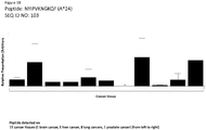
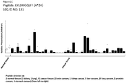
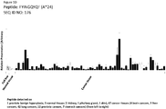
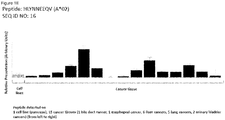


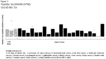
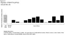
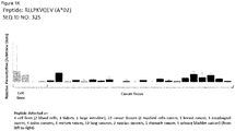
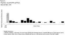
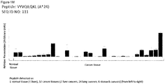

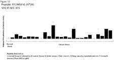
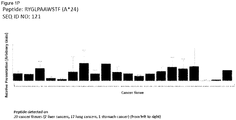


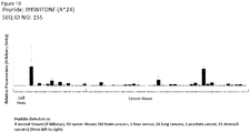
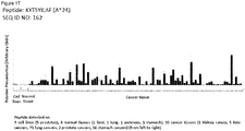


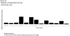
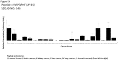
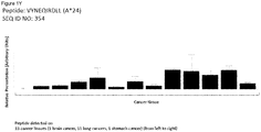
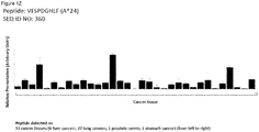
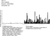
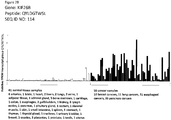

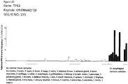

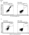
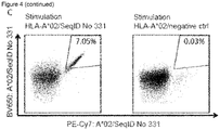

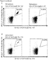
Classifications
-
- A—HUMAN NECESSITIES
- A61—MEDICAL OR VETERINARY SCIENCE; HYGIENE
- A61K—PREPARATIONS FOR MEDICAL, DENTAL OR TOILETRY PURPOSES
- A61K39/00—Medicinal preparations containing antigens or antibodies
- A61K39/0005—Vertebrate antigens
- A61K39/0011—Cancer antigens
-
- A—HUMAN NECESSITIES
- A61—MEDICAL OR VETERINARY SCIENCE; HYGIENE
- A61K—PREPARATIONS FOR MEDICAL, DENTAL OR TOILETRY PURPOSES
- A61K35/00—Medicinal preparations containing materials or reaction products thereof with undetermined constitution
- A61K35/12—Materials from mammals; Compositions comprising non-specified tissues or cells; Compositions comprising non-embryonic stem cells; Genetically modified cells
- A61K35/14—Blood; Artificial blood
- A61K35/17—Lymphocytes; B-cells; T-cells; Natural killer cells; Interferon-activated or cytokine-activated lymphocytes
-
- A—HUMAN NECESSITIES
- A61—MEDICAL OR VETERINARY SCIENCE; HYGIENE
- A61K—PREPARATIONS FOR MEDICAL, DENTAL OR TOILETRY PURPOSES
- A61K38/00—Medicinal preparations containing peptides
- A61K38/04—Peptides having up to 20 amino acids in a fully defined sequence; Derivatives thereof
- A61K38/08—Peptides having 5 to 11 amino acids
-
- A—HUMAN NECESSITIES
- A61—MEDICAL OR VETERINARY SCIENCE; HYGIENE
- A61K—PREPARATIONS FOR MEDICAL, DENTAL OR TOILETRY PURPOSES
- A61K39/00—Medicinal preparations containing antigens or antibodies
- A61K39/385—Haptens or antigens, bound to carriers
-
- A—HUMAN NECESSITIES
- A61—MEDICAL OR VETERINARY SCIENCE; HYGIENE
- A61K—PREPARATIONS FOR MEDICAL, DENTAL OR TOILETRY PURPOSES
- A61K39/00—Medicinal preparations containing antigens or antibodies
- A61K39/395—Antibodies; Immunoglobulins; Immune serum, e.g. antilymphocytic serum
- A61K39/39533—Antibodies; Immunoglobulins; Immune serum, e.g. antilymphocytic serum against materials from animals
- A61K39/39558—Antibodies; Immunoglobulins; Immune serum, e.g. antilymphocytic serum against materials from animals against tumor tissues, cells, antigens
-
- A—HUMAN NECESSITIES
- A61—MEDICAL OR VETERINARY SCIENCE; HYGIENE
- A61K—PREPARATIONS FOR MEDICAL, DENTAL OR TOILETRY PURPOSES
- A61K39/00—Medicinal preparations containing antigens or antibodies
- A61K39/46—Cellular immunotherapy
- A61K39/461—Cellular immunotherapy characterised by the cell type used
- A61K39/4611—T-cells, e.g. tumor infiltrating lymphocytes [TIL], lymphokine-activated killer cells [LAK] or regulatory T cells [Treg]
-
- A—HUMAN NECESSITIES
- A61—MEDICAL OR VETERINARY SCIENCE; HYGIENE
- A61K—PREPARATIONS FOR MEDICAL, DENTAL OR TOILETRY PURPOSES
- A61K39/00—Medicinal preparations containing antigens or antibodies
- A61K39/46—Cellular immunotherapy
- A61K39/464—Cellular immunotherapy characterised by the antigen targeted or presented
- A61K39/4643—Vertebrate antigens
- A61K39/4644—Cancer antigens
-
- A—HUMAN NECESSITIES
- A61—MEDICAL OR VETERINARY SCIENCE; HYGIENE
- A61P—SPECIFIC THERAPEUTIC ACTIVITY OF CHEMICAL COMPOUNDS OR MEDICINAL PREPARATIONS
- A61P35/00—Antineoplastic agents
-
- C—CHEMISTRY; METALLURGY
- C07—ORGANIC CHEMISTRY
- C07K—PEPTIDES
- C07K14/00—Peptides having more than 20 amino acids; Gastrins; Somatostatins; Melanotropins; Derivatives thereof
- C07K14/435—Peptides having more than 20 amino acids; Gastrins; Somatostatins; Melanotropins; Derivatives thereof from animals; from humans
- C07K14/46—Peptides having more than 20 amino acids; Gastrins; Somatostatins; Melanotropins; Derivatives thereof from animals; from humans from vertebrates
- C07K14/47—Peptides having more than 20 amino acids; Gastrins; Somatostatins; Melanotropins; Derivatives thereof from animals; from humans from vertebrates from mammals
- C07K14/4701—Peptides having more than 20 amino acids; Gastrins; Somatostatins; Melanotropins; Derivatives thereof from animals; from humans from vertebrates from mammals not used
- C07K14/4748—Tumour specific antigens; Tumour rejection antigen precursors [TRAP], e.g. MAGE
-
- C—CHEMISTRY; METALLURGY
- C07—ORGANIC CHEMISTRY
- C07K—PEPTIDES
- C07K14/00—Peptides having more than 20 amino acids; Gastrins; Somatostatins; Melanotropins; Derivatives thereof
- C07K14/435—Peptides having more than 20 amino acids; Gastrins; Somatostatins; Melanotropins; Derivatives thereof from animals; from humans
- C07K14/705—Receptors; Cell surface antigens; Cell surface determinants
- C07K14/70503—Immunoglobulin superfamily
- C07K14/7051—T-cell receptor (TcR)-CD3 complex
-
- C—CHEMISTRY; METALLURGY
- C07—ORGANIC CHEMISTRY
- C07K—PEPTIDES
- C07K14/00—Peptides having more than 20 amino acids; Gastrins; Somatostatins; Melanotropins; Derivatives thereof
- C07K14/435—Peptides having more than 20 amino acids; Gastrins; Somatostatins; Melanotropins; Derivatives thereof from animals; from humans
- C07K14/705—Receptors; Cell surface antigens; Cell surface determinants
- C07K14/70503—Immunoglobulin superfamily
- C07K14/70539—MHC-molecules, e.g. HLA-molecules
-
- C—CHEMISTRY; METALLURGY
- C07—ORGANIC CHEMISTRY
- C07K—PEPTIDES
- C07K16/00—Immunoglobulins [IGs], e.g. monoclonal or polyclonal antibodies
- C07K16/18—Immunoglobulins [IGs], e.g. monoclonal or polyclonal antibodies against material from animals or humans
- C07K16/28—Immunoglobulins [IGs], e.g. monoclonal or polyclonal antibodies against material from animals or humans against receptors, cell surface antigens or cell surface determinants
- C07K16/2803—Immunoglobulins [IGs], e.g. monoclonal or polyclonal antibodies against material from animals or humans against receptors, cell surface antigens or cell surface determinants against the immunoglobulin superfamily
- C07K16/2833—Immunoglobulins [IGs], e.g. monoclonal or polyclonal antibodies against material from animals or humans against receptors, cell surface antigens or cell surface determinants against the immunoglobulin superfamily against MHC-molecules, e.g. HLA-molecules
-
- C—CHEMISTRY; METALLURGY
- C07—ORGANIC CHEMISTRY
- C07K—PEPTIDES
- C07K16/00—Immunoglobulins [IGs], e.g. monoclonal or polyclonal antibodies
- C07K16/18—Immunoglobulins [IGs], e.g. monoclonal or polyclonal antibodies against material from animals or humans
- C07K16/28—Immunoglobulins [IGs], e.g. monoclonal or polyclonal antibodies against material from animals or humans against receptors, cell surface antigens or cell surface determinants
- C07K16/30—Immunoglobulins [IGs], e.g. monoclonal or polyclonal antibodies against material from animals or humans against receptors, cell surface antigens or cell surface determinants from tumour cells
- C07K16/3076—Immunoglobulins [IGs], e.g. monoclonal or polyclonal antibodies against material from animals or humans against receptors, cell surface antigens or cell surface determinants from tumour cells against structure-related tumour-associated moieties
-
- C—CHEMISTRY; METALLURGY
- C07—ORGANIC CHEMISTRY
- C07K—PEPTIDES
- C07K16/00—Immunoglobulins [IGs], e.g. monoclonal or polyclonal antibodies
- C07K16/18—Immunoglobulins [IGs], e.g. monoclonal or polyclonal antibodies against material from animals or humans
- C07K16/32—Immunoglobulins [IGs], e.g. monoclonal or polyclonal antibodies against material from animals or humans against translation products of oncogenes
-
- C—CHEMISTRY; METALLURGY
- C07—ORGANIC CHEMISTRY
- C07K—PEPTIDES
- C07K7/00—Peptides having 5 to 20 amino acids in a fully defined sequence; Derivatives thereof
- C07K7/04—Linear peptides containing only normal peptide links
- C07K7/06—Linear peptides containing only normal peptide links having 5 to 11 amino acids
-
- C—CHEMISTRY; METALLURGY
- C07—ORGANIC CHEMISTRY
- C07K—PEPTIDES
- C07K7/00—Peptides having 5 to 20 amino acids in a fully defined sequence; Derivatives thereof
- C07K7/04—Linear peptides containing only normal peptide links
- C07K7/08—Linear peptides containing only normal peptide links having 12 to 20 amino acids
-
- C—CHEMISTRY; METALLURGY
- C12—BIOCHEMISTRY; BEER; SPIRITS; WINE; VINEGAR; MICROBIOLOGY; ENZYMOLOGY; MUTATION OR GENETIC ENGINEERING
- C12N—MICROORGANISMS OR ENZYMES; COMPOSITIONS THEREOF; PROPAGATING, PRESERVING, OR MAINTAINING MICROORGANISMS; MUTATION OR GENETIC ENGINEERING; CULTURE MEDIA
- C12N15/00—Mutation or genetic engineering; DNA or RNA concerning genetic engineering, vectors, e.g. plasmids, or their isolation, preparation or purification; Use of hosts therefor
- C12N15/09—Recombinant DNA-technology
- C12N15/10—Processes for the isolation, preparation or purification of DNA or RNA
-
- C—CHEMISTRY; METALLURGY
- C12—BIOCHEMISTRY; BEER; SPIRITS; WINE; VINEGAR; MICROBIOLOGY; ENZYMOLOGY; MUTATION OR GENETIC ENGINEERING
- C12N—MICROORGANISMS OR ENZYMES; COMPOSITIONS THEREOF; PROPAGATING, PRESERVING, OR MAINTAINING MICROORGANISMS; MUTATION OR GENETIC ENGINEERING; CULTURE MEDIA
- C12N15/00—Mutation or genetic engineering; DNA or RNA concerning genetic engineering, vectors, e.g. plasmids, or their isolation, preparation or purification; Use of hosts therefor
- C12N15/09—Recombinant DNA-technology
- C12N15/11—DNA or RNA fragments; Modified forms thereof; Non-coding nucleic acids having a biological activity
- C12N15/115—Aptamers, i.e. nucleic acids binding a target molecule specifically and with high affinity without hybridising therewith ; Nucleic acids binding to non-nucleic acids, e.g. aptamers
-
- C—CHEMISTRY; METALLURGY
- C12—BIOCHEMISTRY; BEER; SPIRITS; WINE; VINEGAR; MICROBIOLOGY; ENZYMOLOGY; MUTATION OR GENETIC ENGINEERING
- C12N—MICROORGANISMS OR ENZYMES; COMPOSITIONS THEREOF; PROPAGATING, PRESERVING, OR MAINTAINING MICROORGANISMS; MUTATION OR GENETIC ENGINEERING; CULTURE MEDIA
- C12N5/00—Undifferentiated human, animal or plant cells, e.g. cell lines; Tissues; Cultivation or maintenance thereof; Culture media therefor
- C12N5/06—Animal cells or tissues; Human cells or tissues
- C12N5/0602—Vertebrate cells
- C12N5/0634—Cells from the blood or the immune system
- C12N5/0636—T lymphocytes
-
- C—CHEMISTRY; METALLURGY
- C12—BIOCHEMISTRY; BEER; SPIRITS; WINE; VINEGAR; MICROBIOLOGY; ENZYMOLOGY; MUTATION OR GENETIC ENGINEERING
- C12Q—MEASURING OR TESTING PROCESSES INVOLVING ENZYMES, NUCLEIC ACIDS OR MICROORGANISMS; COMPOSITIONS OR TEST PAPERS THEREFOR; PROCESSES OF PREPARING SUCH COMPOSITIONS; CONDITION-RESPONSIVE CONTROL IN MICROBIOLOGICAL OR ENZYMOLOGICAL PROCESSES
- C12Q1/00—Measuring or testing processes involving enzymes, nucleic acids or microorganisms; Compositions therefor; Processes of preparing such compositions
- C12Q1/68—Measuring or testing processes involving enzymes, nucleic acids or microorganisms; Compositions therefor; Processes of preparing such compositions involving nucleic acids
- C12Q1/6876—Nucleic acid products used in the analysis of nucleic acids, e.g. primers or probes
- C12Q1/6883—Nucleic acid products used in the analysis of nucleic acids, e.g. primers or probes for diseases caused by alterations of genetic material
- C12Q1/6886—Nucleic acid products used in the analysis of nucleic acids, e.g. primers or probes for diseases caused by alterations of genetic material for cancer
-
- G—PHYSICS
- G01—MEASURING; TESTING
- G01N—INVESTIGATING OR ANALYSING MATERIALS BY DETERMINING THEIR CHEMICAL OR PHYSICAL PROPERTIES
- G01N33/00—Investigating or analysing materials by specific methods not covered by groups G01N1/00 - G01N31/00
- G01N33/48—Biological material, e.g. blood, urine; Haemocytometers
- G01N33/50—Chemical analysis of biological material, e.g. blood, urine; Testing involving biospecific ligand binding methods; Immunological testing
- G01N33/53—Immunoassay; Biospecific binding assay; Materials therefor
- G01N33/574—Immunoassay; Biospecific binding assay; Materials therefor for cancer
- G01N33/57484—Immunoassay; Biospecific binding assay; Materials therefor for cancer involving compounds serving as markers for tumor, cancer, neoplasia, e.g. cellular determinants, receptors, heat shock/stress proteins, A-protein, oligosaccharides, metabolites
- G01N33/57492—Immunoassay; Biospecific binding assay; Materials therefor for cancer involving compounds serving as markers for tumor, cancer, neoplasia, e.g. cellular determinants, receptors, heat shock/stress proteins, A-protein, oligosaccharides, metabolites involving compounds localized on the membrane of tumor or cancer cells
-
- A—HUMAN NECESSITIES
- A61—MEDICAL OR VETERINARY SCIENCE; HYGIENE
- A61K—PREPARATIONS FOR MEDICAL, DENTAL OR TOILETRY PURPOSES
- A61K39/00—Medicinal preparations containing antigens or antibodies
- A61K2039/51—Medicinal preparations containing antigens or antibodies comprising whole cells, viruses or DNA/RNA
- A61K2039/515—Animal cells
- A61K2039/5158—Antigen-pulsed cells, e.g. T-cells
-
- A—HUMAN NECESSITIES
- A61—MEDICAL OR VETERINARY SCIENCE; HYGIENE
- A61K—PREPARATIONS FOR MEDICAL, DENTAL OR TOILETRY PURPOSES
- A61K39/00—Medicinal preparations containing antigens or antibodies
-
- C—CHEMISTRY; METALLURGY
- C07—ORGANIC CHEMISTRY
- C07K—PEPTIDES
- C07K2319/00—Fusion polypeptide
- C07K2319/40—Fusion polypeptide containing a tag for immunodetection, or an epitope for immunisation
-
- C—CHEMISTRY; METALLURGY
- C12—BIOCHEMISTRY; BEER; SPIRITS; WINE; VINEGAR; MICROBIOLOGY; ENZYMOLOGY; MUTATION OR GENETIC ENGINEERING
- C12N—MICROORGANISMS OR ENZYMES; COMPOSITIONS THEREOF; PROPAGATING, PRESERVING, OR MAINTAINING MICROORGANISMS; MUTATION OR GENETIC ENGINEERING; CULTURE MEDIA
- C12N2310/00—Structure or type of the nucleic acid
- C12N2310/10—Type of nucleic acid
- C12N2310/16—Aptamers
-
- C—CHEMISTRY; METALLURGY
- C12—BIOCHEMISTRY; BEER; SPIRITS; WINE; VINEGAR; MICROBIOLOGY; ENZYMOLOGY; MUTATION OR GENETIC ENGINEERING
- C12N—MICROORGANISMS OR ENZYMES; COMPOSITIONS THEREOF; PROPAGATING, PRESERVING, OR MAINTAINING MICROORGANISMS; MUTATION OR GENETIC ENGINEERING; CULTURE MEDIA
- C12N2501/00—Active agents used in cell culture processes, e.g. differentation
- C12N2501/50—Cell markers; Cell surface determinants
-
- C—CHEMISTRY; METALLURGY
- C12—BIOCHEMISTRY; BEER; SPIRITS; WINE; VINEGAR; MICROBIOLOGY; ENZYMOLOGY; MUTATION OR GENETIC ENGINEERING
- C12Q—MEASURING OR TESTING PROCESSES INVOLVING ENZYMES, NUCLEIC ACIDS OR MICROORGANISMS; COMPOSITIONS OR TEST PAPERS THEREFOR; PROCESSES OF PREPARING SUCH COMPOSITIONS; CONDITION-RESPONSIVE CONTROL IN MICROBIOLOGICAL OR ENZYMOLOGICAL PROCESSES
- C12Q2600/00—Oligonucleotides characterized by their use
- C12Q2600/106—Pharmacogenomics, i.e. genetic variability in individual responses to drugs and drug metabolism
-
- C—CHEMISTRY; METALLURGY
- C12—BIOCHEMISTRY; BEER; SPIRITS; WINE; VINEGAR; MICROBIOLOGY; ENZYMOLOGY; MUTATION OR GENETIC ENGINEERING
- C12Q—MEASURING OR TESTING PROCESSES INVOLVING ENZYMES, NUCLEIC ACIDS OR MICROORGANISMS; COMPOSITIONS OR TEST PAPERS THEREFOR; PROCESSES OF PREPARING SUCH COMPOSITIONS; CONDITION-RESPONSIVE CONTROL IN MICROBIOLOGICAL OR ENZYMOLOGICAL PROCESSES
- C12Q2600/00—Oligonucleotides characterized by their use
- C12Q2600/156—Polymorphic or mutational markers
-
- G—PHYSICS
- G01—MEASURING; TESTING
- G01N—INVESTIGATING OR ANALYSING MATERIALS BY DETERMINING THEIR CHEMICAL OR PHYSICAL PROPERTIES
- G01N2333/00—Assays involving biological materials from specific organisms or of a specific nature
- G01N2333/435—Assays involving biological materials from specific organisms or of a specific nature from animals; from humans
- G01N2333/705—Assays involving receptors, cell surface antigens or cell surface determinants
- G01N2333/70503—Immunoglobulin superfamily, e.g. VCAMs, PECAM, LFA-3
- G01N2333/70539—MHC-molecules, e.g. HLA-molecules
Definitions
- the present invention relates to peptides, proteins, nucleic acids and cells for use in immunotherapeutic methods.
- the present invention relates to the immunotherapy of cancer.
- the present invention furthermore relates to tumor-associated T-cell peptide epitopes, alone or in combination with other tumor-associated peptides that can for example serve as active pharmaceutical ingredients of vaccine compositions that stimulate anti-tumor immune responses, or to stimulate T cells ex vivo and transfer into patients.
- Peptides bound to molecules of the major histocompatibility complex (MHC), or peptides as such can also be targets of antibodies, soluble T-cell receptors, and other binding molecules.
- MHC major histocompatibility complex
- the present invention relates to several novel peptide sequences and their variants derived from HLA class I molecules of human tumor cells that can be used in vaccine compositions for eliciting anti-tumor immune responses, or as targets for the development of pharmaceutically / immunologically active compounds and cells.
- glioblastoma GB
- CLL chronic lymphocytic leukemia
- NSCLC non-small cell and small cell lung cancer
- Lung cancer is the most common type of cancer worldwide and the leading cause of death from cancer in many countries.
- Breast cancer is an immunogenic cancer entity and different types of infiltrating immune cells in primary tumors exhibit distinct prognostic and predictive significance.
- a large number of early phase immunotherapy trials have been conducted in breast cancer patients.
- Most of the completed vaccination studies targeted HER2 and carbohydrate antigens like MUC-1 and revealed rather disappointing results.
- Clinical data on the effects of immune checkpoint modulation with ipilimumab and other T cell-activating antibodies in breast cancer patients are emerging (Emens, 2012).
- CLL is not curable at present, many patients show only slow progression of the disease or worsening of symptoms. As patients do not benefit from an early onset of treatment, the initial approach is "watch and wait” (Richards et al., 1999). For patients with symptomatic or rapidly progressing disease, several treatment options are available. These include chemotherapy, targeted therapy, immune-based therapies like monoclonal antibodies, chimeric antigen-receptors (CARs) and active immunotherapy, and stem cell transplants.
- CARs chimeric antigen-receptors
- Monoclonal antibodies are widely used in hematologic malignancies. This is due to the knowledge of suitable antigens based on the good characterization of immune cell surface molecules and the accessibility of tumor cells in blood or bone marrow.
- Common monoclonal antibodies used in CLL therapy target either CD20 or CD52.
- Rituximab the first monoclonal anti-CD20 antibody originally approved by the FDA for treatment of NHLs, is now widely used in CLL therapy.
- Combinational treatment with rituximab/fludarabine/cyclophosphamide leads to higher CR rates and improved overall survival (OS) compared to the combination fludarabine/cyclophosphamide and has become the preferred treatment option (Hallek et al., 2008).
- Ofatumomab targets CD20 and is used for therapy of refractory CLL patients (Wierda et al., 2011).
- Obinutuzumab is another monoclonal anti-CD20 antibody used in first-line treatment in combination with chlorambucil (Goede et al., 2014).
- Alemtuzumab is an anti-CD52 antibody used for treatment of patients with chemotherapy-resistant disease or patients with poor prognostic factors as del 17p or p53 mutations (Parikh et al., 2011).
- Novel monoclonal antibodies target CD37 (otlertuzumab, BI 836826, IMGN529 and (177)Lu-tetulomab) or CD40 (dacetuzumab and lucatumumab) and are tested in pre-clinical settings (Robak and Robak, 2014).
- CAR engineered autologous chimeric antigen receptor
- Active immunotherapy includes the following strategies: gene therapy, whole modified tumor cell vaccines, DC-based vaccines and tumor associated antigen (TAA)-derived peptide vaccines.
- TAA tumor associated antigen
- B-CLL cells are transfected with immuno-(co-)stimulatory genes like IL-2, IL-12, TNF-alpha, GM-CSF, CD80, CD40L, LFA-3 and ICAM-1 to improve antigen presentation and T cell activation (Carballido et al., 2012). While specific T-cell responses and reduction in tumor cells are readily observed, immune responses are only transient.
- autologous DCs as antigen presenting cells to elicit anti-tumor responses. DCs have been loaded ex vivo with tumor associated peptides, whole tumor cell lysate and tumor-derived RNA or DNA.
- Another strategy uses whole tumor cells for fusion with DCs and generation of DC-B-CLL-cell hybrids.
- Transfected DCs initiated both CD4+ and CD8+ T-cell responses (Muller et al., 2004).
- Fusion hybrids and DCs loaded with tumor cell lysate or apoptotic bodies increased tumor-specific CD8+ T-cell responses.
- Patients that showed a clinical response had increased IL-12 serum levels and reduced numbers of Tregs (Palma et al., 2008).
- trioma cells B-CLL cells are fused to anti-Fc receptor expressing hybridoma cells that have anti-APC specificity. Trioma cells induced CLL-specific T-cell responses in vitro (Kronenberger et al., 2008).
- Another strategy makes use of irradiated autologous CLL cells with Bacillus Calmette-Guerin as an adjuvant as a vaccine.
- Bacillus Calmette-Guerin as an adjuvant as a vaccine.
- Several patients showed a reduction in leukocyte levels or stable disease (Hus et al., 2008).
- TAAs are over-expressed in CLL and are suitable for vaccinations. These include fibromodulin (Mayr et al., 2005), RHAMM/CD168 (Giannopoulos et al., 2006), MDM2 (Mayr et al., 2006), hTERT (Counter et al., 1995), the oncofetal antigen-immature laminin receptor protein(OFAiLRP) (Siegel et al., 2003), adipophilin (Schmidt et al., 2004), survivin (Granziero et al., 2001), KW1 to KW14 (Krackhardt et al., 2002) and the tumor-derived IgVHCDR3 region (Harig et al., 2001; Carballido et al., 2012).
- a phase I clinical trial was conducted using the RHAMM-derived R3 peptide as a vaccine. 5 of 6 patients had detectable R3-specific CD8+ T-cell responses
- CRC colorectal cancer
- VEGF-A vascular endothelial growth factor-A
- EGFR epidermal growth factor receptor
- cetuximab panitumumab
- VEGF-A vascular endothelial growth factor-A
- bevacizumab vascular endothelial growth factor-A
- the inhibitor for VEGF aflibercept, the tyrosine kinase inhibitor regorafenib and the thymidylate-synthetase inhibitor TAS-102 and the dUTPase inhibitor TAS-114 can be used (Stintzing, 2014; Wilson et al., 2014).
- TAAs tumor-associated antigens
- DC dendritic cell
- Peptide vaccines have so far been directed against carcinoembryonic antigen (CEA), mucin 1, EGFR, squamous cell carcinoma antigen recognized by T cells 3 (SART3), beta-human chorionic gonadotropin (beta-hCG), Wilms' Tumor antigen 1 (WT1), Survivin-2B, MAGE3, p53, ring finger protein 43 and translocase of the outer mitochondrial membrane 34 (TOMM34), or mutated KRAS.
- CEA carcinoembryonic antigen
- mucin 1 mucin 1
- EGFR squamous cell carcinoma antigen recognized by T cells 3
- beta-hCG beta-human chorionic gonadotropin
- WT1 Wilms' Tumor antigen 1
- Survivin-2B MAGE3, p53
- TOMM34 translocase of the outer mitochondrial membrane 34
- KRAS translocase of the outer mitochondrial membrane 34
- Dendritic cell vaccines comprise DCs pulsed with either TAA-derived peptides, tumor cell lysates, apoptotic tumor cells, or tumor RNA or DC-tumor cell fusion products. While many patients in phase I/II trials showed specific immunological responses, only the minority had a clinical benefit (Koido et al., 2013).
- Whole tumor cell vaccines consist of autologous tumor cells modified to secrete GM-CSF, modified by irradiation or virus-infected, irradiated cells. Most patients showed no clinical benefit in several phase II/ III trials (Koido et al., 2013).
- Vaccinia virus or replication-defective avian poxvirus encoding CEA as well as B7.1, ICAM-1 and LFA-3 have been used as vehicles in viral vector vaccines in phase I clinical trials.
- a different study used nonreplicating canarypox virus encoding CEA and B7.1.
- the primary treatment strategy for esophageal cancer depends on tumor stage and location, histological type and the medical condition of the patient. Surgery alone is not sufficient, except in a small subgroup of patients with squamous cell carcinoma.
- TTK protein kinase TGF protein kinase
- IGF insulin-like growth factor-II mRNA binding protein 3
- Intra-tumoral injection of activated T cells after in vitro challenge with autologous malignant cells elicited complete or partial tumor responses in four of eleven patients in a phase I/II study (Toomey et al., 2013).
- GC Gastric cancer
- GC Gastric cancer
- cytokine induced killer cells peptide-based vaccines targeting HER2/neu, MAGE-3 or vascular endothelial growth factor receptor 1 and 2 and dendritic cell-based vaccines targeting HER2/neu showed promising results in clinical GC trials.
- Immune checkpoint inhibition and engineered T cells might represent additional therapeutic options, which is currently evaluated in pre-clinical and clinical studies (Matsueda and Graham, 2014).
- glioblastoma WHO grade IV
- the standard therapy in young patients includes resection or biopsy of the tumor, focal radiation therapy and chemotherapy with temozolomide or CCNU/lomustine or a combination of procarbazine with CCNU and vincristine (PCV).
- PCV procarbazine with CCNU and vincristine
- bevacizumab anti-VEGF-antibody
- Antibodies directed against inhibitory T cell receptors or their ligands were shown to efficiently enhance T cell-mediated anti-tumor immune responses in different cancer types, including melanoma and bladder cancer.
- the effects of T cell activating antibodies like ipilimumab and nivolumab are therefore assessed in clinical GB trials, but preliminary data indicate autoimmune-related adverse events.
- peptide-based vaccines including heat-shock protein vaccines, autologous tumor cell vaccines, dendritic cell-based vaccines and viral protein-based vaccines.
- peptides derived from GB-associated proteins like epidermal growth factor receptor variant III (EGFRvIII) or heat shock proteins or dendritic cells pulsed with autologous tumor cell lysate or cytomegalo virus components are applied to induce an anti-tumor immune response in GB patients.
- EGFRvIII epidermal growth factor receptor variant III
- heat shock proteins or dendritic cells pulsed with autologous tumor cell lysate or cytomegalo virus components are applied to induce an anti-tumor immune response in GB patients.
- Adoptive transfer of genetically modified T cells is an additional immunotherapeutic approach for the treatment of GB.
- Different clinical trials currently evaluate the safety and efficacy of chimeric antigen receptor bearing T cells directed against HER2, IL-13 receptor alpha 2 and EGFRvIII (Ampie et al., 2015).
- Cytokines have been used to activate subsets of immune cells and/or increase the tumor immunogenicity (Reinisch et al., 2002; Sangro et al., 2004). Other trials have focused on the infusion of Tumor-infiltrating lymphocytes or activated peripheral blood lymphocytes (Shi et al., 2004; Takayama et al., 1991; Takayama et al., 2000b).
- the standard therapy in melanoma is complete surgical resection with surrounding healthy tissue. If resection is not complete or not possible at all, patients receive primary radiation therapy, which can be combined with interferon-alpha administration in advanced stages (stages IIB/C and IIIA-C).
- Enhancing the anti-tumor immune responses appears to be a promising strategy for the treatment of advanced melanoma.
- the immune checkpoint inhibitor ipilimumab as well as the BRAF kinase inhibitors vemurafenib and dabrafenib and the MEK inhibitor trametinib are already approved for the treatment of advanced melanoma. Both approaches increase the patient's anti-tumor immunity - ipilimumab directly by reducing T cell inhibition and the kinase inhibitors indirectly by enhancing the expression of melanocyte differentiation antigens. Additional checkpoint inhibitors (nivolumab and lambrolizumab) are currently investigated in clinical studies with first encouraging results.
- ipilimumab plus nivolumab ipilimumab plus a gp100-derived peptide vaccine
- ipilimumab plus dacarbazine ipilimumab plus IL-2
- iplimumab plus GM-CSF iplimumab plus GM-CSF
- Adoptive T cell transfer shows great promise for the treatment of advanced stage melanoma.
- T cells with high affinity T cell receptors for the melanocyte specific antigens MART1 and gp100 and the cancer-testis antigen MAGEA3 induced considerable toxic effects in clinical trials.
- adoptive T cell transfer has high therapeutic potential, but safety and tolerability of these treatments needs to be further increased (Phan and Rosenberg, 2013; Hinrichs and Restifo, 2013).
- Treatment options are determined by the type (small cell or non-small cell) and stage of cancer and include surgery, radiation therapy, chemotherapy, and targeted biological therapies such as bevacizumab, erlotinib and gefitinib.
- Surgical resection is the primary therapy in early as well as advanced stage ovarian carcinoma. Surgical removal is followed by systemic chemotherapy with platinum analogs, except for very low grade ovarian cancers (stage IA, grade 1), where postoperative chemotherapy is not indicated.
- stage IA very low grade ovarian cancers
- Immunotherapy appears to be a promising strategy to ameliorate the treatment of ovarian cancer patients, as the presence of pro-inflammatory tumor infiltrating lymphocytes, especially CD8-positive T cells, correlates with good prognosis and T cells specific for tumor-associated antigens can be isolated from cancer tissue.
- Cytokine therapy with interleukin-2, interferon-alpha, interferon-gamma or granulocyte-macrophage colony stimulating factor aims at boosting the patient's own anti-tumor immune response and these treatments have already shown promising results in small study cohorts.
- Monoclonal antibodies that specifically recognize tumor-associated proteins are thought to enhance immune cell-mediated killing of tumor cells.
- the anti-CA-125 antibodies or egovomab and abagovomab as well as the anti-EpCAM antibody catumaxomab achieved promising results in phase II and III studies.
- the anti-MUC1 antibody HMFG1 failed to clearly enhance survival in a phase III study.
- trastuzumab anti-HER2/neu antibody
- MOv18 and MORAb-003 anti-folate receptor alpha antibodies
- bevacizumab anti-VEGF antibody
- Adoptive transfer of immune cells achieved heterogeneous results in clinical trials.
- Adoptive transfer of autologous, in vitro expanded tumor infiltrating T cells was shown to be a promising approach in a pilot trial.
- transfer of T cells harboring a chimeric antigen receptor specific for folate receptor alpha did not induce a significant clinical response in a phase I trial.
- Dendritic cells pulsed with tumor cell lysate or tumor-associated proteins in vitro were shown to enhance the anti-tumor T cell response upon transfer, but the extent of T cell activation did not correlate with clinical effects. Transfer of natural killer cells caused significant toxicities in a phase II study.
- Intrinsic anti-tumor immunity as well as immunotherapy are hampered by an immunosuppressive tumor microenvironment.
- immunomodulatory drugs like cyclophosphamide, anti-CD25 antibodies and pegylated liposomal doxorubicin are tested in combination with immunotherapy.
- Most reliable data are currently available for ipilimumab, an anti-CTLA4 antibody, which enhances T cell activity. Ipilimumab was shown to exert significant anti-tumor effects in ovarian cancer patients (Mantia-Smaldone et al., 2012).
- pancreatic cancer is very limited.
- One major problem for effective treatment is the typically advanced tumor stage at diagnosis.
- pancreatic cancer is rather resistant to chemotherapeutics, which might be caused by the dense and hypovascular desmoplastic tumor stroma.
- Vaccination strategies are investigated as further innovative and promising alternative for the treatment of pancreatic cancer.
- Peptide-based vaccines targeting KRAS mutations, reactive telomerase, gastrin, survivin, CEA and MUC1 have already been evaluated in clinical trials, partially with promising results.
- clinical trials for dendritic cell-based vaccines, allogeneic GM-CSF-secreting vaccines and algenpantucel-L in pancreatic cancer patients also revealed beneficial effects of immunotherapy. Additional clinical trials further investigating the efficiency of different vaccination protocols are currently ongoing (Salman et al., 2013).
- the therapeutic strategy for prostate cancer mainly depends on the cancer stage.
- the dendritic cell-based vaccine sipuleucel-T was the first anti-cancer vaccine to be approved by the FDA. Due to its positive effect on survival in patients with CRPC, much effort is put into the development of further immunotherapies.
- the peptide vaccine prostate-specific antigen (PSA)-TRICOM, the personalized peptide vaccine PPV, the DNA vaccine pTVG-HP and the whole cell vaccine expressing GM-CSF GVAX showed promising results in different clinical trials.
- dendritic cell-based vaccines other than sipuleucel-T namely BPX-101 and DCVAC/Pa were shown to elicited clinical responses in prostate cancer patients.
- Immune checkpoint inhibitors like ipilimumab and nivolumab are currently evaluated in clinical studies as monotherapy as well as in combination with other treatments, including androgen deprivation therapy, local radiation therapy, PSA-TRICOM and GVAX.
- the immunomodulatory substance tasquinimod which significantly slowed progression and increased progression free survival in a phase II trial, is currently further investigated in a phase III trial.
- Lenalidomide another immunomodulator, induced promising effects in early phase clinical studies, but failed to improve survival in a phase III trial. Despite these disappointing results further lenalidomide trials are ongoing (Quinn et al., 2015).
- TroVax a vaccine using a tumor-associated antigen, 5T4, with a pox virus vector
- 5T4 tumor-associated antigen
- TRIST study evaluates whether TroVax (a vaccine using a tumor-associated antigen, 5T4, with a pox virus vector), added to first-line standard of care therapy, prolongs survival of patients with locally advanced or mRCC.
- Median survival had not been reached in either group with 399 patients (54%) remaining on study however analysis of the data confirms prior clinical results, demonstrating that TroVax is both immunologically active and that there is a correlation between the strength of the 5T4-specific antibody response and improved survival.
- AML One treatment option for AML is targeting CD33 with antibody-drug conjugates (anti-CD33 + calechiamicin, SGN-CD33a, anti-CD33 + actinium-225), bispecific antibodies (recognition of CD33 + CD3 (AMG 330) or CD33 + CD16) and chimeric antigen receptors (CARs) (Estey, 2014).
- antibody-drug conjugates anti-CD33 + calechiamicin, SGN-CD33a, anti-CD33 + actinium-225
- bispecific antibodies recognition of CD33 + CD3 (AMG 330) or CD33 + CD16
- CARs chimeric antigen receptors
- Passive immunization includes the injection of recombinant murine anti-Id monoclonal antibodies alone or in combination with IFNalpha, IL2 or chlorambucil.
- Active immunization includes the injection of recombinant protein (Id) conjugated to an adjuvant (KLH), given together with GM-CSF as an immune adjuvant.
- Id recombinant protein conjugated to an adjuvant
- KLH adjuvant
- Tumor-specific Id is produced by hybridoma cultures or using recombinant DNA technology (plasmids) by bacterial, insect or mammalian cell culture.
- Vaccines other than Id include the cancer-testis antigens MAGE, NY-ES01 and PASD-1, the B-cell antigen CD20 or cellular vaccines. The latest mentioned consist of DCs pulsed with apoptotic tumor cells, tumor cell lysate, DC-tumor cell fusion or DCs pulsed with tumor-derived RNA.
- In situ vaccination involves the vaccination with intra-tumoral CpG in combination with chemotherapy or irradiated tumor cells grown in the presence of GM-CSF and collection/ expansion/ re-infusion of T cells.
- Vaccination with antibodies that alter immunologic checkpoints are comprised of anti-CD40, anti-OX40, anti-41BB, anti-CD27, anti-GITR (agonist antibodies that directly enhance anti-tumor response) or anti-PD1, anti-CTLA-4 (blocking antibodies that inhibit the checkpoint that would hinder the immune response).
- anti-CD40 anti-OX40
- anti-41BB anti-CD27
- anti-GITR agonist antibodies that directly enhance anti-tumor response
- anti-PD1 anti-CTLA-4
- blocking antibodies that inhibit the checkpoint that would hinder the immune response are examples of anti-CTLA-4) and CT-011 (anti-PD1) (Palomba, 2012).
- endometrial carcinomas Treatment of endometrial carcinomas is stage-dependent.
- the majority of endometrical carcinomas comprises of well to moderately differentiated endometrioid adenocarcino-mas which are usually confined to the corpus uteri at diagnosis and can be cured by hysterectomy (World Cancer Report, 2014).
- Radio-(chemo-)therapy is chosen at late stages (Stage III and higher), in cases with lymph node infiltration or in cases in which the tumor cannot be excised.
- HPV infections provoke lesions that may ultimately lead to cervical cancer. Therefore, the HPV viral oncoproteins E6 and E7 that are constitutively expressed in high-grade lesions and cancer and are required for the onset and maintenance of the malignant phenotype are considered promising targets for immunotherapeutic approaches (Hung et al., 2008; Vici et al., 2014).
- ACT Adoptive T-cell therapy
- TILs tumor-infiltrating T cells
- Cholangiocarcinoma is mostly identified in advanced stages because it is difficult to diagnose. Cholangiocarcinoma is difficult to treat and is usually lethal.
- Gallbladder cancer is the most common and aggressive malignancy of the biliary tract worldwide.
- the standard treatment for bladder cancer includes surgery, radiation therapy, chemotherapy and immunotherapy.
- the bladder cancer is typically treated by transurethral resection potentially followed by intravesical chemotherapy and optionally combined with intravesical immunotherapeutic treatment with BCG ( bacillus Calmette-Guerin ).
- BCG bacillus Calmette-Guerin
- NMIBC non-muscle invasive bladder cancer
- the immune response to BCG is based on the following key steps: infection of urothelial and bladder cancer cells by BCG, followed by increased expression of antigen-presenting molecules, induction of immune response mediated via cytokine release, induction of antitumor activity via involvement of various immune cells (thereunder cytotoxic T lymphocytes, neutrophils, natural killer cells, and macrophages) (Fuge et al., 2015; Vogel et al., 2013).
- BCG treatment is in general well tolerated by patients but can be fatal especially by the immunocompromised patients.
- BCG refractory is observed in about 30-40% of patients (Fuge et al., 2015; Steinberg et al., 2016a).
- the treatment of patients who failed the BCG therapy is challenging.
- the patients who failed the BCG treatment are at high risk for developing of muscle-invasive disease. Radical cystectomy is the preferable treatment option for non-responders (Steinberg et al., 2016b; von Rundstedt and Lerner, 2015).
- the FDA approved second line therapy of BCG-failed NMIBC for patients who desire the bladder preservation is the chemotherapeutic treatment with valrubicin.
- a number of other second line therapies are available or being currently under investigation as well, thereunder immunotherapeutic approaches like combined BCG-interferon or BCG-check point inhibitor treatments, pre-BCG transdermal vaccination, treatment with Mycobacterium phlei cell wall-nucleic acid (MCNA) complex, mono- or combination chemotherapy with various agents like mitomycin C, gemcitabine, docetaxel, nab-paclitaxel, epirubicin, mitomycin/gemcitabine, gemcitabine/docetaxel, and device-assisted chemotherapies like thermochemo-, radiochemo-, electromotive or photodynamic therapies (Fuge et al., 2015; Steinberg et al., 2016b; von Rundstedt and Lerner, 2015). Further evaluation of available therapies in clinical trials is still required.
- immunotherapeutic approaches like combined BCG-interferon or BCG-check point inhibitor treatments, pre-BCG transdermal vaccination, treatment with Mycobacterium phlei cell wall-nucleic acid (
- the alternative treatment options for advanced bladder cancer are being investigated in ongoing clinical trials.
- the current clinical trials focused on the development of molecularly targeted therapies and immunotherapies.
- the targeted therapies investigate the effects of cancerogenesis related pathway inhibitors (i.e. mTOR, vascular endothelial, fibroblast, or epidermal growth factor receptors, anti-angiogenesis or cell cycle inhibitors) in the treatment of bladder cancer.
- cancerogenesis related pathway inhibitors i.e. mTOR, vascular endothelial, fibroblast, or epidermal growth factor receptors, anti-angiogenesis or cell cycle inhibitors
- checkpoint blockage agents like anti-PD1 monoclonal antibody and adoptive T-cell transfer (Knollman et al., 2015b; Grivas et al., 2015; Jones et al., 2016; Rouanne et al., 2016).
- HNSCC Head and neck squamous cell carcinomas
- HNSCC is considered an immunosuppressive disease, characterized by the dysregulation of immunocompetent cells and impaired cytokine secretion (Economopoulou et al., 2016). Immunotherapeutic strategies differ between HPV-negative and HPV-positive tumors.
- the viral oncoproteins E6 and E7 represent good targets, as they are continuously expressed by tumor cells and are essential to maintain the transformation status of HPV-positive cancer cells.
- Several vaccination therapies are currently under investigation in HPV-positive HNSCC, including DNA vaccines, peptide vaccines and vaccines involving dendritic cells (DCs). Additionally, an ongoing phase II clinical trial investigates the efficacy of lymphodepletion followed by autologous infusion of TILs in patients with HPV-positive tumors (Economopoulou et al., 2016).
- chimeric IgG1 anti-EGFR monoclonal antibody cetuximab has been approved by the FDA in combination with chemotherapy as standard first line treatment for recurring/metastatic HNSCC.
- Other anti-EGFR monoclonal antibodies including panitumumab, nimotuzumab and zalutumumab, are evaluated in HNSCC.
- immune checkpoint inhibitors are investigated in clinical trials for their use in HNSCC.
- Ipilimumab anti-CTLA-4
- tremelimumab anti-CTLA-4
- pembrolizumab anti-PD-1
- nivolumab anti-PD-1
- durvalumab anti-PD-1
- anti-KIR anti-KIR
- urelumab anti-CD137
- anti-LAG-3 anti-LAG-3.
- T cells were induced against either irradiated autologous tumor cells or EBV. Results in disease control and overall survival were promising (Economopoulou et al., 2016).
- HCC hepatocellular carcinoma
- CRC colorectal carcinoma
- GB glioblastoma
- GC gastric cancer
- esophageal cancer non-small cell lung cancer
- PC pancreatic cancer
- PC renal cell carcinoma
- BPH benign prostate hyperplasia
- PCA prostate cancer
- OC ovarian cancer
- melanoma breast cancer, chronic lymphocytic leukemia (CLL), Merkel cell carcinoma (MCC), small cell lung cancer (SCLC), Non-Hodgkin lymphoma (NHL), acute myeloid leukemia (AML), gallbladder cancer and cholangiocarcinoma (GBC, CCC), urinary bladder cancer (UBC), uterine cancer (UEC), head and neck squamous cell carcinoma (HNSCC), in particular.
- HCC hepatocellular carcinoma
- CRC colorectal carcinoma
- GB glioblastoma
- PCA benign prostate hyperplasia
- PCA prostate cancer
- Immunotherapy of cancer represents an option of specific targeting of cancer cells while minimizing side effects. Cancer immunotherapy makes use of the existence of tumor associated antigens.
- TAAs tumor associated antigens
- T-cell based immunotherapy targets peptide epitopes derived from tumor-associated or tumor-specific proteins, which are presented by molecules of the major histocompatibility complex (MHC).
- MHC major histocompatibility complex
- the antigens that are recognized by the tumor specific T lymphocytes, that is, the epitopes thereof, can be molecules derived from all protein classes, such as enzymes, receptors, transcription factors, etc. which are expressed and, as compared to unaltered cells of the same origin, usually up-regulated in cells of the respective tumor.
- MHC class I There are two classes of MHC-molecules, MHC class I and MHC class II.
- MHC class I molecules are composed of an alpha heavy chain and beta-2-microglobulin, MHC class II molecules of an alpha and a beta chain. Their three-dimensional conformation results in a binding groove, which is used for non-covalent interaction with peptides.
- MHC class I molecules can be found on most nucleated cells. They present peptides that result from proteolytic cleavage of predominantly endogenous proteins, defective ribosomal products (DRIPs) and larger peptides. However, peptides derived from endosomal compartments or exogenous sources are also frequently found on MHC class I molecules.
- DRIPs defective ribosomal products
- MHC class II molecules can be found predominantly on professional antigen presenting cells (APCs), and primarily present peptides of exogenous or transmembrane proteins that are taken up by APCs e.g. during endocytosis, and are subsequently processed.
- APCs professional antigen presenting cells
- TCR T-cell receptor
- CD4-positive-helper-T cells CD4-positive-helper-T cells bearing the appropriate TCR. It is well known that the TCR, the peptide and the MHC are thereby present in a stoichiometric amount of 1:1:1.
- CD4-positive helper T cells play an important role in inducing and sustaining effective responses by CD8-positive cytotoxic T cells.
- TAA tumor associated antigens
- T helper cells support a cytotoxic T cell- (CTL-) friendly cytokine milieu (Mortara et al., 2006) and attract effector cells, e.g. CTLs, natural killer (NK) cells, macrophages, and granulocytes (Hwang et al., 2007).
- CTL- cytotoxic T cell- friendly cytokine milieu
- NK natural killer cells
- macrophages macrophages
- granulocytes Hwang et al., 2007.
- MHC class II molecules In the absence of inflammation, expression of MHC class II molecules is mainly restricted to cells of the immune system, especially professional antigen-presenting cells (APC), e.g., monocytes, monocyte-derived cells, macrophages, dendritic cells.
- APC professional antigen-presenting cells
- monocytes e.g., monocytes, monocyte-derived cells, macrophages, dendritic cells.
- monocytes e.g., monocytes, monocyte-derived cells, macrophages, dendritic cells.
- Elongated peptides of the invention can act as MHC class II active epitopes.
- T-helper cells activated by MHC class II epitopes, play an important role in orchestrating the effector function of CTLs in anti-tumor immunity.
- T-helper cell epitopes that trigger a T-helper cell response of the TH1 type support effector functions of CD8-positive killer T cells, which include cytotoxic functions directed against tumor cells displaying tumor-associated peptide/MHC complexes on their cell surfaces.
- tumor-associated T-helper cell peptide epitopes alone or in combination with other tumor-associated peptides, can serve as active pharmaceutical ingredients of vaccine compositions that stimulate anti-tumor immune responses.
- CD4-positive T cells are sufficient for inhibiting manifestation of tumors via inhibition of angiogenesis by secretion of interferon-gamma (IFN ⁇ ) (Beatty and Paterson, 2001; Mumberg et al., 1999). There is evidence for CD4 T cells as direct anti-tumor effectors (Braumuller et al., 2013; Tran et al., 2014).
- IFN ⁇ interferon-gamma
- HLA class II molecules Since the constitutive expression of HLA class II molecules is usually limited to immune cells, the possibility of isolating class II peptides directly from primary tumors was previously not considered possible. However, Dengjel et al. were successful in identifying a number of MHC Class II epitopes directly from tumors ( WO 2007/028574 , EP 1 760 088 B1 ).
- CD8 and CD4 dependent Since both types of response, CD8 and CD4 dependent, contribute jointly and synergistically to the anti-tumor effect, the identification and characterization of tumor-associated antigens recognized by either CD8+ T cells (ligand: MHC class I molecule + peptide epitope) or by CD4-positive T-helper cells (ligand: MHC class II molecule + peptide epitope) is important in the development of tumor vaccines.
- MHC-class-I-binding peptides are usually 8-12 amino acid residues in length and usually contain two conserved residues ("anchors") in their sequence that interact with the corresponding binding groove of the MHC-molecule. In this way each MHC allele has a "binding motif" determining which peptides can bind specifically to the binding groove.
- peptides In the MHC class I dependent immune reaction, peptides not only have to be able to bind to certain MHC class I molecules expressed by tumor cells, they subsequently also have to be recognized by T cells bearing specific T cell receptors (TCR).
- TCR T cell receptors
- the antigen should be expressed mainly by tumor cells and not, or in comparably small amounts, by normal healthy tissues.
- the peptide should be over-presented by tumor cells as compared to normal healthy tissues. It is furthermore desirable that the respective antigen is not only present in a type of tumor, but also in high concentrations (i.e. copy numbers of the respective peptide per cell).
- Tumor-specific and tumor-associated antigens are often derived from proteins directly involved in transformation of a normal cell to a tumor cell due to their function, e.g. in cell cycle control or suppression of apoptosis.
- downstream targets of the proteins directly causative for a transformation may be up-regulated und thus may be indirectly tumor-associated.
- Such indirect tumor-associated antigens may also be targets of a vaccination approach (Singh-Jasuja et al., 2004). It is essential that epitopes are present in the amino acid sequence of the antigen, in order to ensure that such a peptide ("immunogenic peptide"), being derived from a tumor associated antigen, leads to an in vitro or in vivo T-cell-response.
- any peptide able to bind an MHC molecule may function as a T-cell epitope.
- a prerequisite for the induction of an in vitro or in vivo T-cell-response is the presence of a T cell having a corresponding TCR and the absence of immunological tolerance for this particular epitope.
- TAAs are a starting point for the development of a T cell based therapy including but not limited to tumor vaccines.
- the methods for identifying and characterizing the TAAs are usually based on the use of T-cells that can be isolated from patients or healthy subjects, or they are based on the generation of differential transcription profiles or differential peptide expression patterns between tumors and normal tissues.
- the identification of genes over-expressed in tumor tissues or human tumor cell lines, or selectively expressed in such tissues or cell lines does not provide precise information as to the use of the antigens being transcribed from these genes in an immune therapy.
- T cell which upon stimulation with a specific antigen can be clonally expanded and is able to execute effector functions
- the immunogenicity of the underlying peptides is secondary. In these cases, the presentation is the determining factor.
- the present invention relates to a peptide comprising an amino acid sequence selected from the group consisting of SEQ ID NO: 1 to SEQ ID NO: 388 or a variant sequence thereof which is at least 77%, preferably at least 88%, homologous (preferably at least 77% or at least 88% identical) to SEQ ID NO: 1 to SEQ ID NO: 388, wherein said variant binds to MHC and/or induces T cells cross-reacting with said peptide, or a pharmaceutical acceptable salt thereof, wherein said peptide is not the underlying full-length polypeptide.
- RNA expression profile of the corresponding gene can help to select appropriate peptides.
- some peptides are hard to detect by mass spectrometry, either due to their chemical properties or to their low copy numbers on cells, and a screening approach focusing on detection of peptide presentation may fail to identify these targets.
- these targets may be detected by an alternative approach starting with analysis of gene expression in normal tissues and secondarily assessing peptide presentation and gene expression in tumors. This approach was realized in this invention using an mRNA database (Lonsdale, 2013) in combination with further gene expression data (including tumor samples), as well as peptide presentation data.
- mRNA of a gene is nearly absent in normal tissues, especially in vital organ systems, targeting the corresponding peptides by even very potent strategies (such as bispecific affinity-optimized antibodies or T-cell receptors), is more likely to be safe.
- Such peptides even if identified on only a small percentage of tumor tissues, represent interesting targets.
- Routine mass spectrometry analysis is not sensitive enough to assess target coverage on the peptide level. Rather, tumor mRNA expression can be used to assess coverage. For detection of the peptide itself, a targeted mass spectrometry approach with higher sensitivity than in the routine screening may be necessary and may lead to a better estimation of coverage on the level of peptide presentation.
- the present invention further relates to a peptide of the present invention comprising a sequence that is selected from the group consisting of SEQ ID NO: 1 to SEQ ID NO: 388 or a variant thereof, which is at least 77%, preferably at least 88%, homologous (preferably at least 77% or at least 88% identical) to SEQ ID NO: 1 to SEQ ID NO: 388, wherein said peptide or variant thereof has an overall length of between 8 and 100, preferably between 8 and 30, and most preferred of between 8 and 14 amino acids.
- Table 1A Peptides according to the present invention, their respective SEQ ID NOs, and the prospective source (underlying) genes for these peptides. All peptides in Table 1A and Table 2A bind to HLA-A*02. Peptides in Table 1C and Table 2B bind to HLA-A*24. Peptides in Table 1B bind to HLA class II alleles. The peptides in Table 3 are additional peptides that are HLA-A*24 binding and may be useful in combination with the other peptides of the invention.
- Table 1A Peptides according to the present invention, HLA-A*02-bindin. SEQ ID No.
- the present invention furthermore generally relates to the peptides according to the present invention for use in the treatment of proliferative diseases, such as, for example, glioblastoma (GB), breast cancer (BRCA), colorectal cancer (CRC), renal cell carcinoma (RCC), chronic lymphocytic leukemia (CLL), hepatocellular carcinoma (HCC), non-small cell and small cell lung cancer (NSCLC, SCLC), Non-Hodgkin lymphoma (NHL), acute myeloid leukemia (AML), ovarian cancer (OC), pancreatic cancer (PC), prostate cancer (PCA), esophageal cancer including cancer of the gastric-esophageal junction (OSCAR), gallbladder cancer and cholangiocarcinoma (GBC, CCC), melanoma (MEL), gastric cancer (GC), testis cancer (TC), urinary bladder cancer (UBC), head-and neck squamous cell carcinoma (HNSCC), and uterine cancer (UEC).
- peptides - alone or in combination - selected from the group consisting of SEQ ID NO: 1 to SEQ ID NO: 388. More preferred are the peptides - alone or in combination - selected from the group consisting of SEQ ID NO: 1 to SEQ ID NO: 295 (see Table 1A, B, C), and their uses in the immunotherapy of glioblastoma, breast cancer, colorectal cancer, renal cell carcinoma, chronic lymphocytic leukemia, hepatocellular carcinoma, non-small cell and small cell lung cancer, Non-Hodgkin lymphoma, acute myeloid leukemia, ovarian cancer, pancreatic cancer, prostate cancer, esophageal cancer including cancer of the gastric-esophageal junction, gallbladder cancer and cholangiocarcinoma, melanoma, gastric cancer, testis cancer, urinary bladder cancer, head-and neck squamous cell carcinoma, or uterine cancer.
- peptides - alone or in combination - according to the present invention selected from the group consisting of SEQ ID NO: 70, 80, 323, and 325. More preferred are the peptides - alone or in combination - selected from the group consisting of SEQ ID NO: 70, 80, 323, and 325, and their uses in the immunotherapy of glioblastoma, breast cancer, colorectal cancer, renal cell carcinoma, chronic lymphocytic leukemia, hepatocellular carcinoma, non-small cell and small cell lung cancer, Non-Hodgkin lymphoma, acute myeloid leukemia, ovarian cancer, pancreatic cancer, prostate cancer, esophageal cancer including cancer of the gastric-esophageal junction, gallbladder cancer and cholangiocarcinoma, melanoma, gastric cancer, testis cancer, urinary bladder cancer, head-and neck squamous cell carcinoma, or uterine cancer.
- peptides - alone or in combination - according to the present invention selected from the group consisting of SEQ ID NO: 391, and 403. More preferred are the peptides - alone or in combination - selected from the group consisting of SEQ ID NO: 391, and 403, and their uses in the immunotherapy of glioblastoma, breast cancer, colorectal cancer, renal cell carcinoma, chronic lymphocytic leukemia, hepatocellular carcinoma, non-small cell and small cell lung cancer, Non-Hodgkin lymphoma, acute myeloid leukemia, ovarian cancer, pancreatic cancer, prostate cancer, esophageal cancer including cancer of the gastric-esophageal junction, gallbladder cancer and cholangiocarcinoma, melanoma, gastric cancer, testis cancer, urinary bladder cancer, head-and neck squamous cell carcinoma, or uterine cancer.
- Example 1 many of the peptides according to the present invention are found on various tumor types and can, thus, also be used in the immunotherapy of a variety of indications. Over-expression of the underlying polypeptides in a variety of cancers, as shown in Example 2, hints towards the usefulness of these peptides in various other oncological indications.
- another aspect of the present invention relates to the use of the peptides according to the present invention for the - preferably combined - treatment of a proliferative disease selected from the group of glioblastoma, breast cancer, colorectal cancer, renal cell carcinoma, chronic lymphocytic leukemia, hepatocellular carcinoma, non-small cell and small cell lung cancer, Non-Hodgkin lymphoma, acute myeloid leukemia, ovarian cancer, pancreatic cancer, prostate cancer, esophageal cancer including cancer of the gastric-esophageal junction, gallbladder cancer and cholangiocarcinoma, melanoma, gastric cancer, testis cancer, urinary bladder cancer, head-and neck squamous cell carcinoma, or uterine cancer.
- a proliferative disease selected from the group of glioblastoma, breast cancer, colorectal cancer, renal cell carcinoma, chronic lymphocytic leukemia, hepatocellular carcinoma, non-small cell
- the present invention furthermore relates to peptides according to the present invention that have the ability to bind to a molecule of the human major histocompatibility complex (MHC) class-I or - in an elongated form, such as a length-variant - MHC class -II.
- MHC human major histocompatibility complex
- the present invention further relates to the peptides according to the present invention wherein said peptides (each) consist or consist essentially of an amino acid sequence according to SEQ ID NO: 1 to SEQ ID NO: 388.
- the present invention further relates to the peptides according to the present invention, wherein said peptide is modified and/or includes non-peptide bonds.
- the present invention further relates to the peptides according to the present invention, wherein said peptide is part of a fusion protein, in particular fused to the N-terminal amino acids of the HLA-DR antigen-associated invariant chain (li), or fused to (or into the sequence of) an antibody, such as, for example, an antibody that is specific for dendritic cells.
- a fusion protein in particular fused to the N-terminal amino acids of the HLA-DR antigen-associated invariant chain (li), or fused to (or into the sequence of) an antibody, such as, for example, an antibody that is specific for dendritic cells.
- Another embodiment of the present invention relates to a non-naturally occurring peptide wherein said peptide consists or consists essentially of an amino acid sequence according to SEQ ID No: 1 to SEQ ID No: 48 and has been synthetically produced (e.g. synthesized) as a pharmaceutically acceptable salt.
- Methods to synthetically produce peptides are well known in the art.
- the salts of the peptides according to the present invention differ substantially from the peptides in their state(s) in vivo , as the peptides as generated in vivo are no salts.
- the non-natural salt form of the peptide mediates the solubility of the peptide, in particular in the context of pharmaceutical compositions comprising the peptides, e.g.
- the peptide vaccines as disclosed herein.
- a sufficient and at least substantial solubility of the peptide(s) is required in order to efficiently provide the peptides to the subject to be treated.
- the salts are pharmaceutically acceptable salts of the peptides.
- salts according to the invention include alkaline and earth alkaline salts such as salts of the Hofmeister series comprising as anions PO 4 3- , SO 4 2- , CH 3 COO - , Cl - , Br - , NO 3 - , ClO 4 - , I - , SCN - and as cations NH 4 + , Rb + , K + , Na + , Cs + , Li + , Zn 2+ , Mg 2+ , Ca 2+ , Mn 2+ , Cu 2+ and Ba 2+ .
- alkaline and earth alkaline salts such as salts of the Hofmeister series comprising as anions PO 4 3- , SO 4 2- , CH 3 COO - , Cl - , Br - , NO 3 - , ClO 4 - , I - , SCN - and as cations NH 4 + , Rb + , K + , Na + , Cs + , Li +
- Particularly salts are selected from (NH 4 ) 3 PO 4 , (NH 4 ) 2 HPO 4 , (NH 4 )H 2 PO 4 , (NH 4 ) 2 SO 4 , NH 4 CH 3 COO, NH 4 Cl, NH 4 Br, NH 4 NO 3 , NH 4 ClO 4 , NH 4 I, NH 4 SCN, Rb 3 PO 4 , Rb 2 HPO 4 , RbH 2 PO 4 , Rb 2 SO 4 , Rb 4 CH 3 COO, Rb 4 Cl, Rb 4 Br, Rb 4 NO 3 , Rb 4 ClO 4 , Rb 4 I, Rb 4 SCN, K 3 PO 4 , K 2 HPO 4 , KH 2 PO 4 , K 2 SO 4 , KCH 3 COO, KCl, KBr, KNO 3 , KClO 4 , Kl, KSCN, Na 3 PO 4 , Na 2 HPO 4 , NaH 2 PO 4 , Na 2 SO 4 , NaCH
- NH acetate MgCl 2 , KH 2 PO 4 , Na 2 SO 4 , KCl, NaCl, and CaCl 2 , such as, for example, the chloride or acetate (trifluoroacetate) salts.
- peptides and variants may be synthesized by the Fmoc-polyamide mode of solid-phase peptide synthesis as disclosed by Lukas et al. (Lukas et al., 1981) and by references as cited therein.
- Temporary N-amino group protection is afforded by the 9-fluorenylmethyloxycarbonyl (Fmoc) group. Repetitive cleavage of this highly base-labile protecting group is done using 20% piperidine in N, N-dimethylformamide.
- Side-chain functionalities may be protected as their butyl ethers (in the case of serine threonine and tyrosine), butyl esters (in the case of glutamic acid and aspartic acid), butyloxycarbonyl derivative (in the case of lysine and histidine), trityl derivative (in the case of cysteine) and 4-methoxy-2,3,6-trimethylbenzenesulphonyl derivative (in the case of arginine).
- glutamine or asparagine are C-terminal residues, use is made of the 4,4'-dimethoxybenzhydryl group for protection of the side chain amido functionalities.
- the solid-phase support is based on a polydimethyl-acrylamide polymer constituted from the three monomers dimethylacrylamide (backbone-monomer), bisacryloylethylene diamine (cross linker) and acryloylsarcosine methyl ester (functionalizing agent).
- the peptide-to-resin cleavable linked agent used is the acid-labile 4-hydroxymethyl-phenoxyacetic acid derivative. All amino acid derivatives are added as their preformed symmetrical anhydride derivatives with the exception of asparagine and glutamine, which are added using a reversed N, N-dicyclohexyl-carbodiimide/1hydroxybenzotriazole mediated coupling procedure.
- peptides are cleaved from the resin support with concomitant removal of side-chain protecting groups by treatment with 95% trifluoroacetic acid containing a 50 % scavenger mix.
- Scavengers commonly used include ethanedithiol, phenol, anisole and water, the exact choice depending on the constituent amino acids of the peptide being synthesized. Also a combination of solid phase and solution phase methodologies for the synthesis of peptides is possible (see, for example, (Bruckdorfer et al., 2004), and the references as cited therein).
- Trifluoroacetic acid is removed by evaporation in vacuo, with subsequent trituration with diethyl ether affording the crude peptide.
- Any scavengers present are removed by a simple extraction procedure which on lyophilization of the aqueous phase affords the crude peptide free of scavengers.
- Reagents for peptide synthesis are generally available from e.g. Calbiochem-Novabiochem (Nottingham, UK).
- Purification may be performed by any one, or a combination of, techniques such as recrystallization, size exclusion chromatography, ion-exchange chromatography, hydrophobic interaction chromatography and (usually) reverse-phase high performance liquid chromatography using e.g. acetonitrile/water gradient separation.
- techniques such as recrystallization, size exclusion chromatography, ion-exchange chromatography, hydrophobic interaction chromatography and (usually) reverse-phase high performance liquid chromatography using e.g. acetonitrile/water gradient separation.
- the present invention further relates to a nucleic acid, encoding the peptides according to the present invention.
- the present invention further relates to the nucleic acid according to the present invention that is DNA, cDNA, PNA, RNA or combinations thereof.
- the present invention further relates to an expression vector capable of expressing and/or expressing a nucleic acid according to the present invention.
- the present invention further relates to a peptide according to the present invention, a nucleic acid according to the present invention or an expression vector according to the present invention for use in the treatment of diseases and in medicine, in particular in the treatment of cancer.
- the present invention further relates to antibodies that are specific against the peptides according to the present invention or complexes of said peptides according to the present invention with MHC, and methods of making these.
- the present invention further relates to T-cell receptors (TCRs), in particular soluble TCR (sTCRs) and cloned TCRs engineered into autologous or allogeneic T cells, and methods of making these, as well as NK cells or other cells bearing said TCR or cross-reacting with said TCRs.
- TCRs T-cell receptors
- sTCRs soluble TCR
- cloned TCRs engineered into autologous or allogeneic T cells
- the antibodies and TCRs are additional embodiments of the immunotherapeutic use of the peptides according to the invention at hand.
- the present invention further relates to a host cell comprising a nucleic acid according to the present invention or an expression vector as described before.
- the present invention further relates to the host cell according to the present invention that is an antigen presenting cell, and preferably is a dendritic cell.
- the present invention further relates to a method for producing a peptide according to the present invention, said method comprising culturing the host cell according to the present invention, and isolating the peptide from said host cell or its culture medium.
- the present invention further relates to said method according to the present invention, wherein the antigen is loaded onto class I or II MHC molecules expressed on the surface of a suitable antigen-presenting cell or artificial antigen-presenting cell by contacting a sufficient amount of the antigen with an antigen-presenting cell.
- the present invention further relates to the method according to the present invention, wherein the antigen-presenting cell comprises an expression vector capable of expressing or expressing said peptide containing SEQ ID No. 1 to SEQ ID No.: 388, preferably containing SEQ ID No. 1 to SEQ ID No. 295, or a variant amino acid sequence.
- the present invention further relates to activated T cells, produced by the method according to the present invention, wherein said T cell selectively recognizes a cell which expresses a polypeptide comprising an amino acid sequence according to the present invention.
- the present invention further relates to a method of killing target cells in a patient which target cells aberrantly express a polypeptide comprising any amino acid sequence according to the present invention, the method comprising administering to the patient an effective number of T cells as produced according to the present invention.
- the present invention further relates to the use of any peptide as described, the nucleic acid according to the present invention, the expression vector according to the present invention, the cell according to the present invention, the activated T lymphocyte, the T cell receptor or the antibody or other peptide- and/or peptide-MHC-binding molecules according to the present invention as a medicament or in the manufacture of a medicament.
- said medicament is active against cancer.
- said medicament is a cellular therapy, a vaccine or a protein based on a soluble TCR or antibody.
- the present invention further relates to a use according to the present invention, wherein said cancer cells are glioblastoma, breast cancer, colorectal cancer, renal cell carcinoma, chronic lymphocytic leukemia, hepatocellular carcinoma, non-small cell and small cell lung cancer, Non-Hodgkin lymphoma, acute myeloid leukemia, ovarian cancer, pancreatic cancer, prostate cancer, esophageal cancer including cancer of the gastric-esophageal junction, gallbladder cancer and cholangiocarcinoma, melanoma, gastric cancer, testis cancer, urinary bladder cancer, head and neck squamous cell carcinoma, or uterine cancer.
- said cancer cells are glioblastoma, breast cancer, colorectal cancer, renal cell carcinoma, chronic lymphocytic leukemia, hepatocellular carcinoma, non-small cell and small cell lung cancer, Non-Hodgkin lymphoma, acute myeloid leukemia, ovarian cancer, pancre
- the present invention further relates to biomarkers based on the peptides according to the present invention, herein called "targets", that can be used in the diagnosis of cancer, preferably of glioblastoma, breast cancer, colorectal cancer, renal cell carcinoma, chronic lymphocytic leukemia, hepatocellular carcinoma, non-small cell and small cell lung cancer, Non-Hodgkin lymphoma, acute myeloid leukemia, ovarian cancer, pancreatic cancer, prostate cancer, esophageal cancer including cancer of the gastric-esophageal junction, gallbladder cancer and cholangiocarcinoma, melanoma, gastric cancer, testis cancer, urinary bladder cancer, head and neck squamous cell carcinoma, or uterine cancer.
- targets preferably of glioblastoma, breast cancer, colorectal cancer, renal cell carcinoma, chronic lymphocytic leukemia, hepatocellular carcinoma, non-small cell and small cell lung cancer, Non-Ho
- the marker can be over-presentation of the peptide(s) themselves, or over-expression of the corresponding gene(s).
- the markers may also be used to predict the probability of success of a treatment, preferably an immunotherapy, and most preferred an immunotherapy targeting the same target that is identified by the biomarker.
- an antibody or soluble TCR can be used to stain sections of the tumor to detect the presence of a peptide of interest in complex with MHC.
- the antibody carries a further effector function such as an immune stimulating domain or toxin.
- the present invention also relates to the use of these novel targets in the context of cancer treatment.
- A4GNT is frequently expressed in pancreatic cancer cells but not peripheral blood cells and quantitative analysis of A4GNT mRNA expressed in the mononuclear cell fraction of peripheral blood will contribute to the detection of pancreatic cancer (Ishizone et al., 2006).
- A4GNT mRNA was detectable in 80% of patients with an early stage of gastric cancer when the cancer cells were limited to the gastric mucosa, and the expression levels of A4GNT mRNA were increased in association with tumor progression (Shimizu et al., 2003).
- ADAM10 is up-regulated, with levels generally correlating with parameters of tumor progression and poor outcome.
- a selective inhibitor against ADAM10 has been shown to synergize with existing therapies in decreasing tumor growth (Duffy et al., 2009).
- AHCYL2 was shown to be down-regulated in colon carcinoma and in a subset of lung carcinomas (Lleonart et al., 2006). mRNA expression of AHCYL2 was described as being potentially associated with the control of p53 function as well as the ras-MAPK pathway, methylation and transcriptional cellular programs, and AHCYL2 may thus be a regulatory suppressor gene involved in human colon and lung tumors (Lleonart et al., 2006).
- AKR1C1 plays a role in cisplatin resistance in cervical, ovarian and lung cancer cells which includes mitochondrial membrane depolarization, ROS production and activation of the JNK pathway (Chen et al., 2015). Significantly higher intratumoral levels of AKR1C1 were found in responders to neoadjuvant chemotherapy compared with non-responders (Hlavac et al., 2014).
- AKR1C3 was shown to be positively correlated with an elevated Gleason score in prostate cancer, indicating that AKR1C3 can serve as a promising biomarker for the progression of prostate cancer (Tian et al., 2014).
- AKR1C3 was shown to catalyze the reduction of 4-androstene-3,17-dione to testosterone and estrone to 17 ⁇ -estradiol, which promotes the proliferation of hormone dependent prostate and breast cancers, respectively (Byrns et al., 2011).
- AKR1C3 was shown to be up-regulated in breast cancer, prostate cancer and skin squamous cell carcinoma (Byrns et al., 2011; Mantel et al., 2014).
- AKR1C3 was shown to be a marker within a gene signature which is able to discriminate responder patients from non-responders upon chemo-radiotherapy treatment of patients with locally advanced rectal cancer (Agostini et al., 2015). AKR1C3 was shown to be associated with doxorubicin resistance in human breast cancer by activation of anti-apoptosis PTEN/Akt pathway via loss of the tumor suppressor PTEN (Zhong et al., 2015). AKR1C3 was shown to be associated with a higher risk of lung cancer among people from a Chinese county who were exposed to coal emissions (Li et al., 2015a). AKR1C3 was described as a potential therapeutic marker for choriocarcinoma which is also associated with the development of methotrexate resistance in this disease (Zhao et al., 2014a).
- ALDH1L1 The expression of ALDH1L1 was shown to be down-regulated in HCC and gliomas. The down-regulation of ALDH1L1 in those cancers was associated with poorer prognosis and more aggressive phenotype (Chen et al., 2012; Rodriguez et al., 2008).
- ALOX15 is present at high levels in prostate cancer (PCa), lung cancer, breast cancer, melanomas, and colonic adenocarcinomas when compared with normal tissues (Kelavkar et al., 2002). ALOX15 enzyme activity contributes to PCa initiation and progression (Kelavkar et al., 2007).
- AR has been implicated in the development of various cancers such as prostate, castrate-resistant prostate, breast, glioblastoma multiforme, colon and gastric (Wang et al., 2009b; Yu et al., 2015b; Mehta et al., 2015; Wang et al., 2015a; Sukocheva et al., 2015).
- WAF1/CIP1 a cyclin-dependent kinase inhibitor
- ARMC5 Mutations in ARMC5 cause macronodular cortisol-producing neoplasias, bilateral macronodular hyperplasias, primary macronodular adrenal hyperplasia, and meningioma (Espiard and Bertherat, 2015; Kirschner and Stratakis, 2016; Elbelt et al., 2015).
- ATAD5 is significantly up-regulated in malignant peripheral nerve sheath tumors (Pasmant et al., 2011).
- Hepatitis B virus protein X significantly enhances the expression of ATAD5 in HBV-associated hepatocellular carcinoma (Ghosh et al., 2016).
- Loss of ATAD5 is embryonically lethal in mice, it acts as tumor suppressor in both mice and humans, and it interacts with components of the human Fanconi Anemia pathway.
- ATAD5 has a bearing on at least one mammalian phenotype of non-small cell lung cancer (Li et al., 2014).
- ATP10B The expression of ATP10B is de-regulated in highly invasive glioma cells and associated with the invasive behavior (Tatenhorst et al., 2004).
- ATP8B4 may be a prognostic marker and therapeutic target in multiple myeloma patients and other entities ( US Patent No. 20070237770 A1) (Ni et al., 2012 ).
- ATR encodes ATR serine/threonine kinase, which belongs to the PI3/PI4-kinase family. Copy number gain, amplification, or translocation of the ATR gene were observed in oral squamous cell carcinoma cell lines (Parikh et al., 2014). It has been demonstrated that truncating ATR mutations in endometrial cancers are associated with reduced disease-free and overall survival (Zighelboim et al., 2009).
- B3GNT5 is over-expressed in acute myeloid leukemia, and mouse embryonal carcinoma and its expression is inversely correlated with promotor methylation in glioblastoma (Ogasawara et al., 2011; Etcheverry et al., 2010; Wang et al., 2012). Down-regulation of B3GNT5 through miRNA-203 may contribute to the malignancy of hypopharyngeal squamous cell carcinoma (Wang et al., 2015g). B3GNT5 is associated with breast cancer patient survival (Potapenko et al., 2015).
- Bub1 expression is increased in subsets of lymphomas, breast, gastric and prostate cancers. Bub1 over-expression correlates with poor clinical prognosis (Ricke and van Deursen, 2011). Bub1 mutations can be found in colorectal carcinomas exhibiting chromosomal instability (Williams et al., 2007).
- C4A has been described as a biomarker for polycystic ovary syndrome and endometrial cancer and experimental data suggest that C4 can mediate cancer growth (Galazis et al., 2013; Rutkowski et al., 2010).
- a chromosome deletion includes C7orf10 (Pan et al., 2012).
- C8 is constitutively expressed by the human hepatoma cell line HepG2 and expression is strongly enhanced after stimulation with the cytokines IL-6, IFN-gamma and IL-1 beta (Scheurer et al., 1997).
- CASC5 cancer-testis antigen specific primers can detect CASC5 in glioblastoma multiforme, one of the most malignant and aggressive tumors with very poor prognosis.
- CASC5 has specific binding motifs at the N-terminus (for Bub1 and BubR1) and at the C-terminus (for Zwint-1 and hMis14/hNsl1). Disruption of this connection may be able to lead to tumorigenesis (Kiyomitsu et al., 2011; Jiang et al., 2014c).
- CASC5 interacts with the tumor suppressor pRb (Bogdanov and Takimoto, 2008).
- CASC5 is highly expressed in proliferating somatic cells, tumors and healthy human testis (Bogdanov and Takimoto, 2008; Sasao et al., 2004). CASC5 is linked to cell growth suppression and maturation enhancement and its disruption thus may be a key factor for leukemogenesis (Hayette et al., 2000; Bogdanov and Takimoto, 2008; Chinwalla et al., 2003; Kuefer et al., 2003; Yang et al., 2014a).
- CCR4 has been described as a prognostic marker in various tumors such as renal cell carcinoma, head and neck squamous cell carcinoma, gastric cancer, breast cancer, colon cancer and Hodgkin lymphoma (Ishida et al., 2006; Olkhanud et al., 2009; Yang et al., 2011; Tsujikawa et al., 2013; Al-haidari et al., 2013; Liu et al., 2014a). Studies have revealed that gastric cancer patients with CCR4-positive tumors had significantly poorer prognosis compared to those with CCR4-negative tumors (Lee et al., 2009a).
- CCR8 expression is increased in monocytic and granulocytic myeloid cell subsets in peripheral blood of patients with urothelial and renal carcinomas. Up-regulated expression of CCR8 is also detected within human bladder and renal cancer tissues and primarily limited to tumor-associated macrophages.
- the CCL1/CCR8 axis is a component of cancer-related inflammation and may contribute to immune evasion (Eruslanov et al., 2013).
- CD101 was identified as one gene of a 6-gene signature that discriminated chronic phase from blast crisis chronic myeloid leukemia using a Bayesian model averaging approach (Oehler et al., 2009).
- CD84 was described as a CD antigen which is differentially abundant in progressive chronic lymphocytic leukemia as compared to slow-progressive and stable chronic lymphocytic leukemia (Huang et al., 2014). CD84 expression was shown to be significantly elevated from the early stages of chronic lymphocytic leukemia (Binsky-Ehrenreich et al., 2014).
- CLDN14 was shown to be up-regulated in gastric cancer (Gao et al., 2013). CLDN14 expression was shown to be associated with lymphatic metastasis in gastric cancer (Gao et al., 2013). CLDN14 was described to play a role in the regulation of tumor blood vessel integrity and angiogenesis in mice (Baker et al., 2013).
- CLDN3 is highly differentially expressed in many human tumors and may provide an efficient molecular tool to specifically identify and target biologically aggressive human cancer cells as CLDN3 is a high affinity receptor of Clostridium perfringens enterotoxin (Black et al., 2015). CLDN3 is frequently over-expressed in several neoplasias, including ovarian, breast, pancreatic, and prostate cancers (Morin, 2005). CLDN3 was identified as prostate cancer biomarker as it is highly expressed in prostate cancer (Amaro et al., 2014). Decreased expression of CLDN3 is associated with a poor prognosis and EMT in completely resected squamous cell lung carcinoma (Che et al., 2015). CLDN3 inhibits cancer aggressiveness via Wnt-EMT signaling and is a potential prognostic biomarker for hepatocellular carcinoma (Jiang et al., 2014b).
- CLDN4 is highly differentially expressed in many human tumors and may provide an efficient molecular tool to specifically identify and target biologically aggressive human cancer cells as CLDN4 is a high affinity receptor of Clostridium perfringens enterotoxin (Black et al., 2015). CLDN4 is frequently over-expressed in several neoplasias including ovarian, breast, pancreatic, and prostate cancers (Morin, 2005). An antibody against the extracellular domain of CLDN4 provides pro-chemotherapeutic effects in bladder cancer (Kuwada et al., 2015). High expression of CLDN4 was associated with the more differentiated intestinal-type gastric carcinoma and lost in poorly differentiated diffuse type. Low expression of CLDN4 was related to lymphangiogenesis (Shareef et al., 2015).
- CLDN6 expression was shown to be associated with lymph node metastasis and TNM stage in non-small cell lung cancer (Wang et al., 2015f). Furthermore, low expression of CLDN6 was shown to be associated with significantly lower survival rates in patients with non-small cell lung cancer (Wang et al., 2015f). Thus, low CLDN6 expression is an independent prognostic biomarker that indicates worse prognosis in patients with non-small cell lung cancer (Wang et al., 2015f). CLDN6 was shown to be down-regulated in cervical carcinoma and gastric cancer (Zhang et al., 2015; Lin et al., 2013).
- CLDN6 was shown to be up-regulated in BRCA1-related breast cancer and ovarian papillary serous carcinoma (Wang et al., 2013b; Heerma van Voss et al., 2014). CLDN6 was described as a tumor suppressor for breast cancer (Zhang et al., 2015). Gain of CLDN6 expression in the cervical carcinoma cell lines HeLa and C33A was shown to suppress cell proliferation, colony formation in vitro, and tumor growth in vivo, suggesting that CLDN6 may function as a tumor suppressor in cervical carcinoma cells (Zhang et al., 2015). CLDN6 may play a positive role in the invasion and metastasis of ovarian cancer (Wang et al., 2013b).
- CLDN6 was shown to be consistently expressed in germ cell tumors and thus is a novel diagnostic marker for primitive germ cell tumors (Ushiku et al., 2012). CLDN6 expression was shown to be positive in most tumors of an assessed set of atypical teratoid/rhabdoid tumors of the central nervous system, with strong CLDN6 positivity being a potential independent prognostic factor for outcome of the disease (Dufour et al., 2012).
- CLDN9 was shown to be up-regulated in the metastatic Lewis lung carcinoma cell line p-3LL and in tumors derived from these cells and in pituitary oncocytomas (Sharma et al., 2016; Hong et al., 2014). Knock-down of CLDN9 expression in metastatic Lewis lung carcinoma p-3LL cells was shown to result in significantly reduced motility, invasiveness in vitro and metastasis in vivo, whereas transient over-expression in these cells was shown to enhance their motility (Sharma et al., 2016). Thus, CLDN9 may play an essential role in promoting lung cancer metastasis (Sharma et al., 2016).
- CLDN9 was shown to be down-regulated in cervical carcinoma tissues (Zhu et al., 2015a). CLDN9 expression was observed to be correlated with lymphatic metastasis of cervical carcinomas (Zhu et al., 2015a). CLDN9 was described as the most significantly altered and up-regulated gene in pituitary oncocytomas with higher expression levels in invasive compared to non-invasive oncocytomas (Hong et al., 2014). Thus, CLDN9 may be an important biomarker for invasive pituitary oncocytomas (Hong et al., 2014).
- CLEC5A mRNA expression was shown to be significantly lower in primary acute myeloid leukemia patients samples than in macrophages and granulocytes from healthy donors (Batliner et al., 2011).
- CLEC5A was described as a novel transcriptional target of the tumor suppressor PU.1 in monocytes/macrophages and granulocytes (Batliner et al., 2011).
- DCP2 was identified as miR-224 target that was differentially expressed more than 2-fold in methotrexate resistant human colon cancer cells (Mencia et al., 2011).
- DCSTAMP expression is increased in papillary thyroid cancer (Lee et al., 2009b; Kim et al., 2010b). Down-regulation of DCSTAMP leads to a decreased colony formation of MCF-7 cells probably because of decreased proliferation and cell cycle progression as well as increased apoptosis (Zeng et al., 2015). DCSTAMP is over-expressed in peripheral macrophages, and dendritic cells and myeloma plasma cells show high susceptibility to DCSTAMP and are able to transdifferentiate to osteoclasts.
- Ostet al., 2011 Malignant plasma cells expressing cancer stem cell phenotype and high metastasizing capability express osteoclast markers which activate the beta3 transcriptional pathway resulting in ERK1/2 phosphorylation and initiation of bone resorbing activity.
- Esculetin and parthenolide suppress c-Fos and nuclear factor of activated T cell c1 signaling pathway resulting in suppressed DCSTAMP expression, a marker gene for osteoclast differentiation (Ihn et al., 2015; Baek et al., 2015; Cicek et al., 2011; Courtial et al., 2012; Kim et al., 2014a).
- DNAH17 also known as DNEL2
- DNEL2 was described as a homologue to a tumor-antigen identified in melanoma patients (Ehlken et al., 2004).
- DNAH17 was described as one of several candidate genes mapped to a small chromosome interval associated with sporadic breast and ovarian tumorigenesis, and esophageal cancer in the autosomal dominant disorders hereditary neuralgic amyotrophy and tylosis (Kalikin et al., 1999).
- DNAH2 is one of the genes mutated in ⁇ 10% of patients with chronic myelomonocytic leukemia (Mason et al., 2015). Genes encoding microtubule-associated proteins, such as DNAH2, showed a 10% or higher incidence of genetic aberrations in CpG-island methylator phenotype-positive clear renal cell carcinomas (Arai et al., 2015).
- FAM83B mRNA expression was significantly higher in squamous cell carcinoma than in normal lung or adenocarcinoma and FAM83B therefore is a novel biomarker for diagnosis and prognosis (Okabe et al., 2015).
- FAM83B was identified as an oncogene involved in activating CRAF/MAPK signaling and driving epithelial cell transformation. Elevated expression is associated with elevated tumor grade and decreased overall survival (Cipriano et al., 2014). Elevated FAM83B expression also activates the PI3K/AKT signaling pathway and confers a decreased sensitivity to PI3K, AKT, and mTOR inhibitors (Cipriano et al., 2013).
- FANCD2 Down-regulation or dysfunction of FANCD2 due to genetic mutations has been reported in different cancer types including breast cancer, acute lymphatic leukemia and testicular seminomas and is associated with cancer development. Otherwise also re-expression and up-regulation of FANCD2 was shown to be associated with tumor progression and metastasis in gliomas and colorectal cancer (Patil et al., 2014; Shen et al., 2015a; Shen et al., 2015b; Ozawa et al., 2010; Rudland et al., 2010; Zhang et al., 2010a; Smetsers et al., 2012).
- PI3K/mTOR/Akt pathway promotes FANCD2 inducing the ATM/Chk2 checkpoint as DNA damage response and monoubiquitinilated FANCD2 activates the transcription of the tumor suppressor TAp63 (Shen et al., 2013; Park et al., 2013).
- FOXJ1 expression is up-regulated, associated with tumor stage, histologic grade and size and correlated with prognosis in patients with clear cell renal cell carcinoma (Zhu et al., 2015b). Decreased FOXJ1 expression was significantly correlated with clinic stage, lymph node metastasis, and distant metastasis, and lower FOXJ1 expression independently predicted shorter survival time in gastric carcinoma (Wang et al., 2015c). Over-expression of FOXJ1 can promote tumor cell proliferation and cell-cycle transition in hepatocellular carcinoma and is associated with histological grade and poor prognosis (Chen et al., 2013).
- High GINS2 transcript level predicts poor prognosis and correlates with high histological grade and endocrine therapy resistance through mammary cancer stem cells in breast cancer patients (Zheng et al., 2014).
- GINS2 was reported to be present at a high level in lung adenocarcinoma and associated with TNM stages (Liu et al., 2013b).
- GINS2 express abundantly and abnormally in many malignant solid tumors, such as breast cancer, melanoma and hepatic carcinoma. Further, over-expression of GINS2 could promote proliferation of leukemic cell lines (Zhang et al., 2013a).
- GPR98 expression is increased in primary neuroendocrine tumors relative to normal tissue (Sherman et al., 2013). GPR98 was among the genes associated with survival of glioblastoma multiforme (Sadeque et al., 2012). GPR98 displays a transcript regulated by glucocorticoids which are used for the treatment of acute lymphoblastic leukemia as they lead to the induction of apoptosis (Rainer et al., 2012).
- GTPBP10 is highly correlated with copy number variation, gene expression, and patient outcome in glioblastoma (Kong et al., 2016).
- GTSE1 expression represses apoptotic signaling and confers cisplatin resistance in gastric cancer cells (Subhash et al., 2015).
- GTSE1 is over-expressed in uterine leiomyosarcoma (ULMS) and participated in cell cycle regulation.
- ULMS uterine leiomyosarcoma
- H2BFS was consistently expressed as a significant cluster associated with the low-risk acute lymphoblastic leukemia subgroups (Qiu et al., 2003).
- HIST1 H2BJ was shown to be down-regulated in brain tumors and was described as potentially useful for developing molecular markers of diagnostic or prognostic value (Luna et al., 2015).
- Acetylation of HISTH4A might be a potential target to inactivate embryonic kidney cancer (Wilms tumor) (Yan-Fang et al., 2015).
- HIST1H4C may act as risk distinguishing factor for the development of treatment-related myeloid leukemia (Bogni et al., 2006).
- HIST1 H4F was observed to be hyper-methylated in prostate cancer which might also correlate with the aging of the patient (Kitchen et al., 2016).
- HIST1 H4K A high methylation rate of HIST1 H4K was observed in high-grade non-muscle invasive bladder cancer as well as in prostate cancer and is therefore representing a potential biomarker (Payne et al., 2009; Kachakova et al., 2013).
- HIST1 H4L is significantly up-regulated in ERG+ prostate carcinomas (Camoes et al., 2012).
- HIST1H4L encodes the replication-dependent histone cluster 1, H4I that is a member of the histone H4 family (RefSeq, 2002).
- HIST2H4B was identified as novel protein in key cellular pathogenic pathways in cells infected with a reovirus subtype that is presently in clinical trials as an anti-cancer oncolytic agent (Berard et al., 2015).
- HIST4H4 was one of the genes which showed continuous down-regulation in gastric cancer cells after treatment with immune-conjugates composed of an alpha-emitter and the monoclonal antibody d9MAb that specifically target cells expressing mutant d9-E-cadherin (Seidl et al., 2010). Hyper-methylation of other members of the histon H4 family was significantly associated with shorter relapse-free survival in stage I non-small cell lung cancer (Sandoval et al., 2013).
- HIVEP1 was identified as cellular gene disrupted by human T-lymphotropic virus type 1 integration in lymphoma cell lines (Cao et al., 2015). HIVEP1 was associated with the unfavorable 11q deletion and also with the unfavorable Binet stages B and C in chronic lymphocytic leukemia (Aalto et al., 2001).
- HNRNPH2 is up-regulated in different cancer types including pancreatic, liver and gastric cancer (Honore et al., 2004; Zeng et al., 2007). HNRNPH2 is involved in splicing of the beta-deletion transcript of hTERT, which is highly expressed in cancer cells and competes and thereby inhibits endogenous telomerase activity (Listerman et al., 2013).
- HOXD11 is dysregulated in head and neck squamous cell carcinoma showing strikingly high levels in cell lines and patient tumor samples. Knockdown of HOXD11 reduced invasion (Sharpe et al., 2014). HOXD11 is significantly up-regulated in oral squamous cell carcinoma (Rodini et al., 2012). HOXD11 is aberrantly methylated in human breast cancers (Miyamoto et al., 2005). The HOXD11 gene is fused to the NUP98 gene in acute myeloid leukemia with t(2;11) (q31;p15) (Taketani et al., 2002).
- ICOS acts as a ligand of programmed death-1 (PD-1) on T cells, induces the immune escape of cancer cells and also acts as a receptor mediating anti-apoptotic effects on cancer cells (Yang et al., 2015c).
- Murine tumor models have provided significant support for the targeting of multiple immune checkpoints involving ICOS during tumor growth (Leung and Suh, 2014).
- ICOS+ cell infiltration correlates with adverse patient prognosis, identifying ICOS as a new target for cancer immunotherapy (Faget et al., 2013).
- ICOS can enhance the cytotoxic effect of cytokine-induced killer cells against cholangiocarcinoma both in vitro and in vivo (He et al., 2011).
- IFNG Intratumoral expression of IFNG was shown to be associated with expression of MHC Class II molecules and a more aggressive phenotype in human melanomas (Brocker et al., 1988). Autocrine IFNG signaling was shown to enhance experimental metastatic ability of IFNG gene-transfected mammary adenocarcinoma cells, and was attributed to increased resistance to NK cells (Lollini et al., 1993).
- IGFBP3 triple-negative breast cancer has high tumor expression of IGFBP3 associated with markers of poor prognosis (Marzec et al., 2015).
- a novel cell death receptor that binds specifically to IGFBP3 was identified and might be used in breast cancer treatment (Mohanraj and Oh, 2011).
- IGFBP3 exhibits pro-survival and growth-promoting properties in vitro (Johnson and Firth, 2014).
- IGFBP3 is an independent marker of recurrence of the urothelial cell carcinomas (Phe et al., 2009).
- IGFBPL1 is a regulator of insulin-growth factors and is down-regulated in breast cancer cell lines by aberrant hypermethylation. Methylation in IGFBPL1 was clearly associated with worse overall survival and disease-free survival (Smith et al., 2007).
- IGFLR1 is mutated in colorectal cancer (Donnard et al., 2014). IGFLR1 has structural similarity with the tumor necrosis factor receptor family (Lobito et al., 2011).
- IL4I1 protein expression is very frequent in tumors.
- IL4I1 was detected in tumor-associated macrophages of different tumor entities, in neoplastic cells from lymphomas and in rare cases of solid cancers mainly mesothelioma (Carbonnelle-Puscian et al., 2009).
- IL4I1 up-regulation in human Th17 cells limits their T-cell receptor (TCR) - mediated expansion by blocking the molecular pathway involved in the activation of the IL-2 promoter and by maintaining high levels of Tob1, which impairs entry into the cell cycle (Santarlasci et al., 2014).
- TCR T-cell receptor
- IQGAP3 is over-expressed in lung cancer and is associated with tumor cell growth, migration and invasion. Furthermore, it is up-regulated by chromosomal amplification in hepatocellular carcinoma and the expression of IQGAP3 is increased in p53-mutated colorectal cancer patients with poor survival (Katkoori et al., 2012; Yang et al., 2014b; Skawran et al., 2008). IQGAP3 is modulating the EGFR/Ras/ERK signaling cascade and interacts with Rac/Cdc42 (Yang et al., 2014b; Kunimoto et al., 2009).
- Elevated levels of ITGA2 were found in the highly invasive and metastatic melanoma cell lines compared with normal cultured melanocytes and non-metastatic melanoma cell lines (van Muijen et al., 1995).
- the adhesion molecule ITGA2 was up-regulated by IFN-gamma, TNF-alpha, and IL-1-beta in melanoma cells (Garbe and Krasagakis, 1993).
- KCNU1 is located on chromosome 8p in an area that is frequently involved in complex chromosomal rearrangements in breast cancer (Gelsi-Boyer et al., 2005). KCNU1 is one of the top 25 over-expressed extracellular membrane proteins in hepatoblastomas of pediatric cancer samples (Orentas et al., 2012).
- KIAA0226L is thought to be a tumor suppressor gene and is hyper-methylated in cervical cancer. Re-activation of KIAA0226L leads to decreased cell growth, viability, and colony formation (Huisman et al., 2015; Eijsink et al., 2012; Huisman et al., 2013).
- the methylation pattern of KIAA0226L can be used to differ between precursor lesions and normal cervix cancer (Milutin et al., 2015).
- the methylation pattern of KIAA0226L cannot be used as specific biomarker for cervical cancer (Sohrabi et al., 2014). Re-activation of KIAA0226L partially de-methylates its promotor region and also decreases repressive histone methylations (Huisman et al., 2013).
- KIAA1244 over-expression is one of the important mechanisms causing the activation of the estrogen/ERalpha signaling pathway in the hormone-related growth of breast cancer cells (Kim et al., 2009). Inhibiting the interaction between KIAA1244 and PHB2 may be a new therapeutic strategy for the treatment of luminal-type breast cancer (Yoshimaru et al., 2013).
- CIP2A Cancerous Inhibitor of Protein Phosphatase 2A
- a critical role of CIP2A has been shown among other for NSCLC, HCC, HNSCC, bladder, pancreatic, cervical, breast, prostate, ovarian and colorectal cancers (Ventela et al., 2015; Ma et al., 2014; Guo et al., 2015b; Rincon et al., 2015; Guo et al., 2015c; Wu et al., 2015; Peng et al., 2015; Lei et al., 2014; Liu et al., 2014c; Farrell et al., 2014; Fang et al., 2012; He et al., 2012; Bockelman et al., 2012).
- KIF18A was shown to be over-expressed in hepatocellular cancer, which correlated with significantly shorter disease free and overall survival.
- KIF18A might be a biomarker for hepatocellular cancer diagnosis and an independent predictor of disease free and overall survival after surgical resection (Liao et al., 2014).
- KIF18A expression is up-regulated in specific subtypes of synovial sarcoma (Przybyl et al., 2014).
- Estrogen strongly induces KIF18A expression in breast cancer, which is associated with increased proliferation and reduced apoptosis (Zou et al., 2014).
- KIF20A Over-expression of KIF20A was detected in pancreatic ductal adenocarcinoma, melanoma, bladder cancer, non-small cell lung cancer and cholangiocellular carcinoma (Imai et al., 2011; Yamashita et al., 2012; Stangel et al., 2015). Recently, it was reported that patients with pancreatic ductal adenocarcinoma vaccinated with a KIF20A-derived peptide exhibited better prognosis compared to the control group (Asahara et al., 2013). In addition, silencing of KIF20A resulted in an inhibition of proliferation, motility, and invasion of pancreatic cancer cell lines (Stangel et al., 2015).
- KIF26B High expression of KIF26B in breast cancer associates with poor prognosis (Wang et al., 2013c). KIF26B up-regulation was significantly correlated with tumor size analyzing CRC tumor tissues and paired adjacent normal mucosa. KIF26B plays an important role in colorectal carcinogenesis and functions as a novel prognostic indicator and a potential therapeutic target for CRC (Wang et al., 2015d).
- KIF2C was shown to be involved in directional migration and invasion of tumor cells (Ritter et al., 2015). Over-expression of KIF2C was shown to be associated with lymphatic invasion and lymph node metastasis in gastric and colorectal cancer patients (Ritter et al., 2015). KIF2C was shown to be up-regulated in oral tongue cancer (Wang et al., 2014a). High expression of KIF2C was shown to be associated with lymph node metastasis and tumor staging in squamous cell carcinoma of the oral tongue (Wang et al., 2014a).
- KIFC1 was shown to be essential for proper spindle assembly, stable pole-focusing and survival of cancer cells independently from number of formed centrosomes (normal or supernumerary centrisomes).
- KIFC1 expression was shown to be up-regulated in breast cancer, particularly in estrogen receptor negative, progesterone receptor negative and triple negative breast cancer, and 8 human breast cancer cell lines.
- estrogen receptor-positive breast cancer cells KIFC1 was one of 19 other kinesins whose expression was strongly induced by estrogen.
- the overexpression of KIFC1 and its nuclear accumulation was shown to correlate with histological grade and predict poor progression-free and overall survival.
- the overexpression of KIFC1 was shown to mediate the resistance to docetaxel.
- KIFC1 silencing negatively affected the breast cancer cell viability Zou et al., 2014; Pannu et al., 2015; De et al., 2009; Li et al., 2015b).
- KL was described as a tumor suppressor which suppresses the epithelial to mesenchymal transition in cervical cancer and which functions as a tumor suppressor in several types of human cancers by inhibiting insulin/IGF1, p53/p21, and Wnt signaling (Xie et al., 2013; Qureshi et al., 2015).
- KL was described as an aberrantly expressed gene in a number of cancers, including breast cancer, lung cancer and hepatocellular carcinoma (Zhou and Wang, 2015).
- KL was described to be down-regulated in pancreatic cancer, hepatocellular carcinoma, and other tumors (Zhou and Wang, 2015).
- KL was described as a novel biomarker for cancer whose down-regulation was described to result in promoted proliferation and reduced apoptosis of cancer cells.
- Wnt/ß-catenin signaling is one of several relevant signaling pathways (Zhou and Wang, 2015).
- a KL gene polymorphism was shown to be associated with increased risk of colorectal cancer (Liu et al., 2015).
- KLHDC7B is associated with cervical squamous cell carcinoma and is a potential biomarker for cervical squamous cell carcinoma (Guo et al., 2015a).
- KLHL genes are responsible for several Mendelian diseases and have been associated with cancer (Dhanoa et al., 2013).
- KRT31 Focal expression of KRT31 was observed in invasive onychocytic carcinoma originating from nail matrix keratinocytes (Wang et al., 2015e).
- Pilomatricomas are tumors that emulate the differentiation of matrix cells of the hair follicle, showing cortical differentiation, with sequential over-expression of KRT35 and KRT31 keratins (Battistella et al., 2014).
- KRT35 was one of the most frequently and most strongly expressed hair keratins in pilomatrixomas.
- Pilomatricomas are tumors that emulate the differentiation of matrix cells of the hair follicle, showing cortical differentiation, with sequential over-expression of KRT35 and KRT31 keratins (Battistella et al., 2014).
- LRRC8E is over-expressed in osteosarcoma and neuroblastoma tissues in comparison to normal samples (Orentas et al., 2012).
- LTA polymorphisms contributed to the increased risk of cancers (Huang et al., 2013a). Bone resorbing factors like LTA are produced by certain solid and hematologic cancers and have also been implicated in tumor-induced hyper-calcemia (Goni and Tolis, 1993). There is a link between the LTA to LTbetaR signaling axis and cancer (Drutskaya et al., 2010). B-cell-derived lymphotoxin promotes castration-resistant prostate cancer (Ammirante et al., 2010).
- MACC1 is over-expressed in many cancer entities including gastric, colorectal, lung and breast cancer and is associated with cancer progression, metastasis and poor survival of patients (Huang et al., 2013b; Ma et al., 2013a; Stein, 2013; Wang et al., 2015b; Wang et al., 2015h; Ilm et al., 2015).
- MACC1 promotes carcinogenesis through targeting beta-catenin and PI3K/AKT signaling pathways, which leads to an increase of c-Met and beta-catenin and their downstream target genes including c-Myc, cyclin D1, caspase9, BAD and MMP9 (Zhen et al., 2014; Yao et al., 2015).
- MAGEA10 encodes MAGE family member A10, implicated in some hereditary disorders, such as dyskeratosis congenital (RefSeq, 2002).
- RefSeq hereditary disorders
- MAGEA10 was expressed in 36.7% of the tumor tissues from hepatocellular carcinoma patients; however, it was not expressed in the paracancer tissues (Chen et al., 2003).
- MAGEA10 was expressed in 14% of 79 lung cancer tissues (Kim et al., 2012).
- MAGEB6 was identified as new MAGE gene not expressed in normal tissues, except for testis, and expressed in tumors of different histological origins (Lucas et al., 2000). MAGEB6 was found frequently expressed in head and neck squamous cell carcinoma and mRNA positivity presented significant associations with recognized clinical features for poor outcome (Zamuner et al., 2015).
- MCC interacts with beta-catenin and re-expression of MCC in colorectal cancer cells specifically inhibits Wnt signaling (Fukuyama et al., 2008).
- the MCC gene is in close linkage with the adenomatous polyposis coli gene on chromosome 5, in a region of frequent loss of heterozygosity (LOH) in colorectal cancer (Kohonen-Corish et al., 2007).
- LOH of MCC gene could be found in both early and advanced stages of gastric, lung, esophageal and breast cancers (Wang et al., 1999a; Medeiros et al., 1994).
- MET was shown to be up-regulated in dedifferentiated liposarcoma and is associated with melanocytic tumors, hepatocellular carcinoma, non-small cell lung cancer, hereditary papillary kidney cancers and gastric adenocarcinomas (Petrini, 2015; Finocchiaro et al., 2015; Steinway et al., 2015; Bill et al., 2015; Yeh et al., 2015).
- MMP10 The expression of MMP10 in oral squamous cell carcinoma was intensive and in verrucous carcinoma was moderate (Kadeh et al., 2015). MMP10 contributes to hepatocarcinogenesis in a novel crosstalk with the stromal derived factor 1/C-X-C chemokine receptor 4 axis (Garcia-Irigoyen et al., 2015). Helicobacter pylori infection promotes the invasion and metastasis of gastric cancer through increasing the expression of MMP10 (Jiang et al., 2014a). MMP10 promotes tumor progression through regulation of angiogenic and apoptotic pathways in cervical tumors (Zhang et al., 2014).
- MMP13 is already up-regulated in Oral lichen planus (OLP) which has been classified as a pre-malignant condition for oral squamous cell carcinoma (OSCC) (Agha-Hosseini and Mirzaii-Dizgah, 2015). MMP-13 plays a potentially unique physiological role in the regeneration of osteoblast-like cells (Ozeki et al., 2016).
- OHP Oral lichen planus
- OSCC oral squamous cell carcinoma
- MUC5AC is de-regulated in a variety of cancer types including colorectal, gastric, lung and pancreatic cancer. Depletion or low expression in colorectal and gastric tumors is associated with a more aggressive behavior and poor prognosis. Over-expression in lung cancer results in an increased likelihood of recurrence and metastases (Yonezawa et al., 1999; Kocer et al., 2002; Kim et al., 2014b; Yu et al., 1996).
- MUC5B is over-expressed in different cancer entities including colorectal, lung and breast cancer and is associated with tumor progression (Sonora et al., 2006; Valque et al., 2012; Walsh et al., 2013; Nagashio et al., 2015). MUC5B can be repressed under the influence of methylation and can be up-regulated by ATF-1, c-Myc, NFkappaB, Sp1, CREB, TTF-11 and GCR (Perrais et al., 2001; Van, I et al., 2000).
- MUC6 genetic variants of MUC6 have been reported to modify the risk of developing gastric cancer (Resende et al., 2011). In salivary gland tumors the expression patterns of MUC6 appear to be very closely correlated with the histopathological tumor type indicating their potential use to improve diagnostic accuracy (Mahomed, 2011). Studies have identified a differential expression of MUC6 in breast cancer tissues when compared with the non-neoplastic breast tissues (Mukhopadhyay et al., 2011).
- MXRA5 was shown to be over-expressed and might serve as a biomarker for early diagnosis and omental metastasis (Zou et al., 2002; Wang et al., 2013a).
- MYB can be converted into an oncogenic transforming protein through a few mutations (Zhou and Ness, 2011). MYB is known as oncogene and is associated with apoptosis, cell cycle control, cell growth/angiogenesis and cell adhesion by regulating expression of key target genes such as cyclooxygenase-2, Bcl-2, BclX(L) and c-Myc (Ramsay et al., 2003; Stenman et al., 2010).
- the oncogenic fusion protein MYB-NFIB and MYB over-expression are found in adenoid cystic carcinoma of the salivary gland and breast, pediatric diffuse gliomas, acute myeloid leukemia and pancreatic cancer (Wallrapp et al., 1999; Pattabiraman and Gonda, 2013; Nobusawa et al., 2014; Chae et al., 2015; Marchio et al., 2010).
- MYB is associated with colon tumorigenesis (Burgess et al., 2011). Since MYB is a direct target of estrogen signaling anti-MYB therapy is considered for ER-positive breast tumors (Gonda et al., 2008).
- N4BP2 has a potential role in the development of nasopharyngeal carcinoma. There is a statistically relevant difference in two different haplotype blocks which correlate with the risk of sporadic nasopharyngeal carcinoma. Furthermore, N4BP2 is over-expressed in these tumor tissues relative to paired normal tissues (Zheng et al., 2007).
- NAALADL2 was identified as a locus associated with Gleason score, a pathological measure of disease aggressiveness (Berndt et al., 2015). NAALADL2 is over-expressed in prostate and colon cancer and promotes a pro-migratory and pro-metastatic phenotype associated with poor survival (Whitaker et al., 2014).
- NCAPG is down-regulated in patients with multiple myeloma, acute myeloid leukemia, and leukemic cells from blood or myeloma cells (Cohen et al., 2014).
- NCAPG may be a multi-drug resistant gene in colorectal cancer (Li et al., 2012a).
- NCAPG is highly up-regulated in the chromophobe subtype of human cell carcinoma but not in conventional human renal cell carcinoma (Kim et al., 2010a). Up-regulation of NCAPG is associated with melanoma progression (Ryu et al., 2007).
- NCAPG is associated with uveal melanoma (Van Ginkel et al., 1998).
- NCAPG shows variable expression in different tumor cells (Jager et al., 2000).
- NRK encodes Nik related kinase, a protein kinase required for JNK activation which may be involved in the induction of actin polymerization in late embryogenesis (RefSeq, 2002).
- NRK activates the c-Jun N-terminal kinase signaling pathway and may be involved in the regulation of actin cytoskeletal organization in skeletal muscle cells through cofilin phosphorylation (Nakano et al., 2003).
- NUP210 was shown to be a candidate gene carrying polymorphisms associated with the risk of colorectal cancer (Landi et al., 2012). NUP210 was shown to be up-regulated in cervical cancer and suggested to play a role in the early phase of tumorigenesis (Rajkumar et al., 2011).
- ORC1 was shown to be over-expressed in tumor-derived cell lines and is predicted to be a biomarker in prostate cancer as well as in leukemia (Struyf et al., 2003; Zimmerman et al., 2013; Young et al., 2014). Through its interaction with histone acetyl-tranferases such as HBO1, ORC1 exerts oncogenic functions in breast cancer (Wang et al., 2010).
- Non-carrier of heterozygous mutations in two SNPs in OSCP1 might be a biomarker for susceptibility for non-viral liver carcinoma (Toda et al., 2014).
- OVOL2 induces mesenchymal-epithelial transition resulting in decreased metastasis (Roca et al., 2013). OVOL2 inhibits c-Myc and Notch1 (Wells et al., 2009). OVOL2 is hyper-methylated in colorectal cancer resulting in its inability to inhibit Wnt signaling (Ye et al., 2016). Over-expression of OVOL2 decreased cell migration and invasion, reduced markers for epithelial-mesenchymal transition, and suppressed metastasis (Ye et al., 2016). OVOL2 is down-regulated in colorectal cancer and is inversely correlated with tumor stage (Ye et al., 2016). OVOL2 is regulated by Wnt signaling pathway (Ye et al., 2016).
- OXTR is significantly over-expressed in primary small bowel and pancreatic neuroendocrine tumors, small cell carcinoma of the lung, ovarian carcinoma as well as in prostate cancer, mediating cell migration and metastasis (Morita et al., 2004; Zhong et al., 2010; Carr et al., 2012; Carr et al., 2013; Pequeux et al., 2002).
- OXTR1 possesses also an inhibitory effect on proliferation of neoplastic cells of either epithelial, nervous or bone origin, which is thought to be dependent on the receptor localization on the membrane (Cassoni et al., 2004).
- PAPPA represents a metastasis-related gene occurring in a range of cancer types such as NSCLC and hepatocellular carcinoma, where it is positively associated to growth (VEGF and IGF-I) and transcription factors (NF-kappaB p50, NF-kappaB p65, HIF-1alpha) (Salim et al., 2013; lunusova et al., 2013; Engelmann et al., 2015).
- PAPPA regulates mitotic progression through modulating the IGF-1 signaling pathway in breast cancer and ovarian cancer cells, where it is predominantly found at the primary site (Boldt and Conover, 2011; Loddo et al., 2014; Becker et al., 2015; lunusova et al., 2014).
- PGAP1 is down-regulated in the adenocarcinoma cell line AsPC-1 (Yang et al., 2016).
- PGR is highly associated with breast cancer initiation and progression, where it activates MAPK and PI3K/AKT pathways as well as the expression of Growth Factors Receptors (GFR) (Jaiswal et al., 2014; Piasecka et al., 2015). PGR (besides HER and estrogen receptor) acts as a classification factor helping to distinguish between three different subtypes of breast cancer (Safarpour and Tavassoli, 2015).
- GFR Growth Factors Receptors
- PLA2G7 has strong influence on lipid metabolism in breast, ovarian, melanoma and prostate cancer cells, where a blockage of the enzyme leads to impaired cancer pathogenicity (Vainio et al., 2011a; Massoner et al., 2013; Kohnz et al., 2015).
- PLA2G7 is highly associated with prostate cancer and is therefore representing a potential biomarker for this type of cancer (Vainio et al., 2011b).
- PPP3R1 is up-regulated in hepatocellular carcinoma cells affecting up to 10 different signaling pathways (Zekri et al., 2008).
- PRKDC is a frequently mutated gene in endometriosis-associated ovarian cancer and breast cancer (Er et al., 2016; Wheler et al., 2015). PRKDC is up-regulated in cancerous tissues compared with normal tissues in colorectal carcinoma. Patients with high PRKDC expression show poorer overall survival (Sun et al., 2016).
- PSMC1 is able to influence cell growth and is therefore representing a potential anti-cancer target in prostate cancer, multiple myeloma and glioblastoma cells (Dahlman et al., 2012; Kim et al., 2008).
- RAD18 is implicated in tumorigenesis due to its well-known function in DNA damage bypass, post-replication repair and homologous recombination (Ting et al., 2010).
- RAD18 Arg302Gln polymorphism is associated with the risk for colorectal cancer and non-small-cell lung cancer (Kanzaki et al., 2008; Kanzaki et al., 2007).
- RAD18 mediates resistance to ionizing radiation in human glioma cells and knockdown of RAD18 disrupts homologous recombination-mediated repair, resulting in increased accumulation of double strand breaks (Xie et al., 2014).
- melanoma tissue microarray it was shown that nuclear RAD18 expression was up-regulated in primary and metastatic melanoma compared to dysplastic nevi (Wong et al., 2012).
- RAD51AP1 was shown to be associated with radiation exposure papillary thyroid cancer (Handkiewicz-Junak et al., 2016). Amplification of RAD51AP1 was shown to be correlated with cell immortality and a shorter survival time in ovarian cancer (Sankaranarayanan et al., 2015). RAD51AP1 was described as commonly over-expressed in tumor cells and tissues and disruption of RAD51AP1 function was suggested to be a promising approach in targeted tumor therapy (Parplys et al., 2014). RAD51AP1 transcription was shown to be directly stimulated by the tumor suppressor MEN1 (Fang et al., 2013).
- RAD51AP1 was shown to be up-regulated in intrahepatic cholangiocarcinoma, human papillomavirus-positive squamous cell carcinoma of the head and neck and in BRCA1-deficient compared to sporadic breast tumors (Martinez et al., 2007; Martin et al., 2007; Obama et al., 2008). Suppression of RAD51AP1 was shown to result in growth suppression in intrahepatic cholangiocarcinoma cells, suggesting its involvement in the development and/or progression of intrahepatic cholangiocarcinoma (Obama et al., 2008).
- RBM14 Knock-down of RBM14 was shown to block glioblastoma multiforme re-growth after irradiation in vivo (Yuan et al., 2014).
- RBM14 was shown to be down-regulated in renal cell carcinoma (Kang et al., 2008).
- RBM14 was described as a potential tumor suppressor in renal carcinoma which inhibits G(1)-S transition in human kidney cells and suppresses anchorage-independent growth and xenograft tumor formation in part by down-regulation of the proto-oncogene c-myc (Kang et al., 2008).
- RBM14 was shown to be involved in the migration-enhancing action of PEA3 and MCF7 human cancer cells (Verreman et al., 2011).
- the RBM14 gene was shown to be amplified in a subset of primary human cancers including non-small cell lung carcinoma, squamous cell skin carcinoma and lymphoma (Sui et al., 2007).
- RBM4 is involved in regulatory splicing mechanisms of pre-messenger RNA suppressing proliferation and migration of various cancer cells (Lin et al., 2014; Wang et al., 2014c). Dysregulations of BBM4 activity were found in cervical, breast, lung, colon, ovarian and rectal cancers (Liang et al., 2015; Markus et al., 2016).
- Serum RCOR3 levels in liver cancer patients were significantly lower than those in the patients with moderate chronic hepatitis B and with mild chronic hepatitis B (Xue et al., 2011).
- RFWD2 Down-regulation of RFWD2 is correlated with poor prognosis in gastric cancer (Sawada et al., 2013). RFWD2 directly interacts with p27 and the de-regulation of this interaction is involved in tumorigenesis (Choi et al., 2015b; Choi et al., 2015a; Marine, 2012). Up-regulation of RFWD2 is correlated with poor prognosis in bladder cancer, gastric cancer, and triple-negative breast cancer (Ouyang et al., 2015; Li et al., 2016; Li et al., 2012c).
- RIF1 is highly expressed in human breast tumors, encodes an anti-apoptotic factor required for DNA repair and is a potential target for cancer treatment (Wang et al., 2009a).
- the role of RIF1 in the maintenance of genomic integrity has been expanded to include the regulation of chromatin structure, replication timing and intra-S phase checkpoint (Kumar and Cheok, 2014).
- RNF24 was shown to be up-regulated in esophageal adenocarcinoma and plays a critical role in the progression of Barrett's esophagus to esophageal adenocarcinoma (Wang et al., 2014b). RNF24 was shown to be differentially expressed depending on certain risk factors in oral squamous cell carcinoma (Cheong et al., 2009).
- RPGRIP1L suppresses anchorage-independent growth partly through the mitotic checkpoint protein Mad2 and is a candidate tumor suppressor gene in human hepatocellular carcinoma (Lin et al., 2009).
- Rtl1 Over-expression of Rtl1 in the livers of adult mice resulted in highly penetrant tumor formation and over-expression of RTL1 was detected in 30% of analyzed human hepatocellular carcinoma samples (Riordan et al., 2013). Transcriptional activity of the imprinted gene RTL1 was assessed in a panel of 32 Wilms tumors and a massive over-expression was detected compared to normal renal tissue (Hubertus et al., 2011).
- SAPCD2 also called p42.3 or C9orf140 encodes a protein initially found to be expressed in gastric cancer, but not in normal gastric mucosa (Xu et al., 2007).
- SAPCD2 is over-expressed in different cancer entities including colorectal, gastric, hepatocellular and brain cancer and high SAPCD2 levels are associated with tumor progression (Sun et al., 2013; Weng et al., 2014; Wan et al., 2014).
- the optimal pathway of SAPCD2 gene in protein regulatory network in gastric cancer is Ras protein, Raf-1 protein, MEK, MAPK kinase, MAPK, tubulin, spindle protein, centromere protein and tumor (Zhang et al., 2012a; Weng et al., 2014).
- SEMA3A serves as a tumor suppressor in head and neck squamous cell carcinoma and may be a new target for the treatment of this disease (Wang et al., 2016).
- SEMA3A expression was shown to be significantly reverse associated with metastasis in hepatocellular carcinoma (Yan-Chun et al., 2015).
- SEMA3A was described as being down-regulated in numerous types of cancer, including prostate cancer, breast cancer, glioma, epithelial ovarian carcinoma and gastric cancer (Jiang et al., 2015a; Tang et al., 2014).
- Low SEMA3A expression was shown to be correlated with poor differentiation, vascular invasion, depth of invasion, lymph node metastasis, distant metastasis, advanced TNM stage and poor prognosis in gastric cancer (Tang et al., 2014).
- SEMA3A was described as a candidate tumor suppressor and potential prognostic biomarker in gastric carcinogenesis (Tang et al., 2014).
- Expression level of SLC16A14 is significantly associated with progression-free survival and presents a novel putative marker for the progression of epithelial ovarian cancer (Elsnerova et al., 2016).
- SLC18A1 was showing lower expression in unfavorable neuroblastoma tumor types as compared to favorable ones (Wilzen et al., 2009).
- SLC25A43 was identified as a regulator of cell cycle progression and proliferation through a putative mitochondrial checkpoint in breast cancer cell lines (Gabrielson et al., 2016). SLC25A43 affects drug efficacy and cell cycle regulation following drug exposure in breast cancer cell lines (Gabrielson and Tina, 2013).
- SLC28A3 was shown to be down-regulated in pancreatic ductal adenocarcinoma (Mohelnikova-Duchonova et al., 2013). SLC28A3 is associated with clinical outcome in metastatic breast cancer treated with paclitaxel and gemcitabine chemotherapy, overall survival in gemcitabine treated non-small cell lung cancer and overall survival in gemcitabine-based chemoradiation treated pancreatic adenocarcinoma (Li et al., 2012b; Lee et al., 2014b; Marechal et al., 2009). SLC28A3 is associated with fludarabine resistance in chronic lymphocytic leukemia and drug resistance in T-cell leukemia (Karim et al., 2011; Fernandez-Calotti et al., 2012).
- SLC2A13 was consistently increased in the sphere-forming cells in the primary cultures of oral squamous cell carcinoma samples and confocal microscopy revealed that SLC2A13-expressing cells were embedded in the limited areas of tumor tissue as a cluster suggesting that SLC2A13 can be a potential marker for cancer stem cells (Lee et al., 2011). SLC2A13 was identified as gene associated with non-small-cell lung cancer promotion and progression (Bankovic et al., 2010).
- SLC35A1 Inhibition of SLC35A1 was shown to reduce cancer cell sialylation and decrease the metastatic potential of cancer cells (Maggioni et al., 2014).
- SLC7A11 was shown to be down-regulated in drug resistant variants of the W1 ovarian cancer cell line and thus might play a role in cancer cell drug resistance (Januchowski et al., 2013). SLC7A11 was described to modulate tumor microenvironment, leading to a growth advantage for cancer (Savaskan and Eyupoglu, 2010). SLC7A11 was described to be involved in neurodegenerative processes in glioma (Savaskan et al., 2015).
- SLC7A11 was shown to be repressed by p53 in the context of ferroptosis, and the p53-SLC7A11 axis was described as preserved in the p53(3KR) mutant, and contributes to its ability to suppress tumorigenesis in the absence of the classical tumor suppression mechanisms (Jiang et al., 2015b). SLC7A11 was described as the functional subunit of system Xc- whose function is increased in aggressive breast cancer cells (Linher-Melville et al., 2015).
- SLC7A11 High membrane staining for SLC7A11 in cisplatin-resistant bladder cancer was shown to be associated with a poorer clinical outcome and SLC7A11 inhibition was described as a promising therapeutic approach to the treatment of this disease (Drayton et al., 2014).
- SLC7A11 was shown to be differentially expressed in the human promyelocytic leukemia cell line HL-60 that had been exposed to benzene and its metabolites and thus highlights a potential association of SLC7A11 with leukemogenesis (Sarma et al., 2011). Disruption of SLC7A11 was described to result in growth inhibition of a variety of carcinomas, including lymphoma, glioma, prostate and breast cancer (Chen et al., 2009).
- SLC7A11 Inhibition of SLC7A11 was shown to inhibit cell invasion in the esophageal cancer cell line KYSE150 in vitro and its experimental metastasis in nude mice and thus establishes a role of SLC7A11 in tumor metastasis (Chen et al., 2009).
- SLCO5A1 is located at the plasma membrane and may contribute to chemoresistance of small cell lung carcinoma by affecting the intracellular transport of drugs (Olszewski-Hamilton et al., 2011).
- SLCO5A1 is the most prominent organic anion transporting polypeptide in metastatic small cell lung cancer and the mRNA level of SLCO5A1 is highly increased in hepatic tumors and breast cancer (Kindla et al., 2011; Wlcek et al., 2011; Brenner et al., 2015).
- Gene fusions in oropharyngeal squamous cell carcinoma are associated with down-regulation of SLCO5A1 (Guo et al., 2016).
- SP5 was down-regulated after depletion of beta-catenin in colorectal cancer cell lines and is a novel direct downstream target in the Wnt signaling pathway (Takahashi et al., 2005).
- the over-expression of SP5 demonstrated activation of the beta-catenin pathway in rare human pancreatic neoplasms (Cavard et al., 2009).
- monensin reduced the intracellular levels of ⁇ -catenin leading to a decrease in the expression of Wnt signaling target genes such as SP5 and a decreased cell proliferation rate (Tumova et al., 2014).
- STIL is among the genes with copy number alterations and copy-neutral losses of heterozygosity in 15 cortisol-secreting adrenocortical adenomas (Ronchi et al., 2012). Chromosomal deletions that fuse this gene and the adjacent locus commonly occur in T cell leukemias, and are thought to arise through illegitimate V-(D)-J recombination events (Karrman et al., 2009; Alonso et al., 2012).
- TBC1D7 An elevated expression of TBC1D7 was found in the majority of lung cancers and immunohistochemical staining suggested an association of TBC1D7 expression with poor prognosis for NSCLC patients (Sato et al., 2010).
- Over-expression of TBC7 enhanced ubiquitination of TSC1 and increased phosphorylation of S6 protein by S6 kinase, that is located in the mTOR-signaling pathway (Nakashima et al., 2007).
- TDG influences the Wnt signaling pathway in an up-regulating manner via interaction with the transcription factor TCF4 and is thought to be a potential biomarker for colorectal cancer (Xu et al., 2014).
- a reduced expression of TDG leads to an impaired base excision repair (BER) pathway with strong oncogenic features (van de Klundert et al., 2012).
- BER base excision repair
- a down-regulation of the protein was observed in early breast cancer esophageal squamous cell carcinoma (ESCC) as well as in gastric cancer (Li et al., 2013; Du et al., 2015; Yang et al., 2015a).
- TENM4 showing protein-changing mutations in primary CNS lymphomas (Vater et al., 2015).
- MDA-MB-175 cell line contains a chromosomal translocation that leads to the fusion of TENM4 and receptors of the ErbB family.
- Chimeric genes were also found in neuroblastomas (Wang et al., 1999b; Boeva et al., 2013).
- TET2 is a critical regulator for hematopoietic stem cell homeostasis whose functional impairment leads to hematological malignancies (Nakajima and Kunimoto, 2014). TET2 mutations have an adverse impact on prognosis and may help to justify risk-adapted therapeutic strategies for patients with acute myeloid leukemia (Liu et al., 2014b). Nuclear localization of TET2 was lost in a significant portion of colorectal cancer tissues, in association with metastasis (Huang et al., 2016).
- TKTL1 is associated with the development and progression of multiple tumor types such as esophageal squamous cell carcinoma, oral squamous cell carcinoma, lung cancer, colorectal cancer and non-small cell lung cancer (Kayser et al., 2011; Bentz et al., 2013; Grimm et al., 2014).
- TMEM67 functions in centriole migration to the apical membrane and formation of the primary cilium. Defects in this gene are a cause of Meckel syndrome type 3 (MKS3) and Joubert syndrome type 6 (JBTS6) (RefSeq, 2002). TMEM67 is involved in cilia formation and defective cilia may cause ocular coloboma, tongue tumors, and medullo-blastoma (Yang et al., 2015b; Parisi, 2009).
- MKS3 Meckel syndrome type 3
- JBTS6 Joubert syndrome type 6
- TONSL is involved in lung and esophageal carcinogenesis by stabilizing the oncogenic protein MMS22L (Nguyen et al., 2012). Further interactions were shown between TONSL and BRCA1, which acts as a breast and ovarian tumor suppressor (Hill et al., 2014).
- TP63 translocation was described as an event in a subset of anaplastic lymphoma kinase-positive anaplastic large cell lymphomas which is associated with an aggressive course of the disease (Hapgood and Savage, 2015).
- TP63 was described to play a complex role in cancer due to its involvement in epithelial differentiation, cell cycle arrest and apoptosis (Lin et al., 2015).
- the TP63 isoform TAp63 was described to be over-expressed in hematological malignancies while TP63 missense mutations have been reported in squamous cancers and TP63 translocations in lymphomas and some lung adenocarcinomas (Orzol et al., 2015).
- TP63 isoform DeltaNp63 Aberrant splicing resulting in the over-expression of the TP63 isoform DeltaNp63 was described to be frequently found in human cancers such as cutaneous squamous cell carcinoma, where it is likely to favor tumor initiation and progression (Missero and Antonini, 2014; Inoue and Fry, 2014).
- TRIM59 promotes proliferation and migration of non-small cell lung cancer cells by up-regulating cell cycle related proteins (Zhan et al., 2015).
- the putative ubiquitin ligase TRIM59 is up-regulated in gastric tumors compared with non-tumor tissues and levels of TRIM59 correlate with tumor progression and patient survival times.
- TRIM59 interacts with P53, promoting its ubiquitination and degradation, and TRIM59 might promote gastric carcinogenesis via this mechanism (Zhou et al., 2014).
- TRPC4 was found to be up-regulated in lung cancer, ovarian cancer, head and neck cancer, kidney cancer and non-small cell lung cancer (Zhang et al., 2010b; Zeng et al., 2013a; Jiang et al., 2013; Park et al., 2016).
- ULBP3 is expressed soluble and membrane bound isoform in many tumor cells.
- Pediatric acute lymphoblastic leukemia blasts express significantly higher levels of ULBP3 compared to adult blasts (Torelli et al., 2014).
- Much higher expression levels of ULBP3 were found in the leukemia cell line K562 compared to other leukemia cell lines.
- the ULBP3 locus is methylated in colorectal cancer cell lines (Bormann et al., 2011). Increased mRNA levels and surface expression levels of ULBP3 have been detected in the human lung cancer cell line SW-900 (Park et al., 2011).
- ULBP3 has a higher surface expression in leukemic cells (Ogbomo et al., 2008). ULBP3 levels in different tumor cell lines correlate with NK cell cytotoxicity, however, ULBP3 seems not to be suitable as biomarker (Wang et al., 2008; Linkov et al., 2009). ULBP3 is not expressed in the human nasopharyngeal carcinoma cell line CNE2 (Mei et al., 2007). ULBP3 is expressed in ovarian cancer and inversely correlated with patient survival (Carlsten et al., 2007; McGilvray et al., 2010).
- B cells express ULBP3 in non-Hodgkin's lymphoma or it can be found in peripheral blood, bone marrow, or lymph nodes (Catellani et al., 2007).
- ULBP3 is expressed in breast cancer, the glioblastoma cell line U251, human brain tumors, and in head and neck squamous cell carcinoma (Eisele et al., 2006; Butler et al., 2009; Bryant et al., 2011; de Kruijf et al., 2012).
- Tumor cells express soluble and surface ULBP3 to regulate NK cell activity (Mou et al., 2014).
- ULBP3 is over-expressed in certain epithelial tumors. Furthermore, the ULBP3 level in cancer patient sera is elevated compared to healthy donors (Mou et al., 2014).
- VPS13B alleles are mutated in small cell lung cancers (Iwakawa et al., 2015). Mutations of VPS13B were observed in gastric and colorectal cancers (An et al., 2012).
- WDR62 expression was significantly increased in gastric cancer tissues and cell lines and was associated with poor differentiation and prognosis. Further, WDR62 expression was elevated in multidrug resistant cells (Zeng et al., 2013b). WDR62 over-expression is related to centrosome amplification and may be a novel useful differentiation biomarker and a potential therapy target for ovarian cancer (Zhang et al., 2013b).
- Exosome-bound WDR92 inhibits breast cancer cell invasion by degrading amphiregulin mRNA (Saeki et al., 2013). WDR92 potentiates apoptosis induced by tumor necrosis factor-alpha and cycloheximide (Ni et al., 2009).
- WNT5A belongs to the WNT gene family that consists of structurally related genes which encode secreted signaling proteins. These proteins have been implicated in oncogenesis and in several developmental processes, including regulation of cell fate and patterning during embryogenesis.
- the WNT5A gene encodes a member of the WNT family that signals through both the canonical and non-canonical WNT pathways.
- This protein is a ligand for the seven transmembrane receptor frizzled-5 and the tyrosine kinase orphan receptor 2. This protein plays an essential role in regulating developmental pathways during embryogenesis. This protein may also play a role in oncogenesis (RefSeq, 2002).
- WNT5A is over-expressed in CRC and had a concordance rate of 76% between the primary tumor and metastatic site (Lee et al., 2014a). WNT5A is up-regulated and a key regulator of the epithelial-to-mesenchymal transition and metastasis in human gastric carcinoma cells, nasopharyngeal carcinoma and pancreatic cancer (Kanzawa et al., 2013; Zhu et al., 2014; Bo et al., 2013).
- XRN1 is likely involved in a number of regulatory mRNA pathways in astrocytes and astrocytoma cells (Moser et al., 2007). Knockdown of XRN1 inhibited androgen receptor expression in prostate cancer cells and plays an important role in miR-204/XRN1 axis in prostate adenocarcinoma (Ding et al., 2015).
- ZBTB20 promotes cell proliferation in non-small cell lung cancer through repression of FoxO1 (Zhao et al., 2014b). ZBTB20 expression is increased in hepatocellular carcinoma and associated with poor prognosis (Wang et al., 2011c). Polymorphism in ZBTB20 gene is associated with gastric cancer (Song et al., 2013).
- ZFHX4 is thought to regulate cell differentiation and its suppression is linked to glioma-free survival (Chudnovsky et al., 2014). Papillary tumors of the pineal region show high expression levels of ZFHX4 (Fevre-Montange et al., 2006). ZFHX4 was found to be a basal cell carcinoma susceptibility locus (Stacey et al., 2015).
- ZMYM1 is a major interactor of ZNF131 which acts in estrogen signaling and breast cancer proliferation (Oh and Chung, 2012; Kim et al., 2016).
- Stimulation of an immune response is dependent upon the presence of antigens recognized as foreign by the host immune system.
- the discovery of the existence of tumor associated antigens has raised the possibility of using a host's immune system to intervene in tumor growth.
- Various mechanisms of harnessing both the humoral and cellular arms of the immune system are currently being explored for cancer immunotherapy.
- T-cells capable of specifically recognizing and destroying tumor cells.
- the isolation of T-cells from tumor-infiltrating cell populations or from peripheral blood suggests that such cells play an important role in natural immune defense against cancer.
- CD8-positive T-cells in particular, which recognize class I molecules of the major histocompatibility complex (MHC)-bearing peptides of usually 8 to 10 amino acid residues derived from proteins or defect ribosomal products (DRIPS) located in the cytosol, play an important role in this response.
- MHC-molecules of the human are also designated as human leukocyte-antigens (HLA).
- T-cell response shall mean that the specific proliferation and activation of effector functions induced by a peptide in vitro or in vivo.
- effector functions may be lysis of peptide-pulsed, peptide-precursor pulsed or naturally peptide-presenting target cells, secretion of cytokines, preferably Interferon-gamma, TNF-alpha, or IL-2 induced by peptide, secretion of effector molecules, preferably granzymes or perforins induced by peptide, or degranulation.
- peptide is used herein to designate a series of amino acid residues, connected one to the other typically by peptide bonds between the alpha-amino and carbonyl groups of the adjacent amino acids.
- the peptides are preferably 9 amino acids in length, but can be as short as 8 amino acids in length, and as long as 10, 11, 12, 13 or 14 amino acids or longer, and in case of MHC class II peptides (elongated variants of the peptides of the invention) they can be as long as 15, 16, 17, 18, 19 or 20 or more amino acids in length.
- the term "peptide” shall include salts of a series of amino acid residues, connected one to the other typically by peptide bonds between the alpha-amino and carbonyl groups of the adjacent amino acids.
- the salts are pharmaceutical acceptable salts of the peptides, such as, for example, the chloride or acetate (trifluoroacetate) salts. It has to be noted that the salts of the peptides according to the present invention differ substantially from the peptides in their state(s) in vivo, as the peptides are not salts in vivo.
- peptide shall also include “oligopeptide”.
- oligopeptide is used herein to designate a series of amino acid residues, connected one to the other typically by peptide bonds between the alpha-amino and carbonyl groups of the adjacent amino acids.
- the length of the oligopeptide is not critical to the invention, as long as the correct epitope or epitopes are maintained therein.
- the oligopeptides are typically less than about 30 amino acid residues in length, and greater than about 15 amino acids in length.
- polypeptide designates a series of amino acid residues, connected one to the other typically by peptide bonds between the alpha-amino and carbonyl groups of the adjacent amino acids.
- the length of the polypeptide is not critical to the invention as long as the correct epitopes are maintained.
- polypeptide is meant to refer to molecules containing more than about 30 amino acid residues.
- a peptide, oligopeptide, protein or polynucleotide coding for such a molecule is "immunogenic” (and thus is an "immunogen" within the present invention), if it is capable of inducing an immune response.
- immunogenicity is more specifically defined as the ability to induce a T-cell response.
- an "immunogen” would be a molecule that is capable of inducing an immune response, and in the case of the present invention, a molecule capable of inducing a T-cell response.
- the immunogen can be the peptide, the complex of the peptide with MHC, oligopeptide, and/or protein that is used to raise specific antibodies or TCRs against it.
- a class I T cell "epitope" requires a short peptide that is bound to a class I MHC receptor, forming a ternary complex (MHC class I alpha chain, beta-2-microglobulin, and peptide) that can be recognized by a T cell bearing a matching T-cell receptor binding to the MHC/peptide complex with appropriate affinity.
- Peptides binding to MHC class I molecules are typically 8-14 amino acids in length, and most typically 9 amino acids in length.
- HLA-molecules of the human are also designated human leukocyte antigens (HLA)): HLA-A, HLA-B, and HLA-C.
- HLA-A*01, HLA-A*02, and HLA-B*07 are examples of different MHC class I alleles that can be expressed from these loci.
- Table 4 Expression frequencies F of HLA-A*02 and HLA-A*24 and the most frequent HLA-DR serotypes. Frequencies are deduced from haplotype frequencies Gf within the American population adapted from Mori et al.
- the peptides of the invention preferably when included into a vaccine of the invention as described herein preferably bind to A*02, A*24 or class II alleles, as specified.
- a vaccine may also include pan-binding MHC class II peptides. Therefore, the vaccine of the invention can be used to treat cancer in patients that are A*02 positive, whereas no selection for MHC class II allotypes is necessary due to the pan-binding nature of these peptides.
- A*02 peptides of the invention are combined with peptides binding to another allele, for example A*24, a higher percentage of any patient population can be treated compared with addressing either MHC class I allele alone. While in most populations less than 50% of patients could be addressed by either allele alone, a vaccine comprising HLA-A*24 and HLA-A*02 epitopes can treat at least 60% of patients in any relevant population. Specifically, the following percentages of patients will be positive for at least one of these alleles in various regions: USA 61%, Western Europe 62%, China 75%, South Korea 77%, Japan 86% (calculated from www.allelefrequencies.net).
- nucleotide sequence refers to a heteropolymer of deoxyribonucleotides.
- nucleotide sequence coding for a particular peptide, oligopeptide, or polypeptide may be naturally occurring or they may be synthetically constructed.
- DNA segments encoding the peptides, polypeptides, and proteins of this invention are assembled from cDNA fragments and short oligonucleotide linkers, or from a series of oligonucleotides, to provide a synthetic gene that is capable of being expressed in a recombinant transcriptional unit comprising regulatory elements derived from a microbial or viral operon.
- a nucleotide coding for (or encoding) a peptide refers to a nucleotide sequence coding for the peptide including artificial (man-made) start and stop codons compatible for the biological system the sequence is to be expressed by, for example, a dendritic cell or another cell system useful for the production of TCRs.
- nucleic acid sequence includes both single stranded and double stranded nucleic acid.
- specific sequence refers to the single strand DNA of such sequence, the duplex of such sequence with its complement (double stranded DNA) and the complement of such sequence.
- coding region refers to that portion of a gene which either naturally or normally codes for the expression product of that gene in its natural genomic environment, i.e., the region coding in vivo for the native expression product of the gene.
- the coding region can be derived from a non-mutated ("normal"), mutated or altered gene, or can even be derived from a DNA sequence, or gene, wholly synthesized in the laboratory using methods well known to those of skill in the art of DNA synthesis.
- expression product means the polypeptide or protein that is the natural translation product of the gene and any nucleic acid sequence coding equivalents resulting from genetic code degeneracy and thus coding for the same amino acid(s).
- fragment when referring to a coding sequence, means a portion of DNA comprising less than the complete coding region, whose expression product retains essentially the same biological function or activity as the expression product of the complete coding region.
- DNA segment refers to a DNA polymer, in the form of a separate fragment or as a component of a larger DNA construct, which has been derived from DNA isolated at least once in substantially pure form, i.e., free of contaminating endogenous materials and in a quantity or concentration enabling identification, manipulation, and recovery of the segment and its component nucleotide sequences by standard biochemical methods, for example, by using a cloning vector.
- segments are provided in the form of an open reading frame uninterrupted by internal non-translated sequences, or introns, which are typically present in eukaryotic genes. Sequences of non-translated DNA may be present downstream from the open reading frame, where the same do not interfere with manipulation or expression of the coding regions.
- primer means a short nucleic acid sequence that can be paired with one strand of DNA and provides a free 3'-OH end at which a DNA polymerase starts synthesis of a deoxyribonucleotide chain.
- promoter means a region of DNA involved in binding of RNA polymerase to initiate transcription.
- isolated means that the material is removed from its original environment (e.g., the natural environment, if it is naturally occurring).
- a naturally-occurring polynucleotide or polypeptide present in a living animal is not isolated, but the same polynucleotide or polypeptide, separated from some or all of the coexisting materials in the natural system, is isolated.
- Such polynucleotides could be part of a vector and/or such polynucleotides or polypeptides could be part of a composition, and still be isolated in that such vector or composition is not part of its natural environment.
- polynucleotides, and recombinant or immunogenic polypeptides, disclosed in accordance with the present invention may also be in "purified” form.
- purified does not require absolute purity; rather, it is intended as a relative definition, and can include preparations that are highly purified or preparations that are only partially purified, as those terms are understood by those of skill in the relevant art.
- individual clones isolated from a cDNA library have been conventionally purified to electrophoretic homogeneity. Purification of starting material or natural material to at least one order of magnitude, preferably two or three orders, and more preferably four or five orders of magnitude is expressly contemplated.
- a claimed polypeptide which has a purity of preferably 99.999%, or at least 99.99% or 99.9%; and even desirably 99% by weight or greater is expressly encompassed.
- nucleic acids and polypeptide expression products disclosed according to the present invention may be in "enriched form".
- enriched means that the concentration of the material is at least about 2, 5, 10, 100, or 1000 times its natural concentration (for example), advantageously 0.01%, by weight, preferably at least about 0.1% by weight. Enriched preparations of about 0.5%, 1%, 5%, 10%, and 20% by weight are also contemplated.
- sequences, constructs, vectors, clones, and other materials comprising the present invention can advantageously be in enriched or isolated form.
- active fragment means a fragment, usually of a peptide, polypeptide or nucleic acid sequence, that generates an immune response (i.e., has immunogenic activity) when administered, alone or optionally with a suitable adjuvant or in a vector, to an animal, such as a mammal, for example, a rabbit or a mouse, and also including a human, such immune response taking the form of stimulating a T-cell response within the recipient animal, such as a human.
- the "active fragment” may also be used to induce a T-cell response in vitro.
- portion when used in relation to polypeptides, refer to a continuous sequence of residues, such as amino acid residues, which sequence forms a subset of a larger sequence.
- residues such as amino acid residues
- oligopeptides resulting from such treatment would represent portions, segments or fragments of the starting polypeptide.
- these terms refer to the products produced by treatment of said polynucleotides with any of the endonucleases.
- percent identity when referring to a sequence, means that a sequence is compared to a claimed or described sequence after alignment of the sequence to be compared (the "Compared Sequence") with the described or claimed sequence (the "Reference Sequence”).
- the Compared Sequence has the specified minimum percent identity to the Reference Sequence even though alignments may exist in which the herein above calculated percent identity is less than the specified percent identity.
- the present invention thus provides a peptide comprising a sequence that is selected from the group of consisting of SEQ ID NO: 1 to SEQ ID NO: 388 or a variant thereof which is 88% homologous to SEQ ID NO: 1 to SEQ ID NO: 388, or a variant thereof that will induce T cells cross-reacting with said peptide.
- the peptides of the invention have the ability to bind to a molecule of the human major histocompatibility complex (MHC) class-I or elongated versions of said peptides to class II.
- MHC human major histocompatibility complex
- homologous refers to the degree of identity (see percent identity above) between sequences of two amino acid sequences, i.e. peptide or polypeptide sequences.
- the aforementioned "homology” is determined by comparing two sequences aligned under optimal conditions over the sequences to be compared. Such a sequence homology can be calculated by creating an alignment using, for example, the ClustalW algorithm.
- sequence analysis software more specifically, Vector NTI, GENETYX or other tools are provided by public databases.
- T cells induced by a variant of a specific peptide will be able to cross-react with the peptide itself (Appay et al., 2006; Colombetti et al., 2006; Fong et al., 2001; Zaremba et al., 1997).
- the inventors mean that the side chains of, for example, one or two of the amino acid residues are altered (for example by replacing them with the side chain of another naturally occurring amino acid residue or some other side chain) such that the peptide is still able to bind to an HLA molecule in substantially the same way as a peptide consisting of the given amino acid sequence in consisting of SEQ ID NO: 1 to SEQ ID NO: 388.
- a peptide may be modified so that it at least maintains, if not improves, the ability to interact with and bind to the binding groove of a suitable MHC molecule, such as HLA-A*02 or -DR, and in that way it at least maintains, if not improves, the ability to bind to the TCR of activated T cells.
- a suitable MHC molecule such as HLA-A*02 or -DR
- T cells can subsequently cross-react with cells and kill cells that express a polypeptide that contains the natural amino acid sequence of the cognate peptide as defined in the aspects of the invention.
- a polypeptide that contains the natural amino acid sequence of the cognate peptide as defined in the aspects of the invention.
- certain positions of HLA binding peptides are typically anchor residues forming a core sequence fitting to the binding motif of the HLA receptor, which is defined by polar, electrophysical, hydrophobic and spatial properties of the polypeptide chains constituting the binding groove.
- variants of the present invention retain the ability to bind to the TCR of activated T cells, which can subsequently cross-react with and kill cells that express a polypeptide containing the natural amino acid sequence of the cognate peptide as defined in the aspects of the invention.
- the original (unmodified) peptides as disclosed herein can be modified by the substitution of one or more residues at different, possibly selective, sites within the peptide chain, if not otherwise stated. Preferably those substitutions are located at the end of the amino acid chain. Such substitutions may be of a conservative nature, for example, where one amino acid is replaced by an amino acid of similar structure and characteristics, such as where a hydrophobic amino acid is replaced by another hydrophobic amino acid. Even more conservative would be replacement of amino acids of the same or similar size and chemical nature, such as where leucine is replaced by isoleucine.
- Conservative substitutions are herein defined as exchanges within one of the following five groups: Group 1-small aliphatic, nonpolar or slightly polar residues (Ala, Ser, Thr, Pro, Gly); Group 2-polar, negatively charged residues and their amides (Asp, Asn, Glu, Gln); Group 3-polar, positively charged residues (His, Arg, Lys); Group 4-large, aliphatic, nonpolar residues (Met, Leu, Ile, Val, Cys); and Group 5-large, aromatic residues (Phe, Tyr, Trp).
- substitutions may involve structures other than the common L-amino acids.
- D-amino acids might be substituted for the L-amino acids commonly found in the antigenic peptides of the invention and yet still be encompassed by the disclosure herein.
- non-standard amino acids i.e., other than the common naturally occurring proteinogenic amino acids
- substitutions at more than one position are found to result in a peptide with substantially equivalent or greater antigenic activity as defined below, then combinations of those substitutions will be tested to determine if the combined substitutions result in additive or synergistic effects on the antigenicity of the peptide. At most, no more than 4 positions within the peptide would be simultaneously substituted.
- a peptide consisting essentially of the amino acid sequence as indicated herein can have one or two non-anchor amino acids (see below regarding the anchor motif) exchanged without that the ability to bind to a molecule of the human major histocompatibility complex (MHC) class-I or -II is substantially changed or is negatively affected, when compared to the non-modified peptide.
- MHC human major histocompatibility complex
- one or two amino acids can be exchanged with their conservative exchange partners (see herein below) without that the ability to bind to a molecule of the human major histocompatibility complex (MHC) class-I or -II is substantially changed, or is negatively affected, when compared to the non-modified peptide.
- MHC human major histocompatibility complex
- the amino acid residues that do not substantially contribute to interactions with the T-cell receptor can be modified by replacement with other amino acids whose incorporation do not substantially affect T-cell reactivity and does not eliminate binding to the relevant MHC.
- the peptide of the invention may be any peptide (by which term the inventors include oligopeptide or polypeptide), which includes the amino acid sequences or a portion or variant thereof as given.
- Table 5 Preferred variants and motifs of the HLA-A*02-peptides according to SEQ ID NO: 2, 4, and 6. Position 1 2 3 4 5 6 7 8 9 SEQ ID NO.
- MHC class I epitopes although usually between 8 and 11 amino acids long, are generated by peptide processing from longer peptides or proteins that include the actual epitope. It is preferred that the residues that flank the actual epitope are residues that do not substantially affect proteolytic cleavage necessary to expose the actual epitope during processing.
- the peptides of the invention can be elongated by up to four amino acids, that is 1, 2, 3 or 4 amino acids can be added to either end in any combination between 4:0 and 0:4.
- Combinations of the elongations according to the invention can be found in Table 7.
- Table 7 Combinations of the elongations of peptides of the invention C-terminus N-terminus 4 0 3 0 or 1 2 0 or 1 or 2 1 0 or 1 or 2 or 3 0 0 or 1 or 2 or 3 or 4 N-terminus C-terminus 4 0 3 0 or 1 2 0 or 1 or 2 1 0 or 1 or 2 or 3 0 0 or 1 or 2 or 3 or 4
- the amino acids for the elongation/extension can be the peptides of the original sequence of the protein or any other amino acid(s).
- the elongation can be used to enhance the stability or solubility of the peptides.
- the epitopes of the present invention may be identical to naturally occurring tumor-associated or tumor-specific epitopes or may include epitopes that differ by no more than four residues from the reference peptide, as long as they have substantially identical antigenic activity.
- the peptide is elongated on either or both sides by more than four amino acids, preferably to a total length of up to 30 amino acids. This may lead to MHC class II binding peptides. Binding to MHC class II can be tested by methods known in the art.
- the present invention provides peptides and variants of MHC class I epitopes, wherein the peptide or variant has an overall length of between 8 and 100, preferably between 8 and 30, and most preferred between 8 and 14, namely 8, 9, 10, 11, 12, 13, 14 amino acids, in case of the elongated class II binding peptides the length can also be 15, 16, 17, 18, 19, 20, 21 or 22 amino acids.
- the peptide or variant according to the present invention will have the ability to bind to a molecule of the human major histocompatibility complex (MHC) class I or II. Binding of a peptide or a variant to a MHC complex may be tested by methods known in the art.
- MHC human major histocompatibility complex
- the peptide concentration at which the substituted peptides achieve half the maximal increase in lysis relative to background is no more than about 1 mM, preferably no more than about 1 ⁇ M, more preferably no more than about 1 nM, and still more preferably no more than about 100 pM, and most preferably no more than about 10 pM. It is also preferred that the substituted peptide be recognized by T cells from more than one individual, at least two, and more preferably three individuals.
- the peptide consists or consists essentially of an amino acid sequence according to SEQ ID NO: 1 to SEQ ID NO: 388.
- Consisting essentially of shall mean that a peptide according to the present invention, in addition to the sequence according to any of SEQ ID NO: 1 to SEQ ID NO 388 or a variant thereof contains additional N- and/or C-terminally located stretches of amino acids that are not necessarily forming part of the peptide that functions as an epitope for MHC molecules epitope.
- the peptide is part of a fusion protein which comprises, for example, the 80 N-terminal amino acids of the HLA-DR antigen-associated invariant chain (p33, in the following "Ii") as derived from the NCBI, GenBank Accession number X00497.
- the peptides of the present invention can be fused to an antibody as described herein, or a functional part thereof, in particular into a sequence of an antibody, so as to be specifically targeted by said antibody, or, for example, to or into an antibody that is specific for dendritic cells as described herein.
- the peptide or variant may be modified further to improve stability and/or binding to MHC molecules in order to elicit a stronger immune response.
- Methods for such an optimization of a peptide sequence are well known in the art and include, for example, the introduction of reverse peptide bonds or non-peptide bonds.
- US 4,897,445 provides a method for the solid phase synthesis of non-peptide bonds (-CH 2 -NH) in polypeptide chains which involves polypeptides synthesized by standard procedures and the non-peptide bond synthesized by reacting an amino aldehyde and an amino acid in the presence of NaCNBH 3 .
- Peptides comprising the sequences described above may be synthesized with additional chemical groups present at their amino and/or carboxy termini, to enhance the stability, bioavailability, and/or affinity of the peptides.
- additional chemical groups such as carbobenzoxyl, dansyl, or t-butyloxycarbonyl groups may be added to the peptides' amino termini.
- an acetyl group or a 9-fluorenylmethoxy-carbonyl group may be placed at the peptides' amino termini.
- the hydrophobic group, t-butyloxycarbonyl, or an amido group may be added to the peptides' carboxy termini.
- the peptides of the invention may be synthesized to alter their steric configuration.
- the D-isomer of one or more of the amino acid residues of the peptide may be used, rather than the usual L-isomer.
- at least one of the amino acid residues of the peptides of the invention may be substituted by one of the well-known non-naturally occurring amino acid residues. Alterations such as these may serve to increase the stability, bioavailability and/or binding action of the peptides of the invention.
- a peptide or variant of the invention may be modified chemically by reacting specific amino acids either before or after synthesis of the peptide. Examples for such modifications are well known in the art and are summarized e.g. in R. Lundblad, Chemical Reagents for Protein Modification, 3rd ed. CRC Press, 2004 (Lundblad, 2004 ), which is incorporated herein by reference.
- Chemical modification of amino acids includes but is not limited to, modification by acylation, amidination, pyridoxylation of lysine, reductive alkylation, trinitrobenzylation of amino groups with 2,4,6-trinitrobenzene sulphonic acid (TNBS), amide modification of carboxyl groups and sulphydryl modification by performic acid oxidation of cysteine to cysteic acid, formation of mercurial derivatives, formation of mixed disulphides with other thiol compounds, reaction with maleimide, carboxymethylation with iodoacetic acid or iodoacetamide and carbamoylation with cyanate at alkaline pH, although without limitation thereto.
- TNBS 2,4,6-trinitrobenzene sulphonic acid
- modification of e.g. arginyl residues in proteins is often based on the reaction of vicinal dicarbonyl compounds such as phenylglyoxal, 2,3-butanedione, and 1,2-cyclohexanedione to form an adduct.
- vicinal dicarbonyl compounds such as phenylglyoxal, 2,3-butanedione, and 1,2-cyclohexanedione to form an adduct.
- Another example is the reaction of methylglyoxal with arginine residues.
- Cysteine can be modified without concomitant modification of other nucleophilic sites such as lysine and histidine.
- a large number of reagents are available for the modification of cysteine.
- the websites of companies such as Sigma-Aldrich (http://www.sigma-aldrich.com) provide information on specific reagents.
- Disulfide bonds can be formed and oxidized during the heat treatment of biopharmaceuticals.
- Woodward's Reagent K may be used to modify specific glutamic acid residues.
- N-(3-(dimethylamino)propyl)-N'-ethylcarbodiimide can be used to form intra-molecular crosslinks between a lysine residue and a glutamic acid residue.
- diethylpyrocarbonate is a reagent for the modification of histidyl residues in proteins. Histidine can also be modified using 4-hydroxy-2-nonenal.
- lysine residues and other ⁇ -amino groups are, for example, useful in binding of peptides to surfaces or the cross-linking of proteins/peptides.
- Lysine is the site of attachment of poly(ethylene)glycol and the major site of modification in the glycosylation of proteins.
- Methionine residues in proteins can be modified with e.g. iodoacetamide, bromoethylamine, and chloramine T.
- Tetranitromethane and N-acetylimidazole can be used for the modification of tyrosyl residues.
- Cross-linking via the formation of dityrosine can be accomplished with hydrogen peroxide/copper ions.
- a peptide or variant, wherein the peptide is modified or includes non-peptide bonds is a preferred embodiment of the invention.
- peptides and variants may be synthesized by the Fmoc-polyamide mode of solid-phase peptide synthesis as disclosed by Lukas et al. (Lukas et al., 1981) and by references as cited therein.
- Temporary N-amino group protection is afforded by the 9-fluorenylmethyloxycarbonyl (Fmoc) group. Repetitive cleavage of this highly base-labile protecting group is done using 20% piperidine in N, N-dimethylformamide.
- Side-chain functionalities may be protected as their butyl ethers (in the case of serine threonine and tyrosine), butyl esters (in the case of glutamic acid and aspartic acid), butyloxycarbonyl derivative (in the case of lysine and histidine), trityl derivative (in the case of cysteine) and 4-methoxy-2,3,6-trimethylbenzenesulphonyl derivative (in the case of arginine).
- glutamine or asparagine are C-terminal residues, use is made of the 4,4'-dimethoxybenzhydryl group for protection of the side chain amido functionalities.
- the solid-phase support is based on a polydimethyl-acrylamide polymer constituted from the three monomers dimethylacrylamide (backbone-monomer), bisacryloylethylene diamine (cross linker) and acryloylsarcosine methyl ester (functionalizing agent).
- the peptide-to-resin cleavable linked agent used is the acid-labile 4-hydroxymethyl-phenoxyacetic acid derivative. All amino acid derivatives are added as their preformed symmetrical anhydride derivatives with the exception of asparagine and glutamine, which are added using a reversed N, N-dicyclohexylcarbodiimide/1hydroxybenzotriazole mediated coupling procedure.
- peptides are cleaved from the resin support with concomitant removal of side-chain protecting groups by treatment with 95% trifluoroacetic acid containing a 50 % scavenger mix.
- Scavengers commonly used include ethanedithiol, phenol, anisole and water, the exact choice depending on the constituent amino acids of the peptide being synthesized. Also a combination of solid phase and solution phase methodologies for the synthesis of peptides is possible (see, for example, (Bruckdorfer et al., 2004), and the references as cited therein).
- Trifluoroacetic acid is removed by evaporation in vacuo, with subsequent trituration with diethyl ether affording the crude peptide.
- Any scavengers present are removed by a simple extraction procedure which on lyophilization of the aqueous phase affords the crude peptide free of scavengers.
- Reagents for peptide synthesis are generally available from e.g. Calbiochem-Novabiochem (Nottingham, UK).
- Purification may be performed by any one, or a combination of, techniques such as recrystallization, size exclusion chromatography, ion-exchange chromatography, hydrophobic interaction chromatography and (usually) reverse-phase high performance liquid chromatography using e.g. acetonitrile/water gradient separation.
- techniques such as recrystallization, size exclusion chromatography, ion-exchange chromatography, hydrophobic interaction chromatography and (usually) reverse-phase high performance liquid chromatography using e.g. acetonitrile/water gradient separation.
- Analysis of peptides may be carried out using thin layer chromatography, electrophoresis, in particular capillary electrophoresis, solid phase extraction (CSPE), reverse-phase high performance liquid chromatography, amino-acid analysis after acid hydrolysis and by fast atom bombardment (FAB) mass spectrometric analysis, as well as MALDI and ESI-Q-TOF mass spectrometric analysis.
- electrophoresis in particular capillary electrophoresis
- CSPE solid phase extraction
- FAB fast atom bombardment
- MALDI and ESI-Q-TOF mass spectrometric analysis as well as MALDI and ESI-Q-TOF mass spectrometric analysis.
- RNA expression data (Lonsdale, 2013) from about 3000 normal (healthy) tissue samples was screened for genes with near-absent expression in vital organ systems, and low expression in other important organ systems. Then, cancer-associated peptides derived from the protein products of these genes were identified by mass spectrometry using the XPRESIDENTTM platform as described herein.
- a presentation profile was calculated showing the median sample presentation as well as replicate variation.
- the profile juxtaposes samples of the tumor entity of interest to a baseline of normal tissue samples.
- Each of these profiles can then be consolidated into an over-presentation score by calculating the p-value of a Linear Mixed-Effects Model (Pinheiro et al., 2015) adjusting for multiple testing by False Discovery Rate (Benjamini and Hochberg, 1995) (cf. Example 1).
- HLA molecules from shock-frozen tissue samples were purified and HLA-associated peptides were isolated.
- the isolated peptides were separated and sequences were identified by online nano-electrospray-ionization (nanoESI) liquid chromatography-mass spectrometry (LC-MS) experiments.
- the discovery pipeline XPRESIDENT® v2.1 allows the identification and selection of relevant over-presented peptide vaccine candidates based on direct relative quantitation of HLA-restricted peptide levels on cancer tissues in comparison to several different non-cancerous tissues and organs. This was achieved by the development of label-free differential quantitation using the acquired LC-MS data processed by a proprietary data analysis pipeline, combining algorithms for sequence identification, spectral clustering, ion counting, retention time alignment, charge state deconvolution and normalization.
- HLA-peptide complexes from tissue samples were purified and HLA-associated peptides were isolated and analyzed by LC-MS (see examples). All TUMAPs contained in the present application were identified with this approach on primary cancer samples, confirming their presentation on primary glioblastoma, breast cancer, colorectal cancer, renal cell carcinoma, chronic lymphocytic leukemia, hepatocellular carcinoma, non-small cell and small cell lung cancer, Non-Hodgkin lymphoma, acute myeloid leukemia, ovarian cancer, pancreatic cancer, prostate cancer, esophageal cancer including cancer of the gastric-esophageal junction, gallbladder cancer and cholangiocarcinoma, melanoma, gastric cancer, testis cancer, urinary bladder cancer, or uterine cancer.
- TUMAPs as identified on multiple cancer and normal tissues were quantified using ion-counting of label-free LC-MS data.
- the method assumes that LC-MS signal areas of a peptide correlate with its abundance in the sample. All quantitative signals of a peptide in various LC-MS experiments were normalized based on central tendency, averaged per sample and merged into a bar plot, called presentation profile.
- the presentation profile consolidates different analysis methods like protein database search, spectral clustering, charge state deconvolution (decharging) and retention time alignment and normalization.
- the discovery pipeline XPRESIDENT® v2.1 allows the direct absolute quantitation of MHC-, preferably HLA-restricted, peptide levels on cancer or other infected tissues. Briefly, the total cell count was calculated from the total DNA content of the analyzed tissue sample. The total peptide amount for a TUMAP in a tissue sample was measured by nanoLC-MS/MS as the ratio of the natural TUMAP and a known amount of an isotope-labeled version of the TUMAP, the so-called internal standard.
- the efficiency of TUMAP isolation was determined by spiking peptide:MHC complexes of all selected TUMAPs into the tissue lysate at the earliest possible point of the TUMAP isolation procedure and their detection by nanoLC-MS/MS following completion of the peptide isolation procedure.
- the total cell count and the amount of total peptide were calculated from triplicate measurements per tissue sample.
- the peptide-specific isolation efficiencies were calculated as an average from 10 spike experiments each measured as a triplicate (see Example 6 and Table 14).
- the present invention provides peptides that are useful in treating cancers/tumors, preferably glioblastoma, breast cancer, colorectal cancer, renal cell carcinoma, chronic lymphocytic leukemia, hepatocellular carcinoma, non-small cell and small cell lung cancer, Non-Hodgkin lymphoma, acute myeloid leukemia, ovarian cancer, pancreatic cancer, prostate cancer, esophageal cancer including cancer of the gastric-esophageal junction, gallbladder cancer and cholangiocarcinoma, melanoma, gastric cancer, testis cancer, urinary bladder cancer, head and neck squamous cell carcinoma, and uterine cancer that over- or exclusively present the peptides of the invention.
- These peptides were shown by mass spectrometry to be naturally presented by HLA molecules on primary human cancer samples.
- HLA-bound peptides can be recognized by the immune system, specifically T lymphocytes.
- T cells can destroy the cells presenting the recognized HLA/peptide complex, e.g. glioblastoma, breast cancer, colorectal cancer, renal cell carcinoma, chronic lymphocytic leukemia, hepatocellular carcinoma, non-small cell and small cell lung cancer, Non-Hodgkin lymphoma, acute myeloid leukemia, ovarian cancer, pancreatic cancer, prostate cancer, esophageal cancer including cancer of the gastric-esophageal junction, gallbladder cancer and cholangiocarcinoma, melanoma, gastric cancer, testis cancer, urinary bladder cancer, head and neck squamous cell carcinoma, or uterine cancer cells presenting the derived peptides.
- glioblastoma breast cancer
- colorectal cancer renal cell carcinoma
- chronic lymphocytic leukemia hepatocellular carcinoma
- the peptides of the present invention have been shown to be capable of stimulating T cell responses and/or are over-presented and thus can be used for the production of antibodies and/or TCRs, such as soluble TCRs, according to the present invention (see Example 3). Furthermore, the peptides when complexed with the respective MHC can be used for the production of antibodies and/or TCRs, in particular sTCRs, according to the present invention, as well. Respective methods are well known to the person of skill, and can be found in the respective literature as well. Thus, the peptides of the present invention are useful for generating an immune response in a patient by which tumor cells can be destroyed.
- An immune response in a patient can be induced by direct administration of the described peptides or suitable precursor substances (e.g. elongated peptides, proteins, or nucleic acids encoding these peptides) to the patient, ideally in combination with an agent enhancing the immunogenicity (i.e. an adjuvant).
- the immune response originating from such a therapeutic vaccination can be expected to be highly specific against tumor cells because the target peptides of the present invention are not presented on normal tissues in comparable copy numbers, preventing the risk of undesired autoimmune reactions against normal cells in the patient.
- the present description further relates to T-cell receptors (TCRs) comprising an alpha chain and a beta chain (“alpha/beta TCRs"). Also provided are peptides capable of binding to TCRs and antibodies when presented by an MHC molecule.
- TCRs T-cell receptors
- peptides capable of binding to TCRs and antibodies when presented by an MHC molecule.
- the present description also relates to nucleic acids, vectors and host cells for expressing TCRs and peptides of the present description; and methods of using the same.
- T-cell receptor refers to a heterodimeric molecule comprising an alpha polypeptide chain (alpha chain) and a beta polypeptide chain (beta chain), wherein the heterodimeric receptor is capable of binding to a peptide antigen presented by an HLA molecule.
- the term also includes so-called gamma/delta TCRs.
- the description provides a method of producing a TCR as described herein, the method comprising culturing a host cell capable of expressing the TCR under conditions suitable to promote expression of the TCR.
- the description in another aspect relates to methods according to the description, wherein the antigen is loaded onto class I or II MHC molecules expressed on the surface of a suitable antigen-presenting cell or artificial antigen-presenting cell by contacting a sufficient amount of the antigen with an antigen-presenting cell or the antigen is loaded onto class I or II MHC tetramers by tetramerizing the antigen/class I or II MHC complex monomers.
- the alpha and beta chains of alpha/beta TCR's, and the gamma and delta chains of gamma/delta TCRs, are generally regarded as each having two "domains", namely variable and constant domains.
- the variable domain consists of a concatenation of variable region (V), and joining region (J).
- the variable domain may also include a leader region (L).
- Beta and delta chains may also include a diversity region (D).
- the alpha and beta constant domains may also include C-terminal transmembrane (TM) domains that anchor the alpha and beta chains to the cell membrane.
- TCR gamma variable domain refers to the concatenation of the TCR gamma V (TRGV) region without leader region (L), and the TCR gamma J (TRGJ) region
- TCR gamma constant domain refers to the extracellular TRGC region, or to a C-terminal truncated TRGC sequence.
- TCR delta variable domain refers to the concatenation of the TCR delta V (TRDV) region without leader region (L) and the TCR delta D/J (TRDD/TRDJ) region
- TCR delta constant domain refers to the extracellular TRDC region, or to a C-terminal truncated TRDC sequence.
- TCRs of the present description preferably bind to a peptide-HLA molecule complex with a binding affinity (KD) of about 100 ⁇ M or less, about 50 ⁇ M or less, about 25 ⁇ M or less, or about 10 ⁇ M or less. More preferred are high affinity TCRs having binding affinities of about 1 ⁇ M or less, about 100 nM or less, about 50 nM or less, about 25 nM or less.
- KD binding affinity
- Non-limiting examples of preferred binding affinity ranges for TCRs of the present invention include about 1 nM to about 10 nM; about 10 nM to about 20 nM; about 20 nM to about 30 nM; about 30 nM to about 40 nM; about 40 nM to about 50 nM; about 50 nM to about 60 nM; about 60 nM to about 70 nM; about 70 nM to about 80 nM; about 80 nM to about 90 nM; and about 90 nM to about 100 nM.
- binding and grammatical variants thereof are used to mean a TCR having a binding affinity (KD) for a peptide-HLA molecule complex of 100 ⁇ M or less.
- Alpha/beta heterodimeric TCRs of the present description may have an introduced disulfide bond between their constant domains.
- Preferred TCRs of this type include those which have a TRAC constant domain sequence and a TRBC1 or TRBC2 constant domain sequence except that Thr 48 of TRAC and Ser 57 of TRBC1 or TRBC2 are replaced by cysteine residues, the said cysteines forming a disulfide bond between the TRAC constant domain sequence and the TRBC1 or TRBC2 constant domain sequence of the TCR.
- alpha/beta heterodimeric TCRs of the present description may have a TRAC constant domain sequence and a TRBC1 or TRBC2 constant domain sequence, and the TRAC constant domain sequence and the TRBC1 or TRBC2 constant domain sequence of the TCR may be linked by the native disulfide bond between Cys4 of exon 2 of TRAC and Cys2 of exon 2 of TRBC1 or TRBC2.
- TCRs of the present description may comprise a detectable label selected from the group consisting of a radionuclide, a fluorophore and biotin. TCRs of the present description may be conjugated to a therapeutically active agent, such as a radionuclide, a chemotherapeutic agent, or a toxin.
- a therapeutically active agent such as a radionuclide, a chemotherapeutic agent, or a toxin.
- a TCR of the present description having at least one mutation in the alpha chain and/or having at least one mutation in the beta chain has modified glycosylation compared to the unmutated TCR.
- a TCR comprising at least one mutation in the TCR alpha chain and/or TCR beta chain has a binding affinity for, and/or a binding half-life for, an peptide-HLA molecule complex, which is at least double that of a TCR comprising the unmutated TCR alpha chain and/or unmutated TCR beta chain.
- Affinity-enhancement of tumor-specific TCRs, and its exploitation, relies on the existence of a window for optimal TCR affinities.
- TCRs specific for HLA-A2-restricted pathogens have KD values that are generally about 10-fold lower when compared to TCRs specific for HLA-A2-restricted tumor-associated self-antigens. It is now known, although tumor antigens have the potential to be immunogenic, because tumors arise from the individual's own cells only mutated proteins or proteins with altered translational processing will be seen as foreign by the immune system.
- T-cells expressing TCRs that are highly reactive to these antigens will have been negatively selected within the thymus in a process known as central tolerance, meaning that only T-cells with low-affinity TCRs for self-antigens remain. Therefore, affinity of TCRs or variants of the present description to the peptides according to the invention can be enhanced by methods well known in the art.
- the present description further relates to a method of identifying and isolating a TCR according to the present description, said method comprising incubating PBMCs from HLA-A*02-negative healthy donors with A2/peptide monomers, incubating the PBMCs with tetramer-phycoerythrin (PE) and isolating the high avidity T-cells by fluorescence activated cell sorting (FACS)-Calibur analysis.
- PBMCs from HLA-A*02-negative healthy donors with A2/peptide monomers
- PE tetramer-phycoerythrin
- FACS fluorescence activated cell sorting
- the present description further relates to a method of identifying and isolating a TCR according to the present description, said method comprising obtaining a transgenic mouse with the entire human TCR ⁇ gene loci (1.1 and 0.7 Mb), whose T-cells express a diverse human TCR repertoire that compensates for mouse TCR deficiency, immunizing the mouse with peptide of interest, incubating PBMCs obtained from the transgenic mice with tetramer-phycoerythrin (PE), and isolating the high avidity T-cells by fluorescence activated cell sorting (FACS)-Calibur analysis.
- a transgenic mouse with the entire human TCR ⁇ gene loci 1.1 and 0.7 Mb
- T-cells express a diverse human TCR repertoire that compensates for mouse TCR deficiency
- immunizing the mouse with peptide of interest incubating PBMCs obtained from the transgenic mice with tetramer-phycoerythrin (PE), and isolating the high
- nucleic acids encoding TCR-alpha and/or TCR-beta chains of the present description are cloned into expression vectors, such as gamma retrovirus or lentivirus.
- the recombinant viruses are generated and then tested for functionality, such as antigen specificity and functional avidity.
- An aliquot of the final product is then used to transduce the target T-cell population (generally purified from patient PBMCs), which is expanded before infusion into the patient.
- TCR RNAs are synthesized by techniques known in the art, e.g., in vitro transcription systems. The in vitro-synthesized TCR RNAs are then introduced into primary CD8+ T-cells obtained from healthy donors by electroporation to re-express tumor specific TCR-alpha and/or TCR-beta chains.
- nucleic acids encoding TCRs of the present description may be operably linked to strong promoters, such as retroviral long terminal repeats (LTRs), cytomegalovirus (CMV), murine stem cell virus (MSCV) U3, phosphoglycerate kinase (PGK), ⁇ -actin, ubiquitin, and a simian virus 40 (SV40)/CD43 composite promoter, elongation factor (EF)-1a and the spleen focus-forming virus (SFFV) promoter.
- promoter is heterologous to the nucleic acid being expressed.
- TCR expression cassettes of the present description may contain additional elements that can enhance transgene expression, including a central polypurine tract (cPPT), which promotes the nuclear translocation of lentiviral constructs (Follenzi et al., 2000), and the woodchuck hepatitis virus posttranscriptional regulatory element (wPRE), which increases the level of transgene expression by increasing RNA stability (Zufferey et al., 1999).
- cPPT central polypurine tract
- wPRE woodchuck hepatitis virus posttranscriptional regulatory element
- the alpha and beta chains of a TCR of the present invention may be encoded by nucleic acids located in separate vectors, or may be encoded by polynucleotides located in the same vector.
- TCR-alpha and TCR-beta chains of the introduced TCR be transcribed at high levels.
- the TCR-alpha and TCR-beta chains of the present description may be cloned into bicistronic constructs in a single vector, which has been shown to be capable of overcoming this obstacle.
- TCR-alpha and TCR-beta chains are used to coordinate expression of both chains, because the TCR-alpha and TCR-beta chains are generated from a single transcript that is broken into two proteins during translation, ensuring that an equal molar ratio of TCR-alpha and TCR-beta chains are produced.
- IRS intraribosomal entry site
- Nucleic acids encoding TCRs of the present description may be codon optimized to increase expression from a host cell. Redundancy in the genetic code allows some amino acids to be encoded by more than one codon, but certain codons are less "optimal” than others because of the relative availability of matching tRNAs as well as other factors (Gustafsson et al., 2004). Modifying the TCR-alpha and TCR-beta gene sequences such that each amino acid is encoded by the optimal codon for mammalian gene expression, as well as eliminating mRNA instability motifs or cryptic splice sites, has been shown to significantly enhance TCR-alpha and TCR-beta gene expression (Scholten et al., 2006).
- mispairing between the introduced and endogenous TCR chains may result in the acquisition of specificities that pose a significant risk for autoimmunity.
- the formation of mixed TCR dimers may reduce the number of CD3 molecules available to form properly paired TCR complexes, and therefore can significantly decrease the functional avidity of the cells expressing the introduced TCR (Kuball et al., 2007).
- the C-terminus domain of the introduced TCR chains of the present description may be modified in order to promote interchain affinity, while decreasing the ability of the introduced chains to pair with the endogenous TCR.
- These strategies may include replacing the human TCR-alpha and TCR-beta C-terminus domains with their murine counterparts (murinized C-terminus domain); generating a second interchain disulfide bond in the C-terminus domain by introducing a second cysteine residue into both the TCR-alpha and TCR-beta chains of the introduced TCR (cysteine modification); swapping interacting residues in the TCR-alpha and TCR-beta chain C-terminus domains ("knob-in-hole”); and fusing the variable domains of the TCR-alpha and TCR-beta chains directly to CD3 ⁇ (CD3 ⁇ fusion). (Schmitt et al. 2009).
- a host cell is engineered to express a TCR of the present description.
- the host cell is a human T-cell or T-cell progenitor.
- the T-cell or T-cell progenitor is obtained from a cancer patient.
- the T-cell or T-cell progenitor is obtained from a healthy donor.
- Host cells of the present description can be allogeneic or autologous with respect to a patient to be treated.
- the host is a gamma/delta T-cell transformed to express an alpha/beta TCR.
- a “pharmaceutical composition” is a composition suitable for administration to a human being in a medical setting.
- a pharmaceutical composition is sterile and produced according to GMP guidelines.
- compositions comprise the peptides either in the free form or in the form of a pharmaceutically acceptable salt (see also above).
- a pharmaceutically acceptable salt refers to a derivative of the disclosed peptides wherein the peptide is modified by making acid or base salts of the agent.
- acid salts are prepared from the free base (typically wherein the neutral form of the drug has a neutral -NH2 group) involving reaction with a suitable acid.
- Suitable acids for preparing acid salts include both organic acids, e.g., acetic acid, propionic acid, glycolic acid, pyruvic acid, oxalic acid, malic acid, malonic acid, succinic acid, maleic acid, fumaric acid, tartaric acid, citric acid, benzoic acid, cinnamic acid, mandelic acid, methane sulfonic acid, ethane sulfonic acid, p-toluenesulfonic acid, salicylic acid, and the like, as well as inorganic acids, e.g., hydrochloric acid, hydrobromic acid, sulfuric acid, nitric acid phosphoric acid and the like.
- preparation of basic salts of acid moieties which may be present on a peptide are prepared using a pharmaceutically acceptable base such as sodium hydroxide, potassium hydroxide, ammonium hydroxide, calcium hydroxide, trimethylamine or the like.
- the pharmaceutical compositions comprise the peptides as salts of acetic acid (acetates), trifluoro acetates or hydrochloric acid (chlorides).
- the medicament of the present invention is an immunotherapeutic, such as a vaccine. It may be administered directly into the patient, into the affected organ or systemically i.d., i.m., s.c., i.p. and i.v., or applied ex vivo to cells derived from the patient or a human cell line which are subsequently administered to the patient, or used in vitro to select a subpopulation of immune cells derived from the patient, which are then re-administered to the patient. If the nucleic acid is administered to cells in vitro, it may be useful for the cells to be transfected so as to co-express immune-stimulating cytokines, such as interleukin-2.
- cytokines such as interleukin-2.
- the peptide may be substantially pure, or combined with an immune-stimulating adjuvant (see below) or used in combination with immune-stimulatory cytokines, or be administered with a suitable delivery system, for example liposomes.
- the peptide may also be conjugated to a suitable carrier such as keyhole limpet haemocyanin (KLH) or mannan (see WO 95/18145 and (Longenecker et al., 1993)).
- KLH keyhole limpet haemocyanin
- mannan see WO 95/18145 and (Longenecker et al., 1993)
- the peptide may also be tagged, may be a fusion protein, or may be a hybrid molecule.
- the peptides whose sequence is given in the present invention are expected to stimulate CD4 or CD8 T cells.
- CD8 T cells stimulation of CD8 T cells is more efficient in the presence of help provided by CD4 T-helper cells.
- MHC Class I epitopes that stimulate CD8 T cells the fusion partner or sections of a hybrid molecule suitably provide epitopes which stimulate CD4-positive T cells.
- CD4- and CD8-stimulating epitopes are well known in the art and include those identified in the present invention.
- the vaccine comprises at least one peptide having the amino acid sequence set forth SEQ ID No. 1 to SEQ ID No. 388, and at least one additional peptide, preferably two to 50, more preferably two to 25, even more preferably two to 20 and most preferably two, three, four, five, six, seven, eight, nine, ten, eleven, twelve, thirteen, fourteen, fifteen, sixteen, seventeen or eighteen peptides.
- the peptide(s) may be derived from one or more specific TAAs and may bind to MHC class I molecules.
- a further aspect of the invention provides a nucleic acid (for example a polynucleotide) encoding a peptide or peptide variant of the invention.
- the polynucleotide may be, for example, DNA, cDNA, PNA, RNA or combinations thereof, either single- and/or doublestranded, or native or stabilized forms of polynucleotides, such as, for example, polynucleotides with a phosphorothioate backbone and it may or may not contain introns so long as it codes for the peptide.
- a still further aspect of the invention provides an expression vector capable of expressing a polypeptide according to the invention.
- a variety of methods have been developed to link polynucleotides, especially DNA, to vectors for example via complementary cohesive termini. For instance, complementary homopolymer tracts can be added to the DNA segment to be inserted to the vector DNA. The vector and DNA segment are then joined by hydrogen bonding between the complementary homopolymeric tails to form recombinant DNA molecules.
- Synthetic linkers containing one or more restriction sites provide an alternative method of joining the DNA segment to vectors.
- Synthetic linkers containing a variety of restriction endonuclease sites are commercially available from a number of sources including International Biotechnologies Inc. New Haven, CN, USA.
- a desirable method of modifying the DNA encoding the polypeptide of the invention employs the polymerase chain reaction as disclosed by Saiki RK, et al. (Saiki et al., 1988). This method may be used for introducing the DNA into a suitable vector, for example by engineering in suitable restriction sites, or it may be used to modify the DNA in other useful ways as is known in the art. If viral vectors are used, pox- or adenovirus vectors are preferred.
- the DNA (or in the case of retroviral vectors, RNA) may then be expressed in a suitable host to produce a polypeptide comprising the peptide or variant of the invention.
- RNA Ribonucleic acid
- the DNA encoding the peptide or variant of the invention may be used in accordance with known techniques, appropriately modified in view of the teachings contained herein, to construct an expression vector, which is then used to transform an appropriate host cell for the expression and production of the polypeptide of the invention.
- Such techniques include those disclosed, for example, in US 4,440,859 , 4,530,901 , 4,582,800 , 4,677,063 , 4,678,751 , 4,704,362 , 4,710,463 , 4,757,006 , 4,766,075 , and 4,810,648 .
- DNA (or in the case of retroviral vectors, RNA) encoding the polypeptide constituting the compound of the invention may be joined to a wide variety of other DNA sequences for introduction into an appropriate host.
- the companion DNA will depend upon the nature of the host, the manner of the introduction of the DNA into the host, and whether episomal maintenance or integration is desired.
- the DNA is inserted into an expression vector, such as a plasmid, in proper orientation and correct reading frame for expression.
- an expression vector such as a plasmid
- the DNA may be linked to the appropriate transcriptional and translational regulatory control nucleotide sequences recognized by the desired host, although such controls are generally available in the expression vector.
- the vector is then introduced into the host through standard techniques. Generally, not all of the hosts will be transformed by the vector. Therefore, it will be necessary to select for transformed host cells.
- One selection technique involves incorporating into the expression vector a DNA sequence, with any necessary control elements, that codes for a selectable trait in the transformed cell, such as antibiotic resistance.
- the gene for such selectable trait can be on another vector, which is used to co-transform the desired host cell.
- Host cells that have been transformed by the recombinant DNA of the invention are then cultured for a sufficient time and under appropriate conditions known to those skilled in the art in view of the teachings disclosed herein to permit the expression of the polypeptide, which can then be recovered.
- bacteria for example E . coli and Bacillus subtilis
- yeasts for example Saccharomyces cerevisiae
- filamentous fungi for example Aspergillus spec.
- plant cells animal cells and insect cells.
- the system can be mammalian cells such as CHO cells available from the ATCC Cell Biology Collection.
- a typical mammalian cell vector plasmid for constitutive expression comprises the CMV or SV40 promoter with a suitable poly A tail and a resistance marker, such as neomycin.
- a suitable poly A tail and a resistance marker, such as neomycin.
- pSVL available from Pharmacia, Piscataway, NJ, USA.
- An example of an inducible mammalian expression vector is pMSG, also available from Pharmacia.
- Useful yeast plasmid vectors are pRS403-406 and pRS413-416 and are generally available from Stratagene Cloning Systems, La Jolla, CA 92037, USA.
- Plasmids pRS403, pRS404, pRS405 and pRS406 are Yeast Integrating plasmids (Ylps) and incorporate the yeast selectable markers HIS3, TRP1, LEU2 and URA3.
- Plasmids pRS413-416 are Yeast Centromere plasmids (Ycps).
- CMV promoter-based vectors (for example from Sigma-Aldrich) provide transient or stable expression, cytoplasmic expression or secretion, and N-terminal or C-terminal tagging in various combinations of FLAG, 3xFLAG, c-myc or MAT. These fusion proteins allow for detection, purification and analysis of recombinant protein. Dual-tagged fusions provide flexibility in detection.
- CMV human cytomegalovirus
- the strong human cytomegalovirus (CMV) promoter regulatory region drives constitutive protein expression levels as high as 1 mg/L in COS cells. For less potent cell lines, protein levels are typically ⁇ 0.1 mg/L.
- the presence of the SV40 replication origin will result in high levels of DNA replication in SV40 replication permissive COS cells.
- CMV vectors for example, can contain the pMB1 (derivative of pBR322) origin for replication in bacterial cells, the b-lactamase gene for ampicillin resistance selection in bacteria, hGH polyA, and the f1 origin.
- Vectors containing the pre-pro-trypsin leader (PPT) sequence can direct the secretion of FLAG fusion proteins into the culture medium for purification using ANTI-FLAG antibodies, resins, and plates.
- Other vectors and expression systems are well known in the art for use with a variety of host cells.
- two or more peptides or peptide variants of the invention are encoded and thus expressed in a successive order (similar to "beads on a string” constructs).
- the peptides or peptide variants may be linked or fused together by stretches of linker amino acids, such as for example LLLLLL, or may be linked without any additional peptide(s) between them.
- linker amino acids such as for example LLLLLL
- These constructs can also be used for cancer therapy, and may induce immune responses both involving MHC I and MHC II.
- the present invention also relates to a host cell transformed with a polynucleotide vector construct of the present invention.
- the host cell can be either prokaryotic or eukaryotic.
- Bacterial cells may be preferred prokaryotic host cells in some circumstances and typically are a strain of E . coli such as, for example, the E . coli strains DH5 available from Bethesda Research Laboratories Inc., Bethesda, MD, USA, and RR1 available from the American Type Culture Collection (ATCC) of Rockville, MD, USA (No ATCC 31343).
- ATCC American Type Culture Collection
- Preferred eukaryotic host cells include yeast, insect and mammalian cells, preferably vertebrate cells such as those from a mouse, rat, monkey or human fibroblastic and colon cell lines.
- Yeast host cells include YPH499, YPH500 and YPH501, which are generally available from Stratagene Cloning Systems, La Jolla, CA 92037, USA.
- Preferred mammalian host cells include Chinese hamster ovary (CHO) cells available from the ATCC as CCL61, NIH Swiss mouse embryo cells NIH/3T3 available from the ATCC as CRL 1658, monkey kidney-derived COS-1 cells available from the ATCC as CRL 1650 and 293 cells which are human embryonic kidney cells.
- Preferred insect cells are Sf9 cells which can be transfected with baculovirus expression vectors.
- An overview regarding the choice of suitable host cells for expression can be found in, for example, the textbook of Paulina Balbás and Argelia Lorence "Methods in Molecular Biology Recombinant Gene Expression, Reviews and Protocols," Part One, Second Edition, ISBN 978-1-58829-262-9 , and other literature known to the person of skill.
- Transformation of appropriate cell hosts with a DNA construct of the present invention is accomplished by well-known methods that typically depend on the type of vector used.
- transformation of prokaryotic host cells see, for example, Cohen et al. (Cohen et al., 1972) and (Green and Sambrook, 2012) . Transformation of yeast cells is described in Sherman et al. (Sherman et al., 1986). The method of Beggs (Beggs, 1978) is also useful.
- reagents useful in transfecting such cells for example calcium phosphate and DEAE-dextran or liposome formulations, are available from Stratagene Cloning Systems, or Life Technologies Inc., Gaithersburg, MD 20877, USA. Electroporation is also useful for transforming and/or transfecting cells and is well known in the art for transforming yeast cell, bacterial cells, insect cells and vertebrate cells.
- Successfully transformed cells i.e. cells that contain a DNA construct of the present invention, can be identified by well-known techniques such as PCR. Alternatively, the presence of the protein in the supernatant can be detected using antibodies.
- host cells of the invention are useful in the preparation of the peptides of the invention, for example bacterial, yeast and insect cells.
- other host cells may be useful in certain therapeutic methods.
- antigen-presenting cells such as dendritic cells, may usefully be used to express the peptides of the invention such that they may be loaded into appropriate MHC molecules.
- the current invention provides a host cell comprising a nucleic acid or an expression vector according to the invention.
- the host cell is an antigen presenting cell, in particular a dendritic cell or antigen presenting cell.
- APCs loaded with a recombinant fusion protein containing prostatic acid phosphatase (PAP) were approved by the U.S. Food and Drug Administration (FDA) on April 29, 2010, to treat asymptomatic or minimally symptomatic metastatic HRPC (Sipuleucel-T) (Rini et al., 2006; Small et al., 2006).
- a further aspect of the invention provides a method of producing a peptide or its variant, the method comprising culturing a host cell and isolating the peptide from the host cell or its culture medium.
- the peptide, the nucleic acid or the expression vector of the invention are used in medicine.
- the peptide or its variant may be prepared for intravenous (i.v.) injection, sub-cutaneous (s.c.) injection, intradermal (i.d.) injection, intraperitoneal (i.p.) injection, intramuscular (i.m.) injection.
- Preferred methods of peptide injection include s.c., i.d., i.p., i.m., and i.v.
- Preferred methods of DNA injection include i.d., i.m., s.c., i.p. and i.v.
- peptide or DNA between 50 ⁇ g and 1.5 mg, preferably 125 ⁇ g to 500 ⁇ g, of peptide or DNA may be given and will depend on the respective peptide or DNA. Dosages of this range were successfully used in previous trials (Walter et al., 2012).
- the polynucleotide used for active vaccination may be substantially pure, or contained in a suitable vector or delivery system.
- the nucleic acid may be DNA, cDNA, PNA, RNA or a combination thereof. Methods for designing and introducing such a nucleic acid are well known in the art. An overview is provided by e.g. Teufel et al. (Teufel et al., 2005). Polynucleotide vaccines are easy to prepare, but the mode of action of these vectors in inducing an immune response is not fully understood.
- Suitable vectors and delivery systems include viral DNA and/or RNA, such as systems based on adenovirus, vaccinia virus, retroviruses, herpes virus, adeno-associated virus or hybrids containing elements of more than one virus.
- Non-viral delivery systems include cationic lipids and cationic polymers and are well known in the art of DNA delivery. Physical delivery, such as via a "gene-gun", may also be used.
- the peptide or peptides encoded by the nucleic acid may be a fusion protein, for example with an epitope that stimulates T cells for the respective opposite CDR as noted above.
- the medicament of the invention may also include one or more adjuvants.
- adjuvants are substances that non-specifically enhance or potentiate the immune response (e.g., immune responses mediated by CD8-positive T cells and helper-T (TH) cells to an antigen, and would thus be considered useful in the medicament of the present invention.
- Suitable adjuvants include, but are not limited to, 1018 ISS, aluminum salts, AMPLIVAX®, AS15, BCG, CP-870,893, CpG7909, CyaA, dSLIM, flagellin or TLR5 ligands derived from flagellin, FLT3 ligand, GM-CSF, IC30, IC31, Imiquimod (ALDARA®), resiquimod, ImuFact IMP321, Interleukins as IL-2, IL-13, IL-21, Interferon-alpha or-beta, or pegylated derivatives thereof, IS Patch, ISS, ISCOMATRIX, ISCOMs, JuvImmune®, LipoVac, MALP2, MF59, monophosphoryl lipid A, Montanide IMS 1312, Montanide ISA 206, Montanide ISA 50V, Montanide ISA-51, water-in-oil and oil-in-water emulsions, OK-4
- Adjuvants such as Freund's or GM-CSF are preferred.
- Several immunological adjuvants e.g., MF59
- cytokines may be used.
- cytokines have been directly linked to influencing dendritic cell migration to lymphoid tissues (e.g., TNF-), accelerating the maturation of dendritic cells into efficient antigen-presenting cells for T-lymphocytes (e.g., GM-CSF, IL-1 and IL-4) ( U.S. Pat. No.
- CpG immunostimulatory oligonucleotides have also been reported to enhance the effects of adjuvants in a vaccine setting.
- CpG oligonucleotides act by activating the innate (non-adaptive) immune system via Toll-like receptors (TLR), mainly TLR9.
- TLR Toll-like receptors
- CpG triggered TLR9 activation enhances antigen-specific humoral and cellular responses to a wide variety of antigens, including peptide or protein antigens, live or killed viruses, dendritic cell vaccines, autologous cellular vaccines and polysaccharide conjugates in both prophylactic and therapeutic vaccines.
- TH1 bias induced by TLR9 stimulation is maintained even in the presence of vaccine adjuvants such as alum or incomplete Freund's adjuvant (IFA) that normally promote a TH2 bias.
- vaccine adjuvants such as alum or incomplete Freund's adjuvant (IFA) that normally promote a TH2 bias.
- CpG oligonucleotides show even greater adjuvant activity when formulated or co-administered with other adjuvants or in formulations such as microparticles, nanoparticles, lipid emulsions or similar formulations, which are especially necessary for inducing a strong response when the antigen is relatively weak.
- a CpG TLR9 antagonist is dSLIM (double Stem Loop Immunomodulator) by Mologen (Berlin, Germany) which is a preferred component of the pharmaceutical composition of the present invention.
- TLR binding molecules such as RNA binding TLR 7, TLR 8 and/or TLR 9 may also be used.
- CpGs e.g. CpR, Idera
- dsRNA analogues such as Poly(I:C) and derivates thereof (e.g. AmpliGen®, Hiltonol®, poly-(ICLC), poly(IC-R), poly(I:C12U), non-CpG bacterial DNA or RNA as well as immunoactive small molecules and antibodies such as cyclophosphamide, sunitinib, Bevacizumab®, celebrex, NCX-4016, sildenafil, tadalafil, vardenafil, sorafenib, temozolomide, temsirolimus, XL-999, CP-547632, pazopanib, VEGF Trap, ZD2171, AZD2171, anti-CTLA4, other antibodies targeting key structures of the immune system (e.g.
- anti-CD40, anti-TGFbeta, anti-TNFalpha receptor) and SC58175, which may act therapeutically and/or as an adjuvant may act therapeutically and/or as an adjuvant.
- concentrations of adjuvants and additives useful in the context of the present invention can readily be determined by the skilled artisan without undue experimentation.
- Preferred adjuvants are anti-CD40, imiquimod, resiquimod, GM-CSF, cyclophosphamide, sunitinib, bevacizumab, interferon-alpha, CpG oligonucleotides and derivates, poly-(I:C) and derivates, RNA, sildenafil, and particulate formulations with PLG or virosomes.
- the adjuvant is selected from the group consisting of colony-stimulating factors, such as Granulocyte Macrophage Colony Stimulating Factor (GM-CSF, sargramostim), cyclophosphamide, imiquimod, resiquimod, and interferon-alpha.
- colony-stimulating factors such as Granulocyte Macrophage Colony Stimulating Factor (GM-CSF, sargramostim), cyclophosphamide, imiquimod, resiquimod, and interferon-alpha.
- the adjuvant is selected from the group consisting of colony-stimulating factors, such as Granulocyte Macrophage Colony Stimulating Factor (GM-CSF, sargramostim), cyclophosphamide, imiquimod and resiquimod.
- the adjuvant is cyclophosphamide, imiquimod or resiquimod.
- Even more preferred adjuvants are Montanide IMS 1312, Montanide ISA 206, Montanide ISA 50V, Montanide ISA-51, poly-ICLC (Hiltonol®) and anti-CD40 mAB, or combinations thereof.
- composition is used for parenteral administration, such as subcutaneous, intradermal, intramuscular or oral administration.
- parenteral administration such as subcutaneous, intradermal, intramuscular or oral administration.
- the peptides and optionally other molecules are dissolved or suspended in a pharmaceutically acceptable, preferably aqueous carrier.
- the composition can contain excipients, such as buffers, binding agents, blasting agents, diluents, flavors, lubricants, etc.
- the peptides can also be administered together with immune stimulating substances, such as cytokines.
- An extensive listing of excipients that can be used in such a composition can be, for example, taken from A. Kibbe, Handbook of Pharmaceutical Excipients (Kibbe, 2000).
- the composition can be used for a prevention, prophylaxis and/or therapy of adenomatous or cancerous diseases. Exemplary formulations can be found in, for example, EP2112253 .
- the immune response triggered by the vaccine according to the invention attacks the cancer in different cell-stages and different stages of development. Furthermore different cancer associated signaling pathways are attacked. This is an advantage over vaccines that address only one or few targets, which may cause the tumor to easily adapt to the attack (tumor escape). Furthermore, not all individual tumors express the same pattern of antigens. Therefore, a combination of several tumor-associated peptides ensures that every single tumor bears at least some of the targets.
- the composition is designed in such a way that each tumor is expected to express several of the antigens and cover several independent pathways necessary for tumor growth and maintenance. Thus, the vaccine can easily be used "off-the-shelf" for a larger patient population.
- a scaffold refers to a molecule that specifically binds to an (e.g. antigenic) determinant.
- a scaffold is able to direct the entity to which it is attached (e.g. a (second) antigen binding moiety) to a target site, for example to a specific type of tumor cell or tumor stroma bearing the antigenic determinant (e.g. the complex of a peptide with MHC, according to the application at hand).
- a scaffold is able to activate signaling through its target antigen, for example a T cell receptor complex antigen.
- Scaffolds include but are not limited to antibodies and fragments thereof, antigen binding domains of an antibody, comprising an antibody heavy chain variable region and an antibody light chain variable region, binding proteins comprising at least one ankyrin repeat motif and single domain antigen binding (SDAB) molecules, aptamers, (soluble) TCRs and (modified) cells such as allogenic or autologous T cells.
- SDAB single domain antigen binding
- Specific binding means that the scaffold binds the peptide-MHC-complex of interest better than other naturally occurring peptide-MHC-complexes, to an extent that a scaffold armed with an active molecule that is able to kill a cell bearing the specific target is not able to kill another cell without the specific target but presenting other peptide-MHC complex(es). Binding to other peptide-MHC complexes is irrelevant if the peptide of the cross-reactive peptide-MHC is not naturally occurring, i.e. not derived from the human HLA-peptidome. Tests to assess target cell killing are well known in the art. They should be performed using target cells (primary cells or cell lines) with unaltered peptide-MHC presentation, or cells loaded with peptides such that naturally occurring peptide-MHC levels are reached.
- Each scaffold can comprise a labelling which provides that the bound scaffold can be detected by determining the presence or absence of a signal provided by the label.
- the scaffold can be labelled with a fluorescent dye or any other applicable cellular marker molecule.
- marker molecules are well known in the art.
- a fluorescence-labelling for example provided by a fluorescence dye, can provide a visualization of the bound aptamer by fluorescence or laser scanning microscopy or flow cytometry.
- Each scaffold can be conjugated with a second active molecule such as for example IL-21, anti-CD3, anti-CD28.
- a second active molecule such as for example IL-21, anti-CD3, anti-CD28.
- the present invention further relates to aptamers.
- Aptamers (see for example WO 2014/191359 and the literature as cited therein) are short single-stranded nucleic acid molecules, which can fold into defined three-dimensional structures and recognize specific target structures. They have appeared to be suitable alternatives for developing targeted therapies. Aptamers have been shown to selectively bind to a variety of complex targets with high affinity and specificity.
- Aptamers recognizing cell surface located molecules have been identified within the past decade and provide means for developing diagnostic and therapeutic approaches. Since aptamers have been shown to possess almost no toxicity and immunogenicity they are promising candidates for biomedical applications. Indeed aptamers, for example prostate-specific membrane-antigen recognizing aptamers, have been successfully employed for targeted therapies and shown to be functional in xenograft in vivo models. Furthermore, aptamers recognizing specific tumor cell lines have been identified.
- DNA aptamers can be selected to reveal broad-spectrum recognition properties for various cancer cells, and particularly those derived from solid tumors, while non-tumorigenic and primary healthy cells are not recognized. If the identified aptamers recognize not only a specific tumor sub-type but rather interact with a series of tumors, this renders the aptamers applicable as so-called broad-spectrum diagnostics and therapeutics.
- Aptamers are useful for diagnostic and therapeutic purposes. Further, it could be shown that some of the aptamers are taken up by tumor cells and thus can function as molecular vehicles for the targeted delivery of anti-cancer agents such as siRNA into tumor cells.
- Aptamers can be selected against complex targets such as cells and tissues and complexes of the peptides comprising, preferably consisting of, a sequence according to any of SEQ ID NO 1 to SEQ ID NO 388, according to the invention at hand with the MHC molecule, using the cell-SELEX (Systematic Evolution of Ligands by Exponential enrichment) technique.
- complex targets such as cells and tissues and complexes of the peptides comprising, preferably consisting of, a sequence according to any of SEQ ID NO 1 to SEQ ID NO 388, according to the invention at hand with the MHC molecule, using the cell-SELEX (Systematic Evolution of Ligands by Exponential enrichment) technique.
- the peptides of the present invention can be used to generate and develop specific antibodies against MHC/peptide complexes. These can be used for therapy, targeting toxins or radioactive substances to the diseased tissue. Another use of these antibodies can be targeting radionuclides to the diseased tissue for imaging purposes such as PET. This use can help to detect small metastases or to determine the size and precise localization of diseased tissues.
- MHC human major histocompat
- MHC human major histocompatibility complex
- the antibody is binding with a binding affinity of below 20 nanomolar, preferably of below 10 nanomolar, to the complex, which is also regarded as "specific" in the context of the present invention.
- the present invention relates to a peptide comprising a sequence that is selected from the group consisting of SEQ ID NO: 1 to SEQ ID NO: 388, or a variant thereof which is at least 88% homologous (preferably identical) to SEQ ID NO: 1 to SEQ ID NO: 388 or a variant thereof that induces T cells cross-reacting with said peptide, wherein said peptide is not the underlying full-length polypeptide.
- the present invention further relates to a peptide comprising a sequence that is selected from the group consisting of SEQ ID NO: 1 to SEQ ID NO: 388 or a variant thereof which is at least 88% homologous (preferably identical) to SEQ ID NO: 1 to SEQ ID NO: 388, wherein said peptide or variant has an overall length of between 8 and 100, preferably between 8 and 30, and most preferred between 8 and 14 amino acids.
- the present invention further relates to the peptides according to the invention that have the ability to bind to a molecule of the human major histocompatibility complex (MHC) class-I or -II.
- MHC human major histocompatibility complex
- the present invention further relates to the peptides according to the invention wherein the peptide consists or consists essentially of an amino acid sequence according to SEQ ID NO: 1 to SEQ ID NO: 388.
- the present invention further relates to the peptides according to the invention, wherein the peptide is (chemically) modified and/or includes non-peptide bonds.
- the present invention further relates to the peptides according to the invention, wherein the peptide is part of a fusion protein, in particular comprising N-terminal amino acids of the HLA-DR antigen-associated invariant chain (Ii), or wherein the peptide is fused to (or into) an antibody, such as, for example, an antibody that is specific for dendritic cells.
- a fusion protein in particular comprising N-terminal amino acids of the HLA-DR antigen-associated invariant chain (Ii)
- an antibody such as, for example, an antibody that is specific for dendritic cells.
- the present invention further relates to a nucleic acid, encoding the peptides according to the invention, provided that the peptide is not the complete (full) human protein.
- the present invention further relates to the nucleic acid according to the invention that is DNA, cDNA, PNA, RNA or combinations thereof.
- the present invention further relates to an expression vector capable of expressing or expressing a nucleic acid according to the present invention.
- the present invention further relates to a peptide according to the present invention, a nucleic acid according to the present invention or an expression vector according to the present invention for use in medicine, in particular in the treatment of cancers such as glioblastoma, breast cancer, colorectal cancer, renal cell carcinoma, chronic lymphocytic leukemia, hepatocellular carcinoma, non-small cell and small cell lung cancer, Non-Hodgkin lymphoma, acute myeloid leukemia, ovarian cancer, pancreatic cancer, prostate cancer, esophageal cancer including cancer of the gastric-esophageal junction, gallbladder cancer and cholangiocarcinoma, melanoma, gastric cancer (GC), testis cancer (TC), urinary bladder cancer (UBC), head and neck squamous cell carcinoma (HNSCC), or uterine cancer (UEC).
- cancers such as glioblastoma, breast cancer, colorectal cancer, renal cell carcinoma, chronic lymphocytic leuk
- the present invention further relates to a host cell comprising a nucleic acid according to the invention or an expression vector according to the invention.
- the present invention further relates to the host cell according to the present invention that is an antigen presenting cell, and preferably a dendritic cell.
- the present invention further relates to a method of producing a peptide according to the present invention, said method comprising culturing the host cell according to the present invention, and isolating the peptide from said host cell or its culture medium.
- the present invention further relates to the method according to the present invention, where-in the antigen is loaded onto class I or II MHC molecules expressed on the surface of a suitable antigen-presenting cell by contacting a sufficient amount of the antigen with an antigen-presenting cell.
- the present invention further relates to the method according to the invention, wherein the antigen-presenting cell comprises an expression vector capable of expressing said peptide containing SEQ ID NO: 1 to SEQ ID NO: 388 or said variant amino acid sequence.
- the present invention further relates to activated T cells, produced by the method according to the present invention, wherein said T cells selectively recognizes a cell which aberrantly expresses a polypeptide comprising an amino acid sequence according to the present invention.
- the present invention further relates to a method of killing target cells in a patient which target cells aberrantly express a polypeptide comprising any amino acid sequence according to the present invention, the method comprising administering to the patient an effective number of T cells as according to the present invention.
- the present invention further relates to the use of any peptide described, a nucleic acid according to the present invention, an expression vector according to the present invention, a cell according to the present invention, or an activated cytotoxic T lymphocyte according to the present invention as a medicament or in the manufacture of a medicament.
- the present invention further relates to a use according to the present invention, wherein the medicament is active against cancer.
- the present invention further relates to a use according to the invention, wherein the medicament is a vaccine.
- the present invention further relates to a use according to the invention, wherein the medicament is active against cancer.
- the present invention further relates to a use according to the invention, wherein said cancer cells are solid or hematological tumor cells such as glioblastoma, breast cancer, colorectal cancer, renal cell carcinoma, chronic lymphocytic leukemia, hepatocellular carcinoma, non-small cell and small cell lung cancer, Non-Hodgkin lymphoma, acute myeloid leukemia, ovarian cancer, pancreatic cancer, prostate cancer, esophageal cancer including cancer of the gastric-esophageal junction, gallbladder cancer and cholangiocarcinoma, melanoma, gastric cancer, testis cancer, urinary bladder cancer, head and neck squamous cell carcinoma (HNSCC), or uterine cancer.
- solid or hematological tumor cells such as glioblastoma, breast cancer, colorectal cancer, renal cell carcinoma, chronic lymphocytic leukemia, hepatocellular carcinoma, non-small cell and small cell lung cancer, Non-Hodgkin lymph
- the present invention further relates to particular marker proteins and biomarkers based on the peptides according to the present invention, herein called "targets" that can be used in the diagnosis and/or prognosis of glioblastoma, breast cancer, colorectal cancer, renal cell carcinoma, chronic lymphocytic leukemia, hepatocellular carcinoma, non-small cell and small cell lung cancer, Non-Hodgkin lymphoma, acute myeloid leukemia, ovarian cancer, pancreatic cancer, prostate cancer, esophageal cancer including cancer of the gastric-esophageal junction, gallbladder cancer and cholangiocarcinoma, melanoma, gastric cancer, testis cancer, urinary bladder cancer, head and neck squamous cell carcinoma (HNSCC), or uterine cancer.
- targets Non-Hodgkin lymphoma, acute myeloid leukemia, ovarian cancer, pancreatic cancer, prostate cancer, esophageal cancer including cancer of the gas
- antibody or “antibodies” is used herein in a broad sense and includes both polyclonal and monoclonal antibodies. In addition to intact or “full” immunoglobulin molecules, also included in the term “antibodies” are fragments (e.g.
- CDRs, Fv, Fab and Fc fragments or polymers of those immunoglobulin molecules and humanized versions of immunoglobulin molecules, as long as they exhibit any of the desired properties (e.g., specific binding of a glioblastoma, breast cancer, colorectal cancer, renal cell carcinoma, chronic lymphocytic leukemia, hepatocellular carcinoma, non-small cell and small cell lung cancer, Non-Hodgkin lymphoma, acute myeloid leukemia, ovarian cancer, pancreatic cancer, prostate cancer, esophageal cancer including cancer of the gastric-esophageal junction, gallbladder cancer and cholangiocarcinoma, melanoma, gastric cancer, testis cancer, urinary bladder cancer, head and neck squamous cell carcinoma (HNSCC), or uterine cancer marker (poly)peptide, delivery of a toxin to a cancer cell expressing a cancer marker gene at an increased level, and/or inhibiting the activity of a cancer
- the antibodies of the invention may be purchased from commercial sources.
- the antibodies of the invention may also be generated using well-known methods.
- a polypeptide to be used for generating an antibody of the invention may be partially or fully purified from a natural source, or may be produced using re
- a cDNA encoding a peptide according to the present invention can be expressed in prokaryotic cells (e.g., bacteria) or eukaryotic cells (e.g., yeast, insect, or mammalian cells), after which the recombinant protein can be purified and used to generate a monoclonal or polyclonal antibody preparation that specifically bind the marker polypeptide for above-mentioned cancers used to generate the antibody according to the invention.
- prokaryotic cells e.g., bacteria
- eukaryotic cells e.g., yeast, insect, or mammalian cells
- the antibodies may be tested in ELISA assays or, Western blots, immunohistochemical staining of formalin-fixed cancers or frozen tissue sections. After their initial in vitro characterization, antibodies intended for therapeutic or in vivo diagnostic use are tested according to known clinical testing methods.
- the term "monoclonal antibody” as used herein refers to an antibody obtained from a substantially homogeneous population of antibodies, i.e.; the individual antibodies comprising the population are identical except for possible naturally occurring mutations that may be present in minor amounts.
- the monoclonal antibodies herein specifically include "chimeric" antibodies in which a portion of the heavy and/or light chain is identical with or homologous to corresponding sequences in antibodies derived from a particular species or belonging to a particular antibody class or subclass, while the remainder of the chain(s) is identical with or homologous to corresponding sequences in antibodies derived from another species or belonging to another antibody class or subclass, as well as fragments of such antibodies, so long as they exhibit the desired antagonistic activity ( US 4,816,567 , which is hereby incorporated in its entirety).
- Monoclonal antibodies of the invention may be prepared using hybridoma methods.
- a hybridoma method a mouse or other appropriate host animal is typically immunized with an immunizing agent to elicit lymphocytes that produce or are capable of producing antibodies that will specifically bind to the immunizing agent.
- the lymphocytes may be immunized in vitro.
- the monoclonal antibodies may also be made by recombinant DNA methods, such as those described in US 4,816,567 .
- DNA encoding the monoclonal antibodies of the invention can be readily isolated and sequenced using conventional procedures (e.g., by using oligonucleotide probes that are capable of binding specifically to genes encoding the heavy and light chains of murine antibodies).
- In vitro methods are also suitable for preparing monovalent antibodies.
- Digestion of antibodies to produce fragments thereof, particularly Fab fragments can be accomplished using routine techniques known in the art. For instance, digestion can be performed using papain. Examples of papain digestion are described in WO 94/29348 and US 4,342,566 .
- Papain digestion of antibodies typically produces two identical antigen binding fragments, called Fab fragments, each with a single antigen binding site, and a residual Fc fragment. Pepsin treatment yields a F(ab')2 fragment and a pFc' fragment.
- the antibody fragments can also include insertions, deletions, substitutions, or other selected modifications of particular regions or specific amino acids residues, provided the activity of the fragment is not significantly altered or impaired compared to the non-modified antibody or antibody fragment. These modifications can provide for some additional property, such as to remove/add amino acids capable of disulfide bonding, to increase its bio-longevity, to alter its secretory characteristics, etc.
- the antibody fragment must possess a bioactive property, such as binding activity, regulation of binding at the binding domain, etc.
- Functional or active regions of the antibody may be identified by mutagenesis of a specific region of the protein, followed by expression and testing of the expressed polypeptide. Such methods are readily apparent to a skilled practitioner in the art and can include site-specific mutagenesis of the nucleic acid encoding the antibody fragment.
- the antibodies of the invention may further comprise humanized antibodies or human antibodies.
- Humanized forms of non-human (e.g., murine) antibodies are chimeric immunoglobulins, immunoglobulin chains or fragments thereof (such as Fv, Fab, Fab' or other antigen-binding subsequences of antibodies) which contain minimal sequence derived from non-human immunoglobulin.
- Humanized antibodies include human immunoglobulins (recipient antibody) in which residues from a complementary determining region (CDR) of the recipient are replaced by residues from a CDR of a non-human species (donor antibody) such as mouse, rat or rabbit having the desired specificity, affinity and capacity.
- CDR complementary determining region
- Fv framework (FR) residues of the human immunoglobulin are replaced by corresponding non-human residues.
- Humanized antibodies may also comprise residues which are found neither in the recipient antibody nor in the imported CDR or framework sequences.
- the humanized antibody will comprise substantially all of at least one, and typically two, variable domains, in which all or substantially all of the CDR regions correspond to those of a non-human immuno-globulin and all or substantially all of the FR regions are those of a human immuno-globulin consensus sequence.
- the humanized antibody optimally also will comprise at least a portion of an immunoglobulin constant region (Fc), typically that of a human immunoglobulin.
- Fc immunoglobulin constant region
- a humanized antibody has one or more amino acid residues introduced into it from a source which is non-human. These non-human amino acid residues are often referred to as "import" residues, which are typically taken from an "import” variable domain. Humanization can be essentially performed by substituting rodent CDRs or CDR sequences for the corresponding sequences of a human antibody. Accordingly, such "humanized” antibodies are chimeric antibodies ( US 4,816,567 ), wherein substantially less than an intact human variable domain has been substituted by the corresponding sequence from a non-human species. In practice, humanized antibodies are typically human antibodies in which some CDR residues and possibly some FR residues are substituted by residues from analogous sites in rodent antibodies.
- Transgenic animals e.g., mice
- mice that are capable, upon immunization, of producing a full repertoire of human antibodies in the absence of endogenous immunoglobulin production
- homozygous deletion of the antibody heavy chain joining region gene in chimeric and germ-line mutant mice results in complete inhibition of endogenous antibody production.
- Transfer of the human germ-line immunoglobulin gene array in such germ-line mutant mice will result in the production of human antibodies upon antigen challenge.
- Human antibodies can also be produced in phage display libraries.
- Antibodies of the invention are preferably administered to a subject in a pharmaceutically acceptable carrier.
- a pharmaceutically-acceptable salt is used in the formulation to render the formulation isotonic.
- the pharmaceutically-acceptable carrier include saline, Ringer's solution and dextrose solution.
- the pH of the solution is preferably from about 5 to about 8, and more preferably from about 7 to about 7.5.
- Further carriers include sustained release preparations such as semipermeable matrices of solid hydrophobic polymers containing the antibody, which matrices are in the form of shaped articles, e.g., films, liposomes or microparticles. It will be apparent to those persons skilled in the art that certain carriers may be more preferable depending upon, for instance, the route of administration and concentration of antibody being administered.
- the antibodies can be administered to the subject, patient, or cell by injection (e.g., intravenous, intraperitoneal, subcutaneous, intramuscular), or by other methods such as infusion that ensure its delivery to the bloodstream in an effective form.
- the antibodies may also be administered by intratumoral or peritumoral routes, to exert local as well as systemic therapeutic effects. Local or intravenous injection is preferred.
- Effective dosages and schedules for administering the antibodies may be determined empirically, and making such determinations is within the skill in the art. Those skilled in the art will understand that the dosage of antibodies that must be administered will vary depending on, for example, the subject that will receive the antibody, the route of administration, the particular type of antibody used and other drugs being administered. A typical daily dosage of the antibody used alone might range from about 1 ( ⁇ g/kg to up to 100 mg/kg of body weight or more per day, depending on the factors mentioned above.
- an antibody preferably for treating glioblastoma, breast cancer, colorectal cancer, renal cell carcinoma, chronic lymphocytic leukemia, hepatocellular carcinoma, non-small cell and small cell lung cancer, Non-Hodgkin lymphoma, acute myeloid leukemia, ovarian cancer, pancreatic cancer, prostate cancer, esophageal cancer including cancer of the gastric-esophageal junction, gallbladder cancer and cholangiocarcinoma, melanoma, gastric cancer, testis cancer, urinary bladder cancer, head and neck squamous cell carcinoma (HNSCC), or uterine cancer, the efficacy of the therapeutic antibody can be assessed in various ways well known to the skilled practitioner.
- HNSCC head and neck squamous cell carcinoma
- the size, number, and/or distribution of cancer in a subject receiving treatment may be monitored using standard tumor imaging techniques.
- soluble T-cell receptor recognizing a specific peptide-MHC complex.
- soluble T-cell receptors can be generated from specific T-cell clones, and their affinity can be increased by mutagenesis targeting the complementarity-determining regions.
- phage display can be used ( US 2010/0113300, (Liddy et al., 2012 )).
- alpha and beta chain can be linked e.g.
- the T-cell receptor can be linked to toxins, drugs, cytokines (see, for example, US 2013/0115191 ), domains recruiting effector cells such as an anti-CD3 domain, etc., in order to execute particular functions on target cells. Moreover, it could be expressed in T cells used for adoptive transfer. Further information can be found in WO 2004/033685A1 and WO 2004/074322A1 . A combination of sTCRs is described in WO 2012/056407A1 . Further methods for the production are disclosed in WO 2013/057586A1 .
- the peptides and/or the TCRs or antibodies or other binding molecules of the present invention can be used to verify a pathologist's diagnosis of a cancer based on a biopsied sample.
- the antibodies or TCRs may also be used for in vivo diagnostic assays.
- the antibody is labeled with a radionucleotide (such as 111 In, 99 Tc, 14 C, 131 I, 3 H, 32 P or 35 S) so that the tumor can be localized using immunoscintiography.
- a radionucleotide such as 111 In, 99 Tc, 14 C, 131 I, 3 H, 32 P or 35 S
- antibodies or fragments thereof bind to the extracellular domains of two or more targets of a protein selected from the group consisting of the above-mentioned proteins, and the affinity value (Kd) is less than 1 x 10 ⁇ M.
- Antibodies for diagnostic use may be labeled with probes suitable for detection by various imaging methods.
- Methods for detection of probes include, but are not limited to, fluorescence, light, confocal and electron microscopy; magnetic resonance imaging and spectroscopy; fluoroscopy, computed tomography and positron emission tomography.
- Suitable probes include, but are not limited to, fluorescein, rhodamine, eosin and other fluorophores, radioisotopes, gold, gadolinium and other lanthanides, paramagnetic iron, fluorine-18 and other positron-emitting radionuclides. Additionally, probes may be bi- or multi-functional and be detectable by more than one of the methods listed.
- the disease tissue sample may be fresh or frozen or may be embedded in paraffin and fixed with a preservative such as formalin.
- the fixed or embedded section contains the sample are contacted with a labeled primary antibody and secondary antibody, wherein the antibody is used to detect the expression of the proteins in situ.
- Another aspect of the present invention includes an in vitro method for producing activated T cells, the method comprising contacting in vitro T cells with antigen loaded human MHC molecules expressed on the surface of a suitable antigen-presenting cell for a period of time sufficient to activate the T cell in an antigen specific manner, wherein the antigen is a peptide according to the invention.
- the antigen is a peptide according to the invention.
- a sufficient amount of the antigen is used with an antigen-presenting cell.
- the mammalian cell lacks or has a reduced level or function of the TAP peptide transporter.
- Suitable cells that lack the TAP peptide transporter include T2, RMA-S and Drosophila cells.
- TAP is the transporter associated with antigen processing.
- the human peptide loading deficient cell line T2 is available from the American Type Culture Collection, 12301 Parklawn Drive, Rockville, Maryland 20852, USA under Catalogue No CRL 1992; the Drosophila cell line Schneider line 2 is available from the ATCC under Catalogue No CRL 19863; the mouse RMA-S cell line is described in Ljunggren et al. (Ljunggren and Karre, 1985).
- the host cell expresses substantially no MHC class I molecules. It is also preferred that the stimulator cell expresses a molecule important for providing a co-stimulatory signal for T-cells such as any of B7.1, B7.2, ICAM-1 and LFA 3.
- a molecule important for providing a co-stimulatory signal for T-cells such as any of B7.1, B7.2, ICAM-1 and LFA 3.
- the nucleic acid sequences of numerous MHC class I molecules and of the co-stimulator molecules are publicly available from the GenBank and EMBL databases.
- the T cells are CD8-positive T cells.
- an antigen-presenting cell is transfected to express such an epitope
- the cell comprises an expression vector capable of expressing a peptide containing SEQ ID NO: 1 to SEQ ID NO: 388, or a variant amino acid sequence thereof.
- a number of other methods may be used for generating T cells in vitro.
- autologous tumor-infiltrating lymphocytes can be used in the generation of CTL.
- Plebanski et al. (Plebanski et al., 1995) made use of autologous peripheral blood lymphocytes (PLBs) in the preparation of T cells.
- PLBs peripheral blood lymphocytes
- the production of autologous T cells by pulsing dendritic cells with peptide or polypeptide, or via infection with recombinant virus is possible.
- B cells can be used in the production of autologous T cells.
- macrophages pulsed with peptide or polypeptide, or infected with recombinant virus may be used in the preparation of autologous T cells. S.
- aAPCs artificial antigen presenting cells
- aAPCs were generated by the coupling of preformed MHC:peptide complexes to the surface of polystyrene particles (microbeads) by biotin:streptavidin biochemistry. This system permits the exact control of the MHC density on aAPCs, which allows to selectively elicit high- or low-avidity antigen-specific T cell responses with high efficiency from blood samples.
- aAPCs should carry other proteins with co-stimulatory activity like anti-CD28 antibodies coupled to their surface. Furthermore such aAPC-based systems often require the addition of appropriate soluble factors, e. g. cytokines, like interleukin-12.
- Allogeneic cells may also be used in the preparation of T cells and a method is described in detail in WO 97/26328 , incorporated herein by reference.
- other cells may be used to present antigens such as CHO cells, baculovirus-infected insect cells, bacteria, yeast, vaccinia-infected target cells.
- plant viruses may be used (see, for example, Porta et al. (Porta et al., 1994) which describes the development of cowpea mosaic virus as a high-yielding system for the presentation of foreign peptides.
- the activated T cells that are directed against the peptides of the invention are useful in therapy.
- a further aspect of the invention provides activated T cells obtainable by the foregoing methods of the invention.
- Activated T cells which are produced by the above method, will selectively recognize a cell that aberrantly expresses a polypeptide that comprises an amino acid sequence of SEQ ID NO: 1 to SEQ ID NO 388.
- the T cell recognizes the cell by interacting through its TCR with the HLA/peptide-complex (for example, binding).
- the T cells are useful in a method of killing target cells in a patient whose target cells aberrantly express a polypeptide comprising an amino acid sequence of the invention wherein the patient is administered an effective number of the activated T cells.
- the T cells that are administered to the patient may be derived from the patient and activated as described above (i.e. they are autologous T cells). Alternatively, the T cells are not from the patient but are from another individual. Of course, it is preferred if the individual is a healthy individual.
- healthy individual the inventors mean that the individual is generally in good health, preferably has a competent immune system and, more preferably, is not suffering from any disease that can be readily tested for, and detected.
- the target cells for the CD8-positive T cells according to the present invention can be cells of the tumor (which sometimes express MHC class II) and/or stromal cells surrounding the tumor (tumor cells) (which sometimes also express MHC class II; (Dengjel et al., 2006)).
- the T cells of the present invention may be used as active ingredients of a therapeutic composition.
- the invention also provides a method of killing target cells in a patient whose target cells aberrantly express a polypeptide comprising an amino acid sequence of the invention, the method comprising administering to the patient an effective number of T cells as defined above.
- the inventors also mean that the polypeptide is over-expressed compared to levels of expression in normal tissues or that the gene is silent in the tissue from which the tumor is derived but in the tumor it is expressed.
- over-expressed the inventors mean that the polypeptide is present at a level at least 1.2-fold of that present in normal tissue; preferably at least 2-fold, and more preferably at least 5-fold or 10-fold the level present in normal tissue.
- T cells may be obtained by methods known in the art, e.g. those described above.
- Another aspect of the present invention includes the use of the peptides complexed with MHC to generate a T-cell receptor whose nucleic acid is cloned and is introduced into a host cell, preferably a T cell. This engineered T cell can then be transferred to a patient for therapy of cancer.
- Any molecule of the invention i.e. the peptide, nucleic acid, antibody, expression vector, cell, activated T cell, T-cell receptor or the nucleic acid encoding it, is useful for the treatment of disorders, characterized by cells escaping an immune response. Therefore any molecule of the present invention may be used as medicament or in the manufacture of a medicament.
- the molecule may be used by itself or combined with other molecule(s) of the invention or (a) known molecule(s).
- the present invention is further directed at a kit comprising:
- the kit may further comprise one or more of (iii) a buffer, (iv) a diluent, (v) a filter, (vi) a needle, or (v) a syringe.
- the container is preferably a bottle, a vial, a syringe or test tube; and it may be a multi-use container.
- the pharmaceutical composition is preferably lyophilized.
- Kits of the present invention preferably comprise a lyophilized formulation of the present invention in a suitable container and instructions for its reconstitution and/or use.
- Suitable containers include, for example, bottles, vials (e.g. dual chamber vials), syringes (such as dual chamber syringes) and test tubes.
- the container may be formed from a variety of materials such as glass or plastic.
- the kit and/or container contain/s instructions on or associated with the container that indicates directions for reconstitution and/or use.
- the label may indicate that the lyophilized formulation is to be reconstituted to peptide concentrations as described above.
- the label may further indicate that the formulation is useful or intended for subcutaneous administration.
- the container holding the formulation may be a multi-use vial, which allows for repeat administrations (e.g., from 2-6 administrations) of the reconstituted formulation.
- the kit may further comprise a second container comprising a suitable diluent (e.g., sodium bicarbonate solution).
- the kit may further include other materials desirable from a commercial and user standpoint, including other buffers, diluents, filters, needles, syringes, and package inserts with instructions for use. Kits of the present invention may have a single container that contains the formulation of the pharmaceutical compositions according to the present invention with or without other components (e.g., other compounds or pharmaceutical compositions of these other compounds) or may have distinct container for each component.
- kits of the invention include a formulation of the invention packaged for use in combination with the co-administration of a second compound (such as adjuvants (e.g. GM-CSF), a chemotherapeutic agent, a natural product, a hormone or antagonist, an anti-angiogenesis agent or inhibitor, an apoptosis-inducing agent or a chelator) or a pharmaceutical composition thereof.
- a second compound such as adjuvants (e.g. GM-CSF), a chemotherapeutic agent, a natural product, a hormone or antagonist, an anti-angiogenesis agent or inhibitor, an apoptosis-inducing agent or a chelator) or a pharmaceutical composition thereof.
- a second compound such as adjuvants (e.g. GM-CSF), a chemotherapeutic agent, a natural product, a hormone or antagonist, an anti-angiogenesis agent or inhibitor, an apoptosis-inducing agent or a chelator) or a
- the container of a therapeutic kit may be a vial, test tube, flask, bottle, syringe, or any other means of enclosing a solid or liquid.
- the kit will contain a second vial or other container, which allows for separate dosing.
- the kit may also contain another container for a pharmaceutically acceptable liquid.
- a therapeutic kit will contain an apparatus (e.g., one or more needles, syringes, eye droppers, pipette, etc.), which enables administration of the agents of the invention that are components of the present kit.
- the present formulation is one that is suitable for administration of the peptides by any acceptable route such as oral (enteral), nasal, ophthal, subcutaneous, intradermal, intramuscular, intravenous or transdermal.
- the administration is s.c., and most preferably i.d. administration may be by infusion pump.
- the medicament of the invention is preferably used to treat glioblastoma, breast cancer, colorectal cancer, renal cell carcinoma, chronic lymphocytic leukemia, hepatocellular carcinoma, non-small cell and small cell lung cancer, Non-Hodgkin lymphoma, acute myeloid leukemia, ovarian cancer, pancreatic cancer, prostate cancer, esophageal cancer including cancer of the gastric-esophageal junction, gallbladder cancer and cholangiocarcinoma, melanoma, gastric cancer, testis cancer, urinary bladder cancer, or uterine cancer
- the medicament of the invention is preferably used to treat glioblastoma, breast cancer, colorectal cancer, renal cell carcinoma, chronic lymphocytic leukemia, hepatocellular carcinoma, non-small cell and small cell lung cancer, Non-Hodgkin lymphoma, acute myeloid leukemia, ovarian cancer, pancreatic cancer, prostate cancer, esophageal cancer including cancer of
- the present invention further relates to a method for producing a personalized pharmaceutical for an individual patient comprising manufacturing a pharmaceutical composition comprising at least one peptide selected from a warehouse of pre-screened TUMAPs, wherein the at least one peptide used in the pharmaceutical composition is selected for suitability in the individual patient.
- the pharmaceutical composition is a vaccine.
- the method could also be adapted to produce T cell clones for down-stream applications, such as TCR isolations, or soluble antibodies, and other treatment options.
- a "personalized pharmaceutical” shall mean specifically tailored therapies for one individual patient that will only be used for therapy in such individual patient, including actively personalized cancer vaccines and adoptive cellular therapies using autologous patient tissue.
- the term “warehouse” shall refer to a group or set of peptides that have been pre-screened for immunogenicity and/or over-presentation in a particular tumor type.
- the term “warehouse” is not intended to imply that the particular peptides included in the vaccine have been pre-manufactured and stored in a physical facility, although that possibility is contemplated. It is expressly contemplated that the peptides may be manufactured de novo for each individualized vaccine produced, or may be pre-manufactured and stored.
- the warehouse e.g.
- tumor-associated peptides which were highly overexpressed in the tumor tissue of glioblastoma, breast cancer, colorectal cancer, renal cell carcinoma, chronic lymphocytic leukemia, hepatocellular carcinoma, non-small cell and small cell lung cancer, Non-Hodgkin lymphoma, acute myeloid leukemia, ovarian cancer, pancreatic cancer, prostate cancer, esophageal cancer including cancer of the gastric-esophageal junction, gallbladder cancer and cholangiocarcinoma, melanoma, gastric cancer, testis cancer, urinary bladder cancer, head and neck squamous cell carcinoma (HNSCC), or uterine cancer patients with various HLA-A HLA-B and HLA-C alleles.
- HNSCC head and neck squamous cell carcinoma
- the warehouse may contain MHC class I and MHC class II peptides or elongated MHC class I peptides.
- the warehouse may contain HLA-A*02 and HLA-A*24 marker peptides. These peptides allow comparison of the magnitude of T-cell immunity induced by TUMAPS in a quantitative manner and hence allow important conclusion to be drawn on the capacity of the vaccine to elicit anti-tumor responses. Secondly, they function as important positive control peptides derived from a "non-self" antigen in the case that any vaccine-induced T-cell responses to TUMAPs derived from "self” antigens in a patient are not observed. And thirdly, it may allow conclusions to be drawn, regarding the status of immunocompetence of the patient.
- TUMAPs for the warehouse are identified by using an integrated functional genomics approach combining gene expression analysis, mass spectrometry, and T-cell immunology (XPresident ®). The approach assures that only TUMAPs truly present on a high percentage of tumors but not or only minimally expressed on normal tissue, are chosen for further analysis.
- glioblastoma For initial peptide selection, glioblastoma, breast cancer, colorectal cancer, renal cell carcinoma, chronic lymphocytic leukemia, hepatocellular carcinoma, non-small cell and small cell lung cancer, Non-Hodgkin lymphoma, acute myeloid leukemia, ovarian cancer, pancreatic cancer, prostate cancer, esophageal cancer including cancer of the gastric-esophageal junction, gallbladder cancer and cholangiocarcinoma, melanoma, gastric cancer, testis cancer, urinary bladder cancer, head and neck squamous cell carcinoma (HNSCC), and uterine cancer samples from patients and blood from healthy donors were analyzed in a stepwise approach:
- the peptides are pre-screened for immunogenicity before being included in the warehouse.
- the immunogenicity of the peptides included in the warehouse is determined by a method comprising in vitro T-cell priming through repeated stimulations of CD8+ T cells from healthy donors with artificial antigen presenting cells loaded with peptide/MHC complexes and anti-CD28 antibody.
- This method is preferred for rare cancers and patients with a rare expression profile.
- the warehouse allows a significantly higher matching of the actual expression of antigens in the tumor with the vaccine.
- Selected single or combinations of several "off-the-shelf" peptides will be used for each patient in a multitarget approach.
- an approach based on selection of e.g. 5 different antigenic peptides from a library of 50 would already lead to approximately 17 million possible drug product (DP) compositions.
- DP drug product
- the peptides are selected for inclusion in the vaccine based on their suitability for the individual patient based on the method according to the present invention as described herein, or as below.
- the HLA phenotype, transcriptomic and peptidomic data is gathered from the patient's tumor material, and blood samples to identify the most suitable peptides for each patient containing "warehouse” and patient-unique (i.e. mutated) TUMAPs. Those peptides will be chosen, which are selectively or over-expressed in the patients tumor and, where possible, show strong in vitro immunogenicity if tested with the patients' individual PBMCs.
- the peptides included in the vaccine are identified by a method comprising:
- TUMAPs may be identified in the patient de novo, and then included in the vaccine.
- candidate TUMAPs may be identified in the patient by (a1) comparing expression data from the tumor sample to expression data from a sample of normal tissue corresponding to the tissue type of the tumor sample to identify proteins that are over-expressed or aberrantly expressed in the tumor sample; and (a2) correlating the expression data with sequences of MHC ligands bound to MHC class I and/or class II molecules in the tumor sample to identify MHC ligands derived from proteins over-expressed or aberrantly expressed by the tumor.
- proteins may be identified containing mutations that are unique to the tumor sample relative to normal corresponding tissue from the individual patient, and TUMAPs can be identified that specifically target the mutation.
- the genome of the tumor and of corresponding normal tissue can be sequenced by whole genome sequencing: For discovery of non-synonymous mutations in the protein-coding regions of genes, genomic DNA and RNA are extracted from tumor tissues and normal non-mutated genomic germline DNA is extracted from peripheral blood mononuclear cells (PBMCs). The applied NGS approach is confined to the re-sequencing of protein coding regions (exome re-sequencing). For this purpose, exonic DNA from human samples is captured using vendor-supplied target enrichment kits, followed by sequencing with e.g.
- tumor mRNA is sequenced for direct quantification of gene expression and validation that mutated genes are expressed in the patients' tumors.
- the resultant millions of sequence reads are processed through software algorithms.
- the output list contains mutations and gene expression. Tumor-specific somatic mutations are determined by comparison with the PBMC-derived germline variations and prioritized. The de novo identified peptides can then be tested for immunogenicity as described above for the warehouse, and candidate TUMAPs possessing suitable immunogenicity are selected for inclusion in the vaccine.
- the peptides included in the vaccine are identified by: (a) identifying tumor-associated peptides (TUMAPs) presented by a tumor sample from the individual patient by the method as described above; (b) comparing the peptides identified in a) with a warehouse of peptides that have been prescreened for immunogenicity and overpresentation in tumors as compared to corresponding normal tissue; (c) selecting at least one peptide from the warehouse that correlates with a tumor-associated peptide identified in the patient; and (d) optionally, selecting at least one peptide identified de novo in (a) confirming its immunogenicity.
- TUMAPs tumor-associated peptides
- the peptides included in the vaccine are identified by: (a) identifying tumor-associated peptides (TUMAPs) presented by a tumor sample from the individual patient; and (b) selecting at least one peptide identified de novo in (a) and confirming its immunogenicity.
- TUMAPs tumor-associated peptides
- the vaccine preferably is a liquid formulation consisting of the individual peptides dissolved in between 20-40% DMSO, preferably about 30-35% DMSO, such as about 33% DMSO.
- Each peptide to be included into a product is dissolved in DMSO.
- the concentration of the single peptide solutions has to be chosen depending on the number of peptides to be included into the product.
- the single peptide-DMSO solutions are mixed in equal parts to achieve a solution containing all peptides to be included in the product with a concentration of ⁇ 2.5 mg/ml per peptide.
- the mixed solution is then diluted 1:3 with water for injection to achieve a concentration of 0.826 mg/ml per peptide in 33% DMSO.
- the diluted solution is filtered through a 0.22 ⁇ m sterile filter. The final bulk solution is obtained.
- Final bulk solution is filled into vials and stored at -20°C until use.
- One vial contains 700 ⁇ L solution, containing 0.578 mg of each peptide. Of this, 500 ⁇ L (approx. 400 ⁇ g per peptide) will be applied for intradermal injection.
- the peptides of the present invention are also useful as diagnostics. Since the peptides were generated from glioblastoma, breast cancer, colorectal cancer, renal cell carcinoma, chronic lymphocytic leukemia, hepatocellular carcinoma, non-small cell and small cell lung cancer, Non-Hodgkin lymphoma, acute myeloid leukemia, ovarian cancer, pancreatic cancer, prostate cancer, esophageal cancer including cancer of the gastric-esophageal junction, gallbladder cancer and cholangiocarcinoma, melanoma, gastric cancer, testis cancer, urinary bladder cancer, head and neck squamous cell carcinoma (HNSCC), or uterine cancer samples and since it was determined that these peptides are not or at lower levels present in normal tissues, these peptides can be used to diagnose the presence of a cancer.
- HNSCC head and neck squamous cell carcinoma
- tissue biopsies in blood samples can assist a pathologist in diagnosis of cancer. Detection of certain peptides by means of antibodies, mass spectrometry or other methods known in the art can tell the pathologist that the tissue sample is malignant or inflamed or generally diseased, or can be used as a biomarker for glioblastoma, breast cancer, colorectal cancer, renal cell carcinoma, chronic lymphocytic leukemia, hepatocellular carcinoma, non-small cell and small cell lung cancer, Non-Hodgkin lymphoma, acute myeloid leukemia, ovarian cancer, pancreatic cancer, prostate cancer, esophageal cancer including cancer of the gastric-esophageal junction, gallbladder cancer and cholangiocarcinoma, melanoma, gastric cancer, testis cancer, urinary bladder cancer, head and neck squamous cell carcinoma (HNSCC), or uterine cancer. Presence of groups of peptides can enable
- the detection of peptides on diseased tissue specimen can enable the decision about the benefit of therapies involving the immune system, especially if T-lymphocytes are known or expected to be involved in the mechanism of action.
- Loss of MHC expression is a well described mechanism by which infected of malignant cells escape immuno-surveillance. Thus, presence of peptides shows that this mechanism is not exploited by the analyzed cells.
- the peptides of the present invention might be used to analyze lymphocyte responses against those peptides such as T cell responses or antibody responses against the peptide or the peptide complexed to MHC molecules. These lymphocyte responses can be used as prognostic markers for decision on further therapy steps. These responses can also be used as surrogate response markers in immunotherapy approaches aiming to induce lymphocyte responses by different means, e.g. vaccination of protein, nucleic acids, autologous materials, adoptive transfer of lymphocytes. In gene therapy settings, lymphocyte responses against peptides can be considered in the assessment of side effects. Monitoring of lymphocyte responses might also be a valuable tool for follow-up examinations of transplantation therapies, e.g. for the detection of graft versus host and host versus graft diseases.
- Tissues were shock-frozen immediately after excision and stored until isolation of TUMAPs at -70°C or below.
- Peptides were selected if two conditions were true: (1) Its underlying transcript(s) and/or exon(s) are expressed at very low levels, i.e. the median reads per Kilobase per million reads (RPKM) was required to be less than two, and the 75% quartile was required to be less than 5 for the following organs: brain, blood vessel, heart, liver, lung, blood. In addition, the median RPKM was required to be less than 10 for the following organs: urinary bladder, salivary gland, stomach, adrenal gland, colon, small intestine, spleen, bone marrow, pancreas, muscle, adipose tissue, skin, esophagus, kidney, thyroid gland, pituitary gland, nerve. (2) The peptide was tumor-associated, i.e. found specifically or on tumors or over-expressed compared to a baseline of normal tissues (cf. Example 1).
- HLA peptide pools from shock-frozen tissue samples were obtained by immune precipitation from solid tissues according to published protocols (Falk et al., 1991; Seeger et al., 1999) with minor modifications using the HLA-A*02-specific antibody BB7.2, the HLA-A, -B, -C-specific antibody W6/32, the HLA class II-specific antibody L243, CNBr-activated sepharose, acid treatment, and ultrafiltration.
- HLA peptide pools as obtained were separated according to their hydrophobicity by reversed-phase chromatography (nanoAcquity UPLC system, Waters) and the eluting peptides were analyzed in LTQ- velos and fusion hybrid mass spectrometers (ThermoElectron) equipped with an ESI source.
- Peptide pools were loaded directly onto the analytical fused-silica micro-capillary column (75 ⁇ m i.d. x 250 mm) packed with 1.7 ⁇ m C18 reversed-phase material (Waters) applying a flow rate of 400 nL per minute.
- the peptides were separated using a two-step 180 minute-binary gradient from 10% to 33% B at a flow rate of 300 nL per minute.
- the gradient was composed of Solvent A (0.1% formic acid in water) and solvent B (0.1% formic acid in acetonitrile).
- a gold coated glass capillary (PicoTip, New Objective) was used for introduction into the nanoESI source.
- the LTQ-Orbitrap mass spectrometers were operated in the data-dependent mode using a TOP5 strategy.
- Label-free relative LC-MS quantitation was performed by ion counting i.e. by extraction and analysis of LC-MS features (Mueller et al., 2007). The method assumes that the peptide's LC-MS signal area correlates with its abundance in the sample. Extracted features were further processed by charge state deconvolution and retention time alignment (Mueller et al., 2008; Sturm et al., 2008). Finally, all LC-MS features were cross-referenced with the sequence identification results to combine quantitative data of different samples and tissues to peptide presentation profiles. The quantitative data were normalized in a two-tier fashion according to central tendency to account for variation within technical and biological replicates.
- each identified peptide can be associated with quantitative data allowing relative quantification between samples and tissues.
- all quantitative data acquired for peptide candidates was inspected manually to assure data consistency and to verify the accuracy of the automated analysis.
- a presentation profile was calculated showing the mean sample presentation as well as replicate variations. The profiles juxtapose different cancer samples to a baseline of normal tissue samples. Presentation profiles of exemplary over-presented peptides are shown in Figure 1 .
- Tables 8 (A and B) and 9 (A and B) show the presentation on various cancer entities for selected peptides, and thus the particular relevance of the peptides as mentioned for the diagnosis and/or treatment of the cancers as indicated (e.g. peptide SEQ ID No. 1 for urinary bladder cancer, esophageal cancer, including cancer of the gastric-esophageal junction, hepatocellular carcinoma, non-small cell lung cancer, and pancreatic cancer, peptide SEQ ID No. 2 for renal cancer, esophageal cancer, including cancer of the gastric-esophageal, glioblastoma, ... etc.).
- peptide SEQ ID No. 1 for urinary bladder cancer
- esophageal cancer including cancer of the gastric-esophageal junction, hepatocellular carcinoma, non-small cell lung cancer, and pancreatic cancer
- peptide SEQ ID No. 2 for renal cancer
- esophageal cancer including cancer of the gastric-es
- Table 8A Overview of presentation of selected HLA-A*02-binding tumor-associated peptides of the present invention across entities.
- GB glioblastoma
- BRCA breast cancer
- CRC colorectal cancer
- RCC renal cell carcinoma
- CLL chronic lymphocytic leukemia
- HCC hepatocellular carcinoma
- NSCLC non-small cell lung cancer
- SCLC small cell lung cancer
- NHL non-Hodgkin lymphoma (8 samples)
- AML acute myeloid leukemia
- OC ovarian cancer
- PC pancreatic cancer
- cIPC pancreatic cancer cell lines
- PCA prostate cancer and benign prostate hyperplasia
- OSCAR esophageal cancer, including cancer of the gastric-esophageal junction
- GBC_CCC gallbladder adenocarcinoma and cholangiocarcinoma
- MEL melanoma
- GC gastric cancer
- TC testis cancer
- UBC
- Table 8B shows the presentation on additional cancer entities for selected peptides, and thus the particular relevance of the peptides as mentioned for the diagnosis and/or treatment of the cancers as indicated.
- Table 8B Overview of presentation of selected HLA-A*02 peptides across entities.
- GB glioblastoma
- BRCA breast cancer
- CRC colorectal cancer
- RCC renal cell carcinoma
- CLL chronic lymphocytic leukaemia
- HCC hepatocellular carcinoma
- NSCLC non-small cell lung cancer
- SCLC small cell lung cancer
- NHL non-Hodgkin lymphoma
- AML acute myeloid leukaemia
- OC ovarian cancer
- PC pancreatic cancer
- BPH prostate cancer and benign prostate hyperplasia
- OSCAR oesophageal cancer, including cancer of the gastric-oesophageal junction
- GBC_CCC gallbladder adenocarcinoma and cholangiocarcinoma
- MEL melanoma
- GC gastric cancer
- UBC urinary bladder cancer
- UTC uterine cancer
- HNSCC head and neck squamous cell carcinoma.
- GB glioblastoma
- HCC hepatocellular carcinoma
- NSCLC non-small cell lung cancer
- PCA prostate cancer
- GC gastric cancer
- CRC colorectal cancer
- RCC renal cell carcinoma.
- Table 9B shows the presentation on additional cancer entities for selected peptides, and thus the particular relevance of the peptides as mentioned for the diagnosis and/or treatment of the cancers as indicated.
- GB glioblastoma
- CRC colorectal cancer
- RCC renal cell carcinoma
- HCC hepatocellular carcinoma
- NSCLC non-small cell lung cancer
- PCA prostate cancer and benign prostate hyperplasia
- GC gastric cancer.
- Over-presentation or specific presentation of a peptide on tumor cells compared to normal cells is sufficient for its usefulness in immunotherapy, and some peptides are tumor-specific despite their source protein occurring also in normal tissues. Still, mRNA expression profiling adds an additional level of safety in selection of peptide targets for immunotherapies. Especially for therapeutic options with high safety risks, such as affinity-matured TCRs, the ideal target peptide will be derived from a protein that is unique to the tumor and not found on normal tissues, and a high tumor-to-normal ratio of gene expression indicates a therapeutic window. Moreover, over-expression of source genes in tumor entities that were not yet analyzed for peptide presentation indicates that a certain peptide may be of importance in the respective entity.
- HLA class I-binding peptides of this invention normal tissue expression of all source genes was shown to be minimal based on a database of RNASeq data covering around 3000 normal tissue samples (Lonsdale, 2013). In addition, gene expression data from tumors vs normal tissues were analyzed to assess target coverage in various tumor entities.
- RNA from healthy human tissues for RNASeq experiments was obtained from: Asterand (Detroit, MI, USA and Royston, Herts, UK); Bio-Options Inc. (Brea, CA, USA); ProteoGenex Inc. (Culver City, CA, USA); Geneticist Inc. (Glendale, CA, USA); Istituto Nazionale Tumori "Pascale” (Naples, Italy); University Hospital of Heidelberg (Heidelberg, Germany); BioCat GmbH (Heidelberg, Germany), BioServe (Beltsville, MD, USA), Capital BioScience Inc. (Rockville, MD, USA).
- RNA from tumor tissues for RNASeq experiments was obtained from: Asterand (Detroit, MI, USA & Royston, Herts, UK), Bio-Options Inc. (Brea, CA, USA), BioServe (Beltsville, MD, USA), Geneticist Inc. (Glendale, CA, USA), ProteoGenex Inc. (Culver City, CA, USA), Tissue Solutions Ltd (Glasgow, UK), University Hospital Bonn (Bonn, Germany), University Hospital Heidelberg (Heidelberg, Germany), University Hospital Tübingen (Tübingen, Germany)
- RNA samples Quality and quantity of all RNA samples were assessed on an Agilent 2100 Bioanalyzer (Agilent, Waldbronn, Germany) using the RNA 6000 Pico LabChip Kit (Agilent).
- RNAseq next generation sequencing
- CeGaT CeGaT
- sequencing libraries are prepared using the Illumina HiSeq v4 reagent kit according to the provider's protocol (Illumina Inc., San Diego, CA, USA), which includes RNA fragmentation, cDNA conversion and addition of sequencing adaptors. Libraries derived from multiple samples are mixed equimolar and sequenced on the Illumina HiSeq 2500 sequencer according to the manufacturer's instructions, generating 50 bp single end reads. Processed reads are mapped to the human genome (GRCh38) using the STAR software.
- Expression data are provided on transcript level as RPKM (Reads Per Kilobase per Million mapped reads, generated by the software Cufflinks) and on exon level (total reads, generated by the software Bedtools), based on annotations of the ensembl sequence database (Ensembl77). Exon reads are normalized for exon length and alignment size to obtain RPKM values.
- RPKM Reads Per Kilobase per Million mapped reads, generated by the software Cufflinks
- exon level total reads, generated by the software Bedtools
- Exon reads are normalized for exon length and alignment size to obtain RPKM values.
- Exemplary expression profiles of source genes of the present invention that are highly over-expressed or exclusively expressed in different cancers are shown in Figure 2 .
- Expression scores for further exemplary targets are shown in Table 10 (A and B), based on in-house RNASeq analyses.
- Expression data for other entities and further exemplary peptides are summarized in Table 11, based on data generated by the TCGA Research Network: http://cancergenome.nih.gov/.
- the inventors performed investigations using an in vitro T-cell priming assay based on repeated stimulations of CD8+ T cells with artificial antigen presenting cells (aAPCs) loaded with peptide/MHC complexes and anti-CD28 antibody. This way the inventors could show immunogenicity for HLA-A*02:01 restricted TUMAPs of the invention, demonstrating that these peptides are T-cell epitopes against which CD8+ precursor T cells exist in humans (Table 9).
- aAPCs artificial antigen presenting cells
- the inventors In order to perform in vitro stimulations by artificial antigen presenting cells loaded with peptide-MHC complex (pMHC) and anti-CD28 antibody, the inventors first isolated CD8+ T cells from fresh HLA-A*02 leukapheresis products via positive selection using CD8 microbeads (Miltenyi Biotec, Bergisch-Gladbach, Germany) of healthy donors obtained from the University clinics Mannheim, Germany, after informed consent.
- CD8 microbeads Miltenyi Biotec, Bergisch-Gladbach, Germany
- TCM T-cell medium
- PBMCs and isolated CD8+ lymphocytes were incubated in T-cell medium (TCM) until use consisting of RPMI-Glutamax (Invitrogen, Düsseldorf, Germany) supplemented with 10% heat inactivated human AB serum (PAN-Biotech, Aidenbach, Germany), 100 U/ml Penicillin/100 ⁇ g/ml Streptomycin (Cambrex, Cologne, Germany), 1 mM sodium pyruvate (CC Pro, Oberdorla, Germany), 20 ⁇ g/ml Gentamycin (Cambrex). 2.5 ng/ml IL-7 (PromoCell, Heidelberg, Germany) and 10 U/ml IL-2 (Novartis Pharma, Rhein, Germany) were also added to the TCM at this step.
- TCM T-cell medium
- the purified co-stimulatory mouse IgG2a anti human CD28 Ab 9.3 (Jung et al., 1987) was chemically biotinylated using Sulfo-N-hydroxysuccinimidobiotin as recommended by the manufacturer (Perbio, Bonn, Germany). Beads used were 5.6 ⁇ m diameter streptavidin coated polystyrene particles (Bangs Laboratories, Illinois, USA).
- pMHC used for positive and negative control stimulations were A*0201/MLA-001 (peptide ELAGIGILTV (SEQ ID NO. 418) from modified Melan-A/MART-1) and A*0201/DDX5-001 (YLLPAIVHI from DDX5, SEQ ID NO. 419), respectively.
- peptides were synthesized using standard and well-established solid phase peptide synthesis using the Fmoc-strategy. Identity and purity of each individual peptide have been determined by mass spectrometry and analytical RP-HPLC. The peptides were obtained as white to off-white lyophilizates (trifluoro acetate salt) in purities of >50%. All TUMAPs are preferably administered as trifluoro-acetate salts or acetate salts, other salt-forms are also possible.
- Candidate peptides for T cell based therapies according to the present invention were further tested for their MHC binding capacity (affinity).
- the individual peptide-MHC complexes were produced by UV-ligand exchange, where a UV-sensitive peptide is cleaved upon UV-irradiation, and exchanged with the peptide of interest as analyzed. Only peptide candidates that can effectively bind and stabilize the peptide-receptive MHC molecules prevent dissociation of the MHC complexes.
- an ELISA was performed based on the detection of the light chain ( ⁇ 2m) of stabilized MHC complexes. The assay was performed as generally described in Rodenko et al. (Rodenko et al., 2006).
- 96 well MAXISorp plates (NUNC) were coated over night with 2ug/ml streptavidin in PBS at room temperature, washed 4x and blocked for1h at 37°C in 2% BSA containing blocking buffer. Refolded HLA-A*02:01/MLA-001 monomers served as standards, covering the range of 15-500 ng/ml. Peptide-MHC monomers of the UV-exchange reaction were diluted 100 fold in blocking buffer. Samples were incubated for 1h at 37°C, washed four times, incubated with 2ug/ml HRP conjugated anti- ⁇ 2m for 1h at 37°C, washed again and detected with TMB solution that is stopped with NH 2 SO 4 .
- Candidate peptides that show a high exchange yield are generally preferred for a generation and production of antibodies or fragments thereof, and/or T cell receptors or fragments thereof, as they show sufficient avidity to the MHC molecules and prevent dissociation of the MHC complexes.
- Table 13 MHC class I binding scores.
- binders such as antibodies and/or TCRs
- selection criteria include but are not restricted to exclusiveness of presentation and the density of peptide presented on the cell surface.
- the inventors did analyze absolute peptide copies per cell as described. The quantitation of TUMAP copies per cell in solid tumor samples requires the absolute quantitation of the isolated TUMAP, the efficiency of TUMAP isolation, and the cell count of the tissue sample analyzed.
- the internal standard is a double-isotope-labeled variant of each peptide, i.e. two isotope-labeled amino acids were included in TUMAP synthesis. It differs from the tumor-associated peptide only in its mass but shows no difference in other physicochemical properties (Anderson et al., 2012).
- the internal standard was spiked to each MS sample and all MS signals were normalized to the MS signal of the internal standard to level out potential technical variances between MS experiments.
- the calibration curves were prepared in at least three different matrices, i.e. HLA peptide eluates from natural samples similar to the routine MS samples, and each preparation was measured in duplicate MS runs. For evaluation, MS signals were normalized to the signal of the internal standard and a calibration curve was calculated by logistic regression.
- the respective samples were also spiked with the internal standard, the MS signals were normalized to the internal standard and quantified using the peptide calibration curve.
- TUMAP isolation As for any protein purification process, the isolation of proteins from tissue samples is associated with a certain loss of the protein of interest.
- peptide/MHC complexes were generated for all TUMAPs selected for absolute quantitation.
- single-isotope-labeled versions of the TUMAPs were used, i.e. one isotope-labeled amino acid was included in TUMAP synthesis.
- These complexes were spiked into the freshly prepared tissue lysates, i.e. at the earliest possible point of the TUMAP isolation procedure, and then captured like the natural peptide/MHC complexes in the following affinity purification. Measuring the recovery of the single-labeled TUMAPs therefore allows conclusions regarding the efficiency of isolation of individual natural TUMAPs.
- the efficiency of isolation was analyzed in a low number of samples and was comparable among these tissue samples. In contrast, the isolation efficiency differs between individual peptides. This suggests that the isolation efficiency, although determined in only a limited number of tissue samples, may be extrapolated to any other tissue preparation. However, it is necessary to analyze each TUMAP individually as the isolation efficiency may not be extrapolated from one peptide to others.
- the inventors applied DNA content analysis. This method is applicable to a wide range of samples of different origin and, most importantly, frozen samples (Alcoser et al., 2011; Forsey and Chaudhuri, 2009; Silva et al., 2013).
- a tissue sample is processed to a homogenous lysate, from which a small lysate aliquot is taken. The aliquot is divided in three parts, from which DNA is isolated (QiaAmp DNA Mini Kit, Qiagen, Hilden, Germany).
- the total DNA content from each DNA isolation is quantified using a fluorescence-based DNA quantitation assay (Qubit dsDNA HS Assay Kit, Life Technologies, Darmstadt, Germany) in at least two replicates.
- a DNA standard curve from aliquots of single healthy blood cells has been generated.
- the standard curve is used to calculate the total cell content from the total DNA content from each DNA isolation.
- the mean total cell count of the tissue sample used for peptide isolation is extrapolated considering the known volume of the lysate aliquots and the total lysate volume.
Applications Claiming Priority (4)
| Application Number | Priority Date | Filing Date | Title |
|---|---|---|---|
| US201562211276P | 2015-08-28 | 2015-08-28 | |
| GBGB1515321.6A GB201515321D0 (en) | 2015-08-28 | 2015-08-28 | Novel peptides, combination of peptides and scaffolds for use in immunotherapeutic treatment of various cancers |
| PCT/EP2016/070146 WO2017036936A1 (fr) | 2015-08-28 | 2016-08-26 | Nouveaux peptides, combinaisons de peptides et supports pour utilisation dans le traitement immunothérapeutique de divers cancers |
| EP16762752.0A EP3341014A1 (fr) | 2015-08-28 | 2016-08-26 | Nouveaux peptides, combinaisons de peptides et supports pour utilisation dans le traitement immunothérapeutique de divers cancers |
Related Parent Applications (2)
| Application Number | Title | Priority Date | Filing Date |
|---|---|---|---|
| EP16762752.0A Division EP3341014A1 (fr) | 2015-08-28 | 2016-08-26 | Nouveaux peptides, combinaisons de peptides et supports pour utilisation dans le traitement immunothérapeutique de divers cancers |
| PCT/EP2016/070146 Previously-Filed-Application WO2017036936A1 (fr) | 2015-08-28 | 2016-08-26 | Nouveaux peptides, combinaisons de peptides et supports pour utilisation dans le traitement immunothérapeutique de divers cancers |
Publications (1)
| Publication Number | Publication Date |
|---|---|
| EP3461494A1 true EP3461494A1 (fr) | 2019-04-03 |
Family
ID=54326487
Family Applications (2)
| Application Number | Title | Priority Date | Filing Date |
|---|---|---|---|
| EP18201208.8A Pending EP3461494A1 (fr) | 2015-08-28 | 2016-08-26 | Nouveaux peptides, combinaison de peptides et d'échafaudages destinés à être utilisés dans le traitement immunothérapeutique de divers cancers |
| EP16762752.0A Pending EP3341014A1 (fr) | 2015-08-28 | 2016-08-26 | Nouveaux peptides, combinaisons de peptides et supports pour utilisation dans le traitement immunothérapeutique de divers cancers |
Family Applications After (1)
| Application Number | Title | Priority Date | Filing Date |
|---|---|---|---|
| EP16762752.0A Pending EP3341014A1 (fr) | 2015-08-28 | 2016-08-26 | Nouveaux peptides, combinaisons de peptides et supports pour utilisation dans le traitement immunothérapeutique de divers cancers |
Country Status (18)
| Country | Link |
|---|---|
| US (7) | US10525114B2 (fr) |
| EP (2) | EP3461494A1 (fr) |
| JP (2) | JP6970974B2 (fr) |
| KR (3) | KR20180038550A (fr) |
| CN (1) | CN107921111B (fr) |
| AU (3) | AU2016313684B2 (fr) |
| BR (1) | BR112018003460A2 (fr) |
| CA (2) | CA2996963A1 (fr) |
| CR (6) | CR20200494A (fr) |
| EA (2) | EA201891874A3 (fr) |
| GB (1) | GB201515321D0 (fr) |
| MA (7) | MA43461B1 (fr) |
| MX (1) | MX2018002480A (fr) |
| PE (2) | PE20181517A1 (fr) |
| SG (3) | SG10201807588TA (fr) |
| TW (15) | TWI722664B (fr) |
| UA (1) | UA124577C2 (fr) |
| WO (1) | WO2017036936A1 (fr) |
Families Citing this family (25)
| Publication number | Priority date | Publication date | Assignee | Title |
|---|---|---|---|---|
| GB201507030D0 (en) | 2015-04-24 | 2015-06-10 | Immatics Biotechnologies Gmbh | Immunotherapy against lung cancers, in particular NSCLC |
| GB201515321D0 (en) * | 2015-08-28 | 2015-10-14 | Immatics Biotechnologies Gmbh | Novel peptides, combination of peptides and scaffolds for use in immunotherapeutic treatment of various cancers |
| IL259931B2 (en) | 2015-12-16 | 2024-02-01 | Gritstone Bio Inc | Identification of neo-antigens, preparation, and use |
| JP7075125B2 (ja) | 2016-05-25 | 2022-05-25 | イマティクス バイオテクノロジーズ ゲーエムベーハー | 標的としてのおよび胆嚢がんおよび胆管がんおよびその他のがんに対する免疫療法で使用するための新規ペプチド、ペプチド組み合わせ |
| GB201609193D0 (en) | 2016-05-25 | 2016-07-06 | Immatics Biotechnologies Gmbh | Novel peptides, combination of peptides as targets for use in immunotherapy against gallbladder cancer and cholangiocarcinoma and other cancers |
| EP4317432A3 (fr) | 2016-12-08 | 2024-04-17 | Immatics Biotechnologies GmbH | Récepteurs de lymphocytes t à appariement amélioré |
| DE102016123893A1 (de) | 2016-12-08 | 2018-06-14 | Immatics Biotechnologies Gmbh | T-Zellrezeptoren mit verbesserter Bindung |
| CN108250286B (zh) * | 2016-12-28 | 2021-06-08 | 香雪生命科学技术(广东)有限公司 | 源自于pasd1的肿瘤抗原短肽 |
| EP3609531A2 (fr) * | 2017-04-10 | 2020-02-19 | immatics biotechnologies GmbH | Peptides et combinaison de peptides à utiliser en immunothérapie contre les leucémies et d'autres cancers |
| WO2018189152A2 (fr) | 2017-04-10 | 2018-10-18 | Immatics Biotechnologies Gmbh | Peptides et combinaison de peptides à utiliser en immunothérapie contre les leucémies et d'autres cancers |
| HRP20211744T1 (hr) | 2017-07-14 | 2022-02-04 | Immatics Biotechnologies Gmbh | Poboljšana molekula polipeptida dvostruke specifičnosti |
| DE102017115966A1 (de) | 2017-07-14 | 2019-01-17 | Immatics Biotechnologies Gmbh | Polypeptidmolekül mit verbesserter zweifacher Spezifität |
| ES2941966T3 (es) * | 2017-09-28 | 2023-05-29 | Immpact Bio Ltd | Una plataforma universal para preparar un receptor de antígeno quimérico inhibidor (ICAR) |
| AU2018348165A1 (en) | 2017-10-10 | 2020-05-21 | Gritstone Bio, Inc. | Neoantigen identification using hotspots |
| KR20200090855A (ko) | 2017-11-22 | 2020-07-29 | 그릿스톤 온콜로지, 인코포레이티드 | 신생항원에 대한 접합 에피토프 제시 감소 |
| DE102018107224A1 (de) * | 2018-02-21 | 2019-08-22 | Immatics Biotechnologies Gmbh | Peptide und Kombinationen von Peptiden nicht-kanonischen Ursprungs zur Verwendung in der Immuntherapie gegen verschiedene Krebsarten |
| TW202016131A (zh) * | 2018-05-16 | 2020-05-01 | 德商英麥提克生物技術股份有限公司 | 用於抗癌免疫治療的肽 |
| CN109467596B (zh) * | 2018-11-12 | 2021-11-09 | 湖北省农业科学院畜牧兽医研究所 | 转录因子sp1在调控猪rtl1基因表达中的应用 |
| CN109738929B (zh) * | 2018-12-03 | 2022-10-21 | 中国辐射防护研究院 | 一种基于居民食物最大消费量的剂量估算方法及系统 |
| TW202042836A (zh) * | 2019-02-18 | 2020-12-01 | 日商肽夢想股份有限公司 | 血球凝集素結合肽 |
| US20220233661A1 (en) * | 2019-05-13 | 2022-07-28 | Kyogo Ito | Method of determining eligibility of cancer patient for peptide vaccine therapy |
| CN110123453B (zh) * | 2019-05-31 | 2021-07-23 | 东北大学 | 一种基于无标记增强现实的手术导航系统 |
| CN111910000B (zh) * | 2020-07-02 | 2022-07-01 | 中山大学肿瘤防治中心(中山大学附属肿瘤医院、中山大学肿瘤研究所) | 一种预测鼻咽癌预后的肿瘤微环境成分标志物组合及系统 |
| WO2022225852A1 (fr) * | 2021-04-19 | 2022-10-27 | Board Of Regents, The University Of Texas System | Méthodes et compositions comprenant des peptides de classe i du cmh |
| CN115607573B (zh) * | 2022-12-16 | 2023-05-23 | 北京大学第三医院(北京大学第三临床医学院) | 一种用于调节杀伤性t细胞活性的方法、药物及其应用 |
Citations (2)
| Publication number | Priority date | Publication date | Assignee | Title |
|---|---|---|---|---|
| WO2009015842A2 (fr) * | 2007-07-27 | 2009-02-05 | Immatics Biotechnologies Gmbh | Nouveaux épitopes immunogènes destinés à l'immunothérapie |
| WO2015018805A1 (fr) * | 2013-08-05 | 2015-02-12 | Immatics Biotechnologies Gmbh | Nouvelle immunothérapie contre plusieurs tumeurs, tels que le cancer du poumon, comprenant le cancer du poumon à grandes cellules |
Family Cites Families (39)
| Publication number | Priority date | Publication date | Assignee | Title |
|---|---|---|---|---|
| AU2766395A (en) * | 1995-06-05 | 1996-12-24 | Human Genome Sciences, Inc. | Human g-protein receptor hgber32 |
| WO1999045954A1 (fr) * | 1998-03-13 | 1999-09-16 | Epimmune, Inc. | Peptides de liaison de hla et leurs applications |
| US7342108B2 (en) * | 1998-06-25 | 2008-03-11 | The General Hospital Corporation | De novo DNA cytosine methyltransferase genes, polypeptides and uses thereof |
| JP4315301B2 (ja) * | 1998-10-30 | 2009-08-19 | 独立行政法人科学技術振興機構 | ヒトH37タンパク質と、このタンパク質をコードする cDNA |
| US6680197B2 (en) * | 1998-12-28 | 2004-01-20 | Corixa Corporation | Compositions and methods for the therapy and diagnosis of breast cancer |
| CA2311201A1 (fr) * | 1999-08-05 | 2001-02-05 | Genset S.A. | Sequences marqueurs exprimees et proteines humaines codees |
| US20040002068A1 (en) * | 2000-03-01 | 2004-01-01 | Corixa Corporation | Compositions and methods for the detection, diagnosis and therapy of hematological malignancies |
| US6436703B1 (en) | 2000-03-31 | 2002-08-20 | Hyseq, Inc. | Nucleic acids and polypeptides |
| GB0119473D0 (en) * | 2001-08-09 | 2001-10-03 | Astrazeneca | Compounds |
| US6953672B2 (en) * | 2001-08-21 | 2005-10-11 | Millennium Pharmaceuticals, Inc. | Screen for CDC7 inhibitors |
| US7193069B2 (en) * | 2002-03-22 | 2007-03-20 | Research Association For Biotechnology | Full-length cDNA |
| DE10316701A1 (de) | 2003-04-09 | 2004-11-04 | Hinzmann, Bernd, Dr. | Humane Nukleinsäuresequenzen aus Bronchialkarzinomen |
| WO2004099243A2 (fr) * | 2003-05-05 | 2004-11-18 | Pharmacia Italia S.P.A. | Formes tronquees de la dbf4 humaine, complexes avec leurs partenaires d'interaction et procedes destines a l'identification de leurs inhibiteurs |
| EP1522594A3 (fr) | 2003-10-06 | 2005-06-22 | Bayer HealthCare AG | Methode et trousses pour evaluer un cancer |
| JP4721903B2 (ja) * | 2004-01-29 | 2011-07-13 | 大日本住友製薬株式会社 | 新規腫瘍抗原蛋白質及びその利用 |
| JP5354680B2 (ja) * | 2007-07-10 | 2013-11-27 | 塩野義製薬株式会社 | Mmp13に対する中和活性を有するモノクローナル抗体 |
| PT2183361E (pt) * | 2007-07-27 | 2015-09-11 | Immatics Biotechnologies Gmbh | Nova imunoterapia para tumores neuronais e cerebrais |
| EP2220245B1 (fr) * | 2007-10-17 | 2015-04-22 | The Board of Trustees of The Leland Stanford Junior University | Procédé et composition pour la cristallisation de récepteurs couplés aux glycoprotéines |
| WO2009123568A1 (fr) | 2008-03-31 | 2009-10-08 | Agency For Science, Technology & Research | Émetteur linéaire à haut rendement |
| TW201000115A (en) * | 2008-06-11 | 2010-01-01 | Oncotherapy Science Inc | IQGAP3 epitope peptides and vaccines containing the same |
| ES2536465T3 (es) | 2008-10-01 | 2015-05-25 | Immatics Biotechnologies Gmbh | Composición de péptidos tumor-asociados y relacionados con la vacuna contra el cáncer para el tratamiento de glioblastoma (GBM) y otros cánceres |
| US20110318380A1 (en) * | 2008-10-01 | 2011-12-29 | Dako Denmark A/S | MHC Multimers in Cancer Vaccines and Immune Monitoring |
| ITBO20090075A1 (it) * | 2009-02-13 | 2010-08-14 | Magneti Marelli Spa | Macchina elettrica con singolo statore e due rotori tra loro indipendenti e veicolo stradale provvisto di tale macchina elettrica |
| GB0911566D0 (en) * | 2009-07-03 | 2009-08-12 | Immunocore Ltd | T cell receptors |
| GB201004551D0 (en) * | 2010-03-19 | 2010-05-05 | Immatics Biotechnologies Gmbh | NOvel immunotherapy against several tumors including gastrointestinal and gastric cancer |
| US8710014B2 (en) * | 2010-10-08 | 2014-04-29 | Proteapex Therapeutics Llc | Compositions and methods for inhibition of MMP13:MMP-substrate interactions |
| SG191977A1 (en) * | 2011-02-01 | 2013-08-30 | Genmab As | Human antibodies and antibody-drug conjugates against cd74 |
| CN106526186A (zh) | 2011-09-12 | 2017-03-22 | 克里蒂科斯有限责任公司 | 检测靶分子的非侵入性方法 |
| WO2013070603A1 (fr) | 2011-11-11 | 2013-05-16 | Cedars-Sinai Medical Center | Compositions et méthodes de traitement de maladies rénales |
| AU2013243948A1 (en) | 2012-04-02 | 2014-10-30 | Moderna Therapeutics, Inc. | Modified polynucleotides for the production of proteins associated with human disease |
| US9303079B2 (en) * | 2012-04-02 | 2016-04-05 | Moderna Therapeutics, Inc. | Modified polynucleotides for the production of cytoplasmic and cytoskeletal proteins |
| US9500651B2 (en) * | 2012-08-15 | 2016-11-22 | Université de Montréal | Method for identifying novel minor histocompatibility antigens |
| KR102480188B1 (ko) * | 2012-09-14 | 2022-12-21 | 더 유나이티드 스테이츠 오브 어메리카, 애즈 리프리젠티드 바이 더 세크러테리, 디파트먼트 오브 헬쓰 앤드 휴먼 서비씨즈 | Mhc ii형 제한된 mage-a3을 인식하는 t 세포 수용체 |
| US9134326B2 (en) | 2013-03-14 | 2015-09-15 | Battelle Memorial Institute | Biomarkers for liver fibrosis |
| WO2015092710A1 (fr) * | 2013-12-19 | 2015-06-25 | Glaxosmithkline Biologicals, S.A. | Administration simultanée controlatérale de vaccins |
| GB201515321D0 (en) * | 2015-08-28 | 2015-10-14 | Immatics Biotechnologies Gmbh | Novel peptides, combination of peptides and scaffolds for use in immunotherapeutic treatment of various cancers |
| WO2017184590A1 (fr) | 2016-04-18 | 2017-10-26 | The Broad Institute Inc. | Prédiction améliorée d'épitope hla |
| TW202016131A (zh) | 2018-05-16 | 2020-05-01 | 德商英麥提克生物技術股份有限公司 | 用於抗癌免疫治療的肽 |
| US11945850B2 (en) | 2018-09-17 | 2024-04-02 | Immatics Biotechnologies Gmbh | B*44 restricted peptides for use in immunotherapy against cancers and related methods |
-
2015
- 2015-08-28 GB GBGB1515321.6A patent/GB201515321D0/en not_active Ceased
-
2016
- 2016-08-26 TW TW108141499A patent/TWI722664B/zh active
- 2016-08-26 TW TW108141490A patent/TWI722656B/zh active
- 2016-08-26 CA CA2996963A patent/CA2996963A1/fr active Pending
- 2016-08-26 EP EP18201208.8A patent/EP3461494A1/fr active Pending
- 2016-08-26 MX MX2018002480A patent/MX2018002480A/es unknown
- 2016-08-26 CR CR20200494A patent/CR20200494A/es unknown
- 2016-08-26 TW TW108141497A patent/TWI722662B/zh active
- 2016-08-26 EA EA201891874A patent/EA201891874A3/ru unknown
- 2016-08-26 TW TW109122012A patent/TWI782290B/zh active
- 2016-08-26 KR KR1020187007555A patent/KR20180038550A/ko not_active Application Discontinuation
- 2016-08-26 TW TW108141494A patent/TWI722659B/zh active
- 2016-08-26 CR CR20180174A patent/CR20180174A/es unknown
- 2016-08-26 EP EP16762752.0A patent/EP3341014A1/fr active Pending
- 2016-08-26 MA MA43461A patent/MA43461B1/fr unknown
- 2016-08-26 SG SG10201807588TA patent/SG10201807588TA/en unknown
- 2016-08-26 MA MA43459A patent/MA43459A1/fr unknown
- 2016-08-26 MA MA43458A patent/MA43458A1/fr unknown
- 2016-08-26 TW TW108141495A patent/TWI722660B/zh active
- 2016-08-26 UA UAA201808797A patent/UA124577C2/uk unknown
- 2016-08-26 TW TW108141488A patent/TWI722654B/zh active
- 2016-08-26 SG SG10202001830PA patent/SG10202001830PA/en unknown
- 2016-08-26 EA EA201890560A patent/EA201890560A1/ru unknown
- 2016-08-26 MA MA43460A patent/MA43460A1/fr unknown
- 2016-08-26 CN CN201680050119.7A patent/CN107921111B/zh active Active
- 2016-08-26 CR CR20200492A patent/CR20200492A/es unknown
- 2016-08-26 KR KR1020217031080A patent/KR102515909B1/ko active IP Right Grant
- 2016-08-26 AU AU2016313684A patent/AU2016313684B2/en active Active
- 2016-08-26 MA MA042694A patent/MA42694A/fr unknown
- 2016-08-26 MA MA43457A patent/MA43457A1/fr unknown
- 2016-08-26 MA MA045073A patent/MA45073A/fr unknown
- 2016-08-26 TW TW108141492A patent/TWI722658B/zh active
- 2016-08-26 TW TW108141491A patent/TWI722657B/zh active
- 2016-08-26 TW TW108141489A patent/TWI722655B/zh active
- 2016-08-26 CR CR20180423A patent/CR20180423A/es unknown
- 2016-08-26 KR KR1020187023153A patent/KR102308798B1/ko active IP Right Grant
- 2016-08-26 TW TW108141496A patent/TWI722661B/zh active
- 2016-08-26 CR CR20200493A patent/CR20200493A/es unknown
- 2016-08-26 TW TW108141498A patent/TWI722663B/zh active
- 2016-08-26 WO PCT/EP2016/070146 patent/WO2017036936A1/fr active Application Filing
- 2016-08-26 SG SG10202001831SA patent/SG10202001831SA/en unknown
- 2016-08-26 TW TW107128393A patent/TWI699377B/zh active
- 2016-08-26 BR BR112018003460-6A patent/BR112018003460A2/pt active Search and Examination
- 2016-08-26 TW TW105127400A patent/TW201708246A/zh unknown
- 2016-08-26 CA CA3072922A patent/CA3072922A1/fr active Pending
- 2016-08-26 PE PE2018001375A patent/PE20181517A1/es unknown
- 2016-08-26 TW TW108141487A patent/TWI722653B/zh active
- 2016-08-26 JP JP2018511114A patent/JP6970974B2/ja active Active
- 2016-08-26 PE PE2018000318A patent/PE20181164A1/es unknown
- 2016-08-26 CR CR20200495A patent/CR20200495A/es unknown
-
2018
- 2018-05-17 US US15/982,500 patent/US10525114B2/en active Active
- 2018-05-17 US US15/982,293 patent/US10576132B2/en active Active
- 2018-10-18 AU AU2018250464A patent/AU2018250464B2/en active Active
-
2019
- 2019-11-04 US US16/673,608 patent/US10568950B1/en active Active
- 2019-11-04 US US16/673,619 patent/US10695411B2/en active Active
-
2020
- 2020-03-16 AU AU2020201896A patent/AU2020201896B2/en active Active
- 2020-05-29 US US16/887,765 patent/US10898557B2/en active Active
- 2020-06-29 US US16/915,420 patent/US10898558B2/en active Active
- 2020-09-10 US US17/017,358 patent/US11065316B2/en active Active
-
2021
- 2021-04-23 JP JP2021072989A patent/JP2021118721A/ja active Pending
Patent Citations (2)
| Publication number | Priority date | Publication date | Assignee | Title |
|---|---|---|---|---|
| WO2009015842A2 (fr) * | 2007-07-27 | 2009-02-05 | Immatics Biotechnologies Gmbh | Nouveaux épitopes immunogènes destinés à l'immunothérapie |
| WO2015018805A1 (fr) * | 2013-08-05 | 2015-02-12 | Immatics Biotechnologies Gmbh | Nouvelle immunothérapie contre plusieurs tumeurs, tels que le cancer du poumon, comprenant le cancer du poumon à grandes cellules |
Non-Patent Citations (2)
| Title |
|---|
| HUISMAN CHRISTIAN ET AL: "Functional validation of putative tumor suppressor geneC13ORF18in cervical cancer by Artificial Transcription Factors", MOLECULAR ONCOLOGY, ELSEVIER, AMSTERDAM, NL, vol. 7, no. 3, 5 March 2013 (2013-03-05), pages 669 - 679, XP028543820, ISSN: 1574-7891, DOI: 10.1016/J.MOLONC.2013.02.017 * |
| MASSIMO RAIMONDO ET AL: "781 Sensitive DNA Marker Panel for Detection of Pancreatic Cancer by Assay in Pancreatic Juice", GASTROENTEROLOGY, vol. 146, no. 5, 1 May 2014 (2014-05-01), US, pages S - 132, XP055435995, ISSN: 0016-5085, DOI: 10.1016/S0016-5085(14)60469-8 * |
Also Published As
Similar Documents
| Publication | Publication Date | Title |
|---|---|---|
| US10898558B2 (en) | Method of treating with a peptide | |
| US10159726B2 (en) | Peptides, combination of peptides and scaffolds for use in immunotherapeutic treatment of various cancers | |
| US11951160B2 (en) | Peptides and T cells for use in immunotherapeutic treatment of various cancers |
Legal Events
| Date | Code | Title | Description |
|---|---|---|---|
| PUAI | Public reference made under article 153(3) epc to a published international application that has entered the european phase |
Free format text: ORIGINAL CODE: 0009012 |
|
| STAA | Information on the status of an ep patent application or granted ep patent |
Free format text: STATUS: REQUEST FOR EXAMINATION WAS MADE |
|
| 17P | Request for examination filed |
Effective date: 20181018 |
|
| AC | Divisional application: reference to earlier application |
Ref document number: 3341014 Country of ref document: EP Kind code of ref document: P |
|
| AK | Designated contracting states |
Kind code of ref document: A1 Designated state(s): AL AT BE BG CH CY CZ DE DK EE ES FI FR GB GR HR HU IE IS IT LI LT LU LV MC MK MT NL NO PL PT RO RS SE SI SK SM TR |
|
| AX | Request for extension of the european patent |
Extension state: BA ME |
|
| STAA | Information on the status of an ep patent application or granted ep patent |
Free format text: STATUS: REQUEST FOR EXAMINATION WAS MADE |
|
| REG | Reference to a national code |
Ref country code: HK Ref legal event code: DE Ref document number: 1262987 Country of ref document: HK |