EP3403584A1 - Fluoroskopische röntgenuntervorrichtung - Google Patents
Fluoroskopische röntgenuntervorrichtung Download PDFInfo
- Publication number
- EP3403584A1 EP3403584A1 EP18172027.7A EP18172027A EP3403584A1 EP 3403584 A1 EP3403584 A1 EP 3403584A1 EP 18172027 A EP18172027 A EP 18172027A EP 3403584 A1 EP3403584 A1 EP 3403584A1
- Authority
- EP
- European Patent Office
- Prior art keywords
- ray
- anatomical
- anatomical program
- unit
- program
- Prior art date
- Legal status (The legal status is an assumption and is not a legal conclusion. Google has not performed a legal analysis and makes no representation as to the accuracy of the status listed.)
- Withdrawn
Links
- 238000002594 fluoroscopy Methods 0.000 claims abstract description 51
- 238000003384 imaging method Methods 0.000 claims description 33
- 238000001514 detection method Methods 0.000 claims description 11
- 230000001678 irradiating effect Effects 0.000 claims description 10
- 238000010586 diagram Methods 0.000 description 2
- 238000000034 method Methods 0.000 description 1
- 238000002601 radiography Methods 0.000 description 1
- 238000001356 surgical procedure Methods 0.000 description 1
- 238000004846 x-ray emission Methods 0.000 description 1
Images



Classifications
-
- A—HUMAN NECESSITIES
- A61—MEDICAL OR VETERINARY SCIENCE; HYGIENE
- A61B—DIAGNOSIS; SURGERY; IDENTIFICATION
- A61B6/00—Apparatus or devices for radiation diagnosis; Apparatus or devices for radiation diagnosis combined with radiation therapy equipment
- A61B6/48—Diagnostic techniques
- A61B6/486—Diagnostic techniques involving generating temporal series of image data
- A61B6/487—Diagnostic techniques involving generating temporal series of image data involving fluoroscopy
-
- A—HUMAN NECESSITIES
- A61—MEDICAL OR VETERINARY SCIENCE; HYGIENE
- A61B—DIAGNOSIS; SURGERY; IDENTIFICATION
- A61B6/00—Apparatus or devices for radiation diagnosis; Apparatus or devices for radiation diagnosis combined with radiation therapy equipment
-
- A—HUMAN NECESSITIES
- A61—MEDICAL OR VETERINARY SCIENCE; HYGIENE
- A61B—DIAGNOSIS; SURGERY; IDENTIFICATION
- A61B6/00—Apparatus or devices for radiation diagnosis; Apparatus or devices for radiation diagnosis combined with radiation therapy equipment
- A61B6/40—Arrangements for generating radiation specially adapted for radiation diagnosis
-
- A—HUMAN NECESSITIES
- A61—MEDICAL OR VETERINARY SCIENCE; HYGIENE
- A61B—DIAGNOSIS; SURGERY; IDENTIFICATION
- A61B6/00—Apparatus or devices for radiation diagnosis; Apparatus or devices for radiation diagnosis combined with radiation therapy equipment
- A61B6/44—Constructional features of apparatus for radiation diagnosis
-
- A—HUMAN NECESSITIES
- A61—MEDICAL OR VETERINARY SCIENCE; HYGIENE
- A61B—DIAGNOSIS; SURGERY; IDENTIFICATION
- A61B6/00—Apparatus or devices for radiation diagnosis; Apparatus or devices for radiation diagnosis combined with radiation therapy equipment
- A61B6/46—Arrangements for interfacing with the operator or the patient
- A61B6/467—Arrangements for interfacing with the operator or the patient characterised by special input means
-
- A—HUMAN NECESSITIES
- A61—MEDICAL OR VETERINARY SCIENCE; HYGIENE
- A61B—DIAGNOSIS; SURGERY; IDENTIFICATION
- A61B6/00—Apparatus or devices for radiation diagnosis; Apparatus or devices for radiation diagnosis combined with radiation therapy equipment
- A61B6/54—Control of apparatus or devices for radiation diagnosis
-
- A—HUMAN NECESSITIES
- A61—MEDICAL OR VETERINARY SCIENCE; HYGIENE
- A61B—DIAGNOSIS; SURGERY; IDENTIFICATION
- A61B6/00—Apparatus or devices for radiation diagnosis; Apparatus or devices for radiation diagnosis combined with radiation therapy equipment
- A61B6/54—Control of apparatus or devices for radiation diagnosis
- A61B6/545—Control of apparatus or devices for radiation diagnosis involving automatic set-up of acquisition parameters
-
- A—HUMAN NECESSITIES
- A61—MEDICAL OR VETERINARY SCIENCE; HYGIENE
- A61B—DIAGNOSIS; SURGERY; IDENTIFICATION
- A61B6/00—Apparatus or devices for radiation diagnosis; Apparatus or devices for radiation diagnosis combined with radiation therapy equipment
- A61B6/44—Constructional features of apparatus for radiation diagnosis
- A61B6/4405—Constructional features of apparatus for radiation diagnosis the apparatus being movable or portable, e.g. handheld or mounted on a trolley
-
- A—HUMAN NECESSITIES
- A61—MEDICAL OR VETERINARY SCIENCE; HYGIENE
- A61B—DIAGNOSIS; SURGERY; IDENTIFICATION
- A61B6/00—Apparatus or devices for radiation diagnosis; Apparatus or devices for radiation diagnosis combined with radiation therapy equipment
- A61B6/44—Constructional features of apparatus for radiation diagnosis
- A61B6/4429—Constructional features of apparatus for radiation diagnosis related to the mounting of source units and detector units
- A61B6/4435—Constructional features of apparatus for radiation diagnosis related to the mounting of source units and detector units the source unit and the detector unit being coupled by a rigid structure
- A61B6/4441—Constructional features of apparatus for radiation diagnosis related to the mounting of source units and detector units the source unit and the detector unit being coupled by a rigid structure the rigid structure being a C-arm or U-arm
-
- A—HUMAN NECESSITIES
- A61—MEDICAL OR VETERINARY SCIENCE; HYGIENE
- A61B—DIAGNOSIS; SURGERY; IDENTIFICATION
- A61B6/00—Apparatus or devices for radiation diagnosis; Apparatus or devices for radiation diagnosis combined with radiation therapy equipment
- A61B6/52—Devices using data or image processing specially adapted for radiation diagnosis
Definitions
- the present invention relates to an X-ray fluoroscopic device that includes an X-ray irradiating unit and an X-ray detection unit configured to detect an X-ray that was irradiated from the X-ray irradiating unit, and has passed through a subject, in which X-ray fluoroscopy is performed on the subject.
- X-ray fluoroscopy as used herein also refers to a concept that includes serial radiography of taking subsequent images and storing those images at a predetermined frame rate.
- an anatomical program (APR) suited to the type of examination and the body part is used.
- the anatomical program is a data structure associating imaging conditions, the imaging part of the subject, an imaging method, and other types of information.
- a plurality of anatomical programs are configured and stored on the basis of the imaging conditions, the imaging part of the subject, the imaging method, and other types of information.
- a plurality of anatomical programs is displayed on a display unit such as an LCD panel, and the most appropriate anatomical program is selected among the options displayed (see Patent Literature 1).
- the operator may start the X-ray fluoroscopy without selecting an anatomical program.
- the operator even when the operator presses a foot switch or hand switch to start the X-ray fluoroscopy, the X-ray fluoroscopy does not start, and X-rays are not irradiated. Therefore, there is a possibility that the operator incorrectly interprets the device being faulty as the reason for the X-ray fluoroscopy not starting. In such a case, the X-ray fluoroscopy cannot be started, and this hinders smooth treatment or examination.
- the present invention has been made in order to solve the above-mentioned problem, and it is an object of the present invention to provide an X-ray fluoroscopic device that can perform X-ray fluoroscopy even when X-ray fluoroscopy has started without an anatomical program being selected.
- the invention according to claim 1 is an X-ray fluoroscopic device, which is configured to perform X-ray fluoroscopy on a subject, that includes an X-ray irradiating unit, and an X-ray detection unit configured to detect an X-ray that is irradiated from the X-ray source and has passed through the subject, the X-ray fluoroscopic device including an anatomical program storage unit configured to store a plurality of anatomical programs that each correspond to an imaging part; a standard anatomical program storage unit configured to store a standard anatomical program; a selection unit configured to select an anatomical program; an operation unit that is used to perform an X-ray fluoroscopy start operation; and a control unit configured to, when the operation unit is operated after the anatomical program is selected by the selection unit, read the anatomical program selected among the plurality of anatomical programs stored in the anatomical program storage unit to start the X-ray fluoroscopy and, when the operation unit is operated without the anatomical program being
- the invention according to claim 2 is the invention according to claim 1, in which the standard anatomical program is an anatomical program that takes, as an average value, an X-ray imaging condition including tube current and tube voltage that is applied to the X-ray irradiating unit, or an image processing condition for an X-ray image that is detected by the X-ray detection unit.
- the standard anatomical program is an anatomical program that takes, as an average value, an X-ray imaging condition including tube current and tube voltage that is applied to the X-ray irradiating unit, or an image processing condition for an X-ray image that is detected by the X-ray detection unit.
- the invention according to claim 3 is the invention according to claim 1, in which the standard anatomical program is an anatomical program that is frequently used among the plurality of anatomical programs that each correspond to an imaging part.
- the invention according to claim 4 is the invention according to any one of claims 1 to 3, in which the X-ray fluoroscopic device further includes an automatic brightness control unit configured to adjust brightness of an X-ray image that is detected by the X-ray detection unit.
- FIG. 1 is a perspective view for illustrating an imaging unit 1 and a monitor unit 2 in an X-ray fluoroscopic device according to the present invention.
- FIG. 2 is a perspective view for illustrating the imaging unit 1 in the X-ray fluoroscopic device according to the present invention.
- the X-ray fluoroscopic device is a surgical X-ray fluoroscopic device that is moved to an operating room when used to perform X-ray fluoroscopy when performing surgery or the like at a surgical department.
- the imaging unit 1 in the X-ray fluoroscopic device includes a body 11 that can be moved by a plurality of wheels 12.
- the imaging unit 1 in the X-ray fluoroscopic device includes an X-ray irradiating unit that includes an X-ray tube 21, and a collimator 23 that limits an irradiation area of an X-ray irradiated from the X-ray tube 21 to form an X-ray irradiated area; an X-ray detecting unit that includes an image intensifier (I.I.) 32 configured to detect and visualize the X-ray that was irradiated from the X-ray tube 21 and that has passed through a patient, who is a subject, and a camera 33 configured to take the image visualized by the image intensifier 32; and a C-arm 13 configured to support the X-ray irradiating unit and the X-ray detection unit.
- I.I. image intensifier
- the C-arm 13 has an arc shape, and supports both the X-ray irradiating unit and the X-ray detection unit.
- the C-arm 13 is supported by an arm supporting portion 14 in a slideable manner.
- the arm supporting portion 14 is supported by the body 11 so as to be moveable in horizontal and vertical directions.
- the C-arm 13 can be moved by an operator grasping and operating a handle or the like (not shown).
- an LCD touch panel 46 is installed on an upper surface portion of the body 11 of the imaging unit 1.
- a plurality of operation switches that indicate operation items necessary for X-ray fluoroscopy and an X-ray fluoroscopy image are displayed on the LCD touch panel 46.
- the body 11 of the imaging unit 1 is connected to a foot switch 10 that is used when starting the X-ray fluoroscopy.
- the foot switch 10 functions as an operation unit used for performing a start operation of the X-ray fluoroscopy.
- a monitor unit 2 in the X-ray fluoroscopic device includes a monitor 17 configured to display an X-ray image on the basis of an X-ray detected by the X-ray detection unit in the imaging unit 1, and an input unit 16 formed of a keyboard or the like that can be stored in cart body 15. Similar to the imaging unit 1, the monitor unit 2 in the X-ray fluoroscopic device can move due to the action of a plurality of wheels 18.
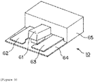
- FIG. 3 is a perspective view of the above-mentioned foot switch 10.
- the foot switch 10 includes a base plate 64, three switch units 61, 62, 63 that are installed on the base plate 64, and a casing 65 that houses electrical components.
- the switch unit 61 is used when exposing the X-ray irradiated from the X-ray source, and the switch portions 62 and 63 are used for other operations.
- the foot switch 10 functions as an operation unit according to the present invention that is used for a start operation of the X-ray fluoroscopy.
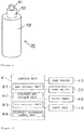
- FIG. 4 is a perspective view of a hand switch 20.
- the hand switch 20 is sometimes used in place of the foot switch 10 as the operation unit that is used for the X-ray fluoroscopy start operation.
- the hand switch 20 includes a first switch 51 and a second switch 52 that are installed on an upper portion of a body 53.
- the X-ray emission is prepared when the first switch 51 is pressed, and performed when the second switch 52 is pressed.
- the LCD touch panel 46 in the imaging unit 1 or the input unit 16 in the monitor unit 2 may be used apart from the above-described foot switch 10 or hand switch 20.
- FIG. 5 is a block diagram for illustrating a main control system of the X-ray fluoroscopic device according to the present invention.
- the X-ray fluoroscopic device includes a control unit 8 configured to control the entire device.
- the control unit 8 includes a CPU that functions as a processor configured to process logical operations, a RAM that temporarily stores data when the control unit 8 controls the device, a ROM that stores operation programs that are needed to control the device, and other components.
- the control unit 8 is connected to the above-mentioned foot switch 10, hand switch 20, LCD touch panel 46, and input unit 16.
- the control unit 8 also includes an anatomical program storage unit 81 configured to store a plurality of anatomical programs that each correspond to an imaging part, a standard anatomical program storage unit 82 configured to store a standard anatomical program, an anatomical program read-out unit 83 configured to read out a selected anatomical program, and an automatic brightness control unit 84.
- the automatic brightness control unit 84 is a unit that is used to perform automatic brightness control, which is referred to as auto brightness control (ABC), on a fluoroscopic image. Through use of this automatic brightness control, it is possible to automatically control the brightness of the fluoroscopic image to make the fluoroscopic image easier to visually recognize.
- ABS auto brightness control
- the standard anatomical program stored in the standard anatomical program storage unit 82 takes an X-ray imaging condition including tube current and tube voltage that is applied to the X-ray tube 21 in the X-ray irradiating unit as an average value.
- the standard anatomical program is an anatomical program that takes an image processing condition for an X-ray image that is detected by the X-ray detection unit, as an average value.
- the standard anatomical program stored in the standard anatomical program storage unit 82 is the anatomical program among the plurality of anatomical programs that each correspond to an imaging part that is used the most in the X-ray fluoroscopic device, or at a hospital or other facility at which the X-ray fluoroscopic device is used.
- the LCD touch panel 46 in the imaging unit 1 and the input unit 16 in the monitor unit 2 are also used as a selection operation unit when the anatomical program to be used for the X-ray fluoroscopy is selected.
- FIG. 6 is a flowchart for illustrating the operation when the X-ray fluoroscopy is started using the X-ray fluoroscopic device according to the present invention.
- Step S1 When the X-ray fluoroscopy is started, the operator waits for an operation, which is the X-ray fluoroscopy start operation, that turns on the foot switch 10 illustrated in FIG. 3 or the hand switch 20 illustrated in FIG. 4 (Step S1). Then, when the foot switch 10 or the hand switch 20 turns on, the operator determines whether or not an anatomical program has already been selected beforehand (Step S2).
- the anatomical program read-out unit 83 reads out the selected anatomical program from among the plurality of anatomical programs stored in the anatomical program storage unit 81 (Step S3).
- the anatomical program read-out unit 83 reads out the standard anatomical program stored in the standard anatomical program storage unit 82 (Step S4).
- Step S5 X-ray fluoroscopy starts.
- the X-ray fluoroscopy is performed based on the imaging conditions that correspond to that anatomical program.
- the X-ray fluoroscopy is performed with the X-ray condition and the image processing condition as an average value, or based on the most frequently used imaging condition.
- the standard anatomical program is selected, a somewhat appropriate X-ray fluoroscopy image can be obtained.
- the automatic brightness control unit 84 adjusts the brightness of the X-ray image. With this configuration, the X-ray fluoroscopy can be performed under the most appropriate imaging conditions. Even in this case, because an average anatomical program is already selected, the time it takes to achieve an optimal X-ray image when adjusting the brightness with the automatic brightness control unit 84 can be shortened.
- the present invention has been applied to a surgical X-ray fluoroscopic device, but the present invention may also be applied to, for example, various types of X-ray fluoroscopic devices that perform X-ray fluoroscopy, including a fluoroscopic imaging table.
- the LCD touch panel 46 in the imaging unit 1 or the input unit 16 in the monitor unit 2 may be used as the operation unit used for performing the X-ray fluoroscopy start operation, or another switch may be used.
Landscapes
- Health & Medical Sciences (AREA)
- Life Sciences & Earth Sciences (AREA)
- Engineering & Computer Science (AREA)
- Medical Informatics (AREA)
- Radiology & Medical Imaging (AREA)
- Molecular Biology (AREA)
- Biophysics (AREA)
- Nuclear Medicine, Radiotherapy & Molecular Imaging (AREA)
- Optics & Photonics (AREA)
- Pathology (AREA)
- Physics & Mathematics (AREA)
- Biomedical Technology (AREA)
- Heart & Thoracic Surgery (AREA)
- High Energy & Nuclear Physics (AREA)
- Surgery (AREA)
- Animal Behavior & Ethology (AREA)
- General Health & Medical Sciences (AREA)
- Public Health (AREA)
- Veterinary Medicine (AREA)
- Human Computer Interaction (AREA)
- Apparatus For Radiation Diagnosis (AREA)
Applications Claiming Priority (1)
| Application Number | Priority Date | Filing Date | Title |
|---|---|---|---|
| JP2017098210A JP2018191983A (ja) | 2017-05-17 | 2017-05-17 | X線透視装置 |
Publications (1)
| Publication Number | Publication Date |
|---|---|
| EP3403584A1 true EP3403584A1 (de) | 2018-11-21 |
Family
ID=62165402
Family Applications (1)
| Application Number | Title | Priority Date | Filing Date |
|---|---|---|---|
| EP18172027.7A Withdrawn EP3403584A1 (de) | 2017-05-17 | 2018-05-14 | Fluoroskopische röntgenuntervorrichtung |
Country Status (3)
| Country | Link |
|---|---|
| EP (1) | EP3403584A1 (de) |
| JP (1) | JP2018191983A (de) |
| CN (1) | CN108937977A (de) |
Families Citing this family (2)
| Publication number | Priority date | Publication date | Assignee | Title |
|---|---|---|---|---|
| JP7249856B2 (ja) * | 2019-04-22 | 2023-03-31 | キヤノンメディカルシステムズ株式会社 | X線ct装置 |
| JP7771704B2 (ja) * | 2021-12-13 | 2025-11-18 | 株式会社島津製作所 | X線装置 |
Citations (4)
| Publication number | Priority date | Publication date | Assignee | Title |
|---|---|---|---|---|
| JP2001340321A (ja) * | 2000-03-29 | 2001-12-11 | Toshiba Medical System Co Ltd | X線診断装置 |
| JP2012143443A (ja) | 2011-01-13 | 2012-08-02 | Shimadzu Corp | 放射線撮影装置 |
| JP2015104488A (ja) * | 2013-11-29 | 2015-06-08 | 株式会社島津製作所 | X線透視装置 |
| WO2015197566A1 (en) * | 2014-06-25 | 2015-12-30 | Koninklijke Philips N.V. | Automatic or assisted region of interest positioning in x-ray diagnostics and interventions |
-
2017
- 2017-05-17 JP JP2017098210A patent/JP2018191983A/ja active Pending
- 2017-08-29 CN CN201710758211.5A patent/CN108937977A/zh not_active Withdrawn
-
2018
- 2018-05-14 EP EP18172027.7A patent/EP3403584A1/de not_active Withdrawn
Patent Citations (4)
| Publication number | Priority date | Publication date | Assignee | Title |
|---|---|---|---|---|
| JP2001340321A (ja) * | 2000-03-29 | 2001-12-11 | Toshiba Medical System Co Ltd | X線診断装置 |
| JP2012143443A (ja) | 2011-01-13 | 2012-08-02 | Shimadzu Corp | 放射線撮影装置 |
| JP2015104488A (ja) * | 2013-11-29 | 2015-06-08 | 株式会社島津製作所 | X線透視装置 |
| WO2015197566A1 (en) * | 2014-06-25 | 2015-12-30 | Koninklijke Philips N.V. | Automatic or assisted region of interest positioning in x-ray diagnostics and interventions |
Also Published As
| Publication number | Publication date |
|---|---|
| CN108937977A (zh) | 2018-12-07 |
| JP2018191983A (ja) | 2018-12-06 |
Similar Documents
| Publication | Publication Date | Title |
|---|---|---|
| US8903041B2 (en) | X-ray diagnostic apparatus | |
| JP4793492B2 (ja) | X線撮影装置 | |
| JP5573688B2 (ja) | 放射線撮影装置 | |
| JP2011167472A (ja) | 放射線撮影装置及びその処理方法 | |
| EP3403584A1 (de) | Fluoroskopische röntgenuntervorrichtung | |
| JP2013066784A (ja) | 乳房撮影検査用x線診断装置 | |
| US10987074B2 (en) | Radiation image capturing apparatus | |
| JP2017164426A (ja) | 放射線撮影装置 | |
| CN112890837B (zh) | X射线透视摄影装置 | |
| JP6737337B2 (ja) | X線透視撮影装置 | |
| JP2010063586A (ja) | 画像の表示方向変更方法及びそれを用いたx線装置 | |
| JP2009089739A (ja) | 乳房撮影検査用x線診断装置 | |
| JP6687036B2 (ja) | X線撮影装置 | |
| JP2019180871A (ja) | X線透視撮影装置 | |
| JP6183183B2 (ja) | X線透視装置 | |
| JP6793764B2 (ja) | X線診断装置 | |
| US7899157B2 (en) | X-ray image diagnosis apparatus and control method therefor | |
| JP5146064B2 (ja) | 放射線撮影装置 | |
| JP2008125610A (ja) | X線透視撮影装置 | |
| JP6241386B2 (ja) | X線透視撮影装置 | |
| JP2019111190A (ja) | X線診断装置 | |
| JP5065591B2 (ja) | X線診断装置 | |
| JP4314013B2 (ja) | X線透視撮影装置 | |
| JP6923287B2 (ja) | X線透視撮影装置 | |
| JP2026075112A (ja) | 放射線画像撮影装置、放射線画像撮影方法及び放射線画像撮影プログラム |
Legal Events
| Date | Code | Title | Description |
|---|---|---|---|
| PUAI | Public reference made under article 153(3) epc to a published international application that has entered the european phase |
Free format text: ORIGINAL CODE: 0009012 |
|
| STAA | Information on the status of an ep patent application or granted ep patent |
Free format text: STATUS: REQUEST FOR EXAMINATION WAS MADE |
|
| 17P | Request for examination filed |
Effective date: 20180514 |
|
| AK | Designated contracting states |
Kind code of ref document: A1 Designated state(s): AL AT BE BG CH CY CZ DE DK EE ES FI FR GB GR HR HU IE IS IT LI LT LU LV MC MK MT NL NO PL PT RO RS SE SI SK SM TR |
|
| AX | Request for extension of the european patent |
Extension state: BA ME |
|
| RBV | Designated contracting states (corrected) |
Designated state(s): AL AT BE BG CH CY CZ DE DK EE ES FI FR GB GR HR HU IE IS IT LI LT LU LV MC MK MT NL NO PL PT RO RS SE SI SK SM TR |
|
| STAA | Information on the status of an ep patent application or granted ep patent |
Free format text: STATUS: THE APPLICATION HAS BEEN WITHDRAWN |
|
| 18W | Application withdrawn |
Effective date: 20190829 |