EP3380522B1 - Molécules d'anticorps se liant à april et leurs utilisations - Google Patents
Molécules d'anticorps se liant à april et leurs utilisations Download PDFInfo
- Publication number
- EP3380522B1 EP3380522B1 EP16810195.4A EP16810195A EP3380522B1 EP 3380522 B1 EP3380522 B1 EP 3380522B1 EP 16810195 A EP16810195 A EP 16810195A EP 3380522 B1 EP3380522 B1 EP 3380522B1
- Authority
- EP
- European Patent Office
- Prior art keywords
- less
- antibody molecule
- april
- amino acid
- antibody
- Prior art date
- Legal status (The legal status is an assumption and is not a legal conclusion. Google has not performed a legal analysis and makes no representation as to the accuracy of the status listed.)
- Active
Links
- 125000003275 alpha amino acid group Chemical group 0.000 claims description 327
- 238000000034 method Methods 0.000 claims description 134
- 208000010159 IgA glomerulonephritis Diseases 0.000 claims description 81
- 206010021263 IgA nephropathy Diseases 0.000 claims description 81
- 150000007523 nucleic acids Chemical class 0.000 claims description 58
- 239000000203 mixture Substances 0.000 claims description 56
- 102000039446 nucleic acids Human genes 0.000 claims description 53
- 108020004707 nucleic acids Proteins 0.000 claims description 53
- 210000002966 serum Anatomy 0.000 claims description 30
- 239000013598 vector Substances 0.000 claims description 29
- 238000000338 in vitro Methods 0.000 claims description 21
- 238000004519 manufacturing process Methods 0.000 claims description 16
- 239000003814 drug Substances 0.000 claims description 15
- 239000000463 material Substances 0.000 claims description 13
- 239000008194 pharmaceutical composition Substances 0.000 claims description 11
- 210000001519 tissue Anatomy 0.000 claims description 9
- 229940124597 therapeutic agent Drugs 0.000 claims description 8
- 108010067060 Immunoglobulin Variable Region Proteins 0.000 claims description 7
- 102000017727 Immunoglobulin Variable Region Human genes 0.000 claims description 7
- 239000003937 drug carrier Substances 0.000 claims description 6
- 210000001185 bone marrow Anatomy 0.000 claims description 5
- 230000002093 peripheral effect Effects 0.000 claims description 5
- 210000004400 mucous membrane Anatomy 0.000 claims description 4
- 239000003153 chemical reaction reagent Substances 0.000 claims description 3
- 238000012258 culturing Methods 0.000 claims description 3
- 101000830600 Homo sapiens Tumor necrosis factor ligand superfamily member 13 Proteins 0.000 description 317
- 108010065323 Tumor Necrosis Factor Ligand Superfamily Member 13 Proteins 0.000 description 291
- 230000027455 binding Effects 0.000 description 283
- 125000000539 amino acid group Chemical group 0.000 description 153
- 241000282414 Homo sapiens Species 0.000 description 112
- 208000037265 diseases, disorders, signs and symptoms Diseases 0.000 description 111
- 101000830595 Mus musculus Tumor necrosis factor ligand superfamily member 13 Proteins 0.000 description 110
- 108010047041 Complementarity Determining Regions Proteins 0.000 description 102
- 210000004027 cell Anatomy 0.000 description 93
- 229940099472 immunoglobulin a Drugs 0.000 description 80
- 102000050862 Transmembrane Activator and CAML Interactor Human genes 0.000 description 76
- 241000699666 Mus <mouse, genus> Species 0.000 description 69
- 125000003729 nucleotide group Chemical group 0.000 description 68
- 208000035475 disorder Diseases 0.000 description 66
- 238000011282 treatment Methods 0.000 description 65
- 239000002773 nucleotide Substances 0.000 description 62
- 102000005962 receptors Human genes 0.000 description 62
- 108020003175 receptors Proteins 0.000 description 62
- 101710178302 Tumor necrosis factor receptor superfamily member 13B Proteins 0.000 description 61
- 235000001014 amino acid Nutrition 0.000 description 60
- 238000002965 ELISA Methods 0.000 description 58
- 230000000903 blocking effect Effects 0.000 description 58
- 102000006942 B-Cell Maturation Antigen Human genes 0.000 description 56
- 108010008014 B-Cell Maturation Antigen Proteins 0.000 description 56
- 230000005764 inhibitory process Effects 0.000 description 56
- 108090000623 proteins and genes Proteins 0.000 description 56
- 229940024606 amino acid Drugs 0.000 description 53
- 150000001413 amino acids Chemical class 0.000 description 52
- 239000000178 monomer Substances 0.000 description 51
- 101000795167 Homo sapiens Tumor necrosis factor receptor superfamily member 13B Proteins 0.000 description 49
- 102000056239 human TNFRSF13B Human genes 0.000 description 47
- 201000010099 disease Diseases 0.000 description 45
- 102000004169 proteins and genes Human genes 0.000 description 44
- NFGXHKASABOEEW-UHFFFAOYSA-N 1-methylethyl 11-methoxy-3,7,11-trimethyl-2,4-dodecadienoate Chemical compound COC(C)(C)CCCC(C)CC=CC(C)=CC(=O)OC(C)C NFGXHKASABOEEW-UHFFFAOYSA-N 0.000 description 42
- 230000000694 effects Effects 0.000 description 40
- 235000018102 proteins Nutrition 0.000 description 39
- 238000003556 assay Methods 0.000 description 34
- 230000035772 mutation Effects 0.000 description 34
- 206010028980 Neoplasm Diseases 0.000 description 33
- 101000801255 Homo sapiens Tumor necrosis factor receptor superfamily member 17 Proteins 0.000 description 32
- 230000001404 mediated effect Effects 0.000 description 32
- 102000046935 human TNFRSF17 Human genes 0.000 description 31
- 108060003951 Immunoglobulin Proteins 0.000 description 28
- 210000004408 hybridoma Anatomy 0.000 description 28
- 102000018358 immunoglobulin Human genes 0.000 description 28
- 210000003734 kidney Anatomy 0.000 description 25
- 108010029485 Protein Isoforms Proteins 0.000 description 24
- 102000001708 Protein Isoforms Human genes 0.000 description 24
- 230000001225 therapeutic effect Effects 0.000 description 24
- 201000011510 cancer Diseases 0.000 description 23
- 208000024891 symptom Diseases 0.000 description 22
- 108091028043 Nucleic acid sequence Proteins 0.000 description 21
- 102100036922 Tumor necrosis factor ligand superfamily member 13B Human genes 0.000 description 21
- 230000002401 inhibitory effect Effects 0.000 description 20
- 241000699670 Mus sp. Species 0.000 description 19
- 238000004458 analytical method Methods 0.000 description 19
- 239000000427 antigen Substances 0.000 description 19
- 108091007433 antigens Proteins 0.000 description 19
- 102000036639 antigens Human genes 0.000 description 19
- 230000000052 comparative effect Effects 0.000 description 19
- 102100024585 Tumor necrosis factor ligand superfamily member 13 Human genes 0.000 description 18
- 210000003719 b-lymphocyte Anatomy 0.000 description 18
- 102000040430 polynucleotide Human genes 0.000 description 18
- 108091033319 polynucleotide Proteins 0.000 description 18
- 239000002157 polynucleotide Substances 0.000 description 18
- 108090000765 processed proteins & peptides Proteins 0.000 description 18
- 230000011664 signaling Effects 0.000 description 18
- 206010047115 Vasculitis Diseases 0.000 description 17
- 230000000295 complement effect Effects 0.000 description 17
- 210000002700 urine Anatomy 0.000 description 17
- -1 e.g. Proteins 0.000 description 16
- 230000014509 gene expression Effects 0.000 description 16
- 208000015446 immunoglobulin a vasculitis Diseases 0.000 description 16
- 238000012417 linear regression Methods 0.000 description 16
- 238000006467 substitution reaction Methods 0.000 description 16
- 210000004369 blood Anatomy 0.000 description 15
- 239000008280 blood Substances 0.000 description 15
- 238000002651 drug therapy Methods 0.000 description 15
- 208000006750 hematuria Diseases 0.000 description 15
- 229910052757 nitrogen Inorganic materials 0.000 description 15
- 229920001184 polypeptide Polymers 0.000 description 15
- 102000004196 processed proteins & peptides Human genes 0.000 description 15
- 239000003795 chemical substances by application Substances 0.000 description 14
- 201000000464 cone-rod dystrophy 2 Diseases 0.000 description 14
- 239000013604 expression vector Substances 0.000 description 14
- 102000004127 Cytokines Human genes 0.000 description 13
- 108090000695 Cytokines Proteins 0.000 description 13
- 208000007342 Diabetic Nephropathies Diseases 0.000 description 13
- 208000033679 diabetic kidney disease Diseases 0.000 description 13
- 239000012634 fragment Substances 0.000 description 13
- 239000000523 sample Substances 0.000 description 13
- DHMQDGOQFOQNFH-UHFFFAOYSA-N Glycine Chemical compound NCC(O)=O DHMQDGOQFOQNFH-UHFFFAOYSA-N 0.000 description 12
- 206010035226 Plasma cell myeloma Diseases 0.000 description 12
- 238000005516 engineering process Methods 0.000 description 12
- 230000004927 fusion Effects 0.000 description 12
- 238000001727 in vivo Methods 0.000 description 12
- 230000003993 interaction Effects 0.000 description 12
- 230000009467 reduction Effects 0.000 description 12
- 241000894007 species Species 0.000 description 12
- 229910052717 sulfur Inorganic materials 0.000 description 12
- 238000012360 testing method Methods 0.000 description 12
- 206010020772 Hypertension Diseases 0.000 description 11
- 101100425747 Mus musculus Tnfrsf17 gene Proteins 0.000 description 11
- 108010057466 NF-kappa B Proteins 0.000 description 11
- 102000003945 NF-kappa B Human genes 0.000 description 11
- 230000015572 biosynthetic process Effects 0.000 description 11
- 150000001875 compounds Chemical class 0.000 description 11
- 230000009260 cross reactivity Effects 0.000 description 11
- 210000004602 germ cell Anatomy 0.000 description 11
- 229960004641 rituximab Drugs 0.000 description 11
- 229910052727 yttrium Inorganic materials 0.000 description 11
- 208000023275 Autoimmune disease Diseases 0.000 description 10
- 108010087819 Fc receptors Proteins 0.000 description 10
- 102000009109 Fc receptors Human genes 0.000 description 10
- AYFVYJQAPQTCCC-GBXIJSLDSA-N L-threonine Chemical compound C[C@@H](O)[C@H](N)C(O)=O AYFVYJQAPQTCCC-GBXIJSLDSA-N 0.000 description 10
- 108010003723 Single-Domain Antibodies Proteins 0.000 description 10
- 208000020832 chronic kidney disease Diseases 0.000 description 10
- 230000004540 complement-dependent cytotoxicity Effects 0.000 description 10
- 239000002299 complementary DNA Substances 0.000 description 10
- 238000002347 injection Methods 0.000 description 10
- 239000007924 injection Substances 0.000 description 10
- 238000002560 therapeutic procedure Methods 0.000 description 10
- 108091032973 (ribonucleotides)n+m Proteins 0.000 description 9
- 101000611183 Homo sapiens Tumor necrosis factor Proteins 0.000 description 9
- 208000034578 Multiple myelomas Diseases 0.000 description 9
- 101100425947 Mus musculus Tnfrsf13b gene Proteins 0.000 description 9
- LMEKQMALGUDUQG-UHFFFAOYSA-N azathioprine Chemical compound CN1C=NC([N+]([O-])=O)=C1SC1=NC=NC2=C1NC=N2 LMEKQMALGUDUQG-UHFFFAOYSA-N 0.000 description 9
- 238000013461 design Methods 0.000 description 9
- 230000006870 function Effects 0.000 description 9
- 238000009169 immunotherapy Methods 0.000 description 9
- 238000013507 mapping Methods 0.000 description 9
- 201000001474 proteinuria Diseases 0.000 description 9
- YBJHBAHKTGYVGT-ZKWXMUAHSA-N (+)-Biotin Chemical compound N1C(=O)N[C@@H]2[C@H](CCCCC(=O)O)SC[C@@H]21 YBJHBAHKTGYVGT-ZKWXMUAHSA-N 0.000 description 8
- 206010006187 Breast cancer Diseases 0.000 description 8
- 208000026310 Breast neoplasm Diseases 0.000 description 8
- 208000015943 Coeliac disease Diseases 0.000 description 8
- 230000004913 activation Effects 0.000 description 8
- 229940125364 angiotensin receptor blocker Drugs 0.000 description 8
- 238000010171 animal model Methods 0.000 description 8
- 238000002512 chemotherapy Methods 0.000 description 8
- 239000003246 corticosteroid Substances 0.000 description 8
- 230000005714 functional activity Effects 0.000 description 8
- 239000003018 immunosuppressive agent Substances 0.000 description 8
- 238000001990 intravenous administration Methods 0.000 description 8
- 229910052740 iodine Inorganic materials 0.000 description 8
- RTGDFNSFWBGLEC-SYZQJQIISA-N mycophenolate mofetil Chemical compound COC1=C(C)C=2COC(=O)C=2C(O)=C1C\C=C(/C)CCC(=O)OCCN1CCOCC1 RTGDFNSFWBGLEC-SYZQJQIISA-N 0.000 description 8
- 229960004866 mycophenolate mofetil Drugs 0.000 description 8
- 230000003472 neutralizing effect Effects 0.000 description 8
- XOFYZVNMUHMLCC-ZPOLXVRWSA-N prednisone Chemical compound O=C1C=C[C@]2(C)[C@H]3C(=O)C[C@](C)([C@@](CC4)(O)C(=O)CO)[C@@H]4[C@@H]3CCC2=C1 XOFYZVNMUHMLCC-ZPOLXVRWSA-N 0.000 description 8
- 229960004618 prednisone Drugs 0.000 description 8
- 239000000047 product Substances 0.000 description 8
- 238000001959 radiotherapy Methods 0.000 description 8
- 230000002829 reductive effect Effects 0.000 description 8
- 206010039073 rheumatoid arthritis Diseases 0.000 description 8
- 239000007787 solid Substances 0.000 description 8
- 239000000243 solution Substances 0.000 description 8
- 150000003431 steroids Chemical class 0.000 description 8
- 238000001890 transfection Methods 0.000 description 8
- 102000004190 Enzymes Human genes 0.000 description 7
- 108090000790 Enzymes Proteins 0.000 description 7
- 206010072579 Granulomatosis with polyangiitis Diseases 0.000 description 7
- 101000979342 Homo sapiens Nuclear factor NF-kappa-B p105 subunit Proteins 0.000 description 7
- 208000031814 IgA Vasculitis Diseases 0.000 description 7
- 102100026120 IgG receptor FcRn large subunit p51 Human genes 0.000 description 7
- 101710177940 IgG receptor FcRn large subunit p51 Proteins 0.000 description 7
- FBOZXECLQNJBKD-ZDUSSCGKSA-N L-methotrexate Chemical compound C=1N=C2N=C(N)N=C(N)C2=NC=1CN(C)C1=CC=C(C(=O)N[C@@H](CCC(O)=O)C(O)=O)C=C1 FBOZXECLQNJBKD-ZDUSSCGKSA-N 0.000 description 7
- 208000012309 Linear IgA disease Diseases 0.000 description 7
- 206010025323 Lymphomas Diseases 0.000 description 7
- 102100023050 Nuclear factor NF-kappa-B p105 subunit Human genes 0.000 description 7
- 201000005638 acute proliferative glomerulonephritis Diseases 0.000 description 7
- 229940044094 angiotensin-converting-enzyme inhibitor Drugs 0.000 description 7
- 230000001684 chronic effect Effects 0.000 description 7
- IDLFZVILOHSSID-OVLDLUHVSA-N corticotropin Chemical compound C([C@@H](C(=O)N[C@@H](CO)C(=O)N[C@@H](CCSC)C(=O)N[C@@H](CCC(O)=O)C(=O)N[C@@H](CC=1NC=NC=1)C(=O)N[C@@H](CC=1C=CC=CC=1)C(=O)N[C@@H](CCCNC(N)=N)C(=O)N[C@@H](CC=1C2=CC=CC=C2NC=1)C(=O)NCC(=O)N[C@@H](CCCCN)C(=O)N1[C@@H](CCC1)C(=O)N[C@@H](C(C)C)C(=O)NCC(=O)N[C@@H](CCCCN)C(=O)N[C@@H](CCCCN)C(=O)N[C@@H](CCCNC(N)=N)C(=O)N[C@@H](CCCNC(N)=N)C(=O)N1[C@@H](CCC1)C(=O)N[C@@H](C(C)C)C(=O)N[C@@H](CCCCN)C(=O)N[C@@H](C(C)C)C(=O)N[C@@H](CC=1C=CC(O)=CC=1)C(=O)N1[C@@H](CCC1)C(=O)N[C@@H](CC(N)=O)C(=O)NCC(=O)N[C@@H](C)C(=O)N[C@@H](CCC(O)=O)C(=O)N[C@@H](CC(O)=O)C(=O)N[C@@H](CCC(O)=O)C(=O)N[C@@H](CO)C(=O)N[C@@H](C)C(=O)N[C@@H](CCC(O)=O)C(=O)N[C@@H](C)C(=O)N[C@@H](CC=1C=CC=CC=1)C(=O)N1[C@@H](CCC1)C(=O)N[C@@H](CC(C)C)C(=O)N[C@@H](CCC(O)=O)C(=O)N[C@@H](CC=1C=CC=CC=1)C(O)=O)NC(=O)[C@@H](N)CO)C1=CC=C(O)C=C1 IDLFZVILOHSSID-OVLDLUHVSA-N 0.000 description 7
- 238000001514 detection method Methods 0.000 description 7
- 229940088598 enzyme Drugs 0.000 description 7
- 238000009396 hybridization Methods 0.000 description 7
- 229960003444 immunosuppressant agent Drugs 0.000 description 7
- 239000003112 inhibitor Substances 0.000 description 7
- 230000003907 kidney function Effects 0.000 description 7
- 238000002372 labelling Methods 0.000 description 7
- 239000013642 negative control Substances 0.000 description 7
- 239000013641 positive control Substances 0.000 description 7
- 230000000069 prophylactic effect Effects 0.000 description 7
- 230000004044 response Effects 0.000 description 7
- 210000004988 splenocyte Anatomy 0.000 description 7
- 239000006228 supernatant Substances 0.000 description 7
- 238000001356 surgical procedure Methods 0.000 description 7
- 206010056508 Acquired epidermolysis bullosa Diseases 0.000 description 6
- 108010028006 B-Cell Activating Factor Proteins 0.000 description 6
- 208000010839 B-cell chronic lymphocytic leukemia Diseases 0.000 description 6
- 206010009208 Cirrhosis alcoholic Diseases 0.000 description 6
- 206010009944 Colon cancer Diseases 0.000 description 6
- 108020004414 DNA Proteins 0.000 description 6
- 206010048768 Dermatosis Diseases 0.000 description 6
- 206010019617 Henoch-Schonlein purpura Diseases 0.000 description 6
- 208000015865 IgA pemphigus Diseases 0.000 description 6
- AGPKZVBTJJNPAG-WHFBIAKZSA-N L-isoleucine Chemical compound CC[C@H](C)[C@H](N)C(O)=O AGPKZVBTJJNPAG-WHFBIAKZSA-N 0.000 description 6
- 241000700159 Rattus Species 0.000 description 6
- FAPWRFPIFSIZLT-UHFFFAOYSA-M Sodium chloride Chemical compound [Na+].[Cl-] FAPWRFPIFSIZLT-UHFFFAOYSA-M 0.000 description 6
- 210000001744 T-lymphocyte Anatomy 0.000 description 6
- 208000033559 Waldenström macroglobulinemia Diseases 0.000 description 6
- 208000010002 alcoholic liver cirrhosis Diseases 0.000 description 6
- 229960002170 azathioprine Drugs 0.000 description 6
- 230000003115 biocidal effect Effects 0.000 description 6
- 230000004071 biological effect Effects 0.000 description 6
- 238000001574 biopsy Methods 0.000 description 6
- 238000012512 characterization method Methods 0.000 description 6
- 230000008021 deposition Effects 0.000 description 6
- 238000000502 dialysis Methods 0.000 description 6
- 238000010790 dilution Methods 0.000 description 6
- 239000012895 dilution Substances 0.000 description 6
- 201000000523 end stage renal failure Diseases 0.000 description 6
- 201000011114 epidermolysis bullosa acquisita Diseases 0.000 description 6
- 229910052731 fluorine Inorganic materials 0.000 description 6
- 102000057041 human TNF Human genes 0.000 description 6
- 230000001861 immunosuppressant effect Effects 0.000 description 6
- 238000001802 infusion Methods 0.000 description 6
- 238000011862 kidney biopsy Methods 0.000 description 6
- 238000005259 measurement Methods 0.000 description 6
- 238000010172 mouse model Methods 0.000 description 6
- 238000006386 neutralization reaction Methods 0.000 description 6
- 229910052698 phosphorus Inorganic materials 0.000 description 6
- 239000013612 plasmid Substances 0.000 description 6
- 229910052700 potassium Inorganic materials 0.000 description 6
- 238000000746 purification Methods 0.000 description 6
- 238000012216 screening Methods 0.000 description 6
- 238000002741 site-directed mutagenesis Methods 0.000 description 6
- 208000017520 skin disease Diseases 0.000 description 6
- 230000008685 targeting Effects 0.000 description 6
- 238000003146 transient transfection Methods 0.000 description 6
- 239000013638 trimer Substances 0.000 description 6
- 238000005406 washing Methods 0.000 description 6
- 208000009304 Acute Kidney Injury Diseases 0.000 description 5
- 102100027211 Albumin Human genes 0.000 description 5
- 108010088751 Albumins Proteins 0.000 description 5
- 206010001580 Albuminuria Diseases 0.000 description 5
- 101100107610 Arabidopsis thaliana ABCF4 gene Proteins 0.000 description 5
- 208000003174 Brain Neoplasms Diseases 0.000 description 5
- 208000001333 Colorectal Neoplasms Diseases 0.000 description 5
- 102400000739 Corticotropin Human genes 0.000 description 5
- 101800000414 Corticotropin Proteins 0.000 description 5
- CMSMOCZEIVJLDB-UHFFFAOYSA-N Cyclophosphamide Chemical compound ClCCN(CCCl)P1(=O)NCCCO1 CMSMOCZEIVJLDB-UHFFFAOYSA-N 0.000 description 5
- 239000006144 Dulbecco’s modified Eagle's medium Substances 0.000 description 5
- 208000000461 Esophageal Neoplasms Diseases 0.000 description 5
- 208000022461 Glomerular disease Diseases 0.000 description 5
- 239000004471 Glycine Substances 0.000 description 5
- 201000004331 Henoch-Schoenlein purpura Diseases 0.000 description 5
- 208000017604 Hodgkin disease Diseases 0.000 description 5
- 208000021519 Hodgkin lymphoma Diseases 0.000 description 5
- 208000010747 Hodgkins lymphoma Diseases 0.000 description 5
- 208000019758 Hypergammaglobulinemia Diseases 0.000 description 5
- 206010061218 Inflammation Diseases 0.000 description 5
- 208000008839 Kidney Neoplasms Diseases 0.000 description 5
- CKLJMWTZIZZHCS-REOHCLBHSA-N L-aspartic acid Chemical compound OC(=O)[C@@H](N)CC(O)=O CKLJMWTZIZZHCS-REOHCLBHSA-N 0.000 description 5
- 206010030155 Oesophageal carcinoma Diseases 0.000 description 5
- 206010038389 Renal cancer Diseases 0.000 description 5
- 208000033626 Renal failure acute Diseases 0.000 description 5
- 241000283984 Rodentia Species 0.000 description 5
- 101100068078 Saccharomyces cerevisiae (strain ATCC 204508 / S288c) GCN4 gene Proteins 0.000 description 5
- KZSNJWFQEVHDMF-UHFFFAOYSA-N Valine Natural products CC(C)C(N)C(O)=O KZSNJWFQEVHDMF-UHFFFAOYSA-N 0.000 description 5
- 201000011040 acute kidney failure Diseases 0.000 description 5
- OFCNXPDARWKPPY-UHFFFAOYSA-N allopurinol Chemical compound OC1=NC=NC2=C1C=NN2 OFCNXPDARWKPPY-UHFFFAOYSA-N 0.000 description 5
- 210000004204 blood vessel Anatomy 0.000 description 5
- GXJABQQUPOEUTA-RDJZCZTQSA-N bortezomib Chemical compound C([C@@H](C(=O)N[C@@H](CC(C)C)B(O)O)NC(=O)C=1N=CC=NC=1)C1=CC=CC=C1 GXJABQQUPOEUTA-RDJZCZTQSA-N 0.000 description 5
- 239000006143 cell culture medium Substances 0.000 description 5
- 210000003169 central nervous system Anatomy 0.000 description 5
- 238000005119 centrifugation Methods 0.000 description 5
- 229960000258 corticotropin Drugs 0.000 description 5
- 229960004397 cyclophosphamide Drugs 0.000 description 5
- 230000006378 damage Effects 0.000 description 5
- 206010012601 diabetes mellitus Diseases 0.000 description 5
- 238000003745 diagnosis Methods 0.000 description 5
- 238000010494 dissociation reaction Methods 0.000 description 5
- 230000005593 dissociations Effects 0.000 description 5
- 229940079593 drug Drugs 0.000 description 5
- 210000003743 erythrocyte Anatomy 0.000 description 5
- 201000004101 esophageal cancer Diseases 0.000 description 5
- 238000011156 evaluation Methods 0.000 description 5
- 229930182830 galactose Natural products 0.000 description 5
- 230000002068 genetic effect Effects 0.000 description 5
- 230000002489 hematologic effect Effects 0.000 description 5
- 229940072221 immunoglobulins Drugs 0.000 description 5
- 230000004054 inflammatory process Effects 0.000 description 5
- AGPKZVBTJJNPAG-UHFFFAOYSA-N isoleucine Natural products CCC(C)C(N)C(O)=O AGPKZVBTJJNPAG-UHFFFAOYSA-N 0.000 description 5
- 229960000310 isoleucine Drugs 0.000 description 5
- 201000010982 kidney cancer Diseases 0.000 description 5
- 208000017169 kidney disease Diseases 0.000 description 5
- 230000007246 mechanism Effects 0.000 description 5
- 238000003199 nucleic acid amplification method Methods 0.000 description 5
- HDACQVRGBOVJII-JBDAPHQKSA-N ramipril Chemical compound C([C@@H](C(=O)OCC)N[C@@H](C)C(=O)N1[C@@H](C[C@@H]2CCC[C@@H]21)C(O)=O)CC1=CC=CC=C1 HDACQVRGBOVJII-JBDAPHQKSA-N 0.000 description 5
- 239000000126 substance Substances 0.000 description 5
- 239000003053 toxin Substances 0.000 description 5
- 231100000765 toxin Toxicity 0.000 description 5
- 229910052720 vanadium Inorganic materials 0.000 description 5
- 230000003442 weekly effect Effects 0.000 description 5
- 208000004998 Abdominal Pain Diseases 0.000 description 4
- 239000004072 C09CA03 - Valsartan Substances 0.000 description 4
- 101150070527 CRD1 gene Proteins 0.000 description 4
- 241000282836 Camelus dromedarius Species 0.000 description 4
- 108091035707 Consensus sequence Proteins 0.000 description 4
- 208000007465 Giant cell arteritis Diseases 0.000 description 4
- 101000961146 Homo sapiens Immunoglobulin heavy constant gamma 2 Proteins 0.000 description 4
- 102100039346 Immunoglobulin heavy constant gamma 2 Human genes 0.000 description 4
- COLNVLDHVKWLRT-QMMMGPOBSA-N L-phenylalanine Chemical compound OC(=O)[C@@H](N)CC1=CC=CC=C1 COLNVLDHVKWLRT-QMMMGPOBSA-N 0.000 description 4
- 108060001084 Luciferase Proteins 0.000 description 4
- 239000005089 Luciferase Substances 0.000 description 4
- 241001465754 Metazoa Species 0.000 description 4
- 241001529936 Murinae Species 0.000 description 4
- 206010030113 Oedema Diseases 0.000 description 4
- 208000002774 Paraproteinemias Diseases 0.000 description 4
- 206010036303 Post streptococcal glomerulonephritis Diseases 0.000 description 4
- 208000006265 Renal cell carcinoma Diseases 0.000 description 4
- QAOWNCQODCNURD-UHFFFAOYSA-N Sulfuric acid Chemical compound OS(O)(=O)=O QAOWNCQODCNURD-UHFFFAOYSA-N 0.000 description 4
- QJJXYPPXXYFBGM-LFZNUXCKSA-N Tacrolimus Chemical compound C1C[C@@H](O)[C@H](OC)C[C@@H]1\C=C(/C)[C@@H]1[C@H](C)[C@@H](O)CC(=O)[C@H](CC=C)/C=C(C)/C[C@H](C)C[C@H](OC)[C@H]([C@H](C[C@H]2C)OC)O[C@@]2(O)C(=O)C(=O)N2CCCC[C@H]2C(=O)O1 QJJXYPPXXYFBGM-LFZNUXCKSA-N 0.000 description 4
- 208000031981 Thrombocytopenic Idiopathic Purpura Diseases 0.000 description 4
- 101710120037 Toxin CcdB Proteins 0.000 description 4
- 230000001594 aberrant effect Effects 0.000 description 4
- 238000002835 absorbance Methods 0.000 description 4
- 238000001042 affinity chromatography Methods 0.000 description 4
- 230000003321 amplification Effects 0.000 description 4
- 239000003242 anti bacterial agent Substances 0.000 description 4
- 206010003246 arthritis Diseases 0.000 description 4
- 230000001363 autoimmune Effects 0.000 description 4
- 201000003710 autoimmune thrombocytopenic purpura Diseases 0.000 description 4
- 229940120638 avastin Drugs 0.000 description 4
- 229960000397 bevacizumab Drugs 0.000 description 4
- 239000012472 biological sample Substances 0.000 description 4
- 229960002685 biotin Drugs 0.000 description 4
- 235000020958 biotin Nutrition 0.000 description 4
- 239000011616 biotin Substances 0.000 description 4
- 230000010001 cellular homeostasis Effects 0.000 description 4
- 238000010367 cloning Methods 0.000 description 4
- 238000002648 combination therapy Methods 0.000 description 4
- 230000021615 conjugation Effects 0.000 description 4
- 238000011161 development Methods 0.000 description 4
- 230000018109 developmental process Effects 0.000 description 4
- 239000006185 dispersion Substances 0.000 description 4
- 239000012636 effector Substances 0.000 description 4
- 208000028208 end stage renal disease Diseases 0.000 description 4
- 239000000499 gel Substances 0.000 description 4
- 208000005017 glioblastoma Diseases 0.000 description 4
- 231100000852 glomerular disease Toxicity 0.000 description 4
- 230000024924 glomerular filtration Effects 0.000 description 4
- 230000013595 glycosylation Effects 0.000 description 4
- 238000006206 glycosylation reaction Methods 0.000 description 4
- 208000008675 hereditary spastic paraplegia Diseases 0.000 description 4
- XXSMGPRMXLTPCZ-UHFFFAOYSA-N hydroxychloroquine Chemical compound ClC1=CC=C2C(NC(C)CCCN(CCO)CC)=CC=NC2=C1 XXSMGPRMXLTPCZ-UHFFFAOYSA-N 0.000 description 4
- 230000003053 immunization Effects 0.000 description 4
- 238000002649 immunization Methods 0.000 description 4
- 238000011534 incubation Methods 0.000 description 4
- 208000015181 infectious disease Diseases 0.000 description 4
- 239000004615 ingredient Substances 0.000 description 4
- 230000004068 intracellular signaling Effects 0.000 description 4
- 238000007912 intraperitoneal administration Methods 0.000 description 4
- VHOGYURTWQBHIL-UHFFFAOYSA-N leflunomide Chemical compound O1N=CC(C(=O)NC=2C=CC(=CC=2)C(F)(F)F)=C1C VHOGYURTWQBHIL-UHFFFAOYSA-N 0.000 description 4
- 208000032839 leukemia Diseases 0.000 description 4
- 239000011159 matrix material Substances 0.000 description 4
- 239000002609 medium Substances 0.000 description 4
- 210000004379 membrane Anatomy 0.000 description 4
- 239000012528 membrane Substances 0.000 description 4
- 229960000485 methotrexate Drugs 0.000 description 4
- 230000004048 modification Effects 0.000 description 4
- 238000012986 modification Methods 0.000 description 4
- 201000008383 nephritis Diseases 0.000 description 4
- 238000010606 normalization Methods 0.000 description 4
- 201000008968 osteosarcoma Diseases 0.000 description 4
- 230000008506 pathogenesis Effects 0.000 description 4
- 229920000642 polymer Polymers 0.000 description 4
- 238000011533 pre-incubation Methods 0.000 description 4
- 238000002360 preparation method Methods 0.000 description 4
- 238000000159 protein binding assay Methods 0.000 description 4
- 230000002285 radioactive effect Effects 0.000 description 4
- 229960003401 ramipril Drugs 0.000 description 4
- ZAHRKKWIAAJSAO-UHFFFAOYSA-N rapamycin Natural products COCC(O)C(=C/C(C)C(=O)CC(OC(=O)C1CCCCN1C(=O)C(=O)C2(O)OC(CC(OC)C(=CC=CC=CC(C)CC(C)C(=O)C)C)CCC2C)C(C)CC3CCC(O)C(C3)OC)C ZAHRKKWIAAJSAO-UHFFFAOYSA-N 0.000 description 4
- 238000003757 reverse transcription PCR Methods 0.000 description 4
- 239000010948 rhodium Substances 0.000 description 4
- 230000019491 signal transduction Effects 0.000 description 4
- 229960002930 sirolimus Drugs 0.000 description 4
- QFJCIRLUMZQUOT-HPLJOQBZSA-N sirolimus Chemical compound C1C[C@@H](O)[C@H](OC)C[C@@H]1C[C@@H](C)[C@H]1OC(=O)[C@@H]2CCCCN2C(=O)C(=O)[C@](O)(O2)[C@H](C)CC[C@H]2C[C@H](OC)/C(C)=C/C=C/C=C/[C@@H](C)C[C@@H](C)C(=O)[C@H](OC)[C@H](O)/C(C)=C/[C@@H](C)C(=O)C1 QFJCIRLUMZQUOT-HPLJOQBZSA-N 0.000 description 4
- 239000011780 sodium chloride Substances 0.000 description 4
- 210000000130 stem cell Anatomy 0.000 description 4
- 208000011580 syndromic disease Diseases 0.000 description 4
- 201000000596 systemic lupus erythematosus Diseases 0.000 description 4
- 229960001967 tacrolimus Drugs 0.000 description 4
- QJJXYPPXXYFBGM-SHYZHZOCSA-N tacrolimus Natural products CO[C@H]1C[C@H](CC[C@@H]1O)C=C(C)[C@H]2OC(=O)[C@H]3CCCCN3C(=O)C(=O)[C@@]4(O)O[C@@H]([C@H](C[C@H]4C)OC)[C@@H](C[C@H](C)CC(=C[C@@H](CC=C)C(=O)C[C@H](O)[C@H]2C)C)OC QJJXYPPXXYFBGM-SHYZHZOCSA-N 0.000 description 4
- RMMXLENWKUUMAY-UHFFFAOYSA-N telmisartan Chemical compound CCCC1=NC2=C(C)C=C(C=3N(C4=CC=CC=C4N=3)C)C=C2N1CC(C=C1)=CC=C1C1=CC=CC=C1C(O)=O RMMXLENWKUUMAY-UHFFFAOYSA-N 0.000 description 4
- 206010043207 temporal arteritis Diseases 0.000 description 4
- 238000000954 titration curve Methods 0.000 description 4
- 238000011830 transgenic mouse model Methods 0.000 description 4
- 229910052721 tungsten Inorganic materials 0.000 description 4
- 230000002485 urinary effect Effects 0.000 description 4
- 239000002525 vasculotropin inhibitor Substances 0.000 description 4
- MTCFGRXMJLQNBG-REOHCLBHSA-N (2S)-2-Amino-3-hydroxypropansäure Chemical compound OC[C@H](N)C(O)=O MTCFGRXMJLQNBG-REOHCLBHSA-N 0.000 description 3
- UEJJHQNACJXSKW-UHFFFAOYSA-N 2-(2,6-dioxopiperidin-3-yl)-1H-isoindole-1,3(2H)-dione Chemical class O=C1C2=CC=CC=C2C(=O)N1C1CCC(=O)NC1=O UEJJHQNACJXSKW-UHFFFAOYSA-N 0.000 description 3
- UAIUNKRWKOVEES-UHFFFAOYSA-N 3,3',5,5'-tetramethylbenzidine Chemical compound CC1=C(N)C(C)=CC(C=2C=C(C)C(N)=C(C)C=2)=C1 UAIUNKRWKOVEES-UHFFFAOYSA-N 0.000 description 3
- 108010009522 AMG623 peptibody Proteins 0.000 description 3
- 208000036764 Adenocarcinoma of the esophagus Diseases 0.000 description 3
- 208000008190 Agammaglobulinemia Diseases 0.000 description 3
- 239000004475 Arginine Substances 0.000 description 3
- 208000028564 B-cell non-Hodgkin lymphoma Diseases 0.000 description 3
- 208000009299 Benign Mucous Membrane Pemphigoid Diseases 0.000 description 3
- VOVIALXJUBGFJZ-KWVAZRHASA-N Budesonide Chemical compound C1CC2=CC(=O)C=C[C@]2(C)[C@@H]2[C@@H]1[C@@H]1C[C@H]3OC(CCC)O[C@@]3(C(=O)CO)[C@@]1(C)C[C@@H]2O VOVIALXJUBGFJZ-KWVAZRHASA-N 0.000 description 3
- 239000002083 C09CA01 - Losartan Substances 0.000 description 3
- 239000005537 C09CA07 - Telmisartan Substances 0.000 description 3
- GHOSNRCGJFBJIB-UHFFFAOYSA-N Candesartan cilexetil Chemical compound C=12N(CC=3C=CC(=CC=3)C=3C(=CC=CC=3)C3=NNN=N3)C(OCC)=NC2=CC=CC=1C(=O)OC(C)OC(=O)OC1CCCCC1 GHOSNRCGJFBJIB-UHFFFAOYSA-N 0.000 description 3
- 241000283707 Capra Species 0.000 description 3
- 108010061435 Enalapril Proteins 0.000 description 3
- 208000006168 Ewing Sarcoma Diseases 0.000 description 3
- 208000021309 Germ cell tumor Diseases 0.000 description 3
- 206010018364 Glomerulonephritis Diseases 0.000 description 3
- 101000961156 Homo sapiens Immunoglobulin heavy constant gamma 1 Proteins 0.000 description 3
- 206010020983 Hypogammaglobulinaemia Diseases 0.000 description 3
- 102000008394 Immunoglobulin Fragments Human genes 0.000 description 3
- 108010021625 Immunoglobulin Fragments Proteins 0.000 description 3
- 102100039345 Immunoglobulin heavy constant gamma 1 Human genes 0.000 description 3
- 208000007766 Kaposi sarcoma Diseases 0.000 description 3
- OUYCCCASQSFEME-QMMMGPOBSA-N L-tyrosine Chemical compound OC(=O)[C@@H](N)CC1=CC=C(O)C=C1 OUYCCCASQSFEME-QMMMGPOBSA-N 0.000 description 3
- 108010007859 Lisinopril Proteins 0.000 description 3
- 208000030289 Lymphoproliferative disease Diseases 0.000 description 3
- 108010052285 Membrane Proteins Proteins 0.000 description 3
- FQISKWAFAHGMGT-SGJOWKDISA-M Methylprednisolone sodium succinate Chemical compound [Na+].C([C@@]12C)=CC(=O)C=C1[C@@H](C)C[C@@H]1[C@@H]2[C@@H](O)C[C@]2(C)[C@@](O)(C(=O)COC(=O)CCC([O-])=O)CC[C@H]21 FQISKWAFAHGMGT-SGJOWKDISA-M 0.000 description 3
- 206010028813 Nausea Diseases 0.000 description 3
- 208000034176 Neoplasms, Germ Cell and Embryonal Diseases 0.000 description 3
- 206010029164 Nephrotic syndrome Diseases 0.000 description 3
- 206010030137 Oesophageal adenocarcinoma Diseases 0.000 description 3
- 241000283973 Oryctolagus cuniculus Species 0.000 description 3
- 206010033128 Ovarian cancer Diseases 0.000 description 3
- 206010061535 Ovarian neoplasm Diseases 0.000 description 3
- 229920001213 Polysorbate 20 Polymers 0.000 description 3
- 208000001647 Renal Insufficiency Diseases 0.000 description 3
- 108010090804 Streptavidin Proteins 0.000 description 3
- 102000013081 Tumor Necrosis Factor Ligand Superfamily Member 13 Human genes 0.000 description 3
- 102100040247 Tumor necrosis factor Human genes 0.000 description 3
- 206010067584 Type 1 diabetes mellitus Diseases 0.000 description 3
- VWQVUPCCIRVNHF-OUBTZVSYSA-N Yttrium-90 Chemical compound [90Y] VWQVUPCCIRVNHF-OUBTZVSYSA-N 0.000 description 3
- 208000002552 acute disseminated encephalomyelitis Diseases 0.000 description 3
- 239000000654 additive Substances 0.000 description 3
- 230000000996 additive effect Effects 0.000 description 3
- 229960003459 allopurinol Drugs 0.000 description 3
- 230000008485 antagonism Effects 0.000 description 3
- 238000013459 approach Methods 0.000 description 3
- ODKSFYDXXFIFQN-UHFFFAOYSA-N arginine Natural products OC(=O)C(N)CCCNC(N)=N ODKSFYDXXFIFQN-UHFFFAOYSA-N 0.000 description 3
- 230000001580 bacterial effect Effects 0.000 description 3
- 239000012148 binding buffer Substances 0.000 description 3
- 238000009534 blood test Methods 0.000 description 3
- 229960001467 bortezomib Drugs 0.000 description 3
- 201000008275 breast carcinoma Diseases 0.000 description 3
- 229960004436 budesonide Drugs 0.000 description 3
- 238000004422 calculation algorithm Methods 0.000 description 3
- 238000004113 cell culture Methods 0.000 description 3
- 230000003915 cell function Effects 0.000 description 3
- 230000008859 change Effects 0.000 description 3
- 238000006243 chemical reaction Methods 0.000 description 3
- 239000003636 conditioned culture medium Substances 0.000 description 3
- 239000013068 control sample Substances 0.000 description 3
- 238000002574 cystoscopy Methods 0.000 description 3
- 230000007423 decrease Effects 0.000 description 3
- 230000001419 dependent effect Effects 0.000 description 3
- 229960000873 enalapril Drugs 0.000 description 3
- GBXSMTUPTTWBMN-XIRDDKMYSA-N enalapril Chemical compound C([C@@H](C(=O)OCC)N[C@@H](C)C(=O)N1[C@@H](CCC1)C(O)=O)CC1=CC=CC=C1 GBXSMTUPTTWBMN-XIRDDKMYSA-N 0.000 description 3
- 208000028653 esophageal adenocarcinoma Diseases 0.000 description 3
- 230000003203 everyday effect Effects 0.000 description 3
- 230000029142 excretion Effects 0.000 description 3
- 238000002474 experimental method Methods 0.000 description 3
- GNBHRKFJIUUOQI-UHFFFAOYSA-N fluorescein Chemical compound O1C(=O)C2=CC=CC=C2C21C1=CC=C(O)C=C1OC1=CC(O)=CC=C21 GNBHRKFJIUUOQI-UHFFFAOYSA-N 0.000 description 3
- 238000009472 formulation Methods 0.000 description 3
- 229950005309 fostamatinib Drugs 0.000 description 3
- GKDRMWXFWHEQQT-UHFFFAOYSA-N fostamatinib Chemical compound COC1=C(OC)C(OC)=CC(NC=2N=C(NC=3N=C4N(COP(O)(O)=O)C(=O)C(C)(C)OC4=CC=3)C(F)=CN=2)=C1 GKDRMWXFWHEQQT-UHFFFAOYSA-N 0.000 description 3
- 230000002496 gastric effect Effects 0.000 description 3
- 230000036541 health Effects 0.000 description 3
- 229910052739 hydrogen Inorganic materials 0.000 description 3
- 230000001900 immune effect Effects 0.000 description 3
- 210000000987 immune system Anatomy 0.000 description 3
- 230000002163 immunogen Effects 0.000 description 3
- 230000001976 improved effect Effects 0.000 description 3
- 238000007918 intramuscular administration Methods 0.000 description 3
- YOSHYTLCDANDAN-UHFFFAOYSA-N irbesartan Chemical compound O=C1N(CC=2C=CC(=CC=2)C=2C(=CC=CC=2)C=2NN=NN=2)C(CCCC)=NC21CCCC2 YOSHYTLCDANDAN-UHFFFAOYSA-N 0.000 description 3
- 201000006370 kidney failure Diseases 0.000 description 3
- 239000003446 ligand Substances 0.000 description 3
- 208000012987 lip and oral cavity carcinoma Diseases 0.000 description 3
- RLAWWYSOJDYHDC-BZSNNMDCSA-N lisinopril Chemical compound C([C@H](N[C@@H](CCCCN)C(=O)N1[C@@H](CCC1)C(O)=O)C(O)=O)CC1=CC=CC=C1 RLAWWYSOJDYHDC-BZSNNMDCSA-N 0.000 description 3
- PSIFNNKUMBGKDQ-UHFFFAOYSA-N losartan Chemical compound CCCCC1=NC(Cl)=C(CO)N1CC1=CC=C(C=2C(=CC=CC=2)C=2NN=NN=2)C=C1 PSIFNNKUMBGKDQ-UHFFFAOYSA-N 0.000 description 3
- OHSVLFRHMCKCQY-NJFSPNSNSA-N lutetium-177 Chemical compound [177Lu] OHSVLFRHMCKCQY-NJFSPNSNSA-N 0.000 description 3
- 201000007919 lymphoplasmacytic lymphoma Diseases 0.000 description 3
- 238000002483 medication Methods 0.000 description 3
- 201000001441 melanoma Diseases 0.000 description 3
- 229960004584 methylprednisolone Drugs 0.000 description 3
- HPNSFSBZBAHARI-UHFFFAOYSA-N micophenolic acid Natural products OC1=C(CC=C(C)CCC(O)=O)C(OC)=C(C)C2=C1C(=O)OC2 HPNSFSBZBAHARI-UHFFFAOYSA-N 0.000 description 3
- 238000010369 molecular cloning Methods 0.000 description 3
- 201000005962 mycosis fungoides Diseases 0.000 description 3
- 201000000050 myeloid neoplasm Diseases 0.000 description 3
- 230000008693 nausea Effects 0.000 description 3
- 208000008795 neuromyelitis optica Diseases 0.000 description 3
- 229940012843 omega-3 fatty acid Drugs 0.000 description 3
- 235000020660 omega-3 fatty acid Nutrition 0.000 description 3
- 239000006014 omega-3 oil Substances 0.000 description 3
- 239000002245 particle Substances 0.000 description 3
- 230000007170 pathology Effects 0.000 description 3
- BASFCYQUMIYNBI-UHFFFAOYSA-N platinum Substances [Pt] BASFCYQUMIYNBI-UHFFFAOYSA-N 0.000 description 3
- 239000000256 polyoxyethylene sorbitan monolaurate Substances 0.000 description 3
- 235000010486 polyoxyethylene sorbitan monolaurate Nutrition 0.000 description 3
- 230000003389 potentiating effect Effects 0.000 description 3
- 230000035755 proliferation Effects 0.000 description 3
- 238000011002 quantification Methods 0.000 description 3
- 201000008158 rapidly progressive glomerulonephritis Diseases 0.000 description 3
- 208000002574 reactive arthritis Diseases 0.000 description 3
- 230000009257 reactivity Effects 0.000 description 3
- 238000010188 recombinant method Methods 0.000 description 3
- 238000011808 rodent model Methods 0.000 description 3
- 238000002864 sequence alignment Methods 0.000 description 3
- 238000012163 sequencing technique Methods 0.000 description 3
- 238000007920 subcutaneous administration Methods 0.000 description 3
- 238000010254 subcutaneous injection Methods 0.000 description 3
- 239000007929 subcutaneous injection Substances 0.000 description 3
- 230000004083 survival effect Effects 0.000 description 3
- 230000008961 swelling Effects 0.000 description 3
- 238000003786 synthesis reaction Methods 0.000 description 3
- 238000002054 transplantation Methods 0.000 description 3
- 229940111528 trexall Drugs 0.000 description 3
- 238000002604 ultrasonography Methods 0.000 description 3
- 229960004699 valsartan Drugs 0.000 description 3
- ACWBQPMHZXGDFX-QFIPXVFZSA-N valsartan Chemical compound C1=CC(CN(C(=O)CCCC)[C@@H](C(C)C)C(O)=O)=CC=C1C1=CC=CC=C1C1=NN=NN1 ACWBQPMHZXGDFX-QFIPXVFZSA-N 0.000 description 3
- 239000003981 vehicle Substances 0.000 description 3
- IAKHMKGGTNLKSZ-INIZCTEOSA-N (S)-colchicine Chemical compound C1([C@@H](NC(C)=O)CC2)=CC(=O)C(OC)=CC=C1C1=C2C=C(OC)C(OC)=C1OC IAKHMKGGTNLKSZ-INIZCTEOSA-N 0.000 description 2
- UUUHXMGGBIUAPW-UHFFFAOYSA-N 1-[1-[2-[[5-amino-2-[[1-[5-(diaminomethylideneamino)-2-[[1-[3-(1h-indol-3-yl)-2-[(5-oxopyrrolidine-2-carbonyl)amino]propanoyl]pyrrolidine-2-carbonyl]amino]pentanoyl]pyrrolidine-2-carbonyl]amino]-5-oxopentanoyl]amino]-3-methylpentanoyl]pyrrolidine-2-carbon Chemical compound C1CCC(C(=O)N2C(CCC2)C(O)=O)N1C(=O)C(C(C)CC)NC(=O)C(CCC(N)=O)NC(=O)C1CCCN1C(=O)C(CCCN=C(N)N)NC(=O)C1CCCN1C(=O)C(CC=1C2=CC=CC=C2NC=1)NC(=O)C1CCC(=O)N1 UUUHXMGGBIUAPW-UHFFFAOYSA-N 0.000 description 2
- RTQWWZBSTRGEAV-PKHIMPSTSA-N 2-[[(2s)-2-[bis(carboxymethyl)amino]-3-[4-(methylcarbamoylamino)phenyl]propyl]-[2-[bis(carboxymethyl)amino]propyl]amino]acetic acid Chemical compound CNC(=O)NC1=CC=C(C[C@@H](CN(CC(C)N(CC(O)=O)CC(O)=O)CC(O)=O)N(CC(O)=O)CC(O)=O)C=C1 RTQWWZBSTRGEAV-PKHIMPSTSA-N 0.000 description 2
- NHHQJBCNYHBUSI-UHFFFAOYSA-N 6-[[5-fluoro-2-(3,4,5-trimethoxyanilino)-4-pyrimidinyl]amino]-2,2-dimethyl-4H-pyrido[3,2-b][1,4]oxazin-3-one Chemical compound COC1=C(OC)C(OC)=CC(NC=2N=C(NC=3N=C4NC(=O)C(C)(C)OC4=CC=3)C(F)=CN=2)=C1 NHHQJBCNYHBUSI-UHFFFAOYSA-N 0.000 description 2
- ZCYVEMRRCGMTRW-UHFFFAOYSA-N 7553-56-2 Chemical compound [I] ZCYVEMRRCGMTRW-UHFFFAOYSA-N 0.000 description 2
- 239000005541 ACE inhibitor Substances 0.000 description 2
- 208000024893 Acute lymphoblastic leukemia Diseases 0.000 description 2
- 208000031261 Acute myeloid leukaemia Diseases 0.000 description 2
- 102000002260 Alkaline Phosphatase Human genes 0.000 description 2
- 108020004774 Alkaline Phosphatase Proteins 0.000 description 2
- 108091093088 Amplicon Proteins 0.000 description 2
- 206010002556 Ankylosing Spondylitis Diseases 0.000 description 2
- 244000303258 Annona diversifolia Species 0.000 description 2
- 235000002198 Annona diversifolia Nutrition 0.000 description 2
- 208000003343 Antiphospholipid Syndrome Diseases 0.000 description 2
- 208000006820 Arthralgia Diseases 0.000 description 2
- 206010003267 Arthritis reactive Diseases 0.000 description 2
- 206010003571 Astrocytoma Diseases 0.000 description 2
- 201000008271 Atypical teratoid rhabdoid tumor Diseases 0.000 description 2
- 208000031212 Autoimmune polyendocrinopathy Diseases 0.000 description 2
- 108090001008 Avidin Proteins 0.000 description 2
- 102100022005 B-lymphocyte antigen CD20 Human genes 0.000 description 2
- 238000011719 B6C3F1 mouse Methods 0.000 description 2
- MLDQJTXFUGDVEO-UHFFFAOYSA-N BAY-43-9006 Chemical compound C1=NC(C(=O)NC)=CC(OC=2C=CC(NC(=O)NC=3C=C(C(Cl)=CC=3)C(F)(F)F)=CC=2)=C1 MLDQJTXFUGDVEO-UHFFFAOYSA-N 0.000 description 2
- 208000032791 BCR-ABL1 positive chronic myelogenous leukemia Diseases 0.000 description 2
- XPCFTKFZXHTYIP-PMACEKPBSA-N Benazepril Chemical compound C([C@@H](C(=O)OCC)N[C@@H]1C(N(CC(O)=O)C2=CC=CC=C2CC1)=O)CC1=CC=CC=C1 XPCFTKFZXHTYIP-PMACEKPBSA-N 0.000 description 2
- 206010005003 Bladder cancer Diseases 0.000 description 2
- 208000011691 Burkitt lymphomas Diseases 0.000 description 2
- 239000002947 C09CA04 - Irbesartan Substances 0.000 description 2
- 241000282832 Camelidae Species 0.000 description 2
- OKTJSMMVPCPJKN-UHFFFAOYSA-N Carbon Chemical compound [C] OKTJSMMVPCPJKN-UHFFFAOYSA-N 0.000 description 2
- 206010007279 Carcinoid tumour of the gastrointestinal tract Diseases 0.000 description 2
- 241000282693 Cercopithecidae Species 0.000 description 2
- 208000030939 Chronic inflammatory demyelinating polyneuropathy Diseases 0.000 description 2
- 108091033380 Coding strand Proteins 0.000 description 2
- 108020004705 Codon Proteins 0.000 description 2
- 208000009798 Craniopharyngioma Diseases 0.000 description 2
- PMATZTZNYRCHOR-CGLBZJNRSA-N Cyclosporin A Chemical compound CC[C@@H]1NC(=O)[C@H]([C@H](O)[C@H](C)C\C=C\C)N(C)C(=O)[C@H](C(C)C)N(C)C(=O)[C@H](CC(C)C)N(C)C(=O)[C@H](CC(C)C)N(C)C(=O)[C@@H](C)NC(=O)[C@H](C)NC(=O)[C@H](CC(C)C)N(C)C(=O)[C@H](C(C)C)NC(=O)[C@H](CC(C)C)N(C)C(=O)CN(C)C1=O PMATZTZNYRCHOR-CGLBZJNRSA-N 0.000 description 2
- 108010036949 Cyclosporine Proteins 0.000 description 2
- 229940123907 Disease modifying antirheumatic drug Drugs 0.000 description 2
- 208000021866 Dressler syndrome Diseases 0.000 description 2
- 108010066671 Enalaprilat Proteins 0.000 description 2
- 206010014967 Ependymoma Diseases 0.000 description 2
- ULGZDMOVFRHVEP-RWJQBGPGSA-N Erythromycin Chemical compound O([C@@H]1[C@@H](C)C(=O)O[C@@H]([C@@]([C@H](O)[C@@H](C)C(=O)[C@H](C)C[C@@](C)(O)[C@H](O[C@H]2[C@@H]([C@H](C[C@@H](C)O2)N(C)C)O)[C@H]1C)(C)O)CC)[C@H]1C[C@@](C)(OC)[C@@H](O)[C@H](C)O1 ULGZDMOVFRHVEP-RWJQBGPGSA-N 0.000 description 2
- 241000588724 Escherichia coli Species 0.000 description 2
- 108010008165 Etanercept Proteins 0.000 description 2
- HKVAMNSJSFKALM-GKUWKFKPSA-N Everolimus Chemical compound C1C[C@@H](OCCO)[C@H](OC)C[C@@H]1C[C@@H](C)[C@H]1OC(=O)[C@@H]2CCCCN2C(=O)C(=O)[C@](O)(O2)[C@H](C)CC[C@H]2C[C@H](OC)/C(C)=C/C=C/C=C/[C@@H](C)C[C@@H](C)C(=O)[C@H](OC)[C@H](O)/C(C)=C/[C@@H](C)C(=O)C1 HKVAMNSJSFKALM-GKUWKFKPSA-N 0.000 description 2
- 108091006020 Fc-tagged proteins Proteins 0.000 description 2
- 206010051066 Gastrointestinal stromal tumour Diseases 0.000 description 2
- 229940121710 HMGCoA reductase inhibitor Drugs 0.000 description 2
- 206010019233 Headaches Diseases 0.000 description 2
- 208000032843 Hemorrhage Diseases 0.000 description 2
- 229920002971 Heparan sulfate Polymers 0.000 description 2
- 241000238631 Hexapoda Species 0.000 description 2
- 238000011993 High Performance Size Exclusion Chromatography Methods 0.000 description 2
- 241000282412 Homo Species 0.000 description 2
- 101000690301 Homo sapiens Aldo-keto reductase family 1 member C4 Proteins 0.000 description 2
- 101000897405 Homo sapiens B-lymphocyte antigen CD20 Proteins 0.000 description 2
- 101001116548 Homo sapiens Protein CBFA2T1 Proteins 0.000 description 2
- 101100369999 Homo sapiens TNFSF13 gene Proteins 0.000 description 2
- 108010001336 Horseradish Peroxidase Proteins 0.000 description 2
- MHAJPDPJQMAIIY-UHFFFAOYSA-N Hydrogen peroxide Chemical compound OO MHAJPDPJQMAIIY-UHFFFAOYSA-N 0.000 description 2
- 201000009794 Idiopathic Pulmonary Fibrosis Diseases 0.000 description 2
- 206010021245 Idiopathic thrombocytopenic purpura Diseases 0.000 description 2
- 108010054477 Immunoglobulin Fab Fragments Proteins 0.000 description 2
- 102000001706 Immunoglobulin Fab Fragments Human genes 0.000 description 2
- 208000026350 Inborn Genetic disease Diseases 0.000 description 2
- 102000051628 Interleukin-1 receptor antagonist Human genes 0.000 description 2
- 108700021006 Interleukin-1 receptor antagonist Proteins 0.000 description 2
- 206010061252 Intraocular melanoma Diseases 0.000 description 2
- UXIGWFXRQKWHHA-UHFFFAOYSA-N Iotalamic acid Chemical compound CNC(=O)C1=C(I)C(NC(C)=O)=C(I)C(C(O)=O)=C1I UXIGWFXRQKWHHA-UHFFFAOYSA-N 0.000 description 2
- XEEYBQQBJWHFJM-UHFFFAOYSA-N Iron Chemical compound [Fe] XEEYBQQBJWHFJM-UHFFFAOYSA-N 0.000 description 2
- 208000009164 Islet Cell Adenoma Diseases 0.000 description 2
- XUJNEKJLAYXESH-REOHCLBHSA-N L-Cysteine Chemical compound SC[C@H](N)C(O)=O XUJNEKJLAYXESH-REOHCLBHSA-N 0.000 description 2
- HNDVDQJCIGZPNO-YFKPBYRVSA-N L-histidine Chemical compound OC(=O)[C@@H](N)CC1=CN=CN1 HNDVDQJCIGZPNO-YFKPBYRVSA-N 0.000 description 2
- ROHFNLRQFUQHCH-YFKPBYRVSA-N L-leucine Chemical compound CC(C)C[C@H](N)C(O)=O ROHFNLRQFUQHCH-YFKPBYRVSA-N 0.000 description 2
- QIVBCDIJIAJPQS-VIFPVBQESA-N L-tryptophane Chemical compound C1=CC=C2C(C[C@H](N)C(O)=O)=CNC2=C1 QIVBCDIJIAJPQS-VIFPVBQESA-N 0.000 description 2
- KZSNJWFQEVHDMF-BYPYZUCNSA-N L-valine Chemical compound CC(C)[C@H](N)C(O)=O KZSNJWFQEVHDMF-BYPYZUCNSA-N 0.000 description 2
- 239000005517 L01XE01 - Imatinib Substances 0.000 description 2
- 239000002147 L01XE04 - Sunitinib Substances 0.000 description 2
- 239000005511 L01XE05 - Sorafenib Substances 0.000 description 2
- 239000002136 L01XE07 - Lapatinib Substances 0.000 description 2
- 239000003798 L01XE11 - Pazopanib Substances 0.000 description 2
- 201000005099 Langerhans cell histiocytosis Diseases 0.000 description 2
- 229910052765 Lutetium Inorganic materials 0.000 description 2
- KDXKERNSBIXSRK-UHFFFAOYSA-N Lysine Natural products NCCCCC(N)C(O)=O KDXKERNSBIXSRK-UHFFFAOYSA-N 0.000 description 2
- 239000004472 Lysine Substances 0.000 description 2
- 206010025557 Malignant fibrous histiocytoma of bone Diseases 0.000 description 2
- 241000124008 Mammalia Species 0.000 description 2
- 208000002030 Merkel cell carcinoma Diseases 0.000 description 2
- 208000003250 Mixed connective tissue disease Diseases 0.000 description 2
- UWWDHYUMIORJTA-HSQYWUDLSA-N Moexipril Chemical compound C([C@@H](C(=O)OCC)N[C@@H](C)C(=O)N1[C@@H](CC2=CC(OC)=C(OC)C=C2C1)C(O)=O)CC1=CC=CC=C1 UWWDHYUMIORJTA-HSQYWUDLSA-N 0.000 description 2
- 208000003445 Mouth Neoplasms Diseases 0.000 description 2
- 241000699660 Mus musculus Species 0.000 description 2
- 201000007224 Myeloproliferative neoplasm Diseases 0.000 description 2
- 230000004988 N-glycosylation Effects 0.000 description 2
- 206010029266 Neuroendocrine carcinoma of the skin Diseases 0.000 description 2
- 230000004989 O-glycosylation Effects 0.000 description 2
- 208000002193 Pain Diseases 0.000 description 2
- 206010056872 Palpable purpura Diseases 0.000 description 2
- 208000000733 Paroxysmal Hemoglobinuria Diseases 0.000 description 2
- 206010034277 Pemphigoid Diseases 0.000 description 2
- 102000004270 Peptidyl-Dipeptidase A Human genes 0.000 description 2
- 108090000882 Peptidyl-Dipeptidase A Proteins 0.000 description 2
- 102100036050 Phosphatidylinositol N-acetylglucosaminyltransferase subunit A Human genes 0.000 description 2
- OAICVXFJPJFONN-UHFFFAOYSA-N Phosphorus Chemical compound [P] OAICVXFJPJFONN-UHFFFAOYSA-N 0.000 description 2
- 108010004729 Phycoerythrin Proteins 0.000 description 2
- 229910052777 Praseodymium Inorganic materials 0.000 description 2
- RJKFOVLPORLFTN-LEKSSAKUSA-N Progesterone Chemical compound C1CC2=CC(=O)CC[C@]2(C)[C@@H]2[C@@H]1[C@@H]1CC[C@H](C(=O)C)[C@@]1(C)CC2 RJKFOVLPORLFTN-LEKSSAKUSA-N 0.000 description 2
- 206010037549 Purpura Diseases 0.000 description 2
- 241001672981 Purpura Species 0.000 description 2
- 108020004511 Recombinant DNA Proteins 0.000 description 2
- 108010008281 Recombinant Fusion Proteins Proteins 0.000 description 2
- 102000007056 Recombinant Fusion Proteins Human genes 0.000 description 2
- 206010038546 Renal vasculitis Diseases 0.000 description 2
- 206010057190 Respiratory tract infections Diseases 0.000 description 2
- 201000000582 Retinoblastoma Diseases 0.000 description 2
- 108091028664 Ribonucleotide Proteins 0.000 description 2
- 206010039491 Sarcoma Diseases 0.000 description 2
- 238000012300 Sequence Analysis Methods 0.000 description 2
- 241000194017 Streptococcus Species 0.000 description 2
- 206010042276 Subacute endocarditis Diseases 0.000 description 2
- 208000031673 T-Cell Cutaneous Lymphoma Diseases 0.000 description 2
- GKLVYJBZJHMRIY-OUBTZVSYSA-N Technetium-99 Chemical compound [99Tc] GKLVYJBZJHMRIY-OUBTZVSYSA-N 0.000 description 2
- CBPNZQVSJQDFBE-FUXHJELOSA-N Temsirolimus Chemical compound C1C[C@@H](OC(=O)C(C)(CO)CO)[C@H](OC)C[C@@H]1C[C@@H](C)[C@H]1OC(=O)[C@@H]2CCCCN2C(=O)C(=O)[C@](O)(O2)[C@H](C)CC[C@H]2C[C@H](OC)/C(C)=C/C=C/C=C/[C@@H](C)C[C@@H](C)C(=O)[C@H](OC)[C@H](O)/C(C)=C/[C@@H](C)C(=O)C1 CBPNZQVSJQDFBE-FUXHJELOSA-N 0.000 description 2
- 208000024313 Testicular Neoplasms Diseases 0.000 description 2
- 206010057644 Testis cancer Diseases 0.000 description 2
- AYFVYJQAPQTCCC-UHFFFAOYSA-N Threonine Natural products CC(O)C(N)C(O)=O AYFVYJQAPQTCCC-UHFFFAOYSA-N 0.000 description 2
- 239000004473 Threonine Substances 0.000 description 2
- 206010043561 Thrombocytopenic purpura Diseases 0.000 description 2
- 239000004012 Tofacitinib Substances 0.000 description 2
- VXFJYXUZANRPDJ-WTNASJBWSA-N Trandopril Chemical compound C([C@@H](C(=O)OCC)N[C@@H](C)C(=O)N1[C@@H](C[C@H]2CCCC[C@@H]21)C(O)=O)CC1=CC=CC=C1 VXFJYXUZANRPDJ-WTNASJBWSA-N 0.000 description 2
- 239000007983 Tris buffer Substances 0.000 description 2
- YZCKVEUIGOORGS-NJFSPNSNSA-N Tritium Chemical compound [3H] YZCKVEUIGOORGS-NJFSPNSNSA-N 0.000 description 2
- QIVBCDIJIAJPQS-UHFFFAOYSA-N Tryptophan Natural products C1=CC=C2C(CC(N)C(O)=O)=CNC2=C1 QIVBCDIJIAJPQS-UHFFFAOYSA-N 0.000 description 2
- 102100029675 Tumor necrosis factor receptor superfamily member 13B Human genes 0.000 description 2
- 208000025851 Undifferentiated connective tissue disease Diseases 0.000 description 2
- 208000017379 Undifferentiated connective tissue syndrome Diseases 0.000 description 2
- 208000007097 Urinary Bladder Neoplasms Diseases 0.000 description 2
- 201000005969 Uveal melanoma Diseases 0.000 description 2
- 206010046851 Uveitis Diseases 0.000 description 2
- 241000700605 Viruses Species 0.000 description 2
- 206010047700 Vomiting Diseases 0.000 description 2
- 208000016025 Waldenstroem macroglobulinemia Diseases 0.000 description 2
- 238000010521 absorption reaction Methods 0.000 description 2
- 230000021736 acetylation Effects 0.000 description 2
- 238000006640 acetylation reaction Methods 0.000 description 2
- 239000002253 acid Substances 0.000 description 2
- 229910052767 actinium Inorganic materials 0.000 description 2
- QQINRWTZWGJFDB-UHFFFAOYSA-N actinium atom Chemical compound [Ac] QQINRWTZWGJFDB-UHFFFAOYSA-N 0.000 description 2
- 230000003044 adaptive effect Effects 0.000 description 2
- 238000000246 agarose gel electrophoresis Methods 0.000 description 2
- 230000004075 alteration Effects 0.000 description 2
- 239000002333 angiotensin II receptor antagonist Substances 0.000 description 2
- 230000000844 anti-bacterial effect Effects 0.000 description 2
- 230000000692 anti-sense effect Effects 0.000 description 2
- 230000005875 antibody response Effects 0.000 description 2
- 239000003146 anticoagulant agent Substances 0.000 description 2
- 229940059756 arava Drugs 0.000 description 2
- 125000000637 arginyl group Chemical group N[C@@H](CCCNC(N)=N)C(=O)* 0.000 description 2
- 210000001106 artificial yeast chromosome Anatomy 0.000 description 2
- 229910052789 astatine Inorganic materials 0.000 description 2
- RYXHOMYVWAEKHL-UHFFFAOYSA-N astatine atom Chemical compound [At] RYXHOMYVWAEKHL-UHFFFAOYSA-N 0.000 description 2
- 208000027625 autoimmune inner ear disease Diseases 0.000 description 2
- PUKBOVABABRILL-YZNIXAGQSA-N avacopan Chemical compound C1=C(C(F)(F)F)C(C)=CC=C1NC(=O)[C@@H]1[C@H](C=2C=CC(NC3CCCC3)=CC=2)N(C(=O)C=2C(=CC=CC=2C)F)CCC1 PUKBOVABABRILL-YZNIXAGQSA-N 0.000 description 2
- 229950001740 avacopan Drugs 0.000 description 2
- RITAVMQDGBJQJZ-FMIVXFBMSA-N axitinib Chemical compound CNC(=O)C1=CC=CC=C1SC1=CC=C(C(\C=C\C=2N=CC=CC=2)=NN2)C2=C1 RITAVMQDGBJQJZ-FMIVXFBMSA-N 0.000 description 2
- 229960003270 belimumab Drugs 0.000 description 2
- 229910052797 bismuth Inorganic materials 0.000 description 2
- JCXGWMGPZLAOME-UHFFFAOYSA-N bismuth atom Chemical compound [Bi] JCXGWMGPZLAOME-UHFFFAOYSA-N 0.000 description 2
- 229950004201 blisibimod Drugs 0.000 description 2
- 230000036772 blood pressure Effects 0.000 description 2
- 230000037396 body weight Effects 0.000 description 2
- 238000006664 bond formation reaction Methods 0.000 description 2
- 239000000872 buffer Substances 0.000 description 2
- 210000004899 c-terminal region Anatomy 0.000 description 2
- 229960000830 captopril Drugs 0.000 description 2
- FAKRSMQSSFJEIM-RQJHMYQMSA-N captopril Chemical compound SC[C@@H](C)C(=O)N1CCC[C@H]1C(O)=O FAKRSMQSSFJEIM-RQJHMYQMSA-N 0.000 description 2
- 229910052799 carbon Inorganic materials 0.000 description 2
- 239000011545 carbonate/bicarbonate buffer Substances 0.000 description 2
- 230000004663 cell proliferation Effects 0.000 description 2
- 229940107810 cellcept Drugs 0.000 description 2
- 230000005754 cellular signaling Effects 0.000 description 2
- 239000007795 chemical reaction product Substances 0.000 description 2
- 239000000460 chlorine Substances 0.000 description 2
- 208000006990 cholangiocarcinoma Diseases 0.000 description 2
- 201000005795 chronic inflammatory demyelinating polyneuritis Diseases 0.000 description 2
- 208000022831 chronic renal failure syndrome Diseases 0.000 description 2
- 229960001265 ciclosporin Drugs 0.000 description 2
- 238000009535 clinical urine test Methods 0.000 description 2
- 230000024203 complement activation Effects 0.000 description 2
- 239000000039 congener Substances 0.000 description 2
- 238000013270 controlled release Methods 0.000 description 2
- 238000007796 conventional method Methods 0.000 description 2
- 229960001334 corticosteroids Drugs 0.000 description 2
- DDRJAANPRJIHGJ-UHFFFAOYSA-N creatinine Chemical compound CN1CC(=O)NC1=N DDRJAANPRJIHGJ-UHFFFAOYSA-N 0.000 description 2
- 239000012228 culture supernatant Substances 0.000 description 2
- 201000007241 cutaneous T cell lymphoma Diseases 0.000 description 2
- 208000017763 cutaneous neuroendocrine carcinoma Diseases 0.000 description 2
- 125000000151 cysteine group Chemical group N[C@@H](CS)C(=O)* 0.000 description 2
- 230000003247 decreasing effect Effects 0.000 description 2
- 230000002950 deficient Effects 0.000 description 2
- 239000005547 deoxyribonucleotide Substances 0.000 description 2
- 125000002637 deoxyribonucleotide group Chemical group 0.000 description 2
- 201000001981 dermatomyositis Diseases 0.000 description 2
- 238000002405 diagnostic procedure Methods 0.000 description 2
- 235000005911 diet Nutrition 0.000 description 2
- 230000004069 differentiation Effects 0.000 description 2
- 239000002988 disease modifying antirheumatic drug Substances 0.000 description 2
- 239000002612 dispersion medium Substances 0.000 description 2
- 238000001493 electron microscopy Methods 0.000 description 2
- OYFJQPXVCSSHAI-QFPUQLAESA-N enalapril maleate Chemical compound OC(=O)\C=C/C(O)=O.C([C@@H](C(=O)OCC)N[C@@H](C)C(=O)N1[C@@H](CCC1)C(O)=O)CC1=CC=CC=C1 OYFJQPXVCSSHAI-QFPUQLAESA-N 0.000 description 2
- 239000002158 endotoxin Substances 0.000 description 2
- 230000007613 environmental effect Effects 0.000 description 2
- 238000006911 enzymatic reaction Methods 0.000 description 2
- OROAFUQRIXKEMV-LDADJPATSA-N eprosartan Chemical compound C=1C=C(C(O)=O)C=CC=1CN1C(CCCC)=NC=C1\C=C(C(O)=O)/CC1=CC=CS1 OROAFUQRIXKEMV-LDADJPATSA-N 0.000 description 2
- 210000003527 eukaryotic cell Anatomy 0.000 description 2
- MHMNJMPURVTYEJ-UHFFFAOYSA-N fluorescein-5-isothiocyanate Chemical compound O1C(=O)C2=CC(N=C=S)=CC=C2C21C1=CC=C(O)C=C1OC1=CC(O)=CC=C21 MHMNJMPURVTYEJ-UHFFFAOYSA-N 0.000 description 2
- 201000011243 gastrointestinal stromal tumor Diseases 0.000 description 2
- 210000000585 glomerular basement membrane Anatomy 0.000 description 2
- 239000001963 growth medium Substances 0.000 description 2
- 201000009277 hairy cell leukemia Diseases 0.000 description 2
- 231100000869 headache Toxicity 0.000 description 2
- HNDVDQJCIGZPNO-UHFFFAOYSA-N histidine Natural products OC(=O)C(N)CC1=CN=CN1 HNDVDQJCIGZPNO-UHFFFAOYSA-N 0.000 description 2
- 102000054751 human RUNX1T1 Human genes 0.000 description 2
- 229960004171 hydroxychloroquine Drugs 0.000 description 2
- 229960001001 ibritumomab tiuxetan Drugs 0.000 description 2
- 238000003384 imaging method Methods 0.000 description 2
- KTUFNOKKBVMGRW-UHFFFAOYSA-N imatinib Chemical compound C1CN(C)CCN1CC1=CC=C(C(=O)NC=2C=C(NC=3N=C(C=CN=3)C=3C=NC=CC=3)C(C)=CC=2)C=C1 KTUFNOKKBVMGRW-UHFFFAOYSA-N 0.000 description 2
- 210000002865 immune cell Anatomy 0.000 description 2
- 230000036737 immune function Effects 0.000 description 2
- 230000036039 immunity Effects 0.000 description 2
- 238000010820 immunofluorescence microscopy Methods 0.000 description 2
- 230000016784 immunoglobulin production Effects 0.000 description 2
- 229940073062 imuran Drugs 0.000 description 2
- 238000000099 in vitro assay Methods 0.000 description 2
- 230000002779 inactivation Effects 0.000 description 2
- APFVFJFRJDLVQX-AHCXROLUSA-N indium-111 Chemical compound [111In] APFVFJFRJDLVQX-AHCXROLUSA-N 0.000 description 2
- 230000006698 induction Effects 0.000 description 2
- 230000001939 inductive effect Effects 0.000 description 2
- 238000010253 intravenous injection Methods 0.000 description 2
- 239000011630 iodine Substances 0.000 description 2
- 229940029378 iothalamate Drugs 0.000 description 2
- 229960002198 irbesartan Drugs 0.000 description 2
- BCFGMOOMADDAQU-UHFFFAOYSA-N lapatinib Chemical compound O1C(CNCCS(=O)(=O)C)=CC=C1C1=CC=C(N=CN=C2NC=3C=C(Cl)C(OCC=4C=C(F)C=CC=4)=CC=3)C2=C1 BCFGMOOMADDAQU-UHFFFAOYSA-N 0.000 description 2
- 229960000681 leflunomide Drugs 0.000 description 2
- GOTYRUGSSMKFNF-UHFFFAOYSA-N lenalidomide Chemical compound C1C=2C(N)=CC=CC=2C(=O)N1C1CCC(=O)NC1=O GOTYRUGSSMKFNF-UHFFFAOYSA-N 0.000 description 2
- 230000029226 lipidation Effects 0.000 description 2
- 239000002502 liposome Substances 0.000 description 2
- 229960002394 lisinopril Drugs 0.000 description 2
- 210000004185 liver Anatomy 0.000 description 2
- 229960004773 losartan Drugs 0.000 description 2
- 238000000464 low-speed centrifugation Methods 0.000 description 2
- OHSVLFRHMCKCQY-UHFFFAOYSA-N lutetium atom Chemical compound [Lu] OHSVLFRHMCKCQY-UHFFFAOYSA-N 0.000 description 2
- 210000004698 lymphocyte Anatomy 0.000 description 2
- 239000003550 marker Substances 0.000 description 2
- 238000002844 melting Methods 0.000 description 2
- 230000008018 melting Effects 0.000 description 2
- 206010063344 microscopic polyangiitis Diseases 0.000 description 2
- 230000000116 mitigating effect Effects 0.000 description 2
- 238000002156 mixing Methods 0.000 description 2
- 238000002703 mutagenesis Methods 0.000 description 2
- 231100000350 mutagenesis Toxicity 0.000 description 2
- HPNSFSBZBAHARI-RUDMXATFSA-N mycophenolic acid Chemical compound OC1=C(C\C=C(/C)CCC(O)=O)C(OC)=C(C)C2=C1C(=O)OC2 HPNSFSBZBAHARI-RUDMXATFSA-N 0.000 description 2
- 208000018795 nasal cavity and paranasal sinus carcinoma Diseases 0.000 description 2
- 230000003535 nephritogenic effect Effects 0.000 description 2
- 208000002154 non-small cell lung carcinoma Diseases 0.000 description 2
- 239000000041 non-steroidal anti-inflammatory agent Substances 0.000 description 2
- 229940021182 non-steroidal anti-inflammatory drug Drugs 0.000 description 2
- 201000002575 ocular melanoma Diseases 0.000 description 2
- 238000006384 oligomerization reaction Methods 0.000 description 2
- 210000000056 organ Anatomy 0.000 description 2
- 230000036407 pain Effects 0.000 description 2
- 208000022102 pancreatic neuroendocrine neoplasm Diseases 0.000 description 2
- 208000021010 pancreatic neuroendocrine tumor Diseases 0.000 description 2
- 229960001972 panitumumab Drugs 0.000 description 2
- 238000007911 parenteral administration Methods 0.000 description 2
- 229960000987 paricalcitol Drugs 0.000 description 2
- BPKAHTKRCLCHEA-UBFJEZKGSA-N paricalcitol Chemical compound C1(/[C@@H]2CC[C@@H]([C@]2(CCC1)C)[C@@H](\C=C\[C@H](C)C(C)(C)O)C)=C\C=C1C[C@@H](O)C[C@H](O)C1 BPKAHTKRCLCHEA-UBFJEZKGSA-N 0.000 description 2
- 201000003045 paroxysmal nocturnal hemoglobinuria Diseases 0.000 description 2
- 230000036961 partial effect Effects 0.000 description 2
- CUIHSIWYWATEQL-UHFFFAOYSA-N pazopanib Chemical compound C1=CC2=C(C)N(C)N=C2C=C1N(C)C(N=1)=CC=NC=1NC1=CC=C(C)C(S(N)(=O)=O)=C1 CUIHSIWYWATEQL-UHFFFAOYSA-N 0.000 description 2
- 239000008188 pellet Substances 0.000 description 2
- 239000000816 peptidomimetic Substances 0.000 description 2
- 229960002087 pertuzumab Drugs 0.000 description 2
- 238000002823 phage display Methods 0.000 description 2
- COLNVLDHVKWLRT-UHFFFAOYSA-N phenylalanine Natural products OC(=O)C(N)CC1=CC=CC=C1 COLNVLDHVKWLRT-UHFFFAOYSA-N 0.000 description 2
- 239000011574 phosphorus Substances 0.000 description 2
- 230000026731 phosphorylation Effects 0.000 description 2
- 238000006366 phosphorylation reaction Methods 0.000 description 2
- 229940072689 plaquenil Drugs 0.000 description 2
- 210000004180 plasmocyte Anatomy 0.000 description 2
- 201000006292 polyarteritis nodosa Diseases 0.000 description 2
- 238000006116 polymerization reaction Methods 0.000 description 2
- UVSMNLNDYGZFPF-UHFFFAOYSA-N pomalidomide Chemical compound O=C1C=2C(N)=CC=CC=2C(=O)N1C1CCC(=O)NC1=O UVSMNLNDYGZFPF-UHFFFAOYSA-N 0.000 description 2
- 239000000843 powder Substances 0.000 description 2
- PUDIUYLPXJFUGB-UHFFFAOYSA-N praseodymium atom Chemical compound [Pr] PUDIUYLPXJFUGB-UHFFFAOYSA-N 0.000 description 2
- 125000002924 primary amino group Chemical group [H]N([H])* 0.000 description 2
- 208000025638 primary cutaneous T-cell non-Hodgkin lymphoma Diseases 0.000 description 2
- 208000037920 primary disease Diseases 0.000 description 2
- FYPMFJGVHOHGLL-UHFFFAOYSA-N probucol Chemical compound C=1C(C(C)(C)C)=C(O)C(C(C)(C)C)=CC=1SC(C)(C)SC1=CC(C(C)(C)C)=C(O)C(C(C)(C)C)=C1 FYPMFJGVHOHGLL-UHFFFAOYSA-N 0.000 description 2
- 229960003912 probucol Drugs 0.000 description 2
- 238000004393 prognosis Methods 0.000 description 2
- 210000001938 protoplast Anatomy 0.000 description 2
- 238000010791 quenching Methods 0.000 description 2
- 230000000171 quenching effect Effects 0.000 description 2
- 239000012857 radioactive material Substances 0.000 description 2
- 238000003127 radioimmunoassay Methods 0.000 description 2
- 230000000306 recurrent effect Effects 0.000 description 2
- FNHKPVJBJVTLMP-UHFFFAOYSA-N regorafenib Chemical compound C1=NC(C(=O)NC)=CC(OC=2C=C(F)C(NC(=O)NC=3C=C(C(Cl)=CC=3)C(F)(F)F)=CC=2)=C1 FNHKPVJBJVTLMP-UHFFFAOYSA-N 0.000 description 2
- 230000001105 regulatory effect Effects 0.000 description 2
- 208000012263 renal involvement Diseases 0.000 description 2
- 230000036454 renin-angiotensin system Effects 0.000 description 2
- 108091008146 restriction endonucleases Proteins 0.000 description 2
- 201000009410 rhabdomyosarcoma Diseases 0.000 description 2
- WUAPFZMCVAUBPE-IGMARMGPSA-N rhenium-186 Chemical compound [186Re] WUAPFZMCVAUBPE-IGMARMGPSA-N 0.000 description 2
- PYWVYCXTNDRMGF-UHFFFAOYSA-N rhodamine B Chemical compound [Cl-].C=12C=CC(=[N+](CC)CC)C=C2OC2=CC(N(CC)CC)=CC=C2C=1C1=CC=CC=C1C(O)=O PYWVYCXTNDRMGF-UHFFFAOYSA-N 0.000 description 2
- 229910052703 rhodium Inorganic materials 0.000 description 2
- MHOVAHRLVXNVSD-UHFFFAOYSA-N rhodium atom Chemical compound [Rh] MHOVAHRLVXNVSD-UHFFFAOYSA-N 0.000 description 2
- 239000002336 ribonucleotide Substances 0.000 description 2
- 125000002652 ribonucleotide group Chemical group 0.000 description 2
- 239000006152 selective media Substances 0.000 description 2
- 210000003491 skin Anatomy 0.000 description 2
- 239000002904 solvent Substances 0.000 description 2
- 210000000952 spleen Anatomy 0.000 description 2
- 210000004989 spleen cell Anatomy 0.000 description 2
- 238000010561 standard procedure Methods 0.000 description 2
- 238000011146 sterile filtration Methods 0.000 description 2
- UCSJYZPVAKXKNQ-HZYVHMACSA-N streptomycin Chemical compound CN[C@H]1[C@H](O)[C@@H](O)[C@H](CO)O[C@H]1O[C@@H]1[C@](C=O)(O)[C@H](C)O[C@H]1O[C@@H]1[C@@H](NC(N)=N)[C@H](O)[C@@H](NC(N)=N)[C@H](O)[C@H]1O UCSJYZPVAKXKNQ-HZYVHMACSA-N 0.000 description 2
- 208000008467 subacute bacterial endocarditis Diseases 0.000 description 2
- NCEXYHBECQHGNR-QZQOTICOSA-N sulfasalazine Chemical compound C1=C(O)C(C(=O)O)=CC(\N=N\C=2C=CC(=CC=2)S(=O)(=O)NC=2N=CC=CC=2)=C1 NCEXYHBECQHGNR-QZQOTICOSA-N 0.000 description 2
- 239000000725 suspension Substances 0.000 description 2
- 230000002459 sustained effect Effects 0.000 description 2
- 230000009885 systemic effect Effects 0.000 description 2
- 239000003826 tablet Substances 0.000 description 2
- 229960005187 telmisartan Drugs 0.000 description 2
- 201000003120 testicular cancer Diseases 0.000 description 2
- 208000008732 thymoma Diseases 0.000 description 2
- 230000002103 transcriptional effect Effects 0.000 description 2
- 238000010361 transduction Methods 0.000 description 2
- 230000026683 transduction Effects 0.000 description 2
- 229960000575 trastuzumab Drugs 0.000 description 2
- 238000011277 treatment modality Methods 0.000 description 2
- XETCRXVKJHBPMK-MJSODCSWSA-N trehalose 6,6'-dimycolate Chemical class C([C@@H]1[C@H]([C@H](O)[C@@H](O)[C@@H](O[C@@H]2[C@@H]([C@@H](O)[C@H](O)[C@@H](COC(=O)C(CCCCCCCCCCC3C(C3)CCCCCCCCCCCCCCCCCC)C(O)CCCCCCCCCCCCCCCCCCCCCCCCC)O2)O)O1)O)OC(=O)C(C(O)CCCCCCCCCCCCCCCCCCCCCCCCC)CCCCCCCCCCC1CC1CCCCCCCCCCCCCCCCCC XETCRXVKJHBPMK-MJSODCSWSA-N 0.000 description 2
- 229910052722 tritium Inorganic materials 0.000 description 2
- 208000029729 tumor suppressor gene on chromosome 11 Diseases 0.000 description 2
- OUYCCCASQSFEME-UHFFFAOYSA-N tyrosine Natural products OC(=O)C(N)CC1=CC=C(O)C=C1 OUYCCCASQSFEME-UHFFFAOYSA-N 0.000 description 2
- 229940121358 tyrosine kinase inhibitor Drugs 0.000 description 2
- 239000005483 tyrosine kinase inhibitor Substances 0.000 description 2
- 150000004917 tyrosine kinase inhibitor derivatives Chemical class 0.000 description 2
- 238000000108 ultra-filtration Methods 0.000 description 2
- 208000018417 undifferentiated high grade pleomorphic sarcoma of bone Diseases 0.000 description 2
- 201000005112 urinary bladder cancer Diseases 0.000 description 2
- 239000004474 valine Substances 0.000 description 2
- 229940099270 vasotec Drugs 0.000 description 2
- 210000003462 vein Anatomy 0.000 description 2
- 230000035899 viability Effects 0.000 description 2
- 230000003612 virological effect Effects 0.000 description 2
- 230000008673 vomiting Effects 0.000 description 2
- HDTRYLNUVZCQOY-UHFFFAOYSA-N α-D-glucopyranosyl-α-D-glucopyranoside Natural products OC1C(O)C(O)C(CO)OC1OC1C(O)C(O)C(O)C(CO)O1 HDTRYLNUVZCQOY-UHFFFAOYSA-N 0.000 description 1
- LLXVXPPXELIDGQ-UHFFFAOYSA-N (2,5-dioxopyrrolidin-1-yl) 3-(2,5-dioxopyrrol-1-yl)benzoate Chemical compound C=1C=CC(N2C(C=CC2=O)=O)=CC=1C(=O)ON1C(=O)CCC1=O LLXVXPPXELIDGQ-UHFFFAOYSA-N 0.000 description 1
- BIDNLKIUORFRQP-XYGFDPSESA-N (2s,4s)-4-cyclohexyl-1-[2-[[(1s)-2-methyl-1-propanoyloxypropoxy]-(4-phenylbutyl)phosphoryl]acetyl]pyrrolidine-2-carboxylic acid Chemical compound C([P@@](=O)(O[C@H](OC(=O)CC)C(C)C)CC(=O)N1[C@@H](C[C@H](C1)C1CCCCC1)C(O)=O)CCCC1=CC=CC=C1 BIDNLKIUORFRQP-XYGFDPSESA-N 0.000 description 1
- HMLGSIZOMSVISS-ONJSNURVSA-N (7r)-7-[[(2z)-2-(2-amino-1,3-thiazol-4-yl)-2-(2,2-dimethylpropanoyloxymethoxyimino)acetyl]amino]-3-ethenyl-8-oxo-5-thia-1-azabicyclo[4.2.0]oct-2-ene-2-carboxylic acid Chemical compound N([C@@H]1C(N2C(=C(C=C)CSC21)C(O)=O)=O)C(=O)\C(=N/OCOC(=O)C(C)(C)C)C1=CSC(N)=N1 HMLGSIZOMSVISS-ONJSNURVSA-N 0.000 description 1
- METKIMKYRPQLGS-GFCCVEGCSA-N (R)-atenolol Chemical compound CC(C)NC[C@@H](O)COC1=CC=C(CC(N)=O)C=C1 METKIMKYRPQLGS-GFCCVEGCSA-N 0.000 description 1
- IIZPXYDJLKNOIY-JXPKJXOSSA-N 1-palmitoyl-2-arachidonoyl-sn-glycero-3-phosphocholine Chemical compound CCCCCCCCCCCCCCCC(=O)OC[C@H](COP([O-])(=O)OCC[N+](C)(C)C)OC(=O)CCC\C=C/C\C=C/C\C=C/C\C=C/CCCCC IIZPXYDJLKNOIY-JXPKJXOSSA-N 0.000 description 1
- SGTNSNPWRIOYBX-UHFFFAOYSA-N 2-(3,4-dimethoxyphenyl)-5-{[2-(3,4-dimethoxyphenyl)ethyl](methyl)amino}-2-(propan-2-yl)pentanenitrile Chemical compound C1=C(OC)C(OC)=CC=C1CCN(C)CCCC(C#N)(C(C)C)C1=CC=C(OC)C(OC)=C1 SGTNSNPWRIOYBX-UHFFFAOYSA-N 0.000 description 1
- JIVPVXMEBJLZRO-CQSZACIVSA-N 2-chloro-5-[(1r)-1-hydroxy-3-oxo-2h-isoindol-1-yl]benzenesulfonamide Chemical compound C1=C(Cl)C(S(=O)(=O)N)=CC([C@@]2(O)C3=CC=CC=C3C(=O)N2)=C1 JIVPVXMEBJLZRO-CQSZACIVSA-N 0.000 description 1
- CJIJXIFQYOPWTF-UHFFFAOYSA-N 7-hydroxycoumarin Natural products O1C(=O)C=CC2=CC(O)=CC=C21 CJIJXIFQYOPWTF-UHFFFAOYSA-N 0.000 description 1
- 101150059573 AGTR1 gene Proteins 0.000 description 1
- 208000002008 AIDS-Related Lymphoma Diseases 0.000 description 1
- 102000012440 Acetylcholinesterase Human genes 0.000 description 1
- 108010022752 Acetylcholinesterase Proteins 0.000 description 1
- 241000251468 Actinopterygii Species 0.000 description 1
- 208000032194 Acute haemorrhagic leukoencephalitis Diseases 0.000 description 1
- 208000026872 Addison Disease Diseases 0.000 description 1
- 102100031786 Adiponectin Human genes 0.000 description 1
- 108010076365 Adiponectin Proteins 0.000 description 1
- 239000000275 Adrenocorticotropic Hormone Substances 0.000 description 1
- 108010005094 Advanced Glycation End Products Proteins 0.000 description 1
- 108010000239 Aequorin Proteins 0.000 description 1
- HJCMDXDYPOUFDY-WHFBIAKZSA-N Ala-Gln Chemical compound C[C@H](N)C(=O)N[C@H](C(O)=O)CCC(N)=O HJCMDXDYPOUFDY-WHFBIAKZSA-N 0.000 description 1
- 208000022309 Alcoholic Liver disease Diseases 0.000 description 1
- 108700028369 Alleles Proteins 0.000 description 1
- 208000032671 Allergic granulomatous angiitis Diseases 0.000 description 1
- 208000024985 Alport syndrome Diseases 0.000 description 1
- 206010001935 American trypanosomiasis Diseases 0.000 description 1
- 206010061424 Anal cancer Diseases 0.000 description 1
- 208000028185 Angioedema Diseases 0.000 description 1
- 108050000824 Angiotensin II receptor Proteins 0.000 description 1
- 102000008873 Angiotensin II receptor Human genes 0.000 description 1
- 208000007860 Anus Neoplasms Diseases 0.000 description 1
- 206010073360 Appendix cancer Diseases 0.000 description 1
- DCXYFEDJOCDNAF-UHFFFAOYSA-N Asparagine Natural products OC(=O)C(N)CC(N)=O DCXYFEDJOCDNAF-UHFFFAOYSA-N 0.000 description 1
- 208000032116 Autoimmune Experimental Encephalomyelitis Diseases 0.000 description 1
- 206010071576 Autoimmune aplastic anaemia Diseases 0.000 description 1
- 206010003827 Autoimmune hepatitis Diseases 0.000 description 1
- 206010071577 Autoimmune hyperlipidaemia Diseases 0.000 description 1
- 206010064539 Autoimmune myocarditis Diseases 0.000 description 1
- 206010069002 Autoimmune pancreatitis Diseases 0.000 description 1
- 208000022106 Autoimmune polyendocrinopathy type 2 Diseases 0.000 description 1
- 206010003840 Autonomic nervous system imbalance Diseases 0.000 description 1
- 241000193830 Bacillus <bacterium> Species 0.000 description 1
- 208000035143 Bacterial infection Diseases 0.000 description 1
- 206010004146 Basal cell carcinoma Diseases 0.000 description 1
- 208000023328 Basedow disease Diseases 0.000 description 1
- 208000009137 Behcet syndrome Diseases 0.000 description 1
- 102100026189 Beta-galactosidase Human genes 0.000 description 1
- 206010004593 Bile duct cancer Diseases 0.000 description 1
- 208000008439 Biliary Liver Cirrhosis Diseases 0.000 description 1
- 208000033222 Biliary cirrhosis primary Diseases 0.000 description 1
- 206010005949 Bone cancer Diseases 0.000 description 1
- 208000018084 Bone neoplasm Diseases 0.000 description 1
- 241000283690 Bos taurus Species 0.000 description 1
- 241000701822 Bovine papillomavirus Species 0.000 description 1
- 206010006143 Brain stem glioma Diseases 0.000 description 1
- 210000003771 C cell Anatomy 0.000 description 1
- 101710155857 C-C motif chemokine 2 Proteins 0.000 description 1
- 125000001433 C-terminal amino-acid group Chemical group 0.000 description 1
- 239000002080 C09CA02 - Eprosartan Substances 0.000 description 1
- 239000002053 C09CA06 - Candesartan Substances 0.000 description 1
- 102100032957 C5a anaphylatoxin chemotactic receptor 1 Human genes 0.000 description 1
- 101710098483 C5a anaphylatoxin chemotactic receptor 1 Proteins 0.000 description 1
- 229940124292 CD20 monoclonal antibody Drugs 0.000 description 1
- 101150013553 CD40 gene Proteins 0.000 description 1
- 229940122739 Calcineurin inhibitor Drugs 0.000 description 1
- 101710192106 Calcineurin-binding protein cabin-1 Proteins 0.000 description 1
- 102100024123 Calcineurin-binding protein cabin-1 Human genes 0.000 description 1
- 229940127291 Calcium channel antagonist Drugs 0.000 description 1
- 108090000565 Capsid Proteins Proteins 0.000 description 1
- 206010007275 Carcinoid tumour Diseases 0.000 description 1
- 201000009030 Carcinoma Diseases 0.000 description 1
- 208000031229 Cardiomyopathies Diseases 0.000 description 1
- 208000005024 Castleman disease Diseases 0.000 description 1
- 241000700198 Cavia Species 0.000 description 1
- 208000037138 Central nervous system embryonal tumor Diseases 0.000 description 1
- 206010008342 Cervix carcinoma Diseases 0.000 description 1
- 208000024699 Chagas disease Diseases 0.000 description 1
- 102000000018 Chemokine CCL2 Human genes 0.000 description 1
- 102000019034 Chemokines Human genes 0.000 description 1
- 108010012236 Chemokines Proteins 0.000 description 1
- ZAMOUSCENKQFHK-UHFFFAOYSA-N Chlorine atom Chemical compound [Cl] ZAMOUSCENKQFHK-UHFFFAOYSA-N 0.000 description 1
- JZUFKLXOESDKRF-UHFFFAOYSA-N Chlorothiazide Chemical compound C1=C(Cl)C(S(=O)(=O)N)=CC2=C1NCNS2(=O)=O JZUFKLXOESDKRF-UHFFFAOYSA-N 0.000 description 1
- 206010008609 Cholangitis sclerosing Diseases 0.000 description 1
- 241000251730 Chondrichthyes Species 0.000 description 1
- 201000009047 Chordoma Diseases 0.000 description 1
- VYZAMTAEIAYCRO-BJUDXGSMSA-N Chromium-51 Chemical compound [51Cr] VYZAMTAEIAYCRO-BJUDXGSMSA-N 0.000 description 1
- 206010008874 Chronic Fatigue Syndrome Diseases 0.000 description 1
- 208000006344 Churg-Strauss Syndrome Diseases 0.000 description 1
- 102100029058 Coagulation factor XIII B chain Human genes 0.000 description 1
- 208000010007 Cogan syndrome Diseases 0.000 description 1
- 208000011038 Cold agglutinin disease Diseases 0.000 description 1
- 206010009868 Cold type haemolytic anaemia Diseases 0.000 description 1
- 206010009900 Colitis ulcerative Diseases 0.000 description 1
- 102000008186 Collagen Human genes 0.000 description 1
- 108010035532 Collagen Proteins 0.000 description 1
- 108010028780 Complement C3 Proteins 0.000 description 1
- 102000016918 Complement C3 Human genes 0.000 description 1
- 208000013586 Complex regional pain syndrome type 1 Diseases 0.000 description 1
- RYGMFSIKBFXOCR-UHFFFAOYSA-N Copper Chemical compound [Cu] RYGMFSIKBFXOCR-UHFFFAOYSA-N 0.000 description 1
- 206010011258 Coxsackie myocarditis Diseases 0.000 description 1
- 208000011231 Crohn disease Diseases 0.000 description 1
- 239000004971 Cross linker Substances 0.000 description 1
- 208000019707 Cryoglobulinemic vasculitis Diseases 0.000 description 1
- 108010037645 Cytokine TWEAK Proteins 0.000 description 1
- IGXWBGJHJZYPQS-SSDOTTSWSA-N D-Luciferin Chemical compound OC(=O)[C@H]1CSC(C=2SC3=CC=C(O)C=C3N=2)=N1 IGXWBGJHJZYPQS-SSDOTTSWSA-N 0.000 description 1
- 238000001712 DNA sequencing Methods 0.000 description 1
- 108010014303 DNA-directed DNA polymerase Proteins 0.000 description 1
- 102000016928 DNA-directed DNA polymerase Human genes 0.000 description 1
- XPDXVDYUQZHFPV-UHFFFAOYSA-N Dansyl Chloride Chemical compound C1=CC=C2C(N(C)C)=CC=CC2=C1S(Cl)(=O)=O XPDXVDYUQZHFPV-UHFFFAOYSA-N 0.000 description 1
- 241000238557 Decapoda Species 0.000 description 1
- CYCGRDQQIOGCKX-UHFFFAOYSA-N Dehydro-luciferin Natural products OC(=O)C1=CSC(C=2SC3=CC(O)=CC=C3N=2)=N1 CYCGRDQQIOGCKX-UHFFFAOYSA-N 0.000 description 1
- 101710088194 Dehydrogenase Proteins 0.000 description 1
- 208000001490 Dengue Diseases 0.000 description 1
- 206010012310 Dengue fever Diseases 0.000 description 1
- 201000004624 Dermatitis Diseases 0.000 description 1
- 206010012468 Dermatitis herpetiformis Diseases 0.000 description 1
- 206010061818 Disease progression Diseases 0.000 description 1
- 241000710945 Eastern equine encephalitis virus Species 0.000 description 1
- 241000196324 Embryophyta Species 0.000 description 1
- 206010014733 Endometrial cancer Diseases 0.000 description 1
- 206010014759 Endometrial neoplasm Diseases 0.000 description 1
- 201000009273 Endometriosis Diseases 0.000 description 1
- 206010014954 Eosinophilic fasciitis Diseases 0.000 description 1
- 208000018428 Eosinophilic granulomatosis with polyangiitis Diseases 0.000 description 1
- 206010064212 Eosinophilic oesophagitis Diseases 0.000 description 1
- 206010015226 Erythema nodosum Diseases 0.000 description 1
- 241000701959 Escherichia virus Lambda Species 0.000 description 1
- 229910052693 Europium Inorganic materials 0.000 description 1
- 208000004332 Evans syndrome Diseases 0.000 description 1
- 208000010201 Exanthema Diseases 0.000 description 1
- 208000017259 Extragonadal germ cell tumor Diseases 0.000 description 1
- 108091006010 FLAG-tagged proteins Proteins 0.000 description 1
- 201000001342 Fallopian tube cancer Diseases 0.000 description 1
- 208000013452 Fallopian tube neoplasm Diseases 0.000 description 1
- 108010039471 Fas Ligand Protein Proteins 0.000 description 1
- 108010021472 Fc gamma receptor IIB Proteins 0.000 description 1
- 208000001640 Fibromyalgia Diseases 0.000 description 1
- 206010053717 Fibrous histiocytoma Diseases 0.000 description 1
- 108090000331 Firefly luciferases Proteins 0.000 description 1
- BJGNCJDXODQBOB-UHFFFAOYSA-N Fivefly Luciferin Natural products OC(=O)C1CSC(C=2SC3=CC(O)=CC=C3N=2)=N1 BJGNCJDXODQBOB-UHFFFAOYSA-N 0.000 description 1
- 241000710831 Flavivirus Species 0.000 description 1
- YPINZEGNLULHHT-UHFFFAOYSA-N Fujimycin Natural products COC1CC(CCC1O)C=C(/C)C2OC(=O)C3CCCCCN3C(=O)C(=O)C4(O)OC(C(CC4C)OC)C(OC)C(C)CC(=CC(CC=C)C(=O)CC(O)C2C)C YPINZEGNLULHHT-UHFFFAOYSA-N 0.000 description 1
- 208000022072 Gallbladder Neoplasms Diseases 0.000 description 1
- GYHNNYVSQQEPJS-OIOBTWANSA-N Gallium-67 Chemical compound [67Ga] GYHNNYVSQQEPJS-OIOBTWANSA-N 0.000 description 1
- 206010017964 Gastrointestinal infection Diseases 0.000 description 1
- 108010010803 Gelatin Proteins 0.000 description 1
- 208000032612 Glial tumor Diseases 0.000 description 1
- 206010018338 Glioma Diseases 0.000 description 1
- 108010015776 Glucose oxidase Proteins 0.000 description 1
- 239000004366 Glucose oxidase Substances 0.000 description 1
- WHUUTDBJXJRKMK-UHFFFAOYSA-N Glutamic acid Natural products OC(=O)C(N)CCC(O)=O WHUUTDBJXJRKMK-UHFFFAOYSA-N 0.000 description 1
- 208000024869 Goodpasture syndrome Diseases 0.000 description 1
- 208000015023 Graves' disease Diseases 0.000 description 1
- 208000035895 Guillain-Barré syndrome Diseases 0.000 description 1
- 229940125497 HER2 kinase inhibitor Drugs 0.000 description 1
- 208000030836 Hashimoto thyroiditis Diseases 0.000 description 1
- 206010019263 Heart block congenital Diseases 0.000 description 1
- 208000035186 Hemolytic Autoimmune Anemia Diseases 0.000 description 1
- 102000008055 Heparan Sulfate Proteoglycans Human genes 0.000 description 1
- HTTJABKRGRZYRN-UHFFFAOYSA-N Heparin Chemical compound OC1C(NC(=O)C)C(O)OC(COS(O)(=O)=O)C1OC1C(OS(O)(=O)=O)C(O)C(OC2C(C(OS(O)(=O)=O)C(OC3C(C(O)C(O)C(O3)C(O)=O)OS(O)(=O)=O)C(CO)O2)NS(O)(=O)=O)C(C(O)=O)O1 HTTJABKRGRZYRN-UHFFFAOYSA-N 0.000 description 1
- 206010019663 Hepatic failure Diseases 0.000 description 1
- 208000028782 Hereditary disease Diseases 0.000 description 1
- 206010019939 Herpes gestationis Diseases 0.000 description 1
- 101000897480 Homo sapiens C-C motif chemokine 2 Proteins 0.000 description 1
- 101100099884 Homo sapiens CD40 gene Proteins 0.000 description 1
- 101000918350 Homo sapiens Coagulation factor XIII B chain Proteins 0.000 description 1
- 101000840258 Homo sapiens Immunoglobulin J chain Proteins 0.000 description 1
- 101000988395 Homo sapiens PDZ and LIM domain protein 4 Proteins 0.000 description 1
- 101000692455 Homo sapiens Platelet-derived growth factor receptor beta Proteins 0.000 description 1
- 101000830565 Homo sapiens Tumor necrosis factor ligand superfamily member 10 Proteins 0.000 description 1
- 101000830603 Homo sapiens Tumor necrosis factor ligand superfamily member 11 Proteins 0.000 description 1
- 101000851434 Homo sapiens Tumor necrosis factor ligand superfamily member 13B Proteins 0.000 description 1
- 101000830594 Homo sapiens Tumor necrosis factor ligand superfamily member 14 Proteins 0.000 description 1
- 101000638161 Homo sapiens Tumor necrosis factor ligand superfamily member 6 Proteins 0.000 description 1
- 102000003839 Human Proteins Human genes 0.000 description 1
- 108090000144 Human Proteins Proteins 0.000 description 1
- 206010020751 Hypersensitivity Diseases 0.000 description 1
- 206010021042 Hypopharyngeal cancer Diseases 0.000 description 1
- 206010056305 Hypopharyngeal neoplasm Diseases 0.000 description 1
- GRSZFWQUAKGDAV-KQYNXXCUSA-N IMP Chemical compound O[C@@H]1[C@H](O)[C@@H](COP(O)(O)=O)O[C@H]1N1C(NC=NC2=O)=C2N=C1 GRSZFWQUAKGDAV-KQYNXXCUSA-N 0.000 description 1
- 208000021330 IgG4-related disease Diseases 0.000 description 1
- 208000014919 IgG4-related retroperitoneal fibrosis Diseases 0.000 description 1
- 206010061598 Immunodeficiency Diseases 0.000 description 1
- 208000029462 Immunodeficiency disease Diseases 0.000 description 1
- 208000031781 Immunoglobulin G4 related sclerosing disease Diseases 0.000 description 1
- 208000004187 Immunoglobulin G4-Related Disease Diseases 0.000 description 1
- 108700005091 Immunoglobulin Genes Proteins 0.000 description 1
- 102100029571 Immunoglobulin J chain Human genes 0.000 description 1
- 108010079585 Immunoglobulin Subunits Proteins 0.000 description 1
- 102000012745 Immunoglobulin Subunits Human genes 0.000 description 1
- 206010021531 Impetigo Diseases 0.000 description 1
- 108010002350 Interleukin-2 Proteins 0.000 description 1
- 206010022557 Intermediate uveitis Diseases 0.000 description 1
- 208000005615 Interstitial Cystitis Diseases 0.000 description 1
- 108091092195 Intron Proteins 0.000 description 1
- UETNIIAIRMUTSM-UHFFFAOYSA-N Jacareubin Natural products CC1(C)OC2=CC3Oc4c(O)c(O)ccc4C(=O)C3C(=C2C=C1)O UETNIIAIRMUTSM-UHFFFAOYSA-N 0.000 description 1
- 208000003456 Juvenile Arthritis Diseases 0.000 description 1
- 206010059176 Juvenile idiopathic arthritis Diseases 0.000 description 1
- 208000000913 Kidney Calculi Diseases 0.000 description 1
- ONIBWKKTOPOVIA-BYPYZUCNSA-N L-Proline Chemical compound OC(=O)[C@@H]1CCCN1 ONIBWKKTOPOVIA-BYPYZUCNSA-N 0.000 description 1
- QNAYBMKLOCPYGJ-REOHCLBHSA-N L-alanine Chemical compound C[C@H](N)C(O)=O QNAYBMKLOCPYGJ-REOHCLBHSA-N 0.000 description 1
- ODKSFYDXXFIFQN-BYPYZUCNSA-P L-argininium(2+) Chemical compound NC(=[NH2+])NCCC[C@H]([NH3+])C(O)=O ODKSFYDXXFIFQN-BYPYZUCNSA-P 0.000 description 1
- DCXYFEDJOCDNAF-REOHCLBHSA-N L-asparagine Chemical compound OC(=O)[C@@H](N)CC(N)=O DCXYFEDJOCDNAF-REOHCLBHSA-N 0.000 description 1
- WHUUTDBJXJRKMK-VKHMYHEASA-N L-glutamic acid Chemical compound OC(=O)[C@@H](N)CCC(O)=O WHUUTDBJXJRKMK-VKHMYHEASA-N 0.000 description 1
- ZDXPYRJPNDTMRX-VKHMYHEASA-N L-glutamine Chemical compound OC(=O)[C@@H](N)CCC(N)=O ZDXPYRJPNDTMRX-VKHMYHEASA-N 0.000 description 1
- KDXKERNSBIXSRK-YFKPBYRVSA-N L-lysine Chemical compound NCCCC[C@H](N)C(O)=O KDXKERNSBIXSRK-YFKPBYRVSA-N 0.000 description 1
- FFEARJCKVFRZRR-BYPYZUCNSA-N L-methionine Chemical compound CSCC[C@H](N)C(O)=O FFEARJCKVFRZRR-BYPYZUCNSA-N 0.000 description 1
- 239000002138 L01XE21 - Regorafenib Substances 0.000 description 1
- 241000282852 Lama guanicoe Species 0.000 description 1
- 201000010743 Lambert-Eaton myasthenic syndrome Diseases 0.000 description 1
- 206010023825 Laryngeal cancer Diseases 0.000 description 1
- 108090001090 Lectins Proteins 0.000 description 1
- 102000004856 Lectins Human genes 0.000 description 1
- ROHFNLRQFUQHCH-UHFFFAOYSA-N Leucine Natural products CC(C)CC(N)C(O)=O ROHFNLRQFUQHCH-UHFFFAOYSA-N 0.000 description 1
- 208000032514 Leukocytoclastic vasculitis Diseases 0.000 description 1
- 206010024434 Lichen sclerosus Diseases 0.000 description 1
- 206010061523 Lip and/or oral cavity cancer Diseases 0.000 description 1
- 108090001030 Lipoproteins Proteins 0.000 description 1
- 102000004895 Lipoproteins Human genes 0.000 description 1
- 102100029205 Low affinity immunoglobulin gamma Fc region receptor II-b Human genes 0.000 description 1
- DDWFXDSYGUXRAY-UHFFFAOYSA-N Luciferin Natural products CCc1c(C)c(CC2NC(=O)C(=C2C=C)C)[nH]c1Cc3[nH]c4C(=C5/NC(CC(=O)O)C(C)C5CC(=O)O)CC(=O)c4c3C DDWFXDSYGUXRAY-UHFFFAOYSA-N 0.000 description 1
- 206010058467 Lung neoplasm malignant Diseases 0.000 description 1
- 208000016604 Lyme disease Diseases 0.000 description 1
- 208000031422 Lymphocytic Chronic B-Cell Leukemia Diseases 0.000 description 1
- 206010025312 Lymphoma AIDS related Diseases 0.000 description 1
- 208000004059 Male Breast Neoplasms Diseases 0.000 description 1
- 208000006644 Malignant Fibrous Histiocytoma Diseases 0.000 description 1
- 208000032271 Malignant tumor of penis Diseases 0.000 description 1
- 208000024556 Mendelian disease Diseases 0.000 description 1
- 208000027530 Meniere disease Diseases 0.000 description 1
- 206010027406 Mesothelioma Diseases 0.000 description 1
- 206010027525 Microalbuminuria Diseases 0.000 description 1
- 206010049567 Miller Fisher syndrome Diseases 0.000 description 1
- HZQDCMWJEBCWBR-UUOKFMHZSA-N Mizoribine Chemical compound OC1=C(C(=O)N)N=CN1[C@H]1[C@H](O)[C@H](O)[C@@H](CO)O1 HZQDCMWJEBCWBR-UUOKFMHZSA-N 0.000 description 1
- 206010060880 Monoclonal gammopathy Diseases 0.000 description 1
- 208000024599 Mooren ulcer Diseases 0.000 description 1
- 241000713333 Mouse mammary tumor virus Species 0.000 description 1
- 208000012192 Mucous membrane pemphigoid Diseases 0.000 description 1
- 101100370000 Mus musculus Tnfsf13 gene Proteins 0.000 description 1
- 101000851433 Mus musculus Tumor necrosis factor ligand superfamily member 13B Proteins 0.000 description 1
- 241000238367 Mya arenaria Species 0.000 description 1
- 201000003793 Myelodysplastic syndrome Diseases 0.000 description 1
- 201000002481 Myositis Diseases 0.000 description 1
- BKAYIFDRRZZKNF-VIFPVBQESA-N N-acetylcarnosine Chemical compound CC(=O)NCCC(=O)N[C@H](C(O)=O)CC1=CN=CN1 BKAYIFDRRZZKNF-VIFPVBQESA-N 0.000 description 1
- 208000001894 Nasopharyngeal Neoplasms Diseases 0.000 description 1
- 206010061306 Nasopharyngeal cancer Diseases 0.000 description 1
- 229940126035 Nefecon Drugs 0.000 description 1
- 206010029148 Nephrolithiasis Diseases 0.000 description 1
- 208000013901 Nephropathies and tubular disease Diseases 0.000 description 1
- 206010029260 Neuroblastoma Diseases 0.000 description 1
- 208000033383 Neuroendocrine tumor of pancreas Diseases 0.000 description 1
- 206010071579 Neuronal neuropathy Diseases 0.000 description 1
- 208000015914 Non-Hodgkin lymphomas Diseases 0.000 description 1
- 108091005461 Nucleic proteins Chemical group 0.000 description 1
- 208000000160 Olfactory Esthesioneuroblastoma Diseases 0.000 description 1
- 206010030302 Oliguria Diseases 0.000 description 1
- 239000005480 Olmesartan Substances 0.000 description 1
- UQGKUQLKSCSZGY-UHFFFAOYSA-N Olmesartan medoxomil Chemical compound C=1C=C(C=2C(=CC=CC=2)C2=NNN=N2)C=CC=1CN1C(CCC)=NC(C(C)(C)O)=C1C(=O)OCC=1OC(=O)OC=1C UQGKUQLKSCSZGY-UHFFFAOYSA-N 0.000 description 1
- 108700026244 Open Reading Frames Proteins 0.000 description 1
- 208000003435 Optic Neuritis Diseases 0.000 description 1
- 206010031096 Oropharyngeal cancer Diseases 0.000 description 1
- 206010057444 Oropharyngeal neoplasm Diseases 0.000 description 1
- 208000007571 Ovarian Epithelial Carcinoma Diseases 0.000 description 1
- 238000012408 PCR amplification Methods 0.000 description 1
- 206010053869 POEMS syndrome Diseases 0.000 description 1
- 206010061902 Pancreatic neoplasm Diseases 0.000 description 1
- 206010067517 Pancreatic neuroendocrine tumour Diseases 0.000 description 1
- 206010061332 Paraganglion neoplasm Diseases 0.000 description 1
- 206010048705 Paraneoplastic cerebellar degeneration Diseases 0.000 description 1
- 108010064255 Paraproteins Proteins 0.000 description 1
- 102000015094 Paraproteins Human genes 0.000 description 1
- 208000000821 Parathyroid Neoplasms Diseases 0.000 description 1
- 208000004788 Pars Planitis Diseases 0.000 description 1
- 208000008223 Pemphigoid Gestationis Diseases 0.000 description 1
- 241000721454 Pemphigus Species 0.000 description 1
- 229930182555 Penicillin Natural products 0.000 description 1
- JGSARLDLIJGVTE-MBNYWOFBSA-N Penicillin G Chemical compound N([C@H]1[C@H]2SC([C@@H](N2C1=O)C(O)=O)(C)C)C(=O)CC1=CC=CC=C1 JGSARLDLIJGVTE-MBNYWOFBSA-N 0.000 description 1
- 208000002471 Penile Neoplasms Diseases 0.000 description 1
- 206010034299 Penile cancer Diseases 0.000 description 1
- 208000031845 Pernicious anaemia Diseases 0.000 description 1
- 208000009565 Pharyngeal Neoplasms Diseases 0.000 description 1
- 206010034811 Pharyngeal cancer Diseases 0.000 description 1
- 201000007100 Pharyngitis Diseases 0.000 description 1
- 206010034839 Pharyngitis streptococcal Diseases 0.000 description 1
- 208000007913 Pituitary Neoplasms Diseases 0.000 description 1
- 208000000766 Pityriasis Lichenoides Diseases 0.000 description 1
- 206010048895 Pityriasis lichenoides et varioliformis acuta Diseases 0.000 description 1
- 208000007452 Plasmacytoma Diseases 0.000 description 1
- 102100026547 Platelet-derived growth factor receptor beta Human genes 0.000 description 1
- 201000008199 Pleuropulmonary blastoma Diseases 0.000 description 1
- 229920002732 Polyanhydride Polymers 0.000 description 1
- 206010065159 Polychondritis Diseases 0.000 description 1
- 229920002534 Polyethylene Glycol 1450 Polymers 0.000 description 1
- 229920000954 Polyglycolide Polymers 0.000 description 1
- 208000007048 Polymyalgia Rheumatica Diseases 0.000 description 1
- 241001505332 Polyomavirus sp. Species 0.000 description 1
- 229920001710 Polyorthoester Polymers 0.000 description 1
- 208000004347 Postpericardiotomy Syndrome Diseases 0.000 description 1
- ZLMJMSJWJFRBEC-UHFFFAOYSA-N Potassium Chemical compound [K] ZLMJMSJWJFRBEC-UHFFFAOYSA-N 0.000 description 1
- 208000012654 Primary biliary cholangitis Diseases 0.000 description 1
- 241000288906 Primates Species 0.000 description 1
- 208000037534 Progressive hemifacial atrophy Diseases 0.000 description 1
- ONIBWKKTOPOVIA-UHFFFAOYSA-N Proline Natural products OC(=O)C1CCCN1 ONIBWKKTOPOVIA-UHFFFAOYSA-N 0.000 description 1
- 102100038567 Properdin Human genes 0.000 description 1
- 108010005642 Properdin Proteins 0.000 description 1
- 206010060862 Prostate cancer Diseases 0.000 description 1
- 208000000236 Prostatic Neoplasms Diseases 0.000 description 1
- 229940124158 Protease/peptidase inhibitor Drugs 0.000 description 1
- 229940079156 Proteasome inhibitor Drugs 0.000 description 1
- 108010076504 Protein Sorting Signals Proteins 0.000 description 1
- 208000003251 Pruritus Diseases 0.000 description 1
- 201000004681 Psoriasis Diseases 0.000 description 1
- 201000001263 Psoriatic Arthritis Diseases 0.000 description 1
- 208000036824 Psoriatic arthropathy Diseases 0.000 description 1
- 208000003670 Pure Red-Cell Aplasia Diseases 0.000 description 1
- 206010037660 Pyrexia Diseases 0.000 description 1
- 102000014128 RANK Ligand Human genes 0.000 description 1
- 108010025832 RANK Ligand Proteins 0.000 description 1
- 208000012322 Raynaud phenomenon Diseases 0.000 description 1
- 208000015634 Rectal Neoplasms Diseases 0.000 description 1
- 201000001947 Reflex Sympathetic Dystrophy Diseases 0.000 description 1
- 208000033464 Reiter syndrome Diseases 0.000 description 1
- 108700008625 Reporter Genes Proteins 0.000 description 1
- 208000005793 Restless legs syndrome Diseases 0.000 description 1
- 206010038979 Retroperitoneal fibrosis Diseases 0.000 description 1
- 208000025747 Rheumatic disease Diseases 0.000 description 1
- 108010073443 Ribi adjuvant Proteins 0.000 description 1
- 241000714474 Rous sarcoma virus Species 0.000 description 1
- 240000004808 Saccharomyces cerevisiae Species 0.000 description 1
- 208000004337 Salivary Gland Neoplasms Diseases 0.000 description 1
- 206010061934 Salivary gland cancer Diseases 0.000 description 1
- 241001222774 Salmonella enterica subsp. enterica serovar Minnesota Species 0.000 description 1
- 206010039705 Scleritis Diseases 0.000 description 1
- 206010039710 Scleroderma Diseases 0.000 description 1
- BUGBHKTXTAQXES-UHFFFAOYSA-N Selenium Chemical compound [Se] BUGBHKTXTAQXES-UHFFFAOYSA-N 0.000 description 1
- 241000710961 Semliki Forest virus Species 0.000 description 1
- MTCFGRXMJLQNBG-UHFFFAOYSA-N Serine Natural products OCC(N)C(O)=O MTCFGRXMJLQNBG-UHFFFAOYSA-N 0.000 description 1
- 208000009359 Sezary Syndrome Diseases 0.000 description 1
- 241000607764 Shigella dysenteriae Species 0.000 description 1
- 208000021386 Sjogren Syndrome Diseases 0.000 description 1
- 208000000453 Skin Neoplasms Diseases 0.000 description 1
- 206010041067 Small cell lung cancer Diseases 0.000 description 1
- 208000021712 Soft tissue sarcoma Diseases 0.000 description 1
- 206010072148 Stiff-Person syndrome Diseases 0.000 description 1
- NINIDFKCEFEMDL-UHFFFAOYSA-N Sulfur Chemical compound [S] NINIDFKCEFEMDL-UHFFFAOYSA-N 0.000 description 1
- 208000002286 Susac Syndrome Diseases 0.000 description 1
- 102000000551 Syk Kinase Human genes 0.000 description 1
- 108010016672 Syk Kinase Proteins 0.000 description 1
- 206010042742 Sympathetic ophthalmia Diseases 0.000 description 1
- 108090000054 Syndecan-2 Proteins 0.000 description 1
- 208000004732 Systemic Vasculitis Diseases 0.000 description 1
- 108700002718 TACI receptor-IgG Fc fragment fusion Proteins 0.000 description 1
- 229940123741 TIE-2 tyrosine kinase inhibitor Drugs 0.000 description 1
- 108700012920 TNF Proteins 0.000 description 1
- 102000046283 TNF-Related Apoptosis-Inducing Ligand Human genes 0.000 description 1
- 108700012411 TNFSF10 Proteins 0.000 description 1
- 102000013530 TOR Serine-Threonine Kinases Human genes 0.000 description 1
- 108010065917 TOR Serine-Threonine Kinases Proteins 0.000 description 1
- 208000001106 Takayasu Arteritis Diseases 0.000 description 1
- 108010006785 Taq Polymerase Proteins 0.000 description 1
- 244000247617 Teramnus labialis var. labialis Species 0.000 description 1
- 206010071574 Testicular autoimmunity Diseases 0.000 description 1
- 239000004098 Tetracycline Substances 0.000 description 1
- 206010043515 Throat cancer Diseases 0.000 description 1
- 201000009365 Thymic carcinoma Diseases 0.000 description 1
- 208000024770 Thyroid neoplasm Diseases 0.000 description 1
- 206010051526 Tolosa-Hunt syndrome Diseases 0.000 description 1
- 102000040945 Transcription factor Human genes 0.000 description 1
- 108091023040 Transcription factor Proteins 0.000 description 1
- 206010044407 Transitional cell cancer of the renal pelvis and ureter Diseases 0.000 description 1
- 108700002109 Transmembrane Activator and CAML Interactor Proteins 0.000 description 1
- HDTRYLNUVZCQOY-WSWWMNSNSA-N Trehalose Natural products O[C@@H]1[C@@H](O)[C@@H](O)[C@@H](CO)O[C@@H]1O[C@@H]1[C@H](O)[C@@H](O)[C@@H](O)[C@@H](CO)O1 HDTRYLNUVZCQOY-WSWWMNSNSA-N 0.000 description 1
- 241000223109 Trypanosoma cruzi Species 0.000 description 1
- 108060008682 Tumor Necrosis Factor Proteins 0.000 description 1
- 108060008683 Tumor Necrosis Factor Receptor Proteins 0.000 description 1
- 102000000852 Tumor Necrosis Factor-alpha Human genes 0.000 description 1
- 102100024584 Tumor necrosis factor ligand superfamily member 12 Human genes 0.000 description 1
- 102100031988 Tumor necrosis factor ligand superfamily member 6 Human genes 0.000 description 1
- 102100033726 Tumor necrosis factor receptor superfamily member 17 Human genes 0.000 description 1
- 102100040245 Tumor necrosis factor receptor superfamily member 5 Human genes 0.000 description 1
- 108700036309 Type I Plasminogen Deficiency Proteins 0.000 description 1
- 201000006704 Ulcerative Colitis Diseases 0.000 description 1
- 206010064996 Ulcerative keratitis Diseases 0.000 description 1
- 208000015778 Undifferentiated pleomorphic sarcoma Diseases 0.000 description 1
- 206010046306 Upper respiratory tract infection Diseases 0.000 description 1
- 206010046431 Urethral cancer Diseases 0.000 description 1
- 206010046458 Urethral neoplasms Diseases 0.000 description 1
- 206010000196 Urinary abnormalities Diseases 0.000 description 1
- 208000006105 Uterine Cervical Neoplasms Diseases 0.000 description 1
- 208000002495 Uterine Neoplasms Diseases 0.000 description 1
- 241000700618 Vaccinia virus Species 0.000 description 1
- 241001416177 Vicugna pacos Species 0.000 description 1
- 206010047642 Vitiligo Diseases 0.000 description 1
- 206010047741 Vulval cancer Diseases 0.000 description 1
- 208000004354 Vulvar Neoplasms Diseases 0.000 description 1
- 102220482543 Zinc finger protein 544_H203D_mutation Human genes 0.000 description 1
- SXEHKFHPFVVDIR-UHFFFAOYSA-N [4-(4-hydrazinylphenyl)phenyl]hydrazine Chemical compound C1=CC(NN)=CC=C1C1=CC=C(NN)C=C1 SXEHKFHPFVVDIR-UHFFFAOYSA-N 0.000 description 1
- 229960003697 abatacept Drugs 0.000 description 1
- 230000003187 abdominal effect Effects 0.000 description 1
- 238000002679 ablation Methods 0.000 description 1
- 230000002159 abnormal effect Effects 0.000 description 1
- 239000003070 absorption delaying agent Substances 0.000 description 1
- 238000009825 accumulation Methods 0.000 description 1
- 229940077422 accupril Drugs 0.000 description 1
- 229940062352 aceon Drugs 0.000 description 1
- 229940022698 acetylcholinesterase Drugs 0.000 description 1
- 230000002378 acidificating effect Effects 0.000 description 1
- 229940119059 actemra Drugs 0.000 description 1
- 229940036707 acthar Drugs 0.000 description 1
- 230000009471 action Effects 0.000 description 1
- 230000003213 activating effect Effects 0.000 description 1
- 239000012190 activator Substances 0.000 description 1
- 239000004480 active ingredient Substances 0.000 description 1
- 208000026816 acute arthritis Diseases 0.000 description 1
- 230000001154 acute effect Effects 0.000 description 1
- 229960002964 adalimumab Drugs 0.000 description 1
- 208000020990 adrenal cortex carcinoma Diseases 0.000 description 1
- 208000007128 adrenocortical carcinoma Diseases 0.000 description 1
- 229940042992 afinitor Drugs 0.000 description 1
- 230000002776 aggregation Effects 0.000 description 1
- 238000004220 aggregation Methods 0.000 description 1
- 235000004279 alanine Nutrition 0.000 description 1
- 230000003281 allosteric effect Effects 0.000 description 1
- 208000004631 alopecia areata Diseases 0.000 description 1
- HDTRYLNUVZCQOY-LIZSDCNHSA-N alpha,alpha-trehalose Chemical compound O[C@@H]1[C@@H](O)[C@H](O)[C@@H](CO)O[C@@H]1O[C@@H]1[C@H](O)[C@@H](O)[C@H](O)[C@@H](CO)O1 HDTRYLNUVZCQOY-LIZSDCNHSA-N 0.000 description 1
- 229940077927 altace Drugs 0.000 description 1
- 229960000528 amlodipine Drugs 0.000 description 1
- HTIQEAQVCYTUBX-UHFFFAOYSA-N amlodipine Chemical compound CCOC(=O)C1=C(COCCN)NC(C)=C(C(=O)OC)C1C1=CC=CC=C1Cl HTIQEAQVCYTUBX-UHFFFAOYSA-N 0.000 description 1
- 206010002022 amyloidosis Diseases 0.000 description 1
- 229960004238 anakinra Drugs 0.000 description 1
- 229940035676 analgesics Drugs 0.000 description 1
- 208000007502 anemia Diseases 0.000 description 1
- 239000000400 angiotensin II type 1 receptor blocker Substances 0.000 description 1
- 238000000137 annealing Methods 0.000 description 1
- 208000022531 anorexia Diseases 0.000 description 1
- 239000005557 antagonist Substances 0.000 description 1
- 239000000730 antalgic agent Substances 0.000 description 1
- 239000002260 anti-inflammatory agent Substances 0.000 description 1
- 229940124599 anti-inflammatory drug Drugs 0.000 description 1
- 230000000845 anti-microbial effect Effects 0.000 description 1
- 230000000702 anti-platelet effect Effects 0.000 description 1
- 229940088710 antibiotic agent Drugs 0.000 description 1
- 238000011091 antibody purification Methods 0.000 description 1
- 229940127219 anticoagulant drug Drugs 0.000 description 1
- 229940121375 antifungal agent Drugs 0.000 description 1
- 239000003429 antifungal agent Substances 0.000 description 1
- 230000000890 antigenic effect Effects 0.000 description 1
- 239000003430 antimalarial agent Substances 0.000 description 1
- 239000003443 antiviral agent Substances 0.000 description 1
- 201000011165 anus cancer Diseases 0.000 description 1
- 208000021780 appendiceal neoplasm Diseases 0.000 description 1
- 230000036528 appetite Effects 0.000 description 1
- 235000019789 appetite Nutrition 0.000 description 1
- 210000002565 arteriole Anatomy 0.000 description 1
- 125000003118 aryl group Chemical group 0.000 description 1
- 235000009582 asparagine Nutrition 0.000 description 1
- 229960001230 asparagine Drugs 0.000 description 1
- 235000003704 aspartic acid Nutrition 0.000 description 1
- 239000013584 assay control Substances 0.000 description 1
- 229940058087 atacand Drugs 0.000 description 1
- 229950009925 atacicept Drugs 0.000 description 1
- 229960002274 atenolol Drugs 0.000 description 1
- 208000006424 autoimmune oophoritis Diseases 0.000 description 1
- 201000009780 autoimmune polyendocrine syndrome type 2 Diseases 0.000 description 1
- 206010071578 autoimmune retinopathy Diseases 0.000 description 1
- 208000010928 autoimmune thyroid disease Diseases 0.000 description 1
- 208000029407 autoimmune urticaria Diseases 0.000 description 1
- 229940000201 avapro Drugs 0.000 description 1
- 229960003005 axitinib Drugs 0.000 description 1
- 230000003376 axonal effect Effects 0.000 description 1
- HDMSISRTSCLVOX-UHFFFAOYSA-N azaperylene Natural products C1=CC(C2=CC=CC=3C2=C2C=CC=3)=C3C2=NC=CC3=C1 HDMSISRTSCLVOX-UHFFFAOYSA-N 0.000 description 1
- 229940022777 azasan Drugs 0.000 description 1
- 229940064856 azulfidine Drugs 0.000 description 1
- 208000022362 bacterial infectious disease Diseases 0.000 description 1
- 230000004888 barrier function Effects 0.000 description 1
- 210000002469 basement membrane Anatomy 0.000 description 1
- 229960004530 benazepril Drugs 0.000 description 1
- 230000009286 beneficial effect Effects 0.000 description 1
- 230000008901 benefit Effects 0.000 description 1
- 229940055053 benicar Drugs 0.000 description 1
- 208000001119 benign fibrous histiocytoma Diseases 0.000 description 1
- 229940022836 benlysta Drugs 0.000 description 1
- 239000002876 beta blocker Substances 0.000 description 1
- 229940097320 beta blocking agent Drugs 0.000 description 1
- 108010005774 beta-Galactosidase Proteins 0.000 description 1
- OQFSQFPPLPISGP-UHFFFAOYSA-N beta-carboxyaspartic acid Natural products OC(=O)C(N)C(C(O)=O)C(O)=O OQFSQFPPLPISGP-UHFFFAOYSA-N 0.000 description 1
- 208000026900 bile duct neoplasm Diseases 0.000 description 1
- 239000003139 biocide Substances 0.000 description 1
- 229920000249 biocompatible polymer Polymers 0.000 description 1
- 230000008033 biological extinction Effects 0.000 description 1
- 239000003181 biological factor Substances 0.000 description 1
- 230000008827 biological function Effects 0.000 description 1
- 230000031018 biological processes and functions Effects 0.000 description 1
- 230000033228 biological regulation Effects 0.000 description 1
- 230000008512 biological response Effects 0.000 description 1
- 230000000740 bleeding effect Effects 0.000 description 1
- 230000017531 blood circulation Effects 0.000 description 1
- 230000036765 blood level Effects 0.000 description 1
- 208000027503 bloody stool Diseases 0.000 description 1
- 210000001124 body fluid Anatomy 0.000 description 1
- 239000010839 body fluid Substances 0.000 description 1
- 210000000988 bone and bone Anatomy 0.000 description 1
- 201000008873 bone osteosarcoma Diseases 0.000 description 1
- 210000000481 breast Anatomy 0.000 description 1
- 239000006189 buccal tablet Substances 0.000 description 1
- 208000000594 bullous pemphigoid Diseases 0.000 description 1
- DQXBYHZEEUGOBF-UHFFFAOYSA-N but-3-enoic acid;ethene Chemical compound C=C.OC(=O)CC=C DQXBYHZEEUGOBF-UHFFFAOYSA-N 0.000 description 1
- 238000010804 cDNA synthesis Methods 0.000 description 1
- 239000000480 calcium channel blocker Substances 0.000 description 1
- 239000001506 calcium phosphate Substances 0.000 description 1
- 229910000389 calcium phosphate Inorganic materials 0.000 description 1
- 235000011010 calcium phosphates Nutrition 0.000 description 1
- 238000004364 calculation method Methods 0.000 description 1
- 229960000932 candesartan Drugs 0.000 description 1
- 229960004349 candesartan cilexetil Drugs 0.000 description 1
- 125000001314 canonical amino-acid group Chemical group 0.000 description 1
- 210000001736 capillary Anatomy 0.000 description 1
- 239000002775 capsule Substances 0.000 description 1
- 208000002458 carcinoid tumor Diseases 0.000 description 1
- 230000000747 cardiac effect Effects 0.000 description 1
- 238000012754 cardiac puncture Methods 0.000 description 1
- 229960004195 carvedilol Drugs 0.000 description 1
- NPAKNKYSJIDKMW-UHFFFAOYSA-N carvedilol Chemical compound COC1=CC=CC=C1OCCNCC(O)COC1=CC=CC2=NC3=CC=C[CH]C3=C12 NPAKNKYSJIDKMW-UHFFFAOYSA-N 0.000 description 1
- 230000010261 cell growth Effects 0.000 description 1
- 239000013592 cell lysate Substances 0.000 description 1
- 230000006037 cell lysis Effects 0.000 description 1
- 239000008004 cell lysis buffer Substances 0.000 description 1
- 230000001413 cellular effect Effects 0.000 description 1
- 201000007455 central nervous system cancer Diseases 0.000 description 1
- 201000010353 central nervous system germ cell tumor Diseases 0.000 description 1
- 208000025997 central nervous system neoplasm Diseases 0.000 description 1
- 229960003115 certolizumab pegol Drugs 0.000 description 1
- 201000010881 cervical cancer Diseases 0.000 description 1
- 229960005395 cetuximab Drugs 0.000 description 1
- 239000002738 chelating agent Substances 0.000 description 1
- 238000012412 chemical coupling Methods 0.000 description 1
- 210000004978 chinese hamster ovary cell Anatomy 0.000 description 1
- 229910052801 chlorine Inorganic materials 0.000 description 1
- 229960001523 chlortalidone Drugs 0.000 description 1
- 238000004587 chromatography analysis Methods 0.000 description 1
- 210000000349 chromosome Anatomy 0.000 description 1
- 208000032852 chronic lymphocytic leukemia Diseases 0.000 description 1
- 208000025302 chronic primary adrenal insufficiency Diseases 0.000 description 1
- 208000024376 chronic urticaria Diseases 0.000 description 1
- 201000010002 cicatricial pemphigoid Diseases 0.000 description 1
- 229940090100 cimzia Drugs 0.000 description 1
- 238000005352 clarification Methods 0.000 description 1
- 239000011248 coating agent Substances 0.000 description 1
- 238000000576 coating method Methods 0.000 description 1
- 229910017052 cobalt Inorganic materials 0.000 description 1
- 239000010941 cobalt Substances 0.000 description 1
- GUTLYIVDDKVIGB-UHFFFAOYSA-N cobalt atom Chemical compound [Co] GUTLYIVDDKVIGB-UHFFFAOYSA-N 0.000 description 1
- 229960001338 colchicine Drugs 0.000 description 1
- 229920001436 collagen Polymers 0.000 description 1
- 210000001072 colon Anatomy 0.000 description 1
- 208000029742 colonic neoplasm Diseases 0.000 description 1
- 230000009918 complex formation Effects 0.000 description 1
- 238000013329 compounding Methods 0.000 description 1
- 230000001010 compromised effect Effects 0.000 description 1
- 201000004395 congenital heart block Diseases 0.000 description 1
- 229910052802 copper Inorganic materials 0.000 description 1
- 239000010949 copper Substances 0.000 description 1
- 230000008878 coupling Effects 0.000 description 1
- 238000010168 coupling process Methods 0.000 description 1
- 238000005859 coupling reaction Methods 0.000 description 1
- 229940097499 cozaar Drugs 0.000 description 1
- 229940109239 creatinine Drugs 0.000 description 1
- 238000004132 cross linking Methods 0.000 description 1
- 201000003278 cryoglobulinemia Diseases 0.000 description 1
- 238000002447 crystallographic data Methods 0.000 description 1
- 229930182912 cyclosporin Natural products 0.000 description 1
- XUJNEKJLAYXESH-UHFFFAOYSA-N cysteine Natural products SCC(N)C(O)=O XUJNEKJLAYXESH-UHFFFAOYSA-N 0.000 description 1
- 235000018417 cysteine Nutrition 0.000 description 1
- 102000003675 cytokine receptors Human genes 0.000 description 1
- 108010057085 cytokine receptors Proteins 0.000 description 1
- 239000000824 cytostatic agent Substances 0.000 description 1
- 230000001085 cytostatic effect Effects 0.000 description 1
- 231100000433 cytotoxic Toxicity 0.000 description 1
- 230000001472 cytotoxic effect Effects 0.000 description 1
- 206010061428 decreased appetite Diseases 0.000 description 1
- 230000007547 defect Effects 0.000 description 1
- 230000007812 deficiency Effects 0.000 description 1
- 230000001934 delay Effects 0.000 description 1
- 230000003210 demyelinating effect Effects 0.000 description 1
- 208000025729 dengue disease Diseases 0.000 description 1
- 238000001212 derivatisation Methods 0.000 description 1
- 230000001627 detrimental effect Effects 0.000 description 1
- 230000037213 diet Effects 0.000 description 1
- 230000000378 dietary effect Effects 0.000 description 1
- 230000009699 differential effect Effects 0.000 description 1
- 238000002050 diffraction method Methods 0.000 description 1
- 230000029087 digestion Effects 0.000 description 1
- 208000019836 digestive system infectious disease Diseases 0.000 description 1
- HSUGRBWQSSZJOP-RTWAWAEBSA-N diltiazem Chemical compound C1=CC(OC)=CC=C1[C@H]1[C@@H](OC(C)=O)C(=O)N(CCN(C)C)C2=CC=CC=C2S1 HSUGRBWQSSZJOP-RTWAWAEBSA-N 0.000 description 1
- 229960004166 diltiazem Drugs 0.000 description 1
- 239000003085 diluting agent Substances 0.000 description 1
- IZEKFCXSFNUWAM-UHFFFAOYSA-N dipyridamole Chemical compound C=12N=C(N(CCO)CCO)N=C(N3CCCCC3)C2=NC(N(CCO)CCO)=NC=1N1CCCCC1 IZEKFCXSFNUWAM-UHFFFAOYSA-N 0.000 description 1
- 229960002768 dipyridamole Drugs 0.000 description 1
- 230000005750 disease progression Effects 0.000 description 1
- 238000009826 distribution Methods 0.000 description 1
- ZWIBGKZDAWNIFC-UHFFFAOYSA-N disuccinimidyl suberate Chemical compound O=C1CCC(=O)N1OC(=O)CCCCCCC(=O)ON1C(=O)CCC1=O ZWIBGKZDAWNIFC-UHFFFAOYSA-N 0.000 description 1
- 239000002934 diuretic Substances 0.000 description 1
- 230000001882 diuretic effect Effects 0.000 description 1
- 231100000673 dose–response relationship Toxicity 0.000 description 1
- 230000007783 downstream signaling Effects 0.000 description 1
- 238000012377 drug delivery Methods 0.000 description 1
- 241001493065 dsRNA viruses Species 0.000 description 1
- 230000009977 dual effect Effects 0.000 description 1
- 208000028715 ductal breast carcinoma in situ Diseases 0.000 description 1
- 208000019479 dysautonomia Diseases 0.000 description 1
- 239000012893 effector ligand Substances 0.000 description 1
- 229940121647 egfr inhibitor Drugs 0.000 description 1
- 238000001962 electrophoresis Methods 0.000 description 1
- 238000004520 electroporation Methods 0.000 description 1
- 238000010828 elution Methods 0.000 description 1
- 208000014616 embryonal neoplasm Diseases 0.000 description 1
- 229940073621 enbrel Drugs 0.000 description 1
- 206010014599 encephalitis Diseases 0.000 description 1
- 230000002357 endometrial effect Effects 0.000 description 1
- 210000003038 endothelium Anatomy 0.000 description 1
- 239000003623 enhancer Substances 0.000 description 1
- 201000000708 eosinophilic esophagitis Diseases 0.000 description 1
- 230000001667 episodic effect Effects 0.000 description 1
- 229960004563 eprosartan Drugs 0.000 description 1
- 229940082789 erbitux Drugs 0.000 description 1
- 229960003276 erythromycin Drugs 0.000 description 1
- 239000003797 essential amino acid Substances 0.000 description 1
- 235000020776 essential amino acid Nutrition 0.000 description 1
- 208000032099 esthesioneuroblastoma Diseases 0.000 description 1
- 229960000403 etanercept Drugs 0.000 description 1
- 239000005038 ethylene vinyl acetate Substances 0.000 description 1
- OGPBJKLSAFTDLK-UHFFFAOYSA-N europium atom Chemical compound [Eu] OGPBJKLSAFTDLK-UHFFFAOYSA-N 0.000 description 1
- 229960005167 everolimus Drugs 0.000 description 1
- 230000005713 exacerbation Effects 0.000 description 1
- 201000005884 exanthem Diseases 0.000 description 1
- 239000013613 expression plasmid Substances 0.000 description 1
- 210000002744 extracellular matrix Anatomy 0.000 description 1
- 239000000284 extract Substances 0.000 description 1
- 238000000605 extraction Methods 0.000 description 1
- 208000024519 eye neoplasm Diseases 0.000 description 1
- 208000002980 facial hemiatrophy Diseases 0.000 description 1
- 230000002349 favourable effect Effects 0.000 description 1
- 238000011049 filling Methods 0.000 description 1
- 238000001914 filtration Methods 0.000 description 1
- 238000001943 fluorescence-activated cell sorting Methods 0.000 description 1
- 239000006260 foam Substances 0.000 description 1
- 229960002490 fosinopril Drugs 0.000 description 1
- 230000037433 frameshift Effects 0.000 description 1
- 238000010230 functional analysis Methods 0.000 description 1
- 230000009454 functional inhibition Effects 0.000 description 1
- 230000002538 fungal effect Effects 0.000 description 1
- ZZUFCTLCJUWOSV-UHFFFAOYSA-N furosemide Chemical compound C1=C(Cl)C(S(=O)(=O)N)=CC(C(O)=O)=C1NCC1=CC=CO1 ZZUFCTLCJUWOSV-UHFFFAOYSA-N 0.000 description 1
- 108020001507 fusion proteins Proteins 0.000 description 1
- 102000037865 fusion proteins Human genes 0.000 description 1
- 201000010175 gallbladder cancer Diseases 0.000 description 1
- 108010074605 gamma-Globulins Proteins 0.000 description 1
- 239000008273 gelatin Substances 0.000 description 1
- 229920000159 gelatin Polymers 0.000 description 1
- 239000007903 gelatin capsule Substances 0.000 description 1
- 235000019322 gelatine Nutrition 0.000 description 1
- 235000011852 gelatine desserts Nutrition 0.000 description 1
- 208000016361 genetic disease Diseases 0.000 description 1
- 208000003884 gestational trophoblastic disease Diseases 0.000 description 1
- 208000018090 giant cell myocarditis Diseases 0.000 description 1
- 229940080856 gleevec Drugs 0.000 description 1
- 229940116332 glucose oxidase Drugs 0.000 description 1
- 235000019420 glucose oxidase Nutrition 0.000 description 1
- 235000013922 glutamic acid Nutrition 0.000 description 1
- 239000004220 glutamic acid Substances 0.000 description 1
- ZDXPYRJPNDTMRX-UHFFFAOYSA-N glutamine Natural products OC(=O)C(N)CCC(N)=O ZDXPYRJPNDTMRX-UHFFFAOYSA-N 0.000 description 1
- 235000006171 gluten free diet Nutrition 0.000 description 1
- 235000020884 gluten-free diet Nutrition 0.000 description 1
- 229960001743 golimumab Drugs 0.000 description 1
- 208000035474 group of disease Diseases 0.000 description 1
- 201000010536 head and neck cancer Diseases 0.000 description 1
- 208000014829 head and neck neoplasm Diseases 0.000 description 1
- 208000016354 hearing loss disease Diseases 0.000 description 1
- 229910001385 heavy metal Inorganic materials 0.000 description 1
- 208000035861 hematochezia Diseases 0.000 description 1
- 208000007475 hemolytic anemia Diseases 0.000 description 1
- 229960002897 heparin Drugs 0.000 description 1
- 229920000669 heparin Polymers 0.000 description 1
- 229940022353 herceptin Drugs 0.000 description 1
- 208000003215 hereditary nephritis Diseases 0.000 description 1
- 229930186900 holotoxin Natural products 0.000 description 1
- 238000001794 hormone therapy Methods 0.000 description 1
- 102000046768 human CCL2 Human genes 0.000 description 1
- 102000044949 human TNFSF10 Human genes 0.000 description 1
- 102000053529 human TNFSF11 Human genes 0.000 description 1
- 102000051198 human TNFSF14 Human genes 0.000 description 1
- 210000005260 human cell Anatomy 0.000 description 1
- 229940048921 humira Drugs 0.000 description 1
- 229960002003 hydrochlorothiazide Drugs 0.000 description 1
- 201000006362 hypersensitivity vasculitis Diseases 0.000 description 1
- 201000006866 hypopharynx cancer Diseases 0.000 description 1
- 101150026046 iga gene Proteins 0.000 description 1
- 229960002411 imatinib Drugs 0.000 description 1
- 230000002519 immonomodulatory effect Effects 0.000 description 1
- 238000003018 immunoassay Methods 0.000 description 1
- 229940127121 immunoconjugate Drugs 0.000 description 1
- 230000007813 immunodeficiency Effects 0.000 description 1
- 230000002998 immunogenetic effect Effects 0.000 description 1
- 230000005847 immunogenicity Effects 0.000 description 1
- 238000003364 immunohistochemistry Methods 0.000 description 1
- 238000001114 immunoprecipitation Methods 0.000 description 1
- 208000025095 immunoproliferative disease Diseases 0.000 description 1
- 230000004957 immunoregulator effect Effects 0.000 description 1
- 229940125721 immunosuppressive agent Drugs 0.000 description 1
- 230000001771 impaired effect Effects 0.000 description 1
- 239000007943 implant Substances 0.000 description 1
- 238000011503 in vivo imaging Methods 0.000 description 1
- 238000011065 in-situ storage Methods 0.000 description 1
- 201000008319 inclusion body myositis Diseases 0.000 description 1
- 238000010348 incorporation Methods 0.000 description 1
- 229910052738 indium Inorganic materials 0.000 description 1
- APFVFJFRJDLVQX-UHFFFAOYSA-N indium atom Chemical compound [In] APFVFJFRJDLVQX-UHFFFAOYSA-N 0.000 description 1
- 239000003701 inert diluent Substances 0.000 description 1
- 230000008595 infiltration Effects 0.000 description 1
- 238000001764 infiltration Methods 0.000 description 1
- 210000004969 inflammatory cell Anatomy 0.000 description 1
- 208000027866 inflammatory disease Diseases 0.000 description 1
- 230000002757 inflammatory effect Effects 0.000 description 1
- 229960000598 infliximab Drugs 0.000 description 1
- 239000007972 injectable composition Substances 0.000 description 1
- 229940005319 inlyta Drugs 0.000 description 1
- 235000013902 inosinic acid Nutrition 0.000 description 1
- 208000036971 interstitial lung disease 2 Diseases 0.000 description 1
- 244000000074 intestinal pathogen Species 0.000 description 1
- 238000001361 intraarterial administration Methods 0.000 description 1
- 238000010255 intramuscular injection Methods 0.000 description 1
- 239000007927 intramuscular injection Substances 0.000 description 1
- 201000008893 intraocular retinoblastoma Diseases 0.000 description 1
- 239000007928 intraperitoneal injection Substances 0.000 description 1
- 238000007913 intrathecal administration Methods 0.000 description 1
- 229910052742 iron Inorganic materials 0.000 description 1
- 230000001788 irregular Effects 0.000 description 1
- 230000002427 irreversible effect Effects 0.000 description 1
- 201000002529 islet cell tumor Diseases 0.000 description 1
- 238000002955 isolation Methods 0.000 description 1
- 239000007951 isotonicity adjuster Substances 0.000 description 1
- 239000008274 jelly Substances 0.000 description 1
- 208000022013 kidney Wilms tumor Diseases 0.000 description 1
- 229940054136 kineret Drugs 0.000 description 1
- 229960004891 lapatinib Drugs 0.000 description 1
- 206010023841 laryngeal neoplasm Diseases 0.000 description 1
- 239000000787 lecithin Substances 0.000 description 1
- 229940067606 lecithin Drugs 0.000 description 1
- 235000010445 lecithin Nutrition 0.000 description 1
- 239000002523 lectin Substances 0.000 description 1
- 208000010729 leg swelling Diseases 0.000 description 1
- 229960004942 lenalidomide Drugs 0.000 description 1
- 230000003902 lesion Effects 0.000 description 1
- 210000000265 leukocyte Anatomy 0.000 description 1
- 201000011486 lichen planus Diseases 0.000 description 1
- 206010071570 ligneous conjunctivitis Diseases 0.000 description 1
- 230000000670 limiting effect Effects 0.000 description 1
- 150000002632 lipids Chemical class 0.000 description 1
- 229920006008 lipopolysaccharide Polymers 0.000 description 1
- 239000008297 liquid dosage form Substances 0.000 description 1
- 239000006193 liquid solution Substances 0.000 description 1
- 201000007270 liver cancer Diseases 0.000 description 1
- 208000007903 liver failure Diseases 0.000 description 1
- 231100000835 liver failure Toxicity 0.000 description 1
- 208000014018 liver neoplasm Diseases 0.000 description 1
- 229940080268 lotensin Drugs 0.000 description 1
- HWYHZTIRURJOHG-UHFFFAOYSA-N luminol Chemical compound O=C1NNC(=O)C2=C1C(N)=CC=C2 HWYHZTIRURJOHG-UHFFFAOYSA-N 0.000 description 1
- 201000005202 lung cancer Diseases 0.000 description 1
- 208000020816 lung neoplasm Diseases 0.000 description 1
- 206010025135 lupus erythematosus Diseases 0.000 description 1
- 210000003563 lymphoid tissue Anatomy 0.000 description 1
- 239000006166 lysate Substances 0.000 description 1
- 125000003588 lysine group Chemical group [H]N([H])C([H])([H])C([H])([H])C([H])([H])C([H])([H])C([H])(N([H])[H])C(*)=O 0.000 description 1
- 229940124302 mTOR inhibitor Drugs 0.000 description 1
- 239000003120 macrolide antibiotic agent Substances 0.000 description 1
- 238000012423 maintenance Methods 0.000 description 1
- 230000014759 maintenance of location Effects 0.000 description 1
- 206010025482 malaise Diseases 0.000 description 1
- 201000003175 male breast cancer Diseases 0.000 description 1
- 208000010907 male breast carcinoma Diseases 0.000 description 1
- 208000015486 malignant pancreatic neoplasm Diseases 0.000 description 1
- 208000026045 malignant tumor of parathyroid gland Diseases 0.000 description 1
- 239000003628 mammalian target of rapamycin inhibitor Substances 0.000 description 1
- 238000007726 management method Methods 0.000 description 1
- 229940103179 mavik Drugs 0.000 description 1
- 108020004999 messenger RNA Proteins 0.000 description 1
- 230000004060 metabolic process Effects 0.000 description 1
- 229910052751 metal Inorganic materials 0.000 description 1
- 239000002184 metal Substances 0.000 description 1
- 230000001394 metastastic effect Effects 0.000 description 1
- 206010061289 metastatic neoplasm Diseases 0.000 description 1
- 208000037970 metastatic squamous neck cancer Diseases 0.000 description 1
- 229930182817 methionine Natural products 0.000 description 1
- 229960002237 metoprolol Drugs 0.000 description 1
- IUBSYMUCCVWXPE-UHFFFAOYSA-N metoprolol Chemical compound COCCC1=CC=C(OCC(O)CNC(C)C)C=C1 IUBSYMUCCVWXPE-UHFFFAOYSA-N 0.000 description 1
- 239000004530 micro-emulsion Substances 0.000 description 1
- 230000005012 migration Effects 0.000 description 1
- 238000013508 migration Methods 0.000 description 1
- 229940029985 mineral supplement Drugs 0.000 description 1
- 235000020786 mineral supplement Nutrition 0.000 description 1
- 229950000844 mizoribine Drugs 0.000 description 1
- 239000003607 modifier Substances 0.000 description 1
- 229960005170 moexipril Drugs 0.000 description 1
- 229940035032 monophosphoryl lipid a Drugs 0.000 description 1
- 230000008881 mucosal defense Effects 0.000 description 1
- 206010051747 multiple endocrine neoplasia Diseases 0.000 description 1
- 201000006417 multiple sclerosis Diseases 0.000 description 1
- 208000029766 myalgic encephalomeyelitis/chronic fatigue syndrome Diseases 0.000 description 1
- 206010028417 myasthenia gravis Diseases 0.000 description 1
- 229940014456 mycophenolate Drugs 0.000 description 1
- 201000006462 myelodysplastic/myeloproliferative neoplasm Diseases 0.000 description 1
- ZTLGJPIZUOVDMT-UHFFFAOYSA-N n,n-dichlorotriazin-4-amine Chemical compound ClN(Cl)C1=CC=NN=N1 ZTLGJPIZUOVDMT-UHFFFAOYSA-N 0.000 description 1
- LBWFXVZLPYTWQI-IPOVEDGCSA-N n-[2-(diethylamino)ethyl]-5-[(z)-(5-fluoro-2-oxo-1h-indol-3-ylidene)methyl]-2,4-dimethyl-1h-pyrrole-3-carboxamide;(2s)-2-hydroxybutanedioic acid Chemical compound OC(=O)[C@@H](O)CC(O)=O.CCN(CC)CCNC(=O)C1=C(C)NC(\C=C/2C3=CC(F)=CC=C3NC\2=O)=C1C LBWFXVZLPYTWQI-IPOVEDGCSA-N 0.000 description 1
- 201000003631 narcolepsy Diseases 0.000 description 1
- 230000035407 negative regulation of cell proliferation Effects 0.000 description 1
- 230000001703 neuroimmune Effects 0.000 description 1
- 201000001119 neuropathy Diseases 0.000 description 1
- 230000007823 neuropathy Effects 0.000 description 1
- 208000004235 neutropenia Diseases 0.000 description 1
- 229940080607 nexavar Drugs 0.000 description 1
- 201000008106 ocular cancer Diseases 0.000 description 1
- 208000015200 ocular cicatricial pemphigoid Diseases 0.000 description 1
- VTRAEEWXHOVJFV-UHFFFAOYSA-N olmesartan Chemical compound CCCC1=NC(C(C)(C)O)=C(C(O)=O)N1CC1=CC=C(C=2C(=CC=CC=2)C=2NN=NN=2)C=C1 VTRAEEWXHOVJFV-UHFFFAOYSA-N 0.000 description 1
- 229960005117 olmesartan Drugs 0.000 description 1
- 230000003287 optical effect Effects 0.000 description 1
- 238000005457 optimization Methods 0.000 description 1
- 229940035567 orencia Drugs 0.000 description 1
- 201000006958 oropharynx cancer Diseases 0.000 description 1
- 201000005580 palindromic rheumatism Diseases 0.000 description 1
- 201000002528 pancreatic cancer Diseases 0.000 description 1
- 208000008443 pancreatic carcinoma Diseases 0.000 description 1
- 208000003154 papilloma Diseases 0.000 description 1
- 208000029211 papillomatosis Diseases 0.000 description 1
- 208000007312 paraganglioma Diseases 0.000 description 1
- 230000007310 pathophysiology Effects 0.000 description 1
- 230000037361 pathway Effects 0.000 description 1
- 229960000639 pazopanib Drugs 0.000 description 1
- 229940049954 penicillin Drugs 0.000 description 1
- 239000000137 peptide hydrolase inhibitor Substances 0.000 description 1
- 229960002582 perindopril Drugs 0.000 description 1
- IPVQLZZIHOAWMC-QXKUPLGCSA-N perindopril Chemical compound C1CCC[C@H]2C[C@@H](C(O)=O)N(C(=O)[C@H](C)N[C@@H](CCC)C(=O)OCC)[C@H]21 IPVQLZZIHOAWMC-QXKUPLGCSA-N 0.000 description 1
- IYNMDWMQHSMDDE-MHXJNQAMSA-N perindopril erbumine Chemical compound CC(C)(C)N.C1CCC[C@@H]2N(C(=O)[C@H](C)N[C@@H](CCC)C(=O)OCC)[C@H](C(O)=O)C[C@@H]21 IYNMDWMQHSMDDE-MHXJNQAMSA-N 0.000 description 1
- KHIWWQKSHDUIBK-UHFFFAOYSA-N periodic acid Chemical compound OI(=O)(=O)=O KHIWWQKSHDUIBK-UHFFFAOYSA-N 0.000 description 1
- 210000003819 peripheral blood mononuclear cell Anatomy 0.000 description 1
- 208000033808 peripheral neuropathy Diseases 0.000 description 1
- 201000002628 peritoneum cancer Diseases 0.000 description 1
- 239000008177 pharmaceutical agent Substances 0.000 description 1
- 239000000546 pharmaceutical excipient Substances 0.000 description 1
- 208000028591 pheochromocytoma Diseases 0.000 description 1
- 230000035479 physiological effects, processes and functions Effects 0.000 description 1
- 208000010916 pituitary tumor Diseases 0.000 description 1
- 208000010626 plasma cell neoplasm Diseases 0.000 description 1
- 230000036470 plasma concentration Effects 0.000 description 1
- 229910052697 platinum Inorganic materials 0.000 description 1
- 210000000557 podocyte Anatomy 0.000 description 1
- 229920001200 poly(ethylene-vinyl acetate) Polymers 0.000 description 1
- 229920000747 poly(lactic acid) Polymers 0.000 description 1
- 229920001223 polyethylene glycol Polymers 0.000 description 1
- 230000003234 polygenic effect Effects 0.000 description 1
- 239000004633 polyglycolic acid Substances 0.000 description 1
- 229920002704 polyhistidine Polymers 0.000 description 1
- 239000004626 polylactic acid Substances 0.000 description 1
- 208000005987 polymyositis Diseases 0.000 description 1
- 229920000136 polysorbate Polymers 0.000 description 1
- 229960000688 pomalidomide Drugs 0.000 description 1
- 229940008606 pomalyst Drugs 0.000 description 1
- 230000008092 positive effect Effects 0.000 description 1
- 230000034190 positive regulation of NF-kappaB transcription factor activity Effects 0.000 description 1
- 231100000683 possible toxicity Toxicity 0.000 description 1
- 239000011591 potassium Substances 0.000 description 1
- 238000001556 precipitation Methods 0.000 description 1
- 238000012910 preclinical development Methods 0.000 description 1
- 229960005205 prednisolone Drugs 0.000 description 1
- OIGNJSKKLXVSLS-VWUMJDOOSA-N prednisolone Chemical compound O=C1C=C[C@]2(C)[C@H]3[C@@H](O)C[C@](C)([C@@](CC4)(O)C(=O)CO)[C@@H]4[C@@H]3CCC2=C1 OIGNJSKKLXVSLS-VWUMJDOOSA-N 0.000 description 1
- 208000018290 primary dysautonomia Diseases 0.000 description 1
- 201000000742 primary sclerosing cholangitis Diseases 0.000 description 1
- 238000012545 processing Methods 0.000 description 1
- 229940002612 prodrug Drugs 0.000 description 1
- 239000000651 prodrug Substances 0.000 description 1
- 239000000186 progesterone Substances 0.000 description 1
- 229960003387 progesterone Drugs 0.000 description 1
- 230000000750 progressive effect Effects 0.000 description 1
- 201000008171 proliferative glomerulonephritis Diseases 0.000 description 1
- 230000002035 prolonged effect Effects 0.000 description 1
- 230000001737 promoting effect Effects 0.000 description 1
- 239000003207 proteasome inhibitor Substances 0.000 description 1
- 231100000654 protein toxin Toxicity 0.000 description 1
- 230000004844 protein turnover Effects 0.000 description 1
- 238000003652 protein unfolding assay Methods 0.000 description 1
- 230000004850 protein–protein interaction Effects 0.000 description 1
- 230000017854 proteolysis Effects 0.000 description 1
- 208000005069 pulmonary fibrosis Diseases 0.000 description 1
- 150000003212 purines Chemical class 0.000 description 1
- 208000009954 pyoderma gangrenosum Diseases 0.000 description 1
- 229960001455 quinapril Drugs 0.000 description 1
- JSDRRTOADPPCHY-HSQYWUDLSA-N quinapril Chemical compound C([C@@H](C(=O)OCC)N[C@@H](C)C(=O)N1[C@@H](CC2=CC=CC=C2C1)C(O)=O)CC1=CC=CC=C1 JSDRRTOADPPCHY-HSQYWUDLSA-N 0.000 description 1
- IBBLRJGOOANPTQ-JKVLGAQCSA-N quinapril hydrochloride Chemical compound Cl.C([C@@H](C(=O)OCC)N[C@@H](C)C(=O)N1[C@@H](CC2=CC=CC=C2C1)C(O)=O)CC1=CC=CC=C1 IBBLRJGOOANPTQ-JKVLGAQCSA-N 0.000 description 1
- 230000005855 radiation Effects 0.000 description 1
- 238000007674 radiofrequency ablation Methods 0.000 description 1
- 230000004223 radioprotective effect Effects 0.000 description 1
- 238000002673 radiosurgery Methods 0.000 description 1
- 206010037844 rash Diseases 0.000 description 1
- 238000011552 rat model Methods 0.000 description 1
- 239000003642 reactive oxygen metabolite Substances 0.000 description 1
- 239000003087 receptor blocking agent Substances 0.000 description 1
- 108700015048 receptor decoy activity proteins Proteins 0.000 description 1
- 230000006798 recombination Effects 0.000 description 1
- 238000005215 recombination Methods 0.000 description 1
- 230000007115 recruitment Effects 0.000 description 1
- 206010038038 rectal cancer Diseases 0.000 description 1
- 201000001275 rectum cancer Diseases 0.000 description 1
- 239000013074 reference sample Substances 0.000 description 1
- 229960004836 regorafenib Drugs 0.000 description 1
- 230000025053 regulation of cell proliferation Effects 0.000 description 1
- 230000022532 regulation of transcription, DNA-dependent Effects 0.000 description 1
- 208000009169 relapsing polychondritis Diseases 0.000 description 1
- 229940116176 remicade Drugs 0.000 description 1
- 208000015347 renal cell adenocarcinoma Diseases 0.000 description 1
- 208000030859 renal pelvis/ureter urothelial carcinoma Diseases 0.000 description 1
- 230000000241 respiratory effect Effects 0.000 description 1
- 230000001177 retroviral effect Effects 0.000 description 1
- 238000012340 reverse transcriptase PCR Methods 0.000 description 1
- 229940120975 revlimid Drugs 0.000 description 1
- 201000003068 rheumatic fever Diseases 0.000 description 1
- 238000009666 routine test Methods 0.000 description 1
- 102200125614 rs765615419 Human genes 0.000 description 1
- 235000015598 salt intake Nutrition 0.000 description 1
- 150000003839 salts Chemical class 0.000 description 1
- 201000000306 sarcoidosis Diseases 0.000 description 1
- 238000013341 scale-up Methods 0.000 description 1
- 230000037390 scarring Effects 0.000 description 1
- 208000010157 sclerosing cholangitis Diseases 0.000 description 1
- 238000007423 screening assay Methods 0.000 description 1
- 230000028327 secretion Effects 0.000 description 1
- 229910052711 selenium Inorganic materials 0.000 description 1
- 239000011669 selenium Substances 0.000 description 1
- 239000008299 semisolid dosage form Substances 0.000 description 1
- 230000035945 sensitivity Effects 0.000 description 1
- 238000011451 sequencing strategy Methods 0.000 description 1
- 229940007046 shigella dysenteriae Drugs 0.000 description 1
- 229940068638 simponi Drugs 0.000 description 1
- 238000009097 single-agent therapy Methods 0.000 description 1
- 238000007390 skin biopsy Methods 0.000 description 1
- 201000000849 skin cancer Diseases 0.000 description 1
- 201000008261 skin carcinoma Diseases 0.000 description 1
- 206010040872 skin infection Diseases 0.000 description 1
- 208000000587 small cell lung carcinoma Diseases 0.000 description 1
- 201000002314 small intestine cancer Diseases 0.000 description 1
- 239000001509 sodium citrate Substances 0.000 description 1
- NLJMYIDDQXHKNR-UHFFFAOYSA-K sodium citrate Chemical compound O.O.[Na+].[Na+].[Na+].[O-]C(=O)CC(O)(CC([O-])=O)C([O-])=O NLJMYIDDQXHKNR-UHFFFAOYSA-K 0.000 description 1
- 238000002415 sodium dodecyl sulfate polyacrylamide gel electrophoresis Methods 0.000 description 1
- 239000001488 sodium phosphate Substances 0.000 description 1
- 229910000162 sodium phosphate Inorganic materials 0.000 description 1
- 239000007909 solid dosage form Substances 0.000 description 1
- 229960003787 sorafenib Drugs 0.000 description 1
- 125000006850 spacer group Chemical group 0.000 description 1
- LXMSZDCAJNLERA-ZHYRCANASA-N spironolactone Chemical compound C([C@@H]1[C@]2(C)CC[C@@H]3[C@@]4(C)CCC(=O)C=C4C[C@H]([C@@H]13)SC(=O)C)C[C@@]21CCC(=O)O1 LXMSZDCAJNLERA-ZHYRCANASA-N 0.000 description 1
- 229960002256 spironolactone Drugs 0.000 description 1
- 230000003393 splenic effect Effects 0.000 description 1
- 230000002269 spontaneous effect Effects 0.000 description 1
- 206010041823 squamous cell carcinoma Diseases 0.000 description 1
- 238000010186 staining Methods 0.000 description 1
- 230000001954 sterilising effect Effects 0.000 description 1
- 238000004659 sterilization and disinfection Methods 0.000 description 1
- 229940090374 stivarga Drugs 0.000 description 1
- 210000002784 stomach Anatomy 0.000 description 1
- 238000003860 storage Methods 0.000 description 1
- 229960005322 streptomycin Drugs 0.000 description 1
- GECHUMIMRBOMGK-UHFFFAOYSA-N sulfapyridine Chemical compound C1=CC(N)=CC=C1S(=O)(=O)NC1=CC=CC=N1 GECHUMIMRBOMGK-UHFFFAOYSA-N 0.000 description 1
- 229960002211 sulfapyridine Drugs 0.000 description 1
- 229960001940 sulfasalazine Drugs 0.000 description 1
- NCEXYHBECQHGNR-UHFFFAOYSA-N sulfasalazine Natural products C1=C(O)C(C(=O)O)=CC(N=NC=2C=CC(=CC=2)S(=O)(=O)NC=2N=CC=CC=2)=C1 NCEXYHBECQHGNR-UHFFFAOYSA-N 0.000 description 1
- 239000011593 sulfur Substances 0.000 description 1
- 229960001796 sunitinib Drugs 0.000 description 1
- WINHZLLDWRZWRT-ATVHPVEESA-N sunitinib Chemical compound CCN(CC)CCNC(=O)C1=C(C)NC(\C=C/2C3=CC(F)=CC=C3NC\2=O)=C1C WINHZLLDWRZWRT-ATVHPVEESA-N 0.000 description 1
- 239000000829 suppository Substances 0.000 description 1
- 239000004094 surface-active agent Substances 0.000 description 1
- 238000013268 sustained release Methods 0.000 description 1
- 229940034785 sutent Drugs 0.000 description 1
- 239000006188 syrup Substances 0.000 description 1
- 235000020357 syrup Nutrition 0.000 description 1
- 230000009897 systematic effect Effects 0.000 description 1
- 229960000235 temsirolimus Drugs 0.000 description 1
- QFJCIRLUMZQUOT-UHFFFAOYSA-N temsirolimus Natural products C1CC(O)C(OC)CC1CC(C)C1OC(=O)C2CCCCN2C(=O)C(=O)C(O)(O2)C(C)CCC2CC(OC)C(C)=CC=CC=CC(C)CC(C)C(=O)C(OC)C(O)C(C)=CC(C)C(=O)C1 QFJCIRLUMZQUOT-UHFFFAOYSA-N 0.000 description 1
- 238000012956 testing procedure Methods 0.000 description 1
- 229960002180 tetracycline Drugs 0.000 description 1
- 229930101283 tetracycline Natural products 0.000 description 1
- 235000019364 tetracycline Nutrition 0.000 description 1
- 150000003522 tetracyclines Chemical class 0.000 description 1
- 229940078806 teveten Drugs 0.000 description 1
- 229960003433 thalidomide Drugs 0.000 description 1
- 229940034915 thalomid Drugs 0.000 description 1
- 229940126585 therapeutic drug Drugs 0.000 description 1
- 229940104230 thymidine Drugs 0.000 description 1
- 201000002510 thyroid cancer Diseases 0.000 description 1
- 210000001685 thyroid gland Anatomy 0.000 description 1
- 230000003867 tiredness Effects 0.000 description 1
- 208000016255 tiredness Diseases 0.000 description 1
- 238000004448 titration Methods 0.000 description 1
- 229960003989 tocilizumab Drugs 0.000 description 1
- 229960001350 tofacitinib Drugs 0.000 description 1
- UJLAWZDWDVHWOW-YPMHNXCESA-N tofacitinib Chemical compound C[C@@H]1CCN(C(=O)CC#N)C[C@@H]1N(C)C1=NC=NC2=C1C=CN2 UJLAWZDWDVHWOW-YPMHNXCESA-N 0.000 description 1
- SYIKUFDOYJFGBQ-YLAFAASESA-N tofacitinib citrate Chemical compound OC(=O)CC(O)(C(O)=O)CC(O)=O.C[C@@H]1CCN(C(=O)CC#N)C[C@@H]1N(C)C1=NC=NC2=C1C=CN2 SYIKUFDOYJFGBQ-YLAFAASESA-N 0.000 description 1
- 238000011200 topical administration Methods 0.000 description 1
- 229940100411 torisel Drugs 0.000 description 1
- 231100000331 toxic Toxicity 0.000 description 1
- 230000002588 toxic effect Effects 0.000 description 1
- 229960002051 trandolapril Drugs 0.000 description 1
- 239000012096 transfection reagent Substances 0.000 description 1
- 238000012546 transfer Methods 0.000 description 1
- 230000001052 transient effect Effects 0.000 description 1
- 230000014621 translational initiation Effects 0.000 description 1
- 208000009174 transverse myelitis Diseases 0.000 description 1
- 229960001612 trastuzumab emtansine Drugs 0.000 description 1
- 238000011269 treatment regimen Methods 0.000 description 1
- QORWJWZARLRLPR-UHFFFAOYSA-H tricalcium bis(phosphate) Chemical compound [Ca+2].[Ca+2].[Ca+2].[O-]P([O-])([O-])=O.[O-]P([O-])([O-])=O QORWJWZARLRLPR-UHFFFAOYSA-H 0.000 description 1
- RYFMWSXOAZQYPI-UHFFFAOYSA-K trisodium phosphate Chemical compound [Na+].[Na+].[Na+].[O-]P([O-])([O-])=O RYFMWSXOAZQYPI-UHFFFAOYSA-K 0.000 description 1
- 230000004614 tumor growth Effects 0.000 description 1
- 102000003298 tumor necrosis factor receptor Human genes 0.000 description 1
- 229940094060 tykerb Drugs 0.000 description 1
- ORHBXUUXSCNDEV-UHFFFAOYSA-N umbelliferone Chemical compound C1=CC(=O)OC2=CC(O)=CC=C21 ORHBXUUXSCNDEV-UHFFFAOYSA-N 0.000 description 1
- HFTAFOQKODTIJY-UHFFFAOYSA-N umbelliferone Natural products Cc1cc2C=CC(=O)Oc2cc1OCC=CC(C)(C)O HFTAFOQKODTIJY-UHFFFAOYSA-N 0.000 description 1
- 241000701161 unidentified adenovirus Species 0.000 description 1
- 241000701447 unidentified baculovirus Species 0.000 description 1
- 241001430294 unidentified retrovirus Species 0.000 description 1
- 229940054495 univasc Drugs 0.000 description 1
- 238000002562 urinalysis Methods 0.000 description 1
- 208000019206 urinary tract infection Diseases 0.000 description 1
- 206010046766 uterine cancer Diseases 0.000 description 1
- 208000037965 uterine sarcoma Diseases 0.000 description 1
- 238000001291 vacuum drying Methods 0.000 description 1
- 238000009777 vacuum freeze-drying Methods 0.000 description 1
- 206010046885 vaginal cancer Diseases 0.000 description 1
- 208000013139 vaginal neoplasm Diseases 0.000 description 1
- 238000010200 validation analysis Methods 0.000 description 1
- 229940099039 velcade Drugs 0.000 description 1
- 210000000264 venule Anatomy 0.000 description 1
- 229960001722 verapamil Drugs 0.000 description 1
- 210000003501 vero cell Anatomy 0.000 description 1
- 244000052613 viral pathogen Species 0.000 description 1
- 238000011179 visual inspection Methods 0.000 description 1
- 229940088594 vitamin Drugs 0.000 description 1
- 239000011782 vitamin Substances 0.000 description 1
- 229930003231 vitamin Natural products 0.000 description 1
- 150000003722 vitamin derivatives Chemical class 0.000 description 1
- 235000019195 vitamin supplement Nutrition 0.000 description 1
- 229940069559 votrient Drugs 0.000 description 1
- 201000005102 vulva cancer Diseases 0.000 description 1
- 235000012431 wafers Nutrition 0.000 description 1
- 229960005080 warfarin Drugs 0.000 description 1
- PJVWKTKQMONHTI-UHFFFAOYSA-N warfarin Chemical compound OC=1C2=CC=CC=C2OC(=O)C=1C(CC(=O)C)C1=CC=CC=C1 PJVWKTKQMONHTI-UHFFFAOYSA-N 0.000 description 1
- 239000002699 waste material Substances 0.000 description 1
- XLYOFNOQVPJJNP-UHFFFAOYSA-N water Substances O XLYOFNOQVPJJNP-UHFFFAOYSA-N 0.000 description 1
- 229940039916 xeljanz Drugs 0.000 description 1
- 229940072252 zestril Drugs 0.000 description 1
- 229940088909 zyloprim Drugs 0.000 description 1
Images


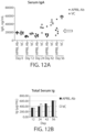


Classifications
-
- A—HUMAN NECESSITIES
- A61—MEDICAL OR VETERINARY SCIENCE; HYGIENE
- A61P—SPECIFIC THERAPEUTIC ACTIVITY OF CHEMICAL COMPOUNDS OR MEDICINAL PREPARATIONS
- A61P1/00—Drugs for disorders of the alimentary tract or the digestive system
-
- C—CHEMISTRY; METALLURGY
- C07—ORGANIC CHEMISTRY
- C07K—PEPTIDES
- C07K16/00—Immunoglobulins [IGs], e.g. monoclonal or polyclonal antibodies
- C07K16/18—Immunoglobulins [IGs], e.g. monoclonal or polyclonal antibodies against material from animals or humans
- C07K16/24—Immunoglobulins [IGs], e.g. monoclonal or polyclonal antibodies against material from animals or humans against cytokines, lymphokines or interferons
- C07K16/241—Tumor Necrosis Factors
-
- C—CHEMISTRY; METALLURGY
- C07—ORGANIC CHEMISTRY
- C07K—PEPTIDES
- C07K16/00—Immunoglobulins [IGs], e.g. monoclonal or polyclonal antibodies
- C07K16/18—Immunoglobulins [IGs], e.g. monoclonal or polyclonal antibodies against material from animals or humans
- C07K16/28—Immunoglobulins [IGs], e.g. monoclonal or polyclonal antibodies against material from animals or humans against receptors, cell surface antigens or cell surface determinants
- C07K16/2875—Immunoglobulins [IGs], e.g. monoclonal or polyclonal antibodies against material from animals or humans against receptors, cell surface antigens or cell surface determinants against the NGF/TNF superfamily, e.g. CD70, CD95L, CD153, CD154
-
- A—HUMAN NECESSITIES
- A61—MEDICAL OR VETERINARY SCIENCE; HYGIENE
- A61K—PREPARATIONS FOR MEDICAL, DENTAL OR TOILETRY PURPOSES
- A61K39/00—Medicinal preparations containing antigens or antibodies
- A61K39/395—Antibodies; Immunoglobulins; Immune serum, e.g. antilymphocytic serum
- A61K39/39533—Antibodies; Immunoglobulins; Immune serum, e.g. antilymphocytic serum against materials from animals
- A61K39/3955—Antibodies; Immunoglobulins; Immune serum, e.g. antilymphocytic serum against materials from animals against proteinaceous materials, e.g. enzymes, hormones, lymphokines
-
- A—HUMAN NECESSITIES
- A61—MEDICAL OR VETERINARY SCIENCE; HYGIENE
- A61K—PREPARATIONS FOR MEDICAL, DENTAL OR TOILETRY PURPOSES
- A61K45/00—Medicinal preparations containing active ingredients not provided for in groups A61K31/00 - A61K41/00
- A61K45/06—Mixtures of active ingredients without chemical characterisation, e.g. antiphlogistics and cardiaca
-
- A—HUMAN NECESSITIES
- A61—MEDICAL OR VETERINARY SCIENCE; HYGIENE
- A61P—SPECIFIC THERAPEUTIC ACTIVITY OF CHEMICAL COMPOUNDS OR MEDICINAL PREPARATIONS
- A61P1/00—Drugs for disorders of the alimentary tract or the digestive system
- A61P1/16—Drugs for disorders of the alimentary tract or the digestive system for liver or gallbladder disorders, e.g. hepatoprotective agents, cholagogues, litholytics
-
- A—HUMAN NECESSITIES
- A61—MEDICAL OR VETERINARY SCIENCE; HYGIENE
- A61P—SPECIFIC THERAPEUTIC ACTIVITY OF CHEMICAL COMPOUNDS OR MEDICINAL PREPARATIONS
- A61P13/00—Drugs for disorders of the urinary system
- A61P13/12—Drugs for disorders of the urinary system of the kidneys
-
- A—HUMAN NECESSITIES
- A61—MEDICAL OR VETERINARY SCIENCE; HYGIENE
- A61P—SPECIFIC THERAPEUTIC ACTIVITY OF CHEMICAL COMPOUNDS OR MEDICINAL PREPARATIONS
- A61P17/00—Drugs for dermatological disorders
-
- A—HUMAN NECESSITIES
- A61—MEDICAL OR VETERINARY SCIENCE; HYGIENE
- A61P—SPECIFIC THERAPEUTIC ACTIVITY OF CHEMICAL COMPOUNDS OR MEDICINAL PREPARATIONS
- A61P19/00—Drugs for skeletal disorders
- A61P19/02—Drugs for skeletal disorders for joint disorders, e.g. arthritis, arthrosis
-
- A—HUMAN NECESSITIES
- A61—MEDICAL OR VETERINARY SCIENCE; HYGIENE
- A61P—SPECIFIC THERAPEUTIC ACTIVITY OF CHEMICAL COMPOUNDS OR MEDICINAL PREPARATIONS
- A61P29/00—Non-central analgesic, antipyretic or antiinflammatory agents, e.g. antirheumatic agents; Non-steroidal antiinflammatory drugs [NSAID]
-
- A—HUMAN NECESSITIES
- A61—MEDICAL OR VETERINARY SCIENCE; HYGIENE
- A61P—SPECIFIC THERAPEUTIC ACTIVITY OF CHEMICAL COMPOUNDS OR MEDICINAL PREPARATIONS
- A61P35/00—Antineoplastic agents
-
- A—HUMAN NECESSITIES
- A61—MEDICAL OR VETERINARY SCIENCE; HYGIENE
- A61P—SPECIFIC THERAPEUTIC ACTIVITY OF CHEMICAL COMPOUNDS OR MEDICINAL PREPARATIONS
- A61P35/00—Antineoplastic agents
- A61P35/02—Antineoplastic agents specific for leukemia
-
- A—HUMAN NECESSITIES
- A61—MEDICAL OR VETERINARY SCIENCE; HYGIENE
- A61P—SPECIFIC THERAPEUTIC ACTIVITY OF CHEMICAL COMPOUNDS OR MEDICINAL PREPARATIONS
- A61P37/00—Drugs for immunological or allergic disorders
- A61P37/02—Immunomodulators
-
- A—HUMAN NECESSITIES
- A61—MEDICAL OR VETERINARY SCIENCE; HYGIENE
- A61P—SPECIFIC THERAPEUTIC ACTIVITY OF CHEMICAL COMPOUNDS OR MEDICINAL PREPARATIONS
- A61P37/00—Drugs for immunological or allergic disorders
- A61P37/02—Immunomodulators
- A61P37/06—Immunosuppressants, e.g. drugs for graft rejection
-
- A—HUMAN NECESSITIES
- A61—MEDICAL OR VETERINARY SCIENCE; HYGIENE
- A61P—SPECIFIC THERAPEUTIC ACTIVITY OF CHEMICAL COMPOUNDS OR MEDICINAL PREPARATIONS
- A61P43/00—Drugs for specific purposes, not provided for in groups A61P1/00-A61P41/00
-
- A—HUMAN NECESSITIES
- A61—MEDICAL OR VETERINARY SCIENCE; HYGIENE
- A61P—SPECIFIC THERAPEUTIC ACTIVITY OF CHEMICAL COMPOUNDS OR MEDICINAL PREPARATIONS
- A61P7/00—Drugs for disorders of the blood or the extracellular fluid
-
- A—HUMAN NECESSITIES
- A61—MEDICAL OR VETERINARY SCIENCE; HYGIENE
- A61P—SPECIFIC THERAPEUTIC ACTIVITY OF CHEMICAL COMPOUNDS OR MEDICINAL PREPARATIONS
- A61P9/00—Drugs for disorders of the cardiovascular system
-
- A—HUMAN NECESSITIES
- A61—MEDICAL OR VETERINARY SCIENCE; HYGIENE
- A61K—PREPARATIONS FOR MEDICAL, DENTAL OR TOILETRY PURPOSES
- A61K39/00—Medicinal preparations containing antigens or antibodies
- A61K2039/505—Medicinal preparations containing antigens or antibodies comprising antibodies
-
- A—HUMAN NECESSITIES
- A61—MEDICAL OR VETERINARY SCIENCE; HYGIENE
- A61K—PREPARATIONS FOR MEDICAL, DENTAL OR TOILETRY PURPOSES
- A61K39/00—Medicinal preparations containing antigens or antibodies
- A61K2039/54—Medicinal preparations containing antigens or antibodies characterised by the route of administration
-
- C—CHEMISTRY; METALLURGY
- C07—ORGANIC CHEMISTRY
- C07K—PEPTIDES
- C07K2317/00—Immunoglobulins specific features
- C07K2317/10—Immunoglobulins specific features characterized by their source of isolation or production
- C07K2317/14—Specific host cells or culture conditions, e.g. components, pH or temperature
-
- C—CHEMISTRY; METALLURGY
- C07—ORGANIC CHEMISTRY
- C07K—PEPTIDES
- C07K2317/00—Immunoglobulins specific features
- C07K2317/20—Immunoglobulins specific features characterized by taxonomic origin
- C07K2317/24—Immunoglobulins specific features characterized by taxonomic origin containing regions, domains or residues from different species, e.g. chimeric, humanized or veneered
-
- C—CHEMISTRY; METALLURGY
- C07—ORGANIC CHEMISTRY
- C07K—PEPTIDES
- C07K2317/00—Immunoglobulins specific features
- C07K2317/30—Immunoglobulins specific features characterized by aspects of specificity or valency
- C07K2317/33—Crossreactivity, e.g. for species or epitope, or lack of said crossreactivity
-
- C—CHEMISTRY; METALLURGY
- C07—ORGANIC CHEMISTRY
- C07K—PEPTIDES
- C07K2317/00—Immunoglobulins specific features
- C07K2317/30—Immunoglobulins specific features characterized by aspects of specificity or valency
- C07K2317/34—Identification of a linear epitope shorter than 20 amino acid residues or of a conformational epitope defined by amino acid residues
-
- C—CHEMISTRY; METALLURGY
- C07—ORGANIC CHEMISTRY
- C07K—PEPTIDES
- C07K2317/00—Immunoglobulins specific features
- C07K2317/50—Immunoglobulins specific features characterized by immunoglobulin fragments
- C07K2317/52—Constant or Fc region; Isotype
-
- C—CHEMISTRY; METALLURGY
- C07—ORGANIC CHEMISTRY
- C07K—PEPTIDES
- C07K2317/00—Immunoglobulins specific features
- C07K2317/50—Immunoglobulins specific features characterized by immunoglobulin fragments
- C07K2317/56—Immunoglobulins specific features characterized by immunoglobulin fragments variable (Fv) region, i.e. VH and/or VL
-
- C—CHEMISTRY; METALLURGY
- C07—ORGANIC CHEMISTRY
- C07K—PEPTIDES
- C07K2317/00—Immunoglobulins specific features
- C07K2317/70—Immunoglobulins specific features characterized by effect upon binding to a cell or to an antigen
- C07K2317/76—Antagonist effect on antigen, e.g. neutralization or inhibition of binding
-
- C—CHEMISTRY; METALLURGY
- C07—ORGANIC CHEMISTRY
- C07K—PEPTIDES
- C07K2317/00—Immunoglobulins specific features
- C07K2317/90—Immunoglobulins specific features characterized by (pharmaco)kinetic aspects or by stability of the immunoglobulin
- C07K2317/92—Affinity (KD), association rate (Ka), dissociation rate (Kd) or EC50 value
-
- C—CHEMISTRY; METALLURGY
- C07—ORGANIC CHEMISTRY
- C07K—PEPTIDES
- C07K2317/00—Immunoglobulins specific features
- C07K2317/90—Immunoglobulins specific features characterized by (pharmaco)kinetic aspects or by stability of the immunoglobulin
- C07K2317/94—Stability, e.g. half-life, pH, temperature or enzyme-resistance
Definitions
- IgA nephropathy is one of the most prevalent, chronic glomerular diseases worldwide. Conservative epidemiological estimates cite a global incidence of approximately 5-50 cases/million (children) and 10-40 cases /million (adults). This incidence of disease presents a regional bias with a higher prevalence in Asia and the Americas, with a particularly higher disease burden in Japan and regions of China. Biopsy confirmed cases of IgA nephropathy in Japan are projected at approximately 350,000. In the US, this projection is approximately 100,000-as such, it is the most frequently diagnosed 1° glomerular disease in adults. While a relatively indolent disease, IgA nephropathy leads to end stage renal disease (ESRD), i.e., renal failure in 20-50% of patients within a 20-30 year span.
- ESRD end stage renal disease
- IgA nephropathy is caused by the deposition of IgA, typically in the form of immune complexes in the mesangium of the kidney.
- WO 2010/100056 relates to a binding compound which binds to human APRIL. More specifically the documents provides compositions of anti-APRIL specific antibodies and methods to use such antibodies in modulating the biological activity APRIL, particularly in inflammatory diseases, inhibition of cell proliferation and cancer.
- This disclosure provides, at least in part, antibody molecules that bind to APRIL, e.g ., human and/or mouse APRIL, and that comprise one or more functional and structural properties disclosed herein.
- the antibody molecule binds to and/or reduces (e.g ., inhibits, blocks or neutralizes) one or more activities of APRIL.
- the antibody molecule binds to a region in APRIL that interacts with TACI (e.g. , the CRD2 domain of TACI).
- the antibody molecule binds to one or more residues within a region of human APRIL as defined in any of Tables 3-4 or 7-8. While not wishing to be bound by theory, it is believed that in an embodiment, improved or optimal inhibition of APRIL activities can be achieved, by targeting certain region(s) on APRIL (e.g. , the region(s) associated with the interactions between APRIL and the CDR2 domain of TACI).
- the antibody molecule is selected from Table 1 or 5, or competes for binding to APRIL with an antibody molecule selected from Table 1 or 5.
- the antibody molecule binds to the same or overlapping epitope as the epitope recognized by an antibody molecule selected from Table 1 or 5.
- the antibody molecule comprises one or more heavy chain variable regions and/or one or more light chain variable regions described in Table 1 or 5. In an embodiment, the antibody molecule comprises one or more heavy chain CDRs and/or one or more light chain CDRs described in Table 1 or 5.
- nucleic acid molecules encoding the antibody molecules, expression vectors, host cells, compositions ( e.g ., pharmaceutical compositions), kits, containers, and methods for making the antibody molecules are also provided.
- the antibody molecules disclosed herein can be used (alone or in combination with other agents or therapeutic modalities) to treat, prevent and/or diagnose disorders associated with APRIL, such as IgA nephropathy.
- this disclosure provides an antibody molecule, e.g ., an antibody molecule described herein, having one or more ( e.g., 1, 2, 3, 4, 5, 6, 7, 8, 9, 10, 11, 12, 13, 14, 15, 16, 17, 18, 19, 20, 21, 22, or 23) of the following properties:
- the disclosure features an anti-APRIL antibody molecule, which:
- the antibody molecule is a synthetic antibody molecule. In an embodiment, the antibody molecule is an isolated antibody molecule.
- the antibody molecule binds, or substantially binds, to human APRIL at an EC 50 of 20 nM or less, e.g., 10 nM or less, 9 nM or less, 8 nM or less, 7 nM or less, 6 nM or less, 5 nM or less, 4 nM or less, 3 nM or less, 2 nM or less, 1 nM or less, 0.8 nM or less, 0.6 nM or less, 0.4 nM or less, 0.2 nM or less, 0.1 nM or less, 0.05 nM or less, 0.02 nM or less, 0.01 nM or less, 0.005 nM or less, 0.002 nM or less, or 0.001 nM or less, e.g., between 0.001 nM and 20 nM, e.g., between 0.01 nM and 20 nM, between 0.1 nM and 20 nM, between
- the antibody molecule binds, or substantially binds, to mouse APRIL at an EC 50 of 100 nM or less, e.g., 80 nM or less, 60 nM or less, 40 nM or less, 20 nM or less, 10 nM or less, 9 nM or less 9 nM or less, 8 nM or less, 7 nM or less, 6 nM or less, 5 nM or less, 4 nM or less, 3 nM or less, 2 nM or less, 1 nM or less, 0.8 nM or less, 0.6 nM or less, 0.4 nM or less, 0.2 nM or less, 0.1 nM or less, 0.05 nM or less, 0.02 nM or less, 0.01 nM or less, 0.005 nM or less, 0.002 nM or less, or 0.001 nM or less, e.g., between 0.001 nM and 100 nM or less
- the antibody molecule inhibits, or substantially inhibits, binding of APRIL (e.g., human APRIL, mouse APRIL, or both) to TACI (e.g., human TACI, mouse TACI, or both), at an IC 50 of 50 nM or less, e.g., 40 nM or less, 30 nM or less, 20 nM or less, 10 nM or less, 9 nM or less, 8 nM or less, 7 nM or less, 6 nM or less, 5 nM or less, 4 nM or less, 3 nM or less, 2 nM or less, 1 nM or less, 0.8 nM or less, 0.6 nM or less, 0.4 nM or less, 0.2 nM or less, 0.1 nM or less, 0.05 nM or less, 0.02 nM or less, or 0.01 nM or less, e.g.
- APRIL e.g., human APRIL
- nM and 5 nM between 0.01 nM and 50 nM, between 0.1 nM and 50 nM, between 0.1 nM and 25 nM, between 0.1 nM and 10 nM, between 0.1 nM and 5 nM, between 0.1 nM and 1 nM, between 0.1 nM and 0.5 nM, between 0.5 nM and 5 nM, or between 1 nM and 5 nM, e.g., as determined by a method described herein.
- the antibody molecule inhibits, or substantially inhibits, binding of APRIL (e.g., human APRIL, mouse APRIL, or both) to BCMA (e.g., human BCMA, mouse BCMA, or both), e.g., at an IC 50 of 200 nM or less, 150 nM or less, 100 nM or less, 50 nM or less, 40 nM or less, 30 nM or less, 20 nM or less, 10 nM or less, 9 nM or less, 8 nM or less, 7 nM or less, 6 nM or less, 5 nM or less, 4 nM or less, 3 nM or less, 2 nM or less, 1 nM or less, 0.8 nM or less, 0.6 nM or less, 0.4 nM or less, 0.2 nM or less, 0.1 nM or less, 0.05 nM or less, 0.02 nM or less, 0.01 nM or
- the antibody molecule is an IgG antibody molecule, e.g ., comprising a heavy chain constant region of IgG, e.g., chosen from IgG1, IgG2 ( e.g., IgG2a), IgG3, or IgG4, e.g., IgG2 or IgG4.
- the antibody molecule is an IgG1 antibody molecule, e.g ., having an IgG1 constant region described herein.
- the antibody molecule is an IgG2 antibody molecule e.g ., having an IgG2 constant region described herein.
- the antibody molecule comprises a light chain constant region of kappa or lambda light chain.
- the antibody molecule comprises an Fc region.
- the Fc region comprises one or more mutations located at the interface between the CH2 and CH3 domains ( e.g., to increase the binding affinity to neonatal receptor FcRn and/or the half-life of the antibody molecule).
- the Fc region comprises one or more mutations, e.g., one or more ( e.g., 2, 3, 4, 5, 6 or all) mutations chosen from T250Q, M252Y, S254T, T256E, M428L, H433K, N434F, or any combination thereof, of IgG1.
- the Fc region comprises one or more mutations at positions 233-236 or 322 of human IgG1 or IgG2, or one or more substitutions at positions 327, 330 or 331 of human IgG4 (e.g., to reduce complement-dependent cytotoxicity (CDC)).
- the Fc region comprises one or more (e.g., 2, 3, 4, 5, 6, 7 or all) mutations chosen from E233P, L234V, L235A, G236, K322A, A327G, A330S, P331S, or any combination thereof.
- the antibody molecule is a humanized antibody molecule, e.g., comprising one or more framework regions derived from human framework germline sequence.
- the antibody molecule comprises a heavy chain variable region (VH) described in Table 1 or 5.
- the antibody molecule comprises a light chain variable region (VL) described in Table 1 or 5.
- the antibody molecule comprises a heavy chain variable region (VH) and a light chain variable region (VL) described in Table 1 or 5.
- the antibody molecule comprises one, two, or three CDRs of a heavy chain variable region (VH) described in Table 1 or 5.
- the antibody molecule comprises one, two, or three CDRs of a light chain variable region (VL) described in Table 1 or 5. In an embodiment, the antibody molecule comprises one, two, or three CDRs of a heavy chain variable region (VH) described in Table 1 or 5, and one, two, or three CDRs of a light chain variable region (VL) described in Table 1 or 5. In an embodiment, the antibody molecule comprises two heavy chain variable regions and two light chain variable regions. In an embodiment, the antibody molecule is a Fab, F(ab')2, Fv, Fd, or a single chain Fv fragment (scFv).
- the antibody molecule binds, or substantially binds, to one or more, e.g., 2, 3, 4, 5, 6, 7, 8, 9, 10, 11, 12, 13, 14, 15, 16, 17, 18, 19, 20, 21, 22, 23, 24, 25, or more, residues within a region of human APRIL as defined in any of Tables 3-4 or 7-8.
- the antibody molecule binds, or substantially binds, to one or more, e.g., 2, 3, 4, 5, 6, 7, 8, 9, 10, 11, 12, 13, 14, 15, 16, 17, 18, 19, 20, 21, 22, 23, 24, 25, or more, residues within a region of human APRIL as defined in Table 3.
- the antibody molecule binds, or substantially binds, to an epitope that comprises or consists of one or more, e.g., 2, 3, 4, 5, 6, 7, 8, 9, 10, 11, 12, 13, 14, 15, 16, 17, 18, 19, 20, 21, 22, 23, 24, 25, or all, of the human APRIL residues from Table 3.
- the antibody molecule binds, or substantially binds, to an epitope that overlaps an epitope that comprises or consists of all of the human APRIL residues from Table 3. In an embodiment, the antibody molecule binds, or substantially binds, to an epitope that comprises APRIL residues from two monomers, e.g. , one or more residues from monomer A and monomer B as shown in Table 3 .
- the antibody molecule binds, or substantially binds, to one or more, e.g., 2, 3, 4, 5, 6, 7, 8, 9, 10, or more, residues within a region of human APRIL as defined in Table 4. In an embodiment, the antibody molecule binds, or substantially binds, to an epitope that comprises or consists of one or more, e.g., 2, 3, 4, 5, 6, 7, 8, 9, 10, or all, of the human APRIL residues from Table 4. In an embodiment, the antibody molecule binds, or substantially binds, to an epitope that overlaps an epitope that comprises or consists of all of the human APRIL residues from Table 4.
- the antibody molecule binds, or substantially binds, to an epitope that comprises one or more APRIL residues from the C-D loop (e.g., the loop connecting ⁇ -sheets C and D), the G-H loop ( e.g., the loop connecting ⁇ -sheets G and H), or both.
- the antibody molecule binds, or substantially binds, to one or more, e.g., 2, 3, 4, 5, 6, 7, 8, 9, 10, 11, 12, 13, 14, 15, 16, 17, or all, residues within a region of human APRIL as defined in Table 7. In an embodiment, the antibody molecule binds, or substantially binds, to an epitope that comprises or consists of one or more, e.g., 2, 3, 4, 5, 6, 7, 8, 9, 10, 11, 12, 13, 14, 15, 16, 17, or all, of the human APRIL residues from Table 7. In an embodiment, the antibody molecule binds, or substantially binds, to an epitope that overlaps an epitope that comprises or consists of all of the human APRIL residues from Table 7.
- the antibody molecule binds, or substantially binds, to one or more, e.g., 2, 3, 4, 5, 6, 7, 8, 9, 10, 11, 12, 13, 14, 15, 16, 17, 18, 19, 20, 21, 22, 23, 24, 25, or all, residues within a region of human APRIL as defined in Table 8.
- the antibody molecule binds, or substantially binds, to an epitope that comprises or consists of one or more, e.g., 2, 3, 4, 5, 6, 7, 8, 9, 10, 11, 12, 13, 14, 15, 16, 17, 18, 19, 20, 21, 22, 23, 24, 25, or all, of the human APRIL residues from Table 8.
- the antibody molecule binds, or substantially binds, to an epitope that overlaps an epitope that comprises or consists of all of the human APRIL residues from Table 8.
- the antibody molecule binds, or substantially binds, to one or more (e.g., 2, 3, 4, 5, 6, 7, 8, 9, or all) residues of human APRIL from positions 105-114 and/or one or more ( e.g., 2, 3, 4, 5, 6, 7, 8, 9, or all) residues of mouse APRIL from positions 96-105.
- the antibody molecule does not bind, or does not substantially bind, to one, two or all of Asp129, Arg233, or His203 of human APRIL.
- the epitope is a conformational epitope.
- binding of the antibody molecule to APRIL inhibits, or substantially inhibits, the binding of the CRD2 domain of TACI (e.g. , human TACI) to APRIL ( e.g. , human APRIL).
- binding of the antibody molecule to human APRIL inhibits, or substantially inhibits, the binding of human TACI, to one or more, e.g., 2, 3, 4, 5, 6, 7, 8, 9, 10, 11, 12, 13, 14, 15, 16, 17, 18, 19, 20, 21, 22, 23, 24, 25, or all, of the APRIL residues from Table 3.
- binding of the antibody molecule to human APRIL inhibits, or substantially inhibits, the binding of human TACI, to one or more, e.g., 2, 3, 4, 5, 6, 7, 8, 9, 10, or all, of the human APRIL residues from Table 4.
- binding of the antibody molecule to human APRIL inhibits, or substantially inhibits, the binding of human TACI, to one or more, e.g., 2, 3, 4, 5, 6, 7, 8, 9, 10, 11, 12, 13, 14, 15, 16, 17, or all, of the human APRIL residues from Table 7.
- binding of the antibody molecule to human APRIL inhibits, or substantially inhibits, the binding of human TACI, to one or more, e.g., 2, 3, 4, 5, 6, 7, 8, 9, 10, 11, 12, 13, 14, 15, 16, 17, 18, 19, 20, 21, 22, 23, 24, 25, or all, of the human APRIL residues from Table 8.
- binding of the antibody molecule to human APRIL inhibits, or substantially inhibits, the binding of human BCMA, to one or more, e.g., 2, 3, 4, 5, 6, 7, 8, 9, 10, 11, 12, 13, 14, 15, 16, 17, 18, 19, 20, 21, 22, 23, 24, 25, or all, of the human APRIL residues from Table 8.
- the disclosure features an antibody molecule that binds, or substantially binds, to one or more, e.g., 2, 3, 4, 5, 6, 7, 8, 9, 10, 11, 12, 13, 14, 15, 16, 17, 18, 19, 20, 21, 22, 23, 24, 25, or more, residues within a region of human APRIL as defined in any of Tables 3-4 or 7-8.
- the anti-APRIL antibody molecule binds, or substantially binds, to an epitope that comprises or consists of one or more, e.g., 2, 3, 4, 5, 6, 7, 8, 9, 10, 11, 12, 13, 14, 15, 16, 17, 18, 19, 20, 21, 22, 23, 24, 25, or more, of the human APRIL residues from any of Tables 3-4 or 7-8.
- the antibody molecule binds, or substantially binds, to a conformational epitope.
- the antibody molecule binds, or substantially binds, to one or more, e.g., 2, 3, 4, 5, 6, 7, 8, 9, 10, 11, 12, 13, 14, 15, 16, 17, 18, 19, 20, 21, 22, 23, 24, 25, or more, residues within a region of human APRIL as defined in Table 3.
- the anti-APRIL antibody molecule binds, or substantially binds, to an epitope that comprises or consists of one or more ( e.g., 2, 3, 4, 5, 6, 7, 8, 9, 10, 11, 12, 13, 14, 15, 16, 17, 18, 19, 20, 21, 22, 23, 24, 25, or all) of the human APRIL residues from Table 3.
- the antibody molecule binds, or substantially binds, to an epitope that comprises APRIL residues from two monomers, e.g ., one or more residues from monomer A and monomer B as shown in Table 3.
- the antibody molecule binds, or substantially binds, to one or more, e.g., 2, 3, 4, 5, 6, 7, 8, 9, 10, or all, residues within a region of human APRIL as defined in Table 4.
- the epitope comprises consists of one or more, e.g., 2, 3, 4, 5, 6, 7, 8, 9, 10, or all of the APRIL residues from Table 4.
- the epitope comprises or consists of one or more APRIL residues from the C-D loop (e.g., the loop connecting ⁇ -sheets C and D), the G-H loop ( e.g., the loop connecting ⁇ -sheets G and H), or both.
- the antibody molecule binds, or substantially binds, to one or more, e.g., 2, 3, 4, 5, 6, 7, 8, 9, 10, 11, 12, 13, 14, 15, 16, 17, or all, residues within a region of human APRIL as defined in Table 7. In an embodiment, the antibody molecule binds, or substantially binds, to an epitope that comprises or consists of one or more, e.g., 2, 3, 4, 5, 6, 7, 8, 9, 10, 11, 12, 13, 14, 15, 16, 17, or all, of the human APRIL residues from Table 7. In an embodiment, the antibody molecule binds, or substantially binds, to an epitope that overlaps an epitope that comprises or consists of all of the human APRIL residues from Table 7.
- the antibody molecule binds, or substantially binds, to one or more, e.g., 2, 3, 4, 5, 6, 7, 8, 9, 10, 11, 12, 13, 14, 15, 16, 17, 18, 19, 20, 21, 22, 23, 24, 25, or all, residues within a region of human APRIL as defined in Table 8.
- the antibody molecule binds, or substantially binds, to an epitope that comprises or consists of one or more, e.g., 2, 3, 4, 5, 6, 7, 8, 9, 10, 11, 12, 13, 14, 15, 16, 17, 18, 19, 20, 21, 22, 23, 24, 25, or all, of the human APRIL residues from Table 8.
- the antibody molecule binds, or substantially binds, to an epitope that overlaps an epitope that comprises or consists of all of the human APRIL residues from Table 8.
- the antibody molecule binds, or substantially binds, to one or more (e.g., 2, 3, 4, 5, 6, 7, 8, 9, or all) residues of human APRIL from positions 105-114 and/or one or more ( e.g., 2, 3, 4, 5, 6, 7, 8, 9, or all) residues of mouse APRIL from positions 96-105.
- the antibody molecule does not bind, or does not substantially bind, to one, two or all of Asp129, Arg233, or His203 of human APRIL.
- the antibody molecule binds, or substantially binds, to an epitope that comprises or consists of one or more (e.g., 2, 3, 4, 5, 6, 7, 8, 9, 10, 11, 12, 13, 14, 15, 16, 17, 18, 19, 20, 21, 22, 23, 24, 25, 26, 27, 28, 29, 30, 31, 32, or all) of human APRIL residues from Table 6.
- the antibody molecule binds, or substantially binds, to one or more ( e.g., 2, 3, 4, 5, or all) of the amino acid residues of human APRIL chosen from V174, F176, Q190, R195, R206, or Y208. In an embodiment, the antibody molecule does not binds, or does not substantially bind, to one or more ( e.g., 2, 3, or all) of the amino acid residues of human APRIL chosen from V181, S226, I228, or N237.
- the antibody molecule binds, or substantially binds, to one or more ( e.g., 2, 3, or all) of the amino acid residues of human APRIL chosen from F176, V181, Q190, or I228. In an embodiment, the antibody molecule does not bind, or does not substantially bind, to one or both of the amino acid residues of human APRIL chosen from Y208 or N237. In an embodiment, the antibody molecule binds, or substantially binds, to one or more ( e.g., 2, or all) of the amino acid residues of human APRIL chosen from V174, R206, or Y208. In an embodiment, the antibody molecule does not bind, or does not substantially bind, to one or more ( e.g., 2, 3, or all) of the amino acid residues of human APRIL chosen from F176, V181, Q190, or N237.
- the antibody molecule binds, or substantially binds, to human APRIL. In an embodiment, the antibody molecule binds, or substantially binds, to human APRIL and mouse APRIL. In an embodiment, the antibody molecule binds, or substantially binds to, human APRIL, but does not bind to mouse APRIL, or binds to mouse APRIL with low affinity.
- the antibody molecule binds, or substantially binds, to human APRIL at an EC 50 of 20 nM or less, e.g., 10 nM or less, 9 nM or less or less, 8 nM or less, 7 nM or less, 6 nM or less, 5 nM or less, 4 nM or less, 3 nM or less, 2 nM or less, 1 nM or less, 0.8 nM or less, 0.6 nM or less, 0.4 nM or less, 0.2 nM or less, 0.1 nM or less, 0.05 nM or less, 0.02 nM or less, 0.01 nM or less, 0.005 nM or less, 0.002 nM or less, or 0.001 nM or less, e.g., between 0.001 nM and 20 nM, e.g., between 0.01 nM and 20 nM, between 0.1 nM and 20 nM,
- the antibody molecule binds, or substantially binds, to mouse APRIL at an EC 50 of 100 nM or less, e.g., 80 nM or less, 60 nM or less, 40 nM or less, 20 nM or less, e.g., 10 nM or less, e.g., 9 nM or less 9 nM or less, 8 nM or less, 7 nM or less, 6 nM or less, 5 nM or less, 4 nM or less, 3 nM or less, 2 nM or less, 1 nM or less, 0.8 nM or less, 0.6 nM or less, 0.4 nM or less, 0.2 nM or less, 0.1 nM or less, 0.05 nM or less, 0.02 nM or less, 0.01 nM or less, 0.005 nM or less, 0.002 nM or less, or 0.001 nM or less, e.g.
- the antibody molecule does not bind to mouse APRIL, or binds to mouse APRIL with low affinity, e.g., at an EC 50 of 1000 nM or more, e.g., 2000 nM or more, e.g., as determined by a method described herein.
- the antibody molecule inhibits, or substantially inhibits, binding of APRIL (e.g., human APRIL, mouse APRIL, or both) to TACI (e.g., human TACI, mouse TACI, or both).
- APRIL e.g., human APRIL, mouse APRIL, or both
- TACI e.g., human TACI, mouse TACI, or both.
- the antibody molecule inhibits, or substantially inhibits, binding of APRIL (e.g ., human APRIL, mouse APRIL, or both) to TACI (e.g ., human TACI, mouse TACI, or both), at an IC 50 of 50 nM or less, e.g., 40 nM or less, 30 nM or less, 20 nM or less, 10 nM or less, 9 nM or less, 8 nM or less, 7 nM or less, 6 nM or less, 5 nM or less, 4 nM or less, 3 nM or less, 2 nM or less, 1 nM or less, 0.8 nM or less, 0.6 nM or less, 0.4 nM or less, 0.2 nM or less, 0.1 nM or less, 0.05 nM or less, 0.02 nM or less, or 0.01 nM or less, e.g.
- APRIL e.g ., human
- nM and 5 nM between 0.01 nM and 50 nM, between 0.1 nM and 50 nM, between 0.1 nM and 25 nM, between 0.1 nM and 10 nM, between 0.1 nM and 5 nM, between 0.1 nM and 1 nM, between 0.1 nM and 0.5 nM, between 0.5 nM and 5 nM, or between 1 nM and 5 nM, e.g., as determined by a method described herein.
- binding of the antibody molecule to APRIL inhibits, or substantially inhibits, the binding of the CRD2 domain of TACI (e.g. , human TACI) to APRIL ( e.g. , human APRIL).
- binding of the antibody molecule to human APRIL inhibits, or substantially inhibits, the binding of human TACI, to one or more, e.g., 2, 3, 4, 5, 6, 7, 8, 9, 10, 11, 12, 13, 14, 15, 16, 17, 18, 19, 20, 21, 22, 23, 24, 25, or all, of the human APRIL residues from Table 3.
- binding of the antibody molecule to human APRIL inhibits, or substantially inhibits, the binding of human TACI to one or more, e.g., 2, 3, 4, 5, 6, 7, 8, 9, 10, or all, of the human APRIL residues from Table 4.
- binding of the antibody molecule to human APRIL inhibits, or substantially inhibits, the binding of human TACI, to one or more, e.g., 2, 3, 4, 5, 6, 7, 8, 9, 10, 11, 12, 13, 14, 15, 16, 17, or all, of the human APRIL residues from Table 7.
- binding of the antibody molecule to human APRIL inhibits, or substantially inhibits, the binding of human TACI, to one or more, e.g., 2, 3, 4, 5, 6, 7, 8, 9, 10, 11, 12, 13, 14, 15, 16, 17, 18, 19, 20, 21, 22, 23, 24, 25, or all, of the human APRIL residues from Table 8.
- binding of the antibody molecule to human APRIL inhibits, or substantially inhibits, the binding of human BCMA, to one or more, e.g., 2, 3, 4, 5, 6, 7, 8, 9, 10, 11, 12, 13, 14, 15, 16, 17, 18, 19, 20, 21, 22, 23, 24, 25, or all, of the human APRIL residues from Table 8.
- the antibody molecule inhibits, or substantially inhibits, binding of APRIL (e.g., human APRIL, mouse APRIL, or both) to BCMA ( e.g., human BCMA, mouse BCMA, or both). In an embodiment, the antibody molecule inhibits, or substantially inhibits, binding of APRIL ( e.g., human APRIL, mouse APRIL, or both) to BCMA ( e.g., human BCMA, mouse BCMA, or both), e.g., at an IC 50 of 200 nM or less, 150 nM or less, 100 nM or less, 50 nM or less, 40 nM or less, 30 nM or less, 20 nM or less, 10 nM or less, 9 nM or less, 8 nM or less, 7 nM or less, 6 nM or less, 5 nM or less, 4 nM or less, 3 nM or less, 2 nM or less, 1 nM or less,
- the antibody molecule is a synthetic antibody molecule. In an embodiment, the antibody molecule is an isolated antibody molecule. In an embodiment, the antibody molecule is an IgG antibody molecule, e.g., comprising a heavy chain constant region of IgG, e.g., chosen from IgG1, IgG2 ( e.g., IgG2a), IgG3, or IgG4, e.g., IgG2 or IgG4. In an embodiment, the antibody molecule is an IgG1 antibody molecule. In an embodiment, the antibody molecule is an IgG2 antibody molecule. In an embodiment, the antibody molecule comprises a light chain constant region of kappa or lambda light chain.
- the antibody molecule comprises an Fc region.
- the Fc region comprises one or more mutations located at the interface between the CH2 and CH3 domains ( e.g., to increase the binding affinity to neonatal receptor FcRn and/or the half-life of the antibody molecule).
- the Fc region comprises one or more mutations, e.g., one or more ( e.g., 2, 3, 4, 6 or all) mutations chosen from T250Q, M252Y, S254T, T256E, M428L, H433K, N434F, or any combination thereof, of IgG1.
- the Fc region comprises one or more mutations at positions 233-236 or 322 of human IgG1 or IgG2, or one or more substitutions at positions 327, 330 or 331 of human IgG4 (e.g., to reduce complement-dependent cytotoxicity (CDC)).
- the Fc region comprises one or more (e.g., 2, 3, 4, 6 7 or all) mutations chosen from E233P, L234V, L235A, G236, K322A, A327G, A330S, P331S, or any combination thereof.
- the antibody molecule is a humanized antibody molecule, e.g ., as described in Table 1 or 5 , e.g., comprising one or more framework regions derived from human framework germline sequence.
- the antibody molecule comprises two heavy chain variable regions and two light chain variable regions.
- the antibody molecule is a Fab, F(ab')2, Fv, Fd, or a single chain Fv fragment (scFv).
- the antibody molecule comprises a heavy chain variable region (VH), wherein the heavy chain variable region comprises three heavy chain complementarity determining regions (HCDR1, HCDR2, and HCDR3), wherein the heavy chain variable region comprises one, two, or all of the following: (i) an HCDR1 comprising an amino acid sequence that differs by no more than 1, 2, or 3 amino acid residues from, or has at least 85, 90, 95, 99 or 100% homology with, the amino acid sequence of the HCDR1 of monoclonal antibody 2419 ( e.g., SEQ ID NO: 11) or a 2419-related antibody; (ii) an HCDR2 comprising an amino acid sequence that differs by no more than 1, 2, or 3 amino acid residues from, or has at least 85, 90, 95, 99 or 100% homology with, the amino acid sequence of the HCDR2 of monoclonal antibody 2419 ( e.g., SEQ ID NO: 12) or a 2419-related antibody; or (iii) an HCDR1
- the antibody molecule comprises a light chain variable region (VL), wherein the light chain variable region comprises three light chain complementarity determining regions (LCDR1, LCDR2, and LCDR3), wherein the light chain variable region comprises one, two, or all of the following: (i) an LCDR1 comprising an amino acid sequence that differs by no more than 1, 2, or 3 amino acid residues from, or has at least 85, 90, 95, 99 or 100% homology with, the amino acid sequence of the LCDR1 of monoclonal antibody 2419 ( e.g., SEQ ID NO: 14) or a 2419-related antibody; (ii) an LCDR2 comprising an amino acid sequence that differs by no more than 1, 2, or 3 amino acid residues from, or has at least 85, 90, 95, 99 or 100% homology with, the amino acid sequence of the LCDR2 of monoclonal antibody 2419 (e.g., SEQ ID NO: 15) or a 2419-related antibody; or (iii) an LCDR3 comprising an amino acid sequence of the
- the antibody molecule comprises:
- the antibody molecule comprises: (i) a VH comprising: an HCDR1 comprising the amino acid sequence of the HCDR1 of monoclonal antibody 2419 ( e.g., SEQ ID NO: 11) or a 2419-related antibody; an HCDR2 comprising the amino acid sequence of the HCDR2 of monoclonal antibody 2419 ( e.g., SEQ ID NO: 12) or a 2419-related antibody; or an HCDR3 comprising the amino acid sequence of the HCDR3 of monoclonal antibody 2419 ( e.g., SEQ ID NO: 13) or a 2419-related antibody, and (ii) a VL comprising: an LCDR1 comprising the amino acid sequence of the LCDR1 of monoclonal antibody 2419 ( e.g., SEQ ID NO: 14) or a 2419-related antibody; an LCDR2 comprising the amino acid sequence of the LCDR2 of monoclonal antibody 2419 ( e.g., SEQ ID NO:
- the antibody molecule comprises a heavy chain variable region (VH), wherein the heavy chain variable region comprises three heavy chain complementarity determining regions (HCDR1, HCDR2, and HCDR3), wherein the heavy chain variable region comprises one, two, or all of the following: (i) an HCDR1 comprising an amino acid sequence that differs by no more than 1, 2, or 3 amino acid residues from, or has at least 85, 90, 95, 99 or 100% homology with, the amino acid sequence of the HCDR1 of monoclonal antibody 2419 ( e.g., SEQ ID NO: 17) or a 2419-related antibody; (ii) an HCDR2 comprising an amino acid sequence that differs by no more than 1, 2, or 3 amino acid residues from, or has at least 85, 90, 95, 99 or 100% homology with, the amino acid sequence of the HCDR2 of monoclonal antibody 2419 ( e.g., SEQ ID NO: 18) or a 2419-related antibody; or (iii) an HCDR1
- the antibody molecule comprises a light chain variable region (VL), wherein the light chain variable region comprises three light chain complementarity determining regions (LCDR1, LCDR2, and LCDR3), wherein the light chain variable region comprises one, two, or all of the following: (i) an LCDR1 comprising an amino acid sequence that differs by no more than 1, 2, or 3 amino acid residues from, or has at least 85, 90, 95, 99 or 100% homology with, the amino acid sequence of the LCDR1 of monoclonal antibody 2419 ( e.g., SEQ ID NO: 14) or a 2419-related antibody; (ii) an LCDR2 comprising an amino acid sequence that differs by no more than 1, 2, or 3 amino acid residues from, or has at least 85, 90, 95, 99 or 100% homology with, the amino acid sequence of the LCDR2 of monoclonal antibody 2419 (e.g., SEQ ID NO: 15) or a 2419-related antibody; or (iii) an LCDR3 comprising an amino acid sequence of the
- the antibody molecule comprises:
- the antibody molecule comprises: (i) a VH comprising: an HCDR1 comprising the amino acid sequence of the HCDR1 of monoclonal antibody 2419 ( e.g., SEQ ID NO: 17) or a 2419-related antibody; an HCDR2 comprising the amino acid sequence of the HCDR2 of monoclonal antibody 2419 ( e.g., SEQ ID NO: 18) or a 2419-related antibody; or an HCDR3 comprising the amino acid sequence of the HCDR3 of monoclonal antibody 2419 ( e.g., SEQ ID NO: 13) or a 2419-related antibody, and (ii) a VL comprising: an LCDR1 comprising the amino acid sequence of the LCDR1 of monoclonal antibody 2419 ( e.g., SEQ ID NO: 14) or a 2419-related antibody; an LCDR2 comprising the amino acid sequence of the LCDR2 of monoclonal antibody 2419 ( e.g., SEQ ID NO:
- the antibody molecule comprises a VH comprising an amino acid sequence that differs by no more than 1, 2, 3, 4, 5, 6, 7, 8, 9, 10, 11, 12, 13, 14, or 15 amino acid residues from, or has at least 85, 90, 95, 96, 97, 98, 99, or 100% homology with, the amino acid sequence of the VH of monoclonal antibody 2419 ( e.g., SEQ ID NO: 19) or a 2419-related antibody.
- VH comprising an amino acid sequence that differs by no more than 1, 2, 3, 4, 5, 6, 7, 8, 9, 10, 11, 12, 13, 14, or 15 amino acid residues from, or has at least 85, 90, 95, 96, 97, 98, 99, or 100% homology with, the amino acid sequence of the VH of monoclonal antibody 2419 (e.g., SEQ ID NO: 19) or a 2419-related antibody.
- the antibody molecule comprises a VL comprising an amino acid sequence that differs by no more than 1, 2, 3, 4, 5, 6, 7, 8, 9, 10, 11, 12, 13, 14, or 15 amino acid residues from, or has at least 85, 90, 95, 96, 97, 98, 99, or 100% homology with, the amino acid sequence of the VL of monoclonal antibody 2419 ( e.g., SEQ ID NO: 20) or a 2419-related antibody.
- VL comprising an amino acid sequence that differs by no more than 1, 2, 3, 4, 5, 6, 7, 8, 9, 10, 11, 12, 13, 14, or 15 amino acid residues from, or has at least 85, 90, 95, 96, 97, 98, 99, or 100% homology with, the amino acid sequence of the VL of monoclonal antibody 2419 (e.g., SEQ ID NO: 20) or a 2419-related antibody.
- the antibody molecule comprises: (i) a VH comprising an amino acid sequence that differs by no more than 1, 2, 3, 4, 5, 6, 7, 8, 9, 10, 11, 12, 13, 14, or 15 amino acid residues from, or has at least 85, 90, 95, 96, 97, 98, 99, or 100% homology with, the amino acid sequence of the VH of monoclonal antibody 2419 ( e.g., SEQ ID NO: 19) or a 2419-related antibody; and (ii) a VL comprising an amino acid sequence that differs by no more than 1, 2, 3, 4, 5, 6, 7, 8, 9, 10, 11, 12, 13, 14, or 15 amino acid residues from, or has at least 85, 90, 95, 96, 97, 98, 99, or 100% homology with, the amino acid sequence of the VL of monoclonal antibody 2419 (e.g., SEQ ID NO: 20) or a 2419-related antibody.
- a VH comprising an amino acid sequence that differs by no more than 1, 2, 3, 4, 5, 6, 7, 8, 9, 10, 11, 12,
- the antibody molecule comprises: (i) a VH comprising the amino acid sequence of the VH of monoclonal antibody 2419 ( e.g., SEQ ID NO: 19) or a 2419-related antibody; and (ii) a VL comprising the amino acid sequence of the VL of monoclonal antibody 2419 ( e.g., SEQ ID NO: 20) or a 2419-related antibody.
- the antibody molecule is monoclonal antibody 2419.
- monoclonal antibody 2419 is a humanized monoclonal antibody 2419.
- the antibody molecule is a 2419-related antibody molecule, e.g ., any of antibodies 2419-0105, 2419-0205, 2419-0206, 2419-0406, 2419-0605, 2419-0805, 2419-0806, 2419-1204, 2419-1205, 2419-1210, 2419-1305, 2419-1306, 2419-1310, or 2419-1406, e.g ., as disclosed in Table 1 or 5.
- the antibody molecule comprises a VH comprising the amino acid sequence of any of SEQ ID NOS: 209-214, 283, 288, 289, 291, 292, 294, 296, or 317, a VL comprising the amino acid sequence of any of SEQ ID NOS: 215-219, 284, 286, 295, or 316, or both.
- the disclosure features an anti-APRIL antibody molecule, which:
- the antibody molecule binds, or substantially binds, to human APRIL at an EC 50 of 20 nM or less, e.g., 10 nM or less, 9 nM or less, 8 nM or less, 7 nM or less, 6 nM or less, 5 nM or less, 4 nM or less, 3 nM or less, 2 nM or less, 1 nM or less, 0.8 nM or less, 0.6 nM or less, 0.4 nM or less, 0.2 nM or less, 0.1 nM or less, 0.05 nM or less, 0.02 nM or less, 0.01 nM or less, 0.005 nM or less, 0.002 nM or less, or 0.001 nM or less, e.g., between 0.001 nM and 20 nM, e.g., between 0.01 nM and 20 nM, between 0.1 nM and 20 nM, between
- the antibody molecule does not bind to mouse APRIL, or binds to mouse APRIL with low affinity, e.g., at an EC 50 of 1000 nM or more, e.g., 2000 nM or more, e.g., as determined by a method described herein.
- the antibody molecule inhibits, or substantially inhibits, binding of APRIL (e.g., human APRIL, mouse APRIL, or both) to TACI (e.g., human TACI, mouse TACI, or both), at an IC 50 of 50 nM or less, e.g., 40 nM or less, 30 nM or less, 20 nM or less, 10 nM or less, 9 nM or less, 8 nM or less, 7 nM or less, 6 nM or less, 5 nM or less, 4 nM or less, 3 nM or less, 2 nM or less, 1 nM or less, 0.8 nM or less, 0.6 nM or less, 0.4 nM or less, 0.2 nM or less, 0.1 nM or less, 0.05 nM or less, 0.02 nM or less, or 0.01 nM or less, e.g ., between 0.01 nM and 50
- APRIL
- the antibody molecule inhibits, or substantially inhibits, binding of APRIL (e.g., human APRIL, mouse APRIL, or both) to BCMA (e.g., human BCMA, mouse BCMA, or both), e.g., at an IC 50 of 200 nM or less, 150 nM or less, 100 nM or less, 50 nM or less, 40 nM or less, 30 nM or less, 20 nM or less, 10 nM or less, 9 nM or less, 8 nM or less, 7 nM or less, 6 nM or less, 5 nM or less, 4 nM or less, 3 nM or less, 2 nM or less, 1 nM or less, 0.8 nM or less, 0.6 nM or less, 0.4 nM or less, 0.2 nM or less, 0.1 nM or less, 0.05 nM or less, 0.02 nM or less, 0.01 nM or
- nM and 5 nM between 0.01 nM and 50 nM, between 0.1 nM and 50 nM, between 0.1 nM and 25 nM, between 0.1 nM and 10 nM, between 0.1 nM and 5 nM, between 0.1 nM and 1 nM, between 0.1 nM and 0.5 nM, between 0.5 nM and 5 nM, or between 1 nM and 5 nM, e.g., as determined by a method described herein.
- the antibody molecule binds, or substantially binds, to an epitope that comprises APRIL residues from two monomers, e.g. , one or more residues from monomer A and monomer B as shown in Table 3.
- the antibody molecule binds, or substantially binds, to one or more APRIL residues from the C-D loop (e.g., the loop connecting ⁇ -sheets C and D), the G-H loop ( e.g., the loop connecting ⁇ -sheets G and H), or both.
- the antibody molecule binds, or substantially binds, to one or more (e.g., 2, 3, 4, 5, 6, 7, 8, 9, or all) residues of human APRIL from positions 105-114 and/or one or more ( e.g., 2, 3, 4, 5, 6, 7, 8, 9, or all) residues of mouse APRIL from positions 96-105.
- the antibody molecule does not bind, or binds with low affinity, to one, two or all of Asp129, Arg233, or His203 of human APRIL.
- the antibody molecule comprises one or both of:
- the antibody molecule comprises one or both of:
- the antibody molecule is a synthetic antibody molecule. In an embodiment, the antibody molecule is an isolated antibody molecule. In an embodiment, the antibody molecule is a humanized antibody molecule, e.g ., comprising one or more framework regions derived from human framework germline sequence.
- the antibody molecule is an IgG antibody molecule, e.g ., comprising a heavy chain constant region of IgG, e.g., chosen from IgG1, IgG2 ( e.g., IgG2a), IgG3, or IgG4, e.g., IgG2 or IgG4.
- the antibody molecule is an IgG1 antibody molecule.
- the antibody molecule is an IgG2 antibody molecule.
- the antibody molecule comprises a light chain constant region of kappa or lambda light chain.
- the antibody molecule comprises an Fc region.
- the Fc region comprises one or more mutations located at the interface between the CH2 and CH3 domains ( e.g., to increase the binding affinity to neonatal receptor FcRn and/or the half-life of the antibody molecule).
- the Fc region comprises one or more mutations, e.g., one or more ( e.g., 2, 3, 4, 6 or all) mutations chosen from T250Q, M252Y, S254T, T256E, M428L, H433K, N434F, or any combination thereof, of IgG1.
- the Fc region comprises one or more mutations at positions 233-236 or 322 of human IgG1 or IgG2, or one or more substitutions at positions 327, 330 or 331 of human IgG4 (e.g., to reduce complement-dependent cytotoxicity (CDC)).
- the Fc region comprises one or more (e.g., 2, 3, 4, 6 7 or all) mutations chosen from E233P, L234V, L235A, G236, K322A, A327G, A330S, P331S, or any combination thereof.
- the antibody molecule comprises two heavy chain variable regions and two light chain variable regions.
- the antibody molecule is a Fab, F(ab')2, Fv, Fd, or a single chain Fv fragment (scFv).
- the disclosure features an anti-APRIL antibody, which:
- the antibody molecule is a synthetic antibody molecule. In an embodiment, the antibody molecule is an isolated antibody molecule.
- the antibody molecule competes for binding with two, three, four, five, six, seven, eight, nine, ten, or more of the antibody molecules that comprise the HCDR1, HCDR2, HCDR3, LCDR1, LCDR2, and LCDR3 of any of monoclonal antibodies 2218, 2419, 2419-0105, 2419-0205, 2419-0206, 2419-0406, 2419-0605, 2419-0805, 2419-0806, 2419-1204, 2419-1205, 2419-1210, 2419-1305, 2419-1306, 2419-1310, 2419-1406, 2922, 3327, 3530, 3525, 3125, 2621, 4035, 4035-062, 3934, 3833, 3631, 3732, 4338, 4540, 4540-063, 4540-033, 4439, or 4237.
- the antibody molecule binds, or substantially binds, to an epitope that completely or partially overlaps with the epitopes of two, three, four, five, six, seven, eight, nine, ten, or more of the antibody molecules that comprise the HCDR1, HCDR2, HCDR3, LCDR1, LCDR2, and LCDR3 of any of monoclonal antibodies 2218, 2419, 2419-0105, 2419-0205, 2419-0206, 2419-0406, 2419-0605, 2419-0805, 2419-0806, 2419-1204, 2419-1205, 2419-1210, 2419-1305, 2419-1306, 2419-1310, 2419-1406, 2922, 3327, 3530, 3525, 3125, 2621, 4035, 4035-062, 3934, 3833, 3631, 3732, 4338, 4540, 4540-063, 4540-033, 4439, or 4237.
- the antibody molecule that comprises the HCDR1, HCDR2, HCDR3, LCDR1, LCDR2, and LCDR3 of any of monoclonal antibodies 2218, 2419, 2419-0105, 2419-0205, 2419-0206, 2419-0406, 2419-0605, 2419-0805, 2419-0806, 2419-1204, 2419-1205, 2419-1210, 2419-1305, 2419-1306, 2419-1310, 2419-1406, 2922, 3327, 3530, 3525, 3125, 2621, 4035, 3934, 3833, 3631, 3732, 4338, 4540, 4439, or 4237 comprises a heavy chain variable region and a light chain variable region of any of monoclonal antibodies 2218, 2419, 2419-0105, 2419-0205, 2419-0206, 2419-0406, 2419-0605, 2419-0805, 2419-0806, 2419-1204, 2419-1205, 2419-1210, 2419-1305,
- the antibody molecule is a humanized monoclonal antibody 2218, 2419, 2419-0105, 2419-0205, 2419-0206, 2419-0406, 2419-0605, 2419-0805, 2419-0806, 2419-1204, 2419-1205, 2419-1210, 2419-1305, 2419-1306, 2419-1310, 2419-1406, 2922, 3327, 3530, 3525, 3125, 2621, 4035, 4035-062, 3934, 3833, 3631, 3732, 4338, 4540, 4540-063, 4540-033, 4439, or 4237.
- the antibody molecule comprises a heavy chain variable region (VH) having an amino acid sequence described in Table 1 or 5.
- the antibody molecule comprises a light chain variable region (VL) having an amino acid sequence described in Table 1 or 5.
- antibody molecule comprises a heavy chain variable region (VH) having an amino acid sequence described in Table 1 or 5 and a light chain variable region (VL) having an amino acid sequence described in Table 1 or 5.
- the antibody molecule competes for binding to human APRIL, mouse APRIL, or both. In an embodiment, the antibody molecule binds, or substantially binds, to human APRIL at an EC 50 of 20 nM or less, e.g., 10 nM or less, 9 nM or less or less, 8 nM or less, 7 nM or less, 6 nM or less, 5 nM or less, 4 nM or less, 3 nM or less, 2 nM or less, 1 nM or less, 0.8 nM or less, 0.6 nM or less, 0.4 nM or less, 0.2 nM or less, 0.1 nM or less, 0.05 nM or less, 0.02 nM or less, 0.01 nM or less, 0.005 nM or less, 0.002 nM or less, or 0.001 nM or less, e.g., between 0.001 nM and 100 nM, e.g., between
- the antibody molecule binds, or substantially binds, to mouse APRIL at an EC 50 of 100 nM or less, e.g., 80 nM or less, 60 nM or less, 40 nM or less, 20 nM or less, 10 nM or less, 9 nM or less, 8 nM or less, 7 nM or less, 6 nM or less, 5 nM or less, 4 nM or less, 3 nM or less, 2 nM or less, 1 nM or less, 0.8 nM or less, 0.6 nM or less, 0.4 nM or less, 0.2 nM or less, 0.1 nM or less, 0.05 nM or less, 0.02 nM or less, 0.01 nM or less, 0.005 nM or less, 0.002 nM or less, or 0.001 nM or less, e.g., between 0.001 nM and 100 nM, e.g., between
- between 0.1 nM and 10 nM between 0.5 nM and 5 nM, between 1 nM and 5 nM, between 0.001 nM and 0.1 nM, between 0.001 nM and 0.01 nM, between 0.001 nM and 0.005 nM, between 0.01 nM and 0.05 nM, or between 0.01 nM and 0.1 nM, e.g., as determined by a method described herein.
- the antibody molecule does not bind, or binds to mouse APRIL with low affinity, e.g., at an EC 50 of 1000 nM or more, e.g., 2000 nM or more, e.g., as determined by a method described herein.
- the antibody molecule inhibits, or substantially inhibits, binding of APRIL (e.g., human APRIL, mouse APRIL, or both) to TACI (e.g., human TACI, mouse TACI, or both), inhibits, or substantially inhibits, binding of APRIL ( e.g ., human APRIL, mouse APRIL, or both) to BCMA ( e.g ., human BCMA, mouse BCMA, or both), or both.
- APRIL e.g., human APRIL, mouse APRIL, or both
- TACI e.g., human TACI, mouse TACI, or both
- BCMA e.g ., human BCMA, mouse BCMA, or both
- the antibody molecule inhibits, or substantially inhibits, binding of APRIL (e.g., human APRIL, mouse APRIL, or both) to TACI (e.g., human TACI, mouse TACI, or both), at an IC 50 of 50 nM or less, e.g., 40 nM or less, 30 nM or less, 20 nM or less, 10 nM or less, 9 nM or less, 8 nM or less, 7 nM or less, 6 nM or less, 5 nM or less, 4 nM or less, 3 nM or less, 2 nM or less, 1 nM or less, 0.8 nM or less, 0.6 nM or less, 0.4 nM or less, 0.2 nM or less, 0.1 nM or less, 0.05 nM or less, 0.02 nM or less, or 0.01 nM or less, e.g ., between 0.01 nM and 50
- APRIL
- binding of the antibody molecule to APRIL inhibits, or substantially inhibits, the binding of the CRD2 domain of TACI (e.g. , human TACI) to APRIL (e.g ., human APRIL).
- binding of the antibody molecule to human APRIL inhibits, or substantially inhibits, the binding of human TACI, to one or more, e.g., 2, 3, 4, 5, 6, 7, 8, 9, 10, 11, 12, 13, 14, 15, 16, 17, 18, 19, 20, 21, 22, 23, 24, 25, or all of the human APRIL residues from Table 3.
- binding of the antibody molecule to human APRIL inhibits, or substantially inhibits, the binding of human TACI to one or more, e.g., 2, 3, 4, 5, 6, 7, 8, 9, 10, or all of the human APRIL residues from Table 4.
- binding of the antibody molecule to human APRIL inhibits, or substantially inhibits, the binding of human TACI to one or more, e.g., 2, 3, 4, 5, 6, 7, 8, 9, 10, 11, 12, 13, 14, 15, 16, 17, or all of the human APRIL residues from Table 7.
- binding of the antibody molecule to human APRIL inhibits, or substantially inhibits, the binding of human TACI to one or more, e.g., 2, 3, 4, 5, 6, 7, 8, 9, 10, 11, 12, 13, 14, 15, 16, 17, 18, 19, 20, 21, 22, 23, 24, 25, or all of the human APRIL residues from Table 8.
- the antibody molecule inhibits, or substantially inhibits, binding of APRIL (e.g., human APRIL, mouse APRIL, or both) to BCMA (e.g., human BCMA, mouse BCMA, or both).
- APRIL e.g., human APRIL, mouse APRIL, or both
- BCMA e.g., human BCMA, mouse BCMA, or both.
- the antibody molecule inhibits, or substantially inhibits, binding of APRIL (e.g., human APRIL, mouse APRIL, or both) to BCMA (e.g., human BCMA, mouse BCMA, or both), e.g., at an IC 50 of 200 nM or less, 150 nM or less, 100 nM or less, 50 nM or less, 40 nM or less, 30 nM or less, 20 nM or less, 10 nM or less, 9 nM or less, 8 nM or less, 7 nM or less, 6 nM or less, 5 nM or less, 4 nM or less, 3 nM or less, 2 nM or less, 1 nM or less, 0.8 nM or less, 0.6 nM or less, 0.4 nM or less, 0.2 nM or less, 0.1 nM or less, 0.05 nM or less, 0.02 nM or less, 0.01 nM or
- the antibody molecule does not inhibit, or does not substantially inhibit, binding of APRIL (e.g., human APRIL, mouse APRIL, or both) to BCMA (e.g., human BCMA, mouse BCMA, or both).
- APRIL e.g., human APRIL, mouse APRIL, or both
- BCMA e.g., human BCMA, mouse BCMA, or both.
- the antibody molecule binds, or substantially binds, to one or more, e.g., 2, 3, 4, 5, 6, 7, 8, 9, 10, 11, 12, 13, 14, 15, 16, 17, 18, 19, 20, 21, 22, 23, 24, 25, or more, residues within a region of human APRIL as defined in any of Tables 3-4 or 7-8 .
- the antibody molecule binds, or substantially binds, to a conformational epitope.
- the antibody molecule binds, or substantially binds, to one or more, e.g., 2, 3, 4, 5, 6, 7, 8, 9, 10, 11, 12, 13, 14, 15, 16, 17, 18, 19, 20, 21, 22, 23, 24, 25, or more, residues within a region of human APRIL as defined in Table 3.
- the antibody molecule binds to an epitope that comprises or consists of one or more, e.g., 2, 3, 4, 5, 6, 7, 8, 9, 10, 11, 12, 13, 14, 15, 16, 17, 18, 19, 20, 21, 22, 23, 24, 25, or all of the human APRIL residues from Table 3.
- the antibody molecule binds, or substantially binds, to an epitope that comprises APRIL residues from two monomers, e.g ., one or more residues from monomer A and monomer B as shown in Table 3.
- the antibody molecule binds, or substantially binds, to one or more, e.g., 2, 3, 4, 5, 6, 7, 8, 9, 10, or more, residues within a region of human APRIL as defined in Table 4.
- the antibody molecule binds, or substantially binds, to an epitope that comprises consists of one or more, e.g .,2, 3, 4, 5, 6, 7, 8, 9, 10, or all of the APRIL residues from Table 4.
- the antibody molecule binds, or substantially binds, to an epitope that comprises one or more APRIL residues from the C-D loop (e.g., the loop connecting ⁇ -sheets C and D), the G-H loop ( e.g., the loop connecting ⁇ -sheets G and H), or both.
- the antibody molecule binds, or substantially binds, to one or more, e.g., 2, 3, 4, 5, 6, 7, 8, 9, 10, 11, 12, 13, 14, 15, 16, 17, or all, residues within a region of human APRIL as defined in Table 7 .
- the antibody molecule binds, or substantially binds, to an epitope that comprises or consists of one or more, e.g., 2, 3, 4, 5, 6, 7, 8, 9, 10, 11, 12, 13, 14, 15, 16, 17, or all, of the human APRIL residues from Table 7.
- the antibody molecule binds, or substantially binds, to an epitope that overlaps an epitope that comprises or consists of all of the human APRIL residues from Table 7.
- the antibody molecule binds, or substantially binds, to one or more, e.g., 2, 3, 4, 5, 6, 7, 8, 9, 10, 11, 12, 13, 14, 15, 16, 17, 18, 19, 20, 21, 22, 23, 24, 25, or all, residues within a region of human APRIL as defined in Table 8.
- the antibody molecule binds, or substantially binds, to an epitope that comprises or consists of one or more, e.g., 2, 3, 4, 5, 6, 7, 8, 9, 10, 11, 12, 13, 14, 15, 16, 17, 18, 19, 20, 21, 22, 23, 24, 25, or all, of the human APRIL residues from Table 8.
- the antibody molecule binds, or substantially binds, to an epitope that overlaps an epitope that comprises or consists of all of the human APRIL residues from Table 8.
- the antibody molecule binds, or substantially binds, to one or more (e.g., 2, 3, 4, 5, 6, 7, 8, 9, or all) residues of human APRIL from positions 105-114 and/or one or more ( e.g., 2, 3, 4, 5, 6, 7, 8, 9, or all) residues of mouse APRIL from positions 96-105.
- the antibody molecule does not bind, or does not substantially bind, to one, two or all of Asp129, Arg233, or His203 of human APRIL.
- the antibody molecule is an IgG antibody molecule, e.g. , comprising a heavy chain constant region of IgG, e.g., chosen from IgG1, IgG2 ( e.g., IgG2a), IgG3, or IgG4, e.g., IgG2 or IgG4.
- the antibody molecule is an IgG1 antibody molecule.
- the antibody molecule is an IgG2 antibody molecule.
- the antibody molecule comprises a light chain constant region of kappa or lambda light chain.
- the antibody molecule comprises an Fc region.
- the Fc region comprises one or more mutations located at the interface between the CH2 and CH3 domains ( e.g., to increase the binding affinity to neonatal receptor FcRn and/or the half-life of the antibody molecule).
- the Fc region comprises one or more mutations, e.g., one or more ( e.g., 2, 3, 4, 6 or all) mutations chosen from T250Q, M252Y, S254T, T256E, M428L, H433K, N434F, or any combination thereof, of IgG1.
- the Fc region comprises one or more mutations at positions 233-236 or 322 of human IgG1 or IgG2, or one or more substitutions at positions 327, 330 or 331 of human IgG4 (e.g., to reduce complement-dependent cytotoxicity (CDC)).
- the Fc region comprises one or more (e.g., 2, 3, 4, 6 7 or all) mutations chosen from E233P, L234V, L235A, G236, K322A, A327G, A330S, P331S, or any combination thereof.
- the antibody molecule is a humanized antibody molecule, e.g., comprising one or more framework regions derived from human framework germline sequence. In an embodiment, the antibody molecule comprises two heavy chain variable regions and two light chain variable regions. In an embodiment, the antibody molecule is a Fab, F(ab')2, Fv, Fd, or a single chain Fv fragment (scFv).
- the disclosure features an anti-APRIL antibody molecule described herein, e.g ., a synthetic or isolated anti-APRIL antibody molecule described herein.
- the antibody molecule comprises one or both of:
- the antibody molecule comprises one or both of:
- the antibody molecule is any of antibodies 2419, 2419-0105, 2419-0205, 2419-0206, 2419-0406, 2419-0605, 2419-0805, 2419-0806, 2419-1204, 2419-1205, 2419-1210, 2419-1305, 2419-1306, 2419-1310, or 2419-1406.
- the antibody molecule binds, or substantially binds, to human APRIL at an EC 50 of 20 nM or less, e.g., 10 nM or less, 9 nM or less, 8 nM or less, 7 nM or less, 6 nM or less, 5 nM or less, 4 nM or less, 3 nM or less, 2 nM or less, 1 nM or less, 0.8 nM or less, 0.6 nM or less, 0.4 nM or less, 0.2 nM or less, 0.1 nM or less, 0.05 nM or less, 0.02 nM or less, 0.01 nM or less, 0.005 nM or less, 0.002 nM or less, or 0.001 nM or less, e.g., between 0.001 nM and 20 nM, e.g., between 0.01 nM and 20 nM, between 0.1 nM and 20 nM, between
- the antibody molecule binds to human APRIL at an EC 50 of 0.01 nM or less, e.g., about 0.001-0.005 nM or 0.002-0.004 nM, e.g., about 0.001, 0.002, 0.003, 0.004, or 0.005 nM.
- the antibody molecule does not bind to mouse APRIL, or binds to mouse APRIL with low affinity, e.g., at an EC 50 of 1000 nM or more, e.g., 2000 nM or more, e.g., as determined by a method described herein.
- the antibody molecule inhibits, or substantially inhibits, binding of APRIL (e.g., human APRIL) to TACI (e.g., human TACI), BCMA (e.g., human BCMA), or both.
- APRIL e.g., human APRIL
- TACI e.g., human TACI
- BCMA e.g., human BCMA
- the antibody molecule inhibits, or substantially inhibits, binding of human APRIL to human TACI at an IC 50 of 50 nM or less, e.g., 40 nM or less, 30 nM or less, 20 nM or less, 10 nM or less, 9 nM or less, 8 nM or less, 7 nM or less, 6 nM or less, 5 nM or less, 4 nM or less, 3 nM or less, 2 nM or less, 1 nM or less, 0.8 nM or less, 0.6 nM or less, 0.4 nM or less, 0.2 nM or less, 0.1 nM or less, 0.05 nM or less, 0.02 nM or less, or 0.01 nM or less, e.g ., between 0.01 nM and 50 nM, between 0.1 nM and 50 nM, between 0.1 nM and 25 nM, between 0.1 nM and 10 n
- the antibody molecule inhibits binding of human APRIL to human TACI at an IC 50 of 0.5 nM or less, e.g., about 0.1-0.5 nM or 0.2-0.4 nM, e.g., about 0.1, 0.2, 0.3, 0.4, or 0.5 nM.
- the antibody molecule inhibits, or substantially inhibits, binding of human APRIL to human BCMA at an IC 50 of 200 nM or less, 150 nM or less, 100 nM or less, 50 nM or less, 40 nM or less, 30 nM or less, 20 nM or less, 10 nM or less, 9 nM or less, 8 nM or less, 7 nM or less, 6 nM or less, 5 nM or less, 4 nM or less, 3 nM or less, 2 nM or less, 1 nM or less, 0.8 nM or less, 0.6 nM or less, 0.4 nM or less, 0.2 nM or less, 0.1 nM or less, 0.05 nM or less, 0.02 nM or less, 0.01 nM or less, e.g ., between 0.01 nM and 50 nM, between 0.1 nM and 50 nM, between 0.1 nM and 25 .
- the antibody molecule inhibits binding of human APRIL to human BCMA at an IC 50 of 0.5 nM or less, e.g., about 0.1-0.5 nM or 0.2-0.4 nM, e.g., about 0.1, 0.2, 0.3, 0.4, or 0.5 nM.
- the antibody molecule comprises (i) a heavy chain variable region (VH), wherein the heavy chain variable region comprises three heavy chain complementarity determining regions (HCDR1, HCDR2, and HCDR3), wherein the heavy chain variable region comprises one, two, or all of the following: an HCDR1 comprising an amino acid sequence that differs by no more than 1, 2, or 3 amino acid residues from, or has at least 85, 90, 95, 99 or 100% homology with, the amino acid sequence of the HCDR1 of monoclonal antibody 2419 ( e.g., SEQ ID NO: 11 or 17); an HCDR2 comprising an amino acid sequence that differs by no more than 1, 2, or 3 amino acid residues from, or has at least 85, 90, 95, 99 or 100% homology with, the amino acid sequence of the HCDR2 of monoclonal antibody 2419 ( e.g., SEQ ID NO: 12 or 18); or an HCDR3 comprising an amino acid sequence that differs by no more than 1, 2, or 3 amino acid residue
- the antibody molecule comprises one or both of: (i) a VH comprising an amino acid sequence that differs by no more than 1, 2, 3, 4, 5, 6, 7, 8, 9, 10, 11, 12, 13, 14, or 15 amino acid residues from, or has at least 85, 90, 95, 96, 97, 98, 99, or 100% homology with, the amino acid sequence of the VH of monoclonal antibody 2419 ( e.g., SEQ ID NO: 19); or (ii) a VL comprising an amino acid sequence that differs by no more than 1, 2, 3, 4, 5, 6, 7, 8, 9, 10, 11, 12, 13, 14, or 15 amino acid residues from, or has at least 85, 90, 95, 96, 97, 98, 99, or 100% homology with, the amino acid sequence of the VL of monoclonal antibody 2419 (e.g., SEQ ID NO: 20).
- the antibody molecule comprises a VH encoded by the nucleotide sequence of SEQ ID NO: 73 (or a nucleotide sequence substantially identical thereto) or a VL encoded by the nucleotide sequence of SEQ ID NO: 74 (or a nucleotide sequence substantially identical thereto), or both.
- the antibody molecule is monoclonal antibody 2419. In an embodiment, monoclonal antibody 2419 is humanized monoclonal antibody 2419. In an embodiment, the antibody molecule comprises a VH comprising the amino acid sequence of any of SEQ ID NOS: 209-214, a VL comprising the amino acid sequence of any of SEQ ID NOS: 215-219, or both.
- the antibody molecule binds, or substantially binds, to human APRIL at an EC 50 of 20 nM or less, e.g., 10 nM or less, 9 nM or less, 8 nM or less, 7 nM or less, 6 nM or less, 5 nM or less, 4 nM or less, 3 nM or less, 2 nM or less, 1 nM or less, 0.8 nM or less, 0.6 nM or less, 0.4 nM or less, 0.2 nM or less, 0.1 nM or less, 0.05 nM or less, 0.02 nM or less, 0.01 nM or less, 0.005 nM or less, 0.002 nM or less, or 0.001 nM or less, e.g., between 0.001 nM and 20 nM, e.g., between 0.01 nM and 20 nM, between 0.1 nM and 20 nM, between
- the antibody molecule binds to human APRIL at an EC 50 of 1 nM or less, e.g ., about 0.8 nM, about 0.003 nM, or about 0.002 nM. In an embodiment, the antibody molecule does not bind, or bind to mouse APRIL with low affinity, e.g ., at an EC 50 of 500 nM or more.
- the antibody molecule inhibits, or substantially inhibits, binding of APRIL (e.g., human APRIL) to TACI (e.g., human TACI), BCMA (e.g., human BCMA), or both.
- APRIL e.g., human APRIL
- TACI e.g., human TACI
- BCMA e.g., human BCMA
- the antibody molecule inhibits, or substantially inhibits, binding of human APRIL to human TACI, at an IC 50 of 50 nM or less, e.g., 40 nM or less, 30 nM or less, 20 nM or less, 10 nM or less, 9 nM or less, 8 nM or less, 7 nM or less, 6 nM or less, 5 nM or less, 4 nM or less, 3 nM or less, 2 nM or less, 1 nM or less, 0.8 nM or less, 0.6 nM or less, 0.4 nM or less, 0.2 nM or less, 0.1 nM or less, 0.05 nM or less, 0.02 nM or less, or 0.01 nM or less, e.g ., between 0.01 nM and 50 nM, between 0.1 nM and 50 nM, between 0.1 nM and 25 nM, between 0.1 nM and 10
- the antibody molecule inhibits binding of human APRIL to human TACI at an IC 50 of 1 nM or less, e.g., about 0.74 nM, about 0.4 nM, 0.3 nM, or 0.2 nM.
- the antibody molecule inhibits, or substantially inhibits, binding of human APRIL to human BCMA, at an IC 50 of 200 nM or less, 150 nM or less, 100 nM or less, 50 nM or less, 40 nM or less, 30 nM or less, 20 nM or less, 10 nM or less, 9 nM or less, 8 nM or less, 7 nM or less, 6 nM or less, 5 nM or less, 4 nM or less, 3 nM or less, 2 nM or less, 1 nM or less, 0.8 nM or less, 0.6 nM or less, 0.4 nM or less, 0.2 nM or less, 0.1 nM or less, 0.05 nM or less, 0.02 nM or less, 0.01 nM or less, e.g ., between 0.01 nM and 50 nM, between 0.1 nM and 50 nM, between 0.1 nM and 25
- the antibody molecule inhibits binding of human APRIL to human BCMA at an IC 50 of 5 nM or less, e.g., about 4 nM, about 2 nM, or about 1 nM, or 0.5 nM or less, e.g., about 0.22 nM, about 1 nM, about 0.7 nM, about 0.3 nM, about 0.2 nM, or about 0.1 nM.
- the antibody molecule comprises:
- the antibody molecule comprises one or both of: (i) a VH comprising an amino acid sequence that differs by no more than 1, 2, 3, 4, 5, 6, 7, 8, 9, 10, 11, 12, 13, 14, or 15 amino acid residues from, or has at least 85, 90, 95, 96, 97, 98, 99, or 100% homology with, the amino acid sequence of the VH of the 2419-related antibody ( e.g ., SEQ ID NOS: 283, 288, 289, 291, 292, 294, 296, or 317); or (ii) a VL comprising an amino acid sequence that differs by no more than 1, 2, 3, 4, 5, 6, 7, 8, 9, 10, 11, 12, 13, 14, or 15 amino acid residues from, or has at least 85, 90, 95, 96, 97, 98, 99, or 100% homology with, the amino acid sequence of the VL of the 2419-related antibody (e.g., SEQ ID NOS: 284, 286, 295, or 316).
- a VH comprising an amino acid sequence that differs by no more
- the antibody molecule comprises a VH encoded by the VH nucleotide sequence of the 2419-related antibody (e.g. , SEQ ID NOS: 304, 307, 308, 309, 310, 311, 313, or 319) (or a nucleotide sequence substantially identical thereto) or a VL encoded by the VL nucleotide sequence of the 2419-related antibody (e.g ., SEQ ID NOS: 305, 306, 312, or 318) (or a nucleotide sequence substantially identical thereto), or both.
- the 2419-related antibody molecule is chosen from antibodies 2419-0105, 2419-0205, 2419-0206, 2419-0406, 2419-0605, 2419-0805, 2419-0806, 2419-1204, 2419-1205, 2419-1210, 2419-1305, 2419-1306, 2419-1310, or 2419-1406.
- the 2419-related antibody is humanized antibody molecule.
- the antibody molecule comprises a VH comprising the amino acid sequence of any of SEQ ID NOS: 209-214, 283, 288, 289, 291, 292, 294, 296, or 317, a VL comprising the amino acid sequence of any of SEQ ID NOS: 215-219, 284, 286, 295, or 316, or both.
- the antibody molecule binds, or substantially binds, to human APRIL. In an embodiment, the antibody molecule binds, or substantially binds, to human APRIL at an EC 50 of 20 nM or less, e.g., 10 nM or less, 9 nM or less or less, 8 nM or less, 7 nM or less, 6 nM or less, 5 nM or less, 4 nM or less, 3 nM or less, 2 nM or less, 1 nM or less, 0.8 nM or less, 0.6 nM or less, 0.4 nM or less, 0.2 nM or less, 0.1 nM or less, 0.05 nM or less, 0.02 nM or less, 0.01 nM or less, 0.005 nM or less, 0.002 nM or less, or 0.001 nM or less, e.g., between 0.001 nM and 20 nM, e.g.,
- the antibody molecule binds to human APRIL at an EC 50 of 1 nM or less, e.g ., about 0.8 nM, about 0.003 nM, or about 0.002 nM.
- the antibody molecule does not bind, or bind to mouse APRIL with low affinity, e.g. , at an EC 50 of 500 nM or more.
- the antibody molecule inhibits, or substantially inhibits, binding of APRIL (e.g., human APRIL) to TACI (e.g., human TACI), BCMA (e.g., human BCMA), or both.
- APRIL e.g., human APRIL
- TACI e.g., human TACI
- BCMA e.g., human BCMA
- the antibody molecule inhibits, or substantially inhibits, binding of human APRIL to human TACI, at an IC 50 of 50 nM or less, e.g., 40 nM or less, 30 nM or less, 20 nM or less, 10 nM or less, 9 nM or less, 8 nM or less, 7 nM or less, 6 nM or less, 5 nM or less, 4 nM or less, 3 nM or less, 2 nM or less, 1 nM or less, 0.8 nM or less, 0.6 nM or less, 0.4 nM or less, 0.2 nM or less, 0.1 nM or less, 0.05 nM or less, 0.02 nM or less, or 0.01 nM or less, e.g.
- nM and 5 nM between 0.01 nM and 50 nM, between 0.1 nM and 50 nM, between 0.1 nM and 25 nM, between 0.1 nM and 10 nM, between 0.1 nM and 5 nM, between 0.1 nM and 1 nM, between 0.1 nM and 0.5 nM, between 0.5 nM and 5 nM, or between 1 nM and 5 nM, e.g., as determined by a method described herein.
- the antibody molecule inhibits binding of human APRIL to human TACI at an IC 50 of 1 nM or less, e.g ., about 0.74 nM, about 0.4 nM, 0.3 nM, or 0.2 nM.
- the antibody molecule inhibits, or substantially inhibits, binding of human APRIL to human BCMA, at an IC 50 of 200 nM or less, 150 nM or less, 100 nM or less, 50 nM or less, 40 nM or less, 30 nM or less, 20 nM or less, 10 nM or less, 9 nM or less, 8 nM or less, 7 nM or less, 6 nM or less, 5 nM or less, 4 nM or less, 3 nM or less, 2 nM or less, 1 nM or less, 0.8 nM or less, 0.6 nM or less, 0.4 nM or less, 0.2 nM or less, 0.1 nM or less, 0.05 nM or less, 0.02 nM or less, 0.01 nM or less, e.g., between 0.01 nM and 50 nM, between 0.1 nM and 50 nM, between 0.1 nM and 25 .
- the antibody molecule inhibits binding of human APRIL to human BCMA at an IC 50 of 5 nM or less, e.g., about 4 nM, about 2 nM, or about 1 nM, or 0.5 nM or less, e.g., about 0.22 nM, about 1 nM, about 0.7 nM, about 0.3 nM, about 0.2 nM, or about 0.1 nM.
- the antibody molecule comprises one or both of:
- the antibody molecule is any of monoclonal antibodies 2218, 2419, 2922, or 3327.
- the antibody molecule binds, or substantially binds, to human APRIL. In an embodiment, the antibody molecule binds, or substantially binds, to human APRIL at an EC 50 of 20 nM or less, e.g., 10 nM or less, 9 nM or less, 8 nM or less, 7 nM or less, 6 nM or less, 5 nM or less, 4 nM or less, 3 nM or less, 2 nM or less, 1 nM or less, 0.8 nM or less, 0.6 nM or less, 0.4 nM or less, 0.2 nM or less, 0.1 nM or less, 0.05 nM or less, 0.02 nM or less, 0.01 nM or less, 0.005 nM or less, 0.002 nM or less, or 0.001 nM or less, e.g., between 0.001 nM and 20 nM, e.g., between 0.
- the antibody molecule does not bind, or binds to mouse APRIL with low affinity, e.g., at an EC 50 of 1000 nM or more, e.g., 2000 nM or more, e.g., as determined by a method described herein.
- the antibody molecule inhibits, or substantially inhibits, binding of APRIL (e.g., human APRIL) to TACI (e.g., human TACI), BCMA (e.g., human BCMA), or both.
- APRIL e.g., human APRIL
- TACI e.g., human TACI
- BCMA e.g., human BCMA
- the antibody molecule inhibits, or substantially inhibits, binding of human APRIL to human TACI, at an IC 50 of 50 nM or less, e.g., 40 nM or less, 30 nM or less, 20 nM or less, 10 nM or less, 9 nM or less, 8 nM or less, 7 nM or less, 6 nM or less, 5 nM or less, 4 nM or less, 3 nM or less, 2 nM or less, 1 nM or less, 0.8 nM or less, 0.6 nM or less, 0.4 nM or less, 0.2 nM or less, or 0.1 nM or less, e.g., between 0.1 and 50 nM, e.g., between 0.1 nM and 25 nM, between 0.1 nM and 10 nM, between 0.5 nM and 5 nM, or between 1 nM and 5 nM, e.g., as
- the antibody molecule inhibits, or substantially inhibits, binding of human APRIL to human BCMA, at an IC 50 of 200 nM or less, 150 nM or less, 100 nM or less, 50 nM or less, 40 nM or less, 30 nM or less, 20 nM or less, 10 nM or less, 9 nM or less, 8 nM or less, 7 nM or less, 6 nM or less, 5 nM or less, 4 nM or less, 3 nM or less, 2 nM or less, 1 nM or less, 0.8 nM or less, 0.6 nM or less, 0.4 nM or less, 0.2 nM or less, 0.1 nM or less, 0.05 nM or less, 0.02 nM or less, 0.01 nM or less, e.g ., between 0.01 nM and 50 nM, between 0.1 nM and 50 nM, between 0.1 nM and 25
- the antibody molecule comprises one or both of:
- the antibody molecule is any of monoclonal antibodies 2218, 2419, 2922, or 3327. In an embodiment, the antibody molecule is a humanized monoclonal antibody 2218, 2419, 2922, or 3327.
- the antibody molecule binds, or substantially binds, to human APRIL. In an embodiment, the antibody molecule binds, or substantially binds, to human APRIL at an EC 50 of 20 nM or less, e.g., 10 nM or less, 9 nM or less, 8 nM or less, 7 nM or less, 6 nM or less, 5 nM or less, 4 nM or less, 3 nM or less, 2 nM or less, 1 nM or less, 0.8 nM or less, 0.6 nM or less, 0.4 nM or less, 0.2 nM or less, 0.1 nM or less, 0.05 nM or less, 0.02 nM or less, 0.01 nM or less, 0.005 nM or less, 0.002 nM or less, or 0.001 nM or less, e.g., between 0.001 nM and 20 nM, e.g., between 0.
- the antibody molecule does not bind, or binds to mouse APRIL with low affinity, e.g., at an EC 50 of 1000 nM or more, e.g., 2000 nM or more, e.g., as determined by a method described herein.
- the antibody molecule inhibits, or substantially inhibits, binding of APRIL (e.g., human APRIL) to TACI (e.g., human TACI), BCMA (e.g., human BCMA), or both.
- APRIL e.g., human APRIL
- TACI e.g., human TACI
- BCMA e.g., human BCMA
- the antibody molecule inhibits, or substantially inhibits, binding of human APRIL to human TACI, at an IC 50 of 50 nM or less, e.g., 40 nM or less, 30 nM or less, 20 nM or less, 10 nM or less, 9 nM or less, 8 nM or less, 7 nM or less, 6 nM or less, 5 nM or less, 4 nM or less, 3 nM or less, 2 nM or less, 1 nM or less, 0.8 nM or less, 0.6 nM or less, 0.4 nM or less, 0.2 nM or less, 0.1 nM or less, 0.05 nM or less, 0.02 nM or less, or 0.01 nM or less, e.g.
- nM and 5 nM between 0.01 nM and 50 nM, between 0.1 nM and 50 nM, between 0.1 nM and 25 nM, between 0.1 nM and 10 nM, between 0.1 nM and 5 nM, between 0.1 nM and 1 nM, between 0.1 nM and 0.5 nM, between 0.5 nM and 5 nM, or between 1 nM and 5 nM, e.g., as determined by a method described herein.
- the antibody molecule inhibits, or substantially inhibits, binding of human APRIL to human BCMA, at an IC 50 of 200 nM or less, 150 nM or less, 100 nM or less, 50 nM or less, 40 nM or less, 30 nM or less, 20 nM or less, 10 nM or less, 9 nM or less, 8 nM or less, 7 nM or less, 6 nM or less, 5 nM or less, 4 nM or less, 3 nM or less, 2 nM or less, 1 nM or less, 0.8 nM or less, 0.6 nM or less, 0.4 nM or less, 0.2 nM or less, 0.1 nM or less, 0.05 nM or less, 0.02 nM or less, 0.01 nM or less, e.g.
- nM and 5 nM between 0.01 nM and 50 nM, between 0.1 nM and 50 nM, between 0.1 nM and 25 nM, between 0.1 nM and 10 nM, between 0.1 nM and 5 nM, between 0.1 nM and 1 nM, between 0.1 nM and 0.5 nM, between 0.5 nM and 5 nM, or between 1 nM and 5 nM, e.g., as determined by a method described herein.
- the antibody molecule binds, or substantially binds, to one or more, e.g., 2, 3, 4, 5, 6, 7, 8, 9, 10, 11, 12, 13, 14, 15, 16, 17, 18, 19, 20, 21, 22, 23, 24, 25, or more, residues within a region of human APRIL as defined in any of Tables 3-4 or 7-8 .
- the antibody molecule binds, or substantially binds, to one or more, e.g., 2, 3, 4, 5, 6, 7, 8, 9, 10, 11, 12, 13, 14, 15, 16, 17, 18, 19, 20, 21, 22, 23, 24, 25, or more, residues within a region of human APRIL as defined in Table 3.
- the antibody molecule binds, or substantially binds, to an epitope that comprises or consists of one or more, e.g., 2, 3, 4, 5, 6, 7, 8, 9, 10, 11, 12, 13, 14, 15, 16, 17, 18, 19, 20, 21, 22, 23, 24, 25, or all, of the human APRIL residues from Table 3 .
- the antibody molecule binds, or substantially binds, to an epitope that overlaps an epitope that comprises or consists of all of the human APRIL residues from Table 3 . In an embodiment, the antibody molecule binds, or substantially binds, to an epitope that comprises APRIL residues from two monomers, e.g ., one or more residues from monomer A and monomer B as shown in Table 3.
- the antibody molecule binds, or substantially binds, to one or more, e.g., 2, 3, 4, 5, 6, 7, 8, 9, 10, or more, residues within a region of human APRIL as defined in Table 4. In an embodiment, the antibody molecule binds, or substantially binds, to an epitope that comprises or consists of one or more, e.g., 2, 3, 4, 5, 6, 7, 8, 9, 10, or all, of the human APRIL residues from Table 4. In an embodiment, the antibody molecule binds, or substantially binds, to an epitope that overlaps an epitope that comprises or consists of all of the human APRIL residues from Table 4.
- the antibody molecule binds, or substantially binds, to an epitope that comprises one or more APRIL residues from the C-D loop (e.g., the loop connecting ⁇ -sheets C and D), the G-H loop ( e.g., the loop connecting ⁇ -sheets G and H), or both.
- the antibody molecule binds, or substantially binds, to one or more, e.g., 2, 3, 4, 5, 6, 7, 8, 9, 10, 11, 12, 13, 14, 15, 16, 17, or all, residues within a region of human APRIL as defined in Table 7. In an embodiment, the antibody molecule binds, or substantially binds, to an epitope that comprises or consists of one or more, e.g., 2, 3, 4, 5, 6, 7, 8, 9, 10, 11, 12, 13, 14, 15, 16, 17, or all, of the human APRIL residues from Table 7. In an embodiment, the antibody molecule binds, or substantially binds, to an epitope that overlaps an epitope that comprises or consists of all of the human APRIL residues from Table 7.
- the antibody molecule binds, or substantially binds, to one or more, e.g., 2, 3, 4, 5, 6, 7, 8, 9, 10, 11, 12, 13, 14, 15, 16, 17, 18, 19, 20, 21, 22, 23, 24, 25, or more, residues within a region of human APRIL as defined in Table 8.
- the antibody molecule binds, or substantially binds, to an epitope that comprises or consists of one or more, e.g., 2, 3, 4, 5, 6, 7, 8, 9, 10, 11, 12, 13, 14, 15, 16, 17, 18, 19, 20, 21, 22, 23, 24, 25, or all, of the human APRIL residues from Table 8.
- the antibody molecule binds, or substantially binds, to an epitope that overlaps an epitope that comprises or consists of all of the human APRIL residues from Table 8. In an embodiment, the antibody molecule binds, or substantially binds, to an epitope that comprises APRIL residues from two monomers, e.g., one or more residues from monomer A and monomer B as shown in Table 8.
- the antibody molecule is an IgG antibody molecule, e.g., comprising a heavy chain constant region of IgG, e.g., chosen from IgG1, IgG2 ( e.g., IgG2a), IgG3, or IgG4, e.g., IgG2 or IgG4.
- the antibody molecule is an IgG1 antibody molecule.
- the antibody molecule is an IgG2 antibody molecule.
- the antibody molecule comprises a light chain constant region of kappa or lambda light chain.
- the antibody molecule comprises an Fc region.
- the Fc region comprises one or more mutations located at the interface between the CH2 and CH3 domains ( e.g., to increase the binding affinity to neonatal receptor FcRn and/or the half-life of the antibody molecule).
- the Fc region comprises one or more mutations, e.g., one or more ( e.g., 2, 3, 4, 6 or all) mutations chosen from T250Q, M252Y, S254T, T256E, M428L, H433K, N434F, or any combination thereof, of IgG1.
- the Fc region comprises one or more mutations at positions 233-236 or 322 of human IgG1 or IgG2, or one or more substitutions at positions 327, 330 or 331 of human IgG4 (e.g., to reduce complement-dependent cytotoxicity (CDC)).
- the Fc region comprises one or more (e.g., 2, 3, 4, 6 7 or all) mutations chosen from E233P, L234V, L235A, G236, K322A, A327G, A330S, P331S, or any combination thereof.
- the antibody molecule is a humanized antibody molecule, e.g. , as described in Table 5, e.g., comprising one or more framework regions derived from human framework germline sequence.
- the antibody molecule comprises two heavy chain variable regions and two light chain variable regions.
- the antibody molecule is a Fab, F(ab')2, Fv, Fd, or a single chain Fv fragment (scFv).
- the disclosure features a composition, e.g. , pharmaceutical composition, comprising an antibody molecule described herein.
- the composition further comprises a pharmaceutical acceptable carrier.
- the disclosure features a nucleic acid molecule encoding a heavy chain variable region (VH), a light chain variable region (VL), or both, of an antibody molecule described herein.
- VH heavy chain variable region
- VL light chain variable region
- the disclosure features a vector comprising a nucleic acid molecule described herein.
- the disclosure features a cell, e.g., an isolated cell, comprising a nucleic acid molecule described herein or a vector described herein.
- the disclosure features a kit comprising an antibody molecule described herein and instructions to use of the antibody molecule.
- the disclosure features a container comprising an antibody molecule described herein.
- the disclosure features a method of producing an anti-APRIL antibody molecule, the method comprising culturing a cell described herein under conditions that allow production of an antibody molecule, thereby producing the antibody molecule.
- the method further comprises isolating the antibody molecule.
- references to methods of treatment in the subsequent paragraphs of this description are to be interpreted as references to the antibody molecules or pharmaceutical compositions of the present invention for use in a method for treatment.
- Also disclosed herein is a method of treating IgA nephropathy, the method comprising administering to a subject in need thereof an effective amount of an antibody molecule described herein or a composition described herein, thereby treating IgA nephropathy.
- the antibody molecule is administered to the subject intravenously.
- the antibody molecule is administered to the subject at a dose between 0.1 mg/kg and 50 mg/kg, e.g ., between 0.2 mg/kg and 25 mg/kg, between 0.5 mg/kg and 10 mg/kg, between 0.5 mg/kg and 5 mg/kg, between 0.5 mg/kg and 3 mg/kg, between 0.5 mg/kg and 2.5 mg/kg, between 0.5 mg/kg and 2 mg/kg, between 0.5 mg/kg and 1.5 mg/kg, between 0.5 mg/kg and 1 mg/kg, between 1 mg/kg and 1.5 mg/kg, between 1 mg/kg and 2 mg/kg, between 1 mg/kg and 2.5 mg/kg, between 1 mg/kg and 3 mg/kg, between 1 mg/kg and 2.5 mg/kg, or between 1 mg/kg and 5 mg/kg.
- 0.1 mg/kg and 50 mg/kg e.g ., between 0.2 mg/kg and 25 mg/kg, between 0.5 mg/kg and 10 mg/kg, between 0.5 mg/kg and 5 mg/kg, between 0.5 mg
- the antibody molecule is administered to the subject at a fixed dose between 10 mg and 1000 mg, e.g. , between 10 mg and 500 mg, between 10 mg and 250 mg, between 10 mg and 150 mg, between 10 mg and 100 mg, between 10 mg and 50 mg, between 250 mg and 500 mg, between 150 mg and 500 mg, between 100 mg and 500 mg, between 50 mg and 500 mg, between 25 mg and 250 mg, between 50 mg and 150 mg, between 50 mg and 100 mg, between 100 mg and 150 mg. between 100 mg and 200 mg, or between 150 mg and 250 mg.
- 10 mg and 1000 mg e.g. , between 10 mg and 500 mg, between 10 mg and 250 mg, between 10 mg and 150 mg, between 10 mg and 100 mg, between 10 mg and 50 mg, between 250 mg and 500 mg, between 150 mg and 500 mg, between 100 mg and 500 mg, between 50 mg and 500 mg, between 25 mg and 250 mg, between 50 mg and 150 mg, between 50 mg and 100 mg, between 100 mg and 150 mg. between 100 mg and 200 mg, or between 150 mg and 250 mg.
- the antibody molecule is administered once a week, twice a week, once every two weeks, once every three weeks, once every four weeks, once every eight weeks, once a month, once every two months, or once every three months.
- administration of the antibody molecule reduces the level of IgA in a peripheral tissue, e.g., in serum, mucosal tissue, bone marrow, or any combination thereof.
- administration of the antibody molecule reduces the level of IgA1. In an embodiment, administration of the antibody molecule reduces the level of IgA1 in polymeric form (pIgA1). In an embodiment, administration of the antibody molecule reduces the level of IgA1 with O-linked glycosylation variants ( e.g. , aberrant or reduced composition of galactose in CH1 hinge region).
- O-linked glycosylation variants e.g. , aberrant or reduced composition of galactose in CH1 hinge region.
- the method further comprises determining the level of IgA in a peripheral tissue sample from the subject, e.g ., chosen from serum, mucosal tissue, or bone marrow.
- the method further comprises administering to the subject a second therapy for IgA nephropathy.
- the second therapy is chosen from an angiotensin-converting enzyme (ACE) inhibitor, an angiotensin receptor blocker (ARB), omega-3 fatty acids, an immunosuppressant (e.g. , a corticosteroid, e.g., prednisone), a statin, mycophenolate mofetil, or any combination thereof.
- ACE angiotensin-converting enzyme
- ARB an angiotensin receptor blocker
- omega-3 fatty acids e.g., an immunosuppressant (e.g. , a corticosteroid, e.g., prednisone), a statin, mycophenolate mofetil, or any combination thereof.
- Also disclosed herein is a method of treating diabetic nephropathy, the method comprising administering to a subject in need thereof an effective amount of an antibody molecule described herein or a composition described herein, thereby treating diabetic nephropathy.
- Also disclosed herein is a method of treating cancer, the method comprising administering to a subject in need thereof an effective amount of an antibody molecule described herein or a composition described herein, thereby treating cancer.
- the cancer is a hematological cancer.
- the hematological cancer is chosen from B-cell non-Hodgkin's lymphoma, chronic lymphocytic leukemia (CLL), Hodgkin's lymphoma, multiple myeloma, Waldenström macroglobulinemia, or lymphoplasmacytic lymphoma.
- the cancer is a multiple myeloma.
- Also disclosed herein is a method of treating an immunoproliferative disorder, the method comprising administering to a subject in need thereof an effective amount of an antibody molecule described herein or a composition described herein, thereby treating the immunoproliferative disorder.
- the immunoproliferative disorder is monoclonal IgA hypergammaglobulinemia.
- Also disclosed herein is a method of treating vasculitis, the method comprising administering to a subject in need thereof an effective amount of an antibody molecule described herein or a composition described herein, thereby treating vasculitis.
- the vasculitis is kidney vasculitis. In an embodiment, the vasculitis is an IgA associated vasculitis ( e.g. , Henoch-Schonlein purpura) or post-streptococcal glomerulonephritis.
- IgA associated vasculitis e.g. , Henoch-Schonlein purpura
- post-streptococcal glomerulonephritis e.g. , Henoch-Schonlein purpura
- Also disclosed herein is a method of treating an autoimmune disorder, the method comprising administering to a subject in need thereof an effective amount of an antibody molecule described herein or a composition described herein, thereby treating the autoimmune disorder.
- the autoimmune disorder is chosen from rheumatoid arthritis, systemic lupus erythematosus, a linear IgA bullous disease (e.g. , linear immunoglobulin A (IgA) dermatosis), or IgA-mediated epidermolysis bullosa acquisita (EBA).
- a linear IgA bullous disease e.g. , linear immunoglobulin A (IgA) dermatosis
- EBA IgA-mediated epidermolysis bullosa acquisita
- Also disclosed herein is a method of treating IgA pemphigus, the method comprising administering to a subject in need thereof an effective amount of an antibody molecule described herein or a composition described herein, thereby treating IgA pemphigus.
- Also disclosed herein is a method of treating celiac disease, the method comprising administering to a subject in need thereof an effective amount of an antibody molecule described herein or a composition described herein, thereby treating celiac disease.
- Also disclosed herein is a method of treating alcoholic cirrhosis, the method comprising administering to a subject in need thereof an effective amount of an antibody molecule described herein or a composition described herein, thereby treating alcoholic cirrhosis.
- Also disclosed herein is a method of reducing the level of IgA in a cell or subject, the method comprising contacting the cell or subject, or administering to a subject in need thereof an effective amount of, an antibody molecule described herein or a composition described herein, thereby reducing the level of IgA.
- the antibody molecule is administered to the subject intravenously.
- the antibody molecule is administered to the subject at a dose between 0.1 mg/kg and 50 mg/kg, e.g ., between 0.2 mg/kg and 25 mg/kg, between 0.5 mg/kg and 10 mg/kg, between 0.5 mg/kg and 5 mg/kg, between 0.5 mg/kg and 3 mg/kg, between 0.5 mg/kg and 2.5 mg/kg, between 0.5 mg/kg and 2 mg/kg, between 0.5 mg/kg and 1.5 mg/kg, between 0.5 mg/kg and 1 mg/kg, between 1 mg/kg and 1.5 mg/kg, between 1 mg/kg and 2 mg/kg, between 1 mg/kg and 2.5 mg/kg, between 1 mg/kg and 3 mg/kg, between 1 mg/kg and 2.5 mg/kg, or between 1 mg/kg and 5 mg/kg.
- 0.1 mg/kg and 50 mg/kg e.g ., between 0.2 mg/kg and 25 mg/kg, between 0.5 mg/kg and 10 mg/kg, between 0.5 mg/kg and 5 mg/kg, between 0.5 mg
- the antibody molecule is administered to the subject at a fixed dose between 10 mg and 1000 mg, e.g. , between 10 mg and 500 mg, between 10 mg and 250 mg, between 10 mg and 150 mg, between 10 mg and 100 mg, between 10 mg and 50 mg, between 250 mg and 500 mg, between 150 mg and 500 mg, between 100 mg and 500 mg, between 50 mg and 500 mg, between 25 mg and 250 mg, between 50 mg and 150 mg, between 50 mg and 100 mg, between 100 mg and 150 mg. between 100 mg and 200 mg, or between 150 mg and 250 mg.
- 10 mg and 1000 mg e.g. , between 10 mg and 500 mg, between 10 mg and 250 mg, between 10 mg and 150 mg, between 10 mg and 100 mg, between 10 mg and 50 mg, between 250 mg and 500 mg, between 150 mg and 500 mg, between 100 mg and 500 mg, between 50 mg and 500 mg, between 25 mg and 250 mg, between 50 mg and 150 mg, between 50 mg and 100 mg, between 100 mg and 150 mg. between 100 mg and 200 mg, or between 150 mg and 250 mg.
- the antibody molecule is administered once a week, twice a week, once every two weeks, once every three weeks, once every four weeks, once every eight weeks, once a month, once every two months, once every three months.
- administration of the antibody molecule reduces the level of IgA in a peripheral tissue, e.g ., in serum, mucosal tissue, bone marrow, or any combination thereof.
- administration of the antibody molecule reduces the level of IgA1. In an embodiment, administration of the antibody molecule reduces the level of IgA1 in polymeric form (pIgA1). In an embodiment, administration of the antibody molecule reduces the level of IgA1 with O-linked glycosylation variants ( e.g., aberrant or reduced composition of galactose in CH1 hinge region).
- Also disclosed herein is use of an antibody molecule described herein or a composition described herein in the treatment, or in the manufacture of a medicament for the treatment, of a disorder described herein.
- the disclosure features an antibody molecule described herein or a composition described herein for use in the treatment of a disorder described herein.
- the disclosure features a method of detecting an APRIL molecule, the method comprising contacting a cell or a sample from a subject with an antibody molecule described herein, thereby detecting the APRIL molecule.
- the antibody molecule is coupled with a detectable label.
- the APRIL molecule is detected in vitro or ex vivo. In another embodiment, the APRIL molecule is detected in vivo.
- antibody molecules that bind to APRIL, e.g ., human APRIL, mouse APRIL, or both, with high affinity and specificity.
- APRIL e.g ., human APRIL, mouse APRIL, or both
- several of the antibody molecules describe herein have improved ability to reduce ( e.g ., inhibit, block, or neutralize) one or more biological activities of APRIL.
- Nucleic acid molecules encoding the antibody molecules, expression vectors, host cells, compositions (e.g ., pharmaceutical compositions), kits, and methods for making the antibody molecules are also provided.
- the antibody molecules and pharmaceutical compositions disclosed herein can be used (alone or in combination with other agents or therapeutic modalities) to treat, prevent and/or diagnose disorders and conditions, e.g., disorders and conditions associated with APRIL, e.g., IgA nephropathy.
- disorders and conditions e.g., disorders and conditions associated with APRIL, e.g., IgA nephropathy.
- the articles “a” and “an” refer to one or to more than one ( e.g., to at least one) of the grammatical object of the article.
- “About” and “approximately” shall generally mean an acceptable degree of error for the quantity measured given the nature or precision of the measurements. Exemplary degrees of error are within 20 percent (%), typically, within 10%, and more typically, within 5% of a given value or range of values.
- compositions and methods disclosed herein encompass polypeptides and nucleic acids having the sequences specified, or sequences substantially identical or similar thereto, e.g. , sequences at least 85%, 90%, 95% identical or higher to the sequence specified.
- amino acid sequences that contain a common structural domain having at least about 85%, 90%, 91%, 92%, 93%, 94%, 95%, 96%, 97%, 98% or 99% identity to a reference sequence, e.g ., a sequence provided herein.
- nucleotide sequence in the context of nucleotide sequence, the term "substantially identical" is used herein to refer to a first nucleic acid sequence that contains a sufficient or minimum number of nucleotides that are identical to aligned nucleotides in a second nucleic acid sequence such that the first and second nucleotide sequences encode a polypeptide having common functional activity, or encode a common structural polypeptide domain or a common functional polypeptide activity.
- the term "functional variant” refers polypeptides that have a substantially identical amino acid sequence to the naturally-occurring sequence, or are encoded by a substantially identical nucleotide sequence, and are capable of having one or more activities of the naturally-occurring sequence.
- the sequences are aligned for optimal comparison purposes (e.g ., gaps can be introduced in one or both of a first and a second amino acid or nucleic acid sequence for optimal alignment and non-homologous sequences can be disregarded for comparison purposes).
- the length of a reference sequence aligned for comparison purposes is at least 30%, e.g., at least 40%, 50%, 60%, e.g., at least 70%, 80%, 90%, 100% of the length of the reference sequence.
- the amino acid residues or nucleotides at corresponding amino acid positions or nucleotide positions are then compared. When a position in the first sequence is occupied by the same amino acid residue or nucleotide as the corresponding position in the second sequence, then the molecules are identical at that position.
- the percent identity between the two sequences is a function of the number of identical positions shared by the sequences, taking into account the number of gaps, and the length of each gap, which need to be introduced for optimal alignment of the two sequences.
- the comparison of sequences and determination of percent identity between two sequences can be accomplished using a mathematical algorithm.
- the percent identity between two amino acid sequences is determined using the Needleman and Wunsch ((1970) J. Mol. Biol. 48:444-453 ) algorithm which has been incorporated into the GAP program in the GCG software package (available at www.gcg.com), using either a Blossum 62 matrix or a PAM250 matrix, and a gap weight of 16, 14, 12, 10, 8, 6, or 4 and a length weight of 1, 2, 3, 4, 5, or 6.
- the percent identity between two nucleotide sequences is determined using the GAP program in the GCG software package (available at www.gcg.com), using a NWSgapdna.CMP matrix and a gap weight of 40, 50, 60, 70, or 80 and a length weight of 1, 2, 3, 4, 5, or 6.
- One suitable set of parameters are a Blossum 62 scoring matrix with a gap penalty of 12, a gap extend penalty of 4, and a frameshift gap penalty of 5.
- the percent identity between two amino acid or nucleotide sequences can be determined using the algorithm of E. Meyers and W. Miller ((1989) CABIOS, 4: 11-17 ) which has been incorporated into the ALIGN program (version 2.0), using a PAM120 weight residue table, a gap length penalty of 12 and a gap penalty of 4.
- nucleic acid and protein sequences described herein can be used as a "query sequence" to perform a search against public databases to, for example, identify other family members or related sequences.
- Such searches can be performed using the NBLAST and XBLAST programs (version 2.0) of Altschul, et al. (1990) J. Mol. Biol. 215:403-10 .
- Gapped BLAST can be utilized as described in Altschul et al., (1997) Nucleic Acids Res. 25:3389-3402 .
- the default parameters of the respective programs e.g ., XBLAST and NBLAST
- XBLAST and NBLAST can be used. See www.ncbi.nlm.nih.gov.
- hybridizes under low stringency, medium stringency, high stringency, or very high stringency conditions describes conditions for hybridization and washing.
- Guidance for performing hybridization reactions can be found in Current Protocols in Molecular Biology, John Wiley & Sons, N.Y. (1989), 6.3.1-6.3.6 . Aqueous and nonaqueous methods are described in that reference and either can be used.
- hybridization conditions referred to herein are as follows: 1) low stringency hybridization conditions in 6X sodium chloride/sodium citrate (SSC) at about 45°C, followed by two washes in 0.2X SSC, 0.1% SDS at least at 50°C (the temperature of the washes can be increased to 55°C for low stringency conditions); 2) medium stringency hybridization conditions in 6X SSC at about 45°C, followed by one or more washes in 0.2X SSC, 0.1% SDS at 60°C; 3) high stringency hybridization conditions in 6X SSC at about 45°C, followed by one or more washes in 0.2X SSC, 0.1% SDS at 65°C; and preferably 4) very high stringency hybridization conditions are 0.5M sodium phosphate, 7% SDS at 65°C, followed by one or more washes at 0.2X SSC, 1% SDS at 65°C. Very high stringency conditions 4) are suitable conditions and the ones that should be used unless otherwise specified.
- amino acid is intended to embrace all molecules, whether natural or synthetic, which include both an amino functionality and an acid functionality and capable of being included in a polymer of naturally-occurring amino acids.
- exemplary amino acids include naturally-occurring amino acids; analogs, derivatives and congeners thereof; amino acid analogs having variant side chains; and all stereoisomers of any of any of the foregoing.
- amino acid includes both the D- or L- optical isomers and peptidomimetics.
- a “conservative amino acid substitution” is one in which the amino acid residue is replaced with an amino acid residue having a similar side chain.
- Families of amino acid residues having similar side chains have been defined in the art. These families include amino acids with basic side chains (e.g., lysine, arginine, histidine), acidic side chains (e.g., aspartic acid, glutamic acid), uncharged polar side chains (e.g ., glycine, asparagine, glutamine, serine, threonine, tyrosine, cysteine), nonpolar side chains ( e.g ., alanine, valine, leucine, isoleucine, proline, phenylalanine, methionine, tryptophan), beta-branched side chains ( e.g ., threonine, valine, isoleucine) and aromatic side chains ( e.g ., tyrosine, phenylalanine, tryptophan
- polypeptide polypeptide
- peptide protein
- protein protein
- the terms “polypeptide,” “peptide” and “protein” (if single chain) are used interchangeably herein to refer to polymers of amino acids of any length.
- the polymer may be linear or branched, it may comprise modified amino acids, and it may be interrupted by non-amino acids.
- the terms also encompass an amino acid polymer that has been modified; for example, disulfide bond formation, glycosylation, lipidation, acetylation, phosphorylation, or any other manipulation, such as conjugation with a labeling component.
- the polypeptide can be isolated from natural sources, can be a produced by recombinant techniques from a eukaryotic or prokaryotic host, or can be a product of synthetic procedures.
- nucleic acid refers to a polymeric form of nucleotides of any length, either deoxyribonucleotides or ribonucleotides, or analogs thereof.
- the polynucleotide may be either single-stranded or double-stranded, and if single-stranded may be the coding strand or non-coding (antisense) strand.
- a polynucleotide may comprise modified nucleotides, such as methylated nucleotides and nucleotide analogs.
- the sequence of nucleotides may be interrupted by non-nucleotide components.
- a polynucleotide may be further modified after polymerization, such as by conjugation with a labeling component.
- the nucleic acid may be a recombinant polynucleotide, or a polynucleotide of genomic, cDNA, semisynthetic, or synthetic origin which either does not occur in nature or is linked to another polynucleotide in a non-natural arrangement.
- isolated refers to material that is removed from its original or native environment (e.g ., the natural environment if it is naturally occurring).
- a naturally-occurring polynucleotide or polypeptide present in a living animal is not isolated, but the same polynucleotide or polypeptide, separated by human intervention from some or all of the coexisting materials in the natural system, is isolated.
- Such polynucleotides could be part of a vector and/or such polynucleotides or polypeptides could be part of a composition, and still be isolated in that such vector or composition is not part of the environment in which it is found in nature.
- the term "treat,” e.g., IgA nephropathy means that a subject ( e.g., a human) who has a disorder, e.g., IgA nephropathy, and/or experiences a symptom of a disorder, e.g., IgA nephropathy, will, in an embodiment, suffer less a severe symptom and/or recover faster when an antibody molecule is administered than if the antibody molecule were never administered.
- a kidney biopsy when IgA nephropathy is treated, a kidney biopsy will show less or no IgA deposits, e.g., in the form of immune complexes in the mesangium of the kidney, after effective treatment for IgA nephropathy.
- a diagnostic assay using immunofluorescence or electron microscopy will detect less no IgA deposits in a biological sample of a subject after administration of an antibody molecule described herein for the effective treatment of IgA nephropathy.
- Other assays, urine tests, blood tests, iothalamate clearance tests, or kidney imaging e.g.
- Treatment can, e.g., partially or completely, alleviate, ameliorate, relieve, inhibit, or reduce the severity of, and/or reduce incidence, and optionally, delay onset of, one or more manifestations of the effects or symptoms, features, and/or causes of a disorder, e.g., IgA nephropathy.
- treatment is of a subject who does not exhibit certain signs of a disorder, e.g., IgA nephropathy, and/or of a subject who exhibits only early signs of a disorder, e . g ., nephropathy.
- treatment is of a subject who exhibits one or more established signs of a disorder, e.g., IgA nephropathy.
- treatment is of a subject diagnosed as suffering from a disorder, e.g., IgA nephropathy.
- the term "prevent,” a disorder, e.g., IgA nephropathy, means that a subject (e.g., a human) is less likely to have the disorder, e.g., IgA nephropathy, if the subject receives the antibody molecule.
- APRIL A PR oliferation I nducing L igand
- TALL-2 TNF- and APOL-related Leukocyte Expressed Ligand 2
- TRDL-1 TNF-related Death Ligand 1
- TNFSF13 Tumor Necrosis Factor Ligand Superfamily Member 13
- APRIL plays a role in a number of biological processes such as signal transduction, regulation of cell proliferation, and IgA class switching ( Hahne et al. (1998) J. Exp. Med. 188: 1185-1190 (1998 ); Castigli et al. Proc. Natl. Acad. Sci. U.S.A. 101:3903-3908 (2004 )).
- APRIL is both functionally and structurally related to BAFF ( B Cell A ctivating F actor F 13B) also known as BLyS (B lymphocyte stimulator). Both cytokines are involved in regulating keys aspects of innate and adaptive immune functions. Both APRIL and BAFF bind the lymphocyte receptors TACI (transmembrane activator and CAML interactor) and BCMA (B cell maturation antigen). APRIL and BAFF appear to heterologously interact with each other through protein-protein interactions. While both APRIL and BAFF share biochemical (receptor binding), immunological and even some structural overlap ( e .
- APRIL binds to biologically relevant heparan sulfate (present in the extracellular matrices of cells as heparan sulfate proteoglycans); BAFF does not. This interaction plays a critical biological function with respect to promoting the oligomerization state of APRIL in concert with its localized interaction with TACI, which likewise requires HSPGS for full activity. Unlike BAFF which acts as a potent activator of B cells inclusive of both proliferation and differentiation, APRIL would appear to function more particularly with respect to the modulation of B cell phenotype, e .
- APRIL-receptor signaling is expected to have less perturbative effects on B cell homeostasis and overall immune function in comparison to other immune related therapeutics that target BAFF (e.g., belimumab) or anti CD20 therapies (e.g., rituximab) that largely target pre and early B cells.
- BAFF e.g., belimumab
- CD20 therapies e.g., rituximab
- APRIL has also been shown to be expressed at high levels on other myeloid related cells and lymphoid tissues, as well as hematological cancers ( e . g ., myeloma, chronic lymphocytic leukemia (CLL)) and solid tumors ( e . g ., colon, thyroid, and breast).
- CLL chronic lymphocytic leukemia
- Exemplary amino acid and nucleotide sequences of human APRIL are described, e . g ., in Hahne et al. J. Exp. Med. 188:1185-1190 (1998 ); Shu etal. J. Leukoc. Biol. 65:680-683 (1999 ); Kelly et al. Cancer Res. 60: 1021-1027(2000 ); and Pradet-Balade et al. EMBO J. 21:5711-5720 (2002 ).
- amino acid sequence of human APRIL isoform alpha, also referred to as the "canonical” sequence (SEQ ID NO: 85) is provided as follows. >huAPRIL
- Isoform beta has the following amino acid sequence (SEQ ID NO: 86): >sp
- sequence of isoform beta differs from the canonical sequence as follows: amino acids 113-129 of SEQ ID NO: 85: KQHSVLHLVPINATSKD ⁇ N
- Isoform gamma has the following amino acid sequence (SEQ ID NO: 87): >sp
- the sequence of isoform gamma differs from the canonical sequence as follows: amino acids 247-249: Missing.
- Isoform 4 has the following amino acid sequence (SEQ ID NO: 88): >sp
- the sequence of isoform 4 differs from the canonical sequence as follows: amino acids 86-113: Missing.
- Isoform TWE-PRIL has the following amino acid sequence (SEQ ID NO: 89): >sp
- Isoform 5 has the following amino acid sequence (SEQ ID NO: 90): >sp
- sequence of isoform 5 differs from the canonical sequence as follows: amino acids 1-17: Missing; amino acids 87-114: Missing.
- an anti-APRIL antibody molecule when an anti-APRIL antibody molecule binds, or substantially binds, to human APRIL, it binds, or substantially binds, to one or more isoforms of human APRIL, e.g., one or more isoforms of human APRIL described herein.
- the antibody molecule binds or substantially binds to human APRIL having the amino acid sequence of SEQ ID NO: 85.
- mouse APRIL isoform 1 (SEQ ID NO: 91) is provided as follows. >muAPRIL
- amino acid sequence of mouse APRIL isoform 2 (SEQ ID NO: 92) is provided as follows.
- an anti-APRIL antibody molecule when an anti-APRIL antibody molecule binds, or substantially binds, to mouse APRIL, it binds, or substantially binds, to one or more isoforms of mouse APRIL, e.g., one or more isoforms of mouse APRIL described herein.
- the antibody molecule binds or substantially binds to mouse APRIL having the amino acid sequence of SEQ ID NO: 91, SEQ ID NO: 92, or both.
- an anti-APRIL antibody molecule when an anti-APRIL antibody molecule does not bind, or does not substantially bind, to mouse APRIL, it does not bind, or does not substantially bind, to one or more isoforms of mouse APRIL, e.g., one or more isoforms of mouse APRIL described herein.
- the antibody molecule does not bind, or does not substantially bind, to mouse APRIL having the amino acid sequence of SEQ ID NO: 91 or 92.
- the antibody molecule does not bind, or does not substantially bind, to mouse APRIL having the amino acid sequence of SEQ ID NO: 91 and mouse APRIL having the amino acid sequence of SEQ ID NO: 92.
- the antibody molecule described herein can bind to an epitope on APRIL (e . g ., human APRIL, mouse APRIL, or both).
- APRIL e . g ., human APRIL, mouse APRIL, or both.
- an epitope bound by an antibody molecule described herein can include one or more epitope contact points described herein.
- the antibody molecule contacts ( e . g ., binds, or substantially binds, to) one or more residues, or one or more regions, as described in any of Tables 3-4 or 6-8, or any of FIGS. 14 , 22 , 23A-23B , 24A-24B , 25A-25B , or 38A-38B .
- the antibody molecule contacts ( e . g ., binds or substantially binds to) one or more (e.g., 2, 3, 4, 5, 6, 7, 8, 9, 10, 11, 12, 13, 14, 15, 16, 17, 18, 19, 20, 21, 22, 23, 24, 25, or all) of the amino acid residues shown in Table 3.
- the antibody molecule contacts (e.g., binds or substantially binds to) all of the amino acid residues shown in Table 3 .
- the antibody molecules described herein can contact the amino acid residues shown in Table 3 in a manner that includes binding across two APRIL monomers ( e . g ., as depicted positionally in Table 3 as A vs. B).
- amino acid residues shown in Table 3 contribute to high affinity interactions between APRIL and the CDR2 domain of TACI.
- contacting one or more of the amino acid residues in Table 3 with an antibody molecule described herein inhibits, or substantially inhibits, binding of APRIL to TACI.
- Exemplary human APRIL amino acid residues that can bind to the anti-APRIL antibody molecules described herein are shown in Table 3.
- a structural representation of this epitope e.g., defined both spatially and conformationally is depicted in FIG. 14 .
- Exemplary Human APRIL Amino Acid Residues that Bind to Anti-APRIL Antibodies (amino acid numbering based on SEQ ID NO: 85) Monomer Amino Acid Position Amino Acid A 130 Asp A 131 Ser A 132 Asp A 174 Val A 175 Thr A 176 Phe A 177 Thr A 178 Met A 179 Gly A 180 Gin A 181 Val A 192 Thr A 195 Arg A 196 Cys A 197 Ile A 200 Met A 201 Pro A 202 Ser A 208 Tyr A 230 Pro A 231 Arg A 232 Ala A 241 His B 170 Leu B 205 Asp B 206 Arg
- the antibody molecule contacts ( e . g ., binds or substantially binds to) one or more (e.g., 2, 3, 4, 5, 6, 7, 8, 9, 10, or all) of the amino acid residues shown in Table 4. In another embodiment, the antibody molecule contacts ( e . g ., binds or substantially binds to) all of the amino acid residues shown in Table 4. In an embodiment, the antibody molecule binds, or substantially binds to, the C-D loop (e.g., the loop connecting ⁇ -sheets C and D), the G-H loop (e.g., the loop connecting ⁇ -sheets G and H), or both, on APRIL.
- the C-D loop e.g., the loop connecting ⁇ -sheets C and D
- the G-H loop e.g., the loop connecting ⁇ -sheets G and H
- both on APRIL.
- FIG. 15 A structural (spatial) representation of this epitope (sometimes referred herein as "core region") is depicted in FIG. 15 .
- each APRIL protein molecule contains two packed antiparallel eight-stranded ⁇ -sheets (A to G), one inner and one outer, in a ⁇ -jelly roll topology. These B sheets are connected by loops that also define (based on secondary structure definitions) a desired epitope. While not wishing to be bound by theory, it is believed that as these positions/structures define a subset of key interactions with APRIL and the CRD2 domain of TACI, optimal inhibition of APRIL binding to TACI by such an antibody would be achieved. Table 4.
- Exemplary Human APRIL Amino Acid Residues that Bind to Anti-APRIL Antibodies (amino acid numbering based on SEQ ID NO: 85) Amino Acid Position Amino Acid 174 Val 175 Thr 176 Phe 177 Thr 178 Met 179 Gly 180 Gin 181 Val 230 Pro 231 Arg 232 Ala
- the antibody molecule does not bind to one, two, or all of Asp 129, Arg233, or HIS203, on human APRIL (e . g ., SEQ ID NO: 85).
- human APRIL e.g ., SEQ ID NO: 85
- one or more mutations at these positions e.g., Asp129Ala, Arg233Asn, His203Asp, or any combination thereof, would not reduce, or substantially reduce, the binding affinity of the antibody molecule to human APRIL, or the inhibitory effect of the antibody molecule on a human APRIL activity ( e . g ., neutralization of APRIL binding to TACI).
- the antibody molecule binds, or substantially binds, to one or more (e.g., 2, 3, 4, 5, 6, 7, 8, 9, or all) residues of human APRIL (e.g., SEQ ID NO: 85) from positions 105-114 and/or one or more (e.g., 2, 3, 4, 5, 6, 7, 8, 9, or all) residues of mouse APRIL (e.g., SEQ ID NO: 91) from positions 96-105.
- human APRIL e.g., SEQ ID NO: 85
- mouse APRIL e.g., SEQ ID NO: 91
- the antibody molecule contacts ( e . g ., binds or substantially binds to) one or more ( e . g ., 2, 3, 4, 5, 6, 7, 8, 9, 10, 11, 12, 13, 14, 15, 16, 17, or all) of the amino acid residues shown in Table 7. In another embodiment, the antibody molecule contacts ( e . g ., binds or substantially binds to) all of the amino acid residues shown in Table 7. Table 7.
- Exemplary Human APRIL Amino Acid Residues that Bind to Anti-APRIL Antibodies (amino acid numbering based on SEQ ID NO: 85) Amino Acid Position Amino Acid 132 Asp 170 Leu 175 Thr 176 Phe 177 Thr 178 Met 181 Val 192 Thr 195 Arg 197 Ile 205 Asp 206 Arg 208 Tyr 228 Iso 230 Pro 231 Arg 232 Ala 241 His
- the antibody molecule e . g ., an anti-APRIL antibody molecule having one, two, three, four, five or six CDRs of any of monoclonal antibodies 2419, 2419-0105, 2419-0205, 2419-0206, 2419-0406, 2419-0605, 2419-0805, 2419-0806, 2419-1204, 2419-1210, 2419-1305, 2419-1306, 2419-1310, or 2419-1406, binds to one or more amino acids described in Table 7.
- the antibody molecule e.g., a human-specific, anti-APRIL antibody molecule, e.g., having one, two, three, four, five or six CDRs of any of monoclonal antibodies 2419, 2419-0105, 2419-0205, 2419-0206, 2419-0406, 2419-0605, 2419-0805, 2419-0806, 2419-1204, 2419-1210, 2419-1305, 2419-1306, 2419-1310, 2419-1406, binds to mouse APRIL when one or more (e.g., 2, 3, 4 or all) following positions within mouse APRIL (mouse APRIL numbering applies) are mutated, e .
- a human-specific, anti-APRIL antibody molecule e.g., having one, two, three, four, five or six CDRs of any of monoclonal antibodies 2419, 2419-0105, 2419-0205, 2419-0206, 2419-0406, 2419-0605,
- the antibody molecule e.g., a human-specific, anti-APRIL antibody molecule, e.g., having one, two, three, four, five or six CDRs of any of monoclonal antibodies 2419, 2419-0105, 2419-0205, 2419-0206, 2419-0406, 2419-0605, 2419-0805, 2419-0806, 2419-1204, 2419-1210, 2419-1305, 2419-1306, 2419-1310, 2419-1406, binds to mouse APRIL when the lysine at position 219 (mouse APRIL numbering applies) is mutated, e.g., to an isoleucine ( i.e ., K2191).
- an isoleucine i.e ., K2191
- the antibody molecule contacts ( e . g ., binds or substantially binds to) one or more (e.g., 2, 3, 4, 5, 6, 7, 8, 9, 10, 11, 12, 13, 14, 15, 16, 17, 18, 19, 20, 21, 22, 23, 24, 25, 26, 27, 28, 29, 30, 31, 32, or all) of the amino acid residues of human APRIL shown in Table 6.
- the antibody molecule is an antibody molecule described herein, e . g ., monoclonal antibody 2218, 2419, 2621, 2622, 3125, 3327, 3525, 3530, 4035, 3934, 3833, 3631, 3732, 4338, 4540, or 4237.
- the antibody molecule contacts ( e . g ., binds or substantially binds to) one or more (e.g., 2, 3, 4, 5, 6, 7, 8, 9, or all) of the amino acid residues of human APRIL chosen from D132, V174, F176, V181, Q190, R195, R206, Y208, I228, or N237.
- the antibody molecule contacts (e.g., binds or substantially binds to) one or more (e.g., 2, 3, 4, 5, or all) of the amino acid residues of human APRIL chosen from V174, F176, Q190, R195, R206, or Y208.
- the antibody molecule contacts (e.g., binds or substantially binds to) one or more (e.g., 2, 3, or all) of the amino acid residues of human APRIL chosen from F176, V181, Q190, or I228. In an embodiment, the antibody molecule contacts ( e . g ., binds or substantially binds to) one or more (e.g., 2, or all) of the amino acid residues of human APRIL chosen from V174, R206, or Y208.
- the antibody molecule does not contact ( e . g ., does not bind or does not substantially bind to) at least one (e.g., 1, 2, 3, 4, 5, 6, 7, 8, 9, 10, 11, 12, 13, 14, 15, 16, 17, 18, 19, or 20) of the amino acid residues of human APRIL shown in Table 6.
- the antibody molecule is an antibody molecule described herein, e . g ., monoclonal antibody 2218, 2419, 2621, 2622, 3125, 3327, 3525, 3530, 4035, 3934, 3833, 3631, 3732, 4338, 4540, or 4237.
- the antibody molecule does not contact ( e . g ., does not bind or does not substantially bind to) one or more (e.g., 2, 3, 4, 5, 6, or all) of the amino acid residues of human APRIL chosen from F176, V181, Q190, S226, I228, Y208, or N237. In an embodiment, the antibody molecule does not contact (e.g., does not bind or does not substantially bind to) one or more ( e.g ., 2, 3, or all) of the amino acid residues of human APRIL chosen from V181, S226, I228, or N237. In an embodiment, the antibody molecule does not contact ( e .
- the antibody molecule does not contact (e . g ., does not bind or does not substantially bind to) one or more (e.g., 2, 3, or all) of the amino acid residues of human APRIL chosen from F176, V181, Q190, or N237.
- the antibody molecule contacts ( e . g ., binds or substantially binds to) one or more (e.g., 2, 3, 4, 5, or all) of the amino acid residues of human APRIL chosen from V174, F176, Q190, R195, R206, or Y208; and does not contact ( e . g ., does not bind or does not substantially bind to) one or more (e.g., 2, 3, or all) of the amino acid residues of human APRIL chosen from V181, S226, I228, or N237.
- the antibody molecule contacts ( e .
- the antibody molecule comprises one or more (e.g., two or three) heavy chain CDRs, one or more (e.g., two or three) light chain CDRs, or both of monoclonal antibody 4035.
- the antibody molecule comprises a heavy chain region, a light chain variable region, or both, of monoclonal antibody 4035.
- monoclonal antibody 4035 is a humanized antibody molecule.
- the antibody molecule contacts ( e . g ., binds or substantially binds to) one or more (e.g., 2, 3, or all) of the amino acid residues of human APRIL chosen from F176, V181, Q190, or I228; and does not contact (e.g., does not bind or does not substantially bind to) one or both of the amino acid residues of human APRIL chosen from Y208 or N237.
- the antibody molecule contacts ( e . g ., binds or substantially binds to) amino acid residue I228 of human APRIL; and does not contact ( e .
- the antibody molecule comprises one or more (e.g., two or three) heavy chain CDRs, one or more (e.g., two or three) light chain CDRs, or both of monoclonal antibody 2419.
- the antibody molecule comprises a heavy chain region, a light chain variable region, or both, of monoclonal antibody 2419.
- monoclonal antibody 2419 is a humanized antibody molecule.
- the antibody molecule contacts ( e . g ., binds or substantially binds to) one or more (e.g., 2, or all) of the amino acid residues of human APRIL chosen from V174, R206, or Y208; and does not contact (e.g., does not bind or does not substantially bind to) one or more ( e.g ., 2, 3, or all) of the amino acid residues of human APRIL chosen from F176, V181, Q190, or N237.
- the antibody molecule contacts ( e . g ., binds or substantially binds to) one or both of the amino acid residues of human APRIL chosen from V174 or R206; and does not contact ( e .
- the antibody molecule comprises one or more (e.g., two or three) heavy chain CDRs, one or more (e . g ., two or three) light chain CDRs, or both of monoclonal antibody 3833.
- the antibody molecule comprises a heavy chain region, a light chain variable region, or both, of monoclonal antibody 3833.
- monoclonal antibody 3833 is a humanized antibody molecule.
- the epitope overlaps with a CRD2 receptor binding site.
- the epitope is non-linear epitope, e . g ., that spans across a monomer interface.
- the epitope is in a region associated with both TACI and BCMA receptor blocking.
- the antibody molecule contacts (e . g ., binds or substantially binds to) one or more (e.g., 2, 3, 4, 5, 6, 7, 8, 9, 10, 11, 12, 13, 14, 15, or all) of the amino acid residues of human APRIL chosen from V133, V181, E185, Q187, G188, R189, Q190, E191, T192, R195, H218, L219, H220, S226, I228, P230 (located in monomer A).
- one or more e.g., 2, 3, 4, 5, 6, 7, 8, 9, 10, 11, 12, 13, 14, 15, or all
- the antibody molecule contacts (e.g., binds or substantially binds to) one or more (e.g., 2, 3, 4, 5, 6, 7, 8, 9, or all) of the amino acid residues of human APRIL chosen from V121, I123, Q139, P140, A141, L142, N237, S239, P240, or H241 (located in monomer B).
- the antibody molecule contacts ( e .
- APRIL chosen from V133, V181, E185, Q187, G188, R189, Q190, E191, T192, R195, H218, L219, H220, S226, I228, P230 (located in monomer A); V121, I123, Q139, P140, A141, L142, N237, S239, P240, or H241 (
- the antibody molecule contacts ( e . g ., binds or substantially binds to) one or more (e.g., 2, 3, or all) of the amino acid residues of human APRIL chosen from V181, Q190, T192, and I228 (located in monomer A). In an embodiment, the antibody molecule contacts ( e . g ., binds or substantially binds to) one or both of the amino acid residues of human APRIL chosen from A141 or H241 (located in monomer B).
- the antibody molecule contacts (e.g., binds or substantially binds to) one or more (e.g., 2, 3, 4, 5, or all) of the amino acid residues of human APRIL chosen from V181, Q190, T192, and I228 (located in monomer A); A141 or H241 (located in monomer B).
- one or more e.g., 2, 3, 4, 5, or all of the amino acid residues of human APRIL chosen from V181, Q190, T192, and I228 (located in monomer A); A141 or H241 (located in monomer B).
- the antibody molecule comprises one or more ( e . g ., two or three) heavy chain CDRs, one or more ( e . g ., two or three) light chain CDRs, or both of monoclonal antibody 2419.
- the antibody molecule comprises a heavy chain region, a light chain variable region, or both, of monoclonal antibody 2419.
- monoclonal antibody 2419 is a humanized antibody molecule.
- the epitope comprise one or more (e.g., 2, 3, 4, 5, 6, 7, 8, 9, 10, 11, 12, 13, 14, 15, 16, 17, 18, 19, 20, 21, 22, 23, 24, 25, or all) of the amino acid residues of human APRIL chosen from V133, V181, E185, Q187, G188, R189, Q190, E191, T192, R195, H218, L219, H220, S226, I228, P230 (located in monomer A); V121, I123, Q139, P140, A141, L142, N237, S239, P240, or H241 (located in monomer B).
- human APRIL chosen from V133, V181, E185, Q187, G188, R189, Q190, E191, T192, R195, H218, L219, H220, S226, I228, P230 (located in monomer A); V121, I123, Q139, P140, A141, L142, N237, S239, P240, or H241 (located in
- the epitope comprises one or more (e.g., 2, 3, 4, 5, or all) of the amino acid residues of human APRIL chosen from V181, Q190, T192, and I228 (located in monomer A); A141 or H241 (located in monomer B).
- APRIL chosen from V181, Q190, T192, and I228 (located in monomer A); A141 or H241 (located in monomer B).
- the epitope comprises one or more (e.g., 2, 3, 4, 5, 6, 7, 8, 9, 10, 11, 12, 13, 14, 15, 16, 17, 18, 19, 20, 21, 22, 23, 24, 25, or all) of the amino acid residues shown in Table 8.
- the antibody molecule contacts ( e . g ., binds, or substantially binds, to) all of the amino acid residues shown in any of Tables 3-4 or 7-8.
- the epitope comprises, or consists of, all of the amino acid residues shown in any of Tables 3-4 or 7-8.
- the antibody molecule has one or more of the following properties described herein, e.g., one or more (e.g., two, three or all) of: (i) binds, or substantially binds, to human APRIL; (ii) binds, or substantially binds, to mouse APRIL; (iii) inhibits, or substantially inhibits, binding of APRIL (e.g., human APRIL, mouse APRIL, or both) to TACI (e.g., human TACI, mouse TACI, or both); or (iv) inhibits, or substantially inhibits, binding of APRIL ( e . g ., human APRIL, mouse APRIL, or both) to BCMA ( e .
- APRIL e.g., human APRIL, mouse APRIL, or both
- BCMA e .
- the antibody molecule binds, or substantially binds, to mouse APRIL. In another embodiment, the antibody molecule does not bind, or binds with low affinity, to mouse APRIL.
- antibody molecules that bind to APRIL, e.g., an APRIL molecule described herein.
- antibody molecule refers to a protein, e . g ., an immunoglobulin chain or a fragment thereof, comprising at least one immunoglobulin variable domain sequence.
- antibody molecule includes, for example, full-length, mature antibodies and antigen-binding fragments of an antibody.
- an antibody molecule can include a heavy (H) chain variable domain sequence (abbreviated herein as VH), and a light (L) chain variable domain sequence (abbreviated herein as VL).
- an antibody molecule in another example, includes two heavy (H) chain variable domain sequences and two light (L) chain variable domain sequence, thereby forming two antigen binding sites, such as Fab, Fab', F(ab')2, Fc, Fd, Fd', Fv, single chain antibodies (scFv for example), single variable domain antibodies, diabodies (Dab) (bivalent and bispecific), and chimeric ( e . g ., humanized) antibodies, which may be produced by the modification of whole antibodies or those synthesized de novo using recombinant DNA technologies. These functional antibody fragments retain the ability to selectively bind with their respective antigen or receptor.
- Antibodies and antibody fragments can be from any class of antibodies including, but not limited to, IgG, IgA, IgM, IgD, and IgE, and from any subclass ( e . g ., IgG1, IgG2, IgG3, and IgG4) of antibodies.
- the antibody molecules can be monoclonal or polyclonal.
- the antibody molecule can also be a human, humanized, CDR-grafted, or in vitro generated antibody.
- the antibody molecule can have a heavy chain constant region chosen from, e.g., IgG1, IgG2, IgG3, or IgG4.
- the antibody molecule can also have a light chain chosen from, e . g ., kappa or lambda.
- immunoglobulin (Ig) is used interchangeably with the term "antibody” herein.
- antigen-binding fragments include: (i) a Fab fragment, a monovalent fragment consisting of the VL, VH, CL and CH1 domains; (ii) a F(ab')2 fragment, a bivalent fragment comprising two Fab fragments linked by a disulfide bridge at the hinge region; (iii) a Fd fragment consisting of the VH and CH1 domains; (iv) a Fv fragment consisting of the VL and VH domains of a single arm of an antibody, (v) a diabody (dAb) fragment, which consists of a VH domain; (vi) a camelid or camelized variable domain; (vii) a single chain Fv (scFv), see e.g., Bird et al.
- a Fab fragment a monovalent fragment consisting of the VL, VH, CL and CH1 domains
- a F(ab')2 fragment a bivalent fragment comprising two Fab fragments linked by a
- antibody includes intact molecules as well as functional fragments thereof. Constant regions of the antibodies can be altered, e . g ., mutated, to modify the properties of the antibody ( e . g ., to increase or decrease one or more of: Fc receptor binding, antibody glycosylation, the number of cysteine residues, effector cell function, or complement function).
- the antibody molecule can be a single chain antibody.
- a single-chain antibody (scFv) may be engineered (see, for example, Colcher, D. et al. (1999) Ann N Y Acad Sci 880:263-80 ; and Reiter, Y. (1996) Clin Cancer Res 2:245-52 ).
- the single chain antibody can be dimerized or multimerized to generate multivalent antibodies having specificities for different epitopes of the same target protein.
- the antibody molecules disclosed herein can also be single domain antibodies.
- Single domain antibodies can include antibodies whose complementary determining regions are part of a single domain polypeptide. Examples include, but are not limited to, heavy chain antibodies, antibodies naturally devoid of light chains, single domain antibodies derived from conventional 4-chain antibodies, engineered antibodies and single domain scaffolds other than those derived from antibodies.
- Single domain antibodies may be any of the art, or any future single domain antibodies.
- Single domain antibodies may be derived from any species including, but not limited to mouse, human, camel, llama, fish, shark, goat, rabbit, and bovine.
- a single domain antibody is a naturally occurring single domain antibody known as heavy chain antibody devoid of light chains. Such single domain antibodies are disclosed in WO 94/04678 , for example.
- variable domain derived from a heavy chain antibody naturally devoid of light chain is known herein as a VHH or nanobody to distinguish it from the conventional VH of four chain immunoglobulins.
- VHH molecule can be derived from antibodies raised in Camelidae species, for example in camel, llama, dromedary, alpaca and guanaco. Other species besides Camelidae may produce heavy chain antibodies naturally devoid of light chain; such VHHs are also contemplated.
- VH and VL regions can be subdivided into regions of hypervariability, termed “complementarity determining regions” (CDR), interspersed with regions that are more conserved, termed “framework regions” (FR or FW).
- CDR complementarity determining region
- FR framework regions
- CDR complementarity determining region
- each VH and VL typically includes three CDRs and four FRs, arranged from amino-terminus to carboxy-terminus in the following order: FR1, CDR1, FR2, CDR2, FR3, CDR3, and FR4.
- an "immunoglobulin variable domain sequence” refers to an amino acid sequence which can form the structure of an immunoglobulin variable domain.
- the sequence may include all or part of the amino acid sequence of a naturally-occurring variable domain.
- the sequence may or may not include one, two, or more N- or C-terminal amino acids, or may include other alterations that are compatible with formation of the protein structure.
- antigen-binding region refers to the part of an antibody molecule that comprises determinants that form an interface that binds to an antigen, e . g ., APRIL, or an epitope thereof.
- the antigen-binding region typically includes one or more loops (of at least, e . g ., four amino acids or amino acid mimics) that form an interface that binds to the antigen, e . g ., APRIL.
- the antigen-binding region of an antibody molecule includes at least one or two CDRs and/or hypervariable loops, or more typically at least three, four, five or six CDRs and/or hypervariable loops.
- Compet or “cross-compete” are used interchangeably herein to refer to the ability of an antibody molecule to interfere with binding of an anti-APRIL antibody molecule, e . g ., an anti-APRIL antibody molecule provided herein, to a target, e . g ., APRIL.
- the interference with binding can be direct or indirect ( e . g ., through an allosteric modulation of the antibody molecule or the target).
- the extent to which an antibody molecule is able to interfere with the binding of another antibody molecule to the target, and therefore whether it can be said to compete can be determined using a competition binding assay, for example, a FACS assay, an ELISA or BIACORE assay.
- a competition binding assay is a quantitative competition assay.
- a first anti-APRIL antibody molecule is said to compete for binding to the target with a second anti-APRIL antibody molecule when the binding of the first antibody molecule to the target is reduced by 10% or more, e.g., 20% or more, 30% or more, 40% or more, 50% or more, 55% or more, 60% or more, 65% or more, 70% or more, 75% or more, 80% or more, 85% or more, 90% or more, 95% or more, 98% or more, 99% or more in a competition binding assay (e . g ., a competition assay described herein).
- monoclonal antibody or “monoclonal antibody composition” as used herein refer to a preparation of antibody molecules of single molecular composition.
- a monoclonal antibody composition displays a single binding specificity and affinity for a particular epitope.
- a monoclonal antibody can be made by hybridoma technology or by methods that do not use hybridoma technology ( e . g ., recombinant methods).
- An "effectively human" protein is a protein that does not evoke a neutralizing antibody response, e . g ., the human anti-murine antibody (HAMA) response.
- HAMA can be problematic in a number of circumstances, e.g., if the antibody molecule is administered repeatedly, e.g., in treatment of a chronic or recurrent disease condition.
- a HAMA response can make repeated antibody administration potentially ineffective because of an increased antibody clearance from the serum (see, e.g., Saleh et al., Cancer Immunol. Immunother., 32:180-190 (1990 )) and also because of potential allergic reactions (see, e.g., LoBuglio et al., Hybridoma, 5:5117-5123 (1986 )).
- the antibody molecule can be a polyclonal or a monoclonal antibody.
- the antibody can be recombinantly produced, e . g ., produced by any suitable phage display or combinatorial methods.
- the antibody molecule is a fully human antibody (e . g ., an antibody made in a mouse which has been genetically engineered to produce an antibody from a human immunoglobulin sequence), or a non-human antibody, e . g ., a rodent (mouse or rat), goat, primate ( e . g ., monkey), camel antibody.
- the non-human antibody is a rodent (mouse or rat antibody). Methods of producing rodent antibodies are known in the art.
- Human monoclonal antibodies can be generated using transgenic mice carrying the human immunoglobulin genes rather than the mouse system. Splenocytes from these transgenic mice immunized with the antigen of interest are used to produce hybridomas that secrete human mAbs with specific affinities for epitopes from a human protein (see e.g., Wood et al. International Application WO 91/00906 , Kucherlapati et al. PCT publication WO 91/10741 ; Lonberg et al. International Application WO 92/03918 ; Kay et al. International Application 92/03917 ; Lonberg, N. et al. 1994 Nature 368:856-859 ; Green, L.L.
- An antibody can be one in which the variable region, or a portion thereof, e.g., the CDRs, are generated in a non-human organism, e . g ., a rat or mouse. Chimeric, CDR-grafted, and humanized antibodies are within the invention. Antibodies generated in a non-human organism, e . g ., a rat or mouse, and then modified, e.g., in the variable framework or constant region, to decrease antigenicity in a human are within the invention.
- Chimeric antibodies can be produced by any suitable recombinant DNA technique.
- Several are known in the art see Robinson et al., International Patent Application Publication No. WO1987/002671 ; Akira, et al., European Patent Application Publication No. 184,187 ; Taniguchi, M., European Patent Application Publication No. 171,496 ; Morrison et al., European Patent Application Publication No. 173,494 ; Neuberger et al., International Patent Application Publication No. WO 86/01533 ; Cabilly et al. U.S. Patent No. 4,816,567 ; Cabilly et al., European Patent Application Publication No. 125,023 ; Better et al.
- a humanized or CDR-grafted antibody will have at least one or two but generally all three recipient CDRs (of heavy and or light immunoglobulin chains) replaced with a donor CDR.
- the antibody may be replaced with at least a portion of a non-human CDR or only some of the CDRs may be replaced with non-human CDRs. It is only necessary to replace the number of CDRs required for binding of the humanized antibody to lipopolysaccharide.
- the donor will be a rodent antibody, e . g ., a rat or mouse antibody
- the recipient will be a human framework or a human consensus framework.
- the immunoglobulin providing the CDRs is called the "donor” and the immunoglobulin providing the framework is called the “acceptor.”
- the donor immunoglobulin is a non-human (e . g ., rodent).
- the acceptor framework is typically a naturally-occurring (e . g ., a human) framework or a consensus framework, or a sequence about 85% or higher, e.g., 90%, 95%, 99% or higher identical thereto.
- the term "consensus sequence” refers to the sequence formed from the most frequently occurring amino acids (or nucleotides) in a family of related sequences (See e . g ., Winnaker, From Genes to Clones (Verlagsgesellschaft, Weinheim, Germany 1987 ). In a family of proteins, each position in the consensus sequence is occupied by the amino acid occurring most frequently at that position in the family. If two amino acids occur equally frequently, either can be included in the consensus sequence.
- a “consensus framework” refers to the framework region in the consensus immunoglobulin sequence.
- An antibody can be humanized by any suitable method, and several such methods known in the art (see e.g., Morrison, S. L., 1985, Science 229: 1202-1207 , by Oi et al., 1986, BioTechniques 4:214 , and by Queen et al. US 5,585,089 , US 5,693,761 and US 5,693,762 ).
- Humanized or CDR-grafted antibodies can be produced by CDR-grafting or CDR substitution, wherein one, two, or all CDRs of an immunoglobulin chain can be replaced. See e.g., U.S. Patent 5,225,539 ; Jones et al. 1986 Nature 321:552-525 ; Verhoeyan et al. 1988 Science 239: 1534 ; Beidler et al. 1988 J. Immunol. 141:4053-4060 ; Winter US 5,225,539 . Winter describes a CDR-grafting method which may be used to prepare humanized antibodies ( UK Patent Application GB 2188638A , filed on March 26, 1987; Winter US 5,225,539 ).
- humanized antibodies in which specific amino acids have been substituted, deleted or added. Criteria for selecting amino acids from the donor are described in, e.g., US 5,585,089 , e . g ., columns 12-16 of US 5,585,089 . Other techniques for humanizing antibodies are described in Padlan et al. EP 519596 A1, published on December 23, 1992 .
- the antibody molecule has a heavy chain constant region chosen from, e.g., the heavy chain constant regions of IgG1, IgG2 (e.g., IgG2a), IgG3, IgG4, IgM, IgA1, IgA2, IgD, and IgE; particularly, chosen from, e.g., the (e.g., human) heavy chain constant regions of IgG1, IgG2, IgG3, and IgG4.
- the antibody molecule has a light chain constant region chosen from, e.g., the (e.g., human) light chain constant regions of kappa or lambda.
- the constant region can be altered, e.g., mutated, to modify the properties of the antibody molecule (e.g., to increase or decrease one or more of: Fc receptor binding, antibody glycosylation, the number of cysteine residues, effector cell function, and/or complement function).
- the antibody molecule has effector function and can fix complement.
- the antibody molecule does not recruit effector cells or fix complement.
- the antibody molecule has reduced or no ability to bind an Fc receptor.
- it may be an isotype or subtype, fragment or other mutant, which does not support binding to an Fc receptor, e.g., it has a mutagenized or deleted Fc receptor binding region.
- a constant region of the antibody molecule is altered.
- Methods for altering an antibody constant region are known in the art.
- Antibody molecules s with altered function e.g. altered affinity for an effector ligand, such as FcR on a cell, or the C1 component of complement can be produced by replacing at least one amino acid residue in the constant portion of the antibody with a different residue (see e.g., EP 388,151 A1 , U.S. Pat. No. 5,624,821 and U.S. Pat. No. 5,648,260 ).
- Amino acid mutations which stabilize antibody structure, such as S228P (EU nomenclature, S241P in Kabat nomenclature) in human IgG4 are also contemplated. Similar type of alterations could be described which if applied to the murine, or other species immunoglobulin would reduce or eliminate these functions.
- the only amino acids in the antibody molecule are canonical amino acids.
- the antibody molecule comprises naturally-occurring amino acids; analogs, derivatives and congeners thereof; amino acid analogs having variant side chains; and/or all stereoisomers of any of any of the foregoing.
- the antibody molecule may comprise the D- or L-optical isomers of amino acids and peptidomimetics.
- a polypeptide of an antibody molecule described herein may be linear or branched, it may comprise modified amino acids, and it may be interrupted by non-amino acids.
- the antibody molecule may also be modified; for example, by disulfide bond formation, glycosylation, lipidation, acetylation, phosphorylation, or any other manipulation, such as conjugation with a labeling component.
- the polypeptide can be isolated from natural sources, can be a produced by recombinant techniques from a eukaryotic or prokaryotic host, or can be a product of synthetic procedures.
- the antibody molecule described herein can be used alone in unconjugated form, or can be bound to a substance, e.g., a toxin or moiety (e.g ., a therapeutic drug; a compound emitting radiation; molecules of plant, fungal, or bacterial origin; or a biological protein (e.g., a protein toxin) or particle (e.g., a recombinant viral particle, e.g., via a viral coat protein).
- the anti-APRIL antibody can be coupled to a radioactive isotope such as an ⁇ -, ⁇ -, or ⁇ -emitter, or a ⁇ -and ⁇ -emitter.
- an antibody molecule can be derivatized or linked to another functional molecule (e . g ., another peptide or protein).
- a "derivatized" antibody molecule is one that has been modified. Methods of derivatization include but are not limited to the addition of a fluorescent moiety, a radionucleotide, a toxin, an enzyme or an affinity ligand such as biotin. Accordingly, the antibody molecules are intended to include derivatized and otherwise modified forms of the antibodies described herein, including immunoadhesion molecules.
- an antibody molecule can be functionally linked (by chemical coupling, genetic fusion, noncovalent association or otherwise) to one or more other molecular entities, such as another antibody ( e .
- a bispecific antibody or a diabody a detectable agent, a toxin, a pharmaceutical agent, and/or a protein or peptide that can mediate association of the antibody or antibody portion with another molecule (such as a streptavidin core region or a polyhistidine tag).
- Some types of derivatized antibody molecule are produced by crosslinking two or more antibodies (of the same type or of different types, e.g., to create bispecific antibodies).
- Suitable crosslinkers include those that are heterobifunctional, having two distinctly reactive groups separated by an appropriate spacer (e . g ., m-maleimidobenzoyl-N-hydroxysuccinimide ester) or homobifunctional ( e . g ., disuccinimidyl suberate).
- Such linkers are available from Pierce Chemical Company, Rockford, Ill.
- Exemplary fluorescent detectable agents include fluorescein, fluorescein isothiocyanate, rhodamine, 5dimethylamine-1-napthalenesulfonyl chloride, phycoerythrin and the like.
- An antibody may also be derivatized with detectable enzymes, such as alkaline phosphatase, horseradish peroxidase, ⁇ -galactosidase, acetylcholinesterase, glucose oxidase and the like.
- detectable enzymes such as alkaline phosphatase, horseradish peroxidase, ⁇ -galactosidase, acetylcholinesterase, glucose oxidase and the like.
- detectable enzymes such as alkaline phosphatase, horseradish peroxidase, ⁇ -galactosidase, acetylcholinesterase, glucose oxidase and the like.
- detectable agent horseradish peroxidase is present, the addition of hydrogen peroxide and diaminobenzidine leads to a colored reaction product, which is detectable.
- An antibody molecule may also be derivatized with a prosthetic group ( e .
- streptavidin/biotin and avidin/biotin For example, an antibody may be derivatized with biotin, and detected through indirect measurement of avidin or streptavidin binding.
- suitable fluorescent materials include umbelliferone, fluorescein, fluorescein isothiocyanate, rhodamine, dichlorotriazinylamine fluorescein, dansyl chloride or phycoerythrin; an example of a luminescent material includes luminol; and examples of bioluminescent materials include luciferase, luciferin, and aequorin.
- Labeled antibody molecules can be used, for example, diagnostically and/or experimentally in a number of contexts, including (i) to isolate a predetermined antigen by standard techniques, such as affinity chromatography or immunoprecipitation; (ii) to detect a predetermined antigen (e . g ., in a cellular lysate or cell supernatant) in order to evaluate the abundance and pattern of expression of the protein; (iii) to monitor protein levels in tissue as part of a clinical testing procedure, e . g ., to determine the efficacy of a given treatment regimen.
- a predetermined antigen e . g ., in a cellular lysate or cell supernatant
- An antibody molecule may be conjugated to another molecular entity, typically a label or a therapeutic (e.g., antimicrobial (e.g., antibacterial or bactericidal), immunomodulatory, immunostimularoty, cytotoxic, or cytostatic) agent or moiety.
- Radioactive isotopes can be used in diagnostic or therapeutic applications. Radioactive isotopes that can be coupled to the antibody molecules include, but are not limited to ⁇ -, ⁇ -, or ⁇ -emitters, or ⁇ -and ⁇ -emitters.
- radioactive isotopes include, but are not limited to iodine ( 131 I or 125 I), yttrium ( 90 Y), lutetium ( 177 Lu), actinium ( 225 Ac), praseodymium, astatine ( 211 At), rhenium ( 186 Re), bismuth ( 212 Bi or 213 Bi), indium ( 111 In), technetium ( 99 mTc), phosphorus ( 32 P), rhodium ( 188 Rh), sulfur ( 35 S) , carbon ( 14 C), tritium ( 3 H), chromium ( 51 Cr), chlorine ( 36 Cl), cobalt ( 57 Co or 58 Co), iron ( 59 Fe), selenium ( 75 Se), or gallium ( 67 Ga).
- Radioisotopes useful as therapeutic agents include yttrium ( 90 Y), lutetium ( 177 Lu), actinium ( 225 Ac), praseodymium, astatine ( 211 At), rhenium ( 186 Re), bismuth ( 212 Bi or 213 Bi), and rhodium ( 188 Rh).
- Radioisotopes useful as labels include iodine ( 131 I or 125 I), indium ( 111 In), technetium ( 99 mTc), phosphorus ( 32 P), carbon ( 14 C), and tritium ( 3 H), or one or more of the therapeutic isotopes listed above.
- the present disclosure provides radiolabeled antibody molecules and methods of labeling the same.
- a method of labeling an antibody molecule is disclosed. The method includes contacting an antibody molecule, with a chelating agent, to thereby produce a conjugated antibody.
- the conjugated antibody is radiolabeled with a radioisotope, e . g ., 111 Indium, 90 Yttrium and 177 Lutetium, to thereby produce a labeled antibody molecule.
- this disclosure provides a method of making an antibody molecule disclosed herein.
- the method includes: providing an antigen, e . g ., APRIL or a fragment thereof; obtaining an antibody molecule that specifically binds to the antigen; evaluating efficacy of the antibody molecule in modulating activity of the antigen and/or organism expressing the antigen, e . g ., APRIL.
- the method can further include administering the antibody molecule, including a derivative thereof (e.g., a humanized antibody molecule) to a subject, e . g ., a human.
- the nucleic acid molecule includes, but is not limited to, RNA, genomic DNA and cDNA.
- Amino acid and nucleotide sequences of exemplary antibody molecules are described in Tables 1 and 2, respectively. Amino acid sequences of additional exemplary humanized antibody molecules are described in Table 5. Table 1. The amino acid sequences of the heavy chain variable region (VH) and light chain variable region (VL) of the exemplary anti-APRIL antibodies are provided as follows. CDRs, defined according to the Kabat system, are underlined and bold, while CDRs defined according to the Chothia system are italicized.
- VHs heavy chain variable regions
- VLs light chain variable regions
- SEQ ID NO 2218 VH 71 VL 72 2419 VH 73 VL 74 2419-1305 VH 304 VL 305 2419-1306 VH 304 VL 306 2419-1310 VH 304 VL 318 2419-0806 VH 307 VL 306 2419-0205 VH 308 VL 305 2419-0406 VH 309 VL 306 2419-0605 VH 319 VL 305 2419-0805 VH 307 VL 305 2419-0105 VH 310 VL 305 2419-1204 VH 311 VL 312 2419-1210 VH 311 VL 318 2419-1406 VH 313 VL 306 2419-1205 VH 311 VL 305 2419-0206 VH 308 VL 306 2621 VH 75 VL 76 2922 VH 77 VL
- the antibody molecule comprises one, two, or three CDRs of the VH region of an antibody molecule described herein, e.g., in Table 1 or 5 (e.g., any of monoclonal antibodies 2218, 2419, 2419-0105, 2419-0205, 2419-0206, 2419-0406, 2419-0605, 2419-0805, 2419-0806, 2419-1204, 2419-1205, 2419-1210, 2419-1305, 2419-1306, 2419-1310, 2419-1406, 2922, 3327, 3530, 3525, 3125, 2621, 4035, 4035-062, 3934, 3833, 3631, 3732, 4338, 4540, 4540-063, 4540-033, 4439, 4439, or 4237), using the Kabat or Chothia definitions of CDRs.
- Table 1 or 5 e.g., any of monoclonal antibodies 2218, 2419, 2419-0105, 2419-0205, 2419-0206,
- the antibody molecule comprises one, two, or three CDRs of the VL region of an antibody molecule described herein, e.g., in Table 1 or 5 ( e . g ., any of monoclonal antibodies 2218, 2419, 2419-0105, 2419-0205, 2419-0206, 2419-0406, 2419-0605, 2419-0805, 2419-0806, 2419-1204, 2419-1205, 2419-1210, 2419-1305, 2419-1306, 2419-1310, 2419-1406, 2922, 3327, 3530, 3525, 3125, 2621, 4035, 4035-062, 3934, 3833, 3631, 3732, 4338, 4540, 4540-063, 4540-033, 4439, 4439, or 4237), using the Kabat or Chothia definitions of CDRs.
- the antibody molecule comprises one or more ( e . g ., two or three) CDRs of the VH region and/or one or more ( e . g ., two or three) CDRs of the VL region of an antibody molecule described herein, e.g., in Table 1 or 5 (e.g., any of monoclonal antibodies 2218, 2419, 2419-0105, 2419-0205, 2419-0206, 2419-0406, 2419-0605, 2419-0805, 2419-0806, 2419-1204, 2419-1205, 2419-1210, 2419-1305, 2419-1306, 2419-1310, 2419-1406, 2922, 3327, 3530, 3525, 3125, 2621, 4035, 4035-062, 3934, 3833, 3631, 3732, 4338, 4540, 4540-063, 4540-033, 4439, 4439, or 4237), using the Kabat or Chothia definitions of CDRs.
- the antibody molecule comprises one, two, or three VH CDRs described in Table 1 or 5. In an embodiment, the antibody molecule comprises one, two, or three VL CDRs described in Table 1 or 5. In an embodiment, the antibody molecule comprises one or more ( e . g ., two or three) VH CDRs and/or one or more ( e . g ., two or three) VL CDRs described in Table 1 or 5.
- the antibody molecule comprises one, two, three, or four frameworks of the VH region of an antibody molecule described in Table 1 or 5 (e.g., any of monoclonal antibodies 2218, 2419, 2419-0105, 2419-0205, 2419-0206, 2419-0406, 2419-0605, 2419-0805, 2419-0806, 2419-1204, 2419-1205, 2419-1210, 2419-1305, 2419-1306, 2419-1310, 2419-1406, 2922, 3327, 3530, 3525, 3125, 2621, 4035, 4035-062, 3934, 3833, 3631, 3732, 4338, 4540, 4540-063, 4540-033, 4439, 4439, or 4237).
- Table 1 or 5 e.g., any of monoclonal antibodies 2218, 2419, 2419-0105, 2419-0205, 2419-0206, 2419-0406, 2419-0605, 2419-0805, 2419-0806,
- the antibody molecule comprises one, two, three, or four frameworks of the VL region of an antibody molecule described in Table 1 or 5 (e.g., any of monoclonal antibodies 2218, 2419, 2419-0105, 2419-0205, 2419-0206, 2419-0406, 2419-0605, 2419-0805, 2419-0806, 2419-1204, 2419-1205, 2419-1210, 2419-1305, 2419-1306, 2419-1310, 2419-1406, 2922, 3327, 3530, 3525, 3125, 2621, 4035, 4035-062, 3934, 3833, 3631, 3732, 4338, 4540, 4540-063, 4540-033, 4439, 4439, or 4237).
- Table 1 or 5 e.g., any of monoclonal antibodies 2218, 2419, 2419-0105, 2419-0205, 2419-0206, 2419-0406, 2419-0605, 2419-0805, 2419-0806,
- the antibody molecule comprises one or more ( e . g ., two, three, or four) frameworks of the VH region and/or one or more ( e . g ., two, three, or four) frameworks of the VL region of an antibody molecule described in Table 1 or 5 (e.g., any of monoclonal antibodies 2218, 2419, 2419-0105, 2419-0205, 2419-0206, 2419-0406, 2419-0605, 2419-0805, 2419-0806, 2419-1204, 2419-1205, 2419-1210, 2419-1305, 2419-1306, 2419-1310, 2419-1406, 2922, 3327, 3530, 3525, 3125, 2621, 4035, 4035-062, 3934, 3833, 3631, 3732, 4338, 4540, 4540-063, 4540-033, 4439, or 4237).
- Table 1 or 5 e.g., any of monoclonal antibodies 2218, 2419, 24
- the antibody molecule comprises a heavy chain variable region of an antibody molecule described herein, e . g ., in Table 1 or 5 (e.g., any of monoclonal antibodies 2218, 2419, 2419-0105, 2419-0205, 2419-0206, 2419-0406, 2419-0605, 2419-0805, 2419-0806, 2419-1204, 2419-1205, 2419-1210, 2419-1305, 2419-1306, 2419-1310, 2419-1406, 2922, 3327, 3530, 3525, 3125, 2621, 4035, 4035-062, 3934, 3833, 3631, 3732, 4338, 4540, 4540-063, 4540-033, 4439, or 4237).
- Table 1 or 5 e.g., any of monoclonal antibodies 2218, 2419, 2419-0105, 2419-0205, 2419-0206, 2419-0406, 2419-0605, 2419-0805, 2419-0806,
- the antibody molecule comprises a light chain variable region of an antibody molecule described herein, e.g., in Table 1 or 5 (e.g., any of monoclonal antibodies 2218, 2419, 2419-0105, 2419-0205, 2419-0206, 2419-0406, 2419-0605, 2419-0805, 2419-0806, 2419-1204, 2419-1205, 2419-1210, 2419-1305, 2419-1306, 2419-1310, 2419-1406, 2922, 3327, 3530, 3525, 3125, 2621, 4035, 4035-062, 3934, 3833, 3631, 3732, 4338, 4540, 4540-063, 4540-033, 4439, or 4237).
- Table 1 or 5 e.g., any of monoclonal antibodies 2218, 2419, 2419-0105, 2419-0205, 2419-0206, 2419-0406, 2419-0605, 2419-0805, 2419-0806, 2419-12
- the antibody molecule comprises a heavy chain variable region and a light chain variable region of an antibody molecule described herein, e.g., in Table 1 or 5 (e.g., any of monoclonal antibodies 2218, 2419, 2419-0105, 2419-0205, 2419-0206, 2419-0406, 2419-0605, 2419-0805, 2419-0806, 2419-1204, 2419-1205, 2419-1210, 2419-1305, 2419-1306, 2419-1310, 2419-1406, 2922, 3327, 3530, 3525, 3125, 2621, 4035, 4035-062, 3934, 3833, 3631, 3732, 4338, 4540, 4540-063, 4540-033, 4439, or 4237).
- Table 1 or 5 e.g., any of monoclonal antibodies 2218, 2419, 2419-0105, 2419-0205, 2419-0206, 2419-0406, 2419-0605, 2419-0805, 2419
- the antibody molecule comprises a heavy chain variable region having an amino acid sequence described in Table 1 or 5, or an amino acid sequence substantially identical thereof. In an embodiment, the antibody molecule comprises a light chain variable region having an amino acid sequence described in Table 1 or 5, or an amino acid sequence substantially identical thereof. In an embodiment, the antibody molecule comprises a heavy chain variable region having an amino acid sequence described in Table 1 or 5 (or an amino acid sequence substantially identical thereof) and a light chain variable region having an amino acid sequences described in Table 1 or 5 (or an amino acid sequence substantially identical thereof).
- the antibody molecule comprises a heavy chain variable region encoded by a nucleotide sequence described in Table 2, or a nucleotide sequence substantially identical thereof. In an embodiment, the antibody molecule comprises a light chain variable region encoded by a nucleotide sequence described in Table 2, or a nucleotide sequence substantially identical thereof. In an embodiment, the antibody molecule comprises a heavy chain variable region encoded by a nucleotide sequence described in Table 2 (or a nucleotide sequence substantially identical thereof) and a light chain variable region encoded by a nucleotide sequence described in Table 2 (or a nucleotide sequence substantially identical thereof).
- the antibody molecule further comprises a heavy chain constant region.
- the heavy chain constant region is an IgG1 constant region, e . g ., any of SEQ ID NOS: 320-322, or a functional portion thereof.
- the heavy chain constant region is an IgG2 constant region, e . g ., any of SEQ ID NOS: 323-326, or a functional portion thereof.
- the antibody molecule further comprises a light chain constant region.
- the antibody molecule further comprises a heavy chain constant region and a light chain constant region.
- the antibody molecule comprises a heavy chain constant region, a light chain constant region, and heavy and light chain variable regions of an antibody molecule described in Table 1 or 5. In certain embodiments, the antibody molecule comprises a heavy chain constant region, a light chain constant region, and variable regions that comprise one, two, three, four, five, or six CDRs of an antibody molecule described in Table 1 or 5.
- the antibody molecule comprises one or both of:
- the antibody molecule comprises:
- the antibody molecule comprises one or both of:
- the antibody molecule comprises:
- the antibody molecule comprises a VH comprising the amino acid sequence of SEQ ID NO: 296. In an embodiment, the antibody molecule comprises a VL comprising the amino acid sequence of SEQ ID NO: 286. In an embodiment, the antibody molecule comprises a VH comprising the amino acid sequence of SEQ ID NO: 296 and a VL comprising the amino acid sequence of SEQ ID NO: 286.
- the antibody molecule comprises a VH encoded by a nucleic acid comprising the nucleotide sequence of SEQ ID NO: 313. In an embodiment, the antibody molecule comprises a VL encoded by a nucleic acid comprising the nucleotide sequence of SEQ ID NO: 306. In an embodiment, the antibody molecule comprises a VH encoded by a nucleic acid comprising the nucleotide sequence of SEQ ID NO: 313 and a VL encoded by a nucleic acid comprising the nucleotide sequence of SEQ ID NO: 306.
- the antibody molecule further comprises a heavy constant region of IgG2, e.g., any of SEQ ID NOS: 323-326.
- the antibody molecule comprises one or both of:
- the antibody molecule comprises:
- the antibody molecule comprises one or both of:
- the antibody molecule comprises:
- the antibody molecule comprises a VH comprising the amino acid sequence of SEQ ID NO: 289. In an embodiment, the antibody molecule comprises a VL comprising the amino acid sequence of SEQ ID NO: 286. In an embodiment, the antibody molecule comprises a VH comprising the amino acid sequence of SEQ ID NO: 289 and a VL comprising the amino acid sequence of SEQ ID NO: 286.
- the antibody molecule comprises a VH encoded by a nucleic acid comprising the nucleotide sequence of SEQ ID NO: 308. In an embodiment, the antibody molecule comprises a VL encoded by a nucleic acid comprising the nucleotide sequence of SEQ ID NO: 305. In an embodiment, the antibody molecule comprises a VH encoded by a nucleic acid comprising the nucleotide sequence of SEQ ID NO: 308 and a VL encoded by a nucleic acid comprising the nucleotide sequence of SEQ ID NO: 306.
- the antibody molecule further comprises a heavy constant region of IgG2, e.g., any of SEQ ID NOS: 323-326.
- the antibody molecule comprises one or both of:
- the antibody molecule comprises:
- the antibody molecule comprises one or both of:
- the antibody molecule comprises:
- the antibody molecule comprises a VH comprising the amino acid sequence of SEQ ID NO: 289. In an embodiment, the antibody molecule comprises a VL comprising the amino acid sequence of SEQ ID NO: 284. In an embodiment, the antibody molecule comprises a VH comprising the amino acid sequence of SEQ ID NO: 289 and a VL comprising the amino acid sequence of SEQ ID NO: 284.
- the antibody molecule comprises a VH encoded by a nucleic acid comprising the nucleotide sequence of SEQ ID NO: 308. In an embodiment, the antibody molecule comprises a VL encoded by a nucleic acid comprising the nucleotide sequence of SEQ ID NO: 305. In an embodiment, the antibody molecule comprises a VH encoded by a nucleic acid comprising the nucleotide sequence of SEQ ID NO: 308 and a VL encoded by a nucleic acid comprising the nucleotide sequence of SEQ ID NO: 305.
- the antibody molecule further comprises a heavy constant region of IgG2, e.g., any of SEQ ID NOS: 323-326.
- the antibody molecule described herein has one or more (e.g., 2, 3, 4, 5, or all) of the following properties: (a) is a humanized antibody molecule; (b) binds to human APRIL at an EC 50 of 60 pM or less, as determined by ELISA; (c) inhibits binding of human APRIL to TACI, e.g., in vitro, at an IC 50 of 0.5 nM or less; (d) inhibits binding of human APRIL to BCMI, e.g., in vitro, at an IC 50 of 0.6 nM or less; (e) is an IgG2x; or (f) has an Fc region engineered to reduce complement activation.
- the antibody molecule comprises one or more (e.g., 2, 3, 4, 5, or all) CDRs, one or both of heavy chain variable region or light chain variable regions, or one or both of heavy chain or light chain, of any of antibody molecules 2419-1406, 2419-0205, or 2419-0206.
- the antibody molecule is suitable for use in treating a disorder in kidney, e . g ., IgA nephropathy.
- the antibody molecule is suitable for use in treating a caner, e.g., a multiple myeloma.
- the antibody molecule described herein has one or more (e.g., 2, 3, 4, 5, or all) of the following properties: (a) is a humanized antibody molecule; (b) binds to human APRIL at an EC 50 of 50 pM or less, as determined by ELISA; (c) inhibits binding of human APRIL to TACI, e.g., in vitro, at an IC 50 of 0.3 nM or less; (d) inhibits binding of human APRIL to BCMA, e.g., in vitro, at an IC 50 of 0.2 nM or less; (e) is an IgG1 ⁇ ; or (f) has higher BCMA neutralization activity, e.g., has an IC 50 of 0.1 nM or less.
- the antibody molecule comprises one or more (e.g., 2, 3, 4, 5, or all) CDRs, one or both of heavy chain variable region or light chain variable regions, or one or both of heavy chain or light chain, of antibody molecule 4035-062.
- the antibody molecule is suitable for use in treating a cancer or an autoimmune disorder.
- the antibody molecules described herein can have several advantageous properties.
- the antibody molecules can be used to effectively treat, prevent or diagnose a disorder associated with APRIL, e.g., a disorder described herein, e.g., IgA nephropathy.
- the antibody molecule is capable of binding, or substantially binding, to human APRIL and mouse APRIL. In an embodiment, the antibody molecule is capable of binding, or substantially binding, to human APRIL, but is not capable of binding, or substantially binding to mouse APRIL. In an embodiment, the antibody molecule binds to APRIL with high affinity, e . g ., with a dissociation constant (K D ) of less than about 100 nM, typically about 10 nM, and more typically, about 10-0.001 nM, about 10-0.01 nM, about 10-0.01 nM, about 5-0.01 nM, about 3-0.05 nM, about 1-0.1 nM, or stronger, e .
- K D dissociation constant
- the antibody molecule binds to APRIL with a K off slower than 1 ⁇ 10 -4 , 5 ⁇ 10 -5 , or 1 ⁇ 10 -5 s -1 . In an embodiment, the antibody molecule binds to APRIL with a K on faster than 1 ⁇ 10 4 , 5 ⁇ 10 4 , 1 ⁇ 10 5 , or 5 ⁇ 10 5 M -1 s -1 .
- the antibody molecule is capable of inhibiting, or substantially inhibiting, binding of human APRIL to TACI. In an embodiment, the antibody molecule is capable of inhibiting, or substantially inhibiting, binding of human APRIL to TACI. In an embodiment, the antibody molecule is capable of inhibiting, or substantially inhibiting, binding of human APRIL to BCMA. In an embodiment, the antibody molecule is capable of inhibiting, or substantially inhibiting, binding of human APRIL to TACI and BCMA. In an embodiment, the antibody molecule is capable of inhibiting, or substantially inhibiting, binding of human APRIL to TACI, but is not capable of inhibiting, or substantially inhibiting, binding of human APRIL to BCMA. In an embodiment, the antibody molecule is capable of inhibiting, or substantially inhibiting, binding of human APRIL to BCMA, but is not capable of inhibiting, or substantially inhibiting, binding of human APRIL to TACI.
- the antibody molecule inhibits binding of human APRIL to human TACI by 50% or more, e.g., 60% or more, 70% or more, 80% or more, 85% or more, 90% or more, 95% or more, 96% or more, 97% or more, 98% or more, 99% or more, or 100%, as determined by a method described herein ( e . g ., normalized to the no antibody control).
- the antibody molecule inhibits binding of human APRIL to human BCMA by 30% or more, e.g., 40% or more, 50% or more, 60% or more, 70% or more, 80% or more, 85% or more, 90% or more, 95% or more, 96% or more, 97% or more, 98% or more, 99% or more, or 100%, as determined by a method described herein ( e . g ., normalized to the no antibody control).
- the antibody molecule does not substantially inhibit binding of human APRIL to human BCMA, e . g ., inhibits binding of human APRIL to human BCMA by less than 10%, as determined by a method described herein ( e . g ., normalized to the no antibody control).
- the antibody molecule binds to a linear or conformational epitope on APRIL. In an embodiment, the antibody molecule binds to an epitope conserved between human APRIL and mouse APRIL. In an embodiment, the antibody molecule binds to an epitope described herein. In an embodiment, the antibody molecule binds, or substantially binds, to the same, similar, or overlapping epitope on APRIL, as a second antibody molecule (e.g., a monoclonal antibody described in Table 1 or 5). In an embodiment, the antibody molecule competes with a second antibody molecule (e.g., a monoclonal antibody described in Table 1 or 5 ) for binding to APRIL.
- a second antibody molecule e.g., a monoclonal antibody described in Table 1 or 5
- the antibody molecule binds, or substantially binds, one or more, e.g., 2, 3, 4, 5, 6, 7, 8, 9, 10, 11, 12, 13, 14, 15, 16, 17, 18, 19, 20, 21, 22, 23, 24, 25, or more, residues within a region of APRIL as defined in Table 3.
- the antibody molecule binds, or substantially binds, to an epitope that comprises or consists of one or more, e.g., 2, 3, 4, 5, 6, 7, 8, 9, 10, 11, 12, 13, 14, 15, 16, 17, 18, 19, 20, 21, 22, 23, 24, 25, or all of the human APRIL residues from Table 3 .
- the antibody molecule binds, or substantially binds, to an epitope that overlaps an epitope that comprises or consists of all of the human APRIL residues from Table 3 . In an embodiment, the antibody molecule binds, or substantially binds, to an epitope that comprises APRIL residues from two monomers, e.g ., one or more residues from monomer A and monomer B as shown in Table 3.
- the antibody molecule binds, or substantially binds, one or more, e.g., 2, 3, 4, 5, 6, 7, 8, 9, 10, or more, residues within a region of APRIL as defined in Table 4 .
- the antibody molecule binds, or substantially binds, to an epitope that comprises or consists of one or more, e.g., 2, 3, 4, 5, 6, 7, 8, 9, 10, or all of the human APRIL residues from Table 4 .
- the antibody molecule binds, or substantially binds, to an epitope that overlaps an epitope that comprises or consists of all of the human APRIL residues from Table 4 .
- the antibody molecule binds, or substantially binds, to an epitope that comprises one or more APRIL residues from the C-D loop (e.g., the loop connecting ⁇ -sheets C and D), the G-H loop ( e.g., the loop connecting ⁇ -sheets G and H), or both.
- the antibody molecule binds, or substantially binds, to one or more (e.g ., 2, 3, 4, 5, 6, 7, 8, 9, or all) residues of human APRIL from positions 105-114 and/or one or more ( e.g ., 2, 3, 4, 5, 6, 7, 8, 9, or all) residues of mouse APRIL from positions 96-105.
- the antibody molecule binds, or substantially binds, one or more, e.g ., 2, 3, 4, 5, 6, 7, 8, 9, 10, 11, 12, 13, 14, 15, 16, 17, or more, residues within a region of APRIL as defined in Table 7 .
- the antibody molecule binds, or substantially binds, to an epitope that comprises or consists of one or more, e.g ., 2, 3, 4, 5, 6, 7, 8, 9, 10, 11, 12, 13, 14, 15, 16, 17, or all of the human APRIL residues from Table 7 .
- the antibody molecule binds, or substantially binds, to an epitope that overlaps an epitope that comprises or consists of all of the human APRIL residues from Table 7 .
- the antibody molecule binds, or substantially binds, one or more, e.g., 2, 3, 4, 5, 6, 7, 8, 9, 10, 11, 12, 13, 14, 15, 16, 17, 18, 19, 20, 21, 22, 23, 24, 25, or more, residues within a region of APRIL as defined in Table 8 .
- the antibody molecule binds, or substantially binds, to an epitope that comprises or consists of one or more, e.g., 2, 3, 4, 5, 6, 7, 8, 9, 10, 11, 12, 13, 14, 15, 16, 17, 18, 19, 20, 21, 22, 23, 24, 25, or all of the human APRIL residues from Table 8 .
- the antibody molecule binds, or substantially binds, to an epitope that overlaps an epitope that comprises or consists of all of the human APRIL residues from Table 8.
- the epitope is a conformational epitope.
- the antibody molecule does not bind, or does not substantially bind, to one, two or all of Asp 129, Arg233, or His203 of human APRIL.
- binding of the antibody molecule to APRIL inhibits, or substantially inhibits, the binding of the CRD2 domain of TACI (e.g., human TACI) to APRIL (e.g., human APRIL).
- binding of the antibody molecule to human APRIL inhibits, or substantially inhibits, the binding of human TACI, to one or more, e.g., 2, 3, 4, 5, 6, 7, 8, 9, 10, 11, 12, 13, 14, 15, 16, 17, 18, 19, 20, 21, 22, 23, 24, 25, or all of the APRIL residues from Table 3 .
- binding of the antibody molecule to human APRIL inhibits, or substantially inhibits, the binding of human TACI, to one or more, e.g., 2, 3, 4, 5, 6, 7, 8, 9, 10, or all of the human APRIL residues from Table 4 .
- binding of the antibody molecule to human APRIL inhibits, or substantially inhibits, the binding of human TACI, to one or more, e.g., 2, 3, 4, 5, 6, 7, 8, 9, 10, 11, 12, 13, 14, 15, 16, 17, or all of the human APRIL residues from Table 7 .
- binding of the antibody molecule to human APRIL inhibits, or substantially inhibits, the binding of human TACI, to one or more, e.g., 2, 3, 4, 5, 6, 7, 8, 9, 10, 11, 12, 13, 14, 15, 16, 17, 18, 19, 20, 21, 22, 23, 24, 25, or all of the human APRIL residues from Table 8.
- the antibody molecules described herein can be evaluated in vivo, e.g., using various animal models.
- an animal model can be used to test the efficacy of an antibody molecule described herein in inhibiting APRIL and/or in treating or preventing a disorder described herein, e.g ., IgA nephropathy.
- Animal models can also be used, e.g., to investigate for side effects, measure concentrations of antibody molecules in situ , demonstrate correlations between an APRIL function and a disorder described herein (e.g., IgA nephropathy).
- Exemplary animal models for IgA nephropathy that can be used for evaluating an antibody molecule described herein include, but are not limited to, a ddY mouse model for spontaneous IgA nephritis ( Imai et al. Kidney Int. 1985; 27(5):756-761 ); a mouse model utilizing inert proteins or a common viral pathogen as the inciting antigen ( Emancipator et al. Curr. Protoc. Immunol. 2001 May; Chapter 15: Unit 15.11 ), a rat model by noninfectious protein antigens ( Emancipator et al. Curr. Protoc. Immunol.
- Exemplary animal models for other disorders described herein are also known in the art.
- Exemplary types of animals that can be used to evaluate the antibody molecules described herein include, but are not limited to, mice, rats, rabbits, guinea pigs, and monkeys.
- compositions e.g. , pharmaceutically acceptable compositions, which include an antibody molecule described herein (e.g., a humanized antibody molecule described herein), formulated together with a pharmaceutically acceptable carrier.
- an antibody molecule described herein e.g., a humanized antibody molecule described herein
- pharmaceutically acceptable carrier includes any and all solvents, dispersion media, isotonic and absorption delaying agents, and the like that are physiologically compatible.
- the carrier can be suitable for intravenous, intramuscular, subcutaneous, parenteral, rectal, spinal or epidermal administration ( e.g ., by injection or infusion).
- less than about 5%, e.g., less than about 4%, 3%, 2%, or 1% of the antibody molecules in the pharmaceutical composition are present as aggregates.
- At least about 95%, e.g., at least about 96%, 97%, 98%, 98.5%, 99%, 99.5%, 99.8%, or more of the antibody molecules in the pharmaceutical composition are present as monomers.
- the level of aggregates or monomers is determined by chromatography, e.g ., high performance size exclusion chromatography (HP-SEC).
- compositions set out herein may be in a variety of forms. These include, for example, liquid, semi-solid and solid dosage forms, such as liquid solutions (e.g ., injectable and infusible solutions), dispersions or suspensions, liposomes, and suppositories.
- liquid solutions e.g ., injectable and infusible solutions
- dispersions or suspensions e.g ., dispersions or suspensions
- liposomes e.g ., liposomes, and suppositories.
- a suitable form depends on the intended mode of administration and therapeutic application. Typical suitable compositions are in the form of injectable or infusible solutions.
- One suitable mode of administration is parenteral ( e.g ., intravenous, subcutaneous, intraperitoneal, intramuscular).
- the antibody molecule is administered by intravenous infusion or injection.
- the antibody is administered by intramuscular or subcutaneous injection.
- parenteral administration and “administered parenterally” as used herein means modes of administration other than enteral and topical administration, usually by injection, and includes, without limitation, intravenous, intramuscular, intraarterial, intrathecal, intracapsular, intraorbital, intracardiac, intradermal, intraperitoneal, transtracheal, subcutaneous, subcuticular, intraarticular, subcapsular, subarachnoid, intraspinal, epidural and intrasternal injection and infusion.
- compositions typically should be sterile and stable under the conditions of manufacture and storage.
- the composition can be formulated as a solution, microemulsion, dispersion, liposome, or other ordered structure suitable to high antibody concentration.
- Sterile injectable solutions can be prepared by incorporating the active compound (i.e., antibody or antibody portion) in the required amount in an appropriate solvent with one or a combination of ingredients enumerated above, as required, followed by filtered sterilization.
- dispersions are prepared by incorporating the active compound into a sterile vehicle that contains a basic dispersion medium and the required other ingredients from those enumerated above.
- the preferred methods of preparation are vacuum drying and freeze-drying that yields a powder of the active ingredient plus any additional desired ingredient from a previously sterile-filtered solution thereof.
- the proper fluidity of a solution can be maintained, for example, by the use of a coating such as lecithin, by the maintenance of the required particle size in the case of dispersion and by the use of surfactants.
- Prolonged absorption of injectable compositions can be brought about by including in the composition an agent that delays absorption, for example, monostearate salts and gelatin.
- the antibody molecules described herein can be administered by a variety of methods. Several are known in the art, and for many therapeutic, prophylactic, or diagnostic applications, an appropriate route/mode of administration is intravenous injection or infusion. For example, the antibody molecules can be administered by intravenous infusion at a rate of less than 10mg/min; preferably less than or equal to 5 mg/min to reach a dose of about 1 to 100 mg/m 2 , preferably about 5 to 50 mg/m 2 , about 7 to 25 mg/m 2 and more preferably, about 10 mg/m 2 . As will be appreciated by the skilled artisan, the route and/or mode of administration will vary depending upon the desired results.
- the active compound may be prepared with a carrier that will protect the compound against rapid release, such as a controlled release formulation, including implants, transdermal patches, and microencapsulated delivery systems.
- a controlled release formulation including implants, transdermal patches, and microencapsulated delivery systems.
- Biodegradable, biocompatible polymers can be used, such as ethylene vinyl acetate, polyanhydrides, polyglycolic acid, collagen, polyorthoesters, and polylactic acid. Many methods for the preparation of such formulations are patented or generally known to those skilled in the art. See, e.g., Sustained and Controlled Release Drug Delivery Systems, J. R. Robinson, ed., Marcel Dekker, Inc., New York, 1978 .
- an antibody molecule can be orally administered, for example, with an inert diluent or an assimilable edible carrier.
- the antibody molecule (and other ingredients, if desired) may also be enclosed in a hard or soft shell gelatin capsule, compressed into tablets, or incorporated directly into the subject's diet.
- the antibody molecule may be incorporated with excipients and used in the form of ingestible tablets, buccal tablets, troches, capsules, elixirs, suspensions, syrups, wafers, and the like.
- To administer an antibody molecule by other than parenteral administration it may be necessary to coat the compound with, or co-administer the compound with, a material to prevent its inactivation.
- Therapeutic, prophylactic, or diagnostic compositions can also be administered with medical devices, and several are known in the art.
- Dosage regimens are adjusted to provide the desired response (e.g., a therapeutic, prophylactic, or diagnostic response). For example, a single bolus may be administered, several divided doses may be administered over time or the dose may be proportionally reduced or increased as indicated by the exigencies of the therapeutic situation. It is especially advantageous to formulate parenteral compositions in dosage unit form for ease of administration and uniformity of dosage.
- Dosage unit form as used herein refers to physically discrete units suited as unitary dosages for the subjects to be treated; each unit contains a predetermined quantity of active compound calculated to produce the desired therapeutic effect in association with the required pharmaceutical carrier.
- the dosage unit forms are dictated by and directly dependent on (a) the unique characteristics of the antibody molecule and the particular therapeutic, prophylactic, or diagnostic effect to be achieved, and (b) the limitations inherent in the art of compounding such an antibody molecule for the treatment of sensitivity in individuals.
- An exemplary, non-limiting range for a therapeutically, prophylactically, or diagnostically effective amount of an antibody molecule is about 0.1-50 mg/kg body weight of a subject, e.g ., about 0.1-30 mg/kg, e.g., about 1-30, 1-15, 1-10, 1-5, 5-10, or 1-3 mg/kg, e.g., about 1, 2, 3, 4, 5, 6, 7, 8, 9, 10, 15, 20, 30, 40, or 50 mg/kg.
- the antibody molecule can be administered by intravenous infusion at a rate of less than 10 mg/min, e.g., less than or equal to 5 mg/min to reach a dose of about 1 to 100 mg/m 2 , e.g., about 5 to 50 mg/m 2 , about 7 to 25 mg/m 2 , e.g ., about 10 mg/m 2 . It is to be noted that dosage values may vary with the type and severity of the condition to be alleviated.
- compositions herein may include a "therapeutically effective amount,” “prophylactically effective amount,” or “diagnostically effectively amount” of an antibody molecule described herein.
- a “therapeutically effective amount” refers to an amount effective, at dosages and for periods of time necessary, to achieve the desired therapeutic result.
- a therapeutically effective amount of the antibody molecule may vary according to factors such as the disease state, age, sex, and weight of the individual, and the ability of the antibody or antibody portion to elicit a desired response in the individual.
- a therapeutically effective amount is also one in which any toxic or detrimental effect of the antibody molecule is outweighed by the therapeutically beneficial effects.
- a “therapeutically effective dosage” typically inhibits a measurable parameter by at least about 20%, e.g., by at least about 40%, by at least about 60%, or by at least about 80% relative to untreated subjects.
- the measurable parameter may be, e.g ., hematuria, colored urine, foamy urine, pain, swelling (edema) in the hands and feet, or high blood pressure.
- the ability of an antibody molecule to inhibit a measurable parameter can be evaluated in an animal model system predictive of efficacy in treating or preventing IgA nephropathy. Alternatively, this property of a composition can be evaluated by examining the ability of the antibody molecule to inhibit APRIL, e.g., by an in vitro assay.
- prophylactically effective amount refers to an amount effective, at dosages and for periods of time necessary, to achieve the desired prophylactic result. Typically, since a prophylactic dose is used in subjects prior to or at an earlier stage of disease, the prophylactically effective amount will be less than the therapeutically effective amount.
- a “diagnostically effective amount” refers to an amount effective, at dosages and for periods of time necessary, to achieve the desired diagnostic result.
- a diagnostically effective amount is one in which a disorder, e.g., a disorder described herein, e.g., IgA nephropathy, can be diagnosed in vitro, ex vivo, or in vivo.
- kits that comprises an antibody molecule, described herein.
- the kit can include one or more other elements including: instructions for use; other reagents, e.g., a label, a therapeutic agent, or an agent useful for chelating, or otherwise coupling, an antibody molecule to a label or therapeutic agent, or a radioprotective composition; devices or other materials for preparing the antibody molecule for administration; pharmaceutically acceptable carriers; and devices or other materials for administration to a subject.
- the present disclosure also features nucleic acids comprising nucleotide sequences that encode the antibody molecules (e.g ., heavy and light chain variable regions and CDRs of the antibody molecules), as described herein.
- the present disclosure features a first and second nucleic acid encoding heavy and light chain variable regions, respectively, of an antibody molecule chosen from one or more of the antibody molecules disclosed herein, e.g., an antibody molecule of Table 1 or 5 , or a portion of an antibody molecule, e.g ., the variable regions of Table 2 .
- the nucleic acid can comprise a nucleotide sequence encoding any one of the amino acid sequences in the tables herein, or a sequence substantially identical thereto ( e.g., a sequence at least about 85%, 90%, 95%, 99% or more identical thereto, or which differs by no more than 3, 6, 15, 30, or 45 nucleotides from the sequences shown in the tables herein).
- the nucleic acid can comprise a nucleotide sequence encoding at least one, two, or three CDRs from a heavy chain variable region having an amino acid sequence as set forth in the tables herein, or a sequence substantially homologous thereto ( e.g., a sequence at least about 85%, 90%, 95%, 99% or more identical thereto, and/or having one or more substitutions, e.g ., conserved substitutions).
- the nucleic acid can comprise a nucleotide sequence encoding at least one, two, or three CDRs from a light chain variable region having an amino acid sequence as set forth in the tables herein, or a sequence substantially homologous thereto ( e.g., a sequence at least about 85%, 90%, 95%, 99% or more identical thereto, and/or having one or more substitutions, e.g ., conserved substitutions).
- the nucleic acid can comprise a nucleotide sequence encoding at least one, two, three, four, five, or six CDRs from heavy and light chain variable regions having an amino acid sequence as set forth in the tables herein, or a sequence substantially homologous thereto ( e.g., a sequence at least about 85%, 90%, 95%, 99% or more identical thereto, and/or having one or more substitutions, e.g ., conserved substitutions).
- the nucleic acid can comprise a nucleotide sequence encoding at least one, two, or three CDRs from a heavy chain variable region having the nucleotide sequence as set forth in Table 2, a sequence substantially homologous thereto (e.g., a sequence at least about 85%, 90%, 95%, 99% or more identical thereto, and/or capable of hybridizing under the stringency conditions described herein).
- the nucleic acid can comprise a nucleotide sequence encoding at least one, two, or three CDRs from a light chain variable region having the nucleotide sequence as set forth in Table 2, or a sequence substantially homologous thereto ( e.g., a sequence at least about 85%, 90%, 95%, 99% or more identical thereto, and/or capable of hybridizing under the stringency conditions described herein).
- the nucleic acid can comprise a nucleotide sequence encoding at least one, two, three, four, five, or six CDRs from heavy and light chain variable regions having the nucleotide sequence as set forth in Table 2, or a sequence substantially homologous thereto ( e.g., a sequence at least about 85%, 90%, 95%, 99% or more identical thereto, and/or capable of hybridizing under the stringency conditions described herein).
- the nucleic acid comprises a nucleotide sequence as set forth in Table 2 or a sequence substantially homologous thereto (e.g., a sequence at least about 85%, 90%, 95%, 99% or more identical thereto, and/or capable of hybridizing under the stringency conditions described herein).
- the nucleic acid comprises a portion of a nucleotide sequence as set forth in Table 2 or a sequence substantially homologous thereto ( e.g., a sequence at least about 85%, 90%, 95%, 99% or more identical thereto, and/or capable of hybridizing under the stringency conditions described herein).
- the portion may encode, for example, a variable region (e.g ., VH or VL); one, two, or three or more CDRs; or one, two, three, or four or more framework regions.
- the nucleic acids disclosed herein include deoxyribonucleotides or ribonucleotides, or analogs thereof.
- the polynucleotide may be either single-stranded or double-stranded, and if single-stranded may be the coding strand or non-coding (antisense) strand.
- a polynucleotide may comprise modified nucleotides, such as methylated nucleotides and nucleotide analogs.
- the sequence of nucleotides may be interrupted by non-nucleotide components.
- a polynucleotide may be further modified after polymerization, such as by conjugation with a labeling component.
- the nucleic acid may be a recombinant polynucleotide, or a polynucleotide of genomic, cDNA, semisynthetic, or synthetic origin which either does not occur in nature or is linked to another polynucleotide in a non-natural arrangement.
- the application features host cells and vectors containing the nucleic acids described herein.
- the nucleic acids may be present in a single vector or separate vectors present in the same host cell or separate host cell, as described in more detail below.
- vectors that comprise nucleotide sequences encoding an antibody molecule described herein.
- the vector comprises a nucleotide encoding an antibody molecule described herein, e.g., as described in Table 1 or 5 .
- the vector comprises a nucleotide sequence described herein, e.g ., in Table 2 .
- the vectors include, but are not limited to, a virus, plasmid, cosmid, lambda phage or a yeast artificial chromosome (YAC).
- vectors utilize DNA elements which are derived from animal viruses such as, for example, bovine papilloma virus, polyoma virus, adenovirus, vaccinia virus, baculovirus, retroviruses (Rous Sarcoma Virus, MMTV or MOMLV) or SV40 virus.
- DNA elements which are derived from animal viruses such as, for example, bovine papilloma virus, polyoma virus, adenovirus, vaccinia virus, baculovirus, retroviruses (Rous Sarcoma Virus, MMTV or MOMLV) or SV40 virus.
- RNA elements derived from RNA viruses such as Semliki Forest virus, Eastern Equine Encephalitis virus and Flaviviruses.
- cells which have stably integrated the DNA into their chromosomes may be selected by introducing one or more markers which allow for the selection of transfected host cells.
- the marker may provide, for example, prototropy to an auxotrophic host, biocide resistance (e.g ., antibiotics), or resistance to heavy metals such as copper, or the like.
- the selectable marker gene can be either directly linked to the DNA sequences to be expressed, or introduced into the same cell by cotransformation. Additional elements may also be needed for optimal synthesis of mRNA. These elements may include splice signals, as well as transcriptional promoters, enhancers, and termination signals.
- the expression vectors may be transfected or introduced into an appropriate host cell.
- Various techniques may be employed to achieve this, such as, for example, protoplast fusion, calcium phosphate precipitation, electroporation, retroviral transduction, viral transfection, gene gun, lipid based transfection or other conventional techniques.
- protoplast fusion the cells are grown in media and screened for the appropriate activity.
- Methods and conditions for culturing the resulting transfected cells and for recovering the antibody molecule produced are known to those skilled in the art, and may be varied or optimized depending upon the specific expression vector and mammalian host cell employed, based upon the present description.
- the present disclosure also provides cells (e.g., host cells) comprising a nucleic acid encoding an antibody molecule as described herein.
- the host cells may comprise a nucleic acid molecule having a nucleotide sequence described in Table 2 , a sequence substantially homologous thereto (e.g., a sequence at least about 85%, 90%, 95%, 99% or more identical thereto, and/or capable of hybridizing under the stringency conditions described herein), or a portion of one of said nucleic acids.
- the host cells may comprise a nucleic acid molecule encoding an amino acid sequence of Table 1 or 5 , a sequence substantially homologous thereto (e.g., a sequence at least about 80%, 85%, 90%, 95%, 99% or more identical thereto), or a portion of one of said sequences.
- the host cells are genetically engineered to comprise nucleic acids encoding the antibody molecule described herein.
- the host cells are genetically engineered by using an expression cassette.
- expression cassette refers to nucleotide sequences, which are capable of affecting expression of a gene in hosts compatible with such sequences.
- Such cassettes may include a promoter, an open reading frame with or without introns, and a termination signal. Additional factors necessary or helpful in effecting expression may also be used, such as, for example, an inducible promoter.
- the disclosure also provides host cells comprising the vectors described herein.
- the cell can be, but is not limited to, a eukaryotic cell, a bacterial cell, an insect cell, or a human cell.
- Suitable eukaryotic cells include, but are not limited to, Vero cells, HeLa cells, COS cells, CHO cells, HEK293 cells, BHK cells and MDCKII cells.
- Suitable insect cells include, but are not limited to, Sf9 cells.
- the cell e.g., host cell
- the cell is an isolated cell.
- the antibody molecules disclosed herein, as well as the pharmaceutical compositions disclosed herein, have in vitro , ex vivo, and in vivo therapeutic, prophylactic, and/or diagnostic utilities.
- the antibody molecule reduces ( e.g ., inhibits, blocks, or neutralizes) one or more biological activities of APRIL.
- these antibodies molecules can be administered to cells in culture, in vitro or ex vivo, or to a subject, e.g., a human subject, e.g., in vivo, to reduce ( e.g., inhibits, blocks, or neutralizes) one or more biological activities of APRIL.
- the antibody molecule inhibits, or substantially inhibit, binding of APRIL, e.g ., human APRIL, to TACI, BCMA, or both.
- the disclosure provides a method of treating, preventing, or diagnosing a disorder, e.g., a disorder described herein (e.g., IgA nephropathy), in a subject, comprising administering to the subject an antibody molecule described herein, such that the disorder is treated, prevented, or diagnosed.
- a disorder e.g., a disorder described herein (e.g., IgA nephropathy)
- the disclosure provides a method comprising contacting the antibody molecule described herein with cells in culture, e.g. in vitro or ex vivo, or administering the antibody molecule described herein to a subject, e.g., in vivo, to treat, prevent, or diagnose a disorder, e.g., a disorder associated with APRIL ( e.g., IgA nephropathy).
- APRIL e.g., IgA nephropathy
- the term "subject" is intended to include human and non-human animals.
- the subject is a human subject, e.g., a human patient having a disorder described herein (e.g., IgA nephropathy), or at risk of having a disorder described herein (e.g., IgA nephropathy).
- a disorder described herein e.g., IgA nephropathy
- non-human animals includes mammals and non-mammals, such as non-human primates.
- the subject is a human.
- the methods and compositions described herein are suitable for treating human patients a disorder described herein (e.g ., IgA nephropathy).
- Patients having a disorder described herein include those who have developed a disorder described herein (e.g., IgA nephropathy) but are (at least temporarily) asymptomatic, patients who have exhibited a symptom of a disorder described herein ( e.g ., IgA nephropathy), or patients having a disorder related to or associated with a disorder described herein ( e.g ., IgA nephropathy).
- the antibody molecules described herein can be used to treat or prevent disorders associated with APRIL or symptoms thereof.
- Exemplary disorders or conditions that can be associated with APRIL include, but are not limited to IgA nephropathy, diabetic nephropathy, cancer (e.g., hematological cancer (e.g., B-cell non-Hodgkin's lymphoma, chronic lymphocytic leukemia, Hodgkin's lymphoma, multiple myeloma, Waldenstrom macroglobulinemia, and lymphoplasmacytic lymphoma) or solid tumors (e.g ., colorectal cancer, breast cancer ( e.g., breast carcinoma), esophageal cancer ( e.g., esophageal adenocarcinoma), brain cancer ( e.g., glioblastoma), and kidney cancer ( e.g., renal cell carcinoma)), immunoproliferative disorders (e.g., monoclonal IgA hypergammaglobulinemia), vasculitis (e.g ., kidney), immunoprolife
- the antibody molecules described herein are typically administered at a frequency that keeps a therapeutically effective level of antibody molecules in the patient's system until the patient recovers.
- the antibody molecules may be administered at a frequency that achieves a serum concentration sufficient for at least about 1, 2, 5, 10, 20, 30, or 40 antibody molecules to bind each APRIL molecule.
- the antibody molecules are administered every 1, 2, 3, 4, 5, 6, or 7 days, every 1, 2, 3, 4, 5, or 6 weeks, or every 1, 2, 3, 4, 5, or 6 months.
- the antibody molecule is administered to the subject (e. g., a human subject) intravenously.
- the antibody molecule is administered to the subject at a dose between 0.1 mg/kg and 50 mg/kg, e.g ., between 0.2 mg/kg and 25 mg/kg, between 0.5 mg/kg and 10 mg/kg, between 0.5 mg/kg and 5 mg/kg, between 0.5 mg/kg and 3 mg/kg, between 0.5 mg/kg and 2.5 mg/kg, between 0.5 mg/kg and 2 mg/kg, between 0.5 mg/kg and 1.5 mg/kg, between 0.5 mg/kg and 1 mg/kg, between 1 mg/kg and 1.5 mg/kg, between 1 mg/kg and 2 mg/kg, between 1 mg/kg and 2.5 mg/kg, between 1 mg/kg and 3 mg/kg, between 1 mg/kg and 2.5 mg/kg, or between 1 mg/kg and 5 mg/kg.
- the antibody molecule is administered to the subject at a fixed dose between 10 mg and 1000 mg, e.g. , between 10 mg and 500 mg, between 10 mg and 250 mg, between 10 mg and 150 mg, between 10 mg and 100 mg, between 10 mg and 50 mg, between 250 mg and 500 mg, between 150 mg and 500 mg, between 100 mg and 500 mg, between 50 mg and 500 mg, between 25 mg and 250 mg, between 50 mg and 150 mg, between 50 mg and 100 mg, between 100 mg and 150 mg. between 100 mg and 200 mg, or between 150 mg and 250 mg.
- 10 mg and 1000 mg e.g. , between 10 mg and 500 mg, between 10 mg and 250 mg, between 10 mg and 150 mg, between 10 mg and 100 mg, between 10 mg and 50 mg, between 250 mg and 500 mg, between 150 mg and 500 mg, between 100 mg and 500 mg, between 50 mg and 500 mg, between 25 mg and 250 mg, between 50 mg and 150 mg, between 50 mg and 100 mg, between 100 mg and 150 mg. between 100 mg and 200 mg, or between 150 mg and 250 mg.
- the antibody molecule is administered once a week, twice a week, once every two weeks, once every three weeks, once every four weeks, once every eight weeks, once a month, once every two months, or once every three months. In an embodiment, the antibody molecule is administered between 0.5 mg/kg and 3 mg/kg or between 50 mg and 150 mg, once a week, twice a week, once every two weeks, or once every four weeks.
- the antibody molecules can be used by themselves or conjugated to a second agent, e.g., a bacterial agent, toxin, or protein, e.g., a second anti-APRIL antibody molecule.
- This method includes: administering the antibody molecule, alone or conjugated to a second agent, to a subject requiring such treatment.
- the antibody molecules can be used to deliver a variety of therapeutic agents, e.g., a toxin, or mixtures thereof.
- IgA nephropathy also known as Berger's disease, Berger disease, Berger's syndrome, Berger syndrome, IgA nephritis, IgAN, or synpharyngitic glomerulonephritis
- IgAN IgA nephropathy
- Conservative epidemiological estimates cite a global incidence of approximately 5-50 cases/million (children) and 10-40 cases /million (adults). This incidence of disease presents a regional bias with a higher prevalence in Asia and the Americas, with a particularly higher disease burden in Japan and regions of China.
- Biopsy confirmed cases of IgA nephropathy in Japan are projected at approximately 350,000. In the US, this projection is approximately 100,000-as such, it is the most frequently diagnosed 1° glomerular disease in adults.
- IgA nephropathy leads to end stage renal disease (ESRD), i.e., renal failure in 20-50% of patients within a 20-30 year span.
- ESRD end stage renal disease
- the disease has a complex pathogenesis with genetic, epidemiological, and potentially environmental components to disease etiology, pathology, and progression. It likewise has a variable clinical presentation ranging from asymptomatic to end-stage renal failure (ESRD).
- ESRD end stage renal failure
- IgA immunoglobulin-derived immunoglobulin-derived immunoglobulin-derived immunoglobulin-derived immunoglobulin-derived immunoglobulin-derived immunoglobulin-derived immunoglobulin-derived immunoglobulin-derived immunoglobulin-derived immunoglobulin-derived immunoglobulin-derived immunoglobulin-derived immunoglobulin-derived immunoglobulin-derived immunoglobulin-derived immunoglobulin-derived immunoglobulin-derived immunoglobulins.
- IgAs are of the A1 subclass (IgA1 vs. IgA2), predominantly polymeric (with J chain-mediated linkages), and apparently differentially o-glycosylated in the hinge region that is intervening between CH1 and CH2 domains.
- these o-glycans are heterogeneously lacking ⁇ 1,3 galactose linkages and, as such, are commonly referred to as galactose-deficient IgA1 (or gdIgA1).
- IgA1 may be viewed as the so-called auto-antigen representing this first critical "hit" in a multi-hit model for IgA nephropathy.
- a set of autoantibodies for this disease has likewise been defined and it relates to immunoglobulins (predominantly IgG) that specifically recognize this differentially glycosylated epitope and promote the formation of immune complexes (representing so-called "hit 2").
- IgA itself is subject to aggregation due to misfolding, conformational changes, and potential changes in the N-glycosylation state of the CH2/CH3 glycans.
- aberrantly glycosylated IgA1 levels correlate with disease and clinical outcomes in IgA nephropathy.
- Aberrantly glycosylated IgA1 has been characterized directly from kidney biopsies and increased production of aberrantly glycosylated IgA1 was observed in B cells (tonsillar, PBMC) in IgA nephropathy patients.
- B cells tonsillar, PBMC
- the level of galactose-deficient IgA1 in the sera of patients with IgA nephropathy is associated with disease progression ( Zhao et al. Kidney Int. 2012; 82(7):790-6 ).
- IgA nephropathy may be appropriately viewed as an autoimmune disease with strong and critical extra-renal involvement.
- APRIL TNFSF13
- Additional rationale for targeting APRIL include emerging genetic data based on multiple, comprehensive genome wide association (GWAS) studies along with IgA related genetic disorders e.g.
- IgA hypogammaglobulinemia related common variable immunoglobulin deficiency whose locus maps to defects in TNFRSF13B (TACI) with direct implications of the role of APRIL-TACI interactions in regulating IgA synthesis.
- IgA nephropathy often does not cause symptoms in the early stages. The disease can go unnoticed for years and is sometimes first diagnosed when routine tests reveal protein and red blood cells in urine that cannot be seen without a microscope (microscopic hematuria). Signs and symptoms of IgA nephropathy when kidney function is impaired include, e.g ., cola- or tea-colored urine (caused by red blood cells in the urine); repeated episodes of cola- or tea-colored urine, sometimes even visible blood in the urine, usually during or after an upper respiratory or other type of infection; pain in the side(s) of the back below the ribs (flank); foam in the toilet water from protein in the urine; swelling (edema) in the hands and feet; and high blood pressure.
- cola- or tea-colored urine caused by red blood cells in the urine
- repeated episodes of cola- or tea-colored urine sometimes even visible blood in the urine, usually during or after an upper respiratory or other type of infection
- the sign or symptom includes, e.g ., one or more of hematuria, proteinuria, albuminuria, hypertension, or an early stage kidney disease ( e.g., requiring dialysis or transplantation).
- the sign or symptom is associated with, e.g ., one or more of aberrantly glycosylated IgA1, auto-antibody formation, deposition of nephritogenic immune complexes in the kidney, or inflammation and loss of kidney function.
- IgA nephropathy The classic presentation (in about 40-50% of the cases, more common in younger adults) of IgA nephropathy is episodic hematuria which usually starts within a day or two of a non-specific upper respiratory tract infection (hence synpharyngitic). Less commonly gastrointestinal or urinary infection can be the inciting agent. All of these infections have in common the activation of mucosal defenses and hence IgA antibody production. These episodes can occur on an irregular basis every few months and in most patients eventually subsides. Renal function usually remains normal, though rarely, acute kidney failure may occur.
- IgA nephropathy patients have microscopic hematuria and proteinuria (less than 2 gram/day). These patients may not have any symptoms and are only clinically found if a doctor decides to take a urine sample. Hence, the disease is more commonly diagnosed in situations where screening of urine is compulsory, e.g., school children in Japan.
- IgA nephropathy patients have the following disease presentation: nephrotic syndrome (e.g ., 3-3.5 grams of protein loss in the urine, associated with a poorer prognosis); acute kidney failure (e.g., either as a complication of the frank hematuria, when it usually recovers, or due to rapidly progressive glomerulonephritis which often leads to chronic kidney failure); chronic kidney failure (e.g., no previous symptoms, presents with anemia, hypertension and other symptoms of kidney failure, in people who probably had longstanding undetected microscopic hematuria and/or proteinuria).
- nephrotic syndrome e.g ., 3-3.5 grams of protein loss in the urine, associated with a poorer prognosis
- acute kidney failure e.g., either as a complication of the frank hematuria, when it usually recovers, or due to rapidly progressive glomerulonephritis which often leads to chronic kidney failure
- chronic kidney failure e.g
- IgA nephropathy such as liver failure, celiac disease, rheumatoid arthritis, reactive arthritis, ankylosing spondylitis and HIV.
- Diagnosis of IgA nephropathy and a search for any associated disease occasionally reveals such an underlying serious systemic disease. Occasionally, there are simultaneous symptoms of Henoch-Schönlein purpura.
- Some HLA alleles have been suspected along with complement phenotypes as being genetic factors.
- IgA nephropathy can be diagnosed by various tests, e.g., urine test, blood tests (e.g., to show increased blood levels of the waste product creatinine), iothalamate clearance test, kidney imaging (e.g ., ultrasound, X-rays, or cystoscopy), kidney biopsy, or a combination thereof.
- urine test e.g., urine test, blood tests (e.g., to show increased blood levels of the waste product creatinine), iothalamate clearance test, kidney imaging (e.g ., ultrasound, X-rays, or cystoscopy), kidney biopsy, or a combination thereof.
- blood tests e.g., to show increased blood levels of the waste product creatinine
- iothalamate clearance test e.g., kidney imaging (e.g ., ultrasound, X-rays, or cystoscopy), kidney biopsy, or a combination thereof.
- kidney imaging e.g ., ultrasound, X-rays, or cystos
- a urinalysis will show red blood cells, usually as red cell urinary casts. Proteinuria, usually less than 2 grams per day, also may be present. Other renal causes of isolated hematuria include, e.g ., thin basement membrane disease and Alport syndrome, the latter being a hereditary disease associated with hearing impairment and eye problems. Other blood tests done to aid in the diagnosis include CRP or ESR, complement levels, ANA, and LDH. Protein electrophoresis and immunoglobulin levels can show increased IgA in 50% of all patients.
- Treatment with a number of medications can slow the progress of the disease and help manage symptoms such as high blood pressure, protein in the urine (proteinuria), and swelling (edema) in the hands and feet.
- exemplary therapies for IgA nephropathy include, e.g ., high blood pressure medications (e.g.
- immunosuppressants e.g ., corticosteroid medications, such as prednisone
- statin therapy e.g ., mycophenolate mofetil, ciclosporin, mizoribine
- cyclophosphamide e.g ., in combination with antiplatelet/anticoagulants, or in combination with steroids and azathioprine
- kidney dialysis or kidney transplantation.
- Exemplary therapies for IgA nephropathy are
- APRIL-/- mice have normal T and B lymphocyte development, normal T and B cell proliferation in vitro, but decreased serum IgA levels ( Castigli et al. Proc Natl Acad Sci USA. 2004; 101(11):3903-8 ).
- Discovery of new risk loci for IgA nephropathy implicates genes involved in immunity against intestinal pathogens ( Kiryluk et al. Nat Genet. 2014; 46(11):1187-96 ).
- Serum levels and B cell production of APRIL are elevated in patients with IgA nephropathy and correlate with aberrantly glycosylated IgA levels ( Zhai et al.
- Plasma levels of APRIL correlate with progression of chronic kidney disease in IgA nephropathy ( Han et al. JAm Soc Nephrol. 2016; 27(2):439-53 ).
- Treatment with anti-APRIL antibody results in reduction of serum IgA, clearing of kidney mesangium, and reduction of inflammatory cell infiltration and glomerular injury, in mice ( Kim et al. PLoS One. 2015; 10(9):e0137044 ).
- Anti-APRIL antibody preserves immune cell homeostasis in bone marrow and spleen ( Kim et al. PLoS One. 2015; 10(9):e0137044 ).
- APRIL represents a logical biological and therapeutic target for the treatment of IgA nephropathy.
- the anti-APRIL antibody molecules described herein e.g., humanized anti-APRIL antibody molecules
- the antibody molecule has picomolar APRIL binding affinity and sub-nanomolar receptor blocking activity to both TACI and BCMA, e.g., in vitro.
- the antibody molecule functionally interfere with APRIL mediated downstream cellular signaling, e.g ., through the canonical NFxB activation pathway.
- the antibody molecule is engineered, e.g ., as an IgG2 subtype, for purposes of clinically mitigating against antibody-dependent exacerbation of complement recruitment, e.g ., in the kidneys of IgA nephropathy patients.
- an antibody molecule described herein can have an improved safety profile in comparison to more depletive B cell-based therapeutic approaches, e.g ., due to a lesser perturbation of Band T cell homeostasis as shown in a murine model ( Kim et al. PLoS One. 2015;10(9):e0137044 ).
- the antibody molecules described herein can be used to treat or prevent different stages of IgA nephropathy.
- the antibody molecule is used to treat a symptom associated with IgA nephropathy, e.g., hematuria, proteinuria, albuminuria, hypertension, an early stage kidney disease (e.g., requiring dialysis or transplantation), or a combination thereof.
- the antibody molecule reduces aberrantly glycosylated IgA1, auto-antibody formation, deposition of nephritogenic immune complexes in the kidney, inflammation and loss of kidney function, or a combination thereof.
- the subject is at low risk, e.g., having minor urinary abnormalities (e.g., micro-hematuria), normal glomerular filtration rate (GFR), and/or no hypertension.
- the subject is at moderate to high risk, e.g., having proteinuria greater than 0.5-1 g/d and/or GFR reduced ( e.g., below 30-50 ml/min) and/or hypertension.
- the subject has acute or rapid GFR loss, e.g ., having nephrotic syndrome or rapidly progressive glomerulonephritis (RPGN), or acute kidney injury (AKI) due to macro-hematuria or other common cause.
- RPGN rapidly progressive glomerulonephritis
- AKI acute kidney injury
- the subject has proteinuria greater than 0.5 g/day, e.g ., between 0.5-1 g/day or greater than 1 g/day.
- the subject treated for IgA nephropathy has glomerular filtration rate (GFR) less than 50 ml/min, e.g ., less than 30 ml/min.
- the antibody molecule does not significantly change (e.g ., capable of preserving) immune cell homeostasis.
- the antibody molecule results in a reduction of IgA not total ablation of IgA.
- Diabetic nephropathy (or known as diabetic kidney disease) is a progressive kidney disease caused, e.g ., by damage to the capillaries in the kidneys' glomeruli. It is typically characterized by nephrotic syndrome and diffuse scarring of the glomeruli. It is often due to longstanding diabetes mellitus, and is a prime reason for dialysis. It is classified as a small blood vessel complication of diabetes.
- Exemplary symptoms of diabetic nephropathy include, but are not limited to, severe tiredness, headaches, a general feeling of illness, nausea, vomiting, frequent voiding, lack of appetite, itchy skin, or leg swelling.
- the cause of diabetic nephropathy can include, e.g ., high blood sugar, advanced glycation end product formation. Cytokines may be involved in the development of diabetic nephropathy.
- GFB glomerular filtration barrier
- Diagnosis of diabetic nephropathy can be based on the measurement of high levels of albumin in the urine or evidence of reduced kidney function ( Lewis and Maxwell Practitioner. 2014; 258(1768):13-7 , 2).
- Albumin measurements can be defined as follows: normal albuminuria: urinary albumin excretion ⁇ 30 mg/24h; microalbuminuria: urinary albumin excretion in the range of 30-299 mg/24h; clinical (overt) albuminuria: urinary albumin excretion ⁇ 300 mg/24h.
- eGFR estimated glomerular filtration rate
- Normal eGFR ranges from 90 to 120 ml/min/1.73 m 2 .
- an angiotensin-converting enzyme (ACE) inhibitor e.g., captopril, enalapril, lisinopril, or ramipril
- an angiotensin II receptor blocker (ARB) e.g ., candesartan cilexetil, irbesartan, losartan, or telmisartan
- a calcium channel blocker e.g ., amlodipine, diltiazem, or verapamil
- a diuretic e.g., chlorthalidone, hydrochlorothiazide, or spironolactone
- a beta-blocker e.g., atenolol, carvedilol, or metoprolol
- diabetes management e.g ., control of high blood pressure or blood sugar
- the antibody molecule described herein can be used to treat or prevent a cancer.
- Exemplary cancers that can be treated or prevented by the antibody molecules described herein include, but are not limited to, acute lymphoblastic leukemia (ALL), acute myeloid leukemia (AML), adrenocortical carcinoma, Kaposi sarcoma, an AIDS-related lymphoma, primary central nervous system (CNS) lymphoma, anal cancer, appendix cancer, astrocytoma, atypical teratoid/rhabdoid tumor, basal cell carcinoma, bile duct cancer, bladder cancer, bone cancer ( e.g ., Ewing sarcoma or osteosarcoma and malignant fibrous histiocytoma), brain tumor ( e.g ., astrocytomas, brain stem glioma, central nervous system atypical teratoid/rhabdoid tumor, central nervous system embryonal tumor, central nervous system germ cell tumor, craniopharyn
- the cancer is a hematological cancer, e.g., a lymphoma or leukemia, e.g., chosen from B-cell non-Hodgkin's lymphoma, chronic lymphocytic leukemia (CLL), Hodgkin's lymphoma, multiple myeloma, Waldenström macroglobulinemia, or lymphoplasmacytic lymphoma.
- the cancer is a multiple myeloma.
- the cancer is a solid tumor, e.g., chosen from colorectal cancer, breast cancer (e.g., breast carcinoma), esophageal cancer (e.g., esophageal adenocarcinoma), brain cancer (e.g., glioblastoma), or kidney cancer (e.g., renal cell carcinoma).
- breast cancer e.g., breast carcinoma
- esophageal cancer e.g., esophageal adenocarcinoma
- brain cancer e.g., glioblastoma
- kidney cancer e.g., renal cell carcinoma
- the antibody molecule is used to treat a lymphoma.
- Other treatments that can be used in combination with the antibody molecule described herein to treat lymphoma include, e.g ., chemotherapy, immunotherapy, targeted drug therapy, radiation therapy, and stem cell transplant.
- exemplary targeted drug therapy includes a CD20 inhibitor (e.g ., rituximab (RITUXAN ® or ibritumomab tiuxetan (ZEVALIN ® )).
- the antibody molecule is used to treat a leukemia.
- Other treatments that can be used in combination with the antibody molecule described herein to treat leukemia include, e.g., chemotherapy, immunotherapy, targeted drug therapy, radiation therapy, and stem cell transplant.
- Exemplary targeted drug therapy includes a tyrosine kinase inhibitor (e.g. , imatinib (GLEEVEC ® ).
- the antibody molecule is used to treat a multiple myeloma.
- Other treatments that can be used in combination with the antibody molecule described herein to treat multiple myeloma include, e.g. , chemotherapy, corticosteroids, immunotherapy, targeted drug therapy, radiation therapy, and stem cell transplant.
- Exemplary targeted drug therapy includes, e.g., a thalidomide analog (e.g., thalidomide (THALOMID ® ), lenalidomide (REVLIMID ® ), or pomalidomide (POMALYST ® )).
- the antibody molecule is used to treat Waldenström macroglobulinemia.
- Other treatments that can be used in combination with the antibody molecule described herein to treat Waldenström macroglobulinemia include, e.g ., plasma exchange, chemotherapy, immunotherapy, targeted drug therapy, and stem cell transplant.
- the antibody molecule is used to treat a colorectal cancer.
- Other treatments that can be used in combination with the antibody molecule described herein to treat colorectal cancer include, e.g ., surgery, chemotherapy, radiation therapy, immunotherapy, and targeted drug therapy.
- Exemplary targeted drug therapy includes, e.g., a VEGF inhibitor (e.g., bevacizumab (AVASTIN ® )), an EGFR inhibitor (e.g ., cetuximab (ERBITUX ® ), panitumumab (VECTIBIX ® )), and dual VEGFR2-TIE2 tyrosine kinase inhibitor (e.g ., regorafenib (STIVARGA ® )).
- a VEGF inhibitor e.g., bevacizumab (AVASTIN ® )
- an EGFR inhibitor e.g ., cetuximab (ERBITUX ® ), panitumumab (VECTIBIX ® )
- dual VEGFR2-TIE2 tyrosine kinase inhibitor e.g ., regorafenib (STIVARGA ®
- the antibody molecule is used to treat a breast cancer, e.g., a breast carcinoma.
- Other treatments that can be used in combination with the antibody molecule described herein to treat breast cancer include, e.g ., surgery, chemotherapy, radiation therapy, hormone therapy, immunotherapy, and targeted drug therapy.
- Exemplary target drug therapy includes, e.g., an HER2 inhibitor (e.g., trastuzumab (HERCEPTIN ® ), pertuzumab (PERJETA ® ), ado-trastuzumab (KADCYLA ® ), or lapatinib (TYKERB ® )) or a VEGF inhibitor (e.g ., bevacizumab (AVASTIN ® )).
- an HER2 inhibitor e.g., trastuzumab (HERCEPTIN ® ), pertuzumab (PERJETA ® ), ado-trastuzumab (KADCYLA ® ), or lapatinib (TYKERB ® )
- a VEGF inhibitor e.g ., bevacizumab (AVASTIN ® )
- the antibody molecule is used to treat an esophageal cancer, e.g., an esophageal adenocarcinoma.
- an esophageal cancer e.g., an esophageal adenocarcinoma.
- Other treatments that can be used in combination with the antibody molecule described herein to treat esophageal cancer include, e.g ., surgery, chemotherapy, radiation therapy, and immunotherapy.
- the antibody molecule is used to treat a brain cancer, e.g., a glioblastoma.
- a brain cancer e.g., a glioblastoma.
- Other treatments that can be used in combination with the antibody molecule described herein to treat brain cancer include, e.g ., surgery, chemotherapy, radiation therapy, radiosurgery, immunotherapy, and targeted drug therapy.
- exemplary targeted drug therapy includes, e.g., a VEGF inhibitor (e.g., bevacizumab (AVASTIN ® )).
- the antibody molecule is used to treat a kidney cancer, e.g., a renal cell carcinoma.
- Other treatments that can be used in combination with the antibody molecule described herein to treat kidney cancer include, e.g ., surgery, cryoablation, radiofrequency ablation, radiation therapy, immunotherapy, and targeted drug therapy.
- Exemplary targeted drug therapy includes, e.g ., a VEGF inhibitor (e.g., bevacizumab (AVASTIN ® )), a tyrosine kinase inhibitor (e.g., axitinib (INLYTA ® ), pazopanib (VOTRIENT ® ), sorafenib (NEXAVAR ® ), or sunitinib (SUTENT ® ), or an mTOR inhibitor (e.g ., temsirolimus (TORISEL ® ) or everolimus (AFINITOR ® ).
- a VEGF inhibitor e.g., bevacizumab (AVASTIN ® )
- a tyrosine kinase inhibitor e.g., axitinib (INLYTA ® ), pazopanib (VOTRIENT ® ), sorafenib (NEXAVAR ® ), or sun
- Immunoproliferative disorders also known as immunoproliferative diseases or immunoproliferative neoplasms
- immunoproliferative disorders are disorders of the immune system that are characterized by the abnormal proliferation of the primary cells of the immune system (e.g ., B cells, T cells and Natural killer (NK) cells) or by the excessive production of immunoglobulins ( e.g ., antibodies).
- immunoproliferative disorders include, but are not limited to, lymphoproliferative disorders (LPDs), hypergammaglobulinemia, and paraproteinemia.
- Lymphoproliferative disorders include several conditions in which lymphocytes are produced in excessive quantities. They typically occur in patients who have compromised immune systems.
- Hypergammaglobulinemia is often characterized by increased levels of immunoglobulins in the blood serum.
- Paraproteinemia or monoclonal gammopathy is the presence of excessive amounts of a single monoclonal gammaglobulin (e.g., a paraprotein) in the blood.
- the antibody molecule is used to treat monoclonal IgA hypergammaglobulinemia.
- vasculitis is a group of disorders that destroy blood vessels by inflammation. Vasculitis is primarily caused by leukocyte migration and resultant damage. Exemplary types of vasculitis include, but are not limited to, microscopic polyarteritis (poly-angiitis), Wegener's granulomatosis, Henoch Schonlein purpura and polyarteritis nodosa.
- the antibody molecule is used to treat Henoch-Schonlein purpura (IgA associated vasculitis).
- HSP Henoch-Schönlein purpura
- anaphylactoid purpura purpura rheumatica
- Schönlein-Henoch purpura is a disease of the skin and other organs that most commonly affects children.
- HSP is a systemic vasculitis (inflammation of blood vessels) and is characterized by deposition of immune complexes of IgA and complement component 3 (C3) on arterioles, capillaries, and venules.
- C3 complement component 3
- the disease causes palpable purpura (small hemorrhages); often with joint and abdominal pain.
- kidney involvement there may be a loss of small amounts of blood and protein in the urine; in a small proportion of cases, the kidney involvement proceeds to chronic kidney disease even irreversible kidney damage.
- HSP is often preceded by an infection, such as a throat infection.
- Symptoms of Henoch-Schönlein purpura include, e.g ., rash (purpura), swollen or sore joints (arthritis), gastrointestinal symptoms (e.g ., abdominal pain, nausea, vomiting or bloody stools), and kidney involvement (e.g ., protein or blood in the urine). Serum levels of IgA are high in HSP patients.
- Treatments that can be used in combination with the antibody molecule described herein to treat Henoch-Schönlein purpura include, e.g., analgesics for the abdominal and joint pains, steroids (e.g ., oral steroids or a combination of intravenous methylprednisolone (steroid), cyclophosphamide and dipyridamole followed by prednisone).
- steroids e.g ., oral steroids or a combination of intravenous methylprednisolone (steroid), cyclophosphamide and dipyridamole followed by prednisone
- Other regimens also include, e.g ., steroids/azathioprine, and steroids/cyclophosphamide (with or without heparin and warfarin), or intravenous immunoglobulin (IVIG).
- the antibody molecule is used to treat acute proliferative glomerulonephritis, e.g. , post-streptococcal glomerulonephritis.
- Acute proliferative glomerulonephritis is a disorder of the glomeruli (glomerulonephritis), or small blood vessels in the kidneys. It is a common complication of bacterial infections, typically skin infection by S treptococcus bacteria types 12, 4 and 1 (impetigo) but also after streptococcal pharyngitis, for which it is also known as postinfectious or poststreptococcal glomerulonephritis. The infection causes blood vessels in the kidneys to develop inflammation, which hampers the renal organs ability to filter urine.
- This disorder produces proteins that have different antigenic determinants, which in turn have an affinity for sites in the glomerulus. As soon as binding occurs to the glomerulus, via interaction with properdin, complement is activated. Complement fixation causes the generation of additional inflammatory mediators.
- Symptoms of acute proliferative glomerulonephritis include, e.g. , hematuria, oliguria, edema, hypertension, fever, headache, malaise, anorexia, and nausea.
- BP blood pressure
- the antibody molecule described herein can be used to treat or prevent an autoimmune disorder.
- autoimmune disorders that can be treated or prevented by the antibody molecule described herein include, but are not limited to, acute Disseminated Encephalomyelitis (ADEM), acute necrotizing hemorrhagic leukoencephalitis, Addison's disease, agammaglobulinemia, alopecia areata, amyloidosis, ankylosing spondylitis, anti-GBM/anti-TBM nephritis, antiphospholipid syndrome (APS), autoimmune angioedema, autoimmune aplastic anemia, autoimmune dysautonomia, autoimmune hepatitis, autoimmune hyperlipidemia, autoimmune immunodeficiency, autoimmune inner ear disease (AIED), autoimmune myocarditis, autoimmune oophoritis, autoimmune pancreatitis, autoimmune retinopathy, autoimmune thrombocytopenic purpura (ATP), autoimmune thyroid disease, autoimmune
- the autoimmune disorder is rheumatoid arthritis, systemic lupus erythematosus, a linear IgA bullous disease (e.g ., linear immunoglobulin A (IgA) dermatosis), or IgA-mediated epidermolysis bullosa acquisita.
- a linear IgA bullous disease e.g ., linear immunoglobulin A (IgA) dermatosis
- IgA-mediated epidermolysis bullosa acquisita e.g ., IgA-mediated epidermolysis bullosa acquisita.
- the antibody molecule is used to treat rheumatoid arthritis.
- Other treatments that can be used in combination with the antibody molecule described herein to treat rheumatoid arthritis includes, e.g., an NSAID, a steroid (e.g., corticosteroid), a disease-modifying antirheumatic drug (DMARD) (e.g ., methotrexate (TREXALL ® ), leflunomide (ARAVA ® ), hydroxychloroquine (PLAQUENIL ® ), or sulfasalazine (AZULFIDINE ® ), a biologic response modifier ( e.g ., abatacept (ORENCIA ® ), adalimumab (HUMIRa ® ), anakinra (KINERET ® ), certolizumab (CIMZIA ® ), etanercept (ENBREL ® ), golimumab (
- the antibody molecule is used to treat systemic lupus erythematosus.
- Other treatments that can be used in combination with the antibody molecule described herein to treat rheumatoid arthritis includes, e.g., an NSAID, an antimalarial drug (e.g ., hydroxychloroquine (PLAQUENIL ® ), corticosteroid ( e.g., prednisone), an immunosuppressant (e.g., azathioprine (IMURAN ® , AZASAN ® ), mycophenolate (CELLCEPT ® ), leflunomide (ARAVA ® ), or methotrexate (TREXALL ® )), or a BAFF inhibitor (e.g ., belimumab (BENLYSTA ® ).
- an NSAID an antimalarial drug
- corticosteroid e.g., prednisone
- an immunosuppressant e.g.
- the antibody molecule is used to treat a linear IgA bullous disease (e.g ., linear immunoglobulin A (IgA) dermatosis).
- a linear IgA bullous disease e.g ., linear immunoglobulin A (IgA) dermatosis
- Other treatments that can be used in combination with the antibody molecule described herein to treat a linear IgA bullous disease include, e.g., corticosteroids (e.g., prednisone or prednisolone), an antibiotic (e.g., tetracycline, erythromycin, sulfapyridine), colchicine, or mycophenolate mofetil.
- the antibody molecule is used to treat IgA-mediated epidermolysis bullosa acquisita.
- Other treatments that can be used in combination with the antibody molecule described herein to treat IgA-mediated epidermolysis bullosa acquisita includes, e.g ., an antibiotic, an anti-inflammatory drug (e.g ., corticosteroid), or surgery.
- the antibody molecule described herein can be used to treat or prevent other disorders, e.g ., IgA pemphigus, celiac disease, or alcoholic cirrhosis.
- the antibody molecule is used to treat or prevent IgA pemphigus.
- Other treatments that can be used in combination with the antibody molecule described herein to treat IgA pemphigus include, e.g., corticosteroid, an immunosuppressant (e.g., azathioprine (IMURAN ® ), methotrexate (TREXALL ® ), or mycophenolate mofetil (CELLCEPT ® )), an CD-20 inhibitor (e.g ., rituximab (RITUXAN ® ), an antibiotic, an antiviral agent, or an antifungal agent.
- an immunosuppressant e.g., azathioprine (IMURAN ® ), methotrexate (TREXALL ® ), or mycophenolate mofetil (CELLCEPT ® )
- an CD-20 inhibitor e.g ., rituximab (RITUXAN ® )
- an antibiotic an antiviral
- the antibody molecule is used to treat or prevent celiac disease.
- Other treatments that can be used in combination with the antibody molecule described herein to treat celiac disease include, e.g., a gluten-free diet, a vitamin or mineral supplement, or a steroid.
- the antibody molecule is used to treat or prevent alcoholic cirrhosis.
- Other treatments that can be used in combination with the antibody molecule described herein to treat alcoholic cirrhosis include, e.g., an immunosuppressant (e.g., azathioprine, prednisone, azathioprine, cyclosporine, or methotrexate) or liver transplant.
- an immunosuppressant e.g., azathioprine, prednisone, azathioprine, cyclosporine, or methotrexate
- liver transplant e.g., liver transplant.
- the antibody molecules can be used in combination with other therapies.
- the combination therapy can include an antibody molecule co-formulated with, and/or co-administered with, one or more additional therapeutic agents, e.g., one or more additional therapeutic agents described herein.
- the antibody molecules are administered in combination with other therapeutic treatment modalities, e.g ., other therapeutic treatment modalities described herein.
- Such combination therapies may advantageously utilize lower dosages of the administered therapeutic agents, thus avoiding possible toxicities or complications associated with the various monotherapies.
- Administered "in combination”, as used herein, means that two (or more) different treatments are delivered to the subject before, or during the course of the subject's affliction with a disorder.
- two or more treatments are delivered prophylactically, e.g ., before the subject has the disorder or is diagnosed with the disorder.
- the two or more treatments are delivered after the subject has developed or diagnosed with the disorder.
- the delivery of one treatment is still occurring when the delivery of the second begins, so that there is overlap. This is sometimes referred to herein as “simultaneous" or “concurrent delivery.”
- the delivery of one treatment ends before the delivery of the other treatment begins.
- the treatment is more effective because of combined administration.
- the second treatment is more effective, e.g ., an equivalent effect is seen with less of the second treatment, or the second treatment reduces symptoms to a greater extent, than would be seen if the second treatment were administered in the absence of the first treatment, or the analogous situation is seen with the first treatment.
- delivery is such that the reduction in a symptom, or other parameter related to the disorder is greater than what would be observed with one treatment delivered in the absence of the other.
- the effect of the two treatments can be partially additive, wholly additive, or greater than additive.
- the delivery can be such that an effect of the first treatment delivered is still detectable when the second is delivered.
- the additional agent is a second antibody molecule, e.g ., an antibody molecule different from a first antibody molecule.
- Exemplary antibody molecules that can be used in combination include, but are not limited to, any combination of the antibody molecules listed in Table 1 or 5.
- the antibody molecule is administered in combination with a second therapy to treat or prevent IgA nephropathy.
- the antibody molecule is administered in combination with an angiotensin-converting-enzyme (ACE) inhibitor or an angiotensin receptor blocker (ARB).
- ACE angiotensin-converting-enzyme
- ARB angiotensin receptor blocker
- the antibody molecule is administered in combination with an Fc decoy receptor, e.g., a soluble Fc receptor.
- the soluble Fc receptor is a soluble Fc-gamma receptor IIB.
- the soluble Fc receptor is SM101/BAX 1810 (Baxalta).
- the soluble Fc receptor is administered at a dose between 1 mg/kg and 50 mg/kg, e.g. , between 5 mg/kg and 15 mg/kg, between 12 mg/kg and 24 mg/kg, or between 20 mg/kg and 30 mg/kg.
- the antibody molecule is administered in combination with repository corticotropin (ACTHAR ® ).
- Repository corticotropin is an adrenocorticotropic hormone (ACTH) analogue.
- repository corticotropin is administered at a dose between 50 U and 150 U, e.g ., between 80 U and 120 U, by subcutaneous injection, twice or three times a week.
- repository corticotropin is administered at a dose of 120 U, by subcutaneous injection, e.g ., once, twice, or three times a week.
- the antibody molecule is administered in combination with mycophenolate mofetil (MMF).
- Mycophenolate mofetil is the 2-morpholinoethyl ester of mycophenolic acid (MPA), an immunosuppressive agent and inosine monophosphate dehydrogenase (IMPDH) inhibitor.
- MPA mycophenolate mofetil
- IMPDH inosine monophosphate dehydrogenase
- mycophenolate mofetil is administered at a dose of between 0.5 g and 2 g, e.g. , between 1 g and 1.5 g or between 1.5 g and 2 g, orally or intravenously, e.g ., once, twice, or three times a day.
- the antibody molecule is administered in combination with bortezomib (VELCADE ® ).
- Bortezomib also known as [(1R)-3-methyl-1-( ⁇ (2S)-3-phenyl-2-[(pyrazin-2-ylcarbonyl)amino]propanoyl ⁇ amino)butyl]boronic acid, is a proteasome inhibitor.
- bortezomib is administered at a dose at between 0.5 mg/m 2 and 2.5 mg/m 2 , e.g ., between 1 mg/m 2 and 1.5 mg/m 2 , e.g. every three days or every week.
- the antibody molecule is administered in combination with allopurinol (ZYLOPRIM ® ).
- Allopurinol also known as 1 H -pyrazolo[3,4-d]pyrimidin-4(2H)-one, is a purine analog.
- allopurinol is administered at a dose between about 50 mg and 1000 mg, e.g ., between 100 mg and 600 mg or between 200 and 300 mg, orally, e.g ., once a day or once two days.
- the antibody molecule is administered in combination with prednisone and/or cyclophosphamide.
- prednisone is administered at a dose between 0.2 mg/kg and 2 mg/kg, e.g., between 0.5 mg/kg and 1 mg/kg, e.g., once a day.
- cyclophosphamide is administered at a dose between 0.2 g and 2 g, e.g., between 0.5 g and 1 g, e.g., once a day.
- the antibody molecule is administered in combination with rituximab (RITUXAN ® ).
- Rituximab is a chimeric anti-CD20 monoclonal antibody.
- rituximab is administered at a dose between 100 mg/m 2 and 500 mg/m 2 , e.g. , between 200 mg/m 2 and 450 mg/m 2 or between 300 mg/m 2 and 400 mg/m 2 , intravenously, e.g ., once weekly, once every two weeks, once every four weeks, or once every eight weeks.
- the antibody molecule is administered in combination with blisibimod.
- Blisibimod also known as A-623 or AMG 623, is a selective antagonist of B-cell activating factor (BAFF, also known as B-lymphocyte stimulator or BLyS).
- BAFF B-cell activating factor
- the antibody molecule is administered with budesonide.
- the budesonide is NEFECON ® , an oral formulation that releases budesonide.
- the antibody molecule is administered with valsartan and/or probucol.
- valsartan is administered at a dose between 50 mg/day and 200 mg/day, e.g ., between 80 mg/day and 160 mg/day.
- probucol is administered at a dose between 500 mg/day and 1000 mg/day, e.g ., between 700 mg/day and 800 mg/day.
- the antibody molecule is administered in combination with OPL-CCL2-LPM.
- OPL-CCL2-LPM is a recombinant fusion protein comprised of the human CCL2 (monocyte chemoattractant protein-1) chemokine fused to a truncated form of the enzymatically active A1 domain of Shigella dysenteriae holotoxin (SA1).
- SA1 Shigella dysenteriae holotoxin
- OPL-CCL2-LPM is administered at a dose between 0.001 mg/kg and 1 mg/kg, e.g ., between 0.01 mg/kg and 0.5 mg/kg or 0.05 mg/kg and 0.1 mg/kg, e.g ., intravenously.
- the antibody molecule is administered in combination with methylprednisolone.
- methylprednisolone is administered at a dose between 0.1 mg/kg and 2 mg/kg/day, e.g., between 0.2 mg/kg/day and 1.5 mg/kg/day or 0.5 mg/kg/day and 1 mg/kg/day, e.g., orally.
- the antibody molecule is administered in combination with sirolimus.
- Sirolimus also known as rapamycin, can inhibit IL-2 and other cytokines receptor-dependent signal transduction mechanisms, via action on mTOR, and thereby block activation of T and B cells.
- sirolimus is administered at dose between 0.2 mg/day and 2 mg/day, e.g ., between 0.5 mg/day and 1 mg/day.
- the antibody molecule is administered in combination with a renin-angiotensin system (RAS) blocker.
- RAS renin-angiotensin system
- the RAS blocker can be an angiotensin-converting enzyme (ACE) inhibitor or an AT1 receptor blocker (ARB).
- ACE angiotensin-converting enzyme
- ARB AT1 receptor blocker
- Exemplary ACE inhibitors that can be used in combination with the antibody molecule described herein include, e.g ., benazepril (LOTENSIN ® ), captopril, enalapril (VASOTEC ® ), fosinopril, lisinopril (ZESTRIL ® ), moexipril (UNIVASC ® ), perindopril (ACEON ® ), quinapril (ACCUPRIL ® ), ramipril (ALTACE ® ), or trandolapril (MAVIK ® ).
- benazepril LOTENSIN ®
- captopril enalapril
- VASOTEC ® enalapril
- fosinopril fosinopril
- ZESTRIL ® fosinopril
- ZESTRIL ® moexipril
- perindopril ACEON ®
- quinapril ACCUPRI
- AT1 receptor blockers that can be used in combination with the antibody molecule described herein include, e.g ., candesartan (ATACAND ® ), eprosartan (TEVETEN ® ), irbesartan (AVAPRO ® ), losartan (COZAAR ® ), olmesartan (BENICAR ® ), telmisartan (MICARDIS ® ), or valsartan (DIOVAN ® ).
- candesartan eprosartan (TEVETEN ® ), irbesartan (AVAPRO ® ), losartan (COZAAR ® ), olmesartan (BENICAR ® ), telmisartan (MICARDIS ® ), or valsartan (DIOVAN ® ).
- the antibody molecule is administered in combination with fostamatinib.
- Fostamatinib is a prodrug of the active compound tamatinib (R-406), which is an inhibitor of the enzyme spleen tyrosine kinase (Syk).
- fostamatinib is administered at a dose between about 50 mg and 200 mg, e.g., between 100 mg and 150 mg, e.g., orally, e.g., every day.
- the antibody molecule is administered in combination with paricalcitol.
- paricalcitol is administered at a dose between about 0.2 mg and 2 mg, e.g ., between 0.5 mg and 1 mg, e.g ., every day.
- the antibody molecule is administered in combination with ramipril.
- ramipril is administered at a dose between about 0.5 mg and 5 mg, e.g ., between 1 mg and 4 mg or between 2 mg and 3 mg, e.g ., every day.
- the antibody molecule is administered in combination with an angiotensin-converting-enzyme (ACE) inhibitor.
- ACE angiotensin-converting-enzyme
- the ACE inhibitor is enalapril (VASOTEC ® ).
- the antibody molecule is administered in combination with an immunosuppressant.
- the immunosuppressant is tacrolimus.
- Tacrolimus also known as FK-506 or fujimycin, is a macrolide calcineurin inhibitor.
- the antibody molecule is administered in combination with omega-3 fatty acids.
- the antibody molecule is administered in combination with CCX168.
- CCX168 is an orally administered C5aR inhibitor.
- Exemplary therapies that can be used in combination with an antibody molecule or composition described herein to treat or prevent other disorders are also described in the section of "Methods of Treating or Preventing Disorders" herein.
- the present disclosure provides a diagnostic method for detecting the presence of APRIL in vitro (e.g., in a biological sample, such as a biopsy or blood sample) or in vivo (e.g., in vivo imaging in a subject).
- the method includes: (i) contacting the sample with an antibody molecule described herein, or administering to the subject, the antibody molecule; (optionally) (ii) contacting a reference sample, e.g., a control sample (e.g., a control biological sample, such as a biopsy or blood sample) or a control subject with an antibody molecule described herein; and (iii) detecting formation of a complex between the antibody molecule and APRIL in the sample or subject, or the control sample or subject, wherein a change, e.g., a statistically significant change, in the formation of the complex in the sample or subject relative to the control sample or subject is indicative of the presence of APRIL in the sample.
- a reference sample e.g., a control sample (e.g., a control biological sample, such as a biopsy or blood sample) or a control subject with an antibody molecule described herein
- a change e.g., a statistically significant change
- the antibody molecule can be directly or indirectly labeled with a detectable substance to facilitate detection of the bound or unbound antibody.
- detectable substances include various enzymes, prosthetic groups, fluorescent materials, luminescent materials and radioactive materials, as described above and described in more detail below.
- sample refers to samples used for detecting a polypeptide (e.g ., APRIL) or a nucleic acid encoding the polypeptide includes, but is not limited to, cells, cell lysates, proteins or membrane extracts of cells, body fluids such as blood, or tissue samples such as biopsies.
- APRIL Complex formation between the antibody molecule, and APRIL, can be detected by measuring or visualizing either the antibody molecule bound to APRIL or unbound antibody molecule.
- Any suitable detection assays can be used, and conventional detection assays include an enzyme-linked immunosorbent assays (ELISA), a radioimmunoassay (RIA) or tissue immunohistochemistry.
- ELISA enzyme-linked immunosorbent assays
- RIA radioimmunoassay
- tissue immunohistochemistry Alternative to labeling the antibody molecule, the presence of APRIL can be assayed in a sample by a competition immunoassay utilizing standards labeled with a detectable substance and an unlabeled antibody molecule. In this assay, the biological sample, the labeled standards and the antibody molecule are combined and the amount of labeled standard bound to the unlabeled binding molecule is determined. The amount of APRIL in the sample is inversely proportional to the amount of labeled standard bound to the antibody molecule.
- the antibody molecules described herein can be used to diagnose disorders that can be treated or prevented by the antibody molecules described herein.
- the detection or diagnostic methods described herein can be used in combination with other methods described herein to treat or prevent a disorder described herein.
- CD-1 IGS outbred stock mice (Charles River Laboratories), female (20-25 g weight), 7-8 weeks old were immunized intraperitoneally (i.p.) with 10 ⁇ g of recombinant, oligomeric FLAG-ACRP30 headless APRIL herein referred to as FLAG-ACR-APRIL.
- FLAG-ACR-APRIL headless APRIL
- both autologous and heterologous immunizations were carried out and comprised the use of human and/or mouse APRIL as immunogens.
- FLAG-tagged APRIL immunogens were formulated in a 200 ⁇ l volume consisting of 100 ⁇ l sterile PBS and 100 ⁇ l emulsified RIBI adjuvant system (Sigma Aldrich) comprised of a defined mixture of Monophosphoryl Lipid A (MPL, isolated from Salmonella minnesota ) and synthetic trehalose dicorynomycolate (TDM, an analogue of trehalose dimycolate from the cord factor of the tubercle bacillus). 3 mice per arm were immunized twice weekly for up to four weeks. Serum titers of anti-APRIL antibodies were detected subsequently by indirect ELISA using FLAG-GCN4 APRIL, R&D Systems.
- MPL Monophosphoryl Lipid A
- TDM synthetic trehalose dicorynomycolate
- Anti- APRIL antibody titers were determined from a fold-dilution of mouse sera (in PBS) initially starting at 1:1000 and followed by incubation of a 1:5000 HRP conjugated rabbit anti-mouse IgG secondary antibody(Jackson Immunoresearch Laboratories) for 1 hour at room temperature. Anti-APRIL immunoglobulin reactivity was visualized using 100 ⁇ l/well of freshly prepared TMB substrate (KPL). Colorimetric development was carried out for up to 10 minutes at room temperature before quenching enzymatic reaction by the addition of 100 ⁇ l of 1N sulfuric acid and quantification by absorbance at 450 nm.
- mice with strong seropositive titers against primary immunogen were boosted by tail vein injection three days prior to sacrifice, removal of spleen and isolation of splenoctye fusions. Mice with preferable species cross-reactivity from serum profiling were noted.
- P3X63Ag8.653 plasmacytomas (ATCC #CRL-1580), herein referred to as P3X cells were used as source of fusion partner myelomas.
- Splenically-derived B cell clones were immortalized using published methods with modification.
- P3X cells were cultured at least 1 week prior to use and maintained in log phase to achieve a target cell density of between 6 ⁇ 10 5 and 1.2 ⁇ 10 6 cells/mL and 95% viability the day prior to subsequently performing the splenic fusion.
- Spleen cells were isolated from 2-3 mice per immunization arm following euthanization and cardiac puncture and collected into DMEM + 1% antibiotic (penicillin/streptomycin), followed by gently washing centrifugation (2X) to pellet tissue debris and clarify suspended splenocytes. Splenocytes were then pelleted by centrifugation for 10min at 400xg at 4°C, and red blood cells lysed at room temperature for 5 minutes following gentle resuspension of cell pellet in 1X red blood cell lysis buffer. Splenocytes were collected by centrifugation (2X) following dilution with ice cold DMEM. P3X cells were also washed 3X in DMEM prior to fusion.
- 2X penicillin/streptomycin
- Mouse splenocytes were fused with P3X cells in fusion medium (50% PEG 1450, Sigma Aldrich)) at a 3:1 ratio in accordance with established methods.
- pre-warmed PEG was added gradually to pelleted mixture of splenocytes and P3X cells (37oC, with gentle resuspension) followed by gradual addition of pre-warmed DMEM.
- Fused cells were collected by low speed centrifugation and resuspended in hybridoma selective media (hypoxanthine-aminopterin-thymidine, Sigma Aldrich) followed by incubation at 37oC for 30 minutes.
- Fused cells sere plated in a 96 well plate at a density of approximately 2.0 ⁇ 10 6 spleen cells per plate (20,000 cells per well).
- Hybridoma supernatants were screened by ELISA on day 14 post-fusion as described (Example 2).
- supernatants from conditioned media were quantified for total IgG by bioinferometry using AMC anti mouse IgG quantification kit (Pall Biosciences).
- Supernatants from hybridoma conditioned media were normalized to 10 ⁇ g/mL when possible and assayed for APRIL reactivity to both FLAG-GCN4-hAPRIL and FLAG GCN4-mAPRIL as described.
- a counter screen using non-APRIL FLAG-tagged protein was also included to exclude clones with a strong immunoreactivity to either FLAG or ACRP30 specific epitopes (non-relevant epitopes present in original immunogen).
- APRIL positive hybridomas human APRIL or mouse
- TACI-Fc was coated on to Maxisorp 96-well flat bottom plates at 100 ng/well in 0.1 M Carbonate-Bicarbonate Buffer (pH 9.6), overnight at 4oC.
- Detection of FLAG-APRIL bound to TACI-Fc was quantified using anti-FLAG M2 antibody conjugated with HRP (Sigma Aldrich) used at 1:10000 dilution as described in Example 3.
- HRP Sigma Aldrich
- APRIL immunoreactive Hybridomas which also exhibited at least 10% inhibition to either human APRIL or mouse APRIL were isolated, subcloned by limited dilution, and reassessed for APRIL binding and blocking activity and IgG titer by ELISA as described.
- Hybridomas with positive activity but low IgG titers were further isotyped for determination of potential IgM-producing clones. Positive hybridomas were selected for culture scale up, antibody purification and further characterization as described in Example 2.
- hybridoma clones from Example 1 were cultured at sequentially higher scale from 96 well plates to 24 well plates and subsequently to T150 flasks (20 mL culture volume). Prior to purification, cells were transferred out of HAT selective media into pre-defined, low Ig media. Supernatants were harvested 3-5 days after media transfer and clarified by centrifugation, followed by sterile filtration through a 0.22 ⁇ m PES membranes (Corning). IgG titers were confirmed by Bioinferometry as described. Supernatants were diluted 1:1 with 2x Protein G binding buffer (1M glycine, 2M NaCl, pH 9.0,).
- Antibodies were purified by Protein G affinity chromatography using 1 mL Protein G HiTrap columns (GE Health Care) at a flow rate of 1 ml/min and as per the manufacturer's recommendations. IgG was eluted from the protein G column by lowering pH using 0.1M glycine buffer, pH 2.8 followed by immediate neutralization using 2M TRIS, pH 8.5. Purified antibodies were reformulated by dialysis in 1x PBS, pH 7.4 followed by concentration by ultrafiltration using an Ultra-30 AMICON 30kD MWCO filtration unit. Final antibody concentration was determined spectrophotometrically by NanoDrop using a generalized extinction coefficient for murine antibodies (IgG1).
- Antibody purity and integrity was confirmed by SDS-PAGE under both reducing and nonreducing conditions.
- Antibody isotype was determined using the Rapid ELISA Mouse mAb isotyping kit (Pierce/Thermofisher Scientific) in conjunction with preliminary sequence analysis ( see Example 3). All purified antibodies were determined to be predominantly a distribution of IgG1 and G2a; light chains were all determined to be of kappa class.
- a relatively smaller subset of APRIL immunoreactive antibodies derived from mice B-002/B-003 e.g., 02-009, 02-016, 046, FIGS. 1A-1B ) were determined to be IgMs and not carried forward for purification.
- Purified antibodies were further characterized for APRIL binding, APRIL species cross reactivity (human APRIL vs. mouse APRIL), and receptor blocking activity using TACI-Fc.
- APRIL-based indirect ELISA was used to determine by first approximation the relative affinity of purified anti-APRIL antibodies to either human APRIL or mouse APRIL.
- ELISA method was as generally described above.
- HA-GCN4 hAPRIL amino acid residues 105-250
- HA-GCN4 mAPRIL amino acid residues 96-241 were coated at a density of 50ng/well. Blocking and wash steps were completed as described.
- Binding of anti-APRIL antibodies to APRIL was quantified using an 8 point dilution of test antibody that spanned over a four log scale. Antibody binding to APRIL was detected using a rabbit anti-mouse IgG (H+L)-HRP conjugate (Jackson ImmunoResearch Laboratories). Antibody binding data was analyzed by non-linear regression analysis using a 3 parameter fit to determine max binding and apparent EC 50 values. The results are shown in FIG. 3 . Human specific, anti-APRIL monoclonal antibodies h01A (described, e.g., in Guadagnoli, M. et al.
- mAb 1313 and A019C11 (described, e.g., in Jagessar, S. et al. J Neuroimmune Pharmacol 7:557-570 (2012 )), herein referred to as mAb 0201, were used as positive controls for binding to human APRIL and for comparative purposes.
- Mouse specific antibody Apry-1-1 (Adipogen) was likewise used for quantification of antibody binding to mouse APRIL.
- Receptor blocking activity of anti-APRIL antibodies was likewise measured by ELISA using recombinant TACI or BCMA (extracellular domain(s) expressed and purified as Fc fusion proteins in a binding competition-based experiment.
- recombinant TACI-Fc or BCMA-Fc were produced in HEK 293 cells following transient transfection of these cells using the Fc expression vector pc-tPA-Fc.
- this vector was constructed from parental mammalian expression vector pcDNA3.1 (Life Technologies) using standard molecular cloning techniques.
- Vector was designed to include a 5' Kozak translation initiation consensus sequence followed by an N-terminal tPA signal sequence for processing and optimized secretion of recombinant protein into the media as a soluble protein.
- the c-termini of the extracellular APRIL binding domains of human TACI or human BCMA were fused in frame to the Fc portion of human IgG1 beginning at position E98 in CH1.
- DNA sequences were synthesized following codon optimization for mammalian expression and initially cloned into pcDNA3.1 typically as a Bam H1-Xba cassette.
- Receptor variants were cloned into resultant vector as Asc1-Bbs1 or Asc1-Not 1 DNA fragments depending on design and cloning strategy.
- TACI-Fc comprises human TACI sequences 29-110 that includes both CRD1 and CRD2 domains and generally corresponds to the TACI-Fc based receptor decoy atacicept. This sequence is herein described as HuTACI-Fc-001 (or more commonly as TACI-Fc unless otherwise noted). A variant of TACI, likewise expressed as an Fc fusion protein only includes CRD2. TACI may include the C-terminal region (or so-called "stalk") from amino acids 110 to approximately 166 that immediately precede the transmembrane domain. BCMA-Fc comprises the extracellular cytokine binding domain (amino acid residues 1-54) and herein described as HuBCMA-Fc-001 (or more commonly as BCMA-Fc unless otherwise noted).
- Recombinant TACI-Fc or BCMA-Fc was coated on 96 well plates in 0.1 M carbonate-bicarbonate buffer (pH 9.6), overnight at 4oC. Plates were washed three times with 1x PBST (PBS + 0.025% Tween 20) followed by blocking with 1% BSA in 1x PBS, 37°C for 1 hour. All subsequent wash steps were carried out with 1x PBST.
- 1x PBST PBS + 0.025% Tween 20
- To assess antibody blocking either 15 ng/mL or 50 ng/mL HA-GCN4-huAPRIL was preincubated with varying antibody concentrations ranging from 0.03-30 ⁇ g/mL in binding buffer comprised of 1% BSA and 0.025% Tween-20 in 1X PBS.
- Preincubation was carried out at 30oC for 1 hour with mixing.
- Antibody-APRIL premix with 50 ng/mL APRIL or 15 ng/mL APRIL was then added to TACI-Fc coated or BCMA-Fc coated plates, respectively, and incubated for an additional hour at 30°C.
- HA-tagged APRIL binding to either receptor was quantified using a goat polyclonal anti-HA tag-HRP antibody (Abcam) added at a 1:10000 dilution followed by colorimetric development using 100 ⁇ l/well of freshly prepared TMB substrate (KPL) carried out for up to 30 minutes at room temperature before quenching enzymatic reaction by the addition of 1N sulfuric acid.
- Abcam goat polyclonal anti-HA tag-HRP antibody
- ELISA signal was quantified by absorbance at 450 nm. The results are shown in FIGS. 2A-2B and FIG. 3 .
- ELISA data was analyzed by non-linear regression. IC 50 values were calculated based on a 4 parameter fit of antibody titration curves. Where appropriate, % inhibition is calculated based on normalization of data to no antibody control (0% inhibition) vs. background (no APRIL), set as 100% inhibition.
- Anti-human mAb 1313 and mAb 0201 were used as positive controls and for comparative purposes. Non-blocking antibodies Aprly-1 or Aprly-5 (Enzo Biosciences) were used as negative controls.
- Mouse specific, blocking antibody aprily-1-1 (Adipogen) was generally used as a positive control in experiments using mouse APRIL (HA-GCN4-mAPRIL).
- the functional activity of anti-APRIL antibodies was also evaluated using a cell-based receptor signaling transduction assay.
- binding of APRIL to either TACI or BCMA results in receptor activation leading, in turn, to downstream activation of NF- ⁇ B, a transcription factor that ultimately mediates programmed changes in B cell gene expression and phenotype.
- NF- ⁇ B a transcription factor that ultimately mediates programmed changes in B cell gene expression and phenotype.
- the use of established NF- ⁇ B reporter cell lines for this purpose has been described in the literature.
- heterologous (non-lymphoid) cell lines lacking TACI or BCMA expression but wherein TACI or BCMA can be introduced exogenously through transfection allows for controlled, receptor defined analysis of APRIL signaling and inhibition of this signal by anti-APRIL antibodies.
- the commercial 293TN-derived NF- ⁇ B reporter cell line NF- ⁇ B /293/GFP-Luc TM (System Biosciences) was chosen. These cells are stably transfected with the genes for both GFP and firefly luciferase placed in tandem under the transcriptional control of a minimal CMV promoter (mCMV) and multiple copies of the NF- ⁇ B recognition element.
- mCMV minimal CMV promoter
- NF- ⁇ B /293/GFP-Luc TM cells were maintained and grown in 293TN Cell growth medium (DMEM base medium supplemented with GlutaMAX and FBS) as per manufacturer's instruction.
- DMEM base medium supplemented with GlutaMAX and FBS
- cDNA Expression plasmids pcMV6-XL4/TACI and pcMV6-XL4BCMA (Origene) were used for transfections.
- plasmids encode full-length human TACI (TNFRSF13B, accession number NM_012452) or BCMA (TNFRSF17, accession number NM_001192) 293TN reporter cells were transfected at a density of approximately 6 ⁇ 10 5 cells/mL (>90% viability) using PEI-MAX as follows: For pcMV6-XL4/TACI, 10.4 ng/mL ( ⁇ 1 ng/well); for pcMV6-XL4BCMA 83 ng/mL cell culture ( ⁇ 8 ng/well).
- Total plasmid concentration was held constant at 1.67 ⁇ g/mL culture volume (167 ng/well) using empty vector pcMV6-XL4 as needed to maintain constant plasmid amounts for each transfection. Transfections were scaled appropriately based on number of plates needed. 100 ⁇ l of transfection mix was transferred to each well. Plates were transferred to 37°C incubator with 5% CO 2 for 20-24 hours.
- recombinant APRIL (HA-GCN4-APRIL) was preincubated with serially diluted antibody in complete 293TN culture media prior to addition to cells.
- APRIL-mediated activation of TACI signal transduction 40 ng/mL APRIL (2X target concentration) was mixed 1:1 with serially diluted antibody (likewise diluted in cell culture media) in a 96 well plate; for APRIL-mediated activation of BCMA signal transduction, 200 ng/mL APRIL (2X target concentration) was likewise mixed 1:1.
- Antibody-APRIL mix was incubated for 1 hour at 37oC with shaking. 40 ⁇ l of preincubation was added to transfected cell culture following removal of spent cell culture media.
- NF-xB-driven luciferase activity was subsequently quantified using ONE-Glo TM Luciferase kit (Promega) generally in accordance with manufacturer protocol, but with minor modifications.
- Relative fluorescent units (RLUs) were measured in Opaque white 96 -well plates using a luminometer plate reader. The results are shown in FIG. 4 . Data was normalized to no Ab control after correcting for assay signal background (no APRIL). IC 50 values were derived from a non-linear regression analysis of antibody titration curves fit to a four fit parameter.
- Antibodies 1313 and 0201 typically were used as positive controls. In the case of using recombinant mouse APRIL, apry 1-1, was used as a positive control. Non-neutralizing antibodies anti human APRIL antibodies Aprily-1 or Aprily-5 were typically used as negative controls.
- VH and VL gene sequences of mouse antibodies derived from hybridoma screening were initially determined by reverse transcriptase PCR of B cell RNA using a pool of pre-defined set of mouse Ig sequence-specific primers of varying degeneracy.
- 5' Primer design for VH sequencing was based on a comprehensive analysis of the mouse immunoglobulin database with corresponding alignment to variable leader sequences. From this analysis, VH leader sequences were clustered (or binned based on sequence relatedness and representation of germline "families"); a unique set of primers, each predicted to anneal more specifically to these binned VH sequence families were designed and used as a cocktail in the RT-PCR reaction.
- 3' primers were designed to anneal in the constant region of the heavy chain. This primer set was naturally less complex and corresponded to unique sequences in CH1 that define the four known mouse IgG constant regions (IgG1, IgG2a, IgG2b and IgG3). IgM related VH sequences were amplified as above but with substitution of an IgM isotype 3' primer. Similarly, a so-called "pooled primer" RT-PCR approach was used to amplify the corresponding VL sequences from mouse hybridoma RNA. A systematic query of all known mouse VL leader sequences was likewise performed. As kappa and lambda light chains share neither the constant region nor variable region sequences, separate primer sets (kappa vs.
- cDNA synthesis and subsequent PCR amplification were completed in tandem using Superscript III One Step RT-PCR kit (Life Technologies).
- PCR amplicons were purified using Qiaquick PCR clean up kit (Life Technologies) and quantified by UV absorbance at 260 and 280 nm using a Nanodrop spectrophotometer.
- PCR products were also analyzed by agarose gel electrophoresis to confirm predicted size and gel purified as needed.
- VH and VL gene sequences were determined by directly sequencing of PCR products using nested primers.
- VH and VL Ig sequences were amplified by PCR and cloned separately into mammalian Ig expression vectors o-pcMG2 and o-pcMK2, respectively, for recombinant production in HEK293 cells as paired mouse IgG2 (HC) and kappa (LC) isotyped antibodies.
- Gene-specific primers were designed based on VH and VL sequences identified as described above.
- Primer design included 18-23 overlapping nucleotides complementary to the corresponding framework regions of the variable gene sequences in addition to vector complementary sequences designed to enable recombination based cloning by modified Gibson assembly fused in frame to variable region sequences through DNA ligation as described below.
- Primer design was assisted through the use of NEB Builder (New England Biolabs) or Primer3 software. Additional Primer design included the incorporation of restriction endonuclease recognition sequences on respective 5' ends for subcloning as needed. In the case of ambiguous variable sequences, primer design incorporated the use of modestly degenerate nucleotide sequences (at 1-2 positions) or surrogate ("best guess") codons guided by the knowledge of the predicted germline framework identified in the original VH and VL sequence analysis.
- cDNA first strand was synthesized using Superscript III First Strand Synthesis Supermix (Life Technologies) as per the manufacturer's protocol.
- 2.5 ⁇ l of cDNA template DNA was amplified by PCR using Q5 high fidelity DNA polymerase (New England BioLabs). Amplification included a total of 35 cycles by "touch-up" PCR” using initially a three step amplification for 10 cycles followed by 25 cycles involving 2 step amplification (annealing and extension both at 72°C).
- PCR amplicons were evaluated by agarose gel electrophoresis to confirm purity, correct size, and approximate amounts.
- PCR products were linearized and prepared for cloning by restriction endonuclease double digestion. 5' ends of digested vectors were subsequently dephosphorylated with shrimp alkaline phosphatase followed by heat inactivation of enzymes.
- PCR products were ligated into 50 ng linearized vector (gel purified) using NEB Builder (New England Biolabs) at a 2:1 mole ratio of insert: vector. 2 ⁇ l of ligation reaction was transformed into chemically competent E . coli (DH5 ⁇ , New England Biolabs) and plated on LB with antibiotic. Recombinant clones were selected by colony DNA sequencing followed by plasmid purification of positive clones at 200 mL E . coli scale using low endotoxin Purelink Maxiprep kits (Life Technologies) as per the manufacturer's protocols.
- Ig expression vectors For recombinant antibody production, approximately 225 ⁇ g each of purified Ig expression vectors (HC and LC) were used to transiently transfect HEK 293F cells. About 2 ⁇ 10 6 cells/mL cells were transfected using PEI-Max-based transfection reagent and cultured in Freestyle 293 cell media for a total of 5-7 days. Antibody titer was quantified by bioinferometry using Protein A-immobilized biosensors (Pall Biosensors).
- Recombinant antibodies were purified from culture supernatant following clarification by low speed centrifugation and sterile filtration through 0.22 ⁇ m PES membranes followed by addition of a protease inhibitor cocktail (Cocktail III, Thermofisher) to mitigate against proteolysis.
- Antibodies were purified by Protein A affinity chromatography using a 1 mL Protein A Hitrap column (GE Healthcare) with low pH elution as described followed by neutralization by TRIS, pH 8.5..
- Antibodies were reformulated and concentrated in 1 ⁇ PBS, pH 7.4 by tandem dialysis and ultrafiltration using Amicon-Ultra 30 centrifugation concentrating units
- Recombinant anti-APRIL antibodies were characterized with respect to both APRIL binding and receptor blocking activities using both ELISA-based and cell-signaling methods as described.
- first-pass antibody binding to human APRIL by indirect ELISA indicated several antibodies with low and subnanomolar binding affinities to the cytokine target based on EC 50 values extrapolated from nonlinear regression analyses of antibody titration curves.
- Antibodies 2218, 2419, and 2621 in particular bound human APRIL with apparent target binding affinities of less than0.2 nM; antibodies 3530 and 2922 bound human APRIL with apparent target binding affinities between 0.2 nM and 1 nM.
- Anti-human blocking antibodies 1313 and 0201 were used as positive controls and for comparative purposes.
- Non-neutralizing antibody Aprily-5 (Enzo Biosciences) was used as a negative control.
- all of the recombinant antibodies demonstrated at least partial blocking of human APRIL to human TACI-Fc.
- monoclonal antibodies 2218, 2419, 2621, 3327, and 3530 blocked APRIL-TACI binding with corresponding IC 50 values in the low or sub-nanomolar range.
- MAbs 3125 and 2922 would appear to block with somewhat lower potency, with IC 50 values greater than 10 nM.
- FIG. 1 As shown in FIG.
- a similar evaluation of receptor blocking using BCMA-Fc indicates antibodies 2218, 2419, and 3327 are likewise able to block BCMA binding with low or subnanomolar potency.
- MAb 3530 would appear to be relatively selective with respect to blocking APRIL binding to TACI-Fc in comparison to BCMA-Fc; as such this antibody may be viewed as being "TACI selective.”
- MAb 2621 did not appear to block APRIL binding to BCMA-Fc as evaluated in this assay; as such, mAb 2621 may be viewed as being a potentially "TACI-specific" antibody.
- Functional (receptor antagonism) activity of anti-APRIL antibodies was further evaluated using an orthogonal, cell-based NF- ⁇ B transcriptional reporter assay to assess inhibition of APRIL-mediated receptor signal activation.
- This assay involved the heterologous expression of full-length (transmembrane) TACI or BCMA by transient plasmid transfection in and engineered HEK293 cell line possessing a stably transfected NF-kB -transcriptionally activated luciferase reporter gene.
- Assay was generally carried out as described using recombinant Hu APRIL as the exogenous source of receptor activating cytokine. Data was normalized to the minus antibody control after subtraction of signal background (no APRIL or Ab).
- FIGS. 11A-11B The data are summarized in FIGS. 11A-11B . Based on these data, the potent receptor blocking activities of monoclonal antibodies 2218 and 2419 were further confirmed in an antibody-dose dependent manner. These activities included blocking of both BCMA and TACI receptors. Monoclonal antibodies 2922 and 3125 qualitatively exhibited lesser activity, consistent with their relatively lower activities in the Receptor-Fc blocking ELISA ( i.e., in comparison to mAbs 2218 and 2419). Apparent discrepancies between these two assays may be attributed to differential receptor expression levels, protein turnover, or other biological factors not present in the less complex ELISA based binding assays. Nevertheless, these data, taken collectively, demonstrate clear functional activity of the recombinant anti-APRIL antibodies with respect to antagonism of APRIL activity described herein.
- Additional experimental data include, for example, the following. Binding of exemplary anti-APRIL antibodies to human APRIL is shown in FIG. 16 . Relative binding affinities of exemplary anti-APRIL antibodies are shown in FIG. 17 . Antibody inhibition of APRIL-mediated receptor signaling is shown in FIGS. 18A-18B . Antibody inhibition of APRIL binding to TNFSF receptors TACI and BCMA is shown in FIGS. 19A-19B . Antibody inhibition of APRIL binding to both human TACI-Fc and human BCMA-Fc is summarized in the table in FIG. 20 .
- Antibody-titered receptor blocking activity data are summarized in FIGS. 10A-10B for blocking APRIL binding to human TACI-Fc and human BCMA-Fc, respectively. Consistent with other data, mAb 3530 would appear to be a "TACI-selective" antibody based on its relative neutralization profile (TACI vs. BCMA). Moreover, this antibody was able to apparently block both mouse and APRIL binding to TACI-Fc with comparable potency (apparent IC 50 values). To our knowledge, this the first example of a species cross reactive anti-APRIL antibody with both implications with respect to epitope specificity as well as use in preclinical development of this antibody using disease relevant (syngeneic) rodent models.
- FIGS. 21A- 21B APRIL species cross blocking activities of anti-APRIL antibodies are also shown in FIGS. 21A- 21B.
- the cytokine APRIL plays several critical roles in the regulation of adaptive and mucosal immunity vis-à-vis the modulation of B and T cell function.
- This immunological activity includes induction of IgA production in B cells through receptormediated induction of Ig class switching, B cell proliferation, and survival of IgA+ related plasma cells.
- This central role of APRIL in IgA production leads to its potential as a therapeutic target for diseases involving the dysregulated production of IgA and/or formation of IgA-containing immune complexes.
- Such diseases would include but not limited to IgA nephropathy, IgA-related vasculitis (e.g ., Henoch-Schodein purpura), SLE, IgA-related monoclonal gammopathies, alcoholic liver disease, etc.
- the biological potency of anti-APRIL therapeutic antibody can therefore be evaluated in vivo based directly on a reduction of serum IgA levels following the administration of such an antibody in a laboratory rodent model.
- the mouse-APRIL specific, blocking antibody Apry-1-1 (Adipogen) used as described herein as a control for assessing anti-APRIL antibody activity in vitro was also used as a test article.
- Age-matched male B6C3F1 mice (6-10 weeks old) were dosed with 20 mg/kg antibody two times a week via IP injection. 12 for 8 weeks. Saline for injection was used as the negative (vehicle) control. Serum Isotype specific immunoglobulin levels (IgG, IgM, and IgA) were monitored individually by ELISA approximately every 12 days. Body weights were monitored 3x weekly, hematology and serum chemistries, and general health of the animals was monitored on a regular basis. Blood was drawn prior to dosing (day 0, pre-bleeds). Survival bleeds occurred at the end of 2 weeks (end of phase I) at 4 weeks, and 6 weeks. Terminal bleeds occurred at the end of 8 weeks. The results are shown in FIGS. 12A-12B .
- APRIL site-directed variants were used for epitope mapping. As shown in FIG. 22 , APRIL is depicted as a trimer (cyan, green, and magenta). Typical epitope containing CRD2 high affinity receptor binding site is depicted in dark blue. Positions of amino acid changes are noted with wildtype amino acid preceding number and mutation following (e.g ., R233G represents mutation of arginine at position 233 to glycine).
- Epitope mapping of exemplary antibodies 4035, 2419, and 3833 was performed. Antibody binding to human and mouse variants was assessed by ELISA. Comparison is made to reference antibody 1313. FLAG-tagged APRIL was captured from cell culture media using anti-FLAG antibody. The results are shown in FIGS. 23A-23B , 24A-24B , and 25A-25B . Differentiated epitope mapping of exemplary anti-APRIL antibodies was performed by site directed mutagenesis. Primary characterization of human APRIL binding site was carried out by site-directed mutagenesis of select amino acid positions within APRIL followed by evaluation of antibody binding to these variants by ELISA. Exemplary data for three anti-APRIL antibodies 4035 (solid circles), 2419 (solid squares), and 1313 (open triangles) are shown in FIG. 26 for illustrative purposes.
- exemplary anti-APRIL antibodies Differential binding profiles of exemplary anti-APRIL antibodies are shown in Table 6 .
- Exemplary amino acid residues that bind to the anti-APRIL antibody molecules described herein are listed. One or more of these residues form at least part of a binding region on APRIL.
- Exemplary residues within the binding region predicted to have an impact on binding e.g ., based on the site directed mutagenesis studies described herein
- Exemplary residues predicted to have lesser or no impact on binding e.g ., based on the site directed mutagenesis studies described herein
- N for comparative purposes.
- Select anti-APRIL antibodies were derived from mouse immunization as described herein.
- the variable regions of select antibodies namely 2419, 4035, and 4540 were subsequently humanized for purposes of potential therapeutic use and mitigation of immunogenicity.
- humanization was performed by identifying human germlines proximal to the mouse variable heavy (VH) and variable light (VL). Once identified, the complementarity determining regions (CDRs) from mAb2419 VH and VL were grafted on the human VH and VL germline templates respectively using structure-guided design. Additional mutations (including back mutations to the parental residue in the mouse mAb) were selectively introduced based on visual inspection of the structural model.
- the humanized antibody constructs were recombinantly produced in HEK293 cells following transient transfection of separate vectors for heavy and light chain expression. Recombinant antibodies were purified by Protein A affinity chromatography using standard methods. Humanized antibodies were subsequently tested for binding to APRIL, functional receptor blocking (TACI-APRIL and BCMA-APRIL interactions) and thermal stability using a differential scanning fluorescence protein unfolding assay. Relative activity and stability profiles were compared to parental, mouse antibodies upon which humanization was based.
- Relative binding of exemplary anti-APRIL antibodies was measured by indirect ELISA.
- the binding of humanized variants of mouse antibodies 4035 and 2419 to human APRIL is shown in FIG. 27A-27B , respectively.
- the binding of a humanized variant of human-mouse cross-reactive antibody 4540-063 to both human APRIL and mouse APRIL is shown in FIGS. 28A-28B , respectively.
- Comparison of humanized anti-APRIL antibodies to parental (non-humanized) mouse antibodies is made for comparative purposes. Extrapolated EC 50 values are summarized in FIGS. 29A-29B .
- the exemplary antibody molecules bind to APRIL with picomolar affinity.
- the apparent binding affinities of antibodies 2419-1406 and 4035-062 to trimeric human APRIL were also measured by ELISA.
- APRIL trimer was stabilized by the N-terminal fusion of APRIL with an isoleucine zipper (GCN4) oligomerization domain. Binding of APRIL to human TACI-Fc is shown for comparative purposes.
- the ELISA binding results are shown in FIG. 29C .
- Picomolar EC 50 values derived from the binding curves depicted in FIG. 29C are summarized in FIG. 29D .
- the antibody showed picomolar (pM) binding to trimeric human APRIL as measured by ELISA. Higher affinity binding of 2419-1406 and 4035-062 to APRIL relative to APRIL binding to its own receptor (TACI-Fc) was observed.
- APRIL binding to TNFSF receptors TACI and BCMA was measured. Assay is based on receptor blocking ELISA using recombinant human APRIL (R&D Systems) and either human TACI-Fc ( FIG. 30A ) or BCMA-Fc ( FIG. 30B ). Parental, chimeric 2419 (mouse VH-VL grafted on to human IgG1 ⁇ constant regions) is included for comparative purposes as is chimeric anti-human APRIL antibody 4035. Inhibition was analyzed by non-linear regression using a four parameter curve fit following normalization to 100% activity (no antibody control). IC 50 values are summarized in FIG. 33 .
- FIG. 32A The inhibition of APRIL binding to TNFSF receptors TACI ( FIG. 32A ) and BCMA ( FIG. 32B ) by humanized variants of mouse/human APRIL cross-neutralizing antibody 4540 was measured.
- Humanized antibody 4540 is of the IgG1 ⁇ subtype.
- Parental 4540 non-humanized chimera
- humanized 4035-062 FIGS. 30A-30B
- Inhibition was analyzed by non-linear regression using a four parameter curve fit as described for FIGS. 29A-29B and FIGS. 30A-30B .
- IC 50 values are also summarized in FIG. 33 . Sub-nanomolar blocking of APRIL-receptor binding was observed for most of the tested antibodies.
- APRIL-mediated receptor signaling The antibody inhibition of APRIL-mediated receptor signaling was evaluated. Inhibition of APRIL-receptor mediated NFxB intracellular signaling was evaluated using the HEK 293 NF ⁇ B reporter cell line following transient transfection of either full-length human TNF family receptors TACI or BCMA cDNA expression vectors. Data are normalized to no antibody control (100%). The inhibition of TACI- and BCMA-mediated NFxB signaling is shown in FIGS. 34A-34B , respectively.
- FIG. 35 depicts the approximate IC 50 values of antibody inhibition of APRIL-mediated receptor signaling.
- Data are extrapolated from FIGS. 34A-34B based on a non-linear regression analysis using a variable slope, three parameter fit of antibody concentration vs. response.
- Negative antibody control demonstrated no activity in this assay (data not shown).
- Blocking occurs with both TACI and BCMA receptors.
- TNFa superfamily TNFSF
- cytokines including: human TNF ⁇ (TNFSF2; Adipogen), human CD40 (TNFSF4, Adipogen), human FasL (TNFSF6, Adipogen), human TRAIL (TNFSF10, Adipogen), human RANKL (TNFSF11; Adipogen), human Tweak (TNFSF12; Abcam), human and mouse BAFF (TNFSF13B, AB Biosciences and Adipogen, respectively), and human LIGHT (TNFSF14, Adipogen).
- TNFSF2 TNFa superfamily
- Adipogen human CD40
- FasL TNFSF6, Adipogen
- TRAIL TNFSF10, Adipogen
- RANKL Tweak
- BAFF Tweak
- BAFF human LIGHT
- Binding was evaluated by an indirect ELISA protocol using similar methods described for evaluation of APRIL binding; cytokines were immobilized to ELISA plate at 50 ng per well and then exposed to a solution of each of the test antibodies at a fixed concentration of 10 ⁇ g/ml Anti-human or anti-mouse Ig- HRP polyclonal antibody conjugates (Jackson Laboratories) were used for detection of antibody binding.
- the results presented in FIG. 36 indicated eleven of the twelve antibodies tested in this assay to specifically bind APRIL with no measurable cross-reactivity with the other TNFSF members substantially above assay background.
- Antibody 4338 demonstrated detectable binding to both the human and mouse versions of BAFF and was used as an assay control.
- mAbs 2419-1406 and 4035-062 demonstrate minimal or no extraneous protein cross-reactivity in extensive array of over 4500 human membrane proteins (RETROGENIX TM ).
- An example of protein expression array design is shown in FIG. 37A . Confirmatory/secondary screen was performed on 12 proteins ( FIGS. 37B-37C ). Binding to Fc receptors was observed for both 2419-1406 and 4035-062 as predicted. Weaker binding to Fc ⁇ R1 was observed for antibody 2419-1406 consistent with it having an IGg2 isotype. Negligible binding to PDGFR was also detected for 2419-1406. Binding to any other membrane targets except those described was not observed Antibody 4035-062
- the epitope of an exemplary anti-APRIL antibody molecule, mAb 2419 was mapped using a combination of empirical and computational tools and data. These methods and resultant data included 1) low resolution crystallography of mAb 2419 Fab in complex with human APRIL (amino acids 115-250) in tandem with 2) structural modeling ( FIG. 38A ), and 3 ) APRIL saturation mutagenesis at select positions within the surface of APRIL carried out in combination with APRIL surface display in yeast ( FIG 38B ). As shown in both FIGS. 38A-38B , the antibody molecule targets a non-linear, quaternary epitope spanning two different monomers of APRIL within a larger trimeric complex.
- the epitope of 2419 also substantially overlaps with a region of APRIL corresponding to the high affinity receptor binding site (CRD2 site) critical for both TACI and BCMA receptor blocking ( FIG. 38B ).
- a secondary receptor binding site (CRD1 site) also overlaps with the 2419 epitope with implications for blocking TACI-APRIL interactions.
- positions within APRIL that define the epitope of 2419 include V133, V181, E185, Q187, G188, R189, Q190, E191, T192, R195, H218, L219, H220, S226, I228, P230 (located in monomer A); V121, I123, Q139, P140, A141, L142, N237, S239, P240, and H241 (located in monomer B).
- a more focused analysis of this epitope by mutagenesis of select surface-accessible positions of APRIL point to a subset of positions within the larger epitope of 2419 (structurally depicted in FIG. 38A ) that appear to particularly critical for antibody binding.
- These so-called “hotspot residues”, i.e., those residues that are critical to the interaction between APRIL and mAb 2419, are empirically defined) using a combination of the methods described above) as those positions where mutation from the wildtype sequence to nearly any other amino residue resulted in a substantial loss of binding of 2419 to APRIL. Examples of such positions include V181, Q190, T192, and I228 on one monomer, and A141 and H241 on an adjacent monomer. Table 8 .
- Exemplary Human APRIL Amino Acid Residues that Bind to mAb 2419 Position Monomer Hotspot Human V133 A V181 A Y E185 A Q187 A G188 A R189 A Q190 A Y E191 A T192 A Y R195 A H218 A L219 A H220 A S226 A I228 A Y P230 A V121 B I123 B Q139 B P140 B A141 B Y L142 B N237 B S239 B P240 B H241 B Y
- the epitope overlaps predicted regions for maximal receptor blocking, and targets a non-linear, quaternary epitope spanning two different monomers of APRIL, implications for neutralizing biologically most active form of APRIL (trimer).
- Thermal stability of exemplary anti-APRIL molecules, 2419-1406 and 4035-062 were measured using Sypro-Orange ® fluorescence scanning assay ( FIG. 39A ).
- the thermal melting temperatures (Tm values) for both 2419-1406 and 4035-062 are listed in FIG. 39B .
- mAb 2419-1406 and mAb 4035-062 exhibit high thermal stability.
- the tg32 mouse model was used as a surrogate for predicting the PK of antibody pharmacokinetics (PK) in humans.
- PK of control antibody (IgG1) with pre-established PK of approximately 25 days (in humans) was also evaluated in this study for comparative purposes.
- FIGS. 40A-40B favorable PK profile of an exemplary anti-APRIL antibody molecule, 2419-1406 and 4035-062, in humanized FcRn transgenic mouse strain tg32 was observed.
- Mouse mAb 4540 is a cross-species reactive (mouse and human) anti-APRIL with receptor neutralizing activity. mAb 4540 targets an overlapping APRIL epitope to mAb 2419-1406. mAb4540 was used as a surrogate to evaluate the effect of anti-APRIL mAb treatment on a reduction of serum IgA levels in C57/BL6 mice. 11 week old mice were sub-chronically dosed (1x weekly by i.p injection) with 20 mg/kg of mAb 4540, mAb 3833, or isotype control antibody for seven weeks. Basal serum levels of total IgA were quantified by ELISA. As shown in FIG. 41 , treatment of mAb 4540 or mAb 3833 reduced serum IgA levels in C57/BL6 mice.
Landscapes
- Health & Medical Sciences (AREA)
- Chemical & Material Sciences (AREA)
- Organic Chemistry (AREA)
- Medicinal Chemistry (AREA)
- Life Sciences & Earth Sciences (AREA)
- General Health & Medical Sciences (AREA)
- Pharmacology & Pharmacy (AREA)
- Animal Behavior & Ethology (AREA)
- Public Health (AREA)
- Veterinary Medicine (AREA)
- Immunology (AREA)
- General Chemical & Material Sciences (AREA)
- Nuclear Medicine, Radiotherapy & Molecular Imaging (AREA)
- Chemical Kinetics & Catalysis (AREA)
- Bioinformatics & Cheminformatics (AREA)
- Engineering & Computer Science (AREA)
- Biochemistry (AREA)
- Biophysics (AREA)
- Genetics & Genomics (AREA)
- Molecular Biology (AREA)
- Proteomics, Peptides & Aminoacids (AREA)
- Epidemiology (AREA)
- Hematology (AREA)
- Rheumatology (AREA)
- Mycology (AREA)
- Microbiology (AREA)
- Endocrinology (AREA)
- Urology & Nephrology (AREA)
- Pain & Pain Management (AREA)
- Oncology (AREA)
- Transplantation (AREA)
- Diabetes (AREA)
- Cardiology (AREA)
- Heart & Thoracic Surgery (AREA)
- Physical Education & Sports Medicine (AREA)
- Orthopedic Medicine & Surgery (AREA)
- Dermatology (AREA)
- Gastroenterology & Hepatology (AREA)
- Peptides Or Proteins (AREA)
- Medicines Containing Antibodies Or Antigens For Use As Internal Diagnostic Agents (AREA)
Claims (22)
- Molécule d'anticorps anti-APRIL, comprenant une région variable de chaîne lourde (VH) comprenant la séquence d'acides aminés de la SEQ ID NO: 296, et une région variable de chaîne légère (VL) comprenant la séquence d'acides aminés de la SEQ ID NO: 286.
- Molécule d'anticorps selon la revendication 1, qui est une molécule d'anticorps synthétique ou isolée, et/ou est une molécule d'anticorps humanisé.
- Molécule d'anticorps selon l'une quelconque des revendications 1 et 2, qui comprend une région constante de chaîne lourde d'IgG1, d'IgG2, d'IgG3 ou d'IgG4, et/ou comprend une région constante de chaîne légère de chaîne légère kappa ou lambda.
- Molécule d'anticorps selon l'une quelconque des revendications 1 à 3, qui comprend une région constante de chaîne lourde d'IgG2.
- Molécule d'anticorps selon la revendication 4, qui comprend une région constante de chaîne légère de kappa.
- Molécule d'anticorps selon l'une quelconque des revendications 1 à 5, qui comprend une région Fc.
- Molécule d'anticorps selon l'une quelconque des revendications 1 à 6, qui comprend deux régions variables de chaîne lourde et deux régions variables de chaîne légère, ou est un Fab, F(ab')2, Fv, Fd, ou un fragment de Fv monocaténaire (scFv).
- Composition pharmaceutique comprenant une molécule d'anticorps selon l'une quelconque des revendications 1 à 7 et un support pharmaceutiquement acceptable.
- Molécule d'acide nucléique codant pour une région variable de chaîne lourde (VH) et une région variable de chaîne légère (VL) d'une molécule d'anticorps selon l'une quelconque des revendications 1 à 7.
- Vecteur comprenant une molécule d'acide nucléique selon la revendication 9.
- Cellule isolée comprenant (i) une molécule d'acide nucléique selon la revendication 9, (ii) une molécule d'acide nucléique codant pour une région variable de chaîne lourde (VH) et une molécule d'acide nucléique codant pour une région variable de chaîne légère (VL), d'une molécule d'anticorps selon l'une quelconque des revendications 1 à 7, ou (iii) un vecteur selon la revendication 10.
- Kit comprenant une molécule d'anticorps selon l'une quelconque des revendications 1 à 7, des instructions pour utiliser la molécule d'anticorps, et l'un ou plusieurs parmi : un réactif qui est une étiquette, un agent thérapeutique et un dispositif ou des matières pour une administration au sujet, et éventuellement comprenant en outre un support pharmaceutiquement acceptable.
- Récipient comprenant une molécule d'anticorps selon l'une quelconque des revendications 1 à 7 ou une composition selon la revendication 8.
- Procédés de production d'une molécule d'anticorps anti-APRIL, le procédé comprenant la culture d'une cellule selon la revendication 11 dans des conditions qui permettent la production d'une molécule d'anticorps, produisant ainsi la molécule d'anticorps.
- Procédé selon la revendication 14, comprenant en outre l'isolement ou la purification de la molécule d'anticorps.
- Molécule d'anticorps selon l'une quelconque des revendications 1 à 7 ou composition selon la revendication 8, pour une utilisation dans un procédé de traitement d'une néphropathie IgA chez un sujet.
- Molécule d'anticorps pour une utilisation selon la revendication 16 ou composition pour une utilisation selon la revendication 16, la molécule d'anticorps étant administrée au sujet de manière intraveineuse ou de manière sous-cutanée, et/ou étant administrée au sujet à une dose comprise entre 0,1 mg/kg et 50 mg/kg, entre 0,2 mg/kg et 25 mg/kg, entre 0,5 et 10 mg/kg, entre 0,5 mg/kg et 5 mg/kg, entre 0,5 mg/kg et 3 mg/kg, entre 0,5 et 2,5 mg/kg, entre 0,5 mg/kg et 2 mg/kg, entre 0,5 mg/kg et 1,5 mg/kg, entre 0,5 mg/kg et 1 mg/kg, entre 1 mg/kg et 1,5 mg/kg, entre 1 mg/kg et 2 mg/kg, entre 1 mg/kg et 2,5 mg/kg, entre 1 mg/kg et 3 mg/kg, entre 1 mg/kg et 10 mg/kg, entre 1 mg/kg et 15 mg/kg ou entre 1 mg/kg et 5 mg/kg, ou 1 mg/kg, 2 mg/kg, 4 mg/kg, 6 mg/kg ou 8 mg/kg, ou
à une dose fixe entre 10 mg et 1000 mg, entre 10 mg et 500 mg, entre 10 mg et 250 mg, entre 10 mg et 150 mg, entre 10 mg et 100 mg, entre 10 mg et 50 mg, entre 250 mg et 500 mg, entre 150 mg et 500 mg, entre 100 mg et 500 mg, entre 50 mg et 500 mg, entre 25 mg et 250 mg, entre 50 mg et 150 mg, entre 50 mg et 100 mg, entre 100 mg et 150 mg, entre 100 mg et 200 mg ou entre 150 mg et 250 mg. - Molécule d'anticorps pour une utilisation selon la revendication 17 ou composition pour une utilisation selon la revendication 17, la molécule d'anticorps étant administrée une fois par semaine, deux fois par semaine, une fois toutes les deux semaines ou une fois toutes les quatre semaines, ou une fois par mois, une fois tous les deux mois ou une fois tous les trois mois.
- Molécule d'anticorps pour une utilisation selon l'une quelconque des revendications 16 à 18 ou composition pour une utilisation selon l'une quelconque des revendications 16 à 18, ledit procédé comprenant en outre la détermination du taux d'IgA dans un échantillon de tissu périphérique provenant du sujet.
- Molécule d'anticorps pour une utilisation selon la revendication 19 ou composition pour une utilisation selon la revendication 19, le tissu périphérique étant le sérum, un tissu muqueux ou de la moelle osseuse.
- Procédé de détection d'une molécule d'APRIL, le procédé comprenant la mise en contact d'une cellule ou d'un échantillon provenant d'un sujet avec une molécule d'anticorps selon l'une quelconque des revendications 1 à 7, détectant ainsi la molécule d'APRIL, la molécule d'APRIL étant détectée in vivo ou ex vivo.
- Procédé de détection d'une molécule d'APRIL selon la revendication 21, la molécule d'anticorps étant couplée à un marqueur détectable.
Priority Applications (3)
| Application Number | Priority Date | Filing Date | Title |
|---|---|---|---|
| HRP20231578TT HRP20231578T1 (hr) | 2015-11-25 | 2016-11-23 | Molekule protutijela na april i njihove uporabe |
| SI201631788T SI3380522T1 (sl) | 2015-11-25 | 2016-11-23 | Molekule protitelesa proti APRIL in uporabe le-teh |
| EP23200975.3A EP4285923A3 (fr) | 2015-11-25 | 2016-11-23 | Molécules d'anticorps se liant à april et leurs utilisations |
Applications Claiming Priority (5)
| Application Number | Priority Date | Filing Date | Title |
|---|---|---|---|
| US201562259897P | 2015-11-25 | 2015-11-25 | |
| US201662313684P | 2016-03-25 | 2016-03-25 | |
| US201662399087P | 2016-09-23 | 2016-09-23 | |
| US201662422848P | 2016-11-16 | 2016-11-16 | |
| PCT/US2016/063518 WO2017091683A1 (fr) | 2015-11-25 | 2016-11-23 | Molécules d'anticorps se liant à april et leurs utilisations |
Related Child Applications (2)
| Application Number | Title | Priority Date | Filing Date |
|---|---|---|---|
| EP23200975.3A Division-Into EP4285923A3 (fr) | 2015-11-25 | 2016-11-23 | Molécules d'anticorps se liant à april et leurs utilisations |
| EP23200975.3A Division EP4285923A3 (fr) | 2015-11-25 | 2016-11-23 | Molécules d'anticorps se liant à april et leurs utilisations |
Publications (2)
| Publication Number | Publication Date |
|---|---|
| EP3380522A1 EP3380522A1 (fr) | 2018-10-03 |
| EP3380522B1 true EP3380522B1 (fr) | 2023-11-08 |
Family
ID=57543212
Family Applications (2)
| Application Number | Title | Priority Date | Filing Date |
|---|---|---|---|
| EP16810195.4A Active EP3380522B1 (fr) | 2015-11-25 | 2016-11-23 | Molécules d'anticorps se liant à april et leurs utilisations |
| EP23200975.3A Pending EP4285923A3 (fr) | 2015-11-25 | 2016-11-23 | Molécules d'anticorps se liant à april et leurs utilisations |
Family Applications After (1)
| Application Number | Title | Priority Date | Filing Date |
|---|---|---|---|
| EP23200975.3A Pending EP4285923A3 (fr) | 2015-11-25 | 2016-11-23 | Molécules d'anticorps se liant à april et leurs utilisations |
Country Status (26)
| Country | Link |
|---|---|
| US (8) | US20170145086A1 (fr) |
| EP (2) | EP3380522B1 (fr) |
| JP (3) | JP2019506363A (fr) |
| KR (1) | KR20180091849A (fr) |
| CN (2) | CN118027201A (fr) |
| AU (2) | AU2016361488B2 (fr) |
| BR (1) | BR112018010673A8 (fr) |
| CA (1) | CA3006058A1 (fr) |
| CL (1) | CL2018001372A1 (fr) |
| CO (1) | CO2018006461A2 (fr) |
| DK (1) | DK3380522T3 (fr) |
| FI (1) | FI3380522T3 (fr) |
| HK (1) | HK1254337A1 (fr) |
| HR (1) | HRP20231578T1 (fr) |
| HU (1) | HUE064791T2 (fr) |
| IL (1) | IL259585A (fr) |
| LT (1) | LT3380522T (fr) |
| MA (1) | MA43308A (fr) |
| MX (1) | MX2018006385A (fr) |
| MY (1) | MY197345A (fr) |
| PL (1) | PL3380522T3 (fr) |
| PT (1) | PT3380522T (fr) |
| SA (1) | SA518391659B1 (fr) |
| SG (1) | SG11201804309PA (fr) |
| SI (1) | SI3380522T1 (fr) |
| WO (1) | WO2017091683A1 (fr) |
Families Citing this family (21)
| Publication number | Priority date | Publication date | Assignee | Title |
|---|---|---|---|---|
| US20210340266A1 (en) * | 2008-10-09 | 2021-11-04 | Visterra, Inc. | Antibody molecules to april and uses thereof |
| NL2011406C2 (en) | 2013-09-06 | 2015-03-10 | Bionovion Holding B V | Method for obtaining april-binding peptides, process for producing the peptides, april-binding peptides obtainable with said method/process and use of the april-binding peptides. |
| US10793571B2 (en) | 2014-01-31 | 2020-10-06 | Dana-Farber Cancer Institute, Inc. | Uses of diazepane derivatives |
| JP2017504653A (ja) | 2014-01-31 | 2017-02-09 | ダナ−ファーバー キャンサー インスティテュート, インコーポレイテッド | ジアミノピリミジンベンゼンスルホン誘導体およびその使用 |
| NL2014108B1 (en) | 2015-01-09 | 2016-09-30 | Aduro Biotech Holdings Europe B V | Altered april binding antibodies. |
| EP3347020A4 (fr) | 2015-09-11 | 2019-08-14 | Dana-Farber Cancer Institute, Inc. | Acétamide thiénotriazoldiazépines et leurs utilisations |
| AU2016319116B2 (en) | 2015-09-11 | 2020-10-01 | Dana-Farber Cancer Institute, Inc. | Cyano thienotriazolodiazepines and uses thereof |
| MX2018006385A (es) * | 2015-11-25 | 2019-09-11 | Visterra Inc | Moleculas de anticuerpo contra april y usos de las mismas. |
| EP3380100A4 (fr) | 2015-11-25 | 2019-10-02 | Dana-Farber Cancer Institute, Inc. | Inhibiteurs de bromodomaines bivalents et leurs utilisations |
| EP3902831A1 (fr) * | 2018-12-24 | 2021-11-03 | Visterra, Inc. | Procédés d'identification d'épitopes et de paratopes |
| CN114502584A (zh) * | 2019-02-22 | 2022-05-13 | 纪念斯隆凯特琳癌症中心 | Cd33抗体和使用所述抗体治疗癌症的方法 |
| CN110522902B (zh) * | 2019-09-25 | 2022-10-11 | 南京农业大学 | 一种用于建立慢性肾病模型的组合物及其应用 |
| AU2021234327A1 (en) * | 2020-03-13 | 2022-10-06 | Janssen Biotech, Inc. | Materials and methods for modulating delta chain mediated immunity |
| MX2022016591A (es) | 2020-06-24 | 2023-04-20 | Visterra Inc | Moléculas de anticuerpo contra april y usos de las mismas. |
| US20220110894A1 (en) * | 2020-10-13 | 2022-04-14 | Accubit LLC - Biotechnology | Methods of Treating IgA Nephropathy with Thiol-Containing Molecules |
| CN112730851B (zh) * | 2021-03-31 | 2021-06-22 | 南京立顶医疗科技有限公司 | 一种高灵敏SARS-CoV-2中和抗体的检测方法、检测试剂盒 |
| CA3217726A1 (fr) * | 2021-05-04 | 2022-11-10 | Oren Bogin | Anticorps anti-nkg2d et leurs utilisations |
| KR20240019124A (ko) | 2021-05-07 | 2024-02-14 | 알파인 이뮨 사이언시즈, 인코포레이티드 | Taci-fc 융합 면역조절 단백질의 투여 및 치료 방법 |
| TW202342548A (zh) | 2022-02-07 | 2023-11-01 | 美商威特拉公司 | 抗獨特型(anti-idiotype)抗體分子及其用途 |
| WO2023212518A1 (fr) | 2022-04-25 | 2023-11-02 | Visterra, Inc. | Molécules d'anticorps contre april et leurs utilisations |
| WO2024077018A2 (fr) | 2022-10-04 | 2024-04-11 | Alpine Immune Sciences, Inc. | Procédés et utilisations d'une protéine immunomodulatrice de fusion taci-fc |
Family Cites Families (54)
| Publication number | Priority date | Publication date | Assignee | Title |
|---|---|---|---|---|
| US4816567A (en) | 1983-04-08 | 1989-03-28 | Genentech, Inc. | Recombinant immunoglobin preparations |
| JPS6147500A (ja) | 1984-08-15 | 1986-03-07 | Res Dev Corp Of Japan | キメラモノクロ−ナル抗体及びその製造法 |
| EP0173494A3 (fr) | 1984-08-27 | 1987-11-25 | The Board Of Trustees Of The Leland Stanford Junior University | Récepteurs chimériques par liaison et expression de l'ADN |
| GB8422238D0 (en) | 1984-09-03 | 1984-10-10 | Neuberger M S | Chimeric proteins |
| JPS61134325A (ja) | 1984-12-04 | 1986-06-21 | Teijin Ltd | ハイブリツド抗体遺伝子の発現方法 |
| DE3689123T2 (de) | 1985-11-01 | 1994-03-03 | Xoma Corp | Modulare einheit von antikörpergenen, daraus hergestellte antikörper und verwendung. |
| GB8607679D0 (en) | 1986-03-27 | 1986-04-30 | Winter G P | Recombinant dna product |
| US5225539A (en) | 1986-03-27 | 1993-07-06 | Medical Research Council | Recombinant altered antibodies and methods of making altered antibodies |
| JP3101690B2 (ja) | 1987-03-18 | 2000-10-23 | エス・ビィ・2・インコーポレイテッド | 変性抗体の、または変性抗体に関する改良 |
| DE768377T1 (de) | 1988-09-02 | 1998-01-02 | Dyax Corp | Herstellung und Auswahl von Rekombinantproteinen mit verschiedenen Bindestellen |
| US5223409A (en) | 1988-09-02 | 1993-06-29 | Protein Engineering Corp. | Directed evolution of novel binding proteins |
| US5530101A (en) | 1988-12-28 | 1996-06-25 | Protein Design Labs, Inc. | Humanized immunoglobulins |
| GB8905669D0 (en) | 1989-03-13 | 1989-04-26 | Celltech Ltd | Modified antibodies |
| WO1991000906A1 (fr) | 1989-07-12 | 1991-01-24 | Genetics Institute, Inc. | Animaux chimeriques et transgeniques pouvant produire des anticorps humains |
| EP0463151B1 (fr) | 1990-01-12 | 1996-06-12 | Cell Genesys, Inc. | Generation d'anticorps xenogeniques |
| US5427908A (en) | 1990-05-01 | 1995-06-27 | Affymax Technologies N.V. | Recombinant library screening methods |
| ES2139598T3 (es) | 1990-07-10 | 2000-02-16 | Medical Res Council | Procedimientos para la produccion de miembros de parejas de union especifica. |
| GB9015198D0 (en) | 1990-07-10 | 1990-08-29 | Brien Caroline J O | Binding substance |
| CA2090473A1 (fr) | 1990-08-29 | 1992-03-01 | Robert M. Kay | Recombinaison homologue dans des cellules mammaliennes |
| ES2108048T3 (es) | 1990-08-29 | 1997-12-16 | Genpharm Int | Produccion y utilizacion de animales inferiores transgenicos capaces de producir anticuerpos heterologos. |
| DE69129154T2 (de) | 1990-12-03 | 1998-08-20 | Genentech Inc | Verfahren zur anreicherung von proteinvarianten mit geänderten bindungseigenschaften |
| EP1820858B1 (fr) | 1991-03-01 | 2009-08-12 | Dyax Corporation | Protéine chimère comprenant des micro-protéines dotées de deux liaisons disulfure ou plus et ses modes de réalisation |
| ES2315612T3 (es) | 1991-04-10 | 2009-04-01 | The Scripps Research Institute | Genotecas de receptores heterodimericos usando fagemidos. |
| DE69233482T2 (de) | 1991-05-17 | 2006-01-12 | Merck & Co., Inc. | Verfahren zur Verminderung der Immunogenität der variablen Antikörperdomänen |
| DE4122599C2 (de) | 1991-07-08 | 1993-11-11 | Deutsches Krebsforsch | Phagemid zum Screenen von Antikörpern |
| DE69334305D1 (de) | 1992-08-21 | 2010-01-28 | Univ Bruxelles | Immunoglobuline ohne leichte Ketten |
| NZ503850A (en) | 1997-09-12 | 2002-12-20 | Apotech S | April - a novel protein with growth effects |
| UA74798C2 (uk) | 1999-10-06 | 2006-02-15 | Байоджен Айдек Ма Інк. | Спосіб лікування раку у ссавця за допомогою поліпептиду, що протидіє взаємодії april з його рецепторами |
| ATE511857T1 (de) | 2000-02-16 | 2011-06-15 | Genentech Inc | Anti-april monoklonaler antikörper und deren verwendung zur behandlung von immunerkrankungen oder krebs |
| MXPA02011130A (es) | 2000-05-12 | 2003-03-10 | Amgen Inc | Metodos y composiciones de materia que se refieren a april/g70, bcma, blys/agp-3, y taci. |
| WO2002094192A2 (fr) | 2001-05-24 | 2002-11-28 | Human Genome Sciences, Inc. | Anticorps contre le facteur de necrose tumorale delta (april) |
| US20070249530A1 (en) | 2004-01-29 | 2007-10-25 | Genentech, Inc. | Bcma Polypeptides and Uses Thereof |
| US8802820B2 (en) * | 2004-11-12 | 2014-08-12 | Xencor, Inc. | Fc variants with altered binding to FcRn |
| AU2006298794B2 (en) * | 2005-09-26 | 2012-05-10 | Enzo Life Sciences, Inc. | Antibodies against APRIL as biomarkers for early prognosis of lymphoma patients |
| EP2139517B1 (fr) * | 2007-03-27 | 2013-05-29 | Zymogenetics, Inc. | Combinaison d'inhibition de blys et mofetil de mycophenolate destinés au traitement de maladies autoimmunes |
| JO3076B1 (ar) * | 2007-10-17 | 2017-03-15 | Janssen Alzheimer Immunotherap | نظم العلاج المناعي المعتمد على حالة apoe |
| WO2009053368A1 (fr) * | 2007-10-22 | 2009-04-30 | Merck Serono S.A. | Ifn-bêta unique fusionné à un fragment fc d'igg muté |
| CA2720651C (fr) * | 2008-05-01 | 2020-03-10 | Zymogenetics, Inc. | Concentrations seriques en heterotrimeres blys/april et utilisation dans des procedes de diagnostic |
| CA2754127C (fr) | 2009-03-02 | 2017-04-18 | Stichting Top Institute Pharma | Anticorps contre le ligand a induisant la proliferation (april) |
| WO2011047121A1 (fr) | 2009-10-14 | 2011-04-21 | Schering Corporation | Antagonistes d'april et méthodes d'utilisation |
| WO2011066501A1 (fr) * | 2009-11-30 | 2011-06-03 | Centocor Ortho Biotech Inc. | Mutants du fc des anticorps présentant une ablation des fonctions effectrices |
| CN103249742B (zh) * | 2010-09-16 | 2017-04-12 | 巴利奥医药股份公司 | 抗人肿瘤坏死因子1型受体抗体 |
| RU2625034C2 (ru) | 2011-04-20 | 2017-07-11 | МЕДИММЬЮН, ЭлЭлСи | Антитела и другие молекулы, которые связывают в7-н1 и pd-1 |
| JP6263467B2 (ja) | 2011-05-27 | 2018-01-17 | グラクソ グループ リミテッドGlaxo Group Limited | Bcma(cd269/tnfrsf17)結合タンパク質 |
| PT3424953T (pt) * | 2011-06-06 | 2020-11-03 | Novo Nordisk As | Anticorpos terapêuticos |
| TWI679212B (zh) | 2011-11-15 | 2019-12-11 | 美商安進股份有限公司 | 針對bcma之e3以及cd3的結合分子 |
| US9243058B2 (en) | 2012-12-07 | 2016-01-26 | Amgen, Inc. | BCMA antigen binding proteins |
| EP2970475A1 (fr) * | 2013-03-15 | 2016-01-20 | Biogen MA Inc. | Traitement et prévention d'une lésion rénale aiguë à l'aide d'anticorps anti- v 5 |
| NL2011406C2 (en) | 2013-09-06 | 2015-03-10 | Bionovion Holding B V | Method for obtaining april-binding peptides, process for producing the peptides, april-binding peptides obtainable with said method/process and use of the april-binding peptides. |
| WO2015069865A1 (fr) * | 2013-11-06 | 2015-05-14 | Janssen Biotech, Inc. | Anticorps anti-ccl17 |
| NL2014108B1 (en) | 2015-01-09 | 2016-09-30 | Aduro Biotech Holdings Europe B V | Altered april binding antibodies. |
| WO2016113368A1 (fr) | 2015-01-15 | 2016-07-21 | Universite Grenoble Alpes | Anticorps anti-april (ligand induisant la prolifération) et leurs utilisations dans le pronostic et/ou le diagnostic d'un cancer |
| MX2018006385A (es) * | 2015-11-25 | 2019-09-11 | Visterra Inc | Moleculas de anticuerpo contra april y usos de las mismas. |
| CN105390742A (zh) * | 2015-12-31 | 2016-03-09 | 石家庄圣泰化工有限公司 | 高电压锂离子电池电解液及其制备方法及应用 |
-
2016
- 2016-11-23 MX MX2018006385A patent/MX2018006385A/es unknown
- 2016-11-23 US US15/360,145 patent/US20170145086A1/en not_active Abandoned
- 2016-11-23 CN CN202410173851.XA patent/CN118027201A/zh active Pending
- 2016-11-23 MY MYPI2018000794A patent/MY197345A/en unknown
- 2016-11-23 KR KR1020187017534A patent/KR20180091849A/ko not_active Application Discontinuation
- 2016-11-23 WO PCT/US2016/063518 patent/WO2017091683A1/fr active Application Filing
- 2016-11-23 HR HRP20231578TT patent/HRP20231578T1/hr unknown
- 2016-11-23 FI FIEP16810195.4T patent/FI3380522T3/fi active
- 2016-11-23 PL PL16810195.4T patent/PL3380522T3/pl unknown
- 2016-11-23 EP EP16810195.4A patent/EP3380522B1/fr active Active
- 2016-11-23 DK DK16810195.4T patent/DK3380522T3/da active
- 2016-11-23 SG SG11201804309PA patent/SG11201804309PA/en unknown
- 2016-11-23 CA CA3006058A patent/CA3006058A1/fr active Pending
- 2016-11-23 HU HUE16810195A patent/HUE064791T2/hu unknown
- 2016-11-23 JP JP2018526763A patent/JP2019506363A/ja not_active Ceased
- 2016-11-23 BR BR112018010673A patent/BR112018010673A8/pt active Search and Examination
- 2016-11-23 PT PT168101954T patent/PT3380522T/pt unknown
- 2016-11-23 MA MA043308A patent/MA43308A/fr unknown
- 2016-11-23 CN CN201680079727.0A patent/CN109089419B/zh active Active
- 2016-11-23 SI SI201631788T patent/SI3380522T1/sl unknown
- 2016-11-23 EP EP23200975.3A patent/EP4285923A3/fr active Pending
- 2016-11-23 LT LTEPPCT/US2016/063518T patent/LT3380522T/lt unknown
- 2016-11-23 AU AU2016361488A patent/AU2016361488B2/en active Active
-
2018
- 2018-05-22 CL CL2018001372A patent/CL2018001372A1/es unknown
- 2018-05-23 SA SA518391659A patent/SA518391659B1/ar unknown
- 2018-05-24 IL IL259585A patent/IL259585A/en unknown
- 2018-06-22 CO CONC2018/0006461A patent/CO2018006461A2/es unknown
- 2018-10-18 HK HK18113413.2A patent/HK1254337A1/zh unknown
- 2018-12-07 US US16/212,957 patent/US10385123B2/en active Active
-
2019
- 2019-06-28 US US16/456,810 patent/US20190330326A1/en not_active Abandoned
-
2020
- 2020-06-10 US US16/898,024 patent/US10954296B2/en active Active
- 2020-06-25 US US16/912,085 patent/US10981982B2/en active Active
- 2020-06-25 US US16/912,131 patent/US10968270B2/en active Active
- 2020-10-20 US US17/074,857 patent/US11136385B2/en active Active
-
2021
- 2021-08-16 JP JP2021132222A patent/JP7029018B2/ja active Active
- 2021-08-31 US US17/463,319 patent/US20220235124A1/en active Pending
-
2022
- 2022-02-17 JP JP2022022801A patent/JP2022068288A/ja not_active Withdrawn
-
2023
- 2023-03-20 AU AU2023201698A patent/AU2023201698A1/en active Pending
Non-Patent Citations (2)
| Title |
|---|
| A. D. MCCONNELL ET AL: "An integrated approach to extreme thermostabilization and affinity maturation of an antibody", PROTEIN ENGINEERING DESIGN AND SELECTION, vol. 26, no. 2, 1 February 2013 (2013-02-01), pages 151 - 164, XP055119708, ISSN: 1741-0126, DOI: 10.1093/protein/gzs090 * |
| YAGHOUB SAFDARI ET AL: "Antibody humanization methods - a review and update", BIOTECHNOLOGY AND GENETIC ENGINEERING REVIEWS, vol. 29, no. 2, 1 October 2013 (2013-10-01), GB, pages 175 - 186, XP055250530, ISSN: 0264-8725, DOI: 10.1080/02648725.2013.801235 * |
Also Published As
Similar Documents
| Publication | Publication Date | Title |
|---|---|---|
| US10968270B2 (en) | Antibody molecules to a proliferation-inducing ligand (APRIL) | |
| US11773179B2 (en) | Antibody molecules to C5aR1 and uses thereof | |
| US20240092921A1 (en) | Antibody molecules to april and uses thereof | |
| US20210340266A1 (en) | Antibody molecules to april and uses thereof | |
| US8748587B2 (en) | Molecules and methods for modulating TMEM16A activities | |
| WO2021262999A1 (fr) | Molécules d'anticorps contre april et leurs utilisations | |
| RU2793755C2 (ru) | Молекулы антител против april и их применения | |
| JP2024073611A (ja) | Aprilに対する抗体分子およびその使用 | |
| CN116670168A (zh) | 针对april的抗体分子及其用途 | |
| KR20240021162A (ko) | Alpha 5 베타 1 인테그린 결합제 및 이의 용도 |
Legal Events
| Date | Code | Title | Description |
|---|---|---|---|
| REG | Reference to a national code |
Ref country code: HR Ref legal event code: TUEP Ref document number: P20231578T Country of ref document: HR |
|
| STAA | Information on the status of an ep patent application or granted ep patent |
Free format text: STATUS: UNKNOWN |
|
| STAA | Information on the status of an ep patent application or granted ep patent |
Free format text: STATUS: THE INTERNATIONAL PUBLICATION HAS BEEN MADE |
|
| PUAI | Public reference made under article 153(3) epc to a published international application that has entered the european phase |
Free format text: ORIGINAL CODE: 0009012 |
|
| STAA | Information on the status of an ep patent application or granted ep patent |
Free format text: STATUS: REQUEST FOR EXAMINATION WAS MADE |
|
| 17P | Request for examination filed |
Effective date: 20180622 |
|
| AK | Designated contracting states |
Kind code of ref document: A1 Designated state(s): AL AT BE BG CH CY CZ DE DK EE ES FI FR GB GR HR HU IE IS IT LI LT LU LV MC MK MT NL NO PL PT RO RS SE SI SK SM TR |
|
| AX | Request for extension of the european patent |
Extension state: BA ME |
|
| REG | Reference to a national code |
Ref country code: HK Ref legal event code: DE Ref document number: 1254337 Country of ref document: HK |
|
| STAA | Information on the status of an ep patent application or granted ep patent |
Free format text: STATUS: EXAMINATION IS IN PROGRESS |
|
| 17Q | First examination report despatched |
Effective date: 20200206 |
|
| STAA | Information on the status of an ep patent application or granted ep patent |
Free format text: STATUS: EXAMINATION IS IN PROGRESS |
|
| STAA | Information on the status of an ep patent application or granted ep patent |
Free format text: STATUS: EXAMINATION IS IN PROGRESS |
|
| GRAP | Despatch of communication of intention to grant a patent |
Free format text: ORIGINAL CODE: EPIDOSNIGR1 |
|
| STAA | Information on the status of an ep patent application or granted ep patent |
Free format text: STATUS: GRANT OF PATENT IS INTENDED |
|
| INTG | Intention to grant announced |
Effective date: 20221209 |
|
| GRAJ | Information related to disapproval of communication of intention to grant by the applicant or resumption of examination proceedings by the epo deleted |
Free format text: ORIGINAL CODE: EPIDOSDIGR1 |
|
| STAA | Information on the status of an ep patent application or granted ep patent |
Free format text: STATUS: EXAMINATION IS IN PROGRESS |
|
| GRAP | Despatch of communication of intention to grant a patent |
Free format text: ORIGINAL CODE: EPIDOSNIGR1 |
|
| STAA | Information on the status of an ep patent application or granted ep patent |
Free format text: STATUS: GRANT OF PATENT IS INTENDED |
|
| INTC | Intention to grant announced (deleted) | ||
| INTG | Intention to grant announced |
Effective date: 20230519 |
|
| P01 | Opt-out of the competence of the unified patent court (upc) registered |
Effective date: 20230526 |
|
| GRAS | Grant fee paid |
Free format text: ORIGINAL CODE: EPIDOSNIGR3 |
|
| GRAA | (expected) grant |
Free format text: ORIGINAL CODE: 0009210 |
|
| STAA | Information on the status of an ep patent application or granted ep patent |
Free format text: STATUS: THE PATENT HAS BEEN GRANTED |
|
| AK | Designated contracting states |
Kind code of ref document: B1 Designated state(s): AL AT BE BG CH CY CZ DE DK EE ES FI FR GB GR HR HU IE IS IT LI LT LU LV MC MK MT NL NO PL PT RO RS SE SI SK SM TR |
|
| REG | Reference to a national code |
Ref country code: GB Ref legal event code: FG4D |
|
| REG | Reference to a national code |
Ref country code: CH Ref legal event code: EP |
|
| REG | Reference to a national code |
Ref country code: DE Ref legal event code: R096 Ref document number: 602016084032 Country of ref document: DE |
|
| REG | Reference to a national code |
Ref country code: IE Ref legal event code: FG4D Ref country code: NL Ref legal event code: FP |
|
| PGFP | Annual fee paid to national office [announced via postgrant information from national office to epo] |
Ref country code: NL Payment date: 20231124 Year of fee payment: 8 Ref country code: LU Payment date: 20231122 Year of fee payment: 8 |
|
| PGFP | Annual fee paid to national office [announced via postgrant information from national office to epo] |
Ref country code: GB Payment date: 20231129 Year of fee payment: 8 |
|
| PGFP | Annual fee paid to national office [announced via postgrant information from national office to epo] |
Ref country code: ES Payment date: 20231206 Year of fee payment: 8 |
|
| REG | Reference to a national code |
Ref country code: DK Ref legal event code: T3 Effective date: 20240117 Ref country code: NO Ref legal event code: T2 Effective date: 20231108 |
|
| PGFP | Annual fee paid to national office [announced via postgrant information from national office to epo] |
Ref country code: IS Payment date: 20231211 Year of fee payment: 8 |
|
| PGFP | Annual fee paid to national office [announced via postgrant information from national office to epo] |
Ref country code: SI Payment date: 20231122 Year of fee payment: 8 Ref country code: NO Payment date: 20231128 Year of fee payment: 8 Ref country code: MT Payment date: 20231120 Year of fee payment: 8 Ref country code: LT Payment date: 20231120 Year of fee payment: 8 Ref country code: IE Payment date: 20231128 Year of fee payment: 8 Ref country code: FR Payment date: 20231124 Year of fee payment: 8 Ref country code: DE Payment date: 20231124 Year of fee payment: 8 Ref country code: CH Payment date: 20231201 Year of fee payment: 8 Ref country code: AT Payment date: 20231122 Year of fee payment: 8 |
|
| REG | Reference to a national code |
Ref country code: SE Ref legal event code: TRGR |
|
| REG | Reference to a national code |
Ref country code: PT Ref legal event code: SC4A Ref document number: 3380522 Country of ref document: PT Date of ref document: 20240214 Kind code of ref document: T Free format text: AVAILABILITY OF NATIONAL TRANSLATION Effective date: 20240207 Ref country code: GR Ref legal event code: EP Ref document number: 20240400189 Country of ref document: GR Effective date: 20240209 |
|
| PGFP | Annual fee paid to national office [announced via postgrant information from national office to epo] |
Ref country code: BE Payment date: 20231122 Year of fee payment: 8 |
|
| REG | Reference to a national code |
Ref country code: HR Ref legal event code: ODRP Ref document number: P20231578T Country of ref document: HR Payment date: 20231212 Year of fee payment: 8 |
|
| REG | Reference to a national code |
Ref country code: HR Ref legal event code: T1PR Ref document number: P20231578 Country of ref document: HR |
|
| PGFP | Annual fee paid to national office [announced via postgrant information from national office to epo] |
Ref country code: GR Payment date: 20240209 Year of fee payment: 8 |
|
| REG | Reference to a national code |
Ref country code: SK Ref legal event code: T3 Ref document number: E 43492 Country of ref document: SK |
|
| REG | Reference to a national code |
Ref country code: EE Ref legal event code: FG4A Ref document number: E024027 Country of ref document: EE Effective date: 20240205 |
|
| REG | Reference to a national code |
Ref country code: HU Ref legal event code: AG4A Ref document number: E064791 Country of ref document: HU |
|
| PGFP | Annual fee paid to national office [announced via postgrant information from national office to epo] |
Ref country code: RO Payment date: 20241127 Year of fee payment: 8 Ref country code: HU Payment date: 20231130 Year of fee payment: 8 Ref country code: FI Payment date: 20240130 Year of fee payment: 8 Ref country code: EE Payment date: 20231123 Year of fee payment: 8 Ref country code: CZ Payment date: 20231123 Year of fee payment: 8 Ref country code: BG Payment date: 20231121 Year of fee payment: 8 Ref country code: SK Payment date: 20231120 Year of fee payment: 8 Ref country code: PT Payment date: 20240212 Year of fee payment: 8 |