EP3360502A2 - Robotische navigation von robotischen chirurgischen systemen - Google Patents
Robotische navigation von robotischen chirurgischen systemen Download PDFInfo
- Publication number
- EP3360502A2 EP3360502A2 EP18152178.2A EP18152178A EP3360502A2 EP 3360502 A2 EP3360502 A2 EP 3360502A2 EP 18152178 A EP18152178 A EP 18152178A EP 3360502 A2 EP3360502 A2 EP 3360502A2
- Authority
- EP
- European Patent Office
- Prior art keywords
- patient
- processor
- anatomy
- surgical
- instrument
- Prior art date
- Legal status (The legal status is an assumption and is not a legal conclusion. Google has not performed a legal analysis and makes no representation as to the accuracy of the status listed.)
- Pending
Links
- 210000003484 anatomy Anatomy 0.000 claims abstract description 330
- 238000000034 method Methods 0.000 claims abstract description 187
- 238000001356 surgical procedure Methods 0.000 claims abstract description 100
- 239000012636 effector Substances 0.000 claims description 222
- 238000013507 mapping Methods 0.000 claims description 214
- 210000000988 bone and bone Anatomy 0.000 claims description 92
- 239000000463 material Substances 0.000 claims description 82
- 230000009466 transformation Effects 0.000 claims description 72
- 238000009877 rendering Methods 0.000 claims description 71
- 239000003550 marker Substances 0.000 claims description 65
- 230000033001 locomotion Effects 0.000 claims description 58
- 230000015654 memory Effects 0.000 claims description 46
- 210000001519 tissue Anatomy 0.000 claims description 45
- 239000011159 matrix material Substances 0.000 claims description 37
- 230000008859 change Effects 0.000 claims description 16
- 230000000007 visual effect Effects 0.000 claims description 10
- 230000000149 penetrating effect Effects 0.000 claims description 7
- 238000002604 ultrasonography Methods 0.000 claims description 6
- 238000003780 insertion Methods 0.000 claims description 5
- 230000037431 insertion Effects 0.000 claims description 5
- 230000003213 activating effect Effects 0.000 claims description 4
- 230000007246 mechanism Effects 0.000 claims description 4
- 238000003825 pressing Methods 0.000 claims description 4
- 238000002603 single-photon emission computed tomography Methods 0.000 claims description 4
- 230000002401 inhibitory effect Effects 0.000 claims description 3
- 230000000875 corresponding effect Effects 0.000 description 80
- 230000008569 process Effects 0.000 description 34
- 238000005516 engineering process Methods 0.000 description 20
- 238000004891 communication Methods 0.000 description 17
- 238000002059 diagnostic imaging Methods 0.000 description 16
- 230000003287 optical effect Effects 0.000 description 15
- 238000002595 magnetic resonance imaging Methods 0.000 description 14
- 230000006870 function Effects 0.000 description 11
- 238000005259 measurement Methods 0.000 description 9
- 238000003384 imaging method Methods 0.000 description 8
- 238000002684 laminectomy Methods 0.000 description 8
- 230000003466 anti-cipated effect Effects 0.000 description 6
- 238000004590 computer program Methods 0.000 description 5
- 238000012545 processing Methods 0.000 description 5
- 230000000087 stabilizing effect Effects 0.000 description 5
- 230000009471 action Effects 0.000 description 4
- 238000002594 fluoroscopy Methods 0.000 description 4
- 230000000399 orthopedic effect Effects 0.000 description 4
- 238000013459 approach Methods 0.000 description 3
- 230000008901 benefit Effects 0.000 description 3
- 230000001413 cellular effect Effects 0.000 description 3
- 238000010586 diagram Methods 0.000 description 3
- 230000003993 interaction Effects 0.000 description 3
- 238000012986 modification Methods 0.000 description 3
- 230000004048 modification Effects 0.000 description 3
- 239000007787 solid Substances 0.000 description 3
- 230000006978 adaptation Effects 0.000 description 2
- 238000003491 array Methods 0.000 description 2
- 239000004973 liquid crystal related substance Substances 0.000 description 2
- 230000005291 magnetic effect Effects 0.000 description 2
- 230000006641 stabilisation Effects 0.000 description 2
- 238000011105 stabilization Methods 0.000 description 2
- 238000012549 training Methods 0.000 description 2
- 238000013519 translation Methods 0.000 description 2
- 230000014616 translation Effects 0.000 description 2
- 238000012800 visualization Methods 0.000 description 2
- PJOIKGJFKWZMEE-HMLXWYNASA-N CC1(CCC(C2C3C2)/C=C(/CC2CC2)\CCCCCCCC3/C=C/C2CCCC2)CCCC1 Chemical compound CC1(CCC(C2C3C2)/C=C(/CC2CC2)\CCCCCCCC3/C=C/C2CCCC2)CCCC1 PJOIKGJFKWZMEE-HMLXWYNASA-N 0.000 description 1
- 241000567769 Isurus oxyrinchus Species 0.000 description 1
- 235000011449 Rosa Nutrition 0.000 description 1
- 206010044565 Tremor Diseases 0.000 description 1
- 230000004913 activation Effects 0.000 description 1
- 238000007792 addition Methods 0.000 description 1
- 239000000654 additive Substances 0.000 description 1
- 210000001367 artery Anatomy 0.000 description 1
- 230000003190 augmentative effect Effects 0.000 description 1
- 230000006399 behavior Effects 0.000 description 1
- 239000008280 blood Substances 0.000 description 1
- 210000004369 blood Anatomy 0.000 description 1
- 238000004883 computer application Methods 0.000 description 1
- 230000002596 correlated effect Effects 0.000 description 1
- 230000002950 deficient Effects 0.000 description 1
- 238000006073 displacement reaction Methods 0.000 description 1
- 239000003302 ferromagnetic material Substances 0.000 description 1
- 230000004927 fusion Effects 0.000 description 1
- 239000007943 implant Substances 0.000 description 1
- 235000019239 indanthrene blue RS Nutrition 0.000 description 1
- UHOKSCJSTAHBSO-UHFFFAOYSA-N indanthrone blue Chemical compound C1=CC=C2C(=O)C3=CC=C4NC5=C6C(=O)C7=CC=CC=C7C(=O)C6=CC=C5NC4=C3C(=O)C2=C1 UHOKSCJSTAHBSO-UHFFFAOYSA-N 0.000 description 1
- 239000007769 metal material Substances 0.000 description 1
- 238000010295 mobile communication Methods 0.000 description 1
- 238000012544 monitoring process Methods 0.000 description 1
- 210000005036 nerve Anatomy 0.000 description 1
- 230000006855 networking Effects 0.000 description 1
- 239000000047 product Substances 0.000 description 1
- 230000000644 propagated effect Effects 0.000 description 1
- 230000001953 sensory effect Effects 0.000 description 1
- 210000004872 soft tissue Anatomy 0.000 description 1
- 210000001032 spinal nerve Anatomy 0.000 description 1
- 230000001954 sterilising effect Effects 0.000 description 1
- 238000004659 sterilization and disinfection Methods 0.000 description 1
- 239000013589 supplement Substances 0.000 description 1
- 230000005945 translocation Effects 0.000 description 1
Images


Classifications
-
- A—HUMAN NECESSITIES
- A61—MEDICAL OR VETERINARY SCIENCE; HYGIENE
- A61B—DIAGNOSIS; SURGERY; IDENTIFICATION
- A61B34/00—Computer-aided surgery; Manipulators or robots specially adapted for use in surgery
- A61B34/20—Surgical navigation systems; Devices for tracking or guiding surgical instruments, e.g. for frameless stereotaxis
-
- A—HUMAN NECESSITIES
- A61—MEDICAL OR VETERINARY SCIENCE; HYGIENE
- A61B—DIAGNOSIS; SURGERY; IDENTIFICATION
- A61B34/00—Computer-aided surgery; Manipulators or robots specially adapted for use in surgery
- A61B34/25—User interfaces for surgical systems
-
- A—HUMAN NECESSITIES
- A61—MEDICAL OR VETERINARY SCIENCE; HYGIENE
- A61B—DIAGNOSIS; SURGERY; IDENTIFICATION
- A61B34/00—Computer-aided surgery; Manipulators or robots specially adapted for use in surgery
- A61B34/30—Surgical robots
-
- A—HUMAN NECESSITIES
- A61—MEDICAL OR VETERINARY SCIENCE; HYGIENE
- A61B—DIAGNOSIS; SURGERY; IDENTIFICATION
- A61B34/00—Computer-aided surgery; Manipulators or robots specially adapted for use in surgery
- A61B34/70—Manipulators specially adapted for use in surgery
- A61B34/76—Manipulators having means for providing feel, e.g. force or tactile feedback
-
- A—HUMAN NECESSITIES
- A61—MEDICAL OR VETERINARY SCIENCE; HYGIENE
- A61B—DIAGNOSIS; SURGERY; IDENTIFICATION
- A61B46/00—Surgical drapes
- A61B46/10—Surgical drapes specially adapted for instruments, e.g. microscopes
-
- A—HUMAN NECESSITIES
- A61—MEDICAL OR VETERINARY SCIENCE; HYGIENE
- A61B—DIAGNOSIS; SURGERY; IDENTIFICATION
- A61B90/00—Instruments, implements or accessories specially adapted for surgery or diagnosis and not covered by any of the groups A61B1/00 - A61B50/00, e.g. for luxation treatment or for protecting wound edges
- A61B90/36—Image-producing devices or illumination devices not otherwise provided for
- A61B90/37—Surgical systems with images on a monitor during operation
-
- G—PHYSICS
- G02—OPTICS
- G02C—SPECTACLES; SUNGLASSES OR GOGGLES INSOFAR AS THEY HAVE THE SAME FEATURES AS SPECTACLES; CONTACT LENSES
- G02C7/00—Optical parts
- G02C7/02—Lenses; Lens systems ; Methods of designing lenses
- G02C7/04—Contact lenses for the eyes
- G02C7/049—Contact lenses having special fitting or structural features achieved by special materials or material structures
-
- G—PHYSICS
- G06—COMPUTING; CALCULATING OR COUNTING
- G06T—IMAGE DATA PROCESSING OR GENERATION, IN GENERAL
- G06T7/00—Image analysis
- G06T7/30—Determination of transform parameters for the alignment of images, i.e. image registration
- G06T7/33—Determination of transform parameters for the alignment of images, i.e. image registration using feature-based methods
- G06T7/344—Determination of transform parameters for the alignment of images, i.e. image registration using feature-based methods involving models
-
- G—PHYSICS
- G06—COMPUTING; CALCULATING OR COUNTING
- G06T—IMAGE DATA PROCESSING OR GENERATION, IN GENERAL
- G06T7/00—Image analysis
- G06T7/70—Determining position or orientation of objects or cameras
- G06T7/73—Determining position or orientation of objects or cameras using feature-based methods
- G06T7/75—Determining position or orientation of objects or cameras using feature-based methods involving models
-
- A—HUMAN NECESSITIES
- A61—MEDICAL OR VETERINARY SCIENCE; HYGIENE
- A61B—DIAGNOSIS; SURGERY; IDENTIFICATION
- A61B17/00—Surgical instruments, devices or methods, e.g. tourniquets
- A61B17/16—Bone cutting, breaking or removal means other than saws, e.g. Osteoclasts; Drills or chisels for bones; Trepans
- A61B17/1613—Component parts
- A61B17/1615—Drill bits, i.e. rotating tools extending from a handpiece to contact the worked material
-
- A—HUMAN NECESSITIES
- A61—MEDICAL OR VETERINARY SCIENCE; HYGIENE
- A61B—DIAGNOSIS; SURGERY; IDENTIFICATION
- A61B17/00—Surgical instruments, devices or methods, e.g. tourniquets
- A61B17/16—Bone cutting, breaking or removal means other than saws, e.g. Osteoclasts; Drills or chisels for bones; Trepans
- A61B17/1662—Bone cutting, breaking or removal means other than saws, e.g. Osteoclasts; Drills or chisels for bones; Trepans for particular parts of the body
- A61B17/1671—Bone cutting, breaking or removal means other than saws, e.g. Osteoclasts; Drills or chisels for bones; Trepans for particular parts of the body for the spine
-
- A—HUMAN NECESSITIES
- A61—MEDICAL OR VETERINARY SCIENCE; HYGIENE
- A61B—DIAGNOSIS; SURGERY; IDENTIFICATION
- A61B17/00—Surgical instruments, devices or methods, e.g. tourniquets
- A61B2017/00681—Aspects not otherwise provided for
- A61B2017/00694—Aspects not otherwise provided for with means correcting for movement of or for synchronisation with the body
-
- A—HUMAN NECESSITIES
- A61—MEDICAL OR VETERINARY SCIENCE; HYGIENE
- A61B—DIAGNOSIS; SURGERY; IDENTIFICATION
- A61B17/00—Surgical instruments, devices or methods, e.g. tourniquets
- A61B2017/00831—Material properties
- A61B2017/00902—Material properties transparent or translucent
-
- A—HUMAN NECESSITIES
- A61—MEDICAL OR VETERINARY SCIENCE; HYGIENE
- A61B—DIAGNOSIS; SURGERY; IDENTIFICATION
- A61B34/00—Computer-aided surgery; Manipulators or robots specially adapted for use in surgery
- A61B34/10—Computer-aided planning, simulation or modelling of surgical operations
- A61B2034/101—Computer-aided simulation of surgical operations
- A61B2034/102—Modelling of surgical devices, implants or prosthesis
- A61B2034/104—Modelling the effect of the tool, e.g. the effect of an implanted prosthesis or for predicting the effect of ablation or burring
-
- A—HUMAN NECESSITIES
- A61—MEDICAL OR VETERINARY SCIENCE; HYGIENE
- A61B—DIAGNOSIS; SURGERY; IDENTIFICATION
- A61B34/00—Computer-aided surgery; Manipulators or robots specially adapted for use in surgery
- A61B34/10—Computer-aided planning, simulation or modelling of surgical operations
- A61B2034/101—Computer-aided simulation of surgical operations
- A61B2034/105—Modelling of the patient, e.g. for ligaments or bones
-
- A—HUMAN NECESSITIES
- A61—MEDICAL OR VETERINARY SCIENCE; HYGIENE
- A61B—DIAGNOSIS; SURGERY; IDENTIFICATION
- A61B34/00—Computer-aided surgery; Manipulators or robots specially adapted for use in surgery
- A61B34/10—Computer-aided planning, simulation or modelling of surgical operations
- A61B2034/107—Visualisation of planned trajectories or target regions
-
- A—HUMAN NECESSITIES
- A61—MEDICAL OR VETERINARY SCIENCE; HYGIENE
- A61B—DIAGNOSIS; SURGERY; IDENTIFICATION
- A61B34/00—Computer-aided surgery; Manipulators or robots specially adapted for use in surgery
- A61B34/20—Surgical navigation systems; Devices for tracking or guiding surgical instruments, e.g. for frameless stereotaxis
- A61B2034/2046—Tracking techniques
- A61B2034/2051—Electromagnetic tracking systems
-
- A—HUMAN NECESSITIES
- A61—MEDICAL OR VETERINARY SCIENCE; HYGIENE
- A61B—DIAGNOSIS; SURGERY; IDENTIFICATION
- A61B34/00—Computer-aided surgery; Manipulators or robots specially adapted for use in surgery
- A61B34/20—Surgical navigation systems; Devices for tracking or guiding surgical instruments, e.g. for frameless stereotaxis
- A61B2034/2046—Tracking techniques
- A61B2034/2055—Optical tracking systems
-
- A—HUMAN NECESSITIES
- A61—MEDICAL OR VETERINARY SCIENCE; HYGIENE
- A61B—DIAGNOSIS; SURGERY; IDENTIFICATION
- A61B34/00—Computer-aided surgery; Manipulators or robots specially adapted for use in surgery
- A61B34/20—Surgical navigation systems; Devices for tracking or guiding surgical instruments, e.g. for frameless stereotaxis
- A61B2034/2046—Tracking techniques
- A61B2034/2059—Mechanical position encoders
-
- A—HUMAN NECESSITIES
- A61—MEDICAL OR VETERINARY SCIENCE; HYGIENE
- A61B—DIAGNOSIS; SURGERY; IDENTIFICATION
- A61B34/00—Computer-aided surgery; Manipulators or robots specially adapted for use in surgery
- A61B34/20—Surgical navigation systems; Devices for tracking or guiding surgical instruments, e.g. for frameless stereotaxis
- A61B2034/2046—Tracking techniques
- A61B2034/2065—Tracking using image or pattern recognition
-
- A—HUMAN NECESSITIES
- A61—MEDICAL OR VETERINARY SCIENCE; HYGIENE
- A61B—DIAGNOSIS; SURGERY; IDENTIFICATION
- A61B34/00—Computer-aided surgery; Manipulators or robots specially adapted for use in surgery
- A61B34/20—Surgical navigation systems; Devices for tracking or guiding surgical instruments, e.g. for frameless stereotaxis
- A61B2034/2068—Surgical navigation systems; Devices for tracking or guiding surgical instruments, e.g. for frameless stereotaxis using pointers, e.g. pointers having reference marks for determining coordinates of body points
- A61B2034/207—Divots for calibration
-
- A—HUMAN NECESSITIES
- A61—MEDICAL OR VETERINARY SCIENCE; HYGIENE
- A61B—DIAGNOSIS; SURGERY; IDENTIFICATION
- A61B34/00—Computer-aided surgery; Manipulators or robots specially adapted for use in surgery
- A61B34/25—User interfaces for surgical systems
- A61B2034/252—User interfaces for surgical systems indicating steps of a surgical procedure
-
- A—HUMAN NECESSITIES
- A61—MEDICAL OR VETERINARY SCIENCE; HYGIENE
- A61B—DIAGNOSIS; SURGERY; IDENTIFICATION
- A61B90/00—Instruments, implements or accessories specially adapted for surgery or diagnosis and not covered by any of the groups A61B1/00 - A61B50/00, e.g. for luxation treatment or for protecting wound edges
- A61B90/06—Measuring instruments not otherwise provided for
- A61B2090/064—Measuring instruments not otherwise provided for for measuring force, pressure or mechanical tension
- A61B2090/065—Measuring instruments not otherwise provided for for measuring force, pressure or mechanical tension for measuring contact or contact pressure
-
- A—HUMAN NECESSITIES
- A61—MEDICAL OR VETERINARY SCIENCE; HYGIENE
- A61B—DIAGNOSIS; SURGERY; IDENTIFICATION
- A61B90/00—Instruments, implements or accessories specially adapted for surgery or diagnosis and not covered by any of the groups A61B1/00 - A61B50/00, e.g. for luxation treatment or for protecting wound edges
- A61B90/36—Image-producing devices or illumination devices not otherwise provided for
- A61B2090/363—Use of fiducial points
-
- A—HUMAN NECESSITIES
- A61—MEDICAL OR VETERINARY SCIENCE; HYGIENE
- A61B—DIAGNOSIS; SURGERY; IDENTIFICATION
- A61B90/00—Instruments, implements or accessories specially adapted for surgery or diagnosis and not covered by any of the groups A61B1/00 - A61B50/00, e.g. for luxation treatment or for protecting wound edges
- A61B90/36—Image-producing devices or illumination devices not otherwise provided for
- A61B2090/364—Correlation of different images or relation of image positions in respect to the body
-
- A—HUMAN NECESSITIES
- A61—MEDICAL OR VETERINARY SCIENCE; HYGIENE
- A61B—DIAGNOSIS; SURGERY; IDENTIFICATION
- A61B90/00—Instruments, implements or accessories specially adapted for surgery or diagnosis and not covered by any of the groups A61B1/00 - A61B50/00, e.g. for luxation treatment or for protecting wound edges
- A61B90/36—Image-producing devices or illumination devices not otherwise provided for
- A61B90/37—Surgical systems with images on a monitor during operation
- A61B2090/374—NMR or MRI
-
- A—HUMAN NECESSITIES
- A61—MEDICAL OR VETERINARY SCIENCE; HYGIENE
- A61B—DIAGNOSIS; SURGERY; IDENTIFICATION
- A61B90/00—Instruments, implements or accessories specially adapted for surgery or diagnosis and not covered by any of the groups A61B1/00 - A61B50/00, e.g. for luxation treatment or for protecting wound edges
- A61B90/36—Image-producing devices or illumination devices not otherwise provided for
- A61B90/37—Surgical systems with images on a monitor during operation
- A61B2090/376—Surgical systems with images on a monitor during operation using X-rays, e.g. fluoroscopy
- A61B2090/3762—Surgical systems with images on a monitor during operation using X-rays, e.g. fluoroscopy using computed tomography systems [CT]
-
- A—HUMAN NECESSITIES
- A61—MEDICAL OR VETERINARY SCIENCE; HYGIENE
- A61B—DIAGNOSIS; SURGERY; IDENTIFICATION
- A61B90/00—Instruments, implements or accessories specially adapted for surgery or diagnosis and not covered by any of the groups A61B1/00 - A61B50/00, e.g. for luxation treatment or for protecting wound edges
- A61B90/36—Image-producing devices or illumination devices not otherwise provided for
- A61B90/37—Surgical systems with images on a monitor during operation
- A61B2090/378—Surgical systems with images on a monitor during operation using ultrasound
-
- A—HUMAN NECESSITIES
- A61—MEDICAL OR VETERINARY SCIENCE; HYGIENE
- A61B—DIAGNOSIS; SURGERY; IDENTIFICATION
- A61B90/00—Instruments, implements or accessories specially adapted for surgery or diagnosis and not covered by any of the groups A61B1/00 - A61B50/00, e.g. for luxation treatment or for protecting wound edges
- A61B90/39—Markers, e.g. radio-opaque or breast lesions markers
- A61B2090/3983—Reference marker arrangements for use with image guided surgery
-
- G—PHYSICS
- G02—OPTICS
- G02B—OPTICAL ELEMENTS, SYSTEMS OR APPARATUS
- G02B1/00—Optical elements characterised by the material of which they are made; Optical coatings for optical elements
- G02B1/04—Optical elements characterised by the material of which they are made; Optical coatings for optical elements made of organic materials, e.g. plastics
- G02B1/041—Lenses
- G02B1/043—Contact lenses
-
- G—PHYSICS
- G02—OPTICS
- G02C—SPECTACLES; SUNGLASSES OR GOGGLES INSOFAR AS THEY HAVE THE SAME FEATURES AS SPECTACLES; CONTACT LENSES
- G02C2202/00—Generic optical aspects applicable to one or more of the subgroups of G02C7/00
- G02C2202/16—Laminated or compound lenses
-
- G—PHYSICS
- G06—COMPUTING; CALCULATING OR COUNTING
- G06T—IMAGE DATA PROCESSING OR GENERATION, IN GENERAL
- G06T2207/00—Indexing scheme for image analysis or image enhancement
- G06T2207/10—Image acquisition modality
- G06T2207/10016—Video; Image sequence
-
- G—PHYSICS
- G06—COMPUTING; CALCULATING OR COUNTING
- G06T—IMAGE DATA PROCESSING OR GENERATION, IN GENERAL
- G06T2207/00—Indexing scheme for image analysis or image enhancement
- G06T2207/30—Subject of image; Context of image processing
- G06T2207/30004—Biomedical image processing
- G06T2207/30008—Bone
- G06T2207/30012—Spine; Backbone
-
- G—PHYSICS
- G06—COMPUTING; CALCULATING OR COUNTING
- G06T—IMAGE DATA PROCESSING OR GENERATION, IN GENERAL
- G06T2207/00—Indexing scheme for image analysis or image enhancement
- G06T2207/30—Subject of image; Context of image processing
- G06T2207/30196—Human being; Person
-
- G—PHYSICS
- G06—COMPUTING; CALCULATING OR COUNTING
- G06T—IMAGE DATA PROCESSING OR GENERATION, IN GENERAL
- G06T2207/00—Indexing scheme for image analysis or image enhancement
- G06T2207/30—Subject of image; Context of image processing
- G06T2207/30204—Marker
-
- G—PHYSICS
- G06—COMPUTING; CALCULATING OR COUNTING
- G06T—IMAGE DATA PROCESSING OR GENERATION, IN GENERAL
- G06T2210/00—Indexing scheme for image generation or computer graphics
- G06T2210/41—Medical
Definitions
- This invention relates generally to robotic surgical systems.
- the invention relates to robotic surgical systems with built-in navigation capability for position tracking during a surgical procedure.
- a navigation system generally contains a tracking device which measures position of the surgical instruments and patient in real time.
- Different tracking devices operate on different principles.
- the most popular are optical tracking and electro-magnetic tracking.
- Optical tracking uses camera systems that measure fiducials (e.g., reflective spheres, LEDs) configured on markers having defined and known anatomy. In this way, the position and orientation of a marker can be determined and, thus, the position and orientation of the element to which they are affixed ( e.g., surgical instruments, patient anatomy) can be tracked as well.
- electro-magnetic tracking the camera of an optical tracking system is replaced by a field generator.
- Markers are sensor units (e.g., coils) which measure spatial changes in the generated field. In this way, the position and orientation of the EM marker can be determined in reference to field generator.
- a typical workflow for use of these navigation systems follows the steps of: obtaining patient images, fixing a reference on the patient, registering the patient, and tracking instrument and patients to show real-time feedback to the surgeon.
- Patient images may be generated by CT, MRI, or flat-panel fluoroscopy (e.g., O-Arm), for example.
- References fixed to the patient include optical markers with a fiducial mark or electro-magnetic markers. Markers are fixed using, for example, bone screws or bone fixations.
- Registering the patient requires defining a relationship between the patient images and the fixed marker. Registration may be performed using a point-to-point method, surface matching, or automatic registration based on images taken with fixed markers ( e.g., on the patient's anatomy).
- Electro-magnetic navigation systems have problems with metal and ferromagnetic materials placed in field which can influence the field and thus add error to marker position measurement. Moreover, navigation systems are expensive, costing approximately 200k or more. The precision of the measurement is relatively low in commercial stations, for example, on the level of 0.3 mm RMS error for position measurement. Additionally, the measurements are noisy. The frequency of measurement is low ( i.e., approximately 20 Hz).
- the most severe limitation of known navigation systems is that the navigation desynchronizes over time.
- the surgeon registers the patient initially at the beginning of the surgical procedure, using one or more markers attached to the patient's anatomy.
- the patient's anatomy shifts due to movement of the patient or as a result of the surgical procedure itself.
- the patient's anatomy will have a different position and orientation relative to the fiducial marker(s) after the elongation or realignment.
- Only the area local to the fiducial marker(s) remains accurate to the physical reality of the patient's anatomy.
- the error between the reality of the patient's anatomy and the assumed reality based on the initial registration increases with distance from the fiducial marker(s).
- the navigation system becomes more desynchronized and thus less useful to the surgeon, as it is less reflective of real life. Likewise, the likelihood of complications and serious medical error increases.
- the systems, apparatus, and methods disclosed herein relate to robotic surgical systems with built-in navigation capability for patient position tracking and surgical instrument guidance during a surgical procedure, without the need for a separate navigation system.
- Robotic based navigation of surgical instruments during surgical procedures allows for easy registration and operative volume identification and tracking.
- the systems, apparatus, and methods herein allow re-registration, model updates, and operative volumes to be performed intra-operatively with minimal disruption to the surgical workflow.
- navigational assistance can be provided to a surgeon by displaying a surgical instrument's position relative to a patient's anatomy. Additionally, by revising pre-operatively defined data such as operative volumes, patient-robot orientation relationships, and anatomical models of the patient, a higher degree of precision and lower risk of complications and serious medical error can be achieved.
- a robotic surgical system comprising a robotic arm that has a directly or indirectly attached force sensor that is used to collect spatial coordinates of a patient's anatomy.
- a surgeon can maneuver the robotic arm between different points in space and contact the patient at different points on the patient's anatomy with an instrument attached to the robotic arm.
- the instrument comprises the force sensor.
- Contact is determined based on haptic feedback registered by the force sensor.
- a threshold e.g., a magnitude of haptic feedback
- the magnitude of haptic feedback can be used to determine the type of tissue being contacted (e.g., because bone is harder than soft tissue).
- the instrument contacts a specially engineered fiducial marker attached to the patient's anatomy at one or more of a set of orienting contact points (e.g., indents), wherein the fiducial marker has an established spatial relationship with the patient's anatomy ( e.g., given its known size and intended attachment at a specific known location on the patient's anatomy).
- a plurality of spatial coordinates can be recorded and stored electronically using a plurality of contacts of the instrument to the patient's anatomy.
- a set of spatial coordinates recorded from contact of the instrument with the patient's anatomy can be used to perform many navigational and surgical guidance functions such as registration, modeling volume removal, re-registration, defining operational volumes, revising operational volumes after re-registration, converting stored volume models to physical locations, and displaying surgical instruments relative to a patient's anatomy on navigation screens.
- a coordinate mapping can be recorded that translates between the coordinate systems of the model and physical reality.
- the model can be represented in a medical image data coordinate system and physical reality in a robot coordinate system.
- a robotic surgical system can know the physical location of the patient's anatomy relative to a surgical instrument attached thereto.
- the aforementioned navigational and surgical guidance functions can be performed quickly and with high precision pre- and/or intra-operatively.
- a set of spatial coordinates can be used to register the patient's anatomy with a model of the patient's anatomy derived from medical image data.
- Medical image data may be used from any relevant technique. Examples include, but are not limited to, x-ray data, tomographic data (e.g., CT data), magnetic resonance imaging (MRI) data, and flat-panel fluoroscopy (e.g ., O-Arm) data.
- x-ray data e.g., CT data
- MRI magnetic resonance imaging
- O-Arm flat-panel fluoroscopy
- such medical image data is taken intra- operatively. In this way, the physical position and orientation of a patient's anatomy can be mapped to the model of the patient's anatomy and the robotic surgical system can know where it is in relation to the anatomy at all times.
- a set of spatial coordinates can be used to update a model of a patient's anatomy after volume removal by determining the volume removed using additional contacts between the instrument and the patient's anatomy. Contacts determined to be made on the new surface of the anatomy that correspond to coordinates inside the volume of the model can be used to update the surface of the model to reflect the patient's new anatomy.
- a set of spatial coordinates collected intra-operatively can be used to re-register the patient's anatomy.
- changes that have occurred in the anatomy such as re- orientation or repositioning of all of or a part of the anatomy, can be used to update the mapping between the robot coordinate system and the medical image data coordinate system as well as the model of the patient's anatomy.
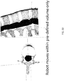
- a set of spatial coordinates can be used to define an operational volume, wherein the movement of a surgical instrument is constrained to be within the operational volume during a part of a procedure. For example, this can be used to limit the volume of bone removed by a surgeon.
- the operational volume may be defined by contacting points on the patient's anatomy and using those as vertices of a surface or by mapping a model of the anatomical volume to the set of spatial coordinates and then defining the operational volume as the mapped anatomical model expressed in the robotic surgical system's coordinate system.
- the operational volume can be updated after a re-registration to accurately reflect the patient's current anatomy and/or anatomical position and orientation.
- a stored model e.g., a model generated from medical image data
- a rendering of the patient's anatomy and a surgical instrument's position relative to the anatomy can be displayed on a navigation screen for use and/or reference by a surgeon using the methods and systems described herein.
- a coordinate mapping By using a coordinate mapping, the location of a terminal point of a surgical instrument can be displayed along with a rendering of the patient's anatomy such that a surgeon can observe an accurate representation of the space or distances between the terminal point and the patient's anatomy.
- This can be used to visualize trajectories, positions, and orientations of the surgical instrument relative to the patient's anatomy.
- a surgeon may use this to monitor the progress of a surgical procedure, avoid a serious medical error, and/or improve patient outcome by revising the planned surgical procedure.
- the surgeon can use the navigational display in deciding to alter the volume planned for removal or the planned orientation or trajectory of the surgical tool when removing the volume. This can be done intra-operatively.
- the disclosed technology includes a robot-based navigation system for real-time, dynamic re-registration of a patient position (e.g., position of vertebrae of a patient) during a procedure (e.g., surgical procedure, e.g., a spinal surgery) (e.g., a combined navigation/robotic system), the system including: (a) a robotic arm (e.g., having 3, 4, 5, 6, or 7 degrees of freedom) including: (i) an end effector [e.g., said end effector including a surgical instrument holder for insertion or attachment of a surgical instrument therein/thereto, e.g., said robotic arm designed to allow direct manipulation of said surgical instrument by an operator (e.g., by a surgeon) when the surgical instrument is inserted in/attached to the surgical instrument holder of the end effector, said manipulation subject to haptic constraints based on the position of the end effector (and/or the surgical instrument) in relation to the patient, e.g., said surgical instrument having known
- the disclosed technology includes a method of registering a patient's anatomy with an instrument attached to an end-effector of a robotic arm of a robotic surgical system, the method including the steps of: receiving, by a processor of a computing device, haptic feedback, from a force sensor attached directly or indirectly to the robotic arm, prompted by movement of the end-effector (e.g., towards a patient); determining, by the processor, that the haptic feedback corresponds to contact of the instrument with a material (e.g., having a certain density or certain mechanical properties) (e.g., based at least on a magnitude of the haptic feedback exceeding a threshold) (e.g., additionally based on the location of at least one point on the instrument) (e.g., wherein the material is bone); determining, by the processor, a set of spatial coordinates, wherein the set of spatial coordinates includes a spatial coordinate for each contact of the instrument with the material, expressed using a robot coordinate system, (e.g., relative to the
- the method includes the step of: outputting, by the processor, rendering data for display (e.g., on a display of the robotic surgical system; e.g., on a display on the robotic arm), wherein the rendering data corresponds to a representation of a position of a member and at least a portion of the medical image data based on the coordinate mapping, wherein the member is selected from the group consisting of: the end-effector, the instrument, and a surgical instrument.
- rendering data for display e.g., on a display of the robotic surgical system; e.g., on a display on the robotic arm
- the rendering data corresponds to a representation of a position of a member and at least a portion of the medical image data based on the coordinate mapping, wherein the member is selected from the group consisting of: the end-effector, the instrument, and a surgical instrument.
- the method includes the steps of: generating, by the processor, new rendering data by modifying the rendering data based on a change in the end- effector's position; and outputting, by the processor, the new rendering data for display.
- a fiducial marker includes the material (e.g., the end- effector contacts a fiducial marker with known size and shape such that the spatial coordinate is determined using a spatial relationship between the fiducial marker and the patient's anatomy).
- the robotic arm is active and non-backdrivable.
- the robotic surgical system includes the processor.
- the method includes storing, by the processor, a patient anatomy model wherein the patient anatomy model is defined by the patient anatomy surface expressed in the robot coordinate system.
- the disclosed technology includes a robotic surgical system for registering a patient's anatomy with an instrument attached to an end-effector of a robotic arm of the robotic surgical system, the system including: a robotic arm with an end-effector having an instrument attached thereto; a force sensor attached directly or indirectly to the robotic arm (e.g., the force sensor located between the instrument and the robotic arm); and a processor and a memory having instructions stored thereon, wherein the instructions, when executed by the processor, cause the processor to: receive haptic feedback, from the force sensor, prompted by movement of the end-effector (e.g., towards a patient); determine that the haptic feedback corresponds to contact of the instrument with a material (e.g., having a certain density or certain mechanical properties) (e.g., based at least on a magnitude of the haptic feedback exceeding a threshold) (e.g., additionally based on the location of at least one point on the instrument) (e.g., wherein the material is bone); determine a set of spatial
- the instructions when executed by the processor, cause the processor to: output rendering data for display (e.g., on a display of the robotic surgical system; e.g., on a display on the robotic arm), wherein the rendering data corresponds to a representation of a position of a member and at least a portion of the medical image data based on the coordinate mapping, wherein the member is selected from the group consisting of: the end-effector, the instrument, and a surgical instrument.
- the instructions when executed by the processor, cause the processor to: generate new rendering data by modifying the rendering data based on a change in the end-effector's position; and output the new rendering data for display.
- a fiducial marker includes the material (e.g., the end- effector contacts a fiducial marker with known size and shape such that the spatial coordinate is determined using a spatial relationship between the fiducial marker and the patient's anatomy).
- the robotic arm is active and non-backdrivable.
- the robotic surgical system includes the processor.
- the instructions when executed by the processor, cause the processor to: store a patient anatomy model wherein the patient anatomy model is defined by the patient anatomy surface expressed in the robot coordinate system.
- the disclosed technology includes a method of updating a model of a patient's anatomy after volume removal with an instrument attached to an end-effector of a robotic arm of a robotic surgical system, the method including the steps of: receiving, by a processor of a computing device, haptic feedback, from a force sensor attached directly or indirectly to the robotic arm, prompted by movement of the end-effector (e.g., towards a patient); determining, by the processor, that the haptic feedback corresponds to contact of the instrument with a material (e.g., having a certain density or certain mechanical properties) (e.g., based at least on a magnitude of the haptic feedback exceeding a threshold) (e.g., additionally based on the location of at least one point on the instrument) (e.g., wherein the material is bone); determining, by the processor, a set of spatial coordinates, wherein the set of spatial coordinates includes a spatial coordinate for each contact of the instrument with the material, expressed using a robot coordinate system (e.g.,
- the disclosed technology includes a system for updating a model of a patient's anatomy after volume removal with an instrument attached to an end-effector of a robotic arm of a robotic surgical system, the system including: a robotic arm with an end-effector having an instrument attached thereto; a force sensor attached directly or indirectly to the robotic arm (e.g., the force sensor located between the instrument and the robotic arm); and a processor and a memory having instructions stored thereon, wherein the instructions, when executed by the processor, cause the processor to: receive haptic feedback, from the force sensor, prompted by movement of the end-effector (e.g., towards a patient); determine that the haptic feedback corresponds to contact of the instrument with a material (e.g., having a certain density or certain mechanical properties) (e.g., based at least on a magnitude of the haptic feedback exceeding a threshold) (e.g., additionally based on the location of at least one point on the instrument) (e.g., wherein the material is bone); determine
- the disclosed technology includes a method of re-registering a patient's anatomy during a surgical procedure with an instrument attached to an end-effector of a robotic arm of a robotic surgical system, the method including the steps of: receiving, by a processor of a computing device, haptic feedback, from a force sensor attached directly or indirectly to the robotic arm, prompted by movement of the end-effector (e.g., towards a patient); determining, by the processor, that the haptic feedback corresponds to contact of the instrument with a material (e.g., having a certain density or certain mechanical properties) (e.g., based at least on a magnitude of the haptic feedback exceeding a threshold) (e.g., additionally based on the location of at least one point on the instrument) (e.g., wherein the material is bone); determining, by the processor, a set of spatial coordinates, wherein the set of spatial coordinates includes a spatial coordinate for each contact of the instrument with the material, expressed using the robot coordinate system (e.g.,
- the mapping is generated using surface matching.
- the updating step includes: determining, by the processor, a set of modeling coordinates, by converting, using the coordinate mapping, a set of medical image modeling coordinates defining the surface of a volume of a patient anatomy, wherein the set of modeling coordinates are expressed in the robot coordinate system and define an anticipated location of the surface of the volume, and the set of medical image modeling coordinates have been generated from medical imaging data; and mapping, by the processor, (e.g., using surface matching,) the surface corresponding to the set of spatial coordinates to the patient anatomy surface corresponding to the set of modeling coordinates (e.g., by generating a transformation array or transformation matrix); and updating, by the processor, the coordinate mapping based on the mapping of the surface corresponding to the set of spatial coordinates to the set of modeling coordinates.
- the updating step includes: receiving, by the processor, a set of modeling coordinates, wherein the set of modeling coordinates are expressed in the robot coordinate system and define the surface of a volume of a patient anatomy; mapping, by the processor, (e.g., using surface matching,) the surface corresponding to the set of spatial coordinates to the patient anatomy surface corresponding to the set of modeling coordinates (e.g., by generating a transformation array or transformation matrix); and updating, by the processor, the coordinate mapping based on the mapping of the surface corresponding to the set of spatial coordinates to the set of modeling coordinates.
- the disclosed technology includes a system for re-registering a patient's anatomy during a surgical procedure with an instrument attached to an end-effector of a robotic arm of a robotic surgical system, the system including: a robotic arm with an end-effector having an instrument attached thereto; a force sensor attached directly or indirectly to the robotic arm (e.g., the force sensor located between the instrument and the robotic arm); and a processor and a memory having instructions stored thereon, wherein the instructions, when executed by the processor, cause the processor to: receive haptic feedback, from the force sensor, prompted by movement of the end-effector (e.g., towards a patient); determine that the haptic feedback corresponds to contact of the instrument with a material (e.g., having a certain density or certain mechanical properties) (e.g., based at least on a magnitude of the haptic feedback exceeding a threshold) (e.g., additionally based on the location of at least one point on the instrument) (e.g.
- a material e.g.,
- the material is bone
- determine a set of spatial coordinates wherein the set of spatial coordinates includes a spatial coordinate for each contact of the instrument with the material, expressed using the robot coordinate system (e.g., relative to the position of the end-effector), wherein each spatial coordinate corresponds to a point on the surface of an anatomical volume (e.g., a point on a surface of a bone); receive a coordinate mapping between a robot coordinate system and a medical image data coordinate system (e.g., a transformation array or transformation matrix), wherein the robot coordinate system corresponds to a physical coordinate system of the end-effector; update the coordinate mapping based on a mapping of the surface corresponding to the set of spatial coordinates; and store the updated coordinate mapping (e.g., to provide an accurate navigational model for use by a surgeon during a surgical procedure), thereby re-registering the patient's anatomy.
- a medical image data coordinate system e.g., a transformation array or transformation matrix
- the mapping is generated using surface matching.
- the updating step includes instructions that, when executed by the processor, cause the processor to: determine a set of modeling coordinates, by converting, using the coordinate mapping, a set of medical image modeling coordinates defining the surface of a volume of a patient anatomy, wherein: the set of modeling coordinates are expressed in the robot coordinate system and define an anticipated location of the surface of the volume, and the set of medical image modeling coordinates have been generated from medical imaging data; and map (e.g., using surface matching,) the surface corresponding to the set of spatial coordinates to the patient anatomy surface corresponding to the set of modeling coordinates (e.g., by generating a transformation array or transformation matrix); and update the coordinate mapping based on the mapping of the surface corresponding to the set of spatial coordinates to the set of modeling coordinates.
- map e.g., using surface matching,
- the updating step includes instructions that, when executed by the processor, cause the processor to: receive a set of modeling coordinates, wherein the set of modeling coordinates are expressed in the robot coordinate system and define the surface of a volume of a patient anatomy; map (e.g., using surface matching,) the surface corresponding to the set of spatial coordinates to the patient anatomy surface corresponding to the set of modeling coordinates (e.g., by generating a transformation array or transformation matrix); and update the coordinate mapping based on the mapping of the surface corresponding to the set of spatial coordinates to the set of modeling coordinates.
- the disclosed technology includes a method of defining an operational volume in which a surgical instrument attached to an end-effector of a robotic arm of a robotic surgical system can be maneuvered, the method including the steps of: receiving, by a processor of a computing device, haptic feedback, from a force sensor attached directly or indirectly to the robotic arm, prompted by movement of the end-effector (e.g., towards a patient); determining, by the processor, that the haptic feedback corresponds to contact of the instrument with a material (e.g., having a certain density or certain mechanical properties) (e.g., based at least on a magnitude of the haptic feedback exceeding a threshold) (e.g., additionally based on the location of at least one point on the instrument) (e.g., wherein the material is bone); determining, by the processor, a set of spatial coordinates, wherein the set of spatial coordinates includes a spatial coordinate for each contact of the instrument with the material, expressed using a robot coordinate system (e.g.
- the updated model volume is a constrained operational volume, wherein a terminal point of the surgical instrument is temporarily constrained to within the constrained operational volume.
- the model volume is generated from medical image data using a coordinate mapping.
- the method includes receiving, by the processor, the updated model volume (e.g., a model of a portion of bone to be removed), wherein the stored model volume is expressed in a first robot coordinate system; receiving, by the processor, an updated coordinate mapping expressed in a second robot coordinate system; mapping, by the processor, the first robot coordinate system to the second robot coordinate system; generating, by the processor, a second updated model volume by converting coordinates of the updated model volume to updated coordinates expressed in the second robot coordinate system using the mapping between the first robot coordinate system and the second robot coordinate system; and storing, by the processor, the second updated model volume.
- the updated model volume e.g., a model of a portion of bone to be removed
- the disclosed technology includes a system for defining an operational volume in which a surgical instrument attached to an end-effector of a robotic arm of a robotic surgical system can be maneuvered, the system including: a robotic arm with an end- effector having an instrument attached thereto; a force sensor attached directly or indirectly to the robotic arm (e.g., the force sensor located between the instrument and the robotic arm); and a processor and a memory having instructions stored thereon, wherein the instructions, when executed by the processor, cause the processor to: receive haptic feedback, from the force sensor, prompted by movement of the end-effector (e.g., towards a patient); determine that the haptic feedback corresponds to contact of the instrument with a material (e.g., having a certain density or certain mechanical properties) (e.g., based at least on a magnitude of the haptic feedback exceeding a threshold) (e.g., additionally based on the location of at least one point on the instrument) (e.g., wherein the material is bone); determine
- the updated model volume is a constrained operational volume, wherein a terminal point of the surgical instrument is temporarily constrained to within the constrained operational volume.
- the model volume is generated from medical image data using a coordinate mapping.
- the instructions when executed by the processor, cause the processor to: receive the updated model volume (e.g., a model of a portion of bone to be removed), wherein the stored model volume is expressed in a first robot coordinate system; receive an updated coordinate mapping expressed in a second robot coordinate system; map the first robot coordinate system to the second robot coordinate system; generate a second updated model volume by converting coordinates of the updated model volume to updated coordinates expressed in the second robot coordinate system using the mapping between the first robot coordinate system and the second robot coordinate system; and store the second updated model volume.
- the updated model volume e.g., a model of a portion of bone to be removed
- the disclosed technology includes a method of displaying a position of a surgical instrument attached to a robotic arm relative to a patient anatomy for navigation during a robotically-assisted surgical procedure, the method including: receiving, by a processor of a computing device, a location of a terminal point of the surgical tool (e.g., wherein the location of the terminal point is determined, by the processor, using a known (e.g., stored) distance between a location of the robotic arm and the terminal point), wherein the location is expressed in a robot coordinate system; receiving, by the processor, a coordinate mapping between the robot coordinate system and a medical image data coordinate system (e.g., a transformation array or transformation matrix); converting, by the processor, using the coordinate mapping, the location of the terminal point, such that the converted location of the terminal point is expressed in a medical image data coordinate system; generating, by the processor, rendering data including the converted location of the terminal point; and outputting, by the processor, the rendering data, wherein a display of the rendering data includes a representation
- the rendering data corresponding to the representation of the patient anatomy is generated from medical image data coordinates generated from medical imaging data, wherein the medical image data coordinates are expressed in the medical image data coordinate system.
- the disclosed technology includes a system of displaying a position of a surgical instrument attached to a robotic arm relative to a patient anatomy for navigation during a robotically-assisted surgical procedure, the system including: a robotic arm with an end- effector having an instrument attached thereto; a force sensor attached directly or indirectly to the robotic arm (e.g., the force sensor located between the instrument and the robotic arm); and a processor and a memory having instructions stored thereon, wherein the instructions, when executed by the processor, cause the processor to: receive a location of a terminal point of the surgical tool (e.g., wherein the location of the terminal point is determined, by the processor, using a known (e.g., stored) distance between a location of the robotic arm and the terminal point), wherein the location is expressed in a robot coordinate system; receive a coordinate mapping between the robot coordinate system and a medical image data coordinate system (e.g., a transformation array or transformation matrix); convert, using the coordinate mapping, the location of the terminal point, such that the converted location of the
- the rendering data corresponding to the representation of the patient anatomy is generated from medical image data coordinates generated from medical imaging data, wherein the medical image data coordinates are expressed in the medical image data coordinate system.
- the disclosed technology includes a method of performing volume removal surgery with one or more instruments, wherein the one or more instruments used by attaching to an end-effector of a robotic arm of a robotic surgical system, the method including the steps of: registering a patient's anatomy to express a model of the patient's anatomy in a robot coordinate system; contacting, following removal of a first volume of the patient's anatomy, an instrument attached to the end-effector to the patient's anatomy in a plurality of locations, wherein contact is determined by haptic feedback from a force sensor attached directly or indirectly to the robotic arm, wherein the haptic feedback is prompted by movement of the end-effector (e.g., towards a patient); updating the model of the patient's anatomy by determining a portion of the model that corresponds to the first volume of the patient's anatomy that has been removed using spatial coordinates corresponding to the plurality of locations contacted; optionally, re-registering the patient's anatomy by contacting a plurality of re- registration locations with the instrument
- the registering step includes: receiving, by a processor of a computing device, haptic feedback, from a force sensor attached directly or indirectly to the robotic arm, prompted by movement of the end-effector (e.g., towards a patient); determining, by the processor, that the haptic feedback corresponds to contact of the instrument with a material (e.g., having a certain density or certain mechanical properties) (e.g., based at least on a magnitude of the haptic feedback exceeding a threshold) (e.g., additionally based on the location of at least one point on the instrument) (e.g., wherein the material is bone); determining, by the processor, a set of spatial coordinates, wherein the set of spatial coordinates includes a spatial coordinate for each contact of the instrument with the material, expressed using the robot coordinate system, (e.g., relative to the position of the end-effector), wherein each spatial coordinate corresponds to a point on the surface of an anatomical volume (e.g., a point on
- the method including the step of: outputting, by the processor, rendering data for display, wherein the rendering data corresponds to a representation of a member's position and at least a portion of the medical image data based on the coordinate mapping, wherein the member is selected from the group consisting of: the end-effector, the instrument, and a surgical instrument.
- the method including the steps of: generating, by the processor, new rendering data by modifying the rendering data based on a change in the end-effector's position; and outputting, by the processor, the new rendering data for display.
- a fiducial marker includes the material (e.g., the end- effector contacts a fiducial marker with known size and shape such that the spatial coordinate is determined using a spatial relationship between the fiducial marker and the patient's anatomy).
- the robotic arm is active and non-backdrivable.
- the robotic surgical system includes the processor.
- the method includes storing, by the processor, a patient anatomy model wherein the patient anatomy model is defined by the patient anatomy surface expressed in the robot coordinate system.
- the updating step includes: determining, by the processor, a set of spatial coordinates, wherein the set of spatial coordinates includes a spatial coordinate for each contact of the instrument with the material, expressed using a robot coordinate system (e.g., relative to the position of the end-effector), wherein each spatial coordinate corresponds to a point on the surface of an anatomical volume (e.g., a point on a surface of a bone); receiving, by the processor, a set of medical image data coordinates that correspond to the surface of a volume of the patient's anatomy, wherein each medical image data coordinate in the set of medical image data coordinates is expressed using a medical image data coordinate system; receiving, by the processor, a coordinate mapping between the robot coordinate system and the medical image data coordinate system (e.g., a transformation array or transformation matrix); determining, by the processor, one or more interior spatial coordinates in the set of spatial coordinates that correspond to points inside the surface of the volume of the patient's anatomy based on the set of medical image data coordinates and the coordinate
- the defining step includes: receiving, by a processor of a computing device, haptic feedback, from a force sensor attached directly or indirectly to the robotic arm, prompted by movement of the end-effector (e.g., towards a patient); determining, by the processor, that the haptic feedback corresponds to contact of the instrument with a material (e.g., having a certain density or certain mechanical properties) (e.g., based at least on a magnitude of the haptic feedback exceeding a threshold) (e.g., additionally based on the location of at least one point on the instrument) (e.g., wherein the material is bone); determining, by the processor, a set of spatial coordinates, wherein the set of spatial coordinates includes a spatial coordinate for each contact of the instrument with the material, expressed using a robot coordinate system (e.g., relative to the position of the end-effector), wherein each spatial coordinate corresponds to a point on the surface of a volume (e.g., a point on a surface
- the updated model volume is a constrained operational volume, wherein a terminal point of the surgical instrument is temporarily constrained to within the constrained operational volume.
- the model volume is generated from medical image data using a coordinate mapping.
- the method includes receiving, by the processor, the updated model volume (e.g., a model of a portion of bone to be removed), wherein the stored model volume is expressed in a first robot coordinate system; receiving, by the processor, an updated coordinate mapping expressed in a second robot coordinate system; mapping, by the processor, the first robot coordinate system to the second robot coordinate system; generating, by the processor, a second updated model volume by converting coordinates of the updated model volume to updated coordinates expressed in the second robot coordinate system using the mapping between the first robot coordinate system and the second robot coordinate system; and storing, by the processor, the second updated model volume.
- the updated model volume e.g., a model of a portion of bone to be removed
- the re-registering step includes: receiving, by a processor of a computing device, haptic feedback, from a force sensor attached directly or indirectly to the robotic arm, prompted by movement of the end-effector (e.g., towards a patient); determining, by the processor, that the haptic feedback corresponds to contact of the instrument with a material (e.g., having a certain density or certain mechanical properties) (e.g., based at least on a magnitude of the haptic feedback exceeding a threshold) (e.g., additionally based on the location of at least one point on the instrument) (e.g., wherein the material is bone); determining, by the processor, a set of spatial coordinates, wherein the set of spatial coordinates includes a spatial coordinate for each contact of the instrument with the material, expressed using the robot coordinate system (e.g., relative to the position of the end-effector), wherein each spatial coordinate corresponds to a point on the surface of an anatomical volume (e.g., a point on
- the mapping is generated using surface matching.
- the updating step includes: determining, by the processor, a set of modeling coordinates, by converting, using the coordinate mapping, a set of medical image modeling coordinates defining the surface of a volume of a patient anatomy, wherein the set of modeling coordinates are expressed in the robot coordinate system and define an anticipated location of the surface of the volume, and the set of medical image modeling coordinates have been generated from medical imaging data; and mapping, by the processor, (e.g., using surface matching,) the surface corresponding to the set of spatial coordinates to the patient anatomy surface corresponding to the set of modeling coordinates (e.g., by generating a transformation array or transformation matrix); and updating, by the processor, the coordinate mapping based on the mapping of the surface corresponding to the set of spatial coordinates to the set of modeling coordinates.
- the updating step includes: receiving, by the processor, a set of modeling coordinates, wherein the set of modeling coordinates are expressed in the robot coordinate system and define the surface of a volume of a patient anatomy; mapping, by the processor, (e.g., using surface matching,) the surface corresponding to the set of spatial coordinates to the patient anatomy surface corresponding to the set of modeling coordinates (e.g., by generating a transformation array or transformation matrix); and updating, by the processor, the coordinate mapping based on the mapping of the surface corresponding to the set of spatial coordinates to the set of modeling coordinates.
- the disclosed technology includes a method of updating an operational volume in which a surgical instrument attached to an end-effector of a robotic arm of a robotic surgical system can be maneuvered, the method including the steps of: receiving, by the processor, a stored model volume including coordinates (e.g., a model of a portion of bone to be removed), wherein the stored model volume is expressed in a first robot coordinate system; receiving, by the processor, an updated coordinate mapping expressed in a second robot coordinate system; converting, by the processor, each coordinate of the stored model volume to be expressed in the second robot coordinate system using the updated coordinate mapping; and storing, by the processor, an updated model volume including the converted coordinates.
- a stored model volume including coordinates e.g., a model of a portion of bone to be removed
- the term “approximately” or “about” refers to a range of values that fall within 25%, 20%, 19%, 18%, 17%, 16%, 15%, 14%, 13%, 12%, 11%, 10%, 9%, 8%, 7%, 6%, 5%, 4%, 3%, 2%, 1%, or less in either direction (greater than or less than) of the stated reference value unless otherwise stated or otherwise evident from the context (except where such number would exceed 100% of a possible value).
- mapping refers to establishing a function between two sets of coordinates or data corresponding to two sets of coordinates. The function between the two sets may be discrete or continuous.
- a mapping allows coordinates recorded and/or stored in one coordinate system to be converted to coordinates in another coordinate system and vice versa. Two sets of coordinates expressed in the same coordinate system may be mapped with each other as well.
- a map or mapping may be stored on a computer readable medium as an array or matrix of data.
- a map or mapping is a linear transform stored as an array on a computer readable medium.
- the map or mapping is used to convert between coordinate systems.
- the coordinate systems are Cartesian.
- mapping may be an optimized function, wherein the mapping represents the function of minimal error or error below a threshold according to the mapping method (e.g., surface matching). In certain embodiments, mapping comprises surface matching.
- mapping comprises surface matching.
- systems, devices, methods, and processes of the aspected invention encompass variations and adaptations developed using information from the embodiments described herein. Adaptation and/or modification of the systems, devices, methods, and processes described herein may be performed by those of ordinary skill in the relevant art.
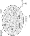
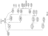
- FIG. 1 illustrates an example robotic surgical system in an operating room 100.
- one or more surgeons, surgical assistants, surgical technologists and/or other technicians, (106a-c) perform an operation on a patient 104 using a robotic-assisted surgical system.
- the surgeon may be guided by the robotic system to accurately execute an operation. This may be achieved by robotic guidance of the surgical tools, including ensuring the proper trajectory of the tool (e.g., drill or screw).
- the surgeon defines the trajectory intra-operatively with little or no pre- operative planning. The system allows a surgeon to physically manipulate the tool holder to safely achieve proper alignment of the tool for performing crucial steps of the surgical procedure.
- Operation of the robot arm by the surgeon (or other operator) in force control mode permits movement of the tool in a measured, even manner that disregards accidental, minor movements of the surgeon.
- the surgeon moves the tool holder to achieve proper trajectory of the tool (e.g., a drill or screw) prior to operation or insertion of the tool into the patient.
- the robotic arm is fixed to maintain the desired trajectory.
- the tool holder serves as a stable, secure guide through which a tool may be moved through or slid at an accurate angle.
- the operation may be spinal surgery, such as a discectomy, a foraminotomy, a laminectomy, or a spinal fusion.
- the surgical robotic system includes a surgical robot 102 on a mobile cart.
- the surgical robot 102 may be positioned in proximity to an operating table 112 without being attached to the operating table, thereby providing maximum operating area and mobility to surgeons around the operating table and reducing clutter on the operating table.
- the surgical robot (or cart) is securable to the operating table.
- both the operating table and the cart are secured to a common base to prevent any movement of the cart or table in relation to each other, even in the event of an earth tremor.
- the mobile cart may permit a user (operator) 106a, such as a technician, nurse, surgeon, or any other medical personnel in the operating room, to move the surgical robot 102 to different locations before, during, and/or after a surgical procedure.
- the mobile cart enables the surgical robot 102 to be easily transported into and out of the operating room 100.
- a user 106a may move the surgical robot into the operating room from a storage location.
- the mobile cart may include wheels, a track system, such as a continuous track propulsion system, or other similar mobility systems for translocation of the cart.
- the mobile cart may include an attached or embedded handle for locomotion of the mobile cart by an operator.
- the mobile cart may be provided with a stabilization system that may be used during a surgical procedure performed with a surgical robot.
- the stabilization system increases the global stiffness of the mobile cart relative to the floor in order to ensure the accuracy of the surgical procedure.
- the wheels include a locking system that prevents the cart from moving.
- the stabilizing, braking, and/or locking system may be activated when the machine is turned on.
- the mobile cart includes multiple stabilizing, braking, and/or locking systems.
- the stabilizing system is electro-mechanical with electronic activation.
- the stabilizing, braking, and/or locking system(s) may be entirely mechanical.
- the stabilizing, braking, and/or locking system(s) may be electronically activated and deactivated.
- the surgical robot 102 includes a robotic arm mounted on a mobile cart.
- An actuator may move the robotic arm.
- the robotic arm may include a force control end-effector configured to hold a surgical tool.
- the robot may be configured to control and/or allow positioning and/or movement of the end-effector with at least four degrees of freedom (e.g., six degrees of freedom, three translations and three rotations).
- the robotic arm is configured to releasably hold a surgical tool, allowing the surgical tool to be removed and replaced with a second surgical tool.
- the system may allow the surgical tools to be swapped without re-registration, or with automatic or semi-automatic re-registration of the position of the end-effector.
- the surgical system includes a surgical robot 102, a tracking detector 108 that captures the position of the patient and different components of the surgical robot 102, and a display screen 110 that displays, for example, real time patient data and/or real time surgical robot trajectories.
- a tracking detector 108 monitors the location of patient 104 and the surgical robot 102.
- the tracking detector may be a camera, a video camera, an infrared detector, field generator and sensors for electro-magnetic tracking or any other motion detecting apparatus.
- the display screen displays a projected trajectory and/or a proposed trajectory for the robotic arm of robot 102 from its current location to a patient operation site.
- the surgical system can calculate updated trajectories and visually display these trajectories on display screen 110 to inform and guide surgeons and/or technicians in the operating room using the surgical robot.
- the surgical robot 102 may also change its position and automatically position itself based on trajectories calculated from the real time patient and robotic arm positions captured using the tracking detector 108. For instance, the trajectory of the end- effector can be automatically adjusted in real time to account for movement of the vertebrae or other part of the patient during the surgical procedure.
- the disclosed technology includes a robot-based navigation system for real-time, dynamic re-registration of a patient position (e.g., position of vertebrae of a patient) during a procedure (e.g., surgical procedure, e.g., a spinal surgery).
- An example robotic surgical system is shown in FIG. 38 .
- the robotic surgical system 3800 includes a robotic arm 3802.
- the robotic arm can have 3, 4, 5, 6, or 7 degrees of freedom.
- the robotic arm 3802 has an end effector 3804.
- the robotic arm 3802 includes a position sensor 3806 for dynamically tracking a position of the end effector 3804 and/or surgical instrument during a surgical procedure. Additionally, one or more points of the surgical instrument can be dynamically tracked, for example, at a rate of at least 100 Hz, 250 Hz or greater, 500 Hz or greater, or 1000 Hz or greater (e.g., position determination per second).
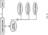
- the system 3800 includes a force feedback subsystem 3808.
- the force feedback subsystem 3808 can include sensor(s), actuator(s), controller(s), servo(s), and/or other mechanisms for delivering a haptic force to a user manipulating the end effector or a surgical instrument inserted in the instrument holder of the end effector.
- the force feedback subsystem 3808 can detect the resistive force caused by the surgical instrument contacting, moving against, penetrating, and/or moving within a tissue of the patient.
- the force feedback subsystem 3808 can distinguish between contacted tissue types (e.g., determining when contacted tissue meets or exceeds a threshold resistance, e.g., when the tissue is bone).
- the force feedback subsystem 3808 can also detect a force delivered by the operator. For example, it can detect forces delivered by direct manipulation of the surgical instrument inserted in the surgical instrument holder of the end effector to cause movement of the surgical instrument and, therefore, the end effector.
- the force feedback subsystem 3808 can further distinguish between the force delivered by the operator and the resistive force caused by movement of the surgical instrument in relation to the tissue of the patient. This allows the operator to both apply forces to the system as well as feel resistance (e.g., via haptic feedback) as a surgical instrument contacts tissue in the patient.
- the robotic surgical system 3800 includes a display 3810 that is attached to, embedded within, or otherwise positioned in relation to the robotic arm being directly manipulated by the operator (e.g., surgeon) to allow for unimpeded visual feedback to the operator during the procedure.
- the operator e.g., surgeon
- the system When an operator uses the system, the system initially accesses (e.g., and graphically renders on the display) an initial registration of a target volume, such as a vertebra of the patient.
- a target volume such as a vertebra of the patient.
- This can be accomplished using medical images of the patient, including MRI, CT, X-rays, SPECT, ultrasound, or the like. These images can be obtained preoperatively or intraoperatively.
- the position of the end effector is dynamically determined (e.g., by processor 3812). Specifically, in some implementations, the system dynamically determines a 3D position of one or more points of a surgical instrument. Forces received by the surgical instrument are dynamically determined when the surgical instrument contacts, moves against, penetrates, and/or moves within the patient. The system can measure these forces and distinguish between contacted tissue types. This can be accomplished, for example, by determining when contacted tissue meets or exceeds a threshold resistance, such as when the tissue is bone). The system can further detect forces applied to the surgical instrument by the operator and distinguish between forces delivered by the operator and the resistive force caused by movement of the surgical instrument in relation to the tissue of the patient.
- the system can dynamically re-register the patient position based at least in part on an updated position of the end effector determined by the position sensor. This can be used to update the 3D representation of the patient situation based at least in part on the updated position of the end effector when it is determined (e.g., via the force feedback subsystem) that the surgical instrument is in contact with a target anatomy. This can be accomplished using a surface matching algorithm keyed to the initial (or previous) registration.
- the system can dynamically re-register the patient position upon detected contact or proximity of the end effector, or the surgical instrument, or a portion or component of the surgical instrument or end effector, with a pre-planned fiducial, such as a mechanical marker, a marker fixed to the patient.
- a pre-planned fiducial such as a mechanical marker, a marker fixed to the patient.
- the system can dynamically re-register the patient position based upon the updated position of the end effector determined upon operator command, such as the operator pressing a button or otherwise activating a graphical or tactile user interface when a re-registered representation is desired.
- a surgical instrument holder can be connected to the end effector for insertion or attachment of a surgical instrument therein/thereto.
- the instrument holder can be removable. In such instances, attachment of the instrument holder to the end effector is precise and predictable such that it is always connected in the same position.
- the robotic arm is designed to allow direct manipulation of a surgical instrument by an operator (e.g., by a surgeon) when the surgical instrument is inserted in/attached to the surgical instrument holder of the end effector.
- the manipulation of the instrument can be subject to haptic constraints based on the position of the end effector (and/or the surgical instrument) in relation to the patient.
- the surgical instrument has a known geometry and position in relation to the surgical instrument holder such that the location of the instrument (e.g., the tip of the instrument) is known by the robotic surgical system. For example, when a surgical instrument is fully inserted into the instrument holder, the position of the instrument is known to the robotic surgical system because the position of the end effector is known as well as information about the surgical instrument and the instrument holder.
- a tool center point facilitates precise positioning and trajectory planning for surgical instrument guides and surgical instruments inserted through or attached to the surgical instrument holder.
- Surgical instruments can be engineered such that when inserted into the surgical instrument holder, there is a defined tool center point with known coordinates relative to robotic arm.
- the origin of a coordinate system used to define the tool center point may be located at a flange of a robotic arm. It may additionally be located at any convenient to define point such as an interface, joint, or terminal aspect of a component of a robotic surgical system.
- the TCP is in a constant position relative to the robotic arm, regardless of whether a surgical guide or surgical instrument is being used with the surgical instrument holder, a surgeon can be provided visualization of the orientation, trajectory, and position of an instrument or instrument guide used with the surgical instrument holder.
- engineered surgical instrument systems eliminates the need for navigation markers to be attached to the end of surgical guides or tools in order to precisely determine the position, orientation, and trajectory of a surgical instrument guide relative to a patient's anatomy.
- a navigation marker attached to surgical instrument holder can be used to track the position and orientation of the universal surgical instrument guide to update the position, orientation, and current trajectory based on manipulation of robotic arm by a surgeon. Additional information provided by patient imaging (e.g., CT data, radio imaging data, or similar) taken pre- or intra-operatively as well as navigation markers attached to a patient's body may be combined with data from a navigation marker attached to a universal surgical instrument guide and displayed on a screen viewable by the surgeon such that the surgeon can see the location of necessary features of the patient's anatomy and the position, trajectory, and orientation of a surgical instrument or surgical instrument guide relative to said anatomy.
- patient imaging e.g., CT data, radio imaging data, or similar
- FIGS. 5A-C illustrate example locations for mounting a force sensor (e.g., force/torque sensor 430).
- the force sensor 502 a is located between the tool holder 506 a and robot 504 a.
- the sterile cover 508 a may be wrapped around the robot arm and between the force sensor and the tool holder to ensure sterilization.
- the force sensor 502 a may provide for direct measurement of forces (e.g., forces and/or torques) on the tool.
- the force sensor 502 a may be designed to resist flexing.
- the force sensor 502 a may be designed to flex under the stress of certain external forces. The displacement caused when an external force is applied may be calculated based on the force and/or torque applied to the tool, radial force stiffness, axial torque stiffness, and the diameter of the holder to which the tool is attached.
- the force sensor (e.g., 502 b in FIG. 5B or 502 c in FIG. 5C ) may be located on the robot or the tool holder, respectively. These configurations may exclusively measure the forces and/or torques applied by the user.
- the force sensor 508 may be connected to the robot with an intermediary analog box which measures forces and torques and transmits them via a network (e.g., Ethernet, CAN, wireless, internet, private LAN, public LAN, etc.). Combinations of the above mentioned force sensor positions are possible to achieve pre-defined behavior (e.g. the first sensor in the base FIG. 5A and the second one in the handle FIG. 5B may be positioned to allow the feedback control system to decouple forces applied to the surgical tool from forces and/or torque applied by a user).
- a network e.g., Ethernet, CAN, wireless, internet, private LAN, public LAN, etc.
- the force sensor is integrated directly in the surgical instrument.
- the force sensor may be integrated directly in the surgical drill bit as illustrated in FIG. 6 .
- the force sensor 604 may be similarly integrated in other surgical instruments. Integrating the force sensor 604 in a surgical instrument, such as a drill bit 602, may be more robust as it minimizes the impact of external disturbances for measuring forces applied to the drill bit.
- the force sensor 604 is integrated in the shaft of the drill bit 602.
- the force sensor 604 in some implementations, is located on the drill bit 602 outside of the body 610 of the drill as shown in FIG. 6 . In other implementations, the force sensor 604 is located inside the body 610 of the drill, thereby better protecting the force sensor 604 from external influences. Force sensor can have multiple degrees of freedom and measure, for example, 1 to 3 forces and/or 1 to 3 torques. Forces are transmitted from the rotating shaft through a connector 606.
- the connector in some implementations, is one or more brushes that provide an electrical connection to the force sensor 604. If the force sensor is an optical sensor, the connector may be an optical transmitter (e.g.
- the brushes contact the drill bit thereby forming an electrical connection with the force sensor 604.
- the brushes touch one or more contacts on the drill bit to form the electrical connection.
- An electric or pneumatic motor 608 rotates the drill bit 602 shaft.
- a sensor 612 e.g., an encoder
- the sensor 612 measures position of the shaft.
- the sensor 612 measures the position of the shaft in order to correlate forces measured by the force sensor to the relative position of the shaft. For example, if the force sensor is located in a drill bit, the measurement of the direction of the force will vary as the drill bit rotates. Specifically, the force sensor measures force and the direction of the force periodically (e.g., every millisecond, every microsecond, or somewhere therebetween).
- the drill bit rotates as the surgeon pushes it into bone. When the drill contacts the bone, the force sensor will indicate some force (F1) in a direction (D1).
- the drill bit will rotate slightly so the force sensor will indicate force of the same value (F1) (assuming a constant force is applied) in a different direction (D2).
- the direction of the force will continue to change relative to a single perspective as the drill bit rotates even if surgeon pushes into the bone with a constant force.
- a constantly changing force direction is not acceptable.
- the positions of the drill in the global space must be calculated as the drill bit rotates.
- the sensor 612 is used to measure the position of the shaft and thus determine the global direction of the force (D).
- the sensor 612 may be located on the back of the motor 608 as shown in FIG. 6 .
- the sensor 612 may be located in other locations relative to the motor 608 as well.
- the force sensor 604 may be provided in various configurations as shown in FIGS. 7A-D . In each configuration, the goal is to measure forces on the tip of the tool (e.g., drill bit ultrasound bit, etc.). In the example shown in FIG. 7A the force sensor 604 is integrated in the shaft of the drill bit 602 as described in relation to FIG. 6 . The force sensor 604 may communicate with a connector 606 (shown in FIG. 6 ) via a sensor cable 702. The sensor cable 702, in some implementations, is routed inside the drill bit 602. In some implementations, the connector 606 (shown in FIG. 6 ) is electrically connected to the sensor cable 702 via one or more connection pads.
- the connector 606 shown in FIG. 6
- the connector 606 is electrically connected to the sensor cable 702 via one or more connection pads.
- the force sensor 604 in this example may be a miniaturized industrial sensor (e.g., the multi-axis force/torque sensor from ATI Industrial Automation, Inc. of Apex, N.C.) that measures, for example, all six components of force and torque using a transducer.
- the force sensor 604 may be an optical sensor.
- the force sensor 604 may comprise a strain gauge 706 integrated directly into the shaft of the drill bit 602 as shown in FIG. 7B .
- the force sensor 604 measures forces on the motor instead of measuring forces on the drill bit 602 itself.
- the shaft of the drill bit 602 in some implementations, includes a flexible element 708 that allows the drill bit 602 to bend (e.g., only slightly) such that after deflection of the shaft of the drill bit 602, forces can be measured by the force sensor 604.
- the measurement of shaft positions e.g., by sensor 612 as shown in FIG. 6
- a goal of the robot-based navigation is to assist a surgeon during a surgical procedure that results in a change to patient's target anatomy.
- the implants and surgical instruments are used for this purpose and the robotic system assists the surgeon to improve the accuracy with which these instruments are used during the surgical procedure.
- FIG. 2 shows a schematic of all the system that assist in surgery, in some embodiments.
- the interaction of a surgeon and components of a surgical system is further outlined in FIG. 3 .
- the patient's target anatomy medical images are obtained using an appropriate imaging technique (e.g., which can be used to generate a model of the patient's anatomy) and used in the following processes.
- the process of determining target anatomy position takes as an input target anatomy.
- the target anatomy may be modeled using medical images, wherein medical images are taken using a medical imaging technique or scanning-based technique.
- determining the target anatomy position provides the exact anatomy position of the target anatomy at any moment in time.
- the process of rendering feedback provides information to the surgeon based on medical images, anatomy position and instrument(s) position. The rendering is visually displayed to the surgeon.
- the tracking instruments process takes surgical instruments and as a result calculates their position. At the same time the instrument are guided which means that their spatial position is constrained in some way by the robotic system ( i.e., using an operational volume).
- the robotic system implements all these four processes and the surgeon participates in the two of them: guidance and rendering feedback.
- the goal of the imaging process is to obtain representation of the patient's target anatomy.
- imaging is used to obtain the representation of the patient's target anatomy.
- the images can be obtained pre-operatively using various modalities, such as MRI, CT, or X- Rays.
- the images can be obtained intra-operatively using intra-operative imaging techniques, such as using flat-panel fluoroscopy technology (e.g. the O-Arm by Medtronic of Minneapolis, Minnesota), intra-operative CTs, MRI and ultrasound.
- intra-operative images can be captured using an intra-operative fluoroscopy device (e.g., a C-Arm).
- information about the target anatomy can be collected by surface scanning using a haptic device and force feedback. Using such a device mounted on the robot allows user to measure forces that can provide spatial information about surface and rigidity of tissues.
- known techniques of surface scanning such as a laser scanner, can be used.
- visual, direct exploration can be used to explore the patient's anatomy. The outcome of these techniques, if used, is stored as medical image data, which is used to model the patient's anatomy.
- FIG. 8 illustrates a range of processes that can be used for defining the position of the target anatomy (i.e., registration).
- the position of the target anatomy is managed using a registration procedure to identify the position of the anatomy in reference to an anatomy- fixed marker and later by the tracking (e.g., optical, or electro-magnetic) the marker and assuming that the marker moves appropriately with the anatomy to determine the position of the target anatomy.
- the disclosed technology utilizes force-based anatomy registration and re-registration to address these shortcomings. In-between re-registrations robot encoders can be used to immediately obtain instrument position at a high frequency (e.g., from 200 to 500 Hz, 500 to 800 HZ, or 800 Hz to 1200 Hz).
- the initial registration data can be used along with a stability module to manage the location of the end-effector relative to the target anatomy.
- the stability module can run all the time in the background ( e.g., automatically), in certain embodiments, without surgeon taking any special actions.
- Examples of such a stability module includes surface matching methods which take a set of points (e.g. measured points) and finds the best match between the set of points and another set of points (e.g., from the surface of the vertebra on medical images).
- Example algorithm for surface matching is Iterative Closet Point method described in Section 4.5.3 of "A Robotic System for Cervical Spine Surgery", Szymon Kostrzewski, Warsaw University of Technology (2011). In certain embodiments, other algorithms are used for a stability modules.
- Re-registration can be accomplished using other methods as well. For example, in certain embodiments, re-registration can be accomplished by periodically re-validating using fiducials. In certain embodiments, when necessary, the surgeon can touch pre-placed fiducials and re-register with an instrument attached to a robotic arm.
- a specially designed marker can be fixed to the patient as shown in FIG. 9 .
- This marker has a set of conic holes which can be easily found by a robot having a force sensor. For example, a surgeon can push a button for the robot to re-register. Then the robot automatically or the surgeon manually would bring the robot to the holes in the marker. After identifying at least 3 holes the re-registration can be found and the surgeon can continue with the surgery.
- Figure 10 is a schematic view of a process of determining a position based on automatic re-registration.
- the re-registration method of FIG. 10 utilizes initial registration data, patient medical images and information about the target anatomy coming from the robotic system (e.g., measured points on the surface of the bone) and, based on this information, updates the registration data which is used by the anatomy tracking process to provide the position of the target anatomy.
- feedback can be rendered using screens accessible in the operating room.
- feedback can be rendered using a robot-held user interface, such as the user interface described in U.S. Patent Application No. 14/858,325, filed September 18, 2015 , entitled "Robot-Mounted User Interface for Interacting with Operation Room Equipment", the content of which is hereby incorporated by reference in its entirety.
- FIG. 12 is an illustration of exemplary processes for tracking instruments.
- tracking can be accomplished using markers attached to the instrument and optical/ electro-magnetic trackers.
- the instruments are tracked using the robotic system which provides their position in space. The robot "knows" the instrument position because it has a measurement system that is used to determine the position of the position of the end effector. Coupled with a known geometry of a given instrument, the position of the instrument attached to the end effector (e.g., in a predicable manner) is known by the robotic surgical system.
- Mechanical template tracking refers to mechanical, custom-made templates which fit in one position only on the top of the target anatomy and contain guide for guiding instruments.
- Passive arms are standard surgical arms which can be fixed in pre-defined position in the operating room.
- FIG. 13 shows exemplary systems for guiding instruments during surgical procedures.
- surgical instruments are guided with robotic guidance.
- robotic guidance occurs automatically ( i.e., without input from a surgeon) after registration of the patient's anatomy with the robotic surgical system.
- manual guidance and passive guidance are used during some stages of surgical procedures. In certain embodiments, manual guidance and passive guidance are used in all stages of surgical procedures.
- a robotic surgical system with instrument attached can be used to contact a patient's anatomy at a plurality of contact points determined using haptic feedback from a force sensor attached directly or indirectly to the robotic arm of the robotic surgical system.
- the coordinates of the plurality of contacts define a set of spatial coordinates.
- the value of a spatial coordinate is determined by storing the position of a portion of the robotic surgical system (e.g., a terminal point of an instrument or surgical instrument or the robotic arm) in the robot's coordinate system when contact is determined.
- a set of spatial coordinates recorded from contact of the instrument with the patient's anatomy can be used to perform many navigational and surgical guidance functions such as registration, modeling volume removal, re-registration, defining operational volumes, revising operational volumes after re-registration, converting stored volume models to physical locations, and displaying surgical instruments relative to a patient's anatomy on navigation screens.
- a processor that is either a part of the robotic surgical system or part of a remote computing device ( e.g., on a server) can be used to correlate the coordinates of surgical instruments, instruments, and/or a patient's anatomy by generating and using appropriate coordinate mappings in combination with sets of spatial coordinates.
- a set of spatial coordinates may be provided for further use, wherein the set of spatial coordinates are generated using a technique other than haptic-feedback-based contacting of the patient's anatomy with an instrument attached to a robotic arm.
- the set of spatial coordinates may be provided as a result of a known registration technique.
- a patient's anatomy can be registered with a robotic surgical system without the use of a separate navigation system by contacting an instrument to the patient's anatomy to generate a set of spatial coordinates that can be correlated with a model of the patient generated using medical imaging data.
- a surgeon uses a surgical instrument to contact the patient, including during registration.
- one or more navigation markers are used for reference during registration.
- the processor has determined a set of spatial coordinates, wherein each spatial coordinate corresponds to a point on the surface of an anatomical volume (e.g., a patient's vertebra(e)), the set of spatial coordinates can be mapped with a model of the patient's anatomy.
- the patient's anatomy may be modeled using medical imaging data.
- CT, MRI, or x-ray data is used to pre-operatively generate a 3D model of the patient's anatomy.
- medical images used to generate patient anatomy models are taken intra-operatively.
- a set of spatial coordinates is mapped to a model of a patient's anatomy by determining a function for converting points in the model to coordinates in the robot coordinate system (i.e., physical reality) and vice versa.
- mappings are made using surface matching of a surface defined by a set of spatial coordinates and the surface of the anatomical model. Points on the surface of the patient's anatomy or the anatomical model of the patient's anatomy may be extrapolated from known points (i.e., points measured by contacting the patient's anatomy with an instrument or data points collected during medical imaging). The extrapolated points may be used to generate the mapping.
- An example of a surface matching method is Iterative Closest Point (ICP) described in Section 4.5.3 of "A Robotic System for Cervical Spine Surgery," Szymon Kostrzewski, Warsaw University of Technology (2011 ). The contents of Section 4.5.3 are hereby incorporated by reference herein in their entirety.
- ICP Iterative Closest Point
- Any algorithm or method that generates a function that can be used to transform points from one coordinate system to another (i.e., by linear transform) is appropriate for use.
- a threshold may be specified that defines an error measure that the mapping must stay under in order to be used. This threshold can be set to a value that is sufficient for high precision mapping ( e.g., registration), but such that mappings can be generated with high frequency ( i.e., the speed of the generation of the mapping does not rate limit a surgical procedure).
- a processor can determine a physical location for each point of the patient's anatomy represented in the anatomical model. In this way, the patient is registered with the robotic surgical system.
- the robotic surgical system knows whether that point is on the surface of the patient's anatomy, in the patient's anatomy, or outside of the patient's anatomy. This information can be used for further processing to assist in surgical guidance and navigation. For example, this information can be used to define "no go" zones for a surgical instrument if the patient's anatomy is to be avoided entirely or only a portion of the patient's anatomy is to be accessible to the surgical instrument. Additionally, a surgical instrument could trace a line, plane, or curve that falls on the surface of the patient's anatomy.
- a small set of points need to be contacted. For example, in certain embodiments, no more than 30 points are needed to register a patient's anatomy to a robotic surgical system with sufficient precision to proceed with surgery. In certain embodiments, only 5-10 contacts are needed. The number of contacts necessary varies with the particular surgical procedure being performed. In general, surgeries requiring more precise surgical instrument positioning require more contacts to be made in order to generate a larger set of spatial coordinates. Given the simplicity of using a robotic surgical system to contact the patient's anatomy, a sufficient number of contacts for registration may be made in a short period of time, thus expediting the overall surgical procedure.
- the coordinate mapping is used to generate navigational renderings on a display, for example, where a terminal point of a surgical instrument is shown in correct relation to the patient's anatomy. This rendering can be live updated as the position of the surgical tool shifts. This is done without the need for navigational markers because the location of the surgical tool is known from registration.
- the set of spatial coordinates is collected by contacting a fiducial marker with a plurality of orienting points (e.g., indents) on the marker ( e.g., distributed on faces of the marker), wherein the orienting points are in a known location relative to the patient's anatomy due to the marker being engineered to attach in a specific location on the patient's anatomy (see FIG. 9 ).
- the specific location is the spinous process of a vertebra.
- registration is performed using a technique known in the art.
- a portion of a patient's anatomy i.e., a first volume
- a portion of a patient's anatomy i.e., a first volume
- the removal of the first volume may not require high precision during removal.
- the removal of the first volume may be done manually.
- the model of the patient's anatomy can be updated to reflect this removal.
- the model update may be necessary to maintain accurate patient records ( i.e., medical history).
- the model update may be necessary for intra-operative planning of additional volume removal.
- a set of spatial coordinates can be used to update the model of a patient's anatomy after volume removal.
- the set of spatial coordinates are generated using haptic-feedback-based contacting of the patient's anatomy with an instrument attached to a robotic arm.
- a coordinate mapping between the model's coordinate system and the coordinate system of the spatial coordinates i.e., a robot's coordinate system
- the coordinate mapping may be generated, for example, during registration.
- the set of spatial coordinates can be converted to be expressed in the coordinate system of the model using the coordinate mapping.
- coordinates determined to be located on the interior of the model's surface can be used to define a new surface for the model.
- an instrument attached to a robotic surgical system can contact points that were previously on the inside of the rectangular solid, thus, the coordinates of the points when converted to the model's coordinate system will be located inside the model. These points can be used to determine a new surface for the model.
- the volume of the model will shrink by excluding all points in the removed volume from the model.
- the updated model will have a smaller volume than the original model that accurately reflects the change in size that occurred due to volume removal.
- a coordinate may be included in a set of coordinates that is not determined to be an internal coordinate in the model (i.e., the model before updating). This coordinate would not be used to define a new surface as it would be part of an existing surface. This coordinate is thus not used to update the model.
- the revised set of coordinates that define the new surface of the patient's anatomy after volume removal can be stored as an updated model for future reference.
- This updated model may be stored in addition to or overwrite the original model.
- patient data is augmented to comprise both the original model before volume removal and the updated model after volume removal, a comparison can be displayed. This comparison includes displaying the original model, the updated model, and the portion that was removed.
- An exemplary original model is shown in FIG. 16 .
- An exemplary model highlighting the removed portion is shown in FIG. 17 .
- An exemplary updated model after volume removal is shown in FIG. 28 .
- a navigation system and/or robotic surgical system are typically only registered once during a surgical procedure, at the beginning. However, a patient's orientation and/or position relative to these systems may shift during the procedure. Additionally, depending on the type of procedure, the patient's anatomy may undergo physical changes that should be reflected in the registration. Serious medical error can result during a surgical procedure due to any desynchronization that occurs between physical reality and the initial registration. In general, more complex procedures involving many steps are more prone to desynchronization and with greater magnitude. Serious medical error is more likely in surgical procedures on or near sensitive anatomical features (e.g., nerves or arteries) due to desynchronization. Thus, easy and fast re-registration that may be performed intra-operatively is of great benefit.
- anatomical features e.g., nerves or arteries
- Re-registration acts to reset any desynchronization that may have happened and, unlike traditional registration methods, haptic-feedback-based contactings with robotic surgical systems are easy to integrate methods for re-registration.
- a re- registration can be processed in 1 - 2 seconds after re-registration contacts are made.
- re-registration can be processed in under 5 seconds.
- Re-registration may be performed with such a system or using such a method by contacting the patient's anatomy at any plurality of points. There is no need to contact the patient's anatomy at specific points, for example, the points contacted during initial registration.
- re- registration may be performed.
- Each coordinate of the patient's anatomy is known to the robotic surgical system after registration by expressing coordinates of the patient's anatomical model in the robot's coordinate system using a coordinate mapping.
- the surface of the patient's anatomy expressed in the robot's coordinate system can be mapped to the surface defined by the set of spatial coordinates. The mapping can be used to update the coordinate mapping.
- An updated coordinate mapping may reflect changes in the patient's anatomy, orientation, or position. For example, if a patient is rotated about an axis, the patient's physical anatomy will be tilted relative to what the patient's anatomical model reflects when expressed in a robot's coordinate system.
- An instrument can contact the patient's anatomy after the rotation at a set of points on the patient's anatomy (for example, 5-10 points).
- a re-registration map between the current patient's anatomical model expressed in the robot's coordinate system and the surface defined by the set of points is generated.
- the coordinate map can be modified using the registration map to produce an updated coordinate map.
- both the coordinate map and re-registration map are linear transforms stored as arrays
- the updated coordinate map is the product of the two arrays.
- a change in position will be reflected as a translation during re-registration and a change in the patient's anatomy may be reflected as a scaling transform.
- the spacing of a patient's vertebrae may change after volume removal, prompting a surgeon to perform re-registration.
- the model of the patient's anatomy is updated simultaneously during re-registration if one or more of the set of spatial coordinates is determined to be an interior coordinate of the model (i.e., the model as it existed pre- re- registration).
- re-registration may be performed after a volume removal whereby re-registration and model updating are processed in one simultaneous method. Re-registration is useful after volume removal because given the likelihood of anatomical shifting during such a significant surgical step is high.
- Operational volumes are physical volumes, wherein the physical volume is defined using the robot's coordinate system. It is clear that the physical volume in which the surgical instrument should be constrained is relative to the patient's anatomy.
- a robotic surgical system provides haptic feedback when a surgeon attempts to move the surgical instrument outside of the operational volume. In this way, the surgeon feels resistance when the surgical instrument is at the boundary of the operational volume and can redirect the surgical tool away from the boundary. This is useful, for example, during bone removal, where a surgeon does not want to remove bone from locations outside of the intended volume.
- Operational volumes are stored on a non-transitory computer readable medium for use in providing haptic feedback to surgeons during surgical procedures.
- a physical operational volume occupied by a volume of and/or around a patient's anatomy can be precisely defined using the patient's anatomical model.
- the intended operational volume can be defined intra-operatively.
- a surgeon can contact a set of points on the patient's anatomy to generate a set of spatial coordinates that define a boundary of the operational volume.
- the operational volume corresponds to an anatomical feature of the patient's anatomy. For example, a particular bone or segment of a bone.
- the surgeon selects the anatomical feature from a model of the patient's anatomy.
- the model of the patient's anatomical feature can be mapped to the set of spatial coordinates corresponding to the surgeon's desired operational volume boundary.
- each coordinate in the model of the patient's anatomical feature can expressed using the robot's coordinate system based on the mapping. Then, the operational volume can be defined and expressed in the robot's coordinate system using the mapping of the model of the patient's anatomical feature to the set of spatial coordinates. In some embodiments, each coordinate of the surface of the model of the patient's anatomical feature is used to define the surface of the operational volume.
- the operational volume is the volume defined by the surface defined by the set of spatial coordinates.
- these methods for producing operational volumes do not require medical imaging data or a pre-constructed model of the patient's anatomy.
- a surgeon may contact 10 points on a patient that are determined as a set of spatial coordinates.
- the set of spatial coordinates can be used as a set of vertices (i.e., surface points) that define a volume. This may be used as an operational volume wherein movement of a surgical instrument attached to the robotic arm of the robotic surgical system is constrained to that operational volume. This can be done with or without any registration.
- An operational volume may be defined by a surgeon selecting a volume of a patient's anatomical model without generating a set of spatial coordinates ( i.e., without contacting the patient's anatomy). The surgeon may select the volume using software that allows viewing of the patient's anatomical model. Once the volume is selected, a coordinate mapping that maps the anatomical model to the robot's coordinate system may be used to generate a set of coordinates that define the operational volume.
- the operational volume is defined using a coordinate mapping generated during, for example, registration or re-registration, and does not require a surgeon to separately generate a set of spatial coordinates for use in defining the operational volume by contacting the patient's anatomy.
- Stored operational volumes may be updated when a coordinate mapping is updated or at some later time using the updated coordinate mapping.
- the coordinate mapping is updated during re-registration, for example, to reflect a shift in the position, orientation, or change in the patient's anatomy.
- the coordinates of the stored operational volume may be converted to the new robot coordinate system by mapping the new robot coordinate system to the robot coordinate system the stored operational volume is expressed in and using the mapping ( i.e., by converting each coordinate of the operational volume to the new coordinate system and storing the converted coordinates as the updated operational volume).
- any stored model volume that is expressed in a medical image data coordinate system can be converted to a spatial volume by expressing the coordinates of the model volume in the robot's coordinate system using a coordinate mapping. This allows a robotic surgical system to trace, enter, maneuver only within or maneuver only outside a physical volume corresponding to the model volume.
- a surgeon benefits from visualizing the position of a surgical instrument relative to a patient's anatomy to assist in navigation and decision making during a surgical procedure.
- the terminal point of a surgical instrument has a pre-defined position relative to the origin of a robot's coordinate system
- the terminal point can be converted to a position in a medical image data coordinate system with a coordinate mapping between the robot's coordinate system and the medical image data coordinate system using the position of the robotic arm.
- the terminal point is used to represent the position of the robotic arm.
- all surgical instruments have the same terminal point when attached to a robotic arm.
- the terminal point After converting the terminal point to be expressed in a medical image data coordinate system, the terminal point can be plotted relative to a model of the patient's anatomy that precisely reflects the distance between the terminal point and the patient's anatomy in physical space. Using this, rendering data can be generated that, when displayed, shows a representation of the terminal point (and, optionally, the surgical instrument) and the model of the patient's anatomy. Thus, visualization of the surgical instrument and its position relative to the patient's anatomy can be made without the use of a navigational marker attached to the surgical instrument and without using image recognition techniques.
- the rendering data may be displayed on a screen that is part of the robotic surgical system or that is viewable from inside and/or outside an operating room.
- the screen may be mounted on the robotic arm.
- the rendering data may be updated to refresh the display in real-time or substantially real-time (i.e., acting as a video feed of the patient).
- the representation of the terminal point and/or surgical instrument may be overlaid over a representation of the model of the patient's anatomy or over medical images taken pre- or intra-operatively. The position of the terminal point on a display will update as the position of the robotic arm is adjusted.
- FIG. 9 shows an exemplary fiducial marker that can be used in registration and/or re-registration.
- the fiducial marker has indents distributed across different faces of an orientation member.
- the orientation member is a cube.
- the attachment member of the fiducial marker is attached to the spinous process of a vertebra, but can equally be adapted to attach to any desired point on patient's anatomy.
- a coordinate mapping can be generated if the orientation points have a defined spatial relationship to the patient anatomy.
- FIG. 14 shows an exemplary robotic surgical system that can be used to perform a laminectomy using robotic-based navigation.
- FIG. 15 shows an exemplary navigation display that can be used by a surgeon for pre-operative planning, for example, to identify an operational volume of the patient's anatomy using the model of the patient's anatomy displayed on the display.
- FIG. 16 shows an additional exemplary navigational display where the segmented vertebra will be the site of the laminectomy.
- preoperative planning an initial volume to be removed has been identified in the model of the patient's anatomy. The initial volume is identified in light blue in FIG. 17 .
- FIG. 18 shows a surgeon using the robotic surgical system to receive real-time feedback.
- the real-time feedback comprises haptic feedback from the surgical instrument attached to the robotic surgical system and visual feedback on the navigation screen attached to the robotic arm.
- FIGS. 19 - 24 show a surgeon registering the patient to the robotic surgical system.
- FIGS. 19 - 22 show views of a navigation display while the patient is registered.
- the navigation shows two perspectives of medical image data that models the patient's anatomy and a representation of the surgical instrument.
- the surgical instrument is touching points of the patient's anatomy ( i.e., the patient's vertebra).
- FIGS. 23 and 24 show the surgical instrument and robotic arm in various stages of contacting the patient's anatomy to generate a set of spatial coordinates during registration.
- FIG. 25 shows a surgeon manually removing the spinous process of the vertebra.
- a volume removal can be performed manually or with a surgical instrument. Additionally, a volume removal can be performed freely, guided only by the surgeon ( i.e., without defining an operational volume). In the case of laminectomy procedures, the spinous process is not important and is not and is not near a sensitive part of the patient's anatomy, so manual removal is sufficient.
- FIGS. 26 - 27 show a surgeon in the process of updating the model of the patient's anatomy by contacting the patient's anatomy in a plurality of points to generate a set of spatial coordinates. Because the set of spatial coordinates generated in the process shown in FIGS. 26 - 27 will include spatial coordinates for points that were previously interior points of the patient's anatomy, the patient's anatomical model can be updated accordingly.
- FIG. 28 shows the updated anatomical model with region of the model near the new surface highlighted in medium blue.
- FIG. 29 shows a surgeon in the process of re-registering the patient.
- the surgeon can contact the patient's anatomy at any point. There is no need to contact the patient at a specific point in order for successful re-registration to occur.
- the re-registration will be performed based on the model of the patient updated to reflect removal of the spinous process.
- FIGS. 30 and 31 show a navigational display viewed by the surgeon during the re-registration process. The surgeon can see the surgical instrument contacting the bone of the patient to provide an additional check that the registration is accurate. For example, if the surgeon was physically contacting the patient's anatomy with the surgical instrument but the navigation display was not showing the terminal point of the surgical instrument at the surface of the patient's anatomy, then the surgeon would know that re- registration was necessary before proceeding with the surgical method.
- FIG. 32 shows the surgeon maneuvering the surgical instrument within an operational volume.
- the surgical instrument can be used to remove volume within the operational volume without risk to the surrounding volumes.
- the surgeon will operate close to a spinal nerve, so precision is critical to surgical outcomes. If the surgeon reaches the boundary of the operational volume, the robotic arm will be prevented from moving further and haptic feedback will signal to the surgeon to redirect movement away from and interior to the boundary.
- FIG. 33 shows a navigation display the surgeon uses during volume removal in the exemplary method. The surgeon can see the representation of the surgical instrument and its terminal point, medical image data modeling the patient's anatomy, and the operational volume of the current procedure. The surgeon can use this to intra-operatively redefine the operational volume if the original operational volume is determined to be insufficient or defective in some way.
- the overlay of the terminal point and the operational volume also provides a visual check for the surgeon that the registration is accurate. If the surgeon was feeling haptic feedback, but the terminal point appeared in the operational volume, the operational volume needs to be redefined and/or the patient needs to be re-registered.
- FIGS. 34 and 35 show a patient's vertebra after completion of the laminectomy.
- the necessary volume was removed.
- the volume removal can take place significantly faster than equivalent volume removals with known techniques. For example, in certain embodiments, the volume removal of a laminectomy occurs in 5 minutes or less, as compared to previously known techniques that generally take 30 minutes or more.
- surgical methods described herein are exemplary. Many surgical procedures require well-defined spatial relationships between a patient's anatomy and surgical instruments as well as operative volumes that constrain the movement of the surgical instruments. Other orthopedic and non-orthopedic methods are easily adapted to integrate the methods described herein. Other surgeries contemplated for use with robotic-based navigation systems and methods include, but are not limited to, orthopedic procedures, ENT procedures, and neurosurgical procedures. It is readily understood by one of ordinary skill in the art that such procedures may be performed using an open, percutaneous, or minimally invasive surgical (MIS) approach. It is also understood that any of the methods for determining coordinates and/or volumes using relevant coordinate systems described herein above can be used in any surgical method described herein.
- MIS minimally invasive surgical
- FIG. 36 shows an illustrative network environment 3600 for use in the methods and systems described herein.
- the cloud computing environment 3600 may include one or more resource providers 3602a, 3602b, 3602c (collectively, 3602).
- Each resource provider 3602 may include computing resources.
- computing resources may include any hardware and/or software used to process data.
- computing resources may include hardware and/or software capable of executing algorithms, computer programs, and/or computer applications.
- exemplary computing resources may include application servers and/or databases with storage and retrieval capabilities.
- Each resource provider 3602 may be connected to any other resource provider 3602 in the cloud computing environment 3600.
- the resource providers 3602 may be connected over a computer network 3608.
- Each resource provider 3602 may be connected to one or more computing device 3604a, 3604b, 3604c (collectively, 3604), over the computer network 3608.
- the cloud computing environment 3600 may include a resource manager 3606.
- the resource manager 3606 may be connected to the resource providers 3602 and the computing devices 3604 over the computer network 3608.
- the resource manager 3606 may facilitate the provision of computing resources by one or more resource providers 3602 to one or more computing devices 3604.
- the resource manager 3606 may receive a request for a computing resource from a particular computing device 3604.
- the resource manager 3606 may identify one or more resource providers 3602 capable of providing the computing resource requested by the computing device 3604.
- the resource manager 3606 may select a resource provider 3602 to provide the computing resource.
- the resource manager 3606 may facilitate a connection between the resource provider 3602 and a particular computing device 3604.
- the resource manager 3606 may establish a connection between a particular resource provider 3602 and a particular computing device 3604. In some implementations, the resource manager 3606 may redirect a particular computing device 3604 to a particular resource provider 3602 with the requested computing resource.
- FIG. 37 shows an example of a computing device 3700 and a mobile computing device 3750 that can be used in the methods and systems described in this disclosure.
- the computing device 3700 is intended to represent various forms of digital computers, such as laptops, desktops, workstations, personal digital assistants, servers, blade servers, mainframes, and other appropriate computers.
- the mobile computing device 3750 is intended to represent various forms of mobile devices, such as personal digital assistants, cellular telephones, smart- phones, and other similar computing devices.
- the components shown here, their connections and relationships, and their functions, are meant to be examples only, and are not meant to be limiting.
- the computing device 3700 includes a processor 3702, a memory 3704, a storage device 3706, a high-speed interface 3708 connecting to the memory 3704 and multiple high- speed expansion ports 3710, and a low-speed interface 3712 connecting to a low-speed expansion port 3714 and the storage device 3706.
- Each of the processor 3702, the memory 3704, the storage device 3706, the high-speed interface 3708, the high-speed expansion ports 3710, and the low-speed interface 3712 are interconnected using various busses, and may be mounted on a common motherboard or in other manners as appropriate.
- the processor 3702 can process instructions for execution within the computing device 3700, including instructions stored in the memory 3704 or on the storage device 3706 to display graphical information for a GUI on an external input/output device, such as a display 3716 coupled to the high-speed interface 3708.
- an external input/output device such as a display 3716 coupled to the high-speed interface 3708.
- multiple processors and/or multiple buses may be used, as appropriate, along with multiple memories and types of memory.
- multiple computing devices may be connected, with each device providing portions of the necessary operations (e.g., as a server bank, a group of blade servers, or a multi-processor system).
- the memory 3704 stores information within the computing device 3700.
- the memory 3704 is a volatile memory unit or units.
- the memory 3704 is a non-volatile memory unit or units.
- the memory 3704 may also be another form of computer-readable medium, such as a magnetic or optical disk.
- the storage device 3706 is capable of providing mass storage for the computing device 3700.
- the storage device 3706 may be or contain a computer- readable medium, such as a floppy disk device, a hard disk device, an optical disk device, or a tape device, a flash memory or other similar solid state memory device, or an array of devices, including devices in a storage area network or other configurations.
- Instructions can be stored in an information carrier.
- the instructions when executed by one or more processing devices (for example, processor 3702), perform one or more methods, such as those described above.
- the instructions can also be stored by one or more storage devices such as computer- or machine- readable mediums (for example, the memory 3704, the storage device 3706, or memory on the processor 3702).
- the high-speed interface 3708 manages bandwidth-intensive operations for the computing device 3700, while the low-speed interface 3712 manages lower bandwidth-intensive operations. Such allocation of functions is an example only.
- the high- speed interface 3708 is coupled to the memory 3704, the display 3716 (e.g., through a graphics processor or accelerator), and to the high-speed expansion ports 3710, which may accept various expansion cards (not shown).
- the low-speed interface 3712 is coupled to the storage device 3706 and the low-speed expansion port 3714.
- the low-speed expansion port 3714 which may include various communication ports (e.g., USB, Bluetooth®, Ethernet, wireless Ethernet) may be coupled to one or more input/output devices, such as a keyboard, a pointing device, a scanner, or a networking device such as a switch or router, e.g., through a network adapter.
- input/output devices such as a keyboard, a pointing device, a scanner, or a networking device such as a switch or router, e.g., through a network adapter.
- the computing device 3700 may be implemented in a number of different forms, as shown in the figure. For example, it may be implemented as a standard server 3720, or multiple times in a group of such servers. In addition, it may be implemented in a personal computer such as a laptop computer 3722. It may also be implemented as part of a rack server system 3724. Alternatively, components from the computing device 3700 may be combined with other components in a mobile device (not shown), such as a mobile computing device 3750. Each of such devices may contain one or more of the computing device 3700 and the mobile computing device 3750, and an entire system may be made up of multiple computing devices communicating with each other.
- the mobile computing device 3750 includes a processor 3752, a memory 3764, an input/output device such as a display 3754, a communication interface 3766, and a transceiver 3768, among other components.
- the mobile computing device 3750 may also be provided with a storage device, such as a micro-drive or other device, to provide additional storage.
- a storage device such as a micro-drive or other device, to provide additional storage.
- Each of the processor 3752, the memory 3764, the display 3754, the communication interface 3766, and the transceiver 3768, are interconnected using various buses, and several of the components may be mounted on a common motherboard or in other manners as appropriate.
- the processor 3752 can execute instructions within the mobile computing device 3750, including instructions stored in the memory 3764.
- the processor 3752 may be implemented as a chipset of chips that include separate and multiple analog and digital processors.
- the processor 3752 may provide, for example, for coordination of the other components of the mobile computing device 3750, such as control of user interfaces, applications run by the mobile computing device 3750, and wireless communication by the mobile computing device 3750.
- the processor 3752 may communicate with a user through a control interface 3758 and a display interface 3756 coupled to the display 3754.
- the display 3754 may be, for example, a TFT (Thin-Film-Transistor Liquid Crystal Display) display or an OLED (Organic Light Emitting Diode) display, or other appropriate display technology.
- the display interface 3756 may comprise appropriate circuitry for driving the display 3754 to present graphical and other information to a user.
- the control interface 3758 may receive commands from a user and convert them for submission to the processor 3752.
- an external interface 3762 may provide communication with the processor 3752, so as to enable near area communication of the mobile computing device 3750 with other devices.
- the external interface 3762 may provide, for example, for wired communication in some implementations, or for wireless communication in other implementations, and multiple interfaces may also be used.
- the memory 3764 stores information within the mobile computing device 3750.
- the memory 3764 can be implemented as one or more of a computer-readable medium or media, a volatile memory unit or units, or a non-volatile memory unit or units.
- An expansion memory 3774 may also be provided and connected to the mobile computing device 3750 through an expansion interface 3772, which may include, for example, a SIMM (Single In Line Memory Module) card interface.
- SIMM Single In Line Memory Module
- the expansion memory 3774 may provide extra storage space for the mobile computing device 3750, or may also store applications or other information for the mobile computing device 3750.
- the expansion memory 3774 may include instructions to carry out or supplement the processes described above, and may include secure information also.
- the expansion memory 3774 may be provided as a security module for the mobile computing device 3750, and may be programmed with instructions that permit secure use of the mobile computing device 3750.
- secure applications may be provided via the SIMM cards, along with additional information, such as placing identifying information on the SIMM card in a non-hackable manner.
- the memory may include, for example, flash memory and/or NVRAM memory (non-volatile random access memory), as discussed below.
- instructions are stored in an information carrier and, when executed by one or more processing devices (for example, processor 3752), perform one or more methods, such as those described above.
- the instructions can also be stored by one or more storage devices, such as one or more computer- or machine-readable mediums (for example, the memory 3764, the expansion memory 3774, or memory on the processor 3752).
- the instructions can be received in a propagated signal, for example, over the transceiver 3768 or the external interface 3762.
- the mobile computing device 3750 may communicate wirelessly through the communication interface 3766, which may include digital signal processing circuitry where necessary.
- the communication interface 3766 may provide for communications under various modes or protocols, such as GSM voice calls (Global System for Mobile communications), SMS (Short Message Service), EMS (Enhanced Messaging Service), or MMS messaging (Multimedia Messaging Service), CDMA (code division multiple access), TDMA (time division multiple access), PDC (Personal Digital Cellular), WCDMA (Wideband Code Division Multiple Access), CDMA2000, or GPRS (General Packet Radio Service), among others.
- GSM voice calls Global System for Mobile communications
- SMS Short Message Service
- EMS Enhanced Messaging Service
- MMS messaging Multimedia Messaging Service
- CDMA code division multiple access
- TDMA time division multiple access
- PDC Personal Digital Cellular
- WCDMA Wideband Code Division Multiple Access
- CDMA2000 Code Division Multiple Access
- GPRS General Packet Radio Service
- GPS Global Positioning System
- short-range communication may occur, such as using a Bluetooth®, Wi-FiTM, or other such transceiver (not shown).
- a GPS (Global Positioning System) receiver module 3770 may provide additional navigation- and location-related wireless data to the mobile computing device 3750, which may be used as appropriate by applications running on the mobile computing device 3750.
- the mobile computing device 3750 may also communicate audibly using an audio codec 3760, which may receive spoken information from a user and convert it to usable digital information.
- the audio codec 3760 may likewise generate audible sound for a user, such as through a speaker, e.g., in a handset of the mobile computing device 3750.
- Such sound may include sound from voice telephone calls, may include recorded sound (e.g., voice messages, music files, etc.) and may also include sound generated by applications operating on the mobile computing device 3750.
- the mobile computing device 3750 may be implemented in a number of different forms, as shown in the figure. For example, it may be implemented as a cellular telephone 3780. It may also be implemented as part of a smart-phone 3782, personal digital assistant, or other similar mobile device.
- implementations of the systems and techniques described here can be realized in digital electronic circuitry, integrated circuitry, specially designed ASICs (application specific integrated circuits), computer hardware, firmware, software, and/or combinations thereof.
- ASICs application specific integrated circuits
- These various implementations can include implementation in one or more computer programs that are executable and/or interpretable on a programmable system including at least one programmable processor, which may be special or general purpose, coupled to receive data and instructions from, and to transmit data and instructions to, a storage system, at least one input device, and at least one output device.
- machine-readable medium and computer-readable medium refer to any computer program product, apparatus and/or device (e.g., magnetic discs, optical disks, memory, Programmable Logic Devices (PLDs)) used to provide machine instructions and/or data to a programmable processor, including a machine-readable medium that receives machine instructions as a machine-readable signal.
- machine-readable signal refers to any signal used to provide machine instructions and/or data to a programmable processor.
- the systems and techniques described here can be implemented on a computer having a display device (e.g., a CRT (cathode ray tube) or LCD (liquid crystal display) monitor) for displaying information to the user and a keyboard and a pointing device (e.g., a mouse or a trackball) by which the user can provide input to the computer.
- a display device e.g., a CRT (cathode ray tube) or LCD (liquid crystal display) monitor
- a keyboard and a pointing device e.g., a mouse or a trackball
- Other kinds of devices can be used to provide for interaction with a user as well; for example, feedback provided to the user can be any form of sensory feedback (e.g., visual feedback, auditory feedback, or tactile feedback); and input from the user can be received in any form, including acoustic, speech, or tactile input.
- the systems and techniques described here can be implemented in a computing system that includes a back end component (e.g., as a data server), or that includes a middleware component (e.g., an application server), or that includes a front end component (e.g., a client computer having a graphical user interface or a Web browser through which a user can interact with an implementation of the systems and techniques described here), or any combination of such back end, middleware, or front end components.
- the components of the system can be interconnected by any form or medium of digital data communication (e.g., a communication network). Examples of communication networks include a local area network (LAN), a wide area network (WAN), and the Internet.
- LAN local area network
- WAN wide area network
- the Internet the global information network
- the computing system can include clients and servers.
- a client and server are generally remote from each other and typically interact through a communication network.
- the relationship of client and server arises by virtue of computer programs running on the respective computers and having a client-server relationship to each other.
- the invention could be defined inter alia by the following aspects:
Landscapes
- Health & Medical Sciences (AREA)
- Engineering & Computer Science (AREA)
- Surgery (AREA)
- Life Sciences & Earth Sciences (AREA)
- General Health & Medical Sciences (AREA)
- Heart & Thoracic Surgery (AREA)
- Medical Informatics (AREA)
- Molecular Biology (AREA)
- Animal Behavior & Ethology (AREA)
- Public Health (AREA)
- Veterinary Medicine (AREA)
- Biomedical Technology (AREA)
- Nuclear Medicine, Radiotherapy & Molecular Imaging (AREA)
- Robotics (AREA)
- Physics & Mathematics (AREA)
- General Physics & Mathematics (AREA)
- Computer Vision & Pattern Recognition (AREA)
- Theoretical Computer Science (AREA)
- Ophthalmology & Optometry (AREA)
- Gynecology & Obstetrics (AREA)
- Radiology & Medical Imaging (AREA)
- Oral & Maxillofacial Surgery (AREA)
- Pathology (AREA)
- Human Computer Interaction (AREA)
- Optics & Photonics (AREA)
- Manipulator (AREA)
- Surgical Instruments (AREA)
Applications Claiming Priority (1)
| Application Number | Priority Date | Filing Date | Title |
|---|---|---|---|
| US201762447884P | 2017-01-18 | 2017-01-18 |
Publications (2)
| Publication Number | Publication Date |
|---|---|
| EP3360502A2 true EP3360502A2 (de) | 2018-08-15 |
| EP3360502A3 EP3360502A3 (de) | 2018-10-31 |
Family
ID=62838598
Family Applications (1)
| Application Number | Title | Priority Date | Filing Date |
|---|---|---|---|
| EP18152178.2A Pending EP3360502A3 (de) | 2017-01-18 | 2018-01-17 | Robotische navigation von robotischen chirurgischen systemen |
Country Status (3)
| Country | Link |
|---|---|
| US (4) | US10420616B2 (de) |
| EP (1) | EP3360502A3 (de) |
| JP (1) | JP7233841B2 (de) |
Cited By (6)
| Publication number | Priority date | Publication date | Assignee | Title |
|---|---|---|---|---|
| EP3375400A3 (de) * | 2017-03-16 | 2018-09-26 | KB Medical SA | Robotische navigation von robotischen chirurgischen systemen |
| US10420616B2 (en) | 2017-01-18 | 2019-09-24 | Globus Medical, Inc. | Robotic navigation of robotic surgical systems |
| EP3811889A1 (de) * | 2019-10-24 | 2021-04-28 | Point Robotics Medtech Inc. | Operationsunterstützungssystem zum erhalten von oberflächeninformationen |
| CN114137955A (zh) * | 2021-10-26 | 2022-03-04 | 中国人民解放军军事科学院国防科技创新研究院 | 基于改进市场法的多机器人快速协同建图方法 |
| US11298186B2 (en) | 2018-08-02 | 2022-04-12 | Point Robotics Medtech Inc. | Surgery assistive system and method for obtaining surface information thereof |
| EP3643265B1 (de) * | 2018-10-26 | 2023-10-11 | Biosense Webster (Israel) Ltd. | Loser modus für roboter |
Families Citing this family (47)
| Publication number | Priority date | Publication date | Assignee | Title |
|---|---|---|---|---|
| US10532466B2 (en) * | 2008-08-22 | 2020-01-14 | Titan Medical Inc. | Robotic hand controller |
| US8332072B1 (en) | 2008-08-22 | 2012-12-11 | Titan Medical Inc. | Robotic hand controller |
| GB2536650A (en) | 2015-03-24 | 2016-09-28 | Augmedics Ltd | Method and system for combining video-based and optic-based augmented reality in a near eye display |
| EP3375399B1 (de) | 2016-10-05 | 2022-05-25 | NuVasive, Inc. | Chirurgisches navigationssystem |
| US10987808B1 (en) | 2016-12-14 | 2021-04-27 | The Board Of Trustees Of The Leland Stanford Junior University | Systems and methods for providing a control solution for an actuator |
| US10568701B2 (en) * | 2016-12-19 | 2020-02-25 | Ethicon Llc | Robotic surgical system with virtual control panel for tool actuation |
| EP4344658A2 (de) | 2017-05-10 | 2024-04-03 | MAKO Surgical Corp. | Robotisches wirbelsäulenchirurgiesystem |
| US11033341B2 (en) | 2017-05-10 | 2021-06-15 | Mako Surgical Corp. | Robotic spine surgery system and methods |
| US10588644B2 (en) * | 2017-08-31 | 2020-03-17 | DePuy Synthes Products, Inc. | Guide attachment for power tools |
| US10603118B2 (en) * | 2017-10-27 | 2020-03-31 | Synaptive Medical (Barbados) Inc. | Method for recovering patient registration |
| US20190175061A1 (en) * | 2017-12-11 | 2019-06-13 | Covidien Lp | Systems, methods, and computer-readable media for non-rigid registration of electromagnetic navigation space to ct volume |
| EP3743004A1 (de) | 2018-01-26 | 2020-12-02 | Mako Surgical Corp. | Endeffektoren, systeme und verfahren zur von chirurgischen robotern geführten impaktion von prothesen |
| US11123139B2 (en) | 2018-02-14 | 2021-09-21 | Epica International, Inc. | Method for determination of surgical procedure access |
| US10751127B2 (en) * | 2018-02-14 | 2020-08-25 | Warsaw Orthopedic, Inc. | Spinal implant system and methods of use |
| EP3787543A4 (de) | 2018-05-02 | 2022-01-19 | Augmedics Ltd. | Registrierung einer bezugsmarke für ein system der erweiterten realität |
| EP3810013A1 (de) | 2018-06-19 | 2021-04-28 | Tornier, Inc. | Neuronales netzwerk zur empfehlung der art der schulterchirurgie |
| US11963729B2 (en) * | 2018-06-21 | 2024-04-23 | Procept Biorobotics Corporation | Artificial intelligence for robotic surgery |
| US11789099B2 (en) * | 2018-08-20 | 2023-10-17 | Children's Hospital Medical Center | System and method for guiding an invasive device |
| US10939977B2 (en) | 2018-11-26 | 2021-03-09 | Augmedics Ltd. | Positioning marker |
| US11766296B2 (en) | 2018-11-26 | 2023-09-26 | Augmedics Ltd. | Tracking system for image-guided surgery |
| FR3092984A1 (fr) * | 2019-02-25 | 2020-08-28 | Fondation B-Com | Procédé et dispositif d’assistance à la manipulation d’un instrument médical par un utilisateur |
| US11608148B2 (en) | 2019-04-05 | 2023-03-21 | Fmc Technologies, Inc. | Submersible remote operated vehicle tool change control |
| WO2020207583A1 (en) * | 2019-04-10 | 2020-10-15 | Brainlab Ag | Method of sampling relevant surface points of a subject |
| US11980506B2 (en) | 2019-07-29 | 2024-05-14 | Augmedics Ltd. | Fiducial marker |
| US20210039262A1 (en) * | 2019-08-09 | 2021-02-11 | Medtech S.A. | Robotic controls for a surgical robot |
| US11612440B2 (en) | 2019-09-05 | 2023-03-28 | Nuvasive, Inc. | Surgical instrument tracking devices and related methods |
| JP7444569B2 (ja) * | 2019-09-18 | 2024-03-06 | ザイオソフト株式会社 | 鏡視下手術支援装置、鏡視下手術支援方法、及びプログラム |
| CN112971982B (zh) * | 2019-12-12 | 2022-08-19 | 珠海横乐医学科技有限公司 | 基于肝内血管配准的手术导航系统 |
| US11382712B2 (en) | 2019-12-22 | 2022-07-12 | Augmedics Ltd. | Mirroring in image guided surgery |
| EP4081153A1 (de) * | 2019-12-23 | 2022-11-02 | Mazor Robotics Ltd. | Mehrarmrobotersystem für wirbelsäulenchirurgie mit bildgebungsführung |
| CN111227945B (zh) * | 2020-02-13 | 2021-08-27 | 京东方科技集团股份有限公司 | 用于整形手术平台的机器人系统和整形手术平台 |
| US20230106323A1 (en) * | 2020-03-19 | 2023-04-06 | Stryker European Operations Limited | Computer Assisted Surgery Device Having A Robot Arm And Method For Operating The Same |
| JP2021153773A (ja) * | 2020-03-26 | 2021-10-07 | 株式会社メディカロイド | ロボット手術支援装置、手術支援ロボット、ロボット手術支援方法、及びプログラム |
| US11389252B2 (en) | 2020-06-15 | 2022-07-19 | Augmedics Ltd. | Rotating marker for image guided surgery |
| US20220215578A1 (en) * | 2021-01-07 | 2022-07-07 | Mazor Robotics Ltd. | Systems and methods for single image registration update |
| CN113040908B (zh) * | 2021-02-02 | 2022-03-25 | 武汉联影智融医疗科技有限公司 | 手术导航的配准方法、装置、计算机设备和存储介质 |
| US11832909B2 (en) | 2021-03-31 | 2023-12-05 | Moon Surgical Sas | Co-manipulation surgical system having actuatable setup joints |
| EP4312857A1 (de) | 2021-03-31 | 2024-02-07 | Moon Surgical SAS | Chirurgisches system zur komanipulation zur verwendung mit chirurgischen instrumenten zur durchführung laparoskopischer chirurgie |
| US11812938B2 (en) | 2021-03-31 | 2023-11-14 | Moon Surgical Sas | Co-manipulation surgical system having a coupling mechanism removeably attachable to surgical instruments |
| US11844583B2 (en) | 2021-03-31 | 2023-12-19 | Moon Surgical Sas | Co-manipulation surgical system having an instrument centering mode for automatic scope movements |
| US11819302B2 (en) | 2021-03-31 | 2023-11-21 | Moon Surgical Sas | Co-manipulation surgical system having user guided stage control |
| US11896445B2 (en) | 2021-07-07 | 2024-02-13 | Augmedics Ltd. | Iliac pin and adapter |
| US20220222835A1 (en) * | 2022-02-06 | 2022-07-14 | Real Image Technology Co., Ltd | Endoscopic image registration |
| CN114948203B (zh) * | 2022-05-31 | 2023-11-14 | 杭州键嘉医疗科技股份有限公司 | 一种可以远程调用机械臂控制功能的方法及电子设备 |
| US11839442B1 (en) | 2023-01-09 | 2023-12-12 | Moon Surgical Sas | Co-manipulation surgical system for use with surgical instruments for performing laparoscopic surgery while estimating hold force |
| US11986165B1 (en) | 2023-01-09 | 2024-05-21 | Moon Surgical Sas | Co-manipulation surgical system for use with surgical instruments for performing laparoscopic surgery while estimating hold force |
| CN116672082B (zh) * | 2023-07-24 | 2024-03-01 | 苏州铸正机器人有限公司 | 一种手术导航标尺的导航配准方法及装置 |
Family Cites Families (588)
| Publication number | Priority date | Publication date | Assignee | Title |
|---|---|---|---|---|
| US3004121A (en) | 1958-09-10 | 1961-10-10 | Bendix Corp | Electrical relay |
| US3046057A (en) | 1960-05-27 | 1962-07-24 | Francis M Smetko | Head rest for seat backs |
| DE2614083B2 (de) | 1976-04-01 | 1979-02-08 | Siemens Ag, 1000 Berlin Und 8000 Muenchen | Röntgenschichtgerät zur Herstellung von Transversalschichtbildern |
| US5354314A (en) | 1988-12-23 | 1994-10-11 | Medical Instrumentation And Diagnostics Corporation | Three-dimensional beam localization apparatus and microscope for stereotactic diagnoses or surgery mounted on robotic type arm |
| US5246010A (en) | 1990-12-11 | 1993-09-21 | Biotrine Corporation | Method and apparatus for exhalation analysis |
| US5417210A (en) | 1992-05-27 | 1995-05-23 | International Business Machines Corporation | System and method for augmentation of endoscopic surgery |
| US5631973A (en) | 1994-05-05 | 1997-05-20 | Sri International | Method for telemanipulation with telepresence |
| US6963792B1 (en) | 1992-01-21 | 2005-11-08 | Sri International | Surgical method |
| US5657429A (en) | 1992-08-10 | 1997-08-12 | Computer Motion, Inc. | Automated endoscope system optimal positioning |
| US5397323A (en) | 1992-10-30 | 1995-03-14 | International Business Machines Corporation | Remote center-of-motion robot for surgery |
| EP0699053B1 (de) | 1993-05-14 | 1999-03-17 | Sri International | Chirurgiegerät |
| JP3378401B2 (ja) | 1994-08-30 | 2003-02-17 | 株式会社日立メディコ | X線装置 |
| US6646541B1 (en) | 1996-06-24 | 2003-11-11 | Computer Motion, Inc. | General purpose distributed operating room control system |
| AU3950595A (en) | 1994-10-07 | 1996-05-06 | St. Louis University | Surgical navigation systems including reference and localization frames |
| US6978166B2 (en) | 1994-10-07 | 2005-12-20 | Saint Louis University | System for use in displaying images of a body part |
| US5882206A (en) | 1995-03-29 | 1999-03-16 | Gillio; Robert G. | Virtual surgery system |
| US5887121A (en) | 1995-04-21 | 1999-03-23 | International Business Machines Corporation | Method of constrained Cartesian control of robotic mechanisms with active and passive joints |
| US6122541A (en) | 1995-05-04 | 2000-09-19 | Radionics, Inc. | Head band for frameless stereotactic registration |
| US5649956A (en) | 1995-06-07 | 1997-07-22 | Sri International | System and method for releasably holding a surgical instrument |
| US5825982A (en) | 1995-09-15 | 1998-10-20 | Wright; James | Head cursor control interface for an automated endoscope system for optimal positioning |
| US5772594A (en) | 1995-10-17 | 1998-06-30 | Barrick; Earl F. | Fluoroscopic image guided orthopaedic surgery system with intraoperative registration |
| US5855583A (en) | 1996-02-20 | 1999-01-05 | Computer Motion, Inc. | Method and apparatus for performing minimally invasive cardiac procedures |
| SG64340A1 (en) | 1996-02-27 | 1999-04-27 | Inst Of Systems Science Nation | Curved surgical instruments and methods of mapping a curved path for stereotactic surgery |
| US6167145A (en) | 1996-03-29 | 2000-12-26 | Surgical Navigation Technologies, Inc. | Bone navigation system |
| US5792135A (en) | 1996-05-20 | 1998-08-11 | Intuitive Surgical, Inc. | Articulated surgical instrument for performing minimally invasive surgery with enhanced dexterity and sensitivity |
| US6167296A (en) | 1996-06-28 | 2000-12-26 | The Board Of Trustees Of The Leland Stanford Junior University | Method for volumetric image navigation |
| US7302288B1 (en) | 1996-11-25 | 2007-11-27 | Z-Kat, Inc. | Tool position indicator |
| US8529582B2 (en) | 1996-12-12 | 2013-09-10 | Intuitive Surgical Operations, Inc. | Instrument interface of a robotic surgical system |
| US7727244B2 (en) | 1997-11-21 | 2010-06-01 | Intuitive Surgical Operation, Inc. | Sterile surgical drape |
| US6205411B1 (en) | 1997-02-21 | 2001-03-20 | Carnegie Mellon University | Computer-assisted surgery planner and intra-operative guidance system |
| US6012216A (en) | 1997-04-30 | 2000-01-11 | Ethicon, Inc. | Stand alone swage apparatus |
| US5820559A (en) | 1997-03-20 | 1998-10-13 | Ng; Wan Sing | Computerized boundary estimation in medical images |
| US5911449A (en) | 1997-04-30 | 1999-06-15 | Ethicon, Inc. | Semi-automated needle feed method and apparatus |
| US6231565B1 (en) | 1997-06-18 | 2001-05-15 | United States Surgical Corporation | Robotic arm DLUs for performing surgical tasks |
| EP2362284B1 (de) | 1997-09-19 | 2015-05-20 | Massachusetts Institute Of Technology | Robotergerät |
| US6226548B1 (en) | 1997-09-24 | 2001-05-01 | Surgical Navigation Technologies, Inc. | Percutaneous registration apparatus and method for use in computer-assisted surgical navigation |
| US5951475A (en) | 1997-09-25 | 1999-09-14 | International Business Machines Corporation | Methods and apparatus for registering CT-scan data to multiple fluoroscopic images |
| US5987960A (en) | 1997-09-26 | 1999-11-23 | Picker International, Inc. | Tool calibrator |
| US6157853A (en) | 1997-11-12 | 2000-12-05 | Stereotaxis, Inc. | Method and apparatus using shaped field of repositionable magnet to guide implant |
| US6212419B1 (en) | 1997-11-12 | 2001-04-03 | Walter M. Blume | Method and apparatus using shaped field of repositionable magnet to guide implant |
| US6031888A (en) | 1997-11-26 | 2000-02-29 | Picker International, Inc. | Fluoro-assist feature for a diagnostic imaging device |
| US6165170A (en) | 1998-01-29 | 2000-12-26 | International Business Machines Corporation | Laser dermablator and dermablation |
| US6949106B2 (en) | 1998-02-24 | 2005-09-27 | Endovia Medical, Inc. | Surgical instrument |
| FR2779339B1 (fr) | 1998-06-09 | 2000-10-13 | Integrated Surgical Systems Sa | Procede et appareil de mise en correspondance pour la chirurgie robotisee, et dispositif de mise en correspondance en comportant application |
| US6477400B1 (en) | 1998-08-20 | 2002-11-05 | Sofamor Danek Holdings, Inc. | Fluoroscopic image guided orthopaedic surgery system with intraoperative registration |
| DE19839825C1 (de) | 1998-09-01 | 1999-10-07 | Siemens Ag | Röntgendiagnostikgerät mit an einer bogenförmigen Halterung einander gegenüberliegend gelagertem Strahlensender und Strahlenempfänger |
| US6033415A (en) | 1998-09-14 | 2000-03-07 | Integrated Surgical Systems | System and method for performing image directed robotic orthopaedic procedures without a fiducial reference system |
| DE19842798C1 (de) | 1998-09-18 | 2000-05-04 | Howmedica Leibinger Gmbh & Co | Kalibriervorrichtung |
| US6340363B1 (en) | 1998-10-09 | 2002-01-22 | Surgical Navigation Technologies, Inc. | Image guided vertebral distractor and method for tracking the position of vertebrae |
| US6973202B2 (en) | 1998-10-23 | 2005-12-06 | Varian Medical Systems Technologies, Inc. | Single-camera tracking of an object |
| US6659939B2 (en) | 1998-11-20 | 2003-12-09 | Intuitive Surgical, Inc. | Cooperative minimally invasive telesurgical system |
| US8527094B2 (en) | 1998-11-20 | 2013-09-03 | Intuitive Surgical Operations, Inc. | Multi-user medical robotic system for collaboration or training in minimally invasive surgical procedures |
| US7125403B2 (en) | 1998-12-08 | 2006-10-24 | Intuitive Surgical | In vivo accessories for minimally invasive robotic surgery |
| US6325808B1 (en) | 1998-12-08 | 2001-12-04 | Advanced Realtime Control Systems, Inc. | Robotic system, docking station, and surgical tool for collaborative control in minimally invasive surgery |
| US6322567B1 (en) | 1998-12-14 | 2001-11-27 | Integrated Surgical Systems, Inc. | Bone motion tracking system |
| US6451027B1 (en) | 1998-12-16 | 2002-09-17 | Intuitive Surgical, Inc. | Devices and methods for moving an image capture device in telesurgical systems |
| US7016457B1 (en) | 1998-12-31 | 2006-03-21 | General Electric Company | Multimode imaging system for generating high quality images |
| DE19905974A1 (de) | 1999-02-12 | 2000-09-07 | Siemens Ag | Verfahren zur Abtastung eines Untersuchungsobjekts mittels eines CT-Geräts |
| US6560354B1 (en) | 1999-02-16 | 2003-05-06 | University Of Rochester | Apparatus and method for registration of images to physical space using a weighted combination of points and surfaces |
| US6501981B1 (en) | 1999-03-16 | 2002-12-31 | Accuray, Inc. | Apparatus and method for compensating for respiratory and patient motions during treatment |
| US6778850B1 (en) | 1999-03-16 | 2004-08-17 | Accuray, Inc. | Frameless radiosurgery treatment system and method |
| US6144875A (en) | 1999-03-16 | 2000-11-07 | Accuray Incorporated | Apparatus and method for compensating for respiratory and patient motion during treatment |
| US6470207B1 (en) | 1999-03-23 | 2002-10-22 | Surgical Navigation Technologies, Inc. | Navigational guidance via computer-assisted fluoroscopic imaging |
| JP2000271110A (ja) | 1999-03-26 | 2000-10-03 | Hitachi Medical Corp | 医用x線装置 |
| US6565554B1 (en) | 1999-04-07 | 2003-05-20 | Intuitive Surgical, Inc. | Friction compensation in a minimally invasive surgical apparatus |
| US6424885B1 (en) | 1999-04-07 | 2002-07-23 | Intuitive Surgical, Inc. | Camera referenced control in a minimally invasive surgical apparatus |
| US6594552B1 (en) | 1999-04-07 | 2003-07-15 | Intuitive Surgical, Inc. | Grip strength with tactile feedback for robotic surgery |
| US6301495B1 (en) | 1999-04-27 | 2001-10-09 | International Business Machines Corporation | System and method for intra-operative, image-based, interactive verification of a pre-operative surgical plan |
| DE19927953A1 (de) | 1999-06-18 | 2001-01-11 | Siemens Ag | Röntgendiagnostikgerät |
| US6314311B1 (en) | 1999-07-28 | 2001-11-06 | Picker International, Inc. | Movable mirror laser registration system |
| US6788018B1 (en) | 1999-08-03 | 2004-09-07 | Intuitive Surgical, Inc. | Ceiling and floor mounted surgical robot set-up arms |
| US8004229B2 (en) | 2005-05-19 | 2011-08-23 | Intuitive Surgical Operations, Inc. | Software center and highly configurable robotic systems for surgery and other uses |
| US7594912B2 (en) | 2004-09-30 | 2009-09-29 | Intuitive Surgical, Inc. | Offset remote center manipulator for robotic surgery |
| US8271130B2 (en) | 2009-03-09 | 2012-09-18 | Intuitive Surgical Operations, Inc. | Master controller having redundant degrees of freedom and added forces to create internal motion |
| US6312435B1 (en) | 1999-10-08 | 2001-11-06 | Intuitive Surgical, Inc. | Surgical instrument with extended reach for use in minimally invasive surgery |
| US6499488B1 (en) | 1999-10-28 | 2002-12-31 | Winchester Development Associates | Surgical sensor |
| US7366562B2 (en) | 2003-10-17 | 2008-04-29 | Medtronic Navigation, Inc. | Method and apparatus for surgical navigation |
| US8239001B2 (en) | 2003-10-17 | 2012-08-07 | Medtronic Navigation, Inc. | Method and apparatus for surgical navigation |
| US8644907B2 (en) | 1999-10-28 | 2014-02-04 | Medtronic Navigaton, Inc. | Method and apparatus for surgical navigation |
| US6235038B1 (en) | 1999-10-28 | 2001-05-22 | Medtronic Surgical Navigation Technologies | System for translation of electromagnetic and optical localization systems |
| US6379302B1 (en) | 1999-10-28 | 2002-04-30 | Surgical Navigation Technologies Inc. | Navigation information overlay onto ultrasound imagery |
| US20010036302A1 (en) | 1999-12-10 | 2001-11-01 | Miller Michael I. | Method and apparatus for cross modality image registration |
| US7635390B1 (en) | 2000-01-14 | 2009-12-22 | Marctec, Llc | Joint replacement component having a modular articulating surface |
| US6377011B1 (en) | 2000-01-26 | 2002-04-23 | Massachusetts Institute Of Technology | Force feedback user interface for minimally invasive surgical simulator and teleoperator and other similar apparatus |
| WO2001056007A1 (en) | 2000-01-28 | 2001-08-02 | Intersense, Inc. | Self-referenced tracking |
| AU2001243237A1 (en) | 2000-02-25 | 2001-09-03 | The Board Of Trustees Of The Leland Stanford Junior University | Methods and apparatuses for maintaining a trajectory in sterotaxi for tracking a target inside a body |
| WO2001064124A1 (en) | 2000-03-01 | 2001-09-07 | Surgical Navigation Technologies, Inc. | Multiple cannula image guided tool for image guided procedures |
| US6996487B2 (en) | 2000-03-15 | 2006-02-07 | Orthosoft Inc. | Automatic calibration system for computer-aided surgical instruments |
| US6535756B1 (en) | 2000-04-07 | 2003-03-18 | Surgical Navigation Technologies, Inc. | Trajectory storage apparatus and method for surgical navigation system |
| US6484049B1 (en) | 2000-04-28 | 2002-11-19 | Ge Medical Systems Global Technology Company, Llc | Fluoroscopic tracking and visualization system |
| US6856827B2 (en) | 2000-04-28 | 2005-02-15 | Ge Medical Systems Global Technology Company, Llc | Fluoroscopic tracking and visualization system |
| US6856826B2 (en) | 2000-04-28 | 2005-02-15 | Ge Medical Systems Global Technology Company, Llc | Fluoroscopic tracking and visualization system |
| US6614453B1 (en) | 2000-05-05 | 2003-09-02 | Koninklijke Philips Electronics, N.V. | Method and apparatus for medical image display for surgical tool planning and navigation in clinical environments |
| US6645196B1 (en) | 2000-06-16 | 2003-11-11 | Intuitive Surgical, Inc. | Guided tool change |
| US6782287B2 (en) | 2000-06-27 | 2004-08-24 | The Board Of Trustees Of The Leland Stanford Junior University | Method and apparatus for tracking a medical instrument based on image registration |
| US6837892B2 (en) | 2000-07-24 | 2005-01-04 | Mazor Surgical Technologies Ltd. | Miniature bone-mounted surgical robot |
| US6902560B1 (en) | 2000-07-27 | 2005-06-07 | Intuitive Surgical, Inc. | Roll-pitch-roll surgical tool |
| DE10037491A1 (de) | 2000-08-01 | 2002-02-14 | Stryker Leibinger Gmbh & Co Kg | Verfahren zum dreidimensionalen Visualisieren von Strukturen im Körperinneren |
| US6823207B1 (en) | 2000-08-26 | 2004-11-23 | Ge Medical Systems Global Technology Company, Llc | Integrated fluoroscopic surgical navigation and imaging workstation with command protocol |
| US7139418B2 (en) | 2000-09-25 | 2006-11-21 | Z-Kat, Inc. | Fluoroscopic registration artifact with optical and/or magnetic markers |
| WO2002034152A1 (de) | 2000-10-23 | 2002-05-02 | Deutsches Krebsforschungszentrum Stiftung des öffentlichen Rechts | Verfahren, vorrichtung und navigationshilfe zur navigation bei medizinischen eingriffen |
| US6718194B2 (en) | 2000-11-17 | 2004-04-06 | Ge Medical Systems Global Technology Company, Llc | Computer assisted intramedullary rod surgery system with enhanced features |
| US6666579B2 (en) | 2000-12-28 | 2003-12-23 | Ge Medical Systems Global Technology Company, Llc | Method and apparatus for obtaining and displaying computed tomography images using a fluoroscopy imaging system |
| US6840938B1 (en) | 2000-12-29 | 2005-01-11 | Intuitive Surgical, Inc. | Bipolar cauterizing instrument |
| US7043961B2 (en) | 2001-01-30 | 2006-05-16 | Z-Kat, Inc. | Tool calibrator and tracker system |
| US7220262B1 (en) | 2001-03-16 | 2007-05-22 | Sdgi Holdings, Inc. | Spinal fixation system and related methods |
| FR2822674B1 (fr) | 2001-04-03 | 2003-06-27 | Scient X | Systeme stabilise de fusion intersomatique pour vertebres |
| US8046057B2 (en) | 2001-04-11 | 2011-10-25 | Clarke Dana S | Tissue structure identification in advance of instrument |
| US7824401B2 (en) | 2004-10-08 | 2010-11-02 | Intuitive Surgical Operations, Inc. | Robotic tool with wristed monopolar electrosurgical end effectors |
| US6783524B2 (en) | 2001-04-19 | 2004-08-31 | Intuitive Surgical, Inc. | Robotic surgical tool with ultrasound cauterizing and cutting instrument |
| US6994708B2 (en) | 2001-04-19 | 2006-02-07 | Intuitive Surgical | Robotic tool with monopolar electro-surgical scissors |
| US8398634B2 (en) | 2002-04-18 | 2013-03-19 | Intuitive Surgical Operations, Inc. | Wristed robotic surgical tool for pluggable end-effectors |
| US6636757B1 (en) | 2001-06-04 | 2003-10-21 | Surgical Navigation Technologies, Inc. | Method and apparatus for electromagnetic navigation of a surgical probe near a metal object |
| US7607440B2 (en) | 2001-06-07 | 2009-10-27 | Intuitive Surgical, Inc. | Methods and apparatus for surgical planning |
| US7493153B2 (en) | 2001-06-13 | 2009-02-17 | Volume Interactions Pte., Ltd. | Augmented reality system controlled by probe position |
| US6584339B2 (en) | 2001-06-27 | 2003-06-24 | Vanderbilt University | Method and apparatus for collecting and processing physical space data for use while performing image-guided surgery |
| JP4347043B2 (ja) | 2001-06-29 | 2009-10-21 | イントゥイティブ・サージカル・インコーポレーテッド | プラットフォーム関節手首 |
| US7063705B2 (en) | 2001-06-29 | 2006-06-20 | Sdgi Holdings, Inc. | Fluoroscopic locator and registration device |
| US20040243147A1 (en) | 2001-07-03 | 2004-12-02 | Lipow Kenneth I. | Surgical robot and robotic controller |
| ITMI20011759A1 (it) | 2001-08-09 | 2003-02-09 | Nuovo Pignone Spa | Dispositivo raschiaiolo per asta di stantuffo di compressori alternativi |
| US7708741B1 (en) | 2001-08-28 | 2010-05-04 | Marctec, Llc | Method of preparing bones for knee replacement surgery |
| US6728599B2 (en) | 2001-09-07 | 2004-04-27 | Computer Motion, Inc. | Modularity system for computer assisted surgery |
| US6587750B2 (en) | 2001-09-25 | 2003-07-01 | Intuitive Surgical, Inc. | Removable infinite roll master grip handle and touch sensor for robotic surgery |
| US6619840B2 (en) | 2001-10-15 | 2003-09-16 | Koninklijke Philips Electronics N.V. | Interventional volume scanner |
| US6839612B2 (en) | 2001-12-07 | 2005-01-04 | Institute Surgical, Inc. | Microwrist system for surgical procedures |
| US6947786B2 (en) | 2002-02-28 | 2005-09-20 | Surgical Navigation Technologies, Inc. | Method and apparatus for perspective inversion |
| US8010180B2 (en) * | 2002-03-06 | 2011-08-30 | Mako Surgical Corp. | Haptic guidance system and method |
| US7831292B2 (en) * | 2002-03-06 | 2010-11-09 | Mako Surgical Corp. | Guidance system and method for surgical procedures with improved feedback |
| US8996169B2 (en) | 2011-12-29 | 2015-03-31 | Mako Surgical Corp. | Neural monitor-based dynamic haptics |
| US7206626B2 (en) | 2002-03-06 | 2007-04-17 | Z-Kat, Inc. | System and method for haptic sculpting of physical objects |
| AU2003224711A1 (en) | 2002-03-19 | 2003-10-08 | Breakaway Imaging, Llc | Computer tomograph with a detector following the movement of a pivotable x-ray source |
| AU2003224882A1 (en) | 2002-04-05 | 2003-10-27 | The Trustees Of Columbia University In The City Of New York | Robotic scrub nurse |
| US7099428B2 (en) | 2002-06-25 | 2006-08-29 | The Regents Of The University Of Michigan | High spatial resolution X-ray computed tomography (CT) system |
| US7248914B2 (en) | 2002-06-28 | 2007-07-24 | Stereotaxis, Inc. | Method of navigating medical devices in the presence of radiopaque material |
| US7630752B2 (en) | 2002-08-06 | 2009-12-08 | Stereotaxis, Inc. | Remote control of medical devices using a virtual device interface |
| US6922632B2 (en) | 2002-08-09 | 2005-07-26 | Intersense, Inc. | Tracking, auto-calibration, and map-building system |
| US7231063B2 (en) | 2002-08-09 | 2007-06-12 | Intersense, Inc. | Fiducial detection system |
| AU2003257309A1 (en) | 2002-08-13 | 2004-02-25 | Microbotics Corporation | Microsurgical robot system |
| US6892090B2 (en) | 2002-08-19 | 2005-05-10 | Surgical Navigation Technologies, Inc. | Method and apparatus for virtual endoscopy |
| US7331967B2 (en) | 2002-09-09 | 2008-02-19 | Hansen Medical, Inc. | Surgical instrument coupling mechanism |
| ES2204322B1 (es) | 2002-10-01 | 2005-07-16 | Consejo Sup. De Invest. Cientificas | Navegador funcional. |
| JP3821435B2 (ja) | 2002-10-18 | 2006-09-13 | 松下電器産業株式会社 | 超音波探触子 |
| US7318827B2 (en) | 2002-12-02 | 2008-01-15 | Aesculap Ag & Co. Kg | Osteotomy procedure |
| US7319897B2 (en) | 2002-12-02 | 2008-01-15 | Aesculap Ag & Co. Kg | Localization device display method and apparatus |
| US8814793B2 (en) | 2002-12-03 | 2014-08-26 | Neorad As | Respiration monitor |
| US7386365B2 (en) | 2004-05-04 | 2008-06-10 | Intuitive Surgical, Inc. | Tool grip calibration for robotic surgery |
| US7945021B2 (en) | 2002-12-18 | 2011-05-17 | Varian Medical Systems, Inc. | Multi-mode cone beam CT radiotherapy simulator and treatment machine with a flat panel imager |
| US7505809B2 (en) | 2003-01-13 | 2009-03-17 | Mediguide Ltd. | Method and system for registering a first image with a second image relative to the body of a patient |
| US7660623B2 (en) | 2003-01-30 | 2010-02-09 | Medtronic Navigation, Inc. | Six degree of freedom alignment display for medical procedures |
| US7542791B2 (en) | 2003-01-30 | 2009-06-02 | Medtronic Navigation, Inc. | Method and apparatus for preplanning a surgical procedure |
| WO2004069040A2 (en) | 2003-02-04 | 2004-08-19 | Z-Kat, Inc. | Method and apparatus for computer assistance with intramedullary nail procedure |
| US6988009B2 (en) | 2003-02-04 | 2006-01-17 | Zimmer Technology, Inc. | Implant registration device for surgical navigation system |
| US7083615B2 (en) | 2003-02-24 | 2006-08-01 | Intuitive Surgical Inc | Surgical tool having electrocautery energy supply conductor with inhibited current leakage |
| JP4163991B2 (ja) | 2003-04-30 | 2008-10-08 | 株式会社モリタ製作所 | X線ct撮影装置及び撮影方法 |
| US9060770B2 (en) | 2003-05-20 | 2015-06-23 | Ethicon Endo-Surgery, Inc. | Robotically-driven surgical instrument with E-beam driver |
| US7194120B2 (en) | 2003-05-29 | 2007-03-20 | Board Of Regents, The University Of Texas System | Methods and systems for image-guided placement of implants |
| US7171257B2 (en) | 2003-06-11 | 2007-01-30 | Accuray Incorporated | Apparatus and method for radiosurgery |
| US9002518B2 (en) | 2003-06-30 | 2015-04-07 | Intuitive Surgical Operations, Inc. | Maximum torque driving of robotic surgical tools in robotic surgical systems |
| US7960935B2 (en) | 2003-07-08 | 2011-06-14 | The Board Of Regents Of The University Of Nebraska | Robotic devices with agent delivery components and related methods |
| US7042184B2 (en) | 2003-07-08 | 2006-05-09 | Board Of Regents Of The University Of Nebraska | Microrobot for surgical applications |
| DE602004024682D1 (de) | 2003-07-15 | 2010-01-28 | Koninkl Philips Electronics Nv | Ung |
| US7313430B2 (en) | 2003-08-28 | 2007-12-25 | Medtronic Navigation, Inc. | Method and apparatus for performing stereotactic surgery |
| US7835778B2 (en) | 2003-10-16 | 2010-11-16 | Medtronic Navigation, Inc. | Method and apparatus for surgical navigation of a multiple piece construct for implantation |
| US7840253B2 (en) | 2003-10-17 | 2010-11-23 | Medtronic Navigation, Inc. | Method and apparatus for surgical navigation |
| US20050171558A1 (en) | 2003-10-17 | 2005-08-04 | Abovitz Rony A. | Neurosurgery targeting and delivery system for brain structures |
| US20050096502A1 (en) | 2003-10-29 | 2005-05-05 | Khalili Theodore M. | Robotic surgical device |
| US9393039B2 (en) | 2003-12-17 | 2016-07-19 | Brainlab Ag | Universal instrument or instrument set for computer guided surgery |
| US7466303B2 (en) | 2004-02-10 | 2008-12-16 | Sunnybrook Health Sciences Center | Device and process for manipulating real and virtual objects in three-dimensional space |
| WO2005086062A2 (en) | 2004-03-05 | 2005-09-15 | Depuy International Limited | Registration methods and apparatus |
| US20060100610A1 (en) | 2004-03-05 | 2006-05-11 | Wallace Daniel T | Methods using a robotic catheter system |
| WO2005087125A2 (en) | 2004-03-10 | 2005-09-22 | Depuy International Ltd | Orthopaedic operating systems, methods, implants and instruments |
| US7657298B2 (en) | 2004-03-11 | 2010-02-02 | Stryker Leibinger Gmbh & Co. Kg | System, device, and method for determining a position of an object |
| US8475495B2 (en) | 2004-04-08 | 2013-07-02 | Globus Medical | Polyaxial screw |
| US8860753B2 (en) | 2004-04-13 | 2014-10-14 | University Of Georgia Research Foundation, Inc. | Virtual surgical system and methods |
| KR100617974B1 (ko) | 2004-04-22 | 2006-08-31 | 한국과학기술원 | 명령추종이 가능한 복강경 장치 |
| US7567834B2 (en) | 2004-05-03 | 2009-07-28 | Medtronic Navigation, Inc. | Method and apparatus for implantation between two vertebral bodies |
| US7379790B2 (en) | 2004-05-04 | 2008-05-27 | Intuitive Surgical, Inc. | Tool memory-based software upgrades for robotic surgery |
| US7974674B2 (en) | 2004-05-28 | 2011-07-05 | St. Jude Medical, Atrial Fibrillation Division, Inc. | Robotic surgical system and method for surface modeling |
| US8528565B2 (en) | 2004-05-28 | 2013-09-10 | St. Jude Medical, Atrial Fibrillation Division, Inc. | Robotic surgical system and method for automated therapy delivery |
| FR2871363B1 (fr) | 2004-06-15 | 2006-09-01 | Medtech Sa | Dispositif robotise de guidage pour outil chirurgical |
| US7327865B2 (en) | 2004-06-30 | 2008-02-05 | Accuray, Inc. | Fiducial-less tracking with non-rigid image registration |
| ITMI20041448A1 (it) | 2004-07-20 | 2004-10-20 | Milano Politecnico | Apparato per la fusione e navigazione di immagini ecografiche e volumetriche di un paziente che utilizza una combinazione di marcatori ottici attivi e passivi per la localizzazione di sonde ecografiche e strumenti chirurgici rispetto al paziente |
| US7440793B2 (en) | 2004-07-22 | 2008-10-21 | Sunita Chauhan | Apparatus and method for removing abnormal tissue |
| CA2513202C (en) | 2004-07-23 | 2015-03-31 | Mehran Anvari | Multi-purpose robotic operating system and method |
| GB2422759B (en) | 2004-08-05 | 2008-07-16 | Elekta Ab | Rotatable X-ray scan apparatus with cone beam offset |
| US7702379B2 (en) | 2004-08-25 | 2010-04-20 | General Electric Company | System and method for hybrid tracking in surgical navigation |
| US7555331B2 (en) | 2004-08-26 | 2009-06-30 | Stereotaxis, Inc. | Method for surgical navigation utilizing scale-invariant registration between a navigation system and a localization system |
| DE102004042489B4 (de) | 2004-08-31 | 2012-03-29 | Siemens Ag | Medizinische Untersuchungs- oder Behandlungseinrichtung mit dazugehörigem Verfahren |
| AU2004323338B2 (en) | 2004-09-15 | 2011-01-20 | Ao Technology Ag | Calibrating device |
| CN101035464A (zh) | 2004-10-06 | 2007-09-12 | 皇家飞利浦电子股份有限公司 | 计算层析成像方法 |
| US7831294B2 (en) | 2004-10-07 | 2010-11-09 | Stereotaxis, Inc. | System and method of surgical imagining with anatomical overlay for navigation of surgical devices |
| US7983733B2 (en) | 2004-10-26 | 2011-07-19 | Stereotaxis, Inc. | Surgical navigation using a three-dimensional user interface |
| US7062006B1 (en) | 2005-01-19 | 2006-06-13 | The Board Of Trustees Of The Leland Stanford Junior University | Computed tomography with increased field of view |
| US7837674B2 (en) | 2005-01-24 | 2010-11-23 | Intuitive Surgical Operations, Inc. | Compact counter balance for robotic surgical systems |
| US7763015B2 (en) | 2005-01-24 | 2010-07-27 | Intuitive Surgical Operations, Inc. | Modular manipulator support for robotic surgery |
| US20060184396A1 (en) | 2005-01-28 | 2006-08-17 | Dennis Charles L | System and method for surgical navigation |
| US7231014B2 (en) | 2005-02-14 | 2007-06-12 | Varian Medical Systems Technologies, Inc. | Multiple mode flat panel X-ray imaging system |
| EP1871267B1 (de) * | 2005-02-22 | 2018-09-12 | Mako Surgical Corp. | System zur haptischen führung |
| JP4976370B2 (ja) | 2005-03-07 | 2012-07-18 | パチェコ,ヘクター,オー. | 後湾形成、椎骨形成、椎体生検、またはスクリューの配置のための椎体に進入する改良されたシステムおよび方法 |
| US8465771B2 (en) | 2005-03-30 | 2013-06-18 | The University Of Western Ontario | Anisotropic hydrogels |
| US8375808B2 (en) | 2005-12-30 | 2013-02-19 | Intuitive Surgical Operations, Inc. | Force sensing for surgical instruments |
| US8496647B2 (en) | 2007-12-18 | 2013-07-30 | Intuitive Surgical Operations, Inc. | Ribbed force sensor |
| US7720523B2 (en) | 2005-04-20 | 2010-05-18 | General Electric Company | System and method for managing power deactivation within a medical imaging system |
| US8208988B2 (en) | 2005-05-13 | 2012-06-26 | General Electric Company | System and method for controlling a medical imaging device |
| WO2007030173A1 (en) | 2005-06-06 | 2007-03-15 | Intuitive Surgical, Inc. | Laparoscopic ultrasound robotic surgical system |
| US8398541B2 (en) | 2006-06-06 | 2013-03-19 | Intuitive Surgical Operations, Inc. | Interactive user interfaces for robotic minimally invasive surgical systems |
| JP2007000406A (ja) | 2005-06-24 | 2007-01-11 | Ge Medical Systems Global Technology Co Llc | X線ct撮影方法およびx線ct装置 |
| US7840256B2 (en) | 2005-06-27 | 2010-11-23 | Biomet Manufacturing Corporation | Image guided tracking array and method |
| US20070005002A1 (en) | 2005-06-30 | 2007-01-04 | Intuitive Surgical Inc. | Robotic surgical instruments for irrigation, aspiration, and blowing |
| US20070038059A1 (en) | 2005-07-07 | 2007-02-15 | Garrett Sheffer | Implant and instrument morphing |
| WO2007022081A2 (en) | 2005-08-11 | 2007-02-22 | The Brigham And Women's Hospital, Inc. | System and method for performing single photon emission computed tomography (spect) with a focal-length cone-beam collimation |
| US7787699B2 (en) | 2005-08-17 | 2010-08-31 | General Electric Company | Real-time integration and recording of surgical image data |
| US8800838B2 (en) | 2005-08-31 | 2014-08-12 | Ethicon Endo-Surgery, Inc. | Robotically-controlled cable-based surgical end effectors |
| US7643862B2 (en) | 2005-09-15 | 2010-01-05 | Biomet Manufacturing Corporation | Virtual mouse for use in surgical navigation |
| US20070073133A1 (en) | 2005-09-15 | 2007-03-29 | Schoenefeld Ryan J | Virtual mouse for use in surgical navigation |
| US7835784B2 (en) | 2005-09-21 | 2010-11-16 | Medtronic Navigation, Inc. | Method and apparatus for positioning a reference frame |
| US8079950B2 (en) | 2005-09-29 | 2011-12-20 | Intuitive Surgical Operations, Inc. | Autofocus and/or autoscaling in telesurgery |
| US8224024B2 (en) | 2005-10-04 | 2012-07-17 | InterSense, LLC | Tracking objects with markers |
| WO2007061890A2 (en) | 2005-11-17 | 2007-05-31 | Calypso Medical Technologies, Inc. | Apparatus and methods for using an electromagnetic transponder in orthopedic procedures |
| US7711406B2 (en) | 2005-11-23 | 2010-05-04 | General Electric Company | System and method for detection of electromagnetic radiation by amorphous silicon x-ray detector for metal detection in x-ray imaging |
| DE602005007509D1 (de) | 2005-11-24 | 2008-07-24 | Brainlab Ag | Medizinisches Referenzierungssystem mit gamma-Kamera |
| US7689320B2 (en) | 2005-12-20 | 2010-03-30 | Intuitive Surgical Operations, Inc. | Robotic surgical system with joint motion controller adapted to reduce instrument tip vibrations |
| US8182470B2 (en) | 2005-12-20 | 2012-05-22 | Intuitive Surgical Operations, Inc. | Telescoping insertion axis of a robotic surgical system |
| US7762825B2 (en) | 2005-12-20 | 2010-07-27 | Intuitive Surgical Operations, Inc. | Electro-mechanical interfaces to mount robotic surgical arms |
| US8672922B2 (en) | 2005-12-20 | 2014-03-18 | Intuitive Surgical Operations, Inc. | Wireless communication in a robotic surgical system |
| US7955322B2 (en) | 2005-12-20 | 2011-06-07 | Intuitive Surgical Operations, Inc. | Wireless communication in a robotic surgical system |
| US7819859B2 (en) | 2005-12-20 | 2010-10-26 | Intuitive Surgical Operations, Inc. | Control system for reducing internally generated frictional and inertial resistance to manual positioning of a surgical manipulator |
| US8054752B2 (en) | 2005-12-22 | 2011-11-08 | Intuitive Surgical Operations, Inc. | Synchronous data communication |
| ES2292327B1 (es) | 2005-12-26 | 2009-04-01 | Consejo Superior Investigaciones Cientificas | Mini camara gamma autonoma y con sistema de localizacion, para uso intraquirurgico. |
| US7907166B2 (en) | 2005-12-30 | 2011-03-15 | Intuitive Surgical Operations, Inc. | Stereo telestration for robotic surgery |
| WO2007120329A2 (en) | 2005-12-30 | 2007-10-25 | Intuitive Surgical, Inc. | Modular force sensor |
| US7930065B2 (en) | 2005-12-30 | 2011-04-19 | Intuitive Surgical Operations, Inc. | Robotic surgery system including position sensors using fiber bragg gratings |
| US7533892B2 (en) | 2006-01-05 | 2009-05-19 | Intuitive Surgical, Inc. | Steering system for heavy mobile medical equipment |
| KR100731052B1 (ko) | 2006-01-23 | 2007-06-22 | 한양대학교 산학협력단 | 미세침습 수술을 위한 컴퓨터통합 수술 보조시스템 |
| US8142420B2 (en) | 2006-01-25 | 2012-03-27 | Intuitive Surgical Operations Inc. | Robotic arm with five-bar spherical linkage |
| US8162926B2 (en) | 2006-01-25 | 2012-04-24 | Intuitive Surgical Operations Inc. | Robotic arm with five-bar spherical linkage |
| US20110290856A1 (en) | 2006-01-31 | 2011-12-01 | Ethicon Endo-Surgery, Inc. | Robotically-controlled surgical instrument with force-feedback capabilities |
| US7845537B2 (en) | 2006-01-31 | 2010-12-07 | Ethicon Endo-Surgery, Inc. | Surgical instrument having recording capabilities |
| EP1815950A1 (de) | 2006-02-03 | 2007-08-08 | The European Atomic Energy Community (EURATOM), represented by the European Commission | Chirurgisches Robotersystem zur Durchführung minimal-invasiver medizinischer Operationen |
| US8219178B2 (en) | 2007-02-16 | 2012-07-10 | Catholic Healthcare West | Method and system for performing invasive medical procedures using a surgical robot |
| US8219177B2 (en) | 2006-02-16 | 2012-07-10 | Catholic Healthcare West | Method and system for performing invasive medical procedures using a surgical robot |
| US20120209117A1 (en) | 2006-03-08 | 2012-08-16 | Orthosensor, Inc. | Surgical Measurement Apparatus and System |
| US8526688B2 (en) | 2006-03-09 | 2013-09-03 | General Electric Company | Methods and systems for registration of surgical navigation data and image data |
| EP2004071B1 (de) | 2006-03-30 | 2013-05-15 | Koninklijke Philips Electronics N.V. | Targeting-vorrichtung, computerlesbarer datenträger und programmelement |
| US20070233238A1 (en) | 2006-03-31 | 2007-10-04 | Medtronic Vascular, Inc. | Devices for Imaging and Navigation During Minimally Invasive Non-Bypass Cardiac Procedures |
| JP5538880B2 (ja) | 2006-04-14 | 2014-07-02 | ウィリアム・ボーモント・ホスピタル | 4面体ビームコンピュータ断層撮影 |
| US8112292B2 (en) | 2006-04-21 | 2012-02-07 | Medtronic Navigation, Inc. | Method and apparatus for optimizing a therapy |
| US8021310B2 (en) | 2006-04-21 | 2011-09-20 | Nellcor Puritan Bennett Llc | Work of breathing display for a ventilation system |
| US7940999B2 (en) | 2006-04-24 | 2011-05-10 | Siemens Medical Solutions Usa, Inc. | System and method for learning-based 2D/3D rigid registration for image-guided surgery using Jensen-Shannon divergence |
| EP2024761B1 (de) | 2006-05-16 | 2014-05-07 | SurgicEye GmbH | Verfahren und einrichtung zur 3d-beschaffung, 3d-visualisierung und computergeführten chirurgie unter verwendung von nuklearsonden |
| US8560047B2 (en) | 2006-06-16 | 2013-10-15 | Board Of Regents Of The University Of Nebraska | Method and apparatus for computer aided surgery |
| US20080004523A1 (en) * | 2006-06-29 | 2008-01-03 | General Electric Company | Surgical tool guide |
| DE102006032127B4 (de) | 2006-07-05 | 2008-04-30 | Aesculap Ag & Co. Kg | Kalibrierverfahren und Kalibriervorrichtung für eine chirurgische Referenzierungseinheit |
| US20080013809A1 (en) | 2006-07-14 | 2008-01-17 | Bracco Imaging, Spa | Methods and apparatuses for registration in image guided surgery |
| EP1886640B1 (de) | 2006-08-08 | 2009-11-18 | BrainLAB AG | Planungsverfahren und System zur Freiform-Implantatsanpassung |
| CN101505658B (zh) | 2006-08-17 | 2015-08-19 | 皇家飞利浦电子股份有限公司 | 计算机断层摄影图像获取 |
| DE102006041033B4 (de) | 2006-09-01 | 2017-01-19 | Siemens Healthcare Gmbh | Verfahren zur Rekonstruktion eines dreidimensionalen Bildvolumens |
| US8231610B2 (en) | 2006-09-06 | 2012-07-31 | National Cancer Center | Robotic surgical system for laparoscopic surgery |
| US8150497B2 (en) | 2006-09-08 | 2012-04-03 | Medtronic, Inc. | System for navigating a planned procedure within a body |
| US8150498B2 (en) | 2006-09-08 | 2012-04-03 | Medtronic, Inc. | System for identification of anatomical landmarks |
| US20080082109A1 (en) | 2006-09-08 | 2008-04-03 | Hansen Medical, Inc. | Robotic surgical system with forward-oriented field of view guide instrument navigation |
| US8532741B2 (en) | 2006-09-08 | 2013-09-10 | Medtronic, Inc. | Method and apparatus to optimize electrode placement for neurological stimulation |
| US8248413B2 (en) | 2006-09-18 | 2012-08-21 | Stryker Corporation | Visual navigation system for endoscopic surgery |
| US9044190B2 (en) | 2006-09-25 | 2015-06-02 | Mazor Robotics Ltd. | C-arm computerized tomography system |
| US8660635B2 (en) | 2006-09-29 | 2014-02-25 | Medtronic, Inc. | Method and apparatus for optimizing a computer assisted surgical procedure |
| US8052688B2 (en) | 2006-10-06 | 2011-11-08 | Wolf Ii Erich | Electromagnetic apparatus and method for nerve localization during spinal surgery |
| US20080144906A1 (en) | 2006-10-09 | 2008-06-19 | General Electric Company | System and method for video capture for fluoroscopy and navigation |
| US20080109012A1 (en) | 2006-11-03 | 2008-05-08 | General Electric Company | System, method and apparatus for tableside remote connections of medical instruments and systems using wireless communications |
| US8551114B2 (en) | 2006-11-06 | 2013-10-08 | Human Robotics S.A. De C.V. | Robotic surgical device |
| US20080108912A1 (en) | 2006-11-07 | 2008-05-08 | General Electric Company | System and method for measurement of clinical parameters of the knee for use during knee replacement surgery |
| US20080108991A1 (en) | 2006-11-08 | 2008-05-08 | General Electric Company | Method and apparatus for performing pedicle screw fusion surgery |
| US8682413B2 (en) | 2006-11-15 | 2014-03-25 | General Electric Company | Systems and methods for automated tracker-driven image selection |
| US7935130B2 (en) | 2006-11-16 | 2011-05-03 | Intuitive Surgical Operations, Inc. | Two-piece end-effectors for robotic surgical tools |
| EP2081494B1 (de) | 2006-11-16 | 2018-07-11 | Vanderbilt University | System und verfahren zur kompensation von organverformungen |
| US8727618B2 (en) | 2006-11-22 | 2014-05-20 | Siemens Aktiengesellschaft | Robotic device and method for trauma patient diagnosis and therapy |
| US7835557B2 (en) | 2006-11-28 | 2010-11-16 | Medtronic Navigation, Inc. | System and method for detecting status of imaging device |
| US8320991B2 (en) | 2006-12-01 | 2012-11-27 | Medtronic Navigation Inc. | Portable electromagnetic navigation system |
| US7683331B2 (en) | 2006-12-08 | 2010-03-23 | Rush University Medical Center | Single photon emission computed tomography (SPECT) system for cardiac imaging |
| US7683332B2 (en) | 2006-12-08 | 2010-03-23 | Rush University Medical Center | Integrated single photon emission computed tomography (SPECT)/transmission computed tomography (TCT) system for cardiac imaging |
| US8556807B2 (en) | 2006-12-21 | 2013-10-15 | Intuitive Surgical Operations, Inc. | Hermetically sealed distal sensor endoscope |
| US20080177203A1 (en) | 2006-12-22 | 2008-07-24 | General Electric Company | Surgical navigation planning system and method for placement of percutaneous instrumentation and implants |
| DE102006061178A1 (de) | 2006-12-22 | 2008-06-26 | Siemens Ag | System zur Durchführung und Überwachung minimal-invasiver Eingriffe |
| US20080161680A1 (en) | 2006-12-29 | 2008-07-03 | General Electric Company | System and method for surgical navigation of motion preservation prosthesis |
| US9220573B2 (en) | 2007-01-02 | 2015-12-29 | Medtronic Navigation, Inc. | System and method for tracking positions of uniform marker geometries |
| US8684253B2 (en) | 2007-01-10 | 2014-04-01 | Ethicon Endo-Surgery, Inc. | Surgical instrument with wireless communication between a control unit of a robotic system and remote sensor |
| US8374673B2 (en) | 2007-01-25 | 2013-02-12 | Warsaw Orthopedic, Inc. | Integrated surgical navigational and neuromonitoring system having automated surgical assistance and control |
| WO2008095166A1 (en) | 2007-02-01 | 2008-08-07 | Interactive Neuroscience Center, Llc | Surgical navigation |
| WO2008097853A2 (en) | 2007-02-02 | 2008-08-14 | Hansen Medical, Inc. | Mounting support assembly for suspending a medical instrument driver above an operating table |
| US8233963B2 (en) | 2007-02-19 | 2012-07-31 | Medtronic Navigation, Inc. | Automatic identification of tracked surgical devices using an electromagnetic localization system |
| US8600478B2 (en) | 2007-02-19 | 2013-12-03 | Medtronic Navigation, Inc. | Automatic identification of instruments used with a surgical navigation system |
| DE102007009017B3 (de) | 2007-02-23 | 2008-09-25 | Siemens Ag | Anordnung zur Unterstützung eines perkutanen Eingriffs |
| CN101621968B (zh) | 2007-02-26 | 2013-06-19 | 皇家飞利浦电子股份有限公司 | 用于医学成像的定点设备 |
| US10039613B2 (en) | 2007-03-01 | 2018-08-07 | Surgical Navigation Technologies, Inc. | Method for localizing an imaging device with a surgical navigation system |
| US8098914B2 (en) | 2007-03-05 | 2012-01-17 | Siemens Aktiengesellschaft | Registration of CT volumes with fluoroscopic images |
| US20080228068A1 (en) | 2007-03-13 | 2008-09-18 | Viswanathan Raju R | Automated Surgical Navigation with Electro-Anatomical and Pre-Operative Image Data |
| US8821511B2 (en) | 2007-03-15 | 2014-09-02 | General Electric Company | Instrument guide for use with a surgical navigation system |
| US20080235052A1 (en) | 2007-03-19 | 2008-09-25 | General Electric Company | System and method for sharing medical information between image-guided surgery systems |
| JP4953303B2 (ja) * | 2007-03-29 | 2012-06-13 | 学校法人早稲田大学 | 病変部位の位置特定システム |
| US8150494B2 (en) | 2007-03-29 | 2012-04-03 | Medtronic Navigation, Inc. | Apparatus for registering a physical space to image space |
| US7879045B2 (en) | 2007-04-10 | 2011-02-01 | Medtronic, Inc. | System for guiding instruments having different sizes |
| EP2142132B1 (de) | 2007-04-16 | 2012-09-26 | NeuroArm Surgical, Ltd. | System zur nicht mechanischen beschränkung und/oder programmierung der bewegung eines werkzeugs eines manipulators entlang einer einzelachse |
| JP2010524547A (ja) | 2007-04-16 | 2010-07-22 | ニューロアーム サージカル リミテッド | 医療用ロボットに関する自動化された動作のための方法、装置、及びシステム |
| US20090012509A1 (en) | 2007-04-24 | 2009-01-08 | Medtronic, Inc. | Navigated Soft Tissue Penetrating Laser System |
| US8010177B2 (en) | 2007-04-24 | 2011-08-30 | Medtronic, Inc. | Intraoperative image registration |
| US8108025B2 (en) | 2007-04-24 | 2012-01-31 | Medtronic, Inc. | Flexible array for use in navigated surgery |
| US8311611B2 (en) | 2007-04-24 | 2012-11-13 | Medtronic, Inc. | Method for performing multiple registrations in a navigated procedure |
| US8301226B2 (en) | 2007-04-24 | 2012-10-30 | Medtronic, Inc. | Method and apparatus for performing a navigated procedure |
| US8062364B1 (en) | 2007-04-27 | 2011-11-22 | Knee Creations, Llc | Osteoarthritis treatment and device |
| DE102007022122B4 (de) | 2007-05-11 | 2019-07-11 | Deutsches Zentrum für Luft- und Raumfahrt e.V. | Greifvorrichtung für eine Chirurgie-Roboter-Anordnung |
| US8057397B2 (en) | 2007-05-16 | 2011-11-15 | General Electric Company | Navigation and imaging system sychronized with respiratory and/or cardiac activity |
| US20080287771A1 (en) | 2007-05-17 | 2008-11-20 | General Electric Company | Surgical navigation system with electrostatic shield |
| US8934961B2 (en) | 2007-05-18 | 2015-01-13 | Biomet Manufacturing, Llc | Trackable diagnostic scope apparatus and methods of use |
| US20080300478A1 (en) | 2007-05-30 | 2008-12-04 | General Electric Company | System and method for displaying real-time state of imaged anatomy during a surgical procedure |
| US20080300477A1 (en) | 2007-05-30 | 2008-12-04 | General Electric Company | System and method for correction of automated image registration |
| US8945148B2 (en) | 2007-06-13 | 2015-02-03 | Intuitive Surgical Operations, Inc. | Surgical system instrument manipulator |
| US9468412B2 (en) | 2007-06-22 | 2016-10-18 | General Electric Company | System and method for accuracy verification for image based surgical navigation |
| JP5591696B2 (ja) | 2007-07-12 | 2014-09-17 | ボード オブ リージェンツ オブ ザ ユニバーシティ オブ ネブラスカ | 生検要素、アーム装置、および医療装置 |
| US7834484B2 (en) | 2007-07-16 | 2010-11-16 | Tyco Healthcare Group Lp | Connection cable and method for activating a voltage-controlled generator |
| JP2009045428A (ja) | 2007-07-25 | 2009-03-05 | Terumo Corp | 操作機構、医療用マニピュレータ及び手術用ロボットシステム |
| WO2009018086A2 (en) | 2007-07-27 | 2009-02-05 | The Cleveland Clinic Foundation | Oblique lumbar interbody fusion |
| US8035685B2 (en) | 2007-07-30 | 2011-10-11 | General Electric Company | Systems and methods for communicating video data between a mobile imaging system and a fixed monitor system |
| US8328818B1 (en) | 2007-08-31 | 2012-12-11 | Globus Medical, Inc. | Devices and methods for treating bone |
| US8092370B2 (en) | 2007-09-19 | 2012-01-10 | Sriort, LLC | Direct visualization robotic intra-operative radiation therapy applicator device |
| US20090080737A1 (en) | 2007-09-25 | 2009-03-26 | General Electric Company | System and Method for Use of Fluoroscope and Computed Tomography Registration for Sinuplasty Navigation |
| US9050120B2 (en) | 2007-09-30 | 2015-06-09 | Intuitive Surgical Operations, Inc. | Apparatus and method of user interface with alternate tool mode for robotic surgical tools |
| US9522046B2 (en) | 2010-08-23 | 2016-12-20 | Gip | Robotic surgery system |
| JP2011502567A (ja) | 2007-11-06 | 2011-01-27 | コーニンクレッカ フィリップス エレクトロニクス エヌ ヴィ | 一体化された非対称平面パネル型コーンビームct及びspectシステムを備えた核医学spect−ct装置 |
| DE102007055203A1 (de) | 2007-11-19 | 2009-05-20 | Kuka Roboter Gmbh | Einen Roboter aufweisende Vorrichtung, medizinischer Arbeitsplatz und Verfahren zum Registrieren eines Objekts |
| US8561473B2 (en) | 2007-12-18 | 2013-10-22 | Intuitive Surgical Operations, Inc. | Force sensor temperature compensation |
| US8400094B2 (en) | 2007-12-21 | 2013-03-19 | Intuitive Surgical Operations, Inc. | Robotic surgical system with patient support |
| CN101902968A (zh) | 2007-12-21 | 2010-12-01 | 皇家飞利浦电子股份有限公司 | 同步介入性扫描器 |
| US8864798B2 (en) | 2008-01-18 | 2014-10-21 | Globus Medical, Inc. | Transverse connector |
| WO2009097539A2 (en) | 2008-01-30 | 2009-08-06 | The Trustees Of Columbia University In The City Of New York | Systems, devices, and methods for robot-assisted micro-surgical stenting |
| US20090198121A1 (en) | 2008-02-01 | 2009-08-06 | Martin Hoheisel | Method and apparatus for coordinating contrast agent injection and image acquisition in c-arm computed tomography |
| US8573465B2 (en) | 2008-02-14 | 2013-11-05 | Ethicon Endo-Surgery, Inc. | Robotically-controlled surgical end effector system with rotary actuated closure systems |
| US8696458B2 (en) | 2008-02-15 | 2014-04-15 | Thales Visionix, Inc. | Motion tracking system and method using camera and non-camera sensors |
| US7925653B2 (en) | 2008-02-27 | 2011-04-12 | General Electric Company | Method and system for accessing a group of objects in an electronic document |
| US20090228019A1 (en) | 2008-03-10 | 2009-09-10 | Yosef Gross | Robotic surgical system |
| US8282653B2 (en) | 2008-03-24 | 2012-10-09 | Board Of Regents Of The University Of Nebraska | System and methods for controlling surgical tool elements |
| US8620267B2 (en) | 2008-03-28 | 2013-12-31 | TELEFONAKTIEBOLAGET L M ERICSSION (publ) | Identification of a manipulated or defect base station during handover |
| US8808164B2 (en) | 2008-03-28 | 2014-08-19 | Intuitive Surgical Operations, Inc. | Controlling a robotic surgical tool with a display monitor |
| US8333755B2 (en) | 2008-03-31 | 2012-12-18 | Intuitive Surgical Operations, Inc. | Coupler to transfer controller motion from a robotic manipulator to an attached instrument |
| US7843158B2 (en) | 2008-03-31 | 2010-11-30 | Intuitive Surgical Operations, Inc. | Medical robotic system adapted to inhibit motions resulting in excessive end effector forces |
| US7886743B2 (en) | 2008-03-31 | 2011-02-15 | Intuitive Surgical Operations, Inc. | Sterile drape interface for robotic surgical instrument |
| US9002076B2 (en) | 2008-04-15 | 2015-04-07 | Medtronic, Inc. | Method and apparatus for optimal trajectory planning |
| US9345875B2 (en) | 2008-04-17 | 2016-05-24 | Medtronic, Inc. | Method and apparatus for cannula fixation for an array insertion tube set |
| US8169468B2 (en) | 2008-04-26 | 2012-05-01 | Intuitive Surgical Operations, Inc. | Augmented stereoscopic visualization for a surgical robot |
| WO2009134367A1 (en) | 2008-04-30 | 2009-11-05 | Nanosys, Inc. | Non-fouling surfaces for reflective spheres |
| US9579161B2 (en) | 2008-05-06 | 2017-02-28 | Medtronic Navigation, Inc. | Method and apparatus for tracking a patient |
| WO2009151206A1 (ko) | 2008-06-09 | 2009-12-17 | (주)미래컴퍼니 | 수술용 로봇의 마스터 인터페이스 및 구동방법 |
| TW201004607A (en) | 2008-07-25 | 2010-02-01 | Been-Der Yang | Image guided navigation system and method thereof |
| US8054184B2 (en) | 2008-07-31 | 2011-11-08 | Intuitive Surgical Operations, Inc. | Identification of surgical instrument attached to surgical robot |
| US8771170B2 (en) | 2008-08-01 | 2014-07-08 | Microaccess, Inc. | Methods and apparatus for transesophageal microaccess surgery |
| JP2010035984A (ja) | 2008-08-08 | 2010-02-18 | Canon Inc | X線撮影装置 |
| US9248000B2 (en) | 2008-08-15 | 2016-02-02 | Stryker European Holdings I, Llc | System for and method of visualizing an interior of body |
| US8500728B2 (en) | 2008-08-18 | 2013-08-06 | Encision, Inc. | Enhanced control systems including flexible shielding and support systems for electrosurgical applications |
| DE102008041813B4 (de) | 2008-09-04 | 2013-06-20 | Carl Zeiss Microscopy Gmbh | Verfahren zur Tiefenanalyse einer organischen Probe |
| US7900524B2 (en) | 2008-09-09 | 2011-03-08 | Intersense, Inc. | Monitoring tools |
| EP2165671B1 (de) | 2008-09-19 | 2011-06-01 | BrainLAB AG | Chirurgisches Pointerinstrument mit Spitzensensor |
| US8165658B2 (en) | 2008-09-26 | 2012-04-24 | Medtronic, Inc. | Method and apparatus for positioning a guide relative to a base |
| US8073335B2 (en) | 2008-09-30 | 2011-12-06 | Intuitive Surgical Operations, Inc. | Operator input device for a robotic surgical system |
| US8379791B2 (en) | 2008-10-10 | 2013-02-19 | Koninklijke Philips Electronics N.V. | Method and apparatus to improve CT image acquisition using a displaced geometry |
| KR100944412B1 (ko) | 2008-10-13 | 2010-02-25 | (주)미래컴퍼니 | 수술용 슬레이브 로봇 |
| WO2010044852A2 (en) | 2008-10-14 | 2010-04-22 | University Of Florida Research Foundation, Inc. | Imaging platform to provide integrated navigation capabilities for surgical guidance |
| EP2355711B1 (de) | 2008-10-20 | 2019-09-11 | The Johns Hopkins University | Umgebungseigenschaftseinschätzung und grafische anzeige |
| EP2455038B1 (de) | 2008-10-21 | 2015-04-01 | Brainlab AG | Integration von chirurgischem Instrument und Anzeigevorrichtung zur Unterstützung der bildgeführten Chirurgie |
| KR101075363B1 (ko) | 2008-10-31 | 2011-10-19 | 정창욱 | 최소 침습 수술 도구를 포함하는 수술용 로봇 시스템 |
| US8858912B2 (en) | 2008-10-31 | 2014-10-14 | The Invention Science Fund I, Llc | Frozen compositions and methods for piercing a substrate |
| US9033958B2 (en) | 2008-11-11 | 2015-05-19 | Perception Raisonnement Action En Medecine | Surgical robotic system |
| TWI435705B (zh) | 2008-11-20 | 2014-05-01 | Been Der Yang | 手術用定位裝置及應用其之影像導航系統 |
| WO2010061810A1 (ja) | 2008-11-27 | 2010-06-03 | 株式会社 日立メディコ | 放射線撮像装置 |
| US8483800B2 (en) | 2008-11-29 | 2013-07-09 | General Electric Company | Surgical navigation enabled imaging table environment |
| WO2010064234A2 (en) | 2008-12-01 | 2010-06-10 | Mazor Surgical Technologies Ltd. | Robot guided oblique spinal stabilization |
| ES2341079B1 (es) | 2008-12-11 | 2011-07-13 | Fundacio Clinic Per A La Recerca Biomedica | Equipo para vision mejorada por infrarrojo de estructuras vasculares,aplicable para asistir intervenciones fetoscopicas, laparoscopicas y endoscopicas y proceso de tratamiento de la señal para mejorar dicha vision. |
| US8021393B2 (en) | 2008-12-12 | 2011-09-20 | Globus Medical, Inc. | Lateral spinous process spacer with deployable wings |
| WO2010074744A1 (en) * | 2008-12-23 | 2010-07-01 | Mako Surgical Corp. | Transmission with connection mechanism for varying tension force |
| US8184880B2 (en) | 2008-12-31 | 2012-05-22 | Intuitive Surgical Operations, Inc. | Robust sparse image matching for robotic surgery |
| US8594841B2 (en) | 2008-12-31 | 2013-11-26 | Intuitive Surgical Operations, Inc. | Visual force feedback in a minimally invasive surgical procedure |
| US8374723B2 (en) | 2008-12-31 | 2013-02-12 | Intuitive Surgical Operations, Inc. | Obtaining force information in a minimally invasive surgical procedure |
| US8830224B2 (en) | 2008-12-31 | 2014-09-09 | Intuitive Surgical Operations, Inc. | Efficient 3-D telestration for local robotic proctoring |
| EP2389114A1 (de) | 2009-01-21 | 2011-11-30 | Koninklijke Philips Electronics N.V. | Verfahren und gerät zur darstellung eines grossen gesichtsfelds und nachweis und kompensation von bewegungsartefakten |
| WO2010086374A1 (en) | 2009-01-29 | 2010-08-05 | Imactis | Method and device for navigation of a surgical tool |
| KR101038417B1 (ko) | 2009-02-11 | 2011-06-01 | 주식회사 이턴 | 수술 로봇 시스템 및 그 제어 방법 |
| US9737235B2 (en) | 2009-03-09 | 2017-08-22 | Medtronic Navigation, Inc. | System and method for image-guided navigation |
| US8918207B2 (en) | 2009-03-09 | 2014-12-23 | Intuitive Surgical Operations, Inc. | Operator input device for a robotic surgical system |
| US8120301B2 (en) | 2009-03-09 | 2012-02-21 | Intuitive Surgical Operations, Inc. | Ergonomic surgeon control console in robotic surgical systems |
| US8418073B2 (en) | 2009-03-09 | 2013-04-09 | Intuitive Surgical Operations, Inc. | User interfaces for electrosurgical tools in robotic surgical systems |
| CA2755036A1 (en) | 2009-03-10 | 2010-09-16 | Mcmaster University | Mobile robotic surgical system |
| US8504139B2 (en) * | 2009-03-10 | 2013-08-06 | Medtronic Xomed, Inc. | Navigating a surgical instrument |
| US8335552B2 (en) | 2009-03-20 | 2012-12-18 | Medtronic, Inc. | Method and apparatus for instrument placement |
| US20110306986A1 (en) | 2009-03-24 | 2011-12-15 | Min Kyu Lee | Surgical robot system using augmented reality, and method for controlling same |
| US20100249571A1 (en) | 2009-03-31 | 2010-09-30 | General Electric Company | Surgical navigation system with wireless magnetoresistance tracking sensors |
| US8882803B2 (en) | 2009-04-01 | 2014-11-11 | Globus Medical, Inc. | Orthopedic clamp and extension rod |
| WO2010124285A1 (en) | 2009-04-24 | 2010-10-28 | Medtronic Inc. | Electromagnetic navigation of medical instruments for cardiothoracic surgery |
| AU2010259131B2 (en) | 2009-05-18 | 2012-11-22 | Teleflex Medical Incorporated | Method and devices for performing minimally invasive surgery |
| ES2388029B1 (es) | 2009-05-22 | 2013-08-13 | Universitat Politècnica De Catalunya | Sistema robótico para cirugia laparoscópica. |
| CN101897593B (zh) | 2009-05-26 | 2014-08-13 | 清华大学 | 一种计算机层析成像设备和方法 |
| US8121249B2 (en) | 2009-06-04 | 2012-02-21 | Virginia Tech Intellectual Properties, Inc. | Multi-parameter X-ray computed tomography |
| WO2011013164A1 (ja) | 2009-07-27 | 2011-02-03 | 株式会社島津製作所 | 放射線撮影装置 |
| CN102598059B (zh) | 2009-08-06 | 2015-07-01 | 皇家飞利浦电子股份有限公司 | 利用偏移探测器几何结构生成计算断层摄影图像的方法和设备 |
| EP2467798B1 (de) | 2009-08-17 | 2020-04-15 | Mazor Robotics Ltd. | Vorrichtung zur verbesserung der genauigkeit manueller betätigungen |
| US9844414B2 (en) | 2009-08-31 | 2017-12-19 | Gregory S. Fischer | System and method for robotic surgical intervention in a magnetic resonance imager |
| EP2298223A1 (de) | 2009-09-21 | 2011-03-23 | Stryker Leibinger GmbH & Co. KG | Technik zum Aufzeichnen von Bilddaten eines Objekts |
| US8465476B2 (en) | 2009-09-23 | 2013-06-18 | Intuitive Surgical Operations, Inc. | Cannula mounting fixture |
| EP2482745B1 (de) | 2009-09-30 | 2013-12-18 | Brainlab AG | Zweiteiliger medizinischer verfolgungsmarker |
| NL1037348C2 (en) | 2009-10-02 | 2011-04-05 | Univ Eindhoven Tech | Surgical robot, instrument manipulator, combination of an operating table and a surgical robot, and master-slave operating system. |
| US8685098B2 (en) | 2010-06-25 | 2014-04-01 | Globus Medical, Inc. | Expandable fusion device and method of installation thereof |
| US8556979B2 (en) | 2009-10-15 | 2013-10-15 | Globus Medical, Inc. | Expandable fusion device and method of installation thereof |
| US8062375B2 (en) | 2009-10-15 | 2011-11-22 | Globus Medical, Inc. | Expandable fusion device and method of installation thereof |
| US8679183B2 (en) | 2010-06-25 | 2014-03-25 | Globus Medical | Expandable fusion device and method of installation thereof |
| EP2493411A4 (de) | 2009-10-28 | 2015-04-29 | Imris Inc | Automatische registrierung von bildern für bildgesteuerte chirurgie |
| USD631966S1 (en) | 2009-11-10 | 2011-02-01 | Globus Medical, Inc. | Basilar invagination implant |
| US8521331B2 (en) | 2009-11-13 | 2013-08-27 | Intuitive Surgical Operations, Inc. | Patient-side surgeon interface for a minimally invasive, teleoperated surgical instrument |
| US20110137152A1 (en) | 2009-12-03 | 2011-06-09 | General Electric Company | System and method for cooling components of a surgical navigation system |
| US8277509B2 (en) | 2009-12-07 | 2012-10-02 | Globus Medical, Inc. | Transforaminal prosthetic spinal disc apparatus |
| JP5739902B2 (ja) | 2009-12-10 | 2015-06-24 | コーニンクレッカ フィリップス エヌ ヴェ | X線デバイス及び方法 |
| US8694075B2 (en) | 2009-12-21 | 2014-04-08 | General Electric Company | Intra-operative registration for navigated surgical procedures |
| US20110160569A1 (en) * | 2009-12-31 | 2011-06-30 | Amit Cohen | system and method for real-time surface and volume mapping of anatomical structures |
| US8353963B2 (en) | 2010-01-12 | 2013-01-15 | Globus Medical | Expandable spacer and method for use thereof |
| WO2011086431A1 (en) | 2010-01-13 | 2011-07-21 | Koninklijke Philips Electronics N.V. | Image integration based registration and navigation for endoscopic surgery |
| US9381045B2 (en) | 2010-01-13 | 2016-07-05 | Jcbd, Llc | Sacroiliac joint implant and sacroiliac joint instrument for fusing a sacroiliac joint |
| US9030444B2 (en) | 2010-01-14 | 2015-05-12 | Brainlab Ag | Controlling and/or operating a medical device by means of a light pointer |
| US9039769B2 (en) | 2010-03-17 | 2015-05-26 | Globus Medical, Inc. | Intervertebral nucleus and annulus implants and method of use thereof |
| US20110238080A1 (en) | 2010-03-25 | 2011-09-29 | Date Ranjit | Robotic Surgical Instrument System |
| US20140330288A1 (en) | 2010-03-25 | 2014-11-06 | Precision Automation And Robotics India Ltd. | Articulating Arm for a Robotic Surgical Instrument System |
| IT1401669B1 (it) | 2010-04-07 | 2013-08-02 | Sofar Spa | Sistema di chirurgia robotizzata con controllo perfezionato. |
| US8870880B2 (en) | 2010-04-12 | 2014-10-28 | Globus Medical, Inc. | Angling inserter tool for expandable vertebral implant |
| US8717430B2 (en) | 2010-04-26 | 2014-05-06 | Medtronic Navigation, Inc. | System and method for radio-frequency imaging, registration, and localization |
| IT1399603B1 (it) | 2010-04-26 | 2013-04-26 | Scuola Superiore Di Studi Universitari E Di Perfez | Apparato robotico per interventi di chirurgia minimamente invasiva |
| CA2797302C (en) | 2010-04-28 | 2019-01-15 | Ryerson University | System and methods for intraoperative guidance feedback |
| CN103118596B (zh) | 2010-05-04 | 2015-11-25 | 开创治疗股份有限公司 | 用于使用伪特征进行腹部表面匹配的系统 |
| US8738115B2 (en) | 2010-05-11 | 2014-05-27 | Siemens Aktiengesellschaft | Method and apparatus for selective internal radiation therapy planning and implementation |
| DE102010020284A1 (de) | 2010-05-12 | 2011-11-17 | Siemens Aktiengesellschaft | Bestimmung von 3D-Positionen und -Orientierungen von chirurgischen Objekten aus 2D-Röntgenbildern |
| US8603077B2 (en) | 2010-05-14 | 2013-12-10 | Intuitive Surgical Operations, Inc. | Force transmission for robotic surgical instrument |
| US8883210B1 (en) | 2010-05-14 | 2014-11-11 | Musculoskeletal Transplant Foundation | Tissue-derived tissuegenic implants, and methods of fabricating and using same |
| KR101181569B1 (ko) | 2010-05-25 | 2012-09-10 | 정창욱 | 단일 통로 수술 모드와 다통로 수술 모드를 실현할 수 있는 수술용 로봇 시스템 및 그 제어 방법 |
| US20110295370A1 (en) | 2010-06-01 | 2011-12-01 | Sean Suh | Spinal Implants and Methods of Use Thereof |
| DE102010026674B4 (de) | 2010-07-09 | 2012-09-27 | Siemens Aktiengesellschaft | Bildgebungsvorrichtung und Strahlentherapiegerät |
| US8675939B2 (en) | 2010-07-13 | 2014-03-18 | Stryker Leibinger Gmbh & Co. Kg | Registration of anatomical data sets |
| EP2593922A1 (de) | 2010-07-14 | 2013-05-22 | BrainLAB AG | Verfahren und system zur definition einer bildgebungsrichtung und kalibrierung eines bildgebungsgeräts |
| US20120035507A1 (en) | 2010-07-22 | 2012-02-09 | Ivan George | Device and method for measuring anatomic geometries |
| US8740882B2 (en) | 2010-07-30 | 2014-06-03 | Lg Electronics Inc. | Medical robotic system and method of controlling the same |
| EP3659490A1 (de) | 2010-08-20 | 2020-06-03 | Veran Medical Technologies, Inc. | Vorrichtung und verfahren für vierdimensionale weichgewebenavigation |
| JP2012045278A (ja) | 2010-08-30 | 2012-03-08 | Fujifilm Corp | X線画像撮影装置およびx線画像撮影方法 |
| SG188303A1 (en) | 2010-09-01 | 2013-04-30 | Agency Science Tech & Res | A robotic device for use in image-guided robot assisted surgical training |
| KR20120030174A (ko) | 2010-09-17 | 2012-03-28 | 삼성전자주식회사 | 촉각 피드백을 제공하는 수술 로봇 시스템 및 수술 장치, 그리고 그의 촉각 피드백 제공 방법 |
| EP2431003B1 (de) | 2010-09-21 | 2018-03-21 | Medizinische Universität Innsbruck | Registriervorrichtung, System, Kit und Verfahren zur Patientenregistrierung |
| US8679125B2 (en) | 2010-09-22 | 2014-03-25 | Biomet Manufacturing, Llc | Robotic guided femoral head reshaping |
| US8657809B2 (en) | 2010-09-29 | 2014-02-25 | Stryker Leibinger Gmbh & Co., Kg | Surgical navigation system |
| US8526700B2 (en) | 2010-10-06 | 2013-09-03 | Robert E. Isaacs | Imaging system and method for surgical and interventional medical procedures |
| US8718346B2 (en) | 2011-10-05 | 2014-05-06 | Saferay Spine Llc | Imaging system and method for use in surgical and interventional medical procedures |
| US9913693B2 (en) | 2010-10-29 | 2018-03-13 | Medtronic, Inc. | Error correction techniques in surgical navigation |
| US8876866B2 (en) | 2010-12-13 | 2014-11-04 | Globus Medical, Inc. | Spinous process fusion devices and methods thereof |
| EP3150113A1 (de) | 2010-12-13 | 2017-04-05 | Ortho Kinematics, Inc. | Verfahren, systeme und vorrichtungen zur berichterstattung über klinische daten und für chirurgische navigation |
| EP2654574B1 (de) | 2010-12-22 | 2017-05-03 | ViewRay Technologies, Inc. | System und verfahren zur bildführung während medizinischer eingriffe |
| US9364171B2 (en) * | 2010-12-22 | 2016-06-14 | Veebot Systems, Inc. | Systems and methods for autonomous intravenous needle insertion |
| US20130281821A1 (en) | 2011-01-13 | 2013-10-24 | Koninklijke Philips Electronics N.V. | Intraoperative camera calibration for endoscopic surgery |
| US8918215B2 (en) * | 2011-01-19 | 2014-12-23 | Harris Corporation | Telematic interface with control signal scaling based on force sensor feedback |
| KR101181613B1 (ko) | 2011-02-21 | 2012-09-10 | 윤상진 | 사용자 지정에 따라 결정되는 변위 정보에 기초하여 수술을 수행하는 수술용 로봇 시스템과 그 제어 방법 |
| US20120226145A1 (en) * | 2011-03-03 | 2012-09-06 | National University Of Singapore | Transcutaneous robot-assisted ablation-device insertion navigation system |
| US9026247B2 (en) | 2011-03-30 | 2015-05-05 | University of Washington through its Center for Communication | Motion and video capture for tracking and evaluating robotic surgery and associated systems and methods |
| US9308050B2 (en) | 2011-04-01 | 2016-04-12 | Ecole Polytechnique Federale De Lausanne (Epfl) | Robotic system and method for spinal and other surgeries |
| US20120256092A1 (en) | 2011-04-06 | 2012-10-11 | General Electric Company | Ct system for use in multi-modality imaging system |
| WO2012139031A1 (en) | 2011-04-06 | 2012-10-11 | The Trustees Of Columbia University In The City Of New York | System, method and computer-accessible medium for providing a panoramic cone beam computed tomography (cbct) |
| WO2012149548A2 (en) | 2011-04-29 | 2012-11-01 | The Johns Hopkins University | System and method for tracking and navigation |
| US9072535B2 (en) | 2011-05-27 | 2015-07-07 | Ethicon Endo-Surgery, Inc. | Surgical stapling instruments with rotatable staple deployment arrangements |
| US20140096369A1 (en) | 2011-06-06 | 2014-04-10 | Ono & Co., Ltd. | Method for manufacturing registration template |
| US8498744B2 (en) | 2011-06-30 | 2013-07-30 | Mako Surgical Corporation | Surgical robotic systems with manual and haptic and/or active control modes |
| US9188973B2 (en) * | 2011-07-08 | 2015-11-17 | Restoration Robotics, Inc. | Calibration and transformation of a camera system's coordinate system |
| JP6106169B2 (ja) | 2011-07-11 | 2017-03-29 | ボード オブ リージェンツ オブ ザ ユニバーシティ オブ ネブラスカ | 手術ロボットシステム |
| US8818105B2 (en) | 2011-07-14 | 2014-08-26 | Accuray Incorporated | Image registration for image-guided surgery |
| KR20130015146A (ko) | 2011-08-02 | 2013-02-13 | 삼성전자주식회사 | 의료 영상 처리 방법 및 장치, 영상 유도를 이용한 로봇 수술 시스템 |
| EP2739251A4 (de) * | 2011-08-03 | 2015-07-29 | Conformis Inc | Automatisierter entwurf, auswahl, herstellung und implantation von patientenadaptierten und verbesserten gelenkimplantaten, entwürfe dafür und zugehörige führungswerkzeuge |
| US10866783B2 (en) | 2011-08-21 | 2020-12-15 | Transenterix Europe S.A.R.L. | Vocally activated surgical control system |
| US9427330B2 (en) | 2011-09-06 | 2016-08-30 | Globus Medical, Inc. | Spinal plate |
| US8864833B2 (en) | 2011-09-30 | 2014-10-21 | Globus Medical, Inc. | Expandable fusion device and method of installation thereof |
| US10582973B2 (en) | 2012-08-08 | 2020-03-10 | Virtual Incision Corporation | Robotic surgical devices, systems, and related methods |
| US10734116B2 (en) * | 2011-10-04 | 2020-08-04 | Quantant Technology, Inc. | Remote cloud based medical image sharing and rendering semi-automated or fully automated network and/or web-based, 3D and/or 4D imaging of anatomy for training, rehearsing and/or conducting medical procedures, using multiple standard X-ray and/or other imaging projections, without a need for special hardware and/or systems and/or pre-processing/analysis of a captured image data |
| CA2852233C (en) * | 2011-10-14 | 2023-08-08 | Jointvue, Llc | Real-time 3-d ultrasound reconstruction of knee and its implications for patient specific implants and 3-d joint injections |
| US9060794B2 (en) | 2011-10-18 | 2015-06-23 | Mako Surgical Corp. | System and method for robotic surgery |
| US20130096573A1 (en) * | 2011-10-18 | 2013-04-18 | Hyosig Kang | System and method for surgical tool tracking |
| US8894688B2 (en) | 2011-10-27 | 2014-11-25 | Globus Medical Inc. | Adjustable rod devices and methods of using the same |
| DE102011054910B4 (de) | 2011-10-28 | 2013-10-10 | Ovesco Endoscopy Ag | Magnetischer Endeffektor und Einrichtung zur Führung und Positionierung desselben |
| CA2854829C (en) | 2011-11-15 | 2019-07-02 | Manickam UMASUTHAN | Method of real-time tracking of moving/flexible surfaces |
| FR2983059B1 (fr) | 2011-11-30 | 2014-11-28 | Medtech | Procede assiste par robotique de positionnement d'instrument chirurgical par rapport au corps d'un patient et dispositif de mise en oeuvre. |
| WO2013084221A1 (en) | 2011-12-05 | 2013-06-13 | Mazor Robotics Ltd. | Active bed mount for surgical robot |
| KR101901580B1 (ko) * | 2011-12-23 | 2018-09-28 | 삼성전자주식회사 | 수술 로봇 및 그 제어 방법 |
| US9265583B2 (en) | 2011-12-30 | 2016-02-23 | Mako Surgical Corp. | Method for image-based robotic surgery |
| FR2985167A1 (fr) | 2011-12-30 | 2013-07-05 | Medtech | Procede medical robotise de surveillance de la respiration d'un patient et de correction de la trajectoire robotisee. |
| CN104185446B (zh) | 2011-12-30 | 2017-04-26 | 玛口外科股份有限公司 | 用于基于图像的机器人外科手术的系统 |
| KR20130080909A (ko) | 2012-01-06 | 2013-07-16 | 삼성전자주식회사 | 수술 로봇 및 그 제어 방법 |
| US9138297B2 (en) | 2012-02-02 | 2015-09-22 | Intuitive Surgical Operations, Inc. | Systems and methods for controlling a robotic surgical system |
| EP4056111A3 (de) | 2012-02-22 | 2022-12-07 | Veran Medical Technologies, Inc. | Systeme, verfahren und vorrichtungen für vierdimensionale weichgewebenavigation |
| US11207132B2 (en) | 2012-03-12 | 2021-12-28 | Nuvasive, Inc. | Systems and methods for performing spinal surgery |
| US8855822B2 (en) | 2012-03-23 | 2014-10-07 | Innovative Surgical Solutions, Llc | Robotic surgical system with mechanomyography feedback |
| KR101946000B1 (ko) | 2012-03-28 | 2019-02-08 | 삼성전자주식회사 | 수술용 로봇 시스템 및 로봇 시스템의 제어방법 |
| US8888821B2 (en) | 2012-04-05 | 2014-11-18 | Warsaw Orthopedic, Inc. | Spinal implant measuring system and method |
| WO2013158655A1 (en) | 2012-04-16 | 2013-10-24 | Neurologica Corp. | Imaging system with rigidly mounted fiducial markers |
| WO2013158640A1 (en) | 2012-04-16 | 2013-10-24 | Neurologica Corp. | Wireless imaging system |
| US10383765B2 (en) * | 2012-04-24 | 2019-08-20 | Auris Health, Inc. | Apparatus and method for a global coordinate system for use in robotic surgery |
| US20140142591A1 (en) | 2012-04-24 | 2014-05-22 | Auris Surgical Robotics, Inc. | Method, apparatus and a system for robotic assisted surgery |
| US9020613B2 (en) | 2012-05-01 | 2015-04-28 | The Johns Hopkins University | Method and apparatus for robotically assisted cochlear implant surgery |
| US20140234804A1 (en) | 2012-05-02 | 2014-08-21 | Eped Inc. | Assisted Guidance and Navigation Method in Intraoral Surgery |
| US9125556B2 (en) | 2012-05-14 | 2015-09-08 | Mazor Robotics Ltd. | Robotic guided endoscope |
| CN104284627A (zh) | 2012-05-18 | 2015-01-14 | 卡尔斯特里姆保健公司 | 锥束计算机断层扫描体积成像系统 |
| KR20130132109A (ko) | 2012-05-25 | 2013-12-04 | 삼성전자주식회사 | 서포터 장치 및 이를 채용한 수술 로봇 시스템 |
| WO2013181503A1 (en) | 2012-06-01 | 2013-12-05 | Intuitive Surgical Operations, Inc. | Manipulator arm-to-patient collision avoidance using a null-space |
| KR102309822B1 (ko) | 2012-06-01 | 2021-10-08 | 인튜어티브 서지컬 오퍼레이션즈 인코포레이티드 | 멀티포트 수술 로봇 시스템 구조 |
| US10842461B2 (en) * | 2012-06-21 | 2020-11-24 | Globus Medical, Inc. | Systems and methods of checking registrations for surgical systems |
| EP3424651B1 (de) | 2012-06-22 | 2020-02-12 | Board of Regents of the University of Nebraska | Robotische chirurgische vorrichtungen mit lokaler steuerung |
| US20130345757A1 (en) | 2012-06-22 | 2013-12-26 | Shawn D. Stad | Image Guided Intra-Operative Contouring Aid |
| US20140001234A1 (en) | 2012-06-28 | 2014-01-02 | Ethicon Endo-Surgery, Inc. | Coupling arrangements for attaching surgical end effectors to drive systems therefor |
| US8880223B2 (en) | 2012-07-16 | 2014-11-04 | Florida Institute for Human & Maching Cognition | Anthro-centric multisensory interface for sensory augmentation of telesurgery |
| US20140031664A1 (en) | 2012-07-30 | 2014-01-30 | Mako Surgical Corp. | Radiographic imaging device |
| KR101997566B1 (ko) | 2012-08-07 | 2019-07-08 | 삼성전자주식회사 | 수술 로봇 시스템 및 그 제어방법 |
| US9770305B2 (en) | 2012-08-08 | 2017-09-26 | Board Of Regents Of The University Of Nebraska | Robotic surgical devices, systems, and related methods |
| US10110785B2 (en) | 2012-08-10 | 2018-10-23 | Karl Storz Imaging, Inc. | Deployable imaging system equipped with solid state imager |
| KR102109594B1 (ko) | 2012-08-15 | 2020-05-12 | 인튜어티브 서지컬 오퍼레이션즈 인코포레이티드 | 영-공간을 이용하여 조인트 운동을 상쇄하기 위한 시스템 및 방법 |
| KR20150058250A (ko) | 2012-08-24 | 2015-05-28 | 유니버시티 오브 휴스턴 | 로봇 장치 및 영상 안내 및 로봇 지원 수술을 위한 시스템 |
| US20140080086A1 (en) | 2012-09-20 | 2014-03-20 | Roger Chen | Image Navigation Integrated Dental Implant System |
| US8892259B2 (en) | 2012-09-26 | 2014-11-18 | Innovative Surgical Solutions, LLC. | Robotic surgical system with mechanomyography feedback |
| US9757160B2 (en) | 2012-09-28 | 2017-09-12 | Globus Medical, Inc. | Device and method for treatment of spinal deformity |
| KR102038632B1 (ko) | 2012-11-06 | 2019-10-30 | 삼성전자주식회사 | 수술용 인스트루먼트, 서포터 장치, 및 수술 로봇 시스템 |
| EP2919699A4 (de) | 2012-11-14 | 2016-06-15 | Intuitive Surgical Operations | Intelligente abdeckhauben zur kollisionsvermeidung |
| KR102079945B1 (ko) | 2012-11-22 | 2020-02-21 | 삼성전자주식회사 | 수술 로봇 및 수술 로봇 제어 방법 |
| US9393361B2 (en) | 2012-12-14 | 2016-07-19 | Medtronic, Inc. | Method to determine a material distribution |
| US9008752B2 (en) | 2012-12-14 | 2015-04-14 | Medtronic, Inc. | Method to determine distribution of a material by an infused magnetic resonance image contrast agent |
| DE102013004459A1 (de) | 2012-12-20 | 2014-06-26 | avateramedical GmBH | Halterungs- und Positioniervorrichtung eines chirurgischen Instruments und/oder eines Endoskops für die minimal-invasive Chirurgie sowie ein chirurgisches Robotersystem |
| US9001962B2 (en) | 2012-12-20 | 2015-04-07 | Triple Ring Technologies, Inc. | Method and apparatus for multiple X-ray imaging applications |
| DE102012025101A1 (de) | 2012-12-20 | 2014-06-26 | avateramedical GmBH | Aktive Positioniereinrichtung eines chirurgischen Instruments und ein diese umfassendes chirurgisches Robotersystem |
| US9002437B2 (en) | 2012-12-27 | 2015-04-07 | General Electric Company | Method and system for position orientation correction in navigation |
| WO2014106262A1 (en) | 2012-12-31 | 2014-07-03 | Mako Surgical Corp. | System for image-based robotic surgery |
| KR20140090374A (ko) | 2013-01-08 | 2014-07-17 | 삼성전자주식회사 | 싱글 포트 수술 로봇 및 그 제어 방법 |
| CN103969269B (zh) | 2013-01-31 | 2018-09-18 | Ge医疗系统环球技术有限公司 | 用于几何校准ct扫描仪的方法和装置 |
| US20140221819A1 (en) | 2013-02-01 | 2014-08-07 | David SARMENT | Apparatus, system and method for surgical navigation |
| EP2951743B1 (de) | 2013-02-04 | 2020-04-29 | Children's National Medical Center | Chirurgisches robotersystem mit hybridsteuerung |
| KR20140102465A (ko) | 2013-02-14 | 2014-08-22 | 삼성전자주식회사 | 수술 로봇 및 그 제어방법 |
| KR102117270B1 (ko) | 2013-03-06 | 2020-06-01 | 삼성전자주식회사 | 수술 로봇 시스템 및 그 제어방법 |
| KR20140110620A (ko) | 2013-03-08 | 2014-09-17 | 삼성전자주식회사 | 수술 로봇 시스템 및 그 작동 방법 |
| KR20140110685A (ko) | 2013-03-08 | 2014-09-17 | 삼성전자주식회사 | 싱글 포트 수술 로봇의 제어 방법 |
| KR102119534B1 (ko) | 2013-03-13 | 2020-06-05 | 삼성전자주식회사 | 수술 로봇 및 그 제어방법 |
| US9314308B2 (en) | 2013-03-13 | 2016-04-19 | Ethicon Endo-Surgery, Llc | Robotic ultrasonic surgical device with articulating end effector |
| KR20140112207A (ko) | 2013-03-13 | 2014-09-23 | 삼성전자주식회사 | 증강현실 영상 표시 시스템 및 이를 포함하는 수술 로봇 시스템 |
| US9743987B2 (en) | 2013-03-14 | 2017-08-29 | Board Of Regents Of The University Of Nebraska | Methods, systems, and devices relating to robotic surgical devices, end effectors, and controllers |
| US9629595B2 (en) | 2013-03-15 | 2017-04-25 | Hansen Medical, Inc. | Systems and methods for localizing, tracking and/or controlling medical instruments |
| CA2906772C (en) | 2013-03-15 | 2021-09-21 | Board Of Regents Of The University Of Nebraska | Robotic surgical devices, systems and related methods |
| KR102117273B1 (ko) | 2013-03-21 | 2020-06-01 | 삼성전자주식회사 | 수술 로봇 시스템 및 그 제어 방법 |
| KR20140121581A (ko) | 2013-04-08 | 2014-10-16 | 삼성전자주식회사 | 수술 로봇 시스템 |
| KR20140123122A (ko) | 2013-04-10 | 2014-10-22 | 삼성전자주식회사 | 수술용 로봇 및 그 제어 방법 |
| US9414859B2 (en) | 2013-04-19 | 2016-08-16 | Warsaw Orthopedic, Inc. | Surgical rod measuring system and method |
| US8964934B2 (en) | 2013-04-25 | 2015-02-24 | Moshe Ein-Gal | Cone beam CT scanning |
| KR20140129702A (ko) | 2013-04-30 | 2014-11-07 | 삼성전자주식회사 | 수술 로봇 시스템 및 그 제어방법 |
| US20140364720A1 (en) | 2013-06-10 | 2014-12-11 | General Electric Company | Systems and methods for interactive magnetic resonance imaging |
| DE102013012397B4 (de) | 2013-07-26 | 2018-05-24 | Rg Mechatronics Gmbh | OP-Robotersystem |
| US10786283B2 (en) | 2013-08-01 | 2020-09-29 | Musc Foundation For Research Development | Skeletal bone fixation mechanism |
| US20150085970A1 (en) | 2013-09-23 | 2015-03-26 | General Electric Company | Systems and methods for hybrid scanning |
| US9283048B2 (en) * | 2013-10-04 | 2016-03-15 | KB Medical SA | Apparatus and systems for precise guidance of surgical tools |
| CN105813585B (zh) | 2013-10-07 | 2020-01-10 | 泰克尼恩研究和发展基金有限公司 | 通过杆操纵实现的针转向 |
| US9848922B2 (en) | 2013-10-09 | 2017-12-26 | Nuvasive, Inc. | Systems and methods for performing spine surgery |
| CN105611884B (zh) | 2013-10-09 | 2019-06-28 | 纽文思公司 | 在手术脊椎程序期间术中设计并评定脊椎畸形矫正的系统 |
| ITBO20130599A1 (it) | 2013-10-31 | 2015-05-01 | Cefla Coop | Metodo e apparato per aumentare il campo di vista in una acquisizione tomografica computerizzata con tecnica cone-beam |
| US20150146847A1 (en) | 2013-11-26 | 2015-05-28 | General Electric Company | Systems and methods for providing an x-ray imaging system with nearly continuous zooming capability |
| KR102457538B1 (ko) | 2014-03-17 | 2022-10-24 | 인튜어티브 서지컬 오퍼레이션즈 인코포레이티드 | 관절방식 암에서 브레이크어웨이 클러칭을 위한 시스템 및 방법 |
| US10026015B2 (en) * | 2014-04-01 | 2018-07-17 | Case Western Reserve University | Imaging control to facilitate tracking objects and/or perform real-time intervention |
| US10709509B2 (en) | 2014-06-17 | 2020-07-14 | Nuvasive, Inc. | Systems and methods for planning, performing, and assessing spinal correction during surgery |
| US10398494B2 (en) * | 2014-07-30 | 2019-09-03 | Medovex Corp. | Surgical tools for spinal facet therapy to alleviate pain and related methods |
| WO2016044574A1 (en) | 2014-09-17 | 2016-03-24 | Intuitive Surgical Operations, Inc. | Systems and methods for utilizing augmented jacobian to control manipulator joint movement |
| EP3226781B1 (de) | 2014-12-02 | 2018-08-01 | KB Medical SA | Roboterunterstützte volumenentfernung in der chirurgie |
| WO2016088130A1 (en) | 2014-12-04 | 2016-06-09 | Mazor Robotics Ltd. | Shaper for vertebral fixation rods |
| US20160166329A1 (en) | 2014-12-15 | 2016-06-16 | General Electric Company | Tomographic imaging for interventional tool guidance |
| CN107645924B (zh) | 2015-04-15 | 2021-04-20 | 莫比乌斯成像公司 | 集成式医学成像与外科手术机器人系统 |
| US10180404B2 (en) | 2015-04-30 | 2019-01-15 | Shimadzu Corporation | X-ray analysis device |
| EP3322337B1 (de) * | 2015-07-13 | 2023-12-20 | Mako Surgical Corp. | Computer-implementiertes verfahren zur berechnung der beinlängen der unteren extremitäten |
| US10682182B2 (en) * | 2015-07-15 | 2020-06-16 | 360 Knee Systems Pty Ltd. | Configuring a surgical tool |
| US10888389B2 (en) * | 2015-09-10 | 2021-01-12 | Duke University | Systems and methods for arbitrary viewpoint robotic manipulation and robotic surgical assistance |
| US20170143284A1 (en) | 2015-11-25 | 2017-05-25 | Carestream Health, Inc. | Method to detect a retained surgical object |
| US10143526B2 (en) * | 2015-11-30 | 2018-12-04 | Auris Health, Inc. | Robot-assisted driving systems and methods |
| US10070939B2 (en) | 2015-12-04 | 2018-09-11 | Zaki G. Ibrahim | Methods for performing minimally invasive transforaminal lumbar interbody fusion using guidance |
| CN114376732A (zh) * | 2016-01-12 | 2022-04-22 | 直观外科手术操作公司 | 在控制状态之间的分段力反馈过渡 |
| CN108601530A (zh) | 2016-01-22 | 2018-09-28 | 纽文思公司 | 用于促进脊柱手术的系统和方法 |
| US11058378B2 (en) | 2016-02-03 | 2021-07-13 | Globus Medical, Inc. | Portable medical imaging system |
| US10448910B2 (en) | 2016-02-03 | 2019-10-22 | Globus Medical, Inc. | Portable medical imaging system |
| US10842453B2 (en) | 2016-02-03 | 2020-11-24 | Globus Medical, Inc. | Portable medical imaging system |
| US9962133B2 (en) | 2016-03-09 | 2018-05-08 | Medtronic Navigation, Inc. | Transformable imaging system |
| US10357315B2 (en) * | 2016-05-27 | 2019-07-23 | Mako Surgical Corp. | Preoperative planning and associated intraoperative registration for a surgical system |
| US9931025B1 (en) | 2016-09-30 | 2018-04-03 | Auris Surgical Robotics, Inc. | Automated calibration of endoscopes with pull wires |
| JP7233841B2 (ja) * | 2017-01-18 | 2023-03-07 | ケービー メディカル エスアー | ロボット外科手術システムのロボットナビゲーション |
| US20210244485A1 (en) * | 2020-02-12 | 2021-08-12 | Medtech S.A. | Robotic guided 3d structured light-based camera |
-
2018
- 2018-01-17 JP JP2018005533A patent/JP7233841B2/ja active Active
- 2018-01-17 EP EP18152178.2A patent/EP3360502A3/de active Pending
- 2018-01-18 US US15/874,695 patent/US10420616B2/en active Active
-
2019
- 2019-08-08 US US16/535,166 patent/US11529195B2/en active Active
-
2022
- 2022-11-09 US US18/053,942 patent/US11779408B2/en active Active
-
2023
- 2023-07-26 US US18/359,018 patent/US20240016553A1/en active Pending
Non-Patent Citations (1)
| Title |
|---|
| SZYMON KOSTRZEWSKI: "A Robotic System for Cervical Spine Surgery", 2011, WARSAW UNIVERSITY OF TECHNOLOGY |
Cited By (11)
| Publication number | Priority date | Publication date | Assignee | Title |
|---|---|---|---|---|
| US10420616B2 (en) | 2017-01-18 | 2019-09-24 | Globus Medical, Inc. | Robotic navigation of robotic surgical systems |
| US11529195B2 (en) | 2017-01-18 | 2022-12-20 | Globus Medical Inc. | Robotic navigation of robotic surgical systems |
| US11779408B2 (en) | 2017-01-18 | 2023-10-10 | Globus Medical, Inc. | Robotic navigation of robotic surgical systems |
| EP3375400A3 (de) * | 2017-03-16 | 2018-09-26 | KB Medical SA | Robotische navigation von robotischen chirurgischen systemen |
| US11071594B2 (en) | 2017-03-16 | 2021-07-27 | KB Medical SA | Robotic navigation of robotic surgical systems |
| US11813030B2 (en) | 2017-03-16 | 2023-11-14 | Globus Medical, Inc. | Robotic navigation of robotic surgical systems |
| US11298186B2 (en) | 2018-08-02 | 2022-04-12 | Point Robotics Medtech Inc. | Surgery assistive system and method for obtaining surface information thereof |
| EP3643265B1 (de) * | 2018-10-26 | 2023-10-11 | Biosense Webster (Israel) Ltd. | Loser modus für roboter |
| EP3811889A1 (de) * | 2019-10-24 | 2021-04-28 | Point Robotics Medtech Inc. | Operationsunterstützungssystem zum erhalten von oberflächeninformationen |
| CN114137955A (zh) * | 2021-10-26 | 2022-03-04 | 中国人民解放军军事科学院国防科技创新研究院 | 基于改进市场法的多机器人快速协同建图方法 |
| CN114137955B (zh) * | 2021-10-26 | 2023-04-28 | 中国人民解放军军事科学院国防科技创新研究院 | 基于改进市场法的多机器人快速协同建图方法 |
Also Published As
| Publication number | Publication date |
|---|---|
| US20230123722A1 (en) | 2023-04-20 |
| US20200030040A1 (en) | 2020-01-30 |
| US11529195B2 (en) | 2022-12-20 |
| US10420616B2 (en) | 2019-09-24 |
| EP3360502A3 (de) | 2018-10-31 |
| JP7233841B2 (ja) | 2023-03-07 |
| JP2018126498A (ja) | 2018-08-16 |
| US11779408B2 (en) | 2023-10-10 |
| US20180200002A1 (en) | 2018-07-19 |
| US20240016553A1 (en) | 2024-01-18 |
Similar Documents
| Publication | Publication Date | Title |
|---|---|---|
| US11779408B2 (en) | Robotic navigation of robotic surgical systems | |
| US11813030B2 (en) | Robotic navigation of robotic surgical systems | |
| US20210038333A1 (en) | Systems and methods for performing minimally invasive surgery | |
| US11806100B2 (en) | Robotic surgical systems | |
| US11737766B2 (en) | Notched apparatus for guidance of an insertable instrument along an axis during spinal surgery | |
| US20170056116A1 (en) | Robotic surgical systems and methods | |
| US20170007328A1 (en) | Controlling a surgical intervention to a bone | |
| CN111417354A (zh) | 用于机器人辅助手术的方法及系统 | |
| WO2021089550A1 (en) | Robotic positioning of a device | |
| CN111417352B (zh) | 用于设定图像引导式外科手术的轨迹和目标位置的方法和系统 | |
| CN112512455A (zh) | 基于介入仪器加载的最佳成像视点 | |
| CN111417352A (zh) | 用于设定图像引导式外科手术的轨迹和目标位置的方法和系统 |
Legal Events
| Date | Code | Title | Description |
|---|---|---|---|
| PUAI | Public reference made under article 153(3) epc to a published international application that has entered the european phase |
Free format text: ORIGINAL CODE: 0009012 |
|
| STAA | Information on the status of an ep patent application or granted ep patent |
Free format text: STATUS: THE APPLICATION HAS BEEN PUBLISHED |
|
| AK | Designated contracting states |
Kind code of ref document: A2 Designated state(s): AL AT BE BG CH CY CZ DE DK EE ES FI FR GB GR HR HU IE IS IT LI LT LU LV MC MK MT NL NO PL PT RO RS SE SI SK SM TR |
|
| AX | Request for extension of the european patent |
Extension state: BA ME |
|
| PUAL | Search report despatched |
Free format text: ORIGINAL CODE: 0009013 |
|
| AK | Designated contracting states |
Kind code of ref document: A3 Designated state(s): AL AT BE BG CH CY CZ DE DK EE ES FI FR GB GR HR HU IE IS IT LI LT LU LV MC MK MT NL NO PL PT RO RS SE SI SK SM TR |
|
| AX | Request for extension of the european patent |
Extension state: BA ME |
|
| RIC1 | Information provided on ipc code assigned before grant |
Ipc: A61B 34/20 20160101AFI20180926BHEP Ipc: A61B 34/00 20160101ALN20180926BHEP Ipc: A61B 34/30 20160101ALI20180926BHEP Ipc: A61B 17/16 20060101ALN20180926BHEP Ipc: A61B 46/10 20160101ALI20180926BHEP Ipc: A61B 90/00 20160101ALN20180926BHEP |
|
| STAA | Information on the status of an ep patent application or granted ep patent |
Free format text: STATUS: REQUEST FOR EXAMINATION WAS MADE |
|
| 17P | Request for examination filed |
Effective date: 20190419 |
|
| RBV | Designated contracting states (corrected) |
Designated state(s): AL AT BE BG CH CY CZ DE DK EE ES FI FR GB GR HR HU IE IS IT LI LT LU LV MC MK MT NL NO PL PT RO RS SE SI SK SM TR |
|
| REG | Reference to a national code |
Ref country code: HK Ref legal event code: DE Ref document number: 1257573 Country of ref document: HK |
|
| STAA | Information on the status of an ep patent application or granted ep patent |
Free format text: STATUS: EXAMINATION IS IN PROGRESS |
|
| 17Q | First examination report despatched |
Effective date: 20220329 |
|
| STAA | Information on the status of an ep patent application or granted ep patent |
Free format text: STATUS: THE APPLICATION HAS BEEN WITHDRAWN |