EP3348640B1 - Verfahren und vorrichtung zum nachweis amplifizierter nukleinsäuren - Google Patents
Verfahren und vorrichtung zum nachweis amplifizierter nukleinsäuren Download PDFInfo
- Publication number
- EP3348640B1 EP3348640B1 EP18152990.0A EP18152990A EP3348640B1 EP 3348640 B1 EP3348640 B1 EP 3348640B1 EP 18152990 A EP18152990 A EP 18152990A EP 3348640 B1 EP3348640 B1 EP 3348640B1
- Authority
- EP
- European Patent Office
- Prior art keywords
- primer
- nucleic acid
- dna
- sequence
- stranded
- Prior art date
- Legal status (The legal status is an assumption and is not a legal conclusion. Google has not performed a legal analysis and makes no representation as to the accuracy of the status listed.)
- Active
Links
- 150000007523 nucleic acids Chemical class 0.000 title claims description 193
- 102000039446 nucleic acids Human genes 0.000 title claims description 177
- 108020004707 nucleic acids Proteins 0.000 title claims description 177
- 238000001514 detection method Methods 0.000 title claims description 138
- 108020004414 DNA Proteins 0.000 claims description 257
- 239000002751 oligonucleotide probe Substances 0.000 claims description 137
- 108020005187 Oligonucleotide Probes Proteins 0.000 claims description 131
- 239000012634 fragment Substances 0.000 claims description 97
- 238000006243 chemical reaction Methods 0.000 claims description 80
- 230000003321 amplification Effects 0.000 claims description 54
- 238000003199 nucleic acid amplification method Methods 0.000 claims description 54
- 102000053602 DNA Human genes 0.000 claims description 45
- 239000000126 substance Substances 0.000 claims description 42
- 230000000295 complement effect Effects 0.000 claims description 41
- 238000002372 labelling Methods 0.000 claims description 38
- 239000002773 nucleotide Substances 0.000 claims description 35
- 125000003729 nucleotide group Chemical group 0.000 claims description 31
- 238000009396 hybridization Methods 0.000 claims description 27
- 230000002401 inhibitory effect Effects 0.000 claims description 21
- 238000011282 treatment Methods 0.000 claims description 11
- 239000002904 solvent Substances 0.000 claims description 10
- 125000006850 spacer group Chemical group 0.000 claims description 6
- 239000000523 sample Substances 0.000 description 105
- 239000000047 product Substances 0.000 description 89
- 238000012360 testing method Methods 0.000 description 59
- 238000000034 method Methods 0.000 description 52
- 239000000203 mixture Substances 0.000 description 43
- 239000012528 membrane Substances 0.000 description 37
- YBJHBAHKTGYVGT-ZKWXMUAHSA-N (+)-Biotin Chemical group N1C(=O)N[C@@H]2[C@H](CCCCC(=O)O)SC[C@@H]21 YBJHBAHKTGYVGT-ZKWXMUAHSA-N 0.000 description 36
- DMLAVOWQYNRWNQ-UHFFFAOYSA-N azobenzene Chemical compound C1=CC=CC=C1N=NC1=CC=CC=C1 DMLAVOWQYNRWNQ-UHFFFAOYSA-N 0.000 description 32
- 238000004587 chromatography analysis Methods 0.000 description 30
- 108090000623 proteins and genes Proteins 0.000 description 29
- 239000007790 solid phase Substances 0.000 description 28
- 230000002441 reversible effect Effects 0.000 description 27
- 239000004816 latex Substances 0.000 description 25
- 229920000126 latex Polymers 0.000 description 25
- 125000003277 amino group Chemical group 0.000 description 24
- 230000015572 biosynthetic process Effects 0.000 description 21
- 238000002360 preparation method Methods 0.000 description 21
- 239000013566 allergen Substances 0.000 description 20
- 239000000243 solution Substances 0.000 description 20
- 108091034117 Oligonucleotide Proteins 0.000 description 19
- 229960002685 biotin Drugs 0.000 description 19
- 239000011616 biotin Substances 0.000 description 19
- 239000002245 particle Substances 0.000 description 19
- XLYOFNOQVPJJNP-UHFFFAOYSA-N water Substances O XLYOFNOQVPJJNP-UHFFFAOYSA-N 0.000 description 19
- 235000020958 biotin Nutrition 0.000 description 18
- 210000004379 membrane Anatomy 0.000 description 18
- 238000003786 synthesis reaction Methods 0.000 description 18
- 239000012501 chromatography medium Substances 0.000 description 16
- 230000001717 pathogenic effect Effects 0.000 description 15
- 239000013642 negative control Substances 0.000 description 14
- 244000052769 pathogen Species 0.000 description 14
- 239000000020 Nitrocellulose Substances 0.000 description 13
- 108091028043 Nucleic acid sequence Proteins 0.000 description 13
- 125000003178 carboxy group Chemical group [H]OC(*)=O 0.000 description 13
- 238000010438 heat treatment Methods 0.000 description 13
- 229920001220 nitrocellulos Polymers 0.000 description 13
- 238000010521 absorption reaction Methods 0.000 description 12
- 239000000758 substrate Substances 0.000 description 12
- 239000006228 supernatant Substances 0.000 description 12
- 238000010586 diagram Methods 0.000 description 11
- 235000013305 food Nutrition 0.000 description 11
- PCHJSUWPFVWCPO-UHFFFAOYSA-N gold Chemical compound [Au] PCHJSUWPFVWCPO-UHFFFAOYSA-N 0.000 description 11
- 239000004793 Polystyrene Substances 0.000 description 10
- 108010090804 Streptavidin Proteins 0.000 description 10
- 239000003365 glass fiber Substances 0.000 description 10
- 229920002223 polystyrene Polymers 0.000 description 10
- 239000007787 solid Substances 0.000 description 10
- FPQQSJJWHUJYPU-UHFFFAOYSA-N 3-(dimethylamino)propyliminomethylidene-ethylazanium;chloride Chemical compound Cl.CCN=C=NCCCN(C)C FPQQSJJWHUJYPU-UHFFFAOYSA-N 0.000 description 9
- 108010034563 DNA modification methylase BamHI Proteins 0.000 description 9
- 108010066072 DNA modification methylase EcoRI Proteins 0.000 description 9
- 238000012408 PCR amplification Methods 0.000 description 9
- VYPSYNLAJGMNEJ-UHFFFAOYSA-N Silicium dioxide Chemical compound O=[Si]=O VYPSYNLAJGMNEJ-UHFFFAOYSA-N 0.000 description 9
- 239000012491 analyte Substances 0.000 description 8
- 239000008363 phosphate buffer Substances 0.000 description 8
- 102000016928 DNA-directed DNA polymerase Human genes 0.000 description 7
- 108010014303 DNA-directed DNA polymerase Proteins 0.000 description 7
- 241000588724 Escherichia coli Species 0.000 description 7
- 239000004677 Nylon Substances 0.000 description 7
- 238000011161 development Methods 0.000 description 7
- 229920001778 nylon Polymers 0.000 description 7
- UQDJGEHQDNVPGU-UHFFFAOYSA-N serine phosphoethanolamine Chemical compound [NH3+]CCOP([O-])(=O)OCC([NH3+])C([O-])=O UQDJGEHQDNVPGU-UHFFFAOYSA-N 0.000 description 7
- 239000004094 surface-active agent Substances 0.000 description 7
- HZAXFHJVJLSVMW-UHFFFAOYSA-N 2-Aminoethan-1-ol Chemical compound NCCO HZAXFHJVJLSVMW-UHFFFAOYSA-N 0.000 description 6
- JKMHFZQWWAIEOD-UHFFFAOYSA-N 2-[4-(2-hydroxyethyl)piperazin-1-yl]ethanesulfonic acid Chemical compound OCC[NH+]1CCN(CCS([O-])(=O)=O)CC1 JKMHFZQWWAIEOD-UHFFFAOYSA-N 0.000 description 6
- 239000007995 HEPES buffer Substances 0.000 description 6
- 239000007987 MES buffer Substances 0.000 description 6
- FAPWRFPIFSIZLT-UHFFFAOYSA-M Sodium chloride Chemical compound [Na+].[Cl-] FAPWRFPIFSIZLT-UHFFFAOYSA-M 0.000 description 6
- 238000002156 mixing Methods 0.000 description 6
- 239000002244 precipitate Substances 0.000 description 6
- 239000000725 suspension Substances 0.000 description 6
- 241000894006 Bacteria Species 0.000 description 5
- 239000007983 Tris buffer Substances 0.000 description 5
- 210000004027 cell Anatomy 0.000 description 5
- 230000000694 effects Effects 0.000 description 5
- 239000002609 medium Substances 0.000 description 5
- LENZDBCJOHFCAS-UHFFFAOYSA-N tris Chemical compound OCC(N)(CO)CO LENZDBCJOHFCAS-UHFFFAOYSA-N 0.000 description 5
- 108091093037 Peptide nucleic acid Proteins 0.000 description 4
- 241000700605 Viruses Species 0.000 description 4
- 239000011324 bead Substances 0.000 description 4
- 239000000872 buffer Substances 0.000 description 4
- MHMNJMPURVTYEJ-UHFFFAOYSA-N fluorescein-5-isothiocyanate Chemical group O1C(=O)C2=CC(N=C=S)=CC=C2C21C1=CC=C(O)C=C1OC1=CC(O)=CC=C21 MHMNJMPURVTYEJ-UHFFFAOYSA-N 0.000 description 4
- 244000144972 livestock Species 0.000 description 4
- 238000002493 microarray Methods 0.000 description 4
- 239000000049 pigment Substances 0.000 description 4
- 102000004169 proteins and genes Human genes 0.000 description 4
- 239000000377 silicon dioxide Substances 0.000 description 4
- 230000000007 visual effect Effects 0.000 description 4
- 102100034614 Ankyrin repeat domain-containing protein 11 Human genes 0.000 description 3
- 102000004190 Enzymes Human genes 0.000 description 3
- 108090000790 Enzymes Proteins 0.000 description 3
- 240000008620 Fagopyrum esculentum Species 0.000 description 3
- 235000009419 Fagopyrum esculentum Nutrition 0.000 description 3
- PYMYPHUHKUWMLA-LMVFSUKVSA-N Ribose Natural products OC[C@@H](O)[C@@H](O)[C@@H](O)C=O PYMYPHUHKUWMLA-LMVFSUKVSA-N 0.000 description 3
- 238000003556 assay Methods 0.000 description 3
- 239000000969 carrier Substances 0.000 description 3
- 230000008859 change Effects 0.000 description 3
- 150000001875 compounds Chemical class 0.000 description 3
- 239000008367 deionised water Substances 0.000 description 3
- 229910021641 deionized water Inorganic materials 0.000 description 3
- 201000010099 disease Diseases 0.000 description 3
- 208000037265 diseases, disorders, signs and symptoms Diseases 0.000 description 3
- 230000002068 genetic effect Effects 0.000 description 3
- 108090000765 processed proteins & peptides Proteins 0.000 description 3
- 239000011780 sodium chloride Substances 0.000 description 3
- 238000011144 upstream manufacturing Methods 0.000 description 3
- KDCGOANMDULRCW-UHFFFAOYSA-N 7H-purine Chemical compound N1=CNC2=NC=NC2=C1 KDCGOANMDULRCW-UHFFFAOYSA-N 0.000 description 2
- 229920000936 Agarose Polymers 0.000 description 2
- 235000017060 Arachis glabrata Nutrition 0.000 description 2
- 244000105624 Arachis hypogaea Species 0.000 description 2
- 235000010777 Arachis hypogaea Nutrition 0.000 description 2
- 235000018262 Arachis monticola Nutrition 0.000 description 2
- 108090001008 Avidin Proteins 0.000 description 2
- 208000035473 Communicable disease Diseases 0.000 description 2
- 241000701022 Cytomegalovirus Species 0.000 description 2
- 238000000018 DNA microarray Methods 0.000 description 2
- 241000620209 Escherichia coli DH5[alpha] Species 0.000 description 2
- 241000711549 Hepacivirus C Species 0.000 description 2
- 241000701044 Human gammaherpesvirus 4 Species 0.000 description 2
- 241000725303 Human immunodeficiency virus Species 0.000 description 2
- 206010028980 Neoplasm Diseases 0.000 description 2
- 235000021307 Triticum Nutrition 0.000 description 2
- 241000209140 Triticum Species 0.000 description 2
- ISAKRJDGNUQOIC-UHFFFAOYSA-N Uracil Chemical compound O=C1C=CNC(=O)N1 ISAKRJDGNUQOIC-UHFFFAOYSA-N 0.000 description 2
- 230000009471 action Effects 0.000 description 2
- 125000001931 aliphatic group Chemical group 0.000 description 2
- HMFHBZSHGGEWLO-UHFFFAOYSA-N alpha-D-Furanose-Ribose Natural products OCC1OC(O)C(O)C1O HMFHBZSHGGEWLO-UHFFFAOYSA-N 0.000 description 2
- PYMYPHUHKUWMLA-UHFFFAOYSA-N arabinose Natural products OCC(O)C(O)C(O)C=O PYMYPHUHKUWMLA-UHFFFAOYSA-N 0.000 description 2
- 201000011510 cancer Diseases 0.000 description 2
- 239000002299 complementary DNA Substances 0.000 description 2
- OPTASPLRGRRNAP-UHFFFAOYSA-N cytosine Chemical compound NC=1C=CNC(=O)N=1 OPTASPLRGRRNAP-UHFFFAOYSA-N 0.000 description 2
- 238000003745 diagnosis Methods 0.000 description 2
- 235000013601 eggs Nutrition 0.000 description 2
- 239000000835 fiber Substances 0.000 description 2
- 125000000524 functional group Chemical group 0.000 description 2
- UYTPUPDQBNUYGX-UHFFFAOYSA-N guanine Chemical compound O=C1NC(N)=NC2=C1N=CN2 UYTPUPDQBNUYGX-UHFFFAOYSA-N 0.000 description 2
- 230000006872 improvement Effects 0.000 description 2
- 238000011534 incubation Methods 0.000 description 2
- 239000004615 ingredient Substances 0.000 description 2
- 230000000670 limiting effect Effects 0.000 description 2
- 239000007788 liquid Substances 0.000 description 2
- 239000000463 material Substances 0.000 description 2
- 244000005700 microbiome Species 0.000 description 2
- 235000013336 milk Nutrition 0.000 description 2
- 239000008267 milk Substances 0.000 description 2
- 210000004080 milk Anatomy 0.000 description 2
- 235000020232 peanut Nutrition 0.000 description 2
- 239000013612 plasmid Substances 0.000 description 2
- BASFCYQUMIYNBI-UHFFFAOYSA-N platinum Chemical compound [Pt] BASFCYQUMIYNBI-UHFFFAOYSA-N 0.000 description 2
- 102000004196 processed proteins & peptides Human genes 0.000 description 2
- 238000003908 quality control method Methods 0.000 description 2
- 238000011160 research Methods 0.000 description 2
- 150000003839 salts Chemical class 0.000 description 2
- 230000035945 sensitivity Effects 0.000 description 2
- 238000002198 surface plasmon resonance spectroscopy Methods 0.000 description 2
- 125000003396 thiol group Chemical group [H]S* 0.000 description 2
- RWQNBRDOKXIBIV-UHFFFAOYSA-N thymine Chemical compound CC1=CNC(=O)NC1=O RWQNBRDOKXIBIV-UHFFFAOYSA-N 0.000 description 2
- 210000001519 tissue Anatomy 0.000 description 2
- PIINGYXNCHTJTF-UHFFFAOYSA-N 2-(2-azaniumylethylamino)acetate Chemical group NCCNCC(O)=O PIINGYXNCHTJTF-UHFFFAOYSA-N 0.000 description 1
- ASJSAQIRZKANQN-CRCLSJGQSA-N 2-deoxy-D-ribose Chemical compound OC[C@@H](O)[C@@H](O)CC=O ASJSAQIRZKANQN-CRCLSJGQSA-N 0.000 description 1
- 229930024421 Adenine Natural products 0.000 description 1
- GFFGJBXGBJISGV-UHFFFAOYSA-N Adenine Chemical compound NC1=NC=NC2=C1N=CN2 GFFGJBXGBJISGV-UHFFFAOYSA-N 0.000 description 1
- 102000002260 Alkaline Phosphatase Human genes 0.000 description 1
- 108020004774 Alkaline Phosphatase Proteins 0.000 description 1
- 108020004998 Chloroplast DNA Proteins 0.000 description 1
- RYGMFSIKBFXOCR-UHFFFAOYSA-N Copper Chemical compound [Cu] RYGMFSIKBFXOCR-UHFFFAOYSA-N 0.000 description 1
- 206010011409 Cross infection Diseases 0.000 description 1
- HMFHBZSHGGEWLO-SOOFDHNKSA-N D-ribofuranose Chemical compound OC[C@H]1OC(O)[C@H](O)[C@@H]1O HMFHBZSHGGEWLO-SOOFDHNKSA-N 0.000 description 1
- 229920002307 Dextran Polymers 0.000 description 1
- 244000058871 Echinochloa crus-galli Species 0.000 description 1
- 235000008247 Echinochloa frumentacea Nutrition 0.000 description 1
- 241000196324 Embryophyta Species 0.000 description 1
- 108060002716 Exonuclease Proteins 0.000 description 1
- 235000014693 Fagopyrum tataricum Nutrition 0.000 description 1
- 206010016952 Food poisoning Diseases 0.000 description 1
- 208000019331 Foodborne disease Diseases 0.000 description 1
- 108010010803 Gelatin Proteins 0.000 description 1
- 206010064571 Gene mutation Diseases 0.000 description 1
- 239000006173 Good's buffer Substances 0.000 description 1
- -1 L-nucleic acids Chemical class 0.000 description 1
- 108060001084 Luciferase Proteins 0.000 description 1
- 241000124008 Mammalia Species 0.000 description 1
- 241001465754 Metazoa Species 0.000 description 1
- 108020005196 Mitochondrial DNA Proteins 0.000 description 1
- 241000187479 Mycobacterium tuberculosis Species 0.000 description 1
- 241000204031 Mycoplasma Species 0.000 description 1
- 101710163270 Nuclease Proteins 0.000 description 1
- 240000007594 Oryza sativa Species 0.000 description 1
- 235000007164 Oryza sativa Nutrition 0.000 description 1
- 240000008114 Panicum miliaceum Species 0.000 description 1
- 235000007199 Panicum miliaceum Nutrition 0.000 description 1
- 108700020962 Peroxidase Proteins 0.000 description 1
- 102000003992 Peroxidases Human genes 0.000 description 1
- 239000004695 Polyether sulfone Substances 0.000 description 1
- 239000004820 Pressure-sensitive adhesive Substances 0.000 description 1
- 241000607142 Salmonella Species 0.000 description 1
- 241000293871 Salmonella enterica subsp. enterica serovar Typhi Species 0.000 description 1
- 240000005498 Setaria italica Species 0.000 description 1
- BQCADISMDOOEFD-UHFFFAOYSA-N Silver Chemical compound [Ag] BQCADISMDOOEFD-UHFFFAOYSA-N 0.000 description 1
- 108020004682 Single-Stranded DNA Proteins 0.000 description 1
- 241000607272 Vibrio parahaemolyticus Species 0.000 description 1
- 240000008042 Zea mays Species 0.000 description 1
- 235000005824 Zea mays ssp. parviglumis Nutrition 0.000 description 1
- 235000002017 Zea mays subsp mays Nutrition 0.000 description 1
- 229960000643 adenine Drugs 0.000 description 1
- 230000002411 adverse Effects 0.000 description 1
- PYMYPHUHKUWMLA-MROZADKFSA-N aldehydo-L-ribose Chemical compound OC[C@H](O)[C@H](O)[C@H](O)C=O PYMYPHUHKUWMLA-MROZADKFSA-N 0.000 description 1
- 238000004458 analytical method Methods 0.000 description 1
- 230000001580 bacterial effect Effects 0.000 description 1
- 244000052616 bacterial pathogen Species 0.000 description 1
- 230000008901 benefit Effects 0.000 description 1
- SRBFZHDQGSBBOR-UHFFFAOYSA-N beta-D-Pyranose-Lyxose Natural products OC1COC(O)C(O)C1O SRBFZHDQGSBBOR-UHFFFAOYSA-N 0.000 description 1
- 239000013060 biological fluid Substances 0.000 description 1
- 239000012472 biological sample Substances 0.000 description 1
- 239000008280 blood Substances 0.000 description 1
- 210000004369 blood Anatomy 0.000 description 1
- 125000002915 carbonyl group Chemical group [*:2]C([*:1])=O 0.000 description 1
- 229920002678 cellulose Polymers 0.000 description 1
- 235000010980 cellulose Nutrition 0.000 description 1
- 238000005119 centrifugation Methods 0.000 description 1
- 235000013339 cereals Nutrition 0.000 description 1
- 239000011248 coating agent Substances 0.000 description 1
- 238000000576 coating method Methods 0.000 description 1
- 230000009918 complex formation Effects 0.000 description 1
- 239000000356 contaminant Substances 0.000 description 1
- 238000007796 conventional method Methods 0.000 description 1
- 229910052802 copper Inorganic materials 0.000 description 1
- 239000010949 copper Substances 0.000 description 1
- 235000005822 corn Nutrition 0.000 description 1
- 238000012864 cross contamination Methods 0.000 description 1
- 210000004748 cultured cell Anatomy 0.000 description 1
- 229940104302 cytosine Drugs 0.000 description 1
- 238000012217 deletion Methods 0.000 description 1
- 230000037430 deletion Effects 0.000 description 1
- 238000001035 drying Methods 0.000 description 1
- 239000000975 dye Substances 0.000 description 1
- 102000013165 exonuclease Human genes 0.000 description 1
- GNBHRKFJIUUOQI-UHFFFAOYSA-N fluorescein Chemical compound O1C(=O)C2=CC=CC=C2C21C1=CC=C(O)C=C1OC1=CC(O)=CC=C21 GNBHRKFJIUUOQI-UHFFFAOYSA-N 0.000 description 1
- 239000007850 fluorescent dye Substances 0.000 description 1
- 235000013373 food additive Nutrition 0.000 description 1
- 239000002778 food additive Substances 0.000 description 1
- 239000000499 gel Substances 0.000 description 1
- 239000008273 gelatin Substances 0.000 description 1
- 229920000159 gelatin Polymers 0.000 description 1
- 235000019322 gelatine Nutrition 0.000 description 1
- 235000011852 gelatine desserts Nutrition 0.000 description 1
- 235000013617 genetically modified food Nutrition 0.000 description 1
- 229910052737 gold Inorganic materials 0.000 description 1
- 239000010931 gold Substances 0.000 description 1
- 210000004209 hair Anatomy 0.000 description 1
- 238000003505 heat denaturation Methods 0.000 description 1
- 238000003384 imaging method Methods 0.000 description 1
- 208000015181 infectious disease Diseases 0.000 description 1
- 238000003780 insertion Methods 0.000 description 1
- 230000037431 insertion Effects 0.000 description 1
- 238000007689 inspection Methods 0.000 description 1
- 238000011901 isothermal amplification Methods 0.000 description 1
- 239000006249 magnetic particle Substances 0.000 description 1
- 238000004519 manufacturing process Methods 0.000 description 1
- 229910052751 metal Inorganic materials 0.000 description 1
- 239000002184 metal Substances 0.000 description 1
- 150000002739 metals Chemical class 0.000 description 1
- 125000001570 methylene group Chemical group [H]C([H])([*:1])[*:2] 0.000 description 1
- 230000000813 microbial effect Effects 0.000 description 1
- 210000002200 mouth mucosa Anatomy 0.000 description 1
- 210000000282 nail Anatomy 0.000 description 1
- 239000002105 nanoparticle Substances 0.000 description 1
- 229930014626 natural product Natural products 0.000 description 1
- 238000001668 nucleic acid synthesis Methods 0.000 description 1
- 238000002966 oligonucleotide array Methods 0.000 description 1
- 235000002252 panizo Nutrition 0.000 description 1
- 239000012071 phase Substances 0.000 description 1
- 125000002467 phosphate group Chemical group [H]OP(=O)(O[H])O[*] 0.000 description 1
- 150000008300 phosphoramidites Chemical class 0.000 description 1
- 239000004033 plastic Substances 0.000 description 1
- 229910052697 platinum Inorganic materials 0.000 description 1
- 229920006393 polyether sulfone Polymers 0.000 description 1
- 229920000642 polymer Polymers 0.000 description 1
- 102000054765 polymorphisms of proteins Human genes 0.000 description 1
- 239000011148 porous material Substances 0.000 description 1
- 238000012545 processing Methods 0.000 description 1
- 235000021251 pulses Nutrition 0.000 description 1
- 125000000714 pyrimidinyl group Chemical group 0.000 description 1
- 230000009467 reduction Effects 0.000 description 1
- 235000021067 refined food Nutrition 0.000 description 1
- 238000010839 reverse transcription Methods 0.000 description 1
- 235000009566 rice Nutrition 0.000 description 1
- 239000012488 sample solution Substances 0.000 description 1
- 238000005070 sampling Methods 0.000 description 1
- 238000012216 screening Methods 0.000 description 1
- 239000000741 silica gel Substances 0.000 description 1
- 229910002027 silica gel Inorganic materials 0.000 description 1
- 235000012239 silicon dioxide Nutrition 0.000 description 1
- 229910052709 silver Inorganic materials 0.000 description 1
- 239000004332 silver Substances 0.000 description 1
- 229920003002 synthetic resin Polymers 0.000 description 1
- 239000000057 synthetic resin Substances 0.000 description 1
- 239000013076 target substance Substances 0.000 description 1
- 229940113082 thymine Drugs 0.000 description 1
- 241001529453 unidentified herpesvirus Species 0.000 description 1
- 229940035893 uracil Drugs 0.000 description 1
- 238000005406 washing Methods 0.000 description 1
Images

Classifications
-
- C—CHEMISTRY; METALLURGY
- C12—BIOCHEMISTRY; BEER; SPIRITS; WINE; VINEGAR; MICROBIOLOGY; ENZYMOLOGY; MUTATION OR GENETIC ENGINEERING
- C12Q—MEASURING OR TESTING PROCESSES INVOLVING ENZYMES, NUCLEIC ACIDS OR MICROORGANISMS; COMPOSITIONS OR TEST PAPERS THEREFOR; PROCESSES OF PREPARING SUCH COMPOSITIONS; CONDITION-RESPONSIVE CONTROL IN MICROBIOLOGICAL OR ENZYMOLOGICAL PROCESSES
- C12Q1/00—Measuring or testing processes involving enzymes, nucleic acids or microorganisms; Compositions therefor; Processes of preparing such compositions
- C12Q1/68—Measuring or testing processes involving enzymes, nucleic acids or microorganisms; Compositions therefor; Processes of preparing such compositions involving nucleic acids
- C12Q1/6813—Hybridisation assays
- C12Q1/6834—Enzymatic or biochemical coupling of nucleic acids to a solid phase
-
- C—CHEMISTRY; METALLURGY
- C12—BIOCHEMISTRY; BEER; SPIRITS; WINE; VINEGAR; MICROBIOLOGY; ENZYMOLOGY; MUTATION OR GENETIC ENGINEERING
- C12N—MICROORGANISMS OR ENZYMES; COMPOSITIONS THEREOF; PROPAGATING, PRESERVING, OR MAINTAINING MICROORGANISMS; MUTATION OR GENETIC ENGINEERING; CULTURE MEDIA
- C12N15/00—Mutation or genetic engineering; DNA or RNA concerning genetic engineering, vectors, e.g. plasmids, or their isolation, preparation or purification; Use of hosts therefor
- C12N15/09—Recombinant DNA-technology
-
- C—CHEMISTRY; METALLURGY
- C12—BIOCHEMISTRY; BEER; SPIRITS; WINE; VINEGAR; MICROBIOLOGY; ENZYMOLOGY; MUTATION OR GENETIC ENGINEERING
- C12Q—MEASURING OR TESTING PROCESSES INVOLVING ENZYMES, NUCLEIC ACIDS OR MICROORGANISMS; COMPOSITIONS OR TEST PAPERS THEREFOR; PROCESSES OF PREPARING SUCH COMPOSITIONS; CONDITION-RESPONSIVE CONTROL IN MICROBIOLOGICAL OR ENZYMOLOGICAL PROCESSES
- C12Q1/00—Measuring or testing processes involving enzymes, nucleic acids or microorganisms; Compositions therefor; Processes of preparing such compositions
- C12Q1/68—Measuring or testing processes involving enzymes, nucleic acids or microorganisms; Compositions therefor; Processes of preparing such compositions involving nucleic acids
- C12Q1/6813—Hybridisation assays
- C12Q1/6832—Enhancement of hybridisation reaction
-
- C—CHEMISTRY; METALLURGY
- C12—BIOCHEMISTRY; BEER; SPIRITS; WINE; VINEGAR; MICROBIOLOGY; ENZYMOLOGY; MUTATION OR GENETIC ENGINEERING
- C12Q—MEASURING OR TESTING PROCESSES INVOLVING ENZYMES, NUCLEIC ACIDS OR MICROORGANISMS; COMPOSITIONS OR TEST PAPERS THEREFOR; PROCESSES OF PREPARING SUCH COMPOSITIONS; CONDITION-RESPONSIVE CONTROL IN MICROBIOLOGICAL OR ENZYMOLOGICAL PROCESSES
- C12Q1/00—Measuring or testing processes involving enzymes, nucleic acids or microorganisms; Compositions therefor; Processes of preparing such compositions
- C12Q1/68—Measuring or testing processes involving enzymes, nucleic acids or microorganisms; Compositions therefor; Processes of preparing such compositions involving nucleic acids
- C12Q1/6844—Nucleic acid amplification reactions
- C12Q1/686—Polymerase chain reaction [PCR]
-
- G—PHYSICS
- G01—MEASURING; TESTING
- G01N—INVESTIGATING OR ANALYSING MATERIALS BY DETERMINING THEIR CHEMICAL OR PHYSICAL PROPERTIES
- G01N33/00—Investigating or analysing materials by specific methods not covered by groups G01N1/00 - G01N31/00
- G01N33/48—Biological material, e.g. blood, urine; Haemocytometers
- G01N33/50—Chemical analysis of biological material, e.g. blood, urine; Testing involving biospecific ligand binding methods; Immunological testing
- G01N33/53—Immunoassay; Biospecific binding assay; Materials therefor
- G01N33/543—Immunoassay; Biospecific binding assay; Materials therefor with an insoluble carrier for immobilising immunochemicals
- G01N33/54313—Immunoassay; Biospecific binding assay; Materials therefor with an insoluble carrier for immobilising immunochemicals the carrier being characterised by its particulate form
Definitions
- the present invention relates to a simple method for detecting amplified nucleic acids and a device for use in the method.
- An amplification product obtained by such a nucleic acid amplification technique can be specifically detected, for example, by a detection method using a target sequence-containing nucleic acid fragment immobilized on a solid phase.
- This method which is designed to specifically capture a target nucleic acid on the solid phase, allows for easy removal of non-specific nucleic acids by washing or the like, and thus improves the detection specificity.
- Non Patent Literature 1 discloses a method for detecting a product of PCR amplification using a primer terminally modified with biotin and another primer modified with a fluorescent substance.
- the PCR product is contacted with a solid phase containing streptavidin and agarose, and then bonded to the solid phase as a streptavidin-biotin complex, which can be measured for fluorescence to assay the target amplification product.
- Another technique for capturing a target nucleic acid on a solid phase is to immobilize a probe containing an oligonucleotide having a complementary sequence to the target nucleic acid on the solid phase, which enables the target nucleic acid to be indirectly immobilized on the solid phase through hybridization of the target nucleic acid and the probe.
- This technique is accompanied with detection of the intensity of a signal of the formed hybrid.
- This type of nucleic acid analysis makes it possible to simultaneously assay multiple target sequences by using varied probe sequences.
- Patent Literature 1 discloses a technique for amplifying a single-stranded nucleic acid via nuclease treatment without heating treatment, this technique is also a troublesome procedure and has the problem of curling of single strands into balls.
- Patent Literature 2 the method based on chromatography disclosed in Patent Literature 2 is easy to operate and allows for rapid and easy detection of target nucleic acids.
- This is a gene detection method that includes the steps of sampling genes from a cell, virus or bacterium, fragmenting the randomly sampled genes, and detecting a target gene, wherein these steps are continuously performed on a single device for gene detection by transferring a liquid sample containing the randomly sampled genes or fragments thereof by capillary action. This method allows not only assessment of the presence of a target gene but also identification of its type.
- Patent Literatures 3 and 4 disclose that a non-natural nucleic acid tag, a hairpin structure or a pseudoknot structure for inhibiting nucleic acid synthesis by DNA polymerase is present on the 5' side of the primer region.
- the single-stranded region is left at one end of the double-stranded nucleic acid after PCR reactions.
- This technique is advantageous in that an amplified double-stranded DNA product having a hybridizable single-stranded region at one end of the double-stranded DNA can be produced only by performing PCR reactions using such a special primer.
- SPR surface plasmon resonance
- WO 00/47767 describes an oligonucleotide array and methods of using it. Reinhartz et al. (Gene 136, 221 - 226 (1993 )) describes a hybridization technique which was referred to as paper chromotography hybridization assay (PACHA).
- PACHA paper chromotography hybridization assay
- Non Patent Literature 1 Analytical biochemistry, 193, 231-235, (1991 )
- the present invention was made to solve the above problems, and an object of the present invention is to provide a nucleic acid detection method which takes advantage of the high specificity of hybridization techniques, reduces the time length and the number of steps required for detection of PCR products, and allows for easy and highly accurate detection by visual observation without the need of special equipment; and a nucleic acid detection device or kit. Meanwhile, conventional techniques leave room for improvement in terms of time and costs because they require the preparation of an expensive label tag for each target nucleic acid.
- the present inventors have independently found that it is possible to easily and accurately detect an amplified DNA fragment without the need of special equipment by amplifying the target nucleic acid in the form of a double-stranded nucleic acid having a single-stranded region at each end thereof, allowing the amplified fragment to bind to a solid phase with an oligonucleotide probe capable of hybridizing to one of the single-stranded regions, and detecting the binding.
- the present invention has been completed.
- Described is a nucleic acid detection method including the step of hybridizing a first oligonucleotide probe immobilized on a solid phase with one of single-stranded regions which contain natural nucleotides and are respectively located at opposite ends of an amplified double-stranded DNA fragment.
- the method further includes the step of hybridizing a second oligonucleotide probe labeled with a labeling substance with the other single-stranded region of the amplified DNA fragment.
- the labeling substance includes a colored carrier that allows the amplified DNA fragment to be visually detected.
- the method includes the step of detecting the presence of the amplified DNA fragment on a nucleic acid detection device.
- the presence of the amplified DNA fragment is detected by chromatography.
- the method includes the following steps (a) to (c):
- the method further includes the step of hybridizing the second oligonucleotide probe labeled with the labeling substance with the amplified DNA fragment before the step (c).
- the method includes the following steps (d) to (h):
- each of the single-stranded regions containing natural nucleotides has a sequence in the same orientation as the double-stranded DNA region.
- the amplified DNA fragment is a product obtained by a nucleic acid amplification technique using two primers each containing a tag region that is not made double-stranded by a nucleic acid amplification reaction.
- the amplified DNA fragment is a product obtained by a nucleic acid amplification technique using a first primer set including primers each containing a sequence capable of hybridizing to a template of the target nucleic acid and a common sequence incapable of hybridizing to the template, and a second primer set including primers each containing a sequence capable of hybridizing to a complementary sequence to the corresponding common sequence and a tag region that is not made double-stranded by a nucleic acid amplification reaction.
- a first primer set including primers each containing a sequence capable of hybridizing to a template of the target nucleic acid and a common sequence incapable of hybridizing to the template
- a second primer set including primers each containing a sequence capable of hybridizing to a complementary sequence to the corresponding common sequence and a tag region that is not made double-stranded by a nucleic acid amplification reaction.
- the tag region that is not made double-stranded by a nucleic acid amplification reaction contains natural nucleotides, and the entire sequences of each primer of the second primer set is in the same orientation.
- the present disclosure also relates to a nucleic acid detection device for use in the nucleic acid detection method, which includes a zone where the amplified DNA fragment is placed; a chromatographic carrier having the first oligonucleotide probe capable of binding to the amplified DNA fragment; and the second oligonucleotide probe labeled with the labeling substance.
- the present disclosure further relates to an amplified double-stranded DNA fragment which is produced by a nucleic acid amplification technique using two primers each containing a tag region that is not made double-stranded by a nucleic acid amplification reaction, and which has at each end thereof a single-stranded region containing natural nucleotides.
- the tag region of each primer that is not made double-stranded by a nucleic acid amplification reaction contains natural nucleotides, and the entire sequence of each primer is in the same orientation.
- the present invention allows an amplified DNA product to specifically bind to a solid phase via a single-stranded region of the amplified DNA product, and to form a complex with a labeling compound via the other single-stranded region thereof. This allows for easy and rapid detection of the amplified DNA product by visual observation without using special equipment. Additionally, the present invention, which involves detecting structurally stable double-stranded DNA, improves in detection sensitivity, compared to in the detection of entirely single-stranded sequences.
- two or more target nucleic acids in a sample can be assayed simultaneously by preparing multiple pairs of a single-stranded region of an amplification product to be bonded to the solid phase and an oligonucleotide probe on the solid phase that is complementary to that region. It is possible to add a single kind of single-stranded region to any target nucleic acids through a low-cost joint primer. The use of a single kind of single-stranded region allows any detection to be carried out using a single kind of label tag and a single device. In this case, it is not necessary to prepare an expensive label tag for each target nucleic acid, which leads to a great improvement in terms of time and costs.
- a nucleic acid detection method including the step of hybridizing a first oligonucleotide probe immobilized on a solid phase with one of single-stranded regions which contain natural nucleotides and are respectively located at opposite ends of an amplified double-stranded DNA fragment.
- the amplified double-stranded DNA fragment is obtained by a nucleic acid amplification reaction of a sample DNA as a template using a certain primer set.
- the sample DNA is not particularly limited and may be any one usable as a template in the nucleic acid amplification reaction. Specific examples include any DNAs derived from biological samples such as blood, biological fluids, tissues, oral mucosa, hairs, nails, cultured cells, animals, plants, and microorganisms.
- the sample DNA may be genomic DNA, cDNA, mitochondrial DNA, chloroplast DNA, or the like.
- a cDNA synthesized from a RNA template by reverse transcription may be also used. A suitable one can be appropriately selected from these DNAs according to the DNA fragment to be amplified.
- the sample DNA needs not be purified DNA, and cells or a tissue containing the sample DNA can be used directly, without being purified, in the nucleic acid amplification reaction.
- the amplified double-stranded DNA fragment having a single-stranded region at each end is preferably a product obtained by a nucleic acid amplification technique using two primers each containing a tag region that is not made double-stranded by a nucleic acid amplification reaction.
- the single-stranded regions at both ends of the amplified double-stranded DNA fragment are derived from the tag regions which are not made double-stranded by a nucleic acid amplification reaction, in the primers used in the nucleic acid amplification reaction.
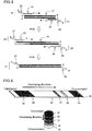
- Fig. 1 shows a primer for nucleic acid amplification.
- the primer contains a primer main region 1, and a tag region 2 which is located on the 5' side of the primer main region and which is not made double-stranded by a nucleic acid amplification reaction.
- the primer may further contain a spacer structure containing a polymerase reaction inhibitory region 3 between the primer main region and the tag region.
- the amplified double-stranded DNA fragment is preferably a product obtained by a nucleic acid amplification technique using a first primer set including primers each containing a sequence capable of hybridizing to a template of the target nucleic acid and a common sequence incapable of hybridizing to the template, and a second primer set including primers each containing a sequence capable of hybridizing to a complementary sequence to the corresponding common sequence and a tag region that is not made double-stranded by a nucleic acid amplification reaction.
- Fig. 2 shows each primer of a first PCR primer set.
- This first PCR primer characteristically contains a primer main region 4 capable of hybridizing to a template of the target nucleic acid and a common region 5 located on the 5' side of the primer main region and having a sequence common to a second primer.
- Fig. 3 shows each primer of a second PCR primer set.
- This second primer characteristically contains a primer main region 6 having a sequence common to the first primer, and a tag region 7 which is located on the 5' side of the main region 6 and which is not made double-stranded by a nucleic acid amplification reaction.
- the second primer may contain a spacer structure containing a polymerase reaction inhibitory region 8 between the second primer main region and the tag region.
- primer main region refers to an oligonucleotide region having a base sequence capable of functioning as a primer in the nucleic acid amplification reaction. Specifically, it is a base sequence that is capable of hybridizing to the 5' end or 3' end of a target base sequence of a target nucleic acid, and in general, is a base sequence complementary to a base sequence at the 5' end or 3' end of the target base sequence.
- the primer main regions may contain base deletions, insertions, and mismatch sites as long as they are capable of specifically binding to the target nucleic acids.
- the primer main regions preferably have a length of at least 8 bases, more preferably at least 12 bases, and still more preferably at least 15 bases.
- the maximum chain length of the primers is not particularly limited, and is generally 50 bases or less, preferably 40 bases or less, from the viewpoint of their synthesis costs and other factors.
- the tag regions of the primers are not particularly limited, provided that they contain natural nucleotides.
- the term "natural nucleotide” means a nucleotide composed of a natural base (adenine, thymine, guanine, cytosine, or uracil), a sugar moiety (deoxyribose or ribose), and phosphate group(s), all of which are not artificially modified.
- the natural nucleotides may be D-nucleotides or L-nucleotides.
- D-nucleotide refers to a nucleotide containing D-deoxyribose or D-ribose.
- L-nucleotide refers to a nucleotide containing L-deoxyribose or L-ribose.
- the effect of such tag regions containing natural nucleotides is to allow easy synthesis at low cost.
- the proportion of natural nucleotides in the tag region of the primer is preferably at least 5%, more preferably at least 20%, still more preferably at least 50%, further more preferably at least 70%, and most preferably at least 90%.
- the length of the tag region is not particularly limited, and the tag region has only to be long enough to hybridize to a complementary nucleic acid strand. The length is generally 5 bases to 60 bases, preferably 6 bases to 40 bases.
- the tag regions of the primers each preferably have a nucleic acid sequence in the same orientation as the primer main region.
- the effect of the tag regions of the primers each having a nucleic acid sequence in the same orientation as the primer main region is to allow easy synthesis at low cost. Even if the tag region and the primer main region are not directly linked to each other (e.g., a non-natural compound such as azobenzene is inserted between the tag region and the primer main region), these regions preferably have sequences in the same orientation as each other.
- nucleic acids being in the same orientation means that adjacent nucleotides are linked to each other via a 5'-3' , not a 3'-3' or 5'-5', phosphodiester bond between the sugar moieties of the nucleotides.
- nucleotides in the main region are also linked to one another via a 5'-3' bond between the sugar moieties.
- the polymerase reaction inhibitory region is not particularly limited, provided that it inhibits a nucleic acid extension reaction catalyzed by a polymerase to maintain the single-stranded structure in the region.
- a nucleic acid extension reaction catalyzed by a polymerase to maintain the single-stranded structure in the region.
- examples of such a structure include nucleic acid sequences having a three-dimensional structure that inhibits the progress of DNA polymerase, such as a tight hairpin structure and a pseudoknot structure; non-natural nucleic acids such as L-nucleic acids and artificial nucleic acids, RNA, and non-nucleic acid structures such as aliphatic chains.
- L-DNA which is DNA containing L-deoxyribose, does not function as a template in DNA extension reactions because it is not recognized by generally used DNA polymerases.
- L-DNA forms a left-handed double helix, and thus is incapable of hybridizing to naturally-occurring D-nucleic acids and capable of hybridizing only to nucleic acids of the same L-form.
- artificial nucleic acids refers to nucleic acids into which a compound that is not present in natural nucleic acid sequences is artificially inserted. Examples include, but are not limited to, peptide nucleic acids (PNA), bridged nucleic acids (BNA and LNA), azobenzene, fluorescein, Cy3, and Cy5.
- PNA refers to a molecule having a structure similar to DNA and RNA but having a backbone including a peptide structure.
- the backbone includes N- (2-aminoethyl) -glycine units linked by amide bonds.
- purine and pyrimidine rings which correspond to nucleic-acid bases, are linked to the backbone through a methylene group and a carbonyl group.
- BNA LNA refers to nucleic acids artificially synthesized by modifying the sugar moiety of DNA or RNA to form a bridge.
- the polymerase reaction inhibitory region is typically necessary between the tag region and the primer region.
- the tag region is incapable of functioning as a template in the reaction catalyzed by a DNA polymerase and thus is not made double-stranded by the nucleic acid amplification reaction, just like L-nucleic acids, artificial nucleic acids and the like, the polymerase reaction inhibitory region can be omitted.
- the primer in the present invention may contain only one of structures such as stable loop structures (e.g. hairpin structures, pseudoknot structures), non-natural nucleic acids (e.g. L-nucleic acids, artificial nucleic acids), and non-nucleic acid structures (e.g. aliphatic chains), or may contain two or more of these structures in combination.
- the primer can be labeled with various molecules generally used for oligonucleotide labeling.
- molecules include enzymes, magnetic particles, fluorescent pigments, and radioisotopes. Any of these may be used alone, or two or more of these may be used in combination.
- the primers thus designed can be produced by any methods without particular limitation, and known methods can be used. Specifically, the designed primers can be easily obtained with a DNA synthesizer or from a custom synthesis service.
- the nucleic acid amplification technique is not particularly limited, provided that it produces an amplified double-stranded DNA fragment having at each end thereof a single-stranded region containing natural nucleotides, using the primers mentioned above.
- One example thereof is PCR.
- Isothermal amplification techniques such as LAMP and ICAN may also be used.
- a pair of reverse and forward primers for the PCR reaction may be designed such that both primers contain different polymerase reaction inhibitory regions from each other, or such that one of them contains a polymerase reaction inhibitory region and the other is free of any polymerase reaction inhibitory region but is modified with biotin or the like.
- the PCR conditions are not particularly limited, provided that a target region of the above-described sample DNA is amplified by PCR using the sample DNA as a template and the primer set.
- the polymerase used in the PCR is not particularly limited, and is preferably a heat-stable DNA polymerase, and more preferably a heat-stable polymerase that does not substantially have a 3'-to-5' exonuclease activity.
- heat-stable DNA polymerases is Ex-Taq (available from TAKARA BIO INC.).
- the PCR reaction conditions including temperature, time, and buffer composition are not particularly limited, and may appropriately be determined according to the DNA polymerase selected to use, the sequences of the primers, the length of the target sequence, and other factors.
- the length of the DNA to be amplified by the nucleic acid amplification reaction is preferably at least 20 bases, and more preferably at least 40 bases. If the length is less than 20 bases, the probability of non-specific amplification tends to be increased.
- PCR can be carried out in a conventional manner using the primer set to provide an amplification product in which a single-stranded region is added to each end of the target nucleic acid sequence.
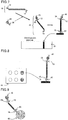
- Fig. 4 is a schematic flow diagram of an exemplary amplification reaction using primers each containing a primer main region and a tag region.
- the forward primer 10 contains a primer main region 11 having the same sequence as a part of the 5' end of a target nucleic acid sequence 9, and a tag region 12 located on the 5' end of the primer main region 11.
- the reverse primer 13 contains a primer main region 14 having a complementary sequence to a part of the 3' end of the target nucleic acid sequence, and a tag region 15 located on the 5' end of the primer main region 14.
- the linked tag region in each primer typically has a different sequence from each other.
- the primer set is used in PCR to afford an amplified DNA product 16 having a single-stranded region at each end because the added tag regions of both primers are not substantially involved in the PCR reaction.
- the amplified DNA fragment having a single-stranded region at each end refers to an amplified DNA product having a double-stranded DNA part that is the same as the target DNA region, and also having single-stranded regions which are respectively located as 5' end tag parts at opposite ends of the double-stranded DNA part, as shown in Fig. 4 .
- the amplified DNA fragment more specifically refers to an amplified double-stranded DNA fragment having at each end thereof a single-stranded region that includes non-modified nucleic acids, wherein the single-stranded region at each end has a sequence in the same orientation as the corresponding DNA strand which it is located next to.
- Fig. 5 is a schematic flow diagram of an exemplary amplification reaction using primers each containing a primer main region and a common sequence region as a joint primer set, and primers each containing the common sequence region and a tag region.
- PCR can be carried out in a conventional manner using the first and second; primer sets to provide an amplification product in which a single-stranded region is added to each end of the target nucleic acid sequence.
- the first forward primer 18 contains a primer main region 19 having the same sequence as a part of the 5' end of a target nucleic acid sequence 17, and a common sequence region 20 located on the 5' end of the primer main region 19.
- the first reverse primer 21 contains a primer main region 22 having a complementary sequence to a part of the 3' end of the target nucleic acid sequence, and a common sequence region 23 located on the 5' end of the primer main region 22.
- the added common sequence regions of both primers typically have different sequences from each other.
- the first primer set is used in the PCR reaction to afford an amplified double-stranded DNA product 24 containing the common regions.
- the second forward primer 25 which is shown around the common sequence region at either end of the amplified DNA product 24, contains a primer main region 26 having a sequence common to a part of the 5' end of the amplified double-stranded DNA product 24 containing the common regions, and a tag region 27 located on the 5' end of the primer main region 26.
- the second reverse primer 28 contains a primer main region 29 having a complementary sequence common to a part of the 3' end of the amplified double-stranded DNA product 24 containing the common regions, and a tag region 30 located on the 5' end of the primer main region 29.
- the linked tag regions of both primers typically have different sequences from each other.
- the primer set is used in PCR to afford an amplified DNA product 31 having a single-stranded region at each end because the added tag regions of both primers are not substantially involved in the PCR reaction.
- the PCR reaction using the first primers and then the PCR reaction using the second primers may be carried out sequentially as shown in Fig. 5 . With respect to the order, the first and second primers may be added at the same time, or alternatively, the second primers may be added later.
- the amplified DNA fragment having a single-stranded region at each end refers to an amplified DNA product having a double-stranded DNA part that is the same as the target DNA region, and also having single-stranded regions which are respectively located as 5' end tag parts at opposite ends of the double-stranded DNA part, as denoted by the reference numeral 31 in Fig. 5 .
- a single set of second primers can be used for different target nucleic acids to provide the same single-stranded tag sequences, as long as these primer sets are designed to have the same common sequences.
- the amplified DNA fragment more specifically refers to an amplified double-stranded DNA fragment having at each end thereof a single-stranded region that includes non-modified nucleic acids, wherein the single-stranded region at each end has a sequence in the same orientation as the corresponding DNA strand which it is located next to.
- hybridization means that molecules containing nucleic acids complementarily form a complex (e.g. DNA/DNA, DNA/RNA, DNA/PNA, L-DNA/L-DNA).
- the amplified DNA product obtained in the nucleic acid amplification step is used in a hybridization reaction without the need of a treatment for making the amplified product single-stranded (e.g. heat treatment) and other treatments because the amplified DNA fragment contains the single-stranded regions.
- the detection method preferably further includes the step of hybridizing a second oligonucleotide probe labeled directly or indirectly with a labeling substance with the other single-stranded region of the amplified double-stranded DNA fragment.
- the formation of a triple complex of the amplified double-stranded DNA fragment, the first oligonucleotide probe, and the second oligonucleotide probe is called "sandwich hybridization".
- the order of hybridization of the three substances is not particularly limited.
- the length of the first oligonucleotide probe is not particularly limited as long as it is capable of hybridizing to the single-stranded region of the amplified double-stranded DNA fragment, and is preferably 5 to 60 bases, and more preferably 10 to 40 bases.
- the length of the second oligonucleotide probe is not particularly limited as long as it is capable of hybridizing to the single-stranded region of the amplified double-stranded DNA fragment, and is preferably 5 to 60 bases, and more preferably 10 to 40 bases.
- the labeling substance bound to the second oligonucleotide probe is not particularly limited as long as it enables the amplified double-stranded DNA fragment to be detected.
- the labeling substance is preferably a colored carrier that enables the amplified double-stranded DNA fragment to be visually detected.
- Examples of such colored carriers include colored particles, and enzyme- or pigment-bound carriers. Preferred among these are colored particles.
- the colored particles include colloidal particles of metals such as gold, silver, copper and platinum, colored latexes which are latexes colored with a pigment, a dye or the like, and silica nanoparticles which are silica (silicon dioxide) particles in which pigment molecules are immobilized.
- colloidal gold particles and colored (e.g. blue, red) latex particles made of a water-dispersible polymer are colloidal gold particles and colored (e.g. blue, red) latex particles made of a water-dispersible polymer.
- the use of such colored particles allows the amplified DNA fragment to be visually determined more easily.
- colored particles of different color is used for each analyte to allow the multiple analytes to be visually determined easily at the same time.
- the particles size is not particularly limited.
- the particle size is determined such that the colored particles have less adverse effect on the formation of a sandwich hybridization complex and on the capturing of the target sequence-containing amplification product on the solid phase, and provide good color development in the detection.
- the particle size of colored particles is selected to be smaller than the pore size of a later-described chromatographic medium. Specifically, the particle size is typically not more than 500 nm, preferably 0.1 nm to 100 nm, and more preferably 1 nm to 50 nm.
- the enzymes usable as the colored carrier are proteins that catalyze reactions of substrates to develop a color or emit light. Examples include peroxidases, alkaline phosphatases, and luciferases, although the enzymes are not limited to these examples, provided that they allow detection by observation with the naked eye.
- the conditions of the hybridization of the single-stranded region at the end of the amplified double-stranded DNA fragment and the first or second oligonucleotide probe are not particularly limited, provided that they can hybridize to each other. Preferably, they are reacted at room temperature in 10 mM phosphate buffer. In this case, the hybridization rate can be increased by adding a salt such as sodium chloride.
- the presence of the target nucleic acid can be assessed by detecting the target substance in the sandwich hybridization complex formed in an identifiable zone on the capture carrier (solid phase).
- the detection is preferably based on visual observation.
- the amplification product of the nucleic acid amplification reaction can be used directly in the hybridization reaction without the need of any treatment for making the amplification product single-stranded (e.g. heat denaturation).
- the nucleic acid detection method involving the formation of a sandwich hybridization complex is preferably carried out on a nucleic acid detection device.
- the nucleic acid chromatography device of Fig. 6 includes a sample pad 32 (a carrier to which the amplified DNA product is to be applied), a conjugate pad 33 (a carrier in which a colored carrier-bound oligonucleotide is placed), a carrier 34 having a capture oligonucleotide (a chromatographic medium), and an absorption pad 35, and these members are attached on a supporting member 36 with a pressure-sensitive adhesive or the like.
- the carrier 34 is provided with a test line 37 along which the capture oligonucleotide is applied, and a control line 38.
- the conjugate pad 33 may not be used.
- the chromatography is carried out to detect the amplified double-stranded DNA fragment by a method including the following steps (a) to (c): (a) placing the amplified DNA fragment in a zone on the nucleic acid detection device which is different from a zone where the first oligonucleotide probe is immobilized; (b) diffusing the amplified DNA fragment with a solvent on the device toward the zone where the first oligonucleotide probe is immobilized; and (c) hybridizing the first oligonucleotide probe with the amplified DNA fragment in the zone where the first oligonucleotide probe is immobilized.
- the amplified DNA fragment is placed on the sample pad 32 in the step (a).
- the amplified DNA fragment is diffused in the direction of the arrow.
- the amplified DNA fragment is hybridized with and captured by the first oligonucleotide probe immobilized on the test line 37.
- the detection method further includes the step of hybridizing the second oligonucleotide probe labeled with the labeling substance with the amplified DNA fragment before the step (c).
- the amplified DNA fragment and the second oligonucleotide probe are hybridized on the conjugate pad 33.
- the chromatography is preferably carried out by the following steps (d) to (h): (d) placing the amplified DNA fragment and the second oligonucleotide probe labeled with the labeling substance respectively in discrete zones on the nucleic acid detection device which are different from a zone where the first oligonucleotide probe is immobilized; (e) diffusing the amplified DNA fragment with a solvent toward the zone where the second oligonucleotide probe labeled with the labeling substance is placed; (f) hybridizing the second oligonucleotide probe labeled with the labeling substance with the amplified DNA fragment in the zone where the second oligonucleotide probe labeled with the labeling substance is placed; (g) diffusing a hybridization complex obtained in the step (f) on a development medium toward the zone where the first oligonucleotide probe is placed; and (h) hybridizing the first oligonucleotide probe with the complex in the zone where the first
- the amplified DNA fragment is placed on the sample pad 32, and the second oligonucleotide probe is placed on the conjugate pad 33 in the step (d).
- the amplified DNA fragment is diffused from the sample pad 32 in the direction of the arrow.
- the amplified DNA fragment and the second oligonucleotide probe are hybridized on the conjugate pad 33.
- the hybridization complex of the amplified DNA fragment and the second oligonucleotide probe labeled with the labeling substance is diffused in the direction of the arrow.
- the first oligonucleotide probe and the complex are hybridized on the test line 37.
- an oligonucleotide probe having a complementary sequence to one of the tag regions of the amplified DNA fragment is immobilized as the first oligonucleotide probe for capturing.
- the first oligonucleotide probe for capturing may be bound to the membrane directly or via a functional group or a certain substance. Examples of such mediating substances include, but are not limited to, peptides, proteins and nucleic acids. In the case where avidin is used as a mediating substance, the capture oligonucleotide should be modified with biotin.
- an oligonucleotide probe for capturing the colored carrier is immobilized on the control line on the membrane.
- the oligonucleotide probe for the control line has a complementary sequence to the second oligonucleotide probe labeled with the labeling substance so that it certainly captures the labeling substance when the sample solution is developed.
- the oligonucleotide probe for the control line may also be bound to the membrane directly or via a functional group or a substance, as described above. Examples of mediating substances include, but are not limited to, peptides, proteins and nucleic acids. In the case where avidin is used as a mediating substance, the capture oligonucleotide should be modified with biotin.
- the presence of the target nucleic acid in a sample can be assessed by visually observing a color on the test line. Also, a color on the control line can be visually observed to determine whether the development and the color reaction are normally carried out.
- the "to visually observe” means observation with the naked eye to assess the color.
- the chromatographic medium examples include paper filters such as qualitative filters, quantitative filters, phase separating filters, glass fiber filters, silica fiber filters, and bicomponent fiber filters.
- Other examples include filters made of celluloses (e.g. nitrocellulose), synthetic resin films such as polyethersulfone membranes, and porous gels such as silica gel, agarose, dextran, and gelatin. Nylon membranes can also be suitably used.
- the form and size of the chromatographic medium are not particularly limited, and may be any suitable ones in operation and observation of the reaction results.
- the back surface of the chromatographic medium whose opposite surface is provided with reaction sites is preferably provided with a supporting material made of plastic or the like.
- the developing direction in the device is not particularly limited, and may be horizontal or vertical as shown in Fig. 6 . Since the solvent used in the nucleic acid amplification reaction can function as a developing solvent as well, the reaction solution obtained by the nucleic acid amplification reaction can be directly dropped to the sample pad 32 in Fig. 6 . Alternatively, a separate developing solution may be added to the reaction solution obtained by the amplification reaction, and the resulting solution may be added to the sample pad. Any developing solvent can be used without particular limitation, provided that it is liquid. Examples thereof include phosphate buffer and Good's buffers such as Tris buffer. Such solvents may contain salts, surfactants, proteins, and nucleic acids dissolved therein.
- a sandwich hybridization complex is formed on a chromatographic carrier.
- An amplified DNA fragment 16 obtained in the nucleic acid amplification step is used in the subsequent complex formation step without performing a treatment for making the fragment single-stranded (e.g. heat treatment) and other treatments.
- the amplified DNA fragment 16 is hybridized with an oligonucleotide probe including a colored carrier 40 and a nucleic acid sequence 39 capable of specifically binding to one (tag region 12) of the tag regions of the DNA fragment, and thereby forms a first complex 41.
- the complex 41 may be formed, for example, in a PCR reaction vessel, prior to the application to the development medium, or may be formed by applying the amplified DNA fragment to the carrier and allowing the amplified DNA fragment to move by capillary action to pass through a carrier that has been subjected to coating with the labeling molecule-bound oligonucleotide and drying.
- the complex 41 comes into contact with a capture oligonucleotide probe 43 that is previously allowed to be bound in an identifiable zone on a chromatographic medium 42 made of a porous membrane or the like, on the development medium.
- the capture oligonucleotide 43 has a complementary sequence to the other tag sequence 15 of the amplified DNA fragment, and thus hybridizes to the complex 41 to form a sandwich hybridization complex.
- a sandwich hybridization complex is not particularly limited. It is preferable that the amplified DNA fragment and the second oligonucleotide probe labeled with the labeling substance form a complex 41, and then the complex and the first oligonucleotide probe for capturing form a complex.
- a sandwich hybridization complex may be formed by enriching the amplified DNA fragment via the first oligonucleotide probe for capturing on the development medium, and then developing the second oligonucleotide labeled with the labeling substance.
- nucleic acid detection device is a microarray (DNA chip) as shown in Fig. 8 .
- the triple complex can be formed by sandwich hybridization in wells of the microarray 44 in which a capture oligonucleotide is immobilized.
- Still another example is a bead form as shown in Fig. 9 .
- the triple complex can be formed by sandwich hybridization on the bead carrier 45 having a capture oligonucleotide.
- the nucleic acid detection method and the nucleic acid detection device can be used for any techniques involving a nucleic acid amplification step.
- the nucleic acid detection method and the nucleic acid detection device can be used for techniques in any fields which involve detection of an amplified DNA fragment (e.g. PCR product) obtained by a nucleic acid amplification technique.
- amplified DNA fragment e.g. PCR product
- they are used for, for example, molecular biology research, detection of pathogens, detection of foreign matter such as allergens in foods, food quality control (inspection of mislabeled foods and genetically modified foods), livestock control, detection of single nucleotide polymorphisms (hereinafter, also referred to as "SNP”), screening of diseases such as cancer, and so on.
- SNP single nucleotide polymorphisms
- the present invention encompasses methods for detecting a pathogenic infectious disease, methods for detecting foreign matter (e.g. allergen) in foods, food quality control methods, livestock control methods, methods for detecting a single nucleotide polymorphism, and like methods which include a step of performing the nucleic acid detection method of the present invention.
- methods for detecting a pathogenic infectious disease methods for detecting foreign matter (e.g. allergen) in foods, food quality control methods, livestock control methods, methods for detecting a single nucleotide polymorphism, and like methods which include a step of performing the nucleic acid detection method of the present invention.
- a pathogen detection method and an allergen detection method according to the present invention are described in detail below.
- the pathogen detection method may be any method including the step of detecting a gene specific to a pathogen by the nucleic acid detection method of the present invention.
- the pathogen is not particularly limited, and specific examples include pathogenic bacteria, pathogenic viruses, food poisoning bacteria, and bacteria and viruses causing hospital infections. More specifically, there may be mentioned, for example, viruses such as hepatitis C virus (HCV), cytomegalovirus (CMV), Epstein-Barr virus (EBV), herpesviruses, and human immunodeficiency virus (HIV) ; bacteria such as Escherichia coli (e.g. 0157), Mycobacterium tuberculosis, Salmonella typhi, salmonella bacteria, and Vibrio parahaemolyticus; and microorganisms such as mycoplasma.
- viruses such as hepatitis C virus (HCV), cytomegalovirus (CMV), Epstein-Barr virus (EBV), herpesviruses, and human immunodeficiency
- the pathogen detection method includes determining, by the nucleic acid detection method, whether a gene specific to a pathogen is present, for example, in a DNA sample prepared from a sample to be assessed for the presence of the pathogen.
- a sample to be assessed for the presence of the pathogen may be directly used for a template for nucleic acid amplification without preparing a DNA sample.
- the pathogen to be detected is a bacterium such as Escherichia coli
- a bacterial colony suspension can be used for a template.
- the gene specific to the pathogen it can be determined that the sample contains the pathogen. In this manner, it is possible to easily and highly accurately determine whether a sample contains a pathogen without the need of special equipment.
- the pathogen detection method can be used for the diagnosis of microbial infectious diseases.
- the allergen detection method may be any method including the step of detecting a gene encoding an allergen by the nucleic acid detection method.
- the allergen is not particularly limited, and specific examples include allergens contained in foods. More specifically, there may be mentioned, for example, egg albumen allergens, milk allergens, wheat allergens, buckwheat allergens, peanut allergens, and so on. More specifically, the allergen detection method according to the present invention includes determining, by the nucleic acid detection method, whether a gene encoding an allergen derived from egg, milk, wheat, buckwheat, peanut or the like is present, for example, in a DNA sample prepared from a food. Then, if such a gene is detected, it can be determined that the food contains an ingredient containing the allergen.
- the allergen origin is not limited to those described above.
- the allergen origin may be any type of rice, corn, Italian millet, proso millet, Japanese millet, buckwheat, or pulses. Since DNA is thermally stable, a trace amount of DNA can be detected even in processed foods.
- the allergen detection method according to the present invention provides data that can be used not only for food labeling and allergen information of foods but also for detection of minute amounts of residual food additives (e.g. processing materials, carry-overs) and detection of contaminants that are not intended by a manufacturer (e.g. the presence or absence of cross-contamination between the manufacturing lines).
- the present invention is applicable to determination of the parentage of a mammal including human, identification of the pedigree of livestock, variety identification for agricultural products, SNP detection, detection of diseases (e.g. cancer) caused by gene mutations, and the like. More specifically, for example, taking the example of livestock, the present invention can be used for pedigree registration, individual identification, parentage determination, removal of a carrier individual with a disease-causing gene, and the like.
- a forward primer (F) and a reverse primer (R) were designed to be able to amplify about 330 base pairs by PCR amplification using pUC19 (available from Takara Bio, Inc.) as a template.
- tag sequences T1 and T2 which are non-natural (L-)DNA strands were respectively introduced into the 5' ends of these primers to construct L-DNA-tagged primers T1-F and T2-R.
- the synthesis of these L-DNA-tagged primers was carried out by a general phosphoramidite method using a DNA automatic synthesizer (H-8-SE: Gene World) with a 0.2 ⁇ M column.
- PCR was performed using the primer set prepared by performing the above step (1). Specifically, a 100 ⁇ l PCR mixture was prepared by putting the prepared primer F and primer R (15 pmol each) and pUC19 (10 ng) in a 0.2-ml PCR tube, and following the instruction manual of a PCR device ExTaq (available from Takara Bio, Inc.). Thereafter, the tube was set in a thermal cycler (GeneAmp PCR System, available from Applied Biosystems), and subjected to heat treatment at 95°C for five minutes and then exposed to 35 cycles of 95°C for 30 seconds, 55°C for 30 seconds, and 72°C for 30 seconds. Consequently, the target fragment of about 330 bp was amplified. Separately, the same reaction was carried out in the absence of pUC19 as a negative control.
- a carboxyl group-containing polystyrene latex (solids content: 10% (w/w), available from Bangs Laboratories, Inc.) and an amino-group containing L-oligonucleotide probe (SEQ ID No: 7, strand complementary to SEQ ID No: 3) were bonded by mixing them in MES buffer containing a necessary amount of a water-soluble carbodiimide, and the resulting product was blocked with monoethanolamine. The reaction solution was centrifuged, and the supernatant was then removed. The obtained precipitate was washed with water, and then resuspended in HEPES buffer with a surfactant. The suspension was uniformly applied to a glass fiber pad, and the pad was dried in a vacuum oven. In this manner, a conjugate pad was obtained. Nucleotide probe 1: 5'- L d(TTGGCTCTGTCTCCGTTGTC)-NH 2 -3' (SEQ ID No:7)
- a nylon membrane modified with carboxyl groups (available from Pall Corporation, 6 mm ⁇ 60 mm) was treated with a water-soluble carbodiimide, and washed with deionized water.
- An amino group-containing L-oligonucleotide probe having a sequence (SEQ ID No:8) complementary to SEQ ID No:4 was applied to the activated membrane with a dispenser along a line drawn 30 mm from one end of the membrane, and air-dried for 15 minutes. Subsequently, the membrane was treated with Tris buffer and blocked, and then washed with water and dried.
- Nucleotide probe 2 5'- L d(CACTGGGCATACGGTAGCAT)-NH 2 -3' (SEQ ID No:8)
- a test strip to be used for detection of the PCR product amplified using the L-DNA-tagged primer set was prepared by attaching a chromatographic medium consisting of the nylon membrane prepared above, the conjugate pad prepared above, a general sample pad as a sample application zone, and an absorption pad for absorbing the developed sample and the labeling substance to a substrate consisting of a backing sheet as shown in Fig. 6 .
- the PCR product obtained in the step (2) was immediately, without being denatured, applied to the sample application zone on the test strip prepared in the step (5) to perform detection by chromatography.
- a colored line specific to the target nucleic acid was detected along the test line for the sample prepared using pUC19 as an analyte in the step (2).
- the detection by chromatography took as short a time as 10 to 15 minutes.
- a forward primer (F) and a reverse primer (R) were designed to be able to amplify about 330 base pairs by PCR amplification using pUC19 (available from Takara Bio, Inc.) as a template. Then, a polymerase reaction inhibitory region (H) having a hairpin structure and a tag sequence T3 or T4 were introduced into the 5' end of each corresponding primer to synthesize tagged primers T3-H-F and T4-H-R.
- PCR was performed using the primer set prepared by performing the above step (1). Specifically, a 100 ⁇ l PCR mixture was prepared by putting the prepared primer F and primer R (15 pmol each), and pUC19 (10 ng) in a 0.2-ml PCR tube, and following the instruction manual of a PCR device ExTaq (available from Takara Bio, Inc.). Thereafter, the tube was set in a thermal cycler (GeneAmp PCR System, available from Applied Biosystems), and subjected to heat treatment at 95°C for five minutes and then exposed to 35 cycles of 95°C for 30 seconds, 55°C for 30 seconds, and 72°C for 30 seconds. Consequently, the target fragment of about 330 bp was amplified. Separately, the same reaction was carried out in the absence of pUC19 as a negative control.
- a carboxyl group-containing polystyrene latex (solids content: 10% (w/w), available from Bangs Laboratories, Inc.) and an amino-group containing oligonucleotide probe (SEQ ID No:14, strand complementary to SEQ ID No:10) were bonded by mixing them in MES buffer containing a necessary amount of a water-soluble carbodiimide, and the resulting product was blocked with monoethanolamine.
- the reaction solution was centrifuged, and the supernatant was then removed.
- the obtained precipitate was washed with water, and then resuspended in HEPES buffer with a surfactant.
- the suspension was uniformly applied to a glass fiber pad, and the pad was dried in a vacuum oven. In this manner, a conjugate pad was obtained.
- Oligonucleotide probe 3 5'- D d(ATTATGCGTGGAGAAGCATATCATA)-NH 2 -3' (SEQ ID No:14)
- a nylon membrane modified with carboxyl groups (available from Pall Corporation, 6 mm ⁇ 60 mm) was treated with a water-soluble carbodiimide, and washed with deionized water.
- An amino group-containing L-oligonucleotide probe having a sequence (SEQ ID No:15) complementary to SEQ ID No:11 was applied to the activated membrane with a dispenser along a line drawn 30 mm from one end of the membrane, and air-dried for 15 minutes. Subsequently, the membrane was treated with Tris buffer and blocked, and then washed with water and dried.
- Oligonucleotide probe 4 5'- D d(ATTGAGCAAGTGTACAGAGCA)-NH 2 -3' (SEQ ID No:15)
- a test strip to be used for detection of the PCR product amplified using the hairpin-tagged primer set was prepared by attaching a chromatographic medium consisting of the nylon membrane prepared above, the conjugate pad prepared above, a general sample pad as a sample application zone, and an absorption pad for absorbing the developed sample and the labeling substance to a substrate consisting of a backing sheet as shown in Fig. 6 .
- the PCR product obtained in the step (2) was immediately, without being denatured, applied to the sample application zone on the test strip prepared in the step (5) to perform detection by chromatography.
- a colored line specific to the target nucleic acid was detected along the test line for the sample prepared using pUC19 as an analyte in the step (2).
- the detection by chromatography took as short a time as 10 to 15 minutes.
- a forward primer (F) and a reverse primer (R) were designed to be able to amplify about 330 base pairs by PCR amplification using pUC19 (available from Takara Bio, Inc.) as a template.
- pUC19 available from Takara Bio, Inc.
- a polymerase reaction inhibitory region (X) containing azobenzene (artificial nucleic acid) and a tag sequence T5 or T6 were introduced into the 5' end of each corresponding primer to synthesize tagged primers T5-X-F and T6-X-R.
- azobenzene-inserted primers were purchased as products custom-synthesized by TSUKUBA OLIGO SERVICE CO., LTD. The following shows the primer set prepared in this study.
- PCR was performed using the primer set prepared by performing the above step (1). Specifically, a 100 ⁇ l PCR mixture was prepared by putting the primer T5-X-F and primer T6-X-R (15 pmol each) and pUC19 (10 ng) in a 0.2-ml PCR tube, and following the instruction manual of a PCR device ExTaq (available from Takara Bio, Inc.). Thereafter, the tube was set in a thermal cycler (GeneAmp PCR System, available from Applied Biosystems), and subjected to heat treatment at 95°C for five minutes and then exposed to 35 cycles of 95°C for 30 seconds, 55°C for 30 seconds, and 72°C for 30 seconds. Consequently, the target fragment of about 330 bp was amplified. Separately, the same reaction was carried out in the absence of pUC19 as a negative control.
- a carboxyl group-containing polystyrene latex (solids content: 10% (w/w), available from Bangs Laboratories, Inc.) and an amino-group containing oligonucleotide probe (SEQ ID No:20, strand complementary to SEQ ID No:16) were bonded by mixing them in MES buffer containing a necessary amount of a water-soluble carbodiimide, and the resulting product was blocked with monoethanolamine. The reaction solution was centrifuged, and the supernatant was then removed. The obtained precipitate was washed with water, and then resuspended in HEPES buffer with a surfactant. The suspension was uniformly applied to a glass fiber pad, and the pad was dried in a vacuum oven. In this manner, a conjugate pad was obtained. Oligonucleotide probe 5: 5'- D d(CTATAAACCCAGTGAAAAATGTTGCCA)-NH 2 -3' (SEQ ID No:20)
- a nylon membrane modified with carboxyl groups (available from Pall Corporation, 6 mm ⁇ 60 mm) was treated with a water-soluble carbodiimide, and washed with deionized water.
- An amino group-containing D-oligonucleotide probe having a sequence (SEQ ID No:21) complementary to SEQ ID No:17 was applied to the activated membrane with a dispenser along a line drawn 30 mm from one end of the membrane, and air-dried for 15 minutes. Subsequently, the membrane was treated with Tris buffer and blocked, and then washed with water and dried.
- Oligonucleotide probe 6 5'- D d(GATCATACACGTGGTTGGAAGCTAACC)-NH 2 -3' (SEQ ID No:21)
- a test strip to be used for detection of the PCR product amplified using the azobenzene-inserted primer set was prepared by attaching a chromatographic medium consisting of the nylon membrane prepared above, the conjugate pad prepared above, a general sample pad as a sample application zone, and an absorption pad for absorbing the developed sample and the labeling substance to a substrate consisting of a backing sheet as shown in Fig. 6 .
- the PCR product obtained in the step (2) was immediately, without being denatured, applied to the sample application zone on the test strip prepared in the step (5) to perform detection by chromatography.
- a colored line specific to the target nucleic acid was detected along the test line for the sample prepared using pUC19 as an analyte in the step (2).
- the detection by chromatography took as short a time as 10 to 15 minutes.
- Gold Colloid 40 nm, 9.0 ⁇ 10 10 (particles/ml), available from British BioCell International
- a thiol group-containing oligonucleotide probe SEQ ID No:22, strand complementary to SEQ ID No:16
- the resulting mixture was centrifuged at 6000 rpm for 15 minutes, and the supernatant was removed.
- the residue was combined and mixed with 0.05 M sodium chloride and 5 mM phosphate buffer (pH 7), and then incubated again at 50°C for 40 hours.
- Oligonucleotide probe 7 5'- D d(CTATAAACCCAGTGAAAAATGTTGCCA)-SH-3' (SEQ ID No:22)
- oligonucleotide probe having a sequence (SEQ ID No: 23) complementary to SEQ ID No: 17 was mixed with streptavidin. The resulting mixture was applied along a line on a nitrocellulose membrane (product name: Hi-Flow 180, available from Millipore Corporation) with a dispenser, and air-dried at 40°C for 30 minutes.
- a test strip to be used for detection of the PCR product amplified using the azobenzene-inserted primer set was prepared by attaching a chromatographic medium consisting of the nitrocellulose membrane prepared above, the conjugate pad prepared above, a general sample pad as a sample application zone, and an absorption pad for absorbing the developed sample and the labeling substance to a substrate consisting of a backing sheet as shown in Fig. 6 .
- the PCR product obtained in the step (2) in Example 3 was immediately, without being denatured, applied to the sample application zone on the test strip prepared in the step (3) to perform detection by chromatography.
- a colored line specific to the target nucleic acid was detected along the test line for the sample prepared using pUC19 as an analyte in the step (2) in Example 3.
- the detection by chromatography took as short a time as 10 to 15 minutes.
- Oligonucleotide probe 9 5'- D d(GATCATACACGTGGTTGGAAGCTAACC)-3' (SEQ ID No:24)
- a test strip to be used for detection of the PCR product amplified using the azobenzene-inserted primer set was prepared by attaching a chromatographic medium consisting of the UltraBind affinity membrane prepared above, the conjugate pad prepared above, a general sample pad as a sample application zone, and an absorption pad for absorbing the developed sample and the labeling substance to a substrate consisting of a backing sheet as shown in Fig. 6 .
- the PCR product obtained in the step (2) in Example 3 was immediately, without being denatured, applied to the sample application zone on the test strip prepared in the step (2) to perform detection by chromatography.
- a colored line specific to the target nucleic acid was detected along the test line for the sample prepared using pUC19 as an analyte in the step (2) in Example 3.
- the detection by chromatography took as short a time as 10 to 15 minutes.
- a forward primer (F1) and a reverse primer (R1) a forward primer (F2) and a reverse primer (R2)
- a forward primer (F3) and a reverse primer (R3) were designed to be able to amplify about 330 base pairs, about 200 base pairs, and about 100 base pairs, respectively, by PCR amplification using the three genes: pUC19 (available from Takara Bio, Inc.), an EcoRI methylase gene, and a BamHI methylase gene as target nucleic acid templates, respectively.
- a polymerase reaction inhibitory region (X) containing azobenzene (artificial nucleic acid) and a tag sequence T10 or T11, or a tag sequence T12 or T13, or a tag sequence T14 or T15 were introduced into the 5' end of each corresponding primer to synthesize tagged primers T10-X-F1 and T11-X-R1, T12-X-F2 and T13-X-R2, and T14-X-F3 and T15-X-R3.
- These six azobenzene-inserted primers were purchased as products custom-synthesized by TSUKUBA OLIGO SERVICE CO., LTD. The following shows the three primer sets prepared in this study.
- PCR was performed using the three primer sets prepared by performing the above step (1). Specifically, a 100 ⁇ l PCR mixture was prepared by putting the primer T10-X-F1 and primer T11-X-R1, the primer T12-X-F2 and primer T13-X-R2, and the primer T14-X-F3 and primer T15-X-R3 (15 pmol each) and the template(s) (10 ng each) in a 0.2-ml PCR tube, and following the instruction manual of a PCR device ExTaq (available from Takara Bio, Inc.). The following five kinds of PCR mixtures were prepared:
- the tubes were set in a thermal cycler (GeneAmp PCR System, available from Applied Biosystems), and subjected to heat treatment at 95°C for five minutes and then exposed to 30 cycles of 95°C for 30 seconds, 55°C for 30 seconds, and 72°C for 30 seconds. Consequently, the following DNA fragments containing the target sequences were amplified: (i) about 330 bp; (ii) about 200 bp; (iii) about 100 bp; (iv) three fragments of about 330 bp, about 200 bp, and about 100 bp; and (v) no amplified DNA fragment (negative control).
- GeneAmp PCR System available from Applied Biosystems
- reaction solutions were centrifuged, and the supernatants were then removed.
- the obtained precipitates were individually washed with water and then resuspended in HEPES buffer with a surfactant to prepare an oligonucleotide probe 16-bound latex (blue), an oligonucleotide probe 17-bound latex (orange), and an oligonucleotide probe 18-bound latex (green), respectively.
- a 3'-biotin-modified oligonucleotide probe having a sequence (SEQ ID No:40) complementary to SEQ ID No:26, a 3'-biotin-modified oligonucleotide probe having a sequence (SEQ ID No:41) complementary to SEQ ID No:30, and a 3'-biotin-modified oligonucleotide probe having a sequence (SEQ ID No:42) complementary to SEQ ID No:34 were individually mixed with streptavidin.
- the resulting mixtures were applied with a dispenser respectively along three separated lines on a nitrocellulose membrane (product name: Hi-Flow 135, available from Millipore Corporation) in the stated order from the upstream, and air-dried at 40°C for 30 minutes. In this manner, three detection lines were formed.
- a test strip to be used for detection of the PCR products amplified using the azobenzene-inserted primer sets was prepared by attaching a chromatographic medium consisting of the nitrocellulose membrane prepared above, the conjugate pad prepared in the step (3), a general sample pad as a sample application zone, and an absorption pad for absorbing the developed sample and the labeling substances to a substrate consisting of a backing sheet as shown in Fig. 6 .
- the detection by chromatography took as short a time as 10 to 15 minutes.
- a forward primer (Fj1) and a reverse primer (Rj1) a forward primer (Fj2) and a reverse primer (Rj2)
- a forward primer (Fj3) and a reverse primer (Rj3) were designed to be able to amplify about 330 base pairs, about 200 base pairs, and about 100 base pairs, respectively, by PCR amplification using the three genes: pUC19 (available from Takara Bio, Inc.), an EcoRI methylase gene, and a BamHI methylase gene as target nucleic acid templates, respectively.
- common sequences KF1 and KR1, common sequences KF2 and KR2, or common sequences KF3 and KR3 were introduced into the 5' ends of each corresponding primer pair to synthesize common sequence-added primers KF1-Fj1 and KR1-Rj1, KF2-Fj2 and KR2-Rj2, and KF3-Fj3 and KR3-Rj3.
- These six common sequence-added primers (joint primers) were purchased as products custom-synthesized by TSUKUBA OLIGO SERVICE CO., LTD. The following shows the three primer sets prepared in this study.
- a polymerase reaction inhibitory region (X) containing azobenzene (artificial nucleic acid) and a tag sequence T22 or T23, or a tag sequence T24 or T25, or a tag sequence T26 or T27 were introduced into the 5' end of each corresponding primer to synthesize tagged primers T22-X-KF1 and T23-X-KR1, T24-X-KF2 and T25-X-KR2, and T26-X-KF3 and T27-X-KR3.
- These six azobenzene-inserted primers were purchased as products custom-synthesized by TSUKUBA OLIGO SERVICE CO., LTD. The following shows the three primer sets prepared in this study.
- PCR was performed using the six primer sets prepared by performing the above steps (1) and (2). Specifically, a 100 ⁇ l PCR mixture was prepared by putting the primer KF1-Fj1, the primer KR1-Rj1, the primer KF2-Fj2, the primer KR2-Rj2, the primer KF3-Fj3, the primer KR3-Rj3, the primer T22-X-KF1, the primer T23-X-KR1, the primer T24-X-KF2, the primer T25-X-KR2, the primer T26-X-KF3, and the primer T27-X-KR3 (8 pmol each), and the template(s) (10 ng each) in a 0.2-ml PCR tube, and following the instruction manual of a PCR device ExTaq (available from Takara Bio, Inc.). The following five kinds of PCR mixtures were prepared:
- the tubes were set in a thermal cycler (GeneAmp PCR System, available from Applied Biosystems), and subjected to heat treatment at 95°C for five minutes and then exposed to 30 cycles of 95°C for 30 seconds, 55°C for 30 seconds, and 72°C for 30 seconds. Consequently, the following DNA fragments containing the target sequences were amplified: (i) about 360 bp; (ii) about 230 bp; (iii) about 130 bp; (iv) three fragments of about 360 bp, about 230 bp, and about 130 bp; and (v) no amplified DNA fragment (negative control).
- GeneAmp PCR System available from Applied Biosystems
- reaction solutions were centrifuged, and the supernatants were then removed.
- the obtained precipitates were individually washed with water and then resuspended in HEPES buffer with a surfactant to prepare an oligonucleotide probe 28-bound latex (blue), an oligonucleotide probe 29-bound latex (orange), and an oligonucleotide probe 30-bound latex (green), respectively.
- a 3'-biotin-modified oligonucleotide probe having a sequence (SEQ ID No:70) complementary to SEQ ID No: 56, a 3'-biotin-modified oligonucleotide probe having a sequence (SEQ ID No:71) complementary to SEQ ID No:60, and a 3'-biotin-modified oligonucleotide probe having a sequence (SEQ ID No:72) complementary to SEQ ID No:64 were individually mixed with streptavidin.
- the resulting mixtures were applied with a dispenser respectively along three separated lines on a nitrocellulose membrane (product name: Hi-Flow 135, available from Millipore Corporation) in the stated order from the upstream, and air-dried at 40°C for 30 minutes. In this manner, three detection lines were formed.
- a test strip to be used for detection of the PCR products amplified using the azobenzene-inserted primer sets was prepared by attaching a chromatographic medium consisting of the nitrocellulose membrane prepared above, the conjugate pad prepared in the step (4), a general sample pad as a sample application zone, and an absorption pad for absorbing the developed sample and the labeling substances to a substrate consisting of a backing sheet as shown in Fig. 6 .
- the detection by chromatography took as short a time as 10 to 15 minutes.
- a forward primer (F) and a reverse primer (R) were designed to be able to amplify about 330 base pairs by PCR amplification using pUC19 (available from Takara Bio, Inc.) as a target nucleic acid template.
- pUC19 available from Takara Bio, Inc.
- common sequences KF and KR were respectively introduced into the 5' ends of these primers to synthesize common sequence-added primers KF-F and KR-R.
- These two common sequence-added primers (joint primers) were purchased as products custom-synthesized by TSUKUBA OLIGO SERVICE CO., LTD. The following shows the primer set prepared in this study.
- primers containing the same common sequences as those of the respective joint primers prepared in the step (1) were designed to be able to bind to the PCR fragment amplified using the joint primer set.
- One of these primers synthesized had a 5' end modified with biotin, and the other had a 5' end modified with FITC.
- These two modified primers were purchased as products custom-synthesized by TSUKUBA OLIGO SERVICE CO., LTD. The following shows the primer set prepared in this study.
- PCR was performed using the primer sets prepared by performing the above steps (1) and (2). Specifically, a 100 ⁇ l PCR mixture was prepared by putting the primer KF-F, the primer KR-R, the primer KF2, and the primer KR2 (8 pmol each), and pUC19 (10 ng) as a template in a 0.2-ml PCR tube, and following the instruction manual of a PCR device ExTaq (available from Takara Bio, Inc.).
- the tube was set in a thermal cycler (GeneAmp PCR System, available from Applied Biosystems), and subjected to heat treatment at 95°C for five minutes and then exposed to 30 cycles of 95°C for 30 seconds, 55°C for 30 seconds, and 72°C for 30 seconds. Consequently, an about 360-bp DNA fragment containing the target sequence was amplified.
- GeneAmp PCR System available from Applied Biosystems
- a carboxyl group-containing polystyrene latex (blue) (solids content: 10% (w/w), available from Bangs Laboratories, Inc.) and streptavidin (available from Wako Pure Chemical Industries, Ltd.) were bonded by mixing them in MES buffer containing a necessary amount of a water-soluble carbodiimide, and the resulting product was blocked with monoethanolamine. The reaction solution was centrifuged, and the supernatant was then removed. The obtained precipitate was washed with water, and then resuspended in HEPES buffer with a surfactant to prepare a streptavidin-bound latex (blue). This latex solution was uniformly applied to a glass fiber pad, and the pad was dried in a vacuum oven. In this manner, a conjugate pad was obtained.
- An anti-FITC antibody (available from Invitrogen) was dissolved in 5 mM Tris buffer (pH 7.5), and applied with a dispenser along a line on a nitrocellulose membrane (product name: Hi-Flow 135, available from Millipore Corporation), and then air-dried at 40°C for 30 minutes. In this manner, a detection line was formed.
- a test strip to be used for detection of the PCR product amplified using the set of the biotin-modified primer and the FITC-modified primer was prepared by attaching a chromatographic medium consisting of the nitrocellulose membrane prepared above, the conjugate pad prepared in the step (4), a general sample pad as a sample application zone, and an absorption pad for absorbing the developed sample and the labeling substance to a substrate consisting of a backing sheet as shown in Fig. 6 .
- the PCR product obtained in the step (3) was immediately, without being denatured, applied to the sample application zone on the test strip prepared in the step (6) to perform detection by chromatography. Consequently, a colored line specific to the target nucleic acid was detected along the test line for the sample prepared using pUC19 as an analyte in the step (3) . In contrast, there was no line detected for the negative control prepared using water instead.
- the detection by chromatography took as short a time as 10 to 15 minutes.
- a forward primer (Fj1) and a reverse primer (Rj1) a forward primer (Fj2) and a reverse primer (Rj2)
- a forward primer (Fj3) and a reverse primer (Rj3) were designed to be able to amplify about 330 base pairs, about 200 base pairs, and about 100 base pairs, respectively, by PCR amplification using the three genes: pUC19 (available from Takara Bio, Inc.), an EcoRI methylase gene, and a BamHI methylase gene as target nucleic acid templates, respectively.
- common sequences KF1 and KR1, common sequences KF2 and KR2, or common sequences KF3 and KR3 were introduced into the 5' ends of each corresponding primer pair to synthesize common sequence-added primers KF1-Fj1 and KR1-Rj1, KF2-Fj2 and KR2-Rj2, and KF3-Fj3 and KR3-Rj3.
- These six common sequence-added primers (joint primers) were purchased as products custom-synthesized by TSUKUBA OLIGO SERVICE CO., LTD. The following shows the three primer sets prepared in this study.
- three pairs of primers containing the same common sequences as those of the respective joint primers prepared in the step (1) were designed to be able to respectively bind to the three kinds of PCR fragments amplified respectively using the joint primer sets.
- primers modified with biotin at the 5' end i.e., KF1, KF2 and KF3, were synthesized.
- tagged primers having at the 5' end a polymerase reaction inhibitory region (X) containing azobenzene (artificial nucleic acid), and a tag sequence T34, T35 or T36, i.e., T34-X-KR1, T35-X-KR2, and T36-X-KR3, were synthesized.
- X polymerase reaction inhibitory region
- T34-X-KR1, T35-X-KR2, and T36-X-KR3 were synthesized.
- PCR was performed using the six primer sets prepared by performing the above steps (1) and (2). Specifically, a 100 ⁇ l PCR mixture was prepared by putting the primer KF1-Fj1, the primer KR1-Rj1, the primer KF2-Fj2, the primer KR2-Rj2, the primer KF3-Fj3, the primer KR3-Rj3, the primer KF1, the primer T34-X-KR1, the primer KF2, the primer T35-X-KR2, the primer KF3, and the primer T36-X-KR3 (8 pmol each), and the template (s) (10 ng each) in a 0.2-ml PCR tube, and following the instruction manual of a PCR device ExTaq (available from Takara Bio, Inc.). The following five kinds of PCR mixtures were prepared:
- the tubes were set in a thermal cycler (GeneAmp PCR System, available from Applied Biosystems), and subjected to heat treatment at 95°C for five minutes and then exposed to 30 cycles of 95°C for 30 seconds, 55°C for 30 seconds, and 72°C for 30 seconds. Consequently, the following DNA fragments containing the target sequences were amplified: (i) about 360 bp; (ii) about 230 bp; (iii) about 130 bp; (iv) three fragments of about 360 bp, about 230 bp, and about 130 bp; and (v) no amplified DNA fragment (negative control).
- GeneAmp PCR System available from Applied Biosystems
- Gold Colloid (particle size: 40 nm, available from British BioCell International) and streptavidin were mixed and incubated at 50°C for 16 hours. The resulting mixture was centrifuged at 6000 rpm for 15 minutes, and the supernatant was removed. The residue was combined and mixed with 0.05 M sodium chloride and 5 mM phosphate buffer (pH 7), and then incubated again at 50°C for 40 hours.
- the obtained gold colloid solution was uniformly applied to a glass fiber pad, and the pad was then dried in a vacuum oven. In this manner, a conjugate pad was obtained.
- An oligonucleotide probe having a sequence (SEQ ID No:100) complementary to SEQ ID No: 91, an oligonucleotide probe having a sequence (SEQ ID No: 101) complementary to SEQ ID No: 94, and an oligonucleotide probe having a sequence (SEQ ID No:102) complementary to SEQ ID No:97 were synthesized.
- the resulting probe solutions were applied with a dispenser respectively along three separated lines on a nitrocellulose membrane (product name: Hi-Flow 135, available from Millipore Corporation) in the stated order from the upstream, and air-dried at 40°C for 30 minutes. In this manner, three detection lines were formed.
- a test strip to be used for detection of the PCR products amplified using the sets of the biotin-modified primers and the azobenzene-inserted primers was prepared by attaching a chromatographic medium consisting of the nitrocellulose membrane prepared above, the conjugate pad prepared in the step (4), a general sample pad as a sample application zone, and an absorption pad for absorbing the developed sample and the labeling substance to a substrate consisting of a backing sheet as shown in Fig. 6 .
- the detection by chromatography took as short a time as 10 to 15 minutes.
- Escherichia coli E. coli DH5 ⁇ transfected with the plasmid pUC19 was used as a target.
- a forward primer (F) and a reverse primer (R) were designed to be able to amplify about 330 base pairs by PCR amplification using pUC19 as a template.
- a polymerase reaction inhibitory region (X) containing azobenzene (artificial nucleic acid), and a tag sequence T37 or T38 were introduced into the 5' end of each corresponding primer to synthesize tagged primers T37-X-F and T38-X-R.
- These two azobenzene-inserted primers were purchased as products custom-synthesized by TSUKUBA OLIGO SERVICE CO., LTD. The following shows the primer set prepared in this study.
- a colony of Escherichia coli ( E. coli DH5 ⁇ ) cells transfected with the plasmid pUC19 was collected and mixed in 1 ml of water.
- PCR was performed using the primer set prepared in the above step (1). Specifically, a 25 ⁇ l PCR mixture was prepared by putting the primer F and primer R (5 pmol each) and the Escherichia coli suspension (1 ⁇ l) in a 0.2-ml PCR tube, and following the instruction manual of a PCR device ExTaq (available from Takara Bio, Inc.).
- the tube was set in a thermal cycler (GeneAmp PCR System, available from Applied Biosystems), and subjected to heat treatment at 95°C for five minutes and then exposed to 30 cycles of 95°C for 30 seconds, 55°C for 30 seconds, and 72°C for 30 seconds. Consequently, the target fragment of about 330 bp was amplified. Separately, the same reaction was carried out in the absence of the suspension as a negative control.
- GeneAmp PCR System available from Applied Biosystems
- Gold Colloid (10 nm, 5.7 ⁇ 10 12 (particles/ml), available from British BioCell International) and an anti-FITC antibody solution (5 mM phosphate buffer, pH 7) were mixed and left to stand still for 20 minutes at room temperature.
- an anti-FITC antibody solution (5 mM phosphate buffer, pH 7) was added to the resulting mixture, one half volume of a solution of 1% BSA and 0.1% PEG was added, and the mixture was centrifuged at 10000 rpm for 25 minutes . After removing the supernatant, the residue was combined and mixed with the solution of 1% BSA and 0.1% PEG, and the mixture was then centrifuged at 10000 rpm for 25 minutes . After the centrifugation, the supernatant was removed and the residue was combined with 5 mM phosphate buffer (pH 7). This buffer replacement procedure was performed again.
- oligonucleotide probe 40 5'- D d(CTATAAACCCAGTGAAAAATGTTGCCA)-FITC-3' (SEQ ID No:109)
- oligonucleotide probe 41 having a sequence (SEQ ID No:110) complementary to SEQ ID No:104 was mixed with streptavidin. The resulting mixture was applied with a dispenser along a line on a nitrocellulose membrane (product name: Hi-Flow 180, available from Millipore Corporation), and air-dried at 40°C for 30 minutes. Oligonucleotide probe 41: 5'- D d(GATCATACACGTGGTTGGAAGCTAACC)-Biotin-3' (SEQ ID No:110)
- a test strip to be used for detection of the PCR product amplified using the azobenzene-inserted primer set was prepared by attaching a chromatographic medium consisting of the nitrocellulose membrane prepared above, the conjugate pad prepared above, a general sample pad as a sample application zone, and an absorption pad for absorbing the developed sample and the labeling substance to a substrate consisting of a backing sheet as shown in Fig. 6 .
- the PCR product obtained in the step (2) was immediately, without being denatured, applied to the sample application zone on the test strip prepared in the step (5) to perform detection by chromatography.
- a colored line specific to the target nucleic acid was detected along the test line for the sample prepared using Escherichia coli cells as an analyte in the step (2).
- the detection by chromatography took as short a time as 10 to 15 minutes.
Claims (9)
- Nukleinsäure-Nachweisverfahren, umfassend die folgenden Schritte (1) bis (4):(1) Amplifizieren einer Zielnukleinsäure mit einer Nukleinsäure-Amplifikationsreaktion unter Verwendung eines Primers, der einen Markierungs("Tag")-Bereich enthält, der nicht durch die Nukleinsäure-Amplifikationsreaktion doppelsträngig gemacht wird, um ein amplifiziertes doppelsträngiges DNA-Fragment zu erhalten, das einen einzelsträngigen Bereich hat, der natürliche Nukleotide umfasst,(2) Platzieren des amplifizierten doppelsträngigen DNA-Fragments in eine Zone auf einer chromatographischen Nachweisvorrichtung, die sich von einer Zone unterscheidet, wo eine Oligonukleotid-Sonde immobilisiert ist, ohne eine Behandlung vorzunehmen, die das amplifizierte doppelsträngige DNA-Fragment einzelsträngig machen würde,(3) Diffundieren des amplifizierten doppelsträngigen DNA-Fragments mit einem Lösungsmittel auf der Vorrichtung in Richtung der Zone, wo die Oligonukleotid-Sonde immobilisiert ist, und(4) Hybridisieren der Oligonukleotid-Sonde, die auf der Vorrichtung immobilisiert ist, mit dem einzelsträngigen Bereich des amplifizierten doppelsträngigen DNA-Fragments.
- Nukleinsäure-Nachweisverfahren nach Anspruch 1, wobei die Länge des einzelsträngigen Bereichs, der natürliche Nukleotide umfasst, 20 bis 27 Nukleotide ist.
- Nukleinsäure-Nachweisverfahren nach Anspruch 1 oder 2, wobei die Hybridisierung bei Raumtemperatur durchgeführt wird.
- Nukleinsäure-Nachweisverfahren nach einem der Ansprüche 1 bis 3, ferner umfassend das Zulassen des Bindens der amplifizierten doppelsträngigen DNA an eine Markierungssubstanz, und visuelles Nachweisen der Markierungssubstanz.
- Nukleinsäure-Nachweisverfahren nach einem der Ansprüche 1 bis 4, wobei der einzelsträngige Bereich, der natürliche Nukleotide umfasst, eine Sequenz in der gleichen Orientierung wie der doppelsträngige DNA-Bereich hat.
- Nukleinsäure-Nachweisverfahren nach einem der Ansprüche 1 bis 5, wobei der Primer ferner eine Spacer-Struktur enthält, die einen Polymerasereaktions-Hemmungsbereich enthält, und zwar zwischen einer Sequenz, die in der Lage ist mit einer Matrize der Zielnukleinsäure zu hybridisieren und dem Markierungs("Tag")-Bereich.
- Nukleinsäure-Nachweisverfahren nach einem der Ansprüche 1 bis 6, wobei im Schritt (1) die Zielnukleinsäure mittels PCR amplifiziert wird, und zwar unter Verwendung eines ersten Primersets und eines zweiten Primersets, das Primer einschließt, von denen jeder eine Sequenz umfasst, die an eine Matrize auf der Zielnukleinsäure hybridisieren kann, und eine gemeinsame Sequenz, die nicht an die Matrize hybridisieren kann, und das zweite Primerset Primer einschließt, von denen jeder eine Sequenz umfasst, die an eine Sequenz hybridisieren kann, die komplementär zur entsprechenden gemeinsamen Sequenz ist und einen Markierungs("Tag")-Bereich umfasst, der nicht durch eine Nukleinsäure-Amplifikationsreaktion doppelsträngig gemacht wird.
- Nukleinsäure-Nachweisverfahren nach Anspruch 7, wobei der Markierungs("Tag")-Bereich der nicht durch eine Nukleinsäure-Amplifikationsreaktion doppelsträngig gemacht wird, natürliche Nukleotide umfasst, und eine Sequenz hat in der gleichen Orientierung wie die Sequenz, die an eine Sequenz hybridisieren kann, die komplementär zur entsprechenden gemeinsamen Sequenz ist.
- Nukleinsäure-Nachweiverfahren nach Anspruch 7 oder 8, wobei der Primer im zweiten Primerset eine Spacer-Struktur enthält, die einen Polymerasereaktions-Hemmungsbereich enthält, und zwar zwischen einer Sequenz, die an eine Sequenz hybridisieren kann, die komplementär zur entsprechenden gemeinsamen Sequenz ist und einen Markierungs("Tag")-Bereich, der nicht durch die Nukleinsäure-Amplifikationsreaktion doppelsträngig gemacht wird.
Applications Claiming Priority (4)
| Application Number | Priority Date | Filing Date | Title |
|---|---|---|---|
| JP2010261728 | 2010-11-24 | ||
| JP2011143424 | 2011-06-28 | ||
| PCT/JP2011/077050 WO2012070618A1 (ja) | 2010-11-24 | 2011-11-24 | 増幅核酸検出方法及び検出デバイス |
| EP11843368.9A EP2644699B1 (de) | 2010-11-24 | 2011-11-24 | Verfahren und vorrichtung zum nachweis amplifizierter nukleinsäuren |
Related Parent Applications (1)
| Application Number | Title | Priority Date | Filing Date |
|---|---|---|---|
| EP11843368.9A Division EP2644699B1 (de) | 2010-11-24 | 2011-11-24 | Verfahren und vorrichtung zum nachweis amplifizierter nukleinsäuren |
Publications (2)
| Publication Number | Publication Date |
|---|---|
| EP3348640A1 EP3348640A1 (de) | 2018-07-18 |
| EP3348640B1 true EP3348640B1 (de) | 2019-09-11 |
Family
ID=46145956
Family Applications (2)
| Application Number | Title | Priority Date | Filing Date |
|---|---|---|---|
| EP11843368.9A Active EP2644699B1 (de) | 2010-11-24 | 2011-11-24 | Verfahren und vorrichtung zum nachweis amplifizierter nukleinsäuren |
| EP18152990.0A Active EP3348640B1 (de) | 2010-11-24 | 2011-11-24 | Verfahren und vorrichtung zum nachweis amplifizierter nukleinsäuren |
Family Applications Before (1)
| Application Number | Title | Priority Date | Filing Date |
|---|---|---|---|
| EP11843368.9A Active EP2644699B1 (de) | 2010-11-24 | 2011-11-24 | Verfahren und vorrichtung zum nachweis amplifizierter nukleinsäuren |
Country Status (7)
| Country | Link |
|---|---|
| US (2) | US9920356B2 (de) |
| EP (2) | EP2644699B1 (de) |
| JP (6) | JPWO2012070618A1 (de) |
| KR (1) | KR101919001B1 (de) |
| CN (2) | CN103228785B (de) |
| SG (1) | SG190377A1 (de) |
| WO (1) | WO2012070618A1 (de) |
Families Citing this family (32)
| Publication number | Priority date | Publication date | Assignee | Title |
|---|---|---|---|---|
| CN103228785B (zh) | 2010-11-24 | 2016-09-07 | 株式会社钟化 | 扩增核酸的检测方法和检测装置 |
| JP5503021B2 (ja) * | 2011-09-14 | 2014-05-28 | 日本碍子株式会社 | 標的核酸の検出方法 |
| WO2013038534A1 (ja) * | 2011-09-14 | 2013-03-21 | 日本碍子株式会社 | 標的核酸の検出方法 |
| JP2013198482A (ja) * | 2012-02-22 | 2013-10-03 | Nippon Meat Packers Inc | 核酸の検出方法 |
| EP2843058B1 (de) * | 2012-04-27 | 2019-12-18 | Kaneka Corporation | Verfahren zur nukleinsäureamplifikation und verfahren zum nachweis von amplifizierten nukleinsäuren |
| BE1020816A5 (fr) * | 2012-07-04 | 2014-05-06 | Coris Bioconcept Sprl | Procede et dispositif pour la detection rapide de sequences nucleotidiques amplifiees. |
| JP6321318B2 (ja) * | 2012-09-14 | 2018-05-09 | 日本碍子株式会社 | 標的核酸の検出方法 |
| WO2015012363A1 (ja) * | 2013-07-24 | 2015-01-29 | 日本碍子株式会社 | 標的核酸の検出方法 |
| US10392652B2 (en) | 2013-11-22 | 2019-08-27 | Kaneka Corporation | Micro RNA detection method using two primers to produce an amplified double stranded DNA fragment having a single stranded region at one end |
| KR20160127753A (ko) | 2014-02-05 | 2016-11-04 | 후소 야쿠힝 고교 가부시끼가이샤 | 마스크 올리고뉴클레오티드를 이용한 핵산의 검출, 정량 방법 및 그 장치 |
| JP6812797B2 (ja) * | 2014-11-04 | 2021-01-13 | 凸版印刷株式会社 | 核酸導入方法、核酸検出方法、生体成分解析方法、生体成分定量用アレイデバイス、及び生体成分解析キット |
| JP6616574B2 (ja) * | 2015-01-30 | 2019-12-04 | 倉敷紡績株式会社 | 目視判定可能な遺伝子検査 |
| CN105713964A (zh) * | 2015-03-13 | 2016-06-29 | 常州易顺生物技术有限公司 | 一种基于PCR扩增产生单链标签Tag的方法 |
| JP2018133998A (ja) * | 2015-07-03 | 2018-08-30 | 株式会社カネカ | 標的核酸の検出法 |
| CN104928397B (zh) * | 2015-07-15 | 2018-04-10 | 福建省农业科学院植物保护研究所 | 豇豆疫霉菌pcr检测引物及其检测方法 |
| JP6679852B2 (ja) * | 2015-07-28 | 2020-04-15 | 東ソー株式会社 | 核酸の検出方法および当該方法を利用した試薬キット |
| JP6829198B2 (ja) * | 2015-08-26 | 2021-02-10 | 株式会社カネカ | 核酸検出用デバイス及び核酸検出方法 |
| JP6523874B2 (ja) * | 2015-08-27 | 2019-06-05 | 株式会社理研ジェネシス | 核酸増幅基板、該基板を用いた核酸増幅方法、及び核酸検出キット |
| JP2017200446A (ja) * | 2016-05-02 | 2017-11-09 | 東洋製罐グループホールディングス株式会社 | 核酸クロマトグラフィー検査器具、核酸クロマトグラフィー検査キット、及び核酸クロマトグラフィー検査器具の使用方法 |
| SG11201811799VA (en) | 2016-07-01 | 2019-02-27 | Kaneka Corp | Primer set, kit and method for detecting two or more target nucleic acids |
| CA3029274A1 (en) | 2016-09-15 | 2018-03-22 | Sophie Laurenson | Devices and methods for sample analysis |
| CN106498077B (zh) * | 2016-12-09 | 2019-09-06 | 青岛千卓分子生物科技有限公司 | Dna扩增产物快速检测方法 |
| WO2018135560A1 (ja) | 2017-01-19 | 2018-07-26 | 株式会社カネカ | マイコバクテリウム・カンサシイを検出するためのプライマーセット、プローブ、キット及び方法 |
| JP2019170322A (ja) * | 2018-03-29 | 2019-10-10 | 国立大学法人東北大学 | 塩基多型の検出方法 |
| JP7068468B2 (ja) * | 2018-08-09 | 2022-05-16 | 警察庁科学警察研究所長 | 大麻草dnaを検出するためのプライマー混合物、キット及び方法 |
| JP7068469B2 (ja) * | 2018-08-09 | 2022-05-16 | 警察庁科学警察研究所長 | 大麻草dnaを検出するためのプライマー対、キット及び方法 |
| EP3990660A1 (de) * | 2019-06-25 | 2022-05-04 | The Board Of Trustees Of The University Of Illinois | Verfahren und vorrichtungen zur identifizierung von pathogenen und antikörpern und behandlungsvorrichtung dafür |
| JPWO2021132596A1 (de) | 2019-12-27 | 2021-07-01 | ||
| TW202204640A (zh) | 2020-04-01 | 2022-02-01 | 國立感染症研究所長代表之日本國 | 判別屬於膿腫分枝桿菌複合群之抗酸菌之erm(41)基因之單鹼基突變之方法、用於該方法之引子組及探針 |
| US11543332B2 (en) * | 2021-04-23 | 2023-01-03 | Poppy Health, Inc. | System and method for characterizing, monitoring, and detecting bioaerosol presence and movement in an indoor environment |
| US11597980B2 (en) | 2021-07-08 | 2023-03-07 | Poppy Health, Inc. | System and method for detecting pathogens in an environment via an electrostatic air sampler |
| CN114231401B (zh) * | 2022-02-21 | 2022-05-03 | 北京芯迈微生物技术有限公司 | 一种核酸检测试剂盒及检测方法 |
Family Cites Families (73)
| Publication number | Priority date | Publication date | Assignee | Title |
|---|---|---|---|---|
| US5310650A (en) * | 1986-09-29 | 1994-05-10 | Abbott Laboratoires | Method and device for improved reaction kinetics in nucleic acid hybridizations |
| CA1303983C (en) * | 1987-03-27 | 1992-06-23 | Robert W. Rosenstein | Solid phase assay |
| US5403711A (en) | 1987-11-30 | 1995-04-04 | University Of Iowa Research Foundation | Nucleic acid hybridization and amplification method for detection of specific sequences in which a complementary labeled nucleic acid probe is cleaved |
| ATE157404T1 (de) * | 1989-03-17 | 1997-09-15 | Abbott Lab | Verfahren und vorrichtung zur hybridisierung von nukleinsäure mit verbesserter reaktionskinetik |
| US5629158A (en) * | 1989-03-22 | 1997-05-13 | Cemu Bitecknik Ab | Solid phase diagnosis of medical conditions |
| GB8920097D0 (en) | 1989-09-06 | 1989-10-18 | Ici Plc | Amplification processes |
| JP3408265B2 (ja) | 1992-03-13 | 2003-05-19 | 国際試薬株式会社 | Pcr増幅dnaの測定法 |
| US5500375A (en) | 1993-04-13 | 1996-03-19 | Serex, Inc. | Integrated packaging-holder device for immunochromatographic assays in flow-through or dipstick formats |
| JP3489102B2 (ja) | 1993-09-07 | 2004-01-19 | Jsr株式会社 | 標的核酸の検出方法およびそのためのキット |
| US6037127A (en) | 1994-03-31 | 2000-03-14 | E. I. Du Pont De Nemours And Company | Method for detection of non-denatured nucleic acid fragments |
| US5475098A (en) * | 1994-06-14 | 1995-12-12 | The United States Of America As Represented By The Department Of Health And Human Services | Distinctive DNA sequence of E. coli 0157:H7 and its use for the rapid, sensitive and specific detection of 0157:H7 and other enterohemorrhagic E. coli |
| US5925518A (en) | 1995-05-19 | 1999-07-20 | Akzo Nobel N.V. | Nucleic acid primers for amplification of a mycobacteria RNA template |
| US20040110167A1 (en) | 1995-07-13 | 2004-06-10 | Gerdes John C. | Lateral flow system for nucleic acid detection |
| US5874216A (en) * | 1996-02-23 | 1999-02-23 | Ensys Environmental Products, Inc. | Indirect label assay device for detecting small molecules and method of use thereof |
| CZ293215B6 (cs) | 1996-08-06 | 2004-03-17 | F. Hoffmann-La Roche Ag | Rekombinantní tepelně stálá DNA polymeráza, způsob její přípravy a prostředek, který ji obsahuje |
| US6124092A (en) * | 1996-10-04 | 2000-09-26 | The Perkin-Elmer Corporation | Multiplex polynucleotide capture methods and compositions |
| GB9812768D0 (en) * | 1998-06-13 | 1998-08-12 | Zeneca Ltd | Methods |
| US6136610A (en) | 1998-11-23 | 2000-10-24 | Praxsys Biosystems, Inc. | Method and apparatus for performing a lateral flow assay |
| JP4688294B2 (ja) | 1998-12-30 | 2011-05-25 | オリゴス・イーティーシー・インコーポレイテッド | 酸安定性骨格で修飾された末端がブロックされた核酸及びその治療的使用 |
| GB9902970D0 (en) * | 1999-02-11 | 1999-03-31 | Zeneca Ltd | Novel matrix |
| JP2003504018A (ja) | 1999-07-02 | 2003-02-04 | インビトロゲン・コーポレーション | 核酸合成の感度および特異性の増大のための組成物および方法 |
| WO2001021637A1 (fr) | 1999-09-20 | 2001-03-29 | Makoto Komiyama | Oligonucleotide sensible a la lumiere |
| JP2001157598A (ja) * | 1999-12-01 | 2001-06-12 | Matsushita Electric Ind Co Ltd | 遺伝子検出方法およびその方法を利用した装置 |
| EP1130113A1 (de) * | 2000-02-15 | 2001-09-05 | Johannes Petrus Schouten | Amplifizierungsassay abhängig von multiplex Ligation |
| GB0016836D0 (en) | 2000-07-07 | 2000-08-30 | Lee Helen | Improved dipstick assays (1) |
| GB0016813D0 (en) | 2000-07-07 | 2000-08-30 | Lee Helen | Improved dipstick assays (4) |
| GB0016833D0 (en) | 2000-07-07 | 2000-08-30 | Lee Helen | Improved dipstick assays (2) |
| GB0016814D0 (en) * | 2000-07-07 | 2000-08-30 | Lee Helen | Improved dipstick assays (3) |
| DE10046184A1 (de) | 2000-09-18 | 2002-04-04 | November Ag Molekulare Medizin | Verfahren zum Nachweis mindestens einer Nukleinsäuresequenz |
| DE10154291B4 (de) | 2001-11-05 | 2005-05-19 | Hain Lifescience Gmbh | Verfahren zum Nachweis von Nukleinsäuren in Form eines Trockenschnelltestes |
| CN1628124A (zh) | 2001-12-21 | 2005-06-15 | 阿普里拉股份有限公司 | 杂构多核苷酸及其使用方法 |
| JP2003294742A (ja) * | 2002-03-29 | 2003-10-15 | Inst Of Physical & Chemical Res | ハイブリダイゼーション用基板及びその使用方法 |
| US7452699B2 (en) | 2003-01-15 | 2008-11-18 | Dana-Farber Cancer Institute, Inc. | Amplification of DNA in a hairpin structure, and applications |
| JP3923917B2 (ja) | 2003-03-26 | 2007-06-06 | 株式会社東芝 | ターゲットの製造方法、標的配列検出方法、ターゲットおよび標的配列検出用アッセイキット |
| FR2853077B1 (fr) * | 2003-03-28 | 2005-12-30 | Vedalab | Procedes immunochromatographiques en phase solide |
| WO2004094456A2 (en) * | 2003-04-18 | 2004-11-04 | Becton, Dickinson And Company | Immuno-amplification |
| ES2363501T3 (es) | 2003-05-07 | 2011-08-05 | Coris Bioconcept Sprl | Dispositivo oligocromático de un paso y procedimiento de uso. |
| CN1459506A (zh) | 2003-05-30 | 2003-12-03 | 山东大学 | 中国对虾抗菌肽基因的重组表达与应用 |
| ATE392618T1 (de) | 2003-06-03 | 2008-05-15 | Bayer Healthcare Llc | Nativer analyt als referenz in lateralfluss- assays |
| JP4518754B2 (ja) * | 2003-06-30 | 2010-08-04 | パナソニック株式会社 | ヌクレオチド鎖修飾方法 |
| JPWO2006043387A1 (ja) | 2004-09-28 | 2008-05-22 | 第一化学薬品株式会社 | 検体の免疫学的検査方法及びその方法に用いる検査器具 |
| US8883487B2 (en) | 2004-12-23 | 2014-11-11 | Abbott Point Of Care Inc. | Molecular diagnostics system and methods |
| JP4268944B2 (ja) | 2005-01-21 | 2009-05-27 | 株式会社カイノス | 核酸の検出あるいは定量方法 |
| US7838269B2 (en) | 2005-02-18 | 2010-11-23 | Japan Science And Technology Agency | Gene detecting method |
| WO2006095550A1 (ja) * | 2005-03-04 | 2006-09-14 | Kyoto University | Pcrプライマー、それを利用したpcr法及びpcr増幅産物、並びにpcr増幅産物を利用するデバイス及びdna-タンパク複合体 |
| AU2006341607B2 (en) | 2005-05-31 | 2011-03-17 | Applied Biosystems, Llc. | Multiplexed amplification of short nucleic acids |
| CN100513577C (zh) | 2005-07-04 | 2009-07-15 | 北京大学 | 一种pcr方法 |
| DE102005047617A1 (de) | 2005-10-05 | 2007-04-19 | Qiagen Gmbh | Polymerase-Kettenreaktions-Verfahren unter Einsatz einer DNA-Polymerase mit Proofreading-Eigenschaft |
| JP5178528B2 (ja) | 2005-12-29 | 2013-04-10 | アボット ポイント オブ ケア インコーポレイテッド | 分子診断用増幅システムおよび方法 |
| CA3148917A1 (en) | 2006-03-08 | 2007-09-13 | Archemix Llc | Complement binding aptamers and anti-c5 agents useful in the treatment of ocular disorders |
| WO2008105814A2 (en) | 2006-08-22 | 2008-09-04 | Los Alamos National Security, Llc | Miniturized lateral flow device for rapid and sensitive detection of proteins or nucleic acids |
| US8377379B2 (en) * | 2006-12-15 | 2013-02-19 | Kimberly-Clark Worldwide, Inc. | Lateral flow assay device |
| US8314220B2 (en) | 2007-01-26 | 2012-11-20 | Agilent Technologies, Inc. | Methods compositions, and kits for detection of microRNA |
| US9689031B2 (en) | 2007-07-14 | 2017-06-27 | Ionian Technologies, Inc. | Nicking and extension amplification reaction for the exponential amplification of nucleic acids |
| JP2009050217A (ja) * | 2007-08-28 | 2009-03-12 | Olympus Corp | 標的dnaを検出する方法 |
| WO2009034842A1 (ja) | 2007-09-11 | 2009-03-19 | Kaneka Corporation | 核酸検出方法、および核酸検出キット |
| US8008019B2 (en) | 2007-11-28 | 2011-08-30 | Luminex Molecular Diagnostics | Use of dual-tags for the evaluation of genomic variable repeat regions |
| JP2009162535A (ja) | 2007-12-28 | 2009-07-23 | Sekisui Medical Co Ltd | 固相作製用試薬及び該試薬を使用して作製した固相 |
| JP2009296948A (ja) | 2008-06-13 | 2009-12-24 | Olympus Corp | Pcr用プライマー、標的核酸の検出方法及び標的生体分子の検出方法 |
| JP5231106B2 (ja) | 2008-07-03 | 2013-07-10 | 株式会社カイノス | 検体検査用具 |
| EP2352028A4 (de) | 2008-11-28 | 2012-05-02 | Konica Minolta Med & Graphic | Immunochromatographie-medium und immunochromatographie-verfahren |
| CN105713968A (zh) | 2009-03-19 | 2016-06-29 | 株式会社钟化 | 核酸的检测方法以及试剂盒、装置 |
| EP2436777B1 (de) * | 2009-05-26 | 2015-08-19 | Xiamen University | Verfahren zur erkennung von variationen in nukleinsäuresequenzen |
| US9587270B2 (en) | 2009-06-29 | 2017-03-07 | Luminex Corporation | Chimeric primers with hairpin conformations and methods of using same |
| MX2012012908A (es) | 2010-05-07 | 2013-05-01 | Quantibact As | Metodo para generar un acido nucleico de doble hebra con una saliente de una sola hebra. |
| CN101845511A (zh) | 2010-06-12 | 2010-09-29 | 中国人民解放军军事医学科学院微生物流行病研究所 | 一种核酸检测方法及其专用试剂盒 |
| US9850527B2 (en) | 2010-06-14 | 2017-12-26 | National University Of Singapore | Modified stem-loop oligonucleotide mediated reverse transcription and base-spacing constrained quantitative PCR |
| US9029103B2 (en) * | 2010-08-27 | 2015-05-12 | Illumina Cambridge Limited | Methods for sequencing polynucleotides |
| CN103228785B (zh) | 2010-11-24 | 2016-09-07 | 株式会社钟化 | 扩增核酸的检测方法和检测装置 |
| JP5503021B2 (ja) | 2011-09-14 | 2014-05-28 | 日本碍子株式会社 | 標的核酸の検出方法 |
| WO2013038534A1 (ja) | 2011-09-14 | 2013-03-21 | 日本碍子株式会社 | 標的核酸の検出方法 |
| EP2756101B1 (de) | 2011-09-15 | 2018-05-23 | David A. Shafer | Probe: antiprobe-zusammensetzungen zum nachweis hochspezifischer dna oder rna |
| EP2843058B1 (de) | 2012-04-27 | 2019-12-18 | Kaneka Corporation | Verfahren zur nukleinsäureamplifikation und verfahren zum nachweis von amplifizierten nukleinsäuren |
-
2011
- 2011-11-24 CN CN201180056583.4A patent/CN103228785B/zh active Active
- 2011-11-24 JP JP2012545788A patent/JPWO2012070618A1/ja active Pending
- 2011-11-24 CN CN201610669073.9A patent/CN106244695B/zh active Active
- 2011-11-24 EP EP11843368.9A patent/EP2644699B1/de active Active
- 2011-11-24 US US13/989,207 patent/US9920356B2/en active Active
- 2011-11-24 WO PCT/JP2011/077050 patent/WO2012070618A1/ja active Application Filing
- 2011-11-24 KR KR1020137013074A patent/KR101919001B1/ko active IP Right Grant
- 2011-11-24 EP EP18152990.0A patent/EP3348640B1/de active Active
- 2011-11-24 SG SG2013039656A patent/SG190377A1/en unknown
-
2013
- 2013-09-18 JP JP2013192871A patent/JP2013247968A/ja active Pending
-
2015
- 2015-05-25 JP JP2015105267A patent/JP5869715B2/ja active Active
-
2016
- 2016-01-07 JP JP2016001723A patent/JP6063069B2/ja active Active
- 2016-08-29 JP JP2016166664A patent/JP6783590B2/ja active Active
-
2018
- 2018-02-02 US US15/887,334 patent/US10829805B2/en active Active
-
2019
- 2019-03-04 JP JP2019038624A patent/JP6513892B1/ja not_active Ceased
Non-Patent Citations (1)
| Title |
|---|
| None * |
Also Published As
| Publication number | Publication date |
|---|---|
| US20140065725A1 (en) | 2014-03-06 |
| CN106244695B (zh) | 2019-12-13 |
| JP6063069B2 (ja) | 2017-01-18 |
| JP2016195614A (ja) | 2016-11-24 |
| US20180155771A1 (en) | 2018-06-07 |
| EP3348640A1 (de) | 2018-07-18 |
| JP2015156872A (ja) | 2015-09-03 |
| JP2019080581A (ja) | 2019-05-30 |
| JPWO2012070618A1 (ja) | 2014-05-19 |
| JP2016073312A (ja) | 2016-05-12 |
| KR101919001B1 (ko) | 2018-11-16 |
| CN103228785B (zh) | 2016-09-07 |
| JP2013247968A (ja) | 2013-12-12 |
| EP2644699B1 (de) | 2018-02-21 |
| SG190377A1 (en) | 2013-06-28 |
| EP2644699A1 (de) | 2013-10-02 |
| CN103228785A (zh) | 2013-07-31 |
| JP5869715B2 (ja) | 2016-02-24 |
| EP2644699A4 (de) | 2014-05-21 |
| US9920356B2 (en) | 2018-03-20 |
| WO2012070618A1 (ja) | 2012-05-31 |
| KR20130118331A (ko) | 2013-10-29 |
| US10829805B2 (en) | 2020-11-10 |
| CN106244695A (zh) | 2016-12-21 |
| JP6513892B1 (ja) | 2019-05-15 |
| JP6783590B2 (ja) | 2020-11-11 |
Similar Documents
| Publication | Publication Date | Title |
|---|---|---|
| US10829805B2 (en) | Amplified nucleic acid detection method and detection device | |
| EP2843058B1 (de) | Verfahren zur nukleinsäureamplifikation und verfahren zum nachweis von amplifizierten nukleinsäuren | |
| JP4528885B1 (ja) | 検体解析方法およびそれに用いるアッセイキット | |
| JP5565781B2 (ja) | 核酸クロマトグラフ法を利用した肺炎原因菌の検出方法 | |
| JP5727570B2 (ja) | 核酸検出方法、および核酸検出キット | |
| US10392652B2 (en) | Micro RNA detection method using two primers to produce an amplified double stranded DNA fragment having a single stranded region at one end | |
| EP2627787A2 (de) | Multiplexierte quantitative erkennung von nukleinsäuretargets | |
| US20140349879A1 (en) | Method for detecting nucleotide mutation, and detection kit |
Legal Events
| Date | Code | Title | Description |
|---|---|---|---|
| PUAI | Public reference made under article 153(3) epc to a published international application that has entered the european phase |
Free format text: ORIGINAL CODE: 0009012 |
|
| STAA | Information on the status of an ep patent application or granted ep patent |
Free format text: STATUS: THE APPLICATION HAS BEEN PUBLISHED |
|
| AC | Divisional application: reference to earlier application |
Ref document number: 2644699 Country of ref document: EP Kind code of ref document: P |
|
| AK | Designated contracting states |
Kind code of ref document: A1 Designated state(s): AL AT BE BG CH CY CZ DE DK EE ES FI FR GB GR HR HU IE IS IT LI LT LU LV MC MK MT NL NO PL PT RO RS SE SI SK SM TR |
|
| STAA | Information on the status of an ep patent application or granted ep patent |
Free format text: STATUS: REQUEST FOR EXAMINATION WAS MADE |
|
| STAA | Information on the status of an ep patent application or granted ep patent |
Free format text: STATUS: EXAMINATION IS IN PROGRESS |
|
| 17P | Request for examination filed |
Effective date: 20181212 |
|
| RBV | Designated contracting states (corrected) |
Designated state(s): AL AT BE BG CH CY CZ DE DK EE ES FI FR GB GR HR HU IE IS IT LI LT LU LV MC MK MT NL NO PL PT RO RS SE SI SK SM TR |
|
| 17Q | First examination report despatched |
Effective date: 20190114 |
|
| GRAP | Despatch of communication of intention to grant a patent |
Free format text: ORIGINAL CODE: EPIDOSNIGR1 |
|
| STAA | Information on the status of an ep patent application or granted ep patent |
Free format text: STATUS: GRANT OF PATENT IS INTENDED |
|
| INTG | Intention to grant announced |
Effective date: 20190322 |
|
| GRAS | Grant fee paid |
Free format text: ORIGINAL CODE: EPIDOSNIGR3 |
|
| GRAA | (expected) grant |
Free format text: ORIGINAL CODE: 0009210 |
|
| STAA | Information on the status of an ep patent application or granted ep patent |
Free format text: STATUS: THE PATENT HAS BEEN GRANTED |
|
| AC | Divisional application: reference to earlier application |
Ref document number: 2644699 Country of ref document: EP Kind code of ref document: P |
|
| AK | Designated contracting states |
Kind code of ref document: B1 Designated state(s): AL AT BE BG CH CY CZ DE DK EE ES FI FR GB GR HR HU IE IS IT LI LT LU LV MC MK MT NL NO PL PT RO RS SE SI SK SM TR |
|
| REG | Reference to a national code |
Ref country code: GB Ref legal event code: FG4D |
|
| REG | Reference to a national code |
Ref country code: CH Ref legal event code: EP |
|
| REG | Reference to a national code |
Ref country code: AT Ref legal event code: REF Ref document number: 1178482 Country of ref document: AT Kind code of ref document: T Effective date: 20190915 |
|
| REG | Reference to a national code |
Ref country code: DE Ref legal event code: R096 Ref document number: 602011062093 Country of ref document: DE |
|
| REG | Reference to a national code |
Ref country code: IE Ref legal event code: FG4D |
|
| REG | Reference to a national code |
Ref country code: NL Ref legal event code: MP Effective date: 20190911 |
|
| REG | Reference to a national code |
Ref country code: LT Ref legal event code: MG4D |
|
| PG25 | Lapsed in a contracting state [announced via postgrant information from national office to epo] |
Ref country code: BG Free format text: LAPSE BECAUSE OF FAILURE TO SUBMIT A TRANSLATION OF THE DESCRIPTION OR TO PAY THE FEE WITHIN THE PRESCRIBED TIME-LIMIT Effective date: 20191211 Ref country code: HR Free format text: LAPSE BECAUSE OF FAILURE TO SUBMIT A TRANSLATION OF THE DESCRIPTION OR TO PAY THE FEE WITHIN THE PRESCRIBED TIME-LIMIT Effective date: 20190911 Ref country code: LT Free format text: LAPSE BECAUSE OF FAILURE TO SUBMIT A TRANSLATION OF THE DESCRIPTION OR TO PAY THE FEE WITHIN THE PRESCRIBED TIME-LIMIT Effective date: 20190911 Ref country code: NO Free format text: LAPSE BECAUSE OF FAILURE TO SUBMIT A TRANSLATION OF THE DESCRIPTION OR TO PAY THE FEE WITHIN THE PRESCRIBED TIME-LIMIT Effective date: 20191211 Ref country code: FI Free format text: LAPSE BECAUSE OF FAILURE TO SUBMIT A TRANSLATION OF THE DESCRIPTION OR TO PAY THE FEE WITHIN THE PRESCRIBED TIME-LIMIT Effective date: 20190911 Ref country code: SE Free format text: LAPSE BECAUSE OF FAILURE TO SUBMIT A TRANSLATION OF THE DESCRIPTION OR TO PAY THE FEE WITHIN THE PRESCRIBED TIME-LIMIT Effective date: 20190911 |
|
| PG25 | Lapsed in a contracting state [announced via postgrant information from national office to epo] |
Ref country code: RS Free format text: LAPSE BECAUSE OF FAILURE TO SUBMIT A TRANSLATION OF THE DESCRIPTION OR TO PAY THE FEE WITHIN THE PRESCRIBED TIME-LIMIT Effective date: 20190911 Ref country code: LV Free format text: LAPSE BECAUSE OF FAILURE TO SUBMIT A TRANSLATION OF THE DESCRIPTION OR TO PAY THE FEE WITHIN THE PRESCRIBED TIME-LIMIT Effective date: 20190911 Ref country code: AL Free format text: LAPSE BECAUSE OF FAILURE TO SUBMIT A TRANSLATION OF THE DESCRIPTION OR TO PAY THE FEE WITHIN THE PRESCRIBED TIME-LIMIT Effective date: 20190911 Ref country code: ES Free format text: LAPSE BECAUSE OF FAILURE TO SUBMIT A TRANSLATION OF THE DESCRIPTION OR TO PAY THE FEE WITHIN THE PRESCRIBED TIME-LIMIT Effective date: 20190911 Ref country code: GR Free format text: LAPSE BECAUSE OF FAILURE TO SUBMIT A TRANSLATION OF THE DESCRIPTION OR TO PAY THE FEE WITHIN THE PRESCRIBED TIME-LIMIT Effective date: 20191212 |
|
| REG | Reference to a national code |
Ref country code: AT Ref legal event code: MK05 Ref document number: 1178482 Country of ref document: AT Kind code of ref document: T Effective date: 20190911 |
|
| PG25 | Lapsed in a contracting state [announced via postgrant information from national office to epo] |
Ref country code: AT Free format text: LAPSE BECAUSE OF FAILURE TO SUBMIT A TRANSLATION OF THE DESCRIPTION OR TO PAY THE FEE WITHIN THE PRESCRIBED TIME-LIMIT Effective date: 20190911 Ref country code: EE Free format text: LAPSE BECAUSE OF FAILURE TO SUBMIT A TRANSLATION OF THE DESCRIPTION OR TO PAY THE FEE WITHIN THE PRESCRIBED TIME-LIMIT Effective date: 20190911 Ref country code: IT Free format text: LAPSE BECAUSE OF FAILURE TO SUBMIT A TRANSLATION OF THE DESCRIPTION OR TO PAY THE FEE WITHIN THE PRESCRIBED TIME-LIMIT Effective date: 20190911 Ref country code: RO Free format text: LAPSE BECAUSE OF FAILURE TO SUBMIT A TRANSLATION OF THE DESCRIPTION OR TO PAY THE FEE WITHIN THE PRESCRIBED TIME-LIMIT Effective date: 20190911 Ref country code: PL Free format text: LAPSE BECAUSE OF FAILURE TO SUBMIT A TRANSLATION OF THE DESCRIPTION OR TO PAY THE FEE WITHIN THE PRESCRIBED TIME-LIMIT Effective date: 20190911 Ref country code: PT Free format text: LAPSE BECAUSE OF FAILURE TO SUBMIT A TRANSLATION OF THE DESCRIPTION OR TO PAY THE FEE WITHIN THE PRESCRIBED TIME-LIMIT Effective date: 20200113 Ref country code: NL Free format text: LAPSE BECAUSE OF FAILURE TO SUBMIT A TRANSLATION OF THE DESCRIPTION OR TO PAY THE FEE WITHIN THE PRESCRIBED TIME-LIMIT Effective date: 20190911 |
|
| PG25 | Lapsed in a contracting state [announced via postgrant information from national office to epo] |
Ref country code: CZ Free format text: LAPSE BECAUSE OF FAILURE TO SUBMIT A TRANSLATION OF THE DESCRIPTION OR TO PAY THE FEE WITHIN THE PRESCRIBED TIME-LIMIT Effective date: 20190911 Ref country code: SK Free format text: LAPSE BECAUSE OF FAILURE TO SUBMIT A TRANSLATION OF THE DESCRIPTION OR TO PAY THE FEE WITHIN THE PRESCRIBED TIME-LIMIT Effective date: 20190911 Ref country code: IS Free format text: LAPSE BECAUSE OF FAILURE TO SUBMIT A TRANSLATION OF THE DESCRIPTION OR TO PAY THE FEE WITHIN THE PRESCRIBED TIME-LIMIT Effective date: 20200224 Ref country code: SM Free format text: LAPSE BECAUSE OF FAILURE TO SUBMIT A TRANSLATION OF THE DESCRIPTION OR TO PAY THE FEE WITHIN THE PRESCRIBED TIME-LIMIT Effective date: 20190911 |
|
| REG | Reference to a national code |
Ref country code: DE Ref legal event code: R097 Ref document number: 602011062093 Country of ref document: DE |
|
| REG | Reference to a national code |
Ref country code: CH Ref legal event code: PL |
|
| PLBE | No opposition filed within time limit |
Free format text: ORIGINAL CODE: 0009261 |
|
| STAA | Information on the status of an ep patent application or granted ep patent |
Free format text: STATUS: NO OPPOSITION FILED WITHIN TIME LIMIT |
|
| PG2D | Information on lapse in contracting state deleted |
Ref country code: IS |
|
| PG25 | Lapsed in a contracting state [announced via postgrant information from national office to epo] |
Ref country code: CH Free format text: LAPSE BECAUSE OF NON-PAYMENT OF DUE FEES Effective date: 20191130 Ref country code: LI Free format text: LAPSE BECAUSE OF NON-PAYMENT OF DUE FEES Effective date: 20191130 Ref country code: LU Free format text: LAPSE BECAUSE OF NON-PAYMENT OF DUE FEES Effective date: 20191124 Ref country code: DK Free format text: LAPSE BECAUSE OF FAILURE TO SUBMIT A TRANSLATION OF THE DESCRIPTION OR TO PAY THE FEE WITHIN THE PRESCRIBED TIME-LIMIT Effective date: 20190911 Ref country code: MC Free format text: LAPSE BECAUSE OF FAILURE TO SUBMIT A TRANSLATION OF THE DESCRIPTION OR TO PAY THE FEE WITHIN THE PRESCRIBED TIME-LIMIT Effective date: 20190911 Ref country code: IS Free format text: LAPSE BECAUSE OF FAILURE TO SUBMIT A TRANSLATION OF THE DESCRIPTION OR TO PAY THE FEE WITHIN THE PRESCRIBED TIME-LIMIT Effective date: 20200112 |
|
| 26N | No opposition filed |
Effective date: 20200615 |
|
| REG | Reference to a national code |
Ref country code: BE Ref legal event code: MM Effective date: 20191130 |
|
| PG25 | Lapsed in a contracting state [announced via postgrant information from national office to epo] |
Ref country code: SI Free format text: LAPSE BECAUSE OF FAILURE TO SUBMIT A TRANSLATION OF THE DESCRIPTION OR TO PAY THE FEE WITHIN THE PRESCRIBED TIME-LIMIT Effective date: 20190911 |
|
| PG25 | Lapsed in a contracting state [announced via postgrant information from national office to epo] |
Ref country code: IE Free format text: LAPSE BECAUSE OF NON-PAYMENT OF DUE FEES Effective date: 20191124 |
|
| PG25 | Lapsed in a contracting state [announced via postgrant information from national office to epo] |
Ref country code: BE Free format text: LAPSE BECAUSE OF NON-PAYMENT OF DUE FEES Effective date: 20191130 |
|
| PG25 | Lapsed in a contracting state [announced via postgrant information from national office to epo] |
Ref country code: CY Free format text: LAPSE BECAUSE OF FAILURE TO SUBMIT A TRANSLATION OF THE DESCRIPTION OR TO PAY THE FEE WITHIN THE PRESCRIBED TIME-LIMIT Effective date: 20190911 |
|
| PG25 | Lapsed in a contracting state [announced via postgrant information from national office to epo] |
Ref country code: HU Free format text: LAPSE BECAUSE OF FAILURE TO SUBMIT A TRANSLATION OF THE DESCRIPTION OR TO PAY THE FEE WITHIN THE PRESCRIBED TIME-LIMIT; INVALID AB INITIO Effective date: 20111124 Ref country code: MT Free format text: LAPSE BECAUSE OF FAILURE TO SUBMIT A TRANSLATION OF THE DESCRIPTION OR TO PAY THE FEE WITHIN THE PRESCRIBED TIME-LIMIT Effective date: 20190911 |
|
| PG25 | Lapsed in a contracting state [announced via postgrant information from national office to epo] |
Ref country code: TR Free format text: LAPSE BECAUSE OF FAILURE TO SUBMIT A TRANSLATION OF THE DESCRIPTION OR TO PAY THE FEE WITHIN THE PRESCRIBED TIME-LIMIT Effective date: 20190911 |
|
| PG25 | Lapsed in a contracting state [announced via postgrant information from national office to epo] |
Ref country code: MK Free format text: LAPSE BECAUSE OF FAILURE TO SUBMIT A TRANSLATION OF THE DESCRIPTION OR TO PAY THE FEE WITHIN THE PRESCRIBED TIME-LIMIT Effective date: 20190911 |
|
| PGFP | Annual fee paid to national office [announced via postgrant information from national office to epo] |
Ref country code: FR Payment date: 20230929 Year of fee payment: 13 |
|
| PGFP | Annual fee paid to national office [announced via postgrant information from national office to epo] |
Ref country code: GB Payment date: 20231006 Year of fee payment: 13 |
|
| PGFP | Annual fee paid to national office [announced via postgrant information from national office to epo] |
Ref country code: DE Payment date: 20230929 Year of fee payment: 13 |