EP3331818B1 - Signal amplification in solution-based plasmonic specific-binding partner assays - Google Patents
Signal amplification in solution-based plasmonic specific-binding partner assays Download PDFInfo
- Publication number
- EP3331818B1 EP3331818B1 EP16833889.5A EP16833889A EP3331818B1 EP 3331818 B1 EP3331818 B1 EP 3331818B1 EP 16833889 A EP16833889 A EP 16833889A EP 3331818 B1 EP3331818 B1 EP 3331818B1
- Authority
- EP
- European Patent Office
- Prior art keywords
- detection
- nanostructures
- analyte
- gold
- detection conjugate
- Prior art date
- Legal status (The legal status is an assumption and is not a legal conclusion. Google has not performed a legal analysis and makes no representation as to the accuracy of the status listed.)
- Active
Links
Images


Classifications
-
- G—PHYSICS
- G01—MEASURING; TESTING
- G01N—INVESTIGATING OR ANALYSING MATERIALS BY DETERMINING THEIR CHEMICAL OR PHYSICAL PROPERTIES
- G01N33/00—Investigating or analysing materials by specific methods not covered by groups G01N1/00 - G01N31/00
- G01N33/48—Biological material, e.g. blood, urine; Haemocytometers
- G01N33/50—Chemical analysis of biological material, e.g. blood, urine; Testing involving biospecific ligand binding methods; Immunological testing
- G01N33/53—Immunoassay; Biospecific binding assay; Materials therefor
- G01N33/543—Immunoassay; Biospecific binding assay; Materials therefor with an insoluble carrier for immobilising immunochemicals
- G01N33/551—Immunoassay; Biospecific binding assay; Materials therefor with an insoluble carrier for immobilising immunochemicals the carrier being inorganic
- G01N33/553—Metal or metal coated
-
- G—PHYSICS
- G01—MEASURING; TESTING
- G01N—INVESTIGATING OR ANALYSING MATERIALS BY DETERMINING THEIR CHEMICAL OR PHYSICAL PROPERTIES
- G01N33/00—Investigating or analysing materials by specific methods not covered by groups G01N1/00 - G01N31/00
- G01N33/48—Biological material, e.g. blood, urine; Haemocytometers
- G01N33/50—Chemical analysis of biological material, e.g. blood, urine; Testing involving biospecific ligand binding methods; Immunological testing
- G01N33/53—Immunoassay; Biospecific binding assay; Materials therefor
- G01N33/543—Immunoassay; Biospecific binding assay; Materials therefor with an insoluble carrier for immobilising immunochemicals
- G01N33/54366—Apparatus specially adapted for solid-phase testing
- G01N33/54373—Apparatus specially adapted for solid-phase testing involving physiochemical end-point determination, e.g. wave-guides, FETS, gratings
-
- G—PHYSICS
- G01—MEASURING; TESTING
- G01N—INVESTIGATING OR ANALYSING MATERIALS BY DETERMINING THEIR CHEMICAL OR PHYSICAL PROPERTIES
- G01N2469/00—Immunoassays for the detection of microorganisms
- G01N2469/10—Detection of antigens from microorganism in sample from host
-
- Y—GENERAL TAGGING OF NEW TECHNOLOGICAL DEVELOPMENTS; GENERAL TAGGING OF CROSS-SECTIONAL TECHNOLOGIES SPANNING OVER SEVERAL SECTIONS OF THE IPC; TECHNICAL SUBJECTS COVERED BY FORMER USPC CROSS-REFERENCE ART COLLECTIONS [XRACs] AND DIGESTS
- Y02—TECHNOLOGIES OR APPLICATIONS FOR MITIGATION OR ADAPTATION AGAINST CLIMATE CHANGE
- Y02A—TECHNOLOGIES FOR ADAPTATION TO CLIMATE CHANGE
- Y02A50/00—TECHNOLOGIES FOR ADAPTATION TO CLIMATE CHANGE in human health protection, e.g. against extreme weather
- Y02A50/30—Against vector-borne diseases, e.g. mosquito-borne, fly-borne, tick-borne or waterborne diseases whose impact is exacerbated by climate change
Definitions
- the present invention relates to devices and methods for detecting target analytes in a sample.
- the present invention provides a localized plasmon resonance-based analyte detection device capable of detecting a minute quantity of a target analyte in a sample.
- LSPR local surface plasmon resonance
- US2013/252275 describes a device for detecting target analytes comprising a metallic nanoparticle assembly comprising metallic nanoparticles.
- the invention is defined by the claims. Any other aspects, configurations or embodiments as set forth herein not falling within the scope of the claims are for information only.
- the present application describes the use of localized surface plasmon resonance (LSPR) techniques for performing assays involving specific binding partners including, but not limited to, ligands, receptors, transcription factors, binding DNA elements, antigens, and antibodies. More specifically, the present application relates to processes and materials for achieving significant amplification in such assays using composite metallic nanomaterial labeled partners.
- LSPR localized surface plasmon resonance
- the present application relates to the use of composite nanomaterial labeled partners in solution to determine the binding of specific binding partners in a qualitative or quantitative manner.
- methods of detecting a target analyte in a sample based on localized surface plasmon resonance comprise mixing the sample with a first detection conjugate and a second detection conjugate in the presence of polyethylene glycol, wherein the polyethylene glycol is present at a concentration of between 0.1 mg/mL to 200 mg/mL, wherein the first and second detection conjugates comprise composite metallic nanostructures coupled to binding partners that are capable of specifically binding to the target analyte if present in the sample to form a complex between the first detection conjugate, the analyte, and the second detection conjugate in solution; exposing the complex to a light source at a wavelength range within the ultraviolet-visible-infrared spectrum; and measuring an optical signal from the complex, wherein a change in the optical signal indicates the presence of the target analyte in the sample.
- the step of mixing occurs in the presence of a polysaccharide.
- the polysaccharide is selected from maltodextrin, corn syrup, and polyglucose.
- the polysaccharide is maltodextrin.
- the step of mixing occurs in the presence of a blocking agent.
- the blocking agent is selected from bovine serum albumin, casein, gelatin, ovalbumin, and gamma-globulins. In a preferred embodiment, the blocking agent is bovine serum albumin.
- the detection conjugates comprise binding partners that are capable of specifically binding to a target analyte.
- the binding partners are haptens and other small molecules, drugs, hormones, biological macromolecules including, but not limited to, antibodies or fragments thereof (e.g., Fv, Fab, (Fab) 2 , single chain, CDR etc.), antigens, receptors, ligands, polynucleotides, aptamers, polypeptides, polysaccharides, lipopolysaccharides, glycopeptides, lipoproteins, or nucleoproteins.
- the binding partners are antibodies.
- the binding partners are antigens.
- the detection conjugates e.g., a first detection conjugate and a second detection conjugate
- the composite metallic nanostructures in the detection conjugates comprise at least two noble metals, transition metals, alkali metals, or lanthanides.
- the composite metallic nanostructures comprise at least two metals selected from gold, silver, copper, platinum, palladium, ruthenium, rhodium, osmium, iridium, titanium, chromium, cadmium, zinc, iron, cobalt, and nickel.
- the composite metallic nanostructures comprise at least two metals selected from gold, silver, copper, platinum, palladium, cadmium, iron, nickel, and zinc.
- the composite metallic nanostructures comprise gold and silver.
- the first binding partner is linked to a composite nanoparticle and the second binding partner is linked to another composite nanomaterial containing two metals selected from the group consisting of gold, silver, copper, platinum, palladium, cadmium, and zinc.
- the first binding partner is conjugated to nanoparticles containing silver and gold and the second binding partner is conjugated to nanoparticles containing gold and copper.
- the assays are direct, indirect, sandwich, competitive, and secondary labelling assays. In certain further embodiments, these assays may use extinction, scattering, and/or reflectance measurements to monitor specific binding events.
- the methods of the present invention are capable of detecting femtogram to nanogram quantities of a target analyte in sample.
- the present application relates to the use of nanomaterial labeled partners, e.g., antibodies conjugated to composite metallic nanostructures, in solution to determine the binding of specific binding partners in a qualitative or quantitative manner.
- the solution comprises one or more of a polysaccharide (e.g., maltodextrin), trehalose, a polymeric material (e.g., PEG), a blocking agent (e.g., bovine serum albumin), and/or sodium chloride.
- a polysaccharide e.g., maltodextrin
- trehalose e.g., trehalose
- a polymeric material e.g., PEG
- a blocking agent e.g., bovine serum albumin
- sodium chloride e.g., sodium chloride
- one or more of the solution components, e.g., maltodextrin may be provided in lyophilized form, e.g., as a bead or pellet.
- one or more of the solution components may be provided as a bead or pellet in a spectrophotometric cuvette or in one or more reaction chambers of an analytical rotor.
- the bead or pellet may be suspended upon the addition of a liquid, e.g., water, saline solution, a liquid sample, etc.
- the solution comprises maltodextrin at a final concentration of about 2% to about 20% weight/volume (wt/vol).
- the solution comprises maltodextrin at a final concentration of about 4% to about 15% wt/vol.
- 4olutelution comprises maltodextrin at a final concentration of about 5% to about 10% wt/vol.
- the sensitivity of the assay is improved when maltodextrin is added to the solution when compared to an assay performed in a solution comprising an alternative sugar, e.g., sucrose or ficoll.
- the present invention provides analyte detection devices for utilizing the methods described herein to detect a target analyte in a sample.
- Suitable analyte detection devices may include, but are not limited to, a spectrophotometric cuvette, an analytical rotor, a microwell plate, a clinical analyzer ( e.g., Cobas Fara), or a flow chamber.
- the tip of an optical fiber or a transparent gel may also be employed to carry out the detection methods disclosed herein.
- the analyte detection device is selected from a spectrophotometric cuvette and an analytical rotor.
- components of the analyte detection device are contained within a centrifugal rotor or disc.
- a rotor or disc may contain one or more reaction chambers in which the plurality of detection conjugates is located.
- the detection conjugates are present in the form of lyophilized compositions, such as lyophilized beads or pellets.
- the analyte detection device comprises a rotor or disc having one or more reaction chambers, wherein each reaction chamber comprises a plurality of detection conjugates (e.g., a first detection conjugate and a second detection conjugate), wherein the detection conjugates are coupled to metallic nanoparticles, e.g., composite metallic nanostructures.
- the detection conjugates can be selected such that a different analyte can be detected in each reaction chamber.
- kits comprising the analyte detection devices of the invention.
- the kit comprises a plurality of detection conjugates (e.g., a first detection conjugate and a second detection conjugate), wherein the detection conjugates are coupled to metallic nanoparticles, e.g., composite metallic nanostructures.
- the detection conjugates may be lyophilized.
- all of the detection conjugates are lyophilized.
- the metallic nanostructure in the first detection conjugate and/or the second detection conjugate is a composite metallic nanostructure.
- the disclosure provides a method for preparing composite metallic nanostructures for use in the detection devices and methods described herein.
- the methods comprise preparing a first solution comprising a mixture of a polymer and chloroauric acid, preparing a second solution comprising silver or copper nanostructures, and incubating the first solution with the second solution for a period of time, wherein the resulting mixture comprises gold-coated silver nanostructures or gold-coated copper nanostructures.
- a reducing agent such as ascorbic acid, is added to the reaction mixture to increase the quantity of nanostructures produced.
- the polymer in the first solution is polyvinylpyrrolidone.
- the polymer in the first solution is polyvinyl alcohol.
- the method comprises preparing a first solution comprising a mixture of a detergent such as CHAPS and chloroauric acid, and a solution comprising silver or copper salts, and incubating the first solution with the second solution containing a reducing agent, such as ascorbic acid leading to the formation of composite nanostructures.
- a reducing agent such as ascorbic acid leading to the formation of composite nanostructures.
- the size and shape of the nanostructures can be varied by changing the ratio of metals used, concentration of detergent and finally the amount of ascorbic acid used.
- the invention is defined by the appended claims.
- the present invention is based, in part, on the discovery that significant amplification in LSPR-based assays can be achieved with composite metallic nanostructure-labeled binding partners.
- the present invention provides analyte detection methods utilizing a plurality of detection conjugates comprising composite metallic nanostructures coupled to biomolecules.
- the present invention overcomes problems of current immunoassays, ligand-receptor binding assays, nucleic acid-protein binding assays or other specific binding partner assays that generally require multiple steps and sophisticated equipment to perform such steps.
- the lack of sensitivity and the complexity involved in performing such heterogeneous assays arises from the specific need to separate labeled from unlabeled specific binding partners.
- the present invention overcomes such limitations by performing all steps involved in the assay in a homogenous format wherein the separation of reacted and unreacted assay components is unnecessary as the binding events change LSPR characteristics that are measured in real time by any of the spectroscopic techniques used by those of ordinary skill in spectroscopy. Separation free, one pot assays of the present invention use plasmonic coupling and related effects to provide amplification of the final LSPR modulated signals.
- the present invention may be applied to the detection of a variety of antigenic analytes, such as those associated with infectious diseases in both humans and animals, e.g., antigens associated with infectious diseases and antibodies generated in response thereto.
- antigenic analytes such as those associated with infectious diseases in both humans and animals
- the techniques described herein may also be used for performing assays involving specific binding partners such as ligands and receptors, and transcription factors and their associated DNA binding elements.
- RNA-RNA, RNA-DNA, DNA-DNA or protein-nucleic acid interactions may be detected using appropriate conjugates of metallic nanoparticles with specific binding partners.
- the present invention describes the use of metallic nanoparticles in solution (as opposed to being attached to a surface via chemical or physical deposition) to determine the binding of specific binding partners in a qualitative or quantitative manner.
- the changes in the characteristics of light interacting with the regions containing unbound and bound partners attached to metallic nanoparticles can be measured, allowing for both qualitative and quantitative interactions between the specific binding partners to be determined by suitable detectors.
- the present invention provides methods of detecting a target analyte in a sample, as recited by the claims.
- the methods comprise mixing the sample with a plurality of detection conjugates that comprise metallic nanostructures coupled to binding partners.
- the methods comprise a first detection conjugate and a second detection conjugate, wherein the first and second detection conjugates comprise metallic nanostructures coupled to binding partners that are capable of specifically binding to the target analyte if present in the sample to form a complex between the first detection conjugate, the analyte, and the second detection conjugate; exposing the complex to a light source at a wavelength range within the ultraviolet-visible-infrared spectrum; and measuring an optical signal from the complex, wherein a change in the optical signal indicates the presence of the target analyte in the sample.
- the metallic nanostructure in the first detection conjugate and the second detection conjugate is a composite metallic nanostructure.
- the step of mixing occurs in the presence of a polymeric material polyethylene glycol (PEG). Additional polymeric materials include polyvinylpyrrolidone, polyallylamine, polyethyleneimine, polylysine, polyacrylic acid, polyvinylalcohol, and polyaspartic acid.
- the step of mixing occurs in the presence of a polysaccharide.
- the polysaccharide is selected from maltodextrin, corn syrup, and polyglucose.
- the polysaccharide is maltodextrin.
- the step of mixing occurs in the presence of a blocking agent.
- the blocking agent is selected from bovine serum albumin, casein, gelatin, ovalbumin, and gamma-globulins.
- the blocking agent is bovine serum albumin.
- the methods of the present invention can be configured in a sandwich assay format, a direct assay format, an indirect assay format, as well competitive and secondary labelling formats.
- the detection methods are sandwich assays.
- the detection conjugates comprise metallic nanostructures coupled to binding partners that are capable of specifically binding to the target analyte if present in the sample.
- the method in a sandwich assay format comprises a first detection conjugate and a second detection conjugate wherein the first and second detection conjugates comprise metallic nanostructures coupled to binding partners that are capable of specifically binding to the target analyte if present in the sample to form a complex between the first detection conjugate, the analyte, and the second detection conjugate.
- the metallic nanostructure in the first detection conjugate and/or the second detection conjugate is a composite metallic nanostructure.
- the complex is exposed to a light source and an optical signal is measured, wherein a change in the optical signal indicates the presence of analyte in the sample.
- a change in the optical signal indicates the presence of analyte in the sample.
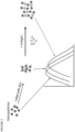
- the target analyte binds to the binding partners in the detection conjugates to form a complex between the first detection conjugate, the analyte, and the second detection conjugate.
- This complex formation brings the metallic nanostructures in the detection conjugates in close proximity to each other, i.e., plasmon-plasmon coupling.
- the amount of light that is absorbed, scattered, or transmitted by the metallic nanostructures is affected by the proximity of the metallic nanostructures in the complex and thus produces an enhanced shift in the peak absorption wavelength, which indicates the presence of the target analyte in the sample.
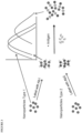
- the detection methods are competitive assays.
- the first detection conjugate comprises metallic nanostructures coupled to the target analyte of interest.
- the second detection conjugate is capable of specifically binding to the target analyte.
- the first detection conjugate will bind to the second detection conjugate initially. If a sample containing a target analyte is mixed with these initial complexes, the unlabeled or free target analyte in the sample will compete with the first detection conjugate for binding to the second detection conjugate.
- the change in optical signal in this type of assay will result from the displacement of the metallic nanostructures in the first detection conjugate from the second detection conjugate, which will proportionately reduce the wavelength shift in the peak absorption wavelength.
- the methods of the invention may utilize a plurality of detection conjugates.
- Detection conjugates comprise metallic nanostructures coupled to binding partners capable of specifically binding to a target analyte or another detection conjugate depending on the assay configuration.
- the detection conjugates comprise metallic nanostructures coupled or conjugated to binding partners that are capable of specifically binding a target analyte.
- at least one of the detection conjugates comprises metallic nanostructures coupled or conjugated to target analytes.
- the metallic nanostructure in the first detection conjugate and the second detection conjugate is a composite metallic nanostructure.
- the detection conjugates comprise binding partners that are capable of specifically binding to a target analyte.
- binding partners that are capable of specifically binding to a target analyte.
- specific binding refers to binding to a target molecule with high affinity, e.g., an affinity of at least 10 -6 M.
- the binding partners are haptens and other small molecules, drugs, hormones, biological macromolecules including, but not limited to, antibodies or fragments thereof (e.g., Fv, Fab, (Fab) 2 , single chain, CDR etc.), antigens, receptors, ligands, polynucleotides, aptamers, polypeptides, polysaccharides, lipopolysaccharides, glycopeptides, lipoproteins, or nucleoproteins.
- the binding partners are antibodies.
- the binding partners are antigens.
- the detection conjugates e.g., a first detection conjugate and a second detection conjugate
- a first detection conjugate and a second detection conjugate can both be antibodies that recognize a target analyte, but the epitope to which the first detection conjugate binds the target analyte is separate from and ideally non-overlapping with the epitope to which the second detection conjugate binds the target analyte.
- the first detection conjugate comprises an antibody that recognizes a first epitope of a target analyte and the second detection conjugate comprises a different antibody that recognizes a second epitope of a target analyte.
- the first detection conjugate may comprise a monoclonal antibody that recognizes a first epitope of a target analyte.
- the second detection conjugate may comprise a monoclonal antibody that recognizes a second epitope of a target analyte that is separate from and ideally non-overlapping with the epitope that is recognized by the first detection conjugate.
- the first detection conjugate and/or the second detection conjugate may comprise a polyclonal antibody.
- the first detection conjugate may comprise a polyclonal antibody while the second detection conjugate comprises a monoclonal antibody.
- the first detection conjugate comprises a polyclonal antibody and the second detection conjugate comprises a polyclonal antibody.
- the metallic nanostructures for use in the methods and devices of the invention are composite metallic nanostructures. Further provided for illustrative purposes only are metallic nanostructures in the detection conjugates composed of a noble metal or composite thereof. In some embodiments, the metallic nanostructures in the detection conjugates may be composed of a transition metal or composite thereof. In some embodiments, the metallic nanostructures in the detection conjugates may comprise an alkali metal or lanthanide in combination with a noble or transition metal.
- metallic nanostructures in the detection conjugates comprise a metal selected from gold, silver, copper, platinum, palladium, ruthenium, rhodium, osmium, iridium, titanium, chromium, cadmium, zinc, iron, cobalt, nickel, and composites thereof.
- the metallic nanostructures are gold nanostructures.
- the metallic nanostructures are silver nanostructures.
- the metallic nanostructures in the detection conjugates are composite metallic nanostructures.
- “Composite metallic nanostructures” refers to nanostructures that comprise at least two noble metals, transition metals, alkali metals, or lanthanides.
- the two or more metals may be mixed together, as in an alloy, or the two or more metals may be present in separate portions of the nanostructure. For example, one metal may form the core of the nanostructure, whereas the second metal forms an outer shell or coating of the nanostructure.
- the composite metallic nanostructures comprise at least two metals selected from gold, silver, copper, platinum, palladium, ruthenium, rhodium, osmium, iridium, titanium, chromium, cadmium, zinc, iron, cobalt, and nickel. In other embodiments, the composite metallic nanostructures comprise at least two metals selected from gold, silver, copper, platinum, palladium, cadmium, iron, nickel, and zinc. In one particular embodiment, the composite metallic nanostructures comprise gold and silver. In another embodiment, the composite metallic nanostructures comprise gold and copper. In yet another embodiment, the composite metallic nanostructures comprise silver and copper.
- the composite metallic nanostructures used in the methods of the invention can include a number of different geometries, such as spherical nanoparticles, pyramidal nanoparticles, hexagonal nanoparticles, nanotubes, nanostars, nanoshells, nanorods, nanodots, nanoislands, nanowires, nanodisks, nanocubes, or combinations thereof.
- the composite metallic nanostructure is selected from a nanostar and a nanorod.
- the core is silica
- the first coating i.e. inner coating
- the second coating is a gold coating (i.e. outer coating).
- the core is silica
- the first coating i.e. inner coating
- the second coating is a gold coating (i.e. outer coating).
- the size and shape of the metallic nanostructures are not uniform - i.e. the metallic nanostructures are a heterogeneous mixture of different shapes and sizes of nanostructures.
- the metallic nanostructures are nanostars.
- the metallic nanostructures are nanorods.
- the metallic nanostructures are composite nanospheres.
- suitable diameter ranges include from about 5 nm to about 200 nm, from about 10 nm to about 100 nm, and from about 20 nm to about 60 nm.
- suitable diameter ranges include from about 5 nm to about 50 nm, from about from about 8 nm to about 30 nm, and from about 10 nm to about 25 nm.
- suitable length ranges include from about 25 nm to about 150 nm, from about 40 nm to about 120 nm, and from about 50 nm to 100 nm.
- the aspect ratio, i.e., length/diameter, of the nanorods is between 2 and 10.
- edge lengths may be from about 10 nm to about 800 nm, from about 20 nm to about 500 nm, from about to 50 nm to about 200 nm, from about 30 nm to about 100 nm, or from about 10 nm to about 300 nm.
- the thickness of the nanoplates can range from about 1 to about 100 nm, from about 5 nm to about 80 nm, from about 10 nm to about 50 nm, or from about 5 nm to about 20 nm.
- the nanoplates have an aspect ratio greater than 2.
- the aspect ratio is the ratio of the edge length to the thickness.
- the nanoplates have an aspect ratio from about 2 to about 25, from about 3 to about 20, from about 5 to about 10, from about 2 to about 15, or from about 10 to about 30.
- conjugation chemistries such as those involving 1-Ethyl-3-[3-dimethylaminopropyl]carbodiimide hydrochloride (EDC), sulfo-NHS coupling, hydrophobic binding or thioether chemistry.
- EDC 1-Ethyl-3-[3-dimethylaminopropyl]carbodiimide hydrochloride
- sulfo-NHS coupling hydrophobic binding or thioether chemistry.
- the binding partners or target analytes can be coupled to the metallic nanostructures through various chemical functionalities including thiol, amine, dithiol, acrylic phosphoramidite, azide, or alkynes.
- the molecule can be coupled to the metallic nanostructure indirectly through a larger carrier molecule or protein.
- the carrier protein is not capable of specific interaction with the target analyte.
- protein A or protein G or protein A/G may be conjugated or coupled to the nanoparticles.
- the metal or metals employed in a first detection conjugate can be the same as the metal or metals from which the metallic nanostructures in the second detection conjugate are fabricated.
- the metallic nanostructures used in the claimed invention are composite metallic nanostructures.
- the first detection conjugate comprises gold nanostructures and the second detection conjugate comprise gold nanostructures.
- the metal employed in the first detection conjugate is different from the metal or metals used to create the metallic nanostructures in the second detection conjugate.
- the first detection conjugate comprises silver nanostructures and the second detection conjugate comprises gold nanostructures.
- the first detection conjugate comprises gold nanostructures and the second detection conjugate comprises silver nanostructures.
- the first detection conjugate comprises gold nanostructures and the second detection conjugate comprises composite nanostructures.
- the composite nanostructures comprise gold-coated silver nanostructures.
- the first detection conjugate comprises gold nanostructures and the second detection conjugate comprises composite nanostructures comprising gold-coated copper nanostructures.
- the first detection conjugate comprises gold nanostructures and the second detection conjugate comprises composite nanostructures comprising gold-coated magnetite nanostructures.
- the first detection conjugate comprises gold nanostructures and the second detection conjugate comprises composite nanostructures comprising gold and an alkali metal or lanthanide.
- the size of the metallic nanostructures used to create the first detection conjugate are similar to the size of the metallic nanostructures used in the second detection conjugate.
- matching the size of the two sets of nanostructures can provide an optimal wavelength shift in a reflectance, emission or scattering spectrum.
- the reaction environment may be adjusted with appropriate buffers, ionic strength, and other accelerants.
- the reaction environment comprises polyethylene glycol (PEG), which, as described herein, can enhance the strength of the LSPR signal.
- PEG polyethylene glycol
- Other similar polymeric materials may also be used, including, but not limited to, polyvinylpyrrolidone, polyallylamine, polyethyleneimine, polylysine, polyacrylic acid, polyvinylalcohol, and polyaspartic acid.
- the present invention also provides analyte detection devices for utilizing the methods of the invention to detect a target analyte in a sample, as recited by the claim set.
- Suitable analyte detection devices may include, but are not limited to, a spectrophotometric cuvette, an analytical rotor, a microwell plate, or a flow chamber.
- the tip of an optical fiber or a transparent gel may also be employed to carry out the detection methods disclosed herein.
- all components of the analyte detection devices described herein are contained within a centrifugal rotor or disc.
- a rotor or disc may contain one or more reaction chambers in which the plurality of detection conjugates is located.
- the detection conjugates are present in the form of lyophilized compositions, such as lyophilized beads or pellets.
- the analyte detection device comprises a rotor or disc having one or more reaction chambers, wherein each reaction chamber comprises a plurality of detection conjugates (e.g., a first detection conjugate and a second detection conjugate), wherein the detection conjugates are coupled to metallic nanoparticles.
- Such a device provides a one-step analyte detection assay whereby a test sample is contacted with the rotor or disc, and application of a centrifugal force to the rotor or disc delivers the test sample to the reaction chambers where the sample mixes with the first detection conjugate and the second detection conjugate.
- the detection conjugates can be selected such that a different analyte can be detected in each reaction chamber.
- These rotor-format detection devices can be configured in the sandwich assay format, the direct competitive format, or both if the rotors comprise multiple reaction chambers.
- the first detection conjugate comprises gold nanostructures and the metallic nanostructures in the second detection conjugate are gold nanostructures.
- the first detection conjugate comprises silver nanostructures and the metallic nanostructures in the second detection conjugate are gold nanostructures.
- the first detection conjugate comprises gold nanostructures and the second detection conjugate comprises composite nanostructures.
- the composite nanostructures are gold-coated silver nanostructures.
- the composite nanostructures are gold-coated copper nanostructures.
- kits comprising the analyte detection devices of the invention as disclosed herein.
- the kit comprises a plurality of detection conjugates (e.g., a first detection conjugate and a second detection conjugate), wherein the detection conjugates are coupled to metallic nanoparticles.
- one or more of the detection conjugates may be lyophilized, for example, in the form of a pellet or bead. In one embodiment, all of the detection conjugates are lyophilized.
- the kit may include one or more additional reagents. In some embodiments, one or more of the additional reagents is provided in lyophilized form.
- the kit may comprise a blocking agent, a sugar, a polymeric accelerant material, sodium chloride, and/or combinations thereof.
- a "blocking agent” is an agent that prevents the association of proteins present in the sample with the detectable agent and/or analyte. Blocking agents are typically proteins themselves and may include, but are not limited to, bovine serum albumin, casein, gelatin, ovalbumin, gamma-globulins, and IgG from non-immunized animals.
- the sugar is a polysaccharide.
- the polysaccharide is selected from maltodextrin, corn syrup, and polyglucose.
- the polysaccharide is maltodextrin.
- the sugar is trehalose.
- the reagent kit may comprise maltodextrin and trehalose.
- the polymeric accelerant material is PEG.
- kits may also include instructions for using the device to detect an analyte in a test sample, devices or tools for collecting biological samples, and/or extraction buffers for obtaining samples from solid materials, such as soil, food, and biological tissues.
- a test sample can be any type of liquid sample, including biological samples or extracts prepared from environmental or food samples.
- the test sample is a biological sample.
- Biological samples include, but are not limited to, whole blood, plasma, serum, saliva, urine, pleural effusion, sweat, bile, cerebrospinal fluid, fecal material, vaginal fluids, sperm, ocular lens fluid, mucous, synovial fluid, peritoneal fluid, amniotic fluid, biopsy tissues, saliva, and cellular lysates.
- the biological sample can be obtained from a human subject or animal subject suspected of having a disease condition, such as cancer, infectious diseases (e.g., viral-, bacterial-, parasitic- or fungal-infections), cardiovascular disease, metabolic disease, autoimmune disease etc.
- a disease condition such as cancer, infectious diseases (e.g., viral-, bacterial-, parasitic- or fungal-infections), cardiovascular disease, metabolic disease, autoimmune disease etc.
- infectious diseases e.g., viral-, bacterial-, parasitic- or fungal-infections
- cardiovascular disease e.g., bacterial-, parasitic- or fungal-infections
- metabolic disease e.g., autoimmune disease etc.
- the test sample is mixed with a first detection conjugate and the mixture is subsequently brought into contact with the second detection conjugate.
- the sample, the first detection conjugate, and the second detection conjugate are brought into contact at the same time. For instance, contact of the sample with both reagents simultaneously may occur in the rotor-format detection devices described herein.
- the present application relates to the use of composite nanomaterial labeled partners in solution to determine the binding of specific binding partners in a qualitative or quantitative manner.
- the present inventors have surprisingly found that the sensitivity of the solution-based assay is significantly enhanced when a polysaccharide, e.g., maltodextrin, is added to the solution as compared with the addition of other sugars such as sucrose, trehalose, or ficoll.
- a polysaccharide e.g., maltodextrin
- a polysaccharide e.g., maltodextrin
- the addition of a polysaccharide, e.g., maltodextrin, to the solution is particularly effective in preventing aggregation and sedimentation of the composite nanomaterial labeled partners, e.g., antibodies conjugated to gold-silver nanostars, during and after centrifugation.
- the improvement in sensitivity in relation to other sugars such as sucrose, trehalose, or ficoll was unexpected.
- the methods of the present invention are performed in a solution comprising a polysaccharide, e.g., maltodextrin, corn syrup, or polyglucose.
- the solution comprises a polysaccharide at a final concentration of about 2% to about 20% wt/vol. In another embodiment, the solution comprises a polysaccharide at a final concentration of about 4% to about 15% wt/vol. In yet another embodiment, the solution comprises a polysaccharide at a final concentration of about 5% to about 10% wt/vol. In an exemplary embodiment, the solution comprises a polysaccharide at a final concentration of about 5%, 6%, 7%, 8%, 9%, or 10%, inclusive of all values therebetween.
- the sensitivity of the assay may be improved when a polysaccharide is added to the solution as compared to an assay performed in a solution comprising an alternative sugar, e.g., sucrose or ficoll.
- the polysaccharide is maltodextrin.
- the solution comprises a blocking agent at a final concentration of about 0.1% to about 20% wt/vol. In another embodiment, the solution comprises a blocking agent at a final concentration of about 0.5% to about 10% wt/vol. In yet another embodiment, the solution comprises a blocking agent at a final concentration of about 1% to about 5% wt/vol. In an exemplary embodiment, the solution comprises a blocking agent at a final concentration of about 1%, 2%, 3%, 4%, or 5%, inclusive of all values therebetween. In various embodiments described herein, the sensitivity of the assay may be improved when a blocking agent is added to the solution as compared to an assay performed in the absence of a blocking agent. In some embodiments, the blocking agent is selected from bovine serum albumin, casein, gelatin, ovalbumin, and gamma-globulins. In an exemplary embodiment, the blocking agent is bovine serum albumin.
- the solution comprises one or more of maltodextrin, trehalose, PEG, a blocking agent (e.g. bovine serum albumin), and/or sodium chloride.
- a blocking agent e.g. bovine serum albumin
- one or more of the solution components may be provided as a lyophilized bead or pellet that is suspended upon the addition of a liquid, e.g., water, saline solution, or a liquid sample.
- a liquid e.g., water, saline solution, or a liquid sample.
- a liquid e.g., water, saline solution, or a liquid sample.
- one or more of the solution components may be provided in a spectrophotometric cuvette or a reaction chamber of an analytical rotor as a bead that is suspended into the solution following the addition of a liquid.
- the LSPR signal may be substantially increased by mixing the first and second detection conjugates with the analyte in the presence of a polymeric accelerant material selected from polyethylene glycol, polyvinylpyrrolidone, polyallylamine, polyethyleneimine, polylysine, polyacrylic acid, polyvinylalcohol, and polyaspartic acid.
- a polymeric accelerant material selected from polyethylene glycol, polyvinylpyrrolidone, polyallylamine, polyethyleneimine, polylysine, polyacrylic acid, polyvinylalcohol, and polyaspartic acid.
- the polymeric material is polyethylene glycol (PEG).
- the reaction mixture comprises a polymeric material, PEG, at a final concentration of about 0.1 mg/mL to about 200 mg/mL.
- the reaction mixture comprises a polymeric material, e.g., PEG, at a final concentration of about 0.2 mg/mL to about 100 ng/mL. In yet another embodiment, the reaction mixture comprises a polymeric material, e.g., PEG, at a final concentration of about 0.5 mg/mL to about 10 mg/mL. In yet another embodiment, the reaction mixture comprises a polymeric material, e.g., PEG, at a final concentration of about 2 mg/mL to about 8 mg/mL. In an exemplary embodiment, the reaction mixture comprises a polymeric material, e.g., PEG, at a final concentration of about 2, 3, 4, 5, 6, 7, or 8 mg/mL, inclusive of all values therebetween.
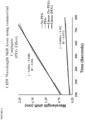
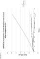
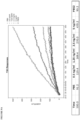
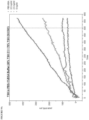
- the detection methods of the invention may be used to determine qualitative or quantitative amounts of a target analyte. Such methods are particularly useful for determining the approximate amount of a target analyte in a sample, which can be used inter alia to diagnose certain medical conditions or evaluate the efficacy of a drug therapy.
- the quantity of a target analyte can be determined by establishing a standard curve for the particular analyte by measuring changes in optical signals from the metallic nanoparticles as described herein for samples with a known quantity of target analyte; determining the optical signal change for a test sample; and comparing the optical signal change for the test sample to the values obtained for the standard curve.
- determining the quantity of a complex between a first reagent and a second reagent comprises comparing the absorbance ratio and/or reaction rate from a test sample to the absorbance ratio and/or reaction rate from one sample with a known quantity of complex, thereby determining the quantity of the complex in the test sample.
- the quantitative values obtained from test samples may be compared to pre-determined threshold values, wherein said pre-determined threshold values are indicative of either an abnormal or normal level of the target analyte.
- the detection methods of the present invention provide a highly sensitive technique for detecting minute quantities of a target analyte in a sample.
- amplification of plasmon resonance-based signals can be achieved with gold nanostructure conjugates such that nanogram quantities of target analyte can be detected in a sample.
- the presence of nanogram quantities of a target analyte is detected.
- plasmon resonance-based signals from detection conjugates comprising gold nanoparticles can be amplified using composite metallic nanostructure detection conjugates.
- Use of gold-coated silver nanostructures conjugated to an analyte-specific antibody may enable the detection of pictogram quantities of the target analyte. Accordingly, in some embodiments of the methods, the presence of picogram quantities of the target analyte is detected. In other embodiments of the methods, the presence of femtogram quantities of the target analyte is detected. Greater sensitivities may be obtained by altering the composition and/or shape of the composite metallic nanostructures.
- the optical properties of the metallic nanostructures depend on their size, shape, and composition.
- solid gold nanoparticles have an absorption peak wavelength ( ⁇ max ) from about 515 nm to about 560 nm depending on particle size.
- Gold spherical nanoparticles having a 30 nm diameter maximally absorb at about 520 nm with ⁇ max shifting to longer wavelengths as particle diameter increases.
- Silver and copper particles have a ⁇ max in the ultraviolet/blue or red region (e.g., from about 350 nm to about 500 nm) with increasing particle diameter causing a shift in ⁇ max to longer wavelengths.
- Metallic nanorods have a transverse ⁇ max1 and a longitudinal ⁇ max2 .
- Alloys of different metals typically exhibit absorption peaks in an intermediate range between the absorption peaks of the comprising metals.
- nanostructures comprising a 50/50 alloy of gold and silver exhibit a ⁇ max of about 480 nm with increasing amounts of gold causing a shift in the absorption peak to longer wavelengths.
- the sensitivity of the LSPR signals to changes in the local medium refractive index can be modified by changing the shape or geometry of the nanostructures. For instance, nonspherical particles (e.g. nanoprisms, nanorods, nanoshells, etc.) have increased LSPR sensitivities as compared to spheres.
- the optical properties e.g. absorption/scattering at particular wavelengths
- the interaction between the incident light and the metallic nanostructures can be monitored as reflected light or transmitted light.
- the amount of the incident light that is absorbed or scattered can be measured as an absorption spectrum in a reflection mode or the absorption spectrum in a transmission mode.
- the optical signal measured from the metallic nanostructures can be an optical reflection, an absorbance spectrum, a scattering spectrum, and/or an emission spectrum.
- the plasmon coupling between the metallic nanostructures in the detection conjugates resulting from complex formation between the binding partners and target analyte produces a change in the localized surface plasmon resonance spectrum of the metallic nanostructures.
- changes can include an increased optical extinction, an increased optical reflection, and/or increased scattering and/or emission signal.
- the change in optical signal indicative of the presence of the target analyte in the sample includes a shift, increase or decrease in optical scattering or a combination of these features.
- the change in optical signal indicative of the presence of the target analyte in the sample is a spectral peak wavelength shift.
- the wavelength shift in the optical spectral peak may be a blue shift (e.g., a shift to a shorter wavelength) within a 200 nm to 1200 nm spectral window.
- the changes in optical signals can be measured at a particular time point following a set reaction period. Additionally or alternatively, changes in the optical signal over the reaction period (e.g. rate determinations) may be measured. Both types of measurements can be used for either qualitative or quantitative analysis of a target analyte.
- spectrophotometric or photometric instruments are suitable for use in the disclosed methods.
- Some non-limiting examples include plate readers, Cobas Fara analyzers, and Piccolo xpress ® and Vetscan analyzers (Abaxis, Inc., Union City, CA), optic fiber readers (e.g., LightPath TM S4 (LamdaGen, Menlo Park, CA)), SPR instruments (e.g., Biacore instruments available from GE Healthcare), centrifugal analyzers from Olympus, Hitachi etc.
- target analyte can be detected using the methods, devices, and assay complexes of the present invention, particularly those that are significant in the diagnoses of diseases.
- a target analyte can include, but is not limited to, a protein, enzyme, antigen, antibody, peptide, nucleic acid (RNA, DNA, mRNA, miRNA), hormone, glycoprotein, polysaccharide, toxin, virus, virus particle, drug molecule, hapten, or chemical.
- the target analyte is a marker or antigen associated with an infectious disease in humans and/or animals.
- the target analyte is a marker or antigen associated with a particular physiological state or pathological condition.
- the target analyte is a pathogenic antigen or antibody to a pathogenic antigen.
- the pathogenic antigen can be a viral antigen (e.g., feline leukemia virus, canine parvovirus, foot and mouth virus, influenza virus, hepatitis a, b, c virus, HIV virus, human papilloma virus, Epstein Barr virus, rabies virus, etc.), a bacterial antigen (e.g., Ehrlichia, Borrelia, Anaplasma, Salmonella, Bacillus, Rickettsia, etc.), a fungal antigen, or parasitic antigen (e.g., canine heartworm, Giardia lamblia, plasmodium falciparum, African trypanosomiasis, Trypanosoma brucei, etc.).
- a viral antigen e.g., feline leukemia virus, canine parvovirus, foot and mouth virus, influenza virus, hepatitis a,
- the bacterial antigen may be from Ehrlichia canis, Ehrlichia chafeensis, Ehrlichia ewingii, Borrelia burgdorferi, Anaplasma platys, Anaplasma phagocytophilum, Salmonella enterica, Bacillus anthracis, and Rickettsia rickettsii.
- the target analyte is a disease-related antigen or antibody to a disease-related antigen.
- the target analyte is an inflammatory antigen or marker (e.g., C-reactive protein, MRP14, MRP8, 25F9, etc.).
- the target analyte is a pregnancy-related antigen or marker (e.g ., a fetal antigen, human chorionic gonadotropin).
- the present disclosure also provides, for illustrative purposes, a method for preparing composite metallic nanostructures.
- the method comprises preparing a first solution comprising a mixture of a polymer and chloroauric acid, preparing a second solution comprising silver or copper nanostructures, and incubating the first solution with the second solution for a period of time, wherein the resulting mixture comprises gold-coated silver nanostructures or gold-coated copper nanostructures.
- the resulting mixture preferably has a peak absorbance of about 515 nm to about 670 nm, or about 520 nm to about 560 nm. In one embodiment, the resulting mixture has a peak absorbance of about 530 nm to about 545 nm.
- the method comprises preparing a first solution comprising a mixture of a detergent such as CHAPS and chloroauric acid, and a solution comprising silver or copper salts, and incubating the first solution with the second solution containing a reducing agent, such as ascorbic acid leading to the formation of composite nanostructures.
- a reducing agent such as ascorbic acid leading to the formation of composite nanostructures.
- the size and shape of the nanostructures can be varied by changing the ratio of metals used, concentration of detergent and finally the amount of ascorbic acid used.
- the polymer used in the preparation of the first solution can be any one of polyvinylpyrrolidone, polyvinyl alcohol, polyacrylate, polyethylene glycol, polyethyleneimine, polyaspartic acid, polyglutamic acid, various gums, gelatin or mixed polymers comprising any of the foregoing.
- the polymer is polyvinylpyrrolidone.
- Different types of coated nanostructures can be obtained by varying the molecular weight of the polymer. Suitable molecular weight ranges of the polymer include from about 5,000 Daltons to about 150,000 Daltons, about 10,000 Daltons to about 100,000 Daltons, from about 20,000 Daltons to about 80,000 Daltons.
- the polymer has a molecular weight less than 50,000 Daltons.
- the polymer has a molecular weight less than 20,000 Daltons.
- the polymer has a molecular weight of about 10,000 Daltons.
- the characteristics of the gold coating can be controlled by adjusting the concentration ratio of polymer to chloroauric acid.
- the concentration ratio of polymer to chloroauric acid is from about 100:1 to about 1:100, from about 2:1 to about 5:1, or from about1.5:1 to about 8:1.
- the concentration ratio of polymer to chloroauric acid is 1:1.
- Suitable concentrations of polymer include, but are not limited to, about 0.1 % to about 20% wt/wet in water or ethanol.
- Suitable concentrations of chloroauric acid include, but are not limited to, about 0.001 M to about 1.0 M, about 0.010 M to about 0.500 M, and about 0.050 M to about 0.100 M.
- the second solution comprising silver or copper nanostructures can be prepared by any of the methods described in U.S. Patent Publication No. 2012/0101007 , U.S. Patent Publication No. 2014/0105982 , or U.S. Patent Publication No. 2013/0230717 .
- the second solution comprising silver or copper nanostructures is prepared by mixing a silver or copper source with a reducing agent.
- a suitable silver source includes a silver salt, such as silver nitrate.
- Suitable copper sources include copper (II) sulfate, copper (II) chloride, copper (II) hydroxide and copper (II) nitrate, copper (II) acetate and copper (II) trifluoroacetate.
- Reducing agents that can be reacted with the silver or copper sources to form the nanostructures can include glucose, ascorbic acid, sodium borohydride, and alkaline solutions ( e.g. pH greater than 7.5) of polymers such as PVP.
- the reducing agent is ascorbic acid.
- the desired shape and optical spectral peak of the silver nanostructures or copper nanostructures can be attained by adjusting the ratios or concentrations of reactants as known to those of ordinary skill in the art.
- the second solution comprising silver or copper nanostructures may have a peak absorbance from about 540 nm to about 1000 nm, from about 600 nm to about 700 nm, from about 630 nm to about 680 nm, from about 750 nm to about 850 nm, from about 900 nm to about 940 nm, from about 580 nm to about 620 nm, or from about 550 nm to about 750 nm.
- the incubation period of the first solution with second solution is at least 12 hours. In other embodiments, the incubation period of the first solution with second solution is greater than 24 hours, preferably greater than 48 hours, more preferably at least 72 hours. Changes in the peak absorbance of the reaction mixture can be monitored during the incubation period to adjust the incubation time accordingly. For example, shifts of the peak absorbance to shorter wavelengths, for instance in the 520 nm to 550 nm region, can indicate that the gold-coated nanostructures have stabilized. In certain embodiments, stability of the resulting nanostructures to sodium chloride (e.g., 0.25-1M) is used to indicate a proper coating of the nanostructures. CTAB-coated particles such as nanorods are resistant to sodium chloride.
- about 0.1 to 1 part of ascorbic acid e.g. about 1 to 5 M is added to the mixture following the first incubation period.
- the second incubation period following addition of the ascorbic acid may be from about 1 to about 24 hours.
- addition of ascorbic acid provides a substantial increase in the quantity of nanostructures produced.
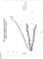
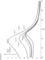
- Sandwich assays are most suitable where an analyte displays at least two distinct binding sites (epitopes of an antigen) with each site binding to a specific binding partner.
- an antibody directed towards one epitope of CRP is immobilized on the gold and/or silver nanoparticles and the second antibody directed towards a non-overlapping epitope is labeled with colloidal gold and/or silver.
- This setup allows measurement of the CRP antigen as the amount of CRP antigen in the sample determines the extent of spectral change. The spectral change is also seen when the second antibody is not labeled but the change is several orders of magnitude lower.
- the metallic composition of the nanoparticles may be changed to optimize the reaction conditions.
- Antibodies were attached to nanostars or nanoalloys using the following method.
- a suitable volume of nanostar or nanoalloy solution was centrifuged at a suitable g force. Supernatant was removed carefully and replaced with equal volume of 1% CHAPS.
- a 1:20 dilution in water was read for spectrum and OD at ⁇ max .
- water, 0.5M borate (pH 9.2), 1% CHAPS washed nanostars/nanoalloy from the step 1, and desired antibody was added, in that sequence.
Landscapes
- Health & Medical Sciences (AREA)
- Life Sciences & Earth Sciences (AREA)
- Immunology (AREA)
- Engineering & Computer Science (AREA)
- Chemical & Material Sciences (AREA)
- Urology & Nephrology (AREA)
- Hematology (AREA)
- Biomedical Technology (AREA)
- Molecular Biology (AREA)
- Medicinal Chemistry (AREA)
- Analytical Chemistry (AREA)
- Cell Biology (AREA)
- Pathology (AREA)
- Food Science & Technology (AREA)
- Biotechnology (AREA)
- Physics & Mathematics (AREA)
- Microbiology (AREA)
- Biochemistry (AREA)
- General Health & Medical Sciences (AREA)
- General Physics & Mathematics (AREA)
- Inorganic Chemistry (AREA)
- Investigating Or Analysing Materials By Optical Means (AREA)
- Investigating, Analyzing Materials By Fluorescence Or Luminescence (AREA)
- Peptides Or Proteins (AREA)
- Chemical Kinetics & Catalysis (AREA)
Priority Applications (1)
| Application Number | Priority Date | Filing Date | Title |
|---|---|---|---|
| EP24187227.4A EP4465021A3 (en) | 2015-08-04 | 2016-08-04 | Signal amplification in solution-based plasmonic specific-binding partner assays |
Applications Claiming Priority (2)
| Application Number | Priority Date | Filing Date | Title |
|---|---|---|---|
| US201562201051P | 2015-08-04 | 2015-08-04 | |
| PCT/US2016/045606 WO2017024163A1 (en) | 2015-08-04 | 2016-08-04 | Signal amplification in solution-based plasmonic specific-binding partner assays |
Related Child Applications (1)
| Application Number | Title | Priority Date | Filing Date |
|---|---|---|---|
| EP24187227.4A Division EP4465021A3 (en) | 2015-08-04 | 2016-08-04 | Signal amplification in solution-based plasmonic specific-binding partner assays |
Publications (3)
| Publication Number | Publication Date |
|---|---|
| EP3331818A1 EP3331818A1 (en) | 2018-06-13 |
| EP3331818A4 EP3331818A4 (en) | 2019-01-23 |
| EP3331818B1 true EP3331818B1 (en) | 2024-07-10 |
Family
ID=57943715
Family Applications (2)
| Application Number | Title | Priority Date | Filing Date |
|---|---|---|---|
| EP16833889.5A Active EP3331818B1 (en) | 2015-08-04 | 2016-08-04 | Signal amplification in solution-based plasmonic specific-binding partner assays |
| EP24187227.4A Pending EP4465021A3 (en) | 2015-08-04 | 2016-08-04 | Signal amplification in solution-based plasmonic specific-binding partner assays |
Family Applications After (1)
| Application Number | Title | Priority Date | Filing Date |
|---|---|---|---|
| EP24187227.4A Pending EP4465021A3 (en) | 2015-08-04 | 2016-08-04 | Signal amplification in solution-based plasmonic specific-binding partner assays |
Country Status (13)
| Country | Link |
|---|---|
| US (5) | US9835622B2 (enExample) |
| EP (2) | EP3331818B1 (enExample) |
| JP (4) | JP6944438B2 (enExample) |
| CN (2) | CN117434261A (enExample) |
| AU (2) | AU2016301375B2 (enExample) |
| CA (2) | CA3227799A1 (enExample) |
| DK (1) | DK3331818T3 (enExample) |
| ES (1) | ES2989755T3 (enExample) |
| FI (1) | FI3331818T3 (enExample) |
| HU (1) | HUE067974T2 (enExample) |
| PL (1) | PL3331818T3 (enExample) |
| PT (1) | PT3331818T (enExample) |
| WO (1) | WO2017024163A1 (enExample) |
Families Citing this family (9)
| Publication number | Priority date | Publication date | Assignee | Title |
|---|---|---|---|---|
| ES2702033T3 (es) | 2011-11-21 | 2019-02-27 | Abay Sa | Amplificación de la señal en inmunoensayos |
| TWI691716B (zh) | 2014-08-13 | 2020-04-21 | 美商艾巴希斯公司 | 電漿特異性結合搭配物檢定中之信號放大 |
| AU2016301375B2 (en) | 2015-08-04 | 2022-03-03 | Zoetis Services Llc | Signal amplification in solution-based plasmonic specific-binding partner assays |
| JP7308148B2 (ja) | 2017-01-30 | 2023-07-13 | ゾエティス サービシズ リミテッド ライアビリティ カンパニー | 溶液ベースのプラズモン特異的結合パートナーアッセイおよび金属ナノ構造体 |
| CN110865182B (zh) * | 2019-11-19 | 2023-06-27 | 东莞市东阳光诊断产品有限公司 | 一种阻断剂及其在免疫检测中的应用 |
| CN113376374A (zh) * | 2020-04-03 | 2021-09-10 | 上海桀蒙生物技术有限公司 | 病毒蛋白抗原检测工具的制备和使用方法 |
| EP4193148A4 (en) * | 2020-08-07 | 2024-11-06 | Vital Biosciences Inc. | KINETIC MODULATION FOR MAGNETIC ANALYTE DETECTION |
| US20220163522A1 (en) * | 2020-11-23 | 2022-05-26 | Arizona Board Of Regents On Behalf Of Arizona State University | Compositions and methods for rapid covid-19 detection |
| PL445690A1 (pl) * | 2023-07-27 | 2025-02-03 | Politechnika Gdańska | Układ do wykrywania białka w próbce materiału biologicznego w postaci płynnej i sposób do wykrywania białka realizowany za pomocą tego układu |
Citations (1)
| Publication number | Priority date | Publication date | Assignee | Title |
|---|---|---|---|---|
| US20060246513A1 (en) * | 2005-05-02 | 2006-11-02 | Bohannon Robert C | Method and device to detect the presence of analytes in a sample |
Family Cites Families (103)
| Publication number | Priority date | Publication date | Assignee | Title |
|---|---|---|---|---|
| US4704366A (en) | 1984-06-22 | 1987-11-03 | Bio-Rad Laboratories, Inc. | Process for binding IgG to protein A |
| US5122284A (en) | 1990-06-04 | 1992-06-16 | Abaxis, Inc. | Apparatus and method for optically analyzing biological fluids |
| US5061381A (en) | 1990-06-04 | 1991-10-29 | Abaxis, Inc. | Apparatus and method for separating cells from biological fluids |
| US5186844A (en) | 1991-04-01 | 1993-02-16 | Abaxis, Inc. | Apparatus and method for continuous centrifugal blood cell separation |
| US5413732A (en) * | 1991-08-19 | 1995-05-09 | Abaxis, Inc. | Reagent compositions for analytical testing |
| US5304348A (en) | 1992-02-11 | 1994-04-19 | Abaxis, Inc. | Reagent container for analytical rotor |
| WO1993019827A1 (en) | 1992-04-02 | 1993-10-14 | Abaxis, Inc. | Analytical rotor with dye mixing chamber |
| JP3706214B2 (ja) | 1996-10-28 | 2005-10-12 | 株式会社三菱化学ヤトロン | 免疫学的分析方法 |
| US5939021A (en) | 1997-01-23 | 1999-08-17 | Hansen; W. Peter | Homogeneous binding assay |
| ATE349697T1 (de) * | 1997-02-20 | 2007-01-15 | Univ California | Plasmon-schwingteilchen, methode und vorrichtung |
| US7144627B2 (en) | 1997-03-12 | 2006-12-05 | William Marsh Rice University | Multi-layer nanoshells comprising a metallic or conducting shell |
| US6344272B1 (en) | 1997-03-12 | 2002-02-05 | Wm. Marsh Rice University | Metal nanoshells |
| US6699724B1 (en) | 1998-03-11 | 2004-03-02 | Wm. Marsh Rice University | Metal nanoshells for biosensing applications |
| JP2000028612A (ja) | 1998-07-07 | 2000-01-28 | Nitto Denko Corp | 免疫学的検査方法および免疫学的検査キット |
| JP3841559B2 (ja) | 1998-07-07 | 2006-11-01 | 日東電工株式会社 | 免疫学的検査方法および免疫学的検査キット |
| JP3298836B2 (ja) | 1998-11-12 | 2002-07-08 | アークレイ株式会社 | 検体分析用具 |
| US6579726B1 (en) | 1999-07-30 | 2003-06-17 | Surromed, Inc. | Instruments, methods and reagents for surface plasmon resonance |
| WO2001035081A1 (en) * | 1999-11-12 | 2001-05-17 | Surromed, Inc. | Biosensing using surface plasmon resonance |
| WO2002079764A1 (en) | 2001-01-26 | 2002-10-10 | Nanoplex Technologies, Inc. | Surface-enhanced spectroscopy-active sandwich nanoparticles |
| AU2002246587A1 (en) | 2000-11-03 | 2002-08-06 | Wm. Marsh Rice University | Partial coverage metal nanoshells and method of making same |
| US7238472B2 (en) | 2001-05-25 | 2007-07-03 | Nanosphere, Inc. | Non-alloying core shell nanoparticles |
| WO2003025568A2 (de) * | 2001-09-14 | 2003-03-27 | Reinhard Zeidler | Identifizierung von antigenen durch xenogene, allogene oder autologe antikörper-vermittelte präzipitation |
| US7135054B2 (en) | 2001-09-26 | 2006-11-14 | Northwestern University | Nanoprisms and method of making them |
| CN1417586A (zh) | 2001-11-05 | 2003-05-14 | 福州市迈新生物技术开发公司 | 一种免疫检测信号放大系统和它的制备方法及其应用 |
| US6970239B2 (en) | 2002-06-12 | 2005-11-29 | Intel Corporation | Metal coated nanocrystalline silicon as an active surface enhanced Raman spectroscopy (SERS) substrate |
| US7212692B2 (en) | 2002-11-08 | 2007-05-01 | Ming Yan | Multiple array surface plasmon resonance biosensor |
| JP4787938B2 (ja) | 2003-03-28 | 2011-10-05 | ザ・プロウボウスト・フェロウズ・ファウンデーション・スカラーズ・アンド・ザ・アザー・メンバーズ・オブ・ボード・オブ・ザ・カレッジ・オブ・ザ・ホリー・アンド・アンデバイデッド・トリニティ・オブ・クイーン | 銀ナノ粒子を用いた検体検出用センサ |
| KR100531760B1 (ko) | 2003-04-28 | 2005-11-29 | 대한민국(관리부서 : 농림부 국립수의과학검역원) | 구제역 바이러스의 감염여부를 진단하는 방법 및 이를구현하기 위한 진단키트 |
| US20060240573A1 (en) | 2003-07-29 | 2006-10-26 | Lamdagen, Llc | Optical system including nanostructures for biological or chemical sensing |
| GB0321937D0 (en) | 2003-09-19 | 2003-10-22 | Univ Liverpool | Nanoparticle conjugates and method of production thereof |
| US7573946B2 (en) * | 2003-12-31 | 2009-08-11 | Intel Corporation | Apparatus and associated methods to perform space-frequency interleaving in a multicarrier wireless communication channel |
| JP4109205B2 (ja) * | 2004-01-07 | 2008-07-02 | 富士フイルム株式会社 | 被検体検出方法 |
| EP1836495A2 (en) | 2004-11-17 | 2007-09-26 | BioVeris Corporation | Electrochemiluminescent assay |
| CN1296492C (zh) | 2004-11-18 | 2007-01-24 | 博奥生物有限公司 | 一种基于生物芯片检测能结合特异序列的核酸结合蛋白的方法 |
| US7405054B1 (en) | 2004-12-13 | 2008-07-29 | University Of Washington Uw Tech Transfer - Invention Licensing | Signal amplification method for surface plasmon resonance-based chemical detection |
| US7485471B1 (en) * | 2004-12-17 | 2009-02-03 | Intel Corporation | Detection of enhanced multiplex signals by surface enhanced Raman spectroscopy |
| WO2006074076A1 (en) | 2004-12-31 | 2006-07-13 | Genentech, Inc. | Detecting human antibodies in non-human serum |
| US8628727B2 (en) | 2005-04-08 | 2014-01-14 | Northwestern University | Compositions, devices and methods for SERS and LSPR |
| US8101424B2 (en) * | 2005-06-15 | 2012-01-24 | University Of Maryland, Baltimore County | Bioassays using plasmonic scattering from noble metal nanostructures |
| US20070092978A1 (en) | 2005-10-20 | 2007-04-26 | Ronald Mink | Target ligand detection |
| JP5442179B2 (ja) * | 2005-10-21 | 2014-03-12 | アルフレッサファーマ株式会社 | 反応性物質が結合した微小粒子の沈降抑制方法および該微小粒子含有試薬 |
| AU2006347951B2 (en) | 2005-10-25 | 2011-12-22 | Louisiana Tech University Research Foundation | Multilayer films, coatings and microcapsules comprising polypeptides |
| CN101360997B (zh) | 2005-11-18 | 2013-05-01 | 美国政府健康及人类服务部,疾病控制和预防中心 | 改性心磷脂及其应用 |
| WO2007103802A2 (en) | 2006-03-03 | 2007-09-13 | William Marsh Rice University | Nanorice particles: hybrid plasmonic nanostructures |
| JP4810304B2 (ja) | 2006-05-12 | 2011-11-09 | キヤノン株式会社 | 化学センサ素子及びその製造方法 |
| US8426152B2 (en) * | 2007-01-03 | 2013-04-23 | Lamdagen Corporation | Enzymatic assay for LSPR |
| CA2981992C (en) | 2007-03-20 | 2020-06-30 | Becton, Dickinson And Company | Assays using surface-enhanced raman spectroscopy (sers)-active particles |
| JP2009150708A (ja) | 2007-12-19 | 2009-07-09 | Canon Inc | 標的物質の検出方法及び検査キット |
| JP2011508072A (ja) | 2007-12-21 | 2011-03-10 | ザ・プロヴォスト,フェローズ・アンド・スカラーズ・オブ・ザ・カレッジ・オブ・ザ・ホーリー・アンド・アンディヴァイデッド・トリニティー・オブ・クイーン・エリザベス,ニア・ダブリン | ナノ粒子の調製方法 |
| US20100279272A1 (en) * | 2008-02-13 | 2010-11-04 | Michael Craig Burrell | Multiplexed analysis methods using sers-active nanoparticles |
| US8137920B2 (en) | 2008-03-20 | 2012-03-20 | Abaxis, Inc. | Multi-wavelength analyses of sol-particle specific binding assays |
| US8530152B2 (en) * | 2008-07-10 | 2013-09-10 | Abaxis, Inc. | Methods of reducing non-specific interaction in metal nanoparticle assays |
| WO2010065781A2 (en) | 2008-12-03 | 2010-06-10 | Abaxis, Inc. | Lateral flow strip assay with immobilized conjugate |
| US8313915B2 (en) | 2009-01-21 | 2012-11-20 | Gundersen Lutheran Medical Foundation, Inc. | Early detection of canine lyme disease by specific peptides and antibodies |
| WO2010091293A1 (en) * | 2009-02-06 | 2010-08-12 | The Regents Of The University Of California | Plasmonic system for detecting binding of biological molecules |
| CA2753598C (en) | 2009-02-27 | 2018-07-10 | Beckman Coulter, Inc. | Solution phase homogeneous assays |
| WO2010116346A1 (en) | 2009-04-08 | 2010-10-14 | National University Of Ireland, Galway | Silver nanoplates |
| JP5563789B2 (ja) | 2009-06-11 | 2014-07-30 | 富士フイルム株式会社 | 検出方法 |
| US9274125B2 (en) | 2009-10-30 | 2016-03-01 | Kyowa Medex Co., Ltd. | Method and kit for measuring component in the presence of fatty acid alkanolamide or nonionic polyoxyethylene surfactant |
| KR20120096932A (ko) | 2009-11-17 | 2012-08-31 | 아박시스, 인크. | 라임병 항체의 검출을 위한 펩티드 및 방법 |
| ES2596952T3 (es) | 2009-11-20 | 2017-01-13 | Abaxis, Inc. | Péptidos, dispositivos y procedimientos para la detección de anticuerpos de Ehrlichia |
| WO2011065747A2 (ko) | 2009-11-24 | 2011-06-03 | 한국생명공학연구원 | 나노입자를 이용한 미생물의 고속 검출방법 |
| US9308582B2 (en) | 2009-12-24 | 2016-04-12 | Yi Sun | Solution stable and chemically reactive metallic nanoparticles |
| EP2354792A1 (en) | 2010-02-08 | 2011-08-10 | Biomonitor A/S | Method for detecting anti-drug antibodies |
| EP2564203B1 (en) | 2010-04-27 | 2017-06-07 | Ventana Medical Systems, Inc. | Antibody-nanoparticle conjugates and methods for making and using such conjugates |
| JP5728006B2 (ja) | 2010-05-28 | 2015-06-03 | 国立大学法人東京工業大学 | 金属微粒子複合体及びその製造方法 |
| US9797842B2 (en) * | 2010-12-08 | 2017-10-24 | Osaka Prefecture University Public Corporation | Device and method utilizing a metallic nanoparticle assembly structure for detecting a target substance |
| JP5614278B2 (ja) | 2010-12-24 | 2014-10-29 | セイコーエプソン株式会社 | センサーチップ、センサーチップの製造方法、検出装置 |
| CN102103145B (zh) | 2011-02-24 | 2013-01-16 | 南京基蛋生物科技有限公司 | 一种双放大系统的胶体金试纸条及其制备方法 |
| US8697129B2 (en) | 2011-03-02 | 2014-04-15 | Imra America, Inc. | Stable colloidal gold nanoparticles with controllable surface modification and functionalization |
| US8784895B2 (en) | 2011-03-15 | 2014-07-22 | Northwestern University | Multifunctional metal nanoparticles having a polydopamine-based surface and methods of making and using the same |
| JP5703098B2 (ja) * | 2011-03-31 | 2015-04-15 | 富士フイルム株式会社 | 生体分子検出装置および生体分子検出方法 |
| US20130230717A1 (en) | 2011-09-02 | 2013-09-05 | Washington University In St. Louis | Copper nanostructures and methods for their preparation |
| US8758772B2 (en) | 2011-11-04 | 2014-06-24 | Abaxis, Inc. | Peptides and methods for the detection of lyme disease antibodies |
| CH705758B1 (fr) | 2011-11-15 | 2016-03-31 | Metalor Technologies Int | Nanoparticules cœur-coquille métal-silice, procédé de fabrication et dispositif de test par immunochromatographie comprenant de telles nanoparticules. |
| ES2702033T3 (es) | 2011-11-21 | 2019-02-27 | Abay Sa | Amplificación de la señal en inmunoensayos |
| US9823246B2 (en) | 2011-12-28 | 2017-11-21 | The Board Of Trustees Of The Leland Stanford Junior University | Fluorescence enhancing plasmonic nanoscopic gold films and assays based thereon |
| WO2013109832A1 (en) | 2012-01-20 | 2013-07-25 | Imra America, Inc. | Stable colloidal suspensions of gold nanoconjugates and the method for preparing the same |
| US20150017258A1 (en) | 2012-01-31 | 2015-01-15 | American University Of Cairo (Auc) | Direct detection of disease biomarkers in clinical specimens using cationic nanoparticle-based assays & versatile and green methods for synthesis of anisotropic silver nanostructures |
| EP2625966A1 (en) * | 2012-02-13 | 2013-08-14 | Bionanoplus, S.L. | Nanoparticles comprising a vegetable hydrophobic protein and a water miscible non-volatile organic solvent and uses thereof |
| TWI435843B (zh) | 2012-04-27 | 2014-05-01 | Univ Nat Taiwan | 奈米顆粒的製造方法 |
| WO2013169640A1 (en) | 2012-05-05 | 2013-11-14 | Academia Sinica | Diaminobenzoic or ortho-phenylene diamine conjugated peptides as functional saccharide tagging reagents |
| WO2014020293A1 (en) | 2012-07-31 | 2014-02-06 | Imperial Innovations Limited | Assay |
| PL2906286T3 (pl) | 2012-10-11 | 2018-03-30 | Nanocomposix, Inc. | Kompozycje zawierające nanopłytki srebra i sposoby ich przygotowywania |
| US9651546B2 (en) | 2012-10-11 | 2017-05-16 | Abaxis, Inc. | Peptides, devices, and methods for the detection of ehrlichia antibodies |
| US9441301B2 (en) * | 2012-12-06 | 2016-09-13 | Nanyang Technological University | Method for forming a bimetallic core-shell nanostructure |
| CN105209616A (zh) | 2013-03-14 | 2015-12-30 | 雅培制药有限公司 | 用于改进的抗体检测的hcv ns3重组抗原及其突变体 |
| MX362075B (es) | 2013-03-14 | 2019-01-07 | Abbott Lab | Ensayo de combinación de antígeno-anticuerpo del virus de la hepatitis c (vhc) y métodos y composiciones para usarlo. |
| FR3003353B1 (fr) | 2013-03-14 | 2015-03-06 | Horiba Abx Sas | Nouvelle methode de dosage en flux d'un objet d'interet |
| JP2014228385A (ja) | 2013-05-22 | 2014-12-08 | キヤノン株式会社 | 免疫試験方法および免疫試験キット |
| US20160120978A1 (en) | 2013-06-05 | 2016-05-05 | Purdue Research Foundation | Titanium nitride plasmonic nanoparticles for clinical therapeutic applications |
| CN103335984B (zh) | 2013-06-09 | 2015-10-28 | 清华大学 | 一种基于lspr的无实体壁微阵列芯片及其应用 |
| EP3028043A4 (en) * | 2013-07-30 | 2017-04-19 | Bio-rad Laboratories, Inc. | Multiplex blocker beads for immunoassays |
| JP2015102337A (ja) * | 2013-11-21 | 2015-06-04 | 凸版印刷株式会社 | 粒子の単分散状態維持方法及び分析対象物の検出方法 |
| KR20160138059A (ko) | 2014-02-26 | 2016-12-02 | 람다젠 코퍼레이션 | 향상된 검정 감도를 위한 디지털 lspr |
| US10024800B2 (en) | 2014-04-15 | 2018-07-17 | Rutgers, The State University Of New Jersey | Gold nanostar substrates for SERS sensing in the femtomolar regime |
| US10518331B2 (en) | 2014-07-11 | 2019-12-31 | Northwestern University | Synthesis of uniform anisotropic nanoparticles |
| CN105492605A (zh) | 2014-08-01 | 2016-04-13 | 比科尔公司 | 用于免疫检验的病毒样颗粒、用于所述免疫检验的封闭剂、及包含它们的试剂盒 |
| TWI691716B (zh) | 2014-08-13 | 2020-04-21 | 美商艾巴希斯公司 | 電漿特異性結合搭配物檢定中之信號放大 |
| CA2976963A1 (en) | 2015-02-19 | 2016-08-25 | Ionica Sciences | Reagents and methods for detecting infectious diseases |
| WO2016170183A1 (en) | 2015-04-24 | 2016-10-27 | Pharmadiagnostics Nv | Improved method of measuring an interaction between molecules |
| AU2016301375B2 (en) | 2015-08-04 | 2022-03-03 | Zoetis Services Llc | Signal amplification in solution-based plasmonic specific-binding partner assays |
| JP7308148B2 (ja) | 2017-01-30 | 2023-07-13 | ゾエティス サービシズ リミテッド ライアビリティ カンパニー | 溶液ベースのプラズモン特異的結合パートナーアッセイおよび金属ナノ構造体 |
-
2016
- 2016-08-04 AU AU2016301375A patent/AU2016301375B2/en active Active
- 2016-08-04 PT PT168338895T patent/PT3331818T/pt unknown
- 2016-08-04 US US15/228,491 patent/US9835622B2/en active Active
- 2016-08-04 CA CA3227799A patent/CA3227799A1/en active Pending
- 2016-08-04 PL PL16833889.5T patent/PL3331818T3/pl unknown
- 2016-08-04 EP EP16833889.5A patent/EP3331818B1/en active Active
- 2016-08-04 CN CN202311388153.3A patent/CN117434261A/zh active Pending
- 2016-08-04 CA CA2991532A patent/CA2991532C/en active Active
- 2016-08-04 HU HUE16833889A patent/HUE067974T2/hu unknown
- 2016-08-04 JP JP2018505669A patent/JP6944438B2/ja active Active
- 2016-08-04 EP EP24187227.4A patent/EP4465021A3/en active Pending
- 2016-08-04 FI FIEP16833889.5T patent/FI3331818T3/fi active
- 2016-08-04 ES ES16833889T patent/ES2989755T3/es active Active
- 2016-08-04 CN CN201680045090.3A patent/CN107864670A/zh active Pending
- 2016-08-04 WO PCT/US2016/045606 patent/WO2017024163A1/en not_active Ceased
- 2016-08-04 DK DK16833889.5T patent/DK3331818T3/da active
-
2017
- 2017-10-31 US US15/799,320 patent/US10429383B2/en active Active
-
2019
- 2019-08-20 US US16/545,019 patent/US11215614B2/en active Active
-
2021
- 2021-09-10 JP JP2021147395A patent/JP7293296B2/ja active Active
- 2021-11-29 US US17/536,520 patent/US11614447B2/en active Active
-
2022
- 2022-06-02 AU AU2022203788A patent/AU2022203788B2/en active Active
-
2023
- 2023-02-24 US US18/174,251 patent/US20230333102A1/en active Pending
- 2023-06-07 JP JP2023093650A patent/JP7485834B2/ja active Active
-
2024
- 2024-05-02 JP JP2024074611A patent/JP2024099812A/ja active Pending
Patent Citations (1)
| Publication number | Priority date | Publication date | Assignee | Title |
|---|---|---|---|---|
| US20060246513A1 (en) * | 2005-05-02 | 2006-11-02 | Bohannon Robert C | Method and device to detect the presence of analytes in a sample |
Non-Patent Citations (1)
| Title |
|---|
| SHKILNYY ANDRIY ET AL: "Poly(ethylene glycol)-stabilized silver nanoparticles for bioanalytical applications of SERS spectroscopy", ANALYST, vol. 134, no. 9, 1 January 2009 (2009-01-01), UK, pages 1868, XP055965568, ISSN: 0003-2654, Retrieved from the Internet <URL:https://pubs.rsc.org/en/content/articlepdf/2009/an/b905694g> DOI: 10.1039/b905694g * |
Also Published As
Similar Documents
| Publication | Publication Date | Title |
|---|---|---|
| US11614447B2 (en) | Signal amplification in solution-based plasmonic specific-binding partner assays | |
| AU2022246447B2 (en) | Solution-based plasmon specific-binding partner assays and metallic nanostructures | |
| EP3180619B1 (en) | Signal amplification in plasmonic specific-binding partner assays | |
| HK1254535B (en) | Signal amplification in solution-based plasmonic specific-binding partner assays |
Legal Events
| Date | Code | Title | Description |
|---|---|---|---|
| STAA | Information on the status of an ep patent application or granted ep patent |
Free format text: STATUS: THE INTERNATIONAL PUBLICATION HAS BEEN MADE |
|
| PUAI | Public reference made under article 153(3) epc to a published international application that has entered the european phase |
Free format text: ORIGINAL CODE: 0009012 |
|
| STAA | Information on the status of an ep patent application or granted ep patent |
Free format text: STATUS: REQUEST FOR EXAMINATION WAS MADE |
|
| 17P | Request for examination filed |
Effective date: 20180117 |
|
| AK | Designated contracting states |
Kind code of ref document: A1 Designated state(s): AL AT BE BG CH CY CZ DE DK EE ES FI FR GB GR HR HU IE IS IT LI LT LU LV MC MK MT NL NO PL PT RO RS SE SI SK SM TR |
|
| AX | Request for extension of the european patent |
Extension state: BA ME |
|
| DAV | Request for validation of the european patent (deleted) | ||
| DAX | Request for extension of the european patent (deleted) | ||
| A4 | Supplementary search report drawn up and despatched |
Effective date: 20181221 |
|
| RIC1 | Information provided on ipc code assigned before grant |
Ipc: G01N 21/17 20060101ALI20181217BHEP Ipc: B82Y 30/00 20110101AFI20181217BHEP Ipc: G01N 21/55 20140101ALI20181217BHEP Ipc: B82Y 35/00 20110101ALI20181217BHEP |
|
| REG | Reference to a national code |
Ref country code: HK Ref legal event code: DE Ref document number: 1254535 Country of ref document: HK |
|
| STAA | Information on the status of an ep patent application or granted ep patent |
Free format text: STATUS: EXAMINATION IS IN PROGRESS |
|
| 17Q | First examination report despatched |
Effective date: 20200807 |
|
| RAP1 | Party data changed (applicant data changed or rights of an application transferred) |
Owner name: ZOETIS SERVICES LLC |
|
| GRAP | Despatch of communication of intention to grant a patent |
Free format text: ORIGINAL CODE: EPIDOSNIGR1 |
|
| STAA | Information on the status of an ep patent application or granted ep patent |
Free format text: STATUS: GRANT OF PATENT IS INTENDED |
|
| INTG | Intention to grant announced |
Effective date: 20240202 |
|
| GRAS | Grant fee paid |
Free format text: ORIGINAL CODE: EPIDOSNIGR3 |
|
| GRAA | (expected) grant |
Free format text: ORIGINAL CODE: 0009210 |
|
| STAA | Information on the status of an ep patent application or granted ep patent |
Free format text: STATUS: THE PATENT HAS BEEN GRANTED |
|
| P01 | Opt-out of the competence of the unified patent court (upc) registered |
Effective date: 20240529 |
|
| AK | Designated contracting states |
Kind code of ref document: B1 Designated state(s): AL AT BE BG CH CY CZ DE DK EE ES FI FR GB GR HR HU IE IS IT LI LT LU LV MC MK MT NL NO PL PT RO RS SE SI SK SM TR |
|
| REG | Reference to a national code |
Ref country code: CH Ref legal event code: EP |
|
| REG | Reference to a national code |
Ref country code: DE Ref legal event code: R096 Ref document number: 602016088367 Country of ref document: DE |
|
| REG | Reference to a national code |
Ref country code: DK Ref legal event code: T3 Effective date: 20240808 |
|
| REG | Reference to a national code |
Ref country code: NL Ref legal event code: FP |
|
| REG | Reference to a national code |
Ref country code: PT Ref legal event code: SC4A Ref document number: 3331818 Country of ref document: PT Date of ref document: 20240830 Kind code of ref document: T Free format text: AVAILABILITY OF NATIONAL TRANSLATION Effective date: 20240827 |
|
| REG | Reference to a national code |
Ref country code: SE Ref legal event code: TRGR |
|
| REG | Reference to a national code |
Ref country code: FI Ref legal event code: FGE |
|
| REG | Reference to a national code |
Ref country code: LT Ref legal event code: MG9D |
|
| REG | Reference to a national code |
Ref country code: GR Ref legal event code: EP Ref document number: 20240402087 Country of ref document: GR Effective date: 20241111 Ref country code: ES Ref legal event code: FG2A Ref document number: 2989755 Country of ref document: ES Kind code of ref document: T3 Effective date: 20241127 |
|
| REG | Reference to a national code |
Ref country code: HU Ref legal event code: AG4A Ref document number: E067974 Country of ref document: HU |
|
| REG | Reference to a national code |
Ref country code: AT Ref legal event code: UEP Ref document number: 1701933 Country of ref document: AT Kind code of ref document: T Effective date: 20240710 |
|
| PG25 | Lapsed in a contracting state [announced via postgrant information from national office to epo] |
Ref country code: BG Free format text: LAPSE BECAUSE OF FAILURE TO SUBMIT A TRANSLATION OF THE DESCRIPTION OR TO PAY THE FEE WITHIN THE PRESCRIBED TIME-LIMIT Effective date: 20240710 |
|
| PG25 | Lapsed in a contracting state [announced via postgrant information from national office to epo] |
Ref country code: LV Free format text: LAPSE BECAUSE OF FAILURE TO SUBMIT A TRANSLATION OF THE DESCRIPTION OR TO PAY THE FEE WITHIN THE PRESCRIBED TIME-LIMIT Effective date: 20240710 |
|
| PG25 | Lapsed in a contracting state [announced via postgrant information from national office to epo] |
Ref country code: IS Free format text: LAPSE BECAUSE OF FAILURE TO SUBMIT A TRANSLATION OF THE DESCRIPTION OR TO PAY THE FEE WITHIN THE PRESCRIBED TIME-LIMIT Effective date: 20241110 |
|
| PG25 | Lapsed in a contracting state [announced via postgrant information from national office to epo] |
Ref country code: HR Free format text: LAPSE BECAUSE OF FAILURE TO SUBMIT A TRANSLATION OF THE DESCRIPTION OR TO PAY THE FEE WITHIN THE PRESCRIBED TIME-LIMIT Effective date: 20240710 |
|
| PG25 | Lapsed in a contracting state [announced via postgrant information from national office to epo] |
Ref country code: RS Free format text: LAPSE BECAUSE OF FAILURE TO SUBMIT A TRANSLATION OF THE DESCRIPTION OR TO PAY THE FEE WITHIN THE PRESCRIBED TIME-LIMIT Effective date: 20241010 |
|
| PG25 | Lapsed in a contracting state [announced via postgrant information from national office to epo] |
Ref country code: RS Free format text: LAPSE BECAUSE OF FAILURE TO SUBMIT A TRANSLATION OF THE DESCRIPTION OR TO PAY THE FEE WITHIN THE PRESCRIBED TIME-LIMIT Effective date: 20241010 Ref country code: LV Free format text: LAPSE BECAUSE OF FAILURE TO SUBMIT A TRANSLATION OF THE DESCRIPTION OR TO PAY THE FEE WITHIN THE PRESCRIBED TIME-LIMIT Effective date: 20240710 Ref country code: IS Free format text: LAPSE BECAUSE OF FAILURE TO SUBMIT A TRANSLATION OF THE DESCRIPTION OR TO PAY THE FEE WITHIN THE PRESCRIBED TIME-LIMIT Effective date: 20241110 Ref country code: HR Free format text: LAPSE BECAUSE OF FAILURE TO SUBMIT A TRANSLATION OF THE DESCRIPTION OR TO PAY THE FEE WITHIN THE PRESCRIBED TIME-LIMIT Effective date: 20240710 Ref country code: BG Free format text: LAPSE BECAUSE OF FAILURE TO SUBMIT A TRANSLATION OF THE DESCRIPTION OR TO PAY THE FEE WITHIN THE PRESCRIBED TIME-LIMIT Effective date: 20240710 |
|
| REG | Reference to a national code |
Ref country code: DE Ref legal event code: R097 Ref document number: 602016088367 Country of ref document: DE |
|
| PG25 | Lapsed in a contracting state [announced via postgrant information from national office to epo] |
Ref country code: RO Free format text: LAPSE BECAUSE OF FAILURE TO SUBMIT A TRANSLATION OF THE DESCRIPTION OR TO PAY THE FEE WITHIN THE PRESCRIBED TIME-LIMIT Effective date: 20240710 Ref country code: SM Free format text: LAPSE BECAUSE OF FAILURE TO SUBMIT A TRANSLATION OF THE DESCRIPTION OR TO PAY THE FEE WITHIN THE PRESCRIBED TIME-LIMIT Effective date: 20240710 |
|
| PG25 | Lapsed in a contracting state [announced via postgrant information from national office to epo] |
Ref country code: LU Free format text: LAPSE BECAUSE OF NON-PAYMENT OF DUE FEES Effective date: 20240804 |
|
| PG25 | Lapsed in a contracting state [announced via postgrant information from national office to epo] |
Ref country code: MC Free format text: LAPSE BECAUSE OF FAILURE TO SUBMIT A TRANSLATION OF THE DESCRIPTION OR TO PAY THE FEE WITHIN THE PRESCRIBED TIME-LIMIT Effective date: 20240710 Ref country code: EE Free format text: LAPSE BECAUSE OF FAILURE TO SUBMIT A TRANSLATION OF THE DESCRIPTION OR TO PAY THE FEE WITHIN THE PRESCRIBED TIME-LIMIT Effective date: 20240710 |
|
| PG25 | Lapsed in a contracting state [announced via postgrant information from national office to epo] |
Ref country code: CZ Free format text: LAPSE BECAUSE OF FAILURE TO SUBMIT A TRANSLATION OF THE DESCRIPTION OR TO PAY THE FEE WITHIN THE PRESCRIBED TIME-LIMIT Effective date: 20240710 |
|
| PG25 | Lapsed in a contracting state [announced via postgrant information from national office to epo] |
Ref country code: SK Free format text: LAPSE BECAUSE OF FAILURE TO SUBMIT A TRANSLATION OF THE DESCRIPTION OR TO PAY THE FEE WITHIN THE PRESCRIBED TIME-LIMIT Effective date: 20240710 |
|
| PLBE | No opposition filed within time limit |
Free format text: ORIGINAL CODE: 0009261 |
|
| STAA | Information on the status of an ep patent application or granted ep patent |
Free format text: STATUS: NO OPPOSITION FILED WITHIN TIME LIMIT |
|
| 26N | No opposition filed |
Effective date: 20250411 |
|
| PG25 | Lapsed in a contracting state [announced via postgrant information from national office to epo] |
Ref country code: IT Free format text: LAPSE BECAUSE OF NON-PAYMENT OF DUE FEES Effective date: 20240804 |
|
| PGFP | Annual fee paid to national office [announced via postgrant information from national office to epo] |
Ref country code: PT Payment date: 20250625 Year of fee payment: 10 |
|
| PGFP | Annual fee paid to national office [announced via postgrant information from national office to epo] |
Ref country code: NL Payment date: 20250711 Year of fee payment: 10 |
|
| PGFP | Annual fee paid to national office [announced via postgrant information from national office to epo] |
Ref country code: HU Payment date: 20250804 Year of fee payment: 10 |
|
| PGFP | Annual fee paid to national office [announced via postgrant information from national office to epo] |
Ref country code: FI Payment date: 20250729 Year of fee payment: 10 Ref country code: ES Payment date: 20250908 Year of fee payment: 10 |
|
| PGFP | Annual fee paid to national office [announced via postgrant information from national office to epo] |
Ref country code: DE Payment date: 20250709 Year of fee payment: 10 Ref country code: DK Payment date: 20250729 Year of fee payment: 10 |
|
| PGFP | Annual fee paid to national office [announced via postgrant information from national office to epo] |
Ref country code: GR Payment date: 20250728 Year of fee payment: 10 Ref country code: NO Payment date: 20250728 Year of fee payment: 10 |
|
| PGFP | Annual fee paid to national office [announced via postgrant information from national office to epo] |
Ref country code: PL Payment date: 20250710 Year of fee payment: 10 Ref country code: TR Payment date: 20250717 Year of fee payment: 10 |
|
| PGFP | Annual fee paid to national office [announced via postgrant information from national office to epo] |
Ref country code: BE Payment date: 20250710 Year of fee payment: 10 Ref country code: GB Payment date: 20250710 Year of fee payment: 10 |
|
| PGFP | Annual fee paid to national office [announced via postgrant information from national office to epo] |
Ref country code: FR Payment date: 20250709 Year of fee payment: 10 Ref country code: AT Payment date: 20250728 Year of fee payment: 10 |
|
| PGFP | Annual fee paid to national office [announced via postgrant information from national office to epo] |
Ref country code: CH Payment date: 20250901 Year of fee payment: 10 Ref country code: SE Payment date: 20250808 Year of fee payment: 10 |
|
| PGFP | Annual fee paid to national office [announced via postgrant information from national office to epo] |
Ref country code: IE Payment date: 20250709 Year of fee payment: 10 |