EP3237906B1 - Befestigung von proteinen an schnittstellen zur verwendung in nicht-linearer optischer detektion - Google Patents
Befestigung von proteinen an schnittstellen zur verwendung in nicht-linearer optischer detektion Download PDFInfo
- Publication number
- EP3237906B1 EP3237906B1 EP15874284.1A EP15874284A EP3237906B1 EP 3237906 B1 EP3237906 B1 EP 3237906B1 EP 15874284 A EP15874284 A EP 15874284A EP 3237906 B1 EP3237906 B1 EP 3237906B1
- Authority
- EP
- European Patent Office
- Prior art keywords
- protein
- mole percent
- shg
- aspects
- lipid bilayer
- Prior art date
- Legal status (The legal status is an assumption and is not a legal conclusion. Google has not performed a legal analysis and makes no representation as to the accuracy of the status listed.)
- Active
Links
- 102000004169 proteins and genes Human genes 0.000 title claims description 213
- 108090000623 proteins and genes Proteins 0.000 title claims description 213
- 230000003287 optical effect Effects 0.000 title claims description 91
- 238000001514 detection method Methods 0.000 title description 17
- 238000000034 method Methods 0.000 claims description 119
- 239000000232 Lipid Bilayer Substances 0.000 claims description 88
- 239000000758 substrate Substances 0.000 claims description 69
- 238000009739 binding Methods 0.000 claims description 66
- 230000027455 binding Effects 0.000 claims description 65
- 230000008859 change Effects 0.000 claims description 63
- 101710175625 Maltose/maltodextrin-binding periplasmic protein Proteins 0.000 claims description 54
- 108010022394 Threonine synthase Proteins 0.000 claims description 52
- 102000004419 dihydrofolate reductase Human genes 0.000 claims description 52
- -1 antibodies Proteins 0.000 claims description 39
- YBJHBAHKTGYVGT-ZKWXMUAHSA-N (+)-Biotin Chemical compound N1C(=O)N[C@@H]2[C@H](CCCCC(=O)O)SC[C@@H]21 YBJHBAHKTGYVGT-ZKWXMUAHSA-N 0.000 claims description 36
- 239000003446 ligand Substances 0.000 claims description 36
- 125000000487 histidyl group Chemical group [H]N([H])C(C(=O)O*)C([H])([H])C1=C([H])N([H])C([H])=N1 0.000 claims description 31
- 108010041952 Calmodulin Proteins 0.000 claims description 29
- 102000000584 Calmodulin Human genes 0.000 claims description 29
- 108010090804 Streptavidin Proteins 0.000 claims description 26
- PXHVJJICTQNCMI-UHFFFAOYSA-N nickel Substances [Ni] PXHVJJICTQNCMI-UHFFFAOYSA-N 0.000 claims description 24
- 108090000765 processed proteins & peptides Proteins 0.000 claims description 24
- 229960002685 biotin Drugs 0.000 claims description 22
- 239000011616 biotin Substances 0.000 claims description 22
- 102000004190 Enzymes Human genes 0.000 claims description 20
- 108090000790 Enzymes Proteins 0.000 claims description 20
- 235000020958 biotin Nutrition 0.000 claims description 18
- 229910052759 nickel Inorganic materials 0.000 claims description 18
- 108020003175 receptors Proteins 0.000 claims description 17
- 102000005962 receptors Human genes 0.000 claims description 17
- SYFQYGMJENQVQT-UHFFFAOYSA-N 6-amino-2-[bis(carboxymethyl)amino]hexanoic acid Chemical compound NCCCCC(C(O)=O)N(CC(O)=O)CC(O)=O SYFQYGMJENQVQT-UHFFFAOYSA-N 0.000 claims description 14
- 125000002730 succinyl group Chemical group C(CCC(=O)*)(=O)* 0.000 claims description 14
- 102000004196 processed proteins & peptides Human genes 0.000 claims description 12
- 150000003384 small molecules Chemical class 0.000 claims description 12
- SNKAWJBJQDLSFF-NVKMUCNASA-N 1,2-dioleoyl-sn-glycero-3-phosphocholine Chemical compound CCCCCCCC\C=C/CCCCCCCC(=O)OC[C@H](COP([O-])(=O)OCC[N+](C)(C)C)OC(=O)CCCCCCC\C=C/CCCCCCCC SNKAWJBJQDLSFF-NVKMUCNASA-N 0.000 claims description 11
- 108091034117 Oligonucleotide Proteins 0.000 claims description 11
- JLCPHMBAVCMARE-UHFFFAOYSA-N [3-[[3-[[3-[[3-[[3-[[3-[[3-[[3-[[3-[[3-[[3-[[5-(2-amino-6-oxo-1H-purin-9-yl)-3-[[3-[[3-[[3-[[3-[[3-[[5-(2-amino-6-oxo-1H-purin-9-yl)-3-[[5-(2-amino-6-oxo-1H-purin-9-yl)-3-hydroxyoxolan-2-yl]methoxy-hydroxyphosphoryl]oxyoxolan-2-yl]methoxy-hydroxyphosphoryl]oxy-5-(5-methyl-2,4-dioxopyrimidin-1-yl)oxolan-2-yl]methoxy-hydroxyphosphoryl]oxy-5-(6-aminopurin-9-yl)oxolan-2-yl]methoxy-hydroxyphosphoryl]oxy-5-(6-aminopurin-9-yl)oxolan-2-yl]methoxy-hydroxyphosphoryl]oxy-5-(6-aminopurin-9-yl)oxolan-2-yl]methoxy-hydroxyphosphoryl]oxy-5-(6-aminopurin-9-yl)oxolan-2-yl]methoxy-hydroxyphosphoryl]oxyoxolan-2-yl]methoxy-hydroxyphosphoryl]oxy-5-(5-methyl-2,4-dioxopyrimidin-1-yl)oxolan-2-yl]methoxy-hydroxyphosphoryl]oxy-5-(4-amino-2-oxopyrimidin-1-yl)oxolan-2-yl]methoxy-hydroxyphosphoryl]oxy-5-(5-methyl-2,4-dioxopyrimidin-1-yl)oxolan-2-yl]methoxy-hydroxyphosphoryl]oxy-5-(5-methyl-2,4-dioxopyrimidin-1-yl)oxolan-2-yl]methoxy-hydroxyphosphoryl]oxy-5-(6-aminopurin-9-yl)oxolan-2-yl]methoxy-hydroxyphosphoryl]oxy-5-(6-aminopurin-9-yl)oxolan-2-yl]methoxy-hydroxyphosphoryl]oxy-5-(4-amino-2-oxopyrimidin-1-yl)oxolan-2-yl]methoxy-hydroxyphosphoryl]oxy-5-(4-amino-2-oxopyrimidin-1-yl)oxolan-2-yl]methoxy-hydroxyphosphoryl]oxy-5-(4-amino-2-oxopyrimidin-1-yl)oxolan-2-yl]methoxy-hydroxyphosphoryl]oxy-5-(6-aminopurin-9-yl)oxolan-2-yl]methoxy-hydroxyphosphoryl]oxy-5-(4-amino-2-oxopyrimidin-1-yl)oxolan-2-yl]methyl [5-(6-aminopurin-9-yl)-2-(hydroxymethyl)oxolan-3-yl] hydrogen phosphate Polymers Cc1cn(C2CC(OP(O)(=O)OCC3OC(CC3OP(O)(=O)OCC3OC(CC3O)n3cnc4c3nc(N)[nH]c4=O)n3cnc4c3nc(N)[nH]c4=O)C(COP(O)(=O)OC3CC(OC3COP(O)(=O)OC3CC(OC3COP(O)(=O)OC3CC(OC3COP(O)(=O)OC3CC(OC3COP(O)(=O)OC3CC(OC3COP(O)(=O)OC3CC(OC3COP(O)(=O)OC3CC(OC3COP(O)(=O)OC3CC(OC3COP(O)(=O)OC3CC(OC3COP(O)(=O)OC3CC(OC3COP(O)(=O)OC3CC(OC3COP(O)(=O)OC3CC(OC3COP(O)(=O)OC3CC(OC3COP(O)(=O)OC3CC(OC3COP(O)(=O)OC3CC(OC3COP(O)(=O)OC3CC(OC3COP(O)(=O)OC3CC(OC3CO)n3cnc4c(N)ncnc34)n3ccc(N)nc3=O)n3cnc4c(N)ncnc34)n3ccc(N)nc3=O)n3ccc(N)nc3=O)n3ccc(N)nc3=O)n3cnc4c(N)ncnc34)n3cnc4c(N)ncnc34)n3cc(C)c(=O)[nH]c3=O)n3cc(C)c(=O)[nH]c3=O)n3ccc(N)nc3=O)n3cc(C)c(=O)[nH]c3=O)n3cnc4c3nc(N)[nH]c4=O)n3cnc4c(N)ncnc34)n3cnc4c(N)ncnc34)n3cnc4c(N)ncnc34)n3cnc4c(N)ncnc34)O2)c(=O)[nH]c1=O JLCPHMBAVCMARE-UHFFFAOYSA-N 0.000 claims description 11
- 108090001008 Avidin Proteins 0.000 claims description 10
- 235000014633 carbohydrates Nutrition 0.000 claims description 10
- 150000001720 carbohydrates Chemical class 0.000 claims description 10
- 108010087904 neutravidin Proteins 0.000 claims description 7
- 150000002148 esters Chemical class 0.000 claims description 6
- 102000005701 Calcium-Binding Proteins Human genes 0.000 claims description 5
- 108010045403 Calcium-Binding Proteins Proteins 0.000 claims description 5
- 235000014113 dietary fatty acids Nutrition 0.000 claims description 3
- 239000000194 fatty acid Substances 0.000 claims description 3
- 229930195729 fatty acid Natural products 0.000 claims description 3
- 229920001223 polyethylene glycol Polymers 0.000 claims description 2
- 239000002202 Polyethylene glycol Substances 0.000 claims 1
- 235000018102 proteins Nutrition 0.000 description 201
- 239000000872 buffer Substances 0.000 description 47
- 238000007792 addition Methods 0.000 description 44
- RAXXELZNTBOGNW-UHFFFAOYSA-N imidazole Natural products C1=CNC=N1 RAXXELZNTBOGNW-UHFFFAOYSA-N 0.000 description 36
- 150000002632 lipids Chemical class 0.000 description 35
- FBOZXECLQNJBKD-ZDUSSCGKSA-N L-methotrexate Chemical compound C=1N=C2N=C(N)N=C(N)C2=NC=1CN(C)C1=CC=C(C(=O)N[C@@H](CCC(O)=O)C(O)=O)C=C1 FBOZXECLQNJBKD-ZDUSSCGKSA-N 0.000 description 28
- 229960000485 methotrexate Drugs 0.000 description 28
- 238000002372 labelling Methods 0.000 description 27
- 238000002474 experimental method Methods 0.000 description 25
- OWEGMIWEEQEYGQ-UHFFFAOYSA-N 100676-05-9 Natural products OC1C(O)C(O)C(CO)OC1OCC1C(O)C(O)C(O)C(OC2C(OC(O)C(O)C2O)CO)O1 OWEGMIWEEQEYGQ-UHFFFAOYSA-N 0.000 description 22
- GUBGYTABKSRVRQ-PICCSMPSSA-N Maltose Natural products O[C@@H]1[C@@H](O)[C@H](O)[C@@H](CO)O[C@@H]1O[C@@H]1[C@@H](CO)OC(O)[C@H](O)[C@H]1O GUBGYTABKSRVRQ-PICCSMPSSA-N 0.000 description 22
- 230000005284 excitation Effects 0.000 description 22
- 108091006054 His-tagged proteins Proteins 0.000 description 21
- 238000011534 incubation Methods 0.000 description 21
- 239000011521 glass Substances 0.000 description 20
- 108091006004 biotinylated proteins Proteins 0.000 description 18
- 239000000523 sample Substances 0.000 description 16
- VYPSYNLAJGMNEJ-UHFFFAOYSA-N Silicium dioxide Chemical compound O=[Si]=O VYPSYNLAJGMNEJ-UHFFFAOYSA-N 0.000 description 15
- FAPWRFPIFSIZLT-UHFFFAOYSA-M Sodium chloride Chemical compound [Na+].[Cl-] FAPWRFPIFSIZLT-UHFFFAOYSA-M 0.000 description 15
- 210000004027 cell Anatomy 0.000 description 15
- 230000033001 locomotion Effects 0.000 description 15
- 238000004949 mass spectrometry Methods 0.000 description 15
- IEDVJHCEMCRBQM-UHFFFAOYSA-N trimethoprim Chemical compound COC1=C(OC)C(OC)=CC(CC=2C(=NC(N)=NC=2)N)=C1 IEDVJHCEMCRBQM-UHFFFAOYSA-N 0.000 description 15
- 230000003993 interaction Effects 0.000 description 14
- 239000000463 material Substances 0.000 description 14
- 229960001082 trimethoprim Drugs 0.000 description 14
- 239000000975 dye Substances 0.000 description 13
- 238000005259 measurement Methods 0.000 description 13
- 238000013459 approach Methods 0.000 description 12
- 239000013078 crystal Substances 0.000 description 12
- 238000002347 injection Methods 0.000 description 12
- 239000007924 injection Substances 0.000 description 12
- 235000018977 lysine Nutrition 0.000 description 12
- 229910021645 metal ion Inorganic materials 0.000 description 12
- BHPQYMZQTOCNFJ-UHFFFAOYSA-N Calcium cation Chemical compound [Ca+2] BHPQYMZQTOCNFJ-UHFFFAOYSA-N 0.000 description 11
- PEDCQBHIVMGVHV-UHFFFAOYSA-N Glycerine Chemical compound OCC(O)CO PEDCQBHIVMGVHV-UHFFFAOYSA-N 0.000 description 11
- GUBGYTABKSRVRQ-QKKXKWKRSA-N Lactose Natural products OC[C@H]1O[C@@H](O[C@H]2[C@H](O)[C@@H](O)C(O)O[C@@H]2CO)[C@H](O)[C@@H](O)[C@H]1O GUBGYTABKSRVRQ-QKKXKWKRSA-N 0.000 description 11
- 239000011575 calcium Substances 0.000 description 11
- 229910001424 calcium ion Inorganic materials 0.000 description 11
- 235000018417 cysteine Nutrition 0.000 description 11
- 239000005350 fused silica glass Substances 0.000 description 11
- 239000008101 lactose Substances 0.000 description 11
- 239000002245 particle Substances 0.000 description 11
- 108091032973 (ribonucleotides)n+m Proteins 0.000 description 10
- 108020004414 DNA Proteins 0.000 description 10
- 150000001412 amines Chemical class 0.000 description 10
- 238000004458 analytical method Methods 0.000 description 10
- 239000003153 chemical reaction reagent Substances 0.000 description 10
- 229920000642 polymer Polymers 0.000 description 10
- 238000012360 testing method Methods 0.000 description 10
- OYPRJOBELJOOCE-UHFFFAOYSA-N Calcium Chemical compound [Ca] OYPRJOBELJOOCE-UHFFFAOYSA-N 0.000 description 9
- KDXKERNSBIXSRK-YFKPBYRVSA-N L-lysine Chemical compound NCCCC[C@H](N)C(O)=O KDXKERNSBIXSRK-YFKPBYRVSA-N 0.000 description 9
- 239000012131 assay buffer Substances 0.000 description 9
- 239000011324 bead Substances 0.000 description 9
- 229910052791 calcium Inorganic materials 0.000 description 9
- 238000010168 coupling process Methods 0.000 description 9
- XUJNEKJLAYXESH-UHFFFAOYSA-N cysteine Natural products SCC(N)C(O)=O XUJNEKJLAYXESH-UHFFFAOYSA-N 0.000 description 9
- 230000007423 decrease Effects 0.000 description 9
- 230000003100 immobilizing effect Effects 0.000 description 9
- KDXKERNSBIXSRK-UHFFFAOYSA-N Lysine Natural products NCCCCC(N)C(O)=O KDXKERNSBIXSRK-UHFFFAOYSA-N 0.000 description 8
- 239000004472 Lysine Substances 0.000 description 8
- 150000001875 compounds Chemical class 0.000 description 8
- 230000021615 conjugation Effects 0.000 description 8
- 230000008878 coupling Effects 0.000 description 8
- 238000005859 coupling reaction Methods 0.000 description 8
- 230000000694 effects Effects 0.000 description 8
- 125000003588 lysine group Chemical group [H]N([H])C([H])([H])C([H])([H])C([H])([H])C([H])([H])C([H])(N([H])[H])C(*)=O 0.000 description 8
- 230000004048 modification Effects 0.000 description 8
- 238000012986 modification Methods 0.000 description 8
- 239000011780 sodium chloride Substances 0.000 description 8
- 230000015572 biosynthetic process Effects 0.000 description 7
- 238000006243 chemical reaction Methods 0.000 description 7
- 239000012528 membrane Substances 0.000 description 7
- 229910052751 metal Inorganic materials 0.000 description 7
- 239000002184 metal Substances 0.000 description 7
- 239000000725 suspension Substances 0.000 description 7
- HEDRZPFGACZZDS-UHFFFAOYSA-N Chloroform Chemical compound ClC(Cl)Cl HEDRZPFGACZZDS-UHFFFAOYSA-N 0.000 description 6
- 241000588724 Escherichia coli Species 0.000 description 6
- 238000003556 assay Methods 0.000 description 6
- 230000008901 benefit Effects 0.000 description 6
- 239000000203 mixture Substances 0.000 description 6
- 230000008569 process Effects 0.000 description 6
- 238000010494 dissociation reaction Methods 0.000 description 5
- 230000005593 dissociations Effects 0.000 description 5
- 239000012530 fluid Substances 0.000 description 5
- 238000002360 preparation method Methods 0.000 description 5
- 241000894007 species Species 0.000 description 5
- QKNYBSVHEMOAJP-UHFFFAOYSA-N 2-amino-2-(hydroxymethyl)propane-1,3-diol;hydron;chloride Chemical compound Cl.OCC(N)(CO)CO QKNYBSVHEMOAJP-UHFFFAOYSA-N 0.000 description 4
- 108091003079 Bovine Serum Albumin Proteins 0.000 description 4
- 108010008281 Recombinant Fusion Proteins Proteins 0.000 description 4
- 102000007056 Recombinant Fusion Proteins Human genes 0.000 description 4
- 239000007983 Tris buffer Substances 0.000 description 4
- XJLXINKUBYWONI-DQQFMEOOSA-N [[(2r,3r,4r,5r)-5-(6-aminopurin-9-yl)-3-hydroxy-4-phosphonooxyoxolan-2-yl]methoxy-hydroxyphosphoryl] [(2s,3r,4s,5s)-5-(3-carbamoylpyridin-1-ium-1-yl)-3,4-dihydroxyoxolan-2-yl]methyl phosphate Chemical compound NC(=O)C1=CC=C[N+]([C@@H]2[C@H]([C@@H](O)[C@H](COP([O-])(=O)OP(O)(=O)OC[C@@H]3[C@H]([C@@H](OP(O)(O)=O)[C@@H](O3)N3C4=NC=NC(N)=C4N=C3)O)O2)O)=C1 XJLXINKUBYWONI-DQQFMEOOSA-N 0.000 description 4
- 238000010521 absorption reaction Methods 0.000 description 4
- 238000007413 biotinylation Methods 0.000 description 4
- 230000006287 biotinylation Effects 0.000 description 4
- 229940098773 bovine serum albumin Drugs 0.000 description 4
- 238000012512 characterization method Methods 0.000 description 4
- 230000001419 dependent effect Effects 0.000 description 4
- 235000011187 glycerol Nutrition 0.000 description 4
- 230000006872 improvement Effects 0.000 description 4
- 229930027945 nicotinamide-adenine dinucleotide Natural products 0.000 description 4
- 150000003904 phospholipids Chemical class 0.000 description 4
- 239000000985 reactive dye Substances 0.000 description 4
- 230000004044 response Effects 0.000 description 4
- 238000000926 separation method Methods 0.000 description 4
- 239000000243 solution Substances 0.000 description 4
- 230000007704 transition Effects 0.000 description 4
- LENZDBCJOHFCAS-UHFFFAOYSA-N tris Chemical compound OCC(N)(CO)CO LENZDBCJOHFCAS-UHFFFAOYSA-N 0.000 description 4
- GUBGYTABKSRVRQ-XLOQQCSPSA-N Alpha-Lactose Chemical compound O[C@@H]1[C@@H](O)[C@@H](O)[C@@H](CO)O[C@H]1O[C@@H]1[C@@H](CO)O[C@H](O)[C@H](O)[C@H]1O GUBGYTABKSRVRQ-XLOQQCSPSA-N 0.000 description 3
- IAZDPXIOMUYVGZ-UHFFFAOYSA-N Dimethylsulphoxide Chemical compound CS(C)=O IAZDPXIOMUYVGZ-UHFFFAOYSA-N 0.000 description 3
- 108010025076 Holoenzymes Proteins 0.000 description 3
- XEEYBQQBJWHFJM-UHFFFAOYSA-N Iron Chemical compound [Fe] XEEYBQQBJWHFJM-UHFFFAOYSA-N 0.000 description 3
- 108010052285 Membrane Proteins Proteins 0.000 description 3
- 238000005481 NMR spectroscopy Methods 0.000 description 3
- ZCQWOFVYLHDMMC-UHFFFAOYSA-N Oxazole Chemical compound C1=COC=N1 ZCQWOFVYLHDMMC-UHFFFAOYSA-N 0.000 description 3
- 238000003491 array Methods 0.000 description 3
- GUBGYTABKSRVRQ-QUYVBRFLSA-N beta-maltose Chemical compound OC[C@H]1O[C@H](O[C@H]2[C@H](O)[C@@H](O)[C@H](O)O[C@@H]2CO)[C@H](O)[C@@H](O)[C@@H]1O GUBGYTABKSRVRQ-QUYVBRFLSA-N 0.000 description 3
- 239000012148 binding buffer Substances 0.000 description 3
- 230000003592 biomimetic effect Effects 0.000 description 3
- 230000003197 catalytic effect Effects 0.000 description 3
- 238000006555 catalytic reaction Methods 0.000 description 3
- 229910017052 cobalt Inorganic materials 0.000 description 3
- 239000010941 cobalt Substances 0.000 description 3
- 230000001427 coherent effect Effects 0.000 description 3
- 238000011033 desalting Methods 0.000 description 3
- OZRNSSUDZOLUSN-LBPRGKRZSA-N dihydrofolic acid Chemical compound N=1C=2C(=O)NC(N)=NC=2NCC=1CNC1=CC=C(C(=O)N[C@@H](CCC(O)=O)C(O)=O)C=C1 OZRNSSUDZOLUSN-LBPRGKRZSA-N 0.000 description 3
- 229940000406 drug candidate Drugs 0.000 description 3
- PCHJSUWPFVWCPO-UHFFFAOYSA-N gold Chemical compound [Au] PCHJSUWPFVWCPO-UHFFFAOYSA-N 0.000 description 3
- 229910052737 gold Inorganic materials 0.000 description 3
- 239000010931 gold Substances 0.000 description 3
- 150000002411 histidines Chemical class 0.000 description 3
- 230000002209 hydrophobic effect Effects 0.000 description 3
- NBZBKCUXIYYUSX-UHFFFAOYSA-N iminodiacetic acid Chemical compound OC(=O)CNCC(O)=O NBZBKCUXIYYUSX-UHFFFAOYSA-N 0.000 description 3
- 239000003112 inhibitor Substances 0.000 description 3
- 238000007689 inspection Methods 0.000 description 3
- 238000004519 manufacturing process Methods 0.000 description 3
- 239000013642 negative control Substances 0.000 description 3
- 230000009022 nonlinear effect Effects 0.000 description 3
- 238000004321 preservation Methods 0.000 description 3
- 150000003141 primary amines Chemical class 0.000 description 3
- 238000000746 purification Methods 0.000 description 3
- 238000011160 research Methods 0.000 description 3
- 238000012216 screening Methods 0.000 description 3
- 239000010936 titanium Substances 0.000 description 3
- 238000005406 washing Methods 0.000 description 3
- XLYOFNOQVPJJNP-UHFFFAOYSA-N water Substances O XLYOFNOQVPJJNP-UHFFFAOYSA-N 0.000 description 3
- 238000002424 x-ray crystallography Methods 0.000 description 3
- YMXHPSHLTSZXKH-RVBZMBCESA-N (2,5-dioxopyrrolidin-1-yl) 5-[(3as,4s,6ar)-2-oxo-1,3,3a,4,6,6a-hexahydrothieno[3,4-d]imidazol-4-yl]pentanoate Chemical compound C([C@H]1[C@H]2NC(=O)N[C@H]2CS1)CCCC(=O)ON1C(=O)CCC1=O YMXHPSHLTSZXKH-RVBZMBCESA-N 0.000 description 2
- ZPZDIFSPRVHGIF-UHFFFAOYSA-N 3-aminopropylsilicon Chemical compound NCCC[Si] ZPZDIFSPRVHGIF-UHFFFAOYSA-N 0.000 description 2
- KDCGOANMDULRCW-UHFFFAOYSA-N 7H-purine Chemical compound N1=CNC2=NC=NC2=C1 KDCGOANMDULRCW-UHFFFAOYSA-N 0.000 description 2
- 229920000936 Agarose Polymers 0.000 description 2
- IJGRMHOSHXDMSA-UHFFFAOYSA-N Atomic nitrogen Chemical compound N#N IJGRMHOSHXDMSA-UHFFFAOYSA-N 0.000 description 2
- UXVMQQNJUSDDNG-UHFFFAOYSA-L Calcium chloride Chemical compound [Cl-].[Cl-].[Ca+2] UXVMQQNJUSDDNG-UHFFFAOYSA-L 0.000 description 2
- 241000252506 Characiformes Species 0.000 description 2
- 102000008186 Collagen Human genes 0.000 description 2
- 108010035532 Collagen Proteins 0.000 description 2
- 101900148372 Escherichia coli Dihydrofolate reductase Proteins 0.000 description 2
- 108010058683 Immobilized Proteins Proteins 0.000 description 2
- 108060003951 Immunoglobulin Proteins 0.000 description 2
- DFPAKSUCGFBDDF-UHFFFAOYSA-N Nicotinamide Chemical group NC(=O)C1=CC=CN=C1 DFPAKSUCGFBDDF-UHFFFAOYSA-N 0.000 description 2
- 229910019142 PO4 Inorganic materials 0.000 description 2
- 108010092494 Periplasmic binding proteins Proteins 0.000 description 2
- 229920001213 Polysorbate 20 Polymers 0.000 description 2
- QAOWNCQODCNURD-UHFFFAOYSA-N Sulfuric acid Chemical compound OS(O)(=O)=O QAOWNCQODCNURD-UHFFFAOYSA-N 0.000 description 2
- 238000002835 absorbance Methods 0.000 description 2
- 150000001413 amino acids Chemical group 0.000 description 2
- 125000003277 amino group Chemical group 0.000 description 2
- 230000000712 assembly Effects 0.000 description 2
- 238000000429 assembly Methods 0.000 description 2
- 239000001110 calcium chloride Substances 0.000 description 2
- 229910001628 calcium chloride Inorganic materials 0.000 description 2
- 125000003178 carboxy group Chemical group [H]OC(*)=O 0.000 description 2
- 239000013522 chelant Substances 0.000 description 2
- 239000002738 chelating agent Substances 0.000 description 2
- GUTLYIVDDKVIGB-UHFFFAOYSA-N cobalt atom Chemical compound [Co] GUTLYIVDDKVIGB-UHFFFAOYSA-N 0.000 description 2
- 229920001436 collagen Polymers 0.000 description 2
- 238000010276 construction Methods 0.000 description 2
- 229910052802 copper Inorganic materials 0.000 description 2
- 239000010949 copper Substances 0.000 description 2
- 150000001945 cysteines Chemical class 0.000 description 2
- 230000003247 decreasing effect Effects 0.000 description 2
- 238000013461 design Methods 0.000 description 2
- 239000003599 detergent Substances 0.000 description 2
- 238000010586 diagram Methods 0.000 description 2
- 229910001873 dinitrogen Inorganic materials 0.000 description 2
- 238000009826 distribution Methods 0.000 description 2
- SYELZBGXAIXKHU-UHFFFAOYSA-N dodecyldimethylamine N-oxide Chemical compound CCCCCCCCCCCC[N+](C)(C)[O-] SYELZBGXAIXKHU-UHFFFAOYSA-N 0.000 description 2
- 230000002255 enzymatic effect Effects 0.000 description 2
- 238000011156 evaluation Methods 0.000 description 2
- SZVJSHCCFOBDDC-UHFFFAOYSA-N ferrosoferric oxide Chemical compound O=[Fe]O[Fe]O[Fe]=O SZVJSHCCFOBDDC-UHFFFAOYSA-N 0.000 description 2
- 238000002866 fluorescence resonance energy transfer Methods 0.000 description 2
- 125000000524 functional group Chemical group 0.000 description 2
- 238000010438 heat treatment Methods 0.000 description 2
- 235000014304 histidine Nutrition 0.000 description 2
- 238000009396 hybridization Methods 0.000 description 2
- 102000018358 immunoglobulin Human genes 0.000 description 2
- 229940072221 immunoglobulins Drugs 0.000 description 2
- 238000010348 incorporation Methods 0.000 description 2
- 230000001788 irregular Effects 0.000 description 2
- 239000010410 layer Substances 0.000 description 2
- 230000000670 limiting effect Effects 0.000 description 2
- 239000007788 liquid Substances 0.000 description 2
- 230000007246 mechanism Effects 0.000 description 2
- 239000002082 metal nanoparticle Substances 0.000 description 2
- 239000004005 microsphere Substances 0.000 description 2
- 239000002480 mineral oil Substances 0.000 description 2
- 235000010446 mineral oil Nutrition 0.000 description 2
- 238000012544 monitoring process Methods 0.000 description 2
- 239000002105 nanoparticle Substances 0.000 description 2
- MGFYIUFZLHCRTH-UHFFFAOYSA-N nitrilotriacetic acid Chemical compound OC(=O)CN(CC(O)=O)CC(O)=O MGFYIUFZLHCRTH-UHFFFAOYSA-N 0.000 description 2
- 108020004707 nucleic acids Proteins 0.000 description 2
- 102000039446 nucleic acids Human genes 0.000 description 2
- 150000007523 nucleic acids Chemical class 0.000 description 2
- NBIIXXVUZAFLBC-UHFFFAOYSA-K phosphate Chemical compound [O-]P([O-])([O-])=O NBIIXXVUZAFLBC-UHFFFAOYSA-K 0.000 description 2
- 239000010452 phosphate Substances 0.000 description 2
- 239000004033 plastic Substances 0.000 description 2
- 229920003023 plastic Polymers 0.000 description 2
- 230000010287 polarization Effects 0.000 description 2
- 229920002704 polyhistidine Polymers 0.000 description 2
- 239000000256 polyoxyethylene sorbitan monolaurate Substances 0.000 description 2
- 235000010486 polyoxyethylene sorbitan monolaurate Nutrition 0.000 description 2
- 229920001296 polysiloxane Polymers 0.000 description 2
- 230000004853 protein function Effects 0.000 description 2
- 230000004850 protein–protein interaction Effects 0.000 description 2
- 238000011002 quantification Methods 0.000 description 2
- 230000009467 reduction Effects 0.000 description 2
- 150000003839 salts Chemical class 0.000 description 2
- 229910052594 sapphire Inorganic materials 0.000 description 2
- 239000010980 sapphire Substances 0.000 description 2
- 230000035945 sensitivity Effects 0.000 description 2
- 239000000377 silicon dioxide Substances 0.000 description 2
- 239000011734 sodium Substances 0.000 description 2
- 239000007787 solid Substances 0.000 description 2
- 239000002904 solvent Substances 0.000 description 2
- 239000011550 stock solution Substances 0.000 description 2
- 239000000126 substance Substances 0.000 description 2
- 235000011149 sulphuric acid Nutrition 0.000 description 2
- 239000006228 supernatant Substances 0.000 description 2
- MPLHNVLQVRSVEE-UHFFFAOYSA-N texas red Chemical compound [O-]S(=O)(=O)C1=CC(S(Cl)(=O)=O)=CC=C1C(C1=CC=2CCCN3CCCC(C=23)=C1O1)=C2C1=C(CCC1)C3=[N+]1CCCC3=C2 MPLHNVLQVRSVEE-UHFFFAOYSA-N 0.000 description 2
- 238000002371 ultraviolet--visible spectrum Methods 0.000 description 2
- 239000011534 wash buffer Substances 0.000 description 2
- MSTNYGQPCMXVAQ-RYUDHWBXSA-N (6S)-5,6,7,8-tetrahydrofolic acid Chemical compound C([C@H]1CNC=2N=C(NC(=O)C=2N1)N)NC1=CC=C(C(=O)N[C@@H](CCC(O)=O)C(O)=O)C=C1 MSTNYGQPCMXVAQ-RYUDHWBXSA-N 0.000 description 1
- 102000040650 (ribonucleotides)n+m Human genes 0.000 description 1
- SLKDGVPOSSLUAI-PGUFJCEWSA-N 1,2-dihexadecanoyl-sn-glycero-3-phosphoethanolamine zwitterion Chemical compound CCCCCCCCCCCCCCCC(=O)OC[C@H](COP(O)(=O)OCCN)OC(=O)CCCCCCCCCCCCCCC SLKDGVPOSSLUAI-PGUFJCEWSA-N 0.000 description 1
- UXDBPOWEWOXJCE-DIPNUNPCSA-N 1,2-dihexadecyl-sn-glycero-3-phosphoethanolamine Chemical compound CCCCCCCCCCCCCCCCOC[C@H](COP(O)(=O)OCCN)OCCCCCCCCCCCCCCCC UXDBPOWEWOXJCE-DIPNUNPCSA-N 0.000 description 1
- GEYOCULIXLDCMW-UHFFFAOYSA-N 1,2-phenylenediamine Chemical class NC1=CC=CC=C1N GEYOCULIXLDCMW-UHFFFAOYSA-N 0.000 description 1
- IVLXQGJVBGMLRR-UHFFFAOYSA-N 2-aminoacetic acid;hydron;chloride Chemical compound Cl.NCC(O)=O IVLXQGJVBGMLRR-UHFFFAOYSA-N 0.000 description 1
- FDHBAJHCCXLMCO-UHFFFAOYSA-N 2-anilinoethene-1,1,2-tricarbonitrile Chemical compound N#CC(C#N)=C(C#N)NC1=CC=CC=C1 FDHBAJHCCXLMCO-UHFFFAOYSA-N 0.000 description 1
- JLBJTVDPSNHSKJ-UHFFFAOYSA-N 4-Methylstyrene Chemical compound CC1=CC=C(C=C)C=C1 JLBJTVDPSNHSKJ-UHFFFAOYSA-N 0.000 description 1
- MSTNYGQPCMXVAQ-KIYNQFGBSA-N 5,6,7,8-tetrahydrofolic acid Chemical compound N1C=2C(=O)NC(N)=NC=2NCC1CNC1=CC=C(C(=O)N[C@@H](CCC(O)=O)C(O)=O)C=C1 MSTNYGQPCMXVAQ-KIYNQFGBSA-N 0.000 description 1
- 108010088751 Albumins Proteins 0.000 description 1
- 102000009027 Albumins Human genes 0.000 description 1
- FOXXZZGDIAQPQI-XKNYDFJKSA-N Asp-Pro-Ser-Ser Chemical compound OC(=O)C[C@H](N)C(=O)N1CCC[C@H]1C(=O)N[C@@H](CO)C(=O)N[C@@H](CO)C(O)=O FOXXZZGDIAQPQI-XKNYDFJKSA-N 0.000 description 1
- KWIUHFFTVRNATP-UHFFFAOYSA-N Betaine Natural products C[N+](C)(C)CC([O-])=O KWIUHFFTVRNATP-UHFFFAOYSA-N 0.000 description 1
- OXSYGCRLQCGSAQ-UHFFFAOYSA-N CC1CCC2N(C1)CC3C4(O)CC5C(CCC6C(O)C(O)CCC56C)C4(O)CC(O)C3(O)C2(C)O Chemical compound CC1CCC2N(C1)CC3C4(O)CC5C(CCC6C(O)C(O)CCC56C)C4(O)CC(O)C3(O)C2(C)O OXSYGCRLQCGSAQ-UHFFFAOYSA-N 0.000 description 1
- RYGMFSIKBFXOCR-UHFFFAOYSA-N Copper Chemical compound [Cu] RYGMFSIKBFXOCR-UHFFFAOYSA-N 0.000 description 1
- 102100030497 Cytochrome c Human genes 0.000 description 1
- 108010075031 Cytochromes c Proteins 0.000 description 1
- 102100024746 Dihydrofolate reductase Human genes 0.000 description 1
- 102220526113 Dihydrofolate reductase_C85A_mutation Human genes 0.000 description 1
- KCXVZYZYPLLWCC-UHFFFAOYSA-N EDTA Chemical compound OC(=O)CN(CC(O)=O)CCN(CC(O)=O)CC(O)=O KCXVZYZYPLLWCC-UHFFFAOYSA-N 0.000 description 1
- 108010029660 Intrinsically Disordered Proteins Proteins 0.000 description 1
- CKLJMWTZIZZHCS-REOHCLBHSA-N L-aspartic acid Chemical compound OC(=O)[C@@H](N)CC(O)=O CKLJMWTZIZZHCS-REOHCLBHSA-N 0.000 description 1
- WHXSMMKQMYFTQS-UHFFFAOYSA-N Lithium Chemical compound [Li] WHXSMMKQMYFTQS-UHFFFAOYSA-N 0.000 description 1
- 239000007993 MOPS buffer Substances 0.000 description 1
- FYYHWMGAXLPEAU-UHFFFAOYSA-N Magnesium Chemical compound [Mg] FYYHWMGAXLPEAU-UHFFFAOYSA-N 0.000 description 1
- PEEHTFAAVSWFBL-UHFFFAOYSA-N Maleimide Chemical compound O=C1NC(=O)C=C1 PEEHTFAAVSWFBL-UHFFFAOYSA-N 0.000 description 1
- 229920002774 Maltodextrin Polymers 0.000 description 1
- 229920000877 Melamine resin Polymers 0.000 description 1
- 102000018697 Membrane Proteins Human genes 0.000 description 1
- ZOKXTWBITQBERF-UHFFFAOYSA-N Molybdenum Chemical compound [Mo] ZOKXTWBITQBERF-UHFFFAOYSA-N 0.000 description 1
- 108010085220 Multiprotein Complexes Proteins 0.000 description 1
- 102000007474 Multiprotein Complexes Human genes 0.000 description 1
- KWIUHFFTVRNATP-UHFFFAOYSA-O N,N,N-trimethylglycinium Chemical compound C[N+](C)(C)CC(O)=O KWIUHFFTVRNATP-UHFFFAOYSA-O 0.000 description 1
- 229910021586 Nickel(II) chloride Inorganic materials 0.000 description 1
- 108091028043 Nucleic acid sequence Proteins 0.000 description 1
- 239000004677 Nylon Substances 0.000 description 1
- 108010059332 Photosynthetic Reaction Center Complex Proteins Proteins 0.000 description 1
- 239000004642 Polyimide Substances 0.000 description 1
- 239000004793 Polystyrene Substances 0.000 description 1
- 229920002684 Sepharose Polymers 0.000 description 1
- 101710172711 Structural protein Proteins 0.000 description 1
- RTAQQCXQSZGOHL-UHFFFAOYSA-N Titanium Chemical compound [Ti] RTAQQCXQSZGOHL-UHFFFAOYSA-N 0.000 description 1
- 238000009825 accumulation Methods 0.000 description 1
- 230000003213 activating effect Effects 0.000 description 1
- 239000000853 adhesive Substances 0.000 description 1
- 230000001070 adhesive effect Effects 0.000 description 1
- 238000001261 affinity purification Methods 0.000 description 1
- 230000002776 aggregation Effects 0.000 description 1
- 238000004220 aggregation Methods 0.000 description 1
- 230000000735 allogeneic effect Effects 0.000 description 1
- 230000003281 allosteric effect Effects 0.000 description 1
- 230000008848 allosteric regulation Effects 0.000 description 1
- 229910045601 alloy Inorganic materials 0.000 description 1
- 239000000956 alloy Substances 0.000 description 1
- 235000001014 amino acid Nutrition 0.000 description 1
- 230000001093 anti-cancer Effects 0.000 description 1
- 229940124350 antibacterial drug Drugs 0.000 description 1
- 230000009830 antibody antigen interaction Effects 0.000 description 1
- 229940041181 antineoplastic drug Drugs 0.000 description 1
- 239000012062 aqueous buffer Substances 0.000 description 1
- 239000007864 aqueous solution Substances 0.000 description 1
- 125000003118 aryl group Chemical group 0.000 description 1
- 229940009098 aspartate Drugs 0.000 description 1
- 239000000987 azo dye Substances 0.000 description 1
- 229960003237 betaine Drugs 0.000 description 1
- 239000003012 bilayer membrane Substances 0.000 description 1
- 238000004166 bioassay Methods 0.000 description 1
- 230000003115 biocidal effect Effects 0.000 description 1
- 230000008033 biological extinction Effects 0.000 description 1
- 230000009141 biological interaction Effects 0.000 description 1
- 230000031018 biological processes and functions Effects 0.000 description 1
- 239000012472 biological sample Substances 0.000 description 1
- 238000005460 biophysical method Methods 0.000 description 1
- PFYXSUNOLOJMDX-UHFFFAOYSA-N bis(2,5-dioxopyrrolidin-1-yl) carbonate Chemical compound O=C1CCC(=O)N1OC(=O)ON1C(=O)CCC1=O PFYXSUNOLOJMDX-UHFFFAOYSA-N 0.000 description 1
- 238000004061 bleaching Methods 0.000 description 1
- 238000007664 blowing Methods 0.000 description 1
- 239000008364 bulk solution Substances 0.000 description 1
- 150000001732 carboxylic acid derivatives Chemical class 0.000 description 1
- 230000015556 catabolic process Effects 0.000 description 1
- 230000010261 cell growth Effects 0.000 description 1
- 210000000170 cell membrane Anatomy 0.000 description 1
- 229920002678 cellulose Polymers 0.000 description 1
- 239000001913 cellulose Substances 0.000 description 1
- 239000000919 ceramic Substances 0.000 description 1
- 230000009920 chelation Effects 0.000 description 1
- 239000011248 coating agent Substances 0.000 description 1
- 238000000576 coating method Methods 0.000 description 1
- 230000002860 competitive effect Effects 0.000 description 1
- 230000000295 complement effect Effects 0.000 description 1
- 238000001816 cooling Methods 0.000 description 1
- 210000004087 cornea Anatomy 0.000 description 1
- 230000002596 correlated effect Effects 0.000 description 1
- 230000000875 corresponding effect Effects 0.000 description 1
- 125000000151 cysteine group Chemical group N[C@@H](CS)C(=O)* 0.000 description 1
- 238000004163 cytometry Methods 0.000 description 1
- 210000001151 cytotoxic T lymphocyte Anatomy 0.000 description 1
- GYOZYWVXFNDGLU-XLPZGREQSA-N dTMP Chemical compound O=C1NC(=O)C(C)=CN1[C@@H]1O[C@H](COP(O)(O)=O)[C@@H](O)C1 GYOZYWVXFNDGLU-XLPZGREQSA-N 0.000 description 1
- 230000006378 damage Effects 0.000 description 1
- 239000008367 deionised water Substances 0.000 description 1
- 229910021641 deionized water Inorganic materials 0.000 description 1
- 238000011161 development Methods 0.000 description 1
- 238000009792 diffusion process Methods 0.000 description 1
- 108020001096 dihydrofolate reductase Proteins 0.000 description 1
- 231100000673 dose–response relationship Toxicity 0.000 description 1
- 229940079593 drug Drugs 0.000 description 1
- 239000003814 drug Substances 0.000 description 1
- 239000003596 drug target Substances 0.000 description 1
- 230000009881 electrostatic interaction Effects 0.000 description 1
- 238000010828 elution Methods 0.000 description 1
- 238000005516 engineering process Methods 0.000 description 1
- 239000002532 enzyme inhibitor Substances 0.000 description 1
- 229940125532 enzyme inhibitor Drugs 0.000 description 1
- 238000001704 evaporation Methods 0.000 description 1
- 230000008020 evaporation Effects 0.000 description 1
- 230000005281 excited state Effects 0.000 description 1
- 150000004665 fatty acids Chemical class 0.000 description 1
- 239000003302 ferromagnetic material Substances 0.000 description 1
- 238000007667 floating Methods 0.000 description 1
- 238000000799 fluorescence microscopy Methods 0.000 description 1
- 229940014144 folate Drugs 0.000 description 1
- OVBPIULPVIDEAO-LBPRGKRZSA-N folic acid Chemical compound C=1N=C2NC(N)=NC(=O)C2=NC=1CNC1=CC=C(C(=O)N[C@@H](CCC(O)=O)C(O)=O)C=C1 OVBPIULPVIDEAO-LBPRGKRZSA-N 0.000 description 1
- 235000019152 folic acid Nutrition 0.000 description 1
- 239000011724 folic acid Substances 0.000 description 1
- 238000009472 formulation Methods 0.000 description 1
- 239000012634 fragment Substances 0.000 description 1
- 230000004927 fusion Effects 0.000 description 1
- 108020001507 fusion proteins Proteins 0.000 description 1
- 102000037865 fusion proteins Human genes 0.000 description 1
- 239000007789 gas Substances 0.000 description 1
- 238000010353 genetic engineering Methods 0.000 description 1
- WHUUTDBJXJRKMK-VKHMYHEASA-L glutamate group Chemical group N[C@@H](CCC(=O)[O-])C(=O)[O-] WHUUTDBJXJRKMK-VKHMYHEASA-L 0.000 description 1
- 150000002313 glycerolipids Chemical class 0.000 description 1
- 150000002327 glycerophospholipids Chemical class 0.000 description 1
- 230000005283 ground state Effects 0.000 description 1
- 229960004198 guanidine Drugs 0.000 description 1
- PJJJBBJSCAKJQF-UHFFFAOYSA-N guanidinium chloride Chemical compound [Cl-].NC(N)=[NH2+] PJJJBBJSCAKJQF-UHFFFAOYSA-N 0.000 description 1
- 238000007871 hydride transfer reaction Methods 0.000 description 1
- 239000000017 hydrogel Substances 0.000 description 1
- 230000005661 hydrophobic surface Effects 0.000 description 1
- 238000001491 hyper Rayleigh scattering spectroscopy Methods 0.000 description 1
- 238000000338 in vitro Methods 0.000 description 1
- 238000001727 in vivo Methods 0.000 description 1
- 230000005764 inhibitory process Effects 0.000 description 1
- 239000000543 intermediate Substances 0.000 description 1
- 229910052742 iron Inorganic materials 0.000 description 1
- 229940043355 kinase inhibitor Drugs 0.000 description 1
- 238000012933 kinetic analysis Methods 0.000 description 1
- 239000004816 latex Substances 0.000 description 1
- 229920000126 latex Polymers 0.000 description 1
- 238000004811 liquid chromatography Methods 0.000 description 1
- 229910052744 lithium Inorganic materials 0.000 description 1
- 229920002521 macromolecule Polymers 0.000 description 1
- 239000011777 magnesium Substances 0.000 description 1
- 229910052749 magnesium Inorganic materials 0.000 description 1
- 239000006249 magnetic particle Substances 0.000 description 1
- 239000002069 magnetite nanoparticle Substances 0.000 description 1
- 150000007974 melamines Chemical class 0.000 description 1
- CXKWCBBOMKCUKX-UHFFFAOYSA-M methylene blue Chemical compound [Cl-].C1=CC(N(C)C)=CC2=[S+]C3=CC(N(C)C)=CC=C3N=C21 CXKWCBBOMKCUKX-UHFFFAOYSA-M 0.000 description 1
- 229960000907 methylthioninium chloride Drugs 0.000 description 1
- 238000000386 microscopy Methods 0.000 description 1
- 108091005601 modified peptides Proteins 0.000 description 1
- 239000002052 molecular layer Substances 0.000 description 1
- 229910052750 molybdenum Inorganic materials 0.000 description 1
- 239000011733 molybdenum Substances 0.000 description 1
- 230000035772 mutation Effects 0.000 description 1
- 230000000869 mutational effect Effects 0.000 description 1
- QMMRZOWCJAIUJA-UHFFFAOYSA-L nickel dichloride Chemical compound Cl[Ni]Cl QMMRZOWCJAIUJA-UHFFFAOYSA-L 0.000 description 1
- 230000009871 nonspecific binding Effects 0.000 description 1
- 239000002773 nucleotide Substances 0.000 description 1
- 229920001778 nylon Polymers 0.000 description 1
- 238000002966 oligonucleotide array Methods 0.000 description 1
- 238000005457 optimization Methods 0.000 description 1
- 125000004287 oxazol-2-yl group Chemical group [H]C1=C([H])N=C(*)O1 0.000 description 1
- 150000002916 oxazoles Chemical class 0.000 description 1
- 238000012856 packing Methods 0.000 description 1
- 239000002907 paramagnetic material Substances 0.000 description 1
- 244000052769 pathogen Species 0.000 description 1
- 150000002979 perylenes Chemical class 0.000 description 1
- 230000000144 pharmacologic effect Effects 0.000 description 1
- 239000003757 phosphotransferase inhibitor Substances 0.000 description 1
- 230000000704 physical effect Effects 0.000 description 1
- 230000004962 physiological condition Effects 0.000 description 1
- 239000013612 plasmid Substances 0.000 description 1
- 229920000058 polyacrylate Polymers 0.000 description 1
- 229920001721 polyimide Polymers 0.000 description 1
- 229920000193 polymethacrylate Polymers 0.000 description 1
- 229920002223 polystyrene Polymers 0.000 description 1
- 235000004252 protein component Nutrition 0.000 description 1
- 108020001580 protein domains Proteins 0.000 description 1
- 235000021075 protein intake Nutrition 0.000 description 1
- BBFCIBZLAVOLCF-UHFFFAOYSA-N pyridin-1-ium;bromide Chemical compound Br.C1=CC=NC=C1 BBFCIBZLAVOLCF-UHFFFAOYSA-N 0.000 description 1
- 238000004445 quantitative analysis Methods 0.000 description 1
- 150000002909 rare earth metal compounds Chemical class 0.000 description 1
- 238000009790 rate-determining step (RDS) Methods 0.000 description 1
- 239000011541 reaction mixture Substances 0.000 description 1
- 230000008707 rearrangement Effects 0.000 description 1
- 238000011084 recovery Methods 0.000 description 1
- 230000002829 reductive effect Effects 0.000 description 1
- 230000002441 reversible effect Effects 0.000 description 1
- 239000010703 silicon Substances 0.000 description 1
- 229910052710 silicon Inorganic materials 0.000 description 1
- 229910052709 silver Inorganic materials 0.000 description 1
- 239000004332 silver Substances 0.000 description 1
- 239000002356 single layer Substances 0.000 description 1
- 238000002741 site-directed mutagenesis Methods 0.000 description 1
- 239000001488 sodium phosphate Substances 0.000 description 1
- 229910000162 sodium phosphate Inorganic materials 0.000 description 1
- 239000011343 solid material Substances 0.000 description 1
- 238000001179 sorption measurement Methods 0.000 description 1
- 125000006850 spacer group Chemical group 0.000 description 1
- 238000001228 spectrum Methods 0.000 description 1
- 150000003408 sphingolipids Chemical class 0.000 description 1
- 230000006641 stabilisation Effects 0.000 description 1
- 238000011105 stabilization Methods 0.000 description 1
- 150000001629 stilbenes Chemical class 0.000 description 1
- 235000021286 stilbenes Nutrition 0.000 description 1
- 230000000638 stimulation Effects 0.000 description 1
- 238000003756 stirring Methods 0.000 description 1
- 239000012536 storage buffer Substances 0.000 description 1
- 238000005556 structure-activity relationship Methods 0.000 description 1
- 238000006467 substitution reaction Methods 0.000 description 1
- 230000000153 supplemental effect Effects 0.000 description 1
- 238000003786 synthesis reaction Methods 0.000 description 1
- 239000000979 synthetic dye Substances 0.000 description 1
- 229910052715 tantalum Inorganic materials 0.000 description 1
- GUVRBAGPIYLISA-UHFFFAOYSA-N tantalum atom Chemical compound [Ta] GUVRBAGPIYLISA-UHFFFAOYSA-N 0.000 description 1
- 150000001911 terphenyls Chemical class 0.000 description 1
- TXEYQDLBPFQVAA-UHFFFAOYSA-N tetrafluoromethane Chemical class FC(F)(F)F TXEYQDLBPFQVAA-UHFFFAOYSA-N 0.000 description 1
- 239000005460 tetrahydrofolate Substances 0.000 description 1
- 150000003573 thiols Chemical class 0.000 description 1
- 229930192474 thiophene Natural products 0.000 description 1
- 150000003577 thiophenes Chemical class 0.000 description 1
- 230000036962 time dependent Effects 0.000 description 1
- 229910052719 titanium Inorganic materials 0.000 description 1
- 238000012546 transfer Methods 0.000 description 1
- 238000013519 translation Methods 0.000 description 1
- RYFMWSXOAZQYPI-UHFFFAOYSA-K trisodium phosphate Chemical compound [Na+].[Na+].[Na+].[O-]P([O-])([O-])=O RYFMWSXOAZQYPI-UHFFFAOYSA-K 0.000 description 1
- 238000010200 validation analysis Methods 0.000 description 1
- 239000013598 vector Substances 0.000 description 1
- 230000035899 viability Effects 0.000 description 1
- 229910000859 α-Fe Inorganic materials 0.000 description 1
Images


Classifications
-
- G—PHYSICS
- G01—MEASURING; TESTING
- G01N—INVESTIGATING OR ANALYSING MATERIALS BY DETERMINING THEIR CHEMICAL OR PHYSICAL PROPERTIES
- G01N33/00—Investigating or analysing materials by specific methods not covered by groups G01N1/00 - G01N31/00
- G01N33/48—Biological material, e.g. blood, urine; Haemocytometers
- G01N33/50—Chemical analysis of biological material, e.g. blood, urine; Testing involving biospecific ligand binding methods; Immunological testing
- G01N33/53—Immunoassay; Biospecific binding assay; Materials therefor
- G01N33/543—Immunoassay; Biospecific binding assay; Materials therefor with an insoluble carrier for immobilising immunochemicals
- G01N33/54353—Immunoassay; Biospecific binding assay; Materials therefor with an insoluble carrier for immobilising immunochemicals with ligand attached to the carrier via a chemical coupling agent
-
- G—PHYSICS
- G01—MEASURING; TESTING
- G01N—INVESTIGATING OR ANALYSING MATERIALS BY DETERMINING THEIR CHEMICAL OR PHYSICAL PROPERTIES
- G01N33/00—Investigating or analysing materials by specific methods not covered by groups G01N1/00 - G01N31/00
- G01N33/48—Biological material, e.g. blood, urine; Haemocytometers
- G01N33/483—Physical analysis of biological material
- G01N33/487—Physical analysis of biological material of liquid biological material
-
- G—PHYSICS
- G01—MEASURING; TESTING
- G01N—INVESTIGATING OR ANALYSING MATERIALS BY DETERMINING THEIR CHEMICAL OR PHYSICAL PROPERTIES
- G01N21/00—Investigating or analysing materials by the use of optical means, i.e. using sub-millimetre waves, infrared, visible or ultraviolet light
- G01N21/62—Systems in which the material investigated is excited whereby it emits light or causes a change in wavelength of the incident light
- G01N21/63—Systems in which the material investigated is excited whereby it emits light or causes a change in wavelength of the incident light optically excited
Definitions
- Second harmonic generation is a nonlinear optical process which may be configured as a surface-selective detection technique that enables detection of conformational change in proteins and other biological targets (as described previously, for example, in U.S. Patent Nos. 6,953,694 and 8,497,703 ).
- a method for classifying and distinguishing between type I and type II kinase inhibitors using SHG to identify conformation changes is disclosed in WO 2012/129347 .
- Methods of screening drug candidates based on a signal difference produced on binding is disclosed in WO 2014/201435 .
- Sabine et.al 2002, Cytometry, 48; 136-145 ) discloses development and characterization of Ni-NTA-bearing microspheres by immobilizing His-tagged proteins and measuring the interactions.
- Jeffrey et al (2008, Langmuir, 24; 4145-4149 ) discloses supported lipid bilayers functionalized with specific protein densities.
- compositions disclosed herein provide means for tethering or immobilizing protein molecules on optical interfaces in a manner that ensures both uniform surface coverage of the optical interface and a high degree of orientation of the molecules, thereby providing for significant enhancements of SHG and other nonlinear optical signals and improved signal-to-noise ratios.
- tethering a biological entity to a substrate comprising: (a) forming a supported lipid bilayer on a surface of a substrate, wherein the supported lipid bilayer comprises an anchor molecule that comprises or bears a first affinity tag that is present in the lipid bilayer at a concentration greater than or equal to 5 mole percent; and (b) contacting the supported lipid bilayer with a biological entity, wherein the biological entity comprises a nonlinear-active label and a second affinity tag capable of binding to the first affinity tag, thereby tethering the biological entity to the supported lipid bilayer in an oriented fashion.
- the methods further comprise detecting a non-linear optical signal arising from the tethered biological entity.
- the substrate is fabricated from a material selected from the group consisting of glass, fused-silica, a polymer, or any combination thereof.
- the first affinity and second affinity tags are Ni-NTA and poly-histidine tags.
- the poly-histidine tag is attached to the N-terminus of a protein.
- the poly-histidine tag is attached to the C-terminus of a protein.
- the poly-histidine tag comprises between 4 and 24 histidine residues.
- the poly-histidine tag comprises 8 histidine residues.
- the poly-histidine tag comprises 6 histidine residues.
- the first affinity tag comprises Co-CMA and the second affinity tag comprises a poly-histidine tag.
- the first affinity tag comprises biotin and the second affinity tag comprises streptavidin.
- the first affinity tag comprises biotin and the second affinity tag comprises neutravidin.
- the non-linear optical signal is second harmonic light.
- the non-linear optical signal is sum frequency light.
- the non-linear optical signal is difference frequency light.
- the biological entity is selected from the group consisting of cells, proteins, peptides, receptors, enzymes, antibodies, DNA, RNA, biological molecules, oligonucleotides, small molecules, synthetic molecules, carbohydrates, or any combination thereof.
- the anchor molecule comprising the first affinity tag is adjusted to a value ranging from 5 mole percent to 100 mole percent of the lipid bilayer.
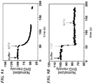
- the non-linear optical signal detected increases by at least 5x when the concentration of the anchor molecule comprising the first affinity tag is increased by a factor of 2x. In some aspects, the non-linear optical signal detected increases by at least 10x when the concentration of the anchor molecule comprising the first affinity tag is increased by a factor of 2x. In some aspects, the non-linear optical signal detected increases by at least 20x when the concentration of the anchor molecule comprising the first affinity tag is increased by a factor of 2x.
- the dependence of the non-linear optical signal on the concentration of the anchor molecule comprising the first affinity tag is described by a power law having an exponent greater than 2.
- the methods further comprise incubating the supported lipid bilayer with the biological entity for about 10 minutes to about 60 minutes.
- the biological entity is present at a concentration of about 0.1 ⁇ M to 10 ⁇ M.
- the supported lipid bilayer comprises 1,2-dioleoyl-sn-glycero-3-phosphocholine.
- the anchor molecule conjugated to the first affinity tag comprises 1,2-dioleoyl-sn-glycero-3-[(N-(5-amino-1-carboxypentyl)iminodiacetic acid)succinyl] (nickel salt).
- the biological entity is a protein.
- the protein is present at a concentration of less than 2 ⁇ M. In some aspects, the amount of protein used is less than 500 ng.
- Also disclosed herein are methods for detecting conformational change in a biological entity comprising: (a) illuminating a substrate with one or more light beams at one or more fundamental frequencies, wherein the substrate comprises a supported lipid bilayer, and wherein the supported lipid bilayer further comprises a Ni-NTA bearing lipid present at a concentration of greater than 5 mole percent, and wherein a biological entity comprising a non-linear active label and poly-histidine tag is tethered to the supported lipid bilayer; (b) measuring a non-linear optical signal arising from the tethered biological entity; (c) optionally contacting the tethered biological entity with one or more candidate binding partners; (d) optionally measuring a non-linear optical signal arising from the tethered biological entity in contact with the one or more candidate binding partners; and (e) optionally comparing the non-linear optical signals measured in steps (b) and (d) to detect a conformational change in the biological entity.
- the substrate is fabricated from a material selected from the group consisting of glass, fused-silica, a polymer, or any combination thereof.
- the biological entity is a protein and the poly-histidine tag is attached to the N-terminus of the protein.
- the biological entity is a protein and the poly-histidine tag is attached to the C-terminus of the protein.
- the poly-histidine tag comprises between 4 and 24 histidine residues.
- the poly-histidine tag comprises 8 histidine residues.
- the polyhistidine tag comprises 6 histidine residues.
- the non-linear optical signal is second harmonic light. In some aspects, the non-linear optical signal is sum frequency light.
- the non-linear optical signal is difference frequency light.
- the supported lipid bilayer comprises 1,2-dioleoyl-sn-glycero-3-phosphocholine.
- the Ni-NTA bearing lipid comprises 1,2-dioleoyl-sn-glycero-3-[(N-(5-amino-1-carboxypentyl)iminodiacetic acid)succinyl] (nickel salt).
- the biological entity is selected from the group consisting of cells, proteins, peptides, receptors, enzymes, antibodies, DNA, RNA, biological molecules, oligonucleotides, small molecules, synthetic molecules, carbohydrates, or any combination thereof.
- the biological entity is a protein.
- the protein is present at a concentration of less than 2 ⁇ M. In some aspects, the amount of protein used is less than 500 ng.
- devices comprising: (a) a substrate, wherein the substrate further comprises a supported lipid bilayer; and (b) a biological entity, wherein the biological entity comprises a non-linear active label and is tethered to the supported lipid bilayer using a pair of affinity tags, and wherein one of the affinity tags is attached to the supported lipid bilayer by means of an anchor molecule that is present in the bilayer at a concentration of greater than or equal to 5 mole percent, and the other affinity tag is attached to the biological entity; wherein the device is capable of producing a non-linear optical signal when exposed to excitation light.
- the substrate is fabricated from a material selected from the group consisting of glass, fused-silica, a polymer, or any combination thereof.
- one of the affinity tags comprises Ni-NTA and the other affinity tag comprises a poly-histidine tag.
- the poly-histidine tag is attached to the N-terminus of a protein.
- the poly-histidine tag is attached to the C-terminus of a protein.
- the poly-histidine tag comprises between 4 and 24 histidine residues.
- the poly-histidine tag comprises 8 histidine residues.
- the poly histidine tag comprises 6 histidine residues.
- one of the affinity tags comprises Co-CMA and the other affinity tag comprises a poly-histidine tag. In some aspects, one of the affinity tags comprises biotin and the other affinity tag comprises streptavidin. In some aspects, one of the affinity tags comprises biotin and the other affinity tag comprises avidin. In some aspects, one of the affinity tags comprises biotin and the other affinity tag comprises neutravidin.
- the non-linear optical signal is second harmonic light. In some aspects, the non-linear optical signal is sum frequency light. In some aspects, the non-linear optical signal is difference frequency light.
- the biological entity is selected from the group consisting of cells, proteins, peptides, receptors, enzymes, antibodies, DNA, RNA, biological molecules, oligonucleotides, small molecules, synthetic molecules, carbohydrates, or any combination thereof.
- the supported lipid bilayer comprises 1,2-dioleoyl-sn-glycero-3-phosphocholine.
- the anchor molecule bearing the first affinity tag comprises 1,2-dioleoyl-sn-glycero-3-[(N-(5-amino-1-carboxypentyl)iminodiacetic acid)succinyl] (nickel salt).
- the concentration of the anchor molecule bearing one of the affinity tag is adjusted to a value ranging from 5 mole percent to 100 mole percent.
- kits comprising: (a) a device comprising a substrate; (b) a lipid suspension, wherein the lipid suspension is capable of forming a supported lipid bilayer on a surface of the substrate and further comprises a lipid bearing a first affinity tag at a concentration of greater than or equal to 5 mole percent of the components capable of forming a supported lipid bilayer; and (c) reagents for tethering biological entities to the supported lipid bilayer using a second affinity tag that is capable of binding to the first affinity tag.
- the substrate is fabricated from a material selected from the group consisting of glass, fused-silica, a polymer, or any combination thereof.
- the first and second affinity tags comprise Ni-NTA and poly-histidine tags.
- the poly-histidine tag is attached to the N-terminus of a protein.
- the poly-histidine tag is attached to the C-terminus of a protein.
- the poly-histidine tag comprises between 4 and 24 histidine residues.
- the poly-histidine tag comprises 8 histidine residues.
- the poly-histidine tag comprises 6 histidine residues.
- the first and second affinity tags comprise biotin- and streptavidin-conjugated reagents. In some aspects, the first and second affinity tags comprise biotin- and avidin-conjugated reagents. In some aspects, the first and second affinity tags comprise biotin- and neutravidin-conjugated reagents.
- the biological entity is selected from the group consisting of cells, proteins, peptides, receptors, enzymes, antibodies, DNA, RNA, biological molecules, oligonucleotides, small molecules, synthetic molecules, carbohydrates, or any combination thereof. In some aspects, the lipid suspension comprises 1,2-dioleoyl-snglycero-3-phosphocholine.
- the lipid bearing the first affinity tag comprises 1,2-dioleoyl-sn-glycero-3-[(N-(5-amino-1-carboxypentyl)iminodiacetic acid)succinyl] (nickel salt).
- Also disclosed herein are methods for tethering a biological entity to a substrate comprising: (a) forming a supported lipid bilayer on a surface of a substrate, wherein the supported lipid bilayer comprises an anchor molecule that comprises or bears a first affinity tag; and (b) contacting the supported lipid bilayer with a biological entity present at a concentration less than or equal to 2 uM, or in an amount of less than or equal to 500 ng, wherein the biological entity comprises a nonlinear-active label and a second affinity tag capable of binding to the first affinity tag, thereby tethering the biological entity to the supported lipid bilayer in an oriented fashion.
- the methods further comprise detecting a non-linear optical signal arising from the tethered biological entity.
- the substrate is fabricated from a material selected from the group consisting of glass, fused-silica, a polymer, or any combination thereof.
- the first affinity and second affinity tags are Ni-NTA and poly-histidine tags.
- the poly-histidine tag is attached to the N-terminus of a protein.
- the poly-histidine tag is attached to the C-terminus of a protein.
- the poly-histidine tag comprises between 4 and 24 histidine residues.
- the poly-histidine tag comprises 8 histidine residues.
- the poly-histidine tag comprises 6 histidine residues.
- the first affinity tag comprises Co-CMA and the second affinity tag comprises a poly-histidine tag.
- the first affinity tag comprises biotin and the second affinity tag comprises streptavidin.
- the first affinity tag comprises biotin and the second affinity tag comprises avidin.
- the first affinity tag comprises biotin and the second affinity tag comprises neutravidin.
- the non-linear optical signal is second harmonic light.
- the non-linear optical signal is sum frequency light. In some aspects, the non-linear optical signal is difference frequency light.
- the biological entity is selected from the group consisting of cells, proteins, peptides, receptors, enzymes, antibodies, DNA, RNA, biological molecules, oligonucleotides, small molecules, synthetic molecules, carbohydrates, or any combination thereof.
- the anchor molecule comprising the first affinity tag is adjusted to a value ranging from 5 mole percent to 100 mole percent of the lipid bilayer.
- the non-linear optical signal detected increases by at least 5x when the concentration of the anchor molecule comprising the first affinity tag is increased by a factor of 2x. In some aspects, the non-linear optical signal detected increases by at least 10x when the concentration of the anchor molecule comprising the first affinity tag is increased by a factor of 2x.
- the non-linear optical signal detected increases by at least 20x when the concentration of the anchor molecule comprising the first affinity tag is increased by a factor of 2x.
- the dependence of the non-linear optical signal on the concentration of the anchor molecule comprising the first affinity tag is described by a power law having an exponent greater than 2.
- the methods further comprise incubating the supported lipid bilayer with the biological entity for about 10 minutes to about 60 minutes.
- the biological entity is present at a concentration of about 0.1 ⁇ M to 10 ⁇ M.
- the supported lipid bilayer comprises 1,2-dioleoyl-sn-glycero-3-phosphocholine.
- the anchor molecule conjugated to the first affinity tag comprises 1,2-dioleoyl-sn-glycero-3-[(N-(5-amino-1-carboxypentyl)iminodiacetic acid)succinyl] (nickel salt).
- a biological entity comprising: (a) illuminating a substrate with one or more light beams at one or more fundamental frequencies, wherein the substrate comprises a supported lipid bilayer, and wherein the supported lipid bilayer further comprises a Ni-NTA bearing lipid, and wherein a biological entity comprising a non-linear active label and poly-histidine tag is tethered to the supported lipid bilayer by contacting the lipid bilayer with the biological entity at a concentration less than or equal to 2 ⁇ M or in an amount less than or equal to 500 ng; (b) measuring a non-linear optical signal arising from the tethered biological entity; (c) optionally contacting the tethered biological entity with one or more candidate binding partners; (d) optionally measuring a non-linear optical signal arising from the tethered biological entity in contact with the one or more candidate binding partners; and (e) optionally comparing the non-linear optical signals measured in steps
- the substrate is fabricated from a material selected from the group consisting of glass, fused-silica, a polymer, or any combination thereof.
- the biological entity is a protein and the poly-histidine tag is attached to the N-terminus of the protein. In some aspects, the biological entity is a protein and the poly-histidine tag is attached to the C-terminus of the protein. In some aspects, the poly-histidine tag comprises between 4 and 24 histidine residues. In some aspects, the poly-histidine tag comprises 8 histidine residues. In some aspects, the poly-histidine tag comprises 6 histidine residues.
- the non-linear optical signal is second harmonic light. In some aspects, the non-linear optical signal is sum frequency light.
- the non-linear optical signal is difference frequency light.
- the supported lipid bilayer comprises 1,2-dioleoyl-sn-glycero-3-phosphocholine.
- the Ni-NTA bearing lipid comprises 1,2-dioleoyl-sn-glycero-3-[(N-(5-amino-1-carboxypentyl)iminodiacetic acid)succinyl] (nickel salt).
- the biological entity is selected from the group consisting of cells, proteins, peptides, receptors, enzymes, antibodies, DNA, RNA, biological molecules, oligonucleotides, small molecules, synthetic molecules, carbohydrates, or any combination thereof.
- the methods and compositions disclosed herein provide means for tethering or immobilizing protein molecules on optical interfaces in a manner that ensures both uniform surface coverage of the optical interface and a high degree of orientation of the molecules, thereby providing for significant enhancements of SHG and/or other nonlinear optical signals arising from the tethered protein molecules, as well as improved signal-to-noise ratios.
- determining orientation, conformation, or changes in orientation or conformation of biological entities may involve measurement of a nonlinear optical signal which is related to and/or proportional to the average orientation of a nonlinear-active label or tag.
- the methods disclosed rely on the use of second harmonic generation (SHG) or related nonlinear optical techniques (e.g. sum frequency generation (SFG) or difference frequency generation (DFG)) for detection of orientation, conformation, or conformational change, as described previously, for example, in U.S. Patent Nos. 6,953,694 , and 8,497,703 .
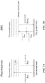
- Second harmonic generation in contrast to more widely used fluorescence-based techniques ( FIG. 1A ), is a nonlinear optical process, in which two photons of the same excitation wavelength or frequency interact with a nonlinear material and are re-emitted as a single photon having twice the energy, i.e. twice the frequency and half the wavelength, of the excitation photons ( FIG. 1B ).
- Second harmonic generation only occurs in nonlinear materials lacking inversion symmetry ( i.e. in non-centrosymmetric materials), and requires a high intensity excitation light source. It is a special case of sum frequency generation, and is related to other nonlinear optical phenomena such as difference frequency generation.
- Second harmonic generation and other nonlinear optical techniques can be configured as surface-selective detection techniques because of their dependence on the orientation of the nonlinear-active species. Tethering of the nonlinear-active species to a planar surface, for example, can instill an overall degree of orientation that is absent when molecules are free to undergo rotational diffusion in solution.
- the intensity of SHG is proportional to the square of the nonlinear susceptibility, and is thus dependent on both the number of oriented nonlinear-active species at the interface and on changes in their average orientation.
- Second harmonic generation and other nonlinear optical techniques may be rendered additionally surface selective through the use of total internal reflection as the mode for delivery of the excitation light to the optical interface on which nonlinear-active species have been immobilized.
- Total internal reflection of the incident excitation light creates an "evanescent wave" at the interface, which may be used to selectively excite only nonlinear-active labels that are in close proximity to the surface, i.e. within the spatial decay distance of the evanescent wave, which is typically on the order of tens of nanometers.
- Total internal reflection may also be used to excite fluorescence in a surface-selective manner, for example to excite a fluorescence donor attached to the optical interface, which then transfers energy to a suitable acceptor molecule via a fluorescence resonance energy transfer (FRET) mechanism.
- FRET fluorescence resonance energy transfer
- the evanescent wave generated by means of total internal reflection of the excitation light is preferentially used to excite a nonlinear-active label or molecule.
- the efficiency of exciting nonlinear active species depends strongly on both their average orientation and on their proximity to the interface.
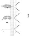
- This surface selective property of SHG and other nonlinear optical techniques can be exploited to detect conformational changes in biological molecules immobilized at interfaces.
- a conformational change in a receptor molecule that results from binding of a ligand to the receptor might be detected using a nonlinear-active label or moiety, where the label is attached to or associated with the receptor in such a way that the conformational change leads to a change in the orientation or distance of the label with respect to the interface ( FIG. 2 ), and thus to a change in a physical property of the nonlinear optical signal.
- the use of surface-selective nonlinear optical techniques has been confined mainly to applications in physics and chemistry, since relatively few biological samples are intrinsically non-linearly active.
- SHG labels second harmonic active labels
- the first example of this was demonstrated by labeling the protein cytochrome c with an oxazole dye and detecting the protein conjugate at an air-water interface with second harmonic generation ( Salafsky (2006), Chem. Physics Letters 342(5-6):485-491 ).
- Surface-selective nonlinear optical techniques are also coherent techniques, meaning that the fundamental and nonlinear optical light beams have wave fronts that propagate through space with well-defined spatial and phase relationships.
- the technique inherently has a very narrow "depth-of-field"; (iii) as a result of the narrow "depth of field", the technique is useful when measurements must be performed in the presence of a overlaying solution, e.g. where a binding process might be obviated or disturbed by a separation or rinse step.
- This aspect of the technique may be particularly useful for performing equilibrium binding measurements, which require the presence of bulk species, or kinetics measurements where the measurements are made over a defined period of time; (iv) the technique exhibits lower photo-bleaching and heating effects than those that occur in fluorescence, due to the facts that the two-photon absorption cross-section is typically much lower than the one-photon absorption cross-section for a given molecule, and that SHG (and sum frequency generation or difference frequency generation) involves scattering, not absorption; (v) minimal collection optics are required and higher signal to noise is expected since the fundamental and nonlinear optical beams (e.g., second harmonic light) have well-defined incoming and outgoing directions with respect to the interface. This is particularly advantageous compared to fluorescence-based detection, as fluorescence emission is isotropic and there may also be a large fluorescence background component to detected signals arising from out-of-focal plane fluorescent species.
- biological entities comprises but is not limited to cells, proteins, peptides, receptors, enzymes, antibodies, DNA, RNA, biological molecules, oligonucleotides, small molecules, synthetic molecules, carbohydrates, or any combination thereof.
- test entities also comprises but is not limited to cells, proteins, peptides, receptors, enzymes, antibodies, DNA, RNA, biological molecules, oligonucleotides, solvents, small molecules, synthetic molecules, carbohydrates, or any combination thereof.
- biological entities may comprise drug targets, or portions thereof, while test entities may comprise drug candidates, or portions thereof.
- a label for use in the presently disclosed methods refers to a nonlinear-active moiety, tag, molecule, or particle which can be bound, either covalently or non-covalently, to a molecule, particle, or phase (e.g., a lipid bilayer) in order to render the resulting system more nonlinear optical active.
- Labels can be employed in the case where the molecule, particle or phase (e.g., lipid bilayer) is not nonlinear active to render the system nonlinear-active, or with a system that is already nonlinear-active to add an extra characterization parameter to the system.
- Exogenous labels can be pre-attached to the molecules, particles, or other biological entities, and any unbound or unreacted labels separated from the labeled entities before use in the methods described herein.
- the nonlinear-active moiety is attached to the target molecule or biological entity in vitro prior to immobilizing the target molecules or biological entities in discrete regions of the substrate surface.
- the labeling of biological molecules or other biological entities with nonlinear-active labels allows a direct optical means of detecting interactions between the labeled biological molecule or entity and another molecule or entity (i.e. the test entity) in cases where the interaction results in a change in orientation or conformation of the biological molecule or entity using a surface-selective nonlinear optical technique.
- At least two distinguishable nonlinear-active labels are used.
- the orientation of the attached two or more distinguishable labels would then be chosen to facilitate well defined directions of the emanating coherent nonlinear light beam.
- the two or more distinguishable labels can be used in assays where multiple fundamental light beams at one or more frequencies, incident with one or more polarization directions relative to the optical interface are used, with the resulting emanation of at least two nonlinear light beams.
- nonlinear-active tags or labels include, but are not limited to, the compounds listed in Table 1, and their derivatives. Table 1.
- a species may be nonlinear-active
- the following characteristics can indicate the potential for nonlinear activity: a large difference dipole moment (difference in dipole moment between the ground and excited states of the molecule), a large Stokes shift in fluorescence, or an aromatic or conjugated bonding character.
- an experimenter can use a simple technique known to those skilled in the art to confirm the nonlinear activity, for example, through detection of SHG from an air-water interface on which the nonlinear-active species has been distributed.
- the species can be conjugated, if desired, to a biological molecule or entity for use in the surface-selective nonlinear optical methods and systems disclosed herein.
- metal nanoparticles and assemblies thereof are modified to create biological nonlinear-active labels.
- the following references describe the modification of metal nanoparticles and assemblies: J.P. Novak and D. L. Feldheim, "Assembly of Phenylacetylene-Bridged Silver and Gold Nanoparticle Arrays". J. Am. Chem. Soc., 2000, 122, 3979-3980 ; J.P. Novak et al., "Nonlinear Optical Properties of Molecularly Bridged Gold Nanoparticle Arrays". J. Am. Chem. Soc. 2000, 122, 12029-12030 ; Vance, F W, Lemon B. I., Hupp, J. T., " Enormous Hyper-Rayleigh Scattering from Nanocrystalline Gold Particle Suspensions”. J. Phys. Chem. B 102:10091-93 (1999 ).
- the nonlinear activity of the system can also be manipulated through the introduction of nonlinear analogues to molecular beacons, that is, molecular beacon probes that have been modified to incorporate a nonlinear-active label (or modulator thereof) instead of fluorophores and quenchers.
- nonlinear optical analogues of molecular beacons are referred to herein as molecular beacon analogues (MB analogues or MBA).
- MB analogues molecular beacon analogues
- the MB analogues to be used in the described methods and systems can be synthesized according to procedures known to one of ordinary skill in the art.
- the methods and systems disclosed herein provide for detection of a variety of interactions between biological entities, or between biological entities and test entities, depending on the choice of biological entities, test entities, and non-linear active labeling technique employed.
- the present disclosure provides for the qualitative detection of binding events, e.g. the binding of a ligand to a receptor, as indicated by the resulting conformational change induced in the receptor.
- the present disclosure provides for quantitative analysis of binding events, e.g. the binding of a ligand to a receptor, by performing replicate measurements using different concentrations of the ligand molecule and generating a dose-response curve using the percent change in maximal conformational change observed.
- other aspects of the present disclosure may provide methods for qualitative or quantitative measurements of enzyme-inhibitor interactions, antibody-antigen interactions, the formation of complexes of biological macromolecules, or interactions of receptors with allosteric modulators.
- MB analogues can be used according to the methods disclosed herein as hybridization probes that can detect the presence of complementary nucleic acid target without having to separate probe-target hybrids from excess probes as in solution-phase hybridization assays, and without the need to label the targets oligonucleotides.
- MB analogue probes can also be used for the detection of RNAs within living cells, for monitoring the synthesis of specific nucleic acids in sample aliquots drawn from bioreactors, and for the construction of self-reporting oligonucleotide arrays. They can be used to perform homogeneous one-well assays for the identification of single-nucleotide variations in DNA and for the detection of pathogens or cells immobilized to surfaces for interfacial detection.
- Interactions between biological entities or biological and test entities can be correlated through the methods presently disclosed to the following measurable nonlinear signal parameters: (i) the intensity of the nonlinear light, (ii) the wavelength or spectrum of the nonlinear light, (iii) the polarization of the nonlinear light, (iv) the time-course of (i), (ii), or (iii), and/or vi) one or more combinations of (i), (ii), (iii), and (iv).
- the systems and methods of the present disclosure typically utilize a planar substrate for immobilization of one or more biological entities on a top surface of the substrate, wherein the top substrate surface further comprises the optical interface (or sample interface) used for exciting nonlinear optical signals.
- the substrate can be glass, silica, fused-silica, plastic, or any other solid material that is transparent to the fundamental and second harmonic light beams, and that supports total internal reflection at the substrate/sample interface when the excitation light is incident at an appropriate angle.
- the substrate (or solid support) upon which biological entities are immobilized may be non-planar, for example, the substrate may comprise a convex, concave, or irregular surface, or may comprise glass beads, polymer beads, and the like.
- a bead or particle may comprise any type of solid, porous, or hollow bead or particle.
- a bead or particle may comprise a discrete shape that may be spherical ( e.g., a microsphere) or have a non-spherical or irregular shape, such as cubic, cuboid, pyramidal, cylindrical, conical, oblong, or disc-shape, and the like.
- a bead or particle may refer to any three dimensional structure that provides an increased surface area for immobilization of biological entities.
- Beads or particles may comprise a variety of materials including, but not limited to, paramagnetic materials (e.g. magnesium, molybdenum, lithium, and tantalum), superparamagnetic materials (e.g. ferrite (Fe 3 O 4 ); magnetite nanoparticles), ferromagnetic materials (e.g.
- iron, nickel, cobalt, some alloys thereof, and some rare earth metal compounds ceramic, plastic, glass, polystyrene, silica, methylstyrene, acrylic polymers, titanium, latex, sepharose, agarose, hydrogel, polymer, cellulose, nylon, and any combination thereof.
- the biological entities may be immobilized uniformly across a substrate surface.
- the biological entities may be immobilized in one or more discrete regions of a substrate surface.
- the discrete regions within which biological entities are contained are configured as one-dimensional or two-dimensional arrays, or are distributed randomly across the substrate surface, and are separated from one another by means of a hydrophobic coating or thin metal layer.
- the discrete regions may comprise indents in the substrate surface.
- the discrete regions may be separated from each other by means of a well-forming component such that the substrate forms the bottom of a microwell plate (or microplate), and each individual discrete region forms the bottom of one well in the microwell plate.
- the well-forming component separates the top surface of the substrate into 96 separate wells. In another aspect, the well-forming component separates the top surface of the substrate into 384 wells. In yet another aspect, the well-forming component separates the top surface of the substrate into 1,536 wells.
- the substrate whether configured in a planar array, indented array, or microwell plate format, may comprise a disposable or consumable device or cartridge that interfaces with other optical and mechanical components of a high throughput analysis system.
- the methods and systems disclosed herein further comprise specifying the number of discrete regions or wells into which the substrate surface is divided, irrespective of how separation is maintained between discrete regions or wells. Having larger numbers of discrete regions or wells on a substrate may be advantageous in terms of increasing the sample analysis throughput of the method or system.
- the number of discrete regions or wells per substrate is between 10 and 1,600. In other aspects, the number of discrete regions or wells is at least 10, at least 20, at least 50, at least 100, at least 200, at least 300, at least 400, at least 500, at least 750, at least 1,000, at least 1,250, at least 1,500, or at least 1,600.
- the number of discrete regions or wells is at most 1,600, at most 1,500, at most 1,000, at most 750, at most 500, at most 400, at most 300, at most 200, at most 100, at most 50, at most 20, or at most 10.
- the number of discrete regions or wells is 96.
- the number of discrete regions or wells is 384.
- the number of discrete regions or wells is 1,536. Those of skill in the art will appreciate that the number of discrete regions or wells may fall within any range bounded by any of these values (e.g. from about 12 to about 1,400).
- the methods and systems disclosed herein also comprise specifying the surface area of the discrete regions or wells into which the substrate surface is divided, irrespective of how separation is maintained between discrete regions or wells. Having discrete regions or wells of larger area may facilitate ease-of-access and manipulation of the associated biological entities in some cases, whereas having discrete regions or wells of smaller area may be advantageous in terms of reducing assay reagent volume requirements and increasing the sample analysis throughput of the method or system.
- the surface area of the discrete regions or wells is between 1 mm 2 and 100 mm 2 .
- the area of the discrete regions or wells is at least 1 mm 2 , at least 2.5 mm 2 , at least 5 mm 2 , at least 10 mm 2 , at least 20 mm 2 , at least 30 mm 2 , at least 40 mm 2 , at least 50 mm 2 , at least 75 mm 2 , or at least 100 mm 2 .
- the area of the discrete regions or wells is at most 100 mm 2 , at most 75 mm 2 , at most 50 mm 2 , at most 40 mm 2 , at most 30 mm 2 , at most 20 mm 2 , at most 10 mm 2 , at most 5 mm 2 , at most 2.5 mm 2 , or at most 1 mm 2 .
- the area of discrete regions or wells is about 35 mm 2 .
- the area of the discrete regions or wells is about 8.6 mm 2 .
- the area of the discrete regions or wells may fall within any range bounded by any of these values (e.g. from about 2 mm 2 to about 95 mm 2 ).
- one or more discrete regions of the substrate surface are simultaneously exposed to (illuminated with) excitation light. In other examples, one or more discrete regions of the substrate surface are sequentially exposed to (illuminated with) excitation light by re-positioning the substrate relative to the excitation light source. Total internal reflection of the incident excitation light creates an "evanescent wave" at the sample interface, which excites the nonlinear-active labels immobilized thereon, and results in generation of second harmonic light (or in some aspects sum frequency or difference frequency light).
- the intensity of the evanescent wave, and hence the intensity of the nonlinear optical signals generated, is dependent on the incident angle of the excitation light beam, precise orientation of the substrate plane with respect to the optical axis of the excitation beam and efficient optical coupling of the beam to the substrate is critical for achieving optimal SHG signal across the array of discrete regions.
- total internal reflection is achieved by means of a single reflection of the excitation light from the substrate surface.
- the substrate may be configured as a waveguide such that the excitation light undergoes multiple total internal reflections as it propagates along the waveguide.
- the substrate may be configured as a zero-mode waveguide, wherein an evanescent field is created by means of nanofabricated structures.
- Efficient optical coupling between the excitation light beam and the substrate is often achieved by use of an index-matching fluid such as mineral oil, mixtures of mineral oil and hydrogenated terphenyls, perfluorocarbon fluids, glycerin, glycerol, or similar fluids having a refractive index near 1.5, wherein the index-matching fluid is wicked between a prism used to guide the light beam and the lower surface of the substrate.
- an index-matching fluid such as mineral oil, mixtures of mineral oil and hydrogenated terphenyls, perfluorocarbon fluids, glycerin, glycerol, or similar fluids having a refractive index near 1.5
- substrates in any of the formats described above are further configured for immobilization of biological entities across at least one surface of the substrate or within specified discrete regions of a substrate surface.
- Immobilization of biological molecules or cells may be accomplished by a variety of techniques known to those of skill in the art, for example, through the use of aminopropyl silane chemistries to functionalize glass or fused-silica surfaces with amine functional groups, followed by covalent coupling using amine-reactive conjugation chemistries, either directly with the biological molecule of interest, or via an intermediate spacer or linker molecule.
- Non-specific adsorption may also be used directly or indirectly, e.g.
- BSA-N-hydroxysuccinimide BSA-N-hydroxysuccinimide
- BSA-NHS BSA-N-hydroxysuccinimide
- biological molecules may be immobilized on the surface by means of tethering to or embedding in "supported lipid bilayers", the latter comprising small patches of lipid bilayer confined to a silicon or glass surface by means of hydrophobic and electrostatic interactions, where the bilayer is "floating" above the substrate surface on a thin layer of aqueous buffer.
- Supported phospholipid bilayers can also be prepared with or without membrane proteins or other membrane-associated components as described, for example, in Salafsky et al.
- Supported phospholipid bilayers are well known in the art and there are numerous techniques available for their fabrication. Potential advantages of using supported lipid bilayers for immobilization of proteins or other biological entities on substrate surfaces or optical interfaces include (i) preservation of membrane protein structure for those proteins that typically span the cell membrane or other membrane components of cells and require interaction with the hydrophobic core of the bilayer for stabilization of secondary and tertiary structure, (ii) preservation of two dimensional lateral and rotational diffusional mobility for studying interactions between protein components within the bilayer, and (iii) preservation of molecular orientation, depending on such factors as the type of protein under study (i.e.
- bilayer membrane or soluble protein how the bilayer membrane is formed on the substrate surface, and how the protein is tethered to the bilayer (in the case of soluble proteins).
- anchor molecules are molecules which are embedded in the lipid bilayer, and may comprise fatty acid, glycerolipid, glycerophospholipid, sphingolipid, or other lipid or non-lipid molecules to which attachment moieties are conjugated.
- concentration of anchor molecules in the supported lipid bilayer may range from about 1 mole percent to as much as 100 mole percent.
- the concentration of anchor molecules in the supported lipid bilayer may be at least 1 mole percent, at least 2.5 mole percent, at least 5 mole percent, at least 10 mole percent, at least 20 mole percent, at least 30 mole percent, at least 40 mole percent, at least 50 mole percent, at least 60 mole percent, at least 70 mole percent, at least 80 mole percent, at least 90 mole percent, or 100 mole percent.
- the concentration of anchor molecules in the supported lipid bilayer may be at most 100 mole percent, at most 90 mole percent, at most 80 mole percent, at most 70 mole percent, at most 60 mole percent, at most 50 mole percent, at most 40 mole percent, at most 30 mole percent, at most 20 mole percent, at most 10 mole percent, at most 5 mole percent, at most 2.5 mole percent, or at most 1 mole percent.
- concentration of anchor molecules in the supported lipid bilayer may have any value within this range, for example, about 8.5 mole percent.
- Linker molecules are molecules used to provide spatial (“vertical") separation between the attachment point of the protein or other biological entity being tethered and the attachment point on the anchor molecule embedded in the plane of the lipid bilayer.
- suitable linker molecules include, but are not limited to, omega-amino fatty acids, polyethylene glycols, and the like.
- Attachment moieties are specific chemical structures or binding partners that provide for covalent or non-covalent binding between two biological entities.
- attachment moieties or affinity tags that are suitable for use in the methods disclosed herein include biotin and avidin (or biotin and streptavidin), and His-tag / Ni-NTA binding partners.
- biotin-streptavidin interaction is widely used in biological assay techniques to conjugate or immobilize proteins or other biological entities.
- Biotinylation of proteins enables capture by multivalent avidin or streptavidin molecules that are themselves adhered to a surface (e.g. glass slides or beads) or conjugated to another molecule (e.g. through the use of a biotin-streptavidin-biotin bridge or linker).
- the biotin moiety is sufficiently small that biotinylation typically doesn't interfere with protein function.
- the high affinity (K d of 10 -14 M to 10 -15 M) and high specificity of the binding interaction between biotin and avidin or streptavidin enables capture of biotinylated proteins of interest even from complex samples. Due to the extremely strong binding interaction, harsh conditions are needed to elute biotinylated protein from streptavidin-coated surfaces (typically 6M guanidine HCl at pH 1.5), which will often denature the protein of interest.
- the use of monomeric forms of avidin or streptavidin, which have a decreased biotin-binding affinity of ⁇ 10 -8 M, may allow biotinylated proteins to be eluted with excess free biotin if necessary.
- lipid molecules comprising biotin moieties may be incorporated into supported lipid bilayers for the purpose of immobilizing or tethering biotinylated proteins and/or other biotinylated biological entities to the bilayer via a biotin-avidin-biotin (or biotin-streptavidin-biotin) bridge.
- Biotinylation of proteins and other biological entities may be performed by direct coupling, e.g. through conjugation of primary amines on the surface of a protein using N-hydroxysuccinimidobiotin (NHS-biotin).
- NHS-biotin N-hydroxysuccinimidobiotin
- recombinant proteins are conveniently biotinylated using the AviTag approach, wherein the AviTag peptide sequence (GLNDIFEAQKIEWHE) is incorporated into the protein through the use of genetic engineering and protein expression techniques.
- the presence of the AviTag sequence allows biotinylation of the protein by treatment with the BirA enzyme.
- His tag chemistry is another widely used tool for purification of recombinant proteins and other biomolecules.
- a DNA sequence specifying a string of six to nine histidine residues may be incorporated into vectors used for production of recombinant proteins comprising 6xHis or poly-His tags fused to their N- or C-termini. His-tagged proteins can then be purified and detected as a result of the fact that the string of histidine residues binds to several types of immobilized metal ions, including nickel, cobalt and copper, under specific buffer conditions.
- the number of histidine residues incorporated into recombinant proteins may range from 1 residue to 24 residues, or more. In some examples, the number of histidine residues incorporated may be at least 1 residue, at least 2 residues, at least 4 residues, at least 6 residues, at least 8 residues, at least 10 residues, at least 12 residues, at least 14 residues, at least 16 residues, at least 18 residues, at least 20 residues, at least 22 residues, or at least 24 residues.
- the number of histidine residues incorporated may be at most 24 residues, at most 22 residues, at most 20 residues, at most 18 residues, at most 16 residues, at most 14 residues, at most 12 residues, at most 10 residues, at most 8 residues, at most 6 residues, at most 4 residues, at most 2 residues, or at most 1 residue.
- the number of histidine residues incorporated may have any value within this range, for example, 7 residues.
- NTA- or IDA-conjugated supports are prepared, they can be "loaded” with the desired divalent metal (e.g., Ni, Co, Cu, or Fe).
- the desired divalent metal e.g., Ni, Co, Cu, or Fe
- nickel the metal
- the resulting affinity support is usually called a Ni-chelate, Ni-IDA or Ni-NTA support.
- Affinity purification of His-tagged fusion proteins is the most common application for metal-chelate supports in protein biology research. Nickel or cobalt metals immobilized by NTA-chelation chemistry are the systems of choice for this application.
- lipid molecules comprising Ni-NTA groups may be incorporated into supported lipid bilayers for the purpose of immobilizing or tethering His-tagged proteins and other His-tagged biological entities to the bilayer.
- the supported lipid bilayer may comprise, for example, 1,2-dioleoyl-sn-glycero-3-phosphocholine, and may also contain, for example, 1,2-dioleoyl-sn-glycero-3-[(N-(5-amino-1-carboxypentyl)iminodiacetic acid)succinyl] (nickel salt) at various concentrations.
- the concentration of 1,2-dioleoyl-sn-glycero-3-[(N-(5-amino-1-carboxypentyl)iminodiacetic acid)succinyl] (nickel salt) in the supported lipid bilayer may range from as little as 1 mole percent to as much as 100 mole percent.
- the concentration of 1,2-dioleoyl-sn-glycero-3-[(N-(5-amino-1-carboxypentyl)iminodiacetic acid)succinyl] (nickel salt) may be at least 1 mole percent, at least 2.5 mole percent, at least 5 mole percent, at least 10 mole percent, at least 20 mole percent, at least 30 mole percent, at least 40 mole percent, at least 50 mole percent, at least 60 mole percent, at least 70 mole percent, at least 80 mole percent, at least 90 mole percent, or 100 mole percent.
- the concentration of 1,2-dioleoyl-sn-glycero-3-[(N-(5-amino-1-carboxypentyl)iminodiacetic acid)succinyl] (nickel salt) in the supported lipid bilayer may be at most 100 mole percent, at most 90 mole percent, at most 80 mole percent, at most 70 mole percent, at most 60 mole percent, at most 50 mole percent, at most 40 mole percent, at most 30 mole percent, at most 20 mole percent, at most 10 mole percent, at most 5 mole percent, at most 2.5 mole percent, or at most 1 mole percent.
- the concentration of 1,2-dioleoyl-sn-glycero-3-[(N-(5-amino-1-carboxypentyl)iminodiacetic acid)succinyl] (nickel salt) in the supported lipid bilayer may have any value within this range, for example, about 5.5 mole percent.
- Poly-His tags bind best to chelated metal ions in near-neutral buffer conditions (physiologic pH and ionic strength).
- a typical binding/wash buffer consists of Tris-buffer saline (TBS) pH 7.2, containing 10-25mM imidazole.
- TBS Tris-buffer saline
- the low-concentration of imidazole helps to prevent nonspecific binding of endogenous proteins that have histidine clusters.
- Elution and recovery of captured His-tagged protein from chelated metal ion supports, when desired, is typically accomplished using a high concentration of imidazole (at least 200mM), low pH (e.g., 0.1M glycine-HCl, pH 2.5), or an excess of strong chelator (e.g., EDTA).
- Immunoglobulins are known to have multiple histidines in their Fc region and can bind to chelated metal ion supports, therefore stringent binding conditions (e.g. using an appropriate concentration of imidazole) are necessary to avoid high levels of background binding if immunoglobulins are present in a sample at high relative abundance compared to the His-tagged proteins of interest.
- Albumins such as bovine serum albumin (BSA) also have multiple histidines and can yield high levels of background binding to chelated metal ion supports in the absence of more abundant His-tagged proteins or the use of imidazole in the binding/wash buffer.
- BSA bovine serum albumin
- proteins or other biological entities may sometimes be desirable to vary the surface density of proteins or other biological entities that are immobilized on or tethered to the substrate surface comprising the optical interface.
- This can be accomplished in a variety of ways, as is well known to those of skill in the art.
- proteins (or other biological entities) are coupled to the surface through the use of aminopropyl silane chemistries to functionalize glass or fused-silica surfaces with amine functional groups, followed by covalent coupling using amine-reactive conjugation chemistries and linker molecules
- the ratio of bi-functional (e.g. comprising both a primary amine and carboxyl functional group) to mono-functional ( e.g. comprising only a carboxyl functional group) linkers in the reaction mixture may be varied to control the surface density of primary amine functional groups available for coupling with the protein.
- the mole percent of the biotinylated lipid molecule used to form the bilayer may be varied in order to control the surface density of biotin groups available for binding.
- the concentration of biotinylated lipid molecules may range from as little as 1 mole percent to as much as 100 mole percent.
- the concentration of biotinylated lipid molecule in the supported lipid bilayer may be at least 1 mole percent, at least 2.5 mole percent, at least 5 mole percent, at least 10 mole percent, at least 20 mole percent, at least 30 mole percent, at least 40 mole percent, at least 50 mole percent, at least 60 mole percent, at least 70 mole percent, at least 80 mole percent, at least 90 mole percent, or 100 mole percent.
- the concentration of biotinylated lipid molecule in the supported lipid bilayer may be at most 100 mole percent, at most 90 mole percent, at most 80 mole percent, at most 70 mole percent, at most 60 mole percent, at most 50 mole percent, at most 40 mole percent, at most 30 mole percent, at most 20 mole percent, at most 10 mole percent, at most 5 mole percent, at most 2.5 mole percent, or at most 1 mole percent.
- concentration of biotinylated lipid molecule in the supported lipid bilayer may have any value within this range, for example, about 8 mole percent.
- the concentration and/or incubation time used for attaching streptavidin to the biotinylated lipid bilayer may be varied.
- the concentration of streptavidin or biotinylated protein used may range from about 0.1 ⁇ M to about 10 ⁇ M, or more.
- the concentration of streptavidin or biotinylated protein used may be at least 0.1 ⁇ M, at least 0.5 ⁇ M, at least 1 ⁇ M, at least 2 ⁇ M, at least 3 ⁇ M, at least 4 ⁇ M, at least 5 ⁇ M, at least 6 ⁇ M, at least 7 ⁇ M, at least 8 ⁇ M, at least 9 ⁇ M, or at least 10 ⁇ m.
- the concentration of streptavidin or biotinylated protein used may be at most 10 ⁇ M, at most 9 ⁇ M, at most 8 ⁇ M, at most 7 ⁇ M, at most 6 ⁇ M, at most 5 ⁇ M, at most 4 ⁇ M, at most 3 ⁇ M, at most 2 ⁇ M, at most 1 ⁇ M, at most 0.5 ⁇ M, or at most 0.1 ⁇ M.
- concentration of streptavidin or biotinylated protein used may have any value within this range, for example, about 4.4 ⁇ M.
- the amount of streptavidin or biotinylated protein used for attachment may range from about 1 nanogram to about 1,000 nanograms, or more.
- the amount of streptavidin or biotinylated protein used for attachment may be at least 1 nanogram, at least 10 nanograms, at least 25 nanograms, at least 75 nanograms, at least 100 nanograms, at least 200 nanograms, at least 300 nanograms, at least 400 nanograms, at least 500 nanograms, at least 600 nanograms, at least 700 nanograms, at least 800 nanograms, at least 900 nanograms, or at least 1,000 nanograms.
- the amount of streptavidin or biotinylated protein used for attachment may be at most 1,000 nanograms, at most 900 nanograms, at most 800 nanograms, at most 700 nanograms, at most 600 nanograms, at most 500 nanograms, at most 400 nanograms, at most 300 nanograms, at most 200 nanograms, at most 100 nanograms, at most 75 nanograms, at most 50 nanograms, at most 25 nanograms, at most 10 nanograms, or at most 1 nanogram.
- the amount of streptavidin or biotinylated protein used for attachment may have any value within this range, for example, about 275 nanograms.
- the incubation time used for attaching streptavidin or biotinylated protein may range from about 10 minutes to about 60 minutes, or longer. In some examples, the incubation time used for attaching streptavidin or biotinylated protein may be at least 10 minutes, at least 20 minutes, at least 30 minutes, at least 40 minutes, at least 50 minutes, or at least 60 minutes. In some examples, the incubation time used for attaching streptavidin or biotinylated protein may be at most 60 minutes, at most 50 minutes, at most 40 minutes, at most 30 minutes, at most 20 minutes, or at most 10 minutes. Those of skill in the art will recognize that the incubation time used for attaching streptavidin or biotinylated protein may have any value within this range, for example, about 34 minutes.
- the mole percent of Ni-NTA-containing lipid molecule used to form the bilayer may be varied in order to control the surface density of Ni-NTA ligands available for binding.
- concentration of Ni-NTA-containing lipid molecules may range from as little as 1 mole percent to as much as 90 mole percent, or more.
- the concentration of Ni-NTA-containing lipid molecules in the supported lipid bilayer may be at least 1 mole percent, at least 2.5 mole percent, at least 5 mole percent, at least 10 mole percent, at least 20 mole percent, at least 30 mole percent, at least 40 mole percent, at least 50 mole percent, at least 60 mole percent, at least 70 mole percent, at least 80 mole percent, or at least 90 mole percent.
- the concentration of Ni-NTA-containing lipid molecules in the supported lipid bilayer may be at most 90 mole percent, at most 80 mole percent, at most 70 mole percent, at most 60 mole percent, at most 50 mole percent, at most 40 mole percent, at most 30 mole percent, at most 20 mole percent, at most 10 mole percent, at most 5 mole percent, at most 2.5 mole percent, or at most 1 mole percent.
- concentration of Ni-NTA-containing lipid molecules in the supported lipid bilayer may have any value within this range, for example, about 4.5 mole percent.
- the concentration and/or incubation time used for attaching His-tagged proteins (or other His-tagged biological entities) to the Ni-NTA groups in the lipid bilayer may be varied.
- the concentration of His-tagged protein (or other His-tagged biological entities) used for attachment may range from about 0.1 ⁇ M to about 10 ⁇ M, or more.
- the concentration of His-tagged proteins (or other His-tagged biological entities) used may be at least 0.1 ⁇ M, at least 0.5 ⁇ M, at least 1 ⁇ M, at least 2 ⁇ M, at least 3 ⁇ M, at least 4 ⁇ M, at least 5 ⁇ M, at least 6 ⁇ M, at least 7 ⁇ M, at least 8 ⁇ M, at least 9 ⁇ M, or at least 10 ⁇ m.
- the concentration of His-tagged proteins (or other His-tagged biological entities) used may be at most 10 ⁇ M, at most 9 ⁇ M, at most 8 ⁇ M, at most 7 ⁇ M, at most 6 ⁇ M, at most 5 ⁇ M, at most 4 ⁇ M, at most 3 ⁇ M, at most 2 ⁇ M, at most 1 ⁇ M, at most 0.5 ⁇ M, or at most 0.1 ⁇ M.
- concentration of u His-tagged proteins (or other His-tagged biological entities) used may have any value within this range, for example, about 7.5 ⁇ M.
- the amount of His-tagged proteins (or other His-tagged biological entities) used for attachment may range from about 1 nanogram to about 1,000 nanograms, or more.
- the amount of His-tagged proteins (or other His-tagged biological entities) used for attachment may be at least 1 nanogram, at least 10 nanograms, at least 25 nanograms, at least 75 nanograms, at least 100 nanograms, at least 200 nanograms, at least 300 nanograms, at least 400 nanograms, at least 500 nanograms, at least 600 nanograms, at least 700 nanograms, at least 800 nanograms, at least 900 nanograms, or at least 1,000 nanograms.
- the amount of His-tagged proteins (or other His-tagged biological entities) used for attachment may be at most 1,000 nanograms, at most 900 nanograms, at most 800 nanograms, at most 700 nanograms, at most 600 nanograms, at most 500 nanograms, at most 400 nanograms, at most 300 nanograms, at most 200 nanograms, at most 100 nanograms, at most 75 nanograms, at most 50 nanograms, at most 25 nanograms, at most 10 nanograms, or at most 1 nanogram.
- the amount of His-tagged proteins (or other His-tagged biological entities) used for attachment may have any value within this range, for example, about 425 nanograms.
- the incubation time used for attaching His-tagged proteins (or other His-tagged biological entities) may range from about 10 minutes to about 60 minutes, or longer. In some examples, the incubation time used for attaching His-tagged proteins (or other His-tagged biological entities) may be at least 10 minutes, at least 20 minutes, at least 30 minutes, at least 40 minutes, at least 50 minutes, or at least 60 minutes. In some examples, the incubation time used for attaching His-tagged proteins (or other His-tagged biological entities) may be at most 60 minutes, at most 50 minutes, at most 40 minutes, at most 30 minutes, at most 20 minutes, or at most 10 minutes. Those of skill in the art will recognize that the incubation time used for attaching His-tagged proteins (or other His-tagged biological entities) may have any value within this range, for example, about 48 minutes.
- a 2-fold increase of the concentration of anchor molecule used for immobilization may yield at least a 5x increase, at least a 10x increase, at least a 15x increase, or at least a 20x increase in the nonlinear optical signal detected.
- a 2-fold increase of the concentration of anchor molecule used for immobilization may yield at most a 20x increase, at most a 15x increase, at most a 10x increase, or at most a 5x increase in the nonlinear optical signal detected.
- a device comprising: (a) a substrate, wherein the substrate further comprises a supported lipid bilayer; and (b) a biological entity, wherein the biological entity comprises an non-linear active label and is tethered to the supported lipid bilayer using a pair of affinity tags, and wherein one of the affinity tags is attached to the supported lipid bilayer and the other affinity tag is attached to the biological entity; and wherein the device is capable of producing a non-linear optical signal when exposed to excitation light.
- a device of this type may be configured in a variety of different ways, for example, the substrate may be fabricated from a material selected from the group including, but not limited to, glass, fused-silica, a polymer, or any combination thereof.
- a variety of affinity tags may be used, including Ni-NTA (or other chelated metal ions) and poly-histidine tags, or biotin and streptavidin (or avidin, or neutravidin).
- the poly-histidine tag may be attached to the N-terminus of a protein or to the C-terminus of a protein, or at any other position in the protein's sequence that maintains protein function and also provides suitable exposure on the protein surface so that the poly-histidine tag is able to interact with Ni-NTA or other chelated metal ion ligands.
- poly-histidine tags may comprise any suitable number of histidine residues, for example, something between 4 and 12 histidine residues, or about 9 histidine residues, or about 6 histidine residues.
- Co-CMA chelated metal ion
- non-linear optical signals that may be produced by the device when exposed to excitation light include second harmonic light, sum frequency light, and difference frequency light.
- a variety of different biological entities may be utilized in such devices including, but not limited to, cells, proteins, peptides, receptors, enzymes, antibodies, DNA, RNA, biological molecules, oligonucleotides, small molecules, synthetic molecules, carbohydrates, or any combination thereof.
- kits disclosed herein consider a kit comprising (a) a substrate; (b) a lipid suspension, wherein the lipid suspension is capable of forming a supported lipid bilayer on a surface of the substrate; and (c) affinity tagged reagents for tethering biological entities to the supported lipid bilayer.
- Kits may be configured in a variety of different ways, for example, the device substrate may be fabricated from a material selected from the group including, but not limited to, glass, fused-silica, a polymer, or any combination thereof.
- a variety of affinity tagged reagents could be used in the kit, including Ni-NTA (or other chelated metal ion ligand) and poly-histidine tagged reagents, or biotinylated reagents along with streptavidin (or avidin, or neutravidin).
- the lipid suspension used for formation of supported lipid bilayers may include a variety of different phospholipids or other suitable lipids, for example, in some examples, the kits may include 1,2-dioleoyl-sn-glycero-3-phosphocholine and/or 1,2-dioleoyl-sn-glycero-3-[(N-(5-amino-1-carboxypentyl)iminodiacetic acid)succinyl] (nickel salt). It will be apparent to those of skill in the art that many other lipids and affinity tagged reagents may be used in kits of the present disclosure.
- the objective of this study was to demonstrate the use of His-tag chemistry for immobilizing labeled proteins (i.e. labeled with an SHG-active label) to substrate surfaces comprising the optical interface used in SHG measurements.
- FIG. 4 is an overlay of SHG data acquired using labeled protein bound to a lipid bilayer comprising 3% Ni-NTA (under slightly different incubation conditions) and that for the 10% Ni-NTA data to show the relative SHG intensity produced by each labeled protein on their respective surfaces.
- the 10% NTA surfaces provided more than an order of magnitude improvement in the SHG signal for the labeled enzyme tested here.
- Protein labeling protocol Stock His-tagged maltose binding protein (MBP) (1 mL at nominal 0.5 mg/mL) was buffer exchanged from 20mM Tris-HCL pH 8.0, 0.1M NaCl, 10% Glycerol into 0.1 M Na phosphate, pH 7.5 by passing over two of the 2 mL ZebaTM Spin Desalting Columns (Thermo Scientific) equilibrated per the manufacturer's instructions. Concentration and volume of the buffer exchanged material were determined by measuring absorbance on a NanoDrop Spectrophotometer (Thermo Scientific). PyMPO-NHS was weighed out and dissolved to 2mM in dry DMSO.
- MBP maltose binding protein
- Slide preparation A glass slide was cleaned by Piranha treatment (heating at 100 °C in 30% H2O2/70% con H2SO4 for 30 minutes). The slide was washed extensively in diH 2 O, dried with N 2 gas, and a well-forming gasket was affixed.
- Bilayer preparation Small unilamellar lipid vesicle (SUV) solutions consisting of 3% DGS-NTA, Ni salt, or 10% DGS-NTA, Ni salt, at a stock concentration of 0.5 mg/mL were diluted to 0.1 mg/mL in 25 mM Tris-HCl, ph 7.2, 150 mM NaCl. 10 ⁇ L aliquots were then added to wells, and incubated at room temperature for 60 minutes to allow bilayer formation.
- SUV Small unilamellar lipid vesicle
- MBP and DHFR conjugates were diluted to 5 ⁇ Min their respective assay buffers, and 10 ⁇ L was added to the appropriate wells, thereby resulting in a concentration of 2.5 ⁇ M in the wells. 10 ⁇ L of assay buffer was added to the bilayer-only controls. Protein tethering was allowed to proceed for 1 hour at room temperature. During the tethering incubation, solutions of 2 mM lactose and 2 mM maltose were made up in MBP assay buffer.
- the chopper was placed 'in line' and operated at 120 Hz.
- the first objective was to show maltose-induced conformational change, using 10 ⁇ L buffer and 10 ⁇ L lactose injections as negative controls.
- a volume of 10 ⁇ L was injected to give a total of 20 ⁇ L/well, and the volume was mixed by repetitively drawing up 10 ⁇ L volumes (4-5 times), then withdrawing 10 ⁇ L of liquid to leave 10 ⁇ L in the well.
- This same routine was followed for the maltose injection, but at the end, 10 ⁇ L was not removed, leaving 20 ⁇ L in the well.
- the assay was successful in showing that MBP only responded to maltose, not to lactose or buffer negative controls, but SHG signal appeared to be dropping with exposure time.
- the second MBP well was exposed for a few seconds (chopper in), then the beam was blocked, and the cycle was repeated to yield 7 short exposures over approximately 320 seconds. The signal appeared to drop even when the beam was blocked, indicating that some protein may be dissociating from the surface.
- FIG. 5 shows a plot of SHG signal intensity data for PyMPO-labeled, His-tagged maltose binding protein (MBP) tethered to supported lipid bilayers doped with either 3% Ni-NTA-DGS or 10% Ni-NTA-DGS as described above.
- MBP maltose binding protein
- calmodulin CaM
- MBP maltose binding protein
- E. coli Escherichia coli dihydrofolate reductase
- MBP is a soluble, well-behaved protein in which the relationship between ligand binding, function, and conformational change has been extensively investigated by X-ray crystallography, NMR, and other biophysical techniques [8-15].
- MBP belongs to a class known as periplasmic-binding proteins which are responsible for efficient uptake and catabolism of maltodextrins.
- Periplasmic-binding proteins share a two-domain structure linked by a flexible ⁇ -strand and are known to undergo large-scale motion from an open to a closed form upon binding maltose [16]. The transition from the open to closed form of MBP has also been exploited as a platform for developing biosensors for specific target compounds [17, 18].
- CaM is a messenger protein that transduces calcium-generated signals by binding calcium ions [19]. CaM is implicated in numerous biological processes. The structural transition of CaM upon calcium ion binding has been extensively characterized by biochemical and biophysical techniques [20-24]. Calcium-free CaM adopts an extended, dumbbell structure with two similar lobes, each containing two calcium ion-binding sites. Binding of calcium ions changes the relative orientation of the helices flanking the calcium-binding loops, exposing a hydrophobic surface region that serves as a binding site for target proteins. Upon binding of a target protein, the two lobes of CaM collapse at the central helix to fold around the target peptide.
- DHFR is a popular enzyme model for studying the relationship between conformational change and catalysis [25-27].
- DHFR catalyzes the reduction of dihydrofolic acid to tetrahydrofolic acid with NADPH as an electron donor [28].
- Tetrahydrofolate serves as cofactor in many reactions and is essential for purine and thymidylate biosynthesis, and thus cell growth, making DHFR a target for anticancer and antibacterial drugs [29].
- DHFR converts between the closed and occluded forms of the protein which involves large conformational changes in the Met20 loop [30, 31].
- the Met20 loop protects the active site from the solvent by packing against the nicotinamide ring of NADPH, bringing it into close proximity to dihydrofolic acid.
- the Met20 loop projects into the active site and sterically blocks the nicotinamide ring. The transition from the occluded to closed state is associated with the rate-determining step in the catalytic cycle [27].
- the SLB surface we employed has integrated immobilized metal affinity (Ni-NTA) chemistry to specifically tether the proteins to the bilayer in an oriented manner via a poly-histidine tag at the N- or C-terminus.
- Ni-NTA immobilized metal affinity
- each protein was incubated on the bilayer in the presence or absence of imidazole, a competitive inhibitor of His-tagged protein binding.
- imidazole a competitive inhibitor of His-tagged protein binding.
- incubation of the SH-functionalized proteins in the absence of imidazole results in a large increase in the SHG signal compared to the controls (addition of the unlabeled proteins or just the bilayer alone).
- the SHG label is also fluorescent and its signal intensity was measured by fluorimetry and compared to a standard curve of the same labeled proteins.
- lysine-labeled MBP (pH 8.3 conjugation) and DHFR are tethered to the bilayer surface at densities of 4.9 ⁇ 0.7 x 10 12 molecules/cm 2 and 2.8 ⁇ 0.1 x 10 12 molecules/cm 2 , respectively.
- the MBP system offers an excellent control in lactose, a stereoisomer of maltose that does not bind to MBP. Because the addition of both 2 mM lactose and buffer alone result in negligible changes in SHG intensity, the change in SHG intensity upon maltose addition is specific to ligand-induced conformational change upon binding.
- Mass spectrometry analysis of MBP revealed that it is heterogeneously labeled at nine lysine residues present in the sequence, although residues K15, K88, and K362 represent approximately 90% of the total population of modified peptides.
- the degree of labeling for MBP is 1.3, confirming that more than one residue is labeled.
- the side chains of the labeled residues show significant orientational differences, providing further validation that the observed change in SHG intensity is the result of ligand-induced binding of maltose to MBP.
- Dihydrofolate reductase As a final demonstration of the technique, we studied E. coli DHFR, a protein previously uncharacterized by SHG. In particular, we focused on the protein's response to two important pharmaceutical inhibitors: methotrexate (MTX) and trimethoprim (TMP)[29]. For these studies, we chose two approaches to label the protein: native lysine residues and an engineered, unique cysteine at residue 20. As seen in FIG.
- the MS analysis also provided indirect support for the orientation of the protein on the bilayer. Because the intensity of the SHG signal is directly dependent on the net, average orientation, the motion at the predominantly labeled residues can be inferred using the direction of the SHG signal change. For example, because the signal change in DHFR upon MTX addition decreased, we would expect that the K76 side chain would likely move away from the surface normal. The protein is tethered to the surface through the N-terminal poly-histidine tag and a large rotation of the K76 side chain away from the normal upon MTX addition is easily seen by inspection of the crystal structures. Only small motions are observed for the other modified residues, K106 and K109, suggesting that K76 contributes the majority of the motion we observe in amine-labeled DHFR samples.
- the technique offers a number of advantages over traditional methods for probing conformational change, one of which is the relative ease of performing experiments, as no a priori structural knowledge or engineering of the protein is required.
- the only requirements for sample preparation are the incorporation of a poly-histidine tag and the labeling of the target protein to make it SH-active.
- Multiple approaches can be taken to label proteins as we have demonstrated here, including labeling of lysines through amine-reactive chemistry, site specific labeling of native or engineered cysteines through thiol-reactive chemistry, and preferentially favoring specific residues by changing the conjugation conditions.
- relatively small amounts of protein are required for each experiment (100's of ng).
- a highly sensitive and generally applicable structural technique can offer great utility in many facets of research, such as probing the functional and implications of structural rearrangement in a specific region of a protein due to ligand/drug/protein-protein interactions, assessing protein viability and stability, protein identification, optimization of formulation or effect of conditions, and mutational screening.
- the inhibition constant (Ki) between MTX and TMP is ⁇ 100 times (in favor of MTX)
- Ki inhibition constant
- the SHG platform could offer a more convenient and efficient way to screen for potential drug candidate over the current activity and kinetic-based assays.
- SHG Another advantage of SHG is that there are no restrictions on the size or type of protein for study by SHG; large molecular weight proteins, protein complexes, and intrinsically disordered proteins can all be studied.
- the technique is amenable to all types of ligands, from chemical fragments to small molecules to larger proteins for probing small molecule and protein-protein interactions since the technique does not depend on mass accumulation to produce a signal.
- the modest protein requirement for SHG measurements should allow large-scale structure-based screens not currently possible using other biophysical methods, which generally require much higher amounts of protein.
- the relatively low protein concentration requirement indicates that the technique lends itself well to study proteins that are not soluble or prone to aggregation at the higher concentrations required for techniques such as NMR and X-ray crystallography.
- SHG Unlike both X-ray crystallography and NMR studies, which are time and resource-intensive, SHG requires as little as 100 ng of labeled protein per sample, making the technique amenable to high-throughput structural screening of compound libraries with relatively little protein consumption.
- the technique's attributes suggests a number of other applications as well, such as serial additions of different ligands in permuted order to determine mutual binding dependencies.
- 1,2-Dioleoyl-sn-glycero-3-phosphocholine (DOPC) and 1,2-dioleoyl-sn-glycero-3-([N-(5-amino-1-carboxypentyl)iminodiacetic acid]succinyl) (DOGS-NTA) were obtained from Avanti Polar Lipids (Alabaster, AL).
- Texas Red® 1,2-dihexadecanoyl-sn-glycero-3-phosphoethanolamine was obtained from Life Technologies (Grand Island, NY).
- N-terminal poly-histidine tagged MBP was purchased from AtGen Co, Ltd (Seongnam-si, Gyeonggi-do, South Korea). N-terminal poly-histidine tagged human calmodulin was obtained from EMD Millipore (Billerica, Massachusetts).
- the N-terminus His-tag (8 His) ecDHFR variants were constructed using the Stratagene QuikChange site-directed mutagenesis kit and the wild-type ecDHFR template as described.(ref: Cameron CE, Benkovic SJ (1997) Evidence for a Functional Role of the Dynamics of Glycine-121 of Escherichia coli Dihydrofolate Reductase Obtained from Kinetic Analysis of a Site-Directed Mutant.
- His-tag WT enzyme, His-tag M20C, and non-His-tag WT E coli DHFR yielded hydride transfer rates of 205 ⁇ 20 s-1, 180 ⁇ 10 s-1, and 220 s-1 at pH 7 and 25 °C (under standard kinetic condition described in ref [28].
- the SH-active dyes PyMPO-SE and PyMPO-maleimide were synthesized by ChemShuttle (Wuxi City, China).
- the dye was conjugated to each protein using standard coupling techniques for either amine or thiol reactive dyes. Unless otherwise indicated, labeling reactions for the amine reactive dye were carried out at pH 8.3. Thiol-reactive dye reactions were carried out at pH 7.0.
- the labeled protein was separated from the free dye with Zeba Spin Desalting columns with a 7K MWCO (Thermo Fisher, Rockford, IL). The UV-Vis spectra of each conjugated was measured and the degree of labeling (dye:protein stoichiometry) was determined using the known extinction coefficients of the proteins and the dye.
- the identification and analysis of the dye-modified residues by Liquid Chromatography/Mass spectrometry were performed by Martin-Protein (Princeton, NJ).
- each protein was screened to determine an optimized binding buffer for bilayer attachment and SHG signal production.
- the optimized buffer is 20 mM Tris pH 8.0, 150 mM NaCl, 0.05% Tween-20, and 1 mM DTT.
- the optimized buffer is 25 mM MOPS pH 7, 150 mM KCl, 2 mM MgCl 2 (with or without 2 mM CaCl 2 ).
- the optimized buffer is 50 mM sodium phosphate, pH 7.0 with 100 ⁇ M NADPH (Sigma).
- 9 ⁇ M MBP, 2 ⁇ M CaM, or 4 ⁇ M DHFR was incubated in each well for a minimum of 1 hour.
- each protein was incubated in 25 mM Tris pH 7.2 and 150 mM NaCl, with or without 300 mM imidazole. Excess protein and imidazole was removed with by washing with 25 mM Tris pH 7.2, 150 mM NaCl prior to SHG data acquisition.
- Our instrument comprises a mode-locked Ti: Sapphire oscillator which provides the fundamental beam necessary to generate the second-harmonic signal (high peak power).
- a Mira 900 Ti: Sapphire ultrafast oscillator (Coherent Inc., Santa Clara, CA, USA) pumped by a Millenia V DPSS laser (Spectra-Physics Corp., Santa Clara, CA, USA).
- the fundamental was passed through a half-wave plate to select p-polarization (used for all the experiments described here), and focused into a Dove prism for total internal reflection (TIR) to a spot size of ⁇ 50 um.
- the second harmonic light was collected by a lens, separated from the fundamental using a dichroic mirror and wavelength filters, and directed into a PMT module with a built-in pre-amplifier for photon counting (Hamamatsu, Bridgewater, NJ, USA).
- a custom electronics board was used to digitize the signal and the data was sent to a computer running customized control and data collection software (Labview, National Instruments Corp. Austin, TX, USA).
- the microscope slide with protein was coupled to a prism using BK7 index matching fluid (Cargille, Cedar Grove, NJ, USA) and the prism itself secured onto a 1-D translation stage capable of 10 ⁇ m randomly-addressable precision (Renishaw, Parker-Hannifin Corp, Rohnert Park, CA, USA).
- Ligand addition was carried out while SHG signal was monitored in real time. Once baseline signal was established, buffer was injected into the well as a control. After 5-10 seconds to assess how the buffer injection changed the signal, the compound of interest was injected into the well to the desired final concentration. SHG signal was monitored over several minutes after injection.
Landscapes
- Health & Medical Sciences (AREA)
- Life Sciences & Earth Sciences (AREA)
- Engineering & Computer Science (AREA)
- Immunology (AREA)
- Biomedical Technology (AREA)
- Chemical & Material Sciences (AREA)
- Hematology (AREA)
- Molecular Biology (AREA)
- Urology & Nephrology (AREA)
- Physics & Mathematics (AREA)
- Analytical Chemistry (AREA)
- Pathology (AREA)
- Food Science & Technology (AREA)
- Biochemistry (AREA)
- General Health & Medical Sciences (AREA)
- General Physics & Mathematics (AREA)
- Medicinal Chemistry (AREA)
- Biotechnology (AREA)
- Cell Biology (AREA)
- Microbiology (AREA)
- Biophysics (AREA)
- Investigating, Analyzing Materials By Fluorescence Or Luminescence (AREA)
- Investigating Or Analysing Materials By Optical Means (AREA)
- Investigating Or Analysing Materials By The Use Of Chemical Reactions (AREA)
- Peptides Or Proteins (AREA)
Claims (16)
- Verfahren, umfassend:a) Bilden einer auf einer Oberfläche eines Substrats geträgerten Lipid-Doppelschicht, wobei die getragene Lipid-Doppelschicht ein Ankermolekül umfasst, das eine erstes Affinitäts-Tag umfasst oder trägt und in der Lipid-Doppelschicht in einer Konzentration von mehr als oder gleich wie 10 Mol-Prozent vorhanden ist;b) Inkontaktbringen der geträgerten Lipid-Doppelschicht mit einem biologischen Molekül, wobei das biologische Molekül eine zweite harmonisch-aktive Markierung, ein zweites Affinitäts-Tag, das an das erste Affinitäts-Tag binden kann, und optional ein Linker-Molekül, das zwischen dem biologischen Molekül und dem zweiten Affinitäts-Tag positioniert ist, umfasst, wodurch das biologische Molekül an die geträgerte Lipid-Doppelschicht auf eine ausgerichtete Weise angebunden ist; undc) Detektieren eines optischen Signals, das von dem angebundenen biologischen Molekül vor und nach dem Inkontaktbringen des angebundenen biologischen Moleküls mit einem Liganden ausgeht, wobei das optische Signal Licht zweiter Oberschwingung ist, und wobei eine Änderung in dem optischen Signal, das vor und nach Inkontaktbringen des angebundenen biologischen Moleküls mit dem Liganden detektiert wird, angibt, dass das biologische Molekül eine Konformationsänderung erfahren hat.
- Verfahren nach Anspruch 1, wobei das erste Affinitäts-Tag Ni-NTA ist und das zweite Affinitäts-Tag ein Poly-Histidin-Tag ist, das zwischen 4 und 24 Histidin-Reste umfasst.
- Verfahren nach Anspruch 2, wobei das Poly-Histidin-Tag an den N-Terminus oder C-Terminus eines Proteins gebunden ist.
- Verfahren nach Anspruch 2, wobei das Poly-Histidin-Tag 6 Histidin-Reste oder 8 Histidin-Reste umfasst.
- Verfahren nach Anspruch 1, wobei das erste Affinitäts-Tag Co-CMA und das zweite Affinitäts-Tag ein Poly-Histidin-Tag umfasst.
- Verfahren nach Anspruch 1, wobei das erste Affinitäts-Tag Biotin und das zweite Affinitäts-Tag Streptavidin, Avidin oder Neutravidin umfasst.
- Verfahren nach einem der Ansprüche 1, 2, 4, 5 oder 6, wobei das biologische Molekül ausgewählt ist aus der Gruppe bestehend aus Proteinen, Peptiden, Rezeptoren, Enzymen, Antikörpern, DNA, RNA, Oligonukleotiden, kleinen Molekülen, synthetischen Molekülen, Kohlenhydraten oder einer beliebigen Kombination davon.
- Verfahren nach einem der Ansprüche 1 bis 7, wobei die Konzentration des Ankermoleküls, das das erste Affinitäts-Tag umfasst, auf einen Wert im Bereich von 10 Mol-Prozent bis 20 Mol-Prozent der Lipid-Doppelschicht eingestellt ist.
- Verfahren nach einem der Ansprüche 1 bis 8, ferner umfassend ein Inkubieren der geträgerten Lipid-Doppelschicht mit dem biologischen Molekül über etwa 10 bis etwa 60 Minuten.
- Verfahren nach Anspruch 1 oder 2, wobei die geträgerte Lipid-Doppelschicht 1,2-Dioleoyl-sn-glycero-3 -phosphocholin umfasst.
- Verfahren nach Anspruch 1 oder 2, wobei das Ankermolekül, das mit dem ersten Affinitäts-Tag konjugiert ist, 1,2-Dioleoyl-sn-glycero-3-[(N-(5-Amino-1-carboxypentyl)iminodiessigsäure)succinyl] (Nickelsalz) umfasst.
- Verfahren nach einem der Ansprüche 1 bis 11, wobei das biologische Molekül ein Protein ist und das Protein in einer Konzentration von weniger als 2 µM vorhanden ist oder die verwendete Proteinmenge weniger als 500 ng ist.
- Verfahren nach einem der Ansprüche 1 bis 12, wobei das biologische Molekül mit PyMPO-Maleimid oder PyMPO-Succinimidylester markiert ist.
- Verfahren nach einem der Ansprüche 1 bis 13, wobei das Linker-Molekül ein Omega-Aminofettsäure- oder Polyethylenglykol-Molekül umfasst.
- Verfahren nach einem der Ansprüche 1 bis 14, wobei das biologische Molekül Calmodulin, ein kalziumbindendes Peptid, ein maltosebindendes Protein oder eine Dihydrofolat-Reduktase ist.
- Verfahren nach einem der Ansprüche 1 bis 15, wobei das Ankermolekül, das das erste Affinitäts-Tag umfasst oder trägt, in der Lipid-Doppelschicht mit einer Konzentration von 10 Mol-Prozent vorhanden ist und die angebundenen biologischen Moleküle mit einer Oberflächendichte von etwa 1 x 10¹² Molekülen pro cm2 angebunden sind.
Applications Claiming Priority (2)
| Application Number | Priority Date | Filing Date | Title |
|---|---|---|---|
| US201462096334P | 2014-12-23 | 2014-12-23 | |
| PCT/US2015/067285 WO2016106286A1 (en) | 2014-12-23 | 2015-12-22 | Attachment of proteins to interfaces for use in nonlinear optical detection |
Publications (4)
| Publication Number | Publication Date |
|---|---|
| EP3237906A1 EP3237906A1 (de) | 2017-11-01 |
| EP3237906A4 EP3237906A4 (de) | 2018-05-30 |
| EP3237906B1 true EP3237906B1 (de) | 2020-08-12 |
| EP3237906B8 EP3237906B8 (de) | 2020-10-28 |
Family
ID=56151507
Family Applications (1)
| Application Number | Title | Priority Date | Filing Date |
|---|---|---|---|
| EP15874284.1A Active EP3237906B8 (de) | 2014-12-23 | 2015-12-22 | Befestigung von proteinen an schnittstellen zur verwendung in nicht-linearer optischer detektion |
Country Status (3)
| Country | Link |
|---|---|
| US (1) | US10768174B2 (de) |
| EP (1) | EP3237906B8 (de) |
| WO (1) | WO2016106286A1 (de) |
Families Citing this family (4)
| Publication number | Priority date | Publication date | Assignee | Title |
|---|---|---|---|---|
| WO2016106286A1 (en) | 2014-12-23 | 2016-06-30 | Biodesy, Inc. | Attachment of proteins to interfaces for use in nonlinear optical detection |
| CN107615047A (zh) * | 2015-04-02 | 2018-01-19 | 比奥德赛公司 | 利用表面选择性非线性光学技术确定蛋白质结构的方法 |
| WO2017196891A1 (en) * | 2016-05-09 | 2017-11-16 | Biodesy, Inc. | Methods and devices for detection of peripheral membrane protein interactions using nonlinear optical techniques |
| WO2018204135A2 (en) * | 2017-05-03 | 2018-11-08 | Biodesy, Inc. | Methods of determining protein structure using two-photon fluorescence measurements |
Family Cites Families (199)
| Publication number | Priority date | Publication date | Assignee | Title |
|---|---|---|---|---|
| US3253065A (en) | 1963-08-07 | 1966-05-24 | Bell Telephone Labor Inc | Preparation of expanded polymers |
| DE2225546C3 (de) | 1972-05-26 | 1979-01-04 | Hoechst Ag, 6000 Frankfurt | Wasserunlösliche Azlactonfarbstoffe, Verfahren zu ihrer Herstellung und ihre Verwendung |
| JPS58182640A (ja) | 1982-04-20 | 1983-10-25 | Hitachi Ltd | 複合型の電子写真用感光体 |
| WO1984003506A1 (en) | 1983-03-08 | 1984-09-13 | Commw Serum Lab Commission | Antigenically active amino acid sequences |
| US4708871A (en) | 1983-03-08 | 1987-11-24 | Commonwealth Serum Laboratories Commission | Antigenically active amino acid sequences |
| NZ207394A (en) | 1983-03-08 | 1987-03-06 | Commw Serum Lab Commission | Detecting or determining sequence of amino acids |
| US4978503A (en) | 1984-06-13 | 1990-12-18 | Ares-Serono Research & Development Limited Partnership | Devices for use in chemical test procedures |
| DE3481644D1 (de) | 1984-12-10 | 1990-04-19 | Prutec Ltd | Verfahren zum optischen nachweis von parametern von substanzen in einem fluessigen analyt. |
| US4775637A (en) | 1984-12-10 | 1988-10-04 | Purtec Limited | An immunoassay apparatus having at least two waveguides and method for its use |
| AU4434585A (en) | 1985-03-30 | 1986-10-23 | Marc Ballivet | Method for obtaining dna, rna, peptides, polypeptides or proteins by means of a dna recombinant technique |
| NZ215865A (en) | 1985-04-22 | 1988-10-28 | Commw Serum Lab Commission | Method of determining the active site of a receptor-binding analogue |
| US4739044A (en) | 1985-06-13 | 1988-04-19 | Amgen | Method for derivitization of polynucleotides |
| US4757141A (en) | 1985-08-26 | 1988-07-12 | Applied Biosystems, Incorporated | Amino-derivatized phosphite and phosphate linking agents, phosphoramidite precursors, and useful conjugates thereof |
| US5236826A (en) | 1985-12-10 | 1993-08-17 | Murex Corporation | Immunoassay for the detection or quantitation of an analyte |
| US5252494A (en) | 1986-06-25 | 1993-10-12 | Trustees Of Tufts College | Fiber optic sensors, apparatus, and detection methods using controlled release polymers and reagent formulations held within a polymeric reaction matrix |
| GB2197065A (en) | 1986-11-03 | 1988-05-11 | Stc Plc | Optical sensor device |
| US5763192A (en) | 1986-11-20 | 1998-06-09 | Ixsys, Incorporated | Process for obtaining DNA, RNA, peptides, polypeptides, or protein, by recombinant DNA technique |
| US5079600A (en) | 1987-03-06 | 1992-01-07 | Schnur Joel M | High resolution patterning on solid substrates |
| DE3883899T3 (de) | 1987-03-18 | 1999-04-22 | Sb2, Inc., Danville, Calif. | Geänderte antikörper. |
| US5231191A (en) | 1987-12-24 | 1993-07-27 | Applied Biosystems, Inc. | Rhodamine phosphoramidite compounds |
| US5266684A (en) | 1988-05-02 | 1993-11-30 | The Reagents Of The University Of California | Peptide mixtures |
| US5571689A (en) | 1988-06-16 | 1996-11-05 | Washington University | Method of N-acylating peptide and proteins with diheteroatom substituted analogs of myristic acid |
| US4959448A (en) | 1988-07-04 | 1990-09-25 | Akzo N.V. | Liquid crystalline side-chain polyesters prepared by polycondensation reactions |
| US5663143A (en) | 1988-09-02 | 1997-09-02 | Dyax Corp. | Engineered human-derived kunitz domains that inhibit human neutrophil elastase |
| US5223409A (en) | 1988-09-02 | 1993-06-29 | Protein Engineering Corp. | Directed evolution of novel binding proteins |
| US4997928A (en) | 1988-09-15 | 1991-03-05 | E. I. Du Pont De Nemours And Company | Fluorescent reagents for the preparation of 5'-tagged oligonucleotides |
| SE462408B (sv) | 1988-11-10 | 1990-06-18 | Pharmacia Ab | Optiskt biosensorsystem utnyttjande ytplasmonresonans foer detektering av en specific biomolekyl, saett att kalibrera sensoranordningen samt saett att korrigera foer baslinjedrift i systemet |
| US5744101A (en) | 1989-06-07 | 1998-04-28 | Affymax Technologies N.V. | Photolabile nucleoside protecting groups |
| US5143854A (en) | 1989-06-07 | 1992-09-01 | Affymax Technologies N.V. | Large scale photolithographic solid phase synthesis of polypeptides and receptor binding screening thereof |
| US5800992A (en) | 1989-06-07 | 1998-09-01 | Fodor; Stephen P.A. | Method of detecting nucleic acids |
| US5156810A (en) | 1989-06-15 | 1992-10-20 | Biocircuits Corporation | Biosensors employing electrical, optical and mechanical signals |
| US5376556A (en) | 1989-10-27 | 1994-12-27 | Abbott Laboratories | Surface-enhanced Raman spectroscopy immunoassay |
| US5215899A (en) | 1989-11-09 | 1993-06-01 | Miles Inc. | Nucleic acid amplification employing ligatable hairpin probe and transcription |
| US5498538A (en) | 1990-02-15 | 1996-03-12 | The University Of North Carolina At Chapel Hill | Totally synthetic affinity reagents |
| US5427908A (en) | 1990-05-01 | 1995-06-27 | Affymax Technologies N.V. | Recombinant library screening methods |
| US5145790A (en) | 1990-05-04 | 1992-09-08 | Abbott Laboratories | Reagents and method for detecting polychlorinated biphenyls |
| US5723286A (en) | 1990-06-20 | 1998-03-03 | Affymax Technologies N.V. | Peptide library and screening systems |
| US5770434A (en) | 1990-09-28 | 1998-06-23 | Ixsys Incorporated | Soluble peptides having constrained, secondary conformation in solution and method of making same |
| US5698426A (en) | 1990-09-28 | 1997-12-16 | Ixsys, Incorporated | Surface expression libraries of heteromeric receptors |
| ATE176239T1 (de) | 1990-11-21 | 1999-02-15 | Iterex Pharma Lp | Synthese äquimolarer mischungen vielzähliger oligomere, speziell oligopeptidmischungen |
| CA2095633C (en) | 1990-12-03 | 2003-02-04 | Lisa J. Garrard | Enrichment method for variant proteins with altered binding properties |
| US5244813A (en) | 1991-01-25 | 1993-09-14 | Trustees Of Tufts College | Fiber optic sensor, apparatus, and methods for detecting an organic analyte in a fluid or vapor sample |
| US5320814A (en) | 1991-01-25 | 1994-06-14 | Trustees Of Tufts College | Fiber optic array sensors, apparatus, and methods for concurrently visualizing and chemically detecting multiple analytes of interest in a fluid sample |
| US5250264A (en) | 1991-01-25 | 1993-10-05 | Trustees Of Tufts College | Method of making imaging fiber optic sensors to concurrently detect multiple analytes of interest in a fluid sample |
| US5270170A (en) | 1991-10-16 | 1993-12-14 | Affymax Technologies N.V. | Peptide library and screening method |
| US5324633A (en) | 1991-11-22 | 1994-06-28 | Affymax Technologies N.V. | Method and apparatus for measuring binding affinity |
| US5389482A (en) | 1992-04-23 | 1995-02-14 | Toda Kogyo Corp. | Magnetic particle powder included in magnetic toners for magnetic image character recognition |
| US5298741A (en) | 1993-01-13 | 1994-03-29 | Trustees Of Tufts College | Thin film fiber optic sensor array and apparatus for concurrent viewing and chemical sensing of a sample |
| WO1994029351A2 (en) | 1993-06-16 | 1994-12-22 | Celltech Limited | Antibodies |
| GB9314991D0 (en) | 1993-07-20 | 1993-09-01 | Sandoz Ltd | Mechanical device |
| JPH0739397A (ja) | 1993-08-04 | 1995-02-10 | Kyowa Medex Co Ltd | 塩素イオンの定量方法 |
| US5599627A (en) | 1993-10-08 | 1997-02-04 | Toda Kogyo Corporation | Magnetic particles comprising magnetite core and process for producing the same |
| US5538848A (en) | 1994-11-16 | 1996-07-23 | Applied Biosystems Division, Perkin-Elmer Corp. | Method for detecting nucleic acid amplification using self-quenching fluorescence probe |
| US5631734A (en) | 1994-02-10 | 1997-05-20 | Affymetrix, Inc. | Method and apparatus for detection of fluorescently labeled materials |
| US5578832A (en) | 1994-09-02 | 1996-11-26 | Affymetrix, Inc. | Method and apparatus for imaging a sample on a device |
| US5432610A (en) | 1994-04-15 | 1995-07-11 | Hewlett-Packard Company | Diode-pumped power build-up cavity for chemical sensing |
| AU7104294A (en) | 1994-06-10 | 1996-01-05 | Symbiotech, Inc. | Method of detecting compounds utilizing genetically modified lambdoid bacteriophage |
| US5807522A (en) | 1994-06-17 | 1998-09-15 | The Board Of Trustees Of The Leland Stanford Junior University | Methods for fabricating microarrays of biological samples |
| US5485277A (en) | 1994-07-26 | 1996-01-16 | Physical Optics Corporation | Surface plasmon resonance sensor and methods for the utilization thereof |
| US5521289A (en) | 1994-07-29 | 1996-05-28 | Nanoprobes, Inc. | Small organometallic probes |
| US5627024A (en) | 1994-08-05 | 1997-05-06 | The Scripps Research Institute | Lambdoid bacteriophage vectors for expression and display of foreign proteins |
| US5512490A (en) | 1994-08-11 | 1996-04-30 | Trustees Of Tufts College | Optical sensor, optical sensing apparatus, and methods for detecting an analyte of interest using spectral recognition patterns |
| GB9508559D0 (en) | 1995-04-27 | 1995-06-14 | Polytech Transfer Ltd | Surface second harmonic and sum-frequency generation immuno and enzyme assays |
| JP3551220B2 (ja) | 1995-06-15 | 2004-08-04 | 戸田工業株式会社 | 球状を呈した磁性トナー用磁性酸化鉄粒子粉末及びその製造法並びに該磁性酸化鉄粒子粉末を用いた磁性トナー |
| US5633724A (en) | 1995-08-29 | 1997-05-27 | Hewlett-Packard Company | Evanescent scanning of biochemical array |
| WO1997009446A1 (en) | 1995-09-07 | 1997-03-13 | Novo Nordisk A/S | Phage display for detergent enzyme activity |
| GB9521943D0 (en) | 1995-10-26 | 1996-01-03 | Univ Hertfordshire | Coded particles for process sequence tracking in combinatorial compound library preparation |
| US20010004528A1 (en) | 1995-11-14 | 2001-06-21 | Stephen W. Fesik | Use of 13c nuclear magnetic resonance to detect binding to target molecules |
| SE9504046D0 (sv) | 1995-11-14 | 1995-11-14 | Pharmacia Ab | Method of determining affinity and kinetic properties |
| US5814524A (en) | 1995-12-14 | 1998-09-29 | Trustees Of Tufts College | Optical sensor apparatus for far-field viewing and making optical analytical measurements at remote locations |
| US5847400A (en) | 1996-02-01 | 1998-12-08 | Molecular Dynamics, Inc. | Fluorescence imaging system having reduced background fluorescence |
| WO1997028189A1 (en) | 1996-02-01 | 1997-08-07 | Neurocrine Biosciences, Inc. | Methods for increasing endogenous levels of corticotropin-releasing factor |
| JPH11507246A (ja) | 1996-03-20 | 1999-06-29 | ダイアックス コープ. | 巨大分子用アフィニティ・リガンドの設計 |
| US5900949A (en) | 1996-05-23 | 1999-05-04 | Hewlett-Packard Company | CCD imager for confocal scanning microscopy |
| EP0954531A1 (de) | 1996-06-06 | 1999-11-10 | Lajolla Pharmaceutical Company | Apl-immunreaktive peptide, deren konjugate und verfahren zur behandlung von apl-antikörpern vermittelten krankheiten |
| DE69731226T2 (de) | 1996-06-10 | 2006-03-09 | The Scripps Research Institute, La Jolla | Verwendung von substrat-subtraktionsbibliotheken zur unterscheidung von enzymspezifitäten |
| US5766905A (en) | 1996-06-14 | 1998-06-16 | Associated Universities Inc. | Cytoplasmic bacteriophage display system |
| US6055051A (en) | 1996-07-31 | 2000-04-25 | The Trustees Of Columbia University In The City Of New York | Method for determining surface properties of microparticles |
| US5821060A (en) | 1996-08-02 | 1998-10-13 | Atom Sciences, Inc. | DNA sequencing, mapping, and diagnostic processes using hybridization chips and unlabeled DNA |
| WO1998014277A1 (en) | 1996-10-04 | 1998-04-09 | Whatman, Inc. | Device and method for simultaneous multiple chemical syntheses |
| DK0934526T3 (da) | 1996-10-08 | 2003-05-05 | Bisys B V U | Fremgangsmåder og midler til udvælgelse af peptider og proteiner, der har specifik affinitet til et mål |
| IL129772A0 (en) | 1996-11-06 | 2000-02-29 | Genentech Inc | Constrained helical peptides and methods of making same |
| IL119587A (en) | 1996-11-07 | 2000-12-06 | Univ Ramot | Method of preparing and for obtaining bimolecular interactions |
| IL119586A (en) | 1996-11-07 | 2001-09-13 | Univ Ramot | Discontinuous library of a single biological unit and a method for its preparation |
| US5696157A (en) | 1996-11-15 | 1997-12-09 | Molecular Probes, Inc. | Sulfonated derivatives of 7-aminocoumarin |
| WO1998023948A1 (en) | 1996-11-29 | 1998-06-04 | The Board Of Trustees Of The Leland Stanford Junior University | Arrays of independently-addressable supported fluid bilayer membranes and methods of use thereof |
| US6699719B2 (en) | 1996-11-29 | 2004-03-02 | Proteomic Systems, Inc. | Biosensor arrays and methods |
| JP3761898B2 (ja) | 1996-12-03 | 2006-03-29 | ソイニ,エルッキ | 生体特異性二光子励起蛍光検出方法およびそのための装置 |
| SE9700384D0 (sv) | 1997-02-04 | 1997-02-04 | Biacore Ab | Analytical method and apparatus |
| ATE349697T1 (de) | 1997-02-20 | 2007-01-15 | Univ California | Plasmon-schwingteilchen, methode und vorrichtung |
| US6023540A (en) | 1997-03-14 | 2000-02-08 | Trustees Of Tufts College | Fiber optic sensor with encoded microspheres |
| WO1998051435A1 (en) | 1997-05-14 | 1998-11-19 | Weixin Tang | Fully-coated, uniform-sized metallic particles |
| SE9702112D0 (sv) | 1997-06-04 | 1997-06-04 | Holdingbolaget Vid Goeteborgs | Method and apparatus for detection of a receptor antagonist |
| US20030205681A1 (en) | 1998-07-22 | 2003-11-06 | Ljl Biosystems, Inc. | Evanescent field illumination devices and methods |
| JPH11119270A (ja) | 1997-10-17 | 1999-04-30 | Naoya Ogata | 有機非線形光学材料及びその製造方法 |
| US6194222B1 (en) | 1998-01-05 | 2001-02-27 | Biosite Diagnostics, Inc. | Methods for monitoring the status of assays and immunoassays |
| US6410245B1 (en) | 1998-04-01 | 2002-06-25 | Affymax, Inc. | Compositions and methods for detecting ligand-dependent nuclear receptor and coactivator interactions |
| PT1068241E (pt) | 1998-04-02 | 2007-11-19 | Genentech Inc | Variantes de anticorpos e respectivos fragmentos |
| US6040586A (en) | 1998-05-05 | 2000-03-21 | The Perkin-Elmer Corporation | Method and system for velocity-normalized position-based scanning |
| US6187247B1 (en) | 1998-05-13 | 2001-02-13 | Velcro Industries B.V. | Injection molding parts with fastener elements |
| US6284197B1 (en) | 1998-06-05 | 2001-09-04 | The Regents Of The University Of California | Optical amplification of molecular interactions using liquid crystals |
| IL124903A0 (en) | 1998-06-15 | 1999-01-26 | Bauer Alan Josef | An enzyme biosensor |
| US6335155B1 (en) | 1998-06-26 | 2002-01-01 | Sunesis Pharmaceuticals, Inc. | Methods for rapidly identifying small organic molecule ligands for binding to biological target molecules |
| US6576478B1 (en) | 1998-07-14 | 2003-06-10 | Zyomyx, Inc. | Microdevices for high-throughput screening of biomolecules |
| US6682942B1 (en) | 1998-07-14 | 2004-01-27 | Zyomyx, Inc. | Microdevices for screening biomolecules |
| DE19846928A1 (de) | 1998-10-12 | 2000-04-13 | Zeiss Carl Fa | Abbildungssystem mit einem Zylinderlinsenarray |
| US6121983A (en) | 1998-11-19 | 2000-09-19 | Xerox Corporation | Method and apparatus for a solid state laser scanning architecture |
| EP1141708A1 (de) | 1998-12-28 | 2001-10-10 | Sunesis Pharmaceuticals Inc. | Auf avidität basierte verfahren und zusammensetzungen zur identifizierung von kleinen organischen ligandmolekülen, die biologische zielmoleküle binden |
| WO2000042233A1 (en) | 1999-01-13 | 2000-07-20 | Cornell Research Foundation, Inc. | Monolithic fabrication of fluidic structures |
| US6134002A (en) | 1999-01-14 | 2000-10-17 | Duke University | Apparatus and method for the rapid spectral resolution of confocal images |
| AUPQ002399A0 (en) * | 1999-04-28 | 1999-05-20 | Australian National University, The | Model membrane systems |
| US6095555A (en) | 1999-05-12 | 2000-08-01 | Trw Inc. | Apparatus for sensing a forward position of a vehicle seat |
| US6680377B1 (en) | 1999-05-14 | 2004-01-20 | Brandeis University | Nucleic acid-based detection |
| US7056661B2 (en) | 1999-05-19 | 2006-06-06 | Cornell Research Foundation, Inc. | Method for sequencing nucleic acid molecules |
| US7088459B1 (en) | 1999-05-25 | 2006-08-08 | Silverbrook Research Pty Ltd | Method and system for providing a copy of a printed page |
| US6204067B1 (en) | 1999-06-17 | 2001-03-20 | Board Of Trustees Operating Michigan State University | Methods of identifying modulators of the estrogen receptor |
| US7105310B1 (en) | 2000-07-19 | 2006-09-12 | California Institute Of Technology | Detection of biomolecules by sensitizer-linked substrates |
| GB9923644D0 (en) | 1999-10-06 | 1999-12-08 | Medical Biosystems Ltd | DNA sequencing |
| US6456423B1 (en) | 1999-10-22 | 2002-09-24 | The Board Of Trustees Of The University Of Illinois | Silicon nanoparticle microcrystal nonlinear optical devices |
| US7108970B2 (en) | 2000-01-07 | 2006-09-19 | Transform Pharmaceuticals, Inc. | Rapid identification of conditions, compounds, or compositions that inhibit, prevent, induce, modify, or reverse transitions of physical state |
| US6917726B2 (en) | 2001-09-27 | 2005-07-12 | Cornell Research Foundation, Inc. | Zero-mode clad waveguides for performing spectroscopy with confined effective observation volumes |
| US7193711B2 (en) | 2000-07-11 | 2007-03-20 | Maven Technologies, Llc | Imaging method and apparatus |
| US7126688B2 (en) | 2000-07-11 | 2006-10-24 | Maven Technologies, Llc | Microarray scanning |
| US7023547B2 (en) | 2000-07-11 | 2006-04-04 | Maven Technologies, Llc | Apparatus including a biochip for imaging of biological samples and method |
| US6833920B2 (en) | 2000-07-11 | 2004-12-21 | Maven Technologies Llc | Apparatus and method for imaging |
| JP2004523726A (ja) | 2000-07-12 | 2004-08-05 | カロ バイオ ユー エス エイ,インコーポレイテッド | コンフォメーションに敏感な結合ペプチドの同定方法およびその利用 |
| CA2418724A1 (en) | 2000-09-13 | 2002-03-21 | Archemix Corporation | Target activated nucleic acid biosensor and methods of using same |
| US6780584B1 (en) | 2000-09-27 | 2004-08-24 | Nanogen, Inc. | Electronic systems and component devices for macroscopic and microscopic molecular biological reactions, analyses and diagnostics |
| AU2001278937A1 (en) | 2000-11-29 | 2002-06-11 | Salafsky, Joshua S. | Method and apparatus using a surface-selective nonlinear optical technique for detection of probe-target interactions |
| US20020094528A1 (en) | 2000-11-29 | 2002-07-18 | Salafsky Joshua S. | Method and apparatus using a surface-selective nonlinear optical technique for detection of probe-target interations |
| US6953694B2 (en) | 2000-12-06 | 2005-10-11 | The Trustees Of Columbia University In The City Of New York | Attachment of second harmonic-active moiety to molecules for detection of molecules at interfaces |
| WO2002054071A1 (en) | 2001-01-08 | 2002-07-11 | Salafsky Joshua S | Method and apparatus using a surface-selective nonlinear optical technique |
| US20050118731A1 (en) | 2001-01-08 | 2005-06-02 | Salafsky Joshua S. | Method and apparatus using a surface-selective nonlinear optical technique for detection of probe-target interactions without labels |
| ATE394482T1 (de) | 2001-01-25 | 2008-05-15 | Anthony C Forster | Verfahren und zusammensetzungen für peptid-, protein- und peptidomimetische synthese |
| EP1364204A1 (de) | 2001-02-01 | 2003-11-26 | Joshua S. Salafsky | Designs für labels, die mit einer oberflächenselektiven nichtlinearen optischen technik detektiert werden |
| SE0100889D0 (sv) | 2001-03-14 | 2001-03-14 | Biacore Ab | Method and apparatus for attenuated total reflection spectrosopy |
| US7316769B2 (en) | 2001-03-19 | 2008-01-08 | Cornell Research Foundation, Inc. | Length-dependent recoil separation of long molecules |
| JP2004521323A (ja) | 2001-03-27 | 2004-07-15 | サラフスカイ,ジョシュア,エス. | 表面選択的非線形光学技法を使用してプローブ−ターゲット相互作用を検出する方法および装置 |
| EP1456360B1 (de) | 2001-04-19 | 2015-06-03 | The Scripps Research Institute | Verfahren und zusammensetzungen zur herstellung von orthoganalen trna/aminoacyl-trna-synthetase-paaren |
| US20030129649A1 (en) | 2001-04-24 | 2003-07-10 | Kobilka Brian K. | Conformational assays to detect binding to G protein-coupled receptors |
| US7384773B1 (en) | 2001-05-10 | 2008-06-10 | Pfizer Inc | Crystal of HIV protease-cleaved human beta secretase and method for crystallization thereof |
| GB0112238D0 (en) | 2001-05-18 | 2001-07-11 | Medical Biosystems Ltd | Sequencing method |
| GB0117326D0 (en) | 2001-07-16 | 2001-09-05 | Univ Aberdeen | Napthoquinone-type inhibitors of protein aggregation |
| AU2002365214A1 (en) | 2001-07-17 | 2003-09-02 | Joshua S. Salafsky | Method and apparatus using a nonlinear optical technique for detection of probe-target interactions in a field |
| US20040146460A1 (en) | 2001-07-17 | 2004-07-29 | Salafsky Joshua S | Design of labels for detection with a surface-seletive nonlinear optical technique |
| US7473361B2 (en) | 2001-11-30 | 2009-01-06 | Cornell Research Foundation | Diffusion-based molecular separation in structured microfluidic devices |
| AU2002364239A1 (en) | 2001-12-24 | 2003-07-15 | Joshua S. Salafsky | Method using a surface-selective nonlinear optical technique for imaging of biological samples |
| GB0200938D0 (en) | 2002-01-16 | 2002-03-06 | Solexa Ltd | Prism design for scanning applications |
| US20030148391A1 (en) | 2002-01-24 | 2003-08-07 | Salafsky Joshua S. | Method using a nonlinear optical technique for detection of interactions involving a conformational change |
| US7033542B2 (en) | 2002-02-14 | 2006-04-25 | Archibald William B | High throughput screening with parallel vibrational spectroscopy |
| JP2006507504A (ja) | 2002-11-22 | 2006-03-02 | ソルス バイオシステムズ インコーポレーテッド | 並列振動分光法による高スループットのスクリーニング |
| CN1997892A (zh) | 2003-03-10 | 2007-07-11 | 先灵公司 | 配体分析 |
| US7358331B2 (en) | 2003-05-19 | 2008-04-15 | Elan Pharmaceuticals, Inc. | Truncated fragments of alpha-synuclein in Lewy body disease |
| US7545494B2 (en) | 2003-07-23 | 2009-06-09 | Bayer Technology Services Gmbh | Analytical system and method for analyzing nonlinear optical signals |
| US7233391B2 (en) | 2003-11-21 | 2007-06-19 | Perkinelmer Las, Inc. | Optical device integrated with well |
| US20050148063A1 (en) | 2003-12-24 | 2005-07-07 | Cracauer Raymond F. | Disposable reaction vessel with integrated optical elements |
| WO2005072386A2 (en) * | 2004-01-27 | 2005-08-11 | Cornell Research Foundation, Inc. | Nonlinear optical detection of fast cellular electrical activity |
| US7336359B1 (en) | 2004-05-03 | 2008-02-26 | Purdue Research Foundation | System and method for nonlinear optical null ellipsometry |
| US7253287B1 (en) | 2004-05-14 | 2007-08-07 | Research Foundation Of The University Of Central Florida, Inc. | Reactive probes for two-photon fluorescent imaging and sensing |
| US7470549B2 (en) | 2004-05-26 | 2008-12-30 | Fujifilm Corporation | Measurement method using biosensor |
| US7342093B2 (en) | 2004-07-23 | 2008-03-11 | University Of Massachusetts | Compounds that inhibit Hsp90 protein-protein interactions with IAP proteins |
| KR100647287B1 (ko) | 2004-08-31 | 2006-11-23 | 삼성에스디아이 주식회사 | 폴리머 전해질막 및 이를 채용한 연료전지 |
| JP2006090782A (ja) | 2004-09-22 | 2006-04-06 | Fyuuensu:Kk | 基板上に固定化した蛋白質の構造変化を検出する方法 |
| ES2547866T3 (es) | 2005-02-16 | 2015-10-09 | The General Hospital Corporation | Uso de proteínas de fusión de hemojuvelina para regular el metabolismo del hierro mediado por hepcidina |
| AU2006249657B2 (en) | 2005-04-12 | 2011-02-10 | Sru Biosystems, Inc. | Proteolipid membrane & lipid membrane biosensor |
| JP4580291B2 (ja) | 2005-06-30 | 2010-11-10 | 富士フイルム株式会社 | バイオセンサーを用いた測定方法 |
| US20070178012A1 (en) | 2005-07-19 | 2007-08-02 | Ferrante Anthony A | Side view imaging microwell array |
| US7790406B2 (en) | 2005-08-11 | 2010-09-07 | Sru Biosystems, Inc | Grating-based sensor combining label-free binding detection and fluorescence amplification and readout system for sensor |
| EP1973573B1 (de) * | 2005-12-08 | 2013-05-22 | University of Louisville Research Foundation, Inc. | Verfahren und zusammensetzungen zur expansion von t-regulatorischen zellen |
| US20090010894A1 (en) | 2005-12-23 | 2009-01-08 | The Parkinson's Institute | Methods and systems for identifying compounds that modulate alpha-synuclein aggregation |
| US8062900B2 (en) | 2005-12-30 | 2011-11-22 | Corning Incorporated | Optically readable microplate |
| US20090032592A1 (en) | 2006-01-11 | 2009-02-05 | Novo Nordisk A/S | Spherical encoded beads |
| US7406222B2 (en) | 2006-02-16 | 2008-07-29 | Pavel Kornilovich | Composite evanescent waveguides and associated methods |
| JP4664221B2 (ja) | 2006-03-20 | 2011-04-06 | 富士フイルム株式会社 | センサユニット及び全反射プリズム |
| JP2009537148A (ja) | 2006-05-16 | 2009-10-29 | アプライド バイオシステムズ インコーポレイテッド | 単分子配列のためのシステム、方法および装置 |
| MX2009001079A (es) | 2006-07-28 | 2009-02-10 | Adlyfe Inc | Sondas de peptido para diagnosis y terapeutica. |
| CN101784378A (zh) | 2007-08-31 | 2010-07-21 | 柯尼卡美能达精密光学株式会社 | 成型方法、光学元件制造方法及阵列状光学元件 |
| US8039270B2 (en) | 2008-05-22 | 2011-10-18 | Maven Technologies, Llc | Apparatus and method for performing ligand binding assays on microarrays in multiwell plates |
| US9182406B2 (en) | 2008-08-04 | 2015-11-10 | Biodesy, Inc. | Nonlinear optical detection of molecules comprising an unnatural amino acid possessing a hyperpolarizability |
| US20110171219A1 (en) | 2008-09-19 | 2011-07-14 | Fahar Merchant | Treating cancer stem cells using targeted cargo proteins |
| US8603792B2 (en) * | 2009-03-27 | 2013-12-10 | Life Technologies Corporation | Conjugates of biomolecules to nanoparticles |
| WO2010151609A1 (en) | 2009-06-24 | 2010-12-29 | Photoswitch Biosciences Inc. | Photoswitch-enabled ion channel assay system |
| US8355133B2 (en) | 2009-12-30 | 2013-01-15 | Maven Technologies, Llc | Biological testing with sawtooth-shaped prisms |
| EP2561359A1 (de) | 2010-04-20 | 2013-02-27 | Addex Pharma SA | Chimäre polypeptide für proximale und dynamische screeningverfahren mit hohem durchsatz |
| WO2011140030A2 (en) | 2010-05-03 | 2011-11-10 | University Of Rochester | System and method for measuring the ratio of forward-propagating to back-propagating second harmonic-generation signal, and applications thereof |
| KR101638976B1 (ko) | 2010-08-25 | 2016-07-13 | 삼성전자주식회사 | 재구성 가능한 논리 장치 |
| US20120202296A1 (en) * | 2011-02-02 | 2012-08-09 | The Trustees Of Columbia University In The City Of New York | System, method, and apparatus for measuring affinity constants |
| WO2012129347A1 (en) * | 2011-03-21 | 2012-09-27 | Biodesy, Llc | Classification of kinase inhibitors using nonlinear optical techniques |
| US9221919B2 (en) | 2011-11-21 | 2015-12-29 | California Institute Of Technology | Functionalization of and use of functionalized second harmonic generating nanoprobes |
| US20130288271A1 (en) | 2012-04-25 | 2013-10-31 | Biodesy, Llc | Methods for detecting allosteric modulators of protein |
| US9395358B2 (en) | 2012-02-05 | 2016-07-19 | Biodesy, Inc. | Methods for detecting allosteric modulators of protein |
| US9938560B2 (en) | 2012-02-05 | 2018-04-10 | Biodesy, Inc. | Methods for identifying modulators of Ras using nonlinear techniques |
| GB2517857A (en) | 2012-04-25 | 2015-03-04 | Biodesy Inc | Methods for detecting allosteric modulators of proteins |
| JP6518656B2 (ja) | 2013-06-13 | 2019-05-22 | バイオデシー, インコーポレイテッド | 標的生体実体を標的にする生化学実体候補をスクリーニングする方法 |
| JP6649904B2 (ja) | 2014-06-30 | 2020-02-19 | バイオデシー, インコーポレイテッド | 生物学的実体におけるコンホメーションのハイスループット分析のためのシステムおよび方法 |
| WO2016106286A1 (en) | 2014-12-23 | 2016-06-30 | Biodesy, Inc. | Attachment of proteins to interfaces for use in nonlinear optical detection |
| CN107615047A (zh) | 2015-04-02 | 2018-01-19 | 比奥德赛公司 | 利用表面选择性非线性光学技术确定蛋白质结构的方法 |
| WO2017196891A1 (en) | 2016-05-09 | 2017-11-16 | Biodesy, Inc. | Methods and devices for detection of peripheral membrane protein interactions using nonlinear optical techniques |
| WO2018204135A2 (en) | 2017-05-03 | 2018-11-08 | Biodesy, Inc. | Methods of determining protein structure using two-photon fluorescence measurements |
-
2015
- 2015-12-22 WO PCT/US2015/067285 patent/WO2016106286A1/en not_active Ceased
- 2015-12-22 EP EP15874284.1A patent/EP3237906B8/de active Active
-
2017
- 2017-06-23 US US15/631,649 patent/US10768174B2/en active Active
Non-Patent Citations (1)
| Title |
|---|
| None * |
Also Published As
| Publication number | Publication date |
|---|---|
| US20170350883A1 (en) | 2017-12-07 |
| US10768174B2 (en) | 2020-09-08 |
| EP3237906B8 (de) | 2020-10-28 |
| WO2016106286A1 (en) | 2016-06-30 |
| EP3237906A1 (de) | 2017-11-01 |
| EP3237906A4 (de) | 2018-05-30 |
Similar Documents
| Publication | Publication Date | Title |
|---|---|---|
| Boukobza et al. | Immobilization in surface-tethered lipid vesicles as a new tool for single biomolecule spectroscopy | |
| US10672502B2 (en) | Methods for determining protein structure using a surface-selective nonlinear optical technique | |
| Sapsford et al. | Kinetics of metal-affinity driven self-assembly between proteins or peptides and CdSe− ZnS quantum dots | |
| Johnson | Calmodulin, conformational states, and calcium signaling. A single-molecule perspective | |
| US20200270586A1 (en) | Multipartite luciferase | |
| Liu et al. | Revealing Two-State Protein− Protein Interactions of Calmodulin by Single-Molecule Spectroscopy | |
| Amaike et al. | Recognition-driven chemical labeling of endogenous proteins in multi-molecular crowding in live cells | |
| JP6302460B2 (ja) | タンパク質のアロステリックモジュレーターを検出するための方法 | |
| US9880172B2 (en) | Nonlinear optical detection of molecules comprising an unnatural amino acid possessing a hyperpolarizability | |
| Brennan | Preparation and entrapment of fluorescently labeled proteins for the development of reagentless optical biosensors | |
| US10768174B2 (en) | Attachment of proteins to interfaces for use in nonlinear optical detection | |
| US20200132604A1 (en) | Methods of determining protein structure using two-photon fluorescence measurements | |
| US20040033624A1 (en) | Membrane receptor reagent and assay | |
| Adams et al. | Encoded fiber-optic microsphere arrays for probing protein-carbohydrate interactions | |
| Staffler et al. | Nucleotide binding kinetics and conformational change analysis of tissue transglutaminase with switchSENSE | |
| Leitner et al. | Single-molecule AFM characterization of individual chemically tagged DNA tetrahedra | |
| Salafsky | Second-harmonic generation for studying structural motion of biological molecules in real time and space | |
| Long et al. | Compact quantitative optic fiber-based immunoarray biosensor for rapid detection of small analytes | |
| Waichman et al. | Maleimide photolithography for single-molecule protein− protein interaction analysis in micropatterns | |
| Lee et al. | In vitro dual detection of GNPs conjugated rabbit IgG using anti-IgG anchored calcein green fluorescent LC microdroplets | |
| CA2426446A1 (en) | Detection of binding species with colloidal and non-colloidal structures | |
| Tang et al. | Single-bead quantification of peptide loading distribution for one-bead one-compound library synthesis using confocal Raman spectroscopy | |
| JPWO2011096440A1 (ja) | 天然変性タンパク質に結合する化合物およびそのスクリーニング方法 | |
| Madhu | Chapter-11 Surface Plasmon Resonance (SPR) | |
| Caudill | Interactions of Nanoparticles and Polycations with Bacterial Cell Surface Structures |
Legal Events
| Date | Code | Title | Description |
|---|---|---|---|
| STAA | Information on the status of an ep patent application or granted ep patent |
Free format text: STATUS: THE INTERNATIONAL PUBLICATION HAS BEEN MADE |
|
| PUAI | Public reference made under article 153(3) epc to a published international application that has entered the european phase |
Free format text: ORIGINAL CODE: 0009012 |
|
| STAA | Information on the status of an ep patent application or granted ep patent |
Free format text: STATUS: REQUEST FOR EXAMINATION WAS MADE |
|
| 17P | Request for examination filed |
Effective date: 20170718 |
|
| AK | Designated contracting states |
Kind code of ref document: A1 Designated state(s): AL AT BE BG CH CY CZ DE DK EE ES FI FR GB GR HR HU IE IS IT LI LT LU LV MC MK MT NL NO PL PT RO RS SE SI SK SM TR |
|
| AX | Request for extension of the european patent |
Extension state: BA ME |
|
| DAV | Request for validation of the european patent (deleted) | ||
| DAX | Request for extension of the european patent (deleted) | ||
| A4 | Supplementary search report drawn up and despatched |
Effective date: 20180502 |
|
| RIC1 | Information provided on ipc code assigned before grant |
Ipc: G01N 33/50 20060101ALI20180424BHEP Ipc: G01N 23/20 20060101ALI20180424BHEP Ipc: G01N 33/483 20060101AFI20180424BHEP Ipc: G01N 33/566 20060101ALI20180424BHEP |
|
| STAA | Information on the status of an ep patent application or granted ep patent |
Free format text: STATUS: EXAMINATION IS IN PROGRESS |
|
| 17Q | First examination report despatched |
Effective date: 20190205 |
|
| GRAP | Despatch of communication of intention to grant a patent |
Free format text: ORIGINAL CODE: EPIDOSNIGR1 |
|
| STAA | Information on the status of an ep patent application or granted ep patent |
Free format text: STATUS: GRANT OF PATENT IS INTENDED |
|
| INTG | Intention to grant announced |
Effective date: 20191011 |
|
| GRAS | Grant fee paid |
Free format text: ORIGINAL CODE: EPIDOSNIGR3 |
|
| GRAJ | Information related to disapproval of communication of intention to grant by the applicant or resumption of examination proceedings by the epo deleted |
Free format text: ORIGINAL CODE: EPIDOSDIGR1 |
|
| GRAL | Information related to payment of fee for publishing/printing deleted |
Free format text: ORIGINAL CODE: EPIDOSDIGR3 |
|
| STAA | Information on the status of an ep patent application or granted ep patent |
Free format text: STATUS: EXAMINATION IS IN PROGRESS |
|
| INTC | Intention to grant announced (deleted) | ||
| RAP1 | Party data changed (applicant data changed or rights of an application transferred) |
Owner name: BIODESY, INC. |
|
| GRAR | Information related to intention to grant a patent recorded |
Free format text: ORIGINAL CODE: EPIDOSNIGR71 |
|
| STAA | Information on the status of an ep patent application or granted ep patent |
Free format text: STATUS: GRANT OF PATENT IS INTENDED |
|
| GRAA | (expected) grant |
Free format text: ORIGINAL CODE: 0009210 |
|
| STAA | Information on the status of an ep patent application or granted ep patent |
Free format text: STATUS: THE PATENT HAS BEEN GRANTED |
|
| INTG | Intention to grant announced |
Effective date: 20200702 |
|
| AK | Designated contracting states |
Kind code of ref document: B1 Designated state(s): AL AT BE BG CH CY CZ DE DK EE ES FI FR GB GR HR HU IE IS IT LI LT LU LV MC MK MT NL NO PL PT RO RS SE SI SK SM TR |
|
| REG | Reference to a national code |
Ref country code: CH Ref legal event code: EP |
|
| REG | Reference to a national code |
Ref country code: IE Ref legal event code: FG4D |
|
| REG | Reference to a national code |
Ref country code: DE Ref legal event code: R096 Ref document number: 602015057522 Country of ref document: DE |
|
| REG | Reference to a national code |
Ref country code: AT Ref legal event code: REF Ref document number: 1302045 Country of ref document: AT Kind code of ref document: T Effective date: 20200915 |
|
| RAP2 | Party data changed (patent owner data changed or rights of a patent transferred) |
Owner name: BLUELIGHT THERAPEUTICS, INC. |
|
| REG | Reference to a national code |
Ref country code: CH Ref legal event code: PK Free format text: BERICHTIGUNG B8 |
|
| REG | Reference to a national code |
Ref country code: LT Ref legal event code: MG4D |
|
| REG | Reference to a national code |
Ref country code: NL Ref legal event code: MP Effective date: 20200812 |
|
| PG25 | Lapsed in a contracting state [announced via postgrant information from national office to epo] |
Ref country code: SE Free format text: LAPSE BECAUSE OF FAILURE TO SUBMIT A TRANSLATION OF THE DESCRIPTION OR TO PAY THE FEE WITHIN THE PRESCRIBED TIME-LIMIT Effective date: 20200812 Ref country code: NO Free format text: LAPSE BECAUSE OF FAILURE TO SUBMIT A TRANSLATION OF THE DESCRIPTION OR TO PAY THE FEE WITHIN THE PRESCRIBED TIME-LIMIT Effective date: 20201112 Ref country code: GR Free format text: LAPSE BECAUSE OF FAILURE TO SUBMIT A TRANSLATION OF THE DESCRIPTION OR TO PAY THE FEE WITHIN THE PRESCRIBED TIME-LIMIT Effective date: 20201113 Ref country code: BG Free format text: LAPSE BECAUSE OF FAILURE TO SUBMIT A TRANSLATION OF THE DESCRIPTION OR TO PAY THE FEE WITHIN THE PRESCRIBED TIME-LIMIT Effective date: 20201112 Ref country code: FI Free format text: LAPSE BECAUSE OF FAILURE TO SUBMIT A TRANSLATION OF THE DESCRIPTION OR TO PAY THE FEE WITHIN THE PRESCRIBED TIME-LIMIT Effective date: 20200812 Ref country code: HR Free format text: LAPSE BECAUSE OF FAILURE TO SUBMIT A TRANSLATION OF THE DESCRIPTION OR TO PAY THE FEE WITHIN THE PRESCRIBED TIME-LIMIT Effective date: 20200812 Ref country code: LT Free format text: LAPSE BECAUSE OF FAILURE TO SUBMIT A TRANSLATION OF THE DESCRIPTION OR TO PAY THE FEE WITHIN THE PRESCRIBED TIME-LIMIT Effective date: 20200812 |
|
| REG | Reference to a national code |
Ref country code: AT Ref legal event code: MK05 Ref document number: 1302045 Country of ref document: AT Kind code of ref document: T Effective date: 20200812 |
|
| PG25 | Lapsed in a contracting state [announced via postgrant information from national office to epo] |
Ref country code: IS Free format text: LAPSE BECAUSE OF FAILURE TO SUBMIT A TRANSLATION OF THE DESCRIPTION OR TO PAY THE FEE WITHIN THE PRESCRIBED TIME-LIMIT Effective date: 20201212 Ref country code: NL Free format text: LAPSE BECAUSE OF FAILURE TO SUBMIT A TRANSLATION OF THE DESCRIPTION OR TO PAY THE FEE WITHIN THE PRESCRIBED TIME-LIMIT Effective date: 20200812 Ref country code: LV Free format text: LAPSE BECAUSE OF FAILURE TO SUBMIT A TRANSLATION OF THE DESCRIPTION OR TO PAY THE FEE WITHIN THE PRESCRIBED TIME-LIMIT Effective date: 20200812 Ref country code: PL Free format text: LAPSE BECAUSE OF FAILURE TO SUBMIT A TRANSLATION OF THE DESCRIPTION OR TO PAY THE FEE WITHIN THE PRESCRIBED TIME-LIMIT Effective date: 20200812 Ref country code: RS Free format text: LAPSE BECAUSE OF FAILURE TO SUBMIT A TRANSLATION OF THE DESCRIPTION OR TO PAY THE FEE WITHIN THE PRESCRIBED TIME-LIMIT Effective date: 20200812 |
|
| PG25 | Lapsed in a contracting state [announced via postgrant information from national office to epo] |
Ref country code: EE Free format text: LAPSE BECAUSE OF FAILURE TO SUBMIT A TRANSLATION OF THE DESCRIPTION OR TO PAY THE FEE WITHIN THE PRESCRIBED TIME-LIMIT Effective date: 20200812 Ref country code: SM Free format text: LAPSE BECAUSE OF FAILURE TO SUBMIT A TRANSLATION OF THE DESCRIPTION OR TO PAY THE FEE WITHIN THE PRESCRIBED TIME-LIMIT Effective date: 20200812 Ref country code: RO Free format text: LAPSE BECAUSE OF FAILURE TO SUBMIT A TRANSLATION OF THE DESCRIPTION OR TO PAY THE FEE WITHIN THE PRESCRIBED TIME-LIMIT Effective date: 20200812 Ref country code: CZ Free format text: LAPSE BECAUSE OF FAILURE TO SUBMIT A TRANSLATION OF THE DESCRIPTION OR TO PAY THE FEE WITHIN THE PRESCRIBED TIME-LIMIT Effective date: 20200812 Ref country code: DK Free format text: LAPSE BECAUSE OF FAILURE TO SUBMIT A TRANSLATION OF THE DESCRIPTION OR TO PAY THE FEE WITHIN THE PRESCRIBED TIME-LIMIT Effective date: 20200812 |
|
| REG | Reference to a national code |
Ref country code: DE Ref legal event code: R097 Ref document number: 602015057522 Country of ref document: DE |
|
| PG25 | Lapsed in a contracting state [announced via postgrant information from national office to epo] |
Ref country code: ES Free format text: LAPSE BECAUSE OF FAILURE TO SUBMIT A TRANSLATION OF THE DESCRIPTION OR TO PAY THE FEE WITHIN THE PRESCRIBED TIME-LIMIT Effective date: 20200812 Ref country code: AL Free format text: LAPSE BECAUSE OF FAILURE TO SUBMIT A TRANSLATION OF THE DESCRIPTION OR TO PAY THE FEE WITHIN THE PRESCRIBED TIME-LIMIT Effective date: 20200812 Ref country code: AT Free format text: LAPSE BECAUSE OF FAILURE TO SUBMIT A TRANSLATION OF THE DESCRIPTION OR TO PAY THE FEE WITHIN THE PRESCRIBED TIME-LIMIT Effective date: 20200812 |
|
| PLBE | No opposition filed within time limit |
Free format text: ORIGINAL CODE: 0009261 |
|
| STAA | Information on the status of an ep patent application or granted ep patent |
Free format text: STATUS: NO OPPOSITION FILED WITHIN TIME LIMIT |
|
| PG25 | Lapsed in a contracting state [announced via postgrant information from national office to epo] |
Ref country code: SK Free format text: LAPSE BECAUSE OF FAILURE TO SUBMIT A TRANSLATION OF THE DESCRIPTION OR TO PAY THE FEE WITHIN THE PRESCRIBED TIME-LIMIT Effective date: 20200812 |
|
| REG | Reference to a national code |
Ref country code: DE Ref legal event code: R119 Ref document number: 602015057522 Country of ref document: DE |
|
| 26N | No opposition filed |
Effective date: 20210514 |
|
| PG25 | Lapsed in a contracting state [announced via postgrant information from national office to epo] |
Ref country code: IT Free format text: LAPSE BECAUSE OF FAILURE TO SUBMIT A TRANSLATION OF THE DESCRIPTION OR TO PAY THE FEE WITHIN THE PRESCRIBED TIME-LIMIT Effective date: 20200812 |
|
| REG | Reference to a national code |
Ref country code: CH Ref legal event code: PL |
|
| GBPC | Gb: european patent ceased through non-payment of renewal fee |
Effective date: 20201222 |
|
| PG25 | Lapsed in a contracting state [announced via postgrant information from national office to epo] |
Ref country code: MC Free format text: LAPSE BECAUSE OF FAILURE TO SUBMIT A TRANSLATION OF THE DESCRIPTION OR TO PAY THE FEE WITHIN THE PRESCRIBED TIME-LIMIT Effective date: 20200812 Ref country code: SI Free format text: LAPSE BECAUSE OF FAILURE TO SUBMIT A TRANSLATION OF THE DESCRIPTION OR TO PAY THE FEE WITHIN THE PRESCRIBED TIME-LIMIT Effective date: 20200812 |
|
| REG | Reference to a national code |
Ref country code: BE Ref legal event code: MM Effective date: 20201231 |
|
| PG25 | Lapsed in a contracting state [announced via postgrant information from national office to epo] |
Ref country code: FR Free format text: LAPSE BECAUSE OF NON-PAYMENT OF DUE FEES Effective date: 20201231 Ref country code: LU Free format text: LAPSE BECAUSE OF NON-PAYMENT OF DUE FEES Effective date: 20201222 Ref country code: IE Free format text: LAPSE BECAUSE OF NON-PAYMENT OF DUE FEES Effective date: 20201222 |
|
| PG25 | Lapsed in a contracting state [announced via postgrant information from national office to epo] |
Ref country code: GB Free format text: LAPSE BECAUSE OF NON-PAYMENT OF DUE FEES Effective date: 20201222 Ref country code: LI Free format text: LAPSE BECAUSE OF NON-PAYMENT OF DUE FEES Effective date: 20201231 Ref country code: DE Free format text: LAPSE BECAUSE OF NON-PAYMENT OF DUE FEES Effective date: 20210701 Ref country code: CH Free format text: LAPSE BECAUSE OF NON-PAYMENT OF DUE FEES Effective date: 20201231 |
|
| PG25 | Lapsed in a contracting state [announced via postgrant information from national office to epo] |
Ref country code: IS Free format text: LAPSE BECAUSE OF FAILURE TO SUBMIT A TRANSLATION OF THE DESCRIPTION OR TO PAY THE FEE WITHIN THE PRESCRIBED TIME-LIMIT Effective date: 20201212 Ref country code: TR Free format text: LAPSE BECAUSE OF FAILURE TO SUBMIT A TRANSLATION OF THE DESCRIPTION OR TO PAY THE FEE WITHIN THE PRESCRIBED TIME-LIMIT Effective date: 20200812 Ref country code: MT Free format text: LAPSE BECAUSE OF FAILURE TO SUBMIT A TRANSLATION OF THE DESCRIPTION OR TO PAY THE FEE WITHIN THE PRESCRIBED TIME-LIMIT Effective date: 20200812 Ref country code: CY Free format text: LAPSE BECAUSE OF FAILURE TO SUBMIT A TRANSLATION OF THE DESCRIPTION OR TO PAY THE FEE WITHIN THE PRESCRIBED TIME-LIMIT Effective date: 20200812 |
|
| PG25 | Lapsed in a contracting state [announced via postgrant information from national office to epo] |
Ref country code: MK Free format text: LAPSE BECAUSE OF FAILURE TO SUBMIT A TRANSLATION OF THE DESCRIPTION OR TO PAY THE FEE WITHIN THE PRESCRIBED TIME-LIMIT Effective date: 20200812 |
|
| PG25 | Lapsed in a contracting state [announced via postgrant information from national office to epo] |
Ref country code: PT Free format text: LAPSE BECAUSE OF FAILURE TO SUBMIT A TRANSLATION OF THE DESCRIPTION OR TO PAY THE FEE WITHIN THE PRESCRIBED TIME-LIMIT Effective date: 20200812 Ref country code: BE Free format text: LAPSE BECAUSE OF NON-PAYMENT OF DUE FEES Effective date: 20201231 |