EP3158089B1 - Färbung auf objektträger durch primerextension - Google Patents
Färbung auf objektträger durch primerextension Download PDFInfo
- Publication number
- EP3158089B1 EP3158089B1 EP15811607.9A EP15811607A EP3158089B1 EP 3158089 B1 EP3158089 B1 EP 3158089B1 EP 15811607 A EP15811607 A EP 15811607A EP 3158089 B1 EP3158089 B1 EP 3158089B1
- Authority
- EP
- European Patent Office
- Prior art keywords
- strand
- oligonucleotide
- nucleotide
- dna
- nucleotides
- Prior art date
- Legal status (The legal status is an assumption and is not a legal conclusion. Google has not performed a legal analysis and makes no representation as to the accuracy of the status listed.)
- Active
Links
Images


Classifications
-
- C—CHEMISTRY; METALLURGY
- C12—BIOCHEMISTRY; BEER; SPIRITS; WINE; VINEGAR; MICROBIOLOGY; ENZYMOLOGY; MUTATION OR GENETIC ENGINEERING
- C12Q—MEASURING OR TESTING PROCESSES INVOLVING ENZYMES, NUCLEIC ACIDS OR MICROORGANISMS; COMPOSITIONS OR TEST PAPERS THEREFOR; PROCESSES OF PREPARING SUCH COMPOSITIONS; CONDITION-RESPONSIVE CONTROL IN MICROBIOLOGICAL OR ENZYMOLOGICAL PROCESSES
- C12Q1/00—Measuring or testing processes involving enzymes, nucleic acids or microorganisms; Compositions therefor; Processes of preparing such compositions
- C12Q1/68—Measuring or testing processes involving enzymes, nucleic acids or microorganisms; Compositions therefor; Processes of preparing such compositions involving nucleic acids
- C12Q1/6813—Hybridisation assays
- C12Q1/6816—Hybridisation assays characterised by the detection means
- C12Q1/6818—Hybridisation assays characterised by the detection means involving interaction of two or more labels, e.g. resonant energy transfer
-
- C—CHEMISTRY; METALLURGY
- C12—BIOCHEMISTRY; BEER; SPIRITS; WINE; VINEGAR; MICROBIOLOGY; ENZYMOLOGY; MUTATION OR GENETIC ENGINEERING
- C12Q—MEASURING OR TESTING PROCESSES INVOLVING ENZYMES, NUCLEIC ACIDS OR MICROORGANISMS; COMPOSITIONS OR TEST PAPERS THEREFOR; PROCESSES OF PREPARING SUCH COMPOSITIONS; CONDITION-RESPONSIVE CONTROL IN MICROBIOLOGICAL OR ENZYMOLOGICAL PROCESSES
- C12Q1/00—Measuring or testing processes involving enzymes, nucleic acids or microorganisms; Compositions therefor; Processes of preparing such compositions
- C12Q1/68—Measuring or testing processes involving enzymes, nucleic acids or microorganisms; Compositions therefor; Processes of preparing such compositions involving nucleic acids
- C12Q1/6804—Nucleic acid analysis using immunogens
Definitions
- single-cell antigen cytometry Several major approaches have been used so far for single-cell antigen cytometry. Among the most popular are single cell PCR, fluorescence activated flow cytometry, mass cytometry and single cell sequencing. These (fluorescence and mass- based cytometry) approaches are limited from either inability to breach the multiplexing levels of more than 100 parameters per analyte (cell in this case) or from inability to achieve high throughput (single cell sequencing). Also these methods are not appropriate or readily modified to enable cell multiplexed analysis of archived tissues and slide based samples.
- a method for analyzing a planar sample may comprise: (a) labeling the planar sample (e.g., a tissue section) with a capture agent (e.g., an antibody or an oligonucleotide probe) in a way that produces a labeled sample in which: (i) the capture agent is linked to a double-stranded nucleic acid that comprises a first strand and a second strand; and (ii) the 3' end or 5' end of either the first strand or the second strand is extendible using the other strand as a template; (b) contacting the labeled sample with i. a polymerase and a nucleotide mix and/or ii.
- a capture agent e.g., an antibody or an oligonucleotide probe
- a labeled oligonucleotide and a ligase thereby adding one or more nucleotides and/or a labeled oligonucleotide to an one of the strands of the double-stranded nucleic acid; and (c) reading a fluorescent signal generated by addition of the one or more nucleotides and/or oligonucleotide to one of the strands of the double-stranded nucleic acid using fluorescence microscopy, thereby producing an image showing the pattern of binding of the capture agent to the planar sample.
- step (b) may contacting the labeled sample with a polymerase and a nucleotide mix that comprises a fluorescent nucleotide, thereby adding the fluorescent nucleotide to one of the strands (i.e., the top strand or the bottom strand, whichever strand has the extendible 3' end) of the double-stranded nucleic acid; and step (c) may comprise reading a fluorescent signal generated by addition of the fluorescent nucleotide to one of the strands (i.e., the top strand or the bottom strand, whichever strand has the extendible 3' end) of the double-stranded nucleic acid.
- the fluorescent signal may: i. emitted directly from the added nucleotide; ii. a FRET signal generated by energy transfer between two fluorescent nucleotides that are added to a 3' end of one of the strands; or iii. a FRET signal generated by energy transfer between a first added fluorescent nucleotide (i.e., a fluorescent nucleotide that has been added to one of the strands) and a second fluorescent nucleotide that is already present in one of the strands.
- a first added fluorescent nucleotide i.e., a fluorescent nucleotide that has been added to one of the strands
- a second fluorescent nucleotide that is already present in one of the strands.
- step (b) comprises contacting the labeled sample with a ligase and a labeled oligonucleotide, thereby adding the labeled oligonucleotide to the 3' or 5' end of one of the strands of the double-stranded nucleic acid; and step (c) comprises reading a fluorescent signal generated by ligation of the labeled oligonucleotide to one of the strands of the double-stranded nucleic acid.
- an extendible 3' end may be extended by a polymerase, and ligated to a labeled oligonucleotide.
- the fluorescent signal may be: i.
- a FRET signal generated by energy transfer between two fluorescent nucleotides that are added to one of the strands; or iii. a FRET signal generated by energy transfer between a first fluorescent nucleotide added one of the strands and a second fluorescent nucleotide that is already present in the other strand.
- extension of one of the strands removes a quencher from a quenched fluorescently labeled oligonucleotide that is hybridized to the other strand, downstream from the first strand.
- the first strand is a rolling circle amplification (RCA) product
- the second strand comprises oligonucleotides that are hybridized to multiple sites in the RCA product.
- RCA rolling circle amplification
- the first strand is an oligonucleotide
- the second strand is a second oligonucleotide that is hybridized to the first oligonucleotide.
- the oligonucleotides may be designed to produce a 5' overhang such that the 3' end of the first strand oligonucleotide is extendible using the other oligonucleotide as a template.
- the oligonucleotides may be designed to produce a 3' overhang such that the 5' end of the first strand oligonucleotide is extendible by ligation, using the other oligonucleotide as a template
- the planar sample may be a tissue section, e.g., a formalin-fixed, paraffin-embedded (FFPE) tissue section.
- a capture agent that is linked to a double-stranded nucleic acid, wherein: (i) the double-stranded nucleic acid comprises a first strand and a second strand; (ii) the capture agent is linked to the first strand; and (iii) the 3' end or 5' end of either the first strand or the second strand is extendible using the other strand as a template.
- a capture agent composition comprising a plurality of capture agents that recognize different complementary sites, wherein: each of the capture agents is linked to a double-stranded nucleic acid that comprises a first strand and a second strand; the capture agents are linked to a double-stranded nucleic acid by the first strand; the 3' end or 5' end of the first or second strand is extendible using the other strand as a template; and the templates immediately downstream of the extendible ends are different for each of the capture agents.
- the sequence of the first strand is the same for each of the capture agents; and the sequence of the second strand is different for each of the capture agents.
- the templates immediately adjacent to the template at the extendible 3' end may be of the formula 3'- N 4n N 1 /N 2 /N 3 -5'optionally followed by short stretch (e.g., 1-5 residues) of random nucleotides on the 5' end to increase the overall polymerase residence on the DNA duplex, where N 1 , N 2 , N 3 and N 4 are different nucleotides selected from G, A, T and C and n is 0, 1 or more.
- the population contains single nucleotide overhangs of nucleotides N 1 , N 2 and N 3 or the population of overhangs comprises two nucleotide overhangs of sequence 3'- N 4 N 1 -5', 3'- N 4 N 2 -5' and 3'- N 4 N 3 -5' -5' and, optionally overhangs of sequence, 3'- N 4 N 4 N 1 -5', 3'-N 4 N 4 N 2 -5' and 3'- N 4 N 4 N 3 -5' and so on (e.g., four nucleotide overhangs of sequence 3'- N 4 N 4 N 4 N 1 -5', 3'- N 4 N 4 N 4 N 2 -5' and 3'-N 4 N 4 N 4 N 3 -5').
- a population of oligonucleotides or RCA products having sequences that are defined by any of these formulas is also provided. In RCA embodiments, the sequence may be found in each repeat of an RCA product.
- the templates immediately adjacent to the extendible 3' end may be of a more general formula 3'- XN 1 /N 2 /N 3 -5', where N 1 , N 2 , N 3 are different nucleotides selected from G, A, T and C and X is a nucleotide stretch of bases Xi (such that Xi are different nucleotides selected from G, A, T and C) of random composition and length.
- the population may comprise comprises two nucleotide overhangs of sequence 3'- X 1 N 1 -5', 3'- X 1 N 2 -5' and 3'- X 1 N 3 -5' and, optionally overhangs of sequence, 3'-N 1 X 1 X 2 -5', 3'-N 2 X 1 X 2 -5' and 3'-N 3 X 1 X 2 -5' and so on (e.g., four nucleotide overhangs of sequence 3'- N 1 X 1 X 2 X 3 -5', 3'- N 2 X 1 X 2 X 3 -5' and 3'- N 3 X 1 X 2 X 3 -5').
- this population additionally contains single nucleotide overhangs of nucleotides N 1 , N 2 and N 3 .
- a population of oligonucleotides or RCA products having sequences that are defined by any of these formulas is also provided. In RCA embodiments, the sequence may be found in each repeat of an RCA product.
- the template immediately adjacent to the extendible 3' end may be of the formula 3'-YN 1 /N 2 -5', optionally followed by short stretch (e.g., 1-5 residues) of random nucleotides on the 5' end to increase the overall polymerase residence on the DNA duplex, wherein Y is a nucleotide sequence of length n (n is 0, 1 or more) composed of bases N 3 and N 4 , wherein nucleotide N 3 is in odd positions and nucleotide N 4 is in even positions, counting from the start of the overhang and N 1 , N 2 , N 3 and N 4 are different nucleotides selected from G, A, T and C.
- the population may comprise 5' overhangs of sequence 3'-N 1 -5' and 3'- N 2 -5' or optionally 3'- N 3 N 1 -5' and 3'- N 3 N 2 -5' or 3'- N 3 N 4 N 1 -5' and 3'- N 3 N 4 N 2 -5' and, optionally, overhangs of sequence 3'- N 3 N 4 N 3 N 1 -5' and 3'- N 3 N 4 N 3 N 2 -5' and so on (e.g., overhangs of sequence 3'- N 3 N 4 N 3 N 4 N 1 -5' and 3'- N 3 N 4 N 3 N 4 N 2 -5' and then 3'-N 3 N 4 N 3 N 4 N 3 N 1 -5' and 3'- N 3 N 4 N 3 N 2 -5').
- a population of oligonucleotides or RCA products having sequences that are defined by any of these formulas is also provided. In RCA embodiments, the sequence may be found in each
- the template immediately adjacent to the extendible 3' end may also be of a more general formula 3'-YN 1 /N 2 -5', wherein Y is a nucleotide sequence of length n (n is 0, 1 or more) composed of alternating random length stretches of bases N 3 and N 4 such that the order number of N 3 - stretches is odd and of N 4 stretches is even and wherein N 1 , N 2 , N 3 and N 4 are different nucleotides selected from G, A, T and C.
- the population may comprise overhangs of sequence 3'-N 1 -5' and 3'-N 2 -5' or optionally 3'- N 3 N 3 N 1 -5' and 3'- N 3 N 3 N 2 -5' or 3'- N 3 N 3 N 4 N 1 -5' and 3'- N 3 N 3 N 4 N 2 -5' and, optionally, overhangs of sequence 3'- N 3 N 3 N 3 N 3 N 4 N 4 N 3 N 3 N 3 N 1 -5' and 3'-N 3 N 3 N 3 N 3 N 4 N 4 N 3 N 3 N-5' and so on).
- a population of oligonucleotides or RCA products having sequences that are defined by any of these formulas is also provided. In RCA embodiments, the sequence may be found in each repeat of an RCA product.
- a method for analyzing a tissue sample may comprise (a) labeling a planar sample with the above-described capture agent composition; (b) contacting the labeled sample with i. a polymerase and either an incomplete nucleotide mix or a nucleotide mix that comprises a reversible terminator nucleotide and/or ii. a labeled oligonucleotide and a ligase; and (c) reading, using fluorescence microscopy, a fluorescent signal generated by addition a nucleotide or a labeled oligonucleotide to some but not all of the capture agents.
- the method may comprises: (c) contacting the planar sample with a polymerase and: (i) a nucleotide mix that comprises fluorescent nucleotides that are complementary to N 1 , N 2 and N 3 and a reversible terminator nucleotide that is complementary to N 4 or (ii) a nucleotide mix that comprises fluorescent nucleotides that are complementary to N 1 , and N 2 , an unlabeled nucleotide that is complementary to N 3 , and no nucleotide that is complementary to N 4 , thereby adding fluorescent nucleotides onto the double-stranded nucleic acids of some but not all of the capture agents; and (d) reading, using fluorescence microscopy, a fluorescent signal generated by addition of a fluorescent nucleotide to some but not all of the capture agents.
- the templates immediately adjacent to the extendible 3' end are of the formula 3'-N 4n N 1 /N 2 /N 3 , wherein N 1 , N 2 , N 3 and N 4 are different nucleotides selected from G, A, T and C and n is 1 or more; and step (c) comprises contacting the planar sample with a polymerase and a nucleotide mix that comprises fluorescent nucleotides that are complementary to N 1 , N 2 and N 3 and a reversible terminator nucleotide that is complementary to N 4 .
- this method may further comprise: (e) inactivating the fluorescent signal, deprotecting the reversible terminator nucleotide and blocking the sample; and (f) repeating steps (c) and (d).
- step (f) may comprise repeating steps (c), (d) and (e) multiple times.
- the templates immediately adjacent to the extendible 3' end may be of the formula 3'-YN 1 /N 2 -5', optionally followed by short stretch (e.g., 1-5 nucleotides) of random nucleotides on the 5' end to increase the overall polymerase residence on the DNA duplex, wherein Y is composed of alternating stretches of bases N 3 and N 4 , and wherein N 1 , N 2 , N 3 and N 4 are different nucleotides selected from G, A, T and C.
- short stretch e.g., 1-5 nucleotides
- the method may comprise (e) inactivating the fluorescent signal and contacting the planar sample with a polymerase and a an unlabeled nucleotide that is complementary to N 4 ; and (f) repeating steps (c) and (d).
- step (f) may comprise repeating steps (c), (d) and (e) multiple times.
- the double-stranded oligonucleotides may each comprise a fluorescently labeled oligonucleotide hybridized to the second strand downstream from first strand, wherein the fluorescently labeled oligonucleotide comprises a quencher and extension of the first strand removes the quencher from some but not all of the quenched fluorescently labeled oligonucleotides, thereby generating a fluorescent signal for some but not all of the capture agents.
- the capture agent is linked to a single stranded oligonucleotide, which can be either unlabeled or labeled with FRET acceptor fluorophore.
- a single stranded nucleotide incorporates a dedicated sequence that hybridizes to a complementary oligonucleotide which is to be extended with unlabeled base or with a base labeled with a FRET excitation fluorophore, thereby generating a fluorescent signal for some but not all of the capture agents.
- a method for analyzing a planar sample comprises: (a) labeling the planar sample with a capture agent to produce a labeled sample, wherein: (i) the capture agent is linked to a double-stranded nucleic acid that comprises a first strand and a second strand; and (ii) a 3' end or 5' end of either the first strand or the second strand is extendible using the other strand as a template; (b) contacting the labeled sample with i. a polymerase and a plurality of nucleotides and/or ii.
- the method may further comprise producing an image showing the pattern of binding of the capture agent to the planar sample.
- step (b) may comprise contacting the labeled sample with a polymerase and a plurality of nucleotides that comprises a fluorescent nucleotide, thereby adding the fluorescent nucleotide to one of the first strand or the second strand of the double-stranded nucleic acid; and step (c) comprises reading a fluorescent signal generated by addition of the fluorescent nucleotide to one of the first strand or the second strand of the double-stranded nucleic acid.
- the fluorescent signal may be: i. emitted directly from the added nucleotide; ii.
- a FRET signal generated by energy transfer between two fluorescent nucleotides of the plurality of flourescent nucleotides that are added to one of the first strand or second strand of the double-stranded nucleic acid; or iii. a FRET signal generated by energy transfer between the added fluorescent nucleotide and a second fluorescent nucleotide that is present in one of the first strand or second strand double-stranded nucleic acid.
- the method step (b) may comprise contacting the labeled sample with a ligase and a labeled oligonucleotide, thereby adding the labeled oligonucleotide to one of the first strand or second strand of the double-stranded nucleic acid; and step (c) comprises reading a fluorescent signal generated by addition of the labeled oligonucleotide to one of the first strand or second strand of the double-stranded nucleic acid.
- the fluorescent signal may be: i. emitted directly from the added labeled nucleotide; ii.
- the labeled nucleotide may comprise a fluorescent nucleotide.
- extension of one of the first strand or second strand of the double-stranded nucleic acid may remove a quencher from a quenched fluorescently labeled oligonucleotide that is hybridized to the other strand, downstream from the first strand.
- the first strand of the double-stranded nucleic acid may be a rolling circle amplification (RCA) product
- the second strand of the double-stranded nucleic acid comprises oligonucleotides that are hybridized to multiple sites in the RCA product.
- the first strand of the double-stranded nucleic acid may be a first oligonucleotide
- the second strand of the double-stranded nucleic acid is a second oligonucleotide that is hybridized to the first oligonucleotide
- planar sample may be a formalin-fixed, paraffin-embedded (FFPE) section.
- FFPE formalin-fixed, paraffin-embedded
- the capture agent may be an antibody, an aptamer, or an oligonucleotide probe.
- a capture agent that is linked to a double-stranded nucleic acid is also provided.
- the double-stranded nucleic acid comprises a first strand and a second strand;
- the capture agent is linked to the first strand; and
- the 5' end or the 3' end of either the first strand or the second strand is extendible using the other strand as a template.
- each of the plurality of capture agents may be linked to a double-stranded nucleic acid that comprises a first strand and a second strand; the 5' end or 3' end of the first or second strand may be extendible using the other strand as a template; and the templates immediately downstream of the extendible ends may be different for each of the plurality of capture agents.
- the sequence of the first strand may be the same for each of the plurality of capture agents; and the sequence of the second strand may be different for each of the plurality of capture agents.
- the templates immediately adjacent to the extendible 3' ends may be of the formula 3'- N 4n N 1 /N 2 /N 3 , wherein N 1 , N 2 , N 3 and N 4 are different nucleotides selected from G, A, T and C and n is 1 or more.
- the templates immediately adjacent to the extendible 3' ends may be of the formula 3'-YN 1 /N 2 -5', optionally followed by a short stretch of random nucleotides on the 5' end to increase the overall polymerase residence on the DNA duplex, wherein Y is composed of alternating stretches of N 3 and N 4 , and wherein N 1 , N 2 , N 3 and N 4 are different nucleotides selected from G, A, T and C.
- a method for analyzing a planar sample may comprise (a) labeling the planar sample with a capture agent composition summarized above; (b) contacting the labeled sample with i. a polymerase and either an incomplete nucleotide mix or a nucleotide mix that comprises a reversible terminator nucleotide, thereby adding a nucleotide to the plurality of capture agents; and/or ii.
- the signal may be a fluorescent signal.
- the reading may be done by fluorescent microscopy.
- the method may be done by (b) contacting the planar sample with a polymerase and: (i) a nucleotide mix that comprises a plurality of fluorescent nucleotides that are complementary to N 1 , N 2 and N 3 and a reversible terminator nucleotide that is complementary to N 4 ; or (ii) a nucleotide mix that comprises a plurality of fluorescent nucleotides that are complementary to N 1 , and N 2 , an unlabeled nucleotide that is complementary to N 3 , and no nucleotide that is complementary to N 4 , thereby adding fluorescent nucleotides onto the double-stranded nucleic acids of some but not all of the plurality of capture agents; and (c) reading, using fluorescence microscopy, a fluorescent signal generated by addition of the fluorescent nucleotides to the double-stranded nucleic acids of some but not all of the plurality of capture agents.
- the templates immediately adjacent to the extendible 3' end may be of the formula 3'-N 4n N 1 /N 2 /N 3 , wherein N 1 , N 2 , N 3 and N 4 are different nucleotides selected from G, A, T and C and n is 1 or more; and step (b) comprises contacting the planar sample with a polymerase and a nucleotide mix that comprises a plurality of fluorescent nucleotides that are complementary to N 1 , N 2 and N 3 and a reversible terminator nucleotide that is complementary to N 4 .
- the method may further comprise: (d) inactivating the fluorescent signal, (e) optionally, deprotecting the reversible terminator nucleotide; (f) blocking the sample; and (g) repeating steps (b) and (c).
- step (g) may comprise repeating steps (b)-(f) multiple times.
- the templates immediately adjacent to the extendible 3' end may be of the formula 3'-YN 1 /N 2 -5', optionally followed by a short stretch of random nucleotides on the 5' end to increase the overall polymerase residence on the DNA duplex, wherein Y is composed of alternating stretches of N 3 and N 4 , and wherein N 1 , N 2 , N 3 and N 4 are different nucleotides selected from G, A, T and C.
- the method may further comprise: (d) inactivating the fluorescent signal; (e) contacting the planar sample with a polymerase and an unlabeled nucleotide that is complementary to N 4 ; and (f) repeating steps (b) and (c). In some cases, step (f) may comprise repeating steps (b)-(e) multiple times.
- the double-stranded nucleic acids each comprise a fluorescently labeled oligonucleotide hybridized to the second strand downstream from the first strand, wherein the fluorescently labeled oligonucleotide comprises a quencher and extension of the first strand removes the quencher from some but not all of the quenched fluorescently labeled oligonucleotides, thereby generating a fluorescent signal for some but not all of the plurality of capture agents.
- extension of the double-stranded nucleic acid comprises contacting the planar sample with a mixture of labeled and unlabeled oligonucleotides and a ligase.
- the plurality of capture agents may be selected from the group consisting of: antibodies, aptamers, and oligonucleotide probes.
- kits may comprise: (a) one or more capture agents, wherein the one or more capture agents can specifically bind to complementary sites in a planar sample.(b) one or more double-stranded nucleic acids comprising a first strand a second strand, wherein each of the one or more capture agents is linked to the double-stranded nucleic acid, and wherein a 5' end or 3' end of either the first strand or the second strand is extendible using the other strand as a template.
- the kit may further comprise a polymerase or ligase.
- the kit may further comprise a nucleotide mix comprising at least one of a fluorescent nucleotide, an unlabeled nucleotide, and a reversible terminator nucleotide.
- the one or more capture agents may be selected from the group consisting of: an antibody, an aptamer and an oligonucleotide probe.
- a method for analyzing a planar sample.
- the method comprises incubating the planar sample with a capture agent under conditions by which the capture agent specifically binds to complementary sites in the planar sample.
- the capture agent is linked to a double-stranded oligonucleotide that comprises a first strand and a second strand.
- a 3' end of the first strand is recessed relative to a 5' end of the second strand, thereby producing an overhang.
- the method comprises contacting the planar sample with a polymerase and a plurality of nucleotides, thereby adding one or more nucleotides of the plurality of nucleotides to the overhang.
- the method comprises reading a signal generated by addition of the one or more nucleotides to the overhang.
- the plurality of nucleotides comprises a plurality of fluorescent nucleotides.
- a fluorescent nucleotide of the plurality of nucleotides is added to the overhang.
- the signal comprises a fluorescent signal.
- the fluorescent signal is emitted directly from the fluorescent nucleotide added to the overhang.
- two of the plurality of fluorescent nucleotides are added to the overhang.
- the fluorescent signal is a FRET signal generated by energy transfer between the two of the plurality of fluorescent nucleotides added to the overhang.
- the fluorescent signal is a FRET signal generated by energy transfer between the fluorescent nucleotide from the plurality of fluorescent nucleotides added to the overhang and a fluorescent nucleotide that is present in the second strand.
- extension of the first strand removes a quencher from a quenched fluorescently labeled oligonucleotide that is hybridized to the second strand, downstream from the first strand.
- the planar sample is a formalin-fixed, paraffin-embedded (FFPE) section.
- the capture agent is linked to the double-stranded oligonucleotide by a 5' end of the first strand.
- the capture agent is linked to the double-stranded oligonucleotide by a 3' end of the second strand.
- the method further comprises crosslinking the capture agent to the planar sample.
- the reading comprises fluorescence microscopy.
- the method further comprises producing an image showing a pattern of binding of the capture agent to the planar sample.
- the one or more nucleotides of the plurality of nucleotides is added to the overhang by primer extension.
- the capture agent is an antibody, an aptamer or an oligonucleotide probe.
- a composition comprising a plurality of capture agents that specifically bind to different complementary sites in a planar sample.
- each of the plurality of capture agents is linked to a double-stranded oligonucleotide that comprises a first strand and a second strand.
- a 3' end of the first strand in each of the double-stranded oligonucleotides is recessed relative to a 5' end of the second strand, thereby producing an overhang.
- the overhang is different for each of the plurality of capture agents.
- each of the plurality of capture agents is linked to the double-stranded oligonucleotide by a 5' end of the first strand.
- each of the plurality of capture agents is linked to the double-stranded oligonucleotide by a 3' end of the second strand.
- a sequence of the first strand is the same for each of the plurality of capture agents and a sequence of the second strand is different for each of the plurality of capture agents.
- the overhang is of the formula 3'-N4nN1/N2/N3, wherein N1, N2, N3 and N4 are different nucleotides selected from G, A, T and C and n is 1 or more.
- the overhang is of the formula 3'-YN1/N2-5', optionally followed by a short stretch of random nucleotides on the 5' end of the first strand to increase the overall polymerase residence on the DNA duplex, wherein Y is composed of alternating stretches of N3 and N4, and wherein N1, N2, N3 and N4 are different nucleotides selected from G, A, T and C.
- Y is a nucleotide sequence of length n and wherein n is 0, 1, or more.
- the order number of N3 stretches is odd and wherein the order number of N4 stretches is even.
- the planar sample is a formalin-fixed, paraffin-embedded section (FFPE).
- the plurality of capture agents are antibodies, aptamers, or oligonucleotide probes.
- a method for analyzing a planar sample.
- the method comprises incubating the planar sample with the composition described above under conditions by which each of the plurality of capture agents specifically bind to different complementary sites in the planar sample.
- the method comprises contacting the planar sample with a polymerase and a plurality of nucleotides, thereby adding one or more nucleotides of the plurality of nucleotides to the overhang of some, but not all, of the plurality of capture agents.
- the method comprises reading a signal generated by addition of the one or more nucleotides from the plurality of nucleotides to the overhang of some, but not all, of the plurality of capture agents.
- the method further comprises crosslinking the plurality of capture agents to the planar sample.
- the plurality of nucleotides comprises an incomplete nucleotide mix or a nucleotide mix comprising a reversible terminator nucleotide.
- the signal comprises a fluorescent signal.
- the reading comprises fluorescence microscopy.
- the method further comprises producing an image showing a pattern of binding of the plurality of capture agents to the planar sample.
- the plurality of nucleotides comprises: (i) a plurality of fluorescent nucleotides that are complementary to N1, N2 and N3, and a reversible terminator nucleotide that is complementary to N4; or (ii) a plurality of fluorescent nucleotides that are complementary to N1 and N2, an unlabeled nucleotide that is complementary to N3, and no nucleotide that is complementary to N4.
- a fluorescent nucleotide of the plurality of fluorescent nucleotides is added to the overhang of some, but not all, of the plurality of capture agents.
- the signal comprises a fluorescent signal generated by addition of the fluorescent nucleotide of the plurality of fluorescent nucleotides to some, but not all, of the plurality of capture agents.
- the reading comprises fluorescence microscopy.
- the method further comprises producing an image showing the pattern of binding of the plurality of capture agents to the planar sample.
- the overhangs are of the formula 3'-N4nN1/N2/N3, wherein N1, N2, N3 and N4 are different nucleotides selected from G, A, T and C and n is 1 or more, and wherein the plurality of nucleotides comprises a plurality of fluorescent nucleotides that are complementary to N1, N2, N3 and a reversible terminator nucleotide that is complementary to N4.
- the method further comprises inactivating the fluorescent signal, optionally, deprotecting the reversible terminator nucleotide; blocking the planar sample; and repeating the steps of contacting and reading.
- the repeating further comprises repeating the steps of contacting, reading, inactivating, optionally deprotecting, and blocking a plurality of times.
- the overhangs are of the formula 3'-YN1/N2-5', optionally followed by a short stretch of random nucleotides on the 5' end of the first strand to increase the overall polymerase residence on the DNA duplex, wherein Y is composed of alternating stretches of N3 and N4, and wherein N1, N2, N3 and N4 are different nucleotides selected from G, A, T and C.
- Y is a nucleotide sequence of length n and wherein n is 0, 1, or more.
- the method further comprises inactivating the fluorescent signal, contacting the planar sample with a polymerase and an unlabeled nucleotide that is complementary to N4; and repeating the steps of contacting and reading. In some cases, the repeating comprises repeating the steps of contacting, reading, inactivating, and contacting a plurality of times.
- each of the double-stranded oligonucleotides comprise a fluorescently labeled oligonucleotide hybridized to the second strand downstream from the first strand, wherein the fluorescently labeled oligonucleotide comprises a quencher and extension of the first strand removes the quencher from some, but not all, of the quenched fluorescently-labeled oligonucleotides, thereby generating a fluorescent signal for some, but not all, of the capture agents.
- nucleic acids are written left to right in 5' to 3' orientation; amino acid sequences are written left to right in amino to carboxy orientation, respectively.
- biological feature of interest refers to any part of a cell that can be indicated by binding to a capture agent.
- exemplary biological features of interest include cell walls, nuclei, cytoplasm, membrane, keratin, muscle fibers, collagen, bone, proteins, nucleic acid (e.g., mRNA or genomic DNA, etc). fat, etc.
- a biological feature of interest can also be indicated by immunohistological methods, e.g., a capture agent that is linked to an oligonucleotide. In these embodiments, the capture agent binds to an site, e.g., a protein epitope, in the sample.
- Exemplary epitopes include, but are not limited to carcinoembryonic antigen (for identification of adenocarcinomas, cytokeratins (for identification of carcinomas but may also be expressed in some sarcomas) CD15 and CD30 (for Hodgkin's disease), alpha fetoprotein (for yolk sac tumors and hepatocellular carcinoma), CD117 (for gastrointestinal stromal tumors), CD10 (for renal cell carcinoma and acute lymphoblastic leukemia), prostate specific antigen (for prostate cancer), estrogens and progesterone (for tumour identification), CD20 (for identification of B-cell lymphomas), CD3 (for identification of T-cell lymphomas).
- Complementary nucleic acid molecules e.g., DNA and/or RNA
- in the sample provide binding complementary sites for oligonucleotide probes.
- multiplexing refers to using more than one label for the simultaneous or sequential detection and measurement of biologically active material.
- antibody and “immunoglobulin” are used interchangeably herein and are well understood by those in the field. Those terms refer to a protein consisting of one or more polypeptides that specifically binds an antigen.
- One form of antibody constitutes the basic structural unit of an antibody. This form is a tetramer and consists of two identical pairs of antibody chains, each pair having one light and one heavy chain. In each pair, the light and heavy chain variable regions are together responsible for binding to an antigen, and the constant regions are responsible for the antibody effector functions.
- the recognized immunoglobulin polypeptides include the kappa and lambda light chains and the alpha, gamma (IgG 1 , IgG 2 , IgG 3 , IgG 4 ), delta, epsilon and mu heavy chains or equivalents in other species.
- Full-length immunoglobulin "light chains" (of about 25 kDa or about 214 amino acids) comprise a variable region of about 110 amino acids at the NH 2 -terminus and a kappa or lambda constant region at the COOH-terminus.
- Full-length immunoglobulin "heavy chains” (of about 50 kDa or about 446 amino acids), similarly comprise a variable region (of about 116 amino acids) and one of the aforementioned heavy chain constant regions, e.g., gamma (of about 330 amino acids).
- antibodies and immunoglobulin include antibodies or immunoglobulins of any isotype, fragments of antibodies which retain specific binding to antigen, including, but not limited to, Fab, Fv, scFv, and Fd fragments, chimeric antibodies, humanized antibodies, minibodies, single-chain antibodies, and fusion proteins comprising an antigen-binding portion of an antibody and a non-antibody protein. Also encompassed by the term are Fab', Fv, F(ab') 2 , and or other antibody fragments that retain specific binding to antigen, and monoclonal antibodies.
- Antibodies may exist in a variety of other forms including, for example, Fv, Fab, and (Fab') 2 , as well as bi-functional (i.e. bi-specific) hybrid antibodies (e.g., Lanzavecchia et al., Eur. J. Immunol. 17, 105 (1987 )) and in single chains (e. g., Huston et al., Proc. Natl. Acad. Sci. U.S.A., 85, 5879-5883 (1988 ) and Bird et al., Science, 242, 423-426 (1988 ).
- Hood et al. "Immunology", Benjamin, N.Y., 2nd ed. (1984 ), and Hunkapiller and Hood, Nature, 323, 15-16 (1986 ),).
- binding refers to the ability of a binding reagent to preferentially bind to a particular analyte that is present in a homogeneous mixture of different analytes. In certain embodiments, a specific binding interaction will discriminate between desirable and undesirable analytes in a sample, in some embodiments more than about 10 to 100-fold or more (e.g., more than about 1000- or 10,000-fold).
- the affinity between a binding reagent and analyte when they are specifically bound in a capture agent/analyte complex is characterized by a K D (dissociation constant) of less than 10 -6 M, less than 10 -7 M, less than 10 -8 M, less than 10 -9 M, less than 10 -9 M, less than 10 -11 M, or less than about 10 -12 M or less.
- K D dissociation constant
- a "plurality” contains at least 2 members. In certain cases, a plurality may have at least 2, at least 5, at least 10, at least 100, at least 1000, at least 10,000, at least 100,000, at least 10 6 , at least 10 7 , at least 10 8 or at least 10 9 or more members.
- labeling refers to attaching a detectable fluorophore to specific sites in a sample (e.g., sites containing an epitope for the antibody being used, for example) such that the presence and/or abundance of the sites can be determined by evaluating the presence and/or abundance of the label.
- the term “labelling” refers to a method for producing a labeled sample in which any necessary steps are performed in any convenient order, as long as the required labeled sample is produced.
- the capture agent may be already linked to a double-stranded nucleic acid prior to binding of the antibody to the sample, in which case a sample can be labeled using relatively few steps.
- the capture agent may be linked to the first strand of the double stranded nucleic acid at the time at which it is incubated with the sample.
- the second strand of the double stranded nucleic acid may be hybridized to the first strand of the double stranded nucleic acid after the antibody has bound to the sample.
- the capture agent may be linked to a rolling circle amplification (RCA) primer at the time at which it is incubated with the sample.
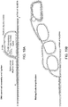
- the double-stranded nucleic acid may be produced by: a) hybridizing the sample with a padlock probe having ends that are complementary to the RCA primer, ligating the ends of the padlock probes together, and copying the padlock probe by rolling circle amplification and b) hybridizing an oligonucleotide to the RCA product, as illustrated in Fig. 16 .
- the RCA product is the first strand of the double-stranded nucleic acid
- the oligonucleotides that are hybridized to the RCA product are the second strand of the double-stranded nucleic acid.
- the labeling step may comprise crosslinking the capture agent to the planar sample so that subsequence manipulations can be done without the capture agent disassociating from its complementary sites in the planar sample.
- the crosslinking step may be done immediately after binding of the antibody to the sample.
- the sample may be cross-linked after binding of the antibody to the sample, and the double-stranded may be produced after crosslinking.
- planar sample refers to a substantially planar, i.e., two dimensional, material (e.g. glass, metal, ceramics, organic polymer surface or gel) that contains cells or any combinations of biomolecules derived from cells, such as proteins, nucleic acids, lipids, oligo/polysachharides, biomolecule complexes, cellular organels, cellular debris or excretions (exosomes, microvesicles).
- a substantially planar i.e., two dimensional, material (e.g. glass, metal, ceramics, organic polymer surface or gel) that contains cells or any combinations of biomolecules derived from cells, such as proteins, nucleic acids, lipids, oligo/polysachharides, biomolecule complexes, cellular organels, cellular debris or excretions (exosomes, microvesicles).
- a planar cellular sample can be made by, e.g., growing cells on a planar surface, depositing cells on a planar surface, e.g., by centrifugation, by cutting a three dimensional object that contains cells into sections and mounting the sections onto a planar surface, i.e., producing a tissue section, absorbing the cellular components onto the surface that is functionalized with affinity agents (e.g. antibodies, haptens, nucleic acid probes), introducing the biomolecules into a polymer gel or transferring them onto a polymer surface electrophoretically or by other means.
- affinity agents e.g. antibodies, haptens, nucleic acid probes
- the cells or biomolecules may be fixed using any number of reagents including formalin, methanol, paraformaldehyde, methanol:acetic acid, glutaraldehyde, bifunctional crosslinkers such as bis(succinimidyl)suberate, bis(succinimidyl)polyethyleneglycole etc.
- This definition is intended to cover cellular samples (e.g., tissue sections, etc), electrophoresis gels and blots thereof, Western blots, dot-blots, ELISAs, antibody microarrays, nucleic acid microarrays etc.
- tissue section refers to a piece of tissue that has been obtained from a subject, fixed, sectioned, and mounted on a planar surface, e.g., a microscope slide.
- FFPE formalin-fixed paraffin embedded
- spatialally-addressable measurements refers to a set of values that are each associated with a specific position on a surface. Spatially-addressable measurements can be mapped to a position in a sample and can be used to reconstruct an image of the sample.
- a “diagnostic marker” is a specific biochemical in the body which has a particular molecular feature that makes it useful for detecting a disease, measuring the progress of disease or the effects of treatment, or for measuring a process of interest.
- a “pathoindicative” cell is a cell which, when present in a tissue, indicates that the animal in which the tissue is located (or from which the tissue was obtained) is afflicted with a disease or disorder.
- a disease or disorder By way of example, the presence of one or more breast cells in a lung tissue of an animal is an indication that the animal is afflicted with metastatic breast cancer.
- complementary site is used to refer to an epitope for an antibody or aptamer, or a nucleic acid molecule if the capture agent is an oligonucleotide probe. Specifically, if the capture agent is an antibody, then the complementary site for the capture agent is the epitope in the sample to which the antibody binds. If the capture agent is an oligonucleotide probe, then the complementary site for the capture agent is a complementary sequence in a DNA or RNA molecule in the sample.
- epitope as used herein is defined as small chemical groups on the antigen molecule that is bound to by an antibody.
- An antigen can have one or more epitopes. In many cases, an epitope is roughly five amino acids or sugars in size.
- an epitope is roughly five amino acids or sugars in size.
- One skilled in the art understands that generally the overall three-dimensional structure or the specific linear sequence of the molecule can be the main criterion of antigenic specificity.
- a "subject" of diagnosis or treatment is a plant or animal, including a human.
- Non-human animals subject to diagnosis or treatment include, for example, livestock and pets.
- the term "incubating” refers to maintaining a planar sample and capture agent under conditions (which conditions include a period of time, a temperature, an appropriate binding buffer and a wash) that are suitable for specific binding of the capture agent to molecules (e.g., epitopes or complementary nucleic acid) in the planar sample.
- capture agent refers to an agent that can specifically bind to complementary sites in a planar sample.
- exemplary capture agents include, e.g., an antibody, an aptamer, and a nucleic acid (e.g., oligonucleotide) probe (which may be DNA or RNA) that hybridizes to a binding site. If antibodies are used, in many cases the antibodies may bind to protein epitopes. If nucleic acid probes are used, the nucleic acid probes may bind to, for example, genomic DNA or RNA (such that the location and abundance of intracellular RNAs can be detected).
- the term "extendible”, in the context of, for example, a 3' end that is “extendible using the other strand as a template”, means that a polymerase or ligase can add to the 3' end of a nucleic acid molecule, where the template sequence that is immediately downstream of the 3' end (i.e., on the other strand) determines which nucleotides (if a polymerase is used) or oligonucleotide (if a ligase is used) is added.
- a "5' end that is extendible using the other strand as a template” means that a ligase can add an oligonucleotide to the 5' end of a nucleic acid molecule, where the template sequence that is immediately downstream of the 5' end (i.e., on the other strand) determines which oligonucleotide is added.
- template sequence that is immediately downstream to the 3' end refers to the sequence on the other strand that use used as a template for extending the 3' end, starting with the first nucleotide.
- the template sequence that is immediately downstream of the 3' end may be a sequence in the RCA product.
- the template sequence that is immediately downstream of the 3' end may be a 5' overhang.
- the term "capture agent that is linked to a double stranded nucleic acid” refers to a capture agent, e.g., an antibody or an oligonucleotide probe, that is non-covalently (e.g., via a streptavidin/biotin interaction) or covalently (e.g., via a click reaction or the like) linked to an double-stranded nucleic acid (which may be composed of two single-stranded oligonucleotide strands that are hybridized together, or an RCA product that is hybridized to a plurality of oligonucleotides) in a way that the capture agent can still bind to its binding site and the 3' end of one of the nucleic acids is accessible to a polymerase and/or ligase.
- a capture agent e.g., an antibody or an oligonucleotide probe
- an oligonucleotide probe that is non-covalently (e.g.
- the nucleic acid and the capture agent may be linked via a number of different methods, including those that use maleimide or halogen-containing group, which are cysteine-reactive.
- the capture agent and the nucleic acid may be linked at, proximal to or at the 5' end of one of the strands of the double stranded nucleic acid, proximal to or at the 3' end of one of the strands of the double stranded nucleic acid, or anywhere in-between.
- nucleic acid and “polynucleotide” are used interchangeably herein to describe a polymer of any length, e.g., greater than about 2 bases, greater than about 10 bases, greater than about 100 bases, greater than about 500 bases, greater than 1000 bases, up to about 10,000 or more bases composed of nucleotides, e.g., deoxyribonucleotides, ribonucleotides or a combination thereof, and may be produced enzymatically or synthetically (e.g., PNA as described in U.S. Patent No.
- Naturally-occurring nucleotides include guanine, cytosine, adenine, thymine, uracil (G, C, A, T and U respectively).
- DNA and RNA have a deoxyribose and ribose sugar backbone, respectively, whereas PNA's backbone is composed of repeating N-(2-aminoethyl)-glycine units linked by peptide bonds.
- LNA locked nucleic acid
- a locked nucleic acid is a modified RNA nucleotide.
- the ribose moiety of an LNA nucleotide is modified with an extra bridge connecting the 2' oxygen and 4' carbon. The bridge "locks" the ribose in the 3'-endo (North) conformation, which is often found in the A-form duplexes.
- LNA nucleotides can be mixed with DNA or RNA residues in the oligonucleotide whenever desired.
- unstructured nucleic acid is a nucleic acid containing non-natural nucleotides that bind to each other with reduced stability.
- an unstructured nucleic acid may contain a G' residue and a C' residue, where these residues correspond to non-naturally occurring forms, i.e., analogs, of G and C that base pair with each other with reduced stability, but retain an ability to base pair with naturally occurring C and G residues, respectively.
- Unstructured nucleic acid is described in US20050233340 .
- oligonucleotide refers to a multimer of at least 10, e.g., at least 15 or at least 30 nucleotides. In some embodiments, an oligonucleotide may be in the range of 15-200 nucleotides in length, or more.
- reading in the context of reading a fluorescent signal, refers to obtaining an image by scanning or by microscopy, where the image shows the pattern of fluorescence as well as the intensity of fluorescence in a field of view.
- primer is an oligonucleotide, either natural or synthetic, that is capable, upon forming a duplex with a polynucleotide template, of acting as a point of initiation of nucleic acid synthesis and being extended from its 3' end along the template so that an extended duplex is formed.
- the sequence of nucleotides added during the extension process is determined by the sequence of the template polynucleotide.
- primers are extended by a DNA polymerase.
- a primer may be at least 10, e.g., at least 15 or at least 30 nucleotides in length.
- single nucleotide 5' overhang refers to a 5' overhang, where the overhang is a single nucleotide in length.
- a "two nucleotide 5' overhang” is a 5' overhang, where the overhang is two nucleotides in length. The 3' end is recessed in a 5' overhang.
- the various nucleotides of an overhang may be referred to by their position, e.g., "first position” and "second position”.
- the "position” is relative to the recessed 3' end.
- the "first” position of the overhang is immediately adjacent to the recessed 3' end and the "second" position of the overhang is immediately adjacent to the first position.
- the complementary strands of a double stranded oligonucleotide or nucleic acid may be referred to herein as being the “first" and “second” or the “top” and “bottom” strands.
- the assignment of a strand as being a “top” or “bottom” strand is arbitrary and does not imply any particular orientation, function or structure.
- fluorescently labeled oligonucleotide comprising a quencher refers to an oligonucleotide that contains a fluorophore and a quencher, wherein the quencher quenches the fluorophore in the same oligonucleotide.
- the term "different" in the context of different 5' overhangs that are different refers to overhangs that have a different sequence. Overhangs of different lengths (e.g., GATC vs GAT) implicitly have a different sequence, even through one sequence may be encompassed by the other.
- the term "overhang” refers to a structure in which one strand of a double stranded nucleic acid ends such that nucleic acid synthesis can be initiated from that strand by a polymerase (or an oligonucleotide can be ligated to the end by a ligase) using the other strand as a template.
- the term "adding to the extendible 3' end" in the context of adding one or more nucleotides or an oligonucleotide to an extendible 3' end, refers to adding nucleotides (or an oligonucleotide) to an extendible 3' end using the other strand as a template (e.g., adding to the recessed 3' end of a 5' overhang using the overhang as a template).
- alternating stretches refers to two nucleotides stretches, where one "stretch” is a contiguous sequence of, e.g., up to 10, of the same nucleotide (e.g., a G, A, T or C), and the second stretch is contiguous sequence of, e.g., up to 10, of a different nucleotide, that alternate with one another, i.e., one stretch (e.g., a string of T's) occupies the odd positions and the other stretch (e.g., a string of A's) occupies the even positions.
- one stretch e.g., a string of T's
- the other stretch e.g., a string of A's
- incomplete nucleotide mix comprises a nucleotide mix that contains one, two or three nucleotides (but not all four nucleotides) selected from G, A, T and C.
- the nucleotides may be labeled or unlabeled.
- reversible terminator refers to a chemically modified nucleotide base that when incorporated into growing DNA strand by DNA polymerase blocks further incorporation of bases. Such "reversible terminator" base and DNA strand can be deprotected by chemical treatment and following such deprotection DNA strand can be further extended by DNA polymerase.
- fluorescently labeled reversible terminator refers to a "reversible terminator" base which is labeled by fluorophore through linker cleavable by same treatment which is used to deprotect the DNA strand which ends with this base. Deprotecting the "fluorescently labeled reversible terminator” simultaneously activates the DNA strand for further extension and removes the fluorescent label from it.
- the method comprises producing a labeled a planar sample (e.g., an FFPE section mounted on a planar surface such as a microscope slide) using a capture agent that specifically binds to complementary sites in the planar sample.
- a planar sample e.g., an FFPE section mounted on a planar surface such as a microscope slide
- a capture agent that specifically binds to complementary sites in the planar sample.
- the capture agent in the labeled sample is linked to a double-stranded nucleic acid that comprises a first strand and a second strand (e.g., two oligonucleotide that are hybridized together or an RCA product that is hybridized to oligonucleotides) and the capture agent is linked (covalently or non-covalently via a biotin) to the double-stranded nucleic acid by the first strand of the double-stranded nucleic acid (e.g., by the 5' end, the 3' end, or anywhere in-between), and the 3' end or 5' end of one of the strands (e.g., the 3' end of the first strand, any 3' ends in the second strand, the 5' end of the first strand or any 5' ends in the second strand) is extendible using the other strand as a template.
- a double-stranded nucleic acid that comprises a first strand and a second strand
- the capture agent
- the capture agent is cross-linked the planar sample, thereby preventing the capture agent from disassociating during subsequent steps.
- This crosslinking step may be done using any amine-to-amine crosslinker (e.g. formaldehyde, disuccinimiyllutarate or another reagents of similar action) although a variety of other chemistries can be used to cross-link the capture agent to the planar sample if desired.
- the method comprises reading a fluorescent signal generated by addition of a nucleotide or short oligonucleotide (e.g., of 2-10 bases) to the extendible end (e.g., the 3' end) of one of the strands.
- This step may be done by contacting the planar sample with a polymerase and a nucleotide mix, a ligase and a labeled oligonucleotide, or a combination of the two, thereby adding one or more nucleotides and/or a labeled oligonucleotide to the extendible end; and reading a fluorescent signal generated by addition of the one or more nucleotides or oligonucleotide to the extendible end.
- the fluorescent signal may be generated by a variety of different methods.
- the fluorescent signal may be fluorescence from a fluorescent nucleotide added to the end of the primer, or a FRET (fluorescence resonance energy transfer) signal resulting from the same.
- the signal may generated by removing a quencher from a fluorescently labeled oligonucleotide that is also hybridized to the oligonucleotide.
- the reading step may be followed by inactivating the fluorescence after reading so that other binding events can be detected and read.
- the fluorescence may be inactivated by peroxide-based bleaching, cleavage of fluorophore linked to nucleotide through cleavable linker (e.g. using TCEP as a cleaving reagent), base-exchange by exo+ polymerase such as Vent, or subsequent incorporation of quencher, for example.
- the method may be multiplexed in a way that a single planar sample can be interrogated by a plurality of different capture agents, where each antibody is linked to a different oligonucleotide (i.e., oligonucleotides of different sequence).
- the planar sample may be labeled using at least 5, at least 10, at least 20, at least 30, at least 50, or at least 100, up to 150 or more capture agents that are each linked to a different oligonucleotide, and binding of the capture agents can be separately read using a fluorescence microscope equipped with an appropriate filter for each fluorophore, or by using dual or triple band-pass filter sets to observe multiple fluorophores.
- the oligonucleotides linked to the capture agent may act as a splint for a padlock probe, and as a primer for initiating rolling circle amplification.
- the capture agent used in some embodiments of the method may be linked to a double-stranded oligonucleotide that contains a 5' overhang (i.e., a recessed 3' end that can be extended by a polymerase or ligase) or a 3' overhang (i.e., a recessed 5' end that can be extended by a ligase).
- a 5' overhang i.e., a recessed 3' end that can be extended by a polymerase or ligase
- a 3' overhang i.e., a recessed 5' end that can be extended by a ligase
- the overhang is a single nucleotide overhang (e.g., an A), although a longer overhang (e.g., at least 2, at least 3, at least 4, at least 5, at least 6, at least 8, at least 10, at least 20, or at least at least 30, may be useful for other applications (e.g., multiplexed applications).
- the overhang may contain a repeated sequence, e.g., 2, 3, 4, 5, or 6 or more repeats of the same sequence of 2, 3, 4, 5 or 6 nucleotides, thereby allowing the capture agent to be used in multiplexed applications as described below.
- the double stranded oligonucleotide may have a recessed 3' end at the other end of the oligonucleotide (i.e., at the end closest to the capture agent). However, this end may be designed to be not extendible.
- the double-stranded oligonucleotide may contain one or more third oligonucleotides that are hybridized to the overhang. In these embodiments, there will be a gap of 1, 2, 3, 4 or 5 or more nucleotides between the second strand of the double-stranded oligonucleotide and the oligonucleotide that is hybridized to the overhang (see, e.g., Figs. 7 and 8 ).
- the plurality of capture agents may be distinguished by the sequence of the overhang and not by the sequence of the first strand of the double stranded oligonucleotide.
- the second strand of the double stranded oligonucleotides is different for each of the capture agents.
- the method may also be implemented using capture agents that are linked to a primer that acts a splint for circularlizing a padlock probe and for priming amplification of circularlized padlock probe by rolling circle amplification.
- the capture agents in the labeled sample may be linked to a rolling circle amplification product.
- the fluorophore used may be a coumarin, a cyanine, a benzofuran, a quinoline, a quinazolinone, an indole, a benzazole, a borapolyazaindacene and or a xanthene including fluorescein, rhodamine and rhodol.
- fluorophores may be chosen so that they are distinguishable, i.e., independently detectable, from one another, meaning that the labels can be independently detected and measured, even when the labels are mixed.
- the amounts of label present (e.g., the amount of fluorescence) for each of the labels are separately determinable, even when the labels are co-located (e.g., in the same tube or in the same area of the section).
- fluorescent dyes of interest include: xanthene dyes, e.g., fluorescein and rhodamine dyes, such as fluorescein isothiocyanate (FITC), 6-carboxyfluorescein (commonly known by the abbreviations FAM and F), 6-carboxy-2',4',7',4,7-hexachlorofluorescein (HEX), 6-carboxy-4', 5'-dichloro-2', 7'-dimethoxyfluorescein (JOE or J), N,N,N',N'-tetramethyl-6-carboxyrhodamine (TAMRA or T), 6-carboxy-X-rhodamine (ROX or R), 5-carboxyrhodamine-6G (R6G 5 or G 5 ), 6-carboxyrhodamine-6G (R6G 6 or G 6 ), and rhodamine 110; cyanine dyes, e.g., Cy3, Cy5
- phenanthridine dyes e.g., Texas Red
- ethidium dyes e.g., acridine dyes
- carbazole dyes e.g., phenoxazine dyes
- porphyrin dyes e.g., polymethine dyes, e.g., BODIPY dyes and quinoline dyes.
- fluorophores of interest that are commonly used in subject applications include: Pyrene, Coumarin, Diethylaminocoumarin, FAM, Fluorescein Chlorotriazinyl, Fluorescein, R110, Eosin, JOE, R6G, Tetramethylrhodamine, TAMRA, Lissamine, Napthofluorescein, Texas Red, Cy3, and Cy5, etc.
- Suitable distinguishable fluorescent label pairs useful in the subject methods include Cy-3 and Cy-5 (Amersham Inc., Piscataway, NJ), Quasar 570 and Quasar 670 (Biosearch Technology, Novato CA), Alexafluor555 and Alexafluor647 (Molecular Probes, Eugene, OR), BODIPY V-1002 and BODIPY V1005 (Molecular Probes, Eugene, OR), POPO-3 and TOTO-3 (Molecular Probes, Eugene, OR), and POPRO3 and TOPRO3 (Molecular Probes, Eugene, OR). Further suitable distinguishable detectable labels may be found in Kricka et al. (Ann Clin Biochem. 39:114-29, 2002 ), Ried et al. (Proc. Natl. Acad. Sci. 1992: 89: 1388-1392 ) and Tanke et al. (Eur. J. Hum. Genet. 1999 7:2-11 ) and others.
- the sample may be stained using a cytological stain, either before or after performing the method described above.
- the stain may be, for example, phalloidin, gadodiamide, acridine orange, bismarck brown, barmine, Coomassie blue, bresyl violet, brystal violet, DAPI, hematoxylin, eosin, ethidium bromide, acid fuchsine, haematoxylin, hoechst stains, iodine, malachite green, methyl green, methylene blue, neutral red, Nile blue, Nile red, osmium tetroxide (formal name: osmium tetraoxide), rhodamine, safranin, phosphotungstic acid, osmium tetroxide, ruthenium tetroxide, ammonium molybdate, cadmium iodide,
- the stain may be specific for any feature of interest, such as a protein or class of proteins, phospholipids, DNA (e.g., dsDNA, ssDNA), RNA, an organelle (e.g., cell membrane, mitochondria, endoplasmic recticulum, golgi body, nulear envelope, and so forth), a compartment of the cell (e.g., cytosol, nuclear fraction, and so forth).
- the stain may enhance contrast or imaging of intracellular or extracellular structures.
- the sample may be stained with haematoxylin and eosin (H&E).
- nucleotides are only exemplary and other nucleotides, including nucleotides that are cleavable by other stimuli (e.g., photocleavable nucleotides) can be used in the present method.
- the fluorescent signal may be produced by a fluorescent nucleotide that is added to (i.e., added by a polymerase or, if the fluorescent nucleotide is in an oligonucleotide, ligated onto) the 3' end of the primer.
- This method may comprise reading a signal from the added fluorescent nucleotide, or reading a FRET signal generated by energy transfer between two fluorescent nucleotides that are added to the primer.
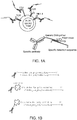
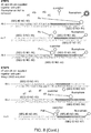
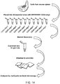
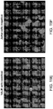
- Figs. 1 and 2 shows how an antibody can be linked to a oligonucleotide chemically, or via biotin/streptavidin interactions ( Fig. 1B ) and how a fluorescent signal can be generated by adding a fluorescent nucleotide to the end of the primer ( Fig. 2 ).
- the antigen is stained by an antibody that is coupled to a DNA dimer with an overhanging 5' end (lower strand) and recessed 3' end (upper strand) either chemically ( Fig. 1 top panel) or through streptavidin ( Fig. 1 bottom and middle panels).
- the pattern of binding of the capture agent may be determined using an on-slide end fill-in reaction by using a suitable polymerase (e.g., by exo - Klenow, Bst, Taq, Klentaq, or an exo - Klenow-Vent mixture) and fluorescently labeled nucleotide ( Fig,1 and Fig.2 top panel).
- a suitable polymerase e.g., by exo - Klenow, Bst, Taq, Klentaq, or an exo - Klenow-Vent mixture
- fluorescently labeled nucleotide Fig,1 and Fig.2 top panel
- the signal-to-noise ratio can be increased by: a) multimerization of position complementary to labeling nucleotide ( Fig. 2 , middle panel); or b) by generating a FRET between two nucleotides are incorporated, whereby the emission wavelength of one of the nucleotides ( Fig. 2 , bottom panel C on the figure) serves as an excitation wavelength for another ( Fig. 2 , bottom panel U on the figure).
- Fluorescence may be inactivated before addition of subsequent staining reagents by any convenient method including, but not limited to photobleaching, peroxide-based bleaching, inactivation by ozone, cleavage of fluorophore linked to nucleotide through cleavable linker (e.g. using TCEP as a cleaving reagent), base-exchange by exo+ polymerase such as Vent, subsequent incorporation of quencher.
- any convenient method including, but not limited to photobleaching, peroxide-based bleaching, inactivation by ozone, cleavage of fluorophore linked to nucleotide through cleavable linker (e.g. using TCEP as a cleaving reagent), base-exchange by exo+ polymerase such as Vent, subsequent incorporation of quencher.
- the method can be repeated, i.e., the planar sample may be re-stained using a different antibody and fluorescence can be read.
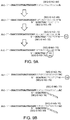
- oligonucleotides can be implemented using specially designed oligonucleotides using two different approaches, referred to as the "reversible terminator” and “missing base” approaches, which are described in greater detail below. Both of these methods rely on a composition comprising a plurality of (e.g., at least 5, at least 10, at least 20, at least 30, at least 50, or at least 100, up to 150 or more) capture agents that recognize different complementary sites, wherein: each of the capture agents is linked to a double-stranded nucleic acid (e.g., oligonucleotide) that comprises a first strand and a second strand; the capture agents are linked to a double-stranded nucleic acid by the (e.g., the 5' end of) the first strand; the 3' end of one of the strands in each of the double-stranded nucleic acids extendible using the other strand as a template, where the template is different for each of the capture agents.
- a composition compris
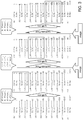
- Figs. 3 and 4 Examples of such compositions are illustrated in Figs. 3 and 4 , where the template is an overhang.
- the general principle shown in Figs. 3 and 4 can be extended to double stranded nucleic acids that comprise RCA products.
- Fig. 3 shows a population of capture agents that have a template (e.g., overhang) defined by the formula: 3'- N 4n N 1 /N 2 /N 3 -5' followed by short stretch of random composition on the 5' end to increase the overall polymerase residence on the DNA duplex, where N 1 , N 2 , N 3 and N 4 are different nucleotides selected from G, A, T and C and n is 0, 1 or more.
- Fig. 3 shows a population of capture agents that have a template (e.g., overhang) defined by the formula: 3'- N 4n N 1 /N 2 /N 3 -5' followed by short stretch of random composition on the 5' end to increase the overall polymerase residence on the
- Y is a nucleotide sequence of length n (n is 0, 1 or more) composed of bases N 3 and N 4 , wherein nucleotide N 3 is in odd positions and nucleotide N 4 is in even positions, counting from the start of the overhang and N 1 , N 2 , N 3 and N 4 are different nucleotides selected from G, A, T and C.
- the sequence of the first strand is the same for each of the capture agents; and the sequence of the second strand is different for each of the capture agents.
- the different second strands make the overhangs different between the different capture agents.
- the multiplex methods may generally comprise: (a) incubating a planar sample with an above-described antibody composition under conditions by which the capture agents bind to complementary sites in the planar sample; (b) cross-linking the capture agents to the planar sample; (c) contacting the planar sample with a polymerase and either an incomplete nucleotide mix of labeled and unlabeled nucleotides or a nucleotide mix where some or all nucleotides are fluorescent and some or all nucleotides are reversible terminator nucleotides or fluorescent reversible terminator nucleotides and optionally, contacting the planar sample with a mixture of labelled and unlabeled oligonucleotides and a DNA ligase enzyme that covalently attaches the short labelled oligonucleotides to the 3'end of the oligonucleotide duplexes that are attached to the specific capture agents.
- oligonucleotides are only added to duplexes that an overhang that is complementary to the oligonucleotide.
- This method further comprises (d) reading, using fluorescence microscopy, a fluorescent signal generated by addition a nucleotide to some but not all of the capture agents. Following signal registration, this method may comprise (e) removing the fluorescent signals by chemical or photocleavage of a labeled nucleotide if the reversible terminator approach is used, followed by deprotecting the 3' ends of the oligonucleotides, enabling the addition of further nucleotides and/or oligonucleotides.
- Step (c) of this method may comprise (c) contacting the planar sample with a polymerase and: (i) a nucleotide mix that comprises fluorescent nucleotides that are complementary to N 1 , N 2 and N 3 and a reversible terminator nucleotide that is complementary to N 4 or (ii) a nucleotide mix that comprises fluorescent reversible terminator nucleotides that are complementary to N 1 , N 2 and N 3 and a reversible terminator nucleotide that is complementary to N 4 or (iii) a nucleotide mix that comprises fluorescent nucleotides that are complementary to N 1 , and N 2 , an unlabeled nucleotide that is complementary to N 3 , and no nucleotide that is complementary to N 4 , thereby adding fluorescent nucleotides onto the double-stranded oligonucleotides of some but not all of the capture agents thereby adding fluorescent nucleotides onto the double-strande
- the length of the read over the oligonucleotide overhangs may increase accordingly. This may or may not reduce the efficiency of staining due to accumulation of primer extension errors along the length of the oligonucleotide duplex.
- the plurality of capture agents can be divided in sets such that number of capture agents in the set does exceed the capacity of the multiplexing protocol to render staining without significant signal loss (e.g.30).
- Each such set of capture agents will be conjugated to "terminated” (the last 3' base is dideoxy- or propyl- modified) upper strand oligonucleotide of the same sequence as in the original version of the "missing base” approach.
- the lower strand oligonucleotides will incorporate an additional set-specific region which will serve as a landing spot for an additional primer which is to be on-slide hybridized to the particular subset of the total plurality of the antibodies at the time when they are to be rendered. This approach allows not to extend the reads beyond certain threshold and at the same time have an unlimited potential number of capture agents in the sample.
- This implementation of the method relies on reversible terminators, i.e., chain terminator nucleotides that can be de-protected after incorporation, thereby allowing further nucleotides to be added to that nucleotide.
- This method can be implemented using a composition comprising a plurality of capture agents that are linked to a double stranded nucleic acid (e.g., oligonucleotides), as illustrated in Fig. 3 .
- a double stranded nucleic acid e.g., oligonucleotides
- the top strand of the double stranded nucleic acid is linked to the capture agent and may be the same for each antibody, and the sequence of the bottom strand varies between capture agents.
- the 5' end of the lower strand of the double-stranded nucleic acid (which may form an overhang) is of the general 3'- N 4n N 1 /N 2 /N 3 -5' followed by short stretch of random nucleotides on the 5' end to increase the overall polymerase residence on the DNA duplex, where N 1 , N 2 , N 3 and N 4 are different nucleotides selected from G, A, T and C and n is 0, 1 or more.
- this method may comprise: (a) incubating a planar sample with a multiplex antibody composition in which the overhangs are of the formula 5'-N 1 /N 2 /N 3 N 4n , wherein N 1 , N 2 , N 3 and N 4 are different nucleotides selected from G, A, T and C and n is 1 or more; under conditions by which the capture agents specifically bind to complementary sites in the planar sample; (b) cross-linking the capture agent to the planar sample; (c) contacting the planar sample with a polymerase and a nucleotide mix that comprises fluorescent nucleotides that are complementary to N 1 , N 2 and N 3 and a reversible terminator nucleotide that is complementary to N 4 and/or ligating a oligonucleotide that comprises a labeled nucleotide; and (d) reading, using fluorescence microscopy, a fluorescent signal generated by addition of a nucleotide to some
- This cycle may be repeated by (e) inactivating the fluorescent signal, deprotecting the reversible terminator nucleotide and (f) blocking the planar sample; and repeating steps (c) and (d).
- the method may comprise repeating steps (c), (d) (e) and (f) multiple times.
- the reagent used for blocking may vary depending on the chemistry used.
- the sample may be blocked with a thiol-reactive compounds such as cysteine, glutathione or iodoacetamide.
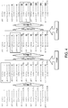
- this method can be implemented using a composition comprising: a first antibody linked to a first double stranded oligonucleotide, wherein the first double stranded oligonucleotide comprises a single nucleotide 5' overhang comprising base N 1 ; a second antibody linked to a second double stranded oligonucleotide, wherein the second double stranded oligonucleotide comprises a single nucleotide 5' overhang comprising base N 2 ; a third antibody linked to a third double stranded oligonucleotide, wherein the third double stranded oligonucleotide comprises a single nucleotide 5' overhang comprising base N 3 ; a fourth antibody linked to a fourth double stranded oligonucleotide comprises a two nucleotide 5' overhang, wherein the first position of the overhang comprises base N 4 and the second position of the overhang is base N
- the strand linked to the antibodies may be different for each of the antibodies, where the RCA product contains a sequence conforming to the formula described above in each repeat of the RCA product.
- the composition may also contain a seventh antibody linked to a seventh double stranded oligonucleotide, wherein the seventh double stranded oligonucleotide comprises a multiple nucleotide 5' overhang, wherein the first position of the overhang comprises base N 4 , the second position of the overhang is base N 4 and third is selected from N 1 , N 2 , and N 3 .
- the same principle may be applied to overhangs that have more than 7 positions (e.g., 9, 10, 11 up to 20, 30, or 40 ore more) positions.
- the planar sample can be co-stained simultaneously using a panel of capture agents, each labeled with one oligonucleotide duplex designed according to the strategy outlined on Fig. 3 .
- the duplexes are designed in such a way that each antibody has the same upper strand sequence linked, covalently or through streptavidin, to an antibody through the 5' end.
- the lower strand changes from antibody to antibody.
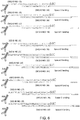
- the general formula for the lower strand is 3'-dideoxydC -sequence-complimentary-to-upper-strand G n A/T/C-5'.
- nucleotide G is reserved for step-wise progression and its complementary pair on the upper strand is never used in labeled form.
- the other three bases are complementary to labeled nucleotides and can be used to identify three capture agents per cycle.
- the general formula for the lower strand is 3'- dideoxydC-sequence-complimentary-to-upper-strand- X-N 1 /N 2 /N 3 -5' where X i of X is any nucleotide excluding one reserved for "walking base" of this particular cycle and X is any base as shown on Fig. 5B .
- Each cycle includes: (a) a labeling step in which the three capture agents are labeled and duplexes on the rest are extended one base at a time, (b) an imaging step and (c) a destaining/deprotection step.
- the added fluorescent labels from the previous cycle are inactivated by any of the suitable methods, including but not limited to: cleavage of fluorophore off the nucleotide (if the labeled nucleotide is linked to the fluorophore through a cleavable linker); peroxide based bleaching; photobleaching; chemically-assisted photobleaching; labeled base replacement by exo+ polymerase, etc.
- the unlabeled "extension" nucleotide that has been added to the remainder of the capture agents is activated by cleavage of the protective group off its 3' end.
- This implementation of the method relies on a "missing" base design in which, in each cycle, two labeled and one unlabeled nucleotides are added to the reaction, and the "missing base” prevents the primers from being extended by more than a single nucleotide.
- This method can be implemented using a composition comprising a plurality of capture agents that are linked to double stranded nucleic acids, as illustrated in Fig. 4 .
- the top strand of the double stranded nucleic acids is linked to the capture agent and may be the same for each antibody, and the sequence of the bottom strand varies between capture agents. As shown in Fig.
- the 5' end of the lower strand of the double-stranded oligonucleotide (which forms the overhang) is of the general formula 3'-YN 1 /N 2 -5', optionally followed by short stretch (e.g., 1-5 residues) of random nucleotides on the 5' end to increase the overall polymerase residence on the DNA duplex, wherein Y is a nucleotide sequence of length n (n is 0, 1 or more) composed of bases N 3 and N 4 , wherein nucleotide N 3 is in odd positions and nucleotide N 4 is in even positions, counting from the start of the overhang and N 1 , N 2 , N 3 and N 4 are different nucleotides selected from G, A, T and C.
- N 1 , N 2 , N 3 and N 4 are different nucleotides selected from G, A, T and C and Y is a nucleotide sequence of length n (n is 0, 1 or more) composed of alternating random length stretches of bases N 3 and N 4 such that the order number of N 3 - stretches is odd and of N 4 stretches is even, may be applicable in this method
- this method may comprise: (a) incubating a planar sample with a multiplex antibody composition in which the overhangs are of the formula (3'-YN 1 /N 2 -5') described in the prior paragraph ; under conditions by which the capture agents specifically bind complementary sites in the planar sample; (b) cross-linking the capture agent to the planar sample; (c) contacting the planar sample with a polymerase and a nucleotide mix that comprises fluorescent nucleotides that are complementary to N 1 , and N 2 , an unlabeled nucleotide that is complementary to N 3 and no nucleotide that is complementary to N 4 and/or ligating an oligonucleotide that has a labeled nucleotide; and (d) reading, using fluorescence microscopy, a fluorescent signal generated by addition of a nucleotide to some but not all of the capture agents.
- This cycle may be repeated by (e) inactivating the fluorescent signal, (f) blocking the sample and contacting the planar sample with a polymerase and an unlabeled nucleotide that is complementary to N 4 and/or contacting the sample with a labeled oligonucleotide and a ligase; and repeating steps (c) (d).
- the method may comprise repeating steps (c), (d), (e) and (f) multiple times.
- This method can be implemented using a capture agent composition that comprises: a first antibody linked to a first double stranded oligonucleotide, wherein the first double stranded oligonucleotide comprises a single nucleotide 5' overhang comprising base N 1 ; a second antibody linked to a second double stranded oligonucleotide, wherein the second double stranded oligonucleotide comprises a single nucleotide 5' overhang comprising base N 2 ; a third antibody linked to a fourth double stranded oligonucleotide, wherein the third double stranded oligonucleotide comprises a two nucleotide 5' overhang, wherein the first from the 3' position of the overhang comprises base N 4 and the second position comprises N 1 ; and a fourth antibody linked to a fourth double stranded oligonucleotide, wherein the fourth double stranded oligonucleot
- the composition may also contain a fifth antibody linked to a fifth double stranded oligonucleotide, wherein the fifth double stranded oligonucleotide comprises a multiple nucleotide 5' overhang, wherein the first position of the overhang comprises base N 4 , the second position comprises base N 3 , and the third position comprises N 1 or N 2 .
- the duplexes can be designed using the strategy shown in Fig. 4 , i.e., in such a way that each antibody has the same upper stand oligonucleotide sequence linked to it via covalent bond or through a streptavidin-biotin interaction.
- the lower strand changes from antibody to antibody.
- the general formula for the lower strand is 3' ddC -sequence-complimentary-to-upper-strand -YA/N 2 -5' where Y is composed of bases T and C such that T can be found only in even and C only at odd positions.
- N 1 , N 2 , N 3 and N 4 are different nucleotides selected from G, A, T and C and Y is a nucleotide sequence of length n (n is 0, 1 or more) composed of alternating random length stretches of bases N 3 and N 4 such that the order number of N 3 - stretches is odd and of N 4 stretches is even.
- n is 0, 1 or more
- two base pairs of the lower strand (T and C as in exemplary design on Fig.4 ) are reserved for step-wise progression and their complementary pair on the upper strand is never labeled.
- each cycle can have three steps: a labeling step in which the two capture agents are labeled by incorporation of fluorescent dNTPs and all of the other duplexes are extended one base at a time, an imaging step, and a de-staining/reactivation step.
- the strand linked to the antibodies may be different for each of the antibodies, where the RCA product contains a sequence conforming to the formula described above in each repeat of the RCA product.
- the labeled capture agents from the prior cycle can be bleached/destained in the same way as described above.
- a quencher labeled nucleotide can be incorporated after the labeled base. Because, in this embodiment, the position that is labeled is the last position in the overhang, the labeled capture agents from prior cycle cannot be re-labeled in later cycles because all nucleotide positions in the overhang have been filled in.
- the performance of "reversible terminator method" as exemplified in sequential detection of CD4 and CD8 positive T-cells in smears of mouse splenocytes is illustrated in fig. 13 , 15 and figure 16 .
- extension of a primer by nick translation removes a quencher from a fluorescently labeled "detector" oligonucleotide that is hybridized to the lower strand oligonucleotide in such a way that is positioned downstream from the upper strand primer.
- detector fluorescently labeled oligonucleotide
- the multiplexed implementations may comprise: (a) incubating the planar sample with a plurality of capture agents that are linked to a double-stranded oligonucleotide; (b) crosslinking the capture agents to the planar sample; (c) extending a primer that is hybridized to the oligonucleotide of a first set of capture agents of the plurality, thereby generating a first set of fluorescent signals (e.g., by removing the quencher from a labeled oligonucleotide that is hybridized to the oligonucleotide downstream from the primer), e.g., by adding a nucleotide using a polymerase or by adding an oligonucleotide using a ligase; (d) reading the first set of fluorescent signals using fluorescence microscopy; (e) inactivating the fluorescence; (f) extending a primer that is hybridized to the oligonucleotide of a
- the architecture of the double-stranded oligonucleotides linked to the capture agent has a specific design which is effectively enabling rendering of the capture agent binding pattern by "nick translation".
- the duplex of the upper strand and the lower strand oligonucleotide with long 5' overhang of the lower strand is further hybridized to a small detector oligonucleotide labeled both by fluorescent and the quencher.
- nick translating polymerase such as DNA pol I removes the quencher from some but not all of the quenched fluorescently labeled oligonucleotides, thereby generating a fluorescent signal for some but not all of the capture agents.
- the method generally comprises: (i) labeling a planar sample with: i. a first antibody, wherein the first antibody is linked to a first oligonucleotide duplex comprising, lower strand oligonucleotide with a unique sequence hybridized thereto: (i) an oligonucleotide upper strand "primer” and (ii) a labeled upper strand oligonucleotide comprising a 5' quencher at a site that is downstream from the primer; and a fluorophore downstream from the quencher and ii.
- a second antibody wherein the second antibody is linked to a second oligonucleotide duplex comprising, lower strand oligonucleotide with unique sequence hybridized thereto: (i) an oligonucleotide upper strand "primer” and (ii) an upper strand oligonucleotide labeled both by fluorophore and a quencher; wherein the gap between the 3' end of the primer and the 5' end of the labeled oligonucleotide is different for the first and second oligonucleotides; (ii) incubating the tissue sample with a first nucleotide mix and a polymerase, thereby removing the quencher from only the labeled oligonucleotide that is hybridized to the first oligonucleotide and producing a first fluorescent signal; (iii) reading the first fluorescent signal using fluorescence microscopy; (iv) inactivating the fluorescent signal by further progression of nick-trans
- Fig's 7 and 8 show an example of this method.
- the multiplexing method shown in Fig. 8 has the following steps:
- the method comprises rending antibody staining by primer extension with a fluorophore labeled base or otherwise reading a FRET signal generated by energy transfer between a first fluorescent nucleotide added to the primer by primer extension and a second nucleotide that is present in the oligonucleotide Fig.10 .
- the principles of this method are illustrated in Fig. 9A .
- the multiplexing is achieved by removing the extension priming oligonucleotide by melting the duplex or by exonuclease and reannealing another primer oligonucleotide which is extendable on a different antibody.
- a multiplexed version of this method is shown in Fig. 9B .
- the multiplexed implementations may comprise: (a) incubating the planar sample with a plurality of capture agents; (b) cross-linking the capture agents to the planar sample; (c) extending a primer that is hybridized to the oligonucleotide of a first set of capture agents of the plurality (e.g., wherein the 3' end of the first primer anneals to only the oligonucleotide of the first population), thereby generating a first set of fluorescent signals (which step can be done by adding a labeled nucleotide using polymerase and/or contacting the sample with a labeled oligonucleotide and a ligase); (d) reading the first set of fluorescent signals using fluorescence microscopy; (e) inactivating the fluorescence; (f) extending a primer that is hybridized to the oligonucleotide of a second set of capture agents of the plurality (e.g., wherein the 3'
- this method comprises: (a) incubating the planar sample with (i) a first antibody that is linked to a first labeled oligonucleotide and (ii) a second antibody that is linked to a second labeled oligonucleotide, (b) cross-linking the capture agents to the planar sample; (c) hybridizing the first and second labeled oligonucleotides with a first primer, wherein the 3' end of the first primer anneals to only the first labeled oligonucleotide; (d) extending the primer with a fluorescent nucleotide (which step can be done by adding a labeled nucleotide using polymerase and/or contacting the sample with a labeled oligonucleotide and a ligase); (e) reading, by fluorescence microscopy, a FRET signal generated by energy transfer between the label of the first oligonucleotide and the fluorescent nucleotide
- Figs. 9-10 shows an example of this method.
- the method shown in Figs. 8-11 has the following steps:
- the signal may be amplified using rolling circle amplification.
- a capture agent that is linked to an oligonucleotide is hybridized to a padlock probe that hybridizes to the oligonucleotide in such a way that the ends of the padlock probe are ligatably adjacent.
- the padlock probe (which is now circularized) can be copied by a rolling circle amplification reaction that is primed by the oligonucleotide. This reaction results in a concatamer of the padlock probe that contains several (in many cases hundreds or thousands) of copies of the same sequence in tandem that is linked to the capture agent.
- the rolling circle amplification product (which is linked to the antibody) can be detected using methods described above and, as illustrated, the signal is amplified because the sequence being detected is repeated.
- the (i) the capture agent is linked to a double-stranded nucleic acid that comprises a first strand (i.e., the RCA product) and a second strand (comprising the detection oligonucleotides). Single molecules can be detected using such methods.
- Fig. 19 shows how RNA molecules can be detected using a padlock probe/RCA amplification approach.
- the padlock probe hybridizes to the same mRNA as the capture agent (the "splint-primer"), thereby ensuring that the padlock probe circularizes only in the presence of the target RNA.
- the splint-primer hybridizes to the target RNA, acts as a splint for the padlock probe, and also acts as a primer for rolling circle amplification, thereby allowing the signal to be amplified in a similar to Fig. 16 .
- Fig. 20 shows an alternative method that relies on primer extension and the ligation of a short, labeled oligonucleotide.
- ligation of short labeled oligonucleotide to the top strand oligonucleotide only occurs after the overhang has been filed in to a certain point.
- a labeled oligonucleotide can be added to either the 3' end or the 5' end of the extendible end.
- compositions described herein find general use in a wide variety of application for analysis of any planar sample (e.g., in the analysis of tissue sections, sheets of cells, spun-down cells, blots of electrophoresis gels, Western blots, dot-blots, ELISAs, antibody microarrays, nucleic acid microarrays etc).
- planar sample e.g., in the analysis of tissue sections, sheets of cells, spun-down cells, blots of electrophoresis gels, Western blots, dot-blots, ELISAs, antibody microarrays, nucleic acid microarrays etc).
- the planar sample may be a section of a tissue biopsy obtained from a patient.
- Biopsies of interest include both tumor and non-neoplastic biopsies of skin (melanomas, carcinomas, etc.), soft tissue, bone, breast, colon, liver, kidney, adrenal, gastrointestinal, pancreatic, gall bladder, salivary gland, cervical, ovary, uterus, testis, prostate, lung, thymus, thyroid, parathyroid, pituitary (adenomas, etc.), brain, spinal cord, ocular, nerve, and skeletal muscle, etc.
- capture agents specifically bind to biomarkers, including cancer biomarkers, that may be proteinaceous or a nucleic acid.
- cancer biomarkers include, but are not limited to carcinoembryonic antigen (for identification of adenocarcinomas), cytokeratins (for identification of carcinomas but may also be expressed in some sarcomas), CD15 and CD30 (for Hodgkin's disease), alpha fetoprotein (for yolk sac tumors and hepatocellular carcinoma), CD117 (for gastrointestinal stromal tumors), CD10 (for renal cell carcinoma and acute lymphoblastic leukemia), prostate specific antigen (for prostate cancer), estrogens and progesterone (for tumour identification), CD20 (for identification of B-cell lymphomas) and CD3 (for identification of T-cell lymphomas).
- carcinoembryonic antigen for identification of adenocarcinomas
- cytokeratins for identification of carcinomas but may also be expressed in some sarcomas
- the above-described method can be used to analyze cells from a subject to determine, for example, whether the cell is normal or not or to determine whether the cells are responding to a treatment.
- the method may be employed to determine the degree of dysplasia in cancer cells.
- the cells may be a sample from a multicellular organism.
- a biological sample may be isolated from an individual, e.g., from a soft tissue.
- the method may be used to distinguish different types of cancer cells in FFPE samples.
- the method described above finds particular utility in examining planar samples using a plurality of antibodies, each antibodies recognizing a different marker. Examples of cancers, and biomarkers that can be used to identify those cancers, are shown below. In these embodiments, one does not need to examine all of the markers listed below in order to make a diagnosis.
- the method may be employed to detect the location and, optionally, the abundance of DNA molecules and/or RNA molecules in situ.
- the method may be used to detect intracellular RNAs.
- the capture agent may be a nucleic acid, and the intracellular location and, optionally, the abundance of RNA molecules (e.g., mRNAs or IncRNAs) may be detected in situ.
- RNA molecules e.g., mRNAs or IncRNAs
- hybridization methods may be adapted from known RNA or DNA FISH methods (see, e.g., Mahadevaiah et al (Methods Mol Biol. 2009 558:433-44 ), Shaffer et al (PLoS One. 2013 8:e75120 ) and Pollex et al (Methods Mol. Biol. 2013 1042:13-31 ).
- the method may involve obtaining an image as described above (an electronic form of which may have been forwarded from a remote location) and may be analyzed by a doctor or other medical professional to determine whether a patient has abnormal cells (e.g., cancerous cells) or which type of abnormal cells are present.
- the image may be used as a diagnostic to determine whether the subject has a disease or condition, e.g., a cancer.
- the method may be used to determine the stage of a cancer, to identify metastasized cells, or to monitor a patient's response to a treatment, for example.
- compositions and methods described herein can be used to diagnose a patient with a disease.
- the presence or absence of a biomarker in the patient's sample can indicate that the patient has a particular disease (e.g., cancer).
- a patient can be diagnosed with a disease by comparing a sample from the patient with a sample from a healthy control.
- a level of a biomarker, relative to the control can be measured.
- a difference in the level of a biomarker in the patient's sample relative to the control can be indicative of disease.
- one or more biomarkers are analyzed in order to diagnose a patient with a disease.
- the compositions and methods of the disclosure are particularly suited to identifying the presence or absence of, or determining expression levels, of a plurality of biomarkers in a sample.
- compositions and methods herein can be used to determine a treatment plan for a patient.
- the presence or absence of a biomarker may indicate that a patient is responsive to or refractory to a particular therapy.
- a presence or absence of one or more biomarkers may indicate that a disease is refractory to a specific therapy and an alternative therapy can be administered.
- a patient is currently receiving the therapy and the presence or absence of one or more biomarkers may indicate that the therapy is no longer effective.
- data can be forwarded to a "remote location", where "remote location,” means a location other than the location at which the image is examined.
- a remote location could be another location (e.g., office, lab, etc.) in the same city, another location in a different city, another location in a different state, another location in a different country, etc.
- office, lab, etc. another location in the same city
- another location in a different city e.g., another location in a different city
- another location in a different state e.g., another location in a different state
- another location in a different country etc.
- the two items can be in the same room but separated, or at least in different rooms or different buildings, and can be at least one mile, ten miles, or at least one hundred miles apart.
- Communication references transmitting the data representing that information as electrical signals over a suitable communication channel (e.g., a private or public network).
- a suitable communication channel e.g., a private or public network.
- "Forwarding" an item refers to any means of getting that item from one location to the next, whether by physically transporting that item or otherwise (where that is possible) and includes, at least in the case of data, physically transporting a medium carrying the data or communicating the data. Examples of communicating media include radio or infra-red transmission channels as well as a network connection to another computer or networked device, and the internet or including email transmissions and information recorded on websites and the like.
- the image may be analyzed by an MD or other qualified medical professional, and a report based on the results of the analysis of the image may be forwarded to the patient from which the sample was obtained.
- the method may be employed in a variety of diagnostic, drug discovery, and research applications that include, but are not limited to, diagnosis or monitoring of a disease or condition (where the image identifies a marker for the disease or condition), discovery of drug targets (where the a marker in the image may be targeted for drug therapy), drug screening (where the effects of a drug are monitored by a marker shown in the image), determining drug susceptibility (where drug susceptibility is associated with a marker) and basic research (where is it desirable to measure the differences between cells in a sample).
- diagnosis or monitoring of a disease or condition where the image identifies a marker for the disease or condition
- discovery of drug targets where the a marker in the image may be targeted for drug therapy
- drug screening where the effects of a drug are monitored by a marker shown in the image
- determining drug susceptibility where drug susceptibility is associated with a marker
- basic research where is it desirable to measure the differences between cells in a sample).
- two different samples may be compared using the above methods.
- the different samples may be composed of an "experimental" sample, i.e., a sample of interest, and a "control" sample to which the experimental sample may be compared.
- the different samples are pairs of cell types or fractions thereof, one cell type being a cell type of interest, e.g., an abnormal cell, and the other a control, e.g., normal, cell. If two fractions of cells are compared, the fractions are usually the same fraction from each of the two cells. In certain embodiments, however, two fractions of the same cell may be compared.
- Exemplary cell type pairs include, for example, cells isolated from a tissue biopsy (e.g., from a tissue having a disease such as colon, breast, prostate, lung, skin cancer, or infected with a pathogen etc.) and normal cells from the same tissue, usually from the same patient; cells grown in tissue culture that are immortal (e.g., cells with a proliferative mutation or an immortalizing transgene), infected with a pathogen, or treated (e.g., with environmental or chemical agents such as peptides, hormones, altered temperature, growth condition, physical stress, cellular transformation, etc.), and a normal cell (e.g., a cell that is otherwise identical to the experimental cell except that it is not immortal, infected, or treated, etc.); a cell isolated from a mammal with a cancer, a disease, a geriatric mammal, or a mammal exposed to a condition, and a cell from a mammal of the same species, preferably from the same family, that is healthy
- cells of different types e.g., neuronal and non-neuronal cells, or cells of different status (e.g., before and after a stimulus on the cells) may be employed.
- the experimental material is cells susceptible to infection by a pathogen such as a virus, e.g., human immunodeficiency virus (HIV), etc.
- the control material is cells resistant to infection by the pathogen.
- the sample pair is represented by undifferentiated cells, e.g., stem cells, and differentiated cells.
- the images produced by the method may be viewed side-by-side or, in some embodiments, the images may be superimposed or combined.
- the images may be in color, where the colors used in the images may correspond to the labels used.
- Cells any organism, e.g., from bacteria, yeast, plants and animals, such as fish, birds, reptiles, amphibians and mammals may be used in the subject methods.
- mammalian cells i.e., cells from mice, rabbits, primates, or humans, or cultured derivatives thereof, may be used.
- the invention also provides a computer system that is configured to implement the methods of the disclosure.
- the system can include a computer server (“server”) that is programmed to implement the methods described herein.
- FIG. 21 depicts a system 1600 adapted to enable a user to detect, analyze, and process images of samples.
- the system 1600 includes a central computer server 1601 that is programmed to implement exemplary methods described herein.
- the server 1601 includes a central processing unit (CPU, also "processor") 1605 which can be a single core processor, a multi core processor, or plurality of processors for parallel processing.
- the server 1601 also includes memory 1610 (e.g. random access memory, read-only memory, flash memory); electronic storage unit 1615 (e.g. hard disk); communications interface 1620 (e.g.
- the memory 1610, storage unit 1615, interface 1620, and peripheral devices 1625 are in communication with the processor 1605 through a communications bus (solid lines), such as a motherboard.
- the storage unit 1615 can be a data storage unit for storing data.
- the server 1601 is operatively coupled to a computer network (“network") 1630 with the aid of the communications interface 1620.
- the network 1630 can be the Internet, an intranet and/or an extranet, an intranet and/or extranet that is in communication with the Internet, a telecommunication or data network.
- the network 1630 in some cases, with the aid of the server 1601, can implement a peer-to-peer network, which may enable devices coupled to the server 1601 to behave as a client or a server.
- a microscope can be a peripheral device 1625 or a remote computer system 1640.
- the storage unit 1615 can store files, such as individual images, time lapse images, data about individual cells, data about individual biomarkers, images showing a pattern of binding of capture agents to a sample, or any aspect of data associated with the invention.
- the data storage unit 1615 may be coupled with data relating to locations of cells in a virtual grid.
- the server can communicate with one or more remote computer systems through the network 1630.
- the one or more remote computer systems may be, for example, personal computers, laptops, tablets, telephones, Smart phones, or personal digital assistants.
- system 1600 includes a single server 1601. In other situations, the system includes multiple servers in communication with one another through an intranet, extranet and/or the Internet.
- Methods as described herein can be implemented by way of machine (e.g., computer processor) computer readable medium (or software) stored on an electronic storage location of the server 1601, such as, for example, on the memory 1610, or electronic storage unit 1615.
- machine e.g., computer processor
- the code can be executed by the processor 1605.
- the code can be retrieved from the storage unit 1615 and stored on the memory 1610 for ready access by the processor 1605.
- the electronic storage unit 1615 can be precluded, and machine-executable instructions are stored on memory 1610.
- the code can be executed on a second computer system 1640.
- aspects of the systems and methods provided herein can be embodied in programming.
- Various aspects of the technology may be thought of as “products” or “articles of manufacture” typically in the form of machine (or processor) executable code and/or associated data that is carried on or embodied in a type of machine readable medium (e.g., computer readable medium).
- Machine-executable code can be stored on an electronic storage unit, such memory (e.g., read-only memory, random-access memory, flash memory) or a hard disk.
- Storage type media can include any or all of the tangible memory of the computers, processors or the like, or associated modules thereof, such as various semiconductor memories, tape drives, disk drives and the like, which may provide non-transitory storage at any time for the software programming. All or portions of the software may at times be communicated through the Internet or various other telecommunication networks. Such communications, for example, may enable loading of the software from one computer or processor into another, for example, from a management server or host computer into the computer platform of an application server.
- another type of media that may bear the software elements includes optical, electrical, and electromagnetic waves, such as used across physical interfaces between local devices, through wired and optical landline networks and over various air-links.
- a machine readable medium such as computer-executable code
- Non-volatile storage media can include, for example, optical or magnetic disks, such as any of the storage devices in any computer(s) or the like, such may be used to implement the system.
- Tangible transmission media can include: coaxial cables, copper wires, and fiber optics (including the wires that comprise a bus within a computer system).
- Carrier-wave transmission media may take the form of electric or electromagnetic signals, or acoustic or light waves such as those generated during radio frequency (RF) and infrared (IR) data communications.
- RF radio frequency
- IR infrared
- Computer-readable media therefore include, for example: a floppy disk, a flexible disk, hard disk, magnetic tape, any other magnetic medium, a CD-ROM, DVD, DVD-ROM, any other optical medium, punch cards, paper tame, any other physical storage medium with patterns of holes, a RAM, a ROM, a PROM and EPROM, a FLASH-EPROM, any other memory chip or cartridge, a carrier wave transporting data or instructions, cables, or links transporting such carrier wave, or any other medium from which a computer may read programming code and/or data.
- Many of these forms of computer readable media may be involved in carrying one or more sequences of one or more instructions to a processor for execution.
- results of the sample staining or labeling can be presented to a user with the aid of a user interface, such as a graphical user interface.
- kits can comprise any number of compositions to perform the methods of the disclosure, each of which have been described herein.
- a kit may comprise at least one capture agent.
- the capture agent can be an antibody, an aptamer, or an oligonucleotide probe.
- the capture agent can be custom-made to specifically bind to a desired target.
- a user may custom-order one or more capture agents to be included in the kit.
- the capture agents can be sold separately.
- the capture agents can specifically bind to a target molecule of interest. Additionally or alternatively, capture agents can be ordered as a panel (i.e., a pre-determined selection of capture agents).
- the panel can be specific for a particular type of disease (e.g., cancer) or a particular sub-type of a disease (e.g., colon cancer).
- a kit of the disclosure can also include one or more oligonucleotides.
- the oligonucleotides can comprise a first strand and a double strand, as described herein.
- the oligonucleotides can be provided as single-stranded oligonucleotides or as double-stranded olgionucleotides.
- the kit can include reagents and/or instructions for annealing the first strand and the second strand of oligonucleotides to produce double-stranded oligonucleotides.
- the single- or double-stranded oligonucleotides can be conjugated to the capture agents or can be provided unconjugated.
- reagents can be included in the kit for conjugating the double-stranded oligonucleotides to the capture agents (e.g., reagents to perform Click chemistry).
- the kit may provide a plurality of oligonucleotides wherein each of the first strands is the same and each of the second strands is different.
- the kit can further comprise any nucleotide mixture disclosed herein. Nucleotide mixtures can comprise any combination of fluorescent nucleotides, unlabeled nucleotides, reversible terminator nucleotides, and the like.
- the nucleotide mixture provided in the kit will be compatible with the provided oligonucleotides.
- the kit can further comprise, without limitation, a polymerase for performing primer extension, a reagent for inactivating a signal (e.g., TCEP), a blocking solution (e.g., iodoacetamide solution), and any buffer or solution suitable to perform the methods herein.
- the kit can comprise any reagent for preparing a sample for labeling such as a fixative (e.g., formaldehyde) or reagents for embedding a sample (i.e., paraffin wax).
- the kit can further comprise a control sample for comparison with a test sample.
- the control sample can be a healthy sample or a diseased sample.
- the control sample may be matched to the tissue or cell type under investigation or to the disease being studied. In some cases, the control sample may be a positive control or a negative control.
- a method for analyzing a planar sample comprises: (a) incubating the planar sample with a capture agent under conditions by which the capture agent specifically binds to complementary sites in the planar sample, wherein: (i) the capture agent is linked to a double-stranded oligonucleotide that comprises a first strand and a second strand; (ii) the capture agent is linked to a double-stranded oligonucleotide by the 5' end of the first strand; and (iii) the 3' end of the first strand is recessed relative to the 5' end of the second strand, thereby producing an overhang; (b) crosslinking the capture agent to planar sample; (c) contacting the planar sample with a polymerase and a nucleotide mix, thereby adding one or more nucleotides to the overhang; and/orcontacting the planar sample with a mixture of short oligonucleotides, some of which may be
- step (c) may comprise contacting the planar sample with a polymerase and a nucleotide mix that comprises a fluorescent nucleotide, thereby adding the fluorescent nucleotide to the overhang, or contacting te planar sample with one or more fluorescently labeled oligonucleotides, thereby adding a fluorescently labeled oligonucleotide to the overhang; and step (d) comprises reading a fluorescent signal generated by addition of the fluorescent nucleotide or oligonucleotide to the overhang.
- the fluorescent signal may be: emitted directly from the added nucleotide or oligonucleotide, a FRET signal generated by energy transfer between two fluorescent nucleotides that are added to the overhang or a FRET signal generated by energy transfer between a first fluorescent nucleotide added to overhang and a second fluorescent nucleotide that is present in the second strand.
- extension of the first strand removes a quencher from a quenched fluorescently labeled oligonucleotide that is hybridized to the second strand, downstream from the first strand.
- the sample may be a formalin-fixed, paraffin-embedded (FFPE) section.
- FFPE formalin-fixed, paraffin-embedded
- a capture agent that is linked to a double-stranded oligonucleotide, wherein: (i) the double-stranded oligonucleotide comprises a first strand and a second strand; (ii) the capture agent is linked to the 5' end of the first strand; and (iii) the 3' end of the first strand is recessed relative to the 5' end of the second strand, thereby producing an overhang or (iiii) the 5' end is recessed relative to the 3'end of the second strand, there producing and overhang
- a capture agent composition comprising a plurality of capture agents that recognize different complementary sites, wherein: each of the capture agents is linked to a double-stranded oligonucleotide that comprises a first strand and a second strand; the capture agents are linked to a double-stranded oligonucleotide by the 5' end of first strand; the 3' end of the first strand in each of the double-stranded oligonucleotides is recessed relative to the 5' end of the second strand, thereby producing an overhang; and the overhang is different for each of the capture agents.
- the 5' end is recessed relative to the 3' end of the second strand, there producing and overhang that is specific to each capture agent.
- the sequence of the first strand may be the same for each of the capture agents; and the sequence of the second strand may be different for each of the capture agents.
- the overhangs may be of the formula 3'-N 4n N 1 /N 2 /N 3 , wherein N 1 , N 2 , N 3 and N 4 are different nucleotides selected from G, A, T and C and n is 1 or more, or the formula 3'-YN 1 /N 2 -5', optionally followed by short stretch of random nucleotides on the 5' end to increase the overall polymerase residence on the DNA duplex, wherein Y is composed of alternating stretches of bases N 3 and N 4 , and wherein N 1 , N 2 , N 3 and N 4 are different nucleotides selected from G, A, T and C.
- a method for analyzing a tissue sample may comprise (a) incubating a planar sample with a capture agent composition of a prior embodiment under conditions by which the capture agents specifically bind to sites in the planar sample; (b) crosslinking capture agents to planar sample; (c) contacting the planar sample with a polymerase and either an incomplete nucleotide mix or a nucleotide mix that comprises a reversible terminator nucleotide and/or a ligase and a labeled oligonucleotide; and (d) reading, using fluorescence microscopy, a fluorescent signal generated by addition a nucleotide to some but not all of the capture agents.
- the method may comprise: (c) contacting the planar sample with a polymerase and: (i) a nucleotide mix that comprises fluorescent nucleotides that are complementary to N 1 , N 2 and N 3 and a reversible terminator nucleotide that is complementary to N 4 or (ii) a nucleotide mix that comprises fluorescent nucleotides that are complementary to N 1 , and N 2 , an unlabeled nucleotide that is complementary to N 3 , and no nucleotide that is complementary to N 4 , thereby adding fluorescent nucleotides onto the double-stranded oligonucleotides of some but not all of the capture agents.
- This step can also be done by ligation, i.e., by contacting the planar sample with a labeled oligonucleotide (or a mixture of the same), where addition of the labeled oligonucleotide depends on the underlying sequence of the overhang.
- This method also comprises: (d) reading, using fluorescence microscopy, a fluorescent signal generated by addition of a fluorescent nucleotide to some but not all of the capture agents.
- the overhangs may be of the formula 3'-N 4n N 1 /N 2 /N 3 , wherein N 1 , N 2 , N 3 and N 4 are different nucleotides selected from G, A, T and C and n is 1 or more; and step (c) comprises contacting the planar sample with a polymerase and a nucleotide mix that comprises fluorescent nucleotides that are complementary to N 1 , N 2 and N 3 and a reversible terminator nucleotide that is complementary to N 4 .
- This step can also be implemented by addition of a labeled oligonucleotide using a ligase.
- the method may comprise (e) inactivating the fluorescent signal, deprotecting the reversible terminator nucleotide and blocking the sample; and (f) repeating steps (c) and (d).
- step (f) may comprise repeating steps (c), (d) and (e) multiple times.
- the overhangs may be of the formula 3'-YN 1 /N 2 -5', optionally followed by short stretch of random nucleotides on the 5' end to increase the overall polymerase residence on the DNA duplex, wherein Y is composed of alternating stretches of bases N 3 and N 4 , and wherein N 1 , N 2 , N 3 and N 4 are different nucleotides selected from G, A, T and C.
- the method may further comprise (e) inactivating the fluorescent signal and contacting the planar sample with a polymerase and an unlabeled nucleotide that is complementary to N 4 ; and (f) repeating steps (c) and (d).
- step (f) may comprise repeating steps (c), (d) and (e) multiple times.
- the double-stranded oligonucleotides each comprise a fluorescently labeled oligonucleotide hybridized to the second strand downstream from first strand, wherein the fluorescently labeled oligonucleotide comprises a quencher and extension of the first strand removes the quencher from some but not all of the quenched fluorescently labeled oligonucleotides, thereby generating a fluorescent signal for some but not all of the capture agents.
- Spleen cells fixed in 2% formaldehyde, permeablized and stored in methanol at -80 were spun from methanol, resuspended and washed with buffer 4 (10mM Tris &.5, 10mM MgC12, 150mM NaCl, 0.1% Triton x100) for 5min on a rotator.
- buffer 4 10mM Tris &.5, 10mM MgC12, 150mM NaCl, 0.1% Triton x100
- the fluorophores were cleaved off cells by incubation in 50mM TCEP for 2min in buffer 405E (10mM Tris 7.5, 0.5M NaCl, 0.1% Triton x100, 5mM EDTA). After cleavage cells were washed in 405E and blocked for for 1min in iodoacetamide solution (FRESHLY made 100mM iodoacetamide in buffer 405E). The blocking solution was removed by two washes with buffer#4. Before proceeding to next cycle cells were again imaged by confocal microscopy. Even cycles (2,4,6,8,10,12, 14) were performed same as odd cycles except for substitution of dG with dA in labeling step and extension of cleavage to 4min at room temperature.
- buffer 405E 10mM Tris 7.5, 0.5M NaCl, 0.1% Triton x100, 5mM EDTA.
- FRESHLY made 100mM iodoacetamide in buffer
- Fig. 11 Flow cytometric analysis of mouse spleen cells stained by primer extension.
- Mouse spleen cells were fixed and permeabilized with methanol as done for intracellular protein staining. Cells were co-stained with conventional TcrB-Ax488 antibody and CD4 antibody conjugated to oligonucleotide duplex as in ( A ). After staining cells were either incubated in extension buffer with dUTP-Cy5 without ( B ) or with ( C ) Klenow exo - polymerase. Note that TcrB positive T-cells in ( B ) are indicated by Ax-488 staining. Dependent upon the addition of Klenow, TcrB positive CD4 positive T-cells are seen as a Cy5 positive subset of TcrB positive T-cells in ( C ).
- Fig. 12 On-slide analysis of mouse spleen cells stained by primer extension. Mouse spleen cells were fixed and permeabilized with methanol as done for intracellular protein staining. Cells were attached to poly-Lysine coated slide and co-stained with conventional TcrB-Ax488 antibody and CD4 antibody conjugated to oligonucleotide duplex as in Fig.12 A . After staining, cells were incubated in extension buffer with dUTP-Cy5 Klenow exo - polymerase and visualized by confocal microscopy. Shown are DIC image in C, Cy5 channel in A, Ax488 channel in B and merged Ax488 and Cy5 channels in D.
- TcrB-Ax488 positive T-cells in ( B ) are rendered Cy5 positive CD4 positive T-cells by primer extension as seen in ( A ).
- the membrane pattern of CD4 points to specificity of staining by primer extension as it takes place at a particular expected subcellular localization.
- CD4 and CD8 were co-analyzed in mouse spleen cells immobilized on a slide by Method 1 and, specifically, the multiplexing approach based on "reversible terminators".
- the cells were simultaneously stained by CD4 and CD8 antibodies conjugated to oligonucleotide duplexes as in ( Fig. 14 , A) simultaneously.
- Two cycles of rendering were performed such that CD8 was visualized in the first cycle ( Fig. 14 , C) and CD4 in the second ( Fig. 14 , D).
- Cells were counterstained with TcrB-Ax488 to delineate T-lynphocytes in the spleen cells.
- CD4 positive cells were rendered as a subset of TcrB positive T-cells mutually exclusive with CD8-positive subset of T-lymphocytes ( Fig. 14 , A-D).
- the data suggests that rendering antibody staining by polymer (DNA-duplex) extension is an approach enabling sensitive antigen detection and multiplexing.
- Fig. 13 Two cycle analysis of CD4 and CD8 staining in mouse spleen using Method 1 with reversible terminators.
- Mouse spleen cells were fixed and permeabilized with methanol as done for intracellular protein staining. Cells were attached to poly-Lysine coated slide and co-stained with conventional TcrB-Ax488 antibody and a mixtue of CD4 and CD8 antibodies conjugated to oligos as indicated on ( A ).
- For the first cycle of staining the cells were incubated in extension buffer with Illumina reversible terminators and Klenow exo - polymerase and visualized by confocal microscopy ( C ).
- the "missing base” multiplexing approach was tested on a model of heterogeneous tissue containing multiplicity of distinct cellular subsets ( Fig. 14 ).
- leukocytes from homogenized mouse spleen were divided into 30 samples.
- 30 different versions of CD45 were made by conjugating purified CD45 to common upper strand oligonucleotide and then separately annealing 30 different lower strand oligonucleotides designed to create overhangs that can be sequentially rendered (two per cycle) in the multiplexed version of "missing base approach”.
- the samples were individually stained (barcoded) by 30 CD45 antibody conjugates, the unbound CD45 was washed off the barcoded samples were mixed and attached to a slide.
- Mouse CD45 antibody was chemically conjugated to an "upper strand” oligonucleotide (oligonucleotide id-146). The conjugated antibody was further divided and separately annealed (by 30min co-incubation at 40C) to 30 different "lower strand” oligonucleotides - thus effectively creating 30 different species of CD45 antibody.
- the 30 "lower strand” oligonucleotides were designed in accordance with "missing base” strategy and in addition in such a way that 2 antibodies could be rendered per cycle using two bases (dUTP and dCTP) reversibly (through s-s linker) coupled with distinct fluorophores (Cy5 and Cy3).
- 30 samples of homogenized mouse spleen have been "barcoded" with these CD45-oligonucleotide duplex complexes such way that majority of cells in each sample became labeled with a particular CD45-upper/lower oligonucleotide combination. Following staining and washing the samples were combined to mimic a tissue with 30 different cellular subsets. The mixture was smeared on a slide and rendered by cycling staining with a "missing base” approach such that two subsets per staining cycle were co-visualized on different imaging channels.
- Rat anti-mouse B220 antibody conjugate to oligonucleotide 146v2 was prepared as described. The conjugate were hybridized to a padlock oligonucleotide (PatgetacegttAATTATTACTGAAACATACACTAAAGATAACATTA ttctgcaag; SEQ ID NO:125) that is designed to form a circular hybrid on 146v2.
- Padlock oligonucleotide PatgetacegttAATTATTATTACTGAAACATACACTAAAGATAACATTA ttctgcaag; SEQ ID NO:125
- Mouse spleen cells were stained with either of the conjugates and then those cells that were stained with the padlock construct were incubated with T4 RNA ligase (NEB) in manufacturer's ligation buffer at 37C for 1 hour and then with phi29 polymerase and dNTP mix for 15 minutes.
- T4 RNA ligase N4 RNA ligase
- RCA product detection oligonucleotide TGAAACATACACTAAAGA; SEQ ID NO:126
- cells were incubated with fluorescent dUTP-Cy5 (Jena Biosciences) was incorporated into the cells by incubating with 200nM dUTP-Cy5 in buffer#4 and 1 ul of exo- Klenow polymerase (Thermo Scientific).
- An aliquot of cells was left out and not subject to the rolling circle amplification (RCA) step and then used as a reference to assess the effect of RCA on staining.
- RCA rolling circle amplification
- Fig. 16 panels A-E schematically illustrate this method.
- Fig. 16 panels F-G shows that the cells staining with the rolling circle amplification is much stronger than without it.
- Antibody conjugates were prepared using the following protocol. Antibodies were subject to partial reduction of disulfide by 30min incubation at room temperature with TCEP (final concentration 1mM) in PBS pH 7.4. The antibodies were purified from TCEP by buffer exchange on BioGel P-30 spin-columns saturated with conjugation buffer (PBS pH 7.0). Oligonucleotide 146v2 (5'Maleimide- ATAGCAGTCCAGCCGAACGGTA GCATCTTGCAGAA; SEQ ID NO: 127) bearing a protected maleimide group were ordered from Trilink Inc.
- Mouse spleen and bone marrow cells were prepared according to standard procedure and fixed in 2% formaldehyde for 10min at room temperature. Following fixation, cells were spun and either stored frozen at -80 in PBS with 5% DMSO or permeabilized by incubation in ice-cold methanol for 10 min. and further stored at -80 °C in methanol.
- Coverslips with sections were dipped for 30 sec into ethanol pre-chilled to dry-ice temperature, and transferred to SME with 4% formaldehyde for 20min. After that the fixed sections were washed twice in SM and further blocked in staining buffer for 30min. Mouse spleen cells were stained in staining buffer for 2-3h at room temperature with a mixture of conjugated antibodies taken at 0.2ug of each antibody per 100ul of solution. After staining cells were washed twice with SM05 (SM supplemented with NaCl up to 0.65M final concentration), then were allowed to adhere to poly-L-lysin coated coverslips and further fixed to coverslip surface by 20min incubation with 5mM BS3 crosslinker in PBS.
- SM05 SM supplemented with NaCl up to 0.65M final concentration
- Staining was rendered by iterative incubation with polymerase reaction mixes. In odd cycles (1,3,5...) - cells were incubated for 2min in G-mix (150nM dG, 150nM dUssCy5, 150nM dCssCy3, 25ul NEB exo- Klenow per ml in buffer 4); wash 3 times with 405 (buffet 4 without MgCl and supplemented with NaCl up to final 0.65M); photographed; incubated 2min in 50mM TCEP in buffer 405; washed twice with 405; photographed; incubated for 1min in freshly made 100mM iodoacetamide in buffer 405; washed three times with buffer 4.
- G-mix 150nM dG, 150nM dUssCy5, 150nM dCssCy3, 25ul NEB exo- Klenow per ml in buffer 4
- wash 3 times with 405 (buffet 4 without MgCl and supplemented
- ABseq was used to explore the variety of cellular subsets in mouse spleen and bone marrow using 22-antibody panel. Isolated spleen and bone marrow cells were barcoded by whole cell staining with NHS-PacBlu and NHS-Ax-488 dyes, mixed, stained with a panel of 22 antibodies tagged with DNA duplexes, attached to slide and rendered by ABseq in 11 primer extension and imaging iterations ( Fig. 17 ). Conspicuously 22-color marker expression data on pseudocolored image bearing all marker expressing data proved to be impossible to parse visually due to proximity of colors in multi-color palette ( Fig. 17 , bottom right panel).
- Antibody conjugation, cell staining and rendering was performed following the same experimental procedures as in section 4 (Co-detecting 22 antigens on dispersed spleen cells). Nine aliquots of spleen cells were stained separately with a different CD45 antibody-DNA conjugate. Conjugates for each panel were formed in the following way.
- Antigen staining was rendered in the following sequence of incubations: dGTP+dUTP-Cy5 -> dATP+dUTP-Cy5 -> dGTP+dUTP-Cy5 -> Incubation with 1uM spacer1 (GTTCACGCTTAATCGC; SEQ ID NO:161) in buffer#4 for 20 minutes -> dGTP+dUTP-Cy5 -> dATP+dUTP-Cy5 -> dGTP+dUTP-Cy5 -> Incubation with 1uM spacer2 (CGCTAATTCGCACTTG; SEQ ID NO:162) in buffer#4 for 20 minutes -> dGTP+dUTP-Cy5 -> dATP+dUTP-Cy5 -> dGTP+dUTP-Cy5. Imaging, fluorophore inactivation with 50mM TCEP pH 7.0 and background blocking with iodoacetamide were performed after each step of rendering.
- NALM and Jurkat cell lines were grown to a density of 1 million/ml, fixed with 1.6% formaldehyde for 10 minutes and then transferred to ice-cold methanol. An aliquot of 200K cells was washed with PBSTR (PBS, 0.1% Tween-20 and 1:1000 Rnasin) and transferred to a hybridization buffer (1x SSC, 10% formamide, 10% vanadyl-ribonucleotide complex, 10% polyvinylsulfonic acid). DNA probe mixture was added to the final concentration of 100 nM and incubated at 40C for 1 hour.
- PBSTR PBS, 0.1% Tween-20 and 1:1000 Rnasin
- Cells were washed 2 times with PBSTR at room temperature for 5 minutes and 2 times with a high salt buffer (4x SSC in PBSTR) at 40 degrees for 20 minutes, once again washed with PBSTR and transferred to a ligation solution (0.1ul T4 DNA ligase (New England Biosciences), 5 ul 10x T4 ligase buffer (New England Biosciences), 45ul H2O). Ligation proceeded for 1h at 37C. Then cells were transferred to amplification solution (1 ul of phi29 polymerase (Thermo Scientific), 5 ul of 10x polymerase buffer (Thermo Scientific), 1ul of 10mM dNTP mix, 43 ul of H2O) and incubated at 30C for 1h.
- amplification solution (1 ul of phi29 polymerase (Thermo Scientific), 5 ul of 10x polymerase buffer (Thermo Scientific), 1ul of 10mM dNTP mix, 43 ul of H2O
- Cells were washed with PBSTR and incubated with 1mM "RCA detection" oligonucleotide for 10 minutes at 37C and transferred to Sequencing buffer (10mM Tris pH 7.5, 10mM MgC12, 150mM NaCl, 0.1% Triton x100, 1:50 Klenow polymerase (Thermo Scientific), 200mM dUTP-Cy5 (Jena Biosciences)). Cells were washed twice with high salt wash buffer (10mM Tris pH 7.5, 10mM MgC12, 650mM NaCl, 0.1% Triton x100) and imaged using a florescent microscope.
- Sequencing buffer 10mM Tris pH 7.5, 10mM MgC12, 150mM NaCl, 0.1% Triton x100, 1:50 Klenow polymerase (Thermo Scientific), 200mM dUTP-Cy5 (Jena Biosciences)
- High salt wash buffer (10mM Tris pH
- a signal amplification system was designed based on proximity ligation and rolling circle amplification (RCA). Five pairs of probes were designed in a way that the two oligos of each pair were complementary to directly adjacent 20-nt stretches of HLADRA mRNA and that the 3' region of the upstream oligonucleotide served as a splint for circularization of the downstream padlock oligonucleotide ( Fig.
Claims (11)
- Verfahren zum Analysieren einer ebenen Probe, umfassend:(a) Markieren der ebenen Probe mit einer Einfangmittelzusammensetzung, umfassend eine Vielzahl von Einfangmitteln, die jeweils unterschiedliche komplementäre Stellen erkennen, wobei:i. das Einfangmittel ein Antikörper ist;ii. jedes der Vielzahl von Einfangmitteln mit einer doppelsträngigen Nukleinsäure verknüpft ist, die einen ersten Strang und einen zweiten Strang umfasst;iii. jede doppelsträngige Nukleinsäure mit einem einzigen Einfangmittel verknüpft ist;iv. das 5'-Ende oder 3'-Ende des ersten oder zweiten Strangs unter Verwendung des anderen Strangs als ein Templat verlängerbar ist; undv. die den verlängerbaren Enden unmittelbar nachgelagerten Template für jedes der Vielzahl von Einfangmitteln unterschiedlich sind;(b) Behandeln der ebenen Probe mit einem chemischen Vernetzer, um das Einfangmittel direkt mit der ebenen Probe zu vernetzen;(c) In-Kontakt-Bringen der markierten Probe mit i. einer Polymerase und entweder einem unvollständigen Nukleotidgemisch oder einem Nukleotidgemisch, das ein reversibles Terminatornukleotid umfasst, wodurch ein Nukleotid zu der Vielzahl von Einfangmitteln hinzugefügt wird; und/oder
ii. einem markierten Oligonukleotid und einer Ligase, wodurch ein markiertes Oligonukleotid zu der Vielzahl von Einfangmitteln hinzugefügt wird; und(d) Auslesen eines Signals aus der ebenen Probe, erzeugt durch Hinzufügen des Nukleotids oder des markierten Oligonukleotids zu einigen aber nicht allen der Vielzahl von Einfangmitteln. - Verfahren nach Anspruch 1, wobei das Signal ein Fluoreszenzsignal umfasst, wobei das Auslesen wahlweise Fluoreszenzmikroskopie umfasst.
- Verfahren nach Anspruch 1, umfassend:(c) In-Kontakt-Bringen der ebenen Probe mit einer Polymerase und: (i) einem Nukleotidgemisch, das eine Vielzahl von fluoreszierenden Nukleotiden umfasst, die zu N1, N2 und N3 komplementär sind, und einem reversiblen Terminatornukleotid, das zu N4 komplementär ist;
oder (ii) einem Nukleotidgemisch, das eine Vielzahl von fluoreszierenden Nukleotiden umfasst, die zu N1 und N2 komplementär sind, einem unmarkierten Nukleotid, das zu N3 komplementär ist, und keinem Nukleotid, das zu N4 komplementär ist, wodurch fluoreszierende Nukleotide auf die doppelsträngigen Nukleinsäuren von einigen aber nicht allen der Vielzahl von Einfangmitteln hinzugefügt werden; und(d) Auslesen, unter Verwendung von Fluoreszenzmikroskopie, eines Fluoreszenzsignals, erzeugt durch Hinzufügen der fluoreszierenden Nukleotide zu der doppelsträngigen Nukleinsäure von einigen aber nicht allen der Vielzahl von Einfangmitteln. - Verfahren nach Anspruch 3, wobei die dem verlängerbaren 3'-Ende unmittelbar benachbarten Template von der Formel 3'-N4nN1/N2/N3 sind, wobei N1, N2, N3 und N4 unterschiedliche Nukleotide sind, ausgewählt aus G, A, T und C, und n 1 oder mehr beträgt; und Schritt (c) In-Kontakt-Bringen der ebenen Probe mit einer Polymerase und einem Nukleotidgemisch, das eine Vielzahl von fluoreszierenden Nukleotiden umfasst, die zu N1, N2 und N3 komplementär sind, und einem reversiblen Terminatornukleotid umfasst, das zu N4 komplementär ist.
- Verfahren nach Anspruch 3, wobei die dem verlängerbaren 3'-Ende unmittelbar benachbarten Template von der Formel 3'-YN1/N2-5' sind, wahlweise gefolgt von einem kurzen Abschnitt zufälliger Nukleotide an dem 5'-Ende, um die Gesamtpolymeraseresidenz an der DNA-Duplex zu steigern, wobei Y aus alternierenden Abschnitten von N3 und N4 aufgebaut ist, und wobei N1, N2, N3 und N4 unterschiedliche Nukleotide sind, ausgewählt aus G, A, T und C.
- Verfahren nach Anspruch 5, umfassend:(e) Inaktivieren des Fluoreszenzsignals;(f) In-Kontakt-Bringen der ebenen Probe mit einer Polymerase und einem unmarkierten Nukleotid, das zu N4 komplementär ist; und(g) Wiederholen von Schritten (c) und (d).
- Verfahren nach Anspruch 6, wobei Schritt (g) mehrfaches Wiederholen von Schritten (c)-(d) umfasst.
- Verfahren nach Anspruch 1, wobei die doppelsträngigen Nukleinsäuren jeweils ein fluoreszierend markiertes Oligonukleotid hybridisiert mit dem dem ersten Strang nachgelagerten zweiten Strang umfassen, wobei das fluoreszierend markierte Oligonukleotid einen Quencher umfasst und Verlängerung des ersten Strangs den Quencher von einigen aber nicht allen der gequenchten fluoreszierend markierten Oligonukleotide entfernt, wodurch ein Fluoreszenzsignal für einige aber nicht alle der Vielzahl von Einfangmitteln erzeugt wird.
- Verfahren nach Anspruch 1, wobei Verlängerung der doppelsträngigen Nukleinsäure In-Kontakt-Bringen der ebenen Probe mit einer Mischung aus markierten und unmarkierten Oligonukleotiden und einer Ligase umfasst.
- Verfahren nach Anspruch 1, wobei (c) In-Kontakt-Bringen der markierten Probe mit i. und ii. umfasst.
- Verfahren nach Anspruch 1, wobei:die Sequenz des ersten Strangs die gleiche ist für jedes der Vielzahl von Einfangmitteln; unddie Sequenz des zweiten Strangs unterschiedlich ist für jedes der Vielzahl von Einfangmitteln.
Priority Applications (1)
| Application Number | Priority Date | Filing Date | Title |
|---|---|---|---|
| EP20199757.4A EP3822362A1 (de) | 2014-06-23 | 2015-06-19 | Färbung auf objektträger durch primerextension |
Applications Claiming Priority (3)
| Application Number | Priority Date | Filing Date | Title |
|---|---|---|---|
| US201462015799P | 2014-06-23 | 2014-06-23 | |
| US14/560,921 US9909167B2 (en) | 2014-06-23 | 2014-12-04 | On-slide staining by primer extension |
| PCT/US2015/036763 WO2015200139A1 (en) | 2014-06-23 | 2015-06-19 | On-slide staining by primer extension |
Related Child Applications (1)
| Application Number | Title | Priority Date | Filing Date |
|---|---|---|---|
| EP20199757.4A Division EP3822362A1 (de) | 2014-06-23 | 2015-06-19 | Färbung auf objektträger durch primerextension |
Publications (3)
| Publication Number | Publication Date |
|---|---|
| EP3158089A1 EP3158089A1 (de) | 2017-04-26 |
| EP3158089A4 EP3158089A4 (de) | 2018-01-24 |
| EP3158089B1 true EP3158089B1 (de) | 2020-10-07 |
Family
ID=54869107
Family Applications (2)
| Application Number | Title | Priority Date | Filing Date |
|---|---|---|---|
| EP20199757.4A Pending EP3822362A1 (de) | 2014-06-23 | 2015-06-19 | Färbung auf objektträger durch primerextension |
| EP15811607.9A Active EP3158089B1 (de) | 2014-06-23 | 2015-06-19 | Färbung auf objektträger durch primerextension |
Family Applications Before (1)
| Application Number | Title | Priority Date | Filing Date |
|---|---|---|---|
| EP20199757.4A Pending EP3822362A1 (de) | 2014-06-23 | 2015-06-19 | Färbung auf objektträger durch primerextension |
Country Status (7)
| Country | Link |
|---|---|
| US (10) | US9909167B2 (de) |
| EP (2) | EP3822362A1 (de) |
| JP (2) | JP6910145B2 (de) |
| CN (2) | CN113151396A (de) |
| AU (2) | AU2015280326B2 (de) |
| CA (1) | CA2952872A1 (de) |
| WO (1) | WO2015200139A1 (de) |
Families Citing this family (50)
| Publication number | Priority date | Publication date | Assignee | Title |
|---|---|---|---|---|
| WO2013067451A2 (en) | 2011-11-04 | 2013-05-10 | Population Diagnostics Inc. | Methods and compositions for diagnosing, prognosing, and treating neurological conditions |
| US11021737B2 (en) | 2011-12-22 | 2021-06-01 | President And Fellows Of Harvard College | Compositions and methods for analyte detection |
| EP2812452B1 (de) | 2012-02-09 | 2020-05-27 | Population Bio, Inc. | Verfahren und zusammensetzungen zum screening und zur behandlung von entwicklungsstörungen |
| CN111662960B (zh) | 2013-06-25 | 2024-04-12 | 普罗格诺西斯生物科学公司 | 采用微流控装置的空间编码生物分析 |
| DK2891722T3 (en) | 2013-11-12 | 2019-01-07 | Population Bio Inc | METHODS AND COMPOSITIONS FOR DIAGNOSTICING, PROGRAMMING AND TREATMENT OF ENDOMETRIOSIS |
| US9909167B2 (en) | 2014-06-23 | 2018-03-06 | The Board Of Trustees Of The Leland Stanford Junior University | On-slide staining by primer extension |
| DK3901281T3 (da) | 2015-04-10 | 2023-01-23 | Spatial Transcriptomics Ab | Rumligt adskilt, multipleks nukleinsyreanalyse af biologiske prøver |
| SG10202107053QA (en) | 2015-07-17 | 2021-08-30 | Nanostring Technologies Inc | Simultaneous quantification of gene expression in a user-defined region of a cross-sectioned tissue |
| SG10202107055SA (en) | 2015-07-17 | 2021-08-30 | Nanostring Technologies Inc | Simultaneous quantification of a plurality of proteins in a user-defined region of a cross-sectioned tissue |
| JP7025340B2 (ja) * | 2016-02-26 | 2022-02-24 | ザ ボード オブ トラスティーズ オブ ザ レランド スタンフォード ジュニア ユニバーシティー | 2プローブ近接ライゲーションシステムを用いた多重化単一分子rna可視化 |
| WO2017189525A1 (en) | 2016-04-25 | 2017-11-02 | President And Fellows Of Harvard College | Hybridization chain reaction methods for in situ molecular detection |
| WO2017200070A1 (ja) * | 2016-05-19 | 2017-11-23 | 凸版印刷株式会社 | 標的分子の検出方法及び標的分子検出キット |
| WO2017222453A1 (en) | 2016-06-21 | 2017-12-28 | Hauling Thomas | Nucleic acid sequencing |
| EP3490616B1 (de) | 2016-07-27 | 2022-09-28 | The Board of Trustees of the Leland Stanford Junior University | Hochmultiplexierte fluoreszente bildgebung |
| FI3929304T3 (fi) | 2016-12-16 | 2024-03-27 | Aratome Llc | Molekulaarin tunnistaminen käyttämällä ligatiovahvistusta |
| WO2019068880A1 (en) | 2017-10-06 | 2019-04-11 | Cartana Ab | RNA MATRIX LIGATION |
| CA3090000A1 (en) * | 2018-01-30 | 2019-08-08 | Rush University Medical Center | Sequential staining for multiplex analyses of tissues and cells |
| WO2019157445A1 (en) | 2018-02-12 | 2019-08-15 | Nanostring Technologies, Inc. | Biomolecular probes and methods of detecting gene and protein expression |
| WO2019200326A1 (en) | 2018-04-13 | 2019-10-17 | Rarecyte, Inc. | Kits for labeling of biomarkers and methods of using the same |
| WO2020082087A1 (en) | 2018-10-19 | 2020-04-23 | Akoya Biosciences, Inc. | Detection of co-occurring receptor-coding nucleic acid segments |
| AU2019371382A1 (en) * | 2018-10-31 | 2021-05-20 | The Board Of Trustees Of The Leland Standford Junior University | Methods and kits for detecting cells using oligonucleotide conjugated antibodies |
| WO2020123320A2 (en) | 2018-12-10 | 2020-06-18 | 10X Genomics, Inc. | Imaging system hardware |
| EP3903104A4 (de) * | 2018-12-27 | 2022-09-21 | Bio-Rad Laboratories, Inc. | Sequenzmultiplex-westernblot |
| US11926867B2 (en) | 2019-01-06 | 2024-03-12 | 10X Genomics, Inc. | Generating capture probes for spatial analysis |
| US11649485B2 (en) | 2019-01-06 | 2023-05-16 | 10X Genomics, Inc. | Generating capture probes for spatial analysis |
| WO2020243579A1 (en) | 2019-05-30 | 2020-12-03 | 10X Genomics, Inc. | Methods of detecting spatial heterogeneity of a biological sample |
| AU2020282024A1 (en) | 2019-05-31 | 2021-11-11 | 10X Genomics, Inc. | Method of detecting target nucleic acid molecules |
| JP2022550554A (ja) | 2019-09-30 | 2022-12-02 | アコヤ・バイオサイエンシズ・インコーポレイテッド | 酵素に媒介される増幅を用いた多重化イメージング |
| EP4025711A2 (de) | 2019-11-08 | 2022-07-13 | 10X Genomics, Inc. | Erhöhung der spezifität einer analytbindung |
| ES2946357T3 (es) | 2019-12-23 | 2023-07-17 | 10X Genomics Inc | Métodos para el análisis espacial usando ligación con molde de ARN |
| US11732299B2 (en) | 2020-01-21 | 2023-08-22 | 10X Genomics, Inc. | Spatial assays with perturbed cells |
| US11702693B2 (en) | 2020-01-21 | 2023-07-18 | 10X Genomics, Inc. | Methods for printing cells and generating arrays of barcoded cells |
| US11898205B2 (en) | 2020-02-03 | 2024-02-13 | 10X Genomics, Inc. | Increasing capture efficiency of spatial assays |
| US11732300B2 (en) | 2020-02-05 | 2023-08-22 | 10X Genomics, Inc. | Increasing efficiency of spatial analysis in a biological sample |
| US11891654B2 (en) | 2020-02-24 | 2024-02-06 | 10X Genomics, Inc. | Methods of making gene expression libraries |
| EP4242325A3 (de) | 2020-04-22 | 2023-10-04 | 10X Genomics, Inc. | Verfahren zur räumlichen analyse unter verwendung von gezieltem rna-abbau |
| WO2021236929A1 (en) | 2020-05-22 | 2021-11-25 | 10X Genomics, Inc. | Simultaneous spatio-temporal measurement of gene expression and cellular activity |
| WO2021237087A1 (en) | 2020-05-22 | 2021-11-25 | 10X Genomics, Inc. | Spatial analysis to detect sequence variants |
| WO2021252499A1 (en) | 2020-06-08 | 2021-12-16 | 10X Genomics, Inc. | Methods of determining a surgical margin and methods of use thereof |
| WO2021263111A1 (en) | 2020-06-25 | 2021-12-30 | 10X Genomics, Inc. | Spatial analysis of dna methylation |
| US11761038B1 (en) | 2020-07-06 | 2023-09-19 | 10X Genomics, Inc. | Methods for identifying a location of an RNA in a biological sample |
| EP4182676A1 (de) | 2020-07-20 | 2023-05-24 | Akoya Biosciences, Inc. | Verarbeitung und abbildung von gewebeproben |
| EP4121561A4 (de) | 2020-08-06 | 2024-04-17 | Singular Genomics Systems Inc | Verfahren für in-situ-transkriptomik und proteomik |
| US11926822B1 (en) | 2020-09-23 | 2024-03-12 | 10X Genomics, Inc. | Three-dimensional spatial analysis |
| WO2022109196A1 (en) | 2020-11-18 | 2022-05-27 | Akoya Biosciences, Inc. | Multiplexed imaging with nanobody probes |
| US11827935B1 (en) | 2020-11-19 | 2023-11-28 | 10X Genomics, Inc. | Methods for spatial analysis using rolling circle amplification and detection probes |
| EP4121555A1 (de) | 2020-12-21 | 2023-01-25 | 10X Genomics, Inc. | Verfahren, zusammensetzungen und systeme zur erfassung von sonden und/oder barcodes |
| CN113024665A (zh) * | 2021-05-31 | 2021-06-25 | 泛肽生物科技(浙江)有限公司 | 一种oligos标记抗体的方法及其标记得到的抗体 |
| WO2023034489A1 (en) | 2021-09-01 | 2023-03-09 | 10X Genomics, Inc. | Methods, compositions, and kits for blocking a capture probe on a spatial array |
| WO2024020122A1 (en) * | 2022-07-20 | 2024-01-25 | President And Fellows Of Harvard College | Method for highly multiplexed, thermal controllable dna extension and its applications |
Citations (5)
| Publication number | Priority date | Publication date | Assignee | Title |
|---|---|---|---|---|
| US5985548A (en) * | 1993-02-04 | 1999-11-16 | E. I. Du Pont De Nemours And Company | Amplification of assay reporters by nucleic acid replication |
| WO2001097616A1 (en) * | 2000-06-20 | 2001-12-27 | Molecular Staging, Inc. | Protein expression profiling |
| WO2005054514A1 (en) * | 2003-12-02 | 2005-06-16 | Agency For Science, Technology And Research | Immuno-amplification rna assay |
| WO2009012220A2 (en) * | 2007-07-17 | 2009-01-22 | Agilent Technologies Inc. | Analyte detection assays |
| WO2012057689A1 (en) * | 2010-10-29 | 2012-05-03 | Ge Healthcare Bio-Sciences Ab | Proximity ligation technology for western blot applications |
Family Cites Families (113)
| Publication number | Priority date | Publication date | Assignee | Title |
|---|---|---|---|---|
| US1000796A (en) | 1910-11-08 | 1911-08-15 | Jacob Whitfield Graham | Spring-tire. |
| US5063151A (en) * | 1985-09-20 | 1991-11-05 | Biometallics, Inc. | Immunoassay method and kit |
| US5068178A (en) * | 1988-12-06 | 1991-11-26 | Genetic Systems Corporation | Method of enhancing direct immunofluorescence staining of cells |
| US5491224A (en) | 1990-09-20 | 1996-02-13 | Bittner; Michael L. | Direct label transaminated DNA probe compositions for chromosome identification and methods for their manufacture |
| ATE185378T1 (de) * | 1992-02-04 | 1999-10-15 | Nen Life Science Prod Inc | Amplifikation von test reporters durch nukleinsäure replikation |
| US5854033A (en) | 1995-11-21 | 1998-12-29 | Yale University | Rolling circle replication reporter systems |
| US6027890A (en) | 1996-01-23 | 2000-02-22 | Rapigene, Inc. | Methods and compositions for enhancing sensitivity in the analysis of biological-based assays |
| US6117631A (en) * | 1996-10-29 | 2000-09-12 | Polyprobe, Inc. | Detection of antigens via oligonucleotide antibody conjugates |
| US20100081134A1 (en) | 1997-07-21 | 2010-04-01 | Mirkin Chad A | Bio-barcode based detection of target analytes |
| US5948902A (en) | 1997-11-20 | 1999-09-07 | South Alabama Medical Science Foundation | Antisense oligonucleotides to human serine/threonine protein phosphatase genes |
| WO1999050460A1 (en) | 1998-03-31 | 1999-10-07 | Tularik, Inc. | HIGH THROUGHPUT ASSAY FOR DETECTION OF mRNA IN CELLS |
| US8131053B2 (en) | 1999-01-25 | 2012-03-06 | Amnis Corporation | Detection of circulating tumor cells using imaging flow cytometry |
| US6316230B1 (en) | 1999-08-13 | 2001-11-13 | Applera Corporation | Polymerase extension at 3′ terminus of PNA-DNA chimera |
| US6998239B1 (en) * | 1999-10-12 | 2006-02-14 | Chemocentryx, Inc. | Method for identifying a modulator of the binding of CCX CKR polypeptide to a chemokine |
| WO2001085043A1 (de) | 2000-05-11 | 2001-11-15 | Synthes Ag Chur | Steckverbindung zur lösbaren verbindung zweier körper, insbesondere pedikelschraube und schraubenzieher |
| US6511809B2 (en) * | 2000-06-13 | 2003-01-28 | E. I. Du Pont De Nemours And Company | Method for the detection of an analyte by means of a nucleic acid reporter |
| US6790636B1 (en) | 2000-06-14 | 2004-09-14 | The United States Of America As Represented By The Department Of Health And Human Services | Rapid fluorescent labeling of tissue for microdissection using fluorescent specific binding agents |
| US6743592B1 (en) | 2000-07-25 | 2004-06-01 | The Trustees Of The University Of Pennsylvania | Methods, systems and kits for immuno-detection of epitopes expressed on molecules |
| US20040038194A1 (en) | 2000-10-06 | 2004-02-26 | Hofmann Scott Daniel | Diagnostic polymerase chain reaction process utilizing simultaneous capture and detection of amplicons |
| US20020072053A1 (en) * | 2000-12-08 | 2002-06-13 | Mcnally Alan J. | Immunoassay based on DNA replication using labeled primer |
| US7846746B2 (en) | 2001-04-10 | 2010-12-07 | Children's Medical Center Corporation | Methods of analysis and labeling of protein-protein interactions |
| EP1270738A1 (de) | 2001-06-18 | 2003-01-02 | chimera biotec GmbH | Verfahren zum Nachweis von Substanzen in Flüssigkeiten |
| US20020197694A1 (en) | 2001-06-20 | 2002-12-26 | Weiping Shao | Conjugates of reduced antibodies and biomolecules |
| US7341831B2 (en) | 2001-07-18 | 2008-03-11 | The Trustees Of The University Of Pennsylvania | Method for immuno-detection of epitopes |
| US20040091857A1 (en) * | 2001-07-20 | 2004-05-13 | Nallur Girish N. | Gene expression profiling |
| US6658780B2 (en) | 2001-09-06 | 2003-12-09 | Aris Papadimas | Revolver device |
| JP3926178B2 (ja) * | 2001-11-02 | 2007-06-06 | 富士通株式会社 | 蛋白質検出デバイス |
| AU2003209340A1 (en) * | 2002-01-18 | 2003-09-02 | Bristol-Myers Squibb Company | Predictor sets for tyrosine kinase pathways |
| EP1490514A4 (de) | 2002-03-29 | 2008-12-17 | Nugen Technologies Inc | Durch isotherme einzelprimer-nukleinsäureamplifikation verbesserter nachweis und quantifizierung von analyten |
| US8399196B2 (en) * | 2003-02-21 | 2013-03-19 | Geneform Technologies Limited | Nucleic acid sequencing methods, kits and reagents |
| US20040185453A1 (en) * | 2003-03-21 | 2004-09-23 | Joel Myerson | Affinity based methods for separating homologous parental genetic material and uses thereof |
| CA2522753C (en) * | 2003-04-18 | 2014-06-10 | Becton, Dickinson And Company | Immuno-amplification |
| US7198900B2 (en) | 2003-08-29 | 2007-04-03 | Applera Corporation | Multiplex detection compositions, methods, and kits |
| US7341837B2 (en) * | 2003-09-02 | 2008-03-11 | Lawton Robert L | Soluble analyte detection and amplification |
| KR100695123B1 (ko) | 2003-12-03 | 2007-03-14 | 삼성전자주식회사 | Tm에 따라 기판상에 고정화된 프로브 폴리뉴클레오티드군을 2이상 포함하는 폴리뉴클레오티드 어레이 및 그를이용하는 표적 핵산 검출 방법 |
| US20050233340A1 (en) | 2004-04-20 | 2005-10-20 | Barrett Michael T | Methods and compositions for assessing CpG methylation |
| EP1842226B2 (de) * | 2004-11-03 | 2014-07-02 | Iris International, Inc. | Homogener nachweis von analyten |
| CA2598508A1 (fr) | 2005-02-25 | 2006-08-31 | Commissariat A L'energie Atomique | Procede et dispositif pour separer des cibles moleculaires dans un melange complexe |
| US20070020650A1 (en) * | 2005-04-01 | 2007-01-25 | Avak Kahvejian | Methods for detecting proteins |
| WO2006128010A2 (en) | 2005-05-26 | 2006-11-30 | The Trustees Of Boston University | Quantification of nucleic acids and proteins using oligonucleotide mass tags |
| WO2007005649A2 (en) | 2005-06-30 | 2007-01-11 | Applera Corporation | Proximity probing of target proteins comprising restriction and/or extension field |
| US8530196B2 (en) | 2006-04-07 | 2013-09-10 | The Trustees Of The Univeristy Of Pennsylvania | In situ cloning from pathological tissue specimens |
| US7553949B2 (en) * | 2006-06-30 | 2009-06-30 | Applied Biosystems, Llc | Reversible terminator nucleotides and methods of use |
| EP2061901A2 (de) | 2006-10-31 | 2009-05-27 | Noxxon Pharma AG | Verfahren zum nachweis eines einzel- oder doppelsträngigen nukleinsäuremoleküls |
| US9201063B2 (en) | 2006-11-16 | 2015-12-01 | General Electric Company | Sequential analysis of biological samples |
| US8305579B2 (en) | 2006-11-16 | 2012-11-06 | Thomas Pirrie Treynor | Sequential analysis of biological samples |
| SE531135C2 (sv) | 2006-12-01 | 2008-12-23 | Alf Maansson | Känslig detektion av gifter, patogener och sjukdomsmarkörer genom aggregation av cytoskeletala filament |
| WO2008076375A2 (en) | 2006-12-13 | 2008-06-26 | Autogenomics, Inc. | Concurrent analysis of multiple patient samples using solid phase addressable multiplex test with high signal-to-noise ratio |
| CN101210270A (zh) | 2006-12-31 | 2008-07-02 | 陕西北美基因股份有限公司 | 病毒病原高通量快速排查检测方法 |
| GB0703996D0 (en) * | 2007-03-01 | 2007-04-11 | Oxitec Ltd | Nucleic acid detection |
| US8003314B2 (en) | 2007-04-16 | 2011-08-23 | Diagnostic Hybrids, Inc. | Methods for direct fluorescent antibody virus detection in liquids |
| US8088715B2 (en) | 2007-11-13 | 2012-01-03 | Ikonisys, Inc. | Detection of circulating tumor cells in peripheral blood with an automated scanning fluorescence microscope |
| WO2009126678A2 (en) | 2008-04-08 | 2009-10-15 | Cornell University | Detection of target nucleic acid sequences using fluorescence resonance energy transfer |
| US20100021904A1 (en) | 2008-05-21 | 2010-01-28 | Pierce Niles A | Shielded cross-linking probes |
| US8530156B2 (en) | 2008-08-08 | 2013-09-10 | President And Fellows Of Harvard College | Chemically cleavable phosphoramidite linkers for sequencing by ligation |
| WO2010019446A1 (en) | 2008-08-09 | 2010-02-18 | University Of Iowa Research Foundation | Nucleic acid aptamers |
| US8262879B2 (en) | 2008-09-03 | 2012-09-11 | Nabsys, Inc. | Devices and methods for determining the length of biopolymers and distances between probes bound thereto |
| US9156010B2 (en) | 2008-09-23 | 2015-10-13 | Bio-Rad Laboratories, Inc. | Droplet-based assay system |
| US8309306B2 (en) | 2008-11-12 | 2012-11-13 | Nodality, Inc. | Detection composition |
| EP2703816B1 (de) | 2008-11-21 | 2016-10-05 | Saint Louis University | Biosensor zur erkennung mehrerer epitope auf einem ziel |
| DE102008062372B3 (de) | 2008-12-17 | 2010-06-17 | Medizinische Hochschule Hannover | Nachweiskonjugat und Verfahren zur Analyse |
| US8753824B2 (en) | 2009-04-03 | 2014-06-17 | University Of Virginia Patent Foundation | Serial multiple antigen colocalization in paraffin-embedded tissue |
| US9284559B2 (en) | 2009-04-14 | 2016-03-15 | Wake Forest University Health Sciences | Multivalent aptamer complexes |
| CA2760774A1 (en) | 2009-05-05 | 2010-11-11 | Altermune Technologies, Llc | Chemically programmable immunity |
| US9677125B2 (en) * | 2009-10-21 | 2017-06-13 | General Electric Company | Detection of plurality of targets in biological samples |
| WO2011143583A1 (en) * | 2010-05-13 | 2011-11-17 | Illumina, Inc. | Binding assays for markers |
| US8658780B2 (en) | 2010-05-18 | 2014-02-25 | California Institute Of Technology | Triggered covalent probes for imaging and silencing genetic expression |
| US20120004132A1 (en) | 2010-07-02 | 2012-01-05 | Affymetrix, Inc. | Detection of Nucleic Acids and Proteins |
| WO2012018397A2 (en) | 2010-08-02 | 2012-02-09 | The Regents Of The University Of California | Molecular subtyping of oral squamous cell carcinoma to distinguish a subtype that is unlikely to metastasize |
| US20120034603A1 (en) | 2010-08-06 | 2012-02-09 | Tandem Diagnostics, Inc. | Ligation-based detection of genetic variants |
| JP5978220B2 (ja) | 2010-10-29 | 2016-08-24 | プレジデント アンド フェローズ オブ ハーバード カレッジ | 核酸ナノ構造バーコードプローブ |
| CA3024250C (en) | 2010-11-01 | 2022-01-04 | Bio-Rad Laboratories, Inc. | System for forming emulsions |
| US20120258870A1 (en) | 2010-11-22 | 2012-10-11 | The University Of Chicago | Methods, Systems, and/or Use of Oligonucleotide Conjugates to Develop Panels for Use in Assays and Detections |
| US20120178081A1 (en) | 2010-12-31 | 2012-07-12 | Affymetrix. Inc. | Methods of Labeling Cells, Labeled Cells, and uses Thereof |
| EP3567121B1 (de) * | 2011-01-17 | 2023-08-30 | Life Technologies Corporation | Arbeitsablauf zum nachweis von liganden mit nukleinsäuren |
| GB201101621D0 (en) * | 2011-01-31 | 2011-03-16 | Olink Ab | Method and product |
| US20120252682A1 (en) * | 2011-04-01 | 2012-10-04 | Maples Corporate Services Limited | Methods and systems for sequencing nucleic acids |
| US8946389B2 (en) | 2011-04-25 | 2015-02-03 | University Of Washington | Compositions and methods for multiplex biomarker profiling |
| GB201107863D0 (en) | 2011-05-11 | 2011-06-22 | Olink Ab | Method and product |
| GB201108678D0 (en) * | 2011-05-24 | 2011-07-06 | Olink Ab | Multiplexed proximity ligation assay |
| US9938524B2 (en) * | 2011-11-22 | 2018-04-10 | Active Motif, Inc. | Multiplex isolation of protein-associated nucleic acids |
| US10689643B2 (en) * | 2011-11-22 | 2020-06-23 | Active Motif, Inc. | Targeted transposition for use in epigenetic studies |
| EP4249605A3 (de) * | 2011-12-22 | 2023-11-15 | President And Fellows Of Harvard College | Verfahren zum nachweis von analyten |
| GB201201547D0 (en) | 2012-01-30 | 2012-03-14 | Olink Ab | Method and product |
| US9267168B2 (en) | 2012-06-12 | 2016-02-23 | Pacific Biosciences Of California, Inc. | Methods and compositions for isolating template nucleic acids |
| WO2013188756A1 (en) | 2012-06-15 | 2013-12-19 | The University Of Chicago | Oligonucleotide-mediated quantitative multiplexed immunoassays |
| US9707562B2 (en) | 2013-03-13 | 2017-07-18 | Denovo Sciences, Inc. | System for capturing and analyzing cells |
| US9809852B2 (en) | 2013-03-15 | 2017-11-07 | Genapsys, Inc. | Systems and methods for biological analysis |
| US9772431B2 (en) | 2013-05-07 | 2017-09-26 | Empire Technology Development Llc | Terahertz frequency tags and methods for their preparation and use |
| AU2014278537B2 (en) | 2013-06-12 | 2018-04-19 | The General Hospital Corporation | Methods, kits, and systems for multiplexed detection of target molecules and uses thereof |
| EP3696277B1 (de) | 2013-07-30 | 2023-01-25 | President and Fellows of Harvard College | Quantitative dna-basierte bildgebung und hochauflösende bildgebung |
| US9193359B2 (en) | 2013-08-12 | 2015-11-24 | GM Global Technology Operations LLC | Vehicle systems and methods for identifying a driver |
| DK3063291T3 (da) | 2013-10-11 | 2019-05-06 | Ventana Med Syst Inc | Multiplex her2 og østrogenreceptor co-farvnings-assays til påvisning af tumorheterogenitet |
| US9834814B2 (en) | 2013-11-22 | 2017-12-05 | Agilent Technologies, Inc. | Spatial molecular barcoding of in situ nucleic acids |
| GB201401885D0 (en) | 2014-02-04 | 2014-03-19 | Olink Ab | Proximity assay with detection based on hybridisation chain reaction (HCR) |
| CN110004208A (zh) | 2014-03-11 | 2019-07-12 | 哈佛学院院长及董事 | 利用可编程核酸探针的高通量且高度多路复用的成像 |
| US9753036B2 (en) * | 2014-04-29 | 2017-09-05 | Edp Biotech Corporation | Methods and compositions for screening and detecting biomarkers |
| WO2015191666A2 (en) | 2014-06-10 | 2015-12-17 | Blueprint Medicines Corporation | Raf1 fusions |
| WO2015188839A2 (en) | 2014-06-13 | 2015-12-17 | Immudex Aps | General detection and isolation of specific cells by binding of labeled molecules |
| US9909167B2 (en) | 2014-06-23 | 2018-03-06 | The Board Of Trustees Of The Leland Stanford Junior University | On-slide staining by primer extension |
| EP3166974A1 (de) | 2014-07-11 | 2017-05-17 | Genentech, Inc. | Anti-pd-l1-antikörper und diagnostische verwendungen davon |
| US9625387B2 (en) | 2014-09-16 | 2017-04-18 | Lawrence Livermore National Security, Llc | System and method for controlling depth of imaging in tissues using fluorescence microscopy under ultraviolet excitation following staining with fluorescing agents |
| US10071121B2 (en) | 2014-11-14 | 2018-09-11 | San Diego State University (Sdsu) Foundation | Cardiac, mesenchymal and endothelial progenitor cell (CPC) chimeras and methods for making and using them |
| WO2016085659A1 (en) | 2014-11-24 | 2016-06-02 | Battelle Memorial Institute | Chromatin activity precipitation method and system |
| US10006917B2 (en) | 2014-12-15 | 2018-06-26 | President And Fellows Of Harvard College | Methods and compositions relating to super-resolution imaging and modification |
| US20180095067A1 (en) | 2015-04-03 | 2018-04-05 | Abbott Laboratories | Devices and methods for sample analysis |
| WO2016168825A1 (en) | 2015-04-17 | 2016-10-20 | Centrillion Technology Holdings Corporation | Methods for performing spatial profiling of biological molecules |
| SG10202107053QA (en) | 2015-07-17 | 2021-08-30 | Nanostring Technologies Inc | Simultaneous quantification of gene expression in a user-defined region of a cross-sectioned tissue |
| US20180224461A1 (en) | 2015-08-07 | 2018-08-09 | President And Fellows Of Harvard College | Super resolution imaging of protein-protein interactions |
| EP4101932A1 (de) | 2016-05-15 | 2022-12-14 | Ultivue, Inc. | Multiplex-bildgebung unter verwendung von strangverschiebung |
| US20170349949A1 (en) | 2016-06-03 | 2017-12-07 | Partikula Llc | Methods of selecting subjects for treatment with metabolic modulators |
| EP3490616B1 (de) | 2016-07-27 | 2022-09-28 | The Board of Trustees of the Leland Stanford Junior University | Hochmultiplexierte fluoreszente bildgebung |
| US11099170B2 (en) | 2018-06-13 | 2021-08-24 | The Board Of Trustees Of The University Of Illinois | Parallel DNA detection by solid-state multi-pore systems and associated methods |
-
2014
- 2014-12-04 US US14/560,921 patent/US9909167B2/en active Active
-
2015
- 2015-06-19 EP EP20199757.4A patent/EP3822362A1/de active Pending
- 2015-06-19 CN CN202110180443.3A patent/CN113151396A/zh active Pending
- 2015-06-19 JP JP2016574911A patent/JP6910145B2/ja active Active
- 2015-06-19 AU AU2015280326A patent/AU2015280326B2/en active Active
- 2015-06-19 CN CN201580040230.3A patent/CN106574301B/zh active Active
- 2015-06-19 EP EP15811607.9A patent/EP3158089B1/de active Active
- 2015-06-19 WO PCT/US2015/036763 patent/WO2015200139A1/en active Application Filing
- 2015-06-19 US US15/317,019 patent/US20170107563A1/en not_active Abandoned
- 2015-06-19 CA CA2952872A patent/CA2952872A1/en active Pending
-
2017
- 2017-12-05 US US15/832,595 patent/US10017808B2/en active Active
- 2017-12-05 US US15/832,621 patent/US10000796B2/en active Active
- 2017-12-05 US US15/832,627 patent/US10006082B2/en active Active
-
2019
- 2019-01-04 US US16/240,463 patent/US11299770B2/en active Active
- 2019-11-11 US US16/679,769 patent/US10982263B2/en active Active
-
2021
- 2021-02-25 US US17/185,757 patent/US11634753B2/en active Active
- 2021-07-05 JP JP2021111337A patent/JP2021164471A/ja active Pending
-
2022
- 2022-02-15 US US17/672,552 patent/US11926865B2/en active Active
- 2022-03-03 AU AU2022201480A patent/AU2022201480A1/en active Pending
-
2023
- 2023-03-02 US US18/177,750 patent/US20240035068A1/en active Pending
Patent Citations (5)
| Publication number | Priority date | Publication date | Assignee | Title |
|---|---|---|---|---|
| US5985548A (en) * | 1993-02-04 | 1999-11-16 | E. I. Du Pont De Nemours And Company | Amplification of assay reporters by nucleic acid replication |
| WO2001097616A1 (en) * | 2000-06-20 | 2001-12-27 | Molecular Staging, Inc. | Protein expression profiling |
| WO2005054514A1 (en) * | 2003-12-02 | 2005-06-16 | Agency For Science, Technology And Research | Immuno-amplification rna assay |
| WO2009012220A2 (en) * | 2007-07-17 | 2009-01-22 | Agilent Technologies Inc. | Analyte detection assays |
| WO2012057689A1 (en) * | 2010-10-29 | 2012-05-03 | Ge Healthcare Bio-Sciences Ab | Proximity ligation technology for western blot applications |
Non-Patent Citations (1)
| Title |
|---|
| ZHANG H-T ET AL: "Protein quantification from complex protein mixtures using a proteomics methodology with single-cell resolution", PROCEEDINGS OF THE NATIONAL ACADEMY OF SCIENCES, NATIONAL ACADEMY OF SCIENCES, US, vol. 98, no. 10, 8 May 2001 (2001-05-08), pages 5497 - 5502, XP002261688, ISSN: 0027-8424, DOI: 10.1073/PNAS.101124598 * |
Also Published As
Similar Documents
| Publication | Publication Date | Title |
|---|---|---|
| EP3158089B1 (de) | Färbung auf objektträger durch primerextension | |
| EP3490616B1 (de) | Hochmultiplexierte fluoreszente bildgebung |
Legal Events
| Date | Code | Title | Description |
|---|---|---|---|
| STAA | Information on the status of an ep patent application or granted ep patent |
Free format text: STATUS: THE INTERNATIONAL PUBLICATION HAS BEEN MADE |
|
| PUAI | Public reference made under article 153(3) epc to a published international application that has entered the european phase |
Free format text: ORIGINAL CODE: 0009012 |
|
| STAA | Information on the status of an ep patent application or granted ep patent |
Free format text: STATUS: REQUEST FOR EXAMINATION WAS MADE |
|
| 17P | Request for examination filed |
Effective date: 20161220 |
|
| AK | Designated contracting states |
Kind code of ref document: A1 Designated state(s): AL AT BE BG CH CY CZ DE DK EE ES FI FR GB GR HR HU IE IS IT LI LT LU LV MC MK MT NL NO PL PT RO RS SE SI SK SM TR |
|
| AX | Request for extension of the european patent |
Extension state: BA ME |
|
| DAV | Request for validation of the european patent (deleted) | ||
| DAX | Request for extension of the european patent (deleted) | ||
| A4 | Supplementary search report drawn up and despatched |
Effective date: 20180103 |
|
| RIC1 | Information provided on ipc code assigned before grant |
Ipc: A61K 31/7088 20060101ALI20171220BHEP Ipc: A61K 31/31 20060101ALI20171220BHEP Ipc: C12Q 1/68 20180101AFI20171220BHEP Ipc: G01N 33/52 20060101ALI20171220BHEP |
|
| STAA | Information on the status of an ep patent application or granted ep patent |
Free format text: STATUS: EXAMINATION IS IN PROGRESS |
|
| 17Q | First examination report despatched |
Effective date: 20180719 |
|
| RAP1 | Party data changed (applicant data changed or rights of an application transferred) |
Owner name: THE BOARD OF TRUSTEES OF THE LELAND STANFORD JUNIO |
|
| GRAP | Despatch of communication of intention to grant a patent |
Free format text: ORIGINAL CODE: EPIDOSNIGR1 |
|
| STAA | Information on the status of an ep patent application or granted ep patent |
Free format text: STATUS: GRANT OF PATENT IS INTENDED |
|
| INTG | Intention to grant announced |
Effective date: 20200422 |
|
| GRAS | Grant fee paid |
Free format text: ORIGINAL CODE: EPIDOSNIGR3 |
|
| GRAA | (expected) grant |
Free format text: ORIGINAL CODE: 0009210 |
|
| STAA | Information on the status of an ep patent application or granted ep patent |
Free format text: STATUS: THE PATENT HAS BEEN GRANTED |
|
| AK | Designated contracting states |
Kind code of ref document: B1 Designated state(s): AL AT BE BG CH CY CZ DE DK EE ES FI FR GB GR HR HU IE IS IT LI LT LU LV MC MK MT NL NO PL PT RO RS SE SI SK SM TR |
|
| REG | Reference to a national code |
Ref country code: GB Ref legal event code: FG4D |
|
| RIN1 | Information on inventor provided before grant (corrected) |
Inventor name: SAMUSIK, NIKOLAY Inventor name: MCILWAIN, DAVID ROBERT Inventor name: NOLAN, GARRY P. Inventor name: GOLTSEV, YURY |
|
| REG | Reference to a national code |
Ref country code: CH Ref legal event code: EP Ref country code: AT Ref legal event code: REF Ref document number: 1321209 Country of ref document: AT Kind code of ref document: T Effective date: 20201015 |
|
| REG | Reference to a national code |
Ref country code: DE Ref legal event code: R096 Ref document number: 602015060225 Country of ref document: DE |
|
| REG | Reference to a national code |
Ref country code: IE Ref legal event code: FG4D |
|
| REG | Reference to a national code |
Ref country code: SE Ref legal event code: TRGR |
|
| REG | Reference to a national code |
Ref country code: NL Ref legal event code: MP Effective date: 20201007 |
|
| REG | Reference to a national code |
Ref country code: AT Ref legal event code: MK05 Ref document number: 1321209 Country of ref document: AT Kind code of ref document: T Effective date: 20201007 |
|
| PG25 | Lapsed in a contracting state [announced via postgrant information from national office to epo] |
Ref country code: NL Free format text: LAPSE BECAUSE OF FAILURE TO SUBMIT A TRANSLATION OF THE DESCRIPTION OR TO PAY THE FEE WITHIN THE PRESCRIBED TIME-LIMIT Effective date: 20201007 Ref country code: NO Free format text: LAPSE BECAUSE OF FAILURE TO SUBMIT A TRANSLATION OF THE DESCRIPTION OR TO PAY THE FEE WITHIN THE PRESCRIBED TIME-LIMIT Effective date: 20210107 Ref country code: FI Free format text: LAPSE BECAUSE OF FAILURE TO SUBMIT A TRANSLATION OF THE DESCRIPTION OR TO PAY THE FEE WITHIN THE PRESCRIBED TIME-LIMIT Effective date: 20201007 Ref country code: RS Free format text: LAPSE BECAUSE OF FAILURE TO SUBMIT A TRANSLATION OF THE DESCRIPTION OR TO PAY THE FEE WITHIN THE PRESCRIBED TIME-LIMIT Effective date: 20201007 Ref country code: PT Free format text: LAPSE BECAUSE OF FAILURE TO SUBMIT A TRANSLATION OF THE DESCRIPTION OR TO PAY THE FEE WITHIN THE PRESCRIBED TIME-LIMIT Effective date: 20210208 Ref country code: GR Free format text: LAPSE BECAUSE OF FAILURE TO SUBMIT A TRANSLATION OF THE DESCRIPTION OR TO PAY THE FEE WITHIN THE PRESCRIBED TIME-LIMIT Effective date: 20210108 |
|
| REG | Reference to a national code |
Ref country code: LT Ref legal event code: MG4D |
|
| PG25 | Lapsed in a contracting state [announced via postgrant information from national office to epo] |
Ref country code: BG Free format text: LAPSE BECAUSE OF FAILURE TO SUBMIT A TRANSLATION OF THE DESCRIPTION OR TO PAY THE FEE WITHIN THE PRESCRIBED TIME-LIMIT Effective date: 20210107 Ref country code: ES Free format text: LAPSE BECAUSE OF FAILURE TO SUBMIT A TRANSLATION OF THE DESCRIPTION OR TO PAY THE FEE WITHIN THE PRESCRIBED TIME-LIMIT Effective date: 20201007 Ref country code: AT Free format text: LAPSE BECAUSE OF FAILURE TO SUBMIT A TRANSLATION OF THE DESCRIPTION OR TO PAY THE FEE WITHIN THE PRESCRIBED TIME-LIMIT Effective date: 20201007 Ref country code: PL Free format text: LAPSE BECAUSE OF FAILURE TO SUBMIT A TRANSLATION OF THE DESCRIPTION OR TO PAY THE FEE WITHIN THE PRESCRIBED TIME-LIMIT Effective date: 20201007 Ref country code: LV Free format text: LAPSE BECAUSE OF FAILURE TO SUBMIT A TRANSLATION OF THE DESCRIPTION OR TO PAY THE FEE WITHIN THE PRESCRIBED TIME-LIMIT Effective date: 20201007 Ref country code: IS Free format text: LAPSE BECAUSE OF FAILURE TO SUBMIT A TRANSLATION OF THE DESCRIPTION OR TO PAY THE FEE WITHIN THE PRESCRIBED TIME-LIMIT Effective date: 20210207 |
|
| PG25 | Lapsed in a contracting state [announced via postgrant information from national office to epo] |
Ref country code: HR Free format text: LAPSE BECAUSE OF FAILURE TO SUBMIT A TRANSLATION OF THE DESCRIPTION OR TO PAY THE FEE WITHIN THE PRESCRIBED TIME-LIMIT Effective date: 20201007 |
|
| REG | Reference to a national code |
Ref country code: DE Ref legal event code: R097 Ref document number: 602015060225 Country of ref document: DE |
|
| PG25 | Lapsed in a contracting state [announced via postgrant information from national office to epo] |
Ref country code: SK Free format text: LAPSE BECAUSE OF FAILURE TO SUBMIT A TRANSLATION OF THE DESCRIPTION OR TO PAY THE FEE WITHIN THE PRESCRIBED TIME-LIMIT Effective date: 20201007 Ref country code: RO Free format text: LAPSE BECAUSE OF FAILURE TO SUBMIT A TRANSLATION OF THE DESCRIPTION OR TO PAY THE FEE WITHIN THE PRESCRIBED TIME-LIMIT Effective date: 20201007 Ref country code: LT Free format text: LAPSE BECAUSE OF FAILURE TO SUBMIT A TRANSLATION OF THE DESCRIPTION OR TO PAY THE FEE WITHIN THE PRESCRIBED TIME-LIMIT Effective date: 20201007 Ref country code: SM Free format text: LAPSE BECAUSE OF FAILURE TO SUBMIT A TRANSLATION OF THE DESCRIPTION OR TO PAY THE FEE WITHIN THE PRESCRIBED TIME-LIMIT Effective date: 20201007 Ref country code: EE Free format text: LAPSE BECAUSE OF FAILURE TO SUBMIT A TRANSLATION OF THE DESCRIPTION OR TO PAY THE FEE WITHIN THE PRESCRIBED TIME-LIMIT Effective date: 20201007 Ref country code: CZ Free format text: LAPSE BECAUSE OF FAILURE TO SUBMIT A TRANSLATION OF THE DESCRIPTION OR TO PAY THE FEE WITHIN THE PRESCRIBED TIME-LIMIT Effective date: 20201007 |
|
| PLBE | No opposition filed within time limit |
Free format text: ORIGINAL CODE: 0009261 |
|
| STAA | Information on the status of an ep patent application or granted ep patent |
Free format text: STATUS: NO OPPOSITION FILED WITHIN TIME LIMIT |
|
| PG25 | Lapsed in a contracting state [announced via postgrant information from national office to epo] |
Ref country code: DK Free format text: LAPSE BECAUSE OF FAILURE TO SUBMIT A TRANSLATION OF THE DESCRIPTION OR TO PAY THE FEE WITHIN THE PRESCRIBED TIME-LIMIT Effective date: 20201007 |
|
| 26N | No opposition filed |
Effective date: 20210708 |
|
| PG25 | Lapsed in a contracting state [announced via postgrant information from national office to epo] |
Ref country code: AL Free format text: LAPSE BECAUSE OF FAILURE TO SUBMIT A TRANSLATION OF THE DESCRIPTION OR TO PAY THE FEE WITHIN THE PRESCRIBED TIME-LIMIT Effective date: 20201007 Ref country code: IT Free format text: LAPSE BECAUSE OF FAILURE TO SUBMIT A TRANSLATION OF THE DESCRIPTION OR TO PAY THE FEE WITHIN THE PRESCRIBED TIME-LIMIT Effective date: 20201007 |
|
| PG25 | Lapsed in a contracting state [announced via postgrant information from national office to epo] |
Ref country code: SI Free format text: LAPSE BECAUSE OF FAILURE TO SUBMIT A TRANSLATION OF THE DESCRIPTION OR TO PAY THE FEE WITHIN THE PRESCRIBED TIME-LIMIT Effective date: 20201007 |
|
| PG25 | Lapsed in a contracting state [announced via postgrant information from national office to epo] |
Ref country code: MC Free format text: LAPSE BECAUSE OF FAILURE TO SUBMIT A TRANSLATION OF THE DESCRIPTION OR TO PAY THE FEE WITHIN THE PRESCRIBED TIME-LIMIT Effective date: 20201007 |
|
| REG | Reference to a national code |
Ref country code: CH Ref legal event code: PL |
|
| REG | Reference to a national code |
Ref country code: BE Ref legal event code: MM Effective date: 20210630 |
|
| PG25 | Lapsed in a contracting state [announced via postgrant information from national office to epo] |
Ref country code: LU Free format text: LAPSE BECAUSE OF NON-PAYMENT OF DUE FEES Effective date: 20210619 |
|
| PG25 | Lapsed in a contracting state [announced via postgrant information from national office to epo] |
Ref country code: LI Free format text: LAPSE BECAUSE OF NON-PAYMENT OF DUE FEES Effective date: 20210630 Ref country code: IE Free format text: LAPSE BECAUSE OF NON-PAYMENT OF DUE FEES Effective date: 20210619 Ref country code: CH Free format text: LAPSE BECAUSE OF NON-PAYMENT OF DUE FEES Effective date: 20210630 |
|
| PG25 | Lapsed in a contracting state [announced via postgrant information from national office to epo] |
Ref country code: IS Free format text: LAPSE BECAUSE OF FAILURE TO SUBMIT A TRANSLATION OF THE DESCRIPTION OR TO PAY THE FEE WITHIN THE PRESCRIBED TIME-LIMIT Effective date: 20210207 |
|
| PG25 | Lapsed in a contracting state [announced via postgrant information from national office to epo] |
Ref country code: BE Free format text: LAPSE BECAUSE OF NON-PAYMENT OF DUE FEES Effective date: 20210630 |
|
| PG25 | Lapsed in a contracting state [announced via postgrant information from national office to epo] |
Ref country code: HU Free format text: LAPSE BECAUSE OF FAILURE TO SUBMIT A TRANSLATION OF THE DESCRIPTION OR TO PAY THE FEE WITHIN THE PRESCRIBED TIME-LIMIT; INVALID AB INITIO Effective date: 20150619 |
|
| PG25 | Lapsed in a contracting state [announced via postgrant information from national office to epo] |
Ref country code: CY Free format text: LAPSE BECAUSE OF FAILURE TO SUBMIT A TRANSLATION OF THE DESCRIPTION OR TO PAY THE FEE WITHIN THE PRESCRIBED TIME-LIMIT Effective date: 20201007 |
|
| PGFP | Annual fee paid to national office [announced via postgrant information from national office to epo] |
Ref country code: FR Payment date: 20230608 Year of fee payment: 9 Ref country code: DE Payment date: 20230607 Year of fee payment: 9 |
|
| PGFP | Annual fee paid to national office [announced via postgrant information from national office to epo] |
Ref country code: SE Payment date: 20230613 Year of fee payment: 9 |
|
| PGFP | Annual fee paid to national office [announced via postgrant information from national office to epo] |
Ref country code: GB Payment date: 20230601 Year of fee payment: 9 |