EP3149042B1 - Pd-l1 antibodies and uses thereof - Google Patents
Pd-l1 antibodies and uses thereof Download PDFInfo
- Publication number
- EP3149042B1 EP3149042B1 EP15725043.2A EP15725043A EP3149042B1 EP 3149042 B1 EP3149042 B1 EP 3149042B1 EP 15725043 A EP15725043 A EP 15725043A EP 3149042 B1 EP3149042 B1 EP 3149042B1
- Authority
- EP
- European Patent Office
- Prior art keywords
- antibody
- antibodies
- seq
- amino acid
- sample
- Prior art date
- Legal status (The legal status is an assumption and is not a legal conclusion. Google has not performed a legal analysis and makes no representation as to the accuracy of the status listed.)
- Active
Links
Images


Classifications
-
- G—PHYSICS
- G01—MEASURING; TESTING
- G01N—INVESTIGATING OR ANALYSING MATERIALS BY DETERMINING THEIR CHEMICAL OR PHYSICAL PROPERTIES
- G01N33/00—Investigating or analysing materials by specific methods not covered by groups G01N1/00 - G01N31/00
- G01N33/48—Biological material, e.g. blood, urine; Haemocytometers
- G01N33/50—Chemical analysis of biological material, e.g. blood, urine; Testing involving biospecific ligand binding methods; Immunological testing
- G01N33/53—Immunoassay; Biospecific binding assay; Materials therefor
- G01N33/574—Immunoassay; Biospecific binding assay; Materials therefor for cancer
- G01N33/57484—Immunoassay; Biospecific binding assay; Materials therefor for cancer involving compounds serving as markers for tumor, cancer, neoplasia, e.g. cellular determinants, receptors, heat shock/stress proteins, A-protein, oligosaccharides, metabolites
- G01N33/57492—Immunoassay; Biospecific binding assay; Materials therefor for cancer involving compounds serving as markers for tumor, cancer, neoplasia, e.g. cellular determinants, receptors, heat shock/stress proteins, A-protein, oligosaccharides, metabolites involving compounds localized on the membrane of tumor or cancer cells
-
- C—CHEMISTRY; METALLURGY
- C07—ORGANIC CHEMISTRY
- C07K—PEPTIDES
- C07K16/00—Immunoglobulins [IGs], e.g. monoclonal or polyclonal antibodies
- C07K16/18—Immunoglobulins [IGs], e.g. monoclonal or polyclonal antibodies against material from animals or humans
- C07K16/28—Immunoglobulins [IGs], e.g. monoclonal or polyclonal antibodies against material from animals or humans against receptors, cell surface antigens or cell surface determinants
- C07K16/2803—Immunoglobulins [IGs], e.g. monoclonal or polyclonal antibodies against material from animals or humans against receptors, cell surface antigens or cell surface determinants against the immunoglobulin superfamily
- C07K16/2827—Immunoglobulins [IGs], e.g. monoclonal or polyclonal antibodies against material from animals or humans against receptors, cell surface antigens or cell surface determinants against the immunoglobulin superfamily against B7 molecules, e.g. CD80, CD86
-
- C—CHEMISTRY; METALLURGY
- C07—ORGANIC CHEMISTRY
- C07K—PEPTIDES
- C07K16/00—Immunoglobulins [IGs], e.g. monoclonal or polyclonal antibodies
- C07K16/18—Immunoglobulins [IGs], e.g. monoclonal or polyclonal antibodies against material from animals or humans
- C07K16/28—Immunoglobulins [IGs], e.g. monoclonal or polyclonal antibodies against material from animals or humans against receptors, cell surface antigens or cell surface determinants
- C07K16/2896—Immunoglobulins [IGs], e.g. monoclonal or polyclonal antibodies against material from animals or humans against receptors, cell surface antigens or cell surface determinants against molecules with a "CD"-designation, not provided for elsewhere
-
- C—CHEMISTRY; METALLURGY
- C07—ORGANIC CHEMISTRY
- C07K—PEPTIDES
- C07K2317/00—Immunoglobulins specific features
- C07K2317/30—Immunoglobulins specific features characterized by aspects of specificity or valency
- C07K2317/34—Identification of a linear epitope shorter than 20 amino acid residues or of a conformational epitope defined by amino acid residues
-
- C—CHEMISTRY; METALLURGY
- C07—ORGANIC CHEMISTRY
- C07K—PEPTIDES
- C07K2317/00—Immunoglobulins specific features
- C07K2317/50—Immunoglobulins specific features characterized by immunoglobulin fragments
- C07K2317/51—Complete heavy chain or Fd fragment, i.e. VH + CH1
-
- C—CHEMISTRY; METALLURGY
- C07—ORGANIC CHEMISTRY
- C07K—PEPTIDES
- C07K2317/00—Immunoglobulins specific features
- C07K2317/50—Immunoglobulins specific features characterized by immunoglobulin fragments
- C07K2317/515—Complete light chain, i.e. VL + CL
-
- C—CHEMISTRY; METALLURGY
- C07—ORGANIC CHEMISTRY
- C07K—PEPTIDES
- C07K2317/00—Immunoglobulins specific features
- C07K2317/90—Immunoglobulins specific features characterized by (pharmaco)kinetic aspects or by stability of the immunoglobulin
- C07K2317/92—Affinity (KD), association rate (Ka), dissociation rate (Kd) or EC50 value
-
- G—PHYSICS
- G01—MEASURING; TESTING
- G01N—INVESTIGATING OR ANALYSING MATERIALS BY DETERMINING THEIR CHEMICAL OR PHYSICAL PROPERTIES
- G01N2333/00—Assays involving biological materials from specific organisms or of a specific nature
- G01N2333/435—Assays involving biological materials from specific organisms or of a specific nature from animals; from humans
- G01N2333/705—Assays involving receptors, cell surface antigens or cell surface determinants
- G01N2333/70596—Molecules with a "CD"-designation not provided for elsewhere in G01N2333/705
-
- G—PHYSICS
- G01—MEASURING; TESTING
- G01N—INVESTIGATING OR ANALYSING MATERIALS BY DETERMINING THEIR CHEMICAL OR PHYSICAL PROPERTIES
- G01N33/00—Investigating or analysing materials by specific methods not covered by groups G01N1/00 - G01N31/00
- G01N33/48—Biological material, e.g. blood, urine; Haemocytometers
- G01N33/50—Chemical analysis of biological material, e.g. blood, urine; Testing involving biospecific ligand binding methods; Immunological testing
- G01N33/53—Immunoassay; Biospecific binding assay; Materials therefor
- G01N33/574—Immunoassay; Biospecific binding assay; Materials therefor for cancer
Definitions
- PD-L1 antibodies relate to PD-L1 antibodies and methods for using the same for detecting PD-L1 polypeptides in a biological sample.
- PD-L1 antibodies also are useful to evaluate the efficacy of a particular therapeutic agent in a subject diagnosed as having a PD-L1-related medical condition.
- PD-1 is a member of the CD28 family of receptors, which includes CD28, CTLA-4, ICOS, PD-1, and BTLA.
- PD-L1 and PD-L2 Two cell surface glycoprotein ligands for PD-1 have been identified, PD-L1 and PD-L2, and have been shown to downregulate T cell activation and cytokine secretion upon binding to PD-1 ( Freeman et al., J Exp Med 192:1027-34 (2000 ); Latchman et al., Nat Immunol 2:261-8 (2001 ); Carter et al., Eur J Immunol 32:634-43 (2002 ); Ohigashi et al., Clin Cancer Res 11:2947-53 (2005 )). Both PD-L1 (B7-H1) and PD-L2 (B7-DC) are B7 homologs that bind to PD-1, but do not bind to other CD28 family members.
- Human PD-L1 encodes a 290 amino acid (aa) type I membrane precursor protein with a putative 18 aa signal peptide, a 221 aa extracellular domain, a 21 aa transmembrane region, and a 31 aa cytoplasmic domain.
- Human PD-L1 is constitutively expressed in several organs such as heart, skeletal muscle, placenta and lung, and in lower amounts in thymus, spleen, kidney and liver.
- PD-L1 expression is upregulated in a small fraction of activated T and B cells and a much larger fraction of activated monocytes.
- PD-L1 expression is also induced in dendritic cells and keratinocytes after IFN gamma stimulation.
- the PD-L1-PD1 pathway is involved in the negative regulation of some immune responses and may play an important role in the regulation of peripheral tolerance. Interaction of PD-L1 with PD1 results in inhibition of TCR-mediated proliferation and cytokine production. PD-L1 has been suggested to play a role in tumor immunity by increasing apoptosis of antigen-specific T-cell clones ( Dong et al. Nat Med 8:793-800 (2002 )). Indeed, PD-L1 expression has been found in several murine and human cancers, including human lung, ovarian and colon carcinoma and various myelomas ( Iwai et al. PNAS 99:12293-7 (2002 ); Ohigashi et al.
- measuring the amount of PD-L1 protein in biological samples may aid in the early detection of cancer pathologies and may help assess the efficacy and durability of investigational drugs that inhibit the binding of the PD-L1 protein.
- an isolated antibody comprising a heavy chain (HC) immunoglobulin variable domain sequence and a light chain (LC) immunoglobulin variable domain sequence
- the HC comprises (a) a HC CDR1 comprising the amino acid sequence NHAIS (SEQ ID NO: 14); and (b) a HC CDR2 comprising the amino acid sequence TINSDTHTYYATWPKG (SEQ ID NO: 15); and (c) a HC CDR3 comprising the amino acid sequence RIFSSSNI (SEQ ID NO: 16); and the LC comprises (a) a LC CDR1 comprising the amino acid sequence QASQSIYNNNWLS (SEQ ID NO: 17); and (b) a LC CDR2 comprising the amino acid sequence LASTLAS (SEQ ID NO: 12); and (c) a LC CDR3 comprising the amino acid sequence IGGESSNNDGIA (SEQ ID NO: 18), and wherein the antibody binds to an epitope of human PD-
- the HC of the isolated antibody comprises a CDR3 consensus sequence RX 1 FSSX 2 NI (SEQ ID NO: 10), wherein X 1 is I or L, and X 2 is S or T; and/or (b) the LC of the isolated antibody comprises a CDR3 consensus sequence X 3 GGESSX 4 X 5 DGIA (SEQ ID NO: 13), wherein X 3 is L or I, X 4 is N or S, and X 5 is N, T or D; and/or (c) the HC of the isolated antibody comprises a CDR3 consensus sequence RX 1 FSSX 2 NI (SEQ ID NO: 10), wherein X 1 is I or L, and X 2 is S or T, and wherein the LC of the isolated antibody comprises a CDR3 consensus sequence X 3 GGESSX 4 X 5 DGIA (SEQ ID NO: 13), wherein X 3 is L or I, X 4 is N or S, and X 5 is N, T
- the HC further comprises a CDR2 consensus sequence TINSDX 6 HX 7 YX 8 ATWX 9 KG (SEQ ID NO: 9), wherein X 6 is T or S, X 7 is T or I, X 8 is Y or S, and X 9 is P or A.
- the HC further comprises a CDR1 consensus sequence X 10 X 11 AIS (SEQ ID NO: 8), wherein X 10 is N or S, and X 11 is H or N.
- the LC further comprises a CDR2 sequence LASTLAS (SEQ ID NO: 12).
- the LC further comprises a CDR1 consensus sequence QASQSIYX 12 X 13 NWLS (SEQ ID NO: 11), wherein X 12 is N or K and X 13 is N or D.
- the HC comprises (a) a HC CDR1 comprising the amino acid sequence NHAIS (SEQ ID NO: 14); and/or (b) a HC CDR2 comprising the amino acid sequence TINSDTHTYYATWPKG (SEQ ID NO: 15); and/or (c) a HC CDR3 comprising the amino acid sequence RIFSSSNI (SEQ ID NO: 16); and/or the LC comprises (a) a LC CDR1 comprising the amino acid sequence QASQSIYNNNWLS (SEQ ID NO: 17); and/or (b) a LC CDR2 comprising the amino acid sequence LASTLAS (SEQ ID NO: 12); and/or (c) a LC CDR3 comprising the amino acid sequence IGGESSNNDGIA (SEQ ID NO: 18).
- the HC comprises (a) a HC CDR1 comprising the amino acid sequence SNAIS (SEQ ID NO: 19); and/or (b) a HC CDR2 comprising the amino acid sequence TINSDSHIYSATWAKG (SEQ ID NO: 20); and/or (c) a HC CDR3 comprising the amino acid sequence RLFSSTNI (SEQ ID NO: 21); and/or the LC comprises (a) a LC CDR1 comprising the amino acid sequence QASQSIYKDNWLS (SEQ ID NO: 22); and/or (b) a LC CDR2 comprising the amino acid sequence LASTLAS (SEQ ID NO: 12); and/or (c) a LC CDR3 comprising the amino acid sequence LGGESSSDDGIA (SEQ ID NO: 23).
- the HC comprises (a) a HC CDR1 comprising the amino acid sequence SHAIS (SEQ ID NO: 24); and/or (b) a HC CDR2 comprising the amino acid sequence TINSDSHTYYATWAKG (SEQ ID NO: 25); and/or (c) a HC CDR3 comprising the amino acid sequence RIFSSSNI (SEQ ID NO: 16); and/or the LC comprises (a) a LC CDR1 comprising the amino acid sequence QASQSIYNNNWLS (SEQ ID NO: 17); and/or (b) a LC CDR2 comprising the amino acid sequence LASTLAS (SEQ ID NO: 12); and/or (c) a LC CDR3 comprising the amino acid sequence IGGESSNTDGIA (SEQ ID NO: 26).
- the HC immunoglobulin variable domain sequence comprises the amino acid sequence of SEQ ID NO: 2, SEQ ID NO: 4, or SEQ ID NO: 6.
- the LC immunoglobulin variable domain sequence comprises the amino acid sequence of SEQ ID NO: 3, SEQ ID NO: 5, or SEQ ID NO: 7.
- the HC immunoglobulin variable domain sequence comprises the amino acid sequence of SEQ ID NO: 2, SEQ ID NO: 4, or SEQ ID NO: 6, and the LC immunoglobulin variable domain sequence comprises the amino acid sequence of SEQ ID NO: 3, SEQ ID NO: 5, or SEQ ID NO: 7.
- the antibody further comprises a detectable label.
- the antibody is a monoclonal antibody, a chimeric antibody or a humanized antibody.
- an antigen binding fragment of the antibodies disclosed herein wherein the antigen binding fragment is selected from the group of Fab, F(ab')2, Fab', scF v , or F v .
- composition comprising an antibody or antigen binding fragment as disclosed herein bound to a peptide comprising SEQ ID NO: 1, for example, a human PD-L1 protein or a fragment thereof.
- the peptide comprising SEQ ID NO: 1 is associated with a cell.
- the composition may comprise a disaggregated cell sample labeled with an antibody or antibody fragment as disclosed herein, which composition is useful in, for example, affinity chromatography methods for isolating cells or for flow cytometry-based cellular analysis or cell sorting.
- the composition may comprise a fixed tissue sample or cell smear labeled with an antibody or antibody fragment as disclosed herein, which composition is useful in, for example, immunohistochemistry or cytology analysis.
- the antibody or the antibody fragment is bound to a solid support, which is useful in, for example: ELISAs; affinity chromatography or immunoprecipitation methods for isolating PD-L1 proteins or fragments thereof, PD-L1-positive cells, or complexes containing PD-L1 and other cellular components.
- the peptide comprising SEQ ID NO: 1 is bound to a solid support.
- the peptide may be bound to the solid support via a secondary antibody specific for the peptide, which is useful in, for example, sandwich ELISAs.
- the peptide may be bound to a chromatography column, which is useful in, for example, isolation or purification of antibodies according to the present invention.
- the peptide is disposed in a solution, such as a lysis solution or a solution containing a sub-cellular fraction of a fractionated cell, which is useful in, for example, ELISAs and affinity chromatography or immunoprecipitation methods of isolating PD-L1 proteins or fragments thereof or complexes containing PD-L1 and other cellular components.
- the peptide is associated with a matrix, such as, for example, a gel electrophoresis gel or a matrix commonly used for western blotting (such as membranes made of nitrocellulose or polyvinylidene difluoride), which compositions are useful for electrophoretic and/or immunoblotting techniques, such as Western blotting.
- a matrix such as, for example, a gel electrophoresis gel or a matrix commonly used for western blotting (such as membranes made of nitrocellulose or polyvinylidene difluoride), which compositions are useful for electrophoretic and/or immunoblotting techniques, such as Western blotting.
- a method of detecting PD-L1 in a biological sample comprising, or alternatively consisting essentially of, or yet further consisting of, contacting the sample with an antibody or antigen binding fragment as disclosed herein, and detecting a complex formed by the binding of the antibody or antigen binding fragment to PD-L1.
- the method further comprises, or alternatively consists essentially of, or yet further consisting of, isolating the sample prior to contacting the sample with the antibody or antigen binding fragment.
- the sample comprises a cell or a tissue sample.
- the sample is obtained from a subject that is diagnosed as having, suspected as having, or at risk of having cancer.
- the cancer is selected from the group consisting of bladder transitional cell carcinoma, lung adenocarcinoma, breast ductal carcinoma, Hodgkin's lymphoma, pancreas adenocarcinoma, prostate adenocarcinoma, cervical squamous cell carcinoma, skin squamous cell carcinoma, and non-small cell lung cancer.
- the detection comprises one or more of immunohistochemistry (IHC), Western blotting, Flow cytometry or ELISA.
- IHC immunohistochemistry
- Western blotting Western blotting
- Flow cytometry ELISA
- a method of detecting a pathological cell in a sample isolated from a subject comprising, or alternatively consisting essentially of, or yet further consisting of: (a) detecting the level of PD-L1 in a biological sample from the subject by detecting a complex formed by an antibody or antigen binding fragment of the present disclosure binding to PD-L1 in the sample; and (b) comparing the levels of PD-L1 observed in step (a) with the levels of PD-L1 observed in a control biological sample; wherein the pathological cell is detected when the level of PD-L1 is elevated compared to that observed in the control biological sample and the pathological cell is not detected when the level of PD-L1 is not elevated as compared to the observed in the control biological sample.
- the biological sample of the subject comprises one or more of a sample isolated from lung, kidney, bladder, breast, pancreas, prostate, cervix or skin.
- the detection comprises one or more of immunohistochemistry (IHC), Western Blotting, Flow cytometry or ELISA.
- IHC immunohistochemistry
- Western Blotting Western Blotting
- Flow cytometry ELISA
- the methods disclosed herein further comprise isolating the biological sample from the subject prior to performance of the methods.
- the subject is a mammal.
- the mammal is selected from the group of: a murine, feline, canine, ovine, bovine, simian, and a human.
- a PD-L1-specific antibody or antigen binding fragment thereof wherein the antibody or antigen binding fragment has the same epitope specificity as an antibody as disclosed herein.
- kits for detecting PD-L1 comprising an antibody or antigen binding fragment as disclosed herein that optionally comprises instructions for use.
- a method of detecting PD-L1 in a tumor sample comprising (a) contacting the sample with an antibody or an antigen binding fragment of the antibody, wherein the antibody comprises a heavy chain (HC) immunoglobulin variable domain sequence and a light chain (LC) immunoglobulin variable domain sequence, wherein the antibody binds to an epitope of human PD-L1 comprising the amino acid sequence CGIQDTNSKKQSDTHLEET (SEQ ID NO: 1) and/or has a half maximal effective concentration (EC 50 ) of at least 1.5 x 10 -11 M, wherein the HC comprises (i) a HC CDR1 comprising the amino acid sequence NHAIS (SEQ ID NO: 14); (ii) a HC CDR2 comprising the amino acid sequence TINSDTHTYYATWPKG (SEQ ID NO: 15); and (iii) a HC CDR3 comprising the amino acid sequence RIFSSSNI (SEQ ID NO: 16); and the LC comprises
- an isolated polypeptide comprising, or alternatively consisting essentially of, or yet further consisting of, the amino acid sequence CGIQDTNSKKQSDTHLEET (SEQ ID NO: 1), that are useful to generate antibodies that bind to PD-L1.
- the isolated polypeptides further comprise a label and/or contiguous polypeptide sequences (e.g., keyhole limpet haemocyanin (KLH) carrier protein) operatively coupled to the amino or carboxyl terminus.
- KLH keyhole limpet haemocyanin
- Figure 1 shows a procedure for generating the monoclonal PD-L1 antibodies of the present disclosure.
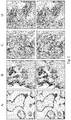
- Figure 2A is an image showing the results of immunohistochemistry (IHC) on a formalin-fixed, paraffin embedded (FFPE) placental tissue section using anti-PD-Ll antibody SP263.
- Figure 2B is an image showing the results of IHC on a FFPE tonsil tissue section using anti-PD-Ll antibody SP263.
- Figure 2C is an image showing the results of IHC on a FFPE Hodgkin lymphoma tissue section using anti-PD-Ll antibody SP263.
- Figure 2D is an image showing the results of IHC on a FFPE lung squamous cell carcinoma tissue section using anti-PD-Ll antibody SP263.
- the top row is a color photograph and the bottom row is a grayscale photograph. Antibody staining appears as brown in the color photographs.
- PD-L1 staining appears as darker regions in the grayscale photographs. Arrows in the grayscale photographs indicate examples of antibody staining in the respective tissues.
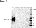
- Figure 3 is a Western blot showing PD-L1 expression in cell lysates from a NIH H820 lung adenocarcinoma cell line (high expression), a HEK293 cell line (weak expression), a Calu-3 lung adenocarcinoma cell line (negative control), a ZR75-1 human breast carcinoma cell line (negative control), a MCF7 human breast carcinoma cell line (negative control), and a T47D human breast carcinoma cell line (negative control) using anti-PD-Ll antibody SP263.
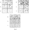
- Figure 4 illustrates IHC labeling of various tissues using SP263, J45H2L4, and J27H6L4.
- Left column is a color photograph and right column is the corresponding grayscale. Antibody staining is brown in the color photographs and illustrated by arrows in the grayscale photographs.
- Figure 4A is an image showing the results of IHC on a FFPE placental tissue section using anti-PD-Ll antibody SP263.
- Figure 4B is an image showing the results of IHC on a FFPE colon tissue section using anti-PD-Ll antibody SP263.
- Figure 4C is an image showing the results of IHC on a FFPE stomach tissue section using anti-PD-Ll antibody SP263.
- Figure 4D is an image showing the results of IHC on a FFPE placenta tissue section using anti-PD-Ll antibody clone J45H2L4.
- Figure 4E is an image showing the results of IHC on a FFPE colon tissue section using anti-PD-Ll antibody clone J45H2L4. Non-specific nuclear staining is seen.
- Figure 4F is an image showing the results of IHC on a FFPE stomach tissue section using anti-PD-Ll antibody clone J45H2L4. Non-specific nuclear staining is seen.
- Figure 4G is an image showing the results of IHC on a FFPE placenta tissue section using anti-PD-Ll antibody clone J27H6L4.
- Figure 4H is an image showing the results of IHC on a FFPE colon tissue section using anti-PD-L1 antibody clone J27H6L4.
- Figure 4I is an image showing the results of IHC on a FFPE stomach tissue section using anti-PD-Ll antibody clone J27H6L4.
- Figure 5 contains images showing the results of IHC on a FFPE placental tissue section using the indicated concentrations of anti-PD-Ll antibody E1L3N or SP263.
- the top rows for each antibody are color images and the bottom rows are grayscale images. Antibody staining appears in the color images as brown.
- Figure 6 contains images showing the results of IHC on a FFPE stomach epithelium or nerve tissue sections using the indicated concentrations of anti-PD-Ll antibody E1L3N or SP263.
- the top rows for each antibody/tissue combinations are color images and the bottom rows are grayscale images. Antibody staining appears in the color images as brown.
- Figure 7 contains images showing the results of IHC on a FFPE stomach epithelium, kidney, bladder transitional cell carcinoma (TCC), breast ductal carcinoma (Ca), and lung squamous cell carcinoma tissue sections using anti-PD-Ll antibody E1L3N or SP263.
- TCC bladder transitional cell carcinoma
- Ca breast ductal carcinoma
- SP263 lung squamous cell carcinoma tissue sections using anti-PD-Ll antibody E1L3N or SP263.
- the top rows for each antibody/tissue combinations are color images and the bottom rows are grayscale images. Antibody staining appears in the color images as brown.
- Figure 8 contains images showing the results of IHC using anti-PD-Ll antibody E1L3N or SP263 on the following FFPE tissue sections: (A) tonsil; (B) cervical squamous cell carcinoma (SCC); (C) Hodgkin Lymphoma (HK lymphoma); (D) pancreatic adenocarcinoma; (E) prostate adenocarcinoma; and (F) skin SCC.
- the top rows for each antibody/tissue combinations are color images and the bottom rows are grayscale images. Antibody staining appears in the color images as brown.
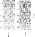
- Figures 9A-9E show the results of IHC on FFPE tissue sections from NSCLC patients using the anti-PD-Ll antibody E1L3N.
- Figures 9F-9J show the results of IHC on FFPE tissue sections from NSCLC patients using anti-PD-L1 antibody SP263.
- the top rows for each antibody/tissue combinations are color images and the bottom rows are grayscale images. Antibody staining appears in the color images as brown.
- Figure 10 shows the results of an ELISA assay involving SP263 binding to immobilized peptide immunogen (PD-L1 aa 272-290).
- a cell includes a plurality of cells, including mixtures thereof.
- the "administration" of an agent or drug to a subject or subject includes any route of introducing or delivering to a subject a compound to perform its intended function. Suitable dosage formulations and methods of administering the agents are known in the art. Route of administration can also be determined and method of determining the most effective route of administration are known to those of skill in the art and will vary with the composition used for treatment, the purpose of the treatment, the health condition or disease stage of the subject being treated and target cell or tissue. Non-limiting examples of route of administration include oral administration, vaginal, nasal administration, injection, topical application and by suppository. Administration includes self-administration and the administration by another. It is also to be appreciated that the various modes of treatment or prevention of medical conditions as described are intended to mean “substantial", which includes total but also less than total treatment or prevention, and wherein some biologically or medically relevant result is achieved.
- Administration can be effected in one dose, continuously or intermittently throughout the course of treatment. Methods of determining the most effective means and dosage of administration are known to those of skill in the art and will vary with the composition used for therapy, the purpose of the therapy, the target cell being treated and the subject being treated. Single or multiple administrations can be carried out with the dose level and pattern being selected by the treating physician.
- animal refers to living multi-cellular vertebrate organisms, a category that includes, for example, mammals and birds.
- mammal includes both human and non-human mammals.
- subject or patient includes both human and veterinary subjects, for example, humans, non-human primates, dogs, cats, sheep, mice, horses, and cows.
- antibody collectively refers to immunoglobulins or immunoglobulin-like molecules including by way of example and without limitation, IgA, IgD, IgE, IgG and IgM, combinations thereof, and similar molecules produced during an immune response in any vertebrate, for example, in mammals such as humans, goats, rabbits and mice, as well as non-mammalian species, such as shark immunoglobulins.
- antibody includes intact immunoglobulins and "antibody fragments” or "antigen binding fragments” that specifically bind to a molecule of interest (or a group of highly similar molecules of interest) to the substantial exclusion of binding to other molecules (for example, antibodies and antibody fragments that have a binding constant for the molecule of interest that is at least 10 3 M -1 greater, at least 10 4 M -1 greater or at least 10 5 M -1 greater than a binding constant for other molecules in a biological sample).
- antibody also includes genetically engineered forms such as chimeric antibodies (for example, humanized murine antibodies), heteroconjugate antibodies (such as, bispecific antibodies). See also, Pierce Catalog and Handbook, 1994-1995 (Pierce Chemical Co., Rockford, Ill .); Kuby, J., Immunology, 3rd Ed., W.H. Freeman & Co., New York, 1997 .
- antibody refers to a polypeptide ligand comprising at least a light chain or heavy chain immunoglobulin variable region which specifically recognizes and binds an epitope of an antigen.
- Antibodies are composed of a heavy and a light chain, each of which has a variable region, termed the variable heavy (V H ) region and the variable light (V L ) region. Together, the V H region and the V L region are responsible for binding the antigen recognized by the antibody.
- an immunoglobulin typically has heavy (H) chains and light (L) chains interconnected by disulfide bonds.
- Each heavy and light chain contains a constant region and a variable region, (the regions are also known as "domains").
- the heavy and the light chain variable regions specifically bind the antigen.
- Light and heavy chain variable regions contain a "framework" region interrupted by three hypervariable regions, also called “complementarity-determining regions" or "CDRs".
- framework region and CDRs have been defined (see, Kabat et al., Sequences of Proteins of Immunological Interest, U.S. Department of Health and Human Services, 1991 ).
- the Kabat database is now maintained online.
- the sequences of the framework regions of different light or heavy chains are relatively conserved within a species.
- the framework region of an antibody that is the combined framework regions of the constituent light and heavy chains, largely adopt a ⁇ -sheet conformation and the CDRs form loops which connect, and in some cases form part of, the ⁇ -sheet structure.
- framework regions act to form a scaffold that provides for positioning the CDRs in correct orientation by inter-chain, non-covalent interactions.
- the CDRs are primarily responsible for binding to an epitope of an antigen.
- the CDRs of each chain are typically referred to as CDR1, CDR2, and CDR3, numbered sequentially starting from the N-terminus, and are also typically identified by the chain in which the particular CDR is located.
- a V H CDR3 is located in the variable domain of the heavy chain of the antibody in which it is found
- a V L CDR1 is the CDR1 from the variable domain of the light chain of the antibody in which it is found.
- An antibody that binds PD-L1 will have a specific V H region and the V L region sequence, and thus specific CDR sequences.
- Antibodies with different specificities i.e.
- antibody is further intended to encompass digestion fragments, specified portions, derivatives and variants thereof, including antibody mimetics or comprising portions of antibodies that mimic the structure and/or function of an antibody or specified fragment or portion thereof, including single chain antibodies and fragments thereof.
- binding fragments encompassed within the term "antigen binding portion" of an antibody include a Fab fragment, a monovalent fragment consisting of the V L , V H , C L and C H , domains; a F(ab') 2 fragment, a bivalent fragment comprising two Fab fragments linked by a disulfide bridge at the hinge region; a F d fragment consisting of the V H and C H , domains; a F v fragment consisting of the V L and V H domains of a single arm of an antibody, a dAb fragment ( Ward et al.
- V L and V H are coded for by separate genes, they can be joined, using recombinant methods, by a synthetic linker that enables them to be made as a single protein chain in which the V L and V H regions pair to form monovalent molecules (known as single chain F v (scF v )).
- scF v single chain F v
- Single chain antibodies are also intended to be encompassed within the term "fragment of an antibody.” Any of the above-noted antibody fragments are obtained using conventional techniques known to those of skill in the art, and the fragments are screened for binding specificity and neutralization activity in the same manner as are intact antibodies.
- Antibody fragments or "antigen binding fragments” include proteolytic antibody fragments (such as F(ab') 2 fragments, Fab' fragments, Fab'-SH fragments and Fab fragments as are known in the art), recombinant antibody fragments (such as sF v fragments, dsF v fragments, bispecific sF v fragments, bispecific dsF v fragments, F(ab)' 2 fragments, single chain Fv proteins ("scF v "), disulfide stabilized F v proteins (“dsF v “), diabodies, and triabodies (as are known in the art), and camelid antibodies (see, for example, U.S. Pat. Nos.
- proteolytic antibody fragments such as F(ab') 2 fragments, Fab' fragments, Fab'-SH fragments and Fab fragments as are known in the art
- recombinant antibody fragments such as sF v fragments, dsF v fragment
- An scF v protein is a fusion protein in which a light chain variable region of an immunoglobulin and a heavy chain variable region of an immunoglobulin are bound by a linker, while in dsF v s, the chains have been mutated to introduce a disulfide bond to stabilize the association of the chains.
- antibody derivative is intended to encompass molecules that bind an epitope as defined herein and which are modifications or derivatives of an isolated PD-L1 antibody of this disclosure. Derivatives include, but are not limited to, for example, bispecific, heterospecific, trispecific, tetraspecific, multispecific antibodies, diabodies, chimeric, recombinant and humanized.
- bispecific molecule is intended to include any agent, e.g., a protein, peptide, or protein or peptide complex, which has two different binding specificities.
- multispecific molecule or “heterospecific molecule” is intended to include any agent, e.g., a protein, peptide, or protein or peptide complex, which has more than two different binding specificities.
- heteroantibodies refers to two or more antibodies, antibody binding fragments (e.g ., Fab), derivatives thereof, or antigen binding regions linked together, at least two of which have different specificities.
- antibody variant is intended to include antibodies produced in a species other than a rabbit. It also includes antibodies containing post-translational modifications to the linear polypeptide sequence of the antibody or fragment. It further encompasses fully human antibodies.
- antigen refers to a compound, composition, or substance that may be specifically bound by the products of specific humoral or cellular immunity, such as an antibody molecule or T-cell receptor.
- Antigens can be any type of molecule including, for example, haptens, simple intermediary metabolites, sugars ( e.g ., oligosaccharides), lipids, and hormones as well as macromolecules such as complex carbohydrates ( e.g. , polysaccharides), phospholipids, and proteins.
- antigens include, but are not limited to, viral antigens, bacterial antigens, fungal antigens, protozoa and other parasitic antigens, tumor antigens, antigens involved in autoimmune disease, allergy and graft rejection, toxins, and other miscellaneous antigens.
- binding affinity refers to the tendency of one molecule to bind (typically non-covalently) with another molecule, such as the tendency of a member of a specific binding pair for another member of a specific binding pair.
- a binding affinity can be measured as a binding constant, which binding affinity for a specific binding pair (such as an antibody/antigen pair) can be at least 1 ⁇ 10 -5 M, at least 1 ⁇ 10 -6 M, at least 1 ⁇ 10 -7 M, at least 1 ⁇ 10 -8 M, at least 1 ⁇ 10 -9 M, at least 1 ⁇ 10 -10 M, at least 1 ⁇ 10 -11 M or at least 1 ⁇ 10 -12 M.
- binding affinity is calculated by a modification of the Scatchard method described by Frankel et al., Mol. Immunol., 16:101-106, 1979 .
- binding affinity is measured by an antigen/antibody dissociation rate.
- a high binding affinity is measured by a competition radioimmunoassay.
- a high binding affinity for an antibody/antigen pair is at least about 1 ⁇ 10 -8 M.
- a high binding affinity is at least about 1.5 ⁇ 10 -8 M, at least about 2.0 ⁇ 10 -8 M, at least about 2.5 ⁇ 10 -8 M, at least about 3.0 ⁇ 10 -8 M, at least about 3.5 ⁇ 10 -8 M, at least about 4.0x10 -8 M, at least about 4.5 ⁇ 10 -8 M, or at least about 5.0 ⁇ 10 -8 M.
- biological equivalent thereof is intended to be synonymous with “equivalent thereof” when referring to a reference protein, antibody, polypeptide, polynucleotide or nucleic acid, and intends those having minimal homology while still maintaining desired structure or functionality. Unless specifically recited herein, it is contemplated that any nucleic acid, polynucleotide, polypeptide, protein or antibody mentioned herein also includes equivalents thereof.
- an equivalent intends at least about 80 % homology or identity and alternatively, at least about 85 %, or alternatively at least about 90 %, or alternatively at least about 95 %, or alternatively 98 % percent homology or identity and exhibits substantially equivalent biological activity to the reference protein, polypeptide, antibody or nucleic acid.
- the term "equivalent” or “biological equivalent” of an antibody means the ability of the antibody to selectively bind its epitope protein or fragment thereof as measured by ELISA, IHC or other suitable methods.
- Biologically equivalent antibodies include, but are not limited to, those antibodies, peptides, antibody fragments, antibody variant, antibody derivative and antibody mimetics that bind to the same epitope as the reference antibody.
- the skilled artisan can prepare an antibody functionally equivalent to the antibodies of the present disclosure by introducing appropriate mutations into the antibody using site-directed mutagenesis ( Hashimoto-Gotoh, T. et al., Gene 152, 271-275 (1995 ); Zoller & Smith, Methods Enzymol.
- Antibodies that are functionally equivalent to the antibodies of the present disclosure and comprise an amino acid sequence comprising mutation of one or more amino acids in the amino acid sequence of an antibody of the present disclosure are also included in the antibodies of the present disclosure.
- the number of amino acids that are mutated is generally 50 amino acids or less, preferably 30 or less, and more preferably 10 or less (for example, 5 amino acids or less).
- An amino acid residue is preferably mutated into one that conserves the properties of the amino acid side chain.
- amino acids are classified into: hydrophobic amino acids (A, I, L, M, F, P, W, Y, and V); hydrophilic amino acids (R, D, N, C, E, Q, G, H, K, S, and T); amino acids having aliphatic side-chains (G, A, V, L, I, and P); amino acids having hydroxyl group-containing side-chains (S, T, and Y); amino acids having sulfur atom-containing side-chains (C and M); amino acids having carboxylic acid- and amide-containing side-chains (D, N, E, and Q); base-containing side-chains (R, K, and H); and amino acids having aromatic-containing side-chains (H, F, Y, and W).
- hydrophobic amino acids A, I, L, M, F, P, W, Y, and V
- hydrophilic amino acids R, D, N, C, E, Q, G, H, K, S, and T
- biological sample means sample material derived from or contacted by living cells.
- biological sample is intended to include tissues, cells and biological fluids isolated from a subject, as well as tissues, cells and fluids present within a subject.
- Biological samples of the disclosure include, e.g ., but are not limited to, whole blood, plasma, semen, saliva, tears, urine, fecal material, sweat, buccal, skin, cerebrospinal fluid, and hair.
- Biological samples can also be obtained from biopsies of internal organs or from cancers. Biological samples can be obtained from subjects for diagnosis or research or can be obtained from healthy individuals, as controls or for basic research.
- cancer refers to cells that have undergone a malignant transformation that makes them pathological to the host organism and are selected from the group consisting of bladder transitional cell carcinoma, lung adenocarcinoma, breast ductal carcinoma, Hodgkin's lymphoma, pancreas adenocarcinoma, prostate adenocarcinoma, cervical squamous cell carcinoma, skin squamous cell carcinoma, and non-small cell lung cancer.
- Primary cancer cells that is, cells obtained from near the site of malignant transformation
- the definition of a cancer cell includes not only a primary cancer cell, but also any cell derived from a cancer cell ancestor. This includes metastasized cancer cells, and in vitro cultures and cell lines derived from cancer cells.
- a "clinically detectable" tumor is one that is detectable on the basis of tumor mass; e.g ., by such procedures as CAT scan, magnetic resonance imaging (MRI), X-ray, ultrasound or palpation. Biochemical or immunologic findings alone may be insufficient to meet this definition.
- a neoplasm is an abnormal mass or colony of cells produced by a relatively autonomous new growth of tissue. Most neoplasms arise from the clonal expansion of a single cell that has undergone neoplastic transformation. The transformation of a normal to a neoplastic cell can be caused by a chemical, physical, or biological agent (or event) that directly and irreversibly alters the cell genome.
- Neoplastic cells are characterized by the loss of some specialized functions and the acquisition of new biological properties, foremost, the property of relatively autonomous (uncontrolled) growth. Neoplastic cells pass on their heritable biological characteristics to progeny cells.
- a malignant neoplasm manifests a greater degree of autonomy, is capable of invasion and metastatic spread, may be resistant to treatment, and may cause death.
- a benign neoplasm has a lesser degree of autonomy, is usually not invasive, does not metastasize, and generally produces no great harm if treated adequately.
- Cancer is a generic term for malignant neoplasms.
- Anaplasia is a characteristic property of cancer cells and denotes a lack of normal structural and functional characteristics (undifferentiation).
- a tumor is literally a swelling of any type, such as an inflammatory or other swelling, but modem usage generally denotes a neoplasm.
- Histogenesis is the origin of a tissue and is a method of classifying neoplasms on the basis of the tissue cell of origin.
- Adenomas are benign neoplasms of glandular epithelium.
- Carcinomas are malignant tumors of epithelium.
- Sarcomas are malignant tumors of mesenchymal tissues.
- One system to classify neoplasia utilizes biological (clinical) behavior, whether benign or malignant, and the histogenesis, the tissue or cell of origin of the neoplasm as determined by histologic and cytologic examination.
- Neoplasms may originate in almost any tissue containing cells capable of mitotic division.
- the histogenetic classification of neoplasms is based upon the tissue (or cell) of origin as determined by histologic and cytologic examination.
- chimeric antibody means an antibody in which the Fc constant region of a monoclonal antibody from one species (e.g., a mouse Fc constant region) is replaced, using recombinant DNA techniques, with an Fc constant region from an antibody of another species (e.g., a human Fc constant region).
- a monoclonal antibody from one species e.g., a mouse Fc constant region
- another species e.g., a human Fc constant region
- compositions and methods include the recited elements, but do not exclude others.
- Consisting essentially of' when used to define compositions and methods shall mean excluding other elements of any essential significance to the combination for the intended use.
- a composition consisting essentially of the elements as defined herein would not exclude trace contaminants from the isolation and purification method and pharmaceutically acceptable carriers, such as phosphate buffered saline, preservatives and the like.
- Consisting of' shall mean excluding more than trace elements of other ingredients and substantial method steps for administering the compositions of this disclosure. Aspects defined by each of these transition terms are within the scope of this disclosure.
- a “control" biological sample is an alternative sample used in an experiment for comparison purpose.
- a control can be "positive” or “negative".
- a positive control a compound or composition known to exhibit the desired therapeutic effect
- a negative control a subject or a sample that does not receive the therapy or receives a placebo.
- the term "detectable label” refers to a molecule or material that can produce a detectable (such as visually, electronically or otherwise) signal that indicates the presence and/or concentration of the label in a sample.
- the detectable label can be used to locate and/or quantify the target to which the specific binding molecule is directed. Thereby, the presence and/or concentration of the target in a sample can be detected by detecting the signal produced by the detectable label.
- a detectable label can be detected directly or indirectly, and several different detectable labels conjugated to different specific-binding molecules can be used in combination to detect one or more targets.
- a first detectable label conjugated to an antibody specific to a target can be detected indirectly through the use of a second detectable label that is conjugated to a molecule that specifically binds the first detectable label.
- Multiple detectable labels that can be separately detected can be conjugated to different specific binding molecules that specifically bind different targets to provide a multiplexed assay that can provide simultaneous detection of the multiple targets in a sample.
- a detectable signal can be generated by any mechanism including absorption, emission and/or scattering of a photon (including radio frequency, microwave frequency, infrared frequency, visible frequency and ultra-violet frequency photons).
- Detectable labels include colored, fluorescent, phosphorescent and luminescent molecules and materials, catalysts (such as enzymes) that convert one substance into another substance to provide a detectable difference (such as by converting a colorless substance into a colored substance or vice versa, or by producing a precipitate or increasing sample turbidity), haptens that can be detected through antibody-hapten binding interactions using additional detectably labeled antibody conjugates, and paramagnetic and magnetic molecules or materials.
- catalysts such as enzymes
- haptens that can be detected through antibody-hapten binding interactions using additional detectably labeled antibody conjugates, and paramagnetic and magnetic molecules or materials.
- detectable labels include enzymes such as horseradish peroxidase, alkaline phosphatase, acid phosphatase, glucose oxidase, ⁇ -galactosidase or ⁇ -glucuronidase; fluorphores such as fluoresceins, luminophores, coumarins, BODIPY dyes, resorufins, and rhodamines (many additional examples of fluorescent molecules can be found in The Handbook--A Guide to Fluorescent Probes and Labeling Technologies, Molecular Probes, Eugene, Oreg.); nanoparticles such as quantum dots (obtained, for example, from QuantumDot Corp, Invitrogen Nanocrystal Technologies, Hayward, Calif.; see also, U.S.
- enzymes such as horseradish peroxidase, alkaline phosphatase, acid phosphatase, glucose oxidase, ⁇ -galactosidase or ⁇ -glucuronidase
- detectable label includes an enzyme
- a detectable substrate such as a chromogen, a fluorogenic compound, or a luminogenic compound can be used in combination with the enzyme to generate a detectable signal (A wide variety of such compounds are commercially available, for example, from Invitrogen Corporation, Eugene Oreg.).
- chromogenic compounds include diaminobenzidine (DAB), 4-nitrophenylphospate (pNPP), fast red, bromochloroindolyl phosphate (BCIP), nitro blue tetrazolium (NBT), BCIP/NBT, fast red, AP Orange, AP blue, tetramethylbenzidine (TMB), 2,2'-azino-di-[3-ethylbenzothiazoline sulphonate] (ABTS), o-dianisidine, 4-chloronaphthol (4-CN), nitrophenyl- ⁇ -D-galactopyranoside (ONPG), o-phenylenediamine (OPD), 5-bromo-4-chloro-3-indolyl-P-galactopyranoside (X-Gal), methylumbelliferyl- ⁇ -D-galactopyranoside (MU-Gal), p-nitrophenyl- ⁇ -D-galactopyrano
- an enzyme can be used in a metallographic detection scheme.
- Metallographic detection methods include using an enzyme such as alkaline phosphatase in combination with a water-soluble metal ion and a redox-inactive substrate of the enzyme. The substrate is converted to a redox-active agent by the enzyme, and the redox-active agent reduces the metal ion, causing it to form a detectable precipitate.
- an enzyme such as alkaline phosphatase in combination with a water-soluble metal ion and a redox-inactive substrate of the enzyme.
- the substrate is converted to a redox-active agent by the enzyme, and the redox-active agent reduces the metal ion, causing it to form a detectable precipitate.
- Metallographic detection methods include using an oxido-reductase enzyme (such as horseradish peroxidase) along with a water soluble metal ion, an oxidizing agent and a reducing agent, again to form a detectable precipitate.
- an oxido-reductase enzyme such as horseradish peroxidase
- Haptens are small molecules that are specifically bound by antibodies, although by themselves they will not elicit an immune response in an animal and must first be attached to a larger carrier molecule such as a protein to generate an immune response. Examples of haptens include dinitrophenyl, biotin, digoxigenin, and fluorescein.
- the detectable label comprises a non-endogenous hapten (e.g. not biotin), such as, for example, the haptens disclosed in U.S. Patent Nos.
- 7,695,929 , 8,618,265 and 8,846,320 including for example pyrazoles, nitrophenyl compounds, benzofurazans, triterpenes, ureas and thioureas, rotenone and rotenone derivatives, oxazoles and thiazoles, coumarin and coumarin derivatives, and cyclolignans.
- detectable labels can be detected using antibodies or antigen-binding fragments thereof capable of binding to the hapten.
- an “epitope” or “antigenic determinant” refers to particular chemical groups or contiguous or non-contiguous peptide sequences on a molecule that are antigenic, i.e., that elicit a specific immune response.
- An antibody binds a particular antigenic epitope.
- Epitopes usually consist of chemically active surface groupings of molecules such as amino acids or sugar side chains and usually have specific three dimensional structural characteristics, as well as specific charge characteristics. Conformational and nonconformational epitopes are distinguished in that the binding to the former but not the latter is lost in the presence of denaturing solvents.
- expression refers to the process by which polynucleotides are transcribed into mRNA and/or the process by which the transcribed mRNA is subsequently being translated into peptides, polypeptides, or proteins. If the polynucleotide is derived from genomic DNA, expression may include splicing of the mRNA in an eukaryotic cell. The expression level of a gene may be determined by measuring the amount of mRNA or protein in a cell or tissue sample. In one aspect, the expression level of a gene from one sample may be directly compared to the expression level of that gene from a control or reference sample. In another aspect, the expression level of a gene from one sample may be directly compared to the expression level of that gene from the same sample following administration of a compound.
- homology or “identical”, percent “identity” or “similarity”, when used in the context of two or more nucleic acids or polypeptide sequences, refers to two or more sequences or subsequences that are the same or have a specified percentage of nucleotides or amino acid residues that are the same, e.g., at least 60% identity, preferably at least 65%, 70%, 75%, 80%, 85%, 90%, 91%, 92%, 93%, 94%, 95%, 96%, 97%, 98%, 99%, or higher identity over a specified region (e.g., nucleotide sequence encoding an antibody described herein or amino acid sequence of an antibody described herein).
- Homology can be determined by comparing a position in each sequence which may be aligned for purposes of comparison. When a position in the compared sequence is occupied by the same base or amino acid, then the molecules are homologous at that position. A degree of homology between sequences is a function of the number of matching or homologous positions shared by the sequences.
- the alignment and the percent homology or sequence identity can be determined using software programs known in the art, for example those described in Current Protocols in Molecular Biology (Ausubel et al., eds. 1987) Supplement 30, section 7.7.18, Table 7.7.1 .
- default parameters are used for alignment.
- a preferred alignment program is BLAST, using default parameters.
- the terms also include sequences that have deletions and/or additions, as well as those that have substitutions.
- the preferred algorithms can account for gaps and the like.
- identity exists over a region that is at least about 25 amino acids or nucleotides in length, or more preferably over a region that is at least 50-100 amino acids or nucleotides in length.
- An "unrelated” or “non-homologous” sequence shares less than 40% identity, or alternatively less than 25% identity, with one of the sequences of the present disclosure.
- human antibody as used herein, is intended to include antibodies having variable and constant regions derived from human germline immunoglobulin sequences.
- the human antibodies of the disclosure may include amino acid residues not encoded by human germline immunoglobulin sequences ( e.g., mutations introduced by random or site-specific mutagenesis in vitro or by somatic mutation in vivo ).
- the term "human antibody” as used herein is not intended to include antibodies in which CDR sequences derived from the germline of another mammalian species, such as a rabbit, have been grafted onto human framework sequences.
- human antibody refers to an antibody in which substantially every part of the protein (e.g., CDR, framework, C L , C H domains ( e.g ., C H1 , C H2 , C H3 ), hinge, V L , V H ) is substantially non-immunogenic in humans, with only minor sequence changes or variations.
- antibodies designated primate monkey, baboon, chimpanzee, etc .
- rodent mouse, rat, rabbit, guinea pig, hamster, and the like
- other mammals designate such species, sub-genus, genus, sub-family, family specific antibodies.
- chimeric antibodies include any combination of the above.
- a human antibody is distinct from a chimeric or humanized antibody. It is pointed out that a human antibody can be produced by a non-human animal or prokaryotic or eukaryotic cell that is capable of expressing functionally rearranged human immunoglobulin (e.g., heavy chain and/or light chain) genes. Further, when a human antibody is a single chain antibody, it can comprise a linker peptide that is not found in native human antibodies.
- an F v can comprise a linker peptide, such as two to about eight glycine or other amino acid residues, which connects the variable region of the heavy chain and the variable region of the light chain.
- linker peptides are considered to be of human origin.
- humanized antibody refers to an antibody comprising a humanized light chain and a humanized heavy chain immunoglobulin.
- a humanized antibody binds to the same antigen as the donor antibody that provides the CDRs.
- the acceptor framework of a humanized immunoglobulin or antibody may have a limited number of substitutions by amino acids taken from the donor framework.
- Humanized or other monoclonal antibodies can have additional conservative amino acid substitutions which have substantially no effect on antigen binding or other immunoglobulin functions.
- Humanized immunoglobulins can be constructed by means of genetic engineering (see for example, U.S. Pat. No. 5,585,089 ).
- humanized immunoglobulin refers to an immunoglobulin including a human framework region and one or more CDRs from a non-human (for example a mouse, rat, rabbit or synthetic) immunoglobulin.
- the non-human immunoglobulin providing the CDRs is termed a "donor,” and the human immunoglobulin providing the framework is termed an "acceptor.”
- all the CDRs are from the donor immunoglobulin in a humanized immunoglobulin.
- Constant regions need not be present, but if they are, they must be substantially identical to human immunoglobulin constant regions, i.e., at least about 85-90%, or at least about 95% or more identical.
- all parts of a humanized immunoglobulin, except possibly the CDRs are substantially identical to corresponding parts of natural human immunoglobulin sequences.
- isolated refers to molecules or biological or cellular materials being substantially free from other materials.
- the term “isolated” refers to nucleic acid, such as DNA or RNA, or protein or polypeptide ( e.g., an antibody or derivative thereof), or cell or cellular organelle, or tissue or organ, separated from other DNAs or RNAs, or proteins or polypeptides, or cells or cellular organelles, or tissues or organs, respectively, that are present in the natural source.
- isolated also refers to a nucleic acid or peptide that is substantially free of cellular material, viral material, or culture medium when produced by recombinant DNA techniques, or chemical precursors or other chemicals when chemically synthesized.
- an "isolated nucleic acid” is meant to include nucleic acid fragments which are not naturally occurring as fragments and would not be found in the natural state.
- isolated is also used herein to refer to polypeptides which are isolated from other cellular proteins and is meant to encompass both purified and recombinant polypeptides.
- isolated is also used herein to refer to cells or tissues that are isolated from other cells or tissues and is meant to encompass both cultured and engineered cells or tissues.
- the term "monoclonal antibody” refers to an antibody produced by a single clone of B-lymphocytes or by a cell into which the light and heavy chain genes of a single antibody have been transfected.
- Monoclonal antibodies are produced by methods known to those of skill in the art, for instance by making hybrid antibody-forming cells from a fusion of myeloma cells with immune spleen cells.
- Monoclonal antibodies include humanized monoclonal antibodies.
- a "pathological cell” is one that is pertaining to or arising from disease. Pathological cells can be hyperproliferative.
- a "hyperproliferative cell” means cells or tissue are dividing and growing at a rate greater than that when the cell or tissue is in a normal or healthy state. Examples of such include, but are not limited to precancerous (i.e., epithelial dysplasia) and cancer cells.
- Hyperproliferative cells also include de-differentiated, immortalized, neoplastic, malignant, metastatic, and cancer cells such as sarcoma cells, leukemia cells, carcinoma cells, or adenocarcinoma cells.
- PD-L1 Programmed death ligand-1
- B7-H1 Human B7 homolog 1
- PDCD1L1 Programmed cell death 1 ligand 1
- Human PD-L1 encodes a 290 amino acid (aa) type I membrane precursor protein with a putative 18 aa signal peptide, a 221 aa extracellular domain, a 21 aa transmembrane region, and a 31 aa cytoplasmic domain (Entrez Gene ID: 29126, UniProtKB: Q9NZQ7 http://www.ncbi.nlm.nih.gov/ last accessed October 20, 2014).
- protein protein
- peptide and “polypeptide” are used interchangeably and in their broadest sense to refer to a compound of two or more subunit amino acids, amino acid analogs or peptidomimetics.
- the subunits may be linked by peptide bonds.
- the subunit may be linked by other bonds, e.g ., ester, ether, etc.
- a protein or peptide must contain at least two amino acids and no limitation is placed on the maximum number of amino acids which may comprise a protein's or peptide's sequence.
- amino acid refers to either natural and/or unnatural or synthetic amino acids, including glycine and both the D and L optical isomers, amino acid analogs and peptidomimetics.
- polynucleotide and “oligonucleotide” are used interchangeably and refer to a polymeric form of nucleotides of any length, either deoxyribonucleotides or ribonucleotides or analogs thereof. Polynucleotides can have any three-dimensional structure and may perform any function, known or unknown.
- polynucleotides a gene or gene fragment (for example, a probe, primer, EST or SAGE tag), exons, introns, messenger RNA (mRNA), transfer RNA, ribosomal RNA, RNAi, ribozymes, cDNA, recombinant polynucleotides, branched polynucleotides, plasmids, vectors, isolated DNA of any sequence, isolated RNA of any sequence, nucleic acid probes and primers.
- a polynucleotide can comprise modified nucleotides, such as methylated nucleotides and nucleotide analogs.
- modifications to the nucleotide structure can be imparted before or after assembly of the polynucleotide.
- the sequence of nucleotides can be interrupted by non-nucleotide components.
- a polynucleotide can be further modified after polymerization, such as by conjugation with a labeling component.
- the term also refers to both double- and single-stranded molecules. Unless otherwise specified or required, any aspect of this disclosure that is a polynucleotide encompasses both the double-stranded form and each of two complementary single-stranded forms known or predicted to make up the double-stranded form.
- a polynucleotide is composed of a specific sequence of four nucleotide bases: adenine (A); cytosine (C); guanine (G); thymine (T); and uracil (U) for thymine when the polynucleotide is RNA.
- polynucleotide sequence is the alphabetical representation of a polynucleotide molecule. This alphabetical representation can be input into databases in a computer having a central processing unit and used for bioinformatics applications such as functional genomics and homology searching.
- a purified nucleic acid, peptide, protein, biological complexes or other active compound is one that is isolated in whole or in part from proteins or other contaminants.
- substantially purified peptides, proteins, biological complexes, or other active compounds for use within the disclosure comprise more than 80% of all macromolecular species present in a preparation prior to admixture or formulation of the peptide, protein, biological complex or other active compound with a pharmaceutical carrier, excipient, buffer, absorption enhancing agent, stabilizer, preservative, adjuvant or other co-ingredient in a complete pharmaceutical formulation for therapeutic administration.
- the peptide, protein, biological complex or other active compound is purified to represent greater than 90%, often greater than 95% of all macromolecular species present in a purified preparation prior to admixture with other formulation ingredients.
- the purified preparation may be essentially homogeneous, wherein other macromolecular species are not detectable by conventional techniques.
- telomere binding means the contact between an antibody and an antigen with a binding affinity of at least 10 -6 M.
- antibodies bind with affinities of at least about 10 -7 M, and preferably 10 -8 M, 10 -9 M, 10 -10 M, 10 -11 M, or 10 -12 M.
- recombinant protein refers to a polypeptide which is produced by recombinant DNA techniques, wherein generally, DNA encoding the polypeptide is inserted into a suitable expression vector which is in turn used to transform a host cell to produce the heterologous protein.
- treating or “treatment” of a disease in a subject refers to (1) preventing the symptoms or disease from occurring in a subject that is predisposed or does not yet display symptoms of the disease; (2) inhibiting the disease or arresting its development; or (3) ameliorating or causing regression of the disease or the symptoms of the disease.
- treatment is an approach for obtaining beneficial or desired results, including clinical results.
- beneficial or desired results can include one or more, but are not limited to, alleviation or amelioration of one or more symptoms, diminishment of extent of a condition (including a disease), stabilized ( i.e., not worsening) state of a condition (including disease), delay or slowing of condition (including disease), progression, amelioration or palliation of the condition (including disease), states and remission (whether partial or total), whether detectable or undetectable.
- An immunoglobulin monomer comprises two heavy chains and two light chains connected by disulfide bonds. Each heavy chain is paired with one of the light chains to which it is directly bound via a disulfide bond. Each heavy chain comprises a constant region (which varies depending on the isotype of the antibody) and a variable region.
- the variable region comprises three hypervariable regions (or complementarity determining regions) which are designated CDRH1, CDRH2 and CDRH3 and which are supported within framework regions.
- Each light chain comprises a constant region and a variable region, with the variable region comprising three hypervariable regions (designated CDRL1, CDRL2 and CDRL3) supported by framework regions in an analogous manner to the variable region of the heavy chain.
- the hypervariable regions of each pair of heavy and light chains mutually cooperate to provide an antigen binding site that is capable of binding a target antigen.
- the binding specificity of a pair of heavy and light chains is defined by the sequence of CDR1, CDR2 and CDR3 of the heavy and light chains.
- an isolated antibody comprising a heavy chain (HC) immunoglobulin variable domain sequence and a light chain (LC) immunoglobulin variable domain sequence, wherein the heavy chain and light chain immunoglobulin variable domain sequences form an antigen binding site that binds to an epitope of human PD-L1 comprising the amino acid sequence CGIQDTNSKKQSDTHLEET (SEQ ID NO: 1) and/or has a half maximal effective concentration (EC 50 ) of at least 1.5 x 10 -11 M.
- HC heavy chain
- LC light chain
- EC 50 half maximal effective concentration
- sequences of CDR3 of the heavy and light chains of the PD-L1 antibodies of the present disclosure conform with the consensus sequences set out in SEQ ID NOS: 10 and 13.
- sequences of CDR1 and CDR2 of the heavy chain of the PD-L1 antibodies of the present disclosure conform with the consensus sequences set out in SEQ ID NOS: 8 and 9.
- sequences of CDR1 of the light chain of the PD-L1 antibodies of the present disclosure conform with the consensus sequence set out in SEQ ID NO: 11.
- sequence of CDR2 of the light chain of the PD-L1 antibodies of the present disclosure comprises the sequence of SEQ ID NO: 12.
- the PD-L1 antibodies of the present disclosure has the CDR3 sequence of the light chain conforming with the consensus sequence of SEQ ID NO: 13 and the CDR3 sequence of the heavy chain conforming with the consensus sequence of SEQ ID NO: 10.
- an antibody comprising a heavy chain having the sequence of CDR1 from J27H6L4, CDR2 from J45H2L4 and CDR3 from SP263 and light chain having the sequence of CDR1 from J45H2L4, CDR2 from SP263 and CDR3 from J27H6L4.
- the isolated antibody includes one or more of the following characteristics:
- the disclosure provides an isolated antibody that is at least 85% identical to an antibody selected from the group consisting of SP263, J45H2L4 and J27H6L4. In one aspect, the disclosure provides an isolated antibody selected from the group consisting of SP263, J45H2L4 and J27H6L4.
- the disclosure provides an isolated antibody comprising the CDRs of SP263. In one aspect the disclosure provides an isolated antibody that is at least 85% identical to SP263.
- the CDRs of SP263 are represented in Table 1.
- the disclosure provides an isolated antibody comprising the CDRs of J45H2L4. In one aspect the disclosure provides an isolated antibody that is at least 85% identical to J45H2L4.
- the CDRs of J45H2L4 are represented in Table 1.
- the disclosure provides an isolated antibody comprising the CDRs of J27H6L4. In one aspect the disclosure provides an isolated antibody that is at least 85% identical to J27H6L4.
- the CDRs of J27H6L4 are represented in Table 1.
- the HC variable domain sequence comprises a variable domain sequence of SP263 and the LC variable domain sequence comprises a variable domain sequence of SP263.
- the HC variable domain sequence comprises a variable domain sequence of J45H2L4 and the LC variable domain sequence comprises a variable domain sequence of J45H2L4.
- the HC variable domain sequence comprises a variable domain sequence of J27H6L4 and the LC variable domain sequence comprises a variable domain sequence of J27H6L4.
- the antibody binds human PD-L1 with a dissociation constant (K D ) of less than 10 -4 M, 10 -5 M, 10 -6 M, 10 -7 M, 10 -8 M, 10 -9 M, 1(T 10 M, 10 -11 M, or 10 -12 M.
- the antigen binding site specifically binds to human PD-L1.
- the antibody is soluble Fab.
- the HC and LC variable domain sequences are components of the same polypeptide chain. In some of the aspects of the antibodies provided herein, the HC and LC variable domain sequences are components of different polypeptide chains.
- the antibody is a full-length antibody.
- the antibody is a monoclonal antibody.
- the antibody is chimeric or humanized.
- the antibody is selected from the group consisting of Fab, F(ab)'2, Fab', scF v , and F v .
- the antibody comprises an Fc domain. In some of the aspects of the antibodies provided herein, the antibody is a rabbit antibody. In some of the aspects of the antibodies provided herein, the antibody is a human or humanized antibody or is non-immunogenic in a human.
- the antibody comprises a human antibody framework region.
- one or more amino acid residues in a CDR of the antibodies provided herein are substituted with another amino acid.
- the substitution may be "conservative" in the sense of being a substitution within the same family of amino acids.
- the naturally occurring amino acids may be divided into the following four families and conservative substitutions will take place within those families.
- one or more amino acid residues are added to or deleted from one or more CDRs of an antibody. Such additions or deletions occur at the N or C termini of the CDR or at a position within the CDR.
- antibodies of the disclosure comprising such varied CDR sequences still bind PD-L1 with similar specificity and sensitivity profiles as SP263, J45H2L4 and J27H6L4. This may be tested by way of the binding assays disclosed in Examples described herein.
- the constant regions of antibodies may also be varied from those specifically disclosed for antibodies SP263, J45H2L4 and J27H6L4.
- antibodies may be provided with Fc regions of any isotype: IgA (IgA1, IgA2), IgD, IgE, IgG (IgG1, IgG2, IgG3, IgG4) or IgM.
- IgA IgA1, IgA2
- IgD IgD
- IgE IgG
- IgG IgG1, IgG2, IgG3, IgG4
- IgM immunoglobulG sequences
- Non-limiting examples of constant region sequences include:
- the SP263, J45H2L4 and J27H6L4 antibodies comprise a heavy chain constant region that is at least 80% identical to SEQ ID NOS: 27-33 or 34.
- the SP263, J45H2L4 and J27H6L4 antibodies comprise a light chain constant region that is at least 80% identical to SEQ ID NO: 35.
- the antibody binds to the epitope bound by SP263, J45H2L4 and J27H6L4.
- the antibody competes with SP263, J45H2L4 and J27H6L4 for binding to PD-L1.
- the antibody contains structural modifications to facilitate rapid binding and cell uptake and/or slow release.
- the PD-L1 antibody contains a deletion in the CH2 constant heavy chain region of the antibody to facilitate rapid binding and cell uptake and/or slow release.
- a Fab fragment is used to facilitate rapid binding and cell uptake and/or slow release.
- a F(ab)'2 fragment is used to facilitate rapid binding and cell uptake and/or slow release.
- Antibodies their manufacture and uses are well known and disclosed in, for example, Harlow, E. and Lane, D., Antibodies: A Laboratory Manual, Cold Spring Harbor Laboratory Press, Cold Spring Harbor, N.Y., 1999 .
- the antibodies may be generated using standard methods known in the art. Examples of antibodies include (but are not limited to) monoclonal, single chain, and functional fragments of antibodies.
- Antibodies may be produced in a range of hosts, for example goats, rabbits, rats, mice, humans, and others. They may be immunized by injection with a target antigen or a fragment or oligopeptide thereof which has immunogenic properties, such as a C-terminal fragment of PD-L1.
- a target antigen or a fragment or oligopeptide thereof which has immunogenic properties such as a C-terminal fragment of PD-L1.
- various adjuvants may be used to increase an immunological response.
- adjuvants include, but are not limited to, Freund's, mineral gels such as aluminum hydroxide, and surface active substances such as lysolecithin, pluronic polyols, polyanions, peptides, oil emulsions, keyhole limpet hemocyanin, and dinitrophenol.
- BCG Bacille Calmette-Guerin
- Corynebacterium parvum are particularly useful.
- the antibodies of the present disclosure are polyclonal, i.e., a mixture of plural types of anti-PD-Ll antibodies having different amino acid sequences, e.g ., antibodies raised against SEQ ID NO: 1 using techniques known in the art and briefly described below.
- the polyclonal antibody comprises a mixture of plural types of anti-PD-L1 antibodies having different CDRs. As such, a mixture of cells which produce different antibodies is cultured, and an antibody purified from the resulting culture can be used (see WO 2004/061104 ).
- Monoclonal antibodies to PD-L1 may be prepared using any technique which provides for the production of antibody molecules by continuous cell lines in culture and in one aspect are prepared using a polypeptide having SEQ ID NO. 1. Such techniques include, but are not limited to, the hybridoma technique (see, e.g., Kohler & Milstein, Nature 256: 495-497 (1975 )); the trioma technique; the human B-cell hybridoma technique (see, e.g., Kozbor, et al., Immunol.
- Human monoclonal antibodies can be utilized in the practice of the disclosure and can be produced by using human hybridomas (see, e.g., Cote, et al., Proc. Natl. Acad. Sci.
- amplified sequences also can be fused to DNAs encoding other proteins e.g., a bacteriophage coat, or a bacterial cell surface protein for expression and display of the fusion polypeptides on phage or bacteria. Amplified sequences can then be expressed and further selected or isolated based, e.g., on the affinity of the expressed antibody or fragment thereof for an antigen or epitope present on the PD-L1 polypeptide.
- hybridomas expressing anti-PD-L1 monoclonal antibodies can be prepared by immunizing a subject and then isolating hybridomas from the subject's spleen using routine methods.
- a selected monoclonal antibody with the desired properties can be (i) used as expressed by the hybridoma, (ii) bound to a molecule such as polyethylene glycol (PEG) to alter its properties, or (iii) a cDNA encoding the monoclonal antibody can be isolated, sequenced and manipulated in various ways.
- PEG polyethylene glycol
- the anti-PD-Ll monoclonal antibody is produced by a hybridoma which includes a B cell obtained from a transgenic non-human animal, e.g., a transgenic mouse, having a genome comprising a human heavy chain transgene and a light chain transgene fused to an immortalized cell.
- Hybridoma techniques include those known in the art and taught in Harlow et al., Antibodies: A Laboratory Manual Cold Spring Harbor Laboratory, Cold Spring Harbor, N.Y., 349 (1988 ); Hammerling et al., Monoclonal Antibodies And T-Cell Hybridomas, 563-681 (1981 ).
- the antibodies of the present disclosure can be produced through the application of recombinant DNA and phage display technology.
- anti-PD-Ll antibodies can be prepared using various phage display methods known in the art.
- phage display methods functional antibody domains are displayed on the surface of a phage particle which carries polynucleotide sequences encoding them.
- Phage with a desired binding property are selected from a repertoire or combinatorial antibody library (e.g. , human or murine) by selecting directly with an antigen, typically an antigen bound or captured to a solid surface or bead.
- Phage used in these methods are typically filamentous phage including fd and M13 with Fab, F v or disulfide stabilized F v antibody domains are recombinantly fused to either the phage gene III or gene VIII protein.
- methods can be adapted for the construction of Fab expression libraries (see, e.g., Huse, et al., Science 246: 1275-1281, 1989 ) to allow rapid and effective identification of monoclonal Fab fragments with the desired specificity for a PD-L1 polypeptide, e.g., a polypeptide or derivatives, fragments, analogs or homologs thereof.
- phage display methods that can be used to make the isolated antibodies of the present disclosure include those disclosed in Huston et al., Proc. Natl. Acad. Sci. U.S.A., 85: 5879-5883 (1988 ); Chaudhary et al., Proc. Natl. Acad. Sci. U.S.A., 87: 1066-1070 (1990 ); Brinkman et al., J. Immunol. Methods 182: 41-50 (1995 ); Ames et al., J. Immunol. Methods 184: 177-186 (1995 ); Kettleborough et al., Eur. J. Immunol.
- Fab, Fab' and F(ab') 2 fragments can also be employed using methods known in the art such as those disclosed in WO 92/22324 ; Mullinax et al., BioTechniques 12: 864-869 (1992 ); Sawai et al., AJRI34: 26-34 (1995 ); and Better et al., Science 240: 1041-1043 (1988 ).
- hybrid antibodies or hybrid antibody fragments that are cloned into a display vector can be selected against the appropriate antigen in order to identify variants that maintained good binding activity, because the antibody or antibody fragment will be present on the surface of the phage or phagemid particle.
- a display vector can be selected against the appropriate antigen in order to identify variants that maintained good binding activity, because the antibody or antibody fragment will be present on the surface of the phage or phagemid particle.
- Other vector formats could be used for this process, such as cloning the antibody fragment library into a lytic phage vector (modified T7 or Lambda Zap systems) for selection and/or screening.
- Antibodies may also be produced by inducing in vivo production in the lymphocyte population or by screening recombinant immunoglobulin libraries or panels of highly specific binding reagents ( Orlandi et al., PNAS 86: 3833-3837 (1989 ); Winter, G. et al., Nature, 349: 293-299 (1991 )).
- Single chain antibodies comprise a heavy chain variable region and a light chain variable region connected with a linker peptide (typically around 5 to 25 amino acids in length).
- linker peptide typically around 5 to 25 amino acids in length.
- the variable regions of the heavy chain and the light chain may be derived from the same antibody or different antibodies.
- scF v s may be synthesized using recombinant techniques, for example by expression of a vector encoding the scF v in a host organism such as E. coli.
- DNA encoding scF v can be obtained by performing amplification using a partial DNA encoding the entire or a desired amino acid sequence of a DNA selected from a DNA encoding the heavy chain or the variable region of the heavy chain of the above-mentioned antibody and a DNA encoding the light chain or the variable region of the light chain thereof as a template, by PCR using a primer pair that defines both ends thereof, and further performing amplification combining a DNA encoding a polypeptide linker portion and a primer pair that defines both ends thereof, so as to ligate both ends of the linker to the heavy chain and the light chain, respectively.
- An expression vector containing the DNA encoding scF v and a host transformed by the expression vector can be obtained according to conventional methods known in the art.
- Antigen binding fragments may also be generated, for example the F(ab') 2 fragments which can be produced by pepsin digestion of the antibody molecule and the Fab fragments which can be generated by reducing the disulfide bridges of the F(ab') 2 fragments.
- Fab expression libraries may be constructed to allow rapid and easy identification of monoclonal Fab fragments with the desired specificity ( Huse et al., Science, 256: 1275-1281 (1989 )).
- the antibodies of the present disclosure may be multimerized to increase the affinity for an antigen.
- the antibody to be multimerized may be one type of antibody or a plurality of antibodies which recognize a plurality of epitopes of the same antigen.
- binding of the IgG CH3 domain to two scF v molecules, binding to streptavidin, introduction of a helix-turn-helix motif and the like can be exemplified.
- the antibody compositions of the present disclosure may be in the form of a conjugate formed between any of these antibodies and another agent (immunoconjugate).
- the antibodies of the present disclosure are conjugated to radioactive material.
- the antibodies of the present disclosure can be bound to various types of molecules such as polyethylene glycol (PEG).
- Antibody Screening Various immunoassays may be used for screening to identify antibodies having the desired specificity. Numerous protocols for competitive binding or immunoradiometric assays using either polyclonal or monoclonal antibodies with established specificities are well known in the art. Such immunoassays typically involve the measurement of complex formation between PD-L1, or any fragment or oligopeptide thereof and its specific antibody. A two-site, monoclonal-based immunoassay utilizing monoclonal antibodies specific to two non-interfering PD-L1 epitopes may be used, but a competitive binding assay may also be employed ( Maddox et al., J. Exp. Med., 158: 1211-1216 (1983 )).
- IHC Automated immunohistochemistry screening of potential anti-PD-L1 antibodies can be performed using a Ventana Medical Systems, Inc (VMSI) Discovery XT and formalin-fixed, paraffin-embedded human tissue on glass slides. Tissue samples first undergo deparaffinization, antigen retrieval, followed by the addition of the potential anti-PD-Ll antibody and a detection antibody. The detection antibody is visualized using a chromogen detection reagent from VMSI. Stained slides are manually screened under a microscope. Samples having a correct primary antibody staining pattern are selected as potential anti-PD-Ll candidates.
- VMSI Ventana Medical Systems, Inc
- the antibodies of the present disclosure can be purified to homogeneity.
- the separation and purification of the antibodies can be performed by employing conventional protein separation and purification methods.
- the antibody can be separated and purified by appropriately selecting and combining use of chromatography columns, filters, ultrafiltration, salt precipitation, dialysis, preparative polyacrylamide gel electrophoresis, isoelectric focusing electrophoresis, and the like.
- Strategies for Protein Purification and Characterization A Laboratory Course Manual, Daniel R. Marshak et al. eds., Cold Spring Harbor Laboratory Press (1996 ); Antibodies: A Laboratory Manual. Ed Harlow and David Lane, Cold Spring Harbor Laboratory (1988 ).
- chromatography examples include affinity chromatography, ion exchange chromatography, hydrophobic chromatography, gel filtration chromatography, reverse phase chromatography, and adsorption chromatography.
- chromatography can be performed by employing liquid chromatography such as HPLC or FPLC.
- a Protein A column or a Protein G column may be used in affinity chromatography.
- Other exemplary columns include a Protein A column, Hyper D, POROS, Sepharose F. F. (Pharmacia) and the like.
- the antibodies of the disclosure are useful in methods known in the art relating to the localization and/or quantitation of a PD-L1 polypeptide (e.g., for use in measuring levels of the PD-L1 polypeptide within appropriate physiological samples, for use in diagnostic methods, for use in imaging the polypeptide, and the like).
- the antibodies of the disclosure are useful in isolating a PD-L1 polypeptide by standard techniques, such as affinity chromatography or immunoprecipitation.
- a PD-L1 antibody of the disclosure can facilitate the purification of natural PD-L1 polypeptides from biological samples, e.g ., mammalian sera or cells as well as recombinantly-produced PD-L1 polypeptides expressed in a host system.
- PD-L1 antibody can be used to detect a PD-L1 polypeptide (e.g., in plasma, a cellular lysate or cell supernatant) in order to evaluate the abundance and pattern of expression of the polypeptide.
- the PD-L1 antibodies of the disclosure can be used diagnostically to monitor PD-L1 levels in tissue as part of a clinical testing procedure, e.g., to determine the efficacy of a given treatment regimen. The detection can be facilitated by coupling ( i.e., physically linking) the PD-L1 antibody of this disclosure to a detectable substance.
- An exemplary method for detecting the level of PD-L1 polypeptides in a biological sample involves obtaining a biological sample from a subject and contacting the biological sample with a PD-L1 antibody of the present disclosure which is capable of detecting the PD-L1 polypeptides.
- the PD-L1 antibodies SP263, J45H2L4 and J27H6L4 or fragments thereof are detectably labeled.
- the term "labeled", with regard to the antibody is intended to encompass direct labeling of the antibody by coupling ( i.e., physically linking) a detectable substance to the antibody, as well as indirect labeling of the antibody by reactivity with another compound that is directly labeled.
- Non-limiting examples of indirect labeling include detection of a primary antibody using a fluorescently-labeled secondary antibody and end-labeling of a DNA probe with biotin such that it can be detected with fluorescently-labeled streptavidin.
- the detection method of this disclosure can be used to detect expression levels of PD-L1 polypeptides in a biological sample in vitro as well as in vivo.
- In vitro techniques for detection of PD-L1 polypeptides include enzyme linked immunosorbent assays (ELISAs), Western blots, flow cytometry, immunoprecipitations, radioimmunoassay, and immunofluorescence (e.g., IHC).
- in vivo techniques for detection of PD-L1 polypeptides include introducing into a subject a labeled anti-PD-Ll antibody.
- the antibody can be labeled with a radioactive marker whose presence and location in a subject can be detected by standard imaging techniques.
- the biological sample contains polypeptide molecules from the test subject.
- a PD-L1 antibody of the present disclosure can be used to assay PD-L1 polypeptide levels in a biological sample (e.g., a cell or tissue sample) using antibody-based techniques.
- a biological sample e.g., a cell or tissue sample
- protein expression in tissues can be studied with classical immunohistochemical (IHC) staining methods ( Jalkanen, M. et al., J. Cell. Biol. 101: 976-985 (1985 ); Jalkanen, M. et al., J. Cell. Biol. 105: 3087-3096 (1987 )).
- IHC immunohistochemical
- Other antibody-based methods useful for detecting protein gene expression include immunoassays, such as the enzyme linked immunosorbent assay (ELISA) and the radioimmunoassay (RIA).
- ELISA enzyme linked immunosorbent assay
- RIA radioimmunoassay
- Suitable antibody assay labels are known in the art and include enzyme labels, such as, glucose oxidase, and radioisotopes or other radioactive agents, such as iodine ( 125 I, 121 I, 131 I), carbon ( 14 C), sulfur ( 35 S), tritium ( 3 H), indium ( 112 In), and technetium ( 99 mTc), and fluorescent labels, such as fluorescein and rhodamine, and biotin.
- enzyme labels such as, glucose oxidase, and radioisotopes or other radioactive agents, such as iodine ( 125 I, 121 I, 131 I), carbon ( 14 C), sulfur ( 35 S), tritium ( 3 H), indium ( 112 In), and technetium ( 99 mTc)
- fluorescent labels such as fluorescein and rhodamine, and biotin.
- PD-L1 polypeptide levels can also be detected in vivo by imaging.
- Labels that can be incorporated with anti- PD-L1 antibodies for in vivo imaging of PD-L1 polypeptide levels include those detectable by X-radiography, NMR or ESR.
- suitable labels include radioisotopes such as barium or cesium, which emit detectable radiation but are not overtly harmful to the subject.
- Suitable markers for NMR and ESR include those with a detectable characteristic spin, such as deuterium, which can be incorporated into the PD-L1 antibody by labeling of nutrients for the relevant scF v clone.
- a PD-L1 antibody which has been labeled with an appropriate detectable imaging moiety such as a radioisotope (e.g ., 131 I, 112 In, 99 mTc), a radio-opaque substance, or a material detectable by nuclear magnetic resonance, is introduced ( e.g., parenterally, subcutaneously, or intraperitoneally) into the subject.
- a radioisotope e.g ., 131 I, 112 In, 99 mTc
- a radio-opaque substance e.g., a radio-opaque substance, or a material detectable by nuclear magnetic resonance
- the size of the subject and the imaging system used will determine the quantity of imaging moiety needed to produce diagnostic images.
- the quantity of radioactivity injected will normally range from about 5 to 20 millicuries of 99 mTc.
- the labeled PD-L1 antibody will then preferentially accumulate at the location of cells which contain the specific target polypeptide.
- in vivo tumor imaging is described in S. W. Burchiel et al., Tumor Imaging: The Radiochemical Detection of Cancer 13 (1982 ).
- PD-L1 antibodies containing structural modifications that facilitate rapid binding and cell uptake and/or slow release are useful in in vivo imaging detection methods.
- the PD-L1 antibody contains a deletion in the CH2 constant heavy chain region of the antibody to facilitate rapid binding and cell uptake and/or slow release.
- a Fab fragment is used to facilitate rapid binding and cell uptake and/or slow release.
- a F(ab)'2 fragment is used to facilitate rapid binding and cell uptake and/or slow release.
- the PD-L1 antibody compositions of the disclosure are useful in diagnostic and prognostic methods.
- the present disclosure provides methods for using the antibodies of the disclosure useful in the diagnosis of PD-L1-related medical conditions in a subject.
- Antibodies of the disclosure may be selected such that they have a high level of epitope binding specificity and high binding affinity to the PD-L1 polypeptide. In general, the higher the binding affinity of an antibody, the more stringent wash conditions can be performed in an immunoassay to remove nonspecifically bound material without removing the target polypeptide.
- PD-L1 antibodies of the disclosure useful in diagnostic assays usually have binding affinities of at least 10 -6 , 10 -7 , 10 -8 , 10 -9 , 10 -10 , 10 -11 , or 10 -12 M.
- PD-L1 antibodies used as diagnostic reagents have a sufficient kinetic on-rate to reach equilibrium under standard conditions in at least 12 hours, at least 5 hours, at least 1 hour, or at least 30 minutes.
- Some methods of the disclosure employ polyclonal preparations of anti-PD-Ll antibodies and anti-PD-Ll antibody compositions of the disclosure as diagnostic reagents, and other methods employ monoclonal isolates.
- the preparation typically contains an assortment of PD-L1 antibodies, e.g ., antibodies, with different epitope specificities to the target polypeptide.
- the monoclonal anti-PD-Ll antibodies of the present disclosure are useful for detecting a single antigen in the presence or potential presence of closely related antigens.
- the PD-L1 antibodies of the present disclosure can be used as diagnostic reagents for any kind of biological sample.
- the PD-L1 antibodies disclosed herein are useful as diagnostic reagents for human biological samples.
- PD-L1 antibodies can be used to detect PD-L1 polypeptides in a variety of standard assay formats. Such formats include immunoprecipitation, Western blotting, ELISA, radioimmunoassay, flow cytometry, IHC and immunometric assays. See Harlow & Lane, Antibodies, A Laboratory Manual (Cold Spring Harbor Publications, New York, 1988 ); U.S. Pat. Nos.
- Bio samples can be obtained from any tissue (including biopsies), cell or body fluid of a subject.
- the disclosure also provides for prognostic (or predictive) assays for determining whether a subject is at risk of developing a medical disease or condition associated with increased PD-L1 polypeptide expression or activity (e.g., detection of a precancerous cell). Such assays can be used for prognostic or predictive purpose to thereby prophylactically treat an individual prior to the onset of a medical disease or condition characterized by or associated with PD-L1 polypeptide expression.
- Another aspect of the disclosure provides methods for determining PD-L1 expression in a subject to thereby select appropriate therapeutic or prophylactic compounds for that subject.
- the prognostic assays can be utilized to identify a subject having or at risk for developing bladder transitional cell carcinoma, lung adenocarcinoma, breast ductal carcinoma, Hodgkin's lymphoma, pancreas adenocarcinoma, prostate adenocarcinoma, cervical squamous cell carcinoma, skin squamous cell carcinoma, and non-small cell lung cancer.
- the disclosure provides a method for identifying a disease or condition associated with increased PD-L1 polypeptide expression levels in which a test sample is obtained from a subject and the PD-L1 polypeptide detected, wherein the presence of increased levels of PD-L1 polypeptides compared to a control sample is predictive for a subject having or at risk of developing a disease or condition associated with increased PD-L1 polypeptide expression levels.
- the disease or condition associated with increased PD-L1 polypeptide expression levels is selected from the group consisting of bladder transitional cell carcinoma, lung adenocarcinoma, breast ductal carcinoma, Hodgkin's lymphoma, pancreas adenocarcinoma, prostate adenocarcinoma, cervical squamous cell carcinoma, skin squamous cell carcinoma, and non-small cell lung cancer.
- the disclosure provides methods for determining whether a subject can be effectively treated with a compound for a disorder or condition associated with increased PD-L1 polypeptide expression wherein a biological sample is obtained from the subject and the PD-L1 polypeptide is detected using the PD-L1 antibody.
- the expression level of the PD-L1 polypeptide in the biological sample obtained from the subject is determined and compared with the PD-L1 expression levels found in a biological sample obtained from a subject who is free of the disease. Elevated levels of the PD-L1 polypeptide in the sample obtained from the subject suspected of having the disease or condition compared with the sample obtained from the healthy subject is indicative of the PD-L1-associated disease or condition in the subject being tested.
- the elevated expression level of PD-L1 polypeptides is known to be indicative of whether a subject with the disease is likely to respond to a particular type of therapy or treatment.
- the method of detecting a PD-L1 polypeptide in a biological sample can be used as a method of prognosis, e.g., to evaluate the likelihood that the subject will respond to the therapy or treatment.
- the level of the PD-L1 polypeptide in a suitable tissue or body fluid sample from the subject is determined and compared with a suitable control, e.g., the level in subjects with the same disease but who have responded favorably to the treatment.
- the present disclosure provides for methods of monitoring the influence of agents (e.g., drugs, compounds, or small molecules) on the expression of PD-L1 polypeptides.
- agents e.g., drugs, compounds, or small molecules
- Such assays can be applied in basic drug screening and in clinical trials.
- the effectiveness of an agent to decrease PD-L1 polypeptide levels can be monitored in clinical trials of subjects exhibiting elevated expression of PD-L1, e.g ., patients diagnosed with cancer.
- An agent that affects the expression of PD-L1 polypeptides can be identified by administering the agent and observing a response.
- the expression pattern of the PD-L1 polypeptide can serve as a marker, indicative of the physiological response of the subject to the agent. Accordingly, this response state may be determined before, and at various points during, treatment of the subject with the agent.
- the present disclosure provides for methods of monitoring or predicting the efficacy of therapeutic agents that target the PD-L1:PD-1 pathway.
- the agent is a therapeutic monoclonal antibody which specifically inhibits PD-1 or PD-L1, thereby resulting in a reduction of the activity or expression of PD-1 or PD-L1.
- therapeutic monoclonal antibodies that specifically target PD-1 or PD-L1 can be found in Brahmer et al., N Engl J Med. 366(26): 2455-2465 (2012 ) (describing the anti-PD-Ll antibody BMS-936559); Topalian et al., N Engl J Med.
- the disclosure provides diagnostic methods for determining the expression level of PD-L1.
- the disclosure provides kits for performing these methods as well as instructions for carrying out the methods of this disclosure such as collecting tissue and/or performing the screen, and/or analyzing the results.
- the kit comprises, or alternatively consists essentially of, or yet further consists of, a PD-L1 antibody composition (e.g., monoclonal antibodies) of the present disclosure, and instructions for use.
- the kits are useful for detecting the presence of PD-L1 polypeptides in a biological sample e.g., any body fluid including, but not limited to, e.g., sputum, serum, plasma, lymph, cystic fluid, urine, stool, cerebrospinal fluid, acitic fluid or blood and including biopsy samples of body tissue.
- the test samples may also be a tumor cell, a normal cell adjacent to a tumor, a normal cell corresponding to the tumor tissue type, a blood cell, a peripheral blood lymphocyte, or combinations thereof.
- test sample used in the above-described method will vary based on the assay format, nature of the detection method and the tissues, cells or extracts used as the sample to be assayed.
- Methods for preparing protein extracts or membrane extracts of cells are known in the art and can be readily adapted in order to obtain a sample which is compatible with the system utilized.
- the kit can comprise: one or more PD-L1 antibodies capable of binding a PD-L1 polypeptide in a biological sample (e.g., an antibody or antigen-binding fragment thereof having the same antigen-binding specificity of PD-L1 antibodies SP263, J45H2L4 and J27H6L4); means for determining the amount of the PD-L1 polypeptide in the sample; and means for comparing the amount of the PD-L1 polypeptide in the sample with a standard.
- One or more of the PD-L1 antibodies may be labeled.
- the kit components, e.g., reagents
- the kit can further comprise instructions for using the kit to detect the PD-L1 polypeptides.
- the kit comprises a first antibody, e.g., attached to a solid support, which binds to a PD-L1 polypeptide; and, optionally; 2) a second, different antibody which binds to either the PD-L1 polypeptide or the first antibody and is conjugated to a detectable label.
- the kit can also comprise, e.g., a buffering agent, a preservative or a protein-stabilizing agent.
- the kit can further comprise components necessary for detecting the detectable-label, e.g., an enzyme or a substrate.
- the kit can also contain a control sample or a series of control samples, which can be assayed and compared to the test sample.
- Each component of the kit can be enclosed within an individual container and all of the various containers can be within a single package, along with instructions for interpreting the results of the assays performed using the kit.
- the kits of the disclosure may contain a written product on or in the kit container. The written product describes how to use the reagents contained in the kit.
- these suggested kit components may be packaged in a manner customary for use by those of skill in the art.
- these suggested kit components may be provided in solution or as a liquid dispersion or the like.
- Figure 1 illustrates the overall procedure used to create PD-L1 monoclonal antibodies using a rabbit host.
- the anti-PD-L1 rabbit monoclonal primary antibodies were directed against the sequence CGIQDTNSKKQSDTHLEET (SEQ ID NO: 1), which represents amino acid residues 272-290 of human PD-L1.
- SEQ ID NO: 1 represents amino acid residues 272-290 of human PD-L1.
- the resulting antibodies would target the C-terminal cytoplasmic region of human PD-L1 much like the E1L3N® anti-PD-L1 antibody (Cell Signaling Technology, MA).
- the 19-amino acid peptide was synthesized and covalently conjugated to a keyhole limpet haemocyanin (KLH) carrier protein.
- KLH keyhole limpet haemocyanin
- New Zealand white rabbits were immunized with KLH-conjugated peptide emulsified with complete Freund's adjuvant followed by a series of booster doses of immunogen emulsified with incomplete Freund's adjuvant.
- the rabbit that generated a IHC positive polyclonal antibody was selected for further monoclonal development.
- standard OptiView DAB kit protocol was used on BenchMark Ultra platform (Ventana Medical System) after StdCC1 cell conditioning.
- haptenated secondary antibody that reacts with the primary antibody.
- Anti-hapten HRP multimer was subsequently added, which reacts with the haptenated secondary antibody.
- DAB chromogenic substrate
- ELISA antibody-expressing cells were isolated and screened via standard direct enzyme-linked immunoabsorbant assay (ELISA) for reactivity to the sequence CGIQDTNSKKQSDTHLEET (SEQ ID NO: 1) (See Antibodies: A Laboratory Manual, page 661, Second editi on) and by IHC assays on control placental tissue blocks.
- ELISA direct enzyme-linked immunoabsorbant assay
- the cDNAs coding for the antibody heavy chain and light chain were isolated and cloned using standard recombinant techniques. Monoclonal antibodies were subsequently produced by co-transfecting the cloned heavy and light chain cDNAs and the functionality of the resulting antibodies was verified by IHC.
- amino acid sequences of CDR1, CDR2 and CDR3 regions of the anti-PD-L1 antibodies SP263, J45H2L4 and J27H6L4 conform with the consensus sequences provided below:
- Light chain CDR1 consensus sequence is QASQSIYX 12 X 13 NWLS (SEQ ID NO: 11), wherein X 12 is N or K and X 13 is N or D.
- Light chain CDR3 consensus sequence is X 3 GGESSX 4 X 5 DGIA (SEQ ID NO: 13), wherein X 3 is L or I, X 4 is N or S, and X 5 is N, T or D.
- Light chain CDR2 sequence is LASTLAS (SEQ ID NO: 12).
- HC immunoglobulin variable domain sequences and LC immunoglobulin variable domain sequences of the SP263, J45H2L4 and J27H6L4 antibodies are provided below:
- J45H2L4 HC immunoglobulin variable domain sequence J45H2L4 HC immunoglobulin variable domain sequence:
- J27H6L4 HC immunoglobulin variable domain sequence J27H6L4 HC immunoglobulin variable domain sequence:
- J27H6L4 LC immunoglobulin variable domain sequence (kappa):
- FFPE formalin-fixed paraffin embedded
- the J27H6L4 anti-PD-L1 antibody exhibited weak staining in placental trophoblasts and no staining in colon and stomach tissue ( Figures 4H-4I ).
- the J45H2L4 anti-PD-L1 antibody generated the strongest signal in placental trophoblasts. See Figure 4D .
- J45H2L4 also exhibited significant background staining, as demonstrated by the non-specific nuclear staining in colon and stomach tissue ( Figures 4E and 4F ).
- the SP263 antibody yielded a strong signal in the cell membranes of the placental syncytiotrophoblasts and little to no background staining in the control stomach and colon tissues ( Figures 4B-4C ).
- the SP263 anti-PD-L1 antibody was selected for further characterization in light of its favorable immunostaining properties.
- SP263 anti-PD-L1 antibody was applied onto FFPE placental, tonsil, Hodgkin's lymphoma, and lung squamous cell carcinoma tissue samples. Each of these four tissues is known to exhibit high levels of membrane-associated PD-L1 expression. Immunohistochemistry was performed on BenchMark Ultra (Ventana Medical System) using StdCC1 cell conditioning and standard Opt iView DAB detection protocol. The SP263 was incubated at 0.9 ⁇ g/ml for 16 min.
- the tumor cells from Hodgkin's lymphoma and lung squamous cell carcinoma in Figures 2C and 2D demonstrated positive PD-L1 staining, while the stromal cells surrounding the PD-L1 positive cancer cells served as negative control cells, which prove the specificity of the PD-L1 antibody.
- the results demonstrate that the PD-L1 antibodies of the present disclosure are useful in methods for detecting cancerous cells in a subject.
- SP263 bound a ⁇ 45-55 kDa protein which corresponds to human PD-L1 protein.
- the 45-55 kDa PD-L1 protein was detected in NIH H820 lung adenocarcinoma cells (which are known to exhibit elevated levels of PD-L1), and was absent in all 4 negative controls. Further, in addition to specifically binding human PD-L1, these results show that the SP263 antibody is capable of detecting the low endogenous levels of PD-L1 in HEK293 cells (which are derived from kidney). The results of the Western blot assay thus bolster the IHC results shown in Figure 2 .
- the E1L3N® antibody (Cell Signaling Technology, MA) is a commercially available rabbit anti-human PD-L1 monoclonal antibody that is recognized for its improved binding properties (specificity and sensitivity), thereby aiding in the detection of human-PD-L1 polypeptides in biological samples (e.g., tissue biopsies). See http://www.cellsignal.com/contents/science-cancer-research/pivotal-tumor-immunology-targets-pd-l1/pd-li-signaling.
- the SP263 and E1L3N® antibodies both target epitopes near the C-terminal region of human PD-L1 and thus bind to the intracellular domain of PD-L1.
- SP263 anti-PD-L1 antibody (0.44 ⁇ g/ml unless otherwise specified) was applied onto FFPE stomach, nerve, kidney, bladder transitional cell carcinoma, breast ductal carcinoma and lung squamous cell carcinoma tissue samples. Immunohistochemistry was performed on BenchMark Ultra (Ventana Medical System) using StdCC1 cell conditioning with Opt iView detection kit. Corresponding IHC experiments with the E1L3N® antibody were conducted in accordance with the manufacturer's protocols.
- both SP263 and E1L3N® were tested at different concentrations.
- E1L3N® exhibited significant background staining in FFPE nerve tissue at concentrations as low as 0.44 ⁇ g/ml.
- the SP263 antibody exhibited comparatively little background staining in FFPE nerve tissue at all tested concentrations.
- the intensity of the background staining in FFPE nerve tissue with the SP263 antibody at 28 ⁇ g/ml was similar to that observed with the E1L3N® antibody at 0.11 ⁇ g/ml.
- the background staining in FFPE stomach tissue with the SP263 antibody at 28 ⁇ g/ml was weaker compared to that observed with the E1L3N® at the same concentration.
- E1L3N® antibody shows non-specific nuclear or cytoplasmic staining in stomach, kidney, bladder transitional cell carcinoma, breast ductal carcinoma and lung squamous cell carcinoma tissue samples (See arrows in Figure 7 ).
- no non-specific staining was observed with the SP263 antibody in any of the corresponding tissue samples (See Figure 7 ), which comports with the membranous PD-L1 expression described in Ghebeh et al., Neoplasia 8(3):190-198 (2006 ).
- SP263 exhibited robust staining in bladder transitional cell carcinoma, breast ductal carcinoma and lung squamous cell carcinoma tissue samples and no staining in the negative control, i.e., the stomach tissue. See Figure 7 .
- the PD-L1 antibodies of the present disclosure exhibit superior specificity over other commercially available PD-L1 antibodies that target similar epitopes at the C-terminal region of human PD-L1.
- the PD-L1 antibodies of the present disclosure are useful in methods for detecting PD-L1 polypeptide levels in a biological sample and diagnosing cancer in a subject.
- SP263 anti-PD-L1 antibody (0.44 ⁇ g/ml) was applied onto FFPE placenta, tonsil, cervical squamous cell carcinoma, Hodgkin's lymphoma, pancreas adenocarcinoma, prostate adenocarcinoma, skin squamous cell carcinoma and non-small cell lung cancer (NSCLC) tissue samples. Immunohistochemistry was performed on BenchMark Ultra (Ventana Medical System) using StdCC1 cell conditioning with Opt iView detection kit. Corresponding IHC experiments with the E1L3N® antibody were conducted in accordance with the manufacturer's protocols.
- both SP263 and E1L3N® were tested at different concentrations.
- E1L3N® exhibited detectable staining in FFPE placenta tissue at concentrations as low as 0.44 ⁇ g/ml.
- the SP263 antibody generated a moderate to strong signal in FFPE placenta tissue at all tested concentrations.
- the intensity of the PD-L1 signal in FFPE placenta tissue with the SP263 antibody at 0.44 ⁇ g/ml was similar to that observed with the E1L3N® antibody at 28 ⁇ g/ml.
- the PD-L1 antibodies of the present disclosure are significantly more sensitive in detecting PD-L1 polypeptide levels in tissue samples compared to other commercially available PD-L1 antibodies that target similar epitopes at the C-terminal region of human PD-L1.
- the PD-L1 antibodies of the present disclosure are useful in methods for detecting PD-L1 polypeptide levels in a biological sample and diagnosing cancer in a subject.
Landscapes
- Health & Medical Sciences (AREA)
- Immunology (AREA)
- Chemical & Material Sciences (AREA)
- Life Sciences & Earth Sciences (AREA)
- Organic Chemistry (AREA)
- Molecular Biology (AREA)
- General Health & Medical Sciences (AREA)
- Medicinal Chemistry (AREA)
- Biochemistry (AREA)
- Cell Biology (AREA)
- Engineering & Computer Science (AREA)
- Proteomics, Peptides & Aminoacids (AREA)
- Genetics & Genomics (AREA)
- Biophysics (AREA)
- Urology & Nephrology (AREA)
- Hematology (AREA)
- Biomedical Technology (AREA)
- Oncology (AREA)
- Food Science & Technology (AREA)
- Analytical Chemistry (AREA)
- General Physics & Mathematics (AREA)
- Pathology (AREA)
- Physics & Mathematics (AREA)
- Microbiology (AREA)
- Biotechnology (AREA)
- Hospice & Palliative Care (AREA)
- Peptides Or Proteins (AREA)
- Medicines Containing Antibodies Or Antigens For Use As Internal Diagnostic Agents (AREA)
- Measuring Or Testing Involving Enzymes Or Micro-Organisms (AREA)
Applications Claiming Priority (3)
| Application Number | Priority Date | Filing Date | Title |
|---|---|---|---|
| US201462004572P | 2014-05-29 | 2014-05-29 | |
| US201462069420P | 2014-10-28 | 2014-10-28 | |
| PCT/EP2015/061921 WO2015181342A1 (en) | 2014-05-29 | 2015-05-29 | Pd-l1 antibodies and uses thereof |
Publications (2)
| Publication Number | Publication Date |
|---|---|
| EP3149042A1 EP3149042A1 (en) | 2017-04-05 |
| EP3149042B1 true EP3149042B1 (en) | 2019-08-28 |
Family
ID=53268817
Family Applications (1)
| Application Number | Title | Priority Date | Filing Date |
|---|---|---|---|
| EP15725043.2A Active EP3149042B1 (en) | 2014-05-29 | 2015-05-29 | Pd-l1 antibodies and uses thereof |
Country Status (8)
Families Citing this family (145)
| Publication number | Priority date | Publication date | Assignee | Title |
|---|---|---|---|---|
| MY188826A (en) | 2008-12-09 | 2022-01-06 | Genentech Inc | Anti-pd-l1 antibodies and their use to enhance t-cell function |
| ES2834093T3 (es) | 2011-07-21 | 2021-06-16 | Sumitomo Dainippon Pharma Oncology Inc | Inhibidores de proteína quinasa heterocíclicos |
| SG11201507333XA (en) | 2013-03-15 | 2015-10-29 | Genentech Inc | Biomarkers and methods of treating pd-1 and pd-l1 related conditions |
| WO2015048312A1 (en) | 2013-09-26 | 2015-04-02 | Costim Pharmaceuticals Inc. | Methods for treating hematologic cancers |
| JOP20200094A1 (ar) | 2014-01-24 | 2017-06-16 | Dana Farber Cancer Inst Inc | جزيئات جسم مضاد لـ pd-1 واستخداماتها |
| JOP20200096A1 (ar) | 2014-01-31 | 2017-06-16 | Children’S Medical Center Corp | جزيئات جسم مضاد لـ tim-3 واستخداماتها |
| GB201403775D0 (en) | 2014-03-04 | 2014-04-16 | Kymab Ltd | Antibodies, uses & methods |
| EA201691765A1 (ru) | 2014-03-14 | 2016-12-30 | Новартис Аг | Молекулы антител против lag-3 и их применения |
| WO2015181342A1 (en) | 2014-05-29 | 2015-12-03 | Spring Bioscience Corporation | Pd-l1 antibodies and uses thereof |
| AU2015288232B2 (en) | 2014-07-11 | 2020-10-15 | Ventana Medical Systems, Inc. | Anti-PD-L1 antibodies and diagnostic uses thereof |
| US20170281624A1 (en) | 2014-09-13 | 2017-10-05 | Novartis Ag | Combination therapies of alk inhibitors |
| JP6877339B2 (ja) | 2014-10-14 | 2021-05-26 | ノバルティス アーゲー | Pd−l1に対する抗体分子およびその使用 |
| EP3254110B1 (en) * | 2015-02-03 | 2020-03-18 | Ventana Medical Systems, Inc. | Histochemical assay for evaluating expression of programmed death ligand 1 (pd-l1) |
| KR20170135860A (ko) | 2015-03-13 | 2017-12-08 | 싸이톰스 테라퓨틱스, 인크. | 항-pdl1 항체, 활성화 가능한 항-pdl1 항체, 및 이들의 사용 방법 |
| EP4086264B1 (en) | 2015-05-18 | 2023-10-25 | Sumitomo Pharma Oncology, Inc. | Alvocidib prodrugs having increased bioavailability |
| BR112017025564B8 (pt) | 2015-05-29 | 2022-01-04 | Agenus Inc | Anticorpos anti-ctla-4 e métodos de uso dos mesmos |
| BR112018011029A2 (pt) | 2016-01-08 | 2018-11-21 | Hoffmann La Roche | métodos para tratar ou atrasar a progressão do câncer e para melhorar a função imune em um indivíduo com câncer, usos de um antagonista de ligação e de um anticorpo biespecífico, composições e kits |
| CN105717298A (zh) * | 2016-01-29 | 2016-06-29 | 浙江数问生物技术有限公司 | Pd-l1免疫组化试剂盒 |
| ES2801873T3 (es) | 2016-03-04 | 2021-01-14 | Sichuan Kelun Biotech Biopharmaceutical Co Ltd | Anticuerpo de PDL-1, composición farmacéutica del mismo y sus usos |
| PL3455257T3 (pl) * | 2016-05-09 | 2022-01-17 | Igm Biosciences Inc. | Przeciwciała anty-pd-l1 |
| EP3458612B1 (en) | 2016-05-17 | 2023-11-15 | Genecentric Therapeutics, Inc. | Methods for subtyping of lung adenocarcinoma |
| WO2017201164A1 (en) | 2016-05-17 | 2017-11-23 | Genecentric Diagnostics, Inc. | Methods for subtyping of lung squamous cell carcinoma |
| CN109476751B (zh) | 2016-05-27 | 2024-04-19 | 艾吉纳斯公司 | 抗tim-3抗体及其使用方法 |
| CN109476753A (zh) * | 2016-06-03 | 2019-03-15 | 百时美施贵宝公司 | 用于治疗肿瘤的方法的抗-pd-1抗体 |
| CN107488229B (zh) * | 2016-06-13 | 2020-11-17 | 天境生物科技(上海)有限公司 | Pd-l1抗体及其用途 |
| US11472856B2 (en) | 2016-06-13 | 2022-10-18 | Torque Therapeutics, Inc. | Methods and compositions for promoting immune cell function |
| WO2018029474A2 (en) | 2016-08-09 | 2018-02-15 | Kymab Limited | Anti-icos antibodies |
| US9567399B1 (en) | 2016-06-20 | 2017-02-14 | Kymab Limited | Antibodies and immunocytokines |
| BR112018076281A2 (pt) | 2016-06-20 | 2019-03-26 | Kymab Limited | imunocitocina, uso de uma imunocitocina, método, composição farmacêutica, método para tratar uma doença proliferativa em um animal, ácido nucleico, vetor, hospedeiro e anticorpo ou fragmento do mesmo |
| US11858996B2 (en) | 2016-08-09 | 2024-01-02 | Kymab Limited | Anti-ICOS antibodies |
| CA3037230A1 (en) * | 2016-09-20 | 2018-03-29 | Merck Patent Gmbh | Diagnostic anti-pd-l1 antibody and use thereof |
| IL265800B2 (en) | 2016-10-11 | 2023-10-01 | Agenus Inc | Anti-lag-3 antibodies and methods of use thereof |
| US11779604B2 (en) | 2016-11-03 | 2023-10-10 | Kymab Limited | Antibodies, combinations comprising antibodies, biomarkers, uses and methods |
| WO2018094275A1 (en) | 2016-11-18 | 2018-05-24 | Tolero Pharmaceuticals, Inc. | Alvocidib prodrugs and their use as protein kinase inhibitors |
| KR102603681B1 (ko) | 2016-12-07 | 2023-11-17 | 아게누스 인코포레이티드 | 항체 및 이의 사용방법 |
| SG10201912560YA (en) | 2016-12-07 | 2020-02-27 | Agenus Inc | Anti-ctla-4 antibodies and methods of use thereof |
| EP3554561B1 (en) | 2016-12-14 | 2023-06-28 | Janssen Biotech, Inc. | Cd137 binding fibronectin type iii domains |
| WO2018111976A1 (en) | 2016-12-14 | 2018-06-21 | Janssen Biotech, Inc. | Pd-l1 binding fibronectin type iii domains |
| EP3554562A4 (en) | 2016-12-14 | 2020-11-04 | Janssen Biotech, Inc. | DOMAINS OF TYPE III FIBRONECTIN BINDING TO CD8A |
| EA201991696A1 (ru) | 2017-01-13 | 2020-02-05 | Эйдженус Инк. | Т-клеточные рецепторы, связывающиеся с ny-eso-1, и способы их применения |
| MX2019012223A (es) | 2017-04-13 | 2019-12-09 | Agenus Inc | Anticuerpos anti-cd137 y metodos de uso de los mismos. |
| AR111651A1 (es) | 2017-04-28 | 2019-08-07 | Novartis Ag | Conjugados de anticuerpos que comprenden agonistas del receptor de tipo toll y terapias de combinación |
| US11021537B2 (en) | 2017-05-01 | 2021-06-01 | Agenus Inc. | Anti-TIGIT antibodies and methods of use thereof |
| AU2018278327B2 (en) | 2017-06-01 | 2023-03-16 | Cytomx Therapeutics, Inc. | Activatable anti-pdl1 antibodies and methods of use thereof |
| WO2018229715A1 (en) | 2017-06-16 | 2018-12-20 | Novartis Ag | Compositions comprising anti-cd32b antibodies and methods of use thereof |
| GB201709808D0 (en) | 2017-06-20 | 2017-08-02 | Kymab Ltd | Antibodies |
| WO2018235056A1 (en) | 2017-06-22 | 2018-12-27 | Novartis Ag | Il-1beta binding antibodies for use in treating cancer |
| TW201904993A (zh) | 2017-06-22 | 2019-02-01 | 瑞士商諾華公司 | IL-1β 結合抗體之用途 |
| EP3642240A1 (en) | 2017-06-22 | 2020-04-29 | Novartis AG | Antibody molecules to cd73 and uses thereof |
| CU24606B1 (es) | 2017-06-22 | 2022-06-06 | Novartis Ag | Moléculas de anticuerpo que se unen a cd73 |
| EP3645048A4 (en) * | 2017-06-25 | 2021-06-16 | Systimmune, Inc. | MULTISPECIFIC ANTIBODIES AND THE METHOD OF MANUFACTURING AND USING THEREOF |
| JP2020525483A (ja) | 2017-06-27 | 2020-08-27 | ノバルティス アーゲー | 抗tim−3抗体のための投与レジメンおよびその使用 |
| KR20250025039A (ko) | 2017-07-20 | 2025-02-20 | 노파르티스 아게 | 항-lag-3 항체의 투여 요법 및 그의 용도 |
| CN111278854A (zh) | 2017-09-04 | 2020-06-12 | 艾吉纳斯公司 | 与混合谱系白血病(mll)特异性磷酸肽结合的t细胞受体和其使用方法 |
| US11497756B2 (en) | 2017-09-12 | 2022-11-15 | Sumitomo Pharma Oncology, Inc. | Treatment regimen for cancers that are insensitive to BCL-2 inhibitors using the MCL-1 inhibitor alvocidib |
| EP3700933A1 (en) | 2017-10-25 | 2020-09-02 | Novartis AG | Antibodies targeting cd32b and methods of use thereof |
| EP3707272A4 (en) | 2017-11-09 | 2021-11-24 | The Trustees of The University of Pennsylvania | EXTRACELLULAR VESICULAR PROTEINS AND THEIR USE FOR CANCER DIAGNOSIS, PREDICTION OF RESPONSE TO THERAPY AND TREATMENT |
| CN111727373A (zh) | 2017-11-30 | 2020-09-29 | 诺华股份有限公司 | 靶向bcma的嵌合抗原受体及其用途 |
| GB201721338D0 (en) | 2017-12-19 | 2018-01-31 | Kymab Ltd | Anti-icos Antibodies |
| US11629189B2 (en) | 2017-12-19 | 2023-04-18 | Kymab Limited | Bispecific antibody for ICOS and PD-L1 |
| WO2019136432A1 (en) | 2018-01-08 | 2019-07-11 | Novartis Ag | Immune-enhancing rnas for combination with chimeric antigen receptor therapy |
| US20210038659A1 (en) | 2018-01-31 | 2021-02-11 | Novartis Ag | Combination therapy using a chimeric antigen receptor |
| US20200399383A1 (en) | 2018-02-13 | 2020-12-24 | Novartis Ag | Chimeric antigen receptor therapy in combination with il-15r and il15 |
| US20210147547A1 (en) | 2018-04-13 | 2021-05-20 | Novartis Ag | Dosage Regimens For Anti-Pd-L1 Antibodies And Uses Thereof |
| EP3784688A2 (en) | 2018-04-26 | 2021-03-03 | Agenus Inc. | Heat shock protein-binding peptide compositions and methods of use thereof |
| AR126019A1 (es) | 2018-05-30 | 2023-09-06 | Novartis Ag | Anticuerpos frente a entpd2, terapias de combinación y métodos de uso de los anticuerpos y las terapias de combinación |
| US20210214459A1 (en) | 2018-05-31 | 2021-07-15 | Novartis Ag | Antibody molecules to cd73 and uses thereof |
| JP2021525806A (ja) | 2018-06-01 | 2021-09-27 | タユー ファシャ バイオテック メディカル グループ カンパニー, リミテッド | 疾患または状態を処置するための組成物およびそれらの使用 |
| UY38251A (es) | 2018-06-01 | 2019-12-31 | Novartis Ag | Moléculas de unión contra bcma y usos de las mismas |
| WO2019227490A1 (en) * | 2018-06-01 | 2019-12-05 | Tayu Huaxia Biotech Medical Group Co., Ltd. | Compositions and methods for imaging |
| PT3820573T (pt) | 2018-07-10 | 2023-10-30 | Novartis Ag | Derivados de 3-(5-hidroxi-1-oxoisoindolin-2-il)piperidina-2,6-diona e seu uso no tratamento de doenças dependentes do dedo de zinco 2 da família ikaros (ikzf2) |
| AR116109A1 (es) | 2018-07-10 | 2021-03-31 | Novartis Ag | Derivados de 3-(5-amino-1-oxoisoindolin-2-il)piperidina-2,6-diona y usos de los mismos |
| CN109232740B (zh) * | 2018-08-20 | 2022-05-10 | 中国科学院微生物研究所 | 一种抗pd-l1抗体及其在抗肿瘤治疗中的应用 |
| US20230053449A1 (en) | 2018-10-31 | 2023-02-23 | Novartis Ag | Dc-sign antibody drug conjugates |
| MX2021005594A (es) | 2018-11-13 | 2021-10-22 | Compass Therapeutics Llc | Constructos multiespecificos de union contra moleculas de puntos de control y usos de los mismos. |
| KR20210099066A (ko) | 2018-12-04 | 2021-08-11 | 스미토모 다이니폰 파마 온콜로지, 인크. | 암의 치료를 위한 작용제로서 사용하기 위한 cdk9 억제제 및 그의 다형체 |
| MX2021007392A (es) | 2018-12-20 | 2021-08-24 | Novartis Ag | Regimen de dosificacion y combinacion farmaceutica que comprende derivados de 3-(1-oxoisoindolin-2-il)piperidina-2,6-diona. |
| WO2020128636A1 (en) | 2018-12-21 | 2020-06-25 | Novartis Ag | Use of il-1 beta antibodies in the treatment or prevention of myelodysplastic syndrome |
| CA3119582A1 (en) | 2018-12-21 | 2020-06-25 | Novartis Ag | Use of il-1.beta. binding antibodies |
| WO2020128637A1 (en) | 2018-12-21 | 2020-06-25 | Novartis Ag | Use of il-1 binding antibodies in the treatment of a msi-h cancer |
| WO2020128613A1 (en) | 2018-12-21 | 2020-06-25 | Novartis Ag | Use of il-1beta binding antibodies |
| IL282838B2 (en) | 2018-12-21 | 2025-05-01 | Valerio Therapeutics | Conjugated nucleic acid molecules and their uses |
| JP2022516072A (ja) * | 2018-12-27 | 2022-02-24 | ギガジェン,インコーポレイティッド | 抗pd-l1結合タンパク質およびその使用方法 |
| US12263234B2 (en) | 2019-01-23 | 2025-04-01 | Tayu Huaxia Biotech Medical Group Co., Ltd. | Anti-PD-L1 diabodies and the use thereof |
| WO2020167990A1 (en) | 2019-02-12 | 2020-08-20 | Tolero Pharmaceuticals, Inc. | Formulations comprising heterocyclic protein kinase inhibitors |
| ES2982474T3 (es) | 2019-02-15 | 2024-10-16 | Novartis Ag | Derivados de 3-(1-oxoisoindolin-2-il)piperidin-1,6-diona sustituidos y usos de estos |
| US20220144807A1 (en) | 2019-02-15 | 2022-05-12 | Novartis Ag | 3-(1-oxo-5-(piperidin-4-yl)isoindolin-2-yl)piperidine-2,6-dione derivatives and uses thereof |
| JP2022525149A (ja) | 2019-03-20 | 2022-05-11 | スミトモ ダイニッポン ファーマ オンコロジー, インコーポレイテッド | ベネトクラクスが失敗した急性骨髄性白血病(aml)の処置 |
| CA3133460A1 (en) | 2019-03-22 | 2020-10-01 | Sumitomo Dainippon Pharma Oncology, Inc. | Compositions comprising pkm2 modulators and methods of treatment using the same |
| MX2022000164A (es) | 2019-07-03 | 2022-04-01 | Sumitomo Pharma Oncology Inc | Inhibidores de tirosina cinasa no receptora 1 (tnk1) y usos de los mismos. |
| WO2021024020A1 (en) | 2019-08-06 | 2021-02-11 | Astellas Pharma Inc. | Combination therapy involving antibodies against claudin 18.2 and immune checkpoint inhibitors for treatment of cancer |
| PH12022550460A1 (en) | 2019-08-30 | 2023-02-27 | Agenus Inc | Anti-cd96 antibodies and methods of use thereof |
| WO2021053559A1 (en) | 2019-09-18 | 2021-03-25 | Novartis Ag | Entpd2 antibodies, combination therapies, and methods of using the antibodies and combination therapies |
| TW202124446A (zh) | 2019-09-18 | 2021-07-01 | 瑞士商諾華公司 | 與entpd2抗體之組合療法 |
| US11781138B2 (en) | 2019-10-14 | 2023-10-10 | Aro Biotherapeutics Company | FN3 domain-siRNA conjugates and uses thereof |
| CN114786682B (zh) | 2019-10-14 | 2024-07-16 | Aro生物疗法公司 | 结合cd71的纤维粘连蛋白iii型结构域 |
| CA3157665A1 (en) | 2019-10-21 | 2021-04-29 | Novartis Ag | Tim-3 inhibitors and uses thereof |
| IL292347A (en) | 2019-10-21 | 2022-06-01 | Novartis Ag | Combination treatments with ventoclax and tim-3 inhibitors |
| JP2023502264A (ja) | 2019-11-22 | 2023-01-23 | スミトモ ファーマ オンコロジー, インコーポレイテッド | 固体用量医薬組成物 |
| IL293834A (en) | 2019-12-20 | 2022-08-01 | Novartis Ag | Combination of anti tim-3 antibody mbg453 and anti tgf-beta antibody nis793, with or without decitabine or the anti pd-1 antibody spartalizumab, for treating myelofibrosis and myelodysplastic syndrome |
| TW202140037A (zh) | 2020-01-17 | 2021-11-01 | 瑞士商諾華公司 | 組合療法 |
| AU2021213317A1 (en) | 2020-01-28 | 2022-06-30 | Centre National De La Recherche Scientifique | Antisense oligonucleotide targeting LINC00518 for treating melanoma |
| TW202214857A (zh) | 2020-06-19 | 2022-04-16 | 法商昂席歐公司 | 新型結合核酸分子及其用途 |
| MX2022015852A (es) | 2020-06-23 | 2023-01-24 | Novartis Ag | Regimen de dosificacion que comprende derivados de 3-(1-oxoisoindolin-2-il)piperidina-2,6-diona. |
| IL298993A (en) | 2020-07-07 | 2023-02-01 | BioNTech SE | Therapeutic rna for hpv-positive cancer |
| CN116134027B (zh) | 2020-08-03 | 2025-01-24 | 诺华股份有限公司 | 杂芳基取代的3-(1-氧代异吲哚啉-2-基)哌啶-2,6-二酮衍生物及其用途 |
| WO2022046286A1 (en) | 2020-08-31 | 2022-03-03 | SAB Biotherapeutics, Inc. | Ungulate-derived polyclonal immunoglobulin specific for coronavirus protein and uses thereof |
| US20240025993A1 (en) | 2020-11-06 | 2024-01-25 | Novartis Ag | Cd19 binding molecules and uses thereof |
| US20230406934A1 (en) * | 2020-11-13 | 2023-12-21 | Sab, Llc | Ungulate-derived polyclonal immunoglobulin specific for pd-li and uses thereof |
| TW202237119A (zh) | 2020-12-10 | 2022-10-01 | 美商住友製藥腫瘤公司 | Alk﹘5抑制劑和彼之用途 |
| WO2022135667A1 (en) | 2020-12-21 | 2022-06-30 | BioNTech SE | Therapeutic rna for treating cancer |
| TW202245808A (zh) | 2020-12-21 | 2022-12-01 | 德商拜恩迪克公司 | 用於治療癌症之治療性rna |
| WO2022135666A1 (en) | 2020-12-21 | 2022-06-30 | BioNTech SE | Treatment schedule for cytokine proteins |
| JP2024505049A (ja) | 2021-01-29 | 2024-02-02 | ノバルティス アーゲー | 抗cd73及び抗entpd2抗体のための投与方式並びにその使用 |
| WO2022195551A1 (en) | 2021-03-18 | 2022-09-22 | Novartis Ag | Biomarkers for cancer and methods of use thereof |
| TW202304506A (zh) | 2021-03-25 | 2023-02-01 | 日商安斯泰來製藥公司 | 涉及抗claudin 18.2抗體的組合治療以治療癌症 |
| TW202304979A (zh) | 2021-04-07 | 2023-02-01 | 瑞士商諾華公司 | 抗TGFβ抗體及其他治療劑用於治療增殖性疾病之用途 |
| CA3213079A1 (en) | 2021-04-13 | 2022-10-20 | Kristin Lynne ANDREWS | Amino-substituted heterocycles for treating cancers with egfr mutations |
| IL307595A (en) | 2021-04-14 | 2023-12-01 | Aro Biotherapeutics Company | FN3 RNA-silencing region conjugates and their uses |
| CA3214552A1 (en) | 2021-04-14 | 2022-10-20 | Russell C. Addis | Cd71 binding fibronectin type iii domains |
| US20220396623A1 (en) | 2021-05-18 | 2022-12-15 | Kymab Limited | Uses of anti-icos antibodies |
| AR125874A1 (es) | 2021-05-18 | 2023-08-23 | Novartis Ag | Terapias de combinación |
| GB202107994D0 (en) | 2021-06-04 | 2021-07-21 | Kymab Ltd | Treatment of cancer |
| WO2023285552A1 (en) | 2021-07-13 | 2023-01-19 | BioNTech SE | Multispecific binding agents against cd40 and cd137 in combination therapy for cancer |
| WO2023051926A1 (en) | 2021-09-30 | 2023-04-06 | BioNTech SE | Treatment involving non-immunogenic rna for antigen vaccination and pd-1 axis binding antagonists |
| TW202333802A (zh) | 2021-10-11 | 2023-09-01 | 德商拜恩迪克公司 | 用於肺癌之治療性rna(二) |
| WO2023083439A1 (en) | 2021-11-09 | 2023-05-19 | BioNTech SE | Tlr7 agonist and combinations for cancer treatment |
| AU2022409713A1 (en) | 2021-12-16 | 2024-06-20 | Valerio Therapeutics | New conjugated nucleic acid molecules and their uses |
| EP4493189A1 (en) | 2022-03-14 | 2025-01-22 | Laekna Limited | Combination treatment for cancer |
| WO2023214325A1 (en) | 2022-05-05 | 2023-11-09 | Novartis Ag | Pyrazolopyrimidine derivatives and uses thereof as tet2 inhibitors |
| EP4525991A1 (en) | 2022-05-18 | 2025-03-26 | Kymab Limited | Uses of anti-icos antibodies |
| WO2024048418A1 (ja) * | 2022-08-29 | 2024-03-07 | 国立大学法人北海道大学 | Pd-l1検出用抗pd-l1抗体 |
| EP4581366A1 (en) | 2022-09-01 | 2025-07-09 | Genentech, Inc. | Therapeutic and diagnostic methods for bladder cancer |
| AU2023401158A1 (en) | 2022-12-01 | 2025-05-29 | BioNTech SE | Multispecific antibody against cd40 and cd137 in combination therapy with anti-pd1 ab and chemotherapy |
| WO2024126457A1 (en) | 2022-12-14 | 2024-06-20 | Astellas Pharma Europe Bv | Combination therapy involving bispecific binding agents binding to cldn18.2 and cd3 and immune checkpoint inhibitors |
| TW202444754A (zh) | 2023-01-30 | 2024-11-16 | 英商凱麥博有限公司 | 抗體 |
| WO2024216028A1 (en) | 2023-04-12 | 2024-10-17 | Agenus Inc. | Methods of treating cancer using an anti-ctla4 antibody and an enpp1 inhibitor |
| WO2024228167A1 (en) | 2023-05-03 | 2024-11-07 | Iox Therapeutics Inc. | Inkt cell modulator liposomal compositions and methods of use |
| CN117384291B (zh) * | 2023-07-28 | 2024-07-05 | 武汉爱博泰克生物科技有限公司 | 抗人sall4兔单克隆抗体及其制备方法和应用、多核苷酸分子、表达载体和宿主细胞 |
| WO2025056180A1 (en) | 2023-09-15 | 2025-03-20 | BioNTech SE | Methods of treatment using agents binding to epcam and cd137 in combination with pd-1 axis binding antagonists |
| WO2025085404A1 (en) | 2023-10-16 | 2025-04-24 | Genentech, Inc. | Diagnostic and therapeutic methods for treating lung cancer |
| WO2025121445A1 (en) | 2023-12-08 | 2025-06-12 | Astellas Pharma Inc. | Combination therapy involving bispecific binding agents binding to cldn18.2 and cd3 and agents stabilizing or increasing expression of cldn18.2 |
| WO2025120866A1 (en) | 2023-12-08 | 2025-06-12 | Astellas Pharma Inc. | Combination therapy involving bispecific binding agents binding to cldn18.2 and cd3 and agents stabilizing or increasing expression of cldn18.2 |
| WO2025120867A1 (en) | 2023-12-08 | 2025-06-12 | Astellas Pharma Inc. | Combination therapy involving bispecific binding agents binding to cldn18.2 and cd3 and anti-vegfr2 antibodies |
| CN119841957B (zh) * | 2025-01-13 | 2025-08-05 | 上海领检科技有限公司 | 一种抗人IgE抗体及其用途 |
Family Cites Families (35)
| Publication number | Priority date | Publication date | Assignee | Title |
|---|---|---|---|---|
| US5922855A (en) | 1993-12-17 | 1999-07-13 | Oregon Health Sciences University | Mammalian DNA mismatch repair genes MLH1 and PMS1 |
| DE69836823D1 (de) | 1997-10-21 | 2007-02-15 | Univ Glasgow | Jmy, mit-aktivator für p300/cbp, dafür kodierendenucleinsäure und deren verwendungen |
| HUP0202882A2 (en) | 1999-08-23 | 2002-12-28 | Dana Farber Cancer Inst Inc | Novel b7-4 molecules and uses therefor |
| US6803192B1 (en) | 1999-11-30 | 2004-10-12 | Mayo Foundation For Medical Education And Research | B7-H1, a novel immunoregulatory molecule |
| WO2001039722A2 (en) | 1999-11-30 | 2001-06-07 | Mayo Foundation For Medical Education And Research | B7-h1, a novel immunoregulatory molecule |
| US7754208B2 (en) | 2001-01-17 | 2010-07-13 | Trubion Pharmaceuticals, Inc. | Binding domain-immunoglobulin fusion proteins |
| AU2012211347B9 (en) | 2004-07-20 | 2015-06-18 | Alexion Pharmaceuticals, Inc. | Polypeptides and antibodies derived from chronic lymphocytic leukemia cells and uses thereof |
| CA2943949C (en) | 2004-10-06 | 2020-03-31 | Mayo Foundation For Medical Education And Research | B7-h1 and methods of diagnosis, prognosis, and treatment of cancer |
| US7895540B2 (en) | 2006-08-02 | 2011-02-22 | Georgia Tech Research Corporation | Multilayer finite difference methods for electrical modeling of packages and printed circuit boards |
| ES2982109T3 (es) | 2008-09-26 | 2024-10-14 | Dana Farber Cancer Inst Inc | Anticuerpos anti PD-1, PD-L1 y PD-L2 humanos y usos de los mismos |
| MY188826A (en) | 2008-12-09 | 2022-01-06 | Genentech Inc | Anti-pd-l1 antibodies and their use to enhance t-cell function |
| EP3192811A1 (en) | 2009-02-09 | 2017-07-19 | Université d'Aix-Marseille | Pd-1 antibodies and pd-l1 antibodies and uses thereof |
| ES2681214T3 (es) | 2009-09-30 | 2018-09-12 | Memorial Sloan-Kettering Cancer Center | Inmunoterapia de combinación para el tratamiento del cáncer |
| RS56469B1 (sr) | 2009-11-24 | 2018-01-31 | Medimmune Ltd | Ciljano vezujući agensi usmereni na b7-h1 |
| US20130109019A1 (en) | 2010-07-02 | 2013-05-02 | Adrian E. Murillo | Hapten conjugates for target detection |
| CN102250911A (zh) | 2011-05-31 | 2011-11-23 | 苏州大学 | 一种可溶性b7-h1定量检测试剂盒 |
| SG11201402603WA (en) | 2011-11-28 | 2014-06-27 | Merck Patent Gmbh | Anti-pd-l1 antibodies and uses thereof |
| WO2013172926A1 (en) | 2012-05-14 | 2013-11-21 | Yale University | Immune biomarkers and assays predictive of clinical response to immunotherapy for cancer |
| MX368507B (es) | 2012-05-15 | 2019-10-07 | Bristol Myers Squibb Co | Uso de un anticuerpo anti-pd-1 en combinación con un anticuerpo anti-ctla-4 en la manufactura de un medicamento para el tratamiento de cáncer. |
| WO2014022758A1 (en) | 2012-08-03 | 2014-02-06 | Dana-Farber Cancer Institute, Inc. | Single agent anti-pd-l1 and pd-l2 dual binding antibodies and methods of use |
| AR093984A1 (es) | 2012-12-21 | 2015-07-01 | Merck Sharp & Dohme | Anticuerpos que se unen a ligando 1 de muerte programada (pd-l1) humano |
| WO2014165082A2 (en) | 2013-03-13 | 2014-10-09 | Medimmune, Llc | Antibodies and methods of detection |
| US20150071910A1 (en) | 2013-03-15 | 2015-03-12 | Genentech, Inc. | Biomarkers and methods of treating pd-1 and pd-l1 related conditions |
| US20160084839A1 (en) | 2013-04-02 | 2016-03-24 | Marisa Dolled-Filhart | Immunohistochemical assay for detecting expression of programmed death ligand 1 (pd-l1) in tumor tissue |
| WO2014194293A1 (en) | 2013-05-30 | 2014-12-04 | Amplimmune, Inc. | Improved methods for the selection of patients for pd-1 or b7-h4 targeted therapies, and combination therapies thereof |
| CN105611947A (zh) | 2013-07-24 | 2016-05-25 | 丹娜法伯癌症研究院 | 用于中和半乳糖凝集素1(Gal1)的抗-Gal1单克隆抗体及其片段 |
| GB201316027D0 (en) | 2013-09-09 | 2013-10-23 | Almac Diagnostics Ltd | Molecular diagnostic test for oesophageal cancer |
| GB201316024D0 (en) | 2013-09-09 | 2013-10-23 | Almac Diagnostics Ltd | Molecular diagnostic test for lung cancer |
| WO2015038538A1 (en) | 2013-09-10 | 2015-03-19 | Medimmune, Llc | Compositions and methods for treating sepsis |
| RU2701327C2 (ru) | 2013-09-11 | 2019-09-25 | Медиммьюн Лимитед | Антитела к b7-h1 для лечения опухолей |
| AU2014339900B2 (en) * | 2013-10-25 | 2019-10-24 | Dana-Farber Cancer Institute, Inc. | Anti-PD-L1 monoclonal antibodies and fragments thereof |
| US10241115B2 (en) | 2013-12-10 | 2019-03-26 | Merck Sharp & Dohme Corp. | Immunohistochemical proximity assay for PD-1 positive cells and PD-ligand positive cells in tumor tissue |
| WO2015124703A1 (en) | 2014-02-24 | 2015-08-27 | Ventana Medical Systems, Inc. | Quinone methide analog signal amplification |
| WO2015181342A1 (en) | 2014-05-29 | 2015-12-03 | Spring Bioscience Corporation | Pd-l1 antibodies and uses thereof |
| EP3149481B1 (en) | 2014-05-30 | 2019-01-09 | Ventana Medical Systems, Inc. | Multiplex assay for improved scoring of tumor tissues stained for pd-l1 |
-
2015
- 2015-05-29 WO PCT/EP2015/061921 patent/WO2015181342A1/en active Application Filing
- 2015-05-29 CA CA2947932A patent/CA2947932C/en active Active
- 2015-05-29 DK DK15725043T patent/DK3149042T3/da active
- 2015-05-29 US US14/725,953 patent/US9885721B2/en active Active
- 2015-05-29 EP EP15725043.2A patent/EP3149042B1/en active Active
- 2015-05-29 ES ES15725043T patent/ES2753360T3/es active Active
- 2015-05-29 AU AU2015265870A patent/AU2015265870B2/en active Active
- 2015-05-29 JP JP2017514966A patent/JP6666905B2/ja active Active
-
2017
- 2017-12-21 US US15/851,513 patent/US10775383B2/en active Active
Non-Patent Citations (1)
| Title |
|---|
| None * |
Also Published As
| Publication number | Publication date |
|---|---|
| US20180196055A1 (en) | 2018-07-12 |
| EP3149042A1 (en) | 2017-04-05 |
| WO2015181342A1 (en) | 2015-12-03 |
| AU2015265870A1 (en) | 2016-10-13 |
| ES2753360T3 (es) | 2020-04-08 |
| JP6666905B2 (ja) | 2020-03-18 |
| CA2947932C (en) | 2021-03-30 |
| AU2015265870B2 (en) | 2020-07-09 |
| CA2947932A1 (en) | 2015-12-03 |
| JP2017518366A (ja) | 2017-07-06 |
| US20150346208A1 (en) | 2015-12-03 |
| US10775383B2 (en) | 2020-09-15 |
| US9885721B2 (en) | 2018-02-06 |
| DK3149042T3 (da) | 2019-11-04 |
Similar Documents
| Publication | Publication Date | Title |
|---|---|---|
| US10775383B2 (en) | PD-L1 antibodies and uses thereof | |
| US11525003B2 (en) | Anti-B7-H3 antibodies and diagnostic uses thereof | |
| US10620211B2 (en) | Histochemical assay for evaluating expression of programmed death ligand 1 (PD-L1) | |
| US9260531B2 (en) | Anti-c-met antibodies | |
| JP6944925B2 (ja) | 組織浸潤nk細胞を検出する方法 | |
| WO2019065747A1 (ja) | 抗ckap4モノクローナル抗体 | |
| CN108700586A (zh) | 用于评估癌症发生的风险的组合物和方法 | |
| EP2738250B1 (en) | Anti-beta1,6-N-acetylglucosaminyltransferase 5B antibody for detecting epithelial ovarian cancer and method for diagnosing epithelial ovarian cancer | |
| CN113508141B (zh) | 抗体及其功能片段 | |
| WO2025026204A1 (en) | Antibodies against ror1 and uses thereof | |
| US20240376227A1 (en) | ANTI-CK2a ANTIBODY OR A FRAGMENT THEREOF | |
| KR20230165913A (ko) | 면역조직화학 방법 및 kir3dl2-특이적 시약 | |
| CN119954953A (zh) | 靶向b7h3抗原结合蛋白及其应用 | |
| HK1248730B (zh) | Igf-1r抗体及其用於癌症诊断的用途 |
Legal Events
| Date | Code | Title | Description |
|---|---|---|---|
| STAA | Information on the status of an ep patent application or granted ep patent |
Free format text: STATUS: THE INTERNATIONAL PUBLICATION HAS BEEN MADE |
|
| PUAI | Public reference made under article 153(3) epc to a published international application that has entered the european phase |
Free format text: ORIGINAL CODE: 0009012 |
|
| STAA | Information on the status of an ep patent application or granted ep patent |
Free format text: STATUS: REQUEST FOR EXAMINATION WAS MADE |
|
| 17P | Request for examination filed |
Effective date: 20170102 |
|
| AK | Designated contracting states |
Kind code of ref document: A1 Designated state(s): AL AT BE BG CH CY CZ DE DK EE ES FI FR GB GR HR HU IE IS IT LI LT LU LV MC MK MT NL NO PL PT RO RS SE SI SK SM TR |
|
| AX | Request for extension of the european patent |
Extension state: BA ME |
|
| DAV | Request for validation of the european patent (deleted) | ||
| DAX | Request for extension of the european patent (deleted) | ||
| STAA | Information on the status of an ep patent application or granted ep patent |
Free format text: STATUS: EXAMINATION IS IN PROGRESS |
|
| 17Q | First examination report despatched |
Effective date: 20180115 |
|
| RIC1 | Information provided on ipc code assigned before grant |
Ipc: C07K 16/28 20060101AFI20181219BHEP Ipc: G01N 33/574 20060101ALI20181219BHEP |
|
| GRAP | Despatch of communication of intention to grant a patent |
Free format text: ORIGINAL CODE: EPIDOSNIGR1 |
|
| STAA | Information on the status of an ep patent application or granted ep patent |
Free format text: STATUS: GRANT OF PATENT IS INTENDED |
|
| INTG | Intention to grant announced |
Effective date: 20190130 |
|
| GRAJ | Information related to disapproval of communication of intention to grant by the applicant or resumption of examination proceedings by the epo deleted |
Free format text: ORIGINAL CODE: EPIDOSDIGR1 |
|
| STAA | Information on the status of an ep patent application or granted ep patent |
Free format text: STATUS: EXAMINATION IS IN PROGRESS |
|
| INTC | Intention to grant announced (deleted) | ||
| GRAR | Information related to intention to grant a patent recorded |
Free format text: ORIGINAL CODE: EPIDOSNIGR71 |
|
| GRAS | Grant fee paid |
Free format text: ORIGINAL CODE: EPIDOSNIGR3 |
|
| STAA | Information on the status of an ep patent application or granted ep patent |
Free format text: STATUS: GRANT OF PATENT IS INTENDED |
|
| GRAA | (expected) grant |
Free format text: ORIGINAL CODE: 0009210 |
|
| STAA | Information on the status of an ep patent application or granted ep patent |
Free format text: STATUS: THE PATENT HAS BEEN GRANTED |
|
| AK | Designated contracting states |
Kind code of ref document: B1 Designated state(s): AL AT BE BG CH CY CZ DE DK EE ES FI FR GB GR HR HU IE IS IT LI LT LU LV MC MK MT NL NO PL PT RO RS SE SI SK SM TR |
|
| INTG | Intention to grant announced |
Effective date: 20190719 |
|
| REG | Reference to a national code |
Ref country code: GB Ref legal event code: FG4D |
|
| REG | Reference to a national code |
Ref country code: CH Ref legal event code: EP |
|
| REG | Reference to a national code |
Ref country code: AT Ref legal event code: REF Ref document number: 1172286 Country of ref document: AT Kind code of ref document: T Effective date: 20190915 |
|
| REG | Reference to a national code |
Ref country code: IE Ref legal event code: FG4D |
|
| REG | Reference to a national code |
Ref country code: DE Ref legal event code: R096 Ref document number: 602015036684 Country of ref document: DE |
|
| REG | Reference to a national code |
Ref country code: DK Ref legal event code: T3 Effective date: 20191029 |
|
| REG | Reference to a national code |
Ref country code: NL Ref legal event code: MP Effective date: 20190828 |
|
| REG | Reference to a national code |
Ref country code: LT Ref legal event code: MG4D |
|
| PG25 | Lapsed in a contracting state [announced via postgrant information from national office to epo] |
Ref country code: FI Free format text: LAPSE BECAUSE OF FAILURE TO SUBMIT A TRANSLATION OF THE DESCRIPTION OR TO PAY THE FEE WITHIN THE PRESCRIBED TIME-LIMIT Effective date: 20190828 Ref country code: PT Free format text: LAPSE BECAUSE OF FAILURE TO SUBMIT A TRANSLATION OF THE DESCRIPTION OR TO PAY THE FEE WITHIN THE PRESCRIBED TIME-LIMIT Effective date: 20191230 Ref country code: NO Free format text: LAPSE BECAUSE OF FAILURE TO SUBMIT A TRANSLATION OF THE DESCRIPTION OR TO PAY THE FEE WITHIN THE PRESCRIBED TIME-LIMIT Effective date: 20191128 Ref country code: BG Free format text: LAPSE BECAUSE OF FAILURE TO SUBMIT A TRANSLATION OF THE DESCRIPTION OR TO PAY THE FEE WITHIN THE PRESCRIBED TIME-LIMIT Effective date: 20191128 Ref country code: NL Free format text: LAPSE BECAUSE OF FAILURE TO SUBMIT A TRANSLATION OF THE DESCRIPTION OR TO PAY THE FEE WITHIN THE PRESCRIBED TIME-LIMIT Effective date: 20190828 Ref country code: SE Free format text: LAPSE BECAUSE OF FAILURE TO SUBMIT A TRANSLATION OF THE DESCRIPTION OR TO PAY THE FEE WITHIN THE PRESCRIBED TIME-LIMIT Effective date: 20190828 Ref country code: LT Free format text: LAPSE BECAUSE OF FAILURE TO SUBMIT A TRANSLATION OF THE DESCRIPTION OR TO PAY THE FEE WITHIN THE PRESCRIBED TIME-LIMIT Effective date: 20190828 Ref country code: HR Free format text: LAPSE BECAUSE OF FAILURE TO SUBMIT A TRANSLATION OF THE DESCRIPTION OR TO PAY THE FEE WITHIN THE PRESCRIBED TIME-LIMIT Effective date: 20190828 |
|
| PG25 | Lapsed in a contracting state [announced via postgrant information from national office to epo] |
Ref country code: RS Free format text: LAPSE BECAUSE OF FAILURE TO SUBMIT A TRANSLATION OF THE DESCRIPTION OR TO PAY THE FEE WITHIN THE PRESCRIBED TIME-LIMIT Effective date: 20190828 Ref country code: LV Free format text: LAPSE BECAUSE OF FAILURE TO SUBMIT A TRANSLATION OF THE DESCRIPTION OR TO PAY THE FEE WITHIN THE PRESCRIBED TIME-LIMIT Effective date: 20190828 Ref country code: AL Free format text: LAPSE BECAUSE OF FAILURE TO SUBMIT A TRANSLATION OF THE DESCRIPTION OR TO PAY THE FEE WITHIN THE PRESCRIBED TIME-LIMIT Effective date: 20190828 Ref country code: GR Free format text: LAPSE BECAUSE OF FAILURE TO SUBMIT A TRANSLATION OF THE DESCRIPTION OR TO PAY THE FEE WITHIN THE PRESCRIBED TIME-LIMIT Effective date: 20191129 Ref country code: IS Free format text: LAPSE BECAUSE OF FAILURE TO SUBMIT A TRANSLATION OF THE DESCRIPTION OR TO PAY THE FEE WITHIN THE PRESCRIBED TIME-LIMIT Effective date: 20191228 |
|
| REG | Reference to a national code |
Ref country code: AT Ref legal event code: MK05 Ref document number: 1172286 Country of ref document: AT Kind code of ref document: T Effective date: 20190828 |
|
| PG25 | Lapsed in a contracting state [announced via postgrant information from national office to epo] |
Ref country code: TR Free format text: LAPSE BECAUSE OF FAILURE TO SUBMIT A TRANSLATION OF THE DESCRIPTION OR TO PAY THE FEE WITHIN THE PRESCRIBED TIME-LIMIT Effective date: 20190828 |
|
| REG | Reference to a national code |
Ref country code: ES Ref legal event code: FG2A Ref document number: 2753360 Country of ref document: ES Kind code of ref document: T3 Effective date: 20200408 |
|
| PG25 | Lapsed in a contracting state [announced via postgrant information from national office to epo] |
Ref country code: PL Free format text: LAPSE BECAUSE OF FAILURE TO SUBMIT A TRANSLATION OF THE DESCRIPTION OR TO PAY THE FEE WITHIN THE PRESCRIBED TIME-LIMIT Effective date: 20190828 Ref country code: AT Free format text: LAPSE BECAUSE OF FAILURE TO SUBMIT A TRANSLATION OF THE DESCRIPTION OR TO PAY THE FEE WITHIN THE PRESCRIBED TIME-LIMIT Effective date: 20190828 Ref country code: RO Free format text: LAPSE BECAUSE OF FAILURE TO SUBMIT A TRANSLATION OF THE DESCRIPTION OR TO PAY THE FEE WITHIN THE PRESCRIBED TIME-LIMIT Effective date: 20190828 Ref country code: EE Free format text: LAPSE BECAUSE OF FAILURE TO SUBMIT A TRANSLATION OF THE DESCRIPTION OR TO PAY THE FEE WITHIN THE PRESCRIBED TIME-LIMIT Effective date: 20190828 |
|
| PG25 | Lapsed in a contracting state [announced via postgrant information from national office to epo] |
Ref country code: SK Free format text: LAPSE BECAUSE OF FAILURE TO SUBMIT A TRANSLATION OF THE DESCRIPTION OR TO PAY THE FEE WITHIN THE PRESCRIBED TIME-LIMIT Effective date: 20190828 Ref country code: SM Free format text: LAPSE BECAUSE OF FAILURE TO SUBMIT A TRANSLATION OF THE DESCRIPTION OR TO PAY THE FEE WITHIN THE PRESCRIBED TIME-LIMIT Effective date: 20190828 Ref country code: CZ Free format text: LAPSE BECAUSE OF FAILURE TO SUBMIT A TRANSLATION OF THE DESCRIPTION OR TO PAY THE FEE WITHIN THE PRESCRIBED TIME-LIMIT Effective date: 20190828 Ref country code: IS Free format text: LAPSE BECAUSE OF FAILURE TO SUBMIT A TRANSLATION OF THE DESCRIPTION OR TO PAY THE FEE WITHIN THE PRESCRIBED TIME-LIMIT Effective date: 20200224 |
|
| REG | Reference to a national code |
Ref country code: DE Ref legal event code: R097 Ref document number: 602015036684 Country of ref document: DE |
|
| PLBE | No opposition filed within time limit |
Free format text: ORIGINAL CODE: 0009261 |
|
| STAA | Information on the status of an ep patent application or granted ep patent |
Free format text: STATUS: NO OPPOSITION FILED WITHIN TIME LIMIT |
|
| PG2D | Information on lapse in contracting state deleted |
Ref country code: IS |
|
| 26N | No opposition filed |
Effective date: 20200603 |
|
| PG25 | Lapsed in a contracting state [announced via postgrant information from national office to epo] |
Ref country code: SI Free format text: LAPSE BECAUSE OF FAILURE TO SUBMIT A TRANSLATION OF THE DESCRIPTION OR TO PAY THE FEE WITHIN THE PRESCRIBED TIME-LIMIT Effective date: 20190828 |
|
| PG25 | Lapsed in a contracting state [announced via postgrant information from national office to epo] |
Ref country code: MC Free format text: LAPSE BECAUSE OF FAILURE TO SUBMIT A TRANSLATION OF THE DESCRIPTION OR TO PAY THE FEE WITHIN THE PRESCRIBED TIME-LIMIT Effective date: 20190828 |
|
| REG | Reference to a national code |
Ref country code: BE Ref legal event code: MM Effective date: 20200531 |
|
| PG25 | Lapsed in a contracting state [announced via postgrant information from national office to epo] |
Ref country code: LU Free format text: LAPSE BECAUSE OF NON-PAYMENT OF DUE FEES Effective date: 20200529 |
|
| PG25 | Lapsed in a contracting state [announced via postgrant information from national office to epo] |
Ref country code: IE Free format text: LAPSE BECAUSE OF NON-PAYMENT OF DUE FEES Effective date: 20200529 |
|
| PG25 | Lapsed in a contracting state [announced via postgrant information from national office to epo] |
Ref country code: BE Free format text: LAPSE BECAUSE OF NON-PAYMENT OF DUE FEES Effective date: 20200531 |
|
| REG | Reference to a national code |
Ref country code: DE Ref legal event code: R081 Ref document number: 602015036684 Country of ref document: DE Owner name: VENTANA MEDICAL SYSTEMS, INC., TUCSON, US Free format text: FORMER OWNER: SPRING BIOSCIENCE CORPORATION, PLEASANTON, CA, US |
|
| REG | Reference to a national code |
Ref country code: GB Ref legal event code: 732E Free format text: REGISTERED BETWEEN 20220317 AND 20220323 |
|
| PG25 | Lapsed in a contracting state [announced via postgrant information from national office to epo] |
Ref country code: MT Free format text: LAPSE BECAUSE OF FAILURE TO SUBMIT A TRANSLATION OF THE DESCRIPTION OR TO PAY THE FEE WITHIN THE PRESCRIBED TIME-LIMIT Effective date: 20190828 Ref country code: CY Free format text: LAPSE BECAUSE OF FAILURE TO SUBMIT A TRANSLATION OF THE DESCRIPTION OR TO PAY THE FEE WITHIN THE PRESCRIBED TIME-LIMIT Effective date: 20190828 |
|
| REG | Reference to a national code |
Ref country code: ES Ref legal event code: PC2A Owner name: VENTANA MEDICAL SYSTEMS, INC. Effective date: 20220623 |
|
| PG25 | Lapsed in a contracting state [announced via postgrant information from national office to epo] |
Ref country code: MK Free format text: LAPSE BECAUSE OF FAILURE TO SUBMIT A TRANSLATION OF THE DESCRIPTION OR TO PAY THE FEE WITHIN THE PRESCRIBED TIME-LIMIT Effective date: 20190828 |
|
| REG | Reference to a national code |
Ref country code: FR Ref legal event code: PLFP Year of fee payment: 9 |
|
| REG | Reference to a national code |
Ref country code: DE Ref legal event code: R082 Ref document number: 602015036684 Country of ref document: DE Representative=s name: KRAUS & LEDERER PARTGMBB, DE |
|
| PGFP | Annual fee paid to national office [announced via postgrant information from national office to epo] |
Ref country code: DE Payment date: 20250423 Year of fee payment: 11 |
|
| PGFP | Annual fee paid to national office [announced via postgrant information from national office to epo] |
Ref country code: GB Payment date: 20250423 Year of fee payment: 11 Ref country code: ES Payment date: 20250602 Year of fee payment: 11 Ref country code: DK Payment date: 20250423 Year of fee payment: 11 |
|
| PGFP | Annual fee paid to national office [announced via postgrant information from national office to epo] |
Ref country code: IT Payment date: 20250423 Year of fee payment: 11 |
|
| PGFP | Annual fee paid to national office [announced via postgrant information from national office to epo] |
Ref country code: FR Payment date: 20250423 Year of fee payment: 11 |
|
| PGFP | Annual fee paid to national office [announced via postgrant information from national office to epo] |
Ref country code: CH Payment date: 20250601 Year of fee payment: 11 |