EP3066130B1 - Anti-wt1/hla bi-specific antibody - Google Patents
Anti-wt1/hla bi-specific antibody Download PDFInfo
- Publication number
- EP3066130B1 EP3066130B1 EP14803300.4A EP14803300A EP3066130B1 EP 3066130 B1 EP3066130 B1 EP 3066130B1 EP 14803300 A EP14803300 A EP 14803300A EP 3066130 B1 EP3066130 B1 EP 3066130B1
- Authority
- EP
- European Patent Office
- Prior art keywords
- seq
- amino acid
- set forth
- acid sequence
- sequence set
- Prior art date
- Legal status (The legal status is an assumption and is not a legal conclusion. Google has not performed a legal analysis and makes no representation as to the accuracy of the status listed.)
- Active
Links
Images


Classifications
-
- C—CHEMISTRY; METALLURGY
- C07—ORGANIC CHEMISTRY
- C07K—PEPTIDES
- C07K16/00—Immunoglobulins [IGs], e.g. monoclonal or polyclonal antibodies
- C07K16/18—Immunoglobulins [IGs], e.g. monoclonal or polyclonal antibodies against material from animals or humans
- C07K16/32—Immunoglobulins [IGs], e.g. monoclonal or polyclonal antibodies against material from animals or humans against translation products of oncogenes
-
- A—HUMAN NECESSITIES
- A61—MEDICAL OR VETERINARY SCIENCE; HYGIENE
- A61P—SPECIFIC THERAPEUTIC ACTIVITY OF CHEMICAL COMPOUNDS OR MEDICINAL PREPARATIONS
- A61P35/00—Antineoplastic agents
-
- A—HUMAN NECESSITIES
- A61—MEDICAL OR VETERINARY SCIENCE; HYGIENE
- A61P—SPECIFIC THERAPEUTIC ACTIVITY OF CHEMICAL COMPOUNDS OR MEDICINAL PREPARATIONS
- A61P35/00—Antineoplastic agents
- A61P35/02—Antineoplastic agents specific for leukemia
-
- C—CHEMISTRY; METALLURGY
- C07—ORGANIC CHEMISTRY
- C07K—PEPTIDES
- C07K16/00—Immunoglobulins [IGs], e.g. monoclonal or polyclonal antibodies
- C07K16/18—Immunoglobulins [IGs], e.g. monoclonal or polyclonal antibodies against material from animals or humans
- C07K16/28—Immunoglobulins [IGs], e.g. monoclonal or polyclonal antibodies against material from animals or humans against receptors, cell surface antigens or cell surface determinants
- C07K16/2803—Immunoglobulins [IGs], e.g. monoclonal or polyclonal antibodies against material from animals or humans against receptors, cell surface antigens or cell surface determinants against the immunoglobulin superfamily
- C07K16/2809—Immunoglobulins [IGs], e.g. monoclonal or polyclonal antibodies against material from animals or humans against receptors, cell surface antigens or cell surface determinants against the immunoglobulin superfamily against the T-cell receptor (TcR)-CD3 complex
-
- A—HUMAN NECESSITIES
- A61—MEDICAL OR VETERINARY SCIENCE; HYGIENE
- A61K—PREPARATIONS FOR MEDICAL, DENTAL OR TOILETRY PURPOSES
- A61K39/00—Medicinal preparations containing antigens or antibodies
- A61K2039/505—Medicinal preparations containing antigens or antibodies comprising antibodies
-
- C—CHEMISTRY; METALLURGY
- C07—ORGANIC CHEMISTRY
- C07K—PEPTIDES
- C07K2317/00—Immunoglobulins specific features
- C07K2317/30—Immunoglobulins specific features characterized by aspects of specificity or valency
- C07K2317/31—Immunoglobulins specific features characterized by aspects of specificity or valency multispecific
-
- C—CHEMISTRY; METALLURGY
- C07—ORGANIC CHEMISTRY
- C07K—PEPTIDES
- C07K2317/00—Immunoglobulins specific features
- C07K2317/30—Immunoglobulins specific features characterized by aspects of specificity or valency
- C07K2317/34—Identification of a linear epitope shorter than 20 amino acid residues or of a conformational epitope defined by amino acid residues
-
- C—CHEMISTRY; METALLURGY
- C07—ORGANIC CHEMISTRY
- C07K—PEPTIDES
- C07K2317/00—Immunoglobulins specific features
- C07K2317/60—Immunoglobulins specific features characterized by non-natural combinations of immunoglobulin fragments
- C07K2317/62—Immunoglobulins specific features characterized by non-natural combinations of immunoglobulin fragments comprising only variable region components
- C07K2317/622—Single chain antibody (scFv)
-
- C—CHEMISTRY; METALLURGY
- C07—ORGANIC CHEMISTRY
- C07K—PEPTIDES
- C07K2317/00—Immunoglobulins specific features
- C07K2317/70—Immunoglobulins specific features characterized by effect upon binding to a cell or to an antigen
- C07K2317/73—Inducing cell death, e.g. apoptosis, necrosis or inhibition of cell proliferation
-
- C—CHEMISTRY; METALLURGY
- C07—ORGANIC CHEMISTRY
- C07K—PEPTIDES
- C07K2317/00—Immunoglobulins specific features
- C07K2317/90—Immunoglobulins specific features characterized by (pharmaco)kinetic aspects or by stability of the immunoglobulin
- C07K2317/92—Affinity (KD), association rate (Ka), dissociation rate (Kd) or EC50 value
-
- C—CHEMISTRY; METALLURGY
- C07—ORGANIC CHEMISTRY
- C07K—PEPTIDES
- C07K2317/00—Immunoglobulins specific features
- C07K2317/90—Immunoglobulins specific features characterized by (pharmaco)kinetic aspects or by stability of the immunoglobulin
- C07K2317/94—Stability, e.g. half-life, pH, temperature or enzyme-resistance
-
- C—CHEMISTRY; METALLURGY
- C07—ORGANIC CHEMISTRY
- C07K—PEPTIDES
- C07K2319/00—Fusion polypeptide
Definitions
- the present invention relates generally to antibodies against cytosolic proteins. More particularly, the invention relates to antibodies against Wilm's tumor oncogene protein (WT1), specifically bi-specific antibodies that recognize a WT1 peptide in conjunction with a major histocompatibility antigen, as well as an antigen displayed on the surface of an immune effector cell.
- WT1 Wilm's tumor oncogene protein
- TCRm mAbs therapeutic T cell receptor-like monoclonal antibodies
- Ags tumor-specific antigens
- Most tumor-specific Ags are intracellular proteins, inaccessible to classical mAb therapy. These proteins are degraded, processed and displayed by MHC class I molecules as peptide/MHC complexes, that are recognized by the TCR of cytotoxic T lymphocytes (CTLs). Consequently, numerous approaches aiming at triggering T cell responses toward the low density of tumor-specific peptide/MHC complexes have been attempted, with limited success.
- CTLs cytotoxic T lymphocytes
- Wilms' tumor protein is a well-validated human tumor-specific Ag for T cell immunotherapy. WT1 is over-expressed in a wide range of human hematopoietic malignancies, leukemia stem cells, and diverse solid tumors. In normal adult tissue, the protein has limited and low expression, which makes it an ideal cancer-specific target ( Gessler et al. Nature. 1990;346(6280):194-197 ; Menssen et al. Leukemia. 1995;9(6):1060-1067 ; Oji et al. Jpn J Cancer Res. 1999;90(2):194-204 ).
- WT1 epitope-specific T cells and antibodies to WT1 whole protein have been detected in patients with hematopoietic malignancies and solid tumors, indicating that WT1 is a highly immunogenic antigen ( Gaiger et al. Clinical cancer research : an official journal of the American Association for Cancer Research. 2001;7(3 Suppl):761s-5s ; Gillmore et al. Clinical cancer research : an official journal of the American Association for Cancer Research. 2006;12(1):34-42 ). Furthermore, a correlation between graft-versus-leukemia and detectable WT1-specific CTLs was observed after allogeneic stem cell transplantation, further demonstrating the therapeutic activity of these T cells.
- RMFPNAPYL RMFPNAPYL
- a WT1-derived peptide fragment, SEQ ID NO: 1 is the best studied and most validated epitope for CD8 T cell recognition in the context of HLA-A0201 molecule.
- the RMF epitope has been widely used in peptide vaccines or as the target of adoptively transferred CD8 T cells expanded ex vivo from patients with acute myeloid leukemia (AML), myeloid dysplastic syndrome (MDS) and various solid tumors. These studies demonstrated the immunogenicity of the peptide epitope, which was associated with clinical responses in some patients ( Krug et al. Cancer Immunol Immunother. 2010;59(1467-1479 ); Maslak et al. Blood.
- T cell-based therapies Despite the significant progress in T cell immunotherapy, objective clinical responses are still rarely seen. Inefficiency of T cell-based therapies has been attributed to low TCR affinities, limited in vivo potent cytotoxic responses against high tumor burdens, the lack of effector cell persistence, tolerance to self-tumor Ags, and the immunosuppression by T-regulatory (T-reg) cells and cytokines ( Morris et al. Blood Reviews 2006; 20: 61-69 ; Konnig R. Curr Opin Immunol 2002: 14 (1) 75-83 ). To develop a different approach to targeting this important epitope of WT1, a fully human TCRm mAb specific for the RMF/HLA-A0201 complex was generated.
- the mAb showed potent therapeutic activity against WT1-expressing leukemia and solid tumors, both in vitro and in vivo, via antibody-dependent cellular cytotoxicity (ADCC) ( Dao et al. Sci Transl Med. 2013;5(176):176ra133 ).
- ADCC antibody-dependent cellular cytotoxicity
- ADCC depends on the presence of natural killer (NK) cells, macrophages, neutrophils and other immune-effector cells, that can be extremely heterogeneous in patients with leukemias or cancers, especially after therapy.
- NK natural killer
- An alternative and effective approach to mediate mAb cytolytic therapy is to use T cells as the effector cells. T cells are among the most potent cytotoxic cells and account for the largest number of circulating cytotoxic cells.
- Bi-specific antibody constructs are designed to cross link the surface Ag on cancer cells to the TCR/CD3 complex on T cells.
- the molecules can redirect both CD4 and CD8 T cells to kill tumor cells in a serial fashion that is independent of the cells' intrinsic Ag-specific TCR recognition, co-stimulatory molecules, and HLA expression on tumor cells.
- bi-specific t-cell engager (BiTE®) antibody Blinatumomab (Amgen, Thousand Oaks, CA), specific for the pan B-cell Ag CD19 and the CD3e signaling chain of the TCR, is FDA approved for the treatment of non-Hodgkin's lymphoma and acute lymphocytic leukemia (ALL).
- WO 2012/135854 identifies and characterizes antigen-binding proteins, such as antibodies, that are able to target cytosolic/intracellular proteins, for example, the WT1 oncoprotein.
- the disclosed antibodies target a peptide/MHC complex as it would typically appear on the surface of a cell following antigen processing of WT1 protein and presentation by the cell.
- WO 2005/040220 teaches a cytotoxically active CD3 specific binding construct comprising a first domain specifically binding to human CD3 and an Ig-derived second binding domain.
- the invention provides a recombinant antibody comprising:
- the invention also provides a nucleic acid that encodes a recombinant antibody of the invention.
- the invention also provides a pharmaceutical composition comprising the recombinant antibody or nucleic acid of the invention.
- the invention also provides the antibody or pharmaceutical composition of the invention for use in a method for treating a WT1-positive disease in a subject wherein the WT1-positive disease is a chronic leukemia or acute leukemia or WT1+cancer.
- the invention also provides a recombinant antibody for use in a method for stimulating a primary T cell response and a secondary T cell response in a subject comprising administering a composition comprising the recombinant antibody according to the invention, wherein the primary T cell response comprises stimulating cytotoxic T cells against the first tumor antigen, and wherein the secondary T cell response comprises stimulating effector T cells and/or memory T cells against the first tumor antigen and/or against a second tumor antigen.
- the bi-specific antibodies that have been described previously are all directed to well-known, high density cell surface Ags that are not tumor-specific.
- the present disclosure relates to bi-specific antibodies derived from a TCRm mAb, designated ESK.
- ESK-bi-specific antibody was able to selectively bind WT1/HLA-A0201 positive tumor cells and showed potent therapeutic activity in multiple human cancer models by redirecting human T cell cytotoxicity. Redirection of the T cell population to the cancer was demonstrated by dual target and effector cell tracking and imaging.
- the TCRm mAb bi-specific antibodies described herein are a potent therapeutic agent targeting a widely-expressed low density intracellular tumor-specific Ag, e.g., WT1.
- the bi-specific antibodies described herein are also capable of inducing a secondary T cell response specific for antigens other than WT1.
- the disclosure relates to a recombinant antibody comprising a first antigen-binding portion that specifically binds to a WT1 peptide complexed with a major histocompatibility complex antigen such as HLA-A2 and can, therefore, bind to a WT1/HLA-A2 + cell even when WT1 is present at low density.
- the antibody further comprises a second antigen-binding portion that specifically binds to a surface antigen on an immune effector cell, for example, CD3, and can, therefore, also bind the immune effector cell.
- the disclosure relates to a recombinant antibody, wherein said recombinant antibody comprises: (i) a first antigen-binding portion comprising: (A) a heavy chain (HC) variable region comprising HC-CDR1, HC-CDR2 and HC-CDR3; and a light chain (LC) variable region comprising LC-CDR1, LC-CDR2 and LC-CDR3, comprising amino acid sequences as set forth in Tables 1-6; (B) a VH and a VL comprising first and second amino acid sequences as set forth in Tables 1-6; or (C) a scFv comprising an amino acid sequence as set forth in Tables 1-6; and (ii) a second antigen-binding portion comprising an amino acid sequence as set forth in Table 7.
- the recombinant antibody comprises the amino acid sequence shown in Figure 10 (SEQ ID NO: 110).
- the disclosure relates to a nucleic acid that encodes a recombinant bi-specific antibody disclosed herein.
- the disclosure relates to pharmaceutical compositions comprising the recombinant bi-specific antibody and a pharmaceutically acceptable excipient.
- the disclosure relates to pharmaceutical compositions comprising a nucleic acid encoding the recombinant bi-specific antibody and a pharmaceutically acceptable excipient.
- the disclosure relates to a method for killing a WT1 + cell, said method comprising contacting the WT1 + cell with an antibody having specificity for the amino acid sequence of SEQ ID NO: 1 and a cytotoxic T cell.
- the cytotoxic T cell is an autologous cell.
- the disclosure relates to a method of treatment of a subject having a WT1-positive disease, the method comprising administering to the subject a therapeutically effective amount of a recombinant bi-specific antibody described herein.
- the method further comprises administering to the subject CD3 + cytotoxic T cells that are autologous.
- the WT1-positive disease is a chronic leukemia or acute leukemia or WT1 + cancer, for example, chronic myelocytic leukemia, multiple myeloma (MM), acute lymphoblastic leukemia (ALL), acute myeloid/myelogenous leukemia (AML), myelodysplastic syndrome (MDS), mesothelioma, ovarian cancer, gastrointestinal cancers, breast cancer, prostate cancer or glioblastoma.
- chronic myelocytic leukemia multiple myeloma
- ALL acute lymphoblastic leukemia
- AML acute myeloid/myelogenous leukemia
- MDS myelodysplastic syndrome
- the disclosure relates to a recombinant bi-specific antibody comprising a first antigen-binding portion comprising one of: (A) a single chain variable fragment (scFV) comprising an amino acid sequence selected from the group consisting of SEQ ID NOS: 18, 36, 54, 72, 90, 108, and 132; or (B) a heavy chain variable domain (VH) and a light chain variable domain (VL), wherein the VH and VL, respectively, comprise amino acid sequences selected from the group consisting of SEQ ID NOS: (i) 14 and 16; (ii) 32 and 34; (iii) 50 and 52; (iv) 68 and 70; (v) 86 and 88; (vi) 104 and 106; (vii) 128 and 130; or (C) (i) the following three VH complementarity determining regions (CDRs): (a) a VH CDR1 comprising the amino acid sequence selected from the group consisting of SEQ ID NOS: 2, 20, 38
- the recombinant bi-specific antibody further comprises a second antigen-binding portion comprising (A) a scFV comprising the amino acid sequence set forth in SEQ ID NO: 113; or (B) a heavy chain variable domain (VH) and a light chain variable domain (VL), wherein the VH and VL, respectively, comprise the amino acid sequences set forth in SEQ ID NOS: 112 and 111 or SEQ ID NOS: 134 and 136.
- a second antigen-binding portion comprising (A) a scFV comprising the amino acid sequence set forth in SEQ ID NO: 113; or (B) a heavy chain variable domain (VH) and a light chain variable domain (VL), wherein the VH and VL, respectively, comprise the amino acid sequences set forth in SEQ ID NOS: 112 and 111 or SEQ ID NOS: 134 and 136.
- the disclosure relates to a method of stimulating a primary T cell response and a secondary T cell response in a subject comprising administering a composition comprising a recombinant antibody, said recombinant antibody comprising a first antigen-binding portion and second antigen-binding portion, wherein said first antigen-binding portion specifically binds to a first tumor antigen and said second antigen-binding portion specifically binds to an immune effector cell surface antigen, wherein the primary T cell response comprises stimulating cytotoxic T cells against the first tumor antigen, and wherein the secondary T cell response comprises stimulating effector T cells and/or memory T cells against the first tumor antigen and/or against a second tumor antigen.
- the secondary T cell response does not require antigen presenting cells or co-stimulatory molecules.
- the secondary T cell response comprises stimulating effector T cells and/or memory T cells against the second tumor antigen.
- the second tumor antigen is a non-WT1-RMF tumor antigen.
- the disclosure relates to a method of producing effector T cells and/or memory T cells against a tumor antigen comprising activating a T cell with a recombinant bi-specific antibody disclosed herein.
- the effector T cells and/or memory T cells may be produced in vivo or ex vivo.
- the effector T cells and/or memory T cells are produced against a non-WT1-RMF tumor antigen, for example, HER2-neu.
- administering and “administration” refer to the application of an active ingredient to the body of a subject.
- Antibody and “antibodies” as those terms are known in the art refer to antigen binding proteins of the immune system.
- the term “antibody” as referred to herein includes whole, full length antibodies having an antigen-binding region, and any fragment thereof in which the "antigen-binding portion” or “antigen-binding region” is retained, or single chains, for example, single chain variable fragment (scFv), thereof.
- a naturally occurring “antibody” is a glycoprotein comprising at least two heavy (H) chains and two light (L) chains inter-connected by disulfide bonds. Each heavy chain is comprised of a heavy chain variable region (abbreviated herein as VH) and a heavy chain constant (CH) region.
- VH heavy chain variable region
- CH heavy chain constant
- the heavy chain constant region is comprised of three domains, CH1, CH2 and CH3.
- Each light chain is comprised of a light chain variable region (abbreviated herein as VL) and a light chain constant CL region.
- the light chain constant region is comprised of one domain, CL.
- the VH and VL regions can be further subdivided into regions of hypervariability, termed complementarity determining regions (CDR), interspersed with regions that are more conserved, termed framework regions (FR).
- CDR complementarity determining regions
- FR framework regions
- Each VH and VL is composed of three CDRs and four FRs arranged from amino-terminus to carboxy-terminus in the following order: FR1, CDR1, FR2, CDR2, FR3, CDR3, FR4.
- variable regions of the heavy and light chains contain a binding domain that interacts with an antigen.
- the constant regions of the antibodies may mediate the binding of the immunoglobulin to host tissues or factors, including various cells of the immune system (e.g., effector cells) and the first component (C1q) of the classical complement system.
- antigen-binding portion or "antigen-binding region” of an antibody, as used herein, refers to that region or portion of the antibody that binds to the antigen and which confers antigen specificity to the antibody; fragments of antigen-binding proteins, for example, antibodies includes one or more fragments of an antibody that retain the ability to specifically bind to an antigen (e.g., an peptide/HLA complex). It has been shown that the antigen-binding function of an antibody can be performed by fragments of a full-length antibody.
- antigen-binding fragments encompassed within the term "antibody fragments" of an antibody include a Fab fragment, a monovalent fragment consisting of the VL, VH, CL and CH1 domains; a F(ab)2 fragment, a bivalent fragment comprising two Fab fragments linked by a disulfide bridge at the hinge region; a Fd fragment consisting of the VH and CH1 domains; a Fv fragment consisting of the VL and VH domains of a single arm of an antibody; a dAb fragment ( Ward et al., 1989 Nature 341:544-546 ), which consists of a VH domain; and an isolated complementarity determining region (CDR).
- Fab fragment a monovalent fragment consisting of the VL, VH, CL and CH1 domains
- F(ab)2 fragment a bivalent fragment comprising two Fab fragments linked by a disulfide bridge at the hinge region
- a Fd fragment consisting of the VH and CH1 domains
- the two domains of the Fv fragment, VL and VH are coded for by separate genes, they can be joined, using recombinant methods, by a synthetic linker that enables them to be made as a single protein chain in which the VL and VH regions pair to form monovalent molecules.
- scFv single chain Fv
- scFv single chain Fv
- These antibody fragments are obtained using conventional techniques known to those of skill in the art, and the fragments are screened for utility in the same manner as are intact antibodies.
- the term "effective amount” means that amount of a compound or therapeutic agent that will elicit the biological or medical response of a tissue, system, animal, or human that is being sought, for instance, by a researcher or clinician.
- terapéuticaally effective amount means any amount which, as compared to a corresponding subject who has not received such amount, results in improved treatment, healing, prevention, or amelioration of a disease, disorder, or side effect, or a decrease in the rate of advancement of a disease or disorder.
- the term also includes within its scope amounts effective to enhance normal physiological function.
- the present disclosure relates to compositions and methods of treatment relating to recombinant bi-specific antibodies.
- the disclosure relates to a method of stimulating a primary T cell response and a secondary T cell response in a subject comprising administering a composition comprising a recombinant antibody, said recombinant antibody comprising a first antigen-binding portion and second antigen-binding portion, wherein said first antigen-binding portion specifically binds to a first tumor antigen and said second antigen-binding portion specifically binds to an immune effector cell surface antigen, wherein the primary T cell response comprises stimulating cytotoxic T cells against the first tumor antigen, and wherein the secondary T cell response comprises stimulating effector T cells and/or memory T cells against the first tumor antigen and/or against a second tumor antigen.
- the first tumor antigen is WT1/HLA and the second tumor antigen is a nont-WT1-RMF tumor antigen.
- the disclosure relates to a method of producing effector T cells and/or memory T cells against a tumor antigen, optionally a non-WT1-RMF tumor antigen such as HER2-neu.
- the methods of the present disclosure may further comprise administering an immune effector cell, for example, a cytotoxic cell such as a CD3+ cytotoxic T cell.
- the bi-specific antibodies of the present disclosure are thus able to achieve a vaccinal effect against tumor cells and can be used to generate cells for use in adoptive T cell therapy.
- the present disclosure relates to an improved anti-WT1 antibody useful for killing WT1 positive cells.
- the improved anti-WT1 antibody is a bi-specific antibody with a first antigen binding portion that specifically binds WT1 when it is presented in a histocompatibility-restricted fashion with HLA and a second antigen-binding portion that specifically binds to a cell surface antigen on the surface of an immune effector cell, for example, CD3 and, therefore, is able to engage immune effector cells, for example, CD3 + T cells (i.e., cytotoxic T cells).
- a recombinant antibody or derivative or fragment thereof comprises: (i) a first antigen-binding portion comprising: (A) a heavy chain (HC) variable region comprising HC-CDR1, HC-CDR2 and HC-CDR3; and a light chain (LC) variable region comprising LC-CDR1, LC-CDR2 and LC-CDR3, comprising amino acid sequences set forth in Tables 1-6; (B) a VH and a VL comprising first and second amino acid sequences as set forth in Tables 1-6; or (C) an scFv comprising an amino acid sequence as set forth in Tables 1-6; and (ii) a second antigen-binding portion comprising an amino acid sequence set forth in Table 7.
- the first antigen binding portion and/or second antigen binding portion is an antibody fragment.
- antibody fragments include, but are not limited to, a Fab fragment; a monovalent fragment consisting of the VL, VH, CL and CH1 domains; a F(ab)2 fragment; a bivalent fragment comprising two Fab fragments linked by a disulfide bridge at the hinge region; a Fd fragment consisting of the VH and CH1 domains; a Fv fragment consisting of the VL and VH domains of a single arm of an antibody; a dAb fragment; an isolated CDR; and a scFv.
- the bi-specific antibody has a first binding portion with a variable heavy chain region comprising the amino acid sequence of SEQ ID NO: 50 and a variable light chain region comprising the amino acid sequence SEQ ID NO: 52 or a scFv with the amino acid sequence of SEQ ID NO: 54. Furthermore, the bi-specific antibody has a second binding portion comprising an L2K light chain variable region comprising the amino acid sequence of SEQ ID NO: 111 and an L2K heavy chain variable region comprising the amino acid sequence of SEQ ID NO: 112.
- the WT1/HLA binding portion of the bi-specific anti-WT1 antibody of the invention comprises one or more of the amino acid sequences (scFv, VH and VL regions or CDRs) listed in Tables 1-6 or 8.
- the antibody comprises a histidine tag for purification. In some embodiments, the antibody comprises a signal peptide and one or more linkers comprising glycine and serine residues.
- CDRs 1 2 3 VH GFSVSGTYMG (SEQ ID NO. 92) LLYSGGGTYHPASLQG (SEQ ID NO. 93) GGAGGGHFDS (SEQ ID NO. 94) DNA VL TGSSSNIGAGYDVH (SEQ ID NO. 98) GNSNRPS (SEQ ID NO. 99) AAWDDSLNGYV (SEQ ID NO. 100) DNA ggtaacagcaatcggccctca (SEQ ID NO. 102) Full VH DNA Full VL DNA scFv DNA Table 7 Antigen CD3/L2K Full VL Full VH scFv DNA (without signal sequence and his tag) DNA (with signal sequence and hexahistidine tag
- the ESK1-bi-specific antibody comprises a first antigen - binding portion that specifically binds to WT1/HLA-A2 and comprises one of the amino acid sequences of an WT1/HLA-A2 antibody as set forth in Tables 1-6 and an antigen-binding portion that binds to CD3 and comprises an amino acid sequence shown in Table 7 above.
- the antibody has the amino acid sequence shown in Figure 10 (SEQ ID NO: 110).
- the ESK1-bi-specific antibody comprises a first antigen-binding portion that specifically binds to WT1/HLA-A2 and comprises one of the amino acid sequences of an WT1/HLA-A2 antibody as set forth in Table 8 and an antigen-binding portion that binds to OKT3 and comprises an amino acid sequence shown in Table 9.
- Antigen WT1 CDRs 1 2 3 VH GYSFTNFWIS (SEQ ID NO: 116) RVDPGYSYSTYSPSFQ (SEQ ID NO: 117) VQYSGYYDWFDP (SEQ ID NO: 118) DNA VL SGSSSNIGSNTVN (SEQ ID NO: 122) SNNQRPS (SEQ ID NO: 123) AAWDDSLNGWV (SEQ ID NO: 124) DNA agtaataatcagcggccctca (SEQ ID NO: 126) Full HC DNA Full LC DNA scFv scFv DNA Table 9 Antigen OKT3 Full HC DNA Full LC DNA
- the recombinant bi-specific antibodies of the present disclosure also include substantially homologous polypeptides having antigen-binding portions that are at least 70%, at least 75%, at least 80%, at least 85%, at least 90%, at least 95%, at least 97%, at least 98%, or at least 99% identical to the peptides described in Tables 1-9.
- Bi-specific antibodies may be prepared by any of a number of conventional techniques. For example, they may be produced in recombinant expression systems, using any technique known in the art. See, for example, Monoclonal Antibodies, Hybridomas: A New Dimension in Biological Analyses, Kennet et al. (eds.), Plenum Press, New York (1980 ); and Antibodies: A Laboratory Manual, Harlow and Land (eds.), Cold Spring Harbor Laboratory Press, Cold Spring Harbor, N.Y., (1988 ). Certain of the techniques involve isolating a nucleic acid encoding a polypeptide chain (or portion thereof) of an antibody of interest, and manipulating the nucleic acid through recombinant DNA technology. The nucleic acid may be fused to another nucleic acid of interest, or altered (e.g., by mutagenesis or other conventional techniques) to add, delete, or substitute one or more amino acid residues.
- host cells are transformed with a recombinant expression vector that comprises DNA encoding a desired polypeptide.
- a recombinant expression vector that comprises DNA encoding a desired polypeptide.
- the host cells that may be employed are prokaryotes, yeast or higher eukaryotic cells.
- Prokaryotes include gram negative or gram positive organisms, for example E. coli or bacilli.
- Higher eukaryotic cells include insect cells and established cell lines of mammalian origin.
- suitable mammalian host cell lines include the COS-7 line of monkey kidney cells (ATCC CRL 1651) ( Gluzman et al., 1981, Cell 23:175 ), L cells, 293 cells, C127 cells, 3T3 cells (ATCC CCL 163), Chinese hamster ovary (CHO) cells, HeLa cells, BHK (ATCC CRL 10) cell lines, and the CVI/EBNA cell line derived from the African green monkey kidney cell line CVI (ATCC CCL 70) as described by McMahan et al., 1991, EMBO J. 10: 2821 .
- the transformed cells can be cultured under conditions that promote expression of the polypeptide, and the polypeptide recovered by conventional protein purification procedures.
- One such purification procedure includes the use of affinity chromatography, e.g., over a matrix having all or a portion of the antigen bound thereto.
- Polypeptides contemplated for use herein include substantially homogeneous recombinant bi-specific antibodies substantially free of contaminating endogenous materials.
- bi-specific antibodies according to the present disclosure are produced from host cells transformed with a recombinant expression vector encoding a peptide having a first antigen-binding portion and a second antigen binding portion.
- a bi-specific antibody of the present disclosure is produced from two separate antibodies, i.e., an antibody having a first antigen-binding portion and an antibody having a second antigen binding portion, that are linked, e.g., using disulfide bonds.
- the amino acid sequence of the bi-specific antibodies disclosed herein may be verified by any means known in the art, and may be identical to the sequences disclosed herein in Tables 1-9, or may differ from those sequences at one or more amino acid residues as result of processing.
- a C-terminal amino acid from either the light chain or the heavy chain (or relevant single-chain molecule) may be removed, by proteolytic processing or other processing that occurs during culture, for example, processing of C-terminal Lys residues.
- more than one C-terminal amino acid residue may be removed, for example two C-terminal amino acids, or three, four or five C-terminal amino acids.
- N-terminal amino acids may be absent, for example, one, two, three, four or five N-terminal amino acids may be absent.
- the bi-specific antibodies may undergo post-translational modifications, for example but not limited to, a glutamine may be cyclized or converted to pyroglutamic acid; additionally or alternatively, amino acids may undergo deamidation, isomerization, glycation and/or oxidation.
- the polypeptides of the invention may undergo additional post-translational modification, including glycosylation, for example N-linked or O-linked glycosylation, at sites that are well-known in the art. As described previously, changes may be made in the amino acid sequence of a polypeptide to preclude or minimize such alterations, or to facilitate them in circumstances where such processing is beneficial.
- Bi-specific antibodies include polypeptides that have been modified in any way and for any reason, for example, to: (1) reduce susceptibility to proteolysis, (2) reduce susceptibility to oxidation, (3) alter binding affinity for forming protein complexes, (4) alter binding affinities, and (4) confer or modify other physicochemical or functional properties. Additionally, single or multiple amino acid substitutions (e.g., conservative amino acid substitutions) made in a sequence described in any of Tables 1-9 (e.g., in the portion of the polypeptide outside the domain(s) forming intermolecular contacts) are encompassed by the present disclosure.
- conservative amino acid substitutions made in a sequence described in any of Tables 1-9 (e.g., in the portion of the polypeptide outside the domain(s) forming intermolecular contacts) are encompassed by the present disclosure.
- Systematic substitution of one or more amino acids of a consensus sequence with a D-amino acid of the same type may also be used, e.g., to generate more stable peptides.
- consensus sequences can be used to select amino acid residues for substitution; those of skill in the art recognize that additional amino acid residues may also be substituted.
- Constrained peptides comprising a consensus sequence or a substantially identical consensus sequence variation may be generated by methods known in the art ( Rizo and Gierasch Ann. Rev. Biochem. 61:387 (1992 )).
- Bi-specific antibodies according to the present disclosure can comprise any constant region known in the art.
- the light chain constant region can be, for example, a kappa- or lambda-type light chain constant region, e.g., a human kappa- or lambda-type light chain constant region.
- the heavy chain constant region can be, for example, an alpha-, delta-, epsilon-, gamma-, or mu-type heavy chain constant regions, e.g., a human alpha-, delta-, epsilon-, gamma-, or mu-type heavy chain constant region.
- the light or heavy chain constant region is a fragment, derivative, variant, or mutein of a naturally occurring constant region.
- a derivative can comprise any molecule or substance that imparts a desired property, such as increased half-life in a particular use.
- molecules that can be used to form a derivative include, but are not limited to, albumin (e.g., human serum albumin) and polyethylene glycol (PEG).
- albumin e.g., human serum albumin
- PEG polyethylene glycol
- Derivatives such as albumin-linked and PEGylated derivatives of antibodies can be prepared using techniques well known in the art.
- An analog may be a non-peptide analog of a bi-specific antibody described herein. Non-peptide analogs are commonly used in the pharmaceutical industry as drugs with properties analogous to those of the template peptide.
- peptide mimetics or “peptidomimetics,” see, for example, Fauchere, J. Adv. Drug Res. 15:29 (1986 ); Veber and Freidinger TINS p. 392 (1985 ); and Evans et al. J. Med. Chem. 30:1229 (1987 ).
- Peptide mimetics that are structurally similar to the bi-specific antibodies of the present disclosure may be used to produce an equivalent therapeutic or prophylactic effect.
- a paradigm polypeptide i.e., a polypeptide that has a desired biochemical property or pharmacological activity
- a recombinant bi-specific antibody comprises: (I) a first antigen-binding portion comprising one of: (A) a scFV comprising an amino acid sequence selected from the group consisting of SEQ ID NOS: 18, 36, 54, 72, 90, 108, and 132; or (B) a heavy chain variable domain (VH) and a light chain variable domain (VL), wherein the VH and VL, respectively, comprise amino acid sequences selected from the group consisting of SEQ ID NOS: (i) 14 and 16; (ii) 32 and 34; (iii) 50 and 52; (iv) 68 and 70; (v) 86 and 88; (vi) 104 and 106; and (vii) 128 and 130; or (C) (i) the following three VH complementarity determining regions (CDRs): (a) a VH CDR1 comprising the amino acid sequence selected from the group consisting of SEQ ID NOS: 2, 20, 38,
- the recombinant antibody comprises a first antigen-binding portion comprising one of: (A) a scFV comprising the amino acid sequence set forth in SEQ ID NO: 18; or (B) a VH comprising the amino acid sequence set forth in SEQ ID NO: 14 and a VL comprising the amino acid sequences set forth in SEQ ID NO: 16; or (C) a VH CDR1 comprising the amino acid sequence set forth in SEQ ID NO: 2; a VH CDR2 comprising the amino acid sequence set forth in SEQ ID NO: 3; a VH CDR3 comprising the amino acid sequence set forth in SEQ ID NO: 4; a VL CDR1 comprising the amino acid sequence set forth in SEQ ID NO: 8; a VL CDR2 comprising the amino acid sequence set forth in SEQ ID NO: 9; and a VL CDR3 comprising the amino acid sequence set forth in SEQ ID NO: 10.
- the recombinant antibody comprises a first antigen-binding portion comprising one of: (A) a scFV comprising the amino acid sequence set forth in SEQ ID NO: 36; or (B) a VH comprising the amino acid sequence set forth in SEQ ID NO: 32 and a VL comprising the amino acid sequence set forth in SEQ ID NO: 34; or (C) a VH CDR1 comprising the amino acid sequence set forth in SEQ ID NO: 20; a VH CDR2 comprising the amino acid sequence set forth in SEQ ID NO: 21; a VH CDR3 comprising the amino acid sequence set forth in SEQ ID NO: 22; a VL CDR1 comprising the amino acid sequence set forth in SEQ ID NO: 26; a VL CDR2 comprising the amino acid sequence set forth in SEQ ID NO: 27; and a VL CDR3 comprising the amino acid sequence set forth in SEQ ID NO: 28.
- A a scFV comprising the amino acid sequence set forth in
- the recombinant antibody comprises a first antigen-binding portion comprising one of: (A) scFV comprising the amino acid sequence set forth in SEQ ID NO: 54; or (B) a VH comprising the amino acid sequence set forth in SEQ ID NO: 50 and a VL comprising the amino acid sequences set forth in SEQ ID NO: 52; or (C) a VH CDR1 comprising the amino acid sequence set forth in SEQ ID NO: 38; a VH CDR2 comprising the amino acid sequence set forth in SEQ ID NO: 39; a VH CDR3 comprising the amino acid sequence set forth in SEQ ID NO: 40; a VL CDR1 comprising the amino acid sequence set forth in SEQ ID NO: 44; a VL CDR2 comprising the amino acid sequence set forth in SEQ ID NO: 45; and a VL CDR3 comprising the amino acid sequence set forth in SEQ ID NO: 46.
- A scFV comprising the amino acid sequence set forth in SEQ ID
- the recombinant antibody comprises a first antigen-binding portion comprising one of: (A) scFV comprising the amino acid sequence set forth in SEQ ID NO: 72; or (B) a VH comprising the amino acid sequence set forth in SEQ ID NO: 68 and a VL comprising the amino acid sequences set forth in SEQ ID NO: 70; or (C) a VH CDR1 comprising the amino acid sequence set forth in SEQ ID NO: 56; a VH CDR2 comprising the amino acid sequence set forth in SEQ ID NO: 57; a VH CDR3 comprising the amino acid sequence set forth in SEQ ID NO: 58; a VL CDR1 comprising the amino acid sequence set forth in SEQ ID NO: 62; a VL CDR2 comprising the amino acid sequence set forth in SEQ ID NO: 63; and a VL CDR3 comprising the amino acid sequence set forth in SEQ ID NO: 64.
- the recombinant antibody comprises a first antigen-binding portion comprising one of: (A) scFV comprising the amino acid sequence set forth in SEQ ID NO: 90; or (B) a VH comprising the amino acid sequence set forth in SEQ ID NO: 86 and a VL comprising the amino acid sequences set forth in SEQ ID NO: 88; or (C) a VH CDR1 comprising the amino acid sequence set forth in SEQ ID NO: 74; a VH CDR2 comprising the amino acid sequence set forth in SEQ ID NO: 75; a VH CDR3 comprising the amino acid sequence set forth in SEQ ID NO: 76; a VL CDR1 comprising the amino acid sequence set forth in SEQ ID NO: 80; a VL CDR2 comprising the amino acid sequence set forth in SEQ ID NO: 81; and a VL CDR3 comprising the amino acid sequence set forth in SEQ ID NO: 82.
- the recombinant antibody comprises a first antigen-binding portion comprising one of: (A) scFV comprising the amino acid sequence set forth in SEQ ID NO: 108; or (B) a VH comprising the amino acid sequence set forth in SEQ ID NO: 104 and a VL comprising the amino acid sequence set forth in SEQ ID NO: 106; or (C) a VH CDR1 comprising the amino acid sequence set forth in SEQ ID NO: 92; a VH CDR2 comprising the amino acid sequence set forth in SEQ ID NO: 93; a VH CDR3 comprising the amino acid sequence set forth in SEQ ID NO: 94; a VL CDR1 comprising the amino acid sequence set forth in SEQ ID NO: 98; a VL CDR2 comprising the amino acid sequence set forth in SEQ ID NO: 99; and a VL CDR3 comprising the amino acid sequence set forth in SEQ ID NO: 100.
- the recombinant antibody comprises a first antigen-binding portion comprising one of: (A) scFV comprising the amino acid sequence set forth in SEQ ID NO: 132; or (B) a VH comprising the amino acid sequence set forth in SEQ ID NO: 128 and a VL comprising the amino acid sequence set forth in SEQ ID NO: 130; or (C) a VH CDR1 comprising the amino acid sequence set forth in SEQ ID NO: 116; a VH CDR2 comprising the amino acid sequence set forth in SEQ ID NO: 117; a VH CDR3 comprising the amino acid sequence set forth in SEQ ID NO: 118; a VL CDR1 comprising the amino acid sequence set forth in SEQ ID NO: 122; a VL CDR2 comprising the amino acid sequence set forth in SEQ ID NO: 123; and a VL CDR3 comprising the amino acid sequence set forth in SEQ ID NO: 124.
- a recombinant antibody according to the present disclosure comprises a first antigen-binding portion comprising a heavy chain variable region comprising CDR1, CDR2, and CDR3 from a VH sequence and a light chain variable region comprising CDR1, CDR2, and CDR3 from a VL sequence in any of Tables 1-6 or 8.
- a recombinant antibody according to the present disclosure comprises a heavy chain variable region comprising CDR1, CDR2, and CDR3 from SEQ ID NO: 50 and a light chain variable region comprising CDR1, CDR2, and CDR3 from SEQ ID NO: 52.
- a recombinant antibody according to the present disclosure comprises a first antigen-binding portion comprising a heavy chain variable region comprising CDR1, CDR2, and CDR3 from a VH sequence in any of Tables 1-6 or 8 that is at least 90% identical to that VH sequence and/or comprises a light chain variable region comprising CDR1, CDR2, and CDR3 from a VL sequence in that same Table that is at least 90% identical to that VL sequence.
- a recombinant antibody according to the present disclosure comprises a heavy chain variable region comprising CDR1, CDR2, and CDR3 from SEQ ID NO: 50 that is at least 90% identical to SEQ ID NO: 50 and/or comprises a light chain variable region comprising CDR1, CDR2, and CDR3 from SEQ ID NO: 52 that is at least 90% identical to SEQ ID NO: 52.
- a recombinant antibody according to the present disclosure comprises a second antigen-binding portion comprising a heavy chain variable region comprising CDR1, CDR2, and CDR3 from a VH sequence and a light chain variable region comprising CDR1, CDR2, and CDR3 from a VL sequence in Table 7 or Table 9.
- a recombinant antibody according to the present disclosure comprises a heavy chain variable region comprising CDR1, CDR2, and CDR3 from SEQ ID NO: 111 and a light chain variable region comprising CDR1, CDR2, and CDR3 from SEQ ID NO: 111.
- a recombinant antibody according to the present disclosure comprises a first antigen-binding portion comprising a heavy chain variable region comprising CDR1, CDR2, and CDR3 from a VH sequence in Table 7 or Table 9 that is at least 90% identical to that VH sequence and/or comprises a light chain variable region comprising CDR1, CDR2, and CDR3 from a VL sequence in that same Table that is at least 90% identical to that VL sequence.
- a recombinant antibody according to the present disclosure comprises a heavy chain variable region comprising CDR1, CDR2, and CDR3 from SEQ ID NO: 112 that is at least 90% identical to SEQ ID NO: 112 and/or comprises a light chain variable region comprising CDR1, CDR2, and CDR3 from SEQ ID NO: 111 that is at least 90% identical to SEQ ID NO: 111.
- the first antigen-binding portion and/or second antigen-binding portion of a bi-specific antibody of the present disclosure are scFvs.
- the bi-specific antibody according to the present invention comprises a first antigen-binding portion comprising a scFV comprising the amino acid sequence set forth in SEQ ID NO: 18, 36, 54, 72, 90, 108 or 132 and/or a second antigen-binding portion comprising a scFV comprising the amino acid sequence set forth in SEQ ID NO: 113.
- a bi-specific antibody according to the present invention comprises a first antigen-binding portion comprising the six CDRs (VH CDR1, CDR2, and CDR3 and VL CDR1, CDR2, and CDR3) from the amino acid sequence set forth in SEQ ID NO: 18, 36, 54, 72, 90, 108 or 132 and is at least 90% identical to that scFV sequence and/or comprises a second antigen-binding portion comprising the six CDRs (VH CDR1, CDR2, and CDR3 and VL CDR1, CDR2, and CDR3) from the amino acid sequence set forth in SEQ ID NO: 113 and is at least 90% identical to that scFV sequence.
- the first antigen-binding portion of a recombinant bi-specific antibody of the present disclosure specifically binds to WT1 HLA and the second antigen-binding portion specifically binds to an antigen on the surface of an immune effector cell.
- the immune effector cell is selected from the group consisting of natural killer (NK) cells, macrophages, T cells, and combinations thereof.
- the immune effector cell is a CD3+ cell.
- the recombinant antibody specifically binds to CD3.
- the recombinant antibody binds to a WT1-HLA2+ cell, for example, a WT1/HLA2+ cell having a low density of WT1/HLA2 on its surface.
- the disclosure relates to a nucleic acid that encodes a recombinant antibody described herein.
- a host cell is transformed with an expression vector comprising a nucleic acid of the present disclosure.
- the nucleic acid comprises a DNA sequence set forth in any of Tables 1-9.
- the present disclosure relates to a pharmaceutical composition
- a pharmaceutical composition comprising a recombinant bi-specific antibody described herein and a physiologically acceptable diluent, excipient, or carrier.
- the composition additionally comprises one or more physiologically active agents, for example, an anticancer agent, an adjuvant or other immune-stimulating substance, an anti-angiogenic substance, an analgesic substance, etc.
- the composition comprises one, two, three, four, five, or six physiologically active agents in addition to a recombinant bi-specific antibody.
- a pharmaceutical composition of the present disclosure comprises a recombinant antibody described herein with one or more substances selected from the group consisting of a buffer, an antioxidant such as ascorbic acid, a low molecular weight polypeptide (such as those having fewer than 10 amino acids), a protein, an amino acid, a carbohydrate such as glucose, sucrose or dextrin, a chelating agent such as EDTA, glutathione, a stabilizer, and an excipient.
- Neutral buffered saline or saline mixed with serum albumin are examples of appropriate diluents.
- preservatives such as benzyl alcohol may also be added.
- a liquid pharmaceutical composition may include, for example, one or more of the following: a sterile diluent such as water for injection, saline solution, preferably physiological saline, Ringer's solution, isotonic sodium chloride, fixed oils that may serve as the solvent or suspending medium, polyethylene glycols, glycerin, propylene glycol or other solvents; antibacterial agents; antioxidants; chelating agents; buffers and agents for the adjustment of tonicity such as sodium chloride or dextrose.
- a parenteral preparation can be enclosed in ampoules, disposable syringes or multiple dose vials made of glass or plastic. The use of physiological saline is preferred, and an injectable pharmaceutical composition is preferably sterile.
- the composition may be formulated as a lyophilizate using appropriate excipient solutions (e.g., sucrose) as diluents.
- appropriate excipient solutions e.g., sucrose
- suitable components are nontoxic to recipients at the dosages and concentrations employed. Further examples of components that may be employed in pharmaceutical formulations are presented in Remington's Pharmaceutical Sciences, 16th Ed. (1980) and 20th Ed. (2000), Mack Publishing Company, Easton, Pa .
- compositions comprising the bi-specific antibodies of the present disclosure are administered to a subject in a manner appropriate to the indication.
- a pharmaceutical composition of the present disclosure comprising a recombinant antibody described herein may be formulated for delivery by any route that provides an effective dose of the bi-specific antibody.
- Pharmaceutical compositions may be administered by any suitable technique, including but not limited to parenterally, topically, or by inhalation. If injected, the pharmaceutical composition can be administered, for example, via intra-articular, intravenous, intramuscular, intralesional, intraperitoneal or subcutaneous routes, by bolus injection, or continuous infusion. Localized administration, e.g. at a tumor site is contemplated, as are transdermal delivery and sustained release from implants.
- Delivery by inhalation includes, for example, nasal or oral inhalation, use of a nebulizer, inhalation of the antagonist in aerosol form, and the like.
- Other alternatives include eyedrops; oral preparations including tablets, capsules, syrups, lozenges or chewing gum; and topical preparations such as lotions, gels, sprays, patches, and ointments.
- the present disclosure relates to a method of killing a WT1-positive cell comprising contacting the cell with a recombinant bi-specific antibody described herein and an immune effector cell.
- the present disclosure also relates to a method of treatment of a subject having a WT1-positive disease comprising administering to the subject a therapeutically effective amount of a recombinant bi-specific antibody described herein.
- the method further comprises administering to the subject an immune effector cell.
- the immune effector cell is a cytotoxic cell, for example, a natural killer cell, macrophage, or T cell.
- the cytotoxic T cells is a CD3+ cytotoxic T cell, optionally, an autologous T cell.
- the WT1 positive disease is leukemia (chronic or acute) or a WT1-positive cancer.
- WT1 positive cancers include, but are not limited to, chronic myelocytic leukemia, multiple myeloma (MM), acute lymphoblastic leukemia (ALL), acute myeloid/myelogenous leukemia (AML), myelodysplastic syndrome (MDS), mesothelioma, ovarian cancer, gastrointestinal cancers, breast cancer, prostate cancer and glioblastoma.
- the present disclosure thus relates to a method of treating a cancer comprising administering a therapeutically effective amount of a recombinant antibody or composition described herein to a patient in need thereof.
- the cancer is selected from the group consisting of adrenal cancer, acinic cell carcinoma, acoustic neuroma, acral lentigious melanoma, acrospiroma, acute eosinophilic leukemia, acute erythroid leukemia, acute lymphoblastic leukemia, acute megakaryoblastic leukemia, acute monocytic leukemia, acute promyelocytic leukemia, adenocarcinoma, adenoid cystic carcinoma, adenoma, adenomatoid odontogenic tumor, adenosquamous carcinoma, adipose tissue neoplasm, adrenocortical carcinoma, adult T-cell leukemia/lymphoma, aggressive NK-cell leukemia, AIDS
- the disclosure relates to a method of stimulating a primary T cell response and a secondary T cell response in a subject comprising administering a composition comprising a recombinant antibody, said recombinant antibody comprising a first antigen-binding portion and second antigen-binding portion, wherein said first antigen-binding portion specifically binds to a first tumor antigen and said second antigen-binding portion specifically binds to an immune effector cell surface antigen, wherein the primary T cell response comprises stimulating cytotoxic T cells against the first tumor antigen, and wherein the secondary T cell response comprises stimulating effector T cells and/or memory T cells against the first tumor antigen and/or against a second tumor antigen.
- the first tumor antigen is WT1-HLA and the second tumor antigen is a non WT1/RMF tumor antigen.
- non-WT1/RMF tumor antigens include, but are not limited to, HER2-neu, mesothelin, Tert, Muc 16, Muc1, PSMA, and others known in the art ( Cheever et al. Clinical cancer research : an official journal of the American Association for Cancer Research. 2009;15(17):5323-37 ).
- the primary T cell response and/or the secondary T cell response comprises an increase in T cells at a tumor site, for example, bone marrow, lung, liver, brain, genitourinary tract, gastrointestinal tract and/or spleen.
- the secondary T cell response comprises stimulating effector T cells and/or memory T cells against the first tumor antigen and a second tumor antigen. In another aspect, the secondary T cell response comprises stimulating effector T cells against the second tumor antigen and not against the first tumor antigen. In one aspect, the secondary T cell response does not require antigen-presenting cells (i.e., cross-presentation) or co-stimulatory molecules such as CD86 or inducible costimulator ligand (ICOSL). In another aspect, the secondary T cell response comprises an increase in CD8 T cells. In a related aspect, the secondary T cell response is long-lived, e.g., lasting for more than one week, more than two weeks, more than three weeks, more than one month, or more than two months. In one aspect, the secondary T cell response comprises T cells that were previously anergic.
- antigen-presenting cells i.e., cross-presentation
- co-stimulatory molecules such as CD86 or inducible costimulator ligand (ICOSL).
- ICOSL induc
- a method of stimulating a primary T cell response and a secondary T cell response comprises administering a composition comprising a recombinant bi-specific antibody described herein, for example, a recombinant bi-specific antibody comprising the amino acid sequence set forth in SEQ ID NO: 110.
- the method comprises administering a bi-specific antibody comprising a first antigen binding portion that specifically binds to WT1/HLA and the secondary T cell response comprises T cells previously activated with a non-WT1-RMF antigen.
- the primary T cell response and/or secondary T cell response occurs in vivo.
- a bi-specific antibody described herein can be administered to a patient in need thereof in an amount effective to stimulate a primary T cell response against WT1/HLA and a secondary T cell response against a non-WT1-RMF tumor antigen.
- the primary T cell response and/or secondary T cell response occurs ex vivo.
- a bi-specific antibody according to the present disclosure can be administered to cells in vitro to activate and expand a population of T cells having specificity for a tumor antigen, e.g., a non-WT1-RMF tumor antigen.
- a therapeutically effective amount of the T cells can then be administered to a subject in need thereof in, e.g., autologously, to treat cancer.
- the primary T cell and/or secondary T cell response may result from the ability of the bi-specific antibody to bring a TCR of a T cell close enough to a tumor cell to recognize additional MHC/peptide epitopes directly on the tumor.
- the binding of the first antigen binding portion of the bi-specific antibody to a first tumor antigen, e.g., WT1/HLA, during the T cell stimulation phase can block recognition of the first tumor antigen from the cognate TCR of the T cells, thereby resulting in a secondary T cell response specific for a tumor antigen other than the first tumor antigen.
- the present disclosure therefore relates to a method of producing effector T cells and/or memory T cells against a tumor antigen comprising activating a T cell with the recombinant antibody described herein.
- said T cells produced following activation are viable for an extended period of time, e.g., at least one week, at least two weeks, at least three weeks, at least one month, or at least two months.
- the recombinant antibody comprises a first antigen binding portion that binds to WT1/HLA, but the effector T cells and/or memory T cells produced are against a non-WT1-RMF tumor antigen, for example, HER2-neu, mesothelin, Tert, Muc 16, Muc1, PSMA, and others known in the art.
- the effector T cells and/or memory T cells are produced in vivo.
- the effector T cells and/or memory T cells are produced ex vivo , for example, for use in adoptive T cell therapy.
- administering a bi-specific antibody increases production of CD8 T cells, resulting in long-lived effector memory.
- the bi-specific antibodies according to the present disclosure having a first binding portion specific for WT1/HLA are therefore capable of inducing a vaccinal response against non-WT1 tumor antigens, resulting in broad and effective anti-tumor therapy.
- a therapeutically effective amount of a recombinant bi-specific antibody or pharmaceutical composition of the invention is an amount effective to inhibit growth of WT1-positive cells, reduce tumor size/burden, prevent tumor cell metastasis/infiltration, and/or result in cell death, e.g., via apoptosis or necrosis.
- a method comprising administering a bi-specific antibody prophylactically, e.g., to induce a secondary T cell response and produce memory T cells, is effective to prevent onset or recurrence or reduce the severity of a disease.
- a bi-specific antibody or pharmaceutical composition described herein need not effect a complete cure, or eradicate every symptom or manifestation of a disease, to constitute a viable therapeutic agent.
- therapeutic agents may reduce the severity of a given disease state, but need not abolish every manifestation of the disease to be regarded as useful.
- a prophylactically administered treatment need not be completely effective in preventing the onset of a condition in order to constitute a viable prophylactic agent. Simply reducing the impact of a disease (for example, by reducing the number or severity of its symptoms, or by increasing the effectiveness of another treatment, or by producing another beneficial effect), or reducing the likelihood that the disease will occur or worsen in a subject, is sufficient.
- Dosages and the frequency of administration for use in the methods of the present disclosure may vary according to such factors as the route of administration, the particular bi-specific antibodies employed, the nature and severity of the disease to be treated, whether the condition is acute or chronic, and the size and general condition of the subject. Appropriate dosages can be determined by procedures known in the pertinent art, e.g. in clinical trials that may involve dose escalation studies.
- a recombinant bi-specific antibody of the present disclosure may be administered, for example, once or more than once, e.g., at regular intervals over a period of time.
- the recombinant antibody or pharmaceutical composition is administered to a subject until the subject manifests a medically relevant degree of improvement over baseline for the chosen indicator or indicators.
- the amount of a recombinant bi-specific antibody described herein present in a dose, or produced in situ by an encoding polynucleotide present in a dose ranges from about 10 ⁇ g per kg to about 20 mg per kg of host.
- the use of the minimum dosage that is sufficient to provide effective therapy is usually preferred.
- Patients may generally be monitored for therapeutic or prophylactic effectiveness using assays suitable for the condition being treated or prevented; assays will be familiar to those having ordinary skill in the art and some are described herein.
- the methods disclosed herein may include oral administration of bi-specific antibody described herein or delivery by injection of a liquid pharmaceutical composition.
- suitable dose sizes will vary with the size of the subject, but will typically range from about 1 ml to about 500 ml (comprising from about 0.01 ⁇ g to about 1000 ⁇ g per kg) for a 10-60 kg subject.
- Optimal doses may generally be determined using experimental models and/or clinical trials. The optimal dose may depend upon the body mass, body area, weight, or blood volume of the subject. As described herein, the appropriate dose may also depend upon the patient's condition, that is, stage of the disease, general health status, age, gender, weight, and other factors familiar to a person skilled in the medical art.
- the subject is a human or non-human animal.
- a subject in need of the treatments described herein may exhibit symptoms or sequelae of a disease, disorder, or condition described herein or may be at risk of developing the disease, disorder, or condition.
- Non-human animals that may be treated include mammals, for example, non-human primates (e.g., monkey, chimpanzee, gorilla), rodents (e.g., rats, mice, gerbils, hamsters, ferrets, rabbits), lagomorphs, swine (e.g., pig, miniature pig), equine, canine, feline, bovine, and other domestic farm and zoo animals.
- non-human primates e.g., monkey, chimpanzee, gorilla
- rodents e.g., rats, mice, gerbils, hamsters, ferrets, rabbits
- lagomorphs e.g., pig, miniature pig
- swine
- PBMCs Peripheral blood mononuclear cells
- HLA-typed healthy donors and patients were obtained by Ficoll density centrifugation.
- the sources for obtaining human leukemia and solid tumor cell lines were described previously (Dao et al., supra ) .
- the cell lines for this study included: AML lines HL60, SET-2, Ph+ ALL line BV173, mesothelioma cell lines JMN and MSTO. All cells were HLA typed.
- the cell lines were cultured in RPMI 1640 supplemented with 5% FCS, penicillin, streptomycin, 2 mmol/L glutamine, and 2-mercaptoethanol at 37 C/5% CO 2 .
- ESK1 and its control human IgG1 were produced by Eureka Therapeutics Inc (Emeryville, CA) and APC conjugation was done according to the instructions of the manufacture (Dao et al., supra ) .
- Monoclonal antibody (mAb) against human HLA-A2 (clone BB7.2) conjugated to FITC or APC, and its isotype control mouse IgG2b/FITC or APC were purchased from BD Biosciences (San Diego, CA).
- Mouse anti-His tag mAb conjugated to FITC or PE and ELISA kits for human IFN- ⁇ were purchased from Invitrogen, (NY). Renilla luciferase substrate ViviRenTM was purchased from Promega (Madison, WI).
- an ESK1 bi-specific antibody is a single-chain bi-specific antibody comprising ESK1 scFv at the N-terminal end and an anti-human CD3 ⁇ scFv of a mouse monoclonal antibody at the C-terminal end ( Figure 10 ).
- the DNA fragments coding for the ESK1 scFv antibody and the anti-human CD3 ⁇ scFv antibody were synthesized by GeneArt (Invitrogen) and subcloned into Eureka's mammalian expression vector pGSN-Hyg using standard DNA technology.
- a hexahistidine (His) tag was inserted downstream of the ESK1 bi-specific antibody at the C-terminal end for antibody purification and detection.
- CHO cells Chinese hamster ovary (CHO) cells were transfected with the ESK1 bi-specific antibody expression vector and stable expression was achieved by standard drug selection with methionine sulfoximine (MSX), a glutamine synthetase (GS)-based method ( Fan, et al. Biotechnology Bioengineering. 109 (4), 1007-1005 (2012 )).
- MSX methionine sulfoximine
- GS glutamine synthetase
- Fan, et al. Biotechnology Bioengineering. 109 (4), 1007-1005 (2012 ) CHO cell supernatants containing secreted ESK1-bi-specific antibody molecules were collected.
- ESK1 bi-specific antibody was purified using HisTrap HP column (GE healthcare) by FPLC AKTA system.
- CHO cell culture was clarified and loaded onto the column with low imidazole concentration (20 mM), and then an isocratic high imidazole concentration elution buffer (500 mM) was used to elute the bound ESK1 bi-specific antibody protein.
- a negative control bi-specific antibody was constructed from an irrelevant human IgG1 antibody (Cat#ET901, Eureka Therapeutics) replacing ESK1 scFv.
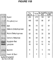
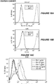
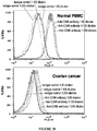
- ESK-bi-specific antibody staining human T cells, tumor cells or cell lines were incubated with different concentrations of ESK- bi-specific antibody or control bi-specific antibody for 30 minutes on ice, washed, and incubated with secondary mAbs against His-Tag.
- HER2-neu expression on primary ovarian cancer cells was measured by staining the tumor cells with Trastuzumab, followed by secondary goat anti-human IgG.
- HLA-A2 expression and ESK binding was determined by direct staining of the cells with respective mAbs.
- Phenotype of PBMCs or T cells from patient samples were characterized by direct staining of the cells with mAbs for CD3, CD4, CD8, CD45 RA, CD45RO, CCR7, CD19 or CD33 conjugated to various florophores.
- Flow cytometry data were collected on a FACS Calibur (Becton Dickinson) and analyzed with FlowJo V8.7.1 and 9.4.8 software.
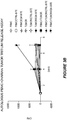
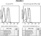
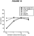
- a binding study was performed to examine ESK1+ L2K bi-specific antibody binding to Jurkat (a CD3 + human cancer cell line) cells. Briefly, bi-specific antibody in 3x serial dilution, starting from 10 ⁇ g/mL was added to 0.5 x 10 6 Jurkat cells. The cells were incubated with FITC conjugated anti-His tag antibody (Thermo #MA1-81891) at a 100x dilution or mouse anti-His tag antibody with or without allophycocyanin (APC)-anti mouse IgG (Biolegend # Poly4053) at a 1000x dilution and analyzed by flow cytometry on a Guava EasyCyte 6HT (EMD Millipore). The results are shown in Figure 8 .
- ESK-1 bi-specific antibody is a full length antibody.
- ESK1 or ET901 (negative control) and anti-CD3 (OKT3) were combined in equimolar ratios, yielding a final concentration of 0.8 mg/ml of each antibody within 0.5ml.
- 12.5 ⁇ l of 1M 2-mercaptoethylamine (2-MEA) was added to the reaction mixture and the solution was incubated at 37°C for 90 minutes.
- 2-MEA was removed by Zeba desalting columns (100-200 ⁇ l, 7-kDA, Pierce.) The solution was stored at 4°C overnight to allow re-oxidation of the disulfide bonds. Production values are shown in Table 11.
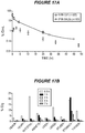
- Binding affinity was determined using ForteBio Octet QK. 5 ⁇ g/ml biotinylated MHC-WT1 was loaded onto the Streptavidin biosensor. After washing off excess antigen, ESK-1-OKT3, ESK1, and OKT3 solutions were tested at 20 ⁇ g/ml, 10 ⁇ g/ml and 10 ⁇ g/ml respectively for their association and dissociation constants. Binding parameters were calculated using 1:1 binding site model and are shown in Table 12.
- ESK-Bi-specific antibody- mediated T cell activation CD3 T cells were isolated from PBMCs by negative immunomagnetic cell separation using a pan T cell isolation kit (Miltenyi Biotec Inc., San Diego, CA). The ESK-bi-specific antibody or its control bi-specific antibody at various concentrations were incubated with target cells and purified CD3 T cells at different effector: target (E:T) ratio for overnight or different time periods. The supernatant fluids were harvested and cytokine release was measured by ELISA for IFN- ⁇ and TNF- ⁇ . In addition, ESK-bi-specific antibody-mediated T cell activation in the presence of autologous tumor cells from a patient with ovarian cancer was evaluated by cell proliferation, measured by overnight 3H-Thymidine incorporation after seven days of co-incubation.
- T-cells were enriched from PBMCs by depletion of monocytes by adhesion. Non- adhering cells were stimulated with irradiated autologous EBV-transformed B cells (EBV-BLCLs) generated by transformation with the B95.8 strain of EBV at a 20:1 responder: stimulator (R: S) ratio and cultured in Yssel's medium, containing 5% HS(YH5; Gemini).
- EBV-BLCLs irradiated autologous EBV-transformed B cells
- interleukin-2 (IL-2) at 10 to 30 units/mL was added to the T cell cultures every 2-3 days (Collaborative Biomedical Products, Bedford, MA), and were re-stimulated weekly with the same EBV-BLCLs at a 4:1 R:S ratio.
- EBV specific T lymphocytes were transduced with retroviral vector tdrrsRLuc, expanded and enriched by sorting for PE, as previously described ( Doubrovina, E. et al. Blood 119 (11), 2644-2655 (2012 )). Transduced EBV specific T lymphocytes were cultured in G-rex flask (Wilson Wolf Manufacturing Corporation). For in vivo T cell tracing study, ten million cells were injected into mice and 4 hrs later, Renilla luciferase substrate ViviRenTM was given i.v..
- ESK-Bi-specific antibody- redirected T cell cytotoxicity The ESK-bi-specific antibody or its control bi-specific antibody at various concentrations were incubated with target cells and PBMCs, purified CD3 T cells or EBV-specific T cells at different effector: target (E:T) ratios for 5 hrs or overnight.
- the cytotoxicity was measured by 51 Cr-release assay (after 5 hrs incubation) or LDH release assay (after overnight incubation) using Cytotox 96 non-radioreactive kit from Promega following their instruction.
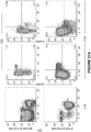
- PBMCs and autologous blasts were co-incubated in the presence or absence of ESK or control-bi-specific antibody at 20 ⁇ g/ml and the cells were harvested and dual stained with CD33 for leukemia blasts and CD3 for T cells on day 3 and 4.
- PBMCs were incubated with autologous ovarian cancer cells for a week and the cytotoxicity was measured by 51 Cr-release assay.
- ESK-bi-specific antibody or control bi-specific antibody were labeled with 125 I (PerkinElmer) using the chloramine-T method.
- 125 I PerkinElmer
- One hundred (100) ⁇ g antibody was reacted with 1 mCi 125 I and 20 ⁇ g chloramine-T, quenched with 200 ⁇ g Na metabisulfite, then separated from free 125 I using a 10DG column equilibrated with 2% bovine serum albumin in PBS. Specific activity of the product was 6 mCi/mg.
- Radiolabeled bi-specific antibody (2 ⁇ g) was diluted with unlabeled bi-specific antibody to 20 ⁇ g per dose, and injected into mice retroorbitally. Blood was collected at various time points, weighed and measured on a gamma counter. At 24-hours, organs were harvested, weighed and measured for activity on a gamma counter.
- ESK-Bi-specific antibody in human tumor xenograft NSG models.
- Human EBV-specific T cells were used for all xenograft models, as their antigenic specificity had been heavily biased towards EBV Ags, wherein they should not induce GVHD.
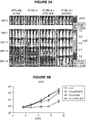
- BV173 ALL model two million BV173 human leukemia cells were injected i.v. into NSG mice. On day 5, tumor engraftment was confirmed by firefly luciferase imaging in all mice that were to be treated; mice were then randomly divided into different treatment groups. On day 6, ten million of EBV-specific T cells were iv injected into mice.
- ESK-bi-specific antibody or its control bi-specific antibody was i.v. injected and was repeated twice a week for total 6 times over the 3 weeks of treatment, along with i.v. injection of T cells once in a week times 2.
- SET-2 AML model one million cells were i.v. injected into mice, the tumor engraftment was confirmed on day 3 by bioluminescence imaging, and mice were randomized into treatment groups.
- ten million EBV-specific T cells were injected i.v. and 6 hrs later, 20 ⁇ g ESK-bi-specific antibody or its control bi-specific antibody was injected i.v.
- T cells were given twice a week and bi-specific antibodies were given every day for a total of 6 days.
- the primary ALL model five million ALL cells were injected i.v. into NSG mice.
- tumor engraftment was confirmed by firefly luciferase imaging in all mice that were to be treated; mice were then randomly divided into different treatment groups.
- Thirty million EBV-specific T cells were injected i.v. into mice followed by injection i.v. of 20 ⁇ g ESK-bispecific antibody or its control bi-specific antibody.
- Bi-specific antibody injection was given daily, and T cells were given twice a week for a total of two weeks.
- JMN mesothelioma model three hundred thousand tumor cells were mixed with six hundred thousand EBV-specific T cells and injected intraperitoneally (i.p.) and one hr later, 20 ⁇ g ESK-bi-specific antibody or control bi-specific antibody was i.v. injected into mice.
- the bi-specific antibodies were given every day for a total of 5 days. Tumor growth was monitored by firefly luciferase imaging at least twice a week, for all the animal models.
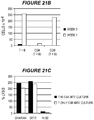
- PBMCs, PBMCs depleted of NK cells and macrophages or purified CD3 T cells from a patient with ovarian cancer were cultured with irradiated (3000 rad) autologous ovarian cancer cells at an E: T ratio of 4-5:1, in the presence or absence of ESK-bi-specific antibody, or control- bi-specific antibody at 0.1 ⁇ g/ml, and presence of human IL-5 (5 ng/ml) and human IL-2 (10 ⁇ g/ml) in RPMI1640 medium supplemented with 10% autologous plasma (AP) for 6 days.
- AP autologous plasma
- HA-Multiscreen plates (Millipore) were coated with 100 ⁇ L of mouse anti-human IFN-g antibody (10 Ag/mL; clone 1-D1K; Mabtech) in PBS, incubated overnight at 4 °C, washed with PBS to remove unbound antibody, and blocked with RPMI 1640/10% autologous plasma (AP) for 2 h at 37 °C. Effectors cells were plated with either T2 cells (4:1 E: APC ratio) or irradiated autologous tumor cells or other tumor cell lines.
- test peptides were added to the wells at 20 ⁇ g/mL.
- Negative control wells contained APCs and T cells without peptides or with irrelevant peptides.
- Positive control wells contained T cells plus APCs plus 20 ⁇ g/mL phytohemagglutinin (PHA, Sigma). All conditions were done in triplicate. Microtiter plates were incubated for 20 h at 37 °C and then extensively washed with PBS/ 0.05% Tween and 100 ⁇ l/well biotinylated detection antibody against human IFN-g (2 ⁇ g/mL; clone 7-B6-1; Mabtech) was added.
- the remaining T cells were expanded by adding fresh medium with IL-15 and IL-2 once a week, up to 7-8 weeks. In some cases, remaining T cells were re-stimulated with autologous tumor at an effector: target ratio of 9:1 for a week in the same conditions as for the first stimulation and T cell response was measured by IFN-g ELISPOT as described.
- ESK1 mAb binds to a panel of leukemia cell lines and mesothelioma cell lines in a WT1 and HLA-A0201-restricted manner (Dao et al., supra ).
- ForteBio Octet® (Menlo Park, CA) binding assay showed a Kd of 355nM of the ESK-bi-specific antibody.
- T cell activation and cytotoxicity against WT1 and HLA-A0201 positive tumor cells It has been shown that the activation of T cells by bi-specific antibody constructs depends on the proximal contact between T cells and target cells expressing the target antigens. This is crucial for avoiding unwanted inflammatory response caused by T cell activation, as one arm of the bi-specific antibody construct recognizes the invariant CD3 signaling complex.
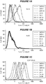
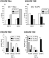
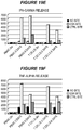
- ESK-bi-specific antibody and target SET-2 cells In the presence of ESK-bi-specific antibody and target SET-2 cells, a dose-dependent IFN- ⁇ release was observed at indicated concentrations of ESK-bi-specific antibody ( Fig. 1D ).
- CD3 T cells alone or incubated with control-bi-specific antibody in the presence of SET-2 cells showed undetectable level of IFN- ⁇ and their values were subtracted from the values of the ESK-bi-specific antibody group.
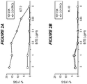
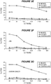
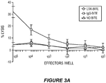
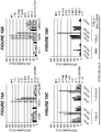
- ESK-bi-specific antibody-directed T cell cytotoxicity against target cells that co-express WT1 and HLA-A0201 was assessed. Purified resting T cells were co-incubated with target cells with serially diluted ESK-or control bi-specific antibody for 5 hrs and cell lysis was measured by 51 Cr-release. ESK-bi-specific antibody dose-dependently induced T cell cytotoxicity against AML cell line SET-2, but not HL-60, which was consistent with their binding specificity shown in Fig. 1 ( Fig. 2A, 2B ).
- the target cell lysis was increased up to 90% and 80% at 3 ⁇ g/ml and 0.3 ⁇ g/ml of ESK-bi-specific antibody, respectively.
- BV173 and primary CML blasts were also lysed by PBMCs in the presence of ESK-bi-specific antibody in a dose-dependent manner in a 6 hr co-incubation ( Fig 2C, 2D ), and the killing of BV173 was evident at 0.01 ⁇ g/ml of the ESK-bi-specific antibody.
- the relative weaker killing against the CML blasts might be due to a lower level expression of HLA-A2 and ESK-binding in this sample.
- Control bi-specific antibody did not induce any significant cytotoxicity, indicating that the target specificity is required for the T cell activation.
- ESK-bi-specific antibody to redirect cytotoxicity of T cells that had previously been repeatedly primed with a different Ag, such as EBV.
- T cells could potentially avoid graft-versus-host disease (GVHD) in xenograft animal models, as the antigenic specificity of T cells should be heavily biased towards only EBV Ags.
- GVHD graft-versus-host disease
- the potency of T cell killing by titration of the effector: target (E:T) ratios with ESK-bi-specific antibody at 0.1 ⁇ g/ml by 5 hr-51 Cr release was tested. Potent dose-dependent killing was observed for ESK-bi-specific antibody against all the target cells tested ( Fig. 2E, 2F, 2G ).
- ESK-bi-specific antibody-mediated activation and cytotoxicity of T cells in an autologous setting was investigated more closely, mimicking the human in vivo situation.
- PBMCs from a patient with ovarian cancer were stimulated with irradiated autologous tumor cells at various E:T ratios as indicated, in the presence of 0.1 ⁇ g/ml of ESK-bi-specific antibody for 7 days.
- the cytotoxicity was measured by adding 51 Cr-labeled autologous tumor cells in a 6 hr-culture on day 8. Dose-dependent killing was observed in the cultures with ESK-bi-specific antibody, even at 100 PBMCs at the start of the stimulation ( Fig. 3A ). No killing was observed in all the control groups.
- T cell proliferation was measured by 3H-thymidine incorporation at the end of eight day culture ( FIG. 3B ).
- Significant cell proliferation was seen in only the cultures with ESK-bi-specific antibody. None of the PBMCs, nor the tumor cells alone responded to either ESK- or control-bi-specific antibody.
- co-cultures of PBMCs with autologous tumor cells alone, or with control bi-specific antibody showed no T cell proliferation, suggesting T cells are tolerant to the self-tumor Ags.
- ESK-bi-specific antibody required the presence of tumor cells and that ESK-bi-specific antibody could force T cells to overcome the self-tolerance.
- the ability of the ESK-bi-specific antibody to induce autologous T cell proliferation and killing of its target cancer cells was illustrated in a second autologous model from a patient with AML ( Fig. 16A-16D ).
- mice xenografted intravenously 6 days previously with BV173 Ph+ALL cells was tested.
- mice had disseminated leukemia visible in their liver, spleen, and bone marrow.
- T cells were i.v. injected into mice and followed by bi-specific antibodies 4 hrs later. Dramatic tumor inhibition was observed, which was especially prominent in the bone marrow of mice that received ESK-bi-specific antibody, starting three days posttreatment and persisting up to 3 weeks ( Fig. 4A ).
- No significant GVHD was observed up to 49 days post-tumor inoculation ( Fig. 4C ).
- ESK- bi-specific antibody could efficiently engage and redirect potent T cell cytotoxicity to kill primary leukemia cells.
- EBV-specific human T cells did not show non-specific killing and therefore, could be used to address bi-specific antibody activity in xenograft models.
- This model suggested, and was confirmed with a pharmacokinetic study, that twice a week administration of ESK-bi-specific antibody was not sufficient, as its half-life was only a few hours.
- ESK-bi-specific antibody seemed to be more effective in treating bone marrow leukemia as shown in BV173 model, this effect in a more aggressive AML cells SET-2 model was investigated.
- SET-2 cells tend to migrate to bone marrow rapidly upon engraftment.
- Dosing and schedule was guided by pharmacokinetic studies that showed a short, 5-hour, beta plasma half-life of the ESK-bi-specific antibody ( Fig. 17A ).
- the treatment dosage was increased to daily bi-specific antibody injection for a total of 6 consecutive days. Cells were given twice a week.
- the mice received treatment starting on day 4 post-tumor inoculation, when the leukemia engraftment was confirmed by BLI ( Fig. 5A , first row).
- SET-2 cells rapidly grow in bone marrow, and a massive infiltration of leukemia was seen in the bone marrow of mice that received SET-2 cells or SET-2 along with T cells.
- no detectable leukemia was seen in the ESK-bi-specific antibody-treated group up to 14 days and only a minimum leukemia burden was observed up to 18 days later at day 32, more than a week after treatment was stopped.
- the gain on the BLI scale was uniformly decreased for all groups for the images from day 7 to 18, in order to show low levels of leukemia in the pretreated time point.
- the control-bi-specific antibody group showed slightly delayed leukemia burden in the beginning.
- EBV-specific T cells This could be caused by activation of EBV-specific T cells by anti-CD3 arm of the control-bi-specific antibody, which had stronger binding affinity for T cells than ESK-bi-specific antibody ( Fig. 1C ).
- EBV-specific T cells had already been activated and expanded multiple times in vitro, rendering them more susceptible to a strong polyclonal stimulation. Activated T cells then release inflammatory cytokines which could create a hostile environment for tumor cells. While all the mice in ESK-bi-specific antibody-treated group were still alive after a month, there was only 20% of survival in the groups of mice treated with T cells alone or no treatment ( Fig. 5B ).
- mice in ESK-Bi-specific antibody-treated group showed no sign of central nerve system (CNS) paralysis, caused by leukemia infiltration into vertebral bone marrow for up to 40 days, whereas nearly all animals in the control group were affected. ( Fig. 5C ). These data demonstrated that ESK-bi-specific antibody is a potent therapeutic agent against aggressive leukemia cells in bone marrow.
- CNS central nerve system
- ESK-Bi-specific antibody mediates T cell retention at tumor sites. While it is known that bi-specific antibodies effectively engage T cells to kill targets in vitro, no in vivo studies have been reported that document the mechanism in vivo. Whether the therapeutic efficacy of ESK-bi-specific antibody was the result of its ability to attract T cell retention and persistence at the sites of leukemia in live animals was investigated. Ten million EBV-specific T cells transduced with Renilla/luciferase were injected into NSG mice three days after GEF/luciferase positive SET-2 AML cells were engrafted, and 4 hrs later followed by bi-specific antibody injection.
- T cell migration was monitored by luminescence imaging at the time of T cell injection (0 hr), 8 (i.e., 4 hrs after bi-specific antibody injection), 24, 48 and 72 hrs.
- T cells migrated into the lungs immediately after the injection, then distributed into other parts including liver, spleen and bone marrow at 8 hrs ( Fig. 6A ).
- T cell signal gradually declined over 72 hrs.
- T cell injection followed by control bi-specific antibody showed a similar distribution pattern and time course as T cells alone.
- mice treated with ESK-bi-specific antibody showed significant increase in T cell signals in lungs and BM from 4-20 hrs, which lasted up to 72 hrs. Cells declined in the lungs substantially, but remained in the liver, spleen and BM.
- ESK-bi-specific antibody in treating aggressive solid tumor was investigated using an i.p. model to simulate peritoneal cavity mesothelioma.
- WT1+/HLA-A0201+ JMN mesothelioma cells were mixed with EBV-specific T cells at a target-effector ratio of 1:2 and were i.p. injected into NSG mice.
- Bi-specific antibodies were i.v. injected one hr later and was repeated for 5 consecutive days.
- ESK bi-specific antibody pharmacokinetics To determine ESK bi-specific antibody pharmacokinetics, trace 125 I-labeled construct was injected either intravenously into C57 BL6/J, or intraperitoneally into BALB/c mice, and blood radioactivity was measured over 48-52 hours. Intravenously injected bi-specific antibody quickly redistributed from blood to tissue with an alpha half-life of 30 minutes, followed by clearance with a beta half-life of 5 hours. After intraperitoneal delivery, bi-specific antibody levels increased in the blood, peaking at 3 hours post-injection. The construct then cleared with the same beta half-life. Total exposure (Area Under the Curve) from intravenous injection was greater than with intraperitoneal injection. The biodistribution pattern of the antibodies was determined using the same radiolabeled constructs. After 24 hours, ⁇ 1% injected dose / gram was detected in all tissues ( Fig. 17A, 17B ).
- bi-specific antibody induced secondary T cell response to a HER2 epitope in the context of HLA-A0201.
- the mechanism of the action for bi-specific antibodies has been attributed to their direct effect of bridging the cancer cell targets and T cells to form a cytolytic synapse. Whether such a proximal contact could directly activate pre-existing T cells in the population specific for other tumor antigens expressed by the autologous tumor cells was investigated to determine if a bi-specific antibody could exert a vaccinal effect using the autologous in vitro model from a HLA-A*02:01 positive patient with ovarian cancer.
- the patient's PBMCs were co-cultured with her autologous tumor cells in the presence of low-dose ESK-bi-specific antibody; a week later, the epitope-specific HLA-A*02:01-restricted T cell responses were assessed by IFN-g ELISPOT assay. Control groups including the cells alone or with control bi-specific antibody did not show any specific IFN-g release.
- the patient's tumor cells expressed both WT1 RMF and HER2-neu on their cell surface ( Fig. 18A-C ).
- a secondary T cell response should require professional APCs among the PBMCs that could take up and present the intracellular antigens released after tumor cell death.
- tumor cells could directly activate pre-existing epitope-specific T cells during the proximate contact between T cells and tumor.
- T cells alone as effectors still were capable of the response to HER2-neu, autologous tumor cells and SET-2 cells, in a similar magnitude as that produced by T plus B cells ( Fig.
- tumor cell antigen presentation depended on co-stimulatory molecules. While CD14+ monocytes from a healthy donor showed strong CD86 expression, ovarian cancer cells had little expression of the CD86. The results indicated that a key co-stimulatory molecule CD86 was unlikely to be involved in the tumor cell antigen presentation ( Fig. 20 ). No CD86 or ICOSL expression was detected. Lack of co-stimulatory molecules is one of the reasons that tumor cells are poor antigen presenting cells; however, certain cytokines may provide co-stimulatory signals to fulfill the requirements for tumor specific T cell activation. ESK-bi-specific antibody induced IFN-gamma and TNF-alpha secretion by both PBMCs and T cells ( Fig. 19E-F ).
- T cells were expanded in vitro in low-dose IL-15 and IL-2, after the first activation by ESK-bi-specific antibody. While T cells in control groups did not survive more than one week in culture, T cells activated by ESK--bi-specific antibody continued to grow for 2 months, without further activation. Phenotype analysis revealed that seven weeks after activation by ESK--bi-specific antibody and tumor, CD8+ T cells increased to 89% of the population and CD4+ T cells decreased to 6% ( Fig. 21A-B ).
- CD45RA- /CD45RO+ cells increased, indicative of effector memory phenotype.
- these T cells still retained their original specificity to autologous tumor and SET-2 AML cells ( Figure 21C ).
- the present disclosure relates to the first bi-specific antibody construct derived from a TCR-like mAb.
- the ESK-bi-specific antibody was specific for the WT1 RMF epitope presented by HLA-A0201 molecule.
- the ESK-bi-specific antibody showed a potent anti-tumor activity against multiple cancers, including ALL, AML, mesothelioma and primary ovarian cancer cells, both in vitro and in vivo.
- the study presented here is distinguished from previous studies, in that: 1) the mAb is a TCR-like mAb allowing recognition of an intracellular tumor-specific target; 2) the target density on tumor cells is extremely low, providing a proof-of-concept that could open the application of bi-specific antibodies to a much larger universe of targets; 3) the mechanisms of the ESK-bi-specific antibody activity in vivo with simultaneous dual effector cell and target cell tracing was demonstrated (previous bi-specific antibody studies have not shown this because in most animal studies, the effector T cells and target cells were mixed before injection; 4) Using an autologous system, it was demonstrated that the ESK-bi-specific antibody could break T cell tolerance to kill primary ovarian cancer cells; 5) the ESK-bi-specific antibody induced long-lived secondary, specific T cell responses against other tumor antigens, such as HER2-neu. Further, the secondary T cell responses were not mediated by cross-presentation effected by classical APCs, but rather was a result of the direct and physically close
- bi-specific antibody constructs that are currently in development target cell surface proteins, such as CD19, CD33, prostate specific membrane antigen (PSMA) or epidermal growth factor (EGF) receptor, that are lineage or differentiation antigens found on normal cells.
- PSMA prostate specific membrane antigen
- EGF epidermal growth factor
- the present disclosure demonstrates for the first time that a TCR-like bi-specific antibody construct is capable of recognizing a low density epitope of a peptide/MHC complex from a tumor-specific target. This argues against a traditional view that a scFv bi-specific antibody construct might not be suitable for targeting a low density Ag because the affinity would be too low or the ability to activate cytotoxicity by T cells would be insufficient.
- the present disclosure demonstrates for the first time that a TCR-mimic bi-specific antibody construct is capable of potent therapeutic activity in several mouse models of both hematopoietic and solid tumors, providing the proof of concept for vastly broadening the development of bi-specific antibodies targeting other cancer-specific intracellular tumor antigens. While it is known from assays in vitro that bi-specific antibodies bring T cells in contact with cancer cells, such a mechanism had not previously been observed in vivo. The present disclosure provides the proof of concept for future development of bi-specific mAbs targeting other intracellular tumor Ags. Thus, the ESK-bi-specific antibody described herein could broaden the therapeutic application of the platform.
- the ESK-bi-specific antibody induced fast and potent cytotoxic activity in vitro against lymphoid and myeoid leukemias, mesothelioma, and primary ovarian cancer cells, co-expressing WT1 and HLA-A0201. Lysis of the target cells by resting T cells or EBV-specific T cells could be detected in five hour co-incubation, compared to the studies with various other bi-specific antibody constructs that showed T cell killing after more than 16 hour of cultures.
- the cytolytic activity of ESK-bi-specific antibody could be further enhanced by longer period of incubation, shown by up to 80-90% of killing achieved with the SET-2 AML cells.
- ESK-bi-specific antibody engages T cell to targets in a serial fashion.
- IFN-g secretion further demonstrated the ESK-bi-specific antibody-mediated target-specific T cell activation.
- ESK-bi-specific antibody could efficiently activate un-manipulated T cells from a patient with ovarian cancer to proliferate and kill the autologous tumor cells, suggesting that the presence of ESK-bi-specific antibody could force T cells to overcome the tolerance to self-tumor Ags. It has been known that T cell infiltration is frequently observed in solid tumors. The relatively low molecular weight of the bi-specific antibody would allow ESK-bi-specific antibody better penetration into solid tumors to activate anergic or unresponsive T cells to kill the surrounding tumor cells.
- ESK-bi-specific antibody The in vitro cytotoxicity of ESK-bi-specific antibody was evidenced in vivo as well, as shown by its remarkably potent therapeutic activity against Ph1+ALL, AML and mesothelioma in xenograft models. In both leukemia models, elimination of tumor cells was evident, especially in bone marrow. In addition, ESK-bi-specific antibody prevented and delayed CNS paralysis, a characteristic feature of leukemia caused by infiltration of vertebral bone marrow.
- ESK-bi-specific antibody to kill leukemia cells in vivo when the T cells were injected separately from the tumor cells (a schedule not typically used in preclinical bi-specific antibody experiments), raised questions about the mechanism of bi-specific antibody activity in vivo.
- the study with SET-2 cells further raised the question of whether ESK-bi-specific antibody actively recruits and retains T cell effectors at the target. These questions were addressed by simultaneously tracing effector T cells and SET-2 AML cells using dual bioluminescence imaging. There was approximately a three-fold increase in T cells in the bone marrow of the mice treated with ESK-bi-specific antibody. The T cell retention was well correlated with the tumor reduction.
- the present disclosure provides direct in vivo evidence that ESK-bi-specific antibody could efficiently engage T cells with targets and kill the tumor cells in situ.
- ESK-bi-specific antibody The therapeutic activity of ESK-bi-specific antibody in a solid tumor model with a peritoneal human mesothelioma in NSG mice was investigated.
- the PK study showed that i.v. injection of the ESK-bi-specific antibody gave a higher level in the blood stream than i.p. injection but that the plasma half-life of the bi-specific antibody was characteristically short. Rapid and dramatic tumor inhibition was seen in the treated mice, suggesting that ESK-bi-specific antibody gained quick access to the peritoneal tumor cells.
- the PK studies confirmed the pharmacokinetic observations from other bi-specific antibody constructs, suggesting that continuous administration of ESK-bi-specific antibody may be necessary to achieve the best therapeutic efficacy.
- ESK-bi-specific antibody Similar to other bi-specific antibody constructs, a limitation of ESK-bi-specific antibody is also the lack of a natural Fc region thus preventing interaction with the neonatal FcRn receptor, which is necessary for delaying mAb clearance and for longer blood PK.
- Engineering an alternative bi-specific form of mAb with dual target specificities yet maintaining its natural architecture could offer a longer half-life and could significantly improve its efficacy in clinical application.
- ESK1 was more effective in eradicating disseminated leukemia BV173 cells in lung and liver in mice, while ESK-bi-specific antibody seemed to be more effective in bone marrow.
- ESK-bi-specific antibody did not induce T cell responses against the original target epitope WT1-RMF, even though the patient had WT1 reactive T cells in her circulation as a consequence of prior adaptive cell therapy.
- bi-specific antibodies could penetrate into solid tumors to activate anergic or unresponsive T cells to kill tumor cells that express not only the known antigenic target of the bi-specific antibody, but also other undefined specific tumor antigens.
- the bi-specific antibody-mediated cytotoxic T cell clonal expansion for a variety of new epitopes should contribute greatly to its long-term therapeutic efficacy by preventing escape of cancer cell variants or low antigen density cells, as well as by promoting long-lived immunity.
- the vaccinal effect could be used for ex vivo expansion in advance of specific adoptive T cell therapy.
Landscapes
- Health & Medical Sciences (AREA)
- Chemical & Material Sciences (AREA)
- Organic Chemistry (AREA)
- Immunology (AREA)
- Life Sciences & Earth Sciences (AREA)
- Medicinal Chemistry (AREA)
- General Health & Medical Sciences (AREA)
- Veterinary Medicine (AREA)
- Biochemistry (AREA)
- Animal Behavior & Ethology (AREA)
- Nuclear Medicine, Radiotherapy & Molecular Imaging (AREA)
- Public Health (AREA)
- General Chemical & Material Sciences (AREA)
- Proteomics, Peptides & Aminoacids (AREA)
- Oncology (AREA)
- Chemical Kinetics & Catalysis (AREA)
- Pharmacology & Pharmacy (AREA)
- Biophysics (AREA)
- Genetics & Genomics (AREA)
- Molecular Biology (AREA)
- Hematology (AREA)
- Peptides Or Proteins (AREA)
- Medicines Containing Antibodies Or Antigens For Use As Internal Diagnostic Agents (AREA)
- Preparation Of Compounds By Using Micro-Organisms (AREA)
- Micro-Organisms Or Cultivation Processes Thereof (AREA)
- Medicines Containing Material From Animals Or Micro-Organisms (AREA)
Applications Claiming Priority (3)
| Application Number | Priority Date | Filing Date | Title |
|---|---|---|---|
| US201361901310P | 2013-11-07 | 2013-11-07 | |
| US201462037875P | 2014-08-15 | 2014-08-15 | |
| PCT/US2014/064621 WO2015070061A1 (en) | 2013-11-07 | 2014-11-07 | Anti-wt1/hla bi-specific antibody |
Publications (2)
| Publication Number | Publication Date |
|---|---|
| EP3066130A1 EP3066130A1 (en) | 2016-09-14 |
| EP3066130B1 true EP3066130B1 (en) | 2019-05-15 |
Family
ID=51987482
Family Applications (1)
| Application Number | Title | Priority Date | Filing Date |
|---|---|---|---|
| EP14803300.4A Active EP3066130B1 (en) | 2013-11-07 | 2014-11-07 | Anti-wt1/hla bi-specific antibody |
Country Status (5)
| Country | Link |
|---|---|
| EP (1) | EP3066130B1 (ja) |
| JP (1) | JP6553069B2 (ja) |
| CN (1) | CN106414500B (ja) |
| ES (1) | ES2742224T3 (ja) |
| WO (1) | WO2015070061A1 (ja) |
Families Citing this family (15)
| Publication number | Priority date | Publication date | Assignee | Title |
|---|---|---|---|---|
| NL2014935B1 (en) | 2015-06-08 | 2017-02-03 | Applied Immune Tech Ltd | T cell receptor like antibodies having fine specificity. |
| MA45004A (fr) | 2015-10-09 | 2019-03-27 | Immatics Biotechnologies Gmbh | Anticorps spécifiques anti-wt1-hla |
| AU2016342041B2 (en) | 2015-10-23 | 2021-12-02 | Eureka Therapeutics, Inc. | Antibody/T-cell receptor chimeric constructs and uses thereof |
| JP7022067B2 (ja) * | 2016-01-14 | 2022-02-17 | メモリアル スローン ケタリング キャンサー センター | Foxp3由来のペプチドに特異的なt細胞受容体様抗体 |
| CN110944651A (zh) | 2017-02-08 | 2020-03-31 | 蜻蜓疗法股份有限公司 | 用于自然杀伤细胞激活的多特异性结合蛋白及其治疗癌症的治疗性用途 |
| CA3054079A1 (en) | 2017-02-20 | 2018-08-23 | Dragonfly Therapeutics, Inc. | Proteins binding her2, nkg2d and cd16 |
| CN110741016A (zh) | 2017-04-26 | 2020-01-31 | 优瑞科生物技术公司 | 嵌合抗体/t-细胞受体构筑体及其用途 |
| JP7072792B2 (ja) * | 2017-12-11 | 2022-05-23 | 国立大学法人神戸大学 | 二重特異性抗体 |
| TWI805665B (zh) * | 2017-12-21 | 2023-06-21 | 瑞士商赫孚孟拉羅股份公司 | 結合hla-a2/wt1之抗體 |
| PE20220278A1 (es) | 2018-02-08 | 2022-02-25 | Dragonfly Therapeutics Inc | Dominios variables de anticuerpos que se dirigen al receptor nkg2d |
| CA3091143A1 (en) | 2018-02-15 | 2019-08-22 | Memorial Sloan-Kettering Cancer Center | Foxp3 targeting agent compositions and methods of use for adoptive cell therapy |
| WO2019222760A1 (en) * | 2018-05-18 | 2019-11-21 | Children's National Medical Center | Improved targeted t-cell therapy |
| CN115698070A (zh) * | 2020-04-02 | 2023-02-03 | 德克萨斯大学系统董事会 | Cd5l结合抗体及其用途 |
| US11883432B2 (en) | 2020-12-18 | 2024-01-30 | Century Therapeutics, Inc. | Chimeric antigen receptor system with adaptable receptor specificity |
| WO2023180346A1 (en) * | 2022-03-22 | 2023-09-28 | Morphosys Ag | Deimmunized antibodies specific for cd3 |
Family Cites Families (4)
| Publication number | Priority date | Publication date | Assignee | Title |
|---|---|---|---|---|
| US5885573A (en) * | 1993-06-01 | 1999-03-23 | Arch Development Corporation | Methods and materials for modulation of the immunosuppressive activity and toxicity of monoclonal antibodies |
| DK1673398T3 (da) * | 2003-10-16 | 2011-04-18 | Micromet Ag | Multispecifikke, deimmuniserede CD3-bindere |
| EP1981532A4 (en) * | 2005-12-21 | 2010-06-30 | Medimmune Llc | EPHA2 MOLECULES AND USES THEREOF |
| WO2012135854A2 (en) * | 2011-04-01 | 2012-10-04 | Memorial Sloan-Kettering Cancer Center | Antibodies to cytosolic peptides |
-
2014
- 2014-11-07 WO PCT/US2014/064621 patent/WO2015070061A1/en active Application Filing
- 2014-11-07 CN CN201480061156.9A patent/CN106414500B/zh active Active
- 2014-11-07 JP JP2016553248A patent/JP6553069B2/ja not_active Expired - Fee Related
- 2014-11-07 ES ES14803300T patent/ES2742224T3/es active Active
- 2014-11-07 EP EP14803300.4A patent/EP3066130B1/en active Active
Non-Patent Citations (1)
| Title |
|---|
| None * |
Also Published As
| Publication number | Publication date |
|---|---|
| WO2015070061A1 (en) | 2015-05-14 |
| CN106414500A (zh) | 2017-02-15 |
| ES2742224T3 (es) | 2020-02-13 |
| EP3066130A1 (en) | 2016-09-14 |
| JP6553069B2 (ja) | 2019-07-31 |
| JP2017500057A (ja) | 2017-01-05 |
| CN106414500B (zh) | 2020-04-10 |
Similar Documents
| Publication | Publication Date | Title |
|---|---|---|
| EP3066130B1 (en) | Anti-wt1/hla bi-specific antibody | |
| RU2770001C2 (ru) | Терапевтические соединения и способы | |
| ES2809455T3 (es) | Métodos para tratamiento tumoral usando anticuerpo biespecífico CD3xCD20 | |
| CN112513070A (zh) | Il-15变体及其用途 | |
| AU2018211278A1 (en) | Agents for treatment of claudin expressing cancer diseases | |
| KR20190019144A (ko) | Pd-l1 및 lag-3에 결합하는 결합 분자 | |
| EP3297672A1 (en) | Trispecific binding proteins and methods of use | |
| KR20180014050A (ko) | Tigit-결합제 및 이의 용도 | |
| JP7369297B2 (ja) | Cd47、pd-l1に特異的な抗体、およびその使用 | |
| CN113286634A (zh) | 对gucy2c特异性的抗体及其用途 | |
| US10239952B2 (en) | Anti-WT1/HLA bi-specific antibody | |
| US10934331B2 (en) | Methods for enhancing immune responsiveness in an individual toward a target cancer cell population comprising apoptotic cells | |
| KR102602329B1 (ko) | Cd3에 특이적인 항체 및 이의 용도 | |
| KR20220150274A (ko) | Il-7 단백질과 이중특이적 항체의 조합물을 사용한 종양의 치료 방법 | |
| CN117396226A (zh) | 包含t细胞重定向治疗剂和抗cd44治疗剂的组合物 | |
| KR20230118108A (ko) | 항-cd25 항체 | |
| JP2022546364A (ja) | キメラ抗原受容体系及びその使用 | |
| RU2792240C2 (ru) | Биспецифический антигенсвязывающий белок egfr/cd16 | |
| JP2024517985A (ja) | 抗-CD300cモノクローナル抗体及びその癌の予防または治療用バイオマーカー | |
| TWI790193B (zh) | 調控免疫反應之方法及抗體 | |
| CA3214628A1 (en) | Combination therapy with dexamethasone and tumor-specific t cell engaging multi-specific antibodies for treating cancer | |
| WO2024026374A1 (en) | Anti-cd16a antibodies and methods of use thereof | |
| TW202305006A (zh) | 與cd47及pd—l1特異性結合的雙特異性抗體 | |
| WO2022214681A1 (en) | Methods for the treatment of anaplastic large cell lymphoma | |
| KR20220070440A (ko) | 치료학적 항체와 인터루킨-2 (il2)를 수반한 치료 |
Legal Events
| Date | Code | Title | Description |
|---|---|---|---|
| PUAI | Public reference made under article 153(3) epc to a published international application that has entered the european phase |
Free format text: ORIGINAL CODE: 0009012 |
|
| 17P | Request for examination filed |
Effective date: 20160603 |
|
| AK | Designated contracting states |
Kind code of ref document: A1 Designated state(s): AL AT BE BG CH CY CZ DE DK EE ES FI FR GB GR HR HU IE IS IT LI LT LU LV MC MK MT NL NO PL PT RO RS SE SI SK SM TR |
|
| AX | Request for extension of the european patent |
Extension state: BA ME |
|
| DAX | Request for extension of the european patent (deleted) | ||
| STAA | Information on the status of an ep patent application or granted ep patent |
Free format text: STATUS: EXAMINATION IS IN PROGRESS |
|
| 17Q | First examination report despatched |
Effective date: 20170804 |
|
| REG | Reference to a national code |
Ref country code: HK Ref legal event code: DE Ref document number: 1227903 Country of ref document: HK |
|
| GRAP | Despatch of communication of intention to grant a patent |
Free format text: ORIGINAL CODE: EPIDOSNIGR1 |
|
| STAA | Information on the status of an ep patent application or granted ep patent |
Free format text: STATUS: GRANT OF PATENT IS INTENDED |
|
| INTG | Intention to grant announced |
Effective date: 20181122 |
|
| RAP1 | Party data changed (applicant data changed or rights of an application transferred) |
Owner name: MEMORIAL SLOAN-KETTERING CANCER CENTER Owner name: EUREKA THERAPEUTICS, INC. |
|
| GRAS | Grant fee paid |
Free format text: ORIGINAL CODE: EPIDOSNIGR3 |
|
| GRAA | (expected) grant |
Free format text: ORIGINAL CODE: 0009210 |
|
| STAA | Information on the status of an ep patent application or granted ep patent |
Free format text: STATUS: THE PATENT HAS BEEN GRANTED |
|
| AK | Designated contracting states |
Kind code of ref document: B1 Designated state(s): AL AT BE BG CH CY CZ DE DK EE ES FI FR GB GR HR HU IE IS IT LI LT LU LV MC MK MT NL NO PL PT RO RS SE SI SK SM TR |
|
| REG | Reference to a national code |
Ref country code: CH Ref legal event code: EP |
|
| REG | Reference to a national code |
Ref country code: CH Ref legal event code: NV Representative=s name: E. BLUM AND CO. AG PATENT- UND MARKENANWAELTE , CH |
|
| REG | Reference to a national code |
Ref country code: IE Ref legal event code: FG4D |
|
| REG | Reference to a national code |
Ref country code: DE Ref legal event code: R096 Ref document number: 602014046895 Country of ref document: DE |
|
| REG | Reference to a national code |
Ref country code: NL Ref legal event code: MP Effective date: 20190515 |
|
| REG | Reference to a national code |
Ref country code: LT Ref legal event code: MG4D |
|
| PG25 | Lapsed in a contracting state [announced via postgrant information from national office to epo] |
Ref country code: FI Free format text: LAPSE BECAUSE OF FAILURE TO SUBMIT A TRANSLATION OF THE DESCRIPTION OR TO PAY THE FEE WITHIN THE PRESCRIBED TIME-LIMIT Effective date: 20190515 Ref country code: PT Free format text: LAPSE BECAUSE OF FAILURE TO SUBMIT A TRANSLATION OF THE DESCRIPTION OR TO PAY THE FEE WITHIN THE PRESCRIBED TIME-LIMIT Effective date: 20190915 Ref country code: NO Free format text: LAPSE BECAUSE OF FAILURE TO SUBMIT A TRANSLATION OF THE DESCRIPTION OR TO PAY THE FEE WITHIN THE PRESCRIBED TIME-LIMIT Effective date: 20190815 Ref country code: AL Free format text: LAPSE BECAUSE OF FAILURE TO SUBMIT A TRANSLATION OF THE DESCRIPTION OR TO PAY THE FEE WITHIN THE PRESCRIBED TIME-LIMIT Effective date: 20190515 Ref country code: SE Free format text: LAPSE BECAUSE OF FAILURE TO SUBMIT A TRANSLATION OF THE DESCRIPTION OR TO PAY THE FEE WITHIN THE PRESCRIBED TIME-LIMIT Effective date: 20190515 Ref country code: LT Free format text: LAPSE BECAUSE OF FAILURE TO SUBMIT A TRANSLATION OF THE DESCRIPTION OR TO PAY THE FEE WITHIN THE PRESCRIBED TIME-LIMIT Effective date: 20190515 Ref country code: HR Free format text: LAPSE BECAUSE OF FAILURE TO SUBMIT A TRANSLATION OF THE DESCRIPTION OR TO PAY THE FEE WITHIN THE PRESCRIBED TIME-LIMIT Effective date: 20190515 Ref country code: NL Free format text: LAPSE BECAUSE OF FAILURE TO SUBMIT A TRANSLATION OF THE DESCRIPTION OR TO PAY THE FEE WITHIN THE PRESCRIBED TIME-LIMIT Effective date: 20190515 |
|
| PG25 | Lapsed in a contracting state [announced via postgrant information from national office to epo] |
Ref country code: LV Free format text: LAPSE BECAUSE OF FAILURE TO SUBMIT A TRANSLATION OF THE DESCRIPTION OR TO PAY THE FEE WITHIN THE PRESCRIBED TIME-LIMIT Effective date: 20190515 Ref country code: RS Free format text: LAPSE BECAUSE OF FAILURE TO SUBMIT A TRANSLATION OF THE DESCRIPTION OR TO PAY THE FEE WITHIN THE PRESCRIBED TIME-LIMIT Effective date: 20190515 Ref country code: BG Free format text: LAPSE BECAUSE OF FAILURE TO SUBMIT A TRANSLATION OF THE DESCRIPTION OR TO PAY THE FEE WITHIN THE PRESCRIBED TIME-LIMIT Effective date: 20190815 Ref country code: GR Free format text: LAPSE BECAUSE OF FAILURE TO SUBMIT A TRANSLATION OF THE DESCRIPTION OR TO PAY THE FEE WITHIN THE PRESCRIBED TIME-LIMIT Effective date: 20190816 |
|
| REG | Reference to a national code |
Ref country code: AT Ref legal event code: MK05 Ref document number: 1133317 Country of ref document: AT Kind code of ref document: T Effective date: 20190515 |
|
| PG25 | Lapsed in a contracting state [announced via postgrant information from national office to epo] |
Ref country code: CZ Free format text: LAPSE BECAUSE OF FAILURE TO SUBMIT A TRANSLATION OF THE DESCRIPTION OR TO PAY THE FEE WITHIN THE PRESCRIBED TIME-LIMIT Effective date: 20190515 Ref country code: AT Free format text: LAPSE BECAUSE OF FAILURE TO SUBMIT A TRANSLATION OF THE DESCRIPTION OR TO PAY THE FEE WITHIN THE PRESCRIBED TIME-LIMIT Effective date: 20190515 Ref country code: DK Free format text: LAPSE BECAUSE OF FAILURE TO SUBMIT A TRANSLATION OF THE DESCRIPTION OR TO PAY THE FEE WITHIN THE PRESCRIBED TIME-LIMIT Effective date: 20190515 Ref country code: EE Free format text: LAPSE BECAUSE OF FAILURE TO SUBMIT A TRANSLATION OF THE DESCRIPTION OR TO PAY THE FEE WITHIN THE PRESCRIBED TIME-LIMIT Effective date: 20190515 Ref country code: SK Free format text: LAPSE BECAUSE OF FAILURE TO SUBMIT A TRANSLATION OF THE DESCRIPTION OR TO PAY THE FEE WITHIN THE PRESCRIBED TIME-LIMIT Effective date: 20190515 Ref country code: RO Free format text: LAPSE BECAUSE OF FAILURE TO SUBMIT A TRANSLATION OF THE DESCRIPTION OR TO PAY THE FEE WITHIN THE PRESCRIBED TIME-LIMIT Effective date: 20190515 |
|
| REG | Reference to a national code |
Ref country code: ES Ref legal event code: FG2A Ref document number: 2742224 Country of ref document: ES Kind code of ref document: T3 Effective date: 20200213 |
|
| REG | Reference to a national code |
Ref country code: DE Ref legal event code: R097 Ref document number: 602014046895 Country of ref document: DE |
|
| PG25 | Lapsed in a contracting state [announced via postgrant information from national office to epo] |
Ref country code: SM Free format text: LAPSE BECAUSE OF FAILURE TO SUBMIT A TRANSLATION OF THE DESCRIPTION OR TO PAY THE FEE WITHIN THE PRESCRIBED TIME-LIMIT Effective date: 20190515 |
|
| PLBE | No opposition filed within time limit |
Free format text: ORIGINAL CODE: 0009261 |
|
| STAA | Information on the status of an ep patent application or granted ep patent |
Free format text: STATUS: NO OPPOSITION FILED WITHIN TIME LIMIT |
|
| PG25 | Lapsed in a contracting state [announced via postgrant information from national office to epo] |
Ref country code: TR Free format text: LAPSE BECAUSE OF FAILURE TO SUBMIT A TRANSLATION OF THE DESCRIPTION OR TO PAY THE FEE WITHIN THE PRESCRIBED TIME-LIMIT Effective date: 20190515 |
|
| 26N | No opposition filed |
Effective date: 20200218 |
|
| PG25 | Lapsed in a contracting state [announced via postgrant information from national office to epo] |
Ref country code: PL Free format text: LAPSE BECAUSE OF FAILURE TO SUBMIT A TRANSLATION OF THE DESCRIPTION OR TO PAY THE FEE WITHIN THE PRESCRIBED TIME-LIMIT Effective date: 20190515 |
|
| PG25 | Lapsed in a contracting state [announced via postgrant information from national office to epo] |
Ref country code: SI Free format text: LAPSE BECAUSE OF FAILURE TO SUBMIT A TRANSLATION OF THE DESCRIPTION OR TO PAY THE FEE WITHIN THE PRESCRIBED TIME-LIMIT Effective date: 20190515 |
|
| PG25 | Lapsed in a contracting state [announced via postgrant information from national office to epo] |
Ref country code: LU Free format text: LAPSE BECAUSE OF NON-PAYMENT OF DUE FEES Effective date: 20191107 Ref country code: MC Free format text: LAPSE BECAUSE OF FAILURE TO SUBMIT A TRANSLATION OF THE DESCRIPTION OR TO PAY THE FEE WITHIN THE PRESCRIBED TIME-LIMIT Effective date: 20190515 |
|
| REG | Reference to a national code |
Ref country code: BE Ref legal event code: MM Effective date: 20191130 |
|
| PG25 | Lapsed in a contracting state [announced via postgrant information from national office to epo] |
Ref country code: IE Free format text: LAPSE BECAUSE OF NON-PAYMENT OF DUE FEES Effective date: 20191107 |
|
| PG25 | Lapsed in a contracting state [announced via postgrant information from national office to epo] |
Ref country code: BE Free format text: LAPSE BECAUSE OF NON-PAYMENT OF DUE FEES Effective date: 20191130 |
|
| PG25 | Lapsed in a contracting state [announced via postgrant information from national office to epo] |
Ref country code: CY Free format text: LAPSE BECAUSE OF FAILURE TO SUBMIT A TRANSLATION OF THE DESCRIPTION OR TO PAY THE FEE WITHIN THE PRESCRIBED TIME-LIMIT Effective date: 20190515 |
|
| PG25 | Lapsed in a contracting state [announced via postgrant information from national office to epo] |
Ref country code: IS Free format text: LAPSE BECAUSE OF FAILURE TO SUBMIT A TRANSLATION OF THE DESCRIPTION OR TO PAY THE FEE WITHIN THE PRESCRIBED TIME-LIMIT Effective date: 20190915 |
|
| PG25 | Lapsed in a contracting state [announced via postgrant information from national office to epo] |
Ref country code: MT Free format text: LAPSE BECAUSE OF FAILURE TO SUBMIT A TRANSLATION OF THE DESCRIPTION OR TO PAY THE FEE WITHIN THE PRESCRIBED TIME-LIMIT Effective date: 20190515 Ref country code: HU Free format text: LAPSE BECAUSE OF FAILURE TO SUBMIT A TRANSLATION OF THE DESCRIPTION OR TO PAY THE FEE WITHIN THE PRESCRIBED TIME-LIMIT; INVALID AB INITIO Effective date: 20141107 |
|
| PG25 | Lapsed in a contracting state [announced via postgrant information from national office to epo] |
Ref country code: MK Free format text: LAPSE BECAUSE OF FAILURE TO SUBMIT A TRANSLATION OF THE DESCRIPTION OR TO PAY THE FEE WITHIN THE PRESCRIBED TIME-LIMIT Effective date: 20190515 |
|
| PGFP | Annual fee paid to national office [announced via postgrant information from national office to epo] |
Ref country code: GB Payment date: 20220915 Year of fee payment: 9 |
|
| PGFP | Annual fee paid to national office [announced via postgrant information from national office to epo] |
Ref country code: FR Payment date: 20220908 Year of fee payment: 9 |
|
| PGFP | Annual fee paid to national office [announced via postgrant information from national office to epo] |
Ref country code: IT Payment date: 20221011 Year of fee payment: 9 Ref country code: ES Payment date: 20221206 Year of fee payment: 9 Ref country code: DE Payment date: 20220914 Year of fee payment: 9 |
|
| PGFP | Annual fee paid to national office [announced via postgrant information from national office to epo] |
Ref country code: CH Payment date: 20221201 Year of fee payment: 9 |
|
| P01 | Opt-out of the competence of the unified patent court (upc) registered |
Effective date: 20230519 |