EP3004385B1 - Analyse d'acides nucléiques basés sur des nanopores avec une détection par fret mixte - Google Patents
Analyse d'acides nucléiques basés sur des nanopores avec une détection par fret mixte Download PDFInfo
- Publication number
- EP3004385B1 EP3004385B1 EP14801275.0A EP14801275A EP3004385B1 EP 3004385 B1 EP3004385 B1 EP 3004385B1 EP 14801275 A EP14801275 A EP 14801275A EP 3004385 B1 EP3004385 B1 EP 3004385B1
- Authority
- EP
- European Patent Office
- Prior art keywords
- nanopore
- fret
- polynucleotide
- nucleotides
- labeled
- Prior art date
- Legal status (The legal status is an assumption and is not a legal conclusion. Google has not performed a legal analysis and makes no representation as to the accuracy of the status listed.)
- Active
Links
Images


Classifications
-
- C—CHEMISTRY; METALLURGY
- C12—BIOCHEMISTRY; BEER; SPIRITS; WINE; VINEGAR; MICROBIOLOGY; ENZYMOLOGY; MUTATION OR GENETIC ENGINEERING
- C12Q—MEASURING OR TESTING PROCESSES INVOLVING ENZYMES, NUCLEIC ACIDS OR MICROORGANISMS; COMPOSITIONS OR TEST PAPERS THEREFOR; PROCESSES OF PREPARING SUCH COMPOSITIONS; CONDITION-RESPONSIVE CONTROL IN MICROBIOLOGICAL OR ENZYMOLOGICAL PROCESSES
- C12Q1/00—Measuring or testing processes involving enzymes, nucleic acids or microorganisms; Compositions therefor; Processes of preparing such compositions
- C12Q1/68—Measuring or testing processes involving enzymes, nucleic acids or microorganisms; Compositions therefor; Processes of preparing such compositions involving nucleic acids
- C12Q1/6869—Methods for sequencing
Definitions
- Single molecule sequencing using nanopores may address some of these challenges, e.g., Maitra et al, Electrophoresis, 33: 3418-3428 (2012 ); Venkatesan et al, Nature Nanotechnology, 6: 615-624 (2011 ); however, this approach has its own set of technical difficulties, such as, reliable nanopore fabrication, control of DNA translocation rates, nucleotide discrimination, detection of electrical signals from large arrays of nanopore sensors, and the like, e.g. Branton et al, Nature Biotechnology, 26(10): 1146-1153 (2008 ); Venkatesan et al (cited above).
- nanopore sensor technology in general and its particular applications, such as optically based nanopore sequencing, if there were available materials and configurations of optical elements that permitted successful optical sensing and analysis of analytes, such as sequences of nucleic acids.
- the invention provides a method of determining a nucleotide sequence of a polynucleotide, the method comprising the steps of:
- the invention provides a system for determining a nucleotide sequence of a polynucleotide, the system comprising:
- a method of determining a nucleotide sequence of a polynucleotide comprises the following steps: (a) translocating a polynucleotide, e.g., a single or double stranded polynucleotide, through a nanopore so that nucleotides of the polynucleotide pass in sequence by a first member of a FRET pair positioned adjacent to the nanopore, a plurality of the nucleotides being within a FRET distance of the first member of the FRET pair as the nucleotides exit the nanopore and at least a portion of the nucleotides being labeled with a second member of the FRET pair; (b) exposing the FRET pairs adjacent to the nanopore to a light beam so that FRET occurs between the first and a plurality of second members of the FRET pair within the FRET distance to generate a mixed FRET signal; (c) measuring mixed FRET signals as the polynucleotide translocates through
- the nanopore is disposed in a solid phase membrane and the first member of a FRET pair is attached to the solid phase membrane adjacent to said nanopore.
- the nanopore is a protein nanopore and the first member of a FRET pair is attached to the protein nanopore.
- a method of determining a nucleotide sequence of a polynucleotide comprises the following steps: (a) translocating a polynucleotide, e.g., a single or double stranded polynucleotide, through a nanopore having an exit so that nucleotides of the polynucleotide pass in sequence through a FRET zone upon exiting the nanopore, the FRET zone encompassing a plurality of the nucleotides during such passage and at least a portion of the nucleotides being labeled with at least one second member of a FRET pair and at least one first member of the FRET pair being in the FRET zone; (b) exposing the first and second members of the FRET pair in the FRET zone to a light beam so that FRET occurs between first and second members of the FRET pair to generate a mixed FRET signal; (c) measuring mixed FRET signals as the polynucleotide moves through the FRET zone; and (d)
- a method of determining a nucleotide sequence of a polynucleotide comprises the following steps: (a) translocating a polynucleotide, e.g., a single or double stranded polynucleotide, with labeled nucleotides through a nanopore dimensioned so that labels on the nucleotides are constrained to suppress FRET reactions, the labels on the nucleotides being second members of a FRET pair, and so that nucleotides of the polynucleotide pass in sequence through a FRET zone upon exiting the nanopore, the FRET zone encompassing a plurality of the nucleotides during such passage and at least one first member of the FRET pair being in the FRET zone; (b) exposing the first and second members of the FRET pair in the FRET zone to a light beam so that FRET occurs between the first and second members to generate a mixed FRET signal; (c) measuring mixed FRET signals as the polynucleot
- a FRET pair generally is one or more FRET donors and one or more FRET acceptors where each donor is capable of a FRET reaction with each acceptor.
- the transition dipole of the donor and the acceptor have to be aligned in a way that allows efficient energy transfer.
- certain variations in part are also based on the discovery and appreciation of a FRET suppressing property of nanopores and the application of this property to enable detection of labeled analytes translocating through a nanopore. It is believed, although the variations described herein are not intended to be limited thereby, that a nanopore may be selected with a bore dimensioned so that a FRET pair label cannot orient to engage in a FRET interaction while translocating through the nanopore.
- the dipoles of the labels of the polynucleotide in the bore of the nanopore are constrained in their rotational freedom based on the limited diameter of the nanopore. This reduction in dipole alignment with the alignment of the corresponding FRET pair attached to the nanopore limits the FRET efficiency dramatically.
- Labeled polynucleotides can engage in a FRET interaction after exiting the nanopore at which point the FRET acceptor or donor on the analyte (e.g. polynucleotide) regains rotational freedom which allows for mixed FRET events.
- FRET acceptor or donor on the analyte e.g. polynucleotide
- analytes measured are acceptor-labeled polymers, especially acceptor-labeled polynucleotides.
- acceptor-labeled polymers especially acceptor-labeled polynucleotides.
- different nucleotides of a polynucleotide analyte are labeled with one or more different kinds of acceptors, so that a nucleotide sequence of the polynucleotide may be determined from measuring mixed FRET signals generated as it translocates through a nanopore.
- analytes measured are donor-labeled polymers, especially donor-labeled polynucleotides.
- the sequence of the polynucleotide may be determined from measuring mixed FRET signals as it translocates through a nanopore.
- at least one of the four nucleotides of a polynucleotide analyte is labeled with a member of a FRET pair.
- the positions of the labeled nucleotides in the polynucleotide are determined by translocating the labeled polynucleotide through a labeled nanopore and measuring FRET events.
- polymer analyte (1000) such as a polynucleotide
- nanopore (1002) which constrains the conformation of polymer (1000) so that its monomeric units translocate through the nanopore in the same order as their primary sequence in the polymer.
- FRET zone (1008) which is a spatial region immediately adjacent to exit (1015) of nanopore (1002), is defined by the FRET distances between donor (1012) and the acceptor labels attached to polymer (1000) as it translocates through and away from nanopore (1002).
- the data is a plot of relative mixed FRET signal intensity versus time for the translocation in a 3'-first orientation of sequence (1082).
- acceptor labels may be attached to different kinds of monomeric units, so that signals having different characteristics, e.g. frequency, intensity, wavelength, etc., are generated for different kinds of monomeric units, thereby permitting the different kinds of monomeric units to be distinguished.
- signals having different characteristics e.g. frequency, intensity, wavelength, etc.
- at least two different acceptor labels are used to label different nucleotides of a target polynucleotide.
- An apparatus for performing the invention is illustrated in Fig. 1C .
- Polynucleotide (1070) comprises cytosines (or cytidines or deoxycytidines) labeled with a first acceptor (solid circles, 1064), Thymidines or thymines labeled with a second acceptor (cross-hatched circles, 1066), and Guanines and Adenines unlabeled (speckled circles, 1068).
- cytosines or cytidines or deoxycytidines labeled with a first acceptor (solid circles, 1064), Thymidines or thymines labeled with a second acceptor (cross-hatched circles, 1066), and Guanines and Adenines unlabeled (speckled circles, 1068).
- cytosines or cytidines or deoxycytidines labeled with a first acceptor (solid circles, 1064), Thymidines or thymines labeled with a second acceptor (cross-hatched circles, 1066), and Guan
- Such emissions are collected by detector (1060) which has conventional optical components for separating FRET emissions (1062) in accordance with the different signal characteristics of the different acceptor labels being employed, such as wavelength which can be separated, for example, by a dichroic mirror and/or filters.
- detector (1060) which has conventional optical components for separating FRET emissions (1062) in accordance with the different signal characteristics of the different acceptor labels being employed, such as wavelength which can be separated, for example, by a dichroic mirror and/or filters.
- an initially collected mixed FRET signal is split into two or more signals representing mixed FRET signals from different acceptors, which may be further processed by conventional components (1072).
- an embodiment corresponding to that of Fig. 1C produced data shown in Fig.
- Signals from first acceptors attached to T's are indicated by dashed line (1095) and signals from first acceptors attached to C's are indicated by solid line (1096) In Fig.
- the data is a plot of relative mixed FRET signal intensity versus time for the translocation in a 3'-first orientation of sequence (1070).
- a method may be carried out by the following steps: (a) translocating a polynucleotide, e.g., a single stranded or double stranded polynucleotide, through a nanopore so that nucleotides of the polynucleotide pass in sequence by a first member of a FRET pair positioned adjacent to the nanopore, a plurality of the nucleotides being within a FRET distance of the first member of the FRET pair as the nucleotides exit the nanopore and a portion of the nucleotides being labeled with a second member of the FRET pair; (b) exposing the FRET pairs adjacent to the nanopore to a light beam so that FRET occurs between the first and a plurality of second members of the FRET pair within the FRET distance to generate a mixed FRET signal; (c) measuring mixed FRET signals as the polynucleotide translocates through the nanopore; and (d) determining a nucleo
- a nanopore is a hybrid nanopore comprising a protein nanopore inserted into a pore of a solid phase membrane, as described more fully below.
- a first member of a FRET pair may be attached directly to the protein nanopore, or alternatively, directly to the solid phase membrane using conventional linking chemistries, such as "click" chemistries, e.g. Kolb et al, Angew. Chem. Int. Ed., 4): 2004-2021 (2001 ), or the like.
- a first member of a FRET pair is attached directly or indirectly to the protein nanopore, for example, as discussed in reference to Fig. 2D .
- the first member of the FRET pair is a donor, such as a quantum dot.
- Quantum dots are typically much larger than acceptors, especially acceptors that are organic dyes, which typically have molecular weights in the range of from 200 to 2000 daltons.
- acceptors typically have molecular weights in the range of from 200 to 2000 daltons.
- Nanopores used with various methods, systems and devices described herein may be solid-state nanopores, protein nanopores, or hybrid nanopores comprising protein nanopores configured in a solid-state membrane, or like framework.
- Important features of nanopores include (i) constraining analytes, particularly polymer analytes, to pass through a detection zone in sequence, (ii) compatibility with a translocating means, that is, whatever method is used to drive an analyte through a nanopore, and (iii) FRET suppression for members of FRET pairs within the lumen, or bore, of the nanopore.
- Nanopores may be fabricated in a variety of materials including but not limited to, silicon nitride (Si 3 N 4 ), silicon dioxide (SiO 2 ), and the like.
- the fabrication and operation of nanopores for analytical applications, such as DNA sequencing, are disclosed in the following exemplary references: Russell, U.S. patent 6,528,258 ; Too, U.S. patent 4,161,690 ; Ling, U.S. patent 7,678,562 ; Hu et al, U.S. patent 7,397,232 ; Golovchenko et al, U.S. patent 6,464,842 ; Chu et al, U.S. patent 5,798,042 ; Sauer et al, U.S.
- patent publication 2009/0029477 Howorka et al, International patent publication WO2009/007743 ; Brown et al, International patent publication WO2011/067559 ; Meller et al, International patent publication WO2009/020682 ; Polonsky et al, International patent publication WO2008/092760 ; Van der Zaag et al, International patent publication WO2010/007537 ; Yan et al, Nano Letters, 5(6): 1129-1134 (2005 ); Iqbal et al, Nature Nanotechnology, 2: 243-248 (2007 ); Wanunu et al, Nano Letters, 7(6): 1580-1585 (2007 ); Dekker, Nature Nanotechnology, 2: 209-215 (2007 ); Storm et al, Nature Materials, 2: 537-540 (2003 ); Wu et al, Electrophoresis, 29(13): 2754-2759 (2008 ); Nakane et al, Electrophoresis, 23: 25
- a 1-50 nm channel is formed through a substrate, usually a membrane, through which an analyte, such as DNA, is induced to translocate.
- an analyte such as DNA
- the solid-state approach of generating nanopores offers robustness and durability as well as the ability to tune the size and shape of the nanopore, the ability to fabricate high-density arrays of nanopores on a wafer scale, superior mechanical, chemical and thermal characteristics compared with lipid-based systems, and the possibility of integrating with electronic or optical readout techniques.
- Bio nanopores provide reproducible narrow bores, or lumens, especially in the 1-10 nanometer range, as well as techniques for tailoring the physical and/or chemical properties of the nanopore and for directly or indirectly attaching groups or elements, such as FRET donors or acceptors, by conventional protein engineering methods.
- Protein nanopores typically rely on delicate lipid bilayers for mechanical support, and the fabrication of solid-state nanopores with precise dimensions remains challenging.
- Combining solid-state nanopores with a biological nanopore overcomes some of these shortcomings, especially the precision of a biological pore protein with the stability of a solid state nanopore.
- a hybrid nanopore provides a precise location of the nanopore which simplifies the data acquisition greatly.
- nanopore proteins inserted in a lipid bilayer makes an optical detection challenging. Since the biological part of a hybrid nanopore does not rely on the insertion in a lipid bilayer the degrees of freedom for modifications made to such a protein are greatly increased, e.g. a genetically modified nanopore protein that does not spontaneously insert in a lipid bilayer may still be used as a protein component of a hybrid nanopore. Bilayer destabilizing agents such as quantum dots may be used to label a protein component of a hybrid nanopore.
- a device or system for detecting one or more analytes comprises the following elements; (a) a solid phase membrane separating a first chamber and a second chamber, the solid phase membrane having at least one aperture connecting the first chamber and the second chamber through a bore; and (b) a first member of a fluorescent resonance energy transfer (FRET) pair attached to the at least one aperture, so that whenever one or more analytes having a plurality of second members of the FRET pair attached thereto traverses the bore, the plurality of second members are constrained to pass in sequence within a FRET distance of the first member of the FRET pair.
- the solid phase membrane has been treated with a low energy ion beam to bleach its autofluorescence.
- a device or system for detecting a plurality of analytes, or a polymer analyte having a plurality of linked monomer units, such as nucleotides is provided.
- Such an instance for determining a sequence of a polynucleotide may comprise one or more of the following elements: (a) a solid phase membrane separating a first chamber and a second chamber, the solid phase membrane having at least one aperture connecting the first chamber and the second chamber, and having a hydrophobic coating on at least one surface; (b) a lipid layer may be disposed on the hydrophobic coating; (c) a protein nanopore immobilized in the aperture, the protein nanopore having a bore with an exit, and the protein nanopore interacting with the lipid layer to form a seal with the solid phase membrane in the aperture so that fluid communication between the first chamber and the second chamber occurs solely through the bore of the protein nanopore, and the protein nanopore being dimensioned so that nucleotides of the polynucleotide
- the hydrophobic coating is optional in that the surface of the solid phase membrane is sufficiently hydrophobic itself so that a lipid layer adheres to it stably.
- the at least one aperture will have an inner surface, or wall, connected to, or contiguous with the surfaces of the solid phase membrane.
- the at least one aperture will be a plurality of apertures, and the plurality of apertures may be arranged as a regular array, such as a rectilinear array of apertures, the spacing of which depending in part on the number and kind of FRET pairs employed and the optical detection system used.
- Each of the apertures has a diameter, which in some embodiments is such that a protein nanopore is substantially immobilized therein.
- substantially immobilized means that a protein nanopore may move no more than 5 nm in the plane of the solid phase membrane relative to the wall of the aperture. In another instance, substantially immobilized means that a protein nanopore may move no more than 5 nm in the plane of the solid phase membrane relative to the wall of the aperture.
- the protein nanopores each have a bore, or passage, or lumen, which permits fluid communication between the first and second chambers when the protein nanopore is immobilized in an aperture. Generally, the bore is coaxially aligned with the aperture.
- One function of the hydrophobic layer is to provide a surface to retain lipids in and/or immediately adjacent to the at least one aperture.
- Such lipids permit disposition and immobilization of a protein nanopore within an aperture in a functional conformation and in a manner that forms a fluid seal with the wall of the aperture.
- such seal also prevents electrical current passing between the first and second chambers around the protein nanopore.
- charged analytes are disposed in an electrolyte solution in the first chamber and are translocated through the bore(s) of the protein nanopore(s) into an electrolytic solution in the second chamber by establishing an electrical field across the solid phase membrane.
- the hydrophobic coating will be on one surface of the solid phase membrane and the wall(s) of the aperture(s).
- the solid phase membrane is treated with a low energy ion beam to bleach its autofluorescence, as described more fully below.
- Figs. 2A-2C are diagrams of hybrid biosensors.
- a nanometer sized hole (102) is drilled into a solid-state substrate, or solid phase membrane, (103) which separates two chambers, or compartments cis (101) and trans (107).
- a protein biosensor e.g a protein nanopore
- a charged polymer 105
- a nanometer sized hole which surface has a hydrophobic coating (106) and may have a lipid layer (109) attached thereto.
- a nanopore may have two sides, or orifices.
- a biological polymer such as a labeled nucleic acid molecule or polymer can be pulled or driven through the pore by an electric field applied through the nanopore, e.g., entering on the cis side of the nanopore and exiting on the trans side of the nanopore.
- Fig. 2D shows protein nanopore (104) inserted into an aperture drilled in a solid state membrane (103).
- Attached to the protein nanopore (104) is an oligonucleotide (108) to which a complementary secondary oligonucleotide (111) is hybridized.
- Said secondary oligonucleotide (111) has one or more first or second members of a FRET pair (110) attached to it.
- a member of a FRET pair may be directly attached to an amino acid of a protein nanopore.
- a hemolysin subunit may be modified by conventional genetic engineering techniques to substitute a cysteine for a suitably located amino acid adjacent to the exit of the nanopore, e.g. the threonine 129.
- An oligonucleotide or members of a FRET pair may be attached via the thio group of the cysteine using conventional linker chemistries, e.g. Hermanson (cited above).
- a hybrid nanopore is utilized, particularly for optical-based nanopore sequencing of polynucleotides.
- Such embodiments comprise a solid-state orifice, or aperture, into which a protein biosensor, such as a protein nanopore, is stably inserted.
- a protein nanopore e.g. alpha hemolysin
- a charged polymer e.g. double stranded DNA
- the aperture in the solid-state substrate is selected to be slightly smaller than the protein, thereby preventing it from translocating through the aperture. Instead, the protein will be embedded into the solid-state orifice.
- the solid-state substrate can be modified to generate active sites on the surface that allow the covalent attachment of the plugged-in protein biosensor resulting in a stable hybrid biosensor.
- the polymer attachment site in the biosensor can be generated by protein engineering e.g. a mutant protein can be constructed that will allow the specific binding of the polymer.
- a cysteine residue may be inserted at the desired position of the protein.
- the cysteine can either replace a natural occurring amino acid or can be incorporated as an addition amino acid. Care must be taken not to disrupt the biological function of the protein.
- the terminal primary amine group of a polymer i.e. DNA
- a hetero-bifunctional crosslinker e.g. SMCC
- the activated polymer is covalently attached to the cysteine residue of the protein biosensor.
- the attachment of the polymer to the biosensor is reversible.
- an easily breakable chemical bond e.g. an S-S bond
- the charged polymer may be removed after insertion of the biosensor into the solid-state aperture.
- a donor fluorophore is attached to the protein nanopore.
- This complex is then inserted into a solid-state aperture or nanohole (3-10nm in diameter) by applying an electric field across the solid state nanohole until the protein nanopore is transported into the solid-state nanohole to form a hybrid nanopore.
- the formation of the hybrid nanopore can be verified by (a) the inserting protein nanopore causing a drop in current based on a partial blockage of the solid-state nanohole and by (b) the optical detection of the donor fluorophore.
- fluorescently labeled (or acceptor labeled) DNA may be added to the cis chamber (the chamber with the (+) electrode).
- the applied electric field forces the negatively charged ssDNA to translocate through the hybrid nanopore during which the labeled nucleotides get in close vicinity of the donor fluorophore.
- double stranded DNA may be utilized.
- Solid state, or synthetic, nanopores may be prepared in a variety of ways, as exemplified in the references cited above.
- a helium ion microscope may be used to drill the synthetic nanopores in a variety of materials, e.g. as disclosed by Yang et al, Nanotechnolgy, 22: 285310 (2011 ).
- a chip that supports one or more regions of a thin-film material, e.g. silicon nitride, that has been processed to be a free-standing membrane is introduced to the helium ion microscope (HIM) chamber.
- HIM motor controls are used to bring a free-standing membrane into the path of the ion beam while the microscope is set for low magnification.
- Beam parameters including focus and stigmation are adjusted at a region adjacent to the free-standing membrane, but on the solid substrate.
- the chip position is moved such that the free-standing membrane region is centered on the ion beam scan region and the beam is blanked.
- the HIM field of view is set to a dimension (in ⁇ m) that is sufficient to contain the entire anticipated nanopore pattern and sufficient to be useful in future optical readout (i.e. dependent on optical magnification, camera resolution, etc.).
- the ion beam is then rastered once through the entire field of view at a pixel dwell time that results in a total ion dose sufficient to remove all or most of the membrane autofluorescence.
- the field of view is then set to the proper value (smaller than that used above) to perform lithographically-defined milling of either a single nanopore or an array of nanopores.
- the pixel dwell time of the pattern is set to result in nanopores of one or more predetermined diameters, determined through the use of a calibration sample prior to sample processing. This entire process is repeated for each desired region on a single chip and/or for each chip introduced into the HIM chamber.
- the solid-state substrate may be modified to generate active sites on the surface that allow the covalent attachment of the plugged in protein biosensor or to modify the surface properties in a way to make it more suitable for a given application.
- modifications may be of covalent or non-covalent nature.
- a covalent surface modification includes a silanization step where an organosilane compound binds to silanol groups on the solid surface. For instance, the alkoxy groups of an alkoxysilane are hydrolyzed to form silanol-containing species. Reaction of these silanes involves four steps. Initially, hydrolysis of the labile groups occurs. Condensation to oligomers follows. The oligomers then hydrogen bond with hydroxyl groups of the substrate.
- organosilanes with active side groups may be employed.
- Such side groups consist of, but are not limited to epoxy side chain, aldehydes, isocyanates, isothiocyanates, azides or alkynes (click chemistry) to name a few.
- side groups on an organosilane may need to be activated before being capable of binding a protein (e.g. primary amines or carboxyl side groups activated with an N-hydroxysuccinimidester).
- Another way of attaching a protein to the solid surface may be achieved through affinity binding by having one affinity partner attached to the protein and the second affinity partner being located on the solid surface.
- affinity pairs consist of the group of, but are not limited to biotin-strepavidin, antigen-antibody and aptamers and the corresponding target molecules.
- the surface modification of the solid state nanopore includes treatment with an organosilane that renders the surface hydrophobic.
- organosilanes include but are not limited to, alkanesilanes (e.g. octadecyldimethylchlorosilane) or modified alkanesilanes such as fluorinated alkanesilanes with an alkane chain length of 5 to 30 carbons.
- the hydrophobic surface may then be treated with a dilute solution of a lipid in pentane. After drying of the solvent and immersing the surface in an aqueous solution the lipid will spontaneously form a layer on the surface. A layer of lipid on the solid surface might prove beneficial for the formation of a hybrid nanopore.
- the lipid layer on the solid phase might reduce the leak current between protein and solid state nanopore and it might increase the stability of the inserted protein pore.
- Combining a low capacitance solid substrate as well as a lipid coating of said substrate may render the hybrid nanopore system amenable to an electrical readout based on current fluctuations generated by translocation of DNA through the hybrid nanopore.
- a means of decreasing the translocation speed of unmodified DNA must be combined with a lipid coated hybrid nanopore.
- Molecular motors such as polymerases or helicases may be combined with a hybrid nanopore and effectively reduce the translocation speed of DNA through the hybrid nanopore.
- the lipids used for coating the surface may be from the group of sphingolipids, phospholipids or sterols.
- a method and/or system for sequencing a biological polymer or molecule may include exciting one or more donor labels attached to a pore or nanopore.
- a biological polymer may be translocated through the pore or nanopore, where a monomer of the biological polymer is labeled with one or more acceptor labels.
- Energy may be transferred from the excited donor label to the acceptor label of the monomer as, after the labeled monomer passes through, exits or enters the pore or nanopore.
- Energy emitted by the acceptor label as a result of the energy transfer may be detected, where the energy emitted by the acceptor label may correspond to or be associated with a single or particular monomer (e.g., a nucleotide) of a biological polymer.
- the sequence of the biological polymer may then be deduced or sequenced based on the detection of the emitted energy from the monomer acceptor label which allows for the identification of the labeled monomer.
- a pore, nanopore, channel or passage e.g., an ion permeable pore, nanopore, channel or passage may be utilized in the systems and methods described herein.
- the nanopore may have one or more labels attached.
- the label is a member of a Forster Resonance Energy Transfer (FRET) pair.
- FRET Forster Resonance Energy Transfer
- Such labels may comprise organic fluorophores, chemiluminescent labels, quantum dots, metallic nanoparticles and fluorescent proteins.
- the nucleic acid may have one distinct label per nucleotide.
- the labels attached to the nucleotides consist of the group of organic fluorophores, chemiluminescent labels, quantum dots, metallic nanoparticles and fluorescent proteins.
- the label attachment site in the pore protein can be generated by protein engineering e.g. a mutant protein can be constructed that will allow the specific binding of the label.
- a cysteine residue may be inserted at the desired position of the protein which inserts a thiol (SH) group that can be used to attach a label.
- the cysteine can either replace a natural occurring amino acid or can be incorporated as an addition amino acid. Care must be taken not to disrupt the biological function of the protein.
- a malemeide-activated label is then covalently attached to the thiol residue of the protein nanopore.
- the attachment of the label to the protein nanopore or the label on the nucleic acid is reversible.
- an easily breakable chemical bond e.g. an S-S bond or a pH labile bond
- a nanopore, or pore may be labeled with one or more donor labels.
- the cis side or surface and/or trans side or surface of the nanopore may be labeled with one or more donor labels.
- the label may be attached to the base of a pore or nanopore or to another portion or monomer making up the nanopore or pore
- a label may be attached to a portion of the membrane or substrate through which a nanopore spans or to a linker or other molecule attached to the membrane, substrate or nanopore.
- the nanopore or pore label may be positioned or attached on the nanopore, substrate or membrane such that the pore label can come into proximity with an acceptor label of a biological polymer, e.g., a nucleic acid, which is translocated through the pore.
- the donor labels may have the same or different emission or absorption spectra.
- the labeling of a pore structure may be achieved via covalent or non-covalent interactions.
- a donor label may be placed as close as possible to the aperture of a nanopore without causing an occlusion that impairs translocation of a nucleic acid through the nanopore.
- a pore label may have a variety of suitable properties and/or characteristics.
- a pore label may have energy absorption properties meeting particular requirements.
- a pore label may have a large radiation energy absorption cross-section, ranging, for example, from about 0 to 1000 nm or from about 200 to 500 nm.
- a pore label may absorb radiation within a specific energy range that is higher than the energy absorption of the nucleic acid label.
- the absorption energy of the pore label may be tuned with respect to the absorption energy of a nucleic acid label in order to control the distance at which energy transfer may occur between the two labels.
- a pore label may be stable and functional for at least 10 ⁇ 6 or 10 ⁇ 9 excitation and energy transfer cycles.
- a solid phase membrane of a microelectromechanical system (MEMS) material is treated with a low energy ion beam to bleach its autofluorescence.
- MEMS microelectromechanical system
- Such treatment is carried out by directing an ion beam to a surface region of the MEMS material, at a sufficiently high energy to cause a physical change in the MEMS material at its surface or near its surface to disrupt or inactivate structures contributing to autofluorescence, but not with such high energy that melting, vaporization, significant deformations or sputtering occur.
- the minimal energy required may be readily determined on a material-by-material basis by gradually increasing beam energy starting from zero and measuring reduction in autofluorescence with increasing beam energy.
- autofluorescence is used synonymously with "background fluorescence” to mean fluorescence emanating from a source at or near a surface of a MEMS material upon excitation with a light source selected to excite a fluorescent label that is not a part of the MEMS material.
- background fluorescence fluorescence emanating from a source at or near a surface of a MEMS material upon excitation with a light source selected to excite a fluorescent label that is not a part of the MEMS material.
- autofluorescence in a MEMS material depends on the frequency of the light source.
- the frequency of the light source is selected to excite organic fluorescent dyes, so that the method reduces autofluorescence of frequencies in the visible range of light as well as frequencies from the near infrared to the near ultraviolet.
- MEMS materials include a wide variety of solids capable of microfabrication and use in analytical techniques using optical detection.
- Exemplary MEMS materials are silicon-based substrates, such as silicon nitride and silicon dioxide or metal based substrates, such as aluminum oxide.
- MEMS materials are processed and used in the form of a membrane.
- the MEMS material is silicon nitride.
- a wide variety of focused ion beams may be employed for such bleaching and guidance for the production and application of such beams at various energies may be found in such references as, Natasi et al, Ion Solid Interactions: Fundamentals and Applications (Cambridge University Press, 1996 ), and like references.
- Exemplary focused ion beams include helium ion beams, neon ion beams and gallium ion beams.
- a helium ion beam is used in the method.
- Helium ion beams may be produced with a commercially available ion beam microscope (HIM) (e.g. Zeiss Orion Nanofab).
- HIM ion beam microscope
- the amount of energy or dosage delivered to a surface of a MEMS material, such as silicon nitride, to reduce autofluorescence may be in the range of from 2e-10 to 8e-10 nC/nm ⁇ 2.
- a nanopore may be labeled with one or more quantum dots.
- one or more quantum dots may be attached to a nanopore, or attached to a solid phase support adjacent to (and within a FRET distance of an entrance or exit of a nanopore), and employed as donors in FRET reactions with acceptors on analytes.
- quantum dots are well known and are described widely in the scientific and patent literature, such as, in U.S. patents 6,252,303 ; 6,855,551 ; 7,235,361 ; and the like.
- a Quantum dot which may be utilized as a pore label is a CdTe quantum dot which can be synthesized in an aqueous solution.
- a CdTe quantum dot may be functionalized with a nucleophilic group such as primary amines, thiols or functional groups such as carboxylic acids.
- a CdTe quantum dot may include a mercaptopropionic acid capping ligand, which has a carboxylic acid functional group that may be utilized to covalently link a quantum dot to a primary amine on the exterior of a protein pore.
- the cross-linking reaction may be accomplished using standard cross-linking reagents (homo-bifunctional as well as hetero-bifunctional) which are known to those having ordinary skill in the art of bioconjugation. Care may be taken to ensure that the modifications do not impair or substantially impair the translocation of a nucleic acid through the nanopore. This may be achieved by varying the length of the employed crosslinker molecule used to attach the donor label to the nanopore.
- the primary amine of the Lysin residue 131 of the natural alpha hemolysin protein may be used to covalently bind carboxy modified CdTe Quantum dots via 1-Ethyl-3-[3-dimethylaminopropyl]carbodiimide hydrochloride/ N-hydroxysulfosuccinimide (EDC/NHS) coupling chemistry.
- EDC/NHS N-hydroxysulfosuccinimide
- a variety of methods, mechanisms and/or routes for attaching one or more pore labels to a pore protein may be utilized.
- a pore protein may be genetically engineered in a manner that introduces amino acids with known properties or various functional groups to the natural protein sequence. Such a modification of a naturally occurring protein sequence may be advantageous for the bioconjugation of Quantum dots to the pore protein.
- the introduction of a cysteine residue would introduce a thiol group that would allow for the direct binding of a Quantum dot, such as a CdTe quantum dot, to a pore protein.
- the introduction of a Lysin residue would introduce a primary amine for binding a Quantum dot.
- the introduction of glutamic acid or aspartic acid would introduce a carboxylic acid moiety for binding a Quantum dot.
- These groups are amenable for bioconjugation with a Quantum dot using either homo- or hetero-bifunctional crosslinker molecules.
- the insertions of poly-histidines allow the direct binding of Quantum dots to a protein pore via metal-histidine coordination.
- Such modifications to pore proteins aimed at the introduction of functional groups for bioconjugation are known to those having ordinary skill in the art. Care should be taken to ensure that the modifications do not impair or substantially impair the translocation of a nucleic acid through the nanopore.
- the nanopore label can be attached to a protein nanopore before or after insertion of said nanopore into a lipid bilayer. Where a label is attached before insertion into a lipid bilayer, care may be taken to label the base of the nanopore and avoid random labeling of the pore protein. This can be achieved by genetic engineering of the pore protein to allow site specific attachment of the pore label (see section 0047). An advantage of this approach is the bulk production of labeled nanopores. Alternatively, a labeling reaction of a pre-inserted nanopore may ensure site-specific attachment of the label to the base (trans-side) of the nanopore without genetically engineering the pore protein.
- a biological polymer e.g., a nucleic acid molecule or polymer
- each of the four nucleotides or building blocks of a nucleic acid molecule may be labeled with an acceptor label thereby creating a labeled (e.g., fluorescent) counterpart to each naturally occurring nucleotide.
- the acceptor label may be in the form of an energy accepting molecule which can be attached to one or more nucleotides on a portion or on the entire strand of a converted nucleic acid.
- a variety of methods may be utilized to label the monomers or nucleotides of a nucleic acid molecule or polymer.
- a labeled nucleotide may be incorporated into a nucleic acid during synthesis of a new nucleic acid using the original sample as a template ("labeling by synthesis").
- labeling by synthesis For example, the labeling of nucleic acid may be achieved via PCR, whole genome amplification, rolling circle amplification, primer extension or the like or via various combinations and extensions of the above methods known to persons having ordinary skill in the art.
- Labeling of a nucleic acid may be achieved by replicating the nucleic acid in the presence of a modified nucleotide analog having a label, which leads to the incorporation of that label into the newly generated nucleic acid.
- the labeling process can also be achieved by incorporating a nucleotide analog with a functional group that can be used to covalently attach an energy accepting moiety in a secondary labeling step.
- Such replication can be accomplished by whole genome amplification ( Zhang, L. et al., Proc. Natl. Acad. Sci.
- strand displacement amplification such as rolling circle amplification, nick translation, transcription, reverse transcription, primer extension and polymerase chain reaction (PCR), degenerate oligonucleotide primer PCR (DOP-PCR) ( Telenius, H. et al., Genomics 13 (1992): 718-725 ) or combinations of the above methods.
- PCR primer extension and polymerase chain reaction
- DOP-PCR degenerate oligonucleotide primer PCR
- a label may comprise a reactive group such as a nucleophile (amines, thiols etc.). Such nucleophiles, which are not present in natural nucleic acids, can then be used to attach fluorescent labels via amine or thiol reactive chemistry such as NHS esters, maleimides, epoxy rings, isocyanates etc. Such nucleophile reactive fluorescent dyes (i.e. NHS-dyes) are readily commercially available from different sources.
- An advantage of labeling a nucleic acid with small nucleophiles lies in the high efficiency of incorporation of such labeled nucleotides when a "labeling by synthesis" approach is used. Bulky fluorescently labeled nucleic acid building blocks may be poorly incorporated by polymerases due to steric hindrance of the labels during the polymerization process into newly synthesized DNA.
- DNA can be directly chemically modified without polymerase mediated incorporation of labeled nucleotides.
- a modification includes cis-platinum containing dyes that modify Guanine bases at their N7 position ( Hoevel, T. et al., Bio Techniques 27 (1999): 1064-1067 ).
- Another example includes the modifying of pyrimidines with hydroxylamine at the C6 position which leads to 6-hydroxylamino derivatives.
- the resulting amine groups can be further modified with amine reactive dyes (e.g. NHS-Cy5).
- Yet another example are azide or alkyne modified nucleotides which are readily incorporated by polymerases ( Gierlich et al., Chem. Eur. J., 2007, 13, 9486-0404 ).
- the alkyne or azide modified polynucleotide is subsequently labeled with an azide or alkyne modified fluorophore following well established click chemistry protocols.
- a nucleic acid molecule may be directly modified with N-Bromosuccinimide which upon reacting with the nucleic acid will result in 5-Bromocystein, 8-Bromoadenine and 8-Bromoguanine.
- the modified nucleotides can be further reacted with di-amine nucleophiles.
- the remaining nucleophile can then be reacted with an amine reactive dye (e.g. NHS-dye) ( Hermanson G. in Bioconjugate Techniques, Academic Press 1996, ISBN 978-0-12-342336-8 ).
- a combination of 1, 2, 3 or 4 nucleotides in a nucleic acid strand may be exchanged with their labeled counterpart.
- the various combinations of labeled nucleotides can be sequenced in parallel, e.g., labeling a source nucleic acid or DNA with combinations of 2 labeled nucleotides in addition to the four single labeled samples, which will result in a total of 10 differently labeled sample nucleic acid molecules or DNAs (G, A, T, C, GA, GT, GC, AT, AC, TC).
- the resulting sequence pattern may allows for a more accurate sequence alignment due to overlapping nucleotide positions in the redundant sequence read- out.
- a method for sequencing a polymer may include providing a nanopore or pore protein (or a synthetic pore) inserted in a membrane or membrane like structure or other substrate.
- the base or other portion of the pore may be modified with one or more pore labels.
- the base may refer to the Trans side of the pore.
- the Cis and/or Trans side of the pore may be modified with one or more pore labels.
- Nucleic acid polymers to be analyzed or sequenced may be used as a template for producing a labeled version of the nucleic acid polymer, in which one of the four nucleotides or up to all four nucleotides in the resulting polymer is/are replaced with the nucleotide's labeled analogue(s).
- An electric field is applied to the nanopore which forces the labeled nucleic acid polymer through the nanopore, while an external monochromatic or other light source may be used to illuminate the nanopore, thereby exciting the pore label.
- nucleotide label radiation is then detected by a confocal microscope setup or other optical detection system or light microscopy system capable of single molecule detection known to people having ordinary skill in the art. Examples of such detection systems include but are not limited to confocal microscopy, epifluorescent microscopy and total internal reflection fluorescent (TIRF) microscopy.
- a confocal microscope setup or other optical detection system or light microscopy system capable of single molecule detection known to people having ordinary skill in the art. Examples of such detection systems include but are not limited to confocal microscopy, epifluorescent microscopy and total internal reflection fluorescent (TIRF) microscopy.
- TIRF total internal reflection fluorescent
- a pore or nanopore donor label e.g., a Quantum Dot
- an acceptor label on a polymer e.g., a nucleic acid
- an acceptor label of an acceptor labeled monomer e.g., nucleotide
- the donor label may be positioned on or attached to the nanopore on the cis or trans side or surface of the nanopore such that the interaction or energy transfer between the donor label and acceptor label does not take place until the labeled monomer exits the nanopore and comes into the vicinity or proximity of the donor label outside of the nanopore channel or opening.
- interaction between the labels, energy transfer from the donor label to the acceptor label, emission of energy from the acceptor label and/or measurement or detection of an emission of energy from the acceptor label may take place outside of the passage, channel or opening running through the nanopore, e.g., within a cis or trans chamber on the cis or trans sides of a nanopore.
- the measurement or detection of the energy emitted from the acceptor label of a monomer may be utilized to identify the monomer.
- the nanopore label may be positioned outside of the passage, channel or opening of the nanopore such that the label may be visible or exposed to facilitate excitation or illumination of the label.
- the interaction and energy transfer between a donor label and accepter label and the emission of energy from the acceptor label as a result of the energy transfer may take place outside of the passage, channel or opening of the nanopore. This may facilitate ease and accuracy of the detection or measurement of energy or light emission from the acceptor label, e.g., via an optical detection or measurement device.
- the donor and acceptor label interaction may take place within a channel of a nanopore and a donor label could be positioned within the channel of a nanopore.
- a donor label may be attached in various manners and/or at various sites on a nanopore.
- a donor label may be directly or indirectly attached or connected to a portion or unit of the nanopore.
- a donor label may be positioned adjacent to a nanopore.
- Each acceptor labeled monomer (e.g., nucleotide) of a polymer (e.g., nucleic acid) can interact sequentially with a donor label positioned on or next to or attached directly or indirectly to a nanopore or channel through which the polymer is translocated.
- the interaction between the donor and acceptor labels may take place outside of the nanopore channel or opening, e.g., after the acceptor labeled monomer exits the nanopore or before the monomer enters the nanopore.
- the interaction may take place within or partially within the nanopore channel or opening, e.g., while the acceptor labeled monomer passes through, enters or exits the nanopore.
- the time dependent signal arising from the single nucleotide label emission is converted into a sequence corresponding to the positions of the labeled nucleotide in the nucleic acid sequence.
- the process is then repeated for each of the four nucleotides in separate samples and the four partial sequences are then aligned to assemble an entire nucleic acid sequence.
- the energy transfer from one or more donor labels to each of the four distinct acceptor labels that may exist on a nucleic acid molecule may result in light emission at four distinct wavelengths or colors (each associated with one of the four nucleotides) which allows for a direct sequence read-out.
- Nanopore based sequencing approaches A major obstacle associated with Nanopore based sequencing approaches is the high translocation velocity of nucleic acid through a nanopore ( ⁇ 500.000 - 1.000.000 nucleotides/sec) which doesn't allow for direct sequence readout due to the limited bandwidth of the recording equipment.
- a way of slowing down the nucleic acid translocation with two different nanopore proteins was recently shown by Cherf et al. (Nat Biotechnol. 2012 Feb 14; 30(4):344-8 ) and Manrao et al. (Nat Biotechnol. 2012 Mar 25; 30(4):349-53 ). Both groups used a DNA polymerase to synthesize a complementary strand from a target template which resulted in the step-wise translocation of the template DNA through the nanopore.
- the synthesis speed of the nucleic acid polymerase (10-500nucleotides/sec) determined the translocation speed of the DNA and since it's roughly 3-4 orders of magnitude slower than direct nucleic acid translocation the analysis of single nucleotides became feasible.
- the polymerase-aided translocation requires significant sample preparation to generate a binding site for the polymerase and the nucleic acid synthesis has to be blocked in bulk and can only start once the nucleic acid-polymerase complex is captured by the nanopore protein. This results in a rather complex set-up which might prevent the implementation in a commercial setting.
- Optical Nanopore sequence as described in this application uses a different way of slowing down the DNA translocation.
- a target nucleic acid is enzymatically copied by incorporating fluorescent modified nucleotides.
- the resulting labeled nucleic acid has an increased nominal diameter which results in a decreased translocation velocity when pulled through a nanopore.
- the preferred translocation rate for optical sequencing lies in the range of 1-1000 nucleotides per second with a more preferred range of 200-800 nucleotides per second and a most preferred translocation rate of 200-600 nucleotides per second.
- translocation speed of a polynucleotide may be controlled by employing a nanopore dimensioned so that adducts and/or labels, e.g. organic dyes attached to bases, inhibit but do not prevent polynucleotide translocation.
- a translocation speed may be selected by attaching labels and/or adducts at a predetermined density.
- Such labels and/or adducts may have regular spaced attachments, e.g. every third nucleotide or the like, or they may have random, or pseudorandom attachments, e.g. every C may be labeled.
- a selected number of different nucleotides may be labeled, e.g. every A and C, or every A and G, or every A and T, or every C, or the like, that results in an average translocation speed.
- Such average speed may be decreased by attaching adducts to unlabeled nucleotides.
- Adducts include any molecule, usually and organic molecule, that may be attached to a nucleotide using conventional chemistries. Typically adducts have a molecular weight in the same range as common organic dyes, e.g. fluorescein, Cy3, or the like. Adducts may or may not be capable of generating signals, that is, serving as a label.
- adducts and/or labels are attached to bases of nucleotides.
- labels and/or adducts may be attached to linkages between nucleosides in a polynucleotide.
- a method of controlling translocation velocity of a single stranded polynucleotide through a nanopore comprises the step of attaching adducts to the polynucleotide at a density, wherein translocation velocity of the single stranded polynucleotide monotonically decreases with a larger number of adducts attached, or with the density of adducts attached.
- not every kind of nucleotide of a polynucleotide is labeled.
- four different sets of a polynucleotide may be produced where nucleotides of each set are labeled with the same molecule, e.g. a fluorescent organic dye acceptor, but in each set a different kind of nucleotide will be labeled.
- a fluorescent organic dye acceptor e.g. a fluorescent organic dye acceptor
- the four sets of polynucleotides may then be analyzed separately in accordance with the methods and systems described herein and a nucleotide sequence of the polynucleotide determined from the data generated in the four analysis.
- a nucleotide sequence of the polynucleotide determined from the data generated in the four analysis.
- translocation speed through a nanopore will be affected by the distribution of label along the polynucleotide.
- nucleotides that are not labeled with an acceptor or donor for generating signals to determine nucleotide sequence may be modified by attaching a non-signal-producing adduct that has substantially the same effect on translocation speed as the signal-producing labels.
- a nanopore apparatus for determining a sequence of acceptor-labeled nucleotides of a polynucleotide, after which it is used to detect a sequence of acceptor-labeled cytosines in a first polynucleotide and to detect a sequence of first acceptor-labeled thymines or thymidines and second acceptor-labeled cytosines in a second polynucleotide.
- HIM drilling to form nanopore(s) in a silicon nitride membrane A 3mm Si chip (Protochips, NC) with a 50x50um etched window spanned by a 30nm Si 3 N 4 membrane is cleaned with oxygen plasma prior to the drilling process. The cleaned chip is inserted into the vacuum chamber of a Helium Ion Microscope (Orion, Zeiss). After insertion, the nanoholes are drilled with a focused ion stream with a beam current of ⁇ 5pA through a 20um aperture and with an exposure time calibrated to result in 4 +/- 2 nm holes
- Protein nanopores A cloned variant of the wt alpha hemolysin protein is used as a template for an in vitro transcription/translation reaction.
- the resulting monomers are heptamerized by a stepwise addition of sodium deoxycholate to a final concentration of 6.25mM and a subsequent incubation for 12 hours at 4C.
- the resulting heptamer is attached to an amine-modified oligonucleotide using a heterobifunctional crosslinker (SMCC, Sulfosuccinimidyl-4-(N-maleimidomethyl) cyclohexane-1-carboxylate).
- SMCC heterobifunctional crosslinker
- This oligonucleotide serves as a hybridization site for a 3kb ds-DNA fragment which is used as a drag force to pull the protein nanopore into the drilled holes in the Si 3 N 4 membrane.
- the base of the protein nanopore is also modified with one or more maleimide activated fluorescent dyes.
- the attached fluorophores can either serve as donor or acceptor in a FRET reaction.
- Hybrid Nanopore TEM grids with drilled holes are cleaned by submersing in 90C hot Piranha solution (3:1 Sulfuric acid : H 2 O 2 , v/v) for 15min or by an air plasma for 5min. After extensive rinsing with water the TEM grid is installed in a Delrin holder separating a cis and trans chamber. The trans chamber is sealed with a cover slip that allows the optical interrogation of the nanopores. Each chamber is filled with Ethanol to promote wetting of the nanopores. The Ethanol is subsequently exchanged with water and then a buffered 1 M KCl solution.
- the buffered KCl contains 50% glycerol to facilitate TIR (total internal reflection) imaging.
- Both the cis and trans chamber harbor a Ag/AgCl electrode in contact with the buffer solution with the cathode (+) in the cis and the anode (-) in the trans chamber.
- the nanopore protein is added to the cis side of the SiN membrane and by applying an electric field (200-600mV) the protein nanopore is plugged into the drilled nanoholes forming a hybrid nanopore.
- the formation of the hybrid nanopores is checked on an inverted microscope (Olympus IX71) equipped with an APON 60x TIRF oil-immersion objective and a 532nm diode laser which is used to excite the fluorophore attached to the protein nanopore.
- the buffer in the cis chamber is exchanged to remove excess nanopore protein.
- Labeled single stranded (ss) DNA at a final concentration of 10nM is added to the cis chamber.
- the electric field is reduced to 200-400mV which promotes the translocation of ssDNA through the hybrid nanopore.
- the labeled DNA when exiting the nanopore, comes in close proximity to the excited donor fluorophore.
- a FRET reaction occurs which results in the photon emission from the labeled DNA.
- Emitted photons are collected, filtered and imaged using an Orca Flash 4.0 cMOS camera (Hamamatsu) at a frame rate of 500-5000Hz.
- Data is extracted from a 5x5 pixel area covering the entire hybrid nanopore from the raw tiff images using ImageJ.
- Raw traces are normalized and a peak find algorithm is used to identify FRET signal. Base calling is performed on the identified peaks.
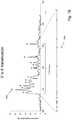
- cytosine of the following 107-mer single stranded polynucleotide (SEQ ID NO: 1) was labeled with a Cy5 fluorophore.
- the labeled polynucleotide was translocated in a 3'-first orientation through the hybrid nanopore and mixed FRET signals from the labeled cytosines was collected.
- Raw data is shown in Fig. IB.
- Fig. IB When translocated through the hybrid nanopore multiple peaks are observed which correspond to the number of cytosines in the DNA. Remarkably, the homopolymer at the 3' end is perfectly resolved.
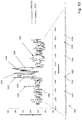
- every cytosine and every thymidine of the following single stranded polynucleotide were labeled with Cy5 and Atto700, respectively.
- the labeled polynucleotide was translocated in a 3'-first orientation through the hybrid nanopore and mixed FRET signals from the labeled cytosines and thymidines were collected.
- Raw data is shown in Fig. ID. The peaks in this raw data trace show a pattern that resembles the position of the labeled nucleotides in the template strand.
- Nanopore means any opening positioned in a substrate that allows the passage of analytes through the substrate in a predetermined or discernable order, or in the case of polymer analytes, passage of their monomeric units through the substrate in a predetermined or discernible order. In the latter case, a predetermined or discernible order may be the primary sequence of monomeric units in the polymer.
- nanopores include proteinaceous or protein based nanopores, synthetic or solid state nanopores, and hybrid nanopores comprising a solid state nanopore having a protein nanopore embedded therein.
- a nanopore may have an inner diameter of, e.g., 1-10 nm or 1-5 nm or 1-3 nm, or other various sizes.
- protein nanopores include but are not limited to, alpha-hemolysin, voltage-dependent mitochondrial porin (VDAC), OmpF, OmpC, MspA and LamB (maltoporin), e.g. disclosed in Rhee, M. et al., Trends in Biotechnology, 25(4) (2007): 174-181 ; Bayley et al (cited above); Gundlach et al, U.S. patent publication 2012/0055792 ; and the like. Any protein pore that allows the translocation of single nucleic acid molecules may be employed.
- a nanopore protein may be labeled at a specific site on the exterior of the pore, or at a specific site on the exterior of one or more monomer units making up the pore forming protein.
- Pore proteins are chosen from a group of proteins such as, but not limited to, alpha-hemolysin, MspA, voltage-dependent mitochondrial porin (VDAC), Anthrax porin, OmpF, OmpC and LamB (maltoporin). Integration of the pore protein into a solid state hole is accomplished by attaching a charged polymer to the pore protein. After applying an electric field the charged complex is electrophoretically pulled into the solid state hole.
- a synthetic nanopore, or solid-state nanopore may be created in various forms of solid substrates, examples of which include but are not limited to silicones (e.g. Si3N4, SiO2), metals, metal oxides (e.g. Al2O3) plastics, glass, semiconductor material, and combinations thereof.
- a synthetic nanopore may be more stable than a biological protein pore positioned in a lipid bilayer membrane.
- a synthetic nanopore may also be created by using a carbon nanotube embedded in a suitable substrate such as but not limited to polymerized epoxy. Carbon nanotubes can have uniform and well-defined chemical and structural properties. Various sized carbon nanotubes can be obtained, ranging from one to hundreds of nanometers.
- the surface charge of a carbon nanotube is known to be about zero, and as a result, electrophoretic transport of a nucleic acid through the nanopore becomes simple and predictable ( Ito, T. et al., Chem. Commun. 12 (2003): 1482-83 ).
- the substrate surface of a synthetic nanopore may be chemically modified to allow for covalent attachment of the protein pore or to render the surface properties suitable for optical nanopore sequencing. Such surface modifications can be covalent or non-covalent. Most covalent modification include an organosilane deposition for which the most common protocols are described:1) Deposition from aqueous alcohol. This is the most facile method for preparing silylated surfaces. A 95% ethanol-5% water solution is adjusted to pH 4.5-5.5 with acetic acid.
- Silane is added with stirring to yield a 2% final concentration. After hydrolysis and silanol group formation the substrate is added for 2-5min. After rinsed free of excess materials by dipping briefly in ethanol. Cure of the silane layer is for 5-10min at 110 degrees Celsius.
- Vapor Phase Deposition Silanes can be applied to substrates under dry aprotic conditions by chemical vapor deposition methods. These methods favor monolayer deposition. In closed chamber designs, substrates are heated to sufficient temperature to achieve 5mm vapor pressure. Alternatively, vacuum can be applied until silane evaporation is observed.
- Spin-on deposition Spin-on applications can be made under hydrolytic conditions which favor maximum functionalization and polylayer deposition or dry conditions which favor monolayer deposition.
- FRET or "Forrester, or fluorescence, resonant energy transfer” means a non-radiative dipole-dipole energy transfer mechanism from a donor to acceptor fluorophore. The efficiency of FRET may be dependent upon the distance between donor and acceptor as well as the properties of the fluorophores ( Stryer, L., Annu Rev Biochem. 47 (1978): 819-846 ).
- FRET distance means a distance between a FRET donor and a FRET acceptor over which a FRET interaction can take place and a detectable FRET signal produced by the FRET acceptor.
- Polynucleotide or “oligonucleotide” are used interchangeably and each mean a linear polymer of nucleotide monomers.
- Monomers making up polynucleotides and oligonucleotides are capable of specifically binding to a natural polynucleotide by way of a regular pattern of monomer-to-monomer interactions, such as Watson-Crick type of base pairing, base stacking, Hoogsteen or reverse Hoogsteen types of base pairing, or the like.
- Such monomers and their internucleosidic linkages may be naturally occurring or may be analogs thereof, e.g. naturally occurring or non-naturally occurring analogs.
- Non-naturally occurring analogs may include PNAs, phosphorothioate internucleosidic linkages, bases containing linking groups permitting the attachment of labels, such as fluorophores, or haptens, and the like.
- PNAs phosphorothioate internucleosidic linkages
- bases containing linking groups permitting the attachment of labels such as fluorophores, or haptens, and the like.
- Polynucleotides typically range in size from a couple or a few monomeric units, e.g. 5-40, when they are usually referred to as “oligonucleotides,” to several thousand monomeric units.
- oligonucleotides typically range in size from a couple or a few monomeric units, e.g. 5-40, when they are usually referred to as “oligonucleotides,” to several thousand monomeric units.
- A denotes deoxyadenosine
- C denotes deoxycytidine
- G denotes deoxyguanosine
- T denotes thymidine
- I denotes deoxyinosine
- U denotes uridine, unless otherwise indicated or obvious from context.
- polynucleotides comprise the four natural nucleosides (e.g. deoxyadenosine, deoxycytidine, deoxyguanosine, deoxythymidine for DNA or their ribose counterparts for RNA) linked by phosphodiester linkages; however, they may also comprise non-natural nucleotide analogs, e.g. including modified bases, sugars, or internucleosidic linkages.
- nucleosides e.g. deoxyadenosine, deoxycytidine, deoxyguanosine, deoxythymidine for DNA or their ribose counterparts for RNA
- non-natural nucleotide analogs e.g. including modified bases, sugars, or internucleosidic linkages.
- oligonucleotide or polynucleotide substrate requirements for activity e.g. single stranded DNA, RNA/DNA duplex, or the like
- selection of appropriate composition for the oligonucleotide or polynucleotide substrates is well within the knowledge of one of ordinary skill, especially with guidance from treatises, such as Sambrook et al, Molecular Cloning, Second Edition (Cold Spring Harbor Laboratory, New York, 1989 ), and like references.
- the oligonucleotide and polynucleotide may refer to either a single stranded form or a double stranded form (i.e. duplexes of an oligonucleotide or polynucleotide and its respective complement). It will be clear to one of ordinary skill which form or whether both forms are intended from the context of the terms usage.
- Sequence determination includes determination of partial as well as full sequence information of the polynucleotide. That is, the terms include sequences of subsets of the full set of four natural nucleotides, A, C, G and T, such as, for example, a sequence of just A's and C's of a target polynucleotide. That is, the terms include the determination of the identities, ordering, and locations of one, two, three or all of the four types of nucleotides within a target polynucleotide.
- the terms include the determination of the identities, ordering, and locations of two, three or all of the four types of nucleotides within a target polynucleotide.
- sequence determination may be accomplished by identifying the ordering and locations of a single type of nucleotide, e.g. cytosines, within the target polynucleotide "catcgc" so that its sequence is represented as a binary code, e.g. "100101" representing "c-(not c)(not c)c-(not c)-c -- and the like.
- the terms may also include subsequences of a target polynucleotide that serve as a fingerprint for the target polynucleotide; that is, subsequences that uniquely identify a target polynucleotide within a set of polynucleotides, e.g. all different RNA sequences expressed by a cell.
Landscapes
- Life Sciences & Earth Sciences (AREA)
- Chemical & Material Sciences (AREA)
- Proteomics, Peptides & Aminoacids (AREA)
- Organic Chemistry (AREA)
- Zoology (AREA)
- Wood Science & Technology (AREA)
- Health & Medical Sciences (AREA)
- Engineering & Computer Science (AREA)
- Microbiology (AREA)
- Biochemistry (AREA)
- Biotechnology (AREA)
- Molecular Biology (AREA)
- Biophysics (AREA)
- Analytical Chemistry (AREA)
- Physics & Mathematics (AREA)
- Immunology (AREA)
- Bioinformatics & Cheminformatics (AREA)
- General Engineering & Computer Science (AREA)
- General Health & Medical Sciences (AREA)
- Genetics & Genomics (AREA)
- Measuring Or Testing Involving Enzymes Or Micro-Organisms (AREA)
- Apparatus Associated With Microorganisms And Enzymes (AREA)
- Investigating Or Analysing Materials By The Use Of Chemical Reactions (AREA)
Claims (7)
- Procédé de détermination d'une séquence nucléotidique d'un polynucléotide, le procédé comprenant les étapes consistant :à effectuer une translocation d'un polynucléotide à travers un nanopore de sorte que les nucléotides du polynucléotide passent dans une séquence par un donneur FRET positionné de manière adjacente au nanopore, une pluralité des nucléotides du polynucléotide se trouvant à une distance de FRET du donneur FRET à mesure que les nucléotides sortent du nanopore et au moins une partie des nucléotides du polynucléotide étant marquée avec un accepteur FRET, où différents types de nucléotides sont marqués avec des accepteurs FRET qui génèrent des signaux pouvant être distingués et où le nanopore est dimensionné de sorte que les accepteurs FRET soient contraints, lors de la translocation à travers le nanopore, de supprimer des réactions de FRET à l'intérieur du nanopore ;à exciter le donneur FRET adjacent au nanopore de sorte qu'un FRET se produise entre le donneur FRET et les accepteurs FRET à la distance de FRET pour générer un signal de FRET mixte ;à mesurer des signaux de FRET mixtes à mesure que le polynucléotide effectue une translocation à travers le nanopore ; età déterminer une séquence nucléotidique du polynucléotide à partir des signaux de FRET mixtes.
- Système pour déterminer une séquence nucléotidique d'un polynucléotide, le système comprenant :un nanopore assurant une communication fluidique entre une première chambre et une deuxième chambre et à travers lequel un polynucléotide peut être transloqué, le nanopore étant dimensionné de sorte que les nucléotides du polynucléotide passent à travers une sortie du nanopore dans une séquence et à chaque fois que les nucléotides du polynucléotide sont marqués avec des accepteurs FRET, un FRET est supprimé entre de tels accepteurs FRET à l'intérieur du nanopore et un donneur FRET à l'extérieur du nanopore ;un polynucléotide, dans lequel au moins une partie des nucléotides du polynucléotide est marquée avec un accepteur FRET, et dans lequel différents types de nucléotides sont marqués avec des accepteurs FRET qui génèrent des signaux pouvant être distingués ;un donneur FRET disposé à une distance de FRET de la sortie du nanopore, de sorte qu'une pluralité de nucléotides passent à une distance de FRET du donneur FRET lorsqu'ils apparaissent à la sortie du nanopore ;un moyen pour exciter le donneur FRET ; etun détecteur pour collecter des signaux de FRET mixtes.
- Procédé de la revendication 1 ou système de la revendication 2, dans lequel ledit nanopore est disposé dans une membrane en phase solide et dans lequel ledit donneur FRET est fixé à la membrane en phase solide de manière adjacente audit nanopore.
- Procédé de la revendication 1 ou 3, ou système de la revendication 2 ou 3, dans lequel ledit nanopore est un nanopore protéique et dans lequel ledit donneur FRET est fixé au nanopore protéique.
- Procédé de l'une quelconque des revendications 1, 3 et 4, ou système de l'une quelconque des revendications 2 à 4, dans lequel le polynucléotide est un polynucléotide simple brin.
- Procédé de l'une quelconque des revendications 1, 3, 4 et 5, ou système de l'une quelconque des revendications 2 à 5, dans lequel l'accepteur FRET est un colorant organique fluorescent.
- Procédé de l'une quelconque des revendications 1, 3, 4, 5 et 6, ou système de l'une quelconque des revendications 2 à 6, dans lequel le donneur FRET est un point quantique.
Applications Claiming Priority (2)
| Application Number | Priority Date | Filing Date | Title |
|---|---|---|---|
| US201361827519P | 2013-05-24 | 2013-05-24 | |
| PCT/US2014/039444 WO2014190322A2 (fr) | 2013-05-24 | 2014-05-23 | Analyse d'acides nucléiques basés sur des nanopores avec une détection par fret mixte |
Publications (3)
| Publication Number | Publication Date |
|---|---|
| EP3004385A2 EP3004385A2 (fr) | 2016-04-13 |
| EP3004385A4 EP3004385A4 (fr) | 2017-02-22 |
| EP3004385B1 true EP3004385B1 (fr) | 2018-11-28 |
Family
ID=51934368
Family Applications (1)
| Application Number | Title | Priority Date | Filing Date |
|---|---|---|---|
| EP14801275.0A Active EP3004385B1 (fr) | 2013-05-24 | 2014-05-23 | Analyse d'acides nucléiques basés sur des nanopores avec une détection par fret mixte |
Country Status (7)
| Country | Link |
|---|---|
| US (4) | US9862997B2 (fr) |
| EP (1) | EP3004385B1 (fr) |
| JP (2) | JP6399610B2 (fr) |
| CN (1) | CN105283560B (fr) |
| AU (1) | AU2014268322B2 (fr) |
| CA (1) | CA2910019A1 (fr) |
| WO (1) | WO2014190322A2 (fr) |
Families Citing this family (22)
| Publication number | Priority date | Publication date | Assignee | Title |
|---|---|---|---|---|
| EP3543357A1 (fr) | 2007-05-08 | 2019-09-25 | Trustees of Boston University | Fonctionnalisation chimique de nanopores à l'état solide et de réseaux de nanopores et leurs applications |
| US9651539B2 (en) | 2012-10-28 | 2017-05-16 | Quantapore, Inc. | Reducing background fluorescence in MEMS materials by low energy ion beam treatment |
| CN105283560B (zh) | 2013-05-24 | 2018-11-30 | 昆塔波尔公司 | 基于纳米孔的通过混合的fret检测的核酸分析 |
| CN107109472B (zh) | 2014-10-10 | 2021-05-11 | 昆塔波尔公司 | 利用互相猝灭的荧光标记物的基于纳米孔的聚合物分析 |
| JP6757316B2 (ja) | 2014-10-24 | 2020-09-16 | クアンタポール, インコーポレイテッド | ナノ構造のアレイを使用するポリマーの効率的光学分析 |
| US20180364169A1 (en) * | 2015-06-09 | 2018-12-20 | Quantapore, Inc. | Nanopore-based analysis of compounds using mobile fret pairs |
| US20190078145A1 (en) | 2015-12-08 | 2019-03-14 | Quantapore, Inc. | Method of translocating nucleic acids through nanopores |
| CN108463560B (zh) | 2016-01-15 | 2022-09-20 | 昆塔波尔公司 | 具有降低的背景的基于光学的纳米孔分析 |
| WO2018081178A1 (fr) * | 2016-10-24 | 2018-05-03 | Two Pore Guys, Inc. | Abondance fractionnaire de séquences polynucléotidiques dans un échantillon |
| CN109415765A (zh) * | 2016-04-14 | 2019-03-01 | 昆塔波尔公司 | 用纳米孔的聚合物分析中的混合光学信号 |
| JP2019517252A (ja) * | 2016-05-31 | 2019-06-24 | クアンタポール, インコーポレイテッド | 二色ナノポア配列決定 |
| EP3482196B1 (fr) * | 2016-07-05 | 2022-02-23 | Quantapore, Inc. | Séquencement de nanopores à base optique |
| CA3031812A1 (fr) | 2016-08-19 | 2018-02-22 | Quantapore, Inc. | Sequencage par nanopores a base optique a l'aide d'agents d'extinction |
| EP3700670B1 (fr) * | 2017-10-23 | 2023-11-29 | F. Hoffmann-La Roche AG | Retrait et réinsertion de nanopores de protéine dans une membrane à l'aide d'un déséquilibre osmotique |
| US11408880B2 (en) * | 2018-05-17 | 2022-08-09 | Northeastern University | Lipid-free anchoring of thermophilic bacteriophage G20c portal adapter into solid-state nanopores |
| US11808755B2 (en) * | 2018-05-17 | 2023-11-07 | Recognition AnalytiX, Inc. | Device, system and method for direct electrical measurement of enzyme activity |
| WO2020074399A1 (fr) * | 2018-10-08 | 2020-04-16 | Adolphe Merkle Institute, University Of Fribourg | Ajustement à base d'oligonucléotides de peptides formant des pores pour augmenter la taille des pores, l'affinité membranaire, la stabilité et l'activité antimicrobienne |
| DE102019200929A1 (de) * | 2019-01-25 | 2020-07-30 | Robert Bosch Gmbh | Verfahren und Vorrichtung zum optischen Erfassen einer Länge eines Makromoleküls |
| CN110243792A (zh) * | 2019-06-11 | 2019-09-17 | 山东师范大学 | 一种基于量子点和四面体dna结构的荧光化学传感器及其检测方法和应用 |
| CN110736775A (zh) * | 2019-10-11 | 2020-01-31 | 深圳清华大学研究院 | 一种固态纳米孔修饰处理方法 |
| EP4102210A1 (fr) * | 2021-06-07 | 2022-12-14 | Oxford University Innovation Limited | Nanopore nanophotonique, systeme memoire comprenant un nanopore nanophotonique, et procede de lecture et/ou d'ecriture d'informations de et/ou a un analyte |
| CN113533275B (zh) * | 2021-06-30 | 2023-11-28 | 东南大学 | 固态纳米孔-荧光共振能量转移复合检测方法及系统 |
Family Cites Families (211)
| Publication number | Priority date | Publication date | Assignee | Title |
|---|---|---|---|---|
| CH614781A5 (fr) | 1977-06-27 | 1979-12-14 | Contraves Ag | |
| US5821058A (en) | 1984-01-16 | 1998-10-13 | California Institute Of Technology | Automated DNA sequencing technique |
| US4962037A (en) | 1987-10-07 | 1990-10-09 | United States Of America | Method for rapid base sequencing in DNA and RNA |
| US5131755A (en) | 1988-02-19 | 1992-07-21 | Chadwick Curt H | Automatic high speed optical inspection system |
| JP3123249B2 (ja) | 1991-09-10 | 2001-01-09 | 株式会社日立製作所 | Dna分子の長さ計測方法および計測装置 |
| US5405747A (en) | 1991-09-25 | 1995-04-11 | The Regents Of The University Of California Office Of Technology Transfer | Method for rapid base sequencing in DNA and RNA with two base labeling |
| DE69322266T2 (de) | 1992-04-03 | 1999-06-02 | Perkin Elmer Corp | Proben zusammensetzung und verfahren |
| US5470705A (en) | 1992-04-03 | 1995-11-28 | Applied Biosystems, Inc. | Probe composition containing a binding domain and polymer chain and methods of use |
| US5387926A (en) | 1992-06-30 | 1995-02-07 | California Institute Of Technology | High speed digital framing camera |
| US5798042A (en) | 1994-03-07 | 1998-08-25 | Regents Of The University Of California | Microfabricated filter with specially constructed channel walls, and containment well and capsule constructed with such filters |
| US5624845A (en) * | 1995-03-16 | 1997-04-29 | International Business Machines Corporation | Assembly and a method suitable for identifying a code |
| US6362002B1 (en) | 1995-03-17 | 2002-03-26 | President And Fellows Of Harvard College | Characterization of individual polymer molecules based on monomer-interface interactions |
| US5795782A (en) | 1995-03-17 | 1998-08-18 | President & Fellows Of Harvard College | Characterization of individual polymer molecules based on monomer-interface interactions |
| US6794127B1 (en) | 1997-06-16 | 2004-09-21 | Diversa Corporation | Capillary array-based sample screening |
| WO1997039150A1 (fr) | 1996-04-15 | 1997-10-23 | University Of Southern California | Synthese d'adn marque par fluorophores |
| US6399023B1 (en) | 1996-04-16 | 2002-06-04 | Caliper Technologies Corp. | Analytical system and method |
| US5945526A (en) | 1996-05-03 | 1999-08-31 | Perkin-Elmer Corporation | Energy transfer dyes with enhanced fluorescence |
| US6136543A (en) | 1997-01-31 | 2000-10-24 | Hitachi, Ltd. | Method for determining nucleic acids base sequence and apparatus therefor |
| JP3935509B2 (ja) | 1997-02-12 | 2007-06-27 | ワイ. チャン,ユージーン | ポリマー分析のための方法および製品 |
| DE19714221A1 (de) | 1997-04-07 | 1998-10-08 | Zeiss Carl Fa | Konfokales Mikroskop mit einem motorischen Scanningtisch |
| BR9800655A (pt) | 1997-04-21 | 1999-08-10 | Randox Lab Ltd | Dispositivo de estado sólido para efetuar ensaios com múltiplos analisados seu uso e e sistema para análise de múltiplos analisados |
| WO1999005167A1 (fr) | 1997-07-25 | 1999-02-04 | University Of Massachusetts | Pores de proteines specifiquement concus utilises en tant que constituants pour des biocapteurs |
| WO1999028505A1 (fr) | 1997-12-03 | 1999-06-10 | Curagen Corporation | Procede et dispositifs de mesure de l'expression genique differentielle |
| US20020144905A1 (en) | 1997-12-17 | 2002-10-10 | Christian Schmidt | Sample positioning and analysis system |
| AU746580B2 (en) | 1997-12-17 | 2002-05-02 | Ecole Polytechnique Federale De Lausanne | Positioning and electrophysiological characterization of individual cells and reconstituted membrane systems on microstructured carriers |
| US7244349B2 (en) | 1997-12-17 | 2007-07-17 | Molecular Devices Corporation | Multiaperture sample positioning and analysis system |
| AU736637B2 (en) | 1997-12-23 | 2001-08-02 | Steris Corporation | Antimicrobial composition delivery system with an integrated filter |
| US6265163B1 (en) | 1998-01-09 | 2001-07-24 | Lynx Therapeutics, Inc. | Solid phase selection of differentially expressed genes |
| DE19804372A1 (de) | 1998-02-04 | 1999-08-05 | Michael W Dr Dr Dahm | Verfahren zur quantitativen Bestimmung von Tumorzellen in einer Körperflüssigkeit und dazu geeignete Testkits |
| GB9805301D0 (en) | 1998-03-12 | 1998-05-06 | Imperial College | Detector |
| US6306589B1 (en) | 1998-05-27 | 2001-10-23 | Vysis, Inc. | Biological assays for analyte detection |
| US6504943B1 (en) | 1998-07-20 | 2003-01-07 | Sandia Corporation | Information-efficient spectral imaging sensor |
| US6447724B1 (en) | 1998-08-11 | 2002-09-10 | Caliper Technologies Corp. | DNA sequencing using multiple fluorescent labels being distinguishable by their decay times |
| US6790671B1 (en) | 1998-08-13 | 2004-09-14 | Princeton University | Optically characterizing polymers |
| US6263286B1 (en) | 1998-08-13 | 2001-07-17 | U.S. Genomics, Inc. | Methods of analyzing polymers using a spatial network of fluorophores and fluorescence resonance energy transfer |
| US6210896B1 (en) | 1998-08-13 | 2001-04-03 | Us Genomics | Molecular motors |
| US6906749B1 (en) | 1998-09-16 | 2005-06-14 | Dalsa, Inc. | CMOS TDI image sensor |
| DE69905832T2 (de) | 1998-09-18 | 2004-02-05 | Massachusetts Institute Of Technology, Cambridge | Biologische Verwendungen von halbleitenden Nanokristallen |
| US6251303B1 (en) | 1998-09-18 | 2001-06-26 | Massachusetts Institute Of Technology | Water-soluble fluorescent nanocrystals |
| US6267872B1 (en) | 1998-11-06 | 2001-07-31 | The Regents Of The University Of California | Miniature support for thin films containing single channels or nanopores and methods for using same |
| US6426231B1 (en) | 1998-11-18 | 2002-07-30 | The Texas A&M University System | Analyte sensing mediated by adapter/carrier molecules |
| US6252303B1 (en) | 1998-12-02 | 2001-06-26 | Advanced Micro Devices, Inc. | Intergration of low-K SiOF as inter-layer dielectric |
| AU2206800A (en) | 1998-12-11 | 2000-06-26 | Regents Of The University Of California, The | Targeted molecular bar codes and methods for using the same |
| FR2788008B1 (fr) | 1998-12-30 | 2001-03-23 | Inst Curie | Milieu thermosensible pour la separation electrocinetique d'especes au sein d'un canal de separation |
| WO2000043547A1 (fr) | 1999-01-20 | 2000-07-27 | Northwestern University | Modificateur de la mobilite de l'adn |
| US6473176B2 (en) | 1999-01-25 | 2002-10-29 | Amnis Corporation | Imaging and analyzing parameters of small moving objects such as cells |
| US6608682B2 (en) | 1999-01-25 | 2003-08-19 | Amnis Corporation | Imaging and analyzing parameters of small moving objects such as cells |
| US6249341B1 (en) | 1999-01-25 | 2001-06-19 | Amnis Corporation | Imaging and analyzing parameters of small moving objects such as cells |
| US6975400B2 (en) | 1999-01-25 | 2005-12-13 | Amnis Corporation | Imaging and analyzing parameters of small moving objects such as cells |
| US6671044B2 (en) | 1999-01-25 | 2003-12-30 | Amnis Corporation | Imaging and analyzing parameters of small moving objects such as cells in broad flat flow |
| EP1192274A2 (fr) | 1999-03-25 | 2002-04-03 | Hyseq, Inc. | Procedes et materiaux permettant l'analyse des sequences par hybridation dans un environnement en solution |
| US20040214221A1 (en) | 1999-05-07 | 2004-10-28 | Klaus Muehlegger | High density labeling of DNA with modified or "chromophore" carrying nucleotides and DNA polymerases used |
| US7122152B2 (en) | 1999-05-10 | 2006-10-17 | University Of Florida | Spatiotemporal and geometric optimization of sensor arrays for detecting analytes fluids |
| US7056661B2 (en) | 1999-05-19 | 2006-06-06 | Cornell Research Foundation, Inc. | Method for sequencing nucleic acid molecules |
| US8206568B2 (en) | 1999-06-22 | 2012-06-26 | President And Fellows Of Harvard College | Material deposition techniques for control of solid state aperture surface properties |
| US6627067B1 (en) | 1999-06-22 | 2003-09-30 | President And Fellows Of Harvard College | Molecular and atomic scale evaluation of biopolymers |
| US6464842B1 (en) | 1999-06-22 | 2002-10-15 | President And Fellows Of Harvard College | Control of solid state dimensional features |
| US6818395B1 (en) | 1999-06-28 | 2004-11-16 | California Institute Of Technology | Methods and apparatus for analyzing polynucleotide sequences |
| US6982146B1 (en) | 1999-08-30 | 2006-01-03 | The United States Of America As Represented By The Department Of Health And Human Services | High speed parallel molecular nucleic acid sequencing |
| WO2001018247A2 (fr) | 1999-09-03 | 2001-03-15 | Lifebeam Technologies, Inc. | Systeme optique destine a l'analyse rapide de polymeres |
| WO2001018251A1 (fr) | 1999-09-07 | 2001-03-15 | The Regents Of The University Of California | Methodes permettant de determiner la presence d'acides nucleiques double brin dans un echantillon |
| US6335420B1 (en) | 1999-11-22 | 2002-01-01 | Ibc Advanced Technologies, Inc. | Polyamide ligand-containing polymeric resins and methods of using the same for removing, separating and/or concentrating desired metal ions from solutions |
| WO2001055454A1 (fr) | 2000-01-28 | 2001-08-02 | Althea Technologies, Inc. | Procedes d'analyse de l'expression genique |
| EP2083015B1 (fr) | 2000-02-11 | 2016-04-06 | The Texas A & M University System | Compositions à biocapteur et procédés d'utilisation |
| US20060292041A1 (en) | 2000-03-23 | 2006-12-28 | Dugas Matthew P | Solid state membrane channel device for the measurement and characterization of atomic and molecular sized samples |
| US6616895B2 (en) | 2000-03-23 | 2003-09-09 | Advanced Research Corporation | Solid state membrane channel device for the measurement and characterization of atomic and molecular sized samples |
| US6413792B1 (en) | 2000-04-24 | 2002-07-02 | Eagle Research Development, Llc | Ultra-fast nucleic acid sequencing device and a method for making and using the same |
| US7001792B2 (en) | 2000-04-24 | 2006-02-21 | Eagle Research & Development, Llc | Ultra-fast nucleic acid sequencing device and a method for making and using the same |
| US6917726B2 (en) | 2001-09-27 | 2005-07-12 | Cornell Research Foundation, Inc. | Zero-mode clad waveguides for performing spectroscopy with confined effective observation volumes |
| EP1349649A2 (fr) | 2000-06-30 | 2003-10-08 | Gnothis Holding SA | Procede de sequencage par molecule individuelle |
| WO2002004680A2 (fr) | 2000-07-07 | 2002-01-17 | Visigen Biotechnologies, Inc. | Determination de sequence en temps reel |
| US6608680B2 (en) | 2000-08-25 | 2003-08-19 | Amnis Corporation | TDI imaging system for kinetic studies |
| US6583865B2 (en) | 2000-08-25 | 2003-06-24 | Amnis Corporation | Alternative detector configuration and mode of operation of a time delay integration particle analyzer |
| US6934408B2 (en) | 2000-08-25 | 2005-08-23 | Amnis Corporation | Method and apparatus for reading reporter labeled beads |
| WO2002018951A2 (fr) | 2000-08-29 | 2002-03-07 | The Rockefeller University | Procedes mettant en application une extinction de fluorescence par des surfaces metalliques |
| US6936433B2 (en) | 2000-11-27 | 2005-08-30 | The Regents Of The University Of California | Methods and devices for characterizing duplex nucleic acid molecules |
| US6998251B2 (en) | 2001-01-12 | 2006-02-14 | Syngenta Participations Ag | Nanoporous membrane reactor for miniaturized reactions and enhanced reaction kinetics |
| US6743905B2 (en) | 2001-04-16 | 2004-06-01 | Applera Corporation | Mobility-modified nucleobase polymers and methods of using same |
| KR100889351B1 (ko) | 2001-05-15 | 2009-03-18 | 가부시키가이샤 에바라 세이사꾸쇼 | Tdi 검출 디바이스, 피드-스루 장비 및 이들 디바이스를 이용하는 전자빔 장치 |
| US20040175710A1 (en) | 2001-05-22 | 2004-09-09 | Haushalter Robert C. | Method for in situ, on-chip chemical synthesis |
| WO2002103335A1 (fr) | 2001-06-18 | 2002-12-27 | Amnis Corporation | Deconvolution spectrale de marqueurs fluorescents |
| US7009163B2 (en) | 2001-06-22 | 2006-03-07 | Orbotech Ltd. | High-sensitivity optical scanning using memory integration |
| AU2002322348A1 (en) | 2001-06-28 | 2003-03-03 | Advanced Research And Technology Institute, Inc. | Methods of preparing multicolor quantum dot tagged beads and conjugates thereof |
| US7280207B2 (en) | 2001-07-25 | 2007-10-09 | Applera Corporation | Time-delay integration in a flow cytometry system |
| US6856390B2 (en) | 2001-07-25 | 2005-02-15 | Applera Corporation | Time-delay integration in electrophoretic detection systems |
| SG187992A1 (en) | 2001-07-25 | 2013-03-28 | Univ Princeton | Nanochannel arrays and their preparation and use for high throughput macromolecular analysis |
| US20080288178A1 (en) | 2001-08-24 | 2008-11-20 | Applera Corporation | Sequencing system with memory |
| US6627433B2 (en) | 2001-08-24 | 2003-09-30 | Applera Corporation | Multi-channel analyte-separation device employing side-entry excitation |
| US6852492B2 (en) | 2001-09-24 | 2005-02-08 | Intel Corporation | Nucleic acid sequencing by raman monitoring of uptake of precursors during molecular replication |
| US6706203B2 (en) | 2001-10-30 | 2004-03-16 | Agilent Technologies, Inc. | Adjustable nanopore, nanotome, and nanotweezer |
| DE10154291B4 (de) | 2001-11-05 | 2005-05-19 | Hain Lifescience Gmbh | Verfahren zum Nachweis von Nukleinsäuren in Form eines Trockenschnelltestes |
| DE10162535A1 (de) | 2001-12-19 | 2003-07-03 | Gnothis Holding Sa Ecublens | Sequenzierung über Lochmembranen |
| US7008547B2 (en) | 2002-03-14 | 2006-03-07 | Sarnoff Corporation | Solid phase sensors |
| JP3752466B2 (ja) | 2002-04-24 | 2006-03-08 | 株式会社日立製作所 | 遺伝子検査方法 |
| US8278055B2 (en) | 2002-05-01 | 2012-10-02 | Intel Corporation | Methods and device for analyte characterization |
| US7744816B2 (en) | 2002-05-01 | 2010-06-29 | Intel Corporation | Methods and device for biomolecule characterization |
| EP1504114B1 (fr) | 2002-05-10 | 2017-07-12 | The Texas A & M University System | Detection stochastique par interactions covalentes |
| US20050019784A1 (en) | 2002-05-20 | 2005-01-27 | Xing Su | Method and apparatus for nucleic acid sequencing and identification |
| US7005264B2 (en) | 2002-05-20 | 2006-02-28 | Intel Corporation | Method and apparatus for nucleic acid sequencing and identification |
| US6952651B2 (en) | 2002-06-17 | 2005-10-04 | Intel Corporation | Methods and apparatus for nucleic acid sequencing by signal stretching and data integration |
| TWI234584B (en) | 2002-08-16 | 2005-06-21 | Univ Nat Taiwan Normal | Nucleic acid sequencing method |
| AU2003276975A1 (en) | 2002-09-25 | 2004-04-19 | Amersham Biosciences (Sv) Corp | High density fluidic chip design and method of sample injection |
| AU2003301244A1 (en) | 2002-10-15 | 2004-05-04 | Advanced Research Corporation | Solid state membrane channel device for the measurement and characterization of atomic and molecular sized samples |
| CN1161613C (zh) | 2002-10-22 | 2004-08-11 | 中国人民解放军南京军区联勤部军事医学研究所 | 免疫细胞微流体阵列 |
| US7390457B2 (en) | 2002-10-31 | 2008-06-24 | Agilent Technologies, Inc. | Integrated microfluidic array device |
| US20040137158A1 (en) | 2003-01-15 | 2004-07-15 | Kools Jacques Constant Stefan | Method for preparing a noble metal surface |
| US7410564B2 (en) | 2003-01-27 | 2008-08-12 | Agilent Technologies, Inc. | Apparatus and method for biopolymer identification during translocation through a nanopore |
| DE602004024034D1 (de) | 2003-01-29 | 2009-12-24 | 454 Corp | Nukleinsäureamplifikation auf basis von kügelchenemulsion |
| WO2004085609A2 (fr) | 2003-02-28 | 2004-10-07 | Brown University | Nanopores, procedes d'utilisation et de fabrication de ceux-ci et procedes de caracterisation de biomolecules utilisant ces nanopores |
| US7250115B2 (en) | 2003-06-12 | 2007-07-31 | Agilent Technologies, Inc | Nanopore with resonant tunneling electrodes |
| US7248771B2 (en) | 2003-06-16 | 2007-07-24 | Brigham Young University | Integrated sensor with electrical and optical single molecule sensitivity |
| US7444053B2 (en) | 2003-06-16 | 2008-10-28 | The Regents Of The University Of California | Integrated electrical and optical sensor for biomolecule analysis with single molecule sensitivity |
| WO2005017025A2 (fr) | 2003-08-15 | 2005-02-24 | The President And Fellows Of Harvard College | Etude de molecules de polymere et conformations avec un nanopore |
| US7075161B2 (en) | 2003-10-23 | 2006-07-11 | Agilent Technologies, Inc. | Apparatus and method for making a low capacitance artificial nanopore |
| US20050095599A1 (en) | 2003-10-30 | 2005-05-05 | Pittaro Richard J. | Detection and identification of biopolymers using fluorescence quenching |
| US20050136408A1 (en) | 2003-12-19 | 2005-06-23 | May Tom-Moy | Methods and systems for characterizing a polymer |
| US7271896B2 (en) | 2003-12-29 | 2007-09-18 | Intel Corporation | Detection of biomolecules using porous biosensors and raman spectroscopy |
| US20050164211A1 (en) | 2004-01-22 | 2005-07-28 | Hannah Eric C. | Carbon nanotube molecular labels |
| JP2007524410A (ja) | 2004-01-23 | 2007-08-30 | リングヴィテ エーエス | ポリヌクレオチドライゲーション反応の改良 |
| US20050186576A1 (en) | 2004-02-19 | 2005-08-25 | Intel Corporation | Polymer sequencing using selectively labeled monomers and data integration |
| US7279337B2 (en) | 2004-03-10 | 2007-10-09 | Agilent Technologies, Inc. | Method and apparatus for sequencing polymers through tunneling conductance variation detection |
| US7238485B2 (en) | 2004-03-23 | 2007-07-03 | President And Fellows Of Harvard College | Methods and apparatus for characterizing polynucleotides |
| US20050227239A1 (en) | 2004-04-08 | 2005-10-13 | Joyce Timothy H | Microarray based affinity purification and analysis device coupled with solid state nanopore electrodes |
| US20070264623A1 (en) | 2004-06-15 | 2007-11-15 | President And Fellows Of Harvard College | Nanosensors |
| US20060019259A1 (en) | 2004-07-22 | 2006-01-26 | Joyce Timothy H | Characterization of biopolymers by resonance tunneling and fluorescence quenching |
| GB2431013B (en) | 2004-07-23 | 2008-05-21 | Electronic Bio Sciences Llc | Method and apparatus for sensing a time varying current passing through an ion channel |
| US20070054276A1 (en) | 2004-08-12 | 2007-03-08 | Sampson Jeffrey R | Polynucleotide analysis and methods of using nanopores |
| CN101103357B (zh) | 2004-08-13 | 2012-10-03 | 哈佛学院院长等 | 超高处理量光学-纳米孔dna读出平台 |
| JP2008513782A (ja) | 2004-09-17 | 2008-05-01 | パシフィック バイオサイエンシーズ オブ カリフォルニア, インコーポレイテッド | 分子解析のための装置及び方法 |
| US20080025875A1 (en) | 2004-09-29 | 2008-01-31 | Martin Charles R | Chemical, Particle, and Biosensing with Nanotechnology |
| CA2523089A1 (fr) | 2004-10-05 | 2006-04-05 | University Of Ottawa | Methode de separation de composes polymeres |
| WO2006052882A1 (fr) | 2004-11-09 | 2006-05-18 | President And Fellows Of Harvard College | Formation de tourbillons dans des canaux fluidiques a sections d'etranglement et leurs utilisations |
| US7609309B2 (en) | 2004-11-18 | 2009-10-27 | Kla-Tencor Technologies Corporation | Continuous clocking of TDI sensors |
| US7220549B2 (en) | 2004-12-30 | 2007-05-22 | Helicos Biosciences Corporation | Stabilizing a nucleic acid for nucleic acid sequencing |
| US20060210995A1 (en) | 2005-03-15 | 2006-09-21 | Joyce Timothy H | Nanopore analysis systems and methods of using nanopore devices |
| EP2348300A3 (fr) | 2005-04-06 | 2011-10-12 | The President and Fellows of Harvard College | Caracterisation moleculaire a l'aide de nanotubes de carbone |
| US20060231419A1 (en) | 2005-04-15 | 2006-10-19 | Barth Philip W | Molecular resonant tunneling sensor and methods of fabricating and using the same |
| US7879575B2 (en) | 2005-04-27 | 2011-02-01 | The Trustees Of The University Of Pennsylvania | Nanostructures that provide a modified nanoenvironment for the enhancement of luminescence |
| EP1721657A1 (fr) | 2005-05-13 | 2006-11-15 | SONY DEUTSCHLAND GmbH | Méthode de fabrication d'une membrane polymère ayant au moins un pore |
| JP2007006720A (ja) | 2005-06-28 | 2007-01-18 | Toshiba Corp | 個体識別方法、並びに個体識別検査のためのアレイ、装置及びシステム |
| WO2007021814A2 (fr) | 2005-08-11 | 2007-02-22 | Eksigent Technologies, Llc | Surfaces plastiques et appareils permettant de reduire l'adsorption de solutes et procedes de preparations correspondants |
| US7666593B2 (en) | 2005-08-26 | 2010-02-23 | Helicos Biosciences Corporation | Single molecule sequencing of captured nucleic acids |
| WO2007041340A2 (fr) | 2005-09-30 | 2007-04-12 | The Trustees Of Columbia University In The City Of New York | Cellules microfluidiques a arrangements paralleles de molecules d'adn individuelles |
| US20070190542A1 (en) | 2005-10-03 | 2007-08-16 | Ling Xinsheng S | Hybridization assisted nanopore sequencing |
| KR100721430B1 (ko) | 2005-10-12 | 2007-05-23 | 학교법인 포항공과대학교 | 나노다공성 멤브레인 및 이의 제조방법 |
| US7397232B2 (en) | 2005-10-21 | 2008-07-08 | The University Of Akron | Coulter counter having a plurality of channels |
| US7835870B2 (en) | 2005-11-01 | 2010-11-16 | Georgia Institute Of Technology | Methods and systems for evaluating the length of elongated elements |
| EP1954838B1 (fr) | 2005-11-14 | 2014-02-26 | Life Technologies Corporation | Molecules codees permettant de detecter des substances cibles a analyser |
| GB0523282D0 (en) | 2005-11-15 | 2005-12-21 | Isis Innovation | Methods using pores |
| WO2007070572A2 (fr) | 2005-12-12 | 2007-06-21 | The Government Of The United States Of America, As Represented By The Secretary Of The Department Of Health And Human Services | Sonde pour le sequencage d’acide nucleique et procedes d’utilisation |
| CN101384846A (zh) | 2006-02-13 | 2009-03-11 | 皇家飞利浦电子股份有限公司 | 用于分子诊断用途的微流体装置 |
| CA2642854A1 (fr) | 2006-02-18 | 2007-08-30 | Michael Strathmann | Sequencage a multiplexage massif |
| US20070202008A1 (en) | 2006-02-28 | 2007-08-30 | Schembri Carol T | Systems and methods of lipoprotein size fraction assaying |
| US20070215472A1 (en) | 2006-03-15 | 2007-09-20 | Slater Gary W | Electroosmotic flow for end labelled free solution electrophoresis |
| US20070218494A1 (en) | 2006-03-17 | 2007-09-20 | Gary Slater | Branched polymer lables as drag-tags in free solution electrophoresis |
| US7777505B2 (en) | 2006-05-05 | 2010-08-17 | University Of Utah Research Foundation | Nanopore platforms for ion channel recordings and single molecule detection and analysis |
| US7849581B2 (en) | 2006-05-05 | 2010-12-14 | University Of Utah Research Foundation | Nanopore electrode, nanopore membrane, methods of preparation and surface modification, and use thereof |
| CA2656529A1 (fr) | 2006-06-30 | 2008-01-10 | Applera Corporation | Procedes d'analyse d'interactions de liaison |
| US7553730B2 (en) | 2006-07-14 | 2009-06-30 | Agilent Technologies, Inc. | Methods of fabrication employing nanoscale mandrels |
| US20080032290A1 (en) | 2006-08-03 | 2008-02-07 | Young James E | Nanopore flow cells |
| US8435775B2 (en) | 2006-09-06 | 2013-05-07 | Medical Research Council | Mutant Pfu DNA polymerase |
| WO2008049795A1 (fr) | 2006-10-23 | 2008-05-02 | Flexgen Bv | Procédés et système pour étalonner la mise au point et la position de lasers dans des puces à adn |
| GB2457402B (en) | 2006-12-01 | 2011-10-19 | Univ Columbia | Four-color DNA sequencing by synthesis using cleavable fluorescent nucleotide reversible terminators |
| US7544578B2 (en) | 2007-01-03 | 2009-06-09 | International Business Machines Corporation | Structure and method for stochastic integrated circuit personalization |
| US8003319B2 (en) | 2007-02-02 | 2011-08-23 | International Business Machines Corporation | Systems and methods for controlling position of charged polymer inside nanopore |
| EP2126072A4 (fr) | 2007-02-21 | 2011-07-13 | Life Technologies Corp | Materiels et methodes de sequencage de molecule unique d'acide nucleique |
| US20080254995A1 (en) | 2007-02-27 | 2008-10-16 | Drexel University | Nanopore arrays and sequencing devices and methods thereof |
| US20110005918A1 (en) | 2007-04-04 | 2011-01-13 | Akeson Mark A | Compositions, devices, systems, and methods for using a nanopore |
| EP3543357A1 (fr) | 2007-05-08 | 2019-09-25 | Trustees of Boston University | Fonctionnalisation chimique de nanopores à l'état solide et de réseaux de nanopores et leurs applications |
| CA2689626C (fr) | 2007-06-06 | 2016-10-25 | Pacific Biosciences Of California, Inc. | Methodes et procedes pour identifier des bases dans des procedes d'incorporation en fonction de la sequence |
| DK2171088T3 (en) * | 2007-06-19 | 2016-01-25 | Stratos Genomics Inc | Nucleic acid sequencing in a high yield by expansion |
| GB0713143D0 (en) | 2007-07-06 | 2007-08-15 | Ucl Business Plc | Nucleic acid detection method |
| GB0717150D0 (en) | 2007-09-04 | 2007-10-17 | Univ Warwick | Apparatus and method |
| US8273532B2 (en) | 2007-10-02 | 2012-09-25 | President And Fellows Of Harvard College | Capture, recapture, and trapping of molecules with a nanopore |
| GB0721340D0 (en) | 2007-10-30 | 2007-12-12 | Isis Innovation | Polymerase-based single-molecule DNA sequencing |
| WO2009091847A2 (fr) * | 2008-01-14 | 2009-07-23 | Life Technologies Corporation | Compositions, procédés et systèmes pour le séquençage d'une molécule simple |
| WO2009092035A2 (fr) | 2008-01-17 | 2009-07-23 | Sequenom, Inc. | Procédés et compositions pour l'analyse de molécules biologiques |
| US20090222216A1 (en) | 2008-02-28 | 2009-09-03 | Electronic Bio Sciences, Llc | System and Method to Improve Accuracy of a Polymer |
| US8143030B2 (en) | 2008-09-24 | 2012-03-27 | Pacific Biosciences Of California, Inc. | Intermittent detection during analytical reactions |
| JP2011515102A (ja) * | 2008-03-28 | 2011-05-19 | パシフィック バイオサイエンシーズ オブ カリフォルニア, インコーポレイテッド | 核酸シーケンシング用組成物及び方法 |
| EP2279268A4 (fr) | 2008-04-04 | 2011-12-07 | Life Technologies Corp | Système à balayage et procédé d'imagerie et de séquençage |
| WO2009143157A2 (fr) | 2008-05-19 | 2009-11-26 | Cornell University | Auto-assemblage de nanoparticules par génie génétique d'acide nucléique |
| US8013300B2 (en) | 2008-06-20 | 2011-09-06 | Carl Zeiss Nts, Llc | Sample decontamination |
| EP2664677B1 (fr) | 2008-06-30 | 2018-05-30 | BioNano Genomics, Inc. | Procédés pour analyse mono-moléculaire de génome entier |
| AU2009269792A1 (en) | 2008-07-07 | 2010-01-14 | Oxford Nanopore Technologies Limited | Base-detecting pore |
| ATE544063T1 (de) | 2008-07-17 | 2012-02-15 | Koninkl Philips Electronics Nv | Nanoporenvorrichtung und verfahren zur nukleinsäureanalyse |
| CA3092369A1 (fr) | 2008-09-22 | 2010-03-25 | University Of Washington | Nanopores msp et procedes associes |
| CN201302544Y (zh) | 2008-12-04 | 2009-09-02 | 浙江大学 | 高俘获率高灵敏度的微流控spr生物传感装置 |
| US8394584B2 (en) | 2008-12-19 | 2013-03-12 | The Board Of Trustees Of The University Of Illinois | Detecting and sorting methylated DNA using a synthetic nanopore |
| CN102414555B (zh) | 2009-03-26 | 2016-05-04 | 波士顿大学董事会 | 在两液体间的薄固态界面上成像的方法 |
| JP5372570B2 (ja) | 2009-03-30 | 2013-12-18 | 株式会社日立ハイテクノロジーズ | ナノポアを用いたバイオポリマー決定方法、システム、及びキット |
| WO2010117470A2 (fr) * | 2009-04-10 | 2010-10-14 | Pacific Biosciences Of California, Inc. | Dispositifs et procédés de séquençage de nanopore |
| WO2010129869A1 (fr) | 2009-05-07 | 2010-11-11 | The Trustees Of Boston University | Fabrication de nanoparticules au moyen des nanopores et d'un flux électrolytique entraîné par une tension |
| US8926904B2 (en) | 2009-05-12 | 2015-01-06 | Daniel Wai-Cheong So | Method and apparatus for the analysis and identification of molecules |
| US9255002B2 (en) | 2009-09-22 | 2016-02-09 | Cornell University | Apparatus and method for forming self-assembly arrays |
| US20140335513A9 (en) | 2009-09-30 | 2014-11-13 | Quantapore, Inc. | Hybrid nanopore device with optical detection and methods of using same |
| EP2483680A4 (fr) | 2009-09-30 | 2014-01-01 | Quantapore Inc | Séquençage ultrarapide de polymères biologiques au moyen de nanopores marqués |
| EP2491138A1 (fr) | 2009-10-21 | 2012-08-29 | Bionano Genomics, Inc. | Procédés et dispositifs correspondants pour une analyse génomique complète de molécule simple |
| CA2781581C (fr) | 2009-12-01 | 2018-03-06 | Oxford Nanopore Technologies Limited | Instrument d'analyse biochimique |
| CN102918166A (zh) | 2010-03-30 | 2013-02-06 | 波士顿大学董事会 | 用于纳米孔解链依赖性核酸测序的工具和方法 |
| WO2011143340A2 (fr) | 2010-05-11 | 2011-11-17 | Trustees Of Boston University | Utilisation de réseaux nanoporeux en vue du séquençage multiplexe d'acides nucléiques |
| US8865078B2 (en) | 2010-06-11 | 2014-10-21 | Industrial Technology Research Institute | Apparatus for single-molecule detection |
| EP2623960B1 (fr) | 2010-09-29 | 2020-11-11 | Hitachi High-Tech Corporation | Dispositif et procédé d'analyse optique de biopolymère |
| WO2012121756A1 (fr) | 2011-03-04 | 2012-09-13 | Quantapore, Inc. | Appareil et procédé pour effectuer la détection ou le séquençage optique de nanopores |
| WO2012170499A2 (fr) | 2011-06-06 | 2012-12-13 | The University Of North Carolina At Greensboro | Fabrication de nanopores et leurs applications |
| US20130040827A1 (en) | 2011-08-14 | 2013-02-14 | Stephen C. Macevicz | Method and compositions for detecting and sequencing nucleic acids |
| GB2497784A (en) | 2011-12-20 | 2013-06-26 | Base4 Innovation Ltd | Identification of polymers using nanopore devices |
| GB201212135D0 (en) | 2012-07-09 | 2012-08-22 | Base4 Innovation Ltd | Improved sequencing apparatus |
| WO2014066905A2 (fr) | 2012-10-28 | 2014-05-01 | Quantapore, Inc. | Réduction de fluorescence d'arrière-plan dans des matériaux mems par traitement par faisceau d'ions de faible énergie |
| CN105283560B (zh) | 2013-05-24 | 2018-11-30 | 昆塔波尔公司 | 基于纳米孔的通过混合的fret检测的核酸分析 |
| JP6757316B2 (ja) * | 2014-10-24 | 2020-09-16 | クアンタポール, インコーポレイテッド | ナノ構造のアレイを使用するポリマーの効率的光学分析 |
-
2014
- 2014-05-23 CN CN201480030047.0A patent/CN105283560B/zh active Active
- 2014-05-23 WO PCT/US2014/039444 patent/WO2014190322A2/fr active Application Filing
- 2014-05-23 EP EP14801275.0A patent/EP3004385B1/fr active Active
- 2014-05-23 JP JP2016515132A patent/JP6399610B2/ja active Active
- 2014-05-23 US US14/786,518 patent/US9862997B2/en active Active
- 2014-05-23 AU AU2014268322A patent/AU2014268322B2/en active Active
- 2014-05-23 CA CA2910019A patent/CA2910019A1/fr not_active Abandoned
-
2017
- 2017-11-28 US US15/824,885 patent/US20180073072A1/en not_active Abandoned
-
2018
- 2018-03-30 JP JP2018067384A patent/JP2018099141A/ja active Pending
-
2019
- 2019-05-24 US US16/422,675 patent/US20190360035A1/en not_active Abandoned
-
2020
- 2020-08-06 US US16/987,234 patent/US20210130887A1/en not_active Abandoned
Non-Patent Citations (1)
| Title |
|---|
| None * |
Also Published As
| Publication number | Publication date |
|---|---|
| EP3004385A2 (fr) | 2016-04-13 |
| AU2014268322A1 (en) | 2015-10-29 |
| US20190360035A1 (en) | 2019-11-28 |
| US20210130887A1 (en) | 2021-05-06 |
| CN105283560A (zh) | 2016-01-27 |
| US20180073072A1 (en) | 2018-03-15 |
| EP3004385A4 (fr) | 2017-02-22 |
| JP6399610B2 (ja) | 2018-10-10 |
| AU2014268322B2 (en) | 2019-01-24 |
| CA2910019A1 (fr) | 2014-11-27 |
| WO2014190322A2 (fr) | 2014-11-27 |
| JP2018099141A (ja) | 2018-06-28 |
| JP2016524469A (ja) | 2016-08-18 |
| WO2014190322A3 (fr) | 2015-01-22 |
| US20160076091A1 (en) | 2016-03-17 |
| US9862997B2 (en) | 2018-01-09 |
| CN105283560B (zh) | 2018-11-30 |
Similar Documents
| Publication | Publication Date | Title |
|---|---|---|
| US20210130887A1 (en) | Nanopore-based nucleic acid analysis with mixed fret detection | |
| US11041197B2 (en) | Efficient optical analysis of polymers using arrays of nanostructures | |
| US20220349000A1 (en) | Nanopore-based polymer analysis with mutually-quenching fluorescent labels | |
| US20140335513A9 (en) | Hybrid nanopore device with optical detection and methods of using same | |
| US20220106629A1 (en) | Method of translocating nucleic acids through nanopores | |
| US9651539B2 (en) | Reducing background fluorescence in MEMS materials by low energy ion beam treatment |
Legal Events
| Date | Code | Title | Description |
|---|---|---|---|
| PUAI | Public reference made under article 153(3) epc to a published international application that has entered the european phase |
Free format text: ORIGINAL CODE: 0009012 |
|
| 17P | Request for examination filed |
Effective date: 20151210 |
|
| AK | Designated contracting states |
Kind code of ref document: A2 Designated state(s): AL AT BE BG CH CY CZ DE DK EE ES FI FR GB GR HR HU IE IS IT LI LT LU LV MC MK MT NL NO PL PT RO RS SE SI SK SM TR |
|
| AX | Request for extension of the european patent |
Extension state: BA ME |
|
| RIN1 | Information on inventor provided before grant (corrected) |
Inventor name: HUBER, MARTIN Inventor name: HARDENBOL, PAUL Inventor name: CLANCY, BASON E. |
|
| DAX | Request for extension of the european patent (deleted) | ||
| A4 | Supplementary search report drawn up and despatched |
Effective date: 20170124 |
|
| RIC1 | Information provided on ipc code assigned before grant |
Ipc: C12Q 1/68 20060101AFI20170118BHEP |
|
| STAA | Information on the status of an ep patent application or granted ep patent |
Free format text: STATUS: EXAMINATION IS IN PROGRESS |
|
| 17Q | First examination report despatched |
Effective date: 20171124 |
|
| REG | Reference to a national code |
Ref country code: DE Ref legal event code: R079 Ref document number: 602014037052 Country of ref document: DE Free format text: PREVIOUS MAIN CLASS: C12Q0001680000 Ipc: C12Q0001686900 |
|
| GRAP | Despatch of communication of intention to grant a patent |
Free format text: ORIGINAL CODE: EPIDOSNIGR1 |
|
| STAA | Information on the status of an ep patent application or granted ep patent |
Free format text: STATUS: GRANT OF PATENT IS INTENDED |
|
| INTG | Intention to grant announced |
Effective date: 20180615 |
|
| RIC1 | Information provided on ipc code assigned before grant |
Ipc: C12Q 1/6869 20180101AFI20180604BHEP |
|
| GRAS | Grant fee paid |
Free format text: ORIGINAL CODE: EPIDOSNIGR3 |
|
| GRAA | (expected) grant |
Free format text: ORIGINAL CODE: 0009210 |
|
| STAA | Information on the status of an ep patent application or granted ep patent |
Free format text: STATUS: THE PATENT HAS BEEN GRANTED |
|
| AK | Designated contracting states |
Kind code of ref document: B1 Designated state(s): AL AT BE BG CH CY CZ DE DK EE ES FI FR GB GR HR HU IE IS IT LI LT LU LV MC MK MT NL NO PL PT RO RS SE SI SK SM TR |
|
| REG | Reference to a national code |
Ref country code: CH Ref legal event code: EP |
|
| REG | Reference to a national code |
Ref country code: AT Ref legal event code: REF Ref document number: 1070279 Country of ref document: AT Kind code of ref document: T Effective date: 20181215 |
|
| REG | Reference to a national code |
Ref country code: DE Ref legal event code: R096 Ref document number: 602014037052 Country of ref document: DE |
|
| REG | Reference to a national code |
Ref country code: IE Ref legal event code: FG4D |
|
| REG | Reference to a national code |
Ref country code: NL Ref legal event code: MP Effective date: 20181128 |
|
| REG | Reference to a national code |
Ref country code: LT Ref legal event code: MG4D |
|
| REG | Reference to a national code |
Ref country code: AT Ref legal event code: MK05 Ref document number: 1070279 Country of ref document: AT Kind code of ref document: T Effective date: 20181128 |
|
| PG25 | Lapsed in a contracting state [announced via postgrant information from national office to epo] |
Ref country code: AT Free format text: LAPSE BECAUSE OF FAILURE TO SUBMIT A TRANSLATION OF THE DESCRIPTION OR TO PAY THE FEE WITHIN THE PRESCRIBED TIME-LIMIT Effective date: 20181128 Ref country code: NO Free format text: LAPSE BECAUSE OF FAILURE TO SUBMIT A TRANSLATION OF THE DESCRIPTION OR TO PAY THE FEE WITHIN THE PRESCRIBED TIME-LIMIT Effective date: 20190228 Ref country code: BG Free format text: LAPSE BECAUSE OF FAILURE TO SUBMIT A TRANSLATION OF THE DESCRIPTION OR TO PAY THE FEE WITHIN THE PRESCRIBED TIME-LIMIT Effective date: 20190228 Ref country code: IS Free format text: LAPSE BECAUSE OF FAILURE TO SUBMIT A TRANSLATION OF THE DESCRIPTION OR TO PAY THE FEE WITHIN THE PRESCRIBED TIME-LIMIT Effective date: 20190328 Ref country code: FI Free format text: LAPSE BECAUSE OF FAILURE TO SUBMIT A TRANSLATION OF THE DESCRIPTION OR TO PAY THE FEE WITHIN THE PRESCRIBED TIME-LIMIT Effective date: 20181128 Ref country code: LT Free format text: LAPSE BECAUSE OF FAILURE TO SUBMIT A TRANSLATION OF THE DESCRIPTION OR TO PAY THE FEE WITHIN THE PRESCRIBED TIME-LIMIT Effective date: 20181128 Ref country code: HR Free format text: LAPSE BECAUSE OF FAILURE TO SUBMIT A TRANSLATION OF THE DESCRIPTION OR TO PAY THE FEE WITHIN THE PRESCRIBED TIME-LIMIT Effective date: 20181128 Ref country code: LV Free format text: LAPSE BECAUSE OF FAILURE TO SUBMIT A TRANSLATION OF THE DESCRIPTION OR TO PAY THE FEE WITHIN THE PRESCRIBED TIME-LIMIT Effective date: 20181128 |
|
| PG25 | Lapsed in a contracting state [announced via postgrant information from national office to epo] |
Ref country code: SE Free format text: LAPSE BECAUSE OF FAILURE TO SUBMIT A TRANSLATION OF THE DESCRIPTION OR TO PAY THE FEE WITHIN THE PRESCRIBED TIME-LIMIT Effective date: 20181128 Ref country code: RS Free format text: LAPSE BECAUSE OF FAILURE TO SUBMIT A TRANSLATION OF THE DESCRIPTION OR TO PAY THE FEE WITHIN THE PRESCRIBED TIME-LIMIT Effective date: 20181128 Ref country code: AL Free format text: LAPSE BECAUSE OF FAILURE TO SUBMIT A TRANSLATION OF THE DESCRIPTION OR TO PAY THE FEE WITHIN THE PRESCRIBED TIME-LIMIT Effective date: 20181128 Ref country code: GR Free format text: LAPSE BECAUSE OF FAILURE TO SUBMIT A TRANSLATION OF THE DESCRIPTION OR TO PAY THE FEE WITHIN THE PRESCRIBED TIME-LIMIT Effective date: 20190301 Ref country code: PT Free format text: LAPSE BECAUSE OF FAILURE TO SUBMIT A TRANSLATION OF THE DESCRIPTION OR TO PAY THE FEE WITHIN THE PRESCRIBED TIME-LIMIT Effective date: 20190328 |
|
| PG25 | Lapsed in a contracting state [announced via postgrant information from national office to epo] |
Ref country code: NL Free format text: LAPSE BECAUSE OF FAILURE TO SUBMIT A TRANSLATION OF THE DESCRIPTION OR TO PAY THE FEE WITHIN THE PRESCRIBED TIME-LIMIT Effective date: 20181128 |
|
| PG25 | Lapsed in a contracting state [announced via postgrant information from national office to epo] |
Ref country code: ES Free format text: LAPSE BECAUSE OF FAILURE TO SUBMIT A TRANSLATION OF THE DESCRIPTION OR TO PAY THE FEE WITHIN THE PRESCRIBED TIME-LIMIT Effective date: 20181128 Ref country code: PL Free format text: LAPSE BECAUSE OF FAILURE TO SUBMIT A TRANSLATION OF THE DESCRIPTION OR TO PAY THE FEE WITHIN THE PRESCRIBED TIME-LIMIT Effective date: 20181128 Ref country code: CZ Free format text: LAPSE BECAUSE OF FAILURE TO SUBMIT A TRANSLATION OF THE DESCRIPTION OR TO PAY THE FEE WITHIN THE PRESCRIBED TIME-LIMIT Effective date: 20181128 Ref country code: DK Free format text: LAPSE BECAUSE OF FAILURE TO SUBMIT A TRANSLATION OF THE DESCRIPTION OR TO PAY THE FEE WITHIN THE PRESCRIBED TIME-LIMIT Effective date: 20181128 Ref country code: IT Free format text: LAPSE BECAUSE OF FAILURE TO SUBMIT A TRANSLATION OF THE DESCRIPTION OR TO PAY THE FEE WITHIN THE PRESCRIBED TIME-LIMIT Effective date: 20181128 |
|
| REG | Reference to a national code |
Ref country code: DE Ref legal event code: R097 Ref document number: 602014037052 Country of ref document: DE |
|
| PG25 | Lapsed in a contracting state [announced via postgrant information from national office to epo] |
Ref country code: SK Free format text: LAPSE BECAUSE OF FAILURE TO SUBMIT A TRANSLATION OF THE DESCRIPTION OR TO PAY THE FEE WITHIN THE PRESCRIBED TIME-LIMIT Effective date: 20181128 Ref country code: RO Free format text: LAPSE BECAUSE OF FAILURE TO SUBMIT A TRANSLATION OF THE DESCRIPTION OR TO PAY THE FEE WITHIN THE PRESCRIBED TIME-LIMIT Effective date: 20181128 Ref country code: EE Free format text: LAPSE BECAUSE OF FAILURE TO SUBMIT A TRANSLATION OF THE DESCRIPTION OR TO PAY THE FEE WITHIN THE PRESCRIBED TIME-LIMIT Effective date: 20181128 Ref country code: SM Free format text: LAPSE BECAUSE OF FAILURE TO SUBMIT A TRANSLATION OF THE DESCRIPTION OR TO PAY THE FEE WITHIN THE PRESCRIBED TIME-LIMIT Effective date: 20181128 |
|
| PLBE | No opposition filed within time limit |
Free format text: ORIGINAL CODE: 0009261 |
|
| STAA | Information on the status of an ep patent application or granted ep patent |
Free format text: STATUS: NO OPPOSITION FILED WITHIN TIME LIMIT |
|
| PG25 | Lapsed in a contracting state [announced via postgrant information from national office to epo] |
Ref country code: SI Free format text: LAPSE BECAUSE OF FAILURE TO SUBMIT A TRANSLATION OF THE DESCRIPTION OR TO PAY THE FEE WITHIN THE PRESCRIBED TIME-LIMIT Effective date: 20181128 |
|
| 26N | No opposition filed |
Effective date: 20190829 |
|
| REG | Reference to a national code |
Ref country code: CH Ref legal event code: PL |
|
| PG25 | Lapsed in a contracting state [announced via postgrant information from national office to epo] |
Ref country code: CH Free format text: LAPSE BECAUSE OF NON-PAYMENT OF DUE FEES Effective date: 20190531 Ref country code: LI Free format text: LAPSE BECAUSE OF NON-PAYMENT OF DUE FEES Effective date: 20190531 Ref country code: MC Free format text: LAPSE BECAUSE OF FAILURE TO SUBMIT A TRANSLATION OF THE DESCRIPTION OR TO PAY THE FEE WITHIN THE PRESCRIBED TIME-LIMIT Effective date: 20181128 |
|
| REG | Reference to a national code |
Ref country code: BE Ref legal event code: MM Effective date: 20190531 |
|
| PG25 | Lapsed in a contracting state [announced via postgrant information from national office to epo] |
Ref country code: LU Free format text: LAPSE BECAUSE OF NON-PAYMENT OF DUE FEES Effective date: 20190523 |
|
| PG25 | Lapsed in a contracting state [announced via postgrant information from national office to epo] |
Ref country code: TR Free format text: LAPSE BECAUSE OF FAILURE TO SUBMIT A TRANSLATION OF THE DESCRIPTION OR TO PAY THE FEE WITHIN THE PRESCRIBED TIME-LIMIT Effective date: 20181128 |
|
| REG | Reference to a national code |
Ref country code: DE Ref legal event code: R081 Ref document number: 602014037052 Country of ref document: DE Owner name: QUANTAPORE INC., SOUTH SAN FRANCISCO, US Free format text: FORMER OWNER: QUANTAPORE INC., MENLO PARK, CA, US |
|
| PG25 | Lapsed in a contracting state [announced via postgrant information from national office to epo] |
Ref country code: IE Free format text: LAPSE BECAUSE OF NON-PAYMENT OF DUE FEES Effective date: 20190523 |
|
| PG25 | Lapsed in a contracting state [announced via postgrant information from national office to epo] |
Ref country code: BE Free format text: LAPSE BECAUSE OF NON-PAYMENT OF DUE FEES Effective date: 20190531 |
|
| PG25 | Lapsed in a contracting state [announced via postgrant information from national office to epo] |
Ref country code: CY Free format text: LAPSE BECAUSE OF FAILURE TO SUBMIT A TRANSLATION OF THE DESCRIPTION OR TO PAY THE FEE WITHIN THE PRESCRIBED TIME-LIMIT Effective date: 20181128 |
|
| PG25 | Lapsed in a contracting state [announced via postgrant information from national office to epo] |
Ref country code: MT Free format text: LAPSE BECAUSE OF FAILURE TO SUBMIT A TRANSLATION OF THE DESCRIPTION OR TO PAY THE FEE WITHIN THE PRESCRIBED TIME-LIMIT Effective date: 20181128 Ref country code: HU Free format text: LAPSE BECAUSE OF FAILURE TO SUBMIT A TRANSLATION OF THE DESCRIPTION OR TO PAY THE FEE WITHIN THE PRESCRIBED TIME-LIMIT; INVALID AB INITIO Effective date: 20140523 |
|
| PG25 | Lapsed in a contracting state [announced via postgrant information from national office to epo] |
Ref country code: MK Free format text: LAPSE BECAUSE OF FAILURE TO SUBMIT A TRANSLATION OF THE DESCRIPTION OR TO PAY THE FEE WITHIN THE PRESCRIBED TIME-LIMIT Effective date: 20181128 |
|
| PGFP | Annual fee paid to national office [announced via postgrant information from national office to epo] |
Ref country code: FR Payment date: 20230526 Year of fee payment: 10 Ref country code: DE Payment date: 20230519 Year of fee payment: 10 |
|
| PGFP | Annual fee paid to national office [announced via postgrant information from national office to epo] |
Ref country code: GB Payment date: 20230524 Year of fee payment: 10 |