EP3003125B1 - Système pour la détection de maladie neurologique - Google Patents
Système pour la détection de maladie neurologique Download PDFInfo
- Publication number
- EP3003125B1 EP3003125B1 EP14803496.0A EP14803496A EP3003125B1 EP 3003125 B1 EP3003125 B1 EP 3003125B1 EP 14803496 A EP14803496 A EP 14803496A EP 3003125 B1 EP3003125 B1 EP 3003125B1
- Authority
- EP
- European Patent Office
- Prior art keywords
- microsaccade
- dynamics
- subject
- eye
- disease
- Prior art date
- Legal status (The legal status is an assumption and is not a legal conclusion. Google has not performed a legal analysis and makes no representation as to the accuracy of the status listed.)
- Active
Links
Images

Classifications
-
- A—HUMAN NECESSITIES
- A61—MEDICAL OR VETERINARY SCIENCE; HYGIENE
- A61B—DIAGNOSIS; SURGERY; IDENTIFICATION
- A61B5/00—Measuring for diagnostic purposes; Identification of persons
- A61B5/16—Devices for psychotechnics; Testing reaction times ; Devices for evaluating the psychological state
- A61B5/163—Devices for psychotechnics; Testing reaction times ; Devices for evaluating the psychological state by tracking eye movement, gaze, or pupil change
-
- A—HUMAN NECESSITIES
- A61—MEDICAL OR VETERINARY SCIENCE; HYGIENE
- A61B—DIAGNOSIS; SURGERY; IDENTIFICATION
- A61B3/00—Apparatus for testing the eyes; Instruments for examining the eyes
- A61B3/0016—Operational features thereof
- A61B3/0025—Operational features thereof characterised by electronic signal processing, e.g. eye models
-
- A—HUMAN NECESSITIES
- A61—MEDICAL OR VETERINARY SCIENCE; HYGIENE
- A61B—DIAGNOSIS; SURGERY; IDENTIFICATION
- A61B3/00—Apparatus for testing the eyes; Instruments for examining the eyes
- A61B3/0091—Fixation targets for viewing direction
-
- A—HUMAN NECESSITIES
- A61—MEDICAL OR VETERINARY SCIENCE; HYGIENE
- A61B—DIAGNOSIS; SURGERY; IDENTIFICATION
- A61B3/00—Apparatus for testing the eyes; Instruments for examining the eyes
- A61B3/10—Objective types, i.e. instruments for examining the eyes independent of the patients' perceptions or reactions
- A61B3/113—Objective types, i.e. instruments for examining the eyes independent of the patients' perceptions or reactions for determining or recording eye movement
-
- A—HUMAN NECESSITIES
- A61—MEDICAL OR VETERINARY SCIENCE; HYGIENE
- A61B—DIAGNOSIS; SURGERY; IDENTIFICATION
- A61B5/00—Measuring for diagnostic purposes; Identification of persons
- A61B5/40—Detecting, measuring or recording for evaluating the nervous system
- A61B5/4076—Diagnosing or monitoring particular conditions of the nervous system
- A61B5/4088—Diagnosing of monitoring cognitive diseases, e.g. Alzheimer, prion diseases or dementia
-
- A—HUMAN NECESSITIES
- A61—MEDICAL OR VETERINARY SCIENCE; HYGIENE
- A61B—DIAGNOSIS; SURGERY; IDENTIFICATION
- A61B5/00—Measuring for diagnostic purposes; Identification of persons
- A61B5/48—Other medical applications
- A61B5/4842—Monitoring progression or stage of a disease
-
- A—HUMAN NECESSITIES
- A61—MEDICAL OR VETERINARY SCIENCE; HYGIENE
- A61B—DIAGNOSIS; SURGERY; IDENTIFICATION
- A61B5/00—Measuring for diagnostic purposes; Identification of persons
- A61B5/48—Other medical applications
- A61B5/4848—Monitoring or testing the effects of treatment, e.g. of medication
Definitions
- the present application is directed to monitoring eye movements to detect neurological disease.
- the present application is directed to analyzing microsaccades for detecting neurological disorder or neurodegenerative diseases, such as Alzheimer's Disease and mild cognitive impairment.
- AD Alzheimer's Disease
- MCI mild cognitive impairment
- a method includes tracking eye movements of the subject, identifying microsaccades from the tracked eye movements, and characterizing microsaccade dynamics of the identified microsaccades to determine one or more parameters. These parameters include microsaccade direction, microsaccade velocity, microsaccade magnitude, microsaccadic peak velocity-magnitude relationship, microsaccade duration, and intersaccadic intervals.
- the method also includes comparing the one or more determined parameters with a corresponding healthy parameter of a healthy subject, assessing the comparison to determine statistical differences between the one or more determined parameters and the corresponding healthy parameters indicative of the presence of AD, and generating a report including the diagnosis of AD based on the assessment.
- the present disclosure also provides a method of diagnosing Alzheimer's Disease (AD) in a subject.
- the method includes tracking eye movements of the subject and identifying microsaccades from the tracked eye movements.
- the method further includes characterizing one or more dynamics of each of the microsaccades, wherein the dynamics include a direction of the microsaccade.
- the method further includes comparing the one or more characterized dynamics to corresponding healthy microsaccade dynamics of a healthy subject and, from this comparison, assessing differences between the one or more determined dynamics and the corresponding healthy microsaccade dynamics to determine a presence of AD.
- the method further includes generating a report including one or more diagnoses related to AD of the patient, the diagnoses being based on the differences between the one or more determined dynamics and the corresponding healthy microsaccade dynamics.
- One of the differences indicating the presence of AD may be an average microsaccade direction that is significantly less horizontal than a corresponding average healthy microsaccade direction.
- the differences indicating the presence of AD may include one or more of a microsaccade velocity, microsaccade magnitude, microsaccadic peak velocity-magnitude relationship, microsaccade duration, and intersaccatic intervals that is not significantly different than the corresponding healthy microsaccade dynamics.
- the diagnoses may include one or both of progression of AD and response to treatment for AD, the progression of AD and the response to treatment being based on the differences as compared to previous statistical differences obtained using the method.
- Another disclosure provides a method of detecting a neurological disease in a subject.
- the method includes obtaining eye movement traces from tracked eye movements of the subject, and identifying a plurality of microsaccades in the eye movement traces.
- the method further includes characterizing one more dynamics of the microsaccades, the dynamics including a distribution of microsaccade directions.
- the method further includes assessing one or more differences between the dynamics of the microsaccades as compared to corresponding control microsaccade dynamics, and generating a report comprising one or more diagnoses related to the neurological disease based on the differences.
- Identifying the plurality of microsaccades may include identifying one or more square-wave jerks.
- the dynamics may include an index of each square-wave jerk.
- the distribution of microsaccade directions includes a direction for each identified microsaccade, the direction being measured as an angular deviation from horizontal.
- One of the differences may be an average deviation of the distribution of microsaccade directions
- the control microsaccade dynamics may be obtained from a healthy subject, and may include a control distribution of microsaccade directions.
- One of the differences may be a deviation of the distribution of microsaccade directions from the control distribution.
- One or more of the differences may include a numerical difference representing a magnitude by which one or more of the dynamics differs from the corresponding control microsaccade dynamic.
- the diagnoses may include one or both of progression of Alzheimer's disease and response to treatment for Alzheimer's disease, the progression of Alzheimer's disease and the response to treatment being based on the differences as compared to previous differences for the subject.
- the method may further include presenting a visual stimulus to provoke measurable microsaccades in the subject's eyes, wherein obtaining the eye movement traces is accomplished by tracking the eye movements of the subject during the visual stimulus.
- the present disclosure provides a system that includes a host operably connected to an eye tracking device capable of recording eye movement traces from eye movements of a subject
- the host may have at least one processor configured to receive the eye movement traces from the eye tracking device, identify a plurality of microsaccades from the eye movement traces, and measure one or more dynamics of each microsaccade, the dynamics including a direction measured as an angular deviation from horizontal.
- the processor may be further configured to compare each of the one or more dynamics to a corresponding healthy microsaccade dynamic of a healthy subject, and to assess one or more statistical differences between the one or more determined dynamics and the corresponding healthy microsaccade dynamics, the statistical differences being indicative of the presence of a neurological disease.
- the processor may be configured to generate a report including one or more diagnoses relating to the neurological disease based on the assessment.
- the processor may be further configured to assess the one or more statistical differences by determining whether the statistical difference is within a normal limit, and the diagnoses may include an indication that the neurological disease may be present if the statistical difference is outside of the normal limit.
- the processor may be further configured to assess the statistical differences by comparing the statistical differences to corresponding previous statistical differences to characterize one or both of progression of the neurological disease and response to treatment for the neurological disease.
- the one or more diagnoses may include one or both of the progression and the response.
- the neurological disease may be Alzheimer's disease.
- a system in accordance with the present invention includes an eye tracking device and a host operably connected to the eye tracking device.
- the eye tracking device is capable of detecting eye movement traces and the host is configured to receive the eye movement traces from the eye tracking device.
- the host is further configured to identify microsaccades from the detected eye movement traces, and characterize microsaccade dynamics of the identified microsaccades to determine one or more parameters including microsaccade direction, microsaccade velocity, microsaccade magnitude, microsaccadic peak velocity-magnitude relationship, microsaccade duration, and intersaccadic intervals.
- the host is also configured to compare the one or more determined parameters with a corresponding healthy parameter of a healthy subject, assess the comparison to determine statistical differences between the one or more determined parameters and the corresponding healthy parameters indicative of the presence of AD, and generate a report including the diagnosis of AD based on the assessment.
- a method and apparatus for characterizing square wave jerks in the eye movements of a person for the differential diagnosis of neurological diseases such as progressive supranuclear palsy (PSP) are provided by US 2010/0277693 A1 .
- US 2010/020 8205 A1 provides an eye-tracking method and system for assessing and/or diagnosing a neurobehavioral disorder in a subject.
- the invention is defined in appended claim 1. Preferred embodiments are defined in the dependent claims.
- the eyes do not stay perfectly still during visual fixation. Rather, when a human fixates their gaze, the eyes are only actually fixated 80% of the time while saccades (quick, simultaneous movements of both eyes in the same direction) and microsaccades (rapid involuntary saccades that occur several times each second during fixation, usually less than 1 degree in magnitude) occur the other 20% of the time.
- Fixational eye movements as a whole that is, both saccades and microsaccades
- these rapid, small-magnitude involuntary eye movements counteract visual fading and generate strong neural transients in the early visual system.
- Microsaccades may also drive perceptual flips in binocular rivalry. Microsaccade rates and directions are moreover modulated by attention, and thus generate rich spatio-temporal dynamics. In certain neurological disorders, attempted fixation results in abnormal fixational eye movements with distinctive characteristics. Thus, determining how normal fixation differs from pathological fixation has the potential to aid early and differential non-invasive diagnosis of neurological disease as well as the quantification of its progression and response to treatment.
- SWJs The most common type of saccadic intrusion is referred to as a square wave jerk (SWJ).
- SWJs are characterized by one small horizontal saccadic movement that moves the eye away from the fixation target, followed by a corrective saccade towards the target shortly thereafter.
- SWJs are prevalent in some neurological diseases such as progressive supranuclear palsy (PSP).
- PSP progressive supranuclear palsy
- SWJs are also common in normal subjects.
- PSP progressive supranuclear palsy
- SWJ parameters such as SWJ rates, magnitudes, percentage of small saccades that are part of SWJs, average inter-saccadic intervals for the SWJs, saccadic rates, saccadic peak velocities within SWJs, standard deviation of the direction difference between pairs of saccades in the SWJs, standard deviation of the difference between the horizontal and the direction of the saccades in the SWJs
- SWJ rates such as SWJ rates, magnitudes, percentage of small saccades that are part of SWJs, average inter-saccadic intervals for the SWJs, saccadic rates, saccadic peak velocities within SWJs, standard deviation of the direction difference between pairs of saccades in the SWJs, standard deviation of the difference between the horizontal and the direction of the saccades in the SWJs
- the objective characterization of SWJs may provide a powerful tool in the differential diagnosis of various oculomotor diseases.
- the present disclosure provides a method and system for monitoring eye movements and, in particular, microsaccades of a subject to detect AD and/or MCI.
- the method includes determining abnormal fixational eye movements, such as microsaccades, as an indication of neurological disease (for example, AD or MCI) and/or quantifying the abnormality to determine disease progression and/or response to treatment.
- the system and method include measuring microsaccades of a subject and analyzing microsaccade parameters and dynamics, such as microsaccade direction, microsaccade velocity, microsaccade magnitude, microsaccadic peak velocity-magnitude relationship, microsaccade duration, and/or intersaccatic intervals, for the differential diagnosis of AD and/or MCI as well as disease progression and/or response to treatment.
- microsaccade parameters and dynamics such as microsaccade direction, microsaccade velocity, microsaccade magnitude, microsaccadic peak velocity-magnitude relationship, microsaccade duration, and/or intersaccatic intervals
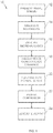
- Fig. 1 illustrates a method 10 for non-invasively monitoring eye movements for the differential diagnosis of AD and/or MCI.
- the method 10 may be carried out using a system 26, as shown in Fig. 4 and described below.
- the method 10 can include presenting visual stimuli to a patient (process block 12).
- visual stimuli can be presented on a display screen about 40 centimeters away from a subject's eyes. Presentation of visual stimuli may begin with a five-point calibration sequence followed by the presentation of a small fixation cross (for example, 1 degree) at the center of the screen.
- the fixation cross can remain displayed for about 20 seconds, and the subject can be instructed to look at the fixation cross as accurately as possible.
- microsaccades can be identified from the tracked eye movements (process block 16), for example through an objective detection algorithm (e.g., Engbert and Kliegl, 2003; Martinez-Conde, Macknik, Hubel, 2000). Based on the identified microsaccades, microsaccade dynamics can be characterized and/or measured (process block 18). Such microsaccade dynamics can include, but are not limited to, microsaccade direction, microsaccade velocity, microsaccade magnitude, microsaccadic peak velocity-magnitude relationship, microsaccade duration, and/or intersaccadic intervals.
- process blocks 16 and 18 can also or alternatively include identifying and characterizing (respectively) SWJs.
- a SWJ may be defined as the combination of one small saccade that moves the eye away from the fixation target, followed after a short period by a second corrective saccade directed back towards the target More specifically, a SWJ is known in the art to be a pair of saccades with three defining characteristics: (1) the two saccades have opposite or nearly opposite directions; (2) both saccades have equal or nearly equal magnitudes; and (3) the two saccades are separated by a short interval.
- An example process for identifying and characterizing SWJs is described in United States Patent No. 8,345,428 .
- This process measures how similar a given saccade pair (that is, a pair of consecutive saccades) is to an "ideal SWJ", based on the three defining characteristics of SWJs described above, and gives the saccade pair an score, or "index,” for its measurements relative to the ideal SWJ. If a saccade pair's SWJ index is larger than a given threshold, it may be characterized as a potential SWJ.
- one or more of the above-described microsaccade dynamics can be compared with dynamics of healthy subjects, considered "normal levels," (process block 20) and such comparisons are then assessed (process block 22).
- Assessment of the comparisons can include, but is not limited to, assessing a statistical difference (for example, within or not within normal limits, as determined by a significant or non-significant difference), quantifying the magnitude of a difference (that is, a numerical difference), etc.
- Comparison (process block 20) and assessment (process block 22) are further described in general and with respect to a particular example study below.
- a report may be generated (for example, recorded, stored, and/or displayed) providing a diagnosis and/or other information based on the assessment (process block 24).
- the report may designate the presence or absence of oculomotor disease (that is, AD or MCI), a quantification of abnormality signifying disease susceptibility, progression, severity, and/or response to treatment or therapy, etc.
- microsaccade direction is significantly different in subjects with AD or aMCI versus healthy subjects.
- the average microsaccade direction may be significantly deviated from horizontal, or from that of healthy subjects, by 20 degrees or more, although a smaller deviation may still be significant when considered with other factors.
- An example study was performed to demonstrate this relationship. The example study included three subject populations: patients with aMCI, patients with AD, and age-matched normal subjects.
- Microsaccades were identified (process block 16) automatically with an objective detection algorithm (see (Engbert and Kliegl, 2003), for details). In subjects in whom eye position was recorded binocularly, the amount of potential noise in the recorded movements was reduced (Engbert, 2006) by considering only binocular microsaccades (i.e., microsaccades with a minimum overlap of one data sample in both eyes). Some microsaccades are followed by a fast, small, oppositely directed, saccadic eye movement called dynamic overshoot, which is often more prominent for the eye that moves in the abducting direction. Dynamic overshoots that occurred less than 20ms after a preceding microsaccade were interpreted as part of the preceding microsaccade. That is, the previous microsaccade was modified to include the dynamic overshoot, which was not itself counted as a discrete microsaccade.
- Microsaccade identification was improved in the study by removing, prior to microsaccade identificaiton, any data epochs where partial pupil occlusion may have led to increased levels of noise.
- Such epochs were automatically identified by identifying high-velocity spikes in the eye movement data (specifically, over 1,000 deg/s, although epochs of as little as 25 deg/s could be removed).
- two epochs were separated by less than 25 samples, they could be merged into a single epoch, which included the interval separating the two original epochs.
- SWJs were identified by first identifying all individual saccades up to 5 degrees (Otero-Millan, Serra, et al., 2011).
- the 5-degree upper magnitude threshold was chosen to include the range of SWJ magnitudes reported previously in healthy subjects (0.1-4.1 deg; (Abadi and Gowen, 2004)), and to allow for potentially larger SWJs magnitudes in patients (Otero-Millan, Serra, et al., 2011).
- saccade pairs were indexed based on the three defining characteristics of SWJs described above: a) the direction dissimilarity of first and second saccade, b) the magnitude similarity of first and second saccade, and c) the temporal proximity of first and second saccade, in a single, continuous variable for each saccade pair. If a saccade pair's SWJ index was larger than a given threshold (Otero-Millan, Serra, et al., 2011) the pair was classified as a potential SWJ.
- a saccade pair's SWJ index was larger than a given threshold (Otero-Millan, Serra, et al., 2011) the pair was classified as a potential SWJ.
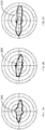
- Figs. 2A-C are polar histograms showing microsaccadic direction (shown in bold lines 200, 205, 210) in the AD population ( Fig. 2A ), the aMCI population ( Fig. 2B ), and the healthy population ( Fig. 2C ).
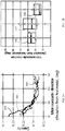
- Fig. 3A is a line graph of the distribution of microsaccades at increasing deviation from horizontal for the three populations, where the AD population is represented by line 300, the aMCI population by line 305, and the healthy population by line 310.
- Fig. 2A-C are polar histograms showing microsaccadic direction (shown in bold lines 200, 205, 210) in the AD population ( Fig. 2A ), the aMCI population ( Fig. 2B ), and the healthy population ( Fig. 2C ).
- Fig. 3A is a line graph of the distribution of microsaccades at increasing deviation from horizontal for the three populations, where the AD population is represented by line 300, the aMC
- 3B is a deviation graph also showing the average microsaccade direction as a deviation from horizontal, with the AD population represented by group 320, the aMCI population by group 325, and the healthy population by group 330.
- the results show that, despite predominantly horizontal microsaccade directions across all subject populations, oblique microsaccades (that is, less horizontal microsaccades) are more prevalent in AD and aMCl subjects than in age-matched healthy subjects. Furthermore, the differences in microsaccade directions between AD or aMCI subjects and healthy subjects extended to those microsaccades forming SWJs.
- microsaccade magnitudes and velocities, and the peak velocity-magnitude relationship are comparable in the three subject groups (that is, AD, aMCI, healthy).
- Microsaccadic durations, intersaccadic intervals and other microsaccade dynamics are also equivalent (and have comparable variability) in the three groups, as are the rate, magnitude, and percent of fixational saccades that are part of SWJs. This finding is consistent with the lack of brainstem oculomotor function impairment in MCI or AD patients with mild to moderate severity of disease (Garbutt et al., 2008; Yang et al., In Press; Yang et al., 2011; but see Simic et al., 2009).
- all the above dynamics may be collectively assessed to determine presence or absence of oculomotor disease (that is, AD or MCI), disease susceptibility, progression, severity, and/or response to treatment, etc. Accordingly, an assessment of significantly less horizontal microsaccade direction, but normal (that is, not significantly different) microsaccade magnitudes, velocities, peak velocity-magnitude relationship, microsaccadic durations, and intersaccadic intervals results in a diagnosis of AD or MCI.
- oculomotor disease that is, AD or MCI
- This assessment can further quantify the obliqueness of the microsaccades in comparison to healthy levels to determine a progression of AD or MCI, response to treatment (for example, if the subject's previously tracked microsaccade dynamics are available for comparison), etc.
- the generated report can therefore detail the diagnosis as well as the assessment of progression and/or response to treatment.
- the above method may include receiving additional input regarding the subject, including demographics such as age, gender, education, results of a mini mental state examination (MMSE) and/or activities of daily living (AOL) to assist in the above-described assessment.
- demographics such as age, gender, education
- AOL activities of daily living
- Table 1 illustrates a comparison of demographics and microsaccade dynamics of healthy subjects ("controls"), MCI subjects, and AD subjects according to a study carrying out the methods of the present disclosure. Any or all of the demographics, characteristics, and/or statistical comparisons detailed in the table below may be presented in the generated report.
- Fig. 4 illustrates a system 26, according to the present invention, for detecting and analyzing eye movements of a subject 28 to diagnose neurological disease.
- the system 26 may be used in accordance with the methods 10 described above.
- the system 26 can include a host 30 operably connected to an eye tracking device 32, a display 34, and a user interface 36.
- the host 30 can include one or more processors 38 operating under control of one or more computer programs 40 loaded from a non-transitory computer readable medium (memory) 42.
- reference to a step performed by a computer program 40 is also a reference to the processor 38 that performed that step, for example in accordance with process blocks of above-described methods 10 of the present disclosure.
- Example tracking devices 32 for use with the present invention can include the EyeLink II by SR Research or other equivalent eye tracking systems such as the IVIEW XTM HI-SPEED 1250 tracking system by SensoMotoric Instruments.
- the system 26 can operate by presenting visual stimuli to the subject 28 through the display 34.
- one of the processors 38 such as a display processor 38A, can retrieve one or more stored image or video files 44 from memory 42 and present the images/videos to the subject 28 via the display 34.
- the eye tracking device 32 can detect the position and movement of the subject's eyes 46, for example, in accordance with process block 14 described above. Identification of microsaccades, for example with respect to process block 16 described above, can be executed by one or more processors 38.
- An example process where eye movements are objectively and non-invasively monitored is described in United States Patent No. 7,857,452 .
- this process includes tracking a subject's eye position, for example, using a tracking processor 38B, and detecting microsaccades from eye position traces, for example, using a microsaccade processor 38C.
- Example algorithms that is, part of computer programs 40 for detecting microsaccades objectively from eye position traces (such as from video, eye coil, optical, or other suitable tracking methods) include the Martinez-Conde and Macknik algorithm ( Martinez-Conde S. Macknik S. L., Hubel D. H. (2000) Nature Neuroscience ) and the Engbert algorithm (Engbert R., Kliegl R. (2003) Vision Res 4:1035-1045 ).
- characterization and analysis of microsaccade dynamics can be executed by one or more processors 38, such as the microsaccade processor 38C, a statistics processor 38D, and/or a comparator 38E.
- additional processors 38 may be included to execute identification and analysis of SWJs.
- report generation for example with respect to process block 24 described above, can be executed by one or more processors 38, such as a reporting processor 38F, wherein the report may be stored in a file 44 in memory 42, displayed on the display 34 or a separate display, or output in another suitable manner to a user, for example as instructed by the user via the user interface 36.
Landscapes
- Health & Medical Sciences (AREA)
- Life Sciences & Earth Sciences (AREA)
- Engineering & Computer Science (AREA)
- Veterinary Medicine (AREA)
- Surgery (AREA)
- Biophysics (AREA)
- Biomedical Technology (AREA)
- Heart & Thoracic Surgery (AREA)
- Medical Informatics (AREA)
- Molecular Biology (AREA)
- Physics & Mathematics (AREA)
- Animal Behavior & Ethology (AREA)
- General Health & Medical Sciences (AREA)
- Public Health (AREA)
- Neurology (AREA)
- Pathology (AREA)
- Ophthalmology & Optometry (AREA)
- Developmental Disabilities (AREA)
- Hospice & Palliative Care (AREA)
- Psychiatry (AREA)
- Psychology (AREA)
- Child & Adolescent Psychology (AREA)
- Neurosurgery (AREA)
- Physiology (AREA)
- Human Computer Interaction (AREA)
- Signal Processing (AREA)
- Educational Technology (AREA)
- Social Psychology (AREA)
- Eye Examination Apparatus (AREA)
- Investigating Or Analysing Biological Materials (AREA)
Claims (9)
- Système qui comprend un hôte (30) relié de manière fonctionnelle à un dispositif d'oculométrie (32) capable d'enregistrer des traces de mouvement d'œil à partir de mouvements d'œil d'un sujet (28), l'hôte (30) ayant au moins un processeur (38) configuré pour :recevoir les traces de mouvement d'œil de la part du dispositif d'oculométrie (32) ;identifier (16) une pluralité de microsaccades à partir des traces de mouvement d'œil, la pluralité de microsaccades étant des mouvements rapides et involontaires de l'œil qui se produisent plusieurs fois par seconde pendant la fixation du regard par le sujet (28) ;mesurer une ou plusieurs dynamique(s) de chaque microsaccade, les dynamiques comprenant une répartition des directions des microsaccades, dans lequel la répartition des directions des microsaccades comprend une direction pour chaque microsaccade identifiée, la direction étant mesurée comme une déviation angulaire par rapport à l'horizontale ;comparer (20) chacune de la ou des dynamique(s) avec une dynamique de microsaccade de contrôle correspondante d'un sujet sain (28) ;évaluer une ou plusieurs différence(s) statistique(s) entre la ou les dynamique(s) déterminée(s) et les dynamiques de microsaccades de contrôle correspondantes, les différences statistiques indiquant la présence de la maladie d'Alzheimer, les différences statistiques comprenant une direction de microsaccade moyenne qui est bien moins horizontale qu'une direction de microsaccade saine moyenne correspondante ; etgénérer un rapport (24) qui contient des informations sur la maladie d'Alzheimer sur la base de l'évaluation.
- Système selon la revendication 1, dans lequel le processeur (38) est configuré pour évaluer la ou les différence(s) statistique(s) en déterminant si la différence statistique se trouve dans des limites normales ou non, et dans lequel les informations comprennent une indication selon laquelle la maladie neurologique peut être présente si la différence statistique se trouve en-dehors des limites normales.
- Système selon la revendication 1, dans lequel le processeur (38) est configuré pour évaluer la ou les différence(s) statistique(s) en comparant (22) la ou les différence(s) statistique(s) avec des différences statistiques précédentes correspondantes afin de caractériser l'une ou les deux de la progression de la maladie neurologique et de la réponse au traitement de la maladie d'Alzheimer.
- Système selon la revendication 3, dans lequel les informations comprennent l'une ou les deux de la progression et de la réponse.
- Système selon la revendication 1 et comprenant en outre un écran (34) relié de manière fonctionnelle à l'hôte (30).
- Système selon la revendication 5, dans lequel le processeur (38) est configuré pour présenter des stimuli visuels au sujet (28) par le biais de l'écran (34), et pour recevoir les traces de mouvement de l'œil enregistrées lorsque les stimuli visuels sont présentés.
- Système selon la revendication 5, dans lequel le processeur (38) est configuré pour afficher (34) le rapport par l'écran (34).
- Système selon la revendication 1 et comprenant en outre une interface utilisateur (36), dans lequel le processeur (38) est configuré pour fournir le rapport à un utilisateur sur la base d'une entrée reçue via l'interface utilisateur (36).
- Système selon la revendication 1, dans lequel les dynamiques comprennent en outre un ou plusieurs d'une vitesse, d'une magnitude, d'une relation vitesse de pointe-magnitude, d'une durée et d'intervalles entre les saccades.
Priority Applications (1)
| Application Number | Priority Date | Filing Date | Title |
|---|---|---|---|
| EP21190322.4A EP3932294B1 (fr) | 2013-05-31 | 2014-04-23 | Système pour la détection de maladie neurologique |
Applications Claiming Priority (2)
| Application Number | Priority Date | Filing Date | Title |
|---|---|---|---|
| US201361829898P | 2013-05-31 | 2013-05-31 | |
| PCT/US2014/035082 WO2014193564A1 (fr) | 2013-05-31 | 2014-04-23 | Système et procédé pour la détection de maladie neurologique |
Related Child Applications (1)
| Application Number | Title | Priority Date | Filing Date |
|---|---|---|---|
| EP21190322.4A Division EP3932294B1 (fr) | 2013-05-31 | 2014-04-23 | Système pour la détection de maladie neurologique |
Publications (3)
| Publication Number | Publication Date |
|---|---|
| EP3003125A1 EP3003125A1 (fr) | 2016-04-13 |
| EP3003125A4 EP3003125A4 (fr) | 2017-02-22 |
| EP3003125B1 true EP3003125B1 (fr) | 2021-08-11 |
Family
ID=51989302
Family Applications (2)
| Application Number | Title | Priority Date | Filing Date |
|---|---|---|---|
| EP21190322.4A Active EP3932294B1 (fr) | 2013-05-31 | 2014-04-23 | Système pour la détection de maladie neurologique |
| EP14803496.0A Active EP3003125B1 (fr) | 2013-05-31 | 2014-04-23 | Système pour la détection de maladie neurologique |
Family Applications Before (1)
| Application Number | Title | Priority Date | Filing Date |
|---|---|---|---|
| EP21190322.4A Active EP3932294B1 (fr) | 2013-05-31 | 2014-04-23 | Système pour la détection de maladie neurologique |
Country Status (5)
| Country | Link |
|---|---|
| US (4) | US9962119B2 (fr) |
| EP (2) | EP3932294B1 (fr) |
| JP (2) | JP6507151B2 (fr) |
| CA (1) | CA2912426C (fr) |
| WO (1) | WO2014193564A1 (fr) |
Families Citing this family (22)
| Publication number | Priority date | Publication date | Assignee | Title |
|---|---|---|---|---|
| EP3932294B1 (fr) * | 2013-05-31 | 2025-07-16 | Dignity Health | Système pour la détection de maladie neurologique |
| US10376183B2 (en) | 2014-04-29 | 2019-08-13 | Dignity Health | Systems and methods for non-intrusive drug impairment detection |
| AU2015274601B2 (en) | 2014-06-11 | 2020-02-27 | Arizona Board Of Regents On Behalf Of Arizona State University | Systems and methods for non-intrusive deception detection |
| WO2019075485A1 (fr) * | 2017-10-15 | 2019-04-18 | Hogan Joshua Noel | Procédé et système de surveillance de capacité de commande de mouvement |
| IL255607A0 (en) * | 2017-11-12 | 2017-12-31 | Bioeye Ltd | A method for the early detection of neurodegeneration using long-term passive tracking of eye markers |
| EP3711680B1 (fr) | 2017-11-14 | 2025-01-01 | Osaka University | Appareil de diagnostic de dysfonctionnement cognitif et programme de diagnostic de dysfonctionnement cognitif |
| US11694803B2 (en) * | 2017-11-30 | 2023-07-04 | Viewmind, Inc. | System and method for detecting neurological disorders and for measuring general cognitive performance |
| WO2019157053A1 (fr) * | 2018-02-07 | 2019-08-15 | RightEye, LLC | Systèmes et procédés d'évaluation de physiologie d'utilisateur sur la base de données de suivi oculaire |
| US12290373B2 (en) | 2018-04-27 | 2025-05-06 | C. Light Technologies, Inc. | Method of detection, prognostication, and monitoring of neurological disorders |
| US11471042B2 (en) | 2019-07-18 | 2022-10-18 | Welch Allyn, Inc. | Vision screening apparatus with a visual acuity protocol compliance feature and an associated method of vision screening |
| US12080104B2 (en) | 2019-09-12 | 2024-09-03 | Semiconductor Energy Laboratory Co., Ltd. | Classification method |
| JP7298508B2 (ja) * | 2020-02-26 | 2023-06-27 | マツダ株式会社 | 状態推定装置 |
| JP7298510B2 (ja) * | 2020-02-26 | 2023-06-27 | マツダ株式会社 | 状態推定装置 |
| JP7298509B2 (ja) * | 2020-02-26 | 2023-06-27 | マツダ株式会社 | 状態推定装置 |
| WO2021192704A1 (fr) | 2020-03-27 | 2021-09-30 | 国立大学法人大阪大学 | Dispositif de diagnostic de déficience cognitive et programme de diagnostic de déficience cognitive |
| EP4266976A4 (fr) * | 2020-12-23 | 2024-10-23 | Viewmind, Inc. | Système et méthode de détection de troubles neurologiques et de mesure des performances cognitives générales |
| US11503998B1 (en) | 2021-05-05 | 2022-11-22 | Innodem Neurosciences | Method and a system for detection of eye gaze-pattern abnormalities and related neurological diseases |
| EP4340724A4 (fr) * | 2021-05-18 | 2025-03-26 | Queen's University At Kingston | Procédés et appareil de détection de troubles cérébraux |
| JP7711753B2 (ja) * | 2021-07-06 | 2025-07-23 | 日本電気株式会社 | 回復度推定装置、回復度推定方法、及び、プログラム |
| JP7711754B2 (ja) * | 2021-07-06 | 2025-07-23 | 日本電気株式会社 | 回復度推定装置、回復度推定方法、及び、プログラム |
| US11903711B2 (en) | 2022-06-09 | 2024-02-20 | EarliTec Diagnostics, Inc. | Assessing developmental disorders via eye tracking |
| WO2024253722A1 (fr) | 2023-06-05 | 2024-12-12 | EarliTec Diagnostics, Inc. | Systèmes et procédés d'utilisation de dispositifs informatiques portables à capacité de détection oculaire |
Family Cites Families (25)
| Publication number | Priority date | Publication date | Assignee | Title |
|---|---|---|---|---|
| US4815839A (en) | 1987-08-03 | 1989-03-28 | Waldorf Ronald A | Infrared/video electronystagmographic apparatus |
| US5137345A (en) | 1991-05-15 | 1992-08-11 | Oculokinetics, Inc. | Apparatus for monitoring physiolgical data to detect drug impairment |
| JPH074345B2 (ja) * | 1992-08-12 | 1995-01-25 | 株式会社エイ・ティ・アール視聴覚機構研究所 | 注視点マスキングによる医療診断装置 |
| JPH08104B2 (ja) * | 1992-09-17 | 1996-01-10 | 株式会社エイ・ティ・アール視聴覚機構研究所 | 奥行き視線移動検査装置 |
| JPH08105B2 (ja) * | 1992-11-27 | 1996-01-10 | 株式会社エイ・ティ・アール視聴覚機構研究所 | 固視微動検査装置 |
| JP2001309890A (ja) * | 2000-02-24 | 2001-11-06 | Matsushita Electric Works Ltd | 脳機能検査方法及びその装置 |
| US6652458B2 (en) | 2000-06-20 | 2003-11-25 | The Mclean Hospital Corporation | ADHD detection by eye saccades |
| JP4336854B2 (ja) * | 2003-11-11 | 2009-09-30 | 学校法人東海大学 | 視線表示装置および痴呆症診断装置 |
| US7309125B2 (en) | 2005-06-09 | 2007-12-18 | Vladimir Pugach | Method and apparatus for detecting abnormalities in spatial perception |
| US8790280B2 (en) | 2007-06-27 | 2014-07-29 | Panasonic Corporation | Human state estimating device and method |
| US7857452B2 (en) | 2007-08-27 | 2010-12-28 | Catholic Healthcare West | Eye movements as a way to determine foci of covert attention |
| US8721081B2 (en) | 2007-11-01 | 2014-05-13 | Dignity Health | Method of detecting neurological disease |
| US9301679B2 (en) | 2007-11-01 | 2016-04-05 | Dignity Health | Method of detecting neurological disease |
| CA2704777C (fr) * | 2007-11-01 | 2017-09-12 | Catholic Healthcare West | Procede de detection d'une maladie neurologique |
| WO2009097430A1 (fr) | 2008-02-01 | 2009-08-06 | Lonky Martin L | Procédés et techniques de mesure, de cartographie et de corrélation de signaux de micro-tremblement oculaire (omt) |
| JP5363043B2 (ja) * | 2008-07-11 | 2013-12-11 | 浜松ホトニクス株式会社 | 眼球運動計測装置 |
| AU2009281683B2 (en) | 2008-07-21 | 2014-07-24 | Erber Aktiengesellschaft | Method for treating food silage for ruminants and food silage additive |
| WO2010042557A2 (fr) | 2008-10-06 | 2010-04-15 | Neuro Kinetics, Inc. | Procédé et appareil pour une analyse de saccades secondaires correctives avec un système d'oculographie vidéo |
| US8808195B2 (en) | 2009-01-15 | 2014-08-19 | Po-He Tseng | Eye-tracking method and system for screening human diseases |
| WO2010107568A1 (fr) | 2009-03-17 | 2010-09-23 | Emory University | Diagnostics cognitifs basés sur internet utilisant une tâche de comparaison appariée visuelle |
| US9101312B2 (en) | 2012-04-18 | 2015-08-11 | TBI Diagnostics LLC | System for the physiological evaluation of brain function |
| US8668337B2 (en) | 2012-04-18 | 2014-03-11 | TBI Diagnostics LLC | System for the physiological evaluation of brain function |
| US10575726B2 (en) | 2013-03-14 | 2020-03-03 | Virginia Commonwealth University | Automated analysis system for the detection and screening of neurological disorders and defects |
| EP3932294B1 (fr) * | 2013-05-31 | 2025-07-16 | Dignity Health | Système pour la détection de maladie neurologique |
| CN117084624A (zh) | 2014-08-04 | 2023-11-21 | 纽约大学 | 用于诊断、评价或量化药物使用、药物滥用和麻醉的方法和试剂盒 |
-
2014
- 2014-04-23 EP EP21190322.4A patent/EP3932294B1/fr active Active
- 2014-04-23 JP JP2016516654A patent/JP6507151B2/ja active Active
- 2014-04-23 US US14/893,552 patent/US9962119B2/en active Active
- 2014-04-23 WO PCT/US2014/035082 patent/WO2014193564A1/fr not_active Ceased
- 2014-04-23 EP EP14803496.0A patent/EP3003125B1/fr active Active
- 2014-04-23 CA CA2912426A patent/CA2912426C/fr active Active
-
2018
- 2018-04-20 US US15/958,403 patent/US10537276B2/en active Active
-
2019
- 2019-03-29 JP JP2019065170A patent/JP2019122816A/ja active Pending
-
2020
- 2020-01-13 US US16/741,549 patent/US11903720B2/en active Active
-
2024
- 2024-02-19 US US18/581,101 patent/US20240188879A1/en active Pending
Non-Patent Citations (1)
| Title |
|---|
| None * |
Also Published As
| Publication number | Publication date |
|---|---|
| EP3003125A1 (fr) | 2016-04-13 |
| EP3932294B1 (fr) | 2025-07-16 |
| JP6507151B2 (ja) | 2019-04-24 |
| US11903720B2 (en) | 2024-02-20 |
| EP3932294A1 (fr) | 2022-01-05 |
| US20200146611A1 (en) | 2020-05-14 |
| WO2014193564A1 (fr) | 2014-12-04 |
| EP3003125A4 (fr) | 2017-02-22 |
| US20240188879A1 (en) | 2024-06-13 |
| CA2912426C (fr) | 2023-09-26 |
| US20160106358A1 (en) | 2016-04-21 |
| JP2016523112A (ja) | 2016-08-08 |
| US9962119B2 (en) | 2018-05-08 |
| JP2019122816A (ja) | 2019-07-25 |
| CA2912426A1 (fr) | 2014-12-04 |
| US20180235531A1 (en) | 2018-08-23 |
| US10537276B2 (en) | 2020-01-21 |
Similar Documents
| Publication | Publication Date | Title |
|---|---|---|
| US20240188879A1 (en) | System and method for detecting neurological disease | |
| US20210052182A1 (en) | Portable brain activity sensing platform for assessment of visual field deficits | |
| EP3716837B1 (fr) | Système et procédé de détection de troubles neurologiques et de mesure des performances cognitives générales | |
| JP6975853B2 (ja) | 脳機能検査システムおよびそのデバイス | |
| CA3103781C (fr) | Methode et systeme pour evaluer la fonction cognitive d'un individu | |
| WO2013102768A1 (fr) | Appareil et procédé d'évaluation psychiatrique | |
| US20180368699A1 (en) | System of predicting dementia and operating method thereof | |
| US11642068B2 (en) | Device and method to determine objectively visual memory of images | |
| CN107296586A (zh) | 视觉误差检测设备/方法及基于该设备的书写系统/方法 | |
| Hassan et al. | Approach to quantify eye movements to augment stroke diagnosis with a non-calibrated eye-tracker | |
| Komogortsev et al. | The application of eye movement biometrics in the automated detection of mild traumatic brain injury | |
| US10070812B2 (en) | Method for improved seizure detection | |
| Smith | Assessing mild cognitive impairment using portable electroencephalography: the P300 component | |
| Florea et al. | Computer vision for cognition: An eye focused perspective | |
| US20230284974A1 (en) | Systems and methods for diagnosing, assessing, and quantifying sedative effects | |
| Xu et al. | Evaluation of Steady-State Visual Evoked Potentials (SSVEP) Stimuli Design for Visual Field Assessment | |
| US20200390577A1 (en) | Method and system for detecting voluntary binary responses by analyzing the pupil diameter of a subject |
Legal Events
| Date | Code | Title | Description |
|---|---|---|---|
| PUAI | Public reference made under article 153(3) epc to a published international application that has entered the european phase |
Free format text: ORIGINAL CODE: 0009012 |
|
| 17P | Request for examination filed |
Effective date: 20151216 |
|
| AK | Designated contracting states |
Kind code of ref document: A1 Designated state(s): AL AT BE BG CH CY CZ DE DK EE ES FI FR GB GR HR HU IE IS IT LI LT LU LV MC MK MT NL NO PL PT RO RS SE SI SK SM TR |
|
| AX | Request for extension of the european patent |
Extension state: BA ME |
|
| RIN1 | Information on inventor provided before grant (corrected) |
Inventor name: LANG, ALEXANDRE Inventor name: KAPOULA, ZOI Inventor name: MARTINEZ-CONDE, SUSANA Inventor name: MACKNIK, M., STEPHEN L. Inventor name: VERNY, MARC |
|
| RIN1 | Information on inventor provided before grant (corrected) |
Inventor name: KAPOULA, ZOI Inventor name: VERNY, MARC Inventor name: MARTINEZ-CONDE, SUSANA Inventor name: MACKNIK, M., STEPHEN L. Inventor name: LANG, ALEXANDRE |
|
| DAX | Request for extension of the european patent (deleted) | ||
| REG | Reference to a national code |
Ref country code: DE Ref legal event code: R079 Ref document number: 602014079397 Country of ref document: DE Free format text: PREVIOUS MAIN CLASS: A61B0003140000 Ipc: A61B0003113000 |
|
| A4 | Supplementary search report drawn up and despatched |
Effective date: 20170124 |
|
| RIC1 | Information provided on ipc code assigned before grant |
Ipc: A61B 3/113 20060101AFI20170118BHEP Ipc: A61B 3/00 20060101ALI20170118BHEP |
|
| RIN1 | Information on inventor provided before grant (corrected) |
Inventor name: KAPOULA, ZOI Inventor name: OTERO-MILLAN, JORGE Inventor name: VERNY, MARC Inventor name: MARTINEZ-CONDE, SUSANA Inventor name: MACKNIK, M., STEPHEN L. Inventor name: LANG, ALEXANDRE |
|
| STAA | Information on the status of an ep patent application or granted ep patent |
Free format text: STATUS: EXAMINATION IS IN PROGRESS |
|
| 17Q | First examination report despatched |
Effective date: 20180111 |
|
| RAP1 | Party data changed (applicant data changed or rights of an application transferred) |
Owner name: DIGNITY HEALTH Owner name: LE CENTRE NATIONAL DE LA RECHERCHE SCIENTIFIQUE |
|
| RIN1 | Information on inventor provided before grant (corrected) |
Inventor name: OTERO-MILLAN, JORGE Inventor name: MACKNIK, M., STEPHEN L. Inventor name: MARTINEZ-CONDE, SUSANA Inventor name: KAPOULA, ZOI |
|
| RIN1 | Information on inventor provided before grant (corrected) |
Inventor name: MACKNIK, M., STEPHEN L. Inventor name: KAPOULA, ZOI Inventor name: OTERO-MILLAN, JORGE Inventor name: MARTINEZ-CONDE, SUSANA |
|
| GRAP | Despatch of communication of intention to grant a patent |
Free format text: ORIGINAL CODE: EPIDOSNIGR1 |
|
| STAA | Information on the status of an ep patent application or granted ep patent |
Free format text: STATUS: GRANT OF PATENT IS INTENDED |
|
| INTG | Intention to grant announced |
Effective date: 20210224 |
|
| GRAS | Grant fee paid |
Free format text: ORIGINAL CODE: EPIDOSNIGR3 |
|
| GRAA | (expected) grant |
Free format text: ORIGINAL CODE: 0009210 |
|
| STAA | Information on the status of an ep patent application or granted ep patent |
Free format text: STATUS: THE PATENT HAS BEEN GRANTED |
|
| AK | Designated contracting states |
Kind code of ref document: B1 Designated state(s): AL AT BE BG CH CY CZ DE DK EE ES FI FR GB GR HR HU IE IS IT LI LT LU LV MC MK MT NL NO PL PT RO RS SE SI SK SM TR |
|
| REG | Reference to a national code |
Ref country code: GB Ref legal event code: FG4D |
|
| REG | Reference to a national code |
Ref country code: CH Ref legal event code: EP |
|
| REG | Reference to a national code |
Ref country code: DE Ref legal event code: R096 Ref document number: 602014079397 Country of ref document: DE |
|
| REG | Reference to a national code |
Ref country code: IE Ref legal event code: FG4D Ref country code: AT Ref legal event code: REF Ref document number: 1418519 Country of ref document: AT Kind code of ref document: T Effective date: 20210915 |
|
| REG | Reference to a national code |
Ref country code: LT Ref legal event code: MG9D |
|
| REG | Reference to a national code |
Ref country code: NL Ref legal event code: MP Effective date: 20210811 |
|
| REG | Reference to a national code |
Ref country code: AT Ref legal event code: MK05 Ref document number: 1418519 Country of ref document: AT Kind code of ref document: T Effective date: 20210811 |
|
| PG25 | Lapsed in a contracting state [announced via postgrant information from national office to epo] |
Ref country code: HR Free format text: LAPSE BECAUSE OF FAILURE TO SUBMIT A TRANSLATION OF THE DESCRIPTION OR TO PAY THE FEE WITHIN THE PRESCRIBED TIME-LIMIT Effective date: 20210811 Ref country code: RS Free format text: LAPSE BECAUSE OF FAILURE TO SUBMIT A TRANSLATION OF THE DESCRIPTION OR TO PAY THE FEE WITHIN THE PRESCRIBED TIME-LIMIT Effective date: 20210811 Ref country code: SE Free format text: LAPSE BECAUSE OF FAILURE TO SUBMIT A TRANSLATION OF THE DESCRIPTION OR TO PAY THE FEE WITHIN THE PRESCRIBED TIME-LIMIT Effective date: 20210811 Ref country code: ES Free format text: LAPSE BECAUSE OF FAILURE TO SUBMIT A TRANSLATION OF THE DESCRIPTION OR TO PAY THE FEE WITHIN THE PRESCRIBED TIME-LIMIT Effective date: 20210811 Ref country code: FI Free format text: LAPSE BECAUSE OF FAILURE TO SUBMIT A TRANSLATION OF THE DESCRIPTION OR TO PAY THE FEE WITHIN THE PRESCRIBED TIME-LIMIT Effective date: 20210811 Ref country code: NO Free format text: LAPSE BECAUSE OF FAILURE TO SUBMIT A TRANSLATION OF THE DESCRIPTION OR TO PAY THE FEE WITHIN THE PRESCRIBED TIME-LIMIT Effective date: 20211111 Ref country code: PT Free format text: LAPSE BECAUSE OF FAILURE TO SUBMIT A TRANSLATION OF THE DESCRIPTION OR TO PAY THE FEE WITHIN THE PRESCRIBED TIME-LIMIT Effective date: 20211213 Ref country code: BG Free format text: LAPSE BECAUSE OF FAILURE TO SUBMIT A TRANSLATION OF THE DESCRIPTION OR TO PAY THE FEE WITHIN THE PRESCRIBED TIME-LIMIT Effective date: 20211111 Ref country code: AT Free format text: LAPSE BECAUSE OF FAILURE TO SUBMIT A TRANSLATION OF THE DESCRIPTION OR TO PAY THE FEE WITHIN THE PRESCRIBED TIME-LIMIT Effective date: 20210811 Ref country code: LT Free format text: LAPSE BECAUSE OF FAILURE TO SUBMIT A TRANSLATION OF THE DESCRIPTION OR TO PAY THE FEE WITHIN THE PRESCRIBED TIME-LIMIT Effective date: 20210811 |
|
| PG25 | Lapsed in a contracting state [announced via postgrant information from national office to epo] |
Ref country code: PL Free format text: LAPSE BECAUSE OF FAILURE TO SUBMIT A TRANSLATION OF THE DESCRIPTION OR TO PAY THE FEE WITHIN THE PRESCRIBED TIME-LIMIT Effective date: 20210811 Ref country code: LV Free format text: LAPSE BECAUSE OF FAILURE TO SUBMIT A TRANSLATION OF THE DESCRIPTION OR TO PAY THE FEE WITHIN THE PRESCRIBED TIME-LIMIT Effective date: 20210811 Ref country code: GR Free format text: LAPSE BECAUSE OF FAILURE TO SUBMIT A TRANSLATION OF THE DESCRIPTION OR TO PAY THE FEE WITHIN THE PRESCRIBED TIME-LIMIT Effective date: 20211112 |
|
| PG25 | Lapsed in a contracting state [announced via postgrant information from national office to epo] |
Ref country code: NL Free format text: LAPSE BECAUSE OF FAILURE TO SUBMIT A TRANSLATION OF THE DESCRIPTION OR TO PAY THE FEE WITHIN THE PRESCRIBED TIME-LIMIT Effective date: 20210811 |
|
| PG25 | Lapsed in a contracting state [announced via postgrant information from national office to epo] |
Ref country code: DK Free format text: LAPSE BECAUSE OF FAILURE TO SUBMIT A TRANSLATION OF THE DESCRIPTION OR TO PAY THE FEE WITHIN THE PRESCRIBED TIME-LIMIT Effective date: 20210811 |
|
| REG | Reference to a national code |
Ref country code: DE Ref legal event code: R097 Ref document number: 602014079397 Country of ref document: DE |
|
| PG25 | Lapsed in a contracting state [announced via postgrant information from national office to epo] |
Ref country code: SM Free format text: LAPSE BECAUSE OF FAILURE TO SUBMIT A TRANSLATION OF THE DESCRIPTION OR TO PAY THE FEE WITHIN THE PRESCRIBED TIME-LIMIT Effective date: 20210811 Ref country code: SK Free format text: LAPSE BECAUSE OF FAILURE TO SUBMIT A TRANSLATION OF THE DESCRIPTION OR TO PAY THE FEE WITHIN THE PRESCRIBED TIME-LIMIT Effective date: 20210811 Ref country code: RO Free format text: LAPSE BECAUSE OF FAILURE TO SUBMIT A TRANSLATION OF THE DESCRIPTION OR TO PAY THE FEE WITHIN THE PRESCRIBED TIME-LIMIT Effective date: 20210811 Ref country code: EE Free format text: LAPSE BECAUSE OF FAILURE TO SUBMIT A TRANSLATION OF THE DESCRIPTION OR TO PAY THE FEE WITHIN THE PRESCRIBED TIME-LIMIT Effective date: 20210811 Ref country code: CZ Free format text: LAPSE BECAUSE OF FAILURE TO SUBMIT A TRANSLATION OF THE DESCRIPTION OR TO PAY THE FEE WITHIN THE PRESCRIBED TIME-LIMIT Effective date: 20210811 Ref country code: AL Free format text: LAPSE BECAUSE OF FAILURE TO SUBMIT A TRANSLATION OF THE DESCRIPTION OR TO PAY THE FEE WITHIN THE PRESCRIBED TIME-LIMIT Effective date: 20210811 |
|
| PLBE | No opposition filed within time limit |
Free format text: ORIGINAL CODE: 0009261 |
|
| STAA | Information on the status of an ep patent application or granted ep patent |
Free format text: STATUS: NO OPPOSITION FILED WITHIN TIME LIMIT |
|
| 26N | No opposition filed |
Effective date: 20220512 |
|
| PG25 | Lapsed in a contracting state [announced via postgrant information from national office to epo] |
Ref country code: IT Free format text: LAPSE BECAUSE OF FAILURE TO SUBMIT A TRANSLATION OF THE DESCRIPTION OR TO PAY THE FEE WITHIN THE PRESCRIBED TIME-LIMIT Effective date: 20210811 |
|
| PG25 | Lapsed in a contracting state [announced via postgrant information from national office to epo] |
Ref country code: SI Free format text: LAPSE BECAUSE OF FAILURE TO SUBMIT A TRANSLATION OF THE DESCRIPTION OR TO PAY THE FEE WITHIN THE PRESCRIBED TIME-LIMIT Effective date: 20210811 |
|
| REG | Reference to a national code |
Ref country code: BE Ref legal event code: MM Effective date: 20220430 |
|
| PG25 | Lapsed in a contracting state [announced via postgrant information from national office to epo] |
Ref country code: MC Free format text: LAPSE BECAUSE OF FAILURE TO SUBMIT A TRANSLATION OF THE DESCRIPTION OR TO PAY THE FEE WITHIN THE PRESCRIBED TIME-LIMIT Effective date: 20210811 Ref country code: LU Free format text: LAPSE BECAUSE OF NON-PAYMENT OF DUE FEES Effective date: 20220423 |
|
| PG25 | Lapsed in a contracting state [announced via postgrant information from national office to epo] |
Ref country code: BE Free format text: LAPSE BECAUSE OF NON-PAYMENT OF DUE FEES Effective date: 20220430 |
|
| PG25 | Lapsed in a contracting state [announced via postgrant information from national office to epo] |
Ref country code: IE Free format text: LAPSE BECAUSE OF NON-PAYMENT OF DUE FEES Effective date: 20220423 |
|
| PG25 | Lapsed in a contracting state [announced via postgrant information from national office to epo] |
Ref country code: HU Free format text: LAPSE BECAUSE OF FAILURE TO SUBMIT A TRANSLATION OF THE DESCRIPTION OR TO PAY THE FEE WITHIN THE PRESCRIBED TIME-LIMIT; INVALID AB INITIO Effective date: 20140423 |
|
| PG25 | Lapsed in a contracting state [announced via postgrant information from national office to epo] |
Ref country code: MK Free format text: LAPSE BECAUSE OF FAILURE TO SUBMIT A TRANSLATION OF THE DESCRIPTION OR TO PAY THE FEE WITHIN THE PRESCRIBED TIME-LIMIT Effective date: 20210811 Ref country code: CY Free format text: LAPSE BECAUSE OF FAILURE TO SUBMIT A TRANSLATION OF THE DESCRIPTION OR TO PAY THE FEE WITHIN THE PRESCRIBED TIME-LIMIT Effective date: 20210811 |
|
| PG25 | Lapsed in a contracting state [announced via postgrant information from national office to epo] |
Ref country code: MT Free format text: LAPSE BECAUSE OF FAILURE TO SUBMIT A TRANSLATION OF THE DESCRIPTION OR TO PAY THE FEE WITHIN THE PRESCRIBED TIME-LIMIT Effective date: 20210811 |
|
| PGFP | Annual fee paid to national office [announced via postgrant information from national office to epo] |
Ref country code: DE Payment date: 20250429 Year of fee payment: 12 |
|
| PGFP | Annual fee paid to national office [announced via postgrant information from national office to epo] |
Ref country code: GB Payment date: 20250428 Year of fee payment: 12 |
|
| PGFP | Annual fee paid to national office [announced via postgrant information from national office to epo] |
Ref country code: FR Payment date: 20250425 Year of fee payment: 12 |
|
| PGFP | Annual fee paid to national office [announced via postgrant information from national office to epo] |
Ref country code: CH Payment date: 20250501 Year of fee payment: 12 |
|
| PG25 | Lapsed in a contracting state [announced via postgrant information from national office to epo] |
Ref country code: TR Free format text: LAPSE BECAUSE OF FAILURE TO SUBMIT A TRANSLATION OF THE DESCRIPTION OR TO PAY THE FEE WITHIN THE PRESCRIBED TIME-LIMIT Effective date: 20210811 |