EP2945652B1 - Méthodes de traitement du cholangiocarcinome - Google Patents
Méthodes de traitement du cholangiocarcinome Download PDFInfo
- Publication number
- EP2945652B1 EP2945652B1 EP14702185.1A EP14702185A EP2945652B1 EP 2945652 B1 EP2945652 B1 EP 2945652B1 EP 14702185 A EP14702185 A EP 14702185A EP 2945652 B1 EP2945652 B1 EP 2945652B1
- Authority
- EP
- European Patent Office
- Prior art keywords
- fgfr2
- fusion
- nucleic acid
- sequence
- therapeutic agent
- Prior art date
- Legal status (The legal status is an assumption and is not a legal conclusion. Google has not performed a legal analysis and makes no representation as to the accuracy of the status listed.)
- Active
Links
- 238000000034 method Methods 0.000 title claims description 143
- 208000006990 cholangiocarcinoma Diseases 0.000 title claims description 120
- 230000004927 fusion Effects 0.000 claims description 504
- 150000007523 nucleic acids Chemical class 0.000 claims description 366
- 102000039446 nucleic acids Human genes 0.000 claims description 327
- 108020004707 nucleic acids Proteins 0.000 claims description 327
- 101710182389 Fibroblast growth factor receptor 2 Proteins 0.000 claims description 278
- 102100023600 Fibroblast growth factor receptor 2 Human genes 0.000 claims description 277
- 206010028980 Neoplasm Diseases 0.000 claims description 176
- 210000004027 cell Anatomy 0.000 claims description 161
- 108090000765 processed proteins & peptides Proteins 0.000 claims description 154
- 102000004196 processed proteins & peptides Human genes 0.000 claims description 142
- 229920001184 polypeptide Polymers 0.000 claims description 136
- 229940043355 kinase inhibitor Drugs 0.000 claims description 68
- 239000003757 phosphotransferase inhibitor Substances 0.000 claims description 67
- 239000003814 drug Substances 0.000 claims description 61
- 239000003112 inhibitor Substances 0.000 claims description 61
- 229940124597 therapeutic agent Drugs 0.000 claims description 55
- 230000014509 gene expression Effects 0.000 claims description 51
- 238000011282 treatment Methods 0.000 claims description 38
- 101000836150 Homo sapiens Transforming acidic coiled-coil-containing protein 3 Proteins 0.000 claims description 34
- 102100027048 Transforming acidic coiled-coil-containing protein 3 Human genes 0.000 claims description 34
- 230000000692 anti-sense effect Effects 0.000 claims description 30
- PHXJVRSECIGDHY-UHFFFAOYSA-N ponatinib Chemical compound C1CN(C)CCN1CC(C(=C1)C(F)(F)F)=CC=C1NC(=O)C1=CC=C(C)C(C#CC=2N3N=CC=CC3=NC=2)=C1 PHXJVRSECIGDHY-UHFFFAOYSA-N 0.000 claims description 27
- 230000001225 therapeutic effect Effects 0.000 claims description 27
- FNHKPVJBJVTLMP-UHFFFAOYSA-N regorafenib Chemical compound C1=NC(C(=O)NC)=CC(OC=2C=C(F)C(NC(=O)NC=3C=C(C(Cl)=CC=3)C(F)(F)F)=CC=2)=C1 FNHKPVJBJVTLMP-UHFFFAOYSA-N 0.000 claims description 25
- 238000012163 sequencing technique Methods 0.000 claims description 25
- 108020004999 messenger RNA Proteins 0.000 claims description 24
- 239000002138 L01XE21 - Regorafenib Substances 0.000 claims description 23
- 239000002137 L01XE24 - Ponatinib Substances 0.000 claims description 23
- 229960001131 ponatinib Drugs 0.000 claims description 23
- 229960004836 regorafenib Drugs 0.000 claims description 23
- 101150088071 fgfr2 gene Proteins 0.000 claims description 22
- 230000005945 translocation Effects 0.000 claims description 20
- 230000001105 regulatory effect Effects 0.000 claims description 16
- VAARYSWULJUGST-UHFFFAOYSA-N PD173955 Chemical compound CSC1=CC=CC(NC=2N=C3N(C)C(=O)C(C=4C(=CC=CC=4Cl)Cl)=CC3=CN=2)=C1 VAARYSWULJUGST-UHFFFAOYSA-N 0.000 claims description 14
- 108020004459 Small interfering RNA Proteins 0.000 claims description 13
- 210000000349 chromosome Anatomy 0.000 claims description 13
- WCWUXEGQKLTGDX-LLVKDONJSA-N (2R)-1-[[4-[(4-fluoro-2-methyl-1H-indol-5-yl)oxy]-5-methyl-6-pyrrolo[2,1-f][1,2,4]triazinyl]oxy]-2-propanol Chemical compound C1=C2NC(C)=CC2=C(F)C(OC2=NC=NN3C=C(C(=C32)C)OC[C@H](O)C)=C1 WCWUXEGQKLTGDX-LLVKDONJSA-N 0.000 claims description 12
- 102000053642 Catalytic RNA Human genes 0.000 claims description 12
- XZXHXSATPCNXJR-ZIADKAODSA-N nintedanib Chemical compound O=C1NC2=CC(C(=O)OC)=CC=C2\C1=C(C=1C=CC=CC=1)\NC(C=C1)=CC=C1N(C)C(=O)CN1CCN(C)CC1 XZXHXSATPCNXJR-ZIADKAODSA-N 0.000 claims description 12
- 229960004378 nintedanib Drugs 0.000 claims description 12
- 108091092562 ribozyme Proteins 0.000 claims description 12
- 108090000994 Catalytic RNA Proteins 0.000 claims description 11
- 238000012217 deletion Methods 0.000 claims description 11
- 230000037430 deletion Effects 0.000 claims description 11
- WJEOLQLKVOPQFV-UHFFFAOYSA-N masitinib Chemical compound C1CN(C)CCN1CC1=CC=C(C(=O)NC=2C=C(NC=3SC=C(N=3)C=3C=NC=CC=3)C(C)=CC=2)C=C1 WJEOLQLKVOPQFV-UHFFFAOYSA-N 0.000 claims description 11
- VRQMAABPASPXMW-HDICACEKSA-N AZD4547 Chemical compound COC1=CC(OC)=CC(CCC=2NN=C(NC(=O)C=3C=CC(=CC=3)N3C[C@@H](C)N[C@@H](C)C3)C=2)=C1 VRQMAABPASPXMW-HDICACEKSA-N 0.000 claims description 10
- KGWWHPZQLVVAPT-STTJLUEPSA-N (2r,3r)-2,3-dihydroxybutanedioic acid;6-(4-methylpiperazin-1-yl)-n-(5-methyl-1h-pyrazol-3-yl)-2-[(e)-2-phenylethenyl]pyrimidin-4-amine Chemical compound OC(=O)[C@H](O)[C@@H](O)C(O)=O.C1CN(C)CCN1C1=CC(NC2=NNC(C)=C2)=NC(\C=C\C=2C=CC=CC=2)=N1 KGWWHPZQLVVAPT-STTJLUEPSA-N 0.000 claims description 9
- KCOYQXZDFIIGCY-CZIZESTLSA-N (3e)-4-amino-5-fluoro-3-[5-(4-methylpiperazin-1-yl)-1,3-dihydrobenzimidazol-2-ylidene]quinolin-2-one Chemical compound C1CN(C)CCN1C1=CC=C(N\C(N2)=C/3C(=C4C(F)=CC=CC4=NC\3=O)N)C2=C1 KCOYQXZDFIIGCY-CZIZESTLSA-N 0.000 claims description 9
- GKJCVYLDJWTWQU-CXLRFSCWSA-N 2-[4-[(e)-2-[5-[(1r)-1-(3,5-dichloropyridin-4-yl)ethoxy]-1h-indazol-3-yl]ethenyl]pyrazol-1-yl]ethanol Chemical compound O([C@H](C)C=1C(=CN=CC=1Cl)Cl)C(C=C12)=CC=C1NN=C2\C=C\C=1C=NN(CCO)C=1 GKJCVYLDJWTWQU-CXLRFSCWSA-N 0.000 claims description 9
- QADPYRIHXKWUSV-UHFFFAOYSA-N BGJ-398 Chemical compound C1CN(CC)CCN1C(C=C1)=CC=C1NC1=CC(N(C)C(=O)NC=2C(=C(OC)C=C(OC)C=2Cl)Cl)=NC=N1 QADPYRIHXKWUSV-UHFFFAOYSA-N 0.000 claims description 9
- 210000000013 bile duct Anatomy 0.000 claims description 9
- 229950005778 dovitinib Drugs 0.000 claims description 9
- 239000003798 L01XE11 - Pazopanib Substances 0.000 claims description 8
- 239000002139 L01XE22 - Masitinib Substances 0.000 claims description 8
- WOSKHXYHFSIKNG-UHFFFAOYSA-N lenvatinib Chemical compound C=12C=C(C(N)=O)C(OC)=CC2=NC=CC=1OC(C=C1Cl)=CC=C1NC(=O)NC1CC1 WOSKHXYHFSIKNG-UHFFFAOYSA-N 0.000 claims description 8
- CUIHSIWYWATEQL-UHFFFAOYSA-N pazopanib Chemical compound C1=CC2=C(C)N(C)N=C2C=C1N(C)C(N=1)=CC=NC=1NC1=CC=C(C)C(S(N)(=O)=O)=C1 CUIHSIWYWATEQL-UHFFFAOYSA-N 0.000 claims description 8
- 238000001356 surgical procedure Methods 0.000 claims description 8
- 239000003184 complementary RNA Substances 0.000 claims description 7
- 229950004444 erdafitinib Drugs 0.000 claims description 7
- 229960004655 masitinib Drugs 0.000 claims description 7
- OLAHOMJCDNXHFI-UHFFFAOYSA-N n'-(3,5-dimethoxyphenyl)-n'-[3-(1-methylpyrazol-4-yl)quinoxalin-6-yl]-n-propan-2-ylethane-1,2-diamine Chemical compound COC1=CC(OC)=CC(N(CCNC(C)C)C=2C=C3N=C(C=NC3=CC=2)C2=CN(C)N=C2)=C1 OLAHOMJCDNXHFI-UHFFFAOYSA-N 0.000 claims description 7
- 229960000639 pazopanib Drugs 0.000 claims description 7
- 108020005544 Antisense RNA Proteins 0.000 claims description 6
- 238000002512 chemotherapy Methods 0.000 claims description 6
- 238000013518 transcription Methods 0.000 claims description 6
- 230000035897 transcription Effects 0.000 claims description 6
- 108091030071 RNAI Proteins 0.000 claims description 5
- 230000009368 gene silencing by RNA Effects 0.000 claims description 5
- 229960003784 lenvatinib Drugs 0.000 claims description 5
- 201000009030 Carcinoma Diseases 0.000 claims description 3
- 108020005345 3' Untranslated Regions Proteins 0.000 claims description 2
- 150000004934 Regorafenib derivatives Chemical group 0.000 claims 1
- 125000003729 nucleotide group Chemical group 0.000 description 185
- 239000002773 nucleotide Substances 0.000 description 180
- 239000000523 sample Substances 0.000 description 155
- 108090000623 proteins and genes Proteins 0.000 description 134
- 239000012634 fragment Substances 0.000 description 107
- 238000001514 detection method Methods 0.000 description 97
- 239000003153 chemical reaction reagent Substances 0.000 description 93
- 201000011510 cancer Diseases 0.000 description 88
- 108700024394 Exon Proteins 0.000 description 84
- 239000003795 chemical substances by application Substances 0.000 description 81
- 230000027455 binding Effects 0.000 description 77
- 102000004169 proteins and genes Human genes 0.000 description 76
- 230000004075 alteration Effects 0.000 description 72
- 239000013615 primer Substances 0.000 description 72
- 230000035772 mutation Effects 0.000 description 71
- 230000000694 effects Effects 0.000 description 67
- 125000003275 alpha amino acid group Chemical group 0.000 description 60
- 239000000126 substance Substances 0.000 description 55
- 230000008859 change Effects 0.000 description 53
- 108020004414 DNA Proteins 0.000 description 48
- 238000002360 preparation method Methods 0.000 description 43
- 230000000295 complement effect Effects 0.000 description 41
- 101000873646 Homo sapiens Protein bicaudal C homolog 1 Proteins 0.000 description 37
- 102100035896 Protein bicaudal C homolog 1 Human genes 0.000 description 37
- 210000001519 tissue Anatomy 0.000 description 36
- 101000703745 Homo sapiens Shootin-1 Proteins 0.000 description 34
- 108091008794 FGF receptors Proteins 0.000 description 33
- 102100031975 Shootin-1 Human genes 0.000 description 33
- 108091032973 (ribonucleotides)n+m Proteins 0.000 description 31
- 101000619542 Homo sapiens E3 ubiquitin-protein ligase parkin Proteins 0.000 description 30
- 239000000047 product Substances 0.000 description 30
- 230000008707 rearrangement Effects 0.000 description 30
- 102100022207 E3 ubiquitin-protein ligase parkin Human genes 0.000 description 29
- 102000044168 Fibroblast Growth Factor Receptor Human genes 0.000 description 29
- 102100023403 Palmitoyltransferase ZDHHC6 Human genes 0.000 description 28
- 102000037865 fusion proteins Human genes 0.000 description 28
- 108020001507 fusion proteins Proteins 0.000 description 28
- 101001038562 Homo sapiens Nucleolar protein 4 Proteins 0.000 description 27
- 101000759160 Homo sapiens Palmitoyltransferase ZDHHC6 Proteins 0.000 description 27
- 102100040316 Nucleolar protein 4 Human genes 0.000 description 27
- -1 DNA Chemical class 0.000 description 26
- 108091000080 Phosphotransferase Proteins 0.000 description 26
- 230000003321 amplification Effects 0.000 description 26
- 238000003556 assay Methods 0.000 description 26
- 238000003199 nucleic acid amplification method Methods 0.000 description 26
- 102000020233 phosphotransferase Human genes 0.000 description 26
- 108091034117 Oligonucleotide Proteins 0.000 description 25
- 238000006243 chemical reaction Methods 0.000 description 23
- 239000002299 complementary DNA Substances 0.000 description 23
- 239000013604 expression vector Substances 0.000 description 21
- 230000001965 increasing effect Effects 0.000 description 21
- 238000009396 hybridization Methods 0.000 description 20
- 201000007450 intrahepatic cholangiocarcinoma Diseases 0.000 description 20
- 239000002987 primer (paints) Substances 0.000 description 20
- 239000011541 reaction mixture Substances 0.000 description 19
- 108700028369 Alleles Proteins 0.000 description 18
- 241000282414 Homo sapiens Species 0.000 description 18
- 241001465754 Metazoa Species 0.000 description 18
- 150000001413 amino acids Chemical class 0.000 description 17
- 150000001875 compounds Chemical class 0.000 description 17
- 239000003446 ligand Substances 0.000 description 17
- 230000003389 potentiating effect Effects 0.000 description 17
- 101150111783 NTRK1 gene Proteins 0.000 description 16
- 108091028043 Nucleic acid sequence Proteins 0.000 description 16
- 239000000463 material Substances 0.000 description 16
- 238000006366 phosphorylation reaction Methods 0.000 description 15
- 230000035755 proliferation Effects 0.000 description 15
- 239000013598 vector Substances 0.000 description 15
- XXJWYDDUDKYVKI-UHFFFAOYSA-N 4-[(4-fluoro-2-methyl-1H-indol-5-yl)oxy]-6-methoxy-7-[3-(1-pyrrolidinyl)propoxy]quinazoline Chemical compound COC1=CC2=C(OC=3C(=C4C=C(C)NC4=CC=3)F)N=CN=C2C=C1OCCCN1CCCC1 XXJWYDDUDKYVKI-UHFFFAOYSA-N 0.000 description 14
- 229960002412 cediranib Drugs 0.000 description 14
- 230000026731 phosphorylation Effects 0.000 description 14
- 150000003384 small molecules Chemical class 0.000 description 14
- 210000004881 tumor cell Anatomy 0.000 description 14
- 239000000427 antigen Substances 0.000 description 13
- 102000036639 antigens Human genes 0.000 description 13
- 108091007433 antigens Proteins 0.000 description 13
- 230000001413 cellular effect Effects 0.000 description 13
- 102000004190 Enzymes Human genes 0.000 description 12
- 108090000790 Enzymes Proteins 0.000 description 12
- 108091008606 PDGF receptors Proteins 0.000 description 12
- 102000011653 Platelet-Derived Growth Factor Receptors Human genes 0.000 description 12
- 230000000875 corresponding effect Effects 0.000 description 12
- 229940088598 enzyme Drugs 0.000 description 12
- 238000012216 screening Methods 0.000 description 12
- 230000004083 survival effect Effects 0.000 description 12
- 102100023593 Fibroblast growth factor receptor 1 Human genes 0.000 description 11
- 101710182386 Fibroblast growth factor receptor 1 Proteins 0.000 description 11
- 239000002246 antineoplastic agent Substances 0.000 description 11
- 230000006870 function Effects 0.000 description 11
- 230000008569 process Effects 0.000 description 11
- AOJJSUZBOXZQNB-TZSSRYMLSA-N Doxorubicin Chemical compound O([C@H]1C[C@@](O)(CC=2C(O)=C3C(=O)C=4C=CC=C(C=4C(=O)C3=C(O)C=21)OC)C(=O)CO)[C@H]1C[C@H](N)[C@H](O)[C@H](C)O1 AOJJSUZBOXZQNB-TZSSRYMLSA-N 0.000 description 10
- 108091008605 VEGF receptors Proteins 0.000 description 10
- 230000002163 immunogen Effects 0.000 description 10
- 210000004962 mammalian cell Anatomy 0.000 description 10
- 239000000203 mixture Substances 0.000 description 10
- 239000000758 substrate Substances 0.000 description 10
- ZEOWTGPWHLSLOG-UHFFFAOYSA-N Cc1ccc(cc1-c1ccc2c(n[nH]c2c1)-c1cnn(c1)C1CC1)C(=O)Nc1cccc(c1)C(F)(F)F Chemical compound Cc1ccc(cc1-c1ccc2c(n[nH]c2c1)-c1cnn(c1)C1CC1)C(=O)Nc1cccc(c1)C(F)(F)F ZEOWTGPWHLSLOG-UHFFFAOYSA-N 0.000 description 9
- 108020004705 Codon Proteins 0.000 description 9
- UIARLYUEJFELEN-LROUJFHJSA-N LSM-1231 Chemical compound C12=C3N4C5=CC=CC=C5C3=C3C(=O)NCC3=C2C2=CC=CC=C2N1[C@]1(C)[C@](CO)(O)C[C@H]4O1 UIARLYUEJFELEN-LROUJFHJSA-N 0.000 description 9
- 102000007056 Recombinant Fusion Proteins Human genes 0.000 description 9
- 108010008281 Recombinant Fusion Proteins Proteins 0.000 description 9
- 230000003247 decreasing effect Effects 0.000 description 9
- 230000001419 dependent effect Effects 0.000 description 9
- 238000001727 in vivo Methods 0.000 description 9
- 238000004519 manufacturing process Methods 0.000 description 9
- 238000007481 next generation sequencing Methods 0.000 description 9
- 238000003752 polymerase chain reaction Methods 0.000 description 9
- 102000040430 polynucleotide Human genes 0.000 description 9
- 108091033319 polynucleotide Proteins 0.000 description 9
- 239000002157 polynucleotide Substances 0.000 description 9
- 239000002924 silencing RNA Substances 0.000 description 9
- 238000002560 therapeutic procedure Methods 0.000 description 9
- 229960005486 vaccine Drugs 0.000 description 9
- YBJHBAHKTGYVGT-ZKWXMUAHSA-N (+)-Biotin Chemical compound N1C(=O)N[C@@H]2[C@H](CCCCC(=O)O)SC[C@@H]21 YBJHBAHKTGYVGT-ZKWXMUAHSA-N 0.000 description 8
- LBVKEEFIPBQIMD-JTQLQIEISA-N 5-chloro-2-n-[(1s)-1-(5-fluoropyridin-2-yl)ethyl]-4-n-(3-propan-2-yloxy-1h-pyrazol-5-yl)pyrimidine-2,4-diamine Chemical compound N1C(OC(C)C)=CC(NC=2C(=CN=C(N[C@@H](C)C=3N=CC(F)=CC=3)N=2)Cl)=N1 LBVKEEFIPBQIMD-JTQLQIEISA-N 0.000 description 8
- QNOXYUNHIGOWNY-UHFFFAOYSA-N 6,7-dimethoxy-2-phenylquinoxaline Chemical compound N1=C2C=C(OC)C(OC)=CC2=NC=C1C1=CC=CC=C1 QNOXYUNHIGOWNY-UHFFFAOYSA-N 0.000 description 8
- GHASVSINZRGABV-UHFFFAOYSA-N Fluorouracil Chemical compound FC1=CNC(=O)NC1=O GHASVSINZRGABV-UHFFFAOYSA-N 0.000 description 8
- 101001077015 Homo sapiens Rab GTPase-activating protein 1-like Proteins 0.000 description 8
- 101001077011 Homo sapiens Rab GTPase-activating protein 1-like, isoform 10 Proteins 0.000 description 8
- 102100025165 Rab GTPase-activating protein 1-like, isoform 10 Human genes 0.000 description 8
- 108091081024 Start codon Proteins 0.000 description 8
- 208000005718 Stomach Neoplasms Diseases 0.000 description 8
- 102000009484 Vascular Endothelial Growth Factor Receptors Human genes 0.000 description 8
- 238000010171 animal model Methods 0.000 description 8
- PIQCTGMSNWUMAF-UHFFFAOYSA-N chembl522892 Chemical compound C1CN(C)CCN1C1=CC=C(NC(=N2)C=3C(NC4=CC=CC(F)=C4C=3N)=O)C2=C1 PIQCTGMSNWUMAF-UHFFFAOYSA-N 0.000 description 8
- 238000003776 cleavage reaction Methods 0.000 description 8
- 208000037265 diseases, disorders, signs and symptoms Diseases 0.000 description 8
- 230000003993 interaction Effects 0.000 description 8
- 210000004185 liver Anatomy 0.000 description 8
- 102000027426 receptor tyrosine kinases Human genes 0.000 description 8
- 108091008598 receptor tyrosine kinases Proteins 0.000 description 8
- 238000003259 recombinant expression Methods 0.000 description 8
- 230000007017 scission Effects 0.000 description 8
- 238000000926 separation method Methods 0.000 description 8
- 230000011664 signaling Effects 0.000 description 8
- XXLPVQZYQCGXOV-UHFFFAOYSA-N 4-amino-5-fluoro-3-[6-(4-methylpiperazin-1-yl)-1H-benzimidazol-2-yl]-1H-quinolin-2-one 2-hydroxypropanoic acid Chemical compound CC(O)C(O)=O.CC(O)C(O)=O.CN1CCN(CC1)c1ccc2nc([nH]c2c1)-c1c(N)c2c(F)cccc2[nH]c1=O XXLPVQZYQCGXOV-UHFFFAOYSA-N 0.000 description 7
- JGEBLDKNWBUGRZ-HXUWFJFHSA-N 9-[[[(2r)-1,4-dioxan-2-yl]methyl-methylsulfamoyl]amino]-2-(1-methylpyrazol-4-yl)-11-oxobenzo[1,2]cyclohepta[2,4-b]pyridine Chemical compound C=1C=C2C=CC3=NC=C(C4=CN(C)N=C4)C=C3C(=O)C2=CC=1NS(=O)(=O)N(C)C[C@@H]1COCCO1 JGEBLDKNWBUGRZ-HXUWFJFHSA-N 0.000 description 7
- 108060003951 Immunoglobulin Proteins 0.000 description 7
- 241000699666 Mus <mouse, genus> Species 0.000 description 7
- XKFTZKGMDDZMJI-HSZRJFAPSA-N N-[5-[(2R)-2-methoxy-1-oxo-2-phenylethyl]-4,6-dihydro-1H-pyrrolo[3,4-c]pyrazol-3-yl]-4-(4-methyl-1-piperazinyl)benzamide Chemical compound O=C([C@H](OC)C=1C=CC=CC=1)N(CC=12)CC=1NN=C2NC(=O)C(C=C1)=CC=C1N1CCN(C)CC1 XKFTZKGMDDZMJI-HSZRJFAPSA-N 0.000 description 7
- DXCUKNQANPLTEJ-UHFFFAOYSA-N PD173074 Chemical compound CC(C)(C)NC(=O)NC1=NC2=NC(NCCCCN(CC)CC)=NC=C2C=C1C1=CC(OC)=CC(OC)=C1 DXCUKNQANPLTEJ-UHFFFAOYSA-N 0.000 description 7
- 102000016971 Proto-Oncogene Proteins c-kit Human genes 0.000 description 7
- 108010014608 Proto-Oncogene Proteins c-kit Proteins 0.000 description 7
- 230000033115 angiogenesis Effects 0.000 description 7
- 230000015572 biosynthetic process Effects 0.000 description 7
- LTEJRLHKIYCEOX-OCCSQVGLSA-N brivanib alaninate Chemical compound C1=C2NC(C)=CC2=C(F)C(OC2=NC=NN3C=C(C(=C32)C)OC[C@@H](C)OC(=O)[C@H](C)N)=C1 LTEJRLHKIYCEOX-OCCSQVGLSA-N 0.000 description 7
- 201000010099 disease Diseases 0.000 description 7
- 238000005516 engineering process Methods 0.000 description 7
- 210000004408 hybridoma Anatomy 0.000 description 7
- 102000018358 immunoglobulin Human genes 0.000 description 7
- 238000000338 in vitro Methods 0.000 description 7
- 230000005764 inhibitory process Effects 0.000 description 7
- 238000002347 injection Methods 0.000 description 7
- 239000007924 injection Substances 0.000 description 7
- 229950001845 lestaurtinib Drugs 0.000 description 7
- 230000001394 metastastic effect Effects 0.000 description 7
- 206010061289 metastatic neoplasm Diseases 0.000 description 7
- 229940124303 multikinase inhibitor Drugs 0.000 description 7
- 230000002441 reversible effect Effects 0.000 description 7
- 239000007858 starting material Substances 0.000 description 7
- 229940121358 tyrosine kinase inhibitor Drugs 0.000 description 7
- 239000005483 tyrosine kinase inhibitor Substances 0.000 description 7
- UOVCGJXDGOGOCZ-UHFFFAOYSA-N 5-(2-chlorophenyl)-7-fluoro-8-methoxy-3-methyl-1,2-dihydropyrazolo[3,4-b][1,4]benzodiazepine Chemical compound C1=2C=C(F)C(OC)=CC=2N=C2NNC(C)=C2N=C1C1=CC=CC=C1Cl UOVCGJXDGOGOCZ-UHFFFAOYSA-N 0.000 description 6
- 206010006187 Breast cancer Diseases 0.000 description 6
- 208000026310 Breast neoplasm Diseases 0.000 description 6
- 229940125831 FGFR2 inhibitor Drugs 0.000 description 6
- 102100027842 Fibroblast growth factor receptor 3 Human genes 0.000 description 6
- 101710182396 Fibroblast growth factor receptor 3 Proteins 0.000 description 6
- 108091005461 Nucleic proteins Proteins 0.000 description 6
- 102000004022 Protein-Tyrosine Kinases Human genes 0.000 description 6
- 108090000412 Protein-Tyrosine Kinases Proteins 0.000 description 6
- 102100033177 Vascular endothelial growth factor receptor 2 Human genes 0.000 description 6
- JLCPHMBAVCMARE-UHFFFAOYSA-N [3-[[3-[[3-[[3-[[3-[[3-[[3-[[3-[[3-[[3-[[3-[[5-(2-amino-6-oxo-1H-purin-9-yl)-3-[[3-[[3-[[3-[[3-[[3-[[5-(2-amino-6-oxo-1H-purin-9-yl)-3-[[5-(2-amino-6-oxo-1H-purin-9-yl)-3-hydroxyoxolan-2-yl]methoxy-hydroxyphosphoryl]oxyoxolan-2-yl]methoxy-hydroxyphosphoryl]oxy-5-(5-methyl-2,4-dioxopyrimidin-1-yl)oxolan-2-yl]methoxy-hydroxyphosphoryl]oxy-5-(6-aminopurin-9-yl)oxolan-2-yl]methoxy-hydroxyphosphoryl]oxy-5-(6-aminopurin-9-yl)oxolan-2-yl]methoxy-hydroxyphosphoryl]oxy-5-(6-aminopurin-9-yl)oxolan-2-yl]methoxy-hydroxyphosphoryl]oxy-5-(6-aminopurin-9-yl)oxolan-2-yl]methoxy-hydroxyphosphoryl]oxyoxolan-2-yl]methoxy-hydroxyphosphoryl]oxy-5-(5-methyl-2,4-dioxopyrimidin-1-yl)oxolan-2-yl]methoxy-hydroxyphosphoryl]oxy-5-(4-amino-2-oxopyrimidin-1-yl)oxolan-2-yl]methoxy-hydroxyphosphoryl]oxy-5-(5-methyl-2,4-dioxopyrimidin-1-yl)oxolan-2-yl]methoxy-hydroxyphosphoryl]oxy-5-(5-methyl-2,4-dioxopyrimidin-1-yl)oxolan-2-yl]methoxy-hydroxyphosphoryl]oxy-5-(6-aminopurin-9-yl)oxolan-2-yl]methoxy-hydroxyphosphoryl]oxy-5-(6-aminopurin-9-yl)oxolan-2-yl]methoxy-hydroxyphosphoryl]oxy-5-(4-amino-2-oxopyrimidin-1-yl)oxolan-2-yl]methoxy-hydroxyphosphoryl]oxy-5-(4-amino-2-oxopyrimidin-1-yl)oxolan-2-yl]methoxy-hydroxyphosphoryl]oxy-5-(4-amino-2-oxopyrimidin-1-yl)oxolan-2-yl]methoxy-hydroxyphosphoryl]oxy-5-(6-aminopurin-9-yl)oxolan-2-yl]methoxy-hydroxyphosphoryl]oxy-5-(4-amino-2-oxopyrimidin-1-yl)oxolan-2-yl]methyl [5-(6-aminopurin-9-yl)-2-(hydroxymethyl)oxolan-3-yl] hydrogen phosphate Polymers Cc1cn(C2CC(OP(O)(=O)OCC3OC(CC3OP(O)(=O)OCC3OC(CC3O)n3cnc4c3nc(N)[nH]c4=O)n3cnc4c3nc(N)[nH]c4=O)C(COP(O)(=O)OC3CC(OC3COP(O)(=O)OC3CC(OC3COP(O)(=O)OC3CC(OC3COP(O)(=O)OC3CC(OC3COP(O)(=O)OC3CC(OC3COP(O)(=O)OC3CC(OC3COP(O)(=O)OC3CC(OC3COP(O)(=O)OC3CC(OC3COP(O)(=O)OC3CC(OC3COP(O)(=O)OC3CC(OC3COP(O)(=O)OC3CC(OC3COP(O)(=O)OC3CC(OC3COP(O)(=O)OC3CC(OC3COP(O)(=O)OC3CC(OC3COP(O)(=O)OC3CC(OC3COP(O)(=O)OC3CC(OC3COP(O)(=O)OC3CC(OC3CO)n3cnc4c(N)ncnc34)n3ccc(N)nc3=O)n3cnc4c(N)ncnc34)n3ccc(N)nc3=O)n3ccc(N)nc3=O)n3ccc(N)nc3=O)n3cnc4c(N)ncnc34)n3cnc4c(N)ncnc34)n3cc(C)c(=O)[nH]c3=O)n3cc(C)c(=O)[nH]c3=O)n3ccc(N)nc3=O)n3cc(C)c(=O)[nH]c3=O)n3cnc4c3nc(N)[nH]c4=O)n3cnc4c(N)ncnc34)n3cnc4c(N)ncnc34)n3cnc4c(N)ncnc34)n3cnc4c(N)ncnc34)O2)c(=O)[nH]c1=O JLCPHMBAVCMARE-UHFFFAOYSA-N 0.000 description 6
- 230000003213 activating effect Effects 0.000 description 6
- 230000004913 activation Effects 0.000 description 6
- 239000002671 adjuvant Substances 0.000 description 6
- 239000012491 analyte Substances 0.000 description 6
- 238000004458 analytical method Methods 0.000 description 6
- 230000000890 antigenic effect Effects 0.000 description 6
- 210000004369 blood Anatomy 0.000 description 6
- 239000008280 blood Substances 0.000 description 6
- 229940079593 drug Drugs 0.000 description 6
- 206010017758 gastric cancer Diseases 0.000 description 6
- 238000002955 isolation Methods 0.000 description 6
- 230000003902 lesion Effects 0.000 description 6
- RXZMYLDMFYNEIM-UHFFFAOYSA-N n,1,4,4-tetramethyl-8-[4-(4-methylpiperazin-1-yl)anilino]-5h-pyrazolo[4,3-h]quinazoline-3-carboxamide Chemical compound CNC(=O)C1=NN(C)C(C2=N3)=C1C(C)(C)CC2=CN=C3NC(C=C1)=CC=C1N1CCN(C)CC1 RXZMYLDMFYNEIM-UHFFFAOYSA-N 0.000 description 6
- 230000001613 neoplastic effect Effects 0.000 description 6
- 239000000243 solution Substances 0.000 description 6
- 201000011549 stomach cancer Diseases 0.000 description 6
- 238000012360 testing method Methods 0.000 description 6
- 230000004614 tumor growth Effects 0.000 description 6
- 150000004917 tyrosine kinase inhibitor derivatives Chemical class 0.000 description 6
- 238000011144 upstream manufacturing Methods 0.000 description 6
- 229940124676 vascular endothelial growth factor receptor Drugs 0.000 description 6
- NHFDRBXTEDBWCZ-ZROIWOOFSA-N 3-[2,4-dimethyl-5-[(z)-(2-oxo-1h-indol-3-ylidene)methyl]-1h-pyrrol-3-yl]propanoic acid Chemical compound OC(=O)CCC1=C(C)NC(\C=C/2C3=CC=CC=C3NC\2=O)=C1C NHFDRBXTEDBWCZ-ZROIWOOFSA-N 0.000 description 5
- 108010024986 Cyclin-Dependent Kinase 2 Proteins 0.000 description 5
- 102100036239 Cyclin-dependent kinase 2 Human genes 0.000 description 5
- 241000588724 Escherichia coli Species 0.000 description 5
- 108010021466 Mutant Proteins Proteins 0.000 description 5
- 102000008300 Mutant Proteins Human genes 0.000 description 5
- 108700020796 Oncogene Proteins 0.000 description 5
- 108700019146 Transgenes Proteins 0.000 description 5
- 208000009956 adenocarcinoma Diseases 0.000 description 5
- 125000000539 amino acid group Chemical group 0.000 description 5
- 239000011324 bead Substances 0.000 description 5
- 230000004071 biological effect Effects 0.000 description 5
- 238000001574 biopsy Methods 0.000 description 5
- 239000012707 chemical precursor Substances 0.000 description 5
- 229950002966 danusertib Drugs 0.000 description 5
- 239000001963 growth medium Substances 0.000 description 5
- 230000001939 inductive effect Effects 0.000 description 5
- 230000000670 limiting effect Effects 0.000 description 5
- 230000003211 malignant effect Effects 0.000 description 5
- 230000002246 oncogenic effect Effects 0.000 description 5
- 230000037361 pathway Effects 0.000 description 5
- 125000002924 primary amino group Chemical group [H]N([H])* 0.000 description 5
- 208000024891 symptom Diseases 0.000 description 5
- NYNZQNWKBKUAII-KBXCAEBGSA-N (3s)-n-[5-[(2r)-2-(2,5-difluorophenyl)pyrrolidin-1-yl]pyrazolo[1,5-a]pyrimidin-3-yl]-3-hydroxypyrrolidine-1-carboxamide Chemical compound C1[C@@H](O)CCN1C(=O)NC1=C2N=C(N3[C@H](CCC3)C=3C(=CC=C(F)C=3)F)C=CN2N=C1 NYNZQNWKBKUAII-KBXCAEBGSA-N 0.000 description 4
- 102000040650 (ribonucleotides)n+m Human genes 0.000 description 4
- WEVYNIUIFUYDGI-UHFFFAOYSA-N 3-[6-[4-(trifluoromethoxy)anilino]-4-pyrimidinyl]benzamide Chemical compound NC(=O)C1=CC=CC(C=2N=CN=C(NC=3C=CC(OC(F)(F)F)=CC=3)C=2)=C1 WEVYNIUIFUYDGI-UHFFFAOYSA-N 0.000 description 4
- 208000005443 Circulating Neoplastic Cells Diseases 0.000 description 4
- 239000003155 DNA primer Substances 0.000 description 4
- 206010027476 Metastases Diseases 0.000 description 4
- 101150056950 Ntrk2 gene Proteins 0.000 description 4
- 102100020718 Receptor-type tyrosine-protein kinase FLT3 Human genes 0.000 description 4
- 108091027967 Small hairpin RNA Proteins 0.000 description 4
- FAPWRFPIFSIZLT-UHFFFAOYSA-M Sodium chloride Chemical compound [Na+].[Cl-] FAPWRFPIFSIZLT-UHFFFAOYSA-M 0.000 description 4
- 108010073929 Vascular Endothelial Growth Factor A Proteins 0.000 description 4
- 108010019530 Vascular Endothelial Growth Factors Proteins 0.000 description 4
- 239000005557 antagonist Substances 0.000 description 4
- 238000013459 approach Methods 0.000 description 4
- 230000035578 autophosphorylation Effects 0.000 description 4
- 230000008901 benefit Effects 0.000 description 4
- 229960002685 biotin Drugs 0.000 description 4
- 235000020958 biotin Nutrition 0.000 description 4
- 239000011616 biotin Substances 0.000 description 4
- 229950005993 brivanib alaninate Drugs 0.000 description 4
- 230000003197 catalytic effect Effects 0.000 description 4
- 230000010261 cell growth Effects 0.000 description 4
- 230000004663 cell proliferation Effects 0.000 description 4
- 239000013068 control sample Substances 0.000 description 4
- 229940127089 cytotoxic agent Drugs 0.000 description 4
- 102000052178 fibroblast growth factor receptor activity proteins Human genes 0.000 description 4
- 239000007850 fluorescent dye Substances 0.000 description 4
- SDUQYLNIPVEERB-QPPQHZFASA-N gemcitabine Chemical compound O=C1N=C(N)C=CN1[C@H]1C(F)(F)[C@H](O)[C@@H](CO)O1 SDUQYLNIPVEERB-QPPQHZFASA-N 0.000 description 4
- 230000012010 growth Effects 0.000 description 4
- KTUFNOKKBVMGRW-UHFFFAOYSA-N imatinib Chemical compound C1CN(C)CCN1CC1=CC=C(C(=O)NC=2C=C(NC=3N=C(C=CN=3)C=3C=NC=CC=3)C(C)=CC=2)C=C1 KTUFNOKKBVMGRW-UHFFFAOYSA-N 0.000 description 4
- 238000003364 immunohistochemistry Methods 0.000 description 4
- 238000001114 immunoprecipitation Methods 0.000 description 4
- 229950003970 larotrectinib Drugs 0.000 description 4
- 239000003550 marker Substances 0.000 description 4
- 230000001404 mediated effect Effects 0.000 description 4
- HAYYBYPASCDWEQ-UHFFFAOYSA-N n-[5-[(3,5-difluorophenyl)methyl]-1h-indazol-3-yl]-4-(4-methylpiperazin-1-yl)-2-(oxan-4-ylamino)benzamide Chemical compound C1CN(C)CCN1C(C=C1NC2CCOCC2)=CC=C1C(=O)NC(C1=C2)=NNC1=CC=C2CC1=CC(F)=CC(F)=C1 HAYYBYPASCDWEQ-UHFFFAOYSA-N 0.000 description 4
- 231100000590 oncogenic Toxicity 0.000 description 4
- 239000012071 phase Substances 0.000 description 4
- 108091008146 restriction endonucleases Proteins 0.000 description 4
- 238000003786 synthesis reaction Methods 0.000 description 4
- 238000001262 western blot Methods 0.000 description 4
- 208000032791 BCR-ABL1 positive chronic myelogenous leukemia Diseases 0.000 description 3
- 241000894006 Bacteria Species 0.000 description 3
- 108091007914 CDKs Proteins 0.000 description 3
- 208000010833 Chronic myeloid leukaemia Diseases 0.000 description 3
- 108091033380 Coding strand Proteins 0.000 description 3
- 206010052358 Colorectal cancer metastatic Diseases 0.000 description 3
- 102000003903 Cyclin-dependent kinases Human genes 0.000 description 3
- 108090000266 Cyclin-dependent kinases Proteins 0.000 description 3
- 102000053602 DNA Human genes 0.000 description 3
- 102000016928 DNA-directed DNA polymerase Human genes 0.000 description 3
- 108010014303 DNA-directed DNA polymerase Proteins 0.000 description 3
- ZBNZXTGUTAYRHI-UHFFFAOYSA-N Dasatinib Chemical compound C=1C(N2CCN(CCO)CC2)=NC(C)=NC=1NC(S1)=NC=C1C(=O)NC1=C(C)C=CC=C1Cl ZBNZXTGUTAYRHI-UHFFFAOYSA-N 0.000 description 3
- 241000238631 Hexapoda Species 0.000 description 3
- 101000932478 Homo sapiens Receptor-type tyrosine-protein kinase FLT3 Proteins 0.000 description 3
- 101000851007 Homo sapiens Vascular endothelial growth factor receptor 2 Proteins 0.000 description 3
- 239000002136 L01XE07 - Lapatinib Substances 0.000 description 3
- 208000033761 Myelogenous Chronic BCR-ABL Positive Leukemia Diseases 0.000 description 3
- PCKPVGOLPKLUHR-UHFFFAOYSA-N OH-Indolxyl Natural products C1=CC=C2C(O)=CNC2=C1 PCKPVGOLPKLUHR-UHFFFAOYSA-N 0.000 description 3
- 241000283973 Oryctolagus cuniculus Species 0.000 description 3
- 238000012408 PCR amplification Methods 0.000 description 3
- 108010029485 Protein Isoforms Proteins 0.000 description 3
- 102000001708 Protein Isoforms Human genes 0.000 description 3
- 108020004511 Recombinant DNA Proteins 0.000 description 3
- 238000012300 Sequence Analysis Methods 0.000 description 3
- 208000007097 Urinary Bladder Neoplasms Diseases 0.000 description 3
- 102000005789 Vascular Endothelial Growth Factors Human genes 0.000 description 3
- HCHKCACWOHOZIP-UHFFFAOYSA-N Zinc Chemical compound [Zn] HCHKCACWOHOZIP-UHFFFAOYSA-N 0.000 description 3
- 229940041181 antineoplastic drug Drugs 0.000 description 3
- 239000000074 antisense oligonucleotide Substances 0.000 description 3
- 238000012230 antisense oligonucleotides Methods 0.000 description 3
- 230000006907 apoptotic process Effects 0.000 description 3
- 239000011230 binding agent Substances 0.000 description 3
- 210000001124 body fluid Anatomy 0.000 description 3
- 239000000872 buffer Substances 0.000 description 3
- 238000000423 cell based assay Methods 0.000 description 3
- 230000024245 cell differentiation Effects 0.000 description 3
- 210000004671 cell-free system Anatomy 0.000 description 3
- 230000008711 chromosomal rearrangement Effects 0.000 description 3
- 210000003040 circulating cell Anatomy 0.000 description 3
- DQLATGHUWYMOKM-UHFFFAOYSA-L cisplatin Chemical compound N[Pt](N)(Cl)Cl DQLATGHUWYMOKM-UHFFFAOYSA-L 0.000 description 3
- 229960004316 cisplatin Drugs 0.000 description 3
- 238000003745 diagnosis Methods 0.000 description 3
- 230000004069 differentiation Effects 0.000 description 3
- 102000052116 epidermal growth factor receptor activity proteins Human genes 0.000 description 3
- 108700015053 epidermal growth factor receptor activity proteins Proteins 0.000 description 3
- 230000005714 functional activity Effects 0.000 description 3
- 230000004077 genetic alteration Effects 0.000 description 3
- 231100000118 genetic alteration Toxicity 0.000 description 3
- 230000002068 genetic effect Effects 0.000 description 3
- 210000005260 human cell Anatomy 0.000 description 3
- 229960002411 imatinib Drugs 0.000 description 3
- 238000010348 incorporation Methods 0.000 description 3
- JYGFTBXVXVMTGB-UHFFFAOYSA-N indolin-2-one Chemical class C1=CC=C2NC(=O)CC2=C1 JYGFTBXVXVMTGB-UHFFFAOYSA-N 0.000 description 3
- 230000002401 inhibitory effect Effects 0.000 description 3
- 230000003834 intracellular effect Effects 0.000 description 3
- 238000002372 labelling Methods 0.000 description 3
- BCFGMOOMADDAQU-UHFFFAOYSA-N lapatinib Chemical compound O1C(CNCCS(=O)(=O)C)=CC=C1C1=CC=C(N=CN=C2NC=3C=C(Cl)C(OCC=4C=C(F)C=CC=4)=CC=3)C2=C1 BCFGMOOMADDAQU-UHFFFAOYSA-N 0.000 description 3
- 208000020816 lung neoplasm Diseases 0.000 description 3
- 230000036210 malignancy Effects 0.000 description 3
- 238000004949 mass spectrometry Methods 0.000 description 3
- 210000004379 membrane Anatomy 0.000 description 3
- 239000012528 membrane Substances 0.000 description 3
- 230000009401 metastasis Effects 0.000 description 3
- 239000002679 microRNA Substances 0.000 description 3
- YOHYSYJDKVYCJI-UHFFFAOYSA-N n-[3-[[6-[3-(trifluoromethyl)anilino]pyrimidin-4-yl]amino]phenyl]cyclopropanecarboxamide Chemical compound FC(F)(F)C1=CC=CC(NC=2N=CN=C(NC=3C=C(NC(=O)C4CC4)C=CC=3)C=2)=C1 YOHYSYJDKVYCJI-UHFFFAOYSA-N 0.000 description 3
- 210000005170 neoplastic cell Anatomy 0.000 description 3
- 238000002823 phage display Methods 0.000 description 3
- 210000002381 plasma Anatomy 0.000 description 3
- 230000002035 prolonged effect Effects 0.000 description 3
- 230000000069 prophylactic effect Effects 0.000 description 3
- 238000000746 purification Methods 0.000 description 3
- 238000010791 quenching Methods 0.000 description 3
- 102000005962 receptors Human genes 0.000 description 3
- 108020003175 receptors Proteins 0.000 description 3
- 230000002829 reductive effect Effects 0.000 description 3
- 230000004044 response Effects 0.000 description 3
- 238000007423 screening assay Methods 0.000 description 3
- WUWDLXZGHZSWQZ-WQLSENKSSA-N semaxanib Chemical compound N1C(C)=CC(C)=C1\C=C/1C2=CC=CC=C2NC\1=O WUWDLXZGHZSWQZ-WQLSENKSSA-N 0.000 description 3
- 230000035945 sensitivity Effects 0.000 description 3
- 239000007787 solid Substances 0.000 description 3
- 241000894007 species Species 0.000 description 3
- 238000010561 standard procedure Methods 0.000 description 3
- 238000002198 surface plasmon resonance spectroscopy Methods 0.000 description 3
- 238000001890 transfection Methods 0.000 description 3
- 230000014616 translation Effects 0.000 description 3
- 231100000588 tumorigenic Toxicity 0.000 description 3
- 230000000381 tumorigenic effect Effects 0.000 description 3
- 238000003160 two-hybrid assay Methods 0.000 description 3
- 230000003827 upregulation Effects 0.000 description 3
- 229950000578 vatalanib Drugs 0.000 description 3
- YCOYDOIWSSHVCK-UHFFFAOYSA-N vatalanib Chemical compound C1=CC(Cl)=CC=C1NC(C1=CC=CC=C11)=NN=C1CC1=CC=NC=C1 YCOYDOIWSSHVCK-UHFFFAOYSA-N 0.000 description 3
- 210000003462 vein Anatomy 0.000 description 3
- 229950003081 volasertib Drugs 0.000 description 3
- SXNJFOWDRLKDSF-STROYTFGSA-N volasertib Chemical compound C1CN([C@H]2CC[C@@H](CC2)NC(=O)C2=CC=C(C(=C2)OC)NC=2N=C3N(C(C)C)[C@@H](C(N(C)C3=CN=2)=O)CC)CCN1CC1CC1 SXNJFOWDRLKDSF-STROYTFGSA-N 0.000 description 3
- 239000011701 zinc Substances 0.000 description 3
- 229910052725 zinc Inorganic materials 0.000 description 3
- NFSKEXQRNDSSAN-NSHDSACASA-N (2s)-2-(4-acetamidophenyl)-n-(5-propan-2-yl-1,3-thiazol-2-yl)propanamide Chemical group S1C(C(C)C)=CN=C1NC(=O)[C@@H](C)C1=CC=C(NC(C)=O)C=C1 NFSKEXQRNDSSAN-NSHDSACASA-N 0.000 description 2
- GZPJCJKUZPUFAL-UHFFFAOYSA-N 1-[4-[4-amino-6-(methoxymethyl)-7-(morpholin-4-ylmethyl)pyrrolo[2,1-f][1,2,4]triazin-5-yl]-2-fluorophenyl]-3-[2-fluoro-5-(trifluoromethyl)phenyl]urea Chemical compound N12N=CN=C(N)C2=C(C=2C=C(F)C(NC(=O)NC=3C(=CC=C(C=3)C(F)(F)F)F)=CC=2)C(COC)=C1CN1CCOCC1 GZPJCJKUZPUFAL-UHFFFAOYSA-N 0.000 description 2
- SRSGVKWWVXWSJT-ATVHPVEESA-N 5-[(z)-(5-fluoro-2-oxo-1h-indol-3-ylidene)methyl]-2,4-dimethyl-n-(2-pyrrolidin-1-ylethyl)-1h-pyrrole-3-carboxamide Chemical compound CC=1NC(\C=C/2C3=CC(F)=CC=C3NC\2=O)=C(C)C=1C(=O)NCCN1CCCC1 SRSGVKWWVXWSJT-ATVHPVEESA-N 0.000 description 2
- WOVKYSAHUYNSMH-RRKCRQDMSA-N 5-bromodeoxyuridine Chemical compound C1[C@H](O)[C@@H](CO)O[C@H]1N1C(=O)NC(=O)C(Br)=C1 WOVKYSAHUYNSMH-RRKCRQDMSA-N 0.000 description 2
- 102100033793 ALK tyrosine kinase receptor Human genes 0.000 description 2
- 208000031261 Acute myeloid leukaemia Diseases 0.000 description 2
- 108020000948 Antisense Oligonucleotides Proteins 0.000 description 2
- 108091023037 Aptamer Proteins 0.000 description 2
- 108090001008 Avidin Proteins 0.000 description 2
- MLDQJTXFUGDVEO-UHFFFAOYSA-N BAY-43-9006 Chemical compound C1=NC(C(=O)NC)=CC(OC=2C=CC(NC(=O)NC=3C=C(C(Cl)=CC=3)C(F)(F)F)=CC=2)=C1 MLDQJTXFUGDVEO-UHFFFAOYSA-N 0.000 description 2
- WOVKYSAHUYNSMH-UHFFFAOYSA-N BROMODEOXYURIDINE Natural products C1C(O)C(CO)OC1N1C(=O)NC(=O)C(Br)=C1 WOVKYSAHUYNSMH-UHFFFAOYSA-N 0.000 description 2
- 206010004593 Bile duct cancer Diseases 0.000 description 2
- 241000282472 Canis lupus familiaris Species 0.000 description 2
- 241000283707 Capra Species 0.000 description 2
- 108050006400 Cyclin Proteins 0.000 description 2
- 230000006820 DNA synthesis Effects 0.000 description 2
- 102100031480 Dual specificity mitogen-activated protein kinase kinase 1 Human genes 0.000 description 2
- 101710146526 Dual specificity mitogen-activated protein kinase kinase 1 Proteins 0.000 description 2
- 102000005720 Glutathione transferase Human genes 0.000 description 2
- 108010070675 Glutathione transferase Proteins 0.000 description 2
- 102100035108 High affinity nerve growth factor receptor Human genes 0.000 description 2
- 101000779641 Homo sapiens ALK tyrosine kinase receptor Proteins 0.000 description 2
- 101000596894 Homo sapiens High affinity nerve growth factor receptor Proteins 0.000 description 2
- 101000686031 Homo sapiens Proto-oncogene tyrosine-protein kinase ROS Proteins 0.000 description 2
- 102100020880 Kit ligand Human genes 0.000 description 2
- 239000005517 L01XE01 - Imatinib Substances 0.000 description 2
- 239000005551 L01XE03 - Erlotinib Substances 0.000 description 2
- 239000002147 L01XE04 - Sunitinib Substances 0.000 description 2
- 239000005511 L01XE05 - Sorafenib Substances 0.000 description 2
- 239000005536 L01XE08 - Nilotinib Substances 0.000 description 2
- 101150028321 Lck gene Proteins 0.000 description 2
- 108091054455 MAP kinase family Proteins 0.000 description 2
- 102000043136 MAP kinase family Human genes 0.000 description 2
- 229940124647 MEK inhibitor Drugs 0.000 description 2
- 241000829100 Macaca mulatta polyomavirus 1 Species 0.000 description 2
- 241000124008 Mammalia Species 0.000 description 2
- 108700011259 MicroRNAs Proteins 0.000 description 2
- 208000034578 Multiple myelomas Diseases 0.000 description 2
- 101100268648 Mus musculus Abl1 gene Proteins 0.000 description 2
- 101100335081 Mus musculus Flt3 gene Proteins 0.000 description 2
- 241000699670 Mus sp. Species 0.000 description 2
- 101150117329 NTRK3 gene Proteins 0.000 description 2
- 108020004711 Nucleic Acid Probes Proteins 0.000 description 2
- 108020005187 Oligonucleotide Probes Proteins 0.000 description 2
- 108010043958 Peptoids Proteins 0.000 description 2
- 206010035226 Plasma cell myeloma Diseases 0.000 description 2
- 102100036691 Proliferating cell nuclear antigen Human genes 0.000 description 2
- 102000001253 Protein Kinase Human genes 0.000 description 2
- 102100023347 Proto-oncogene tyrosine-protein kinase ROS Human genes 0.000 description 2
- 102100028286 Proto-oncogene tyrosine-protein kinase receptor Ret Human genes 0.000 description 2
- 241000283984 Rodentia Species 0.000 description 2
- 108060006706 SRC Proteins 0.000 description 2
- 102000001332 SRC Human genes 0.000 description 2
- 240000004808 Saccharomyces cerevisiae Species 0.000 description 2
- 108010039445 Stem Cell Factor Proteins 0.000 description 2
- 239000003819 Toceranib Substances 0.000 description 2
- 102000004357 Transferases Human genes 0.000 description 2
- 108090000992 Transferases Proteins 0.000 description 2
- 102100022596 Tyrosine-protein kinase ABL1 Human genes 0.000 description 2
- 108090000848 Ubiquitin Proteins 0.000 description 2
- 102000044159 Ubiquitin Human genes 0.000 description 2
- ISAKRJDGNUQOIC-UHFFFAOYSA-N Uracil Chemical compound O=C1C=CNC(=O)N1 ISAKRJDGNUQOIC-UHFFFAOYSA-N 0.000 description 2
- 108010053099 Vascular Endothelial Growth Factor Receptor-2 Proteins 0.000 description 2
- 239000012190 activator Substances 0.000 description 2
- 229940009456 adriamycin Drugs 0.000 description 2
- 229940064305 adrucil Drugs 0.000 description 2
- 238000001042 affinity chromatography Methods 0.000 description 2
- 230000000118 anti-neoplastic effect Effects 0.000 description 2
- 230000001028 anti-proliverative effect Effects 0.000 description 2
- 208000026900 bile duct neoplasm Diseases 0.000 description 2
- 238000010256 biochemical assay Methods 0.000 description 2
- 239000010839 body fluid Substances 0.000 description 2
- 229950004398 broxuridine Drugs 0.000 description 2
- 208000035269 cancer or benign tumor Diseases 0.000 description 2
- JJWKPURADFRFRB-UHFFFAOYSA-N carbonyl sulfide Chemical compound O=C=S JJWKPURADFRFRB-UHFFFAOYSA-N 0.000 description 2
- 230000030833 cell death Effects 0.000 description 2
- 239000013592 cell lysate Substances 0.000 description 2
- 210000001175 cerebrospinal fluid Anatomy 0.000 description 2
- 210000004978 chinese hamster ovary cell Anatomy 0.000 description 2
- 230000002596 correlated effect Effects 0.000 description 2
- 210000004748 cultured cell Anatomy 0.000 description 2
- 239000002254 cytotoxic agent Substances 0.000 description 2
- 229960002448 dasatinib Drugs 0.000 description 2
- 238000011161 development Methods 0.000 description 2
- 230000018109 developmental process Effects 0.000 description 2
- 229960004679 doxorubicin Drugs 0.000 description 2
- 229940099302 efudex Drugs 0.000 description 2
- 239000003623 enhancer Substances 0.000 description 2
- 230000002255 enzymatic effect Effects 0.000 description 2
- AAKJLRGGTJKAMG-UHFFFAOYSA-N erlotinib Chemical compound C=12C=C(OCCOC)C(OCCOC)=CC2=NC=NC=1NC1=CC=CC(C#C)=C1 AAKJLRGGTJKAMG-UHFFFAOYSA-N 0.000 description 2
- 210000003527 eukaryotic cell Anatomy 0.000 description 2
- 229940125829 fibroblast growth factor receptor inhibitor Drugs 0.000 description 2
- GNBHRKFJIUUOQI-UHFFFAOYSA-N fluorescein Chemical compound O1C(=O)C2=CC=CC=C2C21C1=CC=C(O)C=C1OC1=CC(O)=CC=C21 GNBHRKFJIUUOQI-UHFFFAOYSA-N 0.000 description 2
- 229960002949 fluorouracil Drugs 0.000 description 2
- 239000012520 frozen sample Substances 0.000 description 2
- XGALLCVXEZPNRQ-UHFFFAOYSA-N gefitinib Chemical compound C=12C=C(OCCCN3CCOCC3)C(OC)=CC2=NC=NC=1NC1=CC=C(F)C(Cl)=C1 XGALLCVXEZPNRQ-UHFFFAOYSA-N 0.000 description 2
- 229960005277 gemcitabine Drugs 0.000 description 2
- 229940020967 gemzar Drugs 0.000 description 2
- 238000001415 gene therapy Methods 0.000 description 2
- 210000004907 gland Anatomy 0.000 description 2
- UYTPUPDQBNUYGX-UHFFFAOYSA-N guanine Chemical group O=C1NC(N)=NC2=C1N=CN2 UYTPUPDQBNUYGX-UHFFFAOYSA-N 0.000 description 2
- 230000002440 hepatic effect Effects 0.000 description 2
- 206010073071 hepatocellular carcinoma Diseases 0.000 description 2
- 231100000844 hepatocellular carcinoma Toxicity 0.000 description 2
- 229940049235 iclusig Drugs 0.000 description 2
- 230000003053 immunization Effects 0.000 description 2
- 230000002055 immunohistochemical effect Effects 0.000 description 2
- 238000009169 immunotherapy Methods 0.000 description 2
- 238000011065 in-situ storage Methods 0.000 description 2
- 230000000977 initiatory effect Effects 0.000 description 2
- 238000003780 insertion Methods 0.000 description 2
- 230000037431 insertion Effects 0.000 description 2
- 238000007912 intraperitoneal administration Methods 0.000 description 2
- 229960004891 lapatinib Drugs 0.000 description 2
- 230000004807 localization Effects 0.000 description 2
- 210000004072 lung Anatomy 0.000 description 2
- 238000007726 management method Methods 0.000 description 2
- 239000002609 medium Substances 0.000 description 2
- 201000001441 melanoma Diseases 0.000 description 2
- 229960005558 mertansine Drugs 0.000 description 2
- ANZJBCHSOXCCRQ-FKUXLPTCSA-N mertansine Chemical compound CO[C@@H]([C@@]1(O)C[C@H](OC(=O)N1)[C@@H](C)[C@@H]1O[C@@]1(C)[C@@H](OC(=O)[C@H](C)N(C)C(=O)CCS)CC(=O)N1C)\C=C\C=C(C)\CC2=CC(OC)=C(Cl)C1=C2 ANZJBCHSOXCCRQ-FKUXLPTCSA-N 0.000 description 2
- 230000004048 modification Effects 0.000 description 2
- 238000012986 modification Methods 0.000 description 2
- 238000010369 molecular cloning Methods 0.000 description 2
- 230000000508 neurotrophic effect Effects 0.000 description 2
- HHZIURLSWUIHRB-UHFFFAOYSA-N nilotinib Chemical compound C1=NC(C)=CN1C1=CC(NC(=O)C=2C=C(NC=3N=C(C=CN=3)C=3C=NC=CC=3)C(C)=CC=2)=CC(C(F)(F)F)=C1 HHZIURLSWUIHRB-UHFFFAOYSA-N 0.000 description 2
- 239000002853 nucleic acid probe Substances 0.000 description 2
- 239000002751 oligonucleotide probe Substances 0.000 description 2
- 238000011275 oncology therapy Methods 0.000 description 2
- 210000000056 organ Anatomy 0.000 description 2
- 208000037993 perihilar cancer Diseases 0.000 description 2
- 239000008194 pharmaceutical composition Substances 0.000 description 2
- 239000013612 plasmid Substances 0.000 description 2
- 229940063179 platinol Drugs 0.000 description 2
- 238000006116 polymerization reaction Methods 0.000 description 2
- 230000002265 prevention Effects 0.000 description 2
- 238000004393 prognosis Methods 0.000 description 2
- 210000001236 prokaryotic cell Anatomy 0.000 description 2
- 108060006633 protein kinase Proteins 0.000 description 2
- 230000000171 quenching effect Effects 0.000 description 2
- XSCHRSMBECNVNS-UHFFFAOYSA-N quinoxaline Chemical compound N1=CC=NC2=CC=CC=C21 XSCHRSMBECNVNS-UHFFFAOYSA-N 0.000 description 2
- 239000012857 radioactive material Substances 0.000 description 2
- 229940124617 receptor tyrosine kinase inhibitor Drugs 0.000 description 2
- 238000010188 recombinant method Methods 0.000 description 2
- 230000010076 replication Effects 0.000 description 2
- 150000003839 salts Chemical class 0.000 description 2
- 229950003647 semaxanib Drugs 0.000 description 2
- 238000010008 shearing Methods 0.000 description 2
- 230000019491 signal transduction Effects 0.000 description 2
- 239000011780 sodium chloride Substances 0.000 description 2
- 239000001509 sodium citrate Substances 0.000 description 2
- NLJMYIDDQXHKNR-UHFFFAOYSA-K sodium citrate Chemical compound O.O.[Na+].[Na+].[Na+].[O-]C(=O)CC(O)(CC([O-])=O)C([O-])=O NLJMYIDDQXHKNR-UHFFFAOYSA-K 0.000 description 2
- 239000002904 solvent Substances 0.000 description 2
- 238000011895 specific detection Methods 0.000 description 2
- 229940090374 stivarga Drugs 0.000 description 2
- 238000007920 subcutaneous administration Methods 0.000 description 2
- WINHZLLDWRZWRT-ATVHPVEESA-N sunitinib Chemical compound CCN(CC)CCNC(=O)C1=C(C)NC(\C=C/2C3=CC(F)=CC=C3NC\2=O)=C1C WINHZLLDWRZWRT-ATVHPVEESA-N 0.000 description 2
- 239000013589 supplement Substances 0.000 description 2
- 238000010189 synthetic method Methods 0.000 description 2
- 229940124598 therapeutic candidate Drugs 0.000 description 2
- RWQNBRDOKXIBIV-UHFFFAOYSA-N thymine Chemical group CC1=CNC(=O)NC1=O RWQNBRDOKXIBIV-UHFFFAOYSA-N 0.000 description 2
- 238000012546 transfer Methods 0.000 description 2
- 230000009466 transformation Effects 0.000 description 2
- 230000001131 transforming effect Effects 0.000 description 2
- 238000013519 translation Methods 0.000 description 2
- 238000011269 treatment regimen Methods 0.000 description 2
- 241000701447 unidentified baculovirus Species 0.000 description 2
- 239000013603 viral vector Substances 0.000 description 2
- 230000003612 virological effect Effects 0.000 description 2
- 238000005406 washing Methods 0.000 description 2
- 210000005253 yeast cell Anatomy 0.000 description 2
- JRMGHBVACUJCRP-BTJKTKAUSA-N (z)-but-2-enedioic acid;4-[(4-fluoro-2-methyl-1h-indol-5-yl)oxy]-6-methoxy-7-(3-pyrrolidin-1-ylpropoxy)quinazoline Chemical compound OC(=O)\C=C/C(O)=O.COC1=CC2=C(OC=3C(=C4C=C(C)NC4=CC=3)F)N=CN=C2C=C1OCCCN1CCCC1 JRMGHBVACUJCRP-BTJKTKAUSA-N 0.000 description 1
- SPMVMDHWKHCIDT-UHFFFAOYSA-N 1-[2-chloro-4-[(6,7-dimethoxy-4-quinolinyl)oxy]phenyl]-3-(5-methyl-3-isoxazolyl)urea Chemical compound C=12C=C(OC)C(OC)=CC2=NC=CC=1OC(C=C1Cl)=CC=C1NC(=O)NC=1C=C(C)ON=1 SPMVMDHWKHCIDT-UHFFFAOYSA-N 0.000 description 1
- LSNPHHKHXUPAIC-UHFFFAOYSA-N 1-amino-5-fluoro-3-[6-(4-methylpiperazin-1-yl)-1h-benzimidazol-2-yl]quinolin-2-one Chemical compound C1CN(C)CCN1C1=CC=C(N=C(N2)C=3C(N(N)C4=CC=CC(F)=C4C=3)=O)C2=C1 LSNPHHKHXUPAIC-UHFFFAOYSA-N 0.000 description 1
- OWEGMIWEEQEYGQ-UHFFFAOYSA-N 100676-05-9 Natural products OC1C(O)C(O)C(CO)OC1OCC1C(O)C(O)C(O)C(OC2C(OC(O)C(O)C2O)CO)O1 OWEGMIWEEQEYGQ-UHFFFAOYSA-N 0.000 description 1
- PDMUGYOXRHVNMO-UHFFFAOYSA-N 2-[4-[3-(6-quinolinylmethyl)-5-triazolo[4,5-b]pyrazinyl]-1-pyrazolyl]ethanol Chemical compound C1=NN(CCO)C=C1C1=CN=C(N=NN2CC=3C=C4C=CC=NC4=CC=3)C2=N1 PDMUGYOXRHVNMO-UHFFFAOYSA-N 0.000 description 1
- WRFPVMFCRNYQNR-UHFFFAOYSA-N 2-hydroxyphenylalanine Chemical compound OC(=O)C(N)CC1=CC=CC=C1O WRFPVMFCRNYQNR-UHFFFAOYSA-N 0.000 description 1
- FGTCROZDHDSNIO-UHFFFAOYSA-N 3-(4-quinolinylmethylamino)-N-[4-(trifluoromethoxy)phenyl]-2-thiophenecarboxamide Chemical compound C1=CC(OC(F)(F)F)=CC=C1NC(=O)C1=C(NCC=2C3=CC=CC=C3N=CC=2)C=CS1 FGTCROZDHDSNIO-UHFFFAOYSA-N 0.000 description 1
- FWMNVWWHGCHHJJ-SKKKGAJSSA-N 4-amino-1-[(2r)-6-amino-2-[[(2r)-2-[[(2r)-2-[[(2r)-2-amino-3-phenylpropanoyl]amino]-3-phenylpropanoyl]amino]-4-methylpentanoyl]amino]hexanoyl]piperidine-4-carboxylic acid Chemical compound C([C@H](C(=O)N[C@H](CC(C)C)C(=O)N[C@H](CCCCN)C(=O)N1CCC(N)(CC1)C(O)=O)NC(=O)[C@H](N)CC=1C=CC=CC=1)C1=CC=CC=C1 FWMNVWWHGCHHJJ-SKKKGAJSSA-N 0.000 description 1
- ZLOLCGXDJBXQCN-UHFFFAOYSA-N 5-[[4-[(2,3-dimethylindazol-6-yl)methylamino]pyrimidin-2-yl]amino]-2-methylbenzenesulfonamide;hydrochloride Chemical compound Cl.C1=CC2=C(C)N(C)N=C2C=C1CNC(N=1)=CC=NC=1NC1=CC=C(C)C(S(N)(=O)=O)=C1 ZLOLCGXDJBXQCN-UHFFFAOYSA-N 0.000 description 1
- BLQYVHBZHAISJM-CMDGGOBGSA-N 6-(4-methylpiperazin-1-yl)-n-(5-methyl-1h-pyrazol-3-yl)-2-[(e)-2-phenylethenyl]pyrimidin-4-amine Chemical compound C1CN(C)CCN1C1=CC(NC=2NN=C(C)C=2)=NC(\C=C\C=2C=CC=CC=2)=N1 BLQYVHBZHAISJM-CMDGGOBGSA-N 0.000 description 1
- CJIJXIFQYOPWTF-UHFFFAOYSA-N 7-hydroxycoumarin Natural products O1C(=O)C=CC2=CC(O)=CC=C21 CJIJXIFQYOPWTF-UHFFFAOYSA-N 0.000 description 1
- 208000004998 Abdominal Pain Diseases 0.000 description 1
- 206010000077 Abdominal mass Diseases 0.000 description 1
- 102000012440 Acetylcholinesterase Human genes 0.000 description 1
- 108010022752 Acetylcholinesterase Proteins 0.000 description 1
- 206010069754 Acquired gene mutation Diseases 0.000 description 1
- 208000024893 Acute lymphoblastic leukemia Diseases 0.000 description 1
- 108010000239 Aequorin Proteins 0.000 description 1
- ULXXDDBFHOBEHA-ONEGZZNKSA-N Afatinib Chemical compound N1=CN=C2C=C(OC3COCC3)C(NC(=O)/C=C/CN(C)C)=CC2=C1NC1=CC=C(F)C(Cl)=C1 ULXXDDBFHOBEHA-ONEGZZNKSA-N 0.000 description 1
- 230000007730 Akt signaling Effects 0.000 description 1
- 108010088751 Albumins Proteins 0.000 description 1
- 102000009027 Albumins Human genes 0.000 description 1
- 102000002260 Alkaline Phosphatase Human genes 0.000 description 1
- 108020004774 Alkaline Phosphatase Proteins 0.000 description 1
- 108091093088 Amplicon Proteins 0.000 description 1
- 102100022014 Angiopoietin-1 receptor Human genes 0.000 description 1
- 108020004491 Antisense DNA Proteins 0.000 description 1
- 108090000461 Aurora Kinase A Proteins 0.000 description 1
- 102100032311 Aurora kinase A Human genes 0.000 description 1
- 229940123877 Aurora kinase inhibitor Drugs 0.000 description 1
- 101150031493 BICC1 gene Proteins 0.000 description 1
- 102100026189 Beta-galactosidase Human genes 0.000 description 1
- 206010005003 Bladder cancer Diseases 0.000 description 1
- UXVMQQNJUSDDNG-UHFFFAOYSA-L Calcium chloride Chemical compound [Cl-].[Cl-].[Ca+2] UXVMQQNJUSDDNG-UHFFFAOYSA-L 0.000 description 1
- 240000001432 Calendula officinalis Species 0.000 description 1
- 235000005881 Calendula officinalis Nutrition 0.000 description 1
- 208000005623 Carcinogenesis Diseases 0.000 description 1
- 208000029655 Caroli Disease Diseases 0.000 description 1
- 102000014914 Carrier Proteins Human genes 0.000 description 1
- 102000000844 Cell Surface Receptors Human genes 0.000 description 1
- 108010001857 Cell Surface Receptors Proteins 0.000 description 1
- 206010008609 Cholangitis sclerosing Diseases 0.000 description 1
- 208000016718 Chromosome Inversion Diseases 0.000 description 1
- 108091026890 Coding region Proteins 0.000 description 1
- 102000004626 Colony-Stimulating Factor Receptors Human genes 0.000 description 1
- 108010003384 Colony-Stimulating Factor Receptors Proteins 0.000 description 1
- 108020004635 Complementary DNA Proteins 0.000 description 1
- 108020004394 Complementary RNA Proteins 0.000 description 1
- 208000006509 Congenital Pain Insensitivity Diseases 0.000 description 1
- 206010010356 Congenital anomaly Diseases 0.000 description 1
- 206010056533 Congenital hepatic fibrosis Diseases 0.000 description 1
- 108010025464 Cyclin-Dependent Kinase 4 Proteins 0.000 description 1
- 102100032857 Cyclin-dependent kinase 1 Human genes 0.000 description 1
- 101710106279 Cyclin-dependent kinase 1 Proteins 0.000 description 1
- 102100036252 Cyclin-dependent kinase 4 Human genes 0.000 description 1
- 102000004127 Cytokines Human genes 0.000 description 1
- 108090000695 Cytokines Proteins 0.000 description 1
- 241000701022 Cytomegalovirus Species 0.000 description 1
- IGXWBGJHJZYPQS-SSDOTTSWSA-N D-Luciferin Chemical compound OC(=O)[C@H]1CSC(C=2SC3=CC=C(O)C=C3N=2)=N1 IGXWBGJHJZYPQS-SSDOTTSWSA-N 0.000 description 1
- JVTAAEKCZFNVCJ-UWTATZPHSA-N D-lactic acid Chemical compound C[C@@H](O)C(O)=O JVTAAEKCZFNVCJ-UWTATZPHSA-N 0.000 description 1
- WVXNSAVVKYZVOE-UHFFFAOYSA-N DCC-2036 Chemical compound C1=NC(C(=O)NC)=CC(OC=2C=C(F)C(NC(=O)NC=3N(N=C(C=3)C(C)(C)C)C=3C=C4C=CC=NC4=CC=3)=CC=2)=C1 WVXNSAVVKYZVOE-UHFFFAOYSA-N 0.000 description 1
- 101710177611 DNA polymerase II large subunit Proteins 0.000 description 1
- 101710184669 DNA polymerase II small subunit Proteins 0.000 description 1
- 101100481408 Danio rerio tie2 gene Proteins 0.000 description 1
- XPDXVDYUQZHFPV-UHFFFAOYSA-N Dansyl Chloride Chemical compound C1=CC=C2C(N(C)C)=CC=CC2=C1S(Cl)(=O)=O XPDXVDYUQZHFPV-UHFFFAOYSA-N 0.000 description 1
- CYCGRDQQIOGCKX-UHFFFAOYSA-N Dehydro-luciferin Natural products OC(=O)C1=CSC(C=2SC3=CC(O)=CC=C3N=2)=N1 CYCGRDQQIOGCKX-UHFFFAOYSA-N 0.000 description 1
- AHCYMLUZIRLXAA-SHYZEUOFSA-N Deoxyuridine 5'-triphosphate Chemical compound O1[C@H](COP(O)(=O)OP(O)(=O)OP(O)(O)=O)[C@@H](O)C[C@@H]1N1C(=O)NC(=O)C=C1 AHCYMLUZIRLXAA-SHYZEUOFSA-N 0.000 description 1
- 241000702421 Dependoparvovirus Species 0.000 description 1
- 229920002307 Dextran Polymers 0.000 description 1
- 206010013003 Dilatation intrahepatic duct congenital Diseases 0.000 description 1
- 101710179006 E3 ubiquitin-protein ligase parkin Proteins 0.000 description 1
- 238000012286 ELISA Assay Methods 0.000 description 1
- 101000944251 Emericella nidulans (strain FGSC A4 / ATCC 38163 / CBS 112.46 / NRRL 194 / M139) Calcium/calmodulin-dependent protein kinase cmkA Proteins 0.000 description 1
- 206010014733 Endometrial cancer Diseases 0.000 description 1
- 206010014759 Endometrial neoplasm Diseases 0.000 description 1
- 108010013369 Enteropeptidase Proteins 0.000 description 1
- 102100029727 Enteropeptidase Human genes 0.000 description 1
- 102000050554 Eph Family Receptors Human genes 0.000 description 1
- 108091008815 Eph receptors Proteins 0.000 description 1
- 108010055196 EphA2 Receptor Proteins 0.000 description 1
- 102100030340 Ephrin type-A receptor 2 Human genes 0.000 description 1
- 241000283073 Equus caballus Species 0.000 description 1
- LFQSCWFLJHTTHZ-UHFFFAOYSA-N Ethanol Chemical compound CCO LFQSCWFLJHTTHZ-UHFFFAOYSA-N 0.000 description 1
- 101150081124 FGFR gene Proteins 0.000 description 1
- 229940125830 FGFR1 inhibitor Drugs 0.000 description 1
- 101150009958 FLT4 gene Proteins 0.000 description 1
- 108010074860 Factor Xa Proteins 0.000 description 1
- 102000018233 Fibroblast Growth Factor Human genes 0.000 description 1
- 108050007372 Fibroblast Growth Factor Proteins 0.000 description 1
- 102000003972 Fibroblast growth factor 7 Human genes 0.000 description 1
- 108090000385 Fibroblast growth factor 7 Proteins 0.000 description 1
- 102100027844 Fibroblast growth factor receptor 4 Human genes 0.000 description 1
- 101710182387 Fibroblast growth factor receptor 4 Proteins 0.000 description 1
- BJGNCJDXODQBOB-UHFFFAOYSA-N Fivefly Luciferin Natural products OC(=O)C1CSC(C=2SC3=CC(O)=CC=C3N=2)=N1 BJGNCJDXODQBOB-UHFFFAOYSA-N 0.000 description 1
- 101150048336 Flt1 gene Proteins 0.000 description 1
- 108010017213 Granulocyte-Macrophage Colony-Stimulating Factor Proteins 0.000 description 1
- 102100039620 Granulocyte-macrophage colony-stimulating factor Human genes 0.000 description 1
- 241000288105 Grus Species 0.000 description 1
- 208000005176 Hepatitis C Diseases 0.000 description 1
- 102100026122 High affinity immunoglobulin gamma Fc receptor I Human genes 0.000 description 1
- 241000282412 Homo Species 0.000 description 1
- 101000753291 Homo sapiens Angiopoietin-1 receptor Proteins 0.000 description 1
- 101100058342 Homo sapiens BICC1 gene Proteins 0.000 description 1
- 101100066427 Homo sapiens FCGR1A gene Proteins 0.000 description 1
- 101000827688 Homo sapiens Fibroblast growth factor receptor 2 Proteins 0.000 description 1
- 101100240747 Homo sapiens NOL4 gene Proteins 0.000 description 1
- 101000579425 Homo sapiens Proto-oncogene tyrosine-protein kinase receptor Ret Proteins 0.000 description 1
- 101001012157 Homo sapiens Receptor tyrosine-protein kinase erbB-2 Proteins 0.000 description 1
- 101100368705 Homo sapiens TACC3 gene Proteins 0.000 description 1
- 101000655188 Homo sapiens Tachykinin-3 Proteins 0.000 description 1
- 101000997832 Homo sapiens Tyrosine-protein kinase JAK2 Proteins 0.000 description 1
- 101000851018 Homo sapiens Vascular endothelial growth factor receptor 1 Proteins 0.000 description 1
- 108010001336 Horseradish Peroxidase Proteins 0.000 description 1
- 241000701109 Human adenovirus 2 Species 0.000 description 1
- 206010020772 Hypertension Diseases 0.000 description 1
- 208000008017 Hypohidrosis Diseases 0.000 description 1
- 101150026109 INSR gene Proteins 0.000 description 1
- 102000001706 Immunoglobulin Fab Fragments Human genes 0.000 description 1
- 108010054477 Immunoglobulin Fab Fragments Proteins 0.000 description 1
- 102000008394 Immunoglobulin Fragments Human genes 0.000 description 1
- 108010021625 Immunoglobulin Fragments Proteins 0.000 description 1
- 102000016844 Immunoglobulin-like domains Human genes 0.000 description 1
- 108050006430 Immunoglobulin-like domains Proteins 0.000 description 1
- 102000000646 Interleukin-3 Human genes 0.000 description 1
- 108010002386 Interleukin-3 Proteins 0.000 description 1
- 108091092195 Intron Proteins 0.000 description 1
- 208000007666 Klatskin Tumor Diseases 0.000 description 1
- JZKXXXDKRQWDET-QMMMGPOBSA-N L-m-tyrosine Chemical compound OC(=O)[C@@H](N)CC1=CC=CC(O)=C1 JZKXXXDKRQWDET-QMMMGPOBSA-N 0.000 description 1
- OUYCCCASQSFEME-QMMMGPOBSA-N L-tyrosine Chemical compound OC(=O)[C@@H](N)CC1=CC=C(O)C=C1 OUYCCCASQSFEME-QMMMGPOBSA-N 0.000 description 1
- 239000005411 L01XE02 - Gefitinib Substances 0.000 description 1
- 239000002067 L01XE06 - Dasatinib Substances 0.000 description 1
- 239000002118 L01XE12 - Vandetanib Substances 0.000 description 1
- 239000002177 L01XE27 - Ibrutinib Substances 0.000 description 1
- 108091026898 Leader sequence (mRNA) Proteins 0.000 description 1
- 108060001084 Luciferase Proteins 0.000 description 1
- 239000005089 Luciferase Substances 0.000 description 1
- DDWFXDSYGUXRAY-UHFFFAOYSA-N Luciferin Natural products CCc1c(C)c(CC2NC(=O)C(=C2C=C)C)[nH]c1Cc3[nH]c4C(=C5/NC(CC(=O)O)C(C)C5CC(=O)O)CC(=O)c4c3C DDWFXDSYGUXRAY-UHFFFAOYSA-N 0.000 description 1
- 241000282567 Macaca fascicularis Species 0.000 description 1
- 241000282560 Macaca mulatta Species 0.000 description 1
- 208000030070 Malignant epithelial tumor of ovary Diseases 0.000 description 1
- GUBGYTABKSRVRQ-PICCSMPSSA-N Maltose Natural products O[C@@H]1[C@@H](O)[C@H](O)[C@@H](CO)O[C@@H]1O[C@@H]1[C@@H](CO)OC(O)[C@H](O)[C@H]1O GUBGYTABKSRVRQ-PICCSMPSSA-N 0.000 description 1
- 208000036626 Mental retardation Diseases 0.000 description 1
- 102000015728 Mucins Human genes 0.000 description 1
- 108010063954 Mucins Proteins 0.000 description 1
- 241001529936 Murinae Species 0.000 description 1
- 241000699660 Mus musculus Species 0.000 description 1
- 101100481410 Mus musculus Tek gene Proteins 0.000 description 1
- 101100268066 Mus musculus Zap70 gene Proteins 0.000 description 1
- 208000033776 Myeloid Acute Leukemia Diseases 0.000 description 1
- 208000014767 Myeloproliferative disease Diseases 0.000 description 1
- FOFDIMHVKGYHRU-UHFFFAOYSA-N N-(1,3-benzodioxol-5-ylmethyl)-4-(4-benzofuro[3,2-d]pyrimidinyl)-1-piperazinecarbothioamide Chemical compound C12=CC=CC=C2OC2=C1N=CN=C2N(CC1)CCN1C(=S)NCC1=CC=C(OCO2)C2=C1 FOFDIMHVKGYHRU-UHFFFAOYSA-N 0.000 description 1
- VNBRGSXVFBYQNN-UHFFFAOYSA-N N-[4-[(2-amino-3-chloro-4-pyridinyl)oxy]-3-fluorophenyl]-4-ethoxy-1-(4-fluorophenyl)-2-oxo-3-pyridinecarboxamide Chemical compound O=C1C(C(=O)NC=2C=C(F)C(OC=3C(=C(N)N=CC=3)Cl)=CC=2)=C(OCC)C=CN1C1=CC=C(F)C=C1 VNBRGSXVFBYQNN-UHFFFAOYSA-N 0.000 description 1
- 108010025020 Nerve Growth Factor Proteins 0.000 description 1
- 102000007072 Nerve Growth Factors Human genes 0.000 description 1
- 206010029260 Neuroblastoma Diseases 0.000 description 1
- 108010088373 Neurofilament Proteins Proteins 0.000 description 1
- 102000008763 Neurofilament Proteins Human genes 0.000 description 1
- 108091092724 Noncoding DNA Proteins 0.000 description 1
- 101710163270 Nuclease Proteins 0.000 description 1
- 208000007571 Ovarian Epithelial Carcinoma Diseases 0.000 description 1
- 206010033128 Ovarian cancer Diseases 0.000 description 1
- 206010061328 Ovarian epithelial cancer Diseases 0.000 description 1
- 206010061535 Ovarian neoplasm Diseases 0.000 description 1
- 101710098430 Palmitoyltransferase ZDHHC6 Proteins 0.000 description 1
- 206010061902 Pancreatic neoplasm Diseases 0.000 description 1
- 102000057297 Pepsin A Human genes 0.000 description 1
- 108090000284 Pepsin A Proteins 0.000 description 1
- 108010033276 Peptide Fragments Proteins 0.000 description 1
- 102000007079 Peptide Fragments Human genes 0.000 description 1
- 108010038988 Peptide Hormones Proteins 0.000 description 1
- 102000015731 Peptide Hormones Human genes 0.000 description 1
- 108010067902 Peptide Library Proteins 0.000 description 1
- 108010004729 Phycoerythrin Proteins 0.000 description 1
- 108010038512 Platelet-Derived Growth Factor Proteins 0.000 description 1
- 102000010780 Platelet-Derived Growth Factor Human genes 0.000 description 1
- 108010051742 Platelet-Derived Growth Factor beta Receptor Proteins 0.000 description 1
- 102100030485 Platelet-derived growth factor receptor alpha Human genes 0.000 description 1
- 101710148465 Platelet-derived growth factor receptor alpha Proteins 0.000 description 1
- 102100026547 Platelet-derived growth factor receptor beta Human genes 0.000 description 1
- 241000276498 Pollachius virens Species 0.000 description 1
- 241000288906 Primates Species 0.000 description 1
- 206010036790 Productive cough Diseases 0.000 description 1
- 206010060862 Prostate cancer Diseases 0.000 description 1
- 208000000236 Prostatic Neoplasms Diseases 0.000 description 1
- 102000012515 Protein kinase domains Human genes 0.000 description 1
- 108050002122 Protein kinase domains Proteins 0.000 description 1
- 208000037323 Rare tumor Diseases 0.000 description 1
- 241000700159 Rattus Species 0.000 description 1
- 108090000873 Receptor Protein-Tyrosine Kinases Proteins 0.000 description 1
- 108091005682 Receptor kinases Proteins 0.000 description 1
- 102100030086 Receptor tyrosine-protein kinase erbB-2 Human genes 0.000 description 1
- 208000006265 Renal cell carcinoma Diseases 0.000 description 1
- 108700008625 Reporter Genes Proteins 0.000 description 1
- 108091028664 Ribonucleotide Proteins 0.000 description 1
- BCZUAADEACICHN-UHFFFAOYSA-N SGX-523 Chemical compound C1=NN(C)C=C1C1=NN2C(SC=3C=C4C=CC=NC4=CC=3)=NN=C2C=C1 BCZUAADEACICHN-UHFFFAOYSA-N 0.000 description 1
- 101150001535 SRC gene Proteins 0.000 description 1
- 102000001712 STAT5 Transcription Factor Human genes 0.000 description 1
- 108010029477 STAT5 Transcription Factor Proteins 0.000 description 1
- 206010039491 Sarcoma Diseases 0.000 description 1
- 208000005560 Self Mutilation Diseases 0.000 description 1
- 208000021712 Soft tissue sarcoma Diseases 0.000 description 1
- 108010090804 Streptavidin Proteins 0.000 description 1
- 108091008874 T cell receptors Proteins 0.000 description 1
- 102000016266 T-Cell Antigen Receptors Human genes 0.000 description 1
- 102100033009 Tachykinin-3 Human genes 0.000 description 1
- 108091036066 Three prime untranslated region Proteins 0.000 description 1
- 108090000190 Thrombin Proteins 0.000 description 1
- 229940096912 Trk tyrosine kinase inhibitor Drugs 0.000 description 1
- 108010040002 Tumor Suppressor Proteins Proteins 0.000 description 1
- 102000001742 Tumor Suppressor Proteins Human genes 0.000 description 1
- 102100033444 Tyrosine-protein kinase JAK2 Human genes 0.000 description 1
- 229940091171 VEGFR-2 tyrosine kinase inhibitor Drugs 0.000 description 1
- 108010053096 Vascular Endothelial Growth Factor Receptor-1 Proteins 0.000 description 1
- 102000016548 Vascular Endothelial Growth Factor Receptor-1 Human genes 0.000 description 1
- 102000016549 Vascular Endothelial Growth Factor Receptor-2 Human genes 0.000 description 1
- 102000016663 Vascular Endothelial Growth Factor Receptor-3 Human genes 0.000 description 1
- 102100033178 Vascular endothelial growth factor receptor 1 Human genes 0.000 description 1
- 241000251539 Vertebrata <Metazoa> Species 0.000 description 1
- 241000700605 Viruses Species 0.000 description 1
- 239000005862 Whey Substances 0.000 description 1
- 102000007544 Whey Proteins Human genes 0.000 description 1
- 108010046377 Whey Proteins Proteins 0.000 description 1
- 101150062114 ZDHHC6 gene Proteins 0.000 description 1
- PTFCDOFLOPIGGS-UHFFFAOYSA-N Zinc dication Chemical compound [Zn+2] PTFCDOFLOPIGGS-UHFFFAOYSA-N 0.000 description 1
- SWPYNTWPIAZGLT-UHFFFAOYSA-N [amino(ethoxy)phosphanyl]oxyethane Chemical class CCOP(N)OCC SWPYNTWPIAZGLT-UHFFFAOYSA-N 0.000 description 1
- 230000002159 abnormal effect Effects 0.000 description 1
- 238000002835 absorbance Methods 0.000 description 1
- 229940022698 acetylcholinesterase Drugs 0.000 description 1
- 230000002378 acidificating effect Effects 0.000 description 1
- 230000009471 action Effects 0.000 description 1
- 239000013543 active substance Substances 0.000 description 1
- 125000002252 acyl group Chemical group 0.000 description 1
- 238000011374 additional therapy Methods 0.000 description 1
- GFFGJBXGBJISGV-UHFFFAOYSA-N adenyl group Chemical group N1=CN=C2N=CNC2=C1N GFFGJBXGBJISGV-UHFFFAOYSA-N 0.000 description 1
- 238000001261 affinity purification Methods 0.000 description 1
- QNAYBMKLOCPYGJ-UHFFFAOYSA-M alaninate Chemical class CC(N)C([O-])=O QNAYBMKLOCPYGJ-UHFFFAOYSA-M 0.000 description 1
- 210000004381 amniotic fluid Anatomy 0.000 description 1
- 239000004037 angiogenesis inhibitor Substances 0.000 description 1
- 229940121369 angiogenesis inhibitor Drugs 0.000 description 1
- 206010002512 anhidrosis Diseases 0.000 description 1
- 230000037001 anhydrosis Effects 0.000 description 1
- 238000004135 animal tissue culture Methods 0.000 description 1
- 208000022531 anorexia Diseases 0.000 description 1
- 230000003042 antagnostic effect Effects 0.000 description 1
- 239000003242 anti bacterial agent Substances 0.000 description 1
- 238000011123 anti-EGFR therapy Methods 0.000 description 1
- 230000003527 anti-angiogenesis Effects 0.000 description 1
- 230000003141 anti-fusion Effects 0.000 description 1
- 230000000259 anti-tumor effect Effects 0.000 description 1
- 229940088710 antibiotic agent Drugs 0.000 description 1
- 210000000628 antibody-producing cell Anatomy 0.000 description 1
- 239000003146 anticoagulant agent Substances 0.000 description 1
- 229940127219 anticoagulant drug Drugs 0.000 description 1
- 239000003816 antisense DNA Substances 0.000 description 1
- 210000003567 ascitic fluid Anatomy 0.000 description 1
- 230000002238 attenuated effect Effects 0.000 description 1
- 239000003719 aurora kinase inhibitor Substances 0.000 description 1
- 208000037738 autosomal recessive channelopathy-associated congenital insensitivity to pain Diseases 0.000 description 1
- RITAVMQDGBJQJZ-FMIVXFBMSA-N axitinib Chemical compound CNC(=O)C1=CC=CC=C1SC1=CC=C(C(\C=C\C=2N=CC=CC=2)=NN2)C2=C1 RITAVMQDGBJQJZ-FMIVXFBMSA-N 0.000 description 1
- 210000003719 b-lymphocyte Anatomy 0.000 description 1
- 230000001580 bacterial effect Effects 0.000 description 1
- 210000002469 basement membrane Anatomy 0.000 description 1
- 108010005774 beta-Galactosidase Proteins 0.000 description 1
- 201000007180 bile duct carcinoma Diseases 0.000 description 1
- 108091008324 binding proteins Proteins 0.000 description 1
- 230000000975 bioactive effect Effects 0.000 description 1
- 238000004166 bioassay Methods 0.000 description 1
- 239000012472 biological sample Substances 0.000 description 1
- 210000004204 blood vessel Anatomy 0.000 description 1
- 210000001185 bone marrow Anatomy 0.000 description 1
- UBPYILGKFZZVDX-UHFFFAOYSA-N bosutinib Chemical compound C1=C(Cl)C(OC)=CC(NC=2C3=CC(OC)=C(OCCCN4CCN(C)CC4)C=C3N=CC=2C#N)=C1Cl UBPYILGKFZZVDX-UHFFFAOYSA-N 0.000 description 1
- 210000000481 breast Anatomy 0.000 description 1
- 239000001110 calcium chloride Substances 0.000 description 1
- 229910001628 calcium chloride Inorganic materials 0.000 description 1
- 239000001506 calcium phosphate Substances 0.000 description 1
- 229910000389 calcium phosphate Inorganic materials 0.000 description 1
- 235000011010 calcium phosphates Nutrition 0.000 description 1
- 230000036952 cancer formation Effects 0.000 description 1
- 231100000504 carcinogenesis Toxicity 0.000 description 1
- 239000003183 carcinogenic agent Substances 0.000 description 1
- 230000015556 catabolic process Effects 0.000 description 1
- 230000022131 cell cycle Effects 0.000 description 1
- 230000025084 cell cycle arrest Effects 0.000 description 1
- 230000006364 cellular survival Effects 0.000 description 1
- AEULIVPVIDOLIN-UHFFFAOYSA-N cep-11981 Chemical compound C1=C2C3=C4CNC(=O)C4=C4C5=CN(C)N=C5CCC4=C3N(CC(C)C)C2=CC=C1NC1=NC=CC=N1 AEULIVPVIDOLIN-UHFFFAOYSA-N 0.000 description 1
- JXDYOSVKVSQGJM-UHFFFAOYSA-N chembl3109738 Chemical compound N1C2=CC(Br)=CC=C2CN(C)CCCCCOC2=CC3=C1N=CN=C3C=C2OC JXDYOSVKVSQGJM-UHFFFAOYSA-N 0.000 description 1
- 230000000973 chemotherapeutic effect Effects 0.000 description 1
- 201000001173 choledochal cyst Diseases 0.000 description 1
- 230000002759 chromosomal effect Effects 0.000 description 1
- 208000020403 chronic hepatitis C virus infection Diseases 0.000 description 1
- 235000019504 cigarettes Nutrition 0.000 description 1
- 238000012411 cloning technique Methods 0.000 description 1
- 238000000975 co-precipitation Methods 0.000 description 1
- 230000000052 comparative effect Effects 0.000 description 1
- 238000012875 competitive assay Methods 0.000 description 1
- 239000000470 constituent Substances 0.000 description 1
- 239000013601 cosmid vector Substances 0.000 description 1
- 230000008878 coupling Effects 0.000 description 1
- 238000010168 coupling process Methods 0.000 description 1
- 238000005859 coupling reaction Methods 0.000 description 1
- 229960005061 crizotinib Drugs 0.000 description 1
- KTEIFNKAUNYNJU-GFCCVEGCSA-N crizotinib Chemical compound O([C@H](C)C=1C(=C(F)C=CC=1Cl)Cl)C(C(=NC=1)N)=CC=1C(=C1)C=NN1C1CCNCC1 KTEIFNKAUNYNJU-GFCCVEGCSA-N 0.000 description 1
- 239000012228 culture supernatant Substances 0.000 description 1
- 238000012258 culturing Methods 0.000 description 1
- 238000009109 curative therapy Methods 0.000 description 1
- 230000002559 cytogenic effect Effects 0.000 description 1
- OPTASPLRGRRNAP-UHFFFAOYSA-N cytosine Chemical group NC=1C=CNC(=O)N=1 OPTASPLRGRRNAP-UHFFFAOYSA-N 0.000 description 1
- 230000001086 cytosolic effect Effects 0.000 description 1
- 239000000824 cytostatic agent Substances 0.000 description 1
- 231100000433 cytotoxic Toxicity 0.000 description 1
- 231100000599 cytotoxic agent Toxicity 0.000 description 1
- 238000011393 cytotoxic chemotherapy Methods 0.000 description 1
- 230000001472 cytotoxic effect Effects 0.000 description 1
- LVXJQMNHJWSHET-AATRIKPKSA-N dacomitinib Chemical compound C=12C=C(NC(=O)\C=C\CN3CCCCC3)C(OC)=CC2=NC=NC=1NC1=CC=C(F)C(Cl)=C1 LVXJQMNHJWSHET-AATRIKPKSA-N 0.000 description 1
- 230000006378 damage Effects 0.000 description 1
- 206010061428 decreased appetite Diseases 0.000 description 1
- 230000002950 deficient Effects 0.000 description 1
- 238000006731 degradation reaction Methods 0.000 description 1
- 230000003111 delayed effect Effects 0.000 description 1
- 239000005547 deoxyribonucleotide Substances 0.000 description 1
- 125000002637 deoxyribonucleotide group Chemical group 0.000 description 1
- 238000013461 design Methods 0.000 description 1
- 238000012631 diagnostic technique Methods 0.000 description 1
- 238000006471 dimerization reaction Methods 0.000 description 1
- 230000008034 disappearance Effects 0.000 description 1
- 208000035475 disorder Diseases 0.000 description 1
- 238000009826 distribution Methods 0.000 description 1
- 238000009510 drug design Methods 0.000 description 1
- 238000007877 drug screening Methods 0.000 description 1
- 230000009977 dual effect Effects 0.000 description 1
- 238000004520 electroporation Methods 0.000 description 1
- 210000002889 endothelial cell Anatomy 0.000 description 1
- 230000003511 endothelial effect Effects 0.000 description 1
- HKSZLNNOFSGOKW-UHFFFAOYSA-N ent-staurosporine Natural products C12=C3N4C5=CC=CC=C5C3=C3CNC(=O)C3=C2C2=CC=CC=C2N1C1CC(NC)C(OC)C4(C)O1 HKSZLNNOFSGOKW-UHFFFAOYSA-N 0.000 description 1
- 230000007613 environmental effect Effects 0.000 description 1
- 238000007824 enzymatic assay Methods 0.000 description 1
- 230000007515 enzymatic degradation Effects 0.000 description 1
- 210000002919 epithelial cell Anatomy 0.000 description 1
- 229960001433 erlotinib Drugs 0.000 description 1
- 238000002474 experimental method Methods 0.000 description 1
- 210000003722 extracellular fluid Anatomy 0.000 description 1
- 238000000605 extraction Methods 0.000 description 1
- 210000002603 extrahepatic bile duct Anatomy 0.000 description 1
- 239000000834 fixative Substances 0.000 description 1
- MHMNJMPURVTYEJ-UHFFFAOYSA-N fluorescein-5-isothiocyanate Chemical compound O1C(=O)C2=CC(N=C=S)=CC=C2C21C1=CC=C(O)C=C1OC1=CC(O)=CC=C21 MHMNJMPURVTYEJ-UHFFFAOYSA-N 0.000 description 1
- 108010003374 fms-Like Tyrosine Kinase 3 Proteins 0.000 description 1
- 229940125777 fusion inhibitor Drugs 0.000 description 1
- 230000002496 gastric effect Effects 0.000 description 1
- 210000005095 gastrointestinal system Anatomy 0.000 description 1
- 229960002584 gefitinib Drugs 0.000 description 1
- 230000007274 generation of a signal involved in cell-cell signaling Effects 0.000 description 1
- 238000003205 genotyping method Methods 0.000 description 1
- 210000004602 germ cell Anatomy 0.000 description 1
- 229940080856 gleevec Drugs 0.000 description 1
- 229950007540 glesatinib Drugs 0.000 description 1
- 208000005017 glioblastoma Diseases 0.000 description 1
- 239000003102 growth factor Substances 0.000 description 1
- 230000036541 health Effects 0.000 description 1
- 208000010710 hepatitis C virus infection Diseases 0.000 description 1
- 208000037584 hereditary sensory and autonomic neuropathy Diseases 0.000 description 1
- 201000000887 hereditary sensory and autonomic neuropathy type 5 Diseases 0.000 description 1
- 238000004128 high performance liquid chromatography Methods 0.000 description 1
- 238000012203 high throughput assay Methods 0.000 description 1
- 208000018060 hilar cholangiocarcinoma Diseases 0.000 description 1
- 230000002962 histologic effect Effects 0.000 description 1
- 230000006801 homologous recombination Effects 0.000 description 1
- 238000002744 homologous recombination Methods 0.000 description 1
- 102000055736 human FGFR2 Human genes 0.000 description 1
- 102000049046 human NTRK1 Human genes 0.000 description 1
- 229910052739 hydrogen Inorganic materials 0.000 description 1
- 239000001257 hydrogen Substances 0.000 description 1
- 230000002209 hydrophobic effect Effects 0.000 description 1
- 230000003463 hyperproliferative effect Effects 0.000 description 1
- 229960001507 ibrutinib Drugs 0.000 description 1
- XYFPWWZEPKGCCK-GOSISDBHSA-N ibrutinib Chemical compound C1=2C(N)=NC=NC=2N([C@H]2CN(CCC2)C(=O)C=C)N=C1C(C=C1)=CC=C1OC1=CC=CC=C1 XYFPWWZEPKGCCK-GOSISDBHSA-N 0.000 description 1
- 238000003384 imaging method Methods 0.000 description 1
- 230000028993 immune response Effects 0.000 description 1
- 229940072221 immunoglobulins Drugs 0.000 description 1
- 230000003308 immunostimulating effect Effects 0.000 description 1
- 230000001771 impaired effect Effects 0.000 description 1
- 238000007901 in situ hybridization Methods 0.000 description 1
- 238000000099 in vitro assay Methods 0.000 description 1
- 238000005462 in vivo assay Methods 0.000 description 1
- LIRDJALZRPAZOR-UHFFFAOYSA-N indolin-3-one Chemical compound C1=CC=C2C(=O)CNC2=C1 LIRDJALZRPAZOR-UHFFFAOYSA-N 0.000 description 1
- 208000015181 infectious disease Diseases 0.000 description 1
- 238000012482 interaction analysis Methods 0.000 description 1
- 229940076264 interleukin-3 Drugs 0.000 description 1
- 210000003228 intrahepatic bile duct Anatomy 0.000 description 1
- 238000011835 investigation Methods 0.000 description 1
- 229940084651 iressa Drugs 0.000 description 1
- 229960004768 irinotecan Drugs 0.000 description 1
- UWKQSNNFCGGAFS-XIFFEERXSA-N irinotecan Chemical compound C1=C2C(CC)=C3CN(C(C4=C([C@@](C(=O)OC4)(O)CC)C=4)=O)C=4C3=NC2=CC=C1OC(=O)N(CC1)CCC1N1CCCCC1 UWKQSNNFCGGAFS-XIFFEERXSA-N 0.000 description 1
- 230000002427 irreversible effect Effects 0.000 description 1
- 208000014861 isolated congenital hepatic fibrosis Diseases 0.000 description 1
- 150000003893 lactate salts Chemical class 0.000 description 1
- 231100000518 lethal Toxicity 0.000 description 1
- 230000001665 lethal effect Effects 0.000 description 1
- 108020001756 ligand binding domains Proteins 0.000 description 1
- MPVGZUGXCQEXTM-UHFFFAOYSA-N linifanib Chemical compound CC1=CC=C(F)C(NC(=O)NC=2C=CC(=CC=2)C=2C=3C(N)=NNC=3C=CC=2)=C1 MPVGZUGXCQEXTM-UHFFFAOYSA-N 0.000 description 1
- 238000001638 lipofection Methods 0.000 description 1
- 238000012317 liver biopsy Methods 0.000 description 1
- 230000033001 locomotion Effects 0.000 description 1
- HWYHZTIRURJOHG-UHFFFAOYSA-N luminol Chemical compound O=C1NNC(=O)C2=C1C(N)=CC=C2 HWYHZTIRURJOHG-UHFFFAOYSA-N 0.000 description 1
- 239000006166 lysate Substances 0.000 description 1
- 210000005075 mammary gland Anatomy 0.000 description 1
- 201000006512 mast cell neoplasm Diseases 0.000 description 1
- 239000011159 matrix material Substances 0.000 description 1
- 238000005259 measurement Methods 0.000 description 1
- 230000010534 mechanism of action Effects 0.000 description 1
- 208000028138 melanocytic neoplasm Diseases 0.000 description 1
- 238000002844 melting Methods 0.000 description 1
- 230000008018 melting Effects 0.000 description 1
- 239000002207 metabolite Substances 0.000 description 1
- 230000031864 metaphase Effects 0.000 description 1
- 208000037819 metastatic cancer Diseases 0.000 description 1
- 230000006510 metastatic growth Effects 0.000 description 1
- 208000011575 metastatic malignant neoplasm Diseases 0.000 description 1
- CPMDPSXJELVGJG-UHFFFAOYSA-N methyl 2-hydroxy-3-[N-[4-[methyl-[2-(4-methylpiperazin-1-yl)acetyl]amino]phenyl]-C-phenylcarbonimidoyl]-1H-indole-6-carboxylate Chemical compound OC=1NC2=CC(=CC=C2C=1C(=NC1=CC=C(C=C1)N(C(CN1CCN(CC1)C)=O)C)C1=CC=CC=C1)C(=O)OC CPMDPSXJELVGJG-UHFFFAOYSA-N 0.000 description 1
- 108091070501 miRNA Proteins 0.000 description 1
- 229950009655 milciclib Drugs 0.000 description 1
- 239000008267 milk Substances 0.000 description 1
- 210000004080 milk Anatomy 0.000 description 1
- 235000013336 milk Nutrition 0.000 description 1
- 230000006540 mitochondrial respiration Effects 0.000 description 1
- 230000036456 mitotic arrest Effects 0.000 description 1
- 238000001823 molecular biology technique Methods 0.000 description 1
- 230000000877 morphologic effect Effects 0.000 description 1
- 229940051875 mucins Drugs 0.000 description 1
- 210000003097 mucus Anatomy 0.000 description 1
- ZTLGJPIZUOVDMT-UHFFFAOYSA-N n,n-dichlorotriazin-4-amine Chemical compound ClN(Cl)C1=CC=NN=N1 ZTLGJPIZUOVDMT-UHFFFAOYSA-N 0.000 description 1
- LBWFXVZLPYTWQI-IPOVEDGCSA-N n-[2-(diethylamino)ethyl]-5-[(z)-(5-fluoro-2-oxo-1h-indol-3-ylidene)methyl]-2,4-dimethyl-1h-pyrrole-3-carboxamide;(2s)-2-hydroxybutanedioic acid Chemical compound OC(=O)[C@@H](O)CC(O)=O.CCN(CC)CCNC(=O)C1=C(C)NC(\C=C/2C3=CC(F)=CC=C3NC\2=O)=C1C LBWFXVZLPYTWQI-IPOVEDGCSA-N 0.000 description 1
- YRCHYHRCBXNYNU-UHFFFAOYSA-N n-[[3-fluoro-4-[2-[5-[(2-methoxyethylamino)methyl]pyridin-2-yl]thieno[3,2-b]pyridin-7-yl]oxyphenyl]carbamothioyl]-2-(4-fluorophenyl)acetamide Chemical compound N1=CC(CNCCOC)=CC=C1C1=CC2=NC=CC(OC=3C(=CC(NC(=S)NC(=O)CC=4C=CC(F)=CC=4)=CC=3)F)=C2S1 YRCHYHRCBXNYNU-UHFFFAOYSA-N 0.000 description 1
- 230000035407 negative regulation of cell proliferation Effects 0.000 description 1
- JWNPDZNEKVCWMY-VQHVLOKHSA-N neratinib Chemical compound C=12C=C(NC(=O)\C=C\CN(C)C)C(OCC)=CC2=NC=C(C#N)C=1NC(C=C1Cl)=CC=C1OCC1=CC=CC=N1 JWNPDZNEKVCWMY-VQHVLOKHSA-N 0.000 description 1
- 210000005044 neurofilament Anatomy 0.000 description 1
- 210000002569 neuron Anatomy 0.000 description 1
- 229940080607 nexavar Drugs 0.000 description 1
- 229960001346 nilotinib Drugs 0.000 description 1
- 230000009871 nonspecific binding Effects 0.000 description 1
- 238000007899 nucleic acid hybridization Methods 0.000 description 1
- 235000015097 nutrients Nutrition 0.000 description 1
- 230000003287 optical effect Effects 0.000 description 1
- 239000012663 orally bioavailable inhibitor Substances 0.000 description 1
- 229940044205 orally bioavailable inhibitor Drugs 0.000 description 1
- 229940000673 orphan drug Drugs 0.000 description 1
- 239000002859 orphan drug Substances 0.000 description 1
- 125000004095 oxindolyl group Chemical group N1(C(CC2=CC=CC=C12)=O)* 0.000 description 1
- 229940090244 palladia Drugs 0.000 description 1
- 238000002638 palliative care Methods 0.000 description 1
- 210000000496 pancreas Anatomy 0.000 description 1
- 206010033675 panniculitis Diseases 0.000 description 1
- 230000003071 parasitic effect Effects 0.000 description 1
- 102000045222 parkin Human genes 0.000 description 1
- 239000013610 patient sample Substances 0.000 description 1
- 229940111202 pepsin Drugs 0.000 description 1
- 239000000813 peptide hormone Substances 0.000 description 1
- 238000010647 peptide synthesis reaction Methods 0.000 description 1
- 210000003668 pericyte Anatomy 0.000 description 1
- 210000005259 peripheral blood Anatomy 0.000 description 1
- 239000011886 peripheral blood Substances 0.000 description 1
- 210000004214 philadelphia chromosome Anatomy 0.000 description 1
- 150000004713 phosphodiesters Chemical class 0.000 description 1
- 230000000704 physical effect Effects 0.000 description 1
- 238000000053 physical method Methods 0.000 description 1
- 230000004962 physiological condition Effects 0.000 description 1
- 239000013600 plasmid vector Substances 0.000 description 1
- 230000008488 polyadenylation Effects 0.000 description 1
- 201000006038 polycystic kidney disease 4 Diseases 0.000 description 1
- 230000035935 pregnancy Effects 0.000 description 1
- 239000003755 preservative agent Substances 0.000 description 1
- 201000000742 primary sclerosing cholangitis Diseases 0.000 description 1
- 230000037452 priming Effects 0.000 description 1
- 229940002612 prodrug Drugs 0.000 description 1
- 239000000651 prodrug Substances 0.000 description 1
- 230000002062 proliferating effect Effects 0.000 description 1
- 238000011321 prophylaxis Methods 0.000 description 1
- 238000012342 propidium iodide staining Methods 0.000 description 1
- 239000003909 protein kinase inhibitor Substances 0.000 description 1
- 238000001742 protein purification Methods 0.000 description 1
- 230000006337 proteolytic cleavage Effects 0.000 description 1
- 229950001626 quizartinib Drugs 0.000 description 1
- CVWXJKQAOSCOAB-UHFFFAOYSA-N quizartinib Chemical compound O1C(C(C)(C)C)=CC(NC(=O)NC=2C=CC(=CC=2)C=2N=C3N(C4=CC=C(OCCN5CCOCC5)C=C4S3)C=2)=N1 CVWXJKQAOSCOAB-UHFFFAOYSA-N 0.000 description 1
- 230000005855 radiation Effects 0.000 description 1
- 230000002285 radioactive effect Effects 0.000 description 1
- 238000001959 radiotherapy Methods 0.000 description 1
- 239000000376 reactant Substances 0.000 description 1
- 238000003753 real-time PCR Methods 0.000 description 1
- 101150054338 ref gene Proteins 0.000 description 1
- 239000013074 reference sample Substances 0.000 description 1
- 238000002271 resection Methods 0.000 description 1
- 238000007894 restriction fragment length polymorphism technique Methods 0.000 description 1
- 230000000717 retained effect Effects 0.000 description 1
- 238000012552 review Methods 0.000 description 1
- PYWVYCXTNDRMGF-UHFFFAOYSA-N rhodamine B Chemical compound [Cl-].C=12C=CC(=[N+](CC)CC)C=C2OC2=CC(N(CC)CC)=CC=C2C=1C1=CC=CC=C1C(O)=O PYWVYCXTNDRMGF-UHFFFAOYSA-N 0.000 description 1
- 239000002336 ribonucleotide Substances 0.000 description 1
- 125000002652 ribonucleotide group Chemical group 0.000 description 1
- 230000000630 rising effect Effects 0.000 description 1
- 210000003296 saliva Anatomy 0.000 description 1
- 208000010157 sclerosing cholangitis Diseases 0.000 description 1
- 238000007790 scraping Methods 0.000 description 1
- 238000012106 screening analysis Methods 0.000 description 1
- 230000003248 secreting effect Effects 0.000 description 1
- 210000001044 sensory neuron Anatomy 0.000 description 1
- 229950008834 seribantumab Drugs 0.000 description 1
- 210000002966 serum Anatomy 0.000 description 1
- 239000004055 small Interfering RNA Substances 0.000 description 1
- 210000000813 small intestine Anatomy 0.000 description 1
- 230000000391 smoking effect Effects 0.000 description 1
- 210000000329 smooth muscle myocyte Anatomy 0.000 description 1
- 239000007790 solid phase Substances 0.000 description 1
- 239000006104 solid solution Substances 0.000 description 1
- 230000037439 somatic mutation Effects 0.000 description 1
- 229960003787 sorafenib Drugs 0.000 description 1
- 230000009870 specific binding Effects 0.000 description 1
- 230000003595 spectral effect Effects 0.000 description 1
- 210000003802 sputum Anatomy 0.000 description 1
- 208000024794 sputum Diseases 0.000 description 1
- 102000009076 src-Family Kinases Human genes 0.000 description 1
- 108010087686 src-Family Kinases Proteins 0.000 description 1
- 238000012409 standard PCR amplification Methods 0.000 description 1
- HKSZLNNOFSGOKW-FYTWVXJKSA-N staurosporine Chemical compound C12=C3N4C5=CC=CC=C5C3=C3CNC(=O)C3=C2C2=CC=CC=C2N1[C@H]1C[C@@H](NC)[C@@H](OC)[C@]4(C)O1 HKSZLNNOFSGOKW-FYTWVXJKSA-N 0.000 description 1
- CGPUWJWCVCFERF-UHFFFAOYSA-N staurosporine Natural products C12=C3N4C5=CC=CC=C5C3=C3CNC(=O)C3=C2C2=CC=CC=C2N1C1CC(NC)C(OC)C4(OC)O1 CGPUWJWCVCFERF-UHFFFAOYSA-N 0.000 description 1
- 239000003270 steroid hormone Substances 0.000 description 1
- 230000004936 stimulating effect Effects 0.000 description 1
- 210000004304 subcutaneous tissue Anatomy 0.000 description 1
- 229960001796 sunitinib Drugs 0.000 description 1
- 239000006228 supernatant Substances 0.000 description 1
- 229940034785 sutent Drugs 0.000 description 1
- 230000002194 synthesizing effect Effects 0.000 description 1
- 238000007910 systemic administration Methods 0.000 description 1
- 238000004885 tandem mass spectrometry Methods 0.000 description 1
- 229940120982 tarceva Drugs 0.000 description 1
- 230000008685 targeting Effects 0.000 description 1
- 229940069905 tasigna Drugs 0.000 description 1
- 229950003046 tesevatinib Drugs 0.000 description 1
- 238000012956 testing procedure Methods 0.000 description 1
- 229960004072 thrombin Drugs 0.000 description 1
- 229940113082 thymine Drugs 0.000 description 1
- 229960005048 toceranib Drugs 0.000 description 1
- 231100000419 toxicity Toxicity 0.000 description 1
- 230000001988 toxicity Effects 0.000 description 1
- 239000003053 toxin Substances 0.000 description 1
- 231100000765 toxin Toxicity 0.000 description 1
- 108700012359 toxins Proteins 0.000 description 1
- 230000002103 transcriptional effect Effects 0.000 description 1
- 238000011830 transgenic mouse model Methods 0.000 description 1
- 102000035160 transmembrane proteins Human genes 0.000 description 1
- 108091005703 transmembrane proteins Proteins 0.000 description 1
- QORWJWZARLRLPR-UHFFFAOYSA-H tricalcium bis(phosphate) Chemical compound [Ca+2].[Ca+2].[Ca+2].[O-]P([O-])([O-])=O.[O-]P([O-])([O-])=O QORWJWZARLRLPR-UHFFFAOYSA-H 0.000 description 1
- 125000000430 tryptophan group Chemical group [H]N([H])C(C(=O)O*)C([H])([H])C1=C([H])N([H])C2=C([H])C([H])=C([H])C([H])=C12 0.000 description 1
- 230000004565 tumor cell growth Effects 0.000 description 1
- 238000013414 tumor xenograft model Methods 0.000 description 1
- 229940094060 tykerb Drugs 0.000 description 1
- OUYCCCASQSFEME-UHFFFAOYSA-N tyrosine Natural products OC(=O)C(N)CC1=CC=C(O)C=C1 OUYCCCASQSFEME-UHFFFAOYSA-N 0.000 description 1
- 108010002164 tyrosine receptor Proteins 0.000 description 1
- ORHBXUUXSCNDEV-UHFFFAOYSA-N umbelliferone Chemical compound C1=CC(=O)OC2=CC(O)=CC=C21 ORHBXUUXSCNDEV-UHFFFAOYSA-N 0.000 description 1
- HFTAFOQKODTIJY-UHFFFAOYSA-N umbelliferone Natural products Cc1cc2C=CC(=O)Oc2cc1OCC=CC(C)(C)O HFTAFOQKODTIJY-UHFFFAOYSA-N 0.000 description 1
- 241000701161 unidentified adenovirus Species 0.000 description 1
- 241001430294 unidentified retrovirus Species 0.000 description 1
- 229940035893 uracil Drugs 0.000 description 1
- 201000005112 urinary bladder cancer Diseases 0.000 description 1
- 210000002700 urine Anatomy 0.000 description 1
- 229960000241 vandetanib Drugs 0.000 description 1
- UHTHHESEBZOYNR-UHFFFAOYSA-N vandetanib Chemical compound COC1=CC(C(/N=CN2)=N/C=3C(=CC(Br)=CC=3)F)=C2C=C1OCC1CCN(C)CC1 UHTHHESEBZOYNR-UHFFFAOYSA-N 0.000 description 1
- 230000007998 vessel formation Effects 0.000 description 1
- 229940069559 votrient Drugs 0.000 description 1
- 238000010626 work up procedure Methods 0.000 description 1
Images
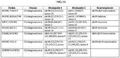


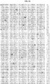
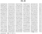


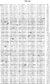


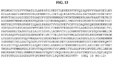
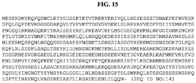
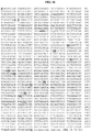
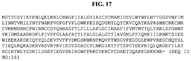
Classifications
-
- A—HUMAN NECESSITIES
- A61—MEDICAL OR VETERINARY SCIENCE; HYGIENE
- A61K—PREPARATIONS FOR MEDICAL, DENTAL OR TOILETRY PURPOSES
- A61K31/00—Medicinal preparations containing organic active ingredients
- A61K31/33—Heterocyclic compounds
- A61K31/395—Heterocyclic compounds having nitrogen as a ring hetero atom, e.g. guanethidine or rifamycins
- A61K31/495—Heterocyclic compounds having nitrogen as a ring hetero atom, e.g. guanethidine or rifamycins having six-membered rings with two or more nitrogen atoms as the only ring heteroatoms, e.g. piperazine or tetrazines
- A61K31/50—Pyridazines; Hydrogenated pyridazines
- A61K31/5025—Pyridazines; Hydrogenated pyridazines ortho- or peri-condensed with heterocyclic ring systems
-
- A—HUMAN NECESSITIES
- A61—MEDICAL OR VETERINARY SCIENCE; HYGIENE
- A61K—PREPARATIONS FOR MEDICAL, DENTAL OR TOILETRY PURPOSES
- A61K31/00—Medicinal preparations containing organic active ingredients
- A61K31/33—Heterocyclic compounds
- A61K31/395—Heterocyclic compounds having nitrogen as a ring hetero atom, e.g. guanethidine or rifamycins
- A61K31/40—Heterocyclic compounds having nitrogen as a ring hetero atom, e.g. guanethidine or rifamycins having five-membered rings with one nitrogen as the only ring hetero atom, e.g. sulpiride, succinimide, tolmetin, buflomedil
- A61K31/403—Heterocyclic compounds having nitrogen as a ring hetero atom, e.g. guanethidine or rifamycins having five-membered rings with one nitrogen as the only ring hetero atom, e.g. sulpiride, succinimide, tolmetin, buflomedil condensed with carbocyclic rings, e.g. carbazole
- A61K31/404—Indoles, e.g. pindolol
-
- A—HUMAN NECESSITIES
- A61—MEDICAL OR VETERINARY SCIENCE; HYGIENE
- A61K—PREPARATIONS FOR MEDICAL, DENTAL OR TOILETRY PURPOSES
- A61K31/00—Medicinal preparations containing organic active ingredients
- A61K31/33—Heterocyclic compounds
- A61K31/395—Heterocyclic compounds having nitrogen as a ring hetero atom, e.g. guanethidine or rifamycins
- A61K31/435—Heterocyclic compounds having nitrogen as a ring hetero atom, e.g. guanethidine or rifamycins having six-membered rings with one nitrogen as the only ring hetero atom
- A61K31/44—Non condensed pyridines; Hydrogenated derivatives thereof
- A61K31/4412—Non condensed pyridines; Hydrogenated derivatives thereof having oxo groups directly attached to the heterocyclic ring
-
- A—HUMAN NECESSITIES
- A61—MEDICAL OR VETERINARY SCIENCE; HYGIENE
- A61K—PREPARATIONS FOR MEDICAL, DENTAL OR TOILETRY PURPOSES
- A61K31/00—Medicinal preparations containing organic active ingredients
- A61K31/33—Heterocyclic compounds
- A61K31/395—Heterocyclic compounds having nitrogen as a ring hetero atom, e.g. guanethidine or rifamycins
- A61K31/435—Heterocyclic compounds having nitrogen as a ring hetero atom, e.g. guanethidine or rifamycins having six-membered rings with one nitrogen as the only ring hetero atom
- A61K31/44—Non condensed pyridines; Hydrogenated derivatives thereof
- A61K31/4427—Non condensed pyridines; Hydrogenated derivatives thereof containing further heterocyclic ring systems
- A61K31/4439—Non condensed pyridines; Hydrogenated derivatives thereof containing further heterocyclic ring systems containing a five-membered ring with nitrogen as a ring hetero atom, e.g. omeprazole
-
- A—HUMAN NECESSITIES
- A61—MEDICAL OR VETERINARY SCIENCE; HYGIENE
- A61K—PREPARATIONS FOR MEDICAL, DENTAL OR TOILETRY PURPOSES
- A61K31/00—Medicinal preparations containing organic active ingredients
- A61K31/33—Heterocyclic compounds
- A61K31/395—Heterocyclic compounds having nitrogen as a ring hetero atom, e.g. guanethidine or rifamycins
- A61K31/435—Heterocyclic compounds having nitrogen as a ring hetero atom, e.g. guanethidine or rifamycins having six-membered rings with one nitrogen as the only ring hetero atom
- A61K31/47—Quinolines; Isoquinolines
-
- A—HUMAN NECESSITIES
- A61—MEDICAL OR VETERINARY SCIENCE; HYGIENE
- A61K—PREPARATIONS FOR MEDICAL, DENTAL OR TOILETRY PURPOSES
- A61K31/00—Medicinal preparations containing organic active ingredients
- A61K31/33—Heterocyclic compounds
- A61K31/395—Heterocyclic compounds having nitrogen as a ring hetero atom, e.g. guanethidine or rifamycins
- A61K31/495—Heterocyclic compounds having nitrogen as a ring hetero atom, e.g. guanethidine or rifamycins having six-membered rings with two or more nitrogen atoms as the only ring heteroatoms, e.g. piperazine or tetrazines
- A61K31/496—Non-condensed piperazines containing further heterocyclic rings, e.g. rifampin, thiothixene
-
- A—HUMAN NECESSITIES
- A61—MEDICAL OR VETERINARY SCIENCE; HYGIENE
- A61K—PREPARATIONS FOR MEDICAL, DENTAL OR TOILETRY PURPOSES
- A61K31/00—Medicinal preparations containing organic active ingredients
- A61K31/33—Heterocyclic compounds
- A61K31/395—Heterocyclic compounds having nitrogen as a ring hetero atom, e.g. guanethidine or rifamycins
- A61K31/495—Heterocyclic compounds having nitrogen as a ring hetero atom, e.g. guanethidine or rifamycins having six-membered rings with two or more nitrogen atoms as the only ring heteroatoms, e.g. piperazine or tetrazines
- A61K31/498—Pyrazines or piperazines ortho- and peri-condensed with carbocyclic ring systems, e.g. quinoxaline, phenazine
-
- A—HUMAN NECESSITIES
- A61—MEDICAL OR VETERINARY SCIENCE; HYGIENE
- A61K—PREPARATIONS FOR MEDICAL, DENTAL OR TOILETRY PURPOSES
- A61K31/00—Medicinal preparations containing organic active ingredients
- A61K31/33—Heterocyclic compounds
- A61K31/395—Heterocyclic compounds having nitrogen as a ring hetero atom, e.g. guanethidine or rifamycins
- A61K31/495—Heterocyclic compounds having nitrogen as a ring hetero atom, e.g. guanethidine or rifamycins having six-membered rings with two or more nitrogen atoms as the only ring heteroatoms, e.g. piperazine or tetrazines
- A61K31/505—Pyrimidines; Hydrogenated pyrimidines, e.g. trimethoprim
- A61K31/506—Pyrimidines; Hydrogenated pyrimidines, e.g. trimethoprim not condensed and containing further heterocyclic rings
-
- A—HUMAN NECESSITIES
- A61—MEDICAL OR VETERINARY SCIENCE; HYGIENE
- A61K—PREPARATIONS FOR MEDICAL, DENTAL OR TOILETRY PURPOSES
- A61K31/00—Medicinal preparations containing organic active ingredients
- A61K31/33—Heterocyclic compounds
- A61K31/395—Heterocyclic compounds having nitrogen as a ring hetero atom, e.g. guanethidine or rifamycins
- A61K31/495—Heterocyclic compounds having nitrogen as a ring hetero atom, e.g. guanethidine or rifamycins having six-membered rings with two or more nitrogen atoms as the only ring heteroatoms, e.g. piperazine or tetrazines
- A61K31/505—Pyrimidines; Hydrogenated pyrimidines, e.g. trimethoprim
- A61K31/517—Pyrimidines; Hydrogenated pyrimidines, e.g. trimethoprim ortho- or peri-condensed with carbocyclic ring systems, e.g. quinazoline, perimidine
-
- A—HUMAN NECESSITIES
- A61—MEDICAL OR VETERINARY SCIENCE; HYGIENE
- A61K—PREPARATIONS FOR MEDICAL, DENTAL OR TOILETRY PURPOSES
- A61K31/00—Medicinal preparations containing organic active ingredients
- A61K31/33—Heterocyclic compounds
- A61K31/395—Heterocyclic compounds having nitrogen as a ring hetero atom, e.g. guanethidine or rifamycins
- A61K31/495—Heterocyclic compounds having nitrogen as a ring hetero atom, e.g. guanethidine or rifamycins having six-membered rings with two or more nitrogen atoms as the only ring heteroatoms, e.g. piperazine or tetrazines
- A61K31/505—Pyrimidines; Hydrogenated pyrimidines, e.g. trimethoprim
- A61K31/519—Pyrimidines; Hydrogenated pyrimidines, e.g. trimethoprim ortho- or peri-condensed with heterocyclic rings
-
- A—HUMAN NECESSITIES
- A61—MEDICAL OR VETERINARY SCIENCE; HYGIENE
- A61K—PREPARATIONS FOR MEDICAL, DENTAL OR TOILETRY PURPOSES
- A61K31/00—Medicinal preparations containing organic active ingredients
- A61K31/33—Heterocyclic compounds
- A61K31/395—Heterocyclic compounds having nitrogen as a ring hetero atom, e.g. guanethidine or rifamycins
- A61K31/53—Heterocyclic compounds having nitrogen as a ring hetero atom, e.g. guanethidine or rifamycins having six-membered rings with three nitrogens as the only ring hetero atoms, e.g. chlorazanil, melamine
-
- A—HUMAN NECESSITIES
- A61—MEDICAL OR VETERINARY SCIENCE; HYGIENE
- A61K—PREPARATIONS FOR MEDICAL, DENTAL OR TOILETRY PURPOSES
- A61K31/00—Medicinal preparations containing organic active ingredients
- A61K31/33—Heterocyclic compounds
- A61K31/395—Heterocyclic compounds having nitrogen as a ring hetero atom, e.g. guanethidine or rifamycins
- A61K31/535—Heterocyclic compounds having nitrogen as a ring hetero atom, e.g. guanethidine or rifamycins having six-membered rings with at least one nitrogen and one oxygen as the ring hetero atoms, e.g. 1,2-oxazines
- A61K31/5375—1,4-Oxazines, e.g. morpholine
- A61K31/5383—1,4-Oxazines, e.g. morpholine ortho- or peri-condensed with heterocyclic ring systems
-
- A—HUMAN NECESSITIES
- A61—MEDICAL OR VETERINARY SCIENCE; HYGIENE
- A61K—PREPARATIONS FOR MEDICAL, DENTAL OR TOILETRY PURPOSES
- A61K31/00—Medicinal preparations containing organic active ingredients
- A61K31/33—Heterocyclic compounds
- A61K31/395—Heterocyclic compounds having nitrogen as a ring hetero atom, e.g. guanethidine or rifamycins
- A61K31/55—Heterocyclic compounds having nitrogen as a ring hetero atom, e.g. guanethidine or rifamycins having seven-membered rings, e.g. azelastine, pentylenetetrazole
- A61K31/553—Heterocyclic compounds having nitrogen as a ring hetero atom, e.g. guanethidine or rifamycins having seven-membered rings, e.g. azelastine, pentylenetetrazole having at least one nitrogen and one oxygen as ring hetero atoms, e.g. loxapine, staurosporine
-
- A—HUMAN NECESSITIES
- A61—MEDICAL OR VETERINARY SCIENCE; HYGIENE
- A61K—PREPARATIONS FOR MEDICAL, DENTAL OR TOILETRY PURPOSES
- A61K45/00—Medicinal preparations containing active ingredients not provided for in groups A61K31/00 - A61K41/00
- A61K45/06—Mixtures of active ingredients without chemical characterisation, e.g. antiphlogistics and cardiaca
-
- A—HUMAN NECESSITIES
- A61—MEDICAL OR VETERINARY SCIENCE; HYGIENE
- A61P—SPECIFIC THERAPEUTIC ACTIVITY OF CHEMICAL COMPOUNDS OR MEDICINAL PREPARATIONS
- A61P35/00—Antineoplastic agents
-
- C—CHEMISTRY; METALLURGY
- C07—ORGANIC CHEMISTRY
- C07K—PEPTIDES
- C07K14/00—Peptides having more than 20 amino acids; Gastrins; Somatostatins; Melanotropins; Derivatives thereof
- C07K14/435—Peptides having more than 20 amino acids; Gastrins; Somatostatins; Melanotropins; Derivatives thereof from animals; from humans
- C07K14/705—Receptors; Cell surface antigens; Cell surface determinants
- C07K14/71—Receptors; Cell surface antigens; Cell surface determinants for growth factors; for growth regulators
-
- C—CHEMISTRY; METALLURGY
- C07—ORGANIC CHEMISTRY
- C07K—PEPTIDES
- C07K16/00—Immunoglobulins [IGs], e.g. monoclonal or polyclonal antibodies
- C07K16/40—Immunoglobulins [IGs], e.g. monoclonal or polyclonal antibodies against enzymes
-
- C—CHEMISTRY; METALLURGY
- C12—BIOCHEMISTRY; BEER; SPIRITS; WINE; VINEGAR; MICROBIOLOGY; ENZYMOLOGY; MUTATION OR GENETIC ENGINEERING
- C12N—MICROORGANISMS OR ENZYMES; COMPOSITIONS THEREOF; PROPAGATING, PRESERVING, OR MAINTAINING MICROORGANISMS; MUTATION OR GENETIC ENGINEERING; CULTURE MEDIA
- C12N15/00—Mutation or genetic engineering; DNA or RNA concerning genetic engineering, vectors, e.g. plasmids, or their isolation, preparation or purification; Use of hosts therefor
- C12N15/09—Recombinant DNA-technology
- C12N15/11—DNA or RNA fragments; Modified forms thereof; Non-coding nucleic acids having a biological activity
- C12N15/113—Non-coding nucleic acids modulating the expression of genes, e.g. antisense oligonucleotides; Antisense DNA or RNA; Triplex- forming oligonucleotides; Catalytic nucleic acids, e.g. ribozymes; Nucleic acids used in co-suppression or gene silencing
- C12N15/1137—Non-coding nucleic acids modulating the expression of genes, e.g. antisense oligonucleotides; Antisense DNA or RNA; Triplex- forming oligonucleotides; Catalytic nucleic acids, e.g. ribozymes; Nucleic acids used in co-suppression or gene silencing against enzymes
-
- C—CHEMISTRY; METALLURGY
- C12—BIOCHEMISTRY; BEER; SPIRITS; WINE; VINEGAR; MICROBIOLOGY; ENZYMOLOGY; MUTATION OR GENETIC ENGINEERING
- C12N—MICROORGANISMS OR ENZYMES; COMPOSITIONS THEREOF; PROPAGATING, PRESERVING, OR MAINTAINING MICROORGANISMS; MUTATION OR GENETIC ENGINEERING; CULTURE MEDIA
- C12N9/00—Enzymes; Proenzymes; Compositions thereof; Processes for preparing, activating, inhibiting, separating or purifying enzymes
- C12N9/10—Transferases (2.)
- C12N9/12—Transferases (2.) transferring phosphorus containing groups, e.g. kinases (2.7)
-
- C—CHEMISTRY; METALLURGY
- C12—BIOCHEMISTRY; BEER; SPIRITS; WINE; VINEGAR; MICROBIOLOGY; ENZYMOLOGY; MUTATION OR GENETIC ENGINEERING
- C12Q—MEASURING OR TESTING PROCESSES INVOLVING ENZYMES, NUCLEIC ACIDS OR MICROORGANISMS; COMPOSITIONS OR TEST PAPERS THEREFOR; PROCESSES OF PREPARING SUCH COMPOSITIONS; CONDITION-RESPONSIVE CONTROL IN MICROBIOLOGICAL OR ENZYMOLOGICAL PROCESSES
- C12Q1/00—Measuring or testing processes involving enzymes, nucleic acids or microorganisms; Compositions therefor; Processes of preparing such compositions
- C12Q1/68—Measuring or testing processes involving enzymes, nucleic acids or microorganisms; Compositions therefor; Processes of preparing such compositions involving nucleic acids
- C12Q1/6876—Nucleic acid products used in the analysis of nucleic acids, e.g. primers or probes
- C12Q1/6883—Nucleic acid products used in the analysis of nucleic acids, e.g. primers or probes for diseases caused by alterations of genetic material
- C12Q1/6886—Nucleic acid products used in the analysis of nucleic acids, e.g. primers or probes for diseases caused by alterations of genetic material for cancer
-
- C—CHEMISTRY; METALLURGY
- C12—BIOCHEMISTRY; BEER; SPIRITS; WINE; VINEGAR; MICROBIOLOGY; ENZYMOLOGY; MUTATION OR GENETIC ENGINEERING
- C12Y—ENZYMES
- C12Y207/00—Transferases transferring phosphorus-containing groups (2.7)
- C12Y207/10—Protein-tyrosine kinases (2.7.10)
- C12Y207/10001—Receptor protein-tyrosine kinase (2.7.10.1)
-
- G—PHYSICS
- G01—MEASURING; TESTING
- G01N—INVESTIGATING OR ANALYSING MATERIALS BY DETERMINING THEIR CHEMICAL OR PHYSICAL PROPERTIES
- G01N33/00—Investigating or analysing materials by specific methods not covered by groups G01N1/00 - G01N31/00
- G01N33/48—Biological material, e.g. blood, urine; Haemocytometers
- G01N33/50—Chemical analysis of biological material, e.g. blood, urine; Testing involving biospecific ligand binding methods; Immunological testing
- G01N33/53—Immunoassay; Biospecific binding assay; Materials therefor
- G01N33/573—Immunoassay; Biospecific binding assay; Materials therefor for enzymes or isoenzymes
-
- C—CHEMISTRY; METALLURGY
- C07—ORGANIC CHEMISTRY
- C07K—PEPTIDES
- C07K2319/00—Fusion polypeptide
-
- C—CHEMISTRY; METALLURGY
- C12—BIOCHEMISTRY; BEER; SPIRITS; WINE; VINEGAR; MICROBIOLOGY; ENZYMOLOGY; MUTATION OR GENETIC ENGINEERING
- C12N—MICROORGANISMS OR ENZYMES; COMPOSITIONS THEREOF; PROPAGATING, PRESERVING, OR MAINTAINING MICROORGANISMS; MUTATION OR GENETIC ENGINEERING; CULTURE MEDIA
- C12N2310/00—Structure or type of the nucleic acid
- C12N2310/10—Type of nucleic acid
- C12N2310/11—Antisense
-
- C—CHEMISTRY; METALLURGY
- C12—BIOCHEMISTRY; BEER; SPIRITS; WINE; VINEGAR; MICROBIOLOGY; ENZYMOLOGY; MUTATION OR GENETIC ENGINEERING
- C12N—MICROORGANISMS OR ENZYMES; COMPOSITIONS THEREOF; PROPAGATING, PRESERVING, OR MAINTAINING MICROORGANISMS; MUTATION OR GENETIC ENGINEERING; CULTURE MEDIA
- C12N2310/00—Structure or type of the nucleic acid
- C12N2310/10—Type of nucleic acid
- C12N2310/12—Type of nucleic acid catalytic nucleic acids, e.g. ribozymes
-
- C—CHEMISTRY; METALLURGY
- C12—BIOCHEMISTRY; BEER; SPIRITS; WINE; VINEGAR; MICROBIOLOGY; ENZYMOLOGY; MUTATION OR GENETIC ENGINEERING
- C12N—MICROORGANISMS OR ENZYMES; COMPOSITIONS THEREOF; PROPAGATING, PRESERVING, OR MAINTAINING MICROORGANISMS; MUTATION OR GENETIC ENGINEERING; CULTURE MEDIA
- C12N2310/00—Structure or type of the nucleic acid
- C12N2310/10—Type of nucleic acid
- C12N2310/14—Type of nucleic acid interfering N.A.
-
- C—CHEMISTRY; METALLURGY
- C12—BIOCHEMISTRY; BEER; SPIRITS; WINE; VINEGAR; MICROBIOLOGY; ENZYMOLOGY; MUTATION OR GENETIC ENGINEERING
- C12Q—MEASURING OR TESTING PROCESSES INVOLVING ENZYMES, NUCLEIC ACIDS OR MICROORGANISMS; COMPOSITIONS OR TEST PAPERS THEREFOR; PROCESSES OF PREPARING SUCH COMPOSITIONS; CONDITION-RESPONSIVE CONTROL IN MICROBIOLOGICAL OR ENZYMOLOGICAL PROCESSES
- C12Q2600/00—Oligonucleotides characterized by their use
- C12Q2600/156—Polymorphic or mutational markers
-
- C—CHEMISTRY; METALLURGY
- C12—BIOCHEMISTRY; BEER; SPIRITS; WINE; VINEGAR; MICROBIOLOGY; ENZYMOLOGY; MUTATION OR GENETIC ENGINEERING
- C12Q—MEASURING OR TESTING PROCESSES INVOLVING ENZYMES, NUCLEIC ACIDS OR MICROORGANISMS; COMPOSITIONS OR TEST PAPERS THEREFOR; PROCESSES OF PREPARING SUCH COMPOSITIONS; CONDITION-RESPONSIVE CONTROL IN MICROBIOLOGICAL OR ENZYMOLOGICAL PROCESSES
- C12Q2600/00—Oligonucleotides characterized by their use
- C12Q2600/158—Expression markers
-
- G—PHYSICS
- G01—MEASURING; TESTING
- G01N—INVESTIGATING OR ANALYSING MATERIALS BY DETERMINING THEIR CHEMICAL OR PHYSICAL PROPERTIES
- G01N2333/00—Assays involving biological materials from specific organisms or of a specific nature
- G01N2333/90—Enzymes; Proenzymes
- G01N2333/91—Transferases (2.)
- G01N2333/912—Transferases (2.) transferring phosphorus containing groups, e.g. kinases (2.7)
-
- G—PHYSICS
- G01—MEASURING; TESTING
- G01N—INVESTIGATING OR ANALYSING MATERIALS BY DETERMINING THEIR CHEMICAL OR PHYSICAL PROPERTIES
- G01N2500/00—Screening for compounds of potential therapeutic value
- G01N2500/04—Screening involving studying the effect of compounds C directly on molecule A (e.g. C are potential ligands for a receptor A, or potential substrates for an enzyme A)
Definitions
- Cancer represents the phenotypic end-point of multiple genetic lesions that endow cells with a full range of biological properties required for tumorigenesis. Indeed, a hallmark genomic feature of many cancers is the presence of numerous complex chromosome structural aberrations, including translocations, intra-chromosomal inversions, point mutations, deletions, gene copy number changes, gene expression level changes, and germline mutations, among others.
- Cholangiocarcinoma is a cancer that includes mutated epithelial cells that originate in the bile ducts. Cholangiocarcinoma is a relatively rare neoplasm that is classified as an adenocarcinoma (a cancer that forms glands or secretes significant amounts of mucins). It has an annual incidence rate of about 1-2 cases per 100,000 in the Western world, but rates of cholangiocarcinoma have been rising worldwide over the past several decades ( Landis S. et al. (1998) CA Cancer J Clin 48 (1): 6-29 ; Patel T (2002) BMC Cancer 2: 10. doi:10.1186/1471-2407-2-10 ).
- ICC intrahepatic cholangiocarcinoma
- HCC hepatocellular carcinoma
- ICC has a routine histologic appearance of an adenocarcinoma
- diagnosis of ICC on a liver biopsy requires an immunohistochemical (IHC) study of the tumor and a thorough clinical workup including imaging studies to rule out a metastatic adenocarcinoma to the liver.
- IHC immunohistochemical
- ICC ulcerative colitis .
- ICC ulcerative colitis .
- Primary sclerosing cholangitis parasitic biliary infection
- polycystic disease of the liver congenital intrahepatic bile duct dilatation (Caroli's Disease)
- congenital hepatic fibrosis congenital hepatic fibrosis
- choledochal cysts chronic Hepatitis C infection is an established cause of ICC with some studies describing a more than 300 fold increase in ICC incidence in patients with longstanding Hepatitis C infections.
- ICC has also been associated with cigarette smoking, alcohol consumption and exposure to a variety of toxins and chemical carcinogens.
- ICC ulcerative colitis .
- the onset of symptoms of ICC are often vague, typically arise late in the course of the disease and include abdominal pain, anorexia and palpable abdominal mass lesions.
- the median survival for ICC is less than 6 months for inoperable tumors and only 20 to 40% for patients who undergo surgery and achieve clear margins.
- Cholangiocarcinoma is considered to be an incurable and rapidly lethal malignancy, unless both the primary tumor and any metastases can be fully resected (removed surgically). No potentially curative treatment exists at this time except surgery; however, most patients have advanced stage disease at presentation and are inoperable at the time of diagnosis. Cholangiocarcinoma has near-100% fatality due to attendant liver complications from the damage to the organ. Patients with cholangiocarcinoma are generally managed with chemotherapy, radiation therapy, and other palliative care measures.
- the invention provides a therapeutic agent that antagonizes or inhibits an FGFR2 gene product, for use in a method of treating cholangiocarcinoma in a subect,
- the disclosure is based, at least in part, on the discovery, in cholangiocarcinomas, of novel rearrangement events that give rise to alterations in a fibroblast growth factor receptor 2 (FGFR2) gene or a neurotrophic tyrosine receptor kinase (NTRK1) gene.
- the alteration is chosen from a translocation, a deletion, an inversion, a rearrangement, or an amplification of, an FGFR2 gene or the NTRK gene.
- the alteration can be chosen from an alteration described in Table 1 and FIGs. 1A-1C .
- the alteration includes a fragment of an FGFR2 gene or the NTRK1 gene, e.g., as exemplified in Table 1, FIGs. 1A-1C and FIGs. 2-17 .
- the disclosure provides new insights into the treatment of these cancers, such as cholangiocarcinomas. Therefore,described herein are methods for treating a cholangiocarcinoma carcinoma, including intrahepatic cholangiocarcinoma (ICC) and extrahepatic cholangiocarcinoma, as well as novel FGFR2 and NTRK1 molecules (e.g., fusion molecules); methods and reagents for identifying, assessing or detecting an alteration in an FGFR2 and/or NTRK1.
- ICC intrahepatic cholangiocarcinoma
- novel FGFR2 and NTRK1 molecules e.g., fusion molecules
- the disclosure features a method of treating a subject having a cholangiocarcinoma.
- the method includes administering to the subject an effective amount of an agent (e.g., a therapeutic agent) that targets, antagonizes or inhibits an FGFR2 or NTRK1 (e.g., an FGFR2 or NTRK1 gene product, e.g., an FGFR2 or NTRK1 protein), thereby treating the subject.
- an agent e.g., a therapeutic agent
- NTRK1 e.g., an FGFR2 or NTRK1 gene product, e.g., an FGFR2 or NTRK1 protein
- the disclosure features, a method of treating a subject having a cholangiocarcinoma.
- the method includes administering to the subject an effective amount of a kinase inhibitor (e.g., a tyrosine kinase inhibitor), thereby treating the subject.
- a kinase inhibitor e.g., a tyrosine kinase inhibitor
- the method further includes acquiring knowledge of one or both of:
- the method further includes identifying the subject, or a cancer or tumor sample from the subject, as having one or both of:
- the presence of the FGFR2 or NTRK1 alteration, or both, in the subject is indicative that the subject is likely to respond to the agent.
- the agent is administered responsive to a determination of the presence of the FGFR2 or NTRK1 alteration, or both, in the subject, or the cancer or tumor sample from the subject.
- the cholangiocarcinima comprises one or more mutated cells that originate in the bile duct.
- the cholangiocarcinoma is chosen from an intrahepatic cholangiocarcinoma or an extrahepatic cholangiocarcinoma.
- the cholangiocarcinoma comprises, or is identified as having, an alteration that is chosen from a translocation, a deletion, an inversion, a rearrangement, or an amplification of, an FGFR2 gene or the NTRK gene.
- the cholangiocarcinoma comprises, or is identified as having, an alteration chosen from an alteration described in Table 1 or FIGs. 1A-1C .
- the cholangiocarcinoma comprises, or is identified as having, an alteration includes a fragment of an FGFR2 gene or the NTRK1 gene, e.g., as exemplified in Table 1, FIGs. 1A-1C and FIGs. 2-17 .
- the cholangiocarcinoma comprises, or is identified as having, a fusion molecule of FGFR2; e.g., a fusion molecule chosen from FGFR2-TACC3, FGFR2-KIAA1598, BICC1-FGFR2, FGFR2-BICC1, PARK2-FGFR2, FGFR2-NOL4, or ZDHHC6-FGFR2 as described, e.g., in Table 1, FIGs.
- the cholangiocarcinoma comprises, or is identified as having, a rearrangement or an amplification of FGFR2 as described, e.g., in Table 1, FIGs. 1A-1C and FIGs. 2-17 .
- the alteration in FGFR2 results in upregulation, increased activity (e.g ., increased transformative or oncogenic activity, kinase activity and/or dimerization), and/or increased level of an FGFR2 gene product (e.g., an FGFR2 protein), compared to a wildtype activity of FGFR2.
- increased activity e.g ., increased transformative or oncogenic activity, kinase activity and/or dimerization
- an FGFR2 gene product e.g., an FGFR2 protein
- the subject has an alteration in FGFR2 or NTRK1, or both, e.g., the subject has a cholangiocarcinoma comprising an alteration in FGFR2 or NTRK1, or both, e.g., as described herein.
- the subject is identified, or has been previously identified, as having a cholangiocarcinoma (e.g., an intrahepatic cholangiocarcinoma (ICC) or an extrahepatic cholangiocarcinoma) comprising an alteration in FGFR2 or NTRK1, or both, e.g., as described herein.
- a cholangiocarcinoma e.g., an intrahepatic cholangiocarcinoma (ICC) or an extrahepatic cholangiocarcinoma
- the subject has, or is identified as having, an alteration that is chosen from a translocation, a deletion, an inversion, a rearrangement, or an amplification of, an FGFR2 gene or the NTRK gene.
- the subject has, or is identified as having, an alteration chosen from an alteration described in Table 1 or FIGs. 1A-1C .
- the subject has, or is identified as having, an alteration includes a fragment of an FGFR2 gene or the NTRK1 gene, e.g., as exemplified in Table 1, FIGs. 1A-1C and FIGs. 2-17 .
- the subject has, or is identified as having, a fusion molecule of FGFR2; e.g., a fusion molecule chosen from FGFR2-TACC3, FGFR2-KIAA1598, BICC1-FGFR2, FGFR2-BICC1, PARK2-FGFR2, FGFR2-NOL4, or ZDHHC6-FGFR2 as described, e.g., in Table 1, FIGs. 1A-1C and FIGs. 2-17 .
- the subject has, or is identified as having, a rearrangement or an amplification of FGFR2 as described, e.g., in Table 1, FIGs. 1A-1C and FIGs. 2-17 .
- the subject is a human.
- the subject has, or is at risk of having a cholangiocarcinoma (e.g ., a cholangiocarcinoma as described herein) at any stage of disease, e.g ., Stage I, II, IIIA-IIIC or IV of intrahepatic cholangiocarcinoma; Stage 0, IA-IB, IIA-IIB, III or IV of extrahepatic cholangiocarcinoma; or a metastatic cancer.
- the subject is a cancer patient, e.g., a patient having a cholangiocarcinoma as described herein.
- the subject is undergoing or has undergone treatment with a different (e.g., non- FGFR2 or non-NTRK1) therapeutic agent or therapeutic modality.
- the non-FGFR2 or non-NTRK1 therapeutic agent or therapeutic modality is a chemotherapy, immunotherapy, or a surgical procedure.
- the non-FGFR2 or non-NTRK1 therapeutic agent or therapeutic modality comprises one or more (or all) of: a surgical procedure, flurouracil (e.g ., 5-FU, Adrucil, Efudex), doxorubicin (Adriamycin, Rubex), gemcitabine ( e.g ., Gemzar) and/or cisplatin (Platinol).
- the different therapeutic agent or therapeutic modality is discontinued.
- the subject has been identified as being likely or unlikely to respond to the different therapeutic agent or therapeutic modality.
- the subject has participated previously in a clinical trial, e.g., a clinical trial for a different (e.g ., non-FGFR2 or non-NTRK1) therapeutic agent or therapeutic modality.
- the subject is a cancer patient who has participated in a clinical trial, e.g., a clinical trial for a different ( e.g ., non-FGFR2 or non-NTRK1) therapeutic agent or therapeutic modality.
- the agent used in the methods targets and/or inhibits FGFR2 or NTRK1 (e.g., a FGFR2 or NTRK1 gene or gene product as described herein). In one instance, the agent binds and inhibits FGFR2 or NTRK1. In one embodiment, the agent is a reversible or an irreversible FGFR2 inhibitor. In certain embodiments, the agent is a pan-FGFR2 inhibitor.
- the agent is an antibody molecule, e.g., an anti- FGFR2 or NTRK1 antibody molecule (e.g., a monoclonal or a bispecific antibody), or a conjugate thereof (e.g., an antibody to FGFR2 or NTRK1 conjugated to a cytotoxic agent (e.g ., mertansine DM1)).
- an antibody molecule e.g., an anti- FGFR2 or NTRK1 antibody molecule (e.g., a monoclonal or a bispecific antibody), or a conjugate thereof (e.g., an antibody to FGFR2 or NTRK1 conjugated to a cytotoxic agent (e.g ., mertansine DM1)).
- a cytotoxic agent e.g ., mertansine DM1
- the agent is a kinase inhibitor.
- the kinase inhibitor is chosen from: a multi-specific kinase inhibitor, an FGFR2 inhibitor (e.g., a pan-FGFR2 inhibitor), an NTRK1 inhibitor, and/or a small molecule inhibitor that is selective for FGFR2 or NTRK1; and/or a FGFR2 or NTRK1 cellular immunotherapy.
- the therapeutic agent is chosen from a kinase inhibitor; a multi-specific kinase inhibitor; an FGF receptor inhibitor (e.g ., a pan FGFR2 inhibitor); an antibody molecule (e.g., a monoclonal antibody) against FGFR2; and/or a small molecule ( e.g ., kinase) inhibitor that is selective for FGFR2 or NTRK1.
- a kinase inhibitor e.g ., a multi-specific kinase inhibitor
- an FGF receptor inhibitor e.g ., a pan FGFR2 inhibitor
- an antibody molecule e.g., a monoclonal antibody against FGFR2
- a small molecule e.g ., kinase
- the therapeutic agent is selected from antisense molecules, ribozymes, RNAi, triple helix molecules that hybridize to a nucleic acid encoding the fusion, or a transcription regulatory region that blocks or reduces mRNA expression of FGFR2 or NTRK1.
- the kinase inhibitor is chosen from: a kinase inhibitor; a multi-specific kinase inhibitor; an FGF receptor inhibitor (e.g., a pan FGFR2 inhibitor); and/or a kinase inhibitor that is selective for FGFR2 or NTRK1.
- the therapeutic agent is chosen from: Regorafenib; Ponatinib; AZD-2171 (Cediranib); AZD-4547; BGJ398; BIBF1120; Brivanib; Dovitinib; ENMD-2076; JNJ42756493; Masitinib; Lenvatinib; LY2874455; Pazopanib; PD-173955; R406; PD173074; Danusertib; Dovitinib Dilactic Acid; TSU-68; Tyrphostin AG 1296; MK-2461; Brivanib Alaninate; Lestaurtinib; PHA-848125; K252a; AZ-23; and/or Oxindole-3.
- the therapeutic agent is chosen from Regorafenib or Ponatinib.
- the method includes acquiring knowledge of the presence of an alteration, e.g ., fusion, from Table 1, FIGs. 1A-1C and FIGs. 2-17 in said subject.
- an alteration e.g ., fusion
- the therapeutic agent is administered responsive to the determination of presence of the alteration, e.g. , fusion, in a tumor sample from said subject.
- the determination of the presence of the alteration comprises sequencing.
- the subject is undergoing or has undergone treatment with a different therapeutic agent or therapeutic modality, e.g., a non-FGFR2 or non-NTRK1 therapeutic agent or therapeutic modality.
- a different therapeutic agent or therapeutic modality e.g., a non-FGFR2 or non-NTRK1 therapeutic agent or therapeutic modality.
- the different therapeutic agent or therapeutic modality is discontinued.
- the different therapeutic agent or therapeutic modality is a chemotherapy or a surgical procedure.
- the non-FGFR2 or non-NTRK1 therapeutic agent or therapeutic modality comprises one or more (or all) of: a surgical procedure, flurouracil (e.g., 5-FU, Adrucil, Efudex), doxorubicin (Adriamycin, Rubex), gemcitabine (e.g ., Gemzar) and/or cisplatin (Platinol).
- the disclosure features, a method of determining the presence of an alteration, e.g., a fusion, disclosed herein in cholangiocarcinoma sample, comprising: directly acquiring knowledge that an alteration, e.g., a fusion nucleic acid molecule, of Table 1, FIGs. 1A-1C and FIGs. 2-17 is present in a sample from a subject.
- an alteration e.g., a fusion nucleic acid molecule, of Table 1, FIGs. 1A-1C and FIGs. 2-17 is present in a sample from a subject.
- the acquiring step comprises sequencing.
- the method further comprises administering a kinase inhibitor to the subject responsive to the determination of the presence of the alteration, e.g., the fusion, in the sample from the subject.
- the disclosure also provides, methods of: identifying, assessing or detecting an alteration, e.g., fusion, of an FGFR2 or an NTRK1, e.g., that arises in a cholangiocarcinoma.
- an alteration e.g., fusion
- exemplary alteration, e.g ., fusions include those summarized in Table 1, FIGs. 1A-1C and FIGs. 2-17 , including a fusion of FGFR2 (e.g., an FGFR2 fusion molecule (e.g., a gene product or fragment thereof)) to a partner from Table 1, FIGs. 1A-1C and FIGs.
- NTRK1 e.g., an NTRK1 fusion molecule (e.g., a gene product or fragment thereof)
- the FGFR2 or NTRK1 is fused to a second gene, or a fragment thereof, e.g., as described in Table 1, FIGs. 1A-1C and FIGs. 2-17 .
- the fusion molecule is chosen from FGFR2-TACC3, FGFR2-KIAA1598, BICC1-FGFR2, FGFR2-BICC1, PARK2-FGFR2, FGFR2-NOL4, ZDHHC6-FGFR2, or RABGAP1L-NTRK1, e.g., as described, e.g., in Table 1, FIGs. 1A-1C and FIGs. 2-17 .
- fusion molecules include isolated fusions nucleic acid molecules, nucleic acid constructs, host cells containing the nucleic acid molecules; purified fusion polypeptides and binding agents; detection reagents (e.g., probes, primers, antibodies, kits, capable, e.g., of specific detection of a fusion nucleic acid or protein); screening assays for identifying molecules that interact with, e.g., inhibit, fusions, e.g ., novel kinase inhibitors or binders of FGFR2 or NTRK1.
- detection reagents e.g., probes, primers, antibodies, kits, capable, e.g., of specific detection of a fusion nucleic acid or protein
- the disclosure features an isolated nucleic acid molecule, or an isolated preparation of nucleic acid molecules, that includes a genetic alteration disclosed herein.
- nucleic acid molecules or preparations thereof can include a genetic alteration described herein or can be used to detect, e.g., sequence, a genetic alteration disclosed herein.
- the alteration of FGFR2 or NRTK1 is chosen from an alteration set forth in Table 1, FIGs. 1A-1C and FIGs. 2-17 .
- the fusion nucleic acid molecule is chosen from FGFR2-TACC3, FGFR2-KIAA1598, BICC1-FGFR2, FGFR2-BICC1, PARK2-FGFR2, FGFR2-NOL4, ZDHHC6-FGFR2, or RABGAP1L-NTRK1, e.g., as described, e.g., in Table 1, FIGs. 1A-1C and FIGs. 2-17 .
- the disclosure also features a nucleic acid molecule, e.g., nucleic acid fragment, suitable as probe, primer, bait or library member that includes, flanks, hybridizes to, which are useful for identifying, or are otherwise based on, a fusion described herein.
- a nucleic acid molecule e.g., nucleic acid fragment
- suitable as probe, primer, bait or library member that includes, flanks, hybridizes to, which are useful for identifying, or are otherwise based on, a fusion described herein.
- the probe, primer or bait molecule is an oligonucleotide that allows capture, detection or isolation of a fusion nucleic acid molecule described herein, e.g., a fusion of FGFR2 to a second gene, or fragment thereof, e.g., BICC1, KIAA1598, TACC3, PARK2, NOL4, or ZDHHC6-FGFR2; or a fusion of NTRK1 to a second gene, or a fragment thereof, e.g., RABGAP1L ( e.g ., as described in Table 1, FIGs. 1A-1C and FIGs. 2-17 ).
- a fusion nucleic acid molecule described herein e.g., a fusion of FGFR2 to a second gene, or fragment thereof, e.g., BICC1, KIAA1598, TACC3, PARK2, NOL4, or ZDHHC6-FGFR2
- the oligonucleotide can comprise a nucleotide sequence substantially complementary to a fragment of a fusion between partners described herein nucleic acid molecules described herein.
- the sequence identity between the nucleic acid fragment, e.g., the oligonucleotide, and the target sequence need not be exact, so long as the sequences are sufficiently complementary to allow the capture, detection or isolation of the target sequence.
- the nucleic acid fragment is a probe or primer that includes an oligonucleotide between about 5 and 25, e.g., between 10 and 20, or 10 and 15 nucleotides in length.
- the nucleic acid fragment is a bait that includes an oligonucleotide between about 100 to 300 nucleotides, 130 and 230 nucleotides, or 150 and 200 nucleotides, in length.
- the nucleic acid fragment can be used to identify or capture, e.g., by hybridization, a fusion nucleic acid molecule described herein, e.g., a fusion of FGFR2 to a second gene, or fragment thereof, e.g., BICC1, KIAA1598, TACC3, PARK2, NOL4, or ZDHHC6; or a fusion of NTRK1 to a second gene, or a fragment thereof, e.g., RABGAP1L ( e.g ., as described in Table 1, FIGs. 1A-1C and FIGs. 2-17 ).
- a fusion nucleic acid molecule described herein e.g., a fusion of FGFR2 to a second gene, or fragment thereof, e.g., BICC1, KIAA1598, TACC3, PARK2, NOL4, or ZDHHC6
- a fusion of NTRK1 to a second gene or a fragment thereof, e.g., RA
- the nucleic acid fragment can be a probe, a primer, or a bait, for use in identifying or capturing, e.g., by hybridization, a fusion described herein.
- the nucleic acid fragment can be useful for identifying or capturing a fusion breakpoint.
- the nucleic acid fragment hybridizes to a nucleotide sequence that includes a breakpoint, e.g ., a breakpoint of the fusion.
- the probes or primers described herein can be used, for example, for FISH detection or PCR amplification.
- amplification of the fusion junction can be performed using a primer or a primer pair, e.g., for amplifying a sequence flanking the fusion junctions described herein, e.g., the mutations or the junction of a chromosomal rearrangement described herein.
- a pair of isolated oligonucleotide primers can amplify a region containing or adjacent to a position in the fusion.
- reverse primers can be designed to hybridize to a nucleotide sequence within genomic or mRNA sequence of one partner
- the forward primers can be designed to hybridize to a nucleotide sequence within the other fusion partner.
- the nucleic acid fragment includes a bait that comprises a nucleotide sequence that hybridizes to a fusion nucleic acid molecule described herein, and thereby allows the capture or isolation said nucleic acid molecule.
- a bait is suitable for solution phase hybridization.
- a bait includes a binding entity, e.g., an affinity tag, that allows capture and separation, e.g., by binding to a binding entity, of a hybrid formed by a bait and a nucleic acid hybridized to the bait.
- the nucleic acid fragment includes a library member comprising a nucleic acid molecule described herein.
- the library member includes a rearrangement that results in a fusion described herein.
- the nucleic acid fragment can be detectably labeled with, e.g., a radiolabel, a fluorescent label, a bioluminescent label, a chemiluminescent label, an enzyme label, a binding pair label, or can include an affinity tag; a tag, or identifier (e.g., an adaptor, barcode or other sequence identifier).
- a radiolabel e.g., a fluorescent label, a bioluminescent label, a chemiluminescent label, an enzyme label, a binding pair label, or can include an affinity tag; a tag, or identifier (e.g., an adaptor, barcode or other sequence identifier).
- the invention features a fusion polypeptide (e.g., a purified fusion polypeptide), a biologically active or antigenic fragment thereof, as well as reagents (e.g ., antibody molecules that bind to a fusion polypeptide), methods for modulating a fusion polypeptide activity and detection of a fusion polypeptide.
- a fusion polypeptide e.g., a purified fusion polypeptide
- reagents e.g ., antibody molecules that bind to a fusion polypeptide
- the fusion polypeptide is chosen from FGFR2-TACC3, FGFR2-KIAA1598, BICC1-FGFR2, FGFR2-BICC1, PARK2-FGFR2, FGFR2-NOL4, ZDHHC6-FGFR2, or RABGAP1L-NTRK1, e.g., as described, e.g., in Table 1, FIGs. 1A-1C and FIGs. 2-17 .
- the fusion polypeptide has at least one biological activity of one or both of its partners.
- the nucleic acid molecule includes a nucleotide sequence encoding a fusion polypeptide that includes a fragment of a each partner of a fusion described herein.
- the disclosure features fusion polypeptide or fragments operatively linked to heterologous polypeptides to form fusion proteins.
- the fusion polypeptide or fragment is a peptide, e.g., an immunogenic peptide or protein, that contains a fusion junction described herein.
- immunogenic peptides or proteins can be used to raise antibodies specific to the fusion protein.
- immunogenic peptides or proteins can be used for vaccine preparation.
- the vaccine preparation can include other components, e.g ., an adjuvant.
- the disclosure features antibody molecules that binds to a fusion polypeptide or fragment described herein.
- the antibody can distinguish wild type from fusion.
- the disclosure features a detection reagent, e.g., a purified or an isolated preparation thereof.
- Detection reagents can distinguish a nucleic acid, or protein sequence, having an alteration, e.g., a breakpoint, of a rearrangement, e.g., of a fusion nucleic acid molecule described herein.
- Exemplary fusions include a fusion of FGFR2 to a second gene, or fragment thereof, e.g., BICC1, KIAA1598, TACC3, PARK2, NOL4, or ZDHHC6; or a fusion of NTRK1 to a second gene, or a fragment thereof, e.g., RABGAP1L, e.g., as described, e.g., in Table 1, FIGs. 1A-1C and FIGs. 2-17 .
- Detection reagents can be used to identify mutations in a target nucleic acid, e.g., DNA, e.g., genomic DNA or cDNA, or RNA, e.g., in a sample, e.g., a sample of nucleic acid derived from a cholangiocarcinoma.
- Detection reagents e.g ., antibody-based detection reagents, can be used to identify mutations in a target protein, e.g., in a sample, e.g., a sample of protein derived from, or produced by, a cholangiocarcinoma cell.
- the detection reagent comprises a nucleic acid molecule, e.g., a DNA, RNA or mixed DNA/RNA molecule, comprising sequence which is complementary with a nucleic acid sequence on a target nucleic acid (the sequence on the target nucleic acid that is bound by the detection reagent is referred to herein as the "detection reagent binding site” and the portion of the detection reagent that corresponds to the detection reagent binding site is referred to as the "target binding site”).
- a nucleic acid molecule e.g., a DNA, RNA or mixed DNA/RNA molecule
- the detection reagent binding site is disposed in relationship to the interrogation position such that binding (or in instances, lack of binding) of the detection reagent to the detection reagent binding site allows differentiation of mutant and reference sequences for an alteration described herein (e.g., an alteration or a fusion nucleic acid molecule described in Table 1, FIGs. 1A-1C and FIGs.
- the detection reagent can be modified, e.g ., with a label or other moiety, e.g., a moiety that allows capture.
- the detection reagent comprises a nucleic acid molecule, e.g., a DNA, RNA or mixed DNA/RNA molecule, which, e.g., in its target binding site, includes the interrogation position and which can distinguish ( e.g ., by affinity of binding of the detection reagent to a target nucleic acid or the ability for a reaction, e.g., a ligation or extension reaction with the detection reagent) between a mutation, e.g., a translocation described herein, and a reference sequence.
- a nucleic acid molecule e.g., a DNA, RNA or mixed DNA/RNA molecule
- the detection reagent comprises a nucleic acid molecule, e.g., a DNA, RNA or mixed DNA/RNA molecule, which, e.g., in its target binding site, includes the interrogation position and which can distinguish ( e.g ., by affinity of binding of the detection reagent to a target nucleic
- the interrogation position can correspond to a terminal, e.g., to a 3' or 5' terminal nucleotide, a nucleotide immediately adjacent to a 3' or 5' terminal nucleotide, or to another internal nucleotide, of the detection reagent or target binding site.
- the difference in the affinity of the detection reagent for a target nucleic acid comprising the mutant and that for a target nucleic acid comprising the reference sequence allows determination of the presence or absence of the mutation (or reference) sequence.
- detection reagents, under assay conditions will exhibit substantially higher levels of binding only to the mutant or only to the reference sequence, e.g ., will exhibit substantial levels of binding only to the mutant or only to the reference sequence.
- binding allows (or inhibits) a subsequent reaction, e.g., a subsequent reaction involving the detection reagent or the target nucleic acid.
- binding can allow ligation, or the addition of one or more nucleotides to a nucleic acid, e.g., the detection reagent, e.g., by DNA polymerase, which can be detected and used to distinguish mutant from reference.
- the interrogation position is located at the terminus, or sufficiently close to the terminus, of the detection reagent or its target binding site, such that hybridization, or a chemical reaction, e.g., the addition of one or more nucleotides to the detection reagent, e.g., by DNA polymerase, only occurs, or occurs at a substantially higher rate, when there is a perfect match between the detection reagent and the target nucleic acid at the interrogation position or at a nucleotide position within 1, 2, or 3 nucleotides of the interrogation position.
- hybridization or a chemical reaction, e.g., the addition of one or more nucleotides to the detection reagent, e.g., by DNA polymerase, only occurs, or occurs at a substantially higher rate, when there is a perfect match between the detection reagent and the target nucleic acid at the interrogation position or at a nucleotide position within 1, 2, or 3 nucleotides of the interrogation position
- the detection reagent comprises a nucleic acid, e.g., a DNA, RNA or mixed DNA/RNA molecule wherein the molecule, or its target binding site, is adjacent (or flanks), e.g ., directly adjacent, to the interrogation position, and which can distinguish between a mutation, e.g ., a translocation described herein, and a reference sequence, in a target nucleic acid.
- a nucleic acid e.g., a DNA, RNA or mixed DNA/RNA molecule wherein the molecule, or its target binding site, is adjacent (or flanks), e.g ., directly adjacent, to the interrogation position, and which can distinguish between a mutation, e.g ., a translocation described herein, and a reference sequence, in a target nucleic acid.
- the detection reagent binding site is adjacent to the interrogation position, e.g., the 5' or 3' terminal nucleotide of the detection reagent, or its target binding site, is adjacent, e.g ., between 0 (directly adjacent) and 1,000, 500, 400, 200, 100, 50, 10, 5, 4, 3, 2, or 1 nucleotides from the interrogation position.
- the outcome of a reaction will vary with the identity of the nucleotide at the interrogation position allowing one to distinguish between mutant and reference sequences. E.g., in the presence of a first nucleotide at the interrogation position a first reaction will be favored over a second reaction.
- the product will differ, e.g., in charge, sequence, size, or susceptibility to a further reaction (e.g ., restriction cleavage) depending on the identity of the nucleotide at the interrogation position.
- the detection reagent comprises paired molecules (e.g ., forward and reverse primers), allowing for amplification, e.g ., by PCR amplification, of a duplex containing the interrogation position.
- the presence of the mutation can be determined by a difference in the property of the amplification product, e.g., size, sequence, charge, or susceptibility to a reaction, resulting from a sequence comprising the interrogation position and a corresponding sequence having a reference nucleotide at the interrogation positions.
- the presence or absence of a characteristic amplification product is indicative of the identity of the nucleotide at the interrogation site and thus allows detection of the mutation.
- the detection reagent, or its target binding site is directly adjacent to the interrogation position, e.g., the 5' or 3' terminal nucleotide of the detection reagent is directly adjacent to the interrogation position.
- the identity of the nucleotide at the interrogation position will determine the nature of a reaction, e.g., a reaction involving the detection reagent, e.g., the modification of one end of the detection reagent. E.g., in the presence of a first nucleotide at the interrogation position a first reaction will be favored over a second reaction.
- the presence of a first nucleotide at the interrogation position can promote a first reaction, e.g., the addition of a complementary nucleotide to the detection reagent.
- a first reaction e.g., the addition of a complementary nucleotide to the detection reagent.
- me presence of an A at the interrogation position will cause the incorporation of a T, having, e.g., a first colorimetric label
- the presence of a G and the interrogation position will cause the incorporation of a C, having, e.g., a second colorimetric label.
- a first nucleotide at the nucleotide will result in ligation of the detection reagent to a second nucleic acid.
- a third nucleic acid can be hybridized to the target nucleic acid sufficiently close to the interrogation site that if the third nucleic acid has an exact match at the interrogation site it will be ligated to the detection reagent. Detection of the ligation product, or its absence, is indicative of the identity of the nucleotide at the interrogation site and thus allows detection of the mutation.
- binding of the detection reagent to the mutant or reference sequence can be followed by a moiety, e.g., a label, associated with the detection reagent, e.g., a radioactive or enzymatic label.
- the label comprises a quenching agent and a signaling agent and hybridization results in altering the distance between those two elements, e.g ., increasing the distance and un-quenching the signaling agent.
- the detection reagent can include a moiety that allows separation from other components of a reaction mixture.
- binding allows cleavage of the bound detection reagent, e.g., by an enzyme, e.g., by the nuclease activity of the DNA polymerase or by a restriction enzyme.
- the cleavage can be detected by the appearance or disappearance of a nucleic acid or by the separation of a quenching agent and a signaling agent associated with the detection reagent.
- binding protects, or renders the target susceptible, to further chemical reaction, e.g., labeling or degradation, e.g ., by restriction enzymes.
- binding with the detection reagent allows capture separation or physical manipulation of the target nucleic acid to thereby allow for identification.
- binding can result in a detectable localization of the detection reagent or target, e.g ., binding could capture the target nucleic acid or displace a third nucleic acid.
- Binding can allow for determination of the presence of mutant or reference sequences with FISH, particularly in the case of rearrangements. Binding can allow for the extension or other size change in a component, e.g., the detection reagent, allowing distinction between mutant and reference sequences. Binding can allow for the production, e.g ., by PCR, of an amplicon that distinguishes mutant from reference sequence.
- the detection reagent, or the target binding site is between 5 and 500, 5 and 300, 5 and 250, 5 and 200, 5 and 150, 5 and 100, 5 and 50, 5 and 25, 5 and 20, 5 and 15, or 5 and 10 nucleotides in length. In an instance the detection reagent, or the target binding site, is between 10 and 500, 10 and 300, 10 and 250, 10 and 200, 10 and 150, 10 and 100, 10 and 50, 10 and 25,10 and 20, or 10 and 15, nucleotides in length. In an instance the detection reagent, or the target binding site, is between 20 and 500, 20 and 300, 20 and 250, 20 and 200, 20 and 150, 20 and 100, 20 and 50, or 20 and 25 nucleotides in length. In an instance the detection reagent, or the target binding site, is sufficiently long to distinguish between mutant and reference sequences and is less than 100, 200, 300, 400, or 500 nucleotides in length.
- the disclosure features purified or isolated preparations of a neoplastic or tumor cell nucleic acid, e.g., DNA, e.g., genomic DNA or cDNA, or RNA, containing an interrogation position described herein, useful for determining if a mutation disclosed herein is present.
- the nucleic acid includes the interrogation position, and typically additional fusion sequence on one or both sides of the interrogation position.
- the nucleic acid can contain heterologous sequences, e.g ., adaptor or priming sequences, typically attached to one or both terminus of the nucleic acid.
- the nucleic acid also includes a label or other moiety, e.g., a moiety that allows separation or localization.
- the nucleic acid is between 20 and 1,000, 30 and 900, 40 and 800, 50 and 700, 60 and 600, 70 and 500, 80 and 400, 90 and 300, or 100 and 200 nucleotides in length (with or without heterologous sequences). In one instance, the nucleic acid is between 40 and 1,000, 50 and 900, 60 and 800, 70 and 700, 80 and 600, 90 and 500, 100 and 400, 110 and 300, or 120 and 200 nucleotides in length (with or without heterologous sequences).
- the nucleic acid is between 50 and 1,000, 50 and 900, 50 and 800, 50 and 700, 50 and 600, 50 and 500, 50 and 400, 50 and 300, or 50 and 200 nucleotides in length (with or without heterologous sequences).
- the nucleic acid is of sufficient lengin to allow sequencing ( e.g ., by chemical sequencing or by determining a difference in T m between mutant and reference preparations) but is optionally less than 100, 200, 300, 400, or 500 nucleotides in length (with or without heterologous sequences).
- Such preparations can be used to sequence nucleic acid from a sample, e.g., a neoplastic or tumor sample.
- the purified preparation is provided by in situ amplification of a nucleic acid provided on a substrate.
- the purified preparation is spatially distinct from other nucleic acids, e.g., other amplified nucleic acids, on a substrate.
- the purified or isolated preparation of nucleic acid is derived from a cholangiocarcinoma.
- the translocation includes a breakpoint, e.g ., a breakpoint in fusion nucleic acid molecule described herein, e.g., a fusion of FGFR2 to a second gene, or fragment thereof, e.g., BICC1, KIAA1598, TACC3, PARK2, NOL4, or ZDHHC6; or a fusion of NTRK1 to a second gene, or a fragment thereof, e.g., RABGAP1L (e.g., an alteration or a fusion nucleic acid molecule described in Table 1, FIGs. 1A-1C and FIGs. 2-17 ).
- a breakpoint e.g ., a breakpoint in fusion nucleic acid molecule described herein, e.g., a fusion of FGFR2 to a second gene, or fragment thereof, e.g., BICC1, KIAA1598, TACC3, PARK2, NOL4, or ZDHHC6
- the disclosure features, a method of determining the sequence of an interrogation position for a mutation described herein, comprising: providing a purified or isolated preparations of nucleic acid or fusion nucleic acid, e.g., DNA, e.g., genomic DNA or cDNA, or RNA, containing an interrogation position described herein, sequencing, by a method that breaks or forms a chemical bond, e.g., a covalent or non-covalent chemical bond, e.g., in a detection reagent or a target sequence, the nucleic acid so as to determine the identity of the nucleotide at an interrogation position.
- the method allows determining if a mutation described herein is present.
- sequencing comprises contacting the fusion nucleic acid with a detection reagent described herein.
- sequencing comprises determining a physical property, e.g., stability of a duplex form of the fusion nucleic acid, e.g., T m , that can distinguish mutant from reference sequence.
- a physical property e.g., stability of a duplex form of the fusion nucleic acid, e.g., T m , that can distinguish mutant from reference sequence.
- the fusion nucleic acid is derived from a cholangiocarcinoma.
- the disclosure features, purified or isolated preparations of a fusion nucleic acid, e.g., DNA, e.g., genomic DNA or cDNA, or RNA, containing an interrogation position described herein, useful for determining if a mutation disclosed herein is present, disposed in sequencing device, or a sample holder for use in such a device.
- a fusion nucleic acid e.g., DNA, e.g., genomic DNA or cDNA, or RNA, containing an interrogation position described herein, useful for determining if a mutation disclosed herein is present, disposed in sequencing device, or a sample holder for use in such a device.
- the fusion nucleic acid is derived from a cholangiocarcinoma.
- the disclosure features, purified or isolated preparations of a fusion nucleic acid, e.g., DNA, e.g., genomic DNA or cDNA, or RNA, containing an interrogation position described herein, useful for determining if a mutation disclosed herein is present, disposed in a device for determining a physical or chemical property, e.g ., stability of a duplex, e.g., T m or a sample holder for use in such a device.
- the device is a calorimeter.
- the fusion nucleic acid is derived from a cholangiocarcinoma.
- the detection reagents described herein can be used to determine if a mutation described herein is present in a sample.
- the sample comprises a nucleic acid that is derived from a cholangiocarcinoma.
- the cell can be from a neoplastic or a tumor sample, e.g., a biopsy taken from the neoplasm or the tumor; from circulating tumor cells, e.g., from peripheral blood; or from a blood or plasma sample.
- the fusion nucleic acid is derived from a cholangiocarcinoma.
- the disclosure features a method of making a reaction mixture, comprising: combining a detection reagent, or purified or isolated preparation thereof, described herein with a target nucleic acid derived from a cholangiocarcinoma which comprises a sequence having an interrogation position for a mutation described herein.
- reaction mixture comprising:
- the detection reagent comprises a nucleic acid, e.g., a DNA, RNA or mixed DNA/RNA, molecule which is complementary with a nucleic acid sequence on a target nucleic acid (the detection reagent binding site) wherein the detection reagent binding site is disposed in relationship to the interrogation position such that binding of the detection reagent to the detection reagent binding site allows differentiation of mutant and reference sequences for a mutant described herein.
- a nucleic acid e.g., a DNA, RNA or mixed DNA/RNA
- the cholangiocarcinoma is as described herein.
- the alteration, e.g., the mutation is an alteration, e.g., a mutation, described herein, including: a translocation, a deletion, an invention, a rearrangement, an amplification as described herein ( e.g., an alteration as described in Table 1, FIGs. 1A-1C and FIGs. 2-17 ).
- the alteration is a fusion described herein, e.g., a fusion of FGFR2 to a second gene, or fragment thereof, e.g., BICC1, KIAA1598, TACC3, PARK2, NOL4 or ZDHHC6; or a fusion of NTRK1 to a second gene, or a fragment thereof, e.g ., RABGAP1L).
- an alteration e.g., a mutation described herein
- a reference e.g., a non-mutant or wildtype sequence
- an enzyme that reacts differentially with the mutation and the reference.
- they can be distinguished by cleavage with a restriction enzyme that has differing activity for the mutant and reference sequences.
- the disclosure includes a method of contacting a nucleic acid comprising an alteration, e.g., a mutation, described herein with such an enzyme and determining if a product of that cleavage which can distinguish mutant form reference sequence is present.
- the disclosure provides, a purified preparation of a restriction enzyme cleavage product which can distinguish between mutant and reference sequence, wherein one end of the cleavage product is defined by an enzyme that cleaves differentially between mutant and reference sequence.
- the cleavage product includes the interrogation position.
- a mutant protein described herein can be distinguished from a reference, e.g., a non-mutant or wild-type protein, by reaction with a reagent, e.g., a substrate, e.g, a substrate for catalytic activity or functional activity, or an antibody, that reacts differentially with the mutant and reference protein.
- a reagent e.g., a substrate, e.g, a substrate for catalytic activity or functional activity, or an antibody, that reacts differentially with the mutant and reference protein.
- the disclosure includes a method of contacting a sample comprising a mutant protein described herein with such reagent and determining if the mutant protein is present in the sample.
- the disclosure features, an antibody that can distinguish a mutant protein described herein, e.g., a mutant protein corresponding to fusion described herein, e.g., a fusion of FGFR2 to a second gene, or fragment thereof, e.g., BICC1, KIAA1598, TACC3, PARK2, NOL4 or ZDHHC6, or a fusion of NTRK1 to a second gene, or a fragment thereof, e.g ., RABGAP1L, or an associated mutation from a reference, e.g., a non-mutant or wildtype protein (e.g., a fusion polypeptide described in Table 1, FIGs. 1A-1C and FIGs. 2-17 ).
- a mutant protein described herein e.g., a mutant protein corresponding to fusion described herein, e.g., a fusion of FGFR2 to a second gene, or fragment thereof, e.g., BICC1, KIAA1598, TACC3,
- the disclosure features a method of making a reaction mixture comprising combining a detection reagent, or purified or isolated preparation thereof, e.g., a substrate, e.g., a substrate for phosphorylation or other activity, or an antibody, described herein with a target fusion protein derived from a cholangiocarcinoma cell which comprises a sequence having an interrogation position for a mutation described herein.
- a detection reagent e.g., a substrate, e.g., a substrate for phosphorylation or other activity, or an antibody, described herein
- a target fusion protein derived from a cholangiocarcinoma cell which comprises a sequence having an interrogation position for a mutation described herein.
- the disclosure features a reaction mixture, comprising: a detection reagent, or purified or isolated preparation thereof, e.g., a substrate, e.g., a substrate for phosphorylation or other activity, or an antibody, described herein; and a target fusion protein derived from a cholangiocarcinoma cell which comprises a sequence having an interrogation position for a mutation described herein.
- a detection reagent or purified or isolated preparation thereof, e.g., a substrate, e.g., a substrate for phosphorylation or other activity, or an antibody, described herein
- a target fusion protein derived from a cholangiocarcinoma cell which comprises a sequence having an interrogation position for a mutation described herein.
- the detection reagent comprises an antibody specific for a mutant fusion protein described herein.
- reaction mixture or the method of making the reaction mixture the cholangiocarcinoma cell.
- the mutation is a mutation described herein, including: a translocation event, e.g., a translocation as described herein.
- the mutation is a breakpoint, found in a fusion described herein, e.g., a fusion of FGFR2 to a second gene, or fragment thereof, e.g., BICC1, KIAA1598, TACC3, PARK2, NOL4, or ZDHHC6; or a fusion of NTRK1 to a second gene, or a fragment thereof, e.g., RABGAP1L) ( e.g., a fusion described in Table 1, FIGs. 1A-1C and FIGs. 2-17 ).
- the disclosure features a kit comprising a detection reagent as described herein.
- the disclosure features a method, or assay, for screening for agents that modulate, e.g ., inhibit, the expression or activity of fusion as described herein.
- the method includes contacting a fusion, or a cell expressing a fusion, with a candidate agent; and detecting a change in a parameter associated with a fusion, e.g., a change in the expression or an activity of the fusion.
- the method can, optionally, include comparing the treated parameter to a reference value, e.g., a control sample (e.g., comparing a parameter obtained from a sample with the candidate agent to a parameter obtained from a sample without the candidate agent).
- the candidate agent is identified as an inhibitor. In another instance, if an increase in expression or activity of the fusion is detected, the candidate agent is identified as an activator.
- the fusion is a nucleic acid molecule or a polypeptide as described herein.
- the contacting step is effected in a cell-free system, e.g., a cell lysate or in a reconstituted system.
- the contacting step is effected in a cell in culture, e.g., a cell expressing fusion (e.g., a mammalian cell, a tumor cell or cell line, a recombinant cell).
- the contacting step is effected in a cell in vivo (a -expressing cell present in a subject, e.g., an animal subject (e.g., an in vivo animal model).
- a method for screening for an agent that modulates, e.g ., inhibits, the expression or activity of an FGFR2 or NTRK1 alteration, e.g., a fusion, from Table 1, FIGs. 1A-1C and FIGs. 2-17 is disclosed.
- the method includes:
- the parameter is the expression or an activity of the FGFR2 or NTRK1 alteration, e.g., a fusion.
- the parameter is selected from one or more of:
- Exemplary parameters evaluated include one or more of:
- a change in a cell free assay in the presence of a candidate agent is evaluated. For example, an activity of a fusion, or interaction of a fusion with a downstream ligand can be detected.
- a fusion polypeptide is contacted with a ligand, e.g., in solution, and a candidate agent is monitored for an ability to modulate, e.g., inhibit, an interaction, e.g., binding, between the fusion polypeptide and the ligand.
- a change in an activity of a cell is detected in a cell in culture, e.g., a cell expressing a fusion (e.g., a mammalian cell, a tumor cell or cell line, a recombinant cell).
- the cell is a recombinant cell that is modified to express a fusion nucleic acid, e.g., is a recombinant cell transfected with a fusion nucleic acid.
- the transfected cell can show a change in response to the expressed fusion, e.g., increased proliferation, changes in morphology, increased tumorigenicity, and/or acquired a transformed phenotype.
- a change in any of the activities of the cell, e.g., the recombinant cell, in the presence of the candidate agent can be detected.
- a decrease in one or more of: proliferation, tumorigenicity, transformed morphology, in the presence of the candidate agent can be indicative of an inhibitor of a fusion.
- a change in binding activity or phosphorylation as described herein is detected.
- a change in a tumor present in an animal subject is detected.
- the animal model is a tumor containing animal or a xenograft comprising cells expressing a fusion (e.g ., tumorigenic cells expressing a fusion).
- the candidate agent can be administered to the animal subject and a change in the tumor is detected.
- the change in the tumor includes one or more of a tumor growth, tumor size, tumor burden, survival, is evaluated. A decrease in one or more of tumor growth, tumor size, tumor burden, or an increased survival is indicative that the candidate agent is an inhibitor.
- a change in expression of a fusion can be monitored by detecting the nucleic acid or protein levels, e.g ., using the methods described herein.
- the screening methods described herein can be repeated and/or combined.
- a candidate agent that is evaluated in a cell-free or cell-based described herein can be further tested in an animal subject.
- the candidate agent is a small molecule compound, e . g., a kinase inhibitor, a nucleic acid (e.g., antisense, siRNA, aptamer, ribozymes, microRNA), an antibody molecule (e.g., a full antibody or antigen binding fragment thereof that binds to the fusion).
- the candidate agent can be obtained from a library (e.g., a commercial library of kinase inhibitors) or rationally designed.
- the disclosure features a method of determining the presence of a fusion as described herein.
- the fusion is detected in a nucleic acid molecule or a polypeptide.
- the method includes detecting whether a fusion nucleic acid molecule or polypeptide is present in a cell ( e.g., a circulating cell), a tissue ( e.g., a tumor), or a sample, e.g., a tumor sample, from a subject.
- the sample is a nucleic acid sample.
- the nucleic acid sample comprises DNA, e.g., genomic DNA or cDNA, or RNA, e.g., mRNA.
- the sample is a protein sample.
- the sample is, or has been, classified as non-malignant using other diagnostic techniques, e.g ., immunohistochemistry.
- the sample is acquired from a subject (e.g., a subject having or at risk of having a cancer, e.g., a patient), or alternatively, the method further includes acquiring a sample from the subject.
- the sample can be chosen from one or more of: tissue, e.g., cancerous tissue (e.g., a tissue biopsy), whole blood, serum, plasma, buccal scrape, sputum, saliva, cerebrospinal fluid, urine, stool, circulating tumor cells, circulating nucleic acids, or bone marrow.
- the sample is a tissue (e.g., a tumor biopsy), a circulating tumor cell or nucleic acid.
- the tumor is from a cancer described herein, e.g., is chosen from a cholangiocarcinoma, e.g ., an intrahepatic or an extrahepatic cholangiocarcinoma.
- a cholangiocarcinoma e.g ., an intrahepatic or an extrahepatic cholangiocarcinoma.
- the subject is at risk of having, or has a cholangiocarcinoma.
- the fusion is detected in a nucleic acid molecule by a method chosen from one or more of: nucleic acid hybridization assay, amplificationbased assays (e.g ., polymerase chain reaction (PCR)), PCR-RFLP assay, real-time PCR, sequencing, screening analysis (including metaphase cytogenetic analysis by standard karyotype methods, FISH ( e . g., break away FISH), spectral karyotyping or MFISH, comparative genomic hybridization), in situ hybridization, SSP, HPLC or mass-spectrometric genotyping.
- amplificationbased assays e.g ., polymerase chain reaction (PCR)
- PCR-RFLP assay e.g., real-time PCR
- sequencing e.g., sequencing, screening analysis (including metaphase cytogenetic analysis by standard karyotype methods, FISH ( e . g., break away FISH), spectral karyotyping or MFISH, comparativ
- the method includes: contacting a nucleic acid sample, e.g., a genomic DNA sample (e.g., a chromosomal sample or a fractionated, enriched or otherwise pre-treated sample) or a gene product (mRNA, cDNA), obtained from the subject, with a nucleic acid fragment (e.g., a probe or primer as described herein (e.g., an exon-specific probe or primer) under conditions suitable for hybridization, and determining the presence or absence of the fusion nucleic acid molecule.
- the method can, optionally, include enriching a sample for the gene or gene product.
- a method for determining the presence of a fusion nucleic acid molecule includes: acquiring a sequence for a position in a nucleic acid molecule, e.g., by sequencing at least one nucleotide of the nucleic acid molecule (e.g ., sequencing at least one nucleotide in the nucleic acid molecule that comprises the fusion), thereby determining that the fusion is present in the nucleic acid molecule.
- the sequence acquired is compared to a reference sequence, or a wild type reference sequence.
- the nucleic acid molecule is from a cell (e.g., a circulating cell), a tissue (e.g., a cholangiocarcinoma), or any sample from a subject ( e.g., blood or plasma sample).
- the nucleic acid molecule from a tumor sample e.g., a tumor or cancer sample
- the sequence is determined by a next generation sequencing method.
- the method further can further include acquiring, e.g., directly or indirectly acquiring, a sample, e.g., a cholangiocarcinoma.
- the disclosure features a method of analyzing a tumor or a circulating tumor cell.
- the method includes acquiring a nucleic acid sample from the tumor or the circulating cell; and sequencing, e.g ., by a next generation sequencing method, a nucleic acid molecule, e.g., a nucleic acid molecule that includes a fusion as described herein.
- a fusion polypeptide is detected.
- the method includes: contacting a protein sample with a reagent which specifically binds to a fusion polypeptide ; and detecting the formation of a complex of the fusion polypeptide and the reagent.
- the reagent is labeled with a detectable group to facilitate detection of the bound and unbound reagent.
- the reagent is an antibody molecule, e.g., is selected from the group consisting of an antibody, and antibody derivative, and an antibody fragment.
- the level (e.g ., expression level) or activity the fusion is evaluated.
- the level (e.g ., expression level) or activity of the fusion e.g ., mRNA or polypeptide
- a pre-determined value e.g., a reference value (e.g., a control sample).
- the fusion is detected prior to initiating, during, or after, a treatment in a subject having a fusion.
- the fusion is detected at the time of diagnosis with a cancer. In other instance, the fusion is detected at a pre-determined interval, e.g., a first point in time and at least at a subsequent point in time.
- the method further includes one or more of:
- the subject is classified as a candidate to receive treatment with a therapy disclosed herein, e.g ., from Table 2.
- the subject e.g., a patient
- the subject can further be assigned to a particular class if a fusion is identified in a sample of the patient.
- a patient identified as having a fusion can be classified as a candidate to receive treatment with a therapy disclosed herein, e.g., from Table 2.
- the subject e.g., a patient, is assigned to a second class if the mutation is not present.
- a patient who has a tumor that does not contain a fusion may be determined as not being a candidate to receive a therapy disclosed herein, e.g., from Table 2.
- the subject is identified as likely to respond to a treatment that comprises a therapy disclosed herein, e.g., from Table 2.
- the method includes administering a kinase inhibitor, e.g ., a kinase inhibitor as described herein, to the subject.
- a kinase inhibitor e.g ., a kinase inhibitor as described herein
- the disclosure features a method of evaluating a subject (e.g., a patient), e.g., for risk of having or developing a cancer, e.g., cholangiocarcinoma, e.g., a intrahepatic cholangiocarcinoma (ICC).
- a subject e.g., a patient
- a cancer e.g., cholangiocarcinoma, e.g., a intrahepatic cholangiocarcinoma (ICC).
- cholangiocarcinoma e.g., a intrahepatic cholangiocarcinoma (ICC).
- ICC intrahepatic cholangiocarcinoma
- the method includes: acquiring information or knowledge of the presence of a fusion as described herein in a subject (e.g ., acquiring genotype information of the subject that identifies a fusion as being present in the subject); acquiring a sequence for a nucleic acid molecule identified herein (e.g., a nucleic acid molecule that includes a fusion sequence); or detecting the presence of a fusion nucleic acid or polypeptide in the subject), wherein the presence of the fusion is positively correlated with increased risk for, or having, a cancer associated with such a fusion.
- the method can further include acquiring, e.g ., directly or indirectly, a sample from a patient and evaluating the sample for the present of a fusion as described herein.
- the method can further include the step(s) of identifying (e.g ., evaluating, diagnosing, screening, and/or selecting) the subject as being positively correlated with increased risk for, or having, a cancer associated with the fusion.
- a subject identified has having a fusion is identified or selected as likely or unlikely to respond to a treatment, e.g ., a therapy disclosed herein, e.g., from Table 2.
- the method can further include treating the subject with a merapy disclosed herein, e.g., from Table 2.
- a method of evaluating a patient or a patient population includes: identifying, selecting, or obtaining information or knowledge that the patient or patient population has participated in a clinical trial; acquiring information or knowledge of the presence of a fusion in the patient or patient population (e.g ., acquiring genotype information of the subject that identifies a fusion as being present in the subject); acquiring a sequence for a nucleic acid molecule identified herein (e.g., a nucleic acid molecule that includes a fusion sequence); or detecting the presence of a fusion nucleic acid or polypeptide in the subject), wherein the presence of the fusion identifies the patient or patient population as having an increased risk for, or having, a cholangiocarcinoma associated with the fusion.
- the method further includes treating the subject with an inhibitor, e.g., a kinase inhibitor as described herein.
- an inhibitor e.g., a kinase inhibitor as described herein.
- Methods described herein can include providing a report, such as, in electronic, web-based, or paper form, to the patient or to another person or entity, e.g., a caregiver, e.g., a physician, e.g., an oncologist, a hospital, clinic, third-party payor, insurance company or government office.
- the report can include output from the method, e.g., the identification of nucleotide values, the indication of presence or absence of a fusion as described herein, or wildtype sequence.
- a report is generated, such as in paper or electronic form, which identifies the presence or absence of an alteration described herein, and optionally includes an identifier for the patient from which the sequence was obtained.
- the report can also include information on the role of a fusion as described herein, or wild-type sequence, in disease. Such information can include information on prognosis, resistance, or potential or suggested therapeutic options.
- the report can include information on the likely effectiveness of a therapeutic option, the acceptability of a therapeutic option, or the advisability of applying the therapeutic option to a patient, e.g., a patient having a sequence, alteration or mutation identified in the test, and in embodiments, identified in the report.
- the report can include information, or a recommendation on, the administration of a drug, e.g., the administration at a preselected dosage or in a preselected treatment regimen, e.g., in combination with other drugs, to the patient.
- the report can be limited to mutations in genes having a preselected level of correlation with the occurrence, prognosis, stage, or susceptibility of the cancer to treatment, e.g ., with a preselected therapeutic option.
- the report can be delivered, e.g., to an entity described herein, within 7, 14, or 21 days from receipt of the sample by the entity practicing the method.
- the disclosure features a method for generating a report, e.g., a personalized cancer treatment report, by obtaining a sample, e.g., a tumor sample, from a subject, detecting a fusion as described herein in the sample, and selecting a treatment based on the mutation identified.
- a report is generated that annotates the selected treatment, or that lists, e.g., in order of preference, two or more treatment options based on the mutation identified.
- the subject e.g., a patient, is further administered the selected method of treatment.
- novel alterations e.g ., rearrangement events, found in cholangiocarcinomas.
- the rearrangement events are found in an FGFR2 gene or an NTRK gene, e.g., as exemplified in Table 1, FIGs. 1A-1C and FIGs. 2-17 .
- the novel rearrangement events give rise to fusion molecules that includes a fragment of a first gene and a fragment of a second gene, e.g., a fusion that includes a 5'-exon and a 3'-exon summarized in FIGs. 1A-1C and FIGs. 2-17 .
- fusion or "fusion molecule” is used generically herein, and includes any fusion molecule (e.g., gene, gene product (e.g., cDNA, mRNA, or polypeptide), and variant thereof) that includes a fragment of first gene and a fragment of second gene described herein, including, e.g., an FGFR2-TACC3, FGFR2-KIAA1598, BICC1-FGFR2, FGFR2-BICC1, PARK2-FGFR2, FGFR2-NOL4, ZDHHC6-FGFR2, or RABGAP1L-NTRK1, e.g ., as described in Table 1, FIGs. 1A-1C and FIGs. 2-17 . Expression of the fusion molecules was detected in cholangiocarcinomas, thus suggesting an association with neoplastic growth or cancer (including pre-malignant, or malignant and/ or metastatic growth).
- fusion molecule e.g., gene, gene product (e.g., cDNA, mRNA, or polypeptide
- Cholangiocarcinoma can arise from the tissues in the bile duct. Cholangiocarcinoma can occur in any part of the bile duct. The part of the tube that is outside of the liver is called extrahepatic. It is in this portion of the bile duct where cancer usually arises.
- a perihilar cancer also called a Klatskin tumor, begins where many small channels join into the bile duct at the point where it leaves the liver. About two-thirds of all cholangiocarcinomas occur here. Distal cholangiocarcinoma occurs at the opposite end of the duct from perihilar cancer, near where the bile duct empties into the small intestine.
- cholangiocarcinomas About one-fourth of all cholangiocarcinomas are distal cholangiocarcinomas. About 5% to 10% of cholangiocarcinomas are intrahepatic, or inside the liver. Adenocarcinoma is the most common type of extrahepatic cholangiocarcinoma, and accounting for up to 95% of all cholangiocarcinomas. Adenocarcinoma is cancer arising from the mucus glands lining the inside of the bile duct. Cholangiocarcinoma is another term that may be used to describe this type of cancer.
- the disclosure provides, at least in part, the following: methods for treating a cholangiocarcinoma using an inhibitor of one of the alterations described herein, e.g., an FGFR2 or an NTRK1 inhibitor; methods for identifying, assessing or detecting an alteration, e.g ., fusion molecule as described herein; methods for identifying, assessing, evaluating, and/or treating a subject having a cancer, e.g., a cholangiocarcinoma having a fusion molecule as described herein; isolated fusion nucleic acid molecules, nucleic acid constructs, host cells containing the nucleic acid molecules; purified fusion polypeptides and binding agents; detection reagents ( e.g ., probes, primers, antibodies, kits, capable, e.g., of specific detection of a fusion nucleic acid or protein); screening assays for identifying molecules that interact with, e.g ., inhibit, the fusions, e.
- compositions and methods identified herein can be used, for example, to identify new inhibitors; to evaluate, identify or select a subject, e.g., a patient, having a cancer; and to treat or prevent a cancer, such as a cholangiocarcinoma.
- the articles “a” and “an” refer to one or to more than one (e.g., to at least one) of the grammatical object of the article.
- “About” and “approximately” shall generally mean an acceptable degree of error for the quantity measured given the nature or precision of the measurements. Exemplary degrees of error are within 20 percent (%), typically, within 10%, and more typically, within 5% of a given value or range of values.
- Directly acquiring means performing a process (e.g ., performing a synthetic or analytical method) to obtain the physical entity or value.
- Indirectly acquiring refers to receiving the physical entity or value from another party or source (e.g., a third party laboratory that directly acquired the physical entity or value).
- Directly acquiring a physical entity includes performing a process that includes a physical change in a physical substance, e.g ., a starting material.
- Exemplary changes include making a physical entity from two or more starting materials, shearing or fragmenting a substance, separating or purifying a substance, combining two or more separate entities into a mixture, performing a chemical reaction that includes breaking or forming a covalent or non-covalent bond.
- Directly acquiring a value includes performing a process that includes a physical change in a sample or another substance, e.g ., performing an analytical process which includes a physical change in a substance, e.g., a sample, analyte, or reagent (sometimes referred to herein as "physical analysis"), performing an analytical method, e.g., a method which includes one or more of the following: separating or purifying a substance, e.g ., an analyte, or a fragment or other derivative thereof, from another substance; combining an analyte, or fragment or other derivative thereof, with another substance, e.g ., a buffer, solvent, or reactant; or changing the structure of an analyte, or a fragment or other derivative thereof, e.g., by breaking or forming a covalent or non-covalent bond, between a first and a second atom of the analyte; or by changing the structure of a reagent, or a fragment
- Acquiring a sequence refers to obtaining possession of a nucleotide sequence or amino acid sequence, by “directly acquiring” or “indirectly acquiring” the sequence.
- Directly acquiring a sequence means performing a process (e.g ., performing a synthetic or analytical method) to obtain the sequence, such as performing a sequencing method (e.g., a Next Generation Sequencing (NGS) method).
- NGS Next Generation Sequencing
- Indirectly acquiring a sequence refers to receiving information or knowledge of, or receiving, the sequence from another party or source (e.g., a third party laboratory that directly acquired the sequence).
- the sequence acquired need not be a full sequence, e.g., sequencing of at least one nucleotide, or obtaining information or knowledge, that identifies a fusion disclosed herein as being present in a subject constitutes acquiring a sequence.
- Directly acquiring a sequence includes performing a process that includes a physical change in a physical substance, e.g ., a starting material, such as a tissue sample, e.g., a biopsy, or an isolated nucleic acid (e.g., DNA or RNA) sample.
- a starting material such as a tissue sample, e.g., a biopsy, or an isolated nucleic acid (e.g., DNA or RNA) sample.
- Exemplary changes include making a physical entity from two or more starting materials, shearing or fragmenting a substance, such as a genomic DNA fragment; separating or purifying a substance ( e.g ., isolating a nucleic acid sample from a tissue); combining two or more separate entities into a mixture, performing a chemical reaction that includes breaking or forming a covalent or non-covalent bond.
- Directly acquiring a value includes performing a process that includes a physical change in a sample or another substance as described above.
- a sample refers to obtaining possession of a sample, e.g., a tissue sample or nucleic acid sample, by “directly acquiring” or “indirectly acquiring” the sample.
- Directly acquiring a sample means performing a process (e.g., performing a physical method such as a surgery or extraction) to obtain the sample.
- Indirectly acquiring a sample refers to receiving the sample from another party or source (e.g., a third party laboratory that directly acquired the sample).
- Directly acquiring a sample includes performing a process that includes a physical change in a physical substance, e.g., a starting material, such as a tissue, e.g., a tissue in a human patient or a tissue that has was previously isolated from a patient.
- a starting material such as a tissue
- Exemplary changes include making a physical entity from a starting material, dissecting or scraping a tissue; separating or purifying a substance (e.g ., a sample tissue or a nucleic acid sample); combining two or more separate entities into a mixture; performing a chemical reaction that includes breaking or forming a covalent or non-covalent bond.
- Directly acquiring a sample includes performing a process that includes a physical change in a sample or another substance, e.g ., as described above.
- Binding entity means any molecule to which molecular tags can be directly or indirectly attached that is capable of specifically binding to an analyte.
- the binding entity can be an affinity tag on a nucleic acid sequence.
- the binding entity allows for separation of the nucleic acid from a mixture, such as an avidin molecule, or an antibody that binds to the hapten or an antigen-binding fragment thereof.
- Exemplary binding entities include, but are not limited to, a biotin molecule, a hapten, an antibody, an antibody binding fragment, a peptide, and a protein.
- “Complementary” refers to sequence complementarity between regions of two nucleic acid strands or between two regions of the same nucleic acid strand. It is known that an adenine residue of a first nucleic acid region is capable of forming specific hydrogen bonds ("base pairing") with a residue of a second nucleic acid region which is antiparallel to the first region if the residue is thymine or uracil. Similarly, it is known that a cytosine residue of a first nucleic acid strand is capable of base pairing with a residue of a second nucleic acid strand which is antiparallel to the first strand if the residue is guanine.
- a first region of a nucleic acid is complementary to a second region of the same or a different nucleic acid if, when the two regions are arranged in an antiparallel fashion, at least one nucleotide residue of the first region is capable of base pairing with a residue of the second region.
- the first region comprises a first portion and the second region comprises a second portion, whereby, when the first and second portions are arranged in an antiparallel fashion, at least about 50%, at least about 75%, at least about 90%, or at least about 95% of the nucleotide residues of the first portion are capable of base pairing with nucleotide residues in the second portion.
- all nucleotide residues of the first portion are capable of base pairing with nucleotide residues in the second portion.
- cancer or “tumor” is used interchangeably herein. These terms refer to the presence of cells possessing characteristics typical of cancer-causing cells, such as uncontrolled proliferation, immortality, metastatic potential, rapid growth and proliferation rate, and certain characteristic morphological features.
- the cancer is a cholangiocarcinoma.
- neoplasm or "neoplastic” cell refers to an abnormal proliferative stage, e.g., a hyperproliferative stage, in a cell or tissue that can include a benign, pre-malignant, malignant (cancer) or metastatic stage.
- Cancer is "inhibited” if at least one symptom of the cancer is alleviated, terminated, slowed, or prevented. As used herein, cancer is also “inhibited” if recurrence or metastasis of the cancer is reduced, slowed, delayed, or prevented.
- “Chemotherapeutic agent” means a chemical substance, such as a cytotoxic or cytostatic agent, that is used to treat a condition, particularly cancer.
- cancer therapy and “cancer treatment” are synonymous terms.
- chemotherapy and “chemotherapeutic” and “chemotherapeutic agent” are synonymous terms.
- sequence similarity refers to sequence similarity between two polynucleotide sequences or between two polypeptide sequences, with identity being a more strict comparison.
- percent identity or homology and “% identity or homology” refer to the percentage of sequence similarity found in a comparison of two or more polynucleotide sequences or two or more polypeptide sequences.
- sequence similarity refers to the percent similarity in base pair sequence (as determined by any suitable method) between two or more polynucleotide sequences. Two or more sequences can be anywhere from 0-100% similar, or any integer value there between.
- Identity or similarity can be determined by comparing a position in each sequence that can be aligned for purposes of comparison. When a position in the compared sequence is occupied by the same nucleotide base or amino acid, then the molecules are identical at that position.
- a degree of similarity or identity between polynucleotide sequences is a function of the number of identical or matching nucleotides at positions shared by the polynucleotide sequences.
- a degree of identity of polypeptide sequences is a function of the number of identical amino acids at positions shared by the polypeptide sequences.
- a degree of homology or similarity of polypeptide sequences is a function of the number of amino acids at positions shared by the polypeptide sequences.
- substantially identical refers to an identity or homology of at least 75%, at least 80%, at least 85%, at least 90%, 91%, 92%, 93%, 94%, 95%, 96%, 97%, 98%, 99%, or more.
- “Likely to” or “increased likelihood,” as used herein, refers to an increased probability that an item, object, thing or person will occur.
- a subject that is likely to respond to treatment with a kinase inhibitor, alone or in combination has an increased probability of responding to treatment with the inhibitor alone or in combination, relative to a reference subject or group of subjects.
- Unlikely to refers to a decreased probability that an event, item, object, thing or person will occur with respect to a reference.
- a subject that is unlikely to respond to treatment with a kinase inhibitor, alone or in combination has a decreased probability of responding to treatment with a kinase inhibitor, alone or in combination, relative to a reference subject or group of subjects.
- “Sequencing" a nucleic acid molecule requires determining the identity of at least 1 nucleotide in the molecule. In embodiments, the identity of less than all of the nucleotides in a molecule are determined. In other embodiments, the identity of a majority or all of the nucleotides in the molecule is determined.
- next-generation sequencing or NGS or NG sequencing refers to any sequencing method that determines the nucleotide sequence of either individual nucleic acid molecules ( e.g., in single molecule sequencing) or clonally expanded proxies for individual nucleic acid molecules in a highly parallel fashion ( e.g ., greater than 10 5 molecules are sequenced simultaneously).
- the relative abundance of the nucleic acid species in the library can be estimated by counting the relative number of occurrences of their cognate sequences in the data generated by the sequencing experiment.
- Next generation sequencing methods are known in the art, and are described, e.g., in Metzker, M. (2010) Nature Biotechnology Reviews 11:31-46 .
- Next generation sequencing can detect a variant present in less than 5% of the nucleic acids in a sample.
- tissue sample each refers to a collection of similar cells obtained from a tissue of a subject or patient.
- the source of the tissue sample can be solid tissue as from a fresh, frozen and/or preserved organ, tissue sample, biopsy, or aspirate; blood or any blood constituents; bodily fluids such as cerebral spinal fluid, amniotic fluid, peritoneal fluid or interstitial fluid; or cells from any time in gestation or development of the subject.
- the tissue sample can contain compounds that are not naturally intermixed with the tissue in nature such as preservatives, anticoagulants, buffers, fixatives, nutrients, antibiotics or the like.
- the sample is preserved as a frozen sample or as formaldehydeor paraformaldehyde-fixed paraffin-embedded (FFPE) tissue preparation.
- FFPE formaldehydeor paraformaldehyde-fixed paraffin-embedded
- the sample can be embedded in a matrix, e.g., an FFPE block or a frozen sample.
- tumor nucleic acid sample refers to nucleic acid molecules from a tumor or cancer sample. Typically, it is DNA, e.g., genomic DNA, or cDNA derived from RNA, from a tumor or cancer sample. In certain embodiments, the tumor nucleic acid sample is purified or isolated (e.g., it is removed from its natural state).
- the reference or control nucleic acid sample is a wild type or a non-mutated sequence.
- the reference nucleic acid sample is purified or isolated (e.g., it is removed from its natural state).
- the reference nucleic acid sample is from a non-tumor sample, e.g., a blood control, a normal adjacent tumor (NAT), or any other non-cancerous sample from the same or a different subject.
- NAT normal adjacent tumor
- adjacent to the interrogation position means that a site sufficiently close such that a detection reagent complementary with the site can be used to distinguish between a mutation, e.g., a mutation described herein, and a reference sequence, e.g., a non-mutant or wild-type sequence, in a target nucleic acid.
- a detection reagent complementary with the site can be used to distinguish between a mutation, e.g., a mutation described herein, and a reference sequence, e.g., a non-mutant or wild-type sequence, in a target nucleic acid.
- a reference sequence e.g., a non-mutant or wild-type sequence
- Associated mutation refers to a mutation within a preselected distance, in terms of nucleotide or primary amino acid sequence, from a definitional mutation, e.g., a mutant as described herein, e.g., a translocation, breakpoint or fusion molecule described herein.
- the associated mutation is within n, wherein n is 2, 5, 10, 20, 30, 50, 100, or 200 nucleotides from the definitional mutation (n does not include the nucleotides defining the associated and definitional mutations).
- the associated mutation is a translocation mutation.
- Interrogation position comprises at least one nucleotide (or, in the case of polypeptides, an amino acid residue) which corresponds to a nucleotide (or amino acid residue) that is mutated in a mutation of interest, e.g., a mutation being identified, or in a nucleic acid (or protein) being analyzed, e.g., sequenced, or recovered.
- a "reference sequence,” as used herein, e.g., as a comparator for a mutant sequence, is a sequence which has a different nucleotide or amino acid at an interrogation position than does the mutant(s) being analyzed.
- the reference sequence is wild-type for at least the interrogation position.
- FGFR2 and NTRK1 genes Described herein are novel rearrangements of the FGFR2 and NTRK1 genes in cholangiocarcinomas.
- the FGFR family plays an important role in cell differentiation, growth and angiogenesis (reviewed in Powers et al. (2000), Endocr. Relat. Cancer, 7(3):165-197 , and gain of function mutations in FGFRs have been reported in several cancer types (reviewed in Eswarakumar et al. (2005), Cytokine Growth Factor Rev., 16(2):139149 ).
- FGFR2 Fibroblast growth factor receptor 2
- a full-length representative protein consists of an extracellular region, composed of three immunoglobulin-like domains, a single hydrophobic membrane-spanning segment and a cytoplasmic tyrosine kinase domain.
- FGFR2 is composed of three immunoglobulin c-2 type domains, one transmembrane domain, and one tyrosine kinase catalytic domain.
- the extracellular portion of the protein interacts with fibroblast growth factors, setting in motion a cascade of downstream signals, ultimately influencing mitogenesis and differentiation.
- This particular family member is a high-affinity receptor for acidic, basic and/or keratinocyte growth factor, depending on the isoform.
- Multiple alternatively spliced transcript variants encoding different isoforms have been noted for the FGFR2 gene.
- the FGFR2 amino and nucleotide sequences are known in the art. Exemplary sequences for human FGFR2 are provided herein as SEQ ID NOs:1 and 11, and FIGs. 2 and 12 (nucleotide), and SEQ ID NOs:2 and 12, and FIGs. 3 and 13 (amino acid).
- FGFR2 amplification has been reported in several cancer types, most frequently in gastric cancer (3-4%) ( Matsumoto et al., 2012, Br. J. Cancer, 106(4):727-732 , Hara et al., 1998, Lab Invest., 78(9): 1143-1153 ) and breast cancer (1-11%) ( Heiskanen et al., 2001, Anal Cell Pathol. 22(4):229-234 , Adnane et al., 1991; Oncogene 6(4):659-663 , Turner et al., 2010, Oncogene 29(14):2013-2023 ).
- FGFR2 has been shown to be expressed in cholangiocarcinoma, leading to activation of the MEK1/2 pathway ( Narong et al., 2011, Oncol. Lett. 2(5):821-825 ).
- the FGFR2 alterations described herein are expected to result in activation and/or upregulation of the FGFR2 protein.
- treatment with an agent that reduces (e.g., inhibits) FGFR2 is encompassed by the invention.
- the agent is Regorafenib.
- Regorafenib inhibits cellular kinases including FGFR2, and has been approved for treatment of some metastatic colorectal cancer (mCRC) patients (FDA.gov, Nov 2012).
- the multi-kinase inhibitor ponatinib (AP24534), recently approved by the FDA for use in chronic myelogenous leukemia based on the results of a Phase 2 trial, has also been shown in preclinical studies to have substantial activity against all four FGFR kinases ( Cortes et al., 2012, American Society of Hematology ASH, Abstract 163 , Gozgit et al., 2012, Mol. Cancer Ther., 11(3):690-699 ). Clinical trials of multiple Fgfr inhibitors are currently underway ( Turner and Grose, 2010, Nat. Rev. Cancer, 10(2):116-129 ).
- the FGFR2-TACC3 fusion has not been reported. However, similar FGFR3-TACC3 fusions have been previously reported in glioblastoma and in a bladder cancer cell line; these fusions were found to be activating and to have transformative potential ( Williams et al., Hum. Mol. Genet ePub, Dec. 2012, Singh et al., 2012, Science 337(6099):1231-1235 ). The FGFR2-TACC3 fusion is therefore expected to be oncogenic. FGFR2 amplification has also been reported in several cancer types, most frequently in gastric cancer and breast cancer as described herein. Inhibitors of FGFR2, such as Regorafenib and ponatinib can be used to treat cholangiosarcoma.
- the rearrangement, nucleotide and amino acid sequences for FGFR2 (exons 1-16)-TACC3 (exons 11-16) are depicted in FIGs. 1A-5 and SEQ ID NOs. 1-4.
- FGFR2-KIAA1598 rearrangement results in truncation of the 3'UTR of the FGFR2 gene, which can result in upregulation of the FGFR2 protein.
- FGFR2 amplification has also been reported in several cancer types, most frequently in gastric cancer and breast cancer as described herein.
- Inhibitors of FGFR2, such as Regorafenib and ponatinib can be used to treat cholangiosarcoma.
- the rearrangement, nucleotide and amino acid sequences for FGFR2 (exons 1-16)-KIAA1598 (exons 7-17) are depicted in FIGs. 1A-1C , 2-3 and 6-7 and SEQ ID NOs. 1-2 and 5-6.
- the BICC1-FGFR2 fusion has not been reported in cholangiocarcinoma, or other cancers.
- FGFR2 amplification has also been reported in several cancer types, most frequently in gastric cancer and breast cancer as described herein.
- Treatment Inhibitors of FGFR2, such as Regorafenib and ponatinib can be used to treat cholangiosarcoma.
- the rearrangement, nucleotide and amino acid sequences for BICC1 (exons 1-2)-FGFR2 (exon 17) are depicted in FIGs. 1A-1C , 2-3 and 8-9 and SEQ ID NOs. 1-2 and 7-8.
- the FGFR2-BICC1 result in an in-frame fusion including the N-terminal portion of FGFR2 (containing the kinase domain) nearly the entire coding sequence of BICC1 ( Garc ⁇ a-Mayoral et al., 2007, Structure 15(4):485-498 , Kim and Bowie, 2003, Trends Biochem. Sci. 28(12):625-628 ).
- Other in-frame fusions containing the kinase domain of FGFR2 have been shown to result in kinase activation ( Singh et al., 2012, Science 337(6099):1231-1235 , Lorenzi et al., 1996, Proc. Natl. Acad. Sci.
- FGFR2 has also been described as a tumor suppressor in the context of other cancers, such as melanoma ( Gartside et al., 2009, Mol. Cancer Res., 7(1):41-54 ). Clinical trials of multiple FGFR inhibitors are currently underway ( Turner and Grose, 2010, Oncogene, 29(14):2013-2023 ). Inhibitors of FGFR2, such as Regorafenib and ponatinib can be used to treat cholangiosarcoma.
- the rearrangement, nucleotide and amino acid sequences for FGFR2 (exons 1-16)-BICC1 (exons 18-21) are depicted in FIGs. 1A-1C , 2-3 and 8-9 and SEQ ID NOs. 1-2 and 7-8.
- the PARK2-FGFR2 fusion results in a fusion that includes the N-terminal portion of PARK2, which encodes the E3 ligase parkin, and the last exon (aa 768-821) of FGFR2 (Uniprot).
- the portion of FGFR2 not included in this fusion is predicted to encode a protein truncated after the functional kinase domain. Similar truncations of FGFR2 (764* and 776*) have been described as oncogenic, efficiently transforming cultured cells ( Lorenzi et al., 1997, Oncogene 15(7):817-26 ). Therefore, this fusion is expected to activate the FGFR2 signaling.
- FGFR2 amplification has also been reported in several cancer types, most frequently in gastric cancer and breast cancer as described herein.
- Treatment Inhibitors of FGFR2, such as Regorafenib and ponatinib can be used to treat cholangiosarcoma.
- the rearrangement comprises a fusion of PARK2 (intron9) to FGFR2 (intron17).
- the expected genomic coordinates are:
- the fusion is comprised of 10 complete exons, all coming from the reverse strand.
- the fusion is in frame.
- the orientation of the fusion is expected to be 5' fusion partner exons: PARK2 (exons1-9) to 3' fusion partner exons: FGFR2 (exon18).
- the fused domains include:
- the FGFR2-NOL4 fusion results in an in-frame fusion, containing transcribed exons 1-17 of FGFR2 (coding for amino acids 1-768) fused to NOL4 transcribed exons 7-11 (coding for amino acids 353-638).
- the resulting fusion protein contains the N-terminus of FGFR2, which includes the protein kinase domain, fused to the C-terminus of the NOL4 protein (UniProt.org).
- FGFR2-involving fusions containing the FGFR2 kinase domain have been reported to be activating and oncogenic, including FGFR-TACC and FGFR2-FRAG1 ( Singh et al., 2012, Science 337(6099):1231-1235 , Lorenzi et al., 1996, Proc. Natl. Acad. Sci. USA, 93(17):8956-8961 ).
- FGFR2 mRNA has been shown to be expressed in cholangiocarcinoma cell lines, leading to activation of the MEK1/2 pathway (Narong and Leelawat, 2011, supra). Tumors with FGFR2 amplification or activating mutations can be sensitive to FGFR inhibitors as described herein.
- FGFR2 has been associated with resistance to chemotherapeutics; shRNA inhibition of FGFR2 increased the sensitivity of ovarian epithelial cancer cells to cisplatin ( Cole et al., 2010, Cancer Biol Ther 10(5):495-504 ).
- Inhibitors of FGFR2, such as Regorafenib and ponatinib can be used to treat cholangiosarcoma.
- FIGs. 1A-1C and 12-15 depicted in FIGs. 1A-1C and 12-15 and SEQ ID NOs. 11-14.
- the rearrangement comprises a fusion of FGFR2 (intron17) to NOL4 (intron 6).
- the expected genomic coordinates are:
- the fusion is comprised of 22 complete exons, all coming from the reverse strand.
- the fusion is in frame.
- the orientation of the fusion is expected to be 5' fusion partner exons: FGFR2 (exons1-17) to 3' fusion partner exons: NOL4 (exons7-11).
- the fused domains include: FGFR2, the fusion includes the core set of exons for all active domains of this transmembrane protein.
- the ZDHHC6-FGFR2 fusion results in a fusion including the N-terminal portion of ZDHHC6 (exons 1-5), which encodes the integral transmembrane domain of a palmitoyltransferase ZDHHC6, and the last exon (aa 768-821) of FGFR2 (Uniprot).
- the portion of FGFR2 not included in this fusion is predicted to encode a protein truncated after the functional kinase domain. Similar truncations of FGFR2 (764* and 776*) have been described as oncogenic, efficiently transforming cultured cells ( Lorenzi et al., 1997, Oncogene 15(7):817-26 ).
- the ZDHHC6 - FGFR2 fusion is predicted to activate FGFR2 signaling by truncating the remaining FGFR2 allele.
- a recent report has described an FGFR2 fusion gene in cholangiocarcinoma, as well as a truncated FGFR2 similar to the one observed here in a patient with prostate cancer ( Wu et al. Cancer Discov ePub, May 2013).
- Inhibitors of FGFR2 such as Regorafenib and ponatinib can be used to treat cholangiosarcoma.
- FIGs. 1A-1C and 12-13 and 16-17 depicted in FIGs. 1A-1C and 12-13 and 16-17 and SEQ ID NOs. 11-12 and 15-16.
- the rearrangement comprises a fusion of ZDHHC6 (intron5) to FGFR2 (intron17).
- the expected genomic coordinates are:
- the fusion is comprised of 6 complete exons, all coming from the reverse strand.
- the fusion is in frame.
- the orientation of the fusion is expected to be 5' fusion partner exons: ZDHHC6 (exons1-5) to 3' fusion partner exons: FGFR2 (exon18).
- the fused domains include:
- NTRK1 Neurotrophic Tyrosine Kinase, Receptor, Type 1
- NTKR neurotrophic tyrosine kinase receptor
- This kinase is a membrane-bound receptor that, upon neurotrophin binding, phosphorylates itself and members of the MAPK pathway. The presence of this kinase leads to cell differentiation and may play a role in specifying sensory neuron subtypes. Mutations in this gene have been associated with congenital insensitivity to pain, anhidrosis, self-mutilating behavior, mental retardation and cancer. Alternate transcriptional splice variants of this gene have been found.
- the NTRK1 amino and nucleotide sequences are known in the art.
- An exemplary amino acid and nucleotide sequence for human NTRK1 are provided herein as SEQ ID NO:17 and SEQ ID NO:18, respectively.
- the disclosure features methods of treating a cholangiocarcinoma, e.g., a cholangiocarcinoma harboring a fusion described herein.
- the methods include administering a therapeutic agent, e.g., which antagonizes the function of FGFR2 or NTRK1.
- the therapeutic agent can be a small molecule, protein, polypeptide, peptide, nucleic acid, e.g., a siRNA, antisense or micro RNA.
- Exemplary agents and classes of agents are provded in Table 2.
- These treatments can be provided to a patient having had unsatisfactory response to a cytotoxic chemotherapy or opportunistic resection.
- An agent from Table 2 can be administered, alone or in combination, e.g., in combination with other chemotherapeutic agents or procedures, in an amount sufficient to reduce or inhibit the tumor cell growth, and/or treat or prevent the cancer(s), in the subject.
- Regorafenib is a multi-kinase inhibitor that inhibits multiple membrane-bound and intracellular kinases, including those in the RET, VEGFR1/2/3, KIT, PDGFR, FGFR1/2, and RAF pathways.
- Regorafenib has been approved to treat patients with metastatic colorectal cancer who have been previously treated with fluoropyrimidine-, oxaliplatin-, and irinotecan-based chemotherapy, an anti-VEGF therapy, and, if KRAS wild type, an anti-EGFR therapy. Tumors with Fgfr2 activation may be sensitive to regorafenib.
- Regorafenib is being studied in clinical trials for multiple solid tumor types.
- the kinase inhibitor is regorafenib.
- Regorafenib (STIVARGA, Bayer) is a small molecule inhibitor of multiple membrane-bound and intracellular kinases. In in vitro biochemical or cellular assays, regorafenib or its major human active metabolites M-2 and M-5 inhibited the activity of FGFR1 and FGFR-2 as well as multiple other kinases. STIVARGA Product Label dated September 27, 2012.
- Regorafenib has the chemical name: 1-(4-chloro-3-(trifluoromethyl)phenyl)-3-(2-fluoro-4-(2-(methylcarbamoyl)pyridin-4-yloxy)phenyl)urea; and has the following structure:
- Ponatinib is a multi-kinase inhibitor targeting BCR-ABL, as well as VEGFRs and FGFRs.
- Ponatinib has been approved by the FDA for use in chronic myeloid leukemia (CML) and Philadelphia chromosome-positive acute lymphoblastic leukemia (ALL).
- CML chronic myeloid leukemia
- ALL Philadelphia chromosome-positive acute lymphoblastic leukemia
- Activating mutations or amplification of FGFR2 can result in sensitivity to ponatinib ( Gozgit et al., 2012, Mol. Cancer Ther., 11(3):690-699 ).
- the kinase inhibitor is ponatinib (AP24534, ICLUSIG, Ariad).
- Ponatinib is a small molecule kinase inhibitor.
- Ponatinib inhibited the in vitro tyrosine kinase activity of ABL and T315I mutant ABL with IC50 concentrations of 0.4 and 2.0 nM, respectively.
- Ponatinib inhibited the in vitro activity of additional kinases with IC50 concentrations between 0.1 and 20 nM, including members of the VEGFR, PDGFR, FGFR, EPH receptors and SRC families of kinases, and KIT, RET, TIE2, and FLT3.
- Ponatinib has the chemical name: 3-(2-(imidazo[1,2-b]pyridazin-3-yl)ethynyl)-4-methyl-N-(4-((4-methylpiperazin-1-yl)methyl)-3-(trifluoromethyl)phenyl)benzamide; and has the following structure:
- the kinase inhibitor is AZD-4547.
- AZD-4547 is an orally bioavailable small molecule inhibitor of the fibroblast growth factor receptor (FGFR).
- FGFR fibroblast growth factor receptor
- AZD-4547 binds to and inhibits FGFR1, 2 and 3 tyrosine kinases.
- FGFR up-regulated in many tumor cell types, is a receptor tyrosine kinase essential to tumor cellular proliferation, differentiation and survival.
- AZD4547 is under clinical investigation for the treatment of FGFR-dependent tumors.
- AZD-4547 has the chemical name N-(5-(3,5-dimethoxyphenethyl)-1H-pyrazol-3-yl)-4-((3S,5R)-3,5-dimethylpiperazin-1-yl)benzamide; and has the following structure:
- the kinase inhibitor is BGJ398.
- BGJ398 (NVP-BGJ398) is a potent, selective, and orally bioavailable small molecule inhibitor of the FGFR2 tyrosine kinases.
- BGJ398 inhibits the proliferation of various FGFR-dependent cell lines including breast and lung cancers harboring FGFR1 amplification, FGFR2-amplified gastric cancer cell lines and FGFR3-mutated bladder cancers.
- BGJ398 has the chemical name: 3-(2,6-dichloro-3,5-dimethoxyphenyl)-1-(6-(4-(4-ethylpiperazin-1-yl)phenylamino)pyrimidin-4-yl)-1-methylurea; and has the following structure:
- the kinase inhibitor is masitinib.
- Masitinib (AB1010) (commercial names: Masivet, Kinavet) is a small molecule tyrosine-kinase inhibitor that is used in the treatment of mast cell tumors in animals, particularly dogs. Masitinib inhibits the receptor tyrosine kinase c-Kit, as well as the platelet derived growth factor receptor (PDGFR) and fibroblast growth factor receptor (FGFR).
- PDGFR platelet derived growth factor receptor
- FGFR fibroblast growth factor receptor
- Masitinib has the chemical name: N-(4-methyl-3-(4-(pyridin-3-yl)thiazol-2-ylamino)phenyl)-4-((4-methylpiperazin-1-yl)methyl)benzamide; and has the following structure:
- the kinase inhibitor is Lenvatinib (E7080).
- Lenvatinib is a small molecule multi-kinase inhibitor that is being investigated for the treatment of various types of cancer by Eisai Co. It inhibits multiple receptor tyrosine kinases including VEGF, FGF and SCF receptors.
- Lenvatinib (E7080) has the chemical name: 1-(4-(6-carbamoyl-7-methoxyquinolin-4-yloxy)-2-chlorophenyl)-3-cyclopropylurea; and has the following structure:
- the kinase inhibitor is dovitinib.
- Dovitinib (dovitinib lactate, also known as receptor tyrosine kinase inhibitor TKI258; code names: TKI258 or CHIR-258) is an orally bioavailable lactate salt of a benzimidazole-quinolinone compound.
- Dovitinib strongly binds to fibroblast growth factor receptor 3 (FGFR3) and inhibits its phosphorylation.
- FGFR3 fibroblast growth factor receptor 3
- dovitinib may inhibit other members of the RTK superfamily, including the vascular endothelial growth factor receptor; fibroblast growth factor receptor 1; platelet-derived growth factor receptor type 3; FMS-like tyrosine kinase 3; stem cell factor receptor (c-KIT); and colony-stimulating factor receptor 1.
- RTK vascular endothelial growth factor receptor
- fibroblast growth factor receptor 1 fibroblast growth factor receptor 1
- platelet-derived growth factor receptor type 3 FMS-like tyrosine kinase 3
- stem cell factor receptor c-KIT
- colony-stimulating factor receptor 1 vascular endothelial growth factor receptor
- Dovitinib has the chemical name: 1-amino-5-fluoro-3-(6-(4-methylpiperazin-1-yl)-1H-benzo[d]imidazol-2-yl)quinolin-2(1H)-one; and has the following structure:
- the kinase inhibitor is dovitinib dilactic acid (TKI258 dilactic acid).
- Dovitinib dilactic acid is a multitargeted RTK inhibitor, mostly for class III (FLT3/c-Kit) with IC50 of 1 nM/2 nM, also potent to class IV (FGFR1/3) and class V (VEGFR1-4) RTKs with IC50from 8-13 nM, less potent to InsR, EGFR, c-Met, EphA2, Tie2, IGFR1 and HER2.
- Dovitinib dilactic acid has the chemical name: Propanoic acid, 2-hydroxy-, compd. with 4-amino-5-fluoro-3-[6-(4-methyl-1-piperazinyl)-1H-benzimidazol-2-yl]-2(1H)-quinolinone; and has the following structure:
- the kinase inhibitor is brivanib (BMS-540215).
- Brivanib is the alaninate salt of the VEGFR-2 inhibitor BMS-540215 and is hydrolyzed to the active moiety BMS-540215 in vivo.
- BMS-540215 a dual tyrosine kinase inhibitor, shows potent and selective inhibition of VEGFR and fibroblast growth factor receptor (FGFR) tyrosine kinases.
- FGFR fibroblast growth factor receptor
- Brivanib has the chemical name: (R)-1-(4-(4-fluoro-2-methyl-1H-indol-5-yloxy)-5-methylpyrrolo[1,2-f][1,2,4]triazin-6-yloxy)propan-2-ol; and has the following structure:
- the kinase inhibitor is ENMD-2076.
- ENMD-2076 is orally bioavailable small molecule inhibitor of the Aurora kinase A, as well as kinases involved in angiogenesis (VEGFRs, FGFRs). The mechanism of action of EMND-2076 involves several pathways key to tumor growth and survival: angiogenesis, proliferation, and the cell cycle.
- ENMD-2076 has received orphan drug designation from the United States Food and Drug Administration (the "FDA") for the treatment of ovarian cancer, multiple myeloma and acute myeloid leukemia (“AML").
- ENMD-2076 has the chemical name (E)-N-(5-methyl-1H-pyrazol-3-yl)-6-(4-methylpiperazin-1-yl)-2-styrylpyrimidin-4-amine; and has the following structure:
- the kinase inhibitor is Cediranib.
- Cediranib (also Recentin or AZD2171) is a small molecule inhibitor of vascular endothelial growth factor (VEGF) receptor tyrosine kinases. See, e.g., WO 2007/060402 .
- Cediranib also inhibits platelet derived growth factor (PDGFR)-associated kinases c-Kit, PDGFR- ⁇ , and PDGFR- ⁇ . Cediranib also inhibits FGFR-1 and FGFR-4. Brave, S.R. Molecular Cancer Ther, 10(5): 861-873, published online March 25, 2011, doi: 10.1158/1535-71634 .
- PDGFR platelet derived growth factor
- Cediranib has the chemical name 4-(4-fluoro-2-methyl-1H-indol-5-yloxy)-6-methoxy-7-(3-(pyrrolidin-1-yl)propoxy)quinazoline; and has the following structure:
- the kinase inhibitor is BIBF1120 (Nintedanib).
- BIBF 1120 is an indolinone derivative that inhibits the process of blood vessel formation (angiogenesis) in tumors. See, e.g., WO2001/27081 ; WO2004/13099 ; WO2010/081817 . It potently blocks the VEGF receptor (VEGFR), PDGFR and fibroblast growth factor receptor (FGFR) kinase activity in enzymatic assays (IC(50), 20-100 nmol/L).
- VAGFR VEGF receptor
- PDGFR vascular endoblast growth factor receptor
- FGFR fibroblast growth factor receptor
- BIBF 1120 inhibits mitogen-activated protein kinase and Akt signaling pathways in three cell types contributing to angiogenesis, endothelial cells, pericytes, and smooth muscle cells, resulting in inhibition of cell proliferation (EC(50), 10-80 nmol/L) and apoptosis.
- BIBF1120 has the chemical name: (Z)-methyl 3-((4-(N-methyl-2-(4-methylpiperazin-1-yl)acetamido)phenylamino)(phenyl)methylene)-2-oxoindoline-6-carboxylate ; and has the following structure:
- the kinase inhibitor is LY2874455.
- LY2874455 is a small molecule that inhibits all four FGFRs with a similar potency in biochemical assays. It exhibits potent activity against FGF/FGFR-mediated signaling in several cancer cell lines and shows a broad spectrum of antitumor activity in several tumor xenograft models representing the major FGF/FGFR2 relevant tumor histologies including lung, gastric, and bladder cancers and multiple myeloma.
- LY2874455 exhibits a 6- to 9- fold in vitro and in vivo selectivity on inhibition of FGF- over VEGF-mediated target signaling in mice.
- LY2874455 did not show VEGF receptor 2-mediated toxicities such as hypertension at efficacious doses. See Zhao, G. et al. Mol Cancer Ther. 2011 Nov;10(11):2200-10. doi: 10.1158/1535-7163 .
- LY2874455 has the chemical name: (R)-(E)-2-(4-(2-(5-(1-(3,5-Dichloropyridin-4-yl)ethoxy)-1H-indazol-3yl)vinyl)-1H-pyrazol-1-yl)ethanol; and has the following structure:
- the kinase inhibitor is JNJ42756493.
- JNJ42756493 is an orally bioavailable, pan fibroblast growth factor receptor (FGFR) inhibitor.
- FGFR pan fibroblast growth factor receptor
- JNJ-42756493 binds to and inhibits FGFR, which may result in the inhibition of FGFR-related signal transduction pathways and thus the inhibition of tumor cell proliferation and tumor cell death in FGFR-overexpressing tumor cells.
- the kinase inhibitor is pazopanib.
- Pazopanib (Votrient®) is a potent and selective multi-targeted receptor tyrosine kinase inhibitor. The FDA has approved it for renal cell carcinoma and soft tissue sarcoma. Pazopanib has the chemical name: 5-[[4-[(2,3-dimethyl-2H-indazol-6yl)methylamino]-2-pyrimidinyl] amino]-2-methylbenzenesulfonamide monohydrochloride; and has the following structure:
- the kinase inhibitor is PD-173955.
- PD-173955 is a potent tyrosine kinase inhibitor.
- PD-173955 is a src tyrosine kinase inhibitor.
- PD173955 inhibited Bcr-Abl-dependent cell growth.
- PD173955 showed cell cycle arrest in G(1).
- PD173955 has an IC(50) of 1-2 nM in kinase inhibition assays of Bcr-Abl, and in cellular growth assays it inhibits Bcr-Abl-dependent substrate tyrosine phosphorylation.
- PD-173955 has the chemical name: 6-(2,6-dichlorophenyl)-8-methyl-2-(3-methylsulfanylanilino)pyrido[2,3-d]pyrimidin-7-one; and has the following structure:
- the kinase inhibitor is R406.
- R406 is a potent tyrosine kinase inhibitor.
- R406 is a potent Syk inhibitor with IC50 of 41 nM, strongly inhibits Syk but not Lyn, 5-fold less potent to Flt3.
- R406 has the chemical name: 6-(2,6-dichlorophenyl)-8-methyl-2-(3-methylsulfanylanilino)pyrido[2,3-d]pyrimidin-7-one; and has the following structure:
- the kinase inhibitor is PD173074.
- PD173074 is a potent FGFR1 inhibitor with IC50 of -25 nM and also inhibits VEGFR2 with IC50 of 100-200 nM, -1000-fold selective for FGFR1 than PDGFR and c-Src.
- PD173074 has the chemical name: 1-tert-butyl-3-(2-(4-(diethylamino)butylamino)-6-(3,5-dimethoxyphenyl)pyrido[2,3-d]pyrimidin-7-yl)urea; and has the following structure:
- the kinase inhibitor is danusertib (PHA-739358).
- Danusertib is an Aurora kinase inhibitor for Aurora A/B/C with IC50 of 13 nM/79 nM/61 nM, modestly potent to Abl, TrkA, c-RET and FGFR1, and less potent to Lck, VEGFR2/3, c-Kit, and CDK2.
- Danusertib has the chemical name: (R)-N-(5-(2-methoxy-2-phenylacetyl)-1,4,5,6-tetrahydropyrrolo[3,4-c]pyrazol-3-yl)-4-(4-methylpiperazin-1-yl)benzamide; and has the following structure:
- the kinase inhibitor is TSU-68 (SU6668).
- SU6668 has greatest potency against PDGFR autophosphorylation with K i of 8 nM, but also strongly inhibits Flk-1 and FGFR1 trans-phosphorylation, little activity against IGF-1R, Met, Src, Lck, Zap70, Abl and CDK2; and does not inhibit EGFR.
- SU6668 has the chemical name: (Z)-3-(2,4-dimethyl-5-((2-oxoindolin-3-ylidene)methyl)-1H-pyrrol-3-yl)propanoic acid; and has the following structure:
- Tyrphostin AG 1296 iIn some embodiments, the kinase inhibitor is tyrphostin AG 1296 (AG 1296).
- Tyrphostin AG 1296 (AG 1296) is an inhibitor of PDGFR with IC50 of 0.3-0.5 ⁇ M, no activity to EGFR.
- Tyrphostin AG 1296 has the chemical name: Quinoxaline, 6,7-dimethoxy-2-phenyl-; and has the following structure:
- the kinase inhibitor is MK-2461.
- MK-2461 is a potent, multi-targeted inhibitor for c-Met(WT/mutants) with IC50 of 0.4-2.5 nM, less potent to Ron, Flt1; 8- to 30-fold greater selectivity of c-Met targets versus FGFR1, FGFR2, FGFR3, PDGFR ⁇ , KDR, Flt3, Flt4, TrkA, and TrkB.
- MK-2461 has the chemical name: N-((2R)-1,4-Dioxan-2-ylmethyl)-N-methyl-N'-[3-(1-methyl-1H-pyrazol-4-yl)-5-oxo-5H-benzo[4,5]cyclohepta[1,2-b]pyridin-7-yl]sulfamide; and has the following structure:
- the kinase inhibitor is brivanib alaninate (BMS-582664).
- Brivanib alaninate (BMS-582664) is the prodrug of BMS-540215, an ATP-competitive inhibitor against VEGFR2 with IC50 of 25 nM.
- Brivanib alaninate has the chemical name: (S)-((R)-1-(4-(4-fluoro-2-methyl-1H-indol-5-yloxy)-5-methylpyrrolo[1,2-f][1,2,4]triazin-6-yloxy)propan-2-yl) 2-aminopropanoate; and has the following structure:
- the kinase inhibitor is lestaurtinib.
- Lestaurtinib has the chemical name: (9 S ,10 S ,12 R )-2,3,9,10,11,12-Hexahydro-10-hydroxy-10-(hydroxymethyl)-9-methyl-9,12-epoxy-1 H -diindolo[1,2,3- fg :3',2',1'- kl ]pyrrolo[3,4- i ][1,6]benzodiazocin-1-one; and has the following structure:
- the kinase inhibitor is PHA-848125 (Milciclib). Milciclib is an orally bioavailable inhibitor of cyclin-dependent kinases (CDKs) and thropomyosin receptor kinase A (TRKA), with potential antineoplastic activity.
- CDK2/TRKA inhibitor PHA-848125 AC potently inhibits cyclin-dependent kinase 2 (CDK2) and exhibits activity against other CDKs including CDK1 and CDK4, in addition to TRKA.
- PHA-848125 (Milciclib) has the chemical name: N,1,4,4-tetramethyl-8-((4-(4-methylpiperazin-1-yl)phenyl)amino)-4,5-dihydro-1H-pyrazolo[4,3-h]quinazoline-3-carboxamide; and has the following structure:
- the kinase inhibitor is K252a.
- K252a has the following structure:
- the kinase inhibitor is AZ-23.
- AZ-23 is a potent and selective tyrosine kinase Trk inhibitor with IC50 to 2 and 8 nM for TrkA and TrkB respectively; AZ-23 showed in vivo TrkA kinase inhibition and efficacy in mice following oral administration; having potential for therapeutic utility in neuroblastoma and multiple other cancer indications.
- AZ-23 has the chemical name: 5-chloro-N-[(1S)-1-(5-fluoropyridin-2-yl)ethyl]-N'-(5-propan-2-yloxy-1H-pyrazol-3-yl)pyrimidine-2,4-diamine; and has the following structure:
- the kinase inhibitor is oxindole 3.
- Oxindole 3 has the chemical name: 1,2 Dihydro-3H-indol-3-one ; and has the following structure:
- the inhibitor is a pan FGFR inhibitor.
- the inhibitor is ACTB-1003 as described in Burd, A. et al. (2010) EJC Supplements Vol. 8(7): page 51 ; Patel, K. et al. (2010) Journal of Clinical Oncology, ASCO Annual Meeting Abstracts. Vol 28, No 15_suppl (May 20 Supplement), 2010: e13665 .
- the inhibitor is an oral inhibitor.
- the inhibitor is Volasertib.
- Volasertib has the following chemical structure:
- the inhibitor is R1530.
- R1530 is a pyrazolobenzodiazepine small molecule with potential antiangiogenesis and antineoplastic activities.
- R1530 is also a mitosis-angiogenesis inhibitor (MAI) that inhibits multiple receptor tyrosine kinases involved in angiogenesis, such as vascular endothelial growth factor receptor (VEGFR)-1, -2, -3, platelet-derived growth factor receptor (PDGFR) beta, FMS-like tyrosine kinase (Flt)-3, and fibroblast growth factor receptor (FGFR) -1, -2.
- VEGFR vascular endothelial growth factor receptor
- PDGFR platelet-derived growth factor receptor
- FMS-like tyrosine kinase (Flt)-3 FMS-like tyrosine kinase
- FGFR fibroblast growth factor receptor
- R1530 has a chemical name: 5-(2-chlorophenyl)-7-fluoro-8-methoxy-3-methyl-2,10-dihydrobenzo[e]pyrazolo[4,3-b][1,4]diazepine (described in, e.g., Kolinsky K, et al. Cancer Chemother Pharmacol. 2011 Dec;68(6): 1585-94 . Epub 2011 May 8. PubMed PMID: 21553286).
- the inhibitor is ARRY-470.
- ARRY-470 has the following structure and chemical name:
- the inhibitor is RXDX-101 or RXDX-102.
- RXDX-101 is an orally available, selective tyrosine kinase inhibitor of the TrkA, ROS1 and ALK proteins.
- RXDX-101 is designed as a targeted therapeutic candidate to treat patients with cancers that harbor activating alterations to TrkA, ROS1 and ALK.
- RXDX-102 is an orally available, selective pan-TRK tyrosine kinase inhibitor, or inhibitor of the TrkA, TrkB and TrkC proteins.
- RXDX-102 is designed as an oncogene-targeted therapeutic candidate to treat patients with cancers that harbor activating alterations to TrkA, TrkB or TrkC.
- the therapeutic agent is a kinase inhibitor.
- the kinase inhibitor is a multi-kinase inhibitor or a specific inhibitor.
- Exemplary kinase inhibitors include, but are not limited to, axitinib (AG013736), bosutinib (SKI-606), cediranib (RECENTINTM, AZD2171), dasatinib (SPRYCEL®, BMS-354825), erlotinib (TARCEVA®), gefitinib (IRESSA®), imatinib (Gleevec®, CGP57148B, STI-571), lapatinib (TYKERB®, TYVERB®), lestaurtinib (CEP-701), neratinib (HKI-272), nilotinib (TASIGNA®), semaxanib (semaxinib, SU5416), sunitinib (SUTENT®, SU1124
- the anti-cancer agent inhibits the expression of nucleic acid encoding fusions.
- antagonists include nucleic acid molecules, for example, antisense molecules, ribozymes, RNAi, triple helix molecules that hybridize to a nucleic acid encoding a fusion, and blocks or reduces mRNA expression of a fusion.
- the kinase inhibitor is administered in combination with a second therapeutic agent or a different therapeutic modality, e.g ., anti-cancer agents, and/or in combination with surgical and/or radiation procedures.
- a second therapeutic agent or a different therapeutic modality e.g ., anti-cancer agents
- the inhibitor is an antibody molecule (e.g., an antibody or an antigen-binding fragment thereof).
- the antibody molecule binds to FGFR2, e.g. , binds to the extracellular ligand binding domain of FGFR2.
- the antibody molecule binds to an isoform of FGFR2, e.g., binds to a IIIb-isoform of FGFR2.
- the antibody molecule is AV369b described in Bai et al. (2010) 22nd EORTC-NCI-AACR Symposium, Berlin, Germany 16-19, 2010 .
- the antibody molecule competes for binding, binds to a similar epitope as AV369b and/or has one or more of the properties as AV369b.
- compositions can be administered concurrently with, prior to, or subsequent to, one or more other additional therapies or therapeutic agents.
- each agent will be administered at a dose and/or on a time schedule determined for that agent.
- the additional therapeutic agent utilized in this combination can be administered together in a single composition or administered separately in different compositions.
- the particular combination to employ in a regimen will take into account compatibility of the inventive pharmaceutical composition with the additional therapeutically active agent and/or the desired therapeutic effect to be achieved.
- Treatment refers to the administration of a kinase inhibitor, alone or in combination with a second agent to impede growth of a cancer, to cause a cancer to shrink by weight or volume, to extend the expected survival time of the subject and or time to progression of the tumor or the like.
- treatment can include, but is not limited to, inhibiting tumor growth, reducing tumor mass, reducing size or number of metastatic lesions, inhibiting the development of new metastatic lesions, prolonged survival, prolonged progression-free survival, prolonged time to progression, and/or enhanced quality of life.
- the terms “prevent,” “preventing” and “prevention” contemplate an action that occurs before a subject begins to suffer from the re-growth of the cancer and/or which inhibits or reduces the severity of the cancer.
- a “therapeutically effective amount” of a compound is an amount sufficient to provide a therapeutic benefit in the treatment or management of the cancer, or to delay or minimize one or more symptoms associated with the cancer.
- a therapeutically effective amount of a compound means an amount of therapeutic agent, alone or in combination with other therapeutic agents, which provides a therapeutic benefit in the treatment or management of the cancer.
- the term "therapeutically effective amount” can encompass an amount that improves overall therapy, reduces or avoids symptoms or causes of the cancer, or enhances the therapeutic efficacy of another therapeutic agent.
- a prophylactically effective amount of a compound is an amount sufficient to prevent re-growth of the cancer, or one or more symptoms associated with the cancer, or prevent its recurrence.
- a prophylactically effective amount of a compound means an amount of the compound, alone or in combination with other therapeutic agents, which provides a prophylactic benefit in the prevention of the cancer.
- the term “prophylactically effective amount” can encompass an amount that improves overall prophylaxis or enhances the prophylactic efficacy of another prophylactic agent.
- the term "patient” or “subject” refers to an animal, typically a human (i.e., a male or female of any age group, e.g., a pediatric patient (e.g, infant, child, adolescent) or adult patient ( e.g ., young adult, middle-aged adult or senior adult) or other mammal, such as a primate ( e.g. , cynomolgus monkey, rhesus monkey).
- a primate e.g. , cynomolgus monkey, rhesus monkey.
- nucleic acid molecules that include a fusion, including nucleic acids which encode a fusion polypeptide or a portion of such a polypeptide.
- the nucleic acid molecules include those nucleic acid molecules which reside in genomic regions identified herein.
- the term "nucleic acid molecule” includes DNA molecules (e.g ., cDNA or genomic DNA) and RNA molecules ( e.g. , mRNA) and analogs of the DNA or RNA generated using nucleotide analogs.
- the nucleic acid molecule can be single-stranded or double-stranded; in certain instances the nucleic acid molecule is double-stranded DNA.
- Isolated nucleic acid molecules also include nucleic acid molecules sufficient for use as hybridization probes or primers to identify nucleic acid molecules that correspond to a fusion, e.g., those suitable for use as PCR primers for the amplification or mutation of nucleic acid molecules.
- nucleic acid molecule is one which is separated from other nucleic acid molecules which are present in the natural source of the nucleic acid molecule.
- an “isolated” nucleic acid molecule is free of sequences (such as protein-encoding sequences) which naturally flank the nucleic acid (i.e. , sequences located at the 5' and 3' ends of the nucleic acid) in the genomic DNA of the organism from which the nucleic acid is derived.
- the isolated nucleic acid molecule can contain less than about 5 kB, less than about 4 kB, less than about 3 kB, less than about 2 kB, less than about 1 kB, less than about 0.5 kB or less than about 0.1 kB of nucleotide sequences which naturally flank the nucleic acid molecule in genomic DNA of the cell from which the nucleic acid is derived.
- an "isolated" nucleic acid molecule such as a cDNA molecule, can be substantially free of other cellular material or culture medium when produced by recombinant techniques, or substantially free of chemical precursors or other chemicals when chemically synthesized.
- nucleic acid molecule that is substantially free of cellular material includes preparations of nucleic acid molecule having less than about 30%, less than about 20%, less than about 10%, or less than about 5% (by dry weight) of other cellular material or culture medium.
- a fusion nucleic acid molecule can be isolated using standard molecular biology techniques and the sequence information in the database records described herein. Using all or a portion of such nucleic acid sequences, fusion nucleic acid molecules as described herein can be isolated using standard hybridization and cloning techniques ( e.g., as described in Sambrook et al., ed., Molecular Cloning: A Laboratory Manual, 2nd ed., Cold Spring Harbor Laboratory Press, Cold Spring Harbor, NY, 1989 ).
- a fusion nucleic acid molecule can be amplified using cDNA, mRNA, or genomic DNA as a template and appropriate oligonucleotide primers according to standard PCR amplification techniques.
- the nucleic acid molecules so amplified can be cloned into an appropriate vector and characterized by DNA sequence analysis.
- oligonucleotides corresponding to all or a portion of a nucleic acid molecule featured in the invention can be prepared by standard synthetic techniques, e.g., using an automated DNA synthesizer.
- a fusion nucleic acid molecule comprises a nucleic acid molecule which has a nucleotide sequence complementary to the nucleotide sequence of the fusion nucleic acid molecule or to the nucleotide sequence of a nucleic acid encoding a fusion protein.
- a nucleic acid molecule which is complementary to a given nucleotide sequence is one which is sufficiently complementary to the given nucleotide sequence that it can hybridize to the given nucleotide sequence thereby forming a stable duplex.
- a fusion nucleic acid molecule can comprise only a portion of a nucleic acid sequence, wherein the full length nucleic acid sequence or which encodes a fusion polypeptide.
- Such nucleic acid molecules can be used, for example, as a probe or primer.
- the probe/primer typically is used as one or more substantially purified oligonucleotides.
- the oligonucleotide typically comprises a region of nucleotide sequence that hybridizes under stringent conditions to at least about 7, at least about 15, at least about 25, at least about 50, at least about 75, at least about 100, at least about 125, at least about 150, at least about 175, at least about 200, at least about 250, at least about 300, at least about 350, at least about 400, at least about 500, at least about 600, at least about 700, at least about 800, at least about 900, at least about 1 kb, at least about 2 kb, at least about 3 kb, at least about 4 kb, at least about 5 kb, at least about 6 kb, at least about 7 kb, at least about 8 kb, at least about 9 kb, at least about 10 kb, at least about 15 kb, at least about 20 kb, at least about 25 kb, at least about 30 kb, at least about 35 kb, at least about 40 kb, at least
- an isolated fusion nucleic acid molecule is at least 7, at least 15, at least 20, at least 25, at least 30, at least 35, at least 40, at least 45, at least 50, at least 55, at least 60, at least 65, at least 70, at least 75, at least 80, at least 85, at least 90, at least 95, at least 100, at least 125, at least 150, at least 175, at least 200, at least 250, at least 300, at least 350, at least 400, at least 450, at least 550, at least 650, at least 700, at least 800, at least 900, at least 1000, at least 1200, at least 1400, at least 1600, at least 1800, at least 2000, at least 2200, at least 2400, at least 2600, at least 2800, at least 3000, or more nucleotides in length and hybridizes under stringent conditions to a fusion nucleic acid molecule or to a nucleic acid molecule encoding a protein corresponding to a marker featured in the disclosure.
- hybridizes under stringent conditions is intended to describe conditions for hybridization and washing under which nucleotide sequences at least 60%, at least 65%, at least 70%, at least 75%, at least 80%, or at least 85% identical to each other typically remain hybridized to each other.
- stringent conditions are known to those skilled in the art and can be found in sections 6.3.1-6.3.6 of Current Protocols in Molecular Biology, John Wiley & Sons, N.Y. (1989 ).
- Another, non-limiting example of stringent hybridization conditions are hybridization in 6X sodium chloride/sodium citrate (SSC) at about 45°C, followed by one or more washes in 0.2X SSC, 0.1% SDS at 50-65°C.
- the disclosure also includes molecular beacon nucleic acid molecules having at least one region which is complementary to a fusion nucleic acid molecule, such that the molecular beacon is useful for quantitating the presence of the nucleic acid molecule featured in the invention in a sample.
- a "molecular beacon" nucleic acid is a nucleic acid molecule comprising a pair of complementary regions and having a fluorophore and a fluorescent quencher associated therewith. The fluorophore and quencher are associated with different portions of the nucleic acid in such an orientation that when the complementary regions are annealed with one another, fluorescence of the fluorophore is quenched by the quencher.
- a fusion includes an in-frame fusion of an exon of fibroblast growth factor receptor 2 (FGFR2), e.g., one more exons of FGFR2 ( e.g., one or more of exons 1-16 of FGFR2) or a fragment thereof, and an exon of a partner as set forth in FIG. IB (e.g., a transforming, acidic coiled-coil containing protein 3 (TACC3), e.g., one or more exons of a TACC3 (e.g., one or more of exons 11-16 of TACC3) or a fragment thereof.
- FGFR2 fibroblast growth factor receptor 2
- TACC3 transforming, acidic coiled-coil containing protein 3
- the FGFR3-TACC3 fusion can include an in-frame fusion within an intron of FGFR2 or a fragment thereof, with an intron of TACC3, KIAA1598, BICC1 PARK2, NOL4 or ZDHHC6, or a fragment thereof, as depicted in FIG. 1A .
- the fusion of the FGFR2- fusion comprises the nucleotide sequence of: chromosome 10 at one or more of the nucleotides shown in FIG.
- the FGFR3-TACC3 fusion is a translocation, e.g. , a translocation of a portion of chromosome 10 and 4.
- the FGFR2- KIAA1598 fusion is a deletion, e.g., a deletion of chromosome 10.
- the FGFR2- BICC1 fusion is an inversion, e.g., a an inversion of chromosome 10 ( e.g., as summarized in FIG. 1A ).
- the FGFR2-TACC3, FGFR2- KIAA1598, FGFR2-BICC1, BICC1-FGFR2, PARK2-FGFR2, FGFR2-NOL4, or ZDHHC6-FGFR2 fusion is in a 5'- to 3'- configuration (also referred to herein as, for example, "5'-FGFR2-TACC-3').”
- fusion or "fusion molecule” can refer to a polypeptide or a nucleic acid fusion, depending on the context.
- the FGFR2-TACC3 fusion polypeptide includes the amino acid sequence shown in FIG. 3 (SEQ ID NO:2) and/or FIG.
- the FGFR2-TACC3 fusion nucleic acid includes the nucleotide sequence shown in FIGs. 2A-2B (SEQ ID NO:1) and/or FIGs. 4A-4B (SEQ ID NO:3), or a nucleotide sequence substantially identical thereto.
- the FGFR2- KIAA1598 fusion polypeptide includes the amino acid sequence shown in FIG. 3 (SEQ ID NO:2) and/or FIG. 7 (SEQ ID NO:6), or an amino acid sequence substantially identical thereto.
- the FGFR2- KIAA1598 fusion nucleic acid includes the nucleotide sequence shown in FIGs.
- the FGFR2- BICC1 fusion polypeptide includes the amino acid sequence shown in FIG. 3 (SEQ ID NO:2) and/or FIG. 9 (SEQ ID NO:8), or an amino acid sequence substantially identical thereto.
- the FGFR2-BICC1 fusion nucleic acid includes the nucleotide sequence shown in FIG. 2 (SEQ ID NO:1) and/or FIG. 8 (SEQ ID NO:7), or a nucleotide sequence substantially identical thereto.
- the BICC1-FGFR2 fusion polypeptide includes the amino acid sequence shown in FIG. 9 (SEQ ID NO:8) and/or FIG. 3 (SEQ ID NO:2), or an amino acid sequence substantially identical thereto.
- the BICC1-FGFR2 fusion nucleic acid includes the nucleotide sequence shown in FIGs. 8A-8B (SEQ ID NO:7) and/or FIGs. 2A-2B (SEQ ID NO:1), or a nucleotide sequence substantially identical thereto.
- the PARK2-FGFR2 fusion polypeptide includes the amino acid sequence shown in FIG. 11 (SEQ ID NO: 10) and/or FIG. 13 (SEQ ID NO:12), or an amino acid sequence substantially identical thereto.
- the FGFR2-NOL4 fusion polypeptide includes the amino acid sequence shown in FIG. 15 (SEQ ID NO:14) and/or FIG. 13 (SEQ ID NO:12), or an amino acid sequence substantially identical thereto.
- the ZDHHC6-FGFR2 fusion polypeptide includes the amino acid sequence shown in FIG. 16 (SEQ ID NO:15) and/or FIG. 13 (SEQ ID NO:12), or an amino acid sequence substantially identical thereto.
- the FGFR2 fusion polypeptide comprises sufficient FGFR2 and sufficient partner sequence such that the fusion has kinase activity, e.g., has elevated activity, e.g., FGFR2 tyrosine kinase activity, as compared with wild type FGFR2, e.g., in a cell of a cancer referred to herein (a cholangiocarcinoma).
- the partner e.g., TACC3 sequence
- has a coiled-coil domain e.g., it may dimerize with one or more partners.
- the FGFR2-TACC3 fusion comprises one or more (or all of) exons 1-16 from FGFR2 and one or more (or all of) exons 11-16 from TACC3 ( e.g., one or more of the exons shown in FIGs 2-5 ).
- the FGFR2-TACC3 fusion comprises at least 1, 2, 3, 4, 5, 6, 7, 9, 10, 11, 12, 13, 14, 15, 16 or more exons from FGFR2 and at least 1, 2, 3, 4, 5, 6, 7, 8, or more exons from TACC3 (e.g. , from the FGFR3 and TACC3 sequences shown in FIGs. 2-5 (SEQ ID NOs:1-4).
- the FGFR2- KIAA1598 fusion comprises one or more (or all of) exons 1-16 from FGFR2 and one or more (or all of) exons 7-17 from KIAA1598 ( e.g., one or more of the exons shown in FIGs 2-3 and 6-7 ).
- the FGFR2- KIAA1598 fusion comprises at least 1, 2, 3, 4, 5, 6, 7, 9, 10, 11, 12, 13, 14, 15, 16 or more exons from FGFR2 and at least 1, 2, 3, 4, 5, 6, 7, 8, or more exons from KIAA1598 (e.g. , from the FGFR2 and KIAA1598 sequences shown in FIGs 2-3 and 6-7 (SEQ ID NOs:1-2 and 5-6).
- the FGFR2- BICC1 fusion comprises one or more (or all of) exons 1-16 from FGFR2 and one or more (or all of) exons 18-21 from BICC1 ( e.g., one or more of the exons shown in FIGs 2-3 and 8-9 ).
- the FGFR2- BICC1 fusion comprises at least 1, 2, 3, 4, 5, 6, 7, 9, 10, 11, 12, 13, 14, 15, 16 or more exons from FGFR2 and at least 1, 2, 3, 4, or more exons from BICC1 (e.g., from the FGFR2 and BICC1 sequences shown in FIGs 2-3 and 8-9 (SEQ ID NOs:1-2 and 7-8).
- the BICC1-FGFR2-fusion comprises one or more (or all of) exons 1-2 FROM BICC1 and exon 17 from FGFR2 ( e.g., one or more of the exons shown in FIGs 2-3 and 8-9 ) ( e.g., from the FGFR2 and BICC1 sequences shown in FIGs 2-3 and 8-9 (SEQ ID NOs:1-2 and 7-8).
- the PARK2-FGFR2-fusion comprises one or more (or all of) exons 1-9 of PARK2 and exon 18 from FGFR2 (e.g., one or more of the exons shown in FIGs. 10-11 and 12-13 ) (e.g., from the PARK2 and FGFR2 sequences shown in FIGs 10-11 and 12-13 (SEQ ID NOs:9-10 and 11-12).
- the FGFR2-NOL4-fusion comprises one or more (or all of) exons 1-17 of FGFR2 and exon 7-11 from NOL4 (e.g., one or more of the exons shown in FIGs. 12-13 and 14-15 and) (e.g., from the FGFR2 and NOL4 sequences shown in FIGs 12-13 and 14-15 (SEQ ID NOs: 11-12 and 13-14).
- the ZDHHC6-FGFR2-fusion comprises one or more (or all of) exons 1-5 of ZDHHC6 and exon 18 from FGFR2 (e.g., one or more of the exons shown in FIGs. 16-17 and 12-13 ) (e.g., from the ZDHHC6 and FGFR2 sequences shown in FIGs 16-17 and 12-13 (SEQ ID NOs:15-16 and 11-12).
- the disclosure features a nucleic acid molecule (e.g., an isolated or purified) nucleic acid molecule that includes a fragment of an FGFR2 gene, and a fragment of a TACC3, KIAA1598, BICC1, PARK2, NOL4 or ZDHHC6 as summarized in FIGs. 1A-1C gene.
- the nucleotide sequence encodes a FGFR2 fusion polypeptide that includes an FGFR2 tyrosine kinase domain or a functional fragment thereof.
- the nucleotide sequence encodes a fragment of the FGFR2 polypeptide of SEQ ID NO:2 or 12, or a fragment thereof; or a sequence substantially identical thereto.
- the nucleic acid molecule includes a fragment of the TACC3 gene encoding the amino acid sequence of SEQ ID NO:4, or a fragment thereof; or a sequence substantially identical thereto. In other embodiments, the nucleic acid molecule includes a fragment of the KIAA1598 gene encoding the amino acid sequence of SEQ ID NO:6, or a fragment thereof; or a sequence substantially identical thereto. In other embodiments, the nucleic acid molecule includes a fragment of the BICC1 gene encoding the amino acid sequence of SEQ ID NO:8, or a fragment thereof; or a sequence substantially identical thereto.
- the nucleic acid molecule includes a fragment of the PARK2 gene encoding the amino acid sequence of SEQ ID NO: 10, or a fragment thereof; or a sequence substantially identical thereto. In other instances, the nucleic acid molecule includes a fragment of the NOL4 gene encoding the amino acid sequence of SEQ ID NO:14, or a fragment thereof; or a sequence substantially identical thereto. In other instances, the nucleic acid molecule includes a fragment of the ZDHHC6 gene encoding the amino acid sequence of SEQ ID NO:16, or a fragment thereof; or a sequence substantially identical thereto.
- the nucleic acid molecule includes a fusion, e.g., an in-frame fusion, between an intron of FGFR2, or a fragment thereof), and an intron of TACC3, KIAA1598, BICC1 PARK2, NOL4 or ZDHHC6, or a fragment thereof, as depicted in FIG. 1A .
- the FGFR2 fusion can comprise a fusion of the nucleotide sequence of: chromosome 10 at one or more of the nucleotides depicted in FIG. 1A (plus or minus 10, 20, 30, 50, 60, 70, 80, 100 nucleotides) and chromosome 4 or 10 at one or more of the nucleotides depicted in FIG.
- the FGFR2 fusion comprises a nucleotide sequence shown in FIGs. 2A-2B (SEQ ID NO: 1) or FIGs. 12A-12B (SEQ ID NO:11) and a partner chosen from: FIGs. 4A-4B (SEQ ID NO:3), or a fragment thereof; FIGs. 6A-6C (SEQ ID NO:5), or a fragment thereof; FIGs. 8A-8B (SEQ ID NO:3), or a fragment thereof; FIGs. 10A-10B (SEQ ID NO:9), or a fragment thereof; FIGs. 14A-14B (SEQ ID NO:13), or a fragment thereof; or FIG. 16 (SEQ ID NO:15), or a fragment thereof.
- the FGFR2 fusion comprises a nucleotide sequence substantially identical to the nucleotide sequence shown in FIGs. 2A-2B (SEQ ID NO: 1), or a fragment thereof or FIGs. 12A-12B (SEQ ID NO:11) and a partner chosen from: FIGs. 4A-4B (SEQ ID NO:3), or a fragment thereof; FIGs. 6A-6C (SEQ ID NO:5), or a fragment thereof; FIGs. 8A-8B (SEQ ID NO:3), or a fragment thereof; FIGs. 10A-10B (SEQ ID NO:9), or a fragment thereof; FIGs. 14A-14B (SEQ ID NO:13), or a fragment thereof; or FIG.
- the FGFR2 fusion comprises a nucleotide sequence containing at least 50, 100, 150, 200, 500, 1000, 1500, 2000, 2500, 3000, or more nucleotides of the nucleotide sequence shown in FIGs. 2A-2B (SEQ ID NO: 1) or FIGs. 12A-12B (SEQ ID NO:11) and a partner chosen from: FIGs. 4A-4B (SEQ ID NO:3), or a fragment thereof; FIGs.
- FIG. 6A-6C (SEQ ID NO:5), or a fragment thereof; FIGs. 8A-8B (SEQ ID NO:3), or a fragment thereof; FIGs. 10A-10B (SEQ ID NO:9), or a fragment thereof; FIGs. 14A-14B (SEQ ID NO:13), or a fragment thereof; or FIG. 16 (SEQ ID NO:15), or a fragment thereof, or a sequence substantially identical thereto.
- the BICC1-FGFR2-fusion comprises one or more (or all of) exons 1-2 and exon 17 from FGFR2 (e.g., one or more of the exons shown in FIGs 2-3 and 8-9 , or a sequence substantially identical thereto) (e.g., from the FGFR2 and BICC1 sequences shown in FIGs 2-3 and 8-9 (SEQ ID NOs:1-2 and 7-8, or a sequence substantially identical thereto).
- the nucleic acid molecule is complementary to at least a portion of a nucleotide sequence disclosed herein, e.g., is capable of hybridizing under a stringency condition described herein to SEQ ID NO:1 or SEQ ID NO:11 and/or SEQ ID NO:3, SEQ ID NO:5, SEQ ID NO:7, SEQ ID NO:9, SEQ ID NO:13, or SEQ ID NO:15, or a fragment thereof.
- the nucleic acid molecule hybridizes to a nucleotide sequence that is complementary to at least a portion of a nucleotide sequence disclosed herein, e.g., is capable of hybridizing under a stringency condition to a nucleotide sequence complementary to SEQ ID NO:1 or SEQ ID NO:11 and/or SEQ ID NO:3, SEQ ID NO:5, SEQ ID NO:7, SEQ ID NO:9, SEQ ID NO:13, or SEQ ID NO:15, or a fragment thereof.
- the FGFR2-TACC3 nucleic acid molecule comprises sufficient FGFR2 and sufficient TACC3 sequence such that the encoded 5' FGFR3-2' TACC3 fusion has kinase activity, e.g., has elevated activity, e.g., FGFR2 kinase activity, as compared with wild type FGFR2, e.g., in a cell of a cancer referred to herein.
- the 5' FGFR2-3' TACC3 fusion comprises exons 1-16 from FGFR2 and exon 11-16 from TACC3.
- the FGFR3-TACC3 fusion comprises at least 1, 2, 3, 4, 5, 6, 7, 9, 10, 11, 12, 13, 14, 15, 16, or more exons from FGFR2 and at least 1, 2, 3, 4, 5, 6, 7, 9, 10, or more exons from TACC3.
- the FGFR2- KIAA1598 fusion comprises one or more (or all of) exons 1-16 from FGFR2 and one or more (or all of) exons 7-17 from KIAA1598 ( e.g ., one or more of the exons shown in FIGs 2-3 and 6-7 or a sequence substantially identical thereto).
- the FGFR2- KIAA1598 fusion comprises at least 1, 2, 3, 4, 5, 6, 7, 9, 10, 11, 12, 13, 14, 15, 16 or more exons from FGFR2 and at least 1, 2, 3, 4, 5, 6, 7, 8, or more exons from KIAA1598 (e.g., from the FGFR2 and KIAA1598 sequences shown in FIGs 2-3 and 6-7 (SEQ ID NOs:1-2 and 5-6), or a sequence substantially identical thereto.
- the FGFR2- BICC1 fusion comprises one or more (or all of) exons 1-16 from FGFR2 and one or more (or all of) exons 18-21 from BICC1 ( e.g., one or more of the exons shown in FIGs 2-3 and 8-9 , or a sequence substantially identical thereto).
- the FGFR2- BICC1 fusion comprises at least 1, 2, 3, 4, 5, 6, 7, 9, 10, 11, 12, 13, 14, 15, 16 or more exons from FGFR2 and at least 1, 2, 3, 4, or more exons from BICC1 (e.g., from the FGFR2 and BICC1 sequences shown in FIGs 2-3 and 8-9 (SEQ ID NOs:1-2 and 7-8, or a sequence substantially identical thereto).
- the BICC1-FGFR2-fusion comprises one or more (or all of) exons 1-2 and exon 17 from FGFR2 (e.g., one or more of the exons shown in FIGs 2-3 and 8-9 , or a sequence substantially identical thereto) (e.g., from the FGFR2 and BICC1 sequences shown in FIGs 2-3 and 8-9 (SEQ ID NOs:1-2 and 7-8, or a sequence substantially identical thereto). Additional fusions and exon combinations are disclosed in FIG. IB.
- the nucleic acid molecule includes a nucleotide sequence that includes a breakpoint, e.g., a breakpoint depicted in FIG. 1A , or a sequence substantially identical thereto.
- the nucleic acid molecule is complementary to at least a portion of a nucleotide sequence disclosed herein, e.g., is capable of hybridizing under a stringency condition described herein to SEQ ID NO:1 or SEQ ID NO:11 and/or SEQ ID NO:3, SEQ ID NO:5, SEQ ID NO:7, SEQ ID NO:9, SEQ ID NO:13, or SEQ ID NO:15 or a fragment thereof.
- the nucleic acid molecule hybridizes to a nucleotide sequence that is complementary to at least a portion of a nucleotide sequence disclosed herein, e.g., is capable of hybridizing under a stringency condition described herein to a nucleotide sequence complementary to SEQ ID NO:1 or SEQ ID NO:11 and/or SEQ ID NO:3, SEQ ID NO:5, SEQ ID NO:7, SEQ ID NO:9, SEQ ID NO:13, or SEQ ID NO:15, or a fragment thereof.
- the disclosure features nucleic acid constructs that include the FGFR2 nucleic acid molecules described herein.
- the nucleic acid molecules are operatively linked to a native or a heterologous regulatory sequence.
- vectors and host cells that include the FGFR2 nucleic acid molecules described herein, e.g. , vectors and host cells suitable for producing the nucleic acid molecules and polypeptides described herein.
- nucleic acid molecules and polypeptides described herein are also described.
- the disclosure features nucleic acid molecules that reduce or inhibit the expression of a nucleic acid molecule that encodes a FGFR2 fusion described herein.
- nucleic acid molecules include, for example, antisense molecules, ribozymes, RNAi, triple helix molecules that hybridize to a nucleic acid encoding FGFR2, or a transcription regulatory region of FGFR2, and blocks or reduces mRNA expression of FGFR2.
- the disclosure also features a nucleic acid molecule, e.g., nucleic acid fragment, suitable as probe, primer, bait or library member that includes, flanks, hybridizes to, which are useful for identifying, or are otherwise based on, the FGFR2 fusions described herein.
- the probe, primer or bait molecule is an oligonucleotide that allows capture, detection or isolation of an FGFR2 fusion nucleic acid molecule described herein.
- the oligonucleotide can comprise a nucleotide sequence substantially complementary to a fragment of the FGFR2fusion nucleic acid molecules described herein.
- the sequence identity between the nucleic acid fragment, e.g., the oligonucleotide, and the target FGFR2 sequence need not be exact, so long as the sequences are sufficiently complementary to allow the capture, detection or isolation of the target sequence.
- the nucleic acid fragment is a probe or primer that includes an oligonucleotide between about 5 and 25, e.g., between 10 and 20, or 10 and 15 nucleotides in length.
- the nucleic acid fragment is a bait that includes an oligonucleotide between about 100 to 300 nucleotides, 130 and 230 nucleotides, or 150 and 200 nucleotides, in length.
- the nucleic acid fragment can be used to identify or capture, e.g., by hybridization, an FGFR2 fusion.
- the nucleic acid fragment can be a probe, a primer, or a bait, for use in identifying or capturing, e.g. , by hybridization, an FGFR2fusion described herein.
- the nucleic acid fragment can be useful for identifying or capturing an FGFR2 breakpoint, e.g., the nucleotide sequence of: chromosome 10 at the nucleotides depicted in FIG.
- the nucleic acid fragment hybridizes to a nucleotide sequence within a chromosomal rearrangement that creates an in-frame fusion of intron 16 of FGFR3 with an intron depicted in FIG. 1A . In one instance, the nucleic acid fragment hybridizes to a nucleotide sequence in the region
- the nucleic acid fragment hybridizes to a nucleotide sequence that comprises at least 6, 12, 15, 20, 25, 50, 75, 100, 150 or more nucleotides from exon 16 of FGFR2 (e.g., from the nucleotide sequence of FGFR3 preceding the fusion junction with the partner, e.g., a partner depicted in FIGs. 1A-1C .
- the nucleic acid fragment hybridizes to a nucleotide sequence that comprises at least 6, 12, 15, 20, 25, 50, 75, 100, 150 or more nucleotides from exon 16 of FGFR2 (e.g., from the nucleotide sequence of FGFR2 preceding the fusion junction with a partner TACC3, KIAA1598, BICC1, PARK2, NOL4 or ZDHHC6.
- the probes or primers described herein can be used, for example, for FISH detection or PCR amplification.
- amplification of the FGFR2 fusion junction can be performed using a primer or a primer pair, e.g., for amplifying a sequence flanking the fusion junctions described herein, e.g., the mutations or the junction of a chromosomal rearrangement described herein, e.g., FGFR2.
- a pair of isolated oligonucleotide primers can amplify a region containing or adjacent to a position in the FGFR2 fusion.
- forward primers can be designed to hybridize to a nucleotide sequence within FGFR2 genomic or mRNA sequence
- the reverse primers can be designed to hybridize to a nucleotide sequence of TACC3, KIAA1598, BICC1, PARK2, NOL4 or ZDHHC6.
- the nucleic acid fragments can be used to identify, e.g., by hybridization, an FGFR2 fusion.
- the nucleic acid fragment hybridizes to a nucleotide sequence that includes a fusion junction between the FGFR2 transcript and the partner transcript.
- the nucleic acid fragment includes a bait that comprises a nucleotide sequence that hybridizes to the the FGFR2 fusion nucleic acid molecule described herein, and thereby allows the capture or isolation said nucleic acid molecule.
- a bait is suitable for solution phase hybridization.
- a bait includes a binding entity, e.g., an affinity tag, that allows capture and separation, e.g ., by binding to a binding entity, of a hybrid formed by a bait and a nucleic acid hybridized to the bait.
- the nucleic acid fragment includes a library member comprising the FGFR2 nucleic acid molecule described herein.
- the library member includes a rearrangement that results in the FGFR2 fusion described herein.
- the nucleic acid fragment can be detectably labeled with, e.g., a radiolabel, a fluorescent label, a bioluminescent label, a chemiluminescent label, an enzyme label, a binding pair label, or can include an affinity tag; a tag, or identifier (e.g., an adaptor, barcode or other sequence identifier).
- a radiolabel e.g., a fluorescent label, a bioluminescent label, a chemiluminescent label, an enzyme label, a binding pair label, or can include an affinity tag; a tag, or identifier (e.g., an adaptor, barcode or other sequence identifier).
- the disclosure features a detection reagent, e.g., a purified or an isolated preparation thereof.
- Detection reagents can distinguish a nucleic acid, or protein sequence, having a breakpoint, e.g ., a FGFR2 breakpoint; from a reference sequence (e.g., a breakpoint disclosed herein, e.g., in FIGs. 1A-1C .
- the detection reagent detects (e.g., specifically detects) a FGFR2 fusion nucleic acid or a polypeptide (e.g., distinguishes a wild type TACC3 or another TACC3 fusion (or FGFR2) from a FGFR2 nucleic acid (e.g., as described herein).
- Detection reagents can be used to identify mutations in a target nucleic acid, e.g., DNA, e.g., genomic DNA or cDNA, or RNA, e.g., in a sample, e.g., a sample of nucleic acid derived from a neoplastic or tumor cell, e.g., a melanocytic neoplasm, melanoma or metastatic cell.
- a target nucleic acid e.g., DNA, e.g., genomic DNA or cDNA, or RNA
- a sample e.g., a sample of nucleic acid derived from a neoplastic or tumor cell, e.g., a melanocytic neoplasm, melanoma or metastatic cell.
- Detection reagents can be used to identify mutations in a target protein, e.g., in a sample, e.g., a sample of protein derived from, or produced by, a neoplastic or tumor cell, e.g., a cholangiocarcinoma or metastatic cell.
- nucleic acid molecules useful as probes.
- Such nucleic acid probes can be designed based on the sequence of a fusion.
- Probes based on the sequence of a fusion nucleic acid molecule as described herein can be used to detect transcripts or genomic sequences corresponding to one or more markers featured in the invention.
- the probe comprises a label group attached thereto, e.g., a radioisotope, a fluorescent compound, an enzyme, or an enzyme cofactor.
- Such probes can be used as part of a test kit for identifying cells or tissues which express the fusion protein such as by measuring levels of a nucleic acid molecule encoding the protein in a sample of cells from a subject, e.g. , detecting mRNA levels or determining whether a gene encoding the protein has been mutated or deleted.
- Probes featured in the disclosure include those that will specifically hybridize to a gene sequence described in herein. Typically these probes are 12 to 20, e.g., 17 to 20 nucleotides in length (longer for large insertions) and have the nucleotide sequence corresponding to the region of the mutations at their respective nucleotide locations on the gene sequence. Such molecules can be labeled according to any technique known in the art, such as with radiolabels, fluorescent labels, enzymatic labels, sequence tags, biotin, other ligands, etc. As used herein, a probe that "specifically hybridizes" to a fusion gene sequence will hybridize under high stringency conditions.
- a probe will typically contain one or more of the specific mutations described herein. Typically, a nucleic acid probe will encompass only one mutation. Such molecules may be labeled and can be used as allele-specific probes to detect the mutation of interest.
- the disclosure features a probe or probe set that specifically hybridizes to a nucleic acid comprising an inversion resulting in a fusion. In another instance, the disclosure features a probe or probe set that specifically hybridizes to a nucleic acid comprising a deletions resulting in a fusion.
- Isolated pairs of allele specific oligonucleotide probes are also provided, where the first probe of the pair specifically hybridizes to the mutant allele, and the second probe of the pair specifically hybridizes to the wildtype allele.
- the first probe of the pair specifically hybridizes to the mutant allele
- the second probe of the pair specifically hybridizes to the wildtype allele.
- one probe will recognize the fusion junction in the fusion, and the other probe will recognize a sequence downstream or upstream of, neither of which includes the fusion junction.
- These allele-specific probes are useful in detecting an fusion partner somatic mutation in a tumor sample, e.g., cholangiocarcinoma sample.
- the disclosure also provides isolated nucleic acid molecules useful as primers.
- primer refers to a sequence comprising two or more deoxyribonucleotides or ribonucleotides, e.g., more than three, and more than eight, or at least 20 nucleotides of a gene described in herein, where the sequence corresponds to a sequence flanking one of the mutations or a wild type sequence of a gene identified herein gene.
- Primers may be used to initiate DNA synthesis via the PCR (polymerase chain reaction) or a sequencing method.
- Primers featured in the disclosure include the sequences recited and complementary sequences which would anneal to the opposite DNA strand of the sample target. Since both strands of DNA are complementary and mirror images of each other, the same segment of DNA will be amplified.
- Primers can be used to sequence a nucleic acid, e.g., an isolated nucleic acid described herein, such as by an NGS method, or to amplify a gene described in the Example, such as by PCR.
- the primers can specifically hybridize, for example, to the ends of the exons or to the introns flanking the exons.
- the amplified segment can then be further analyzed for the presence of the mutation such as by a sequencing method.
- the primers are useful in directing amplification of a target polynucleotide prior to sequencing.
- the disclosure features a pair of oligonucleotide primers that amplify a region that contains or is adjacent to a fusion junction identified in the Example.
- primers are useful in directing amplification of a target region that includes a fusion junction identified herein, e.g., prior to sequencing.
- the primer typically contains 12 to 20, or 17 to 20, or more nucleotides, although a primer may contain fewer nucleotides.
- a primer is typically single stranded, e.g., for use in sequencing or amplification methods, but may be double stranded. If double stranded, the primer may first be treated to separate its strands before being used to prepare extension products. A primer must be sufficiently long to prime the synthesis of extension products in the presence of the inducing agent for polymerization. The exact length of primer will depend on many factors, including applications ( e.g., amplification method), temperature, buffer, and nucleotide composition. A primer typically contains 12-20 or more nucleotides, although a primer may contain fewer nucleotides.
- Primers are typically designed to be “substantially” complementary to each strand of a genomic locus to be amplified.
- the primers must be sufficiently complementary to specifically hybridize with their respective strands under conditions which allow the agent for polymerization to perform.
- the primers should have sufficient complementarity with the 5' and 3' sequences flanking the mutation to hybridize therewith and permit amplification of the genomic locus.
- a sequence is substantially complementary to a fusion junction in an inversion event, e.g., to a fusion junction .
- substantially the same as it refers to oligonucleotide sequences also refers to the functional ability to hybridize or anneal with sufficient specificity to distinguish between the presence or absence of the mutation. This is measurable by the temperature of melting being sufficiently different to permit easy identification of whether the oligonucleotide is binding to the normal or mutant gene sequence identified in the Example.
- the disclosure features a primer or primer set for amplifying a nucleic acid comprising an inversion resulting in a fusion. In another instance of the disclosure features a primer or primer set for amplifying a nucleic acid comprising a deletion resulting in an fusion.
- Isolated pairs of allele specific oligonucleotide primer are also provided, where the first primer of the pair specifically hybridizes to the mutant allele, and the second primer of the pair specifically hybridizes to a sequence upstream or downstream of a mutation, or a fusion junction resulting from, e.g., an inversion, duplication, deletion, insertion or translocation.
- one probe will recognize a translocation, such as by hybridizing to a sequence at the fusion junction between the fusion partner transcripts, and the other primer will recognize a sequence upstream or downstream of the fusion junction.
- These allele-specific primers are useful for amplifying a fusion sequence from a tumor sample, e.g., cholangiocarcinoma.
- one probe will recognize a fusion, such as by hybridizing to a sequence at the fusion junction between the transcripts, and the other primer will recognize a sequence upstream or downstream of the fusion junction.
- allele-specific primers are useful for amplifying a fusion sequence from a tumor sample.
- one primer can recognize an translocation such as by hybridizing to a sequence at the fusion junction between the transcripts, and the other primer will recognize a sequence upstream or downstream of the fusion junction.
- These allele-specific primers are useful for amplifying a fusion sequence from a cholangiocarcinoma sample.
- an exemplary primer pair can be designed such that one primer recognizes an fusion , such as by hybridizing to a sequence at the fusion junction between the transcripts, and the other primer will recognize a sequence upstream or downstream of the fusion junction.
- These allele-specific primers are useful for amplifying a fusion sequence from a tumor sample, e.g., a cholangiocarcinoma sample.
- Primers can be prepared using any suitable method, such as conventional phosphotriester and phosphodiester methods or automated instances thereof.
- diethylphosphoramidites are used as starting materials and may be synthesized as described by Beaucage, et al., Tetrahedron Letters, 22:1859-1862, (1981 ).
- One method for synthesizing oligonucleotides on a modified solid support is described in U.S. Pat. No. 4,458,066 .
- An oligonucleotide probe or primer that hybridizes to a mutant or wildtype allele is said to be the complement of the allele.
- a probe exhibits "complete complementarity" when every nucleotide of the probe is complementary to the corresponding nucleotide of the allele.
- Two polynucleotides are said to be “minimally complementary” if they can hybridize to one another with sufficient stability to permit them to remain annealed to one another under at least conventional "low-stringency” conditions.
- the polynucleotides are said to be “complementary” if they can hybridize to one another with sufficient stability to permit them to remain annealed to one another under conventional "high-stringency” conditions.
- a polynucleotide in order for a polynucleotide to serve as a primer or probe it need only be sufficiently complementary in sequence to be able to form a stable double-stranded structure under the particular solvent and salt concentrations employed.
- Appropriate stringency conditions which promote DNA hybridization are, for example, 6.0 X sodium chloride/sodium citrate (SSC) at about 45°C, followed by a wash of 2.0 X SSC at 50°C. Such conditions are known to those skilled in the art and can be found, for example in Current Protocols in Molecular Biology, John Wiley & Sons, N.Y. (1989 ).
- Salt concentration and temperature in the wash step can be adjusted to alter hybridization stringency.
- conditions may vary from low stringency of about 2.0 X SSC at 40°C to moderately stringent conditions of about 2.0 X SSC at 50°C to high stringency conditions of about 0.2 X SSC at 50°C.
- fusion polypeptides can be isolated from cells or tissue sources by an appropriate purification scheme using standard protein purification techniques.
- fusion polypeptide is produced by recombinant DNA techniques.
- a fusion polypeptide can be synthesized chemically using standard peptide synthesis techniques.
- the FGFR2 fusion comprises an amino acid sequence shown in FIG. 3 (SEQ ID NO:2) or FIG. 13 (SEQ ID NO:12) or a fragment thereof, and a partner chosen from an amino acid sequence of SEQ ID NO: 4, SEQ ID NO: 6, SEQ ID NO: 8, SEQ ID NO: 10, SEQ ID NO: 14, or SEQ ID NO: 16, or a fragment thereof.
- the FGFR2 fusion comprises an amino acid sequence substantially identical to the amino acid sequence shown in FIG. 3 (SEQ ID NO:2) or FIG. 13 (SEQ ID NO:12) and SEQ ID NO: 4, SEQ ID NO: 6, SEQ ID NO: 8, SEQ ID NO: 10, SEQ ID NO: 14, or SEQ ID NO: 16, or a fragment thereof.
- the FGFR2 fusion comprises an amino acid sequence at least 70%, at least 75%, at least 80%, at least 85%, at least 90%, at least 95%, at least 99%, at least 99.5 or greater, identical to the amino acid sequence shown in FIG. 3 (SEQ ID NO:2) or FIG. 13 (SEQ ID NO:12) and SEQ ID NO: 4, SEQ ID NO: 6, SEQ ID NO: 8, SEQ ID NO: 10, SEQ ID NO: 14, or SEQ ID NO: 16.
- the FGFR2 fusion comprises a sequence containing at least 10, 20, 50, 100, 500, 600, 700, 800, 900, 1000, or more amino acids of the amino acid sequence shown in FIG. 3 (SEQ ID NO:2) or FIG.
- the FGFR2 fusion comprises an amino acid sequence containing at least 10, 20, 50, 100, 500, 600, 700, 800, 900, 1000, or more contiguous amino acids of the amino acid sequence shown in FIG. 3 (SEQ ID NO:2) or FIG.
- the FGFR2 fusion polypeptide includes a FGFR2 receptor tyrosine kinase domain or a functional fragment thereof.
- the FGFR2 fusion polypeptide comprises sufficient partner sequence, e.g., TACC3, and sufficient FGFR2 sequence such that it has kinase activity, e.g., has elevated activity, e.g., FGFR2 kinase activity, as compared with wild type FGFR2, e.g., in a cell of a cancer referred to herein.
- sufficient partner sequence e.g., TACC3
- sufficient FGFR2 sequence such that it has kinase activity, e.g., has elevated activity, e.g., FGFR2 kinase activity, as compared with wild type FGFR2, e.g., in a cell of a cancer referred to herein.
- the invention features a FGFR2 fusion polypeptide (e.g., a purified FGFR2 fusion polypeptide), a biologically active or antigenic fragment thereof, as well as reagents (e.g ., antibody molecules that bind to a FGFR2 fusion polypeptide), methods for modulating a FGFR2 polypeptide activity and detection of a FGFR2 polypeptide.
- a FGFR2 fusion polypeptide e.g., a purified FGFR2 fusion polypeptide
- reagents e.g ., antibody molecules that bind to a FGFR2 fusion polypeptide
- the FGFR2 fusion polypeptide has at least one biological activity, e.g ., an FGFR2 kinase activity. In one embodiment, at least one biological activity of the FGFR2 fusion polypeptide is reduced or inhibited by an anti-cancer drug, e.g., a kinase inhibitor (e.g., a multikinase inhibitor or an FGFR2-specific inhibitor). In one embodiment, at least one biological activity of the FGFR2 fusion polypeptide is reduced or inhibited by an FGFR2 kinase inhibitor chosen from an inhibitor depicted in Table 2.
- an anti-cancer drug e.g., a kinase inhibitor (e.g., a multikinase inhibitor or an FGFR2-specific inhibitor).
- at least one biological activity of the FGFR2 fusion polypeptide is reduced or inhibited by an FGFR2 kinase inhibitor chosen from an inhibitor depicted in Table 2.
- the FGFR2 fusion polypeptide is encoded by a nucleic acid molecule described herein.
- the FGFR2 fusion polypeptide comprises one or more of encoded exons 1-16 from FGFR2 and one or more of encoded exons of a partner depicted in FIGs. 1A-1C .
- the FGFR2 fusion polypeptide includes a FGFR2 tyrosine kinase domain or a functional fragment thereof.
- the invention features FGFR2 fusion polypeptide or fragments operatively linked to heterologous polypeptides to form fusion proteins.
- the FGFR2 fusion polypeptide or fragment is a peptide, e.g., an immunogenic peptide or protein, that contains a fusion junction described herein.
- immunogenic peptides or proteins can be used to raise antibodies specific to the fusion protein.
- immunogenic peptides or proteins can be used for vaccine preparation.
- the vaccine preparation can include other components, e.g., an adjuvant.
- the disclosure features antibody molecules that bind to a FGFR2 fusion polypeptide or fragment described herein.
- the antibody can distinguish wild type TACC3, KIAA1598, BICC1 or PARK2, NOL4 or ZDHHC6 (or FGFR2) from FGFR2.
- an “isolated” or “purified” protein or biologically active portion thereof is substantially free of cellular material or other contaminating proteins from the cell or tissue source from which the protein is derived, or substantially free of chemical precursors or other chemicals when chemically synthesized.
- the language “substantially free of cellular material” includes preparations of protein in which the protein is separated from cellular components of the cells from which it is isolated or recombinantly produced.
- protein that is substantially free of cellular material includes preparations of protein having less than about 30%, less than about 20%, less than about 10%, or less than about 5% (by dry weight) of heterologous protein (also referred to herein as a "contaminating protein").
- the protein or biologically active portion thereof When the protein or biologically active portion thereof is recombinantly produced, it can be substantially free of culture medium, i.e., culture medium represents less than about 20%, less than about 10%, or less than about 5% of the volume of the protein preparation.
- culture medium represents less than about 20%, less than about 10%, or less than about 5% of the volume of the protein preparation.
- the protein When the protein is produced by chemical synthesis, it can substantially be free of chemical precursors or other chemicals, i.e., it is separated from chemical precursors or other chemicals which are involved in the synthesis of the protein. Accordingly such preparations of the protein have less than about 30%, less than about 20%, less than about 10%, less than about 5% (by dry weight) of chemical precursors or compounds other man the polypeptide of interest.
- Biologically active portions of a fusion polypeptide include polypeptides comprising amino acid sequences sufficiently identical to or derived from the amino acid sequence of the fusion protein, which include fewer amino acids than the full length protein, and exhibit at least one activity of the corresponding full-length protein, e.g., a kinase activity.
- a biologically active portion of a protein featured in the invention can be a polypeptide which is, for example, 10, 25, 50, 100 or more amino acids in length.
- other biologically active portions, in which other regions of the protein are deleted can be prepared by recombinant techniques and evaluated for one or more of the functional activities of the native form of a polypeptide.
- the fusion polypeptide has an amino acid sequence of a protein encoded by a nucleic acid molecule disclosed herein.
- Other useful proteins are substantially identical (e.g., at least 60, at least 65, at least 70, at least 75, at least 80, at least 85, at least 86, at least 87, at least 88, at least 89, at least 90, at least 91, at least 92, at least 93, at least 94, at least 95, at least 96, at least 97, at least 98, at least 99, at least 99.5% or greater) to one of these sequences and retain the functional activity of the protein of the corresponding full-length protein yet differ in amino acid sequence.
- the sequences are aligned for optimal comparison purposes (e.g., gaps can be introduced in the sequence of a first amino acid or nucleic acid sequence for optimal alignment with a second amino or nucleic acid sequence).
- the amino acid residues or nucleotides at corresponding amino acid positions or nucleotide positions are then compared. When a position in the first sequence is occupied by the same amino acid residue or nucleotide as the corresponding position in the second sequence, then the molecules are identical at that position.
- the determination of percent identity between two sequences can oe accomplished using a mathematical algorithm.
- Another, non-limiting example of a mathematical algorithm utilized for the comparison of two sequences is the algorithm of Karlin and Altschul (1990) Proc. Natl. Acad. Sci. USA 87:2264-2268 , modified as in Karlin and Altschul (1993) Proc. Natl. Acad. Sci. USA 90:5873-5877 .
- Such an algorithm is incorporated into the NBLAST and XBLAST programs of Altschul, et al. (1990) J. Mol. Biol. 215:403-410 .
- Gapped BLAST can be utilized as described in Altschul et al. (1997) Nucleic Acids Res. 25:3389-3402 .
- PSI-Blast can be used to perform an iterated search which detects distant relationships between molecules.
- a PAM120 weight residue table can, for example, be used with a k-tuple value of 2.
- the percent identity between two sequences can be determined using techniques similar to those described above, with or without allowing gaps. In calculating percent identity, only exact matches are counted.
- An isolated fusion polypeptide, or a fragment thereof, can be used as an immunogen to generate antibodies using standard techniques for polyclonal and monoclonal antibody preparation.
- the full-length fusion polypeptide can be used or, alternatively, the disclosure provides antigenic peptide fragments for use as immunogens.
- the antigenic peptide of a protein featured in the invention comprises at least 8 (or at least 10, at least 15, at least 20, or at least 30 or more) amino acid residues of the amino acid sequence of one of the polypeptides featured in the invention, and encompasses an epitope of the protein such that an antibody raised against the peptide forms a specific immune complex with a marker featured in the invention to which the protein corresponds.
- Exemplary epitopes encompassed by the antigenic peptide are regions that are located on the surface of the protein, e.g., hydrophilic regions. Hydrophobicity sequence analysis, hydrophilicity sequence analysis, or similar analyses can be used to identify hydrophilic regions.
- An immunogen typically is used to prepare antibodies by immunizing a suitable (i.e., immunocompetent) subject such as a rabbit, goat, mouse, or other mammal or vertebrate.
- a suitable (i.e., immunocompetent) subject such as a rabbit, goat, mouse, or other mammal or vertebrate.
- An appropriate immunogenic preparation can contain, for example, recombinantly-expressed or chemically-synthesized polypeptide.
- the preparation can further include an adjuvant, such as Freund's complete or incomplete adjuvant, or a similar immunostimulatory agent.
- another aspect featured in the invention pertains to antibodies directed against a fusion polypeptide.
- the antibody molecule specifically binds to fusion, e.g., specifically binds to an epitope formed by the fusion.
- the antibody can distinguish wild type from fusion.
- Another aspect featured in the invention provides antibodies directed against a fusion polypeptide are contemplated.
- the antibody molecule specifically binds to a fusion, e.g., specifically binds to an epitope formed by the fusion.
- the antibody can distinguish wild typefrom the fusion.
- antibody and "antibody molecule” as used interchangeably herein refer to immunoglobulin molecules and immunologically active portions of immunoglobulin molecules, i.e., molecules that contain an antigen binding site which specifically binds an antigen, such as a polypeptide featured in the invention.
- a molecule which specifically binds to a given polypeptide featured in the invention is a molecule which binds the polypeptide, but does not substantially bind other molecules in a sample, e.g., a biological sample, which naturally contains the polypeptide.
- immunologically active portions of immunoglobulin molecules include F(ab) and F(ab') 2 fragments which can be generated by treating the antibody with an enzyme such as pepsin.
- the invention provides polyclonal and monoclonal antibodies.
- Polyclonal antibodies can be prepared as described above by immunizing a suitable subject with a fusion polypeptide as an immunogen.
- Antibody-producing cells can be obtained from the subject and used to prepare monoclonal antibodies by standard techniques, such as the hybridoma technique originally described by Kohler and Milstein (1975) Nature 256:495-497 , the human B cell hybridoma technique (see Kozbor et al., 1983, Immunol. Today 4:72 ), the EBV-hybridoma technique (see Cole et al., pp. 77-96 In Monoclonal Antibodies and Cancer Therapy, Alan R. Liss, Inc., 1985 ) or trioma techniques.
- Hybridoma cells producing a monoclonal antibody are detected by screening the hybridoma culture supernatants for antibodies that bind the polypeptide of interest, e.g., using a standard ELISA assay.
- a monoclonal antibody can be identified and isolated by screening a recombinant combinatorial immunoglobulin library (e.g., an antibody phage display library) with the polypeptide of interest.
- Kits for generating and screening phage display libraries are commercially available (e.g., the Pharmacia Recombinant Phage Antibody System, Catalog No. 27-9400-01; and the Stratagene SurfZAP Phage Display Kit, Catalog No. 240612).
- examples of methods and reagents particularly amenable for use in generating and screening antibody display library can be found in, for example, U.S. Patent No. 5,223,409 ; PCT Publication No.
- recombinant antibodies such as chimeric and humanized monoclonal antibodies, comprising both human and non-human portions can be made using standard recombinant DNA techniques.
- Such chimeric and humanized monoclonal antibodies can be produced by recombinant DNA techniques known in the art, for example using methods described in PCT Publication No. WO 87/02671 ; European Patent Application 184,187 ; European Patent Application 171,496 ; European Patent Application 173,494 ; PCT Publication No. WO 86/01533 ; U.S. Patent No. 4,816,567 ; European Patent Application 125,023 ; Better et al. (1988) Science 240:1041-1043 ; Liu et al.
- Patent 5,225,539 Jones et al. (1986) Nature 321:552-525 ; Verhoeyan et al. (1988) Science 239:1534 ; and Beidler et al. (1988) J. Immunol. 141:4053-4060 .
- Completely human antibodies can be produced using transgenic mice which are incapable of expressing endogenous immunoglobulin heavy and light chains genes, but which can express human heavy and light chain genes.
- this technology for producing human antibodies and human monoclonal antibodies and protocols for producing such antibodies see, e.g., U.S. Patent 5,625,126 ; U.S. Patent 5,633,425 ; U.S. Patent 5,569,825 ; U.S. Patent 5,661,016 ; and U.S. Patent 5,545,806 .
- companies such as Abgenix, Inc. (Freemont, CA), can be engaged to provide human antibodies directed against a selected antigen using technology similar to that described above.
- An antibody directed against a fusion polypeptide or a fusion polypeptide can be used to isolate the polypeptide by standard techniques, such as affinity chromatography or immunoprecipitation.
- an antibody can be used to detect the marker (e.g., in a cellular lysate or cell supernatant) in order to evaluate the level and pattern of expression of the marker. Detection can be facilitated by coupling the antibody to a detectable substance. Examples of detectable substances include, but are not limited to, various enzymes, prosthetic groups, fluorescent materials, luminescent materials, bioluminescent materials, and radioactive materials.
- suitable enzymes include, but are not limited to, horseradish peroxidase, alkaline phosphatase, ⁇ -galactosidase, or acetylcholinesterase
- suitable prosthetic group complexes include, but are not limited to, streptavidin/biotin and avidin/biotin
- suitable fluorescent materials include, but are not limited to, umbelliferone, fluorescein, fluorescein isothiocyanate, rhodamine, dichlorotriazinylamine fluorescein, dansyl chloride or phycoerythrin
- an example of a luminescent material includes, but is not limited to, luminol
- examples of bioluminescent materials include, but are not limited to, luciferase, luciferin, and aequorin
- suitable radioactive materials include, but are not limited to, 125 I, 131 I, 35 S or 3 H.
- An antibody directed against a fusion polypeptide can also be used diagnostically to monitor protein levels in tissues or body fluids ( e.g., in a tumor cell-containing body fluid) as part of a clinical testing procedure, e.g., to, for example, determine the efficacy of a given treatment regimen.
- Instances featured in the disclosure include preparations, e.g. , antigenic preparations, of the entire fusion or a fragment thereof, e.g., a fragment capable of raising antibodies specific to the fusion protein, e.g., a fusion junction containing fragment (collectively referred to herein as a fusion specific polypeptides or FSP).
- the preparation can include an adjuvant or other component.
- An FSP can be used as an antigen or vaccine.
- an FSP can be used as an antigen to immunize an animal, e.g., a rodent, e.g., a mouse or rat, rabbit, horse, goat, dog, or non-human primate, to obtain antibodies, e.g. , fusion protein specific antibodies.
- a fusion specific antibody molecule is an antibody molecule described herein, e.g., a polyclonal.
- a fusion specific antibody molecule is monospecific, e.g. , monoclonal, human, humanized, chimeric or other monospecific antibody molecule.
- Afusion protein specinc antibody molecule can be used to treat a subject having a cholangiocarcinoma.
- Instances featured in the disclosure include vaccine preparations that comprise an FSP capable of stimulating an immune response in a subject, e.g., by raising, in the subject, antibodies specific to the fusion protein.
- the vaccine preparation can include other components, e.g., an adjuvant.
- the vaccine preparations can be used to treat a subject having cholangiocarcinoma.
- the disclosure includes vectors (e.g., expression vectors), containing a nucleic acid encoding a fusion polypeptide or encoding an fusion polypeptide as described herein.
- vector refers to a nucleic acid molecule capable of transporting another nucleic acid to which it has been linked and can include a plasmid, cosmid or viral vector.
- the vector can be capable of autonomous replication or it can integrate into a host DNA.
- Viral vectors include, e.g., replication defective retroviruses, adenoviruses and adeno-associated viruses.
- a vector can include a fusion nucleic acid in a form suitable for expression of the nucleic acid in a host cell.
- the recombinant expression vector includes one or more regulatory sequences operatively linked to the nucleic acid sequence to be expressed.
- the term "regulatory sequence” includes promoters, enhancers and other expression control elements ( e.g., polyadenylation signals). Regulatory sequences include those which direct constitutive expression of a nucleotide sequence, as well as tissue-specific regulatory and/or inducible sequences.
- the design of the expression vector can depend on such factors as the choice of the host cell to be transformed, the level of expression of protein desired, and the like.
- the expression vectors can be introduced into host cells to thereby produce a fusion polypeptide, including fusion proteins or polypeptides encoded by nucleic acids as described herein, mutant forms thereof, and the like).
- recombinant host cell (or simply “host cell” or “recombinant cell”), as used herein, is intended to refer to a cell into which a recombinant expression vector has been introduced. It should be understood that such terms are intended to refer not only to the particular subject cell, but to the progeny of such a cell. Because certain modifications may occur in succeeding generations due to either mutation or environmental influences, such progeny may not, in fact, be identical to the parent cell, but are still included within the scope of the term “host cell” as used herein.
- the recombinant expression vectors can be designed for expression of a fusion polypeptide in prokaryotic or eukaryotic cells.
- polypeptides featured in the invention can be expressed in E. coli, insect cells (e.g., using baculovirus expression vectors), yeast cells or mammalian cells. Suitable host cells are discussed further in Goeddel, (1990) Gene Expression Technology: Methods in Enzymology 185, Academic Press, San Diego, CA .
- the recombinant expression vector can be transcribed and translated in vitro, for example using T7 promoter regulatory sequences and T7 polymerase.
- Fusion vectors add a number of amino acids to a protein encoded therein, usually to the amino terminus of the recombinant protein.
- Such fusion vectors typically serve three purposes: 1) to increase expression of recombinant protein; 2) to increase the solubility of the recombinant protein; and 3) to aid in the purification of the recombinant protein by acting as a ligand in affinity purification.
- a proteolytic cleavage site is introduced at the junction of the fusion moiety and the recombinant protein to enable separation of the recombinant protein from the fusion moiety subsequent to purification of the fusion protein.
- enzymes include Factor Xa, thrombin and enterokinase.
- Typical fusion expression vectors include pGEX ( Pharmacia Biotech Inc; Smith, D.B. and Johnson, K.S.
- GST glutathione S-transferase
- Purified fusion polypeptides can be used in activity assays (e.g., direct assays or competitive assays described in detail below), or to generate antibodies specific for fusion polypeptides.
- E. coli To maximize recombinant protein expression in E. coli is to express the protein in a host bacteria with an impaired capacity to proteolytically cleave the recombinant protein ( Gottesman, S., (1990) Gene Expression Technology: Methods in Enzymology 185, Academic Press, San Diego, California 119-128 ).
- Another strategy is to alter the nucleic acid sequence of the nucleic acid to be inserted into an expression vector so that the individual codons for each amino acid are those preferentially utilized in E. coli ( Wada et al., (1992) Nucleic Acids Res. 20:2111-2118 ). Such alteration of nucleic acid sequences can be carried out by standard DNA synthesis techniques.
- the fusion polypeptide expression vector can be a yeast expression vector, a vector for expression in insect cells, e.g., a baculovirus expression vector or a vector suitable for expression in mammalian cells.
- the expression vector's control functions can be provided by viral regulatory elements.
- viral regulatory elements For example, commonly used promoters are derived from polyoma, Adenovirus 2, cytomegalovirus and Simian Virus 40.
- the promoter is an inducible promoter, e.g., a promoter regulated by a steroid hormone, by a polypeptide hormone ( e.g., by means of a signal transduction pathway), or by a heterologous polypeptide (e.g., the tetracycline-inducible systems, "Tet-On” and "Tet-Off”; see, e.g., Clontech Inc., CA, Gossen and Bujard (1992) Proc. Natl. Acad. Sci. USA 89:5547 , and Paillard (1989) Human Gene Therapy 9:983 ).
- a promoter regulated by a steroid hormone e.g., by means of a signal transduction pathway
- a heterologous polypeptide e.g., the tetracycline-inducible systems, "Tet-On” and "Tet-Off”; see, e.g., Clontech Inc., CA, Gossen and
- the recombinant mammalian expression vector is capable of directing expression of the nucleic acid preferentially in a particular cell type (e.g., tissue-specific regulatory elements are used to express the nucleic acid).
- tissue-specific regulatory elements include the albumin promoter (liver-specific; Pinkert et al. (1987) Genes Dev. 1:268-277 ), lymphoid-specific promoters ( Calame and Eaton (1988) Adv. Immunol. 43:235-275 ), in particular promoters of T cell receptors ( Winoto and Baltimore (1989) EMBO J. 8:729-733 ) and immunoglobulins ( Banerji et al.
- neuron-specific promoters e.g., the neurofilament promoter; Byrne and Ruddle (1989) Proc. Natl. Acad. Sci. USA 86:5473-5477
- pancreas-specific promoters Edlund et al. (1985) Science 230:912-1716
- mammary gland-specific promoters e.g., milk whey promoter; U.S. Patent No. 4,873,316 and European Application Publication No. 264,166 ).
- promoters are also encompassed, for example, the murine hox promoters ( Kessel and Gruss (1990) Science 249:374-379 ) and the -fetoprotein promoter ( Campes and Tilghman (1989) Genes Dev. 3:537-546 ).
- the disclosure further provides a recombinant expression vector comprising a DNA molecule featured in the invention cloned into the expression vector in an antisense orientation.
- Regulatory sequences e.g., viral promoters and/or enhancers
- operatively linked to a nucleic acid cloned in the antisense orientation can be chosen which direct me constitutive, tissue specific or cell type specific expression of antisense RNA in a variety of cell types.
- the antisense expression vector can be in the form of a recombinant plasmid, phagemid or attenuated virus.
- Another instance of the disclosure provides a host cell which includes a nucleic acid molecule described herein, e.g., fusion nucleic acid molecule within a recombinant expression vector or a fusion nucleic acid molecule containing sequences which allow it to homologous recombination into a specific site of the host cell's genome.
- a nucleic acid molecule described herein e.g., fusion nucleic acid molecule within a recombinant expression vector or a fusion nucleic acid molecule containing sequences which allow it to homologous recombination into a specific site of the host cell's genome.
- a host cell can be any prokaryotic or eukaryotic cell.
- a fusion polypeptide can be expressed in bacterial cells (such as E. coli ) , insect cells, yeast or mammalian cells (such as Chinese hamster ovary cells (CHO) or COS cells, e.g., COS-7 cells, CV-1 origin SV40 cells; Gluzman (1981) Cell 23:175-182 ).
- bacterial cells such as E. coli
- insect cells such as yeast or mammalian cells (such as Chinese hamster ovary cells (CHO) or COS cells, e.g., COS-7 cells, CV-1 origin SV40 cells; Gluzman (1981) Cell 23:175-182 ).
- COS-7 cells such as Chinese hamster ovary cells (CHO) or COS cells, e.g., COS-7 cells, CV-1 origin SV40 cells; Gluzman (1981) Cell 23:175-182 .
- Other suitable host cells are known
- Vector DNA can be introduced into host cells via conventional transformation or transfection techniques.
- transformation and “transfection” are intended to refer to a variety of art-recognized techniques for introducing foreign nucleic acid (e.g., DNA) into a host cell, including calcium phosphate or calcium chloride coprecipitation, DEAE-dextran-mediated transfection, lipofection, or electroporation.
- a host cell can be used to produce ( e.g., express) a fusion polypeptide. Accordingly, the disclosure further provides methods for producing a fusion polypeptide using the host cells. In one instance, the method includes culturing the host cell (into which a recombinant expression vector encoding a polypeptide has been introduced) in a suitable medium such that the fusion polypeptide is produced. In another instance, the method further includes isolating a fusion polypeptide from the medium or the host cell.
- the disclosure features, a cell or purified preparation of cells which include a fusion transgene, or which otherwise misexpress fusion. In another instance, the disclosure features, a cell or purified preparation of cells which include a fusion transgene, or which otherwise misexpress a fusion.
- the cell preparation can consist of human or non-human cells, e.g., rodent cells, e.g., mouse or rat cells, rabbit cells, or pig cells.
- the cell or cells include a fusion transgene, e.g., a heterologous form of a fusion, e.g., a gene derived from humans (in the case of a non-human cell) or a fusion transgene, e.g., a heterologous form of a fusion.
- the fusion transgene can be misexpressed, e.g., overexpressed or underexpressed.
- the cell or cells include a gene that mis-expresses an endogenous fusion, e.g., a gene the expression of which is disrupted, e.g., a knockout.
- a gene that mis-expresses an endogenous fusion e.g., a gene the expression of which is disrupted, e.g., a knockout.
- Such cells can serve as a model for studying disorders that are related to mutated or mis-expressed fusion alleles (e.g., cancers) or for use in drug screening, as described herein.
- the disclosure features a method, or assay, for screening for agents that modulate, e.g. , inhibit, the expression or activity of a fusion, described herein.
- the method includes contacting a fusion, or a cell expressing a fusion, with a candidate agent; and detecting a change in a parameter associated with a fusion, e.g., a change in the expression or an activity of the fusion.
- the method can, optionally, include comparing the treated parameter to a reference value, e.g., a control sample (e.g., comparing a parameter obtained from a sample with the candidate agent to a parameter obtained from a sample without the candidate agent).
- the candidate agent is identified as an inhibitor. In another instance, if an increase in expression or activity of the fusion is detected, the candidate agent is identified as an activator.
- the fusion is a nucleic acid molecule or a polypeptide as described herein.
- the contacting step is effected in a cell-free system, e.g., a cell lysate or in a reconstituted system.
- the contacting step is effected in a cell in culture, e.g., a cell expressing a fusion ( e.g., a mammalian cell, a tumor cell or cell line, a recombinant cell).
- the contacting step is effected in a cell in vivo .
- Exemplary parameters evaluated include one or more of:
- a change in a cell free assay in the presence of a candidate agent is evaluated. For example, an activity of a Fusion, or interaction of a Fusion with a downstream ligand can be detected.
- a Fusion polypeptide is contacted with a ligand, e.g., in solution, and a candidate agent is monitored for an ability to modulate, e.g. , inhibit, an interaction, e.g. , binding, between the Fusion polypeptide and the ligand.
- purified Fusion protein is contacted with a ligand, e.g.
- a candidate agent is monitored for an ability to inhibit interaction of the fusion protein with the ligand, or to inhibit phosphorylation of the ligand by the fusion protein.
- An effect on an interaction between the fusion protein and a ligand can be monitored by methods known in the art, such as by absorbance, and an effect on phosphorylation of the ligand can be assayed, e.g ., by Western blot, immunoprecipitation, or immunomagnetic beads.
- a change in an activity of a cell is detected in a cell in culture, e.g., a cell expressing a Fusion (e.g. , a mammalian cell, a tumor cell or cell line, a recombinant cell).
- the cell is a recombinant cell that is modified to express a Fusion nucleic acid, e.g. , is a recombinant cell transfected with a Fusion nucleic acid.
- the transfected cell can show a change in response to the expressed K Fusion, e.g ., increased proliferation, changes in morphology, increased tumorigenicity, and/or acquired a transformed phenotype.
- a change in any of the activities of the cell, e.g., the recombinant cell, in the presence of the candidate agent can be detected.
- a decrease in one or more of: proliferation, tumorigenicity, transformed morphology, in the presence of the candidate agent can be indicative of an inhibitor of a Fusion.
- a change in binding activity or phosphorylation as described nerein is detected.
- a nucleic acid comprising a Fusion can be expressed in a cell, such as a cell ( e.g. , a mammalian cell) in culture.
- the cell containing a nucleic acid expressing the Fusion can be contacted with a candidate agent, and the cell is monitored for an effect of the candidate agent.
- a candidate agent that causes decreased cell proliferation or cell death can be determined to be a candidate for treating a tumor ( e.g. , a cancer) that carries a Fusion.
- a cell containing a nucleic acid expressing a Fusion can be monitored for expression of the Fusion protein.
- Protein expression can be monitored by methods known in the art, such as by, e.g. , mass spectrometry (e.g. , tandem mass spectrometry), a reporter assay (e.g. , a fluorescence-based assay), Western blot, and immunohistochemistry.
- mass spectrometry e.g. , tandem mass spectrometry
- a reporter assay e.g. , a fluorescence-based assay
- Western blot e.g. a cell that does not contain the nucleic acid fusion
- a candidate agent that causes decreased expression of the Fusion protein as compared to a cell that does not contain the nucleic acid fusion can be determined to be a candidate for treating a tumor ( e.g. , a cancer) that carries a Fusion.
- a change in a tumor present in an animal subject is detected.
- the animal model is a tumor containing animal or a xenograft comprising cells expressing a Fusion (e.g ., tumorigenic cells expressing a Fusion).
- the candidate agent can be administered to the animal subject and a change in the tumor is detected.
- the change in the tumor includes one or more of a tumor growth, tumor size, tumor burden, survival, is evaluated. A decrease in one or more of tumor growth, tumor size, tumor burden, or an increased survival is indicative that the candidate agent is an inhibitor.
- a xenograft is created by injecting cells into mouse.
- a candidate agent is administered to the mouse, e.g. , by injection (such as subcutaneous, intraperitoneal, or tail vein injection, or by injection directly into the tumor) or oral delivery, and the tumor is observed to determine an effect of the candidate anti-cancer agent.
- the health of the animal is also monitored, such as to determine if an animal treated with a candidate agent survives longer.
- a candidate agent that causes growth of the tumor to slow or stop, or causes the tumor to shrink in size, or causes decreased tumor burden, or increases survival time can be considered to be a candidate for treating a tumor ( e.g. , a cancer) that carries a Fusion.
- cells expressing a Fusion are injected into the tail vein, e.g. , of a mouse, to induce metastasis.
- a candidate agent is administered to the mouse, e.g ., by injection (such as subcutaneous, intraperitoneal, or tail vein injection, or by injection directly into the tumor) or oral delivery, and the tumor is observed to determine an effect of the candidate anti-cancer agent.
- a candidate agent that inhibits or prevents or reduces metastasis, or increases survival time can be considered to be a candidate for treating a tumor (e.g. , a cancer) that carries a Fusion.
- Cell proliferation can be measured by methods known in the art, such as PCNA (Proliferating cell nuclear antigen) assay, 5-bromodeoxyuridine (BrdUrd) incorporation, Ki-67 assay, mitochondrial respiration, or propidium iodide staining.
- Cells can also be measured for apoptosis, such as by use of a TUNEL (Terminal Deoxynucleotide Transferase dUTP Nick End Labeling) assay.
- Cells can also be assayed for presence of angiogenesis using methods known in the art, such as by measuring endothelial tube formation or by measuring the growth of blood vessels from subcutaneous tissue, such as into a solid gel of basement membrane.
- a change in expression of a Fusion can be monitored by detecting the nucleic acid or protein levels, e.g ., using the methods described herein.
- the screening methods described herein can be repeated and/or combined.
- a candidate agent that is evaluated in a cell-free or cell-based described herein can be further tested in an animal subject.
- the candidate agent is identified and re-tested in the same or a different assay.
- a test compound is identified in an in vitro or cell-free system, and re-tested in an animal model or a cell-based assay. Any order or combination of assays can be used.
- a high throughput assay can be used in combination with an animal model or tissue culture.
- Candidate agents suitable for use in the screening assays described herein include, e.g. , small molecule compounds, nucleic acids (e.g. , siRNA, aptamers, short hairpin RNAs, antisense oligonucleotides, ribozymes, antagomirs, microRNA mimics or DNA, e.g. , for gene therapy) or polypeptides, e.g. , antibodies ( e.g. , full length antibodies or antigen-binding fragments thereof, Fab fragments, or scFv fragments).
- the candidate anti-cancer agents can be obtained from a library ( e.g. , a commercial library), or can oe rationally designed, such as to target an active site in a functional domain of fusion partner.
- the method, or assay includes providing a step based on proximity-dependent signal generation, e.g. , a two-hybrid assay that includes a first fusion protein (e.g ., a Fusion protein), and a second fusion protein (e.g. , a ligand), contacting the two-hybrid assay with a test compound, under conditions wherein said two hybrid assay detects a change in the formation and/or stability of the complex, e.g. , the formation of the complex initiates transcription activation of a reporter gene.
- a two-hybrid assay that includes a first fusion protein (e.g ., a Fusion protein), and a second fusion protein (e.g. , a ligand)
- a test compound under conditions wherein said two hybrid assay detects a change in the formation and/or stability of the complex, e.g. , the formation of the complex initiates transcription activation of a reporter gene.
- the three-dimensional structure of the active site of Fusion is determined by crystallizing the complex formed by the Fusion and a known inhibitor. Rational drug design is then used to identify new test agents by making alterations in the structure of a known inhibitor or by designing small molecule compounds that bind to the active site of the Fusion.
- the candidate agents can be obtained using any of the numerous approaches in combinatorial library methods known in the art, including: biological libraries; peptoid libraries (libraries of molecules having the functionalities of peptides, but with a novel, non-peptide backbone which are resistant to enzymatic degradation but which nevertheless remain bioactive; see, e.g. , Zuckermann, R.N. et al. (1994) J. Med. Chem. 37:2678-85 ); spatially addressable parallel solid phase or solution phase libraries; synthetic library methods requiring deconvolution; the 'one-bead one-compound' library method; and synthetic library methods using affinity chromatography selection.
- the biological library and peptoid library approaches are limited to peptide libraries, while the other four approaches are applicable to peptide, non-peptide oligomer or small molecule libraries of compounds ( Lam (1997) Anticancer Drug Des. 12:145 ).
- the interaction between two molecules can also be detected, e.g., using fluorescence energy transfer (FET) (see, for example, Lakowicz et al., U.S. Patent No. 5,631,169 ; Stavrianopoulos, et al., U.S. Patent No. 4,868,103 ).
- FET fluorescence energy transfer
- a fluorophore label on the first, 'donor' molecule is selected such that its emitted fluorescent energy will be absorbed by a fluorescent label on a second, 'acceptor' molecule, which in turn is able to fluoresce due to the absorbed energy.
- the 'donor' protein molecule may simply utilize the natural fluorescent energy of tryptophan residues.
- Labels are chosen that emit different wavelengths of light, such that the 'acceptor' molecule label may be differentiated from that of the 'donor'. Since the efficiency of energy transfer between the labels is related to the distance separating the molecules, the spatial relationship between the molecules can be assessed. In a situation in which binding occurs between the molecules, the fluorescent emission of the 'acceptor' molecule label in the assay should be maximal.
- An FET binding event can be conveniently measured through standard fluorometric detection means known in the art ( e.g. , using a fluorimeter).
- determining the ability of the Fusion protein to bind to a target molecule can be accomplished using real-time Biomolecular Interaction Analysis (BIA) (see , e.g. , Sjolander, S. and Urbaniczky, C. (1991) Anal. Chem. 63:2338-2345 and Szabo et al. (1995) Curr. Opin. Struct. Biol. 5:699-705 ).
- Biomolecular Interaction Analysis see , e.g. , Sjolander, S. and Urbaniczky, C. (1991) Anal. Chem. 63:2338-2345 and Szabo et al. (1995) Curr. Opin. Struct. Biol. 5:699-705 .
- "Surface plasmon resonance" or "BIA” detects biospecific interactions in real time, without labeling any of the interactants (e.g ., BIAcore).
- the Fusion inhibitor inhibits the expression or nucleic acid encoding the fusion.
- fusion inhibitors include nucleic acid molecules, for example, antisense molecules, ribozymes, siRNA, triple helix molecules that hybridize to a nucleic acid encoding a Fusion, or a transcription regulatory region, and blocks or reduces mRNA expression of the fusion.
- the nucleic acid antagonist is a siRNA that targets mRNA encoding a Fusion.
- Other types of antagonistic nucleic acids can also be used, e.g., a dsRNA, a ribozyme, a triple-helix former, or an antisense nucleic acid.
- isolated nucleic acid molecules that are nucleic acid inhibitors, e.g ., antisense, RNAi, to a Fusion-encoding nucleic acid molecule are provided.
- an “antisense” nucleic acid can include a nucleotide sequence which is complementary to a "sense" nucleic acid encoding a protein, e.g ., complementary to the coding strand of a double-stranded cDNA molecule or complementary to an mRNA sequence.
- the antisense nucleic acid can be complementary to an entire fusion coding strand, or to only a portion thereof.
- the antisense nucleic acid molecule is antisense to a "noncoding region" of the coding strand of a nucleotide sequence encoding fusion (e.g., the 5' and 3' untranslated regions).
- Anti-sense agents can include, for example, from about 8 to about 80 nucleobases ( i.e. , from about 8 to about 80 nucleotides), e.g. , about 8 to about 50 nucleobases, or about 12 to about 30 nucleobases.
- Anti-sense compounds include ribozymes, external guide sequence (EGS) oligonucleotides (oligozymes), and other short catalytic RNAs or catalytic oligonucleotides which hybridize to the target nucleic acid and modulate its expression.
- Anti-sense compounds can include a stretch of at least eight consecutive nucleobases that are complementary to a sequence in the target gene.
- An oligonucleotide need not be 100% complementary to its target nucleic acid sequence to be specifically hybridizable.
- An oligonucleotide is specifically hybridizable when binding of the oligonucleotide to the target interferes with the normal function of the target molecule to cause a loss of utility, and there is a sufficient degree of complementarity to avoid non-specific binding of the oligonucleotide to non-target sequences under conditions in which specific binding is desired, i.e. , under physiological conditions in the case of in vivo assays or therapeutic treatment or, in the case of in vitro assays, under conditions in which the assays are conducted.
- Hybridization of antisense oligonucleotides with mRNA can interrere with one or more of the normal functions of mRNA.
- the functions of mRNA to be interfered with include all key functions such as, for example, translocation of the RNA to the site of protein translation, translation of protein from the RNA, splicing of the RNA to yield one or more mRNA species, and catalytic activity which may be engaged in by the RNA. Binding of specific protein(s) to the RNA may also be interfered with by antisense oligonucleotide hybridization to the RNA.
- Exemplary antisense compounds include DNA or RNA sequences that specifically hybridize to the target nucleic acid, e.g., the mRNA encoding Fusion.
- the complementary region can extend for between about 8 to about 80 nucleobases.
- the compounds can include one or more modified nucleobases. Modified nucleobases are known in the art. Descriptions of modified nucleic acid agents are also available. See , e.g. , U.S. Patent Nos. 4,987,071 ; 5,116,742 ; and 5,093,246 ; Woolf et al. (1992) Proc Natl Acad Sci USA ; Antisense RNA and DNA, D.A. Melton, Ed., Cold Spring Harbor Laboratory, Cold Spring Harbor, N.Y.
- antisense nucleic acid molecules are typically administered to a subject ( e.g., by direct injection at a tissue site), or generated in situ such that they hybridize with or bind to cellular mRNA and/or genomic DNA encoding a fusion to thereby inhibit expression of the protein, e.g., by inhibiting transcription and/or translation.
- antisense nucleic acid molecules can be modified to target selected cells and then be administered systemically.
- antisense molecules can be modified such that they specifically bind to receptors or antigens expressed on a selected cell surface, e.g ., by linking the antisense nucleic acid molecules to peptides or antibodies which bind to cell surface receptors or antigens.
- the antisense nucleic acid molecules can also be delivered to cells using the vectors described herein.
- vector constructs in which the antisense nucleic acid molecule is placed under the control of a strong pol II or pol III promoter are preferred.
- the antisense nucleic acid molecule is an ⁇ -anomeric nucleic acid molecule.
- An ⁇ -anomeric nucleic acid molecule forms specific double-stranded hybrids with complementary RNA in which, contrary to the usual ⁇ -units, the strands run parallel to each other ( Gaultier et al. (1987) Nucleic Acids. Res. 15:6625-6641 ).
- the antisense nucleic acid molecule can also comprise a 2'-o-methylribonucleotide ( Inoue et al. (1987) Nucleic Acids Res. 15:6131-6148 ) or a chimeric RNA-DNA analogue ( Inoue et al. (1987) FEBS Lett. 215:327-330 ).
- siRNAs are small double stranded RNAs (dsRNAs) that optionally include overhangs.
- the duplex region of an siRNA is about 18 to 25 nucleotides in length, e.g. , about 19, 20, 21, 22, 23, or 24 nucleotides in length.
- the siRNA sequences are exactly complementary to the target mRNA.
- dsRNAs and siRNAs in particular can be used to silence gene expression in mammalian cells (e.g., human cells).
- siRNAs also include short hairpin RNAs (shRNAs) with 29-base-pair stems and 2-nucleotide 3' overhangs. See, e.g., Clemens et al. (2000) Proc. Natl.
- an antisense nucleic acid featured in the invention is a ribozyme.
- a ribozyme having specificity for a Fusion-encoding nucleic acid can include one or more sequences complementary to the nucleotide sequence of a fusion cDNA disclosed herein (i.e., SEQ ID NO:6), and a sequence having known catalytic sequence responsible for mRNA cleavage (see U.S. Pat. No. 5,093,246 or Haselhoff and Gerlach (1988) Nature 334:585-591 ).
- a derivative of a Tetrahymena L-19 IVS RNA can be constructed in which the nucleotide sequence of the active site is complementary to the nucleotide sequence to be cleaved in a Fusion -encoding mRNA.
- a Fusion -encoding mRNA See, e.g., Cech et al. U.S. Patent No. 4,987,071 ; and Cech et al. U.S. Patent No. 5,116,742 .
- fusion mRNA can be used to select a catalytic RNA having a specific ribonuclease activity from a pool of RNA molecules. See , e.g. , Bartel, D. and Szostak, J.W. (1993) Science 261:1411-1418 .
- Inhibition of a Fusion gene can be accomplished by targeting nucleotide sequences complementary to the regulatory region of the fusion to form triple helical structures that prevent transcription of the Fusion gene in target cells. See generally, Helene, C. (1991) Anticancer Drug Des. 6:569-84 ; Helene, C. i (1992) Ann. N.Y. Acad. Sci. 660:27-36 ; and Maher, L.J. (1992) Bioassays 14:807-15 .
- the potential sequences that can be targeted for triple helix formation can be increased by creating a so-called "switchback nucleic acid molecule.
- Switchback molecules are synthesized in an alternating 5'-3', 3'-5' manner, such that they base pair with first one strand of a duplex and then the other, eliminating the necessity for a sizeable stretch of either purines or pyrimidines to be present on one strand of a duplex.
- the disclosure provides detectably labeled oligonucleotide primer and probe molecules.
- labels are chemiluminescent, fluorescent, radioactive, or colorimetric.
- a fusion nucleic acid molecule can be modified at the base moiety, sugar moiety or phosphate backbone to improve, e.g., the stability, hybridization, or solubility of the molecule.
- synthetic oligonucleotides with modifications see Toulmé (2001) Nature Biotech. 19:17 and Faria et al. (2001) Nature Biotech. 19:40-44 .
- Such phosphoramidite oligonucleotides can be effective antisense agents.
- the deoxyribose phosphate backbone of the nucleic acid molecules can be modified to generate peptide nucleic acids (see Hyrup B. et al. (1996) Bioorganic & Medicinal Chemistry 4: 5-23 ).
- peptide nucleic acid or "PNA” refers to a nucleic acid mimic, e.g. , a DNA mimic, in which the deoxyribose phosphate backbone is replaced by a pseudopeptide backbone and only the four natural nucleobases are retained.
- the neutral backbone of a PNA can allow for specific hybridization to DNA and RNA under conditions of low ionic strength.
- PNA oligomers can be synthesized using standard solid phase peptide synthesis protocols as described in Hyrup B. et al. (1996) supra and Perry-O'Keefe et al. Proc. Natl. Acad. Sci. 93: 14670-675 .
- PNAs of Fusion nucleic acid molecules can be used in therapeutic and diagnostic applications.
- PNAs can be used as antisense or antigene agents for sequence-specific modulation of gene expression by, for example, inducing transcription or translation arrest or inhibiting replication.
- PNAs of fusion nucleic acid molecules can also be used in the analysis of single base pair mutations in a gene, ( e.g. , by PNA-directed PCR clamping); as 'artificial restriction enzymes' when used in combination with other enzymes, ( e.g. , S1 nucleases (Hyrup B. et al. (1996) supra )); or as probes or primers for DNA sequencing or hybridization (Hyrup B. et al. (1996) supra ; Perry-O'Keefe supra ).
- the oligonucleotide may include other appended groups such as peptides ( e.g., for targeting host cell receptors in vivo ), or agents facilitating transport across the cell membrane (see, e.g., Letsinger et al. (1989) Proc. Natl. Acaa. Sci. USA 86:6553-6556 ; Lemaitre et al. (1987) Proc. Natl. Acad. Sci. USA 84:648-652 ; WO88/09810 ) or the blood-brain barrier (see, e.g., WO 89/10134 ).
- peptides e.g., for targeting host cell receptors in vivo
- agents facilitating transport across the cell membrane see, e.g., Letsinger et al. (1989) Proc. Natl. Acaa. Sci. USA 86:6553-6556 ; Lemaitre et al. (1987) Proc. Natl. Acad. Sci
- oligonucleotides can be modified with hybridization-triggered cleavage agents (see, e.g., Krol et al. (1988) Bio-Techniques 6:958-976 ) or intercalating agents ( See, e.g., Zon (1988) Pharm. Res. 5:539-549 ).
- the oligonucleotide may be conjugated to another molecule, (e.g., a peptide, hybridization triggered cross-linking agent, transport agent, or hybridization-triggered cleavage agent).
- nucleic acid inhibitor described herein is provided for the inhibition of expression of an fusion nucleic acid in vitro.
- Subjects can be evaluated for the presence of a fusion.
- a patient can be evaluated, for example, by determining the genomic sequence of the patient, e.g. , by an NGS method.
- evaluation of a patient can include directly assaying for the presence of a fusion in the patient, such as by an assay to detect a fusion nucleic acid (e.g. , DNA or RNA), such as by, Southern blot, Northern blot, or RT-PCR, e.g., qRT-PCR.
- a patient can be evaluated for the presence of a protein fusion, such as by immunohistochemistry, Western blot, immunoprecipitation, or immunomagnetic bead assay.
- Evaluation of a patient can also include a cytogenetic assay, such as by fluorescence in situ hybridization (FISH), to identify the chromosomal rearrangement resulting in the fusion.
- FISH fluorescence in situ hybridization
- at least a first probe tagged with a first detectable label can be designed to target one fusion partner
- at least a second probe tagged with a second detectable label can be designed to target the other fusion partner.
- the at least one first probe and the at least one second probe will be closer together in patients who carry the fusion than in patients who do not carry the Fusion. Additional methods for fusion detection are provided below.
- the results of a clinical trial can be repurposed to identify agents that target a fusion.
- a candidate agent used in a clinical trial can be reevaluated to determine if the agent in the trial targets a fusion, or is effective to treat a tumor containing a particular fusion.
- subjects who participated in a clinical trial for an agent such as a kinase inhibitor, can be identified.
- Patients who experienced an improvement in symptoms, e.g. , cancer ( e.g. , lung cancer) symptoms, such as decreased tumor size, or decreased rate of tumor growth, can be evaluated for the presence of a Fusion.
- Patients who did not experience an improvement in cancer symptoms can also be evaluated for the presence of a Fusion. Where patients carrying a Fusion are found to have been more likely to respond to the test agent than patients who did not carry such a fusion, then the agent is determined to be an appropriate treatment option for a patient carrying the fusion.
- Reevaluation of patients can include, for example, determining the genomic sequence of the patients, or a subset of the clinical trial patients, e.g. , by an NGS method.
- reevaluation of the patients can include directly assaying for the presence of a Fusion in the patient, such as by an assay to detect a fusion nucleic acid (e.g. , RNA), such as by RT-PCR, e.g., qRT-PCR.
- a patient can be evaluated for the presence of a protein fusion, such as by immunohistochemistry, Western blot, immunoprecipitation, or immunomagnetic bead assay.
- the fusion is detected in a nucleic acid molecule by a method chosen from one or more of: nucleic acid hybridization assay, amplification-based assays (e.g ., polymerase chain reaction (PCR)), PCR-RFLP assay, real-time PCR, sequencing, screening analysis (including metaphase cytogenetic analysis by standard karyotype methods, FISH ( e.g. , break away FISH), spectral karyotyping or MFISH, comparative genomic hybridization), in situ hybridization, SSP, HPLC or mass-spectrometric genotyping.
- amplification-based assays e.g ., polymerase chain reaction (PCR)
- PCR-RFLP assay e.g., polymerase chain reaction (PCR)
- PCR-RFLP assay e.g., PCR-RFLP assay
- real-time PCR e.g., sequencing of sequencing
- screening analysis including metaphase cytogenetic analysis by standard karyotype methods
- Additional exemplary methods include, traditional "direct probe” methods such as Southern blots or in situ hybridization (e.g. , fluorescence in situ hybridization (FISH) and FISH plus SKY), and “comparative probe” methods such as comparative genomic hybridization (CGH), e.g. , cDNA-based or oligonucleotide-based CGH, can be used.
- CGH comparative genomic hybridization
- the methods can be used in a wide variety of formats including, but not limited to, substrate (e.g., membrane or glass) bound methods or array-based approaches.
- the evaluation methods include the probes/primers described herein.
- probes/primers can be designed to detect a fusion or a reciprocal thereof. These probes/primers are suitable, e.g. , for FISH or PCR amplification.
- FISH analysis is used to identify the chromosomal rearrangement resulting in the fusions as described above.
- a variation of a FISH assay e.g ., "break-away FISH" is used to evaluate a patient..
- Other variations of the FISH method known in the art are suitable for evaluating a patient.
- Probes are used that contain DNA segments that are essentially complementary to DNA base sequences existing in different portions of chromosomes. Examples of probes useful according to the invention, and labeling and hybridization of probes to samples are described in two U.S. patents to Vysis, Inc. U.S. Patent Nos. 5,491,224 and 6,277,569 to Bittner, et al.
- Chromosomal probes are typically about 50 to about 10 5 nucleotides in length. Longer probes typically comprise smaller fragments of about 100 to about 500 nucleotides in length. Probes that hybridize with centromeric DNA and locus-specific DNA are available commercially, for example, from Vysis, Inc. (Downers Grove, Ill.), Molecular Probes, Inc. (Eugene, Oreg.) or from Cytocell (Oxfordshire, UK). Alternatively, probes can be made non-commercially from chromosomal or genomic DNA through standard techniques.
- sources of DNA that can be used include genomic DNA, cloned DNA sequences, somatic cell hybrids that contain one, or a part of one, chromosome (e.g ., human chromosome) along with the normal chromosome complement of the host, and chromosomes purified by flow cytometry or microdissection.
- the region of interest can be isolated through cloning, or by site-specific amplification via the polymerase chain reaction (PCR). See, for example, Nath and Johnson, Biotechnic Histochem., 1998, 73(1):6-22 , Wheeless et al., Cytometry 1994, 17:319-326 , and U.S. Patent No. 5,491,224 .
- the probes to be used hybridize to a specific region of a chromosome to determine whether a cytogenetic abnormality is present in this region.
- cytogenetic abnormality is a deletion. Although deletions can be of one or more entire chromosomes, deletions normally involve loss of part of one or more chromosomes. If the entire region of a chromosome that is contained in a probe is deleted from a cell, hybridization of that probe to the DNA from the cell will normally not occur and no signal will be present on that chromosome. If the region of a chromosome that is partially contained within a probe is deleted from a cell, hybridization of that probe to the DNA from the cell can still occur, but less of a signal can be present.
- the loss of a signal is compared to probe hybridization to DNA from control cells that do not contain the genetic abnormalities which the probes are intended to detect.
- at least 1, 5, 10, 20, 30, 40, 50, 60, 70, 80, 90, 100, 110, 120, 130, 140, 150, 160, 170, 180, 190, 200, or more cells are enumerated for presence of the cytogenetic abnormality.
- Cytogenetic abnormalities to be detected can include, but are not limited to, non-reciprocal translocations, balanced translocations, intra-chromosomal inversions, point mutations, deletions, gene copy number changes, gene expression level changes, and germ line mutations.
- one type of cytogenetic abnormality is a duplication.
- Duplications can be of entire chromosomes, or of regions smaller than an entire chromosome. If the region of a chromosome that is contained in a probe is duplicated in a cell, hybridization of that probe to the DNA from the cell will normally produce at least one additional signal as compared to the number of signals present in control cells with no abnormality of the chromosomal region contained in the probe.
- Chromosomal probes are labeled so that the chromosomal region to which they hybridize can be detected.
- Probes typically are directly labeled with a fluorophore, an organic molecule that fluoresces after absorbing light of lower wavelength/higher energy. The fluorophore allows the probe to be visualized without a secondary detection molecule.
- the nucleotide can be directly incorporated into the probe with standard techniques such as nick translation, random priming, and PCR labeling.
- deoxycytidine nucleotides within the probe can be transaminated with a linker. The fluorophore then is covalently attached to the transaminated deoxycytidine nucleotides. See, U.S. Patent No. 5,491,224 .
- U.S. Patent No. 5,491,224 describes probe labeling as a number of the cytosine residues having a fluorescent label covalently bonded thereto.
- the number of fluorescently labeled cytosine bases is sufficient to generate a detectable fluorescent signal while the individual so labeled DNA segments essentially retain their specific complementary binding (hybridizing) properties with respect to the chromosome or chromosome region to be detected.
- Such probes are made by taking the unlabeled DNA probe segment, transaminating with a linking group a number of deoxycytidine nucleotides in the segment, covalently bonding a fluorescent label to at least a portion of the transaminated deoxycytidine bases.
- Probes can also be labeled by nick translation, random primer labeling or PCR labeling. Labeling is done using either fluorescent (direct)-or haptene (indirect)-labeled nucleotides.
- labels include: AMCA-6-dUTP, CascadeBlue-4-dUTP, Fluorescein-12-dUTP, Rhodamine-6-dUTP, TexasRed-6-dUTP, Cy3-6-dUTP, Cy5-dUTP, Biotin(BIO)-11-dUTP, Digoxygenin(DIG)-11-dUTP or Dinitrophenyl (DNP)-11-dUTP.
- Probes also can be indirectly labeled with biotin or digoxygenin, or labeled with radioactive isotopes such as 32 p and . 3 H, although secondary detection molecules or further processing then is required to visualize the probes.
- a probe labeled with biotin can be detected by avidin conjugated to a detectable marker.
- avidin can be conjugated to an enzymatic marker such as alkaline phosphatase or horseradish peroxidase. Enzymatic markers can be detected in standard colorimetric reactions using a substrate and/or a catalyst for the enzyme.
- Catalysts for alkaline phosphatase include 5-bromo-4-chloro-3-indolylphosphate and nitro blue tetrazolium.
- Diaminobenzoate can be used as a catalyst for horseradish peroxidase.
- Probes can also be prepared such that a fluorescent or other label is not part of the DNA before or during the hybridization, and is added after hybridization to detect the probe hybridized to a chromosome.
- probes can be used that have antigenic molecules incorporated into the DNA. After hybridization, these antigenic molecules are detected using specific antibodies reactive with the antigenic molecules. Such antibodies can themselves incorporate a fluorochrome, or can be detected using a second antibody with a bound fluorochrome.
- the probe DNA is commonly purified in order to remove unreacted, residual products (e.g ., fluorochrome molecules not incorporated into the DNA) before use in hybridization.
- chromosomal probes Prior to hybridization, chromosomal probes are denatured according to methods well known in the art. Probes can be hybridized or annealed to the chromosomal DNA under hybridizing conditions. "Hybridizing conditions" are conditions that facilitate annealing between a probe and target chromosomal DNA. Since annealing of different probes will vary depending on probe length, base concentration and the like, annealing is facilitated by varying probe concentration, hybridization temperature, salt concentration and other factors well known in the art.
- Hybridization conditions are facilitated by varying the concentrations, base compositions, complexities, and lengths of the probes, as well as salt concentrations, temperatures, and length of incubation.
- in situ hybridizations are typically performed in hybridization buffer containing 1-2x SSC, 50-65% formamide and blocking DNA to suppress non-specific hybridization.
- hybridization conditions as described above, include temperatures of about 25° C to about 55° C, and incubation lengths of about 0.5 hours to about 96 hours.
- Non-specific binding of chromosomal probes to DNA outside of the target region can be removed by a series of washes. Temperature and concentration of salt in each wash are varied to control stringency of the washes. For example, for high stringency conditions, washes can be carried out at about 65° C to about 80° C, using 0.2x to about 2x SSC, and about 0.1% to about 1% of a non-ionic detergent such as Nonidet P-40 (NP40). Stringency can be lowered by decreasing the temperature of the washes or by increasing the concentration of salt in the washes. In some applications it is necessary to block the hybridization capacity of repetitive sequences.
- tRNA, human genomic DNA, or Cot-I DNA is used to block non-specific hybridization.
- the slide is allowed to drain and air dry, then mounting medium, a counterstain such as DAPI, and a coverslip are applied to the slide. Slides can be viewed immediately or stored at -20° C. before examination.
- fluorescence can be viewed with a fluorescence microscope equipped with an appropriate filter for each fluorophore, or by using dual or triple band-pass filter sets to observe multiple fluorophores. See, for example, U.S. Patent No. 5,776,688 .
- techniques such as flow cytometry can be used to examine the hybridization pattern of the chromosomal probes.
- a first collection of nucleic acids e.g. , from a sample, e.g. , a possible tumor
- a second collection of nucleic acids e.g. , a control, e.g. , from a healthy cell/tissue
- the ratio of hybridization of the nucleic acids is determined by the ratio of the two (first and second) labels binding to each fiber in the array. Where there are chromosomal deletions or multiplications, differences in the ratio of the signals from the two labels will be detected and the ratio will provide a measure of the copy number.
- Array-based CGH can also be performed with single-color labeling (as opposed to labeling the control and the possible tumor sample with two different dyes and mixing them prior to hybridization, which will yield a ratio due to competitive hybridization of probes on the arrays).
- single color CGH the control is labeled and hybridized to one array and absolute signals are read, and the possible tumor sample is labeled and hybridized to a second array (with identical content) and absolute signals are read. Copy number difference is calculated based on absolute signals from the two arrays.
- Hybridization protocols suitable for use with the methods featured in the invention are described, e.g., in Albertson (1984) EMBO J. 3: 1227-1234 ; Pinkel (1988) Proc. Natl. Acad. Sci. USA 85: 9138-9142 ; EPO Pub. No. 430,402 ; Methods in Molecular Biology, Vol. 33: In situ Hybridization Protocols, Choo, ed., Humana Press, Totowa, N.J. (1994 ), etc.
- the hybridization protocol of Pinkel, et al. (1998) Nature Genetics 20: 207-211 , or of Kallioniemi (1992) Proc. Natl Acad Sci USA 89:5321-5325 (1992 ) is used.
- Array-based CGH is described in U.S. Patent No. 6,455,258 .
- amplification-based assays can be used to measure presence/absence and copy number.
- the nucleic acid sequences act as a template in an amplification reaction (e.g ., Polymerase Chain Reaction (PCR).
- PCR Polymerase Chain Reaction
- the amount of amplification product will be proportional to the amount of template in the original sample.
- the known nucleic acid sequence for the genes is sufficient to enable one of skill in the art to routinely select primers to amplify any portion of the gene.
- Fluorogenic quantitative PCR can also be used. In fluorogenic quantitative PCR, quantitation is based on amount of fluorescence signals, e.g ., TaqMan and sybr green.
- ligase chain reaction (LCR) (see Wu and Wallace (1989) Genomics 4: 560 , Landegren, et al. (1988) Science 241:1077 , and Barringer et al. (1990) Gene 89: 117 ), transcription amplification ( Kwoh, et al. (1989) Proc. Natl. Acad. Sci. USA 86: 1173 ), self-sustained sequence replication ( Guatelli, et al. (1990) Proc. Nat. Acad. Sci. USA 87: 1874 ), dot PCR, and linker adapter PCR, etc.
- LCR ligase chain reaction
- tissue samples can be the source of the nucleic acid samples used in the present methods.
- Genomic or subgenomic DNA fragments can be isolated from a subject's sample (e.g., a tumor sample, a normal adjacent tissue (NAT), a blood sample or any normal control)).
- the tissue sample is preserved as a frozen sample or as formaldehyde- or paraformaldehyde-fixed paraffin-embedded (FFPE) tissue preparation.
- FFPE formaldehyde- or paraformaldehyde-fixed paraffin-embedded
- the sample can be embedded in a matrix, e.g ., an FFPE block or a frozen sample.
- the isolating step can include flow-sorting of individual chromosomes; and/or micro-dissecting a subject's sample (e.g., a tumor sample, a NAT, a blood sample).
- Protocols for DNA isolation from a tissue sample are known in the art. Additional methods to isolate nucleic acids (e.g. , DNA) from formaldehyde- or paraformaldehyde-fixed, paraffin-embedded (FFPE) tissues are disclosed, e.g., in Cronin M. et al., (2004) Am J Pathol. 164(1):35-42 ; Masuda N. et al., (1999) Nucleic Acids Res. 27(22):4436-4443 ; Specht K. et al., (2001) Am J Pathol. 158(2):419-429 , Ambion RecoverAllTM Total Nucleic Acid Isolation Protocol (Ambion, Cat. No.
- FFPE paraffin-embedded
- QIAamp® DNA FFPE Tissue Kit uses xylene at elevated temperatures to solubilize paraffin-embedded samples and a glass-fiber filter to capture nucleic acids.
- QIAamp® DNA FFPE Tissue Kit uses QIAamp® DNA Micro technology for purification of genomic and mitochondrial DNA.
- the isolated nucleic acid samples can be fragmented or sheared by practicing routine techniques.
- genomic DNA can be fragmented by physical shearing methods, enzymatic cleavage methods, chemical cleavage methods, and other methods well known to those skilled in the art.
- the nucleic acid library can contain all or substantially all of the complexity of the genome.
- the term "substantially all” in this context refers to the possibility that there can in practice be some unwanted loss of genome complexity during the initial steps of the procedure.
- the methods described herein also are useful in cases where the nucleic acid library is a portion of the genome, i.e. , where the complexity of the genome is reduced by design. In some embodiments, any selected portion of the genome can be used with the methods described herein. In certain embodiments, the entire exome or a subset thereof is isolated.
- Methods can further include isolating a nucleic acid sample to provide a library (e.g., a nucleic acid library).
- the nucleic acid sample includes whole genomic, subgenomic fragments, or both.
- the isolated nucleic acid samples can be used to prepare nucleic acid libraries.
- the methods featured in the invention further include isolating a nucleic acid sample to provide a library (e.g., a nucleic acid library as described herein). Protocols for isolating and preparing libraries from whole genomic or subgenomic fragments are known in the art (e.g ., Illumina's genomic DNA sample preparation kit).
- the genomic or subgenomic DNA fragment is isolated from a subject's sample (e.g., a tumor sample, a normal adjacent tissue (NAT), a blood sample or any normal control)).
- a subject's sample e.g., a tumor sample, a normal adjacent tissue (NAT), a blood sample or any normal control
- the sample e.g., the tumor or NAT sample
- the isolating step includes flow-sorting of individual chromosomes; and/or microdissecting a subject's sample (e.g., a tumor sample, a NAT, a blood sample).
- the nucleic acid sample used to generate the nucleic acid library is less than 5, less than 1 microgram, less than 500 ng, less than 200 ng, less than 100 ng, less than 50 ng or less than 20 ng ( e.g., 10 ng or less).
- the nucleic acid sample used to generate the library includes RNA or cDNA derived from RNA.
- the RNA includes total cellular RNA.
- certain abundant RNA sequences e.g. , ribosomal RNAs
- the poly(A)-tailed mRNA fraction in the total RNA preparation has been enriched.
- the cDNA is produced by random-primed cDNA synthesis methods.
- the cDNA synthesis is initiated at the poly(A) tail of mature mRNAs by priming by oligo(dT)-containing oligonucleotides. Methods for depletion, poly(A) enrichment, and cDNA synthesis are well known to those skilled in the art.
- the method can further include amplifying the nucleic acid sample (e.g ., DNA or RNA sample) by specific or non-specific nucleic acid amplification methods that are well known to those skilled in the art.
- the nucleic acid sample is amplified, e.g. , by whole-genome amplification methods such as random-primed strand-displacement amplification.
- the nucleic acid sample is fragmented or sheared by physical or enzymatic methods and ligated to synthetic adapters, size-selected (e.g. , by preparative gel electrophoresis) and amplified ( e.g. , by PCR).
- the fragmented and adapter-ligated group of nucleic acids is used without explicit size selection or amplification prior to hybrid selection.
- the isolated DNA (e.g. , the genomic DNA) is fragmented or sheared.
- the library includes less than 50% of genomic DNA, such as a subfraction of genomic DNA that is a reduced representation or a defined portion of a genome, e.g., that has been subfractionated by other means.
- the library includes all or substantially all genomic DNA.
- the library includes less than 50% of genomic DNA, such as a subfraction of genomic DNA that is a reduced representation or a defined portion of a genome, e.g., that has been subfractionated by other means.
- the library includes all or substantially all genomic DNA.
- Protocols for isolating and preparing libraries from whole genomic or subgenomic fragments are known in the art (e.g ., Illumina's genomic DNA sample preparation kit).
- Alternative DNA shearing methods can be more automatable and/or more efficient (e.g. , with degraded FFPE samples).
- Alternatives to DNA shearing methods can also be used to avoid a ligation step during library preparation.
- the methods described herein can be performed using a small amount of nucleic acids, e.g., when the amount of source DNA is limiting ( e.g. , even after whole-genome amplification).
- the nucleic acid comprises less than about 5 ⁇ g, 4 ⁇ g, 3 ⁇ g, 2 ⁇ g, 1 ⁇ g, 0.8 ⁇ g, 0.7 ⁇ g, 0.6 ⁇ g, 0.5 ⁇ g, or 400 ng, 300 ng, 200 ng, 100 ng, 50 ng, or 20 ng or less of nucleic acid sample.
- 500 ng of hybridization-ready nucleic acids one typically begins with 3 ⁇ g of genomic DNA.
- a library is generated using DNA (e.g. , genomic DNA) from a sample tissue, and a corresponding library is generated with RNA (or cDNA) isolated from the same sample tissue.
- DNA e.g. , genomic DNA
- RNA or cDNA
- a bait can be a nucleic acid molecule, e.g., a DNA or RNA molecule, which can hybridize to ( e.g., be complementary to), and thereby allow capture of a target nucleic acid.
- a bait is an RNA molecule.
- a bait includes a binding entity, e.g., an affinity tag, that allows capture and separation, e.g., by binding to a binding entity, of a hybrid formed by a bait and a nucleic acid hybridized to the bait.
- a bait is suitable for solution phase hybridization.
- Baits can be produced and used by methods and hybridization conditions as described in US 2010/0029498 and Gnirke, A. et al. (2009) Nat Biotechnol. 27(2):182-189 , and USSN 61/428,568, filed December 30, 2010 .
- biotinylated RNA baits can be produced by obtaining a pool of synthetic long oligonucleotides, originally synthesized on a microarray, and amplifying the oligonucleotides to produce the bait sequences.
- the baits are produced by adding an RNA polymerase promoter sequence at one end of the bait sequences, and synthesizing RNA sequences using RNA polymerase.
- libraries of synthetic oligodeoxynucleotides can be obtained from commercial suppliers, such as Agilent Technologies, Inc., and amplified using known nucleic acid amplification methods.
- Each bait sequence can include a target-specific (e.g., a member-specific) bait sequence and universal tails on each end.
- a target-specific e.g., a member-specific
- the term "bait sequence” can refer to the target-specific bait sequence or the entire oligonucleotide including the target-specific "bait sequence” and other nucleotides of the oligonucleotide.
- the bait is an oligonucleotide about 200 nucleotides in length, of which 170 nucleotides are target-specific "bait sequence".
- the other 30 nucleotides e.g. , 15 nucleotides on each end
- the tails can be any sequence selected by the user.
- the bait sequences described herein can be used for selection of exons and short target sequences.
- the bait is between about 100 nucleotides and 300 nucleotides in length. In another embodiment, the bait is between about 130 nucleotides and 230 nucleotides in length.
- the bait is between about 150 nucleotides and 200 nucleotides in length.
- the target-specific sequences in the baits e.g. , for selection of exons and short target sequences, are between about 40 nucleotides and 1000 nucleotides in length.
- the target-specific sequence is between about 70 nucleotides and 300 nucleotides in length.
- the target-specific sequence is between about 100 nucleotides and 200 nucleotides in length.
- the target-specific sequence is between about 120 nucleotides and 170 nucleotides in length.
- the invention also includes methods of sequencing nucleic acids.
- any of a variety of sequencing reactions known in the art can be used to directly sequence at least a portion of a fusion.
- the fusion sequence is compared to a corresponding reference (control) sequence.
- the sequence of the fusion nucleic acid molecule is determined by a method that includes one or more of: hybridizing an oligonucleotide, e.g. , an allele specific oligonucleotide for one alteration described herein to said nucleic acid; hybridizing a primer, or a primer set ( e.g. , a primer pair), that amplifies a region comprising the mutation or a fusion junction of the allele; amplifying, e.g.
- a region comprising the mutation or a fusion junction of the allele specifically amplifying, a region comprising the mutation or a fusion junction of the allele; attaching an adapter oligonucleotide to one end of a nucleic acid that comprises the mutation or a fusion junction of the allele; generating an optical, e.g. , a colorimetric signal, specific to the presence of the one of the mutation or fusion junction; hybridizing a nucleic acid comprising the mutation or fusion junction to a second nucleic acid, e.g.
- a second nucleic acid attached to a substrate; generating a signal, e.g., an electrical or fluorescent signal, specific to the presence of the mutation or fusion junction; and incorporating a nucleotide into an oligonucleotide that is hybridized to a nucleic acid that contains the mutation or fusion junction.
- a signal e.g., an electrical or fluorescent signal
- the sequence is determined by a method that comprises one or more of: determining the nucleotide sequence from an individual nucleic acid molecule, e.g. , where a signal corresponding to the sequence is derived from a single molecule as opposed, e.g.
- sequencing reactions include those based on techniques developed by Maxam and Gilbert (Proc. Natl Acad Sci USA (1977) 74:560 ) or Sanger ( Sanger et al. (1977) Proc. Nat. Acad. Sci 74:5463 ). Any of a variety of automated sequencing procedures can be utilized when performing the assays ( Biotechniques (1995) 19:448 ), including sequencing by mass spectrometry (see, for example, U.S. Patent Number 5,547,835 and international patent application Publication Number WO 94/16101 , entitled DNA Sequencing by Mass Spectrometry by H. Koster; U.S.
- Sequencing of nucleic acid molecules can also be carried out using next-generation sequencing (NGS).
- Next-generation sequencing includes any sequencing method that determines the nucleotide sequence of either individual nucleic acid molecules or clonally expanded proxies for individual nucleic acid molecules in a highly parallel fashion ( e.g ., greater than 10 5 molecules are sequenced simultaneously).
- the relative abundance of the nucleic acid species in the library can be estimated by counting the relative number of occurrences of their cognate sequences in the data generated by the sequencing experiment.
- Next generation sequencing methods are known in the art, and are described, e.g., in Metzker, M. (2010) Nature Biotechnology Reviews 11:31-46 .
- the next-generation sequencing allows for the determination of the nucleotide sequence of an individual nucleic acid molecule (e.g. , Helicos BioSciences' HeliScope Gene Sequencing system, and Pacific Biosciences' PacBio RS system).
- the sequencing method determines the nucleotide sequence of clonally expanded proxies for individual nucleic acid molecules (e.g., the Solexa sequencer, Illumina Inc., San Diego, Calif; 454 Life Sciences (Branford, Conn.), and Ion Torrent).
- massively parallel short-read sequencing e.g.
- next-generation sequencing includes, but are not limited to, the sequencers provided by 454 Life Sciences (Branford, Conn.), Applied Biosystems (Foster City, Calif.; SOLiD sequencer), and Helicos BioSciences Corporation (Cambridge, Mass.).
- Platforms for next-generation sequencing include, but are not limited to, Roche/454's Genome Sequencer (GS) FLX System, Illumina/Solexa's Genome Analyzer (GA), Life/APG's Support Oligonucleotide Ligation Detection (SOLiD) system, Polonator's G.007 system, Helicos BioSciences' HeliScope Gene Sequencing system, and Pacific Biosciences' PacBio RS system.
- NGS technologies can include one or more of steps, e.g ., template preparation, sequencing and imaging, and data analysis.
- Methods for template preparation can include steps such as randomly breaking nucleic acids (e.g ., genomic DNA or cDNA) into smaller sizes and generating sequencing templates (e.g ., fragment templates or mate-pair templates).
- the spatially separated templates can be attached or immobilized to a solid surface or support, allowing massive amounts of sequencing reactions to be performed simultaneously.
- Types of templates that can be used for NGS reactions include, e.g., clonally amplified templates originating from single DNA molecules, and single DNA molecule templates.
- Methods for preparing clonally amplified templates include, e.g ., emulsion PCR (emPCR) and solid-phase amplification.
- emPCR emulsion PCR
- solid-phase amplification emulsion PCR
- EmPCR can be used to prepare templates for NGS.
- a library of nucleic acid fragments is generated, and adapters containing universal priming sites are ligated to the ends of the fragment.
- the fragments are then denatured into single strands and captured by beads. Each bead captures a single nucleic acid molecule.
- a large amount of templates can be attached or immobilized in a polyacrylamide gel on a standard microscope slide (e.g.
- Polonator chemically crosslinked to an amino-coated glass surface (e.g ., Life/APG; Polonator), or deposited into individual PicoTiterPlate (PTP) wells ( e.g., Roche/454), in which the NGS reaction can be performed.
- amino-coated glass surface e.g ., Life/APG; Polonator
- PTP PicoTiterPlate
- Solid-phase amplification can also be used to produce templates for NGS.
- forward and reverse primers are covalently attached to a solid support.
- the surface density of the amplified fragments is defined by the ratio of the primers to the templates on the support.
- Solid-phase amplification can produce hundreds of millions spatially separated template clusters (e.g ., Illumina/Solexa). The ends of the template clusters can be hybridized to universal sequencing primers for NGS reactions.
- MDA Multiple Displacement Amplification
- Template amplification methods such as PCR can be coupled with NGS platforms to target or enrich specific regions of the genome (e.g ., exons).
- Exemplary template enrichment methods include, e.g., microdroplet PCR technology ( Tewhey R. et al., Nature Biotech. 2009, 27:1025-1031 ), custom-designed oligonucleotide microarrays ( e.g ., Roche/NimbleGen oligonucleotide microarrays), and solution-based hybridization methods (e.g. , molecular inversion probes (MIPs) ( Porreca G. J. et al., Nature Methods, 2007, 4:931-936 ; Krishnakumar S.
- MIPs molecular inversion probes
- Single-molecule templates are another type of templates that can be used for NGS reaction.
- Spatially separated single molecule templates can be immobilized on solid supports by various methods.
- individual primer molecules are covalently attached to the solid support.
- Adapters are added to the templates and templates are then hybridized to the immobilized primers.
- single-molecule templates are covalently attached to the solid support by priming and extending single-stranded, single-molecule templates from immobilized primers. Universal primers are then hybridized to the templates.
- single polymerase molecules are attached to the solid support, to which primed templates are bound.
- Exemplary sequencing and imaging methods for NGS include, but are not limited to, cyclic reversible termination (CRT), sequencing by ligation (SBL), single-molecule addition (pyrosequencing), and real-time sequencing.
- CRT cyclic reversible termination
- SBL sequencing by ligation
- pyrosequencing single-molecule addition
- CRT uses reversible terminators in a cyclic method that minimally includes the steps of nucleotide incorporation, fluorescence imaging, and cleavage.
- a DNA polymerase incorporates a single fluorescently modified nucleotide corresponding to the complementary nucleotide of the template base to the primer.
- DNA synthesis is terminated after the addition of a single nucleotide and the unincorporated nucleotides are washed away. Imaging is performed to determine the identity of the incorporated labeled nucleotide. Then in the cleavage step, the terminating/inhibiting group and the fluorescent dye are removed.
- Exemplary NGS platforms using the CRT method include, but are not limited to, Illumina/Solexa Genome Analyzer (GA), which uses the clonally amplified template method coupled with the four-color CRT method detected by total internal reflection fluorescence (TIRF); and Helicos BioSciences/HeliScope, which uses the single-molecule template method coupled with the one-color CRT method detected by TIRF.
- GA Illumina/Solexa Genome Analyzer
- TIRF total internal reflection fluorescence
- HeliScope Helicos BioSciences/HeliScope, which uses the single-molecule template method coupled with the one-color CRT method detected by TIRF.
- SBL uses DNA ligase and either one-base-encoded probes or two-base-encoded probes for sequencing.
- a fluorescently labeled probe is hybridized to its complementary sequence adjacent to the primed template.
- DNA ligase is used to ligate the dye-labeled probe to the primer. Fluorescence imaging is performed to determine the identity of the ligated probe after non-ligated probes are washed away.
- the fluorescent dye can be removed by using cleavable probes to regenerate a 5'-PO 4 group for subsequent ligation cycles.
- a new primer can be hybridized to the template after the old primer is removed.
- Exemplary SBL platforms include, but are not limited to, Life/APG/SOLiD (support oligonucleotide ligation detection), which uses two-base-encoded probes.
- Pyrosequencing method is based on detecting the activity of DNA polymerase with another chemiluminescent enzyme.
- the method allows sequencing of a single strand of DNA by synthesizing the complementary strand along it, one base pair at a time, and detecting which base was actually added at each step.
- the template DNA is immobile, and solutions of A, C, G, and T nucleotides are sequentially added and removed from the reaction. Light is produced only when the nucleotide solution complements the first unpaired base of the template.
- the sequence of solutions which produce chemiluminescent signals allows the determination of the sequence of the template.
- Exemplary pyrosequencing platforms include, but are not limited to, Roche/454, which uses DNA templates prepared by emPCR with 1-2 million beads deposited into PTP wells.
- Real-time sequencing involves imaging the continuous incorporation of dye-labeled nucleotides during DNA synthesis.
- exemplary real-time sequencing platforms include, but are not limited to, Pacific Biosciences platform, which uses DNA polymerase molecules attached to the surface of individual zero-mode waveguide (ZMW) detectors to obtain sequence information when phospholinked nucleotides are being incorporated into the growing primer strand; Life/VisiGen platform, which uses an engineered DNA polymerase with an attached fluorescent dye to generate an enhanced signal after nucleotide incorporation by fluorescence resonance energy transfer (FRET); and LI-COR Biosciences platform, which uses dye-quencher nucleotides in the sequencing reaction.
- ZMW zero-mode waveguide
- NGS nanopore sequencing
- sequencing by hybridization nano-transistor array based sequencing
- polony sequencing polony sequencing
- STM scanning tunneling microscopy
- Nanopore sequencing involves electrophoresis of nucleic acid molecules in solution through a nano-scale pore which provides a highly confined space within which single-nucleic acid polymers can be analyzed. Exemplary methods of nanopore sequencing are described, e.g. , in Branton D. et al., Nat Biotechnol. 2008; 26(10):1146-53 .
- Sequencing by hybridization is a non-enzymatic method that uses a DNA microarray.
- a single pool of DNA is fluorescently labeled and hybridized to an array containing known sequences.
- Hybridization signals from a given spot on the array can identify the DNA sequence.
- the binding of one strand of DNA to its complementary strand in the DNA double-helix is sensitive to even single-base mismatches when the hybrid region is short or is specialized mismatch detection proteins are present.
- Exemplary methods of sequencing by hybridization are described, e.g., in Hanna G.J. et al., J. Clin. Microbiol. 2000; 38 (7): 2715-21 ; and Edwards J.R. et al., Mut. Res. 2005; 573 (1-2): 3-12 .
- Polony sequencing is based on polony amplification and sequencing-by-synthesis via multiple single-base-extensions (FISSEQ).
- Polony amplification is a method to amplify DNA in situ on a polyacrylamide film. Exemplary polony sequencing methods are described, e.g., in US Patent Application Publication No. 2007/0087362 .
- Nano-transistor array based devices such as Carbon NanoTube Field Effect Transistor (CNTFET) can also be used for NGS.
- CNTFET Carbon NanoTube Field Effect Transistor
- DNA molecules are stretched and driven over nanotubes by micro-fabricated electrodes. DNA molecules sequentially come into contact with the carbon nanotube surface, and the difference in current flow from each base is produced due to charge transfer between the DNA molecule and the nanotubes. DNA is sequenced by recording these differences.
- Exemplary Nano-transistor array based sequencing methods are described, e.g., in U.S. Patent Application Publication No. 2006/0246497 .
- Scanning tunneling microscopy can also be used for NGS.
- STM uses a piezo-electric-controlled probe that performs a raster scan of a specimen to form images of its surface.
- STM can be used to image the physical properties of single DNA molecules, e.g ., generating coherent electron tunneling imaging and spectroscopy by integrating scanning tunneling microscope with an actuator-driven flexible gap.
- Exemplary sequencing methods using STM are described, e.g., in U.S. Patent Application Publication No. 2007/0194225 .
- a molecular-analysis device which is comprised of a nanowire-molecule sensor can also be used for NGS. Such device can detect the interactions of the nitrogenous material disposed on the nanowires and nucleic acid molecules such as DNA.
- a molecule guide is configured for guiding a molecule near the molecule sensor, allowing an interaction and subsequent detection. Exemplary sequencing methods using nanowire-molecule sensor are described, e.g. , in U.S. Patent Application Publication No. 2006/0275779 .
- Double ended sequencing methods can be used for NGS.
- Double ended sequencing uses blocked and unblocked primers to sequence both the sense and antisense strands of DNA. Typically, these methods include the steps of annealing an unblocked primer to a first strand of nucleic acid; annealing a second blocked primer to a second strand of nucleic acid; elongating the nucleic acid along the first strand with a polymerase; terminating the first sequencing primer; deblocking the second primer; and elongating the nucleic acid along the second strand.
- Exemplary double ended sequencing methods are described, e.g., in U.S. Patent Serial No. 7,244,567 .
- NGS reads After NGS reads have been generated, they can be aligned to a known reference sequence or assembled de novo.
- identifying genetic variations such as single-nucleotide polymorphism and structural variants in a sample can be accomplished by aligning NGS reads to a reference sequence (e.g. , a wild-type sequence).
- a reference sequence e.g. , a wild-type sequence.
- de novo assemblies are described, e.g., in Warren R. et al., Bioinformatics, 2007, 23:500-501 ; Butler J. et al., Genome Res., 2008, 18:810-820 ; and Zerbino D.R. and Birney E., Genome Res., 2008, 18:821-829 .
- Sequence alignment or assembly can be performed using read data from one or more NGS platforms, e.g., mixing Roche/454 and Illumina/Solexa read data.
- fusion expression level can also be assayed. Fusion expression can be assessed by any of a wide variety of methods for detecting expression of a transcribed molecule or protein. Non-limiting examples of such methods include immunological methods for detection of secreted, cell-surface, cytoplasmic, or nuclear proteins, protein purification methods, protein function or activity assays, nucleic acid hybridization methods, nucleic acid reverse transcription methods, and nucleic acid amplification methods.
- activity of a particular gene is characterized by a measure of gene transcript (e.g ., mRNA), by a measure of the quantity of translated protein, or by a measure of gene product activity.
- fusion expression can be monitored in a variety of ways, including by detecting mRNA levels, protein levels, or protein activity, any of which can be measured using standard techniques. Detection can involve quantification of the level of gene expression (e.g ., genomic DNA, cDNA, mRNA, protein, or enzyme activity), or, alternatively, can be a qualitative assessment of the level of gene expression, in particular in comparison with a control level. The type of level being detected will be clear from the context.
- mRNA or cDNA made therefrom Methods of detecting and/or quantifying the fusion gene transcript (mRNA or cDNA made therefrom) using nucleic acid hybridization techniques are known to those of skill in the art (see Sambrook et al. supra ) .
- one method for evaluating the presence, absence, or quantity of cDNA involves a Southern transfer as described above. Briefly, the mRNA is isolated ( e.g ., using an acid guanidinium-phenol-chloroform extraction method, Sambrook et al. supra .) and reverse transcribed to produce cDNA. The cDNA is then optionally digested and run on a gel in buffer and transferred to membranes. Hybridization is then carried out using the nucleic acid probes specific for the fusion cDNA, e.g., using the probes and primers described herein.
- expression is assessed by preparing genomic DNA or mRNA/cDNA (i.e., a transcribed polynucleotide) from cells in a subject sample, and by hybridizing the genomic DNA or mRNA/cDNA with a reference polynucleotide which is a complement of a polynucleotide comprising the fusion, and fragments thereof.
- cDNA can, optionally, be amplified using any of a variety of polymerase chain reaction methods prior to hybridization with the reference polynucleotide. Expression of a fusion as described herein can likewise be detected using quantitative PCR (QPCR) to assess the level of expression.
- the activity or level of a fusion polypeptide can also be detected and/or quantified by detecting or quantifying the expressed polypeptide.
- the fusion polypeptide can be detected and quantified by any of a number of means known to those of skill in the art. These can include analytic biochemical methods such as electrophoresis, capillary electrophoresis, high performance liquid chromatography (HPLC), thin layer chromatography (TLC), hyperdiffusion chromatography, and the like, or various immunological methods such as fluid or gel precipitin reactions, immunodiffusion (single or double), immunoelectrophoresis, radioimmunoassay (RIA), enzyme-linked immunosorbent assays (ELISAs), immunofluorescent assays, Western blotting, immunohistochemistry (IHC) and the like.
- HPLC high performance liquid chromatography
- TLC thin layer chromatography
- ELISAs enzyme-linked immunosorbent assays
- immunofluorescent assays Western blotting, immunohisto
- Another agent for detecting a fusion polypeptide is an antibody molecule capable of binding to a polypeptide corresponding to a marker, e.g., an antibody with a detectable label.
- a marker e.g., an antibody with a detectable label.
- Techniques for generating antibodies are described herein.
- the term "labeled", with regard to the probe or antibody, is intended to encompass direct labeling of the probe or antibody by coupling ( i.e. , physically linking) a detectable substance to the probe or antibody, as well as indirect labeling of the probe or antibody by reactivity with another reagent that is directly labeled. Examples of indirect labeling include detection of a primary antibody using a fluorescently labeled secondary antibody and end-labeling of a DNA probe with biotin such that it can be detected with fluorescently labeled streptavidin.
- the antibody is labeled, e.g., a radio-labeled, chromophore-labeled, fluorophore-labeled, or enzyme-labeled antibody.
- an antibody derivative e.g ., an antibody conjugated with a substrate or with the protein or ligand of a protein-ligand pair ⁇ e.g ., biotin-streptavidin ⁇
- an antibody fragment e.g., a single-chain antibody, an isolated antibody hypervariable domain, etc.
- Fusion polypeptides from cells can be isolated using techniques that are known to those of skill in the art.
- the protein isolation methods employed can, for example, be such as those described in Harlow and Lane ( Harlow and Lane, 1988, Antibodies: A Laboratory Manual, Cold Spring Harbor Laboratory Press, Cold Spring Harbor, New York ).
- Western blot (immunoblot) analysis is used to detect and quantify the presence of a polypeptide in the sample.
- the polypeptide is detected using an immunoassay.
- an immunoassay is an assay that utilizes an antibody to specifically bind to the analyte. The immunoassay is thus characterized by detection of specific binding of a polypeptide to an anti-antibody as opposed to the use of other physical or chemical properties to isolate, target, and quantify the analyte.
- the fusion polypeptide is detected and/or quantified using any of a number of immunological binding assays (see, e.g., U.S. Patent Nos. 4,366,241 ; 4,376,110 ; 4,517,288 ; and 4,837,168 ).
- immunological binding assays see, e.g., U.S. Patent Nos. 4,366,241 ; 4,376,110 ; 4,517,288 ; and 4,837,168 .
- the disclosure features, a kit, e.g ., containing an oligonucleotide having a mutation described herein, e.g., a fusion.
- the kit can also contain an oligonucleotide that is the wildtype counterpart of the mutant oligonucleotide.
- a kit featured in the disclosure can include a carrier, e.g. , a means being compartmentalized to receive in close confinement one or more container means.
- the container contains an oligonucleotide, e.g ., a primer or probe as described above.
- the components of the kit are useful, for example, to diagnose or identify a mutation in a tumor sample in a patient.
- the probe or primer of the kit can be used in any sequencing or nucleotide detection assay known in the art, e.g. , a sequencing assay, e.g., an NGS method, RT-PCR, or in situ hybridization.
- the components of the kit are useful, for example, to diagnose or identify a fusion in a tumor sample in a patient, and to accordingly identify an appropriate therapeutic agent to treat the cancer.
- a kit featured in the disclosure can include, e.g ., assay positive and negative controls, nucleotides, enzymes (e.g ., RNA or DNA polymerase or ligase), solvents or buffers, a stabilizer, a preservative, a secondary antibody, e.g ., an anti-HRP antibody (IgG) and a detection reagent.
- enzymes e.g ., RNA or DNA polymerase or ligase
- solvents or buffers e.g., a stabilizer, a preservative, a secondary antibody, e.g ., an anti-HRP antibody (IgG) and a detection reagent.
- secondary antibody e.g ., an anti-HRP antibody (IgG) and a detection reagent.
- An oligonucleotide can be provided in any form, e.g., liquid, dried, semi-dried, or lyophilized, or in a form for storage in a frozen condition.
- an oligonucleotide, and other components in a kit are provided in a form that is sterile.
- An oligonucleotide e.g ., an oligonucleotide that contains an mutation, e.g ., a fusion, described herein, or an oligonucleotide complementary to a fusion described herein, is provided in a liquid solution, the liquid solution generally is an aqueous solution, e.g., a sterile aqueous solution.
- a suitable solvent e.g ., sterile buffer
- the solvent e.g ., sterile buffer, can optionally be provided in the kit.
- the kit can include one or more containers for the composition containing an oligonucleotide in a concentration suitable for use in the assay or with instructions for dilution for use in the assay.
- the kit contains separate containers, dividers or compartments for the oligonucleotide and assay components, and the informational material.
- the oligonucleotides can be contained in a bottle or vial, and the informational material can be contained in a plastic sleeve or packet.
- the separate elements of the kit are contained within a single, undivided container.
- an oligonucleotide composition is contained in a bottle or vial that has attached thereto the informational material in the form of a label.
- the kit includes a plurality ( e.g. , a pack) of individual containers, each containing one or more unit forms (e.g. , for use with one assay) of an oligonucleotide.
- the kit includes a plurality of ampoules, foil packets, or blister packs, each containing a single unit of oligonucleotide for use in sequencing or detecting a mutation in a tumor sample.
- the containers of the kits can be air tight and/or waterproof.
- the container can be labeled for use.
- the kit can include: (1) a first antibody (e.g., attached to a solid support) which binds to a fusion polypeptide; and, optionally, (2) a second, different antibody which binds to either the polypeptide or the first antibody and is conjugated to a detectable agent.
- a first antibody e.g., attached to a solid support
- a second, different antibody which binds to either the polypeptide or the first antibody and is conjugated to a detectable agent.
- the kit can include informational material for performing and interpreting the sequencing or diagnostic.
- the kit can provide guidance as to where to report the results of the assay, e.g., to a treatment center or healthcare provider.
- the kit can include forms for reporting the results of a sequencing or diagnostic assay described herein, and address and contact information regarding where to send such forms or other related information; or a URL (Uniform Resource Locator) address for reporting the results in an online database or an online application (e.g ., an app).
- the informational material can include guidance regarding whether a patient should receive treatment with a particular chemotherapeutic drug, depending on the results of the assay.
- the informational material of the kits is not limited in its form.
- the informational material e.g., instructions
- the informational material is provided in printed matter, e.g., a printed text, drawings, and/or photographs, e.g ., a label or printed sheet.
- the informational material can also be provided in other formats, such as computer readable material, video recording, or audio recording.
- the informational material of the kit is contact information, e.g ., a physical address, email address, website, or telephone number, where a user of the kit can obtain substantive information about the sequencing or diagnostic assay and/or its use in the methods described herein.
- the informational material can also be provided in any combination of formats.
- a biological sample is provided to an assay provider, e.g., a service provider (such as a third party facility) or a healthcare provider, who evaluates the sample in an assay and provides a read out.
- an assay provider receives a biological sample from a subject, such as a blood or tissue sample, e.g., a biopsy sample, and evaluates the sample using an assay described herein, e.g., a sequencing assay or in situ hybridization assay, and determines that the sample contains a fusion.
- the assay provider e.g., a service provider or healthcare provider, can then conclude that the subject is, or is not, a candidate for a particular drug or a particular cancer treatment regimen.
- BICC1-FGFR2 This is an in-frame fusion (chr10 inversion).
- the breakpoint in FGFR2 is found approximately in the middle of the kinase domain. This was selected since the breakpoint is close to a rare exon that was baited. There are possibly additional breakpoints.
- FGFR2-KIAA1598 This is a chr10 deletion. The breakpoint is in the 3' utr of FGFR2 so the entire protein is intact. This is similar to the FGFR3-TACC3 structure.
- FGFR2-TACC3 This is an in-frame fusion (chr4;10 translocation). The breakpoints are in FGFR2 intron 17 and TACC3 intron10.
- FGFR2 brkpt is right after the kinase domain.
- FGFR3-TACC3 has been recently reported as a potential driver in GBM (e.g., PMID: 22837387).
- RABGAP1L-NTRK1 This is an in-frame fusion (chr1 tandem duplication). Again the breakpoint found within the tyrosine kinase domain. This rearrangement is complex. NTRK1 is also amplified.
- FIGs. 1A-1C and FIGs. 2-17 Additional description of the alterations disclosed herein in provided in FIGs. 1A-1C and FIGs. 2-17 , which are summarized below.
- Fusion Description FGFR2-TACC3 chr10:chr4 translocation FGFR2-KIAA1598 chr10 deletion BICC1-FGFR2 chr10 inversion FGFR2-BICC1 chr10:inversion
- HCC is a tumor derived from hepatocytes
- ICC is derived from the intrahepatic bile duct epithelium (also known as the cholangiole. Both HCC and ICC are related to hepatitis C infection. As these rearrangements were not selected through hybridization capture reaction it is believed that rearrangements of this type are far more common in these cancers than the observed frequency.
Claims (14)
- Agent thérapeutique qui antagonise ou inhibe un produit génique FGFR2, pour une utilisation dans un procédé de traitement du cholangiocarcinome chez un sujet,
dans lequel le cholangiocarcinome comprend ou est identifié comme présentant une molécule d'acide nucléique de fusion de FGFR2 ou un polypeptide de fusion de FGFR2, dans lequel la molécule d'acide nucléique de fusion de FGFR2 ou le polypeptide de fusion de FGFR2 est choisi(e) parmi le FGFR2-TACC3, le FGFR2-KIAA1598 ou le BICC1-FGFR2,
et dans lequel l'agent thérapeutique est sélectionné à partir du groupe consistant en :(i) une molécule d'anticorps contre le produit génique FGFR2 ;(ii) un inhibiteur de kinase qui inhibe le produit génique FGFR2 ; et(iii) un ARNsi, un ARN antisens, ou un autre inhibiteur à base d'acide nucléique du produit génique FGFR2. - Agent thérapeutique pour une utilisation selon la revendication 1, dans lequel l'inhibiteur de kinase est sélectionné à partir du groupe consistant en :
le AZD-4547, le BGJ398, le BIBF1120, le Brivanib, le Dovitinib, l'ENMD-2076, le JNJ 42756493, le Lenvatinib, le LY2874455, le Masitinib, le Pazopanib, le PD-173955, le Ponatinib, le Régorafénib, le R406, ou une combinaison de ceux-ci. - Agent thérapeutique pour une utilisation selon la revendication 1 ou 2, dans lequel le FGFR2-TACC3 de fusion est une translocation de chromosome 4;10 in-frame avec des points de rupture dans FGFR2 intron 17 et TACC3 intron 10, le FGFR2-KIAA1598 de fusion est une délétion de chromosome 10 avec un point de rupture dans le 3'UTR de FGFR2, ou le BICC1-FGFR2 de fusion est une inversion de chromosome 10 in-frame.
- Agent thérapeutique pour une utilisation selon l'une quelconque des revendications 1-3, dans lequel le cholangiocarcinome comprend une ou plusieurs cellules mutées qui proviennent du canal cholédoque.
- Agent thérapeutique pour une utilisation selon l'une quelconque des revendications 1-4, dans lequel le cholangiocarcinome est choisi parmi un carcinome intrahépatique ou un carcinome extrahépatique.
- Agent thérapeutique pour une utilisation selon l'une quelconque des revendications 1-5, dans lequel ledit agent thérapeutique est une molécule d'anticorps contre le produit génique FGFR2.
- Agent thérapeutique pour une utilisation selon l'une quelconque des revendications 1-5, dans lequel l'agent thérapeutique est sélectionné parmi une molécule antisens, un ribozyme, un ARNi, ou une molécule à triple hélice qui s'hybride avec un acide nucléique codant pour le polypeptide de fusion de FGFR2, ou une région régulatoire de transcription qui bloque ou réduit l'expression d'ARNm de l'acide nucléique de fusion ou du polypeptide de fusion.
- Agent thérapeutique pour une utilisation selon l'une quelconque des revendications 1-5, dans lequel ledit inhibiteur de kinase est le régorafénib ou le ponatinib.
- Agent thérapeutique pour une utilisation selon l'une quelconque des revendications 1-8, dans lequel le procédé comprend en outre l'étape consistant à :
obtenir une connaissance de la présence du polypeptide de fusion de FGFR2 ou de l'acide nucléique de fusion de FGFR2 dans ledit sujet. - Agent thérapeutique pour une utilisation selon la revendication 9, dans lequel le procédé comprend l'étape consistant à administrer l'agent thérapeutique en réponse à la détermination de la présence du polypeptide de fusion de FGFR2 ou de l'acide nucléique de fusion de FGFR2 dans un échantillon de tumeur provenant dudit sujet.
- Agent thérapeutique pour une utilisation selon la revendication 9, dans lequel la détermination de la présence du polypeptide de fusion de FGFR2 ou de l'acide nucléique de fusion de FGFR2 comprend un séquençage.
- Agent thérapeutique pour une utilisation selon l'une quelconque des revendications 1-11, dans lequel le sujet subit ou a subi un traitement avec un agent thérapeutique ou une modalité thérapeutique différent(e).
- Agent thérapeutique pour une utilisation selon la revendication 12, dans lequel en réponse à une détermination de la présence du polypeptide de fusion de FGFR2 ou de la molécule d'acide nucléique de fusion de FGFR2, l'agent thérapeutique ou la modalité thérapeutique différent(e) est interrompu(e).
- Agent thérapeutique pour une utilisation selon la revendication 12, dans lequel l'agent thérapeutique ou la modalité thérapeutique différent(e) est une chimiothérapie ou une procédure chirurgicale.
Priority Applications (1)
| Application Number | Priority Date | Filing Date | Title |
|---|---|---|---|
| EP21179077.9A EP3939614A1 (fr) | 2013-01-18 | 2014-01-17 | Méthodes de traitement du cholangiocarcinome |
Applications Claiming Priority (3)
| Application Number | Priority Date | Filing Date | Title |
|---|---|---|---|
| US201361754509P | 2013-01-18 | 2013-01-18 | |
| US201361756372P | 2013-01-24 | 2013-01-24 | |
| PCT/US2014/012136 WO2014113729A2 (fr) | 2013-01-18 | 2014-01-17 | Méthodes de traitement du cholangiocarcinome |
Related Child Applications (1)
| Application Number | Title | Priority Date | Filing Date |
|---|---|---|---|
| EP21179077.9A Division EP3939614A1 (fr) | 2013-01-18 | 2014-01-17 | Méthodes de traitement du cholangiocarcinome |
Publications (2)
| Publication Number | Publication Date |
|---|---|
| EP2945652A2 EP2945652A2 (fr) | 2015-11-25 |
| EP2945652B1 true EP2945652B1 (fr) | 2021-07-07 |
Family
ID=50030562
Family Applications (2)
| Application Number | Title | Priority Date | Filing Date |
|---|---|---|---|
| EP21179077.9A Pending EP3939614A1 (fr) | 2013-01-18 | 2014-01-17 | Méthodes de traitement du cholangiocarcinome |
| EP14702185.1A Active EP2945652B1 (fr) | 2013-01-18 | 2014-01-17 | Méthodes de traitement du cholangiocarcinome |
Family Applications Before (1)
| Application Number | Title | Priority Date | Filing Date |
|---|---|---|---|
| EP21179077.9A Pending EP3939614A1 (fr) | 2013-01-18 | 2014-01-17 | Méthodes de traitement du cholangiocarcinome |
Country Status (6)
| Country | Link |
|---|---|
| US (3) | US10980804B2 (fr) |
| EP (2) | EP3939614A1 (fr) |
| AU (3) | AU2014207342C1 (fr) |
| CA (2) | CA3150658A1 (fr) |
| HK (1) | HK1217908A1 (fr) |
| WO (1) | WO2014113729A2 (fr) |
Families Citing this family (55)
| Publication number | Priority date | Publication date | Assignee | Title |
|---|---|---|---|---|
| GB201007286D0 (en) | 2010-04-30 | 2010-06-16 | Astex Therapeutics Ltd | New compounds |
| WO2012166899A2 (fr) | 2011-06-03 | 2012-12-06 | Eisai R&D Management Co., Ltd. | Biomarqueurs pour la prédiction et l'estimation de la sensibilité de sujets atteints d'un cancer de la thyroïde et du rein vis-à-vis de composés lenvatinib |
| WO2013059740A1 (fr) | 2011-10-21 | 2013-04-25 | Foundation Medicine, Inc. | Nouvelles molécules de fusion alk et ntrk1 et leurs utilisations |
| GB201209613D0 (en) | 2012-05-30 | 2012-07-11 | Astex Therapeutics Ltd | New compounds |
| US20150203589A1 (en) | 2012-07-24 | 2015-07-23 | The Trustees Of Columbia University In The City Of New York | Fusion proteins and methods thereof |
| US11913065B2 (en) | 2012-09-04 | 2024-02-27 | Guardent Health, Inc. | Systems and methods to detect rare mutations and copy number variation |
| US10876152B2 (en) | 2012-09-04 | 2020-12-29 | Guardant Health, Inc. | Systems and methods to detect rare mutations and copy number variation |
| US20160040229A1 (en) | 2013-08-16 | 2016-02-11 | Guardant Health, Inc. | Systems and methods to detect rare mutations and copy number variation |
| DE202013012824U1 (de) | 2012-09-04 | 2020-03-10 | Guardant Health, Inc. | Systeme zum Erfassen von seltenen Mutationen und einer Kopienzahlvariation |
| US11230589B2 (en) | 2012-11-05 | 2022-01-25 | Foundation Medicine, Inc. | Fusion molecules and uses thereof |
| AU2013337277B2 (en) | 2012-11-05 | 2018-03-08 | Foundation Medicine, Inc. | Novel NTRK1 fusion molecules and uses thereof |
| CA3150658A1 (en) | 2013-01-18 | 2014-07-24 | Foundation Medicine, Inc. | Methods of treating cholangiocarcinoma |
| RU2658601C2 (ru) | 2013-05-14 | 2018-06-21 | Эйсай Ар Энд Ди Менеджмент Ко., Лтд. | Биомаркер для прогнозирования и оценки чувствительности субъектов с раком эндометрия к соединениям ленватиниба |
| RS58719B1 (sr) | 2013-08-01 | 2019-06-28 | Five Prime Therapeutics Inc | Nefukozilisana anti-fgfr2iiib antitela |
| US11859246B2 (en) | 2013-12-11 | 2024-01-02 | Accuragen Holdings Limited | Methods and compositions for enrichment of amplification products |
| WO2015089333A1 (fr) | 2013-12-11 | 2015-06-18 | Accuragen, Inc. | Compositions et procédés permettant de détecter des variants de séquence rares |
| US11286519B2 (en) | 2013-12-11 | 2022-03-29 | Accuragen Holdings Limited | Methods and compositions for enrichment of amplification products |
| ES2822125T3 (es) | 2013-12-28 | 2021-04-29 | Guardant Health Inc | Métodos y sistemas para detectar variantes genéticas |
| HUE053653T2 (hu) | 2014-03-26 | 2021-07-28 | Astex Therapeutics Ltd | FGFR inhibitor és IGF1R inhibitor kombinációi |
| JO3512B1 (ar) | 2014-03-26 | 2020-07-05 | Astex Therapeutics Ltd | مشتقات كينوكسالين مفيدة كمعدلات لإنزيم fgfr كيناز |
| LT3122358T (lt) | 2014-03-26 | 2021-04-12 | Astex Therapeutics Ltd. | Egfr ir cmet inhibitorių deriniai, skirti vėžio gydymui |
| EP4089076A1 (fr) | 2014-08-28 | 2022-11-16 | Eisai R&D Management Co., Ltd. | Dérivé de quinoline à pureté élevée et son procédé de fabrication |
| PL3198033T3 (pl) * | 2014-09-26 | 2022-08-29 | Janssen Pharmaceutica Nv | Zastosowanie paneli zmutowanych genów fgfr w identyfikacji pacjentów z nowotworem, którzy będą reagowali na leczenie inhibitorem fgfr |
| EP3237452A4 (fr) * | 2014-12-23 | 2018-12-05 | The Trustees of Columbia University in the City of New York | Protéines de fusion et procédés associés |
| JOP20200201A1 (ar) | 2015-02-10 | 2017-06-16 | Astex Therapeutics Ltd | تركيبات صيدلانية تشتمل على n-(3.5- ثنائي ميثوكسي فينيل)-n'-(1-ميثيل إيثيل)-n-[3-(ميثيل-1h-بيرازول-4-يل) كينوكسالين-6-يل]إيثان-1.2-ثنائي الأمين |
| RS65049B1 (sr) | 2015-02-25 | 2024-02-29 | Eisai R&D Man Co Ltd | Metoda za suzbijanje gorčine derivata kinolina |
| CA2978226A1 (fr) | 2015-03-04 | 2016-09-09 | Merck Sharpe & Dohme Corp. | Association d'un antagoniste de pd-1 et d'un inhibiteur des tyrosines kinases vegfr/fgfr/ret pour traiter le cancer |
| US10478494B2 (en) | 2015-04-03 | 2019-11-19 | Astex Therapeutics Ltd | FGFR/PD-1 combination therapy for the treatment of cancer |
| ES2886107T3 (es) | 2015-06-16 | 2021-12-16 | Prism Biolab Co Ltd | Antineoplásico |
| SG10201913215YA (en) * | 2015-08-19 | 2020-02-27 | Eisai R&D Man Co Ltd | Therapeutic agent for biliary tract cancer |
| CN108026095B (zh) | 2015-09-23 | 2021-07-27 | 詹森药业有限公司 | 新化合物 |
| HRP20220012T1 (hr) | 2015-09-23 | 2022-04-01 | Janssen Pharmaceutica Nv | Bi-heteroaril supstituirani 1,4-benzodiazepini i njihova upotreba za liječenje raka |
| CN114807323A (zh) | 2015-10-09 | 2022-07-29 | 安可济控股有限公司 | 用于富集扩增产物的方法及组合物 |
| US10208024B2 (en) | 2015-10-23 | 2019-02-19 | Array Biopharma Inc. | 2-aryl- and 2-heteroaryl-substituted 2-pyridazin-3(2H)-one compounds as inhibitors of FGFR tyrosine kinases |
| CN116327924A (zh) | 2015-11-23 | 2023-06-27 | 戊瑞治疗有限公司 | 用于癌症治疗的单独fgfr2抑制剂或与免疫刺激剂的组合 |
| CN117174167A (zh) | 2015-12-17 | 2023-12-05 | 夸登特健康公司 | 通过分析无细胞dna确定肿瘤基因拷贝数的方法 |
| CN109511265B (zh) | 2016-05-16 | 2023-07-14 | 安可济控股有限公司 | 通过链鉴定改进测序的方法 |
| US11278546B2 (en) * | 2016-07-22 | 2022-03-22 | Aiviva Biopharma, Inc. | Multikinase inhibitors and uses in ocular fibrosis |
| CA3033749A1 (fr) | 2016-08-15 | 2018-02-22 | Accuragen Holdings Limited | Compositions et procedes permettant de detecter des variants de sequences rares |
| CN110621336A (zh) | 2017-05-16 | 2019-12-27 | 戊瑞治疗有限公司 | 癌症治疗中抗fgfr2抗体与化学治疗剂的组合 |
| JOP20190280A1 (ar) * | 2017-06-02 | 2019-12-02 | Janssen Pharmaceutica Nv | مثبطات fgfr2 لعلاج سرطان الأوعية الصفراوية |
| US11203782B2 (en) | 2018-03-29 | 2021-12-21 | Accuragen Holdings Limited | Compositions and methods comprising asymmetric barcoding |
| CN109529041B (zh) * | 2018-11-01 | 2021-05-07 | 南方医科大学中西医结合医院 | 脾酪氨酸激酶作为肝内胆管细胞癌治疗靶点的应用 |
| CN109988837B (zh) * | 2019-03-18 | 2021-05-28 | 厦门艾德生物技术研究中心有限公司 | 一种检测fgfr2融合的fish探针组及其应用 |
| JP2022534742A (ja) * | 2019-05-28 | 2022-08-03 | キューイーディー セラピューティクス,インコーポレイテッド | 胆管癌を治療するための方法 |
| EP3976833A4 (fr) * | 2019-05-31 | 2023-07-05 | QED Therapeutics, Inc. | Procédés de traitement de cancers du système urinaire |
| WO2021138391A1 (fr) | 2019-12-30 | 2021-07-08 | Tyra Biosciences, Inc. | Composés d'indazole |
| US20230115945A1 (en) | 2019-12-30 | 2023-04-13 | Tyra Biosciences, Inc. | Aminopyrimidine compounds |
| CR20230325A (es) | 2020-12-30 | 2023-12-11 | Tyra Biosciences Inc | Compuestos de indazol como inhibidores de cinasas |
| US20240112757A1 (en) * | 2021-01-29 | 2024-04-04 | Foundation Medicine, Inc. | Methods and systems for characterizing and treating combined hepatocellular cholangiocarcinoma |
| EP4297873A1 (fr) | 2021-02-26 | 2024-01-03 | Tyra Biosciences, Inc. | Composés d'aminopyrimidine et leurs procédés d'utilisation |
| DE102021126650A1 (de) | 2021-10-14 | 2023-04-20 | Rheinische Friedrich-Wilhelms-Universität Bonn, Körperschaft des öffentlichen Rechts | Verfahren zur bestimmung der ansprechwahrscheinlichkeit einer malignen erkrankung auf eine behandlung mit einem pharmazeutischen hemmwirkstoff |
| WO2023096651A1 (fr) * | 2021-11-26 | 2023-06-01 | The United States Of America, As Represented By The Secretary, Department Of Health And Human Services | Méthodes de traitement des cancers du conduit biliaire avec du tivozanib |
| WO2024006883A1 (fr) | 2022-06-29 | 2024-01-04 | Tyra Biosciences, Inc. | Composés polymorphes et leurs utilisations |
| WO2024006897A1 (fr) | 2022-06-29 | 2024-01-04 | Tyra Biosciences, Inc. | Composés d'indazole |
Citations (2)
| Publication number | Priority date | Publication date | Assignee | Title |
|---|---|---|---|---|
| US20070248605A1 (en) * | 2003-12-19 | 2007-10-25 | Five Prime Therapetutics, Inc. | Fibroblast Growth Factor Receptors 1,2,3, and 4 as Targets for Therapeutic Intervention |
| WO2014071419A2 (fr) * | 2012-11-05 | 2014-05-08 | Foundation Medicine, Inc. | Nouvelles molécules de fusion et leurs utilisations |
Family Cites Families (291)
| Publication number | Priority date | Publication date | Assignee | Title |
|---|---|---|---|---|
| US3687808A (en) | 1969-08-14 | 1972-08-29 | Univ Leland Stanford Junior | Synthetic polynucleotides |
| US3773919A (en) | 1969-10-23 | 1973-11-20 | Du Pont | Polylactide-drug mixtures |
| US4458066A (en) | 1980-02-29 | 1984-07-03 | University Patents, Inc. | Process for preparing polynucleotides |
| US4376110A (en) | 1980-08-04 | 1983-03-08 | Hybritech, Incorporated | Immunometric assays using monoclonal antibodies |
| US4366241A (en) | 1980-08-07 | 1982-12-28 | Syva Company | Concentrating zone method in heterogeneous immunoassays |
| US4469863A (en) | 1980-11-12 | 1984-09-04 | Ts O Paul O P | Nonionic nucleic acid alkyl and aryl phosphonates and processes for manufacture and use thereof |
| US4517288A (en) | 1981-01-23 | 1985-05-14 | American Hospital Supply Corp. | Solid phase system for ligand assay |
| US5023243A (en) | 1981-10-23 | 1991-06-11 | Molecular Biosystems, Inc. | Oligonucleotide therapeutic agent and method of making same |
| US4476301A (en) | 1982-04-29 | 1984-10-09 | Centre National De La Recherche Scientifique | Oligonucleotides, a process for preparing the same and their application as mediators of the action of interferon |
| US4816567A (en) | 1983-04-08 | 1989-03-28 | Genentech, Inc. | Recombinant immunoglobin preparations |
| US5118800A (en) | 1983-12-20 | 1992-06-02 | California Institute Of Technology | Oligonucleotides possessing a primary amino group in the terminal nucleotide |
| US5550111A (en) | 1984-07-11 | 1996-08-27 | Temple University-Of The Commonwealth System Of Higher Education | Dual action 2',5'-oligoadenylate antiviral derivatives and uses thereof |
| FR2567892B1 (fr) | 1984-07-19 | 1989-02-17 | Centre Nat Rech Scient | Nouveaux oligonucleotides, leur procede de preparation et leurs applications comme mediateurs dans le developpement des effets des interferons |
| JPS6147500A (ja) | 1984-08-15 | 1986-03-07 | Res Dev Corp Of Japan | キメラモノクロ−ナル抗体及びその製造法 |
| EP0173494A3 (fr) | 1984-08-27 | 1987-11-25 | The Board Of Trustees Of The Leland Stanford Junior University | Récepteurs chimériques par liaison et expression de l'ADN |
| GB8422238D0 (en) | 1984-09-03 | 1984-10-10 | Neuberger M S | Chimeric proteins |
| US5367066A (en) | 1984-10-16 | 1994-11-22 | Chiron Corporation | Oligonucleotides with selectably cleavable and/or abasic sites |
| JPS61134325A (ja) | 1984-12-04 | 1986-06-21 | Teijin Ltd | ハイブリツド抗体遺伝子の発現方法 |
| FR2575751B1 (fr) | 1985-01-08 | 1987-04-03 | Pasteur Institut | Nouveaux nucleosides de derives de l'adenosine, leur preparation et leurs applications biologiques |
| US5185444A (en) | 1985-03-15 | 1993-02-09 | Anti-Gene Deveopment Group | Uncharged morpolino-based polymers having phosphorous containing chiral intersubunit linkages |
| US5166315A (en) | 1989-12-20 | 1992-11-24 | Anti-Gene Development Group | Sequence-specific binding polymers for duplex nucleic acids |
| US5034506A (en) | 1985-03-15 | 1991-07-23 | Anti-Gene Development Group | Uncharged morpholino-based polymers having achiral intersubunit linkages |
| US5405938A (en) | 1989-12-20 | 1995-04-11 | Anti-Gene Development Group | Sequence-specific binding polymers for duplex nucleic acids |
| US5235033A (en) | 1985-03-15 | 1993-08-10 | Anti-Gene Development Group | Alpha-morpholino ribonucleoside derivatives and polymers thereof |
| US4676980A (en) | 1985-09-23 | 1987-06-30 | The United States Of America As Represented By The Secretary Of The Department Of Health And Human Services | Target specific cross-linked heteroantibodies |
| JPS63501765A (ja) | 1985-11-01 | 1988-07-21 | インタ−ナショナル、ジェネティック、エンジニアリング インコ−ポレ−テッド | 抗体遺伝子のモジュ−ル組立体、それにより産生された抗体及び用途 |
| CA1291031C (fr) | 1985-12-23 | 1991-10-22 | Nikolaas C.J. De Jaeger | Methode pour la detection de liants specifiques et des substances liables par ceux-ci |
| US4868103A (en) | 1986-02-19 | 1989-09-19 | Enzo Biochem, Inc. | Analyte detection by means of energy transfer |
| US6548640B1 (en) | 1986-03-27 | 2003-04-15 | Btg International Limited | Altered antibodies |
| US5225539A (en) | 1986-03-27 | 1993-07-06 | Medical Research Council | Recombinant altered antibodies and methods of making altered antibodies |
| JP2874751B2 (ja) | 1986-04-09 | 1999-03-24 | ジェンザイム・コーポレーション | 希望する蛋白質をミルク中へ分泌する遺伝子移植動物 |
| US4902505A (en) | 1986-07-30 | 1990-02-20 | Alkermes | Chimeric peptides for neuropeptide delivery through the blood-brain barrier |
| US5116742A (en) | 1986-12-03 | 1992-05-26 | University Patents, Inc. | RNA ribozyme restriction endoribonucleases and methods |
| US4987071A (en) | 1986-12-03 | 1991-01-22 | University Patents, Inc. | RNA ribozyme polymerases, dephosphorylases, restriction endoribonucleases and methods |
| IL85035A0 (en) | 1987-01-08 | 1988-06-30 | Int Genetic Eng | Polynucleotide molecule,a chimeric antibody with specificity for human b cell surface antigen,a process for the preparation and methods utilizing the same |
| EP0307434B2 (fr) | 1987-03-18 | 1998-07-29 | Scotgen Biopharmaceuticals, Inc. | Anticorps alteres |
| US5276019A (en) | 1987-03-25 | 1994-01-04 | The United States Of America As Represented By The Department Of Health And Human Services | Inhibitors for replication of retroviruses and for the expression of oncogene products |
| US5264423A (en) | 1987-03-25 | 1993-11-23 | The United States Of America As Represented By The Department Of Health And Human Services | Inhibitors for replication of retroviruses and for the expression of oncogene products |
| US4904582A (en) | 1987-06-11 | 1990-02-27 | Synthetic Genetics | Novel amphiphilic nucleic acid conjugates |
| US4873316A (en) | 1987-06-23 | 1989-10-10 | Biogen, Inc. | Isolation of exogenous recombinant proteins from the milk of transgenic mammals |
| ATE113059T1 (de) | 1987-06-24 | 1994-11-15 | Florey Howard Inst | Nukleosid-derivate. |
| US5188897A (en) | 1987-10-22 | 1993-02-23 | Temple University Of The Commonwealth System Of Higher Education | Encapsulated 2',5'-phosphorothioate oligoadenylates |
| US4924624A (en) | 1987-10-22 | 1990-05-15 | Temple University-Of The Commonwealth System Of Higher Education | 2,',5'-phosphorothioate oligoadenylates and plant antiviral uses thereof |
| JPH03503894A (ja) | 1988-03-25 | 1991-08-29 | ユニバーシィティ オブ バージニア アランミ パテンツ ファウンデイション | オリゴヌクレオチド n‐アルキルホスホラミデート |
| US5278302A (en) | 1988-05-26 | 1994-01-11 | University Patents, Inc. | Polynucleotide phosphorodithioates |
| US5216141A (en) | 1988-06-06 | 1993-06-01 | Benner Steven A | Oligonucleotide analogs containing sulfur linkages |
| US5175273A (en) | 1988-07-01 | 1992-12-29 | Genentech, Inc. | Nucleic acid intercalating agents |
| EP1026240A3 (fr) | 1988-09-02 | 2004-04-07 | Dyax Corp. | Production et sélection de protéines de liaison diversifiées recombinantes |
| US5223409A (en) | 1988-09-02 | 1993-06-29 | Protein Engineering Corp. | Directed evolution of novel binding proteins |
| ES2052027T5 (es) | 1988-11-11 | 2005-04-16 | Medical Research Council | Clonacion de secuencias de dominio variable de inmunoglobulina. |
| DE3920358A1 (de) | 1989-06-22 | 1991-01-17 | Behringwerke Ag | Bispezifische und oligospezifische, mono- und oligovalente antikoerperkonstrukte, ihre herstellung und verwendung |
| US5134066A (en) | 1989-08-29 | 1992-07-28 | Monsanto Company | Improved probes using nucleosides containing 3-dezauracil analogs |
| US5591722A (en) | 1989-09-15 | 1997-01-07 | Southern Research Institute | 2'-deoxy-4'-thioribonucleosides and their antiviral activity |
| US5399676A (en) | 1989-10-23 | 1995-03-21 | Gilead Sciences | Oligonucleotides with inverted polarity |
| DK0942000T3 (da) | 1989-10-24 | 2004-11-01 | Isis Pharmaceuticals Inc | 2'-modificerede oligonukleotider |
| US5264564A (en) | 1989-10-24 | 1993-11-23 | Gilead Sciences | Oligonucleotide analogs with novel linkages |
| US5177198A (en) | 1989-11-30 | 1993-01-05 | University Of N.C. At Chapel Hill | Process for preparing oligoribonucleoside and oligodeoxyribonucleoside boranophosphates |
| AU647741B2 (en) | 1989-12-01 | 1994-03-31 | Regents Of The University Of California, The | Methods and compositions for chromosome-specific staining |
| CA2029273A1 (fr) | 1989-12-04 | 1991-06-05 | Christine L. Brakel | Compose a base de nucleotide modifie |
| US5587361A (en) | 1991-10-15 | 1996-12-24 | Isis Pharmaceuticals, Inc. | Oligonucleotides having phosphorothioate linkages of high chiral purity |
| US5587470A (en) | 1990-01-11 | 1996-12-24 | Isis Pharmaceuticals, Inc. | 3-deazapurines |
| US5670633A (en) | 1990-01-11 | 1997-09-23 | Isis Pharmaceuticals, Inc. | Sugar modified oligonucleotides that detect and modulate gene expression |
| US5646265A (en) | 1990-01-11 | 1997-07-08 | Isis Pharmceuticals, Inc. | Process for the preparation of 2'-O-alkyl purine phosphoramidites |
| US5681941A (en) | 1990-01-11 | 1997-10-28 | Isis Pharmaceuticals, Inc. | Substituted purines and oligonucleotide cross-linking |
| US5852188A (en) | 1990-01-11 | 1998-12-22 | Isis Pharmaceuticals, Inc. | Oligonucleotides having chiral phosphorus linkages |
| US5459255A (en) | 1990-01-11 | 1995-10-17 | Isis Pharmaceuticals, Inc. | N-2 substituted purines |
| US6150584A (en) | 1990-01-12 | 2000-11-21 | Abgenix, Inc. | Human antibodies derived from immunized xenomice |
| US6075181A (en) | 1990-01-12 | 2000-06-13 | Abgenix, Inc. | Human antibodies derived from immunized xenomice |
| US5321131A (en) | 1990-03-08 | 1994-06-14 | Hybridon, Inc. | Site-specific functionalization of oligodeoxynucleotides for non-radioactive labelling |
| US5470967A (en) | 1990-04-10 | 1995-11-28 | The Dupont Merck Pharmaceutical Company | Oligonucleotide analogs with sulfamate linkages |
| US5427908A (en) | 1990-05-01 | 1995-06-27 | Affymax Technologies N.V. | Recombinant library screening methods |
| GB9009980D0 (en) | 1990-05-03 | 1990-06-27 | Amersham Int Plc | Phosphoramidite derivatives,their preparation and the use thereof in the incorporation of reporter groups on synthetic oligonucleotides |
| DE69032425T2 (de) | 1990-05-11 | 1998-11-26 | Microprobe Corp | Teststreifen zum Eintauchen für Nukleinsäure-Hybridisierungsassays und Verfahren zur kovalenten Immobilisierung von Oligonucleotiden |
| GB9015198D0 (en) | 1990-07-10 | 1990-08-29 | Brien Caroline J O | Binding substance |
| ATE185601T1 (de) | 1990-07-10 | 1999-10-15 | Cambridge Antibody Tech | Verfahren zur herstellung von spezifischen bindungspaargliedern |
| US5623070A (en) | 1990-07-27 | 1997-04-22 | Isis Pharmaceuticals, Inc. | Heteroatomic oligonucleoside linkages |
| US5677437A (en) | 1990-07-27 | 1997-10-14 | Isis Pharmaceuticals, Inc. | Heteroatomic oligonucleoside linkages |
| CA2088258C (fr) | 1990-07-27 | 2004-09-14 | Phillip Dan Cook | Oligonucleotides modifies par la pyrimidine resistants a la nuclease, detectant et modulant l'expression genique |
| US5618704A (en) | 1990-07-27 | 1997-04-08 | Isis Pharmacueticals, Inc. | Backbone-modified oligonucleotide analogs and preparation thereof through radical coupling |
| US5489677A (en) | 1990-07-27 | 1996-02-06 | Isis Pharmaceuticals, Inc. | Oligonucleoside linkages containing adjacent oxygen and nitrogen atoms |
| US5610289A (en) | 1990-07-27 | 1997-03-11 | Isis Pharmaceuticals, Inc. | Backbone modified oligonucleotide analogues |
| US5608046A (en) | 1990-07-27 | 1997-03-04 | Isis Pharmaceuticals, Inc. | Conjugated 4'-desmethyl nucleoside analog compounds |
| US5541307A (en) | 1990-07-27 | 1996-07-30 | Isis Pharmaceuticals, Inc. | Backbone modified oligonucleotide analogs and solid phase synthesis thereof |
| US5602240A (en) | 1990-07-27 | 1997-02-11 | Ciba Geigy Ag. | Backbone modified oligonucleotide analogs |
| NZ239247A (en) | 1990-08-03 | 1993-11-25 | Sterling Drug Inc | Oligonucleosides containing a non-phosphate inter nucleoside linkage |
| US5770429A (en) | 1990-08-29 | 1998-06-23 | Genpharm International, Inc. | Transgenic non-human animals capable of producing heterologous antibodies |
| US5625126A (en) | 1990-08-29 | 1997-04-29 | Genpharm International, Inc. | Transgenic non-human animals for producing heterologous antibodies |
| US5661016A (en) | 1990-08-29 | 1997-08-26 | Genpharm International Inc. | Transgenic non-human animals capable of producing heterologous antibodies of various isotypes |
| ES2108048T3 (es) | 1990-08-29 | 1997-12-16 | Genpharm Int | Produccion y utilizacion de animales inferiores transgenicos capaces de producir anticuerpos heterologos. |
| US5633425A (en) | 1990-08-29 | 1997-05-27 | Genpharm International, Inc. | Transgenic non-human animals capable of producing heterologous antibodies |
| US5545806A (en) | 1990-08-29 | 1996-08-13 | Genpharm International, Inc. | Ransgenic non-human animals for producing heterologous antibodies |
| US5214134A (en) | 1990-09-12 | 1993-05-25 | Sterling Winthrop Inc. | Process of linking nucleosides with a siloxane bridge |
| US5561225A (en) | 1990-09-19 | 1996-10-01 | Southern Research Institute | Polynucleotide analogs containing sulfonate and sulfonamide internucleoside linkages |
| US5491224A (en) | 1990-09-20 | 1996-02-13 | Bittner; Michael L. | Direct label transaminated DNA probe compositions for chromosome identification and methods for their manufacture |
| US6277569B1 (en) | 1990-09-20 | 2001-08-21 | Vysis, Inc. | Methods for multiple direct label probe detection of multiple chromosomes or regions thereof by in situ hybridization |
| US5596086A (en) | 1990-09-20 | 1997-01-21 | Gilead Sciences, Inc. | Modified internucleoside linkages having one nitrogen and two carbon atoms |
| US5432272A (en) | 1990-10-09 | 1995-07-11 | Benner; Steven A. | Method for incorporating into a DNA or RNA oligonucleotide using nucleotides bearing heterocyclic bases |
| ES2113940T3 (es) | 1990-12-03 | 1998-05-16 | Genentech Inc | Metodo de enriquecimiento para variantes de proteinas con propiedades de union alteradas. |
| GB9100304D0 (en) | 1991-01-08 | 1991-02-20 | Ici Plc | Compound |
| US5571894A (en) | 1991-02-05 | 1996-11-05 | Ciba-Geigy Corporation | Recombinant antibodies specific for a growth factor receptor |
| JP4146512B2 (ja) | 1991-03-01 | 2008-09-10 | ダイアックス コープ. | 小型タンパク質 |
| EP0580737B1 (fr) | 1991-04-10 | 2004-06-16 | The Scripps Research Institute | Banques de recepteurs heterodimeres utilisant des phagemides |
| US7015315B1 (en) | 1991-12-24 | 2006-03-21 | Isis Pharmaceuticals, Inc. | Gapped oligonucleotides |
| US5539082A (en) | 1993-04-26 | 1996-07-23 | Nielsen; Peter E. | Peptide nucleic acids |
| US5714331A (en) | 1991-05-24 | 1998-02-03 | Buchardt, Deceased; Ole | Peptide nucleic acids having enhanced binding affinity, sequence specificity and solubility |
| US5719262A (en) | 1993-11-22 | 1998-02-17 | Buchardt, Deceased; Ole | Peptide nucleic acids having amino acid side chains |
| JP4124480B2 (ja) | 1991-06-14 | 2008-07-23 | ジェネンテック・インコーポレーテッド | 免疫グロブリン変異体 |
| DE4122599C2 (de) | 1991-07-08 | 1993-11-11 | Deutsches Krebsforsch | Phagemid zum Screenen von Antikörpern |
| GB9114948D0 (en) | 1991-07-11 | 1991-08-28 | Pfizer Ltd | Process for preparing sertraline intermediates |
| US5571799A (en) | 1991-08-12 | 1996-11-05 | Basco, Ltd. | (2'-5') oligoadenylate analogues useful as inhibitors of host-v5.-graft response |
| US5587458A (en) | 1991-10-07 | 1996-12-24 | Aronex Pharmaceuticals, Inc. | Anti-erbB-2 antibodies, combinations thereof, and therapeutic and diagnostic uses thereof |
| EP0538194B1 (fr) | 1991-10-17 | 1997-06-04 | Novartis AG | Nucléosides et oligonucléosides bicycliques, leur procédé de préparation et leurs intermédiaires |
| WO1993008829A1 (fr) | 1991-11-04 | 1993-05-13 | The Regents Of The University Of California | Compositions induisant la destruction de cellules infectees par l'hiv |
| US5594121A (en) | 1991-11-07 | 1997-01-14 | Gilead Sciences, Inc. | Enhanced triple-helix and double-helix formation with oligomers containing modified purines |
| US6235887B1 (en) | 1991-11-26 | 2001-05-22 | Isis Pharmaceuticals, Inc. | Enhanced triple-helix and double-helix formation directed by oligonucleotides containing modified pyrimidines |
| US5484908A (en) | 1991-11-26 | 1996-01-16 | Gilead Sciences, Inc. | Oligonucleotides containing 5-propynyl pyrimidines |
| US5359044A (en) | 1991-12-13 | 1994-10-25 | Isis Pharmaceuticals | Cyclobutyl oligonucleotide surrogates |
| US6277603B1 (en) | 1991-12-24 | 2001-08-21 | Isis Pharmaceuticals, Inc. | PNA-DNA-PNA chimeric macromolecules |
| ATE317848T1 (de) | 1991-12-24 | 2006-03-15 | Isis Pharmaceuticals Inc | Unterbrochene 2'-modifizierte oligonukleotide |
| CA2087413A1 (fr) | 1992-01-17 | 1993-07-18 | Joseph R. Lakowicz | Dosage immunologique fluorimetrique |
| FR2687679B1 (fr) | 1992-02-05 | 1994-10-28 | Centre Nat Rech Scient | Oligothionucleotides. |
| WO1993016185A2 (fr) | 1992-02-06 | 1993-08-19 | Creative Biomolecules, Inc. | Proteine de liaison biosynthetique pour marqueur de cancer |
| DE4203923A1 (de) | 1992-02-11 | 1993-08-12 | Henkel Kgaa | Verfahren zur herstellung von polycarboxylaten auf polysaccharid-basis |
| US5633360A (en) | 1992-04-14 | 1997-05-27 | Gilead Sciences, Inc. | Oligonucleotide analogs capable of passive cell membrane permeation |
| US5434257A (en) | 1992-06-01 | 1995-07-18 | Gilead Sciences, Inc. | Binding compentent oligomers containing unsaturated 3',5' and 2',5' linkages |
| EP0577558A2 (fr) | 1992-07-01 | 1994-01-05 | Ciba-Geigy Ag | Nucléosides carbocycliques contenant des noyaux bicycliques, oligonucléotides en dérivant, procédé pour leur préparation, leur application et des intermédiaires |
| US6346614B1 (en) | 1992-07-23 | 2002-02-12 | Hybridon, Inc. | Hybrid oligonucleotide phosphorothioates |
| CA2153387A1 (fr) | 1993-01-07 | 1994-07-21 | Hubert Koester | Sequencage de l'adn par spectrometrie de masse |
| US5605798A (en) | 1993-01-07 | 1997-02-25 | Sequenom, Inc. | DNA diagnostic based on mass spectrometry |
| US5476925A (en) | 1993-02-01 | 1995-12-19 | Northwestern University | Oligodeoxyribonucleotides including 3'-aminonucleoside-phosphoramidate linkages and terminal 3'-amino groups |
| GB9304618D0 (en) | 1993-03-06 | 1993-04-21 | Ciba Geigy Ag | Chemical compounds |
| EP0689610B1 (fr) | 1993-03-19 | 2002-07-03 | Sequenom, Inc. | Sequencage de l'adn au moyen de la spectrometrie de masse par degradation a l'exonuclease |
| EP0691968B1 (fr) | 1993-03-30 | 1997-07-16 | Sanofi | Analogues de nucleosides acycliques et sequences d'oligonucleotides contenant ceux-ci |
| EP0691977B1 (fr) | 1993-03-31 | 1997-11-26 | Sanofi | Oligonucleotides a liaisons d'amides remplacant les liaisons de phosphodiesters |
| DE4311944A1 (de) | 1993-04-10 | 1994-10-13 | Degussa | Umhüllte Natriumpercarbonatpartikel, Verfahren zu deren Herstellung und sie enthaltende Wasch-, Reinigungs- und Bleichmittelzusammensetzungen |
| US5955591A (en) | 1993-05-12 | 1999-09-21 | Imbach; Jean-Louis | Phosphotriester oligonucleotides, amidites and method of preparation |
| IT1264447B1 (it) | 1993-05-14 | 1996-09-23 | Ist Naz Stud Cura Dei Tumori | Espressione di oncogeni derivati da ntrki in cellule di feocromocitoma pc12 ed utilizzo di questo sistema come screening di sostanze di |
| US6015886A (en) | 1993-05-24 | 2000-01-18 | Chemgenes Corporation | Oligonucleotide phosphate esters |
| AU691811B2 (en) | 1993-06-16 | 1998-05-28 | Celltech Therapeutics Limited | Antibodies |
| US5502177A (en) | 1993-09-17 | 1996-03-26 | Gilead Sciences, Inc. | Pyrimidine derivatives for labeled binding partners |
| US5538848A (en) | 1994-11-16 | 1996-07-23 | Applied Biosystems Division, Perkin-Elmer Corp. | Method for detecting nucleic acid amplification using self-quenching fluorescence probe |
| IL111659A0 (en) | 1993-11-16 | 1995-01-24 | Genta Inc | Synthetic oligomers having chirally pure phosphonate internucleosidyl linkages mixed with non-phosphonate internucleosidyl linkages |
| US5457187A (en) | 1993-12-08 | 1995-10-10 | Board Of Regents University Of Nebraska | Oligonucleotides containing 5-fluorouracil |
| US5446137B1 (en) | 1993-12-09 | 1998-10-06 | Behringwerke Ag | Oligonucleotides containing 4'-substituted nucleotides |
| US5519134A (en) | 1994-01-11 | 1996-05-21 | Isis Pharmaceuticals, Inc. | Pyrrolidine-containing monomers and oligomers |
| US5599922A (en) | 1994-03-18 | 1997-02-04 | Lynx Therapeutics, Inc. | Oligonucleotide N3'-P5' phosphoramidates: hybridization and nuclease resistance properties |
| US5596091A (en) | 1994-03-18 | 1997-01-21 | The Regents Of The University Of California | Antisense oligonucleotides comprising 5-aminoalkyl pyrimidine nucleotides |
| US5627053A (en) | 1994-03-29 | 1997-05-06 | Ribozyme Pharmaceuticals, Inc. | 2'deoxy-2'-alkylnucleotide containing nucleic acid |
| US5625050A (en) | 1994-03-31 | 1997-04-29 | Amgen Inc. | Modified oligonucleotides and intermediates useful in nucleic acid therapeutics |
| US5525711A (en) | 1994-05-18 | 1996-06-11 | The United States Of America As Represented By The Secretary Of The Department Of Health And Human Services | Pteridine nucleotide analogs as fluorescent DNA probes |
| US5597909A (en) | 1994-08-25 | 1997-01-28 | Chiron Corporation | Polynucleotide reagents containing modified deoxyribose moieties, and associated methods of synthesis and use |
| US6608035B1 (en) | 1994-10-25 | 2003-08-19 | Hybridon, Inc. | Method of down-regulating gene expression |
| US5731168A (en) | 1995-03-01 | 1998-03-24 | Genentech, Inc. | Method for making heteromultimeric polypeptides |
| US6166197A (en) | 1995-03-06 | 2000-12-26 | Isis Pharmaceuticals, Inc. | Oligomeric compounds having pyrimidine nucleotide (S) with 2'and 5 substitutions |
| US6222025B1 (en) | 1995-03-06 | 2001-04-24 | Isis Pharmaceuticals, Inc. | Process for the synthesis of 2′-O-substituted pyrimidines and oligomeric compounds therefrom |
| US5869046A (en) | 1995-04-14 | 1999-02-09 | Genentech, Inc. | Altered polypeptides with increased half-life |
| US6160109A (en) | 1995-10-20 | 2000-12-12 | Isis Pharmaceuticals, Inc. | Preparation of phosphorothioate and boranophosphate oligomers |
| US20030143204A1 (en) | 2001-07-27 | 2003-07-31 | Lewis David L. | Inhibition of RNA function by delivery of inhibitors to animal cells |
| GB9603256D0 (en) | 1996-02-16 | 1996-04-17 | Wellcome Found | Antibodies |
| US6444423B1 (en) | 1996-06-07 | 2002-09-03 | Molecular Dynamics, Inc. | Nucleosides comprising polydentate ligands |
| US6172209B1 (en) | 1997-02-14 | 2001-01-09 | Isis Pharmaceuticals Inc. | Aminooxy-modified oligonucleotides and methods for making same |
| US6639062B2 (en) | 1997-02-14 | 2003-10-28 | Isis Pharmaceuticals, Inc. | Aminooxy-modified nucleosidic compounds and oligomeric compounds prepared therefrom |
| JP3756313B2 (ja) | 1997-03-07 | 2006-03-15 | 武 今西 | 新規ビシクロヌクレオシド及びオリゴヌクレオチド類縁体 |
| ES2244066T3 (es) | 1997-06-24 | 2005-12-01 | Genentech, Inc. | Procedimiento y composiciones de glicoproteinas galactosiladas. |
| US6794499B2 (en) | 1997-09-12 | 2004-09-21 | Exiqon A/S | Oligonucleotide analogues |
| WO1999022764A1 (fr) | 1997-10-31 | 1999-05-14 | Genentech, Inc. | Compositions renfermant des glycoformes de glycoproteine et methodes afferentes |
| US6528640B1 (en) | 1997-11-05 | 2003-03-04 | Ribozyme Pharmaceuticals, Incorporated | Synthetic ribonucleic acids with RNAse activity |
| US6617438B1 (en) | 1997-11-05 | 2003-09-09 | Sirna Therapeutics, Inc. | Oligoribonucleotides with enzymatic activity |
| US6610833B1 (en) | 1997-11-24 | 2003-08-26 | The Institute For Human Genetics And Biochemistry | Monoclonal human natural antibodies |
| EP1034298B1 (fr) | 1997-12-05 | 2011-11-02 | The Scripps Research Institute | Humanisation d'anticorps murins |
| US7273933B1 (en) | 1998-02-26 | 2007-09-25 | Isis Pharmaceuticals, Inc. | Methods for synthesis of oligonucleotides |
| EP1068241B1 (fr) | 1998-04-02 | 2007-10-10 | Genentech, Inc. | Variants d'anticorps et fragments de ceux-ci |
| US6194551B1 (en) | 1998-04-02 | 2001-02-27 | Genentech, Inc. | Polypeptide variants |
| US7045610B2 (en) | 1998-04-03 | 2006-05-16 | Epoch Biosciences, Inc. | Modified oligonucleotides for mismatch discrimination |
| DK2180007T4 (da) | 1998-04-20 | 2017-11-27 | Roche Glycart Ag | Glycosyleringsteknik for antistoffer til forbedring af antistofafhængig cellecytotoxicitet |
| US6531590B1 (en) | 1998-04-24 | 2003-03-11 | Isis Pharmaceuticals, Inc. | Processes for the synthesis of oligonucleotide compounds |
| US6867294B1 (en) | 1998-07-14 | 2005-03-15 | Isis Pharmaceuticals, Inc. | Gapped oligomers having site specific chiral phosphorothioate internucleoside linkages |
| KR100940380B1 (ko) | 1999-01-15 | 2010-02-02 | 제넨테크, 인크. | 효과기 기능이 변화된 폴리펩티드 변이체 |
| US6737056B1 (en) | 1999-01-15 | 2004-05-18 | Genentech, Inc. | Polypeptide variants with altered effector function |
| US6465628B1 (en) | 1999-02-04 | 2002-10-15 | Isis Pharmaceuticals, Inc. | Process for the synthesis of oligomeric compounds |
| US7084125B2 (en) | 1999-03-18 | 2006-08-01 | Exiqon A/S | Xylo-LNA analogues |
| DK2270147T4 (da) | 1999-04-09 | 2020-08-31 | Kyowa Kirin Co Ltd | Fremgangsmåde til at kontrollere aktiviteten af immunologisk funktionelt molekyle |
| US6261775B1 (en) | 1999-04-09 | 2001-07-17 | The Regents Of The University Of California | Detection of chromosome copy number changes to distinguish melanocytic nevi from malignant melanoma |
| CA2372085C (fr) | 1999-05-04 | 2009-10-27 | Exiqon A/S | Analogues de l-ribo-lna |
| US6593466B1 (en) | 1999-07-07 | 2003-07-15 | Isis Pharmaceuticals, Inc. | Guanidinium functionalized nucleotides and precursors thereof |
| US6147200A (en) | 1999-08-19 | 2000-11-14 | Isis Pharmaceuticals, Inc. | 2'-O-acetamido modified monomers and oligomers |
| UA75054C2 (uk) | 1999-10-13 | 2006-03-15 | Бьорінгер Інгельхайм Фарма Гмбх & Ко. Кг | Заміщені в положенні 6 індолінони, їх одержання та їх застосування як лікарського засобу |
| US7504256B1 (en) | 1999-10-19 | 2009-03-17 | Kyowa Hakko Kogyo Co., Ltd. | Process for producing polypeptide |
| AU784983B2 (en) | 1999-12-15 | 2006-08-17 | Genentech Inc. | Shotgun scanning, a combinatorial method for mapping functional protein epitopes |
| AU2001227965A1 (en) | 2000-01-21 | 2001-07-31 | Geron Corporation | 2'-arabino-fluorooligonucleotide n3'-p5'phosphoramidates: their synthesis and use |
| AU2001245793A1 (en) | 2000-03-16 | 2001-09-24 | Cold Spring Harbor Laboratory | Methods and compositions for rna interference |
| IL151853A0 (en) | 2000-04-11 | 2003-04-10 | Genentech Inc | Multivalent antibodies and uses therefor |
| US20020197679A1 (en) | 2000-06-20 | 2002-12-26 | Tang Y. Tom | Novel nucleic acids and polypeptides |
| ATE325806T1 (de) | 2000-10-04 | 2006-06-15 | Santaris Pharma As | Verbesserte synthese von purin-blockierten nukleinsäure-analoga |
| CA2424602C (fr) | 2000-10-06 | 2012-09-18 | Kyowa Hakko Kogyo Co., Ltd. | Cellules produisant des compositions d'anticorps |
| US7064191B2 (en) | 2000-10-06 | 2006-06-20 | Kyowa Hakko Kogyo Co., Ltd. | Process for purifying antibody |
| US6946292B2 (en) | 2000-10-06 | 2005-09-20 | Kyowa Hakko Kogyo Co., Ltd. | Cells producing antibody compositions with increased antibody dependent cytotoxic activity |
| US6596541B2 (en) | 2000-10-31 | 2003-07-22 | Regeneron Pharmaceuticals, Inc. | Methods of modifying eukaryotic cells |
| CN1487996B (zh) | 2000-11-30 | 2010-06-16 | 米德列斯公司 | 用于生产人类抗体的转基因转染色体啮齿动物 |
| EP1423080A4 (fr) | 2001-03-01 | 2009-06-03 | Conforma Therapeutics Corp | Procede de traitement de troubles proliferatifs d'origine genetique par des inhibiteurs de hsp90 |
| US7803382B2 (en) | 2001-05-04 | 2010-09-28 | Ludwig Institute For Cancer Research Ltd. | Method for inducing immune response to NY-CO-58 |
| KR20040054669A (ko) | 2001-08-03 | 2004-06-25 | 글리카트 바이오테크놀로지 아게 | 항체 의존적 세포 독성이 증가된 항체 글리코실화 변이체 |
| WO2003031568A2 (fr) | 2001-08-17 | 2003-04-17 | Incyte Genomics, Inc. | Molecules de signalisation intracellulaire |
| US20040142325A1 (en) | 2001-09-14 | 2004-07-22 | Liat Mintz | Methods and systems for annotating biomolecular sequences |
| CN100423777C (zh) | 2001-10-25 | 2008-10-08 | 杰南技术公司 | 糖蛋白组合物 |
| US20040093621A1 (en) | 2001-12-25 | 2004-05-13 | Kyowa Hakko Kogyo Co., Ltd | Antibody composition which specifically binds to CD20 |
| EP1572902B1 (fr) | 2002-02-01 | 2014-06-11 | Life Technologies Corporation | Courts fragments d'arn interferant haute activite visant a reduire l'expression de genes cibles |
| WO2003085118A1 (fr) | 2002-04-09 | 2003-10-16 | Kyowa Hakko Kogyo Co., Ltd. | Procede de production de composition anticorps |
| AU2003236017B2 (en) | 2002-04-09 | 2009-03-26 | Kyowa Kirin Co., Ltd. | Drug containing antibody composition |
| US20040259150A1 (en) | 2002-04-09 | 2004-12-23 | Kyowa Hakko Kogyo Co., Ltd. | Method of enhancing of binding activity of antibody composition to Fcgamma receptor IIIa |
| WO2003085102A1 (fr) | 2002-04-09 | 2003-10-16 | Kyowa Hakko Kogyo Co., Ltd. | Cellule avec inhibition ou suppression de l'activite de la proteine participant au transport du gdp-fucose |
| CA2481925A1 (fr) | 2002-04-09 | 2003-10-16 | Kyowa Hakko Kogyo Co., Ltd. | Agent therapeutique pour les patients atteints du fc.gamma.riiia humain |
| CA2481657A1 (fr) | 2002-04-09 | 2003-10-16 | Kyowa Hakko Kogyo Co., Ltd. | Cellules a genome modifie |
| WO2003093430A2 (fr) | 2002-05-03 | 2003-11-13 | The Board Of Trustees Of The Leland Stanford Junior University | Procedes et compositions a utiliser dans la preparation d'arnsi |
| EP1513879B1 (fr) | 2002-06-03 | 2018-08-22 | Genentech, Inc. | Bibliotheques de phages et anticorps synthetiques |
| DE10233500A1 (de) | 2002-07-24 | 2004-02-19 | Boehringer Ingelheim Pharma Gmbh & Co. Kg | 3-Z-[1-(4-(N-((4-Methyl-piperazin-1-yl)-methylcarbonyl)-N-methyl-amino)-anilino)-1-phenyl-methylen]-6-methoxycarbonyl-2-indolinon-Monoethansulfonat und dessen Verwendung als Arzneimittel |
| ATE435303T1 (de) | 2002-08-12 | 2009-07-15 | New England Biolabs Inc | Verfahren und zusammensetzungen in verbindung mit gen-silencing |
| US6878805B2 (en) | 2002-08-16 | 2005-04-12 | Isis Pharmaceuticals, Inc. | Peptide-conjugated oligomeric compounds |
| US20070010657A1 (en) | 2002-09-13 | 2007-01-11 | Rainer Klocke | Cytoplasmic dynein heavy chain 1 genes, expression products, non-human animal model uses in human neurological diseases |
| US7361740B2 (en) | 2002-10-15 | 2008-04-22 | Pdl Biopharma, Inc. | Alteration of FcRn binding affinities or serum half-lives of antibodies by mutagenesis |
| US7838531B2 (en) | 2002-10-18 | 2010-11-23 | The United States Of America As Represented By The Department Of Health And Human Services | Farnesyltransferase inhibitors for treatment of laminopathies, cellular aging and atherosclerosis |
| JP4351674B2 (ja) | 2002-12-16 | 2009-10-28 | ジェネンテック・インコーポレーテッド | 免疫グロブリン変異体とその使用法およびその使用 |
| EP1585767A2 (fr) | 2003-01-16 | 2005-10-19 | Genentech, Inc. | Banques de phages anticorps synthetiques |
| JP2007525151A (ja) | 2003-01-29 | 2007-09-06 | 454 コーポレーション | 一本鎖dnaライブラリーの調製方法 |
| CA2533320A1 (fr) | 2003-08-15 | 2006-02-24 | Novartis Ag | 2, 4-pyrimidine diamines utiles dans le cadre du traitement de maladies neoplasiques, de troubles inflammatoires et de troubles du systeme immunitaire |
| JP4731324B2 (ja) | 2003-08-28 | 2011-07-20 | 武 今西 | N−o結合性架橋構造型新規人工核酸 |
| US7601532B2 (en) | 2003-09-25 | 2009-10-13 | Hisamitsu Pharmaceutical Co., Inc. | Microarray for predicting the prognosis of neuroblastoma and method for predicting the prognosis of neuroblastoma |
| AU2004279742A1 (en) | 2003-10-08 | 2005-04-21 | Kyowa Hakko Kirin Co., Ltd. | Fused protein composition |
| WO2005035778A1 (fr) | 2003-10-09 | 2005-04-21 | Kyowa Hakko Kogyo Co., Ltd. | Procede permettant de produire une composition d'anticorps par inhibition par l'arn de la fonction de $g(a)1,6-fucosyltransferase |
| RS57466B1 (sr) | 2003-11-05 | 2018-09-28 | Roche Glycart Ag | Molekuli koji se vezuju za antigen sa povećanim afinitetom vezivanja za fc receptor i efektornom funkcijom |
| JPWO2005053742A1 (ja) | 2003-12-04 | 2007-06-28 | 協和醗酵工業株式会社 | 抗体組成物を含有する医薬 |
| AU2005216549A1 (en) | 2004-02-27 | 2005-09-09 | President And Fellows Of Harvard College | Polony fluorescent in situ sequencing beads |
| WO2005083127A2 (fr) | 2004-02-27 | 2005-09-09 | Applera Corporation | Polymorphismes genetiques associes a l'accident vasculaire cerebral, methodes de detection et utilisations correspondantes |
| EP1740615B1 (fr) | 2004-03-31 | 2014-11-05 | Genentech, Inc. | Anticorps anti-tgf-beta humanises |
| US7785903B2 (en) | 2004-04-09 | 2010-08-31 | Genentech, Inc. | Variable domain library and uses |
| BR122019012028B1 (pt) | 2004-04-13 | 2023-09-26 | F. Hoffmann-La Roche Ag | Anticorpos anti-p-selectina, molécula de ácido nucléico, vetor, e composição |
| TWI380996B (zh) | 2004-09-17 | 2013-01-01 | Hoffmann La Roche | 抗ox40l抗體 |
| RU2412947C2 (ru) | 2004-09-23 | 2011-02-27 | Дженентек, Инк. | Антитела, сконструированные на основе цистеинов, и их конъюгаты |
| TWI287041B (en) | 2005-04-27 | 2007-09-21 | Jung-Tang Huang | An ultra-rapid DNA sequencing method with nano-transistors array based devices |
| PL2439273T3 (pl) | 2005-05-09 | 2019-08-30 | Ono Pharmaceutical Co., Ltd. | Ludzkie przeciwciała monoklonalne przeciwko białku programowanej śmierci komórki 1(pd-1) oraz metody leczenia nowotworów z wykorzystaniem przeciwciał anty-pd-1 samodzielnie lub w połączeniu z innymi immunoterapeutykami |
| US20060275779A1 (en) | 2005-06-03 | 2006-12-07 | Zhiyong Li | Method and apparatus for molecular analysis using nanowires |
| US7750122B2 (en) | 2005-06-06 | 2010-07-06 | Wyeth Llc | Anti-TrkB monoclonal antibodies and uses thereof |
| RS54271B1 (en) | 2005-07-01 | 2016-02-29 | E. R. Squibb & Sons, L.L.C. | HUMAN MONOCLONIC ANTIBODIES FOR LIGAND PROGRAMMED DEATH 1 (PD-L1) |
| CA2614039C (fr) | 2005-07-22 | 2015-10-13 | Five Prime Therapeutics, Inc. | Compositions et procedes de traitement de maladies a l'aide de proteines de fusion fgfr |
| US20070194225A1 (en) | 2005-10-07 | 2007-08-23 | Zorn Miguel D | Coherent electron junction scanning probe interference microscope, nanomanipulator and spectrometer with assembler and DNA sequencing applications |
| ES2577292T3 (es) | 2005-11-07 | 2016-07-14 | Genentech, Inc. | Polipéptidos de unión con secuencias hipervariables de VH/VL diversificadas y consenso |
| GB0523810D0 (en) | 2005-11-23 | 2006-01-04 | Astrazeneca Ab | Pharmaceutical compositions |
| US20070237764A1 (en) | 2005-12-02 | 2007-10-11 | Genentech, Inc. | Binding polypeptides with restricted diversity sequences |
| JP5342881B2 (ja) | 2006-01-27 | 2013-11-13 | アイシス ファーマシューティカルズ, インコーポレーテッド | 6−修飾された二環式核酸類似体 |
| PT2450437T (pt) | 2006-04-14 | 2017-08-25 | Cell Signaling Technology Inc | Defeitos de genes e cinase alk mutante em tumores sólidos humanos |
| CA2651567A1 (fr) | 2006-05-09 | 2007-11-22 | Genentech, Inc. | Polypeptides de liaison a squelettes optimises |
| CA2660286A1 (fr) | 2006-08-09 | 2008-02-21 | Homestead Clinical Corporation | Proteines specifiques d'organes et procedes d'utilisation |
| US10118970B2 (en) | 2006-08-30 | 2018-11-06 | Genentech, Inc. | Multispecific antibodies |
| EA019941B1 (ru) | 2006-12-08 | 2014-07-30 | АйАрЭм ЭлЭлСи | Соединения и композиции в качестве ингибиторов протеинкиназы |
| US20080226635A1 (en) | 2006-12-22 | 2008-09-18 | Hans Koll | Antibodies against insulin-like growth factor I receptor and uses thereof |
| CN100592373C (zh) | 2007-05-25 | 2010-02-24 | 群康科技(深圳)有限公司 | 液晶显示面板驱动装置及其驱动方法 |
| CA2706315A1 (fr) | 2007-11-19 | 2009-05-28 | Bionovo, Inc. | Therapie anticancereuse utilisant un extrait de scutellaria barbata |
| PL2235064T3 (pl) | 2008-01-07 | 2016-06-30 | Amgen Inc | Sposób otrzymywania cząsteczek przeciwciał z heterodimerycznymi fc z zastosowaniem kierujących efektów elektrostatycznych |
| EP2245198A1 (fr) | 2008-02-04 | 2010-11-03 | Massachusetts Institute of Technology | Sélection d'acides nucléiques par hybridation en solution en amorces oligonucléotidiques |
| HUE034465T2 (en) | 2008-02-11 | 2018-02-28 | Cure Tech Ltd | Monoclonal antibodies for tumor treatment |
| WO2009103076A1 (fr) | 2008-02-15 | 2009-08-20 | Oxigene, Inc. | Méthodes et compositions pour améliorer l'efficacité des inhibiteurs des récepteurs tyrosine kinases (rtk) |
| EP2262837A4 (fr) | 2008-03-12 | 2011-04-06 | Merck Sharp & Dohme | Protéines de liaison avec pd-1 |
| KR20110074850A (ko) | 2008-08-25 | 2011-07-04 | 앰플리뮨, 인크. | Pd-1 길항제 및 그의 사용 방법 |
| CN114835812A (zh) | 2008-12-09 | 2022-08-02 | 霍夫曼-拉罗奇有限公司 | 抗-pd-l1抗体及它们用于增强t细胞功能的用途 |
| US20120157472A1 (en) | 2009-01-14 | 2012-06-21 | Boehringer Ingelheim International Gmbh | Method for treating colorectal cancer |
| US20120039805A1 (en) | 2009-02-20 | 2012-02-16 | Pangea Biosciences, Inc. | Therapeutics And Methods For Treating Neoplastic Diseases Comprising Determining The Level Of Caveolin-1 And/Or Caveolin-2 In A Stromal Cell Sample |
| US9512164B2 (en) | 2009-07-07 | 2016-12-06 | Alnylam Pharmaceuticals, Inc. | Oligonucleotide end caps |
| WO2011035376A1 (fr) | 2009-09-24 | 2011-03-31 | Romar Engineering Pty Ltd | Moule ou un noyau de moule et procédé de fabrication d'un moule ou d'un noyau de moule |
| EP2499486A4 (fr) | 2009-11-13 | 2013-11-27 | Infinity Pharmaceuticals Inc | Compositions, kits, et procédés pour l'identification, l'évaluation, la prévention, et la thérapie d'un cancer |
| US20130017199A1 (en) | 2009-11-24 | 2013-01-17 | AMPLIMMUNE ,Inc. a corporation | Simultaneous inhibition of pd-l1/pd-l2 |
| LT3279215T (lt) | 2009-11-24 | 2020-04-10 | Medimmune Limited | Tiksliniai surišantys agentai prieš b7-h1 |
| KR20140024270A (ko) | 2010-12-30 | 2014-02-28 | 파운데이션 메디신 인코포레이티드 | 종양 샘플의 다유전자 분석의 최적화 |
| WO2013089882A2 (fr) | 2011-09-27 | 2013-06-20 | The Regents Of The University Of Michigan | Fusions de gènes récurrentes dans le cancer du sein |
| WO2013059740A1 (fr) | 2011-10-21 | 2013-04-25 | Foundation Medicine, Inc. | Nouvelles molécules de fusion alk et ntrk1 et leurs utilisations |
| AR088941A1 (es) * | 2011-11-23 | 2014-07-16 | Bayer Ip Gmbh | Anticuerpos anti-fgfr2 y sus usos |
| MX2014007121A (es) * | 2011-12-14 | 2014-09-04 | Seattle Genetics Inc | Nuevos conjugados de principio activo-ligante (adc) y su uso. |
| CN104797936B (zh) | 2012-07-24 | 2017-11-24 | 纽约市哥伦比亚大学理事会 | 融合蛋白及其方法 |
| JP6223451B2 (ja) | 2012-08-31 | 2017-11-01 | ザ リージェンツ オブ ザ ユニヴァーシティ オブ コロラド,ア ボディ コーポレイト | がんの診断及び治療方法 |
| AU2013337277B2 (en) | 2012-11-05 | 2018-03-08 | Foundation Medicine, Inc. | Novel NTRK1 fusion molecules and uses thereof |
| CA3150658A1 (en) | 2013-01-18 | 2014-07-24 | Foundation Medicine, Inc. | Methods of treating cholangiocarcinoma |
| US9031642B2 (en) | 2013-02-21 | 2015-05-12 | Medtronic, Inc. | Methods for simultaneous cardiac substrate mapping using spatial correlation maps between neighboring unipolar electrograms |
| EP2981613A4 (fr) | 2013-02-22 | 2016-11-02 | Boris C Bastian | Polynucléotides de fusion et polypeptides de fusion associés au cancer, en particulier au mélanome, et leurs utilisations en tant que cibles thérapeutiques et diagnostiques |
| EP3027655B1 (fr) | 2013-07-30 | 2019-08-21 | Blueprint Medicines Corporation | Fusions de ntrk2 |
| KR20160037989A (ko) | 2013-08-02 | 2016-04-06 | 아두로 바이오테크 홀딩스, 유럽 비.브이. | 면역 자극을 위한 cd27 효능제와 면역 체크포인트 억제의 조합 |
| EP3265079A4 (fr) | 2015-03-03 | 2019-01-02 | Caris MPI, Inc. | Profilage moléculaire du cancer |
| TN2017000502A1 (en) | 2015-06-01 | 2019-04-12 | Loxo Oncology Inc | Methods of diagnosing and treating cancer |
| US10724102B2 (en) | 2015-10-26 | 2020-07-28 | Loxo Oncology, Inc. | Point mutations in TRK inhibitor-resistant cancer and methods relating to the same |
| WO2019158512A1 (fr) | 2018-02-13 | 2019-08-22 | INSERM (Institut National de la Santé et de la Recherche Médicale) | Méthodes pour le pronostic et le traitement du glioblastome |
| WO2019222188A1 (fr) | 2018-05-14 | 2019-11-21 | Jounce Therapeutics, Inc. | Méthodes de traitement du cancer |
| WO2020243021A2 (fr) | 2019-05-24 | 2020-12-03 | Foundation Medicine, Inc. | Molécules de fusion ntrk et utilisations associées |
| CN113186287B (zh) | 2021-05-10 | 2023-03-24 | 深圳康华君泰生物科技有限公司 | 用于非小细胞肺癌分型的生物标志物及其应用 |
-
2014
- 2014-01-17 CA CA3150658A patent/CA3150658A1/en active Pending
- 2014-01-17 US US14/761,518 patent/US10980804B2/en active Active
- 2014-01-17 CA CA2898326A patent/CA2898326C/fr active Active
- 2014-01-17 AU AU2014207342A patent/AU2014207342C1/en active Active
- 2014-01-17 EP EP21179077.9A patent/EP3939614A1/fr active Pending
- 2014-01-17 EP EP14702185.1A patent/EP2945652B1/fr active Active
- 2014-01-17 WO PCT/US2014/012136 patent/WO2014113729A2/fr active Application Filing
-
2016
- 2016-05-24 HK HK16105911.7A patent/HK1217908A1/zh unknown
-
2019
- 2019-03-06 AU AU2019201545A patent/AU2019201545A1/en not_active Abandoned
-
2021
- 2021-03-17 US US17/204,830 patent/US11771698B2/en active Active
- 2021-05-20 AU AU2021203259A patent/AU2021203259A1/en active Pending
-
2023
- 2023-06-30 US US18/346,029 patent/US20230372338A1/en active Pending
Patent Citations (2)
| Publication number | Priority date | Publication date | Assignee | Title |
|---|---|---|---|---|
| US20070248605A1 (en) * | 2003-12-19 | 2007-10-25 | Five Prime Therapetutics, Inc. | Fibroblast Growth Factor Receptors 1,2,3, and 4 as Targets for Therapeutic Intervention |
| WO2014071419A2 (fr) * | 2012-11-05 | 2014-05-08 | Foundation Medicine, Inc. | Nouvelles molécules de fusion et leurs utilisations |
Non-Patent Citations (6)
| Title |
|---|
| MILIND JAVLE ET AL: "Biliary cancer: Utility of next-generation sequencing for clinical management : Genomic Profiling of Biliary Tract Cancer", CANCER., vol. 122, no. 24, 13 September 2016 (2016-09-13), US, pages 3838 - 3847, XP055527602, ISSN: 0008-543X, DOI: 10.1002/cncr.30254 * |
| V. GUAGNANO ET AL: "FGFR Genetic Alterations Predict for Sensitivity to NVP-BGJ398, a Selective Pan-FGFR Inhibitor", CANCER DISCOVERY, vol. 2, no. 12, 20 September 2012 (2012-09-20), pages 1118 - 1133, XP055141813, ISSN: 2159-8274, DOI: 10.1158/2159-8290.CD-12-0210 * |
| V. GUAGNANO: "Supplementary Table S1: Kinase activity and selectivity for NVP-BGJ398 Assay Kinase IC50 uM Kinase IC50 uM", 1 January 2012 (2012-01-01), XP055527727, Retrieved from the Internet <URL:cancerdiscovery.aacrjournals.org/highwire/filestream/36022/field_highwire_adjunct_files/2/T1-4_99K.pdf> [retrieved on 20181128] * |
| Y.-M. WU ET AL: "Identification of Targetable FGFR Gene Fusions in Diverse Cancers", CANCER DISCOVERY, vol. 3, no. 6, 4 April 2013 (2013-04-04), pages 636 - 647, XP055123926, ISSN: 2159-8274, DOI: 10.1158/2159-8290.CD-13-0050 * |
| YUCHEN JIAO ET AL: "Exome sequencing identifies frequent inactivating mutations in BAP1, ARID1A and PBRM1 in intrahepatic cholangiocarcinomas", NATURE GENETICS., vol. 45, no. 12, 3 November 2013 (2013-11-03), NEW YORK, US, pages 1470 - 1473, XP055527599, ISSN: 1061-4036, DOI: 10.1038/ng.2813 * |
| YUCHEN JIAO ET AL: "Exome sequencing identifies frequent inactivating mutations in BAP1, ARID1A and PBRM1 in intrahepatic cholangiocarcinomas", NATURE GENETICS., vol. 45, no. 12, 3 November 2013 (2013-11-03), NEW YORK, US, pages 1470 - 1473, XP055527615, ISSN: 1061-4036, DOI: 10.1038/ng.2813 * |
Also Published As
| Publication number | Publication date |
|---|---|
| AU2014207342A1 (en) | 2015-08-06 |
| WO2014113729A2 (fr) | 2014-07-24 |
| CA2898326A1 (fr) | 2014-07-24 |
| US20210283134A1 (en) | 2021-09-16 |
| WO2014113729A8 (fr) | 2014-11-20 |
| HK1217908A1 (zh) | 2017-01-27 |
| US20150366866A1 (en) | 2015-12-24 |
| AU2014207342B2 (en) | 2018-12-06 |
| AU2021203259A1 (en) | 2021-06-17 |
| US11771698B2 (en) | 2023-10-03 |
| US10980804B2 (en) | 2021-04-20 |
| CA2898326C (fr) | 2022-05-17 |
| US20230372338A1 (en) | 2023-11-23 |
| WO2014113729A3 (fr) | 2014-09-12 |
| CA3150658A1 (en) | 2014-07-24 |
| AU2019201545A1 (en) | 2019-03-28 |
| EP3939614A1 (fr) | 2022-01-19 |
| EP2945652A2 (fr) | 2015-11-25 |
| AU2014207342C1 (en) | 2019-04-04 |
Similar Documents
| Publication | Publication Date | Title |
|---|---|---|
| US11771698B2 (en) | Methods of treating cholangiocarcinoma | |
| AU2020203244B2 (en) | Novel KIF5B-RET fusion molecules and uses thereof | |
| US20220002818A1 (en) | Novel alk and ntrk1 fusion molecules and uses thereof | |
| EP2914622B1 (fr) | Nouvelles molécules de fusion et leurs utilisations | |
| US20190233897A1 (en) | Novel estrogen receptor mutations and uses thereof |
Legal Events
| Date | Code | Title | Description |
|---|---|---|---|
| PUAI | Public reference made under article 153(3) epc to a published international application that has entered the european phase |
Free format text: ORIGINAL CODE: 0009012 |
|
| 17P | Request for examination filed |
Effective date: 20150818 |
|
| AK | Designated contracting states |
Kind code of ref document: A2 Designated state(s): AL AT BE BG CH CY CZ DE DK EE ES FI FR GB GR HR HU IE IS IT LI LT LU LV MC MK MT NL NO PL PT RO RS SE SI SK SM TR |
|
| AX | Request for extension of the european patent |
Extension state: BA ME |
|
| RIN1 | Information on inventor provided before grant (corrected) |
Inventor name: HE, JIE Inventor name: MILLER, VINCENT, A. Inventor name: STEPHENS, PHILIP, JAMES Inventor name: ALI, SIRAJ, MAHAMED Inventor name: HAWRYLUK, MATTHEW, J. Inventor name: LIPSON, DORON Inventor name: ROSS, JEFFREY, S. |
|
| DAX | Request for extension of the european patent (deleted) | ||
| REG | Reference to a national code |
Ref country code: HK Ref legal event code: DE Ref document number: 1217908 Country of ref document: HK |
|
| STAA | Information on the status of an ep patent application or granted ep patent |
Free format text: STATUS: EXAMINATION IS IN PROGRESS |
|
| 17Q | First examination report despatched |
Effective date: 20180216 |
|
| GRAP | Despatch of communication of intention to grant a patent |
Free format text: ORIGINAL CODE: EPIDOSNIGR1 |
|
| STAA | Information on the status of an ep patent application or granted ep patent |
Free format text: STATUS: GRANT OF PATENT IS INTENDED |
|
| INTG | Intention to grant announced |
Effective date: 20201103 |
|
| GRAS | Grant fee paid |
Free format text: ORIGINAL CODE: EPIDOSNIGR3 |
|
| GRAA | (expected) grant |
Free format text: ORIGINAL CODE: 0009210 |
|
| STAA | Information on the status of an ep patent application or granted ep patent |
Free format text: STATUS: THE PATENT HAS BEEN GRANTED |
|
| RAP1 | Party data changed (applicant data changed or rights of an application transferred) |
Owner name: FOUNDATION MEDICINE, INC. |
|
| AK | Designated contracting states |
Kind code of ref document: B1 Designated state(s): AL AT BE BG CH CY CZ DE DK EE ES FI FR GB GR HR HU IE IS IT LI LT LU LV MC MK MT NL NO PL PT RO RS SE SI SK SM TR |
|
| REG | Reference to a national code |
Ref country code: GB Ref legal event code: FG4D |
|
| REG | Reference to a national code |
Ref country code: AT Ref legal event code: REF Ref document number: 1407919 Country of ref document: AT Kind code of ref document: T Effective date: 20210715 |
|
| REG | Reference to a national code |
Ref country code: DE Ref legal event code: R096 Ref document number: 602014078578 Country of ref document: DE |
|
| REG | Reference to a national code |
Ref country code: IE Ref legal event code: FG4D |
|
| REG | Reference to a national code |
Ref country code: LT Ref legal event code: MG9D |
|
| REG | Reference to a national code |
Ref country code: NL Ref legal event code: MP Effective date: 20210707 |
|
| REG | Reference to a national code |
Ref country code: AT Ref legal event code: MK05 Ref document number: 1407919 Country of ref document: AT Kind code of ref document: T Effective date: 20210707 |
|
| PG25 | Lapsed in a contracting state [announced via postgrant information from national office to epo] |
Ref country code: SE Free format text: LAPSE BECAUSE OF FAILURE TO SUBMIT A TRANSLATION OF THE DESCRIPTION OR TO PAY THE FEE WITHIN THE PRESCRIBED TIME-LIMIT Effective date: 20210707 Ref country code: RS Free format text: LAPSE BECAUSE OF FAILURE TO SUBMIT A TRANSLATION OF THE DESCRIPTION OR TO PAY THE FEE WITHIN THE PRESCRIBED TIME-LIMIT Effective date: 20210707 Ref country code: HR Free format text: LAPSE BECAUSE OF FAILURE TO SUBMIT A TRANSLATION OF THE DESCRIPTION OR TO PAY THE FEE WITHIN THE PRESCRIBED TIME-LIMIT Effective date: 20210707 Ref country code: FI Free format text: LAPSE BECAUSE OF FAILURE TO SUBMIT A TRANSLATION OF THE DESCRIPTION OR TO PAY THE FEE WITHIN THE PRESCRIBED TIME-LIMIT Effective date: 20210707 Ref country code: ES Free format text: LAPSE BECAUSE OF FAILURE TO SUBMIT A TRANSLATION OF THE DESCRIPTION OR TO PAY THE FEE WITHIN THE PRESCRIBED TIME-LIMIT Effective date: 20210707 Ref country code: PT Free format text: LAPSE BECAUSE OF FAILURE TO SUBMIT A TRANSLATION OF THE DESCRIPTION OR TO PAY THE FEE WITHIN THE PRESCRIBED TIME-LIMIT Effective date: 20211108 Ref country code: NO Free format text: LAPSE BECAUSE OF FAILURE TO SUBMIT A TRANSLATION OF THE DESCRIPTION OR TO PAY THE FEE WITHIN THE PRESCRIBED TIME-LIMIT Effective date: 20211007 Ref country code: NL Free format text: LAPSE BECAUSE OF FAILURE TO SUBMIT A TRANSLATION OF THE DESCRIPTION OR TO PAY THE FEE WITHIN THE PRESCRIBED TIME-LIMIT Effective date: 20210707 Ref country code: BG Free format text: LAPSE BECAUSE OF FAILURE TO SUBMIT A TRANSLATION OF THE DESCRIPTION OR TO PAY THE FEE WITHIN THE PRESCRIBED TIME-LIMIT Effective date: 20211007 Ref country code: AT Free format text: LAPSE BECAUSE OF FAILURE TO SUBMIT A TRANSLATION OF THE DESCRIPTION OR TO PAY THE FEE WITHIN THE PRESCRIBED TIME-LIMIT Effective date: 20210707 Ref country code: LT Free format text: LAPSE BECAUSE OF FAILURE TO SUBMIT A TRANSLATION OF THE DESCRIPTION OR TO PAY THE FEE WITHIN THE PRESCRIBED TIME-LIMIT Effective date: 20210707 |
|
| PG25 | Lapsed in a contracting state [announced via postgrant information from national office to epo] |
Ref country code: PL Free format text: LAPSE BECAUSE OF FAILURE TO SUBMIT A TRANSLATION OF THE DESCRIPTION OR TO PAY THE FEE WITHIN THE PRESCRIBED TIME-LIMIT Effective date: 20210707 Ref country code: LV Free format text: LAPSE BECAUSE OF FAILURE TO SUBMIT A TRANSLATION OF THE DESCRIPTION OR TO PAY THE FEE WITHIN THE PRESCRIBED TIME-LIMIT Effective date: 20210707 Ref country code: GR Free format text: LAPSE BECAUSE OF FAILURE TO SUBMIT A TRANSLATION OF THE DESCRIPTION OR TO PAY THE FEE WITHIN THE PRESCRIBED TIME-LIMIT Effective date: 20211008 |
|
| REG | Reference to a national code |
Ref country code: DE Ref legal event code: R097 Ref document number: 602014078578 Country of ref document: DE |
|
| PG25 | Lapsed in a contracting state [announced via postgrant information from national office to epo] |
Ref country code: DK Free format text: LAPSE BECAUSE OF FAILURE TO SUBMIT A TRANSLATION OF THE DESCRIPTION OR TO PAY THE FEE WITHIN THE PRESCRIBED TIME-LIMIT Effective date: 20210707 |
|
| PLBE | No opposition filed within time limit |
Free format text: ORIGINAL CODE: 0009261 |
|
| STAA | Information on the status of an ep patent application or granted ep patent |
Free format text: STATUS: NO OPPOSITION FILED WITHIN TIME LIMIT |
|
| PG25 | Lapsed in a contracting state [announced via postgrant information from national office to epo] |
Ref country code: SM Free format text: LAPSE BECAUSE OF FAILURE TO SUBMIT A TRANSLATION OF THE DESCRIPTION OR TO PAY THE FEE WITHIN THE PRESCRIBED TIME-LIMIT Effective date: 20210707 Ref country code: SK Free format text: LAPSE BECAUSE OF FAILURE TO SUBMIT A TRANSLATION OF THE DESCRIPTION OR TO PAY THE FEE WITHIN THE PRESCRIBED TIME-LIMIT Effective date: 20210707 Ref country code: RO Free format text: LAPSE BECAUSE OF FAILURE TO SUBMIT A TRANSLATION OF THE DESCRIPTION OR TO PAY THE FEE WITHIN THE PRESCRIBED TIME-LIMIT Effective date: 20210707 Ref country code: EE Free format text: LAPSE BECAUSE OF FAILURE TO SUBMIT A TRANSLATION OF THE DESCRIPTION OR TO PAY THE FEE WITHIN THE PRESCRIBED TIME-LIMIT Effective date: 20210707 Ref country code: CZ Free format text: LAPSE BECAUSE OF FAILURE TO SUBMIT A TRANSLATION OF THE DESCRIPTION OR TO PAY THE FEE WITHIN THE PRESCRIBED TIME-LIMIT Effective date: 20210707 Ref country code: AL Free format text: LAPSE BECAUSE OF FAILURE TO SUBMIT A TRANSLATION OF THE DESCRIPTION OR TO PAY THE FEE WITHIN THE PRESCRIBED TIME-LIMIT Effective date: 20210707 |
|
| 26N | No opposition filed |
Effective date: 20220408 |
|
| PG25 | Lapsed in a contracting state [announced via postgrant information from national office to epo] |
Ref country code: MC Free format text: LAPSE BECAUSE OF FAILURE TO SUBMIT A TRANSLATION OF THE DESCRIPTION OR TO PAY THE FEE WITHIN THE PRESCRIBED TIME-LIMIT Effective date: 20210707 |
|
| REG | Reference to a national code |
Ref country code: BE Ref legal event code: MM Effective date: 20220131 |
|
| PG25 | Lapsed in a contracting state [announced via postgrant information from national office to epo] |
Ref country code: LU Free format text: LAPSE BECAUSE OF NON-PAYMENT OF DUE FEES Effective date: 20220117 |
|
| PG25 | Lapsed in a contracting state [announced via postgrant information from national office to epo] |
Ref country code: BE Free format text: LAPSE BECAUSE OF NON-PAYMENT OF DUE FEES Effective date: 20220131 |
|
| PG25 | Lapsed in a contracting state [announced via postgrant information from national office to epo] |
Ref country code: IE Free format text: LAPSE BECAUSE OF NON-PAYMENT OF DUE FEES Effective date: 20220117 |
|
| PGFP | Annual fee paid to national office [announced via postgrant information from national office to epo] |
Ref country code: CH Payment date: 20230201 Year of fee payment: 10 |
|
| PGFP | Annual fee paid to national office [announced via postgrant information from national office to epo] |
Ref country code: IT Payment date: 20221213 Year of fee payment: 10 Ref country code: DE Payment date: 20221123 Year of fee payment: 10 |
|
| P01 | Opt-out of the competence of the unified patent court (upc) registered |
Effective date: 20230427 |
|
| PGFP | Annual fee paid to national office [announced via postgrant information from national office to epo] |
Ref country code: GB Payment date: 20231123 Year of fee payment: 11 |
|
| PGFP | Annual fee paid to national office [announced via postgrant information from national office to epo] |
Ref country code: FR Payment date: 20231122 Year of fee payment: 11 |
|
| PG25 | Lapsed in a contracting state [announced via postgrant information from national office to epo] |
Ref country code: HU Free format text: LAPSE BECAUSE OF FAILURE TO SUBMIT A TRANSLATION OF THE DESCRIPTION OR TO PAY THE FEE WITHIN THE PRESCRIBED TIME-LIMIT; INVALID AB INITIO Effective date: 20140117 |