EP2900265B1 - Anticorps anti-uroplakine ii et méthodes associées - Google Patents
Anticorps anti-uroplakine ii et méthodes associées Download PDFInfo
- Publication number
- EP2900265B1 EP2900265B1 EP13841542.7A EP13841542A EP2900265B1 EP 2900265 B1 EP2900265 B1 EP 2900265B1 EP 13841542 A EP13841542 A EP 13841542A EP 2900265 B1 EP2900265 B1 EP 2900265B1
- Authority
- EP
- European Patent Office
- Prior art keywords
- antibody
- staining
- upii
- uroplakin
- seq
- Prior art date
- Legal status (The legal status is an assumption and is not a legal conclusion. Google has not performed a legal analysis and makes no representation as to the accuracy of the status listed.)
- Active
Links
- 238000000034 method Methods 0.000 title description 40
- 206010044412 transitional cell carcinoma Diseases 0.000 claims description 100
- 101000601664 Homo sapiens Paired box protein Pax-8 Proteins 0.000 claims description 81
- 102100037502 Paired box protein Pax-8 Human genes 0.000 claims description 81
- 239000000427 antigen Substances 0.000 claims description 51
- 102000036639 antigens Human genes 0.000 claims description 50
- 108091007433 antigens Proteins 0.000 claims description 50
- 102100028092 Homeobox protein Nkx-3.1 Human genes 0.000 claims description 38
- 101000578249 Homo sapiens Homeobox protein Nkx-3.1 Proteins 0.000 claims description 38
- 239000012634 fragment Substances 0.000 claims description 36
- 241000283973 Oryctolagus cuniculus Species 0.000 claims description 26
- 108090000623 proteins and genes Proteins 0.000 claims description 26
- 102000004169 proteins and genes Human genes 0.000 claims description 26
- 108010065940 Uroplakin II Proteins 0.000 claims description 24
- 241000283707 Capra Species 0.000 claims description 23
- 102000013532 Uroplakin II Human genes 0.000 claims description 23
- 210000004408 hybridoma Anatomy 0.000 claims description 21
- 238000003745 diagnosis Methods 0.000 claims description 20
- 125000003275 alpha amino acid group Chemical group 0.000 claims description 16
- 206010028980 Neoplasm Diseases 0.000 claims description 15
- 239000000203 mixture Substances 0.000 claims description 15
- 108090000765 processed proteins & peptides Proteins 0.000 claims description 13
- 108010009737 Uroplakin III Proteins 0.000 claims description 12
- 102000009764 Uroplakin III Human genes 0.000 claims description 12
- 150000007523 nucleic acids Chemical group 0.000 claims description 11
- 108091028043 Nucleic acid sequence Proteins 0.000 claims description 10
- 102000004196 processed proteins & peptides Human genes 0.000 claims description 10
- FWMNVWWHGCHHJJ-SKKKGAJSSA-N 4-amino-1-[(2r)-6-amino-2-[[(2r)-2-[[(2r)-2-[[(2r)-2-amino-3-phenylpropanoyl]amino]-3-phenylpropanoyl]amino]-4-methylpentanoyl]amino]hexanoyl]piperidine-4-carboxylic acid Chemical compound C([C@H](C(=O)N[C@H](CC(C)C)C(=O)N[C@H](CCCCN)C(=O)N1CCC(N)(CC1)C(O)=O)NC(=O)[C@H](N)CC=1C=CC=CC=1)C1=CC=CC=C1 FWMNVWWHGCHHJJ-SKKKGAJSSA-N 0.000 claims description 8
- 241000283073 Equus caballus Species 0.000 claims description 8
- 241000287828 Gallus gallus Species 0.000 claims description 8
- 229940126619 mouse monoclonal antibody Drugs 0.000 claims description 8
- 229920001184 polypeptide Polymers 0.000 claims description 8
- 238000004519 manufacturing process Methods 0.000 claims description 7
- 201000011510 cancer Diseases 0.000 claims description 6
- 101001066288 Gallus gallus GATA-binding factor 3 Proteins 0.000 claims description 5
- 102100021768 Phosphoserine aminotransferase Human genes 0.000 claims description 3
- 108010072866 Prostate-Specific Antigen Proteins 0.000 claims description 3
- 238000012258 culturing Methods 0.000 claims description 3
- 101710087047 Cytoskeleton-associated protein 4 Proteins 0.000 claims description 2
- 102100027881 Tumor protein 63 Human genes 0.000 claims description 2
- 101710140697 Tumor protein 63 Proteins 0.000 claims description 2
- 238000010186 staining Methods 0.000 description 232
- 101710173761 Uroplakin-2 Proteins 0.000 description 179
- 102100038851 Uroplakin-2 Human genes 0.000 description 179
- 230000001086 cytosolic effect Effects 0.000 description 75
- 210000001519 tissue Anatomy 0.000 description 72
- 208000023747 urothelial carcinoma Diseases 0.000 description 66
- 241000699666 Mus <mouse, genus> Species 0.000 description 57
- 239000000523 sample Substances 0.000 description 40
- 210000003932 urinary bladder Anatomy 0.000 description 30
- 238000001514 detection method Methods 0.000 description 29
- 101000819111 Homo sapiens Trans-acting T-cell-specific transcription factor GATA-3 Proteins 0.000 description 26
- 102100021386 Trans-acting T-cell-specific transcription factor GATA-3 Human genes 0.000 description 26
- 230000035945 sensitivity Effects 0.000 description 25
- 210000004027 cell Anatomy 0.000 description 23
- 206010060862 Prostate cancer Diseases 0.000 description 22
- 208000006265 Renal cell carcinoma Diseases 0.000 description 22
- 102100038854 Uroplakin-3a Human genes 0.000 description 22
- 108050004295 Uroplakin-3a Proteins 0.000 description 22
- 230000014509 gene expression Effects 0.000 description 21
- 235000018102 proteins Nutrition 0.000 description 21
- 239000012528 membrane Substances 0.000 description 20
- 239000003550 marker Substances 0.000 description 19
- 238000003364 immunohistochemistry Methods 0.000 description 14
- 210000002307 prostate Anatomy 0.000 description 13
- 241000282414 Homo sapiens Species 0.000 description 12
- 108010001336 Horseradish Peroxidase Proteins 0.000 description 12
- 241000894007 species Species 0.000 description 12
- 238000002965 ELISA Methods 0.000 description 11
- 208000007097 Urinary Bladder Neoplasms Diseases 0.000 description 11
- 206010005003 Bladder cancer Diseases 0.000 description 10
- 208000000236 Prostatic Neoplasms Diseases 0.000 description 10
- 201000005112 urinary bladder cancer Diseases 0.000 description 10
- 102000002260 Alkaline Phosphatase Human genes 0.000 description 9
- 108020004774 Alkaline Phosphatase Proteins 0.000 description 9
- 239000008186 active pharmaceutical agent Substances 0.000 description 9
- 108020004999 messenger RNA Proteins 0.000 description 9
- 241000699670 Mus sp. Species 0.000 description 8
- 239000003153 chemical reaction reagent Substances 0.000 description 8
- FDGQSTZJBFJUBT-UHFFFAOYSA-N hypoxanthine Chemical compound O=C1NC=NC2=C1NC=N2 FDGQSTZJBFJUBT-UHFFFAOYSA-N 0.000 description 8
- 230000001613 neoplastic effect Effects 0.000 description 8
- 208000021046 prostate intraepithelial neoplasia Diseases 0.000 description 8
- 238000001262 western blot Methods 0.000 description 8
- 208000004965 Prostatic Intraepithelial Neoplasia Diseases 0.000 description 7
- 206010071019 Prostatic dysplasia Diseases 0.000 description 7
- 239000012472 biological sample Substances 0.000 description 7
- 238000011534 incubation Methods 0.000 description 7
- 201000001514 prostate carcinoma Diseases 0.000 description 7
- 108091003079 Bovine Serum Albumin Proteins 0.000 description 6
- WLDHEUZGFKACJH-UHFFFAOYSA-K amaranth Chemical compound [Na+].[Na+].[Na+].C12=CC=C(S([O-])(=O)=O)C=C2C=C(S([O-])(=O)=O)C(O)=C1N=NC1=CC=C(S([O-])(=O)=O)C2=CC=CC=C12 WLDHEUZGFKACJH-UHFFFAOYSA-K 0.000 description 6
- 230000008901 benefit Effects 0.000 description 6
- 238000003018 immunoassay Methods 0.000 description 6
- 239000002953 phosphate buffered saline Substances 0.000 description 6
- 238000003757 reverse transcription PCR Methods 0.000 description 6
- 102000004190 Enzymes Human genes 0.000 description 5
- 108090000790 Enzymes Proteins 0.000 description 5
- 101000808105 Homo sapiens Uroplakin-2 Proteins 0.000 description 5
- 206010027476 Metastases Diseases 0.000 description 5
- 239000006180 TBST buffer Substances 0.000 description 5
- 229940098773 bovine serum albumin Drugs 0.000 description 5
- LOKCTEFSRHRXRJ-UHFFFAOYSA-I dipotassium trisodium dihydrogen phosphate hydrogen phosphate dichloride Chemical compound P(=O)(O)(O)[O-].[K+].P(=O)(O)([O-])[O-].[Na+].[Na+].[Cl-].[K+].[Cl-].[Na+] LOKCTEFSRHRXRJ-UHFFFAOYSA-I 0.000 description 5
- 230000006870 function Effects 0.000 description 5
- 210000003734 kidney Anatomy 0.000 description 5
- 210000001165 lymph node Anatomy 0.000 description 5
- 206010003445 Ascites Diseases 0.000 description 4
- 238000011725 BALB/c mouse Methods 0.000 description 4
- UGQMRVRMYYASKQ-UHFFFAOYSA-N Hypoxanthine nucleoside Natural products OC1C(O)C(CO)OC1N1C(NC=NC2=O)=C2N=C1 UGQMRVRMYYASKQ-UHFFFAOYSA-N 0.000 description 4
- 102000001708 Protein Isoforms Human genes 0.000 description 4
- 108010029485 Protein Isoforms Proteins 0.000 description 4
- 150000001413 amino acids Chemical class 0.000 description 4
- 210000003719 b-lymphocyte Anatomy 0.000 description 4
- 230000000903 blocking effect Effects 0.000 description 4
- 239000003086 colorant Substances 0.000 description 4
- 210000004923 pancreatic tissue Anatomy 0.000 description 4
- 230000009257 reactivity Effects 0.000 description 4
- 210000005084 renal tissue Anatomy 0.000 description 4
- 239000006228 supernatant Substances 0.000 description 4
- 210000004881 tumor cell Anatomy 0.000 description 4
- 206010005081 Bladder squamous cell carcinoma stage unspecified Diseases 0.000 description 3
- 108020004414 DNA Proteins 0.000 description 3
- 241000588724 Escherichia coli Species 0.000 description 3
- MHAJPDPJQMAIIY-UHFFFAOYSA-N Hydrogen peroxide Chemical compound OO MHAJPDPJQMAIIY-UHFFFAOYSA-N 0.000 description 3
- 241000699660 Mus musculus Species 0.000 description 3
- 206010035226 Plasma cell myeloma Diseases 0.000 description 3
- 102000007056 Recombinant Fusion Proteins Human genes 0.000 description 3
- 108010008281 Recombinant Fusion Proteins Proteins 0.000 description 3
- 238000002835 absorbance Methods 0.000 description 3
- 239000002671 adjuvant Substances 0.000 description 3
- 201000006598 bladder squamous cell carcinoma Diseases 0.000 description 3
- 210000005068 bladder tissue Anatomy 0.000 description 3
- 210000000481 breast Anatomy 0.000 description 3
- 239000000872 buffer Substances 0.000 description 3
- 230000030570 cellular localization Effects 0.000 description 3
- 238000006243 chemical reaction Methods 0.000 description 3
- 230000009260 cross reactivity Effects 0.000 description 3
- 102000058211 human UPK2 Human genes 0.000 description 3
- 210000004072 lung Anatomy 0.000 description 3
- 201000000050 myeloid neoplasm Diseases 0.000 description 3
- 210000002741 palatine tonsil Anatomy 0.000 description 3
- 239000000047 product Substances 0.000 description 3
- 238000002415 sodium dodecyl sulfate polyacrylamide gel electrophoresis Methods 0.000 description 3
- 210000001685 thyroid gland Anatomy 0.000 description 3
- 239000003053 toxin Substances 0.000 description 3
- 231100000765 toxin Toxicity 0.000 description 3
- QRXMUCSWCMTJGU-UHFFFAOYSA-L (5-bromo-4-chloro-1h-indol-3-yl) phosphate Chemical compound C1=C(Br)C(Cl)=C2C(OP([O-])(=O)[O-])=CNC2=C1 QRXMUCSWCMTJGU-UHFFFAOYSA-L 0.000 description 2
- UAIUNKRWKOVEES-UHFFFAOYSA-N 3,3',5,5'-tetramethylbenzidine Chemical compound CC1=C(N)C(C)=CC(C=2C=C(C)C(N)=C(C)C=2)=C1 UAIUNKRWKOVEES-UHFFFAOYSA-N 0.000 description 2
- HSTOKWSFWGCZMH-UHFFFAOYSA-N 3,3'-diaminobenzidine Chemical compound C1=C(N)C(N)=CC=C1C1=CC=C(N)C(N)=C1 HSTOKWSFWGCZMH-UHFFFAOYSA-N 0.000 description 2
- DGZSVBBLLGZHSF-UHFFFAOYSA-N 4,4-diethylpiperidine Chemical compound CCC1(CC)CCNCC1 DGZSVBBLLGZHSF-UHFFFAOYSA-N 0.000 description 2
- DHJFFLKPAYHPHU-BYNIDDHOSA-N 5-bromo-4-chloro-3-indolyl beta-D-glucuronide Chemical compound O1[C@H](C(O)=O)[C@@H](O)[C@H](O)[C@@H](O)[C@@H]1OC1=CNC2=CC=C(Br)C(Cl)=C12 DHJFFLKPAYHPHU-BYNIDDHOSA-N 0.000 description 2
- OXEUETBFKVCRNP-UHFFFAOYSA-N 9-ethyl-3-carbazolamine Chemical compound NC1=CC=C2N(CC)C3=CC=CC=C3C2=C1 OXEUETBFKVCRNP-UHFFFAOYSA-N 0.000 description 2
- 206010061818 Disease progression Diseases 0.000 description 2
- 239000006144 Dulbecco’s modified Eagle's medium Substances 0.000 description 2
- WSFSSNUMVMOOMR-UHFFFAOYSA-N Formaldehyde Chemical compound O=C WSFSSNUMVMOOMR-UHFFFAOYSA-N 0.000 description 2
- WZUVPPKBWHMQCE-UHFFFAOYSA-N Haematoxylin Chemical compound C12=CC(O)=C(O)C=C2CC2(O)C1C1=CC=C(O)C(O)=C1OC2 WZUVPPKBWHMQCE-UHFFFAOYSA-N 0.000 description 2
- 102000008394 Immunoglobulin Fragments Human genes 0.000 description 2
- 108010021625 Immunoglobulin Fragments Proteins 0.000 description 2
- 208000008839 Kidney Neoplasms Diseases 0.000 description 2
- 241001465754 Metazoa Species 0.000 description 2
- 239000000020 Nitrocellulose Substances 0.000 description 2
- 241001494479 Pecora Species 0.000 description 2
- 241000700159 Rattus Species 0.000 description 2
- 206010038389 Renal cancer Diseases 0.000 description 2
- FAPWRFPIFSIZLT-UHFFFAOYSA-M Sodium chloride Chemical compound [Na+].[Cl-] FAPWRFPIFSIZLT-UHFFFAOYSA-M 0.000 description 2
- IQFYYKKMVGJFEH-XLPZGREQSA-N Thymidine Chemical compound O=C1NC(=O)C(C)=CN1[C@@H]1O[C@H](CO)[C@@H](O)C1 IQFYYKKMVGJFEH-XLPZGREQSA-N 0.000 description 2
- 102000012349 Uroplakins Human genes 0.000 description 2
- 108010061861 Uroplakins Proteins 0.000 description 2
- 238000004458 analytical method Methods 0.000 description 2
- 238000003556 assay Methods 0.000 description 2
- OHDRQQURAXLVGJ-HLVWOLMTSA-N azane;(2e)-3-ethyl-2-[(e)-(3-ethyl-6-sulfo-1,3-benzothiazol-2-ylidene)hydrazinylidene]-1,3-benzothiazole-6-sulfonic acid Chemical compound [NH4+].[NH4+].S/1C2=CC(S([O-])(=O)=O)=CC=C2N(CC)C\1=N/N=C1/SC2=CC(S([O-])(=O)=O)=CC=C2N1CC OHDRQQURAXLVGJ-HLVWOLMTSA-N 0.000 description 2
- 102000005936 beta-Galactosidase Human genes 0.000 description 2
- 108010005774 beta-Galactosidase Proteins 0.000 description 2
- 206010005084 bladder transitional cell carcinoma Diseases 0.000 description 2
- 210000004369 blood Anatomy 0.000 description 2
- 239000008280 blood Substances 0.000 description 2
- 210000001072 colon Anatomy 0.000 description 2
- 239000002299 complementary DNA Substances 0.000 description 2
- 238000012303 cytoplasmic staining Methods 0.000 description 2
- 238000011161 development Methods 0.000 description 2
- 230000018109 developmental process Effects 0.000 description 2
- UQLDLKMNUJERMK-UHFFFAOYSA-L di(octadecanoyloxy)lead Chemical compound [Pb+2].CCCCCCCCCCCCCCCCCC([O-])=O.CCCCCCCCCCCCCCCCCC([O-])=O UQLDLKMNUJERMK-UHFFFAOYSA-L 0.000 description 2
- 230000005750 disease progression Effects 0.000 description 2
- 208000037265 diseases, disorders, signs and symptoms Diseases 0.000 description 2
- 230000002357 endometrial effect Effects 0.000 description 2
- 239000012091 fetal bovine serum Substances 0.000 description 2
- 239000001963 growth medium Substances 0.000 description 2
- 230000028993 immune response Effects 0.000 description 2
- 230000003053 immunization Effects 0.000 description 2
- 201000010982 kidney cancer Diseases 0.000 description 2
- 238000013507 mapping Methods 0.000 description 2
- 230000009401 metastasis Effects 0.000 description 2
- 230000001394 metastastic effect Effects 0.000 description 2
- 206010061289 metastatic neoplasm Diseases 0.000 description 2
- 229920001220 nitrocellulos Polymers 0.000 description 2
- 230000002611 ovarian Effects 0.000 description 2
- 239000012188 paraffin wax Substances 0.000 description 2
- 230000002085 persistent effect Effects 0.000 description 2
- 238000009801 radical cystectomy Methods 0.000 description 2
- 239000000243 solution Substances 0.000 description 2
- 210000000952 spleen Anatomy 0.000 description 2
- 210000004988 splenocyte Anatomy 0.000 description 2
- 210000002784 stomach Anatomy 0.000 description 2
- 239000000758 substrate Substances 0.000 description 2
- 230000004083 survival effect Effects 0.000 description 2
- 230000001225 therapeutic effect Effects 0.000 description 2
- 210000000626 ureter Anatomy 0.000 description 2
- 210000002700 urine Anatomy 0.000 description 2
- 210000003741 urothelium Anatomy 0.000 description 2
- 238000012800 visualization Methods 0.000 description 2
- XLYOFNOQVPJJNP-UHFFFAOYSA-N water Substances O XLYOFNOQVPJJNP-UHFFFAOYSA-N 0.000 description 2
- 108091032973 (ribonucleotides)n+m Proteins 0.000 description 1
- NHBKXEKEPDILRR-UHFFFAOYSA-N 2,3-bis(butanoylsulfanyl)propyl butanoate Chemical compound CCCC(=O)OCC(SC(=O)CCC)CSC(=O)CCC NHBKXEKEPDILRR-UHFFFAOYSA-N 0.000 description 1
- TVZGACDUOSZQKY-LBPRGKRZSA-N 4-aminofolic acid Chemical compound C1=NC2=NC(N)=NC(N)=C2N=C1CNC1=CC=C(C(=O)N[C@@H](CCC(O)=O)C(O)=O)C=C1 TVZGACDUOSZQKY-LBPRGKRZSA-N 0.000 description 1
- 101000653197 Beet necrotic yellow vein virus (isolate Japan/S) Movement protein TGB3 Proteins 0.000 description 1
- DWRXFEITVBNRMK-UHFFFAOYSA-N Beta-D-1-Arabinofuranosylthymine Natural products O=C1NC(=O)C(C)=CN1C1C(O)C(O)C(CO)O1 DWRXFEITVBNRMK-UHFFFAOYSA-N 0.000 description 1
- 206010006187 Breast cancer Diseases 0.000 description 1
- 208000026310 Breast neoplasm Diseases 0.000 description 1
- 201000009030 Carcinoma Diseases 0.000 description 1
- 102000014914 Carrier Proteins Human genes 0.000 description 1
- 108010078791 Carrier Proteins Proteins 0.000 description 1
- 206010009944 Colon cancer Diseases 0.000 description 1
- 108020004635 Complementary DNA Proteins 0.000 description 1
- 238000012286 ELISA Assay Methods 0.000 description 1
- 206010062767 Hypophysitis Diseases 0.000 description 1
- 101150062179 II gene Proteins 0.000 description 1
- 101150098499 III gene Proteins 0.000 description 1
- 102100032700 Keratin, type I cytoskeletal 20 Human genes 0.000 description 1
- 108010066370 Keratin-20 Proteins 0.000 description 1
- 206010058467 Lung neoplasm malignant Diseases 0.000 description 1
- 239000006137 Luria-Bertani broth Substances 0.000 description 1
- 241000124008 Mammalia Species 0.000 description 1
- 208000003788 Neoplasm Micrometastasis Diseases 0.000 description 1
- 206010061309 Neoplasm progression Diseases 0.000 description 1
- 208000010191 Osteitis Deformans Diseases 0.000 description 1
- 229910019142 PO4 Inorganic materials 0.000 description 1
- 208000027868 Paget disease Diseases 0.000 description 1
- 108010033276 Peptide Fragments Proteins 0.000 description 1
- 102000007079 Peptide Fragments Human genes 0.000 description 1
- 108700020962 Peroxidase Proteins 0.000 description 1
- 102000003992 Peroxidases Human genes 0.000 description 1
- 239000002202 Polyethylene glycol Substances 0.000 description 1
- 108010039918 Polylysine Proteins 0.000 description 1
- 229920001213 Polysorbate 20 Polymers 0.000 description 1
- 239000004793 Polystyrene Substances 0.000 description 1
- 239000013614 RNA sample Substances 0.000 description 1
- 239000007983 Tris buffer Substances 0.000 description 1
- 102100038853 Uroplakin-1b Human genes 0.000 description 1
- 108050006486 Uroplakin-1b Proteins 0.000 description 1
- 230000001594 aberrant effect Effects 0.000 description 1
- 210000004100 adrenal gland Anatomy 0.000 description 1
- 150000001298 alcohols Chemical class 0.000 description 1
- 229960003896 aminopterin Drugs 0.000 description 1
- 230000004888 barrier function Effects 0.000 description 1
- IQFYYKKMVGJFEH-UHFFFAOYSA-N beta-L-thymidine Natural products O=C1NC(=O)C(C)=CN1C1OC(CO)C(O)C1 IQFYYKKMVGJFEH-UHFFFAOYSA-N 0.000 description 1
- 230000008033 biological extinction Effects 0.000 description 1
- 239000000090 biomarker Substances 0.000 description 1
- 238000001574 biopsy Methods 0.000 description 1
- 230000015572 biosynthetic process Effects 0.000 description 1
- 201000001528 bladder urothelial carcinoma Diseases 0.000 description 1
- 210000001185 bone marrow Anatomy 0.000 description 1
- 239000008366 buffered solution Substances 0.000 description 1
- 238000004113 cell culture Methods 0.000 description 1
- 230000007910 cell fusion Effects 0.000 description 1
- 238000005119 centrifugation Methods 0.000 description 1
- 210000001638 cerebellum Anatomy 0.000 description 1
- 210000003710 cerebral cortex Anatomy 0.000 description 1
- 239000007979 citrate buffer Substances 0.000 description 1
- 238000010367 cloning Methods 0.000 description 1
- 239000011248 coating agent Substances 0.000 description 1
- 238000000576 coating method Methods 0.000 description 1
- 208000029742 colonic neoplasm Diseases 0.000 description 1
- 239000012228 culture supernatant Substances 0.000 description 1
- 239000008367 deionised water Substances 0.000 description 1
- 229910021641 deionized water Inorganic materials 0.000 description 1
- 230000004069 differentiation Effects 0.000 description 1
- 238000010790 dilution Methods 0.000 description 1
- 239000012895 dilution Substances 0.000 description 1
- 201000010099 disease Diseases 0.000 description 1
- 208000035475 disorder Diseases 0.000 description 1
- 238000012137 double-staining Methods 0.000 description 1
- 239000003814 drug Substances 0.000 description 1
- 230000009786 epithelial differentiation Effects 0.000 description 1
- 239000007850 fluorescent dye Substances 0.000 description 1
- 230000008014 freezing Effects 0.000 description 1
- 238000007710 freezing Methods 0.000 description 1
- 239000012737 fresh medium Substances 0.000 description 1
- 238000001502 gel electrophoresis Methods 0.000 description 1
- 230000003463 hyperproliferative effect Effects 0.000 description 1
- 210000004201 immune sera Anatomy 0.000 description 1
- 229940042743 immune sera Drugs 0.000 description 1
- 238000002649 immunization Methods 0.000 description 1
- 230000016784 immunoglobulin production Effects 0.000 description 1
- 230000002055 immunohistochemical effect Effects 0.000 description 1
- 238000011532 immunohistochemical staining Methods 0.000 description 1
- 230000006872 improvement Effects 0.000 description 1
- 210000000936 intestine Anatomy 0.000 description 1
- 238000007912 intraperitoneal administration Methods 0.000 description 1
- 239000007928 intraperitoneal injection Substances 0.000 description 1
- 238000011835 investigation Methods 0.000 description 1
- BPHPUYQFMNQIOC-NXRLNHOXSA-N isopropyl beta-D-thiogalactopyranoside Chemical compound CC(C)S[C@@H]1O[C@H](CO)[C@H](O)[C@H](O)[C@H]1O BPHPUYQFMNQIOC-NXRLNHOXSA-N 0.000 description 1
- 210000004185 liver Anatomy 0.000 description 1
- 230000004807 localization Effects 0.000 description 1
- 201000005202 lung cancer Diseases 0.000 description 1
- 208000020816 lung neoplasm Diseases 0.000 description 1
- 239000006249 magnetic particle Substances 0.000 description 1
- 208000027202 mammary Paget disease Diseases 0.000 description 1
- 230000007246 mechanism Effects 0.000 description 1
- 230000001404 mediated effect Effects 0.000 description 1
- 239000002609 medium Substances 0.000 description 1
- 238000002493 microarray Methods 0.000 description 1
- 238000012986 modification Methods 0.000 description 1
- 230000004048 modification Effects 0.000 description 1
- 239000012120 mounting media Substances 0.000 description 1
- 210000001672 ovary Anatomy 0.000 description 1
- 210000003101 oviduct Anatomy 0.000 description 1
- 230000003647 oxidation Effects 0.000 description 1
- 238000007254 oxidation reaction Methods 0.000 description 1
- 210000000496 pancreas Anatomy 0.000 description 1
- 230000000849 parathyroid Effects 0.000 description 1
- 230000037361 pathway Effects 0.000 description 1
- 239000008188 pellet Substances 0.000 description 1
- 210000005259 peripheral blood Anatomy 0.000 description 1
- 239000011886 peripheral blood Substances 0.000 description 1
- 230000035699 permeability Effects 0.000 description 1
- 102000013415 peroxidase activity proteins Human genes 0.000 description 1
- 108040007629 peroxidase activity proteins Proteins 0.000 description 1
- 239000008177 pharmaceutical agent Substances 0.000 description 1
- NBIIXXVUZAFLBC-UHFFFAOYSA-K phosphate Chemical compound [O-]P([O-])([O-])=O NBIIXXVUZAFLBC-UHFFFAOYSA-K 0.000 description 1
- 239000010452 phosphate Substances 0.000 description 1
- 210000003635 pituitary gland Anatomy 0.000 description 1
- 210000002826 placenta Anatomy 0.000 description 1
- 230000007505 plaque formation Effects 0.000 description 1
- 229920001223 polyethylene glycol Polymers 0.000 description 1
- 229920000656 polylysine Polymers 0.000 description 1
- 229920000642 polymer Polymers 0.000 description 1
- 108091033319 polynucleotide Proteins 0.000 description 1
- 102000040430 polynucleotide Human genes 0.000 description 1
- 239000002157 polynucleotide Substances 0.000 description 1
- 235000010486 polyoxyethylene sorbitan monolaurate Nutrition 0.000 description 1
- 239000000256 polyoxyethylene sorbitan monolaurate Substances 0.000 description 1
- 229920002223 polystyrene Polymers 0.000 description 1
- 238000010837 poor prognosis Methods 0.000 description 1
- 238000011248 postoperative chemotherapy Methods 0.000 description 1
- 238000001556 precipitation Methods 0.000 description 1
- 238000002360 preparation method Methods 0.000 description 1
- 230000008569 process Effects 0.000 description 1
- 238000004393 prognosis Methods 0.000 description 1
- 235000004252 protein component Nutrition 0.000 description 1
- 230000026447 protein localization Effects 0.000 description 1
- 238000000746 purification Methods 0.000 description 1
- 230000002285 radioactive effect Effects 0.000 description 1
- -1 radioisotope Substances 0.000 description 1
- 238000011160 research Methods 0.000 description 1
- 230000004044 response Effects 0.000 description 1
- 108091008146 restriction endonucleases Proteins 0.000 description 1
- 238000013341 scale-up Methods 0.000 description 1
- 230000003248 secreting effect Effects 0.000 description 1
- 239000012679 serum free medium Substances 0.000 description 1
- 230000011664 signaling Effects 0.000 description 1
- 239000011780 sodium chloride Substances 0.000 description 1
- 210000000278 spinal cord Anatomy 0.000 description 1
- 238000010561 standard procedure Methods 0.000 description 1
- 239000012089 stop solution Substances 0.000 description 1
- 210000003699 striated muscle Anatomy 0.000 description 1
- 238000012360 testing method Methods 0.000 description 1
- 210000001550 testis Anatomy 0.000 description 1
- 229940104230 thymidine Drugs 0.000 description 1
- 210000001541 thymus gland Anatomy 0.000 description 1
- 108091005703 transmembrane proteins Proteins 0.000 description 1
- 102000035160 transmembrane proteins Human genes 0.000 description 1
- LENZDBCJOHFCAS-UHFFFAOYSA-N tris Chemical compound OCC(N)(CO)CO LENZDBCJOHFCAS-UHFFFAOYSA-N 0.000 description 1
- 239000003656 tris buffered saline Substances 0.000 description 1
- 230000005740 tumor formation Effects 0.000 description 1
- 230000005751 tumor progression Effects 0.000 description 1
- 238000012795 verification Methods 0.000 description 1
- 208000028010 vulval Paget disease Diseases 0.000 description 1
- 238000005406 washing Methods 0.000 description 1
- 239000008096 xylene Substances 0.000 description 1
- 150000003738 xylenes Chemical class 0.000 description 1
Images


Classifications
-
- G—PHYSICS
- G01—MEASURING; TESTING
- G01N—INVESTIGATING OR ANALYSING MATERIALS BY DETERMINING THEIR CHEMICAL OR PHYSICAL PROPERTIES
- G01N33/00—Investigating or analysing materials by specific methods not covered by groups G01N1/00 - G01N31/00
- G01N33/48—Biological material, e.g. blood, urine; Haemocytometers
- G01N33/50—Chemical analysis of biological material, e.g. blood, urine; Testing involving biospecific ligand binding methods; Immunological testing
- G01N33/53—Immunoassay; Biospecific binding assay; Materials therefor
- G01N33/574—Immunoassay; Biospecific binding assay; Materials therefor for cancer
- G01N33/57484—Immunoassay; Biospecific binding assay; Materials therefor for cancer involving compounds serving as markers for tumor, cancer, neoplasia, e.g. cellular determinants, receptors, heat shock/stress proteins, A-protein, oligosaccharides, metabolites
- G01N33/57492—Immunoassay; Biospecific binding assay; Materials therefor for cancer involving compounds serving as markers for tumor, cancer, neoplasia, e.g. cellular determinants, receptors, heat shock/stress proteins, A-protein, oligosaccharides, metabolites involving compounds localized on the membrane of tumor or cancer cells
-
- C—CHEMISTRY; METALLURGY
- C07—ORGANIC CHEMISTRY
- C07K—PEPTIDES
- C07K16/00—Immunoglobulins [IGs], e.g. monoclonal or polyclonal antibodies
- C07K16/18—Immunoglobulins [IGs], e.g. monoclonal or polyclonal antibodies against material from animals or humans
- C07K16/28—Immunoglobulins [IGs], e.g. monoclonal or polyclonal antibodies against material from animals or humans against receptors, cell surface antigens or cell surface determinants
- C07K16/30—Immunoglobulins [IGs], e.g. monoclonal or polyclonal antibodies against material from animals or humans against receptors, cell surface antigens or cell surface determinants from tumour cells
- C07K16/3038—Kidney, bladder
-
- C—CHEMISTRY; METALLURGY
- C07—ORGANIC CHEMISTRY
- C07K—PEPTIDES
- C07K2317/00—Immunoglobulins specific features
- C07K2317/20—Immunoglobulins specific features characterized by taxonomic origin
-
- C—CHEMISTRY; METALLURGY
- C07—ORGANIC CHEMISTRY
- C07K—PEPTIDES
- C07K2317/00—Immunoglobulins specific features
- C07K2317/30—Immunoglobulins specific features characterized by aspects of specificity or valency
- C07K2317/34—Identification of a linear epitope shorter than 20 amino acid residues or of a conformational epitope defined by amino acid residues
-
- C—CHEMISTRY; METALLURGY
- C07—ORGANIC CHEMISTRY
- C07K—PEPTIDES
- C07K2317/00—Immunoglobulins specific features
- C07K2317/50—Immunoglobulins specific features characterized by immunoglobulin fragments
- C07K2317/56—Immunoglobulins specific features characterized by immunoglobulin fragments variable (Fv) region, i.e. VH and/or VL
- C07K2317/565—Complementarity determining region [CDR]
-
- G—PHYSICS
- G01—MEASURING; TESTING
- G01N—INVESTIGATING OR ANALYSING MATERIALS BY DETERMINING THEIR CHEMICAL OR PHYSICAL PROPERTIES
- G01N2333/00—Assays involving biological materials from specific organisms or of a specific nature
- G01N2333/435—Assays involving biological materials from specific organisms or of a specific nature from animals; from humans
- G01N2333/705—Assays involving receptors, cell surface antigens or cell surface determinants
Definitions
- This invention relates to novel anti-Uroplakin II antibodies, compositions, and cocktails comprising the antibodies and methods for using the antibodies.
- IHC immunohistochemistry
- Uroplakins (“UP” or “Ups”) comprise a group of 4 transmembrane proteins (UPs la, Ib, II, and III) expressed in the luminal surface of normal urothelial superficial (umbrella) cells, which are specific differentiation products of urothelial cells.
- Uroplakin II (“UP II”) may be a 15 kDa protein component of the urothelial plaques which may enhance the permeability barrier of the urothelium.
- the expression of UP II may be aberrant in urinary bladder transitional cell carcinoma ("TCC”) of the bladder and thus may make it a useful marker for the diagnosis of cancer.
- TCC transitional cell carcinoma
- UP II mRNA was detected in 19 of 19 (100%) and 15 of 16 (93.8%) bladder tumor tissue specimens and pelvic lymph node samples with metastasis, respectively.
- UP II mRNA was detected in only 6 cases out of 66 (10%) pelvic lymph node samples without metastasis. Therefore, positive expression of UP II mRNA may indicate nodal metastases from bladder cancer. It may be important to determine the nodal metastases in patients with bladder cancer after radical cystectomy as this subpopulation of patients may urgently need postoperative chemotherapy to survive. The authors conclude that the detection of UP II mRNA may improve clinical outcome following radical cystectomy perhaps by providing helpful information in the diagnosis and management of TCC. It is desirable, therefore, for development of an anti-UP II antibody for detection of UP-II protein expression in the tissues of patients such as with TCC.
- UP II mRNA to be expressed in bladder tissues and peripheral blood of patients with primary and metastatic TCCs, perhaps suggesting its potential role as a biomarker for urothelial carcinomas.
- the clinical usefulness of UP II may have been recognized from these studies perhaps solely based on its mRNA data. Additional investigations characterizing the protein localization of UPII in TCC may be warranted, especially when the protein molecules may be more stable than mRNA molecules.
- One study employed a pan-UP antibody which may have reacted with all UPIb, UPII, and even UPIII isoforms perhaps to demonstrate the persistent expression of UP in advanced urothelial carcinomas; however, the specific UP II protein level could not be determined using this pan-UP antibody.
- Anti-UP III antibodies have been previously developed as markers for carcinoma of urothelial origin.
- the present invention provides an anti-UP II antibody [clone BC21] which may be highly specific and may even be more sensitive than the anti-UP III antibody [clone BC17].
- an example of the present invention provides an anti-UP II antibody that exhibited an increased sensitivity (about 46/59, about 78%) compared to the anti-UP III antibody (about 33/59, about 56%).
- the anti-UP II antibody [BC21] may exhibit a wider localization pattern compared to anti-UP III antibody.
- an anti-UP II antibody may aid in the diagnosis of primary and even metastatic TCCs, may aid in the verification of UP II mRNA expression perhaps in previous clinical studies, and may even aid in distinguishing a protein expression of UP II versus UP III.
- New anti-Uroplakin II antibodies such as anti-Uroplakin II antibody [BC21] with perhaps increased staining sensitivity, and perhaps even while preserving equal or even superior staining specificity such as compared to anti-Uroplakin III antibody [BC17], have been provided in the present invention.
- anti-UPII antibody clones such as the anti-UP II antibody clone BC21 can be obtained by immunizing Balb/C mice with a recombinant human UP II protein corresponding to amino acids 26-155, obtained by E. coli expression.
- the UP II proteins may be injected into the BALB/c mice, with an adjuvant, via intraperitoneal injections, perhaps about 5 times at about three week intervals.
- the immune reactivity to UP II may be assessed by direct ELISA on recombinant UP II protein.
- Mice with the highest titer may be chosen for developing hybridomas by cell fusion.
- a hybridoma clone demonstrating the best reactivity to UP II on human tissues may be chosen and may be designated as BC21.
- the BC21 clone may be tested for isotype and may be identified as a mouse IgG1/kappa.
- the BC21 antibody may be produced by large scale tissue culture of the hybridoma cells and by ascites in BALB/c mice. The supernatant and antibody ascites may be collected and the antibody may be purified by Protein A affinity column.
- BC21 demonstrated specific reactivity to human UP II protein by ELISA, Western blotting, and even human tissues.
- Anti-UPII antibodies such as the mouse monoclonal anti-UP II antibody BC21 may be useful for the detection of UP II in tissue samples, perhaps with several significant, but unexpected advantages over currently known anti-UP III antibodies.
- anti-UPII antibodies such as the mouse anti-UP II antibody BC21 may result in membrane or cytoplasmic staining of UP II with a specificity perhaps similar to that of known anti-UP III antibodies.
- anti-UPII antibodies such as BC21 may exhibit increased sensitivity, perhaps as compared to past anti-UP III antibodies, which may offer significant improvements.
- analysis of the sample may be simplified and UP II expression in tumor cells may be readily identifiable, allowing diagnosis in cases that may otherwise be difficult, or not even possible, to diagnose.
- the present invention includes a variety of aspects, which may be combined in different ways.
- the following descriptions are provided to list elements and describe some of the embodiments of the present invention. These elements are listed with initial embodiments, however it should be understood that they may be combined in any manner and in any number to create additional embodiments.
- the variously described examples and preferred embodiments should not be construed to limit the present invention to only the explicitly described systems, techniques, and applications. Further, this description should be understood to support and encompass descriptions and claims of all the various embodiments, systems, techniques, methods, devices, and applications with any number of the disclosed elements, with each element alone, and also with any and all various permutations and combinations of all elements in this or any subsequent application.
- Embodiments of the present invention may provide antibodies and methods thereof that specifically bind to UP II and may be used for the detection of UP II in the diagnosis for several types of cancers in tissue samples.
- An anti-UP II antibody may be an antigen-binding antibody fragment, a mouse monoclonal antibody, a chimeric antibody, a humanized monoclonal antibody, a human monoclonal antibody, an antibody with a label attached or even conjugated therewith or with a fragment thereof, an antibody labeled with a detectable signal or stain, or an antibody labeled with a toxin.
- a label may include radioactive element, magnetic particles, radioisotope, fluorescent dye, enzyme, toxin, signal, stain, detection enzymes, horseradish peroxidase (HRP), alkaline phosphatase (AP), beta-galactosidase, chromogens, Fast Red, 3,3'-diaminobenzidine, 3-amino-9-ethylcarbazole, 5-bromo-4-chloro-3-indolyl phosphate, 3,3',5,5'-tetramethylbenzidine, 5-bromo-4-chloro-3-indolyl- ⁇ -D-glucuronide, or any combination thereof.
- Systems and methods of the present invention may relate to the anti-UP II antibody or its antigen binding portion capable of binding to UP II.
- Embodiments of the present invention may provide monoclonal antibodies and methods thereof that specifically bind to UP II and may be used for the detection of UP II in the diagnosis for several types of cancers in tissue samples.
- the monoclonal anti-UP II antibody may be an antigen binding antibody fragment, a mouse monoclonal antibody, a rabbit monoclonal antibody, a chimeric antibody, a humanized monoclonal antibody, a human monoclonal antibody, an antibody labeled with a detectable signal or stain, or an antibody labeled with a toxin.
- Systems and methods of the present invention may relate to the monoclonal anti-UP II antibody or its antigen binding portion capable of binding to UP II.
- Mouse monoclonal antibodies may be commonly used in immunoassay methods to identify specific analytes, including as primary antibodies in immunohistochemistry procedures.
- Mouse monoclonal antibodies specific for the protein target of interest can be produced using generally known procedures.
- exposing a mouse to the antigen of interest e.g. a peptide fragment of the desired target or the full-length protein target
- the antigen of interest e.g. a peptide fragment of the desired target or the full-length protein target
- B-cells may be isolated from the mouse spleen and the antibodies produced may be evaluated for their suitability as primary antibodies in IHC.
- the associated B-cell may be fused with a tumor cell using known procedures, perhaps resulting in a hybridoma, a new cell line that can endlessly replicate and may continuously produce the desired antibody.
- Monoclonal antibodies may be preferred over polyclonal antibodies for several reasons.
- monoclonal antibodies may be derived from a single B-cell and as such may recognize a single epitope, perhaps resulting in greater specificity.
- Monoclonal antibodies may also be conveniently and reproducibly generated in cell culture, perhaps resulting in a constant supply of the desired antibody.
- polyclonal antibodies may be used in some embodiments.
- Anti-UPII antibodies such as a mouse monoclonal anti-UP II antibody BC21 may be produced using these general procedures and may be evaluated by immunohistochemistry for sensitivity and specificity on a variety of normal and neoplastic tissues, perhaps particularly in comparison to the previously known anti-UP III antibody [BC17].
- UPII protein expression A UPII recombinant protein from amino acid sequence 26 to 155 may be cloned and expressed from E. coli. Briefly, UPII cDNA may be cloned and purified. The UPII cDNA may be digested by restriction enzymes and ligated into the pET30a-GST vector. BL21 cells may be transformed with the construct. The colonies expressing the correct size of recombinant protein may be selected and sequenced. A further scale up production may be performed by culturing the E. coli in LB media containing 0.5mM IPTG. The final UPII recombinant protein may be purified and analyzed by SDS-PAGE.
- mice Female BALB/c (about 6 to about 8 weeks old) mice may be immunized intraperitoneally (i.p.) with about 100 ⁇ g human UPII protein per mouse in complete Freund's adjuvant. About three weeks later, the mice may be boosted with another 100 ⁇ g human UPII per mouse in incomplete Freund's adjuvant about 4 more times in about 3 week intervals. Mice may be bled from the tails, and sera may be collected and stored at -20°C for later analysis of antibody titers by enzyme-linked immunosorbent assay (ELISA).
- ELISA enzyme-linked immunosorbent assay
- Hybridomas producing antibodies to UPII may be generated by standard techniques from splenocytes of UPII-immunized BALB/c mice. For example, splenocytes from UPII-immunized mice may be fused to P3-X63-Ag 8.653 myeloma cells (non-secreting myeloma derived from SP2/0 Balb/c myeloma cells) by incubation with about 50% polyethylene glycol at a ratio of about 4:1.
- cells may be pelleted by centrifugation perhaps at about 300 ⁇ g for about 10 minutes, washed in about 25 ml of PBS, recentrifuged, and cell pellet may be resuspended in about 100 ml of fresh Dulbecco's Medium containing about 20% fetal bovine serum (Hyclone, Utah, Co). Aliquots of about 100 ⁇ l can be added to each well of ten 96-well microtiter plates (Corning, Lowell, MA).
- DMEM culture medium supplemented with about 1M hypoxanthine (HT), about 4 mM aminopterin and about 160 mM thymidine (HAT) can be added to each microtiter well.
- Media may be replaced perhaps after about 4 days with complete media (perhaps containing HAT and HT). Over the following about 10 days, media may be removed and replaced with fresh media with reduced or perhaps even no HAT and HT added.
- Hybridoma supernatants may be screened by ELISA for antibody reactivity to UPII, and hybridoma clones may then be selected and stabilized perhaps by cloning twice by limiting dilution.
- Hybridoma cells referred to as Anti-human UPII hybridoma clone BC21 have been deposited with the American Type Culture Collection (ATCC) under ATCC Patent Deposit Designation No. PTA-13181.
- Embodiments of the present invention may provide an anti-UPII antibody or antigen binding fragment thereof produced by the hybridoma deposited at the ATCC and may even include a method for producing a monoclonal antibody by culturing the hybridoma cell which produces the monoclonal antibody capable of specifically recognizing Uroplakin II and even allowing the hybridoma to produce monoclonal antibodies.
- ELISA Host anti-sera immune responses to UPII may be measured by ELISA.
- a solution of UPII (about 1 ⁇ g/ml) in phosphate-buffered saline (PBS) may be used to coat about 96-well flat bottom polystyrene plates. The plates may then be blocked with about 1% bovine serum albumin (BSA)-PBS. Either diluted immune sera or hybridoma supernatants may be added and incubated at about 37°C for about 1 hour. After washing the plates with PBS, the plates may be incubated with goat anti-mouse-HRP reagents (Jackson Labs). Incubations may be done at about 37°C for about 30 minutes.
- ABTS substrate may be added to develop color and the absorbance at about 405 nm (A405) may be measured in a microtiter plate reader.
- Anti-UPII antibodies such as the BC21 monoclonal antibody may be isotyped using a mouse monoclonal antibody isotyping kit (Invitrogen, Carlsbad CA). For example, about 100 ⁇ l of supernatant from mouse monoclonal antibody [BC21] cells may be added to the plate coated goat anti mouse IgG1, IgG2A, IgG2B, IgG3, IgM, and IgA. After about 30 minutes incubation, the plate may be washed 3 times with PBS and may be incubated with goat anti mouse Ig-HRP reagent.
- ABTS substrate may be added to develop color and the absorbance at about 405 nm (A405) may be measured in a microtiter plate reader.
- the BC21 clone may be tested for isotype and may be identified as a mouse IgG1/kappa.
- the selected hybridoma cells from clone BC21 may be cultured with DMEM culture medium supplemented with about 10% FBS or any serum-free medium.
- the culture supernatants may be further purified by protein A affinity column.
- the hybridoma cells may also be injected into pristane-primed BALB/c mice to produce antibody ascites.
- the antibody ascites may be further purified by protein A affinity column.
- IgG concentration may be measured spectrophotometrically using the extinction coefficient for human IgG of about 1.4 (about 0.1% at about 280nm).
- the purity of IgG may be determined by SDS-PAGE.
- the purified monoclonal antibody [BC21] may be characterized by Western Blotting.
- Full-length UPII or UPIII protein may be subjected to protein gel electrophoresis using about 4 to about 12% SDS-PAGE with Tris-glycine buffer and may be transferred onto nitrocellulose filters in Tris-glycine buffer. Proteins on the blots may be visualized by incubating the BC21 antibody for about 60 minutes in room temperature after blocking with blocking buffer, perhaps followed by incubating with peroxidase-conjugated goat anti-mouse immnoglobulins.
- Total RNA may be extracted from hybridomas using Qiagen kit (USA, Gaithersburg, MD) as per the manufacturer's instructions.
- First-round RT-PCR may be carried out with QIAGEN® OneStep RT-PCR Kit.
- RT-PCR may be performed with primer sets specific for the heavy and light chains. For each RNA sample, about 12 individual heavy chain and about 11 light chain RT-PCR reactions can be set up using degenerate forward primer mixtures covering the leader sequences of variable regions. Reverse primers may be located in the constant regions of heavy and light chains. Restriction sites may not be engineered into the primers.
- the RT-PCR products from the first-round reactions may be amplified in the second-round PCR. About 12 individual heavy chain and about 11 light chain RT-PCR reactions can be set up using semi-nested primer sets specific for antibody variable regions.
- the amplified cDNAs can be gel purified and may then be sequenced.
- Variable Domains were sequenced to provide isolated polynucleotides that comprise nucleic acid sequences encoding the amino acid sequences of one or more of the CDRs of the light and/or heavy chain variable regions of a monoclonal antibody described herein that binds to the UP II epitope LSPALTESLLVALPP identified as SEQ ID NO: 4.
- the sequence of the variable region of the heavy chain is identified as SEQ ID NO: 1 and the sequence of the variable region of the light chain is identified as SEQ ID NO: 2 or SEQ ID NO: 3.
- An anti-UPII antibody or antigen binding fragment thereof may include a polypeptide of the amino acid sequence encoded by the nucleic acid sequence of SEQ ID NO: 1 and/or SEQ ID NO: 2 and/or SEQ ID NO: 3.
- An anti-UPII antibody or antigen binding fragment thereof may include a light chain variable region having an amino acid sequence encoded by the nucleic acid sequence of SEQ ID NO: 2 and/or SEQ ID NO: 3 and may even include a heavy chain variable region having an amino acid sequence encoded by a nucleic acid sequence of SEQ ID NO: 1.
- An anti-UPII antibody or antigen binding fragment thereof may specifically bind to at least one polypeptide of an amino acid sequence of SEQ ID NO: 4.
- an anti-UPII antibody or antigen binding fragment thereof, or even an isolated and purified nucleic acid sequence may have an amino acid sequence of at least about 70% identical to an amino acid sequence encoded by a nucleic acid sequence of SEQ ID NO: 1 and/or SEQ ID NO: 2 and/or SEQ ID NO: 3.
- An anti-UPII antibody or antigen binding fragment thereof may specifically binds to at least one polypeptide with an amino acid sequence that is at least about 70% identical to residues of SEQ ID NO: 4.
- percentages may include at least about 71%, at least about 72%, at least about 73%, at least about 74%, at least about 75%, at least about 76%, at least about 77%, at least about 78%, at least about 79%, at least about 80%, at least about 81%, at least about 82%, at least about 83%, at least about 84%, at least about 85%, at least about 86%, at least about 87%, at least about 88%, at least about 89%, at least about 90%, at least about 91%, at least about 92%, at least about 93%, at least about 94%, at least about 95%, at least about 96%, at least about 97%, at least about 98%, and perhaps even at least about 99%.
- Epitope Mapping of the mouse anti-UPII [BC21] Binding Sequence In order to determine the peptide sequence of UP II that is recognized by anti-UPII antibodies such as BC21, epitope mapping may be conducted perhaps using two assays: direct ELISA and even dot blot. In an ELISA assay, the sensitivity and specificity of the anti-UP II [BC21] antibody may be determined by measuring the antibody titer at about 1:500 and about 1:1000. Overlapping peptides at a length of about 15 amino acids each, covering the human UP II protein sequence from perhaps 26 to 155 amino acids, may be used to determine the preferred sequence of BC21 binding.
- the epitope for BC21 was shown to be included in the residues 36-50 amino acids of UPII, which is LSPALTESLLVALPP identified as SEQ ID NO: 4.
- the epitope of the mouse monoclonal UPII antibody, or a portion thereof, may be a useful antigen for the production of new monoclonal antibodies, including production in species other than mouse (e.g. rabbit, goat, horse, chicken, etc.).
- a polyclonal antibody may specifically bind to an epitope in SEQ ID NO: 4 which relates to residues 36-50 of the Uroplakin II protein.
- the plates may be first coated with about 100 ⁇ l of UP II peptides at about 5 ⁇ g/mL in coating buffer (pH about 9.5) overnight at about 4°C, followed by blocking (about 3% BSA) at about 200 ⁇ l/well for about 1 hour at room temperature.
- the plates may be incubated with purified UP II antibody at about 100 ng/mL and about 200 ng/mL separately for about 1 hour at about room temperature on an ELISA-plate shaker. Then the plates may be washed perhaps five times with PBST (about 300 ⁇ l/well) followed by the addition of goat anti-mouse IgG-HRP to the plates and incubation for about 1 hour on a plate-shaker.
- the plates may then be washed with PBST (about 300 ⁇ l/well) and blotted to dry, and TMB may be added at about 100 ⁇ l /well, developed for about 5 min on a shaker, and may even be followed by a stop solution (about 50 ⁇ l /well).
- Absorbance may be measured at about 450nm on an ELISA plate reader perhaps according to the manufacturer's recommendation.
- a nitrocellulose membrane may be blotted with about 1 ⁇ l at a concentration of about 1mg/ml the peptide, quadruplicates per peptide. This membrane may be incubated for about 1 hour at room temperature until it may be completely dry. The membrane may be blocked with about 3% BSA in TBST (e.g., about 50 mM Tris, about 0.5 M NaCl, about 0.05% Tween-20, pH about 7.4) for about 1 hour at room temperature, then mouse anti UP II antibody [BC21] may be added at about 200ng/ml for about 1hr at RT in TBST.
- BSA e.g., about 50 mM Tris, about 0.5 M NaCl, about 0.05% Tween-20, pH about 7.4
- the membrane may be washed for about 3 times (about 10 minutes each) in TBST on an orbital shaker, followed by incubating with secondary antibody goat anti mouse IgG1-AP for about 1 hour at room temperature in TBST.
- the membrane may be washed perhaps about 3 times (about 10 minutes each) in TBST on a rocker.
- the binding may be detected by adding Western Glo Chemiluminescent detection reagents and exposing to film.
- Immunohistochemistry using anti-UPII antibodies such as the mouse monoclonal anti-UP II antibody BC21 may be performed on formalin-fixed paraffin embedded (FFPE) tissue samples using procedures generally known to those in the art, as generally exemplified by the following non-limiting examples (e.g., washes with Tris-buffered saline, pH about 7.6, between steps):
- results of IHC Staining with mouse monoclonal anti-UP II antibody BC21 were evaluated for UP II expression using BC21 and compared to staining patterns using a mouse monoclonal anti-UP III antibody (BC17, Biocare Medical). Both antibodies were optimized for titer (e.g., concentration) using methods well known to those in the art. For example, various antibody titers were evaluated to maximize staining intensity, perhaps while minimizing or even eliminating background staining. For each antibody, the titer that provided the maximum staining intensity, perhaps with the minimal background staining, was used.
- titer e.g., concentration
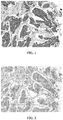
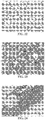
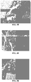
- Figures 1-8 shows several examples of staining of bladder transitional cell carcinoma by anti-UP II antibody (BC21), in comparison to staining with anti-UP III antibody (BC17), on a serial section of the same specimen.
- Table 1 shows the sensitivity of anti-UP II antibody (BC21) staining 178 specimens of bladder cancer (e.g., transitional cell carcinoma (TCC) and papillary TCC), using a tissue microarray (TMA).
- TCC transitional cell carcinoma
- TMA tissue microarray
- anti-UP II antibody identified 68 of 83 (about 82%) of Grade II tumors, and 25 of 44 (about 57%) of Grade III tumors.
- Table 1 Anti-UP II Antibody (BC21) on Bladder cancer (TCC and Papillary TCC) TMA Grade Number of Specimens Number of Positive Specimens % Positive Number of Negative Specimens % Negative Grades I, II & III 178 137 77% 41 23% Grade II 83 68 82% 15 18% Grade III 44 25 57% 19 43%
- anti-UP II antibody compared to anti-UP III antibody (BC17) was demonstrated by staining the same 59 specimens of TCC of Grades I, II and III with each antibody (Table 2). Using the same criteria, anti-UP II antibody (BC21) identified 46 specimens as positive (about 78%), compared to 33 specimens (about 56%) determined to be positive with anti-UP III antibody (BC17). In Grade II specimens, anti-UP II antibody (BC21) and anti-UP III antibody (BC17) demonstrated sensitivities of about 77% (27 of 35) and about 54% (19 of 35), respectively.
- anti-UP II antibody (BC21) and anti-UP III antibody (BC17) demonstrated a similar sensitivity of about 64% (7 of 11). In many comparisons, anti-UP II antibody (BC21) provided a darker staining than anti-UP III antibody (BC17).
- Table 2 Comparison of anti-UP II antibody (BC21) and anti-UP III antibody (BC17) on Bladder cancer (TCC and Papillary TCC) TMA Antibody Grade Number of Specimens Number of Positive Specimens % Positive Number of Negative Specimens % Negative BC21 Grades I, II & III 59 46 78% 13 22% BC17 Grades I, II & III 59 33 56% 26 44% BC21 Grade II 35 27 77% 8 23% BC17 Grade II 35 19 54% 16 46% BC21 Grade III 11 7 64% 4 36% BC17 Grade III 11 7 64% 4 36% BC17 Grade III 11 7 64% 4 36% BC17 Grade III 11 7 64% 4 36%
- Anti-UP II antibody may be highly specific perhaps when evaluated on a variety of normal (Table 3) and even neoplastic (Table 4) tissues. Bladder and ureter may be the only normal tissue to stain positive with UP II (BC21). Such staining may be expected, perhaps considering that the known expression of UP II in normal urothelium anti-UP II antibody (BC21) may not stain any other normal or neoplastic tissues, which may demonstrate its high specificity.
- Anti-UPII antibodies such as the monoclonal mouse anti-UP II antibody (BC21) may offer distinct advantages with its improved sensitivity, perhaps even as compared to monoclonal mouse anti-UP III antibody (BC17).
- Figures 1-8 show examples of comparisons of BC21 with BC17 staining serial sections of the same specimen of bladder TCC, perhaps demonstrating the greater sensitivity of BC21.
- the specimen of Figures 1 and 2 may exhibit strong membrane and cytoplasmic staining with BC21 ( Figure 1 ), while the staining of BC17 may be minimal in this case ( Figure 2 ).
- Figures 3 and 4 a strong and widespread staining of BC21 may be observed ( Figure 3 ); whereas only sparse, focal staining may be observed on the same specimen with BC17 ( Figure 4 ).
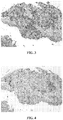
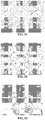
- the specimen of Figures 5 and 6 may display strong staining with BC21 ( Figure 5 ), but may have only limited staining with BC17 ( Figure 6 ).
- Figures 7 and 8 show a specimen that may also exhibit a strong staining with BC21 ( Figure 7 ); in contrast, BC17 may be negative on this same specimen ( Figure 8 ).
- the minimal staining observed with BC17 in Figures 3, 4 , 5 and 6 may provide excellent examples of the challenge that may be faced by pathologists when using a less sensitive antibody; specifically, when the staining observed may be sparse and light, it may be difficult to determine with confidence if this is true positive staining, signaling the presence of UP II and perhaps indicative of urothelial carcinoma, or if it is a misleading staining artifact and should be dismissed.
- the ambiguity associated with a less sensitive antibody may lead to equivocal, or even incorrect diagnoses and patients with urothelial carcinoma may not receive appropriate treatment in a timely fashion.
- an anti-UP II antibody such as BC21, may offer a significant advantage for diagnosis with its increased sensitivity.
- An anti-UP II antibody such as BC21, may result in strong, clear staining of urothelial carcinoma that may allow a pathologist to definitively return a diagnosis of urothelial carcinoma, perhaps allowing a patient to expeditiously receive the most appropriate treatment.
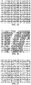
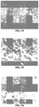
- Binding of BC21 to UP II protein may be demonstrated by Western blot ( Figure 9A ).
- the absence of similar binding of BC21 to UP III protein may also be shown by Western blot ( Figure 9A ).
- the anti-UP III antibody BC17 may not bind UP II protein, but may recognize UP III protein ( Figure 9B ).
- anti-UPII antibodies such as the mouse monoclonal anti-UP II antibody BC21 may be suitable for use in many variations of the above protocols and other methods known to those in the art. Specimens stained with BC21 may be archived using a permanent mounting media and a coverslip. The antibody BC21 may also be used in an automated staining instrument, using standard protocols.
- detection methods e.g., fluorescence
- detection enzymes e.g., alkaline phosphatase (AP), beta-galactosidase
- chromogens e.g., 3-amino-9-ethylcarbazole, 5-bromo-4-chloro-3-indolyl phosphate, 3,3',5,5'-tetramethylbenzidine, 5-bromo-4-chloro-3-indolyl- ⁇ -D-glucuronide
- an epitope of an anti-UPII antibodies such as mouse monoclonal anti-UP II antibody, or a portion thereof, may be a useful antigen for the production of new monoclonal antibodies, including production in species other than mouse (e.g. rabbit, goat, horse, chicken, etc.) as one skilled in the art would understand.
- a monoclonal antibody for Uroplakin III may include a mouse monoclonal antibody, a rabbit monoclonal antibody, a goat monoclonal antibody, a horse monoclonal antibody, a chicken monoclonal antibody, a humanized monoclonal antibody, a chimeric antibody, or any combination thereof.
- a polyclonal antibody for Uroplakin III may include rabbit polyclonal antibody, mouse polyclonal antibody, a goat polyclonal antibody, a horse polyclonal antibody, a chicken polyclonal antibody, a humanized polyclonal antibody, or any combination thereof.
- an antibody may be an isolated antibody.
- anti-UPII antibodies such as BC21 in immunohistochemistry of formalin-fixed paraffin embedded tissues may be described here, its utility in other immunoassays may be readily envisioned and all are included in this application. In particular, it may be well known that many of the same reagents used in IHC of FFPE may also be used in IHC of frozen-tissue sections. Anti-UPII antibodies such as BC21 may also be useful in other immunoassays, including ELISA, perhaps using generally known methods.
- an anti-UP II antibody may be used in conjunction with one or more additional primary antibodies as part of a cocktail, to perform a "double-stain” procedure (also described as multi-stain or even multiplex).
- double-stain procedures may be generally well known in the art; however, the best combinations of primary antibodies for a particular diagnostic application may not be known.
- anti-UPII antibodies such as a mouse monoclonal anti-UP II antibody BC21 could be combined with one or more antibodies in a primary antibody cocktail. At least one of the additional antibodies could be derived from a species other than mouse such as a rabbit antibody. Antibodies may be derived from at least two different species such as a mouse host or a rabbit host. Species may include mouse, rabbit, goat, horse, chicken, human, or any combination thereof. The antibodies may be monoclonal or polyclonal. In this manner, the multiple antibodies in the primary antibody cocktail may be differentiated in the subsequent detection and even visualization steps.
- the usual goat anti-mouse antibody conjugated to HRP may be applied perhaps followed by an appropriate chromogen, such as DAB.
- a second detection step may be performed, using goat anti-rabbit antibody conjugated to AP perhaps followed by an appropriate chromogen, such as Fast Red.
- two or more targets may be identified on the same tissue sample with the resulting two colors.
- mouse primary antibodies including BC21
- rabbit primary antibodies could result in red (Fast Red) staining.
- the anti-mouse or anti rabbit antibodies comprising the antibody-enzyme conjugates may be derived from a different host species, including mouse, rabbit, chicken, horse, rat, goat, or sheep.
- a primary antibody may be from a variety of host species, including mouse, rabbit, chicken, horse, rat, goat, or sheep.
- an antibody may include an antibody-enzyme conjugate and a primary antibody could be obtained from two different host species. Chromogens other than DAB and/or Fast Red may be used as well.
- Embodiments of the present invention may provide a composition having at least two antibodies or antigen binding fragments thereof, perhaps as a cocktail, where at least one of the two antibodies or antigen binding fragments thereof specifically binds to at least Uroplakin II.
- This may provide a method for detecting at least two different proteins in a biological sample perhaps by contacting a biological sample with a composition comprising at least two antibodies or antigen binding fragments thereof, where at least one of the at least two antibodies or antigen binding fragments thereof may bind specifically to at least Uroplakin II, to form an antigen-antibody complex and an antigen-antibody complex may be detected.
- a composition may have at least one first primary antibody and at least one second primary antibody.
- a biological sample may include blood, urine, bladder tissue, urothelial tissue, transitional cell tissue, normal tissue, neoplastic tissue, kidney tissue, ovarian tissue, thyroid tissue, endometrial tissue, renal tissue, tonsil tissue, pancreas tissue, colon tissue, lymph node tissue, neoplastic pancreatic tissue, stomach tissue, prostate tissue, lung tissue and breast tissue.
- At least one of the antibodies or antigen binding fragments thereof may specifically bind to at least Uroplakin II and may even have a positive indication cut-off value of greater than 1% of stained cells.
- a positive indication cut-off value may provide a percentage of stained cells needed to indicate a positive staining result.
- Other cut-off value may include greater than about 1% of stained cells, greater than about 2% of stained cells, greater than about 3% of stained cells, greater than about 4% of stained cells, greater than about 5% of stained cells, greater than about 6% of stained cells, greater than about 7% of stained cells, greater than about 8% of stained cells, greater than about 9% of stained cells, and perhaps even greater than about 10% of stained cells, or more.
- the present invention may provide a composition with at least two antibodies or antigen binding fragments thereof which may be capable of providing different visualization results such as different color results.
- a composition may provide that at least one other of an at least two antibodies or antigen binding fragments thereof may bind specifically to GATA-3, p63, Uroplakin III, PAX8, NKX3.1, PSA, or any combination thereof.
- Antibodies, compositions thereof, perhaps with anti-Uroplakin II antibodies may provide a detection system including urothelial carcinoma detection composition, renal cell carcinoma detection composition, prostate/prostatic carcinoma detection composition, or any combination thereof.
- a single color stain may be used for a primary antibody cocktail.
- the primary antibody cocktail is comprised of antibodies all derived from the same host species, then a single antibody enzyme conjugate may be used to stain for the presence of all of the antibodies with a single color. The presence or absence of each antibody may be determined based on cellular localization, or perhaps such determination is not necessary and the staining may be interpreted effectively without identifying the presence or absence of each individual antibody.
- Certain steps of an IHC procedure may be performed sequentially or simultaneously, perhaps by using a cocktail of reagents, as known to those skilled in the art.
- a cocktail of reagents as known to those skilled in the art.
- antibodies described in a primary antibody cocktail may alternatively be applied in sequential steps of one or more antibodies.
- detection reagents may be applied simultaneously in reagent cocktail or separate reagents in sequential steps.
- a first primary antibody may be applied, followed by a first antibody-enzyme conjugate and first chromogen, and then a denaturing step, before proceeding to application of a second primary antibody, followed by a second antibody-enzyme conjugate and a second chromogen.
- a double-stain of two different colors may be achieved using primary antibodies derived from the same species.
- Antibodies that may be useful for diagnosis when combined with an anti-UPII antibody such as a mouse monoclonal anti-UP II antibody BC21 in a primary antibody cocktail for use in multi-stain procedures may include: Table 5 Antibody Cocktail Utility UPII + UPIII Urothelial marker of enhanced sensitivity UPII + GATA3 Urothelial marker of enhanced sensitivity UPII + UPIII + GATA3 Urothelial marker of enhanced sensitivity UPII + PAX8 Differential marker of bladder and kidney UPII + PAX8 + PSA Differential marker of bladder, kidney and prostate UPII + NKX3.1 Differential marker of bladder and prostate UPII + PAX8 + NKX3.1 Differential marker of bladder, kidney and prostate UPII + p63 Urothelial marker of enhanced sensitivity and differential marker of bladder and prostate and differential marker of bladder and non-bladder squamous cell carcinoma UPII + GATA-3 and/or p63 + PAX8 + PSA and/or NKX3.1 Differential
- an anti-UPII antibody such as a mouse monoclonal anti-UP II antibody BC21 may be specific for detection of UP II and may be useful in immunohistochemical procedures for diagnosis of several types of cancers in human tissue samples.
- anti-UP II antibody such as BC21 has advantages over anti-UP III antibody BC17, including greater sensitivity.
- Expression levels of UP II protein may be a prognostic marker of patient outcomes in cases of bladder cancer. Determination of UP II expression, using an antibody such as BC21, may aid in identifying patients more likely to experience a positive outcome (e.g. longer survival time, longer time to disease progression, reduced tumor size), a positive or good prognosis, or those patients more likely to experience a negative outcome (e.g. shorter survival time, shorter time to disease progression), a negative or poor prognosis. Determination of UP II expression, using an antibody such as BC21, may also aid in predicting patient response to a particular therapeutic treatment. For example, the level of UPII expression may aid in determining the likelihood that a patient could benefit from a particular pharmaceutical agent, including antibody based therapeutics. Conversely, UP II expression may aid in determining the likelihood that a patient may not benefit from a particular therapeutic treatment.
- a positive outcome e.g. longer survival time, longer time to disease progression, reduced tumor size
- a negative outcome e.g. shorter
- UPII antibody such as a mouse monoclonal UPII antibody BC21 in a primary antibody cocktail for use in multi-stain procedures
- UPII antibody such as a mouse monoclonal UPII antibody BC21 in a primary antibody cocktail for use in multi-stain procedures
- Table 6 Antibody Combination Host Species, cellular localization, stain color*
- Possible Diagnostic Utility Detection System used in example and Figure No.
- UPIII Mae, Membrane & cytoplasmic, brown
- UPII and/or UPIII staining may be observed in urothelial carcinoma.
- DS#1 Figure 13 UPIII (Mouse, Membrane & cytoplasmic, red) GATA3 (Rabbit, nuclear, brown) UPII (Mouse, Membrane & cytoplasmic, red) UPII staining may be observed in urothelial carcinoma.
- UPII (Mouse, Membrane & cytoplasmic, red) UPII staining may be observed in urothelial carcinoma.
- DS#1 Figures 18 , 19, 20 PAX8 (Rabbit, nuclear, brown) PAX8 staining may be observed in renal cell carcinoma.
- PSA (Rabbit, cytoplasmic, brown) PSA staining may be observed in prostatic carcinoma.
- UPII Mae, Membrane & cytoplasmic, brown
- UPII staining may be observed in urothelial carcinoma.
- PAX8 (Rabbit, nuclear, red) PSA (Rabbit, cytoplasmic, PAX8 staining may be observed in renal cell carcinoma. red) PSA staining may be observed in prostatic carcinoma.
- UPII (Mouse, Membrane & cytoplasmic, brown) UPII staining may be observed in urothelial carcinoma.
- NKX3.1 (Rabbit, nuclear, red) NKX3.1 staining may be observed in prostatic carcinoma.
- UPII Mae, Membrane & cytoplasmic, brown
- PAX8 (Mouse, nuclear, brown) PAX8 staining may be observed in renal cell carcinoma.
- NKX3.1 (Rabbit, nuclear, red) NKX3.1 staining may be observed in prostatic carcinoma.
- UPII (Mouse, Membrane & cytoplasmic, red) UPII staining may be observed in urothelial carcinoma.
- the detection system may include an anti-mouse antibody perhaps conjugated to AP and even an anti-rabbit antibody perhaps conjugated to HRP, perhaps even with DAB and Fast Red as chromogens, which may result in red staining for mouse antibodies and brown staining for rabbit antibodies (referred to as DS#1).
- an anti-mouse antibody perhaps conjugated to AP and even an anti-rabbit antibody perhaps conjugated to HRP, perhaps even with DAB and Fast Red as chromogens, which may result in red staining for mouse antibodies and brown staining for rabbit antibodies (referred to as DS#1).
- two colors may not be necessary because the antigens may be distinguished by cellular localization of staining, or perhaps it is not diagnostically significant to determine which antigen is staining.
- Other color combinations may be obtained using other detection systems or chromogens and all are meant to be included in this disclosure.
- combining UPII with another antibody that stains urothelial tissue may be useful, perhaps increasing sensitivity compared to staining with each of the antibodies individually.
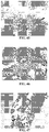
- Figure 10 shows an example of a cocktail of UPIII + UPII staining a specimen of urothelial carcinoma.
- UPII staining may be observed, when perhaps UPIII staining is reduced or absent.
- UPIII staining may be observed, when perhaps UPII staining is reduced or absent.
- a cocktail of UPII + GATA3 may also provide increased sensitivity for urothelial carcinoma.
- a specimen of urothelial carcinoma stained with a cocktail of UPII + GATA3 is shown in Figure 11 .
- UPII staining may be observed, when perhaps GATA3 staining is reduced or absent.
- GATA3 staining may be observed, when perhaps UPII staining is reduced or absent.
- a specimen stained with a cocktail of UPII + GATA3 is shown in Figure 12 .
- the same specimen stained with a cocktail of UPII + UPIII + GATA3 is shown in Figure 13 . Perhaps more staining is observed with the UPII + UPIII + GATA3 cocktail, which may result in improved sensitivity.
- UPII + PAX8 may be useful for differentiating urothelial carcinoma and renal cell carcinoma.
- UPII may stain urothelial carcinoma, which is not stained by PAX8 ( Figures 14 and 15 ).
- renal cell carcinoma may be stained by PAX8, but perhaps not by UPII ( Figures 16 and 17 ).
- UPII + PAX8 + PSA may be useful for differentiating urothelial carcinoma renal cell carcinoma, and prostate carcinoma.
- UPII may stain urothelial carcinoma, which is not stained by PAX8 or PSA ( Figures 18 and 21 ).
- renal cell carcinoma may be stained by PAX8, but perhaps not by UPII or PSA ( Figures 19 and 22 ).
- prostate carcinoma may be stained by PSA, but perhaps not by UPII or PAX8 ( Figures 20 and 23 ).
- UPII + NKX3.1 may be useful for differentiating urothelial carcinoma and prostate carcinoma.
- UPII may stain urothelial carcinoma, which is not stained by NKX3.1 ( Figure 24 ).
- prostate carcinoma may be stained by NKX3.1, but perhaps not by UPII ( Figure 25 ).
- UPII + PAX8 + NKX3.1 may be useful for differentiating urothelial carcinoma renal cell carcinoma, and prostate carcinoma.
- UPII may stain urothelial carcinoma, which is not stained by PAX8 or NKX3.1 ( Figure 26 ).
- renal cell carcinoma may be stained by PAX8, but perhaps not by UPII or NKX3.1 ( Figure 27 ).
- prostate carcinoma may be stained by NKX3.1, but perhaps not by UPII or PAX8 ( Figure 28 ).
- a cocktail of UPII + p63 may also provide increased sensitivity for urothelial carcinoma.
- a specimen of urothelial carcinoma stained with a cocktail of UPII + p63 is shown in Figures 29 and 30 .
- Normal prostate, or perhaps prostatic intraepithelial neoplasia (PIN) may be stained by p63, but perhaps not by UPII ( Figures 31 and 32 ).
- a cocktail of UPII + p40 may also provide increased sensitivity for urothelial carcinoma.
- a specimen of urothelial carcinoma stained with a cocktail of UPII + p40 is shown in Figure 33 .
- Figure 34 shows a schematic summary of various embodiments of the present invention not falling under the scope of the claims including a kit (5) which may provide an anti-UPII antibody, antigen binding fragment thereof, portion thereof, in a composition or even in a cocktail, perhaps even provided from a hybridoma, the anti-UPII antibody (1) may be contacted with a biological sample (2) to form at least one antibody-antigen complex (3) which may then be detected with a detector (4).
- a kit (5) which may provide an anti-UPII antibody, antigen binding fragment thereof, portion thereof, in a composition or even in a cocktail, perhaps even provided from a hybridoma
- the anti-UPII antibody (1) may be contacted with a biological sample (2) to form at least one antibody-antigen complex (3) which may then be detected with a detector (4).
- embodiments of the present invention may provide obtaining tissue from an animal or human to be tested (6), fixing or freezing said tissue (7), treating said fixed or frozen tissue to unmask epitopes to Uroplakin III (8), contacting said treated tissue with an antibody or antigen binding fragment thereof as discussed herein in an amount and under conditions such that an antibody or antigen binding fragment thereof binds to a Uroplakin III protein if the protein is present in said tissue (9); and perhaps even detecting the presences of said bound antibodies (10), as schematically represented in Figure 35 .
- the present invention may provide, in embodiments not falling under the scope of the claims, a diagnostic or even prognostic test kit which may include an antibody or antigen binding fragment thereof (as discussed herein) with an antibody detection element of the antibody or antigen binding fragment thereof perhaps when bound to an antigen.
- a diagnostic or even prognostic test kit which may include an antibody or antigen binding fragment thereof (as discussed herein) with an antibody detection element of the antibody or antigen binding fragment thereof perhaps when bound to an antigen.
- This may provide a method of contacting a biological sample with an antibody or antigen binding fragment thereof and even detecting binding of, or even the presence of the antibody or antigen binding fragment thereof bound to a protein or with an antigen in the biological sample perhaps using an antibody detection element.
- Embodiments not falling under the scope of the claims may provide an immunoassay method for detecting Uroplakin II protein in a mammal or human perhaps by obtaining a tissue from an animal or a human to be tested, contacting the tissue with an anti-UPII antibody or antigen binding fragment thereof in accordance with the various embodiments presented herein perhaps in an amount and under conditions such that the antibody or antigen binding fragment thereof may bind to a Uroplakin II protein if the protein is present in the tissue; and even detecting the presence of bound antibodies.
- a biological sample may include blood, urine, urothelial tissue, transitional cell tissue, bladder tissue, normal tissue, neoplastic tissue, kidney tissue, ovarian tissue, thyroid tissue, endometrial tissue, renal tissue, tonsil tissue, pancreas tissue, colon tissue, lymph node tissue, neoplastic pancreatic tissue, stomach tissue, prostate tissue, lung tissue, or breast tissue, perhaps depending on the antibody or even cocktail being used.
- the basic concepts of the present invention may be embodied in a variety of ways. It involves both antibody techniques as well as devices to accomplish the appropriate antibody.
- the antibody techniques are disclosed as part of the results shown to be achieved by the various devices described and as steps which are inherent to utilization. They are simply the natural result of utilizing the devices as intended and described.
- the devices are disclosed, it should be understood that these not only accomplish certain methods but also can be varied in a number of ways. Importantly, as to all of the foregoing, all of these facets should be understood to be encompassed by this disclosure.
- Uroplakins Specific Membrane Proteins of Urothelial Umbrella Cells, as Histological Markers of Metastatic Transitional Cell Carcinomas, American Journal of Pathology, Vol. 147, No. 5, November 1995 .
- Kaufmann, O. et al. Uroplakin III Is a Highly Specific and Moderately Sensitive Immunohistochemical Marker for Primary and Metastatic Urothelial Carcinomas, Am J Clin Pathol 2000; 113:683-687 Wu XR, Kong XP, Pellicer A, Kreibich G, Sun TT.; Uroplakins in urothelial biology, function, and disease; Kidney Int.
Claims (16)
- Anticorps anti-uroplakine II ou fragment de celui-ci se liant à un antigène produit par l'hybridome déposé auprès de l'American Type Culture Collection (ATCC) sous la désignation de dépôt de brevet ATCC n° PTA-13181.
- Procédé de production d'un anticorps monoclonal selon la revendication 1, comprenant les étapes consistant à :mettre en culture ledit hybridome qui produit un anticorps monoclonal capable de spécifiquement reconnaître l'uroplakine II ; etpermettre audit hybridome de produire l'anticorps monoclonal.
- Anticorps ou fragment de celui-ci se liant à un antigène comprenant un polypeptide possédant la séquence d'acides aminés codée par la séquence d'acide nucléique sélectionnée dans un groupe consistant en SEQ ID NO: 1, SEQ ID NO: 2, et SEQ ID NO: 3.
- Anticorps anti-uroplakine II ou fragment de celui-ci se liant à un antigène, où ledit anticorps ou ledit fragment de celui-ci se liant à un antigène comprend une région variable de chaîne légère comprenant la séquence d'acides aminés codée par la séquence d'acide nucléique de SEQ ID NO: 2 ou SEQ ID NO: 3 et une région variable de chaîne lourde comprenant la séquence d'acides aminés codée par la séquence d'acide nucléique de SEQ ID NO: 1.
- Anticorps ou fragment de celui-ci se liant à un antigène qui se lie spécifiquement à au moins un polypeptide possédant une séquence d'acides aminés comprenant SEQ ID NO: 4.
- Composition comprenant ledit anticorps ou fragment de celui-ci se liant à un antigène selon la revendication 1, 3, 4 ou 5 et au moins un anticorps ou fragment de celui-ci se liant à un antigène supplémentaire afin de créer un cocktail d'anticorps primaires.
- Composition selon la revendication 6, dans laquelle ledit au moins un anticorps ou fragment de celui-ci se liant à un antigène supplémentaire se lie spécifiquement à un antigène sélectionné dans un groupe consistant en GATA-3, p63, l'uroplakine III, PAX8, NKX3.1, PSA, p40, et une quelconque combinaison de ceux-ci.
- Composition selon la revendication 6, dans laquelle ledit anticorps ou fragment de celui-ci se liant à un antigène et ledit au moins anticorps ou fragment de celui-ci se liant à un antigène supplémentaire se lient chacun spécifiquement à des protéines sélectionnées dans un groupe consistant en :- l'uroplakine II et GATA-3 ;- l'uroplakine II et p63 ;- l'uroplakine II et l'uroplakine III ;- l'uroplakine II et PAX8 ;- l'uroplakine II et NKX3.1 ;- l'uroplakine II et PSA ;- l'uroplakine II et l'uroplakine III et GATA-3 ;- l'uroplakine II et PAX8 et PSA ;- l'uroplakine II et PAX8 et NKX3.1 ; et- l'uroplakine II et p40.
- Anticorps selon la revendication 1 ou 5, où ledit anticorps ou fragment de celui se liant à un antigène comprend un polypeptide possédant la séquence d'acides aminés codée par la séquence d'acide nucléique sélectionnée dans un groupe consistant en SEQ ID NO: 1, SEQ ID NO: 2, et SEQ ID NO: 3.
- Anticorps selon la revendication 1 ou 5, où ledit anticorps ou fragment de celui se liant à un antigène comprend un polypeptide possédant la séquence d'acides aminés codée par la séquence d'acide nucléique de SEQ ID NO: 1 et SEQ ID NO: 2 ou SEQ ID NO: 3.
- Anticorps selon la revendication 1 ou 3, où ledit anticorps ou fragment de celui se liant à un antigène se lie spécifiquement à au moins un polypeptide possédant une séquence d'acides aminés comprenant SEQ ID NO: 4.
- Anticorps selon la revendication 1, 3, 4 ou 5, où ledit anticorps comprend un anticorps monoclonal.
- Anticorps selon la revendication 12, où ledit anticorps monoclonal est sélectionné dans un groupe consistant en un anticorps monoclonal de souris, un anticorps monoclonal de lapin, un anticorps monoclonal de chèvre, un anticorps monoclonal de cheval, un anticorps monoclonal de poulet, un anticorps monoclonal humanisé, un anticorps chimérique, et une quelconque combinaison de ceux-ci.
- Anticorps selon la revendication 1, 3, 4 ou 5, où ledit anticorps comprend un anticorps polyclonal.
- Utilisation d'un anticorps selon la revendication 1, 3, 4 ou 5 pour le diagnostic du cancer dans des échantillons de tissu.
- Utilisation d'un anticorps selon la revendication 15, où le cancer est un carcinome à cellules transitionnelles.
Applications Claiming Priority (2)
| Application Number | Priority Date | Filing Date | Title |
|---|---|---|---|
| US201261706312P | 2012-09-27 | 2012-09-27 | |
| PCT/US2013/062043 WO2014052672A1 (fr) | 2012-09-27 | 2013-09-26 | Systèmes d'anticorps anti-uroplakine ii et méthodes associées |
Publications (3)
| Publication Number | Publication Date |
|---|---|
| EP2900265A1 EP2900265A1 (fr) | 2015-08-05 |
| EP2900265A4 EP2900265A4 (fr) | 2016-05-25 |
| EP2900265B1 true EP2900265B1 (fr) | 2018-05-30 |
Family
ID=50388980
Family Applications (1)
| Application Number | Title | Priority Date | Filing Date |
|---|---|---|---|
| EP13841542.7A Active EP2900265B1 (fr) | 2012-09-27 | 2013-09-26 | Anticorps anti-uroplakine ii et méthodes associées |
Country Status (6)
| Country | Link |
|---|---|
| US (2) | US9429577B2 (fr) |
| EP (1) | EP2900265B1 (fr) |
| JP (1) | JP6324970B2 (fr) |
| DK (1) | DK2900265T3 (fr) |
| ES (1) | ES2682345T3 (fr) |
| WO (1) | WO2014052672A1 (fr) |
Cited By (2)
| Publication number | Priority date | Publication date | Assignee | Title |
|---|---|---|---|---|
| US10295542B2 (en) | 2013-10-03 | 2019-05-21 | Biocare Medical, Llc | Systems and methods for anti-SOX10 antibodies |
| US10316103B1 (en) | 2012-03-30 | 2019-06-11 | Biocare Medical, Llc | Systems and methods for anti-Uroplakin III antibodies |
Families Citing this family (6)
| Publication number | Priority date | Publication date | Assignee | Title |
|---|---|---|---|---|
| US8852592B2 (en) | 2011-05-10 | 2014-10-07 | Biocare Medical, Llc | Systems and methods for anti-PAX8 antibodies |
| WO2014052672A1 (fr) | 2012-09-27 | 2014-04-03 | Biocare Medical, Llc | Systèmes d'anticorps anti-uroplakine ii et méthodes associées |
| US10429390B2 (en) | 2012-12-18 | 2019-10-01 | Biocare Medical, Llc | Antibody cocktail systems and methods for classification of histologic subtypes in lung cancer |
| JP6445467B2 (ja) | 2013-02-28 | 2019-01-09 | バイオケア メディカル, エルエルシー | 抗p40抗体システムおよび方法 |
| CN108727496A (zh) * | 2018-06-21 | 2018-11-02 | 南京基诺米医疗科技有限公司 | 鼠抗人p63蛋白单克隆抗体制备及其免疫组化用途 |
| CN113295866A (zh) * | 2021-04-26 | 2021-08-24 | 深圳市龙华区中心医院 | Upⅱ抗原和upⅱ抗体联合在制备检测膀胱癌的产品中的应用 |
Family Cites Families (77)
| Publication number | Priority date | Publication date | Assignee | Title |
|---|---|---|---|---|
| US4145406A (en) | 1977-04-11 | 1979-03-20 | Miles Laboratories, Inc. | Specific binding - adsorbent assay method and test means |
| US4254082A (en) | 1978-06-12 | 1981-03-03 | Miles Laboratories, Inc. | Specific binding-adsorbent assay test means |
| CA1247538A (fr) | 1982-05-21 | 1988-12-28 | Mark C. Glassy | Hybridomes humain-humain contre les tumeurs solides |
| US4687732A (en) | 1983-06-10 | 1987-08-18 | Yale University | Visualization polymers and their application to diagnostic medicine |
| US4690890A (en) | 1984-04-04 | 1987-09-01 | Cetus Corporation | Process for simultaneously detecting multiple antigens using dual sandwich immunometric assay |
| JPS60248618A (ja) | 1984-05-24 | 1985-12-09 | Nippon Zoki Pharmaceut Co Ltd | ジペプチドを含有する潰瘍治療剤 |
| US4792521A (en) | 1985-08-15 | 1988-12-20 | Immunomedics, Inc. | Non-enzymatic immunohistochemical staining system and reagents |
| US4863875A (en) | 1985-08-26 | 1989-09-05 | Gia Research Company, L.P. | Dye labelled antibodies as reagents for use in immunoassay systems |
| GB8607101D0 (en) | 1986-03-21 | 1986-04-30 | Serono Diagnostics Ltd | Immunoassay |
| JPS6447952A (en) | 1987-05-06 | 1989-02-22 | Saibaafuruua Inc | Immunoassay, reagent used therefor and making thereof |
| US5620845A (en) | 1988-06-06 | 1997-04-15 | Ampcor, Inc. | Immunoassay diagnostic kit |
| US5252487A (en) | 1989-05-19 | 1993-10-12 | Cell Analysis Systems, Inc. | Method and apparatus for determining the amount of oncogene protein product in a cell sample |
| US6008057A (en) | 1989-08-25 | 1999-12-28 | Roche Diagnostics Corporation | Immunoassay system |
| US5719063A (en) | 1989-08-25 | 1998-02-17 | Boehringer Mannheim Corporation | Multiplex immunoassay system |
| EP0513332A4 (en) | 1990-11-14 | 1993-03-17 | Cargill, Incorporated | Conjugates of poly(vinylsaccharide) with proteins for the stabilization of proteins |
| US5565332A (en) | 1991-09-23 | 1996-10-15 | Medical Research Council | Production of chimeric antibodies - a combinatorial approach |
| US5280108A (en) | 1991-09-27 | 1994-01-18 | United States Of America | Antibodies to p40 |
| EP0752248B1 (fr) | 1992-11-13 | 2000-09-27 | Idec Pharmaceuticals Corporation | Application thérapeutique d'anticorps chimériques et radio-marqués contre l'antigène à differentiation restreinte des lymphocytes B humains pour le traitement du lymphome des cellules B |
| US5482698A (en) | 1993-04-22 | 1996-01-09 | Immunomedics, Inc. | Detection and therapy of lesions with biotin/avidin polymer conjugates |
| US5487975A (en) | 1993-11-15 | 1996-01-30 | Ventana Medical Systems, Inc. | Biotin/avidin formulation |
| DE69521789T2 (de) | 1994-02-01 | 2002-04-25 | Us Health | Fusionierte proteine die antikörper-teile und nicht-antikörper-teile enthalten |
| US5869274A (en) | 1995-08-09 | 1999-02-09 | Zymed Laboratories, Inc. | Immuno-histochemical method that reduces background staining |
| US5891658A (en) | 1996-06-27 | 1999-04-06 | FCI--FiberChem, Inc. | Single-step, solid-state competitive immunoassay |
| ES2251740T3 (es) | 1996-08-23 | 2006-05-01 | Ludwig Institute For Cancer Research | Factor de crecimiento de celulas de endotelio vascular d recombinante (vegf-d). |
| US7196164B2 (en) | 1997-07-08 | 2007-03-27 | Human Genome Sciences, Inc. | Secreted protein HHTLF25 |
| ES2316171T3 (es) | 1997-09-22 | 2009-04-01 | Novartis Vaccines And Diagnostics, Inc. | Tampones para estabilizar antigenos hcv. |
| US7294459B1 (en) | 1997-10-15 | 2007-11-13 | President And Fellows Of Harvard College | Cell regulatory genes, encoded products, and uses related thereto |
| US6277968B1 (en) * | 1997-11-13 | 2001-08-21 | New York University | Human uroplakin II gene and methods for detecting and classifying bladder cancer via human uroplakin genes |
| AU3208699A (en) | 1998-03-27 | 1999-10-18 | Johns Hopkins University, The | P40 protein acts as an oncogene |
| JP3524401B2 (ja) | 1998-09-16 | 2004-05-10 | 株式会社ニチレイ | 酵素抗体複合体およびその製造方法 |
| US6409990B1 (en) | 1999-05-14 | 2002-06-25 | The Regents Of The University Of California | Macromolecular carrier for drug and diagnostic agent delivery |
| US6723506B2 (en) | 2000-01-20 | 2004-04-20 | Brigham And Women's Hospital | Method of identifying PAX8-PPAR gamma-nucleic acid molecules |
| US20030017491A1 (en) | 2000-09-14 | 2003-01-23 | Zuo-Rong Shi | Chromogenic in situ hybridization methods, kits, and compositions |
| DE10063179A1 (de) | 2000-12-18 | 2002-06-20 | Bayer Ag | Verfahren zur spezifischen Detektion von Tumorzellen und ihren Vorstufen in Gebärmutterhalsabstrichen durch simultane Messung von mindestens zwei verschiedenen molekularen Markern |
| US20030133939A1 (en) | 2001-01-17 | 2003-07-17 | Genecraft, Inc. | Binding domain-immunoglobulin fusion proteins |
| US20020173053A1 (en) | 2001-04-27 | 2002-11-21 | Bassam Damaj | Multiple simultaneous antigen detection by immunohistochemistry |
| US20040076955A1 (en) * | 2001-07-03 | 2004-04-22 | Eos Biotechnology, Inc. | Methods of diagnosis of bladder cancer, compositions and methods of screening for modulators of bladder cancer |
| US6580056B1 (en) | 2001-09-21 | 2003-06-17 | Biocare Medical | Biological specimen heating device and quality control method for immunohistochemistry heat retrieval procedures |
| EP1480520A4 (fr) | 2002-02-12 | 2006-02-15 | Raven Biotechnologies Inc | Cellules epitheliales derivees d'une vessie foetale humaine |
| IL164021A0 (en) | 2002-03-13 | 2005-12-18 | Univ California | Antibodies and anti-pharmaceutical compositions containing the same |
| US7850912B2 (en) | 2003-05-14 | 2010-12-14 | Dako Denmark A/S | Method and apparatus for automated pre-treatment and processing of biological samples |
| KR20120035234A (ko) | 2003-04-11 | 2012-04-13 | 메디뮨 엘엘씨 | 재조합 il?9 항체 및 그의 용도 |
| JP2007501021A (ja) | 2003-05-30 | 2007-01-25 | アレクシオン ファーマシューティカルズ, インコーポレイテッド | 遺伝子操作された定常領域を含む、抗体および融合タンパク質 |
| JP2005122810A (ja) | 2003-10-16 | 2005-05-12 | Hitachi Ltd | 光ディスク装置 |
| JP4949038B2 (ja) | 2003-12-01 | 2012-06-06 | ダコ デンマーク アクティーゼルスカブ | 免疫組織化学的検出のための方法および組成物 |
| WO2005076005A2 (fr) | 2004-01-30 | 2005-08-18 | Medizinische Universität Wien | Procede de classification d'un prelevement de cellules tumorales |
| US20050186642A1 (en) | 2004-02-24 | 2005-08-25 | Biocare Medical, Inc. | Immunoassay reagents and methods of use thereof |
| US7502666B2 (en) | 2004-05-14 | 2009-03-10 | Mts Medication Technologies, Inc. | Systems and methods for storing and dispensing medication |
| JP4576168B2 (ja) * | 2004-06-29 | 2010-11-04 | Tssバイオテック株式会社 | 膀胱尿管逆流症または間質性膀胱炎の検査方法 |
| MY146381A (en) | 2004-12-22 | 2012-08-15 | Amgen Inc | Compositions and methods relating relating to anti-igf-1 receptor antibodies |
| EP1904531B1 (fr) | 2005-07-08 | 2010-10-06 | Pfizer Limited | Anticorps anti-madcam |
| US8114588B2 (en) * | 2005-07-15 | 2012-02-14 | Tss Biotech Inc. | Method for detecting vesicoureteral reflux or interstitial cystitis |
| JP4736037B2 (ja) | 2005-10-26 | 2011-07-27 | 株式会社イーベック | ヒトgm−csfに結合するヒトのモノクローナル抗体並びにその抗原結合部分 |
| BRPI0619249A2 (pt) | 2005-11-30 | 2011-09-20 | Abbott Lab | anticorpos anti-globulÈmeros-aß, frações que se ligam a antìgeno destes, hibridomas correspondentes, ácidos nucléicos, vetores, células hospedeiras, métodos de produzir os ditos anticorpos, composições compreendendo os ditos anticorpos, usos dos ditos anticorpos e métodos de usar os ditos anticorpos |
| KR101496433B1 (ko) | 2006-06-07 | 2015-02-26 | 바이오얼라이언스 씨.브이. | 암세포 상에서 발현되는 cd-43 및 cea 상의 에피토프를 함유하는 탄수화물을 인식하는 항체 및 이를 이용하는 방법 |
| AU2007285217B2 (en) | 2006-08-14 | 2013-02-07 | Forerunner Pharma Research Co., Ltd | Diagnosis and treatment of cancer using anti-desmoglein-3 antibodies |
| CA2672049C (fr) | 2006-12-08 | 2016-05-10 | Lexicon Pharmaceuticals, Inc. | Anticorps monoclonaux diriges contre angptl3 |
| US20080267988A1 (en) | 2007-04-11 | 2008-10-30 | Enteron Limited Partnership | Myelin specific IgE unencumbered by corresponding blocking antibodies as a causative factor in multiple sclerosis |
| JP5117765B2 (ja) | 2007-05-28 | 2013-01-16 | 国立大学法人 東京大学 | 抗robo1抗体を含むpet用腫瘍診断剤 |
| TWI547503B (zh) | 2007-09-26 | 2016-09-01 | 建南德克公司 | 抗α5β1抗體、編碼其之核酸、包含其之組合物及其製造及使用方法 |
| CN102137874B (zh) | 2008-08-29 | 2015-02-18 | 西福根有限公司 | 抗表皮生长因子受体的重组抗体组合物 |
| CA2736829C (fr) | 2008-09-12 | 2018-02-27 | Isis Innovation Limited | Anticorps specifiques de pd-1 et leurs utilisations |
| US7846762B2 (en) | 2008-09-22 | 2010-12-07 | Applied Materials, Inc. | Integrated emitter formation and passivation |
| EP2425253B1 (fr) | 2009-05-01 | 2017-03-22 | Dako Denmark A/S | Nouveau cocktail d'anticorps |
| KR101471239B1 (ko) | 2009-05-12 | 2014-12-09 | 화이자 인코포레이티드 | 항-dkk-1 항체의 차단 및 그의 용도 |
| WO2011047019A1 (fr) | 2009-10-13 | 2011-04-21 | Yale University | Procédé quantitatif objectif pour prédire un sous-type histologique dans le cancer du poumon non à petites cellules |
| US20120321557A1 (en) | 2010-02-26 | 2012-12-20 | Forerunner Pharma Research Co., Ltd. | Anti-icam3 antibody and use thereof |
| EP2402370A1 (fr) | 2010-06-29 | 2012-01-04 | Pierre Fabre Médicament | Nouvel anticorps pour le diagnostic et/ou le pronostic du cancer |
| WO2012031273A2 (fr) | 2010-09-03 | 2012-03-08 | Stem Centrx, Inc. | Nouveaux modulateurs et leurs procédés d'utilisation |
| DK2625502T3 (en) | 2010-10-06 | 2018-12-17 | Biocare Medical Llc | Method and system for efficient processing of biological samples |
| US20120154983A1 (en) | 2010-10-08 | 2012-06-21 | The Regents Of The University Of California | Method of Fabrication of Carbon Nanofibers on Nickel Foam |
| US8852592B2 (en) | 2011-05-10 | 2014-10-07 | Biocare Medical, Llc | Systems and methods for anti-PAX8 antibodies |
| US9156915B2 (en) | 2012-04-26 | 2015-10-13 | Thomas Jefferson University | Anti-GCC antibody molecules |
| WO2014052672A1 (fr) | 2012-09-27 | 2014-04-03 | Biocare Medical, Llc | Systèmes d'anticorps anti-uroplakine ii et méthodes associées |
| US10429390B2 (en) | 2012-12-18 | 2019-10-01 | Biocare Medical, Llc | Antibody cocktail systems and methods for classification of histologic subtypes in lung cancer |
| JP6445467B2 (ja) | 2013-02-28 | 2019-01-09 | バイオケア メディカル, エルエルシー | 抗p40抗体システムおよび方法 |
| ES2765423T3 (es) | 2013-10-03 | 2020-06-09 | Biocare Medical Llc | Sistemas y procedimientos de anticuerpos anti-SOX10 |
-
2013
- 2013-09-26 WO PCT/US2013/062043 patent/WO2014052672A1/fr active Application Filing
- 2013-09-26 US US14/432,132 patent/US9429577B2/en active Active
- 2013-09-26 EP EP13841542.7A patent/EP2900265B1/fr active Active
- 2013-09-26 ES ES13841542.7T patent/ES2682345T3/es active Active
- 2013-09-26 JP JP2015534675A patent/JP6324970B2/ja active Active
- 2013-09-26 DK DK13841542.7T patent/DK2900265T3/en active
-
2016
- 2016-08-02 US US15/226,794 patent/US9823251B2/en active Active
Non-Patent Citations (1)
| Title |
|---|
| None * |
Cited By (2)
| Publication number | Priority date | Publication date | Assignee | Title |
|---|---|---|---|---|
| US10316103B1 (en) | 2012-03-30 | 2019-06-11 | Biocare Medical, Llc | Systems and methods for anti-Uroplakin III antibodies |
| US10295542B2 (en) | 2013-10-03 | 2019-05-21 | Biocare Medical, Llc | Systems and methods for anti-SOX10 antibodies |
Also Published As
| Publication number | Publication date |
|---|---|
| US9429577B2 (en) | 2016-08-30 |
| JP2016500659A (ja) | 2016-01-14 |
| EP2900265A1 (fr) | 2015-08-05 |
| US20160334407A1 (en) | 2016-11-17 |
| DK2900265T3 (en) | 2018-08-20 |
| ES2682345T3 (es) | 2018-09-20 |
| EP2900265A4 (fr) | 2016-05-25 |
| JP6324970B2 (ja) | 2018-05-23 |
| WO2014052672A1 (fr) | 2014-04-03 |
| US9823251B2 (en) | 2017-11-21 |
| US20150247861A1 (en) | 2015-09-03 |
Similar Documents
| Publication | Publication Date | Title |
|---|---|---|
| EP2900265B1 (fr) | Anticorps anti-uroplakine ii et méthodes associées | |
| US9417243B2 (en) | Systems and methods for anti-PAX8 antibodies | |
| US9428576B2 (en) | Anti-p40 antibodies systems and methods | |
| JP5941615B2 (ja) | ヒトcxcl1タンパク質の免疫学的測定方法 | |
| CA2605745A1 (fr) | Anticorps de l'isoforme de l'antigene nucleaire des cellules proliferatives cancereuses specifiques (pcnacs) et leurs utilisations | |
| EP3221352B1 (fr) | Anticorps monoclonaux dirigés contre mcm5 | |
| CN115667304A (zh) | 抗人lag-3抗体及其在免疫组织化学(ihc)中的用途 | |
| KR20110040624A (ko) | 항-fasn 자가면역 항체를 포함하는 간암 진단 마커 및 이의 항원을 포함하는 간암 진단용 조성물 | |
| CN111316102A (zh) | 使用甲硫氨酰-tRNA合成酶诊断胰腺癌的方法以及利用其的胰腺癌诊断试剂盒 | |
| EP1901070B1 (fr) | Procédé et composition pour détecter le cancer au moyen de la détection du coactivateur 2 d'antigène nucléaire de virus Esptein-Barr p100 | |
| US20080220538A1 (en) | Novel Cancer Associated Antibodies And Their Use In Cancer Diagnosis | |
| US10316103B1 (en) | Systems and methods for anti-Uroplakin III antibodies | |
| US9127054B2 (en) | Immunoassay of cofilin 1 protein | |
| US20230340147A1 (en) | Antibodies specific for alpha-1,6-core-fucosylated psa and fucosylated fragments thereof | |
| JP6793514B2 (ja) | 新規甲状腺癌関連抗原に結合する抗体および甲状腺癌診断剤 | |
| CN116234823A (zh) | 用于在免疫组织化学(ihc)方案中诊断癌症的抗体 |
Legal Events
| Date | Code | Title | Description |
|---|---|---|---|
| PUAI | Public reference made under article 153(3) epc to a published international application that has entered the european phase |
Free format text: ORIGINAL CODE: 0009012 |
|
| 17P | Request for examination filed |
Effective date: 20150427 |
|
| AK | Designated contracting states |
Kind code of ref document: A1 Designated state(s): AL AT BE BG CH CY CZ DE DK EE ES FI FR GB GR HR HU IE IS IT LI LT LU LV MC MK MT NL NO PL PT RO RS SE SI SK SM TR |
|
| AX | Request for extension of the european patent |
Extension state: BA ME |
|
| DAX | Request for extension of the european patent (deleted) | ||
| RA4 | Supplementary search report drawn up and despatched (corrected) |
Effective date: 20160428 |
|
| RIC1 | Information provided on ipc code assigned before grant |
Ipc: A61K 39/395 20060101AFI20160421BHEP Ipc: C07K 16/28 20060101ALI20160421BHEP Ipc: C07K 16/30 20060101ALI20160421BHEP Ipc: G01N 33/574 20060101ALI20160421BHEP |
|
| STAA | Information on the status of an ep patent application or granted ep patent |
Free format text: STATUS: EXAMINATION IS IN PROGRESS |
|
| 17Q | First examination report despatched |
Effective date: 20170407 |
|
| GRAP | Despatch of communication of intention to grant a patent |
Free format text: ORIGINAL CODE: EPIDOSNIGR1 |
|
| STAA | Information on the status of an ep patent application or granted ep patent |
Free format text: STATUS: GRANT OF PATENT IS INTENDED |
|
| INTG | Intention to grant announced |
Effective date: 20180103 |
|
| GRAS | Grant fee paid |
Free format text: ORIGINAL CODE: EPIDOSNIGR3 |
|
| GRAA | (expected) grant |
Free format text: ORIGINAL CODE: 0009210 |
|
| STAA | Information on the status of an ep patent application or granted ep patent |
Free format text: STATUS: THE PATENT HAS BEEN GRANTED |
|
| AK | Designated contracting states |
Kind code of ref document: B1 Designated state(s): AL AT BE BG CH CY CZ DE DK EE ES FI FR GB GR HR HU IE IS IT LI LT LU LV MC MK MT NL NO PL PT RO RS SE SI SK SM TR |
|
| REG | Reference to a national code |
Ref country code: GB Ref legal event code: FG4D |
|
| REG | Reference to a national code |
Ref country code: CH Ref legal event code: EP |
|
| REG | Reference to a national code |
Ref country code: AT Ref legal event code: REF Ref document number: 1002918 Country of ref document: AT Kind code of ref document: T Effective date: 20180615 |
|
| REG | Reference to a national code |
Ref country code: IE Ref legal event code: FG4D |
|
| REG | Reference to a national code |
Ref country code: DE Ref legal event code: R096 Ref document number: 602013038364 Country of ref document: DE |
|
| REG | Reference to a national code |
Ref country code: NL Ref legal event code: FP |
|
| REG | Reference to a national code |
Ref country code: DK Ref legal event code: T3 Effective date: 20180814 |
|
| RAP2 | Party data changed (patent owner data changed or rights of a patent transferred) |
Owner name: BIOCARE MEDICAL, LLC |
|
| REG | Reference to a national code |
Ref country code: ES Ref legal event code: FG2A Ref document number: 2682345 Country of ref document: ES Kind code of ref document: T3 Effective date: 20180920 |
|
| REG | Reference to a national code |
Ref country code: FR Ref legal event code: PLFP Year of fee payment: 6 |
|
| REG | Reference to a national code |
Ref country code: LT Ref legal event code: MG4D |
|
| PG25 | Lapsed in a contracting state [announced via postgrant information from national office to epo] |
Ref country code: FI Free format text: LAPSE BECAUSE OF FAILURE TO SUBMIT A TRANSLATION OF THE DESCRIPTION OR TO PAY THE FEE WITHIN THE PRESCRIBED TIME-LIMIT Effective date: 20180530 Ref country code: BG Free format text: LAPSE BECAUSE OF FAILURE TO SUBMIT A TRANSLATION OF THE DESCRIPTION OR TO PAY THE FEE WITHIN THE PRESCRIBED TIME-LIMIT Effective date: 20180830 Ref country code: CY Free format text: LAPSE BECAUSE OF FAILURE TO SUBMIT A TRANSLATION OF THE DESCRIPTION OR TO PAY THE FEE WITHIN THE PRESCRIBED TIME-LIMIT Effective date: 20180530 Ref country code: LT Free format text: LAPSE BECAUSE OF FAILURE TO SUBMIT A TRANSLATION OF THE DESCRIPTION OR TO PAY THE FEE WITHIN THE PRESCRIBED TIME-LIMIT Effective date: 20180530 Ref country code: SE Free format text: LAPSE BECAUSE OF FAILURE TO SUBMIT A TRANSLATION OF THE DESCRIPTION OR TO PAY THE FEE WITHIN THE PRESCRIBED TIME-LIMIT Effective date: 20180530 Ref country code: NO Free format text: LAPSE BECAUSE OF FAILURE TO SUBMIT A TRANSLATION OF THE DESCRIPTION OR TO PAY THE FEE WITHIN THE PRESCRIBED TIME-LIMIT Effective date: 20180830 |
|
| PG25 | Lapsed in a contracting state [announced via postgrant information from national office to epo] |
Ref country code: RS Free format text: LAPSE BECAUSE OF FAILURE TO SUBMIT A TRANSLATION OF THE DESCRIPTION OR TO PAY THE FEE WITHIN THE PRESCRIBED TIME-LIMIT Effective date: 20180530 Ref country code: HR Free format text: LAPSE BECAUSE OF FAILURE TO SUBMIT A TRANSLATION OF THE DESCRIPTION OR TO PAY THE FEE WITHIN THE PRESCRIBED TIME-LIMIT Effective date: 20180530 Ref country code: GR Free format text: LAPSE BECAUSE OF FAILURE TO SUBMIT A TRANSLATION OF THE DESCRIPTION OR TO PAY THE FEE WITHIN THE PRESCRIBED TIME-LIMIT Effective date: 20180831 Ref country code: LV Free format text: LAPSE BECAUSE OF FAILURE TO SUBMIT A TRANSLATION OF THE DESCRIPTION OR TO PAY THE FEE WITHIN THE PRESCRIBED TIME-LIMIT Effective date: 20180530 |
|
| REG | Reference to a national code |
Ref country code: AT Ref legal event code: MK05 Ref document number: 1002918 Country of ref document: AT Kind code of ref document: T Effective date: 20180530 |
|
| PG25 | Lapsed in a contracting state [announced via postgrant information from national office to epo] |
Ref country code: SK Free format text: LAPSE BECAUSE OF FAILURE TO SUBMIT A TRANSLATION OF THE DESCRIPTION OR TO PAY THE FEE WITHIN THE PRESCRIBED TIME-LIMIT Effective date: 20180530 Ref country code: PL Free format text: LAPSE BECAUSE OF FAILURE TO SUBMIT A TRANSLATION OF THE DESCRIPTION OR TO PAY THE FEE WITHIN THE PRESCRIBED TIME-LIMIT Effective date: 20180530 Ref country code: CZ Free format text: LAPSE BECAUSE OF FAILURE TO SUBMIT A TRANSLATION OF THE DESCRIPTION OR TO PAY THE FEE WITHIN THE PRESCRIBED TIME-LIMIT Effective date: 20180530 Ref country code: AT Free format text: LAPSE BECAUSE OF FAILURE TO SUBMIT A TRANSLATION OF THE DESCRIPTION OR TO PAY THE FEE WITHIN THE PRESCRIBED TIME-LIMIT Effective date: 20180530 Ref country code: RO Free format text: LAPSE BECAUSE OF FAILURE TO SUBMIT A TRANSLATION OF THE DESCRIPTION OR TO PAY THE FEE WITHIN THE PRESCRIBED TIME-LIMIT Effective date: 20180530 Ref country code: EE Free format text: LAPSE BECAUSE OF FAILURE TO SUBMIT A TRANSLATION OF THE DESCRIPTION OR TO PAY THE FEE WITHIN THE PRESCRIBED TIME-LIMIT Effective date: 20180530 |
|
| PG25 | Lapsed in a contracting state [announced via postgrant information from national office to epo] |
Ref country code: SM Free format text: LAPSE BECAUSE OF FAILURE TO SUBMIT A TRANSLATION OF THE DESCRIPTION OR TO PAY THE FEE WITHIN THE PRESCRIBED TIME-LIMIT Effective date: 20180530 |
|
| REG | Reference to a national code |
Ref country code: DE Ref legal event code: R097 Ref document number: 602013038364 Country of ref document: DE |
|
| PLBE | No opposition filed within time limit |
Free format text: ORIGINAL CODE: 0009261 |
|
| STAA | Information on the status of an ep patent application or granted ep patent |
Free format text: STATUS: NO OPPOSITION FILED WITHIN TIME LIMIT |
|
| PG25 | Lapsed in a contracting state [announced via postgrant information from national office to epo] |
Ref country code: MC Free format text: LAPSE BECAUSE OF FAILURE TO SUBMIT A TRANSLATION OF THE DESCRIPTION OR TO PAY THE FEE WITHIN THE PRESCRIBED TIME-LIMIT Effective date: 20180530 |
|
| REG | Reference to a national code |
Ref country code: CH Ref legal event code: PL |
|
| 26N | No opposition filed |
Effective date: 20190301 |
|
| PG25 | Lapsed in a contracting state [announced via postgrant information from national office to epo] |
Ref country code: SI Free format text: LAPSE BECAUSE OF FAILURE TO SUBMIT A TRANSLATION OF THE DESCRIPTION OR TO PAY THE FEE WITHIN THE PRESCRIBED TIME-LIMIT Effective date: 20180530 |
|
| REG | Reference to a national code |
Ref country code: BE Ref legal event code: MM Effective date: 20180930 |
|
| REG | Reference to a national code |
Ref country code: IE Ref legal event code: MM4A |
|
| PG25 | Lapsed in a contracting state [announced via postgrant information from national office to epo] |
Ref country code: LU Free format text: LAPSE BECAUSE OF NON-PAYMENT OF DUE FEES Effective date: 20180926 |
|
| PG25 | Lapsed in a contracting state [announced via postgrant information from national office to epo] |
Ref country code: IE Free format text: LAPSE BECAUSE OF NON-PAYMENT OF DUE FEES Effective date: 20180926 |
|
| PG25 | Lapsed in a contracting state [announced via postgrant information from national office to epo] |
Ref country code: BE Free format text: LAPSE BECAUSE OF NON-PAYMENT OF DUE FEES Effective date: 20180930 Ref country code: CH Free format text: LAPSE BECAUSE OF NON-PAYMENT OF DUE FEES Effective date: 20180930 Ref country code: LI Free format text: LAPSE BECAUSE OF NON-PAYMENT OF DUE FEES Effective date: 20180930 |
|
| PG25 | Lapsed in a contracting state [announced via postgrant information from national office to epo] |
Ref country code: AL Free format text: LAPSE BECAUSE OF FAILURE TO SUBMIT A TRANSLATION OF THE DESCRIPTION OR TO PAY THE FEE WITHIN THE PRESCRIBED TIME-LIMIT Effective date: 20180530 |
|
| PG25 | Lapsed in a contracting state [announced via postgrant information from national office to epo] |
Ref country code: MT Free format text: LAPSE BECAUSE OF NON-PAYMENT OF DUE FEES Effective date: 20180926 |
|
| PG25 | Lapsed in a contracting state [announced via postgrant information from national office to epo] |
Ref country code: TR Free format text: LAPSE BECAUSE OF FAILURE TO SUBMIT A TRANSLATION OF THE DESCRIPTION OR TO PAY THE FEE WITHIN THE PRESCRIBED TIME-LIMIT Effective date: 20180530 |
|
| PG25 | Lapsed in a contracting state [announced via postgrant information from national office to epo] |
Ref country code: PT Free format text: LAPSE BECAUSE OF FAILURE TO SUBMIT A TRANSLATION OF THE DESCRIPTION OR TO PAY THE FEE WITHIN THE PRESCRIBED TIME-LIMIT Effective date: 20180530 |
|
| PG25 | Lapsed in a contracting state [announced via postgrant information from national office to epo] |
Ref country code: MK Free format text: LAPSE BECAUSE OF NON-PAYMENT OF DUE FEES Effective date: 20180530 Ref country code: HU Free format text: LAPSE BECAUSE OF FAILURE TO SUBMIT A TRANSLATION OF THE DESCRIPTION OR TO PAY THE FEE WITHIN THE PRESCRIBED TIME-LIMIT; INVALID AB INITIO Effective date: 20130926 |
|
| PG25 | Lapsed in a contracting state [announced via postgrant information from national office to epo] |
Ref country code: IS Free format text: LAPSE BECAUSE OF FAILURE TO SUBMIT A TRANSLATION OF THE DESCRIPTION OR TO PAY THE FEE WITHIN THE PRESCRIBED TIME-LIMIT Effective date: 20180930 |
|
| P01 | Opt-out of the competence of the unified patent court (upc) registered |
Effective date: 20230523 |
|
| PGFP | Annual fee paid to national office [announced via postgrant information from national office to epo] |
Ref country code: NL Payment date: 20230926 Year of fee payment: 11 Ref country code: GB Payment date: 20230926 Year of fee payment: 11 |
|
| PGFP | Annual fee paid to national office [announced via postgrant information from national office to epo] |
Ref country code: FR Payment date: 20230926 Year of fee payment: 11 Ref country code: DK Payment date: 20230927 Year of fee payment: 11 Ref country code: DE Payment date: 20230928 Year of fee payment: 11 |
|
| PGFP | Annual fee paid to national office [announced via postgrant information from national office to epo] |
Ref country code: ES Payment date: 20231018 Year of fee payment: 11 |
|
| PGFP | Annual fee paid to national office [announced via postgrant information from national office to epo] |
Ref country code: IT Payment date: 20230925 Year of fee payment: 11 |