EP2855527B1 - Agents de liaison au tlr3 - Google Patents
Agents de liaison au tlr3 Download PDFInfo
- Publication number
- EP2855527B1 EP2855527B1 EP13727110.2A EP13727110A EP2855527B1 EP 2855527 B1 EP2855527 B1 EP 2855527B1 EP 13727110 A EP13727110 A EP 13727110A EP 2855527 B1 EP2855527 B1 EP 2855527B1
- Authority
- EP
- European Patent Office
- Prior art keywords
- antibody
- tlr3
- seq
- antibodies
- amino acid
- Prior art date
- Legal status (The legal status is an assumption and is not a legal conclusion. Google has not performed a legal analysis and makes no representation as to the accuracy of the status listed.)
- Active
Links
- 239000011230 binding agent Substances 0.000 title description 4
- 230000027455 binding Effects 0.000 claims description 256
- 108090000765 processed proteins & peptides Proteins 0.000 claims description 244
- 102000004196 processed proteins & peptides Human genes 0.000 claims description 236
- 229920001184 polypeptide Polymers 0.000 claims description 235
- 150000001413 amino acids Chemical class 0.000 claims description 219
- 125000003275 alpha amino acid group Chemical group 0.000 claims description 129
- 101000831496 Homo sapiens Toll-like receptor 3 Proteins 0.000 claims description 97
- 102000045716 human TLR3 Human genes 0.000 claims description 77
- 108091032973 (ribonucleotides)n+m Proteins 0.000 claims description 63
- 102000040650 (ribonucleotides)n+m Human genes 0.000 claims description 63
- 230000011664 signaling Effects 0.000 claims description 62
- 230000035772 mutation Effects 0.000 claims description 52
- 238000011282 treatment Methods 0.000 claims description 44
- 230000002829 reductive effect Effects 0.000 claims description 43
- 239000000203 mixture Substances 0.000 claims description 39
- 208000023275 Autoimmune disease Diseases 0.000 claims description 24
- 208000027866 inflammatory disease Diseases 0.000 claims description 20
- 230000002757 inflammatory effect Effects 0.000 claims description 18
- NFGXHKASABOEEW-UHFFFAOYSA-N 1-methylethyl 11-methoxy-3,7,11-trimethyl-2,4-dodecadienoate Chemical compound COC(C)(C)CCCC(C)CC=CC(C)=CC(=O)OC(C)C NFGXHKASABOEEW-UHFFFAOYSA-N 0.000 claims description 16
- 230000009467 reduction Effects 0.000 claims description 10
- 230000001404 mediated effect Effects 0.000 claims description 8
- 239000003937 drug carrier Substances 0.000 claims description 4
- 150000004676 glycans Chemical class 0.000 claims description 4
- 239000008194 pharmaceutical composition Substances 0.000 claims description 3
- 231100000331 toxic Toxicity 0.000 claims description 2
- 230000002588 toxic effect Effects 0.000 claims description 2
- 102100024324 Toll-like receptor 3 Human genes 0.000 claims 20
- 102000008230 Toll-like receptor 3 Human genes 0.000 description 459
- 108010060885 Toll-like receptor 3 Proteins 0.000 description 459
- 235000001014 amino acid Nutrition 0.000 description 212
- 229940024606 amino acid Drugs 0.000 description 209
- 210000004027 cell Anatomy 0.000 description 170
- 238000000034 method Methods 0.000 description 99
- 241000282414 Homo sapiens Species 0.000 description 96
- 108020004414 DNA Proteins 0.000 description 65
- 208000037265 diseases, disorders, signs and symptoms Diseases 0.000 description 55
- 239000000427 antigen Substances 0.000 description 53
- 241000700157 Rattus norvegicus Species 0.000 description 52
- 108091007433 antigens Proteins 0.000 description 52
- 102000036639 antigens Human genes 0.000 description 52
- 201000010099 disease Diseases 0.000 description 51
- 230000000694 effects Effects 0.000 description 49
- 241000699670 Mus sp. Species 0.000 description 47
- 238000003556 assay Methods 0.000 description 43
- 239000003446 ligand Substances 0.000 description 43
- 108090000623 proteins and genes Proteins 0.000 description 42
- 238000012360 testing method Methods 0.000 description 40
- 239000012634 fragment Substances 0.000 description 39
- 210000004408 hybridoma Anatomy 0.000 description 39
- 239000003814 drug Substances 0.000 description 33
- 230000007935 neutral effect Effects 0.000 description 33
- 235000018102 proteins Nutrition 0.000 description 27
- 102000004169 proteins and genes Human genes 0.000 description 27
- 102000005962 receptors Human genes 0.000 description 25
- 108020003175 receptors Proteins 0.000 description 25
- 238000006467 substitution reaction Methods 0.000 description 25
- 108060003951 Immunoglobulin Proteins 0.000 description 24
- 241000699666 Mus <mouse, genus> Species 0.000 description 24
- 102000018358 immunoglobulin Human genes 0.000 description 24
- 102000004127 Cytokines Human genes 0.000 description 22
- 108090000695 Cytokines Proteins 0.000 description 22
- 102000002689 Toll-like receptor Human genes 0.000 description 21
- 108020000411 Toll-like receptor Proteins 0.000 description 21
- 125000000539 amino acid group Chemical group 0.000 description 21
- 230000000875 corresponding effect Effects 0.000 description 21
- 210000004443 dendritic cell Anatomy 0.000 description 21
- 230000003053 immunization Effects 0.000 description 20
- 238000002649 immunization Methods 0.000 description 20
- 229940124597 therapeutic agent Drugs 0.000 description 20
- 206010039073 rheumatoid arthritis Diseases 0.000 description 19
- 230000002378 acidificating effect Effects 0.000 description 18
- 238000009472 formulation Methods 0.000 description 17
- 241000700159 Rattus Species 0.000 description 16
- 238000004458 analytical method Methods 0.000 description 16
- 230000003247 decreasing effect Effects 0.000 description 16
- 238000012217 deletion Methods 0.000 description 16
- 230000037430 deletion Effects 0.000 description 16
- 208000006545 Chronic Obstructive Pulmonary Disease Diseases 0.000 description 15
- 241001465754 Metazoa Species 0.000 description 15
- 230000001363 autoimmune Effects 0.000 description 15
- 210000004369 blood Anatomy 0.000 description 15
- 239000008280 blood Substances 0.000 description 15
- 230000002401 inhibitory effect Effects 0.000 description 15
- 238000003780 insertion Methods 0.000 description 15
- 230000037431 insertion Effects 0.000 description 15
- 150000007523 nucleic acids Chemical class 0.000 description 15
- 230000019491 signal transduction Effects 0.000 description 15
- 239000006228 supernatant Substances 0.000 description 15
- FAPWRFPIFSIZLT-UHFFFAOYSA-M Sodium chloride Chemical compound [Na+].[Cl-] FAPWRFPIFSIZLT-UHFFFAOYSA-M 0.000 description 14
- 239000005557 antagonist Substances 0.000 description 14
- 230000005764 inhibitory process Effects 0.000 description 14
- 210000002540 macrophage Anatomy 0.000 description 14
- 238000004519 manufacturing process Methods 0.000 description 14
- 208000024891 symptom Diseases 0.000 description 14
- 206010061218 Inflammation Diseases 0.000 description 13
- 239000002253 acid Substances 0.000 description 13
- 239000002671 adjuvant Substances 0.000 description 13
- 230000005713 exacerbation Effects 0.000 description 13
- 210000004698 lymphocyte Anatomy 0.000 description 13
- 102100025248 C-X-C motif chemokine 10 Human genes 0.000 description 12
- 101710098275 C-X-C motif chemokine 10 Proteins 0.000 description 12
- DHMQDGOQFOQNFH-UHFFFAOYSA-N Glycine Chemical compound NCC(O)=O DHMQDGOQFOQNFH-UHFFFAOYSA-N 0.000 description 12
- 108010021625 Immunoglobulin Fragments Proteins 0.000 description 12
- 102000008394 Immunoglobulin Fragments Human genes 0.000 description 12
- 230000001154 acute effect Effects 0.000 description 12
- 230000007423 decrease Effects 0.000 description 12
- 238000002474 experimental method Methods 0.000 description 12
- 230000004054 inflammatory process Effects 0.000 description 12
- 230000004048 modification Effects 0.000 description 12
- 238000012986 modification Methods 0.000 description 12
- 210000000440 neutrophil Anatomy 0.000 description 12
- MNDBXUUTURYVHR-UHFFFAOYSA-N roflumilast Chemical compound FC(F)OC1=CC=C(C(=O)NC=2C(=CN=CC=2Cl)Cl)C=C1OCC1CC1 MNDBXUUTURYVHR-UHFFFAOYSA-N 0.000 description 12
- 229960002586 roflumilast Drugs 0.000 description 12
- DCXYFEDJOCDNAF-REOHCLBHSA-N L-asparagine Chemical compound OC(=O)[C@@H](N)CC(N)=O DCXYFEDJOCDNAF-REOHCLBHSA-N 0.000 description 11
- 241000699660 Mus musculus Species 0.000 description 11
- 239000000872 buffer Substances 0.000 description 11
- 230000002163 immunogen Effects 0.000 description 11
- 238000001727 in vivo Methods 0.000 description 11
- 230000004044 response Effects 0.000 description 11
- 235000002639 sodium chloride Nutrition 0.000 description 11
- 239000013598 vector Substances 0.000 description 11
- MTCFGRXMJLQNBG-REOHCLBHSA-N (2S)-2-Amino-3-hydroxypropansäure Chemical compound OC[C@H](N)C(O)=O MTCFGRXMJLQNBG-REOHCLBHSA-N 0.000 description 10
- -1 antibodies) Proteins 0.000 description 10
- 239000003795 chemical substances by application Substances 0.000 description 10
- 150000001875 compounds Chemical class 0.000 description 10
- 230000014509 gene expression Effects 0.000 description 10
- 229940072221 immunoglobulins Drugs 0.000 description 10
- 238000002347 injection Methods 0.000 description 10
- 239000007924 injection Substances 0.000 description 10
- 239000011780 sodium chloride Substances 0.000 description 10
- 102000008186 Collagen Human genes 0.000 description 9
- 108010035532 Collagen Proteins 0.000 description 9
- 102000000589 Interleukin-1 Human genes 0.000 description 9
- 108010002352 Interleukin-1 Proteins 0.000 description 9
- 108090001005 Interleukin-6 Proteins 0.000 description 9
- 102000004889 Interleukin-6 Human genes 0.000 description 9
- OUYCCCASQSFEME-QMMMGPOBSA-N L-tyrosine Chemical compound OC(=O)[C@@H](N)CC1=CC=C(O)C=C1 OUYCCCASQSFEME-QMMMGPOBSA-N 0.000 description 9
- 101100369862 Mus musculus Tlr3 gene Proteins 0.000 description 9
- 108091028043 Nucleic acid sequence Proteins 0.000 description 9
- 206010040047 Sepsis Diseases 0.000 description 9
- 229920001436 collagen Polymers 0.000 description 9
- 229960003957 dexamethasone Drugs 0.000 description 9
- UREBDLICKHMUKA-CXSFZGCWSA-N dexamethasone Chemical compound C1CC2=CC(=O)C=C[C@]2(C)[C@]2(F)[C@@H]1[C@@H]1C[C@@H](C)[C@@](C(=O)CO)(O)[C@@]1(C)C[C@@H]2O UREBDLICKHMUKA-CXSFZGCWSA-N 0.000 description 9
- 208000015181 infectious disease Diseases 0.000 description 9
- 229940100601 interleukin-6 Drugs 0.000 description 9
- 238000010186 staining Methods 0.000 description 9
- 230000001225 therapeutic effect Effects 0.000 description 9
- 238000002965 ELISA Methods 0.000 description 8
- CKLJMWTZIZZHCS-REOHCLBHSA-N L-aspartic acid Chemical compound OC(=O)[C@@H](N)CC(O)=O CKLJMWTZIZZHCS-REOHCLBHSA-N 0.000 description 8
- 230000004913 activation Effects 0.000 description 8
- 210000003719 b-lymphocyte Anatomy 0.000 description 8
- 229940079593 drug Drugs 0.000 description 8
- 238000001943 fluorescence-activated cell sorting Methods 0.000 description 8
- 229940048921 humira Drugs 0.000 description 8
- 238000000338 in vitro Methods 0.000 description 8
- 230000000670 limiting effect Effects 0.000 description 8
- 102000039446 nucleic acids Human genes 0.000 description 8
- 108020004707 nucleic acids Proteins 0.000 description 8
- 230000000770 proinflammatory effect Effects 0.000 description 8
- 230000002441 reversible effect Effects 0.000 description 8
- 108010009817 Immunoglobulin Constant Regions Proteins 0.000 description 7
- 102000009786 Immunoglobulin Constant Regions Human genes 0.000 description 7
- 102000035195 Peptidases Human genes 0.000 description 7
- 108091005804 Peptidases Proteins 0.000 description 7
- 239000002202 Polyethylene glycol Substances 0.000 description 7
- 108700008625 Reporter Genes Proteins 0.000 description 7
- 230000000903 blocking effect Effects 0.000 description 7
- 206010009887 colitis Diseases 0.000 description 7
- 238000001514 detection method Methods 0.000 description 7
- 210000003414 extremity Anatomy 0.000 description 7
- 230000008595 infiltration Effects 0.000 description 7
- 238000001764 infiltration Methods 0.000 description 7
- 238000013507 mapping Methods 0.000 description 7
- 238000010172 mouse model Methods 0.000 description 7
- 229920001223 polyethylene glycol Polymers 0.000 description 7
- 230000000638 stimulation Effects 0.000 description 7
- 238000002198 surface plasmon resonance spectroscopy Methods 0.000 description 7
- 210000001519 tissue Anatomy 0.000 description 7
- MZOFCQQQCNRIBI-VMXHOPILSA-N (3s)-4-[[(2s)-1-[[(2s)-1-[[(1s)-1-carboxy-2-hydroxyethyl]amino]-4-methyl-1-oxopentan-2-yl]amino]-5-(diaminomethylideneamino)-1-oxopentan-2-yl]amino]-3-[[2-[[(2s)-2,6-diaminohexanoyl]amino]acetyl]amino]-4-oxobutanoic acid Chemical compound OC[C@@H](C(O)=O)NC(=O)[C@H](CC(C)C)NC(=O)[C@H](CCCN=C(N)N)NC(=O)[C@H](CC(O)=O)NC(=O)CNC(=O)[C@@H](N)CCCCN MZOFCQQQCNRIBI-VMXHOPILSA-N 0.000 description 6
- 241000283707 Capra Species 0.000 description 6
- 208000017667 Chronic Disease Diseases 0.000 description 6
- 206010015150 Erythema Diseases 0.000 description 6
- WHUUTDBJXJRKMK-VKHMYHEASA-N L-glutamic acid Chemical compound OC(=O)[C@@H](N)CCC(O)=O WHUUTDBJXJRKMK-VKHMYHEASA-N 0.000 description 6
- COLNVLDHVKWLRT-QMMMGPOBSA-N L-phenylalanine Chemical compound OC(=O)[C@@H](N)CC1=CC=CC=C1 COLNVLDHVKWLRT-QMMMGPOBSA-N 0.000 description 6
- AYFVYJQAPQTCCC-GBXIJSLDSA-N L-threonine Chemical compound C[C@@H](O)[C@H](N)C(O)=O AYFVYJQAPQTCCC-GBXIJSLDSA-N 0.000 description 6
- 108060001084 Luciferase Proteins 0.000 description 6
- 239000005089 Luciferase Substances 0.000 description 6
- 241001529936 Murinae Species 0.000 description 6
- 210000001744 T-lymphocyte Anatomy 0.000 description 6
- 108060008682 Tumor Necrosis Factor Proteins 0.000 description 6
- 102000000852 Tumor Necrosis Factor-alpha Human genes 0.000 description 6
- 230000029087 digestion Effects 0.000 description 6
- 239000000839 emulsion Substances 0.000 description 6
- 210000003979 eosinophil Anatomy 0.000 description 6
- 231100000321 erythema Toxicity 0.000 description 6
- 230000001976 improved effect Effects 0.000 description 6
- 239000003550 marker Substances 0.000 description 6
- 238000005259 measurement Methods 0.000 description 6
- 230000037361 pathway Effects 0.000 description 6
- 230000028327 secretion Effects 0.000 description 6
- 210000004989 spleen cell Anatomy 0.000 description 6
- 238000010561 standard procedure Methods 0.000 description 6
- 102000004190 Enzymes Human genes 0.000 description 5
- 108090000790 Enzymes Proteins 0.000 description 5
- 230000005353 IP-10 production Effects 0.000 description 5
- XUJNEKJLAYXESH-REOHCLBHSA-N L-Cysteine Chemical compound SC[C@H](N)C(O)=O XUJNEKJLAYXESH-REOHCLBHSA-N 0.000 description 5
- ZDXPYRJPNDTMRX-VKHMYHEASA-N L-glutamine Chemical compound OC(=O)[C@@H](N)CCC(N)=O ZDXPYRJPNDTMRX-VKHMYHEASA-N 0.000 description 5
- 241000282567 Macaca fascicularis Species 0.000 description 5
- 206010035226 Plasma cell myeloma Diseases 0.000 description 5
- 206010040070 Septic Shock Diseases 0.000 description 5
- 102220475222 Vacuolar protein sorting-associated protein 33A_R64S_mutation Human genes 0.000 description 5
- 239000012472 biological sample Substances 0.000 description 5
- 230000001413 cellular effect Effects 0.000 description 5
- 230000008859 change Effects 0.000 description 5
- 238000012512 characterization method Methods 0.000 description 5
- 238000010367 cloning Methods 0.000 description 5
- 230000006378 damage Effects 0.000 description 5
- 229940088598 enzyme Drugs 0.000 description 5
- 239000012530 fluid Substances 0.000 description 5
- 230000001965 increasing effect Effects 0.000 description 5
- 239000000463 material Substances 0.000 description 5
- 201000000050 myeloid neoplasm Diseases 0.000 description 5
- 239000013612 plasmid Substances 0.000 description 5
- 229920000642 polymer Polymers 0.000 description 5
- 102000040430 polynucleotide Human genes 0.000 description 5
- 108091033319 polynucleotide Proteins 0.000 description 5
- 239000002157 polynucleotide Substances 0.000 description 5
- 230000008707 rearrangement Effects 0.000 description 5
- 102200004836 rs587777009 Human genes 0.000 description 5
- 239000000523 sample Substances 0.000 description 5
- 230000036303 septic shock Effects 0.000 description 5
- 239000007787 solid Substances 0.000 description 5
- 210000004988 splenocyte Anatomy 0.000 description 5
- 239000000126 substance Substances 0.000 description 5
- 230000008961 swelling Effects 0.000 description 5
- 238000002560 therapeutic procedure Methods 0.000 description 5
- YBJHBAHKTGYVGT-ZKWXMUAHSA-N (+)-Biotin Chemical compound N1C(=O)N[C@@H]2[C@H](CCCCC(=O)O)SC[C@@H]21 YBJHBAHKTGYVGT-ZKWXMUAHSA-N 0.000 description 4
- 208000011231 Crohn disease Diseases 0.000 description 4
- 108700020359 Drosophila Tl Proteins 0.000 description 4
- WHUUTDBJXJRKMK-UHFFFAOYSA-N Glutamic acid Natural products OC(=O)C(N)CCC(O)=O WHUUTDBJXJRKMK-UHFFFAOYSA-N 0.000 description 4
- 239000004471 Glycine Substances 0.000 description 4
- 101000998146 Homo sapiens Interleukin-17A Proteins 0.000 description 4
- 101000595548 Homo sapiens TIR domain-containing adapter molecule 1 Proteins 0.000 description 4
- 102000018251 Hypoxanthine Phosphoribosyltransferase Human genes 0.000 description 4
- 108010091358 Hypoxanthine Phosphoribosyltransferase Proteins 0.000 description 4
- 108010074328 Interferon-gamma Proteins 0.000 description 4
- 102100033461 Interleukin-17A Human genes 0.000 description 4
- AGPKZVBTJJNPAG-WHFBIAKZSA-N L-isoleucine Chemical compound CC[C@H](C)[C@H](N)C(O)=O AGPKZVBTJJNPAG-WHFBIAKZSA-N 0.000 description 4
- FBOZXECLQNJBKD-ZDUSSCGKSA-N L-methotrexate Chemical compound C=1N=C2N=C(N)N=C(N)C2=NC=1CN(C)C1=CC=C(C(=O)N[C@@H](CCC(O)=O)C(O)=O)C=C1 FBOZXECLQNJBKD-ZDUSSCGKSA-N 0.000 description 4
- 206010028980 Neoplasm Diseases 0.000 description 4
- 102220486719 Neurogenic locus notch homolog protein 4_K117Q_mutation Human genes 0.000 description 4
- 108091005461 Nucleic proteins Proteins 0.000 description 4
- 108010067372 Pancreatic elastase Proteins 0.000 description 4
- 102000016387 Pancreatic elastase Human genes 0.000 description 4
- 239000004698 Polyethylene Substances 0.000 description 4
- 108010076504 Protein Sorting Signals Proteins 0.000 description 4
- 102220559553 Putative uncharacterized protein TSPEAR-AS2_K89Q_mutation Human genes 0.000 description 4
- 102000007056 Recombinant Fusion Proteins Human genes 0.000 description 4
- 108010008281 Recombinant Fusion Proteins Proteins 0.000 description 4
- 102100036073 TIR domain-containing adapter molecule 1 Human genes 0.000 description 4
- 108090000631 Trypsin Proteins 0.000 description 4
- 102000004142 Trypsin Human genes 0.000 description 4
- KZSNJWFQEVHDMF-UHFFFAOYSA-N Valine Chemical compound CC(C)C(N)C(O)=O KZSNJWFQEVHDMF-UHFFFAOYSA-N 0.000 description 4
- 230000000890 antigenic effect Effects 0.000 description 4
- 208000006673 asthma Diseases 0.000 description 4
- 230000009286 beneficial effect Effects 0.000 description 4
- 210000004899 c-terminal region Anatomy 0.000 description 4
- 230000030570 cellular localization Effects 0.000 description 4
- 238000004590 computer program Methods 0.000 description 4
- 238000010790 dilution Methods 0.000 description 4
- 239000012895 dilution Substances 0.000 description 4
- 208000035475 disorder Diseases 0.000 description 4
- 239000002552 dosage form Substances 0.000 description 4
- 239000000890 drug combination Substances 0.000 description 4
- 238000011156 evaluation Methods 0.000 description 4
- 239000013604 expression vector Substances 0.000 description 4
- 230000006870 function Effects 0.000 description 4
- 239000003094 microcapsule Substances 0.000 description 4
- 239000000047 product Substances 0.000 description 4
- 102220316220 rs1553408451 Human genes 0.000 description 4
- 102220027007 rs267607740 Human genes 0.000 description 4
- 238000009738 saturating Methods 0.000 description 4
- 238000012216 screening Methods 0.000 description 4
- 241000894007 species Species 0.000 description 4
- 230000004083 survival effect Effects 0.000 description 4
- 239000012588 trypsin Substances 0.000 description 4
- FWMNVWWHGCHHJJ-SKKKGAJSSA-N 4-amino-1-[(2r)-6-amino-2-[[(2r)-2-[[(2r)-2-[[(2r)-2-amino-3-phenylpropanoyl]amino]-3-phenylpropanoyl]amino]-4-methylpentanoyl]amino]hexanoyl]piperidine-4-carboxylic acid Chemical compound C([C@H](C(=O)N[C@H](CC(C)C)C(=O)N[C@H](CCCCN)C(=O)N1CCC(N)(CC1)C(O)=O)NC(=O)[C@H](N)CC=1C=CC=CC=1)C1=CC=CC=C1 FWMNVWWHGCHHJJ-SKKKGAJSSA-N 0.000 description 3
- WVDDGKGOMKODPV-UHFFFAOYSA-N Benzyl alcohol Chemical compound OCC1=CC=CC=C1 WVDDGKGOMKODPV-UHFFFAOYSA-N 0.000 description 3
- 102100021943 C-C motif chemokine 2 Human genes 0.000 description 3
- 101710155857 C-C motif chemokine 2 Proteins 0.000 description 3
- 108010074051 C-Reactive Protein Proteins 0.000 description 3
- 102100032752 C-reactive protein Human genes 0.000 description 3
- 108091026890 Coding region Proteins 0.000 description 3
- 206010012735 Diarrhoea Diseases 0.000 description 3
- KCXVZYZYPLLWCC-UHFFFAOYSA-N EDTA Chemical compound OC(=O)CN(CC(O)=O)CCN(CC(O)=O)CC(O)=O KCXVZYZYPLLWCC-UHFFFAOYSA-N 0.000 description 3
- LFQSCWFLJHTTHZ-UHFFFAOYSA-N Ethanol Chemical compound CCO LFQSCWFLJHTTHZ-UHFFFAOYSA-N 0.000 description 3
- 208000009329 Graft vs Host Disease Diseases 0.000 description 3
- 241000282412 Homo Species 0.000 description 3
- 101000917826 Homo sapiens Low affinity immunoglobulin gamma Fc region receptor II-a Proteins 0.000 description 3
- 101000917824 Homo sapiens Low affinity immunoglobulin gamma Fc region receptor II-b Proteins 0.000 description 3
- 101000669447 Homo sapiens Toll-like receptor 4 Proteins 0.000 description 3
- 102000001284 I-kappa-B kinase Human genes 0.000 description 3
- 108060006678 I-kappa-B kinase Proteins 0.000 description 3
- 102100037850 Interferon gamma Human genes 0.000 description 3
- HNDVDQJCIGZPNO-YFKPBYRVSA-N L-histidine Chemical compound OC(=O)[C@@H](N)CC1=CN=CN1 HNDVDQJCIGZPNO-YFKPBYRVSA-N 0.000 description 3
- ROHFNLRQFUQHCH-YFKPBYRVSA-N L-leucine Chemical compound CC(C)C[C@H](N)C(O)=O ROHFNLRQFUQHCH-YFKPBYRVSA-N 0.000 description 3
- KDXKERNSBIXSRK-YFKPBYRVSA-N L-lysine Chemical compound NCCCC[C@H](N)C(O)=O KDXKERNSBIXSRK-YFKPBYRVSA-N 0.000 description 3
- 239000012097 Lipofectamine 2000 Substances 0.000 description 3
- 102100029204 Low affinity immunoglobulin gamma Fc region receptor II-a Human genes 0.000 description 3
- KDXKERNSBIXSRK-UHFFFAOYSA-N Lysine Natural products NCCCCC(N)C(O)=O KDXKERNSBIXSRK-UHFFFAOYSA-N 0.000 description 3
- 241000124008 Mammalia Species 0.000 description 3
- 206010036790 Productive cough Diseases 0.000 description 3
- 239000004365 Protease Substances 0.000 description 3
- 206010037660 Pyrexia Diseases 0.000 description 3
- HEMHJVSKTPXQMS-UHFFFAOYSA-M Sodium hydroxide Chemical compound [OH-].[Na+] HEMHJVSKTPXQMS-UHFFFAOYSA-M 0.000 description 3
- 102100039360 Toll-like receptor 4 Human genes 0.000 description 3
- 208000036142 Viral infection Diseases 0.000 description 3
- 238000012452 Xenomouse strains Methods 0.000 description 3
- 238000007792 addition Methods 0.000 description 3
- 125000003295 alanine group Chemical group N[C@@H](C)C(=O)* 0.000 description 3
- 230000004075 alteration Effects 0.000 description 3
- 210000003423 ankle Anatomy 0.000 description 3
- QVGXLLKOCUKJST-UHFFFAOYSA-N atomic oxygen Chemical compound [O] QVGXLLKOCUKJST-UHFFFAOYSA-N 0.000 description 3
- 230000005784 autoimmunity Effects 0.000 description 3
- 230000008901 benefit Effects 0.000 description 3
- 230000004071 biological effect Effects 0.000 description 3
- 238000001574 biopsy Methods 0.000 description 3
- 230000037396 body weight Effects 0.000 description 3
- 230000004700 cellular uptake Effects 0.000 description 3
- 229920001577 copolymer Polymers 0.000 description 3
- 230000008878 coupling Effects 0.000 description 3
- 238000010168 coupling process Methods 0.000 description 3
- 238000005859 coupling reaction Methods 0.000 description 3
- 230000016396 cytokine production Effects 0.000 description 3
- 230000001086 cytosolic effect Effects 0.000 description 3
- 231100000673 dose–response relationship Toxicity 0.000 description 3
- 230000002121 endocytic effect Effects 0.000 description 3
- 238000000684 flow cytometry Methods 0.000 description 3
- 230000004927 fusion Effects 0.000 description 3
- 239000007789 gas Substances 0.000 description 3
- 208000024908 graft versus host disease Diseases 0.000 description 3
- 230000012010 growth Effects 0.000 description 3
- 239000001963 growth medium Substances 0.000 description 3
- 230000036541 health Effects 0.000 description 3
- HNDVDQJCIGZPNO-UHFFFAOYSA-N histidine Natural products OC(=O)C(N)CC1=CN=CN1 HNDVDQJCIGZPNO-UHFFFAOYSA-N 0.000 description 3
- 230000001900 immune effect Effects 0.000 description 3
- 230000028993 immune response Effects 0.000 description 3
- 208000026278 immune system disease Diseases 0.000 description 3
- 238000000099 in vitro assay Methods 0.000 description 3
- 230000001939 inductive effect Effects 0.000 description 3
- 210000001503 joint Anatomy 0.000 description 3
- 238000004949 mass spectrometry Methods 0.000 description 3
- 230000007246 mechanism Effects 0.000 description 3
- 239000002609 medium Substances 0.000 description 3
- 239000012528 membrane Substances 0.000 description 3
- 229960000485 methotrexate Drugs 0.000 description 3
- 201000006417 multiple sclerosis Diseases 0.000 description 3
- 239000001301 oxygen Substances 0.000 description 3
- 229910052760 oxygen Inorganic materials 0.000 description 3
- 230000036961 partial effect Effects 0.000 description 3
- 210000003819 peripheral blood mononuclear cell Anatomy 0.000 description 3
- 206010034674 peritonitis Diseases 0.000 description 3
- 238000002823 phage display Methods 0.000 description 3
- 229940115272 polyinosinic:polycytidylic acid Drugs 0.000 description 3
- 230000003389 potentiating effect Effects 0.000 description 3
- 238000011533 pre-incubation Methods 0.000 description 3
- 238000007639 printing Methods 0.000 description 3
- 230000002250 progressing effect Effects 0.000 description 3
- 238000000159 protein binding assay Methods 0.000 description 3
- 230000000717 retained effect Effects 0.000 description 3
- 229960004641 rituximab Drugs 0.000 description 3
- 238000012163 sequencing technique Methods 0.000 description 3
- 210000002966 serum Anatomy 0.000 description 3
- 238000002741 site-directed mutagenesis Methods 0.000 description 3
- 239000000243 solution Substances 0.000 description 3
- 238000001228 spectrum Methods 0.000 description 3
- 208000024794 sputum Diseases 0.000 description 3
- 210000003802 sputum Anatomy 0.000 description 3
- 208000011580 syndromic disease Diseases 0.000 description 3
- 230000008685 targeting Effects 0.000 description 3
- 238000001890 transfection Methods 0.000 description 3
- 201000008827 tuberculosis Diseases 0.000 description 3
- 239000003981 vehicle Substances 0.000 description 3
- 230000003612 virological effect Effects 0.000 description 3
- HDTRYLNUVZCQOY-UHFFFAOYSA-N α-D-glucopyranosyl-α-D-glucopyranoside Natural products OC1C(O)C(O)C(CO)OC1OC1C(O)C(O)C(O)C(CO)O1 HDTRYLNUVZCQOY-UHFFFAOYSA-N 0.000 description 2
- WHTVZRBIWZFKQO-AWEZNQCLSA-N (S)-chloroquine Chemical compound ClC1=CC=C2C(N[C@@H](C)CCCN(CC)CC)=CC=NC2=C1 WHTVZRBIWZFKQO-AWEZNQCLSA-N 0.000 description 2
- QTBSBXVTEAMEQO-UHFFFAOYSA-M Acetate Chemical compound CC([O-])=O QTBSBXVTEAMEQO-UHFFFAOYSA-M 0.000 description 2
- 108010062271 Acute-Phase Proteins Proteins 0.000 description 2
- 102000011767 Acute-Phase Proteins Human genes 0.000 description 2
- 208000003343 Antiphospholipid Syndrome Diseases 0.000 description 2
- 206010003011 Appendicitis Diseases 0.000 description 2
- 239000004475 Arginine Substances 0.000 description 2
- 206010003445 Ascites Diseases 0.000 description 2
- CIWBSHSKHKDKBQ-JLAZNSOCSA-N Ascorbic acid Chemical compound OC[C@H](O)[C@H]1OC(=O)C(O)=C1O CIWBSHSKHKDKBQ-JLAZNSOCSA-N 0.000 description 2
- DCXYFEDJOCDNAF-UHFFFAOYSA-N Asparagine Natural products OC(=O)C(N)CC(N)=O DCXYFEDJOCDNAF-UHFFFAOYSA-N 0.000 description 2
- 206010003827 Autoimmune hepatitis Diseases 0.000 description 2
- 241000894006 Bacteria Species 0.000 description 2
- KUVIULQEHSCUHY-XYWKZLDCSA-N Beclometasone Chemical compound C1CC2=CC(=O)C=C[C@]2(C)[C@]2(Cl)[C@@H]1[C@@H]1C[C@H](C)[C@@](C(=O)COC(=O)CC)(OC(=O)CC)[C@@]1(C)C[C@@H]2O KUVIULQEHSCUHY-XYWKZLDCSA-N 0.000 description 2
- 102000004506 Blood Proteins Human genes 0.000 description 2
- 108010017384 Blood Proteins Proteins 0.000 description 2
- 101100296719 Caenorhabditis elegans pde-4 gene Proteins 0.000 description 2
- 102000014914 Carrier Proteins Human genes 0.000 description 2
- 108010035563 Chloramphenicol O-acetyltransferase Proteins 0.000 description 2
- 108091035707 Consensus sequence Proteins 0.000 description 2
- 206010011224 Cough Diseases 0.000 description 2
- VPGRYOFKCNULNK-ACXQXYJUSA-N Deoxycorticosterone acetate Chemical compound C1CC2=CC(=O)CC[C@]2(C)[C@@H]2[C@@H]1[C@@H]1CC[C@H](C(=O)COC(=O)C)[C@@]1(C)CC2 VPGRYOFKCNULNK-ACXQXYJUSA-N 0.000 description 2
- 206010061818 Disease progression Diseases 0.000 description 2
- 238000001061 Dunnett's test Methods 0.000 description 2
- 102000016942 Elastin Human genes 0.000 description 2
- 108010014258 Elastin Proteins 0.000 description 2
- 108010008165 Etanercept Proteins 0.000 description 2
- 206010016654 Fibrosis Diseases 0.000 description 2
- 238000012413 Fluorescence activated cell sorting analysis Methods 0.000 description 2
- 208000035895 Guillain-Barré syndrome Diseases 0.000 description 2
- 101000959820 Homo sapiens Interferon alpha-1/13 Proteins 0.000 description 2
- 101001011382 Homo sapiens Interferon regulatory factor 3 Proteins 0.000 description 2
- 101001032342 Homo sapiens Interferon regulatory factor 7 Proteins 0.000 description 2
- 101000665442 Homo sapiens Serine/threonine-protein kinase TBK1 Proteins 0.000 description 2
- 101000914484 Homo sapiens T-lymphocyte activation antigen CD80 Proteins 0.000 description 2
- DGAQECJNVWCQMB-PUAWFVPOSA-M Ilexoside XXIX Chemical class C[C@@H]1CC[C@@]2(CC[C@@]3(C(=CC[C@H]4[C@]3(CC[C@@H]5[C@@]4(CC[C@@H](C5(C)C)OS(=O)(=O)[O-])C)C)[C@@H]2[C@]1(C)O)C)C(=O)O[C@H]6[C@@H]([C@H]([C@@H]([C@H](O6)CO)O)O)O.[Na+] DGAQECJNVWCQMB-PUAWFVPOSA-M 0.000 description 2
- 108700005091 Immunoglobulin Genes Proteins 0.000 description 2
- 102100029843 Interferon regulatory factor 3 Human genes 0.000 description 2
- 102100038070 Interferon regulatory factor 7 Human genes 0.000 description 2
- 108010050904 Interferons Proteins 0.000 description 2
- 102000014150 Interferons Human genes 0.000 description 2
- 102000013462 Interleukin-12 Human genes 0.000 description 2
- 108010065805 Interleukin-12 Proteins 0.000 description 2
- 102000004388 Interleukin-4 Human genes 0.000 description 2
- 108090000978 Interleukin-4 Proteins 0.000 description 2
- 108010002616 Interleukin-5 Proteins 0.000 description 2
- 102000000743 Interleukin-5 Human genes 0.000 description 2
- 108090001007 Interleukin-8 Proteins 0.000 description 2
- 102000004890 Interleukin-8 Human genes 0.000 description 2
- ONIBWKKTOPOVIA-BYPYZUCNSA-N L-Proline Chemical compound OC(=O)[C@@H]1CCCN1 ONIBWKKTOPOVIA-BYPYZUCNSA-N 0.000 description 2
- ODKSFYDXXFIFQN-BYPYZUCNSA-P L-argininium(2+) Chemical compound NC(=[NH2+])NCCC[C@H]([NH3+])C(O)=O ODKSFYDXXFIFQN-BYPYZUCNSA-P 0.000 description 2
- 125000002061 L-isoleucyl group Chemical group [H]N([H])[C@]([H])(C(=O)[*])[C@](C([H])([H])[H])([H])C(C([H])([H])[H])([H])[H] 0.000 description 2
- FFEARJCKVFRZRR-BYPYZUCNSA-N L-methionine Chemical compound CSCC[C@H](N)C(O)=O FFEARJCKVFRZRR-BYPYZUCNSA-N 0.000 description 2
- QIVBCDIJIAJPQS-VIFPVBQESA-N L-tryptophane Chemical compound C1=CC=C2C(C[C@H](N)C(O)=O)=CNC2=C1 QIVBCDIJIAJPQS-VIFPVBQESA-N 0.000 description 2
- KZSNJWFQEVHDMF-BYPYZUCNSA-N L-valine Chemical compound CC(C)[C@H](N)C(O)=O KZSNJWFQEVHDMF-BYPYZUCNSA-N 0.000 description 2
- 108010006444 Leucine-Rich Repeat Proteins Proteins 0.000 description 2
- 108010000817 Leuprolide Proteins 0.000 description 2
- 239000004472 Lysine Substances 0.000 description 2
- 241000282560 Macaca mulatta Species 0.000 description 2
- 201000009906 Meningitis Diseases 0.000 description 2
- 206010049567 Miller Fisher syndrome Diseases 0.000 description 2
- 102000010168 Myeloid Differentiation Factor 88 Human genes 0.000 description 2
- 108010077432 Myeloid Differentiation Factor 88 Proteins 0.000 description 2
- 208000009525 Myocarditis Diseases 0.000 description 2
- 238000005481 NMR spectroscopy Methods 0.000 description 2
- 108091034117 Oligonucleotide Proteins 0.000 description 2
- 208000005225 Opsoclonus-Myoclonus Syndrome Diseases 0.000 description 2
- 229910019142 PO4 Inorganic materials 0.000 description 2
- ISWSIDIOOBJBQZ-UHFFFAOYSA-N Phenol Natural products OC1=CC=CC=C1 ISWSIDIOOBJBQZ-UHFFFAOYSA-N 0.000 description 2
- 108091000080 Phosphotransferase Proteins 0.000 description 2
- 206010035664 Pneumonia Diseases 0.000 description 2
- ONIBWKKTOPOVIA-UHFFFAOYSA-N Proline Natural products OC(=O)C1CCCN1 ONIBWKKTOPOVIA-UHFFFAOYSA-N 0.000 description 2
- 108091034057 RNA (poly(A)) Proteins 0.000 description 2
- 108091027981 Response element Proteins 0.000 description 2
- 229920002684 Sepharose Polymers 0.000 description 2
- 238000012300 Sequence Analysis Methods 0.000 description 2
- MTCFGRXMJLQNBG-UHFFFAOYSA-N Serine Natural products OCC(N)C(O)=O MTCFGRXMJLQNBG-UHFFFAOYSA-N 0.000 description 2
- 102100038192 Serine/threonine-protein kinase TBK1 Human genes 0.000 description 2
- 206010051379 Systemic Inflammatory Response Syndrome Diseases 0.000 description 2
- 102100027222 T-lymphocyte activation antigen CD80 Human genes 0.000 description 2
- AYFVYJQAPQTCCC-UHFFFAOYSA-N Threonine Natural products CC(O)C(N)C(O)=O AYFVYJQAPQTCCC-UHFFFAOYSA-N 0.000 description 2
- 239000004473 Threonine Substances 0.000 description 2
- IQFYYKKMVGJFEH-XLPZGREQSA-N Thymidine Chemical compound O=C1NC(=O)C(C)=CN1[C@@H]1O[C@H](CO)[C@@H](O)C1 IQFYYKKMVGJFEH-XLPZGREQSA-N 0.000 description 2
- HDTRYLNUVZCQOY-WSWWMNSNSA-N Trehalose Natural products O[C@@H]1[C@@H](O)[C@@H](O)[C@@H](CO)O[C@@H]1O[C@@H]1[C@H](O)[C@@H](O)[C@@H](O)[C@@H](CO)O1 HDTRYLNUVZCQOY-WSWWMNSNSA-N 0.000 description 2
- QIVBCDIJIAJPQS-UHFFFAOYSA-N Tryptophan Natural products C1=CC=C2C(CC(N)C(O)=O)=CNC2=C1 QIVBCDIJIAJPQS-UHFFFAOYSA-N 0.000 description 2
- 101150117115 V gene Proteins 0.000 description 2
- 230000002159 abnormal effect Effects 0.000 description 2
- 229940022663 acetate Drugs 0.000 description 2
- 230000003213 activating effect Effects 0.000 description 2
- 208000002552 acute disseminated encephalomyelitis Diseases 0.000 description 2
- 239000000443 aerosol Substances 0.000 description 2
- 239000000556 agonist Substances 0.000 description 2
- HDTRYLNUVZCQOY-LIZSDCNHSA-N alpha,alpha-trehalose Chemical compound O[C@@H]1[C@@H](O)[C@H](O)[C@@H](CO)O[C@@H]1O[C@@H]1[C@H](O)[C@@H](O)[C@H](O)[C@@H](CO)O1 HDTRYLNUVZCQOY-LIZSDCNHSA-N 0.000 description 2
- 150000001412 amines Chemical class 0.000 description 2
- 230000003042 antagnostic effect Effects 0.000 description 2
- 230000003092 anti-cytokine Effects 0.000 description 2
- ODKSFYDXXFIFQN-UHFFFAOYSA-N arginine Natural products OC(=O)C(N)CCCNC(N)=N ODKSFYDXXFIFQN-UHFFFAOYSA-N 0.000 description 2
- 206010003246 arthritis Diseases 0.000 description 2
- 235000009582 asparagine Nutrition 0.000 description 2
- 229960001230 asparagine Drugs 0.000 description 2
- 208000037979 autoimmune inflammatory disease Diseases 0.000 description 2
- 230000006399 behavior Effects 0.000 description 2
- 229960002685 biotin Drugs 0.000 description 2
- 235000020958 biotin Nutrition 0.000 description 2
- 239000011616 biotin Substances 0.000 description 2
- 238000010322 bone marrow transplantation Methods 0.000 description 2
- 201000011510 cancer Diseases 0.000 description 2
- 239000002775 capsule Substances 0.000 description 2
- 150000001720 carbohydrates Chemical class 0.000 description 2
- 235000014633 carbohydrates Nutrition 0.000 description 2
- YCIMNLLNPGFGHC-UHFFFAOYSA-N catechol Chemical compound OC1=CC=CC=C1O YCIMNLLNPGFGHC-UHFFFAOYSA-N 0.000 description 2
- 238000000423 cell based assay Methods 0.000 description 2
- 239000006285 cell suspension Substances 0.000 description 2
- 229960003677 chloroquine Drugs 0.000 description 2
- WHTVZRBIWZFKQO-UHFFFAOYSA-N chloroquine Natural products ClC1=CC=C2C(NC(C)CCCN(CC)CC)=CC=NC2=C1 WHTVZRBIWZFKQO-UHFFFAOYSA-N 0.000 description 2
- 230000002759 chromosomal effect Effects 0.000 description 2
- 230000007882 cirrhosis Effects 0.000 description 2
- 208000019425 cirrhosis of liver Diseases 0.000 description 2
- 238000012875 competitive assay Methods 0.000 description 2
- 230000000295 complement effect Effects 0.000 description 2
- 238000010276 construction Methods 0.000 description 2
- 239000003246 corticosteroid Substances 0.000 description 2
- 210000000172 cytosol Anatomy 0.000 description 2
- 238000002716 delivery method Methods 0.000 description 2
- 230000001419 dependent effect Effects 0.000 description 2
- 229960004486 desoxycorticosterone acetate Drugs 0.000 description 2
- 229910052805 deuterium Inorganic materials 0.000 description 2
- 238000011161 development Methods 0.000 description 2
- 230000018109 developmental process Effects 0.000 description 2
- 206010012601 diabetes mellitus Diseases 0.000 description 2
- 238000003745 diagnosis Methods 0.000 description 2
- 230000006806 disease prevention Effects 0.000 description 2
- 230000005750 disease progression Effects 0.000 description 2
- BFMYDTVEBKDAKJ-UHFFFAOYSA-L disodium;(2',7'-dibromo-3',6'-dioxido-3-oxospiro[2-benzofuran-1,9'-xanthene]-4'-yl)mercury;hydrate Chemical compound O.[Na+].[Na+].O1C(=O)C2=CC=CC=C2C21C1=CC(Br)=C([O-])C([Hg])=C1OC1=C2C=C(Br)C([O-])=C1 BFMYDTVEBKDAKJ-UHFFFAOYSA-L 0.000 description 2
- 229920002549 elastin Polymers 0.000 description 2
- 210000003743 erythrocyte Anatomy 0.000 description 2
- 230000002349 favourable effect Effects 0.000 description 2
- 229960002989 glutamic acid Drugs 0.000 description 2
- ZDXPYRJPNDTMRX-UHFFFAOYSA-N glutamine Natural products OC(=O)C(N)CCC(N)=O ZDXPYRJPNDTMRX-UHFFFAOYSA-N 0.000 description 2
- 239000005090 green fluorescent protein Substances 0.000 description 2
- 208000006454 hepatitis Diseases 0.000 description 2
- 231100000283 hepatitis Toxicity 0.000 description 2
- 238000004128 high performance liquid chromatography Methods 0.000 description 2
- 210000005260 human cell Anatomy 0.000 description 2
- FDGQSTZJBFJUBT-UHFFFAOYSA-N hypoxanthine Chemical compound O=C1NC=NC2=C1NC=N2 FDGQSTZJBFJUBT-UHFFFAOYSA-N 0.000 description 2
- 210000002865 immune cell Anatomy 0.000 description 2
- 238000003018 immunoassay Methods 0.000 description 2
- 230000016784 immunoglobulin production Effects 0.000 description 2
- 230000001771 impaired effect Effects 0.000 description 2
- 210000004969 inflammatory cell Anatomy 0.000 description 2
- 238000001802 infusion Methods 0.000 description 2
- 239000003112 inhibitor Substances 0.000 description 2
- 230000003993 interaction Effects 0.000 description 2
- 229940079322 interferon Drugs 0.000 description 2
- 229940028885 interleukin-4 Drugs 0.000 description 2
- 229940100602 interleukin-5 Drugs 0.000 description 2
- 230000003834 intracellular effect Effects 0.000 description 2
- 238000007918 intramuscular administration Methods 0.000 description 2
- 238000001990 intravenous administration Methods 0.000 description 2
- 238000002955 isolation Methods 0.000 description 2
- 229960000310 isoleucine Drugs 0.000 description 2
- AGPKZVBTJJNPAG-UHFFFAOYSA-N isoleucine Natural products CCC(C)C(N)C(O)=O AGPKZVBTJJNPAG-UHFFFAOYSA-N 0.000 description 2
- 238000002372 labelling Methods 0.000 description 2
- 230000003902 lesion Effects 0.000 description 2
- 210000004901 leucine-rich repeat Anatomy 0.000 description 2
- RGLRXNKKBLIBQS-XNHQSDQCSA-N leuprolide acetate Chemical compound CC(O)=O.CCNC(=O)[C@@H]1CCCN1C(=O)[C@H](CCCNC(N)=N)NC(=O)[C@H](CC(C)C)NC(=O)[C@@H](CC(C)C)NC(=O)[C@@H](NC(=O)[C@H](CO)NC(=O)[C@H](CC=1C2=CC=CC=C2NC=1)NC(=O)[C@H](CC=1N=CNC=1)NC(=O)[C@H]1NC(=O)CC1)CC1=CC=C(O)C=C1 RGLRXNKKBLIBQS-XNHQSDQCSA-N 0.000 description 2
- 150000002632 lipids Chemical class 0.000 description 2
- 239000007788 liquid Substances 0.000 description 2
- 230000033001 locomotion Effects 0.000 description 2
- 238000003670 luciferase enzyme activity assay Methods 0.000 description 2
- 206010025135 lupus erythematosus Diseases 0.000 description 2
- 230000002132 lysosomal effect Effects 0.000 description 2
- RLSSMJSEOOYNOY-UHFFFAOYSA-N m-cresol Chemical compound CC1=CC=CC(O)=C1 RLSSMJSEOOYNOY-UHFFFAOYSA-N 0.000 description 2
- 230000010534 mechanism of action Effects 0.000 description 2
- 229930182817 methionine Natural products 0.000 description 2
- 239000004005 microsphere Substances 0.000 description 2
- 210000001616 monocyte Anatomy 0.000 description 2
- 238000002703 mutagenesis Methods 0.000 description 2
- 231100000350 mutagenesis Toxicity 0.000 description 2
- 238000006386 neutralization reaction Methods 0.000 description 2
- 150000002926 oxygen Chemical class 0.000 description 2
- 230000020477 pH reduction Effects 0.000 description 2
- AQIXEPGDORPWBJ-UHFFFAOYSA-N pentan-3-ol Chemical compound CCC(O)CC AQIXEPGDORPWBJ-UHFFFAOYSA-N 0.000 description 2
- 239000000546 pharmaceutical excipient Substances 0.000 description 2
- 230000000144 pharmacologic effect Effects 0.000 description 2
- COLNVLDHVKWLRT-UHFFFAOYSA-N phenylalanine Natural products OC(=O)C(N)CC1=CC=CC=C1 COLNVLDHVKWLRT-UHFFFAOYSA-N 0.000 description 2
- 235000021317 phosphate Nutrition 0.000 description 2
- 102000020233 phosphotransferase Human genes 0.000 description 2
- 229920000036 polyvinylpyrrolidone Polymers 0.000 description 2
- 239000001267 polyvinylpyrrolidone Substances 0.000 description 2
- 235000013855 polyvinylpyrrolidone Nutrition 0.000 description 2
- 239000013641 positive control Substances 0.000 description 2
- 238000002360 preparation method Methods 0.000 description 2
- 230000002265 prevention Effects 0.000 description 2
- 230000003449 preventive effect Effects 0.000 description 2
- 206010036784 proctocolitis Diseases 0.000 description 2
- 238000011321 prophylaxis Methods 0.000 description 2
- QELSKZZBTMNZEB-UHFFFAOYSA-N propylparaben Chemical compound CCCOC(=O)C1=CC=C(O)C=C1 QELSKZZBTMNZEB-UHFFFAOYSA-N 0.000 description 2
- GHMLBKRAJCXXBS-UHFFFAOYSA-N resorcinol Chemical compound OC1=CC=CC(O)=C1 GHMLBKRAJCXXBS-UHFFFAOYSA-N 0.000 description 2
- 102220144360 rs201940585 Human genes 0.000 description 2
- 239000012146 running buffer Substances 0.000 description 2
- 238000003118 sandwich ELISA Methods 0.000 description 2
- 238000000926 separation method Methods 0.000 description 2
- 238000009097 single-agent therapy Methods 0.000 description 2
- 229910052708 sodium Inorganic materials 0.000 description 2
- 239000011734 sodium Substances 0.000 description 2
- 210000000952 spleen Anatomy 0.000 description 2
- 239000003381 stabilizer Substances 0.000 description 2
- 238000003860 storage Methods 0.000 description 2
- 238000007920 subcutaneous administration Methods 0.000 description 2
- NCEXYHBECQHGNR-QZQOTICOSA-N sulfasalazine Chemical compound C1=C(O)C(C(=O)O)=CC(\N=N\C=2C=CC(=CC=2)S(=O)(=O)NC=2N=CC=CC=2)=C1 NCEXYHBECQHGNR-QZQOTICOSA-N 0.000 description 2
- 238000013268 sustained release Methods 0.000 description 2
- 239000012730 sustained-release form Substances 0.000 description 2
- 210000001179 synovial fluid Anatomy 0.000 description 2
- 201000004595 synovitis Diseases 0.000 description 2
- 239000004408 titanium dioxide Substances 0.000 description 2
- 238000002054 transplantation Methods 0.000 description 2
- OUYCCCASQSFEME-UHFFFAOYSA-N tyrosine Natural products OC(=O)C(N)CC1=CC=C(O)C=C1 OUYCCCASQSFEME-UHFFFAOYSA-N 0.000 description 2
- 208000019206 urinary tract infection Diseases 0.000 description 2
- 239000004474 valine Substances 0.000 description 2
- 230000009385 viral infection Effects 0.000 description 2
- XLYOFNOQVPJJNP-UHFFFAOYSA-N water Chemical compound O XLYOFNOQVPJJNP-UHFFFAOYSA-N 0.000 description 2
- HMLGSIZOMSVISS-ONJSNURVSA-N (7r)-7-[[(2z)-2-(2-amino-1,3-thiazol-4-yl)-2-(2,2-dimethylpropanoyloxymethoxyimino)acetyl]amino]-3-ethenyl-8-oxo-5-thia-1-azabicyclo[4.2.0]oct-2-ene-2-carboxylic acid Chemical compound N([C@@H]1C(N2C(=C(C=C)CSC21)C(O)=O)=O)C(=O)\C(=N/OCOC(=O)C(C)(C)C)C1=CSC(N)=N1 HMLGSIZOMSVISS-ONJSNURVSA-N 0.000 description 1
- IIZPXYDJLKNOIY-JXPKJXOSSA-N 1-palmitoyl-2-arachidonoyl-sn-glycero-3-phosphocholine Chemical compound CCCCCCCCCCCCCCCC(=O)OC[C@H](COP([O-])(=O)OCC[N+](C)(C)C)OC(=O)CCC\C=C/C\C=C/C\C=C/C\C=C/CCCCC IIZPXYDJLKNOIY-JXPKJXOSSA-N 0.000 description 1
- NHJVRSWLHSJWIN-UHFFFAOYSA-N 2,4,6-trinitrobenzenesulfonic acid Chemical compound OS(=O)(=O)C1=C([N+]([O-])=O)C=C([N+]([O-])=O)C=C1[N+]([O-])=O NHJVRSWLHSJWIN-UHFFFAOYSA-N 0.000 description 1
- CHHHXKFHOYLYRE-UHFFFAOYSA-M 2,4-Hexadienoic acid, potassium salt (1:1), (2E,4E)- Chemical compound [K+].CC=CC=CC([O-])=O CHHHXKFHOYLYRE-UHFFFAOYSA-M 0.000 description 1
- 238000005084 2D-nuclear magnetic resonance Methods 0.000 description 1
- ZOOGRGPOEVQQDX-UUOKFMHZSA-N 3',5'-cyclic GMP Chemical compound C([C@H]1O2)OP(O)(=O)O[C@H]1[C@@H](O)[C@@H]2N1C(N=C(NC2=O)N)=C2N=C1 ZOOGRGPOEVQQDX-UUOKFMHZSA-N 0.000 description 1
- TVZGACDUOSZQKY-LBPRGKRZSA-N 4-aminofolic acid Chemical compound C1=NC2=NC(N)=NC(N)=C2N=C1CNC1=CC=C(C(=O)N[C@@H](CCC(O)=O)C(O)=O)C=C1 TVZGACDUOSZQKY-LBPRGKRZSA-N 0.000 description 1
- MYYIMZRZXIQBGI-HVIRSNARSA-N 6alpha-Fluoroprednisolone Chemical compound O=C1C=C[C@]2(C)[C@H]3[C@@H](O)C[C@](C)([C@@](CC4)(O)C(=O)CO)[C@@H]4[C@@H]3C[C@H](F)C2=C1 MYYIMZRZXIQBGI-HVIRSNARSA-N 0.000 description 1
- 208000030507 AIDS Diseases 0.000 description 1
- 208000004998 Abdominal Pain Diseases 0.000 description 1
- 208000026872 Addison Disease Diseases 0.000 description 1
- 229920000936 Agarose Polymers 0.000 description 1
- BUQICHWNXBIBOG-LMVFSUKVSA-N Ala-Thr Chemical compound C[C@@H](O)[C@@H](C(O)=O)NC(=O)[C@H](C)N BUQICHWNXBIBOG-LMVFSUKVSA-N 0.000 description 1
- 108010088751 Albumins Proteins 0.000 description 1
- 102000009027 Albumins Human genes 0.000 description 1
- 102000002260 Alkaline Phosphatase Human genes 0.000 description 1
- 108020004774 Alkaline Phosphatase Proteins 0.000 description 1
- 208000000044 Amnesia Diseases 0.000 description 1
- 206010060937 Amniotic cavity infection Diseases 0.000 description 1
- 206010002556 Ankylosing Spondylitis Diseases 0.000 description 1
- 208000008822 Ankylosis Diseases 0.000 description 1
- 208000032467 Aplastic anaemia Diseases 0.000 description 1
- WYBVBIHNJWOLCJ-IUCAKERBSA-N Arg-Leu Chemical compound CC(C)C[C@@H](C(O)=O)NC(=O)[C@@H](N)CCCNC(N)=N WYBVBIHNJWOLCJ-IUCAKERBSA-N 0.000 description 1
- IVRMZWNICZWHMI-UHFFFAOYSA-N Azide Chemical compound [N-]=[N+]=[N-] IVRMZWNICZWHMI-UHFFFAOYSA-N 0.000 description 1
- 208000035143 Bacterial infection Diseases 0.000 description 1
- 208000023328 Basedow disease Diseases 0.000 description 1
- DWRXFEITVBNRMK-UHFFFAOYSA-N Beta-D-1-Arabinofuranosylthymine Natural products O=C1NC(=O)C(C)=CN1C1C(O)C(O)C(CO)O1 DWRXFEITVBNRMK-UHFFFAOYSA-N 0.000 description 1
- 102100026189 Beta-galactosidase Human genes 0.000 description 1
- 201000004569 Blindness Diseases 0.000 description 1
- 241000283690 Bos taurus Species 0.000 description 1
- 206010006448 Bronchiolitis Diseases 0.000 description 1
- VOVIALXJUBGFJZ-KWVAZRHASA-N Budesonide Chemical compound C1CC2=CC(=O)C=C[C@]2(C)[C@@H]2[C@@H]1[C@@H]1C[C@H]3OC(CCC)O[C@@]3(C(=O)CO)[C@@]1(C)C[C@@H]2O VOVIALXJUBGFJZ-KWVAZRHASA-N 0.000 description 1
- 206010006811 Bursitis Diseases 0.000 description 1
- 206010062746 Carditis Diseases 0.000 description 1
- 108010078791 Carrier Proteins Proteins 0.000 description 1
- 206010007882 Cellulitis Diseases 0.000 description 1
- 102000019034 Chemokines Human genes 0.000 description 1
- 108010012236 Chemokines Proteins 0.000 description 1
- 206010008469 Chest discomfort Diseases 0.000 description 1
- 208000008158 Chorioamnionitis Diseases 0.000 description 1
- 108090000317 Chymotrypsin Proteins 0.000 description 1
- KRKNYBCHXYNGOX-UHFFFAOYSA-K Citrate Chemical compound [O-]C(=O)CC(O)(CC([O-])=O)C([O-])=O KRKNYBCHXYNGOX-UHFFFAOYSA-K 0.000 description 1
- 208000015943 Coeliac disease Diseases 0.000 description 1
- 206010009900 Colitis ulcerative Diseases 0.000 description 1
- 208000035473 Communicable disease Diseases 0.000 description 1
- 206010010741 Conjunctivitis Diseases 0.000 description 1
- ITRJWOMZKQRYTA-RFZYENFJSA-N Cortisone acetate Chemical compound C1CC2=CC(=O)CC[C@]2(C)[C@@H]2[C@@H]1[C@@H]1CC[C@@](C(=O)COC(=O)C)(O)[C@@]1(C)CC2=O ITRJWOMZKQRYTA-RFZYENFJSA-N 0.000 description 1
- 241000723655 Cowpea mosaic virus Species 0.000 description 1
- 241000699662 Cricetomys gambianus Species 0.000 description 1
- 241000699802 Cricetulus griseus Species 0.000 description 1
- CMSMOCZEIVJLDB-UHFFFAOYSA-N Cyclophosphamide Chemical compound ClCCN(CCCl)P1(=O)NCCCO1 CMSMOCZEIVJLDB-UHFFFAOYSA-N 0.000 description 1
- PMATZTZNYRCHOR-CGLBZJNRSA-N Cyclosporin A Chemical compound CC[C@@H]1NC(=O)[C@H]([C@H](O)[C@H](C)C\C=C\C)N(C)C(=O)[C@H](C(C)C)N(C)C(=O)[C@H](CC(C)C)N(C)C(=O)[C@H](CC(C)C)N(C)C(=O)[C@@H](C)NC(=O)[C@H](C)NC(=O)[C@H](CC(C)C)N(C)C(=O)[C@H](C(C)C)NC(=O)[C@H](CC(C)C)N(C)C(=O)CN(C)C1=O PMATZTZNYRCHOR-CGLBZJNRSA-N 0.000 description 1
- 229930105110 Cyclosporin A Natural products 0.000 description 1
- 108010036949 Cyclosporine Proteins 0.000 description 1
- AMRLSQGGERHDHJ-FXQIFTODSA-N Cys-Ala-Arg Chemical compound [H]N[C@@H](CS)C(=O)N[C@@H](C)C(=O)N[C@@H](CCCNC(N)=N)C(O)=O AMRLSQGGERHDHJ-FXQIFTODSA-N 0.000 description 1
- FBPFZTCFMRRESA-FSIIMWSLSA-N D-Glucitol Natural products OC[C@H](O)[C@H](O)[C@@H](O)[C@H](O)CO FBPFZTCFMRRESA-FSIIMWSLSA-N 0.000 description 1
- FBPFZTCFMRRESA-KVTDHHQDSA-N D-Mannitol Chemical compound OC[C@@H](O)[C@@H](O)[C@H](O)[C@H](O)CO FBPFZTCFMRRESA-KVTDHHQDSA-N 0.000 description 1
- FBPFZTCFMRRESA-JGWLITMVSA-N D-glucitol Chemical compound OC[C@H](O)[C@@H](O)[C@H](O)[C@H](O)CO FBPFZTCFMRRESA-JGWLITMVSA-N 0.000 description 1
- WQZGKKKJIJFFOK-QTVWNMPRSA-N D-mannopyranose Chemical compound OC[C@H]1OC(O)[C@@H](O)[C@@H](O)[C@@H]1O WQZGKKKJIJFFOK-QTVWNMPRSA-N 0.000 description 1
- 206010011841 Dacryoadenitis acquired Diseases 0.000 description 1
- 201000004624 Dermatitis Diseases 0.000 description 1
- YZCKVEUIGOORGS-OUBTZVSYSA-N Deuterium Chemical group [2H] YZCKVEUIGOORGS-OUBTZVSYSA-N 0.000 description 1
- 239000004375 Dextrin Substances 0.000 description 1
- 229920001353 Dextrin Polymers 0.000 description 1
- 208000003164 Diplopia Diseases 0.000 description 1
- 206010059866 Drug resistance Diseases 0.000 description 1
- 239000006144 Dulbecco’s modified Eagle's medium Substances 0.000 description 1
- 208000000059 Dyspnea Diseases 0.000 description 1
- 206010013975 Dyspnoeas Diseases 0.000 description 1
- 208000004145 Endometritis Diseases 0.000 description 1
- 208000004232 Enteritis Diseases 0.000 description 1
- 201000011275 Epicondylitis Diseases 0.000 description 1
- 241000283073 Equus caballus Species 0.000 description 1
- 241000588724 Escherichia coli Species 0.000 description 1
- 206010016228 Fasciitis Diseases 0.000 description 1
- 208000001640 Fibromyalgia Diseases 0.000 description 1
- 208000007882 Gastritis Diseases 0.000 description 1
- 208000005577 Gastroenteritis Diseases 0.000 description 1
- 108010010803 Gelatin Proteins 0.000 description 1
- 208000007465 Giant cell arteritis Diseases 0.000 description 1
- WQZGKKKJIJFFOK-GASJEMHNSA-N Glucose Natural products OC[C@H]1OC(O)[C@H](O)[C@@H](O)[C@@H]1O WQZGKKKJIJFFOK-GASJEMHNSA-N 0.000 description 1
- 208000024869 Goodpasture syndrome Diseases 0.000 description 1
- 206010072579 Granulomatosis with polyangiitis Diseases 0.000 description 1
- 208000015023 Graves' disease Diseases 0.000 description 1
- 108010043121 Green Fluorescent Proteins Proteins 0.000 description 1
- 102000004144 Green Fluorescent Proteins Human genes 0.000 description 1
- 208000001204 Hashimoto Disease Diseases 0.000 description 1
- 208000030836 Hashimoto thyroiditis Diseases 0.000 description 1
- 208000035186 Hemolytic Autoimmune Anemia Diseases 0.000 description 1
- 208000032843 Hemorrhage Diseases 0.000 description 1
- 101001057504 Homo sapiens Interferon-stimulated gene 20 kDa protein Proteins 0.000 description 1
- 101001055144 Homo sapiens Interleukin-2 receptor subunit alpha Proteins 0.000 description 1
- 101000917839 Homo sapiens Low affinity immunoglobulin gamma Fc region receptor III-B Proteins 0.000 description 1
- 101000763579 Homo sapiens Toll-like receptor 1 Proteins 0.000 description 1
- 101000831567 Homo sapiens Toll-like receptor 2 Proteins 0.000 description 1
- 101000637726 Homo sapiens Toll/interleukin-1 receptor domain-containing adapter protein Proteins 0.000 description 1
- 101000611183 Homo sapiens Tumor necrosis factor Proteins 0.000 description 1
- 108090000144 Human Proteins Proteins 0.000 description 1
- 108091006905 Human Serum Albumin Proteins 0.000 description 1
- 102000008100 Human Serum Albumin Human genes 0.000 description 1
- 206010020751 Hypersensitivity Diseases 0.000 description 1
- UGQMRVRMYYASKQ-UHFFFAOYSA-N Hypoxanthine nucleoside Natural products OC1C(O)C(CO)OC1N1C(NC=NC2=O)=C2N=C1 UGQMRVRMYYASKQ-UHFFFAOYSA-N 0.000 description 1
- UWBDLNOCIDGPQE-GUBZILKMSA-N Ile-Lys Chemical compound CC[C@H](C)[C@H](N)C(=O)N[C@H](C(O)=O)CCCCN UWBDLNOCIDGPQE-GUBZILKMSA-N 0.000 description 1
- WMDZARSFSMZOQO-DRZSPHRISA-N Ile-Phe Chemical compound CC[C@H](C)[C@H](N)C(=O)N[C@H](C(O)=O)CC1=CC=CC=C1 WMDZARSFSMZOQO-DRZSPHRISA-N 0.000 description 1
- MUFXDFWAJSPHIQ-XDTLVQLUSA-N Ile-Tyr Chemical compound CC[C@H](C)[C@H]([NH3+])C(=O)N[C@H](C([O-])=O)CC1=CC=C(O)C=C1 MUFXDFWAJSPHIQ-XDTLVQLUSA-N 0.000 description 1
- 108010054477 Immunoglobulin Fab Fragments Proteins 0.000 description 1
- 102000001706 Immunoglobulin Fab Fragments Human genes 0.000 description 1
- 102000012745 Immunoglobulin Subunits Human genes 0.000 description 1
- 108010079585 Immunoglobulin Subunits Proteins 0.000 description 1
- 108010067060 Immunoglobulin Variable Region Proteins 0.000 description 1
- 102000017727 Immunoglobulin Variable Region Human genes 0.000 description 1
- 208000022559 Inflammatory bowel disease Diseases 0.000 description 1
- 238000012695 Interfacial polymerization Methods 0.000 description 1
- 108010047761 Interferon-alpha Proteins 0.000 description 1
- 102000006992 Interferon-alpha Human genes 0.000 description 1
- 102000008070 Interferon-gamma Human genes 0.000 description 1
- 102100020881 Interleukin-1 alpha Human genes 0.000 description 1
- 108050006617 Interleukin-1 receptor Proteins 0.000 description 1
- 102000019223 Interleukin-1 receptor Human genes 0.000 description 1
- 102000051628 Interleukin-1 receptor antagonist Human genes 0.000 description 1
- 108700021006 Interleukin-1 receptor antagonist Proteins 0.000 description 1
- 108050003558 Interleukin-17 Proteins 0.000 description 1
- 102000013691 Interleukin-17 Human genes 0.000 description 1
- 108010082786 Interleukin-1alpha Proteins 0.000 description 1
- 102100026878 Interleukin-2 receptor subunit alpha Human genes 0.000 description 1
- UETNIIAIRMUTSM-UHFFFAOYSA-N Jacareubin Natural products CC1(C)OC2=CC3Oc4c(O)c(O)ccc4C(=O)C3C(=C2C=C1)O UETNIIAIRMUTSM-UHFFFAOYSA-N 0.000 description 1
- 206010023198 Joint ankylosis Diseases 0.000 description 1
- 206010023232 Joint swelling Diseases 0.000 description 1
- 208000003456 Juvenile Arthritis Diseases 0.000 description 1
- 206010059176 Juvenile idiopathic arthritis Diseases 0.000 description 1
- QNAYBMKLOCPYGJ-REOHCLBHSA-N L-alanine Chemical compound C[C@H](N)C(O)=O QNAYBMKLOCPYGJ-REOHCLBHSA-N 0.000 description 1
- 125000001176 L-lysyl group Chemical group [H]N([H])[C@]([H])(C(=O)[*])C([H])([H])C([H])([H])C([H])([H])C(N([H])[H])([H])[H] 0.000 description 1
- 125000002435 L-phenylalanyl group Chemical group O=C([*])[C@](N([H])[H])([H])C([H])([H])C1=C([H])C([H])=C([H])C([H])=C1[H] 0.000 description 1
- 125000002842 L-seryl group Chemical group O=C([*])[C@](N([H])[H])([H])C([H])([H])O[H] 0.000 description 1
- 125000000769 L-threonyl group Chemical group [H]N([H])[C@]([H])(C(=O)[*])[C@](O[H])(C([H])([H])[H])[H] 0.000 description 1
- 125000003580 L-valyl group Chemical group [H]N([H])[C@]([H])(C(=O)[*])C(C([H])([H])[H])(C([H])([H])[H])[H] 0.000 description 1
- 201000008197 Laryngitis Diseases 0.000 description 1
- ROHFNLRQFUQHCH-UHFFFAOYSA-N Leucine Natural products CC(C)CC(N)C(O)=O ROHFNLRQFUQHCH-UHFFFAOYSA-N 0.000 description 1
- 102100029193 Low affinity immunoglobulin gamma Fc region receptor III-A Human genes 0.000 description 1
- 101710099301 Low affinity immunoglobulin gamma Fc region receptor III-A Proteins 0.000 description 1
- 239000004907 Macro-emulsion Substances 0.000 description 1
- 229930195725 Mannitol Natural products 0.000 description 1
- 208000026139 Memory disease Diseases 0.000 description 1
- FQISKWAFAHGMGT-SGJOWKDISA-M Methylprednisolone sodium succinate Chemical compound [Na+].C([C@@]12C)=CC(=O)C=C1[C@@H](C)C[C@@H]1[C@@H]2[C@@H](O)C[C@]2(C)[C@@](O)(C(=O)COC(=O)CCC([O-])=O)CC[C@H]21 FQISKWAFAHGMGT-SGJOWKDISA-M 0.000 description 1
- 208000007101 Muscle Cramp Diseases 0.000 description 1
- 208000003926 Myelitis Diseases 0.000 description 1
- 201000002481 Myositis Diseases 0.000 description 1
- WYBVBIHNJWOLCJ-UHFFFAOYSA-N N-L-arginyl-L-leucine Natural products CC(C)CC(C(O)=O)NC(=O)C(N)CCCN=C(N)N WYBVBIHNJWOLCJ-UHFFFAOYSA-N 0.000 description 1
- 108010057466 NF-kappa B Proteins 0.000 description 1
- 102000003945 NF-kappa B Human genes 0.000 description 1
- 230000006051 NK cell activation Effects 0.000 description 1
- 206010028851 Necrosis Diseases 0.000 description 1
- 206010060860 Neurological symptom Diseases 0.000 description 1
- 239000004677 Nylon Substances 0.000 description 1
- 206010030113 Oedema Diseases 0.000 description 1
- 108020005187 Oligonucleotide Probes Proteins 0.000 description 1
- 208000003435 Optic Neuritis Diseases 0.000 description 1
- 241000283973 Oryctolagus cuniculus Species 0.000 description 1
- 206010031149 Osteitis Diseases 0.000 description 1
- 208000001132 Osteoporosis Diseases 0.000 description 1
- 208000005141 Otitis Diseases 0.000 description 1
- 208000002193 Pain Diseases 0.000 description 1
- 206010033645 Pancreatitis Diseases 0.000 description 1
- MKPDWECBUAZOHP-AFYJWTTESA-N Paramethasone Chemical compound C1([C@@H](F)C2)=CC(=O)C=C[C@]1(C)[C@@H]1[C@@H]2[C@@H]2C[C@@H](C)[C@@](C(=O)CO)(O)[C@@]2(C)C[C@@H]1O MKPDWECBUAZOHP-AFYJWTTESA-N 0.000 description 1
- 208000030852 Parasitic disease Diseases 0.000 description 1
- 206010034038 Parotitis Diseases 0.000 description 1
- 208000031481 Pathologic Constriction Diseases 0.000 description 1
- 241001494479 Pecora Species 0.000 description 1
- 241000721454 Pemphigus Species 0.000 description 1
- 108090000284 Pepsin A Proteins 0.000 description 1
- 102000057297 Pepsin A Human genes 0.000 description 1
- 208000037581 Persistent Infection Diseases 0.000 description 1
- 201000007100 Pharyngitis Diseases 0.000 description 1
- 229940123932 Phosphodiesterase 4 inhibitor Drugs 0.000 description 1
- 206010035742 Pneumonitis Diseases 0.000 description 1
- 108091036414 Polyinosinic:polycytidylic acid Proteins 0.000 description 1
- 241000288906 Primates Species 0.000 description 1
- 206010036774 Proctitis Diseases 0.000 description 1
- 102000007327 Protamines Human genes 0.000 description 1
- 108010007568 Protamines Proteins 0.000 description 1
- 201000004681 Psoriasis Diseases 0.000 description 1
- 206010037596 Pyelonephritis Diseases 0.000 description 1
- 239000012980 RPMI-1640 medium Substances 0.000 description 1
- 208000033464 Reiter syndrome Diseases 0.000 description 1
- 208000037656 Respiratory Sounds Diseases 0.000 description 1
- 241000283984 Rodentia Species 0.000 description 1
- 208000007893 Salpingitis Diseases 0.000 description 1
- 108010071390 Serum Albumin Proteins 0.000 description 1
- 102000007562 Serum Albumin Human genes 0.000 description 1
- 201000001880 Sexual dysfunction Diseases 0.000 description 1
- VYPSYNLAJGMNEJ-UHFFFAOYSA-N Silicium dioxide Chemical compound O=[Si]=O VYPSYNLAJGMNEJ-UHFFFAOYSA-N 0.000 description 1
- 108010003723 Single-Domain Antibodies Proteins 0.000 description 1
- 208000005392 Spasm Diseases 0.000 description 1
- 238000000692 Student's t-test Methods 0.000 description 1
- 229930006000 Sucrose Natural products 0.000 description 1
- CZMRCDWAGMRECN-UGDNZRGBSA-N Sucrose Chemical compound O[C@H]1[C@H](O)[C@@H](CO)O[C@@]1(CO)O[C@@H]1[C@H](O)[C@@H](O)[C@H](O)[C@@H](CO)O1 CZMRCDWAGMRECN-UGDNZRGBSA-N 0.000 description 1
- 230000005867 T cell response Effects 0.000 description 1
- 208000001106 Takayasu Arteritis Diseases 0.000 description 1
- 208000000491 Tendinopathy Diseases 0.000 description 1
- 206010043255 Tendonitis Diseases 0.000 description 1
- 102100024333 Toll-like receptor 2 Human genes 0.000 description 1
- 102100032120 Toll/interleukin-1 receptor domain-containing adapter protein Human genes 0.000 description 1
- 101710120037 Toxin CcdB Proteins 0.000 description 1
- OHGNSVACHBZKSS-KWQFWETISA-N Trp-Ala Chemical compound C1=CC=C2C(C[C@H]([NH3+])C(=O)N[C@@H](C)C([O-])=O)=CNC2=C1 OHGNSVACHBZKSS-KWQFWETISA-N 0.000 description 1
- PITVQFJBUFDJDD-XEGUGMAKSA-N Trp-Ile Chemical compound C1=CC=C2C(C[C@H](N)C(=O)N[C@@H]([C@@H](C)CC)C(O)=O)=CNC2=C1 PITVQFJBUFDJDD-XEGUGMAKSA-N 0.000 description 1
- YVXIAOOYAKBAAI-SZMVWBNQSA-N Trp-Leu-Gln Chemical compound C1=CC=C2C(C[C@H](N)C(=O)N[C@@H](CC(C)C)C(=O)N[C@@H](CCC(N)=O)C(O)=O)=CNC2=C1 YVXIAOOYAKBAAI-SZMVWBNQSA-N 0.000 description 1
- ZHDQRPWESGUDST-JBACZVJFSA-N Trp-Phe-Gln Chemical compound C([C@H](NC(=O)[C@H](CC=1C2=CC=CC=C2NC=1)N)C(=O)N[C@@H](CCC(N)=O)C(O)=O)C1=CC=CC=C1 ZHDQRPWESGUDST-JBACZVJFSA-N 0.000 description 1
- PKZIWSHDJYIPRH-JBACZVJFSA-N Trp-Tyr-Gln Chemical compound [H]N[C@@H](CC1=CNC2=C1C=CC=C2)C(=O)N[C@@H](CC1=CC=C(O)C=C1)C(=O)N[C@@H](CCC(N)=O)C(O)=O PKZIWSHDJYIPRH-JBACZVJFSA-N 0.000 description 1
- DVLHKUWLNKDINO-PMVMPFDFSA-N Trp-Tyr-Leu Chemical compound [H]N[C@@H](CC1=CNC2=C1C=CC=C2)C(=O)N[C@@H](CC1=CC=C(O)C=C1)C(=O)N[C@@H](CC(C)C)C(O)=O DVLHKUWLNKDINO-PMVMPFDFSA-N 0.000 description 1
- LWFWZRANSFAJDR-JSGCOSHPSA-N Trp-Val Chemical compound C1=CC=C2C(C[C@H](N)C(=O)N[C@@H](C(C)C)C(O)=O)=CNC2=C1 LWFWZRANSFAJDR-JSGCOSHPSA-N 0.000 description 1
- 102100040247 Tumor necrosis factor Human genes 0.000 description 1
- 206010067584 Type 1 diabetes mellitus Diseases 0.000 description 1
- 108091005906 Type I transmembrane proteins Proteins 0.000 description 1
- 208000025865 Ulcer Diseases 0.000 description 1
- 201000006704 Ulcerative Colitis Diseases 0.000 description 1
- 208000006374 Uterine Cervicitis Diseases 0.000 description 1
- 206010046851 Uveitis Diseases 0.000 description 1
- 206010046914 Vaginal infection Diseases 0.000 description 1
- 201000008100 Vaginitis Diseases 0.000 description 1
- VEYJKJORLPYVLO-RYUDHWBXSA-N Val-Tyr Chemical compound CC(C)[C@H](N)C(=O)N[C@H](C(O)=O)CC1=CC=C(O)C=C1 VEYJKJORLPYVLO-RYUDHWBXSA-N 0.000 description 1
- 206010047115 Vasculitis Diseases 0.000 description 1
- 241000251539 Vertebrata <Metazoa> Species 0.000 description 1
- 108700005077 Viral Genes Proteins 0.000 description 1
- 206010047513 Vision blurred Diseases 0.000 description 1
- 206010047924 Wheezing Diseases 0.000 description 1
- JLCPHMBAVCMARE-UHFFFAOYSA-N [3-[[3-[[3-[[3-[[3-[[3-[[3-[[3-[[3-[[3-[[3-[[5-(2-amino-6-oxo-1H-purin-9-yl)-3-[[3-[[3-[[3-[[3-[[3-[[5-(2-amino-6-oxo-1H-purin-9-yl)-3-[[5-(2-amino-6-oxo-1H-purin-9-yl)-3-hydroxyoxolan-2-yl]methoxy-hydroxyphosphoryl]oxyoxolan-2-yl]methoxy-hydroxyphosphoryl]oxy-5-(5-methyl-2,4-dioxopyrimidin-1-yl)oxolan-2-yl]methoxy-hydroxyphosphoryl]oxy-5-(6-aminopurin-9-yl)oxolan-2-yl]methoxy-hydroxyphosphoryl]oxy-5-(6-aminopurin-9-yl)oxolan-2-yl]methoxy-hydroxyphosphoryl]oxy-5-(6-aminopurin-9-yl)oxolan-2-yl]methoxy-hydroxyphosphoryl]oxy-5-(6-aminopurin-9-yl)oxolan-2-yl]methoxy-hydroxyphosphoryl]oxyoxolan-2-yl]methoxy-hydroxyphosphoryl]oxy-5-(5-methyl-2,4-dioxopyrimidin-1-yl)oxolan-2-yl]methoxy-hydroxyphosphoryl]oxy-5-(4-amino-2-oxopyrimidin-1-yl)oxolan-2-yl]methoxy-hydroxyphosphoryl]oxy-5-(5-methyl-2,4-dioxopyrimidin-1-yl)oxolan-2-yl]methoxy-hydroxyphosphoryl]oxy-5-(5-methyl-2,4-dioxopyrimidin-1-yl)oxolan-2-yl]methoxy-hydroxyphosphoryl]oxy-5-(6-aminopurin-9-yl)oxolan-2-yl]methoxy-hydroxyphosphoryl]oxy-5-(6-aminopurin-9-yl)oxolan-2-yl]methoxy-hydroxyphosphoryl]oxy-5-(4-amino-2-oxopyrimidin-1-yl)oxolan-2-yl]methoxy-hydroxyphosphoryl]oxy-5-(4-amino-2-oxopyrimidin-1-yl)oxolan-2-yl]methoxy-hydroxyphosphoryl]oxy-5-(4-amino-2-oxopyrimidin-1-yl)oxolan-2-yl]methoxy-hydroxyphosphoryl]oxy-5-(6-aminopurin-9-yl)oxolan-2-yl]methoxy-hydroxyphosphoryl]oxy-5-(4-amino-2-oxopyrimidin-1-yl)oxolan-2-yl]methyl [5-(6-aminopurin-9-yl)-2-(hydroxymethyl)oxolan-3-yl] hydrogen phosphate Polymers Cc1cn(C2CC(OP(O)(=O)OCC3OC(CC3OP(O)(=O)OCC3OC(CC3O)n3cnc4c3nc(N)[nH]c4=O)n3cnc4c3nc(N)[nH]c4=O)C(COP(O)(=O)OC3CC(OC3COP(O)(=O)OC3CC(OC3COP(O)(=O)OC3CC(OC3COP(O)(=O)OC3CC(OC3COP(O)(=O)OC3CC(OC3COP(O)(=O)OC3CC(OC3COP(O)(=O)OC3CC(OC3COP(O)(=O)OC3CC(OC3COP(O)(=O)OC3CC(OC3COP(O)(=O)OC3CC(OC3COP(O)(=O)OC3CC(OC3COP(O)(=O)OC3CC(OC3COP(O)(=O)OC3CC(OC3COP(O)(=O)OC3CC(OC3COP(O)(=O)OC3CC(OC3COP(O)(=O)OC3CC(OC3COP(O)(=O)OC3CC(OC3CO)n3cnc4c(N)ncnc34)n3ccc(N)nc3=O)n3cnc4c(N)ncnc34)n3ccc(N)nc3=O)n3ccc(N)nc3=O)n3ccc(N)nc3=O)n3cnc4c(N)ncnc34)n3cnc4c(N)ncnc34)n3cc(C)c(=O)[nH]c3=O)n3cc(C)c(=O)[nH]c3=O)n3ccc(N)nc3=O)n3cc(C)c(=O)[nH]c3=O)n3cnc4c3nc(N)[nH]c4=O)n3cnc4c(N)ncnc34)n3cnc4c(N)ncnc34)n3cnc4c(N)ncnc34)n3cnc4c(N)ncnc34)O2)c(=O)[nH]c1=O JLCPHMBAVCMARE-UHFFFAOYSA-N 0.000 description 1
- 229960003697 abatacept Drugs 0.000 description 1
- 210000000683 abdominal cavity Anatomy 0.000 description 1
- 239000008351 acetate buffer Substances 0.000 description 1
- DPXJVFZANSGRMM-UHFFFAOYSA-N acetic acid;2,3,4,5,6-pentahydroxyhexanal;sodium Chemical compound [Na].CC(O)=O.OCC(O)C(O)C(O)C(O)C=O DPXJVFZANSGRMM-UHFFFAOYSA-N 0.000 description 1
- 239000004480 active ingredient Substances 0.000 description 1
- 208000038016 acute inflammation Diseases 0.000 description 1
- 230000006022 acute inflammation Effects 0.000 description 1
- 229960002964 adalimumab Drugs 0.000 description 1
- 239000012082 adaptor molecule Substances 0.000 description 1
- 230000002730 additional effect Effects 0.000 description 1
- 239000003470 adrenal cortex hormone Substances 0.000 description 1
- 230000002411 adverse Effects 0.000 description 1
- 239000011543 agarose gel Substances 0.000 description 1
- 235000004279 alanine Nutrition 0.000 description 1
- 238000012867 alanine scanning Methods 0.000 description 1
- 125000000217 alkyl group Chemical group 0.000 description 1
- 208000026935 allergic disease Diseases 0.000 description 1
- 230000007815 allergy Effects 0.000 description 1
- 230000000735 allogeneic effect Effects 0.000 description 1
- PNEYBMLMFCGWSK-UHFFFAOYSA-N aluminium oxide Inorganic materials [O-2].[O-2].[O-2].[Al+3].[Al+3] PNEYBMLMFCGWSK-UHFFFAOYSA-N 0.000 description 1
- CEGOLXSVJUTHNZ-UHFFFAOYSA-K aluminium tristearate Chemical compound [Al+3].CCCCCCCCCCCCCCCCCC([O-])=O.CCCCCCCCCCCCCCCCCC([O-])=O.CCCCCCCCCCCCCCCCCC([O-])=O CEGOLXSVJUTHNZ-UHFFFAOYSA-K 0.000 description 1
- 125000003368 amide group Chemical group 0.000 description 1
- 150000001408 amides Chemical group 0.000 description 1
- 229960003896 aminopterin Drugs 0.000 description 1
- 229960004238 anakinra Drugs 0.000 description 1
- 208000007502 anemia Diseases 0.000 description 1
- 230000003110 anti-inflammatory effect Effects 0.000 description 1
- 230000003460 anti-nuclear Effects 0.000 description 1
- 238000011091 antibody purification Methods 0.000 description 1
- 210000000612 antigen-presenting cell Anatomy 0.000 description 1
- 239000003963 antioxidant agent Substances 0.000 description 1
- 235000006708 antioxidants Nutrition 0.000 description 1
- 230000001640 apoptogenic effect Effects 0.000 description 1
- 230000004596 appetite loss Effects 0.000 description 1
- 238000013459 approach Methods 0.000 description 1
- 239000007864 aqueous solution Substances 0.000 description 1
- 206010003230 arteritis Diseases 0.000 description 1
- 210000000544 articulatio talocruralis Anatomy 0.000 description 1
- 125000003118 aryl group Chemical group 0.000 description 1
- 235000010323 ascorbic acid Nutrition 0.000 description 1
- 229960005070 ascorbic acid Drugs 0.000 description 1
- 239000011668 ascorbic acid Substances 0.000 description 1
- 235000003704 aspartic acid Nutrition 0.000 description 1
- 201000000448 autoimmune hemolytic anemia Diseases 0.000 description 1
- 229960002170 azathioprine Drugs 0.000 description 1
- LMEKQMALGUDUQG-UHFFFAOYSA-N azathioprine Chemical compound CN1C=NC([N+]([O-])=O)=C1SC1=NC=NC2=C1NC=N2 LMEKQMALGUDUQG-UHFFFAOYSA-N 0.000 description 1
- 229940064856 azulfidine Drugs 0.000 description 1
- 230000001580 bacterial effect Effects 0.000 description 1
- 208000022362 bacterial infectious disease Diseases 0.000 description 1
- 239000011324 bead Substances 0.000 description 1
- 229960004495 beclometasone Drugs 0.000 description 1
- 229950000210 beclometasone dipropionate Drugs 0.000 description 1
- 229960000686 benzalkonium chloride Drugs 0.000 description 1
- 229960001950 benzethonium chloride Drugs 0.000 description 1
- UREZNYTWGJKWBI-UHFFFAOYSA-M benzethonium chloride Chemical compound [Cl-].C1=CC(C(C)(C)CC(C)(C)C)=CC=C1OCCOCC[N+](C)(C)CC1=CC=CC=C1 UREZNYTWGJKWBI-UHFFFAOYSA-M 0.000 description 1
- 235000019445 benzyl alcohol Nutrition 0.000 description 1
- CADWTSSKOVRVJC-UHFFFAOYSA-N benzyl(dimethyl)azanium;chloride Chemical compound [Cl-].C[NH+](C)CC1=CC=CC=C1 CADWTSSKOVRVJC-UHFFFAOYSA-N 0.000 description 1
- WQZGKKKJIJFFOK-VFUOTHLCSA-N beta-D-glucose Chemical compound OC[C@H]1O[C@@H](O)[C@H](O)[C@@H](O)[C@@H]1O WQZGKKKJIJFFOK-VFUOTHLCSA-N 0.000 description 1
- 108010005774 beta-Galactosidase Proteins 0.000 description 1
- IQFYYKKMVGJFEH-UHFFFAOYSA-N beta-L-thymidine Natural products O=C1NC(=O)C(C)=CN1C1OC(CO)C(O)C1 IQFYYKKMVGJFEH-UHFFFAOYSA-N 0.000 description 1
- OQFSQFPPLPISGP-UHFFFAOYSA-N beta-carboxyaspartic acid Natural products OC(=O)C(N)C(C(O)=O)C(O)=O OQFSQFPPLPISGP-UHFFFAOYSA-N 0.000 description 1
- 229960002537 betamethasone Drugs 0.000 description 1
- UREBDLICKHMUKA-DVTGEIKXSA-N betamethasone Chemical compound C1CC2=CC(=O)C=C[C@]2(C)[C@]2(F)[C@@H]1[C@@H]1C[C@H](C)[C@@](C(=O)CO)(O)[C@@]1(C)C[C@@H]2O UREBDLICKHMUKA-DVTGEIKXSA-N 0.000 description 1
- 210000000013 bile duct Anatomy 0.000 description 1
- 108091008324 binding proteins Proteins 0.000 description 1
- 238000004166 bioassay Methods 0.000 description 1
- 230000008827 biological function Effects 0.000 description 1
- 230000005540 biological transmission Effects 0.000 description 1
- 230000029918 bioluminescence Effects 0.000 description 1
- 238000005415 bioluminescence Methods 0.000 description 1
- 239000000090 biomarker Substances 0.000 description 1
- 230000015572 biosynthetic process Effects 0.000 description 1
- HUTDDBSSHVOYJR-UHFFFAOYSA-H bis[(2-oxo-1,3,2$l^{5},4$l^{2}-dioxaphosphaplumbetan-2-yl)oxy]lead Chemical compound [Pb+2].[Pb+2].[Pb+2].[O-]P([O-])([O-])=O.[O-]P([O-])([O-])=O HUTDDBSSHVOYJR-UHFFFAOYSA-H 0.000 description 1
- 208000010217 blepharitis Diseases 0.000 description 1
- 239000002981 blocking agent Substances 0.000 description 1
- 108091005948 blue fluorescent proteins Proteins 0.000 description 1
- 208000018339 bone inflammation disease Diseases 0.000 description 1
- 210000004556 brain Anatomy 0.000 description 1
- 206010006451 bronchitis Diseases 0.000 description 1
- 229960004436 budesonide Drugs 0.000 description 1
- DQXBYHZEEUGOBF-UHFFFAOYSA-N but-3-enoic acid;ethene Chemical compound C=C.OC(=O)CC=C DQXBYHZEEUGOBF-UHFFFAOYSA-N 0.000 description 1
- LRHPLDYGYMQRHN-UHFFFAOYSA-N butyl alcohol Substances CCCCO LRHPLDYGYMQRHN-UHFFFAOYSA-N 0.000 description 1
- 125000000484 butyl group Chemical group [H]C([*])([H])C([H])([H])C([H])([H])C([H])([H])[H] 0.000 description 1
- 239000006227 byproduct Substances 0.000 description 1
- 210000004900 c-terminal fragment Anatomy 0.000 description 1
- 210000000234 capsid Anatomy 0.000 description 1
- 125000003178 carboxy group Chemical group [H]OC(*)=O 0.000 description 1
- 239000001768 carboxy methyl cellulose Substances 0.000 description 1
- 239000000969 carrier Substances 0.000 description 1
- 210000004534 cecum Anatomy 0.000 description 1
- 238000004113 cell culture Methods 0.000 description 1
- 230000010261 cell growth Effects 0.000 description 1
- 230000004663 cell proliferation Effects 0.000 description 1
- 230000015861 cell surface binding Effects 0.000 description 1
- 239000001913 cellulose Substances 0.000 description 1
- 229920002678 cellulose Polymers 0.000 description 1
- 235000010980 cellulose Nutrition 0.000 description 1
- 210000003169 central nervous system Anatomy 0.000 description 1
- 206010008323 cervicitis Diseases 0.000 description 1
- 239000002738 chelating agent Substances 0.000 description 1
- 238000009614 chemical analysis method Methods 0.000 description 1
- 238000007385 chemical modification Methods 0.000 description 1
- 238000002512 chemotherapy Methods 0.000 description 1
- 208000003167 cholangitis Diseases 0.000 description 1
- 201000001352 cholecystitis Diseases 0.000 description 1
- 238000004587 chromatography analysis Methods 0.000 description 1
- 230000001684 chronic effect Effects 0.000 description 1
- 208000025302 chronic primary adrenal insufficiency Diseases 0.000 description 1
- 229960002376 chymotrypsin Drugs 0.000 description 1
- 238000003776 cleavage reaction Methods 0.000 description 1
- 238000005354 coacervation Methods 0.000 description 1
- 239000008119 colloidal silica Substances 0.000 description 1
- 210000004953 colonic tissue Anatomy 0.000 description 1
- 230000005757 colony formation Effects 0.000 description 1
- 238000011284 combination treatment Methods 0.000 description 1
- 239000002299 complementary DNA Substances 0.000 description 1
- 230000001268 conjugating effect Effects 0.000 description 1
- 239000000356 contaminant Substances 0.000 description 1
- 238000007796 conventional method Methods 0.000 description 1
- 230000002596 correlated effect Effects 0.000 description 1
- 229960003290 cortisone acetate Drugs 0.000 description 1
- RKHQGWMMUURILY-UHRZLXHJSA-N cortivazol Chemical compound C([C@H]1[C@@H]2C[C@H]([C@]([C@@]2(C)C[C@H](O)[C@@H]1[C@@]1(C)C2)(O)C(=O)COC(C)=O)C)=C(C)C1=CC1=C2C=NN1C1=CC=CC=C1 RKHQGWMMUURILY-UHRZLXHJSA-N 0.000 description 1
- 230000000139 costimulatory effect Effects 0.000 description 1
- 238000009109 curative therapy Methods 0.000 description 1
- 238000005520 cutting process Methods 0.000 description 1
- 230000001351 cycling effect Effects 0.000 description 1
- HPXRVTGHNJAIIH-UHFFFAOYSA-N cyclohexanol Chemical compound OC1CCCCC1 HPXRVTGHNJAIIH-UHFFFAOYSA-N 0.000 description 1
- 229960004397 cyclophosphamide Drugs 0.000 description 1
- XUJNEKJLAYXESH-UHFFFAOYSA-N cysteine Natural products SCC(N)C(O)=O XUJNEKJLAYXESH-UHFFFAOYSA-N 0.000 description 1
- 235000018417 cysteine Nutrition 0.000 description 1
- 201000003146 cystitis Diseases 0.000 description 1
- 229940127089 cytotoxic agent Drugs 0.000 description 1
- 239000002254 cytotoxic agent Substances 0.000 description 1
- 231100000599 cytotoxic agent Toxicity 0.000 description 1
- 201000004400 dacryoadenitis Diseases 0.000 description 1
- 230000034994 death Effects 0.000 description 1
- 230000002950 deficient Effects 0.000 description 1
- 229960001145 deflazacort Drugs 0.000 description 1
- FBHSPRKOSMHSIF-GRMWVWQJSA-N deflazacort Chemical compound C1CC2=CC(=O)C=C[C@]2(C)[C@@H]2[C@@H]1[C@@H]1C[C@H]3OC(C)=N[C@@]3(C(=O)COC(=O)C)[C@@]1(C)C[C@@H]2O FBHSPRKOSMHSIF-GRMWVWQJSA-N 0.000 description 1
- 239000003405 delayed action preparation Substances 0.000 description 1
- 230000003210 demyelinating effect Effects 0.000 description 1
- 230000000779 depleting effect Effects 0.000 description 1
- 201000001981 dermatomyositis Diseases 0.000 description 1
- 235000019425 dextrin Nutrition 0.000 description 1
- 238000000502 dialysis Methods 0.000 description 1
- 235000014113 dietary fatty acids Nutrition 0.000 description 1
- 239000003085 diluting agent Substances 0.000 description 1
- GXGAKHNRMVGRPK-UHFFFAOYSA-N dimagnesium;dioxido-bis[[oxido(oxo)silyl]oxy]silane Chemical compound [Mg+2].[Mg+2].[O-][Si](=O)O[Si]([O-])([O-])O[Si]([O-])=O GXGAKHNRMVGRPK-UHFFFAOYSA-N 0.000 description 1
- 230000003292 diminished effect Effects 0.000 description 1
- ZPWVASYFFYYZEW-UHFFFAOYSA-L dipotassium hydrogen phosphate Chemical compound [K+].[K+].OP([O-])([O-])=O ZPWVASYFFYYZEW-UHFFFAOYSA-L 0.000 description 1
- 229910000396 dipotassium phosphate Inorganic materials 0.000 description 1
- 235000019797 dipotassium phosphate Nutrition 0.000 description 1
- 150000002016 disaccharides Chemical class 0.000 description 1
- BNIILDVGGAEEIG-UHFFFAOYSA-L disodium hydrogen phosphate Chemical compound [Na+].[Na+].OP([O-])([O-])=O BNIILDVGGAEEIG-UHFFFAOYSA-L 0.000 description 1
- 238000010494 dissociation reaction Methods 0.000 description 1
- 230000005593 dissociations Effects 0.000 description 1
- 208000029444 double vision Diseases 0.000 description 1
- 238000012377 drug delivery Methods 0.000 description 1
- 229940112141 dry powder inhaler Drugs 0.000 description 1
- 208000019258 ear infection Diseases 0.000 description 1
- 239000012636 effector Substances 0.000 description 1
- 239000003792 electrolyte Substances 0.000 description 1
- 238000001493 electron microscopy Methods 0.000 description 1
- 238000001962 electrophoresis Methods 0.000 description 1
- 238000002330 electrospray ionisation mass spectrometry Methods 0.000 description 1
- 229940073621 enbrel Drugs 0.000 description 1
- 206010014599 encephalitis Diseases 0.000 description 1
- 206010014665 endocarditis Diseases 0.000 description 1
- 230000012202 endocytosis Effects 0.000 description 1
- 239000006274 endogenous ligand Substances 0.000 description 1
- 238000001839 endoscopy Methods 0.000 description 1
- 210000001163 endosome Anatomy 0.000 description 1
- 238000005516 engineering process Methods 0.000 description 1
- 208000010227 enterocolitis Diseases 0.000 description 1
- 230000006862 enzymatic digestion Effects 0.000 description 1
- 201000010063 epididymitis Diseases 0.000 description 1
- 210000002919 epithelial cell Anatomy 0.000 description 1
- 229960000403 etanercept Drugs 0.000 description 1
- BEFDCLMNVWHSGT-UHFFFAOYSA-N ethenylcyclopentane Chemical compound C=CC1CCCC1 BEFDCLMNVWHSGT-UHFFFAOYSA-N 0.000 description 1
- 239000005038 ethylene vinyl acetate Substances 0.000 description 1
- 230000003090 exacerbative effect Effects 0.000 description 1
- 238000000605 extraction Methods 0.000 description 1
- 230000004424 eye movement Effects 0.000 description 1
- 239000000194 fatty acid Substances 0.000 description 1
- 229930195729 fatty acid Natural products 0.000 description 1
- 150000004665 fatty acids Chemical class 0.000 description 1
- 230000002550 fecal effect Effects 0.000 description 1
- 210000003608 fece Anatomy 0.000 description 1
- 238000001914 filtration Methods 0.000 description 1
- SYWHXTATXSMDSB-GSLJADNHSA-N fludrocortisone acetate Chemical compound C1CC2=CC(=O)CC[C@]2(C)[C@]2(F)[C@@H]1[C@@H]1CC[C@@](C(=O)COC(=O)C)(O)[C@@]1(C)C[C@@H]2O SYWHXTATXSMDSB-GSLJADNHSA-N 0.000 description 1
- 229960000676 flunisolide Drugs 0.000 description 1
- 229960003336 fluorocortisol acetate Drugs 0.000 description 1
- 229960000618 fluprednisolone Drugs 0.000 description 1
- 229960002714 fluticasone Drugs 0.000 description 1
- MGNNYOODZCAHBA-GQKYHHCASA-N fluticasone Chemical compound C1([C@@H](F)C2)=CC(=O)C=C[C@]1(C)[C@]1(F)[C@@H]2[C@@H]2C[C@@H](C)[C@@](C(=O)SCF)(O)[C@@]2(C)C[C@@H]1O MGNNYOODZCAHBA-GQKYHHCASA-N 0.000 description 1
- 229960000289 fluticasone propionate Drugs 0.000 description 1
- WMWTYOKRWGGJOA-CENSZEJFSA-N fluticasone propionate Chemical compound C1([C@@H](F)C2)=CC(=O)C=C[C@]1(C)[C@]1(F)[C@@H]2[C@@H]2C[C@@H](C)[C@@](C(=O)SCF)(OC(=O)CC)[C@@]2(C)C[C@@H]1O WMWTYOKRWGGJOA-CENSZEJFSA-N 0.000 description 1
- 210000002683 foot Anatomy 0.000 description 1
- 239000012737 fresh medium Substances 0.000 description 1
- 238000002825 functional assay Methods 0.000 description 1
- 108020001507 fusion proteins Proteins 0.000 description 1
- 102000037865 fusion proteins Human genes 0.000 description 1
- 238000004868 gas analysis Methods 0.000 description 1
- 239000000499 gel Substances 0.000 description 1
- 238000001502 gel electrophoresis Methods 0.000 description 1
- 239000008273 gelatin Substances 0.000 description 1
- 229920000159 gelatin Polymers 0.000 description 1
- 235000019322 gelatine Nutrition 0.000 description 1
- 235000011852 gelatine desserts Nutrition 0.000 description 1
- 230000002068 genetic effect Effects 0.000 description 1
- 208000007565 gingivitis Diseases 0.000 description 1
- 239000008103 glucose Substances 0.000 description 1
- 235000013922 glutamic acid Nutrition 0.000 description 1
- 239000004220 glutamic acid Substances 0.000 description 1
- 125000005456 glyceride group Chemical group 0.000 description 1
- 229960002449 glycine Drugs 0.000 description 1
- PCHJSUWPFVWCPO-UHFFFAOYSA-N gold Chemical compound [Au] PCHJSUWPFVWCPO-UHFFFAOYSA-N 0.000 description 1
- 239000010931 gold Substances 0.000 description 1
- 229910052737 gold Inorganic materials 0.000 description 1
- 210000003714 granulocyte Anatomy 0.000 description 1
- 239000003102 growth factor Substances 0.000 description 1
- 229940093915 gynecological organic acid Drugs 0.000 description 1
- 230000003394 haemopoietic effect Effects 0.000 description 1
- 210000003958 hematopoietic stem cell Anatomy 0.000 description 1
- 208000002557 hidradenitis Diseases 0.000 description 1
- 201000007162 hidradenitis suppurativa Diseases 0.000 description 1
- 238000013537 high throughput screening Methods 0.000 description 1
- 230000003118 histopathologic effect Effects 0.000 description 1
- 239000005556 hormone Substances 0.000 description 1
- 229940088597 hormone Drugs 0.000 description 1
- 230000009215 host defense mechanism Effects 0.000 description 1
- 102000053350 human FCGR3B Human genes 0.000 description 1
- 102000056142 human TLR1 Human genes 0.000 description 1
- JYGXADMDTFJGBT-VWUMJDOOSA-N hydrocortisone Chemical compound O=C1CC[C@]2(C)[C@H]3[C@@H](O)C[C@](C)([C@@](CC4)(O)C(=O)CO)[C@@H]4[C@@H]3CCC2=C1 JYGXADMDTFJGBT-VWUMJDOOSA-N 0.000 description 1
- 239000000017 hydrogel Substances 0.000 description 1
- 229910052739 hydrogen Inorganic materials 0.000 description 1
- 239000001257 hydrogen Substances 0.000 description 1
- 125000004435 hydrogen atom Chemical group [H]* 0.000 description 1
- 229920001477 hydrophilic polymer Polymers 0.000 description 1
- 229920001600 hydrophobic polymer Polymers 0.000 description 1
- XXSMGPRMXLTPCZ-UHFFFAOYSA-N hydroxychloroquine Chemical compound ClC1=CC=C2C(NC(C)CCCN(CCO)CC)=CC=NC2=C1 XXSMGPRMXLTPCZ-UHFFFAOYSA-N 0.000 description 1
- 229960004171 hydroxychloroquine Drugs 0.000 description 1
- 229920003063 hydroxymethyl cellulose Polymers 0.000 description 1
- 229940031574 hydroxymethyl cellulose Drugs 0.000 description 1
- 208000009326 ileitis Diseases 0.000 description 1
- 238000003384 imaging method Methods 0.000 description 1
- 230000003100 immobilizing effect Effects 0.000 description 1
- 230000002519 immonomodulatory effect Effects 0.000 description 1
- 230000008105 immune reaction Effects 0.000 description 1
- 210000000987 immune system Anatomy 0.000 description 1
- 238000003119 immunoblot Methods 0.000 description 1
- 230000005847 immunogenicity Effects 0.000 description 1
- 238000013388 immunohistochemistry analysis Methods 0.000 description 1
- 238000001114 immunoprecipitation Methods 0.000 description 1
- 230000008676 import Effects 0.000 description 1
- 230000006872 improvement Effects 0.000 description 1
- 238000010348 incorporation Methods 0.000 description 1
- 230000006698 induction Effects 0.000 description 1
- 208000021646 inflammation of heart layer Diseases 0.000 description 1
- 230000006749 inflammatory damage Effects 0.000 description 1
- 229960000598 infliximab Drugs 0.000 description 1
- 230000000977 initiatory effect Effects 0.000 description 1
- 230000015788 innate immune response Effects 0.000 description 1
- 150000002484 inorganic compounds Chemical class 0.000 description 1
- 238000007689 inspection Methods 0.000 description 1
- 229960003130 interferon gamma Drugs 0.000 description 1
- 102000014909 interleukin-1 receptor activity proteins Human genes 0.000 description 1
- 108040006732 interleukin-1 receptor activity proteins Proteins 0.000 description 1
- 238000010212 intracellular staining Methods 0.000 description 1
- 238000007917 intracranial administration Methods 0.000 description 1
- 238000007919 intrasynovial administration Methods 0.000 description 1
- 238000007913 intrathecal administration Methods 0.000 description 1
- 150000002500 ions Chemical class 0.000 description 1
- 201000004614 iritis Diseases 0.000 description 1
- 208000002551 irritable bowel syndrome Diseases 0.000 description 1
- 108010044374 isoleucyl-tyrosine Proteins 0.000 description 1
- 238000005304 joining Methods 0.000 description 1
- 201000002215 juvenile rheumatoid arthritis Diseases 0.000 description 1
- 210000003292 kidney cell Anatomy 0.000 description 1
- 238000009533 lab test Methods 0.000 description 1
- 230000002045 lasting effect Effects 0.000 description 1
- 239000000787 lecithin Substances 0.000 description 1
- 235000010445 lecithin Nutrition 0.000 description 1
- 229940067606 lecithin Drugs 0.000 description 1
- 229960000681 leflunomide Drugs 0.000 description 1
- VHOGYURTWQBHIL-UHFFFAOYSA-N leflunomide Chemical compound O1N=CC(C(=O)NC=2C=CC(=CC=2)C(F)(F)F)=C1C VHOGYURTWQBHIL-UHFFFAOYSA-N 0.000 description 1
- 208000032839 leukemia Diseases 0.000 description 1
- 229960004338 leuprorelin Drugs 0.000 description 1
- 239000002502 liposome Substances 0.000 description 1
- 210000004185 liver Anatomy 0.000 description 1
- 238000007449 liver function test Methods 0.000 description 1
- 238000011068 loading method Methods 0.000 description 1
- 230000004807 localization Effects 0.000 description 1
- 230000007774 longterm Effects 0.000 description 1
- 235000021266 loss of appetite Nutrition 0.000 description 1
- 208000019017 loss of appetite Diseases 0.000 description 1
- 238000003468 luciferase reporter gene assay Methods 0.000 description 1
- 229940087857 lupron Drugs 0.000 description 1
- 239000000391 magnesium silicate Substances 0.000 description 1
- 229940099273 magnesium trisilicate Drugs 0.000 description 1
- 229910000386 magnesium trisilicate Inorganic materials 0.000 description 1
- 235000019793 magnesium trisilicate Nutrition 0.000 description 1
- 230000014759 maintenance of location Effects 0.000 description 1
- 239000000594 mannitol Substances 0.000 description 1
- 235000010355 mannitol Nutrition 0.000 description 1
- 208000004396 mastitis Diseases 0.000 description 1
- 238000013178 mathematical model Methods 0.000 description 1
- 238000002483 medication Methods 0.000 description 1
- 230000006984 memory degeneration Effects 0.000 description 1
- 208000023060 memory loss Diseases 0.000 description 1
- 230000003340 mental effect Effects 0.000 description 1
- 108020004999 messenger RNA Proteins 0.000 description 1
- 229910052751 metal Inorganic materials 0.000 description 1
- 239000002184 metal Chemical class 0.000 description 1
- 210000001872 metatarsal bone Anatomy 0.000 description 1
- 229940071648 metered dose inhaler Drugs 0.000 description 1
- MYWUZJCMWCOHBA-VIFPVBQESA-N methamphetamine Chemical compound CN[C@@H](C)CC1=CC=CC=C1 MYWUZJCMWCOHBA-VIFPVBQESA-N 0.000 description 1
- 125000002496 methyl group Chemical group [H]C([H])([H])* 0.000 description 1
- 235000010270 methyl p-hydroxybenzoate Nutrition 0.000 description 1
- 239000004292 methyl p-hydroxybenzoate Substances 0.000 description 1
- 229960002216 methylparaben Drugs 0.000 description 1
- 229960004584 methylprednisolone Drugs 0.000 description 1
- 239000004530 micro-emulsion Substances 0.000 description 1
- 244000005700 microbiome Species 0.000 description 1
- 238000000386 microscopy Methods 0.000 description 1
- 238000002156 mixing Methods 0.000 description 1
- 210000005087 mononuclear cell Anatomy 0.000 description 1
- 150000002772 monosaccharides Chemical class 0.000 description 1
- 210000003097 mucus Anatomy 0.000 description 1
- 238000007837 multiplex assay Methods 0.000 description 1
- 206010028417 myasthenia gravis Diseases 0.000 description 1
- OHDXDNUPVVYWOV-UHFFFAOYSA-N n-methyl-1-(2-naphthalen-1-ylsulfanylphenyl)methanamine Chemical compound CNCC1=CC=CC=C1SC1=CC=CC2=CC=CC=C12 OHDXDNUPVVYWOV-UHFFFAOYSA-N 0.000 description 1
- 210000004897 n-terminal region Anatomy 0.000 description 1
- 239000002088 nanocapsule Substances 0.000 description 1
- 239000002105 nanoparticle Substances 0.000 description 1
- 239000007922 nasal spray Substances 0.000 description 1
- 229940097496 nasal spray Drugs 0.000 description 1
- 210000000822 natural killer cell Anatomy 0.000 description 1
- 239000006199 nebulizer Substances 0.000 description 1
- 230000017074 necrotic cell death Effects 0.000 description 1
- 230000001338 necrotic effect Effects 0.000 description 1
- 201000008383 nephritis Diseases 0.000 description 1
- 230000001537 neural effect Effects 0.000 description 1
- 230000003472 neutralizing effect Effects 0.000 description 1
- 210000004967 non-hematopoietic stem cell Anatomy 0.000 description 1
- 239000002736 nonionic surfactant Substances 0.000 description 1
- 231100000252 nontoxic Toxicity 0.000 description 1
- 230000003000 nontoxic effect Effects 0.000 description 1
- 238000000655 nuclear magnetic resonance spectrum Methods 0.000 description 1
- 229920001778 nylon Polymers 0.000 description 1
- 239000002751 oligonucleotide probe Substances 0.000 description 1
- 206010030306 omphalitis Diseases 0.000 description 1
- 208000005963 oophoritis Diseases 0.000 description 1
- 230000003287 optical effect Effects 0.000 description 1
- 201000005737 orchitis Diseases 0.000 description 1
- 150000007524 organic acids Chemical class 0.000 description 1
- 235000005985 organic acids Nutrition 0.000 description 1
- 150000002894 organic compounds Chemical class 0.000 description 1
- 210000001672 ovary Anatomy 0.000 description 1
- 238000007500 overflow downdraw method Methods 0.000 description 1
- 238000006213 oxygenation reaction Methods 0.000 description 1
- LXCFILQKKLGQFO-UHFFFAOYSA-N p-hydroxybenzoic acid methyl ester Natural products COC(=O)C1=CC=C(O)C=C1 LXCFILQKKLGQFO-UHFFFAOYSA-N 0.000 description 1
- 239000006174 pH buffer Substances 0.000 description 1
- 229960002858 paramethasone Drugs 0.000 description 1
- 244000052769 pathogen Species 0.000 description 1
- 230000001717 pathogenic effect Effects 0.000 description 1
- 238000000059 patterning Methods 0.000 description 1
- 239000008188 pellet Substances 0.000 description 1
- 229940111202 pepsin Drugs 0.000 description 1
- 230000001175 peptic effect Effects 0.000 description 1
- 208000008494 pericarditis Diseases 0.000 description 1
- WVDDGKGOMKODPV-ZQBYOMGUSA-N phenyl(114C)methanol Chemical compound O[14CH2]C1=CC=CC=C1 WVDDGKGOMKODPV-ZQBYOMGUSA-N 0.000 description 1
- 208000001297 phlebitis Diseases 0.000 description 1
- NBIIXXVUZAFLBC-UHFFFAOYSA-K phosphate Chemical compound [O-]P([O-])([O-])=O NBIIXXVUZAFLBC-UHFFFAOYSA-K 0.000 description 1
- 239000010452 phosphate Substances 0.000 description 1
- 239000002587 phosphodiesterase IV inhibitor Substances 0.000 description 1
- 150000003013 phosphoric acid derivatives Chemical class 0.000 description 1
- 238000007747 plating Methods 0.000 description 1
- 208000008423 pleurisy Diseases 0.000 description 1
- 229920001200 poly(ethylene-vinyl acetate) Polymers 0.000 description 1
- 229920000747 poly(lactic acid) Polymers 0.000 description 1
- 238000002264 polyacrylamide gel electrophoresis Methods 0.000 description 1
- 229920000058 polyacrylate Polymers 0.000 description 1
- 229920000728 polyester Polymers 0.000 description 1
- 229920002338 polyhydroxyethylmethacrylate Polymers 0.000 description 1
- 229920002451 polyvinyl alcohol Polymers 0.000 description 1
- 235000010241 potassium sorbate Nutrition 0.000 description 1
- 239000004302 potassium sorbate Substances 0.000 description 1
- 229940069338 potassium sorbate Drugs 0.000 description 1
- 239000000843 powder Substances 0.000 description 1
- 239000002243 precursor Substances 0.000 description 1
- 229960005205 prednisolone Drugs 0.000 description 1
- OIGNJSKKLXVSLS-VWUMJDOOSA-N prednisolone Chemical compound O=C1C=C[C@]2(C)[C@H]3[C@@H](O)C[C@](C)([C@@](CC4)(O)C(=O)CO)[C@@H]4[C@@H]3CCC2=C1 OIGNJSKKLXVSLS-VWUMJDOOSA-N 0.000 description 1
- 229960004618 prednisone Drugs 0.000 description 1
- XOFYZVNMUHMLCC-ZPOLXVRWSA-N prednisone Chemical compound O=C1C=C[C@]2(C)[C@H]3C(=O)C[C@](C)([C@@](CC4)(O)C(=O)CO)[C@@H]4[C@@H]3CCC2=C1 XOFYZVNMUHMLCC-ZPOLXVRWSA-N 0.000 description 1
- 239000003755 preservative agent Substances 0.000 description 1
- 230000008569 process Effects 0.000 description 1
- 230000035755 proliferation Effects 0.000 description 1
- 235000010232 propyl p-hydroxybenzoate Nutrition 0.000 description 1
- 239000004405 propyl p-hydroxybenzoate Substances 0.000 description 1
- 229960003415 propylparaben Drugs 0.000 description 1
- 201000007094 prostatitis Diseases 0.000 description 1
- 229950008679 protamine sulfate Drugs 0.000 description 1
- 230000017854 proteolysis Effects 0.000 description 1
- 229940024999 proteolytic enzymes for treatment of wounds and ulcers Drugs 0.000 description 1
- 230000005180 public health Effects 0.000 description 1
- 238000000746 purification Methods 0.000 description 1
- MIXMJCQRHVAJIO-TZHJZOAOSA-N qk4dys664x Chemical compound O.C1([C@@H](F)C2)=CC(=O)C=C[C@]1(C)[C@@H]1[C@@H]2[C@@H]2C[C@H]3OC(C)(C)O[C@@]3(C(=O)CO)[C@@]2(C)C[C@@H]1O.C1([C@@H](F)C2)=CC(=O)C=C[C@]1(C)[C@@H]1[C@@H]2[C@@H]2C[C@H]3OC(C)(C)O[C@@]3(C(=O)CO)[C@@]2(C)C[C@@H]1O MIXMJCQRHVAJIO-TZHJZOAOSA-N 0.000 description 1
- 230000002285 radioactive effect Effects 0.000 description 1
- 238000003127 radioimmunoassay Methods 0.000 description 1
- 208000002574 reactive arthritis Diseases 0.000 description 1
- 230000009257 reactivity Effects 0.000 description 1
- 239000002464 receptor antagonist Substances 0.000 description 1
- 229940044551 receptor antagonist Drugs 0.000 description 1
- 238000003259 recombinant expression Methods 0.000 description 1
- 230000008929 regeneration Effects 0.000 description 1
- 238000011069 regeneration method Methods 0.000 description 1
- 238000003571 reporter gene assay Methods 0.000 description 1
- 238000011160 research Methods 0.000 description 1
- 230000029058 respiratory gaseous exchange Effects 0.000 description 1
- 208000023504 respiratory system disease Diseases 0.000 description 1
- 108091008146 restriction endonucleases Proteins 0.000 description 1
- 238000003757 reverse transcription PCR Methods 0.000 description 1
- 238000012552 review Methods 0.000 description 1
- 206010039083 rhinitis Diseases 0.000 description 1
- 150000003839 salts Chemical class 0.000 description 1
- 229920006395 saturated elastomer Polymers 0.000 description 1
- 230000007017 scission Effects 0.000 description 1
- 238000007423 screening assay Methods 0.000 description 1
- 238000004062 sedimentation Methods 0.000 description 1
- 239000006152 selective media Substances 0.000 description 1
- 208000037974 severe injury Diseases 0.000 description 1
- 230000009528 severe injury Effects 0.000 description 1
- 201000009890 sinusitis Diseases 0.000 description 1
- 235000019812 sodium carboxymethyl cellulose Nutrition 0.000 description 1
- 229920001027 sodium carboxymethylcellulose Polymers 0.000 description 1
- NLJMYIDDQXHKNR-UHFFFAOYSA-K sodium citrate Chemical compound O.O.[Na+].[Na+].[Na+].[O-]C(=O)CC(O)(CC([O-])=O)C([O-])=O NLJMYIDDQXHKNR-UHFFFAOYSA-K 0.000 description 1
- 229960000999 sodium citrate dihydrate Drugs 0.000 description 1
- 235000010199 sorbic acid Nutrition 0.000 description 1
- 239000004334 sorbic acid Substances 0.000 description 1
- 229940075582 sorbic acid Drugs 0.000 description 1
- 239000000600 sorbitol Substances 0.000 description 1
- 230000009870 specific binding Effects 0.000 description 1
- 238000004611 spectroscopical analysis Methods 0.000 description 1
- 210000000278 spinal cord Anatomy 0.000 description 1
- 230000003393 splenic effect Effects 0.000 description 1
- 239000007921 spray Substances 0.000 description 1
- 230000006641 stabilisation Effects 0.000 description 1
- 238000011105 stabilization Methods 0.000 description 1
- 238000007619 statistical method Methods 0.000 description 1
- SFVFIFLLYFPGHH-UHFFFAOYSA-M stearalkonium chloride Chemical compound [Cl-].CCCCCCCCCCCCCCCCCC[N+](C)(C)CC1=CC=CC=C1 SFVFIFLLYFPGHH-UHFFFAOYSA-M 0.000 description 1
- 238000011146 sterile filtration Methods 0.000 description 1
- 239000008227 sterile water for injection Substances 0.000 description 1
- 150000003431 steroids Chemical class 0.000 description 1
- 230000004936 stimulating effect Effects 0.000 description 1
- 208000003265 stomatitis Diseases 0.000 description 1
- 238000010254 subcutaneous injection Methods 0.000 description 1
- 239000007929 subcutaneous injection Substances 0.000 description 1
- 239000000758 substrate Substances 0.000 description 1
- 239000005720 sucrose Substances 0.000 description 1
- 235000000346 sugar Nutrition 0.000 description 1
- 150000008163 sugars Chemical class 0.000 description 1
- 229960001940 sulfasalazine Drugs 0.000 description 1
- NCEXYHBECQHGNR-UHFFFAOYSA-N sulfasalazine Natural products C1=C(O)C(C(=O)O)=CC(N=NC=2C=CC(=CC=2)S(=O)(=O)NC=2N=CC=CC=2)=C1 NCEXYHBECQHGNR-UHFFFAOYSA-N 0.000 description 1
- 238000001356 surgical procedure Methods 0.000 description 1
- 230000002459 sustained effect Effects 0.000 description 1
- 238000003786 synthesis reaction Methods 0.000 description 1
- 201000000596 systemic lupus erythematosus Diseases 0.000 description 1
- 206010043207 temporal arteritis Diseases 0.000 description 1
- 201000004415 tendinitis Diseases 0.000 description 1
- 229940104230 thymidine Drugs 0.000 description 1
- 206010043778 thyroiditis Diseases 0.000 description 1
- 230000000451 tissue damage Effects 0.000 description 1
- 231100000827 tissue damage Toxicity 0.000 description 1
- 238000004448 titration Methods 0.000 description 1
- 206010044008 tonsillitis Diseases 0.000 description 1
- 239000003053 toxin Substances 0.000 description 1
- 231100000765 toxin Toxicity 0.000 description 1
- 238000003151 transfection method Methods 0.000 description 1
- 238000012546 transfer Methods 0.000 description 1
- 230000009261 transgenic effect Effects 0.000 description 1
- 238000011830 transgenic mouse model Methods 0.000 description 1
- 238000011269 treatment regimen Methods 0.000 description 1
- 229960005294 triamcinolone Drugs 0.000 description 1
- GFNANZIMVAIWHM-OBYCQNJPSA-N triamcinolone Chemical compound O=C1C=C[C@]2(C)[C@@]3(F)[C@@H](O)C[C@](C)([C@@]([C@H](O)C4)(O)C(=O)CO)[C@@H]4[C@@H]3CCC2=C1 GFNANZIMVAIWHM-OBYCQNJPSA-N 0.000 description 1
- 229960002117 triamcinolone acetonide Drugs 0.000 description 1
- YNDXUCZADRHECN-JNQJZLCISA-N triamcinolone acetonide Chemical compound C1CC2=CC(=O)C=C[C@]2(C)[C@]2(F)[C@@H]1[C@@H]1C[C@H]3OC(C)(C)O[C@@]3(C(=O)CO)[C@@]1(C)C[C@@H]2O YNDXUCZADRHECN-JNQJZLCISA-N 0.000 description 1
- 229940046728 tumor necrosis factor alpha inhibitor Drugs 0.000 description 1
- 239000002452 tumor necrosis factor alpha inhibitor Substances 0.000 description 1
- 239000002451 tumor necrosis factor inhibitor Substances 0.000 description 1
- 231100000397 ulcer Toxicity 0.000 description 1
- 238000002604 ultrasonography Methods 0.000 description 1
- 108020005087 unfolded proteins Proteins 0.000 description 1
- 241001515965 unidentified phage Species 0.000 description 1
- 108010009962 valyltyrosine Proteins 0.000 description 1
- 235000013311 vegetables Nutrition 0.000 description 1
- 208000002003 vulvitis Diseases 0.000 description 1
- 238000005406 washing Methods 0.000 description 1
- 239000001993 wax Substances 0.000 description 1
- 238000001262 western blot Methods 0.000 description 1
- 210000002268 wool Anatomy 0.000 description 1
- 238000002424 x-ray crystallography Methods 0.000 description 1
- 150000003751 zinc Chemical class 0.000 description 1
Images
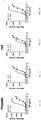
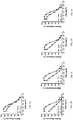
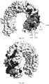
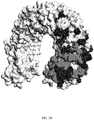
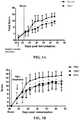

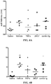
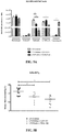


Classifications
-
- C—CHEMISTRY; METALLURGY
- C07—ORGANIC CHEMISTRY
- C07K—PEPTIDES
- C07K16/00—Immunoglobulins [IGs], e.g. monoclonal or polyclonal antibodies
- C07K16/18—Immunoglobulins [IGs], e.g. monoclonal or polyclonal antibodies against material from animals or humans
- C07K16/28—Immunoglobulins [IGs], e.g. monoclonal or polyclonal antibodies against material from animals or humans against receptors, cell surface antigens or cell surface determinants
- C07K16/2896—Immunoglobulins [IGs], e.g. monoclonal or polyclonal antibodies against material from animals or humans against receptors, cell surface antigens or cell surface determinants against molecules with a "CD"-designation, not provided for elsewhere
-
- A—HUMAN NECESSITIES
- A61—MEDICAL OR VETERINARY SCIENCE; HYGIENE
- A61K—PREPARATIONS FOR MEDICAL, DENTAL OR TOILETRY PURPOSES
- A61K39/00—Medicinal preparations containing antigens or antibodies
- A61K39/395—Antibodies; Immunoglobulins; Immune serum, e.g. antilymphocytic serum
- A61K39/39533—Antibodies; Immunoglobulins; Immune serum, e.g. antilymphocytic serum against materials from animals
- A61K39/3955—Antibodies; Immunoglobulins; Immune serum, e.g. antilymphocytic serum against materials from animals against proteinaceous materials, e.g. enzymes, hormones, lymphokines
-
- A—HUMAN NECESSITIES
- A61—MEDICAL OR VETERINARY SCIENCE; HYGIENE
- A61K—PREPARATIONS FOR MEDICAL, DENTAL OR TOILETRY PURPOSES
- A61K47/00—Medicinal preparations characterised by the non-active ingredients used, e.g. carriers or inert additives; Targeting or modifying agents chemically bound to the active ingredient
- A61K47/50—Medicinal preparations characterised by the non-active ingredients used, e.g. carriers or inert additives; Targeting or modifying agents chemically bound to the active ingredient the non-active ingredient being chemically bound to the active ingredient, e.g. polymer-drug conjugates
- A61K47/51—Medicinal preparations characterised by the non-active ingredients used, e.g. carriers or inert additives; Targeting or modifying agents chemically bound to the active ingredient the non-active ingredient being chemically bound to the active ingredient, e.g. polymer-drug conjugates the non-active ingredient being a modifying agent
- A61K47/68—Medicinal preparations characterised by the non-active ingredients used, e.g. carriers or inert additives; Targeting or modifying agents chemically bound to the active ingredient the non-active ingredient being chemically bound to the active ingredient, e.g. polymer-drug conjugates the non-active ingredient being a modifying agent the modifying agent being an antibody, an immunoglobulin or a fragment thereof, e.g. an Fc-fragment
- A61K47/6835—Medicinal preparations characterised by the non-active ingredients used, e.g. carriers or inert additives; Targeting or modifying agents chemically bound to the active ingredient the non-active ingredient being chemically bound to the active ingredient, e.g. polymer-drug conjugates the non-active ingredient being a modifying agent the modifying agent being an antibody, an immunoglobulin or a fragment thereof, e.g. an Fc-fragment the modifying agent being an antibody or an immunoglobulin bearing at least one antigen-binding site
- A61K47/6849—Medicinal preparations characterised by the non-active ingredients used, e.g. carriers or inert additives; Targeting or modifying agents chemically bound to the active ingredient the non-active ingredient being chemically bound to the active ingredient, e.g. polymer-drug conjugates the non-active ingredient being a modifying agent the modifying agent being an antibody, an immunoglobulin or a fragment thereof, e.g. an Fc-fragment the modifying agent being an antibody or an immunoglobulin bearing at least one antigen-binding site the antibody targeting a receptor, a cell surface antigen or a cell surface determinant
-
- A—HUMAN NECESSITIES
- A61—MEDICAL OR VETERINARY SCIENCE; HYGIENE
- A61P—SPECIFIC THERAPEUTIC ACTIVITY OF CHEMICAL COMPOUNDS OR MEDICINAL PREPARATIONS
- A61P1/00—Drugs for disorders of the alimentary tract or the digestive system
- A61P1/04—Drugs for disorders of the alimentary tract or the digestive system for ulcers, gastritis or reflux esophagitis, e.g. antacids, inhibitors of acid secretion, mucosal protectants
-
- A—HUMAN NECESSITIES
- A61—MEDICAL OR VETERINARY SCIENCE; HYGIENE
- A61P—SPECIFIC THERAPEUTIC ACTIVITY OF CHEMICAL COMPOUNDS OR MEDICINAL PREPARATIONS
- A61P11/00—Drugs for disorders of the respiratory system
- A61P11/06—Antiasthmatics
-
- A—HUMAN NECESSITIES
- A61—MEDICAL OR VETERINARY SCIENCE; HYGIENE
- A61P—SPECIFIC THERAPEUTIC ACTIVITY OF CHEMICAL COMPOUNDS OR MEDICINAL PREPARATIONS
- A61P19/00—Drugs for skeletal disorders
- A61P19/02—Drugs for skeletal disorders for joint disorders, e.g. arthritis, arthrosis
-
- A—HUMAN NECESSITIES
- A61—MEDICAL OR VETERINARY SCIENCE; HYGIENE
- A61P—SPECIFIC THERAPEUTIC ACTIVITY OF CHEMICAL COMPOUNDS OR MEDICINAL PREPARATIONS
- A61P19/00—Drugs for skeletal disorders
- A61P19/08—Drugs for skeletal disorders for bone diseases, e.g. rachitism, Paget's disease
- A61P19/10—Drugs for skeletal disorders for bone diseases, e.g. rachitism, Paget's disease for osteoporosis
-
- A—HUMAN NECESSITIES
- A61—MEDICAL OR VETERINARY SCIENCE; HYGIENE
- A61P—SPECIFIC THERAPEUTIC ACTIVITY OF CHEMICAL COMPOUNDS OR MEDICINAL PREPARATIONS
- A61P29/00—Non-central analgesic, antipyretic or antiinflammatory agents, e.g. antirheumatic agents; Non-steroidal antiinflammatory drugs [NSAID]
-
- A—HUMAN NECESSITIES
- A61—MEDICAL OR VETERINARY SCIENCE; HYGIENE
- A61P—SPECIFIC THERAPEUTIC ACTIVITY OF CHEMICAL COMPOUNDS OR MEDICINAL PREPARATIONS
- A61P3/00—Drugs for disorders of the metabolism
- A61P3/08—Drugs for disorders of the metabolism for glucose homeostasis
- A61P3/10—Drugs for disorders of the metabolism for glucose homeostasis for hyperglycaemia, e.g. antidiabetics
-
- A—HUMAN NECESSITIES
- A61—MEDICAL OR VETERINARY SCIENCE; HYGIENE
- A61P—SPECIFIC THERAPEUTIC ACTIVITY OF CHEMICAL COMPOUNDS OR MEDICINAL PREPARATIONS
- A61P31/00—Antiinfectives, i.e. antibiotics, antiseptics, chemotherapeutics
-
- A—HUMAN NECESSITIES
- A61—MEDICAL OR VETERINARY SCIENCE; HYGIENE
- A61P—SPECIFIC THERAPEUTIC ACTIVITY OF CHEMICAL COMPOUNDS OR MEDICINAL PREPARATIONS
- A61P35/00—Antineoplastic agents
-
- A—HUMAN NECESSITIES
- A61—MEDICAL OR VETERINARY SCIENCE; HYGIENE
- A61P—SPECIFIC THERAPEUTIC ACTIVITY OF CHEMICAL COMPOUNDS OR MEDICINAL PREPARATIONS
- A61P37/00—Drugs for immunological or allergic disorders
- A61P37/02—Immunomodulators
-
- A—HUMAN NECESSITIES
- A61—MEDICAL OR VETERINARY SCIENCE; HYGIENE
- A61P—SPECIFIC THERAPEUTIC ACTIVITY OF CHEMICAL COMPOUNDS OR MEDICINAL PREPARATIONS
- A61P37/00—Drugs for immunological or allergic disorders
- A61P37/02—Immunomodulators
- A61P37/06—Immunosuppressants, e.g. drugs for graft rejection
-
- A—HUMAN NECESSITIES
- A61—MEDICAL OR VETERINARY SCIENCE; HYGIENE
- A61P—SPECIFIC THERAPEUTIC ACTIVITY OF CHEMICAL COMPOUNDS OR MEDICINAL PREPARATIONS
- A61P37/00—Drugs for immunological or allergic disorders
- A61P37/08—Antiallergic agents
-
- A—HUMAN NECESSITIES
- A61—MEDICAL OR VETERINARY SCIENCE; HYGIENE
- A61K—PREPARATIONS FOR MEDICAL, DENTAL OR TOILETRY PURPOSES
- A61K39/00—Medicinal preparations containing antigens or antibodies
- A61K2039/505—Medicinal preparations containing antigens or antibodies comprising antibodies
-
- C—CHEMISTRY; METALLURGY
- C07—ORGANIC CHEMISTRY
- C07K—PEPTIDES
- C07K16/00—Immunoglobulins [IGs], e.g. monoclonal or polyclonal antibodies
- C07K16/18—Immunoglobulins [IGs], e.g. monoclonal or polyclonal antibodies against material from animals or humans
- C07K16/28—Immunoglobulins [IGs], e.g. monoclonal or polyclonal antibodies against material from animals or humans against receptors, cell surface antigens or cell surface determinants
- C07K16/2803—Immunoglobulins [IGs], e.g. monoclonal or polyclonal antibodies against material from animals or humans against receptors, cell surface antigens or cell surface determinants against the immunoglobulin superfamily
-
- C—CHEMISTRY; METALLURGY
- C07—ORGANIC CHEMISTRY
- C07K—PEPTIDES
- C07K2317/00—Immunoglobulins specific features
- C07K2317/30—Immunoglobulins specific features characterized by aspects of specificity or valency
- C07K2317/34—Identification of a linear epitope shorter than 20 amino acid residues or of a conformational epitope defined by amino acid residues
-
- C—CHEMISTRY; METALLURGY
- C07—ORGANIC CHEMISTRY
- C07K—PEPTIDES
- C07K2317/00—Immunoglobulins specific features
- C07K2317/50—Immunoglobulins specific features characterized by immunoglobulin fragments
- C07K2317/56—Immunoglobulins specific features characterized by immunoglobulin fragments variable (Fv) region, i.e. VH and/or VL
- C07K2317/565—Complementarity determining region [CDR]
-
- C—CHEMISTRY; METALLURGY
- C07—ORGANIC CHEMISTRY
- C07K—PEPTIDES
- C07K2317/00—Immunoglobulins specific features
- C07K2317/70—Immunoglobulins specific features characterized by effect upon binding to a cell or to an antigen
- C07K2317/76—Antagonist effect on antigen, e.g. neutralization or inhibition of binding
-
- C—CHEMISTRY; METALLURGY
- C07—ORGANIC CHEMISTRY
- C07K—PEPTIDES
- C07K2317/00—Immunoglobulins specific features
- C07K2317/70—Immunoglobulins specific features characterized by effect upon binding to a cell or to an antigen
- C07K2317/77—Internalization into the cell
-
- C—CHEMISTRY; METALLURGY
- C07—ORGANIC CHEMISTRY
- C07K—PEPTIDES
- C07K2317/00—Immunoglobulins specific features
- C07K2317/90—Immunoglobulins specific features characterized by (pharmaco)kinetic aspects or by stability of the immunoglobulin
- C07K2317/92—Affinity (KD), association rate (Ka), dissociation rate (Kd) or EC50 value
Definitions
- the present invention relates to antibodies (e.g. monoclonal antibodies), antibody fragments, and derivatives thereof that bind and inhibit TLR3 signaling.
- the invention also relates to cells producing such antibodies; methods of making such antibodies; fragments, variants, and derivatives of the antibodies; pharmaceutical compositions comprising the same; methods of using the antibodies to diagnose, treat or prevent diseases, e.g. autoimmune diseases, inflammatory diseases and the like.
- TLRs Drosophila toll proteins control dorsal-ventral patterning and are thought to represent an ancient host defense mechanism.
- TLRs are believed to be an important component of innate immunity.
- Human and Drosophila Toll protein sequences show homology over the entire length of the protein chains.
- the family of human Toll-like receptors is comprised of ten highly conserved receptor proteins, TLR1-TLR10.
- human TLRs are type I transmembrane proteins with an extracellular domain consisting of a leucine-rich repeat (LRR) domain that recognizes pathogen-associated molecular patterns (PAMPs), and a cytoplasmic domain that is homologous to the cytoplasmic domain of the human interleukin-1 (IL-1) receptor.
- LRR leucine-rich repeat
- PAMPs pathogen-associated molecular patterns
- IL-1 receptors Similar to the signaling pathways for both Drosophila toll and the IL-1 receptor, human Toll-like receptors signal through the NF-
- TLRs Although the different mammalian TLRs share many characteristics and signal transduction mechanisms, their biological functions are very different. This is due in part to the fact that four different adaptor molecules (MyD88, TIRAP, TRIF and TRAF) are associated in various combinations with the TLRs and mediate different signaling pathways. In addition, different ligands for one TLR may preferentially activate different signal transduction pathways. Furthermore, the TLRs are differentially expressed in various hematopoietic and non-hematopoietic cells. Accordingly, the response to a TLR ligand depends not only on the signal pathway activated by the TLR, but also on the nature of the cells in which the individual TLR is expressed.
- TLR3 Toll-like receptor 3
- Patent application WO98/50547 provides the nucleic acid and amino acid sequence of the hTLR3 protein.
- De Bouteiller et al. (2005) J. Biol. Chem. 280(46): 38133-38145 ) disclose use of an anti-TLR3 antibody to bind cell surface TLR3.
- Antibody C1130 is stated to be activatory toward TLR3 and has been described in WO 2007/051164 .
- Polyclonal antibodies that inhibited TLR3 were described in Cavassani et al. (2008) J. Exp. Med. 205: 2609-2621 .
- WO 03/106499 and Matsumoto et al. (2003) J. Immunol. 171:3154-3162 describes an antibody corresponding to antibody clone TLR3.7 (eBioScience Inc., San Diego) reported to bind and inhibit cell surface TLR3 but not cell compartment TLR3 or in myeloid-lineage DC.
- WO 06/060513 describes an antibody C1068 which is reported to inhibit cytokine production in epithelial cells, which are reported to express TLR3 on the cell surface.
- PCT patent application WO2010/051470 provides further anti-TLR3 antibodies.
- anti-TLR3 antibodies for research use include polyclonal anti-TLR3 antibodies from R&D Systems Corp., antibody 40C1285 from Abcam and antibodies 619F7, 713E4, 716G10, IMG-5631 and -IMG-5348, all from Imgenex Corp.
- anti-TLR3 antibodies are not optimally suited for use as therapeutic agents, e.g. to modulate TLR3 in vivo. For example, many suffer from lack of efficacy or affinity to their epitopes. There is therefore a need to provide improved antibodies directed to TLR3.
- compositions comprising, and methods of using monoclonal antibodies, including but not limited to antibody fragments, and derivatives that specifically bind to and inhibit the function of human TLR3.
- the present disclosure provides antibodies with new properties useful for targeting TLR3 in vivo. Since TLR3 binds its natural ligand (dsRNA) and signals exclusively in the endosome in macrophages and dendritic cells (DCs) (at acidic pH), antibodies have previously been selected based on high affinity at endosomal pH where signalling occurs. However, little remains known about the mechanism by which anti-TLR3 antibodies enter cells.
- the present disclosure provides antibodies that have strong binding to TLR3 exposed at the cell surface and in a pH neutral environment and which display improved potency in TLR3 inhibition. Optimizing binding of cell-surface expressed TLR3 may therefore be an important criteria for cellular (endosomal) uptake by a cell which may condition downstream (or overall) biological activity.
- the present antibodies show strong binding to human cell surface TLR3, as observed in an assay where TLR3 is expressed exclusively at the cell surface in neutral pH conditions. In this way, antibodies having improved cell surface binding were selected.
- the inhibitory activity of anti-TLR3 antibodies may therefore be governed by the cycling back to the cell surface of the endosomal TLR3 polypeptides involved in endocytosis once the receptors separated from their ligands.
- the disclosure provides an antibody that inhibits TLR3-mediated signalling in a TLR3-expressing cell, wherein the antibody specifically binds a human TLR3 polypeptide expressed solely at the surface of a cell, optionally at neutral pH.
- the antibody has an EC 50 of no more than 0.3 ⁇ g/ml, optionally no more than 0.2 ⁇ g/ml, optionally no more than 0.1 ⁇ g/ml, for binding to cells expressing TLR3 solely at the cell surface.
- the disclosure provides antibodies that bind the N-terminal portion of the TLR3 protein at least partly (or primarily or exclusively) on the glycan-free lateral surface of the TLR3 polypeptide (the face bound by dsRNA), and optionally furthermore at least partly within the N-terminal dsRNA binding site of a human TLR3 polypeptide.
- the antibody further binds TLR3 at least partly within the backbone of the N-terminal portion of the TLR3 polypeptide.
- the disclosure provides antibodies that inhibit TLR3 polypeptide activity by binding to the N-terminal portion of a human TLR3 protein, and that also bind non-human primate TLR3. In one aspect the disclosure provides antibodies that bind the N-terminal portion of the TLR3 protein and that do not compete with dsRNA for binding to human TLR3, wherein the antibodies also bind to a of non-human primate TLR3 polypeptide (in a non-human primate-TLR3-expressing cell).
- the disclosure provides antibodies that bind the N-terminal portion of the TLR3 protein and that competes with dsRNA for binding to the N-terminal dsRNA binding site of a human TLR3 polypeptide, wherein the antibodies also bind to a non-human primate TLR3 polypeptide (in a non-human primate-TLR3-expressing cell).
- the antibodies at least partly (or primarily or exclusively) on the glycan-free lateral surface of the TLR3 polypeptide (the face bound by dsRNA).
- the antibodies bind to TLR3 at least partly within the N-terminal dsRNA binding site of a human TLR3 polypeptide.
- the non-human primate is macaca fascicularis.
- the non-human primate TLR3 polypeptide comprises an amino acid sequence shown in NCBI accession number BAG55033 (SEQ ID NO: 2).
- the disclosure provides antibodies that bind to the N-terminal portion of a human TLR3 protein, notably within residues 41 to 251, optionally at least partly within residues 41 to 139, 41 to 120 or residues 41 to 89, of human TLR3 of SEQ ID NO: 1.
- the disclosure provides antibodies that bind to the N-terminal portion of a human TLR3 protein, wherein the antibody has reduced binding to a TLR3 polypeptide having a mutation in its N-terminal portion in the segment corresponding to residues 41-139 of SEQ ID NO: 1, relative to binding between the antibody and a wild-type TLR3 polypeptide of SEQ ID NO: 1.
- the antibodies of the disclosure interfere with binding of dsRNA to the N-terminal dsRNA binding site of a human TLR3 polypeptide.
- the antibodies bind one or more amino acid residues within the glycan-free lateral surface of the TLR3 polypeptide that is involved in binding of the TLR3 polypeptide to dsRNA, and/or residues adjacent thereto.
- the antibodies bind an epitope comprising residues 64 and/or residue 65 of SEQ ID NO: 1, and/or have reduced binding to a TLR3 polypeptide having a mutation at residues 64 and/or residue 65 of SEQ ID NO: 1.
- the antibodies bind an epitope comprising residues 86 and/or residue 89 of SEQ ID NO: 1, and/or have reduced binding to a TLR3 polypeptide having a mutation at residues 86 and/or residue 89 of SEQ ID NO: 1.
- the antibodies bind an epitope comprising residues 117 and/or residue 120 of SEQ ID NO: 1, and/or have reduced binding to a TLR3 polypeptide having a mutation at residues 117 and/or residue 120 of SEQ ID NO: 1.
- the antibodies bind an epitope comprising residues 137 and/or residue 139 of SEQ ID NO: 1, and/or have reduced binding to a TLR3 polypeptide having a mutation at residues 137 and/or residue 139 of SEQ ID NO: 1.
- the antibodies bind an epitope comprising residues 112, 113 and/or 115 of SEQ ID NO: 1, and/or have reduced binding to a TLR3 polypeptide having a mutation at residues 112, 113 and/or 115 of SEQ ID NO: 1.
- the antibodies have reduced binding to a TLR3 polypeptide having a mutation at residues 117 and/or residue 120 of SEQ ID NO: 1, and reduced binding to a TLR3 polypeptide having a mutation at residues 137 and/or residue 139 of SEQ ID NO: 1.
- the antibodies have reduced binding to a TLR3 polypeptide having a mutation at residues 117 and/or residue 120 of SEQ ID NO: 1, and do not have reduced binding to a TLR3 polypeptide having a mutation at residues 137 and/or residue 139 of SEQ ID NO: 1.
- the antibodies have reduced binding to a TLR3 polypeptide having a mutation at residues 64 and/or residue 65 of SEQ ID NO: 1, and reduced binding to a TLR3 polypeptide having a mutation at residues 137 and/or residue 139 of SEQ ID NO: 1.
- the antibodies have reduced binding to a TLR3 polypeptide having a mutation at residues 86 and/or residue 89 of SEQ ID NO: 1. and reduced binding to a TLR3 polypeptide having a mutation at residues 137 and/or residue 139 of SEQ ID NO: 1.
- the antibodies do not have reduced binding to a TLR3 polypeptide having a mutation at residues 117 and/or residue 120 of SEQ ID NO: 1.
- the antibodies do not have reduced binding to a TLR3 polypeptide having a mutation at residues 112, 113 and/or 115 of SEQ ID NO: 1.
- the antibodies have reduced binding to a TLR3 polypeptide having a mutation at residues 64 and/or residue 65 of SEQ ID NO: 1, a TLR3 polypeptide having a mutation at residues 86 and/or residue 89 of SEQ ID NO: 1 and a TLR3 polypeptide having a mutation at residues 137 and residue 139 of SEQ ID NO: 1.
- the antibodies differ in their epitope from previously described antibodies.
- the antibodies have reduced binding to a TLR3 polypeptide having a mutation at residues 112, 113 and/or 115 of SEQ ID NO: 1, and reduced binding to a TLR3 polypeptide having a mutation at residues 137 and/or residue 139 of SEQ ID NO: 1. In one aspect, the antibodies have reduced binding to a TLR3 polypeptide having a mutation at residues 112, 113 and/or 115 of SEQ ID NO: 1, and reduced binding to a TLR3 polypeptide having a mutation at residues 117 and/or residue 120 of SEQ ID NO: 1.
- the antibodies have reduced binding to a TLR3 polypeptide having a mutation at residues 112, 113 and/or 115 of SEQ ID NO: 1, reduced binding to a TLR3 polypeptide having a mutation at residues 117 and/or residue 120 of SEQ ID NO: 1, and reduced binding to a TLR3 polypeptide having a mutation at residues 137 and/or residue 139 of SEQ ID NO: 1.
- the antibodies have reduced binding to a TLR3 polypeptide having a mutation at residues 112 and/or 113 of SEQ ID NO: 1, and reduced binding to a TLR3 polypeptide having a mutation at residue 137 of SEQ ID NO: 1.
- the antibodies do not have reduced binding to a TLR3 polypeptide having a mutation at residues 86 and/or residue 89 of SEQ ID NO: 1.
- the antibodies do not have reduced binding to a TLR3 polypeptide having a mutation at residues 64 and/or residue 65 of SEQ ID NO: 1.
- the antibodies maintain binding (do not have reduced binding) to a TLR3 polypeptide having a mutation at residues 116, 145, 182, 196 and/or residue 171 of SEQ ID NO: 1.
- the disclosure provides antibodies that interfere with binding of dsRNA to a human TLR3 polypeptide.
- the disclosure provides antibodies that interfere with binding of dsRNA to the N-terminal dsRNA binding site of a human TLR3 polypeptide.
- the antibodies compete with dsRNA for binding to human TLR3 polypeptide, e.g, to the N-terminal dsRNA binding site of a human TLR3 polypeptide. Competition can be assessed using standard methods, e.g. Biacore assays to assess whether antibodies bind to immobilized TLR3 in the presence of dsRNA, and/or whether dsRNA binds to immobilized TLR3 in the presence of antibodies, under acidic conditions.
- the antibodies compete with dsRNA for binding to human TLR3 polypeptide in an in vitro assay comprising the steps of: (i) contacting an anti-TLR3 antibody with a TLR3 polypeptide so as to obtain antibodies bound to TLR3 polypeptide, and (ii) contacting the antibody bound TLR3 polypeptide of step (i) with dsRNA and assessing whether dsRNA decreases binding of TLR3 polypeptide ( to the antibody, wherein a decrease of binding indicates competition with dsRNA for human TLR3 polypeptide.
- the antibodies compete with dsRNA for binding to human TLR3 polypeptide in an in vitro assay comprising the steps of: (i) contacting a TLR3 polypeptide with dsRNA so as to obtain dsRNA bound to TLR3 polypeptide, and (ii) contacting the dsRNA bound TLR3 polypeptide of step (i) with an anti-TLR3 antibody and assessing whether the TLR3 polypeptide binds the dsRNA-TLR3 polypeptide complex, wherein lack of substantial binding indicates competition with dsRNA for human TLR3 polypeptide.
- the antibodies compete with dsRNA for binding to human TLR3 polypeptide in an in vitro assay comprising the steps of: (i) attaching an anti-TLR3 antibody to a solid support (e.g., via a constant domain), (ii) contacting said antibody with a TLR3 polypeptide so as to obtain antibodies bound to TLR3 polypeptide, and (iii) contacting the antibody bound TLR3 polypeptide of step (ii) with dsRNA and assessing whether dsRNA decreases binding of TLR3 polypeptide to the antibody, wherein a decrease of binding indicates competition with dsRNA for human TLR3 polypeptide.
- the antibody binds to a human TLR3 polypeptide expressed at the surface of a cell, optionally as assessed in a cell expressing TLR3 exclusively at the cell surface under neutral pH, internalizes into a cell that expresses TLR3, and inhibits TLR3 signaling in a cell (e.g. a TLR3-expressing human dendritic cell).
- the antibodies bind human TLR3 polypeptides under neutral conditions, and in particular under conditions representative of that encountered in the cell cytosol.
- neutral conditions are generally characterized by a pH between 6.6 and 7.4, for example a slightly alkaline pH of 7.2 found in the cell cytosol.
- the antibody has a K D of no more than 10 -9 M, optionally less than 10 -10 M, optionally less than 10 -11 M for binding to a TLR3 polypeptide at neutral pH.
- the binding at neutral conditions is of better affinity than under acid conditions, e.g. where the K D for binding to TLR3 at neutral compared to acidic conditions is lower by at least 0.5-, 1.0-, 1.5- or 2.0- log 10 .
- the present disclosure further provides specific antibodies have increased activity over previously reported antibodies.
- the antibody competes for binding to a TLR3 polypeptide (e.g. a human TLR3 polypeptide comprising an amino acid sequence of SEQ ID NO: 1) with any one or any combination of monoclonal antibody 11E1, 7G11, 31F6, 32C4 and 37B7, optionally under acid and/or neutral conditions.
- a TLR3 polypeptide e.g. a human TLR3 polypeptide comprising an amino acid sequence of SEQ ID NO: 1
- monoclonal antibody 11E1, 7G11, 31F6, 32C4 and 37B7 optionally under acid and/or neutral conditions.
- an antibody of the disclosure competes for binding to a TLR3 polypeptide, optionally under acid and/or neutral conditions, with an antibody having respectively a VH and VL region of SEQ ID NOS: 3 and 4 (11E1), a VH and VL region of SEQ ID NOS: 14 and 15 (31F6), a VH and VL region of SEQ ID NOS: 25 and 26 (32C4), a VH and VL region of SEQ ID NOS: 36 and 37 (37B7) or a VH and VL region of SEQ ID NOS: 47 and 48 (7G11).
- the antibody may have a heavy and/or light chain having one, two or three CDRs of antibody 11E1, 7G11, 31F6, 32C4 or 37B7.
- the antibody that specifically binds TLR3 has one or more (including any combination thereof, or all of) of the following properties:
- the disclosure provides a monoclonal antibody that specifically binds to at least one, two, three, four, five, six, seven or more residues in the segment corresponding to residues 1-251, optionally 41-251, 41-89, 41-120 or 41-139 of the TLR3 polypeptide of SEQ ID NO: 1.
- the antibody inhibits signaling by the TLR3 polypeptide.
- the antibody does not bind residue 116, residue 145 and/or residue 182 of the TLR3 polypeptide of SEQ ID NO: 1.
- the antibody does not bind residue 171, and/or residue 196 of the TLR3 polypeptide of SEQ ID NO: 1.
- binding of the antibody to a TLR3 polypeptide having a mutation at residues 116, 145, 182 196 and/or residue 171 of the TLR3 polypeptide of SEQ ID NO: 1 is maintained (i.e., is not substantially reduced), in comparison to binding to a wild-type TLR3 polypeptide of SEQ ID NO: 1; preferably said mutation is a K145E, D116R, K182E, N196A and/or E171A mutation.
- Such antibodies can optionally further have any properties described herein, e.g. subnanomolar affinity for a TLR3 polypeptide at an acidic pH, inhibits TLR3 signaling in the presence of a TLR3 ligand or in an inflammatory background (e.g.
- TLR3 polypeptide in the presence of inflammatory cytokines such as IFN ⁇ , competes for binding to a TLR3 polypeptide with 11E1, 7G11, 31F6, 32C4 or 37B7; does not compete with dsRNA for binding to C-terminal portion the TLR3 polypeptide; or inhibits IP-10 secretion on DC (e.g. in human myeloid DC).
- inflammatory cytokines such as IFN ⁇
- the disclosure provides an antibody that binds a TLR3 polypeptide, wherein the antibody comprises (i) the heavy chain CDR 1, 2 and 3 (HCDR1, HCDR2, HCDR3) amino acid sequences as shown in SEQ ID NO: 5, 6 or 7 (HCDR1), 8 or 9 (HCDR2) and 10 (HCDR3), and (ii) the light chain CDR 1, 2 and 3 (LCDR1, LCDR2, LCDR3) amino acid sequences as shown in SEQ ID NO: 11, 12 and 13, respectively; wherein one, two, three, four, or five or more of the amino acids in any of said sequences may be substituted by a different amino acid.
- HCDR1, HCDR2, HCDR3 heavy chain CDR1, HCDR2, HCDR3 amino acid sequences as shown in SEQ ID NO: 5, 6 or 7 (HCDR1), 8 or 9 (HCDR2) and 10 (HCDR3)
- LCDR1, LCDR2, LCDR3 light chain CDR 1, 2 and 3 amino acid sequences as shown in SEQ ID NO: 11, 12
- the antibody comprises (i) the heavy chain CDR 1, 2 and 3 (HCDR1, HCDR2, HCDR3) amino acid sequences as shown in SEQ ID NO: 16, 17 or 18 (HCDR1), 19 or 20 (HCDR2) and 21 (HCDR3), and (ii) the light chain CDR 1, 2 and 3 (LCDR1, LCDR2, LCDR3) amino acid sequences as shown in SEQ ID NO: 22, 23 and 24, respectively; wherein one, two, three, four, or five or more of the amino acids in any of said sequences may be substituted by a different amino acid.
- the antibody comprises (i) the heavy chain CDR 1, 2 and 3 (HCDR1, HCDR2, HCDR3) amino acid sequences as shown in SEQ ID NO: 27, 28 or 29 (HCDR1), 30 or 31(HCDR2) and 32 (HCDR3), and (ii) the light chain CDR 1, 2 and 3 (LCDR1, LCDR2, LCDR3) amino acid sequences as shown in SEQ ID NO: 33, 34 and 35, respectively; wherein one, two, three, four, or five or more of the amino acids in any of said sequences may be substituted by a different amino acid.
- the antibody comprises (i) the heavy chain CDR 1, 2 and 3 (HCDR1, HCDR2, HCDR3) amino acid sequences as shown in SEQ ID NO: 38, 39 or 40 (HCDR1), 41 or 42 (HCDR2) and 43 (HCDR3), and (ii) the light chain CDR 1, 2 and 3 (LCDR1, LCDR2, LCDR3) amino acid sequences as shown in SEQ ID NO: 44, 45 and 46, respectively; wherein one, two, three, four, or five or more of the amino acids in any of said sequences may be substituted by a different amino acid.
- the antibody comprises (i) the heavy chain CDR 1, 2 and 3 (HCDR1, HCDR2, HCDR3) amino acid sequences as shown in SEQ ID NO: 49, 50 or 51 (HCDR1), 52 or 53 (HCDR2) and 54 (HCDR3), and (ii) the light chain CDR 1, 2 and 3 (LCDR1, LCDR2, LCDR3) amino acid sequences as shown in SEQ ID NO: 55, 56 and 57, respectively; wherein one, two, three, four, or five or more of the amino acids in any of said sequences may be substituted by a different amino acid.
- the antibody of any of the aspects herein is capable of being internalized by a cell that expresses TLR3 polypeptide on its surface.
- the antibody is chimeric, e.g. contains a non-murine, optionally a human, constant region. In one aspect, the antibody is human or humanized. In another aspect, the antibody is a mouse or rat antibody (e.g., comprises CDRs derived from a rat or rat gene or rat Ig locus gene segment). In another aspect, the antibody does not substantially bind to other human TLRs (e.g. TLR4).
- TLR4 human TLRs
- the isotype of the antibody is IgG, optionally IgG1 or IgG3.
- the antibody comprises an Fc domain or is of an isotype that is bound by Fc ⁇ R.
- the antibody is an antibody fragment selected from Fab, Fab', Fab'-SH, F(ab')2, Fv, diabodies, single-chain antibody fragment, or a multispecific antibody comprising multiple different antibody fragments.
- the antibody does not comprise an Fc domain or is of an isotype that is not substantially bound by Fc ⁇ R (e.g. human CD16).
- the antibody is of human IgG4 or IgG2 isotype. Human IgG4 isotypes or other IgG isotypes modified to reduce their Fc ⁇ R binding can be used for their advantageous pharmacological properties such as serum half-life, while modulating TLR3 signaling, in e.g.
- the anti-TLR3 antibody inhibits TLR3 signaling and comprises a constant region of human IgG4 or IgG2 isotype. In one aspect, of any of the aspects of the disclosure, the anti-TLR3 antibody inhibits TLR3 signaling and comprises a constant region (heavy chain constant region) that does not substantially bind Fc ⁇ RIIIa.
- the anti-TLR3 antibody comprises a heavy chain of human IgG4 isotype.
- the anti-TLR3 antibody comprises a human IgG4 heavy chain constant region and comprising a serine to proline mutation in residue 241, corresponding to position 228 according to the EU-index ( Kabat et al., "Sequences of proteins of immunological interest", 5th ed., NIH, Bethesda, ML, 1991 ).
- compositions comprising such antibodies can be characterized as having less than about 15%, such as less than about 10% (e.g., about 5% or less, about 4% or less, about 3% or less, or even about 1% or less) of IgG4 "half-antibodies" (comprising a single heavy chain/light chain pair).
- IgG4 "half-antibody” byproducts form due to heterogeneity of inter-heavy chain disulfide bridges in the hinge region in a proportion of secreted human IgG4 (see Angal et al., Molecular Immunology, 30(1):105-108, 1993 for a description of IgG4 "half-antibodies", S241P mutation, and related principles). This effect is typically only detectable under denaturing, non-reducing conditions.
- the antibody is conjugated or covalently bound to a detectable or toxic moiety.
- the antibodies optionally inhibit TLR3 signaling without blocking binding of a dsRNA TLR3 ligand to the principal (i.e. C-terminal) dsRNA binding site of the TLR3 polypeptide.
- the antibodies also bind human TLR3 under acidic conditions, and in particular under conditions representative of that encountered in an acidified subcellular compartment of a cell (e.g. compartments of the endocytic pathway endosomic, lysosomal).
- acidic conditions are generally characterized by a pH lower than about pH 6.5, or between about pH 4.5 to 6.5, or about pH 5.6.
- the antibodies modulate, optionally inhibit, TLR3 signaling in an acidified subcellular compartment of a cell (e.g. compartments of the endocytic pathway endosomic, lysosomal).
- the antibodies modulate, optionally inhibit, TLR3 signaling in a dendritic cell (DC) (e.g. a myeloid DC, monocyte derived DC).
- DC dendritic cell
- the antibodies' bivalent binding affinity for TLR3 under neutral and/or acidic conditions can optionally be characterized by a mean K D of no more than about (i.e. better affinity than) 100, 50, 10, 5, or 1 nanomolar, preferably sub-nanomolar or optionally no more than about 500, 200, 100 or 10 picomolar.
- the antibodies inhibit TLR3 signaling by at least partly (or fully) blocking the binding of a TLR3 ligand to a TLR3 polypeptide.
- the TLR3 ligand will generally be a ligand other than an anti-TLR3 antibody and may be a naturally occurring or non-naturally occurring TLR3 ligand, optionally a dsRNA-based ligand such as polyAU (polyadenylic acid:polyuridylic acid) or polylC (polyinosinic:polycytidylic acid).
- the disclosure provides a method of identifying, screening and/or producing an antibody that specifically binds and inhibits a TLR3 polypeptide in a mammalian subject, said method comprising the steps of: a) providing a plurality of antibodies that bind human TLR3 polypeptide, optionally by a method comprising immunizing a non-human mammal with an immunogen comprising a TLR3 polypeptide, and (b) assessing the binding affinity of said antibodies for the TLR3 polypeptide in a cell which expresses human TLR3 solely at the cell surface.
- the method further comprises selecting an antibody from said plurality that has an EC 50 of no more than 0.3 ⁇ g/ml, optionally no more than 0.2 ⁇ g/ml, optionally no more than 0.1 ⁇ g/ml, for binding to cells expressing human TLR3 solely at the cell surface.
- any of the methods of the disclosure can further be characterized as comprising any step described in the application, including notably in the "Detailed Description of the Invention").
- the disclosure further relates to an antibody obtainable by any of present methods.
- the disclosure further relates to pharmaceutical or diagnostic formulations of the antibodies of the present disclosure.
- the disclosure further relates to methods of using antibodies in methods of treatment or diagnosis, optionally in combination with a second therapeutic agent (e.g. a corticoid, a DMARD, an anti-cytokine or anti-cytokine receptor agent, an anti-TNFalpha agent, etc.).
- a second therapeutic agent e.g. a corticoid, a DMARD, an anti-cytokine or anti-cytokine receptor agent, an anti-TNFalpha agent, etc.
- the present disclosure is based, at least in part, on the discovery of high affinity monoclonal antibodies that specifically and efficiently inhibit the TLR3 signaling pathway.
- the inventors also provide new epitopes present on human TLR3, including the epitope recognized by antibody 11E1, 7G11, 31F6, 32C4 and/or 37B7, which are particularly efficient in inhibiting TLR3 signaling, and inhibiting cytokine release in response to stimulation with a TLR3 ligand.
- the antibodies can be used for treating an autoimmune or inflammatory disease in a subject in need thereof.
- the present disclosure also provides methods for treating relapses, attacks, or acute phases, occurring during the course of an inflammatory or autoimmune disease in a subject in need thereof using an anti-TLR3 antibody which inhibits TLR3 signaling.
- the present disclosure also provides novel methods for treating established inflammatory or autoimmune diseases in a subject in need thereof using an anti-TLR3 antibody which inhibits TLR3 signaling.
- the disclosure also provides treatment regimens and treatment combinations that can be used for the treatment of inflammatory or autoimmune disease in a subject in need thereof using an anti-TLR3 antibody which inhibits TLR3 signaling.
- the antibodies of the present disclosure that bind TLR3 under acidic and neutral conditions will generally bind both cell surface TLR3 and endosomic TLR3 at high affinity, such that the antibodies will be useful in any situation (e.g. treatment or prevention of disease) where targeting (e.g. modulating) TLR3 is useful.
- TLR3 has been found in some cases of inflammation the surface of macrophages and blocking TLR3 upon chloroquine neutralization of endosomal acidification nevertheless exhibited some anti-inflammatory activity (Cavassani et al. 2008, supra ) .
- the antibodies of the disclosure will have the greatest advantage over other antibodies in the treatment or prevention of diseases where the modulating (e.g.
- TLR3 in the cytosolic (e.g. endosomic) compartments
- cytosolic (e.g. endosomic) compartments are useful or required, and the relative importance of modulating signaling of such compartments TLR3 may depend on the disease.
- TLR3 cytosolic compartments
- endosomic compartment-expressed TLR3 is believed to play an important role in rheumatoid arthritis, since treatment with chloroquine, an inhibitor of endosomal acidification, inhibits TLR3 signaling and inhibits production of inflammatory cytokines from synovial cultures from patients having rheumatoid arthritis ( Sacre et al. (2008) J. Immunol. 181:8002-8009 ).
- TLR3 Endosomic compartment-expressed TLR3 is believed to play an important role in a number of other diseases where DC (e.g. myeloid DC) are involved in exacerbating disease, as mDC have a well-documented capacity to take up antigens from apoptotic or necrotic cells including during tissue necrosis during acute inflammation.
- DC e.g. myeloid DC
- the present antibodies are specific for TLR3, they can also be used for other purposes, including purifying TLR3 or TLR3-expressing cells, modulating (e.g. activating or inhibiting) TLR3 receptors in vitro, ex vivo, or in vivo, targeting TLR3-expressing cells for destruction in vivo, or specifically labeling/binding TLR3 in vivo, ex vivo, or in vitro, including for methods such as immunoblotting, IHC analysis, i.e. on frozen biopsies, FACS analysis, and immunoprecipitation.
- TLR3 ligand refers to any compound that can specifically bind to and alter the activity of TLR3 in vitro, ex vivo, or in vivo.
- the compound can be a naturally occurring ligand, e.g., generally dsRNA or viral dsRNA, or a synthetic ligand such as polylC or polyAU.
- the compound can be any type of molecule, including inorganic or organic compounds or elements, including proteins (such as antibodies), nucleic acids, carbohydrates, lipids, or any other molecular entity.
- such compounds can modulate TLR3 receptors in any way, including activating or inhibiting, and by any mechanism, including by binding to the receptor and triggering or shutting off activity in a manner similar to a naturally occurring ligand, or by binding to the receptor and blocking access to other ligands.
- the ligand activates the receptor, and as such can be used to induce the production of cytokines by TLR3-expressing cells.
- antibody refers to polyclonal and monoclonal antibodies. Depending on the type of constant domain in the heavy chains, antibodies are assigned to one of five major classes: IgA, IgD, IgE, IgG, and IgM. Several of these are further divided into subclasses or isotypes, such as IgG1, IgG2, IgG3, IgG4, and the like.
- An exemplary immunoglobulin (antibody) structural unit comprises a tetramer. Each tetramer is composed of two identical pairs of polypeptide chains, each pair having one "light” (about 25 kDa) and one "heavy" chain (about 50-70 kDa).
- variable light chain VL
- variable heavy chain VH
- the heavy-chain constant domains that correspond to the different classes of immunoglobulins are termed "alpha,” “delta,” “epsilon,” “gamma” and “mu,” respectively.
- alpha alpha
- delta delta
- epsilon gamma
- mu mu
- the subunit structures and three-dimensional configurations of different classes of immunoglobulins are well known.
- IgG and/or IgM are the preferred classes of antibodies employed in this disclosure, with IgG being particularly preferred, because they are the most common antibodies in the physiological situation and because they are most easily made in a laboratory setting.
- the antibody of this disclosure is a monoclonal antibody.
- Particularly preferred are humanized, chimeric, human, or otherwise-human-suitable antibodies.
- Antibodies also includes any fragment or derivative of any of the herein described antibodies.
- the term "specifically binds to” means that an antibody can bind preferably in a competitive binding assay to the binding partner, e.g. TLR3, as assessed using either recombinant forms of the proteins, epitopes therein, or native proteins present on the surface of isolated target cells.
- a competitive binding assay to the binding partner, e.g. TLR3, as assessed using either recombinant forms of the proteins, epitopes therein, or native proteins present on the surface of isolated target cells.
- an antibody When an antibody is said to "compete with” a particular monoclonal antibody (e.g. 11E1,7G11, 31F6, 32C4 or 37B7) or other TLR3 ligand (e.g., dsRNA, it means that the antibody competes with the monoclonal antibody (or other TLR3 ligand) in a binding assay using either recombinant TLR3 molecules or surface expressed TLR3 molecules.
- a test antibody reduces the binding of 11E1, 7G11, 31F6, 32C4 or 37B7 to a TLR3 polypeptide or TLR3-expressing cell in a binding assay, the antibody is said to "compete” respectively with 11E1, 7G11, 31F6, 32C4 or 37B7.
- affinity means the strength of the binding of an antibody to an epitope.
- the affinity of an antibody is given by the dissociation constant K D , defined as [Ab] x [Ag] / [Ab-Ag], where [Ab-Ag] is the molar concentration of the antibody-antigen complex, [Ab] is the molar concentration of the unbound antibody and [Ag] is the molar concentration of the unbound antigen.
- K D dissociation constant
- Ka is defined by 1/Kd.
- a “determinant” designates a site of interaction or binding on a polypeptide.
- epitope is defined as an antigenic determinant, and is the area or region on an antigen to which an antibody binds.
- a protein epitope may comprise amino acid residues directly involved in the binding as well as amino acid residues which are effectively blocked by the specific antigen binding antibody or peptide, i.e., amino acid residues within the "footprint” of the antibody. It is the simplest form or smallest structural area on a complex antigen molecule that can combine with e.g., an antibody or a receptor.
- Epitopes can be linear or conformational/structural.
- linear epitope is defined as an epitope composed of amino acid residues that are contiguous on the linear sequence of amino acids (primary structure).
- conformational or structural epitope is defined as an epitope composed of amino acid residues that are not all contiguous and thus represent separated parts of the linear sequence of amino acids that are brought into proximity to one another by folding of the molecule (secondary, tertiary and/or quaternary structures).
- a conformational epitope is dependent on the 3-dimensional structure.
- 'conformational' is therefore often used interchangeably with 'structural'.
- immunoglobulin any polypeptidic or peptidic fragment that is capable of eliciting an immune response such as (i) the generation of antibodies binding said fragment and/or binding any form of the molecule comprising said fragment, including the membrane-bound receptor and mutants derived therefrom, (ii) the stimulation of a T-cell response involving T-cells reacting to the bi-molecular complex comprising any MHC molecule and a peptide derived from said fragment, (iii) the binding of transfected vehicles such as bacteriophages or bacteria expressing genes encoding mammalian immunoglobulins.
- an immunogenic fragment also refers to any construction capable of eliciting an immune response as defined above, such as a peptidic fragment conjugated to a carrier protein by covalent coupling, a chimeric recombinant polypeptide construct comprising said peptidic fragment in its amino acid sequence, and specifically includes cells transfected with a cDNA of which sequence comprises a portion encoding said fragment.
- human-suitable antibody refers to any antibody, derivatized antibody, or antibody fragment that can be safely used in humans for, e.g. the therapeutic methods described herein.
- Human-suitable antibodies include all types of humanized, chimeric, or fully human antibodies, or any antibodies in which at least a portion of the antibodies is derived from humans or otherwise modified so as to avoid the immune response that is generally provoked when native non-human antibodies are used.
- a “humanized” or “human” antibody refers to an antibody in which the constant and variable framework region of one or more human immunoglobulins is fused with the binding region, e.g. the CDR, of an animal immunoglobulin.
- Such antibodies are designed to maintain the binding specificity of the non-human antibody from which the binding regions are derived, but to avoid an immune reaction against the non-human antibody.
- Such antibodies can be obtained from transgenic mice or other animals that have been "engineered” to produce specific human antibodies in response to antigenic challenge (see, e.g., Green et al. (1994) Nature Genet 7:13 ; Lonberg et al. (1994) Nature 368:856 ; Taylor et al.
- a fully human antibody also can be constructed by genetic or chromosomal transfection methods, as well as phage display technology, all of which are known in the art (see, e.g., McCafferty et al. (1990) Nature 348:552-553 ). Human antibodies may also be generated by in vitro activated B cells (see, e.g., U.S. Pat. Nos. 5,567,610 and 5,229,275 ).
- a “chimeric antibody” is an antibody molecule in which (a) the constant region, or a portion thereof, is altered, replaced or exchanged so that the antigen binding site (variable region) is linked to a constant region of a different or altered class, effector function and/or species, or an entirely different molecule which confers new properties to the chimeric antibody, e.g., an enzyme, toxin, hormone, growth factor, drug, etc.; or (b) the variable region, or a portion thereof, is altered, replaced or exchanged with a variable region having a different or altered antigen specificity.
- Fc domain refers to a C-terminal fragment of an antibody heavy chain, e.g., from about amino acid (aa) 230 to about aa 450 of human ⁇ (gamma) heavy chain or its counterpart sequence in other types of antibody heavy chains (e.g., ⁇ , ⁇ , ⁇ and ⁇ for human antibodies), or a naturally occurring allotype thereof.
- aa amino acid
- gamma human ⁇
- ⁇ ⁇
- ⁇ and ⁇ for human antibodies e.g., ⁇ , ⁇ and ⁇ for human antibodies
- the commonly accepted Kabat amino acid numbering for immunoglobulins is used throughout this disclosure (see Kabat et al. (1991) Sequences of Protein of Immunological Interest, 5th ed., United States Public Health Service, National Institute of Health, Bethesda, MD ).
- isolated refers to material that is substantially or essentially free from components which normally accompany it as found in its native state. Purity and homogeneity are typically determined using analytical chemistry techniques such as polyacrylamide gel electrophoresis or high performance liquid chromatography. A protein that is the predominant species present in a preparation is substantially purified.
- polypeptide peptide
- protein protein
- amino acid polymers in which one or more amino acid residue is an artificial chemical mimetic of a corresponding naturally occurring amino acid, as well as to naturally occurring amino acid polymers and non-naturally occurring amino acid polymer.
- recombinant when used with reference, e.g., to a cell, or nucleic acid, protein, or vector, indicates that the cell, nucleic acid, protein or vector, has been modified by the introduction of a heterologous nucleic acid or protein or the alteration of a native nucleic acid or protein, or that the cell is derived from a cell so modified.
- recombinant cells express genes that are not found within the native (nonrecombinant) form of the cell or express native genes that are otherwise abnormally expressed, under expressed or not expressed at all.
- antibody that "binds" a common determinant designates an antibody that binds said determinant with specificity and/or affinity.
- the antibodies suitable for the method of the disclosure specifically bind TLR3.
- Antibodies of the disclosure furthermore bind TLR3 with high affinity at conditions corresponding to that encountered in at the cell surface.
- Antibodies of the disclosure are furthermore capable of inhibiting the TLR3 signaling pathway.
- the ability of the inhibitory antibodies to specifically inhibit the TLR3 signaling pathway makes them useful for numerous applications, in particular for treating or preventing diseases wherein the inhibition of TLR3 signaling pathway is desirable, i.e. avoid further cytokine and chemokine secretion as well as cellular activation, as described herein.
- the disclosure provides methods using an antibody that binds human TLR3, and competes for binding to human TLR3 with monoclonal antibody 11E1, 7G11, 31F6, 32C4 or 37B7.
- TLR3 TLR3 polypeptide
- TLR3 receptor TLR3 receptor
- SEQ ID NO: 1 NCBI accession number NP_003256
- the human TLR3 mRNA sequence is described in NCBI accession number NM_003265.
- Human TLR3 sequences are also described in PCT patent publication no. WO 98/50547 .
- a non-human primate (macaca fascicularis) TLR3 amino acid sequence is shown in NCBI accession number BAG55033 (SEQ ID NO: 2).
- the disclosure provides an antibody that competes with monoclonal antibody 11E1, 7G11, 31F6, 32C4 or 37B7 and recognizes, binds to, or has immunospecificity for substantially or essentially the same, or the same, epitope or "epitopic site" on a TLR3 molecule as monoclonal antibody 11E1, 7G11, 31F6, 32C4 or 37B7.
- the monoclonal antibody consists of, or is a derivative or fragment of, antibody 11E1, 7G11, 31F6, 32C4 or 37B7.
- any fragment of TLR3, preferably but not exclusively human TLR3, or any combination of TLR3 fragments, can be used as immunogens to raise antibodies, and the antibodies of the disclosure can recognize epitopes at any location within the TLR3 polypeptide, so long as they can do so on TLR3 expressing cells such as MdDC or MoDC as described herein.
- the recognized epitopes are present on the cell surface, i.e. they are accessible to antibodies present outside of the cell. Most preferably, the epitope is the epitope specifically recognized by antibody 11E1, 7G11, 31F6, 32C4 or 37B7.
- antibodies recognizing distinct epitopes within TLR3 can be used in combination, e.g. to bind to TLR3 polypeptides with maximum efficacy and breadth among different individuals.
- the antibodies of this disclosure may be produced by a variety of techniques known in the art. Typically, they are produced by immunization of a non-human animal, preferably a mouse, with an immunogen comprising a TLR3 polypeptide, preferably a human TLR3 polypeptide.
- the TLR3 polypeptide may comprise the full length sequence of a human TLR3 polypeptide, or a fragment or derivative thereof, typically an immunogenic fragment, i.e., a portion of the polypeptide comprising an epitope exposed on the surface of cells expressing a TLR3 polypeptide, preferably the epitope recognized by the 11E1, 7G11, 31F6, 32C4 or 37B7 antibody.
- Such fragments typically contain at least about 7 consecutive amino acids of the mature polypeptide sequence, even more preferably at least about 10 consecutive amino acids thereof. Fragments typically are essentially derived from the extra-cellular domain of the receptor.
- the immunogen comprises a wild-type human TLR3 polypeptide in a lipid membrane, typically at the surface of a cell.
- the immunogen comprises intact cells, particularly intact human cells, optionally treated or lysed.
- the polypeptide is a recombinant TLR3 polypeptide.
- the step of immunizing a non-human mammal with an antigen may be carried out in any manner well known in the art for stimulating the production of antibodies in a mouse (see, for example, E. Harlow and D. Lane, Antibodies: A Laboratory Manual., Cold Spring Harbor Laboratory Press, Cold Spring Harbor, NY (1988 ).
- the immunogen is suspended or dissolved in a buffer, optionally with an adjuvant, such as complete or incomplete Freund's adjuvant.
- an adjuvant such as complete or incomplete Freund's adjuvant.
- the location and frequency of immunization sufficient to stimulate the production of antibodies is also well known in the art.
- the non-human animals are injected intraperitoneally with antigen on day 1 and again about a week later. This is followed by recall injections of the antigen around day 20, optionally with an adjuvant such as incomplete Freund's adjuvant.
- the recall injections are performed intravenously and may be repeated for several consecutive days. This is followed by a booster injection at day 40, either intravenously or intraperitoneally, typically without adjuvant.
- This protocol results in the production of antigen-specific antibody-producing B cells after about 40 days. Other protocols may also be used as long as they result in the production of B cells expressing an antibody directed to the antigen used in immunization.
- serum is obtained from an immunized non-human animal and the antibodies present therein isolated by well-known techniques.
- the serum may be affinity purified using any of the immunogens set forth above linked to a solid support so as to obtain antibodies that react with TLR3 polypeptides.
- lymphocytes from a non-immunized non-human mammal are isolated, grown in vitro, and then exposed to the immunogen in cell culture. The lymphocytes are then harvested and the fusion step described below is carried out.
- the next step is the isolation of splenocytes from the immunized non-human mammal and the subsequent fusion of those splenocytes with an immortalized cell in order to form an antibody-producing hybridoma.
- the isolation of splenocytes from a non-human mammal is well-known in the art and typically involves removing the spleen from an anesthetized non-human mammal, cutting it into small pieces and squeezing the splenocytes from the splenic capsule through a nylon mesh of a cell strainer into an appropriate buffer so as to produce a single cell suspension.
- the cells are washed, centrifuged and resuspended in a buffer that lyses any red blood cells.
- the solution is again centrifuged and remaining lymphocytes in the pellet are finally resuspended in fresh buffer.
- the lymphocytes can be fused to an immortal cell line.
- This is typically a mouse myeloma cell line, although many other immortal cell lines useful for creating hybridomas are known in the art.
- Preferred murine myeloma lines include, but are not limited to, those derived from MOPC-21 and MPC-11 mouse tumors available from the Salk Institute Cell Distribution Center, San Diego, U. S. A., X63 Ag8653 and SP-2 cells available from the American Type Culture Collection, Rockville, Maryland U. S. A.
- the fusion is effected using polyethylene glycol or the like.
- the resulting hybridomas are then grown in selective media that contains one or more substances that inhibit the growth or survival of the unfused, parental myeloma cells.
- the culture medium for the hybridomas typically will include hypoxanthine, aminopterin, and thymidine (HAT medium), which substances prevent the growth of HGPRT-deficient cells.
- HGPRT hypoxanthine guanine phosphoribosyl transferase
- Hybridomas are typically grown on a feeder layer of macrophages.
- the macrophages are preferably from littermates of the non-human mammal used to isolate splenocytes and are typically primed with incomplete Freund's adjuvant or the like several days before plating the hybridomas. Fusion methods are described in Goding, "Monoclonal Antibodies: Principles and Practice,” pp. 59-103 (Academic Press, 1986 ).
- the cells are allowed to grow in the selection media for sufficient time for colony formation and antibody production. This is usually between about 7 and about 14 days.
- the hybridoma colonies are then assayed for the production of antibodies that specifically bind to TLR3 polypeptide gene products, optionally the epitope specifically recognized by antibody 11E1, 7G11, 31F6, 32C4 or 37B7.
- the assay is typically a colorimetric ELISA-type assay, although any assay may be employed that can be adapted to the wells that the hybridomas are grown in. Other assays include radioimmunoassays or fluorescence activated cell sorting.
- the wells positive for the desired antibody production are examined to determine if one or more distinct colonies are present. If more than one colony is present, the cells may be re-cloned and grown to ensure that only a single cell has given rise to the colony producing the desired antibody.
- the antibodies will also be tested for the ability to bind to TLR3 polypeptides, e.g., TLR3-expressing cells, in paraffin-embedded tissue sections, as described below.
- Hybridomas that are confirmed to produce a monoclonal antibody of this disclosure can be grown up in larger amounts in an appropriate medium, such as DMEM or RPMI-1640.
- the hybridoma cells can be grown in vivo as ascites tumors in an animal.
- the growth media containing monoclonal antibody (or the ascites fluid) is separated away from the cells and the monoclonal antibody present therein is purified. Purification is typically achieved by gel electrophoresis, dialysis, chromatography using protein A or protein G-Sepharose, or an antimouse Ig linked to a solid support such as agarose or Sepharose beads (all described, for example, in the Antibody Purification Handbook, Biosciences, publication No. 18-1037-46, Edition AC).
- the bound antibody is typically eluted from protein A/protein G columns by using low pH buffers (glycine or acetate buffers of pH 3.0 or less) with immediate neutralization of antibody-containing fractions. These fractions are pooled, dialyzed, and concentrated as needed.
- Positive wells with a single apparent colony are typically re-cloned and re-assayed to insure only one monoclonal antibody is being detected and produced.
- Antibodies may also be produced by selection of combinatorial libraries of immunoglobulins, as disclosed for instance in ( Ward et al. Nature, 341 (1989) p. 544 ).
- the identification of one or more antibodies that bind(s) to TLR3, particularly substantially or essentially the same epitope as monoclonal antibody 11E1, 7G11, 31F6, 32C4 or 37B7 can be readily determined using any one of a variety of immunological screening assays in which antibody competition can be assessed. Many such assays are routinely practiced and are well known in the art (see, e. g., U. S. Pat. No. 5,660,827, issued Aug. 26, 1997 ). It will be understood that actually determining the epitope to which an antibody described herein binds is not in any way required to identify an antibody that binds to the same or substantially the same epitope as the monoclonal antibody described herein.
- test antibodies to be examined are obtained from different source animals, or are even of a different Ig isotype
- a simple competition assay may be employed in which the control (11E1, 7G11, 31F6, 32C4 or 37B7, for example) and test antibodies are admixed (or pre-adsorbed) and applied to a sample containing TLR3 polypeptides. Protocols based upon western blotting and the use of BIACORE analysis are suitable for use in such competition studies.
- the control and varying amounts of test antibodies can simply be admixed during exposure to the TLR3 antigen sample. As long as one can distinguish bound from free antibodies (e. g., by using separation or washing techniques to eliminate unbound antibodies) and 11E1, 7G11, 31F6, 32C4 or 37B7 from the test antibodies (e.
- test antibodies by using species-specific or isotype-specific secondary antibodies or by specifically labeling 11E1, 7G11, 31F6, 32C4 or 37B7 with a detectable label) one can determine if the test antibodies reduce the binding of 11E1, 7G11, 31F6, 32C4 or 37B7 to the antigens, indicating that the test antibody recognizes substantially the same epitope as 11E1, 7G11, 31F6, 32C4 or 37B7.
- the binding of the (labeled) control antibodies in the absence of a completely irrelevant antibody can serve as the control high value.
- the control low value can be obtained by incubating the labeled (11E1, 7G11, 31F6, 32C4 or 37B7) antibodies with unlabelled antibodies of exactly the same type (11E1, 7G11, 31F6, 32C4 or 37B7), where competition would occur and reduce binding of the labeled antibodies.
- a significant reduction in labeled antibody reactivity in the presence of a test antibody is indicative of a test antibody that recognizes substantially the same epitope, i.e., one that "cross-reacts" or competes with the labeled (11E1, 7G11, 31F6, 32C4 or 37B7) antibody.
- test antibody that reduces the binding of 11E1, 7G11, 31F6, 32C4 or 37B7 to TLR3 antigens by at least about 50%, such as at least about 60%, or more preferably at least about 80% or 90% (e. g., about 65-100%), at any ratio of 11E1, 7G11, 31F6, 32C4 or 37B7:test antibody between about 1:10 and about 1:100 is considered to be an antibody that binds to substantially the same epitope or determinant as 11E1, 7G11, 31F6, 32C4 or 37B7.
- such test antibody will reduce the binding of 11E1, 7G11, 31F6, 32C4 or 37B7 to the TLR3 antigen by at least about 90% (e.g., about 95%).
- Competition can also be assessed by, for example, a flow cytometry test.
- cells bearing a given TLR3 polypeptide can be incubated first with 11E1, for example, and then with the test antibody labeled with a fluorochrome or biotin.
- the antibody is said to compete with 11E1 if the binding obtained upon preincubation with a saturating amount of 11E1 is about 80%, preferably about 50%, about 40% or less (e.g., about 30%, 20% or 10%) of the binding (as measured by mean of fluorescence) obtained by the antibody without preincubation with 11E1.
- an antibody is said to compete with 11E1 if the binding obtained with a labeled 11E1 antibody (by a fluorochrome or biotin) on cells preincubated with a saturating amount of test antibody is about 80%, preferably about 50%, about 40%, or less (e. g., about 30%, 20% or 10%) of the binding obtained without preincubation with the test antibody.
- a simple competition assay in which a test antibody is pre-adsorbed and applied at saturating concentration to a surface onto which a TLR3 antigen is immobilized may also be employed.
- the surface in the simple competition assay is preferably a BIACORE chip (or other media suitable for surface plasmon resonance analysis).
- the control antibody e.g., 11E1
- the control antibody is then brought into contact with the surface at a TLR3-saturating concentration and the TLR3 and surface binding of the control antibody is measured. This binding of the control antibody is compared with the binding of the control antibody to the TLR3-containing surface in the absence of test antibody.
- a significant reduction in binding of the TLR3-containing surface by the control antibody in the presence of a test antibody indicates that the test antibody recognizes substantially the same epitope as the control antibody such that the test antibody "cross-reacts" with the control antibody.
- Any test antibody that reduces the binding of control (such as 11E1) antibody to a TLR3 antigen by at least about 30% or more, preferably about 40%, can be considered to be an antibody that binds to substantially the same epitope or determinant as a control (e.g., 11E1).
- a test antibody will reduce the binding of the control antibody (e.g., 11E1) to the TLR3 antigen by at least about 50% (e.
- control and test antibodies can be reversed: that is, the control antibody can be first bound to the surface and the test antibody is brought into contact with the surface thereafter in a competition assay.
- the antibody having higher affinity for the TLR3 antigen is bound to the surface first, as it will be expected that the decrease in binding seen for the second antibody (assuming the antibodies are cross-reacting) will be of greater magnitude.
- assays are provided in, e.g., Saunal (1995) J. Immunol. Methods 183: 33-41 .
- an epitope region for an anti-TLR3 antibody may be determined by epitope "foot-printing" using chemical modification of the exposed amines/carboxyls in the TLR3 protein.
- a foot-printing technique is the use of HXMS (hydrogen-deuterium exchange detected by mass spectrometry) wherein a hydrogen/deuterium exchange of receptor and ligand protein amide protons, binding, and back exchange occurs, wherein the backbone amide groups participating in protein binding are protected from back exchange and therefore will remain deuterated.
- NMR nuclear magnetic resonance epitope mapping
- the antigen typically is selectively isotopically labeled with 15N so that only signals corresponding to the antigen and no signals from the antigen binding peptide are seen in the NMR-spectrum.
- Antigen signals originating from amino acids involved in the interaction with the antigen binding peptide typically will shift position in the spectrum of the complex compared to the spectrum of the free antigen, and the amino acids involved in the binding can be identified that way. See, e. g., Ernst Schering Res Found Workshop. 2004; (44): 149-67 ; Huang et al., Journal of Molecular Biology, Vol. 281 (1) pp. 61-67 (1998 ); and Saito and Patterson, Methods. 1996 Jun; 9 (3): 516-24 .
- Epitope mapping/characterization also can be performed using mass spectrometry methods. See, e.g., Downard, J Mass Spectrom. (2000) 35 (4): 493-503 and Kiselar and Downard, Anal Chem. (1999) 71 (9): 1792-801 .
- Protease digestion techniques also can be useful in the context of epitope mapping and identification.
- Antigenic determinant-relevant regions/sequences can be determined by protease digestion, e.g. by using trypsin in a ratio of about 1:50 to TLR3 or o/n digestion at and pH 7-8, followed by mass spectrometry (MS) analysis for peptide identification.
- MS mass spectrometry
- the peptides protected from trypsin cleavage by the anti-TLR3 binder can subsequently be identified by comparison of samples subjected to trypsin digestion and samples incubated with antibody and then subjected to digestion by e.g. trypsin (thereby revealing a footprint for the binder).
- Other enzymes like chymotrypsin, pepsin, etc., also or alternatively can be used in similar epitope characterization methods.
- enzymatic digestion can provide a quick method for analyzing whether a potential antigenic determinant sequence is within a region of the TLR3 polypeptide that is not surface exposed and, accordingly, most likely not relevant in terms of immunogenicity/antigenicity. See, e. g., Manca, Ann 1 st Super Sanita. 1991; 27: 15-9 for a discussion of similar techniques.
- Site-directed mutagenesis is another technique useful for elucidation of a binding epitope. For example, in “alanine-scanning", each residue within a protein segment is replaced with an alanine residue, and the consequences for binding affinity measured. If the mutation leads to a significant resuction in binding affinity, it is most likely involved in binding. Monoclonal antibodies specific for structural epitopes (i.e., antibodies which do not bind the unfolded protein) can be used to verify that the alanine-replacement does not influence overall fold of the protein. See, e.g., Clackson and Wells, Science 1995; 267:383-386 ; and Wells, Proc Natl Acad Sci USA 1996; 93:1-6 .
- Electron microscopy can also be used for epitope "foot-printing".
- Wang et al., Nature 1992; 355:275-278 used coordinated application of cryoelectron micros-copy, three-dimensional image reconstruction, and X-ray crystallography to determine the physical footprint of a Fab-fragment on the capsid surface of native cowpea mosaic virus.
- label-free assay for epitope evaluation include surface plasmon resonance (SPR, BIACORE) and reflectometric interference spectroscopy (RifS).
- SPR surface plasmon resonance
- RifS reflectometric interference spectroscopy
- an antibody binding the same or substantially the same epitope as an antibody of the disclosure can be identified in one or more of the exemplary competition assays described herein.
- antibodies are identified that are capable of binding TLR3 and/or having other desired properties, they will also typically be assessed, using standard methods including those described herein, for their ability to bind to other polypeptides, including unrelated polypeptides and other TLR family members (e.g., human TLR1, 2, or 4-10). Ideally, the antibodies only bind with substantial affinity to TLR3, e.g., human TLR3, and do not bind at a significant level to unrelated polypeptides or to other TLR family members (e.g., TLR2 or TLR4; the amino acid sequence of human precursor TLR4 including a signal peptide at amino acid residues 1-23 is found in NCBI accession number NP_612564).
- the antibodies are suitable for use in the present methods.
- the binding of the antibodies to TLR3-expressing cells can also be assessed in non-human primates, e.g. rhesus or cynomolgus monkeys, or other mammals such as mice.
- the disclosure therefore provides an antibody, as well as fragments and derivatives thereof, wherein said antibody, fragment or derivative specifically binds TLR3, and which furthermore bind TLR3 from non-human primates, e.g., rhesus or cynomolgus monkeys, a TLR3 polypeptide of SEQ ID NO: 2.
- cellular uptake or localization is assessed in order to select an antibody that is readily taken up into the cell and/or into the cellular compartment where it TLR3 is expressed.
- Cellular uptake or localization will generally be measured in the cells in which the antibody is sought or believed to exert its activity, such as in DC.
- Cellular uptake or localization can be assessed by standard methods, such as by confocal staining using an antibody marked with a detectable moiety (e.g. a fluorescent moiety).
- step (a) comprises (i) immunizing a non-human mammal with an immunogen comprising a TLR3 polypeptide; and (ii) preparing antibodies from said immunized animal.
- step (a) comprises generating library of antibody sequences (e.g. using phage-display).
- the antibodies' bivalent binding affinity for human TLR3 under acidic conditions can determined.
- Antibodies can be characterized for example by a mean KD of no more than about (i.e. better affinity than) 100, 60, 10, 5, or 1 nanomolar, preferably sub-nanomolar or optionally no more than about 300, 200, 100 or 10 picomolar.
- K D can be determined for example for example by immobilizing recombinantly produced human TLR3 proteins on a chip surface, followed by application of the antibody to be tested in solution, e.g. as shown in the present Examples.
- To select antibodies that retain binding similar binding under acidic and neutral conditions one can seek to minimize the difference observed between binding at neutral pH (e.g. 7.2) and acidic pH (e.g.
- the method further comprises a step (d), selecting antibodies from (b) that are capable of competing for binding to TLR3 with antibody 11E1, 7G11, 31F6, 32C4 or 37B7, or that are capable of (or not capable of) competing for binding to TLR3 with dsRNA (e.g. polyAU).
- dsRNA e.g. polyAU
- the antibodies prepared according to the present methods are monoclonal antibodies.
- the non-human animal used to produce antibodies according to the methods of the disclosure is a mammal, such as a rodent (e.g. rat), bovine, porcine, fowl, horse, rabbit, goat, or sheep.
- the antibodies of the present disclosure encompass 11E1, 7G11, 31F6, 32C4 and 37B7.
- rodent e.g. rat
- bovine, porcine fowl
- horse rabbit, goat, or sheep.
- the antibodies of the present disclosure encompass 11E1, 7G11, 31F6, 32C4 and 37B7.
- other antibodies of the disclosure can be antibodies other than 11E1, 7G11, 31F6, 32C4 and 37B7.
- antibodies of the disclosure can optionally be specified to be antibodies other than any of antibodies 31C3, 29H3, 23C8, 28F11 or 34A3 disclosed in WO2011/004028 or as deposited Collection Nationale de Culture de Microorganismes (CNCM), Institut Pasteur, 25 rue de Dondel Roux, F-75724 Paris on 3 July 2009, under the number CNCM I-4187 (29H3.7) and CNCM I-4186 (31C3.1), antibody TLR3.7 (eBioScience Inc., San Diego), antibody C1068 of WO 06/060513 , antibody C1130 of WO 2007/051164 , any of the antibodies disclosed WO2010/051470 , e.g., antibodies 1-19 and F17-F19, and their variants such as 15EVQ and 12QVQ/QSV, antibody 40C1285 (Abcam), or antibodies 619F7, 713E4, 716G10, IMG-5631, IMG-315 or IMG-5348 (all from Imgenex. Corp.) or derivatives of the for
- the DNA encoding an antibody that binds an epitope present on TLR3 polypeptides is isolated from the hybridoma of this disclosure and placed in an appropriate expression vector for transfection into an appropriate host.
- the host is then used for the recombinant production of the antibody, or variants thereof, such as a humanized version of that monoclonal antibody, active fragments of the antibody, chimeric antibodies comprising the antigen recognition portion of the antibody, or versions comprising a detectable moiety.
- DNA encoding the monoclonal antibodies of the disclosure can be readily isolated and sequenced using conventional procedures (e. g., by using oligonucleotide probes that are capable of binding specifically to genes encoding the heavy and light chains of murine antibodies). Once isolated, the DNA can be placed into expression vectors, which are then transfected into host cells such as E. coli cells, simian COS cells, Chinese hamster ovary (CHO) cells, or myeloma cells that do not otherwise produce immunoglobulin protein, to obtain the synthesis of monoclonal antibodies in the recombinant host cells.
- host cells such as E. coli cells, simian COS cells, Chinese hamster ovary (CHO) cells, or myeloma cells that do not otherwise produce immunoglobulin protein
- DNA sequences can be modified for any of a large number of purposes, e.g., for humanizing antibodies, producing fragments or derivatives, or for modifying the sequence of the antibody, e.g., in the antigen binding site in order to optimize the binding specificity of the antibody.
- the antibodies of this disclosure are able to modulate, e.g., inhibit signaling by, TLR3 polypeptides, and consequently to modulate the activity or behavior of TLR3-expressing cells.
- TLR3-expressing cells e.g. they can inhibit the TLR3 signaling pathway, optionally by blocking the binding to TLR3 of natural or endogenous ligands such as dsRNA; optionally they may block the ability of TLR3 protein to form homodimers in the presence of a TLR3 ligand, thus blocking the initiation a signaling cascade.
- TLR3-expressing cells e.g. they can inhibit the TLR3 signaling pathway, optionally by blocking the binding to TLR3 of natural or endogenous ligands such as dsRNA; optionally they may block the ability of TLR3 protein to form homodimers in the presence of a TLR3 ligand, thus blocking the initiation a signaling cascade.
- Such antibodies are useful, inter alia, for decreasing the activity of TLR3-expressing immune cells, e.g. for the treatment or prevention of conditions involving excess TLR3-expressing cell activity or number, or where decreased TLR3-expressing cell activity can ameliorate, prevent, eliminate, or in any way improve the condition or any symptom thereof.
- a range of cellular assays can be used to assess the ability of the antibodies to modulate TLR3 signaling. Any of a large number of assays, including molecular, cell-based, and animal-based models can be used to assess the ability of anti-TLR3 antibodies to modulate TLR3-expressing cell activity.
- cell-based assays can be used in which cells expressing TLR3 are exposed to dsRNA, viral dsRNA, polylC, or poly AU, or another TLR3 ligand and the ability of the antibody to disrupt the binding of the ligand or the stimulation of the receptor (as determined, e.g., by examining any of the TLR3 cell activities addressed herein, such as interferon expression, NFkB activity, NK cell activation, etc.) is assessed.
- the TLR3 ligand used in the assays may be in any suitable form, including but not limited to as a purified ligand composition, in a mixture with non-TLR3 ligands, in a naturally occurring composition, in a cell or on the surface of a cell, or secreted by a cell (e.g. a cell that produces ligand is used in the assay), in solution or on a solid support.
- the activity of TLR3-expressing cells can also be assessed in the absence of a ligand, by exposing the cells to the antibody itself and assessing its effect on any aspect of the cells' activity or behavior.
- a baseline level of activity e.g., cytokine production, proliferation, see below
- a high-throughput screening approach is used to identify compounds capable of affecting the activation of the receptor.
- any suitable physiological change that reflects TLR3 activity can be used to evaluate test antibodies or antibody derivatives.
- effects such as changes in gene expression (e.g., NFkB-responding genes), protein secretion (e.g., interferon), cell growth, cell proliferation, pH, intracellular second messengers, e.g., Ca 2+ , IP3, cGMP, or cAMP, or activity such as ability to activate NK cells.
- the activity of the receptor is assessed by detecting production of cytokines, e.g. TLR3-responsive cytokines, proinflammatory cytokines.
- TLR3 modulation can be assessed using any of a number of possible readout systems, most based upon a TLR/IL-1R signal transduction pathway, involving, e.g., the MyD88-independent/TRIF dependent signal transduction pathway, involving, e.g., IRF3, IRF7, IKK ⁇ and/or TBK1 ( Akira and Takeda (2004) Nature Review Immunol. 4:499-511 ). These pathways activate kinases including KB kinase complex. TLR3 activation can be assessed by examining any aspect of TLR signaling.
- TLR3 signaling triggers alterations in protein-protein associations (e.g., TRIF with TBK and/or IKK ⁇ ), in intracellular localization of proteins (such as movement of NK-kB into the nucleus), and in gene expression (e.g., in expression of NK-kB sensitive genes), and cytokine production (e.g., production and secretion of IFN-gamma, IL-6, IP10, MCP-1). Any such alteration can be detected and used to detect TLR3 activation.
- TLR3 stimulation is detected by collecting supernatants after 18-20 hr of culture and measuring levels of IFN-gamma, IL-6, IP-10 and/or MCP-1 by sandwich ELISA.
- TLR3 stimulation is detected by collecting supernatants after 18-20 hr of culture and measuring levels of IFN-gamma, IL-6, IP-10 and/or MCP-1 by sandwich ELISA.
- cells that naturally express TLR3 are used, such as DC (e.g. myeloid DC or monocyte derived DC.
- DC e.g. myeloid DC or monocyte derived DC.
- cells are used that contain a reporter construct that causes the expression of a detectable gene product upon TLR3 stimulation and consequent activation of the signal transduction pathway.
- Reporter genes and reporter gene constructs particularly useful for the assays include, e.g., a reporter gene operatively linked to a promoter sensitive to NF-kB or to signaling mediated by, particularly TRIF, IRF3, IRF7, IKK ⁇ , TBK1.
- the reporter gene operatively linked to the TLR-sensitive promoter can include, without limitation, an enzyme (e.g., luciferase, alkaline phosphatase, beta-galactosidase, chloramphenicol acetyltransferase (CAT), etc.), a bioluminescence marker (e.g., green-fluorescent protein (GFP, e.g., U.S. Pat. No. 5,491,084 ), blue fluorescent protein (BFP, e.g., U.S. Pat. No.
- a surface-expressed molecule e.g., CD25, CD80, CD86
- a secreted molecule e.g., IL-1, IL-6, IL-8, IL-12 p40, TNF-alpha.
- the assay includes determining, in a host cell made to express a human TLR3 polypeptide, whether a test composition induces luciferase expression (or other reporter) under the control of a promoter responsive to TLR3 signaling (e.g. ISRE, IFN-stimulated response element).
- a test composition induces luciferase expression (or other reporter) under the control of a promoter responsive to TLR3 signaling (e.g. ISRE, IFN-stimulated response element).
- substrate can be supplied as part of the assay, and detection can involve measurement of chemoluminescence, fluorescence, color development, incorporation of radioactive label, drug resistance, optical density, or other marker of enzyme activity.
- detection can be accomplished using flow cytometry (FACS) analysis or functional assays.
- Secreted molecules can be assayed using enzyme-linked immunosorbent assay (ELISA) or bioassays.
- ELISA enzyme-linked immunosorbent assay
- the effect of the antibodies on TLR3-expressing cells is assessed in non-human primates in vivo.
- a pharmaceutical composition comprising an anti-TLR3 antibody of the present disclosure is administered to a non-human primate that is either healthy or affected by a condition, e.g. an autoimmune disease or inflammation and the effect of the administration on, e.g., the number or activity of TLR3-expressing cells in the primate, the presence and/or levels of cytokines, or on the progression of the condition is assessed.
- a condition e.g. an autoimmune disease or inflammation
- Any antibody or antibody derivative or fragment that effects a detectable change in any of these TLR3-related parameters is a candidate for use in the herein-described methods.
- an increase or decrease of 5%, 10%, 20%, preferably 30%, 40%, 50%, most preferably 60%, 70%, 80%, 90%, 95%, or greater in any detectable measure of TLR3-stimulated activity in the cells indicates that the test antibody is suitable for use in the present methods.
- the antibodies can be advantageously selected to modify any parameter associated with inflammation or autoimmunity.
- antibodies can be selected to reduce activation, particularly production of pro-inflammatory cytokines, in cells.
- the cells may be, for example, cells obtained from an individual suffering from an inflammatory or autoimmune disorder.
- the antibodies of the present disclosure bind human TLR3 polypeptides.
- the antibodies are antagonistic TLR3 antibodies.
- the antibodies block the signaling induced through human TLR3.
- the antibodies optionally have affinity (K D ) at an acidic pH, i.e. a pH of about 5.6, of less than 10 -9 M, preferably less than 10 -10 M.
- the antibodies have an affinity (K D ) at a neutral pH, i.e. a pH of about 7.2, of less than 10 -9 M, preferably less than 10 -10 M.
- the antibodies have an affinity (K D ) at an acidic pH, i.e. a pH of about 5.6, and at a neutral pH, i.e. a pH of about 7.2, of less than 10 -9 M, preferably less than 10 -10 M.
- Affinity may be, for example monovalent or bivalent binding to TLR3.
- the antibodies are able to inhibit TLR3 signaling in the presence of a TLR3 ligand, i.e. dsRNA (polyAU, polyIC).
- a TLR3 ligand i.e. dsRNA (polyAU, polyIC).
- the antibodies are able to inhibit TLR3 signaling when administered after a TLR3 ligand.
- the antibodies are able to inhibit TLR3 signaling when administered before a TLR3 ligand.
- the antibodies are able to inhibit TLR3 signaling when administered simultaneously with a TLR3 ligand.
- the antibodies compete for binding with dsRNA to the N-terminal dsRNA binding site of the TLR3 polypeptide. In another aspect, the antibodies do not compete for binding with dsRNA to the C-terminal dsRNA binding site of the TLR3 polypeptide.
- the antibodies bind substantially the same epitope as antibody 11E1, 7G11, 31F6, 32C4 or 37B7. In one aspect, all key residues of the epitope is in a segment corresponding to residues 1 to 251 of the TLR3 polypeptide of SEQ ID NO: 1. In one aspect, the antibodies bind an epitope comprising 1, 2, 3, 4, 5, 6, 7 or more residues in the segment corresponding to residues 1 to 251 (or 41-139, 41-115, 41-120 or 41-251) of the TLR3 polypeptide of SEQ ID NO: 1.
- the antibodies bind one or more amino acids present on the surface of the TLR3 polypeptide within the epitopes bound by the anti-TLR3 antibodies of the disclosure, optionally, the antibodies bind 1, 2, 3, 4, 5, 6, 7 or more residues selected from the group consisting of: 41, 43, 60, 61, 62, 64, 65, 66, 67, 68, 86, 88, 89, 91, 92, 93, 96, 97, 108, 110, 112, 113, 114, 115, 117, 120, 121, 132, 134, 137 and residue 139 of SEQ ID NO: 1.
- the antibodies bind at least partly (or primarily) on the glycan-free lateral surface of the N-terminal portion of the human TLR3 polypeptide.
- the antibodies bind at least partly (or primarily) on the backbone of the N-terminal portion of the human TLR3 polypeptide.
- the antibodies bind at least partly (or primarily) on the glycan-free lateral surface of the N-terminal portion of the human TLR3 polypeptide and partly on the backbone of the N-terminal portion of the human TLR3 polypeptide.
- the antibodies bind one or more amino acid residues within the glycan-free lateral surface of the TLR3 polypeptide that is involved in binding of the TLR3 polypeptide to dsRNA, and/or residues adjacent thereto.
- the antibodies bind an epitope comprising residues 41 and/or residue 43 of SEQ ID NO: 1.
- the antibodies bind an epitope comprising 1, 2, 3, 4, 5, 6 or 7 of residues 60, 61, 62, 64, 65, 67 and/or residue 68 of SEQ ID NO: 1.
- the antibodies bind an epitope comprising 1, 2 or 3 of residues 86, 88 and/or residue 89 of SEQ ID NO: 1.
- the antibodies bind an epitope comprising 1, 2, 3, 4, 5 or 6 of residues 91, 92, 93, 96, 97 and/or residue 121 of SEQ ID NO: 1.
- the antibodies bind an epitope comprising 1, 2, 3, 4, 5, 6, 7 or more of residues 108, 110, 112, 113, 114, 115, 116, 117, 120.
- the antibodies bind an epitope comprising 1, 2, 3 or 4, of residues 132, 134, 137 and/or residue 139 of SEQ ID NO: 1.
- the antibodies bind an epitope comprising 1, 2, 3 or, 4 of the residues 112, 113, 115, 117, 120, 137 and 139.
- the antibodies bind an epitope comprising residue 117 and residues 137 and/or 139.
- the antibodies bind an epitope comprising residue 120 and residues 137 and/or 139.
- the antibodies bind an epitope comprising residue 117 and/or 120 but not residues 137 and/or 139.
- the antibodies bind an epitope comprising 1, 2, 3, 4, 5 or 6 of the residues R64, R65, T86 and K89, K137 and K139.
- the antibodies bind an epitope comprising residue 64 and residues 137 and/or 139.
- the antibodies bind an epitope comprising residue 65 and residues 137 and/or 139.
- the antibodies bind an epitope comprising residue 86 and residues 137 and/or 139.
- the antibodies bind an epitope comprising residue 89 and residues 137 and/or 139.
- amino acid residues within the glycan-free lateral surface of the TLR3 polypeptide that is involved in binding of the TLR3 polypeptide to dsRNA, and/or residues adjacent thereto are selected from the group consisting of residues 41, 43, 60, 61, 62, 64, 65, 67, 68, 86, 88, 89, 108, 110, 112, 113, 114, 132, 134, 137 and residue 139 of SEQ ID NO: 1..
- the antibodies bind an epitope comprising 1, 2, 3 or, 4 of the residues 112, 113 and 137 of SEQ ID NO: 1.
- amino acid residues within the backbone of the N-terminal portion of the human TLR3 polypeptide are selected from the group consisting of residues 91, 92, 93, 96, 97, 117, 120 and 121 of SEQ ID NO: 1.
- the antibodies do not bind, or do not bind principally, on the glycan-containing lateral surface of the N-terminal portion of the human TLR3 polypeptide
- the antibodies can optionally further be characterized by not substantially binding to one, two, three, or more residues in the segment corresponding to residues 174 to 191 residues 465-619, or to residues 116, 145 and/or 182, of the mature TLR3 polypeptide of SEQ ID NO: 1.
- the antibodies bind to, or optionally do not bind to, an epitope comprising one or more residues in the segment corresponding to residues 177 to 191, 224 to 243, 280 to 286, 295 to 374, 379 to 391, 428 to 459, 461 to 487, 524 to 529, 533 to 542, 546 to 569, 575 to 581, 583 to 605, 607 to 623, 641 to 657 and/or 670 to 705 of the TLR3 polypeptide of SEQ ID NO: 1.
- TLR3 polypeptides The Examples section herein describes the construction of a series of mutant human TLR3 polypeptides. Binding of anti-TLR3 antibody to cells transfected with the TLR3 mutants was measured and compared to the ability of anti-TLR3 antibody to bind wild-type TLR3 polypeptide (SEQ ID NO:1).
- a reduction in binding between an anti-TLR3 antibody and a mutant TLR3 polypeptide as used herein means that there is a reduction in binding affinity (e.g., as measured by known methods such FACS testing of cells expressing a particular mutant, or by Biacore testing of binding to mutant polypeptides) and/or a reduction in the total binding capacity of the anti-TLR3 antibody (e.g., as evidenced by a decrease in Bmax in a plot of anti-TLR3 antibody concentration versus polypeptide concentration).
- a significant reduction in binding indicates that the mutated residue is directly involved in binding to the anti-TLR3 antibody or is in close proximity to the binding protein when the anti-TLR3 antibody is bound to TLR3.
- An antibody epitope will thus preferably include such residue and may include additional residues adjacent to such residue.
- a significant reduction in binding means that the binding affinity and/or capacity between an anti-TLR3 antibody and a mutant TLR3 polypeptide is reduced by greater than 40 %, greater than 50 %, greater than 55 %, greater than 60 %, greater than 65 %, greater than 70 %, greater than 75 %, greater than 80 %, greater than 85 %, greater than 90% or greater than 95% relative to binding between the antibody and a wild type TLR3 polypeptide (e.g., the polypeptide shown in SEQ ID NO:1). In certain aspects, binding is reduced below detectable limits.
- a significant reduction in binding is evidenced when binding of an anti-TLR3 antibody to a mutant TLR3 polypeptide is less than 50% (e.g., less than 45%, 40%, 35%, 30%, 25%, 20%, 15% or 10%) of the binding observed between the anti-TLR3 antibody and a wild-type TLR3 polypeptide (e.g., the extracellular domain shown in SEQ ID NO:1).
- binding measurements can be made using a variety of binding assays known in the art. A specific example of one such assay is described in the Example section.
- anti-TLR3 antibodies exhibit significantly lower binding for a mutant TLR3 polypeptide in which a residue in a wild-type TLR3 polypeptide (e.g., SEQ ID NO:1) is substituted.
- a residue in a wild-type TLR3 polypeptide e.g., SEQ ID NO:1
- the format is: Wild type residue: Position in polypeptide: Mutant residue, with the numbering of the residues as indicated in SEQ ID NO: 1.
- the antibodies have reduced binding to a TLR3 polypeptide having a substitution at residues 41, 43, 60, 61, 62, 64, 65, 67, 68, 86, 88, 89, 91, 92, 93, 96, 97, 108, 110, 112, 113, 114, 115, 117, 120, 121, 132, 134, 137 and/or residue 139 of SEQ ID NO: 1.
- an anti-TLR3 antibody binds a wild-type TLR3 polypeptide having a sequence of SEQ ID NO: 1 but has decreased binding to a mutant TLR3 polypeptide having any one or more (e.g., 1, 2, 3 or 4) of the following mutations: R64Q, R65Q, T86S, K89Q, K117Q, A120V, K137S and/or K139A (with reference to SEQ ID NO:1).
- binding to the mutant TLR3 is significantly reduced compared to binding to the wild-type TLR3.
- the antibodies are able of inhibiting cytokine (e.g. IP-10) secretion in myeloid dendritic cells (MdDC).
- cytokine e.g. IP-10
- MdDC myeloid dendritic cells
- the antibodies are capable of internalizing into a TLR3-expressing cell rapidly and efficiently.
- the antibodies are capable of internalizing without inducing or requiring hTLR3 down-modulation.
- an antibody may comprise a heavy and/or light chain having CDR1, 2 and/or 3 sequences according to the respective formula selected from Formulas (I) to (VII).
- a particular LCDR1 or -2 or HCDR-1 or 2 may be specified as having a sequence of Formulas (I) to (VII).
- a particular HCDR1-3 or LCDR-1-3 may be specified as having a sequence of Formulas (I) to (VII).
- the antibody comprises a light chain comprising the three LCDRs and a heavy chain comprising the three HCDRs.
- an antibody where any of the light and/or heavy chain variable regions are fused to an immunoglobulin constant region of the IgG type, optionally a human constant region, optionally an IgG1 or IgG4 isotype.
- LCDR1 comprises a sequence of Formula (I): Xaa 1 -A-S-E- Xaa 2 -I- Xaa 3 - Xaa 4 -Xaa 5 -L-A (I) (SEQ ID NO: 58), wherein Xaa 1 to Xaa 5 may be a conservative or non-conservative substitution or a deletion or insertion, preferably, wherein Xaa 1 may be Leu or Gln, and/or Xaa 2 may be Asp or Gly, and/or Xaa 3 may be Ser or Tyr, and/or Xaa 4 may be Asn or Ser, and/or Xaa 5 may be Asp, Tyr or Gly.
- Formula (I) Xaa 1 -A-S-E- Xaa 2 -I- Xaa 3 - Xaa 4 -Xaa 5 -L-A (I) (SEQ ID NO: 58), wherein Xaa
- LCDR2 comprises a sequence of Formula (II): A-A- Xaa 6 -R-L- Xaa 7 -D (II) (SEQ ID NO: 59), wherein Xaa 6 to Xaa 7 may be a conservative or non-conservative substitution or a deletion or insertion, preferably, wherein Xaa 6 may be Ser or Asn and/or Xaa 7 may be Glu or Gln.
- Formula (II) A-A- Xaa 6 -R-L- Xaa 7 -D (II) (SEQ ID NO: 59), wherein Xaa 6 to Xaa 7 may be a conservative or non-conservative substitution or a deletion or insertion, preferably, wherein Xaa 6 may be Ser or Asn and/or Xaa 7 may be Glu or Gln.
- LCDR3 comprises a sequence of Formula (III): Xaa 8 -Q-Xaa 9 -Xaa 10 -Xaa 11 -Xaa 12 -P-Xaa 13 -T (III) (SEQ ID NO: 60), wherein Xaa 8 to Xaa 13 may be a conservative or non-conservative substitution or a deletion or insertion, preferably, wherein Xaa 8 may be Leu or Gln, and/or Xaa 9 may be Ser, Asn or Gly, and/or Xaa 10 may be Tyr, Ser or Val, and/or Xaa 11 may be Lys or Glu, and/or Xaa 12 may be Phe or Tyr, and/or Xaa 13 may be Asn, Tyr, Leu or Val.
- HCDR1 comprises a sequence of Formula (IV): Xaa 14 - Xaa 15 -Y- Xaa 16 - Xaa 17 (IV) (SEQ ID NO: 61), wherein Xaa 14 to Xaa 17 may be a conservative or non-conservative substitution or a deletion or insertion, preferably, wherein Xaa 14 may be Tyr or Ser, and/or Xaa 15 may be Thr or Asn, and/or Xaa 16 may be Met or IIe, and/or Xaa 17 may be Tyr or His.
- HCDR1 comprises a sequence of Formula (V): G-Y-Xaa 18 -Xaa 19 -Xaa 20 -Xaa 21 (V) (SEQ ID NO: 62), wherein Xaa 18 to Xaa 21 may be a conservative or non-conservative substitution or a deletion or insertion, preferably, wherein Xaa 18 may be Thr or Asn, and/or Xaa 19 may be Phe or Ile, and/or Xaa 20 may be Arg, Trp or Thr; and/or Xaa 21 may be Tyr or Ser.
- V Formula
- HCDR2 comprises a sequence of Formula (VI): Xaa 22 -I- Xaa23-P- Xaa 24 - Xaa25-G (VI) (SEQ ID NO: 63), wherein Xaa 22 to Xaa 25 may be a conservative or non-conservative substitution or a deletion or insertion, preferably, wherein Xaa 22 may be Arg or Trp, and/or Xaa 23 may be Asp or Phe, and/or Xaa 24 may be Ala or Gly, and/or Xaa 25 may be Asn or Asp.
- V Formula
- HCDR of Formula VI further comprises a sequence -Xaa 26 -Xaa 27 -Xaa 28 , wherein Xaa 26 to Xaa 28 may be a conservative or non-conservative substitution or a deletion or insertion, preferably, wherein Xaa 26 may be Asp or Asn, and/or Xaa 27 may be Thr or Ser, and/or Xaa 28 may be Asn or Ile.
- HCDR3 comprises a sequence of SEQ ID NO: 21, 32, 43 or 54, or of Formula (VII): G-Xaa 29 -Xaa 30 -Xaa 31 -Y-F-D (VII) (SEQ ID NO: 64), wherein Xaa 29 to Xaa 31 may be a conservative or non-conservative substitution or a deletion or insertion, preferably, wherein Xaa 29 may be a deletion or Glu, and/or Xaa 30 may be Phe or Asp, and/or Xaa 31 may be Asp or Trp.
- an antibody of the disclosure may comprise a light chain comprising:
- an antibody of the disclosure may comprise a heavy chain comprising:
- the amino acid sequence of the heavy chain variable region of 11E1 is listed as SEQ ID NO:3, the amino acid sequence of the light chain variable region is listed as SEQ ID NO: 4.
- the disclosure provides an antibody that binds essentially the same epitope or determinant as monoclonal antibodies 11E1; optionally the antibody comprises an antigen binding region of antibody 11E1.
- antibody 11E1 can be characterized by its amino acid sequence and/or nucleic acid sequence encoding it.
- the monoclonal antibody comprises the Fab or F(ab')2 portion of 11E1. Also provided is a monoclonal antibody that comprises the heavy chain variable region of 11E1.
- the monoclonal antibody comprises the three CDRs of the heavy chain variable region of 11E1. Also provided is a monoclonal antibody that further comprises the variable light chain variable region of 11E1 or one, two or three of the CDRs of the light chain variable region of 11E1. Optionally any one or more of said light or heavy chain CDRs may contain one, two, three, four or five amino acid modifications (e.g. substitutions, insertions or deletions).
- an antibody where any of the light and/or heavy chain variable regions comprising part or all of an antigen binding region of antibody 11E1 are fused to an immunoglobulin constant region of the IgG type, optionally a human constant region, optionally a human IgG1 or IgG4 isotype.
- the antibody is 11E1.
- the disclosure provides an antibody or a purified polynucleotide which encodes an antibody, wherein the antibody comprises: a VHCDR1 region comprising an amino acid sequence as set forth in SEQ ID NO: 5, 6 or 7, wherein one or more amino acids may be substituted by a different amino acid; a VHCDR2 region comprising an amino acid sequence as set forth in SEQ ID NO: 8 or 9, wherein one or more amino acids may be substituted by a different amino acid; a VHCDR3 region comprising an amino acid sequence as set forth in SEQ ID NO: 10, wherein one or more amino acids may be substituted by a different amino acid; a VLCDR1 region comprising an amino acid sequence as set forth in SEQ ID NO: 11, wherein one or more amino acids may be substituted by a different amino acid; a VLCDR2 region comprising an amino acid sequence as set forth in SEQ ID NO:12, wherein one or more amino acids may be substituted by a different amino acid; a VLCDR3 region comprising an amino amino acid sequence
- the disclosure provides an antibody that binds human TLR3, comprising:
- any of the CDRs 1, 2 and 3 of the heavy and light chains may be characterized as having an amino acid sequence that shares at least 50%, 60%, 70%, 80%, 85%, 90% or 95% sequence identity with the particular CDR or set of CDRs listed in the corresponding SEQ ID NO.
- the disclosure provides an antibody that competes for TLR3 binding with a monoclonal antibody of (a) to (h), above.
- the amino acid sequence of the heavy chain variable region of 31F6 is listed as SEQ ID NO: 14, the amino acid sequence of the light chain variable region is listed as SEQ ID NO: 15.
- the disclosure provides an antibody that binds essentially the same epitope or determinant as monoclonal antibodies 31F6; optionally the antibody comprises an antigen binding region of antibody 31F6.
- antibody 31F6 can be characterized by its amino acid sequence and/or nucleic acid sequence encoding it.
- the monoclonal antibody comprises the Fab or F(ab')2 portion of 31F6. Also provided is a monoclonal antibody that comprises the heavy chain variable region of 31F6.
- the monoclonal antibody comprises the three CDRs of the heavy chain variable region of 31F6. Also provided is a monoclonal antibody that further comprises the variable light chain variable region of 31F6 or one, two or three of the CDRs of the light chain variable region of 31F6. Optionally any one or more of said light or heavy chain CDRs may contain one, two, three, four or five amino acid modifications (e.g. substitutions, insertions or deletions).
- an antibody where any of the light and/or heavy chain variable regions comprising part or all of an antigen binding region of antibody 31F6 are fused to an immunoglobulin constant region of the IgG type, optionally a human constant region, optionally a human IgG1 or IgG4 isotype.
- the antibody is 31F6.
- the disclosure provides an antibody or a purified polynucleotide which encodes an antibody, wherein the antibody comprises: a VHCDR1 region comprising an amino acid sequence as set forth in SEQ ID NO: 16, 17 or 18, wherein one or more amino acids may be substituted by a different amino acid; a VHCDR2 region comprising an amino acid sequence as set forth in SEQ ID NO: 19 or 20, wherein one or more amino acids may be substituted by a different amino acid; a VHCDR3 region comprising an amino acid sequence as set forth in SEQ ID NO: 21, wherein one or more amino acids may be substituted by a different amino acid; a VLCDR1 region comprising an amino acid sequence as set forth in SEQ ID NO: 22, wherein one or more amino acids may be substituted by a different amino acid; a VLCDR2 region comprising an amino acid sequence as set forth in SEQ ID NO: 23, wherein one or more amino acids may be substituted by a different amino acid; a VLCDR3 region comprising an amino acid
- the disclosure provides an antibody that binds human TLR3, comprising:
- any of the CDRs 1, 2 and 3 of the heavy and light chains may be characterized as having an amino acid sequence that shares at least 50%, 60%, 70%, 80%, 85%, 90% or 95% sequence identity with the particular CDR or set of CDRs listed in the corresponding SEQ ID NO.
- the disclosure provides an antibody that competes for TLR3 binding with a monoclonal antibody of (a) to (h), above.
- the amino acid sequence of the heavy chain variable region of 32C4 is listed as SEQ ID NO: 25, the amino acid sequence of the light chain variable region is listed as SEQ ID NO: 26.
- the disclosure provides an antibody that binds essentially the same epitope or determinant as monoclonal antibodies 32C4; optionally the antibody comprises an antigen binding region of antibody 32C4.
- antibody 32C4 can be characterized by its amino acid sequence and/or nucleic acid sequence encoding it.
- the monoclonal antibody comprises the Fab or F(ab')2 portion of 32C4. Also provided is a monoclonal antibody that comprises the heavy chain variable region of 32C4.
- the monoclonal antibody comprises the three CDRs of the heavy chain variable region of 32C4. Also provided is a monoclonal antibody that further comprises the variable light chain variable region of 32C4 or one, two or three of the CDRs of the light chain variable region of 32C4. Optionally any one or more of said light or heavy chain CDRs may contain one, two, three, four or five amino acid modifications (e.g. substitutions, insertions or deletions).
- an antibody where any of the light and/or heavy chain variable regions comprising part or all of an antigen binding region of antibody 32C4 are fused to an immunoglobulin constant region of the IgG type, optionally a human constant region, optionally a human IgG1 or IgG4 isotype.
- the antibody is 32C4.
- the disclosure provides an antibody or a purified polynucleotide which encodes an antibody, wherein the antibody comprises: a VHCDR1 region comprising an amino acid sequence as set forth in SEQ ID NO: 27, 28 or 29, wherein one or more amino acids may be substituted by a different amino acid; a VHCDR2 region comprising an amino acid sequence as set forth in SEQ ID NO: 30 or 31, wherein one or more amino acids may be substituted by a different amino acid; a VHCDR3 region comprising an amino acid sequence as set forth in SEQ ID NO: 32, wherein one or more amino acids may be substituted by a different amino acid; a VLCDR1 region comprising an amino acid sequence as set forth in SEQ ID NO: 33, wherein one or more amino acids may be substituted by a different amino acid; a VLCDR2 region comprising an amino acid sequence as set forth in SEQ ID NO: 34, wherein one or more amino acids may be substituted by a different amino acid; a VLCDR3 region comprising
- the disclosure provides an antibody that binds human TLR3, comprising:
- any of the CDRs 1, 2 and 3 of the heavy and light chains may be characterized as having an amino acid sequence that shares at least 50%, 60%, 70%, 80%, 85%, 90% or 95% sequence identity with the particular CDR or set of CDRs listed in the corresponding SEQ ID NO.
- the disclosure provides an antibody that competes for TLR3 binding with a monoclonal antibody of (a) to (h), above.
- the amino acid sequence of the heavy chain variable region of 37B7 is listed as SEQ ID NO:36, the amino acid sequence of the light chain variable region is listed as SEQ ID NO: 37.
- the disclosure provides an antibody that binds essentially the same epitope or determinant as monoclonal antibodies 37B7; optionally the antibody comprises an antigen binding region of antibody 37B7.
- antibody 37B7 can be characterized by its amino acid sequence and/or nucleic acid sequence encoding it.
- the monoclonal antibody comprises the Fab or F(ab')2 portion of 37B7. Also provided is a monoclonal antibody that comprises the heavy chain variable region of 37B7.
- the monoclonal antibody comprises the three CDRs of the heavy chain variable region of 37B7. Also provided is a monoclonal antibody that further comprises the variable light chain variable region of 37B7 or one, two or three of the CDRs of the light chain variable region of 37B7. Optionally any one or more of said light or heavy chain CDRs may contain one, two, three, four or five amino acid modifications (e.g. substitutions, insertions or deletions).
- an antibody where any of the light and/or heavy chain variable regions comprising part or all of an antigen binding region of antibody 37B7 are fused to an immunoglobulin constant region of the IgG type, optionally a human constant region, optionally a human IgG1 or IgG4 isotype.
- the antibody is 37B7.
- the disclosure provides an antibody or a purified polynucleotide which encodes an antibody, wherein the antibody comprises: a VHCDR1 region comprising an amino acid sequence as set forth in SEQ ID NO: 38, 39 or 40, wherein one or more amino acids may be substituted by a different amino acid; a VHCDR2 region comprising an amino acid sequence as set forth in SEQ ID NO: 41 or 42, wherein one or more amino acids may be substituted by a different amino acid; a VHCDR3 region comprising an amino acid sequence as set forth in SEQ ID NO: 43, wherein one or more amino acids may be substituted by a different amino acid; a VLCDR1 region comprising an amino acid sequence as set forth in SEQ ID NO: 44, wherein one or more amino acids may be substituted by a different amino acid; a VLCDR2 region comprising an amino acid sequence as set forth in SEQ ID NO: 45, wherein one or more amino acids may be substituted by a different amino acid; a VLCDR3
- the disclosure provides an antibody that binds human TLR3, comprising:
- any of the CDRs 1, 2 and 3 of the heavy and light chains may be characterized as having an amino acid sequence that shares at least 50%, 60%, 70%, 80%, 85%, 90% or 95% sequence identity with the particular CDR or set of CDRs listed in the corresponding SEQ ID NO.
- the disclosure provides an antibody that competes for TLR3 binding with a monoclonal antibody of (a) to (h), above.
- the amino acid sequence of the heavy chain variable region of 7G11 is listed as SEQ ID NO: 47, the amino acid sequence of the light chain variable region is listed as SEQ ID NO: 48.
- the disclosure provides an antibody that binds essentially the same epitope or determinant as monoclonal antibodies 7G11; optionally the antibody comprises an antigen binding region of antibody 7G11.
- antibody 7G11 can be characterized by its amino acid sequence and/or nucleic acid sequence encoding it.
- the monoclonal antibody comprises the Fab or F(ab')2 portion of 7G11. Also provided is a monoclonal antibody that comprises the heavy chain variable region of 7G11.
- the monoclonal antibody comprises the three CDRs of the heavy chain variable region of 7G11. Also provided is a monoclonal antibody that further comprises the variable light chain variable region of 7G11 or one, two or three of the CDRs of the light chain variable region of 7G11. Optionally any one or more of said light or heavy chain CDRs may contain one, two, three, four or five amino acid modifications (e.g. substitutions, insertions or deletions).
- an antibody where any of the light and/or heavy chain variable regions comprising part or all of an antigen binding region of antibody 7G11 are fused to an immunoglobulin constant region of the IgG type, optionally a human constant region, optionally a human IgG1 or IgG4 isotype.
- the antibody is 7G11.
- the disclosure provides an antibody or a purified polynucleotide which encodes an antibody, wherein the antibody comprises: a VHCDR1 region comprising an amino acid sequence as set forth in SEQ ID NO: 49, 50 or 51, wherein one or more amino acids may be substituted by a different amino acid; a VHCDR2 region comprising an amino acid sequence as set forth in SEQ ID NO: 52 or 53, wherein one or more amino acids may be substituted by a different amino acid; a VHCDR3 region comprising an amino acid sequence as set forth in SEQ ID NO: 54, wherein one or more amino acids may be substituted by a different amino acid; a VLCDR1 region comprising an amino acid sequence as set forth in SEQ ID NO: 55, wherein one or more amino acids may be substituted by a different amino acid; a VLCDR2 region comprising an amino acid sequence as set forth in SEQ ID NO: 56, wherein one or more amino acids may be substituted by a different amino acid; a VLCDR3
- the disclosure provides an antibody that binds human TLR3, comprising:
- any of the CDRs 1, 2 and 3 of the heavy and light chains may be characterized as having an amino acid sequence that shares at least 50%, 60%, 70%, 80%, 85%, 90% or 95% sequence identity with the particular CDR or set of CDRs listed in the corresponding SEQ ID NO.
- the disclosure provides an antibody that competes for TLR3 binding with a monoclonal antibody of (a) to (h), above.
- CDR sequences for antibodies 7G11 and 32C4 are derived from VL and VH gene rearrangements of the rat IGKV12s30*01 and IGHV1s6*01 genes for the light and heavy chains, respectively.
- CDR sequences for the heavy chain of antibody 31F6 are also derived from a VH gene rearrangement of the rat IGHV1s6*01 gene while the light chain CDR sequence are derived from a VL gene rearrangement the rat IGKV12s8*0 gene.
- the light chain of one antibody according to the present disclosure is obtained from or encoded by a nucleic acid sequence derived from a VL gene rearrangement selected from IGKV12s30*01 and IGKV12s8*0 for the V gene.
- the heavy chain of one antibody according to the present disclosure is obtained from or encoded by a nucleic acid sequence derived from a VH gene rearrangement selected from IGHV1s6*01 for the V gene.
- the specified variable region and CDR sequences may comprise conservative sequence modifications.
- Conservative sequence modifications refer to amino acid modifications that do not significantly affect or alter the binding characteristics of the antibody containing the amino acid sequence. Such conservative modifications include amino acid substitutions, additions and deletions. Modifications can be introduced into an antibody of the disclosure by standard techniques known in the art, such as site-directed mutagenesis and PCR-mediated mutagenesis. Conservative amino acid substitutions are typically those in which an amino acid residue is replaced with an amino acid residue having a side chain with similar physicochemical properties.
- Specified variable region and CDR sequences may comprise one, two, three, four or more amino acid insertions, deletions or substitutions. Where substitutions are made, preferred substitutions will be conservative modifications. Families of amino acid residues having similar side chains have been defined in the art. These families include amino acids with basic side chains (e.g ., lysine, arginine, histidine), acidic side chains (e.g., aspartic acid, glutamic acid), uncharged polar side chains (e.g.
- glycine asparagine, glutamine, serine, threonine, tyrosine, cysteine, tryptophan
- nonpolar side chains e.g ., alanine, valine, leucine, isoleucine, proline, phenylalanine, methionine
- beta-branched side chains e.g. threonine, valine, isoleucine
- aromatic side chains e.g., tyrosine, phenylalanine, tryptophan, histidine).
- one or more amino acid residues within the CDR regions of an antibody of the disclosure can be replaced with other amino acid residues from the same side chain family and the altered antibody can be tested for retained function ( i.e., the properties set forth herein) using the assays described herein.
- identity refers to the degree of sequence relatedness between polypeptides, as determined by the number of matches between strings of two or more amino acid residues. "Identity” measures the percent of identical matches between the smaller of two or more sequences with gap alignments (if any) addressed by a particular mathematical model or computer program (i.e., "algorithms”). Identity of related polypeptides can be readily calculated by known methods. Such methods include, but are not limited to, those described in Computational Molecular Biology, Lesk, A. M., ed., Oxford University Press, New York, 1988 ; Biocomputing: Informatics and Genome Projects, Smith, D.
- Preferred methods for determining identity are designed to give the largest match between the sequences tested. Methods of determining identity are described in publicly available computer programs. Preferred computer program methods for determining identity between two sequences include the GCG program package, including GAP ( Devereux et al., Nucl. Acid. Res. 12, 387 (1984 ); Genetics Computer Group, University of Wisconsin, Madison, Wis.), BLASTP, BLASTN, and FASTA ( Altschul et al., J. Mol. Biol. 215, 403-410 (1990 )). The BLASTX program is publicly available from the National Center for Biotechnology Information (NCBI) and other sources ( BLAST Manual, Altschul et al. NCB/NLM/NIH Bethesda, Md. 20894 ; Altschul et al., supra). The well-known Smith Waterman algorithm may also be used to determine identity.
- NCBI National Center for Biotechnology Information
- residue 24 residue before: always a Cys residue after: always a Trp (typically Trp-Tyr-Gln, but also, Trp-Leu-Gln, Trp-Phe-Gln, Trp-Tyr-Leu), length: 10 to 17 residues;
- CDR-L2 Start: always 16 residues after the end of L1, Residues before: generally Ile-Tyr (but also, Val-Tyr, Ile-Lys, Ile-Phe), Length: always 7 residues; CDR-L3, Start: always 33 residues after end of L2, Residue before: always Cys, Residues after: always Phe-Gly-Xaa-Gly, Length: 7 to 11 residues; CDR-H1, Start: approx.
- Trp typically Trp-Tyr-Gln, but also, Trp-Leu-Gln, Trp-Phe-Gln, Trp-Tyr-Leu
- residue 26 (always 4 after a Cys) (Chothia / AbM definition, the Kabat definition starts 5 residues later), Residues before: always Cys-Xaa-Xaa-Xaa, Residues after: always a Trp (typically Trp-Val, but also, Trp-Ile, Trp-Ala), Length: 10 to 12 residues (AbM definition, Chothia definition excludes the last 4 residues); CDR-H2, Start: always 15 residues after the end of Kabat / AbM definition of CDR-H1, Residues before: typically Leu-Glu-Trp-Ile-Gly (but a number of variations, Residues after Lys/Arg-Leu/Ile/Val/Phe/Thr/Ala-Thr/Ser/Ile/Ala), Length: Kabat definition 16 to 19 residues; AbM (and Chothia) definition ends 7 residues earlier; CDR-H3, Start: always 33 residues after end
- the antibodies of the disclosure are of the human or mouse IgG1 isotype. In another aspect, the antibodies of the disclosure are of the human IgG4 isotype. In an aspect, the antibodies of the disclosure are antibody fragments that retain their binding and/or functional properties.
- Fragments and derivatives of antibodies of this disclosure (which are encompassed by the term “antibody” or “antibodies” as used in this application, unless otherwise stated or clearly contradicted by context), preferably a 11E1, 7G11, 31F6, 32C4 or 37B7-like antibody, can be produced by techniques that are known in the art. "Fragments” comprise a portion of the intact antibody, generally the antigen binding site or variable region.
- antibody fragments include Fab, Fab', Fab'-SH, F (ab') 2, and Fv fragments; diabodies; any antibody fragment that is a polypeptide having a primary structure consisting of one uninterrupted sequence of contiguous amino acid residues (referred to herein as a "single-chain antibody fragment” or “single chain polypeptide"), including without limitation (1) single-chain Fv molecules (2) single chain polypeptides containing only one light chain variable domain, or a fragment thereof that contains the three CDRs of the light chain variable domain, without an associated heavy chain moiety and (3) single chain polypeptides containing only one heavy chain variable region, or a fragment thereof containing the three CDRs of the heavy chain variable region, without an associated light chain moiety; and multispecific antibodies formed from antibody fragments. Included, inter alia, are a nanobody, domain antibody, single domain antibody or a "dAb”.
- Fragments of the present antibodies can be obtained using standard methods. For instance, Fab or F (ab') 2 fragments may be produced by protease digestion of the isolated antibodies, according to conventional techniques. It will be appreciated that immunoreactive fragments can be modified using known methods, for example to slow clearance in vivo and obtain a more desirable pharmacokinetic profile the fragment may be modified with polyethylene glycol (PEG). Methods for coupling and site-specifically conjugating PEG to a Fab' fragment are described in, for example, Leong et al, 16 (3): 106-119 (2001) and Delgado et al, Br. J. Cancer 73 (2): 175- 182 (1996 ).
- PEG polyethylene glycol
- the DNA of a hybridoma producing an antibody of the disclosure may be modified so as to encode a fragment of the disclosure.
- the modified DNA is then inserted into an expression vector and used to transform or transfect an appropriate cell, which then expresses the desired fragment.
- the DNA of a hybridoma producing an antibody of this disclosure can be modified prior to insertion into an expression vector, for example, by substituting the coding sequence for human heavy-and light-chain constant domains in place of the homologous non-human sequences (e.g., Morrison et al., PNAS pp. 6851 (1984 )), or by covalently joining to the immunoglobulin coding sequence all or part of the coding sequence for a non-immunoglobulin polypeptide.
- “chimeric” or "hybrid” antibodies are prepared that have the binding specificity of the original antibody.
- such non-immunoglobulin polypeptides are substituted for the constant domains of an antibody of the disclosure.
- the antibody of this disclosure preferably a 11E1, 7G11, 31F6, 32C4 or 37B7-like antibody, is humanized.
- “Humanized” forms of antibodies according to this disclosure are specific chimeric immunoglobulins, immunoglobulin chains or fragments thereof (such as Fv, Fab, Fab', F (ab') 2, or other antigen-binding subsequences of antibodies) which contain minimal sequence derived from the murine or rat immunoglobulin.
- humanized antibodies are human immunoglobulins (recipient antibody) in which residues from a complementary-determining region (CDR) of the recipient are replaced by residues from a CDR of the original antibody (donor antibody) while maintaining the desired specificity, affinity, and capacity of the original antibody.
- CDR complementary-determining region
- humanized antibodies can comprise residues that are not found in either the recipient antibody or in the imported CDR or framework sequences. These modifications are made to further refine and optimize antibody performance.
- the humanized antibody will comprise substantially all of at least one, and typically two, variable domains, in which all or substantially all of the CDR regions correspond to those of the original antibody and all or substantially all of the FR regions are those of a human immunoglobulin consensus sequence.
- the humanized antibody optimally also will comprise at least a portion of an immunoglobulin constant region (Fc), typically that of a human immunoglobulin.
- Fc immunoglobulin constant region
- variable domains both light and heavy
- sequence of the variable domain of an antibody of this disclosure is screened against the entire library of known human variable-domain sequences.
- the human sequence which is closest to that of the mouse is then accepted as the human framework (FR) for the humanized antibody ( Sims et al., J. Immunol. 151, pp. 2296 (1993 ); Chothia and Lesk, J. Mol. 196, 1987, pp. 901 ).
- Another method uses a particular framework from the consensus sequence of all human antibodies of a particular subgroup of light or heavy chains. The same framework can be used for several different humanized antibodies ( Carter et al., PNAS 89, pp. 4285 (1992 ); Presta et al., J. Immunol., 151, p. 2623 (1993 )).
- humanized antibodies are prepared by a process of analysis of the parental sequences and various conceptual humanized products using three-dimensional models of the parental and humanized sequences.
- Three-dimensional immunoglobulin models are commonly available and are familiar to those skilled in the art.
- Computer programs are available which illustrate and display probable three-dimensional structures of selected candidate immunoglobulin sequences. Inspection of these displays permits analysis of the likely role of the residues in the functioning of the candidate immunoglobulin sequence, i.e., the analysis of residues that influence the ability of the candidate immunoglobulin to bind its antigen.
- FR residues can be selected and combined from the consensus and import sequences so that the desired antibody characteristic, such as increased affinity for the target antigen (s), is achieved.
- the CDR residues are directly and most substantially involved in influencing antigen binding.
- Another method of making "human” monoclonal antibodies is to use a XenoMouse (Abgenix, Fremont, CA) as the mouse used for immunization.
- a XenoMouse is a murine host according to this disclosure that has had its immunoglobulin genes replaced by functional human immunoglobulin genes.
- antibodies produced by this mouse or in hybridomas made from the B cells of this mouse are human (or already humanized).
- the XenoMouse is described in United States Patent No. 6,162,963 .
- Human antibodies may also be produced according to various other techniques, such as by using, for immunization, other transgenic animals that have been engineered to express a human antibody repertoire ( Jakobovitz et Nature 362 (1993) 255 ), or by selection of antibody repertoires using phage display methods. Such techniques are known to the skilled person and can be implemented starting from monoclonal antibodies as disclosed in the present application.
- the antibodies of the present disclosure may also be derivatized to "chimeric" antibodies (immunoglobulins) in which a portion of the heavy/light chain(s) is identical with or homologous to corresponding sequences in the original antibody, while the remainder of the chain (s) is identical with or homologous to corresponding sequences in antibodies derived from another species or belonging to another antibody class or subclass, as well as fragments of such antibodies, so long as they exhibit the desired biological activity and binding specificity (Cabilly et al., supra; Morrison et al., Proc. Natl. Acad. Sci. U. S. A., pp. 6851 (1984 )).
- chimeric antibodies immunoglobulins
- the anti TLR3-antibody can be administered at a dosage comprised between 0.05 and 20 mg/kg in human, preferably 0.1 and 10 mg/kg, further preferably between 0.5 and 5 mg/kg (for example a unit dose of between about 25 mg and 500 mg).
- An exemplary treatment regime entails administration twice per week, once per week, once every two weeks, once every three weeks, once every four weeks, once a month, once every 2 to 3 months, or once every 3 to 6 months.
- Exemplary dosage regimens for an anti-TLR3 antibody include between 0.05 and 20 mg/kg (preferably 0.1 and 10 mg/kg, further preferably between 0.5 and 5 mg/kg) body weight body weight via intravenous administration or subcutaneous injection, with the antibody being given using one of the following dosing schedules: (i) loading doses about every 1, 2, 3 of 4 weeks (e.g., for 2-4 dosages), then every one to three months; (ii) once per month or once per two month period; (iii) every one to two weeks, or any other optimal dosing.
- the anti-TLR3 antibody is optionally administered at a dose that is suitable to induce substantially full TLR3 receptor saturation (90%, optionally 95% receptor saturation), e.g. saturation of TLR3 polypeptide expressed in targeted cells.
- substantially full TLR3 receptor saturation 90%, optionally 95% receptor saturation
- an inhibition of less than fully saturation by at least 20%, 30%, 40%, 50% receptor saturation may be useful in the treatment of a disease.
- a dose of anti-TLR3 antibody resulting in at least about 20%, 30%, 40%, 50%, 90% or 95% receptor saturation is administered from about 2 times per week to about once per month, or from about once per month to about once per 2 months.
- the dose can be, e.g., administered at least 3 times, at least 6 times, or more.
- the method may comprise administering an anti-TLR3 antibody at a dose and a dosing frequency achieving at least about 20%, 30%, 40%, 50%, 90% or 95% TLR3 receptor saturation on targeted cells for at least about two weeks, one month, 6 months, 9 months or 12 months.
- a regimen results in sustained substantially full receptor saturation.
- a dose of anti-TLR3 antibody resulting in substantially full receptor saturation for a period of at least about 1 week, 2 weeks or 1 month is administered.
- Receptor occupancy can be evaluated on human samples where target cells are present (e.g. whole blood, any tissue which is the site of an inflammation, synovial fluid).
- Saturation percentage of the TLR3 receptor can be measured by FACS analysis using methods known in the art, via intracellular staining since the TLR3 receptor is present in the cells.
- saturation percentage can be determined using a test of cytokine inhibition secretion profile in response to a TLR3 ligand such as a dsRNA (i.e. polyAU) in mononuclear cells (preferable PBMCs) obtained from a patient.
- a TLR3 ligand such as a dsRNA (i.e. polyAU) in mononuclear cells (preferable PBMCs) obtained from a patient.
- a dsRNA i.e. polyAU
- PBMCs mononuclear cells
- Cytokines that can be measured in this assay are for instance IP-10 or IL-6.
- receptor saturation is assessed as receptor occupancy, for example by conducting free site and bound site assays. Briefly, free and bound TLR3 receptor levels are assessed on target cells from a biological sample obtained from an individual treated with the anti-TLR3 antibody, where a free site assay assesses unbound TLR3 by staining with PE-conjugated form of the anti-TLR3 antibody administered to an individual.
- a bound site assay assesses TLR3 polypeptides occupied by anti-TLR3 antibody by staining with a PE-conjugated mouse anti-human IgG4 monoclonal antibody (when the anti-TLR3 antibody is of human IgG4 isotype) that recognizes the anti-TLR3 antibody bound to the TLR3 polypeptides.
- the disclosure further provides a method for treating an individual comprising: (a) administering an anti-TLR3 antibody to an individual and (b) determining TLR3 receptor saturation in the individual, optionally further determining a dosage of anti-TLR3 antibody to be administered to the individual.
- Therapeutic formulations of the antagonists used in accordance with the present disclosure are prepared for storage by mixing the antagonist having the desired degree of purity with optional pharmaceutically acceptable carriers, excipients, or stabilizers in the form of lyophilized formulations or aqueous solutions.
- optional pharmaceutically acceptable carriers such as, but not limited to, benzyl ether, benzyl ether, benzyl ether, benzyl ether, benzyl ether, sulfonate, aqueous solutions.
- Acceptable carriers, excipients, or stabilizers are non-toxic to recipients at the dosages and concentrations employed, and include buffers such as phosphate, citrate, and other organic acids; antioxidants including ascorbic acid and methionine; preservatives (such as octadecyldimethylbenzyl ammonium chloride; hexamethonium chloride; benzalkonium chloride, benzethonium chloride; phenol, butyl or benzyl alcohol; alkyl parabens such as methyl or propyl paraben; catechol; resorcinol; cyclohexanol; 3-pentanol; and m-cresol); low-molecular-weight (less than about 10 residues) polypeptides; proteins such as serum albumin, gelatin, or immunoglobulins; hydrophilic polymers such as polyvinylpyrrolidone; amino acids such as glycine, glutamine, asparagine, histidine
- Exemplary antibody formulations are described for instance in WO 1998/56418 , which describes a liquid multidose formulation for an anti-CD20 antibody, comprising 40 mg/mL rituximab, 25 mM acetate, 150 mM trehalose, 0.9% benzyl alcohol, and 0.02% polysorbate20TM at pH 5.0 that has a minimum shelf life of two years storage at 2-8°C.
- Another anti-CD20 formulation of interest comprises 10 mg/mL rituximab in 9.0 mg/mL sodium chloride, 7.35 mg/mL sodium citrate dihydrate, 0.7 mg/mL polysorbate80TM, and Sterile Water for Injection, pH 6.5.
- Lyophilized formulations adapted for subcutaneous administration are described, for example, in U.S. Pat. No. 6,267,958 (Andya et al. ). Such lyophilized formulations may be reconstituted with a suitable diluent to a high protein concentration and the reconstituted formulation may be administered subcutaneously to the mammal to be treated herein.
- Crystallized forms of the antagonist are also contemplated. See, for example, US 2002/0136719A1 (Shenoy et al. ).
- the formulation herein may also contain more than one active compound (a second medicament as noted above), preferably those with complementary activities that do not adversely affect each other.
- a second medicament as noted above
- the type and effective amounts of such medicaments depend, for example, on the amount and type of B-cell antagonist present in the formulation, and clinical parameters of the subjects.
- the preferred such second medicaments are noted above.
- the active ingredients may also be entrapped in microcapsules prepared, e.g., by coacervation techniques or by interfacial polymerization, for example, hydroxymethylcellulose or gelatin-microcapsules and poly-(methylmethacylate) microcapsules, respectively, in colloidal drug delivery systems (for example, liposomes, albumin microspheres, microemulsions, nano-particles, and nanocapsules) or in macroemulsions.
- colloidal drug delivery systems for example, liposomes, albumin microspheres, microemulsions, nano-particles, and nanocapsules
- Sustained-release formulations may be prepared. Suitable examples of sustained-release preparations include semi-permeable matrices of solid hydrophobic polymers containing the antagonist, which matrices are in the form of shaped articles, e.g. films, or microcapsules. Examples of sustained-release matrices include polyesters, hydrogels (for example, poly(2-hydroxyethyl-methacrylate), or poly(vinylalcohol)), polylactides ( U.S. Pat. No.
- copolymers of L-glutamic acid and yethyl-L-glutamate copolymers of L-glutamic acid and yethyl-L-glutamate, non-degradable ethylenevinyl acetate, degradable lactic acid-glycolic acid copolymers such as the Lupron DepotTM (injectable microspheres composed of lactic acid-glycolic acid copolymer and leuprolide acetate), and poly-D-(-)-3-hydroxybutyric acid.
- compositions to be used for in vivo administration must be sterile. This is readily accomplished by filtration through sterile filtration membranes.
- Pharmaceutically acceptable carriers that may be used in these compositions include, but are not limited to, ion exchangers, alumina, aluminium stearate, lecithin, serum proteins, such as human serum albumin, buffer substances such as phosphates, glycine, sorbic acid, potassium sorbate, partial glyceride mixtures of saturated vegetable fatty acids, water, salts or electrolytes, such as protamine sulfate, disodium hydrogen phosphate, potassium hydrogen phosphate, sodium chloride, zinc salts, colloidal silica, magnesium trisilicate, polyvinyl pyrrolidone, cellulose-based substances, polyethylene glycol, sodium carboxymethylcellulose, polyacrylates, waxes, polyethylene-polyoxypropylene- block polymers, polyethylene glycol and wool fat.
- the antibodies of this disclosure may be employed in a method of modulating, e.g. inhibiting, the activity of TLR3-expressing cells in a patient.
- This method comprises the step of contacting a patient with said composition (e.g. administering said composition to the patient).
- Such method will be useful for both prophylaxis and therapeutic purposes.
- Formulations may be adapted to nasal or inhalation routes.
- a formulation may comprise a pharmaceutically acceptable nasal carrier.
- nasal delivery any well-known delivery methods such as drops, a nasal spray, a nasal liquid or powder aerosol, a capsule or a nasal insert can be used.
- aerosol delivery any well-known delivery methods such as a nebulizer, inhaler, atomizer, aerosolizer, mister, dry powder inhaler, metered dose inhaler, metered dose sprayer, metered dose mister, metered dose atomizer, or other suitable delivery device can be used.
- the present disclosure provides methods for the treatment of an individual having an autoimmune or inflammatory disease, comprising administering to the individual an anti-TLR3 antibody of the disclosure.
- the antibody may be comprised in a composition that further comprises a pharmaceutically acceptable carrier.
- Such compositions are also referred to as "antibody compositions" of the disclosure.
- antibody compositions of this disclosure comprise an antibody disclosed in the antibody aspects above.
- the disclosure further provides a method of modulating TLR3-expressing cell activity in a patient in need thereof, comprising the step of administering to said patient a composition according to the disclosure.
- the method is more specifically directed at decreasing TLR3 cell activity in patients having a disease in which decreased TLR3 cell activity is beneficial (e.g., autoimmune diseases, inflammatory diseases, infectious disease, viral infection), or which is caused or characterized by excessive TLR3 cell activity.
- the TLR3-expressing cell activity is inhibited, wherein the patient has a disease or disorder wherein such inhibition may promote, enhance, and/or induce a therapeutic effect (or promotes, enhances, and/or induces such an effect in at least a substantial proportion of patients with the disease or disorder and substantially similar characteristics as the patient, as may be determined by, e. g., clinical trials).
- Diseases and conditions in which the present methods can be used include all diseases where modulating TLR3 can be beneficial, including for example diseases mediated or exacerbated partially or totally by TLR3 signaling or by cytokines produced upon said TLR3 signaling.
- diseases include any disorders mediated or exacerbated partially or totally by TLR3 signaling or by cytokines produced upon said TLR3 signaling, including inter alia immune disorders such as inflammatory diseases and autoimmune diseases.
- the methods of the present disclosure are utilized for the treatment of a variety of immune disorders and other diseases including, but not limited to autoimmunity, inflammation, allergy, asthma, infections (e.g. chronic infection, viral infection) and sepsis.
- diseases which can be treated with the antibodies that inhibit TLR3 signaling include, but are not limited to arthritis, systemic lupus erythematosus, sepsis, asthma, osteoporosis, autoimmunity to central nervous system antigens, autoimmune diabetes, inflammatory bowel disease, autoimmune carditis and autoimmune hepatitis.
- an anti-TLR3 antibody of the disclosure that inhibitors signalling by a TLR3 polypeptide is used for the treatment or prevention of graft-versus-host disease (GvHD), e.g. in transplantation or transfusions.
- GvHD graft-versus-host disease
- T cells present in the graft either as contaminants or intentionally introduced into the host, attack the tissues of the transplant recipient after perceiving host tissues as antigenically foreign.
- the T cells produce an excess of cytokines, including TNF- ⁇ and interferon-gamma (IFN ⁇ ).
- Anti-TLR3 antibodies can be administered before, during or following a transplantation or transfusion, e.g.
- the antibody may be any anti-TLR3 antibody that inhibits signalling of a TLR3 polypeptide. Since GvHD is believed to be largely driven by antigen presenting cells, anti-TLR3 antibodies that inhibit TLR3 signalling in DC are believed to be particularly useful.
- autoimmune disorders and inflammatory disorders include, inter alia, autoimmune disorders and inflammatory disorders, including, but not limited to, Crohn's disease, Celiac disease, ulcerative colitis, irritable bowel syndrome, acute disseminated encephalomyelitis (ADEM), Addison's disease, antiphospholipid antibody syndrome (APS), aplastic anemia, autoimmune hepatitis, Diabetes mellitus, Goodpasture's syndrome, Graves' disease, Guillain-Barré syndrome (GBS), Hashimoto's disease, lupus erythematosus, demyelinating conditions, Multiple sclerosis, Myasthenia gravis, opsoclonus myoclonus syndrome (OMS), optic neuritis, Ord's thyroiditis, pemphigus, cirrhosis, psoriasis, rheumatoid arthritis, Reiter's syndrome, Takayasu's arteriti
- One aspect of the disclosure is to provide a composition which is able to treat a SIRS, a sepsis, a severe sepsis or a septic shock.
- Another aspect of the disclosure is to provide a method for the prophylactic treatment of patients who is at risk of developing a sepsis. For instance, patients that have had their spleen surgically removed, patients with an impaired immune system (i.e. chemotherapy treatment, immunodepression) but also other causes such as long term steroids medication, diabetes, AIDS, or cirrhosis, large burns or severe injuries, infections such as pneumonia, meningitis, peritonitis, appendicitis, cellulitis, urinary tract infection or infections occurring after a major surgical act.
- an impaired immune system i.e. chemotherapy treatment, immunodepression
- infections such as pneumonia, meningitis, peritonitis, appendicitis, cellulitis, urinary tract infection or infections occurring after a major surgical act.
- the individual has an autoimmune or inflammatory disease that has been declared for an extended period of time (e.g. more than one year), has signs of ongoing or active inflammation, has physical signs of disease (e.g. joint swelling, lesions, neurological symptoms, etc.), has chronic disease, has severe disease (as assessed by applicable criteria, e.g. DAS or ACR criteria in rheumatoid arthritis) or has progressing disease.
- an extended period of time e.g. more than one year
- has signs of ongoing or active inflammation e.g. joint swelling, lesions, neurological symptoms, etc.
- has chronic disease e.g. joint swelling, lesions, neurological symptoms, etc.
- has severe disease as assessed by applicable criteria, e.g. DAS or ACR criteria in rheumatoid arthritis
- the present disclosure provides methods for the treatment of an individual having an established autoimmune or inflammatory disease, comprising administering to the individual an anti-TLR3 antibody.
- the present disclosure provides methods for the treatment of acute phases, or of an attack, crisis, exacerbation or flare, of autoimmune or inflammatory diseases using a TLR3 antibody (or related compositions), preferably wherein the antibody is administered to an individual during an acute phase or during an attack, crisis, exacerbation or flare of an autoimmune or inflammatory disease.
- the disease is selected from the group consisting of rheumatoid arthritis, Juvenile idiopathic arthritis, multiple sclerosis, Crohn's disease or rectocolitis, Lupus erythematosus, hepatitis, chronic obstructive pulmonary disease (COPD) or asthma, ankylosing spondylitis and related diseases.
- the disease is characterized by the presence of a TLR3 ligand (e.g. extracellular dsRNA).
- the disease is characterized by the presence of detectable levels of a proteolytic enzyme, an inflammatory mediator, a marker of ongoing inflammation or a proinflammatory cytokine (e.g. TNF- ⁇ and/or interleukin-1 (IL-1)).
- the antibody inhibits signaling by the TLR3 polypeptide, optionally further in acid conditions and with high binding affinity, optionally in a human dendritic cell.
- Treatment generally involves the delivery of an effective amount of a composition comprising an anti-TLR3 antibody with the purpose of preventing any symptoms or disease state to develop or worsen, or with the purpose of preventing (e.g. preventing or postponing progression), easing, ameliorating, or eradicating (curing) such symptoms or disease states already developed.
- Disease diagnosis, evolution and rating (or staging) can be defined by standard medical criteria for the particular type of disease in order to determine whether an individual has disease that is established, is in an acute phase, is progressing, is chronic, has physical symptoms, or is of a certain level of severity.
- attack, crisis, exacerbation or flares can be identified by any suitable medical criteria.
- the disclosure will comprise a step of conducting an evaluation or testing step to assess the presence, stage, evolution or rating of disease.
- the disclosure provides a method for the treatment of an autoimmune or inflammatory disease in a patient, comprising: (a) conducting an evaluation of disease in the patient; and (b) if said patient has a disease suitable for treatment with an anti-TLR3 antibody of the disclosure, administering to said patient an effective dose of anti-TLR3 antibody.
- evaluation step may involve obtaining a biological sample from a patient suspected of having an autoimmune or inflammatory disease.
- Methods for evaluating disease e.g. diagnosing, staging, etc.
- Suitable techniques include conducting a PCR or RT-PCR based assay (e.g., to detect disease associated nucleic acids or genes, often referred to as “markers” or “biomarkers”), biopsy, endoscopy, stool studies, any noninvasive laboratory tests (e.g. anemia and infection, liver function tests to screen for liver and bile duct problems, tests for bacterial, viral and parasitic infections), ultrasound, CT, MRE, MRI and other imaging techniques, chromosomal analysis, immunoassay/immunocytochemical detection techniques (e.g. presence of autoantibodies), histological and/or histopathologic assays, serum protein electrophoresis, flow cytometry (e.g.
- the methods comprise detecting the presence of auto-antibodies, for example detecting rheumatoid factor (RhF), anti-cyclic citrullinated peptide antibodies, anti-ssRNA, anti-dsRNA, anti-Smith, anti-phospholipid, anti-nuclear and/or anti-actin antibodies.
- the methods comprise assessing levels of a proteolytic enzyme, an inflammatory mediator, a marker of ongoing inflammation or a proinflammatory cytokine.
- the methods comprise determining c-reactive protein (CRP) level and/or erythrocyte sedimentation rate.
- CRP c-reactive protein
- Delivering anti-TLR3 antibodies to a subject can be used to reduce, treat, prevent, or otherwise ameliorate any suitable aspect of disease or disease progression.
- the methods of the disclosure can be particularly useful in the reduction and/or amelioration of inflammation and/or tissue damage, and any parameter or symptom associated therewith (e.g. the presence of a marker of inflammation, number of pro-inflammatory cells in circulation or in a particular tissue).
- Anti-TLR3 antibodies can advantageously be used to treat established disease.
- Established disease refers to an autoimmune or inflammatory disease which has been declared for an extended period of time, e.g. more than one year.
- established disease also means a disease which is not controlled e.g. which is still progressing or for which the patient does not experience remission, in the presence or in the absence of a treatment.
- the disclosure provides a method for the treatment of an autoimmune or inflammatory disease in a patient, comprising: (a) determining whether said patient has an established disease; and (b) if said patient has an established diseases, administering to said patient an effective dose of anti-TLR3 antibody.
- Anti-TLR3 antibodies can also advantageously be used to treat chronic disease.
- Chronic disease refers to a disease that persists for an extended period of time.
- a chronic disease can be a disease lasting 3 months or more, as defined by the U.S. National Center for Health Statistics.
- the disclosure provides a method for the treatment of an autoimmune or inflammatory disease in a patient, comprising: (a) determining whether said patient has chronic disease; and (b) if said patient has chronic diseases, administering to said patient an effective dose of anti-TLR3 antibody.
- Anti-TLR3 antibodies can also advantageously be used to treat individuals having an attack, crisis, exacerbation or flare.
- attack designate a more rapid evolution of new symptoms or worsening of old symptoms related to an inflammatory or an autoimmune disease.
- Such phases last over a period of hours or days, as opposed to a slow progression of the disease that occurs over months and years.
- the patient experiences fever, pain, inflammatory syndrome (flu-like syndrome).
- flu-like syndrome In RA, the joints of the patient are swollen and painful. The patient can experience flu-like syndromes.
- a crisis can last from a few hours to many weeks.
- flare-ups can feature a new symptom or the worsening of an existing symptom but must last at least 24 hours to be considered a true exacerbation, a flare up denotes new lesions forming in the brain or spinal cord that disrupt neural transmission. Most flare-ups last a few days or weeks but can last for several months. Effects can for instance be: movement difficulties or spasms, balance and coordination problems; vision problems, uncoordinated eye movements, blurred vision or double vision, partial blindness during a flare-up; bladder and bowel symptoms; sexual problems, changes in mental function: memory loss, inattention and poor judgment or depression.
- an exacerbation can be defined as "an event in the natural course of the disease characterized by a change in the patient's baseline dyspnea, cough, and/or sputum that is beyond normal day-to-day variations, is acute in onset and may warrant a change in medication in a patient with underlying COPD".
- the patient experiencing an exacerbation has one of the following symptoms: increased cough and sputum production, change in the color and/or thickness of the sputum, wheezing, chest tightness, fever.
- a flare up is mainly the exacerbation of usual Crohn's disease symptoms: diarrhea, crampy abdominal pain, fever, loss of appetite.
- the disclosure provides a method for the treatment an autoimmune or inflammatory disease in a patient comprising: (a) determining whether said patient is experiencing an attack, crisis, exacerbation or flare; (b) if said patient experiences an attack, crisis, exacerbation or flare, administering to said patient an effective dose of anti-TLR3 antibody.
- Anti-TLR3 antibodies can also advantageously be used to treat individuals having a relapse.
- the term "relapse” refers to improvement or stabilization in a patient's symptoms.
- a disease is relapsing when the health or condition of the patient improves.
- the disclosure provides a method for the treatment an autoimmune or inflammatory disease in a patient comprising: (a) determining whether said patient is experiencing a relapse, crisis, exacerbation or flare; (b) if said patient experiences a relapse, administering to said patient an effective dose of anti-TLR3 antibody.
- an assessment step can be carried out, comprising assessing the expression of a TLR3 polypeptide on cells (e.g. pro-inflammatory cells, dendritic cells, T cells, etc.) from a patient prior to treatment with an anti-TLR3 antibody.
- a sample of cells is taken from a patient, typically as a biopsy, and tested, e.g., using immunoassays, to determine the expression and optionally relative prominence of the TLR3 polypeptide on the cells.
- a determination that a patient has cells that prominently express the TLR3 polypeptide indicates that the anti-TLR3 antibody (and optionally any further therapeutic agent) is suitable for said patient.
- the patient can then be treated with the anti-TLR3 antibody.
- a TLR3 ligand detection step can be carried out, comprising detecting the presence of a TLR3 ligand in a patient, prior to treatment with an anti-TLR3 antibody.
- biological sample is taken from a patient, for example a sample of synovial fluid, e.g. in a patient having rheumatoid arthritis. The biological sample is assessed for the presence of a TLR3 ligand, such as the presence of extracellular dsRNA.
- the patient can then advantageously be treated with the anti-TLR3 antibody, preferably with an antibody that inhibits TLR3 signaling in a TLR3-expressing cell in the presence of a dsRNA TLR3 ligand.
- the anti-TLR3 antibody administered to an individual having a disease can be any monoclonal antibody that specifically binds a TLR3 polypeptide, preferably any antibody inhibits signaling by the TLR3 polypeptide, as described herein.
- the anti-TLR3 antibody is an antibody that specifically binds TLR3, wherein the antibody has a K D for binding to a human TLR3 polypeptide of less than 10 -9 M under acid conditions, and optionally further also a K D of less than 10 -9 M under neutral conditions.
- the anti-TLR3 antibody is used as monotherapy (the sole therapeutic agent).
- the treatment methods this disclosure may further comprise treatment an individual with an anti-TLR3 antibody and a second therapeutic agent, including agents normally utilized for the particular therapeutic purpose for which the antibody is being administered.
- the anti-TLR3 antibody and second therapeutic agent can be administered separately, together or sequentially, or in a cocktail.
- the second therapeutic agent will normally be administered in amounts typically used for that agent in a monotherapy for the particular disease or condition being treated.
- the second therapeutic agent is administered in a dose less than the generally accepted efficacious dose; for example, in various aspects, the composition comprises a dosage that is less than about 10% to 75% of the generally accepted efficacious dose is administered.
- the second therapeutic agent is a corticosteroid, e.g.
- corticosteroid selected from the group consisting of dexamethasone, hydrocortisone (Cortisol), cortisone acetate, prednisone, prednisolone, methylprednisolone, deflazacort, betamethasone, triamcinolone, beclometasone, paramethasone, fluticasone, fludrocortisone acetate, deoxycorticosterone acetate (DOCA), fluprednisolone, fluticasone propionate, budesonide, beclomethasone dipropionate, flunisolide and triamcinolone acetonide.
- dexamethasone hydrocortisone (Cortisol), cortisone acetate, prednisone, prednisolone, methylprednisolone, deflazacort, betamethasone, triamcinolone, beclometasone, paramethasone, fluticasone, flu
- the second therapeutic agent is an agent that reduces proteolytic enzymes, an inflammatory mediator, or a proinflammatory cytokine such as TNF- ⁇ and/or interleukin-1 (IL-1).
- the second therapeutic agent is DMARD or a DMD, optionally further wherein the second therapeutic agent is methotrexate (RheumatrexTM, TrexallTM), hydroxychloroquine (PlaquenilTM), sulfasalazine (Azulfidine®), leflunomide (AravaTM), a tumor necrosis factor inhibitor (e.g.
- etanercept Enbrel®, adalimumab (HumiraTM), and infliximab (RemicadeTM)
- a T-cell costimulatory blocking agent e.g. abatacept (OrenciaTM)
- a B cell depleting agent e.g.
- rituximab (RituxanTM)
- an interleukin-4 (IL-4) antagonist therapy e.g., anti-IL4 antibodies or anti-IL4 receptor antibodies
- an interleukin-5 (IL-5) antagonist therapy e.g., anti-IL5 antibodies or anti-IL5 receptor antibodies
- an interleukin-6 (IL-6) antagonist therapy e.g., anti-IL6 antibodies or anti-IL6 receptor antibodies
- an interleukin-1 (IL-1) receptor antagonist therapy anakinra (KineretTM)
- an anti-BlyS antibody (BenlystaTM)
- the second therapeutic agent is a PDE-4 inhibitor.
- the anti-TLR3 antibody is administered prior to the administration of the second therapeutic agent.
- an anti-TLR3 antibody can be administered approximately 0 to 30 days prior to the administration of the second therapeutic agent.
- an anti-TLR3 antibody is administered from about 30 minutes to about 2 weeks, from about 30 minutes to about 1 week, from about 1 hour to about 2 hours, from about 2 hours to about 4 hours, from about 4 hours to about 6 hours, from about 6 hours to about 8 hours, from about 8 hours to 1 day, or from about 1 to 5 days prior to the administration of the second therapeutic agent.
- the anti-TLR3 antibody is administered concurrently with the administration of the therapeutic agents.
- the anti-TLR3 antibody is administered after the administration of the second therapeutic agent.
- an anti-TLR3 antibody can be administered approximately 0 to 30 days after the administration of the second therapeutic agent.
- an anti-TLR3 antibody is administered from about 30 minutes to about 2 weeks, from about 30 minutes to about 1 week, from about 1 hour to about 2 hours, from about 2 hours to about 4 hours, from about 4 hours to about 6 hours, from about 6 hours to about 8 hours, from about 8 hours to 1 day, or from about 1 to 5 days after the administration of the second therapeutic agent.
- compositions of the present disclosure may be administered orally, parenterally, by inhalation spray, topically, rectally, nasally, buccally, vaginally or via an implanted reservoir.
- the used herein includes subcutaneous, intravenous, intramuscular, intra-articular, intra-synovial, intrasternal, intrathecal, intrahepatic, intralesional and intracranial injection or infusion techniques.
- kits may optionally further contain any number of antibodies and/or other compounds, e.g., 1, 2, 3, 4, or any other number of therapeutic antibodies and/or compounds (e.g. second therapeutic agent(s)). It will be appreciated that this description of the contents of the kits is not limiting in any way.
- the kit may contain other types of therapeutic compounds.
- the kits also include instructions for using the antibodies, e.g., detailing the herein-described methods.
- 293T Human Embryonic Kidney cells (#CRL-1573) was purchased from the ATCC.
- Antibodies (antigen, supplier, reference): AP-coupled F(ab') 2 Fragment Goat Anti-Mouse IgG (H+L), Jackson Immunoresearch, ref. 115-056-003, PE-coupled goat anti-mouse IgG Fc, Beckman Coulter, IM0551 Instrumentation: FACSCanto II® flow cytometer (BD Biosciences).
- TLR3 ligands (name, supplier, reference): poly(IC) HMW, InvivoGen, ref. tlrl-pic.
- PolyAU also referred to as IPH3102
- IPH3102 is an at least partially double stranded molecule made of polyadenylic acid(s) and polyuridylic acid(s), prepared as described in WO2009/130616 (Innate Pharma).
- PolyAU was a high molecular weight polyAU having an M n (also referred to as "number average molecular weight” or “mean molecular weight”) above 2000 kD, a PI of 1.4 - 1.6, and thermal stability: 62.3-63.2 °C, hyperchromicity of 53-60%.
- Antibody 31C3 is described in PCT application No.WO2011/004028 .
- Biacore T100 procedures SPR measurements were performed on a Biacore T100 apparatus (Biacore GE Healthcare) at 25°C. In all Biacore experiments HBS-EP+ or HBS-P buffer (Biacore GE Healthcare) served as running buffer and sensorgrams were analyzed with Biaevaluation 4.1 and Biacore T100 Evaluation software.
- TLR3-His protein was immobilized on a Sensor Chip CM5 (carboxymethylated dextran layer) by amine coupling.
- Anti-TLR3 antibodies were diluted to a concentration series (0.01 to 100 nM) in the running buffer HBS-EP+ (for affinity at neutral pH) or 10mM acetate pH5.6, 150 mM NaCl (for affinity at acidic pH), injected over the immobilized antigen for two minutes at a flow rate of 40 ⁇ l/min and allowed to dissociate for 3 minutes before regeneration by a 5 to 30s injection of 0.5M NaCl, 10mM NaOH buffer..
- the resulting sensorgrams were analysed by global fitting using the appropriate model.
- a reporter gene assay using as promoter ISRE (IFN-stimulated response element) and as reporter gene and protein luciferase was set up.
- a 293T cell line (ATCC, #CRL-1573) was stably transfected with pISRE-Iuc plasmid (#219089 - Stratagene), further selected by cloning as inducing optimal response to IFN-alpha stimulation and referred to as control 293T-ISRE.
- This cell line was further stably transfected with pUNO-humanTLR3 plasmid (#puno-htlr3 - InVivogen) and referred to as 293T-TLR3-ISRE.
- Efficacy assay was performed as described: on day 0, cells are seeded at 4x10⁵ cells/mL in complete culture medium in 96-well culture plate (100 ⁇ l/well). Cells are first incubated at 37°C for 20 hours, then 50 ⁇ L of medium are discarded and cells are activated with 25 ⁇ l/well final of fixed amounts of polyAU together with increasing concentrations of anti-TLR3 antibodies. Cells incubated with fresh medium will be used as background luciferase activity (50 ⁇ l/well). Cells are incubated at 37° C for 6 hours.
- TLR3 mutants K145E, D116R, K182E, N196A and E171A was performed using the Stratagene's QuikChange® Site-Directed Mutagenesis Kit according to the manufacturer instructions.
- the oligonucleotides used are listed in Table 4A. Mutagenesis was performed on wild type human TLR3 inserted into a pcDNA3.1 vector. After sequencing, the vectors containing the mutated sequences were prepared as Maxiprep using the Promega PureYieldTM Plasmid Maxiprep System. Vectors were then used for HEK-293T cell transfection using Invitrogen's Lipofectamine 2000 according to the manufacturer instructions.
- Table 4A Oliqonucleotide name Sequence K145E forward K145E reverse D116R forward D116R reverse K182E forward K182E reverse N196A forward N196A reverse E171A forward E171A reverse E171A reverse
- TLR3 mutants V5-TLR3-CD32 constructs
- All the Mx-R primers were used with the following 5' primer ACCCAAGCTGGCTAGCATGAGACAGACTTTGCCTTG (SEQ ID NO: 121).
- All the Mx-F primers were used with the following 3' primer AGCACAGTGGCGGCCGCTTAGTTATTACTGTTGACATGG (SEQ ID NO: 122).
- the sequences amplified were run on agarose gel then purified using the Qiagen Gel Extraction kit.
- the two PCR product generated for each mutant were then ligated into a pcDNA3.1 vector, digested with the restriction enzyme Nhel and Notl, with the InFusion system (Clontech) according to the manufacturer's instructions.
- the vectors containing the mutated sequences were prepared as Maxiprep using the Promega Pure YieldTM Plasmid Maxiprep System. Vectors were then used for HEK-293T cell transfection using Invitrogen's Lipofectamine 2000 according to the manufacturer instructions.
- the mature wild-type or mutant TLR3 sequence was fused to the CD32 transmembrane and intracellular domains as described by De Bouteiller et al. (2005) J. Biol. Chem. 280(46): 38133-38145 .
- a first round of PCR was performed to introduce a V5 tag in N-terminal position of the TLR3-CD32 fusion protein and a second round - using the first PCR as a template - to introduce the TLR3 leader peptide in the construct.
- Primers used for these steps were summarized in the table below (Table 5).
- the PCR product was TA-cloned into the pGEMTeasy vector for sequencing and finally cloned into the pcDNA3.1 vector using the Nhel and Notl restriction sites.
- Table 5 Oligonucleotide Name Sequence V5 Tag Forward V5 Tag Reverse TLR3 leader peptide Forward TLR3 leader peptide Reverse
- HEK293T cell line was transiently transfected with the wild-type or mutant TLR3 ECD-CD32 construct using lipofectamine 2000 and stained for 30 min at 4°C with a dose-range of 11E1 or 31C3 Bound mAbs were revealed by addition of a PE-coupled goat anti-mouse IgG Fc antibody for 20 additional min at 4°C. Following two wash cycles, cell MFI was analyzed on a FACS Canto II cytometer. With the aim to study the effect of pH on mAb affinity, staining was performed in staining buffer (0.2% BSA, 2mM EDTA, 0.02% Sodium Azoture) pH7.4 or in citric acid-acidified staining buffer pH5.6.
- mice To obtain anti-human TLR3 antibodies, Balb/c mice (3 animals) were immunized with a recombinant human His-tagged TLR3 extracellular domain recombinant protein (R&D systems, #1487-TR-050). Mice were immunized, spleen cells were fused and cultured in the presence of irradiated spleen cells. Mice received one primo-immunisation with an emulsion of 50 ⁇ g TLR3 protein and Complete Freund Adjuvant, intraperitoneally, a 2 nd immunization with an emulsion of 50 ⁇ g TLR3 protein and Incomplete Freund Adjuvant, intraperitoneally, and a boost with 10 ⁇ g TLR3 protein, intravenously.
- R&D systems #1487-TR-050
- Hybridomas were plated into culture plates and supernatants (SN) were evaluated in a first screen for TLR3 binding using an ELISA developed for detection of binding to TLR3. Briefly, His-tagged recombinant TLR3 protein (R&D systems, #1487-TR) was coated on Maxisorp® ELISA plates (Nunc). Supernatant from hybridoma culture plates were harvested and incubated onto ELISA plates in the presence of an anti-TLR3 antibody that inhibits TLR3 and binds within the C-terminal portion, and the presence of TLR3 binding Ig was revealed with AP-coupled F(ab') 2 fragment Goat Anti-Mouse IgG (H+L). 358 hybridomas out of 3840 were selected for the secondary screen.
- hybridomas of interest 358 supernatants were retained and tested in a further screen by FACS staining using a D116R-mutated TLR3 transiently expressing 293T cell line to identify antibodies that do not lose binding to the mutant TLR3 at amino acid position 116. 151 hybridomas on the 358 did not lose binding to D116R. Antagonist activity of hybridomas was also tested in a ISRE-luciferase gene reporter assay on 293T-huTLR3 cells. Wells from supernatants having an inhibitory effect superior to 60% were selected for further cloning. 28 hybridomas had antagonist activity and 25 clones had both antagonist activity and D116R binding.
- the 13 hybridomas were amplified and their supernatants were purified. Purified mAbs were tested in a gene-reporter assay. Hybridomas were selected for further characterization based on efficiency of blockade of TLR3 signaling in comparison with reference anti-TLR3 mAbs.
- LOU/c rats were immunized with recombinant His-tagged human TLR3, carrier free extracellular domain recombinant protein (R&D systems, #1487-TR). Rats received, on day 0, one primo-immunisation with an emulsion of 50 ⁇ g of human TLR3 diluted in PBS and Complete Freund Adjuvant, intraperitoneally, a 2 nd immunization on day 14 with an emulsion of 50 ⁇ g of human TLR3 diluted in PBS and Incomplete Freund Adjuvant, intraperitoneally, and one boost with 25 ⁇ g of human TLR3 diluted in PBS, intravenously. Immune spleen cells were fused with X63.Ag8.653 immortalized B cells, and cultured in the presence of irradiated spleen cells.
- Hybridomas were selected for further characterization based on efficiency of blockade of TLR3 signaling in comparison with reference anti-TLR3 mAbs 31C3. Hybridomas were further assessed for epitope binding to mutant TLR3 polypeptides having K145E and K182E. Clones 7G11, 31F6, 32C4 and 37B7 bound TLR3 even in presence of the mutation K145E or K182E.
- LOU/c rats were immunized with a recombinant His-tagged mouse TLR3, carrier free extracellular domain recombinant protein (R&D systems, #3005-TR) and recombinant His-tagged human TLR3, carrier free extracellular domain recombinant protein (R&D systems, #1487-TR).
- Immune spleen cells were fused with X63.Ag8.653 immortalized B cells, and cultured in the presence of irradiated spleen cells.
- mice TLR3 binding 40 culture plates were obtained and evaluated in a first screen for mouse TLR3 binding using an ELISA developed for detection of binding to TLR3. Briefly, His-tagged recombinant mouse TLR3 protein (R&D systems, #1487-TR-050) was coated on Ni-NTA 96-wells plates (Qiagen). Supernatant (SN) from hybridoma culture plates and incubated in TLR3-plates, and the presence of TLR3 binding Ig was revealed with goat anti-mouse F(ab) IgG-HRP.
- Secondary screen selection of hybridomas of interest. 181 supernatants were retained and tested in a further screen in an inhibition test on 293T-mTLR3 cells. Wells from supernatants having an inhibitory effect superior to 95% were selected for further cloning by limiting dilution.
- hybridomas of potential interest 27 potentially interesting hybridomas selected from the initial screening were cloned by limiting dilution techniques in 96-wells plates, and 370 subclones were evaluated in a screen for mouse TLR3 binding using an ELISA as above. The 178 positive clones were tested in a further screen in an inhibition test on 293T-TLR3 cells as above. Among them was supernatant from well G7 from plate 28 (28G7).
- TLR3 is cycled to the cell surface and that cell surface TLR3 contributes to internalization and efficacy of inhibitory anti-TLR3 antibodies
- binding to cell surface TLR3 was tested, in comparison to reference antibody 31C3. Additionally, binding (e.g. affinity) to cell surface TLR3 at neutral pH may differ from that of endosomally-expressed TLR3 at acidic pH.
- Antibodies were tested for binding to cells expressing human TLR3 solely at the cell surface (TLR3/CD32-expressing 293T cells), at neutral pH conditions since pH could potentially affect TLR3 conformation. Results for neutral pH are shown in Figure 1A, 1B, 1C and 1D for antibodies 11E1, 7G11, 32C4 and 31F6, respectively.
- EC50 values are shown in Table 6.
- Antibodies 11E1, 7G11, 31F6 and 32C4 each had strong binding to surface-expressed human TLR3 at neutral pH. Affinity of the antibodies for TLR3 was stronger than reference antibody 31C3 at neutral pH.
- Table 6 mAb EC50 ( ⁇ g/ml) 31C3 0.413 11E1 0.016 7G11 0.021 32C4 0.037 31F6 0.026
- Binding properties of the antibody 11E1 was determined using the methods described for SPR, item c). Binding to TLR3 at neutral (pH 7.2) and acid (pH 5.6) conditions, and K D values were calculated. At neutral pH, 11E1 showed strong bivalent affinity (K D ) for recombinant human TLR3 (mean K D (M) at pH 7.2 of 8.75 * 10 -11 ). Binding affinity at pH 5.6 was somewhat lower for antibody 11E1 than affinity at pH 7.2, as the mean K D (M) at pH 5.6 was 1 * 10 -9 ).
- binding of anti-mouse TLR3 antibodies to mouse TLR3 was determined at neutral (pH 7.2) and acid (pH 5.6) conditions, and K D values were calculated.
- mAb 32D4, 28G7 and 13D1 all showed strong and similar bivalent affinity (K D ) for recombinant mouse TLR3 better than 500 picomolar.
- the affinity (mean of 2 or 3 experiments) of mAb 28G7 was 7.05 * 10 -13 at neutral pH (mean K D (M) at pH 7.2) and 1.26 * 10 -13 at acid pH (mean K D (M) at pH 5.6).
- Antibodies were tested for inhibition of TLR3 signaling in a luciferase based reporter gene activity (293T-TLR3-ISRE). Engagement of TLR3 receptor using TLR3-agonists such as poly (I:C) has been reported to activate the type-IFN pathway including the promoter ISRE ( Wietek et al. J. Biol. Chem., 278 (51), p50923, 2003 ). Briefly, dsRNA TLR3 agonists were used to induce TLR3 signaling in the reporter assay in the presence of anti-TLR3 antibodies, and TLR3 signaling was assessed. Antibodies 11E1, 7G11, 31F6, 32C4 and 37B7 were all more potent in this assay than antibody 31C3.
- rat anti-mouse TLR3 antibodies were assessed similarly in separate experiments for their ability to inhibit TLR3 signaling in a murine TLR3 luciferase based reporter gene activity (293T-mTLR3-ISRE).
- Antibody 28G7 showed dose-dependent inhibition of TLR3 signaling with an IC50 of 2.6 ⁇ g/ml.
- Antibodies 11E1, 7G11, 31F6, 32C4 and 37B7 competed with previously obtained antibody 31C3 for binding to TLR3 since binding by one antibody impaired the binding to TLR3 of the other antibody.
- Antibody 31C3 binds at least in part to a region of TLR3 corresponding to residues 102 to 204 of the mature TLR3 polypeptide (particularly residues 174 to 191 and residue 182) and compete with each other for binding to human TLR3.
- Antibodies 11E1, 7G11, 31F6, 32C4 and 37B7 each competed with 31C3, but the degree of competition varied, suggesting that epitopes of 11E1, 7G11, 31F6, 32C4 and 37B7 were in the same region but having differences between each other.
- Antibody 11E1 competed with each of antibodies 7G11, 31F6, 32C4 and 37B7. Based on the profiles of competition between antibodies 11E1, 7G11, 31F6, 32C4 and 37B7 and antibody 31C3, at least three different epitope bins were determined since at least 11E1, 37B7 seemed to differ from each other and from 7G11, 31F6, 32C4.
- Antibodies 11E1, 7G11, 31F6, 32C4 and 37B7 compete for binding to TLR3 with antibody 31C3.
- 31C3 has been determined to bind within the N-terminal end of human TLR3, having a principal epitope of the antibody that includes residue 182 but not residues K145, D116, K182, E171 or N196.
- Antibodies 11E1, 7G11, 31F6, 32C4 and 37B7 were therefore tested for binding to TLR3 mutants.
- TLR3 mutant polypeptides having mutations K145E, D116R, K182E, N196A and E171A were prepared as described herein in the Materials and Methods and anti-TLR3 antibody staining to cells expressing TLR3 mutant polypeptides was assessed by FACS.
- Antibodies 11E1, 7G11, 31F6, 32C4 and 37B7 did not show any loss of binding to unmutated wild type (WT) TLR3 nor to any of K145E, D116R, E171A, K182E or N196A.
- the principal epitope of antibodies 11E1, 7G11, 31F6, 32C4 and 37B7 while located in the N-terminal region therefore not include residues K145, D116, K182, E171 or N196.
- TLR3 mutant polypeptides having mutations in positions Q44H and V45I, R64Q and R65Q, T86S and K89Q, K97I and M100L, K117Q and A120V, Q136H, N140S, V144K and K145N, K147A, K163A, Q167G, Q184L and K187R, Q184L and K187Q, D192E and A195G, Q208P and I209L, H218Q, A219T, G234N and S236H, L243W and A246S, S112A.
- Antibodies 11E1 and 37B7 showed complete loss of binding to mutants having the substitutions K137S and K139A, while antibodies 7G11, 31F6 and 32C4 showed partial loss of binding to mutants having the substitutions K137S and K139A. Furthermore, all of the 7G11, 31F6, 32C4 and 37B7 antibodies showed complete loss of binding to mutants having the substitutions S112, Q113 and S115. Antibodies 11E1 and 31C3 did not show loss of binding to mutants having the substitutions S112, Q113, and S115.
- the principal epitope of antibody 11E1 therefore includes residues R64 and R65 and possibly (to lesser degree) adjacent residues T86 and K89, as well as residues K137 and K139.
- the principal epitope of antibodies 7G11, 31F6, 32C4 and 37B7 therefore includes residues K117 and/or A120, as well as S112, Q113 and/or S115. Additionally, for antibody 37B7 the principal epitope further includes residues K137 and K139 while for antibodies 7G11, 31F6 and 32C4 the residues K137 and K139 may also be within the epitope. Residues R64Q and R65Q in particular are within the N-terminal dsRNA binding region on the glycan-free lateral surface of TLR3 and residues K117 and A120 although partly on the backbone of the TLR3 molecule, are adjacent to the N-terminal dsRNA binding region.
- residues adjacent to these mutated residues may also contribute to the epitopes of the antibodies, including for example residues 41, 43, 60, 61, 62, 64, 65,67, 68, 88, 91, 92, 93, 96, 97, 108, 110, 112, 113, 114, 115, 121, 132, 134, 137 and/or 139 (reference to SEQ ID NO: 1) located at the surface of TLR3 in the region of the N-terminal dsRNA binding region.
- Antibodies may as such compete with binding of dsRNA to TLR3.
- the antibodies may bind within and/or adjacent to the N-terminal dsRNA binding site, including optionally further spanning residues on the backbone of the TLR3 protein.
- Figures 2F, 2G , and 2H show view of the N-terminal end of the TLR3 polypeptide, showing amino acid residues mutated indicated in black (Q44, V45, R64, R65, T86, K89, K97, M100, K117, A120, Q136, N140, V144, K145, K147, K163, Q167, Q184, K187, Q184, K187, D192, A195, Q208, I209, H218, A219, G234 and S236, L243 and A246).
- FIG. 2F shows a view of the glycan-free lateral surface of the TLR3 polypeptide
- Figure 2G shows a view of the glycan-containing lateral surface of the TLR3 polypeptide and the backbone, with the N-terminal end of the TLR3 polypeptide in the foreground (at the left of the image).
- Figure 2H shows a view of the glycan-free lateral surface of the TLR3 polypeptide and the backbone, with the N-terminal end of the TLR3 polypeptide in the foreground (at the right of the image).
- mice were immunized on day 0 with 100 ⁇ g of collagen emulsified in CFA complemented with Mycocbater tuberculosis (2mg/ml) and injected intradermally (ID) at the base of the tail.
- ID intradermally
- animals were scored (clinical signs often appear prior to the boost), randomized into 2 groups of 8 or 9 mice according to the sum of the 4 limbs clinical score and treated.
- the collagen immunization was boosted by ID administration of collagen alone (100 ⁇ g in 50 ⁇ l).
- Scoring of the four limbs of the animal was evaluated thrice a week for 3 to 4 weeks. Scoring was evaluated according to Table 7.
- Table 7 Score Inflammation level 0 No evidence of erythema and swelling 1 Erythema and mild swelling confined to the tarsals or ankle joint 2 Erythema and mild swelling extending from the ankle to the tarsals 3 Erythema and moderate swelling extending from the ankle to metatarsal joints 4 Erythema and severe swelling encompass the ankle, foot and digits, or ankylosis of the limb
- mice were immunized on day 0 with intradermal injection of 100 ⁇ g of collagen emulsified in CFA complemented with Mycobater tuberculosis (2mg/ml) at the base of the tail. 21 days later, the collagen immunization is boosted by ID administration of collagen alone (100 ⁇ g in 50 ⁇ l/mice). At day 24, animals were randomized into 3 groups of 10 mice according to the sum of the 4 limbs clinical score and treatment began.
- Scoring of the four limbs of the animal was evaluated thrice a week for 3 to 4 weeks. Scoring was evaluated according to Table 7.
- mice were immunized on day 0 with intradermal injection of 100 ⁇ g of collagen emulsified in CFA complemented with Mycobater tuberculosis (2mg/ml) at the base of the tail. 21 days later, the collagen immunization is boosted by ID administration of collagen alone (100 ⁇ g in 50 ⁇ l/mice). At day 24, animals were randomized into 4 groups according to the sum of the 4 limbs clinical score and treatment began.
- Scoring of the four limbs of the animal was evaluated thrice a week for 3 to 4 weeks. Scoring was evaluated according to Table 7.
- mice Four groups of 10 male mice (Balb/c) were used for the model of TNBS-induced colitis and one extra group of 8 mice without colitis were used as control (no dosage, intracolonic instillation of saline).
- the treated groups were divided as follows:
- TNBS 2, 4, 6-trinitrobenzen-sulfonic acid
- Macroscopic damage score For all groups, several parameters of disease progression were assessed daily: body weight, presence of blood in the feces, presence and severity of diarrhea. All animals were sacrificed for tissue collection 7-days after the induction of colitis. Macroscopic damage score, wall thickness and myeloperoxydase activity (index of granulocyte infiltration), were measured in colonic tissues. Macroscopic damage score is evaluated by observing fecal blood, diarrhea, haemorrhage, adhesion, mucus, erythema, edema, ulcer and stricture.
- Figure 4 shows the results of the experiment.
- Figure 4A shows the wall thickness measurements for the mice treated with saline (black dots), with TNBS only (black squares) with an anti-TNF ⁇ antibody and TNBS (black triangles), with 28G7 and TNBS (open dots), and with a control Ab and TNBS (open squares).
- Figure 4B shows the macroscopic damage score for the mice treated with saline (black dots), with TNBS only (black squares) with an anti-TNF ⁇ antibody and TNBS (black triangles), with 28G7 and TNBS (open dots), and with a control Ab and TNBS (open squares).
- mice Three groups of 10 male mice were treated as follows:
- mice All mice were treated at days 0, 7, 14 and 21 with Elastin and LPS (i.n.), to induce COPD. At day 28 mice were sacrificed for analysis.
- the cellular infiltrates into airways was measured by analysis of bronchoalveolar lavage (BAL) fluid by differential cell count on day 28.
- BAL bronchoalveolar lavage
- the oxygenation of venous blood was measured by gasometry on day 28.
- the level inflammatory mediators in BALF, through the analysis for protein levels of TNF-alpha, IL-6, IL-17A and IP-10 was performed by multiplex assay. Results are shown in Figure 5 .
- the figures show that the anti-TLR3 antibodies were highly effective in treating COPD, including in decreasing neutrophil airway infiltration, venous blood oxygen saturation and pro-inflammatory cytokines.
- Statistical analysis was performed on all data utilizing a Student's t test: '*' p ⁇ 0.05; '**' p ⁇ 0.005; '***'p ⁇ 0.0005.
- Figure 5A shows BAL differential cell counts for macrophages, eosinophils, neutrophils and lymphocytes.
- the anti-TLR3 antibodies strongly decreased the infiltration of neutrophils into the airways, while not substantially affecting macrophages eosinophils or lymphocytes.
- COPD is primarily mediated by neutrophils rather than macrophages or eosinophils (nor lymphocytes, used here as a control).
- Figure 5B shows venous blood saturated oxygen (in percent) for each of LPS/elastase alone and LPS/elastase in combination with anti-TLR3 antibodies or roflumilast.
- the percent saturation of oxygen in arterial blood is diminished in COPD patients compared to healthy individuals, while the venous blood saturation in oxygen is increased in COPD patients compared to healthy individuals.
- a decrease in percent of O 2 in venous blood is therefore an important indicator of a favourable response in disease treatment (see O'Connor et al. Respiration 2011; 81:18-25 ).
- anti-TLR3 antibodies decreased percent of O 2 in venous blood substantially, almost as much as roflumilast. Anti-TLR3 antibodies are therefore effective in treating COPD.
- blood gas analysis is used both in the acute setting and for assessing patients' gas exchange status during periods of clinical stability.
- Blood gas analysis including ABG analysis can therefore be a particularly useful tool to assess whether patients are suitable for treatment with anti-TLR3 antibodies and to assess or monitor whether treatment with anti-TLR3 antibodies is effective, during an acute phase or a phase of clinical stability.
- Figure 5C shows IL17A in BAL fluid (BALF). Anti-TLR3 antibodies decreased IL17A (pg/ml) substantially, and as much as roflumilast.
- Figure 5D shows IP-10 in BALF. Anti-TLR3 antibodies decreased IP-10 (pg/ml) substantially.
- mice Four groups of mice were treated as follows:
- mice All mice were treated with Elastin and LPS (i.n.), to induce COPD and sacrificed as in study A.
- BAL bronchoalveolar lavage
- Figure 5E shows BAL differential cell counts for macrophages, neutrophils and lymphocytes.
- the anti-TLR3 antibodies strongly decreased the infiltration of neutrophils into the airways, with decreases in macrophages and lymphocytes also observed.
- COPD is primarily mediated by neutrophils rather than macrophages or eosinophils (nor lymphocytes, used here as a control).
- Roflumilast also caused decreased infiltration of neutrophils into the airways, together with decreases in macrophages.
- the combination of anti-TLR3 antibodies and roflumilast caused decreased infiltration of neutrophils into the airways substantially to the level seen in wild-type mice, a decrease far beyond that observed with either agent alone.
- mice were operated: the surgery consists in c ⁇ cal ligature and puncture. By this way the content of the caecal lumen is draining of in the abdominal cavity leading to peritonitis and consequently a septic shock.
- the CLP is mid-grade, e.g. ligature is performed approximately in the middle of the cecum.
- mice were treated with 28G7 (100 ⁇ g/mouse, ip), a control antibody with no TLR3 specificity ("control", 100 ⁇ g/mouse, ip) or the PBS (300 ⁇ l/mouse, ip) 6 hours and 24 hours after operation. Survival was assessed at hours 24, 28, 32, 48, 52, 56, 72, 76, 80, 96, 100, 104, 120, 124, 128, 144, 148, 152, 168, 172, 176, 192, 196, 200, 216, 220, 224, 240, 244, 248, 264, 270, 274, 288, 292, 296, 312, 316, 320 and 336. After 336 hours, the mice which have survived have cleared the acute phase infection. The experiment was stopped and mice were sacrificed.
- FIG. 6 shows the results of the experiment.
- the 28G7 treated group has a 80% survival whereas the non-treated group experiences a 40% survival.
- IP-10 production was assessed in human donors in response to pIC.
- Fresh PBMC were isolated from whole blood of donors and incubated in the presence of 0, 10 or 50 ⁇ g/ml anti-human TLR3 mAbs and a dose range of dexamethasone (0.2, 2 and 20 ⁇ g/ml) or Humira® (1, 0.1, 0.01 ⁇ g/ml). Cells were incubated 30 minutes at 37°C prior addition of 30 ⁇ g/ml poly(I:C). Cells were incubated for 24 additional hours at 37°C. Supernatant were then harvested to quantify IP10 production by ELISA.
- Antibodies each substantially reduce IP-10 production in response to polylC and that the antibodies reduce IP-10 further when combined with dexamethasone or Humira®.
- Anti-TLR3 antibodies can therefore provide an additional effect when used in combination with dexamethasone or Humira.
- the antibodies potentiate the effects of dexamethasone, the treatment of reference in rheumatoid arthritis.
- the anti-TLR3 antibodies appeared to potentiate the effects Humira at 0.1 and 1 ⁇ g/ml concentrations.
- the anti-TLR3 antibodies can operate by modulating a signaling pathway that is complementary to those modulated by dexamethasone or Humira®, without antagonistic effects, and can therefore be used advantageously in combination with such drugs.
Claims (14)
- Anticorps monoclonal qui inhibe la signalisation médiée par le TLR3 dans une cellule exprimant le TLR3, dans lequel l'anticorps se lie à la surface latérale dépourvue de glycane de la partie N-terminale du polypeptide TLR3, dans lequel ledit anticorps présente une liaison réduite de plus de 40 % à un polypeptide TLR3 mutant comprenant une mutation au niveau des résidus 137 et 139 du polypeptide TLR3 de SEQ ID NO : 1, par rapport à la liaison entre l'anticorps et un polypeptide TLR3 de type sauvage de SEQ ID NO:1.
- Anticorps selon la revendication 1, dans lequel l'anticorps présente une liaison réduite à un polypeptide TLR3 comportant une mutation au niveau des résidus 117 et 120 du polypeptide TLR3 de SEQ ID NO : 1, par rapport à la liaison entre l'anticorps et un polypeptide TLR3 de type sauvage de SEQ ID NO : 1.
- Anticorps selon la revendication 2, dans lequel l'anticorps présente une liaison réduite à un polypeptide TLR3 comportant une mutation au niveau des résidus 112, 113 et 115 du polypeptide TLR3 de SEQ ID NO: 1, par rapport à la liaison entre l'anticorps et un polypeptide TLR3 de type sauvage de SEQ ID NO : 1.
- Anticorps selon la revendication 1, dans lequel l'anticorps présente une liaison réduite à un polypeptide TLR3 comportant une mutation au niveau des résidus 64 et 65 du polypeptide TLR3 de SEQ ID NO: 1, par rapport à la liaison entre l'anticorps et un polypeptide TLR3 de type sauvage de SEQ ID NO: 1.
- Anticorps selon la revendication 4, dans lequel l'anticorps entre en compétition avec l'ARNdb pour la liaison à la partie N-terminale d'un polypeptide TLR3 humain.
- Anticorps selon les revendications 1 à 6, dans lequel l'anticorps ne se lie sensiblement pas à la surface latérale contenant du glycane de la partie N-terminale du polypeptide TLR3.
- Anticorps selon l'une quelconque des revendications ci-dessus, dans lequel l'anticorps maintient sensiblement la liaison (ne présente pas de réduction significative de la liaison) à un polypeptide TLR3 comportant une mutation au niveau des résidus D116 et K145 du polypeptide TLR3 de SEQ ID NO: 1, par rapport à la liaison entre l'anticorps et un polypeptide TLR3 de type sauvage de SEQ ID NO : 1.
- Anticorps monoclonal selon l'une quelconque des revendications ci-dessus, qui lie spécifiquement un polypeptide TLR3 humain et inhibe la signalisation par un polypeptide TLR3 humain, dans lequel l'anticorps comprend :(i) une chaîne lourde comprenant les séquences d'acides aminés des CDR 1, 2 et 3 (HCDR1, HCDR2, HCDR3) d'une région variable de chaîne lourde de SEQ ID NO : 3 ; et(b) une chaîne légère comprenant les séquences d'acides aminés des CDR 1, 2 et 3 (LCDR1, LCDR2, LCDR3) d'une région variable de chaîne légère de SEQ ID NO : 4, dans lequel un, deux, trois, quatre ou cinq des acides aminés dans l'une quelconque ou plusieurs desdites CDR peuvent être éventuellement substitués par un acide aminé différent ;(ii) une chaîne lourde comprenant les séquences d'acides aminés des CDR 1, 2 et 3 (HCDR1, HCDR2, HCDR3) d'une région variable de chaîne lourde de SEQ ID NO : 14 ; et(b) une chaîne légère comprenant les séquences d'acides aminés des CDR 1, 2 et 3 (LCDR1, LCDR2, LCDR3) d'une région variable de chaîne légère de SEQ ID NO : 15, dans lequel un, deux, trois, quatre ou cinq des acides aminés dans l'une quelconque ou plusieurs desdites CDR peuvent être éventuellement substitués par un acide aminé différent ;(iii) une chaîne lourde comprenant les séquences d'acides aminés des CDR 1, 2 et 3 (HCDR1, HCDR2, HCDR3) d'une région variable de chaîne lourde de SEQ ID NO : 25 ; et(b) une chaîne légère comprenant les séquences d'acides aminés des CDR 1, 2 et 3 (LCDR1, LCDR2, LCDR3) d'une région variable de chaîne légère de SEQ ID NO : 26, dans lequel un, deux, trois, quatre ou cinq des acides aminés dans l'une quelconque ou plusieurs desdites CDR peuvent être éventuellement substitués par un acide aminé différent ;(iv) une chaîne lourde comprenant les séquences d'acides aminés des CDR 1, 2 et 3 (HCDR1, HCDR2, HCDR3) d'une région variable de chaîne lourde de SEQ ID NO: 36 ; et(b) une chaîne légère comprenant les séquences d'acides aminés des CDR 1, 2 et 3 (LCDR1, LCDR2, LCDR3) d'une région variable de chaîne légère de SEQ ID NO : 37, dans lequel un, deux, trois, quatre ou cinq des acides aminés dans l'une quelconque ou plusieurs desdites CDR peuvent être éventuellement substitués par un acide aminé différent ; ou(v) une chaîne lourde comprenant les séquences d'acides aminés des CDR 1, 2 et 3 (HCDR1, HCDR2, HCDR3) d'une région variable de chaîne lourde de SEQ ID NO : 47 ; et(b) une chaîne légère comprenant les séquences d'acides aminés des CDR 1, 2 et 3 (LCDR1, LCDR2, LCDR3) d'une région variable de chaîne légère de SEQ ID NO : 48, dans lequel un, deux, trois, quatre ou cinq des acides aminés dans l'une quelconque ou plusieurs desdites CDR peuvent être éventuellement substitués par un acide aminé différent,les CDR étant définies selon Kabat.
- Anticorps selon l'une quelconque des revendications ci-dessus, dans lequel l'anticorps entre en compétition pour la liaison à un polypeptide TLR3 humain avec l'anticorps 11E1 ayant une région variable de chaîne lourde avec SEQ ID NO : 3 et une région variable de chaîne légère avec SEQ ID NO : 4, 31F6 ayant une région variable de chaîne lourde avec SEQ ID NO : 14 et une région variable de chaîne légère avec SEQ ID NO : 15, 32C4 ayant une région variable de chaîne lourde avec SEQ ID NO: 25 et une région variable de chaîne légère avec SEQ ID NO: 26, 37B7 ayant une région variable de chaîne lourde avec SEQ ID NO : 36 et une région variable de chaîne légère avec SEQ ID NO: 37, ou 7G11 ayant une région variable de chaîne lourde avec SEQ ID NO: 47 et une région variable de chaîne légère avec SEQ ID NO : 48.
- Anticorps selon l'une quelconque des revendications ci-dessus, dans lequel ledit anticorps peut être internalisé par une cellule exprimant le TLR3.
- Anticorps selon l'une quelconque des revendications ci-dessus, dans lequel ledit anticorps est conjugué ou lié de façon covalente à une fraction toxique.
- Anticorps selon l'une quelconque des revendications ci-dessus, destiné à une utilisation dans le traitement d'un trouble inflammatoire ou auto-immun.
- Composition pharmaceutique comprenant un anticorps selon l'une quelconque des revendications ci-dessus et un support pharmaceutiquement acceptable.
- Composition selon la revendication 13, dans laquelle l'anticorps est présent dans une quantité située entre environ 25 mg et 500 mg.
Applications Claiming Priority (4)
| Application Number | Priority Date | Filing Date | Title |
|---|---|---|---|
| US201261653652P | 2012-05-31 | 2012-05-31 | |
| US201261670289P | 2012-07-11 | 2012-07-11 | |
| US201261679923P | 2012-08-06 | 2012-08-06 | |
| PCT/EP2013/061173 WO2013178736A1 (fr) | 2012-05-31 | 2013-05-30 | Agents de liaison tlr3 |
Publications (2)
| Publication Number | Publication Date |
|---|---|
| EP2855527A1 EP2855527A1 (fr) | 2015-04-08 |
| EP2855527B1 true EP2855527B1 (fr) | 2018-08-01 |
Family
ID=48576992
Family Applications (1)
| Application Number | Title | Priority Date | Filing Date |
|---|---|---|---|
| EP13727110.2A Active EP2855527B1 (fr) | 2012-05-31 | 2013-05-30 | Agents de liaison au tlr3 |
Country Status (10)
| Country | Link |
|---|---|
| US (1) | US9944712B2 (fr) |
| EP (1) | EP2855527B1 (fr) |
| JP (1) | JP6356120B2 (fr) |
| CN (1) | CN104379603B (fr) |
| AU (1) | AU2013269652B2 (fr) |
| CA (1) | CA2874918A1 (fr) |
| DK (1) | DK2855527T3 (fr) |
| ES (1) | ES2695151T3 (fr) |
| MX (1) | MX361773B (fr) |
| WO (1) | WO2013178736A1 (fr) |
Families Citing this family (5)
| Publication number | Priority date | Publication date | Assignee | Title |
|---|---|---|---|---|
| CN102471382A (zh) | 2009-07-10 | 2012-05-23 | 依奈特制药公司 | Tlr3 结合剂 |
| CA2874918A1 (fr) | 2012-05-31 | 2013-12-05 | Innate Pharma | Agents de liaison tlr3 |
| WO2014184194A1 (fr) * | 2013-05-14 | 2014-11-20 | Innate Pharma | Méthodes pour restaurer la sensibilité aux corticostéroïdes |
| TWI788321B (zh) * | 2017-01-20 | 2023-01-01 | 美商健臻公司 | 骨靶向抗體 |
| US10537585B2 (en) | 2017-12-18 | 2020-01-21 | Dexcel Pharma Technologies Ltd. | Compositions comprising dexamethasone |
Family Cites Families (33)
| Publication number | Priority date | Publication date | Assignee | Title |
|---|---|---|---|---|
| US3773919A (en) | 1969-10-23 | 1973-11-20 | Du Pont | Polylactide-drug mixtures |
| US4816567A (en) | 1983-04-08 | 1989-03-28 | Genentech, Inc. | Recombinant immunoglobin preparations |
| US5567610A (en) | 1986-09-04 | 1996-10-22 | Bioinvent International Ab | Method of producing human monoclonal antibodies and kit therefor |
| US6673986B1 (en) | 1990-01-12 | 2004-01-06 | Abgenix, Inc. | Generation of xenogeneic antibodies |
| US5229275A (en) | 1990-04-26 | 1993-07-20 | Akzo N.V. | In-vitro method for producing antigen-specific human monoclonal antibodies |
| US5776427A (en) | 1992-03-05 | 1998-07-07 | Board Of Regents, The University Of Texas System | Methods for targeting the vasculature of solid tumors |
| US5491084A (en) | 1993-09-10 | 1996-02-13 | The Trustees Of Columbia University In The City Of New York | Uses of green-fluorescent protein |
| US6267958B1 (en) | 1995-07-27 | 2001-07-31 | Genentech, Inc. | Protein formulation |
| AR006928A1 (es) | 1996-05-01 | 1999-09-29 | Pioneer Hi Bred Int | Una molecula de adn aislada que codifica una proteina fluorescente verde como marcador rastreable para la transformacion de plantas, un metodo para laproduccion de plantas transgenicas, un vector de expresion, una planta transgenica y celulas de dichas plantas. |
| DK1798288T3 (da) | 1997-05-07 | 2010-01-04 | Schering Corp | Humane Toll-lignende receptorproteiner, relaterede reagenser og fremgangsmåder |
| DK0999853T3 (da) | 1997-06-13 | 2003-04-22 | Genentech Inc | Stabiliseret antostofformulering |
| KR100923514B1 (ko) | 2000-12-28 | 2009-10-27 | 알투스 파마슈티컬스 인코포레이티드 | 전항체 및 이의 단편의 결정과 이의 제조 및 사용 방법 |
| US7595378B2 (en) | 2001-06-13 | 2009-09-29 | Genmab A/S | Human monoclonal antibodies to epidermal growth factor receptor (EGFR) |
| AU2002363939A1 (en) | 2001-11-20 | 2003-06-10 | Seattle Genetics, Inc. | Treatment of immunological disorders using anti-cd30 antibodies |
| JP4432031B2 (ja) | 2002-03-22 | 2010-03-17 | ズィナイス オペレーションズ ピーティーワイ.エルティーディー. | インターロイキン13受容体α1(IL−13Rα1)に対するモノクローナル抗体 |
| JP2004016021A (ja) | 2002-06-12 | 2004-01-22 | Japan Science & Technology Corp | 抗体および阻害剤並びにそれを用いた形質転換方法および形質転換キット |
| AU2003294210A1 (en) | 2002-07-31 | 2004-05-04 | Seattle Genetics, Inc | Anti-cd20 antibody-drug conjugates for the treatment of cancer and immune disorders |
| TWI487535B (zh) | 2004-11-30 | 2015-06-11 | Centocor Inc | 類鐸受體3(toll like receptor3)拮抗劑,方法及用途 |
| EP1945820B1 (fr) | 2005-10-27 | 2013-08-28 | Janssen Biotech, Inc. | Modulateurs du récepteur de type toll 3, et leurs procédés et utilisations |
| SG177194A1 (en) | 2005-12-08 | 2012-01-30 | Medarex Inc | Human monoclonal antibodies to protein tyrosine kinase 7 (ptk7) and methods for using anti-ptk7 antibodies |
| US8354508B2 (en) | 2006-07-21 | 2013-01-15 | Diadexus, Inc. | PRO115 antibody compositions and methods of use |
| CA2667466A1 (fr) | 2006-10-27 | 2008-05-02 | Ablynx N.V. | Administration intranasale de polypeptides et de proteines |
| WO2008140826A1 (fr) | 2007-05-15 | 2008-11-20 | Oncomed Pharmaceuticals, Inc. | Compositions et procédés pour le diagnostic et le traitement du cancer |
| EP2238155A4 (fr) * | 2007-12-26 | 2011-07-06 | Centocor Ortho Biotech Inc | Récepteur de type toll 3 de cynomolgus |
| US20110076296A1 (en) | 2008-04-25 | 2011-03-31 | Innate Pharma S.A. | TLR3 Agonist Compositions |
| JP5751631B2 (ja) | 2008-10-31 | 2015-07-22 | ヤンセン バイオテツク,インコーポレーテツド | Toll様受容体3アンタゴニスト |
| US8460659B2 (en) | 2008-10-31 | 2013-06-11 | Janssen Biotech, Inc. | Toll-like receptor 3 antagonists for the treatment of metabolic and cardiovascular diseases |
| US20130090457A1 (en) | 2008-10-31 | 2013-04-11 | Janssen Biotech, Inc. | Toll-Like Receptor 3 Antagonists for the Treatment of Metabolic and Cardiovascular Diseases |
| CA2760525A1 (fr) * | 2009-04-29 | 2010-11-04 | Mark Cunningham | Antagonistes du recepteur 3 de type toll |
| CN102471382A (zh) | 2009-07-10 | 2012-05-23 | 依奈特制药公司 | Tlr3 结合剂 |
| ES2438774T3 (es) | 2009-12-11 | 2014-01-20 | Deutsches Krebsforschungszentrum | Anticuerpo que se une a parvovirus H-1 |
| WO2012095432A2 (fr) * | 2011-01-12 | 2012-07-19 | Innate Pharma | Agents de liaison à tlr3 |
| CA2874918A1 (fr) | 2012-05-31 | 2013-12-05 | Innate Pharma | Agents de liaison tlr3 |
-
2013
- 2013-05-30 CA CA2874918A patent/CA2874918A1/fr not_active Abandoned
- 2013-05-30 CN CN201380028786.1A patent/CN104379603B/zh active Active
- 2013-05-30 WO PCT/EP2013/061173 patent/WO2013178736A1/fr active Application Filing
- 2013-05-30 JP JP2015514499A patent/JP6356120B2/ja not_active Expired - Fee Related
- 2013-05-30 AU AU2013269652A patent/AU2013269652B2/en not_active Ceased
- 2013-05-30 EP EP13727110.2A patent/EP2855527B1/fr active Active
- 2013-05-30 ES ES13727110T patent/ES2695151T3/es active Active
- 2013-05-30 MX MX2014014443A patent/MX361773B/es active IP Right Grant
- 2013-05-30 DK DK13727110.2T patent/DK2855527T3/en active
- 2013-05-30 US US14/403,171 patent/US9944712B2/en active Active
Also Published As
| Publication number | Publication date |
|---|---|
| AU2013269652A1 (en) | 2015-01-22 |
| US9944712B2 (en) | 2018-04-17 |
| MX2014014443A (es) | 2015-02-12 |
| WO2013178736A1 (fr) | 2013-12-05 |
| CN104379603A (zh) | 2015-02-25 |
| ES2695151T3 (es) | 2019-01-02 |
| JP6356120B2 (ja) | 2018-07-11 |
| CA2874918A1 (fr) | 2013-12-05 |
| US20150140000A1 (en) | 2015-05-21 |
| DK2855527T3 (en) | 2018-11-26 |
| JP2015519355A (ja) | 2015-07-09 |
| MX361773B (es) | 2018-12-17 |
| EP2855527A1 (fr) | 2015-04-08 |
| AU2013269652B2 (en) | 2017-07-06 |
| CN104379603B (zh) | 2017-11-07 |
Similar Documents
| Publication | Publication Date | Title |
|---|---|---|
| TWI564305B (zh) | 治療性抗體 | |
| JP6405242B2 (ja) | Mica結合剤 | |
| EP2451844B1 (fr) | Agents de liaison à tlr3 | |
| WO2021180205A1 (fr) | Protéine de liaison à pvgrig et ses utilisations médicales | |
| JP2019517993A (ja) | 抗mica抗体 | |
| EP2855527B1 (fr) | Agents de liaison au tlr3 | |
| CN112969714B (zh) | 抗cd40抗体、其抗原结合片段及其医药用途 | |
| JP2021519088A (ja) | 抗cd27抗体、その抗原結合フラグメント及びその医学的使用 | |
| KR20230086809A (ko) | 콜로니 자극 인자 1 수용체 (csf1r)에 결합하는 항체를 이용하여 병태를 치료하는 방법 | |
| JP2022502024A (ja) | 抗ヒトcd45rc抗体及びその使用 | |
| US20140065154A1 (en) | Tlr3 binding agents | |
| CN116670286A (zh) | 用于免疫相关疾病的人抗tigit单克隆抗体 | |
| WO2023111148A1 (fr) | Anticorps caninisés contre récepteur alpha 1 de l'interleukine-31 canine | |
| KR20240004694A (ko) | 항체 | |
| CA3194384A1 (fr) | Anticorps de rat caninises contre le recepteur alpha de l'interleukine-31 canine | |
| KR20240026185A (ko) | 항-cd40 항체, 항원 결합 단편 및 이의 의학적 용도 |
Legal Events
| Date | Code | Title | Description |
|---|---|---|---|
| PUAI | Public reference made under article 153(3) epc to a published international application that has entered the european phase |
Free format text: ORIGINAL CODE: 0009012 |
|
| 17P | Request for examination filed |
Effective date: 20141218 |
|
| AK | Designated contracting states |
Kind code of ref document: A1 Designated state(s): AL AT BE BG CH CY CZ DE DK EE ES FI FR GB GR HR HU IE IS IT LI LT LU LV MC MK MT NL NO PL PT RO RS SE SI SK SM TR |
|
| AX | Request for extension of the european patent |
Extension state: BA ME |
|
| DAX | Request for extension of the european patent (deleted) | ||
| RIN1 | Information on inventor provided before grant (corrected) |
Inventor name: PATUREL, CARINE Inventor name: PERROT, IVAN Inventor name: GAUTHIER, LAURENT Inventor name: MOREL, YANNIS Inventor name: BONNAFOUS, CECILE |
|
| 17Q | First examination report despatched |
Effective date: 20160624 |
|
| STAA | Information on the status of an ep patent application or granted ep patent |
Free format text: STATUS: EXAMINATION IS IN PROGRESS |
|
| GRAP | Despatch of communication of intention to grant a patent |
Free format text: ORIGINAL CODE: EPIDOSNIGR1 |
|
| STAA | Information on the status of an ep patent application or granted ep patent |
Free format text: STATUS: GRANT OF PATENT IS INTENDED |
|
| INTG | Intention to grant announced |
Effective date: 20180420 |
|
| GRAS | Grant fee paid |
Free format text: ORIGINAL CODE: EPIDOSNIGR3 |
|
| GRAA | (expected) grant |
Free format text: ORIGINAL CODE: 0009210 |
|
| STAA | Information on the status of an ep patent application or granted ep patent |
Free format text: STATUS: THE PATENT HAS BEEN GRANTED |
|
| AK | Designated contracting states |
Kind code of ref document: B1 Designated state(s): AL AT BE BG CH CY CZ DE DK EE ES FI FR GB GR HR HU IE IS IT LI LT LU LV MC MK MT NL NO PL PT RO RS SE SI SK SM TR |
|
| REG | Reference to a national code |
Ref country code: GB Ref legal event code: FG4D |
|
| REG | Reference to a national code |
Ref country code: CH Ref legal event code: EP Ref country code: AT Ref legal event code: REF Ref document number: 1024193 Country of ref document: AT Kind code of ref document: T Effective date: 20180815 |
|
| REG | Reference to a national code |
Ref country code: IE Ref legal event code: FG4D |
|
| REG | Reference to a national code |
Ref country code: DE Ref legal event code: R096 Ref document number: 602013041175 Country of ref document: DE |
|
| REG | Reference to a national code |
Ref country code: NL Ref legal event code: FP |
|
| REG | Reference to a national code |
Ref country code: SE Ref legal event code: TRGR |
|
| REG | Reference to a national code |
Ref country code: DK Ref legal event code: T3 Effective date: 20181119 |
|
| REG | Reference to a national code |
Ref country code: LT Ref legal event code: MG4D |
|
| REG | Reference to a national code |
Ref country code: ES Ref legal event code: FG2A Ref document number: 2695151 Country of ref document: ES Kind code of ref document: T3 Effective date: 20190102 Ref country code: NO Ref legal event code: T2 Effective date: 20180801 |
|
| REG | Reference to a national code |
Ref country code: AT Ref legal event code: MK05 Ref document number: 1024193 Country of ref document: AT Kind code of ref document: T Effective date: 20180801 |
|
| PG25 | Lapsed in a contracting state [announced via postgrant information from national office to epo] |
Ref country code: BG Free format text: LAPSE BECAUSE OF FAILURE TO SUBMIT A TRANSLATION OF THE DESCRIPTION OR TO PAY THE FEE WITHIN THE PRESCRIBED TIME-LIMIT Effective date: 20181101 Ref country code: LT Free format text: LAPSE BECAUSE OF FAILURE TO SUBMIT A TRANSLATION OF THE DESCRIPTION OR TO PAY THE FEE WITHIN THE PRESCRIBED TIME-LIMIT Effective date: 20180801 Ref country code: PL Free format text: LAPSE BECAUSE OF FAILURE TO SUBMIT A TRANSLATION OF THE DESCRIPTION OR TO PAY THE FEE WITHIN THE PRESCRIBED TIME-LIMIT Effective date: 20180801 Ref country code: IS Free format text: LAPSE BECAUSE OF FAILURE TO SUBMIT A TRANSLATION OF THE DESCRIPTION OR TO PAY THE FEE WITHIN THE PRESCRIBED TIME-LIMIT Effective date: 20181201 Ref country code: AT Free format text: LAPSE BECAUSE OF FAILURE TO SUBMIT A TRANSLATION OF THE DESCRIPTION OR TO PAY THE FEE WITHIN THE PRESCRIBED TIME-LIMIT Effective date: 20180801 Ref country code: RS Free format text: LAPSE BECAUSE OF FAILURE TO SUBMIT A TRANSLATION OF THE DESCRIPTION OR TO PAY THE FEE WITHIN THE PRESCRIBED TIME-LIMIT Effective date: 20180801 Ref country code: GR Free format text: LAPSE BECAUSE OF FAILURE TO SUBMIT A TRANSLATION OF THE DESCRIPTION OR TO PAY THE FEE WITHIN THE PRESCRIBED TIME-LIMIT Effective date: 20181102 Ref country code: FI Free format text: LAPSE BECAUSE OF FAILURE TO SUBMIT A TRANSLATION OF THE DESCRIPTION OR TO PAY THE FEE WITHIN THE PRESCRIBED TIME-LIMIT Effective date: 20180801 |
|
| PG25 | Lapsed in a contracting state [announced via postgrant information from national office to epo] |
Ref country code: AL Free format text: LAPSE BECAUSE OF FAILURE TO SUBMIT A TRANSLATION OF THE DESCRIPTION OR TO PAY THE FEE WITHIN THE PRESCRIBED TIME-LIMIT Effective date: 20180801 Ref country code: HR Free format text: LAPSE BECAUSE OF FAILURE TO SUBMIT A TRANSLATION OF THE DESCRIPTION OR TO PAY THE FEE WITHIN THE PRESCRIBED TIME-LIMIT Effective date: 20180801 Ref country code: LV Free format text: LAPSE BECAUSE OF FAILURE TO SUBMIT A TRANSLATION OF THE DESCRIPTION OR TO PAY THE FEE WITHIN THE PRESCRIBED TIME-LIMIT Effective date: 20180801 |
|
| PG25 | Lapsed in a contracting state [announced via postgrant information from national office to epo] |
Ref country code: EE Free format text: LAPSE BECAUSE OF FAILURE TO SUBMIT A TRANSLATION OF THE DESCRIPTION OR TO PAY THE FEE WITHIN THE PRESCRIBED TIME-LIMIT Effective date: 20180801 Ref country code: CZ Free format text: LAPSE BECAUSE OF FAILURE TO SUBMIT A TRANSLATION OF THE DESCRIPTION OR TO PAY THE FEE WITHIN THE PRESCRIBED TIME-LIMIT Effective date: 20180801 Ref country code: RO Free format text: LAPSE BECAUSE OF FAILURE TO SUBMIT A TRANSLATION OF THE DESCRIPTION OR TO PAY THE FEE WITHIN THE PRESCRIBED TIME-LIMIT Effective date: 20180801 |
|
| REG | Reference to a national code |
Ref country code: DE Ref legal event code: R097 Ref document number: 602013041175 Country of ref document: DE |
|
| PG25 | Lapsed in a contracting state [announced via postgrant information from national office to epo] |
Ref country code: SK Free format text: LAPSE BECAUSE OF FAILURE TO SUBMIT A TRANSLATION OF THE DESCRIPTION OR TO PAY THE FEE WITHIN THE PRESCRIBED TIME-LIMIT Effective date: 20180801 Ref country code: SM Free format text: LAPSE BECAUSE OF FAILURE TO SUBMIT A TRANSLATION OF THE DESCRIPTION OR TO PAY THE FEE WITHIN THE PRESCRIBED TIME-LIMIT Effective date: 20180801 |
|
| PLBE | No opposition filed within time limit |
Free format text: ORIGINAL CODE: 0009261 |
|
| STAA | Information on the status of an ep patent application or granted ep patent |
Free format text: STATUS: NO OPPOSITION FILED WITHIN TIME LIMIT |
|
| 26N | No opposition filed |
Effective date: 20190503 |
|
| PG25 | Lapsed in a contracting state [announced via postgrant information from national office to epo] |
Ref country code: SI Free format text: LAPSE BECAUSE OF FAILURE TO SUBMIT A TRANSLATION OF THE DESCRIPTION OR TO PAY THE FEE WITHIN THE PRESCRIBED TIME-LIMIT Effective date: 20180801 |
|
| PG25 | Lapsed in a contracting state [announced via postgrant information from national office to epo] |
Ref country code: MC Free format text: LAPSE BECAUSE OF FAILURE TO SUBMIT A TRANSLATION OF THE DESCRIPTION OR TO PAY THE FEE WITHIN THE PRESCRIBED TIME-LIMIT Effective date: 20180801 |
|
| PG25 | Lapsed in a contracting state [announced via postgrant information from national office to epo] |
Ref country code: LU Free format text: LAPSE BECAUSE OF NON-PAYMENT OF DUE FEES Effective date: 20190530 |
|
| PG25 | Lapsed in a contracting state [announced via postgrant information from national office to epo] |
Ref country code: TR Free format text: LAPSE BECAUSE OF FAILURE TO SUBMIT A TRANSLATION OF THE DESCRIPTION OR TO PAY THE FEE WITHIN THE PRESCRIBED TIME-LIMIT Effective date: 20180801 |
|
| PG25 | Lapsed in a contracting state [announced via postgrant information from national office to epo] |
Ref country code: PT Free format text: LAPSE BECAUSE OF FAILURE TO SUBMIT A TRANSLATION OF THE DESCRIPTION OR TO PAY THE FEE WITHIN THE PRESCRIBED TIME-LIMIT Effective date: 20181201 |
|
| PG25 | Lapsed in a contracting state [announced via postgrant information from national office to epo] |
Ref country code: CY Free format text: LAPSE BECAUSE OF FAILURE TO SUBMIT A TRANSLATION OF THE DESCRIPTION OR TO PAY THE FEE WITHIN THE PRESCRIBED TIME-LIMIT Effective date: 20180801 |
|
| PG25 | Lapsed in a contracting state [announced via postgrant information from national office to epo] |
Ref country code: MT Free format text: LAPSE BECAUSE OF FAILURE TO SUBMIT A TRANSLATION OF THE DESCRIPTION OR TO PAY THE FEE WITHIN THE PRESCRIBED TIME-LIMIT Effective date: 20180801 Ref country code: HU Free format text: LAPSE BECAUSE OF FAILURE TO SUBMIT A TRANSLATION OF THE DESCRIPTION OR TO PAY THE FEE WITHIN THE PRESCRIBED TIME-LIMIT; INVALID AB INITIO Effective date: 20130530 |
|
| PG25 | Lapsed in a contracting state [announced via postgrant information from national office to epo] |
Ref country code: MK Free format text: LAPSE BECAUSE OF FAILURE TO SUBMIT A TRANSLATION OF THE DESCRIPTION OR TO PAY THE FEE WITHIN THE PRESCRIBED TIME-LIMIT Effective date: 20180801 |
|
| PGFP | Annual fee paid to national office [announced via postgrant information from national office to epo] |
Ref country code: NL Payment date: 20220526 Year of fee payment: 10 |
|
| PGFP | Annual fee paid to national office [announced via postgrant information from national office to epo] |
Ref country code: SE Payment date: 20220527 Year of fee payment: 10 Ref country code: NO Payment date: 20220531 Year of fee payment: 10 Ref country code: IT Payment date: 20220523 Year of fee payment: 10 Ref country code: IE Payment date: 20220527 Year of fee payment: 10 Ref country code: GB Payment date: 20220527 Year of fee payment: 10 Ref country code: FR Payment date: 20220525 Year of fee payment: 10 Ref country code: ES Payment date: 20220601 Year of fee payment: 10 Ref country code: DK Payment date: 20220531 Year of fee payment: 10 Ref country code: DE Payment date: 20220527 Year of fee payment: 10 |
|
| PGFP | Annual fee paid to national office [announced via postgrant information from national office to epo] |
Ref country code: CH Payment date: 20220602 Year of fee payment: 10 Ref country code: BE Payment date: 20220527 Year of fee payment: 10 |
|
| REG | Reference to a national code |
Ref country code: DE Ref legal event code: R119 Ref document number: 602013041175 Country of ref document: DE |
|
| REG | Reference to a national code |
Ref country code: NO Ref legal event code: MMEP |
|
| REG | Reference to a national code |
Ref country code: CH Ref legal event code: PL |
|
| REG | Reference to a national code |
Ref country code: SE Ref legal event code: EUG Ref country code: DK Ref legal event code: EBP Effective date: 20230531 |
|
| REG | Reference to a national code |
Ref country code: NL Ref legal event code: MM Effective date: 20230601 |
|
| GBPC | Gb: european patent ceased through non-payment of renewal fee |
Effective date: 20230530 |
|
| REG | Reference to a national code |
Ref country code: BE Ref legal event code: MM Effective date: 20230531 |
|
| PG25 | Lapsed in a contracting state [announced via postgrant information from national office to epo] |
Ref country code: SE Free format text: LAPSE BECAUSE OF NON-PAYMENT OF DUE FEES Effective date: 20230531 Ref country code: NO Free format text: LAPSE BECAUSE OF NON-PAYMENT OF DUE FEES Effective date: 20230531 Ref country code: LI Free format text: LAPSE BECAUSE OF NON-PAYMENT OF DUE FEES Effective date: 20230531 Ref country code: CH Free format text: LAPSE BECAUSE OF NON-PAYMENT OF DUE FEES Effective date: 20230531 |
|
| REG | Reference to a national code |
Ref country code: IE Ref legal event code: MM4A |
|
| PG25 | Lapsed in a contracting state [announced via postgrant information from national office to epo] |
Ref country code: NL Free format text: LAPSE BECAUSE OF NON-PAYMENT OF DUE FEES Effective date: 20230601 |
|
| PG25 | Lapsed in a contracting state [announced via postgrant information from national office to epo] |
Ref country code: IE Free format text: LAPSE BECAUSE OF NON-PAYMENT OF DUE FEES Effective date: 20230530 |