EP2731972B1 - Anti-folate receptor alpha antibodies and uses thereof - Google Patents
Anti-folate receptor alpha antibodies and uses thereof Download PDFInfo
- Publication number
- EP2731972B1 EP2731972B1 EP12736054.3A EP12736054A EP2731972B1 EP 2731972 B1 EP2731972 B1 EP 2731972B1 EP 12736054 A EP12736054 A EP 12736054A EP 2731972 B1 EP2731972 B1 EP 2731972B1
- Authority
- EP
- European Patent Office
- Prior art keywords
- frα
- antibody
- seq
- amino acid
- acid sequence
- Prior art date
- Legal status (The legal status is an assumption and is not a legal conclusion. Google has not performed a legal analysis and makes no representation as to the accuracy of the status listed.)
- Active
Links
Images


Classifications
-
- G—PHYSICS
- G01—MEASURING; TESTING
- G01N—INVESTIGATING OR ANALYSING MATERIALS BY DETERMINING THEIR CHEMICAL OR PHYSICAL PROPERTIES
- G01N33/00—Investigating or analysing materials by specific methods not covered by groups G01N1/00 - G01N31/00
- G01N33/48—Biological material, e.g. blood, urine; Haemocytometers
- G01N33/50—Chemical analysis of biological material, e.g. blood, urine; Testing involving biospecific ligand binding methods; Immunological testing
- G01N33/82—Chemical analysis of biological material, e.g. blood, urine; Testing involving biospecific ligand binding methods; Immunological testing involving vitamins or their receptors
-
- C—CHEMISTRY; METALLURGY
- C07—ORGANIC CHEMISTRY
- C07K—PEPTIDES
- C07K16/00—Immunoglobulins [IG], e.g. monoclonal or polyclonal antibodies
- C07K16/18—Immunoglobulins [IG], e.g. monoclonal or polyclonal antibodies against material from animals or humans
- C07K16/28—Immunoglobulins [IG], e.g. monoclonal or polyclonal antibodies against material from animals or humans against receptors, cell surface antigens or cell surface determinants
-
- A—HUMAN NECESSITIES
- A61—MEDICAL OR VETERINARY SCIENCE; HYGIENE
- A61P—SPECIFIC THERAPEUTIC ACTIVITY OF CHEMICAL COMPOUNDS OR MEDICINAL PREPARATIONS
- A61P35/00—Antineoplastic agents
-
- G—PHYSICS
- G01—MEASURING; TESTING
- G01N—INVESTIGATING OR ANALYSING MATERIALS BY DETERMINING THEIR CHEMICAL OR PHYSICAL PROPERTIES
- G01N33/00—Investigating or analysing materials by specific methods not covered by groups G01N1/00 - G01N31/00
- G01N33/48—Biological material, e.g. blood, urine; Haemocytometers
- G01N33/50—Chemical analysis of biological material, e.g. blood, urine; Testing involving biospecific ligand binding methods; Immunological testing
- G01N33/53—Immunoassay; Biospecific binding assay; Materials therefor
- G01N33/575—Immunoassay; Biospecific binding assay; Materials therefor for cancer
- G01N33/57515—Immunoassay; Biospecific binding assay; Materials therefor for cancer of the breast
-
- G—PHYSICS
- G01—MEASURING; TESTING
- G01N—INVESTIGATING OR ANALYSING MATERIALS BY DETERMINING THEIR CHEMICAL OR PHYSICAL PROPERTIES
- G01N33/00—Investigating or analysing materials by specific methods not covered by groups G01N1/00 - G01N31/00
- G01N33/48—Biological material, e.g. blood, urine; Haemocytometers
- G01N33/50—Chemical analysis of biological material, e.g. blood, urine; Testing involving biospecific ligand binding methods; Immunological testing
- G01N33/53—Immunoassay; Biospecific binding assay; Materials therefor
- G01N33/575—Immunoassay; Biospecific binding assay; Materials therefor for cancer
- G01N33/5752—Immunoassay; Biospecific binding assay; Materials therefor for cancer of the lungs
-
- G—PHYSICS
- G01—MEASURING; TESTING
- G01N—INVESTIGATING OR ANALYSING MATERIALS BY DETERMINING THEIR CHEMICAL OR PHYSICAL PROPERTIES
- G01N33/00—Investigating or analysing materials by specific methods not covered by groups G01N1/00 - G01N31/00
- G01N33/48—Biological material, e.g. blood, urine; Haemocytometers
- G01N33/50—Chemical analysis of biological material, e.g. blood, urine; Testing involving biospecific ligand binding methods; Immunological testing
- G01N33/53—Immunoassay; Biospecific binding assay; Materials therefor
- G01N33/575—Immunoassay; Biospecific binding assay; Materials therefor for cancer
- G01N33/57535—Immunoassay; Biospecific binding assay; Materials therefor for cancer of the large intestine, e.g. colon, rectum or anus
-
- G—PHYSICS
- G01—MEASURING; TESTING
- G01N—INVESTIGATING OR ANALYSING MATERIALS BY DETERMINING THEIR CHEMICAL OR PHYSICAL PROPERTIES
- G01N33/00—Investigating or analysing materials by specific methods not covered by groups G01N1/00 - G01N31/00
- G01N33/48—Biological material, e.g. blood, urine; Haemocytometers
- G01N33/50—Chemical analysis of biological material, e.g. blood, urine; Testing involving biospecific ligand binding methods; Immunological testing
- G01N33/53—Immunoassay; Biospecific binding assay; Materials therefor
- G01N33/575—Immunoassay; Biospecific binding assay; Materials therefor for cancer
- G01N33/57545—Immunoassay; Biospecific binding assay; Materials therefor for cancer of the ovaries
-
- G—PHYSICS
- G01—MEASURING; TESTING
- G01N—INVESTIGATING OR ANALYSING MATERIALS BY DETERMINING THEIR CHEMICAL OR PHYSICAL PROPERTIES
- G01N33/00—Investigating or analysing materials by specific methods not covered by groups G01N1/00 - G01N31/00
- G01N33/48—Biological material, e.g. blood, urine; Haemocytometers
- G01N33/50—Chemical analysis of biological material, e.g. blood, urine; Testing involving biospecific ligand binding methods; Immunological testing
- G01N33/53—Immunoassay; Biospecific binding assay; Materials therefor
- G01N33/575—Immunoassay; Biospecific binding assay; Materials therefor for cancer
- G01N33/5755—Immunoassay; Biospecific binding assay; Materials therefor for cancer of the uterine cervix, uterine corpus or endometrium
-
- G—PHYSICS
- G01—MEASURING; TESTING
- G01N—INVESTIGATING OR ANALYSING MATERIALS BY DETERMINING THEIR CHEMICAL OR PHYSICAL PROPERTIES
- G01N33/00—Investigating or analysing materials by specific methods not covered by groups G01N1/00 - G01N31/00
- G01N33/48—Biological material, e.g. blood, urine; Haemocytometers
- G01N33/50—Chemical analysis of biological material, e.g. blood, urine; Testing involving biospecific ligand binding methods; Immunological testing
- G01N33/53—Immunoassay; Biospecific binding assay; Materials therefor
- G01N33/575—Immunoassay; Biospecific binding assay; Materials therefor for cancer
- G01N33/57557—Immunoassay; Biospecific binding assay; Materials therefor for cancer of other specific parts of the body, e.g. brain
-
- G—PHYSICS
- G01—MEASURING; TESTING
- G01N—INVESTIGATING OR ANALYSING MATERIALS BY DETERMINING THEIR CHEMICAL OR PHYSICAL PROPERTIES
- G01N33/00—Investigating or analysing materials by specific methods not covered by groups G01N1/00 - G01N31/00
- G01N33/48—Biological material, e.g. blood, urine; Haemocytometers
- G01N33/50—Chemical analysis of biological material, e.g. blood, urine; Testing involving biospecific ligand binding methods; Immunological testing
- G01N33/53—Immunoassay; Biospecific binding assay; Materials therefor
- G01N33/575—Immunoassay; Biospecific binding assay; Materials therefor for cancer
- G01N33/5758—Immunoassay; Biospecific binding assay; Materials therefor for cancer involving compounds serving as markers for tumours, cancers or neoplasias, e.g. cellular determinants, receptors, heat shock/stress proteins, A-protein, oligosaccharides or metabolites
- G01N33/5759—Immunoassay; Biospecific binding assay; Materials therefor for cancer involving compounds serving as markers for tumours, cancers or neoplasias, e.g. cellular determinants, receptors, heat shock/stress proteins, A-protein, oligosaccharides or metabolites involving compounds localised on the membrane of tumour or cancer cells
-
- C—CHEMISTRY; METALLURGY
- C07—ORGANIC CHEMISTRY
- C07K—PEPTIDES
- C07K2317/00—Immunoglobulins specific features
- C07K2317/30—Immunoglobulins specific features characterized by aspects of specificity or valency
- C07K2317/34—Identification of a linear epitope shorter than 20 amino acid residues or of a conformational epitope defined by amino acid residues
-
- C—CHEMISTRY; METALLURGY
- C07—ORGANIC CHEMISTRY
- C07K—PEPTIDES
- C07K2317/00—Immunoglobulins specific features
- C07K2317/50—Immunoglobulins specific features characterized by immunoglobulin fragments
- C07K2317/56—Immunoglobulins specific features characterized by immunoglobulin fragments variable (Fv) region, i.e. VH and/or VL
- C07K2317/565—Complementarity determining region [CDR]
-
- C—CHEMISTRY; METALLURGY
- C07—ORGANIC CHEMISTRY
- C07K—PEPTIDES
- C07K2317/00—Immunoglobulins specific features
- C07K2317/90—Immunoglobulins specific features characterized by (pharmaco)kinetic aspects or by stability of the immunoglobulin
- C07K2317/92—Affinity (KD), association rate (Ka), dissociation rate (Kd) or EC50 value
-
- G—PHYSICS
- G01—MEASURING; TESTING
- G01N—INVESTIGATING OR ANALYSING MATERIALS BY DETERMINING THEIR CHEMICAL OR PHYSICAL PROPERTIES
- G01N2800/00—Detection or diagnosis of diseases
- G01N2800/52—Predicting or monitoring the response to treatment, e.g. for selection of therapy based on assay results in personalised medicine; Prognosis
-
- G—PHYSICS
- G01—MEASURING; TESTING
- G01N—INVESTIGATING OR ANALYSING MATERIALS BY DETERMINING THEIR CHEMICAL OR PHYSICAL PROPERTIES
- G01N2800/00—Detection or diagnosis of diseases
- G01N2800/56—Staging of a disease; Further complications associated with the disease
Definitions
- FR ⁇ folate receptor alpha
- alpha, beta, gamma, and delta are typically bound to the membranes of cells by a glycosyl phosphatidylinositol (GPI) anchor. They recycle between extracellular and endocytic compartments and are capable of transporting folate into the cell.
- GPI glycosyl phosphatidylinositol
- Soluble forms of folate receptor may be derived by the action of proteases or phospholipase on membrane anchored folate receptors.
- Folate receptor alpha (also referred to as FR ⁇ , FR-alpha, FOLR-1 or FOLR1) is expressed in a variety of epithelial tissues, including those of the choroid plexus, lung, thyroid, kidney, uterus, breast, Fallopian tube, epididymis, and salivary glands.
- FR ⁇ FR ⁇
- FR-alpha FR-alpha
- FOLR-1 FOLR-1
- FR ⁇ Folate receptor alpha
- lung cancer e.g., carcinoid tumors, and non-small cell lung cancers, such as adenocarcinomas
- mesothelioma ovarian cancer
- renal cancer e.g., brain cancer (e.g., anaplastic ependymoma, cerebellar juvenile pilocytic astrocytoma, and brain metastases); cervical cancer; nasopharyngeal cancer; mesodermally derived tumor; squamous cell carcinoma of the head and neck; endometrial cancer; papillary serous and endometrioid adenocarcinomas of the ovary, serous cystadenocarcinomas of the ovary, breast cancer; bladder cancer; pancreatic cancer; bone cancer ( e.g., high-grade osteosarcoma); pituitary cancer ( e.g., pituitary adenomas); colorectal cancer and medullary thyroid cancer
- bone cancer e.g.,
- antibodies that specifically bind to FR ⁇ . Also described are related polynucleotides capable of encoding the provided antibodies, cells expressing the provided antibodies, as well as associated vectors and detectable antibody labels. In addition, methods of using the provided antibodies are described. For example, the provided antibodies may be used to diagnose cancer; monitor cancer progression, regression, or stable disease; develop a prognosis for cancer in a subject; to determine whether or not a patient should be treated for cancer, or to determine whether or not a subject is afflicted with FR ⁇ -expressing cancer and thus may be amenable to treatment with a FR ⁇ -specific anti-cancer therapeutic.
- FR ⁇ folate receptor alpha
- FR ⁇ folate receptor alpha
- a light chain CDR1 having the amino acid sequence of SEQ ID NO: 26 a light chain CDR2 having the amino acid sequence of SEQ ID NO: 27, a light chain CDR3 having the amino acid sequence of SEQ ID NO: 28, a heavy chain CDR1 having the amino acid sequence of SEQ ID NO: 30, a heavy chain CDR2 having the amino acid sequence of SEQ ID NO: 31, and a heavy chain CDR3 having the amino acid sequence of SEQ ID NO: 32.
- a second aspect of the present invention provides an isolated polynucleotide encoding an antibody, or antigen-binding fragment thereof, specific for folate receptor alpha (FR ⁇ ), wherein the light chain CDR1 of the encoded antibody comprises the amino acid sequence of SEQ ID NO: 26, the light chain CDR2 of the encoded antibody comprises the amino acid sequence of SEQ ID NO: 27, the light chain CDR3 of the encoded antibody comprises the amino acid sequence of SEQ ID NO: 28, the heavy chain CDR1 of the encoded antibody comprises the amino acid sequence of SEQ ID NO: 30, the heavy chain CDR2 of the encoded antibody comprises the amino acid sequence of SEQ ID NO: 31, and the heavy chain CDR3 of the encoded antibody comprises the amino acid sequence of SEQ ID NO: 32.
- the light chain CDR1 of the encoded antibody comprises the amino acid sequence of SEQ ID NO: 26
- the light chain CDR2 of the encoded antibody comprises the amino acid sequence of SEQ ID NO: 27
- the light chain CDR3 of the encoded antibody comprises the amino acid sequence
- a vector comprising the isolated polynucleotide of any one of claims 5 to 6.
- a fourth aspect of the present invention provides a recombinant cell comprising the vector of claim 7.
- FR ⁇ folate receptor alpha
- a sixth aspect of the present invention provides a method of detecting folate receptor alpha (FR ⁇ ) or FR ⁇ -expressing cancer in a biological sample, comprising exposing the sample to the antibody of claim 1 or 10, or antigen-binding fragment thereof, and detecting folate receptor alpha (FR ⁇ ).
- a folate receptor alpha-expressing cancer in a subject comprising:
- a folate receptor alpha-expressing cancer in a subject comprising:
- kits for detecting the presence of folate receptor alpha (FR ⁇ ) in a biological sample comprising at least one antibody of claim 1 or claim 10, or an antigen-binding fragment thereof.
- kits for detecting the presence of folate receptor alpha (FR ⁇ ) in a biological sample comprising:
- kits for detecting the presence of folate receptor alpha (FR ⁇ ) in a biological sample comprising:
- the antibodies or antigen-binding fragments are murine IgG, or derivatives thereof.
- the antibodies or antigen-binding fragments are murine IgG, or derivatives thereof. While the antibodies or antigen-binding fragments may be human, humanized, or chimeric, the antibodies or antigen-binding fragments exemplified herein are murine.
- the described antibodies or antigen-binding fragments may include a light chain variable domain that includes an amino acid sequence substantially the same as, or identical to, SEQ ID NO: 29.
- an isolated polynucleotide that includes a sequence substantially the same as, or identical to, SEQ ID NO: 61 may encode this light chain variable domain amino acid sequence.
- the described antibodies or antigen-binding fragments may include a heavy chain variable domain that includes an amino acid sequence substantially the same as, or identical to, SEQ ID NO: 33.
- an isolated polynucleotide that includes a sequence substantially the same as, or identical to, SEQ ID NO: 65 may encode this heavy chain variable domain amino acid sequence.
- the described antibodies or antigen-binding fragments may include a light and a heavy chain variable domains, wherein the light chain variable domain includes an amino acid sequence substantially the same as, or identical to, SEQ ID NO: 29, and the heavy chain variable domain includes an amino acid sequence substantially the same as, or identical to, SEQ ID NO: 33.
- the 26B3.F2 (26B3) antibody or antigen-binding fragments thereof which is capable of binding to the native, nonreduced, or chemically preserved forms of FR ⁇ .
- the 26B3 antibody is produced by antibody-producing cells deposited with the American Type Culture Collection (10801 University Boulevard., Manassas, Virginia 20110-2209) on May 19, 2011 and have been assigned Accession No. PTA-11885.
- the antibodies, or antigen-binding fragments thereof have the binding affinity for FR ⁇ of the antibodies produced by the deposited antibody-producing cells.
- the disclosed antibodies, or antigen-binding fragments thereof comprise the light and heavy chain CDRs of the antibodies produced by the deposited antibody-producing cells.
- the antibodies, or antigen-binding fragments thereof comprise the light and heavy chain variable regions of the antibodies produced by the deposited antibody-producing cells.
- polynucleotides that encode antibodies or antigen-binding fragments that specifically bind to the native, nonreduced, or chemically preserved forms of FR ⁇ .
- the isolated polynucleotides encode an antibody or antigen-binding fragment thereof having a light chain CDR1 sequence substantially the same as, or identical to, SEQ ID NO: 26, for example SEQ ID NO: 58.
- the isolated polynucleotides encode an antibody or antigen-binding fragment thereof having a light chain CDR2 substantially the same as, or identical to, SEQ ID NO: 27, for example SEQ ID NO: 59.
- the isolated polynucleotides encode an antibody or antigen-binding fragment thereof having a light chain CDR3 substantially the same as, or identical to, SEQ ID NO: 28, for example SEQ ID NO: 60. In some embodiments, the isolated polynucleotides encode an antibody or antigen-binding fragment thereof having a heavy chain CDR1 substantially the same as, or identical to, SEQ ID NO: 30, for example SEQ ID NO: 62. In some embodiments, the isolated polynucleotides encode an antibody or antigen-binding fragment thereof having a heavy chain CDR2 substantially the same as, or identical to, SEQ ID NO: 31, for example SEQ ID NO: 63.
- the isolated polynucleotides encode an antibody or antigen-binding fragment thereof having a heavy chain CDR3 substantially the same as, or identical to, SEQ ID NO: 32, for example SEQ ID NO: 64.
- the polynucleotides may encode an antibody or antigen-binding fragment thereof having a light chain with a CDR1 substantially the same as, or identical to, SEQ ID NO: 26, for example SEQ ID NO: 58; a CDR2 substantially the same as, or identical to, SEQ ID NO: 27, for example SEQ ID NO: 59; and a CDR3 substantially the same as, or identical to, SEQ ID NO: 28, for example SEQ ID NO: 60.
- the polynucleotides may encode an antibody or antigen-binding fragment thereof having a heavy chain CDR1 substantially the same as, or identical to, SEQ ID NO: 30, for example SEQ ID NO: 62; a CDR2 substantially the same as, or identical to, SEQ ID NO: 31, for example SEQ ID NO: 63; and a CDR3 substantially the same as, or identical to, SEQ ID NO: 32, for example SEQ ID NO: 64.
- the polynucleotides may encode an antibody or antigen-binding fragment thereof having a light chain CDR1 substantially the same as, or identical to, SEQ ID NO: 26, for example SEQ ID NO: 58; a CDR2 encoded by a nucleotide sequence substantially the same as, or identical to, SEQ ID NO: 27, for example SEQ ID NO: 59; and a CDR3 encoded by a nucleotide sequence substantially the same as, or identical to, SEQ ID NO: 28, for example SEQ ID NO: 60; and a heavy chain CDR1 substantially the same as, or identical to, SEQ ID NO: 30, for example SEQ ID NO: 62; a CDR2 substantially the same as, or identical to, SEQ ID NO: 31, for example SEQ ID NO: 63; and a CDR3 substantially the same as, or identical to, SEQ ID NO: 32, for example SEQ ID NO: 64.
- Antigen-binding arrangements of CDRs may also be engineered using antibody-like proteins as CDR
- Polynucleotides described herein may encode antibodies or antigen-binding fragments that have a light chain variable domain segment that includes an amino acid sequence substantially the same as, or identical to, SEQ ID NO: 29, for example SEQ ID NO: 61.
- the described polynucleotides may encode antibodies or antigen-binding fragments that have a heavy chain variable domain segment that includes an amino acid sequence substantially the same as, or identical to, SEQ ID NO: 33, for example SEQ ID NO: 65.
- the described polynucleotides may encode antibodies or antigen-binding fragments that have a light chain variable domain segment that includes an amino acid sequence substantially the same as, or identical to, SEQ ID NO: 29, for example SEQ ID NO: 61; and a heavy chain variable domain segment that includes an amino acid sequence substantially the same as, or identical to, SEQ ID NO: 33, for example SEQ ID NO: 65.
- the polynucleotides capable of encoding the variable domain segments provided herein may be included on the same, or different, vectors to produce an antibodies or antigen-binding fragments.
- Polynucleotides described herein may encode the 26B3 antibody or antigen-binding fragments thereof, capable of binding the native, nonreduced, or chemically preserved forms of FR ⁇ .
- Vectors comprising the antibody- and antigen-binding fragment-encoding polynucleotides are provided, as are cells expressing the antibodies or antigen-binding fragments that specifically bind to FR ⁇ . Also provided are cells capable of expressing the described vectors. These cells may be mammalian cells (such as CHO-K1 cells), insect cells (such as Sf7 cells), yeast cells, plant cells, or bacteria cells (such as E. coli ). The described antibodies may also be produced by hybridoma cells, as described herein.
- the described methods involve assessing whether a subject is afflicted with FR ⁇ -expressing cancer by determining the level of FR ⁇ that is present in a sample derived from the subject; and comparing the observed level of FR ⁇ with the level of FR ⁇ in a control sample, wherein a difference between the level of FR ⁇ in the sample derived from the subject and the level of FR ⁇ in the control sample is an indication that the subject either is or is not afflicted with an FR ⁇ -expressing cancer.
- control sample may be derived from a subject that is not afflicted with FR ⁇ -expressing cancer. In some embodiments the control sample may be derived from a subject that is afflicted with FR ⁇ -expressing cancer. In some embodiments where the control sample is derived from a subject that is not afflicted with FR ⁇ -expressing cancer, an observed increase in the amount of FR ⁇ present in the sample, relative to that observed for the control sample, is an indication that the subject being assessed is afflicted with FR ⁇ -expressing cancer.
- control sample is derived from a subject that is not afflicted with FR ⁇ -expressing cancer
- an observed decrease or similarity in the amount of FR ⁇ present in the test sample, relative to that observed for the control sample is an indication that the subject being assessed is not afflicted with FR ⁇ -expressing cancer.
- an observed similarity in the amount of FR ⁇ present in the test sample, relative to that observed for the control sample is an indication that the subject being assessed is afflicted with FR ⁇ -expressing cancer.
- control sample is derived from a subject that is afflicted with FR ⁇ -expressing cancer
- an observed decrease in the amount of FR ⁇ present in the test sample, relative to that observed for the control sample is an indication that the subject being assessed is not afflicted with FR ⁇ -expressing cancer.
- the level of FR ⁇ in the sample derived from the subject is assessed by contacting the sample with an antibody that binds FR ⁇ , such as the antibodies described herein. Similar methods may be used to determine if a subject is afflicted with cancer that is not associated with increased FR ⁇ production.
- the sample assessed for the presence of FR ⁇ may be derived from urine, blood, serum, plasma, saliva, ascites, circulating cells, circulating tumor cells, cells that are not tissue associated (i.e., free cells), tissues (e.g., surgically resected tumor tissue, biopsies, including fine needle aspiration), histological preparations, and the like.
- the described methods involve assessing whether a subject is afflicted with FR ⁇ -expressing cancer by determining the level of FR ⁇ associated with a cell or tissue that is present in a sample derived from the subject; and comparing the observed level of FR ⁇ with the level of FR ⁇ in a control sample, wherein a difference between the level of FR ⁇ in the sample derived from the subject and the level of FR ⁇ in the control sample is an indication that the subject is afflicted with an FR ⁇ -expressing cancer.
- the level of FR ⁇ in the sample derived from the subject is assessed by contacting the sample with an antibody that binds FR ⁇ , such as the antibodies described herein.
- the sample assessed for the presence of FR ⁇ may be circulating cells, circulating tumor cells, cells that are not tissue associated (i.e., free cells), tissues (e.g., surgically resected tumor tissue, biopsies, including fine needle aspiration), histological preparations, and the like.
- the described methods involve assessing whether a subject is afflicted with FR ⁇ -expressing cancer by determining the level of FR ⁇ that is not associated with a cell or tissue that is present in a sample derived from the subject; and comparing the observed level of FR ⁇ with the level of FR ⁇ in a control sample, wherein a difference between the level of FR ⁇ in the sample derived from the subject and the level of FR ⁇ in the control sample is an indication that the subject is afflicted with an FR ⁇ -expressing cancer.
- the level of FR ⁇ in the sample derived from the subject is assessed by contacting the sample with an antibody that binds FR ⁇ , such as the antibodies described herein.
- the sample assessed for the presence of FR ⁇ may be urine, blood, serum, plasma, saliva, ascites, histological preparations, and the like.
- the cancer may be FR ⁇ -expressing cancer.
- the FR ⁇ -expressing cancer is ovarian cancer.
- the FR ⁇ -expressing cancer is endometrial cancer.
- the FR ⁇ -expressing cancer is colorectal cancer.
- the FR ⁇ -expressing cancer is breast cancer.
- the FR ⁇ -expressing cancer is thyroid cancer.
- the FR ⁇ -expressing cancer is fallopian tube cancer.
- the FR ⁇ -expressing cancer is non-small cell lung cancer, such as an adenocarcinoma.
- the described methods may be used to identify cancer that does not express FR ⁇ , such as squamous cell carcinoma.
- the described methods could be used to distinguish a FR ⁇ -expressing lung cancer, such as adenocarcinoma, from a lung cancer that does not express FR ⁇ , such as squamous cell carcinoma.
- the described methods could be used to distinguish a FR ⁇ -expressing breast cancer, such as fibroadenoma, from breast cancer that does not express FR ⁇ , such as cystosarcoma.
- the described methods could be used to distinguish a FR ⁇ -expressing thyroid cancer, such as papillary carcinoma, from thyroid cancer that does not express FR ⁇ , such as medullary carcinoma.
- detection of FR ⁇ -expressing cancer cells in a subject may be used to determine that the subject may be treated with a therapeutic agent directed against FR ⁇ .
- the therapeutic agent directed against FR ⁇ may be an antibody, such as Farletuzumab.
- the level of FR ⁇ is determined by contacting the sample with an antibody, or antigen-binding fragment thereof, the binds FR ⁇ .
- the sample may be contacted by more than one type of antibody, or antigen-binding fragment thereof, that binds FR ⁇ .
- the sample may be contacted by a first antibody, or antigen-binding fragment thereof, that binds FR ⁇ and then contacted by a second antibody, or antigen-binding garment thereof, that binds FR ⁇ .
- the second antibody is selected from the group consisting of:
- the level of FR ⁇ is determined by western blot analysis, radioimmunoassay, immunofluorimetry, immunoprecipitation, equilibrium dialysis, immunodiffusion, electrochemiluminescence (ECL) immunoassay, immunohistochemistry, fluorescence-activated cell sorting (FACS) or ELISA assay.
- control sample is a standardized control level of FR ⁇ in a healthy subject.
- control sample may be FR ⁇ protein at a known concentration (e.g., a recombinant or purified FR ⁇ protein sample).
- the observed FR ⁇ -levels of the tested subject may be compared with FR ⁇ levels observed in samples from subjects known to have FR ⁇ -expressing cancer or known concentrations of FR ⁇ .
- the described methods may be used before treatment for cancer, after treatment for cancer, or both before and after treatment for cancer.
- the described methods involve assessing whether FR ⁇ -expressing cancer is progressing, regressing, or remaining stable by determining the level of FR ⁇ that is present in a test sample derived from the subject; and comparing the observed level of FR ⁇ with the level of FR ⁇ in a sample obtained from the subject at an earlier point in time, wherein a difference between the level of FR ⁇ in the test sample and the earlier sample provides an indication of whether the cancer is progressing, regressing, or remaining stable.
- a test sample with an increased level of FR ⁇ , relative to the levels observed for the earlier sample may indicate progression of an FR ⁇ -expressing cancer.
- a test sample with a decreased level of FR ⁇ , relative to the levels observed for the earlier sample may indicate regression of an FR ⁇ -expressing cancer.
- a test sample with an insignificant difference in the level of FR ⁇ , relative to the levels observed for the earlier sample may indicate a state of stable disease for an FR ⁇ -expressing cancer.
- the level of FR ⁇ in a sample derived from the subject is assessed by contacting the sample with an antibody that binds FR ⁇ , such as the antibodies described herein.
- the sample assessed for the presence of FR ⁇ may be derived from urine, blood, serum, plasma, saliva, ascites, circulating cells, circulating tumor cells, cells that are not tissue associated (i.e., free cells), tissues (e.g., surgically resected tumor tissue, biopsies, including fine needle aspiration), histological preparations, and the like.
- the described methods involve assessing whether FR ⁇ -expressing cancer is progressing, regressing, or remaining stable by determining the level of FR ⁇ associated with a cell or tissue that is present in a test sample derived from the subject; and comparing the observed level of FR ⁇ with the level of FR ⁇ in a sample obtained from the subject, in a similar manner, at an earlier point in time, wherein a difference between the level of FR ⁇ in the test sample and the earlier sample provides an indication of whether the cancer is progressing, regressing, or remaining stable.
- a test sample with an increased level of FR ⁇ , relative to the levels observed for the earlier sample may indicate progression of an FR ⁇ -expressing cancer.
- a test sample with a decreased level of FR ⁇ , relative to the levels observed for the earlier sample may indicate regression of an FR ⁇ -expressing cancer. Accordingly, a test sample with an insignificant difference in the level of FR ⁇ , relative to the levels observed for the earlier sample, may indicate a state of stable disease for an FR ⁇ -expressing cancer.
- the level of FR ⁇ in a sample derived from the subject is assessed by contacting the sample with an antibody that binds FR ⁇ , such as the antibodies described herein.
- the sample assessed for the presence of FR ⁇ may be circulating cells, circulating tumor cells, cells that are not tissue associated (i.e., free cells), tissues (e.g., surgically resected tumor tissue, biopsies, including fine needle aspiration), histological preparations, and the like.
- the described methods involve assessing whether FR ⁇ -expressing cancer is progressing, regressing, or remaining stable by determining the level of FR ⁇ not associated with a cell or tissue that is present in a test sample derived from the subject; and comparing the observed level of FR ⁇ with the level of FR ⁇ in a sample obtained from the subject, in a similar manner, at an earlier point in time, wherein a difference between the level of FR ⁇ in the test sample and the earlier sample provides an indication of whether the cancer is progressing, regressing, or remaining stable.
- a test sample with an increased level of FR ⁇ , relative to the levels observed for the earlier sample may indicate progression of an FR ⁇ -expressing cancer.
- a test sample with a decreased level of FR ⁇ , relative to the levels observed for the earlier sample may indicate regression of an FR ⁇ -expressing cancer. Accordingly, a test sample with an insignificant difference in the level of FR ⁇ , relative to the levels observed for the earlier sample, may indicate a state of stable disease for an FR ⁇ -expressing cancer.
- the level of FR ⁇ in a sample derived from the subject is assessed by contacting the sample with an antibody that binds FR ⁇ , such as the antibodies described herein.
- the sample assessed for the presence of FR ⁇ may be urine, blood, serum, plasma, saliva, ascites, histological preparations, and the like.
- the cancer may be FR ⁇ -expressing cancer.
- the FR ⁇ -expressing cancer is ovarian cancer.
- the FR ⁇ -expressing cancer is endometrial cancer.
- the FR ⁇ -expressing cancer is colorectal cancer.
- the FR ⁇ -expressing cancer is breast cancer.
- the FR ⁇ -expressing cancer is thyroid cancer.
- the FR ⁇ -expressing cancer is fallopian tube cancer.
- the FR ⁇ -expressing cancer is non-small cell lung cancer, such as an adenocarcinoma.
- the level of FR ⁇ is determined by contacting the sample with an antibody, or antigen-binding fragment thereof, that binds FR ⁇ .
- the sample may be contacted by more than one type of antibody, or antigen-binding fragment thereof, that binds FR ⁇ .
- the sample may be contacted by a first antibody, or antigen-binding fragment thereof, that binds FR ⁇ and then contacted by a second antibody, or antigen-binding fragment thereof, that binds FR ⁇ .
- the second antibody is selected from the group consisting of:
- the level of FR ⁇ is determined by western blot analysis, radioimmunoassay, immunofluorimetry, immunoprecipitation, equilibrium dialysis, immunodiffusion, electrochemiluminescence (ECL) immunoassay, immunohistochemistry, fluorescence-activated cell sorting (FACS) or ELISA assay.
- the provided antibodies, and antigen-binding fragments may be used to diagnose ovarian, breast, thyroid, colorectal, endometrial, fallopian tube, or lung cancer; monitor ovarian, breast, thyroid, colorectal, endometrial, fallopian tube, or lung cancer progression, regression, or stable disease; to determine whether or not a patient should be treated for cancer, or to determine whether or not a subject is afflicted with FR ⁇ -expressing cancer and thus may be amenable to treatment with a FR ⁇ -specific anti-cancer therapeutic.
- Polynucleotide synonymously referred to as “nucleic acid molecule” or “nucleic acids,” refers to any polyribonucleotide or polydeoxyribonucleotide, which may be unmodified RNA or DNA or modified RNA or DNA.
- Polynucleotides include, without limitation single- and double-stranded DNA, DNA that is a mixture of single- and double-stranded regions, single- and double-stranded RNA, and RNA that is mixture of single- and double-stranded regions, hybrid molecules comprising DNA and RNA that may be single-stranded or, more typically, double-stranded or a mixture of single- and double-stranded regions.
- polynucleotide refers to triple-stranded regions comprising RNA or DNA or both RNA and DNA.
- the term polynucleotide also includes DNAs or RNAs containing one or more modified bases and DNAs or RNAs with backbones modified for stability or for other reasons.
- Modified bases include, for example, tritylated bases and unusual bases such as inosine.
- polynucleotide embraces chemically, enzymatically or metabolically modified forms of polynucleotides as typically found in nature, as well as the chemical forms of DNA and RNA characteristic of viruses and cells.
- Polynucleotide also embraces relatively short nucleic acid chains, often referred to as oligonucleotides.
- the polynucleotides may encode an antibody or antigen-binding fragment thereof having a light chain CDR1 substantially the same as, or identical to, SEQ ID NO: 26, for example SEQ ID NO: 58; a CDR2 encoded by a nucleotide sequence substantially the same as, or identical to, SEQ ID NO: 27, for example SEQ ID NO: 59; and a CDR3 encoded by a nucleotide sequence substantially the same as, or identical to, SEQ ID NO: 28, for example SEQ ID NO: 60; and a heavy chain CDR1 substantially the same as, or identical to, SEQ ID NO: 30, for example SEQ ID NO: 62; a CDR2 substantially the same as, or identical to, SEQ ID NO: 31, for example SEQ ID NO: 63; and a CDR3 substantially the same as, or identical to, SEQ ID NO: 32, for example SEQ ID NO: 64.
- Antigen-binding arrangements of CDRs may also be engineered using antibody-like proteins as CDR
- Antibodies or antigen-binding fragments described herein may include variants in which amino acid residues from one species are substituted for the corresponding residue in another species, either at the conserved or nonconserved positions. In other embodiments, amino acid residues at nonconserved positions are substituted with conservative or nonconservative residues.
- the techniques for obtaining these variants, including genetic (suppressions, deletions, mutations, etc.), chemical, and enzymatic techniques, are known to the person having ordinary skill in the art.
- the antibodies or antigen-binding fragments described herein have binding affinities (in M) for FR ⁇ that include a dissociation constant (K D ) of less than about 1x10 -8 M.
- the antibody 9F3 has an affinity for FR ⁇ of 7.15x10 -10 M.
- the antibody 19D4 has an affinity for FR ⁇ of 5.67x10 -10 M.
- the antibody 24F12 has an affinity for FR ⁇ of 1.02x10 -10 M.
- the antibody 26B3 has an affinity for FR ⁇ of 2.73x10 -11 M.
- the antibody 9F3 has an affinity for FR ⁇ of about 6.5x10 -10 M to about 8x10 -10 M.
- the antibody 19D4 has an affinity for FR ⁇ of about 5x10 -10 M to about 6.5x10 -10 M.
- the antibody 24F12 has an affinity for FR ⁇ of about 0.5x10 -10 M to about 2x10 -10 M.
- the antibody 26B3 has an affinity for FR ⁇ of about 1x10 -11 M to about 3.5x10 -11 M.
- Vectors for transforming a wide variety of host cells include, but are not limited to, plasmids, phagemids, cosmids, baculoviruses, bacmids, bacterial artificial chromosomes (BACs), yeast artificial chromosomes (YACs), as well as other bacterial, yeast and viral vectors.
- Recombinant expression vectors within the scope of the description include synthetic, genomic, or cDNA-derived nucleic acid fragments that encode at least one recombinant protein which may be operably linked to suitable regulatory elements.
- suitable regulatory elements may include a transcriptional promoter, sequences encoding suitable mRNA ribosomal binding sites, and sequences that control the termination of transcription and translation.
- the sample may be first contacted with an antibody, or antigen-binding fragment thereof, that comprises the heavy chain variable domain segment and light chain variable domain segment amino acid sequences of antibody 26B3 (as provided in Table 1), and then separately contacted with a second antibody, or antigen-binding fragment thereof, that comprises the heavy chain variable domain segment and light chain variable domain segment amino acid sequences of antibody 24F12 (as provided in Table 1).
- the described labels include ruthenium, 111 In-DOTA, 111 In- diethylenetriaminepentaacetic acid (DTPA), horseradish peroxidase, alkaline phosphatase and beta-galactosidase, poly-histidine (HIS tag), acridine dyes, cyanine dyes, fluorone dyes, oxazin dyes, phenanthridine dyes, rhodamine dyes, Alexafluor® dyes, and the like.
- ruthenium 111 In-DOTA
- DTPA 111 In- diethylenetriaminepentaacetic acid
- HIS tag poly-histidine
- acridine dyes cyanine dyes
- fluorone dyes oxazin dyes
- phenanthridine dyes phenanthridine dyes
- rhodamine dyes Alexafluor® dyes, and the like.
- the described antibodies and antigen-binding fragments may be used in a variety of assays to detect FR ⁇ in a sample.
- suitable assays include, but should not be considered limited to, western blot analysis, radioimmunoassay, immunofluorimetry, immunoprecipitation, equilibrium dialysis, immunodiffusion, electrochemiluminescence (ECL) immunoassay, immunohistochemistry, fluorescence-activated cell sorting (FACS) or ELISA assay.
- detection of FR ⁇ -expressing cancer cells in a subject may be used to determine that the subject may be treated with a therapeutic agent directed against FR ⁇ .
- the therapeutic agent directed against FR ⁇ may be an antibody, such as Farletuzumab.
- detecting FR ⁇ in a sample obtained from the subject can allow for, or clarify, diagnosis of the cancer.
- the described methods involve assessing whether a subject is afflicted with FR ⁇ -expressing cancer by determining the amount of FR ⁇ that is present in a sample derived from the subject; and comparing the observed amount of FR ⁇ with the amount of FR ⁇ in a control sample, wherein a difference between the amount of FR ⁇ in the sample derived from the subject and the amount of FR ⁇ in the control sample is an indication that the subject is afflicted with an FR ⁇ -expressing cancer.
- the amount of FR ⁇ in the sample derived from the subject is assessed by contacting the sample with an antibody that binds FR ⁇ , such as the antibodies described herein.
- the method of diagnosing an FR ⁇ -expressing cancer will involve: contacting a biological sample of a subject with an FR ⁇ -specific antibody, or antigen-binding fragment thereof (such as those derivable from the antibodies and fragments provided in Table 1), quantifying the amount of FR ⁇ present in the sample that is bound by the antibody or antigen-binding fragment thereof, comparing the amount of FR ⁇ present in the sample to a known standard; and determining whether the subject's FR ⁇ levels fall within the levels of FR ⁇ associated with cancer.
- the diagnostic method can be followed with an additional step of administering or prescribing a cancer-specific treatment.
- the cancer-specific treatment may be directed against FR ⁇ -expressing cancers, such as Farletuzumab.
- the described methods involve assessing whether a subject is afflicted with FR ⁇ -expressing cancer by determining the amount of FR ⁇ associated with a cell or tissue that is present in a sample derived from the subject; and comparing the observed amount of FR ⁇ with the amount of FR ⁇ in a control sample, wherein a difference between the amount of FR ⁇ in the sample derived from the subject and the amount of FR ⁇ in the control sample is an indication that the subject is afflicted with an FR ⁇ -expressing cancer.
- control sample is derived from a subject that is not afflicted with FR ⁇ -expressing cancer
- an observed decrease or similarity in the amount of FR ⁇ present in the test sample, relative to that observed for the control sample is an indication that the subject being assessed is not afflicted with FR ⁇ -expressing cancer.
- an observed similarity in the amount of FR ⁇ present in the test sample, relative to that observed for the control sample is an indication that the subject being assessed is afflicted with FR ⁇ -expressing cancer.
- control sample is derived from a subject that is afflicted with FR ⁇ -expressing cancer
- an observed decrease in the amount of FR ⁇ present in the test sample, relative to that observed for the control sample is an indication that the subject being assessed is not afflicted with FR ⁇ -expressing cancer.
- the described methods involve assessing whether a subject is afflicted with FR ⁇ -expressing cancer by determining the amount of FR ⁇ that is not associated with a cell or tissue that is present in a sample derived from the subject; and comparing the observed amount of FR ⁇ with the amount of FR ⁇ in a control sample, wherein a difference between the amount of FR ⁇ in the sample derived from the subject and the amount of FR ⁇ in the control sample is an indication that the subject is afflicted with an FR ⁇ -expressing cancer.
- the amount of FR ⁇ in the sample derived from the subject is assessed by contacting the sample with an antibody that binds FR ⁇ , such as the antibodies described herein.
- the sample assessed for the presence of FR ⁇ may be urine, blood, serum, plasma, saliva, ascites, histological preparations, and the like.
- the amount of FR ⁇ is determined by contacting the sample with an antibody, or antigen-binding fragment thereof, that binds FR ⁇ .
- the sample may be contacted by more than one type of antibody, or antigen-binding fragment thereof, that binds FR ⁇ .
- the sample may be contacted by a first antibody, or antigen fragment thereof, that binds FR ⁇ as defined in Claims 1 to 3 and then contacted by a second antibody, or antigen-binding fragment thereof, that binds FR ⁇ .
- the second antibody is selected from the group consisting of:
- the amount of FR ⁇ is determined by western blot analysis, radioimmunoassay, immunofluorimetry, immunoprecipitation, equilibrium dialysis, immunodiffusion, electrochemiluminescence (ECL) immunoassay, immunohistochemistry, fluorescence-activated cell sorting (FACS) or ELISA assay.
- control subject is known to have early stage FR ⁇ -expressing cancer, such as stage I ovarian cancer, endometrial cancer, colorectal cancer, breast cancer, thyroid cancer, fallopian tube cancer, or lung cancer (e.g., adenocarcinoma).
- early stage FR ⁇ -expressing cancer such as stage I ovarian cancer, endometrial cancer, colorectal cancer, breast cancer, thyroid cancer, fallopian tube cancer, or lung cancer (e.g., adenocarcinoma).
- intermediate stage FR ⁇ -expressing cancer such as stage II ovarian cancer, endometrial cancer, colorectal cancer, breast cancer, thyroid cancer, fallopian tube cancer, or lung cancer (e.g., adenocarcinoma).
- the diagnostic methods provided herein also provide a basis upon which it may be possible to predict whether a subject has a relatively higher or lower likelihood of surviving 5 years following diagnosis.
- the described method may be used to predict a favorable outcome for a subject having adenocarcinoma, wherein a favorable outcome is defined as having an increased 5-year survival rate.
- subjects determined to have stage I or stage II adenocarcinoma that does not express FR ⁇ are about 2 times more likely to die within five years than subjects determined to have stage I or stage II adenocarcinoma that does express FR ⁇ .
- the diagnostic methods described herein may be combined with this knowledge to allow for a method of predicting 5-year survivorship likelihood for subjects determined to have cancer.
- the method is used to predict the 5-year survivorship likelihood for subjects determined to have adenocarcinoma.
- the described prognostic method will involve: contacting a biological sample of a subject with an FR ⁇ -specific antibody, or antigen-binding fragment thereof (such as those derivable from the antibodies and fragments provided in Table 1), quantifying the amount of FR ⁇ present in the sample that is bound by the antibody or antigen-binding fragment thereof, comparing the amount of FR ⁇ present in the sample to a known standard; and determining whether the subject's FR ⁇ levels indicate the presence of a FR ⁇ expressing cancer, thereby allowing for a prediction to be made as to the likelihood the subject will survive five years after being diagnosed with cancer.
- the subject is known to have or determined to have adenocarcinoma.
- the subject is a human.
- a test sample with a decreased amount of FR ⁇ , relative to the amount observed for the earlier sample may indicate regression of an FR ⁇ -expressing cancer. Accordingly, a test sample with an insignificant difference in the amount of FR ⁇ , relative to the amount observed for the earlier sample, may indicate a state of stable disease for an FR ⁇ -expressing cancer.
- the amount of FR ⁇ in a sample derived from the subject is assessed by contacting the sample with an antibody that binds FR ⁇ , such as the antibodies described herein.
- the sample assessed for the presence of FR ⁇ may be derived from urine, blood, serum, plasma, saliva, ascites, circulating cells, circulating tumor cells, cells that are not tissue associated (i.e., free cells), tissues (e.g., surgically resected tumor tissue, biopsies, including fine needle aspiration), histological preparations, and the like.
- the subject is a human.
- the method of monitoring an FR ⁇ -expressing cancer will involve: contacting a biological sample of a subject with an FR ⁇ -specific antibody, or antigen-binding fragment thereof (such as those derivable from the antibodies and fragments provided in Table 1), quantifying the amount of FR ⁇ present in the sample that is bound by the antibody or antigen-binding fragment thereof, comparing the amount of FR ⁇ present in the sample to the amount of FR ⁇ determined to be in a sample from the same subject at an earlier point in time; and determining whether the subject's FR ⁇ levels have changed over time.
- a test sample with an increased amount of FR ⁇ , relative to the amount observed for the earlier sample may indicate progression of an FR ⁇ -expressing cancer.
- a test sample with a decreased amount of FR ⁇ , relative to the amount observed for the earlier sample may indicate regression of an FR ⁇ -expressing cancer. Accordingly, a test sample with an insignificant difference in the amount of FR ⁇ , relative to the amount observed for the earlier sample, may indicate a state of stable disease for an FR ⁇ -expressing cancer.
- the FR ⁇ levels of the sample may be compared to a known standard, alone or in addition to the FR ⁇ levels observed for a sample assessed at an earlier point in time.
- the known standard may be FR ⁇ protein at a known concentration (e.g., a recombinant or purified FR ⁇ protein sample).
- the diagnostic method can be followed with an additional step of administering a cancer-specific treatment.
- the cancer-specific treatment may be directed against FR ⁇ -expressing cancers, such as Farletuzumab.
- the described methods involve assessing whether FR ⁇ -expressing cancer is progressing, regressing, or remaining stable by determining the amount of FR ⁇ associated with a cell or tissue that is present in a test sample derived from the subject; and comparing the observed amount of FR ⁇ with the amount of FR ⁇ in a sample obtained from the subject, in a similar manner, at an earlier point in time, wherein a difference between the amount of FR ⁇ in the test sample and the earlier sample provides an indication of whether the cancer is progressing, regressing, or remaining stable.
- a test sample with an increased amount of FR ⁇ , relative to the amount observed for the earlier sample may indicate progression of an FR ⁇ -expressing cancer.
- a test sample with a decreased amount of FR ⁇ , relative to the amount observed for the earlier sample may indicate regression of an FR ⁇ -expressing cancer. Accordingly, a test sample with an insignificant difference in the amount of FR ⁇ , relative to the amount observed for the earlier sample, may indicate a state of stable disease for an FR ⁇ -expressing cancer.
- the amount of FR ⁇ in a sample derived from the subject is assessed by contacting the sample with an antibody that binds FR ⁇ , such as the antibodies described herein.
- the sample assessed for the presence of FR ⁇ may be circulating cells, circulating tumor cells, cells that are not tissue associated (i.e., free cells), tissues (e.g., surgically resected tumor tissue, biopsies, including fine needle aspiration), histological preparations, and the like.
- the amount of FR ⁇ is determined by contacting the sample with an antibody, or antigen-binding fragment thereof, that binds FR ⁇ .
- the sample may be contacted by more than one type of antibody, or antigen-binding fragment thereof, that binds FR ⁇ .
- the sample may be contacted by a first antibody, or antigen-binding fragment thereof, that binds FR ⁇ as defined in Claims 1 to 3 and then contacted by a second antibody, or antigen-binding fragment thereof, that binds FR ⁇ .
- the second antibody may be selected from among:
- the amount of FR ⁇ is determined by western blot analysis, radioimmunoassay, immunofluorimetry, immunoprecipitation, equilibrium dialysis, iinmunodiffusion, electrochemiluminescence (ECL) immunoassay, immunohistochemistry, fluorescence-activated cell sorting (FACS) or ELISA assay.
- kits for detecting FR ⁇ in a sample as defined in Claim 29 are provided herein.
- kits may also include additional components useful for performing the methods described herein.
- the kits may comprise means for obtaining a sample from a subject, a control sample, e.g., a sample from a subject having slowly progressing cancer and/or a subject not having cancer, one or more sample compartments, and/or instructional material which describes performance of a method of the invention and tissue specific controls/standards.
- the means for determining the level of FR ⁇ can further include, for example, buffers or other reagents for use in an assay for determining the level of FR ⁇ .
- the instructions can be, for example, printed instructions for performing the assay and/or instructions for evaluating the level of expression of FR ⁇ .
- kits may also include means for isolating a sample from a subject.
- These means can comprise one or more items of equipment or reagents that can be used to obtain a fluid or tissue from a subject.
- the means for obtaining a sample from a subject may also comprise means for isolating blood components, such as serum, from a blood sample.
- the kit is designed for use with a human subject.
- the described kits may also include a blocking reagent that can be applied to a sample to decrease nonspecific binding of a primary or secondary antibody.
- a blocking reagent is bovine serum albumin (BSA), which may be diluted in a buffer prior to use.
- BSA bovine serum albumin
- Other commercial blocking reagents such as Block Ace and ELISA Synblock (AbD serotec), Background Punisher (BIOCARE MEDICAL), and StartingBlock (Thermo Fisher Scientific) are known in the art.
- the described kits may also include a negative control primary antibody that does not bind to FR ⁇ sufficiently to yield a positive result in an antibody-based detection assay.
- kits may include a secondary antibody capable of binding to a FR ⁇ primary antibody, such as antibody 9F3, antibody 19D4, antibody 24F12, or antibody 26B3.
- the secondary antibody may be conjugated to a detectable label, such as horse radish peroxidase (HRP) or a fluorophore, to allow for detection of the primary antibody bound to a sample.
- HRP horse radish peroxidase
- the described kits may also include a colorimetric or chemiluminescent substrate that allows the presence of a bound secondary antibody to be detected on a sample.
- the colorimetric or chemiluminescent substrate may be 2,2'-azino-bis(3-ethylbenzothiazoline-6-sulphonic acid) (ABTS); 3,3',5,5'-Tetramethylbenzidine (TMB); 3,3'-Diaminobenzidine (DAB); SuperSignal (Thermo Fisher Scientific); ECL reagent (Thermo Fisher Scientific) or other such reagents known to those of ordinary skill in the art.
- ABTS 2,2'-azino-bis(3-ethylbenzothiazoline-6-sulphonic acid)
- TMB 3,3',5,5'-Tetramethylbenzidine
- DAB 3,3'-Diaminobenzidine
- SuperSignal Thermo Fisher Scientific
- ECL reagent Thermo Fisher Scientific
- FR ⁇ folate receptor alpha
- Sf9 insect cell line that expressed recombinant human FR ⁇ via baculovirus. This system was prepared using a human FR ⁇ sequence, containing a leader sequence optimized for insect cell expression, a N-terminal 6x histidine (6xhis) epitope tag, and the native GPI attachment site intact.
- the cells were then incubated in a 1L shake flask and log-phase cultures of Sf9 insect cells were infected with the recombinant baculovirus at a multiplicity of infection (MOI) of ⁇ 1.
- MOI multiplicity of infection
- Cells from 30L of culture were harvested, lysed and extracted 2x with 1X phosphate-buffered saline (PBS) containing 10 mM 3-[(3-cholamidopropyl)dimethylammonio]-1-propanesulfonate (CHAPS).
- PBS 1X phosphate-buffered saline
- CHAPS 3-[(3-cholamidopropyl)dimethylammonio]-1-propanesulfonate
- the NaCl concentration was adjusted to 300 mM and filtered through a 0.2 um membrane.
- the clarified supernatant was purified by affinity chromatography, using 1X PBS with 2M NaCl, 1 mM CHAPS, pH 7.4 as wash buffer, followed by elution with 10 mM 3-(N-morpholino)propanesulfonic acid (MOPS), 3M MgCl 2 , 1 mM CHAPS, pH 6.8. Peak fractions were dialyzed extensively against 1X PBS, pH 7.4, analyzed for purity by SDS-PAGE, quantitated by bicinchoninic acid assay (BCA) assay, aliquoted and stored at -80 degrees Celsius.
- MOPS 3-(N-morpholino)propanesulfonic acid
- BCA bicinchoninic acid assay
- a Chinese hamster ovary (CHO) cell line stably expressing and secreting human FR ⁇ was produced using a human folate receptor alpha (FR ⁇ ) sequence, containing a human immunoglobulin kappa leader sequence and a C-terminal 6xhis epitope tag replacing the GPI attachment site.
- FR ⁇ human folate receptor alpha
- the FR ⁇ -expressing CHO cells were grown at 25L-scale in wave bags.
- cell supernatant was cleared of cellular debris by depth filtration and then concentrated 10-fold by tangential flow filtration and diafiltered into 50 mM sodium phosphate, 300 mM NaCl, 1 mM imidazole, pH 8.0.
- FR ⁇ human folate receptor beta
- FR ⁇ human folate receptor gamma
- FR ⁇ human folate receptor delta
- Human mesothelin-expressing CHO cells were grown at 25L-scale in wave bags.
- To purify the secreted mesothelin protein cell supernatant was cleared of debris by hollow-fiber filtration and clarified supernatant was concentrated 10-fold by tangential flow filtration. Supernatant NaCl concentration was adjusted to 300 mM NaCl and 0.5 mM imidazole.
- Ammonium sulfate was added to a final concentration of 1M, and final purification was then done on a pre-packed phenyl sepharose column using a step gradient of 1M - 0M ammonium sulfate in 50 mM potassium phosphate, pH 7.5. Peak fractions were dialyzed extensively against 1X PBS, pH 7.4, analyzed for purity by SDS-PAGE, quantitated by BCA assay, aliquoted and stored at -80 degrees Celsius.
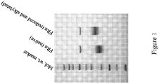
- Efforts were undertaken to produce a reduced and alkylated antigenic form of FR ⁇ .
- purified FR ⁇ was concentrated to 2 mg/mL in phosphate buffered saline (pH 7.4) using centrifugal filters (Amicon Ultra, 3 kD MW limit). The protein concentration was determined using a BCA assay (Thermo Scientific).
- the resultant FR ⁇ was diluted 1:1 in 8M urea/ PBS to generate a final concentration of 1 mg/mL FR ⁇ in PBS containing 4M urea.
- Dithiothreitol solution 500 mM in PBS
- the solution was incubated at 65 degrees Celsius for one hour, and cooled to room temperature.
- Figure 1 shows the differential migration of native FR ⁇ protein and a reduced and alkylated form of the protein analyzed by SDS-PAGE under nonreducing conditions.
- Initial intraperitoneal immunizations administered on day 0 comprised 50 ⁇ g of the respective immunogen mixed 1:1 (v:v) with complete Freund's adjuvant (Rockland, Cat# D614-0050).
- Mice were then boosted with 50 ⁇ g immunogen mixed 1:1 (v:v) with incomplete Freund's adjuvant (Rockland, Cat# D615-0050) administered intraperitoneally 14 days later and every 21 days thereafter. Blood samples were collected from immunized mice 24 days after the initial immunization and every 21 days thereafter.
- FR ⁇ protein 100ml of a 1mg/mL solution in PBS, 0.02 M potassium phosphate, 0.15 M Sodium Chloride, pH 7.2
- PBST PBS containing 0.2% Tween®-20
- PBST PBS containing 0.2% Tween®-20
- mice showing the highest antigen-specific titers were harvested and hybridomas were prepared by electrofusion (HybrimuneTM Model CEEF-50B Waveform Generator; Cellectis, Romainville, France) of splenocytes with Sp2/0 Ag14 myeloma cells (ATTC CRL1581). Subsequently, hybridoma supernatants were screened by ELISA against FR ⁇ and recombinant Mesothelin-His 6 as described above to select positive parental fusion cell lines.
- Selected cell lines were tested for mycoplasma using a mycoplasma test kit (Rockland, Cat# MAB-012) before seeding into 1L roller bottles containing serum free medium (Invitrogen, Cat#12045-076) and 5% low IgG FBS (0.1 ⁇ g/ml) (Gibco, Cat# 16250-078) at 0.5x10 5 cells/mL. Cultures were allowed to grow at 37°C for either 14 or 21 days, after which supernatant was harvested and concentrated approximately 10-fold through a 50kDa filtration membrane (Spectrum Labs, Collinso Dominguez CA) and then purified using protein A chromatography (Rockland, Cat# PA50-00-0025).
- Bound antibody was eluted with 0.1M sodium citrate, pH 3.5/4.5 depending on antibody isotype, and buffer was exchanged against PBS by dialysis using a 12-14kDa membranous tubing (Spectrum Labs, Collinso Dominguez CA). Purified antibody was sterile filtered using a 0.22 ⁇ m ExpressTMPLUS Stericups (Millipore, Billerica MA) and stored at 4°C for further testing.
- Isolated RNA was then amplified via multiplex RT-PCR, performed in triplicate for each hybridoma with a Mastercycler® EP Gradient Thermocycler (Eppendorf).
- Eppendorf Eppendorf
- two separate gene-specific cDNA amplifications were performed for each hybridoma ( ⁇ 1ug RNA/reaction) to determine which Ig heavy and light chain genes were used during Ig rearrangement.
- Each cocktail consisted of unique family-specific primers designed to anneal to any of the potential murine Ig V gene families (IgHv, IgKv) and Ig constant region genes (IgHc Gamma , IgKc).
- cDNA generation and amplification was performed using SuperScript® III One-Step RT-PCR System with Platinum® Taq High Fidelity (Invitrogen) under the following conditions: 55°C for 30 minutes and 95°C for 2 minutes, followed by 40 cycles of 95°C for 1 minute, 55°C for 1 minute, 68°C for 1 minute, and a final 68°C for 10 minutes completion step.
- DNA products were electrophoresed on a 2% agarose gel. Appropriate bands were excised and gel purified using the QIAquick® Gel Extraction Kit (Qiagen) following the manufacturer's protocol. Purified DNA was submitted for sequencing (GENEWIZ, Inc., South Plainfield, NJ) to determine the germline gene segments expressed by each hybridoma.
- RT-PCR analysis suited to the particular genes identified for each hybridoma was then performed using the same RNA source as above and gene-specific primers (in contrast to family-specific primers used in the multiplex RT-PCR mixture).
- gene-specific primers in contrast to family-specific primers used in the multiplex RT-PCR mixture.
- amplified Ig cDNAs were placed into an In-Fusion (IF) expression vector, each gene-specific primer also contained vector-compatible linker sequences which would enable homologous crossover. All other reagents and thermocycler conditions are the same as those used for the multiplex RT-PCR experiments, described above.
- Binding characteristics of the purified monoclonal antibodies to FR ⁇ were determined by surface plasmon resonance (SPR) experiments. All of the SPR experiments were performed at 25°C using a BIAcore T100 with research grade CM5 chips (GE Healthcare), as specified by the manufacturer. Initially, anti-mouse IgG provided in the mouse antibody capture kit (GE Healthcare) was immobilized by amide coupling to CM5 sensor chips. Mouse anti-FR ⁇ monoclonal antibodies (26B3, 24F12, 19D4, or 9F3) were captured on individual flow cells per binding cycle, while the fourth flow cell was used as a reference. Binding experiments were performed with HBS-P (GE Healthcare) as running buffer and at a flow rate of 30 ⁇ L/min.
- HBS-P GE Healthcare
- Each monoclonal antibody sample (0.5 ⁇ g/mL) was injected for 3 minutes to capture the antibody.
- Various concentrations of purified recombinant human FR ⁇ (rh-FR ⁇ ) (1nM - 30nM) were then injected over the FR ⁇ -specific and reference surfaces for 3 minutes to record binding sensograms using a single-cycle kinetics method. The dissociation profile was monitored for 25 minutes. In between bindings, the surface was regenerated with a 30 ⁇ l injection of 10mM glycine (pH 1.7). The sensograms were processed and fitted to a 1:1 Langmuir binding model using BIAcore T100 evaluation software (version 2.0.1).
- binding characteristics of antibodies 26B3, 24F12, 19D4, and 9F3 are provided in Table 2.
- Table 2 Binding characteristics of FR ⁇ -specific antibodies.
- FR ⁇ -specific antibodies 26B3, 24F12, and 9F3 were further assessed in epitope binding studies using Octet QK.
- the results showed that 26B3 and 24F12, which have high affinities to purified human FR ⁇ , compete with one another for binding to FR ⁇ .
- these antibodies may share a common epitope, or have epitopes that are immediately adjacent to each other.
- the results also indicate that the 9F3 antibody has a unique epitope, since it did not compete with other FR ⁇ -specific antibodies for binding to FR ⁇ .
- Immunoblotting was conducted using purified mouse monoclonal antibodies 9F3, 19D4, 24F12, or 26B3 (1 ⁇ g/mL) specific for FR ⁇ , which were detected with a goat-anti-mouse HRP-conjugated antibody and visualized using SuperSignal West Pico chemiluminescent substrate (Pierce, Rockford, IL). Luminescence was visualized using the Omega 12iC molecular imaging system (Ultra-Lum, Claremont, CA) with image analysis performed using UltraQuantTM 6.0 software (Ultra-Lum).
- IHC Immunohistochemistry
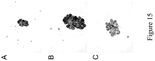
- Flow cytometry studies were conducted to assess the ability of selected FR ⁇ -specific antibodies to bind to the native protein.
- Chinese hamster ovary (CHO) cells expressing FR ⁇ were harvested, washed, and re-suspended in ice-cold growth media (RPMI supplemented with 10% FBS). Cells were incubated for 1 hour on ice with 9F3, 19D4, 24F12, or 26B3 (1 ⁇ g/mL), washed and then incubated with FITC-conjugated secondary antibodies [dilution 1:100] (Southern Biotech, Birmingham, AL).
- EXAMPLE 9 Detection of FR ⁇ in the Serum of Subjects known to have Ovarian Cancer
- Electrochemiluminescence studies were conducted to determine whether the FR ⁇ -specific antibodies described herein could detect FR ⁇ in the serum of patients known to have ovarian cancer.
- MAb 26B3 was used as the capture MAb and added to ECL plates at a concentration of 75 ⁇ g/mL. Plates were washed and 50 ⁇ L of sample serum was added to each well and incubated for 2 hours. Serum samples were obtained from normal healthy females (negative control) and from ovarian cancer patients. Samples were diluted 1:4 in PBST (phosphate buffered saline, pH7.4, containing 0.01% Tween®20).
- EXAMPLE 10 Detection of FR ⁇ in the Serum and Urine of Subjects known to have Ovarian Cancer
- Electrochemiluminescence studies were then conducted to determine whether the FR ⁇ -specific antibodies described herein could detect FR ⁇ in the serum and urine of patients known to have ovarian cancer.
- MAb 26B3 was used as the capture MAb and added to ECL plates at a concentration of 75 ⁇ g/mL. Plates were washed and 50 ⁇ L of sample serum was added to each well and incubated for 2 hours. Matched serum and urine samples were obtained from normal healthy females (negative control) and from ovarian cancer patients. Samples (serum or urine) were diluted 1:4 in PBST (phosphate buffered saline, pH7.4, containing 0.01% Tween®20).
- PBST phosphate buffered saline, pH7.4, containing 0.01% Tween®20
- Table 6 Relative serum and urine levels of FR ⁇ Patient Designation Serum FRalpha pg/mL Urine FRalpha pg/mL Normal 1 398 3080 Normal 2 236 11508 Normal 3 315 7704 Normal 4 320 13198 Ovarian Cancer 1 19479 368066 Ovarian Cancer 2 4144 23738 Ovarian Cancer 3 986 165826 Ovarian Cancer 4 719 414187
- M-score A metric for staining (M-score) of each sample was developed and can be defined as follows: In the equation, x ij is the percentage of tumor stained at intensity j for patient i and w j is the absolute value of the intensity (ranging from 0 to 3+). The metric has a theoretical range from zero (no positive staining) to fifty (100% of cells staining at 3+ intensity). As such, the M-score is a weighted score for FR ⁇ IHC tumor cell membrane staining that captures both the proportion of FR ⁇ positive cells and staining intensity. M-scores for each patient were averaged over multiple tissue microarray (TMA) samples, where appropriate. If a sample was void of results, i. e. no tumor present or necrotic tissue, the M-score was assigned to the non-void determinations.
- TMA tissue microarray
- EXAMPLE 12 Comparative Staining of Lung Carcinoma Cells with Antibody 26B3 and Antibody BN3.2
- M-score mean ⁇ SD 19.84 ⁇ 18.64
- EXAMPLE 13 Detection of FR ⁇ in Subjects known to have Adenocarcinoma of the Lung
- FR ⁇ positive histology was associated with particular forms of lung cancer.
- a tissue microarray having duplicate samples of normal and cancerous, stage I, stage II, stage III, and stage IV, lung tissue specimens was assessed for FR ⁇ expression via IHC staining using antibody 26B3, as described in Example 7.
- FR ⁇ is associated with adenocarcinomas relative to squamous cell carcinomas, which exhibited limited positive staining.
- Table 7 Histological evaluation of cancerous tissue samples Membrane Staining Membrane Positive Total Negative Positive Histology Groups Adenocarcinoma Count 11 27 38 % within Histology Groups 28.9% 71.1% 100.0% Squamous Count 28 3 31 % within Histology Groups 90.3% 9.7% 100.0% Other Carcinomas Count 17 4 21 % within Histology Groups 81.0% 19.0% 100.0% Normal Count 2 8 10 % within Histology Groups 20.0% 80.0% 100.0% Total Count 58 42 100 % within Histology Groups 58.0% 42.0% 100.0%
- Table 8 Distribution of FR ⁇ Expression Across NSCLC Type# Variable FR ⁇ negative N (%) FR ⁇ positive N (%) Total P value* Tumor Histology Normal 1 (10%) 9 (90%) 10 Squamous cell carcinoma 28 (87%) 4 (14%) 32 ⁇ 0.0001 Large cell carcinoma 3 (60%) 2 (40%) 5 Small cell carcinoma 7 (87%) 1 (13%) 8 Neuroendocrine carcinoma 4 (67%) 2 (33%) 6 Adenocarcinoma** 10 (16%) 28 (74%) 38 Tumor Grade Grade 1 1 (20%) 4 (80%) 5 Grade 2 5 (22%) 18 (78%) 23 Grade 3 4 (40%) 6 (60%) 10 0.517 Tumor Stage Stage I 4 (29%) 11 (71%) 15 Stage II 2 (17%) 10 (83%) 12 Stage III + IV*** 4 (36) 7 (64) 11 0.563 Gender Female 3 (18%) 14 (82%) 17 Male 7 (33%) 14 (67%) 21 0.46 #US Biomax Lung Cancer TMA (catalog # BC041114; 90 cases, duplicate cores) * P values determined using Fisher
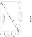
- M-score analyses of duplicate adenocarcinoma histology samples showed little variation in staining by antibody 26B3 ( Figure 8 ), a reflection of the robustness of antibody 26B3 staining. Also, an examination of M-scores by stage and grade within the adenocarcinoma histologic subtype indicated that neither stage nor grade of disease was associated with the degree of staining as defined by the M-scores (data not shown).
- the M-score distribution for FR ⁇ staining of lung adenocarcinoma and squamous cell carcinoma samples is shown in Figure 9 .
- the mean ( ⁇ SD) M-scores for adenocarcinoma and squamous cell carcinoma samples stained with antibody 26B3 were 19.84 ( ⁇ 18.64) and 1.39 ( ⁇ 5.54), respectively (p ⁇ 0.0001).
- the M-score for adenocarcinoma was also significantly higher when compared against all other lung cancer histologic types.
- a Tree Analysis was performed to determine the odds for the histology of the cancer being adenocarcinoma.
- An M-score >21.7 resulted in an odds ratio (OR) of 16, further demonstrating that FR ⁇ is predominately expressed in the adenocarcinoma histology (analysis not shown).
- FFPE paraffin-embedded
- EXAMPLE 14 - FR ⁇ is Expressed by CK+/CD45- Cells, but not CK-/CD45+ Cells, Isolated from the Blood of Patients known to have Non-Small Cell Lung Carcinoma
- CTCs circulating tumor cells
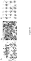
- blood samples were obtained from 15 healthy donors and 5 stage IV lung cancer patients and then enriched for CTCs using ApoCell's ApoSteamTM system. After enrichment, each sample was stained for cytokeratin (CK), CD45 (protein tyrosine phosphatase receptor type C), nuclei, and FR ⁇ .
- CK cytokeratin
- CD45 protein tyrosine phosphatase receptor type C
- nuclei nuclei
- FR ⁇ staining was performed using antibody 26B3 as the primary antibody, which was then detected using a mouse-specific, secondary antibody conjugated to DyLight® 649.
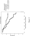
- EXAMPLE 15 5-year Survivorship of Subjects with and without FR ⁇ -expressing Adenocarcinoma of the Lung
- ROC receiver operating characteristic
- FIG. 11 illustrates the survival functions for stage I and stage II adenocarcinoma groups deemed to be FR ⁇ positive and FR ⁇ negative by 26B3 detection. At 5 years the hazard ratio is 2.42. This indicates that subjects having tumors that are negative (M ⁇ 10) for FR ⁇ are 2.5 times more likely to die within five years of diagnosis than subjects with FR ⁇ -positive tumors (M ⁇ 10).
- EXAMPLE 16 Folate Receptor Alpha Expression is Associated with Triple-Negative Forms of Breast Cancer
- TMA tissue microarray
- the distribution of histologies present on the breast cancer TMA (U.S. BioMAX catalog # BR1503a; 72 cases, duplicate cores) are shown in Table 10, the majority of the cases represented being identified as invasive ductal carcinoma (IDC).
- the TMA included 2 normal breast samples, which were positive for FR ⁇ expression as determined by MAb 26B3. Staining in normal breast was restricted to ductal cells with luminal and membrane staining. Two of three fibroadenoma cases (67%), 0/2 cystosarcoma cases (0%) and 1/6 ductal carcinoma in situ cases (17%) were positive for FR ⁇ .
- the single invasive lobular carcinoma (ILC) was negative for FR ⁇ staining.
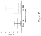
- FR ⁇ positive IDC cases Of the 18 FR ⁇ positive IDC cases, only 2 (11 %) were Her2 positive meaning that the vast majority (89%) were Her2 negative. These data suggest that FR ⁇ positivity tracks more closely with Her2 negativity. Further, of the 18 FR ⁇ positive IDC cases, 3 were estrogen receptor (ER) positive and 4 were progesterone receptor (PR) positive, but all ER/PR positive/FR ⁇ positive cases were Her2 negative. Of the FR ⁇ positive IDC cases 12/18 (67%) were triple negative breast cancers (TNBC), suggesting that FR ⁇ may be a marker and target for very poor prognosis TNBC molecular subtype.
- ER estrogen receptor
- PR progesterone receptor
- the TMA described above was composed primarily of early stage breast cancers: stage I, 6/60 (10%); stage II, 44/60 (73%); stage III, 10/60 (17%). Therefore, to confirm and extend the results obtained on the TMA, 61 FFPE tissue blocks from stage IV(T4) Her2 negative breast cancers with known ER/PR expression ranging from 0-100% positive were assessed (FFPE tissue blocks were obtained from the archives of Genzyme Genetics). All 61 of these samples were from metastases, not the primary tumor. The results of this study are summarized in Table 11.
- Table 11 Distribution of FR ⁇ positivity in molecular subtypes of metastatic breast cancer samples FR ⁇ positive FR ⁇ negative Tumor Molecular subtype N (%) N (%) Total P value* Total Samples: 22 (36%) 39 (64%) 61 ER/PR+ 3 (14%) 20 (86%) 23 ER/PR/Her2- 19 (50%) 19 (50%) 38 0.0054 (ER/PR+ versus ER/PR/Her2-) Grade 1 3 (30%) 7 (70%) 10 Grade 2 11 (28%) 28 (72%) 39 1.0 (Grade 1 versus Grade 2) Grade 3 8 (67%) 4 (33%) 12 0.037 (Grade 1 or 2 versus Grade 3) * P values calculated via 2X2 contingency table analysis using Fisher's exact test.
- samples from stage IV metastatic disease were obtained from a number of metastatic sites including lymph node, bone, skin and liver as well as fluid and fine needle aspirate (FNA) samples obtained primarily from pleura and paracentesis.
- FNA fine needle aspirate
- TMA tissue microarray
- FFPE FFPE histology samples prepared and stained with antibody 26B3 as described in Example 7.
- Commercial tissue microarrays were obtained from US Biomax, Inc. (Rockville, MD) for ovarian carcinomas (catalog # OV1921; 96 cases, duplicate cores); endometrial carcinomas (catalog # EMC1021; 102 cases, single cores); and fallopian tube carcinomas (catalog # UTE601; 30 cases, duplicate cores).
- a sample was considered positive for FR ⁇ expression if the percentage of the tumor cells positive for membranous staining was greater than or equal to 5% at any intensity.
- a sample was rejected and therefore not included in the analyses if the gynecologic pathologist determined it was either missing entirely or was composed of necrotic tissue with an insufficient number of viable cells for evaluation.
- the endometrial samples six contained only atypical complex hyperplasia without adenocarcinoma. Histologic classification of cell type and grade were based the WHO Classification of Breast and Female Genital Organs (Tavassoli and Devilee). Clinical stage based on FIGO and TNM system was provided by the manufacturer of the TMA (US Biomax).
- Membrane and cytoplasmic staining intensity was scored as 0, no staining; 1+, weak; 2+, moderate and 3+, strong. The percent of cells for each intensity in the sample was also determined.
- Tissue was analyzed under 4x, 10x, 20x and 40x objectives. Strong membrane staining (3+) was readily visualized under 4x and confirmed at 10x. Moderate membrane staining (2+) was visible at 10x and confirmed at 20x. Weak staining (1+) required 20x or 40x ( Figure 16 ). In the presence of 3+ staining, the membrane was thick occurring at apical and lateral cell borders. In tangential sections, a complete circumferential pattern was evident ( Figure 16 (A) and (B) ).
- 2+ membrane staining was weaker in intensity and thinner than 3+, usually localized on the apical luminal borders and occasionally on lateral cell borders. 1+ weak membrane was generally limited to the luminal borders. The accompanying cytoplasmic staining was variable, depending on the type of tumors.
- the FR ⁇ positive rate for each cell type was as follows: 100% (70/70) in serous type, 80% (8/10) mucinous type, 75% (3/4) endometrioid type, and 67% (2/3) clear cell type.
- the difference between serous and mucinous type is significant with p value at 0.014 by Fisher's exact test (Table 12). FR ⁇ status was not significant for histologic grade or clinical stage.
- FR ⁇ was expressed in 80% (4/5) of normal ( Figure 17(A) ), 100% (6/6) of atypical complex hyperplasia ( Figure 17(B) ), and 89% (80/90) of adenocarcinomas, including 88 endometrioid type and 1 clear cell type ( Figures 18 and 19 ).
- Eight endometrioid adenocarcinomas contained areas of squamous metaplasia.
- membrane staining was weak and limited to the apical luminal borders ( Figure 17(A) ).
- staining was predominantly luminal with additional staining on lateral cell borders in some cases ( Figure 17(B) ).
- cytoplasmic staining varied in intensity from strong ( Figure 18(A) ) to weak ( Figures 18(B) and (C) ). Tumor cells with 1+ or 2+ membrane staining rarely expressed cytoplasmic staining. The majority of metaplastic squamous cells and clear cells exhibited moderate to strong membrane staining. ( Figures 19(A) and (B) ).
- FR ⁇ status was not significant in relation to T1 vs. T2/3; N0 vs. N1, and stage I vs. stage II/III.
- TMA tissue microarray
- Thyroid papillary carcinoma was strongly positive for FR ⁇ membrane expression (26/28, 93%) and was distinguishable from medullary carcinoma, where all 5 samples were negative for FR ⁇ staining, in agreement with previous reports.
- follicular adenomas were separable into macrofollicular type and microfollicular type with 3/13 (23%) and 18/22 (82%) showing positivity for FR ⁇ expression, respectively.
Landscapes
- Health & Medical Sciences (AREA)
- Life Sciences & Earth Sciences (AREA)
- Immunology (AREA)
- Engineering & Computer Science (AREA)
- Chemical & Material Sciences (AREA)
- Molecular Biology (AREA)
- Biomedical Technology (AREA)
- Urology & Nephrology (AREA)
- Hematology (AREA)
- Medicinal Chemistry (AREA)
- General Health & Medical Sciences (AREA)
- Biochemistry (AREA)
- Cell Biology (AREA)
- Pathology (AREA)
- Food Science & Technology (AREA)
- General Physics & Mathematics (AREA)
- Biotechnology (AREA)
- Analytical Chemistry (AREA)
- Physics & Mathematics (AREA)
- Microbiology (AREA)
- Organic Chemistry (AREA)
- Biophysics (AREA)
- Proteomics, Peptides & Aminoacids (AREA)
- Genetics & Genomics (AREA)
- General Chemical & Material Sciences (AREA)
- Public Health (AREA)
- Chemical Kinetics & Catalysis (AREA)
- Animal Behavior & Ethology (AREA)
- Pharmacology & Pharmacy (AREA)
- Veterinary Medicine (AREA)
- Nuclear Medicine, Radiotherapy & Molecular Imaging (AREA)
- Peptides Or Proteins (AREA)
- Micro-Organisms Or Cultivation Processes Thereof (AREA)
- Reproductive Health (AREA)
- Oncology (AREA)
- Medicines Containing Antibodies Or Antigens For Use As Internal Diagnostic Agents (AREA)
- Preparation Of Compounds By Using Micro-Organisms (AREA)
- Measuring Or Testing Involving Enzymes Or Micro-Organisms (AREA)
Priority Applications (6)
| Application Number | Priority Date | Filing Date | Title |
|---|---|---|---|
| RS20180252A RS56985B1 (sr) | 2011-07-15 | 2012-07-13 | Antitela protiv alfa receptora folata i njihove upotrebe |
| PL12736054T PL2731972T3 (pl) | 2011-07-15 | 2012-07-13 | Przeciwciała skierowane przeciwko receptorowi folianowemu alfa i ich zastosowania |
| SI201231234T SI2731972T1 (en) | 2011-07-15 | 2012-07-13 | Anti-folate receptor alpha antibodies and uses thereof |
| HRP20180353TT HRP20180353T1 (hr) | 2011-07-15 | 2012-07-13 | Protutijela protiv folatnog receptora alfa i njihova upotreba |
| SM20180150T SMT201800150T1 (it) | 2011-07-15 | 2012-07-13 | Anticorpi anti-recettore alfa del folato e loro utilizzi |
| EP17196948.8A EP3330291B1 (en) | 2011-07-15 | 2012-07-13 | Anti-folate receptor alpha antibodies and uses thereof |
Applications Claiming Priority (4)
| Application Number | Priority Date | Filing Date | Title |
|---|---|---|---|
| US201161508444P | 2011-07-15 | 2011-07-15 | |
| US201261604412P | 2012-02-28 | 2012-02-28 | |
| US201261604954P | 2012-02-29 | 2012-02-29 | |
| PCT/US2012/046672 WO2013012722A1 (en) | 2011-07-15 | 2012-07-13 | Anti-folate receptor alpha antibodies and uses thereof |
Related Child Applications (2)
| Application Number | Title | Priority Date | Filing Date |
|---|---|---|---|
| EP17196948.8A Division-Into EP3330291B1 (en) | 2011-07-15 | 2012-07-13 | Anti-folate receptor alpha antibodies and uses thereof |
| EP17196948.8A Division EP3330291B1 (en) | 2011-07-15 | 2012-07-13 | Anti-folate receptor alpha antibodies and uses thereof |
Publications (2)
| Publication Number | Publication Date |
|---|---|
| EP2731972A1 EP2731972A1 (en) | 2014-05-21 |
| EP2731972B1 true EP2731972B1 (en) | 2017-12-20 |
Family
ID=46516898
Family Applications (2)
| Application Number | Title | Priority Date | Filing Date |
|---|---|---|---|
| EP12736054.3A Active EP2731972B1 (en) | 2011-07-15 | 2012-07-13 | Anti-folate receptor alpha antibodies and uses thereof |
| EP17196948.8A Active EP3330291B1 (en) | 2011-07-15 | 2012-07-13 | Anti-folate receptor alpha antibodies and uses thereof |
Family Applications After (1)
| Application Number | Title | Priority Date | Filing Date |
|---|---|---|---|
| EP17196948.8A Active EP3330291B1 (en) | 2011-07-15 | 2012-07-13 | Anti-folate receptor alpha antibodies and uses thereof |
Country Status (21)
Families Citing this family (50)
| Publication number | Priority date | Publication date | Assignee | Title |
|---|---|---|---|---|
| TWI622402B (zh) | 2010-02-24 | 2018-05-01 | 免疫遺傳股份有限公司 | 葉酸受體1抗體類和免疫共軛物類及彼等之用途 |
| BR112013025415B1 (pt) | 2011-04-01 | 2021-03-16 | Immunogen, Inc | uso de um imunoconjugado antirreceptor de folato 1 (folr1) |
| SI2731972T1 (en) * | 2011-07-15 | 2018-04-30 | Eisai R&D Management Co., Ltd. | Anti-folate receptor alpha antibodies and uses thereof |
| US9200073B2 (en) | 2012-08-31 | 2015-12-01 | Immunogen, Inc. | Diagnostic assays and kits for detection of folate receptor 1 |
| CA2914237A1 (en) | 2013-06-20 | 2014-12-24 | Morphotek, Inc. | Methods for treatment of ovarian cancer |
| CN113150145B (zh) | 2013-08-30 | 2025-02-14 | 伊缪诺金公司 | 用于检测叶酸受体1的抗体和测定 |
| DK3140653T3 (da) * | 2014-05-08 | 2022-06-20 | Novodiax Inc | Direkte immunhistokemianalyse |
| EP4141032B1 (en) | 2014-11-20 | 2024-05-29 | F. Hoffmann-La Roche AG | Combination therapy of t cell activating bispecific antigen binding molecules and pd-1 axis binding antagonists |
| PT3221356T (pt) | 2014-11-20 | 2020-10-29 | Hoffmann La Roche | Moléculas de ligação ao antigénio biespecíficas ativadoras das células t contra folr1 e cd3 |
| DK3221357T3 (da) | 2014-11-20 | 2020-08-10 | Hoffmann La Roche | Fælles letkæder og fremgangsmåder til anvendelse |
| EP3283886B1 (en) * | 2015-04-17 | 2020-01-15 | Eisai Inc. | Methods for treating lung cancer |
| CN116440279B (zh) | 2015-09-17 | 2026-02-24 | 伊缪诺金公司 | 包含抗folr1免疫缀合物的治疗组合 |
| WO2018031968A1 (en) | 2016-08-12 | 2018-02-15 | L.E.A.F. Holdings Group Llc | Alpha and gamma-d polyglutamated antifolates and uses thereof |
| CN121177507A (zh) | 2016-08-12 | 2025-12-23 | L.E.A.F.控股集团公司 | α和γ-D聚谷氨酸化抗叶酸剂及其用途 |
| EP3544999A1 (en) * | 2016-11-23 | 2019-10-02 | Eisai R&D Management Co., Ltd. | Anti-folate receptor alpha antibodies and uses thereof |
| CN107353325B (zh) * | 2017-07-26 | 2020-08-11 | 中国药科大学 | 叶酸受体α特异性结合肽1及其应用 |
| CN107793471B (zh) * | 2017-07-26 | 2020-08-11 | 中国药科大学 | 叶酸受体α特异性结合肽4及其应用 |
| CN107446021B (zh) * | 2017-07-26 | 2020-08-11 | 中国药科大学 | 叶酸受体α特异性结合肽5及其应用 |
| CN107446020B (zh) * | 2017-07-28 | 2019-10-01 | 中国药科大学 | 叶酸受体α特异性结合肽2及其应用 |
| US20200284810A1 (en) * | 2017-09-05 | 2020-09-10 | Immunogen, Inc. | Methods for detection of folate receptor 1 in a patient sample |
| WO2019157145A1 (en) | 2018-02-07 | 2019-08-15 | L.E.A.F. Holdings Group Llc | Gamma polyglutamated pemetrexed and uses thereof |
| US12310966B2 (en) | 2018-02-07 | 2025-05-27 | L.E.A.F. Holdings Group Llc | Alpha polyglutamated aminopterin and uses thereof |
| CN111867593A (zh) | 2018-02-07 | 2020-10-30 | L.E.A.F.控股集团公司 | α聚谷氨酸化抗叶酸剂及其用途 |
| EP3749318A4 (en) | 2018-02-07 | 2022-07-06 | L.E.A.F Holdings Group LLC | GAMMA-POLYGLUTAMATED RALTITREXED AND ITS USES |
| US12220431B2 (en) | 2018-02-07 | 2025-02-11 | L.E.A.F. Holdings Group Llc | Gamma polyglutamated antifolates and uses thereof |
| JP7491572B2 (ja) | 2018-02-07 | 2024-05-28 | エル.イー.エー.エフ. ホールディングス グループ エルエルシー | アルファポリグルタミン酸化ペメトレキセドおよびその使用 |
| EP3749319A4 (en) | 2018-02-07 | 2022-06-22 | L.E.A.F Holdings Group LLC | TETRAHYDROFOLATES ALPHA POLYGLUTAMATE AND THEIR USES |
| EP3749314A4 (en) | 2018-02-07 | 2022-02-23 | L.E.A.F Holdings Group LLC | ALPHA-POLYGLUTAMATE METHOTREXATE AND ITS USES |
| WO2019157121A1 (en) | 2018-02-07 | 2019-08-15 | L.E.A.F. Holdings Group Llc | Alpha polyglutamated lometrexol and uses thereof |
| WO2019157129A1 (en) | 2018-02-07 | 2019-08-15 | L.E.A.F. Holdings Group Llc | Alpha polyglutamated pralatrexate and uses thereof |
| CA3090389A1 (en) | 2018-02-07 | 2019-08-15 | L.E.A.F. Holdings Group Llc | Alpha polyglutamated raltitrexed and uses thereof |
| US12350271B2 (en) | 2018-02-14 | 2025-07-08 | L.E.A.F. Holdings Group Llc | Gamma polyglutamated aminopterin and uses thereof |
| WO2019160734A1 (en) | 2018-02-14 | 2019-08-22 | L.E.A.F. Holdings Group Llc | Gamma polyglutamated lometrexol and uses thereof |
| CA3090753A1 (en) | 2018-02-14 | 2019-08-22 | L.E.A.F. Holdings Group Llc | Gamma polyglutamated pralatrexate and uses thereof |
| WO2019160735A1 (en) | 2018-02-14 | 2019-08-22 | L.E.A.F. Holdings Group Llc | Gamma polyglutamated tetrahydrofolates and uses thereof |
| JP7462950B2 (ja) | 2018-02-14 | 2024-04-08 | エル.イー.エー.エフ. ホールディングス グループ エルエルシー | ガンマポリグルタミン酸化メトトレキセートおよびその使用 |
| AU2019236091B2 (en) | 2018-03-13 | 2025-06-05 | Phanes Therapeutics, Inc. | Anti-folate receptor 1 antibodies and uses thereof |
| KR102275930B1 (ko) | 2018-03-14 | 2021-07-12 | (주)알테오젠 | Folr1에 특이적으로 결합하는 항체 및 그의 용도 |
| CN112955548B (zh) * | 2018-07-09 | 2023-11-24 | 普众发现医药科技(上海)有限公司 | 叶酸受体α特异性抗体 |
| CN111393528B (zh) * | 2020-01-19 | 2023-01-31 | 中国药科大学 | 一种靶向叶酸受体α的单链抗体及其应用 |
| WO2021247821A1 (en) * | 2020-06-03 | 2021-12-09 | The Wistar Institute Of Anatomy And Biology | Monoclonal antibodies against human siglec-9 and use thereof for immunotherapy |
| EP4347661A4 (en) * | 2021-06-04 | 2025-06-18 | ImmunoGen, Inc. | TREATMENT OF CANCER IN PATIENTS WITH SOLUBLE FR-ALPHA |
| US12281161B2 (en) | 2021-07-28 | 2025-04-22 | Trustees Of Boston University | Polypeptides and uses thereof |
| CN119137154A (zh) * | 2022-03-25 | 2024-12-13 | 酵活英属哥伦比亚有限公司 | 抗叶酸受体α抗体及使用方法 |
| WO2024104431A1 (zh) * | 2022-11-16 | 2024-05-23 | 迈威(上海)生物科技股份有限公司 | 一种靶向FRα的抗体或其抗原结合片段及其应用 |
| EP4724490A2 (en) | 2023-06-12 | 2026-04-15 | Amgen Inc. | Lymphotoxin beta receptor agonist binding proteins |
| CN117700556B (zh) * | 2024-02-05 | 2024-04-12 | 苏州百道医疗科技有限公司 | 一种抗FRα小鼠单克隆抗体及其应用 |
| CN117700557B (zh) * | 2024-02-05 | 2024-04-30 | 卡秋(江苏)生物科技有限公司 | 一种特异性结合叶酸受体α的抗体或抗原结合片段 |
| CN120554514B (zh) * | 2025-07-29 | 2025-12-05 | 合肥综合性国家科学中心大健康研究院 | 一种靶向人的叶酸受体α的单克隆抗体 |
| CN120842412B (zh) * | 2025-09-23 | 2025-12-05 | 合肥综合性国家科学中心大健康研究院 | 一种靶向人的叶酸受体α的单克隆抗体及其应用 |
Family Cites Families (17)
| Publication number | Priority date | Publication date | Assignee | Title |
|---|---|---|---|---|
| US5646253A (en) | 1994-03-08 | 1997-07-08 | Memorial Sloan-Kettering Cancer Center | Recombinant human anti-LK26 antibodies |
| US5595756A (en) * | 1993-12-22 | 1997-01-21 | Inex Pharmaceuticals Corporation | Liposomal compositions for enhanced retention of bioactive agents |
| US6770445B1 (en) | 1999-02-26 | 2004-08-03 | Pacific Northwest Research Institute | Methods and compositions for diagnosing carcinomas |
| US7270960B2 (en) | 2001-08-29 | 2007-09-18 | Pacific Northwest Research Institute | Diagnosis of ovarian carcinomas |
| WO2004082463A2 (en) | 2003-03-17 | 2004-09-30 | Medical College Of Ohio | Folate receptor gene modulation for cancer diagnosis and therapy |
| AU2004249673A1 (en) | 2003-05-23 | 2004-12-29 | Morphotek, Inc. | Anti alpha - folate - receptor - tetramer antibodies |
| EP3101034A1 (en) | 2004-02-12 | 2016-12-07 | Morphotek, Inc. | Monoclonal antibodies that specifically bind to folate receptor alpha |
| ATE460668T1 (de) | 2005-03-30 | 2010-03-15 | Purdue Research Foundation | Verfahren zur prognose von brustkrebs mittels quantifizierung von zellulären folat vitamin rezeptoren |
| CA2607444C (en) | 2005-04-22 | 2015-03-10 | Morphotek, Inc. | Antibodies with immune effector activity and that internalize in folate receptor alpha-positive cells |
| EP1900752A1 (en) | 2006-09-15 | 2008-03-19 | DOMPE' pha.r.ma s.p.a. | Human anti-folate receptor alpha antibodies and antibody fragments for the radioimmunotherapy of ovarian carcinoma |
| US7754698B2 (en) | 2007-01-09 | 2010-07-13 | Isis Pharmaceuticals, Inc. | Modulation of FR-alpha expression |
| FR2918994B1 (fr) | 2007-07-20 | 2012-10-19 | Rhodia Operations | Formulations de diesters d'acide carboxylique et leur utilisation pour le traitement de materiaux. |
| WO2009132081A2 (en) | 2008-04-24 | 2009-10-29 | The Research Foundation Of State University Of New York | Monoclonal antibody-based targeting of folate receptors |
| US20110177525A1 (en) | 2010-01-19 | 2011-07-21 | Predictive Biosciences, Inc. | Antibodies and methods of diagnosing diseases |
| MX343607B (es) | 2010-11-05 | 2016-11-11 | Morphotek Inc | Receptor alfa de folato como marcador de diagnóstico y pronóstico para cánceres que expresan receptor alfa de folato. |
| JP2014508165A (ja) | 2011-03-02 | 2014-04-03 | モルフォテック、インク. | 乳癌の治療のためのエリブリンおよびファーレツズマブの共投与 |
| SI2731972T1 (en) * | 2011-07-15 | 2018-04-30 | Eisai R&D Management Co., Ltd. | Anti-folate receptor alpha antibodies and uses thereof |
-
2012
- 2012-07-13 SI SI201231234T patent/SI2731972T1/en unknown
- 2012-07-13 WO PCT/US2012/046672 patent/WO2013012722A1/en not_active Ceased
- 2012-07-13 CN CN201280043606.2A patent/CN103998467B/zh active Active
- 2012-07-13 LT LTEP12736054.3T patent/LT2731972T/lt unknown
- 2012-07-13 CN CN201611025725.1A patent/CN107011439B/zh active Active
- 2012-07-13 TR TR2018/02838T patent/TR201802838T4/tr unknown
- 2012-07-13 SM SM20180150T patent/SMT201800150T1/it unknown
- 2012-07-13 CA CA2841725A patent/CA2841725C/en active Active
- 2012-07-13 HR HRP20180353TT patent/HRP20180353T1/hr unknown
- 2012-07-13 ES ES17196948T patent/ES2824498T3/es active Active
- 2012-07-13 US US13/548,775 patent/US8475795B2/en active Active
- 2012-07-13 DK DK12736054.3T patent/DK2731972T3/en active
- 2012-07-13 JP JP2014520368A patent/JP6220333B2/ja active Active
- 2012-07-13 PT PT127360543T patent/PT2731972T/pt unknown
- 2012-07-13 AU AU2012284321A patent/AU2012284321C1/en active Active
- 2012-07-13 PL PL12736054T patent/PL2731972T3/pl unknown
- 2012-07-13 BR BR112014001030A patent/BR112014001030A2/pt not_active Application Discontinuation
- 2012-07-13 EP EP12736054.3A patent/EP2731972B1/en active Active
- 2012-07-13 EP EP17196948.8A patent/EP3330291B1/en active Active
- 2012-07-13 HU HUE12736054A patent/HUE036929T2/hu unknown
- 2012-07-13 ES ES12736054.3T patent/ES2663556T3/es active Active
- 2012-07-13 SG SG10201605278PA patent/SG10201605278PA/en unknown
- 2012-07-13 RS RS20180252A patent/RS56985B1/sr unknown
-
2013
- 2013-03-13 US US13/800,575 patent/US8834877B2/en active Active
-
2014
- 2014-08-04 US US14/451,044 patent/US9599621B2/en active Active
-
2017
- 2017-02-16 US US15/434,326 patent/US10101343B2/en active Active
- 2017-04-11 AU AU2017202365A patent/AU2017202365B2/en active Active
- 2017-09-29 JP JP2017190224A patent/JP6526143B2/ja active Active
-
2018
- 2018-03-01 CY CY181100260T patent/CY1120600T1/el unknown
- 2018-09-26 AU AU2018236755A patent/AU2018236755B2/en active Active
Also Published As
Similar Documents
| Publication | Publication Date | Title |
|---|---|---|
| AU2018236755B2 (en) | Anti-folate receptor alpha antibodies and uses thereof | |
| US10822410B2 (en) | Anti-folate receptor alpha antibodies and uses thereof | |
| AU2014217611A1 (en) | Therapeutic and diagnostic target for cancer comprising DLL3 binding reagents | |
| KR20230016212A (ko) | 항-cldn18.2 항체 및 이의 진단 용도 | |
| TW200823293A (en) | Novel anti-Notch3 antibodies and their use in the detection and diagnosis of disease | |
| CN104755496A (zh) | 作为癌症治疗和诊断靶标的ly75 | |
| KR102766022B1 (ko) | Cd37 의 검출을 위한 항체 및 검정 | |
| DK2726094T3 (en) | Therapeutic and diagnostic targets | |
| EP2958590B1 (en) | Bispecific antibody binding to naaladl2 and cd3 for use as a therapeutic in treating prostate cancer | |
| TWI782000B (zh) | 抗gpr20抗體、其製造方法及其應用 | |
| HK40013436A (en) | Anti-gpr20 antibody |
Legal Events
| Date | Code | Title | Description |
|---|---|---|---|
| PUAI | Public reference made under article 153(3) epc to a published international application that has entered the european phase |
Free format text: ORIGINAL CODE: 0009012 |
|
| 17P | Request for examination filed |
Effective date: 20140120 |
|
| AK | Designated contracting states |
Kind code of ref document: A1 Designated state(s): AL AT BE BG CH CY CZ DE DK EE ES FI FR GB GR HR HU IE IS IT LI LT LU LV MC MK MT NL NO PL PT RO RS SE SI SK SM TR |
|
| DAX | Request for extension of the european patent (deleted) | ||
| 17Q | First examination report despatched |
Effective date: 20141223 |
|
| STAA | Information on the status of an ep patent application or granted ep patent |
Free format text: STATUS: EXAMINATION IS IN PROGRESS |
|
| GRAP | Despatch of communication of intention to grant a patent |
Free format text: ORIGINAL CODE: EPIDOSNIGR1 |
|
| STAA | Information on the status of an ep patent application or granted ep patent |
Free format text: STATUS: GRANT OF PATENT IS INTENDED |
|
| INTG | Intention to grant announced |
Effective date: 20170703 |
|
| GRAJ | Information related to disapproval of communication of intention to grant by the applicant or resumption of examination proceedings by the epo deleted |
Free format text: ORIGINAL CODE: EPIDOSDIGR1 |
|
| STAA | Information on the status of an ep patent application or granted ep patent |
Free format text: STATUS: EXAMINATION IS IN PROGRESS |
|
| GRAR | Information related to intention to grant a patent recorded |
Free format text: ORIGINAL CODE: EPIDOSNIGR71 |
|
| GRAS | Grant fee paid |
Free format text: ORIGINAL CODE: EPIDOSNIGR3 |
|
| STAA | Information on the status of an ep patent application or granted ep patent |
Free format text: STATUS: GRANT OF PATENT IS INTENDED |
|
| GRAA | (expected) grant |
Free format text: ORIGINAL CODE: 0009210 |
|
| STAA | Information on the status of an ep patent application or granted ep patent |
Free format text: STATUS: THE PATENT HAS BEEN GRANTED |
|
| INTC | Intention to grant announced (deleted) | ||
| AK | Designated contracting states |
Kind code of ref document: B1 Designated state(s): AL AT BE BG CH CY CZ DE DK EE ES FI FR GB GR HR HU IE IS IT LI LT LU LV MC MK MT NL NO PL PT RO RS SE SI SK SM TR |
|
| INTG | Intention to grant announced |
Effective date: 20171113 |
|
| REG | Reference to a national code |
Ref country code: GB Ref legal event code: FG4D |
|
| REG | Reference to a national code |
Ref country code: CH Ref legal event code: EP |
|
| REG | Reference to a national code |
Ref country code: IE Ref legal event code: FG4D |
|
| REG | Reference to a national code |
Ref country code: AT Ref legal event code: REF Ref document number: 956290 Country of ref document: AT Kind code of ref document: T Effective date: 20180115 |
|
| REG | Reference to a national code |
Ref country code: DE Ref legal event code: R096 Ref document number: 602012041096 Country of ref document: DE |
|
| REG | Reference to a national code |
Ref country code: HR Ref legal event code: TUEP Ref document number: P20180353 Country of ref document: HR |
|
| REG | Reference to a national code |
Ref country code: RO Ref legal event code: EPE |
|
| REG | Reference to a national code |
Ref country code: PT Ref legal event code: SC4A Ref document number: 2731972 Country of ref document: PT Date of ref document: 20180309 Kind code of ref document: T Free format text: AVAILABILITY OF NATIONAL TRANSLATION Effective date: 20180301 |
|
| REG | Reference to a national code |
Ref country code: DK Ref legal event code: T3 Effective date: 20180305 |
|
| REG | Reference to a national code |
Ref country code: NL Ref legal event code: FP |
|
| REG | Reference to a national code |
Ref country code: CH Ref legal event code: NV Representative=s name: ISLER AND PEDRAZZINI AG, CH Ref country code: EE Ref legal event code: FG4A Ref document number: E014935 Country of ref document: EE Effective date: 20180201 |
|
| REG | Reference to a national code |
Ref country code: SE Ref legal event code: TRGR |
|
| REG | Reference to a national code |
Ref country code: HR Ref legal event code: T1PR Ref document number: P20180353 Country of ref document: HR |
|
| REG | Reference to a national code |
Ref country code: ES Ref legal event code: FG2A Ref document number: 2663556 Country of ref document: ES Kind code of ref document: T3 Effective date: 20180413 |
|
| REG | Reference to a national code |
Ref country code: NO Ref legal event code: T2 Effective date: 20171220 |
|
| REG | Reference to a national code |
Ref country code: SK Ref legal event code: T3 Ref document number: E 26999 Country of ref document: SK |
|
| REG | Reference to a national code |
Ref country code: GR Ref legal event code: EP Ref document number: 20180400737 Country of ref document: GR Effective date: 20180627 |
|
| REG | Reference to a national code |
Ref country code: FR Ref legal event code: PLFP Year of fee payment: 7 |
|
| REG | Reference to a national code |
Ref country code: HU Ref legal event code: AG4A Ref document number: E036929 Country of ref document: HU |
|
| REG | Reference to a national code |
Ref country code: DE Ref legal event code: R097 Ref document number: 602012041096 Country of ref document: DE |
|
| PLBE | No opposition filed within time limit |
Free format text: ORIGINAL CODE: 0009261 |
|
| STAA | Information on the status of an ep patent application or granted ep patent |
Free format text: STATUS: NO OPPOSITION FILED WITHIN TIME LIMIT |
|
| 26N | No opposition filed |
Effective date: 20180921 |
|
| REG | Reference to a national code |
Ref country code: HR Ref legal event code: ODRP Ref document number: P20180353 Country of ref document: HR Payment date: 20190618 Year of fee payment: 8 |
|
| REG | Reference to a national code |
Ref country code: HR Ref legal event code: ODRP Ref document number: P20180353 Country of ref document: HR Payment date: 20200618 Year of fee payment: 9 |
|
| REG | Reference to a national code |
Ref country code: AT Ref legal event code: UEP Ref document number: 956290 Country of ref document: AT Kind code of ref document: T Effective date: 20171220 |
|
| REG | Reference to a national code |
Ref country code: HR Ref legal event code: ODRP Ref document number: P20180353 Country of ref document: HR Payment date: 20210621 Year of fee payment: 10 |
|
| REG | Reference to a national code |
Ref country code: HR Ref legal event code: ODRP Ref document number: P20180353 Country of ref document: HR Payment date: 20220620 Year of fee payment: 11 |
|
| P01 | Opt-out of the competence of the unified patent court (upc) registered |
Effective date: 20230510 |
|
| REG | Reference to a national code |
Ref country code: HR Ref legal event code: ODRP Ref document number: P20180353 Country of ref document: HR Payment date: 20230623 Year of fee payment: 12 |
|
| REG | Reference to a national code |
Ref country code: HR Ref legal event code: ODRP Ref document number: P20180353 Country of ref document: HR Payment date: 20240619 Year of fee payment: 13 |
|
| PGFP | Annual fee paid to national office [announced via postgrant information from national office to epo] |
Ref country code: SM Payment date: 20250627 Year of fee payment: 14 |
|
| PGFP | Annual fee paid to national office [announced via postgrant information from national office to epo] |
Ref country code: MC Payment date: 20250624 Year of fee payment: 14 |
|
| PGFP | Annual fee paid to national office [announced via postgrant information from national office to epo] |
Ref country code: PL Payment date: 20250618 Year of fee payment: 14 |
|
| PGFP | Annual fee paid to national office [announced via postgrant information from national office to epo] |
Ref country code: LT Payment date: 20250618 Year of fee payment: 14 |
|
| PGFP | Annual fee paid to national office [announced via postgrant information from national office to epo] |
Ref country code: RS Payment date: 20250619 Year of fee payment: 14 |
|
| PGFP | Annual fee paid to national office [announced via postgrant information from national office to epo] |
Ref country code: HR Payment date: 20250624 Year of fee payment: 14 |
|
| PGFP | Annual fee paid to national office [announced via postgrant information from national office to epo] |
Ref country code: PT Payment date: 20250626 Year of fee payment: 14 Ref country code: LV Payment date: 20250618 Year of fee payment: 14 |
|
| PGFP | Annual fee paid to national office [announced via postgrant information from national office to epo] |
Ref country code: EE Payment date: 20250619 Year of fee payment: 14 |
|
| REG | Reference to a national code |
Ref country code: HR Ref legal event code: ODRP Ref document number: P20180353 Country of ref document: HR Payment date: 20250624 Year of fee payment: 14 |
|
| PGFP | Annual fee paid to national office [announced via postgrant information from national office to epo] |
Ref country code: MT Payment date: 20250618 Year of fee payment: 14 |
|
| PGFP | Annual fee paid to national office [announced via postgrant information from national office to epo] |
Ref country code: SK Payment date: 20250618 Year of fee payment: 14 |
|
| PGFP | Annual fee paid to national office [announced via postgrant information from national office to epo] |
Ref country code: CZ Payment date: 20250623 Year of fee payment: 14 |
|
| PGFP | Annual fee paid to national office [announced via postgrant information from national office to epo] |
Ref country code: SI Payment date: 20250617 Year of fee payment: 14 |
|
| PGFP | Annual fee paid to national office [announced via postgrant information from national office to epo] |
Ref country code: HU Payment date: 20250625 Year of fee payment: 14 |
|
| PGFP | Annual fee paid to national office [announced via postgrant information from national office to epo] |
Ref country code: NL Payment date: 20250726 Year of fee payment: 14 Ref country code: LU Payment date: 20250728 Year of fee payment: 14 |
|
| PGFP | Annual fee paid to national office [announced via postgrant information from national office to epo] |
Ref country code: ES Payment date: 20250801 Year of fee payment: 14 Ref country code: FI Payment date: 20250725 Year of fee payment: 14 |
|
| PGFP | Annual fee paid to national office [announced via postgrant information from national office to epo] |
Ref country code: DK Payment date: 20250725 Year of fee payment: 14 Ref country code: DE Payment date: 20250729 Year of fee payment: 14 |
|
| PGFP | Annual fee paid to national office [announced via postgrant information from national office to epo] |
Ref country code: NO Payment date: 20250729 Year of fee payment: 14 Ref country code: GR Payment date: 20250729 Year of fee payment: 14 |
|
| PGFP | Annual fee paid to national office [announced via postgrant information from national office to epo] |
Ref country code: TR Payment date: 20250709 Year of fee payment: 14 Ref country code: IT Payment date: 20250721 Year of fee payment: 14 |
|
| PGFP | Annual fee paid to national office [announced via postgrant information from national office to epo] |
Ref country code: BG Payment date: 20250716 Year of fee payment: 14 Ref country code: BE Payment date: 20250728 Year of fee payment: 14 Ref country code: GB Payment date: 20250728 Year of fee payment: 14 |
|
| PGFP | Annual fee paid to national office [announced via postgrant information from national office to epo] |
Ref country code: AL Payment date: 20250705 Year of fee payment: 14 Ref country code: AT Payment date: 20250620 Year of fee payment: 14 Ref country code: FR Payment date: 20250725 Year of fee payment: 14 |
|
| PGFP | Annual fee paid to national office [announced via postgrant information from national office to epo] |
Ref country code: CH Payment date: 20250801 Year of fee payment: 14 Ref country code: SE Payment date: 20250727 Year of fee payment: 14 |
|
| PGFP | Annual fee paid to national office [announced via postgrant information from national office to epo] |
Ref country code: IE Payment date: 20250728 Year of fee payment: 14 |
|
| PGFP | Annual fee paid to national office [announced via postgrant information from national office to epo] |
Ref country code: RO Payment date: 20250701 Year of fee payment: 14 |
|
| PGFP | Annual fee paid to national office [announced via postgrant information from national office to epo] |
Ref country code: IS Payment date: 20250704 Year of fee payment: 14 |
|
| PGFP | Annual fee paid to national office [announced via postgrant information from national office to epo] |
Ref country code: MK Payment date: 20250619 Year of fee payment: 14 |
|
| PGFP | Annual fee paid to national office [announced via postgrant information from national office to epo] |
Ref country code: CY Payment date: 20250624 Year of fee payment: 14 |