EP2675464B1 - Verwendung von zellen der nabelblutschnur zur behandlung der amyotrophen lateralsklerose - Google Patents
Verwendung von zellen der nabelblutschnur zur behandlung der amyotrophen lateralsklerose Download PDFInfo
- Publication number
- EP2675464B1 EP2675464B1 EP12705594.5A EP12705594A EP2675464B1 EP 2675464 B1 EP2675464 B1 EP 2675464B1 EP 12705594 A EP12705594 A EP 12705594A EP 2675464 B1 EP2675464 B1 EP 2675464B1
- Authority
- EP
- European Patent Office
- Prior art keywords
- cells
- cell
- derived cells
- umbilical cord
- cord tissue
- Prior art date
- Legal status (The legal status is an assumption and is not a legal conclusion. Google has not performed a legal analysis and makes no representation as to the accuracy of the status listed.)
- Active
Links
- 206010002026 amyotrophic lateral sclerosis Diseases 0.000 title claims description 38
- 238000011282 treatment Methods 0.000 title claims description 38
- 210000004027 cell Anatomy 0.000 claims description 1103
- 210000001519 tissue Anatomy 0.000 claims description 210
- 210000003954 umbilical cord Anatomy 0.000 claims description 138
- 108090000623 proteins and genes Proteins 0.000 claims description 108
- 230000014509 gene expression Effects 0.000 claims description 79
- 241000282414 Homo sapiens Species 0.000 claims description 68
- -1 CD86 Proteins 0.000 claims description 54
- 230000001537 neural effect Effects 0.000 claims description 53
- 102000004890 Interleukin-8 Human genes 0.000 claims description 45
- 108090001007 Interleukin-8 Proteins 0.000 claims description 45
- 229940096397 interleukin-8 Drugs 0.000 claims description 42
- XKTZWUACRZHVAN-VADRZIEHSA-N interleukin-8 Chemical compound C([C@H](NC(=O)[C@H](CC(O)=O)NC(=O)[C@H](CC=1C2=CC=CC=C2NC=1)NC(=O)[C@@H](NC(C)=O)CCSC)C(=O)N[C@@H](CC(O)=O)C(=O)N[C@@H](CC(O)=O)C(=O)N[C@@H](CC(C)C)C(=O)N[C@@H](CC(N)=O)C(=O)N[C@@H](CC=1C=CC=CC=1)C(=O)N[C@@H]([C@@H](C)O)C(=O)NCC(=O)N[C@@H](CCSC)C(=O)N1[C@H](CCC1)C(=O)N1[C@H](CCC1)C(=O)N[C@@H](C)C(=O)N[C@H](CC(O)=O)C(=O)N[C@H](CCC(O)=O)C(=O)N[C@H](CC(O)=O)C(=O)N[C@H](CC=1C=CC(O)=CC=1)C(=O)N[C@H](CO)C(=O)N1[C@H](CCC1)C(N)=O)C1=CC=CC=C1 XKTZWUACRZHVAN-VADRZIEHSA-N 0.000 claims description 42
- 210000002950 fibroblast Anatomy 0.000 claims description 40
- 230000000626 neurodegenerative effect Effects 0.000 claims description 34
- 102100031573 Hematopoietic progenitor cell antigen CD34 Human genes 0.000 claims description 31
- 101000777663 Homo sapiens Hematopoietic progenitor cell antigen CD34 Proteins 0.000 claims description 31
- 230000001965 increasing effect Effects 0.000 claims description 31
- 230000001228 trophic effect Effects 0.000 claims description 30
- 210000004369 blood Anatomy 0.000 claims description 29
- 239000008280 blood Substances 0.000 claims description 29
- 238000000338 in vitro Methods 0.000 claims description 28
- 108010058597 HLA-DR Antigens Proteins 0.000 claims description 26
- 102000006354 HLA-DR Antigens Human genes 0.000 claims description 26
- 239000003446 ligand Substances 0.000 claims description 25
- 102100022464 5'-nucleotidase Human genes 0.000 claims description 21
- 102100032912 CD44 antigen Human genes 0.000 claims description 21
- 102100028972 HLA class I histocompatibility antigen, A alpha chain Human genes 0.000 claims description 21
- 108010075704 HLA-A Antigens Proteins 0.000 claims description 21
- 101000678236 Homo sapiens 5'-nucleotidase Proteins 0.000 claims description 21
- 101000868273 Homo sapiens CD44 antigen Proteins 0.000 claims description 21
- 101000738771 Homo sapiens Receptor-type tyrosine-protein phosphatase C Proteins 0.000 claims description 20
- 102100037422 Receptor-type tyrosine-protein phosphatase C Human genes 0.000 claims description 20
- 210000002901 mesenchymal stem cell Anatomy 0.000 claims description 20
- 238000002347 injection Methods 0.000 claims description 19
- 239000007924 injection Substances 0.000 claims description 19
- 101000763314 Homo sapiens Thrombomodulin Proteins 0.000 claims description 18
- 102100026966 Thrombomodulin Human genes 0.000 claims description 18
- 101001008874 Homo sapiens Mast/stem cell growth factor receptor Kit Proteins 0.000 claims description 16
- 102100027754 Mast/stem cell growth factor receptor Kit Human genes 0.000 claims description 16
- 102100034980 ICOS ligand Human genes 0.000 claims description 14
- 101710093458 ICOS ligand Proteins 0.000 claims description 14
- 101100407308 Mus musculus Pdcd1lg2 gene Proteins 0.000 claims description 14
- 108700030875 Programmed Cell Death 1 Ligand 2 Proteins 0.000 claims description 14
- 102100024213 Programmed cell death 1 ligand 2 Human genes 0.000 claims description 14
- 102100028967 HLA class I histocompatibility antigen, alpha chain G Human genes 0.000 claims description 13
- 108010024164 HLA-G Antigens Proteins 0.000 claims description 13
- 101000914484 Homo sapiens T-lymphocyte activation antigen CD80 Proteins 0.000 claims description 13
- 101000800116 Homo sapiens Thy-1 membrane glycoprotein Proteins 0.000 claims description 13
- 102100027222 T-lymphocyte activation antigen CD80 Human genes 0.000 claims description 13
- 102100033523 Thy-1 membrane glycoprotein Human genes 0.000 claims description 13
- 239000011159 matrix material Substances 0.000 claims description 13
- 210000003061 neural cell Anatomy 0.000 claims description 12
- 210000002798 bone marrow cell Anatomy 0.000 claims description 11
- 210000005260 human cell Anatomy 0.000 claims description 11
- 102100022647 Reticulon-1 Human genes 0.000 claims description 10
- 101710122684 Reticulon-1 Proteins 0.000 claims description 10
- 210000002569 neuron Anatomy 0.000 claims description 10
- 238000007913 intrathecal administration Methods 0.000 claims description 9
- 102000009410 Chemokine receptor Human genes 0.000 claims description 8
- 108050000299 Chemokine receptor Proteins 0.000 claims description 8
- 238000001990 intravenous administration Methods 0.000 claims description 8
- 210000004248 oligodendroglia Anatomy 0.000 claims description 8
- 210000001130 astrocyte Anatomy 0.000 claims description 7
- 238000001802 infusion Methods 0.000 claims description 7
- 210000002161 motor neuron Anatomy 0.000 claims description 7
- 210000002894 multi-fate stem cell Anatomy 0.000 claims description 7
- 210000001178 neural stem cell Anatomy 0.000 claims description 7
- 210000000653 nervous system Anatomy 0.000 claims description 6
- 210000001778 pluripotent stem cell Anatomy 0.000 claims description 6
- 108010017842 Telomerase Proteins 0.000 claims description 5
- 238000002513 implantation Methods 0.000 claims description 5
- 210000002826 placenta Anatomy 0.000 description 127
- 238000000034 method Methods 0.000 description 115
- 239000001963 growth medium Substances 0.000 description 92
- NWIBSHFKIJFRCO-WUDYKRTCSA-N Mytomycin Chemical compound C1N2C(C(C(C)=C(N)C3=O)=O)=C3[C@@H](COC(N)=O)[C@@]2(OC)[C@@H]2[C@H]1N2 NWIBSHFKIJFRCO-WUDYKRTCSA-N 0.000 description 83
- 210000001113 umbilicus Anatomy 0.000 description 79
- 230000012010 growth Effects 0.000 description 52
- 239000002609 medium Substances 0.000 description 52
- 108010010803 Gelatin Proteins 0.000 description 49
- 229920000159 gelatin Polymers 0.000 description 49
- 235000019322 gelatine Nutrition 0.000 description 49
- 235000011852 gelatine desserts Nutrition 0.000 description 49
- 239000008273 gelatin Substances 0.000 description 48
- UCSJYZPVAKXKNQ-HZYVHMACSA-N streptomycin Chemical compound CN[C@H]1[C@H](O)[C@@H](O)[C@H](CO)O[C@H]1O[C@@H]1[C@](C=O)(O)[C@H](C)O[C@H]1O[C@@H]1[C@@H](NC(N)=N)[C@H](O)[C@@H](NC(N)=N)[C@H](O)[C@H]1O UCSJYZPVAKXKNQ-HZYVHMACSA-N 0.000 description 48
- LOKCTEFSRHRXRJ-UHFFFAOYSA-I dipotassium trisodium dihydrogen phosphate hydrogen phosphate dichloride Chemical compound P(=O)(O)(O)[O-].[K+].P(=O)(O)([O-])[O-].[Na+].[Na+].[Cl-].[K+].[Cl-].[Na+] LOKCTEFSRHRXRJ-UHFFFAOYSA-I 0.000 description 47
- 239000002953 phosphate buffered saline Substances 0.000 description 47
- 229960004857 mitomycin Drugs 0.000 description 44
- 238000007799 mixed lymphocyte reaction assay Methods 0.000 description 42
- 238000002955 isolation Methods 0.000 description 41
- 108091003079 Bovine Serum Albumin Proteins 0.000 description 40
- 102000004169 proteins and genes Human genes 0.000 description 39
- 102000004190 Enzymes Human genes 0.000 description 37
- 108090000790 Enzymes Proteins 0.000 description 37
- 102000010834 Extracellular Matrix Proteins Human genes 0.000 description 37
- 108010037362 Extracellular Matrix Proteins Proteins 0.000 description 37
- 229940088598 enzyme Drugs 0.000 description 37
- 239000012091 fetal bovine serum Substances 0.000 description 37
- 239000000047 product Substances 0.000 description 36
- 210000002744 extracellular matrix Anatomy 0.000 description 35
- 238000000684 flow cytometry Methods 0.000 description 35
- 239000008194 pharmaceutical composition Substances 0.000 description 35
- 235000018102 proteins Nutrition 0.000 description 35
- 239000006144 Dulbecco’s modified Eagle's medium Substances 0.000 description 33
- 210000000130 stem cell Anatomy 0.000 description 31
- 230000000694 effects Effects 0.000 description 30
- 239000000203 mixture Substances 0.000 description 30
- 108060005980 Collagenase Proteins 0.000 description 29
- 102000029816 Collagenase Human genes 0.000 description 29
- 229960002424 collagenase Drugs 0.000 description 28
- 102100036153 C-X-C motif chemokine 6 Human genes 0.000 description 27
- 239000003795 chemical substances by application Substances 0.000 description 27
- 108010007093 dispase Proteins 0.000 description 27
- DGVVWUTYPXICAM-UHFFFAOYSA-N β‐Mercaptoethanol Chemical compound OCCS DGVVWUTYPXICAM-UHFFFAOYSA-N 0.000 description 27
- JGSARLDLIJGVTE-MBNYWOFBSA-N Penicillin G Chemical compound N([C@H]1[C@H]2SC([C@@H](N2C1=O)C(O)=O)(C)C)C(=O)CC1=CC=CC=C1 JGSARLDLIJGVTE-MBNYWOFBSA-N 0.000 description 26
- 230000000735 allogeneic effect Effects 0.000 description 26
- 230000035755 proliferation Effects 0.000 description 26
- 239000000463 material Substances 0.000 description 25
- 108010014423 Chemokine CXCL6 Proteins 0.000 description 24
- 229930182555 Penicillin Natural products 0.000 description 24
- 230000010261 cell growth Effects 0.000 description 24
- 239000002299 complementary DNA Substances 0.000 description 24
- 229940049954 penicillin Drugs 0.000 description 24
- 229960005322 streptomycin Drugs 0.000 description 24
- 238000004458 analytical method Methods 0.000 description 23
- 230000004069 differentiation Effects 0.000 description 23
- 230000008774 maternal effect Effects 0.000 description 23
- 238000010186 staining Methods 0.000 description 23
- 108010003272 Hyaluronate lyase Proteins 0.000 description 22
- 102000001974 Hyaluronidases Human genes 0.000 description 22
- 239000003102 growth factor Substances 0.000 description 22
- 229960002773 hyaluronidase Drugs 0.000 description 22
- 239000000243 solution Substances 0.000 description 22
- QVGXLLKOCUKJST-UHFFFAOYSA-N atomic oxygen Chemical compound [O] QVGXLLKOCUKJST-UHFFFAOYSA-N 0.000 description 21
- 239000003636 conditioned culture medium Substances 0.000 description 21
- 239000001301 oxygen Substances 0.000 description 21
- 229910052760 oxygen Inorganic materials 0.000 description 21
- 210000002966 serum Anatomy 0.000 description 21
- 102000003729 Neprilysin Human genes 0.000 description 20
- 108090000028 Neprilysin Proteins 0.000 description 20
- 102100024616 Platelet endothelial cell adhesion molecule Human genes 0.000 description 20
- 108020004999 messenger RNA Proteins 0.000 description 19
- 238000003752 polymerase chain reaction Methods 0.000 description 19
- 102100022749 Aminopeptidase N Human genes 0.000 description 18
- 102100021943 C-C motif chemokine 2 Human genes 0.000 description 18
- WQZGKKKJIJFFOK-GASJEMHNSA-N Glucose Natural products OC[C@H]1OC(O)[C@H](O)[C@@H](O)[C@@H]1O WQZGKKKJIJFFOK-GASJEMHNSA-N 0.000 description 18
- 101000757160 Homo sapiens Aminopeptidase N Proteins 0.000 description 18
- 108010073929 Vascular Endothelial Growth Factor A Proteins 0.000 description 18
- 102000005789 Vascular Endothelial Growth Factors Human genes 0.000 description 18
- 108010019530 Vascular Endothelial Growth Factors Proteins 0.000 description 18
- 102100035071 Vimentin Human genes 0.000 description 18
- 108010065472 Vimentin Proteins 0.000 description 18
- 239000008103 glucose Substances 0.000 description 18
- 210000005059 placental tissue Anatomy 0.000 description 18
- 239000006228 supernatant Substances 0.000 description 18
- 210000005048 vimentin Anatomy 0.000 description 18
- 238000004519 manufacturing process Methods 0.000 description 17
- 230000001413 cellular effect Effects 0.000 description 16
- 208000037265 diseases, disorders, signs and symptoms Diseases 0.000 description 16
- 238000001727 in vivo Methods 0.000 description 16
- 108090000715 Brain-derived neurotrophic factor Proteins 0.000 description 15
- 102000004219 Brain-derived neurotrophic factor Human genes 0.000 description 15
- 101710155857 C-C motif chemokine 2 Proteins 0.000 description 15
- 108090000100 Hepatocyte Growth Factor Proteins 0.000 description 15
- 102000003745 Hepatocyte Growth Factor Human genes 0.000 description 15
- 102000036693 Thrombopoietin Human genes 0.000 description 15
- 108010041111 Thrombopoietin Proteins 0.000 description 15
- 229940077737 brain-derived neurotrophic factor Drugs 0.000 description 15
- 230000029087 digestion Effects 0.000 description 15
- 229920003023 plastic Polymers 0.000 description 15
- 239000004033 plastic Substances 0.000 description 15
- 108010085238 Actins Proteins 0.000 description 14
- 102000007469 Actins Human genes 0.000 description 14
- 102000004889 Interleukin-6 Human genes 0.000 description 14
- 108090001005 Interleukin-6 Proteins 0.000 description 14
- 102000016610 Oxidized LDL Receptors Human genes 0.000 description 14
- 108010028191 Oxidized LDL Receptors Proteins 0.000 description 14
- 238000004113 cell culture Methods 0.000 description 14
- 239000013592 cell lysate Substances 0.000 description 14
- 238000012258 culturing Methods 0.000 description 14
- 229940100601 interleukin-6 Drugs 0.000 description 14
- 239000007788 liquid Substances 0.000 description 14
- 210000003205 muscle Anatomy 0.000 description 14
- 230000008569 process Effects 0.000 description 14
- 230000000638 stimulation Effects 0.000 description 14
- 108010008951 Chemokine CXCL12 Proteins 0.000 description 13
- 238000012286 ELISA Assay Methods 0.000 description 13
- 102000018233 Fibroblast Growth Factor Human genes 0.000 description 13
- 108050007372 Fibroblast Growth Factor Proteins 0.000 description 13
- 102000003972 Fibroblast growth factor 7 Human genes 0.000 description 13
- 108090000385 Fibroblast growth factor 7 Proteins 0.000 description 13
- 102100033762 Proheparin-binding EGF-like growth factor Human genes 0.000 description 13
- 239000003937 drug carrier Substances 0.000 description 13
- 229940126864 fibroblast growth factor Drugs 0.000 description 13
- 201000001119 neuropathy Diseases 0.000 description 13
- 230000007823 neuropathy Effects 0.000 description 13
- 239000008188 pellet Substances 0.000 description 13
- 238000002360 preparation method Methods 0.000 description 13
- 230000009758 senescence Effects 0.000 description 13
- 239000007787 solid Substances 0.000 description 13
- IJGRMHOSHXDMSA-UHFFFAOYSA-N Atomic nitrogen Chemical compound N#N IJGRMHOSHXDMSA-UHFFFAOYSA-N 0.000 description 12
- CURLTUGMZLYLDI-UHFFFAOYSA-N Carbon dioxide Chemical compound O=C=O CURLTUGMZLYLDI-UHFFFAOYSA-N 0.000 description 12
- 108010055166 Chemokine CCL5 Proteins 0.000 description 12
- 239000006285 cell suspension Substances 0.000 description 12
- 108010047303 von Willebrand Factor Proteins 0.000 description 12
- 102100036537 von Willebrand factor Human genes 0.000 description 12
- 102100034608 Angiopoietin-2 Human genes 0.000 description 11
- 241000283707 Capra Species 0.000 description 11
- 102000000013 Chemokine CCL3 Human genes 0.000 description 11
- 108010035532 Collagen Proteins 0.000 description 11
- 102000008186 Collagen Human genes 0.000 description 11
- KCXVZYZYPLLWCC-UHFFFAOYSA-N EDTA Chemical compound OC(=O)CN(CC(O)=O)CCN(CC(O)=O)CC(O)=O KCXVZYZYPLLWCC-UHFFFAOYSA-N 0.000 description 11
- 230000001857 anti-mycotic effect Effects 0.000 description 11
- 239000002543 antimycotic Substances 0.000 description 11
- 229920001436 collagen Polymers 0.000 description 11
- 150000001875 compounds Chemical class 0.000 description 11
- 201000010099 disease Diseases 0.000 description 11
- 239000003814 drug Substances 0.000 description 11
- 229940079593 drug Drugs 0.000 description 11
- 230000003169 placental effect Effects 0.000 description 11
- 239000000523 sample Substances 0.000 description 11
- 238000012384 transportation and delivery Methods 0.000 description 11
- 102100023698 C-C motif chemokine 17 Human genes 0.000 description 10
- 102100036845 C-C motif chemokine 22 Human genes 0.000 description 10
- 101710155856 C-C motif chemokine 3 Proteins 0.000 description 10
- 108010082169 Chemokine CCL17 Proteins 0.000 description 10
- 108010083701 Chemokine CCL22 Proteins 0.000 description 10
- 108010085895 Laminin Proteins 0.000 description 10
- 241001465754 Metazoa Species 0.000 description 10
- GLNADSQYFUSGOU-GPTZEZBUSA-J Trypan blue Chemical compound [Na+].[Na+].[Na+].[Na+].C1=C(S([O-])(=O)=O)C=C2C=C(S([O-])(=O)=O)C(/N=N/C3=CC=C(C=C3C)C=3C=C(C(=CC=3)\N=N\C=3C(=CC4=CC(=CC(N)=C4C=3O)S([O-])(=O)=O)S([O-])(=O)=O)C)=C(O)C2=C1N GLNADSQYFUSGOU-GPTZEZBUSA-J 0.000 description 10
- 230000003115 biocidal effect Effects 0.000 description 10
- 210000003169 central nervous system Anatomy 0.000 description 10
- 238000003501 co-culture Methods 0.000 description 10
- 210000003819 peripheral blood mononuclear cell Anatomy 0.000 description 10
- 230000004083 survival effect Effects 0.000 description 10
- 238000012546 transfer Methods 0.000 description 10
- VBEQCZHXXJYVRD-GACYYNSASA-N uroanthelone Chemical compound C([C@@H](C(=O)N[C@H](C(=O)N[C@@H](CS)C(=O)N[C@@H](CC(N)=O)C(=O)N[C@@H](CS)C(=O)N[C@H](C(=O)N[C@@H]([C@@H](C)CC)C(=O)NCC(=O)N[C@@H](CC=1C=CC(O)=CC=1)C(=O)N[C@@H](CO)C(=O)NCC(=O)N[C@@H](CC(O)=O)C(=O)N[C@@H](CCCNC(N)=N)C(=O)N[C@@H](CS)C(=O)N[C@@H](CCC(N)=O)C(=O)N[C@@H]([C@@H](C)O)C(=O)N[C@@H](CCCNC(N)=N)C(=O)N[C@@H](CC(O)=O)C(=O)N[C@@H](CC(C)C)C(=O)N[C@@H](CCCNC(N)=N)C(=O)N[C@@H](CC=1C2=CC=CC=C2NC=1)C(=O)N[C@@H](CC=1C2=CC=CC=C2NC=1)C(=O)N[C@@H](CCC(O)=O)C(=O)N[C@@H](CC(C)C)C(=O)N[C@@H](CCCNC(N)=N)C(O)=O)C(C)C)[C@@H](C)O)NC(=O)[C@H](CO)NC(=O)[C@H](CC(O)=O)NC(=O)[C@H](CC(C)C)NC(=O)[C@H](CO)NC(=O)[C@H](CCC(O)=O)NC(=O)[C@@H](NC(=O)[C@H](CC=1NC=NC=1)NC(=O)[C@H](CCSC)NC(=O)[C@H](CS)NC(=O)[C@@H](NC(=O)CNC(=O)CNC(=O)[C@H](CC(N)=O)NC(=O)[C@H](CC(C)C)NC(=O)[C@H](CS)NC(=O)[C@H](CC=1C=CC(O)=CC=1)NC(=O)CNC(=O)[C@H](CC(O)=O)NC(=O)[C@H](CC=1C=CC(O)=CC=1)NC(=O)[C@H](CO)NC(=O)[C@H](CO)NC(=O)[C@H]1N(CCC1)C(=O)[C@H](CS)NC(=O)CNC(=O)[C@H]1N(CCC1)C(=O)[C@H](CC=1C=CC(O)=CC=1)NC(=O)[C@H](CO)NC(=O)[C@@H](N)CC(N)=O)C(C)C)[C@@H](C)CC)C1=CC=C(O)C=C1 VBEQCZHXXJYVRD-GACYYNSASA-N 0.000 description 10
- APKFDSVGJQXUKY-KKGHZKTASA-N Amphotericin-B Natural products O[C@H]1[C@@H](N)[C@H](O)[C@@H](C)O[C@H]1O[C@H]1C=CC=CC=CC=CC=CC=CC=C[C@H](C)[C@@H](O)[C@@H](C)[C@H](C)OC(=O)C[C@H](O)C[C@H](O)CC[C@@H](O)[C@H](O)C[C@H](O)C[C@](O)(C[C@H](O)[C@H]2C(O)=O)O[C@H]2C1 APKFDSVGJQXUKY-KKGHZKTASA-N 0.000 description 9
- 108010048036 Angiopoietin-2 Proteins 0.000 description 9
- 102100021984 C-C motif chemokine 4-like Human genes 0.000 description 9
- 102100032367 C-C motif chemokine 5 Human genes 0.000 description 9
- 108010055165 Chemokine CCL4 Proteins 0.000 description 9
- 102100036912 Desmin Human genes 0.000 description 9
- 108010044052 Desmin Proteins 0.000 description 9
- 101800003838 Epidermal growth factor Proteins 0.000 description 9
- 102000003974 Fibroblast growth factor 2 Human genes 0.000 description 9
- 108090000379 Fibroblast growth factor 2 Proteins 0.000 description 9
- 102100033237 Pro-epidermal growth factor Human genes 0.000 description 9
- 102100021669 Stromal cell-derived factor 1 Human genes 0.000 description 9
- 102000011117 Transforming Growth Factor beta2 Human genes 0.000 description 9
- 101800000304 Transforming growth factor beta-2 Proteins 0.000 description 9
- 108090000631 Trypsin Proteins 0.000 description 9
- 102000004142 Trypsin Human genes 0.000 description 9
- APKFDSVGJQXUKY-INPOYWNPSA-N amphotericin B Chemical compound O[C@H]1[C@@H](N)[C@H](O)[C@@H](C)O[C@H]1O[C@H]1/C=C/C=C/C=C/C=C/C=C/C=C/C=C/[C@H](C)[C@@H](O)[C@@H](C)[C@H](C)OC(=O)C[C@H](O)C[C@H](O)CC[C@@H](O)[C@H](O)C[C@H](O)C[C@](O)(C[C@H](O)[C@H]2C(O)=O)O[C@H]2C1 APKFDSVGJQXUKY-INPOYWNPSA-N 0.000 description 9
- 229960003942 amphotericin b Drugs 0.000 description 9
- 210000005045 desmin Anatomy 0.000 description 9
- 230000002500 effect on skin Effects 0.000 description 9
- 229940116977 epidermal growth factor Drugs 0.000 description 9
- 238000002474 experimental method Methods 0.000 description 9
- 238000010899 nucleation Methods 0.000 description 9
- 230000008439 repair process Effects 0.000 description 9
- 230000028327 secretion Effects 0.000 description 9
- 230000001225 therapeutic effect Effects 0.000 description 9
- 239000012588 trypsin Substances 0.000 description 9
- 108020004414 DNA Proteins 0.000 description 8
- 206010012289 Dementia Diseases 0.000 description 8
- IAZDPXIOMUYVGZ-UHFFFAOYSA-N Dimethylsulphoxide Chemical compound CS(C)=O IAZDPXIOMUYVGZ-UHFFFAOYSA-N 0.000 description 8
- 102100037362 Fibronectin Human genes 0.000 description 8
- 101000871708 Homo sapiens Proheparin-binding EGF-like growth factor Proteins 0.000 description 8
- 108010038512 Platelet-Derived Growth Factor Proteins 0.000 description 8
- 102000010780 Platelet-Derived Growth Factor Human genes 0.000 description 8
- 230000001154 acute effect Effects 0.000 description 8
- 210000000612 antigen-presenting cell Anatomy 0.000 description 8
- 229910002092 carbon dioxide Inorganic materials 0.000 description 8
- 238000005119 centrifugation Methods 0.000 description 8
- 238000012512 characterization method Methods 0.000 description 8
- 239000011248 coating agent Substances 0.000 description 8
- 238000000576 coating method Methods 0.000 description 8
- 230000006870 function Effects 0.000 description 8
- 238000011534 incubation Methods 0.000 description 8
- 108010071584 oxidized low density lipoprotein Proteins 0.000 description 8
- 239000013641 positive control Substances 0.000 description 8
- 238000003753 real-time PCR Methods 0.000 description 8
- 230000008929 regeneration Effects 0.000 description 8
- 238000011069 regeneration method Methods 0.000 description 8
- 238000000926 separation method Methods 0.000 description 8
- 108091032973 (ribonucleotides)n+m Proteins 0.000 description 7
- 108010014419 Chemokine CXCL1 Proteins 0.000 description 7
- 102000016950 Chemokine CXCL1 Human genes 0.000 description 7
- 102000003951 Erythropoietin Human genes 0.000 description 7
- 108090000394 Erythropoietin Proteins 0.000 description 7
- 102100033421 Keratin, type I cytoskeletal 18 Human genes 0.000 description 7
- 108010066327 Keratin-18 Proteins 0.000 description 7
- KZSNJWFQEVHDMF-BYPYZUCNSA-N L-valine Chemical compound CC(C)[C@H](N)C(O)=O KZSNJWFQEVHDMF-BYPYZUCNSA-N 0.000 description 7
- 210000001744 T-lymphocyte Anatomy 0.000 description 7
- 238000003556 assay Methods 0.000 description 7
- 239000003153 chemical reaction reagent Substances 0.000 description 7
- 230000001684 chronic effect Effects 0.000 description 7
- 230000006378 damage Effects 0.000 description 7
- 229940105423 erythropoietin Drugs 0.000 description 7
- 210000004700 fetal blood Anatomy 0.000 description 7
- 239000003112 inhibitor Substances 0.000 description 7
- 239000010410 layer Substances 0.000 description 7
- 210000005155 neural progenitor cell Anatomy 0.000 description 7
- OXCMYAYHXIHQOA-UHFFFAOYSA-N potassium;[2-butyl-5-chloro-3-[[4-[2-(1,2,4-triaza-3-azanidacyclopenta-1,4-dien-5-yl)phenyl]phenyl]methyl]imidazol-4-yl]methanol Chemical compound [K+].CCCCC1=NC(Cl)=C(CO)N1CC1=CC=C(C=2C(=CC=CC=2)C2=N[N-]N=N2)C=C1 OXCMYAYHXIHQOA-UHFFFAOYSA-N 0.000 description 7
- 230000002829 reductive effect Effects 0.000 description 7
- 239000000758 substrate Substances 0.000 description 7
- 238000002560 therapeutic procedure Methods 0.000 description 7
- 238000002054 transplantation Methods 0.000 description 7
- 239000013598 vector Substances 0.000 description 7
- 102100034605 Atrial natriuretic peptide receptor 3 Human genes 0.000 description 6
- KZSNJWFQEVHDMF-SCSAIBSYSA-N D-valine Chemical compound CC(C)[C@@H](N)C(O)=O KZSNJWFQEVHDMF-SCSAIBSYSA-N 0.000 description 6
- 229930182831 D-valine Natural products 0.000 description 6
- 238000002965 ELISA Methods 0.000 description 6
- 101000924488 Homo sapiens Atrial natriuretic peptide receptor 3 Proteins 0.000 description 6
- 101000638161 Homo sapiens Tumor necrosis factor ligand superfamily member 6 Proteins 0.000 description 6
- 206010028980 Neoplasm Diseases 0.000 description 6
- 208000012902 Nervous system disease Diseases 0.000 description 6
- 208000025966 Neurological disease Diseases 0.000 description 6
- 102000035092 Neutral proteases Human genes 0.000 description 6
- 108091005507 Neutral proteases Proteins 0.000 description 6
- 239000004677 Nylon Substances 0.000 description 6
- 102000007000 Tenascin Human genes 0.000 description 6
- 108010008125 Tenascin Proteins 0.000 description 6
- 102100031988 Tumor necrosis factor ligand superfamily member 6 Human genes 0.000 description 6
- 108091009550 Vesicle-associated membrane protein 5 Proteins 0.000 description 6
- 239000012190 activator Substances 0.000 description 6
- 230000002424 anti-apoptotic effect Effects 0.000 description 6
- 239000003963 antioxidant agent Substances 0.000 description 6
- 235000006708 antioxidants Nutrition 0.000 description 6
- 210000004271 bone marrow stromal cell Anatomy 0.000 description 6
- 238000002659 cell therapy Methods 0.000 description 6
- 210000000349 chromosome Anatomy 0.000 description 6
- 238000001514 detection method Methods 0.000 description 6
- 239000003599 detergent Substances 0.000 description 6
- 239000006481 glucose medium Substances 0.000 description 6
- RWSXRVCMGQZWBV-WDSKDSINSA-N glutathione Chemical compound OC(=O)[C@@H](N)CCC(=O)N[C@@H](CS)C(=O)NCC(O)=O RWSXRVCMGQZWBV-WDSKDSINSA-N 0.000 description 6
- 230000002519 immonomodulatory effect Effects 0.000 description 6
- 230000001900 immune effect Effects 0.000 description 6
- 230000028993 immune response Effects 0.000 description 6
- 239000007943 implant Substances 0.000 description 6
- 239000003550 marker Substances 0.000 description 6
- 239000012528 membrane Substances 0.000 description 6
- 230000031864 metaphase Effects 0.000 description 6
- 238000002493 microarray Methods 0.000 description 6
- 239000003068 molecular probe Substances 0.000 description 6
- 229910052757 nitrogen Inorganic materials 0.000 description 6
- 229920001778 nylon Polymers 0.000 description 6
- 230000037361 pathway Effects 0.000 description 6
- 229920000747 poly(lactic acid) Polymers 0.000 description 6
- 230000001105 regulatory effect Effects 0.000 description 6
- 238000011160 research Methods 0.000 description 6
- 230000002103 transcriptional effect Effects 0.000 description 6
- 229960001134 von willebrand factor Drugs 0.000 description 6
- XLYOFNOQVPJJNP-UHFFFAOYSA-N water Chemical compound O XLYOFNOQVPJJNP-UHFFFAOYSA-N 0.000 description 6
- 102000006573 Chemokine CXCL12 Human genes 0.000 description 5
- 101710139422 Eotaxin Proteins 0.000 description 5
- 102100023688 Eotaxin Human genes 0.000 description 5
- 241000283074 Equus asinus Species 0.000 description 5
- 108010067306 Fibronectins Proteins 0.000 description 5
- 238000012413 Fluorescence activated cell sorting analysis Methods 0.000 description 5
- 101800001649 Heparin-binding EGF-like growth factor Proteins 0.000 description 5
- 102000005741 Metalloproteases Human genes 0.000 description 5
- 108010006035 Metalloproteases Proteins 0.000 description 5
- 229930192392 Mitomycin Natural products 0.000 description 5
- 241000699666 Mus <mouse, genus> Species 0.000 description 5
- 208000017787 Paraneoplastic neurologic syndrome Diseases 0.000 description 5
- 102100021947 Survival motor neuron protein Human genes 0.000 description 5
- 238000002835 absorbance Methods 0.000 description 5
- 239000013543 active substance Substances 0.000 description 5
- 229940121363 anti-inflammatory agent Drugs 0.000 description 5
- 239000002260 anti-inflammatory agent Substances 0.000 description 5
- 238000003491 array Methods 0.000 description 5
- 230000009286 beneficial effect Effects 0.000 description 5
- 238000006243 chemical reaction Methods 0.000 description 5
- 208000035475 disorder Diseases 0.000 description 5
- 239000012595 freezing medium Substances 0.000 description 5
- 239000012737 fresh medium Substances 0.000 description 5
- 238000003364 immunohistochemistry Methods 0.000 description 5
- 208000014674 injury Diseases 0.000 description 5
- 238000007917 intracranial administration Methods 0.000 description 5
- 210000004379 membrane Anatomy 0.000 description 5
- 208000022074 proximal spinal muscular atrophy Diseases 0.000 description 5
- 238000010561 standard procedure Methods 0.000 description 5
- 239000000126 substance Substances 0.000 description 5
- YBJHBAHKTGYVGT-ZKWXMUAHSA-N (+)-Biotin Chemical compound N1C(=O)N[C@@H]2[C@H](CCCCC(=O)O)SC[C@@H]21 YBJHBAHKTGYVGT-ZKWXMUAHSA-N 0.000 description 4
- 208000024827 Alzheimer disease Diseases 0.000 description 4
- 108090000145 Bacillolysin Proteins 0.000 description 4
- 102000004954 Biglycan Human genes 0.000 description 4
- 108090001138 Biglycan Proteins 0.000 description 4
- 102000019034 Chemokines Human genes 0.000 description 4
- 108010012236 Chemokines Proteins 0.000 description 4
- 102100032765 Chordin-like protein 1 Human genes 0.000 description 4
- 101710173231 Chordin-like protein 1 Proteins 0.000 description 4
- 208000017667 Chronic Disease Diseases 0.000 description 4
- 102000016911 Deoxyribonucleases Human genes 0.000 description 4
- 108010053770 Deoxyribonucleases Proteins 0.000 description 4
- 108010014258 Elastin Proteins 0.000 description 4
- LFQSCWFLJHTTHZ-UHFFFAOYSA-N Ethanol Chemical compound CCO LFQSCWFLJHTTHZ-UHFFFAOYSA-N 0.000 description 4
- 102000053171 Glial Fibrillary Acidic Human genes 0.000 description 4
- 101710193519 Glial fibrillary acidic protein Proteins 0.000 description 4
- 102100031181 Glyceraldehyde-3-phosphate dehydrogenase Human genes 0.000 description 4
- PEDCQBHIVMGVHV-UHFFFAOYSA-N Glycerine Chemical compound OCC(O)CO PEDCQBHIVMGVHV-UHFFFAOYSA-N 0.000 description 4
- 241000282412 Homo Species 0.000 description 4
- 101000599951 Homo sapiens Insulin-like growth factor I Proteins 0.000 description 4
- 108090000723 Insulin-Like Growth Factor I Proteins 0.000 description 4
- 102100037852 Insulin-like growth factor I Human genes 0.000 description 4
- XUJNEKJLAYXESH-REOHCLBHSA-N L-Cysteine Chemical compound SC[C@H](N)C(O)=O XUJNEKJLAYXESH-REOHCLBHSA-N 0.000 description 4
- 108010025020 Nerve Growth Factor Proteins 0.000 description 4
- 102000007072 Nerve Growth Factors Human genes 0.000 description 4
- 229930040373 Paraformaldehyde Natural products 0.000 description 4
- 208000018737 Parkinson disease Diseases 0.000 description 4
- 229920000954 Polyglycolide Polymers 0.000 description 4
- 239000004365 Protease Substances 0.000 description 4
- 102000013275 Somatomedins Human genes 0.000 description 4
- 108010000499 Thromboplastin Proteins 0.000 description 4
- 102100030859 Tissue factor Human genes 0.000 description 4
- 102000004887 Transforming Growth Factor beta Human genes 0.000 description 4
- 108090001012 Transforming Growth Factor beta Proteins 0.000 description 4
- 230000001464 adherent effect Effects 0.000 description 4
- 239000003242 anti bacterial agent Substances 0.000 description 4
- 229940088710 antibiotic agent Drugs 0.000 description 4
- 239000000427 antigen Substances 0.000 description 4
- 108091007433 antigens Proteins 0.000 description 4
- 102000036639 antigens Human genes 0.000 description 4
- 230000000903 blocking effect Effects 0.000 description 4
- 210000001185 bone marrow Anatomy 0.000 description 4
- 239000000872 buffer Substances 0.000 description 4
- 229910052799 carbon Inorganic materials 0.000 description 4
- 230000024245 cell differentiation Effects 0.000 description 4
- 238000010367 cloning Methods 0.000 description 4
- 238000005138 cryopreservation Methods 0.000 description 4
- 238000011161 development Methods 0.000 description 4
- 230000018109 developmental process Effects 0.000 description 4
- 230000002255 enzymatic effect Effects 0.000 description 4
- 238000001976 enzyme digestion Methods 0.000 description 4
- 210000003754 fetus Anatomy 0.000 description 4
- 238000001943 fluorescence-activated cell sorting Methods 0.000 description 4
- 238000004108 freeze drying Methods 0.000 description 4
- 239000000499 gel Substances 0.000 description 4
- 238000012239 gene modification Methods 0.000 description 4
- 230000005017 genetic modification Effects 0.000 description 4
- 235000013617 genetically modified food Nutrition 0.000 description 4
- 210000005046 glial fibrillary acidic protein Anatomy 0.000 description 4
- 108020004445 glyceraldehyde-3-phosphate dehydrogenase Proteins 0.000 description 4
- 235000003642 hunger Nutrition 0.000 description 4
- 230000003834 intracellular effect Effects 0.000 description 4
- 239000006166 lysate Substances 0.000 description 4
- 239000012139 lysis buffer Substances 0.000 description 4
- 230000000510 mucolytic effect Effects 0.000 description 4
- 239000003900 neurotrophic factor Substances 0.000 description 4
- 229940021182 non-steroidal anti-inflammatory drug Drugs 0.000 description 4
- 238000002966 oligonucleotide array Methods 0.000 description 4
- 229920002866 paraformaldehyde Polymers 0.000 description 4
- 210000001428 peripheral nervous system Anatomy 0.000 description 4
- 239000002243 precursor Substances 0.000 description 4
- 230000000750 progressive effect Effects 0.000 description 4
- 238000009256 replacement therapy Methods 0.000 description 4
- 230000004044 response Effects 0.000 description 4
- 230000037351 starvation Effects 0.000 description 4
- 230000004936 stimulating effect Effects 0.000 description 4
- ZRKFYGHZFMAOKI-QMGMOQQFSA-N tgfbeta Chemical compound C([C@H](NC(=O)[C@H](C(C)C)NC(=O)CNC(=O)[C@H](CCC(O)=O)NC(=O)[C@H](CCCNC(N)=N)NC(=O)[C@H](CC(N)=O)NC(=O)[C@H](CC(C)C)NC(=O)[C@H]([C@@H](C)O)NC(=O)[C@H](CCC(O)=O)NC(=O)[C@H]([C@@H](C)O)NC(=O)[C@H](CC(C)C)NC(=O)CNC(=O)[C@H](C)NC(=O)[C@H](CO)NC(=O)[C@H](CCC(N)=O)NC(=O)[C@@H](NC(=O)[C@H](C)NC(=O)[C@H](C)NC(=O)[C@@H](NC(=O)[C@H](CC(C)C)NC(=O)[C@@H](N)CCSC)C(C)C)[C@@H](C)CC)C(=O)N[C@@H]([C@@H](C)O)C(=O)N[C@@H](C(C)C)C(=O)N[C@@H](CC=1C=CC=CC=1)C(=O)N[C@@H](C)C(=O)N1[C@@H](CCC1)C(=O)N[C@@H]([C@@H](C)O)C(=O)N[C@@H](CC(N)=O)C(=O)N[C@@H](CCC(O)=O)C(=O)N[C@@H](C)C(=O)N[C@@H](CC=1C=CC=CC=1)C(=O)N[C@@H](CCCNC(N)=N)C(=O)N[C@@H](C)C(=O)N[C@@H](CC(C)C)C(=O)N1[C@@H](CCC1)C(=O)N1[C@@H](CCC1)C(=O)N[C@@H](CCCNC(N)=N)C(=O)N[C@@H](CCC(O)=O)C(=O)N[C@@H](CCCNC(N)=N)C(=O)N[C@@H](CO)C(=O)N[C@@H](CCCNC(N)=N)C(=O)N[C@@H](CC(C)C)C(=O)N[C@@H](CC(C)C)C(O)=O)C1=CC=C(O)C=C1 ZRKFYGHZFMAOKI-QMGMOQQFSA-N 0.000 description 4
- 229940072041 transforming growth factor beta 2 Drugs 0.000 description 4
- 230000008733 trauma Effects 0.000 description 4
- GPRLSGONYQIRFK-MNYXATJNSA-N triton Chemical compound [3H+] GPRLSGONYQIRFK-MNYXATJNSA-N 0.000 description 4
- 239000003981 vehicle Substances 0.000 description 4
- 230000035899 viability Effects 0.000 description 4
- KIUKXJAPPMFGSW-DNGZLQJQSA-N (2S,3S,4S,5R,6R)-6-[(2S,3R,4R,5S,6R)-3-Acetamido-2-[(2S,3S,4R,5R,6R)-6-[(2R,3R,4R,5S,6R)-3-acetamido-2,5-dihydroxy-6-(hydroxymethyl)oxan-4-yl]oxy-2-carboxy-4,5-dihydroxyoxan-3-yl]oxy-5-hydroxy-6-(hydroxymethyl)oxan-4-yl]oxy-3,4,5-trihydroxyoxane-2-carboxylic acid Chemical compound CC(=O)N[C@H]1[C@H](O)O[C@H](CO)[C@@H](O)[C@@H]1O[C@H]1[C@H](O)[C@@H](O)[C@H](O[C@H]2[C@@H]([C@@H](O[C@H]3[C@@H]([C@@H](O)[C@H](O)[C@H](O3)C(O)=O)O)[C@H](O)[C@@H](CO)O2)NC(C)=O)[C@@H](C(O)=O)O1 KIUKXJAPPMFGSW-DNGZLQJQSA-N 0.000 description 3
- KISWVXRQTGLFGD-UHFFFAOYSA-N 2-[[2-[[6-amino-2-[[2-[[2-[[5-amino-2-[[2-[[1-[2-[[6-amino-2-[(2,5-diamino-5-oxopentanoyl)amino]hexanoyl]amino]-5-(diaminomethylideneamino)pentanoyl]pyrrolidine-2-carbonyl]amino]-3-hydroxypropanoyl]amino]-5-oxopentanoyl]amino]-5-(diaminomethylideneamino)p Chemical compound C1CCN(C(=O)C(CCCN=C(N)N)NC(=O)C(CCCCN)NC(=O)C(N)CCC(N)=O)C1C(=O)NC(CO)C(=O)NC(CCC(N)=O)C(=O)NC(CCCN=C(N)N)C(=O)NC(CO)C(=O)NC(CCCCN)C(=O)NC(C(=O)NC(CC(C)C)C(O)=O)CC1=CC=C(O)C=C1 KISWVXRQTGLFGD-UHFFFAOYSA-N 0.000 description 3
- QTBSBXVTEAMEQO-UHFFFAOYSA-N Acetic acid Chemical compound CC(O)=O QTBSBXVTEAMEQO-UHFFFAOYSA-N 0.000 description 3
- 102100024394 Adipocyte enhancer-binding protein 1 Human genes 0.000 description 3
- 101710105604 Adipocyte enhancer-binding protein 1 Proteins 0.000 description 3
- 102100024090 Aldo-keto reductase family 1 member C3 Human genes 0.000 description 3
- 102100040743 Alpha-crystallin B chain Human genes 0.000 description 3
- NLXLAEXVIDQMFP-UHFFFAOYSA-N Ammonia chloride Chemical compound [NH4+].[Cl-] NLXLAEXVIDQMFP-UHFFFAOYSA-N 0.000 description 3
- 102100037140 BCL2/adenovirus E1B 19 kDa protein-interacting protein 3-like Human genes 0.000 description 3
- 108010081589 Becaplermin Proteins 0.000 description 3
- 102100036189 C-X-C motif chemokine 3 Human genes 0.000 description 3
- 102000001327 Chemokine CCL5 Human genes 0.000 description 3
- 108010010786 Cholesterol 25-hydroxylase Proteins 0.000 description 3
- 102100024337 Collagen alpha-1(VIII) chain Human genes 0.000 description 3
- 108010079362 Core Binding Factor Alpha 3 Subunit Proteins 0.000 description 3
- 102100026533 Cytochrome P450 1A2 Human genes 0.000 description 3
- 101710088434 Cytochrome c oxidase subunit 7A1, mitochondrial Proteins 0.000 description 3
- 102100025629 Cytochrome c oxidase subunit 7A1, mitochondrial Human genes 0.000 description 3
- 102100038493 Cytokine receptor-like factor 1 Human genes 0.000 description 3
- 101710194728 Cytokine receptor-like factor 1 Proteins 0.000 description 3
- 102100032883 DNA-binding protein SATB2 Human genes 0.000 description 3
- 102100028556 Disheveled-associated activator of morphogenesis 2 Human genes 0.000 description 3
- 102100031814 EGF-containing fibulin-like extracellular matrix protein 1 Human genes 0.000 description 3
- 101710176517 EGF-containing fibulin-like extracellular matrix protein 1 Proteins 0.000 description 3
- 102100021717 Early growth response protein 3 Human genes 0.000 description 3
- 102000016942 Elastin Human genes 0.000 description 3
- 102100033167 Elastin Human genes 0.000 description 3
- 108010055191 EphA3 Receptor Proteins 0.000 description 3
- 102100030324 Ephrin type-A receptor 3 Human genes 0.000 description 3
- 102100039676 Frizzled-7 Human genes 0.000 description 3
- CEAZRRDELHUEMR-URQXQFDESA-N Gentamicin Chemical compound O1[C@H](C(C)NC)CC[C@@H](N)[C@H]1O[C@H]1[C@H](O)[C@@H](O[C@@H]2[C@@H]([C@@H](NC)[C@@](C)(O)CO2)O)[C@H](N)C[C@@H]1N CEAZRRDELHUEMR-URQXQFDESA-N 0.000 description 3
- 229930182566 Gentamicin Natural products 0.000 description 3
- 108010024636 Glutathione Proteins 0.000 description 3
- 102100039165 Heat shock protein beta-1 Human genes 0.000 description 3
- 101710100504 Heat shock protein beta-1 Proteins 0.000 description 3
- 102000052396 Hephaestin Human genes 0.000 description 3
- 108700038053 Hephaestin Proteins 0.000 description 3
- 102100022373 Homeobox protein DLX-5 Human genes 0.000 description 3
- 102100037102 Homeobox protein MOX-2 Human genes 0.000 description 3
- 102100029279 Homeobox protein SIX1 Human genes 0.000 description 3
- 102100027332 Homeobox protein SIX2 Human genes 0.000 description 3
- 101000891982 Homo sapiens Alpha-crystallin B chain Proteins 0.000 description 3
- 101000740545 Homo sapiens BCL2/adenovirus E1B 19 kDa protein-interacting protein 3-like Proteins 0.000 description 3
- 101000947193 Homo sapiens C-X-C motif chemokine 3 Proteins 0.000 description 3
- 101000947177 Homo sapiens C-X-C motif chemokine 6 Proteins 0.000 description 3
- 101000909492 Homo sapiens Collagen alpha-1(VIII) chain Proteins 0.000 description 3
- 101000655236 Homo sapiens DNA-binding protein SATB2 Proteins 0.000 description 3
- 101000896450 Homo sapiens Early growth response protein 3 Proteins 0.000 description 3
- 101001027128 Homo sapiens Fibronectin Proteins 0.000 description 3
- 101000885797 Homo sapiens Frizzled-7 Proteins 0.000 description 3
- 101000901627 Homo sapiens Homeobox protein DLX-5 Proteins 0.000 description 3
- 101000955037 Homo sapiens Homeobox protein MOX-2 Proteins 0.000 description 3
- 101000634171 Homo sapiens Homeobox protein SIX1 Proteins 0.000 description 3
- 101000651912 Homo sapiens Homeobox protein SIX2 Proteins 0.000 description 3
- 101001044940 Homo sapiens Insulin-like growth factor-binding protein 2 Proteins 0.000 description 3
- 101000997670 Homo sapiens Integrin beta-8 Proteins 0.000 description 3
- 101000976713 Homo sapiens Integrin beta-like protein 1 Proteins 0.000 description 3
- 101001003147 Homo sapiens Interleukin-11 receptor subunit alpha Proteins 0.000 description 3
- 101001026236 Homo sapiens Intermediate conductance calcium-activated potassium channel protein 4 Proteins 0.000 description 3
- 101000602237 Homo sapiens Neuroblastoma suppressor of tumorigenicity 1 Proteins 0.000 description 3
- 101000904196 Homo sapiens Pancreatic secretory granule membrane major glycoprotein GP2 Proteins 0.000 description 3
- 101000612134 Homo sapiens Procollagen C-endopeptidase enhancer 1 Proteins 0.000 description 3
- 101000736906 Homo sapiens Protein prune homolog 2 Proteins 0.000 description 3
- 101000880310 Homo sapiens SH3 and cysteine-rich domain-containing protein Proteins 0.000 description 3
- 101000703741 Homo sapiens Short stature homeobox protein 2 Proteins 0.000 description 3
- 101000626125 Homo sapiens Tetranectin Proteins 0.000 description 3
- 208000023105 Huntington disease Diseases 0.000 description 3
- 102100022710 Insulin-like growth factor-binding protein 2 Human genes 0.000 description 3
- 102100033336 Integrin beta-8 Human genes 0.000 description 3
- 102100023481 Integrin beta-like protein 1 Human genes 0.000 description 3
- 102100020787 Interleukin-11 receptor subunit alpha Human genes 0.000 description 3
- 102100037441 Intermediate conductance calcium-activated potassium channel protein 4 Human genes 0.000 description 3
- 102100023529 Iroquois-class homeodomain protein IRX-5 Human genes 0.000 description 3
- 101710136127 Iroquois-class homeodomain protein IRX-5 Proteins 0.000 description 3
- 208000027747 Kennedy disease Diseases 0.000 description 3
- PWKSKIMOESPYIA-BYPYZUCNSA-N L-N-acetyl-Cysteine Chemical compound CC(=O)N[C@@H](CS)C(O)=O PWKSKIMOESPYIA-BYPYZUCNSA-N 0.000 description 3
- ZDXPYRJPNDTMRX-VKHMYHEASA-N L-glutamine Chemical compound OC(=O)[C@@H](N)CCC(N)=O ZDXPYRJPNDTMRX-VKHMYHEASA-N 0.000 description 3
- 101710091439 Major capsid protein 1 Proteins 0.000 description 3
- 102000047918 Myelin Basic Human genes 0.000 description 3
- 101710107068 Myelin basic protein Proteins 0.000 description 3
- 102000002452 NPR3 Human genes 0.000 description 3
- 101150066297 NPR3 gene Proteins 0.000 description 3
- 102100037142 Neuroblastoma suppressor of tumorigenicity 1 Human genes 0.000 description 3
- 102100024019 Pancreatic secretory granule membrane major glycoprotein GP2 Human genes 0.000 description 3
- 102000035195 Peptidases Human genes 0.000 description 3
- 108091005804 Peptidases Proteins 0.000 description 3
- 239000004698 Polyethylene Substances 0.000 description 3
- 102100041026 Procollagen C-endopeptidase enhancer 1 Human genes 0.000 description 3
- 102100038931 Proenkephalin-A Human genes 0.000 description 3
- 108010065942 Prostaglandin-F synthase Proteins 0.000 description 3
- 102100036040 Protein prune homolog 2 Human genes 0.000 description 3
- 239000012980 RPMI-1640 medium Substances 0.000 description 3
- 102100025369 Runt-related transcription factor 3 Human genes 0.000 description 3
- 102100037646 SH3 and cysteine-rich domain-containing protein Human genes 0.000 description 3
- 102100031976 Short stature homeobox protein 2 Human genes 0.000 description 3
- 208000003954 Spinal Muscular Atrophies of Childhood Diseases 0.000 description 3
- 208000002220 Supravalvular aortic stenosis Diseases 0.000 description 3
- 102100024554 Tetranectin Human genes 0.000 description 3
- 239000013504 Triton X-100 Substances 0.000 description 3
- 229920004890 Triton X-100 Polymers 0.000 description 3
- 102000033162 Vesicle-associated membrane protein 5 Human genes 0.000 description 3
- 102100031484 Vesicle-associated membrane protein 5 Human genes 0.000 description 3
- 206010049644 Williams syndrome Diseases 0.000 description 3
- 201000001305 Williams-Beuren syndrome Diseases 0.000 description 3
- 229960004308 acetylcysteine Drugs 0.000 description 3
- 230000003110 anti-inflammatory effect Effects 0.000 description 3
- 230000008901 benefit Effects 0.000 description 3
- 229940098773 bovine serum albumin Drugs 0.000 description 3
- 230000032823 cell division Effects 0.000 description 3
- 238000001516 cell proliferation assay Methods 0.000 description 3
- 230000010094 cellular senescence Effects 0.000 description 3
- 230000008859 change Effects 0.000 description 3
- 210000004748 cultured cell Anatomy 0.000 description 3
- 230000007850 degeneration Effects 0.000 description 3
- 229940119679 deoxyribonucleases Drugs 0.000 description 3
- 230000009429 distress Effects 0.000 description 3
- 229920002549 elastin Polymers 0.000 description 3
- 238000011156 evaluation Methods 0.000 description 3
- 239000006260 foam Substances 0.000 description 3
- 238000007710 freezing Methods 0.000 description 3
- 230000008014 freezing Effects 0.000 description 3
- 229960002518 gentamicin Drugs 0.000 description 3
- 229960003180 glutathione Drugs 0.000 description 3
- 210000003958 hematopoietic stem cell Anatomy 0.000 description 3
- 229920002674 hyaluronan Polymers 0.000 description 3
- 229960003160 hyaluronic acid Drugs 0.000 description 3
- 230000001506 immunosuppresive effect Effects 0.000 description 3
- 230000001939 inductive effect Effects 0.000 description 3
- 238000011081 inoculation Methods 0.000 description 3
- 238000000185 intracerebroventricular administration Methods 0.000 description 3
- 238000007914 intraventricular administration Methods 0.000 description 3
- 108010046018 leukocyte inhibitory factor Proteins 0.000 description 3
- 210000001161 mammalian embryo Anatomy 0.000 description 3
- 239000007758 minimum essential medium Substances 0.000 description 3
- 238000012544 monitoring process Methods 0.000 description 3
- 201000006417 multiple sclerosis Diseases 0.000 description 3
- 230000004770 neurodegeneration Effects 0.000 description 3
- 230000001272 neurogenic effect Effects 0.000 description 3
- 210000004498 neuroglial cell Anatomy 0.000 description 3
- 239000004090 neuroprotective agent Substances 0.000 description 3
- 230000000324 neuroprotective effect Effects 0.000 description 3
- 230000003018 neuroregenerative effect Effects 0.000 description 3
- 229960000988 nystatin Drugs 0.000 description 3
- VQOXZBDYSJBXMA-NQTDYLQESA-N nystatin A1 Chemical compound O[C@H]1[C@@H](N)[C@H](O)[C@@H](C)O[C@H]1O[C@H]1/C=C/C=C/C=C/C=C/CC/C=C/C=C/[C@H](C)[C@@H](O)[C@@H](C)[C@H](C)OC(=O)C[C@H](O)C[C@H](O)C[C@H](O)CC[C@@H](O)[C@H](O)C[C@](O)(C[C@H](O)[C@H]2C(O)=O)O[C@H]2C1 VQOXZBDYSJBXMA-NQTDYLQESA-N 0.000 description 3
- 210000000056 organ Anatomy 0.000 description 3
- 230000036542 oxidative stress Effects 0.000 description 3
- 210000002381 plasma Anatomy 0.000 description 3
- 229920001610 polycaprolactone Polymers 0.000 description 3
- 230000035935 pregnancy Effects 0.000 description 3
- 108010041071 proenkephalin Proteins 0.000 description 3
- 230000009467 reduction Effects 0.000 description 3
- 239000012679 serum free medium Substances 0.000 description 3
- 210000001626 skin fibroblast Anatomy 0.000 description 3
- 210000000278 spinal cord Anatomy 0.000 description 3
- 208000002320 spinal muscular atrophy Diseases 0.000 description 3
- MDKGKXOCJGEUJW-UHFFFAOYSA-N suprofen Chemical compound C1=CC(C(C(O)=O)C)=CC=C1C(=O)C1=CC=CS1 MDKGKXOCJGEUJW-UHFFFAOYSA-N 0.000 description 3
- 210000002504 synaptic vesicle Anatomy 0.000 description 3
- 238000012360 testing method Methods 0.000 description 3
- 229960004295 valine Drugs 0.000 description 3
- 239000013603 viral vector Substances 0.000 description 3
- 235000019154 vitamin C Nutrition 0.000 description 3
- 239000011718 vitamin C Substances 0.000 description 3
- 235000019165 vitamin E Nutrition 0.000 description 3
- 239000011709 vitamin E Substances 0.000 description 3
- GVJHHUAWPYXKBD-UHFFFAOYSA-N (±)-α-Tocopherol Chemical compound OC1=C(C)C(C)=C2OC(CCCC(C)CCCC(C)CCCC(C)C)(C)CCC2=C1C GVJHHUAWPYXKBD-UHFFFAOYSA-N 0.000 description 2
- FWBHETKCLVMNFS-UHFFFAOYSA-N 4',6-Diamino-2-phenylindol Chemical compound C1=CC(C(=N)N)=CC=C1C1=CC2=CC=C(C(N)=N)C=C2N1 FWBHETKCLVMNFS-UHFFFAOYSA-N 0.000 description 2
- CIWBSHSKHKDKBQ-JLAZNSOCSA-N Ascorbic acid Chemical compound OC[C@H](O)[C@H]1OC(=O)C(O)=C1O CIWBSHSKHKDKBQ-JLAZNSOCSA-N 0.000 description 2
- 206010061666 Autonomic neuropathy Diseases 0.000 description 2
- 102100021631 B-cell lymphoma 6 protein Human genes 0.000 description 2
- 108010076667 Caspases Proteins 0.000 description 2
- 102000011727 Caspases Human genes 0.000 description 2
- 102100038497 Cytokine receptor-like factor 2 Human genes 0.000 description 2
- 102000004127 Cytokines Human genes 0.000 description 2
- 108090000695 Cytokines Proteins 0.000 description 2
- 208000034423 Delivery Diseases 0.000 description 2
- 101710093421 Disheveled-associated activator of morphogenesis 2 Proteins 0.000 description 2
- OHCQJHSOBUTRHG-KGGHGJDLSA-N FORSKOLIN Chemical compound O=C([C@@]12O)C[C@](C)(C=C)O[C@]1(C)[C@@H](OC(=O)C)[C@@H](O)[C@@H]1[C@]2(C)[C@@H](O)CCC1(C)C OHCQJHSOBUTRHG-KGGHGJDLSA-N 0.000 description 2
- 208000001730 Familial dysautonomia Diseases 0.000 description 2
- 102100034221 Growth-regulated alpha protein Human genes 0.000 description 2
- 108010010378 HLA-DP Antigens Proteins 0.000 description 2
- 102000015789 HLA-DP Antigens Human genes 0.000 description 2
- 108010062347 HLA-DQ Antigens Proteins 0.000 description 2
- 229940121710 HMGCoA reductase inhibitor Drugs 0.000 description 2
- HTTJABKRGRZYRN-UHFFFAOYSA-N Heparin Chemical compound OC1C(NC(=O)C)C(O)OC(COS(O)(=O)=O)C1OC1C(OS(O)(=O)=O)C(O)C(OC2C(C(OS(O)(=O)=O)C(OC3C(C(O)C(O)C(O3)C(O)=O)OS(O)(=O)=O)C(CO)O2)NS(O)(=O)=O)C(C(O)=O)O1 HTTJABKRGRZYRN-UHFFFAOYSA-N 0.000 description 2
- 108010020382 Hepatocyte Nuclear Factor 1-alpha Proteins 0.000 description 2
- 101000924533 Homo sapiens Angiopoietin-2 Proteins 0.000 description 2
- 101100220044 Homo sapiens CD34 gene Proteins 0.000 description 2
- 101000994363 Homo sapiens Integrin alpha-7 Proteins 0.000 description 2
- 101001015037 Homo sapiens Integrin beta-7 Proteins 0.000 description 2
- 101000979760 Homo sapiens Protein NDNF Proteins 0.000 description 2
- 101000617738 Homo sapiens Survival motor neuron protein Proteins 0.000 description 2
- 101000711846 Homo sapiens Transcription factor SOX-9 Proteins 0.000 description 2
- 101000830570 Homo sapiens Tumor necrosis factor alpha-induced protein 3 Proteins 0.000 description 2
- 101000964749 Homo sapiens Zinc finger protein 710 Proteins 0.000 description 2
- 108091058560 IL8 Proteins 0.000 description 2
- 102000048143 Insulin-Like Growth Factor II Human genes 0.000 description 2
- 108090001117 Insulin-Like Growth Factor II Proteins 0.000 description 2
- 102100032832 Integrin alpha-7 Human genes 0.000 description 2
- 102100033016 Integrin beta-7 Human genes 0.000 description 2
- 108010002352 Interleukin-1 Proteins 0.000 description 2
- 102100033420 Keratin, type I cytoskeletal 19 Human genes 0.000 description 2
- 239000004201 L-cysteine Substances 0.000 description 2
- 235000013878 L-cysteine Nutrition 0.000 description 2
- 208000034693 Laceration Diseases 0.000 description 2
- 102100024295 Maltase-glucoamylase Human genes 0.000 description 2
- 108010016113 Matrix Metalloproteinase 1 Proteins 0.000 description 2
- 102000000380 Matrix Metalloproteinase 1 Human genes 0.000 description 2
- 108010093175 Member 2 Group A Nuclear Receptor Subfamily 4 Proteins 0.000 description 2
- 102000018697 Membrane Proteins Human genes 0.000 description 2
- 108010052285 Membrane Proteins Proteins 0.000 description 2
- 208000001089 Multiple system atrophy Diseases 0.000 description 2
- 208000010428 Muscle Weakness Diseases 0.000 description 2
- 206010028372 Muscular weakness Diseases 0.000 description 2
- 229920002274 Nalgene Polymers 0.000 description 2
- 102000008730 Nestin Human genes 0.000 description 2
- 108010088225 Nestin Proteins 0.000 description 2
- 102100022676 Nuclear receptor subfamily 4 group A member 2 Human genes 0.000 description 2
- 241000283973 Oryctolagus cuniculus Species 0.000 description 2
- 241001631646 Papillomaviridae Species 0.000 description 2
- 206010033799 Paralysis Diseases 0.000 description 2
- 208000010886 Peripheral nerve injury Diseases 0.000 description 2
- 229920001213 Polysorbate 20 Polymers 0.000 description 2
- 239000004793 Polystyrene Substances 0.000 description 2
- WCUXLLCKKVVCTQ-UHFFFAOYSA-M Potassium chloride Chemical compound [Cl-].[K+] WCUXLLCKKVVCTQ-UHFFFAOYSA-M 0.000 description 2
- 102100024983 Protein NDNF Human genes 0.000 description 2
- 108010026552 Proteome Proteins 0.000 description 2
- 208000033526 Proximal spinal muscular atrophy type 3 Diseases 0.000 description 2
- 238000002123 RNA extraction Methods 0.000 description 2
- 238000011530 RNeasy Mini Kit Methods 0.000 description 2
- 201000001638 Riley-Day syndrome Diseases 0.000 description 2
- 108020004459 Small interfering RNA Proteins 0.000 description 2
- QAOWNCQODCNURD-UHFFFAOYSA-N Sulfuric acid Chemical compound OS(O)(=O)=O QAOWNCQODCNURD-UHFFFAOYSA-N 0.000 description 2
- 108090001109 Thermolysin Proteins 0.000 description 2
- 208000007536 Thrombosis Diseases 0.000 description 2
- IQFYYKKMVGJFEH-XLPZGREQSA-N Thymidine Chemical compound O=C1NC(=O)C(C)=CN1[C@@H]1O[C@H](CO)[C@@H](O)C1 IQFYYKKMVGJFEH-XLPZGREQSA-N 0.000 description 2
- 102100034204 Transcription factor SOX-9 Human genes 0.000 description 2
- 208000030886 Traumatic Brain injury Diseases 0.000 description 2
- 102000004243 Tubulin Human genes 0.000 description 2
- 108090000704 Tubulin Proteins 0.000 description 2
- 102100024596 Tumor necrosis factor alpha-induced protein 3 Human genes 0.000 description 2
- 201000004810 Vascular dementia Diseases 0.000 description 2
- 108010031318 Vitronectin Proteins 0.000 description 2
- 102100035140 Vitronectin Human genes 0.000 description 2
- 102100025342 Voltage-dependent N-type calcium channel subunit alpha-1B Human genes 0.000 description 2
- 101710088658 Voltage-dependent N-type calcium channel subunit alpha-1B Proteins 0.000 description 2
- 208000006269 X-Linked Bulbo-Spinal Atrophy Diseases 0.000 description 2
- 102100040663 Zinc finger protein 710 Human genes 0.000 description 2
- 230000004913 activation Effects 0.000 description 2
- 210000004504 adult stem cell Anatomy 0.000 description 2
- 230000002411 adverse Effects 0.000 description 2
- 108010028144 alpha-Glucosidases Proteins 0.000 description 2
- 230000003321 amplification Effects 0.000 description 2
- 230000001028 anti-proliverative effect Effects 0.000 description 2
- 239000003429 antifungal agent Substances 0.000 description 2
- 229940034982 antineoplastic agent Drugs 0.000 description 2
- 239000002246 antineoplastic agent Substances 0.000 description 2
- 230000003078 antioxidant effect Effects 0.000 description 2
- 230000001640 apoptogenic effect Effects 0.000 description 2
- 238000013459 approach Methods 0.000 description 2
- 239000007640 basal medium Substances 0.000 description 2
- 239000003181 biological factor Substances 0.000 description 2
- 239000012620 biological material Substances 0.000 description 2
- 229960000074 biopharmaceutical Drugs 0.000 description 2
- 229960002685 biotin Drugs 0.000 description 2
- 235000020958 biotin Nutrition 0.000 description 2
- 239000011616 biotin Substances 0.000 description 2
- 210000001772 blood platelet Anatomy 0.000 description 2
- 210000004204 blood vessel Anatomy 0.000 description 2
- 230000006931 brain damage Effects 0.000 description 2
- 231100000874 brain damage Toxicity 0.000 description 2
- 208000029028 brain injury Diseases 0.000 description 2
- 229940046731 calcineurin inhibitors Drugs 0.000 description 2
- 239000001569 carbon dioxide Substances 0.000 description 2
- 235000011089 carbon dioxide Nutrition 0.000 description 2
- 230000015556 catabolic process Effects 0.000 description 2
- 210000003855 cell nucleus Anatomy 0.000 description 2
- 239000002458 cell surface marker Substances 0.000 description 2
- 206010008118 cerebral infarction Diseases 0.000 description 2
- 208000026106 cerebrovascular disease Diseases 0.000 description 2
- 210000001136 chorion Anatomy 0.000 description 2
- ACTIUHUUMQJHFO-UPTCCGCDSA-N coenzyme Q10 Chemical compound COC1=C(OC)C(=O)C(C\C=C(/C)CC\C=C(/C)CC\C=C(/C)CC\C=C(/C)CC\C=C(/C)CC\C=C(/C)CC\C=C(/C)CC\C=C(/C)CC\C=C(/C)CCC=C(C)C)=C(C)C1=O ACTIUHUUMQJHFO-UPTCCGCDSA-N 0.000 description 2
- 238000007906 compression Methods 0.000 description 2
- 230000006835 compression Effects 0.000 description 2
- 229920001577 copolymer Polymers 0.000 description 2
- 239000003246 corticosteroid Substances 0.000 description 2
- 229960001334 corticosteroids Drugs 0.000 description 2
- 229960002433 cysteine Drugs 0.000 description 2
- 230000002559 cytogenic effect Effects 0.000 description 2
- 230000034994 death Effects 0.000 description 2
- 230000003247 decreasing effect Effects 0.000 description 2
- 230000001079 digestive effect Effects 0.000 description 2
- 239000003085 diluting agent Substances 0.000 description 2
- 238000010494 dissociation reaction Methods 0.000 description 2
- 230000005593 dissociations Effects 0.000 description 2
- 239000002552 dosage form Substances 0.000 description 2
- 231100000673 dose–response relationship Toxicity 0.000 description 2
- 238000012377 drug delivery Methods 0.000 description 2
- 238000001962 electrophoresis Methods 0.000 description 2
- 210000002308 embryonic cell Anatomy 0.000 description 2
- 230000002124 endocrine Effects 0.000 description 2
- 210000002889 endothelial cell Anatomy 0.000 description 2
- 230000007717 exclusion Effects 0.000 description 2
- 238000000605 extraction Methods 0.000 description 2
- 210000002219 extraembryonic membrane Anatomy 0.000 description 2
- 210000003414 extremity Anatomy 0.000 description 2
- 239000000835 fiber Substances 0.000 description 2
- 239000000706 filtrate Substances 0.000 description 2
- 238000001914 filtration Methods 0.000 description 2
- 239000012530 fluid Substances 0.000 description 2
- 210000003953 foreskin Anatomy 0.000 description 2
- 238000009472 formulation Methods 0.000 description 2
- 239000012634 fragment Substances 0.000 description 2
- 230000002068 genetic effect Effects 0.000 description 2
- 210000001654 germ layer Anatomy 0.000 description 2
- 239000011521 glass Substances 0.000 description 2
- ZDXPYRJPNDTMRX-UHFFFAOYSA-N glutamine Natural products OC(=O)C(N)CCC(N)=O ZDXPYRJPNDTMRX-UHFFFAOYSA-N 0.000 description 2
- 230000035876 healing Effects 0.000 description 2
- 239000000017 hydrogel Substances 0.000 description 2
- 239000002471 hydroxymethylglutaryl coenzyme A reductase inhibitor Substances 0.000 description 2
- 238000003365 immunocytochemistry Methods 0.000 description 2
- 238000012744 immunostaining Methods 0.000 description 2
- 239000003018 immunosuppressive agent Substances 0.000 description 2
- 238000011065 in-situ storage Methods 0.000 description 2
- 238000010348 incorporation Methods 0.000 description 2
- 230000006698 induction Effects 0.000 description 2
- 230000000977 initiatory effect Effects 0.000 description 2
- NOESYZHRGYRDHS-UHFFFAOYSA-N insulin Chemical compound N1C(=O)C(NC(=O)C(CCC(N)=O)NC(=O)C(CCC(O)=O)NC(=O)C(C(C)C)NC(=O)C(NC(=O)CN)C(C)CC)CSSCC(C(NC(CO)C(=O)NC(CC(C)C)C(=O)NC(CC=2C=CC(O)=CC=2)C(=O)NC(CCC(N)=O)C(=O)NC(CC(C)C)C(=O)NC(CCC(O)=O)C(=O)NC(CC(N)=O)C(=O)NC(CC=2C=CC(O)=CC=2)C(=O)NC(CSSCC(NC(=O)C(C(C)C)NC(=O)C(CC(C)C)NC(=O)C(CC=2C=CC(O)=CC=2)NC(=O)C(CC(C)C)NC(=O)C(C)NC(=O)C(CCC(O)=O)NC(=O)C(C(C)C)NC(=O)C(CC(C)C)NC(=O)C(CC=2NC=NC=2)NC(=O)C(CO)NC(=O)CNC2=O)C(=O)NCC(=O)NC(CCC(O)=O)C(=O)NC(CCCNC(N)=N)C(=O)NCC(=O)NC(CC=3C=CC=CC=3)C(=O)NC(CC=3C=CC=CC=3)C(=O)NC(CC=3C=CC(O)=CC=3)C(=O)NC(C(C)O)C(=O)N3C(CCC3)C(=O)NC(CCCCN)C(=O)NC(C)C(O)=O)C(=O)NC(CC(N)=O)C(O)=O)=O)NC(=O)C(C(C)CC)NC(=O)C(CO)NC(=O)C(C(C)O)NC(=O)C1CSSCC2NC(=O)C(CC(C)C)NC(=O)C(NC(=O)C(CCC(N)=O)NC(=O)C(CC(N)=O)NC(=O)C(NC(=O)C(N)CC=1C=CC=CC=1)C(C)C)CC1=CN=CN1 NOESYZHRGYRDHS-UHFFFAOYSA-N 0.000 description 2
- 238000007918 intramuscular administration Methods 0.000 description 2
- 208000037906 ischaemic injury Diseases 0.000 description 2
- 201000004815 juvenile spinal muscular atrophy Diseases 0.000 description 2
- 230000003902 lesion Effects 0.000 description 2
- 239000003589 local anesthetic agent Substances 0.000 description 2
- 229960005015 local anesthetics Drugs 0.000 description 2
- 229940124302 mTOR inhibitor Drugs 0.000 description 2
- 239000003628 mammalian target of rapamycin inhibitor Substances 0.000 description 2
- 230000035800 maturation Effects 0.000 description 2
- 230000002503 metabolic effect Effects 0.000 description 2
- 229920000609 methyl cellulose Polymers 0.000 description 2
- 239000001923 methylcellulose Substances 0.000 description 2
- 235000010981 methylcellulose Nutrition 0.000 description 2
- 230000005012 migration Effects 0.000 description 2
- 238000013508 migration Methods 0.000 description 2
- 230000001617 migratory effect Effects 0.000 description 2
- 239000002829 mitogen activated protein kinase inhibitor Substances 0.000 description 2
- 210000005087 mononuclear cell Anatomy 0.000 description 2
- 208000005264 motor neuron disease Diseases 0.000 description 2
- 210000005055 nestin Anatomy 0.000 description 2
- 208000015122 neurodegenerative disease Diseases 0.000 description 2
- 230000000926 neurological effect Effects 0.000 description 2
- 238000003199 nucleic acid amplification method Methods 0.000 description 2
- 238000005457 optimization Methods 0.000 description 2
- 230000003204 osmotic effect Effects 0.000 description 2
- 102000002574 p38 Mitogen-Activated Protein Kinases Human genes 0.000 description 2
- 108010068338 p38 Mitogen-Activated Protein Kinases Proteins 0.000 description 2
- HIANJWSAHKJQTH-UHFFFAOYSA-N pemirolast Chemical compound CC1=CC=CN(C2=O)C1=NC=C2C=1N=NNN=1 HIANJWSAHKJQTH-UHFFFAOYSA-N 0.000 description 2
- 229960004439 pemirolast Drugs 0.000 description 2
- 210000000578 peripheral nerve Anatomy 0.000 description 2
- 210000004623 platelet-rich plasma Anatomy 0.000 description 2
- 229920000642 polymer Polymers 0.000 description 2
- 108010055896 polyornithine Proteins 0.000 description 2
- 229920002714 polyornithine Polymers 0.000 description 2
- 239000000256 polyoxyethylene sorbitan monolaurate Substances 0.000 description 2
- 235000010486 polyoxyethylene sorbitan monolaurate Nutrition 0.000 description 2
- 229920002223 polystyrene Polymers 0.000 description 2
- 229910000028 potassium bicarbonate Inorganic materials 0.000 description 2
- 239000011736 potassium bicarbonate Substances 0.000 description 2
- TYJJADVDDVDEDZ-UHFFFAOYSA-M potassium hydrogencarbonate Chemical compound [K+].OC([O-])=O TYJJADVDDVDEDZ-UHFFFAOYSA-M 0.000 description 2
- 229960003912 probucol Drugs 0.000 description 2
- FYPMFJGVHOHGLL-UHFFFAOYSA-N probucol Chemical compound C=1C(C(C)(C)C)=C(O)C(C(C)(C)C)=CC=1SC(C)(C)SC1=CC(C(C)(C)C)=C(O)C(C(C)(C)C)=C1 FYPMFJGVHOHGLL-UHFFFAOYSA-N 0.000 description 2
- 102000004196 processed proteins & peptides Human genes 0.000 description 2
- 108090000765 processed proteins & peptides Proteins 0.000 description 2
- 238000012545 processing Methods 0.000 description 2
- 201000002212 progressive supranuclear palsy Diseases 0.000 description 2
- 230000002062 proliferating effect Effects 0.000 description 2
- 235000019419 proteases Nutrition 0.000 description 2
- ZAHRKKWIAAJSAO-UHFFFAOYSA-N rapamycin Natural products COCC(O)C(=C/C(C)C(=O)CC(OC(=O)C1CCCCN1C(=O)C(=O)C2(O)OC(CC(OC)C(=CC=CC=CC(C)CC(C)C(=O)C)C)CCC2C)C(C)CC3CCC(O)C(C3)OC)C ZAHRKKWIAAJSAO-UHFFFAOYSA-N 0.000 description 2
- 230000001172 regenerating effect Effects 0.000 description 2
- 102000037983 regulatory factors Human genes 0.000 description 2
- 108091008025 regulatory factors Proteins 0.000 description 2
- 229940116176 remicade Drugs 0.000 description 2
- 238000010839 reverse transcription Methods 0.000 description 2
- 238000012552 review Methods 0.000 description 2
- 239000012266 salt solution Substances 0.000 description 2
- 238000003118 sandwich ELISA Methods 0.000 description 2
- 238000012216 screening Methods 0.000 description 2
- 238000012163 sequencing technique Methods 0.000 description 2
- 229960002930 sirolimus Drugs 0.000 description 2
- QFJCIRLUMZQUOT-HPLJOQBZSA-N sirolimus Chemical compound C1C[C@@H](O)[C@H](OC)C[C@@H]1C[C@@H](C)[C@H]1OC(=O)[C@@H]2CCCCN2C(=O)C(=O)[C@](O)(O2)[C@H](C)CC[C@H]2C[C@H](OC)/C(C)=C/C=C/C=C/[C@@H](C)C[C@@H](C)C(=O)[C@H](OC)[C@H](O)/C(C)=C/[C@@H](C)C(=O)C1 QFJCIRLUMZQUOT-HPLJOQBZSA-N 0.000 description 2
- 239000004055 small Interfering RNA Substances 0.000 description 2
- 235000019333 sodium laurylsulphate Nutrition 0.000 description 2
- 239000011343 solid material Substances 0.000 description 2
- 238000000527 sonication Methods 0.000 description 2
- 241000894007 species Species 0.000 description 2
- 208000020431 spinal cord injury Diseases 0.000 description 2
- 238000007920 subcutaneous administration Methods 0.000 description 2
- 230000009469 supplementation Effects 0.000 description 2
- 229960004492 suprofen Drugs 0.000 description 2
- XYKWNRUXCOIMFZ-UHFFFAOYSA-N tepoxalin Chemical compound C1=CC(OC)=CC=C1N1C(C=2C=CC(Cl)=CC=2)=CC(CCC(=O)N(C)O)=N1 XYKWNRUXCOIMFZ-UHFFFAOYSA-N 0.000 description 2
- 229950009638 tepoxalin Drugs 0.000 description 2
- 238000010257 thawing Methods 0.000 description 2
- 230000017423 tissue regeneration Effects 0.000 description 2
- UPSPUYADGBWSHF-UHFFFAOYSA-N tolmetin Chemical compound C1=CC(C)=CC=C1C(=O)C1=CC=C(CC(O)=O)N1C UPSPUYADGBWSHF-UHFFFAOYSA-N 0.000 description 2
- 229960001017 tolmetin Drugs 0.000 description 2
- 230000001988 toxicity Effects 0.000 description 2
- 231100000419 toxicity Toxicity 0.000 description 2
- 229960005342 tranilast Drugs 0.000 description 2
- NZHGWWWHIYHZNX-CSKARUKUSA-N tranilast Chemical compound C1=C(OC)C(OC)=CC=C1\C=C\C(=O)NC1=CC=CC=C1C(O)=O NZHGWWWHIYHZNX-CSKARUKUSA-N 0.000 description 2
- 210000004881 tumor cell Anatomy 0.000 description 2
- 208000032527 type III spinal muscular atrophy Diseases 0.000 description 2
- 238000000108 ultra-filtration Methods 0.000 description 2
- 230000003612 virological effect Effects 0.000 description 2
- 239000011534 wash buffer Substances 0.000 description 2
- 239000012130 whole-cell lysate Substances 0.000 description 2
- DIGQNXIGRZPYDK-WKSCXVIASA-N (2R)-6-amino-2-[[2-[[(2S)-2-[[2-[[(2R)-2-[[(2S)-2-[[(2R,3S)-2-[[2-[[(2S)-2-[[2-[[(2S)-2-[[(2S)-2-[[(2R)-2-[[(2S,3S)-2-[[(2R)-2-[[(2S)-2-[[(2S)-2-[[(2S)-2-[[2-[[(2S)-2-[[(2R)-2-[[2-[[2-[[2-[(2-amino-1-hydroxyethylidene)amino]-3-carboxy-1-hydroxypropylidene]amino]-1-hydroxy-3-sulfanylpropylidene]amino]-1-hydroxyethylidene]amino]-1-hydroxy-3-sulfanylpropylidene]amino]-1,3-dihydroxypropylidene]amino]-1-hydroxyethylidene]amino]-1-hydroxypropylidene]amino]-1,3-dihydroxypropylidene]amino]-1,3-dihydroxypropylidene]amino]-1-hydroxy-3-sulfanylpropylidene]amino]-1,3-dihydroxybutylidene]amino]-1-hydroxy-3-sulfanylpropylidene]amino]-1-hydroxypropylidene]amino]-1,3-dihydroxypropylidene]amino]-1-hydroxyethylidene]amino]-1,5-dihydroxy-5-iminopentylidene]amino]-1-hydroxy-3-sulfanylpropylidene]amino]-1,3-dihydroxybutylidene]amino]-1-hydroxy-3-sulfanylpropylidene]amino]-1,3-dihydroxypropylidene]amino]-1-hydroxyethylidene]amino]-1-hydroxy-3-sulfanylpropylidene]amino]-1-hydroxyethylidene]amino]hexanoic acid Chemical compound C[C@@H]([C@@H](C(=N[C@@H](CS)C(=N[C@@H](C)C(=N[C@@H](CO)C(=NCC(=N[C@@H](CCC(=N)O)C(=NC(CS)C(=N[C@H]([C@H](C)O)C(=N[C@H](CS)C(=N[C@H](CO)C(=NCC(=N[C@H](CS)C(=NCC(=N[C@H](CCCCN)C(=O)O)O)O)O)O)O)O)O)O)O)O)O)O)O)N=C([C@H](CS)N=C([C@H](CO)N=C([C@H](CO)N=C([C@H](C)N=C(CN=C([C@H](CO)N=C([C@H](CS)N=C(CN=C(C(CS)N=C(C(CC(=O)O)N=C(CN)O)O)O)O)O)O)O)O)O)O)O)O DIGQNXIGRZPYDK-WKSCXVIASA-N 0.000 description 1
- CMCBDXRRFKYBDG-UHFFFAOYSA-N 1-dodecoxydodecane Chemical compound CCCCCCCCCCCCOCCCCCCCCCCCC CMCBDXRRFKYBDG-UHFFFAOYSA-N 0.000 description 1
- WEEMDRWIKYCTQM-UHFFFAOYSA-N 2,6-dimethoxybenzenecarbothioamide Chemical compound COC1=CC=CC(OC)=C1C(N)=S WEEMDRWIKYCTQM-UHFFFAOYSA-N 0.000 description 1
- IEQAICDLOKRSRL-UHFFFAOYSA-N 2-[2-[2-[2-[2-[2-[2-[2-[2-[2-[2-[2-[2-[2-[2-[2-[2-[2-[2-[2-[2-[2-(2-dodecoxyethoxy)ethoxy]ethoxy]ethoxy]ethoxy]ethoxy]ethoxy]ethoxy]ethoxy]ethoxy]ethoxy]ethoxy]ethoxy]ethoxy]ethoxy]ethoxy]ethoxy]ethoxy]ethoxy]ethoxy]ethoxy]ethoxy]ethanol Chemical compound CCCCCCCCCCCCOCCOCCOCCOCCOCCOCCOCCOCCOCCOCCOCCOCCOCCOCCOCCOCCOCCOCCOCCOCCOCCOCCOCCO IEQAICDLOKRSRL-UHFFFAOYSA-N 0.000 description 1
- LCSKNASZPVZHEG-UHFFFAOYSA-N 3,6-dimethyl-1,4-dioxane-2,5-dione;1,4-dioxane-2,5-dione Chemical compound O=C1COC(=O)CO1.CC1OC(=O)C(C)OC1=O LCSKNASZPVZHEG-UHFFFAOYSA-N 0.000 description 1
- UMCMPZBLKLEWAF-BCTGSCMUSA-N 3-[(3-cholamidopropyl)dimethylammonio]propane-1-sulfonate Chemical compound C([C@H]1C[C@H]2O)[C@H](O)CC[C@]1(C)[C@@H]1[C@@H]2[C@@H]2CC[C@H]([C@@H](CCC(=O)NCCC[N+](C)(C)CCCS([O-])(=O)=O)C)[C@@]2(C)[C@@H](O)C1 UMCMPZBLKLEWAF-BCTGSCMUSA-N 0.000 description 1
- LHYQAEFVHIZFLR-UHFFFAOYSA-L 4-(4-diazonio-3-methoxyphenyl)-2-methoxybenzenediazonium;dichloride Chemical compound [Cl-].[Cl-].C1=C([N+]#N)C(OC)=CC(C=2C=C(OC)C([N+]#N)=CC=2)=C1 LHYQAEFVHIZFLR-UHFFFAOYSA-L 0.000 description 1
- 102100033792 ALX homeobox protein 1 Human genes 0.000 description 1
- 229920001817 Agar Polymers 0.000 description 1
- 229920000936 Agarose Polymers 0.000 description 1
- 201000011374 Alagille syndrome Diseases 0.000 description 1
- 108010088751 Albumins Proteins 0.000 description 1
- 102000009027 Albumins Human genes 0.000 description 1
- 102100023635 Alpha-fetoprotein Human genes 0.000 description 1
- 102100034452 Alternative prion protein Human genes 0.000 description 1
- 208000000044 Amnesia Diseases 0.000 description 1
- 229920000856 Amylose Polymers 0.000 description 1
- 102100039181 Ankyrin repeat domain-containing protein 1 Human genes 0.000 description 1
- 101710122305 Ankyrin repeat domain-containing protein 1 Proteins 0.000 description 1
- 102100036817 Ankyrin-3 Human genes 0.000 description 1
- 108020005544 Antisense RNA Proteins 0.000 description 1
- 206010003591 Ataxia Diseases 0.000 description 1
- 102000007371 Ataxin-3 Human genes 0.000 description 1
- 239000012583 B-27 Supplement Substances 0.000 description 1
- 101710111825 B-cell lymphoma 6 protein Proteins 0.000 description 1
- 102100026189 Beta-galactosidase Human genes 0.000 description 1
- 102100021961 Bis(5'-adenosyl)-triphosphatase ENPP4 Human genes 0.000 description 1
- 201000006474 Brain Ischemia Diseases 0.000 description 1
- 208000003174 Brain Neoplasms Diseases 0.000 description 1
- 206010006542 Bulbar palsy Diseases 0.000 description 1
- 208000029402 Bulbospinal muscular atrophy Diseases 0.000 description 1
- 206010068597 Bulbospinal muscular atrophy congenital Diseases 0.000 description 1
- 239000004255 Butylated hydroxyanisole Substances 0.000 description 1
- 210000003771 C cell Anatomy 0.000 description 1
- 102100026194 C-type lectin domain family 2 member B Human genes 0.000 description 1
- 108700012434 CCL3 Proteins 0.000 description 1
- 102100031173 CCN family member 4 Human genes 0.000 description 1
- 101710137353 CCN family member 4 Proteins 0.000 description 1
- 108010017148 CCR8 Receptors Proteins 0.000 description 1
- 102000004426 CCR8 Receptors Human genes 0.000 description 1
- 102100035882 Catalase Human genes 0.000 description 1
- 108010053835 Catalase Proteins 0.000 description 1
- 108090000994 Catalytic RNA Proteins 0.000 description 1
- 102000053642 Catalytic RNA Human genes 0.000 description 1
- 206010008120 Cerebral ischaemia Diseases 0.000 description 1
- 206010058842 Cerebrovascular insufficiency Diseases 0.000 description 1
- 208000009043 Chemical Burns Diseases 0.000 description 1
- 241000819038 Chichester Species 0.000 description 1
- 229920001287 Chondroitin sulfate Polymers 0.000 description 1
- 206010057645 Chronic Inflammatory Demyelinating Polyradiculoneuropathy Diseases 0.000 description 1
- 208000030939 Chronic inflammatory demyelinating polyneuropathy Diseases 0.000 description 1
- 108090000746 Chymosin Proteins 0.000 description 1
- 108090000317 Chymotrypsin Proteins 0.000 description 1
- 208000033647 Classic progressive supranuclear palsy syndrome Diseases 0.000 description 1
- 206010053567 Coagulopathies Diseases 0.000 description 1
- 208000034656 Contusions Diseases 0.000 description 1
- 101710194733 Cytokine receptor-like factor 2 Proteins 0.000 description 1
- 102100028530 Cytoplasmic dynein 1 intermediate chain 1 Human genes 0.000 description 1
- ZZZCUOFIHGPKAK-UHFFFAOYSA-N D-erythro-ascorbic acid Natural products OCC1OC(=O)C(O)=C1O ZZZCUOFIHGPKAK-UHFFFAOYSA-N 0.000 description 1
- 238000000116 DAPI staining Methods 0.000 description 1
- 102000053602 DNA Human genes 0.000 description 1
- 206010067889 Dementia with Lewy bodies Diseases 0.000 description 1
- 208000016192 Demyelinating disease Diseases 0.000 description 1
- 206010012305 Demyelination Diseases 0.000 description 1
- SUZLHDUTVMZSEV-UHFFFAOYSA-N Deoxycoleonol Natural products C12C(=O)CC(C)(C=C)OC2(C)C(OC(=O)C)C(O)C2C1(C)C(O)CCC2(C)C SUZLHDUTVMZSEV-UHFFFAOYSA-N 0.000 description 1
- 108091027757 Deoxyribozyme Proteins 0.000 description 1
- 229920000045 Dermatan sulfate Polymers 0.000 description 1
- 208000032131 Diabetic Neuropathies Diseases 0.000 description 1
- 102000010777 Dual Specificity Phosphatase 2 Human genes 0.000 description 1
- 108010038535 Dual Specificity Phosphatase 2 Proteins 0.000 description 1
- 239000012983 Dulbecco’s minimal essential medium Substances 0.000 description 1
- 102100039246 Elongator complex protein 1 Human genes 0.000 description 1
- 101710167754 Elongator complex protein 1 Proteins 0.000 description 1
- 241000196324 Embryophyta Species 0.000 description 1
- 241000283073 Equus caballus Species 0.000 description 1
- 108091060211 Expressed sequence tag Proteins 0.000 description 1
- 102100026353 F-box-like/WD repeat-containing protein TBL1XR1 Human genes 0.000 description 1
- 102100026536 Fibronectin type III domain-containing protein 4 Human genes 0.000 description 1
- 102100036963 Filamin A-interacting protein 1-like Human genes 0.000 description 1
- 201000011240 Frontotemporal dementia Diseases 0.000 description 1
- 108700028146 Genetic Enhancer Elements Proteins 0.000 description 1
- 208000003736 Gerstmann-Straussler-Scheinker Disease Diseases 0.000 description 1
- 206010072075 Gerstmann-Straussler-Scheinker syndrome Diseases 0.000 description 1
- 206010018338 Glioma Diseases 0.000 description 1
- 102000053187 Glucuronidase Human genes 0.000 description 1
- 108010060309 Glucuronidase Proteins 0.000 description 1
- SXRSQZLOMIGNAQ-UHFFFAOYSA-N Glutaraldehyde Chemical compound O=CCCCC=O SXRSQZLOMIGNAQ-UHFFFAOYSA-N 0.000 description 1
- 102000003886 Glycoproteins Human genes 0.000 description 1
- 108090000288 Glycoproteins Proteins 0.000 description 1
- 229920002683 Glycosaminoglycan Polymers 0.000 description 1
- 208000035895 Guillain-Barré syndrome Diseases 0.000 description 1
- 102100021514 HLA class I histocompatibility antigen protein P5 Human genes 0.000 description 1
- 206010018985 Haemorrhage intracranial Diseases 0.000 description 1
- 241000193159 Hathewaya histolytica Species 0.000 description 1
- 206010019196 Head injury Diseases 0.000 description 1
- 208000010496 Heart Arrest Diseases 0.000 description 1
- 102000002812 Heat-Shock Proteins Human genes 0.000 description 1
- 108010004889 Heat-Shock Proteins Proteins 0.000 description 1
- 102000003693 Hedgehog Proteins Human genes 0.000 description 1
- 108090000031 Hedgehog Proteins Proteins 0.000 description 1
- 229920002971 Heparan sulfate Polymers 0.000 description 1
- 102000009824 Hepatocyte Nuclear Factor 1-alpha Human genes 0.000 description 1
- 102100022057 Hepatocyte nuclear factor 1-alpha Human genes 0.000 description 1
- 102100028993 Hippocalcin-like protein 1 Human genes 0.000 description 1
- 101000779615 Homo sapiens ALX homeobox protein 1 Proteins 0.000 description 1
- 101000928342 Homo sapiens Ankyrin-3 Proteins 0.000 description 1
- 101000897056 Homo sapiens Bis(5'-adenosyl)-triphosphatase ENPP4 Proteins 0.000 description 1
- 101000912618 Homo sapiens C-type lectin domain family 2 member B Proteins 0.000 description 1
- 101000915295 Homo sapiens Cytoplasmic dynein 1 intermediate chain 1 Proteins 0.000 description 1
- 101000915408 Homo sapiens Disheveled-associated activator of morphogenesis 2 Proteins 0.000 description 1
- 101000835675 Homo sapiens F-box-like/WD repeat-containing protein TBL1XR1 Proteins 0.000 description 1
- 101000913658 Homo sapiens Fibronectin type III domain-containing protein 4 Proteins 0.000 description 1
- 101000878301 Homo sapiens Filamin A-interacting protein 1-like Proteins 0.000 description 1
- 101001069921 Homo sapiens Growth-regulated alpha protein Proteins 0.000 description 1
- 101000899151 Homo sapiens HLA class I histocompatibility antigen protein P5 Proteins 0.000 description 1
- 101000838883 Homo sapiens Hippocalcin-like protein 1 Proteins 0.000 description 1
- 101001054732 Homo sapiens Inhibin beta A chain Proteins 0.000 description 1
- 101001098352 Homo sapiens OX-2 membrane glycoprotein Proteins 0.000 description 1
- 101000722006 Homo sapiens Olfactomedin-like protein 2B Proteins 0.000 description 1
- 101000886179 Homo sapiens Polypeptide N-acetylgalactosaminyltransferase 3 Proteins 0.000 description 1
- 101000974737 Homo sapiens Potassium channel subfamily K member 15 Proteins 0.000 description 1
- 101000933601 Homo sapiens Protein BTG1 Proteins 0.000 description 1
- 101000931462 Homo sapiens Protein FosB Proteins 0.000 description 1
- 101000652172 Homo sapiens Protein Smaug homolog 1 Proteins 0.000 description 1
- 101000861454 Homo sapiens Protein c-Fos Proteins 0.000 description 1
- 101000971468 Homo sapiens Protein kinase C zeta type Proteins 0.000 description 1
- 101000615238 Homo sapiens Proto-oncogene DBL Proteins 0.000 description 1
- 101000890554 Homo sapiens Retinal dehydrogenase 2 Proteins 0.000 description 1
- 101000879840 Homo sapiens Serglycin Proteins 0.000 description 1
- 101000727795 Homo sapiens Solute carrier family 35 member F5 Proteins 0.000 description 1
- 101000635958 Homo sapiens Transforming growth factor beta-2 proprotein Proteins 0.000 description 1
- 101000795753 Homo sapiens mRNA decay activator protein ZFP36 Proteins 0.000 description 1
- 241000701044 Human gammaherpesvirus 4 Species 0.000 description 1
- 206010021143 Hypoxia Diseases 0.000 description 1
- 206010062016 Immunosuppression Diseases 0.000 description 1
- 208000026350 Inborn Genetic disease Diseases 0.000 description 1
- 206010061218 Inflammation Diseases 0.000 description 1
- 102100027004 Inhibin beta A chain Human genes 0.000 description 1
- 102000004877 Insulin Human genes 0.000 description 1
- 108090001061 Insulin Proteins 0.000 description 1
- 102100029607 Interferon-induced protein 44 Human genes 0.000 description 1
- 101710197212 Interferon-induced protein 44 Proteins 0.000 description 1
- 102100036679 Interleukin-26 Human genes 0.000 description 1
- 101710181612 Interleukin-26 Proteins 0.000 description 1
- 208000008574 Intracranial Hemorrhages Diseases 0.000 description 1
- 239000007760 Iscove's Modified Dulbecco's Medium Substances 0.000 description 1
- 108700003486 Jagged-1 Proteins 0.000 description 1
- 102100023972 Keratin, type II cytoskeletal 8 Human genes 0.000 description 1
- 108010066302 Keratin-19 Proteins 0.000 description 1
- 108010070511 Keratin-8 Proteins 0.000 description 1
- 102100033529 Keratin-associated protein 1-1 Human genes 0.000 description 1
- 101710183636 Keratin-associated protein 1-1 Proteins 0.000 description 1
- 102000011782 Keratins Human genes 0.000 description 1
- 108010076876 Keratins Proteins 0.000 description 1
- 229930182816 L-glutamine Natural products 0.000 description 1
- GUBGYTABKSRVRQ-QKKXKWKRSA-N Lactose Natural products OC[C@H]1O[C@@H](O[C@H]2[C@H](O)[C@@H](O)C(O)O[C@@H]2CO)[C@H](O)[C@@H](O)[C@H]1O GUBGYTABKSRVRQ-QKKXKWKRSA-N 0.000 description 1
- 102000004058 Leukemia inhibitory factor Human genes 0.000 description 1
- 108090000581 Leukemia inhibitory factor Proteins 0.000 description 1
- 208000034800 Leukoencephalopathies Diseases 0.000 description 1
- 208000009829 Lewy Body Disease Diseases 0.000 description 1
- 238000000719 MTS assay Methods 0.000 description 1
- 231100000070 MTS assay Toxicity 0.000 description 1
- 208000002569 Machado-Joseph Disease Diseases 0.000 description 1
- 208000019090 Machado-Joseph disease type 3 Diseases 0.000 description 1
- 241000124008 Mammalia Species 0.000 description 1
- 208000026139 Memory disease Diseases 0.000 description 1
- 208000024556 Mendelian disease Diseases 0.000 description 1
- 102000003792 Metallothionein Human genes 0.000 description 1
- 108090000157 Metallothionein Proteins 0.000 description 1
- 206010049567 Miller Fisher syndrome Diseases 0.000 description 1
- 206010028289 Muscle atrophy Diseases 0.000 description 1
- 206010056677 Nerve degeneration Diseases 0.000 description 1
- 101710163270 Nuclease Proteins 0.000 description 1
- 102100037589 OX-2 membrane glycoprotein Human genes 0.000 description 1
- 102100025388 Olfactomedin-like protein 2B Human genes 0.000 description 1
- 108020005187 Oligonucleotide Probes Proteins 0.000 description 1
- 102100029181 PDZ and LIM domain protein 5 Human genes 0.000 description 1
- 108010067372 Pancreatic elastase Proteins 0.000 description 1
- 102000016387 Pancreatic elastase Human genes 0.000 description 1
- 108090000526 Papain Proteins 0.000 description 1
- 206010033885 Paraparesis Diseases 0.000 description 1
- 206010034620 Peripheral sensory neuropathy Diseases 0.000 description 1
- 208000000609 Pick Disease of the Brain Diseases 0.000 description 1
- 108010039918 Polylysine Proteins 0.000 description 1
- 102100039685 Polypeptide N-acetylgalactosaminyltransferase 3 Human genes 0.000 description 1
- 239000004721 Polyphenylene oxide Substances 0.000 description 1
- 102100022797 Potassium channel subfamily K member 15 Human genes 0.000 description 1
- 208000032319 Primary lateral sclerosis Diseases 0.000 description 1
- 108091000054 Prion Proteins 0.000 description 1
- 208000024777 Prion disease Diseases 0.000 description 1
- 102100026036 Protein BTG1 Human genes 0.000 description 1
- 102100020847 Protein FosB Human genes 0.000 description 1
- 108010029485 Protein Isoforms Proteins 0.000 description 1
- 102000001708 Protein Isoforms Human genes 0.000 description 1
- 102000003923 Protein Kinase C Human genes 0.000 description 1
- 102100030591 Protein Smaug homolog 1 Human genes 0.000 description 1
- 102100027584 Protein c-Fos Human genes 0.000 description 1
- 102100032702 Protein jagged-1 Human genes 0.000 description 1
- 108700037966 Protein jagged-1 Proteins 0.000 description 1
- 102100021538 Protein kinase C zeta type Human genes 0.000 description 1
- 102000052575 Proto-Oncogene Human genes 0.000 description 1
- 108700020978 Proto-Oncogene Proteins 0.000 description 1
- 108010090920 Proto-Oncogene Proteins c-bcl-6 Proteins 0.000 description 1
- 102100021384 Proto-oncogene DBL Human genes 0.000 description 1
- 208000032225 Proximal spinal muscular atrophy type 1 Diseases 0.000 description 1
- RWRDLPDLKQPQOW-UHFFFAOYSA-N Pyrrolidine Chemical compound C1CCNC1 RWRDLPDLKQPQOW-UHFFFAOYSA-N 0.000 description 1
- 108090000244 Rat Proteins Proteins 0.000 description 1
- 241000700159 Rattus Species 0.000 description 1
- 108090000783 Renin Proteins 0.000 description 1
- 102100028255 Renin Human genes 0.000 description 1
- 108700008625 Reporter Genes Proteins 0.000 description 1
- 208000004756 Respiratory Insufficiency Diseases 0.000 description 1
- 108010081750 Reticulin Proteins 0.000 description 1
- 102100040070 Retinal dehydrogenase 2 Human genes 0.000 description 1
- 208000017442 Retinal disease Diseases 0.000 description 1
- 206010038923 Retinopathy Diseases 0.000 description 1
- 241000283984 Rodentia Species 0.000 description 1
- 108091007110 SCF2 complex Proteins 0.000 description 1
- 108700013394 SOD1 G93A Proteins 0.000 description 1
- 208000021811 Sandhoff disease Diseases 0.000 description 1
- 102100037344 Serglycin Human genes 0.000 description 1
- 102000012479 Serine Proteases Human genes 0.000 description 1
- 108010022999 Serine Proteases Proteins 0.000 description 1
- 208000009106 Shy-Drager Syndrome Diseases 0.000 description 1
- 108020004682 Single-Stranded DNA Proteins 0.000 description 1
- DBMJMQXJHONAFJ-UHFFFAOYSA-M Sodium laurylsulphate Chemical compound [Na+].CCCCCCCCCCCCOS([O-])(=O)=O DBMJMQXJHONAFJ-UHFFFAOYSA-M 0.000 description 1
- 102100030112 Solute carrier family 35 member F5 Human genes 0.000 description 1
- 208000002548 Spastic Paraparesis Diseases 0.000 description 1
- 208000036834 Spinocerebellar ataxia type 3 Diseases 0.000 description 1
- 229920002472 Starch Polymers 0.000 description 1
- 208000006011 Stroke Diseases 0.000 description 1
- 101710088580 Stromal cell-derived factor 1 Proteins 0.000 description 1
- 208000037065 Subacute sclerosing leukoencephalitis Diseases 0.000 description 1
- 206010042297 Subacute sclerosing panencephalitis Diseases 0.000 description 1
- QAOWNCQODCNURD-UHFFFAOYSA-L Sulfate Chemical compound [O-]S([O-])(=O)=O QAOWNCQODCNURD-UHFFFAOYSA-L 0.000 description 1
- 108010021188 Superoxide Dismutase-1 Proteins 0.000 description 1
- 102100038836 Superoxide dismutase [Cu-Zn] Human genes 0.000 description 1
- 208000032859 Synucleinopathies Diseases 0.000 description 1
- 108700012411 TNFSF10 Proteins 0.000 description 1
- 208000022292 Tay-Sachs disease Diseases 0.000 description 1
- 208000000323 Tourette Syndrome Diseases 0.000 description 1
- 108010009583 Transforming Growth Factors Proteins 0.000 description 1
- 102000009618 Transforming Growth Factors Human genes 0.000 description 1
- 102100030737 Transforming growth factor beta-2 proprotein Human genes 0.000 description 1
- 102000056172 Transforming growth factor beta-3 Human genes 0.000 description 1
- 108090000097 Transforming growth factor beta-3 Proteins 0.000 description 1
- 108060008682 Tumor Necrosis Factor Proteins 0.000 description 1
- 102100040247 Tumor necrosis factor Human genes 0.000 description 1
- 108091000117 Tyrosine 3-Monooxygenase Proteins 0.000 description 1
- 102000048218 Tyrosine 3-monooxygenases Human genes 0.000 description 1
- 206010046298 Upper motor neurone lesion Diseases 0.000 description 1
- 206010063661 Vascular encephalopathy Diseases 0.000 description 1
- 241000251539 Vertebrata <Metazoa> Species 0.000 description 1
- 229930003268 Vitamin C Natural products 0.000 description 1
- 229930003427 Vitamin E Natural products 0.000 description 1
- 208000026481 Werdnig-Hoffmann disease Diseases 0.000 description 1
- 208000008383 Wilms tumor Diseases 0.000 description 1
- 208000026448 Wilms tumor 1 Diseases 0.000 description 1
- 102100022748 Wilms tumor protein Human genes 0.000 description 1
- 101710127857 Wilms tumor protein Proteins 0.000 description 1
- 208000027418 Wounds and injury Diseases 0.000 description 1
- 210000002593 Y chromosome Anatomy 0.000 description 1
- 238000009825 accumulation Methods 0.000 description 1
- 230000009692 acute damage Effects 0.000 description 1
- 239000012574 advanced DMEM Substances 0.000 description 1
- 239000008272 agar Substances 0.000 description 1
- 235000010419 agar Nutrition 0.000 description 1
- 239000011543 agarose gel Substances 0.000 description 1
- 150000001298 alcohols Chemical class 0.000 description 1
- 235000010443 alginic acid Nutrition 0.000 description 1
- 229920000615 alginic acid Polymers 0.000 description 1
- 150000001335 aliphatic alkanes Chemical class 0.000 description 1
- 125000002877 alkyl aryl group Chemical group 0.000 description 1
- 210000001643 allantois Anatomy 0.000 description 1
- 208000026935 allergic disease Diseases 0.000 description 1
- 238000011129 allogeneic cell therapy Methods 0.000 description 1
- 238000011316 allogeneic transplantation Methods 0.000 description 1
- 108010026331 alpha-Fetoproteins Proteins 0.000 description 1
- VREFGVBLTWBCJP-UHFFFAOYSA-N alprazolam Chemical compound C12=CC(Cl)=CC=C2N2C(C)=NN=C2CN=C1C1=CC=CC=C1 VREFGVBLTWBCJP-UHFFFAOYSA-N 0.000 description 1
- 230000004075 alteration Effects 0.000 description 1
- 229940024606 amino acid Drugs 0.000 description 1
- 235000001014 amino acid Nutrition 0.000 description 1
- 150000001413 amino acids Chemical class 0.000 description 1
- 235000019270 ammonium chloride Nutrition 0.000 description 1
- 210000001691 amnion Anatomy 0.000 description 1
- 230000033115 angiogenesis Effects 0.000 description 1
- 239000002870 angiogenesis inducing agent Substances 0.000 description 1
- 230000002491 angiogenic effect Effects 0.000 description 1
- 210000004102 animal cell Anatomy 0.000 description 1
- 238000010171 animal model Methods 0.000 description 1
- 230000000118 anti-neoplastic effect Effects 0.000 description 1
- 230000000692 anti-sense effect Effects 0.000 description 1
- 239000003146 anticoagulant agent Substances 0.000 description 1
- 229940127219 anticoagulant drug Drugs 0.000 description 1
- 238000003782 apoptosis assay Methods 0.000 description 1
- 230000006907 apoptotic process Effects 0.000 description 1
- 210000001367 artery Anatomy 0.000 description 1
- 239000012298 atmosphere Substances 0.000 description 1
- 230000037444 atrophy Effects 0.000 description 1
- 230000003305 autocrine Effects 0.000 description 1
- 239000012752 auxiliary agent Substances 0.000 description 1
- 230000008335 axon cargo transport Effects 0.000 description 1
- 210000003719 b-lymphocyte Anatomy 0.000 description 1
- 230000004888 barrier function Effects 0.000 description 1
- 108010005774 beta-Galactosidase Proteins 0.000 description 1
- 229920000249 biocompatible polymer Polymers 0.000 description 1
- 230000015572 biosynthetic process Effects 0.000 description 1
- 210000002459 blastocyst Anatomy 0.000 description 1
- 210000001124 body fluid Anatomy 0.000 description 1
- 230000037396 body weight Effects 0.000 description 1
- 239000012888 bovine serum Substances 0.000 description 1
- 239000007853 buffer solution Substances 0.000 description 1
- 235000019282 butylated hydroxyanisole Nutrition 0.000 description 1
- 229940043253 butylated hydroxyanisole Drugs 0.000 description 1
- CZBZUDVBLSSABA-UHFFFAOYSA-N butylated hydroxyanisole Chemical compound COC1=CC=C(O)C(C(C)(C)C)=C1.COC1=CC=C(O)C=C1C(C)(C)C CZBZUDVBLSSABA-UHFFFAOYSA-N 0.000 description 1
- 230000004094 calcium homeostasis Effects 0.000 description 1
- 201000011510 cancer Diseases 0.000 description 1
- 239000002775 capsule Substances 0.000 description 1
- 150000001720 carbohydrates Chemical class 0.000 description 1
- 235000014633 carbohydrates Nutrition 0.000 description 1
- 125000004432 carbon atom Chemical group C* 0.000 description 1
- 238000009960 carding Methods 0.000 description 1
- 239000005018 casein Substances 0.000 description 1
- BECPQYXYKAMYBN-UHFFFAOYSA-N casein, tech. Chemical group NCCCCC(C(O)=O)N=C(O)C(CC(O)=O)N=C(O)C(CCC(O)=N)N=C(O)C(CC(C)C)N=C(O)C(CCC(O)=O)N=C(O)C(CC(O)=O)N=C(O)C(CCC(O)=O)N=C(O)C(C(C)O)N=C(O)C(CCC(O)=N)N=C(O)C(CCC(O)=N)N=C(O)C(CCC(O)=N)N=C(O)C(CCC(O)=O)N=C(O)C(CCC(O)=O)N=C(O)C(COP(O)(O)=O)N=C(O)C(CCC(O)=N)N=C(O)C(N)CC1=CC=CC=C1 BECPQYXYKAMYBN-UHFFFAOYSA-N 0.000 description 1
- 235000021240 caseins Nutrition 0.000 description 1
- 229940105657 catalase Drugs 0.000 description 1
- 239000006143 cell culture medium Substances 0.000 description 1
- 238000012832 cell culture technique Methods 0.000 description 1
- 210000000170 cell membrane Anatomy 0.000 description 1
- 230000004663 cell proliferation Effects 0.000 description 1
- 230000008614 cellular interaction Effects 0.000 description 1
- 238000010382 chemical cross-linking Methods 0.000 description 1
- 210000000038 chest Anatomy 0.000 description 1
- 230000001713 cholinergic effect Effects 0.000 description 1
- 210000002932 cholinergic neuron Anatomy 0.000 description 1
- 229940094517 chondroitin 4-sulfate Drugs 0.000 description 1
- KXKPYJOVDUMHGS-OSRGNVMNSA-N chondroitin sulfate Chemical compound CC(=O)N[C@H]1[C@H](O)O[C@H](OS(O)(=O)=O)[C@H](O)[C@@H]1O[C@H]1[C@H](O)[C@@H](O)[C@H](O)[C@@H](C(O)=O)O1 KXKPYJOVDUMHGS-OSRGNVMNSA-N 0.000 description 1
- 229960002376 chymotrypsin Drugs 0.000 description 1
- 230000035602 clotting Effects 0.000 description 1
- 230000003920 cognitive function Effects 0.000 description 1
- OHCQJHSOBUTRHG-UHFFFAOYSA-N colforsin Natural products OC12C(=O)CC(C)(C=C)OC1(C)C(OC(=O)C)C(O)C1C2(C)C(O)CCC1(C)C OHCQJHSOBUTRHG-UHFFFAOYSA-N 0.000 description 1
- 238000007398 colorimetric assay Methods 0.000 description 1
- 238000004040 coloring Methods 0.000 description 1
- 229940072738 combination collagenase Drugs 0.000 description 1
- 238000004891 communication Methods 0.000 description 1
- 239000003184 complementary RNA Substances 0.000 description 1
- 239000002131 composite material Substances 0.000 description 1
- 230000001010 compromised effect Effects 0.000 description 1
- 230000001143 conditioned effect Effects 0.000 description 1
- 238000011109 contamination Methods 0.000 description 1
- 238000013270 controlled release Methods 0.000 description 1
- 230000009519 contusion Effects 0.000 description 1
- 210000004351 coronary vessel Anatomy 0.000 description 1
- 230000001054 cortical effect Effects 0.000 description 1
- 238000002788 crimping Methods 0.000 description 1
- 230000002338 cryopreservative effect Effects 0.000 description 1
- 238000005520 cutting process Methods 0.000 description 1
- 102000003675 cytokine receptors Human genes 0.000 description 1
- 108010057085 cytokine receptors Proteins 0.000 description 1
- 230000002380 cytological effect Effects 0.000 description 1
- 230000009089 cytolysis Effects 0.000 description 1
- 230000003436 cytoskeletal effect Effects 0.000 description 1
- 210000004292 cytoskeleton Anatomy 0.000 description 1
- 231100000433 cytotoxic Toxicity 0.000 description 1
- 230000001472 cytotoxic effect Effects 0.000 description 1
- 231100000135 cytotoxicity Toxicity 0.000 description 1
- 230000003013 cytotoxicity Effects 0.000 description 1
- 238000013480 data collection Methods 0.000 description 1
- 210000005151 decidua basalis Anatomy 0.000 description 1
- 230000007423 decrease Effects 0.000 description 1
- 230000003412 degenerative effect Effects 0.000 description 1
- 229920006237 degradable polymer Polymers 0.000 description 1
- 238000006731 degradation reaction Methods 0.000 description 1
- 238000012217 deletion Methods 0.000 description 1
- 230000037430 deletion Effects 0.000 description 1
- 208000017004 dementia pugilistica Diseases 0.000 description 1
- 239000012649 demethylating agent Substances 0.000 description 1
- 230000003210 demyelinating effect Effects 0.000 description 1
- 238000004925 denaturation Methods 0.000 description 1
- 230000036425 denaturation Effects 0.000 description 1
- 210000001787 dendrite Anatomy 0.000 description 1
- 238000009795 derivation Methods 0.000 description 1
- AVJBPWGFOQAPRH-FWMKGIEWSA-L dermatan sulfate Chemical compound CC(=O)N[C@H]1[C@H](O)O[C@H](CO)[C@H](OS([O-])(=O)=O)[C@@H]1O[C@H]1[C@H](O)[C@@H](O)[C@H](O)[C@H](C([O-])=O)O1 AVJBPWGFOQAPRH-FWMKGIEWSA-L 0.000 description 1
- 229940051593 dermatan sulfate Drugs 0.000 description 1
- 238000010586 diagram Methods 0.000 description 1
- 235000014113 dietary fatty acids Nutrition 0.000 description 1
- 208000037765 diseases and disorders Diseases 0.000 description 1
- 238000002224 dissection Methods 0.000 description 1
- 239000012153 distilled water Substances 0.000 description 1
- 238000009826 distribution Methods 0.000 description 1
- 210000005064 dopaminergic neuron Anatomy 0.000 description 1
- 230000003291 dopaminomimetic effect Effects 0.000 description 1
- 238000007878 drug screening assay Methods 0.000 description 1
- 239000000975 dye Substances 0.000 description 1
- 230000004064 dysfunction Effects 0.000 description 1
- 210000003981 ectoderm Anatomy 0.000 description 1
- 229960001484 edetic acid Drugs 0.000 description 1
- 230000002900 effect on cell Effects 0.000 description 1
- 210000004177 elastic tissue Anatomy 0.000 description 1
- 238000004520 electroporation Methods 0.000 description 1
- 230000008030 elimination Effects 0.000 description 1
- 238000003379 elimination reaction Methods 0.000 description 1
- 230000003073 embolic effect Effects 0.000 description 1
- 210000001671 embryonic stem cell Anatomy 0.000 description 1
- 239000003995 emulsifying agent Substances 0.000 description 1
- 230000007368 endocrine function Effects 0.000 description 1
- 210000001900 endoderm Anatomy 0.000 description 1
- 238000005516 engineering process Methods 0.000 description 1
- 239000003623 enhancer Substances 0.000 description 1
- 230000006862 enzymatic digestion Effects 0.000 description 1
- 230000001037 epileptic effect Effects 0.000 description 1
- 210000003743 erythrocyte Anatomy 0.000 description 1
- 235000019441 ethanol Nutrition 0.000 description 1
- ZMMJGEGLRURXTF-UHFFFAOYSA-N ethidium bromide Chemical compound [Br-].C12=CC(N)=CC=C2C2=CC=C(N)C=C2[N+](CC)=C1C1=CC=CC=C1 ZMMJGEGLRURXTF-UHFFFAOYSA-N 0.000 description 1
- 229960005542 ethidium bromide Drugs 0.000 description 1
- 238000011124 ex vivo culture Methods 0.000 description 1
- 230000003492 excitotoxic effect Effects 0.000 description 1
- 231100000063 excitotoxicity Toxicity 0.000 description 1
- 230000001747 exhibiting effect Effects 0.000 description 1
- 238000013401 experimental design Methods 0.000 description 1
- 210000000646 extraembryonic cell Anatomy 0.000 description 1
- 201000006061 fatal familial insomnia Diseases 0.000 description 1
- 229930195729 fatty acid Natural products 0.000 description 1
- 239000000194 fatty acid Substances 0.000 description 1
- 230000004720 fertilization Effects 0.000 description 1
- 230000001605 fetal effect Effects 0.000 description 1
- 210000000604 fetal stem cell Anatomy 0.000 description 1
- 239000010419 fine particle Substances 0.000 description 1
- 238000007667 floating Methods 0.000 description 1
- GNBHRKFJIUUOQI-UHFFFAOYSA-N fluorescein Chemical compound O1C(=O)C2=CC=CC=C2C21C1=CC=C(O)C=C1OC1=CC(O)=CC=C21 GNBHRKFJIUUOQI-UHFFFAOYSA-N 0.000 description 1
- MKXKFYHWDHIYRV-UHFFFAOYSA-N flutamide Chemical compound CC(C)C(=O)NC1=CC=C([N+]([O-])=O)C(C(F)(F)F)=C1 MKXKFYHWDHIYRV-UHFFFAOYSA-N 0.000 description 1
- 235000013305 food Nutrition 0.000 description 1
- 210000001652 frontal lobe Anatomy 0.000 description 1
- 210000001222 gaba-ergic neuron Anatomy 0.000 description 1
- 230000003371 gabaergic effect Effects 0.000 description 1
- 229960003692 gamma aminobutyric acid Drugs 0.000 description 1
- BTCSSZJGUNDROE-UHFFFAOYSA-N gamma-aminobutyric acid Chemical compound NCCCC(O)=O BTCSSZJGUNDROE-UHFFFAOYSA-N 0.000 description 1
- WIGCFUFOHFEKBI-UHFFFAOYSA-N gamma-tocopherol Natural products CC(C)CCCC(C)CCCC(C)CCCC1CCC2C(C)C(O)C(C)C(C)C2O1 WIGCFUFOHFEKBI-UHFFFAOYSA-N 0.000 description 1
- 238000001641 gel filtration chromatography Methods 0.000 description 1
- 238000011223 gene expression profiling Methods 0.000 description 1
- 238000003197 gene knockdown Methods 0.000 description 1
- 230000002518 glial effect Effects 0.000 description 1
- 208000005017 glioblastoma Diseases 0.000 description 1
- 210000001362 glutamatergic neuron Anatomy 0.000 description 1
- PCHJSUWPFVWCPO-UHFFFAOYSA-N gold Chemical compound [Au] PCHJSUWPFVWCPO-UHFFFAOYSA-N 0.000 description 1
- 239000010931 gold Substances 0.000 description 1
- 229910052737 gold Inorganic materials 0.000 description 1
- 239000008187 granular material Substances 0.000 description 1
- 230000005484 gravity Effects 0.000 description 1
- 238000003306 harvesting Methods 0.000 description 1
- 210000003128 head Anatomy 0.000 description 1
- 229920000669 heparin Polymers 0.000 description 1
- 229960002897 heparin Drugs 0.000 description 1
- 210000003494 hepatocyte Anatomy 0.000 description 1
- 208000008675 hereditary spastic paraplegia Diseases 0.000 description 1
- 238000004128 high performance liquid chromatography Methods 0.000 description 1
- 230000006801 homologous recombination Effects 0.000 description 1
- 238000002744 homologous recombination Methods 0.000 description 1
- 229940088597 hormone Drugs 0.000 description 1
- 239000005556 hormone Substances 0.000 description 1
- 238000009396 hybridization Methods 0.000 description 1
- 229920003063 hydroxymethyl cellulose Polymers 0.000 description 1
- 229940031574 hydroxymethyl cellulose Drugs 0.000 description 1
- 238000007654 immersion Methods 0.000 description 1
- 210000002865 immune cell Anatomy 0.000 description 1
- 238000010166 immunofluorescence Methods 0.000 description 1
- 230000005847 immunogenicity Effects 0.000 description 1
- 230000002055 immunohistochemical effect Effects 0.000 description 1
- 230000002480 immunoprotective effect Effects 0.000 description 1
- 229960003444 immunosuppressant agent Drugs 0.000 description 1
- 229940125721 immunosuppressive agent Drugs 0.000 description 1
- 230000001976 improved effect Effects 0.000 description 1
- 230000006872 improvement Effects 0.000 description 1
- 238000007901 in situ hybridization Methods 0.000 description 1
- 230000000415 inactivating effect Effects 0.000 description 1
- 230000002779 inactivation Effects 0.000 description 1
- 210000003000 inclusion body Anatomy 0.000 description 1
- 230000007574 infarction Effects 0.000 description 1
- 208000015181 infectious disease Diseases 0.000 description 1
- 230000002757 inflammatory effect Effects 0.000 description 1
- 230000004054 inflammatory process Effects 0.000 description 1
- 230000005764 inhibitory process Effects 0.000 description 1
- 229940125396 insulin Drugs 0.000 description 1
- 230000010354 integration Effects 0.000 description 1
- 230000003993 interaction Effects 0.000 description 1
- 238000007912 intraperitoneal administration Methods 0.000 description 1
- 238000011835 investigation Methods 0.000 description 1
- 238000004255 ion exchange chromatography Methods 0.000 description 1
- 230000007794 irritation Effects 0.000 description 1
- 208000028867 ischemia Diseases 0.000 description 1
- 230000002147 killing effect Effects 0.000 description 1
- 206010023497 kuru Diseases 0.000 description 1
- JVTAAEKCZFNVCJ-UHFFFAOYSA-N lactic acid Chemical class CC(O)C(O)=O JVTAAEKCZFNVCJ-UHFFFAOYSA-N 0.000 description 1
- 235000014655 lactic acid Nutrition 0.000 description 1
- 239000008101 lactose Substances 0.000 description 1
- 201000010901 lateral sclerosis Diseases 0.000 description 1
- 230000000503 lectinlike effect Effects 0.000 description 1
- 208000036546 leukodystrophy Diseases 0.000 description 1
- 239000002502 liposome Substances 0.000 description 1
- 230000033001 locomotion Effects 0.000 description 1
- 239000000314 lubricant Substances 0.000 description 1
- 230000002934 lysing effect Effects 0.000 description 1
- 102100031622 mRNA decay activator protein ZFP36 Human genes 0.000 description 1
- 229920002521 macromolecule Polymers 0.000 description 1
- 238000002595 magnetic resonance imaging Methods 0.000 description 1
- 210000004993 mammalian placenta Anatomy 0.000 description 1
- 238000005259 measurement Methods 0.000 description 1
- 238000011177 media preparation Methods 0.000 description 1
- 201000001441 melanoma Diseases 0.000 description 1
- 230000006984 memory degeneration Effects 0.000 description 1
- 208000023060 memory loss Diseases 0.000 description 1
- 210000003716 mesoderm Anatomy 0.000 description 1
- 230000000813 microbial effect Effects 0.000 description 1
- 210000000274 microglia Anatomy 0.000 description 1
- 239000011859 microparticle Substances 0.000 description 1
- 239000004005 microsphere Substances 0.000 description 1
- 230000004065 mitochondrial dysfunction Effects 0.000 description 1
- 238000002156 mixing Methods 0.000 description 1
- 230000004048 modification Effects 0.000 description 1
- 238000012986 modification Methods 0.000 description 1
- 210000001616 monocyte Anatomy 0.000 description 1
- 230000000877 morphologic effect Effects 0.000 description 1
- 230000007659 motor function Effects 0.000 description 1
- 210000000663 muscle cell Anatomy 0.000 description 1
- 201000000585 muscular atrophy Diseases 0.000 description 1
- 230000001613 neoplastic effect Effects 0.000 description 1
- 210000005036 nerve Anatomy 0.000 description 1
- 230000016273 neuron death Effects 0.000 description 1
- 239000002858 neurotransmitter agent Substances 0.000 description 1
- 230000000508 neurotrophic effect Effects 0.000 description 1
- UYDLBVPAAFVANX-UHFFFAOYSA-N octylphenoxy polyethoxyethanol Chemical compound CC(C)(C)CC(C)(C)C1=CC=C(OCCOCCOCCOCCO)C=C1 UYDLBVPAAFVANX-UHFFFAOYSA-N 0.000 description 1
- 239000003921 oil Substances 0.000 description 1
- 235000019198 oils Nutrition 0.000 description 1
- 239000002751 oligonucleotide probe Substances 0.000 description 1
- 208000031237 olivopontocerebellar atrophy Diseases 0.000 description 1
- 210000000963 osteoblast Anatomy 0.000 description 1
- 238000009116 palliative therapy Methods 0.000 description 1
- 229940055729 papain Drugs 0.000 description 1
- 235000019834 papain Nutrition 0.000 description 1
- 201000005989 paraneoplastic polyneuropathy Diseases 0.000 description 1
- 238000007911 parenteral administration Methods 0.000 description 1
- 230000036961 partial effect Effects 0.000 description 1
- 239000002245 particle Substances 0.000 description 1
- 230000032696 parturition Effects 0.000 description 1
- 230000003950 pathogenic mechanism Effects 0.000 description 1
- 230000035515 penetration Effects 0.000 description 1
- 229940056360 penicillin g Drugs 0.000 description 1
- 238000011056 performance test Methods 0.000 description 1
- 230000009984 peri-natal effect Effects 0.000 description 1
- 239000008177 pharmaceutical agent Substances 0.000 description 1
- 239000000825 pharmaceutical preparation Substances 0.000 description 1
- 238000011458 pharmacological treatment Methods 0.000 description 1
- 230000000704 physical effect Effects 0.000 description 1
- 239000013612 plasmid Substances 0.000 description 1
- 229920001606 poly(lactic acid-co-glycolic acid) Polymers 0.000 description 1
- 230000008488 polyadenylation Effects 0.000 description 1
- 229920000570 polyether Polymers 0.000 description 1
- 229920000656 polylysine Polymers 0.000 description 1
- 229940068977 polysorbate 20 Drugs 0.000 description 1
- 238000002600 positron emission tomography Methods 0.000 description 1
- 235000015497 potassium bicarbonate Nutrition 0.000 description 1
- 239000001103 potassium chloride Substances 0.000 description 1
- 235000011164 potassium chloride Nutrition 0.000 description 1
- 230000002028 premature Effects 0.000 description 1
- 239000003755 preservative agent Substances 0.000 description 1
- 208000001282 primary progressive aphasia Diseases 0.000 description 1
- 230000005522 programmed cell death Effects 0.000 description 1
- 208000032207 progressive 1 supranuclear palsy Diseases 0.000 description 1
- 201000002241 progressive bulbar palsy Diseases 0.000 description 1
- 208000037821 progressive disease Diseases 0.000 description 1
- 206010036807 progressive multifocal leukoencephalopathy Diseases 0.000 description 1
- 238000003498 protein array Methods 0.000 description 1
- 230000006920 protein precipitation Effects 0.000 description 1
- 230000020978 protein processing Effects 0.000 description 1
- 201000000196 pseudobulbar palsy Diseases 0.000 description 1
- 101150036383 rad16 gene Proteins 0.000 description 1
- 150000003254 radicals Chemical class 0.000 description 1
- 239000000700 radioactive tracer Substances 0.000 description 1
- 238000011552 rat model Methods 0.000 description 1
- 239000011535 reaction buffer Substances 0.000 description 1
- 102000005962 receptors Human genes 0.000 description 1
- 108020003175 receptors Proteins 0.000 description 1
- 230000010410 reperfusion Effects 0.000 description 1
- 230000003362 replicative effect Effects 0.000 description 1
- 230000008943 replicative senescence Effects 0.000 description 1
- 201000004193 respiratory failure Diseases 0.000 description 1
- 210000003019 respiratory muscle Anatomy 0.000 description 1
- 238000010993 response surface methodology Methods 0.000 description 1
- 238000012340 reverse transcriptase PCR Methods 0.000 description 1
- 230000002441 reversible effect Effects 0.000 description 1
- 108091092562 ribozyme Proteins 0.000 description 1
- 102220337910 rs1553612598 Human genes 0.000 description 1
- 150000003839 salts Chemical class 0.000 description 1
- 210000004116 schwann cell Anatomy 0.000 description 1
- 238000007790 scraping Methods 0.000 description 1
- 230000011218 segmentation Effects 0.000 description 1
- 239000006152 selective media Substances 0.000 description 1
- 239000012056 semi-solid material Substances 0.000 description 1
- 230000035945 sensitivity Effects 0.000 description 1
- 230000037152 sensory function Effects 0.000 description 1
- 201000005572 sensory peripheral neuropathy Diseases 0.000 description 1
- 230000002295 serotoninergic effect Effects 0.000 description 1
- 239000004017 serum-free culture medium Substances 0.000 description 1
- 239000002356 single layer Substances 0.000 description 1
- 239000001590 sorbitan monolaureate Substances 0.000 description 1
- 235000011067 sorbitan monolaureate Nutrition 0.000 description 1
- 108010048090 soybean lectin Proteins 0.000 description 1
- 230000001148 spastic effect Effects 0.000 description 1
- 230000000920 spermatogeneic effect Effects 0.000 description 1
- 239000003381 stabilizer Substances 0.000 description 1
- 239000008107 starch Substances 0.000 description 1
- 235000019698 starch Nutrition 0.000 description 1
- 230000003068 static effect Effects 0.000 description 1
- 229960002385 streptomycin sulfate Drugs 0.000 description 1
- 208000003755 striatonigral degeneration Diseases 0.000 description 1
- 238000013337 sub-cultivation Methods 0.000 description 1
- 238000001356 surgical procedure Methods 0.000 description 1
- 238000013268 sustained release Methods 0.000 description 1
- 239000012730 sustained-release form Substances 0.000 description 1
- 208000011580 syndromic disease Diseases 0.000 description 1
- 230000009885 systemic effect Effects 0.000 description 1
- 239000003826 tablet Substances 0.000 description 1
- 239000004753 textile Substances 0.000 description 1
- 229940124597 therapeutic agent Drugs 0.000 description 1
- 230000001732 thrombotic effect Effects 0.000 description 1
- 230000009772 tissue formation Effects 0.000 description 1
- 230000008467 tissue growth Effects 0.000 description 1
- 239000002407 tissue scaffold Substances 0.000 description 1
- 238000003325 tomography Methods 0.000 description 1
- 231100000331 toxic Toxicity 0.000 description 1
- 230000002588 toxic effect Effects 0.000 description 1
- 239000003053 toxin Substances 0.000 description 1
- 231100000765 toxin Toxicity 0.000 description 1
- 108700012359 toxins Proteins 0.000 description 1
- 238000013518 transcription Methods 0.000 description 1
- 230000035897 transcription Effects 0.000 description 1
- 230000010474 transient expression Effects 0.000 description 1
- 230000005945 translocation Effects 0.000 description 1
- 230000032258 transport Effects 0.000 description 1
- 230000000472 traumatic effect Effects 0.000 description 1
- 229960001814 trypan blue Drugs 0.000 description 1
- 208000032471 type 1 spinal muscular atrophy Diseases 0.000 description 1
- 210000003606 umbilical vein Anatomy 0.000 description 1
- 241001430294 unidentified retrovirus Species 0.000 description 1
- 210000004291 uterus Anatomy 0.000 description 1
- 229960000604 valproic acid Drugs 0.000 description 1
- 210000003462 vein Anatomy 0.000 description 1
- 229920002554 vinyl polymer Polymers 0.000 description 1
- 229940046009 vitamin E Drugs 0.000 description 1
- 239000000080 wetting agent Substances 0.000 description 1
- 210000001325 yolk sac Anatomy 0.000 description 1
- 229910052727 yttrium Inorganic materials 0.000 description 1
Images
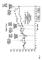
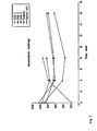
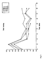
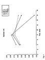
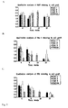

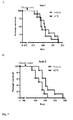


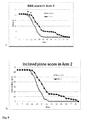
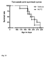
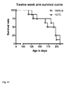
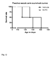
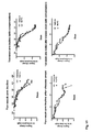
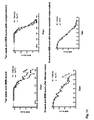
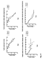


Classifications
-
- C—CHEMISTRY; METALLURGY
- C12—BIOCHEMISTRY; BEER; SPIRITS; WINE; VINEGAR; MICROBIOLOGY; ENZYMOLOGY; MUTATION OR GENETIC ENGINEERING
- C12N—MICROORGANISMS OR ENZYMES; COMPOSITIONS THEREOF; PROPAGATING, PRESERVING, OR MAINTAINING MICROORGANISMS; MUTATION OR GENETIC ENGINEERING; CULTURE MEDIA
- C12N5/00—Undifferentiated human, animal or plant cells, e.g. cell lines; Tissues; Cultivation or maintenance thereof; Culture media therefor
- C12N5/06—Animal cells or tissues; Human cells or tissues
- C12N5/0602—Vertebrate cells
- C12N5/0603—Embryonic cells ; Embryoid bodies
- C12N5/0605—Cells from extra-embryonic tissues, e.g. placenta, amnion, yolk sac, Wharton's jelly
-
- A—HUMAN NECESSITIES
- A61—MEDICAL OR VETERINARY SCIENCE; HYGIENE
- A61P—SPECIFIC THERAPEUTIC ACTIVITY OF CHEMICAL COMPOUNDS OR MEDICINAL PREPARATIONS
- A61P21/00—Drugs for disorders of the muscular or neuromuscular system
-
- A—HUMAN NECESSITIES
- A61—MEDICAL OR VETERINARY SCIENCE; HYGIENE
- A61P—SPECIFIC THERAPEUTIC ACTIVITY OF CHEMICAL COMPOUNDS OR MEDICINAL PREPARATIONS
- A61P21/00—Drugs for disorders of the muscular or neuromuscular system
- A61P21/02—Muscle relaxants, e.g. for tetanus or cramps
-
- A—HUMAN NECESSITIES
- A61—MEDICAL OR VETERINARY SCIENCE; HYGIENE
- A61P—SPECIFIC THERAPEUTIC ACTIVITY OF CHEMICAL COMPOUNDS OR MEDICINAL PREPARATIONS
- A61P25/00—Drugs for disorders of the nervous system
-
- A—HUMAN NECESSITIES
- A61—MEDICAL OR VETERINARY SCIENCE; HYGIENE
- A61P—SPECIFIC THERAPEUTIC ACTIVITY OF CHEMICAL COMPOUNDS OR MEDICINAL PREPARATIONS
- A61P25/00—Drugs for disorders of the nervous system
- A61P25/02—Drugs for disorders of the nervous system for peripheral neuropathies
-
- A—HUMAN NECESSITIES
- A61—MEDICAL OR VETERINARY SCIENCE; HYGIENE
- A61P—SPECIFIC THERAPEUTIC ACTIVITY OF CHEMICAL COMPOUNDS OR MEDICINAL PREPARATIONS
- A61P43/00—Drugs for specific purposes, not provided for in groups A61P1/00-A61P41/00
-
- C—CHEMISTRY; METALLURGY
- C12—BIOCHEMISTRY; BEER; SPIRITS; WINE; VINEGAR; MICROBIOLOGY; ENZYMOLOGY; MUTATION OR GENETIC ENGINEERING
- C12N—MICROORGANISMS OR ENZYMES; COMPOSITIONS THEREOF; PROPAGATING, PRESERVING, OR MAINTAINING MICROORGANISMS; MUTATION OR GENETIC ENGINEERING; CULTURE MEDIA
- C12N5/00—Undifferentiated human, animal or plant cells, e.g. cell lines; Tissues; Cultivation or maintenance thereof; Culture media therefor
- C12N5/06—Animal cells or tissues; Human cells or tissues
- C12N5/0602—Vertebrate cells
- C12N5/0607—Non-embryonic pluripotent stem cells, e.g. MASC
-
- C—CHEMISTRY; METALLURGY
- C12—BIOCHEMISTRY; BEER; SPIRITS; WINE; VINEGAR; MICROBIOLOGY; ENZYMOLOGY; MUTATION OR GENETIC ENGINEERING
- C12N—MICROORGANISMS OR ENZYMES; COMPOSITIONS THEREOF; PROPAGATING, PRESERVING, OR MAINTAINING MICROORGANISMS; MUTATION OR GENETIC ENGINEERING; CULTURE MEDIA
- C12N5/00—Undifferentiated human, animal or plant cells, e.g. cell lines; Tissues; Cultivation or maintenance thereof; Culture media therefor
- C12N5/06—Animal cells or tissues; Human cells or tissues
- C12N5/0602—Vertebrate cells
- C12N5/0618—Cells of the nervous system
- C12N5/0619—Neurons
-
- C—CHEMISTRY; METALLURGY
- C12—BIOCHEMISTRY; BEER; SPIRITS; WINE; VINEGAR; MICROBIOLOGY; ENZYMOLOGY; MUTATION OR GENETIC ENGINEERING
- C12N—MICROORGANISMS OR ENZYMES; COMPOSITIONS THEREOF; PROPAGATING, PRESERVING, OR MAINTAINING MICROORGANISMS; MUTATION OR GENETIC ENGINEERING; CULTURE MEDIA
- C12N5/00—Undifferentiated human, animal or plant cells, e.g. cell lines; Tissues; Cultivation or maintenance thereof; Culture media therefor
- C12N5/06—Animal cells or tissues; Human cells or tissues
- C12N5/0602—Vertebrate cells
- C12N5/0618—Cells of the nervous system
- C12N5/0622—Glial cells, e.g. astrocytes, oligodendrocytes; Schwann cells
-
- A—HUMAN NECESSITIES
- A61—MEDICAL OR VETERINARY SCIENCE; HYGIENE
- A61K—PREPARATIONS FOR MEDICAL, DENTAL OR TOILETRY PURPOSES
- A61K35/00—Medicinal preparations containing materials or reaction products thereof with undetermined constitution
- A61K35/12—Materials from mammals; Compositions comprising non-specified tissues or cells; Compositions comprising non-embryonic stem cells; Genetically modified cells
-
- C—CHEMISTRY; METALLURGY
- C12—BIOCHEMISTRY; BEER; SPIRITS; WINE; VINEGAR; MICROBIOLOGY; ENZYMOLOGY; MUTATION OR GENETIC ENGINEERING
- C12N—MICROORGANISMS OR ENZYMES; COMPOSITIONS THEREOF; PROPAGATING, PRESERVING, OR MAINTAINING MICROORGANISMS; MUTATION OR GENETIC ENGINEERING; CULTURE MEDIA
- C12N2500/00—Specific components of cell culture medium
- C12N2500/30—Organic components
- C12N2500/32—Amino acids
-
- C—CHEMISTRY; METALLURGY
- C12—BIOCHEMISTRY; BEER; SPIRITS; WINE; VINEGAR; MICROBIOLOGY; ENZYMOLOGY; MUTATION OR GENETIC ENGINEERING
- C12N—MICROORGANISMS OR ENZYMES; COMPOSITIONS THEREOF; PROPAGATING, PRESERVING, OR MAINTAINING MICROORGANISMS; MUTATION OR GENETIC ENGINEERING; CULTURE MEDIA
- C12N2500/00—Specific components of cell culture medium
- C12N2500/30—Organic components
- C12N2500/34—Sugars
-
- C—CHEMISTRY; METALLURGY
- C12—BIOCHEMISTRY; BEER; SPIRITS; WINE; VINEGAR; MICROBIOLOGY; ENZYMOLOGY; MUTATION OR GENETIC ENGINEERING
- C12N—MICROORGANISMS OR ENZYMES; COMPOSITIONS THEREOF; PROPAGATING, PRESERVING, OR MAINTAINING MICROORGANISMS; MUTATION OR GENETIC ENGINEERING; CULTURE MEDIA
- C12N2500/00—Specific components of cell culture medium
- C12N2500/30—Organic components
- C12N2500/44—Thiols, e.g. mercaptoethanol
-
- C—CHEMISTRY; METALLURGY
- C12—BIOCHEMISTRY; BEER; SPIRITS; WINE; VINEGAR; MICROBIOLOGY; ENZYMOLOGY; MUTATION OR GENETIC ENGINEERING
- C12N—MICROORGANISMS OR ENZYMES; COMPOSITIONS THEREOF; PROPAGATING, PRESERVING, OR MAINTAINING MICROORGANISMS; MUTATION OR GENETIC ENGINEERING; CULTURE MEDIA
- C12N2500/00—Specific components of cell culture medium
- C12N2500/90—Serum-free medium, which may still contain naturally-sourced components
- C12N2500/95—Protein-free medium and culture conditions
-
- C—CHEMISTRY; METALLURGY
- C12—BIOCHEMISTRY; BEER; SPIRITS; WINE; VINEGAR; MICROBIOLOGY; ENZYMOLOGY; MUTATION OR GENETIC ENGINEERING
- C12N—MICROORGANISMS OR ENZYMES; COMPOSITIONS THEREOF; PROPAGATING, PRESERVING, OR MAINTAINING MICROORGANISMS; MUTATION OR GENETIC ENGINEERING; CULTURE MEDIA
- C12N2501/00—Active agents used in cell culture processes, e.g. differentation
- C12N2501/10—Growth factors
- C12N2501/11—Epidermal growth factor [EGF]
-
- C—CHEMISTRY; METALLURGY
- C12—BIOCHEMISTRY; BEER; SPIRITS; WINE; VINEGAR; MICROBIOLOGY; ENZYMOLOGY; MUTATION OR GENETIC ENGINEERING
- C12N—MICROORGANISMS OR ENZYMES; COMPOSITIONS THEREOF; PROPAGATING, PRESERVING, OR MAINTAINING MICROORGANISMS; MUTATION OR GENETIC ENGINEERING; CULTURE MEDIA
- C12N2501/00—Active agents used in cell culture processes, e.g. differentation
- C12N2501/10—Growth factors
- C12N2501/115—Basic fibroblast growth factor (bFGF, FGF-2)
-
- C—CHEMISTRY; METALLURGY
- C12—BIOCHEMISTRY; BEER; SPIRITS; WINE; VINEGAR; MICROBIOLOGY; ENZYMOLOGY; MUTATION OR GENETIC ENGINEERING
- C12N—MICROORGANISMS OR ENZYMES; COMPOSITIONS THEREOF; PROPAGATING, PRESERVING, OR MAINTAINING MICROORGANISMS; MUTATION OR GENETIC ENGINEERING; CULTURE MEDIA
- C12N2501/00—Active agents used in cell culture processes, e.g. differentation
- C12N2501/10—Growth factors
- C12N2501/119—Other fibroblast growth factors, e.g. FGF-4, FGF-8, FGF-10
-
- C—CHEMISTRY; METALLURGY
- C12—BIOCHEMISTRY; BEER; SPIRITS; WINE; VINEGAR; MICROBIOLOGY; ENZYMOLOGY; MUTATION OR GENETIC ENGINEERING
- C12N—MICROORGANISMS OR ENZYMES; COMPOSITIONS THEREOF; PROPAGATING, PRESERVING, OR MAINTAINING MICROORGANISMS; MUTATION OR GENETIC ENGINEERING; CULTURE MEDIA
- C12N2501/00—Active agents used in cell culture processes, e.g. differentation
- C12N2501/10—Growth factors
- C12N2501/12—Hepatocyte growth factor [HGF]
-
- C—CHEMISTRY; METALLURGY
- C12—BIOCHEMISTRY; BEER; SPIRITS; WINE; VINEGAR; MICROBIOLOGY; ENZYMOLOGY; MUTATION OR GENETIC ENGINEERING
- C12N—MICROORGANISMS OR ENZYMES; COMPOSITIONS THEREOF; PROPAGATING, PRESERVING, OR MAINTAINING MICROORGANISMS; MUTATION OR GENETIC ENGINEERING; CULTURE MEDIA
- C12N2501/00—Active agents used in cell culture processes, e.g. differentation
- C12N2501/10—Growth factors
- C12N2501/13—Nerve growth factor [NGF]; Brain-derived neurotrophic factor [BDNF]; Cilliary neurotrophic factor [CNTF]; Glial-derived neurotrophic factor [GDNF]; Neurotrophins [NT]; Neuregulins
-
- C—CHEMISTRY; METALLURGY
- C12—BIOCHEMISTRY; BEER; SPIRITS; WINE; VINEGAR; MICROBIOLOGY; ENZYMOLOGY; MUTATION OR GENETIC ENGINEERING
- C12N—MICROORGANISMS OR ENZYMES; COMPOSITIONS THEREOF; PROPAGATING, PRESERVING, OR MAINTAINING MICROORGANISMS; MUTATION OR GENETIC ENGINEERING; CULTURE MEDIA
- C12N2501/00—Active agents used in cell culture processes, e.g. differentation
- C12N2501/20—Cytokines; Chemokines
- C12N2501/21—Chemokines, e.g. MIP-1, MIP-2, RANTES, MCP, PF-4
-
- C—CHEMISTRY; METALLURGY
- C12—BIOCHEMISTRY; BEER; SPIRITS; WINE; VINEGAR; MICROBIOLOGY; ENZYMOLOGY; MUTATION OR GENETIC ENGINEERING
- C12N—MICROORGANISMS OR ENZYMES; COMPOSITIONS THEREOF; PROPAGATING, PRESERVING, OR MAINTAINING MICROORGANISMS; MUTATION OR GENETIC ENGINEERING; CULTURE MEDIA
- C12N2501/00—Active agents used in cell culture processes, e.g. differentation
- C12N2501/20—Cytokines; Chemokines
- C12N2501/23—Interleukins [IL]
-
- C—CHEMISTRY; METALLURGY
- C12—BIOCHEMISTRY; BEER; SPIRITS; WINE; VINEGAR; MICROBIOLOGY; ENZYMOLOGY; MUTATION OR GENETIC ENGINEERING
- C12N—MICROORGANISMS OR ENZYMES; COMPOSITIONS THEREOF; PROPAGATING, PRESERVING, OR MAINTAINING MICROORGANISMS; MUTATION OR GENETIC ENGINEERING; CULTURE MEDIA
- C12N2501/00—Active agents used in cell culture processes, e.g. differentation
- C12N2501/30—Hormones
- C12N2501/38—Hormones with nuclear receptors
- C12N2501/385—Hormones with nuclear receptors of the family of the retinoic acid recptor, e.g. RAR, RXR; Peroxisome proliferator-activated receptor [PPAR]
-
- C—CHEMISTRY; METALLURGY
- C12—BIOCHEMISTRY; BEER; SPIRITS; WINE; VINEGAR; MICROBIOLOGY; ENZYMOLOGY; MUTATION OR GENETIC ENGINEERING
- C12N—MICROORGANISMS OR ENZYMES; COMPOSITIONS THEREOF; PROPAGATING, PRESERVING, OR MAINTAINING MICROORGANISMS; MUTATION OR GENETIC ENGINEERING; CULTURE MEDIA
- C12N2501/00—Active agents used in cell culture processes, e.g. differentation
- C12N2501/40—Regulators of development
- C12N2501/41—Hedgehog proteins; Cyclopamine (inhibitor)
-
- C—CHEMISTRY; METALLURGY
- C12—BIOCHEMISTRY; BEER; SPIRITS; WINE; VINEGAR; MICROBIOLOGY; ENZYMOLOGY; MUTATION OR GENETIC ENGINEERING
- C12N—MICROORGANISMS OR ENZYMES; COMPOSITIONS THEREOF; PROPAGATING, PRESERVING, OR MAINTAINING MICROORGANISMS; MUTATION OR GENETIC ENGINEERING; CULTURE MEDIA
- C12N2502/00—Coculture with; Conditioned medium produced by
- C12N2502/02—Coculture with; Conditioned medium produced by embryonic cells
-
- C—CHEMISTRY; METALLURGY
- C12—BIOCHEMISTRY; BEER; SPIRITS; WINE; VINEGAR; MICROBIOLOGY; ENZYMOLOGY; MUTATION OR GENETIC ENGINEERING
- C12N—MICROORGANISMS OR ENZYMES; COMPOSITIONS THEREOF; PROPAGATING, PRESERVING, OR MAINTAINING MICROORGANISMS; MUTATION OR GENETIC ENGINEERING; CULTURE MEDIA
- C12N2502/00—Coculture with; Conditioned medium produced by
- C12N2502/03—Coculture with; Conditioned medium produced by non-embryonic pluripotent stem cells
-
- C—CHEMISTRY; METALLURGY
- C12—BIOCHEMISTRY; BEER; SPIRITS; WINE; VINEGAR; MICROBIOLOGY; ENZYMOLOGY; MUTATION OR GENETIC ENGINEERING
- C12N—MICROORGANISMS OR ENZYMES; COMPOSITIONS THEREOF; PROPAGATING, PRESERVING, OR MAINTAINING MICROORGANISMS; MUTATION OR GENETIC ENGINEERING; CULTURE MEDIA
- C12N2506/00—Differentiation of animal cells from one lineage to another; Differentiation of pluripotent cells
- C12N2506/02—Differentiation of animal cells from one lineage to another; Differentiation of pluripotent cells from embryonic cells
- C12N2506/025—Differentiation of animal cells from one lineage to another; Differentiation of pluripotent cells from embryonic cells from extra-embryonic cells, e.g. trophoblast, placenta
-
- C—CHEMISTRY; METALLURGY
- C12—BIOCHEMISTRY; BEER; SPIRITS; WINE; VINEGAR; MICROBIOLOGY; ENZYMOLOGY; MUTATION OR GENETIC ENGINEERING
- C12N—MICROORGANISMS OR ENZYMES; COMPOSITIONS THEREOF; PROPAGATING, PRESERVING, OR MAINTAINING MICROORGANISMS; MUTATION OR GENETIC ENGINEERING; CULTURE MEDIA
- C12N2506/00—Differentiation of animal cells from one lineage to another; Differentiation of pluripotent cells
- C12N2506/03—Differentiation of animal cells from one lineage to another; Differentiation of pluripotent cells from non-embryonic pluripotent stem cells
-
- C—CHEMISTRY; METALLURGY
- C12—BIOCHEMISTRY; BEER; SPIRITS; WINE; VINEGAR; MICROBIOLOGY; ENZYMOLOGY; MUTATION OR GENETIC ENGINEERING
- C12N—MICROORGANISMS OR ENZYMES; COMPOSITIONS THEREOF; PROPAGATING, PRESERVING, OR MAINTAINING MICROORGANISMS; MUTATION OR GENETIC ENGINEERING; CULTURE MEDIA
- C12N2509/00—Methods for the dissociation of cells, e.g. specific use of enzymes
-
- C—CHEMISTRY; METALLURGY
- C12—BIOCHEMISTRY; BEER; SPIRITS; WINE; VINEGAR; MICROBIOLOGY; ENZYMOLOGY; MUTATION OR GENETIC ENGINEERING
- C12N—MICROORGANISMS OR ENZYMES; COMPOSITIONS THEREOF; PROPAGATING, PRESERVING, OR MAINTAINING MICROORGANISMS; MUTATION OR GENETIC ENGINEERING; CULTURE MEDIA
- C12N2533/00—Supports or coatings for cell culture, characterised by material
- C12N2533/50—Proteins
Definitions
- This invention relates generally to compositions, methods and kits for cell-based or regenerative therapy for neurological diseases and disorders such as amyotrophic lateral sclerosis.
- this invention provides umbilical cord tissue-derived cells for use in preserving motor neuron function in a patient with amyotrophic lateral sclerosis.
- ALS amyotrophic lateral sclerosis
- ALS is a progressive neurodegenerative disease that mainly affects motor neurons resulting in muscle weakness and atrophy. The disease is characterized by selective, premature degeneration and death of motor neurons. Affected neurons show loss of dendrites, cytoskeletal changes and accumulation of proteins and inclusion bodies. ALS results in a progressive paralysis that is typically fatal within a handful of years due to respiratory failure resulting from paralysis of the respiratory muscles. The average duration of disease from onset to death is three to five years. Only about 10% of ALS patients survive for ten or more years. As many as 20,000 to 30,000 people in the United States suffer from ALS and an additional 5,000 are diagnosed each year.
- ALS pathogenic mechanisms of ALS are not fully understood but a variety of processes are thought to contribute to ALS including mitochondrial dysfunction, oxidative stress, excitotoxicity as well as alterations in the cytoskeleton, in axonal transport, protein processing, and calcium homeostasis ( Ilieva et al. (2009), J. Cell Biol. 187:761-72 ). Despite intense effort, very limited therapeutic options have emerged for the slowing disease course, although advances have been made in palliative therapy. Allogeneic cell therapy may provide an effective multi-factorial therapy for the treatment of ALS by elaborating trophic factors, which diminish motor neuron degeneration, preserve motor neuron function and extend life.
- the invention provides umbilical cord tissue-derived cells for use in preserving motor neuron function in a patient with amyotrophic lateral sclerosis, wherein the umbilical cord tissue-derived cells are isolatable from human umbilical cord tissue substantially free of blood, are capable of self-renewal and expansion in culture, have the potential to differentiate into cells of other phenotypes, can undergo at least 40 doublings, and have the following characteristics:
- compositions, methods and kits of the illustrative embodiments described herein provides compositions and methods applicable to cell-based or regenerative therapy for neurological diseases and disorders such as amyotrophic lateral sclerosis.
- the invention features pharmaceutical compositions, devices and methods for the regeneration or repair of neural tissue using umbilical cord tissue-derived cells.
- One aspect of the invention features an isolated umbilical cord tissue-derived cell substantially free of blood, wherein the cell is capable of self-renewal and expansion in culture and has the potential to differentiate into a cell of a neural phenotype; wherein the cell requires L-valine for growth and is capable of growth in at least about 5% oxygen.
- This cell further comprises one or more of the following characteristics: (a) potential for at least about 40 doublings in culture; (b) attachment and expansion on a coated or uncoated tissue culture vessel, wherein the coated tissue culture vessel comprises a coating of gelatin, laminin, collagen, polyornithine, vitronectin, or fibronectin; (c) production of at least one of tissue factor, vimentin, and alpha-smooth muscle actin; (d) production of at least one of CD10, CD13, CD44, CD73, CD90, PDGFr-alpha, PD-L2 and HLA-A,B,C; (e) lack of production of at least one of CD31, CD34, CD45, CD80, CD86, CD117, CD141, CD178, B7-H2, HLA-G, and HLA-DR,DP,DQ, as detected by flow cytometry; (f) expression of a gene, which relative to a human cell that is a fibroblast, a mesenchymal stem
- the umbilicus-derived cell has all identifying features of any one of: cell type UMB 022803 (P7) (ATCC Accession No. PTA-6067); or cell type UMB 022803 (P17) (ATCC Accession No. PTA-6068).
- umbilical cord tissue-derived cells are isolated in the presence of one or more enzyme activities comprising metalloprotease activity, mucolytic activity and neutral protease activity.
- the cells Preferably, the cells have a normal karyotype, which is maintained as the cells are passaged in culture.
- the postpartum-derived cells comprise each of CD10, CD13, CD44, CD73, CD90, PDGFr-alpha, and HLA-A,B,C and does not comprise any of CD31, CD34, CD45, CD117, CD141, or HLA-DR,DP,DQ, as detected by flow cytometry.
- the population is a substantially homogeneous population of the umbilical cord tissue-derived cells.
- the population comprises a clonal cell line of the umbilical cord tissue-derived cells.
- the population is a heterogeneous population comprising the umbilical cord tissue-derived cells and at least one other cell type.
- the other cell type is an astrocyte, oligodendrocyte, neuron, neural progenitor, neural stem cell or other multipotent or pluripotent stem cell.
- the cell population is cultured in contact with one or more factors that stimulate stem cell differentiation toward a neural lineage.
- a cell lysate prepared from umbilical cord tissue-derived cells.
- the cell lysate may be separated into a membrane enriched fraction and a soluble cell fraction.
- the disclosure also features an extracellular matrix produced by the umbilical cord tissue-derived cells, as well as a conditioned medium in which the cells have been grown.
- the neurodegenerative condition is an acute neurodegenerative condition, such as a brain trauma, spinal cord trauma or peripheral nerve trauma.
- it is a chronic or progressive neurodegenerative condition, such as Parkinson's disease, Alzheimer's disease, Huntington's disease, amyotrophic lateral sclerosis, tumor, multiple sclerosis or chronic peripheral nerve injury.
- the umbilical cord tissue-derived cells are induced in vitro to differentiate into a neural lineage cells prior to administration.
- the cells are genetically engineered to produce a gene product that promotes treatment of the neurodegenerative condition.
- the cells are administered with at least one other cell type, such as an astrocyte, oligodendrocyte, neuron, neural progenitor, neural stem cell or other multipotent or pluripotent stem cell.
- the other cell type can be administered simultaneously with, or before, or after, the umbilical cord tissue-derived cells.
- the cells are administered with at least one other agent, such as a drug for neural therapy, or another beneficial adjunctive agent such as an anti-inflammatory agent, anti-apoptotic agents, antioxidant or growth factor.
- the other agent can be administered simultaneously with, or before, or after, the umbilical cord tissue-derived cells.
- the cells are administered at a pre-determined site in the central or peripheral nervous system of the patient. They can be administered by injection or infusion, or encapsulated within an implantable device, or by implantation of a matrix or scaffold containing the cells.
- One embodiment is a method of treating amyotrophic lateral sclerosis comprising administering umbilical cord tissue-derived cells in an amount effective to treat amyotrophic lateral sclerosis to a patient.
- the umbilical cord tissue-derived cells are isolated from human umbilical cord tissue substantially free of blood, are capable of self-renewal and expansion into culture, have the potential to differentiate into cells of other phenotypes, can undergo at least 40 doublings, and have the following characteristics: (a) expression each of CD10, CD13, CD44, CD73, CD90, PDGFr-alpha, PD-L2 and HLA-A,B,C; (b) do not express any of CD31, CD34, CD45, CD80, CD86, CD 117, CD141, CD178, B7-H2, HLA-G, or HLA-DR,DP,DQ; and (c) increased expression of interleukin-8; reticulon 1; and chemokine receptor ligand (C-X-C motif)
- the umbilical cord tissue-derived cells do not express hTERT or telomerase.
- the umbilical cord tissue-derived cells are administered by injection (such as e . g . intravenous or intrathecal injection or infusion.
- the umbilical cord tissue-derived cells exert a trophic effect on the nervous system of the patient.
- Another embodiment is a method of treating amyotrophic lateral sclerosis comprising administering an effective amount of a substantially homogeneous population of umbilical cord tissue-derived cells to a patient.
- the population of umbilical cord tissue-derived cells is isolated from human umbilical cord tissue substantially free of blood, is capable of self-renewal and expansion into culture, has the potential to differentiate into cells of other phenotypes, can undergo at least 40 doublings, and has the following characteristics: (a) expresses each of CD10, CD13, CD44, CD73, CD90, PDGFr-alpha, PD-L2 and HLA-A,B,C; (b) does not express any of CD31, CD34, CD45, CD80, CD86, CD 117, CD141, CD178, B7-H2, HLA-G, or HLA-DR,DP,DQ; and (c) increased expression of interleukin-8; reticulon 1; and chemokine receptor ligand (C-X-C motif
- the umbilical cord tissue-derived cells do not express hTERT or telomerase.
- the substantially homogeneous population of umbilical cord tissue-derived cells is administered by injection or infusion.
- the substantially homogeneous population of umbilical cord tissue-derived cells is administered by intravenous or intrathecal injection.
- the substantially homogeneous population of umbilical cord tissue-derived cells exerts a trophic effect on the nervous system of the patient.
- Yet another embodiment is a method of treating amyotrophic lateral sclerosis comprising administering a pharmaceutical composition comprising umbilical cord tissue-derived cells in an amount effective to treat amyotrophic lateral sclerosis to a patient.
- the umbilical cord tissue-derived cells are isolated from human umbilical cord tissue substantially free of blood, are capable of self-renewal and expansion into culture, have the potential to differentiate into cells of other phenotypes, can undergo at least 40 doublings, and have the following characteristics: (a) expresses each of CD10, CD13, CD44, CD73, CD90, PDGFr-alpha, PD-L2 and HLA-A,B,C; (b) does not express any of CD31, CD34, CD45, CD80, CD86, CD 117, CD141, CD178, B7-H2, HLA-G, or HLA-DR,DP,DQ; and (c) increased expression of interleukin-8; reticulon 1; and chemokine receptor lig
- the umbilical cord tissue-derived cells do not express hTERT or telomerase.
- the umbilical cord tissue-derived cells are administered by injection (such as e.g. intravenous or intrathecal injection) or infusion.
- the umbilical cord tissue-derived cells exert a trophic effect on the nervous system of the patient.
- Another aspect of the invention features a pharmaceutical composition for treating a patient having a neurodegenerative condition, amyotrophic lateral sclerosis, comprising a pharmaceutically acceptable carrier and the umbilical cord tissue-derived cells described above.
- the neurodegenerative condition to be treated may be an acute neurodegenerative condition, or it may be a chronic or progressive condition.
- the pharmaceutical composition comprises cells that have been induced in vitro to differentiate into a neural lineage cells prior to formulation of the composition, or cells that have been genetically engineered to produce a gene product that promotes treatment of the neurodegenerative condition.
- the pharmaceutical composition comprises at least one other cell type, such as astrocyte, oligodendrocyte, neuron, neural progenitor, neural stem cell or other multipotent or pluripotent stem cell.
- the pharmaceutical composition comprises at least one other agent, such as a drug for neural therapy, or another beneficial adjunctive agent such as an anti-inflammatory agent, anti-apoptotic agents, antioxidant or growth factor.
- the pharmaceutical composition is formulated for administration by injection or infusion.
- it may comprise an implantable device in which the cells are encapsulated, or a matrix or scaffold containing the cells.
- a kit for treating a patient having a neurodegenerative condition.
- the kit comprises a pharmaceutically acceptable carrier, a population of the above-described umbilical cord tissue-derived cells and instructions for using the kit in a method of treating the patient.
- the kit may further comprise at least one reagent and instructions for culturing the umbilical cord tissue-derived cells. It may also comprise a population of at least one other cell type, or at least one other agent for treating a neurodegenerative condition.
- a method for treating a patient having a neurodegenerative condition such as e . g . amyotrophic lateral sclerosis
- a preparation made from the above-described umbilical cord tissue-derived cells may comprise a cell lysate (or fraction thereof) of the umbilical cord tissue-derived cells, an extracellular matrix of the umbilical cord tissue-derived cells, or a conditioned medium in which the umbilical cord tissue-derived cells were grown.
- kits for practicing this aspect of the invention are also provided. These may include the one or more of a pharmaceutically acceptable carrier or other agent or reagent, one or more of a cell lysate or fraction thereof, an extracellular matrix or a conditioned medium from the umbilical cord tissue-derived cells, and instructions for use of the kit components.
- Stem cells are undifferentiated cells defined by the ability of a single cell both to self-renew, and to differentiate to produce progeny cells, including self-renewing progenitors, non-renewing progenitors, and terminally differentiated cells. Stem cells are also characterized by their ability to differentiate in vitro into functional cells of various cell lineages from multiple germ layers (endoderm, mesoderm and ectoderm), as well as to give rise to tissues of multiple germ layers following transplantation, and to contribute substantially to most, if not all, tissues following injection into blastocysts.
- Stem cells are classified according to their developmental potential as: (1) totipotent ; (2) pluripotent ; (3) multipotent ; (4) oligopotent ; and (5) unipotent .
- Totipotent cells are able to give rise to all embryonic and extraembryonic cell types.
- Pluripotent cells are able to give rise to all embryonic cell types.
- Multipotent cells include those able to give rise to a subset of cell lineages, but all within a particular tissue, organ, or physiological system (for example, hematopoietic stem cells (HSC) can produce progeny that include HSC (self-renewal), blood cell-restricted oligopotent progenitors, and all cell types and elements (e.g., platelets) that are normal components of the blood).
- HSC hematopoietic stem cells
- Cells that are oligopotent can give rise to a more restricted subset of cell lineages than multipotent stem cells; and cells that are unipotent are able to give rise to a single cell lineage (e.g., spermatogenic stem cells).
- Stem cells are also categorized on the basis of the source from which they may be obtained.
- An adult stem cell is generally a multipotent undifferentiated cell found in tissue comprising multiple differentiated cell types. The adult stem cell can renew itself. Under normal circumstances, it can also differentiate to yield the specialized cell types of the tissue from which it originated, and possibly other tissue types.
- An embryonic stem cell is a pluripotent cell from the inner cell mass of a blastocyst-stage embryo.
- a fetal stem cell is one that originates from fetal tissues or membranes.
- a postpartum stem cell is a multipotent or pluripotent cell that originates substantially from extraembryonic tissue available after birth, namely, the placenta and the umbilical cord.
- Postpartum stem cells may be blood-derived (e.g., as are those obtained from umbilical cord blood) or non-blood-derived (e . g ., as obtained from the non-blood tissues of the umbilical cord and placenta).
- Embryonic tissue is typically defined as tissue originating from the embryo (which in humans refers to the period from fertilization to about six weeks of development. Fetal tissue refers to tissue originating from the fetus, which in humans refers to the period from about six weeks of development to parturition. Extraembryonic tissue is tissue associated with, but not originating from, the embryo or fetus. Extraembryonic tissues include extraembryonic membranes (chorion, amnion, yolk sac and allantois), umbilical cord and placenta (which itself forms from the chorion and the maternal decidua basalis).
- Differentiation is the process by which an unspecialized ("uncommitted") or less specialized cell acquires the features of a specialized cell, such as a nerve cell or a muscle cell, for example.
- a differentiated cell is one that has taken on a more specialized ("committed") position within the lineage of a cell.
- the term committed when applied to the process of differentiation, refers to a cell that has proceeded in the differentiation pathway to a point where, under normal circumstances, it will continue to differentiate into a specific cell type or subset of cell types, and cannot, under normal circumstances, differentiate into a different cell type or revert to a less differentiated cell type.
- De - differentiation refers to the process by which a cell reverts to a less specialized (or committed) position within the lineage of a cell.
- the lineage of a cell defines the heredity of the cell, i.e. which cells it came from and what cells it can give rise to.
- the lineage of a cell places the cell within a hereditary scheme of development and differentiation.
- a progenitor cell is a cell that has the capacity to create progeny that are more differentiated than itself, and yet retains the capacity to replenish the pool of progenitors.
- stem cells themselves are also progenitor cells, as are the more immediate precursors to terminally differentiated cells.
- this broad definition of progenitor cell may be used.
- a progenitor cell is often defined as a cell that is intermediate in the differentiation pathway, i.e ., it arises from a stem cell and is intermediate in the production of a mature cell type or subset of cell types. This type of progenitor cell is generally not able to self-renew. Accordingly, if this type of cell is referred to herein, it will be referred to as a non-renewing progenitor cell or as an intermediate progenitor or precursor cell.
- the phrase differentiates into a neural lineage or phenotype refers to a cell that becomes partially or fully committed to a specific neural phenotype of the CNS or PNS, i.e ., a neuron or a glial cell, the latter category including without limitation astrocytes, oligodendrocytes, Schwann cells and microglia.
- the cells of the present invention are generally referred to as umbilical cord tissue-derived cells (or UTC or hUTCs). They also may sometimes be referred to as umbilicus-derived cells (UDCs) .
- the cells may be described as being stem or progenitor cells, the latter term being used in the broad sense.
- the term derived is used to indicate that the cells have been obtained from their biological source and grown or otherwise manipulated in vitro (e.g., cultured in a Growth Medium to expand the population and/or to produce a cell line). The in vitro manipulations of umbilical stem cells and the unique features of the umbilicus-derived cells of the present invention are described in detail below.
- Cell culture refers generally to cells taken from a living organism and grown under controlled condition ("in culture” or “cultured”).
- a primary cell culture is a culture of cells, tissues, or organs taken directly from an organism(s) before the first subculture.
- Cells are expanded in culture when they are placed in a Growth Medium under conditions that facilitate cell growth and/or division, resulting in a larger population of the cells.
- the rate of cell proliferation is sometimes measured by the amount of time needed for the cells to double in number. This is referred to as doubling time.
- a cell line is a population of cells formed by one or more subcultivations of a primary cell culture. Each round of subculturing is referred to as a passage. When cells are subcultured, they are referred to as having been passaged. A specific population of cells, or a cell line, is sometimes referred to or characterized by the number of times it has been passaged. For example, a cultured cell population that has been passaged ten times may be referred to as a P10 culture.
- the primary culture i.e ., the first culture following the isolation of cells from tissue, is designated P0. Following the first subculture, the cells are described as a secondary culture (P1 or passage 1).
- the cells After the second subculture, the cells become a tertiary culture (P2 or passage 2), and so on. It will be understood by those of skill in the art that there may be many population doublings during the period of passaging; therefore, the number of population doublings of a culture is greater than the passage number.
- the expansion of cells (i.e., the number of population doublings) during the period between passaging depends on many factors, including but not limited to the seeding density, substrate, medium, growth conditions, and time between passaging.
- a conditioned medium is a medium in which a specific cell or population of cells has been cultured, and then removed. When cells are cultured in a medium, they may secrete cellular factors that can provide trophic support to other cells. Such trophic factors include, but are not limited to hormones, cytokines, extracellular matrix (ECM), proteins, vesicles, antibodies, and granules.
- the medium containing the cellular factors is the conditioned medium.
- a trophic factor is defined as a substance that promotes survival, growth, differentiation, proliferation and /or maturation of a cell, or stimulates increased activity of a cell.
- the interaction between cells via trophic factors may occur between cells of different types. Cell interaction by way of trophic factors is found in essentially all cell types, and is a particularly significant means of communication among neural cell types.
- Trophic factors also can function in an autocrine fashion, i.e ., a cell may produce trophic factors that affect its own survival, growth, differentiation, proliferation and/or maturation.
- senescence also replicative senescence or cellular senescence refers to a property attributable to finite cell cultures; namely, their inability to grow beyond a finite number of population doublings (sometimes referred to as Hayflick's limit ).
- cellular senescence was first described using fibroblast-like cells, most normal human cell types that can be grown successfully in culture undergo cellular senescence.
- the in vitro lifespan of different cell types varies, but the maximum lifespan is typically fewer than 100 population doublings (this is the number of doublings for all the cells in the culture to become senescent and thus render the culture unable to divide).
- Senescence does not depend on chronological time, but rather is measured by the number of cell divisions, or population doublings, the culture has undergone. Thus, cells made quiescent by removing essential growth factors are able to resume growth and division when the growth factors are re-introduced, and thereafter carry out the same number of doublings as equivalent cells grown continuously. Similarly, when cells are frozen in liquid nitrogen after various numbers of population doublings and then thawed and cultured, they undergo substantially the same number of doublings as cells maintained unfrozen in culture. Senescent cells are not dead or dying cells; they are actually resistant to programmed cell death (apoptosis), and have been maintained in their nondividing state for as long as three years. These cells are very much alive and metabolically active, but they do not divide. The nondividing state of senescent cells has not yet been found to be reversible by any biological, chemical, or viral agent.
- neurodegenerative condition is an inclusive term encompassing acute and chronic conditions, disorders or diseases of the central or peripheral nervous system.
- a neurodegenerative condition may be age-related, or it may result from injury or trauma, or it may be related to a specific disease or disorder.
- Acute neurodegenerative conditions include, but are not limited to, conditions associated with neuronal cell death or compromise including cerebrovascular insufficiency, focal or diffuse brain trauma, diffuse brain damage, spinal cord injury or peripheral nerve trauma, e.g., resulting from physical or chemical burns, deep cuts or limb severance.
- Examples of acute neurodegenerative disorders are: cerebral ischemia or infarction including embolic occlusion and thrombotic occlusion, reperfusion following acute ischemia, perinatal hypoxic-ischemic injury, cardiac arrest, as well as intracranial hemorrhage of any type (such as epidural, subdural, subarachnoid and intracerebral), and intracranial and intravertebral lesions (such as contusion, penetration, shear, compression and laceration), as well as whiplash and shaken infant syndrome.
- cerebral ischemia or infarction including embolic occlusion and thrombotic occlusion, reperfusion following acute ischemia, perinatal hypoxic-ischemic injury, cardiac arrest, as well as intracranial hemorrhage of any type (such as epidural, subdural, subarachnoid and intracerebral), and intracranial and intravertebral lesions (such as contusion, penetration, shear, compression and laceration), as well
- Chronic neurodegenerative conditions include, but are not limited to, Alzheimer's disease, Pick's disease, diffuse Lewy body disease, progressive supranuclear palsy (Steel-Richardson syndrome), multisystem degeneration (Shy-Drager syndrome), chronic epileptic conditions associated with neurodegeneration, motor neuron diseases including amyotrophic lateral sclerosis, degenerative ataxias, cortical basal degeneration, ALS-Parkinson's-Dementia complex of Guam, subacute sclerosing panencephalitis, Huntington's disease, Parkinson's disease, synucleinopathies (including multiple system atrophy), primary progressive aphasia, striatonigral degeneration, Machado-Joseph disease / spinocerebellar ataxia type 3 and olivopontocerebellar degenerations, Gilles De La Tourette's disease, bulbar and pseudobulbar palsy, spinal and spinobulbar muscular atrophy (Kennedy's disease), primary
- neurodegenerative conditions include tumors and other neoplastic conditions affecting the CNS and PNS. Though the underlying disease is considered proliferative (rather than neurodegenerative), surrounding tissues may be compromised. Furthermore, cell therapy may be utilized to deliver apoptotic or other antineoplastic molecules to the tumor site, e.g., via delivery of genetically modified cells producing such agents.
- neurodegenerative conditions include various neuropathies, such as multifocal neuropathies, sensory neuropathies, motor neuropathies, sensory-motor neuropathies, infection-related neuropathies, autonomic neuropathies, sensory-autonomic neuropathies, demyelinating neuropathies (including, but not limited to, Guillain-Barre syndrome and chronic inflammatory demyelinating polyradiculoneuropathy), other inflammatory and immune neuropathies, neuropathies induced by drugs, neuropathies induced by pharmacological treatments, neuropathies induced by toxins, traumatic neuropathies (including, but not limited to, compression, crush, laceration and segmentation neuropathies), metabolic neuropathies, endocrine and paraneoplastic neuropathies, among others.
- demyelinating neuropathies including, but not limited to, Guillain-Barre syndrome and chronic inflammatory demyelinating polyradiculoneuropathy
- neuropathies induced by drugs include, but not limited to, Guillain-Barre syndrome and chronic
- dementias regardless of underlying etiology, including age-related dementia and other dementias and conditions with memory loss including dementia associated with Alzheimer's disease, vascular dementia, diffuse white matter disease (Binswanger's disease), dementia of endocrine or metabolic origin, dementia of head trauma and diffuse brain damage, dementia pugilistica and frontal lobe dementia.
- age-related dementia including age-related dementia and other dementias and conditions with memory loss including dementia associated with Alzheimer's disease, vascular dementia, diffuse white matter disease (Binswanger's disease), dementia of endocrine or metabolic origin, dementia of head trauma and diffuse brain damage, dementia pugilistica and frontal lobe dementia.
- dementias regardless of underlying etiology, including age-related dementia and other dementias and conditions with memory loss including dementia associated with Alzheimer's disease, vascular dementia, diffuse white matter disease (Binswanger's disease), dementia of endocrine or metabolic origin, dementia of head trauma and diffuse brain damage, dementia pugilistica and frontal lobe dementia.
- treating (nor treatment of) a neurodegenerative condition refers to ameliorating the effects of, or delaying, halting or reversing the progress of, or delaying or preventing the onset of, a neurodegenerative condition as defined herein.
- an effective amount refers to a concentration or amount of a reagent or pharmaceutical composition, such as a growth factor, differentiation agent, trophic factor, cell population or other agent, that is effective for producing an intended result, including cell growth and/or differentiation in vitro or in vivo, or treatment of a neurodegenerative condition as described herein.
- a reagent or pharmaceutical composition such as a growth factor, differentiation agent, trophic factor, cell population or other agent, that is effective for producing an intended result, including cell growth and/or differentiation in vitro or in vivo, or treatment of a neurodegenerative condition as described herein.
- growth factors an effective amount may range from about 1 nanogram/milliliter to about 1 microgram/milliliter.
- an effective amount may range from as few as several hundred or fewer to as many as several million or more.
- an effective amount may range from 10 3 -10 11 , more specifically at least about 10 4 cells.
- the number of cells to be administered will vary depending on the specifics of the disorder to be treated, including but not limited to size or total volume/surface area to be treated, as well as proximity of the site of administration to the location of the region to be treated, among other factors familiar to the medicinal biologist.
- effective period and effective conditions refer to a period of time or other controllable conditions (e.g., temperature, humidity for in vitro methods), necessary or preferred for an agent or pharmaceutical composition to achieve its intended result.
- controllable conditions e.g., temperature, humidity for in vitro methods
- patient or subject refers to animals, including mammals, preferably humans, who are treated with the pharmaceutical compositions or in accordance with the methods described herein.
- pharmaceutically acceptable carrier which may be used interchangeably with the term biologically compatible carrier or Medium, refers to reagents, cells, compounds, materials, compositions, and/or dosage forms that are not only compatible with the cells and other agents to be administered therapeutically, but also are, within the scope of sound medical judgment, suitable for use in contact with the tissues of human beings and animals without excessive toxicity, irritation, allergic response, or other complication commensurate with a reasonable benefit/risk ratio.
- pharmaceutically acceptable carriers suitable for use in the present invention include liquids, semi-solid (e.g., gels) and solid materials (e.g., cell scaffolds and matrices, tubes sheets and other such materials as known in the art and described in greater detail herein).
- semi-solid and solid materials may be designed to resist degradation within the body ( non - biodegradable ) or they may be designed to degrade within the body ( biodegradable , bioerodable ).
- a biodegradable material may further be bioresorbable or bioabsorbable , i.e ., it may be dissolved and absorbed into bodily fluids (water-soluble implants are one example), or degraded and ultimately eliminated from the body, either by conversion into other materials or breakdown and elimination through natural pathways.
- autologous transfer refers to treatments wherein the cell donor is also the recipient of the cell replacement therapy.
- allogeneic transfer, allogeneic transplantation, allograft and the like refer to treatments wherein the cell donor is of the same species as the recipient of the cell replacement therapy, but is not the same individual.
- a cell transfer in which the donor's cells and have been histocompatibly matched with a recipient is sometimes referred to as a syngeneic transfer .
- xenogeneic transfer , xenogeneic transplantation, xenograft and the like refer to treatments wherein the cell donor is of a different species than the recipient of the cell replacement therapy.
- Neurodegenerative conditions which encompass acute, chronic and progressive disorders and diseases having widely divergent causes, have as a common feature the dysfunction or loss of a specific or vulnerable group of neural cells. This commonality enables development of similar therapeutic approaches for the repair and regeneration of vulnerable or damaged neural tissue, one of which is cell-based therapy.
- the present invention features methods and pharmaceutical compositions for neural repair and regeneration that utilize progenitor cells and cell populations derived from umbilical cord tissue.
- the disclosure is applicable to any neurodegenerative condition, but is expected to be particularly suitable for a number of neural disorders for which treatment or cure heretofore has been difficult or unavailable.
- the neurodegenerative condition is amyotrophic lateral sclerosis (ALS).
- ALS amyotrophic lateral sclerosis
- the invention in one of its aspects is generally directed to isolated umbilical cord tissue-derived cells (UTCs), which have been rendered substantially free of blood.
- the UTCs are capable of self-renewal and expansion in culture and have the potential to differentiate into cells of neural phenotypes.
- Certain embodiments feature populations comprising such cells, pharmaceutical compositions comprising the cells or components or products thereof, and methods of using the pharmaceutical compositions for treatment of patients with acute or chronic neurodegenerative conditions.
- the umbilical cord tissue-derived cells have been characterized by their growth properties in culture, by their cell surface markers, by their gene expression, by their ability to produce certain biochemical trophic factors, and by their immunological properties.
- a mammalian placenta and umbilical cord are recovered upon or shortly after termination of either a full-term or pre-term pregnancy, for example, after expulsion after birth.
- the tissue may be transported from the birth site to a laboratory in a sterile container such as a flask, beaker, culture dish, or bag.
- the container may have a solution or medium, including but not limited to a salt solution, such as, for example, Dulbecco's Modified Eagle's Medium (DMEM) or phosphate buffered saline (PBS), or any solution used for transportation of organs used for transplantation, such as University of Wisconsin solution or perfluorochemical solution.
- DMEM Dulbecco's Modified Eagle's Medium
- PBS phosphate buffered saline
- antibiotic and/or antimycotic agents such as but not limited to penicillin, streptomycin, amphotericin B, gentamicin, and nystatin, may be added to the medium or buffer.
- the umbilical and placenta tissue may be rinsed with an anticoagulant solution such as heparin-containing solution. It is preferable to keep the tissue at about 4-10°C prior to extraction of the cells. It is even more preferable that the tissue not be frozen prior to extraction of the umbilical or placenta-derived cells.
- Isolation of cells preferably occurs in an aseptic environment.
- the umbilical cord may be separated from the placenta by means known in the art. Alternatively, the umbilical cord and placenta are used without separation. Blood and debris are preferably removed from the tissue prior to isolation of cells.
- the tissue may be washed with buffer solution, such as but not limited to phosphate buffered saline.
- the wash buffer also may comprise one or more antimycotic and/or antibiotic agents, such as but not limited to penicillin, streptomycin, amphotericin B, gentamicin, and nystatin.
- Tissue comprising a whole umbilical cord, whole placenta or a fragment or section thereof is disaggregated by mechanical force (mincing or shear forces).
- the isolation procedure also utilizes an enzymatic digestion process.
- Many enzymes are known in the art to be useful for the isolation of individual cells from complex tissue matrices to facilitate growth in culture. Ranging from weakly digestive (e.g. deoxyribonucleases and the neutral protease, dispase) to strongly digestive (e.g. papain and trypsin), such enzymes are available commercially.
- a nonexhaustive list of enzymes compatible herewith includes mucolytic enzyme activities, metalloproteases, neutral proteases, serine proteases (such as trypsin, chymotrypsin, or elastase), and deoxyribonucleases.
- enzyme activities selected from metalloproteases, neutral proteases and mucolytic activities.
- collagenases are known to be useful for isolating various cells from tissues.
- Deoxyribonucleases can digest single-stranded DNA and can minimize cell-clumping during isolation.
- Preferred methods involve enzymatic treatment with for example collagenase and dispase, or collagenase, dispase, and hyaluronidase, and such methods are provided wherein in certain preferred embodiments, a mixture of collagenase and the neutral protease dispase are used in the dissociating step. More preferred are those methods which employ digestion in the presence of at least one collagenase from Clostridium histolyticum, and either of the protease activities, dispase and thermolysin. Still more preferred are methods employing digestion with both collagenase and dispase enzyme activities. Also preferred are methods which include digestion with a hyaluronidase activity in addition to collagenase and dispase activities.
- enzyme treatments are known in the art for isolating cells from various tissue sources.
- the LIBERASETM Blendzyme (Roche) series of enzyme combinations are suitable for use in the instant methods.
- Other sources of enzymes are known, and the skilled artisan may also obtain such enzymes directly from their natural sources.
- the skilled artisan is also well-equipped to assess new, or additional enzymes or enzyme combinations for their utility in isolating the cells of the invention.
- Preferred enzyme treatments are 0.5, 1, 1.5, or 2 hours long or longer.
- the tissue is incubated at 37°C during the enzyme treatment of the dissociation step.
- the tissue is separated into sections comprising various aspects of the tissue, such as neonatal, neonatal/maternal, and maternal aspects of the placenta, for instance.
- the separated sections then are dissociated by mechanical and/or enzymatic dissociation according to the methods described herein.
- Cells of neonatal or maternal lineage may be identified by any means known in the art, for example, by karyotype analysis or in situ hybridization for a Y chromosome.
- Isolated cells or tissue from which hUTCs grow out may be used to initiate, or seed, cell cultures. Isolated cells are transferred to sterile tissue culture vessels either uncoated or coated with extracellular matrix or ligands such as laminin, collagen (native, denatured or crosslinked), gelatin, fibronectin, and other extracellular matrix proteins.
- extracellular matrix or ligands such as laminin, collagen (native, denatured or crosslinked), gelatin, fibronectin, and other extracellular matrix proteins.
- hUTCs are cultured in any culture medium capable of sustaining growth of the cells such as, but not limited to, DMEM (high or low glucose), advanced DMEM, DMEM/MCDB 201, Eagle's basal medium, Ham's F10 medium (F10), Ham's F-12 medium (F12), Iscove's modified Dulbecco's medium, Mesenchymal Stem Cell Growth Medium (MSCGM), DMEM/F12, RPMI 1640, and Cellgro FREETM.
- DMEM high or low glucose
- advanced DMEM DMEM/MCDB 201
- Eagle's basal medium Eagle's basal medium
- Ham's F10 medium (F10) Ham's F-12 medium (F12)
- Iscove's modified Dulbecco's medium Iscove's modified Dulbecco's medium
- MSCGM Mesenchymal Stem Cell Growth Medium
- DMEM/F12 RPMI 1640
- Cellgro FREETM Cellgro FREETM
- the culture medium may be supplemented with one or more components including, for example, fetal bovine serum (FBS), preferably about 2-15% (v/v); equine serum (ES); human serum(HS); beta-mercaptoethanol (BME or 2-ME), preferably about 0.001% (v/v); one or more growth factors, for example, platelet-derived growth factor (PDGF), epidermal growth factor (EGF), fibroblast growth factor (FGF), vascular endothelial growth factor (VEGF), insulin-like growth factor-1 (IGF-1), leukocyte inhibitory factor (LIF) and erythropoietin; amino acids, including L-valine; and one or more antibiotic and/or antimycotic agents to control microbial contamination, such as, for example, penicillin G, streptomycin sulfate, amphotericin B, gentamicin, and nystatin, either alone or in combination.
- the culture medium preferably comprises Growth Medium (DMEM-low glucose, serum, BME
- the cells are seeded in culture vessels at a density to allow cell growth.
- the cells are cultured at about 0 to about 5 percent by volume CO 2 in air.
- the cells are cultured at about 2 to about 25 percent O 2 in air, preferably about 5 to about 20 percent O 2 in air.
- the cells preferably are cultured at about 25 to about 40°C and more preferably are cultured at 37°C.
- the cells are preferably cultured in an incubator.
- the medium in the culture vessel can be static or agitated, for example, using a bioreactor.
- the cells preferably are grown under low oxidative stress (e.g., with addition of glutathione, Vitamin C, Catalase, Vitamin E, N-Acetylcysteine).
- Low oxidative stress refers to conditions of no or minimal free radical damage to the cultured cells.
- UTCs After culturing the isolated cells or tissue fragments for a sufficient period of time, UTCs will have grown out, either as a result of migration from the tissue or cell division, or both.
- UTCs are passaged, or removed to a separate culture vessel containing fresh medium of the same or a different type as that used initially, where the population of cells can be mitotically expanded.
- the cells of the invention may be used at any point between passage 0 and senescence.
- the cells preferably are passaged between about 3 and about 25 times, more preferably are passaged about 4 to about 12 times, and preferably are passaged 10 or 11 times. Cloning and/or subcloning may be performed to confirm that a clonal population of cells has been isolated.
- the different cell types present in umbilical cord tissue are fractionated into subpopulations from which the cells can be isolated. This may be accomplished using standard techniques for cell separation including, but not limited to, enzymatic treatment to dissociate the tissue into its component cells, followed by cloning and selection of specific cell types, for example but not limited to selection based on morphological and/or biochemical markers; selective growth of desired cells (positive selection), selective destruction of unwanted cells (negative selection); separation based upon differential cell agglutinability in the mixed population as, for example, with soybean agglutinin; freeze-thaw procedures; differential adherence properties of the cells in the mixed population; filtration; conventional and zonal centrifugation; centrifugal elutriation (counter-streaming centrifugation); unit gravity separation; countercurrent distribution; electrophoresis; and fluorescence activated cell sorting (FACS).
- FACS fluorescence activated cell sorting
- the culture medium is changed as necessary, for example, by carefully aspirating the medium from the dish, for example, with a pipette, and replenishing with fresh medium. Incubation is continued until a sufficient number or density of cells accumulate in the dish.
- the original explanted tissue sections may be removed and the remaining cells trypsinized using standard techniques or using a cell scraper. After trypsinization, the cells are collected, removed to fresh medium and incubated as above.
- the medium is changed at least once at approximately 24 hours post-trypsinization to remove any floating cells. The cells remaining in culture are considered to be UTCs.
- UTCs may be cryopreserved. Accordingly, in a preferred embodiment described in greater detail below, UTCs for autologous transfer (for either the mother or child) may be derived from appropriate postpartum tissues following the birth of a child, then cryopreserved so as to be available in the event they are later needed for transplantation.
- Umbilicus- and placenta-derived cells may be characterized, for example, by growth characteristics (e.g., population doubling capability, doubling time, passages to senescence), karyotype analysis (e.g., normal karyotype; maternal or neonatal lineage), flow cytometry (e.g., FACS analysis), immunohistochemistry and/or immunocytochemistry (e.g., for detection of epitopes), gene expression profiling (e.g., gene chip arrays; polymerase chain reaction (for example, reverse transcriptase PCR, real time PCR, and conventional PCR)), protein arrays, protein secretion (e.g., by plasma clotting assay or analysis of PDC-conditioned medium, for example, by Enzyme Linked ImmunoSorbent Assay (ELISA)), mixed lymphocyte reaction (e.g., as measure of stimulation of PBMCs), and/or other methods known in the art.
- growth characteristics e.g., population doubling capability, doubling time
- Examples of cells derived from placental tissue were deposited with the American Type Culture Collection (ATCC, Manassas, VA) and assigned ATCC Accession Numbers as follows: (1) strain designation PLA 071003 (P8) was deposited June 15, 2004 and assigned Accession No. PTA-6074; (2) strain designation PLA 071003 (P11) was deposited June 15, 2004 and assigned Accession No. PTA-6075; and (3) strain designation PLA 071003 (P16) was deposited June 16, 2004 and assigned Accession No. PTA-6079. Examples of cells derived from umbilicus tissue were deposited with the American Type Culture Collection on June 10, 2004, and assigned ATCC Accession Numbers as follows: (1) strain designation UMB 022803 (P7) was assigned Accession No. PTA-6067; and (2) strain designation UMB 022803 (P17) was assigned Accession No. PTA-6068.
- the cells possess one or more of the following growth features (1) they require L-valine for growth in culture; (2) they are capable of growth in atmospheres containing oxygen from about 5% to at least about 20% (3) they have the potential for at least about 40 doublings in culture before reaching senescence; and (4) they attach and expand on a coated or uncoated tissue culture vessel, wherein the coated tissue culture vessel comprises a coating of gelatin, laminin, collagen, polyornithine, vitronectin or fibronectin.
- the cells possess a normal karyotype, which is maintained as the cells are passaged.
- Karyotyping is particularly useful for identifying and distinguishing neonatal from maternal cells derived from placenta. Methods for karyotyping are available and known to those of skill in the art.
- the cells may be characterized by production of certain proteins, including (1) production of at least one of tissue factor, vimentin, and alpha-smooth muscle actin; and (2) production of at least one of CD10, CD 13, CD44, CD73, CD90, PDGFr-alpha, PD-L2 and HLA-A,B,C cell surface markers, as detected by flow cytometry.
- the UTCs may be characterized by lack of production of at least one of CD31, CD34, CD45, CD80, CD86, CD117, CD141, CD178, B7-H2, HLA-G, and HLA-DR,DP,DQ cell surface markers, as detected by flow cytometry.
- Particularly preferred are cells that produce at least two of tissue factor, vimentin, and alpha-smooth muscle actin. More preferred are those cells producing all three of the proteins tissue factor, vimentin, and alpha-smooth muscle actin.
- the cells may be characterized by gene expression, which relative to a human cell that is a fibroblast, a mesenchymal stem cell, or an iliac crest bone marrow cell, is reduced for a gene encoding at least one of: short stature homeobox 2; heat shock 27 kDa protein 2; chemokine (C-X-C motif) ligand 12 (stromal cell-derived factor 1); elastin (supravalvular aortic stenosis, Williams-Beuren syndrome); Homo sapiens mRNA; cDNA DKFZp586M2022 (from clone DKFZp586M2022); mesenchyme homeo box 2 (growth arrest-specific homeo box); sine oculis homeobox homolog 1 ( Drosophila ); crystallin, alpha B; disheveled associated activator of morphogenesis 2; DKFZP586B2420 protein; similar to neuralin 1;
- the cells may be characterized by secretion of at least one of MCP-1, IL-6, IL-8, GCP-2, HGF, KGF, FGF, HB-EGF, BDNF, TPO, MIP1a, RANTES, and TIMP1.
- the cells may be characterized by lack of secretion of at least one of TGF-beta2, ANG2, PDGFbb, MIP1b, I309, MDC, and VEGF, as detected by ELISA.
- the cell comprises two or more of the above-listed growth, protein/surface marker production, gene expression or substance-secretion characteristics. More preferred are those cells comprising, three, four, or five or more of the characteristics. Still more preferred are cells comprising six, seven, or eight or more of the characteristics. Still more preferred presently are those cells comprising all of above characteristics.
- the cells are umbilical cord tissue-derived cells which are isolated from human umbilical cord tissue substantially free of blood, are capable of self-renewal and expansion into culture, have the potential to differentiate into cells of other phenotypes, can undergo at least 40 doublings, and have the following characteristics: (a) express each of CD10, CD13, CD44, CD73, CD90, PDGFr-alpha, PD-L2 and HLA-A,B,C; (b) do not express any of CD31, CD34, CD45, CD80, CD86, CD 117, CD141, CD178, B7-H2, HLA-G, or HLA-DR,DP,DQ; and (c) increased expression of interleukin-8; reticulon 1; and chemokine receptor ligand (C-X-C motif) ligand 3, relative to that of a human cell which is a fibroblast, a mesenchymal stem cell, or an iliac crest bone marrow cell.
- C-X-C motif
- these umbilical cord derived cells also have one of more of the following characteristics: (a) secretion of each of the factor MCP-1, MIP1beta, IL-6, IL-8, GCP-2, HGF, KGF, FGF, HB-EGF, BDNF, TPO, RANTES, and TIMP1; and (b) no secretion of any of the factors SDF-1alpha TGF-beta2, ANG2, PDGFbb, MIP1a and VEGF.
- these umbilical cord tissue-derived cells do not express hTERT or telomerase.
- umbilical cord tissue-derived cells having the characteristics described above and more particularly those wherein the cells have normal karyotypes and maintain normal karyotypes with passaging, and further wherein the cells express each of the markers CD10, CD13, CD44, CD73, CD90, PDGFr-alpha, and HLA-A,B,C, wherein the cells produce the immunologically-detectable proteins which correspond to the listed markers. Still more preferred are those cells which in addition to the foregoing do not produce proteins corresponding to any of the markers CD31, CD34, CD45, CD117, CD141, or HLA-DR,DP,DQ, as detected by flow cytometry.
- Certain cells having the potential to differentiate along lines leading to various phenotypes are unstable and thus can spontaneously differentiate.
- Presently preferred for use with the invention are cells that do not spontaneously differentiate, for example along neural lines.
- Preferred cells, when grown in Growth Medium, are substantially stable with respect to the cell markers produced on their surface, and with respect to the expression pattern of various genes, for example as determined using an Affymetrix GENECHIP®. The cells remain substantially constant, for example in their surface marker characterisitics over passaging, through multiple population doublings.
- one feature of the cells is that they may be deliberately induced to differentiate into neural lineage phenotypes by subjecting them to differentiation-inducing cell culture conditions. This may be accomplished by one or more methods known in the art. For instance, as exemplified herein, umbilical cord tissue-derived cells may be plated on flasks coated with laminin in Neurobasal-A medium (Invitrogen, Carlsbad, CA) containing B27 (B27 supplement, Invitrogen), L-glutamine and Penicillin/Streptomycin, the combination of which is referred to herein as Neural Progenitor Expansion (NPE) medium. NPE media may be further supplemented with bFGF and/or EGF. Alternatively, the UTCs may be induced to differentiate in vitro by (1) co-culturing theUTCs with neural progenitor cells, or (2) growing the UTCs in neural progenitor cell-conditioned medium.
- NPE Neural Progenitor Expansion
- UTCs Differentiation of the UTCs may be demonstrated by a bipolar cell morphology with extended processes.
- the induced cell populations may stain positive for the presence of nestin.
- Differentiated UTCs may be assessed by detection of nestin, TuJ1 (BIII tubulin), GFAP, tyrosine hydroxylase, GABA, 04 and/or MBP.
- UTCs have exhibited the ability to form three-dimensional bodies characteristic of neuronal stem cell formation of neurospheres.
- the cell population is heterogeneous.
- a heterogeneous cell population of the invention may comprise at least about 5%, 10%, 20%, 30%, 40%, 50%, 60%, 70%, 80%, 90%, or 95% UTCs of the invention.
- the heterogeneous cell populations of the invention may further comprise stem cells or other progenitor cells, such as neural progenitor cells, or it may further comprise fully differentiated neural cells.
- the population is substantially homogeneous, i.e ., comprises substantially only UTCs (preferably at least about 96%, 97%, 98%, 99% or more UTCs).
- the homogeneous cell population of the invention comprises umbilicus-derived cells.
- Homogeneous populations of umbilicus-derived cells are preferably free of cells of maternal lineage. Homogeneity of a cell population may be achieved by any method known in the art, for example, by cell sorting ( e . g ., flow cytometry) or by clonal expansion in accordance with known methods. Thus, preferred homogeneous UTC populations may comprise a clonal cell line of umbilical cord tissue-derived cells. Such populations are particularly useful when a cell clone with highly desirable functionality has been isolated.
- populations of cells incubated in the presence of one or more factors, or under conditions, that stimulate stem cell differentiation along a neurogenic pathway are known in the art and the skilled artisan will appreciate that determination of suitable conditions for differentiation can be accomplished with routine experimentation. Optimization of such conditions can be accomplished by statistical experimental design and analysis, for example response surface methodology allows simultaneous optimization of multiple variables, for example in a biological culture.
- Presently preferred factors include, but are not limited to factors, such as growth or trophic factors, demethylating agents, co-culture with neural lineage cells or culture in neural lineage cell-conditioned medium, as well other conditions known in the art to stimulate stem cell differentiation along a neurogenic pathway or lineage (see, e.g., Lang, K.J.D.
- UTCs may also be genetically modified to produce neurotherapeutically useful gene products, or to produce antineoplastic agents for treatment of tumors, for example. Genetic modification may be accomplished using any of a variety of vectors including, but not limited to, integrating viral vectors, e.g., retrovirus vector or adeno-associated viral vectors; non-integrating replicating vectors, e.g., papilloma virus vectors, SV40 vectors, adenoviral vectors; or replication-defective viral vectors.
- integrating viral vectors e.g., retrovirus vector or adeno-associated viral vectors
- non-integrating replicating vectors e.g., papilloma virus vectors, SV40 vectors, adenoviral vectors
- replication-defective viral vectors e.g., papilloma virus vectors, SV40 vectors, adenoviral vectors
- Other methods of introducing DNA into cells include the use of liposomes, electrop
- Hosts cells are preferably transformed or transfected with DNA controlled by or in operative association with, one or more appropriate expression control elements such as promoter or enhancer sequences, transcription terminators, polyadenylation sites, among others, and a selectable marker.
- Any promoter may be used to drive the expression of the inserted gene.
- viral promoters include, but are not limited to, the CMV promoter/enhancer, SV 40, papillomavirus, Epstein-Barr virus or elastin gene promoter.
- the control elements used to control expression of the gene of interest can allow for the regulated expression of the gene so that the product is synthesized only when needed in vivo.
- constitutive promoters are preferably used in a non-integrating and/or replication-defective vector.
- inducible promoters could be used to drive the expression of the inserted gene when necessary.
- Inducible promoters include, but are not limited to, those associated with metallothionein and heat shock proteins.
- engineered cells may be allowed to grow in enriched media and then switched to selective media.
- the selectable marker in the foreign DNA confers resistance to the selection and allows cells to stably integrate the foreign DNA as, for example, on a plasmid, into their chromosomes and grow to form foci which, in turn, can be cloned and expanded into cell lines. This method can be advantageously used to engineer cell lines that express the gene product.
- the cells of the invention may be genetically engineered to "knock out” or “knock down” expression of factors that promote inflammation or rejection at the implant site. Negative modulatory techniques for the reduction of target gene expression levels or target gene product activity levels are discussed below. "Negative modulation,” as used herein, refers to a reduction in the level and/or activity of target gene product relative to the level and/or activity of the target gene product in the absence of the modulatory treatment.
- the expression of a gene native to a neuron or glial cell can be reduced or knocked out using a number of techniques including, for example, inhibition of expression by inactivating the gene using the homologous recombination technique.
- an exon encoding an important region of the protein is interrupted by a positive selectable marker, e.g., neo, preventing the production of normal mRNA from the target gene and resulting in inactivation of the gene.
- a gene may also be inactivated by creating a deletion in part of a gene, or by deleting the entire gene. By using a construct with two regions of homology to the target gene that are far apart in the genome, the sequences intervening the two regions can be deleted ( Mombaerts et al., 1991, Proc. Nat. Acad. Sci. U.S.A. 88:3084-3087 ).
- Antisense, DNAzymes, ribozymes, small interfering RNA (siRNA) and other such molecules that inhibit expression of the target gene can also be used to reduce the level of target gene activity.
- antisense RNA molecules that inhibit the expression of major histocompatibility gene complexes (HLA) have been shown to be most versatile with respect to immune responses.
- triple helix molecules can be utilized in reducing the level of target gene activity.
- the disclosure provides cell lysates and cell soluble fractions prepared from UTCs, or heterogeneous or homogeneous cell populations comprising UTCs, as well as UTCs or populations thereof that have been genetically modified or that have been stimulated to differentiate along a neurogenic pathway.
- UTC lysate soluble fraction i.e. , substantially free of membranes
- Methods of lysing cells are well-known in the art and include various means of mechanical disruption, enzymatic disruption, or chemical disruption, or combinations thereof.
- Such cell lysates may be prepared from cells directly in their Growth Medium and thus containing secreted growth factors and the like, or may be prepared from cells washed free of medium in, for example, PBS or other solution. Washed cells may be resuspended at concentrations greater than the original population density if preferred.
- whole cell lysates are prepared, e.g., by disrupting cells without subsequent separation of cell fractions.
- a cell membrane fraction is separated from a soluble fraction of the cells by routine methods known in the art, e.g., centrifugation, filtration, or similar methods.
- Cell lysates or cell soluble fractions prepared from populations of umbilical cord tissue-derived cells may be used as is, further concentrated, by for example, ultrafiltration or lyophilization, or even dried, partially purified, combined with pharmaceutically-acceptable carriers or diluents as are known in the art, or combined with other compounds such as biologicals, for example pharmaceutically useful protein compositions.
- Cell lysates or fractions thereof may be used in vitro or in vivo, alone or for example, with autologous or syngeneic live cells.
- the lysates, if introduced in vivo may be introduced locally at a site of treatment, or remotely to provide, for example needed cellular growth factors to a patient.
- UTCs can be cultured in vitro to produce biological products in high yield.
- such cells which either naturally produce a particular biological product of interest (e.g., a trophic factor), or have been genetically engineered to produce a biological product, can be clonally expanded using the culture techniques described herein.
- cells may be expanded in a medium that induces differentiation to a neural lineage.
- biological products produced by the cell and secreted into the medium can be readily isolated from the conditioned medium using standard separation techniques, e.g., such as differential protein precipitation, ion-exchange chromatography, gel filtration chromatography, electrophoresis, and HPLC, to name a few.
- a “bioreactor” may be used to take advantage of the flow method for feeding, for example, a three-dimensional culture in vitro . Essentially, as fresh media is passed through the three-dimensional culture, the biological product is washed out of the culture and may then be isolated from the outflow, as above.
- a biological product of interest may remain within the cell and, thus, its collection may require that the cells be lysed, as described above.
- the biological product may then be purified using any one or more of the above-listed techniques.
- the disclosure provides conditioned medium from cultured UTCs for use in vitro and in vivo as described below.
- Use of the UTC conditioned medium allows the beneficial trophic factors secreted by the UTCs to be used allogeneically in a patient without introducing intact cells that could trigger rejection, or other adverse immunological responses.
- Conditioned medium is prepared by culturing cells in a culture medium, then removing the cells from the medium.
- Conditioned medium prepared from populations of umbilical cord tissue-derived cells may be used as is, further concentrated, by for example, ultrafiltration or lyophilization, or even dried, partially purified, combined with pharmaceutically-acceptable carriers or diluents as are known in the art, or combined with other compounds such as biologicals, for example pharmaceutically useful protein compositions.
- Conditioned medium may be used in vitro or in vivo, alone or for example, with autologous or syngeneic live cells.
- the conditioned medium, if introduced in vivo may be introduced locally at a site of treatment, or remotely to provide, for example needed cellular growth or trophic factors to a patient.
- an extracellular matrix (ECM) produced by culturing UTCs on liquid, solid or semi-solid substrates is prepared, collected and utilized as an alternative to implanting live cells into a subject in need of tissue repair or replacement.
- UTCs are cultured in vitro , on a three dimensional framework as described elsewhere herein, under conditions such that a desired amount of ECM is secreted onto the framework.
- the comprising the new tissue are removed, and the ECM processed for further use, for example, as an injectable preparation.
- cells on the framework are killed and any cellular debris removed from the framework.
- This process may be carried out in a number of different ways.
- the living tissue can be flash-frozen in liquid nitrogen without a cryopreservative, or the tissue can be immersed in sterile distilled water so that the cells burst in response to osmotic pressure.
- the cellular membranes may be disrupted and cellular debris removed by treatment with a mild detergent rinse, such as EDTA, CHAPS or a zwitterionic detergent.
- a mild detergent rinse such as EDTA, CHAPS or a zwitterionic detergent.
- the tissue can be enzymatically digested and/or extracted with reagents that break down cellular membranes and allow removal of cell contents.
- enzymes include, but are not limited to, hyaluronidase, dispase, proteases, and nucleases.
- detergents include non-ionic detergents such as, for example, alkylaryl polyether alcohol (TRITON X-100), octylphenoxy polyethoxy-ethanol (Rohm and Haas Philadelphia, PA), BRIJ-35, a polyethoxyethanol lauryl ether (Atlas Chemical Co., San Diego, CA), polysorbate 20 (TWEEN 20), a polyethoxyethanol sorbitan monolaureate (Rohm and Haas), polyethylene lauryl ether (Rohm and Haas); and ionic detergents such as, for example, sodium dodecyl sulphate, sulfated higher aliphatic alcohols, sulfonated alkanes and sulfonated alkylarenes containing 7 to 22 carbon atoms in a branched or unbranched chain.
- non-ionic detergents such as, for example, alkylaryl polyether alcohol (TRITON X-100), octy
- the collection of the ECM can be accomplished in a variety of ways, depending, for example, on whether the new tissue has been formed on a three-dimensional framework that is biodegradable or non-biodegradable.
- the framework is non-biodegradable
- the ECM can be removed by subjecting the framework to sonication, high pressure water jets, mechanical scraping, or mild treatment with detergents or enzymes, or any combination of the above.
- the ECM can be collected, for example, by allowing the framework to degrade or dissolve in solution.
- the biodegradable framework is composed of a material that can itself be injected along with the ECM, the framework and the ECM can be processed in toto for subsequent injection.
- the ECM can be removed from the biodegradable framework by any of the methods described above for collection of ECM from a non-biodegradable framework. All collection processes are preferably designed so as not to denature the ECM.
- the ECM may be processed further.
- the ECM can be homogenized to fine particles using techniques well known in the art such as by sonication, so that it can pass through a surgical needle.
- the components of the ECM can be crosslinked, if desired, by gamma irradiation.
- the ECM can be irradiated between 0.25 to 2 mega rads to sterilize and crosslink the ECM.
- the amounts and/or ratios of proteins may be adjusted by mixing the ECM produced by the cells of the invention with ECM of one or more other cell types.
- biologically active substances such as proteins, growth factors and/or drugs, can be incorporated into the ECM.
- tissue growth factors such as TGF-beta, and the like, which promote healing and tissue repair at the site of the injection.
- additional agents may be utilized in any of the embodiments described herein above, e.g., with whole cell lysates, soluble cell fractions, or further purified components and products produced by the UTCs.
- compositions comprising UTCs, UTC components or products
- the invention provides pharmaceutical compositions that utilize the UTCs, UTC populations, components and products of UTCs in various methods for treatment of amyotrophic lateral sclerosis.
- Certain embodiments encompass pharmaceutical compositions comprising live cells (UTCs alone or admixed with other cell types).
- Other embodiments encompass pharmaceutical compositions comprising UTC cellular components (e.g., cell lysates, soluble cell fractions, conditioned medium, ECM, or components of any of the foregoing) or products (e.g., trophic and other biological factors produced naturally by UTCs or through genetic modification, conditioned medium from UTC culture).
- the pharmaceutical composition may further comprise other active agents, such as anti-inflammatory agents, anti-apoptotic agents, antioxidants, growth factors, neurotrophic factors or neuroregenerative or neuroprotective drugs as known in the art.
- UTC pharmaceutical compositions examples include, but are not limited to: (1) other neuroprotective or neurobeneficial drugs; (2) selected extracellular matrix components, such as one or more types of collagen known in the art, and/or growth factors, platelet-rich plasma, and drugs (alternatively, UTCs may be genetically engineered to express and produce growth factors); (3) anti-apoptotic agents (e.g., erythropoietin (EPO), EPO mimetibody, thrombopoietin, insulin-like growth factor (IGF)-I, IGF-II, hepatocyte growth factor, caspase inhibitors); (4) anti-inflammatory compounds (e.g., p38 MAP kinase inhibitors, TGF-beta inhibitors, statins, IL-6 and IL-1 inhibitors, PEMIROLAST, TRANILAST, REMICADE®, SIROLIMUS, and non-steroidal anti-inflammatory drugs (NSAIDS) (such as TEPO
- compositions of the invention comprise UTCs, or components or products thereof, formulated with a pharmaceutically acceptable carrier or medium.
- suitable pharmaceutically acceptable carriers include water, salt solution (such as Ringer's solution), alcohols, oils, gelatins, and carbohydrates, such as lactose, amylose, or starch, fatty acid esters, hydroxymethylcellulose, and polyvinyl pyrolidine.
- Such preparations can be sterilized, and if desired, mixed with auxiliary agents such as lubricants, preservatives, stabilizers, wetting agents, emulsifiers, salts for influencing osmotic pressure, buffers, and coloring.
- Pharmaceutical carriers suitable for use in the present invention are known in the art and are described, for example, in Pharmaceutical Sciences (17th Ed., Mack Pub. Co., Easton, PA ) and WO 96/05309 .
- compositions comprising UTC components or products, but not live cells, are formulated as liquids (or as solid tablets, capsules and the like, when oral delivery is appropriate). These may be formulated for administration by any acceptable route known in the art to achieve delivery of drugs and biological molecules to the target neural tissue, including, but not limited to, oral, nasal, ophthalmic and parenteral, including intravenous.
- Particular routes of parenteral administration include, but are not limited to, intramuscular, subcutaneous, intraperitoneal, intracerebral, intraventricular, intracerebroventricular, intrathecal, intracisternal, intraspinal and/or peri-spinal routes of administration by delivery via intracranial or intravertebral needles and/or catheters with or without pump devices.
- compositions comprising UTC live cells are typically formulated as liquids, semisolids (e.g., gels) or solids (e.g., matrices, scaffolds and the like, as appropriate for neural tissue engineering). Liquid compositions are formulated for administration by any acceptable route known in the art to achieve delivery of live cells to the target neural tissues.
- these include injection or infusion into the CNS or PNS, either in a diffuse fashion or targeted to the site of neurological disease or distress, by a route of administration including, but not limited to, intraocular, intracerebral, intraventricular, intracerebroventricular, intrathecal, intracisternal, intraspinal and/or peri-spinal routes of administration by delivery via intracranial or intravertebral needles and/or catheters with or without pump devices.
- a route of administration including, but not limited to, intraocular, intracerebral, intraventricular, intracerebroventricular, intrathecal, intracisternal, intraspinal and/or peri-spinal routes of administration by delivery via intracranial or intravertebral needles and/or catheters with or without pump devices.
- compositions comprising live cells in a semi-solid or solid carrier are typically formulated for surgical implantation at the site of neurological damage or distress. It will be appreciated that liquid compositions also may be administered by surgical procedures.
- semi-solid or solid pharmaceutical compositions may comprise semi-permeable gels, lattices, cellular scaffolds and the like, which may be non-biodegradable or biodegradable.
- it may be desirable or appropriate to sequester the exogenous cells from their surroundings, yet enable the cells to secrete and deliver biological molecules (e . g . neurotrophic factors) to surrounding neural cells.
- cells may be formulated as autonomous implants comprising living UTCs or cell population comprising UTCs surrounded by a non-degradable, selectively permeable barrier that physically separates the transplanted cells from host tissue.
- Such implants are sometimes referred to as "immunoprotective," as they have the capacity to prevent immune cells and macromolecules from killing the transplanted cells in the absence of pharmacologically induced immunosuppression (for a review of such devices and methods, see, e.g., P.A. Tresco et al., 2000, Adv. Drug Delivery Rev. 42: 3-27 ).
- the pharmaceutical compositions comprising umbilical cord tissue-derived cells, the substantially homogenous populations comprising umbilical cord tissue-derived cells or the umbilical cord tissue-derived cells are administered by intravenous and/or intrathecal injection.
- the pharmaceutical compositions comprising umbilical cord tissue-derived cells, the substantially homogenous populations comprising umbilical cord tissue-derived cells or the umbilical cord tissue-derived cells are repeatedly administered by intravenous and/or intrathecal injection.
- degradable materials particularly suitable for sustained release formulations include biocompatible polymers, such as poly(lactic acid), poly (lactic-co-glycolic acid), methylcellulose, hyaluronic acid, collagen, and the like.
- biocompatible polymers such as poly(lactic acid), poly (lactic-co-glycolic acid), methylcellulose, hyaluronic acid, collagen, and the like.
- the structure, selection and use of degradable polymers in drug delivery vehicles have been reviewed in several publications, including, A. Domb et al., 1992, Polymers for Advanced Technologies 3:279 .
- a biodegradable, preferably bioresorbable or bioabsorbable, scaffold or matrix typically three-dimensional biomaterials contain the living cells attached to the scaffold, dispersed within the scaffold, or incorporated in an extracellular matrix entrapped in the scaffold.
- these implants Once implanted into the target region of the body, these implants become integrated with the host tissue, wherein the transplanted cells gradually become established (see, e.g., P.A. Tresco et al. , 2000, supra ; see also D.W. Hutraum, 2001, J. Biomater. Sci. Polymer Edn. 12: 107-174 ).
- scaffold or matrix (sometimes referred to collectively as "framework") material examples include nonwoven mats, porous foams, or self assembling peptides.
- Nonwoven mats may, for example, be formed using fibers comprised of a synthetic absorbable copolymer of glycolic and lactic acids (PGA/PLA), sold under the trade name VICRYL (Ethicon, Inc., Somerville, NJ), Foams, composed of, for example, poly(epsilon-caprolactone)/poly(glycolic acid) (PCL/PGA) copolymer, formed by processes such as freeze-drying, or lyophilized, as discussed in U.S. Patent No. 6,355,699 , also may be utilized.
- PGA/PLA synthetic absorbable copolymer of glycolic and lactic acids
- VICRYL Ethicon, Inc., Somerville, NJ
- Foams composed of, for example, poly(epsilon-caprolactone)/poly(glycolic
- Hydrogels such as self-assembling peptides (e.g., RAD16) may also be used.
- In situ -forming degradable networks are also suitable for use in the invention (see, e.g., Anseth, K.S. et al., 2002, J. Controlled Release 78: 199-209 ; Wang, D. et al., 2003, Biomaterials 24: 3969-3980 ; U.S. Patent Publication 2002/0022676 to He et al. ).
- These materials are formulated as fluids suitable for injection, then may be induced by a variety of means (e.g., change in temperature, pH, exposure to light) to form degradable hydrogel networks in situ or in vivo.
- the framework is a felt, which can be composed of a multifilament yarn made from a bioabsorbable material, e . g ., PGA, PLA, PCL copolymers or blends, or hyaluronic acid.
- the yarn is made into a felt using standard textile processing techniques consisting of crimping, cutting, carding and needling.
- cells are seeded onto foam scaffolds that may be composite structures.
- the framework may be molded into a useful shape, such as that of the spinal cord with segregated columns for nerve tract repair, for example ( Friedman JA et al., 2002, Neurosurgery 51: 742-51 ).
- UTCs may be cultured on pre-formed, non-degradable surgical or implantable devices, e.g., in a manner corresponding to that used for preparing fibroblast-containing GDC endovascular coils, for instance ( Marx, W.F. et al., 2001, Am. J. Neuroradiol. 22: 323-333 ).
- the matrix, scaffold or device may be treated prior to inoculation of cells in order to enhance cell attachment.
- nylon matrices can be treated with 0.1 molar acetic acid and incubated in polylysine, PBS, and/or collagen to coat the nylon.
- Polystyrene can be similarly treated using sulfuric acid.
- the external surfaces of a framework may also be modified to improve the attachment or growth of cells and differentiation of tissue, such as by plasma coating the framework or addition of one or more proteins (e.g., collagens, elastic fibers, reticular fibers), glycoproteins, glycosaminoglycans (e.g., heparin sulfate, chondroitin-4-sulfate, chondroitin-6-sulfate, dermatan sulfate, keratin sulfate), a cellular matrix, and/or other materials such as, but not limited to, gelatin, alginates, agar, agarose, and plant gums, among others.
- proteins e.g., collagens, elastic fibers, reticular fibers
- glycoproteins e.g., glycoproteins, glycosaminoglycans (e.g., heparin sulfate, chondroitin-4-sulfate, chondroitin-6-sulfate, dermat
- UTC-containing frameworks are prepared according to methods known in the art. For example, cells can be grown freely in a culture vessel to sub-confluency or confluency, lifted from the culture and inoculated onto the framework. Growth factors may be added to the culture medium prior to, during, or subsequent to inoculation of the cells to trigger differentiation and tissue formation, if desired. Alternatively, the frameworks themselves may be modified so that the growth of cells thereon is enhanced, or so that the risk of rejection of the implant is reduced. Thus, one or more biologically active compounds, including, but not limited to, anti-inflammatories, immunosuppressants or growth factors, may be added to the framework for local release.
- one or more biologically active compounds including, but not limited to, anti-inflammatories, immunosuppressants or growth factors, may be added to the framework for local release.
- UTCs, or cell populations comprising UTCs, or components of or products produced by UTCs may be used in a variety of ways to support and facilitate repair and regeneration of neural cells and tissues. Such utilities encompass in vitro , ex vivo and in vivo methods.
- UTCs, or cell populations comprising UTCs, or components of or products produced by UTCs may be used to treat amyotrophic lateral sclerosis.
- UTCs may be used in vitro to screen a wide variety of compounds for effectiveness and cytotoxicity of pharmaceutical agents, growth factors, regulatory factors, and the like. For example, such screening may be performed on substantially homogeneous populations of UTCs to assess the efficacy or toxicity of candidate compounds to be formulated with, or co-administered with, the UTCs, for treatment of a neurodegenerative condition. Alternatively, such screening may be performed on UTCs that have been stimulated to differentiate into a neural cell or neural progenitor cell, for the purpose of evaluating the efficacy of new pharmaceutical drug candidates. In this embodiment, the UTCs are maintained in vitro and exposed to the compound to be tested.
- the activity of a potentially cytotoxic compound can be measured by its ability to damage or kill cells in culture. This may readily be assessed by vital staining techniques.
- the effect of growth or regulatory factors may be assessed by analyzing the number or robustness of the cultured cells, as compared with cells not exposed to the factors. This may be accomplished using standard cytological and/or histological techniques, including the use of immunocytochemical techniques employing antibodies that define type-specific cellular antigens.
- UTCs can be cultured in vitro to produce biological products that are either naturally produced by the cells, or produced by the cells when induced to differentiate into neural lineages, or produced by the cells via genetic modification.
- TIMP1, TPO, KGF, HGF, FGF, HBEGF, BDNF, MIP1b, MCP1, RANTES, I309, TARC, MDC, and IL-8 were found to be secreted from umbilicus-derived cells grown in Growth Medium.
- TIMP1, TPO, KGF, HGF, HBEGF, BDNF, MIP1a, MCP-1, RANTES, TARC, Eotaxin, and IL-8 were found to be secreted from placenta-derived cells cultured in Growth Medium (see Examples). Some of these trophic factors, such as BDNF and IL-6, have important roles in neural regeneration. Other trophic factors, as yet undetected or unexamined, of use in neural repair and regeneration, are likely to be produced by UTCs and possibly secreted into the medium.
- UTCs for production of conditioned medium, either from undifferentiated UTCs or from UTCs incubated under conditions that stimulate differentiation into a neural lineage.
- conditioned media are contemplated for use in in vitro or ex vivo culture of neurogeneic precursor cells, or in vivo to support transplanted cells comprising homogeneous populations of UTCs or heterogeneous populations comprising UTCs and neural progenitors, for example.
- Yet another embodiment comprises the use of UTC cell lysates, soluble cell fractions or components thereof, or ECM or components thereof, for a variety of purposes. As mentioned above, some of these components may be used in pharmaceutical compositions. In other embodiments, a cell lysate or ECM is used to coat or otherwise treat substances or devices to be used surgically, or for implantation, or for ex vivo purposes, to promote healing or survival of cells or tissues contacted in the course of such treatments.
- UTCs are used advantageously in co-cultures in vitro to provide trophic support to other cells, in particular neural cells and neural progenitors.
- the UTCs and the desired other cells may be co-cultured under conditions in which the two cell types are in contact. This can be achieved, for example, by seeding the cells as a heterogeneous population of cells in culture medium or onto a suitable culture substrate.
- the UTCs can first be grown to confluence, and then will serve as a substrate for the second desired cell type in culture.
- the cells may further be physically separated, e . g ., by a membrane or similar device, such that the other cell type may be removed and used separately, following the co-culture period.
- Use of UTCs in co-culture to promote expansion and differentiation of neural cell types may find applicability in research and in clinical/therapeutic areas.
- UTC co-culture may be utilized to facilitate growth and differentiation of neural cells in culture, for basic research purposes or for use in drug screening assays, for example.
- UTC co-culture may also be utilized for ex vivo expansion of neural progenitors for later administration for therapeutic purposes.
- neural progenitor cells may be harvested from an individual, expanded ex vivo in co-culture with UTCs, then returned to that individual (autologous transfer) or another individual (syngeneic or allogeneic transfer).
- autologous transfer or another individual (syngeneic or allogeneic transfer).
- the mixed population of cells comprising the UTCs and neural progenitors could be administered to a patient in need of treatment.
- the co-cultured cell populations may be physically separated in culture, enabling removal of the autologous neural progenitors for administration to the patient.
- UTCs have been shown to be effectively transplanted into the body, and to supply lost neural function in an animal model accepted for its predictability of efficacy in humans. These results support a preferred embodiment of the invention, wherein UTCs are used in cell therapy for treating a neurodegenerative condition such as amyotrophic lateral sclerosis.
- UTCs Once transplanted into a target neural location in the body, UTCs may themselves differentiate into one or more neural phenotypes, or they may provide trophic support for neural progenitors and neural cells in situ, or they may exert a beneficial effect in both of those fashions, among others.
- UTCs may be administered alone (e.g., as substantially homogeneous populations) or as admixtures with other cells.
- UTCs may be administered as formulated in a pharmaceutical preparation with a matrix or scaffold, or with conventional pharmaceutically acceptable carriers. Where UTCs are administered with other cells, they may be administered simultaneously or sequentially with the other cells (either before or after the other cells).
- Cells that may be administered in conjunction with UTCs include, but are not limited to, neurons, astrocytes, oligodendrocytes, neural progenitor cells, neural stem cells and/or other multipotent or pluripotent stem cells.
- the cells of different types may be admixed with the UTCs immediately or shortly prior to administration, or they may be co-cultured together for a period of time prior to administration.
- the UTCs may be administered with other neuro-beneficial drugs or biological molecules, or other active agents, such as anti-inflammatory agents, anti-apoptotic agents, antioxidants, growth factors, neurotrophic factors or neuroregenerative or neuroprotective drugs as known in the art.
- active agents such as anti-inflammatory agents, anti-apoptotic agents, antioxidants, growth factors, neurotrophic factors or neuroregenerative or neuroprotective drugs as known in the art.
- UTCs When UTCs are administered with other agents, they may be administered together in a single pharmaceutical composition, or in separate pharmaceutical compositions, simultaneously or sequentially with the other agents (either before or after administration of the other agents).
- UTCs examples include, but are not limited to: (1) other neuroprotective or neurobeneficial drugs; (2) selected extracellular matrix components, such as one or more types of collagen known in the art, and/or growth factors, platelet-rich plasma, and drugs (alternatively, UTCs may be genetically engineered to express and produce growth factors); (3) anti-apoptotic agents (e.g., erythropoietin (EPO), EPO mimetibody, thrombopoietin, insulin-like growth factor (IGF)-I, IGF-II, hepatocyte growth factor, caspase inhibitors); (4) anti-inflammatory compounds ( e .
- EPO erythropoietin
- IGF insulin-like growth factor
- IGF insulin-like growth factor
- p38 MAP kinase inhibitors such as TEPOXALIN, TOLMETIN, and SUPROFEN
- TGF-beta inhibitors such as IL-6 and IL-1 inhibitors
- PEMIROLAST such as TEPOXALIN, TOLMETIN, and SUPROFEN
- immunosuppressive or immunomodulatory agents such as calcineurin inhibitors, mTOR inhibitors, antiproliferatives, corticosteroids and various antibodies
- antioxidants such as probucol, vitamins C and E, conenzyme Q-10, glutathione, L-cysteine and N-acetylcysteine
- local anesthetics such as probucol, vitamins C and E, conenzyme Q-10, glutathione, L-cysteine and N-acetylcysteine
- local anesthetics to name a few.
- UTCs are administered as undifferentiated cells, i.e ., as cultured in Growth Medium.
- UTCs may be administered following exposure in culture to conditions that stimulate differentiation toward a desired neural phenotype, e.g., astrocyte, oligodendrocyte or neuron, and more specifically, serotoninergic, dopaminergic, cholinergic, GABA-ergic or glutamatergic neurons (see, e.g., Isacson, O., 2003. The Lancet (Neurology) 2: 417-424 ).
- the cells of the invention may be surgically implanted, injected, delivered (e.g., by way of a catheter or syringe), or otherwise administered directly or indirectly to the site of neurological damage or distress (e.g. for the treatment of amyotrophic lateral sclerosis).
- Routes of administration of the cells of the invention or compositions thereof include, but are not limited to, intravenous, intramuscular, subcutaneous, intranasal, intracerebral, intraventricular, intracerebroventricular, intrathecal, intracisternal, intraspinal and/or peri-spinal routes of administration by delivery via intracranial or intravertebral needles and/or catheters with or without pump devices.
- Liquid or fluid pharmaceutical compositions may be administered to a more general location in the CNS or PNS (e.g., throughout a diffusely affected area, such as would be the case in Parkinson's disease or diffuse ischemic injury, for example), inasmuch as neural progenitor cells have been shown to be capable of extensive migration from a point of entry to the nervous system to a particular location, e.g., by following radial glia or by responding to chemical signals.
- neural stem cells when implanted into intracranial gliomas in vivo in adult rodents, distribute themselves quickly and extensively through the tumor bed and migrate in juxtaposition to expanding and advancing tumor cells, while continuing to stably express a foreign gene ( Aboody, K. et al., 2000, Proc. Natl. Acad. Sci. USA 97: 12846-12851 ).
- UTCs are also expected to be suitable for this type of use, i.e ., UTCs genetically modified to produce an apoptotic or other antineoplastic agent, e.g., IL-12 ( Ehtesham, M. et al., 2002, Cancer Research 62: 5657-5663 ) or tumor necrosis factor-related apoptosis-inducing ligand ( Ehtesham, M. et al., 2002, Cancer Research 62: 7170-7174 ) may be injected or otherwise administered to a general site of a malignant tumor (e . g ., glioblastoma), whereafter the UTCs can migrate to the tumor cells for local delivery of the therapeutic agent.
- an apoptotic or other antineoplastic agent e.g., IL-12 ( Ehtesham, M. et al., 2002, Cancer Research 62: 5657-5663 ) or tumor necrosis factor-related apoptosis-induc
- Other embodiments encompass methods of treating neurodegenerative conditions (such as e.g. amyotrophic lateral sclerosis) by administering pharmaceutical compositions comprising UTC cellular components (e.g., cell lysates or components thereof) or products (e.g., trophic and other biological factors produced naturally by UTCs or through genetic modification, conditioned medium from UTC culture).
- these methods may further comprise administering other active agents, such as growth factors, neurotrophic factors or neuroregenerative or neuroprotective drugs as known in the art.
- Dosage forms and regimes for administering UTCs or any of the other pharmaceutical compositions described herein are developed in accordance with good medical practice, taking into account the condition of the individual patient, e . g ., nature and extent of the neurodegenerative condition, age, sex, body weight and general medical condition, and other factors known to medical practitioners.
- the effective amount of a pharmaceutical composition to be administered to a patient is determined by these considerations as known in the art.
- UTCs Because the CNS is a somewhat immunoprivileged tissue, it may not be necessary or desirable to immunosuppress a patient prior to initiation of cell therapy with UTCs.
- UTCs have been shown not to stimulate allogeneic PBMCs in a mixed lymphocyte reaction. Accordingly, transplantation with allogeneic, or even xenogeneic, UTCs may be tolerated in some instances.
- Survival of transplanted UTCs in a living patient can be determined through the use of a variety of scanning techniques, e . g ., computerized axial tomography (CAT or CT) scan, magnetic resonance imaging (MRI) or positron emission tomography (PET) scans. Determination of transplant survival can also be done post mortem by removing the neural tissue, and examining it visually or through a microscope. Alternatively, cells can be treated with stains that are specific for neural cells or products thereof, e . g ., neurotransmitters.
- CAT or CT computerized axial tomography
- MRI magnetic resonance imaging
- PET positron emission tomography
- Transplanted cells can also be identified by prior incorporation of tracer dyes such as rhodamine- or fluorescein-labeled microspheres, fast blue, ferric microparticles, bisbenzamide or genetically introduced reporter gene products, such as beta-galactosidase or beta-glucuronidase.
- tracer dyes such as rhodamine- or fluorescein-labeled microspheres, fast blue, ferric microparticles, bisbenzamide or genetically introduced reporter gene products, such as beta-galactosidase or beta-glucuronidase.
- Functional integration of transplanted UTCs into neural tissue of a subject can be assessed by examining restoration of the neural function that was damaged or diseased.
- Such functions include, but are not limited to motor, cognitive, sensory and endocrine functions, in accordance with procedures well known to neurobiologists and physicians.
- Kits and banks comprising UTCs, UTC components or products
- kits that utilize the UTCs, UTC populations, components and products of UTCs in various methods for neural regeneration and repair as described above.
- the kits are useful for the treatment of amyotrophic lateral sclerosis.
- the kits may include one or more cell populations, including at least UTCs and a pharmaceutically acceptable carrier (liquid, semi-solid or solid).
- the kits also optionally may include a means of administering the cells, for example by injection.
- the kits further may include instructions for use of the cells.
- Kits prepared for field hospital use, such as for military use may include full-procedure supplies including tissue scaffolds, surgical sutures, and the like, where the cells are to be used in conjunction with repair of acute injuries.
- Kits for assays and in vitro methods as described herein may contain one or more of (1) UTCs or components or products of UTCs, (2) reagents for practicing the in vitro method, (3) other cells or cell populations, as appropriate, and (4) instructions for conducting the in vitro method.
- banking of tissues, cells, cellular components and cell populations is disclosed.
- the cells are readily cryopreserved.
- the disclosure therefore provides methods of cryopreserving the cells in a bank, wherein the cells are stored frozen and associated with a complete characterization of the cells based on immunological, biochemical and genetic properties of the cells.
- the frozen cells can be thawed and expanded or used directly for autologous, syngeneic, or allogeneic therapy, depending on the requirements of the procedure and the needs of the patient.
- the information on each cryopreserved sample is stored in a computer, which is searchable based on the requirements of the surgeon, procedure and patient with suitable matches being made based on the characterization of the cells or populations.
- the cells of the invention are grown and expanded to the desired quantity of cells and therapeutic cell compositions are prepared either separately or as co-cultures, in the presence or absence of a matrix or support. While for some applications it may be preferable to use cells freshly prepared, the remainder can be cryopreserved and banked by freezing the cells and entering the information in the computer to associate the computer entry with the samples. Even where it is not necessary to match a source or donor with a recipient of such cells, for immunological purposes, the bank system makes it easy to match, for example, desirable biochemical or genetic properties of the banked cells to the therapeutic needs. Upon matching of the desired properties with a banked sample, the sample is retrieved and prepared for therapeutic use. Cell lysates, ECM or cellular components prepared as described herein may also be cryopreserved or otherwise preserved ( e . g ., by lyophilization) and banked.
- the term Growth Medium generally refers to a medium sufficient for the culturing of UTCs.
- one presently preferred medium for the culturing of the cells of the invention in comprises Dulbecco's Modified Essential Media (also abbreviated DMEM herein).
- DMEM-low glucose also DMEM-LG herein
- the DMEM-low glucose is preferably supplemented with 15% (v/v) fetal bovine serum (e.g.
- Growth Medium refers to DMEM-low glucose with 15% fetal bovine serum and antibiotics/antimycotics (when penicillin/streptomycin are included, it is preferably at 50 U/milliliter and 50 microgram/milliliter respectively; when penicillin/streptomycin/amphotericin B are use, it is preferably at 100 U/milliliter, 100 microgram/milliliter and 0.25 microgram/milliliter, respectively). In some cases different growth media are used, or different supplementations are provided, and these are normally indicated in the text as supplementations to Growth Medium.
- standard growth conditions refers to culturing of cells at 37°C, in a standard atmosphere comprising 5% CO 2 . While foregoing the conditions are useful for culturing, it is to be understood that such conditions are capable of being varied by the skilled artisan who will appreciate the options available in the art for culturing cells.
- ANG2 for angiopoietin 2
- APC for antigen-presenting cells
- BDNF for brain-derived neurotrophic factor
- bFGF for basic fibroblast growth factor
- bid (BID) for "bis in die” (twice per day)
- CK18 for cytokeratin 18
- CNS for central nervous system
- CXC ligand 3 for chemokine receptor ligand 3
- DMEM Dulbecco's Minimal Essential Medium
- EGF (nor E) for epidermal growth factor
- FACS for fluorescent activated cell sorting
- FBS for fetal bovine serum
- FGF (nor F) for fibroblast growth factor
- GCP-2 for granulocyte chemotactic protein-2
- This example describes the preparation cells from placental and umbilical cord tissues.
- Umbilical cords and placentae were obtained upon birth of either a full term or pre-term pregnancy.
- Cells were harvested from 5 separate donors of umbilicus and placental tissue.
- Different methods of cell isolation were tested for their ability to yield cells with: 1) the potential to differentiate into cells with different phenotypes, a characteristic common to stem cells, or 2) the potential to provide trophic factors useful for other cells and tissues.
- Umbilical cell isolation Umbilical cords were obtained from National Disease Research Interchange (NDRI , Philadelphia, PA). The tissues were obtained following normal deliveries. The cell isolation protocol was performed aseptically in a laminar flow hood. To remove blood and debris, the cord was washed in phosphate buffered saline (PBS; Invitrogen, Carlsbad, CA) in the presence of antimycotic and antibiotic (100 units/milliliter penicillin, 100 micrograms/milliliter streptomycin, 0.25 micrograms/milliliter amphotericin B).
- PBS phosphate buffered saline
- the tissues were then mechanically dissociated in 150 cm 2 tissue culture plates in the presence of 50 milliliters of medium (DMEM-Low glucose or DMEM-High glucose; Invitrogen), until the tissue was minced into a fine pulp.
- the chopped tissues were transferred to 50 milliliter conical tubes (approximately 5 grams of tissue per tube).
- the tissue was then digested in either DMEM-Low glucose medium or DMEM-High glucose medium, each containing antimycotic and antibiotic as described above.
- C:D collagenase (Sigma, St Louis, MO), 500 Units/milliliter; and dispase (Invitrogen), 50 Units/milliliter in DMEM:-Low glucose medium).
- C:D:H collagenase, dispase and hyaluronidase
- the conical tubes containing the tissue, medium and digestion enzymes were incubated at 37°C in an orbital shaker (Environ, Brooklyn, NY) at 225 rpm for 2 hrs.
- the cells were centrifuged at 150 x g for 5 minutes, the supernatant was aspirated.
- the pellet was resuspended in 20 milliliters of Growth Medium (DMEM:Low glucose (Invitrogen), 15 percent (v/v) fetal bovine serum (FBS; defined bovine serum; Lot#AND18475; Hyclone, Logan, UT), 0.001% (v/v) 2-mercaptoethanol (Sigma), 1 milliliter per 100 milliliters of antibiotic/antimycotic as described above.
- the cell suspension was filtered through a 70-micrometer nylon cell strainer (BD Biosciences). An additional 5 milliliters rinse comprising Growth Medium was passed through the strainer.
- the cell suspension was then passed through a 40-micrometer nylon cell strainer (BD Biosciences) and chased with a rinse of an additional 5 milliliters of Growth Medium.
- the filtrate was resuspended in Growth Medium (total volume 50 milliliters) and centrifuged at 150 x g for 5 minutes. The supernatant was aspirated and the cells were resuspended in 50 milliliters of fresh Growth Medium. This process was repeated twice more.
- the cells isolated from umbilical cords were seeded at 5,000 cells/cm 2 onto gelatin-coated T-75 cm 2 flasks (Corning Inc., Corning, NY) in Growth Medium with antibiotics/antimycotics as described above. After 2 days (in various experiments, cells were incubated from 2-4 days), spent medium was aspirated from the flasks. Cells were washed with PBS three times to remove debris and blood-derived cells. Cells were then replenished with Growth Medium and allowed to grow to confluence (about 10 days from passage 0) to passage 1. On subsequent passages (from passage 1 to 2 and so on), cells reached sub-confluence (75-85 percent confluence) in 4-5 days. For these subsequent passages, cells were seeded at 5000 cells/cm 2 . Cells were grown in a humidified incubator with 5 percent carbon dioxide and atmospheric oxygen, at 37°C.
- Placental Cell Isolation Placental tissue was obtained from NDRI (Philadelphia, PA). The tissues were from a pregnancy and were obtained at the time of a normal surgical delivery. Placental cells were isolated as described for umbilical cell isolation.
- the following example applies to the isolation of separate populations of maternal-derived and neonatal-derived cells from placental tissue.
- the cell isolation protocol was performed aseptically in a laminar flow hood.
- the placental tissue was washed in phosphate buffered saline (PBS; Invitrogen, Carlsbad, CA) in the presence of antimycotic and antibiotic (as described above) to remove blood and debris.
- PBS phosphate buffered saline
- the placental tissue was then dissected into three sections: top-line (neonatal side or aspect), mid-line (mixed cell isolation neonatal and maternal) and bottom line (maternal side or aspect).
- the separated sections were individually washed several times in PBS with antibiotic/antimycotic to further remove blood and debris. Each section was then mechanically dissociated in 150 cm 2 tissue culture plates in the presence of 50 milliliters of DMEM/Low glucose, to a fine pulp. The pulp was transferred to 50 milliliter conical tubes. Each tube contained approximately 5 grams of tissue. The tissue was digested in either DMEM-Low glucose or DMEM-High glucose medium containing antimycotic and antibiotic (100 U/milliliter penicillin, 100 micrograms/milliliter streptomycin, 0.25 micrograms/milliliter amphotericin B) and digestion enzymes.
- C:D collagenase and dispase
- collagenase Sigma, St Louis, MO
- dispase Invitrogen
- DMEM-Low glucose medium a mixture of collagenase, dispase and hyaluronidase (C:D:H) was used (collagenase, 500 Units/milliliter; dispase, 50 Units/milliliter; and hyaluronidase (Sigma), 5 Units/milliliter in DMEM-Low glucose).
- the conical tubes containing the tissue, medium, and digestion enzymes were incubated for 2 h at 37°C in an orbital shaker (Environ, Brooklyn, NY) at 225 rpm.
- the tissues were centrifuged at 150 x g for 5 minutes, the resultant supernatant was aspirated off.
- the pellet was resuspended in 20 milliliters of Growth Medium with penicillin/streptomycin/amphotericin B.
- the cell suspension was filtered through a 70 micometer nylon cell strainer (BD Biosciences), chased by a rinse with an additional 5 milliliters of Growth Medium.
- the total cell suspension was passed through a 40 micometer nylon cell strainer (BD Biosciences) followed with an additional 5 milliliters of Growth Medium as a rinse.
- the filtrate was resuspended in Growth Medium (total volume 50 milliliters) and centrifuged at 150 x g for 5 minutes. The supernatant was aspirated and the cell pellet was resuspended in 50 milliliters of fresh Growth Medium. This process was repeated twice more. After the final centrifugation, supernatant was aspirated and the cell pellet was resuspended in 5 milliliters of fresh Growth Medium. A cell count was determined using the Trypan Blue Exclusion test. Cells were then cultured at standard conditions.
- LIBERASETM Cell Isolation Cells were isolated from umbilicus tissues in DMEM-Low glucose medium with LIBERASETM (Boehringer Mannheim Corp., Indianapolis, IN) (2.5 milligrams per milliliter, Blendzyme 3; Roche Applied Sciences, Indianapolis, IN) and hyaluronidase (5 Units/milliliter, Sigma). Digestion of the tissue and isolation of the cells was as described for other protease digestions above, using the LIBERASETM/hyaluronidase mixture in place of the C:D or C:D:H enzyme mixture. Tissue digestion with LIBERASETM resulted in the isolation of cell populations from umbilicus and placental tissues that expanded readily.
- the resulting cell suspension was vortexed for 5 seconds, and incubated for 2 minutes at ambient temperature.
- the lysate was centrifuged (10 minutes at 200 x g).
- the cell pellet was resuspended in complete minimal essential medium (Gibco, Carlsbad CA) containing 10 percent fetal bovine serum (Hyclone, Logan UT), 4 millimolar glutamine (Mediatech Herndon, VA), 100 Units penicillin per 100 milliliters and 100 micrograms streptomycin per 100 milliliters (Gibco, Carlsbad, CA).
- the resuspended cells were centrifuged (10 minutes at 200 x g), the supernatant was aspirated, and the cell pellet was washed in complete medium.
- T75 flasks (Corning, NY), T75 laminin-coated flasks, or T175 fibronectin-coated flasks (both Becton Dickinson, Bedford, MA).
- Table 1-2 Isolation and culture expansion of cells under varying conditions: Condition Medium 15% FBS BME Gelatin 20% O 2 Growth Factors 1 DMEM-Lg Y Y Y Y N 2 DMEM-Lg Y Y Y N (5%) N 3 DMEM-Lg Y Y N Y N 4 DMEM-Lg Y Y N N (5%) N 5 DMEM-Lg N (2%) Y N (Laminin) Y EGF/FGF (20 ng/mL) 6 DMEM-Lg N (2%) Y N (Laminin) N (5%) EGF/FGF (20 ng/mL) 7 DMEM-Lg N (2%) Y N (Fibrone) Y PDGF/VEGF 8 DMEM-Lg N (2%) Y N (Fibrone) N (5%) PDGF/VEGF 9 DMEM-Lg Y N Y Y N 10 DMEM-Lg Y N Y N (5%) N 11 DMEM-Lg Y
- the preparations contained red blood cells and platelets. No nucleated cells attached and divided during the first 3 weeks. The medium was changed 3 weeks after seeding and no cells were observed to attach and grow.
- Populations of cells can be derived from umbilical cord and placental tissue efficiently using the enzyme combination collagenase (a matrix metalloprotease), dispase (a neutral protease) and hyaluronidase (a mucolytic enzyme that breaks down hyaluronic acid).
- LIBERASETM which is a Blendzyme, may also be used.
- Blendzyme 3 which is collagenase (4 Wunsch units/g) and thermolysin (1714 casein Units/g) was also used together with hyaluronidase to isolate cells. These cells expanded readily over many passages when cultured in Growth Medium on gelatin coated plastic.
- Cells were also isolated from residual blood in the cords, but not cord blood. The presence of cells in blood clots washed from the tissue, that adhere and grow under the conditions used, may be due to cells being released during the dissection process.
- Umbilical cord tissue-derived cells are highly suited for therapeutic use because they can be readily expanded to sufficient cell numbers.
- Tissue culture plastic flasks were coated by adding 20 milliliters 2% (w/v) porcine gelatin (Type B: 225 Bloom; Sigma, St Louis, MO) to a T75 flask (Corning, Corning, NY) for 20 minutes at room temperature. After removing the gelatin solution, 10 milliliters phosphate-buffered saline (PBS) (Invitrogen, Carlsbad, CA) was added and then aspirated.
- PBS phosphate-buffered saline
- Cells were initially seeded at 5,000 cells/cm 2 on gelatin-coated T75 flasks in Growth Medium with penicillin/streptomycin/amphotericin B. For subsequent passages, cell cultures were treated as follows. After trypsinization, viable cells were counted after Trypan Blue staining. Cell suspension (50 microliters) was combined with Trypan Blue (50 milliliters, Sigma, St. Louis MO). Viable cell numbers were estimated using a hemocytometer.
- cells were seeded at 5,000 cells/cm 2 onto gelatin-coated T 75 flasks in 25 milliliters of fresh Growth Medium. Cells were grown under standard conditions at 37°C. The Growth Medium was changed twice per week. When cells reached about 85 percent confluence they were passaged; this process was repeated until the cells reached senescence.
- Expansion potential of cell banks at low density The expansion potential of cells banked at passage 10 was also tested, using a different set of conditions. Normal dermal skin fibroblasts (cc-2509 lot # 9F0844; Cambrex, Walkersville, MD), umbilicus-derived cells, and placenta-derived cells were tested. These cell populations had been banked at passage 10 previously, having been cultured at 5,000 cells/cm 2 and grown to confluence at each passage to that point. The effect of cell density on the cell populations following cell thaw at passage 10 was determined. Cells were thawed under standard conditions and counted using Trypan Blue staining.
- DMEM:Low glucose Growth Medium with antibiotic/antimycotic as described above.
- UTCs Expansion of UTCs at low density from initial cell seeding. The expansion potential of freshly isolated UTCs under low cell seeding conditions was tested.
- Cloning was used in order to expand a population of neonatal cells from placental tissue. Following isolation of three differential cell populations from the placenta (as described herein), these cell populations were expanded under standard growth conditions and then karyotyped to reveal the identity of the isolated cell populations. Because the cells were isolated from a mother who delivered a boy, it was straightforward to distinguish between the male and female chromosomes by performing metaphase spreads.
- fetal-aspect cells were karyotype positive for neonatal phenotpye
- mid-layer cells were karyotype positive for both neonatal and maternal phenotypes
- maternal-aspect cells were karyotype positive for maternal cells.
- Table 2-1 Growth characteristics for different cell populations grown to senescence Cell Type Senescence Total Population Doublings Total Cell Yield MSC 24 d 8 4.72 E7 Adipose 57 d 24 4.5 E12 Fibroblasts 53 d 26 2.82 E13 Umbilicus 65 d 42 6.15 E17 Placenta 80 d 46 2.49 E19
- Umbilicus-derived, placenta-derived and fibroblast cells expanded for greater than 10 passages generating cell yields of > 1E11 cells in 60 days (Table 2-2). After 60 days under these conditions the fibroblasts became senescent whereas the umbilicus-derived and placenta-derived cell populations senesced after 80 days, completing >50 and > 40 population doublings respectively.
- Table2- 2 Growth characteristics for different cell populations using low density growth expansion from passage 10 till senescence Cell Type Senescence Total Population Doublings Total Cell Yield Fibroblast (P10) 80 d 43.68 2.59 E11 Umbilicus (P10) 80 d 53.6 1.25 E14 Placenta (P10) 60 d 32.96 6.09 E12
- UTCs Expansion of UTCs at low density from initial cell seeding.
- UTCs were expanded at low density (1,000 cells/cm 2 ) on gelatin-coated and uncoated plates or flasks. Growth potential of these cells under these conditions was good. The cells expanded readily in a log phase growth. The rate of cell expansion was similar to that observed when placenta-derived cells were seeded at 5000 cells/cm 2 on gelatin-coated flasks in Growth Medium. No differences were observed in cell expansion potential between culturing on either uncoated flasks or gelatin-coated flasks. However, cells appeared phenotypically much smaller on gelatin-coated flasks and more larger cell phenotypes were observed on uncoated flasks.
- a clonal neonatal or maternal cell population can be expanded from placenta-derived cells isolated from the neonatal aspect or the maternal aspect, respectively, of the placenta.
- Cells are serially diluted and then seeded onto gelatin-coated plates in Growth medium for expansion at 1 cell/well in 96-well gelatin coated plates. From this initial cloning, expansive clones are identified, trypsinized, and reseeded in 12-well gelatin-coated plates in Growth medium and then subsequently passaged into T25 gelatin-coated flasks at 5,000 cells/cm 2 in Growth medium.
- Subcloning is performed to ensure that a clonal population of cells has been identified.
- cells are trypsinized and reseeded at 0.5 cells/well.
- the subclones that grow well are expanded in gelatin-coated T25 flasks at 5,000 cells cm 2 /flask. Cells are passaged at 5,000 cells cm 2 /T75 flask.
- the growth characteristics of a clone may be plotted to demonstrate cell expansion.
- Karyotyping analysis can confirm that the clone is either neonatal or maternal.
- Cell expansion conditions comprising growing isolated umbilical cord tissue-derived cells at densities of about 5000 cells/cm 2 , in Growth Medium on gelatin-coated or uncoated flasks, under standard atmospheric oxygen, are sufficient to generate large numbers of cells at passage 11. Furthermore, the data suggests that the cells can be readily expanded using lower density culture conditions (e.g. 1000 cells/cm 2 ). umbilical cord tissue-derived cell expansion in low oxygen conditions also facilitates cell expansion, although no incremental improvement in cell expansion potential has yet been observed when utilizing these conditions for growth. Presently, culturing umbilical cord tissue-derived cells under standard atmospheric conditions is preferred for generating large pools of cells. However, when the culture conditions are altered, umbilical cord tissue-derived cell expansion can likewise be altered. This strategy may be used to enhance the proliferative and differentiative capacity of these cell populations.
- lower density culture conditions e.g. 1000 cells/cm 2
- umbilical cord tissue-derived cell expansion in low oxygen conditions also facilitates cell expansion, although no incremental improvement in cell expansion potential
- placenta-derived cells were evaluated for their ability to support the growth of placenta-derived cells.
- Placenta-derived cells at passage 8 were seeded at 1 x 10 3 cells/well in 96 well plates in Growth Medium with penicillin/streptomycin. After 8 hours the medium was changed as described below and cells were incubated in normal (atmospheric) or low (5%, v/v) oxygen at 37°C, 5% CO 2 for 48 hours. MTS was added to the culture medium (Cell Titer 96® AQueous One Solution Cell Proliferation Assay, Promega, Madison, WI) for 3 hours and the absorbance measured at 490 nanometers (Molecular Devices, Sunnyvale CA). Table 3-1.
- Standard curves for the MTS assay established a linear correlation between an increase in absorbance and an increase in cell number.
- the absorbance values obtained were converted into estimated cell numbers and the change (%) relative to the initial seeding was calculated.
- Placenta-derived cells may be grown in a variety of culture media in normal or low oxygen. Short term growth of placenta-derived cells was determined in twelve basal media with 0, 2 and 10% (v/v) serum in 5% or atmospheric oxygen. In general, placenta-derived cells did not grow as well in serum-free conditions with the exception of Ham's F10 and Cellgro FREETM, which are also protein-free. Growth in these serum-free media was about 25-33% of the maximal growth observed with media containing 15% serum.
- Placenta-derived cells (P3), fibroblasts (P9) and umbilical-derived cells (P5) were seeded at 5 x 10 3 cells/cm 2 in gelatin-coated T75 flasks (Corning, Corning, NY). After 24 hours the medium was removed and the cells were washed with phosphate buffered saline (PBS) (Gibco, Carlsbad, CA) to remove residual medium.
- PBS phosphate buffered saline
- the medium was replaced with a Modified Growth Medium (DMEM with D-valine (special order Gibco), 15% (v/v) dialyzed fetal bovine serum (Hyclone, Logan, UT), 0.001% (v/v) betamercaptoethanol (Sigma), penicillin/streptomycin (Gibco)).
- DMEM Modified Growth Medium
- D-valine special order Gibco
- fetal bovine serum Hyclone, Logan, UT
- betamercaptoethanol Sigma
- penicillin/streptomycin Gabco
- Placenta-derived, umbilical-derived, and fibroblast cells seeded in the D-valine-containing medium did not proliferate, unlike cells seeded in Growth Medium containing dialyzed serum. Fibroblasts cells changed morphologically, increasing in size and changing shape. All of the cells died and eventually detached from the flask surface after 4 weeks. These results indicate that medium containing D-valine is not suitable for selectively growing postpartum-derived cells.
- Cryopreservation media for the cryopreservation of placenta-derived cells were evaluated.
- Placenta-derived cells grown in Growth Medium in a gelatin-coated T75 flask were washed with PBS and trypsinized using 1 milliliter Trypsin/EDTA (Gibco). The trypsinization was stopped by adding 10 milliliters Growth Medium. The cells were centrifuged at 150 x g, supernatant removed, and the cell pellet was resuspended in 1 milliliter Growth Medium. An aliquot of cell suspension, 60 microliters, was removed and added to 60 microliters trypan blue (Sigma). The viable cell number was estimated using a hemocytometer. The cell suspension was divided into four equal aliquots each containing 88 x 10 4 cells each. The cell suspension was centrifuged and resuspended in 1 milliliter of each media below and transferred into Cryovials (Nalgene).
- the initial viability of the cells to be cryopreserved was assessed by trypan blue staining to be 100%.
- the initial viability of the cells to be cryopreserved was assessed by trypan blue staining to be 100%.
- cryopreservation of cells is one procedure available for preparation of a cell bank or a cell product.
- Four cryopreservation mixtures were compared for their ability to protect human placenta-derived cells from freezing damage.
- Dulbecco's modified Eagle's medium (DMEM) and 10% (v/v) dimethylsulfoxide (DMSO) is the preferred medium of those compared for cryopreservation of placenta-derived cells.
- Cell lines used in cell therapy are preferably homogeneous and free from any contaminating cell type.
- Cells used in cell therapy should have a normal chromosome number (46) and structure.
- placenta- and umbilicus-derived cell lines that are homogeneous and free from cells of non-postpartum tissue origin karyotypes of cell samples were analyzed.
- UTCs from postpartum tissue of a male neonate were cultured in Growth Medium containing penicillin/streptomycin.
- Postpartum tissue from a male neonate (X,Y) was selected to allow distinction between neonatal-derived cells and maternal derived cells (X,X).
- Cells were seeded at 5,000 cells per square centimeter in Growth Medium in a T25 flask (Corning, Corning, NY) and expanded to 80% confluence. A T25 flask containing cells was filled to the neck with Growth Medium. Samples were delivered to a clinical cytogenetics laboratory by courier (estimated lab to lab transport time is one hour). Cells were analyzed during metaphase when the chromosomes are best visualized.
- Chromosome analysis identified placenta- and umbilicus-derived cellswhose karyotypes appeared normal as interpreted by a clinical cytogenetic laboratory.
- Karyotype analysis also identified cell lines free from maternal cells, as determined by homogeneous karyotype.
- Characterization of cell surface proteins or "markers" by flow cytometry can be used to determine a cell line's identity. The consistency of expression can be determined from multiple donors, and in cells exposed to different processing and culturing conditions. Cell lines isolated from the placenta and umbilicus were characterized (by flow cytometry), providing a profile for the identification of these cell lines.
- Placenta and umbilicus comparison were compared to umbilicus-derived cells at passage 8.
- Placenta- and umbilicus-derived cells were analyzed at passages 8, 15, and 20.
- Donor to donor comparison To compare differences among donors, placenta-derived cells from different donors were compared to each other, and umbilicus-derived cells from different donors were compared to each other.
- Placenta-derived cells cultured on gelatin-coated flasks was compared to placenta-derived cells cultured on uncoated flasks.
- Umbilicus-derived cells cultured on gelatin-coated flasks was compared to umbilicus-derived cells cultured on uncoated flasks.
- Digestion enzyme comparison Four treatments used for isolation and preparation of cells were compared. Cells isolated from placenta by treatment with 1) collagenase; 2) collagenase/dispase; 3) collagenase/hyaluronidase; and 4) collagenase/hyaluronidase/dispase were compared.
- Placental layer comparison Cells derived from the maternal aspect of placental tissue were compared to cells derived from the villous region of placental tissue and cells derived from the neonatal fetal aspect of placenta.
- Placenta vs. umbilicus comparison Placenta- and umbilicus-derived cells analyzed by flow cytometry showed positive expression of CD10, CD13, CD44, CD73, CD 90, PDGFr-alpha and HLA-A, B, C, indicated by the increased values of fluorescence relative to the IgG control. These cells were negative for detectable expression of CD31, CD34, CD45, CD117, CD 141, and HLA-DR, DP, DQ, indicated by fluorescence values comparable to the IgG control. Variations in fluorescence values of positive curves were accounted for. The mean ( i . e . CD 13) and range ( i . e . CD90) of the positive curves showed some variation, but the curves appeared normal, confirming a homogenous population. Both curves individually exhibited values greater than the IgG control.
- Placenta-derived cells at passages 8, 15, and 20 analyzed by flow cytometry all were positive for expression of CD10, CD13, CD44, CD73, CD 90, PDGFr-alpha and HLA-A, B, C, as reflected in the increased value of fluorescence relative to the IgG control.
- the cells were negative for expression of CD31, CD34, CD45, CD117, CD141, and HLA-DR, DP, DQ having fluorescence values consistent with the IgG control.
- Placenta-derived cells isolated from separate donors analyzed by flow cytometry each expressed CD10, CD13, CD44, CD73, CD 90, PDGFr-alpha and HLA-A, B, C, with increased values of fluorescence relative to the IgG control.
- the cells were negative for expression of CD31, CD34, CD45, CD117, CD 141, and HLA-DR, DP, DQ as indicated by fluorescence value consistent with the IgG control.
- Umbilicus-derived cells isolated from separate donors analyzed by flow cytometry each showed positive expression of CD10, CD13, CD44, CD73, CD 90, PDGFr-alpha and HLA-A, B, C, reflected in the increased values of fluorescence relative to the IgG control. These cells were negative for expression of CD31, CD34, CD45, CD 117, CD141, and HLA-DR, DP, DQ with fluorescence values consistent with the IgG control.
- Placenta-derived cells expanded on either gelatin-coated or uncoated flasks analyzed by flow cytometry all expressed of CD10, CD13, CD44, CD73, CD 90, PDGFr-alpha and HLA-A, B, C, reflected in the increased values of fluorescence relative to the IgG control. These cells were negative for expression of CD31, CD34, CD45, CD117, CD141, and HLA-DR, DP, DQ indicated by fluorescence values consistent with the IgG control.
- Umbilicus-derived cells expanded on gelatin and uncoated flasks analyzed by flow cytometry all were positive for expression of CD10, CD13, CD44, CD73, CD 90, PDGFr-alpha and HLA-A, B, C, with increased values of fluorescence relative to the IgG control. These cells were negative for expression of CD31, CD34, CD45, CD117, CD141, and HLA-DR, DP, DQ, with fluorescence values consistent with the IgG control.
- Placenta-derived cells isolated using various digestion enzymes analyzed by flow cytometry all expressed CD10, CD13, CD44, CD73, CD 90, PDGFr-alpha and HLA-A, B, C, as indicated by the increased values of fluorescence relative to the IgG control. These cells were negative for expression of CD31, CD34, CD45, CD117, CD141, and HLA-DR, DP, DQ as indicated by fluorescence values consistent with the IgG control.
- Placental layer comparison Cells isolated from the maternal, villous, and neonatal layers of the placenta, respectively, analyzed by flow cytometry showed positive expression of CD10, CD13, CD44, CD73, CD 90, PDGFr-alpha and HLA-A, B, C, as indicated by the increased value of fluorescence relative to the IgG control. These cells were negative for expression of CD31, CD34, CD45, CD117, CD141, and HLA-DR, DP, DQ as indicated by fluorescence values consistent with the IgG control.
- Placenta- and umbilicus-derived cells are positive for CD10, CD13, CD44, CD73, CD90, PDGFr-alpha, HLA-A,B,C and negative for CD31, CD34, CD45, CD117, CD141and HLA-DR, DP, DQ. This identity was consistent between variations in variables including the donor, passage, culture vessel surface coating, digestion enzymes, and placental layer.
- the phenotypes of cells found within human umbilical cord and placenta were analyzed by immunohistochemistry.
- anti-human GROalpha - PE (1:100; Becton Dickinson, Franklin Lakes, NJ)
- anti-human GCP-2 (1:100; Santa Cruz Biotech, Santa Cruz, CA)
- anti-human oxidized LDL receptor 1 ox-LDL R1; 1:100; Santa Cruz Biotech
- anti-human NOGO-A (1:100; Santa Cruz Biotech).
- Fixed specimens were trimmed with a scalpel and placed within OCT embedding compound (Tissue-Tek OCT; Sakura, Torrance, CA) on a dry ice bath containing ethanol. Frozen blocks were then sectioned (10 ⁇ m thick) using a standard cryostat (Leica Microsystems) and mounted onto glass slides for staining.
- Immunohistochemistry was performed similar to previous studies (e.g., Messina, et al., 2003, Exper. Neurol. 184: 816-829 ). Tissue sections were washed with phosphate-buffered saline (PBS) and exposed to a protein blocking solution containing PBS, 4% (v/v) goat serum (Chemicon, Temecula, CA), and 0.3% (v/v) Triton (Triton X-100; Sigma) for 1 hour to access intracellular antigens.
- PBS phosphate-buffered saline
- Triton Triton X-100
- Triton was omitted in all steps of the procedure in order to prevent epitope loss. Furthermore, in instances where the primary antibody was raised against goat (GCP-2, ox-LDL R1, NOGO-A), 3% (v/v) donkey serum was used in place of goat serum throughout the procedure. Primary antibodies, diluted in blocking solution, were then applied to the sections for a period of 4 hours at room temperature.
- fluorescence was visualized using the appropriate fluorescence filter on an Olympus inverted epi-fluorescent microscope (Olympus, Melville, NY). Positive staining was represented by fluorescence signal above control staining. Representative images were captured using a digital color videocamera and ImagePro software (Media Cybernetics, Carlsbad, CA). For triple-stained samples, each image was taken using only one emission filter at a time. Layered montages were then prepared using Adobe Photoshop software (Adobe, San Jose, CA).
- Vimentin, desmin, SMA, CK18, vWF, and CD34 markers were expressed in a subset of the cells found within umbilical cord. In particular, vWF and CD34 expression were restricted to blood vessels contained within the cord. CD34+ cells were on the innermost layer (lumen side). Vimentin expression was found throughout the matrix and blood vessels of the cord. SMA was limited to the matrix and outer walls of the artery and vein, but not contained with the vessels themselves. CK18 and desmin were observed within the vessels only, desmin being restricted to the middle and outer layers.
- Placenta characterization Vimentin, desmin, SMA, CK18, vWF, and CD34 were all observed within the placenta and regionally specific.
- GROalpha, GCP-2, ox-LDL R1, and NOGO-A Tissue Expression None of these markers were observed within umbilical cord or placental tissue.
- Vimentin, desmin, alpha-smooth muscle actin, cytokeratin 18, von Willebrand Factor, and CD 34 are expressed in cells within human umbilical cord and placenta.
- Affymetrix GENECHIP® arrays were used to compare gene expression profiles of umbilicus- and placenta-derived cells with fibroblasts, human mesenchymal stem cells, and another cell line derived from human bone marrow. This analysis provided a characterization of the cells and identified unique molecular markers for these cells.
- Human dermal fibroblasts were purchased from Cambrex Incorporated (Walkersville, MD; Lot number 9F0844) and ATCC CRL-1501 (CCD39SK). Both lines were cultured in DMEM/F12 medium (Invitrogen, Carlsbad, CA) with 10% (v/v) fetal bovine serum (Hyclone) and penicillin/streptomycin (Invitrogen). The cells were grown on standard tissue-treated plastic.
- hMSC Human mesenchymal stem cells
- Human iliac crest bone marrow was received from NDRI with patient consent.
- the marrow was processed according to the method outlined by Ho, et al . ( WO03/025149 ).
- the marrow was mixed with lysis buffer (155 mM NH 4 Cl, 10 mM KHCO 3 , and 0.1 mM EDTA, pH 7.2) at a ratio of 1 part bone marrow to 20 parts lysis buffer.
- the cell suspension was vortexed, incubated for 2 minutes at ambient temperature, and centrifuged for 10 minutes at 500 x g.
- the supernatant was discarded and the cell pellet was resuspended in Minimal Essential Medium-alpha (Invitrogen) supplemented with 10 % (v/v) fetal bovine serum and 4 mM glutamine.
- the cells were centrifuged again and the cell pellet was resuspended in fresh medium.
- the viable mononuclear cells were counted using trypan-blue exclusion (Sigma, St. Louis, MO).
- the mononuclear cells were seeded in tissue-cultured plastic flasks at 5 x 10⁴ cells/cm 2 .
- the cells were incubated at 37 °C with 5% CO 2 at either standard atmospheric O 2 or at 5% O 2 .
- Cells were cultured for 5 days without a media change. Media and non-adherent cells were removed after 5 days of culture. The adherent cells were maintained in culture.
- Table 9-2 shows the Euclidean distances that were calculated for the comparison of the cell pairs.
- the Euclidean distances were based on the comparison of the cells based on the 290 genes that were differentially expressed among the cell types.
- the Euclidean distance is inversely proportional to similarity between the expression of the 290 genes ( i . e ., the greater the distance, the less similarity exists).
- Table 9-2 The Euclidean Distances for the Cell Pairs.
- Tables 9-3, 9-4, and 9-5 show the expression of genes increased in placenta-derived cells (Table 9-3), increased in umbilicus-derived cells (Table 9-4), and reduced in umbilicus-and placenta-derived cells (Table 9-5).
- the column entitled “Probe Set ID” refers to the manufacturer's identification code for the sets of several oligonucleotide probes located on a particular site on the chip, which hybridize to the named gene (column "Gene Name”), comprising a sequence that can be found within the NCBI (GenBank) database at the specified accession number (column "NCBI Accession Number”). Table 9-3.
- Tables 9-6, 9-7, and 9-8 show the expression of genes increased in human fibroblasts (Table 9-6), ICBM cells (Table 9-7), and MSCs (Table 9-8). Table 9-6. Genes that were shown to have increased expression in fibroblasts as compared to the other cell lines assayed.
- the present examination was performed to provide a molecular characterization of the cells derived from umbilical cord and placenta. This analysis included cells derived from three different umbilical cords and three different placentas. The examination also included two different lines of dermal fibroblasts, three lines of mesenchymal stem cells, and three lines of iliac crest bone marrow cells. The mRNA that was expressed by these cells was analyzed using an oligonucleotide array that contained probes for 22,000 genes. Results showed that 290 genes are differentially expressed in these five different cell types. These genes include ten genes that are specifically increased in the placenta-derived cells and seven genes specifically increased in the umbilical cord-derived cells.
- Fifty-four genes were found to have specifically lower expression levels in placenta and umbilical cord, as compared with the other cell types. The expression of selected genes has been confirmed by PCR (see the example that follows). These results demonstrate that the cells have a distinct gene expression profile, for example, as compared to bone marrow-derived cells and fibroblasts.
- similarities and differences in cells derived from the human placenta and the human umbilical cord were assessed by comparing their gene expression profiles with those of cells derived from other sources (using an oligonucleotide array).
- Six "signature” genes were identified: oxidized LDL receptor 1, interleukin-8, rennin, reticulon, chemokine receptor ligand 3 (CXC ligand 3), and granulocyte chemotactic protein 2 (GCP-2). These "signature” genes were expressed at relatively high levels in the cells.
- Placenta-derived cells three isolates, including one isolate predominately neonatal as identified by karyotyping analysis), umbilicus-derived cells (four isolates), and Normal Human Dermal Fibroblasts (NHDF; neonatal and adult) grown in Growth Medium with penicillin/streptomycin in a gelatin-coated T75 flask.
- Mesechymal Stem Cells were grown in Mesenchymal Stem Cell Growth Medium Bullet kit (MSCGM; Cambrex, Walkerville, MD).
- IL-8 protocol cells were thawed from liquid nitrogen and plated in gelatin-coated flasks at 5,000 cells/cm 2 , grown for 48 hours in Growth Medium and then grown for further 8 hours in 10 milliliters of serum starvation medium [DMEM -low glucose (Gibco, Carlsbad, CA), penicillin/streptomycin (Gibco, Carlsbad, CA) and 0.1% (w/v) Bovine Serum Albumin (BSA; Sigma, St. Louis, MO)]. After this treatment RNA was extracted and the supernatants were centrifuged at 150 x g for 5 minutes to remove cellular debris. Supernatants were then frozen at -80°C for ELISA analysis.
- serum starvation medium DMEM -low glucose (Gibco, Carlsbad, CA), penicillin/streptomycin (Gibco, Carlsbad, CA) and 0.1% (w/v) Bovine Serum Albumin (BSA; Sigma, St. Louis, MO)
- Cell culture for ELISA assay Cells derived from placenta and umbilicus, as well as human fibroblasts derived from human neonatal foreskin were cultured in Growth Medium in gelatin-coated T75 flasks. Cells were frozen at passage 11 in liquid nitrogen. Cells were thawed and transferred to 15-milliliter centrifuge tubes. After centrifugation at 150 x g for 5 minutes, the supernatant was discarded. Cells were resuspended in 4 milliliters culture medium and counted. Cells were grown in a 75 cm 2 flask containing 15 milliliters of Growth Medium at 375,000 cell/flask for 24 hours. The medium was changed to a serum starvation medium for 8 hours. Serum starvation medium was collected at the end of incubation, centrifuged at 14,000 x g for 5 minutes (and stored at -20 °C).
- tyrpsin/EDTA Gibco, Carlsbad, CA
- trypsin activity was neutralized with 8 milliliters of Growth Medium.
- Cells were transferred to a 15 milliliters centrifuge tube and centrifuged at 150 x g for 5 minutes. Supernatant was removed and 1 milliliter Growth Medium was added to each tube to resuspend the cells. Cell number was estimated using a hemocytometer.
- ELISA assay The amount of IL-8 secreted by the cells into serum starvation medium was analyzed using ELISA assays (R&D Systems, Minneapolis, MN). All assays were tested according to the instructions provided by the manufacturer.
- RNA isolation was extracted from confluent umbilicus and placenta-derived cells and fibroblasts or for IL-8 expression from cells treated as described above. Cells were lysed with 350 microliters buffer RLT containing beta-mercaptoethanol (Sigma, St. Louis, MO) according to the manufacturer's instructions (RNeasy Mini Kit; Qiagen, Valencia, CA). RNA was extracted according to the manufacturer's instructions (RNeasy Mini Kit; Qiagen, Valencia, CA) and subjected to DNase treatment (2.7 U/sample) (Sigma St. Louis, MO). RNA was eluted with 50 microliters DEPC-treated water and stored at -80°C.
- Genes identified by cDNA microarray as uniquely regulated in postpartum cells were further investigated using real-time and conventional PCR.
- PCR was performed on cDNA samples using Assays-on-DemandTM gene expression products: oxidized LDL receptor (Hs00234028); rennin (Hs00166915); reticulon (Hs00382515); CXC ligand 3 (Hs00171061); GCP-2 (Hs00605742); IL-8 (Hs00174103); and GAPDH (Applied Biosystems, Foster City, CA) were mixed with cDNA and TaqMan Universal PCR master mix according to the manufacturer's instructions (Applied Biosystems, Foster City, CA) using a 7000 sequence detection system with ABI Prism 7000 SDS software (Applied Biosystems, Foster City, CA).
- IL-8 For IL-8, CXC ligand 3, and reticulon (94°C for 15 seconds, 55°C for 15 seconds and 72°C for 30 seconds for 30 cycles); for rennin (94°C for 15 seconds, 53°C for 15 seconds and 72°C for 30 seconds for 38 cycles); for oxidized LDL receptor and GAPDH (94°C for 15 seconds, 55°C for 15 seconds and 72°C for 30 seconds for 33 cycles).
- Primers used for amplification are listed in Table 1. Primer concentration in the final PCR reaction was 1 micromolar except for GAPDH, which was 0.5 micromolar. GAPDH primers were the same as real-time PCR, except that the manufacturer's TaqMan probe was not added to the final PCR reaction.
- anti-human GRO alpha - PE (1:100; Becton Dickinson, Franklin Lakes, NJ)
- anti-human GCP-2 (1:100; Santa Cruz Biotech, Santa Cruz, CA)
- anti-human oxidized LDL receptor 1 ox-LDL R1; 1:100; Santa Cruz Biotech
- anti-human NOGA-A (1:100; Santa Cruz, Biotech).
- fluorescence was visualized using an appropriate fluorescence filter on an Olympus inverted epi-fluorescent microscope (Olympus, Melville, NY). In all cases, positive staining represented fluorescence signal above control staining where the entire procedure outlined above was followed with the exception of application of a primary antibody solution. Representative images were captured using a digital color videocamera and ImagePro software (Media Cybernetics, Carlsbad, CA). For triple-stained samples, each image was taken using only one emission filter at a time. Layered montages were then prepared using Adobe Photoshop software (Adobe, San Jose, CA).
- Antibody was added to aliquots as per manufactures specifications and the cells were incubated for in the dark for 30 minutes at 4°C. After incubation, cells were washed with PBS and centrifuged to remove excess antibody. Cells requiring a secondary antibody were resuspended in 100 microliters of 3% FBS. Secondary antibody was added as per manufactures specification and the cells were incubated in the dark for 30 minutes at 4°C. After incubation, cells were washed with PBS and centrifuged to remove excess secondary antibody. Washed cells were resuspended in 0.5 milliliters PBS and analyzed by flow cytometry.
- oxidized LDL receptor 1 (sc-5813; Santa Cruz, Biotech), GROa (555042; BD Pharmingen, Bedford, MA), Mouse IgG1 kappa, (P-4685 and M-5284; Sigma), Donkey against Goat IgG (sc-3743; Santa Cruz, Biotech.).
- FACScalibur Becton Dickinson San Jose, CA.
- results of real-time PCR for selected "signature" genes performed on cDNA from cells derived from human placenta, adult and neonatal fibroblasts and Mesenchymal Stem Cells (MSCs) indicate that both oxidized LDL receptor and rennin were expressed at higher level in the placenta-derived cells as compared to other cells.
- the data obtained from real-time PCR were analyzed by the ⁇ CT method and expressed on a logarithmic scale. Levels of reticulon and oxidized LDL receptor expression were higher in umbilicus-derived cells as compared to other cells. No significant difference in the expression levels of CXC ligand 3 and GCP-2 were found between placenta-derived cells and controls.
- cytokine, IL-8 The production of the cytokine, IL-8 in postpartum was elevated in both Growth Medium-cultured and serum-starved derived cells. All real-time PCR data was validated with conventional PCR and by sequencing PCR products.
- Placenta-derived cells were also examined for the production of oxidized LDL receptor, GCP-2 and GROalpha by FACS analysis. Cells tested positive for GCP-2. Oxidized LDL receptor and GRO were not detected by this method.
- Placenta-derived cells were also tested for the production of selected proteins by immunocytochemical analysis. Immediately after isolation (passage 0), cells derived from the human placenta were fixed with 4% paraformaldehyde and exposed to antibodies for six proteins: von Willebrand Factor, CD34, cytokeratin 18, desmin, alpha-smooth muscle actin, and vimentin. Cells stained positive for both alpha-smooth muscle actin and vimentin. This pattern was preserved through passage 11. Only a few cells ( ⁇ 5%) at passage 0 stained positive for cytokeratin 18.
- UTC cells were evaluated in vitro for their immunological characteristics in an effort to predict the immunological response, if any, these cells would elicit upon in vivo transplantation.
- UTCs were assayed by flow cytometry for the presence of HLA-DR, HLA-DP, HLA-DQ, CD80, CD86, and B7-H2. These proteins are expressed by antigen-presenting cells (APC) and are required for the direct stimulation of naive CD4 + T cells ( Abbas & Lichtman, CELLULAR AND MOLECULAR IMMUNOLOGY, 5th Ed. (2003) Saunders, Philadelphia, p. 171 ).
- APC antigen-presenting cells
- the cell lines were also analyzed by flow cytometry for the expression of HLA-G (Abbas & Lichtman, 2003, supra ), CD 178 ( Coumans, et al., (1999) Journal of Immunological Methods 224, 185-196 ), and PD-L2 (Abbas & Lichtman, 2003, supra; Brown, et. al. (2003) The Journal of Immunology 170, 1257-1266 ).
- the expression of these proteins by cells residing in placental tissues is thought to mediate the immuno-privileged status of placental tissues in utero .
- the cell lines were tested in a one-way mixed lymphocyte reaction (MLR).
- MLR mixed lymphocyte reaction
- Cell culture Cells were cultured to confluence in Growth Medium containing penicillin/streptomycin in T75 flasks (Corning, Corning, NY) coated with 2% gelatin (Sigma, St. Louis, MO).
- PBMCs Peripheral blood mononuclear cells
- Stimulator (donor) allogeneic PBMC, autologous PBMC, and umbilical and placental cell lines were treated with mitomycin C.
- Autologous and mitomycin C-treated stimulator cells were added to responder (recipient) PBMCs and cultured for 4 days. After incubation, [ 3 H]thymidine was added to each sample and cultured for 18 hours. Following harvest of the cells, radiolabeled DNA was extracted, and [ 3 H]-thymidine incorporation was measured using a scintillation counter.
- the stimulation index for the allogeneic donor was calculated as the mean proliferation of the receiver plus mitomycin C-treated allogeneic donor divided by the baseline proliferation of the receiver.
- the stimulation index of the UTCs was calculated as the mean proliferation of the receiver plus mitomycin C-treated postpartum cell line divided by the baseline proliferation of the receiver.
- the average stimulation index ranged from 1.3 (plate 2) to 3 (plate 1) and the allogeneic donor positive controls ranged from 46.25 (plate 2) to 279 (plate 1) (Table 11-3).
- Table 11-2. Mixed Lymphocyte Reaction Data - Cell Line B (Placenta) DPM for Proliferation Assay Plate ID: Plate1 Analytical Culture Replicates number System 1 2 3 Mean SD CV IM03-7769 Proliferation baseline of receiver 79 119 138 112.0 30.12 26.9 Control of autostinulation (Mitonycin Ctreated autologous cells) 241 272 175 229.3 49.54 21.6 MLR allogenic donor IM03-7768 (Mitomycin C treated) 23971 22352 20921 22414.7 1525.97 6.8 MLR with cell line (Mtomycin Ctreated cell type B) 664 559 1090 771.0 281.21 36.5 SI (donor) 200 SI (cell line) 7 IM03-7770 Proliferation baseline of receiver 206 134 262 200.7
- Antigen presenting cell markers - placenta-derived cells Histograms of placenta-derived cells analyzed by flow cytometry show negative expression of HLA-DR, DP, DQ, CD80, CD86, and B7-H2, as noted by fluorescence value consistent with the IgG control, indicating that placental cell lines lack the cell surface molecules required to directly stimulate CD4 + T cells.
- Immunomodulating markers - placenta-derived cells Histograms of placenta-derived cells analyzed by flow cytometry show positive expression of PD-L2, as noted by the increased value of fluorescence relative to the IgG control, and negative expression of CD 178 and HLA-G, as noted by fluorescence value consistent with the IgG control.
- Antigen presenting cell markers - umbilicus-derived cells Histograms of umbilicus-derived cells analyzed by flow cytometry show negative expression of HLA-DR, DP, DQ, CD80, CD86, and B7-H2, as noted by fluorescence value consistent with the IgG control, indicating that umbilical cell lines lack the cell surface molecules required to directly stimulate CD4 + T cells.
- Immunomodulating cell markers - umbilicus-derived cells Histograms of umbilicus-derived cells analyzed by flow cytometry show positive expression of PD-L2, as noted by the increased value of fluorescence relative to the IgG control, and negative expression of CD 178 and HLA-G, as noted by fluorescence value consistent with the IgG control.
- the average stimulation index ranged from 1.3 to 3, and that of the allogeneic positive controls ranged from 46.25 to 279.
- the average stimulation index ranged from 6.5 to 9, and that of the allogeneic positive controls ranged from 42.75 to 70.
- Placenta- and umbilicus-derived cell lines were negative for the expression of the stimulating proteins HLA-DR, HLA-DP, HLA-DQ, CD80, CD86, and B7-H2, as measured by flow cytometry.
- Placenta- and umbilicus-derived cell lines were negative for the expression of immunomodulating proteins HLA-G and CD 178 and positive for the expression of PD-L2, as measured by flow cytometry.
- Allogeneic donor PBMCs contain antigen-presenting cells expressing HLA-DR, DQ, CD8, CD86, and B7-H2, thereby allowing for the stimulation of naive CD4 + T cells.
- the absence of antigen-presenting cell surface molecules on placenta- and umbilicus-derived cells required for the direct stimulation of naive CD4 + T cells and the presence of PD-L2, an immunomodulating protein, may account for the low stimulation index exhibited by these cells in a MLR as compared to allogeneic controls.
- HGF hepatocyte growth factor
- MCP-1 monocyte chemotactic protein 1
- IL-8 interleukin-8
- KGF keratinocyte growth factor
- bFGF basic fibroblast growth factor
- VEGF vascular endothelial growth factor
- TFP1 matrix metalloproteinase 1
- ANG2 angiopoietin 2
- PDGF-bb platelet derived growth factor
- TPO thrombopoietin
- HB-EGF heparin-binding epidermal growth factor
- SDF-1 alpha stromal-derived factor 1 alpha
- interleukin-6 interleukin-6
- GCP-2 granulocyte chemotactic protein-2
- TGFbeta2 transforming growth factor beta2
- chemokine activity such as macrophage inflammatory protein 1 alpha (MIP1a), macrophage inflammatory protein 1 beta (MIP1b), monocyte chemoattractant-1 (MCP-1), Rantes (regulated on activation, normal T cell expressed and secreted), I309, thymus and activation-regulated chemokine (TARC), Eotaxin, macrophage-derived chemokine (MDC), IL-8).
- Cell culture Cells from placenta and umbilicus as well as human fibroblasts derived from human neonatal foreskin were cultured in Growth Medium with penicillin/streptomycin on gelatin-coated T75 flasks. Cells were cryopreserved at passage 11 and stored in liquid nitrogen. After thawing of the cells, Growth Medium was added to the cells followed by transfer to a 15 milliliter centrifuge tube and centrifugation of the cells at 150 x g for 5 minutes. The supernatant was discarded. The cell pellet was resuspended in 4 milliliters Growth Medium, and cells were counted.
- Cells were seeded at 375,000 cells/75 cm 2 flask containing 15 milliliters of Growth Medium and cultured for 24 hours. The medium was changed to a serum-free medium (DMEM-low glucose (Gibco), 0.1% (w/v) bovine serum albumin (Sigma), penicillin/streptomycin (Gibco)) for 8 hours. Conditioned serum-free medium was collected at the end of incubation by centrifugation at 14,000 x g for 5 minutes and stored at -20°C. To estimate the number of cells in each flask, cells were washed with PBS and detached using 2 milliliters trypsin/EDTA. Trypsin activity was inhibited by addition of 8 milliliters Growth Medium. Cells were centrifuged at 150 x g for 5 minutes. Supernatant was removed, and cells were resuspended in 1 milliliter Growth Medium. Cell number was estimated using a hemocytometer.
- ELISA assay Cells were grown at 37°C in 5% carbon dioxide and atmospheric oxygen. Placenta-derived cells (batch 101503) also were grown in 5% oxygen or beta-mercaptoethanol (BME). The amount of MCP-1, IL-6, VEGF, SDF-1alpha, GCP-2 , IL-8, and TGF-beta 2 produced by each cell sample was measured by an ELISA assay (R&D Systems, Minneapolis, MN). All assays were performed according to the manufacturer's instructions.
- the arrays are produced by spotting a 2 x 2, 3 x 3, or 4 x 4 pattern of four to 16 different capture antibodies into each well of a 96-well plate. Following a sandwich ELISA procedure, the entire plate is imaged to capture chemiluminescent signal generated at each spot within each well of the plate. The amount of signal generated in each spot is proportional to the amount of target protein in the original standard or sample.
- MCP-1 and IL-6 were secreted by placenta- and umbilicus-derived cells and dermal fibroblasts (Table 12-1). SDF-1alpha was secreted by placenta-derived cells cultured in 5% O 2 and by fibroblasts.
- GCP-2 and IL-8 were secreted by umbilicus-derived cells and by placenta-derived cells cultured in the presence of BME or 5% O 2 . GCP-2 also was secreted by human fibroblasts. TGF-beta2 was not detectable by ELISA assay. Table 12-1.
- TIMP1, TPO, KGF, HGF, FGF, HBEGF, BDNF, MIP1b, MCP1, RANTES, I309, TARC, MDC, and IL-8 were secreted from umbilicus-derived cells (Tables 12-2 and 12-3).
- TIMP1, TPO, KGF, HGF, HBEGF, BDNF, MIP1a, MCP-1, RANTES, TARC, Eotaxin, and IL-8 were secreted from placenta-derived cells (Tables 12-2 and 12-3). No Ang2, VEGF, or PDGF-bb were detected. Table 12-2.
- ND Not Detected. Table 12-3. SearchlightTM Multiplexed ELISA assay results MIP1a MIP1b MCP1 RANTES 1309 TARC Eotaxin MDC IL8 hFB ND ND 39.6 ND ND 0.1 ND ND 204.9 P1 79.5 ND 228.4 4.1 ND 3.8 12.2 ND 413.5 U1 ND 8.0 1694.2 ND 22.4 37.6 ND 18.9 51930.1 P3 ND ND 102.7 ND 0.4 ND ND 63.8 U3 ND 5.2 2018.7 41.5 11.6 21.4 ND 4.8 10515.9 Key: hFB (human fibroblasts), P1 (placenta-derived cells (042303)), U1 (umbilicus-derived cells (022803)), P3 (placenta-derived cells (071003)), U3 (umbilicus-derived cells (071003)). ND: Not Detected.
- Umbilicus- and placenta-derived cells secreted a number of trophic factors. Some of these trophic factors, such as HGF, bFGF, MCP-1 and IL-8, play important roles in angiogenesis. Other trophic factors, such as BDNF and IL-6, have important roles in neural regeneration.
- placenta- and umbilicus-derived cells were assessed.
- mesenchymal stem cells P3; 1F2155; Cambrex, Walkersville, MD
- osteoblasts P5; CC2538; Cambrex
- adipose-derived cells Artecel, US6555374 B1
- P6; Donor 2 neonatal human dermal fibroblasts
- P6; CC2509; Cambrex neonatal human dermal fibroblasts
- DMEM/F12 medium Invitrogen, Carlsbad, CA
- FBS fetal bovine serum
- bFGF basic fibroblast growth factor
- EGF epidermal growth factor
- Peprotech penicillin/streptomycin
- NPE Neural Progenitor Expansion medium
- NPE + F + E adult rat neural progenitors isolated from hippocampus
- Umbilicus-derived cells (P11; (042203)) were thawed and culture expanded in Growth Medium at 5,000 cells/cm 2 until sub-confluence (75%) was reached. Cells were then trypsinized and seeded at 2,000 cells/cm 2 , onto 24 well laminin-coated plates (BD Biosciences) in the presence of NPE + F (20 nanograms/milliliter) + E (20 nanograms/milliliter). In addition, some wells contained NPE + F + E + 2% FBS or 10% FBS.
- Neural progenitor co-culture protocol Adult rat hippocampal progenitors (062603) were plated as neurospheres or single cells (10,000 cells/well) onto laminin-coated 24 well dishes (BD Biosciences) in NPE + F (20 nanograms/milliliter) + E (20 nanograms/milliliter).
- umbilicus-derived cells (042203) P11 and placenta-derived cells (022803) P11 were thawed and culture expanded in NPE + F (20 nanograms/milliliter) + E (20 nanograms/milliliter) at 5,000 cells/cm 2 for a period of 48 hours. Cells were then trypsinized and seeded at 2,500 cells/well onto existing cultures of neural progenitors. At that time, existing medium was exchanged for fresh medium.
- Immunocytochemistry was performed using the antibodies listed in Table NU1-1. Cultures were washed with phosphate-buffered saline (PBS) and exposed to a protein blocking solution containing PBS, 4% (v/v) goat serum (Chemicon, Temecula, CA), and 0.3% (v/v) Triton (Triton X-100; Sigma) for 30 minutes to access intracellular antigens. Primary antibodies, diluted in blocking solution, were then applied to the cultures for a period of 1 hour at room temperature.
- PBS phosphate-buffered saline
- Triton Triton X-100
- fluorescence was visualized using the appropriate fluorescence filter on an Olympus inverted epi-fluorescent microscope (Olympus, Melville, NY). In all cases, positive staining represented fluorescence signal above control staining where the entire procedure outlined above was followed with the exception of application of a primary antibody solution. Representative images were captured using a digital color videocamera and ImagePro software (Media Cybernetics, Carlsbad, CA). For triple-stained samples, each image was taken using only one emission filter at a time. Layered montages were then prepared using Adobe Photoshop software (Adobe, San Jose, CA).
- TuJ1 a marker for immature and mature neurons
- GFAP a marker of astrocytes
- nestin a marker of astrocytes
- Umbilicus and placenta isolates (as well as human fibroblasts and rodent neural progenitors as negative and positive control cell types, respectively) were plated on laminin (neural promoting)-coated dishes and exposed to 13 different growth conditions (and two control conditions) known to promote differentiation of neural progenitors into neurons and astrocytes.
- laminin neural promoting
- two control conditions two conditions were added to examine the influence of GDF5, and BMP7 on cell differentiation.
- a two-step differentiation approach was taken, where the cells were first placed in neural progenitor expansion conditions for a period of 6 days, followed by full differentiation conditions for 7 days.
- Neural progenitor and UTC co-culture Umbilical and placental-derived cells were plated onto cultures of rat neural progenitors seeded two days earlier in neural expansion conditions (NPE + F + E). While visual confirmation of plated UTCs and placenta derived cells proved that these cells were plated as single cells, human-specific nuclear staining (hNuc) 4 days post-plating (6 days total) showed that they tended to ball up and avoid contact with the neural progenitors. Furthermore, where UTCs attached, these cells spread out and appeared to be innervated by differentiated neurons that were of rat origin, suggesting that the UTCs may have differentiated into muscle cells. This observation was based upon morphology under phase contrast microscopy.
- neural progenitors extended neurites to UTCs and placenta derived cells in a way that indicates the progenitors differentiated into neurons and innervated the UTCs and placenta derived cells.
- cultures of neural progenitors and UTCs and placenta derived cells had more cells of rat origin and larger amounts of differentiation than control cultures of neural progenitors alone, further indicating that plated UTCs and placenta derived cells provided soluble factors and or contact-dependent mechanisms that stimulated neural progenitor survival, proliferation, and/or differentiation.
- NPE Neural Progenitor Expansion
- NPE media was further supplemented with bFGF (20 nanograms/milliliter, Peprotech, Rocky Hill, NJ) and EGF (20 nanograms/milliliter, Peprotech, Rocky Hill, NJ), herein referred to as NPE + bFGF + EGF.
- Immunocytochemistry After a period of one month, all flasks were fixed with cold 4% (w/v) paraformaldehyde (Sigma) for 10 minutes at room temperature. Immunocytochemistry was performed using antibodies directed against TuJ1 (BIII Tubulin; 1:500; Sigma, St. Louis, MO) and GFAP (glial fibrillary acidic protein; 1:2000; DakoCytomation, Carpinteria, CA).
- TuJ1 BIII Tubulin; 1:500; Sigma, St. Louis, MO
- GFAP glial fibrillary acidic protein; 1:2000; DakoCytomation, Carpinteria, CA.
- cultures were washed with phosphate-buffered saline (PBS) and exposed to a protein blocking solution containing PBS, 4% (v/v) goat serum (Chemicon, Temecula, CA), and 0.3% (v/v) Triton (Triton X-100; Sigma) for 30 minutes to access intracellular antigens.
- Primary antibodies, diluted in blocking solution, were then applied to the cultures for a period of 1 hour at room temperature.
- NPE + bFGF + EGF media slows proliferation of UTCs and alters their morphology.
- Clones of umbilicus-derived cells express neuronal proteins. Cultures were fixed at one month post-thawing/plating and stained for the neuronal protein TuJ1 and GFAP, an intermediate filament found in astrocytes. While all control cultures grown in Growth Medium and human fibroblasts and MSCs grown in NPE + bFGF + EGF medium were found to be TuJ1-/GFAP-, TuJ1 was detected in the umbilicus and placenta derived cells. Expression was observed in cells with and without neuronal-like morphologies. No expression of GFAP was observed in either culture.
- NPE medium was washed in Neurobasal-A medium (Invitrogen, Carlsbad, CA) containing B27 (B27 supplement; Invitrogen), L-glutamine (4mM; Invitrogen), and penicillin/streptomycin (Invitrogen), the combination of which is herein referred to as Neural Progenitor Expansion (NPE) medium.
- NPE medium was further supplemented with bFGF (20 nanograms/milliliter, Peprotech, Rocky Hill, NJ) and EGF (20 nanograms/milliliter, Peprotech, Rocky Hill, NJ), herein referred to as NPE + bFGF + EGF.
- tissue minced with a scalpel Following wash, the overlying meninges were removed, and the tissue minced with a scalpel. Minced tissue was collected and trypsin/EDTA (Invitrogen) added as 75% of the total volume. DNAse (100 microliters per 8 milliliters total volume, Sigma, St. Louis, MO) was also added. Next, the tissue/media was sequentially passed through an 18 gauge needle, 20 gauge needle, and finally a 25 gauge needle one time each (all needles from Becton Dickinson, Franklin Lakes, NJ). The mixture was centrifuged for 3 minutes at 250 g. Supernatant was removed, fresh NPE + bFGF + EGF was added and the pellet resuspended.
- trypsin/EDTA Invitrogen
- the resultant cell suspension was passed through a 40 micrometer cell strainer (Becton Dickinson), plated on laminin-coated T-75 flasks (Becton Dickinson) or low cluster 24-well plates (Becton Dickinson), and grown in NPE + bFGF + EGF media until sufficient cell numbers were obtained for the studies outlined.
- Neural progenitor plating Neural progenitors, grown as neurospheres or as single cells, were seeded onto laminin-coated 24 well plates at an approximate density of 2,000 cells / well in NPE + bFGF + EGF for a period of one day to promote cellular attachment. One day later, transwell inserts containing derived cells were added according to the following scheme:
- Immunocytochemistry After 7 days in co-culture, all conditions were fixed with cold 4% (w/v) paraformaldehyde (Sigma) for a period of 10 minutes at room temperature. Immunocytochemistry was performed using antibodies directed against the epitopes listed in Table 15-1. Briefly, cultures were washed with phosphate-buffered saline (PBS) and exposed to a protein blocking solution containing PBS, 4% (v/v) goat serum (Chemicon, Temecula, CA), and 0.3% (v/v) Triton (Triton X-100; Sigma) for 30 minutes to access intracellular antigens. Primary antibodies, diluted in blocking solution, were then applied to the cultures for a period of 1 hour at room temperature.
- PBS phosphate-buffered saline
- Triton Triton X-100
- Quantitative analysis of neural progenitor differentiation Quantification of hippocampal neural progenitor differentiation was examined. A minimum of 1000 cells were counted per condition or if less, the total number of cells observed in that condition. The percentage of cells positive for a given stain was assessed by dividing the number of positive cells by the total number of cells as determined by DAPI (nuclear) staining.
- Mass spectrometry analysis & 2D gel electrophoresis In order to identify unique, secreted factors as a result of co-culture, conditioned media samples taken prior to culture fixation were frozen down at -80°C overnight. Samples were then applied to ultrafiltration spin devices (MW cutoff 30 kD). Retentate was applied to immunoaffinity chromatography (anti-Hu-albumin; IgY) (immunoaffinity did not remove albumin from the samples). Filtrate was analyzed by MALDI. The pass through was applied to Cibachron Blue affinity chromatography. Samples were analyzed by SDS-PAGE and 2D gel electrophoresis.
- Derived cell co-culture stimulates adult neural progenitor differentiation. Following culture with umbilicus- or placenta-derived cells, co-cultured neural progenitor cells derived from adult rat hippocampus exhibited significant differentiation along all three major lineages in the central nervous system. This effect was clearly observed after five days in co-culture, with numerous cells elaborating complex processes and losing their phase bright features characteristic of dividing progenitor cells. Conversely, neural progenitors grown alone in the absence of bFGF and EGF appeared unhealthy and survival was limited.
- cultures were stained for markers indicative of undifferentiated stem and progenitor cells (nestin), immature and mature neurons (TuJ1), astrocytes (GFAP), and mature oligodendrocytes (MBP). Differentiation along all three lineages was confirmed while control conditions did not exhibit significant differentiation as evidenced by retention of nestin-positive staining amongst the majority of cells. While both umbilicus- and placenta-derived cells induced cell differentiation, the degree of differentiation for all three lineages was less in co-cultures with placenta-derived cells than in co-cultures with umbilicus-derived cells.
- Cells derived from the umbilicus and placenta are useful for regenerative therapies.
- the tissue produced by umbilical cord tissue and placenta-derived cells transplanted into SCID mice with a biodegradable material was evaluated.
- the materials evaluated were Vicryl non-woven, 35/65 PCL/PGA foam, and RAD 16 self-assembling peptide hydrogel.
- Placenta- and umbilicus-derived cells were grown in Growth Medium (DMEM-low glucose (Gibco, Carlsbad CA), 15% (v/v) fetal bovine serum (Cat. #SH30070.03; Hyclone, Logan, UT), 0.001% (v/v) betamercaptoethanol (Sigma, St. Louis, MO), penicillin/streptomycin (Gibco)) in a gelatin-coated flasks.
- Growth Medium DMEM-low glucose (Gibco, Carlsbad CA)
- fetal bovine serum Cat. #SH30070.03; Hyclone, Logan, UT
- betamercaptoethanol Sigma, St. Louis, MO
- penicillin/streptomycin Gabco
- RAD16 self-assembling peptides (3D Matrix, Cambridge, MA under a material transfer agreement) was obtained as a sterile 1 % (w/v) solution in water, which was mixed 1:1 with 1 x 10 6 cells in 10% (w/v) sucrose (Sigma, St Louis, MO), 10 mM HEPES in Dulbecco's modified medium (DMEM; Gibco) immediately before use.
- the final concentration of cells in RAD16 hydrogel was 1 x 10 6 cells/100 microliters.
- TEST MATERIAL N 4 / Rx
- mice Mus Musculus
- Fox Chase SCID / Male Harlan Sprague Dawley, Inc., Indianapolis, Indiana
- All handling of the SCID mice took place under a hood.
- the mice were individually weighed and anesthetized with an intraperitoneal injection of a mixture of 60 milligrams/kg KETASET (ketamine hydrochloride, Aveco Co., Inc., Fort Dodge, Iowa) and 10 milligrams/kg ROMPUN (xylazine, Mobay Corp., Shawnee, Kansas) and saline.
- KETASET ketamine hydrochloride, Aveco Co., Inc., Fort Dodge, Iowa
- ROMPUN xylazine, Mobay Corp., Shawnee, Kansas
- Subcutaneous Implantation Technique Four skin incisions, each approximately 1.0 cm in length, were made on the dorsum of the mice. Two cranial sites were located transversely over the dorsal lateral thoracic region, about 5-mm caudal to the palpated inferior edge of the scapula, with one to the left and one to the right of the vertebral column. Another two were placed transversely over the gluteal muscle area at the caudal sacro-lumbar level, about 5-mm caudal to the palpated iliac crest, with one on either side of the midline. Implants were randomly placed in these sites in accordance with the experimental design.
- the skin was separated from the underlying connective tissue to make a small pocket and the implant placed (or injected for RAD16) about 1-cm caudal to the incision.
- the appropriate test material was implanted into the subcutaneous space. The skin incision was closed with metal clips.
- mice were individually housed in microisolator cages throughout the course of the study within a temperature range of 64°F - 79°F and relative humidity of 30% to 70%, and maintained on an approximate 12 hour light/12 hour dark cycle. The temperature and relative humidity were maintained within the stated ranges to the greatest extent possible. Diet consisted of Irradiated Pico Mouse Chow 5058 (Purina Co.) and water fed ad libitum.
- mice were euthanized at their designated intervals by carbon dioxide inhalation.
- the subcutaneous implantation sites with their overlying skin were excised and frozen for histology.
- Retinal ganglion cell (RGC) lesions have been extensively used as models for various repair strategies in the adult mammalian CNS. It has been demonstrated that retrobulbar section of adult rodent RGC axons results in abortive sprouting ( Zeng BY et al. J. Anat.186:495-508 (1995 )) and progressive death of the parent cell population ( Villegas-Perez et al., J Neurosci.8:265-80 (1988 )). Numerous studies have demonstrated the stimulatory effects of various exogenous and endogenous factors on the survival of axotomized RGC's and regeneration of their axons ( Yip and So, Prog Retin Eye Res.
- Self-assembling peptides have been developed to act as a scaffold for cell-attachment to encapsulate cells in 3-D, plate cells in 2-D coatings, or as microcarriers in suspension cultures.
- Three-dimensional cell culture has required either animal-derived materials (mouse sarcoma extract), with their inherent reproducibility and cell signaling issues, or much larger synthetic scaffolds, which fail to approximate the physical nanometer-scale and chemical attributes of native ECM.
- RAD 16 (NH2-(RADA) 3 -COOH) and KLD (NH2-(KLDL) 3 -COOH) are synthesized in small (RAD16 is 5 nanometers) oligopeptide fragments that self-assemble into nanofibers on a scale similar to the in vivo extracellular matrix (ECM) (3D Matrix, Inc Cambridge, MA). The self-assembly is initiated by mono- or di-valent cations found in culture media or the physiological environment. In the protocols described in this example, RAD 16 was used as a microcarrier for the implantation of umbilical cord tissue- and placenta-derived cells into the ocular defect. In this example, it is demonstrated that transplants of umbilical cord tissue- and placenta-derived cells can provide efficacy in an adult rat optic nerve axonal regeneration model.
- ECM extracellular matrix
- mice were injected with dexamethasone (2 milligrams/kilogram) for 10 days post transplantation.
- dexamethasone 2 milligrams/kilogram
- animals were maintained on oral cyclosporine A (210 milligrams/liter of drinking water; resulting blood concentration: 250-300 micrograms/liter) (Bedford Labs, Bedford, Ohio) from 2 days pre-transplantation until end of the study. Food and water were available ad libitum. Animals were sacrificed at either 30 or 60 days posttransplantation.
- CTB Application Three days before animals were sacrificed, under anesthesia, a glass micropipette with a 30-50 millimeter tip was inserted tangentially through the sclera behind the lens, and two 4-5 microliter aliquots of a 1% retrograde tracer-cholera toxin B (CTB) aqueous solution (List Biologic, Campbell, CA) was injected into the vitreous. Animals were perfused with fixative and optic nerves were collected in the same fixative for 1 hour. The optic nerves were transferred into sucrose overnight.
- CTB retrograde tracer-cholera toxin B
- Rats were perfused with 4% paraformaldehyde. Rats were given 4 cubic centimeters of urethane and were then perfused with PBS (0.1 molar) then with 4% Para formaldehyde. The spinal cord was cut and the bone removed from the head to expose the colliculus. The colliculus was then removed and placed in 4% paraformaldehyde. The eye was removed by cutting around the outside of the eye and going as far back as possible. Care was given not to cut the optic nerve that lies on the underside of the eye. The eye was removed and the muscles were cut exposing the optic nerve this was then placed in 4% paraformaldehyde.
- RAD-16 transplantation Following transplantation of RAD-16 into the cut, visible ingrowth of vascularized connective tissue was observed. However, no axonal in growth was observed between the proximal and distal stumps. The results demonstrate that application of RAD-16 alone is not sufficient for inducing axonal regeneration in this situation.
- Transplantation of umbilical cord tissue or placenta-derived cells Transplantation of cells into the severed optic nerve stimulated optic nerve regrowth. Some regrowth was also observed in conditions in which fibroblast cells were implanted, although this was minimal as compared with the regrowth observed with the transplanted placenta-derived cells. Optic nerve regrowth was observed in 4/5 animals transplanted with placenta-derived cells, 3/6 animals transplanted with adult dermal fibroblasts and in 1/4 animals transplanted with umbilicus-derived cells. In situations where regrowth was observed, CTB labeling confirmed regeneration of retinal ganglion cell axons, which were demonstrated to penetrate through the transplant area. GFAP labeling was also performed to determine the level of glial scarring. The GFAP expression was intensified at the proximal stump with some immunostaining being observed through the reinervated graft.
- Postpartum umbilicus- and placenta-derived cells were tested for their ability to impart functional improvements in 6-hydroxydopamine (6-OHDA)-lesioned rodents as a model for treating neurodegenerative disease, such as Parkinson's disease ( Eisenhofer, G. et al. (2003) FASEB J. 17L1248-1255 ; Rios M. et al. (1999) J. Neurosci. 1999:3519-26 ; Xu Y. et al. (1998) J. Neurosci. Res. 54:691-7 ).
- 6-OHDA 6-hydroxydopamine
- 6-hydroxydopamine 6-hydroxydopamine
- 6-OHDA 6-hydroxydopamine
- mice Upon arrival, animals were given a period of one week for habituation before undergoing transplantation, and were allowed to feed ad libitum throughout the experimental period except during the fasting required by the skilled paw reaching test described below. Rats were housed two per cage, monitored daily for weight variation and tested during the light phase of a 12:12h light:dark cycle. Animal care and experiments were conducted in accordance with the Canadian Guide for the Care and Use of Laboratory animals and all procedures were approved by the Institutional Animal Care Committee of Laval University. Behavioral deficits associated with the 6-OHDA lesion were evaluated two and half weeks post-surgery by the apomorphine challenge.
- Each transplantation was performed by infusing the cells through a 26-gauge stainless steel beveled needle (45°) attached to a 5 ⁇ l microsyringe (Hamilton Company, Reno, NV) mounted in a motorized microinjection unit (Model UMPII, David Kopf Instruments, Tujunga, CA) infusion pump.
- Rats were treated with 30mg/kg Cyclosporine A (CsA 25mg/mL dilute in olive oil, Bedford Laboratories, Bedford, OH) one day prior to transplantation and given 15mg/kg/day of CsA for the remaining period of the experiment by subcutaneous (s.c.) injection. Animals that served as controls did not receive CsA. All animals received 10 ml of lactate and 0.03 mg/kg of buprenorphine before the surgery as pre-operative treatment and twice a day (lactate once a day) for three days following surgery.
- CsA Cyclosporine A 25mg/mL dilute in olive oil, Bedford Laboratories, Bedford, OH
- Rats were initially challenged two and half weeks before transplantation and subsequently 2 days prior to the various time points of sacrifice (4, 8 and 16 weeks post-transplantation). Each rat received a dose of 0.05 mg/kg by i.p. injection and was immediately placed in the apomorphine challenge apparatus (spherical bowl).
- a harness consisting of one elastic placed around the rat's chest just behind the elbows is attached by a Velcro fitting to a rope approximativly 16 inches fitted to a rotometer connected to a computer recording the total number of full body turns made by the rat and the direction of rotation (Rotometer System, San Diego Instruments, San Diego, CA). The final score used for analysis was derived from subtracting the total number of ipsilateral turns from the total number of contralateral turns. Statistical analysis was performed by repeated measures ANOVA using (SAS statistics program).
- the apparatus is made up of a plexiform glass container that has two compartments.
- the main chamber 300 mm long X 115 mm high X 103 mm wide
- the main chamber 300 mm long X 115 mm high X 103 mm wide
- a narrower section leads off from this chamber (185 mm X 115 mm X 60 mm) and contains a central 22 mm wide platform that runs along its length, at a height of 62 mm.
- a 19 mm trough On both sides of the platform is a 19 mm trough, in which a seven-step staircase is located.
- the rat climbs onto the platform and collects 45mg food pellets from small wells within each step.
- the platform onto which the rat climbs is narrow enough to prevent rat from turning around and reaching to the right trough with the left paw or vice versa.
- the top of the platform overhangs 5 mm on either side (Moore et al., 2001). This version of the test takes place over 12 days divided into four components: accommodation, training, food deprivation and testing.
- rats are removed from their cages, returned to their home cage and allowed to feed freely for four hours.
- the number of pellets taken and eaten by each rat is calculated and recorded for both left and right paws separately.
- the ratios were averaged for the last 5 days of testing, yielding an average accuracy score for each rat and analyze using repeated measures ANOVA.
- mice were deeply anesthetized by an i.p. injection of pentobarbital (60 mg/ml, [0.1 ml/100 g]) and perfused intracardially with saline 0.9% containing 0.1% of heparin followed by paraformaldehyde (PFA) 4% in 0.1 M phosphate-buffered saline (PBS) pH 7.4.
- PFA paraformaldehyde
- PBS phosphate-buffered saline
- brains were collected and postfixed for six hours in PFA 4% and then placed in sucrose 20% in PBS. Brains were sectioned 35 ⁇ m-thick on a freezing microtome (Leica Microsystems, Montreal, Canada) and serially collected and stored in antifreeze, subsequently retrieved and washed in PBS for each experiment.
- DAPI Molecular Probes, Eugene, OR
- DABCO mounting media polyvinyl alcohol, DABCO, Tris-HCl 1.0 M pH 8.0, distilled water, glycerol
- Fluorescence staining was evaluated using a i90 Nikon fluorescence microscope coupled to a Hamamatsu 1394 ORCA-285 monochrome camera and exploited by Simple PCI software version 5.3.0.1102 (Compix Inc Imaging Systems, PA, USA).
- mice anti-Vimentin Sigma, St Louis, MO; 1:5000
- anti-tubulin isoform ⁇ III Chemicon, Temecula, CA
- mouse anti-Neuron-specific nuclear protein Nectron-activated protein
- Rhodamine goat anti-mouse IgG (Santa Cruz Biotechnology, Santa Cruz, CA; 1:400) in PBS 0.1 M, 1% BSA, 0.4% Triton X-100 for one hour. Sections were then washed and incubated with DAPI for 7 min (Molecular Probes, Eugene, OR), mounted and coverslipped as described above.
- tyrosine hydroxylase (TH) immuno-staining sections were washed three times in PBS 0.1M pH 7.4 and placed in 3% peroxide for 30 min at RT. Slices were subsequently washed in PBS 0.1M and then preincubated in a solution containing PBS 0.1M, 0.1% Triton X-100 (Sigma, St. Louis, MO) and 5% Normal Goat Serum (NGS, Wisent Inc., St-Jean-Baptiste de Rouville, QC) for 30 min at RT. Sections were incubated overnight at 4°C with anti-TH (Pel-Freez, Rogers, AR; 1:5000) in PBS, 0.1% Triton X-100 and 5% NGS.
- anti-TH Pel-Freez, Rogers, AR; 1:5000
- Antibodies were revealed by placing the sections in Tris buffer solution containing 0.05% 3.3'-diaminobenzidine tetrahydrochloride (DAB, Sigma, St.Louis, MO) and 0.1% of 30% hydrogen peroxidase at RT. Reaction was stopped by washing in 0.05M Tris buffer and subsequent PBS washes. Slices were mounted on gelatin-coated slides, air-dried overnight, dehydrated in ascending grades of ethanol and coverslipped with DPX mounting media (Electron Microscopy Science, Hatfield, PA).
- DAB 3.3'-diaminobenzidine tetrahydrochloride
- Head turning depicts the total number of ipsilateral rotations performed by animals following cell transplantation. At the time points tested (2, 4, 6, 8, 10, 12, 14, and 16 weeks post-transplant) no significant differences were observed between animals transplanted with cells or animals that received vehicle alone ( Figure 3 ). Head turning is purely subjective and determines the natural tendency of the rat to rotate its head left or right after lesion. All groups were analyzed using repeated measures ANOVA (variable: difference between the number of left and right head turns). As time increased in this study an improvement was seen in terms of no bias for head turning, however this was apparent in all groups ( Figure 3 ).
- H&E sections demonstrated good cellular engraftment 1 day post-transplant. Cells were identified in the transplant sites up to 8 weeks post-transplantation by human nuclear antigen staining although the numbers of human cells had decreased in response to time in the graft. At present, no data has confirmed that postpartum-derived cells differentiated into a neuronal phenotype following transplantation in vivo in this model system.
- GFAP glial fibrillary acidic protein
- TH-immunopositive cells were not observed in any cell type at any time points.
- TH is not the sole cellular pathway for producing dopamine.
- Dopamine can be produced independently from tyrosine hydroxylase, via the tyrosinase pathway.
- dopamine, in the presence of tyrosinase covalently modifies and inactivates tyrosine hydroxylase.
- transplanted cells may enable the processing of DOPA (amino acid from food) in plasma after a meal to DOPA.
- DOPA amino acid from food
- Umbilical-derived cells were seeded at 5,000 cells per cm 2 in gelatin-coated flasks with growth media and expanded for 3 to 4 days (25,000 cells per cm 2 target harvest density). Cells were harvested with trypsin, collected, and centrifuged at 300 rcf for 5 minutes. The trypsin/media was removed by aspiration and cells were washed three times with phosphate buffered saline (PBS).
- PBS phosphate buffered saline
- Cell Wash and Aliquoting After washing, the cells were re-suspended at 10 7 cell/ml in PBS and delivered as 1 ml aliquots into 1.5 ml sterile siliconized microcentrifuge tubes. The cells were centrifuged at 300 rcf for 5 minutes and the PBS was removed by aspiration. Cells were either lyzed and analyzed by the array, or lyzed and lyophilized for analysis.
- cryovials For lyophilization, multiple 1.5 ml sterile cryovials labeled with lysate were loaded into an autoclaved and cooled heat transfer block. Aliquots of lysate supernatant fluid at defined total protein concentration were loaded into the cryovials. The heat block containing uncapped cryovials were aseptically loaded into autoclaved un-used autoclave pouch. The pouch was loaded into the lyophilizer.
- Test materials with applied lysate were loaded into a FTS Systems Dura-Stop MP Stoppering Tray Dryer and lyophilized using the following ramping program. All steps had a ramping rate of 2.5°C/minute and a 100-mT vacuum. Step Shelf Temperature (°C) Hold Time (minutes) a -40 180 b -25 2160 c -15 180 d -5 180 e 5 120 f 20 120 g 20 60
- Frozen cell pellets (lots 063004B, 022803, 050604B, 072804, 120304, 071404C, 090304) were lysed using a 1:1 mix of RIPA buffer (50 mM Tris HCl, pH8, 150 mM NaCl, 1% NP-40, 0.5% Sodium deoxycholate and 0.1% SDS) and cell lysis buffer provided in the RayBio® cytokine array 1000.1 kit (Raybiotech Inc. Norcross, GA). Glass beads (Sigma, MO) were used to achieve complete cell lysis. Protein concentration was measured using the BCA protein assay kit (Pierce Biotechnology, Inc. Rockford, IL).
- RayBio ® Array Analysis RayBio® arrays VI and VII, which constitute the array 1000.1, were probed overnight with equal amounts of protein from each sample. The remaining protocol was followed as per the manufacturer's guidelines. The spots on the membrane were qualitatively analyzed to determine proteins of interest. For quantitative comparison between samples, these spots could be analyzed by densitometry and changes in expression confirmed by ELISA.
- the RayBio® array confirms the expression of proteins previously identified by gene array and/or ELISA analyses.
- Various trophic factors beneficial for specific disease treatment have been identified. For instance, FGF, TGF-b, and GCSF were identified in UTCs, and these growth factors have been previously identified with improvements in animal models of acute stroke and stroke recovery.
- BDNF, BMP-4, BMP-6, and TGF-b1 which are positively associated with Parkinson's disease, have been identified in UTCs. All data presented are qualitatively assessed; quantitative analysis of the level of expression for proteins of interest is pending.
- Telomerase functions to synthesize telomere repeats that serve to protect the integrity of chromosomes and to prolong the replicative life span of cells ( Liu, K, et al., PNAS, 1999; 96:5147-5152 ). Telomerase consists of two components, telomerase RNA template (hTER) and telomerase reverse transcriptase (hTERT). Regulation of telomerase is determined by transcription of hTERT but not hTER. Real-time polymerase chain reaction (PCR) for hTERT mRNA thus is an accepted method for determining telomerase activity of cells.
- PCR Real-time polymerase chain reaction
- Human umbilical cord tissue-derived cells were prepared in accordance the examples set forth above. Generally, umbilical cords obtained from National Disease Research Interchange (Philadelphia, Pa.) following a normal delivery were washed to remove blood and debris and mechanically dissociated. The tissue was then incubated with digestion enzymes including collagenase, dispase and hyaluronidase in culture medium at 37°C. Human umbilical cord tissue-derived cells were cultured according to the methods set forth in the examples above.
- NTERA-2 cl.Dl pluripotent human testicular embryonal carcinoma (teratoma) cell line nTera-2 cells (NTERA-2 cl.Dl), ( see, Plaia et al., Stem Cells, 2006; 24(3):531-546 ) was purchased from ATCC (Manassas, Va.) and was cultured according to the methods set forth above.
- PCR was performed on cDNA samples using the Applied Biosystems Assays-On-DemandTM (also known as TaqMan® Gene Expression Assays) according to the manufacturer's specifications (Applied Biosystems).
- This commercial kit is widely used to assay for telomerase in human cells. Briefly, hTERT (human telomerase gene) (HsOO162669) and human GAPDH (an internal control) were mixed with cDNA and TaqMan® Universal PCR master mix using a 7000 sequence detection system with ABI prism 7000 SDS software (Applied Biosystems). Thermal cycle conditions were initially 50°C for 2 minutes and 95°C for 10 minues followed by 40 cycles of 95°C for 15 seconds and 60°C for 1 minute. PCR data was analyzed according to the manufacturer's specifications.
- hTERT Human umbilical cord tissue-derived cells (ATCC Accession No. PTA-6067), fibroblasts, and mesenchymal stem cells were assayed for hTERT and 18S RNA. As shown in Table 20-1, hTERT, and hence telomerase, was not detected in human umbilical cord tissue-derived cells. Table 20-1 hTERT 18S RNA Umbilical cells (022803) ND + Fibroblasts ND + ND- not detected; + signal detected
- the human umbilical tissue-derived cells of the present invention do not express telomerase.
- hUTC human umbilical cord tissue derived cells
- hUTC isolated from human umbilical cord tissue
- PBS phosphate buffered saline
- the animals positive for the superoxide dismutase 1 (SOD1) gene were enrolled into the study. These animals were weighed twice a week to determine when their body weight dropped for two consecutive measurements, the time of which was defined as disease onset. This is a sensitive and very objective measure for determining disease onset in rodent models of ALS ( Xu et al. (2006) Transplantation. 82(7):865-75 ). Rats with disease onset were randomly assigned in pair wise to vehicle or cell treated groups.
- a transcutaneous injection method was employed to deliver cells intrathecally to into the CSF of the lumbar cistern of SOD1 rats.
- the lumbar region was shaved and a cylindrical support element placed under the rat lower abdomen to stretch the intervertebral spaces of the lumbar spine.
- Cells were delivered with 200-500 ⁇ l microsyringes fitted with a 25Gx1" needle.
- the needle with microsyringe containing 50 micro liter cells was slowly inserted into the subarachnoid space at the L4-5 or L6-S1 intervertebral spaces.
- a physical sign of irritation of the cauda equina was observed (i.e. tailor or hind paw flick)
- cells were slowly infused into the subarachnoid space over a 1-minute period.
- Syringe was held in place for 2 minutes and then the needle was withdrawn.
- the needle entry point was pressed with a cotton swab tip immersed in iodine for 1 minute, and then the animal was dismounted and put on a heat pad to allow recovery. After the animal woke up, it was returned to its cage.
- locomotor activity measured weekly
- body weight measured twice a week
- locomotor tests included the Basso, Beattie and Bresnahan (BBB) test ( Basso et al. (1995) J Neurotrauma 1995; 12(1): 1 ; Basso et al. (1996) Exp. Neurol. 139: 244-256 ) and the inclined plane performance test ( Rivlin AS, Tator CH. (1977) J Neurosurg 47(4): 577 ).
- the rats were gentled and adapted to the open field.
- two examiners blinded for the treatment, conducted a 4-minute testing session using the BBB locomotor rating scale that is a 21-point scale with high intra- and interobserver reproducibility.
- the open field test was videotaped. From this tape, two examiners quantitatively assessed the number of hindlimb (HL) movements performed during a 1-minute period; this time period was a composite of episodes over the 4-minute testing session when each HL is in full view of the camera.
- the 1-minute time limit ensured that the right and left HLs of each animal were assessed for the same length of time since both HLs are not always in full view of the camera.
- the footage was reassessed in slow motion if the examiners' scores disagreed. Absence of observable hind limb movement was scored 0 and the initial points were awarded for isolated joint movements. The score increased when more joints showed movement and/or the movements were more extensive. As locomotion increased, points were given for plantar placement of the paw, weight support and coordination between forelimb and hind limb. The final points were achieved by toe clearance, trunk stability and tail position.
- the inclined plane is another behavioral task that assesses the animal's ability to maintain its position on a rubber corrugated board; this board was raised at 5° increments.
- the maximum angle at which an animal can support its weight for 5 seconds is defined as the capacity angle. This test examines sensory feedback, coordination, and muscle strength required for locomotion.
- the end-stage of the disease was determined by paralysis so severe that the animal could not right itself within 20 seconds when placed on its side. Both BBB and inclined plane scores at this point are zero, which is an endpoint frequently used for SOD1 mutant rats and one that is consistent with the requirements of the Animal Care and Use Committee of Johns Hopkins University.
- BBB and inclined plane scores were analyzed by repeated measures analysis of variance (ANOVA) followed by Fisher LSD post-hoc test to assess differences between vehicle and cell treated groups.
- ANOVA repeated measures analysis of variance
- Fisher LSD post-hoc test to assess differences between vehicle and cell treated groups.
- Course of illness as an effect of treatment was analyzed by comparing age at disease onset and age of death between the two groups (with student's t test) as well as with Kaplan-Meier survival analysis followed by log-rank testing.
- Figure 9 shows that administration of hUTC at disease onset ( Figures 9A and 9B ) or 1 week following disease onset ( Figures 9C and 9D ) was associated with increased scores in the BBB test and inclined plane performance test, indicating a slower progression in muscle weakness in animals grafted with hUTC compared to animals that received vehicle only. These results suggest that hUTC might be able to preserve locomotor activity in this model.
- Table 21-2 Time of disease onset and duration Treatment group Disease onset Days (Weeks) Disease duration Days (Weeks) Life span Days (Weeks) Arm 1 1.
- the disease modifying effects of hUTC were evaluated prior to disease onset (10 or 12 weeks of age) as a preventative treatment.
- the cells were administered via an intravenous tail vein injection, which represents a clinically advantageous route of administration.
- Functional evaluations of locomotor activity were performed once weekly in the Basso, Beattie and Bresnahan (BBB) test and inclined plane performance test.
- BBB Basso, Beattie and Bresnahan
- the goal of this study was to explore the preventative role of umbilical cord-derived, self-renewable and expandable cells, on clinical aspects of transgenic rat ALS as it occurs in SOD1 G93A rat subjects, along the main methodological lines published in work by Koliatsos and colleagues ( Yan et al. (2006) Stem Cells 24: 1976-1985 ; Yan et al. (2007) PLoS Med 4: e39 ; Xu et al. (2006) Transplantation 82:865-875 ). Two arms were chosen: (1) a 10-week arm that received the umbilical cord-derived cells or vehicle at 10 weeks of age; and (2) a 12-week arm that received cells or vehicle at 12 weeks of age.
- Group A (blinded in most cases as odd-numbered animals) was designated to receive the umbilical cord-derived cells and Group B (blinded in most cases as even-numbered animals) was delegated to vehicle treatment.
- No immune protection of graft-versus-host events as proposed in earlier studies by the Koliatsos Lab (Yan et al. (2006); Xu et al. (2006)) was performed.
- the analysis that follows does take into account the shifting phenotype of the colony from a very variable to a less variable and eventually optimized population with respect to disease onset and severity.
- SOD1 rat colony was propagated by back-breeding male pups of the same litter with the original female breeders to reduce phenotypic variance.
- the standard was that the male breeder's disease onset time should be within 90-120 days. Otherwise, the phenotype of its progenies was observed to become "unstable", i.e . disease onset could drift as far as 180 days of age and beyond.
- the initial breeders that had been used to generate animals used for this study were outside this 90 to 120 days frame, a problem that caused substantial phenotypic drift in their progeny. To decrease the impact of phenotypic drift, each animal designated to group A was initially paired with an animal of the same gender from the same litter designated to group B.
- Average disease onset times for groups A and B in the 10-week arm were 153.15 ⁇ 12.71 and 154.0 ⁇ 13.75 days and life spans for groups A and B were 202.0 ⁇ 10.32, 202.75 ⁇ 17.31 days, respectively.
- the average disease onset time for groups A and B in 12-week arm were 149.0 ⁇ 27.58 and 146.5 ⁇ 28.53 days; life spans for groups A and B were 180.5 ⁇ 30.25, 174.88 ⁇ 35.87 days, respectively.
- both average disease onset time and life span in group A were longer than in group B, these differences were not statistically significant. Because the last two pairs in the 12-week arm were comprised of genotypically tight SOD1 rats, i.e.
- Behavioral testing of animals in the two arms--inclined plane and BBB data Serial data from these two tests with repeated-measures ANOVA (RM ANOVA) was analyzed in two ways. In the first mode, data starting at one week before the first animal showed signs of disease was collected and comparison analysis was performed (left top panels in Figures 13-18 ). In the second mode, data was collected starting at the time point of disease onset (left bottom panels in Figures 13-18 ). When performing these analyses, a "compensatory" correction was also applied to remove the curve deviation factor caused by the very disparate survival curves (because of genotypic variance) (right panels in Figures 13-18 ).
- HNu staining did not show any survival for grafted cells in these organs in end-stage animals.
- G93A SOD1 rats recapitulate many cardinal symptoms of human ALS.
- Motor neuron degeneration in SOD1 rat spinal cord proceeds from lumbar motor neurons that innervate lower limb muscles to cervical and thoracic motor neurons that innervate respiratory muscles. Animals die from inanition and respiratory failure.
- the histological aim of this experiment was limited because of the lack of the appropriate tissues from mid-disease animals and a substantial phenotypic spread. Available spinal cord tissues were from animals killed at end stage, with the majority of motor neurons having degenerated, even in animals with hypothetical protective effects of stem cells injection. Therefore, the only value of histological analysis was to depict gross neuropathology in the spinal cords of animals treated with live or dead cells. Five such spinal cords were processed.
- Neural stem cells in the central nervous system can self renew and are multipotent as they can differentiate into neurons, oligodendrocytes and astrocytes. These cells may be activated following an insult and initiate neurogenesis. However, this differentiation efficiency is very low and the newly generated neurons are mostly short-lived, possibly due to the absence of the formation of any functional synapses. Exogenous delivery of cell-based products may enhance neurogenesis and the formation of new synapses and consequently overcome this issue and thus may improve functional recovery in trauma or degenerative conditions. Recent studies have suggested that cell based technologies may be an effective therapy for multiple neurological conditions. To understand the underlying mechanism(s), the effect of hUTC on adult neural stem differentiation was determined.
- hUTC Human umbilical tissue-derived cells
- the cells When the cells were 80% confluent, they were detached using Accutase® (Millipore, catalog number SCR005) and quantitated using a Guava® instrument. For differentiation assay, the cells were seeded at 20,000 cells/cm 2 onto coated 6-well plates (Millipore, catalog number PICL06P05).
- a 6-well transwell (Corning catalog number 3450) system was used for the co-culture assay.
- the adult rat hippocampal neural stem cells were plated either alone or with hUTC at 20,000 cells/cm 2 onto the bottom of the well.
- the cultures were incubated with DMEM/F12 with B27 supplement and without FGF; half of the medium was replaced with fresh medium every other day.
- RNA and protein were prepared using the AllPrep RNA/Protein Kit (Qiagen, Catalog number 80404) according to manufacturer's instructions. The lysis buffer used for the cells grown in the 6-well plates was scaled up accordingly.
- RNA isolation was isolation of mRNA and subsequent preparation of cDNA with the QuantiTect® Reverse Transcription Kit (Qiagen, catalog number 205313). Genomic DNA removal was performed according to manufacturer's instruction prior to cDNA synthesis. cDNA synthesis was performed with 0.5 micrograms of total RNA isolated from adult rat hippocampal neural stem cells using QuantiTect® RT Kit in a total volume of 20 microliters according to manufacturer's instruction.
- Quantitative TaqMan PCR Quantitative TaqMan PCR.
- PCR was performed in ABI 7000 Real time PCR System in optical 96-well reaction plates (Applied Biosystems, catalog number 4306737) in a final volume of 20 microliters. Rodent transcripts were detected with a reaction mixture containing 10 microliter 2x TaqMan universal PCR master mix (Applied Biosystems, catalog number 4364338), 1 microliter - 20X TaqMan gene expression assay (Applied Biosystems, catalog number 4331182); GFAP (assay ID: Rn 00566603_m1), nestin (Rn00364394_R1), myelin basic protein (assay ID: Rn00566745_m1), Sox2 (rn00584808_m1), tubulin beta III isoform (assay ID: Rn 00594933_m1) and GAPDH (assay ID: 99999916_s1).
- GFAP assay ID: Rn 00566603_m1
- nestin Rn00364394_R1
- the proteins were transferred onto a 0.45 micron PVDF membrane (Invitrogen, catalog number LC2005) using a semi-dry electrophoretic Trans-Blot® Cell (Bio-Rad) in transfer buffer (25 mM Tris-Base; 192 mM glycine; 10% (v/v) methanol). The transfer was performed at 25 V for 45 minutes. The membrane was blocked with 3% (w/v) BSA in PBST (PBS plus 0.1% (v/v) Tween 20) at room temperature for 1 hour. Blots were probed with primary antibodies for 1 hour at room temperature.
- the membranes were washed 3 times with PBST and subsequently incubated with secondary antibodies conjugated with horseradish peroxidase for 1 hour at room temperature at 1:5000 in PBS. Signals were detected using the SuperSignal® West Dura extended duration substrate (Pierce, catalog number 34076).
- anti-tubulin III beta isoform (1:250, Millipore, Catalog number MAB 1637); anti-GFAP (1:2000, DAKO, catalog number Z0334); anti-Sox (1:1000, Abcam, catalog number ab59776); anti-MBP (1:500, Millipore, catalog number MAB 382) anti-GAPDH peroxidase conjugate (1:5000, Sigma, catalog number G9295); goat anti-mouse IgG HRP (R&D systems, catalog number HAF007); and goat anti-rabbit IgG HRP (Sigma, catalog number A0545).
- the cells were washed three times with buffer (PBS with 0.3% Triton-X100) prior incubation with the appropriate fluorophore-conjugated secondary antibodies (1:400), such as e.g. R-phycoerythrin-conjugated goat anti-rabbit IgG (Molecular Probes, catalog number P-2771MP) and Alexa Fluor® 488-conjugated goat anti-mouse IgG (Molecular Probes, catalog number A-11029), in blocking buffer for 45 minutes. Thereafter, the stained cells were washed three times with washing buffer, suspended in PBS and subjected to analyses by using flow cytometry (FAC ScaliburTM).
- FAC ScaliburTM flow cytometry
- hUTC induce differentiation of neural stem cells.
- hUTCs To investigate the effect of hUTCs on neural stem cell differentiation, a co-culture experiment was performed with or without the addition of FGF-2. Without supplemented FGF-2, the cells tend to differentiate poorly.
- FGF-2 vascular endothelial growth factor-2
- the neuronal and non-neuronal makers were quantified by real-time PCR, Western analysis and flow cytometry.
- Cell phenotype was identified by measuring the expression of key marker genes: SOX for progenitor cells; glial fibrillary acidic protein (GFAP) for astrocytes; tubulin beta III for neurons and myelin basic proteins (MBP) for oligodendrocytes.
- SOX progenitor cells
- GFAP glial fibrillary acidic protein
- tubulin beta III for neurons
- myelin basic proteins MBP
- hUTC induced an increase in GFAP transcripts; the increasse was even higher in the presence of exogenous FGF-2.
- the expression of tubulin beta III isoform was three fold higher in the presence of hUTC and FGF-2.
- GFAP protein level was also increased.
- Flow cytometry showed: (1) a clear population of cells that exhibited GFAP expression; and (2) a clear but small population of tubulin beta III positive cells. A similar protein expression was observed in the absence or presence of FGF-2 in the medium.
- the same experimental system was performed using neural stem cells, which were chemically injured by sodium azide and deoxyglucose. When the ischemic neural stem cells were treated with hUTC, a similar trend of transcript and protein expression was observed, except that GFAP transcripts increase 100-200 folds in the presence of FGF-2.
- Table 23-1 Neuronal and non-neuronal transcripts quantitated by real-time PCR for Indirect co-culture Control (untreated) Co-culture with hUTC (in the presence of exogenous FGF) Co-culture with hUTC (in absence of exogenous FGF) Sox (progenitor) 1 1.03 ⁇ 0.23 2.11 ⁇ 1.1 GFAP (astrocytes) 1 57.68 ⁇ 1.41 12.63 ⁇ 1.15 Neurons (tubulin beta, isoform III) 1 3.01 ⁇ 1.5 1.14 ⁇ 0.65 Myelin basic protein (oligodendrocytes) 1 0.93 ⁇ 0.04 0.625 ⁇ 0.15 Data expressed as fold increase over controls Table 23-2: Neuronal and non-neuronal transcripts quantitated by real-time PCR for direct co-culture Control (untreated) Co-Culture with hUTC (in absence of exogenous FGF) Sox (progenitor) 1 0.83 ⁇ 0.26 GFAP (astrocytes)
- PC 12 cells are a useful model system for neuronal differentiation. These cells proliferate indefinitely in culture but can differentiate into neuron-like cells when treated with specific factors, including nerve growth factor (NGF), basic fibroblast growth factor (bFGF), epidermal growth factor (EGF) and possibly interleukin 6 (IL-6), granulocyte colony-stimulating factor (GCSF) and Leukemia inhibitory factor (LIF).
- NGF nerve growth factor
- bFGF basic fibroblast growth factor
- EGF epidermal growth factor
- IL-6 interleukin 6
- GCSF granulocyte colony-stimulating factor
- LIF Leukemia inhibitory factor
- hUTC human umbilical tissue-derived cells
- Neuroscreen-1 cells subclone of PC12 cells (Lot no. NS1081219, Cellomics, catalog number R04-0001-C1) were thawed (P2 or P6), seeded onto collagen I coated flasks (Nunc, Catalog number 132707) and cultured in RPMI with 10% heat inactivated horse serum, 5% FBS (ATCC, catalog number 30-2020), and 1% penicillin/streptomycin according to manufacturer's instruction. When the cells were 70% confluent, they were detached using TrypLETM (Gibco, Catalog number 12604-021) and quantitated using a Guava® instrument. For differentiation assay, the cells were seeded at 20 to 30,000 cells/cm 2 in collagen coated 6-well plates (Millipore, catalog number PICL06P05).
- Neuroscreen-1 cells were washed three times with PBS and they were quiescent for 18-24 hours with RPMI and 0.5% horse serum before treatment with 10 ng/ml human nerve growth factor beta (NGF, Millipore, catalog number GF028) or conditioned medium. The cells were cultured for 4 days.
- NGF human nerve growth factor beta
- This system is based on an inverted epifluorescence microscope that automatically focuses and scans fields in individual wells using a motorized stage. Fluorescence images were produced using a multiple bandpass emission filter and matched excitation filters for the blue channel (nuclei) and green channel (cell body and processes) and acquired with a high-resolution charge-coupled device camera. The acquired images were analyzed by the ArrayScan® software using the Neuronal Profiling bioapplication (Thermo Fisher Scientific) to measure various morphological parameters including total neurite length per cell, neurites per cell, branch points and cell body area for each valid cell in the image. A cell was defined as differentiated when it had a total neurite length > 5 micrometer, which is twice the total neurite length in cells in the absence of NGF. Using a 10 ⁇ objective, a sufficient number of fields were acquired for the analysis of at least 500 cells per well.
- conditioned medium from cultured hUTCs
- neuroscreen-1 cells were treated with 10 ng/ml NGF.
- the effect of NGF or conditioned medium was observed by microscopy.
- NGF or conditioned medium In the absence of NGF or hUTC conditioned medium, cells are relatively small and rounded and have very few visible neurites.
- Treatment with NGF or conditioned medium for 2 to 4 days resulted in slightly larger cell bodies with an extension network of neurites. There was a gradual increase in neurite length after exposure to NGF or conditioned medium. More than 40% of the cells were differentiated on the fourth day after exposure to the conditioned medium.
- hUTC conditioned medium is sufficient to induce PC 12 cells differentiation.
- Neuroscreen-1 cells induced by NGF and hUTC 48 hour conditioned medium (Table 24-1).
- Neuroscreen-1 cells were grown on 12-well plates for 4 days in 0.5% horse serum RPMI (negative control), or with 20ng/ml NGF (positive control) or 48 hour conditioned medium. After 4 days, cells were fixed and stained as described in Materials and Methods. The results are tabulated from two independent sets of experiments. Data are means of ⁇ S.D.
- Table 24-1 Neuroscreen-1 cells induced by NGF and hUTC 48 hour conditioned medium Neurite total length per neuron ( ⁇ m) Branch point total count per 500 cells Neurite outgrowth index (%) Negative Control 2.45 ⁇ 1.32 7.48 ⁇ 4.77 8.03 ⁇ 5.53 Positive Control (20 ng/ml NGF) 9.83 ⁇ 1.75 34.46 ⁇ 1.68 32.73 ⁇ 6.73 hUTC 48 hr conditioned medium 16.44 ⁇ 0.11 68.27 ⁇ 19 41.4 ⁇ 1.59
- hUTCs may be efficacious in the SOD1 G93A rat model of Amyotrophic Lateral Sclerosis (ALS) with a single administration. As such, additional studies may demonstrate that repeat administration of hUTCs may have added efficacy in this disease model.
- ALS Amyotrophic Lateral Sclerosis
- SOD1 G93A may be treated with hUTCs at or near the time of disease onset with hUTC injected directly into the intrathecal space or cisterna magna. Cells may be injected weekly or more or less frequently.
- the lumbar region is shaved and a cylindrical support element is placed under the rat lower abdomen to stretch the intervertebral spaces of the lumbar spine.
- Cells are delivered with 200-500 ⁇ l microsyringes fitted with a 25Gx1" needle.
- the needle with empty microsyringe is slowly inserted into the subarachnoid space at the L4-5 or L6-S1 intervertebral spaces.
- a physical sign of irritation of the cauda equina is obtained, i . e . tail or hind paw flick, cells are slowly infused into the subarachnoid space over 1 minute period. Syringe is held in place for 2 minutes and then removed.
- the needle entry point is pressed with a cotton swab tip immersed in iodine for 1 minute, and then the animal is dismounted and resuscitated on a heat pad. After the animal wakes up, it is returned to its cage.
- Animals may be treated with hUTCs at different dose levels with delivery of a constant amount of total injection volume. Animals may be treated with hUTC at approximately biweekly intervals and evaluated to end-stage.
- Table 25-1 Design study to evaluate multiple hUTC injections Group Number Number of Animals Test Material Dose Number/Dosage Dose Volume ( ⁇ l) Route of Administration Time to be euthanized Mix sex 1 15 Vehicle 4/0 50 IT End-stage 2 15 hUTC 1/0 50 IT End-stage 3 15 hUTC 4/0.3 x 10 6 Cells 50 IT End-stage 4 15 hUTC 4/1 x 10 6 Cells 50 IT End-stage 5 15 Riluzole Per day/44mg/kg 2 ml Oral End-stage 6 15 Vehicle 2/0 50 IT Midpoint 7 15 hUTC 2/1 x 10 6 Cells 50 IT Midpoint
- the disease progression and potential therapeutic efficacy are monitored through two outcome measurements: (1) functional locomotor activity (measured weekly); and (2) body weight (measured twice a week).
- the locomotor tests include the Basso, Beattie and Bresnahan (BBB) test ( Basso et al. (1995) J Neurotrauma 1995; 12(1): 1 ; Basso et al. (1996) Exp. Neurol. 139: 244-256 ) and the inclined plane performance test ( Rivlin AS, Tator CH. (1977) J Neurosurg 47(4): 577 ).
- the rats are gentled and adapted to the open field.
- two examiners blinded for the treatment, conduct a 4-minute testing session using the BBB locomotor rating scale that is a 21-point scale with high intra- and interobserver reproducibility.
- the open field test is videotaped. From this tape, two examiners quantitatively assessed the number of hindlimb (HL) movements performed during a 1-minute period; this period is a composite of episodes over the 4-minute testing session when each HL is in full view of the camera.
- the 1-minute time limit ensures that the right and left HLs of each animal are assessed for the same length of time since both HLs are not always in full view of the camera.
- the footage is reassessed in slow motion if the examiners' scores disagree. Absence of observable hind limb movement is scored 0 and the initial points were awarded for isolated joint movements. The score is increased when more joints show movement and/or the movements are more extensive. As locomotion increases, points are given for plantar placement of the paw, weight support and coordination between forelimb and hind limb. The final points are achieved by toe clearance, trunk stability and tail position.
- the inclined plane is another behavioral task that assesses the animal's ability to maintain its position on a rubber corrugated board; this board was raised at 5° increments.
- the maximum angle at which an animal can support its weight for 5 seconds is defined as the capacity angle. This test examines sensory feedback, coordination, and muscle strength required for locomotion.
- the end-stage of the disease is determined by paralysis so severe that the animal could not right itself within 20 seconds when placed on its side, which is an endpoint frequently used for SOD1 mutant rats. Both BBB and inclined plane scores at this point are zero,
Landscapes
- Health & Medical Sciences (AREA)
- Engineering & Computer Science (AREA)
- Life Sciences & Earth Sciences (AREA)
- Biomedical Technology (AREA)
- Chemical & Material Sciences (AREA)
- Bioinformatics & Cheminformatics (AREA)
- Organic Chemistry (AREA)
- Biotechnology (AREA)
- Zoology (AREA)
- Genetics & Genomics (AREA)
- Wood Science & Technology (AREA)
- General Health & Medical Sciences (AREA)
- Developmental Biology & Embryology (AREA)
- Cell Biology (AREA)
- Microbiology (AREA)
- Biochemistry (AREA)
- General Engineering & Computer Science (AREA)
- Neurology (AREA)
- Pharmacology & Pharmacy (AREA)
- Veterinary Medicine (AREA)
- Medicinal Chemistry (AREA)
- Animal Behavior & Ethology (AREA)
- Gynecology & Obstetrics (AREA)
- Public Health (AREA)
- Reproductive Health (AREA)
- General Chemical & Material Sciences (AREA)
- Neurosurgery (AREA)
- Nuclear Medicine, Radiotherapy & Molecular Imaging (AREA)
- Chemical Kinetics & Catalysis (AREA)
- Pregnancy & Childbirth (AREA)
- Physical Education & Sports Medicine (AREA)
- Orthopedic Medicine & Surgery (AREA)
- Pain & Pain Management (AREA)
- Immunology (AREA)
- Virology (AREA)
- Epidemiology (AREA)
- Medicines Containing Material From Animals Or Micro-Organisms (AREA)
- Micro-Organisms Or Cultivation Processes Thereof (AREA)
Claims (11)
- Aus Nabelschnurgewebe abgeleitete Zellen zur Verwendung in der Aufrechterhaltung der Motoneuronfunktion in einem Patienten mit amyotropher Lateralsklerose, wobei die aus Nabelschnurgewebe abgeleiteten Zellen aus im Wesentlichen blutfreiem menschlichem Nabelschnurgewebe isolierbar sind, zur Selbsterneuerung und Expansion in Kultur fähig sind, das Potential aufweisen, sich zu Zellen anderer Phänotypen zu differenzieren, mindestens 40 Verdopplungen durchlaufen können und die folgenden Eigenschaften aufweisen:(a) jedes von CD10, CD13, CD44, CD73, CD90, PDGFr-alpha, PD-L2 und HLA-A,B,C exprimieren;(b) keines von CD31, CD34, CD45, CD80, CD86, CD117, CD141, CD178, B7-H2, HLA-G oder HLA-DR,DP,DQ exprimieren; und(c) erhöhte Expression von Interleukin-8; Reticulon 1; und Chemokinrezeptorligand (C-X-C-Motiv) Ligand 3 im Vergleich zu derjenigen einer menschlichen Zelle, die eine Fibroblastenzelle, eine mesenchymale Stammzelle oder eine Knochenmarkzelle des Beckenkamms ist, aufweisen.
- Aus Nabelschnurgewebe abgeleitete Zellen zur Verwendung nach Anspruch 1, wobei die aus Nabelschnurgewebe abgeleiteten Zellen nicht hTERT oder Telomerase exprimieren.
- Aus Nabelschnurgewebe abgeleitete Zellen zur Verwendung nach Anspruch 1, wobei die aus Nabelschnurgewebe abgeleiteten Zellen vor der Verabreichung in vitro induziert werden, um sich zu einer Nervenzelllinie zu differenzieren.
- Aus Nabelschnurgewebe abgeleitete Zellen zur Verwendung nach Anspruch 1, wobei das Aufrechterhalten das Verabreichen der aus Nabelschnurgewebe abgeleiteten Zellen mit mindestens einem weiteren Zelltyp eines Astrozyten, eines Oligodendrozyten, einer Nervenzelle, einer Nervenvorläuferzelle, einer Nervenstammzelle oder einer anderen multipotenten oder pluripotenten Stammzelle umfasst.
- Aus Nabelschnurgewebe abgeleitete Zellen zur Verwendung nach Anspruch 1, wobei das Aufrechterhalten das Verabreichen der aus Nabelschnurgewebe abgeleiteten Zellen an einer vorbestimmten Stelle im Nervensystem des Patienten umfasst.
- Aus Nabelschnurgewebe abgeleitete Zellen zur Verwendung nach Anspruch 1, wobei das Aufrechterhalten das Verabreichen der aus Nabelschnurgewebe abgeleiteten Zellen mittels Injektion oder Infusion umfasst.
- Aus Nabelschnurgewebe abgeleitete Zellen zur Verwendung nach Anspruch 1, wobei das Aufrechterhalten das Verabreichen der aus Nabelschnurgewebe abgeleiteten Zellen mittels intravenöser oder intrathekaler Injektion umfasst.
- Aus Nabelschnurgewebe abgeleitete Zellen zur Verwendung nach Anspruch 1, wobei das Aufrechterhalten das Verabreichen der innerhalb einer implantierbaren Vorrichtung verkapselten aus Nabelschnurgewebe abgeleiteten Zellen umfasst.
- Aus Nabelschnurgewebe abgeleitete Zellen zur Verwendung nach Anspruch 1, wobei das Aufrechterhalten das Verabreichen der aus Nabelschnurgewebe abgeleiteten Zellen mittels Implantation einer Matrix oder eines Gerüsts, die/das die Zellen enthält, umfasst.
- Aus Nabelschnurgewebe abgeleitete Zellen zur Verwendung nach Anspruch 1, wobei die aus Nabelschnurgewebe abgeleiteten Zellen eine trophische Wirkung auf das Nervensystem des Patienten ausüben.
- Aus Nabelschnurgewebe abgeleitete Zellen zur Verwendung nach Anspruch 1, wobei die aus Nabelschnurgewebe abgeleiteten Zellen so genetisch verändert worden sind, dass sie ein Genprodukt produzieren, das die Behandlung des neurodegenerativen Leidens unterstützt.
Priority Applications (1)
| Application Number | Priority Date | Filing Date | Title |
|---|---|---|---|
| PL12705594T PL2675464T3 (pl) | 2011-02-14 | 2012-02-14 | Leczenie stwardnienia zanikowego bocznego przy wykorzystaniu komórek pochodzących z pępowiny |
Applications Claiming Priority (2)
| Application Number | Priority Date | Filing Date | Title |
|---|---|---|---|
| US13/026,995 US8491883B2 (en) | 2003-06-27 | 2011-02-14 | Treatment of amyotrophic lateral sclerosis using umbilical derived cells |
| PCT/US2012/025086 WO2012112576A1 (en) | 2011-02-14 | 2012-02-14 | Treatment of amyotrophic lateral sclerosis using umbilical derived cells |
Publications (2)
| Publication Number | Publication Date |
|---|---|
| EP2675464A1 EP2675464A1 (de) | 2013-12-25 |
| EP2675464B1 true EP2675464B1 (de) | 2016-11-16 |
Family
ID=45755574
Family Applications (1)
| Application Number | Title | Priority Date | Filing Date |
|---|---|---|---|
| EP12705594.5A Active EP2675464B1 (de) | 2011-02-14 | 2012-02-14 | Verwendung von zellen der nabelblutschnur zur behandlung der amyotrophen lateralsklerose |
Country Status (12)
| Country | Link |
|---|---|
| US (1) | US8491883B2 (de) |
| EP (1) | EP2675464B1 (de) |
| JP (1) | JP6000982B2 (de) |
| AU (1) | AU2012217782B2 (de) |
| BR (1) | BR112013020586A2 (de) |
| ES (1) | ES2616457T3 (de) |
| MX (1) | MX343931B (de) |
| PL (1) | PL2675464T3 (de) |
| RU (1) | RU2612391C2 (de) |
| SG (2) | SG10201603861RA (de) |
| WO (1) | WO2012112576A1 (de) |
| ZA (1) | ZA201306916B (de) |
Families Citing this family (12)
| Publication number | Priority date | Publication date | Assignee | Title |
|---|---|---|---|---|
| US9572840B2 (en) | 2003-06-27 | 2017-02-21 | DePuy Synthes Products, Inc. | Regeneration and repair of neural tissue using postpartum-derived cells |
| US9592258B2 (en) | 2003-06-27 | 2017-03-14 | DePuy Synthes Products, Inc. | Treatment of neurological injury by administration of human umbilical cord tissue-derived cells |
| ATE530638T1 (de) | 2007-04-26 | 2011-11-15 | Univ Ramot | Pluripotente autologe stammzellen aus der mündlichen oder gastrointestinalen schleimhaut |
| EP2411504B1 (de) | 2009-03-26 | 2017-05-10 | DePuy Synthes Products, Inc. | Humane nabelschnur gewebezellen in der alzheimer therapie |
| US8796315B2 (en) | 2009-06-25 | 2014-08-05 | Darlene E. McCord | Methods for improved wound closure employing olivamine and human umbilical vein endothelial cells |
| KR101468123B1 (ko) * | 2012-11-27 | 2014-12-08 | 차의과학대학교 산학협력단 | 탯줄 유래 줄기세포를 포함하는 근위축 질환 개선 또는 치료용 조성물 |
| CA2892375A1 (en) | 2012-11-30 | 2014-06-05 | Darlene E. MCCORD | Hydroxytyrosol and oleuropein compositions for induction of dna damage, cell death and lsd1 inhibition |
| US20140274649A1 (en) * | 2013-03-14 | 2014-09-18 | Sean Kerr | Bone Marrow Concentrator |
| US20150079062A1 (en) * | 2013-07-12 | 2015-03-19 | Patrick J. Casey | Method for harvesting, processing, and storage of proteins from the mammalian feto-placental unit and use of such proteins in compositions and medical treatment |
| US20190127702A1 (en) * | 2014-08-14 | 2019-05-02 | Avita International Ltd. | Stem cell compositions and methods of producing stem cells for therapeutic applications |
| JP7126703B2 (ja) * | 2016-05-24 | 2022-08-29 | 国立大学法人 東京大学 | 臍帯由来細胞を含む脳障害の治療剤 |
| US20230066347A1 (en) * | 2020-05-15 | 2023-03-02 | University Of Kansas | Ketamine treatment for amyotrophic lateral sclerosis |
Family Cites Families (178)
| Publication number | Priority date | Publication date | Assignee | Title |
|---|---|---|---|---|
| US4352883A (en) | 1979-03-28 | 1982-10-05 | Damon Corporation | Encapsulation of biological material |
| US4963489A (en) | 1987-04-14 | 1990-10-16 | Marrow-Tech, Inc. | Three-dimensional cell and tissue culture system |
| US5902741A (en) | 1986-04-18 | 1999-05-11 | Advanced Tissue Sciences, Inc. | Three-dimensional cartilage cultures |
| US5863531A (en) | 1986-04-18 | 1999-01-26 | Advanced Tissue Sciences, Inc. | In vitro preparation of tubular tissue structures by stromal cell culture on a three-dimensional framework |
| US5266480A (en) | 1986-04-18 | 1993-11-30 | Advanced Tissue Sciences, Inc. | Three-dimensional skin culture system |
| CA1322262C (en) | 1987-06-26 | 1993-09-21 | Yoshito Ikada | Artificial skin |
| NZ226750A (en) | 1987-10-29 | 1990-09-26 | Amrad Corp Ltd | Immortalisation of neural precursor cells by introducing a retrovirus vector containing a myc-oncogene |
| US5004681B1 (en) | 1987-11-12 | 2000-04-11 | Biocyte Corp | Preservation of fetal and neonatal hematopoietic stem and progenitor cells of the blood |
| US5192553A (en) | 1987-11-12 | 1993-03-09 | Biocyte Corporation | Isolation and preservation of fetal and neonatal hematopoietic stem and progenitor cells of the blood and methods of therapeutic use |
| US20030032178A1 (en) | 1988-08-04 | 2003-02-13 | Williams Robert Lindsay | In vitro propagation of embryonic stem cells |
| US5437994A (en) | 1989-06-15 | 1995-08-01 | Regents Of The University Of Michigan | Method for the ex vivo replication of stem cells, for the optimization of hematopoietic progenitor cell cultures, and for increasing the metabolism, GM-CSF secretion and/or IL-6 secretion of human stromal cells |
| US5840580A (en) | 1990-05-01 | 1998-11-24 | Becton Dickinson And Company | Phenotypic characterization of the hematopoietic stem cell |
| US5342761A (en) | 1990-10-01 | 1994-08-30 | Research Development Foundation | Oncofetal gene, gene product and uses therefor |
| US5811094A (en) | 1990-11-16 | 1998-09-22 | Osiris Therapeutics, Inc. | Connective tissue regeneration using human mesenchymal stem cell preparations |
| US5486359A (en) | 1990-11-16 | 1996-01-23 | Osiris Therapeutics, Inc. | Human mesenchymal stem cells |
| US5286632A (en) | 1991-01-09 | 1994-02-15 | Jones Douglas H | Method for in vivo recombination and mutagenesis |
| US6399369B1 (en) | 1991-07-08 | 2002-06-04 | Neurospheres Holdings Ltd. | Multipotent neural stem cell cDNA libraries |
| WO1994000484A1 (en) | 1992-06-22 | 1994-01-06 | Young Henry E | Scar inhibitory factor and use thereof |
| US5320962A (en) | 1992-07-22 | 1994-06-14 | Duke University | DNA encoding the human A1 adenosine receptor |
| US5589376A (en) | 1992-07-27 | 1996-12-31 | California Institute Of Technology | Mammalian neural crest stem cells |
| US20040224409A1 (en) | 1992-09-25 | 2004-11-11 | Laurent Pradier | Recombinant adenoviruses coding for brain-derived neurotrophic factor (BDNF) |
| DE69333209T2 (de) | 1992-10-29 | 2004-07-01 | The Australian National University, Acton | Angiogenese-inhibierende antikörper |
| US5670483A (en) | 1992-12-28 | 1997-09-23 | Massachusetts Insititute Of Technology | Stable macroscopic membranes formed by self-assembly of amphiphilic peptides and uses therefor |
| US5955343A (en) | 1992-12-28 | 1999-09-21 | Massachusetts Institute Of Technology | Stable macroscopic membranes formed by self-assembly of amphiphilic peptides and uses therefor |
| US5707643A (en) | 1993-02-26 | 1998-01-13 | Santen Pharmaceutical Co., Ltd. | Biodegradable scleral plug |
| WO1994025584A1 (en) | 1993-04-28 | 1994-11-10 | Johns Hopkins University School Of Medicine | Chronic endothelial cell culture under flow |
| IL110589A0 (en) | 1993-08-10 | 1994-11-11 | Bioph Biotech Entw Pharm Gmbh | Growth/differentiation factor of the TGF- beta family |
| JP3680114B2 (ja) | 1993-09-17 | 2005-08-10 | 敏一 中村 | 脳神経障害治療剤 |
| US6686198B1 (en) | 1993-10-14 | 2004-02-03 | President And Fellows Of Harvard College | Method of inducing and maintaining neuronal cells |
| US6432711B1 (en) | 1993-11-03 | 2002-08-13 | Diacrin, Inc. | Embryonic stem cells capable of differentiating into desired cell lines |
| US5698518A (en) | 1994-03-30 | 1997-12-16 | Oklahoma Medical Research Foundation | Method for regulating inflammation and tumor growth with calmodulin, calmodulin analogues or calmodulin antagonists |
| US5466233A (en) | 1994-04-25 | 1995-11-14 | Escalon Ophthalmics, Inc. | Tack for intraocular drug delivery and method for inserting and removing same |
| US6001647A (en) | 1994-04-28 | 1999-12-14 | Ixion Biotechnology, Inc. | In vitro growth of functional islets of Langerhans and in vivo uses thereof |
| US5834308A (en) | 1994-04-28 | 1998-11-10 | University Of Florida Research Foundation, Inc. | In vitro growth of functional islets of Langerhans |
| US6703017B1 (en) | 1994-04-28 | 2004-03-09 | Ixion Biotechnology, Inc. | Reversal of insulin-dependent diabetes by islet-producing stem cells, islet progenitor cells and islet-like structures |
| US6309853B1 (en) | 1994-08-17 | 2001-10-30 | The Rockfeller University | Modulators of body weight, corresponding nucleic acids and proteins, and diagnostic and therapeutic uses thereof |
| US5725493A (en) | 1994-12-12 | 1998-03-10 | Avery; Robert Logan | Intravitreal medicine delivery |
| US5684032A (en) | 1994-12-13 | 1997-11-04 | Smithkline Beecham Corporation | Compounds |
| US5843780A (en) | 1995-01-20 | 1998-12-01 | Wisconsin Alumni Research Foundation | Primate embryonic stem cells |
| US5736396A (en) | 1995-01-24 | 1998-04-07 | Case Western Reserve University | Lineage-directed induction of human mesenchymal stem cell differentiation |
| US5906934A (en) | 1995-03-14 | 1999-05-25 | Morphogen Pharmaceuticals, Inc. | Mesenchymal stem cells for cartilage repair |
| US5869079A (en) | 1995-06-02 | 1999-02-09 | Oculex Pharmaceuticals, Inc. | Formulation for controlled release of drugs by combining hydrophilic and hydrophobic agents |
| US5641750A (en) | 1995-11-29 | 1997-06-24 | Amgen Inc. | Methods for treating photoreceptors using glial cell line-derived neurotrophic factor (GDNF) protein product |
| US6200606B1 (en) | 1996-01-16 | 2001-03-13 | Depuy Orthopaedics, Inc. | Isolation of precursor cells from hematopoietic and nonhematopoietic tissues and their use in vivo bone and cartilage regeneration |
| US5842477A (en) | 1996-02-21 | 1998-12-01 | Advanced Tissue Sciences, Inc. | Method for repairing cartilage |
| JP4307552B2 (ja) | 1996-03-15 | 2009-08-05 | ミューニン コーポレイション | 細胞外マトリックスシグナリング分子 |
| CA2253724A1 (en) | 1996-04-26 | 1997-11-06 | Case Western Reserve University | Skin regeneration using mesenchymal stem cells |
| US6358737B1 (en) | 1996-07-31 | 2002-03-19 | Board Of Regents, The University Of Texas System | Osteocyte cell lines |
| US6787355B1 (en) | 1996-08-26 | 2004-09-07 | Mcgill University | Multipotent neural stem cells from peripheral tissues and uses thereof |
| US5919702A (en) | 1996-10-23 | 1999-07-06 | Advanced Tissue Science, Inc. | Production of cartilage tissue using cells isolated from Wharton's jelly |
| DE69840171D1 (de) | 1997-05-30 | 2008-12-11 | Osteobiologics Inc | Faserverstärkte,poröse,biologisch abbaubare implantatvorrichtung |
| US5902598A (en) | 1997-08-28 | 1999-05-11 | Control Delivery Systems, Inc. | Sustained release drug delivery devices |
| US6391297B1 (en) | 1997-12-02 | 2002-05-21 | Artecel Sciences, Inc. | Differentiation of adipose stromal cells into osteoblasts and uses thereof |
| US6291240B1 (en) | 1998-01-29 | 2001-09-18 | Advanced Tissue Sciences, Inc. | Cells or tissues with increased protein factors and methods of making and using same |
| DE69922933T2 (de) | 1998-03-13 | 2005-12-29 | Osiris Therapeutics, Inc. | Anwendungen für humane nicht autologe, mesenchymale stammzellen |
| US6179872B1 (en) | 1998-03-17 | 2001-01-30 | Tissue Engineering | Biopolymer matt for use in tissue repair and reconstruction |
| US6171610B1 (en) | 1998-04-24 | 2001-01-09 | University Of Massachusetts | Guided development and support of hydrogel-cell compositions |
| WO1999056759A1 (en) | 1998-05-07 | 1999-11-11 | University Of South Florida | Bone marrow cells as a source of neurons for brain and spinal cord repair |
| WO1999061587A1 (en) | 1998-05-29 | 1999-12-02 | Osiris Therapeutics, Inc. | Human cd45+ and/or fibroblast + mesenchymal stem cells |
| US6323188B1 (en) | 1998-07-01 | 2001-11-27 | Donald L. Weissman | Treatment and prevention of cardiovascular diseases, heart attack, and stroke, primary and subsequent, with help of aspirin and certain vitamins |
| US20040037818A1 (en) | 1998-07-30 | 2004-02-26 | Brand Stephen J. | Treatment for diabetes |
| US5958767A (en) | 1998-08-14 | 1999-09-28 | The Children's Medical Center Corp. | Engraftable human neural stem cells |
| US6284245B1 (en) | 1998-08-25 | 2001-09-04 | Diacrin, Inc. | Neural retinal cells and retinal pigment epithelium cells and their use in treatment of retinal disorders |
| US6444205B2 (en) | 1998-09-30 | 2002-09-03 | Diacrin, Inc. | Transplantation of neural cells for the treatment of chronic pain or spasticity |
| US6610540B1 (en) | 1998-11-18 | 2003-08-26 | California Institute Of Technology | Low oxygen culturing of central nervous system progenitor cells |
| EP1144595A2 (de) | 1999-02-04 | 2001-10-17 | McGILL UNIVERSITY | Verfahren für zelldifferentierung |
| US6326201B1 (en) | 1999-02-10 | 2001-12-04 | Curis, Inc. | Pancreatic progenitor cells, methods and uses related thereto |
| US20030007954A1 (en) | 1999-04-12 | 2003-01-09 | Gail K. Naughton | Methods for using a three-dimensional stromal tissue to promote angiogenesis |
| KR100696407B1 (ko) | 1999-04-16 | 2007-03-19 | 더블유엠. 마쉬 라이스 유니버시티 | 폴리(프로필렌 푸마레이트)-디아크릴레이트 마크로머와가교결합된 생분해성 폴리(프로필렌 푸마레이트) 네트워크 |
| US6287340B1 (en) | 1999-05-14 | 2001-09-11 | Trustees Of Tufts College | Bioengineered anterior cruciate ligament |
| US6372494B1 (en) | 1999-05-14 | 2002-04-16 | Advanced Tissue Sciences, Inc. | Methods of making conditioned cell culture medium compositions |
| US6333029B1 (en) | 1999-06-30 | 2001-12-25 | Ethicon, Inc. | Porous tissue scaffoldings for the repair of regeneration of tissue |
| US6355699B1 (en) | 1999-06-30 | 2002-03-12 | Ethicon, Inc. | Process for manufacturing biomedical foams |
| US6429013B1 (en) | 1999-08-19 | 2002-08-06 | Artecel Science, Inc. | Use of adipose tissue-derived stromal cells for chondrocyte differentiation and cartilage repair |
| US6555374B1 (en) | 1999-08-19 | 2003-04-29 | Artecel Sciences, Inc. | Multiple mesodermal lineage differentiation potentials for adipose tissue-derived stromal cells and uses thereof |
| US6331313B1 (en) | 1999-10-22 | 2001-12-18 | Oculex Pharmaceticals, Inc. | Controlled-release biocompatible ocular drug delivery implant devices and methods |
| US20030129745A1 (en) | 1999-10-28 | 2003-07-10 | Robl James M. | Gynogenetic or androgenetic production of pluripotent cells and cell lines, and use thereof to produce differentiated cells and tissues |
| US20020164307A1 (en) | 1999-12-06 | 2002-11-07 | Habener Joel F. | Stem cells of the islets of langerhans and their use in treating diabetes mellitus |
| US20030082155A1 (en) | 1999-12-06 | 2003-05-01 | Habener Joel F. | Stem cells of the islets of langerhans and their use in treating diabetes mellitus |
| WO2001039784A1 (en) | 1999-12-06 | 2001-06-07 | The General Hospital Corporation | Pancreatic stem cells and their use in transplantation |
| WO2001053503A1 (en) | 2000-01-18 | 2001-07-26 | Cornell Research Foundation, Inc. | Neural progenitor cells from hippocampal tissue and a method for isolating and purifying them |
| US7544509B2 (en) | 2000-01-24 | 2009-06-09 | Mcgill University | Method for preparing stem cell preparations |
| US6610535B1 (en) | 2000-02-10 | 2003-08-26 | Es Cell International Pte Ltd. | Progenitor cells and methods and uses related thereto |
| ATE473751T1 (de) | 2000-02-11 | 2010-07-15 | Schepens Eye Res Inst | Isolierung und transplantation von retinalen stammzellen |
| JP2003521935A (ja) | 2000-02-11 | 2003-07-22 | フイラデルフイア・ヘルス・アンド・エデユケーシヨン・コーポレーシヨン | 骨髄細胞のニューロン細胞への分化およびそのための使用 |
| AU4346401A (en) | 2000-03-09 | 2001-09-17 | Cryo Cell Int | Human cord blood as a source of neural tissue for repair of the brain and spinalcord |
| US6436704B1 (en) | 2000-04-10 | 2002-08-20 | Raven Biotechnologies, Inc. | Human pancreatic epithelial progenitor cells and methods of isolation and use thereof |
| US6673606B1 (en) | 2000-04-12 | 2004-01-06 | The Children's Hospital Of Philadelphia | Therapeutic uses for mesenchymal stromal cells |
| AT412303B (de) | 2000-04-18 | 2004-12-27 | Hoerbiger Ventilwerke Gmbh | Ventil |
| US6375972B1 (en) | 2000-04-26 | 2002-04-23 | Control Delivery Systems, Inc. | Sustained release drug delivery devices, methods of use, and methods of manufacturing thereof |
| CA2408701A1 (en) | 2000-05-12 | 2001-11-22 | University Of Utah Research Foundation | Compositions and methods for tissue dedifferentiation and regeneration |
| US8273570B2 (en) | 2000-05-16 | 2012-09-25 | Riken | Process of inducing differentiation of embryonic cell to cell expressing neural surface marker using OP9 or PA6 cells |
| US7049072B2 (en) | 2000-06-05 | 2006-05-23 | University Of South Florida | Gene expression analysis of pluri-differentiated mesenchymal progenitor cells and methods for diagnosing a leukemic disease state |
| US6759039B2 (en) | 2000-06-30 | 2004-07-06 | Amcyte, Inc. | Culturing pancreatic stem cells having a specified, intermediate stage of development |
| US6984522B2 (en) | 2000-08-03 | 2006-01-10 | Regents Of The University Of Michigan | Isolation and use of solid tumor stem cells |
| WO2002026941A2 (en) | 2000-09-29 | 2002-04-04 | Kooy Derek V D | Primitive neural stem cells and method for differentiation of stem cells to neural cells |
| WO2002036749A2 (en) | 2000-11-06 | 2002-05-10 | The Salk Institute For Biological Studies | Postmortem stem cells |
| NZ526243A (en) | 2000-11-30 | 2006-02-24 | Stemron Inc | Isolated homozygous stem cells, differentiated cells derived therefrom, and materials and methods for making and using same |
| EP2322601A1 (de) | 2000-12-06 | 2011-05-18 | Anthrogenesis Corporation | Methode zur Isolierung von Plazentastammzellen |
| US7311905B2 (en) | 2002-02-13 | 2007-12-25 | Anthrogenesis Corporation | Embryonic-like stem cells derived from post-partum mammalian placenta, and uses and methods of treatment using said cells |
| US6599323B2 (en) | 2000-12-21 | 2003-07-29 | Ethicon, Inc. | Reinforced tissue implants and methods of manufacture and use |
| US7449180B2 (en) | 2001-02-06 | 2008-11-11 | John Kisiday | Macroscopic scaffold containing amphiphilic peptides encapsulating cells |
| ATE419333T1 (de) | 2001-02-06 | 2009-01-15 | Massachusetts Inst Technology | Peptidgerüstverkapselung von gewebszellen und verwendungen davon |
| JP2004528021A (ja) | 2001-02-14 | 2004-09-16 | アンスロジェネシス コーポレーション | 分娩後の哺乳動物の胎盤、その使用およびそれに由来する胎盤幹細胞 |
| KR101012952B1 (ko) | 2001-02-14 | 2011-02-08 | 안트로제네시스 코포레이션 | 산후 포유류의 태반, 이의 용도 및 태반 줄기세포 |
| CA2442177A1 (en) | 2001-03-29 | 2002-10-10 | Ixion Biotechnology, Inc. | Method for transdifferentiation of non-pancreatic stem cells to the pancreatic differentiation pathway |
| US20030211605A1 (en) | 2001-05-01 | 2003-11-13 | Lee Sang-Hun | Derivation of midbrain dopaminergic neurons from embryonic stem cells |
| US20030022369A1 (en) | 2001-05-18 | 2003-01-30 | Helen Fillmore | Differentiation of specialized dermal and epidermal cells into neuronal cells |
| WO2002096203A1 (en) | 2001-05-25 | 2002-12-05 | Cythera, Inc. | Stem cell differentiation |
| WO2003014317A2 (en) | 2001-08-08 | 2003-02-20 | Celmed Biosciences Usa | Compositions and methods for isolation, propagation, and differentiation of human stem cells and uses thereof |
| US20030211603A1 (en) | 2001-08-14 | 2003-11-13 | Earp David J. | Reprogramming cells for enhanced differentiation capacity using pluripotent stem cells |
| AU2002313817A1 (en) | 2001-08-27 | 2003-03-10 | Advanced Cell Technology, Inc. | Trans-differentiation and re-differentiation of somatic cells and production of cells for cell therapies |
| US20030104997A1 (en) | 2001-09-05 | 2003-06-05 | Black Ira B. | Multi-lineage directed induction of bone marrow stromal cell differentiation |
| CN1195055C (zh) | 2001-09-06 | 2005-03-30 | 周胜利 | 从胎盘组织中提取造血干细胞用于建立造血干细胞库的新方法 |
| US9969980B2 (en) | 2001-09-21 | 2018-05-15 | Garnet Biotherapeutics | Cell populations which co-express CD49c and CD90 |
| EP1444345A4 (de) | 2001-10-18 | 2004-12-08 | Ixion Biotechnology Inc | Umwandlung von stamm- und vorläuferzellen der leber zu funktionsfähigen bauchspeicheldrüsenzellen |
| US7129034B2 (en) | 2001-10-25 | 2006-10-31 | Cedars-Sinai Medical Center | Differentiation of whole bone marrow |
| MXPA04004311A (es) | 2001-11-09 | 2005-03-31 | Artecel Sciences Inc | DIFERENCIACION DEL PáNCREAS ENDOCRINOS DE CELULAS ESTROMALES DERIVADAS DEL TEJIDO ADIPOSO Y USOS DE LAS MISMAS. |
| JP4330995B2 (ja) | 2001-11-15 | 2009-09-16 | チルドレンズ メディカル センター コーポレーション | 絨毛膜絨毛、羊水、および胎盤からの胎児性幹細胞を単離、増殖、および分化させる方法、ならびにその治療的使用方法 |
| JP3728750B2 (ja) | 2001-11-22 | 2005-12-21 | ニプロ株式会社 | 培養皮膚及びその製造方法 |
| AU2002349583B2 (en) | 2001-11-28 | 2007-11-22 | Anges Mg, Inc. | Genetic remedies for neurodegenerative diseases |
| US20030109036A1 (en) | 2001-12-06 | 2003-06-12 | The Regents Of The University Of California | Method for differentiating islet precursor cells into beta cells |
| EP2264146A1 (de) | 2001-12-07 | 2010-12-22 | Geron Corporation | Inselzellen aus menschlichen Embryostammzellen |
| US20030113910A1 (en) | 2001-12-18 | 2003-06-19 | Mike Levanduski | Pluripotent stem cells derived without the use of embryos or fetal tissue |
| WO2003055989A2 (en) | 2001-12-21 | 2003-07-10 | Mount Sinai Hospital | Cellular compositions and methods of making and using them |
| US7101546B2 (en) | 2001-12-21 | 2006-09-05 | Amcyte, Inc. | In situ maturation of cultured pancreatic stem cells having a specified, intermediate stage of development |
| WO2003060085A2 (en) | 2002-01-14 | 2003-07-24 | The Board Of Trustees Of The University Of Illinois | Mammalian neural stem cells, compositions and uses thereof |
| US20030162290A1 (en) | 2002-01-25 | 2003-08-28 | Kazutomo Inoue | Method for inducing differentiation of embryonic stem cells into functioning cells |
| EP1482787A4 (de) * | 2002-02-13 | 2006-02-15 | Anthrogenesis Corp | Embryoartige stammzellen aus der post partum plazenta von säugetieren sowie anwendungszwecke und behandlungsverfahren, bei denen diese zellen verwendet werden |
| WO2003070189A2 (en) | 2002-02-15 | 2003-08-28 | Cornell Research Foundation, Inc. | Enhancing neurotrophin-induced neurogenesis by endogenous neural progenitor cells by concurrent overexpression of brain derived neurotrophic factor and an inhibitor of a pro-gliogenic bone morphogenetic protein |
| US7736892B2 (en) | 2002-02-25 | 2010-06-15 | Kansas State University Research Foundation | Cultures, products and methods using umbilical cord matrix cells |
| US20030161818A1 (en) | 2002-02-25 | 2003-08-28 | Kansas State University Research Foundation | Cultures, products and methods using stem cells |
| US7150990B2 (en) | 2002-03-06 | 2006-12-19 | Reprocell, Inc. | Self-renewing pluripotent hepatic stem cells |
| US7498171B2 (en) | 2002-04-12 | 2009-03-03 | Anthrogenesis Corporation | Modulation of stem and progenitor cell differentiation, assays, and uses thereof |
| JP4136434B2 (ja) | 2002-04-17 | 2008-08-20 | 進 清野 | インスリン産生細胞の誘導 |
| AU2003239159A1 (en) | 2002-04-19 | 2003-11-03 | University Of Pittsburgh Of The Commonwealth System Of Higher Education | Placental derived stem cells and uses thereof |
| JP2005526838A (ja) | 2002-04-25 | 2005-09-08 | ウイスコンシン アラムナイ リサーチ フオンデーシヨン | パーキンソン病および他の神経変性疾患を治療するためのgdnf分泌ヒト神経幹細胞の使用 |
| US20040029269A1 (en) | 2002-05-07 | 2004-02-12 | Goldman Steven A | Promoter-based isolation, purification, expansion, and transplantation of neuronal progenitor cells, oligodendrocyte progenitor cells, or neural stem cells from a population of embryonic stem cells |
| US20040014662A1 (en) | 2002-05-08 | 2004-01-22 | Per Lindquist | Modulation of neural stem cells and neural progenitor cells |
| WO2003102151A2 (en) | 2002-05-30 | 2003-12-11 | Celgene Corporation | Modulating cell differentiation and treating myeloprolifertive disorders with jnk/mkk inhibitors |
| AU2003247514A1 (en) | 2002-06-11 | 2003-12-22 | Roy Ogle | Meningeal-derived stem cells |
| US7285415B2 (en) | 2002-07-11 | 2007-10-23 | The Regents Of The University Of California | Oligodendrocytes derived from human embryonic stem cells for remyelination and treatment of spinal cord injury |
| US20050249731A1 (en) | 2002-07-16 | 2005-11-10 | Hadi Aslan | Methods of implating mesenchymal stem cells for tissue repair and formation |
| US7390659B2 (en) | 2002-07-16 | 2008-06-24 | The Trustees Of Columbia University In The City Of New York | Methods for inducing differentiation of embryonic stem cells and uses thereof |
| US20040063202A1 (en) | 2002-08-28 | 2004-04-01 | Petersen Bryon E. | Neurogenesis from hepatic stem cells |
| US9969977B2 (en) | 2002-09-20 | 2018-05-15 | Garnet Biotherapeutics | Cell populations which co-express CD49c and CD90 |
| US7560276B2 (en) | 2003-06-27 | 2009-07-14 | Ethicon, Incorporated | Soft tissue repair and regeneration using postpartum-derived cells |
| JP4790592B2 (ja) | 2003-02-11 | 2011-10-12 | ダビース,ジヨン・イー | ヒト臍帯のウォートンジェリーからの前駆細胞 |
| US7875272B2 (en) | 2003-06-27 | 2011-01-25 | Ethicon, Incorporated | Treatment of stroke and other acute neuraldegenerative disorders using postpartum derived cells |
| US8518390B2 (en) | 2003-06-27 | 2013-08-27 | Advanced Technologies And Regenerative Medicine, Llc | Treatment of stroke and other acute neural degenerative disorders via intranasal administration of umbilical cord-derived cells |
| US20050089513A1 (en) | 2003-10-28 | 2005-04-28 | Norio Sakuragawa | Side population cells originated from human amnion and their uses |
| US7534606B2 (en) * | 2004-01-12 | 2009-05-19 | National Health Research Institutes | Placental stem cell and methods thereof |
| US8039258B2 (en) | 2004-09-28 | 2011-10-18 | Ethicon, Inc. | Tissue-engineering scaffolds containing self-assembled-peptide hydrogels |
| CN101084303A (zh) * | 2004-11-17 | 2007-12-05 | 神经干公司 | 用于治疗神经变性症状的人类神经细胞的移植 |
| US20060166361A1 (en) | 2004-12-21 | 2006-07-27 | Agnieszka Seyda | Postpartum cells derived from placental tissue, and methods of making, culturing, and using the same |
| US20060153815A1 (en) | 2004-12-21 | 2006-07-13 | Agnieszka Seyda | Tissue engineering devices for the repair and regeneration of tissue |
| US20060171930A1 (en) | 2004-12-21 | 2006-08-03 | Agnieszka Seyda | Postpartum cells derived from umbilical cord tissue, and methods of making, culturing, and using the same |
| ES2621847T3 (es) | 2004-12-23 | 2017-07-05 | DePuy Synthes Products, Inc. | Células posparto derivadas de tejido de cordón umbilical, y métodos de elaboración y uso de las mismas |
| PL1835924T3 (pl) * | 2004-12-23 | 2014-01-31 | Ethicon Incorporated | Leczenie choroby Parkinsona i zaburzeń związanych z tą chorobą z użyciem komórek uzyskiwanych po porodzie |
| US7923007B2 (en) | 2005-08-08 | 2011-04-12 | Academia Sinica | Brain tissue damage therapies |
| PL1971681T3 (pl) | 2005-12-16 | 2018-01-31 | Depuy Synthes Products Inc | Kompozycje oraz sposoby do hamowania niepożądanej odpowiedzi immunologicznej w przypadku transplantacji z brakiem zgodności tkankowej |
| WO2007073552A1 (en) | 2005-12-19 | 2007-06-28 | Ethicon, Inc. | In vitro expansion of postpartum derived cells in roller bottles |
| AU2006330409B2 (en) | 2005-12-28 | 2012-07-19 | Ethicon, Incorporated | Treatment of peripheral vascular disease using postpartum-derived cells |
| NZ568618A (en) * | 2005-12-29 | 2011-10-28 | Anthrogenesis Corp | Co-culture of placental stem cells and stem cells from a second source |
| KR20200123283A (ko) | 2005-12-29 | 2020-10-28 | 안트로제네시스 코포레이션 | 태반 줄기 세포 집단 |
| GB0619500D0 (en) | 2006-10-03 | 2006-11-08 | Univ Keele | Treatment of fibrosis |
| ES2432395T3 (es) | 2006-10-12 | 2013-12-03 | Ethicon, Inc. | Células derivadas de riñon y metodo de uso en la reparación y regeneración tisular |
| CN101611139B (zh) | 2006-11-13 | 2012-07-04 | 伊西康公司 | 利用微载体的产后来源的细胞的体外扩增 |
| US20080305148A1 (en) | 2007-03-19 | 2008-12-11 | National Yang Ming University | Treatment of spinal injuries using human umbilical mesenchymal stem cells |
| RU2347579C1 (ru) * | 2007-07-03 | 2009-02-27 | Общество с ограниченной ответственностью "Медицинские технологии" | Способ получения клеточной культуры для лечения сосудистых и демиелинизирующих заболеваний нервной системы и клеточная культура, полученная этим способом (варианты) |
| JP5323845B2 (ja) | 2007-10-05 | 2013-10-23 | エシコン・インコーポレイテッド | ヒト臍帯組織由来細胞を用いた腎組織の修復および再建 |
| US8236538B2 (en) | 2007-12-20 | 2012-08-07 | Advanced Technologies And Regenerative Medicine, Llc | Methods for sterilizing materials containing biologically active agents |
| ES2621610T3 (es) | 2007-12-27 | 2017-07-04 | DePuy Synthes Products, Inc. | Tratamiento de la degeneración de discos intervertebrales utilizando células derivadas de tejido cordón umbilical humano |
| US10179900B2 (en) | 2008-12-19 | 2019-01-15 | DePuy Synthes Products, Inc. | Conditioned media and methods of making a conditioned media |
| CN102481321B (zh) | 2008-12-19 | 2017-12-19 | 德普伊新特斯产品公司 | 用于治疗神经病性疼痛和痉挛状态的脐带组织来源的细胞 |
| BRPI0923070A2 (pt) | 2008-12-19 | 2016-06-14 | Atrm Llc | "usos de composições para regeneração e reparo de tecido neural após lesão, as referidas composições, e kit" |
| WO2010071864A1 (en) | 2008-12-19 | 2010-06-24 | Ethicon, Incorporated | Treatment of lung and pulmonary diseases and disorders |
| EP2411504B1 (de) | 2009-03-26 | 2017-05-10 | DePuy Synthes Products, Inc. | Humane nabelschnur gewebezellen in der alzheimer therapie |
-
2011
- 2011-02-14 US US13/026,995 patent/US8491883B2/en active Active
-
2012
- 2012-02-14 PL PL12705594T patent/PL2675464T3/pl unknown
- 2012-02-14 ES ES12705594.5T patent/ES2616457T3/es active Active
- 2012-02-14 BR BR112013020586A patent/BR112013020586A2/pt not_active Application Discontinuation
- 2012-02-14 SG SG10201603861RA patent/SG10201603861RA/en unknown
- 2012-02-14 JP JP2013553661A patent/JP6000982B2/ja not_active Expired - Fee Related
- 2012-02-14 EP EP12705594.5A patent/EP2675464B1/de active Active
- 2012-02-14 SG SG2013060751A patent/SG192705A1/en unknown
- 2012-02-14 RU RU2013141914A patent/RU2612391C2/ru active
- 2012-02-14 MX MX2013009421A patent/MX343931B/es active IP Right Grant
- 2012-02-14 WO PCT/US2012/025086 patent/WO2012112576A1/en active Application Filing
- 2012-02-14 AU AU2012217782A patent/AU2012217782B2/en not_active Ceased
-
2013
- 2013-09-13 ZA ZA2013/06916A patent/ZA201306916B/en unknown
Also Published As
| Publication number | Publication date |
|---|---|
| US8491883B2 (en) | 2013-07-23 |
| RU2013141914A (ru) | 2015-03-27 |
| EP2675464A1 (de) | 2013-12-25 |
| WO2012112576A1 (en) | 2012-08-23 |
| JP6000982B2 (ja) | 2016-10-05 |
| RU2612391C2 (ru) | 2017-03-09 |
| AU2012217782B2 (en) | 2016-05-26 |
| JP2014509319A (ja) | 2014-04-17 |
| ES2616457T3 (es) | 2017-06-13 |
| SG10201603861RA (en) | 2016-07-28 |
| SG192705A1 (en) | 2013-09-30 |
| US20110223205A1 (en) | 2011-09-15 |
| MX2013009421A (es) | 2014-02-27 |
| PL2675464T3 (pl) | 2017-05-31 |
| AU2012217782A1 (en) | 2013-09-05 |
| MX343931B (es) | 2016-11-28 |
| BR112013020586A2 (pt) | 2019-05-07 |
| ZA201306916B (en) | 2015-04-29 |
Similar Documents
| Publication | Publication Date | Title |
|---|---|---|
| EP1835924B1 (de) | Behandlung von morbus parkinson und verwandter erkrankungen mit post partum erhaltenen zellen | |
| EP2338980B1 (de) | Verwendung von von der Nabelschnur postpartum abgeleiteten Zellen zur Reparatur und Wiederherstellung neuronaler Gewebe | |
| EP2675464B1 (de) | Verwendung von zellen der nabelblutschnur zur behandlung der amyotrophen lateralsklerose | |
| US7875272B2 (en) | Treatment of stroke and other acute neuraldegenerative disorders using postpartum derived cells | |
| US8518390B2 (en) | Treatment of stroke and other acute neural degenerative disorders via intranasal administration of umbilical cord-derived cells | |
| US9572840B2 (en) | Regeneration and repair of neural tissue using postpartum-derived cells | |
| AU2011226836B2 (en) | Treatment of parkinson's disease and related disorders using postpartum derived cells | |
| AU2011226961B2 (en) | Regeneration and repair of neural tissue using postpartum-derived cells | |
| AU2011226863B2 (en) | Regeneration and repair of neural tissue using postpartum-derived cells |
Legal Events
| Date | Code | Title | Description |
|---|---|---|---|
| PUAI | Public reference made under article 153(3) epc to a published international application that has entered the european phase |
Free format text: ORIGINAL CODE: 0009012 |
|
| 17P | Request for examination filed |
Effective date: 20130916 |
|
| AK | Designated contracting states |
Kind code of ref document: A1 Designated state(s): AL AT BE BG CH CY CZ DE DK EE ES FI FR GB GR HR HU IE IS IT LI LT LU LV MC MK MT NL NO PL PT RO RS SE SI SK SM TR |
|
| DAX | Request for extension of the european patent (deleted) | ||
| REG | Reference to a national code |
Ref country code: HK Ref legal event code: DE Ref document number: 1192146 Country of ref document: HK |
|
| 17Q | First examination report despatched |
Effective date: 20150130 |
|
| REG | Reference to a national code |
Ref country code: DE Ref legal event code: R079 Ref document number: 602012025391 Country of ref document: DE Free format text: PREVIOUS MAIN CLASS: A61K0035160000 Ipc: A61K0035120000 |
|
| GRAP | Despatch of communication of intention to grant a patent |
Free format text: ORIGINAL CODE: EPIDOSNIGR1 |
|
| RIC1 | Information provided on ipc code assigned before grant |
Ipc: A61K 35/12 20060101AFI20160607BHEP Ipc: C12N 5/073 20100101ALI20160607BHEP Ipc: C12N 5/079 20100101ALI20160607BHEP Ipc: C12N 5/074 20100101ALI20160607BHEP |
|
| INTG | Intention to grant announced |
Effective date: 20160628 |
|
| GRAS | Grant fee paid |
Free format text: ORIGINAL CODE: EPIDOSNIGR3 |
|
| GRAA | (expected) grant |
Free format text: ORIGINAL CODE: 0009210 |
|
| RAP1 | Party data changed (applicant data changed or rights of an application transferred) |
Owner name: DEPUY SYNTHES PRODUCTS, INC. |
|
| AK | Designated contracting states |
Kind code of ref document: B1 Designated state(s): AL AT BE BG CH CY CZ DE DK EE ES FI FR GB GR HR HU IE IS IT LI LT LU LV MC MK MT NL NO PL PT RO RS SE SI SK SM TR |
|
| REG | Reference to a national code |
Ref country code: GB Ref legal event code: FG4D |
|
| REG | Reference to a national code |
Ref country code: CH Ref legal event code: EP |
|
| REG | Reference to a national code |
Ref country code: IE Ref legal event code: FG4D |
|
| REG | Reference to a national code |
Ref country code: AT Ref legal event code: REF Ref document number: 845257 Country of ref document: AT Kind code of ref document: T Effective date: 20161215 |
|
| REG | Reference to a national code |
Ref country code: DE Ref legal event code: R096 Ref document number: 602012025391 Country of ref document: DE |
|
| REG | Reference to a national code |
Ref country code: FR Ref legal event code: PLFP Year of fee payment: 6 |
|
| PG25 | Lapsed in a contracting state [announced via postgrant information from national office to epo] |
Ref country code: LV Free format text: LAPSE BECAUSE OF FAILURE TO SUBMIT A TRANSLATION OF THE DESCRIPTION OR TO PAY THE FEE WITHIN THE PRESCRIBED TIME-LIMIT Effective date: 20161116 |
|
| REG | Reference to a national code |
Ref country code: NL Ref legal event code: MP Effective date: 20161116 |
|
| REG | Reference to a national code |
Ref country code: LT Ref legal event code: MG4D |
|
| REG | Reference to a national code |
Ref country code: AT Ref legal event code: MK05 Ref document number: 845257 Country of ref document: AT Kind code of ref document: T Effective date: 20161116 |
|
| PG25 | Lapsed in a contracting state [announced via postgrant information from national office to epo] |
Ref country code: NO Free format text: LAPSE BECAUSE OF FAILURE TO SUBMIT A TRANSLATION OF THE DESCRIPTION OR TO PAY THE FEE WITHIN THE PRESCRIBED TIME-LIMIT Effective date: 20170216 Ref country code: NL Free format text: LAPSE BECAUSE OF FAILURE TO SUBMIT A TRANSLATION OF THE DESCRIPTION OR TO PAY THE FEE WITHIN THE PRESCRIBED TIME-LIMIT Effective date: 20161116 Ref country code: SE Free format text: LAPSE BECAUSE OF FAILURE TO SUBMIT A TRANSLATION OF THE DESCRIPTION OR TO PAY THE FEE WITHIN THE PRESCRIBED TIME-LIMIT Effective date: 20161116 Ref country code: LT Free format text: LAPSE BECAUSE OF FAILURE TO SUBMIT A TRANSLATION OF THE DESCRIPTION OR TO PAY THE FEE WITHIN THE PRESCRIBED TIME-LIMIT Effective date: 20161116 Ref country code: GR Free format text: LAPSE BECAUSE OF FAILURE TO SUBMIT A TRANSLATION OF THE DESCRIPTION OR TO PAY THE FEE WITHIN THE PRESCRIBED TIME-LIMIT Effective date: 20170217 |
|
| PG25 | Lapsed in a contracting state [announced via postgrant information from national office to epo] |
Ref country code: FI Free format text: LAPSE BECAUSE OF FAILURE TO SUBMIT A TRANSLATION OF THE DESCRIPTION OR TO PAY THE FEE WITHIN THE PRESCRIBED TIME-LIMIT Effective date: 20161116 Ref country code: BE Free format text: LAPSE BECAUSE OF NON-PAYMENT OF DUE FEES Effective date: 20170228 Ref country code: RS Free format text: LAPSE BECAUSE OF FAILURE TO SUBMIT A TRANSLATION OF THE DESCRIPTION OR TO PAY THE FEE WITHIN THE PRESCRIBED TIME-LIMIT Effective date: 20161116 Ref country code: PT Free format text: LAPSE BECAUSE OF FAILURE TO SUBMIT A TRANSLATION OF THE DESCRIPTION OR TO PAY THE FEE WITHIN THE PRESCRIBED TIME-LIMIT Effective date: 20170316 Ref country code: HR Free format text: LAPSE BECAUSE OF FAILURE TO SUBMIT A TRANSLATION OF THE DESCRIPTION OR TO PAY THE FEE WITHIN THE PRESCRIBED TIME-LIMIT Effective date: 20161116 Ref country code: AT Free format text: LAPSE BECAUSE OF FAILURE TO SUBMIT A TRANSLATION OF THE DESCRIPTION OR TO PAY THE FEE WITHIN THE PRESCRIBED TIME-LIMIT Effective date: 20161116 |
|
| REG | Reference to a national code |
Ref country code: ES Ref legal event code: FG2A Ref document number: 2616457 Country of ref document: ES Kind code of ref document: T3 Effective date: 20170613 |
|
| PG25 | Lapsed in a contracting state [announced via postgrant information from national office to epo] |
Ref country code: CZ Free format text: LAPSE BECAUSE OF FAILURE TO SUBMIT A TRANSLATION OF THE DESCRIPTION OR TO PAY THE FEE WITHIN THE PRESCRIBED TIME-LIMIT Effective date: 20161116 Ref country code: SK Free format text: LAPSE BECAUSE OF FAILURE TO SUBMIT A TRANSLATION OF THE DESCRIPTION OR TO PAY THE FEE WITHIN THE PRESCRIBED TIME-LIMIT Effective date: 20161116 Ref country code: EE Free format text: LAPSE BECAUSE OF FAILURE TO SUBMIT A TRANSLATION OF THE DESCRIPTION OR TO PAY THE FEE WITHIN THE PRESCRIBED TIME-LIMIT Effective date: 20161116 Ref country code: RO Free format text: LAPSE BECAUSE OF FAILURE TO SUBMIT A TRANSLATION OF THE DESCRIPTION OR TO PAY THE FEE WITHIN THE PRESCRIBED TIME-LIMIT Effective date: 20161116 Ref country code: DK Free format text: LAPSE BECAUSE OF FAILURE TO SUBMIT A TRANSLATION OF THE DESCRIPTION OR TO PAY THE FEE WITHIN THE PRESCRIBED TIME-LIMIT Effective date: 20161116 |
|
| REG | Reference to a national code |
Ref country code: DE Ref legal event code: R097 Ref document number: 602012025391 Country of ref document: DE |
|
| PG25 | Lapsed in a contracting state [announced via postgrant information from national office to epo] |
Ref country code: SM Free format text: LAPSE BECAUSE OF FAILURE TO SUBMIT A TRANSLATION OF THE DESCRIPTION OR TO PAY THE FEE WITHIN THE PRESCRIBED TIME-LIMIT Effective date: 20161116 Ref country code: BG Free format text: LAPSE BECAUSE OF FAILURE TO SUBMIT A TRANSLATION OF THE DESCRIPTION OR TO PAY THE FEE WITHIN THE PRESCRIBED TIME-LIMIT Effective date: 20170216 Ref country code: BE Free format text: LAPSE BECAUSE OF FAILURE TO SUBMIT A TRANSLATION OF THE DESCRIPTION OR TO PAY THE FEE WITHIN THE PRESCRIBED TIME-LIMIT Effective date: 20161116 |
|
| PLBE | No opposition filed within time limit |
Free format text: ORIGINAL CODE: 0009261 |
|
| STAA | Information on the status of an ep patent application or granted ep patent |
Free format text: STATUS: NO OPPOSITION FILED WITHIN TIME LIMIT |
|
| PG25 | Lapsed in a contracting state [announced via postgrant information from national office to epo] |
Ref country code: MC Free format text: LAPSE BECAUSE OF FAILURE TO SUBMIT A TRANSLATION OF THE DESCRIPTION OR TO PAY THE FEE WITHIN THE PRESCRIBED TIME-LIMIT Effective date: 20161116 |
|
| REG | Reference to a national code |
Ref country code: CH Ref legal event code: PL |
|
| 26N | No opposition filed |
Effective date: 20170817 |
|
| PG25 | Lapsed in a contracting state [announced via postgrant information from national office to epo] |
Ref country code: LI Free format text: LAPSE BECAUSE OF NON-PAYMENT OF DUE FEES Effective date: 20170228 Ref country code: CH Free format text: LAPSE BECAUSE OF NON-PAYMENT OF DUE FEES Effective date: 20170228 |
|
| REG | Reference to a national code |
Ref country code: IE Ref legal event code: MM4A |
|
| PG25 | Lapsed in a contracting state [announced via postgrant information from national office to epo] |
Ref country code: SI Free format text: LAPSE BECAUSE OF FAILURE TO SUBMIT A TRANSLATION OF THE DESCRIPTION OR TO PAY THE FEE WITHIN THE PRESCRIBED TIME-LIMIT Effective date: 20161116 |
|
| PG25 | Lapsed in a contracting state [announced via postgrant information from national office to epo] |
Ref country code: LU Free format text: LAPSE BECAUSE OF NON-PAYMENT OF DUE FEES Effective date: 20170214 |
|
| REG | Reference to a national code |
Ref country code: FR Ref legal event code: PLFP Year of fee payment: 7 |
|
| REG | Reference to a national code |
Ref country code: HK Ref legal event code: GR Ref document number: 1192146 Country of ref document: HK |
|
| PG25 | Lapsed in a contracting state [announced via postgrant information from national office to epo] |
Ref country code: IE Free format text: LAPSE BECAUSE OF NON-PAYMENT OF DUE FEES Effective date: 20170214 |
|
| PG25 | Lapsed in a contracting state [announced via postgrant information from national office to epo] |
Ref country code: MT Free format text: LAPSE BECAUSE OF NON-PAYMENT OF DUE FEES Effective date: 20170214 |
|
| PG25 | Lapsed in a contracting state [announced via postgrant information from national office to epo] |
Ref country code: HU Free format text: LAPSE BECAUSE OF FAILURE TO SUBMIT A TRANSLATION OF THE DESCRIPTION OR TO PAY THE FEE WITHIN THE PRESCRIBED TIME-LIMIT; INVALID AB INITIO Effective date: 20120214 |
|
| PG25 | Lapsed in a contracting state [announced via postgrant information from national office to epo] |
Ref country code: CY Free format text: LAPSE BECAUSE OF NON-PAYMENT OF DUE FEES Effective date: 20161116 |
|
| PG25 | Lapsed in a contracting state [announced via postgrant information from national office to epo] |
Ref country code: MK Free format text: LAPSE BECAUSE OF FAILURE TO SUBMIT A TRANSLATION OF THE DESCRIPTION OR TO PAY THE FEE WITHIN THE PRESCRIBED TIME-LIMIT Effective date: 20161116 |
|
| PG25 | Lapsed in a contracting state [announced via postgrant information from national office to epo] |
Ref country code: TR Free format text: LAPSE BECAUSE OF FAILURE TO SUBMIT A TRANSLATION OF THE DESCRIPTION OR TO PAY THE FEE WITHIN THE PRESCRIBED TIME-LIMIT Effective date: 20161116 |
|
| PG25 | Lapsed in a contracting state [announced via postgrant information from national office to epo] |
Ref country code: AL Free format text: LAPSE BECAUSE OF FAILURE TO SUBMIT A TRANSLATION OF THE DESCRIPTION OR TO PAY THE FEE WITHIN THE PRESCRIBED TIME-LIMIT Effective date: 20161116 Ref country code: IS Free format text: LAPSE BECAUSE OF FAILURE TO SUBMIT A TRANSLATION OF THE DESCRIPTION OR TO PAY THE FEE WITHIN THE PRESCRIBED TIME-LIMIT Effective date: 20170316 |
|
| PGFP | Annual fee paid to national office [announced via postgrant information from national office to epo] |
Ref country code: ES Payment date: 20210309 Year of fee payment: 10 |
|
| PGFP | Annual fee paid to national office [announced via postgrant information from national office to epo] |
Ref country code: GB Payment date: 20211230 Year of fee payment: 11 |
|
| PGFP | Annual fee paid to national office [announced via postgrant information from national office to epo] |
Ref country code: DE Payment date: 20211230 Year of fee payment: 11 |
|
| PGFP | Annual fee paid to national office [announced via postgrant information from national office to epo] |
Ref country code: PL Payment date: 20220118 Year of fee payment: 11 Ref country code: IT Payment date: 20220111 Year of fee payment: 11 Ref country code: FR Payment date: 20220118 Year of fee payment: 11 |
|
| REG | Reference to a national code |
Ref country code: ES Ref legal event code: FD2A Effective date: 20230428 |
|
| PG25 | Lapsed in a contracting state [announced via postgrant information from national office to epo] |
Ref country code: ES Free format text: LAPSE BECAUSE OF NON-PAYMENT OF DUE FEES Effective date: 20220215 |
|
| REG | Reference to a national code |
Ref country code: DE Ref legal event code: R119 Ref document number: 602012025391 Country of ref document: DE |
|
| GBPC | Gb: european patent ceased through non-payment of renewal fee |
Effective date: 20230214 |
|
| PG25 | Lapsed in a contracting state [announced via postgrant information from national office to epo] |
Ref country code: GB Free format text: LAPSE BECAUSE OF NON-PAYMENT OF DUE FEES Effective date: 20230214 |
|
| PG25 | Lapsed in a contracting state [announced via postgrant information from national office to epo] |
Ref country code: IT Free format text: LAPSE BECAUSE OF NON-PAYMENT OF DUE FEES Effective date: 20230214 Ref country code: GB Free format text: LAPSE BECAUSE OF NON-PAYMENT OF DUE FEES Effective date: 20230214 Ref country code: FR Free format text: LAPSE BECAUSE OF NON-PAYMENT OF DUE FEES Effective date: 20230228 Ref country code: DE Free format text: LAPSE BECAUSE OF NON-PAYMENT OF DUE FEES Effective date: 20230901 |