EP2638403B1 - Bin1-expression als marker für die skelettmuskelmasse und die nerven betreffende erkrankungen - Google Patents
Bin1-expression als marker für die skelettmuskelmasse und die nerven betreffende erkrankungen Download PDFInfo
- Publication number
- EP2638403B1 EP2638403B1 EP11850406.7A EP11850406A EP2638403B1 EP 2638403 B1 EP2638403 B1 EP 2638403B1 EP 11850406 A EP11850406 A EP 11850406A EP 2638403 B1 EP2638403 B1 EP 2638403B1
- Authority
- EP
- European Patent Office
- Prior art keywords
- bin1
- subject
- skeletal muscle
- muscle mass
- level
- Prior art date
- Legal status (The legal status is an assumption and is not a legal conclusion. Google has not performed a legal analysis and makes no representation as to the accuracy of the status listed.)
- Active
Links
- 210000002027 skeletal muscle Anatomy 0.000 title claims description 80
- 230000014509 gene expression Effects 0.000 title claims description 78
- 230000000926 neurological effect Effects 0.000 title description 10
- 239000003550 marker Substances 0.000 title description 3
- 101150040844 Bin1 gene Proteins 0.000 title 1
- 102100021970 Myc box-dependent-interacting protein 1 Human genes 0.000 claims description 179
- 238000000034 method Methods 0.000 claims description 77
- 210000003205 muscle Anatomy 0.000 claims description 76
- 208000037265 diseases, disorders, signs and symptoms Diseases 0.000 claims description 48
- 201000010099 disease Diseases 0.000 claims description 47
- 108010029485 Protein Isoforms Proteins 0.000 claims description 37
- 102000001708 Protein Isoforms Human genes 0.000 claims description 37
- 210000002381 plasma Anatomy 0.000 claims description 27
- 210000004369 blood Anatomy 0.000 claims description 25
- 239000008280 blood Substances 0.000 claims description 25
- 230000002829 reductive effect Effects 0.000 claims description 22
- 210000002966 serum Anatomy 0.000 claims description 10
- 206010003694 Atrophy Diseases 0.000 claims description 5
- 230000037444 atrophy Effects 0.000 claims description 5
- 201000006417 multiple sclerosis Diseases 0.000 claims description 5
- 230000003247 decreasing effect Effects 0.000 claims description 4
- 230000003292 diminished effect Effects 0.000 claims description 4
- 208000001076 sarcopenia Diseases 0.000 claims description 4
- 230000001684 chronic effect Effects 0.000 claims description 2
- 238000012151 immunohistochemical method Methods 0.000 claims description 2
- 230000004968 inflammatory condition Effects 0.000 claims description 2
- 230000001272 neurogenic effect Effects 0.000 claims description 2
- 101000970561 Homo sapiens Myc box-dependent-interacting protein 1 Proteins 0.000 claims 18
- 101710146921 Myc box-dependent-interacting protein 1 Proteins 0.000 description 162
- 108090000765 processed proteins & peptides Proteins 0.000 description 56
- 241000282414 Homo sapiens Species 0.000 description 55
- 102000004196 processed proteins & peptides Human genes 0.000 description 44
- 239000012634 fragment Substances 0.000 description 42
- 241000282465 Canis Species 0.000 description 38
- 210000004027 cell Anatomy 0.000 description 32
- 229920001184 polypeptide Polymers 0.000 description 32
- 108090000623 proteins and genes Proteins 0.000 description 32
- 241000282472 Canis lupus familiaris Species 0.000 description 28
- 210000001519 tissue Anatomy 0.000 description 28
- 239000012472 biological sample Substances 0.000 description 27
- 108010047041 Complementarity Determining Regions Proteins 0.000 description 26
- 239000000523 sample Substances 0.000 description 26
- 241001465754 Metazoa Species 0.000 description 22
- 241000283690 Bos taurus Species 0.000 description 21
- 108020004414 DNA Proteins 0.000 description 20
- 210000004408 hybridoma Anatomy 0.000 description 20
- 239000013615 primer Substances 0.000 description 20
- 102000004169 proteins and genes Human genes 0.000 description 18
- 108091032973 (ribonucleotides)n+m Proteins 0.000 description 17
- 108060003951 Immunoglobulin Proteins 0.000 description 17
- 238000006243 chemical reaction Methods 0.000 description 17
- 102000018358 immunoglobulin Human genes 0.000 description 17
- 235000018102 proteins Nutrition 0.000 description 17
- 230000027455 binding Effects 0.000 description 16
- 239000000427 antigen Substances 0.000 description 14
- 108091007433 antigens Proteins 0.000 description 14
- 102000036639 antigens Human genes 0.000 description 14
- 230000000694 effects Effects 0.000 description 14
- 102000039446 nucleic acids Human genes 0.000 description 14
- 108020004707 nucleic acids Proteins 0.000 description 14
- 150000007523 nucleic acids Chemical class 0.000 description 14
- 238000003757 reverse transcription PCR Methods 0.000 description 14
- 238000004458 analytical method Methods 0.000 description 10
- 230000000875 corresponding effect Effects 0.000 description 10
- 238000002965 ELISA Methods 0.000 description 9
- 125000003275 alpha amino acid group Chemical group 0.000 description 9
- 108091034117 Oligonucleotide Proteins 0.000 description 8
- 238000003556 assay Methods 0.000 description 8
- 238000004519 manufacturing process Methods 0.000 description 8
- 235000013861 fat-free Nutrition 0.000 description 7
- 108020004999 messenger RNA Proteins 0.000 description 7
- 241000894007 species Species 0.000 description 7
- 101100112922 Candida albicans CDR3 gene Proteins 0.000 description 6
- 102100035361 Cerebellar degeneration-related protein 2 Human genes 0.000 description 6
- 101000737796 Homo sapiens Cerebellar degeneration-related protein 2 Proteins 0.000 description 6
- 241001529936 Murinae Species 0.000 description 6
- 238000013459 approach Methods 0.000 description 6
- 230000015572 biosynthetic process Effects 0.000 description 6
- 238000013461 design Methods 0.000 description 6
- 238000001514 detection method Methods 0.000 description 6
- 239000000975 dye Substances 0.000 description 6
- -1 endophilin Proteins 0.000 description 6
- 239000001963 growth medium Substances 0.000 description 6
- 238000012417 linear regression Methods 0.000 description 6
- 239000000203 mixture Substances 0.000 description 6
- 238000012163 sequencing technique Methods 0.000 description 6
- 239000000126 substance Substances 0.000 description 6
- 102100031780 Endonuclease Human genes 0.000 description 5
- 102000004190 Enzymes Human genes 0.000 description 5
- 108090000790 Enzymes Proteins 0.000 description 5
- 241000283073 Equus caballus Species 0.000 description 5
- 241000699666 Mus <mouse, genus> Species 0.000 description 5
- 208000012902 Nervous system disease Diseases 0.000 description 5
- 108091028043 Nucleic acid sequence Proteins 0.000 description 5
- 206010035226 Plasma cell myeloma Diseases 0.000 description 5
- 241000283984 Rodentia Species 0.000 description 5
- JLCPHMBAVCMARE-UHFFFAOYSA-N [3-[[3-[[3-[[3-[[3-[[3-[[3-[[3-[[3-[[3-[[3-[[5-(2-amino-6-oxo-1H-purin-9-yl)-3-[[3-[[3-[[3-[[3-[[3-[[5-(2-amino-6-oxo-1H-purin-9-yl)-3-[[5-(2-amino-6-oxo-1H-purin-9-yl)-3-hydroxyoxolan-2-yl]methoxy-hydroxyphosphoryl]oxyoxolan-2-yl]methoxy-hydroxyphosphoryl]oxy-5-(5-methyl-2,4-dioxopyrimidin-1-yl)oxolan-2-yl]methoxy-hydroxyphosphoryl]oxy-5-(6-aminopurin-9-yl)oxolan-2-yl]methoxy-hydroxyphosphoryl]oxy-5-(6-aminopurin-9-yl)oxolan-2-yl]methoxy-hydroxyphosphoryl]oxy-5-(6-aminopurin-9-yl)oxolan-2-yl]methoxy-hydroxyphosphoryl]oxy-5-(6-aminopurin-9-yl)oxolan-2-yl]methoxy-hydroxyphosphoryl]oxyoxolan-2-yl]methoxy-hydroxyphosphoryl]oxy-5-(5-methyl-2,4-dioxopyrimidin-1-yl)oxolan-2-yl]methoxy-hydroxyphosphoryl]oxy-5-(4-amino-2-oxopyrimidin-1-yl)oxolan-2-yl]methoxy-hydroxyphosphoryl]oxy-5-(5-methyl-2,4-dioxopyrimidin-1-yl)oxolan-2-yl]methoxy-hydroxyphosphoryl]oxy-5-(5-methyl-2,4-dioxopyrimidin-1-yl)oxolan-2-yl]methoxy-hydroxyphosphoryl]oxy-5-(6-aminopurin-9-yl)oxolan-2-yl]methoxy-hydroxyphosphoryl]oxy-5-(6-aminopurin-9-yl)oxolan-2-yl]methoxy-hydroxyphosphoryl]oxy-5-(4-amino-2-oxopyrimidin-1-yl)oxolan-2-yl]methoxy-hydroxyphosphoryl]oxy-5-(4-amino-2-oxopyrimidin-1-yl)oxolan-2-yl]methoxy-hydroxyphosphoryl]oxy-5-(4-amino-2-oxopyrimidin-1-yl)oxolan-2-yl]methoxy-hydroxyphosphoryl]oxy-5-(6-aminopurin-9-yl)oxolan-2-yl]methoxy-hydroxyphosphoryl]oxy-5-(4-amino-2-oxopyrimidin-1-yl)oxolan-2-yl]methyl [5-(6-aminopurin-9-yl)-2-(hydroxymethyl)oxolan-3-yl] hydrogen phosphate Polymers Cc1cn(C2CC(OP(O)(=O)OCC3OC(CC3OP(O)(=O)OCC3OC(CC3O)n3cnc4c3nc(N)[nH]c4=O)n3cnc4c3nc(N)[nH]c4=O)C(COP(O)(=O)OC3CC(OC3COP(O)(=O)OC3CC(OC3COP(O)(=O)OC3CC(OC3COP(O)(=O)OC3CC(OC3COP(O)(=O)OC3CC(OC3COP(O)(=O)OC3CC(OC3COP(O)(=O)OC3CC(OC3COP(O)(=O)OC3CC(OC3COP(O)(=O)OC3CC(OC3COP(O)(=O)OC3CC(OC3COP(O)(=O)OC3CC(OC3COP(O)(=O)OC3CC(OC3COP(O)(=O)OC3CC(OC3COP(O)(=O)OC3CC(OC3COP(O)(=O)OC3CC(OC3COP(O)(=O)OC3CC(OC3COP(O)(=O)OC3CC(OC3CO)n3cnc4c(N)ncnc34)n3ccc(N)nc3=O)n3cnc4c(N)ncnc34)n3ccc(N)nc3=O)n3ccc(N)nc3=O)n3ccc(N)nc3=O)n3cnc4c(N)ncnc34)n3cnc4c(N)ncnc34)n3cc(C)c(=O)[nH]c3=O)n3cc(C)c(=O)[nH]c3=O)n3ccc(N)nc3=O)n3cc(C)c(=O)[nH]c3=O)n3cnc4c3nc(N)[nH]c4=O)n3cnc4c(N)ncnc34)n3cnc4c(N)ncnc34)n3cnc4c(N)ncnc34)n3cnc4c(N)ncnc34)O2)c(=O)[nH]c1=O JLCPHMBAVCMARE-UHFFFAOYSA-N 0.000 description 5
- 210000001015 abdomen Anatomy 0.000 description 5
- 239000000872 buffer Substances 0.000 description 5
- 239000002299 complementary DNA Substances 0.000 description 5
- 230000029087 digestion Effects 0.000 description 5
- 229940088598 enzyme Drugs 0.000 description 5
- 102000006602 glyceraldehyde-3-phosphate dehydrogenase Human genes 0.000 description 5
- 108020004445 glyceraldehyde-3-phosphate dehydrogenase Proteins 0.000 description 5
- 229940072221 immunoglobulins Drugs 0.000 description 5
- 239000002609 medium Substances 0.000 description 5
- 201000000050 myeloid neoplasm Diseases 0.000 description 5
- 239000002773 nucleotide Substances 0.000 description 5
- 125000003729 nucleotide group Chemical group 0.000 description 5
- 239000000047 product Substances 0.000 description 5
- 238000003786 synthesis reaction Methods 0.000 description 5
- 238000012360 testing method Methods 0.000 description 5
- 108091026890 Coding region Proteins 0.000 description 4
- 108700039887 Essential Genes Proteins 0.000 description 4
- 241000282412 Homo Species 0.000 description 4
- 102000018251 Hypoxanthine Phosphoribosyltransferase Human genes 0.000 description 4
- 108010091358 Hypoxanthine Phosphoribosyltransferase Proteins 0.000 description 4
- 108010054477 Immunoglobulin Fab Fragments Proteins 0.000 description 4
- 102000001706 Immunoglobulin Fab Fragments Human genes 0.000 description 4
- 241000699670 Mus sp. Species 0.000 description 4
- 208000025966 Neurological disease Diseases 0.000 description 4
- 239000004365 Protease Substances 0.000 description 4
- 108010092799 RNA-directed DNA polymerase Proteins 0.000 description 4
- 238000011529 RT qPCR Methods 0.000 description 4
- 108010006785 Taq Polymerase Proteins 0.000 description 4
- 235000001014 amino acid Nutrition 0.000 description 4
- 150000001413 amino acids Chemical class 0.000 description 4
- 230000003321 amplification Effects 0.000 description 4
- 210000000988 bone and bone Anatomy 0.000 description 4
- CVSVTCORWBXHQV-UHFFFAOYSA-N creatine Chemical compound NC(=[NH2+])N(C)CC([O-])=O CVSVTCORWBXHQV-UHFFFAOYSA-N 0.000 description 4
- 210000004602 germ cell Anatomy 0.000 description 4
- 239000000463 material Substances 0.000 description 4
- 238000012986 modification Methods 0.000 description 4
- 230000004048 modification Effects 0.000 description 4
- 238000010606 normalization Methods 0.000 description 4
- 238000003199 nucleic acid amplification method Methods 0.000 description 4
- 230000036961 partial effect Effects 0.000 description 4
- 239000002987 primer (paints) Substances 0.000 description 4
- 238000003127 radioimmunoassay Methods 0.000 description 4
- 238000003307 slaughter Methods 0.000 description 4
- KCXVZYZYPLLWCC-UHFFFAOYSA-N EDTA Chemical compound OC(=O)CN(CC(O)=O)CCN(CC(O)=O)CC(O)=O KCXVZYZYPLLWCC-UHFFFAOYSA-N 0.000 description 3
- 241000725303 Human immunodeficiency virus Species 0.000 description 3
- 206010028289 Muscle atrophy Diseases 0.000 description 3
- 101710163270 Nuclease Proteins 0.000 description 3
- 108010038807 Oligopeptides Proteins 0.000 description 3
- 102000015636 Oligopeptides Human genes 0.000 description 3
- 241000283973 Oryctolagus cuniculus Species 0.000 description 3
- 108090000526 Papain Proteins 0.000 description 3
- 108010033276 Peptide Fragments Proteins 0.000 description 3
- 102000007079 Peptide Fragments Human genes 0.000 description 3
- 238000010802 RNA extraction kit Methods 0.000 description 3
- 125000000539 amino acid group Chemical group 0.000 description 3
- 206010002026 amyotrophic lateral sclerosis Diseases 0.000 description 3
- 230000000903 blocking effect Effects 0.000 description 3
- 230000037396 body weight Effects 0.000 description 3
- 230000007423 decrease Effects 0.000 description 3
- 230000001419 dependent effect Effects 0.000 description 3
- 230000009977 dual effect Effects 0.000 description 3
- 210000003414 extremity Anatomy 0.000 description 3
- 230000002068 genetic effect Effects 0.000 description 3
- 229940124452 immunizing agent Drugs 0.000 description 3
- 230000016784 immunoglobulin production Effects 0.000 description 3
- 238000001114 immunoprecipitation Methods 0.000 description 3
- 238000000338 in vitro Methods 0.000 description 3
- 230000003993 interaction Effects 0.000 description 3
- 210000004698 lymphocyte Anatomy 0.000 description 3
- 238000012544 monitoring process Methods 0.000 description 3
- 230000020763 muscle atrophy Effects 0.000 description 3
- 230000003387 muscular Effects 0.000 description 3
- 201000000585 muscular atrophy Diseases 0.000 description 3
- 229940055729 papain Drugs 0.000 description 3
- 235000019834 papain Nutrition 0.000 description 3
- 238000002360 preparation method Methods 0.000 description 3
- 230000002035 prolonged effect Effects 0.000 description 3
- 238000000746 purification Methods 0.000 description 3
- 238000010839 reverse transcription Methods 0.000 description 3
- 238000012549 training Methods 0.000 description 3
- 238000011282 treatment Methods 0.000 description 3
- YBJHBAHKTGYVGT-ZKWXMUAHSA-N (+)-Biotin Chemical compound N1C(=O)N[C@@H]2[C@H](CCCCC(=O)O)SC[C@@H]21 YBJHBAHKTGYVGT-ZKWXMUAHSA-N 0.000 description 2
- 108010082126 Alanine transaminase Proteins 0.000 description 2
- 102000002260 Alkaline Phosphatase Human genes 0.000 description 2
- 108020004774 Alkaline Phosphatase Proteins 0.000 description 2
- 108091093088 Amplicon Proteins 0.000 description 2
- 206010003445 Ascites Diseases 0.000 description 2
- 108010003415 Aspartate Aminotransferases Proteins 0.000 description 2
- 102000004625 Aspartate Aminotransferases Human genes 0.000 description 2
- 108091003079 Bovine Serum Albumin Proteins 0.000 description 2
- 241000714266 Bovine leukemia virus Species 0.000 description 2
- 241000283707 Capra Species 0.000 description 2
- 208000017667 Chronic Disease Diseases 0.000 description 2
- 241000777300 Congiopodidae Species 0.000 description 2
- 102000004420 Creatine Kinase Human genes 0.000 description 2
- 108010042126 Creatine kinase Proteins 0.000 description 2
- 241000699802 Cricetulus griseus Species 0.000 description 2
- 241000588724 Escherichia coli Species 0.000 description 2
- 206010015719 Exsanguination Diseases 0.000 description 2
- 241000282324 Felis Species 0.000 description 2
- 101710107035 Gamma-glutamyltranspeptidase Proteins 0.000 description 2
- 101710173228 Glutathione hydrolase proenzyme Proteins 0.000 description 2
- 241000714260 Human T-lymphotropic virus 1 Species 0.000 description 2
- DGABKXLVXPYZII-UHFFFAOYSA-N Hyodeoxycholic acid Natural products C1C(O)C2CC(O)CCC2(C)C2C1C1CCC(C(CCC(O)=O)C)C1(C)CC2 DGABKXLVXPYZII-UHFFFAOYSA-N 0.000 description 2
- 108010021625 Immunoglobulin Fragments Proteins 0.000 description 2
- 102000008394 Immunoglobulin Fragments Human genes 0.000 description 2
- 241000124008 Mammalia Species 0.000 description 2
- 208000029549 Muscle injury Diseases 0.000 description 2
- 206010028980 Neoplasm Diseases 0.000 description 2
- 239000000020 Nitrocellulose Substances 0.000 description 2
- 108020005187 Oligonucleotide Probes Proteins 0.000 description 2
- 206010033799 Paralysis Diseases 0.000 description 2
- 208000018737 Parkinson disease Diseases 0.000 description 2
- 238000002123 RNA extraction Methods 0.000 description 2
- 241000714474 Rous sarcoma virus Species 0.000 description 2
- CDBYLPFSWZWCQE-UHFFFAOYSA-L Sodium Carbonate Chemical compound [Na+].[Na+].[O-]C([O-])=O CDBYLPFSWZWCQE-UHFFFAOYSA-L 0.000 description 2
- 206010041591 Spinal osteoarthritis Diseases 0.000 description 2
- 208000006011 Stroke Diseases 0.000 description 2
- IQFYYKKMVGJFEH-XLPZGREQSA-N Thymidine Chemical compound O=C1NC(=O)C(C)=CN1[C@@H]1O[C@H](CO)[C@@H](O)C1 IQFYYKKMVGJFEH-XLPZGREQSA-N 0.000 description 2
- 108090000848 Ubiquitin Proteins 0.000 description 2
- 102000044159 Ubiquitin Human genes 0.000 description 2
- 241000251539 Vertebrata <Metazoa> Species 0.000 description 2
- 239000003146 anticoagulant agent Substances 0.000 description 2
- 229940127219 anticoagulant drug Drugs 0.000 description 2
- 206010003246 arthritis Diseases 0.000 description 2
- 230000004071 biological effect Effects 0.000 description 2
- 229940098773 bovine serum albumin Drugs 0.000 description 2
- 238000009395 breeding Methods 0.000 description 2
- 230000001488 breeding effect Effects 0.000 description 2
- 210000001175 cerebrospinal fluid Anatomy 0.000 description 2
- 230000008859 change Effects 0.000 description 2
- HVYWMOMLDIMFJA-DPAQBDIFSA-N cholesterol Chemical compound C1C=C2C[C@@H](O)CC[C@]2(C)[C@@H]2[C@@H]1[C@@H]1CC[C@H]([C@H](C)CCCC(C)C)[C@@]1(C)CC2 HVYWMOMLDIMFJA-DPAQBDIFSA-N 0.000 description 2
- ZPUCINDJVBIVPJ-LJISPDSOSA-N cocaine Chemical compound O([C@H]1C[C@@H]2CC[C@@H](N2C)[C@H]1C(=O)OC)C(=O)C1=CC=CC=C1 ZPUCINDJVBIVPJ-LJISPDSOSA-N 0.000 description 2
- 238000006482 condensation reaction Methods 0.000 description 2
- 239000013068 control sample Substances 0.000 description 2
- 229960003624 creatine Drugs 0.000 description 2
- 239000006046 creatine Substances 0.000 description 2
- 125000000151 cysteine group Chemical class N[C@@H](CS)C(=O)* 0.000 description 2
- 238000012217 deletion Methods 0.000 description 2
- 230000037430 deletion Effects 0.000 description 2
- 238000011161 development Methods 0.000 description 2
- 229940079593 drug Drugs 0.000 description 2
- 239000003814 drug Substances 0.000 description 2
- 230000004064 dysfunction Effects 0.000 description 2
- 239000013613 expression plasmid Substances 0.000 description 2
- 239000007850 fluorescent dye Substances 0.000 description 2
- 230000006870 function Effects 0.000 description 2
- 102000006640 gamma-Glutamyltransferase Human genes 0.000 description 2
- 238000007429 general method Methods 0.000 description 2
- 239000011521 glass Substances 0.000 description 2
- 238000009396 hybridization Methods 0.000 description 2
- FDGQSTZJBFJUBT-UHFFFAOYSA-N hypoxanthine Chemical compound O=C1NC=NC2=C1NC=N2 FDGQSTZJBFJUBT-UHFFFAOYSA-N 0.000 description 2
- 230000003053 immunization Effects 0.000 description 2
- 230000008676 import Effects 0.000 description 2
- 238000001727 in vivo Methods 0.000 description 2
- 238000005304 joining Methods 0.000 description 2
- 210000004731 jugular vein Anatomy 0.000 description 2
- 210000003141 lower extremity Anatomy 0.000 description 2
- 210000001165 lymph node Anatomy 0.000 description 2
- 210000004962 mammalian cell Anatomy 0.000 description 2
- 238000005259 measurement Methods 0.000 description 2
- 239000012528 membrane Substances 0.000 description 2
- 210000005036 nerve Anatomy 0.000 description 2
- 229920001220 nitrocellulos Polymers 0.000 description 2
- 235000016709 nutrition Nutrition 0.000 description 2
- 239000002751 oligonucleotide probe Substances 0.000 description 2
- 210000000056 organ Anatomy 0.000 description 2
- 210000001672 ovary Anatomy 0.000 description 2
- 238000002559 palpation Methods 0.000 description 2
- 239000012188 paraffin wax Substances 0.000 description 2
- 238000010647 peptide synthesis reaction Methods 0.000 description 2
- 210000005105 peripheral blood lymphocyte Anatomy 0.000 description 2
- 208000033808 peripheral neuropathy Diseases 0.000 description 2
- 238000002823 phage display Methods 0.000 description 2
- 125000002924 primary amino group Chemical group [H]N([H])* 0.000 description 2
- 108020001580 protein domains Proteins 0.000 description 2
- 230000000171 quenching effect Effects 0.000 description 2
- 239000011347 resin Substances 0.000 description 2
- 229920005989 resin Polymers 0.000 description 2
- 238000012216 screening Methods 0.000 description 2
- 238000002415 sodium dodecyl sulfate polyacrylamide gel electrophoresis Methods 0.000 description 2
- 210000000278 spinal cord Anatomy 0.000 description 2
- 208000020431 spinal cord injury Diseases 0.000 description 2
- 150000003431 steroids Chemical class 0.000 description 2
- 201000008827 tuberculosis Diseases 0.000 description 2
- 210000000689 upper leg Anatomy 0.000 description 2
- 238000005406 washing Methods 0.000 description 2
- 238000001262 western blot Methods 0.000 description 2
- LJRDOKAZOAKLDU-UDXJMMFXSA-N (2s,3s,4r,5r,6r)-5-amino-2-(aminomethyl)-6-[(2r,3s,4r,5s)-5-[(1r,2r,3s,5r,6s)-3,5-diamino-2-[(2s,3r,4r,5s,6r)-3-amino-4,5-dihydroxy-6-(hydroxymethyl)oxan-2-yl]oxy-6-hydroxycyclohexyl]oxy-4-hydroxy-2-(hydroxymethyl)oxolan-3-yl]oxyoxane-3,4-diol;sulfuric ac Chemical compound OS(O)(=O)=O.N[C@@H]1[C@@H](O)[C@H](O)[C@H](CN)O[C@@H]1O[C@H]1[C@@H](O)[C@H](O[C@H]2[C@@H]([C@@H](N)C[C@@H](N)[C@@H]2O)O[C@@H]2[C@@H]([C@@H](O)[C@H](O)[C@@H](CO)O2)N)O[C@@H]1CO LJRDOKAZOAKLDU-UDXJMMFXSA-N 0.000 description 1
- 125000003088 (fluoren-9-ylmethoxy)carbonyl group Chemical group 0.000 description 1
- 102000040650 (ribonucleotides)n+m Human genes 0.000 description 1
- UAIUNKRWKOVEES-UHFFFAOYSA-N 3,3',5,5'-tetramethylbenzidine Chemical compound CC1=C(N)C(C)=CC(C=2C=C(C)C(N)=C(C)C=2)=C1 UAIUNKRWKOVEES-UHFFFAOYSA-N 0.000 description 1
- QFVHZQCOUORWEI-UHFFFAOYSA-N 4-[(4-anilino-5-sulfonaphthalen-1-yl)diazenyl]-5-hydroxynaphthalene-2,7-disulfonic acid Chemical compound C=12C(O)=CC(S(O)(=O)=O)=CC2=CC(S(O)(=O)=O)=CC=1N=NC(C1=CC=CC(=C11)S(O)(=O)=O)=CC=C1NC1=CC=CC=C1 QFVHZQCOUORWEI-UHFFFAOYSA-N 0.000 description 1
- TVZGACDUOSZQKY-LBPRGKRZSA-N 4-aminofolic acid Chemical compound C1=NC2=NC(N)=NC(N)=C2N=C1CNC1=CC=C(C(=O)N[C@@H](CCC(O)=O)C(O)=O)C=C1 TVZGACDUOSZQKY-LBPRGKRZSA-N 0.000 description 1
- 241000251468 Actinopterygii Species 0.000 description 1
- 102000007469 Actins Human genes 0.000 description 1
- 108010085238 Actins Proteins 0.000 description 1
- 208000026872 Addison Disease Diseases 0.000 description 1
- 102100036475 Alanine aminotransferase 1 Human genes 0.000 description 1
- 108010088751 Albumins Proteins 0.000 description 1
- 102000009027 Albumins Human genes 0.000 description 1
- 208000003130 Alcoholic Neuropathy Diseases 0.000 description 1
- 208000007848 Alcoholism Diseases 0.000 description 1
- 208000024827 Alzheimer disease Diseases 0.000 description 1
- 239000004382 Amylase Substances 0.000 description 1
- 102000013142 Amylases Human genes 0.000 description 1
- 108010065511 Amylases Proteins 0.000 description 1
- 206010002027 Amyotrophy Diseases 0.000 description 1
- 241001465677 Ancylostomatoidea Species 0.000 description 1
- 208000000103 Anorexia Nervosa Diseases 0.000 description 1
- 208000002109 Argyria Diseases 0.000 description 1
- 241000271566 Aves Species 0.000 description 1
- 241000713838 Avian myeloblastosis virus Species 0.000 description 1
- 244000063299 Bacillus subtilis Species 0.000 description 1
- 208000035143 Bacterial infection Diseases 0.000 description 1
- DWRXFEITVBNRMK-UHFFFAOYSA-N Beta-D-1-Arabinofuranosylthymine Natural products O=C1NC(=O)C(C)=CN1C1C(O)C(O)C(CO)O1 DWRXFEITVBNRMK-UHFFFAOYSA-N 0.000 description 1
- 208000020446 Cardiac disease Diseases 0.000 description 1
- 206010058892 Carnitine deficiency Diseases 0.000 description 1
- 241000700199 Cavia porcellus Species 0.000 description 1
- 206010008190 Cerebrovascular accident Diseases 0.000 description 1
- 241001227713 Chiron Species 0.000 description 1
- 102000005853 Clathrin Human genes 0.000 description 1
- 108010019874 Clathrin Proteins 0.000 description 1
- 108020004635 Complementary DNA Proteins 0.000 description 1
- 108091035707 Consensus sequence Proteins 0.000 description 1
- 208000020406 Creutzfeldt Jacob disease Diseases 0.000 description 1
- 208000003407 Creutzfeldt-Jakob Syndrome Diseases 0.000 description 1
- 208000010859 Creutzfeldt-Jakob disease Diseases 0.000 description 1
- 208000014311 Cushing syndrome Diseases 0.000 description 1
- 239000003155 DNA primer Substances 0.000 description 1
- 239000003298 DNA probe Substances 0.000 description 1
- 108010014303 DNA-directed DNA polymerase Proteins 0.000 description 1
- 102000016928 DNA-directed DNA polymerase Human genes 0.000 description 1
- 206010012289 Dementia Diseases 0.000 description 1
- 201000004624 Dermatitis Diseases 0.000 description 1
- 208000032131 Diabetic Neuropathies Diseases 0.000 description 1
- 206010012735 Diarrhoea Diseases 0.000 description 1
- BWGNESOTFCXPMA-UHFFFAOYSA-N Dihydrogen disulfide Chemical compound SS BWGNESOTFCXPMA-UHFFFAOYSA-N 0.000 description 1
- 239000006144 Dulbecco’s modified Eagle's medium Substances 0.000 description 1
- 208000005189 Embolism Diseases 0.000 description 1
- 208000017701 Endocrine disease Diseases 0.000 description 1
- 108010042407 Endonucleases Proteins 0.000 description 1
- 241000588921 Enterobacteriaceae Species 0.000 description 1
- 241000283074 Equus asinus Species 0.000 description 1
- LFQSCWFLJHTTHZ-UHFFFAOYSA-N Ethanol Chemical compound CCO LFQSCWFLJHTTHZ-UHFFFAOYSA-N 0.000 description 1
- 241000282326 Felis catus Species 0.000 description 1
- 206010017076 Fracture Diseases 0.000 description 1
- 208000018522 Gastrointestinal disease Diseases 0.000 description 1
- 102000006395 Globulins Human genes 0.000 description 1
- 108010044091 Globulins Proteins 0.000 description 1
- WQZGKKKJIJFFOK-GASJEMHNSA-N Glucose Natural products OC[C@H]1OC(O)[C@H](O)[C@@H](O)[C@@H]1O WQZGKKKJIJFFOK-GASJEMHNSA-N 0.000 description 1
- 102000003886 Glycoproteins Human genes 0.000 description 1
- 108090000288 Glycoproteins Proteins 0.000 description 1
- 229940121710 HMGCoA reductase inhibitor Drugs 0.000 description 1
- 206010018852 Haematoma Diseases 0.000 description 1
- 208000035211 Heart Murmurs Diseases 0.000 description 1
- 208000007514 Herpes zoster Diseases 0.000 description 1
- 101000896557 Homo sapiens Eukaryotic translation initiation factor 3 subunit B Proteins 0.000 description 1
- 101000988834 Homo sapiens Hypoxanthine-guanine phosphoribosyltransferase Proteins 0.000 description 1
- 101001055222 Homo sapiens Interleukin-8 Proteins 0.000 description 1
- 108010001336 Horseradish Peroxidase Proteins 0.000 description 1
- 208000023105 Huntington disease Diseases 0.000 description 1
- 208000037171 Hypercorticoidism Diseases 0.000 description 1
- 206010020850 Hyperthyroidism Diseases 0.000 description 1
- 206010020880 Hypertrophy Diseases 0.000 description 1
- UGQMRVRMYYASKQ-UHFFFAOYSA-N Hypoxanthine nucleoside Natural products OC1C(O)C(CO)OC1N1C(NC=NC2=O)=C2N=C1 UGQMRVRMYYASKQ-UHFFFAOYSA-N 0.000 description 1
- 102100029098 Hypoxanthine-guanine phosphoribosyltransferase Human genes 0.000 description 1
- 102000009786 Immunoglobulin Constant Regions Human genes 0.000 description 1
- 108010009817 Immunoglobulin Constant Regions Proteins 0.000 description 1
- 102000018071 Immunoglobulin Fc Fragments Human genes 0.000 description 1
- 108010091135 Immunoglobulin Fc Fragments Proteins 0.000 description 1
- 108700005091 Immunoglobulin Genes Proteins 0.000 description 1
- 102000012745 Immunoglobulin Subunits Human genes 0.000 description 1
- 108010079585 Immunoglobulin Subunits Proteins 0.000 description 1
- 206010061217 Infestation Diseases 0.000 description 1
- 102000004890 Interleukin-8 Human genes 0.000 description 1
- 108090001007 Interleukin-8 Proteins 0.000 description 1
- 208000003618 Intervertebral Disc Displacement Diseases 0.000 description 1
- 208000001055 Ischemic Contracture Diseases 0.000 description 1
- 206010060820 Joint injury Diseases 0.000 description 1
- 241000235058 Komagataella pastoris Species 0.000 description 1
- 241000270322 Lepidosauria Species 0.000 description 1
- 102000004882 Lipase Human genes 0.000 description 1
- 108090001060 Lipase Proteins 0.000 description 1
- 239000004367 Lipase Substances 0.000 description 1
- 241000701076 Macacine alphaherpesvirus 1 Species 0.000 description 1
- 208000002720 Malnutrition Diseases 0.000 description 1
- 241000713869 Moloney murine leukemia virus Species 0.000 description 1
- 208000029578 Muscle disease Diseases 0.000 description 1
- 206010049816 Muscle tightness Diseases 0.000 description 1
- 208000021642 Muscular disease Diseases 0.000 description 1
- 208000023178 Musculoskeletal disease Diseases 0.000 description 1
- 201000009623 Myopathy Diseases 0.000 description 1
- 206010061533 Myotonia Diseases 0.000 description 1
- 206010068871 Myotonic dystrophy Diseases 0.000 description 1
- 208000028389 Nerve injury Diseases 0.000 description 1
- 238000000636 Northern blotting Methods 0.000 description 1
- 206010052181 Oculopharyngeal dystrophy Diseases 0.000 description 1
- 201000009110 Oculopharyngeal muscular dystrophy Diseases 0.000 description 1
- 206010030113 Oedema Diseases 0.000 description 1
- 208000005141 Otitis Diseases 0.000 description 1
- 238000009004 PCR Kit Methods 0.000 description 1
- 238000012408 PCR amplification Methods 0.000 description 1
- 208000030852 Parasitic disease Diseases 0.000 description 1
- 208000031481 Pathologic Constriction Diseases 0.000 description 1
- 241001494479 Pecora Species 0.000 description 1
- 102000057297 Pepsin A Human genes 0.000 description 1
- 108090000284 Pepsin A Proteins 0.000 description 1
- 108091005804 Peptidases Proteins 0.000 description 1
- 208000037581 Persistent Infection Diseases 0.000 description 1
- 208000010067 Pituitary ACTH Hypersecretion Diseases 0.000 description 1
- 208000020627 Pituitary-dependent Cushing syndrome Diseases 0.000 description 1
- 208000007452 Plasmacytoma Diseases 0.000 description 1
- 208000000474 Poliomyelitis Diseases 0.000 description 1
- 241000276498 Pollachius virens Species 0.000 description 1
- 239000002202 Polyethylene glycol Substances 0.000 description 1
- WCUXLLCKKVVCTQ-UHFFFAOYSA-M Potassium chloride Chemical compound [Cl-].[K+] WCUXLLCKKVVCTQ-UHFFFAOYSA-M 0.000 description 1
- 208000008425 Protein deficiency Diseases 0.000 description 1
- 241000589516 Pseudomonas Species 0.000 description 1
- 208000028017 Psychotic disease Diseases 0.000 description 1
- 239000013614 RNA sample Substances 0.000 description 1
- 239000012980 RPMI-1640 medium Substances 0.000 description 1
- 206010037779 Radiculopathy Diseases 0.000 description 1
- 241000700159 Rattus Species 0.000 description 1
- 108020004511 Recombinant DNA Proteins 0.000 description 1
- 102000007056 Recombinant Fusion Proteins Human genes 0.000 description 1
- 108010008281 Recombinant Fusion Proteins Proteins 0.000 description 1
- 208000037656 Respiratory Sounds Diseases 0.000 description 1
- 102100037486 Reverse transcriptase/ribonuclease H Human genes 0.000 description 1
- 102000006382 Ribonucleases Human genes 0.000 description 1
- 108010083644 Ribonucleases Proteins 0.000 description 1
- 240000004808 Saccharomyces cerevisiae Species 0.000 description 1
- 235000014680 Saccharomyces cerevisiae Nutrition 0.000 description 1
- 241000293869 Salmonella enterica subsp. enterica serovar Typhimurium Species 0.000 description 1
- 241001486234 Sciota Species 0.000 description 1
- 229920002684 Sepharose Polymers 0.000 description 1
- 241000607720 Serratia Species 0.000 description 1
- 206010051297 Soft tissue haemorrhage Diseases 0.000 description 1
- 206010048992 Spinal cord haemorrhage Diseases 0.000 description 1
- 208000005716 Subacute Combined Degeneration Diseases 0.000 description 1
- 241000282898 Sus scrofa Species 0.000 description 1
- 206010042928 Syringomyelia Diseases 0.000 description 1
- 208000001871 Tachycardia Diseases 0.000 description 1
- 241000589499 Thermus thermophilus Species 0.000 description 1
- 206010043540 Thromboangiitis obliterans Diseases 0.000 description 1
- 208000007536 Thrombosis Diseases 0.000 description 1
- 208000024799 Thyroid disease Diseases 0.000 description 1
- 238000008050 Total Bilirubin Reagent Methods 0.000 description 1
- 201000005485 Toxoplasmosis Diseases 0.000 description 1
- 208000030886 Traumatic Brain injury Diseases 0.000 description 1
- 206010044608 Trichiniasis Diseases 0.000 description 1
- 108010020713 Tth polymerase Proteins 0.000 description 1
- 102000001742 Tumor Suppressor Proteins Human genes 0.000 description 1
- 108010040002 Tumor Suppressor Proteins Proteins 0.000 description 1
- 208000036142 Viral infection Diseases 0.000 description 1
- 201000000027 Volkmann contracture Diseases 0.000 description 1
- PNNCWTXUWKENPE-UHFFFAOYSA-N [N].NC(N)=O Chemical compound [N].NC(N)=O PNNCWTXUWKENPE-UHFFFAOYSA-N 0.000 description 1
- 230000001594 aberrant effect Effects 0.000 description 1
- 230000002159 abnormal effect Effects 0.000 description 1
- 230000005856 abnormality Effects 0.000 description 1
- 238000009825 accumulation Methods 0.000 description 1
- 239000002253 acid Substances 0.000 description 1
- 150000007513 acids Chemical class 0.000 description 1
- 230000002730 additional effect Effects 0.000 description 1
- 230000001919 adrenal effect Effects 0.000 description 1
- 201000005255 adrenal gland hyperfunction Diseases 0.000 description 1
- 238000001042 affinity chromatography Methods 0.000 description 1
- 239000011543 agarose gel Substances 0.000 description 1
- 201000007930 alcohol dependence Diseases 0.000 description 1
- 208000020701 alcoholic polyneuropathy Diseases 0.000 description 1
- 229960003896 aminopterin Drugs 0.000 description 1
- 102000004111 amphiphysin Human genes 0.000 description 1
- 108090000686 amphiphysin Proteins 0.000 description 1
- 235000019418 amylase Nutrition 0.000 description 1
- 239000003263 anabolic agent Substances 0.000 description 1
- 210000003484 anatomy Anatomy 0.000 description 1
- 239000010868 animal carcass Substances 0.000 description 1
- 210000000628 antibody-producing cell Anatomy 0.000 description 1
- 102000025171 antigen binding proteins Human genes 0.000 description 1
- 108091000831 antigen binding proteins Proteins 0.000 description 1
- 230000001580 bacterial effect Effects 0.000 description 1
- 208000022362 bacterial infectious disease Diseases 0.000 description 1
- 235000015278 beef Nutrition 0.000 description 1
- 230000008901 benefit Effects 0.000 description 1
- IQFYYKKMVGJFEH-UHFFFAOYSA-N beta-L-thymidine Natural products O=C1NC(=O)C(C)=CN1C1OC(CO)C(O)C1 IQFYYKKMVGJFEH-UHFFFAOYSA-N 0.000 description 1
- 230000000975 bioactive effect Effects 0.000 description 1
- 230000008827 biological function Effects 0.000 description 1
- 229960002685 biotin Drugs 0.000 description 1
- 235000020958 biotin Nutrition 0.000 description 1
- 239000011616 biotin Substances 0.000 description 1
- 210000001124 body fluid Anatomy 0.000 description 1
- 239000010839 body fluid Substances 0.000 description 1
- 244000309464 bull Species 0.000 description 1
- 210000004899 c-terminal region Anatomy 0.000 description 1
- AIYUHDOJVYHVIT-UHFFFAOYSA-M caesium chloride Chemical compound [Cl-].[Cs+] AIYUHDOJVYHVIT-UHFFFAOYSA-M 0.000 description 1
- QALAKUHQOSUJEU-UHFFFAOYSA-N calcium;magnesium Chemical compound [Mg+2].[Ca+2] QALAKUHQOSUJEU-UHFFFAOYSA-N 0.000 description 1
- 244000309466 calf Species 0.000 description 1
- 201000011510 cancer Diseases 0.000 description 1
- 125000003178 carboxy group Chemical group [H]OC(*)=O 0.000 description 1
- 208000003295 carpal tunnel syndrome Diseases 0.000 description 1
- 230000015556 catabolic process Effects 0.000 description 1
- 230000001413 cellular effect Effects 0.000 description 1
- 230000007541 cellular toxicity Effects 0.000 description 1
- 210000003169 central nervous system Anatomy 0.000 description 1
- 208000026106 cerebrovascular disease Diseases 0.000 description 1
- 208000036319 cervical spondylosis Diseases 0.000 description 1
- 238000001311 chemical methods and process Methods 0.000 description 1
- 239000003795 chemical substances by application Substances 0.000 description 1
- 210000000038 chest Anatomy 0.000 description 1
- 235000012000 cholesterol Nutrition 0.000 description 1
- 208000019902 chronic diarrheal disease Diseases 0.000 description 1
- 208000025302 chronic primary adrenal insufficiency Diseases 0.000 description 1
- 229930193282 clathrin Natural products 0.000 description 1
- 238000010367 cloning Methods 0.000 description 1
- 229960003920 cocaine Drugs 0.000 description 1
- 230000000052 comparative effect Effects 0.000 description 1
- 230000002860 competitive effect Effects 0.000 description 1
- 230000006957 competitive inhibition Effects 0.000 description 1
- 230000000295 complement effect Effects 0.000 description 1
- 239000000306 component Substances 0.000 description 1
- 150000001875 compounds Chemical class 0.000 description 1
- 230000003750 conditioning effect Effects 0.000 description 1
- 210000002808 connective tissue Anatomy 0.000 description 1
- 239000000356 contaminant Substances 0.000 description 1
- 230000002596 correlated effect Effects 0.000 description 1
- 238000004132 cross linking Methods 0.000 description 1
- 235000018417 cysteine Nutrition 0.000 description 1
- 230000006378 damage Effects 0.000 description 1
- 230000002950 deficient Effects 0.000 description 1
- 238000000432 density-gradient centrifugation Methods 0.000 description 1
- 201000001981 dermatomyositis Diseases 0.000 description 1
- 206010012601 diabetes mellitus Diseases 0.000 description 1
- 238000000502 dialysis Methods 0.000 description 1
- 230000004069 differentiation Effects 0.000 description 1
- 238000010790 dilution Methods 0.000 description 1
- 239000012895 dilution Substances 0.000 description 1
- 208000035475 disorder Diseases 0.000 description 1
- 230000006334 disulfide bridging Effects 0.000 description 1
- 208000019258 ear infection Diseases 0.000 description 1
- 239000012636 effector Substances 0.000 description 1
- 238000001962 electrophoresis Methods 0.000 description 1
- 230000002255 enzymatic effect Effects 0.000 description 1
- 210000000981 epithelium Anatomy 0.000 description 1
- 210000003743 erythrocyte Anatomy 0.000 description 1
- 238000011156 evaluation Methods 0.000 description 1
- 239000013604 expression vector Substances 0.000 description 1
- 210000003608 fece Anatomy 0.000 description 1
- 210000003754 fetus Anatomy 0.000 description 1
- 239000000835 fiber Substances 0.000 description 1
- 239000012530 fluid Substances 0.000 description 1
- 238000001943 fluorescence-activated cell sorting Methods 0.000 description 1
- 230000008014 freezing Effects 0.000 description 1
- 238000007710 freezing Methods 0.000 description 1
- 239000000499 gel Substances 0.000 description 1
- 238000001502 gel electrophoresis Methods 0.000 description 1
- 238000003500 gene array Methods 0.000 description 1
- 238000010353 genetic engineering Methods 0.000 description 1
- 239000008103 glucose Substances 0.000 description 1
- 125000003712 glycosamine group Chemical group 0.000 description 1
- 208000019622 heart disease Diseases 0.000 description 1
- 210000004394 hip joint Anatomy 0.000 description 1
- 210000000003 hoof Anatomy 0.000 description 1
- 108091008147 housekeeping proteins Proteins 0.000 description 1
- 102000052624 human CXCL8 Human genes 0.000 description 1
- 235000020256 human milk Nutrition 0.000 description 1
- 210000004251 human milk Anatomy 0.000 description 1
- BHEPBYXIRTUNPN-UHFFFAOYSA-N hydridophosphorus(.) (triplet) Chemical compound [PH] BHEPBYXIRTUNPN-UHFFFAOYSA-N 0.000 description 1
- 238000012872 hydroxylapatite chromatography Methods 0.000 description 1
- 239000002471 hydroxymethylglutaryl coenzyme A reductase inhibitor Substances 0.000 description 1
- 208000003532 hypothyroidism Diseases 0.000 description 1
- 230000002989 hypothyroidism Effects 0.000 description 1
- 230000037120 immobility Effects 0.000 description 1
- 238000002649 immunization Methods 0.000 description 1
- 238000003018 immunoassay Methods 0.000 description 1
- 230000002163 immunogen Effects 0.000 description 1
- 230000009851 immunogenic response Effects 0.000 description 1
- 238000003364 immunohistochemistry Methods 0.000 description 1
- 239000012133 immunoprecipitate Substances 0.000 description 1
- 230000001771 impaired effect Effects 0.000 description 1
- 238000007901 in situ hybridization Methods 0.000 description 1
- 238000010348 incorporation Methods 0.000 description 1
- 201000006747 infectious mononucleosis Diseases 0.000 description 1
- 239000003112 inhibitor Substances 0.000 description 1
- 230000005764 inhibitory process Effects 0.000 description 1
- 208000014674 injury Diseases 0.000 description 1
- 238000003780 insertion Methods 0.000 description 1
- 230000037431 insertion Effects 0.000 description 1
- XKTZWUACRZHVAN-VADRZIEHSA-N interleukin-8 Chemical compound C([C@H](NC(=O)[C@H](CC(O)=O)NC(=O)[C@H](CC=1C2=CC=CC=C2NC=1)NC(=O)[C@@H](NC(C)=O)CCSC)C(=O)N[C@@H](CC(O)=O)C(=O)N[C@@H](CC(O)=O)C(=O)N[C@@H](CC(C)C)C(=O)N[C@@H](CC(N)=O)C(=O)N[C@@H](CC=1C=CC=CC=1)C(=O)N[C@@H]([C@@H](C)O)C(=O)NCC(=O)N[C@@H](CCSC)C(=O)N1[C@H](CCC1)C(=O)N1[C@H](CCC1)C(=O)N[C@@H](C)C(=O)N[C@H](CC(O)=O)C(=O)N[C@H](CCC(O)=O)C(=O)N[C@H](CC(O)=O)C(=O)N[C@H](CC=1C=CC(O)=CC=1)C(=O)N[C@H](CO)C(=O)N1[C@H](CCC1)C(N)=O)C1=CC=CC=C1 XKTZWUACRZHVAN-VADRZIEHSA-N 0.000 description 1
- 229940096397 interleukin-8 Drugs 0.000 description 1
- 238000007918 intramuscular administration Methods 0.000 description 1
- 208000028867 ischemia Diseases 0.000 description 1
- 230000000302 ischemic effect Effects 0.000 description 1
- 208000018937 joint inflammation Diseases 0.000 description 1
- 210000000629 knee joint Anatomy 0.000 description 1
- 238000002372 labelling Methods 0.000 description 1
- 208000030175 lameness Diseases 0.000 description 1
- 210000002414 leg Anatomy 0.000 description 1
- 230000003902 lesion Effects 0.000 description 1
- 230000000670 limiting effect Effects 0.000 description 1
- 235000019421 lipase Nutrition 0.000 description 1
- 210000004185 liver Anatomy 0.000 description 1
- 230000033001 locomotion Effects 0.000 description 1
- 238000010841 mRNA extraction Methods 0.000 description 1
- 201000004792 malaria Diseases 0.000 description 1
- 230000036244 malformation Effects 0.000 description 1
- 230000036210 malignancy Effects 0.000 description 1
- 230000001071 malnutrition Effects 0.000 description 1
- 235000000824 malnutrition Nutrition 0.000 description 1
- 230000007246 mechanism Effects 0.000 description 1
- 238000002483 medication Methods 0.000 description 1
- 230000008018 melting Effects 0.000 description 1
- 238000002844 melting Methods 0.000 description 1
- 238000002493 microarray Methods 0.000 description 1
- 238000010208 microarray analysis Methods 0.000 description 1
- 238000012775 microarray technology Methods 0.000 description 1
- 238000000386 microscopy Methods 0.000 description 1
- 238000010369 molecular cloning Methods 0.000 description 1
- 208000005264 motor neuron disease Diseases 0.000 description 1
- 238000010172 mouse model Methods 0.000 description 1
- 238000001964 muscle biopsy Methods 0.000 description 1
- 201000006938 muscular dystrophy Diseases 0.000 description 1
- 208000028891 muscular glycogenosis Diseases 0.000 description 1
- 238000002703 mutagenesis Methods 0.000 description 1
- 231100000350 mutagenesis Toxicity 0.000 description 1
- 230000035772 mutation Effects 0.000 description 1
- 210000003098 myoblast Anatomy 0.000 description 1
- 208000019382 nerve compression syndrome Diseases 0.000 description 1
- 230000008764 nerve damage Effects 0.000 description 1
- 230000001537 neural effect Effects 0.000 description 1
- 230000004770 neurodegeneration Effects 0.000 description 1
- 210000002569 neuron Anatomy 0.000 description 1
- 230000006764 neuronal dysfunction Effects 0.000 description 1
- 201000001119 neuropathy Diseases 0.000 description 1
- 230000007823 neuropathy Effects 0.000 description 1
- 208000015380 nutritional deficiency disease Diseases 0.000 description 1
- 238000011275 oncology therapy Methods 0.000 description 1
- 238000001543 one-way ANOVA Methods 0.000 description 1
- 230000004768 organ dysfunction Effects 0.000 description 1
- 201000008482 osteoarthritis Diseases 0.000 description 1
- 230000002018 overexpression Effects 0.000 description 1
- 208000021090 palsy Diseases 0.000 description 1
- 235000001954 papillon Nutrition 0.000 description 1
- 244000229285 papillon Species 0.000 description 1
- 229960001639 penicillamine Drugs 0.000 description 1
- 229940111202 pepsin Drugs 0.000 description 1
- 230000002093 peripheral effect Effects 0.000 description 1
- 210000000578 peripheral nerve Anatomy 0.000 description 1
- 238000000554 physical therapy Methods 0.000 description 1
- 201000006292 polyarteritis nodosa Diseases 0.000 description 1
- 229920001223 polyethylene glycol Polymers 0.000 description 1
- 238000003752 polymerase chain reaction Methods 0.000 description 1
- 208000005987 polymyositis Diseases 0.000 description 1
- 108091033319 polynucleotide Proteins 0.000 description 1
- 102000040430 polynucleotide Human genes 0.000 description 1
- 239000002157 polynucleotide Substances 0.000 description 1
- 230000008569 process Effects 0.000 description 1
- 230000001915 proofreading effect Effects 0.000 description 1
- 235000019419 proteases Nutrition 0.000 description 1
- 238000003498 protein array Methods 0.000 description 1
- 238000000159 protein binding assay Methods 0.000 description 1
- 210000004915 pus Anatomy 0.000 description 1
- 125000004309 pyranyl group Chemical group O1C(C=CC=C1)* 0.000 description 1
- 238000010791 quenching Methods 0.000 description 1
- 230000002285 radioactive effect Effects 0.000 description 1
- 238000003753 real-time PCR Methods 0.000 description 1
- 230000009467 reduction Effects 0.000 description 1
- 230000012385 regulation of binding Effects 0.000 description 1
- 230000003252 repetitive effect Effects 0.000 description 1
- 208000023504 respiratory system disease Diseases 0.000 description 1
- 230000002441 reversible effect Effects 0.000 description 1
- 206010039073 rheumatoid arthritis Diseases 0.000 description 1
- 201000000306 sarcoidosis Diseases 0.000 description 1
- 201000004409 schistosomiasis Diseases 0.000 description 1
- 238000007790 scraping Methods 0.000 description 1
- 230000003248 secreting effect Effects 0.000 description 1
- 230000000276 sedentary effect Effects 0.000 description 1
- 230000035945 sensitivity Effects 0.000 description 1
- 238000000926 separation method Methods 0.000 description 1
- 238000002741 site-directed mutagenesis Methods 0.000 description 1
- 208000017520 skin disease Diseases 0.000 description 1
- 239000011734 sodium Substances 0.000 description 1
- 229910052708 sodium Inorganic materials 0.000 description 1
- 229910000029 sodium carbonate Inorganic materials 0.000 description 1
- 210000004872 soft tissue Anatomy 0.000 description 1
- 239000000243 solution Substances 0.000 description 1
- 206010062261 spinal cord neoplasm Diseases 0.000 description 1
- 208000037959 spinal tumor Diseases 0.000 description 1
- 210000000952 spleen Anatomy 0.000 description 1
- 210000004989 spleen cell Anatomy 0.000 description 1
- 208000005801 spondylosis Diseases 0.000 description 1
- 230000002269 spontaneous effect Effects 0.000 description 1
- 238000010186 staining Methods 0.000 description 1
- 238000010561 standard procedure Methods 0.000 description 1
- 238000007619 statistical method Methods 0.000 description 1
- 230000036262 stenosis Effects 0.000 description 1
- 208000037804 stenosis Diseases 0.000 description 1
- 210000004722 stifle Anatomy 0.000 description 1
- 238000003860 storage Methods 0.000 description 1
- 238000006467 substitution reaction Methods 0.000 description 1
- 239000000758 substrate Substances 0.000 description 1
- 239000006228 supernatant Substances 0.000 description 1
- 230000004083 survival effect Effects 0.000 description 1
- 208000024891 symptom Diseases 0.000 description 1
- 230000022054 synaptic vesicle endocytosis Effects 0.000 description 1
- 108010016910 synaptojanin Proteins 0.000 description 1
- 102000000580 synaptojanin Human genes 0.000 description 1
- 208000011580 syndromic disease Diseases 0.000 description 1
- 238000001308 synthesis method Methods 0.000 description 1
- 230000002194 synthesizing effect Effects 0.000 description 1
- 208000016505 systemic primary carnitine deficiency disease Diseases 0.000 description 1
- 230000006794 tachycardia Effects 0.000 description 1
- 210000004341 tarsal joint Anatomy 0.000 description 1
- 238000002560 therapeutic procedure Methods 0.000 description 1
- 150000007970 thio esters Chemical class 0.000 description 1
- 125000003396 thiol group Chemical group [H]S* 0.000 description 1
- 229940104230 thymidine Drugs 0.000 description 1
- 208000021510 thyroid gland disease Diseases 0.000 description 1
- 208000005057 thyrotoxicosis Diseases 0.000 description 1
- 238000006257 total synthesis reaction Methods 0.000 description 1
- 238000012546 transfer Methods 0.000 description 1
- 230000009261 transgenic effect Effects 0.000 description 1
- 230000008733 trauma Effects 0.000 description 1
- 230000009529 traumatic brain injury Effects 0.000 description 1
- 150000003626 triacylglycerols Chemical class 0.000 description 1
- 208000003982 trichinellosis Diseases 0.000 description 1
- 201000007588 trichinosis Diseases 0.000 description 1
- 239000003656 tris buffered saline Substances 0.000 description 1
- 210000004881 tumor cell Anatomy 0.000 description 1
- 241000701161 unidentified adenovirus Species 0.000 description 1
- 241000701447 unidentified baculovirus Species 0.000 description 1
- 210000002700 urine Anatomy 0.000 description 1
- 208000019553 vascular disease Diseases 0.000 description 1
- 210000003501 vero cell Anatomy 0.000 description 1
- 230000009385 viral infection Effects 0.000 description 1
- 210000001835 viscera Anatomy 0.000 description 1
- XLYOFNOQVPJJNP-UHFFFAOYSA-N water Substances O XLYOFNOQVPJJNP-UHFFFAOYSA-N 0.000 description 1
- 239000012130 whole-cell lysate Substances 0.000 description 1
- 210000005253 yeast cell Anatomy 0.000 description 1
Images


Classifications
-
- G—PHYSICS
- G01—MEASURING; TESTING
- G01N—INVESTIGATING OR ANALYSING MATERIALS BY DETERMINING THEIR CHEMICAL OR PHYSICAL PROPERTIES
- G01N33/00—Investigating or analysing materials by specific methods not covered by groups G01N1/00 - G01N31/00
- G01N33/48—Biological material, e.g. blood, urine; Haemocytometers
- G01N33/50—Chemical analysis of biological material, e.g. blood, urine; Testing involving biospecific ligand binding methods; Immunological testing
- G01N33/68—Chemical analysis of biological material, e.g. blood, urine; Testing involving biospecific ligand binding methods; Immunological testing involving proteins, peptides or amino acids
- G01N33/6887—Chemical analysis of biological material, e.g. blood, urine; Testing involving biospecific ligand binding methods; Immunological testing involving proteins, peptides or amino acids from muscle, cartilage or connective tissue
-
- C—CHEMISTRY; METALLURGY
- C07—ORGANIC CHEMISTRY
- C07K—PEPTIDES
- C07K16/00—Immunoglobulins [IGs], e.g. monoclonal or polyclonal antibodies
- C07K16/18—Immunoglobulins [IGs], e.g. monoclonal or polyclonal antibodies against material from animals or humans
Definitions
- Skeletal muscle is the largest organ in the body and there is increasing awareness of the importance of skeletal muscle in biological function. Skeletal muscle loss afflicts a large number of individuals causing a range of dysfunction and disability. For example, sarcopenia affects about ten percent of individuals over sixty years of age, with higher rates as age advances. Other conditions such as, for example, multiple sclerosis or chronic disease, can also lead to reduced muscle mass.
- the methods comprise detecting a level of BIN1 expression in a biological sample from the subject and comparing the detected level of BIN1 expression to a control level of BIN1 expression.
- the level of BIN1 expression relative to the control level indicates the skeletal muscle mass of the subject.
- the methods comprise detecting a level of BIN1 expression in a biological sample from the subject and comparing the detected level of BIN1 expression to a control level of BIN1 expression.
- the level of BIN1 expression relative to the control level indicates that the subject has a disease or condition associated with reduced or increased skeletal muscle mass as compared to control.
- the methods comprise detecting a level of BIN1 expression in a biological sample from the subject and comparing the detected level of BIN1 expression to a control level of BIN1 expression.
- the level of BIN1 expression relative to the control level indicates that the subject has a neurological disease or condition.
- purified antibodies that bind specifically to a BIN1 polypeptide that is expressed specifically in skeletal muscle.
- Methods described herein are based on the finding that muscle mass in a subject can be directly correlated to levels of BIN1 expression in the subject. For example, a decrease in BIN1 expression correlates with a decrease in skeletal muscle mass of the subject when compared to a control. Accordingly, BIN1 expression can be used as a marker to determine the skeletal muscle mass of a subject.
- the bridging integrator 1 (BIN1) gene encodes a nucleocytosolic protein which was initially identified as a Myc-interacting protein with features of a tumor suppressor.
- BIN1 is also known as amphiphysin II, amphiphysin-like, and box dependant MYC interacting protein 1. Alternate splicing of the BIN1 gene results in ten transcript variants encoding different isoforms. Some isoforms of BIN1 are expressed ubiquitously while others show a tissue specific expression. BIN1 isoforms 1-7 are expressed in neurons. Isoform 8 is skeletal muscle specific, while isoforms 9 and 10 are ubiquitous.
- Isoforms that are expressed in the central nervous system may be involved in synaptic vesicle endocytosis and may interact with dynanim, synaptojanin, endophilin, and clathrin. Aberrant splice variants expressed in tumor cell lines have also been described.
- a skeletal muscle specific BIN1 is a BIN1 that is expressed exclusively in skeletal muscle or that is predominantly expressed in the skeletal muscle as compared to other tissues.
- a subject can be a vertebrate, and more specifically a mammal (e.g., a human, horse, pig, rabbit, dog, sheep, goat, non-human primate, cow, cat, guinea pig or rodent), a fish, a bird or a reptile or an amphibian.
- a mammal e.g., a human, horse, pig, rabbit, dog, sheep, goat, non-human primate, cow, cat, guinea pig or rodent
- the term does not denote a particular age or sex. Thus, adult and newborn subjects, as well as fetuses, whether male or female, are intended to be covered.
- patient or subject may be used interchangeably and can refer to a subject with or at risk of developing a disease or disorder.
- patient or subject includes human and veterinary subjects.
- the methods comprise detecting a level of BIN1 expression in a biological sample from the subject and comparing the detected level of BIN1 expression to a control level of BIN1 expression.
- the level of BIN1 expression relative to the control level can be used to indicate skeletal muscle mass in the subject. For example, a detected level that is lower than a control level (from the same subject, e.g., prior to the onset or in the absence of a parameter such as disease or inactivity; from a control subject; or a known control value based on a pool of control subjects) indicates decreased skeletal muscle mass in the subject. Moreover, a detected level that is higher than a control level indicates increased skeletal muscle mass in the subject.
- control level refers to a level of BIN1 expression from the same subject or a different subject or subjects.
- a level of BIN1 expression from the same subject can be obtained at various time points previous to the most recent time point for comparison as to the levels of BIN1 expression.
- a level of BIN1 expression from a different subject can be obtained at the same time point as the present subject (e.g., the control subject and the present subject are the same age).
- the control subject and the present subject share many of the same or similar characteristics (e.g., age, weight, height, ethnicity, and breed).
- a biological sample can be any sample obtained from an organism.
- biological samples include body fluids and tissue specimens.
- the source of the sample may be physiological media as blood, serum, plasma, skeletal muscle tissue, cerebral spinal fluid, breast milk, pus, tissue scrapings, washings, urine, feces, tissue, such as lymph nodes, spleen or the like.
- tissue refers to any tissue of the body, including blood, connective tissue, epithelium, contractile tissue (including skeletal muscle), neural tissue, and the like.
- a control level may be obtained from a control sample, which can comprise either a sample obtained from a control subject (e.g., from the same subject at a different time than the biological sample), or from a second subject, or can comprise a known standard.
- the control level is taken from an individual of a population having expected or standard muscle mass for an individual of that population.
- the control level may be the level of BIN1 expression obtained from a twenty-five year old male having a medium muscular build and a body mass index (BMI) of 22 to 23 kg/m 2 .
- BMI body mass index
- the control level may be the level of BIN1 expression obtained from a twenty-five year old female having a medium muscular build and a body mass index (BMI) of 22 to 23 kg/m 2 .
- the biological sample is blood or plasma.
- the biological sample is skeletal muscle tissue (e.g., a muscle biopsy).
- the BIN1 detected in blood, plasma or skeletal muscle tissue can be a skeletal muscle specific BIN 1.
- the control level can be normal. In such case, a detected level less than the control level indicates diminished skeletal muscle mass in the subject, whereas, a detected level higher than the control indicates increased muscle mass in the subject.
- the control level is lower than normal, and a detected level comparable to or less than the control level indicates diminished skeletal muscle mass in the subject.
- the control level is higher than normal, and a detected level comparable to or higher than the control level indicates increased skeletal muscle mass in the subject.
- Monitoring includes the determination as to whether a disease or condition has improved, worsened or remained unchanged.
- the disease or condition can be selected from the group consisting of multiple sclerosis, atrophy, neurogenic atrophy, a chronic inflammatory condition, and sarcopenia. If monitoring detects a change, then treatment is optionally modified. For example, if reduction in muscle mass is detected, a regime of physical therapy initiated or increased.
- the disease or condition can optionally be another disease or condition associated with muscle atrophy, which can include, but is not limited to muscle injury, prolonged bed rest or inactivity, prolonged immobility, nerve injury, neuropathy, diabetic neuropathy, alcoholic neuropathy, subacute combined degeneration of the spinal cord, diabetes, rheumatoid arthritis, motor neuron diseases, muscular dystrophy (e.g., Duchenne, Becker's, fascioscapulohumeral, limb girdle oculopharyngeal dystrophy, myotonic dystrophy), carpal tunnel syndrome, chronic infection, tuberculosis, muscle and joint disuse, arthritis, joint injuries, joint inflammation, paralysis of limbs, nerve entrapment, damage to nerve supplying muscle, spinal cord lesion, primary muscle disease (myopathy), malnutrition, alcoholism, drug use (e.g., cocaine), medications (e.g., statins, penicillamine), anorexia nervosa, malignancy, chronic disease, viral
- the methods for diagnosing a condition or disease associated with reduced skeletal muscle mass comprise detecting a level of BIN1 expression in a biological sample from the subject and comparing the detected level of BIN1 expression to a control level of BIN1 expression.
- the level of BIN1 expression relative to the control level indicates the subject has a disease or condition associated with reduced skeletal muscle mass.
- a detected level that is lower than a control level from the subject indicates that the subject has a disease or condition associated with reduced skeletal muscle mass.
- the control level is a normal level of BIN1 in a selected biological sample, and a detected level less than the control level is used to indicate that the subject has a disease or condition associated with reduced skeletal muscle mass.
- a detected level may be taken from the same tissue type as the control level.
- control level is from blood
- the detected level for comparison to the control level may be from blood.
- control level is lower than normal, and a detected level comparable to or less than the control level indicates that the subject has a disease or condition associated with reduced skeletal muscle mass.
- the methods further comprise selecting a subject having or suspected of having a disease or condition associated with reduced skeletal muscle mass.
- the methods comprise detecting a level of BIN1 expression in a biological sample from the subject and comparing the detected level of BIN1 expression to a control level of BIN1 expression.
- the level of BIN1 expression relative to the control level indicates the subject has a disease or condition associate with increased skeletal muscle mass.
- the method further comprises selecting a subject having or suspected of having a disease or condition associated with increased muscle mass.
- the disease or condition can be selected from the group consisting of strength training, anabolic steroid use, hypothyroidism, and myotonia dystrophy syndromes.
- the methods comprise detecting a level of BIN1 expression in a biological sample from the subject and comparing the detected level of BIN1 expression to a control level of BIN1 expression.
- the level of BIN1 expression relative to the control level indicates that the subject has a neurological disease or condition.
- the biological sample comprises cerebrospinal fluid.
- a detected level that is lower than a control level indicates the subject has a neurological condition or disease.
- the control level is a normal level of BIN1, and a detected level less than the control level is used to indicate that the subject has a neurological condition or disease.
- the control level is lower than normal and a detected level comparable to or less than the control level indicates that the subject has a neurological condition or disease.
- the methods further comprise selecting a subject having or suspected of having a neurological condition or disease.
- Neurological disease and conditions include diseases and conditions associated with neuron loss or dysfunction.
- Neurological diseases and conditions include, for example, Alzheimer's disease, stroke, spinal cord injury, traumatic brain injury, depression, dementia, multiple sclerosis, Parkinson's disease, depression, Huntington's Disease, ALS and psychosis.
- a control level may be obtained from a control sample, which can comprise either a sample obtained from a control subject (e.g., from the same subject at a different time than the biological sample), or from a second subject, or can comprise a known standard.
- the control level is taken from an individual of a population having no signs of a neurological condition or disease for an individual of that population.
- the control level may be the level of BIN1 expression obtained from a twenty-five year old male or female having no particular signs or symptoms of a neurological condition or disease.
- a level of BIN1 expression can be used to diagnose and/or monitor the diseases and conditions listed above in non-human animals.
- BIN1 expression level is used to diagnose or monitor conditions associated with atrophy (e.g. reduced BIN1 levels) or hypertrophy (e.g. increased BIN1 levels) in canine, feline and equine subjects.
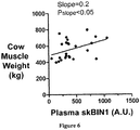
- BIN1 expression levels can be used to assess muscle mass to objectively determine production stages, to grade muscle development, and/or to improve genetic lines in production animals, such as, for example, avian, bovine, caprine, ovine, and porcine subjects and populations thereof.
- BIN1 expression levels can be determined in a production animal subject, or a population (e.g., two or more production animals) of production animal subjects.
- a detected level of BIN1 as compared to a control level of BIN1 can indicate increased muscle mass in the subject or population of subjects.
- individual animals or populations with increased BIN1 expression levels can be optionally selected for breeding programs to improve genetic stock for higher muscle mass.
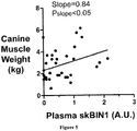
- BIN1 expression levels can be used to assess muscle mass development in performance animals including, for example, canines and equines.
- a detected level of BIN1 as compared to a control BIN1 level can indicate increased muscle mass in a performance animal, such as a race horse or dog. Increased muscle mass may indicate the effect of training, nutritional or other factors used to enhance performance of the animal.
- a detected level of BIN1 as compared to a control BIN1 level indicates decreased muscle mass in a performance animal, such as a race horse or dog. Decreased muscle mass may also indicate the effect of training, nutritional or other factors used to enhance performance of the animal. Therefore an individual, such as a trainer or breeder, can use BIN1 levels to guide management decisions related to the animal's performance.
- individual performance animals or populations with desired levels of BIN 1 for example increased BIN1 expression levels, can be optionally selected for breeding programs to improve genetic stock to better achieve desired levels of muscle mass, such as increased muscle mass.
- the BIN1 can be a BIN1 that is expressed specifically in skeletal muscle.
- a skeletal muscle specific BIN1 is a BIN1 that is expressed exclusively in skeletal muscle or that is predominantly expressed in skeletal muscle as compared to other tissues.
- the BIN1 can, for example, be a skeletal muscle specific BIN1 isoform.
- the skeletal muscle specific BIN1 isoform is BIN1 isoform 8.
- the skeletal muscle specific BIN1 isoform comprises SEQ ID NO:1.
- the skeletal muscle specific isoform comprises SEQ ID NO:2.
- the level of BIN1 expression can, for example, be determined by detecting BIN1 polypeptide in the biological sample.
- the level of BIN1 expression can be determined by detecting a BIN1-encoding nucleic acid (e.g., BIN1 mRNA), or fragment thereof, in the biological sample.
- Examples of analytical techniques useful in determining the expression of BIN1 include reverse transcription-polymerase chain reaction (RT-PCR), quantitative real time-PCR (qRT-PCR), one step PCR, RNase protection assay, primer extension assay, microarray analysis, gene chip, in situ hybridization, immunohistochemistry, Northern blot, Western blot, enzyme-linked immunosorbent assay (ELISA), enzyme immunoassay (EIA), radioimmunoassay (RIA), or protein array.
- RT-PCR reverse transcription-polymerase chain reaction
- qRT-PCR quantitative real time-PCR
- one step PCR RNase protection assay
- primer extension assay primer extension assay
- microarray analysis gene chip
- in situ hybridization immunohistochemistry
- Northern blot Western blot
- ELISA enzyme-linked immunosorbent assay
- EIA enzyme immunoassay
- RIA radioimmunoassay
- RNA can be isolated from a biological sample.
- RNA is isolated from blood, plasma or skeletal muscle tissue of a subject, and optionally, from corresponding normal tissue or subject as a control.
- RNA isolation can be performed using a purification kit, buffer set and protease from commercial manufacturers according to the manufacturer's instructions. For example, total RNA can be isolated using Qiagen RNeasy® mini-columns (Hilden, DE).
- RNA isolation kits include MasterPure® Complete DNA and RNA Purification Kit (EPICENTRE®, Madison, WI), and Paraffin Block RNA Isolation Kit® (Ambion, Inc., Austin, TX). Total RNA from tissue samples can be isolated using RNA Stat-60® (Tel-Test, Friendswood, TX). RNA prepared from a biological sample can be isolated, for example, by cesium chloride density gradient centrifugation.
- RNA template can be transcribed into cDNA, followed by its exponential amplification in a PCR reaction.
- a number of reverse transcriptases may be used, including, but not limited to, Avian Myeloblastosis Virus Reverse Transcriptase (AMV-RT), Moloney Murine Leukemia Virus Reverse Transcriptase (MMLV-RT), reverse transcriptase from human T-cell leukemia virus type I (HTLV-I), bovine leukemia virus (BLV), Rous sarcoma virus (RSV), human immunodeficiency virus (HIV) and Thermus thermophilus (Tth).
- AMV-RT Avian Myeloblastosis Virus Reverse Transcriptase
- MMLV-RT Moloney Murine Leukemia Virus Reverse Transcriptase
- HTLV-I human T-cell leukemia virus type I
- BLV bovine leukemia virus
- RSV Rous sarcoma virus
- HMV human immunode
- the reverse transcription step is typically primed using specific primers, random hexamers, or oligo-dT primers, depending on the circumstances and the goal of RT-PCR.
- extracted RNA can be reverse-transcribed using a GeneAmp RNA PCR kit (Perkin Elmer, Waltham, MA), following the manufacturer's instructions.
- the derived cDNA can then be used as a template in the subsequent PCR reaction.
- the PCR step can use a variety of thermostable DNA-dependent DNA polymerases, it typically employs the Taq DNA polymerase, which has a 5'-3' nuclease activity but lacks a 3'-5' proofreading endonuclease activity.
- TaqMan® PCR typically utilizes the 5'-nuclease activity of Taq or Tth polymerase to hydrolyze a hybridization probe bound to its target amplicon, but any enzyme with equivalent 5' nuclease activity can be used.
- Two oligonucleotide primers are used to generate an amplicon typical of a PCR reaction.
- a third oligonucleotide, or probe is designed to detect nucleotide sequence located between the two PCR primers.
- the probe is non-extendible by Taq DNA polymerase enzyme and is labeled with a reporter fluorescent dye and a quencher fluorescent dye. Any laser-induced emission from the reporter dye is quenched by the quenching dye when the two dyes are located close together as they are on the probe.
- the Taq DNA polymerase enzyme cleaves the probe in a template-dependent manner.
- the resultant probe fragments disassociate in solution, and signal from the released reporter dye is free from the quenching effect of the second fluorophore.
- One molecule of reporter dye is liberated for each new molecule synthesized, and detection of the unquenched reporter dye provides the basis for quantitative interpretation of the data.
- RT-PCR can be performed using commercially available equipment, such as, for example, ABI PRISM 7700TM Sequence Detection System® (Perkin-Elmer-Applied Biosystems, Foster City, CA), or Lightcycler® (Roche Molecular Biochemicals, Mannheim, DE).
- the 5' nuclease procedure is run on a real-time quantitative PCR device.
- a system can comprise a thermocycler, laser, charge-coupled device (CCD), camera and computer.
- CCD charge-coupled device
- the system amplifies samples in a 96-well format on a thermocycler.
- laser-induced fluorescent signal is collected in real-time through fiber optic cables for all 96 wells, and detected at the CCD.
- the system includes software for running the instrument and for analyzing the data.
- 5'-Nuclease assay data are initially expressed as Ct, or the threshold cycle. Fluorescence values are recorded during every cycle and represent the amount of product amplified to that point in the amplification reaction. The point when the fluorescent signal is first recorded as statistically significant is the threshold cycle (Ct).
- RT-PCR is optionally performed using an internal standard.
- the ideal internal standard is expressed at a constant level among different tissues, and is unaffected by the experimental treatment.
- RNAs most frequently used to normalize patterns of gene expression are mRNAs for the housekeeping genes glyceraldehyde-3-phosphate-dehydrogenase (GAPDH) and ⁇ -actin.
- GPDH glyceraldehyde-3-phosphate-dehydrogenase
- ⁇ -actin glyceraldehyde-3-phosphate-dehydrogenase
- a variation of the RT-PCR technique is the real time quantitative PCR, which measures PCR product accumulation through a dual-labeled fluorogenic probe.
- Real time PCR is compatible both with quantitative competitive PCR, where internal competitor for each target sequence is used for normalization, and with quantitative comparative PCR using a normalization gene contained within the sample, or a housekeeping gene for RT-PCR.
- the assay can optionally incorporate analysis of the expression of certain reference genes (or "normalizing genes"), including well known housekeeping genes, such as GAPDH, HPRT1, ubiquitin, etc.
- normalization can be based on the mean or median signal (Ct) of all of the assayed genes or a large subset thereof (often referred to as a "global normalization" approach).
- Ct mean or median signal
- measured normalized amount of a subject tissue mRNA may be compared to the amount found in a corresponding normal tissue.
- primers and probes can be designed based upon exon sequence to be amplified.
- the primer/probe design can include determining a target exon sequence within the gene of interest. This can be done by publicly available software, such as the DNA BLAST software developed by Kent, W.J., Genome Res. 12(4):656-64 (2002 ), or by the BLAST software including its variations.
- One target exon sequence that can be used is SEQ ID NO:2. Subsequent steps follow well established methods of PCR primer and probe design.
- repetitive sequences within the target sequence of the gene can be optionally masked when designing the primers and probes.
- the masked sequences can then be used to design primer and probe sequences using any commercially or otherwise publicly available primer/probe design packages, such as Primer Express (Applied Biosystems, Carlsbad, CA); MGB,assay-by-design (Applied Biosystems, Carlsbad, CA).
- PCR primers can include primer length, melting temperature (Tm), G/C content, specificity, complementary primer sequences, and 3'-end sequence.
- PCR primers can optionally be 17-30 bases in length, and contain about 20-80% G+C bases, (e.g., about 50-60% G+C bases). Tm's between 50°C and 80°C, e.g. about 50°C to 70°C.
- Microarray technology may be used to detect differential expression of BIN 1 in a subject's biological sample and normal or control biological sample.
- polynucleotide sequences of interest including cDNAs and oligonucleotides
- the arrayed sequences are then hybridized with specific DNA probes from cells or tissues of interest.
- the source of mRNA is optionally total RNA isolated from subject's biological sample, and optionally corresponding normal or control biological sample.
- Fluorescently labeled cDNA probes can be generated through incorporation of fluorescent nucleotides by reverse transcription of RNA extracted from tissues of interest. Labeled cDNA probes applied to the chip hybridize with specificity to each spot of DNA on the array. After stringent washing to remove non-specifically bound probes, the chip is scanned by confocal laser microscopy or by another detection method, such as a CCD camera. Quantitation of hybridization of each arrayed element can be used for assessment of corresponding mRNA abundance.
- the arrayed oligonucleotides may include oligonucleotides which hybridize to a specific region of BIN1 nucleic acid. In certain embodiments, multiple copies of a first oligonucleotide which specifically hybridizes to a first region of BIN 1 nucleic acid are arrayed. In certain embodiments, multiple copies of first and a second oligonucleotide which specifically hybridize to a first and a second region of BIN 1 nucleic acid, respectively, are arrayed, and so on. In certain embodiments, the BIN1 expression level is determined by mean values of the signal from each of these oligonucleotides.
- the array may also include oligonucleotides which specifically hybridize to nucleic acid of a normalizing gene, such as a housekeeping gene or other genes known not to be significantly differentially expressed in diseased versus normal tissue, for example, CaV 1.2.
- a normalizing gene such as a housekeeping gene or other genes known not to be significantly differentially expressed in diseased versus normal tissue, for example, CaV 1.2.
- Immunohistochemical methods may also be used for detecting the expression levels of BIN1.
- antibodies or antisera such as, polyclonal antisera and monoclonal antibodies specific for BIN1 may be used to assess BIN1 expression.
- the antibodies can be detected by direct labeling of the antibodies themselves, for example, with radioactive labels, fluorescent labels, hapten labels such as biotin, or an enzyme such as horse radish peroxidase or alkaline phosphatase.
- unlabeled primary antibody is used in conjunction with a labeled secondary antibody, comprising antisera, polyclonal antisera or a monoclonal antibody specific for the primary antibody.
- BIN1 expression in a tissue sample from a patient may be compared to BIN1 expression in a normal tissue sample or in a normal subject.
- the amount of BIN1 protein present in a biological sample may be determined by a Western blot.
- proteins present in the whole cell lysate from a biological sample may be separated by SDS-PAGE; the separated proteins transferred to a nitrocellulose membrane; BIN1 detected by using an antibody or antiserum specific for BIN1 or a specific isoform of BIN1.
- At least one normalizing protein for example, Cav 1.2 or a housekeeping protein such as GAPDH can also be detected simultaneously or in parallel and used to normalize the BIN protein expression levels.
- BIN1 expression level may be determined by performing a BIN1 immunoprecipitation using an excess of anti-BIN 1 antibody, followed by separation of the immunoprecipitate by SDS-PAGE; the separated proteins transferred to a nitrocellulose membrane; and detected by staining the gel, e.g., by Coomassie Blue or silver staining.
- Immunoprecipitation of a control protein such as GAPDH or ubiquitin may also be carried out either simultaneously or in parallel.
- the same procedure may be carried out on corresponding normal tissue or from a sample from a normal subject.
- the BIN1 polypeptide, nucleic acid, or fragments of said polypeptides or nucleic acids detected is human.
- BIN1 polypeptide, nucleic acid, or fragments of said polypeptides or nucleic acids detected is non-human (e.g., rodent, equine, canine, or feline).
- BIN1 refers to the BIN1 and homologs, variants, and isoforms thereof DNA and mRNA sequences for these polypeptides may also be determined to detect BIN1 expression levels in a biological sample.
- nucleotide and amino acid sequences of human skeletal muscle-specific BIN1 isoform 8 can be found at GenBank Accession Nos. NM_004305.3 and NP_04296.1 (SEQ ID NO:1), respectively.
- nucleotide and amino acid sequences of BIN1 isoforms 1-7, 9, and 10 can be found at GenBank Accession Nos.
- nucleotide sequences of BIN1 comprising a nucleotide sequence at least about 70%, 75%, 80%, 85%, 90%, 95%, 98%, 99% or more identical to the nucleotide sequences of the aforementioned GenBank Accession Numbers.
- amino acid sequences of BIN1 comprising an amino acid sequence at least about 70%, 75%, 80%, 85%, 90%, 95%, 98%, 99% or more identical to the sequences of the aforementioned GenBank Accession Numbers.
- Antibodies that bind the polypeptides described above, including BIN1, or fragments thereof can be used to detected BIN1 expression in a biological sample.
- the polypeptides described above can be used to produce antibodies or fragments thereof to BIN1.
- the BIN1 polypeptide is a skeletal muscle specific BIN1 isoform.
- the BIN1 isoform is BIN1 isoform 8.
- the muscle specific BIN1 isoform comprises SEQ ID NO:1.
- the muscle specific BIN1 isoform comprises SEQ ID NO:2.
- the antibody specifically binds SEQ ID NO:2.
- the isolated antibodies or fragments thereof that specifically bind to skeletal muscle BIN1 can, for example, have the same epitope specificity as an antibody or fragment with a light chain with polypeptide sequences (complementarity determining regions or CDRs) comprising SEQ ID NO:8, SEQ ID NO:9, and SEQ ID NO:10; SEQ ID NO:18, SEQ ID NO:19, and SEQ ID NO:20; or SEQ ID NO:28, SEQ ID NO:29, and SEQ ID NO:30, and a heavy chain with polypeptide sequences comprising SEQ ID NO:11, SEQ ID NO:12, and SEQ ID NO:13; SEQ ID NO:21, SEQ ID NO:22, and SEQ ID NO:23; or SEQ ID NO:31, SEQ ID NO:32, and SEQ ID NO:33.
- polypeptide sequences complementarity determining regions or CDRs
- the antibody or fragment comprises a light chain comprising SEQ ID NO:5, SEQ ID NO:15, or SEQ ID NO:25.
- the antibody or fragment comprises a heavy chain comprising SEQ ID NO:7, SEQ ID NO:17, or SEQ ID NO:27.
- the light chain can, for example, comprise polypeptide sequences (CDRs) comprising SEQ ID NO:8, SEQ ID NO:9, and SEQ ID NO:10; SEQ ID NO:18, SEQ ID NO:19, and SEQ ID NO:20; or SEQ ID NO:28, SEQ ID NO:29, and SEQ ID NO:30.
- the heavy chain can, for example, comprise polypeptide sequences (CDRs) comprising SEQ ID NO:11, SEQ ID NO:12, and SEQ ID NO:13; SEQ ID NO:21, SEQ ID NO:22, and SEQ ID NO:23; or SEQ ID NO:31, SEQ ID NO:32, and SEQ ID NO:33.
- CDRs polypeptide sequences
- the term antibody encompasses, but is not limited to, whole immunoglobulin (i.e., an intact antibody) of any class.
- Native antibodies are usually heterotetrameric glycoproteins, composed of two identical light (L) chains and two identical heavy (H) chains.
- L light
- H heavy
- each light chain is linked to a heavy chain by one covalent disulfide bond, while the number of disulfide linkages varies between the heavy chains of different immunoglobulin isotypes.
- Each heavy and light chain also has regularly spaced intrachain disulfide bridges.
- Each heavy chain has at one end a variable domain (V(H)) followed by a number of constant domains.
- V(H) variable domain
- Each light chain has a variable domain at one end (V(L)) and a constant domain at its other end; the constant domain of the light chain is aligned with the first constant domain of the heavy chain, and the light chain variable domain is aligned with the variable domain of the heavy chain.
- Particular amino acid residues are believed to form an interface between the light and heavy chain variable domains.
- the light chains of antibodies from any vertebrate species can be assigned to one of two clearly distinct types, called kappa ( ⁇ ) and lambda ( ⁇ ), based on the amino acid sequences of their constant domains.
- immunoglobulins can be assigned to different classes.
- immunoglobulins There are five major classes of immunoglobulins: IgA, IgD, IgE, IgG and IgM, and several of these may be further divided into subclasses (isotypes), e.g., IgG-1, IgG-2, IgG-3, and IgG-4; IgA-1 and IgA-2.
- the heavy chain constant domains that correspond to the different classes of immunoglobulins are called alpha, delta, epsilon, gamma, and mu, respectively.
- variable is used herein to describe certain portions of the antibody domains that differ in sequence among antibodies and are used in the binding and specificity of each particular antibody for its particular antigen.
- variability is not usually evenly distributed through the variable domains of antibodies. It is typically concentrated in three segments called complementarity determining regions (CDRs) or hypervariable regions both in the light chain and the heavy chain variable domains.
- CDRs complementarity determining regions
- FR framework
- the variable domains of native heavy and light chains each comprise four FR regions, largely adopting a ⁇ -sheet configuration, connected by three CDRs, which form loops connecting, and in some cases forming part of, the ⁇ -sheet structure.
- the CDRs in each chain are held together in close proximity by the FR regions and, with the CDRs from the other chain, contribute to the formation of the antigen binding site of antibodies.
- the constant domains are not involved directly in binding an antibody to an antigen, but exhibit various effector functions, such as participation of the antibody in antibody-dependent cellular toxicity.
- epitope is meant to include any determinant capable of specific interaction with the provided antibodies.
- Epitopic determinants usually consist of chemically active surface groupings of molecules such as amino acids or sugar side chains and usually have specific three dimensional structural characteristics, as well as specific charge characteristics.
- Identification of the epitope that the antibody recognizes is performed as follows. First, various partial structures of the target molecule that the monoclonal antibody recognizes are prepared. The partial structures are prepared by preparing partial peptides of the molecule. Such peptides are prepared by, for example, known oligopeptide synthesis technique or by incorporating DNA encoding the desired partial polypeptide in a suitable expression plasmid. The expression plasmid is delivered to a suitable host, such as E.
- a series of polypeptides having appropriately reduced lengths, working from the C- or N-terminus of the target molecule, can be prepared by established genetic engineering techniques. By establishing which fragments react with the antibody, the epitope region is identified. The epitope is more closely identified by synthesizing a variety of smaller peptides or mutants of the peptides using established oligopeptide synthesis techniques. The smaller peptides are used, for example, in a competitive inhibition assay to determine whether a specific peptide interferes with binding of the antibody to the target molecule. If so, the peptide is the epitope to which the antibody binds.
- kits such as the SPOTs Kit (Genosys Biotechnologies, Inc., The Woodlands, TX) and a series of multipin peptide synthesis kits based on the multipin synthesis method (Chiron Corporation, Emeryvile, CA) may be used to obtain a large variety of oligopeptides.
- antibody or fragments thereof can also encompass chimeric antibodies and hybrid antibodies, with dual or multiple antigen or epitope specificities, and fragments, such as F(ab')2, Fab', Fab and the like, including hybrid fragments.
- fragments of the antibodies that retain the ability to bind their specific antigens are provided.
- fragments of antibodies which maintain binding activity to BIN1 expressed specifically in skeletal muscle are included within the meaning of the term antibody or fragment thereof
- Such antibodies and fragments can be made by techniques known in the art and can be screened for specificity and activity according to general methods for producing antibodies and screening antibodies for specificity and activity (See Harlow and Lane. Antibodies, A Laboratory Manual. Cold Spring Harbor Publications, New York (1988 )).
- antibody or fragments thereof conjugates of antibody fragments and antigen binding proteins (single chain antibodies) as described, for example, in U.S. Pat. No. 4,704,692 .
- the antibody is a monoclonal antibody.
- monoclonal antibody refers to an antibody from a substantially homogeneous population of antibodies, i.e., the individual antibodies comprising the population are identical except for possible naturally occurring mutations that may be present in minor amounts.
- Monoclonal antibodies may be prepared using hybridoma methods, such as those described by Kohler and Milstein, Nature, 256:495 (1975 ) or Harlow and Lane, Antibodies, A Laboratory Manual. Cold Spring Harbor Publications, New York (1988 ).
- a hybridoma method a mouse or other appropriate host animal is typically immunized with an immunizing agent to elicit lymphocytes that produce or are capable of producing antibodies that will specifically bind to the immunizing agent.
- the lymphocytes may be immunized in vitro.
- the immunizing agent can be BIN1 specifically expressed in skeletal muscle or an immunogenic fragment thereof.
- peripheral blood lymphocytes are used in methods of producing monoclonal antibodies if cells of human origin are desired, or spleen cells or lymph node cells are used if non-human mammalian sources are desired.
- the lymphocytes are then fused with an immortalized cell line using a suitable fusing agent, such as polyethylene glycol, to form a hybridoma cell ( Goding, Monoclonal Antibodies: Principles and Practice, Academic Press, pp. 59-103 (1986 )).
- Immortalized cell lines are usually transformed mammalian cells, including myeloma cells of rodent, bovine, equine, and human origin. Usually, rat or mouse myeloma cell lines are employed.
- the hybridoma cells may be cultured in a suitable culture medium that preferably contains one or more substances that inhibit the growth or survival of the unfused, immortalized cells.
- a suitable culture medium that preferably contains one or more substances that inhibit the growth or survival of the unfused, immortalized cells.
- the culture medium for the hybridomas typically will include hypoxanthine, aminopterin, and thymidine ("HAT medium”) substances that prevent the growth of HGPRT-deficient cells.
- Immortalized cell lines useful here are those that fuse efficiently, support stable high level expression of antibody by the selected antibody-producing cells, and are sensitive to a medium such as HAT medium.
- Immortalized cell lines include murine myeloma lines, which can be obtained, for instance, from the Salk Institute Cell Distribution Center; San Diego, Calif. and the American Type Culture Collection; Rockville, Md. Human myeloma and mouse-human heteromyeloma cell lines also have been described for the production of human monoclonal antibodies ( Kozbor, J. Immunol., 133:3001 (1984 ); Brön et al., Monoclonal Antibody Production Techniques and Applications, Marcel Dekker, Inc., New York (1987) pp. 51-63 ).
- the culture medium in which the hybridoma cells are cultured can then be assayed for the presence of monoclonal antibodies directed against BIN1 specifically expressed in skeletal muscle or selected epitopes thereof.
- the binding specificity of monoclonal antibodies produced by the hybridoma cells can be determined by immunoprecipitation or by an in vitro binding assay, such as radioimmunoassay (RIA) or enzyme-linked immunoabsorbent assay (ELISA).
- RIA radioimmunoassay
- ELISA enzyme-linked immunoabsorbent assay
- the clones may be subcloned by limiting dilution or FACS sorting procedures and grown by standard methods. Suitable culture media for this purpose include, for example, Dulbecco's Modified Eagle's Medium and RPMI-1640 medium. Alternatively, the hybridoma cells may be grown in vivo as ascites in a mammal.
- the monoclonal antibodies secreted by the subclones may be isolated or purified from the culture medium or ascites fluid by conventional immunoglobulin purification procedures such as, for example, protein A-Sepharose, hydroxylapatite chromatography, gel electrophoresis, dialysis, or affinity chromatography.
- the monoclonal antibodies may also be made by recombinant DNA methods, such as those described in U.S. Pat. No. 4,816,567 .
- DNA encoding the monoclonal antibodies can be readily isolated and sequenced using conventional procedures (e.g., by using oligonucleotide probes that are capable of binding specifically to genes encoding the heavy and light chains of murine antibodies).
- the hybridoma cells can serve as a preferred source of such DNA.
- the DNA may be placed into expression vectors, which are then transfected into host cells such as simian COS cells, Chinese hamster ovary (CHO) cells, plasmacytoma cells, or myeloma cells that do not otherwise produce immunoglobulin protein, to obtain the synthesis of monoclonal antibodies in the recombinant host cells.
- host cells such as simian COS cells, Chinese hamster ovary (CHO) cells, plasmacytoma cells, or myeloma cells that do not otherwise produce immunoglobulin protein, to obtain the synthesis of monoclonal antibodies in the recombinant host cells.
- the DNA also may be modified, for example, by substituting the coding sequence for human heavy and light chain constant domains in place of the homologous murine sequences ( U.S. Pat. No. 4,816,567 ) or by covalently joining to the immunoglobulin coding sequence all or part of the coding sequence for a non-immunoglobulin polypeptide.
- non-immunoglobulin polypeptide can be substituted for the constant domains of an antibody provided herein, or can be substituted for the variable domains of one antigen-combining site of an antibody to create a chimeric bivalent antibody comprising one antigen-combining site having specificity for BIN specifically expressed in skeletal muscle and another antigen-combining site having specificity for a different antigen.
- In vitro methods are also suitable for preparing monovalent antibodies.
- Digestion of antibodies to produce fragments thereof, particularly, Fab fragments can be accomplished using routine techniques known in the art. For instance, digestion can be performed using papain. Examples of papain digestion are described in WO 94/29348 , U.S. Pat. No. 4,342,566 , and Harlow and Lane, Antibodies, A Laboratory Manual, Cold Spring Harbor Publications, New York, (1988 ).
- Papain digestion of antibodies typically produces two identical antigen binding fragments, called Fab fragments, each with a single antigen binding site, and a residual Fc fragment. Pepsin treatment yields a fragment, called the F(ab')2 fragment that has two antigen combining sites and is still capable of cross-linking antigen.
- the Fab fragments produced in the antibody digestion can also contain the constant domains of the light chain and the first constant domain of the heavy chain.
- Fab' fragments differ from Fab fragments by the addition of a few residues at the carboxy terminus of the heavy chain domain including one or more cysteines from the antibody hinge region.
- the F(ab')2 fragment is a bivalent fragment comprising two Fab' fragments linked by a disulfide bridge at the hinge region.
- Fab'-SH is the designation herein for Fab' in which the cysteine residue(s) of the constant domains bear a free thiol group.
- One method of producing proteins comprising the provided antibodies or polypeptides is to link two or more peptides or polypeptides together by protein chemistry techniques.
- peptides or polypeptides can be chemically synthesized using currently available laboratory equipment using either Fmoc (9-fluorenylmethyl-oxycarbonyl) or Boc (tert-butyloxycarbonoyl) chemistry (Applied Biosystems, Inc.; Foster City, CA).
- Fmoc 9-fluorenylmethyl-oxycarbonyl
- Boc tert-butyloxycarbonoyl
- a peptide or polypeptide can be synthesized and not cleaved from its synthesis resin whereas the other fragment of an antibody can be synthesized and subsequently cleaved from the resin, thereby exposing a terminal group that is functionally blocked on the other fragment.
- peptide condensation reactions these two fragments can be covalently joined via a peptide bond at their carboxyl and amino termini, respectively, to form an antibody, or fragment thereof.
- Grant GA (1992) Synthetic Peptides: A User Guide. W.H. Freeman and Co., N.Y. (1992 ); Bodansky M and Trost B., Ed. (1993) Principles of Peptide Synthesis. Springer Verlag Inc., NY Alternatively, the peptide or polypeptide can by independently synthesized in vivo. Once isolated, these independent peptides or polypeptides may be linked to form an antibody or fragment thereof via similar peptide condensation reactions.
- enzymatic ligation of cloned or synthetic peptide segments can allow relatively short peptide fragments to be joined to produce larger peptide fragments, polypeptides or whole protein domains ( Abrahmsen et al., Biochemistry, 30:4151 (1991 )).
- native chemical ligation of synthetic peptides can be utilized to synthetically construct large peptides or polypeptides from shorter peptide fragments. This method consists of a two step chemical reaction ( Dawson et al. Synthesis of Proteins by Native Chemical Ligation. Science, 266:776 779 (1994 )).
- the first step is the chemoselective reaction of an unprotected synthetic peptide a thioester with another unprotected peptide segment containing an amino terminal Cys residue to give a thioester linked intermediate as the initial covalent product. Without a change in the reaction conditions, this intermediate undergoes spontaneous, rapid intramolecular reaction to form a native peptide bond at the ligation site.
- IL-8 human interleukin 8
- unprotected peptide segments can be chemically linked where the bond formed between the peptide segments as a result of the chemical ligation is an unnatural (non peptide) bond ( Schnolzer et al., Science 256:221 (1992 )).
- This technique has been used to synthesize analogs of protein domains as well as large amounts of relatively pure proteins with full biological activity ( deLisle et al., Techniques in Protein Chemistry IV. Academic Press, New York, pp. 257-267 (1992 )).
- the provided polypeptide fragments can be recombinant proteins obtained by cloning nucleic acids encoding the polypeptide in an expression system capable of producing the polypeptide fragments thereof, such as a bacterial, adenovirus or baculovirus expression system.
- an expression system capable of producing the polypeptide fragments thereof, such as a bacterial, adenovirus or baculovirus expression system.
- amino acids found to not contribute to either the activity or the binding specificity or affinity of the antibody can be deleted without a loss in the respective activity.
- the provided fragments can also include insertions, deletions, substitutions, or other selected modifications of particular regions or specific amino acids residues, provided the activity of the fragment is not significantly altered or impaired compared to the nonmodified antibody or epitope. These modifications can provide for some additional property, such as to remove or add amino acids capable of disulfide bonding, to increase its bio longevity, to alter its secretory characteristics, and the like.
- the fragment can possess a bioactive property, such as binding activity, regulation of binding at the binding domain, and the like. Functional or active regions may be identified by mutagenesis of a specific region of the protein, followed by expression and testing of the expressed polypeptide.
- the humanized or human antibody comprises at least one complementarity determining region (CDR) of an antibody having the same epitope specificity as an antibody produced by the hybridoma cell line disclosed herein.
- the antibody can comprise all CDRs of an antibody having the same epitope specificity as an antibody produced by the hybridoma cell line.
- the humanized or human antibody can comprise at least one residue of the framework region of the monoclonal antibody produced by a disclosed hybridoma cell line.
- Humanized and human antibodies can be made using methods known to a skilled artesian; for example, the human antibody can be produced using a germ-line mutant animal or by a phage display library.
- Antibodies can also be generated in other species and humanized for administration to humans.
- fully human antibodies can also be made by immunizing a mouse or other species capable of making a fully human antibody (e.g., mice genetically modified to produce human antibodies) and screening clones that bind BIN1 specifically expressed in skeletal muscle. See, e.g., Lonberg and Huszar, Int. Rev. Immunol. 13:65-93, (1995 ).
- the term humanized and human in relation to antibodies relate to any antibody which is expected to elicit a therapeutically tolerable weak immunogenic response in a human subject.
- the terms include fully humanized or fully human as well as partially humanized or partially human.
- Humanized forms of non-human (e.g., murine) antibodies are chimeric immunoglobulins, immunoglobulin chains or fragments thereof (such as Fv, Fab, Fab', F(ab')2, or other antigen-binding subsequences of antibodies) which contain minimal sequence derived from non-human immunoglobulin.
- Humanized antibodies include human immunoglobulins (recipient antibody) in which residues from a CDR of the recipient are replaced by residues from a CDR of a non-human species (donor antibody) such as mouse, rat or rabbit having the desired specificity, affinity and capacity.
- donor antibody such as mouse, rat or rabbit having the desired specificity, affinity and capacity.
- Fv framework residues of the human immunoglobulin are replaced by corresponding non-human residues.
- Humanized antibodies may also comprise residues that are found neither in the recipient antibody nor in the imported CDR or framework sequences.
- the humanized antibody will comprise substantially all or at least one, and typically two, variable domains, in which all or substantially all of the CDR regions correspond to those of a non-human immunoglobulin and all or substantially all of the FR regions are those of a human immunoglobulin consensus sequence.
- the humanized antibody optimally also will comprise at least a portion of an immunoglobulin constant region (Fc), typically that of a human immunoglobulin ( Jones et al., Nature, 321:522-525 (1986 ); Riechmann et al., Nature, 332:323-327 (1988 ); and Presta, Curr. Op. Struct. Biol., 2:593-596 (1992 )).
- Fc immunoglobulin constant region
- a humanized antibody has one or more amino acid residues introduced into it from a source that is non-human. These non-human amino acid residues are often referred to as import residues, which are typically taken from an import variable domain. Humanization can be essentially performed following the methods described in Jones et al., Nature 321:522-525 (1986 ); Riechmann et al., Nature 332:323-327 (1988 ); or Verhoeyen et al., Science 239:1534-1536 (1988 ), by substituting rodent CDRs or CDR sequences for the corresponding sequences of a human antibody. Accordingly, such humanized antibodies are chimeric antibodies ( U.S. Pat. No.
- humanized antibodies are typically human antibodies in which some CDR residues and possibly some FR residues are substituted by residues from analogous sites in rodent antibodies.
- nucleotide sequences encoding the provided antibodies can be readily isolated and sequenced using conventional procedures (e.g., by using oligonucleotide probes that are capable of binding specifically to genes encoding the heavy and light chains of murine antibodies). These nucleotide sequences can also be modified, or humanized, for example, by substituting the coding sequence for human heavy and light chain constant domains in place of the homologous murine sequences (see, e.g., U.S. Pat. No. 4,816,567 ).
- the nucleotide sequences encoding any of the provided antibodies can be expressed in appropriate host cells. These include prokaryotic host cells including, but not limited to, E.
- Eukaryotic host cells can also be utilized. These include, but are not limited to, yeast cells (for example, Saccharomyces cerevisiae and Pichia pastoris ) , and mammalian cells such as VERO cells, HeLa cells, Chinese hamster ovary (CHO) cells, W138 cells, BHK cells, COS-7 cells, 293T cells and MDCK cells.
- yeast cells for example, Saccharomyces cerevisiae and Pichia pastoris
- mammalian cells such as VERO cells, HeLa cells, Chinese hamster ovary (CHO) cells, W138 cells, BHK cells, COS-7 cells, 293T cells and MDCK cells.
- the antibodies produced by these cells can be purified from the culture medium and assayed for binding, activity, specificity or any other property of the monoclonal antibodies by utilizing the methods set forth herein and standard in the art.
- Transgenic animals e.g., mice
- J(H) antibody heavy chain joining region
- chimeric and germ-line mutant mice results in complete inhibition of endogenous antibody production.
- Transfer of the human germ-line immunoglobulin gene array in such germ-line mutant mice will result in the production of human antibodies upon antigen challenge (see, e.g., Jakobovits et al., Proc. Natl. Acad. Sci.
- Human antibodies can also be produced in phage display libraries ( Hoogenboom et al., J. Mol. Biol., 227:381 (1991 ); Marks et al., J. Mol. Biol., 222:581 (1991 )).
- the techniques of Cole et al. and Boerner et al. are also available for the preparation of human monoclonal antibodies ( Cole et al., Monoclonal Antibodies and Cancer Therapy, Alan R. Liss, ed., p. 77 (1985 ); Boerner et al., J. Immunol., 147(1):86-95 (1991 )).
- any subset or combination of these is also specifically contemplated and disclosed. This concept applies to all aspects of this disclosure including, but not limited to, steps in methods of using the disclosed compositions. Thus, if there are a variety of additional steps that can be performed it is understood that each of these additional steps can be performed with any specific method steps or combination of method steps of the disclosed methods, and that each such combination or subset of combinations is specifically contemplated and should be considered disclosed.
- Dogs were selected for determination of BIN1 levels.
- the dogs used were at minimum age of six months and a minimum body weight of 2.3 kilograms (kg).
- the dogs were outwardly healthy with no indication of active diseases presently or in their past medical history.
- the dogs were excluded for the following reasons: a) had exercised or had an elevated temperature within the previous 96 hours; b) had an intractable disposition precluding safe restraint without risk to dog or personnel; c) had a disease or condition such as a significant skin disease (e.g., dermatitis, otitis), a musculoskeletal disorder (e.g., lameness, neurologic disorders, muscle atrophy), an abdominal disorder (e.g., palpable masses, abnormalities with organ shape), a cardiac disease (e.g., tachycardia, weak/abnormal peripheral pulse, significant heart murmurs), and a respiratory disease (e.g., increased lung sounds); d) had an inability to obtain sufficient blood for both plasma B
- MM Clinical evaluation of muscle mass
- the caudal aspects of hindlimb musculature e.g., semitendinosus, semimembrinosis, gracilis, adductor cruris caudalis, and biceps femoris muscles
- This site was chosen as the hindlimb provided the primary force for locomotion in canines, and the condition of this muscle group is indicative of the overall muscular conditioning.
- This muscle group is responsible for extension of the hip joint as well as both extension and flexion of the stifle joint, and extension of the tarsal joint ( Evans and Christensen, Miller's Anatomy of the Dog, 2nd Ed., WB Saudners, pp 383-400 (1979 )). Palpation of this area is readily performed with the dog in standing position, requiring minimal restraint. With the leg in the relaxed position and the stifle in extension, accurate, relaxed muscle tone can be assessed (as opposed to a state of muscle tension due to heavy restraint or other physiologic stressors). There is additionally an absence of significant fat deposits, such as truncal fat in an obese dog, facilitating palpation of isolated muscle bellies.
- Level 1 is characterized by very poor muscle mass and tone such that the caudal aspect of the mid-femur may be readily palpated from a caudal approach, with little obstruction from muscle bellies.
- the muscles themselves are soft/flaccid. This condition can be found in a dog that spends close to its entire life in a cage with little to no opportunity for exercise.
- Level 2 is characterized by a subnormal muscle mass and tone such that from a caudal approach the caudal aspect of the mid-femur may not be palpated due to obstruction from the muscle bellies, however, the lateral and medial aspects of the femur may be just palpated.
- the muscles have some tone, and reasonable mass.
- Level 3 is characterized by average muscle mass and tone such that from a caudal approach the mid-femur cannot be palpated at all due to obstruction by the muscle bellies. There is a good tone felt when the muscles are relaxed.
- Level 4 is characterized by above-average muscle mass and tone such that the muscles feel hard in a relaxed state and bulge significantly in comparison to the area of the mid-femur, when palpated from a caudal approach at the level of the mid-femur. This condition can be found in a dog that spends a significant amount of time outside running in a large yard.
- Venipuncture procedure in canine population Each dog was restrained in sternal recumbancy. The hair of the dog was clipped for a 1 inch window over the jugular vein and the skin was prepped with alcohol. A finger was placed in the jugular groove to act as a tourniquet, and a 12.0 ml syringe with a 21 gauge-1 inch needle was used for venipuncture. Between 3.0 to 7.0 mls of blood was collected. A finger was placed over the puncture site, and the needle and syringe were removed from the dog and pressure was applied to the puncture site to prevent a hematoma.
- the blood was placed in a 7.0 ml glass EDTA tube by removing both the needle and tube stopper gently and injecting the blood into the side of the tube.
- the stopper was replaced and the anticoagulant was mixed with the blood by gently inverting the tube 20 times.
- Plasma acquisition in bovine population Plasma acquisition in bovine population. Samples were collected at a commercial cattle slaughterhouse. Cattle were slaughtered according to conventional procedures including severing of the jugular vein for exsanguinations. During the process of exsanguinations and after a steady flow of blood developed, 7.0 ml of whole blood was collected into a glass EDTA tube by removing the tube stopper. The stopper was replaced and the anticoagulant was mixed with the blood by gently inverting the tube 20 times. Collection of the blood samples did not affect the pace of handling the animal prior to slaughter, or handling the carcass post slaughter.
- Plasma collection and storage Within 3 hours of collection of the blood, the EDTA tube was centrifuged for 15 minutes at a speed of 3000 rotations per minute (RPM). The supernatant (plasma) was removed, carefully avoiding collection of red blood cells. The plasma was separated into two equal aliquots, placed into 2 ml cryovials, and frozen at -20°C.
- RPM rotations per minute
- Plasma chemistry analysis Within three days of freezing, the plasma samples were thawed and analyzed with an Olympus AU2700 blood chemistry analyzer at a commercial reference laboratory (Antech Diagnostics; Tampa, FL). The tests performed included: total protein; albumin; globulin; serum glutamic oxaloacetic transaminase (SGOT); serum glutamic pyruvic transaminase (SGPT); alkaline phosphatase; gamma-glutamyl transpeptidase (GGTP); total bilirubin; urea nitrogen; creatine; phosphorous; glucose; calcium; magnesium; sodium; potassium; chloride; cholesterol; triglycerides; amylase; lipase; and creatine phosphokinase levels. Samples with high muscle contaminant (creatine kinase > 500 IU/L) were not used in the analysis.
- Novel customized rabbit polyclonal anti-skeletal BIN1 (anti-skBIN1) antibodies were raised against antigen of the skeletal specific phosphoinositol binding domain of BIN 1 (transcript variant 8) (Anaspec; San Jose, CA). Round bottomed 96-well plates were coated at 4°C for 16 hours with mouse anti-BIN1 (1/1000) or anti-skBIN1 (1/1000) diluted in 0.1 M sodium carbonate buffer, pH 9.0.
- TBST tris-buffered saline tween-20TM
- BSA bovine serum albumin
- TBST blocking buffer
- 100 ml of each serum sample was added, in triplicate, and plates were incubated for 1 hour at room temperature with rotation.
- the samples were then aspirated and plates were washed twice quickly and three times for 5 minutes with TBST.
- Goat anti-BIN1 (1/1000 in blocking buffer) was then applied as a detection antibody, and the plates were incubated for 1 hour at room temperature with rotation.
- the detection antibody was then aspirated and the plates were washed twice quickly, followed by three times for 5 minutes with TBST.
- the plates were subsequently incubated for 1 hour at room temperature with HRP-conjugated donkey anti-goat IgG (1/4000 in blocking buffer) before two quick washes and three 5 minute washes with TBST.
- TMB substrate was added and plates were incubated in the dark for 1 hour before reaction termination with 1 N hydrocholic acid (HCL).
- HCL hydrocholic acid
- the plates were read using the ELx800 BioTek microplate spectrophotometer (BioTek; Winooski, VT) and OD values were determined at 405 nm. Values were normalized to one tenth the OD of a standard canine serum sample, which was included on all plates.
- the RT-PCR products from the first-round reactions were further amplified in the second-round PCR.
- 12 individual heavy chain and 11 light chain RT-PCR reactions were set up using semi-nested primer sets specific for antibody variable regions.
- the PCR reaction was run on an agarose gel to visualize the amplified DNA fragments.
- the correct antibody variable region DNA fragments should have a size between 400-500 base pairs.
- the positive PCR bands were cloned and 10-20 clones per sample were sequenced.
- a large canine study was undertaken to determine the correlation between serum BIN1 and skeletal specific BIN1 levels and clinically obtained parameters (e.g., age, weight, and muscle mass).
- 34 healthy purebred Havanese and Sheltie canines ranging from 0.5 to 10 years were studied.
- the canines were selected for the study using the criteria provided above, and Tables 1 and 2 show a breakdown of the canines used in the study.
- Serum was obtained from the canines, and the serum was used to measure levels of BIN1 protein by a capture enzyme-linked immunosorbent assay (ELISA).
- the levels of BIN1 were compared to the weight of the canines across all species examined ( Figure 1 ), to the age of the canines across all species examined ( Figure 2 ), to the age of the canines in two specific species examined ( Figure 3 ), and to the muscle mass of the canines ( Figure 4 ). It was found that there is little dependency of BIN1 on weight ( Figure 1 ) and that there is a potential dependency of BIN1 on the age ( Figure 2 ) when the data were plotted for all species of dogs examined. When the data were limited to two particular species (Sheltie and Havanese), for which the most data were present, it was found that there was an inverse correlation between BIN1 levels and the age of the dog ( Figure 3 ).
- Live canines used for clinical examination of MM and for venous skBIN1 levels (MM is clinically assessed muscle mass). Breed Count Age (yrs) Weight (kg) MM Havanese 15 4.1 4.6 2.6 Sheltie 19 3.3 9.5 3.5 Both Breeds 34 3.6 7.4 3.1
- skBIN1 The levels of skBIN1 were also compared to the muscle weight across all canines examined. Muscle weight was determined as 25% of measured live body weight plus an additional 4% of body weight for every point of physically palpated muscle mass (MM, scored 1 to 4). Results are shown in Figure 5 . Canine muscle weight increases in direct proportion to measured skBIN1. The resulting slope is 08 kg/A.U. (P ⁇ 0.05). This statistically significant correlation suggests that canine muscle weight can be predicted by venous blood samples skBIN1. Therefore, skBIN1 prediction of canine muscle weight is not dependent on a particular physical examination.
- the standard of muscle mass measurement is direct weight obtained from an animal carcass in which non-muscle structures are eliminated.
- blood was obtained from adult Holstein cows.
- the hanging or dressed weigh was obtained.
- the dressed weight is a combination of skeletal muscle, intramuscular fat, and bone that is left behind following removal of the head, hide, hooves, viscera, and intracavitary fat. Typically, bone is 20% of this weight.
- the cows had an average muscle weight of 448.6 kg.
- Figure 6 demonstrates measured cow muscle weight as a function of venous skBIN1 obtained at the time of slaughter.
- the cow data has a strong linear correlation between measured muscle weight and plasma based skBIN1 (slope is 0.2 kg/A.U., P ⁇ 0.05).
- slope is 0.2 kg/A.U., P ⁇ 0.05.
- skBIN1 measured live weight of the cows
- Human skBIN1 levels Using the skeletal specific BIN1 antibody, human skBIN1 levels were assessed by ELISA. The fat percentage of the humans were also assessed by the 9-point caliper method, and measured regions included the chest, abdomen, thigh, bicep, tricep, subscapular, superiliac, lower back, and calf. The generic BIN1 antibody was also used to determine total BIN1 content in the plasma of the same patients. Non-fat percentage was the difference between fat percentage from the 9-point caliper measurements and 100%.
- Sequencing of the monoclonal antibody produced from hybridoma MHC 156 produced the following sequences: the nucleic acid and amino acid sequences of the variable region of the light chain (SEQ ID NOs:4 and 5) and the variable region of the heavy chain (SEQ ID NOs:6 and 7). Further analysis of the variable region of the light chain indicated a complementarity determining region 1 (CDR1) sequence of QDVSTA (SEQ ID NO:8), a CDR2 sequence of SASY (SEQ ID NO:9), and a CDR3 sequence of QQHYSTPLT (SEQ ID NO:10).
- Sequencing of the monoclonal antibody produced from hybridoma MHC 157 produced the following sequences: the nucleic acid and amino acid sequences of the variable region of the light chain (SEQ ID NOs:14 and 15) and the variable region of the heavy chain (SEQ ID NOs:16 and 17).
- variable region of the light chain indicated a CDR1 sequence of QDVSTA (SEQ ID NO:18), a CDR2 sequence of SASY (SEQ ID NO:19), and a CDR3 sequence of QQHYSTPLT (SEQ ID NO:20).
- Further analysis of the heavy chain indicated a CDR1 sequence of GYTFTRYY (SEQ ID NO:21), a CDR2 sequence of IYPGNVNT (SEQ ID NO:22), and a CDR3 sequence of AREGSYEYDEADY (SEQ ID NO:23).
- Sequencing of the monoclonal antibody produced from hybridoma MHC 158 produced the following sequences: the nucleic acid and amino acid sequences of the variable region of the light chain (SEQ ID NOs:24 and 25) and the variable region of the heavy chain (SEQ ID NOs:26 and 27). Further analysis of the variable region of the light chain indicated a CDR1 sequence of QSLLDSDGKTY (SEQ ID NO:28), a CDR2 sequence of LVSK (SEQ ID NO:29), and a CDR3 sequence of WQGTHFPYT (SEQ ID NO:30).
- variable region of the heavy chain indicated a CDR1 sequence of GFNIKDYY (SEQ ID NO:31), a CDR2 sequence of IDPENGDT (SEQ ID NO:32), and a CDR3 sequence of NSDY (SEQ ID NO:33).
Claims (11)
- Immunhistochemisches Verfahren zur Bestimmung der relativen Skelettmuskelmasse in einem Wesen, umfassend:a. Detektieren eines Levels der BIN1-Expression in einer Blut-, Serum- oder Plasmaprobe von dem Wesen; undb. Vergleichen des detektierten Levels der BIN1-Expression mit einem Kontrolllevel der BIN1-Expression, wobei der Level der BIN1-Expression relativ zum Kontrolllevel relative Skelettmuskelmasse in dem Wesen anzeigt;wobei das BIN1 eine Skelettmuskel-spezifische BIN1-lsoform 8 ist, und wobei BIN1-Isoform 8 unter Verwendung eines Antikörpers, der spezifisch an BIN1-Isoform 8 bindet, detektiert wird.
- Verfahren nach Anspruch 1, wobei der Kontrolllevel normal ist, wobei ein detektierter Level, der geringer ist als der Kontrolllevel, eine verringerte Skelettmuskelmasse im Wesen anzeigt, und wobei ein detektierter Level, der höher ist als der Kontrolllevel, erhöhte Muskelmasse in dem Wesen anzeigt, oder wobei ein detektierter Level, der niedriger ist als ein Kontrolllevel von dem Wesen, eine verringerte Skelettmuskelmasse in dem Wesen anzeigt.
- Verfahren nach Anspruch 1, wobei der Kontrolllevel niedriger als normal ist, wobei ein detektierter Level, der mit dem Kontrolllevel vergleichbar oder geringer ist als der Kontrolllevel, eine verringerte Skelettmuskelmasse in dem Wesen anzeigt, oder wobei der Kontrolllevel höher als normal ist, wobei ein detektierter Level, der mit dem Kontrolllevel vergleichbar oder höher als der Kontrolllevel ist, eine erhöhte Skelettmuskelmasse in dem Wesen anzeigt, oder wobei ein detektierter Level, der höher als ein Kontrolllevel vom Wesen ist, eine erhöhte Skelettmuskelmasse in dem Wesen anzeigt.
- Verfahren zur Diagnose eines Zustands oder einer Erkrankung, der/die mit verminderten Skelettmuskelmasse in einem Wesen assoziiert ist, umfassend:a. Detektieren eines Levels der BIN1-Expression in einer Blut-, Serum- oder Plasmaprobe von dem Wesen; undb. Vergleichen des detektierten Levels der BIN1-Expression mit einem Kontrolllevel der BIN1-Expression, wobei der Level der BIN1-Expression relativ zu dem Kontrolllevel anzeigt, dass das Wesen eine Krankheit oder einen Zustand hat, die/der mit verringerter Skelettmuskelmasse assoziiert ist;wobei das BIN1 eine Skelettmuskel-spezifische BIN1-lsoform 8 ist, und wobei BIN1-Isoform 8 unter Verwendung eines Antikörpers, der spezifisch an BIN1-Isoform 8 bindet, detektiert wird.
- Verfahren nach Anspruch 4, wobei der Kontrolllevel normal ist, wobei ein detektierter Level, der geringer als der Kontrolllevel ist, anzeigt, dass das Wesen eine Erkrankung oder einen Zustand hat, die/der mit verringerter Skelettmuskelmasse assoziiert ist; oder wobei der Kontrolllevel ein niedrigerer als normaler ist, wobei ein detektierter Level, der mit dem Kontrolllevel vergleichbar oder kleiner als der Kontrolllevel ist, anzeigt, dass das Wesen eine Krankheit oder einen Zustand hat, die/der mit verringerter Skelettmuskelmasse assoziiert ist; oder wobei ein detektierter Level, der niedriger als ein Kontrolllevel von dem Wesen ist, anzeigt, dass das Wesen eine Krankheit oder einen Zustand hat, die/der mit verringerter Skelettmuskelmasse assoziiert ist.
- Verfahren nach einem der Ansprüche 1, 2, 4 oder 5, ferner das Auswählen eines Wesens umfassend, das eine Erkrankung oder einen Zustand, die/der mit verringerter Skelettmuskelmasse assoziiert ist, hat oder bei dem diese/r vermutet wird, wobei die Erkrankung oder der Zustand Multiple Sklerose ist.
- Verfahren nach Anspruch 1, 2, 4 oder 5, ferner das Auswählen eines Wesens umfassend, das eine Erkrankung oder einen Zustand, die/der mit verringerter Skelettmuskelmasse assoziiert ist, hat oder bei dem diese/r vermutet wird, wobei die Erkrankung oder der Zustand eine Atrophie ist.
- Verfahren nach einem der Ansprüche 1, 2, 4 oder 5, ferner das Auswählen eines Wesens umfassend, das eine Erkrankung oder einen Zustand, die/der mit verringerter Skelettmuskelmasse assoziiert ist, hat oder bei dem diese/r vermutet wird, wobei die Erkrankung oder der Zustand eine neurogene Atrophie ist.
- Verfahren nach Anspruch 1, 2, 4 oder 5, ferner das Auswählen eines Wesens umfassend, das eine Erkrankung oder einen Zustand, die/der mit verringerter Skelettmuskelmasse assoziiert ist, hat oder bei dem diese/r vermutet wird, wobei die Erkrankung oder der Zustand ein chronischer Entzündungszustand ist.
- Verfahren nach Anspruch 1, 2, 4 oder 5, ferner das Auswählen eines Wesens umfassend, das eine Erkrankung oder einen Zustand, die/der mit verringerter Skelettmuskelmasse assoziiert ist, hat oder bei dem diese/r vermutet wird, wobei die Erkrankung oder der Zustand Sarkopenie ist, wobei vorzugsweise das Wesen Sarkopenie hat, oder vermutet wird, dass es Sarkopenie hat.
- Verfahren nach einem der Ansprüche 1 bis 10, wobei die Skelettmuskel-spezifische BIN1-Isoform 8 SEQ ID NO: 1 aufweist, oder wobei die Skelettmuskel-spezifische BIN1-Isoform 8 SEQ ID NO: 2 aufweist.
Applications Claiming Priority (2)
| Application Number | Priority Date | Filing Date | Title |
|---|---|---|---|
| US41168310P | 2010-11-09 | 2010-11-09 | |
| PCT/US2011/059574 WO2012087437A2 (en) | 2010-11-09 | 2011-11-07 | Bin1 expression as a marker of skeletal muscle mass and neurological conditions |
Publications (3)
| Publication Number | Publication Date |
|---|---|
| EP2638403A2 EP2638403A2 (de) | 2013-09-18 |
| EP2638403A4 EP2638403A4 (de) | 2014-05-07 |
| EP2638403B1 true EP2638403B1 (de) | 2017-03-22 |
Family
ID=46314702
Family Applications (1)
| Application Number | Title | Priority Date | Filing Date |
|---|---|---|---|
| EP11850406.7A Active EP2638403B1 (de) | 2010-11-09 | 2011-11-07 | Bin1-expression als marker für die skelettmuskelmasse und die nerven betreffende erkrankungen |
Country Status (6)
| Country | Link |
|---|---|
| US (1) | US9891232B2 (de) |
| EP (1) | EP2638403B1 (de) |
| JP (1) | JP5866126B2 (de) |
| AU (2) | AU2011345312A1 (de) |
| CA (1) | CA2817519C (de) |
| WO (1) | WO2012087437A2 (de) |
Families Citing this family (11)
| Publication number | Priority date | Publication date | Assignee | Title |
|---|---|---|---|---|
| JP5802197B2 (ja) | 2009-04-24 | 2015-10-28 | ザ リージェンツ オブ ザ ユニバーシティ オブ カリフォルニア | 心血管疾患における予後診断マーカーとしてのbin1 |
| EP2630490B1 (de) * | 2010-10-20 | 2017-12-06 | The Regents of The University of California | Körperflüssigkeit bin1 als marker für herzgesundheit |
| US9846159B2 (en) | 2011-09-30 | 2017-12-19 | Sarcotein Diagnostics, Llc | BIN1 expression as a marker of cancer |
| JP6742330B2 (ja) | 2015-03-02 | 2020-08-19 | サーコーティン ダイアグノスティックス エルエルシー | 心臓障害のマーカーとしての13+/17+bin1発現 |
| JP6539511B2 (ja) * | 2015-06-17 | 2019-07-03 | 花王株式会社 | 運動機能の判定用マーカー |
| JP6886241B2 (ja) * | 2016-01-07 | 2021-06-16 | 味の素株式会社 | 骨格筋面積の評価方法 |
| CN112770723A (zh) * | 2018-07-25 | 2021-05-07 | 阿克罗斯生物科学公司 | 新型cldn 18.2特异性单克隆抗体及其使用方法 |
| EP3863687A4 (de) * | 2018-10-09 | 2022-08-31 | Ibex Biosciences, Llc | Gegen filamin-a gerichtete antikörper und therapeutische verwendungen davon |
| US20240026034A1 (en) * | 2020-12-15 | 2024-01-25 | Arizona Board Of Regents On Behalf Of Arizona State University | Antibodies specific for qsox1 and methods of using the same |
| CN112778414B (zh) * | 2020-12-31 | 2022-04-15 | 中南大学湘雅二医院 | cBIN1抗体及其应用 |
| CN112794900B (zh) * | 2020-12-31 | 2022-10-25 | 中南大学湘雅二医院 | cBIN1抗体及其应用 |
Family Cites Families (9)
| Publication number | Priority date | Publication date | Assignee | Title |
|---|---|---|---|---|
| US6048702A (en) * | 1995-05-05 | 2000-04-11 | The Wistar Institute Of Anatomy And Biology | Murine and human box-dependent myc-interacting protein (Bin1) and uses therefor |
| AU5632300A (en) | 1999-07-07 | 2001-01-30 | Regeneron Pharmaceuticals, Inc. | Methods of inhibiting muscle atrophy |
| US6831063B1 (en) * | 1999-08-31 | 2004-12-14 | The Wistar Institute Of Anatomy And Biology | Bridging Integrator-2(Bin2) nucleic acid molecules and proteins and uses therefor |
| US20090088482A1 (en) * | 2003-10-14 | 2009-04-02 | The Trustees Of Columbia University In The City New York | Compositions and Methods For Treating Heart Failure |
| ES2367311T3 (es) | 2004-04-15 | 2011-11-02 | University Of Florida Research Foundation, Inc. | Productos de degradación proteolítica de map-2 como biomarcadores de diagnóstico para las lesiones neurales. |
| WO2006009629A2 (en) * | 2004-06-10 | 2006-01-26 | New York University | Methods and agents for maintaining muscle mass and for preventing muscle atrophy and biomarkers for monitoring same |
| WO2008008846A2 (en) | 2006-07-11 | 2008-01-17 | The Government Of The United States Of America As Represented By The Secretary Of The Department Of Health And Human Services | Differential expression of molecules associated with intra-cerebral hemorrhage |
| JP5802197B2 (ja) | 2009-04-24 | 2015-10-28 | ザ リージェンツ オブ ザ ユニバーシティ オブ カリフォルニア | 心血管疾患における予後診断マーカーとしてのbin1 |
| EP2630490B1 (de) * | 2010-10-20 | 2017-12-06 | The Regents of The University of California | Körperflüssigkeit bin1 als marker für herzgesundheit |
-
2011
- 2011-11-07 AU AU2011345312A patent/AU2011345312A1/en not_active Abandoned
- 2011-11-07 WO PCT/US2011/059574 patent/WO2012087437A2/en active Application Filing
- 2011-11-07 JP JP2013538809A patent/JP5866126B2/ja active Active
- 2011-11-07 CA CA2817519A patent/CA2817519C/en active Active
- 2011-11-07 US US13/884,454 patent/US9891232B2/en active Active
- 2011-11-07 EP EP11850406.7A patent/EP2638403B1/de active Active
-
2017
- 2017-03-10 AU AU2017201661A patent/AU2017201661B2/en active Active
Non-Patent Citations (1)
| Title |
|---|
| None * |
Also Published As
| Publication number | Publication date |
|---|---|
| JP5866126B2 (ja) | 2016-02-17 |
| JP2014500960A (ja) | 2014-01-16 |
| AU2011345312A1 (en) | 2013-06-20 |
| US9891232B2 (en) | 2018-02-13 |
| WO2012087437A2 (en) | 2012-06-28 |
| AU2017201661B2 (en) | 2019-01-17 |
| WO2012087437A3 (en) | 2012-08-16 |
| US20130324432A1 (en) | 2013-12-05 |
| CA2817519C (en) | 2020-01-14 |
| CA2817519A1 (en) | 2012-06-28 |
| EP2638403A4 (de) | 2014-05-07 |
| EP2638403A2 (de) | 2013-09-18 |
| AU2017201661A1 (en) | 2017-03-30 |
Similar Documents
| Publication | Publication Date | Title |
|---|---|---|
| AU2017201661B2 (en) | BIN1 expression as a marker of skeletal muscle mass and neurological conditions | |
| CN107099581B (zh) | 预测、诊断和治疗特发性肺纤维化的方法 | |
| JP5265923B2 (ja) | 軽度の変形性関節症のバイオマーカーおよびその使用 | |
| KR20110020853A (ko) | 유전자 또는 단백질 발현 프로파일을 이용한 신장 동종이식 거부반응 진단방법 | |
| JP2021509475A (ja) | 外傷性脳損傷を診断及び査定するための、新規のバイオマーカー及び方法 | |
| US10391142B2 (en) | Biomarkers for painful intervertebral discs and methods of use thereof | |
| US20090186034A1 (en) | Gene expression markers for inflammatory bowel disease | |
| Okamoto et al. | Molecular mechanism of rigid spine with muscular dystrophy type 1 caused by novel mutations of selenoprotein N gene | |
| JP2007524408A (ja) | 精神障害の診断および処置 | |
| KR101289134B1 (ko) | 유전성 샤르코-마리-투스 말초신경병증의 cmt4b3 아형에 대한 원인 유전자로서의 sbf1(mtmr5) 돌연변이 유전자 및 이를 이용한 상기 질병의 진단방법 및 진단용 조성물 | |
| WO2008079406A2 (en) | Gene expression markers for inflammatory bowel disease | |
| JP2007525213A (ja) | 新規の脳性ナトリウム利尿ペプチドの変異体及びその利用方法 | |
| EP2128272A1 (de) | Gen-/proteinmarker zur vorhersage oder diagnose der pharmakologischen wirksamkeit eines aurora-a-inhibitors | |
| EP3133400B1 (de) | Verwendung von männlicher fertilität assoziierten proteinen ak6 und gpx5 oder kombination daraus | |
| US6680172B1 (en) | Treatments and markers for cancers of the central nervous system | |
| US20050084860A1 (en) | Inhibition of tristetraproline for protection of the heart from cardiac injuries | |
| CN110499366B (zh) | Pi15作为骨关节炎标志物的应用 | |
| JP2012050371A (ja) | 免疫抑制物質のスクリーニング方法、免疫抑制活性検出キット及び免疫抑制活性検出マーカー | |
| KR101968962B1 (ko) | 비만 저항성 진단용 조성물 | |
| US9638699B2 (en) | Biomarkers of oxidative stress | |
| KR101849685B1 (ko) | 샤르코-마리-투스병 제1형의 원인 유전자로서 pmp22 및 이를 이용한 상기 질병의 진단방법 | |
| JP2003225093A (ja) | 軟骨障害マーカー及びその利用 | |
| WO2010055525A1 (en) | Method for predicting a patient's responsiveness to anti-folate therapy | |
| KR20200112069A (ko) | 골 질환의 예방 또는 치료를 위한 Jmjd2b 억제제의 용도 | |
| WO2008150964A1 (en) | Methods and kits for diagnosing and treating acute joint injury |
Legal Events
| Date | Code | Title | Description |
|---|---|---|---|
| PUAI | Public reference made under article 153(3) epc to a published international application that has entered the european phase |
Free format text: ORIGINAL CODE: 0009012 |
|
| 17P | Request for examination filed |
Effective date: 20130531 |
|
| AK | Designated contracting states |
Kind code of ref document: A2 Designated state(s): AL AT BE BG CH CY CZ DE DK EE ES FI FR GB GR HR HU IE IS IT LI LT LU LV MC MK MT NL NO PL PT RO RS SE SI SK SM TR |
|
| DAX | Request for extension of the european patent (deleted) | ||
| A4 | Supplementary search report drawn up and despatched |
Effective date: 20140404 |
|
| RIC1 | Information provided on ipc code assigned before grant |
Ipc: G01N 33/68 20060101AFI20140331BHEP Ipc: G01N 33/53 20060101ALI20140331BHEP Ipc: G01N 33/577 20060101ALI20140331BHEP Ipc: C12Q 1/68 20060101ALI20140331BHEP Ipc: C07K 16/00 20060101ALI20140331BHEP |
|
| 17Q | First examination report despatched |
Effective date: 20150123 |
|
| GRAP | Despatch of communication of intention to grant a patent |
Free format text: ORIGINAL CODE: EPIDOSNIGR1 |
|
| INTG | Intention to grant announced |
Effective date: 20161027 |
|
| GRAS | Grant fee paid |
Free format text: ORIGINAL CODE: EPIDOSNIGR3 |
|
| GRAA | (expected) grant |
Free format text: ORIGINAL CODE: 0009210 |
|
| AK | Designated contracting states |
Kind code of ref document: B1 Designated state(s): AL AT BE BG CH CY CZ DE DK EE ES FI FR GB GR HR HU IE IS IT LI LT LU LV MC MK MT NL NO PL PT RO RS SE SI SK SM TR |
|
| REG | Reference to a national code |
Ref country code: GB Ref legal event code: FG4D |
|
| REG | Reference to a national code |
Ref country code: CH Ref legal event code: EP |
|
| REG | Reference to a national code |
Ref country code: AT Ref legal event code: REF Ref document number: 878302 Country of ref document: AT Kind code of ref document: T Effective date: 20170415 |
|
| REG | Reference to a national code |
Ref country code: IE Ref legal event code: FG4D |
|
| REG | Reference to a national code |
Ref country code: DE Ref legal event code: R096 Ref document number: 602011036330 Country of ref document: DE |
|
| REG | Reference to a national code |
Ref country code: NL Ref legal event code: MP Effective date: 20170322 |
|
| PG25 | Lapsed in a contracting state [announced via postgrant information from national office to epo] |
Ref country code: GR Free format text: LAPSE BECAUSE OF FAILURE TO SUBMIT A TRANSLATION OF THE DESCRIPTION OR TO PAY THE FEE WITHIN THE PRESCRIBED TIME-LIMIT Effective date: 20170623 Ref country code: FI Free format text: LAPSE BECAUSE OF FAILURE TO SUBMIT A TRANSLATION OF THE DESCRIPTION OR TO PAY THE FEE WITHIN THE PRESCRIBED TIME-LIMIT Effective date: 20170322 Ref country code: LT Free format text: LAPSE BECAUSE OF FAILURE TO SUBMIT A TRANSLATION OF THE DESCRIPTION OR TO PAY THE FEE WITHIN THE PRESCRIBED TIME-LIMIT Effective date: 20170322 Ref country code: HR Free format text: LAPSE BECAUSE OF FAILURE TO SUBMIT A TRANSLATION OF THE DESCRIPTION OR TO PAY THE FEE WITHIN THE PRESCRIBED TIME-LIMIT Effective date: 20170322 Ref country code: NO Free format text: LAPSE BECAUSE OF FAILURE TO SUBMIT A TRANSLATION OF THE DESCRIPTION OR TO PAY THE FEE WITHIN THE PRESCRIBED TIME-LIMIT Effective date: 20170622 |
|
| REG | Reference to a national code |
Ref country code: LT Ref legal event code: MG4D |
|
| REG | Reference to a national code |
Ref country code: AT Ref legal event code: MK05 Ref document number: 878302 Country of ref document: AT Kind code of ref document: T Effective date: 20170322 |
|
| PG25 | Lapsed in a contracting state [announced via postgrant information from national office to epo] |
Ref country code: SE Free format text: LAPSE BECAUSE OF FAILURE TO SUBMIT A TRANSLATION OF THE DESCRIPTION OR TO PAY THE FEE WITHIN THE PRESCRIBED TIME-LIMIT Effective date: 20170322 Ref country code: BG Free format text: LAPSE BECAUSE OF FAILURE TO SUBMIT A TRANSLATION OF THE DESCRIPTION OR TO PAY THE FEE WITHIN THE PRESCRIBED TIME-LIMIT Effective date: 20170622 Ref country code: RS Free format text: LAPSE BECAUSE OF FAILURE TO SUBMIT A TRANSLATION OF THE DESCRIPTION OR TO PAY THE FEE WITHIN THE PRESCRIBED TIME-LIMIT Effective date: 20170322 Ref country code: LV Free format text: LAPSE BECAUSE OF FAILURE TO SUBMIT A TRANSLATION OF THE DESCRIPTION OR TO PAY THE FEE WITHIN THE PRESCRIBED TIME-LIMIT Effective date: 20170322 |
|
| PG25 | Lapsed in a contracting state [announced via postgrant information from national office to epo] |
Ref country code: NL Free format text: LAPSE BECAUSE OF FAILURE TO SUBMIT A TRANSLATION OF THE DESCRIPTION OR TO PAY THE FEE WITHIN THE PRESCRIBED TIME-LIMIT Effective date: 20170322 |
|
| REG | Reference to a national code |
Ref country code: FR Ref legal event code: PLFP Year of fee payment: 7 |
|
| PG25 | Lapsed in a contracting state [announced via postgrant information from national office to epo] |
Ref country code: ES Free format text: LAPSE BECAUSE OF FAILURE TO SUBMIT A TRANSLATION OF THE DESCRIPTION OR TO PAY THE FEE WITHIN THE PRESCRIBED TIME-LIMIT Effective date: 20170322 Ref country code: CZ Free format text: LAPSE BECAUSE OF FAILURE TO SUBMIT A TRANSLATION OF THE DESCRIPTION OR TO PAY THE FEE WITHIN THE PRESCRIBED TIME-LIMIT Effective date: 20170322 Ref country code: EE Free format text: LAPSE BECAUSE OF FAILURE TO SUBMIT A TRANSLATION OF THE DESCRIPTION OR TO PAY THE FEE WITHIN THE PRESCRIBED TIME-LIMIT Effective date: 20170322 Ref country code: AT Free format text: LAPSE BECAUSE OF FAILURE TO SUBMIT A TRANSLATION OF THE DESCRIPTION OR TO PAY THE FEE WITHIN THE PRESCRIBED TIME-LIMIT Effective date: 20170322 Ref country code: RO Free format text: LAPSE BECAUSE OF FAILURE TO SUBMIT A TRANSLATION OF THE DESCRIPTION OR TO PAY THE FEE WITHIN THE PRESCRIBED TIME-LIMIT Effective date: 20170322 Ref country code: IT Free format text: LAPSE BECAUSE OF FAILURE TO SUBMIT A TRANSLATION OF THE DESCRIPTION OR TO PAY THE FEE WITHIN THE PRESCRIBED TIME-LIMIT Effective date: 20170322 Ref country code: SK Free format text: LAPSE BECAUSE OF FAILURE TO SUBMIT A TRANSLATION OF THE DESCRIPTION OR TO PAY THE FEE WITHIN THE PRESCRIBED TIME-LIMIT Effective date: 20170322 |
|
| PG25 | Lapsed in a contracting state [announced via postgrant information from national office to epo] |
Ref country code: PT Free format text: LAPSE BECAUSE OF FAILURE TO SUBMIT A TRANSLATION OF THE DESCRIPTION OR TO PAY THE FEE WITHIN THE PRESCRIBED TIME-LIMIT Effective date: 20170724 Ref country code: IS Free format text: LAPSE BECAUSE OF FAILURE TO SUBMIT A TRANSLATION OF THE DESCRIPTION OR TO PAY THE FEE WITHIN THE PRESCRIBED TIME-LIMIT Effective date: 20170722 Ref country code: SM Free format text: LAPSE BECAUSE OF FAILURE TO SUBMIT A TRANSLATION OF THE DESCRIPTION OR TO PAY THE FEE WITHIN THE PRESCRIBED TIME-LIMIT Effective date: 20170322 Ref country code: PL Free format text: LAPSE BECAUSE OF FAILURE TO SUBMIT A TRANSLATION OF THE DESCRIPTION OR TO PAY THE FEE WITHIN THE PRESCRIBED TIME-LIMIT Effective date: 20170322 |
|
| REG | Reference to a national code |
Ref country code: DE Ref legal event code: R097 Ref document number: 602011036330 Country of ref document: DE |
|
| PLBE | No opposition filed within time limit |
Free format text: ORIGINAL CODE: 0009261 |
|
| STAA | Information on the status of an ep patent application or granted ep patent |
Free format text: STATUS: NO OPPOSITION FILED WITHIN TIME LIMIT |
|
| PG25 | Lapsed in a contracting state [announced via postgrant information from national office to epo] |
Ref country code: DK Free format text: LAPSE BECAUSE OF FAILURE TO SUBMIT A TRANSLATION OF THE DESCRIPTION OR TO PAY THE FEE WITHIN THE PRESCRIBED TIME-LIMIT Effective date: 20170322 |
|
| 26N | No opposition filed |
Effective date: 20180102 |
|
| PG25 | Lapsed in a contracting state [announced via postgrant information from national office to epo] |
Ref country code: SI Free format text: LAPSE BECAUSE OF FAILURE TO SUBMIT A TRANSLATION OF THE DESCRIPTION OR TO PAY THE FEE WITHIN THE PRESCRIBED TIME-LIMIT Effective date: 20170322 |
|
| PG25 | Lapsed in a contracting state [announced via postgrant information from national office to epo] |
Ref country code: MC Free format text: LAPSE BECAUSE OF FAILURE TO SUBMIT A TRANSLATION OF THE DESCRIPTION OR TO PAY THE FEE WITHIN THE PRESCRIBED TIME-LIMIT Effective date: 20170322 |
|
| PG25 | Lapsed in a contracting state [announced via postgrant information from national office to epo] |
Ref country code: CH Free format text: LAPSE BECAUSE OF NON-PAYMENT OF DUE FEES Effective date: 20171130 Ref country code: LI Free format text: LAPSE BECAUSE OF NON-PAYMENT OF DUE FEES Effective date: 20171130 |
|
| PG25 | Lapsed in a contracting state [announced via postgrant information from national office to epo] |
Ref country code: LU Free format text: LAPSE BECAUSE OF NON-PAYMENT OF DUE FEES Effective date: 20171107 |
|
| REG | Reference to a national code |
Ref country code: BE Ref legal event code: MM Effective date: 20171130 |
|
| REG | Reference to a national code |
Ref country code: IE Ref legal event code: MM4A |
|
| PG25 | Lapsed in a contracting state [announced via postgrant information from national office to epo] |
Ref country code: MT Free format text: LAPSE BECAUSE OF NON-PAYMENT OF DUE FEES Effective date: 20171107 |
|
| REG | Reference to a national code |
Ref country code: FR Ref legal event code: PLFP Year of fee payment: 8 |
|
| PG25 | Lapsed in a contracting state [announced via postgrant information from national office to epo] |
Ref country code: IE Free format text: LAPSE BECAUSE OF NON-PAYMENT OF DUE FEES Effective date: 20171107 |
|
| PG25 | Lapsed in a contracting state [announced via postgrant information from national office to epo] |
Ref country code: BE Free format text: LAPSE BECAUSE OF NON-PAYMENT OF DUE FEES Effective date: 20171130 |
|
| PG25 | Lapsed in a contracting state [announced via postgrant information from national office to epo] |
Ref country code: HU Free format text: LAPSE BECAUSE OF FAILURE TO SUBMIT A TRANSLATION OF THE DESCRIPTION OR TO PAY THE FEE WITHIN THE PRESCRIBED TIME-LIMIT; INVALID AB INITIO Effective date: 20111107 |
|
| PG25 | Lapsed in a contracting state [announced via postgrant information from national office to epo] |
Ref country code: CY Free format text: LAPSE BECAUSE OF NON-PAYMENT OF DUE FEES Effective date: 20170322 |
|
| PG25 | Lapsed in a contracting state [announced via postgrant information from national office to epo] |
Ref country code: MK Free format text: LAPSE BECAUSE OF FAILURE TO SUBMIT A TRANSLATION OF THE DESCRIPTION OR TO PAY THE FEE WITHIN THE PRESCRIBED TIME-LIMIT Effective date: 20170322 |
|
| PG25 | Lapsed in a contracting state [announced via postgrant information from national office to epo] |
Ref country code: TR Free format text: LAPSE BECAUSE OF FAILURE TO SUBMIT A TRANSLATION OF THE DESCRIPTION OR TO PAY THE FEE WITHIN THE PRESCRIBED TIME-LIMIT Effective date: 20170322 |
|
| PG25 | Lapsed in a contracting state [announced via postgrant information from national office to epo] |
Ref country code: AL Free format text: LAPSE BECAUSE OF FAILURE TO SUBMIT A TRANSLATION OF THE DESCRIPTION OR TO PAY THE FEE WITHIN THE PRESCRIBED TIME-LIMIT Effective date: 20170322 |
|
| PGFP | Annual fee paid to national office [announced via postgrant information from national office to epo] |
Ref country code: GB Payment date: 20231116 Year of fee payment: 13 |
|
| PGFP | Annual fee paid to national office [announced via postgrant information from national office to epo] |
Ref country code: FR Payment date: 20231124 Year of fee payment: 13 Ref country code: DE Payment date: 20231120 Year of fee payment: 13 |