EP2566489B1 - Dosage unit formulations of autologous dermal fibroblasts - Google Patents
Dosage unit formulations of autologous dermal fibroblasts Download PDFInfo
- Publication number
- EP2566489B1 EP2566489B1 EP11720666.4A EP11720666A EP2566489B1 EP 2566489 B1 EP2566489 B1 EP 2566489B1 EP 11720666 A EP11720666 A EP 11720666A EP 2566489 B1 EP2566489 B1 EP 2566489B1
- Authority
- EP
- European Patent Office
- Prior art keywords
- cells
- dosage formulation
- cell
- treatment
- formulation
- Prior art date
- Legal status (The legal status is an assumption and is not a legal conclusion. Google has not performed a legal analysis and makes no representation as to the accuracy of the status listed.)
- Active
Links
- 210000002950 fibroblast Anatomy 0.000 title claims description 109
- 239000000203 mixture Substances 0.000 title claims description 73
- 238000009472 formulation Methods 0.000 title claims description 62
- 230000002500 effect on skin Effects 0.000 title description 12
- 210000004027 cell Anatomy 0.000 claims description 230
- 238000011282 treatment Methods 0.000 claims description 93
- 230000037303 wrinkles Effects 0.000 claims description 41
- 238000000034 method Methods 0.000 claims description 36
- 210000003491 skin Anatomy 0.000 claims description 29
- 230000001815 facial effect Effects 0.000 claims description 20
- 210000004207 dermis Anatomy 0.000 claims description 16
- 241000282414 Homo sapiens Species 0.000 claims description 15
- 206010039580 Scar Diseases 0.000 claims description 15
- 101000800116 Homo sapiens Thy-1 membrane glycoprotein Proteins 0.000 claims description 12
- 102100033523 Thy-1 membrane glycoprotein Human genes 0.000 claims description 12
- 230000007547 defect Effects 0.000 claims description 11
- 238000009826 distribution Methods 0.000 claims description 9
- 210000002510 keratinocyte Anatomy 0.000 claims description 8
- 230000017423 tissue regeneration Effects 0.000 claims description 8
- 239000012595 freezing medium Substances 0.000 claims description 6
- 239000003814 drug Substances 0.000 claims description 4
- 238000011069 regeneration method Methods 0.000 claims description 4
- 238000012546 transfer Methods 0.000 claims description 4
- 230000007910 cell fusion Effects 0.000 claims description 3
- 239000003085 diluting agent Substances 0.000 claims description 3
- 210000001082 somatic cell Anatomy 0.000 claims description 3
- 239000002458 cell surface marker Substances 0.000 claims 2
- 238000002347 injection Methods 0.000 description 53
- 239000007924 injection Substances 0.000 description 53
- 239000006285 cell suspension Substances 0.000 description 33
- 239000000523 sample Substances 0.000 description 32
- 231100000241 scar Toxicity 0.000 description 32
- 208000032544 Cicatrix Diseases 0.000 description 25
- 230000037387 scars Effects 0.000 description 25
- 239000001963 growth medium Substances 0.000 description 24
- 239000000902 placebo Substances 0.000 description 23
- 229940068196 placebo Drugs 0.000 description 23
- 208000027418 Wounds and injury Diseases 0.000 description 18
- 239000012071 phase Substances 0.000 description 18
- 239000000047 product Substances 0.000 description 17
- 230000035899 viability Effects 0.000 description 17
- 239000008186 active pharmaceutical agent Substances 0.000 description 16
- LOKCTEFSRHRXRJ-UHFFFAOYSA-I dipotassium trisodium dihydrogen phosphate hydrogen phosphate dichloride Chemical compound P(=O)(O)(O)[O-].[K+].P(=O)(O)([O-])[O-].[Na+].[Na+].[Cl-].[K+].[Cl-].[Na+] LOKCTEFSRHRXRJ-UHFFFAOYSA-I 0.000 description 16
- 229940088679 drug related substance Drugs 0.000 description 16
- 230000006872 improvement Effects 0.000 description 16
- 239000002953 phosphate buffered saline Substances 0.000 description 16
- 210000001519 tissue Anatomy 0.000 description 16
- 241000508269 Psidium Species 0.000 description 14
- 238000001574 biopsy Methods 0.000 description 14
- 239000000243 solution Substances 0.000 description 14
- 238000012360 testing method Methods 0.000 description 14
- 238000003306 harvesting Methods 0.000 description 13
- IAZDPXIOMUYVGZ-UHFFFAOYSA-N Dimethylsulphoxide Chemical compound CS(C)=O IAZDPXIOMUYVGZ-UHFFFAOYSA-N 0.000 description 12
- 206010052428 Wound Diseases 0.000 description 12
- 108010035532 Collagen Proteins 0.000 description 11
- 102000008186 Collagen Human genes 0.000 description 11
- 238000004458 analytical method Methods 0.000 description 11
- 229920001436 collagen Polymers 0.000 description 11
- 230000008569 process Effects 0.000 description 11
- 230000004044 response Effects 0.000 description 11
- 208000002874 Acne Vulgaris Diseases 0.000 description 10
- 206010000496 acne Diseases 0.000 description 10
- 229940000406 drug candidate Drugs 0.000 description 10
- 239000000463 material Substances 0.000 description 10
- 239000000725 suspension Substances 0.000 description 10
- 238000002360 preparation method Methods 0.000 description 9
- 230000037390 scarring Effects 0.000 description 9
- 239000006144 Dulbecco’s modified Eagle's medium Substances 0.000 description 8
- 239000007760 Iscove's Modified Dulbecco's Medium Substances 0.000 description 8
- 230000001154 acute effect Effects 0.000 description 8
- 230000015572 biosynthetic process Effects 0.000 description 8
- 230000035876 healing Effects 0.000 description 8
- 230000000977 initiatory effect Effects 0.000 description 8
- 108090000623 proteins and genes Proteins 0.000 description 8
- CURLTUGMZLYLDI-UHFFFAOYSA-N Carbon dioxide Chemical compound O=C=O CURLTUGMZLYLDI-UHFFFAOYSA-N 0.000 description 7
- 230000010261 cell growth Effects 0.000 description 7
- 230000000694 effects Effects 0.000 description 7
- 238000007710 freezing Methods 0.000 description 7
- 230000008014 freezing Effects 0.000 description 7
- 230000033001 locomotion Effects 0.000 description 7
- 102000004169 proteins and genes Human genes 0.000 description 7
- IJGRMHOSHXDMSA-UHFFFAOYSA-N Atomic nitrogen Chemical compound N#N IJGRMHOSHXDMSA-UHFFFAOYSA-N 0.000 description 6
- 208000020401 Depressive disease Diseases 0.000 description 6
- 239000000872 buffer Substances 0.000 description 6
- 238000002659 cell therapy Methods 0.000 description 6
- 238000005119 centrifugation Methods 0.000 description 6
- 239000000975 dye Substances 0.000 description 6
- 230000012010 growth Effects 0.000 description 6
- 208000014674 injury Diseases 0.000 description 6
- 238000004519 manufacturing process Methods 0.000 description 6
- 238000003908 quality control method Methods 0.000 description 6
- 239000006228 supernatant Substances 0.000 description 6
- 238000003260 vortexing Methods 0.000 description 6
- 241000283690 Bos taurus Species 0.000 description 5
- 108091003079 Bovine Serum Albumin Proteins 0.000 description 5
- 102000010834 Extracellular Matrix Proteins Human genes 0.000 description 5
- 108010037362 Extracellular Matrix Proteins Proteins 0.000 description 5
- 230000032683 aging Effects 0.000 description 5
- 229940055553 azficel-t Drugs 0.000 description 5
- 239000003153 chemical reaction reagent Substances 0.000 description 5
- 238000002316 cosmetic surgery Methods 0.000 description 5
- 230000006378 damage Effects 0.000 description 5
- 238000011161 development Methods 0.000 description 5
- 230000018109 developmental process Effects 0.000 description 5
- 229940126534 drug product Drugs 0.000 description 5
- 230000007774 longterm Effects 0.000 description 5
- 239000003550 marker Substances 0.000 description 5
- 101150067055 minC gene Proteins 0.000 description 5
- 239000000825 pharmaceutical preparation Substances 0.000 description 5
- 230000008439 repair process Effects 0.000 description 5
- 238000010374 somatic cell nuclear transfer Methods 0.000 description 5
- 241000204031 Mycoplasma Species 0.000 description 4
- 208000002193 Pain Diseases 0.000 description 4
- PXIPVTKHYLBLMZ-UHFFFAOYSA-N Sodium azide Chemical compound [Na+].[N-]=[N+]=[N-] PXIPVTKHYLBLMZ-UHFFFAOYSA-N 0.000 description 4
- 230000001464 adherent effect Effects 0.000 description 4
- 238000004113 cell culture Methods 0.000 description 4
- 230000008859 change Effects 0.000 description 4
- 239000013068 control sample Substances 0.000 description 4
- 238000005138 cryopreservation Methods 0.000 description 4
- 238000011018 current good manufacturing practice Methods 0.000 description 4
- 238000013461 design Methods 0.000 description 4
- 210000002615 epidermis Anatomy 0.000 description 4
- 210000002744 extracellular matrix Anatomy 0.000 description 4
- 239000012091 fetal bovine serum Substances 0.000 description 4
- 230000001969 hypertrophic effect Effects 0.000 description 4
- 230000002163 immunogen Effects 0.000 description 4
- 230000003902 lesion Effects 0.000 description 4
- 239000002609 medium Substances 0.000 description 4
- 229920001296 polysiloxane Polymers 0.000 description 4
- 230000008672 reprogramming Effects 0.000 description 4
- 238000003860 storage Methods 0.000 description 4
- 238000002560 therapeutic procedure Methods 0.000 description 4
- 238000005406 washing Methods 0.000 description 4
- 108060005980 Collagenase Proteins 0.000 description 3
- 102000029816 Collagenase Human genes 0.000 description 3
- 206010061619 Deformity Diseases 0.000 description 3
- 206010061218 Inflammation Diseases 0.000 description 3
- 102000004142 Trypsin Human genes 0.000 description 3
- 108090000631 Trypsin Proteins 0.000 description 3
- 238000003556 assay Methods 0.000 description 3
- 230000003416 augmentation Effects 0.000 description 3
- 239000012512 bulk drug substance Substances 0.000 description 3
- 230000001413 cellular effect Effects 0.000 description 3
- 230000007423 decrease Effects 0.000 description 3
- 230000029087 digestion Effects 0.000 description 3
- 238000005516 engineering process Methods 0.000 description 3
- 230000001973 epigenetic effect Effects 0.000 description 3
- 238000011156 evaluation Methods 0.000 description 3
- 230000036512 infertility Effects 0.000 description 3
- 230000004054 inflammatory process Effects 0.000 description 3
- 230000001788 irregular Effects 0.000 description 3
- 239000007788 liquid Substances 0.000 description 3
- 238000012986 modification Methods 0.000 description 3
- 230000004048 modification Effects 0.000 description 3
- 229910052757 nitrogen Inorganic materials 0.000 description 3
- 230000036407 pain Effects 0.000 description 3
- 239000008188 pellet Substances 0.000 description 3
- 239000002243 precursor Substances 0.000 description 3
- 238000012429 release testing Methods 0.000 description 3
- 210000000130 stem cell Anatomy 0.000 description 3
- 238000003786 synthesis reaction Methods 0.000 description 3
- 239000012588 trypsin Substances 0.000 description 3
- XLYOFNOQVPJJNP-UHFFFAOYSA-N water Chemical compound O XLYOFNOQVPJJNP-UHFFFAOYSA-N 0.000 description 3
- KCXVZYZYPLLWCC-UHFFFAOYSA-N EDTA Chemical compound OC(=O)CN(CC(O)=O)CCN(CC(O)=O)CC(O)=O KCXVZYZYPLLWCC-UHFFFAOYSA-N 0.000 description 2
- 102000004190 Enzymes Human genes 0.000 description 2
- 108090000790 Enzymes Proteins 0.000 description 2
- 108010052014 Liberase Proteins 0.000 description 2
- 102000012750 Membrane Glycoproteins Human genes 0.000 description 2
- 108010090054 Membrane Glycoproteins Proteins 0.000 description 2
- 206010067482 No adverse event Diseases 0.000 description 2
- BELBBZDIHDAJOR-UHFFFAOYSA-N Phenolsulfonephthalein Chemical compound C1=CC(O)=CC=C1C1(C=2C=CC(O)=CC=2)C2=CC=CC=C2S(=O)(=O)O1 BELBBZDIHDAJOR-UHFFFAOYSA-N 0.000 description 2
- FAPWRFPIFSIZLT-UHFFFAOYSA-M Sodium chloride Chemical compound [Na+].[Cl-] FAPWRFPIFSIZLT-UHFFFAOYSA-M 0.000 description 2
- 101150052863 THY1 gene Proteins 0.000 description 2
- 206010053615 Thermal burn Diseases 0.000 description 2
- 108010045569 atelocollagen Proteins 0.000 description 2
- 238000011130 autologous cell therapy Methods 0.000 description 2
- 230000002146 bilateral effect Effects 0.000 description 2
- 230000033228 biological regulation Effects 0.000 description 2
- 235000011089 carbon dioxide Nutrition 0.000 description 2
- 210000004413 cardiac myocyte Anatomy 0.000 description 2
- 238000011072 cell harvest Methods 0.000 description 2
- 230000003833 cell viability Effects 0.000 description 2
- 229960002424 collagenase Drugs 0.000 description 2
- 238000010835 comparative analysis Methods 0.000 description 2
- 239000002537 cosmetic Substances 0.000 description 2
- 230000001086 cytosolic effect Effects 0.000 description 2
- 230000006735 deficit Effects 0.000 description 2
- 230000001419 dependent effect Effects 0.000 description 2
- 230000000994 depressogenic effect Effects 0.000 description 2
- 238000010586 diagram Methods 0.000 description 2
- 238000010790 dilution Methods 0.000 description 2
- 239000012895 dilution Substances 0.000 description 2
- 229940079593 drug Drugs 0.000 description 2
- 239000002158 endotoxin Substances 0.000 description 2
- 230000006862 enzymatic digestion Effects 0.000 description 2
- 229940088598 enzyme Drugs 0.000 description 2
- 239000000945 filler Substances 0.000 description 2
- 230000003325 follicular Effects 0.000 description 2
- 210000001061 forehead Anatomy 0.000 description 2
- 238000000338 in vitro Methods 0.000 description 2
- NOESYZHRGYRDHS-UHFFFAOYSA-N insulin Chemical compound N1C(=O)C(NC(=O)C(CCC(N)=O)NC(=O)C(CCC(O)=O)NC(=O)C(C(C)C)NC(=O)C(NC(=O)CN)C(C)CC)CSSCC(C(NC(CO)C(=O)NC(CC(C)C)C(=O)NC(CC=2C=CC(O)=CC=2)C(=O)NC(CCC(N)=O)C(=O)NC(CC(C)C)C(=O)NC(CCC(O)=O)C(=O)NC(CC(N)=O)C(=O)NC(CC=2C=CC(O)=CC=2)C(=O)NC(CSSCC(NC(=O)C(C(C)C)NC(=O)C(CC(C)C)NC(=O)C(CC=2C=CC(O)=CC=2)NC(=O)C(CC(C)C)NC(=O)C(C)NC(=O)C(CCC(O)=O)NC(=O)C(C(C)C)NC(=O)C(CC(C)C)NC(=O)C(CC=2NC=NC=2)NC(=O)C(CO)NC(=O)CNC2=O)C(=O)NCC(=O)NC(CCC(O)=O)C(=O)NC(CCCNC(N)=N)C(=O)NCC(=O)NC(CC=3C=CC=CC=3)C(=O)NC(CC=3C=CC=CC=3)C(=O)NC(CC=3C=CC(O)=CC=3)C(=O)NC(C(C)O)C(=O)N3C(CCC3)C(=O)NC(CCCCN)C(=O)NC(C)C(O)=O)C(=O)NC(CC(N)=O)C(O)=O)=O)NC(=O)C(C(C)CC)NC(=O)C(CO)NC(=O)C(C(C)O)NC(=O)C1CSSCC2NC(=O)C(CC(C)C)NC(=O)C(NC(=O)C(CCC(N)=O)NC(=O)C(CC(N)=O)NC(=O)C(NC(=O)C(N)CC=1C=CC=CC=1)C(C)C)CC1=CN=CN1 NOESYZHRGYRDHS-UHFFFAOYSA-N 0.000 description 2
- 210000004153 islets of langerhan Anatomy 0.000 description 2
- 238000005259 measurement Methods 0.000 description 2
- 230000007246 mechanism Effects 0.000 description 2
- 239000013642 negative control Substances 0.000 description 2
- 210000002569 neuron Anatomy 0.000 description 2
- 238000010899 nucleation Methods 0.000 description 2
- 206010033675 panniculitis Diseases 0.000 description 2
- 229960003531 phenolsulfonphthalein Drugs 0.000 description 2
- 210000001778 pluripotent stem cell Anatomy 0.000 description 2
- 230000005855 radiation Effects 0.000 description 2
- 230000009467 reduction Effects 0.000 description 2
- 238000012552 review Methods 0.000 description 2
- 238000013341 scale-up Methods 0.000 description 2
- 230000036548 skin texture Effects 0.000 description 2
- 210000004500 stellate cell Anatomy 0.000 description 2
- 210000004304 subcutaneous tissue Anatomy 0.000 description 2
- 238000001356 surgical procedure Methods 0.000 description 2
- 238000012549 training Methods 0.000 description 2
- 210000001364 upper extremity Anatomy 0.000 description 2
- 239000012808 vapor phase Substances 0.000 description 2
- 230000000007 visual effect Effects 0.000 description 2
- 210000001260 vocal cord Anatomy 0.000 description 2
- 210000000707 wrist Anatomy 0.000 description 2
- 206010067484 Adverse reaction Diseases 0.000 description 1
- APKFDSVGJQXUKY-KKGHZKTASA-N Amphotericin-B Natural products O[C@H]1[C@@H](N)[C@H](O)[C@@H](C)O[C@H]1O[C@H]1C=CC=CC=CC=CC=CC=CC=C[C@H](C)[C@@H](O)[C@@H](C)[C@H](C)OC(=O)C[C@H](O)C[C@H](O)CC[C@@H](O)[C@H](O)C[C@H](O)C[C@](O)(C[C@H](O)[C@H]2C(O)=O)O[C@H]2C1 APKFDSVGJQXUKY-KKGHZKTASA-N 0.000 description 1
- 108090000145 Bacillolysin Proteins 0.000 description 1
- 208000000094 Chronic Pain Diseases 0.000 description 1
- 108020004414 DNA Proteins 0.000 description 1
- 102000016942 Elastin Human genes 0.000 description 1
- 108010014258 Elastin Proteins 0.000 description 1
- 108010010803 Gelatin Proteins 0.000 description 1
- 229930182566 Gentamicin Natural products 0.000 description 1
- CEAZRRDELHUEMR-URQXQFDESA-N Gentamicin Chemical compound O1[C@H](C(C)NC)CC[C@@H](N)[C@H]1O[C@H]1[C@H](O)[C@@H](O[C@@H]2[C@@H]([C@@H](NC)[C@@](C)(O)CO2)O)[C@H](N)C[C@@H]1N CEAZRRDELHUEMR-URQXQFDESA-N 0.000 description 1
- SXRSQZLOMIGNAQ-UHFFFAOYSA-N Glutaraldehyde Chemical compound O=CCCCC=O SXRSQZLOMIGNAQ-UHFFFAOYSA-N 0.000 description 1
- 241000282412 Homo Species 0.000 description 1
- 101000994365 Homo sapiens Integrin alpha-6 Proteins 0.000 description 1
- 206010020751 Hypersensitivity Diseases 0.000 description 1
- 102000004877 Insulin Human genes 0.000 description 1
- 108090001061 Insulin Proteins 0.000 description 1
- 102100032816 Integrin alpha-6 Human genes 0.000 description 1
- 108010041100 Integrin alpha6 Proteins 0.000 description 1
- 102000000426 Integrin alpha6 Human genes 0.000 description 1
- 108010022238 Integrin beta4 Proteins 0.000 description 1
- 102000012334 Integrin beta4 Human genes 0.000 description 1
- 108700021430 Kruppel-Like Factor 4 Proteins 0.000 description 1
- AYFVYJQAPQTCCC-GBXIJSLDSA-N L-threonine Chemical compound C[C@@H](O)[C@H](N)C(O)=O AYFVYJQAPQTCCC-GBXIJSLDSA-N 0.000 description 1
- 241000282560 Macaca mulatta Species 0.000 description 1
- 206010025421 Macule Diseases 0.000 description 1
- 108020005196 Mitochondrial DNA Proteins 0.000 description 1
- 206010062575 Muscle contracture Diseases 0.000 description 1
- 101710135898 Myc proto-oncogene protein Proteins 0.000 description 1
- 102100038895 Myc proto-oncogene protein Human genes 0.000 description 1
- 102400000108 N-terminal peptide Human genes 0.000 description 1
- 101800000597 N-terminal peptide Proteins 0.000 description 1
- 229920002274 Nalgene Polymers 0.000 description 1
- 102000035092 Neutral proteases Human genes 0.000 description 1
- 108091005507 Neutral proteases Proteins 0.000 description 1
- 108091093105 Nuclear DNA Proteins 0.000 description 1
- 206010033733 Papule Diseases 0.000 description 1
- 206010051246 Photodermatosis Diseases 0.000 description 1
- 239000004793 Polystyrene Substances 0.000 description 1
- 101100247004 Rattus norvegicus Qsox1 gene Proteins 0.000 description 1
- 229920001954 Restylane Polymers 0.000 description 1
- 101150086694 SLC22A3 gene Proteins 0.000 description 1
- 206010040925 Skin striae Diseases 0.000 description 1
- 208000031439 Striae Distensae Diseases 0.000 description 1
- 108010017842 Telomerase Proteins 0.000 description 1
- 206010043276 Teratoma Diseases 0.000 description 1
- 101710150448 Transcriptional regulator Myc Proteins 0.000 description 1
- 102100023935 Transmembrane glycoprotein NMB Human genes 0.000 description 1
- GLNADSQYFUSGOU-GPTZEZBUSA-J Trypan blue Chemical compound [Na+].[Na+].[Na+].[Na+].C1=C(S([O-])(=O)=O)C=C2C=C(S([O-])(=O)=O)C(/N=N/C3=CC=C(C=C3C)C=3C=C(C(=CC=3)\N=N\C=3C(=CC4=CC(=CC(N)=C4C=3O)S([O-])(=O)=O)S([O-])(=O)=O)C)=C(O)C2=C1N GLNADSQYFUSGOU-GPTZEZBUSA-J 0.000 description 1
- 208000024780 Urticaria Diseases 0.000 description 1
- 238000010317 ablation therapy Methods 0.000 description 1
- 230000009471 action Effects 0.000 description 1
- 230000006838 adverse reaction Effects 0.000 description 1
- APKFDSVGJQXUKY-INPOYWNPSA-N amphotericin B Chemical compound O[C@H]1[C@@H](N)[C@H](O)[C@@H](C)O[C@H]1O[C@H]1/C=C/C=C/C=C/C=C/C=C/C=C/C=C/[C@H](C)[C@@H](O)[C@@H](C)[C@H](C)OC(=O)C[C@H](O)C[C@H](O)CC[C@@H](O)[C@H](O)C[C@H](O)C[C@](O)(C[C@H](O)[C@H]2C(O)=O)O[C@H]2C1 APKFDSVGJQXUKY-INPOYWNPSA-N 0.000 description 1
- 229960003942 amphotericin b Drugs 0.000 description 1
- 230000036592 analgesia Effects 0.000 description 1
- 239000003242 anti bacterial agent Substances 0.000 description 1
- 229940088710 antibiotic agent Drugs 0.000 description 1
- 239000000427 antigen Substances 0.000 description 1
- 108091007433 antigens Proteins 0.000 description 1
- 102000036639 antigens Human genes 0.000 description 1
- 230000001640 apoptogenic effect Effects 0.000 description 1
- 238000013459 approach Methods 0.000 description 1
- 230000003385 bacteriostatic effect Effects 0.000 description 1
- 239000011324 bead Substances 0.000 description 1
- 239000012620 biological material Substances 0.000 description 1
- 230000001851 biosynthetic effect Effects 0.000 description 1
- 210000002459 blastocyst Anatomy 0.000 description 1
- 239000007844 bleaching agent Substances 0.000 description 1
- 230000005587 bubbling Effects 0.000 description 1
- 239000013590 bulk material Substances 0.000 description 1
- 238000004364 calculation method Methods 0.000 description 1
- 239000000969 carrier Substances 0.000 description 1
- 238000012832 cell culture technique Methods 0.000 description 1
- 239000001913 cellulose Substances 0.000 description 1
- 229920002678 cellulose Polymers 0.000 description 1
- 238000006243 chemical reaction Methods 0.000 description 1
- 239000003795 chemical substances by application Substances 0.000 description 1
- 230000001684 chronic effect Effects 0.000 description 1
- 239000000356 contaminant Substances 0.000 description 1
- 238000011109 contamination Methods 0.000 description 1
- 208000006111 contracture Diseases 0.000 description 1
- 238000001816 cooling Methods 0.000 description 1
- 210000000736 corneocyte Anatomy 0.000 description 1
- 238000012937 correction Methods 0.000 description 1
- 238000004132 cross linking Methods 0.000 description 1
- 230000002338 cryopreservative effect Effects 0.000 description 1
- 230000007812 deficiency Effects 0.000 description 1
- 238000009795 derivation Methods 0.000 description 1
- 238000001514 detection method Methods 0.000 description 1
- 230000004069 differentiation Effects 0.000 description 1
- 102000038379 digestive enzymes Human genes 0.000 description 1
- 108091007734 digestive enzymes Proteins 0.000 description 1
- 230000008034 disappearance Effects 0.000 description 1
- 238000002845 discoloration Methods 0.000 description 1
- CETRZFQIITUQQL-UHFFFAOYSA-N dmso dimethylsulfoxide Chemical compound CS(C)=O.CS(C)=O CETRZFQIITUQQL-UHFFFAOYSA-N 0.000 description 1
- 210000005069 ears Anatomy 0.000 description 1
- 229960001484 edetic acid Drugs 0.000 description 1
- 229920002549 elastin Polymers 0.000 description 1
- 210000002257 embryonic structure Anatomy 0.000 description 1
- 230000007515 enzymatic degradation Effects 0.000 description 1
- 230000007717 exclusion Effects 0.000 description 1
- 238000004880 explosion Methods 0.000 description 1
- 230000008921 facial expression Effects 0.000 description 1
- 230000003176 fibrotic effect Effects 0.000 description 1
- 238000001914 filtration Methods 0.000 description 1
- 238000001943 fluorescence-activated cell sorting Methods 0.000 description 1
- 239000012737 fresh medium Substances 0.000 description 1
- 229920000159 gelatin Polymers 0.000 description 1
- 239000008273 gelatin Substances 0.000 description 1
- 235000019322 gelatine Nutrition 0.000 description 1
- 235000011852 gelatine desserts Nutrition 0.000 description 1
- 230000014509 gene expression Effects 0.000 description 1
- 229960002518 gentamicin Drugs 0.000 description 1
- 210000001654 germ layer Anatomy 0.000 description 1
- 201000005562 gingival recession Diseases 0.000 description 1
- 210000004247 hand Anatomy 0.000 description 1
- 238000012787 harvest procedure Methods 0.000 description 1
- 238000010438 heat treatment Methods 0.000 description 1
- 230000001096 hypoplastic effect Effects 0.000 description 1
- 230000001900 immune effect Effects 0.000 description 1
- 210000000987 immune system Anatomy 0.000 description 1
- 230000001771 impaired effect Effects 0.000 description 1
- 238000001727 in vivo Methods 0.000 description 1
- 230000002779 inactivation Effects 0.000 description 1
- 230000002757 inflammatory effect Effects 0.000 description 1
- 229940125396 insulin Drugs 0.000 description 1
- 238000009830 intercalation Methods 0.000 description 1
- 230000010534 mechanism of action Effects 0.000 description 1
- 238000013508 migration Methods 0.000 description 1
- 230000005012 migration Effects 0.000 description 1
- 239000003147 molecular marker Substances 0.000 description 1
- 208000015122 neurodegenerative disease Diseases 0.000 description 1
- 239000002581 neurotoxin Substances 0.000 description 1
- 231100000618 neurotoxin Toxicity 0.000 description 1
- 230000037311 normal skin Effects 0.000 description 1
- 210000000287 oocyte Anatomy 0.000 description 1
- 229940127240 opiate Drugs 0.000 description 1
- 230000003287 optical effect Effects 0.000 description 1
- 238000006213 oxygenation reaction Methods 0.000 description 1
- 238000004806 packaging method and process Methods 0.000 description 1
- 239000012188 paraffin wax Substances 0.000 description 1
- 239000002245 particle Substances 0.000 description 1
- XYJRXVWERLGGKC-UHFFFAOYSA-D pentacalcium;hydroxide;triphosphate Chemical compound [OH-].[Ca+2].[Ca+2].[Ca+2].[Ca+2].[Ca+2].[O-]P([O-])([O-])=O.[O-]P([O-])([O-])=O.[O-]P([O-])([O-])=O XYJRXVWERLGGKC-UHFFFAOYSA-D 0.000 description 1
- 230000002085 persistent effect Effects 0.000 description 1
- 230000000144 pharmacologic effect Effects 0.000 description 1
- 230000008845 photoaging Effects 0.000 description 1
- 230000019612 pigmentation Effects 0.000 description 1
- 239000004033 plastic Substances 0.000 description 1
- 229920002223 polystyrene Polymers 0.000 description 1
- 238000012545 processing Methods 0.000 description 1
- 230000000750 progressive effect Effects 0.000 description 1
- 230000035755 proliferation Effects 0.000 description 1
- 238000002278 reconstructive surgery Methods 0.000 description 1
- 238000005070 sampling Methods 0.000 description 1
- 230000028327 secretion Effects 0.000 description 1
- 210000002966 serum Anatomy 0.000 description 1
- 238000010008 shearing Methods 0.000 description 1
- 230000037075 skin appearance Effects 0.000 description 1
- 238000007390 skin biopsy Methods 0.000 description 1
- 210000001626 skin fibroblast Anatomy 0.000 description 1
- 230000037339 smooth wrinkles Effects 0.000 description 1
- 239000011780 sodium chloride Substances 0.000 description 1
- 210000004872 soft tissue Anatomy 0.000 description 1
- 239000007858 starting material Substances 0.000 description 1
- 230000003068 static effect Effects 0.000 description 1
- 239000008223 sterile water Substances 0.000 description 1
- 238000003756 stirring Methods 0.000 description 1
- 239000000126 substance Substances 0.000 description 1
- 230000003319 supportive effect Effects 0.000 description 1
- 238000004114 suspension culture Methods 0.000 description 1
- 230000002459 sustained effect Effects 0.000 description 1
- 208000024891 symptom Diseases 0.000 description 1
- 230000009885 systemic effect Effects 0.000 description 1
- 238000002691 topical anesthesia Methods 0.000 description 1
- 108091007466 transmembrane glycoproteins Proteins 0.000 description 1
- 230000000472 traumatic effect Effects 0.000 description 1
- 238000011144 upstream manufacturing Methods 0.000 description 1
- 231100000747 viability assay Toxicity 0.000 description 1
- 238000003026 viability measurement method Methods 0.000 description 1
- 239000002699 waste material Substances 0.000 description 1
- 230000029663 wound healing Effects 0.000 description 1
Images


Classifications
-
- A—HUMAN NECESSITIES
- A61—MEDICAL OR VETERINARY SCIENCE; HYGIENE
- A61K—PREPARATIONS FOR MEDICAL, DENTAL OR TOILETRY PURPOSES
- A61K9/00—Medicinal preparations characterised by special physical form
- A61K9/08—Solutions
-
- C—CHEMISTRY; METALLURGY
- C12—BIOCHEMISTRY; BEER; SPIRITS; WINE; VINEGAR; MICROBIOLOGY; ENZYMOLOGY; MUTATION OR GENETIC ENGINEERING
- C12N—MICROORGANISMS OR ENZYMES; COMPOSITIONS THEREOF; PROPAGATING, PRESERVING, OR MAINTAINING MICROORGANISMS; MUTATION OR GENETIC ENGINEERING; CULTURE MEDIA
- C12N5/00—Undifferentiated human, animal or plant cells, e.g. cell lines; Tissues; Cultivation or maintenance thereof; Culture media therefor
- C12N5/06—Animal cells or tissues; Human cells or tissues
- C12N5/0602—Vertebrate cells
- C12N5/0652—Cells of skeletal and connective tissues; Mesenchyme
- C12N5/0656—Adult fibroblasts
-
- A—HUMAN NECESSITIES
- A61—MEDICAL OR VETERINARY SCIENCE; HYGIENE
- A61K—PREPARATIONS FOR MEDICAL, DENTAL OR TOILETRY PURPOSES
- A61K35/00—Medicinal preparations containing materials or reaction products thereof with undetermined constitution
- A61K35/12—Materials from mammals; Compositions comprising non-specified tissues or cells; Compositions comprising non-embryonic stem cells; Genetically modified cells
- A61K35/36—Skin; Hair; Nails; Sebaceous glands; Cerumen; Epidermis; Epithelial cells; Keratinocytes; Langerhans cells; Ectodermal cells
-
- A—HUMAN NECESSITIES
- A61—MEDICAL OR VETERINARY SCIENCE; HYGIENE
- A61K—PREPARATIONS FOR MEDICAL, DENTAL OR TOILETRY PURPOSES
- A61K35/00—Medicinal preparations containing materials or reaction products thereof with undetermined constitution
- A61K35/12—Materials from mammals; Compositions comprising non-specified tissues or cells; Compositions comprising non-embryonic stem cells; Genetically modified cells
- A61K35/33—Fibroblasts
-
- A—HUMAN NECESSITIES
- A61—MEDICAL OR VETERINARY SCIENCE; HYGIENE
- A61K—PREPARATIONS FOR MEDICAL, DENTAL OR TOILETRY PURPOSES
- A61K9/00—Medicinal preparations characterised by special physical form
- A61K9/0012—Galenical forms characterised by the site of application
- A61K9/0019—Injectable compositions; Intramuscular, intravenous, arterial, subcutaneous administration; Compositions to be administered through the skin in an invasive manner
-
- A—HUMAN NECESSITIES
- A61—MEDICAL OR VETERINARY SCIENCE; HYGIENE
- A61K—PREPARATIONS FOR MEDICAL, DENTAL OR TOILETRY PURPOSES
- A61K9/00—Medicinal preparations characterised by special physical form
- A61K9/10—Dispersions; Emulsions
-
- A—HUMAN NECESSITIES
- A61—MEDICAL OR VETERINARY SCIENCE; HYGIENE
- A61K—PREPARATIONS FOR MEDICAL, DENTAL OR TOILETRY PURPOSES
- A61K9/00—Medicinal preparations characterised by special physical form
- A61K9/14—Particulate form, e.g. powders, Processes for size reducing of pure drugs or the resulting products, Pure drug nanoparticles
- A61K9/19—Particulate form, e.g. powders, Processes for size reducing of pure drugs or the resulting products, Pure drug nanoparticles lyophilised, i.e. freeze-dried, solutions or dispersions
-
- A—HUMAN NECESSITIES
- A61—MEDICAL OR VETERINARY SCIENCE; HYGIENE
- A61P—SPECIFIC THERAPEUTIC ACTIVITY OF CHEMICAL COMPOUNDS OR MEDICINAL PREPARATIONS
- A61P17/00—Drugs for dermatological disorders
- A61P17/02—Drugs for dermatological disorders for treating wounds, ulcers, burns, scars, keloids, or the like
-
- C—CHEMISTRY; METALLURGY
- C12—BIOCHEMISTRY; BEER; SPIRITS; WINE; VINEGAR; MICROBIOLOGY; ENZYMOLOGY; MUTATION OR GENETIC ENGINEERING
- C12N—MICROORGANISMS OR ENZYMES; COMPOSITIONS THEREOF; PROPAGATING, PRESERVING, OR MAINTAINING MICROORGANISMS; MUTATION OR GENETIC ENGINEERING; CULTURE MEDIA
- C12N5/00—Undifferentiated human, animal or plant cells, e.g. cell lines; Tissues; Cultivation or maintenance thereof; Culture media therefor
- C12N5/06—Animal cells or tissues; Human cells or tissues
Definitions
- This invention is as defined in the accompanying claims and relates to dosage unit formulations of isolated, prepared autologous dermal fibroblasts for injection at a site for the repair and/or long term augmentation of skin and soft tissue defects in human subjects.
- cosmetic and aesthetic defects in the skin of a subject can be corrected by the injection of a suspension of autologous dermal fibroblasts into the dermis and subcutaneous tissue subadjacent to the defect.
- Typical defects that can be corrected by this method include rhytids, stretch marks, depressed scars, cutaneous depressions of non-traumatic origin, scaring from acne vulgaris, and hypoplasia of the lip.
- the cells are histocompatible with the subject, preferably derived by culture of a biopsy specimen taken from the subject, and have been expanded by passage in a cell culture system.
- Atelocollagen in solution proved to be less than completely satisfactory because the material was absorbed in a relatively short time by the subject from the site of injection without replacement by host material. Residence in the body was increased by glutaraldehyde cross-linking, followed by filtration and shearing by passage through fine mesh. The increased and irregular viscosity rendered the material too difficult to use, however.
- Human collagen for injection that is derived entirely from a sample of the subjects own tissue is available but there is no evidence that human collagen injections are any more persistent than bovine collagen injections.
- Dosage units consist of an autologous cell therapy product composed of fibroblasts grown for each individual to be treated.
- the suspension of autologous fibroblasts grown from a biopsy of each individual's own skin using current good manufacturing practices (CGMP) and standard tissue culture procedures, is supplied in vials containing cryopreserved fibroblasts or precursors thereof, having a purity of at least 98% fibroblasts and a viability of at least 85%, for administration of from one to six mL, preferably two mL administered three times approximately five weeks apart, of cells at a concentration of from 1.0-2.0 x 10 7 cells/mL, injected to between 0.05 and 0.5 mL per linear centimeter.
- CGMP current good manufacturing practices
- the passaged dermal fibroblasts are rendered substantially free of immunogenic proteins present in the culture medium by incubating the expanded fibroblasts for a period of time in protein free medium.
- the autologous fibroblasts are thought to increase the synthesis of extracellular matrix components, including collagen, reducing the severity of these wrinkles. Dosage and timing of administration have been demonstrated to be critical to achieving clinically significant outcomes.
- fibroblast cell suspension is not part of the same pharmacological class as these products. These products are structural fillers that are injected into facial tissue to smooth wrinkles and folds, by temporarily adding volume to facial tissue.
- the fibroblast dosage formulation operates by a very different proposed mechanism of action.
- the dosage formulation units described herein are useful for the treatment of rhytids, nasolabial and melolabial folds, perioral lines, lateral canthal lines, periorbital lines, and glabellar lines.
- the fibroblast dosage formulation is also useful for providing pluripotent cells for tissue repair and regeneration.
- the cell therapy product is composed of a suspension of autologous fibroblasts, grown from a biopsy of each individual's own skin using standard tissue culture procedures. Skin tissue (dermis and epidermis layers) is biopsied from a patient's post-auricular area and shipped via next day delivery to a manufacturing facility at 2-8°C. Fibroblasts isolated from the tissue via enzymatic digestion are expanded to a quantity sufficient for injection into the patient's target treatment area.
- the Cell therapy product consists of expanded fibroblasts, formulated to the target Cell therapy product cell concentration and cryopreserved in cryovials, called Bulk Drug Substance - Cryovial.
- the final cell therapy product consists of thawed Bulk Cell therapy product-Cryovial cells that are thawed, washed and prepared for patient injection.
- the cells in the formulation display typical fibroblast morphologies when growing in cultured monolayers. Specifically, cells may display an elongated, fusiform or spindle appearance with slender extensions, or cells may appear as larger, flattened stellate cells which may have cytoplasmic leading edges. A mixture of these morphologies may also be observed.
- the cells express proteins characteristic of normal fibroblasts including the fibroblast-specific marker, CD90 (Thy-1), a 35 kDa cell-surface glycoprotein, and the extracellular matrix protein, collagen.
- the fibroblast dosage formulation is an autologous cell therapy product composed of a suspension of autologous fibroblasts, grown from a biopsy of each individual's own skin using standard tissue culture procedures. Skin tissue (dermis and epidermis layers) is biopsied from a patient's post-auricular area.
- the fibroblasts can also be used to create other cell types for tissue repair or regeneration.
- Derivation of embryonic stem (ES) cells genetically identical to a patient by somatic cell nuclear transfer (SCNT) holds the potential to cure or alleviate the symptoms of many degenerative diseases while circumventing concerns regarding rejection by the host immune system.
- SCNT somatic cell nuclear transfer
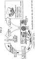
- FIG. 1 is a schematic from Byrne 2008 Hum. Mol. Gen. 17:R37-R41 , showing how skin derived fibroblasts can be de-differentiated using epigenetic reprogramming into pluripotent stem cells, which can then differentiate into neurons, cardiomyocytes, beta islet cells, and hematopoetic cells. See Hochedlinger, et al., Development. 2009 Feb;136(4):509-23 and Kanawaty, et al. Bioessays. 2009 Feb;31(2):134-8 .
- Figure 3 is a schematic of methods by which fibroblasts can be de-differentiated into pluripotent cells: cell fusion ( Cowan et al. Science. 2005 Aug 26;309(5739):1369-73 ), direct reprogramming ( Takahashi, et al., Cell. 2007 30;131(5):861-72 ), and somatic cell nuclear transfer (Byrne, et al. 2007).
- Takahashi, et al. demonstrated the generation of iPS cells from adult human dermal fibroblasts with the same four factors: Oct3/4, Sox2, Klf4, and c-Myc.
- Human iPS cells were similar to human embryonic stem (ES) cells in morphology, proliferation, surface antigens, gene expression, epigenetic status of pluripotent cell-specific genes, and telomerase activity. Furthermore, these cells could differentiate into cell types of the three germ layers in vitro and in teratomas. These findings demonstrate that iPS cells can be generated from adult human fibroblasts.
- ES embryonic stem
- the autologous fibroblasts in the Drug Substance are derived by outgrowth from a biopsy of the recipient's own skin followed by expansion in culture using standard cell culture techniques.
- Skin tissue (dermis and epidermis layers) is biopsied from a subject's post-auricular area.
- the starting material is composed of three 3-mm punch skin biopsies collected using standard aseptic practices.
- the biopsies are collected by the treating physician, placed into a vial containing sterile phosphate buffered saline (PBS).
- PBS sterile phosphate buffered saline
- the biopsy After arrival at the manufacturing facility, the biopsy is inspected and, upon acceptance, transferred directly to the manufacturing area. Upon initiation of the process, the biopsy tissue is then washed prior to enzymatic digestion. After washing, a Liberase Digestive Enzyme Solution is added without mincing, and the biopsy tissue is incubated at 37.0 ⁇ 2°C for one hour. Time of biopsy tissue digestion is a critical process parameter that can affect the viability and growth rate of cells in culture.
- Liberase is a collagenase/neutral protease enzyme cocktail obtained formulated from Lonza Walkersville, Inc. (Walkersville, MD) and unformulated from Roche Diagnostics Corp. (Indianapolis, IN).
- Serva Collagenase NB6 Helidelburg, Germany
- Initiation Growth Media IMDM, GA, 10% Fetal Bovine Serum (FBS)
- FBS Fetal Bovine Serum
- cells are pelleted by centrifugation and resuspended in 5.0 mL Initiation Growth Media.
- centrifugation is not performed, with full inactivation of the enzyme occurring by the addition of Initiation Growth Media only.
- Initiation Growth Media is added prior to seeding of the cell suspension into a T-175 cell culture flask for initiation of cell growth and expansion.
- a T-75, T-150, T-185 or T-225 flask can be used in place of the T-75 flask.
- Cells are incubated at 37 ⁇ 2.0°C with 5.0 ⁇ 1.0% CO 2 and fed with fresh Complete Growth Media every three to five days. All feeds in the process are performed by removing half of the Complete Growth Media and replacing the same volume with fresh media. Alternatively, full feeds can be performed. Cells should not remain in the T-175 flask greater than 30 days prior to passaging. Confluence is monitored throughout the process to ensure adequate seeding densities during culture splitting. When cell confluence is greater than or equal to 40% in the T-175 flask, they are passaged by removing the spent media, washing the cells, and treating with Trypsin-EDTA to release adherent cells in the flask into the solution.
- T-500 flask a T-500 flask for continued cell expansion.
- one or two T-300 flasks One Layer Cell Stack (1 CS), One Layer Cell Factory (1 CF) or a Two Layer Cell Stack (2 CS) can be used in place of the T-500 Flask.
- Morphology is evaluated at each passage and prior to harvest to monitor the culture purity throughout the culture purity throughout the process. Morphology is evaluated by comparing the observed sample with visual standards for morphology examination of cell cultures.
- the cells display typical fibroblast morphologies when growing in cultured monolayers. Cells may display either an elongated, fusiform or spindle appearance with slender extensions, or appear as larger, flattened stellate cells which may have cytoplasmic leading edges. A mixture of these morphologies may also be observed. Fibroblasts in less confluent areas can be similarly shaped, but randomly oriented.
- the presence of keratinocytes in cell cultures is also evaluated. Keratinocytes appear round and irregularly shaped and, at higher confluence, they appear organized in a cobblestone formation. At lower confluence, keratinocytes are observable in small colonies.
- Cells are incubated at 37 ⁇ 2.0°C with 5.0 ⁇ 1.0% CO2 and fed every three to five days in the T-500 flask and every five to seven days in the ten layer cell stack (10CS). Cells should not remain in the T-500 flask for more than 10 days prior to passaging. Quality Control (QC) release testing for safety of the Bulk Drug Substance includes sterility and endotoxin testing.
- QC Quality Control
- two Five Layer Cell Stacks (5 CS) or a 10 Layer Cell Factory (10 CF) can be used in place of the 10 CS. 10CS.
- Passage to the 10 CS is performed by removing the spent media, washing the cells, and treating with Trypsin-EDTA to release adherent cells in the flask into the solution. Cells are then transferred to the 10 CS. Additional Complete Growth Media is added to neutralize the trypsin and the cells from the T-500 flask are pipetted into a 2L bottle containing fresh Complete Growth Media. The contents of the 2L bottle are transferred into the 10 CS and seeded across all layers. Cells are then incubated at 37 ⁇ 2.0°C with 5.0 ⁇ 1.0% CO 2 and fed with fresh Complete Growth Media every five to seven days. Cells should not remain in the 10CS for more than 20 days prior to passaging.
- the passaged dermal fibroblasts are rendered substantially free of immunogenic proteins present in the culture medium by incubating the expanded fibroblasts for a period of time in protein free medium.
- Primary Harvest When cell confluence in the 10 CS is 95% or more, cells are harvested. Harvesting is performed by removing the spent media, washing the cells, treating with Trypsin-EDTA to release adherent cells into the solution, and adding additional Complete Growth Media to neutralize the trypsin. Cells are collected by centrifugation, resuspended, and in-process QC testing performed to determine total viable cell count and cell viability.
- the total cell count must be 3.4 x 10 8 cells and viability 85% or higher. Alternatively, total cell yields for other indications can range from 3.4 x 10 8 to 1 x 10 9 cells.
- Cell count and viability at harvest are critical parameters to ensure adequate quantities of viable cells for formulation of the Drug Substance. If total viable cell count is sufficient for the intended treatment, an aliquot of cells and spent media are tested for mycoplasma contamination. Mycoplasma testing is performed. Harvested cells are formulated and cryopreserved.
- Step 5a in Figure 1 If additional cells are required after receiving cell count results from the primary 10 CS harvest, an additional passage into multiple cell stacks (up to four 10 CS) is performed (Step 5a in Figure 1 ).
- cells from the primary harvest are added to a 2L media bottle containing fresh Complete Growth Media. Resuspended cells are added to multiple cell stacks and incubated at 37 ⁇ 2.0°C with 5.0 ⁇ 1.0% CO 2 .
- the cell stacks are fed and harvested as described above, except cell confluence must be 80% or higher prior to cell harvest.
- the harvest procedure is the same as described for the primary harvest above.
- a mycoplasma sample from cells and spent media is collected, and cell count and viability performed as described for the primary harvest above.
- the method decreases or eliminates immunogenic proteins be avoiding their introduction from animal-sourced reagents.
- cells are cryopreserved in protein-free freeze media, then thawed and washed prior to prepping the final injection to further reduce remaining residuals.
- Step 5a in Figure 1 If additional Drug Substance is needed after the harvest and cryopreservation of cells from additional passaging is complete (Step 5a in Figure 1 ), aliquots of frozen Drug Substance - Cryovial are thawed and used to seed 5 CS or 10 CS culture vessels (Step 7a in Figure 1 ).
- a four layer cell factory (4 CF), two 4 CF, or two 5 CS can be used in place of a 5 CS or 10 CS.
- a frozen cryovial(s) of cells is thawed, washed, added to a 2L media bottle containing fresh Complete Growth Media and cultured, harvested and cryopreserved as described above.
- the cell suspension is added Cell confluence must be 80% or more prior to cell harvest.
- the drug substance consists of a population of viable, autologous human fibroblast cells suspended in a cryopreservation medium consisting of, for example, Iscove's Modified Dulbecco's Medium (IMDM) and Profreeze-CDMTM (Lonza, Walkerville, MD) plus 7.5% dimethyl sulfoxide (DMSO).
- IMDM Iscove's Modified Dulbecco's Medium
- Profreeze-CDMTM Lionza, Walkerville, MD
- DMSO dimethyl sulfoxide
- a lower DMSO concentration may be used in place of 7.5% or CryoStorTM CS5 or CryoStorTM CS10 (BioLife Solutions, Bothell, WA) may be used in place of IMDM/Profreeze/DMSO.
- the freezing process consists of a control rate freezing step to the following ramp program:
- Drug Substance vials are transferred to a cryogenic freezer for storage in the vapor phase. After cryogenic freezing, the Drug Substance is submitted for Quality Control testing. Drug Substance specifications also include cell count and cell viability testing performed prior to cryopreservation and performed again for Drug Substance - Cryovial. Viability of the cells must be 85% or higher for product release. Cell count and viability are conducted using an automated cell counting system (Guava Technologies), which utilizes a combination of permeable and impermeable fluorescent, DNA-intercalating dyes for the detection and differentiation of live and dead cells. Alternatively, a manual cell counting assay employing the trypan blue exclusion method may be used in place of the automated cell method above.
- purity/identity of the Drug Substance is performed and must confirm the suspension contains 98% or more fibroblasts.
- the usual cell contaminants include keratinocytes.
- the purity/identify assay employs fluorescent-tagged antibodies against CD90 and CD104 (cell surface markers for fibroblast and keratinocyte cells, respectively) to quantify the percent purity of a fibroblast cell population.
- CD90 Thy-1 is a 35 kDa cell-surface glycoprotein. Antibodies against CD90 protein have been shown to exhibit high specificity to human fibroblast cells.
- CD104 integrin ⁇ 4 chain
- CD49f integrin ⁇ 6 chain
- This complex has been shown to act as a molecular marker for keratinocyte cells (Adams and Watt 1991).
- Antibodies to CD104 protein bind to 100% of human keratinocyte cells.
- Cell count and viability is determined by incubating the samples with Viacount Dye Reagent and analyzing samples using the Guava PCA system.
- the reagent is composed of two dyes, a membrane-permeable dye which stains all nucleated cells, and a membrane-impermeable dye which stains only damaged or dying cells.
- the use of this dye combination enables the Guava PCA system to estimate the total number of cells present in the sample, and to determine which cells are viable, apoptotic, or dead.
- the method was custom developed specifically for use in determining purity/identity of autologous cultured fibroblasts. The specific procedure is as follows:
- microcentrifuge tubes For each sample set, per test vial, label four 1.5 mL microcentrifuge tubes as follows: "CD90,” “CD104,” “Mouse” (corresponding to mouse IgG1 isotype control), and “Rat” (corresponding to rat IgG2b Isotype control). Using the mean cell count result, calculate the appropriate volume of cells to aliquot so that each tube receives 1x 10 5 viable cells.
- Pellet the cells in each sample tube by centrifugation at 3,000 rpm for 3 minutes. If difficulty in pellet formation arises, then centrifuge tubes again at 3,000 rpm for 3 minutes. If pellet formation is adequate after the second centrifugation proceed with aspirating the supernatant. With the aid of a pipette, aspirate the supernatant into a waste container containing 20% bleach in DI water. Leave approximately 50 ⁇ L of supernatant in each tube to minimize inadvertently aspirating cells.
- the "Adjust Settings" screen should appear and the system will automatically set the threshold to exclude background fluorescence.
- Select an FSC Gain intensity to position the center of the defined cell cluster over 10e3 in the lower right quadrant on the X-axis of the FSC vs. Viability (PM1) plot (e.g., "x2").
- sample information control panel enter the sample Part, Lot and isotype control or antibody (e.g., "DR01 200502001_CD90") to identify each sample in the Sample ID field. Verify that the PM1 fluorescence markers are set to the same settings as the mouse IgG1 isotype control sample.
- the "Adjust Settings" screen should appear and the system will automatically set the threshold to exclude background fluorescence.
- cells can be passaged from either the T-175 flask (or alternatives) or the T-500 flask (or alternatives) into a spinner flask containing microcarriers as the cell growth surface.
- Microcarriers are small bead-like structures that are used as a growth surface for anchorage dependent cells in suspension culture. They are designed to produce large cell yields in small volumes.
- a volume of Complete Growth Media ranging from 50mL-300mL is added to a 500mL, IL or 2L sterile disposable spinner flask.
- Sterile microcarriers are added to the spinner flask.
- the culture is allowed to remain static or is placed on a stir plate at a low RPM (15-30 RRM) for a short period of time (1-24 hours) in a 37 ⁇ 2.0°C with 5.0 ⁇ 1.0% CO 2 incubator to allow for adherence of cells to the carriers.
- the speed of the spin plate is increased (30-120 RPM). Cells are fed with fresh Complete Growth Media every one to five days, or when media appears spent by color change.
- Cells are collected at regular intervals by sampling the microcarriers, isolating the cells and performing cell count and viability analysis. The concentration of cells per carrier is used to determine when to scale-up the culture. When enough cells are produced, cells are washed with PBS and harvested from the microcarriers using trypsin-EDTA and seeded back into the spinner flask in a larger amount of microcarriers and higher volume of Complete Growth Media (300mL-2L). Alternatively, additional microcarriers and Complete Growth Media can be added directly to the spinner flask containing the existing microcarrier culture, allowing for direct bead-to-bead transfer of cells without the use of trypsinizationtrypsiziation and reseeding. Alternatively, if enough cells are produced from the initial T-175 or T-500 flask, the cells can be directly seeded into the scale-up amount of microcarriers.
- the speed of the spin plate is increased (30-120 RPM).
- Cells are fed with fresh Complete Growth Media every one to five days, or when media appears spent by color change.
- concentration reaches the desired cell count for the intended indication, the cells are washed with PBS and harvested using trypsin-EDTA. All release testing, cryopreservation and preparation of Drug Product - Injection would follow the process described in Sections C and D.
- Microcarriers used within the disposable spinner flask may be made from poly blend such as BioNOC II® (Cesco Bioengineering, distributed by Bellco Biotechnology, Vineland, NJ) and FibraCel® (New Brunswick Scientific, Edison, NJ), gelatin, such as Cultispher-G (Percell Biolytica, Astrop, Sweden), cellulose, such as CytoporeTM (GE Healthcare, Piscataway, NJ) or coated/ uncoated polystyrene, such as 2D MicroHexTM (Nunc, Weisbaden, Germany), Cytodex® (GE Healthcare, Piscataway, NJ) or Hy-Q SphereTM (Thermo Scientific Hyclone, Logan, UT).
- poly blend such as BioNOC II® (Cesco Bioengineering, distributed by Bellco Biotechnology, Vineland, NJ) and FibraCel® (New Brunswick Scientific, Edison, NJ)
- gelatin such as Cultispher-G (Percell Biolytica, Astro
- cells can be processed on poly blend 2D microcarriers such as BioNOC II® and FibraCel® using an automatic bellow system, such as FibraStageTM (New Brunswick Scientific, Edison, NJ) or BelloCell® (Cesco Bioengineering, distributed by Bellco Biotechnology, Vineland, NJ) in place of the spinner flask apparatus.
- Cells from the T-175 (or alternatives) or T-500 flask (or alternatives) are passaged into a bellow bottle containing microcarriers with the appropriate amount of Complete Growth Media, and placed into the system.
- the system pumps media over the microcarriers to feed cells, and draws away media to allow for oxygenation in a repeating fixed cycle.
- ACE Automated Cellular Expansion
- the ACE system can be a scaled down, single lot unit version comprised of a disposable component that consists of cell growth surface, delivery tubing, media and reagents, and a permanent base that houses mechanics and computer processing capabilities for heating/cooling, media transfer and execution of the automated programming cycle.
- a disposable component that consists of cell growth surface, delivery tubing, media and reagents, and a permanent base that houses mechanics and computer processing capabilities for heating/cooling, media transfer and execution of the automated programming cycle.
- each sterile irradiated ACE disposable unit Upon receipt, each sterile irradiated ACE disposable unit will be unwrapped from its packaging and loaded with media and reagents by hanging pre-filled bags and connecting the bags to the existing tubing via aseptic connectors.
- a suspension of cells from a biopsy that has been enzymatically digested is introduced into the "pre-growth chamber" (small unit on top of the cell tower), which is already filled with Initiation Growth Media containing antibiotics. From the BSC, the disposable would be transferred to the permanent ACE unit already in place.
- pre-growth chamber small unit on top of the cell tower
- the cells within the pre-growth chamber are trypsinized and introduced into the cell tower itself, which is pre-filled with Complete Growth Media.
- the "bubbling action" caused by CO 2 injection force the media to circulate at such a rate that the cells spiral downward and settle on the surface of the discs in an evenly distributed manner.
- the cells are allowed to multiply.
- confluence will be checked (method unknown at time of writing) to verify that culture is growing.
- the Complete Growth Media will be replaced with fresh Complete Growth Media.
- CGM will be replaced every seven days for three to four weeks.
- the confluence is checked once more to verify that there is sufficient growth to possibly yield the desired quantity of cells for the intended treatment.
- the culture is sufficiently confluent, it is harvested.
- the spent media (supernatant) is drained from the vessel.
- PBS will then is pumped into the vessel (to wash the media, FBS from the cells) and drained almost immediately.
- Trypsin-EDTA is pumped into the vessel to detach the cells from the growth surface.
- the trypsin/cell mixture is drained from the vessel and enter the spin separato.
- Cryopreservative is pumped into the vessel to rinse any residual cells from the surface of the discs, and be sent to the spin separator as well.
- the spin separator collects the cells and then evenly resuspend the cells in the shipping/injection medium.
- the cells will be sent through an inline automated cell counting device or a sample collected for cell count and viability testing via laboratory analyses. Once a specific number of cells has been counted and the proper cell concentration has been reached, the harvested cells are delivered to a collection vial that can be removed to aliquot the samples for cryogenic freezing.
- automated robotic systems may be used to perform cell feeding, passaging, and harvesting for the entire length or a portion of the process.
- Cells can be introduced into the robotic device directly after digest and seed into the T-175 flask (or alternative).
- the device may have the capacity to incubate cells, perform cell count and viability analysis and perform feeds and transfers to larger culture vessels.
- the system may also have a computerized cataloging function to track individual lots. Existing technologies or customized systems may be used for the robotic option.
- Drug Substance - Cryovial used to prepare the final dosage unit consists of fibroblasts that are harvested from the final culture vessel, formulated to the desired cell concentration and cryopreserved in cryovials.
- Drug Substance-Cryovial is stored in a cryopreservation medium consisting of, for example, IMDM and ProfreezeTM plus 7.5% DMSO to a target of 2.2 ⁇ 10 7 cells/mL. After exposure to a controlled rate freezing cycle, the cryovialed Drug Substance is stored frozen in the vapor phase of a liquid nitrogen freezer.
- Harvested cells are pooled, formulated in a cryopreservation media that includes, for example, Profreeze, DMSO and IMDM media, aliquoted into cryovials and stored frozen in liquid nitrogen as the Drug Substance - Cryovial material via controlled rate freezing.
- a cryopreservation media that includes, for example, Profreeze, DMSO and IMDM media, aliquoted into cryovials and stored frozen in liquid nitrogen as the Drug Substance - Cryovial material via controlled rate freezing.
- the caps and vials are radiation sterilized and received sterile from the manufacturer.
- the required volume of bulk material needed for treatment is removed from frozen storage, thawed, and pooled.
- the cells are washed with 4x bulk volume of PBS and centrifuged at 150 x g for 10 minutes (5 ⁇ 3°C). This is followed by a wash with 4x bulk volume of DMEM by resuspension and centrifugation at 150 x g for 10 minutes (5 ⁇ 3°C).
- the washed cells are resuspended in DMEM without phenol red to a target concentration of 1.0 - 2.0 ⁇ 10 7 cells/mL.
- the second 4x wash and final resuspension can be performed with Hypothermosol®-FRS (BioLife Solutions, Bothell, WA).
- the final sterile cryovial containers are then manually filled in a Biological Safety Cabinet to a volume of 1.2 mL/container.
- the Drug Product - Injection is stored at 2-8°C until shipment in a a 2-8°C refrigerated shipper to the administration site.
- Drug Substance vials can be removed from cryogenic storage and shipped directly to the administration site for dilution and administration.
- the cells are harvested and prepared for cryopreservation at a higher cell concentration (3.0 - 4.0 x 10 7 cell/mL as compared to the current target of 2.2 x 10 7 cells/mL).
- the frozen vial will be shipped to the study site on dry ice or in a liquid nitrogen dewar.
- the administration site thaws the vial by hand or with a heat block, and performs a 1:1 ratio dilution of the frozen cells at the study site using a typical injection diluent such as bacteriostatic water, sterile water, sodium chloride, or phosphate buffered saline.
- a typical injection diluent such as bacteriostatic water, sterile water, sodium chloride, or phosphate buffered saline.
- DMEM may be used as the diluent. This concept eliminates the need to wash and prepare a fresh suspension of the injection for overnight shipment to the study site.
- cells freshly harvested from flasks or cells stacks can be adjusted to a target concentration of 1.0 - 2.0 ⁇ 10 7 cells/mL in DMEM, undergo all Bulk Harvest and Drug Substance - Cyrovial testing described above and shipped fresh overnight to the administration site in a 2-8°C refrigerated shipper as the final injection product.
- sterility and mycoplasma testing may be performed upstream from the harvest to allow time for results prior to shipment.
- the azficel-T Azficel-T Drug Product - Injection consists of a suspension of each patient's own living autologous fibroblasts formulated in Dulbecco's Modified Eagle's Medium (DMEM) without phenol red.
- Azficel-T fibroblast dosage formulation is supplied as two 2 mL vials with each vial containing 1.2 mL of Drug Product at 1.0-2.0 x 10 7 cells/mL.
- the sterile cellular suspension is intended for intradermal injection. Initially, the cell dosage for Azficel-fibroblast dosage formulation was based on the number of cells administered by clinicians before the regulation of Azficel-T fibroblast dosage formulation by the FDA.
- a dose range from 1.5 - 7.0 x 10 7 cells per 1.2-1.4 mL injection was used successfully. Viscosity became a concern for the fibroblast dosage formulation injections in higher concentrations than this range. This dose range converts to 1.1 - 5.8 x 10 7 cells/mL.
- Vials are to be warmed to room temperature and gently inverted to resuspend the settled cells.
- the cellular suspension is withdrawn from the container using a small unit syringe fitted with a detachable needle or with a fixed needle.
- Use of short, sharp needles and small unit syringes e.g., 0.5 mL insulin syringes
- a 29-gauge or 30-gauge needle is required for intradermal injection of the product.
- a syringe with a larger bore 21-gauge detachable needle may be used to aid in withdrawing the product from the container. Once withdrawn, the 21-gauge needle can be switched out with a 30-gauge needle and the product administered.
- a batch consists of three injection treatments.
- a single injection treatment (batch) requires two 2 mL vials containing 1.2 mL/vial of azficelAzficel-T fibroblast dosage formulation.
- the aging process of the skin occurs as a result of both intrinsic and extrinsic factors.
- the factors that contribute to intrinsic or natural aging are both structural and functional. Structurally, the epidermis becomes thinner, the corneocytes are less adherent and the dermal-epidermal junction is flattened. Functionally, there is a reduction in the number and biosynthetic capacity of fibroblasts and the dermis becomes atrophic and relatively acellular and avascular. Exposure to ultraviolet light radiation is the primary cause of extrinsic or photoaging. Extrinsic aging characteristics are loss of elasticity, increased roughness and dryness, irregular pigmentation, deep wrinkling, a leathery appearance, blister formation and impaired wound healing.
- fibroblast cell suspension into contour deficiencies of the dermis can result in correction of damaged dermal and subcutaneous tissues.
- Dermis of skin contains fibroblasts which are primarily responsible for the secretion of extracellular matrix components such as collagen and elastin, which provide mechanical strength and integrity to skin.
- the fibroblast cell suspension contains autologous fibroblasts that may increase the synthesis of extracellular matrix components when injected into patient skin, thus increasing skin integrity, ultimately leading to a decrease in fine lines and wrinkles.
- the effect of the therapy is not immediate, but instead provides a gradual improvement in the appearance of lines and wrinkles over time.
- the fibroblast cell suspension was initially evaluated in a clinical study conducted by Dr. William Boss in 1995, and was then marketed in the US as a cosmetic treatment for facial contour deformities from December 1995 to February 1999. After February 1999, FDA was required to regulate for all somatic cell therapies under the PHS Act.
- the fibroblast cell suspension was studied in the treatment of acne scars (Study IT-A-008; IND #13455). Ninety-nine subjects received treatment with the fibroblast cell suspension during Study IT-A-008. The fibroblast cell suspension was also developed for use in other indications including treatment of restrictive burns scars (IND #13308), vocal cord scarring (IND #9892) and gingival repair (IND #10805).
- Study IT-R-001 was a Phase II double-blind, randomized, and placebo-controlled study of the fibroblast cell suspension for the treatment of rhytids (nasolabial and melolabial folds, perioral lines, glabellar lines, acne scars, and forehead were treated).
- rhytids nasolabial and melolabial folds, perioral lines, glabellar lines, acne scars, and forehead were treated.
- each subject received three treatments of the fibroblast cell suspension (0.5 x 10 7 , 1.0 x 10 7 , or 2.0 x 10 7 cells/mL) or placebo, administered approximately two weeks apart.
- Study IT-R-002 was a Phase III double-blind, randomized and placebo-controlled study of the fibroblast cell suspension for the treatment of facial contour deformities and scars.
- each subject received three treatments of the fibroblast cell suspension containing 2.0 x 10 7 cells/mL or placebo, administered every 14 ⁇ 7 days.
- Study IT-R-003B showed efficacy for both of the co-primary endpoints
- the failure of one of the co-primary endpoints in Study IT-R-003A to meet the criterion for statistical significance (for the Investigator Evaluation) led to two new protocols as the pivotal Phase III trials for the fibroblast cell suspension (Studies IT-R-005 and IT-R-006).
- the reasons for the missed endpoint in 003A are believed to have included sub-optimal dosing.
- the concentration of cells remained the same, but the volume delivered was increased. Other reasons were identified, including training technique and time between injections in the series..
- Study IT-R-007 was a Phase II multicenter, open-label study of the safety and efficacy of the fibroblast cell suspension in the treatment of facial wrinkles and creases, designed to obtain safety and efficacy data for the use of the fibroblast cell suspension for uses other than nasolabial folds.
- each subject received two treatments of up to 6 mL of the fibroblast cell suspension containing 1.0-2.0 x 10 7 cells/mL, administered every five weeks ⁇ 10 days.
- 45 subjects were treated with the fibroblast cell suspension. This study exposed subjects to a three-fold higher total dose of the fibroblast cell suspension than was used in the 005/006 studies.
- the populations enrolled into the Phase III IT-R-003A and IT-R-003B (003A/B) studies were similar to those from 005/006.
- Females comprised 94% of the population in the 003A/B studies, and 95% of the population was White.
- the mean age of subjects across the two studies was 54.1 years.
- the 003A/B protocol was very similar in design to the 005/006 protocol, 003A/003B permitted the enrollment of subjects with a broader range of wrinkle severity, with scores for the primary nasolabial fold deformity upon enrollment ranging from Grade 2-5 on the Evaluator Scale, but the mean severity score was similar at 3.9.
- Evaluator Wrinkle Severity Assessment the blinded evaluator live assessment of the bilateral nasolabial fold wrinkles at rest, at visit 6, using a 6-point ordinal wrinkle severity scale with a photoguide, where a response is defined as a two point or better improvement on the scale compared to baseline. These endpoints were selected to provide both an impartial assessment (grading by a blinded physician) and clinical relevance (subject's opinion of their own appearance).
- the scale used for the Evaluator Wrinkle Severity Assessment was the 6-point ordinal wrinkle severity scale for the assessment of nasolabial folds (NLF) developed and validated by Lemperle et al., Plast Reconstr Surg. 2001 108(6):1735-50 ).
- the Lemperle scale has been validated to detect a one point improvement in the severity of the nasolabial fold wrinkle. However, because the assessment of appearance can be subjective and prone to variability, success for this endpoint was defined as a two point improvement for each NLF, i.e. for a given patient, both the left and right NLF had to improve by two points in the Evaluator's assessment in order to be considered a responder.
- the Lemperle scale is an accepted standard of measurement in the field of dermatology, and has been employed successfully in pivotal clinical trials for FDA-approved products in similar indications.
- the scale used for the Subject Wrinkle Assessment is based on the published scale used by Cohen and Holmes, Plast Reconstr Surg. 2004 15;114(4):964-76 . As with the Evaluator Assessment, a two-point improvement was established as the criteria for a successful response to treatment.
- the 003A/B studies were designed with similar co-primary endpoints, but used a different subject assessment tool than the 005/006 studies. As stated in the 003A/B protocol.
- the co-primary efficacy endpoints is the efficacy of the fibroblast dosage formulation injection in the primary nasolabial fold at the 6 month visit using the Investigator's 6-point ordinal scale and the Subject's VAS assessment.
- the 6-point ordinal scale referenced in the protocol is the same Lemperle scale used in the 005/006 studies, although the 005/006 study provided more descriptive text for each point on the scale than was used in the 003A/B studies.
- a Visual Analog Scale was used For the Subject assessment. This scale asked subjects to rate each contour deformity from 0 (no defect) to 100 (very severe defect) by placing a mark on a 10 cm line. While both studies met statistical significance for improvement using this assessment tool, the VAS scale was replaced with the ordinal Subject Wrinkle Assessment scale in the 005/006 studies in order to improve the clinical relevance and interpretability of the subject assessment data.
- the 005/006 studies were designed after review and analysis of the data from the 003A/B studies. As a result of this review, a number of modifications were made to the design of the 005/006 protocol, as compared to the 003A/B studies. A summary of the modifications made that may have contributed to the observed difference in outcome of the primary endpoints is provided in Table 6.
- Study IT-R-001 was a placebo-controlled Phase II dose ranging study that evaluated the fibroblast cell suspension at 0.5, 1.0, and 2.0 x 10 7 cells/mL at 0.1 mL per linear centimeter versus placebo for safety and preliminary efficacy in the treatment of facial rhytids. Baseline to four months following the initial injection (primary efficacy timepoint for Study IT-R-001) in the highest dose group (2.0 x 10 7 cells/mL) gave the best results. Therefore, this was the density of cells that was used in all subsequent studies.
- the dose per linear centimeter was the same as that used in other azficel-T fibroblast dosage formulation studies for this indication (0.1 mL per linear centimeter).
- the total dose for each treatment was limited to 1 mL, over a total treatment area of 10 cm of total. This difference is because Studies 003A/B did not allow for treatment of what is sometimes termed the mesolabial fold wrinkle, which is the fold or wrinkle that extends inferiorly from the corner of the mouth toward the chin.
- the total dose was increased to 2 mL in 005/006, to permit treatment of the mesolabial fold wrinkle, as it is frequently contiguous with the nasolabial fold wrinkle and is therefore a contributing component to the overall aesthetic outcome. While the total dose permitted was not the only difference between the 003A/B protocol and the 005/006 protocol, this increase in volume of product administered is considered to be a contributing factor in the successful results obtained from the 005/006 studies.
- the recommended dose interval of five weeks ⁇ one week was determined based on feedback provided by the Investigators from the 003A/B studies and the outcome of the 005/006 trials.
- the 003A/B investigators advised Isolagen that the time between treatments in those studies (seven to 14 days) was insufficient to permit the inflammation induced by one injection to recede before administration of the next injection. This duration of inflammation was noted only during the injection process and was not reported as an adverse event.
- investigators reported that the injection sites were more likely to appear closer to normal skin, which led to greater control of both the depth and volume of injection. This led to lengthening the interval between injections in the 005/006 studies first to four, then five ⁇ one week. This change is also considered to be a contributing factor in the successful 005/006 study results.
- Subject Improvement Assessment On the subject's photographic assessment (Subject Improvement Assessment), where subjects compared their own Baseline photos to those taken at the final efficacy visit, 67% of IT-treated subjects and 26% of placebo-treated subjects showed improvement.
- the increase in response rates using this assessment instrument as compared to the live assessment results demonstrates the subtle, gradual onset of effect, which may not be apparent to subjects or evaluators until appearance is compared to baseline.
- the preferred dosage for the treatment of nasolabial fold wrinkles is to inject one to two mL of the dosage formulation per treatment session, into the superficial papillary dermis of the wrinkles at a dose distribution of 0.1 mL/linear cm, preferably for three treatment sessions separated by five weeks plus or minus seven to ten days.
- the preferred dosage for the treatment of rhytids in multiple facial regions is to inject five to six mL of the dosage formulation per treatment session into the superficial papillary dermis according to the treatment map in Figure 4 at a dose distribution of 0.05 mL/linear cm, preferably for one or two treatment sessions separated by five weeks plus or minus seven to ten days.
- the appropriate dosage for the treatment of acne scars can be determined using a validated method and scale for the objective, physician assessed, live grading of acne scarring severity, the "Evaluator Live Acne Scar Assessment Scale," employing the comparative evaluation of relative scarring appearance under direct and tangential lighting, referring to Figure 2 .
- Table 1 Evaluator Live Acne Scar Assessment Scale Grade Term Description 0 Clear No depressions are seen in the treatment area. Macular discoloration may be seen. 1 Very Mild A single depression is easily noticeable with direct lighting (deep). Most or all of the depressions seen are only readily apparent with tangential lighting (shallow). 2 Mild A few to several, but less than half of all the depressions are easily noticeable with direct lighting (deep). Most of the depressions seen are only readily apparent with tangential lighting (shallow). 3 Moderate More than half of the depressions are apparent with direct lighting (deep). 4 Severe All or almost all the lesions can be seen with direct lighting (deep).
- Atrophic scars defined morphologically as ice-pick, macular atrophic or follicular macular atrophic - translated into scores ranging from 1 to 6 representing 1-5, 6-10, 11-25, 26-50, 51-100 and more than 100 scars, respectively. Ice-pick scars were described as those with an irregular border, jagged edges and sharp margins with steep sides leading to a fibrotic base. Macular atrophic scars were soft and distensible in which the base was often easily creased.
- Follicular macular atrophic scars were described as small white perifollicular papules or macules. The authors separately quantified keloidal and hypertrophic scars owing to their greater level of disfigurement. Score allocation of 2, 4 and 6 represented one to three, four to seven and more than seven scars of this type, respectively. Keloidal scars were described as those that were indurated and extending beyond the boundaries of the initiating inflammatory acne lesion, while hypertrophic scars were defined as less raised and conforming to the area of the primary acne lesion. A total scar score was then obtained by adding the scores from both atrophic and hypertrophic categories. Such total scores could be calculated separately for the face, chest and back to provide a comprehensive system for scar evaluation, and also provide a means for assessing effective dosage and treatment regimes.
- acne scars are treated by injecting two to twelve mL of the fibroblast dosage formulation per treatment session, into the superficial papillary dermis at a dose distribution of 0.1 mL/cm 2 of scarred area, for one to three treatment sessions separated by fourteen days plus or minus three days.
- a potentially serious long-term consequence of burn injuries to the deep dermis are post-injury scars.
- the formation of restrictive scars due to serious burns can prevent the normal movements of the affected area and restrict range of motion (flexion, adduction, and/or extension).
- "burn scarring and contracture affecting function remain the most frustrating late complications of burn injury” ( Iwuagwu, Plast Reconstr Surg. 1999 Apr;103(4):1198-204 ).
- These scars can have a significantly negative impact on quality of life by causing pain and reduced functionality.
- restrictive burn scars may result in significant impairment and loss of function of the upper extremity.
- the fibroblast dosage formulation is particularly useful in the treatment of restrictive burn scars to the upper extremities, specifically to reduce the level of impairment and associated disability experienced by the subject.
- the preferred dosage for the treatment of restrictive burn scars is to inject one to ten mL of the dosage formulation per treatment session into the palpated restriction band of the burn scar at a dose distribution of 0.1 - 0.5 mL/cm 2 of scarred area, preferably for one to five treatments separated by two to six weeks.
- Dr. Chris Inglefield in London reported a case of a man with multiple burns to neck, lower face, parts of his eyes and ears.
- the subject previously had received a fibroblast formulation for facial rhytids and therefore had cells in storage at the time of his burn. This enabled him to have his facial burns treated within six weeks of his initial injury. He subsequently received three additional treatments thre months after the injuries. Each treatment was performed under topical anesthesia and comprised of 3-4 ml of cells given in 30-40 injections.
- the subject previously had been treated with skin grafts. The subject reported improved movement of his face and neck and improved texture and pliability of his skin within three weeks of the first treatment.
- Another subject with 12 non-healing wounds on her forehead and mallar region from a burn from a laser was treated with a fibroblast formulation on a compassionate use basis.
- Her only alternative at the time she was treated was a bovine skin graft. Following treatment, most of the wounds healed within six weeks of injection.
- fibroblast formulations can improve not just sub-acute or healing burn wounds, but also longstanding, well healed but debilitating restrictive burn scars.
- Dr. Chris Inglefield in London, UK reported the case of a woman who had sustained multiple burns two years prior to presentation, resulting in major facial scars that restricted her facial expression on her left side.
- the subject's injury had severe negative psychological consequences and she felt that she had "lost who she was”.
- the subject received three treatments to her face of 3 mL each.
- the subject, her family and the treating physician noted remarkable improvement, such that her mother felt that her old daughter "was back".
- Her ability to move her face improved as did the skin texture and appearance.
- the same subject then underwent therapy for contracted scars and non-healing wounds on her hands. Prior to therapy the subject was having difficulty manipulating small objects in her jewelry making course. Within four weeks after treatment the wounds had healed and she had improved ability to perform her jewelry making tasks.
- Dr. Mark Palmer in Leeds, UK reported on another subject treated for chronic wounds of 10 years duration to her neck, shoulders and upper arms. Prior to treatment the subject had severely restricted range of motion of her neck and shoulder and required daily opiate analgesia for her chronic pain. The subject received a total of two four mL treatments in her neck region. Even after the first treatment the subject reported improved range of motion and skin texture. By the second treatment the subject's range of motion was near normal and a dramatic change in scar appearance and texture was noted.
Landscapes
- Health & Medical Sciences (AREA)
- Life Sciences & Earth Sciences (AREA)
- Engineering & Computer Science (AREA)
- Chemical & Material Sciences (AREA)
- Biomedical Technology (AREA)
- General Health & Medical Sciences (AREA)
- Zoology (AREA)
- Biotechnology (AREA)
- Cell Biology (AREA)
- Animal Behavior & Ethology (AREA)
- Medicinal Chemistry (AREA)
- Pharmacology & Pharmacy (AREA)
- Public Health (AREA)
- Veterinary Medicine (AREA)
- Bioinformatics & Cheminformatics (AREA)
- Organic Chemistry (AREA)
- Epidemiology (AREA)
- Genetics & Genomics (AREA)
- Wood Science & Technology (AREA)
- Immunology (AREA)
- Virology (AREA)
- Developmental Biology & Embryology (AREA)
- Dermatology (AREA)
- Microbiology (AREA)
- Biochemistry (AREA)
- General Engineering & Computer Science (AREA)
- Rheumatology (AREA)
- Nuclear Medicine, Radiotherapy & Molecular Imaging (AREA)
- General Chemical & Material Sciences (AREA)
- Chemical Kinetics & Catalysis (AREA)
- Dispersion Chemistry (AREA)
- Micro-Organisms Or Cultivation Processes Thereof (AREA)
- Medicines Containing Material From Animals Or Micro-Organisms (AREA)
- Medicinal Preparation (AREA)
- Materials For Medical Uses (AREA)
Applications Claiming Priority (2)
| Application Number | Priority Date | Filing Date | Title |
|---|---|---|---|
| US12/776,163 US8529883B2 (en) | 2010-05-07 | 2010-05-07 | Dosage unit formulations of autologous dermal fibroblasts |
| PCT/US2011/035332 WO2011140323A1 (en) | 2010-05-07 | 2011-05-05 | Dosage unit formulations of autologous dermal fibroblasts |
Publications (2)
| Publication Number | Publication Date |
|---|---|
| EP2566489A1 EP2566489A1 (en) | 2013-03-13 |
| EP2566489B1 true EP2566489B1 (en) | 2019-04-24 |
Family
ID=44121161
Family Applications (1)
| Application Number | Title | Priority Date | Filing Date |
|---|---|---|---|
| EP11720666.4A Active EP2566489B1 (en) | 2010-05-07 | 2011-05-05 | Dosage unit formulations of autologous dermal fibroblasts |
Country Status (8)
| Country | Link |
|---|---|
| US (3) | US8529883B2 (ja) |
| EP (1) | EP2566489B1 (ja) |
| JP (1) | JP6309268B2 (ja) |
| KR (1) | KR102001254B1 (ja) |
| CN (2) | CN107812014B (ja) |
| AU (1) | AU2011248067B2 (ja) |
| CA (1) | CA2800333C (ja) |
| WO (1) | WO2011140323A1 (ja) |
Families Citing this family (35)
| Publication number | Priority date | Publication date | Assignee | Title |
|---|---|---|---|---|
| WO2008027984A1 (en) | 2006-08-29 | 2008-03-06 | Isolagen Technologies, Inc. | Methods for culturing minimally-passaged fibroblasts and uses thereof |
| US8512715B2 (en) * | 2008-08-14 | 2013-08-20 | The Cleveland Clinic Foundation | Apparatus and method for treating a neuromuscular defect |
| WO2011075676A2 (en) | 2009-12-18 | 2011-06-23 | Knowlton Edward W | A skin treatment and drug delivery device |
| US8529883B2 (en) * | 2010-05-07 | 2013-09-10 | Fibrocell Technologies, Inc. | Dosage unit formulations of autologous dermal fibroblasts |
| WO2012051505A1 (en) | 2010-10-14 | 2012-04-19 | Fibrocell Science, Inc. | Treatment of vocal cords with autologous dermal fibroblast formulation |
| US11278309B2 (en) | 2010-12-17 | 2022-03-22 | Srgi Holdings, Llc | Pixel array medical systems, devices and methods |
| US10695546B2 (en) | 2010-12-17 | 2020-06-30 | Srgi Holdings, Llc | Systems, devices and methods for fractional resection, fractional skin grafting, fractional scar reduction and fractional tattoo removal |
| US10736653B2 (en) | 2013-12-06 | 2020-08-11 | Srgi Holdings, Llc | Pixel array medical systems, devices and methods |
| US11103275B2 (en) | 2010-12-17 | 2021-08-31 | Srgi Holdings, Llc | Pixel array medical systems, devices and methods |
| US11612410B2 (en) | 2010-12-17 | 2023-03-28 | Srgi Holdings, Llc | Pixel array medical systems, devices and methods |
| US10368904B2 (en) | 2013-12-06 | 2019-08-06 | Srgi Holdings, Llc | Pixel array medical systems, devices and methods |
| US11000310B2 (en) | 2010-12-17 | 2021-05-11 | Srgi Holdings, Llc | Pixel array medical systems, devices and methods |
| US10702684B2 (en) | 2010-12-17 | 2020-07-07 | Srgi Holdings, Llc | Systems, devices and methods for fractional resection, fractional skin grafting, fractional scar reduction and fractional tattoo removal |
| US10661063B2 (en) | 2010-12-17 | 2020-05-26 | Srgi Holdings, Llc | Systems, devices and methods for fractional resection, fractional skin grafting, fractional scar reduction and fractional tattoo removal |
| CA2866563A1 (en) * | 2012-03-07 | 2013-09-12 | Fibrocell Technologies, Inc. | Topical dermal formulations and methods of personalized treatment of skin |
| WO2013155109A1 (en) | 2012-04-09 | 2013-10-17 | The United States Of America, As Represented By The Secretary, Department Of Health And Human Services | Methods for generation of pluripotent and multipotent cells |
| US20140072611A1 (en) * | 2012-09-07 | 2014-03-13 | Fibrocell Technologies, Inc. | Methods and Compositions for Treating Post-Cardial Infarction Damage |
| ITMI20131053A1 (it) * | 2013-06-25 | 2014-12-26 | Maria Oliva Salviati | Composizione che comprende ortica |
| ES2827049T3 (es) | 2013-10-02 | 2021-05-19 | Srgi Holdings Llc | Dispositivos médicos de conjunto de píxeles |
| CA2926322C (en) | 2013-10-02 | 2022-10-18 | Srgi Holdings, Llc | Pixel array medical devices and methods |
| US11937846B2 (en) | 2013-12-06 | 2024-03-26 | Srgi Holdings Llc | Pixel array medical systems, devices and methods |
| US11229452B2 (en) | 2013-12-06 | 2022-01-25 | Srgi Holdings, Llc | Pixel array medical systems, devices and methods |
| WO2016127091A1 (en) | 2015-02-05 | 2016-08-11 | Srgi Holdings, Llc | Pixel array medical systems, devices and methods |
| US11751903B2 (en) | 2015-08-31 | 2023-09-12 | Srgi Holdings, Llc | Pixel array medical systems, devices and methods |
| US11490952B2 (en) | 2015-08-31 | 2022-11-08 | Srgi Holdings, Llc | Pixel array medical devices and methods |
| US10420802B2 (en) * | 2015-12-10 | 2019-09-24 | Aesthetics Biomedical, Inc. | Topical formulation for skin care |
| US11564706B2 (en) | 2019-10-28 | 2023-01-31 | Srgi Holdings, Llc | Pixel array medical systems, devices and methods |
| WO2020086915A2 (en) | 2017-05-03 | 2020-04-30 | Srgi Holdings Llc | Handed spiral slotted scalpet array |
| EP3962504A4 (en) * | 2019-04-27 | 2022-09-14 | Figene, LLC | INCREASE OF THERAPEUTIC FIBROBLAST ACTIVITY THROUGH T-CELL MODULATION |
| WO2020223155A1 (en) * | 2019-04-28 | 2020-11-05 | Figene, Llc | Fibroblast cell therapy for treatment of osteoporosis |
| CN110075125A (zh) * | 2019-05-10 | 2019-08-02 | 成都康景生物科技有限公司 | 一种用于治疗系统性硬化症伴随肢端硬化的脐带间充质干细胞注射液及其制备方法 |
| AU2020387177A1 (en) * | 2019-11-22 | 2022-06-09 | Cutiss Ag | Methods and systems for producing skin grafts |
| AU2021245671A1 (en) * | 2020-03-31 | 2022-10-20 | Cell Exosome Therapeutics Inc. | Cell preservation method |
| KR20220074132A (ko) | 2020-11-27 | 2022-06-03 | 테고사이언스 (주) | 건 재생 효과를 나타내는 피부 유래 섬유아세포 및 이의 용도 |
| JP2023138461A (ja) * | 2022-03-16 | 2023-10-02 | フィブロバイオロジクス インコーポレイテッド | 創傷治癒に使用するための線維芽細胞の治療的使用 |
Family Cites Families (40)
| Publication number | Priority date | Publication date | Assignee | Title |
|---|---|---|---|---|
| US4524065A (en) | 1983-08-04 | 1985-06-18 | Bio-Specifics N.V. | Method for the prevention and treatment of scars with enzymes |
| US4772557A (en) | 1985-11-12 | 1988-09-20 | Washington University | DNA clone of human skin fibroblast collagenase enzyme |
| US5591444A (en) | 1995-07-28 | 1997-01-07 | Isolagen Technologies, Inc. | Use of autologous dermal fibroblasts for the repair of skin and soft tissue defects |
| US20040229203A1 (en) * | 1996-06-14 | 2004-11-18 | Biostore New Zealand Ltd. | Compositions and methods for the preservation of living tissues |
| US7767452B2 (en) | 1997-02-20 | 2010-08-03 | Kleinsek Don A | Tissue treatments with adipocyte cells |
| WO1998036705A1 (en) | 1997-02-20 | 1998-08-27 | Keller Gregory S | Augmentation and repair of dermal, subcutaneous, and vocal cord tissue defects |
| US6110459A (en) | 1997-05-28 | 2000-08-29 | Mickle; Donald A. G. | Transplants for myocardial scars and methods and cellular preparations |
| US6150505A (en) * | 1997-09-19 | 2000-11-21 | Hadasit Medical Research Services & Development Ltd. | Fibrin microbeads prepared from fibrinogen, thrombin and factor XIII |
| US7115417B1 (en) * | 1998-05-01 | 2006-10-03 | Chancellor Michael B | Soft tissue and bone augmentation and bulking utilizing muscle-derived progenito compositions, and treatments thereof |
| US6432710B1 (en) * | 1998-05-22 | 2002-08-13 | Isolagen Technologies, Inc. | Compositions for regenerating tissue that has deteriorated, and methods for using such compositions |
| WO2000073418A2 (en) | 1999-05-28 | 2000-12-07 | Pacgen Technologies Llc | A method of using autologous fibroblasts to promote healing of wounds and fistulas |
| US6733530B1 (en) | 1999-08-02 | 2004-05-11 | Eric Sun-Yin Chan | Material and method for engraftment of a composite biocompatible skin graft on the neodermis of artificial skin |
| US20080286242A2 (en) | 1999-11-05 | 2008-11-20 | Donald Kleinsek | Augmentation and repair of spincter defects with cells including mesenchymal cells |
| US7799325B2 (en) | 1999-11-05 | 2010-09-21 | Kleinsek Donald A | Removal of hypertrophic scars |
| US20040101959A1 (en) | 2002-11-21 | 2004-05-27 | Olga Marko | Treatment of tissue with undifferentiated mesenchymal cells |
| US7186557B2 (en) | 2003-06-13 | 2007-03-06 | Isolagen Technologies, Inc. | Methods of producing neurons |
| EP1641913B1 (en) | 2003-06-27 | 2016-01-06 | DePuy Synthes Products, Inc. | Postpartum cells derived from umbilical cord tissue, and methods of making and using the same |
| CN101415458A (zh) * | 2003-11-10 | 2009-04-22 | 辛弗尼医药有限公司 | 控制心房纤维性颤动患者心室律的方法 |
| EP1885280A1 (en) * | 2005-05-26 | 2008-02-13 | Intercytex Limited | Tissue repair using allogenic dermal fibroblasts |
| AU2007203961A1 (en) * | 2006-01-04 | 2007-07-12 | Do-Coop Technologies Ltd. | Solid-fluid composition |
| US20070264239A1 (en) * | 2006-05-10 | 2007-11-15 | University Of Pittsburgh - Of The Commonwealth System Of Higher Education | Isolation of pericytes |
| GB2440908A (en) * | 2006-05-25 | 2008-02-20 | Intercytex Ltd | Tissue Repair and Augmentation |
| US20070298005A1 (en) * | 2006-06-22 | 2007-12-27 | Marie-Josee Thibault | Injectable composition for treatment of skin defects or deformations |
| KR20070122316A (ko) | 2006-06-26 | 2007-12-31 | (주) 에스바이오메딕스 | 주사용 인체 연조직 충전제 및 이의 제조 방법 |
| EP2046435A4 (en) * | 2006-07-13 | 2012-06-27 | Medtronic Inc | METHOD AND FORMULATIONS FOR OPTIMAL LOCAL ADMINISTRATION OF CELL THERAPY BY MEANS OF MINIMALLY INVASIVE PROCEDURES |
| WO2008027984A1 (en) * | 2006-08-29 | 2008-03-06 | Isolagen Technologies, Inc. | Methods for culturing minimally-passaged fibroblasts and uses thereof |
| KR20090093792A (ko) * | 2006-12-28 | 2009-09-02 | 고쿠리츠 다이가쿠 호우징 나고야 다이가쿠 | 피부 조직 개선재 및 그 제조 방법 |
| US20100303770A1 (en) | 2006-12-28 | 2010-12-02 | John Maslowski | Methods for culturing dermal cells for treatment of skin injuries such as burns |
| JP2008245583A (ja) * | 2007-03-30 | 2008-10-16 | Yamaguchi Univ | 哺乳動物の神経細胞の精製培養方法とその保存方法 |
| US7960098B2 (en) * | 2007-07-12 | 2011-06-14 | Warsaw Orthoperic, Inc. | Methods and compositions for the preservation of cells and tissues |
| WO2009023161A1 (en) | 2007-08-10 | 2009-02-19 | University Of Dayton | Methods of producing pluripotent stem-like cells |
| JP2009148236A (ja) * | 2007-12-20 | 2009-07-09 | Glycoresearch Kk | 線維芽細胞の細胞識別方法 |
| US20090209456A1 (en) * | 2008-02-19 | 2009-08-20 | Iliana Sweis | Compositions and methods for improving facial and body aesthetics |
| WO2009142717A2 (en) * | 2008-05-19 | 2009-11-26 | President And Fellows Of Harvard College | Methods and products for dedifferentiation of cells |
| WO2010008486A2 (en) * | 2008-06-24 | 2010-01-21 | Parkinsons Institute | Pluripotent cell lines and methods of use thereof |
| US20110165130A1 (en) * | 2008-06-25 | 2011-07-07 | Inserm | Methods for Preparing Human Skin Substitutes from Human Pluripotent Stem Cells |
| EP2398896A4 (en) * | 2009-02-20 | 2013-02-20 | Ventria Bioscience | CELL CULTURE MEDIUM CONTAINING COMBINATIONS OF PROTEINS |
| US8240314B2 (en) * | 2009-04-06 | 2012-08-14 | Eden Esthetics, A Professional Nursing Corporation | Method for injecting fillers into the dermis |
| JP5816416B2 (ja) * | 2010-04-23 | 2015-11-18 | 国立大学法人名古屋大学 | 細胞評価装置、インキュベータ、プログラム、および、培養方法 |
| US8529883B2 (en) * | 2010-05-07 | 2013-09-10 | Fibrocell Technologies, Inc. | Dosage unit formulations of autologous dermal fibroblasts |
-
2010
- 2010-05-07 US US12/776,163 patent/US8529883B2/en active Active
-
2011
- 2011-05-05 CN CN201710907274.2A patent/CN107812014B/zh active Active
- 2011-05-05 CN CN2011800230385A patent/CN102905711A/zh active Pending
- 2011-05-05 AU AU2011248067A patent/AU2011248067B2/en active Active
- 2011-05-05 EP EP11720666.4A patent/EP2566489B1/en active Active
- 2011-05-05 KR KR1020127030921A patent/KR102001254B1/ko active IP Right Grant
- 2011-05-05 CA CA2800333A patent/CA2800333C/en active Active
- 2011-05-05 JP JP2013509258A patent/JP6309268B2/ja active Active
- 2011-05-05 WO PCT/US2011/035332 patent/WO2011140323A1/en active Application Filing
-
2013
- 2013-07-12 US US13/941,265 patent/US11554142B2/en active Active
-
2022
- 2022-12-12 US US18/079,358 patent/US20230270793A1/en active Pending
Non-Patent Citations (1)
| Title |
|---|
| None * |
Also Published As
| Publication number | Publication date |
|---|---|
| US20130295061A1 (en) | 2013-11-07 |
| KR20130060212A (ko) | 2013-06-07 |
| JP2013524856A (ja) | 2013-06-20 |
| CN107812014A (zh) | 2018-03-20 |
| CN102905711A (zh) | 2013-01-30 |
| CA2800333A1 (en) | 2011-11-10 |
| AU2011248067B2 (en) | 2015-03-12 |
| US20230270793A1 (en) | 2023-08-31 |
| CN107812014B (zh) | 2021-11-16 |
| EP2566489A1 (en) | 2013-03-13 |
| JP6309268B2 (ja) | 2018-04-11 |
| US8529883B2 (en) | 2013-09-10 |
| AU2011248067A1 (en) | 2012-12-20 |
| US11554142B2 (en) | 2023-01-17 |
| KR102001254B1 (ko) | 2019-07-17 |
| US20110274665A1 (en) | 2011-11-10 |
| CA2800333C (en) | 2018-10-30 |
| WO2011140323A1 (en) | 2011-11-10 |
Similar Documents
| Publication | Publication Date | Title |
|---|---|---|
| US20230270793A1 (en) | Dosage Unit Formulations Of Autologous Dermal Fibroblasts | |
| US10881695B2 (en) | Treatment of vocal cords with autologous dermal fibroblast formulation | |
| JP7386162B2 (ja) | 線維芽細胞の再生活性の増強 | |
| CN102625689A (zh) | 用于恢复面部或身体所选区域中年龄相关组织损失的方法和组合物 | |
| JP2023513394A (ja) | 細胞外小胞及びその皮膚製品における使用 | |
| CN109136180A (zh) | 人体脐带血间充质干细胞提取物及其制备方法和应用 | |
| CN108057116A (zh) | 干细胞组合物在皮肤损伤治疗药物中的应用 | |
| Yang et al. | Platelet poor plasma gel combined with amnion improves the therapeutic effects of human umbilical cord‑derived mesenchymal stem cells on wound healing in rats | |
| CN102233144A (zh) | 一种利用组织工程技术进行皮下组织再生的方法 | |
| KR101083047B1 (ko) | 간엽줄기세포의 배양방법 및 간엽줄기세포를 포함하는 피부결함의 치료용 조성물 | |
| CN108066750A (zh) | 干细胞及其分泌物用于治疗皮肤烧烫伤的新用途 | |
| CN108066824A (zh) | 一种制备表皮缺损治疗药物的新方法 | |
| CN108057131A (zh) | 一种含有干细胞的新型试剂盒 | |
| RU2455357C1 (ru) | Клеточный продукт для аутологичных и аллогенных трансплантаций, полученный из плаценты человека, и способ его получения | |
| Martins et al. | Adipose tissue mature stem cells in skin healing: a controlled randomized study | |
| US20240293471A1 (en) | Placental cell treatment for critical limb ischemia patient subpopulations | |
| Manturova et al. | Treatment of Skin Wounds in Old Animals Using Mesenzymal Progenitor Cells and Polymer Gel Compositions | |
| Tsimponis et al. | The effect of host tissue and radiation on fat-graft survival: A comparative experimental study | |
| CN115161273A (zh) | 一种湿盒碎片贴壁自体成纤维细胞培养方法 |
Legal Events
| Date | Code | Title | Description |
|---|---|---|---|
| PUAI | Public reference made under article 153(3) epc to a published international application that has entered the european phase |
Free format text: ORIGINAL CODE: 0009012 |
|
| 17P | Request for examination filed |
Effective date: 20121206 |
|
| AK | Designated contracting states |
Kind code of ref document: A1 Designated state(s): AL AT BE BG CH CY CZ DE DK EE ES FI FR GB GR HR HU IE IS IT LI LT LU LV MC MK MT NL NO PL PT RO RS SE SI SK SM TR |
|
| DAX | Request for extension of the european patent (deleted) | ||
| 17Q | First examination report despatched |
Effective date: 20160127 |
|
| STAA | Information on the status of an ep patent application or granted ep patent |
Free format text: STATUS: EXAMINATION IS IN PROGRESS |
|
| RIC1 | Information provided on ipc code assigned before grant |
Ipc: A61K 35/36 20060101AFI20180925BHEP Ipc: A61P 17/02 20060101ALI20180925BHEP Ipc: C12N 5/071 20100101ALI20180925BHEP |
|
| GRAP | Despatch of communication of intention to grant a patent |
Free format text: ORIGINAL CODE: EPIDOSNIGR1 |
|
| STAA | Information on the status of an ep patent application or granted ep patent |
Free format text: STATUS: GRANT OF PATENT IS INTENDED |
|
| INTG | Intention to grant announced |
Effective date: 20181102 |
|
| GRAS | Grant fee paid |
Free format text: ORIGINAL CODE: EPIDOSNIGR3 |
|
| GRAA | (expected) grant |
Free format text: ORIGINAL CODE: 0009210 |
|
| STAA | Information on the status of an ep patent application or granted ep patent |
Free format text: STATUS: THE PATENT HAS BEEN GRANTED |
|
| AK | Designated contracting states |
Kind code of ref document: B1 Designated state(s): AL AT BE BG CH CY CZ DE DK EE ES FI FR GB GR HR HU IE IS IT LI LT LU LV MC MK MT NL NO PL PT RO RS SE SI SK SM TR |
|
| REG | Reference to a national code |
Ref country code: GB Ref legal event code: FG4D |
|
| REG | Reference to a national code |
Ref country code: CH Ref legal event code: EP |
|
| REG | Reference to a national code |
Ref country code: AT Ref legal event code: REF Ref document number: 1123322 Country of ref document: AT Kind code of ref document: T Effective date: 20190515 Ref country code: IE Ref legal event code: FG4D |
|
| REG | Reference to a national code |
Ref country code: DE Ref legal event code: R096 Ref document number: 602011058298 Country of ref document: DE |
|
| REG | Reference to a national code |
Ref country code: NL Ref legal event code: FP |
|
| REG | Reference to a national code |
Ref country code: LT Ref legal event code: MG4D |
|
| PG25 | Lapsed in a contracting state [announced via postgrant information from national office to epo] |
Ref country code: AL Free format text: LAPSE BECAUSE OF FAILURE TO SUBMIT A TRANSLATION OF THE DESCRIPTION OR TO PAY THE FEE WITHIN THE PRESCRIBED TIME-LIMIT Effective date: 20190424 Ref country code: ES Free format text: LAPSE BECAUSE OF FAILURE TO SUBMIT A TRANSLATION OF THE DESCRIPTION OR TO PAY THE FEE WITHIN THE PRESCRIBED TIME-LIMIT Effective date: 20190424 Ref country code: PT Free format text: LAPSE BECAUSE OF FAILURE TO SUBMIT A TRANSLATION OF THE DESCRIPTION OR TO PAY THE FEE WITHIN THE PRESCRIBED TIME-LIMIT Effective date: 20190824 Ref country code: SE Free format text: LAPSE BECAUSE OF FAILURE TO SUBMIT A TRANSLATION OF THE DESCRIPTION OR TO PAY THE FEE WITHIN THE PRESCRIBED TIME-LIMIT Effective date: 20190424 Ref country code: HR Free format text: LAPSE BECAUSE OF FAILURE TO SUBMIT A TRANSLATION OF THE DESCRIPTION OR TO PAY THE FEE WITHIN THE PRESCRIBED TIME-LIMIT Effective date: 20190424 Ref country code: NO Free format text: LAPSE BECAUSE OF FAILURE TO SUBMIT A TRANSLATION OF THE DESCRIPTION OR TO PAY THE FEE WITHIN THE PRESCRIBED TIME-LIMIT Effective date: 20190724 Ref country code: FI Free format text: LAPSE BECAUSE OF FAILURE TO SUBMIT A TRANSLATION OF THE DESCRIPTION OR TO PAY THE FEE WITHIN THE PRESCRIBED TIME-LIMIT Effective date: 20190424 Ref country code: LT Free format text: LAPSE BECAUSE OF FAILURE TO SUBMIT A TRANSLATION OF THE DESCRIPTION OR TO PAY THE FEE WITHIN THE PRESCRIBED TIME-LIMIT Effective date: 20190424 |
|
| REG | Reference to a national code |
Ref country code: CH Ref legal event code: NV Representative=s name: VALIPAT S.A. C/O BOVARD SA NEUCHATEL, CH |
|
| PG25 | Lapsed in a contracting state [announced via postgrant information from national office to epo] |
Ref country code: LV Free format text: LAPSE BECAUSE OF FAILURE TO SUBMIT A TRANSLATION OF THE DESCRIPTION OR TO PAY THE FEE WITHIN THE PRESCRIBED TIME-LIMIT Effective date: 20190424 Ref country code: PL Free format text: LAPSE BECAUSE OF FAILURE TO SUBMIT A TRANSLATION OF THE DESCRIPTION OR TO PAY THE FEE WITHIN THE PRESCRIBED TIME-LIMIT Effective date: 20190424 Ref country code: BG Free format text: LAPSE BECAUSE OF FAILURE TO SUBMIT A TRANSLATION OF THE DESCRIPTION OR TO PAY THE FEE WITHIN THE PRESCRIBED TIME-LIMIT Effective date: 20190724 Ref country code: RS Free format text: LAPSE BECAUSE OF FAILURE TO SUBMIT A TRANSLATION OF THE DESCRIPTION OR TO PAY THE FEE WITHIN THE PRESCRIBED TIME-LIMIT Effective date: 20190424 Ref country code: GR Free format text: LAPSE BECAUSE OF FAILURE TO SUBMIT A TRANSLATION OF THE DESCRIPTION OR TO PAY THE FEE WITHIN THE PRESCRIBED TIME-LIMIT Effective date: 20190725 |
|
| REG | Reference to a national code |
Ref country code: AT Ref legal event code: MK05 Ref document number: 1123322 Country of ref document: AT Kind code of ref document: T Effective date: 20190424 |
|
| PG25 | Lapsed in a contracting state [announced via postgrant information from national office to epo] |
Ref country code: IS Free format text: LAPSE BECAUSE OF FAILURE TO SUBMIT A TRANSLATION OF THE DESCRIPTION OR TO PAY THE FEE WITHIN THE PRESCRIBED TIME-LIMIT Effective date: 20190824 |
|
| REG | Reference to a national code |
Ref country code: DE Ref legal event code: R097 Ref document number: 602011058298 Country of ref document: DE |
|
| PG25 | Lapsed in a contracting state [announced via postgrant information from national office to epo] |
Ref country code: MC Free format text: LAPSE BECAUSE OF FAILURE TO SUBMIT A TRANSLATION OF THE DESCRIPTION OR TO PAY THE FEE WITHIN THE PRESCRIBED TIME-LIMIT Effective date: 20190424 Ref country code: DK Free format text: LAPSE BECAUSE OF FAILURE TO SUBMIT A TRANSLATION OF THE DESCRIPTION OR TO PAY THE FEE WITHIN THE PRESCRIBED TIME-LIMIT Effective date: 20190424 Ref country code: SK Free format text: LAPSE BECAUSE OF FAILURE TO SUBMIT A TRANSLATION OF THE DESCRIPTION OR TO PAY THE FEE WITHIN THE PRESCRIBED TIME-LIMIT Effective date: 20190424 Ref country code: EE Free format text: LAPSE BECAUSE OF FAILURE TO SUBMIT A TRANSLATION OF THE DESCRIPTION OR TO PAY THE FEE WITHIN THE PRESCRIBED TIME-LIMIT Effective date: 20190424 Ref country code: AT Free format text: LAPSE BECAUSE OF FAILURE TO SUBMIT A TRANSLATION OF THE DESCRIPTION OR TO PAY THE FEE WITHIN THE PRESCRIBED TIME-LIMIT Effective date: 20190424 Ref country code: CZ Free format text: LAPSE BECAUSE OF FAILURE TO SUBMIT A TRANSLATION OF THE DESCRIPTION OR TO PAY THE FEE WITHIN THE PRESCRIBED TIME-LIMIT Effective date: 20190424 Ref country code: RO Free format text: LAPSE BECAUSE OF FAILURE TO SUBMIT A TRANSLATION OF THE DESCRIPTION OR TO PAY THE FEE WITHIN THE PRESCRIBED TIME-LIMIT Effective date: 20190424 |
|
| PG25 | Lapsed in a contracting state [announced via postgrant information from national office to epo] |
Ref country code: LU Free format text: LAPSE BECAUSE OF NON-PAYMENT OF DUE FEES Effective date: 20190505 Ref country code: SM Free format text: LAPSE BECAUSE OF FAILURE TO SUBMIT A TRANSLATION OF THE DESCRIPTION OR TO PAY THE FEE WITHIN THE PRESCRIBED TIME-LIMIT Effective date: 20190424 |
|
| PLBE | No opposition filed within time limit |
Free format text: ORIGINAL CODE: 0009261 |
|
| STAA | Information on the status of an ep patent application or granted ep patent |
Free format text: STATUS: NO OPPOSITION FILED WITHIN TIME LIMIT |
|
| PG25 | Lapsed in a contracting state [announced via postgrant information from national office to epo] |
Ref country code: TR Free format text: LAPSE BECAUSE OF FAILURE TO SUBMIT A TRANSLATION OF THE DESCRIPTION OR TO PAY THE FEE WITHIN THE PRESCRIBED TIME-LIMIT Effective date: 20190424 |
|
| 26N | No opposition filed |
Effective date: 20200127 |
|
| PG25 | Lapsed in a contracting state [announced via postgrant information from national office to epo] |
Ref country code: SI Free format text: LAPSE BECAUSE OF FAILURE TO SUBMIT A TRANSLATION OF THE DESCRIPTION OR TO PAY THE FEE WITHIN THE PRESCRIBED TIME-LIMIT Effective date: 20190424 |
|
| PG25 | Lapsed in a contracting state [announced via postgrant information from national office to epo] |
Ref country code: CY Free format text: LAPSE BECAUSE OF FAILURE TO SUBMIT A TRANSLATION OF THE DESCRIPTION OR TO PAY THE FEE WITHIN THE PRESCRIBED TIME-LIMIT Effective date: 20190424 |
|
| REG | Reference to a national code |
Ref country code: GB Ref legal event code: 732E Free format text: REGISTERED BETWEEN 20210610 AND 20210616 |
|
| REG | Reference to a national code |
Ref country code: GB Ref legal event code: 732E Free format text: REGISTERED BETWEEN 20210617 AND 20210623 |
|
| PG25 | Lapsed in a contracting state [announced via postgrant information from national office to epo] |
Ref country code: HU Free format text: LAPSE BECAUSE OF FAILURE TO SUBMIT A TRANSLATION OF THE DESCRIPTION OR TO PAY THE FEE WITHIN THE PRESCRIBED TIME-LIMIT; INVALID AB INITIO Effective date: 20110505 Ref country code: MT Free format text: LAPSE BECAUSE OF FAILURE TO SUBMIT A TRANSLATION OF THE DESCRIPTION OR TO PAY THE FEE WITHIN THE PRESCRIBED TIME-LIMIT Effective date: 20190424 |
|
| PG25 | Lapsed in a contracting state [announced via postgrant information from national office to epo] |
Ref country code: MK Free format text: LAPSE BECAUSE OF FAILURE TO SUBMIT A TRANSLATION OF THE DESCRIPTION OR TO PAY THE FEE WITHIN THE PRESCRIBED TIME-LIMIT Effective date: 20190424 |
|
| REG | Reference to a national code |
Ref country code: DE Ref legal event code: R081 Ref document number: 602011058298 Country of ref document: DE Owner name: CASTLE CREEK BIOSCIENCES, LLC, EXTON, US Free format text: FORMER OWNER: FIBROCELL TECHNOLOGIES, INC., EXTON, PA, US |
|
| REG | Reference to a national code |
Ref country code: NL Ref legal event code: PD Owner name: CASTLE CREEK BIOSCIENCES, LLC; US Free format text: DETAILS ASSIGNMENT: CHANGE OF OWNER(S), ASSIGNMENT; FORMER OWNER NAME: FIBROCELL TECHNOLOGIES, INC. Effective date: 20230517 |
|
| P01 | Opt-out of the competence of the unified patent court (upc) registered |
Effective date: 20230524 |
|
| REG | Reference to a national code |
Ref country code: BE Ref legal event code: PD Owner name: CASTLE CREEK BIOSCIENCES, LLC; US Free format text: DETAILS ASSIGNMENT: CHANGE OF OWNER(S), ASSIGNMENT; FORMER OWNER NAME: FIBROCELL SCIENCE, INC. Effective date: 20230512 |
|
| PGFP | Annual fee paid to national office [announced via postgrant information from national office to epo] |
Ref country code: IT Payment date: 20230519 Year of fee payment: 13 |
|
| PGFP | Annual fee paid to national office [announced via postgrant information from national office to epo] |
Ref country code: NL Payment date: 20240526 Year of fee payment: 14 |
|
| PGFP | Annual fee paid to national office [announced via postgrant information from national office to epo] |
Ref country code: IE Payment date: 20240527 Year of fee payment: 14 |
|
| PGFP | Annual fee paid to national office [announced via postgrant information from national office to epo] |
Ref country code: GB Payment date: 20240527 Year of fee payment: 14 |
|
| PGFP | Annual fee paid to national office [announced via postgrant information from national office to epo] |
Ref country code: DE Payment date: 20240530 Year of fee payment: 14 |
|
| PGFP | Annual fee paid to national office [announced via postgrant information from national office to epo] |
Ref country code: CH Payment date: 20240602 Year of fee payment: 14 |
|
| PGFP | Annual fee paid to national office [announced via postgrant information from national office to epo] |
Ref country code: FR Payment date: 20240527 Year of fee payment: 14 |
|
| PGFP | Annual fee paid to national office [announced via postgrant information from national office to epo] |
Ref country code: BE Payment date: 20240527 Year of fee payment: 14 |