EP2556047B1 - Verfahren zur analyse einer zelle oder von anderem biologischem material mit einer nukleinsäure - Google Patents
Verfahren zur analyse einer zelle oder von anderem biologischem material mit einer nukleinsäure Download PDFInfo
- Publication number
- EP2556047B1 EP2556047B1 EP11720571.6A EP11720571A EP2556047B1 EP 2556047 B1 EP2556047 B1 EP 2556047B1 EP 11720571 A EP11720571 A EP 11720571A EP 2556047 B1 EP2556047 B1 EP 2556047B1
- Authority
- EP
- European Patent Office
- Prior art keywords
- compound
- cell
- cells
- formula
- fluorochrome
- Prior art date
- Legal status (The legal status is an assumption and is not a legal conclusion. Google has not performed a legal analysis and makes no representation as to the accuracy of the status listed.)
- Active
Links
- 108020004707 nucleic acids Proteins 0.000 title claims description 65
- 102000039446 nucleic acids Human genes 0.000 title claims description 65
- 150000007523 nucleic acids Chemical class 0.000 title claims description 65
- 238000000034 method Methods 0.000 title claims description 38
- 239000012620 biological material Substances 0.000 title claims description 9
- 150000001875 compounds Chemical class 0.000 claims description 258
- BFMYDTVEBKDAKJ-UHFFFAOYSA-L disodium;(2',7'-dibromo-3',6'-dioxido-3-oxospiro[2-benzofuran-1,9'-xanthene]-4'-yl)mercury;hydrate Chemical compound O.[Na+].[Na+].O1C(=O)C2=CC=CC=C2C21C1=CC(Br)=C([O-])C([Hg])=C1OC1=C2C=C(Br)C([O-])=C1 BFMYDTVEBKDAKJ-UHFFFAOYSA-L 0.000 claims description 67
- 238000001514 detection method Methods 0.000 claims description 49
- 230000030833 cell death Effects 0.000 claims description 32
- 230000027455 binding Effects 0.000 claims description 31
- 238000003556 assay Methods 0.000 claims description 23
- 230000005670 electromagnetic radiation Effects 0.000 claims description 22
- 238000010521 absorption reaction Methods 0.000 claims description 19
- 125000002887 hydroxy group Chemical group [H]O* 0.000 claims description 12
- 125000002947 alkylene group Chemical group 0.000 claims description 8
- 229910052799 carbon Inorganic materials 0.000 claims description 8
- 125000004432 carbon atom Chemical group C* 0.000 claims description 8
- 239000001257 hydrogen Substances 0.000 claims description 8
- 229910052739 hydrogen Inorganic materials 0.000 claims description 8
- 125000004435 hydrogen atom Chemical group [H]* 0.000 claims description 8
- 239000000463 material Substances 0.000 claims description 8
- 239000000203 mixture Substances 0.000 claims description 8
- 229910052757 nitrogen Inorganic materials 0.000 claims description 8
- 125000004433 nitrogen atom Chemical group N* 0.000 claims description 8
- 230000002596 correlated effect Effects 0.000 claims description 7
- 239000002243 precursor Substances 0.000 claims description 7
- 125000004178 (C1-C4) alkyl group Chemical group 0.000 claims description 4
- 125000004423 acyloxy group Chemical group 0.000 claims description 4
- 125000004990 dihydroxyalkyl group Chemical group 0.000 claims description 4
- 125000000623 heterocyclic group Chemical group 0.000 claims description 4
- 150000001204 N-oxides Chemical class 0.000 claims description 3
- 125000003545 alkoxy group Chemical group 0.000 claims description 3
- 125000005122 aminoalkylamino group Chemical group 0.000 claims description 3
- 150000001450 anions Chemical class 0.000 claims description 3
- 238000004519 manufacturing process Methods 0.000 claims 1
- 210000004027 cell Anatomy 0.000 description 382
- 239000000975 dye Substances 0.000 description 99
- 238000004458 analytical method Methods 0.000 description 36
- 125000002924 primary amino group Chemical group [H]N([H])* 0.000 description 31
- 230000005284 excitation Effects 0.000 description 23
- 238000010186 staining Methods 0.000 description 23
- 239000000523 sample Substances 0.000 description 22
- 230000003595 spectral effect Effects 0.000 description 21
- 230000001413 cellular effect Effects 0.000 description 20
- XJMOSONTPMZWPB-UHFFFAOYSA-M propidium iodide Chemical group [I-].[I-].C12=CC(N)=CC=C2C2=CC=C(N)C=C2[N+](CCC[N+](C)(CC)CC)=C1C1=CC=CC=C1 XJMOSONTPMZWPB-UHFFFAOYSA-M 0.000 description 20
- 108020004414 DNA Proteins 0.000 description 18
- 239000012528 membrane Substances 0.000 description 17
- 238000000684 flow cytometry Methods 0.000 description 15
- 239000007850 fluorescent dye Substances 0.000 description 15
- XMBWDFGMSWQBCA-UHFFFAOYSA-N hydrogen iodide Chemical compound I XMBWDFGMSWQBCA-UHFFFAOYSA-N 0.000 description 15
- 239000000243 solution Substances 0.000 description 13
- 239000003795 chemical substances by application Substances 0.000 description 12
- 201000001441 melanoma Diseases 0.000 description 12
- 108010043121 Green Fluorescent Proteins Proteins 0.000 description 11
- 230000001010 compromised effect Effects 0.000 description 11
- 210000004072 lung Anatomy 0.000 description 11
- 230000008569 process Effects 0.000 description 11
- 108090000672 Annexin A5 Proteins 0.000 description 10
- 102000004121 Annexin A5 Human genes 0.000 description 10
- 230000006907 apoptotic process Effects 0.000 description 10
- 210000001072 colon Anatomy 0.000 description 10
- 238000003384 imaging method Methods 0.000 description 10
- 210000004940 nucleus Anatomy 0.000 description 10
- UCSJYZPVAKXKNQ-HZYVHMACSA-N streptomycin Chemical compound CN[C@H]1[C@H](O)[C@@H](O)[C@H](CO)O[C@H]1O[C@@H]1[C@](C=O)(O)[C@H](C)O[C@H]1O[C@@H]1[C@@H](NC(N)=N)[C@H](O)[C@@H](NC(N)=N)[C@H](O)[C@H]1O UCSJYZPVAKXKNQ-HZYVHMACSA-N 0.000 description 10
- 230000001640 apoptogenic effect Effects 0.000 description 9
- 230000008901 benefit Effects 0.000 description 9
- 210000003169 central nervous system Anatomy 0.000 description 9
- 125000001495 ethyl group Chemical group [H]C([H])([H])C([H])([H])* 0.000 description 9
- VJJPUSNTGOMMGY-MRVIYFEKSA-N etoposide Chemical compound COC1=C(O)C(OC)=CC([C@@H]2C3=CC=4OCOC=4C=C3[C@@H](O[C@H]3[C@@H]([C@@H](O)[C@@H]4O[C@H](C)OC[C@H]4O3)O)[C@@H]3[C@@H]2C(OC3)=O)=C1 VJJPUSNTGOMMGY-MRVIYFEKSA-N 0.000 description 9
- 238000002189 fluorescence spectrum Methods 0.000 description 9
- HEDRZPFGACZZDS-UHFFFAOYSA-N Chloroform Chemical compound ClC(Cl)Cl HEDRZPFGACZZDS-UHFFFAOYSA-N 0.000 description 8
- -1 aminoalkylamino anthraquinone compounds Chemical class 0.000 description 8
- HKSZLNNOFSGOKW-UHFFFAOYSA-N ent-staurosporine Natural products C12=C3N4C5=CC=CC=C5C3=C3CNC(=O)C3=C2C2=CC=CC=C2N1C1CC(NC)C(OC)C4(C)O1 HKSZLNNOFSGOKW-UHFFFAOYSA-N 0.000 description 8
- 230000003834 intracellular effect Effects 0.000 description 8
- 125000001436 propyl group Chemical group [H]C([*])([H])C([H])([H])C([H])([H])[H] 0.000 description 8
- HKSZLNNOFSGOKW-FYTWVXJKSA-N staurosporine Chemical compound C12=C3N4C5=CC=CC=C5C3=C3CNC(=O)C3=C2C2=CC=CC=C2N1[C@H]1C[C@@H](NC)[C@@H](OC)[C@]4(C)O1 HKSZLNNOFSGOKW-FYTWVXJKSA-N 0.000 description 8
- CGPUWJWCVCFERF-UHFFFAOYSA-N staurosporine Natural products C12=C3N4C5=CC=CC=C5C3=C3CNC(=O)C3=C2C2=CC=CC=C2N1C1CC(NC)C(OC)C4(OC)O1 CGPUWJWCVCFERF-UHFFFAOYSA-N 0.000 description 8
- YXHLJMWYDTXDHS-IRFLANFNSA-N 7-aminoactinomycin D Chemical compound C[C@H]1OC(=O)[C@H](C(C)C)N(C)C(=O)CN(C)C(=O)[C@@H]2CCCN2C(=O)[C@@H](C(C)C)NC(=O)[C@H]1NC(=O)C1=C(N)C(=O)C(C)=C2OC(C(C)=C(N)C=C3C(=O)N[C@@H]4C(=O)N[C@@H](C(N5CCC[C@H]5C(=O)N(C)CC(=O)N(C)[C@@H](C(C)C)C(=O)O[C@@H]4C)=O)C(C)C)=C3N=C21 YXHLJMWYDTXDHS-IRFLANFNSA-N 0.000 description 7
- 108700012813 7-aminoactinomycin D Proteins 0.000 description 7
- 108010040476 FITC-annexin A5 Proteins 0.000 description 7
- 210000000481 breast Anatomy 0.000 description 7
- 210000000170 cell membrane Anatomy 0.000 description 7
- 238000010586 diagram Methods 0.000 description 7
- 238000010348 incorporation Methods 0.000 description 7
- 238000001228 spectrum Methods 0.000 description 7
- 230000035899 viability Effects 0.000 description 7
- YMWUJEATGCHHMB-UHFFFAOYSA-N Dichloromethane Chemical compound ClCCl YMWUJEATGCHHMB-UHFFFAOYSA-N 0.000 description 6
- RTZKZFJDLAIYFH-UHFFFAOYSA-N Diethyl ether Chemical compound CCOCC RTZKZFJDLAIYFH-UHFFFAOYSA-N 0.000 description 6
- 239000012491 analyte Substances 0.000 description 6
- 239000012298 atmosphere Substances 0.000 description 6
- 238000004113 cell culture Methods 0.000 description 6
- 239000003814 drug Substances 0.000 description 6
- 230000000694 effects Effects 0.000 description 6
- 229960005420 etoposide Drugs 0.000 description 6
- ZDXPYRJPNDTMRX-UHFFFAOYSA-N glutamine Natural products OC(=O)C(N)CCC(N)=O ZDXPYRJPNDTMRX-UHFFFAOYSA-N 0.000 description 6
- 238000002372 labelling Methods 0.000 description 6
- 239000002159 nanocrystal Substances 0.000 description 6
- 230000002611 ovarian Effects 0.000 description 6
- 239000002096 quantum dot Substances 0.000 description 6
- 230000004044 response Effects 0.000 description 6
- 210000004881 tumor cell Anatomy 0.000 description 6
- FWBHETKCLVMNFS-UHFFFAOYSA-N 4',6-Diamino-2-phenylindol Chemical compound C1=CC(C(=N)N)=CC=C1C1=CC2=CC=C(C(N)=N)C=C2N1 FWBHETKCLVMNFS-UHFFFAOYSA-N 0.000 description 5
- 101100278318 Dictyostelium discoideum dohh-2 gene Proteins 0.000 description 5
- 206010056740 Genital discharge Diseases 0.000 description 5
- 206010025323 Lymphomas Diseases 0.000 description 5
- 229930182555 Penicillin Natural products 0.000 description 5
- JGSARLDLIJGVTE-MBNYWOFBSA-N Penicillin G Chemical compound N([C@H]1[C@H]2SC([C@@H](N2C1=O)C(O)=O)(C)C)C(=O)CC1=CC=CC=C1 JGSARLDLIJGVTE-MBNYWOFBSA-N 0.000 description 5
- 239000012980 RPMI-1640 medium Substances 0.000 description 5
- 230000001464 adherent effect Effects 0.000 description 5
- 238000013459 approach Methods 0.000 description 5
- 230000015572 biosynthetic process Effects 0.000 description 5
- 229940079593 drug Drugs 0.000 description 5
- 238000000799 fluorescence microscopy Methods 0.000 description 5
- 230000000977 initiatory effect Effects 0.000 description 5
- 238000005259 measurement Methods 0.000 description 5
- 239000000178 monomer Substances 0.000 description 5
- 229940049954 penicillin Drugs 0.000 description 5
- 229960005322 streptomycin Drugs 0.000 description 5
- 230000003442 weekly effect Effects 0.000 description 5
- PYKYMHQGRFAEBM-UHFFFAOYSA-N anthraquinone Natural products CCC(=O)c1c(O)c2C(=O)C3C(C=CC=C3O)C(=O)c2cc1CC(=O)OC PYKYMHQGRFAEBM-UHFFFAOYSA-N 0.000 description 4
- 125000000484 butyl group Chemical group [H]C([*])([H])C([H])([H])C([H])([H])C([H])([H])[H] 0.000 description 4
- 239000003153 chemical reaction reagent Substances 0.000 description 4
- 210000003483 chromatin Anatomy 0.000 description 4
- 230000001427 coherent effect Effects 0.000 description 4
- 230000009977 dual effect Effects 0.000 description 4
- 238000002474 experimental method Methods 0.000 description 4
- 238000001943 fluorescence-activated cell sorting Methods 0.000 description 4
- 238000011534 incubation Methods 0.000 description 4
- 230000007774 longterm Effects 0.000 description 4
- 210000001700 mitochondrial membrane Anatomy 0.000 description 4
- 238000012544 monitoring process Methods 0.000 description 4
- 201000008968 osteosarcoma Diseases 0.000 description 4
- 238000003756 stirring Methods 0.000 description 4
- 238000003786 synthesis reaction Methods 0.000 description 4
- ANRHNWWPFJCPAZ-UHFFFAOYSA-M thionine Chemical compound [Cl-].C1=CC(N)=CC2=[S+]C3=CC(N)=CC=C3N=C21 ANRHNWWPFJCPAZ-UHFFFAOYSA-M 0.000 description 4
- XLYOFNOQVPJJNP-UHFFFAOYSA-N water Substances O XLYOFNOQVPJJNP-UHFFFAOYSA-N 0.000 description 4
- DPKDBVSAHDKACA-UHFFFAOYSA-N 1,5-bis[2-(dimethylamino)ethylamino]anthracene-9,10-dione Chemical compound O=C1C2=C(NCCN(C)C)C=CC=C2C(=O)C2=C1C=CC=C2NCCN(C)C DPKDBVSAHDKACA-UHFFFAOYSA-N 0.000 description 3
- 208000003950 B-cell lymphoma Diseases 0.000 description 3
- 108010077544 Chromatin Proteins 0.000 description 3
- 230000004568 DNA-binding Effects 0.000 description 3
- HEMHJVSKTPXQMS-UHFFFAOYSA-M Sodium hydroxide Chemical compound [OH-].[Na+] HEMHJVSKTPXQMS-UHFFFAOYSA-M 0.000 description 3
- MZZINWWGSYUHGU-UHFFFAOYSA-J ToTo-1 Chemical compound [I-].[I-].[I-].[I-].C12=CC=CC=C2C(C=C2N(C3=CC=CC=C3S2)C)=CC=[N+]1CCC[N+](C)(C)CCC[N+](C)(C)CCC[N+](C1=CC=CC=C11)=CC=C1C=C1N(C)C2=CC=CC=C2S1 MZZINWWGSYUHGU-UHFFFAOYSA-J 0.000 description 3
- 239000000872 buffer Substances 0.000 description 3
- 230000003833 cell viability Effects 0.000 description 3
- 238000000295 emission spectrum Methods 0.000 description 3
- 230000007717 exclusion Effects 0.000 description 3
- 230000003325 follicular Effects 0.000 description 3
- 230000003993 interaction Effects 0.000 description 3
- 230000004807 localization Effects 0.000 description 3
- 231100000053 low toxicity Toxicity 0.000 description 3
- 239000003550 marker Substances 0.000 description 3
- 239000003068 molecular probe Substances 0.000 description 3
- 239000002105 nanoparticle Substances 0.000 description 3
- 230000003287 optical effect Effects 0.000 description 3
- 230000035515 penetration Effects 0.000 description 3
- 238000002360 preparation method Methods 0.000 description 3
- 238000012360 testing method Methods 0.000 description 3
- 231100000419 toxicity Toxicity 0.000 description 3
- 230000001988 toxicity Effects 0.000 description 3
- 230000007704 transition Effects 0.000 description 3
- 238000001429 visible spectrum Methods 0.000 description 3
- 238000005160 1H NMR spectroscopy Methods 0.000 description 2
- WEVYAHXRMPXWCK-UHFFFAOYSA-N Acetonitrile Chemical compound CC#N WEVYAHXRMPXWCK-UHFFFAOYSA-N 0.000 description 2
- 239000002028 Biomass Substances 0.000 description 2
- VFJXZAPLCIZYLJ-UHFFFAOYSA-N CN(CCNC1=CC=CC=2C(C3C(C=CC(=C3C(C12)=O)O)(O)NCCN(C)C)=O)C Chemical compound CN(CCNC1=CC=CC=2C(C3C(C=CC(=C3C(C12)=O)O)(O)NCCN(C)C)=O)C VFJXZAPLCIZYLJ-UHFFFAOYSA-N 0.000 description 2
- 108010078791 Carrier Proteins Proteins 0.000 description 2
- 102000011727 Caspases Human genes 0.000 description 2
- 108010076667 Caspases Proteins 0.000 description 2
- 102000053602 DNA Human genes 0.000 description 2
- IAZDPXIOMUYVGZ-WFGJKAKNSA-N Dimethyl sulfoxide Chemical compound [2H]C([2H])([2H])S(=O)C([2H])([2H])[2H] IAZDPXIOMUYVGZ-WFGJKAKNSA-N 0.000 description 2
- 241000206602 Eukaryota Species 0.000 description 2
- 101000655352 Homo sapiens Telomerase reverse transcriptase Proteins 0.000 description 2
- FGBAVQUHSKYMTC-UHFFFAOYSA-M LDS 751 dye Chemical compound [O-]Cl(=O)(=O)=O.C1=CC2=CC(N(C)C)=CC=C2[N+](CC)=C1C=CC=CC1=CC=C(N(C)C)C=C1 FGBAVQUHSKYMTC-UHFFFAOYSA-M 0.000 description 2
- 206010058467 Lung neoplasm malignant Diseases 0.000 description 2
- 241001529936 Murinae Species 0.000 description 2
- VYPSYNLAJGMNEJ-UHFFFAOYSA-N Silicium dioxide Chemical compound O=[Si]=O VYPSYNLAJGMNEJ-UHFFFAOYSA-N 0.000 description 2
- FAPWRFPIFSIZLT-UHFFFAOYSA-M Sodium chloride Chemical compound [Na+].[Cl-] FAPWRFPIFSIZLT-UHFFFAOYSA-M 0.000 description 2
- GRRMZXFOOGQMFA-UHFFFAOYSA-J YoYo-1 Chemical compound [I-].[I-].[I-].[I-].C12=CC=CC=C2C(C=C2N(C3=CC=CC=C3O2)C)=CC=[N+]1CCC[N+](C)(C)CCC[N+](C)(C)CCC[N+](C1=CC=CC=C11)=CC=C1C=C1N(C)C2=CC=CC=C2O1 GRRMZXFOOGQMFA-UHFFFAOYSA-J 0.000 description 2
- 238000000862 absorption spectrum Methods 0.000 description 2
- DPKHZNPWBDQZCN-UHFFFAOYSA-N acridine orange free base Chemical compound C1=CC(N(C)C)=CC2=NC3=CC(N(C)C)=CC=C3C=C21 DPKHZNPWBDQZCN-UHFFFAOYSA-N 0.000 description 2
- 230000002776 aggregation Effects 0.000 description 2
- 238000004220 aggregation Methods 0.000 description 2
- XKRFYHLGVUSROY-UHFFFAOYSA-N argon Substances [Ar] XKRFYHLGVUSROY-UHFFFAOYSA-N 0.000 description 2
- 229910052786 argon Inorganic materials 0.000 description 2
- 238000000149 argon plasma sintering Methods 0.000 description 2
- 230000001580 bacterial effect Effects 0.000 description 2
- DZBUGLKDJFMEHC-UHFFFAOYSA-N benzoquinolinylidene Natural products C1=CC=CC2=CC3=CC=CC=C3N=C21 DZBUGLKDJFMEHC-UHFFFAOYSA-N 0.000 description 2
- 239000012148 binding buffer Substances 0.000 description 2
- 125000002091 cationic group Chemical group 0.000 description 2
- 238000000423 cell based assay Methods 0.000 description 2
- 230000025084 cell cycle arrest Effects 0.000 description 2
- 210000003855 cell nucleus Anatomy 0.000 description 2
- 230000008859 change Effects 0.000 description 2
- 239000000306 component Substances 0.000 description 2
- 229960004132 diethyl ether Drugs 0.000 description 2
- 201000010099 disease Diseases 0.000 description 2
- 208000037265 diseases, disorders, signs and symptoms Diseases 0.000 description 2
- 231100000673 dose–response relationship Toxicity 0.000 description 2
- 238000007877 drug screening Methods 0.000 description 2
- 210000003527 eukaryotic cell Anatomy 0.000 description 2
- 238000000695 excitation spectrum Methods 0.000 description 2
- 238000001914 filtration Methods 0.000 description 2
- 238000007654 immersion Methods 0.000 description 2
- 230000001976 improved effect Effects 0.000 description 2
- 230000006698 induction Effects 0.000 description 2
- 230000001939 inductive effect Effects 0.000 description 2
- 230000005764 inhibitory process Effects 0.000 description 2
- 238000011835 investigation Methods 0.000 description 2
- INQOMBQAUSQDDS-UHFFFAOYSA-N iodomethane Chemical compound IC INQOMBQAUSQDDS-UHFFFAOYSA-N 0.000 description 2
- 208000032839 leukemia Diseases 0.000 description 2
- 201000005202 lung cancer Diseases 0.000 description 2
- 208000020816 lung neoplasm Diseases 0.000 description 2
- 230000014759 maintenance of location Effects 0.000 description 2
- 210000004962 mammalian cell Anatomy 0.000 description 2
- 230000004060 metabolic process Effects 0.000 description 2
- 230000003458 metachromatic effect Effects 0.000 description 2
- VNWKTOKETHGBQD-UHFFFAOYSA-N methane Chemical compound C VNWKTOKETHGBQD-UHFFFAOYSA-N 0.000 description 2
- 150000007522 mineralic acids Chemical class 0.000 description 2
- 230000002438 mitochondrial effect Effects 0.000 description 2
- 230000017074 necrotic cell death Effects 0.000 description 2
- 238000012758 nuclear staining Methods 0.000 description 2
- 239000001048 orange dye Substances 0.000 description 2
- 150000007524 organic acids Chemical class 0.000 description 2
- 230000035699 permeability Effects 0.000 description 2
- 239000002244 precipitate Substances 0.000 description 2
- 230000035755 proliferation Effects 0.000 description 2
- 210000002307 prostate Anatomy 0.000 description 2
- 238000006862 quantum yield reaction Methods 0.000 description 2
- 238000010791 quenching Methods 0.000 description 2
- 230000000171 quenching effect Effects 0.000 description 2
- 230000005855 radiation Effects 0.000 description 2
- 230000001105 regulatory effect Effects 0.000 description 2
- 238000005070 sampling Methods 0.000 description 2
- 239000007787 solid Substances 0.000 description 2
- 210000000130 stem cell Anatomy 0.000 description 2
- 239000011550 stock solution Substances 0.000 description 2
- 239000000758 substrate Substances 0.000 description 2
- 231100000331 toxic Toxicity 0.000 description 2
- 230000002588 toxic effect Effects 0.000 description 2
- 239000003053 toxin Substances 0.000 description 2
- 231100000765 toxin Toxicity 0.000 description 2
- 108700012359 toxins Proteins 0.000 description 2
- 239000003039 volatile agent Substances 0.000 description 2
- CHADEQDQBURGHL-UHFFFAOYSA-N (6'-acetyloxy-3-oxospiro[2-benzofuran-1,9'-xanthene]-3'-yl) acetate Chemical compound O1C(=O)C2=CC=CC=C2C21C1=CC=C(OC(C)=O)C=C1OC1=CC(OC(=O)C)=CC=C21 CHADEQDQBURGHL-UHFFFAOYSA-N 0.000 description 1
- 125000000229 (C1-C4)alkoxy group Chemical group 0.000 description 1
- TZCPCKNHXULUIY-RGULYWFUSA-N 1,2-distearoyl-sn-glycero-3-phosphoserine Chemical compound CCCCCCCCCCCCCCCCCC(=O)OC[C@H](COP(O)(=O)OC[C@H](N)C(O)=O)OC(=O)CCCCCCCCCCCCCCCCC TZCPCKNHXULUIY-RGULYWFUSA-N 0.000 description 1
- PRDFBSVERLRRMY-UHFFFAOYSA-N 2'-(4-ethoxyphenyl)-5-(4-methylpiperazin-1-yl)-2,5'-bibenzimidazole Chemical compound C1=CC(OCC)=CC=C1C1=NC2=CC=C(C=3NC4=CC(=CC=C4N=3)N3CCN(C)CC3)C=C2N1 PRDFBSVERLRRMY-UHFFFAOYSA-N 0.000 description 1
- YKBSQULHVVNVBI-UHFFFAOYSA-N 2-(4-ethoxyphenyl)-6-[6-(4-methylpiperazin-1-yl)-1h-benzimidazol-2-yl]-1h-benzimidazole;trihydrate;trihydrochloride Chemical compound O.O.O.Cl.Cl.Cl.C1=CC(OCC)=CC=C1C1=NC2=CC(C=3NC4=CC=C(C=C4N=3)N3CCN(C)CC3)=CC=C2N1 YKBSQULHVVNVBI-UHFFFAOYSA-N 0.000 description 1
- JKMHFZQWWAIEOD-UHFFFAOYSA-N 2-[4-(2-hydroxyethyl)piperazin-1-yl]ethanesulfonic acid Chemical compound OCC[NH+]1CCN(CCS([O-])(=O)=O)CC1 JKMHFZQWWAIEOD-UHFFFAOYSA-N 0.000 description 1
- MCSXGCZMEPXKIW-UHFFFAOYSA-N 3-hydroxy-4-[(4-methyl-2-nitrophenyl)diazenyl]-N-(3-nitrophenyl)naphthalene-2-carboxamide Chemical compound Cc1ccc(N=Nc2c(O)c(cc3ccccc23)C(=O)Nc2cccc(c2)[N+]([O-])=O)c(c1)[N+]([O-])=O MCSXGCZMEPXKIW-UHFFFAOYSA-N 0.000 description 1
- 230000035502 ADME Effects 0.000 description 1
- 241000894006 Bacteria Species 0.000 description 1
- 206010005003 Bladder cancer Diseases 0.000 description 1
- 208000003174 Brain Neoplasms Diseases 0.000 description 1
- 206010006187 Breast cancer Diseases 0.000 description 1
- 208000026310 Breast neoplasm Diseases 0.000 description 1
- CPELXLSAUQHCOX-UHFFFAOYSA-M Bromide Chemical compound [Br-] CPELXLSAUQHCOX-UHFFFAOYSA-M 0.000 description 1
- UXVMQQNJUSDDNG-UHFFFAOYSA-L Calcium chloride Chemical compound [Cl-].[Cl-].[Ca+2] UXVMQQNJUSDDNG-UHFFFAOYSA-L 0.000 description 1
- 108010051152 Carboxylesterase Proteins 0.000 description 1
- 102000013392 Carboxylesterase Human genes 0.000 description 1
- VEXZGXHMUGYJMC-UHFFFAOYSA-M Chloride anion Chemical compound [Cl-] VEXZGXHMUGYJMC-UHFFFAOYSA-M 0.000 description 1
- 206010009944 Colon cancer Diseases 0.000 description 1
- 230000005778 DNA damage Effects 0.000 description 1
- 231100000277 DNA damage Toxicity 0.000 description 1
- 241000252208 Danio Species 0.000 description 1
- 241000252212 Danio rerio Species 0.000 description 1
- 206010014733 Endometrial cancer Diseases 0.000 description 1
- 206010014759 Endometrial neoplasm Diseases 0.000 description 1
- ZWZWYGMENQVNFU-UHFFFAOYSA-N Glycerophosphorylserin Natural products OC(=O)C(N)COP(O)(=O)OCC(O)CO ZWZWYGMENQVNFU-UHFFFAOYSA-N 0.000 description 1
- 101000801640 Homo sapiens Phospholipid-transporting ATPase ABCA3 Proteins 0.000 description 1
- 101000738771 Homo sapiens Receptor-type tyrosine-protein phosphatase C Proteins 0.000 description 1
- 208000008839 Kidney Neoplasms Diseases 0.000 description 1
- 102100020870 La-related protein 6 Human genes 0.000 description 1
- 108050008265 La-related protein 6 Proteins 0.000 description 1
- 108091093105 Nuclear DNA Proteins 0.000 description 1
- 206010061902 Pancreatic neoplasm Diseases 0.000 description 1
- 229930040373 Paraformaldehyde Natural products 0.000 description 1
- 102100033623 Phospholipid-transporting ATPase ABCA3 Human genes 0.000 description 1
- 239000004793 Polystyrene Substances 0.000 description 1
- 206010060862 Prostate cancer Diseases 0.000 description 1
- 208000000236 Prostatic Neoplasms Diseases 0.000 description 1
- 102100037422 Receptor-type tyrosine-protein phosphatase C Human genes 0.000 description 1
- 208000015634 Rectal Neoplasms Diseases 0.000 description 1
- 206010038389 Renal cancer Diseases 0.000 description 1
- 240000004808 Saccharomyces cerevisiae Species 0.000 description 1
- 208000000453 Skin Neoplasms Diseases 0.000 description 1
- QAOWNCQODCNURD-UHFFFAOYSA-N Sulfuric acid Chemical class OS(O)(=O)=O QAOWNCQODCNURD-UHFFFAOYSA-N 0.000 description 1
- 208000024770 Thyroid neoplasm Diseases 0.000 description 1
- DPXHITFUCHFTKR-UHFFFAOYSA-L To-Pro-1 Chemical compound [I-].[I-].S1C2=CC=CC=C2[N+](C)=C1C=C1C2=CC=CC=C2N(CCC[N+](C)(C)C)C=C1 DPXHITFUCHFTKR-UHFFFAOYSA-L 0.000 description 1
- 208000007097 Urinary Bladder Neoplasms Diseases 0.000 description 1
- 241000251539 Vertebrata <Metazoa> Species 0.000 description 1
- 241000700605 Viruses Species 0.000 description 1
- ULHRKLSNHXXJLO-UHFFFAOYSA-L Yo-Pro-1 Chemical compound [I-].[I-].C1=CC=C2C(C=C3N(C4=CC=CC=C4O3)C)=CC=[N+](CCC[N+](C)(C)C)C2=C1 ULHRKLSNHXXJLO-UHFFFAOYSA-L 0.000 description 1
- KYIKRXIYLAGAKQ-UHFFFAOYSA-N abcn Chemical compound C1CCCCC1(C#N)N=NC1(C#N)CCCCC1 KYIKRXIYLAGAKQ-UHFFFAOYSA-N 0.000 description 1
- 239000002253 acid Substances 0.000 description 1
- 239000000980 acid dye Substances 0.000 description 1
- 210000004504 adult stem cell Anatomy 0.000 description 1
- 150000001350 alkyl halides Chemical class 0.000 description 1
- 230000029936 alkylation Effects 0.000 description 1
- 238000005804 alkylation reaction Methods 0.000 description 1
- 230000003698 anagen phase Effects 0.000 description 1
- 210000004102 animal cell Anatomy 0.000 description 1
- 239000003242 anti bacterial agent Substances 0.000 description 1
- 229940088710 antibiotic agent Drugs 0.000 description 1
- 238000003491 array Methods 0.000 description 1
- 210000001130 astrocyte Anatomy 0.000 description 1
- 230000002238 attenuated effect Effects 0.000 description 1
- WPYMKLBDIGXBTP-UHFFFAOYSA-N benzoic acid group Chemical group C(C1=CC=CC=C1)(=O)O WPYMKLBDIGXBTP-UHFFFAOYSA-N 0.000 description 1
- 239000012867 bioactive agent Substances 0.000 description 1
- 230000008033 biological extinction Effects 0.000 description 1
- 229960000074 biopharmaceutical Drugs 0.000 description 1
- 238000001574 biopsy Methods 0.000 description 1
- 239000012503 blood component Substances 0.000 description 1
- 239000001110 calcium chloride Substances 0.000 description 1
- 229910001628 calcium chloride Inorganic materials 0.000 description 1
- 244000309466 calf Species 0.000 description 1
- 239000000298 carbocyanine Substances 0.000 description 1
- 230000015556 catabolic process Effects 0.000 description 1
- 150000001768 cations Chemical class 0.000 description 1
- 230000006369 cell cycle progression Effects 0.000 description 1
- 230000032823 cell division Effects 0.000 description 1
- 239000002771 cell marker Substances 0.000 description 1
- 210000002421 cell wall Anatomy 0.000 description 1
- 230000019522 cellular metabolic process Effects 0.000 description 1
- 230000033077 cellular process Effects 0.000 description 1
- 210000003850 cellular structure Anatomy 0.000 description 1
- 238000006243 chemical reaction Methods 0.000 description 1
- 208000029742 colonic neoplasm Diseases 0.000 description 1
- 230000009918 complex formation Effects 0.000 description 1
- 229940125890 compound Ia Drugs 0.000 description 1
- 238000004624 confocal microscopy Methods 0.000 description 1
- 238000011109 contamination Methods 0.000 description 1
- 230000000875 corresponding effect Effects 0.000 description 1
- 239000006059 cover glass Substances 0.000 description 1
- 230000005574 cross-species transmission Effects 0.000 description 1
- 230000016396 cytokine production Effects 0.000 description 1
- 210000004292 cytoskeleton Anatomy 0.000 description 1
- 230000001086 cytosolic effect Effects 0.000 description 1
- 231100000433 cytotoxic Toxicity 0.000 description 1
- 229940127089 cytotoxic agent Drugs 0.000 description 1
- 239000002254 cytotoxic agent Substances 0.000 description 1
- 230000001472 cytotoxic effect Effects 0.000 description 1
- 238000006731 degradation reaction Methods 0.000 description 1
- 230000002939 deleterious effect Effects 0.000 description 1
- 230000004069 differentiation Effects 0.000 description 1
- 239000003085 diluting agent Substances 0.000 description 1
- 238000010790 dilution Methods 0.000 description 1
- 239000012895 dilution Substances 0.000 description 1
- 238000009826 distribution Methods 0.000 description 1
- 238000007876 drug discovery Methods 0.000 description 1
- 238000001493 electron microscopy Methods 0.000 description 1
- 230000008030 elimination Effects 0.000 description 1
- 238000003379 elimination reaction Methods 0.000 description 1
- 210000001671 embryonic stem cell Anatomy 0.000 description 1
- 210000002257 embryonic structure Anatomy 0.000 description 1
- 239000002158 endotoxin Substances 0.000 description 1
- 238000005516 engineering process Methods 0.000 description 1
- 230000007613 environmental effect Effects 0.000 description 1
- 230000001667 episodic effect Effects 0.000 description 1
- ZMMJGEGLRURXTF-UHFFFAOYSA-N ethidium bromide Chemical compound [Br-].C12=CC(N)=CC=C2C2=CC=C(N)C=C2[N+](CC)=C1C1=CC=CC=C1 ZMMJGEGLRURXTF-UHFFFAOYSA-N 0.000 description 1
- 229960005542 ethidium bromide Drugs 0.000 description 1
- 238000000855 fermentation Methods 0.000 description 1
- 230000004151 fermentation Effects 0.000 description 1
- 210000002950 fibroblast Anatomy 0.000 description 1
- 239000000834 fixative Substances 0.000 description 1
- GNBHRKFJIUUOQI-UHFFFAOYSA-N fluorescein Chemical compound O1C(=O)C2=CC=CC=C2C21C1=CC=C(O)C=C1OC1=CC(O)=CC=C21 GNBHRKFJIUUOQI-UHFFFAOYSA-N 0.000 description 1
- MHMNJMPURVTYEJ-UHFFFAOYSA-N fluorescein-5-isothiocyanate Chemical compound O1C(=O)C2=CC(N=C=S)=CC=C2C21C1=CC=C(O)C=C1OC1=CC(O)=CC=C21 MHMNJMPURVTYEJ-UHFFFAOYSA-N 0.000 description 1
- 238000002073 fluorescence micrograph Methods 0.000 description 1
- 238000012757 fluorescence staining Methods 0.000 description 1
- 239000012634 fragment Substances 0.000 description 1
- 230000012010 growth Effects 0.000 description 1
- 150000004820 halides Chemical class 0.000 description 1
- 230000036541 health Effects 0.000 description 1
- 229910001385 heavy metal Inorganic materials 0.000 description 1
- 210000003494 hepatocyte Anatomy 0.000 description 1
- 210000005260 human cell Anatomy 0.000 description 1
- 210000004754 hybrid cell Anatomy 0.000 description 1
- 210000004408 hybridoma Anatomy 0.000 description 1
- 230000002895 hyperchromatic effect Effects 0.000 description 1
- 230000006882 induction of apoptosis Effects 0.000 description 1
- 230000001524 infective effect Effects 0.000 description 1
- 230000010354 integration Effects 0.000 description 1
- 201000010982 kidney cancer Diseases 0.000 description 1
- 230000001418 larval effect Effects 0.000 description 1
- 210000000265 leukocyte Anatomy 0.000 description 1
- 238000012423 maintenance Methods 0.000 description 1
- 208000015486 malignant pancreatic neoplasm Diseases 0.000 description 1
- 239000011159 matrix material Substances 0.000 description 1
- 230000007246 mechanism Effects 0.000 description 1
- 239000002609 medium Substances 0.000 description 1
- 230000007102 metabolic function Effects 0.000 description 1
- 210000000274 microglia Anatomy 0.000 description 1
- 238000000386 microscopy Methods 0.000 description 1
- 210000003470 mitochondria Anatomy 0.000 description 1
- 230000004048 modification Effects 0.000 description 1
- 238000012986 modification Methods 0.000 description 1
- 230000000877 morphologic effect Effects 0.000 description 1
- 238000010235 multi-parametric assay Methods 0.000 description 1
- VMCOQLKKSNQANE-UHFFFAOYSA-N n,n-dimethyl-4-[6-[6-(4-methylpiperazin-1-yl)-1h-benzimidazol-2-yl]-1h-benzimidazol-2-yl]aniline Chemical compound C1=CC(N(C)C)=CC=C1C1=NC2=CC=C(C=3NC4=CC(=CC=C4N=3)N3CCN(C)CC3)C=C2N1 VMCOQLKKSNQANE-UHFFFAOYSA-N 0.000 description 1
- 230000001338 necrotic effect Effects 0.000 description 1
- 210000002569 neuron Anatomy 0.000 description 1
- 230000004987 nonapoptotic effect Effects 0.000 description 1
- 231100000252 nontoxic Toxicity 0.000 description 1
- 230000003000 nontoxic effect Effects 0.000 description 1
- 231100000956 nontoxicity Toxicity 0.000 description 1
- 210000004248 oligodendroglia Anatomy 0.000 description 1
- 235000005985 organic acids Nutrition 0.000 description 1
- 238000004806 packaging method and process Methods 0.000 description 1
- 201000002528 pancreatic cancer Diseases 0.000 description 1
- 208000008443 pancreatic carcinoma Diseases 0.000 description 1
- 229920002866 paraformaldehyde Polymers 0.000 description 1
- 244000045947 parasite Species 0.000 description 1
- 239000002245 particle Substances 0.000 description 1
- 230000001575 pathological effect Effects 0.000 description 1
- 150000004686 pentahydrates Chemical class 0.000 description 1
- 239000008191 permeabilizing agent Substances 0.000 description 1
- 230000003094 perturbing effect Effects 0.000 description 1
- 230000003285 pharmacodynamic effect Effects 0.000 description 1
- 235000011007 phosphoric acid Nutrition 0.000 description 1
- INAAIJLSXJJHOZ-UHFFFAOYSA-N pibenzimol Chemical compound C1CN(C)CCN1C1=CC=C(N=C(N2)C=3C=C4NC(=NC4=CC=3)C=3C=CC(O)=CC=3)C2=C1 INAAIJLSXJJHOZ-UHFFFAOYSA-N 0.000 description 1
- 229920002223 polystyrene Polymers 0.000 description 1
- 239000000047 product Substances 0.000 description 1
- 230000002062 proliferating effect Effects 0.000 description 1
- 108090000623 proteins and genes Proteins 0.000 description 1
- 239000010453 quartz Substances 0.000 description 1
- 206010038038 rectal cancer Diseases 0.000 description 1
- 201000001275 rectum cancer Diseases 0.000 description 1
- 239000001044 red dye Substances 0.000 description 1
- 230000009467 reduction Effects 0.000 description 1
- 230000002829 reductive effect Effects 0.000 description 1
- 230000000717 retained effect Effects 0.000 description 1
- 150000003839 salts Chemical class 0.000 description 1
- 210000004116 schwann cell Anatomy 0.000 description 1
- 238000012216 screening Methods 0.000 description 1
- 230000011218 segmentation Effects 0.000 description 1
- 238000011896 sensitive detection Methods 0.000 description 1
- 238000000926 separation method Methods 0.000 description 1
- 210000002966 serum Anatomy 0.000 description 1
- 239000002356 single layer Substances 0.000 description 1
- 201000000849 skin cancer Diseases 0.000 description 1
- 210000001626 skin fibroblast Anatomy 0.000 description 1
- 239000011780 sodium chloride Substances 0.000 description 1
- 238000003860 storage Methods 0.000 description 1
- 239000000126 substance Substances 0.000 description 1
- 235000011149 sulphuric acid Nutrition 0.000 description 1
- 230000008093 supporting effect Effects 0.000 description 1
- 230000008685 targeting Effects 0.000 description 1
- 230000001225 therapeutic effect Effects 0.000 description 1
- 201000002510 thyroid cancer Diseases 0.000 description 1
- 230000036962 time dependent Effects 0.000 description 1
- 210000001519 tissue Anatomy 0.000 description 1
- 238000012546 transfer Methods 0.000 description 1
- 230000005945 translocation Effects 0.000 description 1
- 150000004684 trihydrates Chemical class 0.000 description 1
- 201000005112 urinary bladder cancer Diseases 0.000 description 1
- 238000005406 washing Methods 0.000 description 1
- 239000002676 xenobiotic agent Substances 0.000 description 1
- 230000002034 xenobiotic effect Effects 0.000 description 1
Images


Classifications
-
- G—PHYSICS
- G01—MEASURING; TESTING
- G01N—INVESTIGATING OR ANALYSING MATERIALS BY DETERMINING THEIR CHEMICAL OR PHYSICAL PROPERTIES
- G01N1/00—Sampling; Preparing specimens for investigation
- G01N1/28—Preparing specimens for investigation including physical details of (bio-)chemical methods covered elsewhere, e.g. G01N33/50, C12Q
- G01N1/30—Staining; Impregnating ; Fixation; Dehydration; Multistep processes for preparing samples of tissue, cell or nucleic acid material and the like for analysis
-
- C—CHEMISTRY; METALLURGY
- C09—DYES; PAINTS; POLISHES; NATURAL RESINS; ADHESIVES; COMPOSITIONS NOT OTHERWISE PROVIDED FOR; APPLICATIONS OF MATERIALS NOT OTHERWISE PROVIDED FOR
- C09B—ORGANIC DYES OR CLOSELY-RELATED COMPOUNDS FOR PRODUCING DYES, e.g. PIGMENTS; MORDANTS; LAKES
- C09B1/00—Dyes with anthracene nucleus not condensed with any other ring
- C09B1/16—Amino-anthraquinones
- C09B1/20—Preparation from starting materials already containing the anthracene nucleus
- C09B1/207—Dyes with amino groups and with onium groups
-
- C—CHEMISTRY; METALLURGY
- C09—DYES; PAINTS; POLISHES; NATURAL RESINS; ADHESIVES; COMPOSITIONS NOT OTHERWISE PROVIDED FOR; APPLICATIONS OF MATERIALS NOT OTHERWISE PROVIDED FOR
- C09K—MATERIALS FOR MISCELLANEOUS APPLICATIONS, NOT PROVIDED FOR ELSEWHERE
- C09K11/00—Luminescent, e.g. electroluminescent, chemiluminescent materials
- C09K11/06—Luminescent, e.g. electroluminescent, chemiluminescent materials containing organic luminescent materials
-
- C—CHEMISTRY; METALLURGY
- C12—BIOCHEMISTRY; BEER; SPIRITS; WINE; VINEGAR; MICROBIOLOGY; ENZYMOLOGY; MUTATION OR GENETIC ENGINEERING
- C12Q—MEASURING OR TESTING PROCESSES INVOLVING ENZYMES, NUCLEIC ACIDS OR MICROORGANISMS; COMPOSITIONS OR TEST PAPERS THEREFOR; PROCESSES OF PREPARING SUCH COMPOSITIONS; CONDITION-RESPONSIVE CONTROL IN MICROBIOLOGICAL OR ENZYMOLOGICAL PROCESSES
- C12Q1/00—Measuring or testing processes involving enzymes, nucleic acids or microorganisms; Compositions therefor; Processes of preparing such compositions
- C12Q1/68—Measuring or testing processes involving enzymes, nucleic acids or microorganisms; Compositions therefor; Processes of preparing such compositions involving nucleic acids
-
- G—PHYSICS
- G01—MEASURING; TESTING
- G01N—INVESTIGATING OR ANALYSING MATERIALS BY DETERMINING THEIR CHEMICAL OR PHYSICAL PROPERTIES
- G01N33/00—Investigating or analysing materials by specific methods not covered by groups G01N1/00 - G01N31/00
- G01N33/48—Biological material, e.g. blood, urine; Haemocytometers
- G01N33/50—Chemical analysis of biological material, e.g. blood, urine; Testing involving biospecific ligand binding methods; Immunological testing
- G01N33/58—Chemical analysis of biological material, e.g. blood, urine; Testing involving biospecific ligand binding methods; Immunological testing involving labelled substances
- G01N33/582—Chemical analysis of biological material, e.g. blood, urine; Testing involving biospecific ligand binding methods; Immunological testing involving labelled substances with fluorescent label
-
- C—CHEMISTRY; METALLURGY
- C09—DYES; PAINTS; POLISHES; NATURAL RESINS; ADHESIVES; COMPOSITIONS NOT OTHERWISE PROVIDED FOR; APPLICATIONS OF MATERIALS NOT OTHERWISE PROVIDED FOR
- C09K—MATERIALS FOR MISCELLANEOUS APPLICATIONS, NOT PROVIDED FOR ELSEWHERE
- C09K2211/00—Chemical nature of organic luminescent or tenebrescent compounds
- C09K2211/10—Non-macromolecular compounds
- C09K2211/1003—Carbocyclic compounds
- C09K2211/1011—Condensed systems
-
- G—PHYSICS
- G01—MEASURING; TESTING
- G01N—INVESTIGATING OR ANALYSING MATERIALS BY DETERMINING THEIR CHEMICAL OR PHYSICAL PROPERTIES
- G01N1/00—Sampling; Preparing specimens for investigation
- G01N1/28—Preparing specimens for investigation including physical details of (bio-)chemical methods covered elsewhere, e.g. G01N33/50, C12Q
- G01N1/30—Staining; Impregnating ; Fixation; Dehydration; Multistep processes for preparing samples of tissue, cell or nucleic acid material and the like for analysis
- G01N2001/302—Stain compositions
Definitions
- This invention relates to a method of analysing a cell or other biological material, methods of discriminating between intact and non-intact cells, detection systems for same, and also to certain novel compounds and to fluorescent complexes including these compounds and a nucleic acid.
- the invention has wide ranging applications in the analysis of cells and other biological material, with particular, although not exclusive, reference to a class of cell impermeant fluorescent dyes and their uses.
- Methods for the determination of the cell concentration and viability of specimens including the discrimination of cellular integrity in non-fixed cell samples and the staining of nucleic material in fixed and permeabilised cell samples, are in common use in the life sciences and in the health care industries.
- Live versus dead cell analyses have previously exploited the ability of an intact or metabolically active cell to exclude the penetration of colorimetric or fluorescent dyes into one or more cellular compartments. In higher eukaryotes this property primarily relates to the integrity of the plasma membrane, whereas in lower eukaryotes, prokaryotic systems and plants, cell wall composition and disruption can also affect dye penetration and behaviour.
- the transition phases from live to dead cell states have been described but frequently differ between cells of different types and in the rapidity and forms of the processes involved. Live, intact or viable cells are understood to be those that retain both a degree of metabolic function and integrity of the plasma membrane without necessarily implying proliferative capacity.
- An example of membrane reorganisation during cell death is given by the enhanced binding of Annexin V molecules to the cell surface but needs to be distinguished from the binding of Annexin V to cells with disrupted membranes representing a later stage in cell death associated with positive staining by a cell impermeant dye.
- Cells displaying compromised membrane integrity can be described as non-viable or non-intact cells and are understood to include dead, permeabilised or dying cells showing features of membrane disruption.
- This critical transition point can be identified by the enhanced entry of live cell impermeant dyes providing a functional definition of cell death and a method of analysis.
- dyes would have the capacity to bind to residual intracellular structure and therefore preferentially accumulate within the non-intact fraction of cells within a population. It is understood that there is a relatively long term retention of residual nucleic acid bearing structures during cell death and the eventual disassembly of the cell unit into multiple fragments frequently identified as debris.
- Vital stains can be used to detect, and therefore select, a population of cells. This is particularly advantageous in assays that require retention of the functionality of live, non-compromised cells.
- One approach is to positively assess viability by the detection of active cell metabolism which can be determined by the intracellular conversion of a cell permeant non-fluorescent substrate into a highly fluorescent product that is preferentially retained within an intact cells (e.g. fluorescein diacetate metabolism by intracellular non-specific esterase activity) thereby positively identifying a viable fraction.
- active cell metabolism can be determined by the intracellular conversion of a cell permeant non-fluorescent substrate into a highly fluorescent product that is preferentially retained within an intact cells (e.g. fluorescein diacetate metabolism by intracellular non-specific esterase activity) thereby positively identifying a viable fraction.
- the exclusion of cells with compromised integrity acts to enhance the validity of the information derived from the assay.
- live cell-impermeant stains will enter membrane-compromised cells that are dead or are in later stages of apoptosis or cell death.
- dye entry and subsequent interactions with intracellular residual nucleic acids is used to report the compromised status of the membrane of a given cell.
- the 'complex' between the intracellular dye and the residual nucleic acids, preferably DNA is the reporting principle.
- the reagent is no longer being excluded from those cells and now has accessibility for complex formation and therefore provides a negative stain for viability and a positive stain for compromised cells.
- cell samples may also be processed using fixation methods to allow for the analysis of cellular features as part of a wide range of techniques used in the life sciences.
- fixation method and cell permeabilisation methods may vary but frequently results in membrane changes that allow the entry of live cell impermeant dyes.
- Dye entry is preferably indicated by the acquisition of a fluorescence signal associated with the high affinity binding of the dye to intracellular nucleic acids and is invariably considered to be aided by fluorescence enhancement upon binding.
- Cell-impermeant (and cell-permeant) fluorescent dyes which variably bind to nucleic acids, are a large group of molecular probes used extensively in the biosciences and readily available from commercial suppliers. Sought features of these agents include: nucleic acid selectivity, excitation and emission characteristics, quantum yield, the potential for fluorescence enhancement upon binding, performance in aqueous environments, degree of exclusion from non-compromised cells (providing a negative stain for viability and a positive stain for cell death) or rapidity of penetration into intact cells for intact cell assays. Selection of a particular dye is often determined by the degree of spectral overlap with other fluorophores incorporated into an assay and the availability of convenient light sources for selective or optimal excitation.
- Fluorescent staining of cells with nucleic acid targeting dyes is therefore a complex matrix of cell status, permeation properties, dye binding specificity, dye binding modes and dye-dye interactions.
- the traditional approach has been to prefer high fluorescence enhancement and high quantum yield dyes.
- a wide range of cell impermeant dyes have found applications in nucleic acid staining. The most frequently used example is propidium iodide (PI).
- the intensely fluorescent PI signal has the advantage of simple and sensitive detection, but there are disadvantages when this fluorochrome is incorporated into multi-colour analyses.
- PI has the capacity to be excited at UVA (eg 365 nm) wavelengths and by blue light (eg 488 nm) wavelengths, complicating its application when differential excitation is being used to distinguish a fluorescent analyte or an analyte detected by a fluorescent probe.
- PI has no colorimetric signature for convenient analysis of cell staining. In particular, compensation must be applied to the signals gathered in parts of the visible spectrum adjacent to the peak emission region being analysed for PI to account for 'spill over'.
- fluorescence emissions of PI may occupy a region of the spectrum in which emissions originating from a fluorescent analyte, or an analyte detected by a fluorescent probe, may need to be distinguished.
- PI offers some spectral advantage as a live cell impermeant dyes that emit in the red region and beyond (eg > 620 nm wavelength).
- US 5,057,413 teaches the use of the cell permeant nucleic acid dye LDS-751 where preference for DNA distinguishes between damaged and intact cells based on the amount of fluorescence emitted.
- LDS-751 cell permeant nucleic acid dye
- a distinction between intact and dead cells is enabled using the dye 7-AAD (with low intact cell permeation properties) in the flow cytometric leukaemia/lymphoma assessment of the expression of the leukocyte marker (CD45) to avoid the pitfalls of non-specific staining (Shenkin, Babu et al. 2007).
- United States Patent Application 20070082377 discloses the use of the cell impermeant DNA dye 7-AAD as a component in multiparametric assays which also utilise a fluorescent probe that is a membrane stain, and a fluorescent probe that is a cell-permeable apoptosis-detection probe that binds to active caspase enzymes.
- a fluorescent probe that is a membrane stain
- a fluorescent probe that is a cell-permeable apoptosis-detection probe that binds to active caspase enzymes.
- Such approaches permit the distinction between dead or necrotic cells (detected using the vital stain 7-AAD; when this stain binds to or intercalates with DNA, it becomes detectable, e.g., through a process of fluorescence enhancement upon binding to a nucleic acid) and apoptotic cells characterized by modified caspase activity.
- the present invention in at least some of its embodiments, addresses the above-described problems and needs.
- the present invention in at least some of its embodiments, provides improved cell impermeant dyes, and also provides an improved system which can be used for live/dead cell discrimination using cell impermeant and cell permeant dyes.
- the invention provides cell impermeant dyes having a preferred emission in the far red (eg > 660 nm wavelength or > 690 nm wavelength) and with reduced emission in the orange/red spectral region (eg > 530 ⁇ 620 nm wavelengths).
- n 1
- the present disclosure describes quaternarised aminoalkylamino anthraquinone compounds which may be used as inter alia fluorescent dyes.
- a quaternarised aminoalkylamino substituent is present at least the 1 position. Further quaternarised aminoalkylamino substituents may be present at the 4, 5, or 8 positions, or combinations thereof.
- International publications WO91/05824 and WO99/65992 disclose various kinds of aminoalkylamino anthraquinone compounds which can be used as precursors to the synthesis of compounds of the present invention.
- the compound is of formula (IA) or (IB).
- the compound is of formula (IA):
- Another preferred compound is a formula (IB):
- the compounds of the present invention may include any suitable counter-anions.
- counter-anions are halides such as chloride, bromide and iodide, physiologically acceptable anions derived from inorganic acids such as phosphoric and sulphuric acids, and organic acids such as acetic, ascorbic, benzoic, citric, fumaric, gluconic, isethionic, lactic, maleic, malic, methane sulphonic, oxalic, succinic, sulphamic and tartaric.
- halides such as chloride, bromide and iodide
- physiologically acceptable anions derived from inorganic acids such as phosphoric and sulphuric acids
- organic acids such as acetic, ascorbic, benzoic, citric, fumaric, gluconic, isethionic, lactic, maleic, malic, methane sulphonic, oxalic, succinic, sulphamic and tartaric.
- the compounds of the present invention can be conveniently prepared by quaternarisation of an aminoalkylamino precursor compound to the compound of formula (I).
- the quaternarisation process can comprise alkylation of the precursor (for example using an alkyl halide reagent) or quaternarisation through formation of an acid addition salt using a suitable organic or inorganic acid.
- the aminoalkylamino anthraquinone compounds discussed previously in connection with International publications WO91/05824 and WO99/65992 can serve as suitable precursor compounds to the quaternarisation step.
- Other routes for the synthesis of the precursor compounds which are quaternarised to produce the compounds of the present invention would be readily apparent to the skilled reader.
- composition including a compound of formula (I) as defined above with a physiologically acceptable diluent or carrier.
- a fluorescent complex including a nucleic acid and a compound of Formula (IA) or (IB).
- the nucleic acid may be DNA, and the DNA may be present in a cell.
- the DNA may be present in a non-intact cell.
- a method of analysing a sample of cells or other biological material containing nucleic acid including the steps of:
- the spectroscopic property associated with absorption of electromagnetic radiation by the compound of Formula (IA) or (IB) is fluorescence
- step c) includes exciting the compound of Formula (IA) or (IB) with electromagnetic radiation, and detecting an emitted fluorescence signal. Fluorescence intensity in pre-defined spectral regions may be measured, although other detection schemes, such as measurements of fluorescence lifetimes, may be used.
- the spectroscopic property associated with the absorption of electromagnetic radiation by the compound of Formula (IA) or (IB) may be a colorimetric property.
- the method may be used for discrimination of cellular nuclei in the sample of cells, in which step b) is performed to cause binding of nucleic acid in cellular nuclei by the compound of Formula (IA) or (IB), and the discrimination of the cellular nuclei is based at least in part on the spectroscopic property detected in step c).
- Step b) may be performed to stain the sample of cells with the compound of Formula (IA) or (IB).
- the method may be one in which cell death accruement is monitored, wherein step b) is performed prior to or during an assay period thereby enabling a continuous or frequent readout of cell death accruement during the assay period.
- Compounds of Formula (IA) are particularly preferred for use in these methods. This approach takes advantage of the non-toxicity of impermeant compounds of the present invention. This means that it is possible to include a compound of Formula (IA) or (IB) with a cell mixture so that the compound of Formula (IA) or (IB) is present during the test.
- the cell becomes stained with the compound of Formula (IA) or (IB).
- the compound of Formula (IA) or (IB) may be added before, during or after any treatment that might cause cell death. This permits sampling during a test which may be made on a continuous basis.
- Step c) may include detecting fluorescence emitted by individual cells by flow cytometry, intra-cellular location detection by fluorescence microscopy, or any other suitable kind of fluorescence based detection technique.
- Imaging techniques may be employed. It is understood that a range of imaging systems may be employed for the analysis of fluorescence signals, including but not exclusively fluorescence intensity, polarisation, fluorescence life time, fluorescence spectrum, and spatial disposition of such qualities within a specimen or sample being analysed.
- fixed or permeabilised cells are analysed, wherein the sample of cells are fixed by treatment with a fixative or permeabilising agent.
- the discrimination and staining of cellular nuclei in fixed and permeabilised cells are particularly preferred embodiments.
- step b) further includes treating the sample with at least one other fluorochrome or light-emitting compound
- step c) further includes detecting a spectroscopic property associated with the absorption of electromagnetic radiation by the fluorochrome or light-emitting compound.
- the steps b) and c) associated with the other fluorochrome or light-emitting compound may be performed simultaneously with, or separately from, the steps b) and c) associated with the compound of Formula (IA) or (IB).
- the method discriminates between intact and non-intact cells, in which the compound of Formula (IA) or (IB) is cell impermeant, step b) further includes treating the sample with a second fluorochrome or light-emitting compound which is cell permeant, and step c) further includes detecting a spectroscopic property associated with the absorption of electromagnetic radiation by the second fluorochrome or light-emitting compound, wherein the detection of the spectroscopic property associated with the absorption of electromagnetic radiation by the compound of Formula (IA) or (IB) is correlated with the presence of non-intact cells, and the detection of the spectroscopic property associated with the absorption of electromagnetic radiation by the second fluorochrome or light-emitting compound is correlated with the presence of intact cells.
- Non-intact cells are understood to include dead cells, and damaged cells with compromised or disrupted membranes.
- the second fluorochrome or light-emitting compound has a binding potential (which is preferably, but not necessarily, related to binding affinity) for nucleic acid and/or other macromolecular material in the discriminated cells which is lower than that of the compound of Formula (IA) or (IB) and as a consequence competes less efficiently in the presence of the compound of Formula (IA) or (IB) for binding to the nucleic acid and/or other macromolecular material in the discriminated cells so that the second fluorochrome or light-emitting compound is substantially excluded from binding to non-intact cells or masked by the compound of Formula (IA) or (IB).
- a binding potential which is preferably, but not necessarily, related to binding affinity
- the preferred full exclusion of the second fluorochrome or light-emitting compound by the compound of Formula (IA) or (IB) in damaged cells provides for optimal discrimination on the basis of the dual analysis of fluorescence emissions. Further the fluorescence signal from the second fluorochrome or light-emitting compound is effectively eliminated in damaged cells optimally labelled with compound of Formula (IA) or (IB) but could also be detectable at an attenuated level by simply changing the ratio of compound of Formula (IA) or (IB) to that of the second fluorochrome or light-emitting compound providing for a ratiometric analysis of fluorescence attained by the range of co-staining conditions as described above.
- the molar ratios of compounds of the compound of Formula (IA) or (IB) to that of the second fluorochrome or light-emitting compound would be within the range of 1:10 and 10:1 and more preferably 3:20.
- the staining of intact cells exclusively reporting the presence of the second fluorochrome or light-emitting compound provides additional information of value in determining cell status including, but not exclusively: cell biomass related to total dye binding and the presence of intracellular nucleic acid as a positive fluorescence discriminator for nucleated cells.
- Preferable indications of changes in cellular biomass permit distinction to be made between cells continuing to progress metabolically without intervening cell division preferably in the analysis of cell undergoing long term inhibition of proliferation or cell cycle arrest.
- the second fluorochrome or light-emitting compound is a compound of Formula (II): or an N-oxide derivative thereof;
- the compound of Formula (II) is a 1,5 amino substituted anthraquinone, ie, X 2 is NR 1 -A-NR 2 R 3 but X 1 and X 3 are not.
- a particularly preferred embodiment of this class of 1,5 amino substituted anthraquinones is a compound of Formula (IIA)
- discrimination between intact and non-intact cells is made after the sample of cells is exposed to an agent which is potentially cytotoxic or otherwise capable of inducing cell death, in order to monitor the effect of the agent on the sample of cells.
- step b) further includes treating the sample with at least a third fluorochrome or light-emitting compound
- step c) further includes detecting a spectroscopic property associated with the absorption of electromagnetic radiation by at least the third fluorochrome or light-emitting compound and correlating said spectroscopic property with a feature or property of intact cells and/or non-intact cells.
- the detected spectroscopic property of the third fluorochrome or light-emitting compound may be similar to the detected spectroscopic property of the compound of Formula (IA) or (IB) and distinct from the detected spectroscopic property of the second fluorochrome or light-emitted compound, wherein the detected spectroscopic property of the third fluorochrome or light-emitting compound is correlated with a feature or property of intact cells.
- the detected spectroscopic property of the third fluorochrome or light-emitting compound may be similar to the detected spectroscopic property of the second fluorochrome or light-emitting compound and distinct from the detected spectroscopic property of the compound of Formula (IA) or (IB) Formula wherein the detected spectroscopic property of the third fluorochrome or light-emitting compound is correlated with a feature or property of non-intact cells.
- the detected spectroscopic properties in step c) are emitted fluorescence in a pre-defined region of the electromagnetic spectrum, and step c) includes exciting the compound of Formula (IA) or (IB) the second and, optionally, the third and any further, fluorochrome or light-emitting compound with electromagnetic radiation.
- the compound of Formula (IA) or (IB), the second and, optionally, the third fluorochrome or light-emitting compounds are co-excited by a single source of electromagnetic radiation.
- emitted fluorescence of the third fluorochrome or light-emitting compound is in a pre-defined region of the electromagnetic spectrum which is i) similar to that of the emitted fluorescence of the compound of Formula (IA) or (IB), preferably in the red and/or near IR region, and ii) distinct from that of the emitted fluorescence of the second fluorochrome or light-emitting compound.
- the third fluorochrome may be Qdot 705 nm emitting nanocrystals.
- emitted fluorescence of the third fluorochrome or light-emitting compound is in a pre-defined region of the electromagnetic spectrum which is i) similar to that of the emitted fluorescence of the second fluorochrome or light-emitting compound, preferably in the orange region, and ii) distinct from that of the emitted fluorescence of the compound of Formula (IA) or (IB).
- step c) may be performed using flow cytometry, and or using fluorescence microscopy to provide information on cellular location of fluorescence emissions. It is understood that a range of imaging systems may be employed for the analysis of fluorescence signals, including but not exclusively fluorescence intensity, polarisation, fluorescence life time, fluorescence spectrum, and spatial disposition of such qualities within a specimen or sample being analysed.
- Step c) may further include measurements of light scattering from cells. However, useful results can be obtained without requiring light scattering measurements to be performed as well.
- Measurements may be made over a period of time in order to acquire time resolved date, for example to examine accrued changes in cellular integrity or to examine changes in discriminated intact cells as determined by characteristics correlated with the presence of the second and/or third fluorochromes or light-emitting compounds.
- a method of discriminating between intact and non-intact cells including the steps of:
- the following dyes may be used as the cell permeant fluorochrome or light-emitting compound ('number/number' indicates wavelengths in nm for max Excitation versus max Emission):
- the following dyes may be used as the cell impermeant fluorochrome or light-emitting compound ('number/number' indicates wavelengths in nm for max Excitation versus max Emission):
- a range of detection systems may be employed for the analysis of the different fluorescence signals obtained using the invention, including but not exclusively fluorescence intensity, polarisation, fluorescence life time, fluorescence spectrum. It is further understood that cellular imaging methods may additionally provide spatial disposition and dynamic analyses of such qualities within a specimen or of a sample being analysed.
- the detectors are fluorescence detectors.
- the detection system may have a single source of electromagnetic radiation for co-exciting the fluorochromes and light-emitting compounds.
- the plurality of detectors are in the form of a pair of detectors which detect the spectroscopic properties of all of the fluorochromes and light-emitting compounds.
- the invention may be applied to the investigation of cell integrity in a diverse range of cell types and applications. Where mention is made herein of a cell or cell type, it is preferred that the cell or cell type is a live eukaryotic cell.
- the invention can be used in conjunction with all cell types.
- the cells may be selected without limitation from the following cell types: Animal cells including human and mammalian cells derived as biopsy specimens (e.g., by fine needle aspirates), as tissue explants, as primary cultures (e.g., human skin fibroblasts), as transformed cell lines (e.g., SV40 transformed fibroblasts), as immortalized cell lines (e.g., cell lines immortalized with human telomerase reverse transcriptase [hTERT]), or as established tumour cell lines.
- Animal cells including human and mammalian cells derived as biopsy specimens (e.g., by fine needle aspirates), as tissue explants, as primary cultures (e.g., human skin fibroblasts), as transformed cell lines (e.
- Plant cells and bacterial cells Plant cells and bacterial cells.
- Human tumour cell lines including those representing specific sites and diseases of therapeutic, diagnostic and analytical interest, preferably those capable of demonstrating adherent growth on a substrate, for example: Brain Cancer, Bladder Cancer, Breast Cancer, Colon and Rectal Cancer, Endometrial Cancer, Kidney Cancer (Renal Cell), Leukaemia, Lung Cancer, Melanoma, Pancreatic Cancer, Prostate Cancer, Skin Cancer (Non-melanoma), Thyroid Cancer.
- Human tumour cell lines selected for their functional expression of specific molecular entities such as transporters of xenobiotic molecules (e.g., the ABCA3 drug transporter expressing in lung cancer lines H522M, A549, and EKVX) and human tumour cells selected for their convenient performance in gene transfer studies (e.g., U2-OS human osteosarcoma cells).
- transporters of xenobiotic molecules e.g., the ABCA3 drug transporter expressing in lung cancer lines H522M, A549, and EKVX
- human tumour cells selected for their convenient performance in gene transfer studies (e.g., U2-OS human osteosarcoma cells).
- Mammalian cell lines used in functional genomics studies e.g., NIH 3T3 murine cell line
- Single-cell forms of vertebrates e.g., components of embryos, larval forms or cells derived from dissociated cell preparations of zebrafish Danio [Brachydanio] rerio).
- Cell lines used in ADME/Tox Absorption, Distribution, Metabolism, Elimination/Toxicity screening protocols (e.g., hepatocyte derived cell lines such as HepG2).
- Embryonic stem cells derived from human or murine sources.
- Neurones and/or supporting cells of the central nervous system e.g. astrocytes, oligodendrocytes, microglia and Schwann cells.
- Immortal somatic cell hybrids including hybrids that secrete antibodies (e.g. hybridomas).
- Changes in cell integrity due to physiological changes in cells e.g., differentiation or change in growth phase. Changes in cell integrity due to changes in cells in response to a disease process. Changes in cell integrity induced by infective agents including bacteria and viruses Changes in cell integrity induced by a parasite. Changes in cell integrity due to changes in cells in response to a physical agent (e.g., ionising and non-ionising radiations). Changes in cell integrity due to the incorporation of optical active physical agents (e.g., quantum-well carrying nanoparticles) or chromatic dyes.
- optical active physical agents e.g., quantum-well carrying nanoparticles
- a fermentation process e.g., yeast life cycle in a brewing application.
- Changes in cell integrity for the monitoring the progress of a biopharmaceutical preparation process e.g., cytokine production. Changes in cell integrity to discern state transitions associated with cell death (apoptosis or necrosis). Changes in cell integrity for the analysis of cell cycle progression in physiological and pathological systems. Analysis of pharmacodynamic responses for the purpose of drug screening or discovery. Changes in cell integrity for the study of cellular systems that modulate cell structure and function as they undergo state changes under the influence of internal programmes or enforced by perturbing agents (e.g., cytoskeleton or chromatin modulating agents
- the invention provides a means of labelling live cells and dead cells using a combination of a permeant dye and an impermeant dye.
- This combination of dyes can be substantially mutually exclusive, in the sense that detection of the permeant dye can be associated with the presence of live cells, and the detection of the cell impermeant dye can be associated with the presence of dead cells.
- This principle is shown in Table 1 below: Table 1. Positive labelling of live/dead cells through detection of cell permeant/cell impermeant dyes.
- the invention provides the capability to associate a live cell with fluorescence from the cell permeant dye, and to associate a dead cell with fluorescence from the cell impermeant dye, since there is little or no "cross channel” interference between the dyes. It will be appreciated that this provides the opportunity to perform numerous advantageous two-colour, two-fluorochrome experiments. Moreover, the present inventors have realised that dye combinations of this type also provide a platform for performing a range of advantageous experiments using one or more further fluorochromes. Table 2 shows without limitation examples of detection systems of this type with reference to the specific cell permeant/cell impermeant dye combination of compound of Formula (IIA)/compound of Formula (IA). Table 2.
- Examples of fluorescent probes that can be used with a cell permeant/cell impermeant dye combination and predicted positive or negative staining patterns.
- Cell state determined by a compound (IA) combined with a compound of formula (IIA) analysis Combination staining patterns Inclusion of fluors with overlapping spectral properties Inclusion of fluors with different spectral properties Compound of formula (IIA) (at 530 nm) staining Compound (IA) Far red (at >695 nm) staining Red Probe for an analyte in intact cells (eg Qdot 705 nm emitting nanoparticle labelled cell) Orange probe for an analyte in non-intact cells (eg an Alexa 568 dye-tagged antibody for a disrupted cell membrane feature) Green probe for inact cells (eg Annexin V-FITC) Other fluors spectrally distinct for the analysis, for example of cell surface analytes Live cell stain + - + - + or
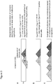
- Figure 1 shows in general terms (ie, diagrammatically) the excitation and fluorescence spectra of Compound of Formula (IIA) (B) (Ex/Em peak at 518/615nm), and a compound of Formula (IA) (A) (Ex/Em peak at 620/660nm). Therefore, the Compound of Formula (IIA) fluorescence is generally in the orange portion of the visible spectrum, whereas compound (IA) fluorescence is generally in the far red portion of the visible spectrum.
- Figure 1b shows the fluorescence spectrum of Compound of Formula (IIA) alone.
- the compound of Formula (IIA) is a cell permeant dye, and therefore it would be expected that the fluorescence signature B would be in connection with live or dead cells.
- Figure 1c shows the fluorescence signature A of compound (IA) alone.
- Compound (IA) is a cell impermeant dye, and therefore the fluorescence signature A would only be observed in connection with dead cells or dying and not live cells.
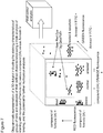
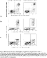
- Figure 2a shows the fluorescence obtained when a certain combination of a cell permeant dye (such as a compound of Formula (IIA)) and a cell impermeant dye (such as compound (IA)) is used in conjunction with live cells. As might perhaps be expected, it is only the fluorescence signature B associated with the cell permeant dye which is observed.
- Figure 2b shows an entirely surprising effect provided by the present invention when certain combinations of cell permeant/cell impermeant dyes such as the compound of Formula (IIA)/compound (IA) combinations are used. It might be expected that a significant contribution of the observed fluorescence would emanate from the compound of Formula (IIA) dye. However, it has been found that with dead cells, little or no fluorescence is observed from the compound of Formula (IIA) dye. Rather, all or virtually all of the observed fluorescence is due to the cell impermeant dye, compound (IA). Therefore, the compound (IA) dye appears to quench the compound of Formula (IIA) signal.
- cell permeant/cell impermeant dyes such as the compound of Formula (IIA)/compound (IA) combinations are used. It might be expected that a significant contribution of the observed fluorescence would emanate from the compound of Formula (IIA) dye. However, it has been found that with dead cells, little or no fluorescence is observed from the compound of
- this quenching of the compound of Formula (IIA) signal appears to occur across the whole of the cell, and not just in the cell nucleus.
- the surprising quenching effect provided by the invention may be due to the cell impermeant dye having a binding affinity for nucleic acid, and, possibly, other macromolecular material in the dead cells, which is higher than that of the cell impermeant dye.
- other mechanisms may play a role.
- the upshot is that it is possible to provide a "traffic light" system to indicate the state of a cell, wherein fluorescence in one spectral region A is associated with dead cells, and fluorescence in another spectral region B is associated with live cells.
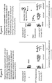
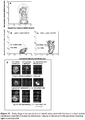
- Figure 3 shows some examples of how a three channel, two colour detection system might be provided with reference to the specific cell permeant/cell impermeant dye combination compound of Formula (IIA)/compound (IA).
- Figure 3a shows fluorescence detected in live cells using the spectral ranges A and B. Fluorescence in the range B is associated with emission from the compound of Formula (IIA) as before.
- compound of Formula (IIA) is used in combination with compound (IA) and a further red dye or light emitting agent preferably associated with intact cells such as Qdot 705nm emitting nanocrystals.
- This system exploits the fact that cells which provide a positive compound of Formula (IIA) signal do not exhibit a signal in the red due to compound (IA), and can be positively identified as live cells.
- the invention comprehends that live cells which have been "tagged” in this way through compound of Formula (IIA) fluorescence in the orange have a potential detection channel in the red region A which is free from interference from compound (IA) emission.
- Figure 3b depicts a detection scheme which exploits the existence of a potential detection channel in dead cells in the orange region B which is substantially free from interference from compound of Formula (IIA) fluorescence. As shown in Figure 3b , the presence of compound (IA) fluorescence in the red spectral region A effectively "tags" a cell as a dead cell.
- a fluorescence spectrum such as that shown in Figure 3b can be obtained, wherein the second orange dye can be used to provide further information about dead cells. It is extremely convenient to utilise three fluorochrome, two colour detection systems of this type, since a large amount of information can be extracted using a relatively simple detection system.
- the invention includes the use of multi colour dye combinations utilising fluorescence in more than two regions of the electromagnetic spectrum.
- Figure 3c depicts a generalised multi colour dye fluorescence scheme, wherein one or more dyes which fluoresce in spectral regions differing from the spectral regions A and B are used.
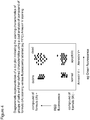
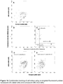
- Figure 4 shows results which might be obtained from a detection system which utilises compound (IA) in combination with an Annexin V assay such as AnnexinV-FITC which fluoresces in the green spectral region.
- This combination of dyes provides enhanced discrimination of the stages of cell death connected with apoptosis.
- a low compound (IA)/low Annexin V signal is indicative of normal cells.
- Apoptotic cells are indicated by an increased Annexin V signal in combination with a low compound (IA) signal.
- the onset of cell death is indicated by the presence of both a high Annexin V signal and a restricted compound (IA) signal. Additional discrimination is provided by a channel comprising a low Annexin V signal and a high compound (IA) signal which is indicative of cellular debris.
- Figure 5 is a plot of the same general type as that shown in Figure 4 , which in this instance shows fluorescence intensity in a detection system which utilises the compound of Formula (IIA)/compound (IA) dye combination. Again, a level of the discrimination is observed in the progression of cells through to cell death. More specifically, when a low compound (IA) fluorescence signal is reported, three distinct "channels" can be identified. A positive signal in the orange for compound of Formula (IIA) in combination with a low compound (IA) signal is indicative of normal cells, whereas an enhanced compound of Formula (IIA) signal is indicative of arrested cells. Note that a negative signal for both compound (IA) and compound of Formula (IIA) is indicative of cellular debris.
- a positive compound (IA) signal in combination with a negative compound of Formula (IIA) signal is indicative of dead cells.
- the skilled reader will appreciate from a consideration of Figure 5 that there are potentially two further channels available, i.e., the combination of a high red signal with either a high orange or an enhanced orange signal. Since the high or enhanced orange signal is associated with the presence of live cells, it is not possible to obtain a high compound (IA) signal in combination with these orange signals. Instead, it is possible to utilise a third fluorochrome which fluoresces in the red region, such as Qdot 705 nm emitting nanocrystals, to provide a two colour, three fluorochrome analysis system.
- An advantage with such a system is that a single laser colour may be used to excite all three fluorochromes, for example using 488nm radiation from an Ar-ion laser.
- Figure 6 is a plot of the same general type as that shown in Figure 5 , which depicts a two colour, three fluorochrome analysis system using Qdot 705 nm emitting nanocrystals in combination with compound (IA) and compound of Formula (IIA). It can be seen that fluorescence in the red spectral region from the Qdot nanocrystals is observed in the two channels which are made available owing to the absence of compound (IA) fluorescence from live cells.
- Figure 7 shows results which can be obtained from a three colour system which is based on the compound of Formula (IIA)/compound (IA) cell permeant/cell impermeant dye combination.
- a third fluorochrome such as Qdot705 nm emitting nanocrystals which fluoresce in the red region of the spectrum is used in order to probe live cells which have been positively labelled through the detection of compound of Formula (IIA) fluorescence.
- a fourth fluorochrome such as AnnexinV-FITC is also used, with detection being made in the green region of the spectrum. This detection arrangement might be characterised as a two laser three colour four fluorochrome analysis technique.
- the result which might be obtained from such a system can be presented using a three axis system to represent the fluorescence obtained in the three colour ranges. Therefore, the results obtained can be understood in terms of a three dimensional volume of data, which provides an enhanced level of discrimination in the progression of cells from normal state through apoptosis and cell death.
- the combination of low compound (IA) signal, enhanced compound of Formula (IIA) signal and high Annexin V-FITC signal is indicative of cells which are both arrested and apoptotic
- the combination of high compound (IA) signal, low compound of Formula (IIA) signal and high Annexin V-FITC signal is indicative of dead cells.
- a further fluorochrome might fluoresce in a different spectral or region to provide an additional colour channel, or, possibly, a fluorochrome which fluoresces in the orange or green spectral regions might be used provided that the detection characteristics of such an additional fluorochrome do not interfere with the compound of Formula (IIA) or AnnexinV-FITC detection channels.
- Green and/or cyan dyes may be used in order to track cell change processes.
- Example 1 Synthesis of Compound of Formula (IIA) [1,5-Bis ⁇ [2-(dimethylamino)ethyl]amino ⁇ anthracene-9,10-dione] and a Compound of Formula (IB) [1,5-Bis ⁇ N-[2-(trimethylamino)ethyl]amino ⁇ anthracene-9,10-dione iodide]
- Compound (IA) was synthesized using the principles described in Example 2 and stored at +4°C as a stock solution of 5 mM compound (IA) dilution in buffer. Absorbance spectra were obtained using a spectrometer and a 20 ⁇ M solution of agent dissolved in PBS and measured in a 1 cm path quartz silica cuvette. Fluorescence spectra for a solution of 20 ⁇ M compound (IA) in a 1 cm path length semi-micro quartz cuvette were determined by excitation at 633, 589, 534, 488 nm. Fluorescence measurements were made on a Perkin Elmer LS50 spectrofluorometer with slit widths set at 10 nm.
- Compound (IA) may be sub-optimally excited by wavelengths from 488 nm (in flow cytometry) and up to 647 nm (ExAmax 646 nm). Typically, for cell imaging, excitation is performed with either 633 nm or 647 nm wavelengths. Emission spectra are independent of excitation wavelength,ie, all the emission spectra are identical irrespective of excitation wavelength.
- This example relates to later stage cell death associated with the translocation of phosphatidylserine molecules from the inner (cytoplasmic) leaflet of the plasma membrane in human B cell lymphoma (DoHH2) cells that have been exposed to a cytotoxic drug (VP-16). Detection of dose-dependent induction of apoptosis was performed using flow cytometry.
- Compound IA was able to detect with similar accuracy as propidium iodide the non-viable fraction. This fraction showed a dose dependent increase upon increased doses of etoposide. Timed uptakes were done to optimise compound (IA) labelling. Dose modification was also performed to determine optimal concentration of compound (IA).
- VP-16 (VP-16-213; VEPESID; Etoposide) was provided as a 34 mM stock solution (Bristol Meyers Pharmaceuticals, Syracuse, NY) and stored at 4°C.
- Fluorescein-conjugated annexin V (annexin V-FITC) was purchased from Pharmingen (Becton Dickinson UK, Oxford, UK.).
- Propidium iodide (PI) was obtained as a 1 mg/ml solution in H 2 O (Molecular Probes Europe, Leiden, The Netherlands).
- Compound (IA) was formulated in water as a 5 mM solution and stored at 4°C.
- Sample preparation for Annexin-V labelling Samples were prepared for the detection of Annexin V-FITC surface binding to cells undergoing apoptotic changes and co-stained with PI or the compound (IA) to detect loss of plasma membrane integrity. Samples were prepared according to Vermes et al . Briefly, cell samples (4x 10 5 cells/ml) were washed with cold PBS and resuspended in 1X binding buffer (10 mM Hepes/NaOH, pH 7.4, 140mM NaCl, 2.5 mM CaCl 2 ) at a concentration of 2 x 10 5 cells/ml.
- 1X binding buffer (10 mM Hepes/NaOH, pH 7.4, 140mM NaCl, 2.5 mM CaCl 2
- a FACS Vantage flow cytometer (Becton Dickinson Immunocytometry Systems, San Jose, CA) equipped with a Coherent Enterprise II argon ion laser having 488nm and multiline UV (351-355nm) outputs (Coherent, Inc., Santa Clara, CA) was used.
- the Enterprise II laser power was regulated at 30mW (monitored on the multiline UV output)
- CELLQuest software (Becton Dickinson Immunocytometry Systems) was used for signal acquisition and analysis.
- FSC Forward scatter
- SSC side scatter
- FITC and PI fluorescent signals derived from 488nm excitation were detected in logarithmic mode at photomultipliers detecting emissions spectrally selected by optical filters; compound (IA) signal derived from 488nm excitation was detected in logarithmic or linear mode but could also be detected following excitation at 633 nm using a third laser conveniently incorporated into the optical system.
- Compound of Formula (IIA) signal derived from excitation at 488 nm was detected in both logarithmic or linear mode. Signals for forward and side scatter and fluorescence were collected for 10,000 cells using the forward light scatter parameter as the master signal. Pulse analysis of fluorescence signals and fluorescence compensation settings were modified to improve recognition of cell subsets in multi-fluor combinations as readily understood in the art. Data are expressed as mean fluorescence intensity (FI) values and are shown in Figure 9 .
- EXAMPLE 5 Prominent nuclear staining of fixed U-2 OS human osteosarcoma cells by a compound of Formula (IA) detected by fluorescence microscopy.
- Chambers were placed onto an Axiovert 100 microscope (Carl Zeiss, Welwyn Garden City, UK and using a 40x, 1.3 NA oil immersion plan apochromat lens).fluorescence images (Ex: 620/60nm; Em 700/75nm) nm captured using an ORCA-ER CCD camera (Hamamatsu, Reading, UK) and MetaMorph (MDS, USA) acquisition software. Cells showed the localization of high levels of compound (IA) in the nucleus.
- the compound (IA) staining of nuclei were segmented using a simple threshold algorithm which depict the nuclei, and provide binary or mask information of each nucleus (object) localization.
- the original compound (IA) nuclear localisation and after segmentation are shown. Results are shown in Figure 10 .
- EXAMPLE 6 The staining of cells with compromised membranes by a compound of Formula (IA) allows for the identification by negative staining of intact cells.
- This example shows the analysis of cell death induction by staurosporine in populations of human Jurkat cells analysed by flow cytometry.
- compound (IA) as a cell viability marker determining the non-viable fraction due to cell enhanced membrane permeability as a result of induced cell death with staurosporine.
- a Jurkat cell line was used in this study Jurkat cultures was routinely maintained in RPMI 1640 supplemented with 10% FCS and 100 U ml-1 penicillin, 100 ⁇ g ml-1 streptomycin, and 2mM glutamine. The cells were passaged twice weekly at an initiating density of 5 ⁇ 10 4 cells ml -1 cultured at 37°C in a humidified atmosphere of 5% CO2/95% air. Cells were set at 0.5x10 5 cells/ml, 5 ml per flask. Cells were exposed to 0 and 2 ⁇ M staurosporine for 24 hours under standard culture conditions to induce cell death. Compound (IA) (3 ⁇ M) from a 5 mM stock was added to each sample and analysed by flow cytometry.
- IA Compound (IA) (3 ⁇ M) from a 5 mM stock was added to each sample and analysed by flow cytometry.
- a FACS Vantage flow cytometer (Becton Dickinson Immunocytometry Systems, San Jose, CA) equipped with a Coherent Enterprise II argon ion laser having 488nm and multiline UV (351-355nm) outputs (Coherent, Inc., Santa Clara, CA) was used.
- the Enterprise II laser power was regulated at 30mW (monitored on the multiline UV output)
- CELLQuest software (Becton Dickinson Immunocytometry Systems) was used for signal acquisition and analysis.
- FSC Forward scatter
- SSC side scatter
- Compound (IA) fluorescent signals derived from 488nm excitation were detected in logarithmic mode at FL3 695LP Signals for forward and side scatter and fluorescence were collected for 10,000 cells using the forward light scatter parameter as the master signal. Data are displayed as contour plots fluorescence intensity (695LP) for compound (IA) against forward scatter signal to indicate cell size. Results are shown in Figure 11 .
- SU-DHL-4 was routinely maintained in RPMI 1640 supplemented with 10% FCS and 100 U ml-1 penicillin, 100 ⁇ gml -1 streptomycin, and 2 mM glutamine. The cells were passaged twice weekly at an initiating density of 5 ⁇ 10 4 cells ml -1 cultured at 37°C in a humidified atmosphere of 5% CO 2 /95% air. Cells were set at 0.5x10 5 cells/ml at 5 ml per flask. Each culture was continuously exposed to one of three compound (IA) doses (0, 3 and 10 ⁇ M) for 96 hours under standard culture conditions.
- IA compound
- cell density was determined by Coulter counting of 0.4 ml samples from each flask. Data are displayed as increase in relative cell number (Nt/N0) (695LP) against time (hours) for the given concentration of compound (IA). The results are shown in Figure 12 .
- EXAMPLE 8 Early stage in human Jurkat cell death associated with the loss of mitochondrial membrane potential in response to the apoptosis inducing agent staurosporine.
- the dye JC-1 (5, 5', 6, 6'-tetrachloro-1, 1', 3, 3'-tetraethylbenzimidazol-carbocyanine iodide) is a lipophilic fluorescent cation that incorporates into the mitochondrial membrane, where it can form aggregates due to the physiological maintenance membrane potential of mitochondria. Aggregation modifies the fluorescence properties of JC-1 leading to a shift from green to orange fluorescence. Flow cytometry or imaging was used to monitor the decrease of the orange fluorescence and an increase of the green fluorescence allowing apoptotic cells to be distinguished from non-apoptotic cells.
- the further incorporation of the preferred cell impermeant dye with red but not orange fluorescence properties allows for the co-identification of those cells already in late stages of cell death associated with loss of membrane integrity providing a finer resolution of the stages of cell death not previously attainable.
- a Jurkat cell line was used in this study Jurkat cultures was routinely maintained in RPMI 1640 supplemented with 10% FCS and 100 U ml-1 penicillin, 100 ⁇ g ml-1 streptomycin, and 2mM glutamine. The cells were passaged twice weekly at an initiating density of 5 ⁇ 10 4 cells ml -1 cultured at 37°C in a humidified atmosphere of 5% CO2/95% air. For the assay, cells were set at 0.5x10 5 cells/ml, 1 ml per well. Cells were exposed to 0 (control conditions) and 1 ⁇ M staurosporine for 4 hours. Cells were washed and exposed to JC-1 (RPM).
- Compound (IA) (3 ⁇ M) from a 5 mM stock was added to each sample. These were then analysed by flow cytometry or placed into a Nunc, 2 Well Lab-Tek II, (Fisher Scientific) and analysed by three channel confocal microscopy.
- Multi-parameter flow cytometry of staurosporine treated cells A FACS Vantage flow cytometer (as for example above). Forward scatter (FSC) and side scatter (SSC) were acquired in linear mode. JC-1 two parameter fluorescent signal were derived from 488 nm excitation were detected in logarithmic mode Green J-monomer was detected using FL1 (530/30 nm emission); Orange J-aggregate was detected using FL2 (585/42 nm emission). Compound (IA) fluorescent signals also derived from 488nm excitation were detected in logarithmic mode at FL3 at 695LP. Signals for forward and side scatter and fluorescence were collected for 10,000 cells using the forward light scatter parameter as the master signal. Results shown in Figure 13 . (A-C).
- JC-1 data are displayed as contour plots fluorescence intensity (585/42 nm) for J-aggregate against fluorescence intensity (530/30 nm) for J-monomer. These are further segmented to derive the cells with high mitochondrial membrane potential (upper region) and cells with low mitochondrial potential (lower region). Cell viability is simultaneously depicted in these two fractions with compound (IA).
- the upper region consists of predominantly live cells and the lower region consists of both non-viable and live cells, compound (IA) functionality can depict these sub-fractions (early (compound (IA) negative) and late apoptosis (compound (IA) positive and permeable).
- the scanning unit was a BioRad Radiance MP system (BioRad Microscience, Hemel Hempstead, UK) linked to a Nikon Eclipse TE300 inverted microscope, using a planapo 60x/1.4 NA oil immersion lens.
- Three channel, three-dimensional (3D) (x,y,z) images were collected using a confocal configuration (pinhole closed). All channels green ( J-monomer excitation at 488 nm emission at 500-530 nm); orange ( J-aggregate excitation at 488 nm emission at 590/70; and red (compound (IA)) (non-viable cell marker excitation at 637 nm emission at 660LP) were collected simultaneously.
- the 3D image sequence were processed into single maximum projection images, and all three channels displayed as J-monomer, J-aggregate and DNA nucleus cells. Results are shown in Figure 13 (D) .
- a human follicular B-lymphoma cell line was used in this study.
- DoHH2 was routinely maintained in RPMI 1640 supplemented with 5% FCS and 100 U ml-1 penicillin, 100 ⁇ gml -1 streptomycin, and 2mM glutamine.
- the cells were passaged twice weekly at an initiating density of 5 ⁇ 10 4 cells ml -1 cultured at 37°C in a humidified atmosphere of 5% CO 2 /95% air.
- Cells were set at 5 ⁇ 10 5 cells/per ml and exposed to VP-16 doses (0.25 ⁇ M) to induce apoptosis.
- 20 ⁇ M Compound (IIA) and 4 ⁇ M compound (IA) were added to 1 ml of cells and incubated under standard culture conditions for 10 minutes. The samples were analysed by flow cytometry.
- a FACS Vantage flow cytometer (as for example above) was used.
- Compound (IIA) and compound (IA) fluorescent signals derived from 488nm excitation were detected in linear mode at FL2 for Compound (IIA) (585/42 nm filters); compound (IA) signal derived from 488nm were detected in linear mode at FL3 with a 695LP filter and a FL1/2 560 nm SP dichroic SP to determine scatter properties.
- Signals for forward and side scatter and fluorescence were collected for 10,000 cells using the forward light scatter parameter as the master signal. Data are expressed as contour plots and are shown in Figure 14 . Contour plots of side versus forward scatter depict two populations of cells.
- Compound (IIA) and compound (IA) provide functional status of these cultures.
- All cells are accounted in the assay using these two co-targetting fluorophores.
- Compound (IA) identifies the non-intact cell fraction, while the Compound (IIA) positive cells represent the live cell (viable) fraction. Note that this viable fraction has two populations further depicting an accrual of an arrested (G2) population.
- the Compound (IIA) fraction represented a unique fraction with a higher forward scatter properties (ie cell size), while the compound (IA) population displayed lower mean forward scatter properties.
Landscapes
- Chemical & Material Sciences (AREA)
- Life Sciences & Earth Sciences (AREA)
- Health & Medical Sciences (AREA)
- Engineering & Computer Science (AREA)
- Organic Chemistry (AREA)
- Molecular Biology (AREA)
- Immunology (AREA)
- Analytical Chemistry (AREA)
- Physics & Mathematics (AREA)
- Biochemistry (AREA)
- General Health & Medical Sciences (AREA)
- Biomedical Technology (AREA)
- Pathology (AREA)
- General Physics & Mathematics (AREA)
- Proteomics, Peptides & Aminoacids (AREA)
- Wood Science & Technology (AREA)
- Zoology (AREA)
- Biotechnology (AREA)
- Microbiology (AREA)
- Hematology (AREA)
- Urology & Nephrology (AREA)
- Materials Engineering (AREA)
- Genetics & Genomics (AREA)
- General Engineering & Computer Science (AREA)
- Biophysics (AREA)
- Bioinformatics & Cheminformatics (AREA)
- Cell Biology (AREA)
- Food Science & Technology (AREA)
- Medicinal Chemistry (AREA)
- Investigating, Analyzing Materials By Fluorescence Or Luminescence (AREA)
- Investigating Or Analysing Biological Materials (AREA)
- Investigating Or Analysing Materials By The Use Of Chemical Reactions (AREA)
- Measuring Or Testing Involving Enzymes Or Micro-Organisms (AREA)
- Organic Low-Molecular-Weight Compounds And Preparation Thereof (AREA)
- Apparatus Associated With Microorganisms And Enzymes (AREA)
Claims (13)
- Verbindung, die die Formel (IA) aufweist


- Fluoreszierender Komplex, enthaltend eine Nukleinsäure und eine Verbindung, wie in Anspruch 1 definiert.
- Verfahren zum Analysieren einer Probe von Zellen oder anderem biologischem, Nukleinsäure enthaltendem Material, das die Schritte beinhaltet:a) Herstellen einer biologisch verträglichen Lösung, enthaltend eine Verbindung, wie in Anspruch 1 definiert;b) Behandeln einer Probe von Zellen oder anderem biologischem Material mit der biologisch verträglichen Lösung; undc) Nachweisen einer mit der Absorption von elektromagnetischer Strahlung durch die Verbindung von Formel (IA) oder (IB) verbundenen spektroskopischen Eigenschaft.
- Verfahren nach Anspruch 3, in dem Schritt b) ausgeführt wird, um die Probe von Zellen mit der Verbindung der Formel (IA) oder (IB) anzufärben.
- Verfahren nach Anspruch 4, in dem Zelltod-Entstehung verfolgt wird, wobei Schritt b) vor oder während eines Assayzeitraums ausgeführt wird, wodurch eine kontinuierliche oder häufige Auslesung von Zelltod-Entstehung während des Assayzeitraums ermöglicht wird.
- Verfahren nach Anspruch 5 zum Unterscheiden zwischen intakten und nicht-intakten Zellen, wobei die Verbindung der Formel (IA) oder (IB) zellundurchdringend ist, Schritt b) weiter Behandeln der Probe mit einem zweiten Fluorochrom oder einer lichtemittierenden Verbindung, die zelldurchdringend ist, beinhaltet und Schritt c) weiter Nachweisen einer mit der Absorption von elektromagnetischer Strahlung durch das zweite Fluorochrom oder die lichtemittierende Verbindung verbundenen spektroskopischen Eigenschaft beinhaltet, wobei der Nachweis der mit der Absorption von elektromagnetischer Strahlung durch die Verbindung der Formel (IA) oder (IB) verbundenen spektroskopischen Eigenschaft mit der Anwesenheit von nicht-intakten Zellen korreliert, und der Nachweis der mit der Absorption von elektromagnetischer Strahlung durch das zweite Fluorochrom oder die lichtemittierende Verbindung verbundenen spektroskopischen Eigenschaft mit der Anwesenheit von intakten Zellen korreliert.
- Verfahren nach Anspruch 6, in dem das zweite Fluorochrom oder die lichtemittierende Verbindung eine Verbindung der Formel (II):
 wobei: A eine C2-8-Alkylengruppe ist; R1, R2 und R3 unabhängig ausgewählt sind aus Wasserstoff, C1-4-Alkyl, C2-4-Dihydroxyalkyl, in dem das Kohlenstoffatom, an das das Stickstoffatom gebunden ist, keine Hydroxylgruppe trägt und kein Kohlenstoffatom mit zwei Hydroxylgruppen substituiert ist, oder R2 und R3 zusammen eine C2-6-Alkylengruppe bilden, die mit dem Stickstoffatom, an das R2 und R3 gebunden sind, einen heterocyclischen Ring bilden; undX1, X2 und X3 unabhängig ausgewählt sind aus Wasserstoff, Hydroxyl, NR1-A-NR2R3, Halogenamino, C1-4-Alkyloxy oder C2-8-Alkanoyloxy.
 - Verfahren zum Unterscheiden zwischen intakten und nicht-intakten Zellen, das die Schritte beinhaltet:a) Herstellen einer biologisch verträglichen Lösung, enthaltend eine Verbindung, wie in Anspruch 1 definiert;b) Herstellen einer biologisch verträglichen Lösung, enthaltend ein zelldurchdringendes Fluorochrom oder eine lichtemittierende Verbindung, die ein Bindungspotenzial für Nukleinsäure und/oder anderes makromolekulares Material in den unterschiedenen Zellen aufweist, das geringer als jenes von dem zellundurchdringenden Fluorochrom oder der lichtemittierenden Verbindung ist, und als eine Folge weniger effizient in der Anwesenheit von dem zellundurchdringenden Fluorochrom oder der lichtemittierenden Verbindung zum Binden an die Nukleinsäure und/oder anderem makromolekularem Material in den unterschiedenen Zellen konkurriert, so dass das zelldurchdringende Fluorochrom oder die lichtemittierende Verbindung im Wesentlichen vom Binden an nicht-intakte Zellen ausgeschlossen oder durch das zellundurchdringende Fluorochrom oder die lichtemittierende Verbindung maskiert ist;c) Behandeln einer Probe von Zellen mit der biologisch verträglichen Lösung oder Lösungen; undd) Nachweisen einer spektroskopischen Eigenschaft, die mit der Absorption von elektromagnetischer Strahlung durch das zellundurchdringende Fluorochrom oder die lichtemittierende Verbindung verbunden ist und Korrelieren derselben mit der Anwesenheit von nicht-intakten Zellen und Nachweisen einer spektroskopischen Eigenschaft, die mit der Absorption von elektromagnetischer Strahlung durch das zelldurchdringende Fluorochrom oder die lichtemittierende Verbindung verbunden ist und Korrelieren derselben mit der Anwesenheit von intakten Zellen.
- Zusammensetzung, beinhaltend ein Gemisch von einer Verbindung nach Anspruch 1 mit mindestens einem zweiten Fluorochrom oder einer lichtemittierenden Verbindung.
- Zusammensetzung nach Anspruch 9, wobei das zweite Fluorochrom oder die lichtemittierende Verbindung die Formel (II) aufweist

wobei
A eine C2-8-Alkylengruppe ist; R1, R2 und R3 unabhängig ausgewählt sind aus Wasserstoff, C1-4-Alkyl, C2-4-Dihydroxyalkyl, in dem das Kohlenstoffatom, an das das Stickstoffatom gebunden ist, keine Hydroxylgruppe trägt und kein Kohlenstoffatom mit zwei Hydroxylgruppen substituiert ist, oder R2 und R3 zusammen eine C2-6-Alkylengruppe bilden, die mit dem Stickstoffatom, an das R2 und R3 gebunden sind, einen heterocyclischen Ring bilden; und
X1, X2 und X3 unabhängig ausgewählt sind aus Wasserstoff, Hydroxyl, NR1-A-NR2R3, Halogenamino, C1-4-Alkyloxy oder C2-8-Alkanoyloxy. - Zusammensetzung nach Anspruch 10, wobei in der Verbindung der Formel (II) X2 NR1-A-NR2R3 ist, aber X1 und X3 nicht.
- Zusammensetzung nach Anspruch 11, in welcher die Verbindung der Formel (II) die Formel (IIA) aufweist

- Verfahren zur Herstellung einer Verbindung nach Anspruch 1, beinhaltend die Schritte: Bereitstellen einer Aminoalkylamino-Vorstufen-Verbindung für die Verbindung der Formel (IA) oder (IB); und Quaternisieren der Aminoalkylamino-Vorstufen-Verbindung.
Priority Applications (3)
| Application Number | Priority Date | Filing Date | Title |
|---|---|---|---|
| DK18167949.9T DK3366667T3 (da) | 2010-04-09 | 2011-04-08 | Fremgangsmåde til analyse af en celle eller andet biologisk materiale indeholdende nukleinsyre |
| EP18167949.9A EP3366667B1 (de) | 2010-04-09 | 2011-04-08 | Verfahren zur analyse einer zelle oder von anderem biologischem material mit einer nukleinsäure |
| PL11720571T PL2556047T3 (pl) | 2010-04-09 | 2011-04-08 | Sposób analizy komórki lub innego materiału biologicznego zawierającego kwas nukleinowy |
Applications Claiming Priority (2)
| Application Number | Priority Date | Filing Date | Title |
|---|---|---|---|
| GBGB1005939.2A GB201005939D0 (en) | 2010-04-09 | 2010-04-09 | Method of analysing a cell or other biological material containing nucleic acid |
| PCT/GB2011/050702 WO2011124927A1 (en) | 2010-04-09 | 2011-04-08 | Method of analysing a cell or other biological material containing a nucleic acid |
Related Child Applications (2)
| Application Number | Title | Priority Date | Filing Date |
|---|---|---|---|
| EP18167949.9A Division EP3366667B1 (de) | 2010-04-09 | 2011-04-08 | Verfahren zur analyse einer zelle oder von anderem biologischem material mit einer nukleinsäure |
| EP18167949.9A Division-Into EP3366667B1 (de) | 2010-04-09 | 2011-04-08 | Verfahren zur analyse einer zelle oder von anderem biologischem material mit einer nukleinsäure |
Publications (2)
| Publication Number | Publication Date |
|---|---|
| EP2556047A1 EP2556047A1 (de) | 2013-02-13 |
| EP2556047B1 true EP2556047B1 (de) | 2018-06-06 |
Family
ID=42236073
Family Applications (2)
| Application Number | Title | Priority Date | Filing Date |
|---|---|---|---|
| EP18167949.9A Active EP3366667B1 (de) | 2010-04-09 | 2011-04-08 | Verfahren zur analyse einer zelle oder von anderem biologischem material mit einer nukleinsäure |
| EP11720571.6A Active EP2556047B1 (de) | 2010-04-09 | 2011-04-08 | Verfahren zur analyse einer zelle oder von anderem biologischem material mit einer nukleinsäure |
Family Applications Before (1)
| Application Number | Title | Priority Date | Filing Date |
|---|---|---|---|
| EP18167949.9A Active EP3366667B1 (de) | 2010-04-09 | 2011-04-08 | Verfahren zur analyse einer zelle oder von anderem biologischem material mit einer nukleinsäure |
Country Status (16)
| Country | Link |
|---|---|
| US (2) | US9051467B2 (de) |
| EP (2) | EP3366667B1 (de) |
| JP (2) | JP6033767B2 (de) |
| KR (2) | KR101957288B1 (de) |
| CN (1) | CN102947266B (de) |
| AU (2) | AU2011236610B2 (de) |
| BR (1) | BR112012025820B1 (de) |
| CA (1) | CA2797440C (de) |
| DK (2) | DK2556047T3 (de) |
| ES (1) | ES2676351T3 (de) |
| GB (1) | GB201005939D0 (de) |
| HU (1) | HUE038706T2 (de) |
| NO (1) | NO2556047T3 (de) |
| PL (1) | PL2556047T3 (de) |
| PT (1) | PT2556047T (de) |
| WO (1) | WO2011124927A1 (de) |
Families Citing this family (14)
| Publication number | Priority date | Publication date | Assignee | Title |
|---|---|---|---|---|
| GB201005939D0 (en) * | 2010-04-09 | 2010-05-26 | Biostatus Ltd | Method of analysing a cell or other biological material containing nucleic acid |
| WO2012138805A2 (en) | 2011-04-04 | 2012-10-11 | Biotium, Inc. | Substituted anthraquinone dyes for cellular stains and enzyme detection |
| JP2013094135A (ja) * | 2011-11-02 | 2013-05-20 | Waseda Univ | 人工ナノ材料の安全性を評価する方法 |
| CN104379760B (zh) * | 2012-04-05 | 2018-03-16 | Bd公司 | 用于流式细胞术的样品制备 |
| US9933341B2 (en) | 2012-04-05 | 2018-04-03 | Becton, Dickinson And Company | Sample preparation for flow cytometry |
| CN104755931B (zh) | 2012-05-02 | 2016-08-31 | 查尔斯河实验室公司 | 用于检测细胞样品中的活细胞的方法 |
| WO2013166336A1 (en) | 2012-05-02 | 2013-11-07 | Charles River Laboratories, Inc. | Viability staining method |
| WO2013166338A2 (en) | 2012-05-02 | 2013-11-07 | Charles River Laboratories, Inc. | Cell capture system and use thereof |
| GB201214169D0 (en) | 2012-08-08 | 2012-09-19 | Biostatus Ltd | New compounds and uses thereof |
| WO2015095152A1 (en) * | 2013-12-17 | 2015-06-25 | The Penn State Research Foundation | Manipulation of light spectral quality to reduce parasitism by cuscuta and other plant parasites |
| GB201610971D0 (en) | 2016-06-23 | 2016-08-10 | Biostatus Ltd | Compounds |
| CN113667323B (zh) * | 2019-11-21 | 2023-07-14 | 苏州优逸兰迪生物科技有限公司 | 一种染料化合物的制备方法 |
| CN112679543B (zh) * | 2020-12-29 | 2022-07-22 | 天津全和诚科技有限责任公司 | 一种亲脂性荧光素探针及其制备方法 |
| CN113072937B (zh) * | 2021-04-12 | 2022-06-07 | 郑州大学 | 一种脂滴靶向碳点、制备方法及应用 |
Citations (2)
| Publication number | Priority date | Publication date | Assignee | Title |
|---|---|---|---|---|
| GB2027195A (en) * | 1978-07-13 | 1980-02-13 | Oreal | Evaluation of the state of change of keratin fibres |
| WO2000034379A1 (en) * | 1998-12-07 | 2000-06-15 | Eastman Chemical Company | A colorant composition, a polymer nanocomposite comprising the colorant composition and articles produced therefrom |
Family Cites Families (20)
| Publication number | Priority date | Publication date | Assignee | Title |
|---|---|---|---|---|
| US3123605A (en) * | 1960-01-08 | 1964-03-03 | Basic anthraquinone dyestuffs | |
| NL302452A (de) * | 1962-10-29 | |||
| FR1379649A (fr) * | 1963-06-20 | 1964-11-27 | Oreal | Nouveaux colorants anthraquinoniques et leur utilisation pour la teinture des cheveux |
| GB1053300A (de) * | 1962-10-29 | |||
| JPS503337B1 (de) * | 1964-08-06 | 1975-02-03 | ||
| US3646072A (en) * | 1970-07-27 | 1972-02-29 | Du Pont | Turquoise biscationic anthraquinone dyes |
| JPS503337A (de) | 1973-05-11 | 1975-01-14 | ||
| US4123222A (en) | 1976-03-23 | 1978-10-31 | Ciba-Geigy Corporation | Process for the dyeing or printing of polyacrylonitrile material |
| FR2431128A1 (fr) * | 1978-07-13 | 1980-02-08 | Oreal | Procede d'evaluation de l'etat ionique des cheveux |
| US5057413A (en) * | 1988-06-13 | 1991-10-15 | Becton, Dickinson And Company | Method for discriminating between intact and damaged cells in a sample |
| GB8923075D0 (en) | 1989-10-13 | 1989-11-29 | Patterson Laurence H | Anti-cancer compounds |
| JPH04136802A (ja) * | 1990-09-28 | 1992-05-11 | Nippon Kayaku Co Ltd | 着色フィルター |
| US5520707A (en) * | 1995-08-07 | 1996-05-28 | Clairol, Inc. | Methods for dyeing hair with anthraquinone hair dyes having a quaternary ammonium side chain |
| GB9813062D0 (en) * | 1998-06-18 | 1998-08-19 | Univ Wales Medicine | An anthraquinone and its derivatives |
| US6562785B1 (en) * | 1999-03-23 | 2003-05-13 | Howard M. Shapiro | Method for overcoming bacterial antibiotic resistance |
| USRE40572E1 (en) * | 2000-02-28 | 2008-11-11 | Sekisui Medical Co., Ltd. | Fluorescent probe for the quantitation of zinc |
| US6949250B2 (en) * | 2002-12-16 | 2005-09-27 | The United States Of America As Represented By The Secretary Of Agriculture | Selective algaecides for control of cyanochloronta |
| US20070082377A1 (en) | 2005-10-07 | 2007-04-12 | Olin Michael R | Cytotoxicity assays |
| US9334281B2 (en) * | 2008-09-08 | 2016-05-10 | Enzo Life Sciences, Inc. | Fluorochromes for organelle tracing and multi-color imaging |
| GB201005939D0 (en) * | 2010-04-09 | 2010-05-26 | Biostatus Ltd | Method of analysing a cell or other biological material containing nucleic acid |
-
2010
- 2010-04-09 GB GBGB1005939.2A patent/GB201005939D0/en not_active Ceased
-
2011
- 2011-04-08 PL PL11720571T patent/PL2556047T3/pl unknown
- 2011-04-08 JP JP2013503178A patent/JP6033767B2/ja active Active
- 2011-04-08 KR KR1020187009266A patent/KR101957288B1/ko active IP Right Grant
- 2011-04-08 DK DK11720571.6T patent/DK2556047T3/en active
- 2011-04-08 US US13/639,872 patent/US9051467B2/en active Active
- 2011-04-08 CA CA2797440A patent/CA2797440C/en active Active
- 2011-04-08 PT PT117205716T patent/PT2556047T/pt unknown
- 2011-04-08 BR BR112012025820-6A patent/BR112012025820B1/pt active IP Right Grant
- 2011-04-08 HU HUE11720571A patent/HUE038706T2/hu unknown
- 2011-04-08 ES ES11720571.6T patent/ES2676351T3/es active Active
- 2011-04-08 KR KR1020127029033A patent/KR101846491B1/ko active IP Right Grant
- 2011-04-08 NO NO11720571A patent/NO2556047T3/no unknown
- 2011-04-08 CN CN201180028424.3A patent/CN102947266B/zh active Active
- 2011-04-08 EP EP18167949.9A patent/EP3366667B1/de active Active
- 2011-04-08 WO PCT/GB2011/050702 patent/WO2011124927A1/en active Application Filing
- 2011-04-08 DK DK18167949.9T patent/DK3366667T3/da active
- 2011-04-08 EP EP11720571.6A patent/EP2556047B1/de active Active
- 2011-04-08 AU AU2011236610A patent/AU2011236610B2/en active Active
-
2015
- 2015-06-05 US US14/731,647 patent/US11255761B2/en active Active
-
2016
- 2016-10-26 AU AU2016250384A patent/AU2016250384B2/en active Active
- 2016-10-26 JP JP2016209377A patent/JP6586070B2/ja active Active
Patent Citations (2)
| Publication number | Priority date | Publication date | Assignee | Title |
|---|---|---|---|---|
| GB2027195A (en) * | 1978-07-13 | 1980-02-13 | Oreal | Evaluation of the state of change of keratin fibres |
| WO2000034379A1 (en) * | 1998-12-07 | 2000-06-15 | Eastman Chemical Company | A colorant composition, a polymer nanocomposite comprising the colorant composition and articles produced therefrom |
Non-Patent Citations (4)
| Title |
|---|
| CHEMICAL ABSTRACTS, vol. 67, no. 117854, 22 April 2001, Columbus, Ohio, US; abstract no. 61:11785, ICHIKAWA, M.; OKAZAKI, M.: "Synthesis of cationic dyes of the anthraquinone series. I. Quaternary ammonium salts derived from 1,4-disubstituted anthraquinones" page 138; * |
| ICHIKAWA, M.; OKAZAKI, M.: "Synthesis of cationic dyes of the anthraquinone series. I. Quaternary ammonium salts derived from 1,4-disubstituted anthraquinones", KOGYO KAGAKU ZASSHI, vol. 67, 1 January 1964 (1964-01-01), pages 138 - 142 * |
| LUXAMI V ET AL: "Quaternary ammonium salt-based chromogenic and fluorescent chemosensors for fluoride ions", TETRAHEDRON LETTERS, PERGAMON, GB, vol. 49, no. 27, 30 June 2008 (2008-06-30), pages 4265 - 4268, XP022695658, ISSN: 0040-4039, [retrieved on 20080501], DOI: 10.1016/J.TETLET.2008.04.147 * |
| PERRY P J ET AL: "Human Telomerase", 19980101, vol. 41, no. 24, 1 January 1998 (1998-01-01), pages 4873 - 84, XP002188538 * |
Also Published As
Similar Documents
| Publication | Publication Date | Title |
|---|---|---|
| EP2556047B1 (de) | Verfahren zur analyse einer zelle oder von anderem biologischem material mit einer nukleinsäure | |
| AU2011236610A1 (en) | Method of analysing a cell or other biological material containing a nucleic acid | |
| Xu et al. | Recent advances in formaldehyde-responsive fluorescent probes | |
| Wang et al. | Specifically and wash-free labeling of SNAP-tag fused proteins with a hybrid sensor to monitor local micro-viscosity | |
| CN105542756B (zh) | 一种检测甲醛的荧光探针及其制备方法与应用 | |
| KASTEN | Probes: Properties, History and Applications | |
| WO2012083064A1 (en) | Silaanthracene as a red and near infrared sensor and a method to manufacture such a sensor | |
| Langlois et al. | Interactions between pairs of DNA-specific fluorescent stains bound to mammalian cells. | |
| CN106220640A (zh) | 一类汞离子荧光探针及其制备方法和应用 | |
| Li et al. | A novel 3-Hydroxychromone fluorescence sensor for intracellular Zn2+ and its application in the recognition of prostate cancer cells | |
| CN110229165A (zh) | 上转换荧光探针罗丹明衍生物及其应用 | |
| KR20050120668A (ko) | 형광성을 향상시키는 방법 | |
| Zhang et al. | A robust camphor-based colorimetric and fluormetric dual-modal probe with a large Stokes shift for real-time monitoring of endogenous labile Fe2+ in vivo and in vitro | |
| Jensen et al. | Strategies for choosing a deoxyribonucleic acid stain for flow cytometry of metaphase chromosomes. | |
| Berrones Reyes et al. | Platinum (II)‐Based Optical Probes for Imaging Quadruplex DNA Structures via Phosphorescence Lifetime Imaging Microscopy | |
| CN108715847A (zh) | 一种基于dna增强孔雀石绿荧光的快速检测法及其应用 | |
| CN108863961A (zh) | 一种三氮唑类蒽醌衍生物银离子荧光探针及其制备方法和应用 | |
| Jia et al. | Spectrophotometric determination of copper (II) in water based on fluorescein diacetate | |
| Jin | Background-free cytometry using rare earth complex bioprobes | |
| Sevillano-Arredondo et al. | A turn-on fluorescence sensor based on water-soluble amino-pyridyl-benzothiazole-derivatives for measuring cellular Zn2+ | |
| CN104535546B (zh) | 脱氢枞酸基芳胺化合物作为金属离子荧光探针的应用 | |
| Gasser M et al. | Sensitive ratiometric sensor for Al (III) detection in water samples using luminescence or eye-vision | |
| Thiel | From Imaging to Proteomics–Using Photoactivatable Probes to Study Enzymes | |
| Chen | T Luminescence Imaging and Biosensing in Cells and Tissues | |
| Shapiro | Fluorescent probes |
Legal Events
| Date | Code | Title | Description |
|---|---|---|---|
| PUAI | Public reference made under article 153(3) epc to a published international application that has entered the european phase |
Free format text: ORIGINAL CODE: 0009012 |
|
| 17P | Request for examination filed |
Effective date: 20121031 |
|
| AK | Designated contracting states |
Kind code of ref document: A1 Designated state(s): AL AT BE BG CH CY CZ DE DK EE ES FI FR GB GR HR HU IE IS IT LI LT LU LV MC MK MT NL NO PL PT RO RS SE SI SK SM TR |
|
| DAX | Request for extension of the european patent (deleted) | ||
| 17Q | First examination report despatched |
Effective date: 20150911 |
|
| STAA | Information on the status of an ep patent application or granted ep patent |
Free format text: STATUS: EXAMINATION IS IN PROGRESS |
|
| GRAP | Despatch of communication of intention to grant a patent |
Free format text: ORIGINAL CODE: EPIDOSNIGR1 |
|
| STAA | Information on the status of an ep patent application or granted ep patent |
Free format text: STATUS: GRANT OF PATENT IS INTENDED |
|
| INTG | Intention to grant announced |
Effective date: 20171212 |
|
| GRAS | Grant fee paid |
Free format text: ORIGINAL CODE: EPIDOSNIGR3 |
|
| GRAA | (expected) grant |
Free format text: ORIGINAL CODE: 0009210 |
|
| STAA | Information on the status of an ep patent application or granted ep patent |
Free format text: STATUS: THE PATENT HAS BEEN GRANTED |
|
| RAP1 | Party data changed (applicant data changed or rights of an application transferred) |
Owner name: BIOSTATUS LIMITED |
|
| AK | Designated contracting states |
Kind code of ref document: B1 Designated state(s): AL AT BE BG CH CY CZ DE DK EE ES FI FR GB GR HR HU IE IS IT LI LT LU LV MC MK MT NL NO PL PT RO RS SE SI SK SM TR |
|
| REG | Reference to a national code |
Ref country code: GB Ref legal event code: FG4D |
|
| REG | Reference to a national code |
Ref country code: CH Ref legal event code: EP Ref country code: AT Ref legal event code: REF Ref document number: 1005921 Country of ref document: AT Kind code of ref document: T Effective date: 20180615 |
|
| REG | Reference to a national code |
Ref country code: IE Ref legal event code: FG4D |
|
| REG | Reference to a national code |
Ref country code: DE Ref legal event code: R096 Ref document number: 602011049024 Country of ref document: DE |
|
| REG | Reference to a national code |
Ref country code: PT Ref legal event code: SC4A Ref document number: 2556047 Country of ref document: PT Date of ref document: 20180711 Kind code of ref document: T Free format text: AVAILABILITY OF NATIONAL TRANSLATION Effective date: 20180704 |
|
| REG | Reference to a national code |
Ref country code: DK Ref legal event code: T3 Effective date: 20180711 |
|
| REG | Reference to a national code |
Ref country code: ES Ref legal event code: FG2A Ref document number: 2676351 Country of ref document: ES Kind code of ref document: T3 Effective date: 20180718 |
|
| REG | Reference to a national code |
Ref country code: SE Ref legal event code: TRGR |
|
| REG | Reference to a national code |
Ref country code: NL Ref legal event code: FP |
|
| REG | Reference to a national code |
Ref country code: NO Ref legal event code: T2 Effective date: 20180606 |
|
| REG | Reference to a national code |
Ref country code: LT Ref legal event code: MG4D |
|
| PG25 | Lapsed in a contracting state [announced via postgrant information from national office to epo] |
Ref country code: LT Free format text: LAPSE BECAUSE OF FAILURE TO SUBMIT A TRANSLATION OF THE DESCRIPTION OR TO PAY THE FEE WITHIN THE PRESCRIBED TIME-LIMIT Effective date: 20180606 Ref country code: BG Free format text: LAPSE BECAUSE OF FAILURE TO SUBMIT A TRANSLATION OF THE DESCRIPTION OR TO PAY THE FEE WITHIN THE PRESCRIBED TIME-LIMIT Effective date: 20180906 Ref country code: CY Free format text: LAPSE BECAUSE OF FAILURE TO SUBMIT A TRANSLATION OF THE DESCRIPTION OR TO PAY THE FEE WITHIN THE PRESCRIBED TIME-LIMIT Effective date: 20180606 |
|
| REG | Reference to a national code |
Ref country code: HU Ref legal event code: AG4A Ref document number: E038706 Country of ref document: HU |
|
| PG25 | Lapsed in a contracting state [announced via postgrant information from national office to epo] |
Ref country code: HR Free format text: LAPSE BECAUSE OF FAILURE TO SUBMIT A TRANSLATION OF THE DESCRIPTION OR TO PAY THE FEE WITHIN THE PRESCRIBED TIME-LIMIT Effective date: 20180606 Ref country code: RS Free format text: LAPSE BECAUSE OF FAILURE TO SUBMIT A TRANSLATION OF THE DESCRIPTION OR TO PAY THE FEE WITHIN THE PRESCRIBED TIME-LIMIT Effective date: 20180606 Ref country code: GR Free format text: LAPSE BECAUSE OF FAILURE TO SUBMIT A TRANSLATION OF THE DESCRIPTION OR TO PAY THE FEE WITHIN THE PRESCRIBED TIME-LIMIT Effective date: 20180907 Ref country code: LV Free format text: LAPSE BECAUSE OF FAILURE TO SUBMIT A TRANSLATION OF THE DESCRIPTION OR TO PAY THE FEE WITHIN THE PRESCRIBED TIME-LIMIT Effective date: 20180606 |
|
| REG | Reference to a national code |
Ref country code: AT Ref legal event code: MK05 Ref document number: 1005921 Country of ref document: AT Kind code of ref document: T Effective date: 20180606 |
|
| PG25 | Lapsed in a contracting state [announced via postgrant information from national office to epo] |
Ref country code: CZ Free format text: LAPSE BECAUSE OF FAILURE TO SUBMIT A TRANSLATION OF THE DESCRIPTION OR TO PAY THE FEE WITHIN THE PRESCRIBED TIME-LIMIT Effective date: 20180606 Ref country code: IS Free format text: LAPSE BECAUSE OF FAILURE TO SUBMIT A TRANSLATION OF THE DESCRIPTION OR TO PAY THE FEE WITHIN THE PRESCRIBED TIME-LIMIT Effective date: 20181006 Ref country code: EE Free format text: LAPSE BECAUSE OF FAILURE TO SUBMIT A TRANSLATION OF THE DESCRIPTION OR TO PAY THE FEE WITHIN THE PRESCRIBED TIME-LIMIT Effective date: 20180606 Ref country code: AT Free format text: LAPSE BECAUSE OF FAILURE TO SUBMIT A TRANSLATION OF THE DESCRIPTION OR TO PAY THE FEE WITHIN THE PRESCRIBED TIME-LIMIT Effective date: 20180606 Ref country code: SK Free format text: LAPSE BECAUSE OF FAILURE TO SUBMIT A TRANSLATION OF THE DESCRIPTION OR TO PAY THE FEE WITHIN THE PRESCRIBED TIME-LIMIT Effective date: 20180606 Ref country code: RO Free format text: LAPSE BECAUSE OF FAILURE TO SUBMIT A TRANSLATION OF THE DESCRIPTION OR TO PAY THE FEE WITHIN THE PRESCRIBED TIME-LIMIT Effective date: 20180606 |
|
| PG25 | Lapsed in a contracting state [announced via postgrant information from national office to epo] |
Ref country code: SM Free format text: LAPSE BECAUSE OF FAILURE TO SUBMIT A TRANSLATION OF THE DESCRIPTION OR TO PAY THE FEE WITHIN THE PRESCRIBED TIME-LIMIT Effective date: 20180606 |
|
| REG | Reference to a national code |
Ref country code: DE Ref legal event code: R097 Ref document number: 602011049024 Country of ref document: DE |
|
| PLBE | No opposition filed within time limit |
Free format text: ORIGINAL CODE: 0009261 |
|
| STAA | Information on the status of an ep patent application or granted ep patent |
Free format text: STATUS: NO OPPOSITION FILED WITHIN TIME LIMIT |
|
| 26N | No opposition filed |
Effective date: 20190307 |
|
| PG25 | Lapsed in a contracting state [announced via postgrant information from national office to epo] |
Ref country code: SI Free format text: LAPSE BECAUSE OF FAILURE TO SUBMIT A TRANSLATION OF THE DESCRIPTION OR TO PAY THE FEE WITHIN THE PRESCRIBED TIME-LIMIT Effective date: 20180606 |
|
| PG25 | Lapsed in a contracting state [announced via postgrant information from national office to epo] |
Ref country code: AL Free format text: LAPSE BECAUSE OF FAILURE TO SUBMIT A TRANSLATION OF THE DESCRIPTION OR TO PAY THE FEE WITHIN THE PRESCRIBED TIME-LIMIT Effective date: 20180606 |
|
| PG25 | Lapsed in a contracting state [announced via postgrant information from national office to epo] |
Ref country code: MC Free format text: LAPSE BECAUSE OF FAILURE TO SUBMIT A TRANSLATION OF THE DESCRIPTION OR TO PAY THE FEE WITHIN THE PRESCRIBED TIME-LIMIT Effective date: 20180606 |
|
| PG25 | Lapsed in a contracting state [announced via postgrant information from national office to epo] |
Ref country code: TR Free format text: LAPSE BECAUSE OF FAILURE TO SUBMIT A TRANSLATION OF THE DESCRIPTION OR TO PAY THE FEE WITHIN THE PRESCRIBED TIME-LIMIT Effective date: 20180606 |
|
| PG25 | Lapsed in a contracting state [announced via postgrant information from national office to epo] |
Ref country code: MT Free format text: LAPSE BECAUSE OF FAILURE TO SUBMIT A TRANSLATION OF THE DESCRIPTION OR TO PAY THE FEE WITHIN THE PRESCRIBED TIME-LIMIT Effective date: 20180606 |
|
| PG25 | Lapsed in a contracting state [announced via postgrant information from national office to epo] |
Ref country code: MK Free format text: LAPSE BECAUSE OF FAILURE TO SUBMIT A TRANSLATION OF THE DESCRIPTION OR TO PAY THE FEE WITHIN THE PRESCRIBED TIME-LIMIT Effective date: 20180606 |
|
| PGFP | Annual fee paid to national office [announced via postgrant information from national office to epo] |
Ref country code: PL Payment date: 20230329 Year of fee payment: 13 |
|
| PGFP | Annual fee paid to national office [announced via postgrant information from national office to epo] |
Ref country code: PT Payment date: 20230404 Year of fee payment: 13 Ref country code: NO Payment date: 20230405 Year of fee payment: 13 Ref country code: IT Payment date: 20230421 Year of fee payment: 13 Ref country code: IE Payment date: 20230405 Year of fee payment: 13 Ref country code: FR Payment date: 20230407 Year of fee payment: 13 Ref country code: ES Payment date: 20230505 Year of fee payment: 13 Ref country code: DK Payment date: 20230410 Year of fee payment: 13 Ref country code: DE Payment date: 20230419 Year of fee payment: 13 Ref country code: CH Payment date: 20230502 Year of fee payment: 13 |
|
| PGFP | Annual fee paid to national office [announced via postgrant information from national office to epo] |
Ref country code: SE Payment date: 20230413 Year of fee payment: 13 Ref country code: HU Payment date: 20230412 Year of fee payment: 13 Ref country code: FI Payment date: 20230405 Year of fee payment: 13 |
|
| PGFP | Annual fee paid to national office [announced via postgrant information from national office to epo] |
Ref country code: BE Payment date: 20230411 Year of fee payment: 13 |
|
| PGFP | Annual fee paid to national office [announced via postgrant information from national office to epo] |
Ref country code: GB Payment date: 20240320 Year of fee payment: 14 |
|
| PGFP | Annual fee paid to national office [announced via postgrant information from national office to epo] |
Ref country code: LU Payment date: 20240425 Year of fee payment: 14 |
|
| PGFP | Annual fee paid to national office [announced via postgrant information from national office to epo] |
Ref country code: NL Payment date: 20240425 Year of fee payment: 14 |