EP2535716B1 - Cancer marker and therapeutic target - Google Patents
Cancer marker and therapeutic target Download PDFInfo
- Publication number
- EP2535716B1 EP2535716B1 EP12183760.3A EP12183760A EP2535716B1 EP 2535716 B1 EP2535716 B1 EP 2535716B1 EP 12183760 A EP12183760 A EP 12183760A EP 2535716 B1 EP2535716 B1 EP 2535716B1
- Authority
- EP
- European Patent Office
- Prior art keywords
- ccr4
- tumour
- cancer
- cells
- ccl17
- Prior art date
- Legal status (The legal status is an assumption and is not a legal conclusion. Google has not performed a legal analysis and makes no representation as to the accuracy of the status listed.)
- Not-in-force
Links
Images


Classifications
-
- G—PHYSICS
- G01—MEASURING; TESTING
- G01N—INVESTIGATING OR ANALYSING MATERIALS BY DETERMINING THEIR CHEMICAL OR PHYSICAL PROPERTIES
- G01N33/00—Investigating or analysing materials by specific methods not covered by groups G01N1/00 - G01N31/00
- G01N33/48—Biological material, e.g. blood, urine; Haemocytometers
- G01N33/50—Chemical analysis of biological material, e.g. blood, urine; Testing involving biospecific ligand binding methods; Immunological testing
- G01N33/74—Chemical analysis of biological material, e.g. blood, urine; Testing involving biospecific ligand binding methods; Immunological testing involving hormones or other non-cytokine intercellular protein regulatory factors such as growth factors, including receptors to hormones and growth factors
-
- A—HUMAN NECESSITIES
- A61—MEDICAL OR VETERINARY SCIENCE; HYGIENE
- A61K—PREPARATIONS FOR MEDICAL, DENTAL OR TOILETRY PURPOSES
- A61K31/00—Medicinal preparations containing organic active ingredients
-
- A—HUMAN NECESSITIES
- A61—MEDICAL OR VETERINARY SCIENCE; HYGIENE
- A61P—SPECIFIC THERAPEUTIC ACTIVITY OF CHEMICAL COMPOUNDS OR MEDICINAL PREPARATIONS
- A61P35/00—Antineoplastic agents
-
- C—CHEMISTRY; METALLURGY
- C07—ORGANIC CHEMISTRY
- C07K—PEPTIDES
- C07K16/00—Immunoglobulins [IGs], e.g. monoclonal or polyclonal antibodies
- C07K16/18—Immunoglobulins [IGs], e.g. monoclonal or polyclonal antibodies against material from animals or humans
- C07K16/28—Immunoglobulins [IGs], e.g. monoclonal or polyclonal antibodies against material from animals or humans against receptors, cell surface antigens or cell surface determinants
- C07K16/2866—Immunoglobulins [IGs], e.g. monoclonal or polyclonal antibodies against material from animals or humans against receptors, cell surface antigens or cell surface determinants against receptors for cytokines, lymphokines, interferons
-
- G—PHYSICS
- G01—MEASURING; TESTING
- G01N—INVESTIGATING OR ANALYSING MATERIALS BY DETERMINING THEIR CHEMICAL OR PHYSICAL PROPERTIES
- G01N33/00—Investigating or analysing materials by specific methods not covered by groups G01N1/00 - G01N31/00
- G01N33/48—Biological material, e.g. blood, urine; Haemocytometers
- G01N33/50—Chemical analysis of biological material, e.g. blood, urine; Testing involving biospecific ligand binding methods; Immunological testing
- G01N33/5005—Chemical analysis of biological material, e.g. blood, urine; Testing involving biospecific ligand binding methods; Immunological testing involving human or animal cells
-
- G01N33/575—
-
- G01N33/5759—
-
- G—PHYSICS
- G01—MEASURING; TESTING
- G01N—INVESTIGATING OR ANALYSING MATERIALS BY DETERMINING THEIR CHEMICAL OR PHYSICAL PROPERTIES
- G01N2333/00—Assays involving biological materials from specific organisms or of a specific nature
- G01N2333/435—Assays involving biological materials from specific organisms or of a specific nature from animals; from humans
- G01N2333/705—Assays involving receptors, cell surface antigens or cell surface determinants
- G01N2333/72—Assays involving receptors, cell surface antigens or cell surface determinants for hormones
- G01N2333/726—G protein coupled receptor, e.g. TSHR-thyrotropin-receptor, LH/hCG receptor, FSH
-
- G—PHYSICS
- G01—MEASURING; TESTING
- G01N—INVESTIGATING OR ANALYSING MATERIALS BY DETERMINING THEIR CHEMICAL OR PHYSICAL PROPERTIES
- G01N2800/00—Detection or diagnosis of diseases
- G01N2800/52—Predicting or monitoring the response to treatment, e.g. for selection of therapy based on assay results in personalised medicine; Prognosis
-
- G—PHYSICS
- G01—MEASURING; TESTING
- G01N—INVESTIGATING OR ANALYSING MATERIALS BY DETERMINING THEIR CHEMICAL OR PHYSICAL PROPERTIES
- G01N2800/00—Detection or diagnosis of diseases
- G01N2800/56—Staging of a disease; Further complications associated with the disease
Definitions
- the invention relates to oncology and in vitro methods of cancer diagnosis, stratification, disease staging and determining response to treatment.
- the field of the invention therefore concerns markers of predictive or clinical value in cancer diagnosis.
- Chemokine receptors and their ligands direct the trafficking of cells in normal tissue homeostasis and in disease, influencing cell motility, invasiveness and survival [1].
- chemokines in the diseased tissues contribute to the rolling, tethering and invasion of leucocytes from the blood vessels through the endothelial cell basement membrane and into the parenchyma [2].
- CCR4 is one of 18 known chemokine receptors.
- Chemokine receptors are generally expressed on immune cells and in the tumour microenvironment a number of receptors and their ligands are present in the immune cell infiltrate.
- chemokine receptors receptors that are not usually found on their normal counterparts.
- Metastatic cancer cells are thought to gain characteristics of chemokine receptor-expressing leucocytes, using chemokines to aid their migration to, and survival at, sites distant to the original tumour [3, 4, 5]. Inappropriate presence on cancer cells of chemokine receptors that usually have a highly restricted pattern of expression further supports the hypothesis that specific chemokine receptors may help cells spread to, and/or survive in, different metastatic sites [8].
- CXCR5 is normally restricted to B cells and some T cell
- pancreatic cancer cells where it is implicated in the establishment of liver metastases; the liver being a site of production of the CXCR5 ligand, CXCL13 [9].
- Melanoma cells that have metastasised to the intestine express CCR9 [10].
- the CCR9 ligand CCL25 recruits rare T cell subsets to the intestine.
- pancreatic cancer expression of CCR6 has been observed [38][39].
- CCR6 expression has also been reported in human renal carcinoma, together with CCR3 and CXCR2 [40].
- CXCR4 chemokine receptor 4
- CXCR4 is commonly found on malignant cells in many advanced human cancers.
- Woerner et al found that CXCR4 was also present in the early stages of disease in glioblastoma [20].
- using a phospho-specific anti-CXCR4 antibody they found that in the less malignant Grade 1 lesions, the level of receptor activation was much lower.
- RET/PTC1 oncogene is necessary and sufficient for malignant transformation of primary thyrocytes [24].
- This oncogene induces a pro-inflammatory programme in the thyrocytes that includes induction of functional CXCR4.
- Alveolar rhabdomyosarcoma is a highly aggressive tumour characterised by recurrent PAX3 and PAX7-FKHR gene fusions. Transfer of PAX3-FKHR into embryonal rhabdomyosarcoma cells also activates CXCR4 expression [Libura, 2002 #9346].
- CCR4 expression is generally restricted to the immune system, and is known as a marker of Th2 and regulatory T cells. In the tumour environment, these cells act to suppress cytotoxic T cells and dendritic cell maturation, hence suppressing antitumour immune responses. In addition, CCR4 has been shown to be expressed in haematological malignancies, including by a high proportion of adult T cell lymphomas (ATL), and was a significant prognostic factor associated with metastasis to skin [35], [41]. As such, CCR4 is of interest as a therapeutic target in ATL [37], [42].
- ATL adult T cell lymphomas
- CCR4 expression by adult T cell leukaemia is associated with skin metastases; its ligands CCL17 and CCL22 are produced by both malignant cells and the skin tumor microenvironment [36].
- Ishida et al have developed an anti-CCR4 monoclonal antibody therapeutic for the treatment of adult T cell lymphoma that induces ADCC activity against the tumor cells and may also act on immunosuppressive malignant Treg cells found in this disease [37].
- CCR4 positive solid tumor cell line is the human lung cancer cell line SBC-5 [34]. These cells migrated towards CCL22 gradients and in bone metastatic SBC-5 xenografts there was close co-localisation of osteoclasts expressing CCL22 and SBC-5 cells expressing CCR4. There are no reports of CCR4 expression in primary human tumour cells.
- WO05106471 (BAYER HEALTHCARE AG) discloses screening methods for agents of potential use in treating a wide range of diseases; specifically consisting of cardiovascular disorders, gastrointestinal and liver diseases, inflammatory diseases, metabolic diseases, haematological disorders, cancer disorders, neurological disorders, respiratory diseases and reproduction disorders in a mammal.
- the screening method determines the degree of binding or otherwise of candidate agents to CCR4.
- the cells and tissues were obtained from disparate sources and just an isolated few were cancerous cells/tissues; e.g. thyroid, ileum, HeLa, Jurkat, lung and breast cancer cells.
- WO9623068 discloses a chemokine receptor able to bind to Monocyte Chemotactic Protein-1 (MCP-1 / CCL2), Macrophage Inflammatory Protein 1 ⁇ (MIP1 ⁇ / CCL3) and/or 'RANTES' (Regulated upon Activation, Normal T-cell Expressed, and Secreted / CCL5).
- MCP-1 / CCL2 Monocyte Chemotactic Protein-1
- MIP1 ⁇ / CCL3 Macrophage Inflammatory Protein 1 ⁇
- 'RANTES' 'RANTES' (Regulated upon Activation, Normal T-cell Expressed, and Secreted / CCL5).
- a nucleotide and an amino-acid sequence for CCR4 are disclosed (CC-CKR-4 / K5.5. K5.5 and CC-CKR-4 are alternative names for CCR4.)
- the expression of CCR4 is discovered in a relatively limited range of normal human tissues and in a range of T-cell samples.
- WO0041724A1 (LELAND STANFORD / LEUKOSITE) proposes the modulation of systemic memory T cell trafficking by administration of CCR4 modulating agents. This is intended as a treatment for inflammatory skin disease. Substances capable of modulating CCR4 binding to its ligands are used in in vitro tests to show how T-cell migration is affected.
- Antibodies reactive against CCR4 are known.
- WO0164754 (Kyowa Hakko Kogyo) discloses a recombinant antibody or fragment thereof allegedly reactive specifically with the extracellular domain of CCR4. Also disclosed is a polypeptide sequence of such an antibody. There is also disclosed an antibody which reacts with a CCR4 positive cell and is cytotoxic or causes antibody-dependent cell-mediated cytotoxicity (ADCC.) These antibodies are proposed for the use in the treatment of Th2-mediated immune diseases or blood cancer, specifically leukaemia.
- WO05035582 (Kyowa Hakko Kogyo) discloses an antibody capable of specifically binding CCR4 and also discloses a CCR4 antibody which has a complex N-linked glycosylation in the Fc region. Also disclosed are antibodies to the extracellular domains of CCR4.
- WO03018635 discloses 'Human CDR-grafted antibodies and fragments'.
- a specific CDR (complementarity determining region) which binds specifically to CCR4 is disclosed.
- the antibodies are proposed for use in the diagnosis or treatment of Th2-mediated immune diseases or cancers such as blood cancers.
- WO05053741 discloses a medicament comprising a recombinant antibody, which specifically binds CCR4, in combination with at least one other agent.
- the antibody is proposed for the treatment of tumours, specifically haematopoietic organ tumours.
- WO0042074 (MILLENIUM PHARMACEUTICALS) discloses antibodies to CCR4 and antibodies that can compete with their binding. No specific diagnostic applications are disclosed. Therapy of inflammatory disorders is proposed.
- WO04007472 (ONO PHARMACEUTICAL CO.) discloses a small molecule tricyclic compound with anti-CCR4 activity.
- WO05023771 (ONO PHARMACEUTICAL CO.) discloses small molecule nitrogen-containing heterocyclic compounds with anti-CCR4 activity.
- WO02094264 discloses specific compounds with CCR4 inhibitory activities.
- WO0230358 discloses various CCR4-binding compounds and uses for treatment of various diseases, but not including cancer.
- WO0230357 discloses compounds that are antagonists of CCR4. This application describes uses for the treatment of inflammatory diseases and conditions.
- WO051236976 discloses quinazoline derivatives as CCR4 regulators.
- WO05085212 (YAMANOUCHI PHARMACEUTICAL CO., LTD.) discloses pyrimidine derivatives as CCR4 modulators.
- WO05082865 (YAMANOUCHI PHARMACEUTICAL CO., LTD.) discloses fused bicyclic pyrimidine derivatives as CCR4 function-controlling agents.
- WO04108717 discloses sulphonamide compounds that modulate chemokine (specifically CCR4) receptor activity.
- EP1633729 discloses sulphonamide compounds that modulate chemokine (specifically CCR4) receptor activity.
- WO03014153 discloses another technology in the art, a method of modulating viral infection of a cell by modulating the interaction between chemokine receptors (including CCR4) and a virus.
- WO2004/045526 discloses antibodies to particular chemokines and chemokine receptors and their use in inhibiting the growth and metastasis of cancer cells. Antibodies were raised against the particular chemoldne receptors and their ligands, which does not include CCR4. Also described are methods of testing for over-expression of particular chemokines in a tumour and the suggestion that such tumours can be treated by administering antibodies against the particular over-expressed chemokine or chemokine receptor.
- WO99/15666 discloses nucleotide sequences and polypeptide sequences of a macrophage-derived C-C chemokine designated 'Macrophage Derived Chemokine' (MDC).
- MDC appears synonymous with CCL22.
- TARC appears synonymous with CCL17.
- Methods for the recombinant or synthetic production of MDC protein or polypeptide fragments are described.
- antibodies reactive with MDC as well assays for identifying modulators of MDC and TARC chemokine activity.
- Wagsater et al. Quantification of the chemokines CCL17 and CCL22 in human colorectal adenocarcinoma), MOLECULAR MEDICINE REPORTS, SPANDIDOS PUBLICATIONS, vol. 1, no. 2, pages 211-217 , assessed the role of CCL17 and CCL22 protein expression in colorectal cancer (CRC) and sought to ascertain whether an association exists between certain gene polymorphisms in CRC versus non-CRC subjects.
- CRC colorectal cancer
- Cervical cancer is the second most common type of cancer in women worldwide. Symptoms are often absent until the cancer is at a late stage and hence cervical cancer has been the subject of an intense population screening program using the Pap smear, which can detect pre-malignant changes by histopathology. Although an abnormal Pap smear indicates possible cervical neoplasia, it is insufficient for diagnosis, which is subsequently carried out by biopsy and additional invasive procedures ('colposcopy'). In total, 24,000 women are referred in the UK each year with abnormal Pap smears. The Pap smear has only 70% sensitivity, hence a significant proportion of women with cervical cancer or pre-invasive lesions remain undiagnosed. Therefore, more accurate screening methods are required to i) allow screening to be more automated and less subjective ii) to improve the sensitivity of screening.
- HPV Human Papilloma Virus
- a biomarker characteristic of one cancer type may be shared with other cancer types thus the use of a biomarker may extend beyond the original cancer type it was found to be associated with.
- the stage of a cancer is a descriptor (usually numbers I to IV) of how much the cancer has spread.
- the stage often takes into account the size of a tumour, how deep it has penetrated, whether it has invaded adjacent organs, if and how many lymph nodes it has metastasized to, and whether it has spread to distant organs.
- Staging of cancer is important because the stage at diagnosis is the most powerful predictor of survival, and treatments are often changed based on the stage.
- Correct staging is critical because treatment is directly related to disease stage. Thus, incorrect staging would lead to improper treatment, and material diminution of patient survivability. Correct staging, however, can be difficult to achieve.
- Pathologic staging where a pathologist examines sections of tissue, can be particularly problematic for two specific reasons: visual discretion and random sampling of tissue.
- “Visual discretion” means being able to identify single cancerous cells intermixed with healthy cells on a slide. Oversight of one cell can mean mis-staging and lead to serious, unexpected spread of cancer. "Random sampling” refers to the fact that samples are chosen at random from patients' lymph nodes and are examined. If cancerous cells present in the lymph node happen not to be present in the slices of tissue viewed, incorrect staging and improper treatment can result.
- chemokine receptor CCR4 expression is an early event in carcinogenesis.
- the inventors have discovered that the expression of two ligands of CCR4, CCL17 and CCL22, increases during tumour progression.
- Also disclosed herein is a method of obtaining information of predictive or diagnostic character for a cancer patient, comprising the step of measuring the amount and/or activity of chemokine receptor CCR4 expressed by tumour cells in a solid tumour sample or in a non-haematological cell tumour sample taken from the patient, the amount and/or activity of CCR4 providing the information of predictive or diagnostic character.
- Haematological tumours are derived from blood cells, including immune cells and include leukaemias and lymphomas of various types.
- the invention does not therefore concern haematological tumours.
- the invention concerns tumors selected from cervical, oesophageal, bronchial, nasopharyngeal, laryngeal, skin, brain, pancreatic, neck, kidney, liver, breast, bladder, stomach, ovarian, germ cell and prostate cancer.
- any reference herein to a "solid or non-haematological cancer” in relation to any aspects of the present invention should be understood to be limited to the specific cancer types listed above.
- CCR4 is expressed by cells of the tumour.
- the methods of the invention therefore concern CCR4-expressing cancers in which CCR4 is expressed by samples of patient tumour cells (or reference cells) and substantially not by cells of the immune system.
- CCR4 arises in any patent tumour samples from an undesired source, such as infiltrating immune cells, the amount and/or activity of CCR4 being measured is either not significant or it is controlled for in any measurements being made.
- a reference amount and/or level of activity of CCR4 may be measured in one or more non-tumour samples.
- The, or at least one non- tumour sample may be taken from the patient.
- a reference amount and/or level of activity of CCR4 is determined from non-tumour cells of the patient, a single sample of non-tumour tissue may be taken from the patient. If desired, a multiplicity of non-tumour samples can be taken from different locations of the same patient.
- the reference amount may therefore be a mean figure determined from a number of samples taken from the patient.
- the one or more non-tumour samples are optionally not taken from the patient.
- Such samples may be taken from other patients and may include cultured tumour cell lines.
- the present invention provides in one aspect a method of predicting whether a solid or non-haematological tumour which expresses chemokine receptor CCR4 will be susceptible to an anti-cancer treatment, comprising the step of measuring the amount and/or activity of CCR4 ligand CCL17 in a sample of said tumour taken from the patient, comparing the amount and/or activity of CCL17 in said tumour sample with a reference amount and/or level of activity of CCL17 measured in, or pre-determined from, one or more non-tumour samples and predicting whether said tumour will be susceptible to an anti-cancer treatment, wherein an increased amount and/or activity of CCL17 compared to the reference amount and/or activity is indicative of a tumour that will be susceptible to an anti-cancer treatment, and wherein said tumour is selected from cervical, oesophageal, bronchial, nasopharyngeal, laryngeal, skin, brain, pancreatic, neck, kidney, liver, breast, bladder, stomach, ovarian, germ cell
- it provides a method of identifying a malignant cancer in a patient, comprising the step of measuring the amount and/or activity of CCR4 ligand CCL17 in a sample of a tumour taken from the patient, comparing the amount and/or activity of CCL17 in said tumour sample with a reference amount and/or level of activity of CCL17 measured in, or pre-determined from, one or more non-tumour samples and making a diagnosis, wherein an increased amount and/or activity of CCL17 compared to the reference amount and/or activity is indicative of malignant cancer, and wherein said tumour is selected from cervical, oesophageal, bronchial, nasopharyngeal, laryngeal, skin, brain, pancreatic, neck, kidney, liver, breast, bladder, stomach, ovarian, germ cell and prostate cancer.
- it provides a method of determining whether a solid or non-haematological tumour which expresses CCR4 has responded to an anti-cancer treatment, wherein the patient has received an anti-cancer treatment and measurements of the amount and/or activity of CCL17 are made before and after the start of treatment and the information obtained is used to determine whether the solid tumour or the non-haematological cell tumour of the patient has responded to the anti-cancer treatment and wherein said tumour is selected from cervical, oesophageal, bronchial, nasopharyngeal, laryngeal, skin, brain, pancreatic, neck, kidney, liver, breast, bladder, stomach, ovarian, germ cell and prostate cancer.
- the information of predictive or diagnostic character may be obtained by comparing the amount and/or activity of CCL17 and optionally CCL22 in the solid tumour sample or in the non-haematological cell tumour sample with a reference amount and/or level of activity of CCL17 and optionally CCL22.
- the reference amount and/or level of activity of CCL17 and optionally CCL22 may be measured in one or more non-tumour samples.
- non-tumour samples may be taken from the patient or from a different patient or source, including cultured cell lines.
- the information may be used to predict whether the solid tumour or the non-haematological cell tumour of the patient will be susceptible to an anti-cancer treatment.
- the patient has received an anti-cancer treatment and measurements of the amount and/or activity of CCL17 and optionally CCL22 are made before and after the start of treatment and the information obtained is used to determine whether the solid tumour or the non-haematological cell tumour of the patient has responded to the anti-cancer treatment.
- a particular anti-cancer treatment may comprise an agent which modulates or inhibits CCR4 expression or activity.
- Disclosed herein is a method of treating a cancer patient having a solid tumour or a non-haematological tumour expressing CCR4, comprising administering an effective amount of an agent which modulates or inhibits CCR4 expression or activity.
- the agent may be administered in the form of a pharmaceutical formulation.
- suitable formulations include sterile aqueous or non-aqueous solutions, suspensions, and emulsions.
- the compositions may further comprise auxiliary agents or excipients, as known in the art, see, e.g., Berkow et al., The Merck Manual, 16th edition Merck & Co., Rahman, NJ (1992 ), Avery's Drug Treatment: Principles and Practice of Clinical Pharmacology and Therapeutics, 3rd edition, ADIS Press, Ltd., Williams and Wilkins, Baltimore, MD (1987 ) & Osol (ed.), Remington's Pharmaceutical Sciences, Mack Publishing Co., Easton, PA 1324-1341 (1980 ).
- the pharmaceutical compositions administered in accordance with the invention are preferably presented in the form of individual doses (unit doses).
- composition or medicament may further comprise salts, buffers, or other substances which are desirable for improving the efficacy of the composition.
- the administration of composition or medicament in accordance with the invention may be local or systemic.
- the agent which modulates or inhibits CCR4 expression or activity may be:
- compositions and formulations of active agents include those described in the aforementioned publications.
- the invention further provides a method according to claim 1 for determining whether a cancer patient is suitable for treatment with an anti-cancer agent.
- the suitability of a cancer patient for treatment with a particular therapeutic agent is governed by a multiplicity of factors, some inter-related.
- Patient age, sex, stage of the cancer, type of cancer, genetic make up of patient, lifestyle factors, such at diet or smoking, may all impact on the potential outcome of a given treatment regime.
- Stratification is usually undertaken in order to group patients on the basis of a multiplicity of selected parameters that can allow predictions to be made in terms of clinical outcome for a group of patients or an individual patient falling within a group.
- an increased level or activity of CCL17 and optionally CCL22 in a patient tumour sample is indicative of a patient for whom treatment with the anti-cancer agents disclosed herein is beneficial.
- the invention further includes a method of monitoring the efficiency of an anticancer treatment according to claim 4.
- the sampling of tumour cells may take place before, during and/or subsequent to the anti-cancer agent being administered.
- the invention therefore also provides a method according to claim 1.
- Increased levels of activity and/or expression of CCL17 and optionally CCL22, whether in absolute terms on a standardized basis having regard to reference values, or whether on a relative (standardized) basis as between (a) tumour/non-tumour cells or (b) tumour cells over time, is generally indicative of a tumour susceptible to treatment with the anti-CCR4 agents disclosed herein.
- Samples obtained from patients are preferably biopsy samples.
- a biopsy is a medical test involving the removal of cells or tissues for examination. The tissue is generally examined under a microscope by a pathologist and/or may be analyzed chemically using techniques well known in the art to assess protein or RNA levels. When a smaller sample of tissue is removed, the procedure is called an incisional biopsy or core biopsy. When an entire lump or suspicious area is removed, the procedure is called an excisional biopsy. When a sample of tissue or fluid is removed with a needle, the procedure is called a needle aspiration biopsy.
- tumor samples may be obtained from patients by other methods well known in the art, including but not limited to, samples of cells taken by a 'smear' test.
- a smear test for example a Papancolaou test, also called a Pap smear or cervical smear test
- a smear test may be used to sample cells from a patient.
- cells are collected and removed from the surface of the tissue being tested by means of physical contact with an Aylesbury spatula, plastic fronded 'broom' or other instrument.
- the cells collected in a sample taken from a patient may be processed immediately or preserved in a suitable storage medium for later processing.
- a suitable storage medium for later processing.
- the cells are often preserved in an ethanol based storage medium for later processing and analysis.
- the sample may be treated for the purposes of preservation or for maximising the accuracy and/or reliability of the signal obtained by analysis of the sample.
- the cancers are those that give rise to solid humours selected from the group consisting of bronchial, nasopharyngeal, laryngeal, skin (e.g. melanoma or basal cell carcinoma), brain, pancreatic, neck, kidney, liver, breast, bladder, oesophagus, stomach, cervical, ovarian, germ cell and prostate. More preferably the cancers are cancers of the cervix, oesophagus, kidney, brain, breast and ovary.
- the cancer may be a carcinoma, preferably a squamous cell carcinoma (SCC) or adenocarcinoma, preferably selected from cancers of the cervix, oesophagus, kidney, brain, breast and ovary.
- SCC squamous cell carcinoma
- adenocarcinoma preferably selected from cancers of the cervix, oesophagus, kidney, brain, breast and ovary.
- an increased level of CCL17 and optionally CCL22 produced by the tumour cells identifies a malignant cancer or a prospectively malignant cancer.
- the level of CCL17 and optionally CCL22 and/or CCR4 produced in non-tumour cells may be determined and the level in tumour and non-tumour cells compared.
- the level of CCL17 and optionally CCL22 and/or CCR4 in tumour cells may be compared with pre-determined levels.
- Pre-determined levels may be derived from normal non- cancerous tissue, earlier stage cancerous tissue, data obtained from databases or directly from available biological material or samples.
- CCL17 and optionally CCL22 and/or CCR4 may be employed in methods of the invention.
- the protein level and/or activity of CCR4 and/or CCL17 and/or CCL22 may be used as a measure of the gene products of CCR4 and/or CCL17 and/or CCL22 in the sample.
- the protein level of CCL17 and optionally CCR4 and/or CCL22 is measured using an antibody reactive against CCR4, CCL17 and CCL22 respectively, preferably a specific antibody, e.g. a monoclonal antibody.
- the location and amount of specific proteins can be detected by microscopy and histological techniques. Using sample preparation, staining and probing techniques well known in the art, the structure of cells can be shown and specific proteins associated with them can be detected and their location within the sample found.
- Histochemical stains are well known in the art and may be used to show cell morphology and/or more specific cellular components. Commonly used stains include hematoxylin (which stains nucleic acids and ergastoplasm, blue) and eosin (which stains elastic and reticular fibres, pink).
- Immunohistochemistry is a technique whereby antibodies to specific proteins are used for detection of said proteins in samples. Their binding of antibody to antigen in the sample can be detected in a number of ways.
- the most standard method is to conjugate an enzyme that catalyses a colour changing reaction (e.g. alkaline phosphatase, horseradish peroxidase) to the antibody, thus the use of a suitable chromogenic substrate allows visualisation of the location of the antigen under the light microscope.
- a colour changing reaction e.g. alkaline phosphatase, horseradish peroxidase
- a variation upon this method is immunofluorescence whereby the antibody is conjugated to a fluorophore (e.g. FITC, rhodamine, Texas Red) that emits a detectable signal when excited by a suitable source of energy. Normally this is light of a specific wavelength.
- Immunofluorescence is advantageous because the use of multiple fluorophores attached to different antibodies allows detection of multiple targets within a sample and is particularly suitable for confocal laser scanning microscopy, which is highly sensitive and can also be used to visualise interactions between multiple proteins.
- detection of the specific antigen is done by a, multiply staged, indirect method.
- An unlabelled or unconjugated 'primary' antibody, raised against the antigen being tested for is used to bind said antigen.
- This 'primary' antibody may then be detected by a 'secondary' antibody conjugated to a detectable marker and raised such that it will react with the immunoglobulin of the species that the 'primary' antibody was raised in.
- Measurement of protein levels using antibodies may use techniques such as ELISA (Enzyme-linked Tmmunosorbent Assay), RIA (Radioimmunoassay), EMIT (Enzyme Multiplied Immunoassay Technique), protein microarray analysis, flow cytometry, western blotting, dot blotting or slot blotting, preferably the methodology is quantitative.
- ELISA Enzyme-linked Tmmunosorbent Assay
- RIA Radioimmunoassay
- EMIT Enzyme Multiplied Immunoassay Technique
- protein microarray analysis flow cytometry, western blotting, dot blotting or slot blotting, preferably the methodology is quantitative.
- Flow cytometry is a technique for counting, examining, and sorting microscopic particles suspended in a stream of fluid. It allows simultaneous multiparametric analysis of the physical and/or chemical characteristics of single cells flowing through an optical and/or electronic detection apparatus.
- Fluorescence-activated cell-sorting is a specialised type of flow cytometry. It provides a method for sorting a heterogeneous mixture of biological cells into two or more containers, one cell at a time, based upon the specific light scattering and fluorescent characteristics of each cell. It is a useful scientific instrument as it provides fast, objective and quantitative recording of fluorescent signals from individual cells as well as physical separation of cells of particular interest.
- the population of cells in a sample is normally heterogeneous. In order to detect the differences between cells, they are treated with chemical and immunochemical techniques similar to those of histochemistry. Immunochemical detection of antigens may be done using antibodies labeled with fluorophores such as FITC, Cy5 and GFP. Staining the cells with dyes (such the DNA binding dyes SYBR-Green and DAPI) may be used to detect differences such as cell size or cell cycle stage within and between samples. Using these techniques in combination allows different cells within the heterogeneous to be given specific fluorescence profiles that are distinguishable by the flow cytometer. In this way cells expressing particular antigens or associated with particular light scattering profiles may be detected and their prevalence, within the sample population, measured.
- fluorophores such as FITC, Cy5 and GFP.
- dyes such as DNA binding dyes SYBR-Green and DAPI
- the level of CCL17 and optionally CCR4 and/or CCL22 may be determined by measuring the level of mRNA encoding CCR4 and/or CCL17 and/or CCL22 as a measure of the level of the gene products of CCR4 and/or CCL17 and/or CCL22 in the sample.
- the mRNA level is measured by a quantitative polymerase chain reaction (qPCR) method, preferably a qPCR method where the template is the product of a reverse transcriptase reaction (RT-qPCR.)
- qPCR quantitative polymerase chain reaction
- RT-qPCR reverse transcriptase reaction
- mRNA is extracted from the sample and reverse transcribed to produce cDNA prior to qPCR.
- the level of transcription of CCL17 and optionally CCL22 and/or CCR4 is measured using a nuclease protection assay, preferably the probe used is specific for CCR4 and/or CCL17 and/or CCL22.
- the mRNA level is measured using a DNA microarray.
- a DNA microarray (also known as gene or genome chip, DNA chip, or gene array) is a collection of microscopic DNA spots, commonly representing single genes, arrayed on a solid surface by covalent attachment to chemically suitable matrices.
- Qualitative or quantitative measurements with DNA microarrays utilize the selective nature of DNA-DNA or DNA-RNA hybridization under high-stringency conditions. Fluorophore-based detection may be used to determine the degree of hybridisation from which a quantitative measurement may be calculated.
- the cancer is a malignant cancer.
- the cancer may be a pre-malignant cancer.
- the method of the invention can advantageously identify the stage to which cancer in a patient has progressed, thereby permitting identification of the most appropriate course of treatment.
- CCR4 receptor as a marker for the identification and/or staging of cancer.
- the CCR4 receptor may be detected by means of an antibody, preferably a specific antibody, e.g. a monoclonal antibody.
- the invention provides for the use of CCL17 ligand as a marker for the identification and/or staging of cancer.
- the CCL17 ligand may be detected by means of an antibody, preferably a specific antibody, e.g. a monoclonal antibody.
- the invention optionally additionally also provides for the use of CCL22 ligand as a marker for the identification and/or staging of cancer.
- the CCL22 ligand may be detected by means of an antibody, preferably a specific antibody, e.g. a monoclonal antibody.
- An anti-cancer treatment may comprise an antibody specific for CCR4, and may be a monoclonal antibody, for example, the antibodies disclosed in WO0164754 (Kyowa Hakko Kogyo), WO05035582 (Kyowa Hakko Kogyo), WO03018635 (Kyowa Hakko Kogyo), WO05053741 (Kyowa Hakko Kogyo) or WO0042074 (MILLENIUM PHARMACEUTICALS); or an antibody specific for CCL17, which may be a monoclonal antibody, or an antibody specific for CCL22, which may be a monoclonal antibody.
- the antibody may be a Fab fragment wherein said Fab fragment may be selected from the group consisting of: scFv, F(ab') 2 , Fab, Fv and Fd fragments; or CDR3 regions.
- the fragment antigen binding is a region on an antibody which binds to antigens. It is composed of one constant and one variable domain of each of the heavy and the light chain. These domains shape the paratope - the antigen binding site - at the amino terminal end of the monomer. The two variable domains bind the epitope on their specific antigens.
- Fc and Fab fragments can be generated.
- the enzyme papain can be used to cleave an immunoglobulin monomer into two Fab fragments and an Fc fragment.
- the enzyme pepsin cleaves below the hinge region, so a F(ab')2 fragment and a Fc fragment may be formed.
- the variable regions of the heavy and light chains can be fused together to form a single chain variable fragment (scFv), which is only half the size of the Fab fragment yet retains the original specificity of the parent immunoglobulin.
- a complementarity determining region is a short amino acid sequence found in the variable domains of antigen receptor (e.g. immunoglobulin and T cell receptor) proteins that complements an antigen and therefore provides the receptor with its specificity for that particular antigen.
- antigen receptor e.g. immunoglobulin and T cell receptor
- Most of the sequence variation associated with immunoglobulins and T cell receptors are found in the CDR regions, these regions are sometimes referred to as hypervariable domains.
- CDR3 shows the greatest variability as it is encoded by a recombination of the VJ regions.
- the antibodies may be humanised or chimeric antibodies.
- Humanized antibodies or chimeric antibodies are a type of monoclonal antibody that are synthesized using recombinant DNA technology to circumvent the clinical problem of immune response to foreign antigens.
- the standard procedure of producing monoclonal antibodies yields mouse antibodies.
- murine antibodies are very similar to human antibodies the differences are significant enough that the human immune system recognizes mouse antibodies as foreign, rapidly removing them from circulation and causing systemic inflammatory effects.
- Humanized antibodies may be produced by merging the DNA that encodes the binding portion of a monoclonal mouse antibody with human antibody-producing DNA. Mammalian cell cultures are then used to express this DNA and produce these part-mouse and part-human antibodies that are not as immunogenic as the purely murine variety.
- Modifications may be made to monoclonal antibodies that bind only to cell-specific antigens and preferably induce an immunological response against the target cancer cell.
- Such monoclonal antibodies are preferably modified for delivery of a toxin, radioisotope, cytokine or other active conjugate.
- bispecific antibodies may be designed that can bind with their Fab regions both to target antigen and to a conjugate or effector cell. Also, all intact antibodies can bind to cell receptors or other proteins with their Fc region.
- the production of recombinant monoclonal antibodies may also involve technologies, referred to as repertoire cloning or phage display/yeast display. These may involve the use of viruses or yeast to create antibodies, rather than mice. These techniques rely on rapid cloning of immunoglobulin gene segments to create libraries of antibodies with slightly different amino acid sequences from which antibodies with desired specificities can be selected. This process can be used to enhance the specificity with which antibodies recognize antigens, alter their stability in various environmental conditions, increase their therapeutic efficacy, and modulate their detectability in diagnostic applications.
- Also disclosed is a method of treating or preventing malignant disease in an individual suffering from cancer comprising treating the individual with an effective amount of a small molecule inhibitor of CCR4 and/or CCL17 and/or CCL22.
- Exemplary small molecule inhibitors are disclosed in WO04007472 (ONO PHARMACEUTICAL CO.), WO05023771 (ONO PHARMACEUTICAL CO.), WO02094264 (TULARIK INC.), WO0230358 (TULARIK / CHEMOCENTRYX), WO0230357 (CHEMOCENTRYX), WO051236976 (ASTELLAS PHARMA INC.), WO05085212 (YAMANOUCHI PHARMACEUTICAL CO., LTD.), WO05082865 (YAMANOUCHI PHARMACEUTICAL CO., LTD.), WO04108717 (ASTRAZENECA AB), EP1633729 (ASTRAZENECA AB) or WO03014153 (TOPIGEN PHARMACEUTIQUE INC.)
- Also disclosed is a method of treating or preventing malignant disease in an individual suffering from cancer comprising treating the individual with an effective amount of an agent that modulates the activity of CCR4 and/or CCL17 and/or CCL22.
- CCR4 modulating agents are disclosed in WO0041724A1 (LELAND STANFORD / LEUKOSITE).
- chemokine receptor CCR4 expression is an early event in carcinogenesis in certain tumour types.
- Epithelial expression of a receptor for homeostatic chemokines usually present in a tissue may confer a survival advantage on the initiated cell.
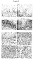
- the chemokine receptor CCR4 was present on dysplastic non-invasive lesions of the cervix and oesophagus. This was particularly striking in some of the oesophageal cancer samples where CCR4 positive dysplastic areas were clearly seen adjacent to normal epithelial areas in the same section (e.g. Figure 7C and D ).
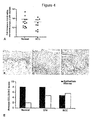
- the chemokine receptor CCR4 increased with malignant progression of the cervix. This was not only due to increased infiltration of CCR4-positive macrophages and Treg cells, but also to acquisition of CCR4 expression by epithelial cells. An unexpected finding was that CCR4 was strongly expressed on non-invasive epithelial cells in intraepithelial (CIN) lesions as well as invasive cancer cells. Progression from CIN to invasive disease was associated with increased stromal cell expression of CCR4 ligands CCL17 and 22 and these chemokines stimulated growth and migration of a CCR4-positive cervical cancer cell line (e.g. Figure 4 and Figure 6 ).
- CCR4 was also detected on dysplastic as well as invasive epithelial cells in oesophageal cancer, again with CCL17 and CCL22 levels increasing during malignant progression. Changes in CCL17 and 22 gradients aid transition from pre-invasive to invasive disease and attract tumour-promoting leucocytes that help initiated cells evade immune surveillance.
- CCL17 and CCL22 were also found on the surface of blood and lymphatic vessels in the tumour biopsies. It was not possible to quantify this but preliminary observations indicate an increase in the intensity of staining with malignant progression.
- Another element of the CCR4 system is the non-signalling chemokine receptor D6 that has a high affinity for CCL17 and CCL22 [18]; its presence in the tissues would be expected to influence gradients of these chemokines [19].
- FIG. 6 shows that the CCR4 receptor is functional on the cervical cancer cell line C-41.
- CCR4 can be up-regulated by the microenvironment.
- CCR4 positive and negative cells were exposed to a number of cytokines (TNF- ⁇ , TGF- ⁇ , IFN- ⁇ , IL-4 and IL-10) known to be present in the cervical microenvironment and for which receptors were likely to be present on the tumour cells. None of these influenced CCR4 expression.
- CCR4 mRNA levels, but not protein levels were up-regulated by co-culture of C-41 cells with macrophages.
- CCR4 and D6 are located on chromosome 3p close to where critical cervical cancer tumour suppressor genes are thought to be located with complex aberrations (loss of heterozygosity, homozygosity and gene amplification) reported [25, 26, 27]. While neither CCR4 nor D6 are directly implicated in these changes [26], genetic alterations nearby may have an impact on their regulation.
- EBV-immortalised B cells secrete CCL22 as well as CCL3 and CCL4 [28].
- Stable expression of the EBV oncogene LMP1 also induced CCL17 and CCL22 in a B cell line and LMP1-induced CCL17 and CCL22 expression was regulated by NF-kB. It was suggested that induction of these two chemokines by EBV helps malignant cells evade immune surveillance by attracting Th2 and Treg cells. Other oncogenic changes may induce CCL17 and CCL22 production by epithelial cells.
- the inventors undertook the detailed quantitation of two components of the mononuclear infiltrate in cervical cancer, specifically CD68+ macrophages and FoxP3+ Tregs.
- the density of CCR4-positive infiltrating cells increases in CIN compared with normal cervix and increases further in both SCC and adenocarcinomas.
- CD68+ macrophages follow the same pattern and we found that these were CCR4 positive.
- Cross talk between macrophages and malignant cells is critical at all stages of cancer progression, influencing malignant cell survival, aiding the angiogenic switch, polarizing leucocytes and aiding malignant cell invasion [29,30,31].
- chemokines CCL17 and CCL22 play a role in macrophage recruitment whereas in other cancers e.g. ovarian cancer, chemokines such as CCL2 are critical [32].
- CCL17 and CCL22 are also important in the recruitment of Tregs that increase in a manner parallel to the CD68+ cells in the cervical biopsies.
- the recruitment of Treg cells to the pre-malignant and malignant lesions fosters immune privilege.
- the malignant cells are surrounded by a large number of CCR4+ FoxP3+ lymphocytes [33].
- These cells recruited by the malignant HL cells, create a favourable environment for malignant cells to escape the host immune system. The inventors think that this is also the case for cervical and oesophageal cancer.
- CCL17 and CLL22 gradients directly encourage tumor cell survival and spread but they attract in leucocytes that may also provide survival factors for the tumor cells and contribute to immune privilege/immunosuppression that prevents effective host responses against the tumor.
- CCR4 epithelial CCR4
- epithelial CCR4 is both a highly sensitive and highly specific biomarker for both pre-malignant and malignant cervical neoplasia.
- CCR4 may offer cells protection from apoptotic stimuli within the tumour environment as well as being necessary for tumour cell invasion of the basement membrane. Due to its high sensitivity and selectivity, there is the potential for CCR4 to be used as a diagnostic biomarker for all stages of cervical cancer.
- CCR4 was not detectable in any normal epithelial oesophageal tissue, but was present in epithelial cells of all pre-invasive and invasive lesions. Due to its high sensitivity and selectivity, there is the potential for CCR4 to be used as a diagnostic biomarker for all stages of oesophageal cancer.
- the chemokine receptor CCR4 and its ligands increase during malignant progression of cervical, oesophageal, kidney, brain, ovarian or breast cancers. Changes in CCR4 and gradients of its ligand have several pro-tumor implications. First CCR4 stimulation increases the growth and survival of the initiated and invasive cancer cells; second, changes in chemokine gradients assists in invasion of the basement membrane and subsequent movement of the malignant cells into the blood vessels or lymphatic system. Finally CCL17 and CCL22 attract the types of cells, including M2 macrophages and FoxP3 Tregs that encourage tumor growth and allow the initiated cells to escape immune surveillance. CCR4 and its ligands may be useful diagnostic markers and therapeutic targets in epithelial neoplasia.
- tumour biopsies from patients with cervical cancer (11 squamous cell carcinoma, S1-S11, and 4 adenocarcinomas, A1-A4) and 14 samples of non-neoplastic cervical tissue (N1-N14) were obtained during surgery and snap-frozen in liquid nitrogen. Diagnosis was made by the pathology department of Barts and The London NHS Trust. Patient samples were divided according to the FIGO classification (stage I, II, III, IV) and tumour biopsies were classified according to increasing grade of nuclear atypia (1, 2, 3) or as well, moderately, or poorly differentiated.
- Resected specimens from thirty-one patients with primary squamous oesophageal carcinoma were also included in this paper. These patients were from a high-risk area for oesophageal carcinoma in Anyang City, Henan province, China. All patients received surgical treatment at the Department of Surgery of the Central Hospital of Anyang. None of these patients had undergone chemotherapy, radiotherapy or immunomodulatory therapy before surgery. Samples were taken from macroscopically cancerous and the corresponding normal areas of the same cancer patient The tissues were fixed in PBS containing 10% neutral-buffered formalin.
- Cervical tissue biopsies were homogenised using a liquid nitrogen-cooled mill 6750 (Glen Creston Ltd, Stanmore) and then solubilised in Tri ReagentTM (Sigma, Poole, UK). Extracted RNA was treated with 10 units DNase (Pharmacia, St Albans, UK) following the manufacturers instructions. RPA was performed using Riboquant® hCR5 and hCR6 template sets (BD Pharmingen, Oxford, UK) and [ ⁇ 32 P] UTP (Amersham International plc, Aylesbury, UK). RNase-protected fragments were run on an acrylamide-urea sequencing gel (BioRad Laboratories Ltd, Hemel Hempstead, UK), adsorbed to filter paper and dried under vacuum. Autoradiography was performed using Kodak ® Biomax ® MS film with a Transcreen LE intensifying screen (Sigma).
- Paraffin-embedded cervical tissues were cut under RNase-free conditions and mounted onto UV-treated PALM® membrane slides (PALM, Microlaser Technologies, Germany). These were then deparaffinised in xylene and rehydrated through graded alcohols. Samples were stained for 1 min with Mayer's haematoxylin solution, dehydrated and air-dried before processing. Sections were laser-microdissected following the manufacturer's protocol. Briefly, areas of interest were laser microdissected and catapulted into a microfuge cap containing Protein Kinase (PK) buffer. Approximately 500-5000 cells were captured in each session. Laser microdissected cells were dissolved in 100 ⁇ l PK Buffer mixed with 5 ⁇ l PK.
- PK Protein Kinase
- RNA was then extracted using the Paraffin block RNA isolation kit (1902, Ambion, USA) according to the manufacturer's instructions.
- cDNA was amplified as described above and analysed using custom-made microfluidic gene array cards (PE Applied Biosystems) according to the manufacturer's instructions.
- the gene expression profile of individual genes in seven cervical tumour samples was compared to five normal cervical samples.
- the gene expression levels in the normal epithelial or stromal cells samples was used as a baseline value of "1" and was compared with the average value of either tumour epithelial cells or tumour stromal cells respectively.
- the laser microdissected tumour samples comprised of one sample from stage 1A2 and 2B, and five of stage 1B1.
- Paraffin-embedded sections (4 ⁇ m) were stained for CCR4, CCL17 and CCL22. Briefly, sections were dewaxed in xylene and dehydrated through an ethanol gradient. Following PBS washing the antigen was exposed using Target Retrieval Solution (S1700, DAKO) at 95°C for 20 min or Antigen Unmasking Solution (H-3300, Vector) for 9 min in a microwave. Sections were blocked with normal rabbit or goat serum for 30 min and incubated overnight at 4°C with the primary antibody: CCR4 (1:300, ab1669, AbCam, Cambridge), CCL17 (1:50, ab9816-50, AbCam, Cambridge) and CCL22 (1:20, 500-P107, Peprotech).
- Double staining CD68 , FoxP3 , SR-A Scoring methods and categories
- CCR4, CCL17 and CCL22 expression on non-malignant and malignant epithelial cells each sample was assessed semi-quantitatively with the following scoring system: 0 (no positive protein expression), +1 ( ⁇ 25% of the cross-section on average has positive expression), +2 (26-50%), +3 (51-75%), +4 (>76%).
- the intensity of positive cells was analysed as follows: 0 (no expression), 1 (mild expression), 2 (moderate expression), 3 (strong expression). Scoring of CCR4, CCL17 and CCL22 expression in tumour stroma (intratumoral infiltrating cells) and the invasive border of the tumour (peritumoral infiltrating cells) was performed based on the 'running mean' method [43].
- YW board-certified pathologist
- the cervical cancer cell line C-41 (ATCC, Rockville, MD, USA) was cultured in DMEM medium supplemented with 10% FCS. In some experiments cells were stimulated with 1, 10, 100, or 1000 ng/ml of CCL17 or CCL22 (PeproTech, London, UK). Proliferation and migration were assessed using methods described previously [11].
- RPA Ribonuclease protection assays
- CCR4 mRNA was up-regulated in stromal areas from malignant tissues when these were compared to their non-neoplastic counterparts.
- CCR4 mRNA was also up-regulated in extracts from the malignant epithelial cell areas compared to normal epithelium.
- the stroma consists of various cell types, and tests were carried out to ascertain which of the infiltrating cells contributed to CCR4 expression. As macrophages and Treg cells express CCR4, CCR4 protein expression was examined in these two cell types and also counted the number of CD68+ macrophages and FoxP3+ Treg cells in the tissue biopsies were counted.
- CD68+ macrophages and FoxP3+ Tregs increased with malignant progression of the cervix.
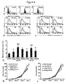
- Figure 2D-I there were few CD68 and FoxP3 cells in biopsies of normal cervix, but the numbers increased in CIN and both cell types were prominent in invasive cancers.
- CD68+ and FoxP3+ cells were then counted in the 122 and 33 biopsies respectively.
- Figure 3B there was a significant (p ⁇ 0.001) increase in CD68+ cells in CIN lesions compared to normal cervix.
- the number of 13 CD68+ cells further increased in SCC, adenocarcinoma, recurrent cancers and lymph node metastases (P ⁇ 0.001, and for LN mets P ⁇ 0.05, compared to normal cervix).
- a similar increase in FoxP3+ cells occurred with malignant progression with SCC, adenocarcinomas and lymph node metastases all showing significant increases in the FoxP3+ infiltrate compared to normal cervix (p ⁇ 0.01).
- SR-A scavenger receptor-A
- FIG. 2 H More detail relating to the IHC score for different stages of CIN is shown in Figure 2 H. This shows that epithelial CCR4 expression was essentially unchanged through progression from CINI to III but that stromal levels of CCR4 increased from CIN I-III
- CCL22 is a product of macrophages, monocytes, DC, B and T cells [13,14]. It is also found in epithelial tissues; for instance intestinal epithelium constitutively produces CCL22 that can be further up-regulated by inflammatory cytokines such as TNF- ⁇ [15].
- CCL17 In normal tissues, CCL17 is expressed by vascular and lymphatic endothelial cells but is also produced by macrophages, DC and keratinocytes [16, 17, 55].
- mRNA was isolated from 14 biopsies of normal cervix, 11 SCC and 4 adenocarcinomas and CCL17 levels assessed by real time RT-PCR. As shown in Figure 5A levels of CCL17mRNA were higher in SCC compared to normal cervix.
- Normal cervical biopsies had low levels of CCL17 in a minority of samples both the epithelium and stroma ( Figure 5B ). Only 2/19 normal samples had CCL17 positive cells in the epithelium compared with 23/33 CIN samples and 13/20 SCC. The number of stromal cells that were CCL17 positive was increased in CIN ( Figure 5C ) and SCC ( Figure 5D ) compared to normal samples. Six of 21 normal biopsies had CCL17 positive stromal cells compared to 25/33 CIN and 15/20 SCC. The epithelial and stromal CCL17 IHC score was increased in CIN and SCC compared to normal biopsies ( Figure 5E ).
- CCR4 expression was screened for CCR4 expression.
- the cell line C-41 expressed cell surface CCR4 in a constitutive manner ( Figure 6A ).
- FACS analysis it was shown that C-41 cells expressed cell surface CCR4.
- This cell line also had intracellular CCL22 protein but the other CCR4 ligand CCL17 was not present ( Figure 6A ).
- cell surface CCR4 protein was internalized on C-41 cells after 2 hours and returned back to the surface after 3 hours ( Figure 6B ).
- C-41 cells demonstrated a typical bell-shaped chemotactic response towards both CCL17 and CCL22 in trans-well migration assays ( Figure 6D ).
- CCR4 and changes in chemokine ligand were specific for cervical cancer or whether they were seen in any other epithelial malignancies that have a link with inflammation.
- Cancer of the oesophagus is an epithelial cancer where examples of all stages of neoplastic progression can be readily obtained, often simultaneously from the same patient.
- CCR4 expression was examined in 31 specimens from patients with oesophageal cancer. In 27 of the cases, all stages of carcinogenesis of the oesophagus: normal, hyperplasia, dysplasia, in situ carcinoma and invasive cancer, were present in biopsies from the same patient. Four of 31 cases had pre-invasive lesions without invasive cancer areas.
- CCR4-positive cells were also present in the stroma, the pattern being the same as in cervical cancer. As shown in Figure 7E , there were few CCR4-positive cells in the normal submucosa; with malignant progression, there were more CCR4 positive cells infiltrating the stroma ( Figure 7F-H ).
- CCL17 was generally absent in both the epithelial and stromal areas of the normal tissues, although there were a few CCL17-positive cells in the stroma and a minority of hyperplastic areas.
- the number of samples continuing CCL17-positive epithelial or stromal cells increased in dysplasia and was highest in invasive areas with 10/23 of these showing some CCL17 positivity.
- stromal positivity Similar to the observations in cervical cancer, the levels of stromal positivity for CCL22 also increased with malignant progression. Only 1/23 samples showed CCL22 positive cells in the stroma of the normal areas, but in dysplastic areas and invasive areas 20/23 and 18/23 samples respectively contained CCL22-positive cells in the stroma. The stromal cell CCL22 positivity increased with the degree of dysplasia. Eight of 23 dysplasia I samples had CCL22 positive cells in the stroma; this increased to 19 of 23 samples of dysplasia II and 20/23 samples of dysplasia III.
- CCL22 was not detected in normal epithelium although it has been reported to be present in normal intestinal epithelium [15].
- Epithelial CCL22 expression increased with malignant progression of the oesophagus; 0/23 samples were positive in the normal areas, 2/23 hyperplasias, 7/23 dysplasias and14/23 invasive areas had CCL22 positive epithelial cells.
- more endothelial cells of blood vessels within the stroma of invasive cancer tissues were positive for CCL22 staining compared with normal and dysplastic epithelium.
- tumour cDNA library (Cancer Research UK) containing cDNA generated from RNA isolated from 5-10 tumour samples and 2-5 normal samples for 11 different tumour types: lung, colon, bladder, stomach, pancreas, skin, breast, brain, oesophagus, ovary and prostate.
- the CCR4 mRNA expression levels were measured using quantitative Real Time RT-PCR.
- CCR4 mRNA levels were significantly elevated in cancers of the cervix, oesophagus, kidney, brain, breast and ovary.
- Figure 11 shows the results of Fluorescence Activated Cell Scanning (FACS) analysis on cervical (C41, C33A) and renal cancer cell lines using an anti-CCR4 antibody to detect CCR4 expression. All the cell lines expressed CCR4.
- the dashed lines in Figure 11 show the data for an isotype-matched control antibody.
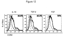
- cytokines present in a tumour are IL-10, TGF- ⁇ , FGF, TNF- ⁇ .
- C41 cervical cancer cell line was stimulated in culture with different cytokines (IL-10, TNF- ⁇ , TGF- ⁇ , FGF; 20 ng/ml) for 24 h.
- the expression of CCR4 was then determined by FACS analysis.
- CCR4 was upregulated in terms of percentage of positive cells after IL-10, TGF- ⁇ and FGF stimulation (blue line) when compared with the unstimulated cells (black line).
- the dashed line shows the data for an isotype control.
- the bold line shows CCR4 expression after stimulation. The results indicate that tumour microenvironment can induce expression of CCR4 on tumour cells.
Landscapes
- Health & Medical Sciences (AREA)
- Life Sciences & Earth Sciences (AREA)
- Chemical & Material Sciences (AREA)
- Immunology (AREA)
- Engineering & Computer Science (AREA)
- Molecular Biology (AREA)
- Medicinal Chemistry (AREA)
- General Health & Medical Sciences (AREA)
- Hematology (AREA)
- Biomedical Technology (AREA)
- Urology & Nephrology (AREA)
- Biochemistry (AREA)
- Cell Biology (AREA)
- Physics & Mathematics (AREA)
- Microbiology (AREA)
- Analytical Chemistry (AREA)
- Food Science & Technology (AREA)
- Biotechnology (AREA)
- General Physics & Mathematics (AREA)
- Pathology (AREA)
- Organic Chemistry (AREA)
- Public Health (AREA)
- Veterinary Medicine (AREA)
- Pharmacology & Pharmacy (AREA)
- Animal Behavior & Ethology (AREA)
- Epidemiology (AREA)
- Tropical Medicine & Parasitology (AREA)
- Genetics & Genomics (AREA)
- Endocrinology (AREA)
- Biophysics (AREA)
- Proteomics, Peptides & Aminoacids (AREA)
- Chemical Kinetics & Catalysis (AREA)
- General Chemical & Material Sciences (AREA)
- Nuclear Medicine, Radiotherapy & Molecular Imaging (AREA)
- Medicines That Contain Protein Lipid Enzymes And Other Medicines (AREA)
- Medicines Containing Antibodies Or Antigens For Use As Internal Diagnostic Agents (AREA)
- Measuring Or Testing Involving Enzymes Or Micro-Organisms (AREA)
- Pharmaceuticals Containing Other Organic And Inorganic Compounds (AREA)
- Oncology (AREA)
- Investigating Or Analysing Biological Materials (AREA)
Applications Claiming Priority (2)
| Application Number | Priority Date | Filing Date | Title |
|---|---|---|---|
| GB0718167A GB0718167D0 (en) | 2007-09-18 | 2007-09-18 | Cancer marker and therapeutic target |
| EP08806315.1A EP2176664B1 (en) | 2007-09-18 | 2008-09-18 | Ccr4 as cancer marker |
Related Parent Applications (2)
| Application Number | Title | Priority Date | Filing Date |
|---|---|---|---|
| EP08806315.1A Division EP2176664B1 (en) | 2007-09-18 | 2008-09-18 | Ccr4 as cancer marker |
| EP08806315.1 Division | 2008-09-18 |
Publications (3)
| Publication Number | Publication Date |
|---|---|
| EP2535716A2 EP2535716A2 (en) | 2012-12-19 |
| EP2535716A3 EP2535716A3 (en) | 2013-03-13 |
| EP2535716B1 true EP2535716B1 (en) | 2016-11-02 |
Family
ID=38670078
Family Applications (3)
| Application Number | Title | Priority Date | Filing Date |
|---|---|---|---|
| EP12183760.3A Not-in-force EP2535716B1 (en) | 2007-09-18 | 2008-09-18 | Cancer marker and therapeutic target |
| EP12182790.1A Not-in-force EP2533047B1 (en) | 2007-09-18 | 2008-09-18 | Ccr4 as therapeutic target for cancer |
| EP08806315.1A Not-in-force EP2176664B1 (en) | 2007-09-18 | 2008-09-18 | Ccr4 as cancer marker |
Family Applications After (2)
| Application Number | Title | Priority Date | Filing Date |
|---|---|---|---|
| EP12182790.1A Not-in-force EP2533047B1 (en) | 2007-09-18 | 2008-09-18 | Ccr4 as therapeutic target for cancer |
| EP08806315.1A Not-in-force EP2176664B1 (en) | 2007-09-18 | 2008-09-18 | Ccr4 as cancer marker |
Country Status (11)
| Country | Link |
|---|---|
| US (2) | US9134293B2 (enExample) |
| EP (3) | EP2535716B1 (enExample) |
| JP (2) | JP5774309B2 (enExample) |
| AU (1) | AU2008300413B2 (enExample) |
| CA (1) | CA2699702C (enExample) |
| DK (3) | DK2176664T3 (enExample) |
| ES (3) | ES2588507T3 (enExample) |
| GB (1) | GB0718167D0 (enExample) |
| PL (3) | PL2176664T3 (enExample) |
| RU (2) | RU2529797C2 (enExample) |
| WO (1) | WO2009037454A2 (enExample) |
Families Citing this family (20)
| Publication number | Priority date | Publication date | Assignee | Title |
|---|---|---|---|---|
| JPH0623815Y2 (ja) | 1988-10-06 | 1994-06-22 | 株式会社大金製作所 | エア回転継手のシール装置 |
| GB0718167D0 (en) | 2007-09-18 | 2007-10-31 | Cancer Rec Tech Ltd | Cancer marker and therapeutic target |
| US11029313B2 (en) | 2008-09-26 | 2021-06-08 | The General Hospital Corporation | Method of treating cervical neoplasia in patients infected with human papilloma virus |
| WO2010037042A2 (en) | 2008-09-26 | 2010-04-01 | The General Hospital Corporation | Methods for detecting and treating cancer |
| GB0909906D0 (en) * | 2009-06-09 | 2009-07-22 | Affitech As | Antibodies |
| EP2580348B1 (en) * | 2010-06-14 | 2018-04-25 | Qiagen GmbH | Method for determination of target cells or tissue for extraction of biomolecules from non-formalin-fixed biological samples |
| GB201021289D0 (en) * | 2010-12-15 | 2011-01-26 | Immatics Biotechnologies Gmbh | Novel biomarkers for a prediction of the outcome of an immunotherapy against cancer |
| US10266599B2 (en) | 2010-12-07 | 2019-04-23 | Cancer Research Technology Limited | Antibodies which bind to the human CC chemokine receptor 4 and uses thereof |
| GB201020738D0 (en) | 2010-12-07 | 2011-01-19 | Affitech Res As | Antibodies |
| WO2013063130A1 (en) * | 2011-10-24 | 2013-05-02 | Metasignal Therapeutics Inc. | Carbonic anhydrase ix-related markers and use thereof |
| PL2785692T3 (pl) | 2011-12-01 | 2018-02-28 | Chemocentryx, Inc. | Podstawione aniliny jako antagoniści ccr(4) |
| GB2512857A (en) * | 2013-04-09 | 2014-10-15 | Cancer Res Technology | Cancer biomarker |
| SI3065774T1 (sl) * | 2013-11-06 | 2021-11-30 | Janssen Biotech, Inc. | Protitelesa proti CCL17 |
| SMT202100116T1 (it) | 2014-05-28 | 2021-05-07 | Agenus Inc | Anticorpi anti-gitr e metodi di utilizzo degli stessi |
| RU2740254C2 (ru) * | 2014-10-01 | 2021-01-12 | Сфинготек Гмбх | ОПРЕДЕЛЕНИЕ hGH ДЛЯ ПРИМЕНЕНИЯ ДЛЯ ПРЕДУПРЕЖДЕНИЯ ТЯЖЕЛОГО НЕБЛАГОПРИЯТНОГО СЕРДЕЧНО-СОСУДИСТОГО ЯВЛЕНИЯ ИЛИ СЕРДЕЧНО-СОСУДИСТОГО ЗАБОЛЕВАНИЯ У ИНДИВИДУУМА |
| EP3283886B1 (en) * | 2015-04-17 | 2020-01-15 | Eisai Inc. | Methods for treating lung cancer |
| WO2017017283A1 (en) | 2015-07-30 | 2017-02-02 | Qiagen Gmbh | Method of preparing a frozen biological sample |
| TW202134282A (zh) | 2015-12-02 | 2021-09-16 | 美商艾吉納斯公司 | 抗體和使用彼之方法 |
| EP3626269A4 (en) | 2017-05-19 | 2021-03-03 | Shingo Maeda | METHOD FOR INFILTRATION OF REGULATORY T-CELLS USING CCR4 INHIBITION AND METHOD FOR TREATMENT OF NEOPLASTIC DISEASES IN DOGS |
| SG11202101454PA (en) | 2018-08-29 | 2021-03-30 | Chemocentryx Inc | Combination therapy using c-c chemokine receptor 4 (ccr4) antagonists and one or more immune checkpoint inhibitors |
Family Cites Families (32)
| Publication number | Priority date | Publication date | Assignee | Title |
|---|---|---|---|---|
| US5786158A (en) * | 1992-04-30 | 1998-07-28 | Yale University | Therapeutic and diagnostic methods and compositions based on notch proteins and nucleic acids |
| US5342947A (en) * | 1992-10-09 | 1994-08-30 | Glaxo Inc. | Preparation of water soluble camptothecin derivatives |
| JPH08507927A (ja) * | 1993-03-19 | 1996-08-27 | ザ・ジョーンズ・ホプキンス・ユニバーシティ | Apc遺伝子の変異決定用抗体およびアッセイ |
| GB9501683D0 (en) | 1995-01-27 | 1995-03-15 | Glaxo Group Ltd | Substances and their uses |
| US6498015B1 (en) | 1995-06-07 | 2002-12-24 | Icos Corporation | Methods of identifying agents that modulate the binding between MDC and an MDC receptor |
| US6245332B1 (en) | 1999-01-15 | 2001-06-12 | The Board Of Trustees Of The Leland Stanford Junior University | Modulation of systemic memory T cell trafficking |
| US6488930B1 (en) | 1999-01-15 | 2002-12-03 | Millennium Pharmaceuticals, Inc. | Anti-CCR4 antibodies and methods of use therefor |
| DE60134962D1 (de) | 2000-03-03 | 2008-09-04 | Kyowa Hakko Kogyo Kk | Anti-ccr4 antikörper und fragmente davon |
| EP1578341A4 (en) | 2000-10-11 | 2005-09-28 | Tularik Inc | MODULATION OF THE CCR4 FUNCTION |
| WO2002030357A2 (en) | 2000-10-11 | 2002-04-18 | Chemocentryx, Inc. | Compounds and methods for modulating ccr4 function |
| EP1326617B9 (en) | 2000-10-18 | 2006-10-25 | Schering Aktiengesellschaft | Use of 11beta-(4-acetylphenyl)-17beta-hydroxy-17alpha-(1,1,2,2-pentafluoroethyl)estra-4,9-dien-3-one for the preparation of a medicament for the treatment fo breast, ovarian, endometrial cancer, myeloma and meningioma |
| US20020182624A1 (en) * | 2001-02-28 | 2002-12-05 | Eos Biotechnology, Inc. | Chemokine receptors and disease |
| US7144903B2 (en) | 2001-05-23 | 2006-12-05 | Amgen Inc. | CCR4 antagonists |
| US20050101530A1 (en) | 2001-08-10 | 2005-05-12 | Topigen Pharmaceutique, Inc. | Cellular virus receptors and methods of use |
| WO2003018635A1 (fr) | 2001-08-31 | 2003-03-06 | Kyowa Hakko Kogyo Co., Ltd. | Anticorps greffes cdr humains et fragments de ces anticorps |
| US20060004010A1 (en) | 2002-07-10 | 2006-01-05 | Hiromu Habashita | Ccr4 antagonist and medical use thereof |
| AU2003291549A1 (en) | 2002-11-15 | 2004-06-15 | Morehouse School Of Medicine | Anti-chemokine and associated receptors antibodies for inhibition of growth of neoplasms |
| JP2007524362A (ja) * | 2003-02-14 | 2007-08-30 | サイグレス ディスカバリー, インコーポレイテッド | 癌における治療gpcr標的 |
| US7807389B2 (en) * | 2003-03-14 | 2010-10-05 | University Of Rochester | Methods and compositions related to joint inflammation diseases |
| SE0301650D0 (sv) | 2003-06-04 | 2003-06-04 | Astrazeneca Ab | Novel compounds |
| SE0301653D0 (sv) | 2003-06-05 | 2003-06-05 | Astrazeneca Ab | Novel compounds |
| EP1661889A4 (en) | 2003-09-05 | 2009-08-05 | Ono Pharmaceutical Co | ANTAGONIST OF THE CHEMOKINE RECEPTOR AND ITS USE FOR MEDICAL PURPOSES |
| JPWO2005035582A1 (ja) | 2003-10-08 | 2007-11-22 | 協和醗酵工業株式会社 | Ccr4に特異的に結合する抗体組成物 |
| AU2004294842B2 (en) | 2003-12-04 | 2010-05-13 | Kyowa Kirin Co., Ltd. | Medicine containing genetically modified antibody against chemokine receptor CCR4 |
| WO2005082865A1 (ja) | 2004-02-27 | 2005-09-09 | Astellas Pharma Inc. | 縮合二環性ピリミジン誘導体 |
| JP2007217282A (ja) | 2004-03-04 | 2007-08-30 | Astellas Pharma Inc | 置換ピリミジン誘導体 |
| WO2005106471A2 (en) | 2004-04-30 | 2005-11-10 | Bayer Healthcare Ag | Diagnostics and therapeutics for diseases associated with c-c chemokine receptor 4 (ccr4) |
| JP2007269629A (ja) | 2004-06-21 | 2007-10-18 | Astellas Pharma Inc | キナゾリン誘導体 |
| RU2296328C1 (ru) * | 2005-09-21 | 2007-03-27 | Общество с ограниченной ответственностью "ГЕН" | Способ определения предрасположенности к онкологическим заболеваниям и диагностический набор для его осуществления |
| EP1777523A1 (en) * | 2005-10-19 | 2007-04-25 | INSERM (Institut National de la Santé et de la Recherche Médicale) | An in vitro method for the prognosis of progression of a cancer and of the outcome in a patient and means for performing said method |
| EP1984501B1 (en) | 2006-02-14 | 2011-05-18 | Noxxon Pharma AG | Mcp-i binding nucleic acids |
| GB0718167D0 (en) | 2007-09-18 | 2007-10-31 | Cancer Rec Tech Ltd | Cancer marker and therapeutic target |
-
2007
- 2007-09-18 GB GB0718167A patent/GB0718167D0/en not_active Ceased
-
2008
- 2008-09-18 AU AU2008300413A patent/AU2008300413B2/en not_active Ceased
- 2008-09-18 WO PCT/GB2008/003160 patent/WO2009037454A2/en not_active Ceased
- 2008-09-18 EP EP12183760.3A patent/EP2535716B1/en not_active Not-in-force
- 2008-09-18 RU RU2010123921/15A patent/RU2529797C2/ru not_active IP Right Cessation
- 2008-09-18 ES ES12182790.1T patent/ES2588507T3/es active Active
- 2008-09-18 EP EP12182790.1A patent/EP2533047B1/en not_active Not-in-force
- 2008-09-18 DK DK08806315.1T patent/DK2176664T3/da active
- 2008-09-18 EP EP08806315.1A patent/EP2176664B1/en not_active Not-in-force
- 2008-09-18 ES ES12183760.3T patent/ES2612690T3/es active Active
- 2008-09-18 PL PL08806315T patent/PL2176664T3/pl unknown
- 2008-09-18 CA CA2699702A patent/CA2699702C/en not_active Expired - Fee Related
- 2008-09-18 JP JP2010525420A patent/JP5774309B2/ja not_active Expired - Fee Related
- 2008-09-18 DK DK12182790.1T patent/DK2533047T3/en active
- 2008-09-18 DK DK12183760.3T patent/DK2535716T3/en active
- 2008-09-18 PL PL12182790.1T patent/PL2533047T3/pl unknown
- 2008-09-18 PL PL12183760T patent/PL2535716T3/pl unknown
- 2008-09-18 US US12/679,002 patent/US9134293B2/en not_active Expired - Fee Related
- 2008-09-18 ES ES08806315.1T patent/ES2443541T3/es active Active
-
2014
- 2014-07-14 RU RU2014128513A patent/RU2014128513A/ru not_active Application Discontinuation
-
2015
- 2015-06-30 JP JP2015131752A patent/JP6234967B2/ja not_active Expired - Fee Related
- 2015-08-11 US US14/823,755 patent/US10261099B2/en not_active Expired - Fee Related
Also Published As
| Publication number | Publication date |
|---|---|
| PL2535716T3 (pl) | 2017-06-30 |
| EP2176664A2 (en) | 2010-04-21 |
| WO2009037454A3 (en) | 2009-05-07 |
| DK2535716T3 (en) | 2017-02-13 |
| US10261099B2 (en) | 2019-04-16 |
| PL2533047T3 (pl) | 2016-11-30 |
| EP2176664B1 (en) | 2013-11-06 |
| RU2010123921A (ru) | 2011-12-20 |
| GB0718167D0 (en) | 2007-10-31 |
| RU2014128513A (ru) | 2016-02-10 |
| EP2535716A3 (en) | 2013-03-13 |
| RU2529797C2 (ru) | 2014-09-27 |
| EP2533047A1 (en) | 2012-12-12 |
| DK2176664T3 (da) | 2014-01-20 |
| EP2535716A2 (en) | 2012-12-19 |
| ES2443541T3 (es) | 2014-02-19 |
| EP2533047B1 (en) | 2016-05-11 |
| JP6234967B2 (ja) | 2017-11-22 |
| ES2612690T3 (es) | 2017-05-18 |
| PL2176664T3 (pl) | 2014-04-30 |
| WO2009037454A2 (en) | 2009-03-26 |
| US20100278844A1 (en) | 2010-11-04 |
| US20160223572A1 (en) | 2016-08-04 |
| JP5774309B2 (ja) | 2015-09-09 |
| CA2699702C (en) | 2018-03-06 |
| AU2008300413A1 (en) | 2009-03-26 |
| JP2015212703A (ja) | 2015-11-26 |
| AU2008300413B2 (en) | 2014-09-11 |
| US9134293B2 (en) | 2015-09-15 |
| CA2699702A1 (en) | 2009-03-26 |
| DK2533047T3 (en) | 2016-08-22 |
| ES2588507T3 (es) | 2016-11-03 |
| JP2010539508A (ja) | 2010-12-16 |
Similar Documents
| Publication | Publication Date | Title |
|---|---|---|
| EP2535716B1 (en) | Cancer marker and therapeutic target | |
| Ren et al. | Intratumoral CD103+ CD8+ T cells predict response to neoadjuvant chemoimmunotherapy in advanced head and neck squamous cell carcinoma | |
| JP2016001187A (ja) | 抗体ベースのアレイを使用する胃癌療法のための薬物選択 | |
| EP2805166B1 (en) | Method for the prognosis of survival time of a patient suffering from a solid cancer | |
| JP2013520958A (ja) | 上皮間葉転換のバイオマーカーとしてaxlを使用する方法 | |
| Wang et al. | Overexpression of caveolin-1 in cancer-associated fibroblasts predicts good outcome in breast cancer | |
| JP2020040959A (ja) | 抗egfr薬を用いた胃癌の処置のための、egfrバイオマーカーの使用 | |
| Shu et al. | Immune landscape of tertiary lymphoid structures in hepatocellular carcinoma (HCC) treated with neoadjuvant immune checkpoint blockade | |
| Hermida-Prado et al. | Endocrine Therapy Synergizes with SMAC Mimetics to Potentiate Antigen Presentation and Tumor Regression in Hormone Receptor–Positive Breast Cancer | |
| Dubuisson et al. | Immunodynamics of explanted human tumors for immuno‐oncology | |
| Frolova et al. | A shift from nuclear to cytoplasmic breast cancer metastasis suppressor 1 expression is associated with highly proliferative estrogen receptor-negative breast cancers | |
| WO2011010309A1 (en) | A method of diagnosing cancer | |
| CN116137862A (zh) | 三级淋巴结构在预后疾病进展和治疗癌症中的应用 | |
| AU2013203349B2 (en) | Cancer marker and therapeutic target | |
| JP2019518970A (ja) | 腎臓癌を患う対象の癌治療に対する感受性を予測するための方法およびキット | |
| Gouravski | Assessing ER Positive Breast Cancer Heterogeneity and Identification of Tumour Microenvironment Molecular Markers in Response to Treatment Via Multi-omic Approach | |
| Ibrahim et al. | NF1 Loss Remodels Tumor Niches for Immune Evasion | |
| Scholz et al. | 33rd Annual Meeting & Pre-Conference Programs of the Society for Immunotherapy of Cancer (SITC 2018) | |
| KR20250033118A (ko) | 신장암에서 면역 관문 억제제의 치료 반응성 예측 마커 및 이를 이용한 치료 반응성 예측 방법 | |
| TW202417641A (zh) | 預測對egfr激酶活性之抑制劑的反應之方法 | |
| Pagano | The potential role of CXCR4 and SDF-1 as indicators of tumor aggressiviness in patients with conventional papillary thyroid carcinoma | |
| Devictor et al. | cytochemical Assays of P-glycoprotein in Breast |
Legal Events
| Date | Code | Title | Description |
|---|---|---|---|
| PUAI | Public reference made under article 153(3) epc to a published international application that has entered the european phase |
Free format text: ORIGINAL CODE: 0009012 |
|
| AC | Divisional application: reference to earlier application |
Ref document number: 2176664 Country of ref document: EP Kind code of ref document: P |
|
| AK | Designated contracting states |
Kind code of ref document: A2 Designated state(s): AT BE BG CH CY CZ DE DK EE ES FI FR GB GR HR HU IE IS IT LI LT LU LV MC MT NL NO PL PT RO SE SI SK TR |
|
| PUAL | Search report despatched |
Free format text: ORIGINAL CODE: 0009013 |
|
| AK | Designated contracting states |
Kind code of ref document: A3 Designated state(s): AT BE BG CH CY CZ DE DK EE ES FI FR GB GR HR HU IE IS IT LI LT LU LV MC MT NL NO PL PT RO SE SI SK TR |
|
| RIC1 | Information provided on ipc code assigned before grant |
Ipc: G01N 33/50 20060101ALI20130207BHEP Ipc: G01N 33/574 20060101AFI20130207BHEP Ipc: A61K 31/00 20060101ALI20130207BHEP |
|
| 17P | Request for examination filed |
Effective date: 20130905 |
|
| RBV | Designated contracting states (corrected) |
Designated state(s): AT BE BG CH CY CZ DE DK EE ES FI FR GB GR HR HU IE IS IT LI LT LU LV MC MT NL NO PL PT RO SE SI SK TR |
|
| 17Q | First examination report despatched |
Effective date: 20140226 |
|
| GRAP | Despatch of communication of intention to grant a patent |
Free format text: ORIGINAL CODE: EPIDOSNIGR1 |
|
| INTG | Intention to grant announced |
Effective date: 20141003 |
|
| GRAP | Despatch of communication of intention to grant a patent |
Free format text: ORIGINAL CODE: EPIDOSNIGR1 |
|
| INTG | Intention to grant announced |
Effective date: 20150714 |
|
| GRAP | Despatch of communication of intention to grant a patent |
Free format text: ORIGINAL CODE: EPIDOSNIGR1 |
|
| RAP1 | Party data changed (applicant data changed or rights of an application transferred) |
Owner name: CANCER RESEARCH TECHNOLOGY LIMITED |
|
| INTG | Intention to grant announced |
Effective date: 20160510 |
|
| GRAS | Grant fee paid |
Free format text: ORIGINAL CODE: EPIDOSNIGR3 |
|
| GRAA | (expected) grant |
Free format text: ORIGINAL CODE: 0009210 |
|
| AC | Divisional application: reference to earlier application |
Ref document number: 2176664 Country of ref document: EP Kind code of ref document: P |
|
| AK | Designated contracting states |
Kind code of ref document: B1 Designated state(s): AT BE BG CH CY CZ DE DK EE ES FI FR GB GR HR HU IE IS IT LI LT LU LV MC MT NL NO PL PT RO SE SI SK TR |
|
| REG | Reference to a national code |
Ref country code: GB Ref legal event code: FG4D |
|
| REG | Reference to a national code |
Ref country code: AT Ref legal event code: REF Ref document number: 842361 Country of ref document: AT Kind code of ref document: T Effective date: 20161115 Ref country code: CH Ref legal event code: EP |
|
| REG | Reference to a national code |
Ref country code: IE Ref legal event code: FG4D |
|
| REG | Reference to a national code |
Ref country code: DE Ref legal event code: R096 Ref document number: 602008047228 Country of ref document: DE |
|
| REG | Reference to a national code |
Ref country code: DK Ref legal event code: T3 Effective date: 20170207 |
|
| REG | Reference to a national code |
Ref country code: NL Ref legal event code: FP |
|
| PG25 | Lapsed in a contracting state [announced via postgrant information from national office to epo] |
Ref country code: LV Free format text: LAPSE BECAUSE OF FAILURE TO SUBMIT A TRANSLATION OF THE DESCRIPTION OR TO PAY THE FEE WITHIN THE PRESCRIBED TIME-LIMIT Effective date: 20161102 |
|
| REG | Reference to a national code |
Ref country code: SE Ref legal event code: TRGR |
|
| REG | Reference to a national code |
Ref country code: LT Ref legal event code: MG4D |
|
| REG | Reference to a national code |
Ref country code: NO Ref legal event code: T2 Effective date: 20161102 |
|
| PG25 | Lapsed in a contracting state [announced via postgrant information from national office to epo] |
Ref country code: LT Free format text: LAPSE BECAUSE OF FAILURE TO SUBMIT A TRANSLATION OF THE DESCRIPTION OR TO PAY THE FEE WITHIN THE PRESCRIBED TIME-LIMIT Effective date: 20161102 |
|
| REG | Reference to a national code |
Ref country code: ES Ref legal event code: FG2A Ref document number: 2612690 Country of ref document: ES Kind code of ref document: T3 Effective date: 20170518 |
|
| PG25 | Lapsed in a contracting state [announced via postgrant information from national office to epo] |
Ref country code: HR Free format text: LAPSE BECAUSE OF FAILURE TO SUBMIT A TRANSLATION OF THE DESCRIPTION OR TO PAY THE FEE WITHIN THE PRESCRIBED TIME-LIMIT Effective date: 20161102 Ref country code: IS Free format text: LAPSE BECAUSE OF FAILURE TO SUBMIT A TRANSLATION OF THE DESCRIPTION OR TO PAY THE FEE WITHIN THE PRESCRIBED TIME-LIMIT Effective date: 20170302 Ref country code: PT Free format text: LAPSE BECAUSE OF FAILURE TO SUBMIT A TRANSLATION OF THE DESCRIPTION OR TO PAY THE FEE WITHIN THE PRESCRIBED TIME-LIMIT Effective date: 20170302 |
|
| PG25 | Lapsed in a contracting state [announced via postgrant information from national office to epo] |
Ref country code: SK Free format text: LAPSE BECAUSE OF FAILURE TO SUBMIT A TRANSLATION OF THE DESCRIPTION OR TO PAY THE FEE WITHIN THE PRESCRIBED TIME-LIMIT Effective date: 20161102 Ref country code: CZ Free format text: LAPSE BECAUSE OF FAILURE TO SUBMIT A TRANSLATION OF THE DESCRIPTION OR TO PAY THE FEE WITHIN THE PRESCRIBED TIME-LIMIT Effective date: 20161102 Ref country code: EE Free format text: LAPSE BECAUSE OF FAILURE TO SUBMIT A TRANSLATION OF THE DESCRIPTION OR TO PAY THE FEE WITHIN THE PRESCRIBED TIME-LIMIT Effective date: 20161102 Ref country code: RO Free format text: LAPSE BECAUSE OF FAILURE TO SUBMIT A TRANSLATION OF THE DESCRIPTION OR TO PAY THE FEE WITHIN THE PRESCRIBED TIME-LIMIT Effective date: 20161102 |
|
| REG | Reference to a national code |
Ref country code: DE Ref legal event code: R097 Ref document number: 602008047228 Country of ref document: DE |
|
| PG25 | Lapsed in a contracting state [announced via postgrant information from national office to epo] |
Ref country code: BG Free format text: LAPSE BECAUSE OF FAILURE TO SUBMIT A TRANSLATION OF THE DESCRIPTION OR TO PAY THE FEE WITHIN THE PRESCRIBED TIME-LIMIT Effective date: 20170202 |
|
| PLBE | No opposition filed within time limit |
Free format text: ORIGINAL CODE: 0009261 |
|
| STAA | Information on the status of an ep patent application or granted ep patent |
Free format text: STATUS: NO OPPOSITION FILED WITHIN TIME LIMIT |
|
| REG | Reference to a national code |
Ref country code: FR Ref legal event code: PLFP Year of fee payment: 10 |
|
| 26N | No opposition filed |
Effective date: 20170803 |
|
| PGFP | Annual fee paid to national office [announced via postgrant information from national office to epo] |
Ref country code: FI Payment date: 20170925 Year of fee payment: 10 Ref country code: GR Payment date: 20170925 Year of fee payment: 10 |
|
| PG25 | Lapsed in a contracting state [announced via postgrant information from national office to epo] |
Ref country code: SI Free format text: LAPSE BECAUSE OF FAILURE TO SUBMIT A TRANSLATION OF THE DESCRIPTION OR TO PAY THE FEE WITHIN THE PRESCRIBED TIME-LIMIT Effective date: 20161102 |
|
| PGFP | Annual fee paid to national office [announced via postgrant information from national office to epo] |
Ref country code: IE Payment date: 20170922 Year of fee payment: 10 Ref country code: DK Payment date: 20170929 Year of fee payment: 10 Ref country code: TR Payment date: 20170925 Year of fee payment: 10 Ref country code: BE Payment date: 20170925 Year of fee payment: 10 Ref country code: AT Payment date: 20171002 Year of fee payment: 10 Ref country code: SE Payment date: 20170929 Year of fee payment: 10 |
|
| PGFP | Annual fee paid to national office [announced via postgrant information from national office to epo] |
Ref country code: NO Payment date: 20171003 Year of fee payment: 10 |
|
| PGFP | Annual fee paid to national office [announced via postgrant information from national office to epo] |
Ref country code: PL Payment date: 20171002 Year of fee payment: 10 |
|
| PG25 | Lapsed in a contracting state [announced via postgrant information from national office to epo] |
Ref country code: MC Free format text: LAPSE BECAUSE OF FAILURE TO SUBMIT A TRANSLATION OF THE DESCRIPTION OR TO PAY THE FEE WITHIN THE PRESCRIBED TIME-LIMIT Effective date: 20161102 |
|
| PG25 | Lapsed in a contracting state [announced via postgrant information from national office to epo] |
Ref country code: LU Free format text: LAPSE BECAUSE OF NON-PAYMENT OF DUE FEES Effective date: 20170918 |
|
| REG | Reference to a national code |
Ref country code: FR Ref legal event code: PLFP Year of fee payment: 11 |
|
| PG25 | Lapsed in a contracting state [announced via postgrant information from national office to epo] |
Ref country code: MT Free format text: LAPSE BECAUSE OF NON-PAYMENT OF DUE FEES Effective date: 20170918 |
|
| REG | Reference to a national code |
Ref country code: NO Ref legal event code: MMEP |
|
| PG25 | Lapsed in a contracting state [announced via postgrant information from national office to epo] |
Ref country code: FI Free format text: LAPSE BECAUSE OF NON-PAYMENT OF DUE FEES Effective date: 20180918 |
|
| REG | Reference to a national code |
Ref country code: SE Ref legal event code: EUG |
|
| REG | Reference to a national code |
Ref country code: DK Ref legal event code: EBP Effective date: 20180930 |
|
| REG | Reference to a national code |
Ref country code: AT Ref legal event code: MM01 Ref document number: 842361 Country of ref document: AT Kind code of ref document: T Effective date: 20180918 Ref country code: AT Ref legal event code: UEP Ref document number: 842361 Country of ref document: AT Kind code of ref document: T Effective date: 20161102 |
|
| PG25 | Lapsed in a contracting state [announced via postgrant information from national office to epo] |
Ref country code: SE Free format text: LAPSE BECAUSE OF NON-PAYMENT OF DUE FEES Effective date: 20180919 |
|
| REG | Reference to a national code |
Ref country code: BE Ref legal event code: FP Effective date: 20170201 Ref country code: BE Ref legal event code: MM Effective date: 20180930 |
|
| REG | Reference to a national code |
Ref country code: IE Ref legal event code: MM4A |
|
| PG25 | Lapsed in a contracting state [announced via postgrant information from national office to epo] |
Ref country code: HU Free format text: LAPSE BECAUSE OF FAILURE TO SUBMIT A TRANSLATION OF THE DESCRIPTION OR TO PAY THE FEE WITHIN THE PRESCRIBED TIME-LIMIT; INVALID AB INITIO Effective date: 20080918 |
|
| PG25 | Lapsed in a contracting state [announced via postgrant information from national office to epo] |
Ref country code: IE Free format text: LAPSE BECAUSE OF NON-PAYMENT OF DUE FEES Effective date: 20180918 Ref country code: NO Free format text: LAPSE BECAUSE OF NON-PAYMENT OF DUE FEES Effective date: 20180930 |
|
| PG25 | Lapsed in a contracting state [announced via postgrant information from national office to epo] |
Ref country code: BE Free format text: LAPSE BECAUSE OF NON-PAYMENT OF DUE FEES Effective date: 20180930 Ref country code: GR Free format text: LAPSE BECAUSE OF NON-PAYMENT OF DUE FEES Effective date: 20190402 |
|
| PG25 | Lapsed in a contracting state [announced via postgrant information from national office to epo] |
Ref country code: AT Free format text: LAPSE BECAUSE OF NON-PAYMENT OF DUE FEES Effective date: 20180918 Ref country code: DK Free format text: LAPSE BECAUSE OF NON-PAYMENT OF DUE FEES Effective date: 20180930 Ref country code: CY Free format text: LAPSE BECAUSE OF NON-PAYMENT OF DUE FEES Effective date: 20161102 |
|
| PG25 | Lapsed in a contracting state [announced via postgrant information from national office to epo] |
Ref country code: PL Free format text: LAPSE BECAUSE OF NON-PAYMENT OF DUE FEES Effective date: 20180918 |
|
| PGFP | Annual fee paid to national office [announced via postgrant information from national office to epo] |
Ref country code: DE Payment date: 20200922 Year of fee payment: 13 Ref country code: NL Payment date: 20200925 Year of fee payment: 13 Ref country code: FR Payment date: 20200921 Year of fee payment: 13 Ref country code: GB Payment date: 20200922 Year of fee payment: 13 |
|
| PGFP | Annual fee paid to national office [announced via postgrant information from national office to epo] |
Ref country code: CH Payment date: 20200921 Year of fee payment: 13 |
|
| PGFP | Annual fee paid to national office [announced via postgrant information from national office to epo] |
Ref country code: ES Payment date: 20201001 Year of fee payment: 13 Ref country code: IT Payment date: 20200924 Year of fee payment: 13 |
|
| REG | Reference to a national code |
Ref country code: DE Ref legal event code: R082 Ref document number: 602008047228 Country of ref document: DE |
|
| REG | Reference to a national code |
Ref country code: DE Ref legal event code: R119 Ref document number: 602008047228 Country of ref document: DE |
|
| REG | Reference to a national code |
Ref country code: CH Ref legal event code: PL |
|
| REG | Reference to a national code |
Ref country code: NL Ref legal event code: MM Effective date: 20211001 |
|
| GBPC | Gb: european patent ceased through non-payment of renewal fee |
Effective date: 20210918 |
|
| PG25 | Lapsed in a contracting state [announced via postgrant information from national office to epo] |
Ref country code: TR Free format text: LAPSE BECAUSE OF NON-PAYMENT OF DUE FEES Effective date: 20180918 Ref country code: NL Free format text: LAPSE BECAUSE OF NON-PAYMENT OF DUE FEES Effective date: 20211001 |
|
| PG25 | Lapsed in a contracting state [announced via postgrant information from national office to epo] |
Ref country code: GB Free format text: LAPSE BECAUSE OF NON-PAYMENT OF DUE FEES Effective date: 20210918 Ref country code: FR Free format text: LAPSE BECAUSE OF NON-PAYMENT OF DUE FEES Effective date: 20210930 Ref country code: DE Free format text: LAPSE BECAUSE OF NON-PAYMENT OF DUE FEES Effective date: 20220401 |
|
| PG25 | Lapsed in a contracting state [announced via postgrant information from national office to epo] |
Ref country code: LI Free format text: LAPSE BECAUSE OF NON-PAYMENT OF DUE FEES Effective date: 20210930 Ref country code: CH Free format text: LAPSE BECAUSE OF NON-PAYMENT OF DUE FEES Effective date: 20210930 |
|
| PG25 | Lapsed in a contracting state [announced via postgrant information from national office to epo] |
Ref country code: IT Free format text: LAPSE BECAUSE OF NON-PAYMENT OF DUE FEES Effective date: 20210918 |
|
| REG | Reference to a national code |
Ref country code: ES Ref legal event code: FD2A Effective date: 20221107 |
|
| PG25 | Lapsed in a contracting state [announced via postgrant information from national office to epo] |
Ref country code: ES Free format text: LAPSE BECAUSE OF NON-PAYMENT OF DUE FEES Effective date: 20210919 |