EP2413809B1 - Medizinische vorrichtungen zur schnellen ablage und fixierung von gewebeankern - Google Patents
Medizinische vorrichtungen zur schnellen ablage und fixierung von gewebeankern Download PDFInfo
- Publication number
- EP2413809B1 EP2413809B1 EP10712286.3A EP10712286A EP2413809B1 EP 2413809 B1 EP2413809 B1 EP 2413809B1 EP 10712286 A EP10712286 A EP 10712286A EP 2413809 B1 EP2413809 B1 EP 2413809B1
- Authority
- EP
- European Patent Office
- Prior art keywords
- needle
- tissue
- plug
- suture
- medical device
- Prior art date
- Legal status (The legal status is an assumption and is not a legal conclusion. Google has not performed a legal analysis and makes no representation as to the accuracy of the status listed.)
- Active
Links
- 125000006850 spacer group Chemical group 0.000 claims description 34
- 238000004873 anchoring Methods 0.000 claims description 20
- 238000013519 translation Methods 0.000 claims description 7
- 230000002093 peripheral effect Effects 0.000 claims description 3
- 230000001419 dependent effect Effects 0.000 claims description 2
- 210000001519 tissue Anatomy 0.000 description 159
- 238000000034 method Methods 0.000 description 15
- 239000000463 material Substances 0.000 description 13
- 238000010276 construction Methods 0.000 description 9
- -1 polytetrafluorethylenes Polymers 0.000 description 9
- 239000010410 layer Substances 0.000 description 5
- 229920003023 plastic Polymers 0.000 description 5
- 239000004033 plastic Substances 0.000 description 5
- 229920001343 polytetrafluoroethylene Polymers 0.000 description 5
- 239000004698 Polyethylene Substances 0.000 description 4
- 239000002184 metal Substances 0.000 description 4
- 229910052751 metal Inorganic materials 0.000 description 4
- 229920000573 polyethylene Polymers 0.000 description 4
- 229920000642 polymer Polymers 0.000 description 4
- 241001465754 Metazoa Species 0.000 description 3
- 239000004677 Nylon Substances 0.000 description 3
- 229920000954 Polyglycolide Polymers 0.000 description 3
- 206010052428 Wound Diseases 0.000 description 3
- 208000027418 Wounds and injury Diseases 0.000 description 3
- 229910045601 alloy Inorganic materials 0.000 description 3
- 239000000956 alloy Substances 0.000 description 3
- 229920001971 elastomer Polymers 0.000 description 3
- 238000012986 modification Methods 0.000 description 3
- 230000004048 modification Effects 0.000 description 3
- 229920001778 nylon Polymers 0.000 description 3
- 239000004417 polycarbonate Substances 0.000 description 3
- 229920000515 polycarbonate Polymers 0.000 description 3
- 239000004952 Polyamide Substances 0.000 description 2
- 239000004642 Polyimide Substances 0.000 description 2
- 239000000853 adhesive Substances 0.000 description 2
- 230000001070 adhesive effect Effects 0.000 description 2
- 210000001124 body fluid Anatomy 0.000 description 2
- 239000010839 body fluid Substances 0.000 description 2
- 230000003247 decreasing effect Effects 0.000 description 2
- 239000000806 elastomer Substances 0.000 description 2
- RTZKZFJDLAIYFH-UHFFFAOYSA-N ether Substances CCOCC RTZKZFJDLAIYFH-UHFFFAOYSA-N 0.000 description 2
- 239000004744 fabric Substances 0.000 description 2
- 238000002594 fluoroscopy Methods 0.000 description 2
- 208000021302 gastroesophageal reflux disease Diseases 0.000 description 2
- 239000003102 growth factor Substances 0.000 description 2
- 239000011159 matrix material Substances 0.000 description 2
- 230000007246 mechanism Effects 0.000 description 2
- 150000002739 metals Chemical class 0.000 description 2
- HLXZNVUGXRDIFK-UHFFFAOYSA-N nickel titanium Chemical compound [Ti].[Ti].[Ti].[Ti].[Ti].[Ti].[Ti].[Ti].[Ti].[Ti].[Ti].[Ni].[Ni].[Ni].[Ni].[Ni].[Ni].[Ni].[Ni].[Ni].[Ni].[Ni].[Ni].[Ni].[Ni] HLXZNVUGXRDIFK-UHFFFAOYSA-N 0.000 description 2
- 229910001000 nickel titanium Inorganic materials 0.000 description 2
- 229920000747 poly(lactic acid) Polymers 0.000 description 2
- 229920002647 polyamide Polymers 0.000 description 2
- 239000004633 polyglycolic acid Substances 0.000 description 2
- 229920001721 polyimide Polymers 0.000 description 2
- 229920002635 polyurethane Polymers 0.000 description 2
- 239000004814 polyurethane Substances 0.000 description 2
- 229920000915 polyvinyl chloride Polymers 0.000 description 2
- 230000002787 reinforcement Effects 0.000 description 2
- 239000002356 single layer Substances 0.000 description 2
- 239000010935 stainless steel Substances 0.000 description 2
- 229910001220 stainless steel Inorganic materials 0.000 description 2
- 241000283690 Bos taurus Species 0.000 description 1
- BVKZGUZCCUSVTD-UHFFFAOYSA-L Carbonate Chemical compound [O-]C([O-])=O BVKZGUZCCUSVTD-UHFFFAOYSA-L 0.000 description 1
- 229920002101 Chitin Polymers 0.000 description 1
- 229920002307 Dextran Polymers 0.000 description 1
- 102000010834 Extracellular Matrix Proteins Human genes 0.000 description 1
- 108010037362 Extracellular Matrix Proteins Proteins 0.000 description 1
- 102000009123 Fibrin Human genes 0.000 description 1
- 108010073385 Fibrin Proteins 0.000 description 1
- BWGVNKXGVNDBDI-UHFFFAOYSA-N Fibrin monomer Chemical compound CNC(=O)CNC(=O)CN BWGVNKXGVNDBDI-UHFFFAOYSA-N 0.000 description 1
- AEMRFAOFKBGASW-UHFFFAOYSA-N Glycolic acid Polymers OCC(O)=O AEMRFAOFKBGASW-UHFFFAOYSA-N 0.000 description 1
- 229920003171 Poly (ethylene oxide) Polymers 0.000 description 1
- 229920002732 Polyanhydride Polymers 0.000 description 1
- 229920000331 Polyhydroxybutyrate Polymers 0.000 description 1
- RTAQQCXQSZGOHL-UHFFFAOYSA-N Titanium Chemical compound [Ti] RTAQQCXQSZGOHL-UHFFFAOYSA-N 0.000 description 1
- 229940061720 alpha hydroxy acid Drugs 0.000 description 1
- 150000001280 alpha hydroxy acids Chemical class 0.000 description 1
- 239000003242 anti bacterial agent Substances 0.000 description 1
- 229940088710 antibiotic agent Drugs 0.000 description 1
- 238000007681 bariatric surgery Methods 0.000 description 1
- 150000001277 beta hydroxy acids Chemical class 0.000 description 1
- 230000005540 biological transmission Effects 0.000 description 1
- 239000005018 casein Substances 0.000 description 1
- BECPQYXYKAMYBN-UHFFFAOYSA-N casein, tech. Chemical compound NCCCCC(C(O)=O)N=C(O)C(CC(O)=O)N=C(O)C(CCC(O)=N)N=C(O)C(CC(C)C)N=C(O)C(CCC(O)=O)N=C(O)C(CC(O)=O)N=C(O)C(CCC(O)=O)N=C(O)C(C(C)O)N=C(O)C(CCC(O)=N)N=C(O)C(CCC(O)=N)N=C(O)C(CCC(O)=N)N=C(O)C(CCC(O)=O)N=C(O)C(CCC(O)=O)N=C(O)C(COP(O)(O)=O)N=C(O)C(CCC(O)=N)N=C(O)C(N)CC1=CC=CC=C1 BECPQYXYKAMYBN-UHFFFAOYSA-N 0.000 description 1
- 235000021240 caseins Nutrition 0.000 description 1
- 229920002678 cellulose Polymers 0.000 description 1
- 239000001913 cellulose Substances 0.000 description 1
- 239000000919 ceramic Substances 0.000 description 1
- 230000006835 compression Effects 0.000 description 1
- 238000007906 compression Methods 0.000 description 1
- 229920001577 copolymer Polymers 0.000 description 1
- 229940079593 drug Drugs 0.000 description 1
- 239000003814 drug Substances 0.000 description 1
- 230000000694 effects Effects 0.000 description 1
- 210000002744 extracellular matrix Anatomy 0.000 description 1
- 239000000835 fiber Substances 0.000 description 1
- 229950003499 fibrin Drugs 0.000 description 1
- 239000012530 fluid Substances 0.000 description 1
- 210000001035 gastrointestinal tract Anatomy 0.000 description 1
- 150000004676 glycans Chemical class 0.000 description 1
- 230000035876 healing Effects 0.000 description 1
- 229920001519 homopolymer Polymers 0.000 description 1
- 229940088597 hormone Drugs 0.000 description 1
- 239000005556 hormone Substances 0.000 description 1
- 238000002347 injection Methods 0.000 description 1
- 239000007924 injection Substances 0.000 description 1
- 230000001788 irregular Effects 0.000 description 1
- 230000014759 maintenance of location Effects 0.000 description 1
- 238000004519 manufacturing process Methods 0.000 description 1
- 239000000203 mixture Substances 0.000 description 1
- 102000035118 modified proteins Human genes 0.000 description 1
- 108091005573 modified proteins Proteins 0.000 description 1
- 238000012544 monitoring process Methods 0.000 description 1
- 150000002905 orthoesters Chemical class 0.000 description 1
- 239000002245 particle Substances 0.000 description 1
- 229920000729 poly(L-lysine) polymer Polymers 0.000 description 1
- 229920001308 poly(aminoacid) Polymers 0.000 description 1
- 239000005015 poly(hydroxybutyrate) Substances 0.000 description 1
- 229920000218 poly(hydroxyvalerate) Polymers 0.000 description 1
- 229920002627 poly(phosphazenes) Polymers 0.000 description 1
- 229920001610 polycaprolactone Polymers 0.000 description 1
- 239000004632 polycaprolactone Substances 0.000 description 1
- 239000000622 polydioxanone Substances 0.000 description 1
- 229920006149 polyester-amide block copolymer Polymers 0.000 description 1
- 229920002643 polyglutamic acid Polymers 0.000 description 1
- 239000004626 polylactic acid Substances 0.000 description 1
- 229920001282 polysaccharide Polymers 0.000 description 1
- 239000005017 polysaccharide Substances 0.000 description 1
- 239000004800 polyvinyl chloride Substances 0.000 description 1
- 238000002360 preparation method Methods 0.000 description 1
- 230000001737 promoting effect Effects 0.000 description 1
- 235000018102 proteins Nutrition 0.000 description 1
- 102000004169 proteins and genes Human genes 0.000 description 1
- 108090000623 proteins and genes Proteins 0.000 description 1
- 238000002310 reflectometry Methods 0.000 description 1
- 239000005060 rubber Substances 0.000 description 1
- 229920003031 santoprene Polymers 0.000 description 1
- 238000009958 sewing Methods 0.000 description 1
- 210000000813 small intestine Anatomy 0.000 description 1
- 239000007787 solid Substances 0.000 description 1
- 238000001356 surgical procedure Methods 0.000 description 1
- 239000003356 suture material Substances 0.000 description 1
- 210000004876 tela submucosa Anatomy 0.000 description 1
- 238000002560 therapeutic procedure Methods 0.000 description 1
- 239000010936 titanium Substances 0.000 description 1
- 229910052719 titanium Inorganic materials 0.000 description 1
- 238000002604 ultrasonography Methods 0.000 description 1
- 230000009278 visceral effect Effects 0.000 description 1
- 238000012800 visualization Methods 0.000 description 1
- 238000007794 visualization technique Methods 0.000 description 1
Images


Classifications
-
- A—HUMAN NECESSITIES
- A61—MEDICAL OR VETERINARY SCIENCE; HYGIENE
- A61B—DIAGNOSIS; SURGERY; IDENTIFICATION
- A61B17/00—Surgical instruments, devices or methods, e.g. tourniquets
- A61B17/04—Surgical instruments, devices or methods, e.g. tourniquets for suturing wounds; Holders or packages for needles or suture materials
- A61B17/0401—Suture anchors, buttons or pledgets, i.e. means for attaching sutures to bone, cartilage or soft tissue; Instruments for applying or removing suture anchors
-
- A—HUMAN NECESSITIES
- A61—MEDICAL OR VETERINARY SCIENCE; HYGIENE
- A61B—DIAGNOSIS; SURGERY; IDENTIFICATION
- A61B17/00—Surgical instruments, devices or methods, e.g. tourniquets
- A61B17/0057—Implements for plugging an opening in the wall of a hollow or tubular organ, e.g. for sealing a vessel puncture or closing a cardiac septal defect
-
- A—HUMAN NECESSITIES
- A61—MEDICAL OR VETERINARY SCIENCE; HYGIENE
- A61B—DIAGNOSIS; SURGERY; IDENTIFICATION
- A61B17/00—Surgical instruments, devices or methods, e.g. tourniquets
- A61B17/04—Surgical instruments, devices or methods, e.g. tourniquets for suturing wounds; Holders or packages for needles or suture materials
- A61B17/0487—Suture clamps, clips or locks, e.g. for replacing suture knots; Instruments for applying or removing suture clamps, clips or locks
-
- A—HUMAN NECESSITIES
- A61—MEDICAL OR VETERINARY SCIENCE; HYGIENE
- A61B—DIAGNOSIS; SURGERY; IDENTIFICATION
- A61B17/00—Surgical instruments, devices or methods, e.g. tourniquets
- A61B17/04—Surgical instruments, devices or methods, e.g. tourniquets for suturing wounds; Holders or packages for needles or suture materials
- A61B17/0469—Suturing instruments for use in minimally invasive surgery, e.g. endoscopic surgery
-
- A—HUMAN NECESSITIES
- A61—MEDICAL OR VETERINARY SCIENCE; HYGIENE
- A61B—DIAGNOSIS; SURGERY; IDENTIFICATION
- A61B17/00—Surgical instruments, devices or methods, e.g. tourniquets
- A61B2017/00004—(bio)absorbable, (bio)resorbable or resorptive
-
- A—HUMAN NECESSITIES
- A61—MEDICAL OR VETERINARY SCIENCE; HYGIENE
- A61B—DIAGNOSIS; SURGERY; IDENTIFICATION
- A61B17/00—Surgical instruments, devices or methods, e.g. tourniquets
- A61B17/0057—Implements for plugging an opening in the wall of a hollow or tubular organ, e.g. for sealing a vessel puncture or closing a cardiac septal defect
- A61B2017/00646—Type of implements
- A61B2017/00663—Type of implements the implement being a suture
-
- A—HUMAN NECESSITIES
- A61—MEDICAL OR VETERINARY SCIENCE; HYGIENE
- A61B—DIAGNOSIS; SURGERY; IDENTIFICATION
- A61B17/00—Surgical instruments, devices or methods, e.g. tourniquets
- A61B17/04—Surgical instruments, devices or methods, e.g. tourniquets for suturing wounds; Holders or packages for needles or suture materials
- A61B17/0401—Suture anchors, buttons or pledgets, i.e. means for attaching sutures to bone, cartilage or soft tissue; Instruments for applying or removing suture anchors
- A61B2017/0409—Instruments for applying suture anchors
-
- A—HUMAN NECESSITIES
- A61—MEDICAL OR VETERINARY SCIENCE; HYGIENE
- A61B—DIAGNOSIS; SURGERY; IDENTIFICATION
- A61B17/00—Surgical instruments, devices or methods, e.g. tourniquets
- A61B17/04—Surgical instruments, devices or methods, e.g. tourniquets for suturing wounds; Holders or packages for needles or suture materials
- A61B17/0401—Suture anchors, buttons or pledgets, i.e. means for attaching sutures to bone, cartilage or soft tissue; Instruments for applying or removing suture anchors
- A61B2017/0417—T-fasteners
-
- A—HUMAN NECESSITIES
- A61—MEDICAL OR VETERINARY SCIENCE; HYGIENE
- A61B—DIAGNOSIS; SURGERY; IDENTIFICATION
- A61B17/00—Surgical instruments, devices or methods, e.g. tourniquets
- A61B17/04—Surgical instruments, devices or methods, e.g. tourniquets for suturing wounds; Holders or packages for needles or suture materials
- A61B17/0487—Suture clamps, clips or locks, e.g. for replacing suture knots; Instruments for applying or removing suture clamps, clips or locks
- A61B2017/0488—Instruments for applying suture clamps, clips or locks
-
- A—HUMAN NECESSITIES
- A61—MEDICAL OR VETERINARY SCIENCE; HYGIENE
- A61B—DIAGNOSIS; SURGERY; IDENTIFICATION
- A61B17/00—Surgical instruments, devices or methods, e.g. tourniquets
- A61B17/04—Surgical instruments, devices or methods, e.g. tourniquets for suturing wounds; Holders or packages for needles or suture materials
- A61B2017/0496—Surgical instruments, devices or methods, e.g. tourniquets for suturing wounds; Holders or packages for needles or suture materials for tensioning sutures
-
- A—HUMAN NECESSITIES
- A61—MEDICAL OR VETERINARY SCIENCE; HYGIENE
- A61B—DIAGNOSIS; SURGERY; IDENTIFICATION
- A61B17/00—Surgical instruments, devices or methods, e.g. tourniquets
- A61B17/04—Surgical instruments, devices or methods, e.g. tourniquets for suturing wounds; Holders or packages for needles or suture materials
- A61B17/06—Needles ; Sutures; Needle-suture combinations; Holders or packages for needles or suture materials
- A61B2017/06052—Needle-suture combinations in which a suture is extending inside a hollow tubular needle, e.g. over the entire length of the needle
Definitions
- the present invention relates generally to medical devices for placing tissue anchors in bodily walls, such as for closing perforations in tissue.
- Perforations in bodily walls may be naturally occurring, or formed intentionally or unintentionally.
- numerous medical devices and methods have been developed employing sutures, adhesives, clips, staples and the like.
- tissue anchors T-anchors or visceral anchors.
- Exemplary tissue anchors are disclosed in U.S. 5,123,914 , U.S. 2008 132 948 A1 and U.S. 2010 0256 678 A1 .
- tissue anchors may be used to close a perforation. Difficulties arise in sequentially deploying multiple tissue anchors because the distal-most tissue anchor is being pushed directly upon by an adjacent tissue anchor. Thus, as the distal-most tissue anchor is deployed, the proximally adjacent tissue anchor is already partially deployed and can easily fall out of the introduction needle. Moreover, deploying numerous tissue anchors individually can be tedious and time consuming due to reloading the various tissue anchors into the introduction needle and individually deploying the tissue anchors. There is also difficulty in maintaining the position of the device, while a new tissue anchor is loaded and placed back through the device.
- Tissue anchors typically include a crossbar or some anchoring member connected to suture.
- the anchoring member and suture may take many forms, but generally a needle is used to pierce tissue and deliver the anchoring member on one side of the tissue, leaving the suture extending back to the other side of the tissue.
- EP 2 042 105 A2 discloses a suturing device comprising a suture thread with both ends respectively fitted to an anchor. A needle is used to pierce the tissue on a first side of an opening in the tissue to deposit a first anchor and to pierce the tissue on a second side of the opening to deposit a second anchor. The suture thread extending between the two anchors is held by a stopping member.
- Tightening the suture thread and holding it in its tightened state by the stopping member results in closing the opening in the tissue.
- the sutures of one or more tissue anchors are collected and connected together, such as through tying the sutures together.
- Manually tying suture strands together to close a perforation can be very complex and time consuming. For example, a significant level of skill and coordination is required by the medical professional, especially when the perforation and sutures are difficult to access within the body, such as in endoscopic or laparoscopic procedures.
- the numerous difficulties with manually tying sutures are well documented.
- various automatic suture tying systems have been developed. Unfortunately, such automatic systems are often complex and costly, difficult to use, and limited to use in certain situations.
- the present invention provides medical devices and systems for delivering tissue anchors.
- One embodiment of such a medical device constructed in accordance with the teachings of the present invention, generally comprises at least one tissue anchor having an anchoring member connected to a suture.
- a needle having a needle lumen is sized to slidably receive the at least one tissue anchor.
- the needle and the needle lumen define a longitudinal axis of the medical device.
- the medical device further includes an over-the-needle suture lock for fixing the suture after suture lock fixes the suture after delivery of the at least one tissue anchor.
- the suture lock comprises a plug and a retaining sleeve.
- the plug has a main body having a first internal wall defining a first internal passageway.
- the retaining sleeve has a tubular body having a second internal wall defining a second internal passageway.
- the second internal passageway is sized to receive the plug therein and engage the suture of the at least one tissue anchor between the plug and the second internal wall of the retaining sleeve. Both the first and second internal passageways are sized to slidably receive the needle during delivery of the at least one tissue anchor.
- an inner sheath is engageable with the plug and has an inner sheath lumen sized to slidably receive the needle.
- An outer sheath is engageable with the retaining sleeve and has an outer sheath lumen sized to slidably receive the inner sheath and the plug. Translation of the inner sheath causes the plug to slide over the needle.
- a medical device as described above is provided and is delivered to a position proximate the tissue.
- the needle is deployed by translating the needle relative to the inner and outer sheaths.
- the tissue anchor is deployed by translating the tissue anchor relative to the needle such that the tissue anchor exits the needle lumen.
- the medical device includes a plurality of tissue anchors serially aligned within the needle lumen, the step of deploying the tissue anchor is repeated for at least a portion of the plurality of tissue anchors.
- the at least one anchor includes an anchoring member connected to a suture.
- the at least one anchor is slidably disposed within a needle, the needle is slidably disposed within an inner sheath, and the inner sheath is slidably disposed within an outer sheath.
- the needle is deployed by translating the needle relative to the inner and outer sheaths.
- the at least one anchor is deployed from the needle by translating the at least one anchor relative to the needle such that the at least one anchor exits the needle lumen.
- the suture of the at least one anchor is secured with a suture lock.
- the suture lock includes a plug and a retaining sleeve.
- the plug has a main body with a first internal wall defining a first internal passageway.
- the retaining sleeve has a tubular body with a second internal wall defining a second internal passageway.
- the suture is pre-loaded within the second internal passageway of the retaining sleeve and the retaining sleeve is sized to receive the plug therein in a locked configuration. Both the first and second internal passageways slidably receive the needle.
- the step of securing the suture includes translating the retaining sleeve and the plug distally over the needle.
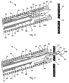
- FIG. 1 depicts a medical device 20 constructed in accordance with the teachings of the present invention.
- the medical device 20 generally comprises a needle 22 and a suture lock 48, which may be employed via inner and outer sheaths 24 and 26.
- the medical device 20 is designed for delivering tissue anchors 28a, b through tissue, e.g., for closing a perforation 14, or for apposing tissue, for example, in gastroesophageal reflux disease (GERD) therapy, or bariatric surgery in which an anastamosis is formed, or for use in other procedures.
- the device 20 preferably includes a pusher 25 extending through the needle 22 for expelling the anchors 28 therefrom.
- the needle 22 defines a needle lumen 30 and a longitudinal axis 10 of the medical device 20.
- the needle 22 is preferably constructed of a metal or alloy such as stainless steel or nitinol, although other metals, alloys and plastics can be used for the needle 22, as is known in the art.
- the needle lumen 30 is sized to slidably receive a plurality of tissue anchors 28a, b therein.
- the tissue anchors 28a, b generally comprise an anchoring member 32 and a suture 34 attached thereto, and the anchoring member 32 is received within the needle lumen 30 along with a portion of the suture 34.
- the suture 34 is preferably formed from a flexible material, such as nylon and of the monofilament variety, although the suture 34 may be formed from metal wire, including single filament and multi-filament wires, and wound and braided wires, plastic strings, rope and the like.
- the suture 34 preferably has a diameter in the range of about 0.20 mm to about 0.35 mm, and most preferably about 0.287 mm, although other sizes may be used and the suture lock 48 sized accordingly.
- a distal end 36 of the needle 22 also defines a needle slot 38 that is longitudinally extending and opens longitudinally at the distal end 36 of the needle 22.
- the slot 38 is sized to receive the sutures 34 therein.
- the slot 38 may be sized and structured to frictionally engage the sutures 34 therein to provide improved retention of the tissue anchors 28a, b within the distal end 36 of the needle 22 during manipulation of the needle, e.g., during preparation for a procedure.
- the slot 38 may have a width sized to be less than or equal to a width of the sutures 34. In this manner, the needle 22 frictionally engages the sutures 34 to retain the tissue anchors 28a, b within the needle lumen 30.
- the needle 22 may not include the slot 38, although it is preferable to keep the sutures 34 safe from the sharp distal tip 37 of the needle 22 through provision of the slot 38, or the width of the slot 38 can be sized larger than a diameter of the sutures 34.
- a biodegradable or resorbable spacer member 46 is preferably positioned between anchoring members 32 of the tissue anchors 28a, b within the needle lumen 30. While the figures illustrate one spacer member 46 positioned between two tissue anchors 28a, b, it will be recognized by those skilled in the art that a larger number of tissue anchors 28a, b may be disposed within the needle lumen 30, and thus a larger number of spacer members 46 may likewise be disposed within the needle lumen 30. In addition, more than one spacer member 46 may be positioned between adjacent tissue anchors 28a, b to provide a larger distance between tissue anchors 28a, b.
- resorbable refers to the ability of a material to be absorbed into a tissue and/or body fluid upon contact with the tissue and/or body fluid.
- a number of resorbable materials are known in the art, and any suitable resorbable material can be used. Examples of suitable types of resorbable materials include resorbable homopolymers, copolymers, or blends of resorbable polymers.
- suitable resorbable materials include poly-alpha hydroxy acids such as polylactic acid, polylactide, polyglycolic acid (PGA), or polyglycolide; tri- methlyene carbonate; polycaprolactone; poly-beta hydroxy acids such as polyhydroxybutyrate or polyhydroxyvalerate; or other polymers such as polyphosphazines, polyorgano- phosphazines, polyanhydrides, polyesteramides, poly- orthoesters, polyethylene oxide, polyester-ethers (e.g., poly- dioxanone) or polyamino acids (e.g., poly-L-glutamic acid or poly-L-lysine).
- poly-alpha hydroxy acids such as polylactic acid, polylactide, polyglycolic acid (PGA), or polyglycolide
- tri- methlyene carbonate such as polycaprolactone
- poly-beta hydroxy acids such as polyhydroxybutyrate or polyhydroxyvalerate
- other polymers such as polypho
- resorbable polymers that may be suitable, including modified polysaccharides, such as cellulose, chitin, and dextran, and modified proteins, such as fibrin and casein.
- modified polysaccharides such as cellulose, chitin, and dextran
- modified proteins such as fibrin and casein.
- Another example of a suitable resorbable material includes bio-remodelable, extracellular matrix material (ECM).
- ECM extracellular matrix material
- One suitable form of ECM is harvested from porcine or bovine small intestine submucosa (SIS).
- SIS is a resorbable, acellular, naturally occurring tissue matrix composed of ECM proteins in various growth factors.
- biodegradable materials that degrade, but are not necessarily resorbed or adsorbed by the bodily tissues, are known in the art and any suitable biodegradable material can be used.
- the longitudinal length of the needle slot 38 which is sized to receive the sutures 34 of the tissue anchors 28a, b therein, is dependent upon the number of tissue anchors 28a, b and spacer members 46 within the needle lumen 30 and the lengths of the corresponding anchoring members 32 of the tissue anchors 28a, b and the spacer members 46.
- the number of spacer members 46 is one less than the number (n T ) of tissue anchors 28a, b.
- the length of the anchoring members 32 is preferably between around 6 mm and 10 mm, most preferably around 8 mm.
- the length of the spacer members 46 is preferably between around 3 mm and 6 mm, most preferably around 5 mm.
- the needle 22 In one preferred construction in which the needle 22 houses two tissue anchors 28a, b disposed within the needle lumen 30 and one spacer member 46 positioned between the two tissue anchors, the needle 22 has an outer diameter of about 1,0668 mm (.042 inches), an inner diameter of about 0,8128 mm (.032 inches), and the slot 38 has a longitudinal length of about 12 mm to about 21 mm, and most preferably about 17 mm.
- two anchors and one spacer are used in the system 40.
- the medical device 20 further includes an over-the-needle suture lock 48 for fixing the sutures 34 of the tissue anchors 28a, b after delivery of the tissue anchors 28a, b through a bodily wall 12.
- An over-the-needle suture lock 48 allows the sutures 34 of the tissue anchors 28a, b to be preloaded within the suture lock 48 during delivery of the tissue anchors 28a, b through the bodily wall 12.
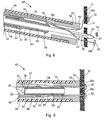
- the suture lock 48 generally includes a locking pin or plug 50 and a retaining sleeve 52 which cooperate to fix the sutures 34 of the tissue anchors 28a, b relative to tissue of the bodily wall 12 for closing the perforation 14 in the bodily wall 12.
- the retaining sleeve 52 and plug 50 have been depicted as having circular cross-sections, it will be recognized that other cross-sectional shapes may be used including triangular, square, etc.
- the retaining sleeve 52 generally includes a tubular body 54 having an interior surface 56 defining an interior passageway 58.
- a peripheral rim 60 is formed at a distal end of the tubular body 54, and defines a shoulder 62 which is used for placement of the retaining sleeve 52, as will be discussed in further detail herein.
- the retaining sleeve 52 receives the sutures 34 of the tissue anchors 28a, b within the interior passageway 58.
- the sutures 34 are then fixed in place using the plug 50, which is designed to fit within the passageway 58 and pinch or compress the sutures 34.
- the plug 50 may have many configurations (e.g. regular or irregular shapes), and constructions (e.g. cast, molded, machined, wound (such as with wire), etc.) so long as a portion of the plug 50 cooperates with the retaining sleeve 52 to fix the sutures 34.
- the plug 50 generally includes a main body 64 having an interior surface 66 defining an interior passageway 68 sized to slidably receive the needle 22.
- the main body 64 and the interior passageway 68 define a longitudinal axis 65.
- the main body 64 includes a grip 70 and a stop 72, each extending radially from the main body 64.
- the grip 70 is formed at a distal end of the plug 50, although it could be moved proximally along the length of the main body 64.
- the grip 70 defines an annular edge 74 that is used to engage the sutures 34, as will be discussed in more detail herein.
- the grip 70 includes a leading surface 76 located distally of the annular edge 74, and a trailing surface 78 located proximally of the annular edge 74.
- the leading surface 76 tapers, and most preferably is curved such as the dome-shaped surface (e.g., semi-spherical) shown in FIGS. 2-3 .
- the trailing surface 78 is generally transverse to the longitudinal axis 65.
- the leading and trailing surfaces 76, 78 have apertures corresponding to the interior passageway 68 in the plug 50 such that they are annular or ring shaped.
- the main body 64 also includes a tapered portion 64a and reduced diameter portion 64b located between the grip 70 and the stop 72.
- the stop 72 is longitudinally spaced from the grip 70 and is used to control the position of the plug 50 within the retaining sleeve 52.
- the stop 72 generally includes a distally facing surface 79 and a proximally facing surface 80.
- the proximally facing surface 80 and the main body 64 define a shoulder 82 which is used to position the plug 50, as will be discussed in more detail herein.
- the stop 72 is positioned relative to the grip 70 to prevent the grip 70 from passing completely through the internal passageway 58 of the retaining sleeve 52.
- FIGS. 5 and 9-10 depict a locked configuration of the suture lock 48 (the unlocked configuration being shown in FIGS. 1-2 and 6-8 ).
- the interior passageway 58 of the retaining sleeve 52 is sized to receive at least a portion of the plug 50 therein.
- the main body 64 and grip 70 are received within the interior passageway 58 of the retaining sleeve 52.
- the sutures 34 are compressed between the grip 70 and the interior surface 56 of the tubular body 54.
- the plug 50 is advanced (i.e. distally) from left to right in FIGS.
- the tapered leading surface 76 of the grip 70 allows the plug 50 to be translated distally relative to the sutures 34 and retaining sleeve 52.
- the sutures 34 are maintained in a fixed relationship relative to one another and to the tissue of the bodily wall 12.
- the sutures 34 are generally in tension, due in part to the natural elasticity of the bodily tissue 12, which generally attempts to pull the sutures 34 distally. Accordingly, while the plug 50 may be advanced through the retaining sleeve 52 and slid alongside the sutures 34 into the locked configuration, the tension on the sutures 34 also exerts a distally directed force on the plug 50 via the grip 70 and its annular edge 74.
- the suture lock 48 is a form of self-motivating locking device that promotes secure fixation of the sutures 34 relative to the tissue 12.
- the sutures 34 may be pulled in the proximal direction to adjust suture tension, suture lock position, and/or perforation closure, even when the suture lock 48 is in the locked configuration.
- the main body 64 is sized to at least partially compress the sutures 34 against the interior surface 56 of the tubular body 54.
- the tapered portion 64a and reduced diameter portion 64b provide an area of limited or no contact with the sutures 34. These areas may be sized to adjust the level of friction between the sutures 34 and the suture lock 48, for example based on the type and size of suture material.
- the stop 72 abuts against a proximal end surface 55 of the tubular body 54, thereby limiting the position of the plug 50 within the retaining sleeve 52.
- the distally facing surface 79 of the stop 72 is generally tapered to slightly compress the sutures 34 against the tubular body 54, while still allowing the sutures 34 to exit the suture lock 48 and be translated in a proximal direction.
- the components of the suture lock 48 may be constructed of various materials, such as stainless steel, titanium, nitinol or other metals/alloys, although various ceramics or plastics can also be employed, such as polycarbonates (PC), polyamides including Nylon(TM), polytetrafluorethylenes (i.e. PTFE and EPTFE), polyethylene ether ketones (PEEK), polyvinylchlorides (PVC), polyimides, polyurethanes, and polyethylenes (high, medium or low density), including multi-layer or single layer constructions with or without reinforcement wires, coils or filaments.
- PC polycarbonates
- PC polyamides including Nylon(TM), polytetrafluorethylenes (i.e. PTFE and EPTFE), polyethylene ether ketones (PEEK), polyvinylchlorides (PVC), polyimides, polyurethanes, and polyethylenes (high, medium or low density), including multi-layer or single layer constructions with or without reinforcement wires, coil
- the plug 50 has a length of about 6,5786 mm (259 in)
- the main body 64 has an outer diameter of about 1,651 mm (.065 in) along a center region and an outer diameter of about 1,524 mm (.060 in) along a proximal region (which is received within the inner sheath 24) and an inner diameter of about 1,143 mm (.045 in) defining the interior passageway 68
- the stop 72 has an outer diameter of about 2,032 mm (080 in)
- the annular edge 74 defining the grip 70 has an outer diameter of about 1,8288 mm (072 in).
- the retaining sleeve 52 has a length of about 3,81 mm (150 in), and the tubular body 54 has an outer sleeve has length of and the tubular has diameter of about 2,54 mm (100 in) and an inner diameter of about 2,032 mm (.080 in) defining the interior passageway 58. While these dimensions of a currently preferred embodiment have been described, the dimensions may be increased or decreased, scaled up or down, to accommodate differently sized anchors, sutures, needles, sheaths, and bodily walls or tissue structures.
- the inner sheath 24 defines an inner sheath lumen 42 which is sized to slidably receive the needle 22 therein.
- the inner sheath 24 is sized and positioned to engage or abut the shoulder 82 of the plug 50.
- the outer sheath 26 defines an outer sheath lumen 44 which is sized to slidably receive the inner sheath 24 and the plug 50 therein.
- the outer sheath 26 is sized and positioned to engage or abut the shoulder 62 of the retaining sleeve 52.
- the inner sheath 24 has an outer diameter of about 1,7272 mm (.068 in) and an inner diameter of about 1,143 mm (.045 in) such that the proximal portion of the main body 64 of the plug 50 (having an outer diameter of about 1,524 mm (.060 in) is press fit within the distal end of the inner sheath 24, wherein the inner sheath 24 stretches slightly to hold the plug 50 in place. The plug 50 can then be detached from the inner sheath 24 with a relatively low force.
- the outer sheath 26 has an outer diameter of about 3,3274 mm (131 in) and an inner diameter of about 2,413 mm (.095 in) such that the tubular body 54 of the retaining sleeve 52 (having an outer diameter of about 2,54 mm (.100 in) is press fit within the distal end of the outer sheath 26, wherein the outer sheath 26 stretches slightly to hold the retaining sleeve 52 in place. The retaining sleeve 52 can then be detached from the outer sheath 26 with a relatively low force.
- the inner diameter of the inner and outer sheaths 24 and 26 may be sized larger relative to the plug 50 and retaining sleeve 52, respectively. In this manner, the plug 50 and the retaining sleeve 52 are received by and maintained within the distal ends of the inner and outer sheaths 24 and 26, respectively, by an adhesive or any other suitable means known in the art.
- the inner and outer sheaths 24 and 26 are preferably formed of a plastic such as polytetrafluorethylene (PTFE), expanded polytetrafluorethylene (EPTFE), polyethylene ether ketone (PEEK), polyvinylchloride (PVC), polycarbonate (PC), polyamide including nylon, polyimide, polyurethane, polyethylene (high, medium or low density), or elastomers such as Santoprene ® , including multi-layer or single layer constructions with or without reinforcement wires, coils or filaments.
- the needle 22, inner and outer sheaths 24 and 26, and the pusher 25 are preferably elongated structures that are flexible, allowing navigation within a patient's body such as during endoscopic or laparoscopic procedures.
- a suitable handle or control mechanism will be connected to the proximal ends of the needle 22, inner and outer sheaths 24 and 26, and the pusher 25 for relative translation of these components by the medical professional, as is known in the art.
- the medical devices 20 and systems 40 are also applicable to other tissue anchor placement devices that may be used in open surgery, on external wounds, or that otherwise do not require an elongated medical device to access the targeted tissue.
- the medical device 20 may be sized to be used through an accessory channel of an endoscope or alongside an endoscope, or in combination with other devices used in conjunction with endoscopes, for example, endoscopic suction devices or fluid injection devices.
- the medical device 20 is operable between at least a delivery configuration, depicted in FIG. 6 , and a deployed configuration, depicted in FIGS. 7-8 .
- the needle 22 is substantially contained within the outer sheath lumen 44 so as to protect bodily structures from the sharp distal tip 37 of the needle 22 during introduction of the medical device 20.
- the needle 22 is translated relative to the inner and outer sheaths 24 and 26 such that the needle 22 projects beyond the distal end 27 of the outer sheath 26.
- the pusher 25 is translated relative to the needle 22 such that the distal-most tissue anchor 28a is urged distally out of the distal tip 37 of the needle 22.
- the suture 34 connected to the tissue anchor 28a also slides distally within the needle slot 38 and exits the needle 22.
- the needle 22 is retracted within the outer sheath lumen 44, the medical device 20 is repositioned, and the steps of translating the needle 22 relative to the outer sheath 26 and the pusher 25 relative to the needle 22 are repeated for additional tissue anchors.
- the method includes providing a medical system having a plurality of tissue anchors and at least one resorbable spacer member positioned in between adjacent tissue anchors, a needle and inner and outer sheaths, and a suture lock, such as the medical system 40 depicted in FIGS. 1 and 6-10 .
- the medical system 40 is delivered to a position proximate the bodily tissue 12 that has been targeted for placement of the tissue anchors 28a, b.
- the medical system 40 may include a visualization system for assisting in locating the tissue 12, identifying a target site for deployment of the tissue anchors 28a, b, and monitoring operation of the medical device 20 and system 40.
- visualization techniques may include catheter-based fiber optic systems, fluoroscopy, ultrasound or the like.
- the needle 22 can have markings designed for viewing under fluoroscopy, and the distal end 36 of the needle 22 can have a surface of enhanced ultrasonic reflectivity, such by being roughened, having dimples or other incongruities, or having embedded particles.
- the tissue anchors 28a, b are disposed within the needle lumen 30 at the distal end 36 of the needle 22 and a spacer member 46 is disposed between the tissue anchors 28a, b. Spaces between the spacer member 46 and the tissue anchors 28a, b have been shown for clarity, but the spacer member 46 and the tissue anchors 28a, b would generally be abutting end-to-end within the needle lumen 30.
- the sutures 34 follow a somewhat tortuous path from within the needle lumen 30, through the needle slot 38, extending proximally within the outer sheath lumen 44 between the interior surfaces of the retaining sleeve 52 and the outer sheath 26 and the exterior surfaces of the plug and the inner sheath 24, the sutures 34 effectively being preloaded within the suture lock 48 and extending to a proximal end of the medical device 20. Accordingly, this tortuous path can be sufficient to retain the tissue anchors 28a, b within the needle lumen 30, through frictional engagement of the sutures 34 between the exterior surface of the inner sheath 24 and the interior surface of the outer sheath 26.
- the medical device 20 and system 40 are operated into their deployed configuration, as shown in FIG. 7 .
- the needle 22 is deployed through the bodily tissue 12 by translating the needle 22 relative to the inner and outer sheaths 24 and 26.
- the distal-most tissue anchor 28a is then deployed from the needle 22 by translating the tissue anchor 28a relative to the needle 22 so that the tissue anchor 28a exits the needle lumen 30.
- FIG. 7 shows that the needle 22 is deployed through the bodily tissue 12 by translating the needle 22 relative to the inner and outer sheaths 24 and 26.
- the distal-most tissue anchor 28a is then deployed from the needle 22 by translating the tissue anchor 28a relative to the needle 22 so that the tissue anchor 28a exits the needle lumen 30.
- the tissue anchors 28a, b and the spacer member 46 positioned therebetween are shown aligned within the needle lumen 30 along the longitudinal axis 10 of the needle lumen 30 and medical device 20 such that the pusher 25 may be slidably received within the inner sheath lumen 24 and used to engage and press on the proximal-most tissue anchor 28b.
- the pusher 25 is advanced distally to press upon the anchoring member 32 of the proximal-most tissue anchor 28b, which will in turn transmit force through the spacer member 46 and the distal-most tissue anchor 28a, thus advancing the distal-most tissue anchor 28a out of the needle lumen 30.
- the spacer member 46 is moved distally to a position slightly past the needle tip 37, the pusher 25 may be retracted slightly and, due to the adequate clearance between the spacer member 46 and the inner diameter of the needle 22, as the needle pierces the tissue 12, the spacer member 46 is easily moved proximally within the needle lumen 30 to ensure that the sharpened needle tip 37 is able to pierce through the tissue 12 for deployment of the remaining tissue anchors 28a, b.
- the needle 22 is retracted back through the bodily tissue 12 by translating the needle 22 proximally, repositioned at a different position about the perforation 14, and redeployed back through the tissue 12 by translating the needle 22 relative to the inner and outer sheaths 24 and 26.
- the pusher 25 is then further advanced distally to deploy the spacer member 46 and the proximal tissue anchor 28b, wherein the suture 34 of the tissue anchor 28b is released from within the needle slot 38.
- the spacer member 46 may be deployed through the tissue 12 with the proximal tissue anchor 28b, as shown in FIG. 8 .
- the spacer member 46 may be deployed within the body prior to passing the needle 22 through the tissue 12 to deploy the proximal anchor 28b.
- the spacer member 46 may be deployed within the gastrointestinal tract, wherein the spacer member 46 passes naturally. Since the spacer member 46 is resorbable, it is inconsequential that it is left within the patient's body. Thus, if the spacer member 46 accidentally falls out of the tip 37 of the needle 22 before being deployed with the proximal tissue anchor 28b, this is of no consequence. The proximal tissue anchor 28b is still positioned sufficiently proximal within the needle lumen 30 to be deployed appropriately at the repositioned location.
- the spacer member 46 may contain antibiotics or other drugs, hormones, or growth factors that facilitate healing of the tissue 12 around the implanted tissue anchors 28a, b.
- the medical system 40 in accordance with the teachings of the present invention, provides the ability to sequentially deploy multiple tissue anchors, in which tissue anchors and spacer members disposed between adjacent tissue anchors are preloaded within the needle 22. Accordingly, the longitudinal length of needle slot 38 can be sized to accommodate any number of sutures 34.
- the method may therefore include withdrawing the needle 22 from the bodily tissue 12 by translating the needle 22 proximally, and then repeating the steps of translating the needle 22 through the tissue 12 and deploying another tissue anchor 28 therethrough.
- the needle 22 is retracted back through to the proximal side of the bodily tissue 12 and retracted within the inner sheath lumen 42.
- the needle 22 may be removed from within the medical device 20 at this time or it may be removed with the entire medical device 20 after fixation of the sutures 34 relative to the tissue 12.
- the suture lock 48 is engaged to fix the sutures 34 relative to the bodily tissue 12.
- the system 40 again does not require removal from the body, as it includes the over-the-needle suture lock 48.
- the retaining sleeve 52 is fitted onto the distal end 27 of the outer sheath 26.
- the outer sheath 26 may take the form of any sheath or catheter known in the art, but preferably has sufficient strength and rigidity for both longitudinal and rotational force transmission, while still providing flexibility for navigation of a patient's body.
- Exemplary sheaths are sold by Cook Medical, Inc. It will also be recognized that other sheaths or pushing elements may be employed, such as solid wires or wire guides, clamps, graspers and the like. Magnets could likewise be employed to releasably connect the outer sheath 26 to the retaining sleeve 52.
- the outer sheath lumen 44 is sized to receive the tubular body 54 of the retaining sleeve 52, while a distal end surface 29 of the outer sheath 26 abuts against the shoulder 62 of the retaining sleeve 52.
- the outer sheath 26 and retaining sleeve 52 are loosely press fit such that the retaining sleeve 52 may be readily controlled and positioned using the outer sheath 26.
- the retaining sleeve 52 maintains its connection to the outer sheath 26 during placement of the plug 50 within the retaining sleeve 52, while at the same time the retaining sleeve 52 is also readily disconnected from the outer sheath 26 at the end of the procedure.
- outer sheath 26 and retaining sleeve 52 need not be sized to frictionally engage, as the tensioned sutures 34 and the tissue 12 will generally maintain the position of the retaining sleeve 52 on the outer sheath 26 during placement of the plug 50, such as is shown in FIGS. 9 and 10 .
- the sutures 34 are preloaded or threaded through the interior passageway 58 of the retaining sleeve 52 and through the outer sheath lumen 44.
- the outer sheath 26 is used to distally translate the retaining sleeve 52 over the sutures 34 to a position proximate the tissue 12 and perforation 14.
- the sutures 34 are tensioned in order to draw the perforation 14 closed and press the tissue 12 against the peripheral rim 60 of the retaining sleeve 52.
- the inner sheath 24 is press fit with the plug 50, although the two structures may simply abut each other for longitudinal translation.
- the inner sheath 24 may have a construction similar to the outer sheath 26 or other catheter described above.
- the inner sheath 24 includes a distal end 23 sized to abut against the shoulder 82 and receive the main body 64 of the plug 50, respectively. Accordingly, the inner sheath 24 is connected to the plug 50 and together they are translated distally through the outer sheath lumen 44. If the needle 22 has not yet been withdrawn from the medical device 20 during securing of the sutures 34, the inner sheath 24 causes the plug 50 to slide over-the-needle 22.
- the plug 50 is pressed into engagement with the retaining sleeve 52 to fix the sutures 34 therebetween. With the sutures 34 in tension (e.g. by pulling them in a proximal direction), the plug 50 is advanced through the interior passageway 58 of the retaining sleeve 52, whereby the sutures 34 are compressed between the grip 70 and the interior surface 56 of the retaining sleeve 52. It can therefore be seen that relative translation of the outer sheath 26 and the inner sheath 24 controls the relative positions of the retaining sleeve 52 and the plug 50 to operate the suture lock 48 between a locked configuration and an unlocked configuration.
- the leading surface 76 of the grip 70 is slid along the sutures 34 as the plug 50 is distally advanced through the interior passageway 58. With further advancement, the main body 64 also engages the sutures 34 and at least partially compresses them against the interior surface 56 of the retaining sleeve 52.
- the annular shape of the grip 70 allows the sutures 34 to be positioned anywhere around the outer periphery of the grip 70 and plug 50. Distal movement of the plug 50 is eventually limited by the stop 72, and namely the distally facing surface 79 of the stop 72 abutting against the proximal end surface 55 of the retaining sleeve 52.
- the tension on the sutures 34 grips into the annular edge 74 of the grip 70, and serves to promote movement of the plug 50 in the distal direction, as well as resist proximal movement and unlocking of the suture lock 48.
- the grip 70 When in the locked configuration (and when partially locked such as when the plug 50 is partially placed within the retaining sleeve 52 but not fully seated), the grip 70 is structured to permit further translation of the sutures 34 proximally, i.e. away from the tissue 12, and prevent translation of the sutures 34 distally, i.e. towards the tissue 12. Further, the sutures 34 may be individually pulled or tensioned in order to orient the suture lock 48 relative to the bodily tissue 12 and perforation 14, even when the sutures 34 are compressed by the plug 50 and retaining sleeve 52, such as when the suture lock 48 is in the locked configuration. As such, tension on the sutures 34 may be modified to adjust how the perforation 14 is closed. This represents a marked improvement over existing suture locks, which typically are permanently fixed in position along the sutures such that adjustment during and after the locking procedure, i.e. in partially locked and finally locked configurations, is not possible.
- the retaining sleeve 52 and plug 50 are interconnected through their respective frictional engagement with the sutures 34 and compression thereof.
- the entire medical device 20 may be removed from the patient at once, the inner and outer sheaths 24 and 26 being easily removed from the retaining sleeve 52 and the plug 50, respectively.
- the inner sheath 24 and needle 22 may be removed first and the outer sheath 26 removed separately.
- the sutures 34 may be trimmed as necessary with endoscopic scissors and the like.
- the sutures 34 may be cut, or the outer sheath 26 may be used to hold the retaining sleeve 52 while the plug 50 is grasped (such as with a snare, forceps, or similar device) and physically withdrawn against the friction and tension of the sutures 34.
- the present invention provides a medical system capable of delivering multiple tissue anchors in a controlled manner, as well as locking the anchors together (e.g., to close a perforation) without needing to withdraw and introduce the system (or multiple medical devices) any number of times, thereby saving time and improving efficiency. Since the sutures connected to the tissue anchors are preloaded within the over-the-needle suture lock, one medical system is provided for both the delivery of multiple tissue anchors and the fixation of their sutures.
- the medical system is simple and reliable in use, provides complete perforation closure, and is adaptable to a variety of suture fixation and perforation closure applications.
- any number of suture strands may be employed and the relative sizes of the plug and retaining sleeve may be adjusted based on suture size, perforation size and the like.
- the interconnection of the plug and retaining sleeve is such that the suture lock is self-motivated and biased towards a locked configuration, thereby assisting and promoting complete perforation closure as well as control over the position of the suture lock relative to the tissue being sutured through adjustment of the suture strands even when they are compressed. Further description of the interconnection between the plug and retaining sleeve may be found in co-pending U.S. 2 008 300 629 A1 .
- Adjustment of individual suture tension and location of the suture lock are also possible during and after placement of the suture lock.
- the inner and outer sheaths provide a simple system for deployment of multiple tissue anchors that can be traversed through the body of a patient to even the most remote locations.
- the devices and methods described above generally include placing the tissue anchors in tissue through an internal bodily lumen
- the devices and methods may be used on any layer of material (e.g. fabrics, cloth, polymers, elastomers, plastics and rubber) that may or may not be associated with a human or animal body and a bodily lumen.
- the devices disclosed herein can find use in laboratory and industrial settings for placing devices through one or more layers of material that may or may not find application to the human or animal body, and likewise closing holes or perforations in layers of material that are not bodily tissue.
- Some examples include sewing or stitching and related manufacturing, working with synthetic tissues, connecting polymeric sheets, animal studies, and post-mortem activities.
Landscapes
- Health & Medical Sciences (AREA)
- Life Sciences & Earth Sciences (AREA)
- Surgery (AREA)
- Molecular Biology (AREA)
- Engineering & Computer Science (AREA)
- Biomedical Technology (AREA)
- Heart & Thoracic Surgery (AREA)
- Medical Informatics (AREA)
- Nuclear Medicine, Radiotherapy & Molecular Imaging (AREA)
- Animal Behavior & Ethology (AREA)
- General Health & Medical Sciences (AREA)
- Public Health (AREA)
- Veterinary Medicine (AREA)
- Rheumatology (AREA)
- Cardiology (AREA)
- Surgical Instruments (AREA)
Claims (12)
- Medizinische Vorrichtung (20) mit:mindestens einem Gewebeanker (28), der ein Ankerelement (32) aufweist, das mit einem Faden (34) verbunden ist;einer Nadel (22), die ein Nadellumen (30) aufweist, das derart dimensioniert ist, dass es den mindestens einen Gewebeanker (28) verschiebbar aufnimmt, wobei die Nadel (22) und das Nadellumen (30) eine Längsachse (10) der medizinischen Vorrichtung (20) definieren;einer Fadenklemmvorrichtung (48) zum Fixieren des Fadens (34) nach Ausbringen des mindestens einen Gewebeankers (28);einem inneren Mantel (24), der ein inneres Mantellumen (42) aufweist; undeinem äußeren Mantel (26), der ein äußeres Mantellumen (44) aufweist, das derart dimensioniert ist, dass es den inneren Mantel (24) verschiebbar aufnimmt;dadurch gekennzeichnet, dassdie Fadenklemmvorrichtung (48) umfasst:einen Pfropfen (50) mit einem Hauptkörper (64), der eine erste Innenwand (26) aufweist, die einen ersten inneren Durchgang (68) definiert, undeine Haltehülse (52) mit einem rohrförmigen Körper (54), der eine zweite Innenwand (46) aufweist, die einen zweiten inneren Durchgang (58) definiert, der derart dimensioniert ist, dass er den Pfropfen (50) darin aufnimmt und der Faden (34) des mindestens einen Gewebeankers (28) zwischen dem Pfropfen (50) und der zweiten Innenwand (56) der Haltehülse (52) in Eingriff kommt, wobei sowohl der erste (68) als auch der zweite (58) innere Durchgang derart dimensioniert sind, dass sie die Nadel (62) während des Ausbringens des mindestens einen Gewebeankers (28) verschiebbar aufnehmen;wobei der innere Mantel (24) in Eingriff mit dem Pfropfen (50) gebracht werden kann, und wobei das innere Mantellumen (42) derart dimensioniert ist, dass es die Nadel (22) verschiebbar aufnimmt, wobei eine Translationsbewegung des inneren Mantels (24) den Pfropfen (50) über die Nadel (22) gleiten lässt; undwobei der äußere Mantel (26) mit der Haltehülse (52) in Eingriff gebracht werden kann, und das äußere Mantellumen (44) derart dimensioniert ist, dass es den Pfropfen (50) verschiebbar aufnimmt.
- Medizinische Vorrichtung (20) nach Anspruch 1, wobei der Pfropfen (50) ein Einspannelement (70) und einen Anschlag (72) umfasst, wobei sich beide in radialer Richtung von dem Hauptkörper (64) erstrecken, wobei der Anschlag (72) eine erste Schulter (82) definiert, die in proximale Richtung gewandt ist, wobei die Haltehülse (52) eine umlaufende Randzone (60) umfasst, die sich in radialer Richtung erstreckt und eine zweite Schulter (62) definiert, die in proximale Richtung gewandt ist, wobei der innere Mantel (24) derart dimensioniert und angeordnet ist, dass er an der ersten Schulter (82) des Pfropfens (50) anschlägt, und wobei der äußere Mantel (26) derart dimensioniert und angeordnet ist, dass er an der zweiten Schulter (62) der Haltehülse (52) anschlägt.
- Medizinische Vorrichtung (20) nach Anspruch 1, wobei die Fadenklemmvorrichtung (48) zwischen einer entriegelten Konfiguration während des Ausbringens des mindestens einen Gewebeankers (28) durch Gewebe (12) und einer verriegelten Konfiguration nach Ausbringen des mindestens einen Gewebeankers (28) durch das Gewebe (12) steuerbar ist, wobei der Pfropfen (50) und die Haltehülse (52) in der entriegelten Konfiguration voneinander getrennt sind und in der verriegelten Konfiguration miteinander in Eingriff stehen, wobei der Pfropfen (50) und die Haltehülse (52) derart dimensioniert und strukturiert sind, dass sie den Faden (34) des mindestens einen Gewebeankers (28) in der verriegelten Konfiguration zwischen dem Pfropfen (50) und der zweiten Innenwand (56) der Haltehülse (52) einklemmen.
- Medizinische Vorrichtung (20) nach Anspruch 1, wobei die medizinische Vorrichtung (20) zwischen einer Ausbringkonfiguration und einer Anwendungskonfiguration betätigbar ist, wobei in der Ausbringkonfiguration der innere Mantel (24), der Pfropfen (50) und die Nadel (22) im Wesentlichen im äußeren Mantellumen (44) aufgenommen sind, und wobei die Nadel (22) in der Anwendungskonfiguration über ein distales Ende (27) des äußeren Mantels (26) und die Haltehülse (52) herausragt.
- Medizinische Vorrichtung (20) nach Anspruch 1, wobei sich der Faden (34) des mindestens einen Gewebeankers (28) in proximaler Richtung und zwischen dem Pfropfen (50) und der Haltehülse (52) erstreckt.
- Medizinische Vorrichtung (20) nach Anspruch 1, wobei sich der Faden (34) des mindestens einen Gewebeankers (28) in einer proximalen Richtung entlang einer Außenseite des inneren Mantels (24) und innerhalb des äußeren Mantellumens (44) erstreckt.
- Medizinische Vorrichtung (20) nach Anspruch 1, wobei der Faden (34) des mindestens einen Gewebeankers (28) innerhalb der Fadenklemmvorrichtung (48) vorgespannt ist.
- Medizinische Vorrichtung (20) nach Anspruch 1, wobei ein distales Ende der Nadel (22) eine Nadelnut (38) definiert, die derart dimensioniert ist, dass sie den Faden (34) des mindestens einen Gewebeankers (28) darin aufnimmt.
- Medizinische Vorrichtung (20) nach Anspruch 1, wobei der mindestens eine Gewebeanker (28) eine Mehrzahl an Gewebeankern (28) umfasst, und wobei die Ankerelemente (32) der Mehrzahl an Gewebeankern (28) in Reihe innerhalb des Nadellumens (30) angeordnet sind.
- Medizinische Vorrichtung (20) nach Anspruch 9, weiterhin mit einem Stößel (25), der verschiebbar innerhalb des Nadellumens (30) aufgenommen ist, wobei der Stößel (25) derart dimensioniert und angeordnet ist, dass er ein proximalstes Ankerelement (32) der Mehrzahl an Gewebeankern (28) in Eingriff nimmt.
- Medizinische Vorrichtung nach Anspruch 9, wobei die Mehrzahl an Gewebeankern (28) erste (28a) und zweite (28b) Gewebeanker umfasst, die entsprechende erste und zweite Ankerelemente (32) aufweisen, wobei die medizinische Vorrichtung (20) weiterhin mindestens ein absorbierbares Abstandselement (46) umfasst, das zwischen dem ersten und dem zweiten Ankerelement (32) innerhalb des Nadellumens (30) angeordnet ist.
- Medizinische Vorrichtung (20) nach Anspruch 11, wobei ein distales Ende der Nadel (22) eine Nadelnut (38) definiert, die derart dimensioniert ist, dass sie den Faden (34) von jedem Gewebeanker (28) der Mehrzahl an Gewebeankern (28) darin aufnimmt, wobei die Nadelnut (38) eine Nutlänge umfasst, die von der Anzahl und der Länge der Ankerelemente (32) der Mehrzahl an Gewebeankern (28) und von der Anzahl und Länge der Abstandselemente (46), die innerhalb des Nadellumens (30) aufgenommen sind, abhängig ist.
Applications Claiming Priority (2)
| Application Number | Priority Date | Filing Date | Title |
|---|---|---|---|
| US16636109P | 2009-04-03 | 2009-04-03 | |
| PCT/US2010/029798 WO2010115113A1 (en) | 2009-04-03 | 2010-04-02 | Medical devices, systems, and methods for rapid deployment and fixation of tissue anchors |
Publications (2)
| Publication Number | Publication Date |
|---|---|
| EP2413809A1 EP2413809A1 (de) | 2012-02-08 |
| EP2413809B1 true EP2413809B1 (de) | 2014-10-08 |
Family
ID=42211655
Family Applications (1)
| Application Number | Title | Priority Date | Filing Date |
|---|---|---|---|
| EP10712286.3A Active EP2413809B1 (de) | 2009-04-03 | 2010-04-02 | Medizinische vorrichtungen zur schnellen ablage und fixierung von gewebeankern |
Country Status (6)
| Country | Link |
|---|---|
| US (1) | US8382776B2 (de) |
| EP (1) | EP2413809B1 (de) |
| JP (1) | JP5619138B2 (de) |
| AU (1) | AU2010232485B2 (de) |
| CA (1) | CA2757494C (de) |
| WO (1) | WO2010115113A1 (de) |
Cited By (2)
| Publication number | Priority date | Publication date | Assignee | Title |
|---|---|---|---|---|
| US11591554B2 (en) | 2017-09-11 | 2023-02-28 | Heartstitch, Inc. | Methods and devices for papillary suturing |
| US11779324B2 (en) | 2013-12-06 | 2023-10-10 | Med-Venture Investments, Llc | Suturing methods and apparatuses |
Families Citing this family (23)
| Publication number | Priority date | Publication date | Assignee | Title |
|---|---|---|---|---|
| EP2510883B1 (de) | 2005-06-20 | 2018-04-11 | Nobles Medical Technologies, Inc. | Vorrichtung zum Aufbringen eines Knotens auf einer Naht |
| US8317679B2 (en) * | 2008-10-06 | 2012-11-27 | Cook Medical Technologies Llc | Endcap for safely deploying tissue anchors |
| AU2009322353B2 (en) | 2008-12-05 | 2013-04-18 | Cook Medical Technologies Llc | Tissue anchors for purse-string closure of perforations |
| WO2010115072A1 (en) | 2009-04-03 | 2010-10-07 | Wilson-Cook Medical, Inc. | Tissue anchors and medical devices for rapid deployment of tissue anchors |
| US10743854B2 (en) | 2010-01-20 | 2020-08-18 | Micro Interventional Devices, Inc. | Tissue closure device and method |
| US9980708B2 (en) | 2010-01-20 | 2018-05-29 | Micro Interventional Devices, Inc. | Tissue closure device and method |
| WO2011091185A1 (en) | 2010-01-20 | 2011-07-28 | New Hope Ventures, Lp | Tissue closure device and method |
| US10058314B2 (en) | 2010-01-20 | 2018-08-28 | Micro Interventional Devices, Inc. | Tissue closure device and method |
| US10959840B2 (en) | 2010-01-20 | 2021-03-30 | Micro Interventional Devices, Inc. | Systems and methods for affixing a prosthesis to tissue |
| US20110218191A1 (en) * | 2010-03-03 | 2011-09-08 | Boehringer Ingelheim Vetmedica Gmbh | Use of meloxicam for the long term-treatment of kidney disorders in cats |
| US20120053619A1 (en) | 2010-08-31 | 2012-03-01 | Boston Scientific Scimed, Inc. | Hemostatic compositions and methods of making and using same |
| PT2755614T (pt) * | 2011-09-13 | 2018-01-18 | Nxthera Inc | Sistemas de tratamento da próstata |
| EP2959837B1 (de) * | 2013-02-22 | 2019-07-31 | Sumitomo Bakelite Co., Ltd. | Wiederholungsartiges organbefestigungswerkzeug |
| US9629620B2 (en) | 2014-02-13 | 2017-04-25 | Medtronic Vascular, Inc. | Method and apparatus for forming a suture connector in situ |
| US9757117B2 (en) | 2014-02-13 | 2017-09-12 | Medtronic Vascular, Inc. | Method and apparatus for forming a suture connector in situ |
| SG11201704757UA (en) * | 2014-12-10 | 2017-07-28 | Edwards Lifesciences Ag | Multiple-firing securing device and methods for using and manufacturing same |
| CN106999291B (zh) | 2014-12-12 | 2020-01-31 | 皇家飞利浦有限公司 | 用在舌头操纵系统中的舌头前进装置系统 |
| CN106333719B (zh) | 2015-07-07 | 2020-06-26 | 爱彼医疗有限公司 | 用于缝合的设备和方法 |
| CN114099057A (zh) * | 2016-11-29 | 2022-03-01 | 埃斯卡拉医疗公司 | 锚固件递送系统和方法 |
| EP4115818A3 (de) | 2017-06-19 | 2023-04-05 | Heartstitch, Inc. | Nähsysteme und verfahren zum nähen von körpergewebe |
| US20210045731A1 (en) * | 2018-02-01 | 2021-02-18 | Transluminal Technologies, Llc | Trans-radial closure device, deployment apparatus, and method of deploying a trans-radial closure device |
| EP3934546A1 (de) * | 2019-03-06 | 2022-01-12 | Speed Clip Solutions, LLC | Nahtspannungs- und -sicherungsvorrichtung, -system und -verfahren |
| US11382609B2 (en) * | 2020-12-03 | 2022-07-12 | Xdot Medical Inc. | Access site management system for percutaneous vascular access |
Family Cites Families (171)
| Publication number | Priority date | Publication date | Assignee | Title |
|---|---|---|---|---|
| US2199025A (en) | 1936-06-08 | 1940-04-30 | Carl E Conn | Means and method of closing surgical incisions |
| US3556079A (en) | 1967-05-16 | 1971-01-19 | Haruo Omizo | Method of puncturing a medical instrument under guidance of ultrasound |
| US3664345A (en) | 1970-07-06 | 1972-05-23 | Clyde Harwell Dabbs | Surgical buttons |
| US3766610A (en) | 1971-06-29 | 1973-10-23 | A Thorsbakken | Wedge locking device |
| ES199828Y (es) | 1974-01-25 | 1975-12-16 | Manufacturas De Acero | Dispositivo para el anclaje de trenzas y cordones de arma- duras de construccion. |
| US4059333A (en) | 1977-01-05 | 1977-11-22 | Amp Incorporated | Electrical connector |
| US4235238A (en) | 1978-05-11 | 1980-11-25 | Olympus Optical Co., Ltd. | Apparatus for suturing coeliac tissues |
| US5417691A (en) | 1982-05-20 | 1995-05-23 | Hayhurst; John O. | Apparatus and method for manipulating and anchoring tissue |
| US4532926A (en) | 1983-06-20 | 1985-08-06 | Ethicon, Inc. | Two-piece tissue fastener with ratchet leg staple and sealable latching receiver |
| US4604094A (en) | 1984-09-06 | 1986-08-05 | The United States Of America As Represented By The Secretary Of The Department Of Health And Human Services | Toposcopic catheter and method of fabrication |
| JPH0329635Y2 (de) | 1984-11-08 | 1991-06-24 | ||
| US4669473A (en) | 1985-09-06 | 1987-06-02 | Acufex Microsurgical, Inc. | Surgical fastener |
| US5123914A (en) | 1986-05-19 | 1992-06-23 | Cook Incorporated | Visceral anchor for visceral wall mobilization |
| US5203787A (en) | 1990-11-19 | 1993-04-20 | Biomet, Inc. | Suture retaining arrangement |
| US5366480A (en) | 1990-12-24 | 1994-11-22 | American Cyanamid Company | Synthetic elastomeric buttressing pledget |
| US5258015A (en) | 1991-05-03 | 1993-11-02 | American Cyanamid Company | Locking filament caps |
| US5333624A (en) | 1992-02-24 | 1994-08-02 | United States Surgical Corporation | Surgical attaching apparatus |
| US5254126A (en) | 1992-06-24 | 1993-10-19 | Ethicon, Inc. | Endoscopic suture punch |
| DE4228909C2 (de) | 1992-08-28 | 1994-06-09 | Ethicon Gmbh | Endoskopisches Instrument zur Applizierung von Ligaturbindern und Ligaturbinder |
| US5282832A (en) | 1992-10-09 | 1994-02-01 | United States Surgical Corporation | Suture clip |
| IL103737A (en) | 1992-11-13 | 1997-02-18 | Technion Res & Dev Foundation | Stapler device particularly useful in medical suturing |
| US5693060A (en) | 1992-11-17 | 1997-12-02 | Smith & Nephew, Inc. | Suture securing device and method |
| US5403348A (en) | 1993-05-14 | 1995-04-04 | Bonutti; Peter M. | Suture anchor |
| US5423860A (en) | 1993-05-28 | 1995-06-13 | American Cyanamid Company | Protective carrier for suture anchor |
| CA2124651C (en) | 1993-08-20 | 2004-09-28 | David T. Green | Apparatus and method for applying and adjusting an anchoring device |
| US5584835A (en) | 1993-10-18 | 1996-12-17 | Greenfield; Jon B. | Soft tissue to bone fixation device and method |
| IT1269443B (it) | 1994-01-19 | 1997-04-01 | Stefano Nazari | Protesi vascolare per la sostituzione o il rivestimento interno di vasi sanguigni di medio e grande diametro e dispositivo per la sua applicazione senza interruzione del flusso ematico |
| US5486197A (en) | 1994-03-24 | 1996-01-23 | Ethicon, Inc. | Two-piece suture anchor with barbs |
| US5630824A (en) | 1994-06-01 | 1997-05-20 | Innovasive Devices, Inc. | Suture attachment device |
| US5464427A (en) | 1994-10-04 | 1995-11-07 | Synthes (U.S.A.) | Expanding suture anchor |
| US5531763A (en) | 1994-10-07 | 1996-07-02 | United States Surgical Corporation | Suture cinching apparatus |
| US6086608A (en) | 1996-02-22 | 2000-07-11 | Smith & Nephew, Inc. | Suture collet |
| US6132438A (en) | 1995-06-07 | 2000-10-17 | Ep Technologies, Inc. | Devices for installing stasis reducing means in body tissue |
| US5690656A (en) | 1995-06-27 | 1997-11-25 | Cook Incorporated | Method and apparatus for creating abdominal visceral anastomoses |
| US5662683A (en) | 1995-08-22 | 1997-09-02 | Ortho Helix Limited | Open helical organic tissue anchor and method of facilitating healing |
| US5782865A (en) | 1995-08-25 | 1998-07-21 | Grotz; Robert Thomas | Stabilizer for human joints |
| US5674231A (en) | 1995-10-20 | 1997-10-07 | United States Surgical Corporation | Apparatus and method for vascular hole closure |
| US5810848A (en) | 1996-08-21 | 1998-09-22 | Hayhurst; John O. | Suturing system |
| US5948000A (en) | 1996-10-03 | 1999-09-07 | United States Surgical Corporation | System for suture anchor placement |
| US5891159A (en) | 1997-05-02 | 1999-04-06 | Cardiothoratic Systems, Inc. | Automatic purse string suture device |
| US6071292A (en) | 1997-06-28 | 2000-06-06 | Transvascular, Inc. | Transluminal methods and devices for closing, forming attachments to, and/or forming anastomotic junctions in, luminal anatomical structures |
| US6010525A (en) | 1997-08-01 | 2000-01-04 | Peter M. Bonutti | Method and apparatus for securing a suture |
| US5931844A (en) | 1998-03-31 | 1999-08-03 | Smith & Nephew, Inc. | Surgical drive tool |
| EP1079740B1 (de) | 1998-05-21 | 2007-08-29 | Christopher J. Walshe | System zum fixieren von gewebe |
| US6200329B1 (en) | 1998-08-31 | 2001-03-13 | Smith & Nephew, Inc. | Suture collet |
| US6679851B2 (en) | 1998-09-01 | 2004-01-20 | Senorx, Inc. | Tissue accessing and anchoring device and method |
| EP1054703B1 (de) | 1998-12-09 | 2004-09-15 | Cook Incorporated | Super-elastische gebogene hohlnadel zur medizinischen verwendung |
| US6110183A (en) | 1998-12-22 | 2000-08-29 | Cook Incorporated | Suture anchor device |
| CA2293057C (en) | 1998-12-30 | 2008-04-01 | Depuy Orthopaedics, Inc. | Suture locking device |
| US6482178B1 (en) | 1999-05-21 | 2002-11-19 | Cook Urological Incorporated | Localization device with anchoring barbs |
| US7416554B2 (en) | 2002-12-11 | 2008-08-26 | Usgi Medical Inc | Apparatus and methods for forming and securing gastrointestinal tissue folds |
| US7744613B2 (en) | 1999-06-25 | 2010-06-29 | Usgi Medical, Inc. | Apparatus and methods for forming and securing gastrointestinal tissue folds |
| US7618426B2 (en) | 2002-12-11 | 2009-11-17 | Usgi Medical, Inc. | Apparatus and methods for forming gastrointestinal tissue approximations |
| JP4108882B2 (ja) | 1999-08-04 | 2008-06-25 | オリンパス株式会社 | 内視鏡用壁固定具 |
| US6231561B1 (en) | 1999-09-20 | 2001-05-15 | Appriva Medical, Inc. | Method and apparatus for closing a body lumen |
| US6503273B1 (en) | 1999-11-22 | 2003-01-07 | Cyograft Tissue Engineering, Inc. | Tissue engineered blood vessels and methods and apparatus for their manufacture |
| US6635073B2 (en) | 2000-05-03 | 2003-10-21 | Peter M. Bonutti | Method of securing body tissue |
| GB2359024A (en) | 2000-02-09 | 2001-08-15 | Anson Medical Ltd | Fixator for arteries |
| US7993368B2 (en) | 2003-03-13 | 2011-08-09 | C.R. Bard, Inc. | Suture clips, delivery devices and methods |
| US6572629B2 (en) | 2000-08-17 | 2003-06-03 | Johns Hopkins University | Gastric reduction endoscopy |
| US6551333B2 (en) | 2000-10-19 | 2003-04-22 | Ethicon Endo-Surgery, Inc. | Method for attaching hernia mesh |
| US6623510B2 (en) | 2000-12-07 | 2003-09-23 | Integrated Vascular Systems, Inc. | Closure device and methods for making and using them |
| EP1345546B1 (de) | 2000-12-22 | 2009-03-25 | United States Surgical Corporation | Nähfadenschraube |
| ATE443476T1 (de) | 2001-05-18 | 2009-10-15 | Ucl Business Plc | Flexibles gerät zum durchstechen und zusammenfügen von gewebe |
| US7033379B2 (en) | 2001-06-08 | 2006-04-25 | Incisive Surgical, Inc. | Suture lock having non-through bore capture zone |
| US6699263B2 (en) | 2002-04-05 | 2004-03-02 | Cook Incorporated | Sliding suture anchor |
| US7335221B2 (en) | 2002-04-12 | 2008-02-26 | Ethicon, Inc. | Suture anchoring and tensioning device and method for using same |
| US7494496B2 (en) | 2002-05-17 | 2009-02-24 | Ucl Biomedica Plc | Device for transfixing and joining tissue |
| JP4373146B2 (ja) | 2002-07-11 | 2009-11-25 | オリンパス株式会社 | 内視鏡用縫合装置 |
| US6966916B2 (en) | 2002-09-26 | 2005-11-22 | Kumar Sarbjeet S | Device and method for surgical repair of abdominal wall hernias |
| US8105345B2 (en) | 2002-10-04 | 2012-01-31 | Medtronic, Inc. | Anastomosis apparatus and methods |
| US20060015125A1 (en) | 2004-05-07 | 2006-01-19 | Paul Swain | Devices and methods for gastric surgery |
| US7942884B2 (en) | 2002-12-11 | 2011-05-17 | Usgi Medical, Inc. | Methods for reduction of a gastric lumen |
| EP1596723A2 (de) | 2003-02-04 | 2005-11-23 | ev3 Sunnyvale, Inc. | Verschlusssystem für ein offenes foramen ovale |
| US9314235B2 (en) | 2003-02-05 | 2016-04-19 | Smith & Nephew, Inc. | Tissue anchor and insertion tool |
| JP4145200B2 (ja) | 2003-06-06 | 2008-09-03 | オリンパス株式会社 | 縫合器 |
| US7316706B2 (en) | 2003-06-20 | 2008-01-08 | Medtronic Vascular, Inc. | Tensioning device, system, and method for treating mitral valve regurgitation |
| US8216252B2 (en) | 2004-05-07 | 2012-07-10 | Usgi Medical, Inc. | Tissue manipulation and securement system |
| US20050043749A1 (en) | 2003-08-22 | 2005-02-24 | Coalescent Surgical, Inc. | Eversion apparatus and methods |
| US7371244B2 (en) | 2003-08-25 | 2008-05-13 | Ethicon, Inc. | Deployment apparatus for suture anchoring device |
| JP2007505657A (ja) | 2003-09-15 | 2007-03-15 | アボット・ラボラトリーズ | 縫合糸ロックデバイスおよび方法 |
| US7217279B2 (en) | 2003-11-14 | 2007-05-15 | Ethicon, Inc. | Suture loop anchor |
| US20050251189A1 (en) | 2004-05-07 | 2005-11-10 | Usgi Medical Inc. | Multi-position tissue manipulation assembly |
| US7361180B2 (en) | 2004-05-07 | 2008-04-22 | Usgi Medical, Inc. | Apparatus for manipulating and securing tissue |
| US7300451B2 (en) | 2003-12-22 | 2007-11-27 | Ethicon, Inc. | Suture anchoring device |
| EP1702569B1 (de) | 2004-01-08 | 2015-10-14 | Olympus Corporation | Anastomosevorrichtung und verfahren zum ausschneiden eines wandteils eines luminalen organs in vivo |
| US8828025B2 (en) | 2004-02-13 | 2014-09-09 | Ethicon Endo-Surgery, Inc. | Methods and devices for reducing hollow organ volume |
| JP4643328B2 (ja) | 2004-04-07 | 2011-03-02 | オリンパス株式会社 | 医療用結紮縫合装置 |
| EP1750595A4 (de) | 2004-05-07 | 2008-10-22 | Valentx Inc | Vorrichtungen und verfahren zur befestigung eines endoluminalen gastrointestinalen implantats |
| US7736374B2 (en) | 2004-05-07 | 2010-06-15 | Usgi Medical, Inc. | Tissue manipulation and securement system |
| US7390329B2 (en) | 2004-05-07 | 2008-06-24 | Usgi Medical, Inc. | Methods for grasping and cinching tissue anchors |
| US8057511B2 (en) | 2004-05-07 | 2011-11-15 | Usgi Medical, Inc. | Apparatus and methods for positioning and securing anchors |
| EP2837336B1 (de) | 2004-05-11 | 2018-01-10 | Olympus Corporation | Chirurgisches Operationsinstrument mit passiv rotierbarem Klemmbereich |
| WO2005112785A2 (en) | 2004-05-14 | 2005-12-01 | Ethicon Endo-Surgery, Inc. | Suture locking and cutting devices and methods |
| WO2005110241A1 (en) | 2004-05-14 | 2005-11-24 | Ethicon Endo-Surgery, Inc. | Devices for locking and/or cutting a suture |
| JP4727295B2 (ja) | 2004-05-20 | 2011-07-20 | オリンパス株式会社 | 医療用装置および生体組織用処置システム |
| US8475476B2 (en) | 2004-06-01 | 2013-07-02 | Cook Medical Technologies Llc | System and method for accessing a body cavity |
| US7736379B2 (en) | 2004-06-09 | 2010-06-15 | Usgi Medical, Inc. | Compressible tissue anchor assemblies |
| US7695493B2 (en) | 2004-06-09 | 2010-04-13 | Usgi Medical, Inc. | System for optimizing anchoring force |
| US7678135B2 (en) | 2004-06-09 | 2010-03-16 | Usgi Medical, Inc. | Compressible tissue anchor assemblies |
| US8206417B2 (en) | 2004-06-09 | 2012-06-26 | Usgi Medical Inc. | Apparatus and methods for optimizing anchoring force |
| US7931661B2 (en) | 2004-06-14 | 2011-04-26 | Usgi Medical, Inc. | Apparatus and methods for performing transluminal gastrointestinal procedures |
| AU2005326322B2 (en) | 2004-07-01 | 2009-02-05 | Yale University | Targeted and high density drug loaded polymeric materials |
| US20060020277A1 (en) | 2004-07-20 | 2006-01-26 | Gostout Christopher J | Gastric reshaping devices and methods |
| US20060020274A1 (en) | 2004-07-23 | 2006-01-26 | Usgi Medical Inc. | Manipulatable grasping needle |
| US20090326578A1 (en) | 2004-09-30 | 2009-12-31 | Usgi Medical, Inc. | Interlocking tissue anchor apparatus and methods |
| EP1802238A2 (de) * | 2004-10-18 | 2007-07-04 | Temple University of the Commonwealth System of Higher Education | Vorrichtung und verfahren zum endoskopischen nähen |
| US8128640B2 (en) | 2005-02-07 | 2012-03-06 | Ivy Sports Medicine LLC | System and method for all-inside suture fixation for implant attachment and soft tissue repair |
| US20060241691A1 (en) | 2005-04-12 | 2006-10-26 | Wilk Patent, Llc | Medical treatment method and device utilizing magnetic elements |
| US8663236B2 (en) | 2005-04-26 | 2014-03-04 | Usgi Medical Inc. | Transgastric abdominal access |
| US20060237023A1 (en) | 2005-04-26 | 2006-10-26 | Usgi Medical Inc. | Transgastric tubal ligation |
| US9585651B2 (en) | 2005-05-26 | 2017-03-07 | Usgi Medical, Inc. | Methods and apparatus for securing and deploying tissue anchors |
| US8628541B2 (en) | 2005-05-26 | 2014-01-14 | Usgi Medical, Inc. | Methods and apparatus for securing and deploying tissue anchors |
| US7622068B2 (en) | 2005-06-23 | 2009-11-24 | Ethicon, Inc. | Tissue repair device and fabrication thereof |
| US7641836B2 (en) | 2005-06-23 | 2010-01-05 | Ethicon, Inc. | Tissue repair device and fabrication thereof |
| US8679154B2 (en) | 2007-01-12 | 2014-03-25 | Ethicon Endo-Surgery, Inc. | Adjustable compression staple and method for stapling with adjustable compression |
| US7846179B2 (en) | 2005-09-01 | 2010-12-07 | Ovalis, Inc. | Suture-based systems and methods for treating septal defects |
| US8758375B2 (en) | 2005-09-28 | 2014-06-24 | Olympus Medical Systems Corp | Method for suturing perforation |
| US20070123840A1 (en) | 2005-10-18 | 2007-05-31 | Usgi Medical, Inc. | Instrument assisted abdominal access |
| US20070112362A1 (en) | 2005-11-14 | 2007-05-17 | Olympus Medical Systems Corp. | Perforation suturing method |
| US7850712B2 (en) | 2005-11-15 | 2010-12-14 | Ethicon Endo-Surgery, Inc. | Self-shielding suture anchor |
| US20070156175A1 (en) | 2005-12-29 | 2007-07-05 | Weadock Kevin S | Device for attaching, relocating and reinforcing tissue and methods of using same |
| US20070162052A1 (en) | 2006-01-06 | 2007-07-12 | Olympus Medical Systems Corp. | Loading device for indwelling implement |
| US20080255422A1 (en) | 2006-01-13 | 2008-10-16 | Olympus Medical Systems Corp. | Medical device |
| US20070219411A1 (en) | 2006-01-13 | 2007-09-20 | Olympus Medical Systems Corp. | Overtube and endoscopic treatment system |
| US20070213702A1 (en) | 2006-03-08 | 2007-09-13 | Olympus Medical Systems Corp. | Medical procedure carried out via a natural opening |
| US8721657B2 (en) | 2006-01-13 | 2014-05-13 | Olympus Medical Systems Corp. | Medical instrument |
| US7731727B2 (en) | 2006-04-26 | 2010-06-08 | Lsi Solutions, Inc. | Medical instrument to place a pursestring suture, open a hole and pass a guidewire |
| AU2007254117A1 (en) | 2006-05-18 | 2007-11-29 | Aponos Medical Corp. | Multifunctional instrument introducer |
| US7758598B2 (en) | 2006-05-19 | 2010-07-20 | Ethicon Endo-Surgery, Inc. | Combination knotting element and suture anchor applicator |
| US7736376B2 (en) | 2006-07-05 | 2010-06-15 | Olympus Medical Systems Corp. | Living body wall fixing tool used in endoscope |
| US8870916B2 (en) | 2006-07-07 | 2014-10-28 | USGI Medical, Inc | Low profile tissue anchors, tissue anchor systems, and methods for their delivery and use |
| US7837455B2 (en) | 2006-07-28 | 2010-11-23 | Ethicon, Inc. | Apparatus and method for making suture packages |
| US20080058865A1 (en) | 2006-08-21 | 2008-03-06 | Wilk Peter J | Surgical closure device and associated method |
| US7674275B2 (en) | 2006-10-05 | 2010-03-09 | Ethicon Endo-Surgery, Inc. | Suture anchor |
| WO2008088982A1 (en) | 2007-01-16 | 2008-07-24 | Board Of Regents, The University Of Texas System | Needle-electrode and tissue anchor system |
| EP2127606B1 (de) | 2007-01-26 | 2012-05-23 | Olympus Medical Systems Corp. | Haltevorrichtung und haltewerkzeug |
| US20080208214A1 (en) | 2007-02-26 | 2008-08-28 | Olympus Medical Systems Corp. | Applicator and tissue fastening method through natural orifice |
| US8460314B2 (en) | 2007-02-26 | 2013-06-11 | Olympus Medical Systems Corp. | Application of procedure through natural orifice |
| US8128657B2 (en) | 2007-02-27 | 2012-03-06 | Olympus Medical Systems Corp. | Suture instrument |
| US7780702B2 (en) | 2007-02-27 | 2010-08-24 | Olympus Medical Systems Corp. | Suture tool |
| US8308766B2 (en) | 2007-02-27 | 2012-11-13 | Olympus Medical Systems Corp. | Endoscopic treatment instrument |
| US7815662B2 (en) | 2007-03-08 | 2010-10-19 | Ethicon Endo-Surgery, Inc. | Surgical suture anchors and deployment device |
| WO2008112850A2 (en) | 2007-03-15 | 2008-09-18 | Minos Medical | System and method for translumenal closure in natural orifice surgery |
| US20080228202A1 (en) | 2007-03-16 | 2008-09-18 | Ethicon Endo-Surgery, Inc. | Endoscopic tissue approximation system |
| US20080262525A1 (en) | 2007-04-17 | 2008-10-23 | Usgi Medical, Inc. | Tissue penetration and grasping apparatus |
| US8500629B2 (en) | 2007-04-30 | 2013-08-06 | Ethicon Endo-Surgery, Inc. | Endoscopic device |
| US7967741B2 (en) | 2007-05-01 | 2011-06-28 | Ethicon Endo-Surgery, Inc. | Endoscopic guide device |
| CA2688261C (en) * | 2007-05-25 | 2013-04-30 | Wilson-Cook Medical, Inc. | Medical devices, systems and methods for closing perforations |
| US8631991B2 (en) | 2007-05-30 | 2014-01-21 | Ethicon Endo-Surgery, Inc. | Surgical instrument |
| US7699871B2 (en) | 2007-05-30 | 2010-04-20 | Ethicon Endo-Surgery, Inc. | Surgical instrument |
| US8740937B2 (en) * | 2007-05-31 | 2014-06-03 | Cook Medical Technologies Llc | Suture lock |
| US7967842B2 (en) | 2007-06-01 | 2011-06-28 | Ethicon Endo-Surgery, Inc. | Integrated securement and closure apparatus |
| US8992569B2 (en) | 2007-06-29 | 2015-03-31 | Ethicon Endo-Surgery, Inc. | Insertion device and method of use |
| EP2187820A2 (de) | 2007-08-17 | 2010-05-26 | Wilson-Cook Medical Inc. | Nahtverschluss |
| US8771314B2 (en) | 2007-09-28 | 2014-07-08 | Ethicon, Inc. | Surgical anchor device |
| US7998150B2 (en) | 2007-09-28 | 2011-08-16 | Olympus Medical Systems Corp. | Suturing device |
| JP2011500221A (ja) | 2007-10-19 | 2011-01-06 | ガイデッド デリバリー システムズ, インコーポレイテッド | テザー終結の装置 |
| US8512362B2 (en) | 2007-11-05 | 2013-08-20 | Usgi Medical Inc. | Endoscopic ligation |
| US9526487B2 (en) | 2007-12-05 | 2016-12-27 | Indiana University Research & Technology Corporation | Methods and apparatuses for delivering anchoring devices into body passage walls |
| US8652150B2 (en) | 2008-05-30 | 2014-02-18 | Ethicon Endo-Surgery, Inc. | Multifunction surgical device |
| US8361112B2 (en) | 2008-06-27 | 2013-01-29 | Ethicon Endo-Surgery, Inc. | Surgical suture arrangement |
| US9943302B2 (en) | 2008-08-12 | 2018-04-17 | Covidien Lp | Medical device for wound closure and method of use |
| US8480686B2 (en) | 2008-09-25 | 2013-07-09 | Ethicon Endo-Surgery, Inc. | Methods and devices for delivering and applying suture anchors |
| US9089320B2 (en) | 2008-09-25 | 2015-07-28 | Ethicon Endo-Surgery, Inc. | Methods and devices for delivering and applying multiple suture anchors |
| JP2012505048A (ja) | 2008-10-10 | 2012-03-01 | ガイデッド デリバリー システムズ, インコーポレイテッド | 終端デバイスおよび関連方法 |
| US8262675B2 (en) | 2008-10-29 | 2012-09-11 | Ethicon Endo-Surgery, Inc. | Methods and devices for applying multiple suture anchors |
| US20100113873A1 (en) | 2008-11-06 | 2010-05-06 | Takayuki Suzuki | Suturing device and suturing system |
| US8317827B2 (en) | 2009-01-07 | 2012-11-27 | Ethicon Endo-Surgery, Inc. | Suturing devices and methods |
| WO2010085456A1 (en) | 2009-01-20 | 2010-07-29 | Guided Delivery Systems Inc. | Anchor deployment devices and related methods |
| US8734484B2 (en) | 2009-04-21 | 2014-05-27 | Medtronic, Inc. | System and method for closure of an internal opening in tissue, such as a trans-apical access opening |
-
2010
- 2010-04-02 WO PCT/US2010/029798 patent/WO2010115113A1/en active Application Filing
- 2010-04-02 EP EP10712286.3A patent/EP2413809B1/de active Active
- 2010-04-02 JP JP2012503743A patent/JP5619138B2/ja active Active
- 2010-04-02 US US12/753,111 patent/US8382776B2/en active Active
- 2010-04-02 CA CA2757494A patent/CA2757494C/en active Active
- 2010-04-02 AU AU2010232485A patent/AU2010232485B2/en active Active
Cited By (2)
| Publication number | Priority date | Publication date | Assignee | Title |
|---|---|---|---|---|
| US11779324B2 (en) | 2013-12-06 | 2023-10-10 | Med-Venture Investments, Llc | Suturing methods and apparatuses |
| US11591554B2 (en) | 2017-09-11 | 2023-02-28 | Heartstitch, Inc. | Methods and devices for papillary suturing |
Also Published As
| Publication number | Publication date |
|---|---|
| US20100256679A1 (en) | 2010-10-07 |
| AU2010232485A1 (en) | 2011-11-10 |
| US8382776B2 (en) | 2013-02-26 |
| JP5619138B2 (ja) | 2014-11-05 |
| AU2010232485B2 (en) | 2013-11-07 |
| JP2012522600A (ja) | 2012-09-27 |
| CA2757494A1 (en) | 2010-10-07 |
| EP2413809A1 (de) | 2012-02-08 |
| WO2010115113A1 (en) | 2010-10-07 |
| CA2757494C (en) | 2013-11-12 |
Similar Documents
| Publication | Publication Date | Title |
|---|---|---|
| EP2413809B1 (de) | Medizinische vorrichtungen zur schnellen ablage und fixierung von gewebeankern | |
| US11812949B2 (en) | Filamentous tissue implant | |
| JP6817355B2 (ja) | 組織修復用インプラントと送達装置と送達方法 | |
| EP2413810B1 (de) | Gewebeanker und medizinprodukte zur schnellen ablage von gewebeankern | |
| US8740937B2 (en) | Suture lock | |
| CN113645909B (zh) | 内窥镜组织拉近系统和方法 | |
| EP2710964B1 (de) | Nahtführer | |
| JP2010536486A (ja) | 縫合糸ロック | |
| WO2014178046A1 (en) | A shaft-pushed fastenable suture | |
| US20100145362A1 (en) | Apparatus and methods for controlled release of tacking devices | |
| AU2010232538B2 (en) | Tissue anchors and medical devices for rapid deployment of tissue anchors | |
| US20220249084A1 (en) | Suturing breakaway anchor strip |
Legal Events
| Date | Code | Title | Description |
|---|---|---|---|
| PUAI | Public reference made under article 153(3) epc to a published international application that has entered the european phase |
Free format text: ORIGINAL CODE: 0009012 |
|
| 17P | Request for examination filed |
Effective date: 20111013 |
|
| AK | Designated contracting states |
Kind code of ref document: A1 Designated state(s): AT BE BG CH CY CZ DE DK EE ES FI FR GB GR HR HU IE IS IT LI LT LU LV MC MK MT NL NO PL PT RO SE SI SK SM TR |
|
| DAX | Request for extension of the european patent (deleted) | ||
| 17Q | First examination report despatched |
Effective date: 20121008 |
|
| GRAP | Despatch of communication of intention to grant a patent |
Free format text: ORIGINAL CODE: EPIDOSNIGR1 |
|
| INTG | Intention to grant announced |
Effective date: 20140425 |
|
| GRAS | Grant fee paid |
Free format text: ORIGINAL CODE: EPIDOSNIGR3 |
|
| GRAA | (expected) grant |
Free format text: ORIGINAL CODE: 0009210 |
|
| AK | Designated contracting states |
Kind code of ref document: B1 Designated state(s): AT BE BG CH CY CZ DE DK EE ES FI FR GB GR HR HU IE IS IT LI LT LU LV MC MK MT NL NO PL PT RO SE SI SK SM TR |
|
| REG | Reference to a national code |
Ref country code: GB Ref legal event code: FG4D |
|
| REG | Reference to a national code |
Ref country code: AT Ref legal event code: REF Ref document number: 690147 Country of ref document: AT Kind code of ref document: T Effective date: 20141015 Ref country code: CH Ref legal event code: EP |
|
| REG | Reference to a national code |
Ref country code: IE Ref legal event code: FG4D |
|
| REG | Reference to a national code |
Ref country code: DE Ref legal event code: R096 Ref document number: 602010019378 Country of ref document: DE Effective date: 20141113 |
|
| REG | Reference to a national code |
Ref country code: NL Ref legal event code: VDEP Effective date: 20141008 |
|
| REG | Reference to a national code |
Ref country code: AT Ref legal event code: MK05 Ref document number: 690147 Country of ref document: AT Kind code of ref document: T Effective date: 20141008 |
|
| REG | Reference to a national code |
Ref country code: LT Ref legal event code: MG4D |
|
| PG25 | Lapsed in a contracting state [announced via postgrant information from national office to epo] |
Ref country code: NL Free format text: LAPSE BECAUSE OF FAILURE TO SUBMIT A TRANSLATION OF THE DESCRIPTION OR TO PAY THE FEE WITHIN THE PRESCRIBED TIME-LIMIT Effective date: 20141008 |
|
| PG25 | Lapsed in a contracting state [announced via postgrant information from national office to epo] |
Ref country code: ES Free format text: LAPSE BECAUSE OF FAILURE TO SUBMIT A TRANSLATION OF THE DESCRIPTION OR TO PAY THE FEE WITHIN THE PRESCRIBED TIME-LIMIT Effective date: 20141008 Ref country code: LT Free format text: LAPSE BECAUSE OF FAILURE TO SUBMIT A TRANSLATION OF THE DESCRIPTION OR TO PAY THE FEE WITHIN THE PRESCRIBED TIME-LIMIT Effective date: 20141008 Ref country code: PT Free format text: LAPSE BECAUSE OF FAILURE TO SUBMIT A TRANSLATION OF THE DESCRIPTION OR TO PAY THE FEE WITHIN THE PRESCRIBED TIME-LIMIT Effective date: 20150209 Ref country code: NO Free format text: LAPSE BECAUSE OF FAILURE TO SUBMIT A TRANSLATION OF THE DESCRIPTION OR TO PAY THE FEE WITHIN THE PRESCRIBED TIME-LIMIT Effective date: 20150108 Ref country code: IS Free format text: LAPSE BECAUSE OF FAILURE TO SUBMIT A TRANSLATION OF THE DESCRIPTION OR TO PAY THE FEE WITHIN THE PRESCRIBED TIME-LIMIT Effective date: 20150208 Ref country code: FI Free format text: LAPSE BECAUSE OF FAILURE TO SUBMIT A TRANSLATION OF THE DESCRIPTION OR TO PAY THE FEE WITHIN THE PRESCRIBED TIME-LIMIT Effective date: 20141008 |
|
| PG25 | Lapsed in a contracting state [announced via postgrant information from national office to epo] |
Ref country code: GR Free format text: LAPSE BECAUSE OF FAILURE TO SUBMIT A TRANSLATION OF THE DESCRIPTION OR TO PAY THE FEE WITHIN THE PRESCRIBED TIME-LIMIT Effective date: 20150109 Ref country code: SE Free format text: LAPSE BECAUSE OF FAILURE TO SUBMIT A TRANSLATION OF THE DESCRIPTION OR TO PAY THE FEE WITHIN THE PRESCRIBED TIME-LIMIT Effective date: 20141008 Ref country code: AT Free format text: LAPSE BECAUSE OF FAILURE TO SUBMIT A TRANSLATION OF THE DESCRIPTION OR TO PAY THE FEE WITHIN THE PRESCRIBED TIME-LIMIT Effective date: 20141008 Ref country code: PL Free format text: LAPSE BECAUSE OF FAILURE TO SUBMIT A TRANSLATION OF THE DESCRIPTION OR TO PAY THE FEE WITHIN THE PRESCRIBED TIME-LIMIT Effective date: 20141008 Ref country code: CY Free format text: LAPSE BECAUSE OF FAILURE TO SUBMIT A TRANSLATION OF THE DESCRIPTION OR TO PAY THE FEE WITHIN THE PRESCRIBED TIME-LIMIT Effective date: 20141008 Ref country code: HR Free format text: LAPSE BECAUSE OF FAILURE TO SUBMIT A TRANSLATION OF THE DESCRIPTION OR TO PAY THE FEE WITHIN THE PRESCRIBED TIME-LIMIT Effective date: 20141008 Ref country code: LV Free format text: LAPSE BECAUSE OF FAILURE TO SUBMIT A TRANSLATION OF THE DESCRIPTION OR TO PAY THE FEE WITHIN THE PRESCRIBED TIME-LIMIT Effective date: 20141008 |
|
| REG | Reference to a national code |
Ref country code: DE Ref legal event code: R097 Ref document number: 602010019378 Country of ref document: DE |
|
| PG25 | Lapsed in a contracting state [announced via postgrant information from national office to epo] |
Ref country code: RO Free format text: LAPSE BECAUSE OF FAILURE TO SUBMIT A TRANSLATION OF THE DESCRIPTION OR TO PAY THE FEE WITHIN THE PRESCRIBED TIME-LIMIT Effective date: 20141008 Ref country code: DK Free format text: LAPSE BECAUSE OF FAILURE TO SUBMIT A TRANSLATION OF THE DESCRIPTION OR TO PAY THE FEE WITHIN THE PRESCRIBED TIME-LIMIT Effective date: 20141008 Ref country code: SK Free format text: LAPSE BECAUSE OF FAILURE TO SUBMIT A TRANSLATION OF THE DESCRIPTION OR TO PAY THE FEE WITHIN THE PRESCRIBED TIME-LIMIT Effective date: 20141008 Ref country code: EE Free format text: LAPSE BECAUSE OF FAILURE TO SUBMIT A TRANSLATION OF THE DESCRIPTION OR TO PAY THE FEE WITHIN THE PRESCRIBED TIME-LIMIT Effective date: 20141008 Ref country code: CZ Free format text: LAPSE BECAUSE OF FAILURE TO SUBMIT A TRANSLATION OF THE DESCRIPTION OR TO PAY THE FEE WITHIN THE PRESCRIBED TIME-LIMIT Effective date: 20141008 |
|
| PLBE | No opposition filed within time limit |
Free format text: ORIGINAL CODE: 0009261 |
|
| STAA | Information on the status of an ep patent application or granted ep patent |
Free format text: STATUS: NO OPPOSITION FILED WITHIN TIME LIMIT |
|
| PG25 | Lapsed in a contracting state [announced via postgrant information from national office to epo] |
Ref country code: IT Free format text: LAPSE BECAUSE OF FAILURE TO SUBMIT A TRANSLATION OF THE DESCRIPTION OR TO PAY THE FEE WITHIN THE PRESCRIBED TIME-LIMIT Effective date: 20141008 |
|
| 26N | No opposition filed |
Effective date: 20150709 |
|
| PG25 | Lapsed in a contracting state [announced via postgrant information from national office to epo] |
Ref country code: LU Free format text: LAPSE BECAUSE OF FAILURE TO SUBMIT A TRANSLATION OF THE DESCRIPTION OR TO PAY THE FEE WITHIN THE PRESCRIBED TIME-LIMIT Effective date: 20150402 Ref country code: MC Free format text: LAPSE BECAUSE OF FAILURE TO SUBMIT A TRANSLATION OF THE DESCRIPTION OR TO PAY THE FEE WITHIN THE PRESCRIBED TIME-LIMIT Effective date: 20141008 |
|
| REG | Reference to a national code |
Ref country code: CH Ref legal event code: PL |
|
| PG25 | Lapsed in a contracting state [announced via postgrant information from national office to epo] |
Ref country code: LI Free format text: LAPSE BECAUSE OF NON-PAYMENT OF DUE FEES Effective date: 20150430 Ref country code: CH Free format text: LAPSE BECAUSE OF NON-PAYMENT OF DUE FEES Effective date: 20150430 |
|
| REG | Reference to a national code |
Ref country code: FR Ref legal event code: ST Effective date: 20151231 |
|
| PG25 | Lapsed in a contracting state [announced via postgrant information from national office to epo] |
Ref country code: SI Free format text: LAPSE BECAUSE OF FAILURE TO SUBMIT A TRANSLATION OF THE DESCRIPTION OR TO PAY THE FEE WITHIN THE PRESCRIBED TIME-LIMIT Effective date: 20141008 Ref country code: FR Free format text: LAPSE BECAUSE OF NON-PAYMENT OF DUE FEES Effective date: 20150430 |
|
| PG25 | Lapsed in a contracting state [announced via postgrant information from national office to epo] |
Ref country code: MT Free format text: LAPSE BECAUSE OF FAILURE TO SUBMIT A TRANSLATION OF THE DESCRIPTION OR TO PAY THE FEE WITHIN THE PRESCRIBED TIME-LIMIT Effective date: 20141008 |
|
| PG25 | Lapsed in a contracting state [announced via postgrant information from national office to epo] |
Ref country code: SM Free format text: LAPSE BECAUSE OF FAILURE TO SUBMIT A TRANSLATION OF THE DESCRIPTION OR TO PAY THE FEE WITHIN THE PRESCRIBED TIME-LIMIT Effective date: 20141008 Ref country code: HU Free format text: LAPSE BECAUSE OF FAILURE TO SUBMIT A TRANSLATION OF THE DESCRIPTION OR TO PAY THE FEE WITHIN THE PRESCRIBED TIME-LIMIT; INVALID AB INITIO Effective date: 20100402 Ref country code: BG Free format text: LAPSE BECAUSE OF FAILURE TO SUBMIT A TRANSLATION OF THE DESCRIPTION OR TO PAY THE FEE WITHIN THE PRESCRIBED TIME-LIMIT Effective date: 20141008 |
|
| PG25 | Lapsed in a contracting state [announced via postgrant information from national office to epo] |
Ref country code: TR Free format text: LAPSE BECAUSE OF FAILURE TO SUBMIT A TRANSLATION OF THE DESCRIPTION OR TO PAY THE FEE WITHIN THE PRESCRIBED TIME-LIMIT Effective date: 20141008 |
|
| PG25 | Lapsed in a contracting state [announced via postgrant information from national office to epo] |
Ref country code: BE Free format text: LAPSE BECAUSE OF FAILURE TO SUBMIT A TRANSLATION OF THE DESCRIPTION OR TO PAY THE FEE WITHIN THE PRESCRIBED TIME-LIMIT Effective date: 20141008 |
|
| PG25 | Lapsed in a contracting state [announced via postgrant information from national office to epo] |
Ref country code: MK Free format text: LAPSE BECAUSE OF FAILURE TO SUBMIT A TRANSLATION OF THE DESCRIPTION OR TO PAY THE FEE WITHIN THE PRESCRIBED TIME-LIMIT Effective date: 20141008 |
|
| P01 | Opt-out of the competence of the unified patent court (upc) registered |
Effective date: 20230602 |
|
| PGFP | Annual fee paid to national office [announced via postgrant information from national office to epo] |
Ref country code: IE Payment date: 20240326 Year of fee payment: 15 |
|
| PGFP | Annual fee paid to national office [announced via postgrant information from national office to epo] |
Ref country code: GB Payment date: 20240314 Year of fee payment: 15 |
|
| PGFP | Annual fee paid to national office [announced via postgrant information from national office to epo] |
Ref country code: DE Payment date: 20240315 Year of fee payment: 15 |