US7744613B2 - Apparatus and methods for forming and securing gastrointestinal tissue folds - Google Patents
Apparatus and methods for forming and securing gastrointestinal tissue folds Download PDFInfo
- Publication number
- US7744613B2 US7744613B2 US10/734,547 US73454703A US7744613B2 US 7744613 B2 US7744613 B2 US 7744613B2 US 73454703 A US73454703 A US 73454703A US 7744613 B2 US7744613 B2 US 7744613B2
- Authority
- US
- United States
- Prior art keywords
- anchor
- tissue
- needle
- proximal
- suture
- Prior art date
- Legal status (The legal status is an assumption and is not a legal conclusion. Google has not performed a legal analysis and makes no representation as to the accuracy of the status listed.)
- Expired - Lifetime, expires
Links
Images


Classifications
-
- A—HUMAN NECESSITIES
- A61—MEDICAL OR VETERINARY SCIENCE; HYGIENE
- A61B—DIAGNOSIS; SURGERY; IDENTIFICATION
- A61B1/00—Instruments for performing medical examinations of the interior of cavities or tubes of the body by visual or photographical inspection, e.g. endoscopes; Illuminating arrangements therefor
- A61B1/005—Flexible endoscopes
- A61B1/0051—Flexible endoscopes with controlled bending of insertion part
- A61B1/0055—Constructional details of insertion parts, e.g. vertebral elements
-
- A—HUMAN NECESSITIES
- A61—MEDICAL OR VETERINARY SCIENCE; HYGIENE
- A61B—DIAGNOSIS; SURGERY; IDENTIFICATION
- A61B1/00—Instruments for performing medical examinations of the interior of cavities or tubes of the body by visual or photographical inspection, e.g. endoscopes; Illuminating arrangements therefor
- A61B1/00131—Accessories for endoscopes
- A61B1/00135—Oversleeves mounted on the endoscope prior to insertion
-
- A—HUMAN NECESSITIES
- A61—MEDICAL OR VETERINARY SCIENCE; HYGIENE
- A61B—DIAGNOSIS; SURGERY; IDENTIFICATION
- A61B1/00—Instruments for performing medical examinations of the interior of cavities or tubes of the body by visual or photographical inspection, e.g. endoscopes; Illuminating arrangements therefor
- A61B1/012—Instruments for performing medical examinations of the interior of cavities or tubes of the body by visual or photographical inspection, e.g. endoscopes; Illuminating arrangements therefor characterised by internal passages or accessories therefor
- A61B1/018—Instruments for performing medical examinations of the interior of cavities or tubes of the body by visual or photographical inspection, e.g. endoscopes; Illuminating arrangements therefor characterised by internal passages or accessories therefor for receiving instruments
-
- A—HUMAN NECESSITIES
- A61—MEDICAL OR VETERINARY SCIENCE; HYGIENE
- A61B—DIAGNOSIS; SURGERY; IDENTIFICATION
- A61B1/00—Instruments for performing medical examinations of the interior of cavities or tubes of the body by visual or photographical inspection, e.g. endoscopes; Illuminating arrangements therefor
- A61B1/273—Instruments for performing medical examinations of the interior of cavities or tubes of the body by visual or photographical inspection, e.g. endoscopes; Illuminating arrangements therefor for the upper alimentary canal, e.g. oesophagoscopes, gastroscopes
- A61B1/2736—Gastroscopes
-
- A—HUMAN NECESSITIES
- A61—MEDICAL OR VETERINARY SCIENCE; HYGIENE
- A61B—DIAGNOSIS; SURGERY; IDENTIFICATION
- A61B17/00—Surgical instruments, devices or methods, e.g. tourniquets
- A61B17/00234—Surgical instruments, devices or methods, e.g. tourniquets for minimally invasive surgery
-
- A—HUMAN NECESSITIES
- A61—MEDICAL OR VETERINARY SCIENCE; HYGIENE
- A61B—DIAGNOSIS; SURGERY; IDENTIFICATION
- A61B17/00—Surgical instruments, devices or methods, e.g. tourniquets
- A61B17/02—Surgical instruments, devices or methods, e.g. tourniquets for holding wounds open; Tractors
- A61B17/0218—Surgical instruments, devices or methods, e.g. tourniquets for holding wounds open; Tractors for minimally invasive surgery
-
- A—HUMAN NECESSITIES
- A61—MEDICAL OR VETERINARY SCIENCE; HYGIENE
- A61B—DIAGNOSIS; SURGERY; IDENTIFICATION
- A61B17/00—Surgical instruments, devices or methods, e.g. tourniquets
- A61B17/04—Surgical instruments, devices or methods, e.g. tourniquets for suturing wounds; Holders or packages for needles or suture materials
- A61B17/0401—Suture anchors, buttons or pledgets, i.e. means for attaching sutures to bone, cartilage or soft tissue; Instruments for applying or removing suture anchors
-
- A—HUMAN NECESSITIES
- A61—MEDICAL OR VETERINARY SCIENCE; HYGIENE
- A61B—DIAGNOSIS; SURGERY; IDENTIFICATION
- A61B17/00—Surgical instruments, devices or methods, e.g. tourniquets
- A61B17/04—Surgical instruments, devices or methods, e.g. tourniquets for suturing wounds; Holders or packages for needles or suture materials
- A61B17/0469—Suturing instruments for use in minimally invasive surgery, e.g. endoscopic surgery
-
- A—HUMAN NECESSITIES
- A61—MEDICAL OR VETERINARY SCIENCE; HYGIENE
- A61B—DIAGNOSIS; SURGERY; IDENTIFICATION
- A61B17/00—Surgical instruments, devices or methods, e.g. tourniquets
- A61B17/04—Surgical instruments, devices or methods, e.g. tourniquets for suturing wounds; Holders or packages for needles or suture materials
- A61B17/0482—Needle or suture guides
-
- A—HUMAN NECESSITIES
- A61—MEDICAL OR VETERINARY SCIENCE; HYGIENE
- A61B—DIAGNOSIS; SURGERY; IDENTIFICATION
- A61B17/00—Surgical instruments, devices or methods, e.g. tourniquets
- A61B17/04—Surgical instruments, devices or methods, e.g. tourniquets for suturing wounds; Holders or packages for needles or suture materials
- A61B17/0483—Hand-held instruments for holding sutures
-
- A—HUMAN NECESSITIES
- A61—MEDICAL OR VETERINARY SCIENCE; HYGIENE
- A61B—DIAGNOSIS; SURGERY; IDENTIFICATION
- A61B17/00—Surgical instruments, devices or methods, e.g. tourniquets
- A61B17/04—Surgical instruments, devices or methods, e.g. tourniquets for suturing wounds; Holders or packages for needles or suture materials
- A61B17/0487—Suture clamps, clips or locks, e.g. for replacing suture knots; Instruments for applying or removing suture clamps, clips or locks
-
- A—HUMAN NECESSITIES
- A61—MEDICAL OR VETERINARY SCIENCE; HYGIENE
- A61B—DIAGNOSIS; SURGERY; IDENTIFICATION
- A61B17/00—Surgical instruments, devices or methods, e.g. tourniquets
- A61B17/04—Surgical instruments, devices or methods, e.g. tourniquets for suturing wounds; Holders or packages for needles or suture materials
- A61B17/06—Needles ; Sutures; Needle-suture combinations; Holders or packages for needles or suture materials
- A61B17/062—Needle manipulators
- A61B17/0625—Needle manipulators the needle being specially adapted to interact with the manipulator, e.g. being ridged to snap fit in a hole of the manipulator
-
- A—HUMAN NECESSITIES
- A61—MEDICAL OR VETERINARY SCIENCE; HYGIENE
- A61B—DIAGNOSIS; SURGERY; IDENTIFICATION
- A61B17/00—Surgical instruments, devices or methods, e.g. tourniquets
- A61B17/34—Trocars; Puncturing needles
- A61B17/3417—Details of tips or shafts, e.g. grooves, expandable, bendable; Multiple coaxial sliding cannulas, e.g. for dilating
- A61B17/3421—Cannulas
-
- A—HUMAN NECESSITIES
- A61—MEDICAL OR VETERINARY SCIENCE; HYGIENE
- A61B—DIAGNOSIS; SURGERY; IDENTIFICATION
- A61B17/00—Surgical instruments, devices or methods, e.g. tourniquets
- A61B17/34—Trocars; Puncturing needles
- A61B17/3478—Endoscopic needles, e.g. for infusion
-
- A—HUMAN NECESSITIES
- A61—MEDICAL OR VETERINARY SCIENCE; HYGIENE
- A61B—DIAGNOSIS; SURGERY; IDENTIFICATION
- A61B1/00—Instruments for performing medical examinations of the interior of cavities or tubes of the body by visual or photographical inspection, e.g. endoscopes; Illuminating arrangements therefor
- A61B1/31—Instruments for performing medical examinations of the interior of cavities or tubes of the body by visual or photographical inspection, e.g. endoscopes; Illuminating arrangements therefor for the rectum, e.g. proctoscopes, sigmoidoscopes, colonoscopes
-
- A—HUMAN NECESSITIES
- A61—MEDICAL OR VETERINARY SCIENCE; HYGIENE
- A61B—DIAGNOSIS; SURGERY; IDENTIFICATION
- A61B17/00—Surgical instruments, devices or methods, e.g. tourniquets
- A61B17/064—Surgical staples, i.e. penetrating the tissue
- A61B17/0644—Surgical staples, i.e. penetrating the tissue penetrating the tissue, deformable to closed position
-
- A—HUMAN NECESSITIES
- A61—MEDICAL OR VETERINARY SCIENCE; HYGIENE
- A61B—DIAGNOSIS; SURGERY; IDENTIFICATION
- A61B17/00—Surgical instruments, devices or methods, e.g. tourniquets
- A61B17/28—Surgical forceps
- A61B17/29—Forceps for use in minimally invasive surgery
-
- A—HUMAN NECESSITIES
- A61—MEDICAL OR VETERINARY SCIENCE; HYGIENE
- A61B—DIAGNOSIS; SURGERY; IDENTIFICATION
- A61B17/00—Surgical instruments, devices or methods, e.g. tourniquets
- A61B17/28—Surgical forceps
- A61B17/29—Forceps for use in minimally invasive surgery
- A61B17/295—Forceps for use in minimally invasive surgery combined with cutting implements
-
- A—HUMAN NECESSITIES
- A61—MEDICAL OR VETERINARY SCIENCE; HYGIENE
- A61B—DIAGNOSIS; SURGERY; IDENTIFICATION
- A61B17/00—Surgical instruments, devices or methods, e.g. tourniquets
- A61B17/34—Trocars; Puncturing needles
- A61B17/3468—Trocars; Puncturing needles for implanting or removing devices, e.g. prostheses, implants, seeds, wires
-
- A—HUMAN NECESSITIES
- A61—MEDICAL OR VETERINARY SCIENCE; HYGIENE
- A61B—DIAGNOSIS; SURGERY; IDENTIFICATION
- A61B17/00—Surgical instruments, devices or methods, e.g. tourniquets
- A61B17/00234—Surgical instruments, devices or methods, e.g. tourniquets for minimally invasive surgery
- A61B2017/00238—Type of minimally invasive operation
- A61B2017/00269—Type of minimally invasive operation endoscopic mucosal resection EMR
-
- A—HUMAN NECESSITIES
- A61—MEDICAL OR VETERINARY SCIENCE; HYGIENE
- A61B—DIAGNOSIS; SURGERY; IDENTIFICATION
- A61B17/00—Surgical instruments, devices or methods, e.g. tourniquets
- A61B17/00234—Surgical instruments, devices or methods, e.g. tourniquets for minimally invasive surgery
- A61B2017/00292—Surgical instruments, devices or methods, e.g. tourniquets for minimally invasive surgery mounted on or guided by flexible, e.g. catheter-like, means
- A61B2017/003—Steerable
-
- A—HUMAN NECESSITIES
- A61—MEDICAL OR VETERINARY SCIENCE; HYGIENE
- A61B—DIAGNOSIS; SURGERY; IDENTIFICATION
- A61B17/00—Surgical instruments, devices or methods, e.g. tourniquets
- A61B17/00234—Surgical instruments, devices or methods, e.g. tourniquets for minimally invasive surgery
- A61B2017/00353—Surgical instruments, devices or methods, e.g. tourniquets for minimally invasive surgery one mechanical instrument performing multiple functions, e.g. cutting and grasping
-
- A—HUMAN NECESSITIES
- A61—MEDICAL OR VETERINARY SCIENCE; HYGIENE
- A61B—DIAGNOSIS; SURGERY; IDENTIFICATION
- A61B17/00—Surgical instruments, devices or methods, e.g. tourniquets
- A61B2017/00743—Type of operation; Specification of treatment sites
- A61B2017/00818—Treatment of the gastro-intestinal system
- A61B2017/00827—Treatment of gastro-esophageal reflux
-
- A—HUMAN NECESSITIES
- A61—MEDICAL OR VETERINARY SCIENCE; HYGIENE
- A61B—DIAGNOSIS; SURGERY; IDENTIFICATION
- A61B17/00—Surgical instruments, devices or methods, e.g. tourniquets
- A61B17/04—Surgical instruments, devices or methods, e.g. tourniquets for suturing wounds; Holders or packages for needles or suture materials
- A61B17/0401—Suture anchors, buttons or pledgets, i.e. means for attaching sutures to bone, cartilage or soft tissue; Instruments for applying or removing suture anchors
- A61B2017/0404—Buttons
-
- A—HUMAN NECESSITIES
- A61—MEDICAL OR VETERINARY SCIENCE; HYGIENE
- A61B—DIAGNOSIS; SURGERY; IDENTIFICATION
- A61B17/00—Surgical instruments, devices or methods, e.g. tourniquets
- A61B17/04—Surgical instruments, devices or methods, e.g. tourniquets for suturing wounds; Holders or packages for needles or suture materials
- A61B17/0401—Suture anchors, buttons or pledgets, i.e. means for attaching sutures to bone, cartilage or soft tissue; Instruments for applying or removing suture anchors
- A61B2017/0417—T-fasteners
-
- A—HUMAN NECESSITIES
- A61—MEDICAL OR VETERINARY SCIENCE; HYGIENE
- A61B—DIAGNOSIS; SURGERY; IDENTIFICATION
- A61B17/00—Surgical instruments, devices or methods, e.g. tourniquets
- A61B17/04—Surgical instruments, devices or methods, e.g. tourniquets for suturing wounds; Holders or packages for needles or suture materials
- A61B17/0401—Suture anchors, buttons or pledgets, i.e. means for attaching sutures to bone, cartilage or soft tissue; Instruments for applying or removing suture anchors
- A61B2017/0419—H-fasteners
-
- A—HUMAN NECESSITIES
- A61—MEDICAL OR VETERINARY SCIENCE; HYGIENE
- A61B—DIAGNOSIS; SURGERY; IDENTIFICATION
- A61B17/00—Surgical instruments, devices or methods, e.g. tourniquets
- A61B17/04—Surgical instruments, devices or methods, e.g. tourniquets for suturing wounds; Holders or packages for needles or suture materials
- A61B17/0401—Suture anchors, buttons or pledgets, i.e. means for attaching sutures to bone, cartilage or soft tissue; Instruments for applying or removing suture anchors
- A61B2017/044—Suture anchors, buttons or pledgets, i.e. means for attaching sutures to bone, cartilage or soft tissue; Instruments for applying or removing suture anchors with a threaded shaft, e.g. screws
- A61B2017/0443—Suture anchors, buttons or pledgets, i.e. means for attaching sutures to bone, cartilage or soft tissue; Instruments for applying or removing suture anchors with a threaded shaft, e.g. screws the shaft being resilient and having a coiled or helical shape in the released state
-
- A—HUMAN NECESSITIES
- A61—MEDICAL OR VETERINARY SCIENCE; HYGIENE
- A61B—DIAGNOSIS; SURGERY; IDENTIFICATION
- A61B17/00—Surgical instruments, devices or methods, e.g. tourniquets
- A61B17/04—Surgical instruments, devices or methods, e.g. tourniquets for suturing wounds; Holders or packages for needles or suture materials
- A61B17/0401—Suture anchors, buttons or pledgets, i.e. means for attaching sutures to bone, cartilage or soft tissue; Instruments for applying or removing suture anchors
- A61B2017/0446—Means for attaching and blocking the suture in the suture anchor
- A61B2017/0448—Additional elements on or within the anchor
- A61B2017/0451—Cams or wedges holding the suture by friction
-
- A—HUMAN NECESSITIES
- A61—MEDICAL OR VETERINARY SCIENCE; HYGIENE
- A61B—DIAGNOSIS; SURGERY; IDENTIFICATION
- A61B17/00—Surgical instruments, devices or methods, e.g. tourniquets
- A61B17/04—Surgical instruments, devices or methods, e.g. tourniquets for suturing wounds; Holders or packages for needles or suture materials
- A61B17/0401—Suture anchors, buttons or pledgets, i.e. means for attaching sutures to bone, cartilage or soft tissue; Instruments for applying or removing suture anchors
- A61B2017/0446—Means for attaching and blocking the suture in the suture anchor
- A61B2017/0454—Means for attaching and blocking the suture in the suture anchor the anchor being crimped or clamped on the suture
-
- A—HUMAN NECESSITIES
- A61—MEDICAL OR VETERINARY SCIENCE; HYGIENE
- A61B—DIAGNOSIS; SURGERY; IDENTIFICATION
- A61B17/00—Surgical instruments, devices or methods, e.g. tourniquets
- A61B17/04—Surgical instruments, devices or methods, e.g. tourniquets for suturing wounds; Holders or packages for needles or suture materials
- A61B17/0401—Suture anchors, buttons or pledgets, i.e. means for attaching sutures to bone, cartilage or soft tissue; Instruments for applying or removing suture anchors
- A61B2017/0446—Means for attaching and blocking the suture in the suture anchor
- A61B2017/0458—Longitudinal through hole, e.g. suture blocked by a distal suture knot
-
- A—HUMAN NECESSITIES
- A61—MEDICAL OR VETERINARY SCIENCE; HYGIENE
- A61B—DIAGNOSIS; SURGERY; IDENTIFICATION
- A61B17/00—Surgical instruments, devices or methods, e.g. tourniquets
- A61B17/04—Surgical instruments, devices or methods, e.g. tourniquets for suturing wounds; Holders or packages for needles or suture materials
- A61B17/0401—Suture anchors, buttons or pledgets, i.e. means for attaching sutures to bone, cartilage or soft tissue; Instruments for applying or removing suture anchors
- A61B2017/0446—Means for attaching and blocking the suture in the suture anchor
- A61B2017/0461—Means for attaching and blocking the suture in the suture anchor with features cooperating with special features on the suture, e.g. protrusions on the suture
- A61B2017/0462—One way system, i.e. also tensioning the suture
-
- A—HUMAN NECESSITIES
- A61—MEDICAL OR VETERINARY SCIENCE; HYGIENE
- A61B—DIAGNOSIS; SURGERY; IDENTIFICATION
- A61B17/00—Surgical instruments, devices or methods, e.g. tourniquets
- A61B17/04—Surgical instruments, devices or methods, e.g. tourniquets for suturing wounds; Holders or packages for needles or suture materials
- A61B17/0401—Suture anchors, buttons or pledgets, i.e. means for attaching sutures to bone, cartilage or soft tissue; Instruments for applying or removing suture anchors
- A61B2017/0464—Suture anchors, buttons or pledgets, i.e. means for attaching sutures to bone, cartilage or soft tissue; Instruments for applying or removing suture anchors for soft tissue
-
- A—HUMAN NECESSITIES
- A61—MEDICAL OR VETERINARY SCIENCE; HYGIENE
- A61B—DIAGNOSIS; SURGERY; IDENTIFICATION
- A61B17/00—Surgical instruments, devices or methods, e.g. tourniquets
- A61B17/04—Surgical instruments, devices or methods, e.g. tourniquets for suturing wounds; Holders or packages for needles or suture materials
- A61B17/0487—Suture clamps, clips or locks, e.g. for replacing suture knots; Instruments for applying or removing suture clamps, clips or locks
- A61B2017/0488—Instruments for applying suture clamps, clips or locks
-
- A—HUMAN NECESSITIES
- A61—MEDICAL OR VETERINARY SCIENCE; HYGIENE
- A61B—DIAGNOSIS; SURGERY; IDENTIFICATION
- A61B17/00—Surgical instruments, devices or methods, e.g. tourniquets
- A61B17/04—Surgical instruments, devices or methods, e.g. tourniquets for suturing wounds; Holders or packages for needles or suture materials
- A61B2017/0496—Surgical instruments, devices or methods, e.g. tourniquets for suturing wounds; Holders or packages for needles or suture materials for tensioning sutures
-
- A—HUMAN NECESSITIES
- A61—MEDICAL OR VETERINARY SCIENCE; HYGIENE
- A61B—DIAGNOSIS; SURGERY; IDENTIFICATION
- A61B17/00—Surgical instruments, devices or methods, e.g. tourniquets
- A61B17/04—Surgical instruments, devices or methods, e.g. tourniquets for suturing wounds; Holders or packages for needles or suture materials
- A61B17/06—Needles ; Sutures; Needle-suture combinations; Holders or packages for needles or suture materials
- A61B2017/06052—Needle-suture combinations in which a suture is extending inside a hollow tubular needle, e.g. over the entire length of the needle
-
- A—HUMAN NECESSITIES
- A61—MEDICAL OR VETERINARY SCIENCE; HYGIENE
- A61B—DIAGNOSIS; SURGERY; IDENTIFICATION
- A61B17/00—Surgical instruments, devices or methods, e.g. tourniquets
- A61B17/04—Surgical instruments, devices or methods, e.g. tourniquets for suturing wounds; Holders or packages for needles or suture materials
- A61B17/06—Needles ; Sutures; Needle-suture combinations; Holders or packages for needles or suture materials
- A61B17/06066—Needles, e.g. needle tip configurations
- A61B2017/061—Needles, e.g. needle tip configurations hollow or tubular
-
- A—HUMAN NECESSITIES
- A61—MEDICAL OR VETERINARY SCIENCE; HYGIENE
- A61B—DIAGNOSIS; SURGERY; IDENTIFICATION
- A61B17/00—Surgical instruments, devices or methods, e.g. tourniquets
- A61B17/04—Surgical instruments, devices or methods, e.g. tourniquets for suturing wounds; Holders or packages for needles or suture materials
- A61B17/06—Needles ; Sutures; Needle-suture combinations; Holders or packages for needles or suture materials
- A61B17/06166—Sutures
- A61B2017/06176—Sutures with protrusions, e.g. barbs
-
- A—HUMAN NECESSITIES
- A61—MEDICAL OR VETERINARY SCIENCE; HYGIENE
- A61B—DIAGNOSIS; SURGERY; IDENTIFICATION
- A61B17/00—Surgical instruments, devices or methods, e.g. tourniquets
- A61B17/064—Surgical staples, i.e. penetrating the tissue
- A61B2017/0649—Coils or spirals
-
- A—HUMAN NECESSITIES
- A61—MEDICAL OR VETERINARY SCIENCE; HYGIENE
- A61B—DIAGNOSIS; SURGERY; IDENTIFICATION
- A61B17/00—Surgical instruments, devices or methods, e.g. tourniquets
- A61B17/28—Surgical forceps
- A61B17/29—Forceps for use in minimally invasive surgery
- A61B2017/2901—Details of shaft
- A61B2017/2905—Details of shaft flexible
-
- A—HUMAN NECESSITIES
- A61—MEDICAL OR VETERINARY SCIENCE; HYGIENE
- A61B—DIAGNOSIS; SURGERY; IDENTIFICATION
- A61B17/00—Surgical instruments, devices or methods, e.g. tourniquets
- A61B17/28—Surgical forceps
- A61B17/29—Forceps for use in minimally invasive surgery
- A61B2017/2926—Details of heads or jaws
- A61B2017/2927—Details of heads or jaws the angular position of the head being adjustable with respect to the shaft
-
- A—HUMAN NECESSITIES
- A61—MEDICAL OR VETERINARY SCIENCE; HYGIENE
- A61B—DIAGNOSIS; SURGERY; IDENTIFICATION
- A61B17/00—Surgical instruments, devices or methods, e.g. tourniquets
- A61B17/28—Surgical forceps
- A61B17/29—Forceps for use in minimally invasive surgery
- A61B2017/2926—Details of heads or jaws
- A61B2017/2932—Transmission of forces to jaw members
- A61B2017/2943—Toothed members, e.g. rack and pinion
-
- A—HUMAN NECESSITIES
- A61—MEDICAL OR VETERINARY SCIENCE; HYGIENE
- A61B—DIAGNOSIS; SURGERY; IDENTIFICATION
- A61B17/00—Surgical instruments, devices or methods, e.g. tourniquets
- A61B17/34—Trocars; Puncturing needles
- A61B17/3417—Details of tips or shafts, e.g. grooves, expandable, bendable; Multiple coaxial sliding cannulas, e.g. for dilating
- A61B17/3421—Cannulas
- A61B2017/3445—Cannulas used as instrument channel for multiple instruments
-
- A—HUMAN NECESSITIES
- A61—MEDICAL OR VETERINARY SCIENCE; HYGIENE
- A61B—DIAGNOSIS; SURGERY; IDENTIFICATION
- A61B17/00—Surgical instruments, devices or methods, e.g. tourniquets
- A61B17/34—Trocars; Puncturing needles
- A61B2017/348—Means for supporting the trocar against the body or retaining the trocar inside the body
- A61B2017/3482—Means for supporting the trocar against the body or retaining the trocar inside the body inside
- A61B2017/3484—Anchoring means, e.g. spreading-out umbrella-like structure
- A61B2017/3488—Fixation to inner organ or inner body tissue
-
- A—HUMAN NECESSITIES
- A61—MEDICAL OR VETERINARY SCIENCE; HYGIENE
- A61B—DIAGNOSIS; SURGERY; IDENTIFICATION
- A61B90/00—Instruments, implements or accessories specially adapted for surgery or diagnosis and not covered by any of the groups A61B1/00 - A61B50/00, e.g. for luxation treatment or for protecting wound edges
- A61B90/36—Image-producing devices or illumination devices not otherwise provided for
- A61B90/361—Image-producing devices, e.g. surgical cameras
- A61B2090/3614—Image-producing devices, e.g. surgical cameras using optical fibre
-
- A—HUMAN NECESSITIES
- A61—MEDICAL OR VETERINARY SCIENCE; HYGIENE
- A61F—FILTERS IMPLANTABLE INTO BLOOD VESSELS; PROSTHESES; DEVICES PROVIDING PATENCY TO, OR PREVENTING COLLAPSING OF, TUBULAR STRUCTURES OF THE BODY, e.g. STENTS; ORTHOPAEDIC, NURSING OR CONTRACEPTIVE DEVICES; FOMENTATION; TREATMENT OR PROTECTION OF EYES OR EARS; BANDAGES, DRESSINGS OR ABSORBENT PADS; FIRST-AID KITS
- A61F5/00—Orthopaedic methods or devices for non-surgical treatment of bones or joints; Nursing devices; Anti-rape devices
- A61F5/0003—Apparatus for the treatment of obesity; Anti-eating devices
- A61F5/0013—Implantable devices or invasive measures
- A61F5/0083—Reducing the size of the stomach, e.g. gastroplasty
- A61F5/0086—Reducing the size of the stomach, e.g. gastroplasty using clamps, folding means or the like
Definitions
- the present invention relates to methods and apparatus for intraluminally forming and securing gastrointestinal (“GI”) tissue folds. More particularly, the present invention relates to methods and apparatus for reducing the effective cross-sectional area of a gastrointestinal lumen.
- GI gastrointestinal
- Morbid obesity is a serious medical condition pervasive in the United States and other countries. Its complications include hypertension, diabetes, coronary artery disease, stroke, congestive heart failure, multiple orthopedic problems and pulmonary insufficiency with markedly decreased life expectancy.
- the gastrointestinal lumen includes four tissue layers, wherein the mucosa layer is the top tissue layer followed by connective tissue, the muscularis layer and the serosa layer.
- the anchors or staples
- the mucosa and connective tissue layers typically are not strong enough to sustain the tensile loads imposed by normal movement of the stomach wall during ingestion and processing of food.
- these layers tend to stretch elastically rather than firmly hold the anchors (or staples) in position, and accordingly, the more rigid muscularis and/or serosa layer must be engaged.
- the catheter configured for advancement into a patient's gastrointestinal lumen to form a gastrointestinal tissue fold.
- the catheter has a distal region including a tissue grabbing assembly adapted to engage and/or stretch a portion of the tissue wall of the GI lumen at a first tissue contact point.
- a second tissue contact point is then established with the tissue wall at a location initially proximal of, or in line with, the first tissue contact point.
- the tissue engaged by the tissue grabbing assembly then is moved to a position proximal of the second tissue contact point to form a tissue fold, and one or more anchor assemblies may be delivered across the tissue fold.
- delivery of the anchor assembly across the tissue fold includes delivering the anchor assembly across the muscularis and serosa layers of the tissue wall.
- a third tissue contact point may be established at another location initially proximal of, or in line with, the first tissue contact point.
- tissue fold is formed with the second and third contact points on opposing sides of the fold.
- the third contact point may provide backside stabilization upon delivery of the anchor assembly across the tissue fold from a vicinity of the second tissue contact point.
- the tissue grabbing assembly is carried on a first flexible tube associated with the distal region of the catheter, and the one or more anchor assemblies are delivered by an anchor delivery system disposed within a second flexible tube associated with the distal region of the catheter.
- the tissue grabbing assembly may comprise any of a number of mechanisms configured to engage the tissue wall, including a pair of jaws configured to move between open and closed positions, a plurality of linearly translating barbs, a coil screw, or one or more needles or hooks.
- the first tissue contact point may be moved from a tissue engagement position distal to, or in line with, the second tissue contact point, to the tissue folding position by any of a number of mechanisms, including a hinge assembly, a treadmill assembly, or a linear pull assembly.
- the distal region of the catheter includes a bendable section that permits the first tissue contact point to be positioned relative to the second tissue contact point so that the tissue fold is oriented substantially perpendicular to the anchor delivery system.
- the anchor delivery system when deployed, pierces the tissue fold and exits into the interior of the GI lumen, rather than the exterior of the tissue wall, thereby reducing a risk of injury to adjacent organs.
- the anchor assembly delivery system of the present invention preferably comprises a needle or obturator adapted to pierce the tissue fold and deliver an anchor assembly.
- the anchor assembly comprises a pair of rod-like anchors that are delivered through a needle in a reduced delivery profile, wherein the longitudinal axis of the rods is substantially parallel to the longitudinal axis of the needle. Once ejected from the needle, the rods rotate about 90 degrees to engage the tissue.
- the anchor assembly may comprise anchors of various shapes delivered, for example, over the exterior of an obturator.
- the catheter is adapted to form a plurality of gastrointestinal tissue folds that may be approximated.
- an anchor assembly may be placed across each tissue fold, and the plurality of tissue folds then may be approximated by cinching the plurality of anchor assemblies together.
- an anchor assembly may be placed across a plurality of tissue folds, and the plurality of tissue folds may be approximated by cinching the anchor assembly.
- a plurality of tissue folds may be approximated prior to placement of an anchor assembly.
- One or more anchor assemblies then may be placed across the approximated plurality of tissue folds to secure the plurality in the approximated position.
- Multiple pluralities of tissue folds may be joined together and/or approximated in order to perform a procedure, for example, a gastric reduction or treatment of gastroesophageal reflux disease (“GERD”).
- GERD gastroesophageal reflux disease
- a shape-lockable guide may be provided having a flexible state and reversibly rigidizable state.
- This guide may comprise an overtube through which instruments of the present invention, as well as an endoscope, may be advanced.
- exemplary procedures achievable when using tools of the present invention in conjunction with an endoscope include, for example, endoluminal gastric reduction and endoluminal treatment of GERD.
- FIGS. 1A and 1B are, respectively, a side view and detail view of apparatus of the present invention for forming a gastrointestinal fold in accordance with the principles of the present invention
- FIGS. 2A and 2B are side-sectional views of a tissue grabbing assembly suitable for use with the apparatus of FIG. 1 ;
- FIGS. 3A-3E are side views illustrating a method of using the apparatus of FIG. 1 to form a gastrointestinal fold
- FIGS. 4A-4C are side-sectional views of an anchor assembly and delivery system suitable for use with apparatus of the present invention.
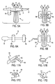
- FIGS. 5A and 5B are side-sectional views of another anchor assembly suitable for use with apparatus of the present invention.
- FIGS. 6A and 6B are side-sectional views of another alternative anchor assembly suitable for use with apparatus of the present invention.
- FIGS. 7A-7C are, respectively, a schematic side-sectional view of a unidirectionally adjustable anchor assembly suitable for use with apparatus of the present invention, schematic side-sectional views of alternative techniques for fixing the distal anchor of the assembly, and a cross-sectional view of the proximal anchor taken along section line A-A of FIG. 7A ;
- FIGS. 8A and 8B are schematic cross-sectional views illustrating the unidirectional adjustment capability of the anchor assembly of FIG. 7 ;
- FIGS. 9A-9C are schematic cross-sectional views of alternative embodiments of the proximal anchor of the anchor assembly of FIG. 7 ;
- FIGS. 10A and 10B are schematic cross-sectional views of an alternative unidirectionally adjustable anchor assembly suitable for use with apparatus of the present invention
- FIGS. 11A-11C are, respectively, a schematic side-view of another alternative unidirectionally adjustable anchor assembly suitable for use with the present invention, and cross-sectional views of the same taken along section line B-B of FIG. 11A ;
- FIG. 12 is a schematic cross-sectional view of an alternative unidirectionally adjustable anchor assembly comprising pivoting paddles
- FIG. 13 is a schematic cross-sectional view of an alternative unidirectionally adjustable anchor assembly comprising spring material
- FIGS. 14A-14B are schematic side-sectional views of alternative unidirectionally adjustable anchor assemblies comprising one-way valves;
- FIGS. 15A-15C are side-sectional and detail views of alternative unidirectionally adjustable anchor assemblies comprising slipknots;
- FIGS. 16A-16C are, respectively, a schematic side-sectional view of a bi-directionally adjustable anchor assembly comprising a locking mechanism, and cross-sectional views of the same taken along section line C--C of FIG. 16A ;
- FIGS. 17A-17D are perspective views of alternative anchors suitable for use with the anchor assemblies of the present invention.
- FIGS. 18A-18D are side views of alternative apparatus for forming a gastrointestinal fold
- FIG. 19 is a cross-sectional view of the apparatus of FIGS. 18A-18D ;
- FIGS. 20A-20D are side views of further alternative apparatus for forming a gastrointestinal tissue fold in accordance with the principles of the present invention.
- FIGS. 21A-21G are schematic side-sectional views of an anchor delivery system adapted for use with the adjustable anchor assemblies of FIGS. 7-17 , illustrating a method of delivering the unidirectionally adjustable anchor assembly of FIG. 7 across a tissue fold;
- FIGS. 22A and 22B are, respectively, a schematic side-view, partially in section, and an end-view of an alternative anchor delivery system adapted for use with the adjustable anchor assemblies of FIGS. 7-17 , wherein the proximal anchor is disposed within a separate delivery tube;
- FIG. 23 is a schematic side-sectional view of an alternative anchor delivery system adapted for use with the adjustable anchor assemblies of FIGS. 7-17 , wherein both the proximal and distal anchors are loaded within the needle;
- FIG. 24 is a schematic side-sectional view of an alternative embodiment of the anchor delivery system of FIG. 23 comprising motion limitation apparatus;
- FIG. 25 is a schematic side view, partially in section of an alternative anchor delivery system adapted to deliver a plurality of anchor assemblies
- FIG. 26 is a schematic side view of an alternative embodiment of the anchor delivery system of FIG. 25 ;
- FIGS. 27A and 27B are, respectively, schematic isometric and side views, partially in section, of an alternative anchor delivery system adapted to deliver a plurality of anchor assemblies via a revolver;
- FIGS. 28A and 28B are side views of an alternative embodiment of the apparatus of FIG. 20 illustrating a method for simultaneously forming and approximating multiple gastrointestinal tissue folds;
- FIG. 29 is an isometric view of an alternative embodiment of the apparatus of FIG. 1 for forming a gastrointestinal tissue fold comprising backside stabilization;
- FIGS. 30A-30E are a side view, partially in section, and isometric views illustrating a method of using the apparatus of FIG. 29 to form a backside stabilized gastrointestinal tissue fold;
- FIGS. 31A-31C are side views of further alternative tissue folding apparatus illustrating a method for forming a gastrointestinal tissue fold via a linear displacement of tissue;
- FIG. 32 is a side view of an alternative embodiment of the apparatus of FIG. 31 providing enhanced flexibility
- FIGS. 33A and 33B are side views of further alternative front and backside stabilized linear displacement plication apparatus, illustrating a method for forming a gastrointestinal tissue fold;
- FIGS. 34A and 34B are, respectively, a side view and a side view, partially in section, of still further alternative apparatus illustrating a method for forming a stabilized gastrointestinal tissue fold via a braided mesh;
- FIG. 35 is a side view of illustrative shape-lockable apparatus for use with the tissue folding and anchor delivery apparatus of the present invention.
- FIG. 36 is a side-sectional exploded view of nestable elements of a first embodiment of an overtube suitable for use with the shape-lockable apparatus of FIG. 35 ;
- FIG. 37 is a side-sectional view of a distal region of the apparatus of FIG. 35 constructed in accordance with principles of the present invention.
- FIG. 38 is a side-sectional view of an illustrative arrangement of a mechanism suitable for use in the handle of the apparatus of FIG. 35 ;
- FIG. 39 is a side-sectional view of the detail of a wire clamping system suitable for use in the handle of FIG. 35 ;
- FIGS. 40A-40D are side-views, partially in section, illustrating an exemplary method of performing endoluminal gastric reduction with a system of tools illustratively comprising the shape-lockable apparatus of FIGS. 35-39 , the plication apparatus of FIGS. 1-3 , the anchor assembly of FIG. 7 , the anchor delivery system of FIG. 21 and a commercially available gastroscope;
- FIGS. 41A-41C are, respectively, an isometric view of a patient's stomach after performing endoluminal gastric reduction using the methods of FIG. 40 ; a cross-sectional view of the same along plane A-A in FIG. 41A ; and a cross-sectional view of the stomach along plane B-B in FIG. 41A , prior to approximation of the pluralities of tissue folds to achieve the gastric reduction;
- FIGS. 42A-42C are side-views, partially in section, illustrating an exemplary method of treating gastroesophageal reflux disease with the illustrative system of tools described with respect to FIG. 40 ;
- FIGS. 43A and 43B are side-views, partially in section, illustrating an alternative method of performing endoluminal gastric reduction utilizing a system of tools of the present invention
- FIG. 44 is a side view, partially in section, illustrating a method of resecting a lesion or early cancer utilizing a system of tools of the present invention illustratively comprising a suction plicator and a resection loop;
- FIG. 45 is a side view, partially in section, illustrating a method of treating a bleeding site utilizing a system of tools of the present invention.
- GI gastrointestinal
- the present invention involves endoscopic apparatus that engages a tissue wall of the gastrointestinal lumen, creates one or more tissue folds and disposes one or more anchor assemblies through the tissue fold(s).
- the anchor assemblies are disposed through the muscularis and/or serosa layers of the gastrointestinal lumen.
- a distal tip of the probe engages the tissue and then moves the engaged tissue to a proximal position relative to the catheter tip, thereby providing a substantially uniform plication of predetermined size.
- Formation of a tissue fold preferably is accomplished using at least two tissue contact points that are separated by a linear or curvilinear distance, wherein the separation distance between the tissue contact points affects the length and/or depth of the fold.
- a tissue grabbing assembly engages the tissue wall in its normal state (i.e., non-folded and substantially flat), thus providing a first tissue contact point.
- the first tissue contact point then is moved to a position proximal of a second tissue contact point to form the tissue fold.
- An anchor assembly then may be extended across the tissue fold at the second tissue contact point.
- a third tissue contact point may be established such that, upon formation of the tissue fold, the second and third tissue contact points are disposed on opposing sides of the tissue fold, thereby providing backside stabilization during extension of the anchor assembly across the tissue fold from the second tissue contact point.
- the first tissue contact point is used to engage and then stretch or rotate the tissue wall over the second tissue contact point to form the tissue fold.
- the tissue fold is then articulated to a position wherein a portion of the tissue fold overlies the second tissue contact point at an orientation that is substantially normal to the tissue fold.
- An anchor then is delivered across the tissue fold at or near the second tissue contact point.
- apparatus 10 of the present invention comprises torqueable catheter 11 having distal region 12 from which first and second interconnected flexible tubes 13 and 14 extend, and proximal region 15 having handle 16 and actuator 17 .
- Catheter 11 is configured for insertion through a patient's mouth and esophagus into the gastrointestinal lumen.
- Tissue grabbing assembly 18 is disposed on the distal end of flexible tube 13 , and is coupled to actuator 17 via control wire 19 that extends through flexible tube 13 .
- flexible tubes 13 and 14 are connected via hinge assembly 20 that comprises link 21 attached to flexible tube 13 at pivot point 22 and attached to flexible tube 14 at pivot point 23 .
- Hinge assembly 20 prevents tissue grabbing assembly 18 from moving more than a predetermined distance relative to distal end 24 of flexible tube 14 .
- flexible tubes 13 and 14 preferably include bendable sections 25 and 26 , respectively.
- the bendable sections may comprise, for example, a plurality of through-wall slots 27 to enhance flexibility of the tube.
- flexible tubes 13 and 14 are made from stainless steel with an etched or laser-cut slot pattern. More preferably, the slot pattern is a sinusoidal repeating pattern of slots perpendicular to the longitudinal axis of tubes 13 and 14 .
- Alternative flexible patterns will be apparent to those of skill in the art.
- tissue grabbing assembly 18 comprises pair of jaws 28 a , 28 b arranged to rotate about pivot point 29 between an open configuration ( FIG. 2A ) and a closed configuration ( FIG. 2B ).
- Control wire 19 is coupled via pivot point 30 to arms 31 a and 31 b .
- Arms 31 a and 31 b are in turn pivotally coupled to jaws 28 a and 28 b , respectively, at pivot points 32 a and 32 b .
- Each of jaws 28 a and 28 b preferably includes sharpened teeth 33 disposed near its distal ends to facilitate grasping of the tissue wall of the GI lumen.
- Control wire 19 is coupled to actuator 17 of handle 16 so that translation of the wire within flexible tube 13 causes the jaws to open or close.
- urging control wire distally moves pivot point 30 distally, thereby forcing the jaws to open.
- Urging control wire 19 proximally moves pivot point 30 proximally, thereby forcing the jaws to close together.
- tissue grabbing assembly 18 may comprise a grappling hook or fork, or plurality of needles coupled to the distal end of flexible tube 13 .
- Flexible tube 14 is affixed to and immovable within catheter 11 , while flexible tube 13 is coupled to catheter 11 only via hinge 20 . Accordingly, when control wire 19 is extended in the distal direction, flexible tube 13 is carried in the distal direction. When control wire 19 is retracted in the proximal direction, flexible tube remains stationary until jaws 28 a and 28 b close together, after which further retraction of control wire 19 by moving actuator 17 causes flexible tube 13 to buckle in bendable region 25 , as described hereinafter.
- FIGS. 1 and 3 A- 3 E operation of apparatus 10 is described to create a tissue fold in a tissue wall of a GI lumen.
- distal region 12 of catheter 11 is positioned within a patient's GI lumen transesophageally, and jaws 28 a and 28 b of tissue grabbing assembly 18 are opened by moving actuator 17 to the distal-most position on handle 16 .
- actuator 17 may then be moved proximally until the jaws of tissue grabbing assembly 18 engage a portion of tissue wall W at contact point P 1 .
- flexible tube 13 is urged proximally within catheter 11 by further proximal retraction of control wire 19 to stretch tissue wall W and create tissue fold F.
- link 21 of hinge assembly 20 causes tissue grabbing assembly 18 to move from a position distal to distal end 24 of flexible tube 14 , to a position proximal of distal end 24 of flexible tube 14 .
- Bendable sections 25 and 26 of flexible tubes 13 and 14 respectively, accommodate any lateral motion caused by operation of hinge assembly 20 .
- formation of fold F facilitates the penetration of the tissue wall by a needle and subsequent delivery of an anchor assembly, as described hereinafter.
- Hinge assembly 20 transmits force applied to flexible tube 13 via control wire 19 and actuator 17 to the distal tip 24 .
- flexible tube 14 is configured so that distal tip 24 contacts, and is substantially perpendicular, to tissue fold F at contact point P 2 .
- sharpened needle or obturator 34 may be extended from distal tip 24 of flexible tube 14 to pierce all four layers of the tissue wall W. Sharpened needle or obturator 34 is inserted via inlet 35 to flexible tube 14 on handle 16 (see FIG. 1A ).
- the GI lumen comprises an inner mucosal layer, connective tissue, the muscularis layer and the serosa layer.
- the staples or anchors used to achieve reduction of the GI lumen must engage at least the muscularis tissue layer, and more preferably, the serosa layer as well.
- stretching of tissue fold F across distal tip 24 permits an anchor to be ejected through both the muscularis and serosa layers, thus enabling durable gastrointestinal tissue approximation.
- needle 34 may be extended from distal tip 24 and through tissue fold F. Because needle 34 penetrates the tissue wall twice, it exits within the gastrointestinal lumen, thus reducing the potential for injury to surrounding organs. Once the needle has penetrated tissue fold F, an anchor assembly is ejected through distal tip 24 as described hereinbelow.
- Anchor assembly 36 comprises T-anchor assembly having distal rod 38 a and proximal rod 38 b connected by suture 39 .
- the precise shape, size and materials of the anchors may vary for individual applications.
- the suture material also may vary for individual applications.
- the suture material may consist of monofilament wire, multifilament wire or any other conventional suture material.
- suture 39 may comprise elastic material, e.g. a rubber band, to facilitate adjustment of the distance between the proximal and distal rods.
- Suture 39 extends through a pair of through-holes 40 in each rod, thereby forming a loop.
- suture 39 may be attached to the rods via an eyelet or using a suitable adhesive.
- through-holes 40 are located near the center of the rods 38 a and 38 b.
- rods 38 a and 38 b may be delivered through needle 34 (see FIG. 3E ) using push rod 42 .
- Push rod 42 is adapted to freely translate through flexible tube 14 and needle 34 .
- Push rod 42 is preferably flexible, so that it may slide through bendable section 26 of flexible tube 14 .
- push rod 42 may include notch 43 near its distal end to facilitate grasping and tensioning suture 39 after anchor delivery.
- distal rod 38 a During anchor delivery, the longitudinal axis of distal rod 38 a is substantially parallel to the longitudinal axis of needle 34 . However, once distal rod 38 a is ejected from needle 34 , suture tension induces the rod to rotate approximately 90 degrees about its longitudinal axis, so that its longitudinal axis is substantially perpendicular to the longitudinal axis of needle 35 . This rotation of distal rod 38 a prevents it from being pulled back through tissue wall W.
- suture 39 may comprise an elastic material that dynamically tightens the rods against tissue fold F.
- the anchor assembly comprises a T-anchor assembly suitable to be disposed over obturator 50 .
- distal rod 38 a includes through-hole 51 dimensioned for the passage of obturator tip 52
- obturator 50 is translatably inserted through flexible tube 14 via inlet 35 of handle 16 (see FIG. 1A ).
- Proximal rod 38 b may be a solid rod that does not include a through-hole for passage of obturator 50 .
- proximal rod 38 b may include a through-hole for the passage of the obturator.
- obturator tip 52 is sharpened to facilitate tissue penetration.
- rod 38 a is ejected on the distal side of fold F, it rotates into a position substantially parallel to tissue wall W and perpendicular to the longitudinal axis of the obturator.
- Obturator 50 then is retracted and proximal rod 38 b is ejected from flexible tube 14 . More particularly, when flexible tube 14 is retracted from tissue wall W, proximal rod 38 b is pulled through distal tip 24 . Proximal rod 38 b then rotates substantially 90 degrees as it is ejected from flexible tube 14 so that rod 38 b is urged against tissue wall W.
- anchor assembly 55 comprises a T-anchor assembly similar to the embodiment depicted in FIG. 4A .
- anchor assembly 55 includes fine wire tether 56 that may be twisted to maintain the tension between rods 38 a and 38 b.
- distal rod 38 a is delivered across both tissue walls using needle 34 .
- the needle then is retracted to release distal rod 38 a so that it engages the tissue wall.
- needle 34 is retracted to release proximal rod 38 b , so that it too rotates into engagement with the tissue wall.
- a proximal portion of the wire tether is captured by notch 43 of push rod 42 (see FIG. 4B ), and the push rod is rotated to cause proximal rod 38 b to clamp down on the tissue fold. Because wire tether 56 is twisted by rotation of push rod 42 , it maintains the desired force on the tissue walls.
- Anchor assembly 60 comprises distal anchor 62 and unidirectionally adjustable proximal anchor 64 , which are connected by suture 39 .
- Distal anchor 62 is translationally fixed with respect to suture 39 .
- Such fixation may be achieved in a variety of ways.
- distal anchor 62 may comprise a pair of through-holes 63 , located near the center of anchor 62 and through which suture 39 is threaded and tied off at knot 65 .
- FIG. 7B provides alternative techniques for fixing the distal anchor.
- distal anchor 62 may comprise hollow tube T having opening O.
- a distal end of suture 39 is passed through opening O and formed into knot K, which is dimensioned such that it cannot pass through opening O, thereby fixing the distal anchor with respect to the suture.
- distal anchor 62 optionally may comprise distal opening DO, which is dimensioned such that knot K may pass therethrough.
- the distal end of suture 39 may be passed through distal opening DO, knotted, and then pulled back within hollow tube T of anchor 62 until it catches at opening O.
- a drawback of the fixation technique described with respect to FIG. 7 B(i) is a risk of suture 39 being torn or cut due to rubbing against opening O.
- hollow tube T comprises first end E to which is connected wire loop L, which may be formed, for example from a nickel-titanium alloy (“Nitinol”).
- wire loop L which may be formed, for example from a nickel-titanium alloy (“Nitinol”).
- Suture 39 passes through the wire loop before terminating at knot K.
- Knot K is dimensioned such that it cannot pass back through the wire loop.
- Wire loop L directs suture 39 through opening O, thereby reducing rubbing of the suture against the opening and reducing a risk of tearing or cutting of suture 39 .
- FIG. 7 B(iii) provides yet another alternative technique for fixing the distal anchor with respect to the suture.
- Distal anchor 62 again comprises hollow tube T having opening O.
- Rod R is disposed within tube T, and the ends of the tube may be either closed or crimped to rod R, such that the rod is maintained within the tube.
- the distal end of suture 39 is threaded through opening O, around rod R, and back out opening O. The suture is then knotted at knot K, thereby fixing distal anchor 62 with respect to suture 39 .
- suture 39 alternatively may be fixed with respect to anchor 62 by other means, for example, via a knotted eyelet or via a suitable adhesive. Additional techniques will be apparent to those of skill in the art. While anchor 62 is illustratively shown as a rod- or T-type anchor, any of a variety of anchors, per se known, may be used as distal anchor 62 . Exemplary anchors are described in co-pending U.S. patent application Ser. No. 10/612,170, filed Jul. 1, 2003, which is incorporated herein by reference in its entirety. Additional anchors are described hereinbelow with respect to FIG. 17 .
- anchors and anchor assemblies should be understood to include clips for securing tissue, as well as suture knots and knot replacements.
- anchor assemblies may comprise multiple components that are not initially coupled to one another; the components may be brought together and/or coupled within a patient at a treatment site.
- adjustable proximal anchor 64 comprises outer cylinder 66 having first end 67 a and second end 67 b , as well as first opening 68 a and second opening 68 b .
- First and second openings 68 are preferably disposed near the center of cylinder 66 and approximately 180° apart.
- Anchor 64 further comprises first flexible rod 70 a and second flexible rod 70 b , both of which are disposed within outer cylinder 66 and coupled to first and second ends 67 of cylinder 66 .
- Rods 70 may be formed, for example, from Nitinol or from a polymer, and may be separated from one another by small gap G. As with the previous anchor assemblies, the precise shape, size and materials of the anchors and suture may vary as required for specific applications.
- suture 39 passes from distal anchor 62 through first opening 68 a of proximal anchor 64 , around second flexible rod 70 b , around first flexible rod 70 a , between rods 70 a and 70 b , and out through second opening 68 b .
- This suture winding provides a unidirectional adjustment capability that allows a length L of suture 39 disposed between distal anchor 62 and proximal anchor 64 to be shortened. However, the suture winding precludes an increase in length L.
- FIG. 8 illustrate the mechanism of this unidirectional adjustment capability in greater detail.
- suture 39 may be tied off proximal of anchor 64 at knot 69 , thereby forming a proximal loop of suture to facilitate deployment and/or adjustment of anchor assembly 60 .
- a proximally-directed force F 1 is applied to suture 39 proximal of adjustable anchor 64 , while anchor 64 is held stationary or is advanced distally.
- a portion of force F 1 is transferred through suture 39 to second flexible rod 70 b , which causes rod 70 b to bow, thereby increasing gap G and allowing suture 39 to freely pass between rods 70 a and 70 b and through proximal anchor 64 , facilitating unidirectional adjustment.
- distal anchor 62 retracts proximally towards anchor 64 .
- distal anchor 62 when anchor 64 is advanced distally while suture 39 is retracted proximally, distal anchor 62 either remains stationary or retracts proximally towards proximal anchor 64 , depending upon a degree of distal advancement of proximal anchor 64 . Regardless, length L of suture 39 disposed between anchors 62 and 64 is decreased, thereby unidirectionally adjusting a distance between the anchors.
- a distally-directed force F 2 is applied to suture 39 distal of adjustable anchor 64 .
- Force F 2 may be applied, for example, by tissue compressed between anchors 62 and 64 . Compressed tissue stores energy in a manner similar to a compression spring and seeks to push anchors 62 and 64 apart after unidirectional tightening. Force F 2 causes the loop of suture 39 around first and second rods 70 to tighten, thereby bowing both rods inward and closing gap G such that suture 39 is friction locked between first and second flexible rods 70 . In this manner, the length L of suture between anchors 62 and 64 may be selectively decreased but cannot be increased.
- the magnitude of force required to unidirectionally adjust length L may be altered in a variety of ways.
- a length, flexibility or diameter of rods 70 may be altered.
- the elasticity or diameter of suture 39 may be altered. Initial gap G may be increased or decreased.
- the materials used to form rods 70 and suture 39 may be changed to alter material properties, such as coefficients of friction, and/or rods 70 or suture 39 may comprise a lubricious coating. Additional methods for varying the magnitude of force, a few of which are described hereinbelow with respect to FIG. 9 , will be apparent in view of this disclosure and are included in the present invention.
- FIG. 9 alternative anchors 64 are described.
- flexible rods 70 of proximal adjustable anchor 64 ′ are rotated with respect to openings 68 (or vice versa).
- rotation of rods 70 up to 180° clockwise progressively increases friction when force is applied to anchors 62 and 64 .
- the magnitude of the friction lock is increased when force is applied in the manner described with respect to FIG. 8B .
- friction is also increased when unidirectionally adjusting the length of suture between the proximal and distal anchors by applying force in the manner described with respect to FIG. 8A .
- Rotation of rods 70 more than about 180° clockwise would cause anchor 64 ′ to friction lock regardless of which direction force were applied to suture 39 , thereby negating the unidirectional adjustment capability.
- Counterclockwise rotation of rods 70 with respect to openings 68 would initially reduce friction during force application to suture 39 in either direction. It is expected that counterclockwise rotation in excess of about 90° would eliminate the friction lock described in FIG. 8B and allow bidirectional adjustment.
- Continued counterclockwise rotation beyond about 450° would reverse the directions of friction lock and unidirectional adjustment, while counterclockwise rotation beyond about 720° would result in friction lock regardless of which direction force were applied to suture 39 .
- openings 68 of cylinder 66 of anchor 64 are preferably disposed approximately 180° apart from one another.
- first opening 68 a may be rotated counterclockwise with respect to second opening 68 b (or vice versa), as seen with anchor 64 ′′ of FIG. 9B .
- first opening 68 a is no longer in line with rods 70
- second opening 68 b remains in line with rods 70 .
- force F 1 is applied to anchor 64 ′′
- second flexible rod 70 b is able to bow outward and increase gap G, thereby facilitating unidirectional adjustment.
- gap G is closed more tightly upon suture 39 , thereby increasing the friction lock force. If first opening 68 a alternatively were rotated clockwise with respect to the second opening, it is expected that the friction lock force would be decreased.
- proximal adjustable anchor 64 ′′′ comprises an alternative suture winding.
- Suture 39 passes from distal anchor 62 through first opening 68 a of anchor 64 ′′′, around second flexible rod 70 b , around first flexible rod 70 a , back around second flexible rod 70 b , between rods 70 a and 70 b , and out through second opening 68 b .
- the suture winding illustrated in FIG. 9C provides a unidirectional adjustment capability that allows a length L of suture 39 disposed between distal anchor 62 and proximal anchor 64 ′′′ to be shortened. However, this suture winding precludes an increase in length L. Additional unidirectionally adjustable suture windings will be apparent to those of skill in the art.
- Anchor assembly 80 comprises distal anchor 62 and proximal anchor 82 .
- Unidirectionally adjustable proximal anchor 82 comprises outer cylinder 84 having first end 85 a and second end 85 b (not shown), as well as first opening 86 a and second opening 86 b .
- First and second openings 86 are preferably disposed near the center of cylinder 84 and approximately 180° apart.
- Anchor 82 further comprises first flexible rod 88 a , second flexible rod 88 b and third flexible rod 88 c , all of which are disposed within outer cylinder 66 and coupled to first and second ends 85 of cylinder 64 .
- Rods 88 are separated from one another by gaps G 1 and G 2 .
- Suture 39 passes from distal anchor 62 through first opening 86 a of proximal anchor 82 , around first rod 88 a , between first rod 88 a and second rod 88 b , between second rod 88 b and third rod 88 c , around third rod 88 c , back to and around first rod 88 a , and out through second opening 86 b .
- FIG. 10A when force F 1 is applied to suture 39 , gaps G 1 and G 2 remain open, thereby facilitating unidirectional adjustment/shortening of length L of suture 39 disposed between distal anchor 62 and proximal anchor 82 .
- FIG. 10B when force F 2 is applied to suture 39 , gaps G 1 and G 2 close down upon suture 39 , thereby forming a friction lock that precludes an increase in length L of suture 39 .
- the unidirectionally adjustable anchors described hereinabove with respect to FIGS. 7-10 all comprise rods disposed within a cylinder having openings for passage of a suture.
- the openings act to center the suture with respect to the rods and can be used to alter magnitudes of force applied during adjustment and friction locking, as discussed previously.
- such openings present a risk of tearing or cutting the suture as the suture slides through the openings.
- anchor assembly 90 comprises distal anchor 62 and proximal anchor 92 .
- Unidirectionally adjustable proximal anchor 92 comprises first flexible rod 94 a and second flexible rod 94 b , as well as rigid rod 96 , which is preferably larger in diameter than first and second rods 94 .
- Flexible rods 94 are preferably fabricated from Nitinol or a polymer, while rigid rod 96 is preferably fabricated from stainless steel or a polymer. Alternative materials will be apparent to those of skill in the art.
- Anchor 92 further comprises first outer cylinder 98 a and second outer cylinder 98 b , which are crimped to the ends of first and second rods 94 , and rigid rod 96 .
- first and second cylinders 98 may each comprise an end cap (not shown) to which the rods are coupled.
- First and second cylinders 94 do not span a central portion of anchor 92 .
- Flexible rods 94 are separated from one another by gap G 1 , while rods 94 are separated from rigid rod 96 by gap G 2 .
- Anchor 92 comprises three rods, but, unlike anchor 82 of FIG. 10 , suture 39 is only wrapped around two of them to achieve unidirectional adjustment. As best seen in FIGS. 11B and 1C , the illustrative suture winding of anchor assembly 90 is similar to that described previously with respect to anchor assembly 60 of FIGS. 7 and 8 .
- the break between first and second cylinders 98 acts to center suture 39 with respect to the rods, as seen in FIG. 11A , while rigid rod 96 acts to stiffen and reduce rotation of anchor 92 as it directs suture 39 about flexible rods 94 .
- Suture 39 passes from distal anchor 62 to proximal anchor 92 , between rigid rod 96 and flexible rods 94 , around second flexible rod 94 b , around first flexible rod 94 a , between rigid rod 96 and first flexible rod 94 a , between flexible rods 94 a and 94 b , and out.
- FIG. 11A when force F 1 is applied to suture 39 , flexible rods 94 are forced apart and gap G 1 widens while gap G 2 remains substantially constant, thereby allowing unidirectional adjustment of length L of suture 39 disposed between distal anchor 62 and proximal anchor 92 .
- gap G 1 closes down upon suture 39 , thereby forming a friction lock that precludes an increase in length L of suture 39 .
- Gap G 2 again remains substantially constant.
- Anchor assembly 100 comprises distal anchor 62 and proximal anchor 102 .
- Unidirectionally adjustable proximal anchor 102 comprises outer cylinder 103 having first end 104 a and second end 104 b (not shown), as well as first opening 105 a and second opening 105 b .
- First and second openings 105 are preferably disposed near the center of cylinder 103 and approximately 180° apart.
- Anchor 102 further comprises first rod or paddle 106 a and second rod or paddle 106 b , both of which are disposed within outer cylinder 103 and coupled to the first and second ends of cylinder 103 by pins 107 , which pass through pivot holes 108 .
- first and second paddles 106 are able to rotate about pivot holes 108 .
- Paddles 106 may be formed, for example, from stainless steel or a polymer, and are separated from one another by gap G. As with the previous anchor assemblies, the precise shape, size and materials of the anchors, as well as suture 39 , may vary as required for specific applications.
- Suture 39 illustratively passes from distal anchor 62 through first opening 105 a of proximal anchor 102 , around second paddle 106 b , around first paddle 106 a , between paddles 106 a and 106 b , and out through second opening 105 b .
- the placement of pivot holes 108 ensures that application of force F 1 , as described hereinabove, causes paddles 106 to rotate apart from one another and expand gap G, thereby enabling unidirectional adjustment.
- application of previously discussed force F 2 causes paddles 106 to rotate together, thereby closing gap G and pinching suture 39 between the paddles in a friction lock.
- An increase in the magnitude of force F 2 serves to rotate paddles 106 together more tightly, thereby increasing the magnitude of the friction lock acting upon suture 39 between the paddles. In this manner, unidirectional adjustment is achieved.
- Anchor assembly 110 comprises distal anchor 62 and proximal anchor 112 .
- Unidirectionally adjustable proximal anchor 112 comprises outer cylinder 113 having first end 114 a and second end 114 b (not shown), as well as first opening 115 a and second opening 115 b .
- First and second openings 115 are preferably disposed near the center of cylinder 113 and approximately 180° apart.
- Anchor 112 further comprises first rod 116 a and second rod 116 b that are separated by gap G, as well as spring material 118 , all of which are disposed within outer cylinder 113 .
- Spring material 118 abuts rods 116 , which preferably are substantially the same length as cylinder 113 , and may either move freely within cylinder 113 or may be coupled to the ends (not shown) of cylinder 113 .
- Spring material 118 may also move freely within cylinder 113 or may be coupled to the cylinder, and comprises lumen 119 having a diameter that is preferably equal to or less than the diameter of suture 39 .
- Spring material 118 may comprise, for example, a compressible biocompatible foam, which acts as a compression spring.
- Suture 39 passes from distal anchor 62 to proximal anchor 112 through first opening 115 a of cylinder 113 , between rods 116 , through lumen 119 of spring material 118 , and out through second opening 115 b .
- Lumen 119 snugly contacts suture 39 such that application of force F 1 causes friction between the suture and the spring material to compress the spring material against the wall of cylinder 114 , thereby reducing a stress applied to rods 116 by spring material 118 and increasing gap G such that unidirectional adjustment of length L of suture 39 disposed between distal anchor 62 and proximal anchor 102 may proceed.
- Application of force F 2 stretches spring material 118 against rods 116 , thereby increasing the stress applied to the rods by the spring material and closing gap G such that suture 39 is friction locked between rods 116 .
- anchor assembly 120 comprises distal anchor 62 and proximal anchor 122 .
- Unidirectionally adjustable proximal anchor 122 comprises outer cylinder 124 having first and second ends 125 a and 125 b , as well as first opening 126 a and second opening 12 . 6 b .
- First and second openings 126 are preferably disposed near the center of cylinder 124 and approximately 180° apart.
- Anchor 122 further comprises first inclined plane 128 a and second inclined plane 128 b , which are forced into apposition by compression springs 129 a and 129 b , thereby forming one-way valve V at the junction of the two inclined planes.
- Inclined planes 128 and springs 129 are disposed within outer cylinder 124 ; springs 129 abut ends 125 of cylinder 124 , as well as the ends of the inclined planes.
- Suture 39 ′ comprises a plurality of knots or beads B adapted to actuate one-way valve V.
- Suture 39 ′ passes from distal anchor 62 to proximal anchor 122 through first opening 126 a of cylinder 124 , between inclined planes 128 , through one-way valve V, and out through second opening 126 b .
- Application of force F 1 to suture 39 ′ causes a bead B to contact inclined planes 128 and gradually coax them apart by compressing springs 129 , thereby opening valve V and allowing the bead to pass through the valve.
- springs 129 force inclined planes 128 back into apposition, thereby closing the valve.
- force F 1 allows multiple beads to pass through the valve, which facilitates unidirectional adjustment of suture length L disposed between distal anchor 62 and proximal anchor 122 .
- Application of force F 2 causes a bead B of suture 39 ′ to impinge upon the proximal sides of inclined planes 128 .
- force transferred to the planes by the bead is perpendicular to the direction required to compress springs 129 and urge planes 128 apart.
- the bead B impinging upon the proximal sides of planes 128 is not able to open one-way valve V and pass back through the valve in a distal direction, thereby ensuring only unidirectional adjustment, i.e. shortening, of the length L of suture disposed between the proximal and distal anchors.
- Anchor assembly 130 comprises distal anchor 62 and proximal anchor 132 .
- Unidirectionally adjustable proximal anchor 132 comprises lumen 134 having cantilevered inclined plane 136 disposed therein, which forms one-way valve V.
- ‘Zip-tie’ fastener 138 having a plurality of inclined planes 139 , connects proximal anchor 132 and distal anchor 62 .
- the plurality of inclined planes 139 are disposed about 180° out of phase with inclined plane 136 of anchor 132 .
- Fastener 138 passes from distal anchor 62 to proximal anchor 132 , through lumen 134 and past inclined plane 136 .
- Inclined planes 139 of fastener 138 mesh with inclined plane 136 and bend or cantilever plane 136 , such that planes 139 of fastener 138 may proximally pass one-way valve V when force F 1 is applied to the fastener, thereby enabling unidirectional adjustment of length L of fastener 138 disposed between the proximal and distal anchors.
- anchor assembly 140 comprises distal anchor 142 and proximal anchor 144 .
- Through-holes 143 a and 143 b extend through distal anchor 142
- through-holes 145 a and 145 b extend through proximal anchor 145 .
- through-holes 143 and 145 are located near the center of anchors 142 and 144 , respectively.
- the distal end of suture 39 passes through through-hole 145 a of proximal anchor 144 to distal anchor 142 , where it passes through through-hole 143 a and back through through-hole 143 b . It then extends from distal anchor 142 back to proximal anchor 144 , where it passes through through-hole 145 b of the proximal anchor.
- the distal end of suture 39 is tied off at unidirectional slipknot S, which is located proximal of anchor 144 .
- FIG. 15B provides a detail view illustrating formation of slipknot S.
- FIG. 15C illustrates an alternative embodiment of anchor assembly 140 wherein the slipknot is disposed within the proximal anchor.
- Anchor assembly 140 ′ comprises distal anchor 142 and proximal anchor 144 ′.
- Proximal anchor 144 ′ comprises hollow cylinder or tube 146 having distal openings 147 a and 147 b , and proximal opening 148 .
- suture 39 passes through proximal opening 148 into the interior of tube 146 . It then passes through distal opening 147 a of proximal anchor 144 ′ to distal anchor 142 , where it passes through through-hole 143 a and back through through-hole 143 b . Next, suture 39 extends from distal anchor 142 back to proximal anchor 144 ′, where it passes through distal opening 147 b into the interior of tube 146 of the proximal anchor. The distal end of suture 39 is tied off at unidirectional slipknot S, which is disposed within tube 146 of anchor 144 ′. Anchor assembly 140 ′ may be unidirectionally adjusted in a manner similar to that described hereinabove with respect to anchor assembly 140 of FIG. 15A .
- FIGS. 7-15 have illustrated anchor assemblies comprising various mechanisms for achieving unidirectional adjustment of the distance between the proximal and distal anchors. These mechanisms have been provided solely for the sake of illustration and should in no way be construed as limiting. Additional mechanisms for achieving unidirectional adjustment will be apparent to those of skill in the art in view of this disclosure and are included in the present invention. Furthermore, a majority of the anchor assemblies of FIGS. 7-15 have been described with the distal anchor being fixed relative to the suture, and the proximal anchor being adjustable. However, it should be understood that the distal anchor may alternatively be adjustable and the proximal anchor may be fixed, and/or both anchors may be unidirectionally adjustable, as with anchor assembly 140 of FIG. 15 .
- Anchor assembly 150 comprises distal anchor 62 and proximal anchor 152 .
- bi-directionally adjustable proximal anchor 152 comprises outer cylinder 153 having first end. 154 a and second end 154 b , as well as first opening 155 a and second opening 155 b .
- First and second openings 155 are preferably disposed near the center of cylinder 153 and approximately 90° apart.
- Proximal anchor 152 further comprises tension spring 158 disposed within outer cylinder 153 .
- suture 39 passes from distal anchor 62 to proximal anchor 152 through first opening 155 a , around spring 158 , and out through second opening 155 b .
- Suture 39 moves freely about tension spring 158 in either direction during application of force F 1 or force F 2 , thereby facilitating bi-directional adjustment of suture length L disposed between the proximal and distal anchors.
- simultaneous application of forces F 1 and F 2 with sufficient magnitude causes suture 39 to force threads T of spring 158 apart, such that suture 39 is trapped between threads T and locked in position, thereby precluding further adjustment of suture length L.
- the magnitude of forces required to actuate the locking mechanism of proximal anchor 152 and lock suture 39 within threads T of spring 158 may be specified/altered in a variety of ways. For example, the angular spacing of openings 155 about outer cylinder 153 may be altered, the spring constant of spring 158 may be specified, and/or spring 158 or suture 39 may comprise a lubricious coating. Additional techniques will be apparent to those of skill in the art. It is expected that simultaneous application of forces F 1 and F 2 will be encountered when anchor assembly 150 has been deployed across a tissue fold and suture length L has been adjusted such that the tissue fold is compressed. A medical practitioner would then apply force F 1 , while the compressed tissue fold would apply force F 2 .
- anchor assemblies of FIGS. 10-16 have illustratively been described without knots or loops of suture or fastener disposed proximal of the proximal anchor (as seen, for example, with knot 69 on suture 39 of anchor assembly 60 in FIGS. 7 and 8 ) it should be understood that such loops or knots optionally may be provided in order to facilitate deployment and/or adjustment of the anchor assemblies.
- the previously described anchor assemblies illustratively comprise distal rod- or T-type anchors. However, it should be understood that distal T-anchors have only been provided for the sake of illustration.
- the distal anchors may comprise any of a variety of anchors, per se known, including, for example, surgical or endoluminal clips, clips for securing tissue and suture knots or knot replacements.
- Exemplary anchors are described in co-pending U.S. patent application Ser. No. 10/612,170, filed Jul. 1, 2003, which is incorporated herein by reference in its entirety.
- anchor assemblies may comprise multiple components that are not initially coupled to one another; the components may be brought together and/or coupled within a patient at a treatment site. Additional anchors are described hereinbelow with respect to FIG. 17 .
- articulating anchor 160 includes semi-cylindrical base 161 , rod 162 and suture 39 .
- Rod 162 rotates about pivot point 163 (as indicated by arrow 164 ) between an expanded position (shown in FIG. 7A ) and a reduced profile position, wherein rod 162 pivots within the semi-cylindrical base 161 .
- Articulating anchor 160 may be delivered through a tissue fold using, for example, needle 34 described hereinabove with respect to FIG. 3E .
- articulating anchor 160 is biased in the expanded position so that it automatically expands once it is ejected from the needle.
- anchors of the present invention also may comprise one or more oblong bodies connected by at least one suture.
- anchor 165 comprises elliptical ring 166 having sutures 39 attached at substantially opposite sides of the ring.
- anchor 168 comprises angle bracket 169 having a pair of through-holes 170 for suture 39 .
- anchor 171 comprises oblong bead 172 having a pair of through-holes 173 for suture 39 .
- All three anchors 165 , 168 and 171 (as well as the T-anchors described previously) have a first dimension (e.g., width) that is substantially larger than a second dimension (e.g., height). This dimensional difference necessitates that anchors 165 , 168 and 171 be inserted within a needle (e.g., needle 34 of FIG. 3E ) in a particular orientation.
- a needle e.g., needle 34 of FIG. 3E
- tension on suture 39 forces the anchor to rotate so that it cannot be pulled back through the tissue wall.
- numerous other anchors may be employed without departing from the scope of the present invention.
- Apparatus 175 comprises treadmill assembly 176 disposed at distal tip 174 of flexible tube 177 .
- Flexible tube 177 is configured to be inserted through a patient's mouth, esophagus and into the stomach.
- Treadmill assembly 176 comprises conveyor 180 that circles around a pair of hubs 181 a and 181 b . Hubs 181 a and 181 b rotate about axles 182 a and 182 b , respectively, and are interconnected by bracket 183 .
- a plurality of barbs or needles 185 is disposed at substantially regular intervals around the circumference of conveyor 180 .
- Flexible tube 177 preferably includes a plurality of through-wall slots 186 to enhance flexibility of the tube, yet maintain torqueability.
- flexible tube 177 is made from stainless steel with an etched or laser-cut slot pattern.
- the slot pattern is a sinusoidal repeating pattern of slots perpendicular to the longitudinal axis of the tube. Additional and/or alternative patterns will be apparent to those of skill in the art.
- drive shaft 202 disposed within flexible tube 177 is coupled to a manual knob or motor located at the proximal end of the catheter.
- the distal tip of drive shaft 202 is provided with beveled gear 203 that meshes with beveled gear 204 provided on axle 182 b .
- rotation of beveled gear 203 is transmitted to beveled gear 204 , thereby causing axle 182 b to rotate.
- Axle 182 b in turn rotates hub 181 b , actuating conveyor 180 . Reversing the rotation of drive shaft 202 reverses the direction of conveyor 180 .
- FIGS. 18A-18D a method of forming a gastrointestinal tissue fold F using apparatus 175 is described.
- flexible tube 177 is positioned transesophageally so that treadmill assembly 176 contacts tissue wall W.
- contact should be made at an angle relative to the tissue wall W.
- an angle of approximately 45 degrees is depicted in FIG. 8A , while many other angles may be used without departing from the scope of the present invention.
- distal end 174 of flexible tube 177 may be articulated in bendable section 190 , thereby moving treadmill assembly 176 away from tissue wall W.
- the articulation of flexible tube 177 may be accomplished using a control wire and actuator disposed at the proximal end of the catheter, as previously described with respect to the embodiment of FIG. 1 .
- tissue fold F is stretched across bendable section 190 of flexible tube 177 to create contact point P 2 .
- stretching of tissue fold F across bendable section 190 permits an anchor to be ejected through both the muscularis and serosa layers, thus providing a durable foundation for gastrointestinal tissue approximation.
- needle 192 may be extended through slot 186 in bendable section 190 , and through the base of tissue fold F, and an anchor assembly (such as described with respect to any of FIGS. 4-17 ) may be ejected from needle 192 to secure the fold.
- an obturator (such as described with respect to FIGS. 5A and 5B ) may be used to pierce the tissue fold at contact point P 2 and deliver the anchor assembly.
- Treadmill assembly 176 may be disengaged from tissue wall W by reversing the rotation of proximal hub 181 b.
- Apparatus 200 comprises tissue grabbing assembly 18 ′ coupled to the distal end of a flexible tube 177 ′, such as described with respect to the embodiment of FIG. 18 .
- Flexible tube 177 ′ preferably includes a plurality of through-wall slots 186 ′ to enhance flexibility of the tube, yet maintain torqueability.
- flexible tube 177 ′ may be made from stainless steel with an etched or laser-cut slot pattern, such as a sinusoidal repeating pattern of slots perpendicular to the longitudinal axis of the tube. Alternative flexible patterns will be apparent.
- Tissue grabbing assembly 18 ′ is similar to that described with respect to the embodiment of FIG. 1 , and comprises a pair of jaws 28 a ′, 28 b ′ arranged to rotate about pivot point 29 ′ between an open configuration and a closed configuration.
- Each of jaws 28 a ′, 28 b ′ preferably includes sharpened teeth 33 ′ disposed near its distal end to facilitate grasping tissue wall W.
- tissue grabbing assembly 18 ′ is positioned transesophageally adjacent to tissue wall W and jaws 28 a ′, 28 b ′ are moved to the open position. Tissue grabbing assembly 18 ′ then is moved into contact with tissue wall W. As depicted in FIG. 20B , tissue grabbing assembly 18 ′ is used to grab the tissue wall at a first contact point P 1 . After capturing a portion of tissue wall W within jaws 28 a ′, 28 b ′, flexible tube 177 ′ is urged proximally to stretch tissue wall W and create tissue fold F.
- tissue fold F is formed, the distal end of flexible tube 177 ′ is articulated about bendable section 190 ′ to move tissue grabbing assembly 18 ′ away from tissue wall W. Articulation of flexible tube 177 ′ may be controlled using an actuator disposed at the proximal end of the catheter, thus causing tissue fold F to become elongated.
- tissue fold F is shown stretched across bendable section 190 ′ so that a sharpened needle or obturator may be extended from one of slots 186 ′ in bendable section 190 ′ and across all four layers of the tissue wall W. Needle 192 ′ then may be extended from slot 186 ′ in bendable section 190 ′ through contact point P 2 and tissue fold F.
- An anchor assembly e.g., as described with respect to any of FIGS. 4-17
- an obturator e.g., as described with respect to FIGS. 5A and 5B
- FIG. 21 an anchor delivery system adapted for use with the adjustable anchor assemblies of FIGS. 7-17 is described.
- the anchor delivery system is illustratively shown in use with anchor assembly 60 of FIG. 7 , but this should in no way be construed as-limiting.
- the delivery system of FIG. 21 may be used in conjunction with apparatus for forming a tissue fold, such as apparatus 10 , 175 and 200 described previously, or alternative apparatus described hereinafter, in order to anchor the tissue fold.
- the delivery system may be used for any other application, or in conjunction with any other apparatus, requiring delivery of an anchor assembly.
- Anchor delivery system 250 comprises flexible delivery tube 252 having lumen 253 .
- Flexible delivery tube 252 may be configured for insertion through a patient's mouth and esophagus into a gastrointestinal lumen, such as the stomach.
- Lumen 253 of delivery tube 252 preferably has a diameter of less than about 5 mm, and even more preferably has a diameter of about 2-3 mm.
- Flexible delivery tube 252 preferably includes a plurality of through-wall slots 254 to enhance flexibility of the tube, yet maintain torqueability. Slots 254 may form bendable section 255 .
- flexible delivery tube 252 is made from stainless steel with an etched or laser-cut slot pattern. The slot pattern is preferably a sinusoidal repeating pattern of slots perpendicular to the longitudinal axis of the tube. Additional and/or alternative patterns will be apparent.
- Anchor delivery system 250 further comprises delivery needle 260 .
- Needle 260 preferably has a length of less than 2 cm, and even more preferably has a length of about 1.5 cm.
- Needle 260 preferably comprises sharpened distal tip 262 , lumen 264 , slot 266 extending proximally from distal tip 262 , and proximal eyelet 268 .
- Lumen 264 of needle 260 is dimensioned such that a distal anchor may be disposed therein.
- anchor delivery system 250 is illustratively described in conjunction with anchor assembly 60 of FIG. 7 .
- distal anchor 62 is disposed within lumen 264 of needle 260 .
- Suture 39 passes through slot 266 of the needle as the suture extends from distal anchor 62 to proximal anchor 64 .
- Needle 260 preferably is disposed within lumen 253 of flexible delivery tube 252 distal of bendable section 255
- proximal anchor 64 preferably is disposed within delivery tube 252 proximal of bendable section 255 .
- distal anchor 62 may be deployed through needle 260 while the bendable section is actuated or bent, e.g., when anchor delivery system 250 is used in conjunction with previously described plication apparatus.
- Proximal anchor 64 subsequently may be advanced through bendable section 255 after the bendable section has once again been straightened.
- the distance, or length, of suture 39 extending between distal anchor 62 , which is disposed distal of the bendable section, and proximal anchor 64 , which is disposed proximal of the bendable section is preferably greater than or equal to about 2 cm, and is even more preferably greater than or equal to about 4 cm.
- Needle 260 is proximally coupled to needle pushrod 270 , which facilitates translation of the needle beyond a distal end of flexible delivery tube 252 .
- Needle pushrod 270 extends to a control actuator disposed at a proximal end of anchor delivery system 250 (not shown).
- Pushrod 270 optionally may be spring-loaded (not shown), for example, to facilitate puncture of tissue wall W and passage of needle 260 through tissue fold F.
- Anchor delivery system 250 further comprises anchor pushrod 280 , which is removably disposed through eyelet 268 of needle 260 , and is configured to eject distal anchor 62 from lumen 264 of needle 260 .
- anchor pushrod 280 extends to a control actuator disposed at a proximal end of anchor delivery system 250 (not shown).
- the actuators controlling pushrods 270 and 280 are preferably at least partially coupled so that relative motion between the two pushrods can be limited and/or eliminated, as needed.
- Pushrod 280 passes through the proximal loop of suture formed by knot 69 on suture 39 , such that the suture loop is threaded between needle pushrod 270 and anchor pushrod 280 . This facilitates unidirectional adjustment of the length of suture disposed between distal anchor 62 and proximal anchor 64 , as described hereinbelow.
- pushrods 270 and 280 are simultaneously distally advanced with sufficient force, e.g., via spring-loading, such that sharpened distal tip 262 of needle 260 pierces tissue wall W and is advanced across fold F.
- Bendable section 255 of flexible delivery tube 252 optionally may be bent during advancement of the needle, as described previously with respect to the plication apparatus (see FIG. 3E ).
- Anchor pushrod 280 is then advanced distally with respect to needle pushrod 270 and needle 260 , such that it abuts distal anchor 62 and ejects the anchor from lumen 264 of needle 260 on the distal side of tissue fold F, as seen in FIG. 21C .
- Suture 39 likewise is ejected from slot 266 and disposed across fold F.
- distal anchor 62 During delivery, the longitudinal axis of distal anchor 62 is substantially parallel to the longitudinal axis of needle 260 . However, once anchor 62 has been ejected from needle 260 , suture tension induces the anchor to rotate approximately 90° about its longitudinal axis, so that its longitudinal axis is substantially perpendicular to the longitudinal axis of needle 260 . This rotation of distal anchor 62 prevents it from being pulled back through tissue wall W. One or both ends of anchor 62 may be flared outward (not shown) to facilitate such rotation upon contact with the tissue wall.
- anchor pushrod 280 is retracted proximally within lumen 264 of needle 260 , the needle is retracted within flexibly delivery tube 252 via pushrod 270 , and then delivery system 2501 s retracted proximally across tissue fold F.
- Distal anchor 62 is disposed on the distal side of the tissue fold, suture 39 extends through the fold, and proximal anchor 64 is disposed on the proximal side of the fold within delivery tube 252 . If bendable section 255 were flexed during deployment of distal anchor 62 (see FIG. 3E ), it is straightened to facilitate delivery of the proximal anchor.
- Delivery tube 252 is then retracted proximally with respect to pushrods 270 and 280 , causing needle 260 to exit lumen 253 of the delivery tube on the proximal side of tissue fold F, thereby providing space for proximal anchor 64 to exit the lumen.
- delivery tube 252 or the full delivery system 250 is retracted, such that proximal anchor 64 is ejected from delivery tube lumen 253 , as seen in FIG. 21E .
- Delivery tube 252 is then re-advanced and/or pushrods 270 and 280 are simultaneously retracted, such that needle 260 is repositioned within lumen 253 of the delivery tube.
- Flexible delivery tube 252 is advanced with respect to needle 260 , such that it pushes proximal anchor 64 distally.
- the proximal suture loop formed by knot 69 on suture 39 catches against the proximal end of needle 260 and anchor pushrod 280 , which pulls distal anchor 62 taut against tissue fold F, as seen in FIG. 21F .
- Continued advancement of delivery tube 252 unidirectionally adjusts, i.e. shortens, length L of suture 39 disposed between distal anchor 62 and proximal anchor 64 , while forcing proximal anchor 64 against the tissue fold and firmly anchoring the fold between the proximal and distal anchors.
- anchor pushrod 280 may be retracted proximally with respect to needle pushrod 270 and needle 260 , such that the distal end of anchor pushrod 280 is proximally retracted through eyelet 268 and out of needle 260 .
- the suture loop formed by knot 69 on suture 39 slips off the distal end of anchor pushrod 280 , removing anchor assembly 60 from anchor delivery system 250 .
- Anchor delivery system 250 may then be removed from the patient.
- needle 260 , needle pushrod 270 and anchor pushrod 280 may be proximally retracted and removed from lumen 253 of anchor delivery tube 252 .
- An additional anchor assembly 60 may then be reloaded within needle 260 and delivery tube 252 from a proximal end of the delivery tube, while a distal end of the delivery tube remains within the patient.
- the additional anchor assembly may, for example, be placed across an additional tissue fold.
- Delivery system 250 optionally may comprise cutting apparatus (not shown) for removing the portion of suture extending proximally of proximal anchor 64 post-adjustment.
- cutting apparatus for removing the portion of suture extending proximally of proximal anchor 64 post-adjustment.
- secondary apparatus may be provided to remove such proximal length of suture.
- the unneeded length of suture may be left within the patient post-procedure.
- anchor delivery system 250 may be retracted proximally, such that needle 260 is retracted across tissue fold F while still disposed outside of delivery tube lumen 253 . This is in contrast to the method described with respect to FIG. 21D , wherein the needle is disposed within the delivery tube prior to retraction across the tissue fold.
- Anchor assembly 60 then may be unidirectionally adjusted, as described previously.
- Anchor delivery system 250 advantageously provides a medical practitioner with significant control during all steps of anchor assembly deployment. Such control affords the medical practitioner ample opportunity to abort deployment of the anchor assembly.
- the medical practitioner may decide to retract the needle across the fold and not launch the distal anchor.
- the medical practitioner may decide not to deploy the proximal anchor and may sever the suture connecting the proximal and distal anchors. The distal anchor then would simply pass harmlessly through the patient's digestive system.
- the medical practitioner may decide not to cinch the proximal and distal anchors post-deployment, thereby leaving the anchors in place without securing tissue fold F. Furtherstill, the medical practitioner may reverse cinching or cut the anchor assembly post-deployment, thereby reversing tissue fold formation.
- flexible delivery tube 252 may either comprise or be advanced through flexible tube 14 , 177 or 177 ′, of apparatus 10 , 175 or 200 , respectively.
- needle 260 may comprise needle 34 , 92 or 92 ′, of apparatus 10 , 175 or 200 , respectively.
- Needle 260 alternatively may comprise obturator 50 of FIG. 5 .
- components of anchor delivery system 250 may also comprise or be advanced through comparable components of alternative tissue folding apparatus described hereinafter.
- anchor delivery system 300 of FIG. 22 is adapted for use with the adjustable anchor assemblies of FIGS. 7-17 .
- the anchor delivery system 300 is illustratively shown in use with anchor assembly 60 of FIG. 7 , but this should in no way be construed as limiting.
- delivery system 300 may be used in conjunction with apparatus for forming a tissue fold, such as apparatus 10 , 175 and 200 described previously, or alternative apparatus described hereinafter, in order to anchor the tissue fold.
- the delivery system may be used for any other application, or in conjunction with any other apparatus, requiring delivery of an anchor assembly.
- FIG. 22A illustrates a distal region of anchor delivery system 300 .
- System 300 comprises flexible delivery tube 302 having lumen 303 .
- Flexible delivery tube 302 may be configured for insertion through a patient's mouth and esophagus into a gastrointestinal lumen, such as the stomach.
- Flexible delivery tube 302 preferably includes a plurality of through-wall slots 304 to enhance flexibility of the tube, yet maintain torqueability. Slots 304 may form bendable section 305 .
- flexible delivery tube 302 is made from stainless steel with an etched or laser-cut slot pattern. The slot pattern is preferably a sinusoidal repeating pattern of slots perpendicular to the longitudinal axis of the tube. Additional/alternative patterns will be apparent to those of skill in the art.
- Flexible delivery tube 302 further comprises end region 306 , which is coupled to anchor tube 307 having lumen or bore 308 . As best seen in FIG. 22B , lumen 308 of anchor tube 307 communicates with lumen 303 of delivery tube 302 via through-slot 309 .
- Proximal anchor 64 is disposed within anchor tube 307
- distal anchor 62 is disposed within needle 260 ′, which sits within delivery tube 302 .
- Suture 39 passes out of needle 260 ′ from distal anchor 62 through slot 266 ′. It then crosses from flexible delivery tube 302 to anchor tube 307 via through-slot 309 . After passing through proximal anchor 64 , suture 39 is passed back to delivery tube 302 via the through-slot, and is threaded around anchor pushrod 280 ′, such that the loop of suture formed by knot 69 on suture 39 is disposed between needle pushrod 270 ′ and anchor pushrod 280 ′.
- Needle 260 ′, needle pushrod 270 ′ and anchor pushrod 280 ′ are substantially the same as needle 260 and pushrods 270 and 280 , respectively, which are described hereinabove with respect to anchor delivery system 250 of FIG. 21 .
- anchor assembly 60 may be delivered from and adjusted by anchor delivery system 300 in a manner similar to that described hereinabove with respect to system 250 .
- anchor tube 307 of anchor delivery system 300 is illustratively shown as a relatively short tube having lumen or bore 308 adapted for disposal of proximal anchor 64 therein.
- anchor tube 307 , lumen 308 and/or through-slot 309 alternatively may extend all or part of the way to a proximal end of flexible delivery tube 302 of delivery system 300 .
- such an arrangement facilitates loading of anchor assembly 60 from a proximal end of the anchor delivery system, e.g., for reloading of anchor delivery system 300 while a distal region of the system is disposed within a patient.
- Such an arrangement also may simplify manufacturing of the system.
- Anchor delivery system 300 illustratively has been described with a single anchor assembly 60 disposed therein. However, it should be understood that a plurality of anchor assemblies may be loaded within delivery system 300 , thereby facilitating delivery of multiple anchor assemblies across different points of a tissue fold, across different (e.g., adjacent) tissue folds, or across other tissue structures.
- the plurality of distal anchors 62 preferably are loaded within needle 262 ′ of flexible delivery tube 302
- the plurality of proximal anchors 64 preferably are loaded within lumen 308 of anchor tube 307 .
- An advantage of anchor delivery system 300 is that both the proximal and distal anchors are located distal of the bendable section of the delivery tube during delivery. This reduces an initial length of suture that must be disposed between the anchors, thereby reducing a length of unneeded suture extending proximally of the proximal anchor post-delivery and adjustment. It also simplifies delivery by allowing both the proximal and distal anchors to be delivered while the bendable section of the delivery tube is bent.
- proximal anchor in a separate anchor tube eliminates a need to eject the needle from the flexible delivery tube on the proximal side of a tissue fold in order to deploy the proximal anchor, thereby reducing a risk of accidental tissue puncture with the needle.
- anchor delivery system 400 of FIG. 23 is adapted for use with the adjustable anchor assemblies of FIGS. 7-17 .
- Anchor delivery system 400 is illustratively shown in use with anchor assembly 60 of FIG. 7 , but this should in no way be construed as limiting.
- delivery system 400 may be used in conjunction with apparatus for forming a tissue fold, such as apparatus 10 , 175 and 200 described previously, or alternative apparatus described hereinafter, in order to anchor the tissue fold.
- delivery system 400 may be used for any other application, or in conjunction with any other apparatus, requiring delivery of an anchor assembly.
- FIG. 23 illustrates a distal region of anchor delivery system 400 .
- System 400 comprises flexible delivery tube 402 having lumen 403 .
- Flexible delivery tube 402 may be configured for insertion through a patient's mouth and esophagus into a gastrointestinal lumen, such as the stomach.
- Flexible delivery tube 402 preferably includes a plurality of through-wall slots to enhance flexibility of the tube, yet maintain torqueability.
- the slots may form bendable section 405 .
- Anchor delivery system 400 further comprises delivery needle 260 ′′, which is disposed within lumen 403 of flexible delivery tube 402 distal of bendable section 405 during delivery.
- anchor delivery system 400 is illustratively described in conjunction with anchor assembly 60 of FIG. 7 .
- Needle 260 ′′ preferably has a length sufficient for both distal anchor 62 and proximal anchor 64 of anchor assembly 60 to be disposed therein; for example, needle 260 ′′ preferably has a length of less than about 5 cm, and even more preferably has a length of about 3 cm. Except for an increase in length, needle 260 ′′ is substantially the same as needle 260 of FIG. 21 .
- both distal anchor 62 and proximal anchor 64 are disposed within lumen 264 ′′ of needle 260 ′′.
- Suture 39 passes through and back through slot 266 ′′ of the needle as the suture extends from distal anchor 62 to proximal anchor 64 .
- the length of suture between the proximal and distal anchors may be disposed within the needle during delivery.
- both the proximal and distal anchors of anchor assembly 60 may be deployed through needle 260 ′′ while bendable section 405 is actuated or bent, e.g., while anchor delivery system 400 is used in conjunction with previously described plication apparatus.
- Needle 260 ′′ is proximally coupled to flexible needle pushtube 420 , which facilitates translation of the needle beyond a distal end of flexible delivery tube 402 .
- needle 260 ′′ and needle pushtube 420 optionally may be manufactured as a single piece.
- Needle pushtube 420 comprises lumen 422 , as well as skive 424 that communicates with lumen 422 .
- Needle pushtube 420 extends to a control actuator (not shown), which may be spring-loaded, disposed at a proximal end of anchor delivery system 400 .
- Anchor pushrod 280 ′′ which is substantially the same as anchor pushrod 280 described previously, is removably disposed within lumen 422 of needle pushtube 420 distal of skive 424 .
- anchor pushrod 280 ′′ extends to a control actuator (not shown) disposed at a proximal end of the anchor delivery system.
- Suture 39 proximally extends from proximal anchor 64 through slot 266 ′′ of needle 260 ′′, through skive 424 and within lumen 422 of needle pushtube 420 , around anchor pushrod 280 ′′ and out through skive 424 to knot 69 .
- knot 69 The proximal loop of suture formed by knot 69 is trapped around pushrod 280 ′′ and within lumen 422 of the needle pushtube, thereby facilitating unidirectional adjustment of the length of suture disposed between distal anchor 62 and proximal anchor 64 .
- knot 69 may be formed on the proximal end of suture 39 , such that the knot is trapped between anchor pushtube 280 ′′ and needle pushrod 420 (see knot K of FIG. 24 ).
- Anchor assembly 60 may be delivered, deployed and adjusted by anchor delivery system 400 in a manner similar to that described hereinabove with respect to system 250 of FIG. 21 , with a few alterations. Specifically, during deployment of distal anchor 62 , anchor pushrod 280 ′′ is advanced against proximal anchor 64 , which in turn advances in-line distal anchor 62 . The pushrod is advanced a sufficient distance with respect to needle 260 ′′ to eject the distal anchor from needle lumen 264 ′′, but not so far as to also prematurely eject proximal anchor 64 .
- Motion limitation apparatus may be provided to ensure that the distal anchor is not prematurely ejected. Exemplary motion limitation apparatus is described hereinbelow with respect to FIG. 24 ; additional apparatus, per se known, will be apparent.
- anchor pushrod 280 ′′ is advanced a sufficient distance within the lumen of needle 260 ′′ to eject the proximal anchor from the lumen (or a combination thereof).
- anchor pushrod 280 ′′ is retracted proximal of skive 424 such that the loop of suture 39 formed by knot 69 is no longer trapped within lumen 422 of needle pushrod 420 .
- delivery system 400 may be removed from the patient.
- needle 260 ′′ and needle pushrod 420 may be removed from the patient, reloaded with a new anchor assembly, and re-advanced through flexible delivery tube 402 for deployment of additional anchors without necessitating removal of delivery tube 402 from the patient.
- a significant advantage of anchor delivery system 400 is that both the proximal and distal anchors are disposed distal of bendable section 405 of flexible delivery tube 402 .
- a significant advantage of anchor delivery system 400 is that both the proximal and distal anchors are disposed within needle 260 ′′, thereby eliminating a need for an anchor tube and reducing a profile of the system.
- Anchor delivery system 400 ′ is substantially the same as system 400 , except that needle pushtube 420 ′ comprises two skives: motion limitation skive 430 and unidirectional adjustment skive 432 , both of which communicate with lumen 422 ′ of the needle pushrod.
- Suture 39 proximally extends from proximal anchor 64 , through motion limitation skive 430 and within lumen 422 ′ between anchor pushrod 280 ′′ and needle pushtube 420 ′.
- Suture 39 exits skive 430 and is tied off at motion limitation knot K, which is trapped at skive 430 by anchor pushrod 280 ′′.
- Suture 39 then continues proximally to unidirectional adjustment skive 432 and the proximal loop of suture formed by knot 69 , which is trapped at skive 432 around pushrod 280 ′′.
- a length of suture extending between proximal anchor 64 and knot K is specified such that distal anchor 62 may exit lumen 264 ′′ of needle 260 ′′, but proximal anchor 64 cannot exit while knot K is trapped at skive 430 by anchor pushrod 280 ′′.
- advancement of pushrod 280 ′′ advances proximal anchor 64 , which in turn advances in-line distal anchor 62 until the distal anchor is ejected from needle lumen 264 ′′ on the distal side of the tissue fold.
- Knot K limits a distance anchor pushrod 280 ′′ may be advanced and ensures that proximal anchor 64 is not prematurely deployed.
- anchor pushrod 280 ′′ is retracted proximal of motion limitation skive 430 , thereby allowing knot K to escape from skive 430 and facilitating deployment of proximal anchor 64 .
- Proximal anchor 64 may be deployed by either retracting needle 260 ′′ until the length of suture between the two anchors is pulled taut and pulls the proximal anchor out of the needle, or by re-advancing pushrod 280 ′′ to push the proximal anchor out of the needle.
- anchor assembly may then be unidirectionally adjusted via the suture loop trapped at skive 232 , as described previously.
- anchor pushrod 280 ′′ is retracted proximal of unidirectional adjustment skive 432 , thereby allowing the loop of suture formed by knot 69 of suture 39 to escape from skive 432 .
- system 400 ′ upon deployment of the anchor assembly, system 400 ′ may be removed from the patient, or may be reloaded while flexible tube 402 remains in the patient.
- a significant advantage of anchor delivery system 400 ′, as compared to system 400 of FIG. 23 is that motion limitation skive 430 reduces a risk of premature deployment of proximal anchor 64 .
- Anchor delivery system 500 is adapted to deliver multiple adjustable anchor assemblies without necessitating reloading or removal from a patient. Delivery system 500 may be used in conjunction with apparatus for forming a tissue fold or may be used for any other application, or in conjunction with any other apparatus, requiring delivery of an anchor assembly.
- anchor delivery system 500 is illustratively loaded with multiple anchor assemblies 60 of FIG. 7 , but this should in no way be construed as limiting.
- FIG. 25 illustrates a distal region of anchor delivery system 500 .
- System 500 comprises flexible delivery tube 510 , flexible needle tube 520 , anchor pushrod 530 and skive rod 540 .
- Delivery tube 510 which is substantially the same as delivery tube 402 described previously, comprises lumen 511 and optional bendable section 512 .
- Needle tube 520 comprises delivery needle 522 , anchor lumen 523 and skive bore 524 .
- Lumen 523 extends through needle tube 520 from its proximal end to needle 522 , and anchor pushrod 530 , as well as anchor assemblies 60 , is disposed within the lumen.
- Bore 524 preferably terminates just proximal of needle 522 ; skive rod 540 is disposed within the bore.
- bore 524 optionally may be replaced with a lumen (not shown) that extends all the way to needle 522 .
- Needle tube 520 further comprises two through-slots and two skives: first motion limitation through-slot 526 a , first unidirectional adjustment skive 528 a , second motion limitation through-slot 526 b , and second unidirectional adjustment skive 528 b .
- the skives and through-slots all communicate with skive bore 524 .
- Through-slots 526 further communicate with anchor lumen 523 and provide passageways between the anchor lumen and skive bore 524 .
- Skives 528 further communicate with the exterior of needle tube 520 and provide passageways between the exterior and skive bore 524 .
- anchor delivery system 500 is illustratively shown loaded with anchor assemblies 60 .
- First anchor assembly 60 a and second anchor assembly 60 b are disposed within anchor lumen 523 , with assembly 60 a disposed distal of assembly 60 b .
- Anchor pushrod 530 is disposed proximal of second assembly 60 b within lumen 523 .
- First suture 39 a of first anchor assembly 60 a proximally extends within lumen 523 from first distal anchor 62 a to and through first proximal anchor 64 a .
- Suture 39 a then extends from anchor lumen 523 to skive bore 524 via first motion limitation through-slot 526 a .
- Suture 39 a loosely encircles skive rod 540 and is tied off to itself at first motion limitation knot K 1A , such that the loop of suture formed by knot K 1A is trapped about skive rod 540 .
- a length of suture disposed between first proximal anchor 64 a and first motion limitation knot K 1A is sufficient to allow deployment of first distal anchor 62 a from lumen 523 of needle tube 520 , but is not long enough to allow deployment of first proximal anchor 64 a from the lumen. Rather, the length of suture is pulled taut with the loop formed by knot K 1A abutting first through-slot 526 a and trapped about skive rod 540 . In this manner, the suture loop formed by knot K 1A provides motion limitation when disposed about skive rod 540 . Skive rod 540 may be translated relative to the suture loop to release the loop from the rod after deployment of first distal anchor 62 a.
- first suture 39 a continues proximally to first unidirectional adjustment skive 528 a .
- Suture 39 a then once again loosely encircles skive rod 540 and is tied off to itself at first unidirectional adjustment knot K 1B .
- the loop of suture formed by knot K 1B may be used to unidirectionally adjust first anchor assembly 60 a post-deployment, as described hereinabove.
- a length of suture disposed between first proximal anchor 64 a and first unidirectional adjustment knot K 1B is sufficient to enable deployment of the anchor from lumen 523 of anchor tube 520 .
- Second suture 39 b of second anchor assembly 60 b couples second distal anchor 62 b and second proximal anchor 64 b to second motion limitation through-slot 526 b and second unidirectional adjustment skive 528 b in a manner similar to that described with respect to first suture 39 a of first anchor assembly 60 a .
- the loop of suture formed by second motion limitation knot K 2A precludes premature deployment of second proximal anchor 64 b
- the loop of suture formed by second unidirectional adjustment knot K 2B enables adjustment of a length of suture disposed between second distal anchor 62 b and second proximal anchor 64 b.
- Anchor assemblies 60 a and 60 b may be delivered from, and adjusted by, anchor delivery system 500 in a manner similar to that described hereinabove with respect to system 400 ′ of FIG. 24 , with a few alterations.
- anchor pushrod 530 is advanced against second proximal anchor 64 b , which in turn advances in-line second distal anchor 62 b and in-line first proximal anchor 64 a .
- the pushrod is advanced a sufficient distance with respect to needle tube 520 to eject first distal anchor 62 a from anchor lumen 523 .
- Additional advancement of pushrod 530 causes the suture loop formed by first motion limitation knot K 1A to catch against first through-slot 526 a , thereby ensuring that first proximal anchor 64 a is not prematurely ejected from lumen 523 .
- skive rod 540 may be retracted proximal of first motion limitation through-slot 526 a , thereby freeing the loop of suture formed by knot K 1A from the skive rod.
- First proximal anchor 64 a may then be deployed by further distal advancement of the anchor pushrod against second anchor assembly 60 b .
- the second anchor assembly advances first proximal anchor 64 a out of needle 522 .
- First anchor assembly 60 a then may be unidirectionally adjusted as described hereinabove, using first skive 528 a , the loop of suture formed by first adjustment knot K 1B and delivery tube 510 .
- skive rod 540 may be further retracted within skive bore 524 to a position proximal of first skive 528 a , thereby releasing first anchor assembly 60 a from anchor delivery system 500 .
- Second anchor assembly 60 b may then be deployed and adjusted in a similar manner using second through-slot 526 b , second skive 528 b and second knots K 2A and K 2B .
- anchor delivery system 500 illustratively has been described in a configuration suited for delivery of only two anchor assemblies, any number of anchor assemblies may be accommodated, for example, by adding additional pairs of motion limitation through-slots and unidirectional adjustment skives to needle tube 520 .
- spacers for example, digestible spacers, wax spacers, polymer spacers, etc., may be provided between anchors and/or between anchor assemblies to reduce a risk of premature deployment of an anchor or assembly. Additional spacing and motion limitation techniques will be apparent.
- anchor delivery system 500 preferably comprises flexible delivery tube 510 .
- Anchor delivery system 500 ′ is substantially the same as system 500 , except that first and second through-slots 526 a ′ and 526 b ′ do not communicate with skive bore 524 . Rather, through-slots 526 ′ provide openings between anchor lumen 523 and the exterior of needle tube 520 . Additionally, needle tube 520 further comprises first and second motion limitation skives 527 a and 527 b , which constrain the loops of suture formed by first and second motion limitation knots K 1A and K 2A , respectively. It is expected that anchor delivery system 500 ′ will be easier to manufacture than system 500 .
- first suture 39 a and second suture 39 b are disposed outside of needle tube 520 within lumen 511 of delivery tube 510 (see FIG. 25 ) during delivery.
- delivery system 500 ′ may be provided with spacers between anchors and/or anchor assemblies to reduce a risk of premature anchor deployment.
- Anchor delivery systems 500 and 500 ′ of FIGS. 25 and 26 provide for delivery and deployment of multiple adjustable anchor assemblies from a linear stack of anchors disposed within a lumen of the needle tube.
- FIG. 27 illustrate a first embodiment of an alternative anchor delivery system wherein the multiple anchor assemblies are delivered and deployed from chambers of a radial stack or revolver disposed about the needle tube, without necessitating reloading or removal from the patient. Either the revolver or the needle tube (or both) may be rotated to align the needle tube with successive chambers of the revolver for reloading the needle tube with anchor assemblies from the radial stack.
- anchor delivery system 600 comprises flexible delivery tube 610 , flexible needle tube 620 , anchor pushrod 630 and revolver 640 .
- Flexible delivery tube 610 comprises lumen 612 .
- Needle tube 620 comprises needle 622 , anchor loading slot 624 , anchor lumen 626 and plurality of adjustment skives 628 .
- Skives 628 are disposed over a longitudinal length of needle tube 620 and optionally may be disposed at varying radial positions about needle tube 620 .
- Anchor pushrod 630 is translatably disposed within anchor lumen 626 of needle tube 620 for deployment and adjustment of anchor assemblies 60 , and both needle 622 and anchor slot 624 communicate with lumen 626 .
- Revolver 640 comprising plurality of chambers 642 , preferably is coupled to, or is integral with, flexible delivery tube 610 . Chambers 642 communicate with lumen 612 of flexible delivery tube 610 .
- Chambers 642 preferably are pre-loaded with plurality of anchor assemblies 60 . Furthermore, lumen 626 of needle tube 620 preferably is pre-loaded with first anchor assembly 60 a . Sutures 39 extend proximally from anchor assemblies 60 to skives 628 , where sutures 39 form knotted suture loops K that are reversibly constrained within lumen 626 of needle tube 620 by anchor pushrod 630 , in order to facilitate adjustment of the length of suture disposed between the proximal and distal anchors of each assembly, as discussed previously.
- Suture 39 a of first anchor assembly 60 a extends proximally from first proximal anchor 64 a through anchor loading slot 624 to first skive 628 a , where it forms first knotted suture loop K a .
- anchor pushrod 630 may be used to deploy first anchor assembly 60 a from lumen 626 of needle tube 620 , for example, with distal anchor 62 a disposed on the distal side of a tissue fold and proximal anchor 64 a disposed on the proximal side. After adjustment in the manner described previously, anchor pushrod 630 may be retracted proximally to a position proximal of first skive 628 a , thereby freeing first knotted suture loop from lumen 626 . Proximal retraction of anchor delivery system 600 with respect to first anchor assembly 60 a completely removes suture 39 a from the delivery system, thereby completing deployment of first anchor assembly 60 a.
- chambers 642 of revolver 640 communicate with lumen 612 of flexible delivery tube 610 . This facilitates loading of successive anchor assemblies 60 within needle tube 620 by longitudinally and radially aligning successive loaded chambers 642 of revolver 640 with anchor loading slot 624 of the needle tube. Needle tube 620 and revolver 640 optionally may be initially aligned such that second anchor assembly 60 b disposed in chamber 642 b drops through anchor loading slot 624 into lumen 626 of anchor tube 620 upon retraction of anchor pushrod 630 proximal of anchor loading slot 624 , e.g., while releasing first knotted suture loop K a from lumen 626 .
- Subsequent loading of anchor assembly 60 c , etc., post-deployment, -adjustment and -release of anchor assembly 60 b may be achieved by rotating needle tube 520 with respect to revolver 540 /delivery tube 510 , or vice versa.
- plurality of anchor assemblies 60 may be delivered and deployed without necessitating reloading or removal of anchor delivery system 600 from the patient.
- revolver 640 illustratively is shown with three loaded chambers 642 .
- any alternative number of chambers and anchor assemblies may be provided, as will be apparent to those of skill in the art.
- optional motion limitation apparatus may be provided.
- detents, color-coding or other mechanisms may be provided to facilitate proper radial and longitudinal alignment of delivery tube 610 , needle tube 620 (as well as anchor loading slot 624 and skives 628 of the needle tube), anchor pushrod 630 and revolver 640 (as well as successive chambers 642 of the revolver) relative to one another.
- FIG. 28 illustrates an alternative embodiment of apparatus 200 of FIG. 20 .
- Apparatus 200 ′ is adapted to simultaneously or sequentially form and approximate a plurality of tissue folds F within tissue wall W.
- Apparatus 200 ′ may be used in conjunction with any of the anchors and anchor delivery systems described hereinabove, as well as with any applicable alternative anchors or systems, to secure the approximated tissue folds together. It is expected that approximating and securing a plurality of tissue folds will have substantial utility in performing a variety of medical treatments including, for example, gastric reduction.
- apparatus 200 ′ comprises a plurality of articulate-able, flexible tubes 177 ′, each with a tissue grabbing assembly 18 ′.
- Tubes 177 ′ preferably are biased such that the tubes flare outward distal of sheath 650 , thereby ensuring that the portions of tissue wall W plicated to form tissue folds F are separated by an appropriate distance prior to approximation, and that the tubes resiliently return to their flared position post-plication and release of tissue wall W.
- Tubes 177 ′ preferably may be translated relative to sheath 650 .
- flexible tubes 177 ′ articulate inward toward a longitudinal axis of apparatus 200 ′ in order to form and approximate tissue folds F.
- a magnitude of the outward bias applied to tubes 177 ′ may be specified such that an appropriate initial separation distance L between the tissue grasping assemblies for a desired medical treatment is achieved.
- flexible tubes 177 ′ may be disposed within sheath 650 (or another external sheath) during delivery.
- All plication apparatus described hereinabove comprise a distal region including a tissue grabbing assembly adapted to engage and stretch a portion of a tissue wall within a GI lumen at a first tissue contact point.
- a second tissue contact point then is established with the tissue wall at a location initially proximal of, or in line with, the first tissue contact point.
- the tissue engaged by the tissue grabbing assembly then is moved to a position proximal of the second tissue contact point to form a tissue fold, and an anchor assembly may be delivered across the tissue fold, e.g. across the muscularis and serosa layers of the tissue wall.
- Apparatus 10 ′ of FIG. 29 is a first embodiment of apparatus adapted to establish a third tissue contact point at another location initially proximal of, or in line with, the first tissue contact point; additional embodiments will be described hereinbelow.
- a tissue fold is formed with the second and third contact points on opposing sides of the fold.
- the second and third contact points provide both front and backside stabilization, respectively, of the tissue fold.
- apparatus 10 ′ of the present invention comprises torqueable catheter 11 ′, which may be configured, for example, for insertion through a patient's mouth and esophagus into the patient's gastrointestinal lumen.
- Catheter 11 ′ comprises distal region 12 ′ having first and second interconnected flexible tubes 13 and 14 extending therefrom. Tubes 13 and 14 are joined by hinge assembly 20 , and tissue grabbing assembly 18 is disposed on the distal end of flexible tube 13 .
- the tissue grabbing assembly is coupled to a control wire (not shown) that extends through tube 13 to a proximal region of catheter 11 ′ (not shown).
- Distal region 12 ′ of apparatus 10 ′ is substantially the same as distal region 12 of apparatus 10 of FIG. 1 , except that distal region 12 ′ further comprises selectively deployable backside stabilizer 700 .
- Backside stabilizer 700 comprises wire loop 710 , which preferably is fabricated from a loop of shape memory material, e.g. Nitinol, coupled to control wire 720 that extends from wire tube 730 .
- the proximal region of catheter 11 ′ comprises actuators (not shown) in communication with the tissue grabbing assembly control wire and control wire 720 for actuating the tissue grabbing assembly and the backside stabilizer, respectively.
- FIGS. 30A-30E a method of using apparatus 10 ′ of FIG. 29 to form a backside stabilized tissue fold is described.
- apparatus 10 ′ is delivered to a treatment site through delivery sheath 740 with wire loop 710 compressed to a reduced delivery configuration within the delivery sheath.
- distal region 12 ′ of catheter 11 ′ is advanced distal of delivery sheath 740 , such that wire loop 710 expands to a free-space configuration.
- control wire 720 of backside stabilizer 700 is retracted proximally to retract wire loop 710 to an intermediate ready position.
- tissue fold F is formed at tissue wall W with distal region 12 ′, e.g., as described hereinabove with respect to FIG. 3 .
- wire loop 710 of backside stabilizer 700 establishes a third tissue contact point on the backside of tissue fold F, thereby providing backside stabilization to the tissue fold. It is expected that backside stabilization/establishment of an opposing third tissue contact point will simplify delivery of an anchor assembly across the tissue fold while facilitating formation and securing of a serosa-to-serosa fold.
- wire loop 710 may be adjustable to facilitate backside stabilization of tissue folds of variable size.
- wire loop 710 may be provided as a substantially stagnant loop, i.e. the loop may not be retractable via control wire 720 . In such a configuration, wire loop 710 would apply pressure to the tissue fold as it is formed, thereby facilitating formation of the fold in addition to providing stabilization.
- Apparatus 800 is adapted to linearly retract tissue at tissue wall W in order to form tissue fold F.
- the apparatus comprises catheter 810 having tissue grabbing assembly 18 ; such as described hereinabove and comprising a pair of jaws 28 a , 28 b having sharpened teeth 33 arranged to rotate about pivot point 29 between an open configuration and a closed configuration; coupled to tube 820 having slot 822 .
- Control wire 19 is disposed within the lumen of tube 820 and extends from tissue grabbing assembly 18 to a proximal end of apparatus 800 (not shown) for actuating the tissue grabbing assembly.
- Tissue grabbing assembly 18 is configured to establish a first tissue contact point with tissue wall W and may be retracted proximally via tube 820 to facilitate formation of tissue fold F.
- Apparatus 800 further comprises frontside and backside linkages 830 a and 830 b , respectively. Proximal regions of the linkages are pivotably and translatably disposed within slot 822 of tube 820 .
- a distal region of frontside linkage 830 a is pivotably coupled to anchor delivery tube 840 for delivery of an anchor assembly across a tissue fold, as described previously, while a distal region of backside linkage 830 b is pivotably coupled to backside stabilizer 850 .
- Anchor delivery tube 840 and backside stabilizer 850 preferably are biased such that they substantially align with the longitudinal axis of apparatus 800 when not under stress. Such alignment may be achieved, for example, by forming the elements from spring tube, or by providing the elements with resilient spines, e.g. Nitinol spines.
- Tube 840 and stabilizer 850 are configured to establish second and third tissue contact points, respectively, at tissue wall W, thereby providing frontside and backside stabilization of a tissue fold formed at the wall, as described here
- tissue grabbing assembly 18 has been advanced to a vicinity of tissue wall W with the proximal ends of linkages 830 disposed near the proximal end of slot 822 in tube 820 .
- Tissue grabbing assembly 18 is then actuated via retraction of control wire 19 to grab tissue and establish the first tissue contact point.
- continued retraction of control wire 19 causes proximal retraction of tube 820 and assembly 18 in a substantially linear fashion relative to linkages 830 , as seen in FIG. 31B .
- linkages 830 slidably translate within slot 822 of the tube 820 until the proximal regions of the linkages contact a distal end of slot 822 .
- linkages 830 to rotatably pivot at both their proximal and distal regions.
- anchor delivery tube 840 and backside stabilizer 850 to rotate inwards and form the second and third tissue contact points, respectively, thereby forming frontside and backside-stabilized tissue fold F.
- anchor delivery tube 840 and backside stabilizer 850 resiliently realign with the longitudinal axis of apparatus 800 .
- backside stabilizer 850 may comprise a second anchor delivery tube, e.g., for delivery of an anchor assembly across tissue fold F from the backside, for passage of all or part of an anchor assembly from frontside anchor delivery tube 840 to the backside stabilizer across the tissue fold, or for delivery of a multiple component anchor assembly having a first component deployable from the frontside anchor delivery tube and a second component deployable via the backside stabilizer.
- the first and second components optionally may be coupled together to form a composite anchor assembly. Additional configurations will be apparent to those of skill in the art.
- slot 822 optionally may be omitted from tube 820 , and the tube alternatively may be provided with proximal and distal stops disposed on the exterior of, or formed integral with, the tube near its distal end.
- the proximal ends of linkages 830 would be pivotably and translatably disposed about the exterior of tube 820 , such that the proximal and distal stops limit translation of the linkages relative to the tube.
- tube 820 acts as a linear bearing about which linkages 830 may travel.
- Apparatus 800 ′ is substantially the same as apparatus 800 , except that linkages 830 have been replaced with control wires 830 ′, while slot 822 of tube 820 has been replaced with skive 822 ′ of tube 820 ′. Additionally, anchor delivery tube 840 ′ and backside stabilizer 850 ′ comprise optional pulley eyelets 860 a and 860 b , respectively, for routing of control wires 830 ′. Control wires 830 ′ are proximally coupled to control wire 19 of tissue grabbing assembly 18 within the lumen of tube 820 ′.
- Control wires 830 ′ exit the lumen at skive 822 ′ and extend distally through optional pulley eyelets 860 .
- Control wire 830 a ′ is distally coupled to anchor delivery tube 840 ′, while control wire 830 b ′ is distally coupled to backside stabilizer 850 ′.
- a length of control wires 830 ′ is specified such that retraction of control wire 19 causes actuation of tissue grabbing assembly 18 and retraction of tube 820 ′, prior to control wires 830 ′ being pulled taut.
- backside stabilizer 850 ′ (as well as associated control wire 830 b ′) of apparatus 800 ′ optionally may be omitted.
- Apparatus 900 comprises outer tube 910 having distal region 912 with rigid or resilient frontside and backside stabilizers 914 a and 914 b , respectively.
- stabilizers 914 may be formed, for example, from a shaped stainless steel wire or rod.
- the stabilizers may be formed, for example, from a wire or rod of shape memory material such as Nitinol or from a thinner wire or rod of stainless steel.
- the durometer of the material used to fabricate stabilizers 914 may be specified to achieve a desired degree of rigidity or resiliency.
- Apparatus 900 further comprises inner tube 920 , which is coaxially and slidably disposed within outer tube 910 .
- Tissue grabbing assembly 922 coupled to a distal region of inner tube 910 , comprises helical coil 924 having sharpened distal tip 925 .
- Helical coil 924 is adapted to reversibly engage tissue by reversibly screwing the coil into the tissue in a manner similar to a wine corkscrew, and as demonstrated by arrows in FIG. 33A .
- tissue grabbing assembly 922 may comprise a jaw structure similar to that of assembly 18 ; likewise any of the previously described tissue folding apparatus optionally may comprise a tissue grabbing assembly having a helical coil. Additional tissue grabbing assemblies, per se known, will be apparent in view of this disclosure.
- FIG. 33 illustrate a method of forming a tissue fold with apparatus 900 .
- tissue grabbing assembly 922 is advanced distal of stabilizers 914 to engage tissue at tissue wall W at a first tissue contact point.
- Helical coil 924 is screwed into the tissue, and inner tube 920 is then retracted relative to outer tube 910 , and/or the outer tube is advanced relative to the inner tube, such that tissue engaged by assembly 922 is pulled proximal of stabilizers 914 , as seen in FIG. 9B .
- Stabilizers 914 contact the tissue at second and third contact points, and form frontside- and backside-stabilized tissue fold F.
- a cross-sectional width of tissue fold F may be specified by a separation distance between the frontside and backside stabilizers.
- the stabilizers When resilient, the stabilizers may bow outward to facilitate formation of the tissue fold by decreasing a pulling force required by coil 924 to form the fold, as well as by reducing a risk of the coil detaching from, or tearing through, the tissue during fold formation.
- one of stabilizer 914 a and stabilizer 914 b optionally may be omitted when only a second tissue contact point is required.
- apparatus 900 may be used in conjunction with an anchor delivery system, such as those described previously; the anchor delivery system optionally may provide the third tissue contact point.
- one or both of stabilizers 914 may be extendable/retractable relative to distal region 912 of outer tube 910 of apparatus 900 .
- the stabilizers may be sizable in vivo to facilitate formation of a tissue fold of specified magnitude.
- apparatus 950 achieves backside stabilization of a tissue fold via tissue contact over an arcuate segment, as opposed to at discrete points or along a line.
- full 360° radial contact around the fold is established; however, as will be apparent to those of skill in the art, contact alternatively may be established at one or more locations over one or more arcuate segments of less than 360°.
- Apparatus 950 comprises inner tube 960 and coaxially disposed outer tube 970 .
- Inner tube 960 comprises tissue grabbing assembly 18 coupled to a distal of the tube.
- Apparatus 950 further comprises braided mesh 980 having proximal end 982 coupled to a distal end of outer tube 970 , and distal end 984 coupled to inner tube 960 proximal of tissue grabbing assembly 18 .
- Braided mesh 980 preferably is fabricated from polymer or metal wires.
- the mesh may be everted, e.g. over a tissue fold, to provide frontside and backside stabilization of a fold via arcuate contact.
- apparatus 950 may be used in conjunction with an anchor delivery system to secure a stabilized tissue fold.
- FIG. 34 illustrate a method of using apparatus 950 to form a stabilized tissue fold.
- tissue grabbing assembly 18 engages tissue wall W at a first tissue contact point.
- inner tube 960 is retracted relative to outer tube 970 and/or outer tube 970 is advanced relative to inner tube 980 , such that proximal end 982 is advanced distal of distal end 984 of braided mesh 980 .
- the braided mesh everts about tissue fold F, thereby providing frontside and backside stabilization to the tissue fold via contact over a full 360° radial segment (braided mesh 980 shown in section in FIG. 34B ).
- FIGS. 29-34 have illustrated exemplary plication apparatus comprising optional elements for backside stabilizing tissue folds formed with the apparatus.
- backside stabilization elements optionally may also be provided with any other plication apparatus of the present invention.
- backside stabilization elements may be provided with any anchor delivery system, e.g., in order to reduce tissue tenting during deployment of an anchor assembly across a tissue fold.
- Shape-lockable guides have been described previously in Applicant's co-pending U.S. patent application Ser. No. 10/173,203, filed Jun. 13, 2002, which is incorporated herein by reference in its entirety and from which the present invention claims priority.
- GI gastrointestinal
- the GI lumen varies significantly in geometry and material properties along its length, and it is expected that properly positioning, as well as visualizing, endoluminal tools of the present invention at any desired location within the GI lumen will present significant challenges. Transmitting forces and torques to the tools over substantial separation distances between a medical practitioner and the working ends of the tools within the GI lumen presents additional challenges. It therefore would be nice to provide guide apparatus capable of providing exposure or targeting, stability, and flexibility to tools of the present invention when disposed within a patient.
- Apparatus 1000 of FIGS. 35-39 addresses these challenges by facilitating placement of a diagnostic instrument, such as an endoscope, e.g., a colonoscope or gastroscope; and/or a therapeutic instrument, such as those described hereinabove; through the tortuous or unpredictably supported anatomy of a hollow body organ, such as the colon, esophagus and/or stomach; while reducing a risk of distending or injuring the organ.
- Apparatus 1000 permits such instruments to be readily advanced into the patient's tortuous or unpredictably supported anatomy by selectively shape-fixing an overtube portion of the apparatus, while also preventing tissue from being captured or pinched between the overtube and the instrument(s).
- apparatus 1000 illustratively comprises an overtube, it should be understood that apparatus 1000 alternatively may comprise a selectively rigidizable, shape-fixing or shape-locking guide wire or inner conduit, over which diagnostic or therapeutic instruments may be advanced.
- Apparatus 1000 comprises handle 1001 , overtube 1002 , and distal region 1003 having atraumatic tip 1004 .
- Handle 1001 includes lumen 1005 that extends from Toughy-Borst valve 1006 through overtube 1002 , distal region 1003 and atraumatic tip 1004 .
- Lumen 1005 is configured to facilitate passage of a standard commercially available endoscope, such as endoscope 1100 having steerable distal tip 1101 (see FIG. 40 ), and/or a therapeutic device of the present invention, e.g. a tissue grabbing assembly, plication apparatus, an anchor delivery system, or an anchor assembly, therethrough.
- apparatus 1000 illustratively comprises a single lumen, multiple lumens optionally may be provided for passage of multiple diagnostic and/or therapeutic instruments.
- Toughy-Borst valve 1006 may be actuated to releasably lock instrument(s) to apparatus 1000 when the instrument(s) are inserted within lumen 1005 .
- overtube 1002 is configured so that it can be selectively transitioned between a flexible state and a rigid, shape-fixed state by actuator 1007 disposed on handle 1001 .
- an illustrative embodiment of overtube 1002 comprises a multiplicity of nestable elements 1010 .
- nestable elements 1010 are shown spaced-apart, but it should be understood that elements 1010 are disposed so that their adjacent surfaces 1011 and 1012 coact.
- Each of nestable elements 1010 has central bore 1013 to accommodate diagnostic and therapeutic instruments, and preferably three or more tension wire bores 1015 .
- nestable elements 1010 are fastened with adjacent surfaces 1011 and 1012 disposed in a coacting fashion by a plurality of tension wires 1016 that extend through tension wire bores 1015 .
- adjacent surfaces 1011 and 1012 of each nestable element 1010 are contoured to mate with the next adjacent element, so that when tension wires 1016 are relaxed, surfaces 1011 and 1012 can rotate relative to one another.
- Tension wires 1016 are fixedly connected to the distal end of overtube 1002 at their distal ends and to a tensioning mechanism disposed within handle 1001 at their proximal ends. When actuated by actuator 1007 , tension wires 1016 impose a load that clamps adjacent surfaces 1011 and 1012 of nestable elements 1010 together at the current relative orientation, thereby fixing the shape of overtube 1002 .
- tension wires 1016 provide for relative angular movement between nestable elements 1010 . This in turn renders overtube 1002 sufficiently flexible to negotiate a tortuous path or unpredictably supported anatomy through, for example, any region of a patient's GI lumen, such as the colon, esophagus and/or stomach.
- tension wires 1016 are retracted proximally to apply a clamping load to the nestable elements.
- This load prevents further relative movement between adjacent elements 1010 and stiffens overtube 1002 , so that any distally directed force applied to instruments within lumen 1005 causes the working ends of the instruments to advance further into the GI lumen, rather than cause overtube 1002 to bear against the wall of the lumen or lose its spatial orientation within an unpredictably supported space.
- the shape-fixed overtube absorbs and distributes vector forces, shielding the GI lumen.
- Distal region 1003 comprises flexible, kink-resistant coil 1021 encapsulated in flexible layer 1022 .
- Layer 1022 preferably comprises a soft elastomeric and hydrophilic coated material, such as silicon or synthetic rubber, and extends through bores 1013 of nestable elements 1010 to form liner 1023 for lumen 1005 .
- Layer 1022 extends to handle 1001 at the proximal end, and at the distal end terminates in enlarged section 1024 that forms atraumatic tip 1004 .
- Layer 1022 preferably joins with or is integrally formed with flexible elastomeric cover 1025 which encapsulates nestable elements 1010 in annular chamber 1026 .
- Cover 1025 provides a relatively smooth outer surface for overtube 1002 , and prevents tissue from being captured or pinched during relative rotation of adjacent nestable elements 1010 .
- endoscope 1100 may be positioned with its distal tip 1101 disposed in distal region 1003 , so that deflection of steerable distal tip 1101 imparts an angular deflection to distal region 1003 and atraumatic tip 1004 .
- Toughy-Borst valve 1006 is tightened to engage apparatus 1000 to the endoscope/instruments. In this manner, the instrument(s) and distal region 1003 may be simultaneously advanced through the colon, with the distal tip of endoscope 1100 providing a steering capability to apparatus 1000 .
- Apparatus 1000 therefore may be advantageously advanced together with instruments disposed within lumen 1005 when overtube 1002 is in the flexible state, reducing relative motion between apparatus 1000 and such instruments to those instances where overtube 1002 must be shape-locked to prevent distension or to maintain distal region 1003 orientation with the GI lumen.
- Terminations 1027 of tension wires are described. Terminations 1027 illustratively comprise balls welded or molded onto the ends of tension wires 1016 that ensure the tension wires cannot be pulled through tension wire bores 1015 of the distalmost nestable element 1010 . This ensures that the nestable elements cannot come loose when overtube 1002 is disposed within a patient.
- terminations 1027 may comprise knots formed in the ends of tension wires 1016 , or any suitable fastener that prevents the tension wires from being drawn through the tension wire bores of the distal-most nestable element.
- cover 1025 provides additional assurance that all of nestable elements 1010 can be safely retrieved from a patient's colon in the unlikely event of a tension wire failure.
- tension wires 1016 within overtube 1002 , liner 1023 and lumen 1005 extend from distal region 1003 , through overtube 1002 , and to handle 1001 .
- each tension wire 1016 passes through wire lock release 1031 fixedly attached to handle 1001 , and wire lock 1032 disposed on slide block 1033 .
- Each tension wire 1016 terminates at wire tension spring 1034 , which maintains tension wires 1016 in light tension even when overtube 1002 is in the flexible state.
- the degree of tension provided by wire tension springs 1034 is not sufficient to clamp adjacent nestable elements 1010 together, but on the other hand does not let gaps form between adjacent nestable elements, and helps to manage the tension wire take up or slack as overtube 1002 makes various bends.
- Slide block 1033 is keyed to slide along rail 1035 disposed between limit blocks 1036 and 1037 , and comprises a rigid block having a bore through which rail 1035 extends and an additional number of bores as required for the number of tension wires 1016 employed.
- Rack gear 1038 is fixedly coupled to slide block 1033 .
- Rack 1038 mates with pinion gear 1039 , which is in turn driven by bi-directional pawl 1040 coupled to actuator 1007 .
- Pinion gear 1039 may be selectively engaged by either prong 1041 or 1042 of bidirectional pawl 1040 , depending upon the position of selector switch 1043 .
- a squeezing action applied to actuator 1007 causes rack 1033 to move in the D direction in FIG. 38 , thereby applying tension to tension wires 1016 .
- Repeated actuation of hand grip 1044 causes slide block 1033 to move progressively further in direction D, thereby applying an increasing clamping load on nestable elements 1010 .
- Any slack lengths of tension wires 1016 extending below slide block 1033 are taken up by wire tension springs 1034 .
- wire locks 1032 which are affixed to slide block 1033 , engage and retract tension wires 1016 concurrently with movement of slide block 1033 in the D direction.
- prong 1042 is instead chosen by selector switch 1043 to engage pinion gear 1039 , repeated actuation of hand grip 1044 causes slide block 1033 to translate in direction U, thereby relaxing the tensile load applied by tension wires 1016 to nestable elements 1010 .
- Repeated actuation of hand grip 1044 causes slide block 1033 to advance in direction U until wire lock releases 1031 engage wire locks 1032 , releasing all tension from tension wires 1016 except that provided by wire tension springs 1034 .
- This action permits the clamping forces imposed on nestable elements 1010 to be progressively reduced and render overtube 1002 progressively move flexible, until when wire lock releases 1031 engage wire locks 1032 , the overtube is returned to its most flexible state.
- Wire lock 1032 includes jaws 1045 disposed within collet 1046 .
- Collet 1046 includes a tapered conical bore 1047 .
- Jaws 1045 have ramped exterior surfaces 1048 and teeth 1049 , and are biased against the surface formed by the tapered conical bore by springs 70 .
- Teeth 1049 are configured to engage tension wire 1016 under the bias force of springs 70 .
- slide block 1033 is actuated as described previously to move in direction U. Further actuation of slide block 1033 towards limit block 1036 and wire lock release 1031 causes wire lock release 1031 to extend into tapered conical bore 1047 and push jaws 1045 backward against the bias of springs 70 . Once tension wires 1016 are freed from jaws 1045 , overtube 1002 returns to its most flexible state.
- apparatus 1000 has been described as having a flexible state and rigid state. However, it should be understood that apparatus 1000 optionally may comprise one or more intermediary states wherein overtube 1002 is only partially flexible or only partially rigid. Furthermore, overtube 1002 optionally may comprise one or more sections of varied rigidity or flexibility in either the flexible or the rigid state, or both, as compared to one or more other sections of the overtube. For example, at least one section of the overtube may remain in the flexible state upon transition of the overtube to the rigid state. Alternatively, at least one section of the overtube may comprise varied rigidity relative to a different section of the overtube when the overtube is disposed in the rigid state. As yet another alternative, at least one section of the overtube may comprise varied flexibility relative to a different section of the overtube when the overtube is disposed in the flexible state. Additional configurations will be apparent to those of skill in the art.
- apparatus 1000 optionally may be provided with steering capabilities in addition to locking capabilities, in order to properly position apparatus 1000 within a GI lumen, e.g. prior to locking the overtube in a desired orientation.
- steering capabilities may be achieved, for example, using tensioning or stiffening wires, per se known. Additional steering techniques will be apparent to those of skill in the art.
- such instruments optionally may be coupled to apparatus 1000 .
- an endoscope, a tissue grabbing assembly, plication apparatus and/or an anchor delivery system in accordance with the present invention may be coupled to distal region 1003 of apparatus 1000 .
- such instruments may be telescopically disposed and advanced from within distal region 1003 .
- Coupling instruments or tools to a shape-lockable guide is described in more detail, for example, in Applicant's co-pending U.S. patent application Ser. No. 10/458,060, filed Jun. 9, 2003, which is incorporated herein by reference in its entirety and from which the present invention claims priority. Additional configurations will be apparent to those of skill in the art.
- Gastric reduction is a technique for reducing a patient's appetite and/or ability to ingest food by reducing a volume of the stomach through which food may pass.
- Endoluminal gastric reduction in accordance with the present invention entails partitioning the stomach into first and second chambers, and more specifically into a small lumen or pouch and a larger chamber, over at least a portion of the stomach.
- the lumen/pouch preferably has a volume of approximately 10-50 cm 3 , and even more preferably a volume of approximately 15 cm 3 , and is positioned near and inferior to the patient's gastroesophageal junction. Ingested food may only pass through the small lumen over the partitioned portion of the stomach.
- the lumen preferably is formed by approximating opposing anterior and posterior segments of the patient's stomach wall over a length or arc of the wall near and inferior to the gastroesophageal junction.
- endoscopic gastric reduction may be achieved by endoscopically forming, approximating and securing a plurality of tissue folds in a first plane within a patient's stomach, then endoscopically forming, approximating and securing at least one additional plurality of tissue folds in at least one substantially parallel plane within the patient's stomach.
- the first plurality of tissue folds and the at least one additional plurality of tissue folds may be attached or detached from one another.
- Each plurality of folds preferably comprises one or more tissue folds from opposing anterior and posterior segments of the stomach near and inferior to the gastroesophageal junction.
- endoscopic gastric reduction may be achieved by advancing an overtube through a patient's esophagus into the patient's stomach while the overtube is disposed in a flexible state, then transitioning the overtube to a rigid state in a desired orientation within the patient's stomach.
- a plication device either coupled to the overtube or advanced therethrough, may then be used to form the plurality of tissue folds, while an anchor delivery system may be used to approximate and/or secure the tissue folds, thereby partitioning the patient's stomach.
- overtube 1002 of shape-lockable apparatus 1000 illustratively comprises first and second lumens 1005 a and 1005 b for passage of gastroscope 1100 and plication apparatus 10 /anchor delivery system 250 , respectively.
- optional thin wall sheath 2000 is disposed within a patient's gastrointestinal lumen through the patient's mouth, into esophagus E, past the gastroesophageal junction GE, and into the patient's stomach S.
- Shape-lockable overtube 1002 of apparatus 1000 is advanced through sheath 2000 into stomach S while disposed in the flexible state.
- Sheath 2000 provides a barrier between overtube 1002 and esophagus E, which may facilitate increased maneuverability of apparatus 1000 by protecting the esophagus during optional torqueing, translation and/or articulation of overtube 1002 .
- gastroscope 1100 illustratively has been advanced through lumen 1005 a of overtube 1002 past distal region 1003 while the overtube is disposed in the flexible state.
- a visualization element alternatively or additionally may be coupled to overtube 1002 ; multiple point visualization may facilitate complex procedures and/or enable triangulation for deployment of anchor assemblies across tissue folds.
- plication apparatus 10 and anchor delivery system 250 optionally may be coupled to the overtube or may be advanced through the overtube while the tube is disposed in the flexible state.
- overtube 1002 is articulated to an orientation whereby distal region 1003 facilitates engagement of tissue near and inferior to the patient's gastroesophageal junction GE.
- Such articulation may be achieved, for example, by actuating steerable distal tip 1101 of gastroscope 1100 .
- apparatus 1000 may comprise steering features.
- a steering tool such as a shaped wire, may be advanced through second lumen 1005 b to properly orient the overtube.
- the overtube may comprise a pre-formed flexible shape whereby the overtube assumes an arcuate configuration, and a rigid wire may be reversibly disposed with second lumen 1005 b of overtube 1002 in order to straighten the overtube during insertion through esophagus E; upon positioning of distal region 1003 of apparatus 1000 within stomach S, the wire may be removed from the lumen, such that overtube 1002 re-assumes its pre-formed shape.
- plication apparatus 10 and/or anchor delivery system 250 may comprise steering features and may be advanced through the second lumen to steer the overtube into position.
- the apparatus With apparatus 1000 disposed in the desired configuration or orientation, the apparatus is reversibly shape-locked to a rigid state as described previously, such that the apparatus maintains its position within the stomach.
- the articulated portion of apparatus 1000 traverses an arc of substantially continuous radius of curvature in the shape-locked configuration, thereby reducing a magnitude of forces required to advance and retract instruments through overtube 1002 .
- the arc traverses approximately 270° and has a radius of curvature between about 5 and 10 cm, and, even more preferably, approximately 7-8 cm.
- distal region 1003 of apparatus 1000 is directed back towards the body of overtube 1002 near and inferior to gastroesophageal junction GE.
- Plication apparatus 10 and anchor delivery system 250 are advanced through second lumen 1005 b of overtube 1002 distal of distal region 1003 (alternatively, the plication apparatus and anchor delivery system may be coupled to the overtube).
- tissue is engaged within stomach S using tissue grabbing assembly 18 .
- Tissue fold F is formed, for example, as described previously with respect to FIG. 3 .
- Anchor assembly 60 then is deployed across the tissue fold via anchor delivery system 250 and is adjusted to secure the fold, for example, as described hereinabove with respect to FIG. 21 .
- opposing anterior An and posterior Po surfaces of stomach S are drawn together to partition the stomach into first lumen or pouch P and second larger chamber C.
- a plurality of folds is formed on the opposing surfaces in a first plane P 1 .
- the opposing folds are connected by suture 39 or by other means and are approximated, for example, by reducing a length of suture disposed between the opposing surfaces, to partition the stomach as in FIG. 41B .
- FIGS. 40D and 41C comprise optional additional pluralities of tissue folds in second and third planes P 2 and P 3 .
- an anterior ridge AR of tissue folds is formed, and an opposing posterior ridge PR of tissue folds is formed.
- These additional pluralities of tissue folds may be attached to the first plurality of tissue folds, as in FIG. 41C , or may be unattached.
- pouch P is formed, and endoluminal gastric reduction is achieved.
- the number of planes in which pluralities of tissue folds are formed may be specified based on a preferred longitudinal spacing of anchor assemblies and/or based upon a desired length of pouch P.
- overtube 1002 preferably has an outer diameter of approximately 1.6 cm.
- the diameter of pouch P must be at least 1.6 cm, so that the overtube may pass through the pouch.
- a length of pouch P should be about 7.5 cm.
- overtube 1002 in order to form, secure and approximate tissue folds on opposing anterior An and posterior Po surfaces of stomach S, as well as in multiple planes P x , overtube 1002 preferably comprises multiple degrees of freedom.
- Arrows in FIG. 40C describe illustrative directions in which apparatus 1000 may be maneuvered to re-orient or reconfigure overtube 1002 .
- apparatus 1000 may be translated relative to esophagus E and sheath 2000 , as described by arrow Tr.
- apparatus 1000 may be torqued, as described by arrow To.
- the apparatus may be articulated, as described by arrow ARt.
- additional or alternative degrees of freedom optionally may be provided.
- a medical practitioner translating or torqueing handle 1001 of apparatus 1000 from external to the patient may achieve translation and torqueing of overtube 1002 .
- Articulation may be achieved by a number of means, such as steering features provided within overtube 1002 , e.g. a tensioning wire, or by temporarily returning overtube 1002 to the flexible state, actuating steerable end 1101 of gastroscope 1100 to articulate overtube 1002 to a desired configuration, then once again shape-locking overtube 1002 to the rigid state.
- Combinations of torqueing, translation and articulation may be used to position overtube 1002 in any desired configuration.
- FIG. 40D by repositioning overtube 1002 , re-loading anchor delivery system 250 , and re-actuating plication apparatus 10 and anchor delivery system 250 , tissue folds have been formed, secured and approximated on opposing surfaces in multiple planes, thereby forming pouch P and chamber C within stomach S.
- Overtube 1002 has been transitioned back to the flexible state, and all instruments advanced through the overtube have been removed from apparatus 1000 .
- Overtube 1002 and optional sheath 2000 now may be removed from stomach S and esophagus E through pouch P, thereby completing endoluminal gastric reduction.
- Apparatus 1000 is advanced through a patient's esophagus E with overtube 1002 disposed in a flexible state.
- optional sheath 2000 may be provided between esophagus E and apparatus 1000 .
- the apparatus is then manipulated, for example, as described hereinabove, into a configuration enabling access to tissue in a vicinity of the patient's gastroesophageal junction GE.
- Overtube 1002 is then shape-locked to a rigid state, as seen in FIG. 42A .
- plication apparatus 10 (either advanced through or coupled to apparatus 1000 ) is used to form tissue fold F 1 on a first side of gastroesophageal junction GE.
- Anchor assembly 60 which is deployed and adjusted via anchor delivery system 250 , secures the tissue fold. Visualization of the procedure is achieved, for example, via gastroscope 1100 .
- Tissue fold F 1 provides a flap that reduces reflux of acid or other stomach materials into esophagus E.
- apparatus 1000 may be repositioned, anchor delivery system 250 reloaded, and opposing fold F 2 formed, as in FIG. 42C .
- overtube 1002 may be returned to the flexible state, and apparatus 1000 , as well as any instruments advanced therethrough or coupled thereto, may be removed from the patient, thereby providing endoluminal treatment of gastroesophageal reflux disease.
- first and second pressure sensors Pr 1 and Pr 2 may be provided along the length of apparatus 1000 to measure the pressure differential across gastroesophageal junction GE, as seen in FIG. 42B .
- first pressure sensor Pr 1 may be positioned distal of the junction within the patient's stomach S, while second pressure sensor Pr 2 is disposed proximal of the junction within the patient's esophagus E. Additional or alternative sensors will be apparent to those of skill in the art.
- FIG. 43A a plurality of tissue folds F have been formed around the circumference of stomach S within an upper portion of the stomach via a system of tools comprising plication apparatus 10 , a plurality of anchor assemblies 60 interconnected by suture 39 , multi-fire anchor delivery system 500 , shape-lockable apparatus 1000 and gastroscope 1100 .
- FIG. 43B the interconnected anchor assemblies have been cinched together, thereby approximating the plurality of tissue folds F, and endoluminally providing stomach S with an hourglass profile.
- the system of tools may then be removed from the patient, thereby completing endoluminal reduction or remodeling of stomach S.
- a multi-fire anchor delivery system such as anchor delivery system 500 , 500 ′ or 600 of FIGS. 25-27 (loaded, for example, with multiple anchor assemblies 60 of FIG. 7 ), may be used so as to omit a need to remove and reload all or a portion of the anchor delivery system from the patient's GI lumen in order to deploy and secure multiple anchor assemblies across one or more tissue folds.
- FIGS. 40-43 have illustratively been described with reference to a system of tools comprising plication apparatus 10 , anchor assembly 60 , anchor delivery system 250 (delivery system 500 in FIG.
- any combination of diagnostic or therapeutic tools/instruments in accordance with the present invention may be utilized, including, for example, alternative plication apparatus, anchor assemblies, anchor delivery systems and shape-lockable apparatus described previously.
- the visualization element described in FIGS. 40-43 comprises an endoscope or gastroscope, it should be understood that any other alternative or additional methods or apparatus for visualizing a medical procedure may be provided, including, but not limited to, magnetic resonance imaging, ultrasound imaging, optical coherence tomography imaging, fluoroscopic imaging, and combinations thereof.
- the system of tools have illustratively been described as advanced through shape-lockable apparatus 1000 , it should be understood that any or all of the tools alternatively or additionally may be coupled to apparatus 1000 , for example, to distal region 1003 of apparatus 1000 .
- FIGS. 40-43 have presented methods of using apparatus of the present invention to perform endoluminal gastric reduction and treatment of GERD.
- Alternative methods for performing these medical procedures using apparatus of the present invention will be apparent to those of skill in the art.
- U.S. Pat. No. 6,540,789 to Silvermann et al. which is incorporated herein by reference, describes a method for performing gastric reduction by injecting bulking agents into a patient's stomach at a plurality of locations, thereby reducing a volume of the stomach.
- Apparatus of the present invention may be used to reduce the volume of a patient's stomach by forming a plurality of secured tissue folds within the stomach. The tissue folds may be formed, for example, at a plurality of randomly selected locations.
- apparatus of the present invention may be used to perform gastric reduction via placement and/or sizing of an implantable stoma within the stomach.
- marking devices may be provided with apparatus of the present invention in order to map out locations for formation of tissue folds, e.g., to achieve gastric reduction. Additional methods will be apparent.
- Apparatus of the present invention should in no way be construed as limited to treatment of GERD or morbid obesity. Rather, a variety of other medical procedures—both diagnostic and therapeutic, or a combination thereof—may be performed within a patient's gastrointestinal lumen or other body cavities or organs, including hollow, tortuous and/or unpredictably supported body cavities, using tools and instruments of the present invention.
- ERCP endoscopic retrograde cholangiopancreatography
- intubation of the bile duct upper or lower gastrointestinal endoscopy, colonoscopy, flexible sigmoidoscopy, esophageal dilatation, anastomosis, liver biopsy, esophageal manometry, esophageal pH, cholecystectomy, enteroscopy, resection of lesions or early cancers, treatment of bleeding sites, trans-esophageal microsurgery, trans-anal microsurgery, and combinations thereof. Additional procedures will be apparent to those of skill in the art.
- system of tools 3000 comprises shape-lockable overtube 1000 having suction plicator 1500 coupled to its distal end.
- System 3000 further comprises endoscope 1100 and tool delivery tube 1600 disposed within lumen 1005 of overtube 1000 .
- Tool delivery tube 1600 optionally may comprise delivery tube 252 of anchor delivery system 250 , as described hereinbelow with respect to FIG. 45 , or may comprise the delivery tube of any alternative anchor delivery system described previously.
- tube 1600 may be coupled to overtube 1000 or may advanceable relative to the overtube.
- Suction plicator 1500 comprises side aperture 1510 to facilitate side-suction plication of tissue.
- Plicator 1500 additionally or alternatively may comprise one or more apertures at its distal end (not shown) to facilitate end-suction plication of tissue.
- Plicator 1500 and overtube 1000 preferably are sealed along their lengths, such that suction may be drawn through the overtube and plicator, e.g., via a suction pump (not shown) coupled to a proximal region of overtube 1000 external to the patient.
- shape-lockable overtube 1000 allows system of tools 3000 to be positioned at a treatment site while the overtube is disposed in a flexible state. Overtube 1000 then optionally may be transitioned to a rigid state prior to drawing of suction through plicator 1500 . In this manner, system 3000 may be directed to, and maintained at, a treatment site during a medical procedure.
- shape-lockable overtube 1000 has been endoscopically advanced, e.g., through a patient's esophagus or colon, under endoscopic visualization provided by endoscope 1100 , to a vicinity of lesion or early cancer C along tissue wall W, while the overtube was disposed in a flexible state.
- Overtube 1000 alternatively may be advanced laparascopically, e.g. through a trocar.
- the overtube preferably is then transitioned to a rigid state in a configuration enabling access, e.g. luminal access, to the lesion or early cancer.
- Suction is drawn through overtube 1000 and suction plicator 1500 to urge tissue in the vicinity of lesion/early cancer C through side aperture 1510 and into lumen 1005 of overtube 1000 , thereby forming tissue fold F.
- lesion or cancer C resides on the folded tissue.
- the lesion, polyp, cancer, etc. then may be removed via cutting apparatus, such as snare or resection loop 1700 advanced through tool delivery tube 1600 .
- cutting apparatus such as snare or resection loop 1700 advanced through tool delivery tube 1600 .
- alternative plication apparatus in accordance with the present invention may be used to resect lesion C.
- system of tools 3000 ′ is substantially the same as system 3000 of FIG. 44 , except that tool delivery tube 1600 ′ illustratively comprises delivery tube 252 of anchor delivery system of 250 , and resection loop 1700 illustratively has been replaced with anchor delivery system 250 and anchor assembly 60 for securing tissue folds drawn through side-aperture 1510 of suction plicator 1500 .
- System 3000 ′ has been positioned, and tissue fold F has been formed, utilizing the techniques described hereinabove with respect to FIG. 44 , such that bleeding site B resides on folded tissue F.
- Anchor delivery system 250 having needle 260 is then actuated in the manner described previously to deploy and adjust anchor assembly 60 and secure tissue fold F, thereby sealing and precluding additional bleeding from bleeding site B.
- alternative plication apparatus and/or anchor delivery systems in accordance with the present invention may be used to treat bleeding site B.
- tool systems 3000 and 3000 ′ of FIGS. 44 and 45 respectively, have illustratively been described for use in resecting lesions and/or treating bleeding sites, these systems alternatively or additionally may be used for any other applicable medical procedure, including, but not limited to, those described previously, such as gastric reduction and treatment of gastroesophageal reflux disease.
Abstract
Description
L=4V/(πD 2) (1)
For example, overtube 1002 preferably has an outer diameter of approximately 1.6 cm. Thus, the diameter of pouch P must be at least 1.6 cm, so that the overtube may pass through the pouch. Assuming a pouch diameter of approximately 1.6 cm, and in order to provide the pouch with a volume of about 15 cm3, a length of pouch P should be about 7.5 cm.
Claims (14)
Priority Applications (6)
| Application Number | Priority Date | Filing Date | Title |
|---|---|---|---|
| US10/734,547 US7744613B2 (en) | 1999-06-25 | 2003-12-12 | Apparatus and methods for forming and securing gastrointestinal tissue folds |
| US10/992,306 US7704264B2 (en) | 1999-06-25 | 2004-11-17 | Apparatus and methods for forming and securing gastrointestinal tissue folds |
| PCT/US2004/041570 WO2005058239A2 (en) | 2003-12-12 | 2004-12-10 | Apparatus and methods for forming and securing gastrointestinal tissue folds |
| JP2006544058A JP2007513717A (en) | 2003-12-12 | 2004-12-10 | Devices and methods for forming and fixing gastrointestinal tissue folds |
| EP04813830A EP1699366A4 (en) | 2003-12-12 | 2004-12-10 | Apparatus and methods for forming and securing gastrointestinal tissue folds |
| US12/767,731 US8343175B2 (en) | 1999-06-25 | 2010-04-26 | Apparatus and methods for forming and securing gastrointestinal tissue folds |
Applications Claiming Priority (16)
| Application Number | Priority Date | Filing Date | Title |
|---|---|---|---|
| US14107799P | 1999-06-25 | 1999-06-25 | |
| US09/602,436 US6669687B1 (en) | 1999-06-25 | 2000-06-23 | Apparatus and methods for treating tissue |
| US09/746,579 US6991643B2 (en) | 2000-12-20 | 2000-12-20 | Multi-barbed device for retaining tissue in apposition and methods of use |
| US09/898,726 US6626899B2 (en) | 1999-06-25 | 2001-07-03 | Apparatus and methods for treating tissue |
| US10/173,203 US7128708B2 (en) | 2002-06-13 | 2002-06-13 | Shape lockable apparatus and method for advancing an instrument through unsupported anatomy |
| US10/188,509 US7186262B2 (en) | 1999-06-25 | 2002-07-03 | Apparatus and methods for treating tissue |
| US10/288,619 US7160312B2 (en) | 1999-06-25 | 2002-11-04 | Implantable artificial partition and methods of use |
| US43306502P | 2002-12-11 | 2002-12-11 | |
| US10/346,709 US7637905B2 (en) | 2003-01-15 | 2003-01-15 | Endoluminal tool deployment system |
| US47189303P | 2003-05-19 | 2003-05-19 | |
| US10/458,060 US20040138529A1 (en) | 2003-01-15 | 2003-06-09 | Endoluminal tool deployment system |
| US10/612,170 US20040122456A1 (en) | 2002-12-11 | 2003-07-01 | Methods and apparatus for gastric reduction |
| US10/639,162 US7618426B2 (en) | 2002-12-11 | 2003-08-11 | Apparatus and methods for forming gastrointestinal tissue approximations |
| US50062703P | 2003-09-05 | 2003-09-05 | |
| US10/672,375 US7416554B2 (en) | 2002-12-11 | 2003-09-25 | Apparatus and methods for forming and securing gastrointestinal tissue folds |
| US10/734,547 US7744613B2 (en) | 1999-06-25 | 2003-12-12 | Apparatus and methods for forming and securing gastrointestinal tissue folds |
Related Parent Applications (6)
| Application Number | Title | Priority Date | Filing Date |
|---|---|---|---|
| US10/173,203 Continuation-In-Part US7128708B2 (en) | 1999-06-25 | 2002-06-13 | Shape lockable apparatus and method for advancing an instrument through unsupported anatomy |
| US10/288,619 Continuation-In-Part US7160312B2 (en) | 1999-06-25 | 2002-11-04 | Implantable artificial partition and methods of use |
| US10/458,060 Continuation-In-Part US20040138529A1 (en) | 1999-06-25 | 2003-06-09 | Endoluminal tool deployment system |
| US10/612,170 Continuation-In-Part US20040122456A1 (en) | 1999-06-25 | 2003-07-01 | Methods and apparatus for gastric reduction |
| US10/639,162 Continuation-In-Part US7618426B2 (en) | 1999-06-25 | 2003-08-11 | Apparatus and methods for forming gastrointestinal tissue approximations |
| US10/672,375 Continuation-In-Part US7416554B2 (en) | 1999-06-25 | 2003-09-25 | Apparatus and methods for forming and securing gastrointestinal tissue folds |
Related Child Applications (3)
| Application Number | Title | Priority Date | Filing Date |
|---|---|---|---|
| US10/458,060 Continuation-In-Part US20040138529A1 (en) | 1999-06-25 | 2003-06-09 | Endoluminal tool deployment system |
| US10/612,170 Continuation-In-Part US20040122456A1 (en) | 1999-06-25 | 2003-07-01 | Methods and apparatus for gastric reduction |
| US10/992,306 Continuation US7704264B2 (en) | 1999-06-25 | 2004-11-17 | Apparatus and methods for forming and securing gastrointestinal tissue folds |
Publications (2)
| Publication Number | Publication Date |
|---|---|
| US20040225305A1 US20040225305A1 (en) | 2004-11-11 |
| US7744613B2 true US7744613B2 (en) | 2010-06-29 |
Family
ID=46300527
Family Applications (6)
| Application Number | Title | Priority Date | Filing Date |
|---|---|---|---|
| US10/735,030 Expired - Lifetime US8574243B2 (en) | 1999-06-25 | 2003-12-12 | Apparatus and methods for forming and securing gastrointestinal tissue folds |
| US10/734,547 Expired - Lifetime US7744613B2 (en) | 1999-06-25 | 2003-12-12 | Apparatus and methods for forming and securing gastrointestinal tissue folds |
| US10/734,562 Expired - Lifetime US7955340B2 (en) | 1999-06-25 | 2003-12-12 | Apparatus and methods for forming and securing gastrointestinal tissue folds |
| US10/992,306 Active 2024-06-26 US7704264B2 (en) | 1999-06-25 | 2004-11-17 | Apparatus and methods for forming and securing gastrointestinal tissue folds |
| US10/994,101 Abandoned US20050075653A1 (en) | 1999-06-25 | 2004-11-18 | Apparatus and methods for forming and securing gastrointestinal tissue folds |
| US12/767,731 Active 2024-08-27 US8343175B2 (en) | 1999-06-25 | 2010-04-26 | Apparatus and methods for forming and securing gastrointestinal tissue folds |
Family Applications Before (1)
| Application Number | Title | Priority Date | Filing Date |
|---|---|---|---|
| US10/735,030 Expired - Lifetime US8574243B2 (en) | 1999-06-25 | 2003-12-12 | Apparatus and methods for forming and securing gastrointestinal tissue folds |
Family Applications After (4)
| Application Number | Title | Priority Date | Filing Date |
|---|---|---|---|
| US10/734,562 Expired - Lifetime US7955340B2 (en) | 1999-06-25 | 2003-12-12 | Apparatus and methods for forming and securing gastrointestinal tissue folds |
| US10/992,306 Active 2024-06-26 US7704264B2 (en) | 1999-06-25 | 2004-11-17 | Apparatus and methods for forming and securing gastrointestinal tissue folds |
| US10/994,101 Abandoned US20050075653A1 (en) | 1999-06-25 | 2004-11-18 | Apparatus and methods for forming and securing gastrointestinal tissue folds |
| US12/767,731 Active 2024-08-27 US8343175B2 (en) | 1999-06-25 | 2010-04-26 | Apparatus and methods for forming and securing gastrointestinal tissue folds |
Country Status (1)
| Country | Link |
|---|---|
| US (6) | US8574243B2 (en) |
Cited By (78)
| Publication number | Priority date | Publication date | Assignee | Title |
|---|---|---|---|---|
| US20040162568A1 (en) * | 1999-06-25 | 2004-08-19 | Usgi Medical | Apparatus and methods for forming and securing gastrointestinal tissue folds |
| US20080249566A1 (en) * | 2007-03-13 | 2008-10-09 | Harris Peter S | Methods and devices for reducing gastric volume |
| US20080275297A1 (en) * | 2007-05-01 | 2008-11-06 | Ethicon Endo-Surgery, Inc. | Endoscopic guide device |
| US20090255544A1 (en) * | 2008-03-21 | 2009-10-15 | Usgi Medical, Inc. | Devices and methods for the endolumenal treatment of obesity |
| US20100030019A1 (en) * | 2008-07-31 | 2010-02-04 | Kuroda Noriko | Endoscopic surgical operation method |
| US20100076488A1 (en) * | 2008-09-25 | 2010-03-25 | Ethicon Endo-Surgery, Inc. | Methods and devices for delivering and applying multiple suture anchors |
| US20100076462A1 (en) * | 2008-09-25 | 2010-03-25 | Ethicon Endo-Surgery, Inc. | Methods and devices for delivering and applying suture anchors |
| US20100106166A1 (en) * | 2008-10-29 | 2010-04-29 | Ethicon Endo-Surgery, Inc. | Methods and devices for applying mulitple suture anchors |
| US20100145385A1 (en) * | 2008-12-05 | 2010-06-10 | Wilson-Cook Medical, Inc. | Tissue anchors for purse-string closure of perforations |
| US20100168787A1 (en) * | 2008-12-31 | 2010-07-01 | Wilson-Cook Medical Inc. | Medical device with pivotable jaws |
| US20100187285A1 (en) * | 2009-01-26 | 2010-07-29 | Harris Jason L | Surgical stapler for applying a large staple though a small delivery port and a method of using the stapler to secure a tissue fold |
| US20100191258A1 (en) * | 2009-01-26 | 2010-07-29 | Harris Jason L | surgical stapler for applying a large staple through a small delivery port and a method of using the stapler to secure a tissue fold |
| US20100191262A1 (en) * | 2009-01-26 | 2010-07-29 | Harris Jason L | Surgical stapler for applying a large staple through small delivery port and a method of using the stapler to secure a tissue fold |
| US20100256679A1 (en) * | 2009-04-03 | 2010-10-07 | Wilson-Cook Medical Inc. | Medical devices, systems and methods for rapid deployment and fixation of tissue anchors |
| US20100320252A1 (en) * | 2009-06-19 | 2010-12-23 | Tyco Healthcare Group Lp | Flexible surgical stapler with motor in the head |
| US20100327042A1 (en) * | 2008-03-14 | 2010-12-30 | Amid Parviz K | Hernia stapler with integrated mesh manipulator |
| US20110152887A1 (en) * | 2009-12-22 | 2011-06-23 | Wilson-Cook Medical Inc. | Medical devices with detachable pivotable jaws |
| US20110208209A1 (en) * | 2009-03-23 | 2011-08-25 | Usgi Medical, Inc. | Devices and methods for laparoscopic gastric tissue reconfiguration |
| US20110238113A1 (en) * | 2006-01-04 | 2011-09-29 | Tornier, Inc. | Ring cinch assembly to attach bone to tissue |
| US8192461B2 (en) | 2008-09-11 | 2012-06-05 | Cook Medical Technologies Llc | Methods for facilitating closure of a bodily opening using one or more tacking devices |
| US8337566B2 (en) | 2002-04-08 | 2012-12-25 | Barosense, Inc. | Method and apparatus for modifying the exit orifice of a satiation pouch |
| US8491610B2 (en) | 2008-12-19 | 2013-07-23 | Cook Medical Technologies Llc | Clip devices and methods of delivery and deployment |
| US8500760B2 (en) | 2008-12-09 | 2013-08-06 | Cook Medical Technologies Llc | Retractable tacking device |
| US8545519B2 (en) | 2009-12-22 | 2013-10-01 | Cook Medical Technologies Llc | Medical devices with detachable pivotable jaws |
| US8551139B2 (en) | 2006-11-30 | 2013-10-08 | Cook Medical Technologies Llc | Visceral anchors for purse-string closure of perforations |
| US8647368B2 (en) | 2009-04-03 | 2014-02-11 | Cook Medical Technologies Llc | Tissue anchors and medical devices for rapid deployment of tissue anchors |
| US8747421B2 (en) | 2008-11-10 | 2014-06-10 | Boston Scientific Scimed, Inc. | Multi-fire stapling systems and methods for delivering arrays of staples |
| US8784354B2 (en) | 2001-08-27 | 2014-07-22 | Boston Scientific Scimed, Inc. | Positioning tools and methods for implanting medical devices |
| US8845753B2 (en) | 2001-08-27 | 2014-09-30 | Boston Scientific Scimed, Inc. | Satiation devices and methods |
| US8858588B2 (en) | 2010-10-11 | 2014-10-14 | Cook Medical Technologies Llc | Medical devices with detachable pivotable jaws |
| US8864008B2 (en) | 2008-03-18 | 2014-10-21 | Boston Scientific Scimed, Inc. | Endoscopic stapling devices and methods |
| US8870049B2 (en) | 2008-03-14 | 2014-10-28 | Transenterix, Inc. | Hernia stapler |
| US20140319197A1 (en) * | 2003-04-29 | 2014-10-30 | Covidien Lp | Dissecting tip for surgical stapler |
| US8900250B2 (en) | 2008-08-19 | 2014-12-02 | Cook Medical Technologies, LLC | Apparatus and methods for removing lymph nodes or anchoring into tissue during a translumenal procedure |
| US8939997B2 (en) | 2010-10-11 | 2015-01-27 | Cook Medical Technologies Llc | Medical devices with detachable pivotable jaws |
| US8979891B2 (en) | 2010-12-15 | 2015-03-17 | Cook Medical Technologies Llc | Medical devices with detachable pivotable jaws |
| US20150190173A1 (en) * | 2012-06-15 | 2015-07-09 | Endo Tools Therapeutics S.A. | Endoscopic surgical apparatus and method thereof |
| US9248038B2 (en) | 2003-10-10 | 2016-02-02 | Boston Scientific Scimed, Inc. | Methods for retaining a gastro-esophageal implant |
| US9265514B2 (en) | 2012-04-17 | 2016-02-23 | Miteas Ltd. | Manipulator for grasping tissue |
| US9314361B2 (en) | 2006-09-15 | 2016-04-19 | Boston Scientific Scimed, Inc. | System and method for anchoring stomach implant |
| US9339270B2 (en) | 2010-10-11 | 2016-05-17 | Cook Medical Technologies Llc | Medical devices with detachable pivotable jaws |
| US9345476B2 (en) | 2009-05-28 | 2016-05-24 | Cook Medical Technologies Llc | Tacking device and methods of deployment |
| US9445791B2 (en) | 2003-10-10 | 2016-09-20 | Boston Scientific Scimed, Inc. | Systems and methods related to gastro-esophageal implants |
| US9445805B2 (en) | 2009-11-16 | 2016-09-20 | Tornier, Inc. | Bone implant with convertible suture attachment |
| US9521995B2 (en) | 2007-03-13 | 2016-12-20 | Longevity Surgical, Inc. | Devices and systems for approximation and fastening of soft tissue |
| US9539001B2 (en) | 2006-03-22 | 2017-01-10 | Tornier, Inc. | Bone anchor installer and method of use |
| US20170189026A1 (en) * | 2013-08-15 | 2017-07-06 | Ethicon Llc | Endoluminal stapler with rotating wheel cam for multi-staple firing |
| US9713471B2 (en) | 2009-01-26 | 2017-07-25 | Ethicon Endo-Surgery, Inc. | Surgical device with tandem fasteners |
| US9713468B2 (en) | 2009-01-26 | 2017-07-25 | Ethicon Endo-Surgery, Inc. | Surgical stapler for applying a large staple through a small delivery port and a method of using the stapler to secure a tissue fold |
| US9775735B2 (en) | 2014-01-31 | 2017-10-03 | Covidien Lp | Gastric calibration tube |
| US9918863B2 (en) | 2013-11-13 | 2018-03-20 | Covidien Lp | Steerable gastric calibration tube |
| US9936941B2 (en) | 2012-02-07 | 2018-04-10 | Arthrocare Corporation | Surgical instrument for manipulating and passing suture |
| US9980716B2 (en) | 2012-03-21 | 2018-05-29 | Ethicon Llc | Methods and devices for creating tissue plications |
| US10010336B2 (en) | 2009-12-22 | 2018-07-03 | Cook Medical Technologies, Inc. | Medical devices with detachable pivotable jaws |
| US10016192B2 (en) | 2013-06-14 | 2018-07-10 | Tornier, Inc. | Suture for connecting a human or animal tissue, soft anchor and method for attaching a tissue to a bone |
| US10159425B2 (en) | 2013-11-08 | 2018-12-25 | Covidien Lp | Devices and methods facilitating sleeve gastrectomy and other procedures |
| US10206709B2 (en) | 2012-05-14 | 2019-02-19 | Ethicon Llc | Apparatus for introducing an object into a patient |
| US10258406B2 (en) | 2011-02-28 | 2019-04-16 | Ethicon Llc | Electrical ablation devices and methods |
| US10278761B2 (en) | 2011-02-28 | 2019-05-07 | Ethicon Llc | Electrical ablation devices and methods |
| US10314649B2 (en) | 2012-08-02 | 2019-06-11 | Ethicon Endo-Surgery, Inc. | Flexible expandable electrode and method of intraluminal delivery of pulsed power |
| US10314603B2 (en) | 2008-11-25 | 2019-06-11 | Ethicon Llc | Rotational coupling device for surgical instrument with flexible actuators |
| US10342598B2 (en) | 2012-08-15 | 2019-07-09 | Ethicon Llc | Electrosurgical system for delivering a biphasic waveform |
| US10478248B2 (en) | 2007-02-15 | 2019-11-19 | Ethicon Llc | Electroporation ablation apparatus, system, and method |
| US10492880B2 (en) | 2012-07-30 | 2019-12-03 | Ethicon Llc | Needle probe guide |
| US10542986B2 (en) | 2014-03-29 | 2020-01-28 | Standard Bariatrics, Inc. | End effectors, surgical stapling devices, and methods of using same |
| US10548597B2 (en) | 2017-08-14 | 2020-02-04 | Standard Bariatrics, Inc. | Surgical stapling devices and methods of using same |
| US10624638B2 (en) | 2014-03-29 | 2020-04-21 | Standard Bariatrics, Inc. | End effectors, surgical stapling devices, and methods of using same |
| US10779882B2 (en) | 2009-10-28 | 2020-09-22 | Ethicon Endo-Surgery, Inc. | Electrical ablation devices |
| US10987108B2 (en) | 2013-12-17 | 2021-04-27 | Standard Bariatrics, Inc. | Resection line guide for a medical procedure and method of using same |
| US11173060B2 (en) | 2019-11-04 | 2021-11-16 | Standard Bariatrics, Inc. | Systems and methods of performing surgery using Laplace's law tension retraction during surgery |
| US11324620B2 (en) | 2015-09-16 | 2022-05-10 | Standard Bariatrics, Inc. | Systems and methods for measuring volume of potential sleeve in a sleeve gastrectomy |
| US11331089B2 (en) | 2017-04-03 | 2022-05-17 | Olympus Corporation | Overtube and medical system |
| US11399834B2 (en) | 2008-07-14 | 2022-08-02 | Cilag Gmbh International | Tissue apposition clip application methods |
| US11452574B1 (en) | 2021-03-23 | 2022-09-27 | Standard Bariatrics, Inc. | Systems and methods for preventing tissue migration in surgical staplers |
| US11484191B2 (en) | 2013-02-27 | 2022-11-01 | Cilag Gmbh International | System for performing a minimally invasive surgical procedure |
| US20230056943A1 (en) * | 2019-12-13 | 2023-02-23 | Dinesh Vyas | Stapler apparatus and methods for use |
| US11844516B2 (en) * | 2019-12-13 | 2023-12-19 | Lxs, Llc | Stapler apparatus and methods for use |
| US11950778B2 (en) | 2010-05-21 | 2024-04-09 | Boston Scientific Scimed, Inc. | Tissue-acquisition and fastening devices and methods |
Families Citing this family (555)
| Publication number | Priority date | Publication date | Assignee | Title |
|---|---|---|---|---|
| US8944070B2 (en) | 1999-04-07 | 2015-02-03 | Intuitive Surgical Operations, Inc. | Non-force reflecting method for providing tool force information to a user of a telesurgical system |
| US8287554B2 (en) | 1999-06-22 | 2012-10-16 | Ethicon Endo-Surgery, Inc. | Method and devices for tissue reconfiguration |
| US6835200B2 (en) | 1999-06-22 | 2004-12-28 | Ndo Surgical. Inc. | Method and devices for tissue reconfiguration |
| US7846180B2 (en) * | 1999-06-22 | 2010-12-07 | Ethicon Endo-Surgery, Inc. | Tissue fixation devices and methods of fixing tissue |
| US6663639B1 (en) | 1999-06-22 | 2003-12-16 | Ndo Surgical, Inc. | Methods and devices for tissue reconfiguration |
| US6821285B2 (en) | 1999-06-22 | 2004-11-23 | Ndo Surgical, Inc. | Tissue reconfiguration |
| US7637905B2 (en) | 2003-01-15 | 2009-12-29 | Usgi Medical, Inc. | Endoluminal tool deployment system |
| US20040122456A1 (en) * | 2002-12-11 | 2004-06-24 | Saadat Vahid C. | Methods and apparatus for gastric reduction |
| US7416554B2 (en) | 2002-12-11 | 2008-08-26 | Usgi Medical Inc | Apparatus and methods for forming and securing gastrointestinal tissue folds |
| US6482145B1 (en) | 2000-02-14 | 2002-11-19 | Obtech Medical Ag | Hydraulic anal incontinence treatment |
| US6471635B1 (en) | 2000-02-10 | 2002-10-29 | Obtech Medical Ag | Anal incontinence disease treatment with controlled wireless energy supply |
| US6503189B1 (en) | 1999-08-12 | 2003-01-07 | Obtech Medical Ag | Controlled anal incontinence disease treatment |
| US6464628B1 (en) | 1999-08-12 | 2002-10-15 | Obtech Medical Ag | Mechanical anal incontinence |
| US7662161B2 (en) | 1999-09-13 | 2010-02-16 | Rex Medical, L.P | Vascular hole closure device |
| DE60134585D1 (en) | 2000-02-10 | 2008-08-07 | Potencia Medical Ag | Controlled urinary incontinence treatment |
| CN1222254C (en) | 2000-02-10 | 2005-10-12 | 波滕西亚医疗公司 | Urinary incontinence treatment with wireless energy supply |
| EP1253877B1 (en) | 2000-02-10 | 2005-05-11 | Potencia Medical AG | Mechanical impotence treatment apparatus |
| MX339106B (en) | 2000-02-11 | 2016-05-12 | Potentica Ag | Impotence treatment apparatus with energy transforming means. |
| DE60131726T2 (en) | 2000-02-11 | 2008-11-06 | Potencia Medical Ag | CONTROLLED IMPOTENA TREATMENT |
| CN1196451C (en) | 2000-02-14 | 2005-04-13 | 波滕西亚医疗公司 | Male impotence prosthesis apparatus with wireless energy supply |
| US20030100929A1 (en) * | 2000-02-14 | 2003-05-29 | Peter Forsell | Controlled penile prosthesis |
| AU2001232586A1 (en) | 2000-02-14 | 2001-07-09 | Potencia Medical Ag | Penile prosthesis |
| US7399304B2 (en) | 2000-03-03 | 2008-07-15 | C.R. Bard, Inc. | Endoscopic tissue apposition device with multiple suction ports |
| US7220266B2 (en) | 2000-05-19 | 2007-05-22 | C. R. Bard, Inc. | Tissue capturing and suturing device and method |
| US6921361B2 (en) * | 2000-07-24 | 2005-07-26 | Olympus Corporation | Endoscopic instrument for forming an artificial valve |
| DE60143801D1 (en) * | 2000-08-11 | 2011-02-17 | Univ Temple | TREATMENT OF GRAVITY |
| US7737109B2 (en) | 2000-08-11 | 2010-06-15 | Temple University Of The Commonwealth System Of Higher Education | Obesity controlling method |
| US7033373B2 (en) | 2000-11-03 | 2006-04-25 | Satiety, Inc. | Method and device for use in minimally invasive placement of space-occupying intragastric devices |
| JP4493258B2 (en) * | 2001-04-04 | 2010-06-30 | オリンパス株式会社 | Tissue puncture device |
| US8202315B2 (en) | 2001-04-24 | 2012-06-19 | Mitralign, Inc. | Catheter-based annuloplasty using ventricularly positioned catheter |
| US7083629B2 (en) * | 2001-05-30 | 2006-08-01 | Satiety, Inc. | Overtube apparatus for insertion into a body |
| US6558400B2 (en) | 2001-05-30 | 2003-05-06 | Satiety, Inc. | Obesity treatment tools and methods |
| AU2002256296B2 (en) | 2001-06-14 | 2006-12-07 | Intuitive Surgical Operations, Inc. | Apparatus and method for surgical suturing with thread management |
| US6675809B2 (en) | 2001-08-27 | 2004-01-13 | Richard S. Stack | Satiation devices and methods |
| US6702835B2 (en) | 2001-09-07 | 2004-03-09 | Core Medical, Inc. | Needle apparatus for closing septal defects and methods for using such apparatus |
| US20070129755A1 (en) * | 2005-12-05 | 2007-06-07 | Ovalis, Inc. | Clip-based systems and methods for treating septal defects |
| US20050267495A1 (en) * | 2004-05-17 | 2005-12-01 | Gateway Medical, Inc. | Systems and methods for closing internal tissue defects |
| US20060052821A1 (en) | 2001-09-06 | 2006-03-09 | Ovalis, Inc. | Systems and methods for treating septal defects |
| US6905863B2 (en) * | 2002-03-21 | 2005-06-14 | Council Of Scientific And Industrial Research | Aerobic method of removing total dissolved solids (TDS) from tannery wastewaters |
| US8105342B2 (en) * | 2002-05-08 | 2012-01-31 | Apollo Endosurgery, Inc. | Apparatus for ligating/suturing living tissues and system for resecting/suturing living tissues |
| WO2004004579A1 (en) * | 2002-07-09 | 2004-01-15 | Eva Corporation | Delivery apparatus for use during a surgical procedure and method of using the same |
| US6746460B2 (en) * | 2002-08-07 | 2004-06-08 | Satiety, Inc. | Intra-gastric fastening devices |
| US7033384B2 (en) * | 2002-08-30 | 2006-04-25 | Satiety, Inc. | Stented anchoring of gastric space-occupying devices |
| US7214233B2 (en) | 2002-08-30 | 2007-05-08 | Satiety, Inc. | Methods and devices for maintaining a space occupying device in a relatively fixed location within a stomach |
| US6981978B2 (en) * | 2002-08-30 | 2006-01-03 | Satiety, Inc. | Methods and devices for maintaining a space occupying device in a relatively fixed location within a stomach |
| US20040158125A1 (en) | 2002-09-06 | 2004-08-12 | Aznoian Harold M. | Integrated endoscope and accessory treatment device |
| US6966919B2 (en) * | 2002-09-20 | 2005-11-22 | Id, Llc | Instrument for applying a surgical fastener particularly for the transoral treatment of gastroesophageal reflux disease (GERD) |
| US7033378B2 (en) * | 2002-09-20 | 2006-04-25 | Id, Llc | Surgical fastener, particularly for the endoluminal treatment of gastroesophageal reflux disease (GERD) |
| US7678122B2 (en) * | 2002-09-20 | 2010-03-16 | Id, Llc | Method of performing a treatment for gastroesophagheal reflux disease (GERD) |
| US8454628B2 (en) * | 2002-09-20 | 2013-06-04 | Syntheon, Llc | Surgical fastener aligning instrument particularly for transoral treatment of gastroesophageal reflux disease |
| NZ539136A (en) | 2002-10-21 | 2008-04-30 | Mitralign Inc | Method and apparatus for performing catheter-based annuloplasty using local plications |
| US20050119735A1 (en) | 2002-10-21 | 2005-06-02 | Spence Paul A. | Tissue fastening systems and methods utilizing magnetic guidance |
| US7220237B2 (en) | 2002-10-23 | 2007-05-22 | Satiety, Inc. | Method and device for use in endoscopic organ procedures |
| US7837669B2 (en) | 2002-11-01 | 2010-11-23 | Valentx, Inc. | Devices and methods for endolumenal gastrointestinal bypass |
| US9060844B2 (en) | 2002-11-01 | 2015-06-23 | Valentx, Inc. | Apparatus and methods for treatment of morbid obesity |
| US20060015125A1 (en) * | 2004-05-07 | 2006-01-19 | Paul Swain | Devices and methods for gastric surgery |
| US7794447B2 (en) | 2002-11-01 | 2010-09-14 | Valentx, Inc. | Gastrointestinal sleeve device and methods for treatment of morbid obesity |
| US7942898B2 (en) | 2002-12-11 | 2011-05-17 | Usgi Medical, Inc. | Delivery systems and methods for gastric reduction |
| US7942884B2 (en) | 2002-12-11 | 2011-05-17 | Usgi Medical, Inc. | Methods for reduction of a gastric lumen |
| AU2003294347A1 (en) * | 2002-12-30 | 2004-07-29 | Fannie Mae | System and method for processing data pertaining to financial assets |
| US20040143342A1 (en) | 2003-01-16 | 2004-07-22 | Stack Richard S. | Satiation pouches and methods of use |
| JP4094445B2 (en) * | 2003-01-31 | 2008-06-04 | オリンパス株式会社 | Endoscopic mucosal resection tool |
| US7175638B2 (en) * | 2003-04-16 | 2007-02-13 | Satiety, Inc. | Method and devices for modifying the function of a body organ |
| BRPI0410376B1 (en) | 2003-05-16 | 2016-06-14 | Bard Inc C R | endoscopic suture system and single intubation |
| US8211142B2 (en) * | 2003-09-30 | 2012-07-03 | Ortiz Mark S | Method for hybrid gastro-jejunostomy |
| US7452363B2 (en) * | 2003-09-30 | 2008-11-18 | Ethicon Endo-Surgery, Inc. | Applier for fastener for single lumen access anastomosis |
| US7097650B2 (en) * | 2003-10-14 | 2006-08-29 | Satiety, Inc. | System for tissue approximation and fixation |
| US7914543B2 (en) | 2003-10-14 | 2011-03-29 | Satiety, Inc. | Single fold device for tissue fixation |
| US20050251189A1 (en) | 2004-05-07 | 2005-11-10 | Usgi Medical Inc. | Multi-position tissue manipulation assembly |
| US7347863B2 (en) | 2004-05-07 | 2008-03-25 | Usgi Medical, Inc. | Apparatus and methods for manipulating and securing tissue |
| US7361180B2 (en) | 2004-05-07 | 2008-04-22 | Usgi Medical, Inc. | Apparatus for manipulating and securing tissue |
| US7166127B2 (en) * | 2003-12-23 | 2007-01-23 | Mitralign, Inc. | Tissue fastening systems and methods utilizing magnetic guidance |
| US8864822B2 (en) | 2003-12-23 | 2014-10-21 | Mitralign, Inc. | Devices and methods for introducing elements into tissue |
| US20050177176A1 (en) | 2004-02-05 | 2005-08-11 | Craig Gerbi | Single-fold system for tissue approximation and fixation |
| EP1714409A1 (en) * | 2004-02-12 | 2006-10-25 | Northrop Grumman Corporation | Photonic rf distribution system |
| CA2556228C (en) | 2004-02-13 | 2014-05-13 | Satiety, Inc. | Methods for reducing hollow organ volume |
| US7632287B2 (en) | 2004-02-20 | 2009-12-15 | Endogastric Solutions, Inc. | Tissue fixation devices and assemblies for deploying the same |
| US20060009792A1 (en) * | 2004-02-20 | 2006-01-12 | Esophyx, Inc. | Tissue fixation assembly having prepositioned fasteners and method |
| US8961540B2 (en) | 2004-02-20 | 2015-02-24 | Endogastric Solutions, Inc. | Tissue fixation assembly having prepositioned fasteners and method |
| US20050187565A1 (en) | 2004-02-20 | 2005-08-25 | Baker Steve G. | Tissue fixation devices and a transoral endoscopic gastroesophageal flap valve restoration device and assembly using same |
| US7608092B1 (en) | 2004-02-20 | 2009-10-27 | Biomet Sports Medicince, LLC | Method and apparatus for performing meniscus repair |
| WO2005084592A2 (en) | 2004-02-27 | 2005-09-15 | Satiety, Inc. | Methods and devices for reducing hollow organ volume |
| US8252009B2 (en) | 2004-03-09 | 2012-08-28 | Ethicon Endo-Surgery, Inc. | Devices and methods for placement of partitions within a hollow body organ |
| US7703459B2 (en) * | 2004-03-09 | 2010-04-27 | Usgi Medical, Inc. | Apparatus and methods for mapping out endoluminal gastrointestinal surgery |
| US9028511B2 (en) | 2004-03-09 | 2015-05-12 | Ethicon Endo-Surgery, Inc. | Devices and methods for placement of partitions within a hollow body organ |
| US8628547B2 (en) * | 2004-03-09 | 2014-01-14 | Ethicon Endo-Surgery, Inc. | Devices and methods for placement of partitions within a hollow body organ |
| US8449560B2 (en) | 2004-03-09 | 2013-05-28 | Satiety, Inc. | Devices and methods for placement of partitions within a hollow body organ |
| US7753870B2 (en) | 2004-03-26 | 2010-07-13 | Satiety, Inc. | Systems and methods for treating obesity |
| US20050272977A1 (en) * | 2004-04-14 | 2005-12-08 | Usgi Medical Inc. | Methods and apparatus for performing endoluminal procedures |
| US8512229B2 (en) * | 2004-04-14 | 2013-08-20 | Usgi Medical Inc. | Method and apparatus for obtaining endoluminal access |
| US8277373B2 (en) * | 2004-04-14 | 2012-10-02 | Usgi Medical, Inc. | Methods and apparaus for off-axis visualization |
| US8562516B2 (en) | 2004-04-14 | 2013-10-22 | Usgi Medical Inc. | Methods and apparatus for obtaining endoluminal access |
| US7717843B2 (en) | 2004-04-26 | 2010-05-18 | Barosense, Inc. | Restrictive and/or obstructive implant for inducing weight loss |
| US20050251159A1 (en) * | 2004-05-07 | 2005-11-10 | Usgi Medical Inc. | Methods and apparatus for grasping and cinching tissue anchors |
| WO2005110280A2 (en) | 2004-05-07 | 2005-11-24 | Valentx, Inc. | Devices and methods for attaching an endolumenal gastrointestinal implant |
| US8444657B2 (en) * | 2004-05-07 | 2013-05-21 | Usgi Medical, Inc. | Apparatus and methods for rapid deployment of tissue anchors |
| US7736374B2 (en) * | 2004-05-07 | 2010-06-15 | Usgi Medical, Inc. | Tissue manipulation and securement system |
| US7520884B2 (en) * | 2004-05-07 | 2009-04-21 | Usgi Medical Inc. | Methods for performing gastroplasty |
| US20050251205A1 (en) | 2004-05-07 | 2005-11-10 | Usgi Medical Inc. | Apparatus and methods for positioning and securing anchors |
| US8257394B2 (en) | 2004-05-07 | 2012-09-04 | Usgi Medical, Inc. | Apparatus and methods for positioning and securing anchors |
| US7390329B2 (en) * | 2004-05-07 | 2008-06-24 | Usgi Medical, Inc. | Methods for grasping and cinching tissue anchors |
| WO2005110244A1 (en) | 2004-05-07 | 2005-11-24 | Usgi Medical Inc. | Apparatus and methods for positioning and securing anchors |
| US20050251208A1 (en) * | 2004-05-07 | 2005-11-10 | Usgi Medical Inc. | Linear anchors for anchoring to tissue |
| US7918869B2 (en) | 2004-05-07 | 2011-04-05 | Usgi Medical, Inc. | Methods and apparatus for performing endoluminal gastroplasty |
| US20050267520A1 (en) * | 2004-05-12 | 2005-12-01 | Modesitt D B | Access and closure device and method |
| US20050261711A1 (en) * | 2004-05-24 | 2005-11-24 | Olympus Corporation | Treatment method and endoscope apparatus |
| US8206417B2 (en) | 2004-06-09 | 2012-06-26 | Usgi Medical Inc. | Apparatus and methods for optimizing anchoring force |
| US7736379B2 (en) | 2004-06-09 | 2010-06-15 | Usgi Medical, Inc. | Compressible tissue anchor assemblies |
| US7678135B2 (en) | 2004-06-09 | 2010-03-16 | Usgi Medical, Inc. | Compressible tissue anchor assemblies |
| US7931661B2 (en) * | 2004-06-14 | 2011-04-26 | Usgi Medical, Inc. | Apparatus and methods for performing transluminal gastrointestinal procedures |
| US8172857B2 (en) | 2004-08-27 | 2012-05-08 | Davol, Inc. | Endoscopic tissue apposition device and method of use |
| US7993354B1 (en) | 2010-10-01 | 2011-08-09 | Endoevolution, Llc | Devices and methods for minimally invasive suturing |
| US8123764B2 (en) | 2004-09-20 | 2012-02-28 | Endoevolution, Llc | Apparatus and method for minimally invasive suturing |
| JP4855405B2 (en) * | 2004-09-20 | 2012-01-18 | スターテック インコーポレイテッド | Apparatus and method for minimally invasive suturing |
| US9775600B2 (en) | 2010-10-01 | 2017-10-03 | Endoevolution, Llc | Devices and methods for minimally invasive suturing |
| US7976555B2 (en) | 2008-07-17 | 2011-07-12 | Endoevolution, Llc | Apparatus and method for minimally invasive suturing |
| WO2006039199A2 (en) | 2004-09-29 | 2006-04-13 | Usgi Medical Inc. | Apparatus and methods for manipulating and securing tissue |
| WO2006039223A2 (en) * | 2004-09-30 | 2006-04-13 | Usgi Medical Inc. | Needle assembly for tissue manipulation |
| CA2583997C (en) | 2004-10-15 | 2014-07-08 | Bfkw, Llc | Bariatric device and method |
| US7749250B2 (en) | 2006-02-03 | 2010-07-06 | Biomet Sports Medicine, Llc | Soft tissue repair assembly and associated method |
| US8361113B2 (en) | 2006-02-03 | 2013-01-29 | Biomet Sports Medicine, Llc | Method and apparatus for coupling soft tissue to a bone |
| US8840645B2 (en) | 2004-11-05 | 2014-09-23 | Biomet Sports Medicine, Llc | Method and apparatus for coupling soft tissue to a bone |
| US8128658B2 (en) | 2004-11-05 | 2012-03-06 | Biomet Sports Medicine, Llc | Method and apparatus for coupling soft tissue to bone |
| US7909851B2 (en) | 2006-02-03 | 2011-03-22 | Biomet Sports Medicine, Llc | Soft tissue repair device and associated methods |
| US9801708B2 (en) | 2004-11-05 | 2017-10-31 | Biomet Sports Medicine, Llc | Method and apparatus for coupling soft tissue to a bone |
| US7905904B2 (en) | 2006-02-03 | 2011-03-15 | Biomet Sports Medicine, Llc | Soft tissue repair device and associated methods |
| US8137382B2 (en) | 2004-11-05 | 2012-03-20 | Biomet Sports Medicine, Llc | Method and apparatus for coupling anatomical features |
| US7658751B2 (en) | 2006-09-29 | 2010-02-09 | Biomet Sports Medicine, Llc | Method for implanting soft tissue |
| US8118836B2 (en) | 2004-11-05 | 2012-02-21 | Biomet Sports Medicine, Llc | Method and apparatus for coupling soft tissue to a bone |
| US20060189993A1 (en) | 2004-11-09 | 2006-08-24 | Arthrotek, Inc. | Soft tissue conduit device |
| US8303604B2 (en) | 2004-11-05 | 2012-11-06 | Biomet Sports Medicine, Llc | Soft tissue repair device and method |
| US8298262B2 (en) | 2006-02-03 | 2012-10-30 | Biomet Sports Medicine, Llc | Method for tissue fixation |
| US7857830B2 (en) * | 2006-02-03 | 2010-12-28 | Biomet Sports Medicine, Llc | Soft tissue repair and conduit device |
| US8088130B2 (en) | 2006-02-03 | 2012-01-03 | Biomet Sports Medicine, Llc | Method and apparatus for coupling soft tissue to a bone |
| US9017381B2 (en) | 2007-04-10 | 2015-04-28 | Biomet Sports Medicine, Llc | Adjustable knotless loops |
| US8998949B2 (en) | 2004-11-09 | 2015-04-07 | Biomet Sports Medicine, Llc | Soft tissue conduit device |
| US7452325B2 (en) * | 2004-11-15 | 2008-11-18 | Benvenue Medical Inc. | Catheter-based tissue remodeling devices and methods |
| US20060106288A1 (en) * | 2004-11-17 | 2006-05-18 | Roth Alex T | Remote tissue retraction device |
| US20060116697A1 (en) * | 2004-11-30 | 2006-06-01 | Esophyx, Inc. | Flexible transoral endoscopic gastroesophageal flap valve restoration device and method |
| US8088132B2 (en) * | 2004-12-21 | 2012-01-03 | Davol, Inc. (a C.R. Bard Company) | Anastomotic outlet revision |
| US20060135847A1 (en) * | 2004-12-22 | 2006-06-22 | Zoom Medical, L.L.C. | Endoscope guiding device |
| JP4960262B2 (en) * | 2005-01-21 | 2012-06-27 | メイヨ フオンデーシヨン フオー メデイカル エジユケーシヨン アンド リサーチ | Thoracoscopic heart valve repair method and apparatus |
| US20060167481A1 (en) | 2005-01-25 | 2006-07-27 | Esophyx, Inc. | Slitted tissue fixation devices and assemblies for deploying the same |
| ES2590252T3 (en) * | 2005-02-07 | 2016-11-21 | Ivy Sports Medicine, Llc. | System for fixing a completely internal suture for joining an implant and repairing a soft tissue |
| US8128640B2 (en) * | 2005-02-07 | 2012-03-06 | Ivy Sports Medicine LLC | System and method for all-inside suture fixation for implant attachment and soft tissue repair |
| EP1859743B1 (en) * | 2005-03-17 | 2015-08-05 | Olympus Corporation | Suture instrument |
| WO2006101720A2 (en) * | 2005-03-23 | 2006-09-28 | Wilk Patent, Llc | Surgical closure method and associated device |
| US8333777B2 (en) | 2005-04-22 | 2012-12-18 | Benvenue Medical, Inc. | Catheter-based tissue remodeling devices and methods |
| CA2607387C (en) | 2005-05-12 | 2014-12-23 | Arstasis, Inc. | Access and closure device and method |
| US9789608B2 (en) | 2006-06-29 | 2017-10-17 | Intuitive Surgical Operations, Inc. | Synthetic representation of a surgical robot |
| US8628542B2 (en) | 2005-05-20 | 2014-01-14 | Neotract, Inc. | Median lobe destruction apparatus and method |
| US7758594B2 (en) * | 2005-05-20 | 2010-07-20 | Neotract, Inc. | Devices, systems and methods for treating benign prostatic hyperplasia and other conditions |
| US7645286B2 (en) | 2005-05-20 | 2010-01-12 | Neotract, Inc. | Devices, systems and methods for retracting, lifting, compressing, supporting or repositioning tissues or anatomical structures |
| US10195014B2 (en) | 2005-05-20 | 2019-02-05 | Neotract, Inc. | Devices, systems and methods for treating benign prostatic hyperplasia and other conditions |
| US10925587B2 (en) | 2005-05-20 | 2021-02-23 | Neotract, Inc. | Anchor delivery system |
| US8603106B2 (en) | 2005-05-20 | 2013-12-10 | Neotract, Inc. | Integrated handle assembly for anchor delivery system |
| US8668705B2 (en) | 2005-05-20 | 2014-03-11 | Neotract, Inc. | Latching anchor device |
| US9549739B2 (en) | 2005-05-20 | 2017-01-24 | Neotract, Inc. | Devices, systems and methods for treating benign prostatic hyperplasia and other conditions |
| US8628541B2 (en) * | 2005-05-26 | 2014-01-14 | Usgi Medical, Inc. | Methods and apparatus for securing and deploying tissue anchors |
| US8298291B2 (en) | 2005-05-26 | 2012-10-30 | Usgi Medical, Inc. | Methods and apparatus for securing and deploying tissue anchors |
| US9585651B2 (en) | 2005-05-26 | 2017-03-07 | Usgi Medical, Inc. | Methods and apparatus for securing and deploying tissue anchors |
| ATE519434T1 (en) | 2005-06-02 | 2011-08-15 | Cordis Corp | MEDICAL DEVICE FOR USING A MECHANICAL CLOSURE DEVICE |
| US7766925B2 (en) * | 2005-06-13 | 2010-08-03 | Ethicon Endo-Surgery, Inc. | Surgical suturing apparatus |
| US7615060B2 (en) * | 2005-06-13 | 2009-11-10 | Ethicon-Endo Surgery, Inc. | Endoscopic suturing device |
| US9545191B2 (en) * | 2005-06-13 | 2017-01-17 | Ethicon Endo-Surgery, Inc. | Method for suture lacing |
| US7846169B2 (en) * | 2005-06-13 | 2010-12-07 | Ethicon Endo-Surgery, Inc. | Adjustable vacuum chamber for a surgical suturing apparatus |
| US8641728B2 (en) * | 2005-06-13 | 2014-02-04 | Ethicon Endo-Surgery, Inc. | Attachment apparatus for coupling with an endoscope |
| US8500756B2 (en) * | 2005-06-13 | 2013-08-06 | Ethicon Endo. Surgery, Inc. | Quick load mechanism for a surgical suturing apparatus |
| US7628796B2 (en) | 2005-06-13 | 2009-12-08 | Ethicon Endo-Surgery, Inc. | Surgical suturing apparatus with anti-backup system |
| US20080249502A1 (en) * | 2005-06-22 | 2008-10-09 | Thompson Ronald J | Apparatus and method of safe placement of bulking materials for gastroscopic vagus nerve bulking to reduce appetite and to effect weight loss |
| US20070005082A1 (en) | 2005-06-29 | 2007-01-04 | Esophyx, Inc. | Apparatus and method for manipulating stomach tissue and treating gastroesophageal reflux disease |
| US20070005080A1 (en) * | 2005-06-29 | 2007-01-04 | Esophyx, Inc. | Bolt action fastener delivery assembly |
| US8951285B2 (en) | 2005-07-05 | 2015-02-10 | Mitralign, Inc. | Tissue anchor, anchoring system and methods of using the same |
| US8579936B2 (en) | 2005-07-05 | 2013-11-12 | ProMed, Inc. | Centering of delivery devices with respect to a septal defect |
| JP4756943B2 (en) * | 2005-07-22 | 2011-08-24 | オリンパス株式会社 | Endoscopic suturing device |
| JP4436793B2 (en) * | 2005-08-01 | 2010-03-24 | オリンパスメディカルシステムズ株式会社 | Endoscopic treatment tool |
| US20100114124A1 (en) * | 2005-08-03 | 2010-05-06 | Brian Kelleher | Method and apparatus for partioning an organ within the body |
| US8715294B2 (en) * | 2005-08-05 | 2014-05-06 | Ethicon Endo-Surgery, Inc. | Gastric instrument sleeve to prevent cross contamination of stomach content and provide fixation and repeatable path |
| US7779845B2 (en) * | 2005-08-05 | 2010-08-24 | Ethicon Endo-Surgery, Inc. | Method and apparatus for endoscopically performing gastric reduction surgery |
| US8029535B2 (en) * | 2005-08-05 | 2011-10-04 | Ethicon Endo-Surgery, Inc. | Fasteners for use with gastric restriction |
| US8029522B2 (en) * | 2005-08-05 | 2011-10-04 | Ethicon Endo-Surgery, Inc. | Method and apparatus for sealing a gastric opening |
| US8252006B2 (en) * | 2005-08-05 | 2012-08-28 | Ethicon Endo-Surgery, Inc. | Single pass gastric restriction with a corkscrew style wall anchor |
| US8147506B2 (en) * | 2005-08-05 | 2012-04-03 | Ethicon Endo-Surgery, Inc. | Method and clamp for gastric reduction surgery |
| US7896894B2 (en) * | 2005-08-05 | 2011-03-01 | Ethicon Endo-Surgery, Inc. | Apparatus for single pass gastric restriction |
| US9271720B2 (en) * | 2005-08-11 | 2016-03-01 | Biomet Sports Medicine, Llc | Steerable suture passing device |
| US20070038232A1 (en) | 2005-08-12 | 2007-02-15 | Kraemer Stefan J M | Apparatus and method for securing the stomach to the diaphragm for use, for example, in treating hiatal hernias and gastroesophageal reflux disease |
| US7771440B2 (en) * | 2005-08-18 | 2010-08-10 | Ethicon Endo-Surgery, Inc. | Method and apparatus for endoscopically performing gastric reduction surgery in a single pass |
| US20070185530A1 (en) | 2005-09-01 | 2007-08-09 | Chao Chin-Chen | Patent foramen ovale closure method |
| US7846179B2 (en) | 2005-09-01 | 2010-12-07 | Ovalis, Inc. | Suture-based systems and methods for treating septal defects |
| US7896890B2 (en) * | 2005-09-02 | 2011-03-01 | Ethicon Endo-Surgery, Inc. | Method and apparatus for endoscopically performing gastric reduction surgery in a single step |
| US7875041B2 (en) * | 2005-09-28 | 2011-01-25 | Olympus Medical Systems Corp. | Suturing method for penetrating hole |
| US9055942B2 (en) * | 2005-10-03 | 2015-06-16 | Boston Scienctific Scimed, Inc. | Endoscopic plication devices and methods |
| US20070088373A1 (en) | 2005-10-18 | 2007-04-19 | Endogastric Solutions, Inc. | Invaginator for gastroesophageal flap valve restoration device |
| AU2006306391A1 (en) * | 2005-10-26 | 2007-05-03 | The Brigham And Women's Hospital, Inc. | Devices and methods for treating mitral valve regurgitation |
| US20070100351A1 (en) * | 2005-10-31 | 2007-05-03 | Deffenbaugh Daren L | Multiple suture anchor delivery device, suture anchor delivery kit and associated method |
| US20070100350A1 (en) * | 2005-10-31 | 2007-05-03 | Deffenbaugh Daren L | Suture anchor cartridge holder, suture anchor cartridge and associated method |
| US20070100352A1 (en) * | 2005-10-31 | 2007-05-03 | Deffenbaugh Daren L | Cartridge suture anchor delivery device, suture anchor delivery device and associated method |
| US7798992B2 (en) * | 2005-11-04 | 2010-09-21 | Ethicon Endo-Surgery, Inc. | Lumen traversing device |
| US7815659B2 (en) | 2005-11-15 | 2010-10-19 | Ethicon Endo-Surgery, Inc. | Suture anchor applicator |
| US7651017B2 (en) * | 2005-11-23 | 2010-01-26 | Ethicon Endo-Surgery, Inc. | Surgical stapler with a bendable end effector |
| US20070123917A1 (en) * | 2005-11-29 | 2007-05-31 | Ortiz Mark S | Anastomotic device promoting tissue necrosis |
| US9161754B2 (en) | 2012-12-14 | 2015-10-20 | Endogastric Solutions, Inc. | Apparatus and method for concurrently forming a gastroesophageal valve and tightening the lower esophageal sphincter |
| US8136888B2 (en) * | 2005-12-14 | 2012-03-20 | Olympus Medical Systems Corp. | Lifting method for lesion area, and anchoring device |
| US8267942B2 (en) * | 2005-12-23 | 2012-09-18 | Ethicon, Inc. | Systems and methods for closing a vessel wound |
| US20070156175A1 (en) * | 2005-12-29 | 2007-07-05 | Weadock Kevin S | Device for attaching, relocating and reinforcing tissue and methods of using same |
| JP5179377B2 (en) * | 2006-01-06 | 2013-04-10 | コーディス・コーポレイション | Medical delivery system for delivery of medically useful payload |
| US8518052B2 (en) * | 2006-01-06 | 2013-08-27 | Cordis Corporation | Medical delivery system for delivery of a medically useful payload |
| US20070265646A1 (en) * | 2006-01-17 | 2007-11-15 | Ellipse Technologies, Inc. | Dynamically adjustable gastric implants |
| US20070185374A1 (en) * | 2006-01-17 | 2007-08-09 | Ellipse Technologies, Inc. | Two-way adjustable implant |
| US8062321B2 (en) * | 2006-01-25 | 2011-11-22 | Pq Bypass, Inc. | Catheter system for connecting adjacent blood vessels |
| US8726909B2 (en) | 2006-01-27 | 2014-05-20 | Usgi Medical, Inc. | Methods and apparatus for revision of obesity procedures |
| WO2007089603A2 (en) | 2006-01-27 | 2007-08-09 | Suturtek Incorporated | Apparatus and method for tissue closure |
| US8652171B2 (en) | 2006-02-03 | 2014-02-18 | Biomet Sports Medicine, Llc | Method and apparatus for soft tissue fixation |
| US8801783B2 (en) | 2006-09-29 | 2014-08-12 | Biomet Sports Medicine, Llc | Prosthetic ligament system for knee joint |
| US9271713B2 (en) * | 2006-02-03 | 2016-03-01 | Biomet Sports Medicine, Llc | Method and apparatus for tensioning a suture |
| US8771352B2 (en) | 2011-05-17 | 2014-07-08 | Biomet Sports Medicine, Llc | Method and apparatus for tibial fixation of an ACL graft |
| US9408599B2 (en) | 2006-02-03 | 2016-08-09 | Biomet Sports Medicine, Llc | Method and apparatus for coupling soft tissue to a bone |
| US8574235B2 (en) | 2006-02-03 | 2013-11-05 | Biomet Sports Medicine, Llc | Method for trochanteric reattachment |
| US7959650B2 (en) | 2006-09-29 | 2011-06-14 | Biomet Sports Medicine, Llc | Adjustable knotless loops |
| US8597327B2 (en) | 2006-02-03 | 2013-12-03 | Biomet Manufacturing, Llc | Method and apparatus for sternal closure |
| US8652172B2 (en) | 2006-02-03 | 2014-02-18 | Biomet Sports Medicine, Llc | Flexible anchors for tissue fixation |
| US9078644B2 (en) | 2006-09-29 | 2015-07-14 | Biomet Sports Medicine, Llc | Fracture fixation device |
| US8562647B2 (en) | 2006-09-29 | 2013-10-22 | Biomet Sports Medicine, Llc | Method and apparatus for securing soft tissue to bone |
| US9538998B2 (en) | 2006-02-03 | 2017-01-10 | Biomet Sports Medicine, Llc | Method and apparatus for fracture fixation |
| US9149267B2 (en) | 2006-02-03 | 2015-10-06 | Biomet Sports Medicine, Llc | Method and apparatus for coupling soft tissue to a bone |
| US8251998B2 (en) | 2006-08-16 | 2012-08-28 | Biomet Sports Medicine, Llc | Chondral defect repair |
| US8968364B2 (en) | 2006-02-03 | 2015-03-03 | Biomet Sports Medicine, Llc | Method and apparatus for fixation of an ACL graft |
| US11259792B2 (en) | 2006-02-03 | 2022-03-01 | Biomet Sports Medicine, Llc | Method and apparatus for coupling anatomical features |
| US9468433B2 (en) | 2006-02-03 | 2016-10-18 | Biomet Sports Medicine, Llc | Method and apparatus for forming a self-locking adjustable loop |
| US8562645B2 (en) | 2006-09-29 | 2013-10-22 | Biomet Sports Medicine, Llc | Method and apparatus for forming a self-locking adjustable loop |
| US10517587B2 (en) | 2006-02-03 | 2019-12-31 | Biomet Sports Medicine, Llc | Method and apparatus for forming a self-locking adjustable loop |
| US11311287B2 (en) | 2006-02-03 | 2022-04-26 | Biomet Sports Medicine, Llc | Method for tissue fixation |
| US8506597B2 (en) | 2011-10-25 | 2013-08-13 | Biomet Sports Medicine, Llc | Method and apparatus for interosseous membrane reconstruction |
| US20170014132A9 (en) * | 2006-02-10 | 2017-01-19 | Steve G. Baker | Transesophageal gastric reduction method and device for reducing the size of a previously formed gastric reduction pouch |
| US20070191870A1 (en) * | 2006-02-10 | 2007-08-16 | Endogastric Solutions, Inc. | Transesophageal gastric reduction method and device for practicing same |
| US8221438B2 (en) * | 2006-02-17 | 2012-07-17 | Ethicon Endo-Surgery, Inc. | Lumen reduction methods and devices |
| US20070198032A1 (en) * | 2006-02-22 | 2007-08-23 | Ethicon Endo-Surgery, Inc. | Methods and devices for fastener removal |
| US20070225556A1 (en) * | 2006-03-23 | 2007-09-27 | Ethicon Endo-Surgery, Inc. | Disposable endoscope devices |
| US7615004B2 (en) | 2006-03-30 | 2009-11-10 | Ethicon Endo-Surgery, Inc. | Endoscopic ancillary attachment devices |
| US8118820B2 (en) * | 2006-03-31 | 2012-02-21 | Ethicon Endo-Surgery, Inc. | Method for instrument insertion through a body orifice |
| US20070239179A1 (en) * | 2006-03-31 | 2007-10-11 | Ethicon Endo-Surgery, Inc. | Compliant Gastroplasty: Devices And Methods |
| US20070239206A1 (en) * | 2006-03-31 | 2007-10-11 | Shelton Frederick E Iv | Suture with adhesive/sealant delivery mechanism |
| AU2007201312B2 (en) | 2006-03-31 | 2012-08-30 | Ethicon Endo-Surgery, Inc. | Method for suture lacing |
| US7686831B2 (en) * | 2006-03-31 | 2010-03-30 | Ethicon Endo-Surgery, Inc. | Method for securing a suture |
| WO2007127199A1 (en) | 2006-04-24 | 2007-11-08 | Synecor, Llc | Natural orifice surgical system |
| US8518024B2 (en) * | 2006-04-24 | 2013-08-27 | Transenterix, Inc. | System and method for multi-instrument surgical access using a single access port |
| US7881797B2 (en) | 2006-04-25 | 2011-02-01 | Valentx, Inc. | Methods and devices for gastrointestinal stimulation |
| US7862582B2 (en) | 2006-05-02 | 2011-01-04 | Ethicon Endo-Surgery, Inc. | Suture management |
| US7758598B2 (en) * | 2006-05-19 | 2010-07-20 | Ethicon Endo-Surgery, Inc. | Combination knotting element and suture anchor applicator |
| JP2009538215A (en) * | 2006-05-25 | 2009-11-05 | ミトラリグン・インコーポレーテッド | Locker for a surgical tension member and its method of use for securing a surgical tension member |
| US20070276409A1 (en) * | 2006-05-25 | 2007-11-29 | Ethicon Endo-Surgery, Inc. | Endoscopic gastric restriction methods and devices |
| US7635373B2 (en) * | 2006-05-25 | 2009-12-22 | Ethicon Endo-Surgery, Inc. | Absorbable gastric restriction devices and methods |
| US7942868B2 (en) * | 2006-06-13 | 2011-05-17 | Intuitive Surgical Operations, Inc. | Surgical instrument with parallel motion mechanism |
| US20070293879A1 (en) * | 2006-06-15 | 2007-12-20 | Endogastric Solutions, Inc. | Tissue fixation devices including reaction causing agents |
| US20090192523A1 (en) | 2006-06-29 | 2009-07-30 | Intuitive Surgical, Inc. | Synthetic representation of a surgical instrument |
| US10008017B2 (en) | 2006-06-29 | 2018-06-26 | Intuitive Surgical Operations, Inc. | Rendering tool information as graphic overlays on displayed images of tools |
| US9718190B2 (en) | 2006-06-29 | 2017-08-01 | Intuitive Surgical Operations, Inc. | Tool position and identification indicator displayed in a boundary area of a computer display screen |
| US10258425B2 (en) | 2008-06-27 | 2019-04-16 | Intuitive Surgical Operations, Inc. | Medical robotic system providing an auxiliary view of articulatable instruments extending out of a distal end of an entry guide |
| US8870916B2 (en) | 2006-07-07 | 2014-10-28 | USGI Medical, Inc | Low profile tissue anchors, tissue anchor systems, and methods for their delivery and use |
| WO2008030403A1 (en) | 2006-09-02 | 2008-03-13 | Synecor, Llc | Intestinal sleeves and associated deployment systems and methods |
| US8672969B2 (en) | 2006-09-29 | 2014-03-18 | Biomet Sports Medicine, Llc | Fracture fixation device |
| US11259794B2 (en) | 2006-09-29 | 2022-03-01 | Biomet Sports Medicine, Llc | Method for implanting soft tissue |
| US9918826B2 (en) | 2006-09-29 | 2018-03-20 | Biomet Sports Medicine, Llc | Scaffold for spring ligament repair |
| US8500818B2 (en) | 2006-09-29 | 2013-08-06 | Biomet Manufacturing, Llc | Knee prosthesis assembly with ligament link |
| US20080086148A1 (en) * | 2006-10-04 | 2008-04-10 | Endogastric Solutions, Inc. | Assemblies for deploying fasteners in tissue and snares for use in such assemblies |
| WO2008043044A2 (en) * | 2006-10-04 | 2008-04-10 | Ndo Surgical, Inc. | Devices and methods for endoluminal gastric restriction tissue manipulation, and drug delivery |
| US20080097249A1 (en) * | 2006-10-20 | 2008-04-24 | Ellipse Technologies, Inc. | External sensing system for gastric restriction devices |
| US7749235B2 (en) * | 2006-10-20 | 2010-07-06 | Ethicon Endo-Surgery, Inc. | Stomach invagination method and apparatus |
| EP2077768B8 (en) * | 2006-10-26 | 2012-11-14 | Hourglass Technologies, Inc. | Devices for treating obesity and gerd by intussuscepting a portion of stomach tissue |
| US20080103441A1 (en) * | 2006-10-26 | 2008-05-01 | Cook Incorporated | Inside out t-fastener system |
| WO2008058005A2 (en) * | 2006-11-03 | 2008-05-15 | The Penn State Research Foundation | Endosurgery suturing device |
| DE102006054218A1 (en) * | 2006-11-15 | 2008-05-21 | Karl Storz Medizinische Nähsysteme GmbH & Co. KG | Surgical instrument e.g. endoscope, for occluding incision into human body, has occluder that is moved for occluding incision using guidance wire from guidance channel, which is work channel of endoscope |
| WO2008067250A2 (en) * | 2006-11-27 | 2008-06-05 | Brian Kelleher | Methods and devices for organ partitioning |
| US20080154286A1 (en) * | 2006-12-21 | 2008-06-26 | Ryan Abbott | Systems and Methods for Treating Septal Defects with Capture Devices and Other Devices |
| EP2263555B1 (en) * | 2006-12-28 | 2017-03-15 | Olympus Corporation | Gastric therapy system and method for suturing gastric wall |
| WO2008085994A2 (en) | 2007-01-08 | 2008-07-17 | Endogastric Solutions | Connected fasteners, delivery device and method |
| WO2008097586A2 (en) * | 2007-02-06 | 2008-08-14 | The Ohio State University Research Foundation | Endolumenal restriction method and apparatus |
| US8529431B2 (en) | 2007-02-14 | 2013-09-10 | Bfkw, Llc | Bariatric device and method |
| US8372087B2 (en) * | 2007-02-14 | 2013-02-12 | Bfkw, Llc | Medical device fixation tool and method of fixation of a medical device |
| US8758406B2 (en) * | 2007-03-05 | 2014-06-24 | Tomier, Inc. | Tack anchor systems, bone anchor systems, and methods of use |
| US20080221599A1 (en) * | 2007-03-06 | 2008-09-11 | Starksen Niel F | Devices, methods, and kits for gastrointestinal procedures |
| US11660190B2 (en) | 2007-03-13 | 2023-05-30 | Edwards Lifesciences Corporation | Tissue anchors, systems and methods, and devices |
| US8911461B2 (en) | 2007-03-13 | 2014-12-16 | Mitralign, Inc. | Suture cutter and method of cutting suture |
| US8979872B2 (en) * | 2007-03-13 | 2015-03-17 | Longevity Surgical, Inc. | Devices for engaging, approximating and fastening tissue |
| US20080228265A1 (en) | 2007-03-13 | 2008-09-18 | Mitralign, Inc. | Tissue anchors, systems and methods, and devices |
| US20080228202A1 (en) * | 2007-03-16 | 2008-09-18 | Ethicon Endo-Surgery, Inc. | Endoscopic tissue approximation system |
| US20080228199A1 (en) * | 2007-03-16 | 2008-09-18 | Ethicon Endo-Surgery, Inc. | Endoscopic tissue approximation method |
| US8852216B2 (en) * | 2007-03-23 | 2014-10-07 | Ethicon Endo-Surgery, Inc. | Tissue approximation methods |
| US7951159B2 (en) | 2007-04-04 | 2011-05-31 | Ethicon Endo-Surgery, Inc. | Method for plicating and fastening gastric tissue |
| US7722628B2 (en) * | 2007-04-04 | 2010-05-25 | Ethicon Endo-Surgery, Inc. | Device for plicating and fastening gastric tissue |
| US7803166B2 (en) * | 2007-04-04 | 2010-09-28 | Ethicon Endo-Surgery, Inc. | Method for plicating and fastening gastric tissue |
| US7815653B2 (en) | 2007-04-04 | 2010-10-19 | Ethicon Endo-Surgery, Inc. | Method for plicating and fastening gastric tissue |
| US7799040B2 (en) * | 2007-04-04 | 2010-09-21 | Ethicon Endo-Surgery, Inc. | Device for plicating and fastening gastric tissue |
| US7803165B2 (en) * | 2007-04-04 | 2010-09-28 | Ethicon Endo-Surgery, Inc. | Device for plicating and fastening gastric tissue |
| JP4980777B2 (en) * | 2007-04-06 | 2012-07-18 | オリンパスメディカルシステムズ株式会社 | Endoscopic treatment tool |
| US8075572B2 (en) | 2007-04-26 | 2011-12-13 | Ethicon Endo-Surgery, Inc. | Surgical suturing apparatus |
| US8100922B2 (en) | 2007-04-27 | 2012-01-24 | Ethicon Endo-Surgery, Inc. | Curved needle suturing tool |
| US20090036923A1 (en) * | 2007-05-04 | 2009-02-05 | Jabba Ronald J | Systems and methods for accommodating anatomical characteristics in the treatment of septal defects |
| US7875042B2 (en) * | 2007-05-04 | 2011-01-25 | Ethicon Endo-Surgery, Inc. | Suture anchor loader |
| US20090012544A1 (en) * | 2007-06-08 | 2009-01-08 | Valen Tx, Inc. | Gastrointestinal bypass sleeve as an adjunct to bariatric surgery |
| EP2164558A4 (en) | 2007-06-08 | 2010-08-04 | Valentx Inc | Methods and devices for intragastric support of functional or prosthetic gastrointestinal devices |
| WO2008154594A2 (en) * | 2007-06-11 | 2008-12-18 | Valentx, Inc. | Endoscopic delivery devices and methods |
| US9084623B2 (en) | 2009-08-15 | 2015-07-21 | Intuitive Surgical Operations, Inc. | Controller assisted reconfiguration of an articulated instrument during movement into and out of an entry guide |
| US9138129B2 (en) | 2007-06-13 | 2015-09-22 | Intuitive Surgical Operations, Inc. | Method and system for moving a plurality of articulated instruments in tandem back towards an entry guide |
| US9089256B2 (en) | 2008-06-27 | 2015-07-28 | Intuitive Surgical Operations, Inc. | Medical robotic system providing an auxiliary view including range of motion limitations for articulatable instruments extending out of a distal end of an entry guide |
| US8620473B2 (en) | 2007-06-13 | 2013-12-31 | Intuitive Surgical Operations, Inc. | Medical robotic system with coupled control modes |
| US9469034B2 (en) | 2007-06-13 | 2016-10-18 | Intuitive Surgical Operations, Inc. | Method and system for switching modes of a robotic system |
| EP2178447B1 (en) | 2007-07-18 | 2017-10-25 | Boston Scientific Scimed, Inc. | Endoscopic implant system and method |
| US20090030284A1 (en) | 2007-07-18 | 2009-01-29 | David Cole | Overtube introducer for use in endoscopic bariatric surgery |
| US8657652B2 (en) * | 2007-08-23 | 2014-02-25 | Saint-Gobain Abrasives, Inc. | Optimized CMP conditioner design for next generation oxide/metal CMP |
| US8465515B2 (en) | 2007-08-29 | 2013-06-18 | Ethicon Endo-Surgery, Inc. | Tissue retractors |
| US8172871B2 (en) * | 2007-08-31 | 2012-05-08 | Ken Christopher G M | Closure medical device |
| US8568410B2 (en) | 2007-08-31 | 2013-10-29 | Ethicon Endo-Surgery, Inc. | Electrical ablation surgical instruments |
| US8579897B2 (en) | 2007-11-21 | 2013-11-12 | Ethicon Endo-Surgery, Inc. | Bipolar forceps |
| US8262655B2 (en) | 2007-11-21 | 2012-09-11 | Ethicon Endo-Surgery, Inc. | Bipolar forceps |
| AU2008304583B2 (en) * | 2007-09-25 | 2012-03-29 | Cook Medical Technologies Llc | Medical devices, systems, and methods for using tissue anchors |
| US20090105744A1 (en) * | 2007-10-17 | 2009-04-23 | Modesitt D Bruce | Methods for forming tracts in tissue |
| US8226709B2 (en) * | 2007-10-19 | 2012-07-24 | Cordis Corporation | Method and system for plicating tissue in a minimally invasive medical procedure for the treatment of mitral valve regurgitation |
| US20090118762A1 (en) * | 2007-10-31 | 2009-05-07 | Lawrence Crainch | Disposable cartridge for use in a gastric volume reduction procedure |
| US20090112059A1 (en) | 2007-10-31 | 2009-04-30 | Nobis Rudolph H | Apparatus and methods for closing a gastrotomy |
| US8480657B2 (en) | 2007-10-31 | 2013-07-09 | Ethicon Endo-Surgery, Inc. | Detachable distal overtube section and methods for forming a sealable opening in the wall of an organ |
| US8496684B2 (en) * | 2007-10-31 | 2013-07-30 | Ethicon Endo-Surgery, Inc. | Method for deploying a device for gastric volume reduction |
| US8517931B2 (en) | 2007-11-26 | 2013-08-27 | Ethicon Endo-Surgery, Inc. | Tissue retractors |
| US9526487B2 (en) * | 2007-12-05 | 2016-12-27 | Indiana University Research & Technology Corporation | Methods and apparatuses for delivering anchoring devices into body passage walls |
| US20090157099A1 (en) * | 2007-12-18 | 2009-06-18 | Wilson-Cook Medical, Inc. | Device and method for placement of tissue anchors |
| US20090171383A1 (en) | 2007-12-31 | 2009-07-02 | David Cole | Gastric space occupier systems and methods of use |
| US8414599B1 (en) | 2007-12-31 | 2013-04-09 | Dallen Medical, Inc. | Dynamic suture tensioning device and methods |
| EP2244758A4 (en) * | 2008-01-28 | 2016-12-14 | Kirk Promotion Ltd | A drainage device comprising an active filter |
| EP3925580A1 (en) | 2008-01-29 | 2021-12-22 | Implantica Patent Ltd. | Apparatus for treating obesity |
| US20110029013A1 (en) | 2008-02-15 | 2011-02-03 | Mcguckin James F | Vascular Hole Closure Device |
| US8491629B2 (en) | 2008-02-15 | 2013-07-23 | Rex Medical | Vascular hole closure delivery device |
| US9226738B2 (en) | 2008-02-15 | 2016-01-05 | Rex Medical, L.P. | Vascular hole closure delivery device |
| US8070772B2 (en) | 2008-02-15 | 2011-12-06 | Rex Medical, L.P. | Vascular hole closure device |
| US9131939B1 (en) | 2008-02-27 | 2015-09-15 | Mitralign, Inc. | Device for percutaneously delivering a cardiac implant through the application of direct actuation forces external to the body |
| US8382829B1 (en) | 2008-03-10 | 2013-02-26 | Mitralign, Inc. | Method to reduce mitral regurgitation by cinching the commissure of the mitral valve |
| US8262680B2 (en) | 2008-03-10 | 2012-09-11 | Ethicon Endo-Surgery, Inc. | Anastomotic device |
| US8303591B1 (en) | 2008-03-18 | 2012-11-06 | Dallen Medical, Inc. | Load shaping for dynamic tensioning mechanisms and methods |
| CA2720206C (en) * | 2008-04-04 | 2016-10-04 | Curaseal, Inc. | Implantable fistula closure device |
| US20090259251A1 (en) * | 2008-04-11 | 2009-10-15 | Cohen Matthew D | Loop suture |
| EP2280680A4 (en) * | 2008-04-22 | 2013-09-04 | Ethicon Endo Surgery Inc | Methods and devices for dissecting tissue |
| JP2011518026A (en) * | 2008-04-22 | 2011-06-23 | エシコン・エンド−サージェリィ・インコーポレイテッド | Method and apparatus for providing direction to a surgical tool |
| CA2722406C (en) * | 2008-04-23 | 2013-12-10 | Wilson-Cook Medical Inc. | Tacking device |
| US7862581B2 (en) * | 2008-05-01 | 2011-01-04 | Ethicon Endo-Surgery, Inc. | Tissue conveyor for use in gastric reduction surgery and associated method for use |
| US8197504B2 (en) * | 2008-05-01 | 2012-06-12 | Ethicon Endo-Surgery, Inc. | Safe tissue puncture device |
| US20090287045A1 (en) | 2008-05-15 | 2009-11-19 | Vladimir Mitelberg | Access Systems and Methods of Intra-Abdominal Surgery |
| US8070759B2 (en) | 2008-05-30 | 2011-12-06 | Ethicon Endo-Surgery, Inc. | Surgical fastening device |
| US8317806B2 (en) | 2008-05-30 | 2012-11-27 | Ethicon Endo-Surgery, Inc. | Endoscopic suturing tension controlling and indication devices |
| US8652150B2 (en) | 2008-05-30 | 2014-02-18 | Ethicon Endo-Surgery, Inc. | Multifunction surgical device |
| US8679003B2 (en) | 2008-05-30 | 2014-03-25 | Ethicon Endo-Surgery, Inc. | Surgical device and endoscope including same |
| US8114072B2 (en) | 2008-05-30 | 2012-02-14 | Ethicon Endo-Surgery, Inc. | Electrical ablation device |
| US8771260B2 (en) | 2008-05-30 | 2014-07-08 | Ethicon Endo-Surgery, Inc. | Actuating and articulating surgical device |
| US8906035B2 (en) | 2008-06-04 | 2014-12-09 | Ethicon Endo-Surgery, Inc. | Endoscopic drop off bag |
| US8403926B2 (en) | 2008-06-05 | 2013-03-26 | Ethicon Endo-Surgery, Inc. | Manually articulating devices |
| US8864652B2 (en) | 2008-06-27 | 2014-10-21 | Intuitive Surgical Operations, Inc. | Medical robotic system providing computer generated auxiliary views of a camera instrument for controlling the positioning and orienting of its tip |
| US9179832B2 (en) | 2008-06-27 | 2015-11-10 | Intuitive Surgical Operations, Inc. | Medical robotic system with image referenced camera control using partitionable orientational and translational modes |
| US8361112B2 (en) | 2008-06-27 | 2013-01-29 | Ethicon Endo-Surgery, Inc. | Surgical suture arrangement |
| US8262563B2 (en) | 2008-07-14 | 2012-09-11 | Ethicon Endo-Surgery, Inc. | Endoscopic translumenal articulatable steerable overtube |
| EP2315548A4 (en) * | 2008-07-21 | 2015-05-27 | Arstasis Inc | Devices and methods for forming tracts in tissue |
| JP2011528605A (en) | 2008-07-21 | 2011-11-24 | アルスタシス,インコーポレイテッド | Device, method, and kit for forming a tube in tissue |
| US20100023024A1 (en) * | 2008-07-25 | 2010-01-28 | Zeiner Mark S | Reloadable laparoscopic fastener deploying device with disposable cartridge for use in a gastric volume reduction procedure |
| US20100023022A1 (en) * | 2008-07-25 | 2010-01-28 | Zeiner Mark S | Reloadable laparoscopic fastener deploying device with disposable cartridge use in a gastric volume reduction procedure |
| US8211125B2 (en) | 2008-08-15 | 2012-07-03 | Ethicon Endo-Surgery, Inc. | Sterile appliance delivery device for endoscopic procedures |
| US8529563B2 (en) | 2008-08-25 | 2013-09-10 | Ethicon Endo-Surgery, Inc. | Electrical ablation devices |
| US8241204B2 (en) | 2008-08-29 | 2012-08-14 | Ethicon Endo-Surgery, Inc. | Articulating end cap |
| US8480689B2 (en) | 2008-09-02 | 2013-07-09 | Ethicon Endo-Surgery, Inc. | Suturing device |
| US8409200B2 (en) | 2008-09-03 | 2013-04-02 | Ethicon Endo-Surgery, Inc. | Surgical grasping device |
| CA2735748C (en) * | 2008-09-04 | 2017-08-29 | Curaseal Inc. | Inflatable devices for enteric fistula treatment |
| US8114119B2 (en) | 2008-09-09 | 2012-02-14 | Ethicon Endo-Surgery, Inc. | Surgical grasping device |
| WO2010030778A1 (en) * | 2008-09-11 | 2010-03-18 | Wilson-Cook Medical Inc. | Methods for achieving serosa-to-serosa closure of a bodily opening using one or more tacking devices |
| EP2344045A1 (en) * | 2008-09-16 | 2011-07-20 | Ventralfix, Inc. | Method and apparatus for minimally invasive delivery, tensioned deployment and fixation of secondary material prosthetic devices in patient body tissue, including hernia repair within the patient's herniation site |
| CA2738610C (en) | 2008-09-25 | 2016-10-25 | Cae Healthcare Inc. | Simulation of medical imaging |
| WO2010036227A1 (en) | 2008-09-29 | 2010-04-01 | C R . Bard, Inc . | Endoscopic suturing device |
| US8337394B2 (en) | 2008-10-01 | 2012-12-25 | Ethicon Endo-Surgery, Inc. | Overtube with expandable tip |
| JP5580828B2 (en) * | 2008-10-06 | 2014-08-27 | クック メディカル テクノロジーズ エルエルシー | End cap for secure deployment of tissue anchors |
| WO2010042012A1 (en) | 2008-10-10 | 2010-04-15 | Milux Holding Sa | Heart help device, system, and method |
| EP3120896A1 (en) | 2008-10-10 | 2017-01-25 | Kirk Promotion LTD. | A system, an apparatus, and a method for treating a sexual dysfunctional female patient |
| EP3851076A1 (en) | 2008-10-10 | 2021-07-21 | MedicalTree Patent Ltd. | An improved artificial valve |
| ES2962384T3 (en) | 2008-10-10 | 2024-03-18 | Implantica Patent Ltd | Device for the treatment of female sexual dysfunction |
| ES2950024T3 (en) | 2008-10-10 | 2023-10-04 | Medicaltree Patent Ltd | Heart support device, system and procedure |
| CA2776467A1 (en) | 2008-10-10 | 2010-04-15 | Peter Forsell | Fastening means for implantable medical control assembly |
| US20100099952A1 (en) * | 2008-10-22 | 2010-04-22 | Boston Scientific Scimed, Inc. | Steerable Shaft Having Micromachined Tube |
| JP5485286B2 (en) * | 2008-10-29 | 2014-05-07 | クック メディカル テクノロジーズ エルエルシー | Endoscopic end cap for suturing tissue |
| US20120022326A1 (en) * | 2008-12-01 | 2012-01-26 | Stc.Unm | Intubating stylets and systems and methods for intubation |
| WO2010077608A1 (en) * | 2008-12-09 | 2010-07-08 | Wilson-Cook Medical Inc. | Apparatus and methods for controlled release of tacking devices |
| US8172772B2 (en) | 2008-12-11 | 2012-05-08 | Ethicon Endo-Surgery, Inc. | Specimen retrieval device |
| AU2009335902B2 (en) * | 2008-12-19 | 2013-10-10 | Cook Medical Technologies Llc | Variable thickness tacking devices and methods of delivery and deployment |
| US20110230723A1 (en) * | 2008-12-29 | 2011-09-22 | Salvatore Castro | Active Instrument Port System for Minimally-Invasive Surgical Procedures |
| US8361066B2 (en) | 2009-01-12 | 2013-01-29 | Ethicon Endo-Surgery, Inc. | Electrical ablation devices |
| US8828031B2 (en) | 2009-01-12 | 2014-09-09 | Ethicon Endo-Surgery, Inc. | Apparatus for forming an anastomosis |
| US8388349B2 (en) * | 2009-01-14 | 2013-03-05 | Ams Research Corporation | Anastomosis deployment force training tool |
| US20100187283A1 (en) * | 2009-01-26 | 2010-07-29 | Lawrence Crainich | Method For Feeding Staples In a Low Profile Surgical Stapler |
| US8602286B2 (en) * | 2009-01-26 | 2013-12-10 | Ethicon Endo-Surgery, Inc. | Apparatus for feeding staples in a low profile surgical stapler |
| US20100191255A1 (en) * | 2009-01-26 | 2010-07-29 | Lawrence Crainich | Method for Applying A Surgical Staple |
| US8453905B2 (en) | 2009-01-26 | 2013-06-04 | Ethicon Endo-Surgery, Inc. | Surgical fastener for applying a large staple through a small delivery port |
| US8469252B2 (en) | 2009-01-26 | 2013-06-25 | Ethicon Endo-Surgery, Inc. | Surgical stapler fastening device with adjustable anvil |
| US8439244B2 (en) | 2010-01-20 | 2013-05-14 | Ethicon Endo-Surgery, Inc. | Surgical stapler fastening device with movable anvil |
| EP2648625A4 (en) * | 2009-01-29 | 2017-06-07 | Kirk Promotion LTD. | Stomach instrument and method |
| US9226772B2 (en) | 2009-01-30 | 2016-01-05 | Ethicon Endo-Surgery, Inc. | Surgical device |
| US8252057B2 (en) | 2009-01-30 | 2012-08-28 | Ethicon Endo-Surgery, Inc. | Surgical access device |
| US8037591B2 (en) | 2009-02-02 | 2011-10-18 | Ethicon Endo-Surgery, Inc. | Surgical scissors |
| US8906037B2 (en) | 2009-03-18 | 2014-12-09 | Endogastric Solutions, Inc. | Methods and devices for forming a tissue fold |
| US20110098725A1 (en) * | 2009-09-03 | 2011-04-28 | Usgi Medical, Inc. | Devices and methods for endolumenal weight loss treatments |
| US20110230906A1 (en) * | 2010-01-11 | 2011-09-22 | Arstasis, Inc. | Devices, methods and kits for forming tracts in tissue |
| US8043248B2 (en) * | 2009-04-02 | 2011-10-25 | The Board Of Trustees Of The Leland Stanford Junior University | Method for securing a barrier device within the gastrointestinal tract and integral component for same |
| US8961539B2 (en) | 2009-05-04 | 2015-02-24 | Boston Scientific Scimed, Inc. | Endoscopic implant system and method |
| WO2010132835A1 (en) * | 2009-05-15 | 2010-11-18 | Arstasis, Inc. | Devices, methods and kits for forming tracts in tissue |
| US20100305710A1 (en) | 2009-05-28 | 2010-12-02 | Biomet Manufacturing Corp. | Knee Prosthesis |
| US8579901B1 (en) | 2009-06-17 | 2013-11-12 | Dallen Medical, Inc. | Suture band buckle and methods |
| US8715297B1 (en) | 2009-07-14 | 2014-05-06 | Dallen Medical, Inc. | Flat suture banding system and methods |
| US10952836B2 (en) | 2009-07-17 | 2021-03-23 | Peter Forsell | Vaginal operation method for the treatment of urinary incontinence in women |
| US9949812B2 (en) | 2009-07-17 | 2018-04-24 | Peter Forsell | Vaginal operation method for the treatment of anal incontinence in women |
| US8828053B2 (en) | 2009-07-24 | 2014-09-09 | Depuy Mitek, Llc | Methods and devices for repairing and anchoring damaged tissue |
| US8814903B2 (en) | 2009-07-24 | 2014-08-26 | Depuy Mitek, Llc | Methods and devices for repairing meniscal tissue |
| US8918211B2 (en) | 2010-02-12 | 2014-12-23 | Intuitive Surgical Operations, Inc. | Medical robotic system providing sensory feedback indicating a difference between a commanded state and a preferred pose of an articulated instrument |
| US9492927B2 (en) | 2009-08-15 | 2016-11-15 | Intuitive Surgical Operations, Inc. | Application of force feedback on an input device to urge its operator to command an articulated instrument to a preferred pose |
| US8668696B2 (en) | 2009-08-20 | 2014-03-11 | Dallen Medical, Inc. | Low friction buckle tightening systems and methods |
| AU2010298315A1 (en) * | 2009-09-22 | 2012-04-19 | Arstasis, Inc. | Devices, methods, and kits for forming tracts in tissue |
| US8608652B2 (en) | 2009-11-05 | 2013-12-17 | Ethicon Endo-Surgery, Inc. | Vaginal entry surgical devices, kit, system, and method |
| US8353487B2 (en) | 2009-12-17 | 2013-01-15 | Ethicon Endo-Surgery, Inc. | User interface support devices for endoscopic surgical instruments |
| US8496574B2 (en) | 2009-12-17 | 2013-07-30 | Ethicon Endo-Surgery, Inc. | Selectively positionable camera for surgical guide tube assembly |
| US9028483B2 (en) | 2009-12-18 | 2015-05-12 | Ethicon Endo-Surgery, Inc. | Surgical instrument comprising an electrode |
| US8506564B2 (en) | 2009-12-18 | 2013-08-13 | Ethicon Endo-Surgery, Inc. | Surgical instrument comprising an electrode |
| US9005198B2 (en) | 2010-01-29 | 2015-04-14 | Ethicon Endo-Surgery, Inc. | Surgical instrument comprising an electrode |
| US9743919B2 (en) | 2010-04-27 | 2017-08-29 | DePuy Synthes Products, Inc. | Stitch lock for attaching two or more structures |
| US9451938B2 (en) * | 2010-04-27 | 2016-09-27 | DePuy Synthes Products, Inc. | Insertion instrument for anchor assembly |
| CN102933158B (en) | 2010-04-27 | 2015-11-25 | 斯恩蒂斯有限公司 | Comprise the anchoring assembly of expandable anchor |
| US9724080B2 (en) | 2010-04-27 | 2017-08-08 | DePuy Synthes Products, Inc. | Insertion instrument for anchor assembly |
| ES2655976T3 (en) | 2010-06-09 | 2018-02-22 | C.R. Bard Inc. | Instruments for placing transfascial sutures and transfascial suture assemblies |
| WO2012013246A1 (en) * | 2010-07-30 | 2012-02-02 | Ethicon Endo-Surgery, Inc. | A system and method for submucosal tunneling of the gi tract for the diversion of bodily fluids |
| US9055960B2 (en) * | 2010-11-15 | 2015-06-16 | Intuitive Surgical Operations, Inc. | Flexible surgical devices |
| US9271754B2 (en) | 2010-12-16 | 2016-03-01 | Boston Scientific Scimed, Inc. | Movable curved needle for delivering implants and methods of delivering implants |
| US20120165604A1 (en) * | 2010-12-22 | 2012-06-28 | Ethicon Endo-Surgery, Inc. | Endoluminal fold creation |
| US10092291B2 (en) | 2011-01-25 | 2018-10-09 | Ethicon Endo-Surgery, Inc. | Surgical instrument with selectively rigidizable features |
| US9314620B2 (en) | 2011-02-28 | 2016-04-19 | Ethicon Endo-Surgery, Inc. | Electrical ablation devices and methods |
| US9763763B2 (en) | 2011-03-10 | 2017-09-19 | Boston Scientific Scimed, Inc. | Multi-arm inside-out tool for delivering implants and methods thereof |
| US9049987B2 (en) | 2011-03-17 | 2015-06-09 | Ethicon Endo-Surgery, Inc. | Hand held surgical device for manipulating an internal magnet assembly within a patient |
| EP2522307B1 (en) * | 2011-05-08 | 2020-09-30 | ITSO Medical AB | Device for delivery of medical devices to a cardiac valve |
| US8974456B2 (en) | 2011-05-19 | 2015-03-10 | Dallen Medical, Inc. | Hybrid medical device implant with migration management |
| ES2694824T3 (en) | 2011-05-20 | 2018-12-27 | Bfkw, Llc | Intraluminal device with improved anti-migration |
| EP2720625A4 (en) | 2011-06-16 | 2015-02-18 | Curaseal Inc | Fistula treatment devices and related methods |
| CN103841903B (en) | 2011-06-17 | 2017-06-09 | 库拉希尔公司 | For the device and method of fistula treatment |
| DE102011107613A1 (en) * | 2011-06-30 | 2013-01-03 | Siegfried Riek | trocar |
| US10383620B2 (en) | 2011-08-18 | 2019-08-20 | Anchor Orthopedics Xt Inc. | Suture passing instrumentation and methods of use thereof |
| US20140194681A1 (en) | 2011-08-25 | 2014-07-10 | Mis Solutions, Inc. | Apparatus and method for intra-abdominally moving a first internal organ to a position away from a second internal organ and then holding the first internal organ in the position without manual input |
| US9345472B2 (en) * | 2011-09-02 | 2016-05-24 | Boston Scientific Scimed, Inc. | Multi-arm tool for delivering implants and methods thereof |
| US10010319B2 (en) | 2011-09-09 | 2018-07-03 | Endogastric Solutions, Inc. | Methods and devices for manipulating and fastening tissue |
| US9572571B2 (en) | 2011-09-09 | 2017-02-21 | Endogastric Solutions, Inc. | Methods and devices for manipulating and fastening tissue |
| US9955957B2 (en) | 2011-09-09 | 2018-05-01 | Endogastric Solutions, Inc. | Methods and devices for manipulating and fastening tissue |
| WO2013059821A2 (en) * | 2011-10-21 | 2013-04-25 | Viking Systems, Inc. | Steerable electronic stereoscopic endoscope |
| US9357991B2 (en) | 2011-11-03 | 2016-06-07 | Biomet Sports Medicine, Llc | Method and apparatus for stitching tendons |
| US9924938B2 (en) | 2011-11-07 | 2018-03-27 | C.R. Bard, Inc. | Instruments for delivering transfascial sutures and methods of transfascial suturing |
| US9381013B2 (en) | 2011-11-10 | 2016-07-05 | Biomet Sports Medicine, Llc | Method for coupling soft tissue to a bone |
| US9314241B2 (en) | 2011-11-10 | 2016-04-19 | Biomet Sports Medicine, Llc | Apparatus for coupling soft tissue to a bone |
| US9370350B2 (en) | 2011-11-10 | 2016-06-21 | Biomet Sports Medicine, Llc | Apparatus for coupling soft tissue to a bone |
| US9585546B2 (en) | 2011-11-21 | 2017-03-07 | Cook Medical Technologies Llc | Endoscope stabilization system |
| US9173657B2 (en) | 2011-12-15 | 2015-11-03 | Ethicon Endo-Surgery, Inc. | Devices and methods for endoluminal plication |
| US9119615B2 (en) | 2011-12-15 | 2015-09-01 | Ethicon Endo-Surgery, Inc. | Devices and methods for endoluminal plication |
| US9314362B2 (en) | 2012-01-08 | 2016-04-19 | Vibrynt, Inc. | Methods, instruments and devices for extragastric reduction of stomach volume |
| US8382775B1 (en) | 2012-01-08 | 2013-02-26 | Vibrynt, Inc. | Methods, instruments and devices for extragastric reduction of stomach volume |
| EP2806804B1 (en) | 2012-01-25 | 2018-08-29 | St. Jude Medical, LLC | Apparatus for heart valve repair |
| EP2806805B1 (en) | 2012-01-25 | 2016-05-25 | St. Jude Medical, Inc. | Apparatus for heart valve repair |
| US8419720B1 (en) | 2012-02-07 | 2013-04-16 | National Advanced Endoscopy Devices, Incorporated | Flexible laparoscopic device |
| US20130217957A1 (en) * | 2012-02-16 | 2013-08-22 | Usgi Medical, Inc. | Devices and methods for the endolumenal treatment of obesity |
| US8986199B2 (en) | 2012-02-17 | 2015-03-24 | Ethicon Endo-Surgery, Inc. | Apparatus and methods for cleaning the lens of an endoscope |
| US10292801B2 (en) | 2012-03-29 | 2019-05-21 | Neotract, Inc. | System for delivering anchors for treating incontinence |
| US20150133968A1 (en) | 2012-05-01 | 2015-05-14 | Jon Einarsson | Suturing device for laparoscopic procedures |
| US20130317481A1 (en) | 2012-05-25 | 2013-11-28 | Arstasis, Inc. | Vascular access configuration |
| US20130317438A1 (en) | 2012-05-25 | 2013-11-28 | Arstasis, Inc. | Vascular access configuration |
| US9681975B2 (en) | 2012-05-31 | 2017-06-20 | Valentx, Inc. | Devices and methods for gastrointestinal bypass |
| US8956318B2 (en) | 2012-05-31 | 2015-02-17 | Valentx, Inc. | Devices and methods for gastrointestinal bypass |
| US20130324906A1 (en) | 2012-05-31 | 2013-12-05 | Valen Tx, Inc. | Devices and methods for gastrointestinal bypass |
| US10130353B2 (en) | 2012-06-29 | 2018-11-20 | Neotract, Inc. | Flexible system for delivering an anchor |
| US9855044B2 (en) | 2012-06-29 | 2018-01-02 | Freehold Surgical, Inc. | Apparatus and method for delivering surgical tissue connectors into an abdominal cavity and removing the surgical tissue connectors from the abdominal cavity |
| US9078662B2 (en) | 2012-07-03 | 2015-07-14 | Ethicon Endo-Surgery, Inc. | Endoscopic cap electrode and method for using the same |
| DE102012211886A1 (en) | 2012-07-06 | 2014-01-09 | Technische Universität Berlin | Medical instrument and method for pivoting such a medical instrument |
| US10105219B2 (en) * | 2012-08-02 | 2018-10-23 | St. Jude Medical, Cardiology Division, Inc. | Mitral valve leaflet clip |
| US9572623B2 (en) | 2012-08-02 | 2017-02-21 | Ethicon Endo-Surgery, Inc. | Reusable electrode and disposable sheath |
| US9662205B2 (en) | 2012-08-02 | 2017-05-30 | St. Jude Medical, Cardiology Division, Inc. | Apparatus and method for heart valve repair |
| WO2014117107A1 (en) | 2013-01-28 | 2014-07-31 | Cartiva, Inc. | Systems and methods for orthopedic repair |
| US9737294B2 (en) * | 2013-01-28 | 2017-08-22 | Cartiva, Inc. | Method and system for orthopedic repair |
| US20140214063A1 (en) * | 2013-01-31 | 2014-07-31 | Olympus Medical Systems Corp. | Operative method for lumen |
| US10507066B2 (en) | 2013-02-15 | 2019-12-17 | Intuitive Surgical Operations, Inc. | Providing information of tools by filtering image areas adjacent to or on displayed images of the tools |
| US9757119B2 (en) | 2013-03-08 | 2017-09-12 | Biomet Sports Medicine, Llc | Visual aid for identifying suture limbs arthroscopically |
| US9642706B2 (en) | 2013-03-11 | 2017-05-09 | St. Jude Medical, Llc | Apparatus and method for heart valve repair |
| US9757264B2 (en) | 2013-03-13 | 2017-09-12 | Valentx, Inc. | Devices and methods for gastrointestinal bypass |
| US9918827B2 (en) | 2013-03-14 | 2018-03-20 | Biomet Sports Medicine, Llc | Scaffold for spring ligament repair |
| CN105283214B (en) | 2013-03-15 | 2018-10-16 | 北京泰德制药股份有限公司 | Translate conduit, system and its application method |
| US10154835B2 (en) | 2013-05-09 | 2018-12-18 | Essential Medical, Inc. | Vascular closure device with conforming plug member |
| US10524786B2 (en) | 2013-05-14 | 2020-01-07 | Mubashir H. Khan | Spring-closing endoscopic clip where the spring action can also reverse the clip prior anytime before full ejection |
| US20150374392A1 (en) | 2013-05-14 | 2015-12-31 | Mubashir H. Khan | Endoscopic snare combined with a clip applier |
| US11426176B2 (en) | 2013-05-14 | 2022-08-30 | Mubashir H. Khan | Cartridge with multi-clip dispensing provisions |
| RU2531973C1 (en) * | 2013-06-10 | 2014-10-27 | Федеральное государственное бюджетное учреждение "Государственный научный центр колопроктологии" Министерства здравоохранения Российской Федерации | Multispiral computed tomography with enterography and colonography for diagnosing jejuneal and colon diseases |
| EP3009086B1 (en) * | 2013-06-11 | 2018-07-04 | Olympus Corporation | Treatment tool for endoscope |
| EP3019092B1 (en) | 2013-07-10 | 2022-08-31 | Medtronic Inc. | Helical coil mitral valve annuloplasty systems |
| CN103417302B (en) * | 2013-08-14 | 2015-12-23 | 李国华 | A kind of Distraction Apparatus of reparing skin defect |
| US10070857B2 (en) | 2013-08-31 | 2018-09-11 | Mitralign, Inc. | Devices and methods for locating and implanting tissue anchors at mitral valve commissure |
| US20150126983A1 (en) * | 2013-11-05 | 2015-05-07 | Apollo Endosurgery, Inc. | Incisionless Endoluminal Gastric Tissue Approximation for the Treatment Of Obesity |
| US10136886B2 (en) | 2013-12-20 | 2018-11-27 | Biomet Sports Medicine, Llc | Knotless soft tissue devices and techniques |
| US9572666B2 (en) | 2014-03-17 | 2017-02-21 | Evalve, Inc. | Mitral valve fixation device removal devices and methods |
| US9615822B2 (en) | 2014-05-30 | 2017-04-11 | Biomet Sports Medicine, Llc | Insertion tools and method for soft anchor |
| US9700291B2 (en) | 2014-06-03 | 2017-07-11 | Biomet Sports Medicine, Llc | Capsule retractor |
| JP6494660B2 (en) | 2014-06-20 | 2019-04-03 | エンド ツールズ セラピューティクス エス.エー. | Device for fixing the folding of gastrointestinal tissue |
| DE102014109663A1 (en) * | 2014-07-10 | 2016-01-14 | Karl Storz Gmbh & Co. Kg | Endoscopic instrument |
| JP6246093B2 (en) * | 2014-07-25 | 2017-12-13 | オリンパス株式会社 | Treatment instrument and treatment instrument system |
| US10040584B1 (en) * | 2014-08-06 | 2018-08-07 | Gerald B. Keaton | Multi-purpose tool and method for securing a locking fastener |
| US10039543B2 (en) | 2014-08-22 | 2018-08-07 | Biomet Sports Medicine, Llc | Non-sliding soft anchor |
| CN105615931B (en) * | 2014-10-29 | 2017-12-19 | 上海理工大学 | The Minimally Invasive Surgery apparatus of mitral regurgitation is repaired through apex of the heart implantable artificial chordae tendineae |
| US10085735B2 (en) * | 2014-10-29 | 2018-10-02 | Smith & Nephew, Inc. | Modular tissue repair kit and devices and method related thereto |
| MX2017008595A (en) | 2014-12-29 | 2018-03-23 | Bfkw Llc | Fixation of intraluminal device. |
| US11013629B2 (en) | 2014-12-29 | 2021-05-25 | Bfkw, Llc | Fixation of intraluminal device |
| US11020213B2 (en) | 2014-12-29 | 2021-06-01 | Bfkw, Llc | Fixation of intraluminal device |
| US10390922B2 (en) * | 2015-01-05 | 2019-08-27 | Boston Scientific Scimed, Inc. | Devices and methods for delivery of attachment devices |
| US9955980B2 (en) | 2015-02-24 | 2018-05-01 | Biomet Sports Medicine, Llc | Anatomic soft tissue repair |
| US11272833B2 (en) * | 2015-03-12 | 2022-03-15 | Keio University | Treatment-instrument insertion aid |
| JP6525668B2 (en) * | 2015-03-27 | 2019-06-05 | テルモ株式会社 | Medical device |
| US9974534B2 (en) | 2015-03-31 | 2018-05-22 | Biomet Sports Medicine, Llc | Suture anchor with soft anchor of electrospun fibers |
| US20160287279A1 (en) * | 2015-04-01 | 2016-10-06 | Auris Surgical Robotics, Inc. | Microsurgical tool for robotic applications |
| US9662100B2 (en) * | 2015-05-27 | 2017-05-30 | Ethicon, Inc. | Tissue wound closure device and applicator instrument |
| CN106794021B (en) * | 2015-07-01 | 2019-06-18 | 奥林巴斯株式会社 | Treatment instrument for endoscope |
| US10492790B2 (en) * | 2015-09-24 | 2019-12-03 | Ethicon Llc | Apparatus and method for cinching a straight staple line |
| US10238495B2 (en) * | 2015-10-09 | 2019-03-26 | Evalve, Inc. | Delivery catheter handle and methods of use |
| US10080584B2 (en) * | 2015-12-03 | 2018-09-25 | Timothy L. Miller | System and method for receiving tube forceps for use in body piercings |
| EP3389519A1 (en) * | 2015-12-18 | 2018-10-24 | Boston Scientific Scimed Inc. | Illuminable needle devices, systems, and methods |
| WO2017139635A1 (en) * | 2016-02-12 | 2017-08-17 | Sanford Health | Mesh for hiatal hernia repair and deployment device |
| US10299782B2 (en) | 2016-04-20 | 2019-05-28 | Medos International Sarl | Meniscal repair devices, systems, and methods |
| US10932769B2 (en) | 2016-05-26 | 2021-03-02 | Ivy Sports Medicine, Llc | System and method for all-inside suture fixation for implant attachment and soft tissue repair |
| US10736632B2 (en) * | 2016-07-06 | 2020-08-11 | Evalve, Inc. | Methods and devices for valve clip excision |
| US11071564B2 (en) | 2016-10-05 | 2021-07-27 | Evalve, Inc. | Cardiac valve cutting device |
| WO2018119459A1 (en) | 2016-12-23 | 2018-06-28 | Brigham And Women's Hospital, Inc. | Systems and methods for suturing tissue |
| US11083580B2 (en) | 2016-12-30 | 2021-08-10 | Pipeline Medical Technologies, Inc. | Method of securing a leaflet anchor to a mitral valve leaflet |
| US10925731B2 (en) | 2016-12-30 | 2021-02-23 | Pipeline Medical Technologies, Inc. | Method and apparatus for transvascular implantation of neo chordae tendinae |
| US20180242967A1 (en) | 2017-02-26 | 2018-08-30 | Endoevolution, Llc | Apparatus and method for minimally invasive suturing |
| CN110891646B (en) | 2017-05-17 | 2023-07-04 | 麻省理工学院 | Component with high API load |
| US11541015B2 (en) | 2017-05-17 | 2023-01-03 | Massachusetts Institute Of Technology | Self-righting systems, methods, and related components |
| WO2018235242A1 (en) | 2017-06-22 | 2018-12-27 | オリンパス株式会社 | Overtube device |
| US10292698B2 (en) | 2017-07-27 | 2019-05-21 | Endoevolution, Llc | Apparatus and method for minimally invasive suturing |
| CN107456255B (en) * | 2017-09-12 | 2019-08-16 | 西安交通大学医学院第二附属医院 | A kind of surgical operation rapid suture device |
| EP3727171B1 (en) | 2017-12-23 | 2023-06-07 | Teleflex Life Sciences Limited | Expandable tissue engagement apparatus |
| CN108143465B (en) * | 2017-12-25 | 2020-11-03 | 邵阳学院 | Tumor stripper |
| US10869670B2 (en) * | 2018-03-26 | 2020-12-22 | Olympus Corporation | Procedure for endoscopic full-thickness resection |
| AU2019269636A1 (en) | 2018-05-17 | 2020-11-19 | Massachusetts Institute Of Technology | Systems for electrical stimulation |
| US11389185B2 (en) | 2018-05-23 | 2022-07-19 | Boston Scientific Scimed, Inc. | Devices, systems and methods for tissue resection |
| EP3905967A4 (en) * | 2019-01-04 | 2022-10-05 | The Texas A&M University System | Approximating anchor device |
| US11504105B2 (en) | 2019-01-25 | 2022-11-22 | Rex Medical L.P. | Vascular hole closure device |
| US11771829B2 (en) | 2019-02-01 | 2023-10-03 | Massachusetts Institute Of Technology | Systems and methods for liquid injection |
| CN114364330A (en) * | 2019-07-10 | 2022-04-15 | 波士顿科学国际有限公司 | System and device for fastening tissue |
| CN110638495B (en) * | 2019-09-29 | 2020-06-09 | 遵义医学院附属医院 | Split type expansible percutaneous spinal endoscope is with intervertebral space sleeve device that struts |
| CN110638494B (en) * | 2019-09-29 | 2020-06-09 | 遵义医学院附属医院 | Novel intervertebral fusion expandable sleeve device under split type percutaneous spinal endoscope |
| WO2021080640A1 (en) * | 2019-10-24 | 2021-04-29 | Metamodix, Inc. | Systems and methods for anchoring and restraining gastrointestinal prostheses |
| US11541216B2 (en) | 2019-11-21 | 2023-01-03 | Massachusetts Institute Of Technology | Methods for manufacturing tissue interfacing components |
| WO2021113361A1 (en) * | 2019-12-03 | 2021-06-10 | Boston Scientific Scimed, Inc. | Medical device placement assemblies and methods of using the same |
| US11826075B2 (en) * | 2020-04-07 | 2023-11-28 | Boston Scientific Medical Device Limited | Elongated medical assembly |
| EP4301245A1 (en) * | 2021-03-03 | 2024-01-10 | Altyx Surgical Inc. | Suture-based assembly for tissue repair, and associated devices and methods |
| WO2023081693A1 (en) * | 2021-11-04 | 2023-05-11 | Thomas Jefferson University | Systems, devices, and associated methods for therapeutic eus anchor |
| WO2023179576A1 (en) * | 2022-03-21 | 2023-09-28 | 南微医学科技股份有限公司 | Anchoring device and anchoring instrument |
Citations (499)
| Publication number | Priority date | Publication date | Assignee | Title |
|---|---|---|---|---|
| US616672A (en) | 1898-12-27 | kelling | ||
| US2201610A (en) | 1938-05-20 | 1940-05-21 | Jr James C Dawson | Wound clip |
| US2413142A (en) | 1945-06-11 | 1946-12-24 | Edwin M Jones | Suturing assembly and method |
| US2510198A (en) | 1947-10-17 | 1950-06-06 | Earl B Tesmer | Flexible positioner |
| US2533494A (en) | 1949-02-18 | 1950-12-12 | Jr Iverson O Mitchell | Adjustable article support |
| US3060972A (en) | 1957-08-22 | 1962-10-30 | Bausch & Lomb | Flexible tube structures |
| US3096962A (en) | 1960-02-04 | 1963-07-09 | Meijs Pieter Johannes | Locking device for a measuring apparatus or the like |
| US3150379A (en) | 1962-03-01 | 1964-09-29 | Ernest C Wood | Single clip disposable applicator |
| US3162214A (en) | 1963-01-16 | 1964-12-22 | American Optical Corp | Flexible tubular structures |
| US3166072A (en) | 1962-10-22 | 1965-01-19 | Jr John T Sullivan | Barbed clips |
| US3168274A (en) | 1962-09-18 | 1965-02-02 | Polymathic Engineering Company | Supporting stand for instruments, tools and the like |
| US3430662A (en) | 1964-09-21 | 1969-03-04 | Stephen Guarnaschelli | Flexible segmented tube |
| US3494006A (en) | 1968-01-12 | 1970-02-10 | George C Brumlik | Self-gripping fastening device |
| US3546961A (en) | 1967-12-22 | 1970-12-15 | Gen Electric | Variable flexibility tether |
| US3551987A (en) | 1968-09-12 | 1971-01-05 | Jack E Wilkinson | Stapling clamp for gastrointestinal surgery |
| US3646615A (en) | 1970-01-26 | 1972-03-07 | Richard A Ness | Reinforcing element for muscles |
| US3664345A (en) | 1970-07-06 | 1972-05-23 | Clyde Harwell Dabbs | Surgical buttons |
| US3753438A (en) | 1972-04-25 | 1973-08-21 | E Wood | Suture clip |
| US3858578A (en) | 1974-01-21 | 1975-01-07 | Pravel Wilson & Matthews | Surgical retaining device |
| US3867944A (en) | 1972-10-27 | 1975-02-25 | Wood Ernest C | Hemostatic clip and applicator therefor |
| US3874388A (en) | 1973-02-12 | 1975-04-01 | Ochsner Med Found Alton | Shunt defect closure system |
| US3910281A (en) | 1973-10-09 | 1975-10-07 | Bio Medicus Inc | Suture anchor |
| US3913565A (en) | 1973-05-18 | 1975-10-21 | Olympus Optical Co | Guide tube for a treating instrument to be inserted into body cavity |
| US3974834A (en) | 1975-04-23 | 1976-08-17 | Medtronic, Inc. | Body-implantable lead |
| US3976079A (en) | 1974-08-01 | 1976-08-24 | Samuels Peter B | Securing devices for sutures |
| US4007743A (en) | 1975-10-20 | 1977-02-15 | American Hospital Supply Corporation | Opening mechanism for umbrella-like intravascular shunt defect closure device |
| US4054128A (en) | 1976-09-28 | 1977-10-18 | Universite De Sherbrooke | Device for carrying observation and/or manipulation instruments |
| US4060089A (en) | 1975-09-03 | 1977-11-29 | United States Surgical Corporation | Surgical fastening method and device therefor |
| US4069825A (en) | 1976-01-28 | 1978-01-24 | Taichiro Akiyama | Surgical thread and cutting apparatus for the same |
| US4235238A (en) | 1978-05-11 | 1980-11-25 | Olympus Optical Co., Ltd. | Apparatus for suturing coeliac tissues |
| US4245624A (en) | 1977-01-20 | 1981-01-20 | Olympus Optical Co., Ltd. | Endoscope with flexible tip control |
| US4366810A (en) | 1980-08-28 | 1983-01-04 | Slanetz Jr Charles A | Tactile control device for a remote sensing device |
| US4367746A (en) | 1979-12-11 | 1983-01-11 | Derechinsky Victor E | Clip-holder instrument for clipping blood vessels |
| US4414720A (en) | 1982-07-22 | 1983-11-15 | Clarence Crooms | Cranial closure |
| US4462402A (en) | 1982-11-15 | 1984-07-31 | Minnesota Mining And Manufacturing Company | Method and anchor for anchoring |
| US4494531A (en) | 1982-12-06 | 1985-01-22 | Cook, Incorporated | Expandable blood clot filter |
| US4532926A (en) | 1983-06-20 | 1985-08-06 | Ethicon, Inc. | Two-piece tissue fastener with ratchet leg staple and sealable latching receiver |
| US4534350A (en) | 1983-06-20 | 1985-08-13 | Ethicon, Inc. | Two-piece tissue fastener with compressible leg staple and retaining receiver |
| US4548202A (en) | 1983-06-20 | 1985-10-22 | Ethicon, Inc. | Mesh tissue fasteners |
| GB2165559A (en) | 1984-09-11 | 1986-04-16 | Univ London | Sewing or stapling machine |
| US4586503A (en) | 1983-12-01 | 1986-05-06 | University Of New Mexico | Surgical microclip |
| US4592356A (en) | 1984-09-28 | 1986-06-03 | Pedro Gutierrez | Localizing device |
| US4592339A (en) | 1985-06-12 | 1986-06-03 | Mentor Corporation | Gastric banding device |
| US4595007A (en) | 1983-03-14 | 1986-06-17 | Ethicon, Inc. | Split ring type tissue fastener |
| US4610250A (en) | 1985-10-08 | 1986-09-09 | United States Surgical Corporation | Two-part surgical fastener for fascia wound approximation |
| US4648733A (en) | 1984-07-14 | 1987-03-10 | Robert Merkt | Device for producing an installation template for conduits, especially conduits for hydraulic or pneumatic control or process circuits |
| US4655257A (en) | 1985-03-25 | 1987-04-07 | Kabushiki Kaisha Machida Seisakusho | Guide tube assembly for industrial endoscope |
| US4669473A (en) | 1985-09-06 | 1987-06-02 | Acufex Microsurgical, Inc. | Surgical fastener |
| US4705040A (en) | 1985-11-18 | 1987-11-10 | Medi-Tech, Incorporated | Percutaneous fixation of hollow organs |
| US4711002A (en) | 1987-01-14 | 1987-12-08 | Pinckney Molded Plastics, Inc. | Bag tie with press release lever |
| US4724840A (en) | 1982-02-03 | 1988-02-16 | Ethicon, Inc. | Surgical fastener applier with rotatable front housing and laterally extending curved needle for guiding a flexible pusher |
| US4750492A (en) | 1985-02-27 | 1988-06-14 | Richards Medical Company | Absorbable suture apparatus, method and installer |
| US4765335A (en) | 1987-03-16 | 1988-08-23 | Intermar, Inc. | Aneurysm clip |
| US4832055A (en) | 1988-07-08 | 1989-05-23 | Palestrant Aubrey M | Mechanically locking blood clot filter |
| US4873976A (en) | 1984-02-28 | 1989-10-17 | Schreiber Saul N | Surgical fasteners and method |
| US4890615A (en) | 1987-11-05 | 1990-01-02 | Concept, Inc. | Arthroscopic suturing instrument |
| US4929240A (en) | 1983-12-01 | 1990-05-29 | University Of New Mexico | Surgical clip and applier |
| US4949927A (en) | 1989-10-17 | 1990-08-21 | John Madocks | Articulable column |
| US4957498A (en) | 1987-11-05 | 1990-09-18 | Concept, Inc. | Suturing instrument |
| US5032127A (en) | 1990-03-07 | 1991-07-16 | Frazee John G | Hemostatic clip and applicator therefor |
| US5035692A (en) | 1990-02-13 | 1991-07-30 | Nicholas Herbert | Hemostasis clip applicator |
| US5037433A (en) | 1990-05-17 | 1991-08-06 | Wilk Peter J | Endoscopic suturing device and related method and suture |
| US5041129A (en) | 1990-07-02 | 1991-08-20 | Acufex Microsurgical, Inc. | Slotted suture anchor and method of anchoring a suture |
| US5059201A (en) | 1989-11-03 | 1991-10-22 | Asnis Stanley E | Suture threading, stitching and wrapping device for use in open and closed surgical procedures |
| US5088979A (en) | 1990-10-11 | 1992-02-18 | Wilson-Cook Medical Inc. | Method for esophageal invagination and devices useful therein |
| US5100418A (en) | 1987-05-14 | 1992-03-31 | Inbae Yoon | Suture tie device system and applicator therefor |
| WO1992004870A1 (en) | 1990-09-25 | 1992-04-02 | University College London | Sewing device |
| US5108420A (en) | 1991-02-01 | 1992-04-28 | Temple University | Aperture occlusion device |
| US5122136A (en) | 1990-03-13 | 1992-06-16 | The Regents Of The University Of California | Endovascular electrolytically detachable guidewire tip for the electroformation of thrombus in arteries, veins, aneurysms, vascular malformations and arteriovenous fistulas |
| US5123914A (en) | 1986-05-19 | 1992-06-23 | Cook Incorporated | Visceral anchor for visceral wall mobilization |
| USRE34021E (en) | 1985-11-18 | 1992-08-04 | Abbott Laboratories | Percutaneous fixation of hollow organs |
| US5174276A (en) * | 1988-11-18 | 1992-12-29 | Hillway Surgical Limited | Endoscope device for applying an aneurysm clip |
| US5176691A (en) | 1990-09-11 | 1993-01-05 | Pierce Instruments, Inc. | Knot pusher |
| US5201746A (en) | 1991-10-16 | 1993-04-13 | United States Surgical Corporation | Surgical hemostatic clip |
| US5203864A (en) | 1991-04-05 | 1993-04-20 | Phillips Edward H | Surgical fastener system |
| US5217471A (en) | 1991-05-30 | 1993-06-08 | Burkhart Stephen S | Endoscopic suture knotting instrument |
| US5217473A (en) | 1989-12-05 | 1993-06-08 | Inbae Yoon | Multi-functional instruments and stretchable ligating and occluding devices |
| US5222508A (en) | 1992-10-09 | 1993-06-29 | Osvaldo Contarini | Method for suturing punctures of the human body |
| US5222963A (en) | 1991-01-17 | 1993-06-29 | Ethicon, Inc. | Pull-through circular anastomosic intraluminal stapler with absorbable fastener means |
| US5222961A (en) | 1989-12-26 | 1993-06-29 | Naomi Nakao | Endoscopic stapling device and related staple |
| US5224946A (en) | 1990-07-02 | 1993-07-06 | American Cyanamid Company | Bone anchor and method of anchoring a suture to a bone |
| US5234430A (en) | 1991-12-18 | 1993-08-10 | Huebner Randall J | Orthopedic fixation screw and method |
| US5234445A (en) | 1992-09-18 | 1993-08-10 | Ethicon, Inc. | Endoscopic suturing device |
| US5250053A (en) | 1992-05-29 | 1993-10-05 | Linvatec Corporation | Suture shuttle device |
| US5251611A (en) | 1991-05-07 | 1993-10-12 | Zehel Wendell E | Method and apparatus for conducting exploratory procedures |
| US5254126A (en) | 1992-06-24 | 1993-10-19 | Ethicon, Inc. | Endoscopic suture punch |
| US5261916A (en) | 1991-12-12 | 1993-11-16 | Target Therapeutics | Detachable pusher-vasoocclusive coil assembly with interlocking ball and keyway coupling |
| US5268001A (en) | 1990-09-25 | 1993-12-07 | Innovasive Devices, Inc. | Bone fastener |
| EP0497781B1 (en) | 1989-10-23 | 1994-01-12 | BAUERFEIND, Peter | Device for inserting tubular optical fibre instruments, especially colonoscopes |
| US5282827A (en) | 1991-11-08 | 1994-02-01 | Kensey Nash Corporation | Hemostatic puncture closure system and method of use |
| US5284488A (en) | 1992-12-23 | 1994-02-08 | Sideris Eleftherios B | Adjustable devices for the occlusion of cardiac defects |
| US5289817A (en) | 1991-08-20 | 1994-03-01 | Linvatec Corporation | Endoscopic surgical retractor |
| US5304195A (en) | 1991-12-12 | 1994-04-19 | Target Therapeutics, Inc. | Detachable pusher-vasoocclusive coil assembly with interlocking coupling |
| US5304204A (en) | 1993-02-09 | 1994-04-19 | Ethicon, Inc. | Receiverless surgical fasteners |
| US5304184A (en) | 1992-10-19 | 1994-04-19 | Indiana University Foundation | Apparatus and method for positive closure of an internal tissue membrane opening |
| US5316543A (en) | 1990-11-27 | 1994-05-31 | Cook Incorporated | Medical apparatus and methods for treating sliding hiatal hernias |
| US5327914A (en) | 1992-09-02 | 1994-07-12 | Shlain Leonard M | Method and devices for use in surgical gastroplastic procedure |
| US5330503A (en) | 1989-05-16 | 1994-07-19 | Inbae Yoon | Spiral suture needle for joining tissue |
| US5334217A (en) | 1992-01-21 | 1994-08-02 | Regents Of The University Of Minnesota | Septal defect closure device |
| US5337732A (en) | 1992-09-16 | 1994-08-16 | Cedars-Sinai Medical Center | Robotic endoscopy |
| US5342376A (en) | 1993-05-03 | 1994-08-30 | Dermagraphics, Inc. | Inserting device for a barbed tissue connector |
| US5345949A (en) | 1992-09-02 | 1994-09-13 | Shlain Leonard M | Methods for use in surgical gastroplastic procedure |
| US5348259A (en) | 1992-02-10 | 1994-09-20 | Massachusetts Institute Of Technology | Flexible, articulable column |
| US5354298A (en) | 1991-03-22 | 1994-10-11 | United States Surgical Corporation | Suture anchor installation system |
| US5366479A (en) | 1991-10-18 | 1994-11-22 | United States Surgical Corporation | Surgical staple for attaching an object to body tissue |
| US5366459A (en) | 1987-05-14 | 1994-11-22 | Inbae Yoon | Surgical clip and clip application procedures |
| US5372604A (en) | 1993-06-18 | 1994-12-13 | Linvatec Corporation | Suture anchor for soft tissue fixation |
| US5372146A (en) | 1990-11-06 | 1994-12-13 | Branch; Thomas P. | Method and apparatus for re-approximating tissue |
| US5374275A (en) | 1993-03-25 | 1994-12-20 | Synvasive Technology, Inc. | Surgical suturing device and method of use |
| US5380334A (en) | 1993-02-17 | 1995-01-10 | Smith & Nephew Dyonics, Inc. | Soft tissue anchors and systems for implantation |
| US5382231A (en) | 1993-02-02 | 1995-01-17 | Shlain; Leonard M. | Method for transesophageal retraction of the stomach |
| US5395030A (en) | 1992-06-04 | 1995-03-07 | Olympus Optical Co., Ltd. | Surgical device for stapling and fastening body tissues |
| US5403326A (en) | 1993-02-01 | 1995-04-04 | The Regents Of The University Of California | Method for performing a gastric wrap of the esophagus for use in the treatment of esophageal reflux |
| US5403329A (en) | 1992-09-23 | 1995-04-04 | United States Surgical Corporation | Instrument for closing trocar puncture wounds |
| EP0646356A2 (en) | 1993-09-30 | 1995-04-05 | Ethicon, Inc. | Articulable socket joint assembly for an endoscopic instrument and surgical fastener track therefor |
| US5417691A (en) | 1982-05-20 | 1995-05-23 | Hayhurst; John O. | Apparatus and method for manipulating and anchoring tissue |
| US5417699A (en) | 1992-12-10 | 1995-05-23 | Perclose Incorporated | Device and method for the percutaneous suturing of a vascular puncture site |
| US5425744A (en) | 1991-11-05 | 1995-06-20 | C. R. Bard, Inc. | Occluder for repair of cardiac and vascular defects |
| US5429598A (en) | 1994-04-19 | 1995-07-04 | Applied Medical Resources Corporation | Surgical access device and procedure |
| US5431666A (en) | 1994-02-24 | 1995-07-11 | Lasersurge, Inc. | Surgical suture instrument |
| US5433727A (en) | 1994-08-16 | 1995-07-18 | Sideris; Eleftherios B. | Centering buttoned device for the occlusion of large defects for occluding |
| US5433721A (en) | 1992-01-17 | 1995-07-18 | Ethicon, Inc. | Endoscopic instrument having a torsionally stiff drive shaft for applying fasteners to tissue |
| WO1995019140A1 (en) | 1994-01-13 | 1995-07-20 | Suturtek, Inc. | Suturing instrument with thread management |
| US5437266A (en) | 1992-07-02 | 1995-08-01 | Mcpherson; William | Coil screw surgical retractor |
| US5437680A (en) | 1987-05-14 | 1995-08-01 | Yoon; Inbae | Suturing method, apparatus and system for use in endoscopic procedures |
| WO1995025468A1 (en) | 1994-03-23 | 1995-09-28 | University College London | Sewing device |
| US5458609A (en) | 1992-09-04 | 1995-10-17 | Laurus Medical Corporation | Surgical needle and retainer system |
| US5462560A (en) | 1993-10-08 | 1995-10-31 | Tahoe Surgical Instruments | Double needle ligature device |
| US5462561A (en) | 1993-08-05 | 1995-10-31 | Voda; Jan K. | Suture device |
| US5465894A (en) | 1993-12-06 | 1995-11-14 | Ethicon, Inc. | Surgical stapling instrument with articulated stapling head assembly on rotatable and flexible support shaft |
| US5470337A (en) | 1994-05-17 | 1995-11-28 | Moss; Gerald | Surgical fastener |
| US5470338A (en) | 1993-10-08 | 1995-11-28 | United States Surgical Corporation | Instrument for closing trocar puncture wounds |
| US5476470A (en) | 1994-04-15 | 1995-12-19 | Fitzgibbons, Jr.; Robert J. | Trocar site suturing device |
| US5478354A (en) | 1993-07-14 | 1995-12-26 | United States Surgical Corporation | Wound closing apparatus and method |
| US5496332A (en) | 1994-10-20 | 1996-03-05 | Cordis Corporation | Wound closure apparatus and method for its use |
| US5496334A (en) | 1993-03-31 | 1996-03-05 | J. Stro/ bel & Sohne GmbH & Co. | Suturing apparatus |
| US5499991A (en) | 1994-12-19 | 1996-03-19 | Linvatec Corporation | Endoscopic needle with suture retriever |
| US5501691A (en) | 1993-03-23 | 1996-03-26 | Goldrath; Milton H. | Verres needle suturing device |
| US5507811A (en) | 1993-11-26 | 1996-04-16 | Nissho Corporation | Prosthetic device for atrial septal defect repair |
| US5520701A (en) | 1993-06-16 | 1996-05-28 | Lerch; Karl-Dieter | Set for the treatment of vascular deformations |
| US5522843A (en) | 1994-02-23 | 1996-06-04 | Orthopaedic Biosystems Limited, Inc. | Apparatus for attaching soft tissue to bone |
| US5527321A (en) | 1993-07-14 | 1996-06-18 | United States Surgical Corporation | Instrument for closing trocar puncture wounds |
| US5527342A (en) | 1993-12-14 | 1996-06-18 | Pietrzak; William S. | Method and apparatus for securing soft tissues, tendons and ligaments to bone |
| US5527322A (en) | 1993-11-08 | 1996-06-18 | Perclose, Inc. | Device and method for suturing of internal puncture sites |
| US5531788A (en) | 1989-10-09 | 1996-07-02 | Foundation Pour L'avenir Pour La Recherche Medicale Appliquee | Anti-Pulmonary embolism filter |
| US5531759A (en) | 1994-04-29 | 1996-07-02 | Kensey Nash Corporation | System for closing a percutaneous puncture formed by a trocar to prevent tissue at the puncture from herniating |
| US5540704A (en) | 1992-09-04 | 1996-07-30 | Laurus Medical Corporation | Endoscopic suture system |
| US5549621A (en) | 1993-05-14 | 1996-08-27 | Byron C. Sutherland | Apparatus and method for performing vertical banded gastroplasty |
| US5549618A (en) | 1994-01-18 | 1996-08-27 | Coral Medical | Knot tying method and apparatus |
| US5558665A (en) | 1994-06-24 | 1996-09-24 | Archimedes Surgical, Inc. | Surgical instrument and method for intraluminal retraction of an anatomic structure |
| US5562684A (en) | 1994-10-11 | 1996-10-08 | Ethicon, Inc. | Surgical knot pusher device and improved method of forming knots |
| US5562686A (en) | 1995-04-19 | 1996-10-08 | United States Surgical Corporation | Apparaus and method for suturing body tissue |
| US5562688A (en) | 1994-03-25 | 1996-10-08 | Riza; Erol D. | Apparatus facilitating suturing in laparoscopic surgery |
| US5569306A (en) | 1995-06-06 | 1996-10-29 | Thal; Raymond | Knotless suture anchor assembly |
| US5569274A (en) | 1993-02-22 | 1996-10-29 | Heartport, Inc. | Endoscopic vascular clamping system and method |
| US5571119A (en) | 1993-10-25 | 1996-11-05 | Children's Medical Center Corporation | Retractable suture needle with self-contained driver |
| US5571116A (en) | 1994-10-02 | 1996-11-05 | United States Surgical Corporation | Non-invasive treatment of gastroesophageal reflux disease |
| US5573548A (en) | 1994-06-09 | 1996-11-12 | Zimmer, Inc. | Suture anchor |
| US5573540A (en) | 1994-07-18 | 1996-11-12 | Yoon; Inbae | Apparatus and method for suturing an opening in anatomical tissue |
| US5582616A (en) | 1994-08-05 | 1996-12-10 | Origin Medsystems, Inc. | Surgical helical fastener with applicator |
| US5584835A (en) | 1993-10-18 | 1996-12-17 | Greenfield; Jon B. | Soft tissue to bone fixation device and method |
| US5584859A (en) | 1993-10-12 | 1996-12-17 | Brotz; Gregory R. | Suture assembly |
| US5601557A (en) | 1982-05-20 | 1997-02-11 | Hayhurst; John O. | Anchoring and manipulating tissue |
| US5603718A (en) | 1994-03-31 | 1997-02-18 | Terumo Kabushiki Kaisha | Suturing device |
| US5613975A (en) | 1993-04-28 | 1997-03-25 | Christy; William J. | Endoscopic suturing device and method |
| US5624381A (en) | 1994-08-09 | 1997-04-29 | Kieturakis; Maciej J. | Surgical instrument and method for retraction of an anatomic structure defining an interior lumen |
| US5626588A (en) | 1992-04-30 | 1997-05-06 | Lasersurge, Inc. | Trocar wound closure device |
| US5626614A (en) | 1995-12-22 | 1997-05-06 | Applied Medical Resources Corporation | T-anchor suturing device and method for using same |
| US5630540A (en) | 1995-05-24 | 1997-05-20 | United States Surgical Corporation | Surgical staple and staple drive member |
| US5632752A (en) | 1993-10-12 | 1997-05-27 | Urohealth Systems, Inc. | Surgical suturing device |
| US5643295A (en) | 1994-12-29 | 1997-07-01 | Yoon; Inbae | Methods and apparatus for suturing tissue |
| US5643320A (en) | 1995-03-13 | 1997-07-01 | Depuy Inc. | Soft tissue anchor and method |
| US5643274A (en) | 1993-06-21 | 1997-07-01 | United States Surgical Corporation | Orthopedic fastener applicator kit |
| US5643317A (en) | 1992-11-25 | 1997-07-01 | William Cook Europe S.A. | Closure prosthesis for transcatheter placement |
| US5658312A (en) | 1990-10-22 | 1997-08-19 | United States Surgical Corporation | Skin fastener |
| US5662662A (en) | 1992-10-09 | 1997-09-02 | Ethicon Endo-Surgery, Inc. | Surgical instrument and method |
| US5662663A (en) | 1992-04-22 | 1997-09-02 | United States Surgical Corporation | Surgical suture instrument |
| US5662654A (en) | 1995-06-14 | 1997-09-02 | Incont, Inc. | Bone anchor, insertion tool and surgical kit employing same |
| US5662587A (en) | 1992-09-16 | 1997-09-02 | Cedars Sinai Medical Center | Robotic endoscopy |
| US5665109A (en) | 1994-12-29 | 1997-09-09 | Yoon; Inbae | Methods and apparatus for suturing tissue |
| US5667513A (en) | 1995-06-07 | 1997-09-16 | Smith & Nephew Dyonics Inc. | Soft tissue anchor delivery apparatus |
| US5679005A (en) | 1995-04-24 | 1997-10-21 | Einstein; Peter | Model of corrected transposition of the great arteries |
| US5683417A (en) | 1996-08-14 | 1997-11-04 | Cooper; William I. | Suture and method for endoscopic surgery |
| US5690655A (en) | 1994-09-28 | 1997-11-25 | Innovasive Devices, Inc. | Suture tensioning device |
| US5693060A (en) | 1992-11-17 | 1997-12-02 | Smith & Nephew, Inc. | Suture securing device and method |
| US5700273A (en) | 1995-07-14 | 1997-12-23 | C.R. Bard, Inc. | Wound closure apparatus and method |
| US5702421A (en) | 1995-01-11 | 1997-12-30 | Schneidt; Bernhard | Closure device for closing a vascular opening, such as patent ductus arteriosus |
| US5702397A (en) | 1996-02-20 | 1997-12-30 | Medicinelodge, Inc. | Ligament bone anchor and method for its use |
| US5707394A (en) | 1996-02-07 | 1998-01-13 | Bristol-Myers Squibb Company | Pre-loaded suture anchor with rigid extension |
| US5709708A (en) | 1997-01-31 | 1998-01-20 | Thal; Raymond | Captured-loop knotless suture anchor assembly |
| US5713903A (en) | 1991-03-22 | 1998-02-03 | United States Surgical Corporation | Orthopedic fastener |
| US5724978A (en) | 1996-09-20 | 1998-03-10 | Cardiovascular Imaging Systems, Inc. | Enhanced accuracy of three-dimensional intraluminal ultrasound (ILUS) image reconstruction |
| US5725552A (en) | 1994-07-08 | 1998-03-10 | Aga Medical Corporation | Percutaneous catheter directed intravascular occlusion devices |
| US5732707A (en) | 1994-05-03 | 1998-03-31 | Molecular Biosystems, Inc. | Method of ultrasonically quantitating myocardial perfusion using as intravenously injected tracer |
| US5741297A (en) | 1996-08-28 | 1998-04-21 | Simon; Morris | Daisy occluder and method for septal defect repair |
| US5746752A (en) | 1995-11-08 | 1998-05-05 | Arthrex, Inc. | Double-diameter knot pusher |
| US5746755A (en) | 1994-06-01 | 1998-05-05 | Perclose, Inc. | Method and device for providing hemostasis at vascular penetration sites |
| US5749828A (en) | 1995-12-22 | 1998-05-12 | Hewlett-Packard Company | Bending neck for use with invasive medical devices |
| US5749893A (en) | 1993-04-30 | 1998-05-12 | United States Surgical Corporation | Surgical instrument having an articulated jaw structure and a detachable knife |
| US5752963A (en) | 1996-08-19 | 1998-05-19 | Bristol-Myers Squibb Company | Suture anchor driver |
| US5759151A (en) | 1995-06-07 | 1998-06-02 | Carnegie Mellon University | Flexible steerable device for conducting exploratory procedures |
| US5766189A (en) | 1996-02-29 | 1998-06-16 | Olympus Optical Co., Ltd. | Clip device |
| EP0847727A1 (en) | 1996-12-11 | 1998-06-17 | Ethicon, Inc. | Surgical H-type fastener |
| US5776150A (en) | 1996-06-10 | 1998-07-07 | Ethicon Endo Surgery, Inc. | Suture assist device |
| US5782865A (en) | 1995-08-25 | 1998-07-21 | Grotz; Robert Thomas | Stabilizer for human joints |
| US5782859A (en) | 1992-02-12 | 1998-07-21 | United States Surgical Corporation | Articulating endoscopic surgical apparatus |
| US5797960A (en) | 1993-02-22 | 1998-08-25 | Stevens; John H. | Method and apparatus for thoracoscopic intracardiac procedures |
| US5797929A (en) | 1994-06-01 | 1998-08-25 | Perclose, Inc. | Apparatus and methods for advancing surgical knots |
| US5810853A (en) | 1996-01-16 | 1998-09-22 | Yoon; Inbae | Knotting element for use in suturing anatomical tissue and methods therefor |
| US5810849A (en) | 1997-06-09 | 1998-09-22 | Cardiologics, L.L.C. | Device and method for suturing blood vessels and the like |
| US5810851A (en) | 1996-03-05 | 1998-09-22 | Yoon; Inbae | Suture spring device |
| US5814070A (en) | 1996-02-20 | 1998-09-29 | Howmedica Inc. | Suture anchor and driver |
| US5817110A (en) | 1997-05-06 | 1998-10-06 | Kronner; Richard F. | Abdominal incision suturing apparatus |
| US5817107A (en) | 1995-12-28 | 1998-10-06 | Schaller; Guenter | Grasping instrument with a guided-on, attachable modified knot pusher |
| US5824011A (en) | 1993-06-23 | 1998-10-20 | Kevin R. Stone | Suture anchor assembly |
| US5827298A (en) | 1995-11-17 | 1998-10-27 | Innovasive Devices, Inc. | Surgical fastening system and method for using the same |
| US5836955A (en) | 1995-07-14 | 1998-11-17 | C.R. Bard, Inc. | Wound closure apparatus and method |
| US5840078A (en) | 1995-03-01 | 1998-11-24 | Yerys; Paul | Method and apparatus for mechanical attachment of soft tissue to bone tissue |
| US5843084A (en) | 1995-11-17 | 1998-12-01 | Innovasive Devices, Inc. | Surgical fastening system and method for using the same |
| US5843126A (en) | 1997-08-15 | 1998-12-01 | Jameel; Irfan M. | Multiple surgical suture application |
| US5846261A (en) | 1994-07-08 | 1998-12-08 | Aga Medical Corp. | Percutaneous catheter directed occlusion devices |
| US5861003A (en) | 1996-10-23 | 1999-01-19 | The Cleveland Clinic Foundation | Apparatus and method for occluding a defect or aperture within body surface |
| US5865791A (en) | 1995-06-07 | 1999-02-02 | E.P. Technologies Inc. | Atrial appendage stasis reduction procedure and devices |
| US5868762A (en) | 1997-09-25 | 1999-02-09 | Sub-Q, Inc. | Percutaneous hemostatic suturing device and method |
| US5879371A (en) | 1997-01-09 | 1999-03-09 | Elective Vascular Interventions, Inc. | Ferruled loop surgical fasteners, instruments, and methods for minimally invasive vascular and endoscopic surgery |
| FR2768324A1 (en) | 1997-09-12 | 1999-03-19 | Jacques Seguin | Surgical instrument for joining soft tissues through percutaneous access |
| US5888247A (en) | 1995-04-10 | 1999-03-30 | Cardiothoracic Systems, Inc | Method for coronary artery bypass |
| US5887594A (en) | 1997-09-22 | 1999-03-30 | Beth Israel Deaconess Medical Center Inc. | Methods and devices for gastroesophageal reflux reduction |
| US5893856A (en) | 1996-06-12 | 1999-04-13 | Mitek Surgical Products, Inc. | Apparatus and method for binding a first layer of material to a second layer of material |
| US5895404A (en) | 1997-09-29 | 1999-04-20 | Ruiz; Carlos E. | Apparatus and methods for percutaneously forming a passageway between adjacent vessels or portions of a vessel |
| US5897417A (en) | 1995-12-11 | 1999-04-27 | Primordial, Llc | Construction system |
| US5899921A (en) | 1997-07-25 | 1999-05-04 | Innovasive Devices, Inc. | Connector device and method for surgically joining and securing flexible tissue repair members |
| US5899920A (en) | 1997-02-11 | 1999-05-04 | Wright Medical Technology, Inc. | Suture anchor assembly and kit |
| US5902254A (en) | 1996-07-29 | 1999-05-11 | The Nemours Foundation | Cathether guidewire |
| US5901895A (en) | 1994-10-05 | 1999-05-11 | United States Surgical Corporation | Articulating apparatus for applying surgical fasteners to body tissue |
| WO1999022649A2 (en) | 1997-11-03 | 1999-05-14 | Symbiosis Corporation | Flexible endoscopic instrument for invagination and fundoplication |
| US5916224A (en) | 1997-07-09 | 1999-06-29 | The United States Of America As Represented By The Secretary Of The Army | Tendon repair clip implant |
| US5916147A (en) | 1997-09-22 | 1999-06-29 | Boury; Harb N. | Selectively manipulable catheter |
| US5921915A (en) | 1997-04-30 | 1999-07-13 | C.R. Bard, Inc. | Directional surgical device for use with endoscope, gastroscope, colonoscope or the like |
| US5925059A (en) | 1993-04-19 | 1999-07-20 | Target Therapeutics, Inc. | Detachable embolic coil assembly |
| US5928260A (en) | 1997-07-10 | 1999-07-27 | Scimed Life Systems, Inc. | Removable occlusion system for aneurysm neck |
| US5928264A (en) | 1995-11-08 | 1999-07-27 | Sugar Surgical Technologies, Inc. | Tissue grasping device |
| US5944738A (en) | 1998-02-06 | 1999-08-31 | Aga Medical Corporation | Percutaneous catheter directed constricting occlusion device |
| US5947983A (en) | 1998-03-16 | 1999-09-07 | Boston Scientific Corporation | Tissue cutting and stitching device and method |
| US5948001A (en) | 1996-10-03 | 1999-09-07 | United States Surgical Corporation | System for suture anchor placement |
| US5954732A (en) | 1997-09-10 | 1999-09-21 | Hart; Charles C. | Suturing apparatus and method |
| US5954731A (en) | 1997-07-29 | 1999-09-21 | Yoon; Inbae | Surgical instrument with multiple rotatably mounted spreadable end effectors |
| US5961440A (en) | 1997-01-02 | 1999-10-05 | Myocor, Inc. | Heart wall tension reduction apparatus and method |
| US5964782A (en) | 1997-09-18 | 1999-10-12 | Scimed Life Systems, Inc. | Closure device and method |
| US5964765A (en) | 1998-04-16 | 1999-10-12 | Axya Medical, Inc. | Soft tissue fixation device |
| US5964783A (en) | 1997-11-07 | 1999-10-12 | Arthrex, Inc. | Suture anchor with insert-molded suture |
| WO1999051283A2 (en) | 1998-04-07 | 1999-10-14 | Stm Medizintechnik Starnberg Gmbh | Flexible trocar with an upturning tube system |
| US5976127A (en) | 1998-01-14 | 1999-11-02 | Lax; Ronald | Soft tissue fixation devices |
| US5976158A (en) | 1997-06-02 | 1999-11-02 | Boston Scientific Corporation | Method of using a textured ligating band |
| US5976159A (en) | 1995-02-24 | 1999-11-02 | Heartport, Inc. | Surgical clips and methods for tissue approximation |
| US5980558A (en) | 1997-09-30 | 1999-11-09 | Biomet Inc. | Suture anchor system |
| WO1999059664A1 (en) | 1998-05-18 | 1999-11-25 | Boston Scientific Limited | Guidewire and catheter locking device and method |
| US5993476A (en) | 1996-12-03 | 1999-11-30 | Groiso; Jorge A. | Surgical clip and method |
| US6013083A (en) | 1997-05-02 | 2000-01-11 | Bennett; William F. | Arthroscopic rotator cuff repair apparatus and method |
| US6017358A (en) | 1997-05-01 | 2000-01-25 | Inbae Yoon | Surgical instrument with multiple rotatably mounted offset end effectors |
| US6027523A (en) | 1997-10-06 | 2000-02-22 | Arthrex, Inc. | Suture anchor with attached disk |
| US6033430A (en) | 1996-11-15 | 2000-03-07 | Bonutti; Peter M. | Apparatus and method for use in positioning a suture anchor |
| US6036699A (en) | 1992-12-10 | 2000-03-14 | Perclose, Inc. | Device and method for suturing tissue |
| US6042155A (en) | 1994-01-04 | 2000-03-28 | Lockwood Products, Inc. | Ball and socket joint with internal stop |
| US6045573A (en) | 1999-01-21 | 2000-04-04 | Ethicon, Inc. | Suture anchor having multiple sutures |
| US6045497A (en) | 1997-01-02 | 2000-04-04 | Myocor, Inc. | Heart wall tension reduction apparatus and method |
| US6050936A (en) | 1997-01-02 | 2000-04-18 | Myocor, Inc. | Heart wall tension reduction apparatus |
| US6053935A (en) | 1996-11-08 | 2000-04-25 | Boston Scientific Corporation | Transvaginal anchor implantation device |
| US6059719A (en) | 1997-08-06 | 2000-05-09 | Olympus Optical Co., Ltd. | Endoscope system |
| US6074401A (en) | 1997-01-09 | 2000-06-13 | Coalescent Surgical, Inc. | Pinned retainer surgical fasteners, instruments and methods for minimally invasive vascular and endoscopic surgery |
| US6077214A (en) | 1998-07-29 | 2000-06-20 | Myocor, Inc. | Stress reduction apparatus and method |
| US6086601A (en) | 1998-04-29 | 2000-07-11 | Yoon; Inbae | Instrument and method for suturing anatomical tissue and tying suture material |
| WO2000040159A1 (en) | 1998-12-31 | 2000-07-13 | Yeung Teresa T | Tissue fastening devices and delivery means |
| US6110183A (en) | 1998-12-22 | 2000-08-29 | Cook Incorporated | Suture anchor device |
| EP1031321A1 (en) | 1999-02-02 | 2000-08-30 | Arthrex Inc | Bioabsorbable tissue tack with oval-shaped head |
| US6113609A (en) | 1998-05-26 | 2000-09-05 | Scimed Life Systems, Inc. | Implantable tissue fastener and system for treating gastroesophageal reflux disease |
| US6113611A (en) | 1998-05-28 | 2000-09-05 | Advanced Vascular Technologies, Llc | Surgical fastener and delivery system |
| US6119913A (en) | 1996-06-14 | 2000-09-19 | Boston Scientific Corporation | Endoscopic stapler |
| WO2000054653A1 (en) | 1999-03-12 | 2000-09-21 | Boston Scientific Limited | Controllable endoscopic sheath |
| WO2000057796A1 (en) | 1999-03-31 | 2000-10-05 | Rosenblatt Peter L | Systems and methods for soft tissue reconstruction |
| US6149658A (en) | 1997-01-09 | 2000-11-21 | Coalescent Surgical, Inc. | Sutured staple surgical fasteners, instruments and methods for minimally invasive vascular and endoscopic surgery |
| US6159146A (en) | 1999-03-12 | 2000-12-12 | El Gazayerli; Mohamed Mounir | Method and apparatus for minimally-invasive fundoplication |
| WO2000078229A1 (en) | 1999-06-22 | 2000-12-28 | Ndo Medical, Inc. | Device and method for correction of a painful body defect |
| WO2000078227A1 (en) | 1999-06-22 | 2000-12-28 | Ndo Surgical, Inc. | Gerd treatment apparatus and method |
| US6171320B1 (en) | 1996-12-25 | 2001-01-09 | Niti Alloys Technologies Ltd. | Surgical clip |
| US6174323B1 (en) | 1998-06-05 | 2001-01-16 | Broncus Technologies, Inc. | Method and assembly for lung volume reduction |
| US6179195B1 (en) | 1998-06-19 | 2001-01-30 | Scimed Life Systems, Inc. | Method and device for full thickness resectioning of an organ |
| US6183411B1 (en) | 1998-09-21 | 2001-02-06 | Myocor, Inc. | External stress reduction device and method |
| US6197022B1 (en) | 1996-07-30 | 2001-03-06 | James A. Baker | Medical instruments and techniques for treatment of gastro-esophageal reflux disease |
| USRE37117E1 (en) | 1992-09-22 | 2001-03-27 | Target Therapeutics, Inc. | Detachable embolic coil assembly using interlocking clasps and method of use |
| WO2001021246A1 (en) | 1999-09-20 | 2001-03-29 | Nuvasive, Inc. | Annulotomy closure device |
| US6214007B1 (en) | 1999-06-01 | 2001-04-10 | David G. Anderson | Surgical fastener for fixation of a soft tissue graft to a bone tunnel |
| US6228023B1 (en) | 1999-02-17 | 2001-05-08 | Abiomed, Inc. | Tissue pick and method for use in minimally invasive surgical procedures |
| US6231561B1 (en) | 1999-09-20 | 2001-05-15 | Appriva Medical, Inc. | Method and apparatus for closing a body lumen |
| US6245079B1 (en) | 1995-08-24 | 2001-06-12 | Sutura, Inc. | Suturing device and method for sealing an opening in a blood vessel or other biological structure |
| US6260552B1 (en) | 1998-07-29 | 2001-07-17 | Myocor, Inc. | Transventricular implant tools and devices |
| US6270515B1 (en) | 1995-02-06 | 2001-08-07 | Scimed Life Systems, Inc. | Device for closing a septal defect |
| US6283973B1 (en) | 1998-12-30 | 2001-09-04 | Depuy Orthopaedics, Inc. | Strength fixation device |
| WO2001066001A2 (en) | 2000-03-03 | 2001-09-13 | C.R. Bard, Inc. | Suture clips, delivery devices and methods |
| WO2001066018A1 (en) | 2000-03-03 | 2001-09-13 | C. R. Bard, Inc. | Endoscopic tissue apposition device with multiple suction ports |
| US6293956B1 (en) | 2000-01-26 | 2001-09-25 | Designs Standards Corporation | Ligating clips |
| WO2001070096A1 (en) | 2000-03-23 | 2001-09-27 | Atropos Limited | An insertion device for an endoscope |
| US6306159B1 (en) | 1998-12-23 | 2001-10-23 | Depuy Orthopaedics, Inc. | Meniscal repair device |
| US6315789B1 (en) | 1999-02-08 | 2001-11-13 | Andrew H. Cragg | Medical device anchoring system and method |
| WO2001085034A1 (en) | 2000-05-10 | 2001-11-15 | Boston Scientific Limited | Devices and related methods for securing a tissue fold |
| WO2001087144A1 (en) | 2000-05-15 | 2001-11-22 | C.R. Bard, Inc. | Endoscopic accessory attachment mechanism |
| US6322580B1 (en) | 2000-09-01 | 2001-11-27 | Angiolink Corporation | Wound site management and wound closure device |
| US6322563B1 (en) | 1999-09-17 | 2001-11-27 | Genzyme Corporation | Small tissue and membrane fixation apparatus and methods for use thereof |
| WO2001089393A1 (en) | 2000-05-19 | 2001-11-29 | C.R. Bard, Inc. | Tissue capturing and suturing device and method |
| WO2001089370A2 (en) | 2000-05-19 | 2001-11-29 | C.R. Bard, Inc. | Method of promoting tissue adhesion |
| WO2001089392A2 (en) | 2000-05-19 | 2001-11-29 | Coapt Systems, Inc. | Tissue approximation device and a method using it |
| US20010049509A1 (en) | 2000-02-29 | 2001-12-06 | Olympus Optical Co., Ltd. | Endoscopic treatment system |
| US20010051815A1 (en) | 2000-03-15 | 2001-12-13 | Esplin Vermon S. | Soft tissue anchor |
| US6332893B1 (en) | 1997-12-17 | 2001-12-25 | Myocor, Inc. | Valve to myocardium tension members device and method |
| US20010056282A1 (en) | 2000-03-16 | 2001-12-27 | Elazar Sonnenschein | Fundoplication apparatus and method |
| WO2002000119A2 (en) | 2000-06-27 | 2002-01-03 | Smith & Nephew, Inc. | Surgical procedures and instruments |
| US6336940B1 (en) | 1996-02-16 | 2002-01-08 | Smith & Nephew, Inc. | Graft anchor |
| US20020010490A1 (en) | 1999-03-01 | 2002-01-24 | Laurent Schaller | Tissue connector apparatus and methods |
| US20020013608A1 (en) | 2000-06-22 | 2002-01-31 | Arthrex, Inc. | Graft fixation using a screw or plug against suture or tissue |
| US6346074B1 (en) | 1993-02-22 | 2002-02-12 | Heartport, Inc. | Devices for less invasive intracardiac interventions |
| US20020019649A1 (en) | 1999-12-02 | 2002-02-14 | Smith & Nephew, Inc., Delaware Corporation | Closure device and method for tissue repair |
| US20020022851A1 (en) | 2000-08-17 | 2002-02-21 | Johns Hopkins University | Gastric reduction endoscopy |
| US6352503B1 (en) * | 1998-07-17 | 2002-03-05 | Olympus Optical Co., Ltd. | Endoscopic surgery apparatus |
| US6355052B1 (en) | 1996-02-09 | 2002-03-12 | Pfm Produkte Fur Die Medizin Aktiengesellschaft | Device for closure of body defect openings |
| US6358197B1 (en) | 1999-08-13 | 2002-03-19 | Enteric Medical Technologies, Inc. | Apparatus for forming implants in gastrointestinal tract and kit for use therewith |
| WO2002024058A2 (en) | 2000-09-21 | 2002-03-28 | Medigus Ltd. | Multiple view endoscopes |
| WO2002024080A2 (en) | 2000-09-22 | 2002-03-28 | Boston Scientific Limited | Methods and devices for folding and securing tissue |
| US6363938B2 (en) | 1998-12-22 | 2002-04-02 | Angiotrax, Inc. | Methods and apparatus for perfusing tissue and/or stimulating revascularization and tissue growth |
| US20020040226A1 (en) | 1999-06-22 | 2002-04-04 | Laufer Michael D. | Tissue reconfiguration |
| US6368338B1 (en) | 1999-03-05 | 2002-04-09 | Board Of Regents, The University Of Texas | Occlusion method and apparatus |
| US6368339B1 (en) | 1994-07-08 | 2002-04-09 | Aga Medical Corporation | Method of forming medical devices: intra-vascular occlusion devices |
| US20020049458A1 (en) | 2000-08-28 | 2002-04-25 | Wamis Singhatat | Intracorporeal knot tier |
| US20020055757A1 (en) | 2000-11-03 | 2002-05-09 | Torre Roger De La | Method and device for use in minimally invasive placement of intragastric devices |
| US6387104B1 (en) | 1999-11-12 | 2002-05-14 | Scimed Life Systems, Inc. | Method and apparatus for endoscopic repair of the lower esophageal sphincter |
| WO2002039909A1 (en) | 2000-11-20 | 2002-05-23 | Medigus Ltd. | Stapler for endoscopes |
| WO2002039880A2 (en) | 2000-11-14 | 2002-05-23 | Biomedical Engineering Solutions, Inc. | Method and system for internal ligation of tubular structures |
| US20020062062A1 (en) | 2000-04-03 | 2002-05-23 | Amir Belson | Steerable segmented endoscope and method of insertion |
| US20020065534A1 (en) | 1999-12-17 | 2002-05-30 | Hermann George D. | Method and device for use in minimally invasive approximation of muscle and other tissue |
| US20020068945A1 (en) | 2000-12-06 | 2002-06-06 | Robert Sixto | Surgical clips particularly useful in the endoluminal treatment of gastroesophageal reflux disease (GERD) |
| US20020072761A1 (en) | 1998-05-11 | 2002-06-13 | Surgical Connections, Inc. | Surgical stabilizer devices and methods |
| US6406420B1 (en) | 1997-01-02 | 2002-06-18 | Myocor, Inc. | Methods and devices for improving cardiac function in hearts |
| US20020082622A1 (en) | 2000-11-24 | 2002-06-27 | Kane David Lee | Collapsed deployable soft tissue anchor for repairing soft tissue to bone |
| US20020078967A1 (en) | 2000-12-06 | 2002-06-27 | Robert Sixto | Methods for the endoluminal treatment of gastroesophageal reflux disease (GERD) |
| USH2037H1 (en) | 1997-05-14 | 2002-07-02 | David C. Yates | Electrosurgical hemostatic device including an anvil |
| US6423087B1 (en) | 1999-08-04 | 2002-07-23 | Olympus Optical Co., Ltd. | Internal organ walls joining instrument for an endoscope |
| US6425911B1 (en) | 2001-05-09 | 2002-07-30 | Radi Medical Systems Ab | Positioning device and incision closure device |
| US20020107530A1 (en) | 2001-02-02 | 2002-08-08 | Sauer Jude S. | System for endoscopic suturing |
| WO2002060328A1 (en) | 2001-01-31 | 2002-08-08 | Rex Medical, Inc. | Apparatus and method for stapling and resectioning gastro-esophageal tissue |
| US20020116012A1 (en) | 2001-02-16 | 2002-08-22 | May Thomas C. | Surgical knot pusher and method of use |
| WO2002064012A2 (en) | 2000-11-07 | 2002-08-22 | Artemis Medical, Inc. | Target tissue localization assembly and method |
| US20020120178A1 (en) | 2000-04-03 | 2002-08-29 | Tartaglia Joseph M. | Endoscope with guiding apparatus |
| WO2002068988A1 (en) | 2001-02-26 | 2002-09-06 | Medigus Ltd. | Ultrasonic positioning |
| WO2002069841A2 (en) | 2001-03-08 | 2002-09-12 | Atropos Limited | Inserting device for colonoscopes |
| US20020147385A1 (en) | 2001-03-08 | 2002-10-10 | John Butler | Colonic overtube |
| WO2002085252A1 (en) | 2001-04-24 | 2002-10-31 | Dhc Systems, Inc. | Method and apparatus for catheter-based annuloplasty using local plications |
| US20020161281A1 (en) | 2000-04-03 | 2002-10-31 | Ross Jaffe | Endoscope having a guide tube |
| WO2002094105A2 (en) | 2001-05-23 | 2002-11-28 | Boston Scientific Limited | Method for performing endoluminal fundoplication and apparatus for use in the method |
| US20020183768A1 (en) | 2001-05-30 | 2002-12-05 | Deem Mark E. | Obesity treatment tools and methods |
| US20020193662A1 (en) | 2000-04-03 | 2002-12-19 | Amir Belson | Steerable endoscope and improved method of insertion |
| US20030009085A1 (en) | 2001-06-04 | 2003-01-09 | Olympus Optical Co., Ltd. | Treatment apparatus for endoscope |
| US6508828B1 (en) | 2000-11-03 | 2003-01-21 | Radi Medical Systems Ab | Sealing device and wound closure device |
| WO2003007796A2 (en) | 2001-07-18 | 2003-01-30 | Ndo Surgical, Inc. | Methods and devices for tissue reconfiguration |
| WO2003007799A2 (en) | 2001-07-20 | 2003-01-30 | Opus Medical, Inc. | Percutaneous suturing apparatus and method |
| US20030045778A1 (en) | 2000-04-03 | 2003-03-06 | Ohline Robert M. | Tendon-driven endoscope and methods of insertion |
| US6533796B1 (en) | 2000-10-11 | 2003-03-18 | Lsi Solutions, Inc. | Loader for surgical suturing instrument |
| US20030055442A1 (en) | 1999-06-22 | 2003-03-20 | Laufer Michael D. | Method and devices for tissue reconfiguration |
| US6537285B1 (en) | 2000-02-04 | 2003-03-25 | University Of Utah | Spiral cervical retractor apparatus |
| US20030065359A1 (en) | 2001-05-30 | 2003-04-03 | Gary Weller | Overtube apparatus for insertion into a body |
| US6554845B1 (en) | 2000-09-15 | 2003-04-29 | PARÉ Surgical, Inc. | Suturing apparatus and method |
| US20030109900A1 (en) | 2000-09-15 | 2003-06-12 | Jonathan Martinek | Knotless tissue anchor |
| US6589208B2 (en) | 2000-06-20 | 2003-07-08 | Applied Medical Resources Corporation | Self-deploying catheter assembly |
| US20030139752A1 (en) | 2001-11-26 | 2003-07-24 | Olympus Optical Co., Ltd. | System comprising endoscope and endoscopic instruments |
| US20030158582A1 (en) | 1997-08-01 | 2003-08-21 | Bonutti Peter M. | Suture securing tool |
| US20030167062A1 (en) | 2003-03-13 | 2003-09-04 | Gambale Richard A | Suture clips,delivery devices and methods |
| US20030171760A1 (en) | 2000-05-19 | 2003-09-11 | Gambale Richard A | Tissue capturing and suturing device and method |
| US20030176890A1 (en) | 2002-02-04 | 2003-09-18 | Damage Control Surgical Technologies, Inc. | Method and apparatus for solid organ tissue approximation |
| US20030181924A1 (en) | 2002-01-30 | 2003-09-25 | Olympus Optical Co., Ltd. | Endoscopic suturing system |
| US20030204205A1 (en) | 2001-02-02 | 2003-10-30 | Lsi Solutions, Inc. | Crimping instrument with motion limiting feature |
| US6641592B1 (en) | 1999-11-19 | 2003-11-04 | Lsi Solutions, Inc. | System for wound closure |
| WO2003090633A2 (en) | 2002-04-24 | 2003-11-06 | Boston Scientific Limited | Tissue fastening devices and processes that promote tissue adhesion |
| WO2003092509A1 (en) | 2002-05-01 | 2003-11-13 | Boston Scientific Limited | Tissue fastening devices and related insertion tools and methods |
| WO2003094785A1 (en) | 2002-05-09 | 2003-11-20 | Egan Thomas D | Gastric bypass prosthesis |
| US20030216613A1 (en) | 2002-03-19 | 2003-11-20 | Anthony Kalloo | Anastomosis system |
| WO2003096909A1 (en) | 2002-05-17 | 2003-11-27 | Onux Medical, Inc. | Surgical suturing instrument and method of use |
| US6656194B1 (en) | 2002-11-05 | 2003-12-02 | Satiety, Inc. | Magnetic anchoring devices |
| WO2003099137A2 (en) | 1999-06-22 | 2003-12-04 | Ndo Surgical, Inc. | Method and devices for tissue reconfiguration |
| US20030225312A1 (en) | 2002-03-18 | 2003-12-04 | Anthony Kalloo | Endoscopic system for treating inside of body cavity |
| US20030229296A1 (en) | 2002-03-18 | 2003-12-11 | Olympus Optical Co., Ltd. | Guide tube |
| US6663639B1 (en) | 1999-06-22 | 2003-12-16 | Ndo Surgical, Inc. | Methods and devices for tissue reconfiguration |
| WO2003105732A1 (en) | 2002-06-13 | 2003-12-24 | Richard Cancel | System for treating obesity and implant therefor |
| US20030236549A1 (en) | 2000-07-21 | 2003-12-25 | Frank Bonadio | Surgical instrument |
| US20030236505A1 (en) | 2000-07-21 | 2003-12-25 | Frank Bonadio | Cannula |
| US20030236536A1 (en) | 2002-06-20 | 2003-12-25 | Scimed Life Systems, Inc. | Endoscopic fundoplication devices and methods for treatment of gastroesophageal reflux disease |
| WO2004004544A2 (en) | 2002-07-02 | 2004-01-15 | The Foundry Inc. | Methods and devices for luminal and sphincter augmentation |
| WO2004004542A2 (en) | 2002-07-02 | 2004-01-15 | Satiety, Inc. | Method and device for use in tissue approximation and fixation |
| US20040030347A1 (en) | 2002-08-07 | 2004-02-12 | Jamy Gannoe | Intra-gastric fastening devices |
| US6702826B2 (en) * | 2000-06-23 | 2004-03-09 | Viacor, Inc. | Automated annular plication for mitral valve repair |
| WO2004019787A2 (en) | 2002-08-29 | 2004-03-11 | Boston Scientific Limited | Tissue fasteners and related deployment systems and methods |
| WO2004019788A2 (en) | 2002-08-29 | 2004-03-11 | Boston Scientific Limited | Devices and methods for fastening tissue layers |
| US20040049095A1 (en) | 2002-06-07 | 2004-03-11 | Olympus Optical Co., Ltd. | Endoscope treatment-tool, endoscope device, treatment-tool fixing method and catheter-replacing method |
| WO2004021867A2 (en) | 2002-09-06 | 2004-03-18 | C.R. Bard, Inc. | Endoscopic accessory mounting adaptor |
| WO2004021894A1 (en) | 2002-09-09 | 2004-03-18 | Brian Kelleher | Device and method for endoluminal therapy |
| WO2004021873A2 (en) | 2002-09-06 | 2004-03-18 | C.R. Bard, Inc. | Integrated endoscope and accessory treatment device |
| WO2004021865A2 (en) | 2002-09-06 | 2004-03-18 | C.R. Bard, Inc. | Endoscopic band ligator |
| WO2004021868A2 (en) | 2002-09-06 | 2004-03-18 | C.R. Bard, Inc. | External endoscopic accessory control system |
| US20040059346A1 (en) | 2000-07-18 | 2004-03-25 | Adams Ronald D. | Device for full thickness resectioning of an organ |
| US6716232B1 (en) | 1993-04-30 | 2004-04-06 | United States Surgical Corporation | Surgical instrument having an articulated jaw structure and a detachable knife |
| US6719763B2 (en) | 2000-09-29 | 2004-04-13 | Olympus Optical Co., Ltd. | Endoscopic suturing device |
| US6719764B1 (en) | 2001-08-24 | 2004-04-13 | Scimed Life Systems, Inc. | Forward deploying suturing device and methods of use |
| US20040082963A1 (en) | 2002-10-23 | 2004-04-29 | Jamy Gannoe | Method and device for use in endoscopic organ procedures |
| US20040092974A1 (en) | 2002-10-23 | 2004-05-13 | Jamy Gannoe | Method and device for use in endoscopic organ procedures |
| US6736828B1 (en) | 2000-09-29 | 2004-05-18 | Scimed Life Systems, Inc. | Method for performing endoluminal fundoplication and apparatus for use in the method |
| US20040116949A1 (en) | 2002-12-11 | 2004-06-17 | Ewers Richard C. | Apparatus and methods for forming gastrointestinal tissue approximations |
| WO2004049905A2 (en) | 2002-11-27 | 2004-06-17 | Neoguide Systems, Inc. | Endoscope with adjacently positioned guiding apparatus |
| US20040122473A1 (en) | 2002-12-11 | 2004-06-24 | Ewers Richard C. | Delivery systems and methods for gastric reduction |
| US20040122474A1 (en) | 2002-12-19 | 2004-06-24 | Scimed Life Systems, Inc. | Anchoring to soft tissue |
| US20040122456A1 (en) | 2002-12-11 | 2004-06-24 | Saadat Vahid C. | Methods and apparatus for gastric reduction |
| US6755843B2 (en) | 2000-09-29 | 2004-06-29 | Olympus Optical Co., Ltd. | Endoscopic suturing device |
| US20040138682A1 (en) | 2002-07-11 | 2004-07-15 | Olympus Optical Co., Ltd. | Endoscopic suture apparatus |
| US20040147958A1 (en) | 2002-12-11 | 2004-07-29 | Usgi Medical | Apparatus and methods for forming and securing gastrointestinal tissue folds |
| US20040147941A1 (en) | 2002-01-30 | 2004-07-29 | Olympus Corporation | Endoscopic suturing system |
| US20040162568A1 (en) | 1999-06-25 | 2004-08-19 | Usgi Medical | Apparatus and methods for forming and securing gastrointestinal tissue folds |
| WO2004071284A1 (en) | 2003-02-11 | 2004-08-26 | Olympus Corporation | Overtube, producing method and placing method of the same, and method of treating intra-abdominal cavity |
| US20040167546A1 (en) | 2002-12-11 | 2004-08-26 | Vahid Saadat | Methods for reduction of a gastric lumen |
| US20040176784A1 (en) | 2002-07-19 | 2004-09-09 | Olympus Corporation | Clip manipulating device |
| WO2004075787A1 (en) | 2003-02-24 | 2004-09-10 | Boston Scientific Limited | Apparatus for anchoring an intravascular device along a guidewire |
| WO2004080313A1 (en) | 2003-03-10 | 2004-09-23 | Scimed Life Systems, Inc. | Re-shapeable medical device |
| WO2004084702A2 (en) | 2003-03-07 | 2004-10-07 | Neoguide Systems, Inc. | Method and apparatus for tracking insertion depth |
| WO2004084808A2 (en) | 2003-03-24 | 2004-10-07 | The Research Foundation Of State University Of New York | Animal with surgically modified gastrointestinal tract and method for study of weight reduction |
| US20040210243A1 (en) | 2003-04-16 | 2004-10-21 | Jamy Gannoe | Method and devices for modifying the function of a body organ |
| US6811532B2 (en) | 2000-10-02 | 2004-11-02 | Olympus Corporation | Endoscope |
| US20040243152A1 (en) | 2003-06-01 | 2004-12-02 | Taylor Thomas V. | Obesity treatment |
| WO2004103189A1 (en) | 2003-05-16 | 2004-12-02 | C.R. Bard, Inc. | Single intubation, multi-stitch endoscopic suturing system |
| US20040249395A1 (en) | 2003-06-06 | 2004-12-09 | Olympus Corporation | Suturing instrument |
| US20040249362A1 (en) | 2003-03-28 | 2004-12-09 | Gi Dynamics, Inc. | Enzyme sleeve |
| US20040249392A1 (en) | 2003-06-06 | 2004-12-09 | Olympus Corporation | Anastomosing instrument |
| US20040249367A1 (en) | 2003-01-15 | 2004-12-09 | Usgi Medical Corp. | Endoluminal tool deployment system |
| US6837849B2 (en) | 2000-10-02 | 2005-01-04 | Olympus Corporation | Endoscope |
| WO2005004727A1 (en) | 2003-07-11 | 2005-01-20 | Endogun Medical Systems Ltd. | Surgical fasteners and devices for surgical fastening |
| US20050020901A1 (en) | 2000-04-03 | 2005-01-27 | Neoguide Systems, Inc., A Delaware Corporation | Apparatus and methods for facilitating treatment of tissue via improved delivery of energy based and non-energy based modalities |
| US20050043758A1 (en) | 2003-08-18 | 2005-02-24 | Scimed Life Systems, Inc. | Endoscopic medical instrument and related methods of use |
| US20050049617A1 (en) | 2003-08-25 | 2005-03-03 | Ethicon, Inc. | Deployment apparatus for suture anchoring device |
| US20050065401A1 (en) | 2003-01-15 | 2005-03-24 | Usgi Medical Inc. | Endoluminal tool deployment system |
| WO2005037152A1 (en) | 2003-10-10 | 2005-04-28 | Synecor, Llc | Devices and methods for retaining a gastro-esophageal implant |
| US20050119671A1 (en) | 2003-11-13 | 2005-06-02 | Usgi Medical Inc. | Apparatus and methods for endoscopic suturing |
| US6932834B2 (en) | 2002-06-27 | 2005-08-23 | Ethicon, Inc. | Suture anchor |
| US20050192629A1 (en) | 1999-06-25 | 2005-09-01 | Usgi Medical Inc. | Methods and apparatus for creating and regulating a gastric stoma |
| US20050203488A1 (en) | 2004-03-09 | 2005-09-15 | Usgi Medical Inc. | Apparatus and methods for mapping out endoluminal gastrointestinal surgery |
| US20050203489A1 (en) | 2004-03-09 | 2005-09-15 | Usgi Medical Inc. | Apparatus and methods for performing mucosectomy |
| US20050234296A1 (en) | 2004-04-14 | 2005-10-20 | Usgi Medical Inc. | Method and apparatus for obtaining endoluminal access |
| US20050234294A1 (en) | 2004-04-14 | 2005-10-20 | Usgi Medical Inc. | Methods and apparatus for obtaining endoluminal access |
| US20050250985A1 (en) | 2004-05-07 | 2005-11-10 | Usgi Medical Inc. | Self-locking removable apparatus and methods for manipulating and securing tissue |
| US20050250988A1 (en) | 2004-05-07 | 2005-11-10 | Usgi Medical Inc. | Removable apparatus for manipulating and securing tissue within a treatment space |
| US20050250987A1 (en) | 2004-05-07 | 2005-11-10 | Usgi Medical Inc. | Removable apparatus and methods for manipulating and securing tissue |
| US20050251091A1 (en) | 2004-05-10 | 2005-11-10 | Usgi Medical Inc. | Apparatus and methods for transgastric tissue manipulation |
| US20050251161A1 (en) | 2004-05-07 | 2005-11-10 | Usgi Medical Inc. | Needle assembly for tissue manipulation |
| US20050251162A1 (en) | 2004-05-07 | 2005-11-10 | Usgi Medical Inc. | Apparatus and methods for manipulating and securing tissue |
| US20050251177A1 (en) | 2004-05-07 | 2005-11-10 | Usgi Medical Inc. | Apparatus and methods for rapid deployment of tissue anchors |
| US20050251158A1 (en) | 2004-05-07 | 2005-11-10 | Usgi Medical Inc. | Methods and apparatus for performing endoluminal gastroplasty |
| US20050251176A1 (en) | 2004-05-07 | 2005-11-10 | Usgi Medical Inc. | System for treating gastroesophageal reflux disease |
| US20050251205A1 (en) | 2004-05-07 | 2005-11-10 | Usgi Medical Inc. | Apparatus and methods for positioning and securing anchors |
| US20050251210A1 (en) | 2004-05-07 | 2005-11-10 | Usgi Medical Inc. | Methods and apparatus for grasping and cinching tissue anchors |
| US20050251166A1 (en) | 2004-05-07 | 2005-11-10 | Usgi Medical Inc. | Tissue manipulation and securement system |
| US20050251159A1 (en) | 2004-05-07 | 2005-11-10 | Usgi Medical Inc. | Methods and apparatus for grasping and cinching tissue anchors |
| US20050251165A1 (en) | 2004-05-07 | 2005-11-10 | Usgi Medical Inc. | Tissue manipulation and securement system |
| US20050251209A1 (en) | 2004-05-07 | 2005-11-10 | Usgi Medical Inc. | Apparatus and methods for positioning and securing anchors |
| US20050250980A1 (en) | 2004-05-07 | 2005-11-10 | Usgi Medical Corp. | Methods for performing gastroplasty |
| US20050251160A1 (en) | 2004-05-07 | 2005-11-10 | Usgi Medical Inc. | Apparatus for manipulating and securing tissue |
| US20050267523A1 (en) | 2004-03-03 | 2005-12-01 | Nmt Medical Inc. | Delivery/recovery system for septal occluder |
| US20050272977A1 (en) | 2004-04-14 | 2005-12-08 | Usgi Medical Inc. | Methods and apparatus for performing endoluminal procedures |
| US20050273135A1 (en) | 2004-05-07 | 2005-12-08 | Nmt Medical, Inc. | Catching mechanisms for tubular septal occluder |
| US20050277975A1 (en) | 2004-06-09 | 2005-12-15 | Usgi Medical Corp. | Methods and apparatus for creating a working space within a body lumen or cavity |
| US20050277983A1 (en) | 2004-06-09 | 2005-12-15 | Usgi Medical Inc. | System for optimizing anchoring force |
| US20050277966A1 (en) | 2004-06-09 | 2005-12-15 | Usgi Medical Inc. | Compressible tissue anchor assemblies |
| US20050277945A1 (en) | 2004-06-14 | 2005-12-15 | Usgi Medical Inc. | Apparatus and methods for performing transluminal gastrointestinal procedures |
| US20050277981A1 (en) | 2004-06-09 | 2005-12-15 | Usgi Medical Inc. | Apparatus and methods for optimizing anchoring force |
| US20060009789A1 (en) | 2002-09-06 | 2006-01-12 | C. R. Bard, Inc. | Tissue capturing devices |
| US6986781B2 (en) | 2001-11-08 | 2006-01-17 | Smith & Nephew, Inc. | Tissue repair system |
| US20060020274A1 (en) | 2004-07-23 | 2006-01-26 | Usgi Medical Inc. | Manipulatable grasping needle |
| US20060020276A1 (en) | 2004-07-23 | 2006-01-26 | Usgi Medical Inc. | Apparatus and methods for achieving prolonged maintenance of gastrointestinal tissue folds |
| US20060100579A1 (en) | 2004-07-23 | 2006-05-11 | Usgi Medical Inc. | Apparatus and methods for controlling pressurization of a body cavity |
| US20060135971A1 (en) | 2004-05-07 | 2006-06-22 | Usgi Medical Inc. | System for treating gastroesophageal reflux disease |
| US20060157067A1 (en) | 2005-01-14 | 2006-07-20 | Usgi Medical Inc. | Attenuation of environmental parameters on a gastric lumen |
| US20060161185A1 (en) | 2005-01-14 | 2006-07-20 | Usgi Medical Inc. | Methods and apparatus for transmitting force to an end effector over an elongate member |
| US20060178562A1 (en) | 2005-02-10 | 2006-08-10 | Usgi Medical Inc. | Apparatus and methods for obtaining endoluminal access with a steerable guide having a variable pivot |
| US20060184161A1 (en) | 2005-02-16 | 2006-08-17 | Usgi Medical Inc. | Flexible shaft system having interchangeable end effectors |
| US20060189845A1 (en) | 2004-04-14 | 2006-08-24 | Usgi Medical Inc. | Methods and apparaus for off-axis visualization |
| EP1699366A2 (en) | 2003-12-12 | 2006-09-13 | USGI Medical Inc | Apparatus and methods for forming and securing gastrointestinal tissue folds |
| US20060217762A1 (en) | 2004-06-09 | 2006-09-28 | Usgi Medical, Inc. | Compressible tissue anchor assemblies |
| US20060237022A1 (en) | 2005-04-26 | 2006-10-26 | Usgi Medical Inc. | Transgastric abdominal access |
| US20060237023A1 (en) | 2005-04-26 | 2006-10-26 | Usgi Medical Inc. | Transgastric tubal ligation |
| US20060258909A1 (en) | 2005-04-08 | 2006-11-16 | Usgi Medical, Inc. | Methods and apparatus for maintaining sterility during transluminal procedures |
| US20060271074A1 (en) | 2005-05-26 | 2006-11-30 | Ewers Richard C | Methods and apparatus for securing and deploying tissue anchors |
| US20060271101A1 (en) | 2005-05-26 | 2006-11-30 | Usgi Medical Inc. | Methods and apparatus for securing and deploying tissue anchors |
| US20060271073A1 (en) | 2005-05-26 | 2006-11-30 | Usgi Medical Inc. | Methods and apparatus for securing and deploying tissue anchors |
| US7160312B2 (en) | 1999-06-25 | 2007-01-09 | Usgi Medical, Inc. | Implantable artificial partition and methods of use |
| US20070123840A1 (en) | 2005-10-18 | 2007-05-31 | Usgi Medical, Inc. | Instrument assisted abdominal access |
| US20070142849A1 (en) | 2005-12-16 | 2007-06-21 | Usgi Medical, Inc. | Helical tissue manipulation instruments and methods of use |
| EP1804680A2 (en) | 2004-09-29 | 2007-07-11 | USGI Medical Inc | Apparatus and methods for manipulating and securing tissue |
Family Cites Families (143)
| Publication number | Priority date | Publication date | Assignee | Title |
|---|---|---|---|---|
| US3190286A (en) * | 1961-10-31 | 1965-06-22 | Bausch & Lomb | Flexible viewing probe for endoscopic use |
| US3266059A (en) | 1963-06-19 | 1966-08-16 | North American Aviation Inc | Prestressed flexible joint for mechanical arms and the like |
| US3583393A (en) | 1967-12-26 | 1971-06-08 | Olympus Optical Co | Bendable tube assembly |
| JPS4831554B1 (en) * | 1968-12-24 | 1973-09-29 | ||
| US3948251A (en) | 1972-10-25 | 1976-04-06 | Olympus Optical Co., Ltd. | Flexible tube endoscope |
| US3835841A (en) | 1973-05-31 | 1974-09-17 | Olympus Optical Co | Oblique view type endoscope |
| JPS574963Y2 (en) | 1973-06-21 | 1982-01-29 | ||
| DE2556717B2 (en) | 1974-12-19 | 1979-09-20 | Olympus Optical Co. Ltd., Tokio | Non-flexible endoscope with image transmission lenses |
| US4006747A (en) | 1975-04-23 | 1977-02-08 | Ethicon, Inc. | Surgical method |
| JPS5724333Y2 (en) | 1976-12-27 | 1982-05-26 | ||
| US4176662A (en) | 1977-06-17 | 1979-12-04 | The United States Of America As Represented By The Administrator Of The National Aeronautics And Space Administration | Apparatus for endoscopic examination |
| US4224929A (en) | 1977-11-08 | 1980-09-30 | Olympus Optical Co., Ltd. | Endoscope with expansible cuff member and operation section |
| US4259959A (en) | 1978-12-20 | 1981-04-07 | Walker Wesley W | Suturing element |
| US4601283A (en) | 1981-12-07 | 1986-07-22 | Machida Endoscope Co., Ltd. | Endoscope with a memory shape alloy to control tube bending |
| US4651718A (en) | 1984-06-29 | 1987-03-24 | Warner-Lambert Technologies Inc. | Vertebra for articulatable shaft |
| US4872579A (en) | 1984-07-23 | 1989-10-10 | Ballard Medical Products | Aspirating/ventilating apparatus and method |
| US4577621A (en) | 1984-12-03 | 1986-03-25 | Patel Jayendrakumar I | Endoscope having novel proximate and distal portions |
| US4646722A (en) | 1984-12-10 | 1987-03-03 | Opielab, Inc. | Protective endoscope sheath and method of installing same |
| DE3504824A1 (en) | 1985-02-13 | 1986-08-14 | Schölly Fiberoptic GmbH, 7809 Denzlingen | Endoscope |
| US4624243A (en) | 1985-04-08 | 1986-11-25 | American Hospital Supply Corp. | Endoscope having a reusable eyepiece and a disposable distal section |
| US4700693A (en) * | 1985-12-09 | 1987-10-20 | Welch Allyn, Inc. | Endoscope steering section |
| US5035231A (en) | 1987-04-27 | 1991-07-30 | Olympus Optical Co., Ltd. | Endoscope apparatus |
| US4748959A (en) | 1987-05-04 | 1988-06-07 | Ford Motor Company | Regulation of engine parameters in response to vapor recovery purge systems |
| US4745908A (en) | 1987-05-08 | 1988-05-24 | Circon Corporation | Inspection instrument fexible shaft having deflection compensation means |
| JPS63291026A (en) | 1987-05-25 | 1988-11-28 | Olympus Optical Co Ltd | Image formation optical system |
| US4790294A (en) * | 1987-07-28 | 1988-12-13 | Welch Allyn, Inc. | Ball-and-socket bead endoscope steering section |
| US5172225A (en) | 1987-11-25 | 1992-12-15 | Olympus Optical Co., Ltd. | Endoscope system |
| US4815450A (en) | 1988-02-01 | 1989-03-28 | Patel Jayendra I | Endoscope having variable flexibility |
| US5020539A (en) | 1988-03-30 | 1991-06-04 | Olympus Optical Co., Ltd. | Ultrasonic endoscope apparatus |
| US4869238A (en) | 1988-04-22 | 1989-09-26 | Opielab, Inc. | Endoscope for use with a disposable sheath |
| US5005558A (en) | 1988-05-16 | 1991-04-09 | Kabushiki Kaisha Toshiba | Endoscope |
| US5073166A (en) | 1989-02-15 | 1991-12-17 | Medical Innovations Corporation | Method and apparatus for emplacement of a gastrostomy catheter |
| US5068719A (en) | 1989-06-07 | 1991-11-26 | Olympus Optical Co., Ltd. | Endoscope photometric apparatus |
| DE3941108C1 (en) | 1989-12-13 | 1991-06-27 | Richard Wolf Gmbh, 7134 Knittlingen, De | |
| US5015249A (en) | 1989-12-26 | 1991-05-14 | Nakao Naomi L | Endoscopic stapling device and method |
| US5025778A (en) | 1990-03-26 | 1991-06-25 | Opielab, Inc. | Endoscope with potential channels and method of using the same |
| US5000683A (en) | 1990-05-10 | 1991-03-19 | Brock David L | Periodontal probe |
| JP2987452B2 (en) * | 1990-05-17 | 1999-12-06 | オリンパス光学工業株式会社 | Endoscope |
| US5092901A (en) | 1990-06-06 | 1992-03-03 | The Royal Institution For The Advancement Of Learning (Mcgill University) | Shape memory alloy fibers having rapid twitch response |
| JPH0452614A (en) | 1990-06-20 | 1992-02-20 | Olympus Optical Co Ltd | Endoscope |
| JP3164609B2 (en) | 1990-10-31 | 2001-05-08 | オリンパス光学工業株式会社 | Endoscope device |
| JPH04307024A (en) | 1991-04-02 | 1992-10-29 | Olympus Optical Co Ltd | Electronic endoscope apparatus |
| US5741429A (en) | 1991-09-05 | 1998-04-21 | Cardia Catheter Company | Flexible tubular device for use in medical applications |
| US5275608A (en) | 1991-10-16 | 1994-01-04 | Implemed, Inc. | Generic endoscopic instrument |
| US5271381A (en) * | 1991-11-18 | 1993-12-21 | Vision Sciences, Inc. | Vertebrae for a bending section of an endoscope |
| US5217001A (en) | 1991-12-09 | 1993-06-08 | Nakao Naomi L | Endoscope sheath and related method |
| CA2087132A1 (en) | 1992-01-31 | 1993-08-01 | Michael S. Williams | Stent capable of attachment within a body lumen |
| US5624380A (en) | 1992-03-12 | 1997-04-29 | Olympus Optical Co., Ltd. | Multi-degree of freedom manipulator |
| AU4026793A (en) | 1992-04-10 | 1993-11-18 | Cardiorhythm | Shapable handle for steerable electrode catheter |
| US5325845A (en) | 1992-06-08 | 1994-07-05 | Adair Edwin Lloyd | Steerable sheath for use with selected removable optical catheter |
| US5482029A (en) | 1992-06-26 | 1996-01-09 | Kabushiki Kaisha Toshiba | Variable flexibility endoscope system |
| US5395367A (en) | 1992-07-29 | 1995-03-07 | Wilk; Peter J. | Laparoscopic instrument with bendable shaft and removable actuator |
| US5402768A (en) | 1992-09-01 | 1995-04-04 | Adair; Edwin L. | Endoscope with reusable core and disposable sheath with passageways |
| US7060077B2 (en) | 1992-09-04 | 2006-06-13 | Boston Scientific Scimed, Inc. | Suturing instruments and methods of use |
| US5772597A (en) | 1992-09-14 | 1998-06-30 | Sextant Medical Corporation | Surgical tool end effector |
| US5383897A (en) | 1992-10-19 | 1995-01-24 | Shadyside Hospital | Method and apparatus for closing blood vessel punctures |
| DE4305376C1 (en) | 1993-02-22 | 1994-09-29 | Wolf Gmbh Richard | Medical instrument shaft |
| US5318528A (en) | 1993-04-13 | 1994-06-07 | Advanced Surgical Inc. | Steerable surgical devices |
| DE4323585A1 (en) | 1993-07-14 | 1995-01-19 | Delma Elektro Med App | Bipolar high-frequency surgical instrument |
| US5466243A (en) | 1994-02-17 | 1995-11-14 | Arthrex, Inc. | Method and apparatus for installing a suture anchor through a hollow cannulated grasper |
| US5429118A (en) | 1994-04-07 | 1995-07-04 | Cook (Canada) Incorporated | Disposable medical scope sheath |
| US5842973A (en) | 1994-05-17 | 1998-12-01 | Bullard; James Roger | Nasal intubation apparatus |
| DE4420608A1 (en) | 1994-06-13 | 1995-12-14 | Delma Elektro Med App | Medical multifunctional instrument for endoscopic surgery |
| US5868760A (en) | 1994-12-07 | 1999-02-09 | Mcguckin, Jr.; James F. | Method and apparatus for endolumenally resectioning tissue |
| JP3331273B2 (en) | 1994-12-26 | 2002-10-07 | 富士写真光機株式会社 | Endoscope |
| US5582577A (en) | 1995-02-13 | 1996-12-10 | Vance Products Incorporated | Surgical retractor including central elastic member |
| JP3543027B2 (en) | 1995-04-10 | 2004-07-14 | オリンパス株式会社 | Curved sheath for probe |
| US5620408A (en) | 1995-04-14 | 1997-04-15 | Vennes; Jack A. | Endoscopic over-tube |
| US6027460A (en) | 1995-09-14 | 2000-02-22 | Shturman Cardiology Systems, Inc. | Rotatable intravascular apparatus |
| DE19535179A1 (en) | 1995-09-22 | 1997-03-27 | Wolf Gmbh Richard | Angled pipe and process for its manufacture |
| US5769816A (en) | 1995-11-07 | 1998-06-23 | Embol-X, Inc. | Cannula with associated filter |
| US5704898A (en) * | 1995-11-17 | 1998-01-06 | Circon Corporation | Articulation mechanism for an endoscope |
| US5772576A (en) | 1995-12-11 | 1998-06-30 | Embro Vascular L.L.C. | Apparatus and method for vein removal |
| IT1277690B1 (en) * | 1995-12-22 | 1997-11-11 | Bieffe Medital Spa | VERTEBRAL SUPPORT AND IMPLEMENTATION SYSTEM IN PARTICULAR FOR SURGICAL AND DIAGNOSTIC INSTRUMENTS |
| US5792044A (en) | 1996-03-22 | 1998-08-11 | Danek Medical, Inc. | Devices and methods for percutaneous surgery |
| US5938587A (en) | 1996-04-25 | 1999-08-17 | Modified Polymer Components, Inc. | Flexible inner liner for the working channel of an endoscope |
| US5746694A (en) | 1996-05-16 | 1998-05-05 | Wilk; Peter J. | Endoscope biopsy channel liner and associated method |
| US5676670A (en) | 1996-06-14 | 1997-10-14 | Beth Israel Deaconess Medical Center | Catheter apparatus and method for creating a vascular bypass in-vivo |
| US5904647A (en) | 1996-10-08 | 1999-05-18 | Asahi Kogyo Kabushiki Kaisha | Treatment accessories for an endoscope |
| US6331165B1 (en) | 1996-11-25 | 2001-12-18 | Scimed Life Systems, Inc. | Biopsy instrument having irrigation and aspiration capabilities |
| US5759188A (en) | 1996-11-27 | 1998-06-02 | Yoon; Inbae | Suturing instrument with rotatably mounted needle driver and catcher |
| US5993467A (en) | 1996-11-27 | 1999-11-30 | Yoon; Inbae | Suturing instrument with rotatably mounted spreadable needle holder |
| US5993466A (en) | 1997-06-17 | 1999-11-30 | Yoon; Inbae | Suturing instrument with multiple rotatably mounted spreadable needle holders |
| US6159224A (en) | 1996-11-27 | 2000-12-12 | Yoon; Inbae | Multiple needle suturing instrument and method |
| US5984932A (en) | 1996-11-27 | 1999-11-16 | Yoon; Inbae | Suturing instrument with one or more spreadable needle holders mounted for arcuate movement |
| US5957937A (en) | 1996-11-27 | 1999-09-28 | Yoon; Inbae | Suturing instrument with spreadable needle holder mounted for arcuate movement |
| US5779624A (en) | 1996-12-05 | 1998-07-14 | Boston Scientific Corporation | Sigmoid splint device for endoscopy |
| US6391044B1 (en) | 1997-02-03 | 2002-05-21 | Angioguard, Inc. | Vascular filter system |
| US6056770A (en) | 1997-02-11 | 2000-05-02 | Biointerventional Corporation | Expansile device for use in blood vessels and tracts in the body and method |
| US5814064A (en) | 1997-03-06 | 1998-09-29 | Scimed Life Systems, Inc. | Distal protection device |
| US5908381A (en) | 1997-04-30 | 1999-06-01 | C. R. Bard Inc. | Directional surgical device for use with endoscope, gastroscope, colonoscope or the like |
| US5858762A (en) * | 1997-05-01 | 1999-01-12 | Wisconsin Alumni Research Foundation | Plasmid for transformation of root nodule bacteria |
| US5873817A (en) * | 1997-05-12 | 1999-02-23 | Circon Corporation | Endoscope with resilient deflectable section |
| DE19727419C2 (en) | 1997-06-27 | 2000-07-13 | Wolf Gmbh Richard | endoscope |
| EP1030603B1 (en) | 1997-11-12 | 2008-08-13 | Genesis Technologies LLC. | Biological passageway occlusion removal |
| US6277064B1 (en) | 1997-12-30 | 2001-08-21 | Inbae Yoon | Surgical instrument with rotatably mounted offset endoscope |
| US6692485B1 (en) | 1998-02-24 | 2004-02-17 | Endovia Medical, Inc. | Articulated apparatus for telemanipulator system |
| US6197017B1 (en) | 1998-02-24 | 2001-03-06 | Brock Rogers Surgical, Inc. | Articulated apparatus for telemanipulator system |
| US20020087048A1 (en) | 1998-02-24 | 2002-07-04 | Brock David L. | Flexible instrument |
| JPH11299725A (en) | 1998-04-21 | 1999-11-02 | Olympus Optical Co Ltd | Hood for endoscope |
| US6171235B1 (en) | 1998-05-29 | 2001-01-09 | Circon Corporation | Flexible pressure resistant cover for the articulation system of a medical instrument |
| US6044847A (en) | 1998-06-23 | 2000-04-04 | Surx, Inc. | Tuck and fold fascia shortening for incontinence |
| US6306163B1 (en) | 1998-08-04 | 2001-10-23 | Advanced Cardiovascular Systems, Inc. | Assembly for collecting emboli and method of use |
| US6174280B1 (en) | 1998-11-19 | 2001-01-16 | Vision Sciences, Inc. | Sheath for protecting and altering the bending characteristics of a flexible endoscope |
| US20050004576A1 (en) | 1998-11-23 | 2005-01-06 | Benderev Theodore V. | System for securing sutures, grafts and soft tissue to bone and periosteum |
| US6234958B1 (en) | 1998-11-30 | 2001-05-22 | Medical Access Systems, Llc | Medical device introduction system including medical introducer having a plurality of access ports and methods of performing medical procedures with same |
| JP3448228B2 (en) | 1998-11-30 | 2003-09-22 | 富士写真光機株式会社 | Endoscope insertion guide tube |
| JP2000193893A (en) * | 1998-12-28 | 2000-07-14 | Suzuki Motor Corp | Bending device of insertion tube for inspection |
| US6428550B1 (en) | 1999-05-18 | 2002-08-06 | Cardica, Inc. | Sutureless closure and deployment system for connecting blood vessels |
| US7846180B2 (en) * | 1999-06-22 | 2010-12-07 | Ethicon Endo-Surgery, Inc. | Tissue fixation devices and methods of fixing tissue |
| US7163801B2 (en) * | 1999-09-01 | 2007-01-16 | The Burnham Institute | Methods for determining the prognosis for cancer patients using tucan |
| JP4454078B2 (en) | 1999-10-08 | 2010-04-21 | 株式会社町田製作所 | Endoscope bending tube and method of manufacturing the same |
| JP3679674B2 (en) * | 2000-02-03 | 2005-08-03 | オリンパス株式会社 | Endoscope |
| US6440061B1 (en) | 2000-03-24 | 2002-08-27 | Donald E. Wenner | Laparoscopic instrument system for real-time biliary exploration and stone removal |
| US6974411B2 (en) * | 2000-04-03 | 2005-12-13 | Neoguide Systems, Inc. | Endoscope with single step guiding apparatus |
| US6485411B1 (en) | 2000-04-12 | 2002-11-26 | Circon Corporation | Endoscope shaft with superelastic alloy spiral frame and braid |
| US6485503B2 (en) | 2000-05-19 | 2002-11-26 | Coapt Systems, Inc. | Multi-point tissue tension distribution device, a brow and face lift variation, and a method of tissue approximation using the device |
| US6443944B1 (en) | 2000-05-19 | 2002-09-03 | Rajiv Doshi | Surgical devices comprising articulated members and methods for using the same |
| US6743239B1 (en) | 2000-05-25 | 2004-06-01 | St. Jude Medical, Inc. | Devices with a bendable tip for medical procedures |
| US6419696B1 (en) | 2000-07-06 | 2002-07-16 | Paul A. Spence | Annuloplasty devices and related heart valve repair methods |
| US6921361B2 (en) * | 2000-07-24 | 2005-07-26 | Olympus Corporation | Endoscopic instrument for forming an artificial valve |
| JP3600194B2 (en) * | 2000-10-02 | 2004-12-08 | オリンパス株式会社 | Endoscope |
| US6716226B2 (en) | 2001-06-25 | 2004-04-06 | Inscope Development, Llc | Surgical clip |
| US7232445B2 (en) * | 2000-12-06 | 2007-06-19 | Id, Llc | Apparatus for the endoluminal treatment of gastroesophageal reflux disease (GERD) |
| US6569085B2 (en) * | 2001-08-16 | 2003-05-27 | Syntheon, Llc | Methods and apparatus for delivering a medical instrument over an endoscope while the endoscope is in a body lumen |
| DE10100533A1 (en) * | 2001-01-09 | 2002-07-18 | Xion Gmbh | Endoscope device especially for emergency medical intubations has improved positioning and control elements that are also more economical and easier to repair than existing devices |
| US6814728B2 (en) | 2001-02-26 | 2004-11-09 | Pentax Corporation | Endoscope having an operation wire |
| JP4261814B2 (en) * | 2001-04-04 | 2009-04-30 | オリンパス株式会社 | Tissue puncture system |
| JP4493258B2 (en) * | 2001-04-04 | 2010-06-30 | オリンパス株式会社 | Tissue puncture device |
| US6808491B2 (en) * | 2001-05-21 | 2004-10-26 | Syntheon, Llc | Methods and apparatus for on-endoscope instruments having end effectors and combinations of on-endoscope and through-endoscope instruments |
| US6817974B2 (en) * | 2001-06-29 | 2004-11-16 | Intuitive Surgical, Inc. | Surgical tool having positively positionable tendon-actuated multi-disk wrist joint |
| US6921378B2 (en) | 2001-10-09 | 2005-07-26 | Boston Scientific Scimed, Inc. | Anti-reflux drainage devices and methods |
| WO2003053253A1 (en) * | 2001-12-20 | 2003-07-03 | Rex Medical, L.P. | Apparatus and method for treating gastroesophageal reflux disease |
| US7048754B2 (en) | 2002-03-01 | 2006-05-23 | Evalve, Inc. | Suture fasteners and methods of use |
| US8105342B2 (en) * | 2002-05-08 | 2012-01-31 | Apollo Endosurgery, Inc. | Apparatus for ligating/suturing living tissues and system for resecting/suturing living tissues |
| US6837847B2 (en) | 2002-06-13 | 2005-01-04 | Usgi Medical, Inc. | Shape lockable apparatus and method for advancing an instrument through unsupported anatomy |
| JP4142369B2 (en) | 2002-08-07 | 2008-09-03 | オリンパス株式会社 | Endoscopic treatment system |
| US6846178B2 (en) * | 2002-10-29 | 2005-01-25 | Rmo, Inc. | Orthodontic bracket base apparatus and method of manufacture |
| US20040199052A1 (en) | 2003-04-01 | 2004-10-07 | Scimed Life Systems, Inc. | Endoscopic imaging system |
| US6851285B1 (en) * | 2003-09-29 | 2005-02-08 | David J. Haas | Keyless locking device |
| EP1684842B1 (en) | 2003-10-27 | 2013-03-06 | Petrus Antonius Besselink | Self-activating endoluminal device |
| US7063630B2 (en) | 2004-05-17 | 2006-06-20 | Acushnet Company | Lightweight performance golf balls |
| JP2005334237A (en) | 2004-05-26 | 2005-12-08 | Olympus Corp | Endoscope apparatus |
-
2003
- 2003-12-12 US US10/735,030 patent/US8574243B2/en not_active Expired - Lifetime
- 2003-12-12 US US10/734,547 patent/US7744613B2/en not_active Expired - Lifetime
- 2003-12-12 US US10/734,562 patent/US7955340B2/en not_active Expired - Lifetime
-
2004
- 2004-11-17 US US10/992,306 patent/US7704264B2/en active Active
- 2004-11-18 US US10/994,101 patent/US20050075653A1/en not_active Abandoned
-
2010
- 2010-04-26 US US12/767,731 patent/US8343175B2/en active Active
Patent Citations (621)
| Publication number | Priority date | Publication date | Assignee | Title |
|---|---|---|---|---|
| US616672A (en) | 1898-12-27 | kelling | ||
| US2201610A (en) | 1938-05-20 | 1940-05-21 | Jr James C Dawson | Wound clip |
| US2413142A (en) | 1945-06-11 | 1946-12-24 | Edwin M Jones | Suturing assembly and method |
| US2510198A (en) | 1947-10-17 | 1950-06-06 | Earl B Tesmer | Flexible positioner |
| US2533494A (en) | 1949-02-18 | 1950-12-12 | Jr Iverson O Mitchell | Adjustable article support |
| US3060972A (en) | 1957-08-22 | 1962-10-30 | Bausch & Lomb | Flexible tube structures |
| US3096962A (en) | 1960-02-04 | 1963-07-09 | Meijs Pieter Johannes | Locking device for a measuring apparatus or the like |
| US3150379A (en) | 1962-03-01 | 1964-09-29 | Ernest C Wood | Single clip disposable applicator |
| US3168274A (en) | 1962-09-18 | 1965-02-02 | Polymathic Engineering Company | Supporting stand for instruments, tools and the like |
| US3166072A (en) | 1962-10-22 | 1965-01-19 | Jr John T Sullivan | Barbed clips |
| US3162214A (en) | 1963-01-16 | 1964-12-22 | American Optical Corp | Flexible tubular structures |
| US3430662A (en) | 1964-09-21 | 1969-03-04 | Stephen Guarnaschelli | Flexible segmented tube |
| US3546961A (en) | 1967-12-22 | 1970-12-15 | Gen Electric | Variable flexibility tether |
| US3494006A (en) | 1968-01-12 | 1970-02-10 | George C Brumlik | Self-gripping fastening device |
| US3551987A (en) | 1968-09-12 | 1971-01-05 | Jack E Wilkinson | Stapling clamp for gastrointestinal surgery |
| US3646615A (en) | 1970-01-26 | 1972-03-07 | Richard A Ness | Reinforcing element for muscles |
| US3664345A (en) | 1970-07-06 | 1972-05-23 | Clyde Harwell Dabbs | Surgical buttons |
| US3753438A (en) | 1972-04-25 | 1973-08-21 | E Wood | Suture clip |
| US3867944A (en) | 1972-10-27 | 1975-02-25 | Wood Ernest C | Hemostatic clip and applicator therefor |
| US3874388A (en) | 1973-02-12 | 1975-04-01 | Ochsner Med Found Alton | Shunt defect closure system |
| US3913565A (en) | 1973-05-18 | 1975-10-21 | Olympus Optical Co | Guide tube for a treating instrument to be inserted into body cavity |
| US3910281A (en) | 1973-10-09 | 1975-10-07 | Bio Medicus Inc | Suture anchor |
| US3858578A (en) | 1974-01-21 | 1975-01-07 | Pravel Wilson & Matthews | Surgical retaining device |
| US3976079A (en) | 1974-08-01 | 1976-08-24 | Samuels Peter B | Securing devices for sutures |
| US3974834A (en) | 1975-04-23 | 1976-08-17 | Medtronic, Inc. | Body-implantable lead |
| US4060089A (en) | 1975-09-03 | 1977-11-29 | United States Surgical Corporation | Surgical fastening method and device therefor |
| US4007743A (en) | 1975-10-20 | 1977-02-15 | American Hospital Supply Corporation | Opening mechanism for umbrella-like intravascular shunt defect closure device |
| US4069825A (en) | 1976-01-28 | 1978-01-24 | Taichiro Akiyama | Surgical thread and cutting apparatus for the same |
| US4054128A (en) | 1976-09-28 | 1977-10-18 | Universite De Sherbrooke | Device for carrying observation and/or manipulation instruments |
| US4245624A (en) | 1977-01-20 | 1981-01-20 | Olympus Optical Co., Ltd. | Endoscope with flexible tip control |
| US4235238A (en) | 1978-05-11 | 1980-11-25 | Olympus Optical Co., Ltd. | Apparatus for suturing coeliac tissues |
| US4367746A (en) | 1979-12-11 | 1983-01-11 | Derechinsky Victor E | Clip-holder instrument for clipping blood vessels |
| US4366810A (en) | 1980-08-28 | 1983-01-04 | Slanetz Jr Charles A | Tactile control device for a remote sensing device |
| US4724840A (en) | 1982-02-03 | 1988-02-16 | Ethicon, Inc. | Surgical fastener applier with rotatable front housing and laterally extending curved needle for guiding a flexible pusher |
| US5601557A (en) | 1982-05-20 | 1997-02-11 | Hayhurst; John O. | Anchoring and manipulating tissue |
| US5417691A (en) | 1982-05-20 | 1995-05-23 | Hayhurst; John O. | Apparatus and method for manipulating and anchoring tissue |
| US4414720A (en) | 1982-07-22 | 1983-11-15 | Clarence Crooms | Cranial closure |
| US4462402A (en) | 1982-11-15 | 1984-07-31 | Minnesota Mining And Manufacturing Company | Method and anchor for anchoring |
| US4494531A (en) | 1982-12-06 | 1985-01-22 | Cook, Incorporated | Expandable blood clot filter |
| US4595007A (en) | 1983-03-14 | 1986-06-17 | Ethicon, Inc. | Split ring type tissue fastener |
| US4532926A (en) | 1983-06-20 | 1985-08-06 | Ethicon, Inc. | Two-piece tissue fastener with ratchet leg staple and sealable latching receiver |
| US4534350A (en) | 1983-06-20 | 1985-08-13 | Ethicon, Inc. | Two-piece tissue fastener with compressible leg staple and retaining receiver |
| US4548202A (en) | 1983-06-20 | 1985-10-22 | Ethicon, Inc. | Mesh tissue fasteners |
| US4586503A (en) | 1983-12-01 | 1986-05-06 | University Of New Mexico | Surgical microclip |
| US4929240A (en) | 1983-12-01 | 1990-05-29 | University Of New Mexico | Surgical clip and applier |
| US4873976A (en) | 1984-02-28 | 1989-10-17 | Schreiber Saul N | Surgical fasteners and method |
| US4648733A (en) | 1984-07-14 | 1987-03-10 | Robert Merkt | Device for producing an installation template for conduits, especially conduits for hydraulic or pneumatic control or process circuits |
| GB2165559A (en) | 1984-09-11 | 1986-04-16 | Univ London | Sewing or stapling machine |
| US4841888A (en) | 1984-09-11 | 1989-06-27 | Mills Timothy N | Sewing machine |
| US4592356A (en) | 1984-09-28 | 1986-06-03 | Pedro Gutierrez | Localizing device |
| US4750492A (en) | 1985-02-27 | 1988-06-14 | Richards Medical Company | Absorbable suture apparatus, method and installer |
| US4655257A (en) | 1985-03-25 | 1987-04-07 | Kabushiki Kaisha Machida Seisakusho | Guide tube assembly for industrial endoscope |
| US4592339A (en) | 1985-06-12 | 1986-06-03 | Mentor Corporation | Gastric banding device |
| US4669473A (en) | 1985-09-06 | 1987-06-02 | Acufex Microsurgical, Inc. | Surgical fastener |
| US4610250A (en) | 1985-10-08 | 1986-09-09 | United States Surgical Corporation | Two-part surgical fastener for fascia wound approximation |
| USRE34021E (en) | 1985-11-18 | 1992-08-04 | Abbott Laboratories | Percutaneous fixation of hollow organs |
| US4705040A (en) | 1985-11-18 | 1987-11-10 | Medi-Tech, Incorporated | Percutaneous fixation of hollow organs |
| US5123914A (en) | 1986-05-19 | 1992-06-23 | Cook Incorporated | Visceral anchor for visceral wall mobilization |
| US4711002A (en) | 1987-01-14 | 1987-12-08 | Pinckney Molded Plastics, Inc. | Bag tie with press release lever |
| US4765335A (en) | 1987-03-16 | 1988-08-23 | Intermar, Inc. | Aneurysm clip |
| US5437680A (en) | 1987-05-14 | 1995-08-01 | Yoon; Inbae | Suturing method, apparatus and system for use in endoscopic procedures |
| US5366459A (en) | 1987-05-14 | 1994-11-22 | Inbae Yoon | Surgical clip and clip application procedures |
| US5100418A (en) | 1987-05-14 | 1992-03-31 | Inbae Yoon | Suture tie device system and applicator therefor |
| US5445167A (en) | 1987-05-14 | 1995-08-29 | Yoon; Inbae | Methods of applying surgical chips and suture tie devices to bodily tissue during endoscopic procedures |
| US5480405A (en) | 1987-05-14 | 1996-01-02 | Yoon; Inbae | Anchor applier instrument for use in suturing tissue |
| US4957498A (en) | 1987-11-05 | 1990-09-18 | Concept, Inc. | Suturing instrument |
| US4890615B1 (en) | 1987-11-05 | 1993-11-16 | Linvatec Corporation | Arthroscopic suturing instrument |
| US4923461B1 (en) | 1987-11-05 | 1994-10-18 | Linvatec Corp | Method of arthroscopic suturing of tissue |
| US4923461B2 (en) | 1987-11-05 | 1995-06-20 | Linvatec Corp | Method of arthroscopic suturing |
| US4923461A (en) | 1987-11-05 | 1990-05-08 | Concept, Inc. | Method of arthroscopic suturing of tissue |
| US4890615A (en) | 1987-11-05 | 1990-01-02 | Concept, Inc. | Arthroscopic suturing instrument |
| US4832055A (en) | 1988-07-08 | 1989-05-23 | Palestrant Aubrey M | Mechanically locking blood clot filter |
| US5174276A (en) * | 1988-11-18 | 1992-12-29 | Hillway Surgical Limited | Endoscope device for applying an aneurysm clip |
| US5330503A (en) | 1989-05-16 | 1994-07-19 | Inbae Yoon | Spiral suture needle for joining tissue |
| US5531788A (en) | 1989-10-09 | 1996-07-02 | Foundation Pour L'avenir Pour La Recherche Medicale Appliquee | Anti-Pulmonary embolism filter |
| US4949927A (en) | 1989-10-17 | 1990-08-21 | John Madocks | Articulable column |
| EP0497781B1 (en) | 1989-10-23 | 1994-01-12 | BAUERFEIND, Peter | Device for inserting tubular optical fibre instruments, especially colonoscopes |
| US5337733A (en) | 1989-10-23 | 1994-08-16 | Peter Bauerfeind | Tubular inserting device with variable rigidity |
| US5059201A (en) | 1989-11-03 | 1991-10-22 | Asnis Stanley E | Suture threading, stitching and wrapping device for use in open and closed surgical procedures |
| US5217473A (en) | 1989-12-05 | 1993-06-08 | Inbae Yoon | Multi-functional instruments and stretchable ligating and occluding devices |
| US5222961A (en) | 1989-12-26 | 1993-06-29 | Naomi Nakao | Endoscopic stapling device and related staple |
| US5035692A (en) | 1990-02-13 | 1991-07-30 | Nicholas Herbert | Hemostasis clip applicator |
| US5032127A (en) | 1990-03-07 | 1991-07-16 | Frazee John G | Hemostatic clip and applicator therefor |
| US5122136A (en) | 1990-03-13 | 1992-06-16 | The Regents Of The University Of California | Endovascular electrolytically detachable guidewire tip for the electroformation of thrombus in arteries, veins, aneurysms, vascular malformations and arteriovenous fistulas |
| US5037433A (en) | 1990-05-17 | 1991-08-06 | Wilk Peter J | Endoscopic suturing device and related method and suture |
| US5224946A (en) | 1990-07-02 | 1993-07-06 | American Cyanamid Company | Bone anchor and method of anchoring a suture to a bone |
| US5041129A (en) | 1990-07-02 | 1991-08-20 | Acufex Microsurgical, Inc. | Slotted suture anchor and method of anchoring a suture |
| US5176691A (en) | 1990-09-11 | 1993-01-05 | Pierce Instruments, Inc. | Knot pusher |
| US5268001A (en) | 1990-09-25 | 1993-12-07 | Innovasive Devices, Inc. | Bone fastener |
| WO1992004870A1 (en) | 1990-09-25 | 1992-04-02 | University College London | Sewing device |
| US5088979A (en) | 1990-10-11 | 1992-02-18 | Wilson-Cook Medical Inc. | Method for esophageal invagination and devices useful therein |
| EP0480428A2 (en) | 1990-10-11 | 1992-04-15 | Wilson-Cook Medical Inc. | Device for esophageal invagination |
| US5658312A (en) | 1990-10-22 | 1997-08-19 | United States Surgical Corporation | Skin fastener |
| US5372146A (en) | 1990-11-06 | 1994-12-13 | Branch; Thomas P. | Method and apparatus for re-approximating tissue |
| US5520691A (en) | 1990-11-06 | 1996-05-28 | Branch; Thomas P. | Method and apparatus for re-approximating tissue |
| US5316543A (en) | 1990-11-27 | 1994-05-31 | Cook Incorporated | Medical apparatus and methods for treating sliding hiatal hernias |
| US5222963A (en) | 1991-01-17 | 1993-06-29 | Ethicon, Inc. | Pull-through circular anastomosic intraluminal stapler with absorbable fastener means |
| US5108420A (en) | 1991-02-01 | 1992-04-28 | Temple University | Aperture occlusion device |
| US5713903A (en) | 1991-03-22 | 1998-02-03 | United States Surgical Corporation | Orthopedic fastener |
| US5354298A (en) | 1991-03-22 | 1994-10-11 | United States Surgical Corporation | Suture anchor installation system |
| US5203864A (en) | 1991-04-05 | 1993-04-20 | Phillips Edward H | Surgical fastener system |
| US5251611A (en) | 1991-05-07 | 1993-10-12 | Zehel Wendell E | Method and apparatus for conducting exploratory procedures |
| US5217471A (en) | 1991-05-30 | 1993-06-08 | Burkhart Stephen S | Endoscopic suture knotting instrument |
| US5289817A (en) | 1991-08-20 | 1994-03-01 | Linvatec Corporation | Endoscopic surgical retractor |
| US5201746A (en) | 1991-10-16 | 1993-04-13 | United States Surgical Corporation | Surgical hemostatic clip |
| US5366479A (en) | 1991-10-18 | 1994-11-22 | United States Surgical Corporation | Surgical staple for attaching an object to body tissue |
| US5425744A (en) | 1991-11-05 | 1995-06-20 | C. R. Bard, Inc. | Occluder for repair of cardiac and vascular defects |
| US5282827A (en) | 1991-11-08 | 1994-02-01 | Kensey Nash Corporation | Hemostatic puncture closure system and method of use |
| US5261916A (en) | 1991-12-12 | 1993-11-16 | Target Therapeutics | Detachable pusher-vasoocclusive coil assembly with interlocking ball and keyway coupling |
| US5304195A (en) | 1991-12-12 | 1994-04-19 | Target Therapeutics, Inc. | Detachable pusher-vasoocclusive coil assembly with interlocking coupling |
| US5234430A (en) | 1991-12-18 | 1993-08-10 | Huebner Randall J | Orthopedic fixation screw and method |
| US5433721A (en) | 1992-01-17 | 1995-07-18 | Ethicon, Inc. | Endoscopic instrument having a torsionally stiff drive shaft for applying fasteners to tissue |
| US6077291A (en) | 1992-01-21 | 2000-06-20 | Regents Of The University Of Minnesota | Septal defect closure device |
| US5334217A (en) | 1992-01-21 | 1994-08-02 | Regents Of The University Of Minnesota | Septal defect closure device |
| US5578045A (en) | 1992-01-21 | 1996-11-26 | Regents Of The University Of Minnesota | Septal defect closure device |
| US6077281A (en) | 1992-01-21 | 2000-06-20 | Regents Of The University Of Minnesota | Septal defect closure device |
| US5348259A (en) | 1992-02-10 | 1994-09-20 | Massachusetts Institute Of Technology | Flexible, articulable column |
| US5782859A (en) | 1992-02-12 | 1998-07-21 | United States Surgical Corporation | Articulating endoscopic surgical apparatus |
| US5662663A (en) | 1992-04-22 | 1997-09-02 | United States Surgical Corporation | Surgical suture instrument |
| US5626588A (en) | 1992-04-30 | 1997-05-06 | Lasersurge, Inc. | Trocar wound closure device |
| US5250053A (en) | 1992-05-29 | 1993-10-05 | Linvatec Corporation | Suture shuttle device |
| US5395030A (en) | 1992-06-04 | 1995-03-07 | Olympus Optical Co., Ltd. | Surgical device for stapling and fastening body tissues |
| US5254126A (en) | 1992-06-24 | 1993-10-19 | Ethicon, Inc. | Endoscopic suture punch |
| US5573496A (en) | 1992-07-02 | 1996-11-12 | Mcpherson; William E. | Method of using a coil screw surgical retractor |
| US5437266A (en) | 1992-07-02 | 1995-08-01 | Mcpherson; William | Coil screw surgical retractor |
| US5327914A (en) | 1992-09-02 | 1994-07-12 | Shlain Leonard M | Method and devices for use in surgical gastroplastic procedure |
| US5345949A (en) | 1992-09-02 | 1994-09-13 | Shlain Leonard M | Methods for use in surgical gastroplastic procedure |
| US5458609A (en) | 1992-09-04 | 1995-10-17 | Laurus Medical Corporation | Surgical needle and retainer system |
| US5540704A (en) | 1992-09-04 | 1996-07-30 | Laurus Medical Corporation | Endoscopic suture system |
| US5337732A (en) | 1992-09-16 | 1994-08-16 | Cedars-Sinai Medical Center | Robotic endoscopy |
| US5662587A (en) | 1992-09-16 | 1997-09-02 | Cedars Sinai Medical Center | Robotic endoscopy |
| US5234445A (en) | 1992-09-18 | 1993-08-10 | Ethicon, Inc. | Endoscopic suturing device |
| USRE37117E1 (en) | 1992-09-22 | 2001-03-27 | Target Therapeutics, Inc. | Detachable embolic coil assembly using interlocking clasps and method of use |
| US5403329A (en) | 1992-09-23 | 1995-04-04 | United States Surgical Corporation | Instrument for closing trocar puncture wounds |
| US5222508A (en) | 1992-10-09 | 1993-06-29 | Osvaldo Contarini | Method for suturing punctures of the human body |
| US5662662A (en) | 1992-10-09 | 1997-09-02 | Ethicon Endo-Surgery, Inc. | Surgical instrument and method |
| US5304184A (en) | 1992-10-19 | 1994-04-19 | Indiana University Foundation | Apparatus and method for positive closure of an internal tissue membrane opening |
| US5693060A (en) | 1992-11-17 | 1997-12-02 | Smith & Nephew, Inc. | Suture securing device and method |
| US5643317A (en) | 1992-11-25 | 1997-07-01 | William Cook Europe S.A. | Closure prosthesis for transcatheter placement |
| US5947997A (en) | 1992-11-25 | 1999-09-07 | William Cook Europe A/S | Closure prothesis for transcatheter placement |
| US5860991A (en) | 1992-12-10 | 1999-01-19 | Perclose, Inc. | Method for the percutaneous suturing of a vascular puncture site |
| US5613974A (en) | 1992-12-10 | 1997-03-25 | Perclose, Inc. | Apparatus and method for vascular closure |
| US5417699A (en) | 1992-12-10 | 1995-05-23 | Perclose Incorporated | Device and method for the percutaneous suturing of a vascular puncture site |
| US5779719A (en) | 1992-12-10 | 1998-07-14 | Perclose, Inc. | Device and method for the percutaneous suturing of a vascular puncture site |
| US6036699A (en) | 1992-12-10 | 2000-03-14 | Perclose, Inc. | Device and method for suturing tissue |
| US5284488A (en) | 1992-12-23 | 1994-02-08 | Sideris Eleftherios B | Adjustable devices for the occlusion of cardiac defects |
| US5403326A (en) | 1993-02-01 | 1995-04-04 | The Regents Of The University Of California | Method for performing a gastric wrap of the esophagus for use in the treatment of esophageal reflux |
| US5382231A (en) | 1993-02-02 | 1995-01-17 | Shlain; Leonard M. | Method for transesophageal retraction of the stomach |
| US5304204A (en) | 1993-02-09 | 1994-04-19 | Ethicon, Inc. | Receiverless surgical fasteners |
| US5380334A (en) | 1993-02-17 | 1995-01-10 | Smith & Nephew Dyonics, Inc. | Soft tissue anchors and systems for implantation |
| US5569274A (en) | 1993-02-22 | 1996-10-29 | Heartport, Inc. | Endoscopic vascular clamping system and method |
| US6346074B1 (en) | 1993-02-22 | 2002-02-12 | Heartport, Inc. | Devices for less invasive intracardiac interventions |
| US5797960A (en) | 1993-02-22 | 1998-08-25 | Stevens; John H. | Method and apparatus for thoracoscopic intracardiac procedures |
| US5823956A (en) | 1993-02-22 | 1998-10-20 | Heartport, Inc. | Method and apparatus for thoracoscopic intracardiac procedures |
| US5829447A (en) | 1993-02-22 | 1998-11-03 | Heartport, Inc. | Method and apparatus for thoracoscopic intracardiac procedures |
| US5855614A (en) | 1993-02-22 | 1999-01-05 | Heartport, Inc. | Method and apparatus for thoracoscopic intracardiac procedures |
| US6079414A (en) | 1993-02-22 | 2000-06-27 | Heartport, Inc. | Method for thoracoscopic intracardiac procedures including septal defect |
| US5501691A (en) | 1993-03-23 | 1996-03-26 | Goldrath; Milton H. | Verres needle suturing device |
| US5374275A (en) | 1993-03-25 | 1994-12-20 | Synvasive Technology, Inc. | Surgical suturing device and method of use |
| US5496334A (en) | 1993-03-31 | 1996-03-05 | J. Stro/ bel & Sohne GmbH & Co. | Suturing apparatus |
| US5925059A (en) | 1993-04-19 | 1999-07-20 | Target Therapeutics, Inc. | Detachable embolic coil assembly |
| US5613975A (en) | 1993-04-28 | 1997-03-25 | Christy; William J. | Endoscopic suturing device and method |
| US5749893A (en) | 1993-04-30 | 1998-05-12 | United States Surgical Corporation | Surgical instrument having an articulated jaw structure and a detachable knife |
| US6716232B1 (en) | 1993-04-30 | 2004-04-06 | United States Surgical Corporation | Surgical instrument having an articulated jaw structure and a detachable knife |
| US5342376A (en) | 1993-05-03 | 1994-08-30 | Dermagraphics, Inc. | Inserting device for a barbed tissue connector |
| US5549621A (en) | 1993-05-14 | 1996-08-27 | Byron C. Sutherland | Apparatus and method for performing vertical banded gastroplasty |
| US5520701A (en) | 1993-06-16 | 1996-05-28 | Lerch; Karl-Dieter | Set for the treatment of vascular deformations |
| US5372604A (en) | 1993-06-18 | 1994-12-13 | Linvatec Corporation | Suture anchor for soft tissue fixation |
| US5643274A (en) | 1993-06-21 | 1997-07-01 | United States Surgical Corporation | Orthopedic fastener applicator kit |
| US5824011A (en) | 1993-06-23 | 1998-10-20 | Kevin R. Stone | Suture anchor assembly |
| US5527321A (en) | 1993-07-14 | 1996-06-18 | United States Surgical Corporation | Instrument for closing trocar puncture wounds |
| US5478354A (en) | 1993-07-14 | 1995-12-26 | United States Surgical Corporation | Wound closing apparatus and method |
| US5462561A (en) | 1993-08-05 | 1995-10-31 | Voda; Jan K. | Suture device |
| EP0646356A2 (en) | 1993-09-30 | 1995-04-05 | Ethicon, Inc. | Articulable socket joint assembly for an endoscopic instrument and surgical fastener track therefor |
| US5462560A (en) | 1993-10-08 | 1995-10-31 | Tahoe Surgical Instruments | Double needle ligature device |
| US5470338A (en) | 1993-10-08 | 1995-11-28 | United States Surgical Corporation | Instrument for closing trocar puncture wounds |
| US5632752A (en) | 1993-10-12 | 1997-05-27 | Urohealth Systems, Inc. | Surgical suturing device |
| US5584859A (en) | 1993-10-12 | 1996-12-17 | Brotz; Gregory R. | Suture assembly |
| US5584835A (en) | 1993-10-18 | 1996-12-17 | Greenfield; Jon B. | Soft tissue to bone fixation device and method |
| US5571119A (en) | 1993-10-25 | 1996-11-05 | Children's Medical Center Corporation | Retractable suture needle with self-contained driver |
| US5792152A (en) | 1993-11-08 | 1998-08-11 | Perclose, Inc. | Device and method for suturing of internal puncture sites |
| US5527322A (en) | 1993-11-08 | 1996-06-18 | Perclose, Inc. | Device and method for suturing of internal puncture sites |
| US5507811A (en) | 1993-11-26 | 1996-04-16 | Nissho Corporation | Prosthetic device for atrial septal defect repair |
| US5465894A (en) | 1993-12-06 | 1995-11-14 | Ethicon, Inc. | Surgical stapling instrument with articulated stapling head assembly on rotatable and flexible support shaft |
| US5527342A (en) | 1993-12-14 | 1996-06-18 | Pietrzak; William S. | Method and apparatus for securing soft tissues, tendons and ligaments to bone |
| US6042155A (en) | 1994-01-04 | 2000-03-28 | Lockwood Products, Inc. | Ball and socket joint with internal stop |
| WO1995019140A1 (en) | 1994-01-13 | 1995-07-20 | Suturtek, Inc. | Suturing instrument with thread management |
| US5437681A (en) | 1994-01-13 | 1995-08-01 | Suturtek Inc. | Suturing instrument with thread management |
| US5549618A (en) | 1994-01-18 | 1996-08-27 | Coral Medical | Knot tying method and apparatus |
| US5522843A (en) | 1994-02-23 | 1996-06-04 | Orthopaedic Biosystems Limited, Inc. | Apparatus for attaching soft tissue to bone |
| US5431666A (en) | 1994-02-24 | 1995-07-11 | Lasersurge, Inc. | Surgical suture instrument |
| WO1995025468A1 (en) | 1994-03-23 | 1995-09-28 | University College London | Sewing device |
| US5792153A (en) | 1994-03-23 | 1998-08-11 | University College London | Sewing device |
| US5562688A (en) | 1994-03-25 | 1996-10-08 | Riza; Erol D. | Apparatus facilitating suturing in laparoscopic surgery |
| US5603718A (en) | 1994-03-31 | 1997-02-18 | Terumo Kabushiki Kaisha | Suturing device |
| US5476470A (en) | 1994-04-15 | 1995-12-19 | Fitzgibbons, Jr.; Robert J. | Trocar site suturing device |
| US5429598A (en) | 1994-04-19 | 1995-07-04 | Applied Medical Resources Corporation | Surgical access device and procedure |
| US5531759A (en) | 1994-04-29 | 1996-07-02 | Kensey Nash Corporation | System for closing a percutaneous puncture formed by a trocar to prevent tissue at the puncture from herniating |
| US5732707A (en) | 1994-05-03 | 1998-03-31 | Molecular Biosystems, Inc. | Method of ultrasonically quantitating myocardial perfusion using as intravenously injected tracer |
| US5470337A (en) | 1994-05-17 | 1995-11-28 | Moss; Gerald | Surgical fastener |
| US5797929A (en) | 1994-06-01 | 1998-08-25 | Perclose, Inc. | Apparatus and methods for advancing surgical knots |
| US5746755A (en) | 1994-06-01 | 1998-05-05 | Perclose, Inc. | Method and device for providing hemostasis at vascular penetration sites |
| US5573548A (en) | 1994-06-09 | 1996-11-12 | Zimmer, Inc. | Suture anchor |
| US5558665A (en) | 1994-06-24 | 1996-09-24 | Archimedes Surgical, Inc. | Surgical instrument and method for intraluminal retraction of an anatomic structure |
| US5787897A (en) | 1994-06-24 | 1998-08-04 | Archimedes Surgical, Inc. | Surgical method for intraluminally plicating a fundus of a patient |
| US6368339B1 (en) | 1994-07-08 | 2002-04-09 | Aga Medical Corporation | Method of forming medical devices: intra-vascular occlusion devices |
| US5846261A (en) | 1994-07-08 | 1998-12-08 | Aga Medical Corp. | Percutaneous catheter directed occlusion devices |
| US5725552A (en) | 1994-07-08 | 1998-03-10 | Aga Medical Corporation | Percutaneous catheter directed intravascular occlusion devices |
| US5573540A (en) | 1994-07-18 | 1996-11-12 | Yoon; Inbae | Apparatus and method for suturing an opening in anatomical tissue |
| US5582616A (en) | 1994-08-05 | 1996-12-10 | Origin Medsystems, Inc. | Surgical helical fastener with applicator |
| US5810882A (en) | 1994-08-05 | 1998-09-22 | Origin Medsystems, Inc. | Surgical helical fastener with applicator and method of use |
| US6296656B1 (en) | 1994-08-05 | 2001-10-02 | Origin Medsystems, Inc. | Surgical helical fastener with applicator |
| US5624381A (en) | 1994-08-09 | 1997-04-29 | Kieturakis; Maciej J. | Surgical instrument and method for retraction of an anatomic structure defining an interior lumen |
| US5433727A (en) | 1994-08-16 | 1995-07-18 | Sideris; Eleftherios B. | Centering buttoned device for the occlusion of large defects for occluding |
| US5690655A (en) | 1994-09-28 | 1997-11-25 | Innovasive Devices, Inc. | Suture tensioning device |
| US5571116A (en) | 1994-10-02 | 1996-11-05 | United States Surgical Corporation | Non-invasive treatment of gastroesophageal reflux disease |
| US5897562A (en) | 1994-10-02 | 1999-04-27 | United States Surgical Corporation | Non-invasive apparatus for treatment of gastroesophageal reflux disease |
| US5676674A (en) | 1994-10-02 | 1997-10-14 | Bolanos; Henry | Non-invasive treatment of gastroesophageal reflux disease |
| US5901895A (en) | 1994-10-05 | 1999-05-11 | United States Surgical Corporation | Articulating apparatus for applying surgical fasteners to body tissue |
| US5562684A (en) | 1994-10-11 | 1996-10-08 | Ethicon, Inc. | Surgical knot pusher device and improved method of forming knots |
| US5496332A (en) | 1994-10-20 | 1996-03-05 | Cordis Corporation | Wound closure apparatus and method for its use |
| US5499991A (en) | 1994-12-19 | 1996-03-19 | Linvatec Corporation | Endoscopic needle with suture retriever |
| US5643295A (en) | 1994-12-29 | 1997-07-01 | Yoon; Inbae | Methods and apparatus for suturing tissue |
| US5665109A (en) | 1994-12-29 | 1997-09-09 | Yoon; Inbae | Methods and apparatus for suturing tissue |
| US5984933A (en) | 1994-12-29 | 1999-11-16 | Yoon; Inbae | Apparatus for suturing tissue |
| US5702421A (en) | 1995-01-11 | 1997-12-30 | Schneidt; Bernhard | Closure device for closing a vascular opening, such as patent ductus arteriosus |
| US6270515B1 (en) | 1995-02-06 | 2001-08-07 | Scimed Life Systems, Inc. | Device for closing a septal defect |
| US5976159A (en) | 1995-02-24 | 1999-11-02 | Heartport, Inc. | Surgical clips and methods for tissue approximation |
| US5840078A (en) | 1995-03-01 | 1998-11-24 | Yerys; Paul | Method and apparatus for mechanical attachment of soft tissue to bone tissue |
| US5643320A (en) | 1995-03-13 | 1997-07-01 | Depuy Inc. | Soft tissue anchor and method |
| US5888247A (en) | 1995-04-10 | 1999-03-30 | Cardiothoracic Systems, Inc | Method for coronary artery bypass |
| US6167889B1 (en) | 1995-04-10 | 2001-01-02 | Cardiothoracic Systems, Inc. | Method for coronary artery bypass |
| US6332468B1 (en) | 1995-04-10 | 2001-12-25 | Cardiothoracic Systems, Inc. | Method for coronary artery bypass |
| US5562686A (en) | 1995-04-19 | 1996-10-08 | United States Surgical Corporation | Apparaus and method for suturing body tissue |
| US5679005A (en) | 1995-04-24 | 1997-10-21 | Einstein; Peter | Model of corrected transposition of the great arteries |
| US5630540A (en) | 1995-05-24 | 1997-05-20 | United States Surgical Corporation | Surgical staple and staple drive member |
| US5683419A (en) | 1995-06-06 | 1997-11-04 | Thal; Raymond | Knotless suture anchor assembly |
| US5720765A (en) | 1995-06-06 | 1998-02-24 | Thal; Raymond | Knotless suture anchor assembly |
| US5665112A (en) | 1995-06-06 | 1997-09-09 | Thal; Raymond | Knotless suture anchor assembly |
| US5658313A (en) | 1995-06-06 | 1997-08-19 | Thal; Raymond | Knotless suture anchor assembly |
| US5569306A (en) | 1995-06-06 | 1996-10-29 | Thal; Raymond | Knotless suture anchor assembly |
| US5865791A (en) | 1995-06-07 | 1999-02-02 | E.P. Technologies Inc. | Atrial appendage stasis reduction procedure and devices |
| US5759151A (en) | 1995-06-07 | 1998-06-02 | Carnegie Mellon University | Flexible steerable device for conducting exploratory procedures |
| US5667513A (en) | 1995-06-07 | 1997-09-16 | Smith & Nephew Dyonics Inc. | Soft tissue anchor delivery apparatus |
| US5662654A (en) | 1995-06-14 | 1997-09-02 | Incont, Inc. | Bone anchor, insertion tool and surgical kit employing same |
| US5836955A (en) | 1995-07-14 | 1998-11-17 | C.R. Bard, Inc. | Wound closure apparatus and method |
| US5700273A (en) | 1995-07-14 | 1997-12-23 | C.R. Bard, Inc. | Wound closure apparatus and method |
| US6245079B1 (en) | 1995-08-24 | 2001-06-12 | Sutura, Inc. | Suturing device and method for sealing an opening in a blood vessel or other biological structure |
| US5782865A (en) | 1995-08-25 | 1998-07-21 | Grotz; Robert Thomas | Stabilizer for human joints |
| US5928264A (en) | 1995-11-08 | 1999-07-27 | Sugar Surgical Technologies, Inc. | Tissue grasping device |
| US5746752A (en) | 1995-11-08 | 1998-05-05 | Arthrex, Inc. | Double-diameter knot pusher |
| US5843084A (en) | 1995-11-17 | 1998-12-01 | Innovasive Devices, Inc. | Surgical fastening system and method for using the same |
| US5827298A (en) | 1995-11-17 | 1998-10-27 | Innovasive Devices, Inc. | Surgical fastening system and method for using the same |
| US5897417A (en) | 1995-12-11 | 1999-04-27 | Primordial, Llc | Construction system |
| US5626614A (en) | 1995-12-22 | 1997-05-06 | Applied Medical Resources Corporation | T-anchor suturing device and method for using same |
| US5749828A (en) | 1995-12-22 | 1998-05-12 | Hewlett-Packard Company | Bending neck for use with invasive medical devices |
| US5817107A (en) | 1995-12-28 | 1998-10-06 | Schaller; Guenter | Grasping instrument with a guided-on, attachable modified knot pusher |
| US5810853A (en) | 1996-01-16 | 1998-09-22 | Yoon; Inbae | Knotting element for use in suturing anatomical tissue and methods therefor |
| US5707394A (en) | 1996-02-07 | 1998-01-13 | Bristol-Myers Squibb Company | Pre-loaded suture anchor with rigid extension |
| US6355052B1 (en) | 1996-02-09 | 2002-03-12 | Pfm Produkte Fur Die Medizin Aktiengesellschaft | Device for closure of body defect openings |
| US6336940B1 (en) | 1996-02-16 | 2002-01-08 | Smith & Nephew, Inc. | Graft anchor |
| US5814070A (en) | 1996-02-20 | 1998-09-29 | Howmedica Inc. | Suture anchor and driver |
| US5702397A (en) | 1996-02-20 | 1997-12-30 | Medicinelodge, Inc. | Ligament bone anchor and method for its use |
| US5766189A (en) | 1996-02-29 | 1998-06-16 | Olympus Optical Co., Ltd. | Clip device |
| US5810851A (en) | 1996-03-05 | 1998-09-22 | Yoon; Inbae | Suture spring device |
| US5776150A (en) | 1996-06-10 | 1998-07-07 | Ethicon Endo Surgery, Inc. | Suture assist device |
| US5893856A (en) | 1996-06-12 | 1999-04-13 | Mitek Surgical Products, Inc. | Apparatus and method for binding a first layer of material to a second layer of material |
| US6119913A (en) | 1996-06-14 | 2000-09-19 | Boston Scientific Corporation | Endoscopic stapler |
| US5902254A (en) | 1996-07-29 | 1999-05-11 | The Nemours Foundation | Cathether guidewire |
| US6197022B1 (en) | 1996-07-30 | 2001-03-06 | James A. Baker | Medical instruments and techniques for treatment of gastro-esophageal reflux disease |
| US5683417A (en) | 1996-08-14 | 1997-11-04 | Cooper; William I. | Suture and method for endoscopic surgery |
| US5752963A (en) | 1996-08-19 | 1998-05-19 | Bristol-Myers Squibb Company | Suture anchor driver |
| US5741297A (en) | 1996-08-28 | 1998-04-21 | Simon; Morris | Daisy occluder and method for septal defect repair |
| US5724978A (en) | 1996-09-20 | 1998-03-10 | Cardiovascular Imaging Systems, Inc. | Enhanced accuracy of three-dimensional intraluminal ultrasound (ILUS) image reconstruction |
| US5948001A (en) | 1996-10-03 | 1999-09-07 | United States Surgical Corporation | System for suture anchor placement |
| US5861003A (en) | 1996-10-23 | 1999-01-19 | The Cleveland Clinic Foundation | Apparatus and method for occluding a defect or aperture within body surface |
| US6053935A (en) | 1996-11-08 | 2000-04-25 | Boston Scientific Corporation | Transvaginal anchor implantation device |
| US6033430A (en) | 1996-11-15 | 2000-03-07 | Bonutti; Peter M. | Apparatus and method for use in positioning a suture anchor |
| US5993476A (en) | 1996-12-03 | 1999-11-30 | Groiso; Jorge A. | Surgical clip and method |
| EP0847727A1 (en) | 1996-12-11 | 1998-06-17 | Ethicon, Inc. | Surgical H-type fastener |
| US6152935A (en) | 1996-12-11 | 2000-11-28 | Ethicon, Inc. | Meniscal repair device having integral spring member |
| US6171320B1 (en) | 1996-12-25 | 2001-01-09 | Niti Alloys Technologies Ltd. | Surgical clip |
| US6332863B1 (en) | 1997-01-02 | 2001-12-25 | Myocor, Inc. | Heart wall tension reduction kit |
| US6165119A (en) | 1997-01-02 | 2000-12-26 | Myocor, Inc. | Heart wall tension reduction apparatus and method |
| US6261222B1 (en) | 1997-01-02 | 2001-07-17 | Myocor, Inc. | Heart wall tension reduction apparatus and method |
| US6406420B1 (en) | 1997-01-02 | 2002-06-18 | Myocor, Inc. | Methods and devices for improving cardiac function in hearts |
| US6059715A (en) | 1997-01-02 | 2000-05-09 | Myocor, Inc. | Heart wall tension reduction apparatus |
| US20020077524A1 (en) | 1997-01-02 | 2002-06-20 | Myocor, Inc. | Heart wall tension reduction apparatus |
| US5961440A (en) | 1997-01-02 | 1999-10-05 | Myocor, Inc. | Heart wall tension reduction apparatus and method |
| US20020068849A1 (en) | 1997-01-02 | 2002-06-06 | Myocor, Inc. | Heart wall tension reduction apparatus |
| US6050936A (en) | 1997-01-02 | 2000-04-18 | Myocor, Inc. | Heart wall tension reduction apparatus |
| US6045497A (en) | 1997-01-02 | 2000-04-04 | Myocor, Inc. | Heart wall tension reduction apparatus and method |
| US6165120A (en) | 1997-01-02 | 2000-12-26 | Myocor, Inc. | Heart wall tension reduction apparatus and method |
| US20020058855A1 (en) | 1997-01-02 | 2002-05-16 | Myocor, Inc. | Heart wall tension reduction apparatus and method |
| US6162168A (en) | 1997-01-02 | 2000-12-19 | Myocor, Inc. | Heart wall tension reduction apparatus |
| US6332864B1 (en) | 1997-01-02 | 2001-12-25 | Myocor, Inc. | Heart wall tension reduction apparatus |
| US5879371A (en) | 1997-01-09 | 1999-03-09 | Elective Vascular Interventions, Inc. | Ferruled loop surgical fasteners, instruments, and methods for minimally invasive vascular and endoscopic surgery |
| US6074401A (en) | 1997-01-09 | 2000-06-13 | Coalescent Surgical, Inc. | Pinned retainer surgical fasteners, instruments and methods for minimally invasive vascular and endoscopic surgery |
| US6149658A (en) | 1997-01-09 | 2000-11-21 | Coalescent Surgical, Inc. | Sutured staple surgical fasteners, instruments and methods for minimally invasive vascular and endoscopic surgery |
| US5709708A (en) | 1997-01-31 | 1998-01-20 | Thal; Raymond | Captured-loop knotless suture anchor assembly |
| US5891168A (en) | 1997-01-31 | 1999-04-06 | Thal; Raymond | Process for attaching tissue to bone using a captured-loop knotless suture anchor assembly |
| US5899920A (en) | 1997-02-11 | 1999-05-04 | Wright Medical Technology, Inc. | Suture anchor assembly and kit |
| US5921915A (en) | 1997-04-30 | 1999-07-13 | C.R. Bard, Inc. | Directional surgical device for use with endoscope, gastroscope, colonoscope or the like |
| US6017358A (en) | 1997-05-01 | 2000-01-25 | Inbae Yoon | Surgical instrument with multiple rotatably mounted offset end effectors |
| US6013083A (en) | 1997-05-02 | 2000-01-11 | Bennett; William F. | Arthroscopic rotator cuff repair apparatus and method |
| US5817110A (en) | 1997-05-06 | 1998-10-06 | Kronner; Richard F. | Abdominal incision suturing apparatus |
| USH2037H1 (en) | 1997-05-14 | 2002-07-02 | David C. Yates | Electrosurgical hemostatic device including an anvil |
| US5976158A (en) | 1997-06-02 | 1999-11-02 | Boston Scientific Corporation | Method of using a textured ligating band |
| US5810849A (en) | 1997-06-09 | 1998-09-22 | Cardiologics, L.L.C. | Device and method for suturing blood vessels and the like |
| US5916224A (en) | 1997-07-09 | 1999-06-29 | The United States Of America As Represented By The Secretary Of The Army | Tendon repair clip implant |
| US5928260A (en) | 1997-07-10 | 1999-07-27 | Scimed Life Systems, Inc. | Removable occlusion system for aneurysm neck |
| US5899921A (en) | 1997-07-25 | 1999-05-04 | Innovasive Devices, Inc. | Connector device and method for surgically joining and securing flexible tissue repair members |
| US5954731A (en) | 1997-07-29 | 1999-09-21 | Yoon; Inbae | Surgical instrument with multiple rotatably mounted spreadable end effectors |
| US20030158582A1 (en) | 1997-08-01 | 2003-08-21 | Bonutti Peter M. | Suture securing tool |
| US6059719A (en) | 1997-08-06 | 2000-05-09 | Olympus Optical Co., Ltd. | Endoscope system |
| US5843126A (en) | 1997-08-15 | 1998-12-01 | Jameel; Irfan M. | Multiple surgical suture application |
| US5954732A (en) | 1997-09-10 | 1999-09-21 | Hart; Charles C. | Suturing apparatus and method |
| FR2768324A1 (en) | 1997-09-12 | 1999-03-19 | Jacques Seguin | Surgical instrument for joining soft tissues through percutaneous access |
| US5964782A (en) | 1997-09-18 | 1999-10-12 | Scimed Life Systems, Inc. | Closure device and method |
| US5916147A (en) | 1997-09-22 | 1999-06-29 | Boury; Harb N. | Selectively manipulable catheter |
| US5887594A (en) | 1997-09-22 | 1999-03-30 | Beth Israel Deaconess Medical Center Inc. | Methods and devices for gastroesophageal reflux reduction |
| US5868762A (en) | 1997-09-25 | 1999-02-09 | Sub-Q, Inc. | Percutaneous hemostatic suturing device and method |
| US5895404A (en) | 1997-09-29 | 1999-04-20 | Ruiz; Carlos E. | Apparatus and methods for percutaneously forming a passageway between adjacent vessels or portions of a vessel |
| US5980558A (en) | 1997-09-30 | 1999-11-09 | Biomet Inc. | Suture anchor system |
| US6027523A (en) | 1997-10-06 | 2000-02-22 | Arthrex, Inc. | Suture anchor with attached disk |
| US6312437B1 (en) | 1997-11-03 | 2001-11-06 | Symbiosis Corporation | Flexible endoscopic surgical instrument for invagination and fundoplication |
| WO1999022649A2 (en) | 1997-11-03 | 1999-05-14 | Symbiosis Corporation | Flexible endoscopic instrument for invagination and fundoplication |
| US6086600A (en) | 1997-11-03 | 2000-07-11 | Symbiosis Corporation | Flexible endoscopic surgical instrument for invagination and fundoplication |
| US20040010271A1 (en) | 1997-11-03 | 2004-01-15 | Symbiosis Corporation | Flexible endoscopic surgical instrument for invagination and fundoplication |
| US5964783A (en) | 1997-11-07 | 1999-10-12 | Arthrex, Inc. | Suture anchor with insert-molded suture |
| US6332893B1 (en) | 1997-12-17 | 2001-12-25 | Myocor, Inc. | Valve to myocardium tension members device and method |
| US20020029080A1 (en) | 1997-12-17 | 2002-03-07 | Myocor, Inc. | Valve to myocardium tension members device and method |
| US5976127A (en) | 1998-01-14 | 1999-11-02 | Lax; Ronald | Soft tissue fixation devices |
| US5944738A (en) | 1998-02-06 | 1999-08-31 | Aga Medical Corporation | Percutaneous catheter directed constricting occlusion device |
| US5947983A (en) | 1998-03-16 | 1999-09-07 | Boston Scientific Corporation | Tissue cutting and stitching device and method |
| WO1999051283A2 (en) | 1998-04-07 | 1999-10-14 | Stm Medizintechnik Starnberg Gmbh | Flexible trocar with an upturning tube system |
| US6554793B1 (en) | 1998-04-07 | 2003-04-29 | Stm Medizintechnik Starnberg Gmbh | Flexible trocar with an upturning tube system |
| US5964765A (en) | 1998-04-16 | 1999-10-12 | Axya Medical, Inc. | Soft tissue fixation device |
| US6086601A (en) | 1998-04-29 | 2000-07-11 | Yoon; Inbae | Instrument and method for suturing anatomical tissue and tying suture material |
| US20020072761A1 (en) | 1998-05-11 | 2002-06-13 | Surgical Connections, Inc. | Surgical stabilizer devices and methods |
| WO1999059664A1 (en) | 1998-05-18 | 1999-11-25 | Boston Scientific Limited | Guidewire and catheter locking device and method |
| US6113609A (en) | 1998-05-26 | 2000-09-05 | Scimed Life Systems, Inc. | Implantable tissue fastener and system for treating gastroesophageal reflux disease |
| US6447533B1 (en) | 1998-05-26 | 2002-09-10 | Scimed Life Systems, Inc. | Implantable tissue fastener and system for treating gastroesophageal reflux disease |
| US6113611A (en) | 1998-05-28 | 2000-09-05 | Advanced Vascular Technologies, Llc | Surgical fastener and delivery system |
| US6174323B1 (en) | 1998-06-05 | 2001-01-16 | Broncus Technologies, Inc. | Method and assembly for lung volume reduction |
| US6179195B1 (en) | 1998-06-19 | 2001-01-30 | Scimed Life Systems, Inc. | Method and device for full thickness resectioning of an organ |
| US6352503B1 (en) * | 1998-07-17 | 2002-03-05 | Olympus Optical Co., Ltd. | Endoscopic surgery apparatus |
| US6260552B1 (en) | 1998-07-29 | 2001-07-17 | Myocor, Inc. | Transventricular implant tools and devices |
| US6077214A (en) | 1998-07-29 | 2000-06-20 | Myocor, Inc. | Stress reduction apparatus and method |
| US20010025171A1 (en) | 1998-07-29 | 2001-09-27 | Myocor, Inc. | Transventricular implant tools and devices |
| US6264602B1 (en) | 1998-07-29 | 2001-07-24 | Myocor, Inc. | Stress reduction apparatus and method |
| US6402680B2 (en) | 1998-07-29 | 2002-06-11 | Myocor, Inc. | Stress reduction apparatus and method |
| US20010016675A1 (en) | 1998-07-29 | 2001-08-23 | Myocor, Inc. | Stress reduction apparatus and method |
| US6183411B1 (en) | 1998-09-21 | 2001-02-06 | Myocor, Inc. | External stress reduction device and method |
| US6402679B1 (en) | 1998-09-21 | 2002-06-11 | Myocor, Inc. | External stress reduction device and method |
| US6363938B2 (en) | 1998-12-22 | 2002-04-02 | Angiotrax, Inc. | Methods and apparatus for perfusing tissue and/or stimulating revascularization and tissue growth |
| US6110183A (en) | 1998-12-22 | 2000-08-29 | Cook Incorporated | Suture anchor device |
| US6306159B1 (en) | 1998-12-23 | 2001-10-23 | Depuy Orthopaedics, Inc. | Meniscal repair device |
| US6283973B1 (en) | 1998-12-30 | 2001-09-04 | Depuy Orthopaedics, Inc. | Strength fixation device |
| WO2000040159A1 (en) | 1998-12-31 | 2000-07-13 | Yeung Teresa T | Tissue fastening devices and delivery means |
| US6045573A (en) | 1999-01-21 | 2000-04-04 | Ethicon, Inc. | Suture anchor having multiple sutures |
| EP1031321A1 (en) | 1999-02-02 | 2000-08-30 | Arthrex Inc | Bioabsorbable tissue tack with oval-shaped head |
| US6315789B1 (en) | 1999-02-08 | 2001-11-13 | Andrew H. Cragg | Medical device anchoring system and method |
| US6228023B1 (en) | 1999-02-17 | 2001-05-08 | Abiomed, Inc. | Tissue pick and method for use in minimally invasive surgical procedures |
| US20020010490A1 (en) | 1999-03-01 | 2002-01-24 | Laurent Schaller | Tissue connector apparatus and methods |
| US6368338B1 (en) | 1999-03-05 | 2002-04-09 | Board Of Regents, The University Of Texas | Occlusion method and apparatus |
| US20010000040A1 (en) | 1999-03-12 | 2001-03-15 | Ronald Adams | Controllable endoscopic sheath apparatus and related method of use |
| US6179776B1 (en) | 1999-03-12 | 2001-01-30 | Scimed Life Systems, Inc. | Controllable endoscopic sheath apparatus and related method of use |
| US6761685B2 (en) | 1999-03-12 | 2004-07-13 | Scimed Life Systems, Inc. | Controllable endoscopic sheath apparatus and related method of use |
| US6159146A (en) | 1999-03-12 | 2000-12-12 | El Gazayerli; Mohamed Mounir | Method and apparatus for minimally-invasive fundoplication |
| WO2000054653A1 (en) | 1999-03-12 | 2000-09-21 | Boston Scientific Limited | Controllable endoscopic sheath |
| WO2000057796A1 (en) | 1999-03-31 | 2000-10-05 | Rosenblatt Peter L | Systems and methods for soft tissue reconstruction |
| US6214007B1 (en) | 1999-06-01 | 2001-04-10 | David G. Anderson | Surgical fastener for fixation of a soft tissue graft to a bone tunnel |
| US6506196B1 (en) | 1999-06-22 | 2003-01-14 | Ndo Surgical, Inc. | Device and method for correction of a painful body defect |
| US20040193194A1 (en) | 1999-06-22 | 2004-09-30 | Ndo Surgical, Inc., A Massachusetts Corporation | Tissue reconfiguration |
| US6494888B1 (en) | 1999-06-22 | 2002-12-17 | Ndo Surgical, Inc. | Tissue reconfiguration |
| US6773441B1 (en) | 1999-06-22 | 2004-08-10 | Ndo Surgical, Inc. | Methods and devices for tissue reconfiguration |
| US6821285B2 (en) | 1999-06-22 | 2004-11-23 | Ndo Surgical, Inc. | Tissue reconfiguration |
| WO2000078229A1 (en) | 1999-06-22 | 2000-12-28 | Ndo Medical, Inc. | Device and method for correction of a painful body defect |
| US20040193117A1 (en) | 1999-06-22 | 2004-09-30 | Ndo Surgical, Inc., A Massachusetts Corporation | Tissue reconfiguration |
| WO2000078227A1 (en) | 1999-06-22 | 2000-12-28 | Ndo Surgical, Inc. | Gerd treatment apparatus and method |
| US20020193816A1 (en) | 1999-06-22 | 2002-12-19 | Ndo Surgical, Inc., A Delaware Corporation | Tissue reconfiguration |
| US20050033328A1 (en) | 1999-06-22 | 2005-02-10 | Ndo Surgical, Inc., A Massachusetts Corporation | Methods and devices for tissue reconfiguration |
| US20040194790A1 (en) | 1999-06-22 | 2004-10-07 | Ndo Surgical, Inc. | Tissue reconfiguration |
| US20030055442A1 (en) | 1999-06-22 | 2003-03-20 | Laufer Michael D. | Method and devices for tissue reconfiguration |
| US20020040226A1 (en) | 1999-06-22 | 2002-04-04 | Laufer Michael D. | Tissue reconfiguration |
| US6663639B1 (en) | 1999-06-22 | 2003-12-16 | Ndo Surgical, Inc. | Methods and devices for tissue reconfiguration |
| US20040193193A1 (en) | 1999-06-22 | 2004-09-30 | Ndo Surgical, Inc., A Massachusetts Corporation | Tissue reconfiguration |
| WO2003099137A2 (en) | 1999-06-22 | 2003-12-04 | Ndo Surgical, Inc. | Method and devices for tissue reconfiguration |
| US20040193184A1 (en) | 1999-06-22 | 2004-09-30 | Ndo Surgical, Inc., A Massachusetts Corporation | Methods and devices for tissue reconfiguration |
| US20040225305A1 (en) | 1999-06-25 | 2004-11-11 | Usgi Medical | Apparatus and methods for forming and securing gastrointestinal tissue folds |
| US7160312B2 (en) | 1999-06-25 | 2007-01-09 | Usgi Medical, Inc. | Implantable artificial partition and methods of use |
| US20040162568A1 (en) | 1999-06-25 | 2004-08-19 | Usgi Medical | Apparatus and methods for forming and securing gastrointestinal tissue folds |
| US20050192629A1 (en) | 1999-06-25 | 2005-09-01 | Usgi Medical Inc. | Methods and apparatus for creating and regulating a gastric stoma |
| US20050075653A1 (en) | 1999-06-25 | 2005-04-07 | Usgi Medical Inc. | Apparatus and methods for forming and securing gastrointestinal tissue folds |
| US20050245945A1 (en) | 1999-06-25 | 2005-11-03 | Usgi Medical Inc. | Apparatus and methods for forming and securing gastrointestinal tissue folds |
| US20040225183A1 (en) | 1999-06-25 | 2004-11-11 | Usgi Medical | Apparatus and methods for forming and securing gastrointestinal tissue folds |
| US6423087B1 (en) | 1999-08-04 | 2002-07-23 | Olympus Optical Co., Ltd. | Internal organ walls joining instrument for an endoscope |
| US6695764B2 (en) | 1999-08-13 | 2004-02-24 | Scimed Life Systems, Inc. | Apparatus for treating wall of body cavity |
| US6358197B1 (en) | 1999-08-13 | 2002-03-19 | Enteric Medical Technologies, Inc. | Apparatus for forming implants in gastrointestinal tract and kit for use therewith |
| US6322563B1 (en) | 1999-09-17 | 2001-11-27 | Genzyme Corporation | Small tissue and membrane fixation apparatus and methods for use thereof |
| US6290674B1 (en) | 1999-09-20 | 2001-09-18 | Appriva Medical, Inc. | Method and apparatus for closing intracardiac septal defects |
| WO2001021246A1 (en) | 1999-09-20 | 2001-03-29 | Nuvasive, Inc. | Annulotomy closure device |
| US20050149115A1 (en) | 1999-09-20 | 2005-07-07 | Roue Chad C. | Method of closing an opening in a wall of the heart |
| US6231561B1 (en) | 1999-09-20 | 2001-05-15 | Appriva Medical, Inc. | Method and apparatus for closing a body lumen |
| US6387104B1 (en) | 1999-11-12 | 2002-05-14 | Scimed Life Systems, Inc. | Method and apparatus for endoscopic repair of the lower esophageal sphincter |
| US6641592B1 (en) | 1999-11-19 | 2003-11-04 | Lsi Solutions, Inc. | System for wound closure |
| US20020019649A1 (en) | 1999-12-02 | 2002-02-14 | Smith & Nephew, Inc., Delaware Corporation | Closure device and method for tissue repair |
| US20020065534A1 (en) | 1999-12-17 | 2002-05-30 | Hermann George D. | Method and device for use in minimally invasive approximation of muscle and other tissue |
| US6293956B1 (en) | 2000-01-26 | 2001-09-25 | Designs Standards Corporation | Ligating clips |
| US6537285B1 (en) | 2000-02-04 | 2003-03-25 | University Of Utah | Spiral cervical retractor apparatus |
| US20010049509A1 (en) | 2000-02-29 | 2001-12-06 | Olympus Optical Co., Ltd. | Endoscopic treatment system |
| WO2001066018A1 (en) | 2000-03-03 | 2001-09-13 | C. R. Bard, Inc. | Endoscopic tissue apposition device with multiple suction ports |
| WO2001066001A2 (en) | 2000-03-03 | 2001-09-13 | C.R. Bard, Inc. | Suture clips, delivery devices and methods |
| US20030208209A1 (en) | 2000-03-03 | 2003-11-06 | Gambale Richard A. | Endoscopic tissue apposition device with multiple suction ports |
| US20010051815A1 (en) | 2000-03-15 | 2001-12-13 | Esplin Vermon S. | Soft tissue anchor |
| US20010056282A1 (en) | 2000-03-16 | 2001-12-27 | Elazar Sonnenschein | Fundoplication apparatus and method |
| WO2001067964A3 (en) | 2000-03-16 | 2002-04-18 | Medigus Ltd | Fundoplication apparatus and method |
| WO2001070096A1 (en) | 2000-03-23 | 2001-09-27 | Atropos Limited | An insertion device for an endoscope |
| WO2001070097A1 (en) | 2000-03-23 | 2001-09-27 | Atropos Limited | An insertion device for an endoscope |
| US20040193008A1 (en) | 2000-04-03 | 2004-09-30 | Neoguide Systems, Inc. | Endoscope having a guide tube |
| US20020193661A1 (en) | 2000-04-03 | 2002-12-19 | Amir Belson | Steerable endoscope and improved method of insertion |
| US6800056B2 (en) | 2000-04-03 | 2004-10-05 | Neoguide Systems, Inc. | Endoscope with guiding apparatus |
| US20030045778A1 (en) | 2000-04-03 | 2003-03-06 | Ohline Robert M. | Tendon-driven endoscope and methods of insertion |
| US20020062062A1 (en) | 2000-04-03 | 2002-05-23 | Amir Belson | Steerable segmented endoscope and method of insertion |
| US20020120178A1 (en) | 2000-04-03 | 2002-08-29 | Tartaglia Joseph M. | Endoscope with guiding apparatus |
| US20020193662A1 (en) | 2000-04-03 | 2002-12-19 | Amir Belson | Steerable endoscope and improved method of insertion |
| US20040193009A1 (en) | 2000-04-03 | 2004-09-30 | Neoguide Systems, Inc. | Endoscope having a guide tube |
| US20050020901A1 (en) | 2000-04-03 | 2005-01-27 | Neoguide Systems, Inc., A Delaware Corporation | Apparatus and methods for facilitating treatment of tissue via improved delivery of energy based and non-energy based modalities |
| US20020161281A1 (en) | 2000-04-03 | 2002-10-31 | Ross Jaffe | Endoscope having a guide tube |
| WO2001085034A1 (en) | 2000-05-10 | 2001-11-15 | Boston Scientific Limited | Devices and related methods for securing a tissue fold |
| US6592596B1 (en) | 2000-05-10 | 2003-07-15 | Scimed Life Systems, Inc. | Devices and related methods for securing a tissue fold |
| WO2001087144A1 (en) | 2000-05-15 | 2001-11-22 | C.R. Bard, Inc. | Endoscopic accessory attachment mechanism |
| US20030171651A1 (en) | 2000-05-15 | 2003-09-11 | Page Edward C | Endoscopic accessory attachment mechanism |
| WO2001089392A2 (en) | 2000-05-19 | 2001-11-29 | Coapt Systems, Inc. | Tissue approximation device and a method using it |
| US20030171760A1 (en) | 2000-05-19 | 2003-09-11 | Gambale Richard A | Tissue capturing and suturing device and method |
| WO2001089393A1 (en) | 2000-05-19 | 2001-11-29 | C.R. Bard, Inc. | Tissue capturing and suturing device and method |
| WO2001089370A2 (en) | 2000-05-19 | 2001-11-29 | C.R. Bard, Inc. | Method of promoting tissue adhesion |
| US6589208B2 (en) | 2000-06-20 | 2003-07-08 | Applied Medical Resources Corporation | Self-deploying catheter assembly |
| US20020013608A1 (en) | 2000-06-22 | 2002-01-31 | Arthrex, Inc. | Graft fixation using a screw or plug against suture or tissue |
| US6702826B2 (en) * | 2000-06-23 | 2004-03-09 | Viacor, Inc. | Automated annular plication for mitral valve repair |
| WO2002000119A2 (en) | 2000-06-27 | 2002-01-03 | Smith & Nephew, Inc. | Surgical procedures and instruments |
| US20040059346A1 (en) | 2000-07-18 | 2004-03-25 | Adams Ronald D. | Device for full thickness resectioning of an organ |
| US20030236549A1 (en) | 2000-07-21 | 2003-12-25 | Frank Bonadio | Surgical instrument |
| US20030236505A1 (en) | 2000-07-21 | 2003-12-25 | Frank Bonadio | Cannula |
| US6572629B2 (en) | 2000-08-17 | 2003-06-03 | Johns Hopkins University | Gastric reduction endoscopy |
| US20020022851A1 (en) | 2000-08-17 | 2002-02-21 | Johns Hopkins University | Gastric reduction endoscopy |
| US20020049458A1 (en) | 2000-08-28 | 2002-04-25 | Wamis Singhatat | Intracorporeal knot tier |
| US6322580B1 (en) | 2000-09-01 | 2001-11-27 | Angiolink Corporation | Wound site management and wound closure device |
| US6348064B1 (en) | 2000-09-01 | 2002-02-19 | Angiolink Corporation | Wound site management and wound closure device |
| US20030109900A1 (en) | 2000-09-15 | 2003-06-12 | Jonathan Martinek | Knotless tissue anchor |
| US6554845B1 (en) | 2000-09-15 | 2003-04-29 | PARÉ Surgical, Inc. | Suturing apparatus and method |
| WO2002024058A2 (en) | 2000-09-21 | 2002-03-28 | Medigus Ltd. | Multiple view endoscopes |
| WO2002024058A3 (en) | 2000-09-21 | 2002-08-22 | Medigus Ltd | Multiple view endoscopes |
| US20020082621A1 (en) | 2000-09-22 | 2002-06-27 | Schurr Marc O. | Methods and devices for folding and securing tissue |
| WO2002024080A2 (en) | 2000-09-22 | 2002-03-28 | Boston Scientific Limited | Methods and devices for folding and securing tissue |
| US6736828B1 (en) | 2000-09-29 | 2004-05-18 | Scimed Life Systems, Inc. | Method for performing endoluminal fundoplication and apparatus for use in the method |
| US6719763B2 (en) | 2000-09-29 | 2004-04-13 | Olympus Optical Co., Ltd. | Endoscopic suturing device |
| US6755843B2 (en) | 2000-09-29 | 2004-06-29 | Olympus Optical Co., Ltd. | Endoscopic suturing device |
| US6811532B2 (en) | 2000-10-02 | 2004-11-02 | Olympus Corporation | Endoscope |
| US6837849B2 (en) | 2000-10-02 | 2005-01-04 | Olympus Corporation | Endoscope |
| US6533796B1 (en) | 2000-10-11 | 2003-03-18 | Lsi Solutions, Inc. | Loader for surgical suturing instrument |
| US6508828B1 (en) | 2000-11-03 | 2003-01-21 | Radi Medical Systems Ab | Sealing device and wound closure device |
| US20020055757A1 (en) | 2000-11-03 | 2002-05-09 | Torre Roger De La | Method and device for use in minimally invasive placement of intragastric devices |
| WO2002064012A2 (en) | 2000-11-07 | 2002-08-22 | Artemis Medical, Inc. | Target tissue localization assembly and method |
| WO2002039880A2 (en) | 2000-11-14 | 2002-05-23 | Biomedical Engineering Solutions, Inc. | Method and system for internal ligation of tubular structures |
| WO2002039909A1 (en) | 2000-11-20 | 2002-05-23 | Medigus Ltd. | Stapler for endoscopes |
| US20020082622A1 (en) | 2000-11-24 | 2002-06-27 | Kane David Lee | Collapsed deployable soft tissue anchor for repairing soft tissue to bone |
| US20020078967A1 (en) | 2000-12-06 | 2002-06-27 | Robert Sixto | Methods for the endoluminal treatment of gastroesophageal reflux disease (GERD) |
| US20020068945A1 (en) | 2000-12-06 | 2002-06-06 | Robert Sixto | Surgical clips particularly useful in the endoluminal treatment of gastroesophageal reflux disease (GERD) |
| US20050033320A1 (en) | 2001-01-31 | 2005-02-10 | Mcguckin James F. | Apparatus and method for resectioning gastro-esophageal tissue |
| WO2002060328A1 (en) | 2001-01-31 | 2002-08-08 | Rex Medical, Inc. | Apparatus and method for stapling and resectioning gastro-esophageal tissue |
| US6835199B2 (en) | 2001-01-31 | 2004-12-28 | Rex Medical, L.P. | Apparatus and method for resectioning gastro-esophageal tissue |
| US20020107530A1 (en) | 2001-02-02 | 2002-08-08 | Sauer Jude S. | System for endoscopic suturing |
| US20030204205A1 (en) | 2001-02-02 | 2003-10-30 | Lsi Solutions, Inc. | Crimping instrument with motion limiting feature |
| US20020116012A1 (en) | 2001-02-16 | 2002-08-22 | May Thomas C. | Surgical knot pusher and method of use |
| WO2002068988A1 (en) | 2001-02-26 | 2002-09-06 | Medigus Ltd. | Ultrasonic positioning |
| US20020147385A1 (en) | 2001-03-08 | 2002-10-10 | John Butler | Colonic overtube |
| WO2002069841A2 (en) | 2001-03-08 | 2002-09-12 | Atropos Limited | Inserting device for colonoscopes |
| WO2002085252A1 (en) | 2001-04-24 | 2002-10-31 | Dhc Systems, Inc. | Method and apparatus for catheter-based annuloplasty using local plications |
| US6425911B1 (en) | 2001-05-09 | 2002-07-30 | Radi Medical Systems Ab | Positioning device and incision closure device |
| WO2002094105A2 (en) | 2001-05-23 | 2002-11-28 | Boston Scientific Limited | Method for performing endoluminal fundoplication and apparatus for use in the method |
| US20020183768A1 (en) | 2001-05-30 | 2002-12-05 | Deem Mark E. | Obesity treatment tools and methods |
| US20040122452A1 (en) | 2001-05-30 | 2004-06-24 | Satiety, Inc. | Obesity treatment tools and methods |
| US20040122453A1 (en) | 2001-05-30 | 2004-06-24 | Satiety, Inc. | Obesity treatment tools and methods |
| US6558400B2 (en) | 2001-05-30 | 2003-05-06 | Satiety, Inc. | Obesity treatment tools and methods |
| US20030065359A1 (en) | 2001-05-30 | 2003-04-03 | Gary Weller | Overtube apparatus for insertion into a body |
| US20030109892A1 (en) | 2001-05-30 | 2003-06-12 | Deem Mark E. | Obesity treatment tools and methods |
| US20030009085A1 (en) | 2001-06-04 | 2003-01-09 | Olympus Optical Co., Ltd. | Treatment apparatus for endoscope |
| WO2003007796A2 (en) | 2001-07-18 | 2003-01-30 | Ndo Surgical, Inc. | Methods and devices for tissue reconfiguration |
| WO2003007799A2 (en) | 2001-07-20 | 2003-01-30 | Opus Medical, Inc. | Percutaneous suturing apparatus and method |
| US6719764B1 (en) | 2001-08-24 | 2004-04-13 | Scimed Life Systems, Inc. | Forward deploying suturing device and methods of use |
| US6986781B2 (en) | 2001-11-08 | 2006-01-17 | Smith & Nephew, Inc. | Tissue repair system |
| US20030139752A1 (en) | 2001-11-26 | 2003-07-24 | Olympus Optical Co., Ltd. | System comprising endoscope and endoscopic instruments |
| US20040147941A1 (en) | 2002-01-30 | 2004-07-29 | Olympus Corporation | Endoscopic suturing system |
| US20030181924A1 (en) | 2002-01-30 | 2003-09-25 | Olympus Optical Co., Ltd. | Endoscopic suturing system |
| US20030176890A1 (en) | 2002-02-04 | 2003-09-18 | Damage Control Surgical Technologies, Inc. | Method and apparatus for solid organ tissue approximation |
| US20030225312A1 (en) | 2002-03-18 | 2003-12-04 | Anthony Kalloo | Endoscopic system for treating inside of body cavity |
| US20030229296A1 (en) | 2002-03-18 | 2003-12-11 | Olympus Optical Co., Ltd. | Guide tube |
| US20030216613A1 (en) | 2002-03-19 | 2003-11-20 | Anthony Kalloo | Anastomosis system |
| WO2003090633A2 (en) | 2002-04-24 | 2003-11-06 | Boston Scientific Limited | Tissue fastening devices and processes that promote tissue adhesion |
| WO2003092509A1 (en) | 2002-05-01 | 2003-11-13 | Boston Scientific Limited | Tissue fastening devices and related insertion tools and methods |
| WO2003094785A1 (en) | 2002-05-09 | 2003-11-20 | Egan Thomas D | Gastric bypass prosthesis |
| WO2003096909A1 (en) | 2002-05-17 | 2003-11-27 | Onux Medical, Inc. | Surgical suturing instrument and method of use |
| US20040049095A1 (en) | 2002-06-07 | 2004-03-11 | Olympus Optical Co., Ltd. | Endoscope treatment-tool, endoscope device, treatment-tool fixing method and catheter-replacing method |
| WO2003105732A1 (en) | 2002-06-13 | 2003-12-24 | Richard Cancel | System for treating obesity and implant therefor |
| US20030236536A1 (en) | 2002-06-20 | 2003-12-25 | Scimed Life Systems, Inc. | Endoscopic fundoplication devices and methods for treatment of gastroesophageal reflux disease |
| US6932834B2 (en) | 2002-06-27 | 2005-08-23 | Ethicon, Inc. | Suture anchor |
| US20040215216A1 (en) | 2002-07-02 | 2004-10-28 | Jamy Gannoe | Method and device for use in tissue approximation and fixation |
| US6773440B2 (en) | 2002-07-02 | 2004-08-10 | Satiety, Inc. | Method and device for use in tissue approximation and fixation |
| WO2004004542A2 (en) | 2002-07-02 | 2004-01-15 | Satiety, Inc. | Method and device for use in tissue approximation and fixation |
| WO2004004544A2 (en) | 2002-07-02 | 2004-01-15 | The Foundry Inc. | Methods and devices for luminal and sphincter augmentation |
| US20040138682A1 (en) | 2002-07-11 | 2004-07-15 | Olympus Optical Co., Ltd. | Endoscopic suture apparatus |
| US20040176784A1 (en) | 2002-07-19 | 2004-09-09 | Olympus Corporation | Clip manipulating device |
| US20040093091A1 (en) | 2002-08-07 | 2004-05-13 | Jamy Gannoe | Intra-gastric fastening devices |
| US20040030347A1 (en) | 2002-08-07 | 2004-02-12 | Jamy Gannoe | Intra-gastric fastening devices |
| US6746460B2 (en) | 2002-08-07 | 2004-06-08 | Satiety, Inc. | Intra-gastric fastening devices |
| WO2004019787A2 (en) | 2002-08-29 | 2004-03-11 | Boston Scientific Limited | Tissue fasteners and related deployment systems and methods |
| WO2004019788A2 (en) | 2002-08-29 | 2004-03-11 | Boston Scientific Limited | Devices and methods for fastening tissue layers |
| WO2004021867A2 (en) | 2002-09-06 | 2004-03-18 | C.R. Bard, Inc. | Endoscopic accessory mounting adaptor |
| WO2004021873A2 (en) | 2002-09-06 | 2004-03-18 | C.R. Bard, Inc. | Integrated endoscope and accessory treatment device |
| WO2004021865A2 (en) | 2002-09-06 | 2004-03-18 | C.R. Bard, Inc. | Endoscopic band ligator |
| WO2004021868A2 (en) | 2002-09-06 | 2004-03-18 | C.R. Bard, Inc. | External endoscopic accessory control system |
| US20060009789A1 (en) | 2002-09-06 | 2006-01-12 | C. R. Bard, Inc. | Tissue capturing devices |
| WO2004021894A1 (en) | 2002-09-09 | 2004-03-18 | Brian Kelleher | Device and method for endoluminal therapy |
| US20040092974A1 (en) | 2002-10-23 | 2004-05-13 | Jamy Gannoe | Method and device for use in endoscopic organ procedures |
| US20040082963A1 (en) | 2002-10-23 | 2004-04-29 | Jamy Gannoe | Method and device for use in endoscopic organ procedures |
| US6656194B1 (en) | 2002-11-05 | 2003-12-02 | Satiety, Inc. | Magnetic anchoring devices |
| US20040088008A1 (en) | 2002-11-05 | 2004-05-06 | Satiety, Inc. | Magnetic anchoring devices |
| WO2004049905A2 (en) | 2002-11-27 | 2004-06-17 | Neoguide Systems, Inc. | Endoscope with adjacently positioned guiding apparatus |
| US20040167546A1 (en) | 2002-12-11 | 2004-08-26 | Vahid Saadat | Methods for reduction of a gastric lumen |
| US20040116949A1 (en) | 2002-12-11 | 2004-06-17 | Ewers Richard C. | Apparatus and methods for forming gastrointestinal tissue approximations |
| US20040122473A1 (en) | 2002-12-11 | 2004-06-24 | Ewers Richard C. | Delivery systems and methods for gastric reduction |
| US20050065536A1 (en) | 2002-12-11 | 2005-03-24 | Usgi Medical Inc. | Apparatus and methods for forming gastrointestinal tissue approximations |
| US20040122456A1 (en) | 2002-12-11 | 2004-06-24 | Saadat Vahid C. | Methods and apparatus for gastric reduction |
| US20040147958A1 (en) | 2002-12-11 | 2004-07-29 | Usgi Medical | Apparatus and methods for forming and securing gastrointestinal tissue folds |
| US20040122474A1 (en) | 2002-12-19 | 2004-06-24 | Scimed Life Systems, Inc. | Anchoring to soft tissue |
| WO2004056273A1 (en) | 2002-12-19 | 2004-07-08 | Boston Scientific Limited | Anchoring to soft tissue |
| US20050065401A1 (en) | 2003-01-15 | 2005-03-24 | Usgi Medical Inc. | Endoluminal tool deployment system |
| US20060178560A1 (en) | 2003-01-15 | 2006-08-10 | Usgi Medical, Inc. | Endoluminal tool deployment system |
| US20040249367A1 (en) | 2003-01-15 | 2004-12-09 | Usgi Medical Corp. | Endoluminal tool deployment system |
| US20050065397A1 (en) | 2003-01-15 | 2005-03-24 | Usgi Medical Inc. | Endoluminal tool deployment system |
| US20050113640A1 (en) | 2003-01-15 | 2005-05-26 | Usgi Medical Inc. | Endoluminal tool deployment system |
| WO2004071284A1 (en) | 2003-02-11 | 2004-08-26 | Olympus Corporation | Overtube, producing method and placing method of the same, and method of treating intra-abdominal cavity |
| WO2004075787A1 (en) | 2003-02-24 | 2004-09-10 | Boston Scientific Limited | Apparatus for anchoring an intravascular device along a guidewire |
| WO2004084702A2 (en) | 2003-03-07 | 2004-10-07 | Neoguide Systems, Inc. | Method and apparatus for tracking insertion depth |
| WO2004080313A1 (en) | 2003-03-10 | 2004-09-23 | Scimed Life Systems, Inc. | Re-shapeable medical device |
| US20030167062A1 (en) | 2003-03-13 | 2003-09-04 | Gambale Richard A | Suture clips,delivery devices and methods |
| WO2004084808A2 (en) | 2003-03-24 | 2004-10-07 | The Research Foundation Of State University Of New York | Animal with surgically modified gastrointestinal tract and method for study of weight reduction |
| US20040249362A1 (en) | 2003-03-28 | 2004-12-09 | Gi Dynamics, Inc. | Enzyme sleeve |
| US20040210243A1 (en) | 2003-04-16 | 2004-10-21 | Jamy Gannoe | Method and devices for modifying the function of a body organ |
| WO2004103189A1 (en) | 2003-05-16 | 2004-12-02 | C.R. Bard, Inc. | Single intubation, multi-stitch endoscopic suturing system |
| US20040243152A1 (en) | 2003-06-01 | 2004-12-02 | Taylor Thomas V. | Obesity treatment |
| US20040249395A1 (en) | 2003-06-06 | 2004-12-09 | Olympus Corporation | Suturing instrument |
| US20040249392A1 (en) | 2003-06-06 | 2004-12-09 | Olympus Corporation | Anastomosing instrument |
| EP1648279A2 (en) | 2003-07-01 | 2006-04-26 | USGI Medical, Inc. | Methods and apparatus for gastric reduction |
| WO2005004727A1 (en) | 2003-07-11 | 2005-01-20 | Endogun Medical Systems Ltd. | Surgical fasteners and devices for surgical fastening |
| US20050043758A1 (en) | 2003-08-18 | 2005-02-24 | Scimed Life Systems, Inc. | Endoscopic medical instrument and related methods of use |
| US20050049617A1 (en) | 2003-08-25 | 2005-03-03 | Ethicon, Inc. | Deployment apparatus for suture anchoring device |
| WO2005037152A1 (en) | 2003-10-10 | 2005-04-28 | Synecor, Llc | Devices and methods for retaining a gastro-esophageal implant |
| US20050119671A1 (en) | 2003-11-13 | 2005-06-02 | Usgi Medical Inc. | Apparatus and methods for endoscopic suturing |
| EP1699366A2 (en) | 2003-12-12 | 2006-09-13 | USGI Medical Inc | Apparatus and methods for forming and securing gastrointestinal tissue folds |
| US20050267523A1 (en) | 2004-03-03 | 2005-12-01 | Nmt Medical Inc. | Delivery/recovery system for septal occluder |
| US20050203500A1 (en) | 2004-03-09 | 2005-09-15 | Usgi Medical Inc. | Apparatus and methods for mapping out endoluminal gastrointestinal surgery |
| US20050203489A1 (en) | 2004-03-09 | 2005-09-15 | Usgi Medical Inc. | Apparatus and methods for performing mucosectomy |
| US20050203488A1 (en) | 2004-03-09 | 2005-09-15 | Usgi Medical Inc. | Apparatus and methods for mapping out endoluminal gastrointestinal surgery |
| US20050234294A1 (en) | 2004-04-14 | 2005-10-20 | Usgi Medical Inc. | Methods and apparatus for obtaining endoluminal access |
| US20050234296A1 (en) | 2004-04-14 | 2005-10-20 | Usgi Medical Inc. | Method and apparatus for obtaining endoluminal access |
| US20060189845A1 (en) | 2004-04-14 | 2006-08-24 | Usgi Medical Inc. | Methods and apparaus for off-axis visualization |
| US20060183975A1 (en) | 2004-04-14 | 2006-08-17 | Usgi Medical, Inc. | Methods and apparatus for performing endoluminal procedures |
| US20050272977A1 (en) | 2004-04-14 | 2005-12-08 | Usgi Medical Inc. | Methods and apparatus for performing endoluminal procedures |
| US20050251210A1 (en) | 2004-05-07 | 2005-11-10 | Usgi Medical Inc. | Methods and apparatus for grasping and cinching tissue anchors |
| US20050251206A1 (en) | 2004-05-07 | 2005-11-10 | Usgi Medical Corporation | Apparatus and methods for positioning and securing anchors |
| US20050251166A1 (en) | 2004-05-07 | 2005-11-10 | Usgi Medical Inc. | Tissue manipulation and securement system |
| US20050251189A1 (en) | 2004-05-07 | 2005-11-10 | Usgi Medical Inc. | Multi-position tissue manipulation assembly |
| US20050251159A1 (en) | 2004-05-07 | 2005-11-10 | Usgi Medical Inc. | Methods and apparatus for grasping and cinching tissue anchors |
| US20050251165A1 (en) | 2004-05-07 | 2005-11-10 | Usgi Medical Inc. | Tissue manipulation and securement system |
| US20050251209A1 (en) | 2004-05-07 | 2005-11-10 | Usgi Medical Inc. | Apparatus and methods for positioning and securing anchors |
| US20050251157A1 (en) | 2004-05-07 | 2005-11-10 | Usgi Medical Inc. | Apparatus and methods for positioning and securing anchors |
| US20050251207A1 (en) | 2004-05-07 | 2005-11-10 | Usgi Medical Inc. | Apparatus and methods for positioning and securing anchors |
| US20050250986A1 (en) | 2004-05-07 | 2005-11-10 | Usgi Medical Inc. | Removable apparatus and methods for manipulating and securing tissue |
| US20050251202A1 (en) | 2004-05-07 | 2005-11-10 | Usgi Medical Inc. | Interlocking tissue anchor apparatus and methods |
| US20050250980A1 (en) | 2004-05-07 | 2005-11-10 | Usgi Medical Corp. | Methods for performing gastroplasty |
| US20050251160A1 (en) | 2004-05-07 | 2005-11-10 | Usgi Medical Inc. | Apparatus for manipulating and securing tissue |
| US20050251162A1 (en) | 2004-05-07 | 2005-11-10 | Usgi Medical Inc. | Apparatus and methods for manipulating and securing tissue |
| US20050251176A1 (en) | 2004-05-07 | 2005-11-10 | Usgi Medical Inc. | System for treating gastroesophageal reflux disease |
| US20050251158A1 (en) | 2004-05-07 | 2005-11-10 | Usgi Medical Inc. | Methods and apparatus for performing endoluminal gastroplasty |
| US20050273135A1 (en) | 2004-05-07 | 2005-12-08 | Nmt Medical, Inc. | Catching mechanisms for tubular septal occluder |
| US20050250985A1 (en) | 2004-05-07 | 2005-11-10 | Usgi Medical Inc. | Self-locking removable apparatus and methods for manipulating and securing tissue |
| US20050251205A1 (en) | 2004-05-07 | 2005-11-10 | Usgi Medical Inc. | Apparatus and methods for positioning and securing anchors |
| US20050250988A1 (en) | 2004-05-07 | 2005-11-10 | Usgi Medical Inc. | Removable apparatus for manipulating and securing tissue within a treatment space |
| US20050250987A1 (en) | 2004-05-07 | 2005-11-10 | Usgi Medical Inc. | Removable apparatus and methods for manipulating and securing tissue |
| US20060135971A1 (en) | 2004-05-07 | 2006-06-22 | Usgi Medical Inc. | System for treating gastroesophageal reflux disease |
| US20050250984A1 (en) | 2004-05-07 | 2005-11-10 | Usgi Medical Inc. | Multiple removable apparatus and methods for manipulating and securing tissue |
| US20050251177A1 (en) | 2004-05-07 | 2005-11-10 | Usgi Medical Inc. | Apparatus and methods for rapid deployment of tissue anchors |
| US20050251161A1 (en) | 2004-05-07 | 2005-11-10 | Usgi Medical Inc. | Needle assembly for tissue manipulation |
| US20050251091A1 (en) | 2004-05-10 | 2005-11-10 | Usgi Medical Inc. | Apparatus and methods for transgastric tissue manipulation |
| US20060217762A1 (en) | 2004-06-09 | 2006-09-28 | Usgi Medical, Inc. | Compressible tissue anchor assemblies |
| US20050277966A1 (en) | 2004-06-09 | 2005-12-15 | Usgi Medical Inc. | Compressible tissue anchor assemblies |
| US20050277983A1 (en) | 2004-06-09 | 2005-12-15 | Usgi Medical Inc. | System for optimizing anchoring force |
| US20050277975A1 (en) | 2004-06-09 | 2005-12-15 | Usgi Medical Corp. | Methods and apparatus for creating a working space within a body lumen or cavity |
| US20050277981A1 (en) | 2004-06-09 | 2005-12-15 | Usgi Medical Inc. | Apparatus and methods for optimizing anchoring force |
| US20050277945A1 (en) | 2004-06-14 | 2005-12-15 | Usgi Medical Inc. | Apparatus and methods for performing transluminal gastrointestinal procedures |
| EP1781184A2 (en) | 2004-06-14 | 2007-05-09 | USGI Medical Inc | Apparatus and methods for performing transluminal gastrointestinal procedures |
| US20060020274A1 (en) | 2004-07-23 | 2006-01-26 | Usgi Medical Inc. | Manipulatable grasping needle |
| US20060100579A1 (en) | 2004-07-23 | 2006-05-11 | Usgi Medical Inc. | Apparatus and methods for controlling pressurization of a body cavity |
| US20060020276A1 (en) | 2004-07-23 | 2006-01-26 | Usgi Medical Inc. | Apparatus and methods for achieving prolonged maintenance of gastrointestinal tissue folds |
| EP1804683A2 (en) | 2004-09-29 | 2007-07-11 | USGI Medical Inc | Methods and apparatus for grasping and cinching tissue anchors |
| EP1804680A2 (en) | 2004-09-29 | 2007-07-11 | USGI Medical Inc | Apparatus and methods for manipulating and securing tissue |
| US20060161185A1 (en) | 2005-01-14 | 2006-07-20 | Usgi Medical Inc. | Methods and apparatus for transmitting force to an end effector over an elongate member |
| US20060157067A1 (en) | 2005-01-14 | 2006-07-20 | Usgi Medical Inc. | Attenuation of environmental parameters on a gastric lumen |
| US20060178562A1 (en) | 2005-02-10 | 2006-08-10 | Usgi Medical Inc. | Apparatus and methods for obtaining endoluminal access with a steerable guide having a variable pivot |
| US20060184161A1 (en) | 2005-02-16 | 2006-08-17 | Usgi Medical Inc. | Flexible shaft system having interchangeable end effectors |
| US20060258909A1 (en) | 2005-04-08 | 2006-11-16 | Usgi Medical, Inc. | Methods and apparatus for maintaining sterility during transluminal procedures |
| US20060237023A1 (en) | 2005-04-26 | 2006-10-26 | Usgi Medical Inc. | Transgastric tubal ligation |
| US20060237022A1 (en) | 2005-04-26 | 2006-10-26 | Usgi Medical Inc. | Transgastric abdominal access |
| US20060271074A1 (en) | 2005-05-26 | 2006-11-30 | Ewers Richard C | Methods and apparatus for securing and deploying tissue anchors |
| US20060271101A1 (en) | 2005-05-26 | 2006-11-30 | Usgi Medical Inc. | Methods and apparatus for securing and deploying tissue anchors |
| US20060271073A1 (en) | 2005-05-26 | 2006-11-30 | Usgi Medical Inc. | Methods and apparatus for securing and deploying tissue anchors |
| US20070123840A1 (en) | 2005-10-18 | 2007-05-31 | Usgi Medical, Inc. | Instrument assisted abdominal access |
| US20070142849A1 (en) | 2005-12-16 | 2007-06-21 | Usgi Medical, Inc. | Helical tissue manipulation instruments and methods of use |
Non-Patent Citations (115)
| Title |
|---|
| Angiolink, The Expanding Vascular Staple [brochure], 1 page total, (2004). |
| Bluett et al., "Experimental Evaluation of Staple Lines in Gastric Surgery," Arch. Surg., vol. 122, Jul. 1987, pp. 772-776. |
| Brolin et al., "Experimental Evaluation of Techniques of Gastric Paritioning for Morbid Obesity", Surgery, Gynecology & Obstetrics, vol. 153, Dec. 1981, pp. 878-882. |
| Chuttani et al., "A Novel Endoscopic Full-thickness Plicator for Treatment of GERD: An Animal Model Study," Gastrointestinal Endoscopy, vol. 26, No. 1,( 2002), pp. 116-122. |
| Johnston et al. "The Magenstrasse and Mill Operation of Morbid Obesity", Obesity Surgery 13, 2003, pp. 10-16. |
| Mason, "Development of Future of Gastroplasties for Morbid Obesity," Arch Surg, vol. 138 (Apr. 2003), pp. 362-366. |
| Okudaira et al., "The Healing and Tensile Strength of the Gastroplasty Staple Line," The American Surgeon, Oct. 1984, pp. 564-568. |
| Surgical Dynamics Inc., The S D sorb Meniscal Stapler [brochure] (1997), 3 pages total. |
| Sutura, The Next Generation in Vascular Suturing Devices: SuperStitch [brochure], 2 pages total, (2004). |
| U.S. Appl. No 10/841,411, filed May 7, 2004, in the name of Flores et al., Response to Restriction Requirement filed Jan. 8, 2007. |
| U.S. Appl. No. 10/612,170, filed Jul. 1, 2003 in the name of Saadat et al., Final Office Action mailed Oct. 7, 2009. |
| U.S. Appl. No. 10/612,170, filed Jul. 1, 2003, in the name of Saadat et al., Advisory Action mailed Nov. 3, 2006. |
| U.S. Appl. No. 10/612,170, filed Jul. 1, 2003, in the name of Saadat et al., Final Office Action mailed Mar. 29, 2006. |
| U.S. Appl. No. 10/612,170, filed Jul. 1, 2003, in the name of Saadat et al., Final Office Action mailed Nov. 21, 2007. |
| U.S. Appl. No. 10/612,170, filed Jul. 1, 2003, in the name of Saadat et al., Non-final Office Action mailed Apr. 23, 2008. |
| U.S. Appl. No. 10/612,170, filed Jul. 1, 2003, in the name of Saadat et al., Non-final Office Action mailed Apr. 3, 2007. |
| U.S. Appl. No. 10/612,170, filed Jul. 1, 2003, in the name of Saadat et al., Request for Continued Examination filed Sep. 28, 2006. |
| U.S. Appl. No. 10/612,170, filed Jul. 1, 2003, in the name of Saadat et al., Response to final Office Action filed Feb. 15, 2008. |
| U.S. Appl. No. 10/612,170, filed Jul. 1, 2003, in the name of Saadat et al., Response to Non-final Office Action filed Aug. 3, 2007. |
| U.S. Appl. No. 10/612,170, filed Jul. 1, 2003, in the name of Saadat et al., Response to Non-final Office Action filed Sep. 23, 2008. |
| U.S. Appl. No. 10/612,491, filed Jul. 1, 2003, in the name of Saadat et al., Appeal Brief filed Feb. 12, 2009. |
| U.S. Appl. No. 10/612,491, filed Jul. 1, 2003, in the name of Saadat et al., Final Office Action mailed Apr. 15, 2008. |
| U.S. Appl. No. 10/612,491, filed Jul. 1, 2003, in the name of Saadat et al., Final Office Action mailed Apr. 17, 2007. |
| U.S. Appl. No. 10/612,491, filed Jul. 1, 2003, in the name of Saadat et al., Non-final Office Action mailed Jul. 9, 2007. |
| U.S. Appl. No. 10/612,491, filed Jul. 1, 2003, in the name of Saadat et al., Non-final Office Action mailed May 26, 2009. |
| U.S. Appl. No. 10/612,491, filed Jul. 1, 2003, in the name of Saadat et al., Non-final Office Action mailed Sep. 19, 2006. |
| U.S. Appl. No. 10/612,491, filed Jul. 1, 2003, in the name of Saadat et al., Notice of Appeal filed Sep. 12, 2008. |
| U.S. Appl. No. 10/612,491, filed Jul. 1, 2003, in the name of Saadat et al., Request for Continued Examination filed Jun. 19, 2007. |
| U.S. Appl. No. 10/612,491, filed Jul. 1, 2003, in the name of Saadat et al., Response to Non-final Office Action filed Feb. 20, 2007. |
| U.S. Appl. No. 10/612,491, filed Jul. 1, 2003, in the name of Saadat et al., Response to Non-final Office Action filed Nov. 25, 2009. |
| U.S. Appl. No. 10/612,491, filed Jul. 1, 2003, in the name of Saadat et al., Response to Non-final Ofice Action filed Nov. 15, 2007. |
| U.S. Appl. No. 10/612,491, filed Jul. 1, 2003, in the name of Saadat et al., Response to Restriction Requirement filed Sep. 13, 2006. |
| U.S. Appl. No. 10/612,491, filed Jul. 1, 2003, in the name of Saadat et al., Restriction Requirement mailed Sep. 5, 2006. |
| U.S. Appl. No. 10/840,950, filed May 7, 2004 in the name of Saadat et al., Final Office Action mailed Apr. 21, 2009. |
| U.S. Appl. No. 10/840,950, filed May 7, 2004 in the name of Saadat et al., Non-final Action mailed Sep. 4, 2008. |
| U.S. Appl. No. 10/840,950, filed May 7, 2004 in the name of Saadat et al., Non-final Office Action mailed Dec. 23, 2009. |
| U.S. Appl. No. 10/840,950, filed May 7, 2004, in the name of Saadat et al., Request for Continued Examination filed Oct. 21, 2009. |
| U.S. Appl. No. 10/840,950, filed May 7, 2004, in the name of Saadat et al., Response to Non-final Office Action filed Feb. 4, 2009. |
| U.S. Appl. No. 10/840,950, filed May 7, 2004, in the name of Saadat et al., Response to Restriction Requirement filed May 5, 2008. |
| U.S. Appl. No. 10/840,950, filed May 7, 2004, in the name of Saadat et al., Restriction Requirement mailed Apr. 4, 2008. |
| U.S. Appl. No. 10/840,950, filed May 7, 2004, in the name of Saadat et al., Supplemental IDS filed Dec. 21, 2007. |
| U.S. Appl. No. 10/840,950, filed May 7, 2004, in the name of Saadat et al., Supplemental IDS filed Jan. 7, 2008. |
| U.S. Appl. No. 10/840,950, filed May 7, 2004, in the name of Saadat et al., Supplemental IDS filed Oct. 21, 2010. |
| U.S. Appl. No. 10/840,951, filed May 7, 2004 in the name of Ewers et al., Final Office Action mailed Dec. 10, 2009. |
| U.S. Appl. No. 10/840,951, filed May 7, 2004, in the name of Ewers et al., Final Office Action mailed Jun. 25, 2008. |
| U.S. Appl. No. 10/840,951, filed May 7, 2004, in the name of Ewers et al., Non-final Office Action mailed Apr. 11, 2007. |
| U.S. Appl. No. 10/840,951, filed May 7, 2004, in the name of Ewers et al., Non-final Office Action mailed Mar. 4, 2009. |
| U.S. Appl. No. 10/840,951, filed May 7, 2004, in the name of Ewers et al., Non-final Office Action mailed Sep. 18, 2007. |
| U.S. Appl. No. 10/840,951, filed May 7, 2004, in the name of Ewers et al., Request for Continued Examination filed Nov. 25, 2008. |
| U.S. Appl. No. 10/840,951, filed May 7, 2004, in the name of Ewers et al., Response to Non-final Office Action filed Aug. 4, 2009. |
| U.S. Appl. No. 10/840,951, filed May 7, 2004, in the name of Ewers et al., Response to Non-final Office Action filed Jul. 12, 2007. |
| U.S. Appl. No. 10/840,951, filed May 7, 2004, in the name of Ewers et al., Response to Non-final Office Action filed Mar. 18, 2008. |
| U.S. Appl. No. 10/840,951, filed May 7, 2004, in the name of Ewers et al., Response to Restriction Requirement filed Jan. 5, 2007. |
| U.S. Appl. No. 10/840,951, filed May 7, 2004, in the name of Ewers et al., Restriction Requirement mailed Sep. 5, 2006. |
| U.S. Appl. No. 10/841,411, filed May 7, 2004, in the name of Flores et al., Final Office Action mailed Aug. 15, 2007. |
| U.S. Appl. No. 10/841,411, filed May 7, 2004, in the name of Flores et al., Final Office Action mailed Nov. 6, 2008. |
| U.S. Appl. No. 10/841,411, filed May 7, 2004, in the name of Flores et al., Non-final Office Action mailed Feb. 12, 2007. |
| U.S. Appl. No. 10/841,411, filed May 7, 2004, in the name of Flores et al., Non-final Office Action mailed Jan. 22, 2008. |
| U.S. Appl. No. 10/841,411, filed May 7, 2004, in the name of Flores et al., Non-final Office Action mailed Jul. 21, 2009. |
| U.S. Appl. No. 10/841,411, filed May 7, 2004, in the name of Flores et al., Notice of Panel Decision from Pre-Appeal Brief Review mailed May 5, 2009. |
| U.S. Appl. No. 10/841,411, filed May 7, 2004, in the name of Flores et al., Pre-Appeal Brief Request and Notice of Appeal filed Mar. 6, 2009. |
| U.S. Appl. No. 10/841,411, filed May 7, 2004, in the name of Flores et al., Request for Continued Examination filed Oct. 29, 2007. |
| U.S. Appl. No. 10/841,411, filed May 7, 2004, in the name of Flores et al., Response to Non-final Office Action filed Dec. 21, 2009. |
| U.S. Appl. No. 10/841,411, filed May 7, 2004, in the name of Flores et al., Response to Non-final Office Action filed Jul. 22, 2008. |
| U.S. Appl. No. 10/841,411, filed May 7, 2004, in the name of Flores et al., Response to Non-final Office Action filed May 11, 2007. |
| U.S. Appl. No. 10/841,411, filed May 7, 2004, in the name of Flores et al., Restriction Requirement mailed Oct. 19, 2006. |
| U.S. Appl. No. 10/841,411, filed May 7, 2004, in the name of Flores et al., Supplemental IDS filed Dec. 27, 2007. |
| U.S. Appl. No. 10/841,411, filed May 7, 2004, in the name of Flores et al., Supplemental IDS filed Jan. 7, 2008. |
| U.S. Appl. No. 10/865,243, filed Jun. 9, 2004, in the name of Maahs et al., Response to Non-final Office Action filed Dec. 10, 2009. |
| U.S. Appl. No. 10/865,243, filed Jun. 9, 2004, in the name of Maahs et al., Response to Non-final Office Action filed Jun. 26, 2008. |
| U.S. Appl. No. 10/865,243, filed Jun. 9, 2004, in the name of Maahs et al., Supplemental IDS filed Dec. 10, 2009. |
| U.S. Appl. No. 10/869,472, filed Jun. 15, 2004 in the name of Elmer et al., Final Office Action mailed Oct. 28, 2009. |
| U.S. Appl. No. 10/869,472, filed Jun. 15, 2004, in the name of Elmer et al., Non-final Office Action mailed Dec. 24, 2008. |
| U.S. Appl. No. 10/869,472, filed Jun. 15, 2004, in the name of Elmer et al., Non-final Office Action mailed Mar. 26, 2008. |
| U.S. Appl. No. 10/869,472, filed Jun. 15, 2004, in the name of Elmer et al., Response to Non-final Office Action filed Jun. 24, 2009. |
| U.S. Appl. No. 10/869,472, filed Jun. 15, 2004, in the name of Elmer et al., Response to Non-final Office Action filed Sep. 26, 2008. |
| U.S. Appl. No. 10/869,472, filed Jun. 15, 2004, in the name of Elmer et al., Supplemental IDS filed Dec. 24, 2007. |
| U.S. Appl. No. 10/869,472, filed Jun. 15, 2004, in the name of Elmer et al., Supplemental IDS filed Jan. 7, 2008. |
| U.S. Appl. No. 10/954,665, filed Sep. 29, 2004 in the name of Ewers et al., Non-final Office Action mailed Aug. 27, 2009. |
| U.S. Appl. No. 10/954,665, filed Sep. 29, 2004, in the name of Ewers et al., Non-final Office Action mailed Dec. 24, 2008. |
| U.S. Appl. No. 10/954,665, filed Sep. 29, 2004, in the name of Ewers et al., Non-final Office Action mailed Mar. 26, 2008. |
| U.S. Appl. No. 10/954,665, filed Sep. 29, 2004, in the name of Ewers et al., Response to Non-final Office Action filed Jan. 27, 2010. |
| U.S. Appl. No. 10/954,665, filed Sep. 29, 2004, in the name of Ewers et al., Response to Restriction Requirement filed Jun. 2, 2009. |
| U.S. Appl. No. 10/954,665, filed Sep. 29, 2004, in the name of Ewers et al., Restriction Requirement mailed Apr. 2, 2009. |
| U.S. Appl. No. 10/954,665, filed Sep. 29, 2004, in the name of Ewers et al., Supplemental IDS filed Dec. 26, 2007. |
| U.S. Appl. No. 10/954,665, filed Sep. 29, 2004, in the name of Ewers et al., Supplemental IDS filed Jan. 7, 2008. |
| U.S. Appl. No. 11/036,866, filed Jan. 14, 2005 in the name of Saadat et al., Final Office Action mailed Dec. 22, 2009. |
| U.S. Appl. No. 11/036,866, filed Jan. 14, 2005, in the name of Saadat et al., Final Office Action mailed Mar. 24, 2008. |
| U.S. Appl. No. 11/036,866, filed Jan. 14, 2005, in the name of Saadat et al., Non-final Office Action mailed Apr. 21, 2009. |
| U.S. Appl. No. 11/036,866, filed Jan. 14, 2005, in the name of Saadat et al., Non-final Office Action mailed Jun. 29, 2007. |
| U.S. Appl. No. 11/036,866, filed Jan. 14, 2005, in the name of Saadat et al., Request for Continued Examination filed Jan. 26, 2009. |
| U.S. Appl. No. 11/036,866, filed Jan. 14, 2005, in the name of Saadat et al., Response to Non-final Office Action filed Dec. 28, 2007. |
| U.S. Appl. No. 11/036,866, filed Jan. 14, 2005, in the name of Saadat et al., Response to Non-final Office Action filed Sep. 21, 2009. |
| U.S. Appl. No. 11/036,866, filed Jan. 14, 2005, in the name of Saadat et al., Response to Restriction Requirement filed May 29, 2007. |
| U.S. Appl. No. 11/036,866, filed Jan. 14, 2005, in the name of Saadat et al., Restriction Requirement mailed Apr. 27, 2007. |
| U.S. Appl. No. 11/036,866, filed Jan. 14, 2005, in the name of Saadat et al., Supplemental IDS filed Dec. 26, 2007. |
| U.S. Appl. No. 11/036,866, filed Jan. 14, 2005, in the name of Saadat et al., Supplemental IDS filed Jan. 7, 2008. |
| U.S. Appl. No. 11/036,946, filed Jan. 14, 2005, in the name of Westra et al., Notice of Allowance mailed Sep. 11, 2007. |
| U.S. Appl. No. 11/036,946, filed Jan. 14, 2005, in the name of Westra et al., Request for Continued Examination filed Oct. 30, 2007. |
| U.S. Appl. No. 11/036,946, filed Jan. 14, 2005, in the name of Westra et al., Response to Restriction Requirement filed Jul. 2, 2007. |
| U.S. Appl. No. 11/036,946, filed Jan. 14, 2005, in the name of Westra et al., Restriction Requirement mailed May 30, 2007. |
| U.S. Appl. No. 11/036,946, filed Jan. 14, 2005, in the name of Westra et al., Supplemental IDS filed Dec. 26, 2007. |
| U.S. Appl. No. 11/179,082, filed Jul. 11, 2005 in the name of Ewers et al., Non-final Action mailed Feb. 25, 2008. |
| U.S. Appl. No. 11/179,082, filed Jul. 11, 2005 in the name of Ewers et al., Non-final Office Action mailes Jun. 10, 2009. |
| U.S. Appl. No. 11/179,082, filed Jul. 11, 2005, in the name of Ewers et al., Response to Non-final Office Action filed Jun. 25, 2008. |
| U.S. Appl. No. 11/179,082, filed Jul. 11, 2005, in the name of Ewers et al., Response to Non-final Office Action filed Oct. 13, 2009. |
| U.S. Appl. No. 11/179,082, filed Jul. 11, 2005, in the name of Ewers et al., Supplemental IDS filed Oct. 16, 2009. |
| U.S. Appl. No. 11/404,423, filed Apr. 14, 2006 in the name of Maahs et al., Final Office Action mailed Jun. 16, 2009. |
| U.S. Appl. No. 11/404,423, filed Apr. 14, 2006 in the name of Maahs et al., Non-final Office Action mailed Oct. 3, 2008. |
| U.S. Appl. No. 11/404,423, filed Apr. 14, 2006 in the name of Maahs et al., Notice of Allowance mailed Nov. 5, 2009. |
| U.S. Appl. No. 11/404,423, filed Apr. 14, 2006, in the name of Maahs et al., IDS filed Oct. 16, 2009. |
| U.S. Appl. No. 11/404,423, filed Apr. 14, 2006, in the name of Maahs et al., Notice of Allowance mailed Nov. 5, 2009. |
| U.S. Appl. No. 11/404,423, filed Apr. 14, 2006, in the name of Maahs et al., Request for Continued Examination filed Oct. 16, 2009. |
| U.S. Appl. No. 11/404,423, filed Apr. 14, 2006, in the name of Maahs et al., Response to final Office Action filed Sep. 14, 2009. |
| U.S. Appl. No. 11/404,423, filed Apr. 14, 2006, in the name of Maahs et al., Response to Non-final Office Action filed Mar. 3, 2009. |
Cited By (153)
| Publication number | Priority date | Publication date | Assignee | Title |
|---|---|---|---|---|
| US20040162568A1 (en) * | 1999-06-25 | 2004-08-19 | Usgi Medical | Apparatus and methods for forming and securing gastrointestinal tissue folds |
| US8574243B2 (en) | 1999-06-25 | 2013-11-05 | Usgi Medical, Inc. | Apparatus and methods for forming and securing gastrointestinal tissue folds |
| US9358144B2 (en) | 2001-08-27 | 2016-06-07 | Boston Scientific Scimed, Inc. | Gastrointestinal implants |
| US8992457B2 (en) | 2001-08-27 | 2015-03-31 | Boston Scientific Scimed, Inc. | Gastrointestinal implants |
| US9844453B2 (en) | 2001-08-27 | 2017-12-19 | Boston Scientific Scimed, Inc. | Positioning tools and methods for implanting medical devices |
| US9138340B2 (en) | 2001-08-27 | 2015-09-22 | Boston Scientific Scimed, Inc. | Gastro-esophageal implants |
| US9180036B2 (en) | 2001-08-27 | 2015-11-10 | Boston Scientific Scimed, Inc. | Methods for implanting medical devices |
| US8845753B2 (en) | 2001-08-27 | 2014-09-30 | Boston Scientific Scimed, Inc. | Satiation devices and methods |
| US8784354B2 (en) | 2001-08-27 | 2014-07-22 | Boston Scientific Scimed, Inc. | Positioning tools and methods for implanting medical devices |
| US9872786B2 (en) | 2001-08-27 | 2018-01-23 | Boston Scientific Scimed, Inc. | Gastro-esophageal implants |
| US8337566B2 (en) | 2002-04-08 | 2012-12-25 | Barosense, Inc. | Method and apparatus for modifying the exit orifice of a satiation pouch |
| US9936968B2 (en) | 2003-04-29 | 2018-04-10 | Covidien Lp | Dissecting tip for surgical stapler |
| US10758259B2 (en) | 2003-04-29 | 2020-09-01 | Covidien Lp | Dissecting tip for surgical stapler |
| US20140319197A1 (en) * | 2003-04-29 | 2014-10-30 | Covidien Lp | Dissecting tip for surgical stapler |
| US9522004B2 (en) * | 2003-04-29 | 2016-12-20 | Covidien Lp | Dissecting tip for surgical stapler |
| US10413312B2 (en) | 2003-04-29 | 2019-09-17 | Covidien Lp | Dissecting tip for surgical stapler |
| US10507032B2 (en) | 2003-04-29 | 2019-12-17 | Covidien Lp | Dissecting tip for surgical stapler |
| US10285836B2 (en) | 2003-10-10 | 2019-05-14 | Boston Scientific Scimed, Inc. | Systems and methods related to gastro-esophageal implants |
| US9445791B2 (en) | 2003-10-10 | 2016-09-20 | Boston Scientific Scimed, Inc. | Systems and methods related to gastro-esophageal implants |
| US9248038B2 (en) | 2003-10-10 | 2016-02-02 | Boston Scientific Scimed, Inc. | Methods for retaining a gastro-esophageal implant |
| US20110238113A1 (en) * | 2006-01-04 | 2011-09-29 | Tornier, Inc. | Ring cinch assembly to attach bone to tissue |
| US9539001B2 (en) | 2006-03-22 | 2017-01-10 | Tornier, Inc. | Bone anchor installer and method of use |
| US9314361B2 (en) | 2006-09-15 | 2016-04-19 | Boston Scientific Scimed, Inc. | System and method for anchoring stomach implant |
| US8551139B2 (en) | 2006-11-30 | 2013-10-08 | Cook Medical Technologies Llc | Visceral anchors for purse-string closure of perforations |
| US10478248B2 (en) | 2007-02-15 | 2019-11-19 | Ethicon Llc | Electroporation ablation apparatus, system, and method |
| US20110066167A1 (en) * | 2007-03-13 | 2011-03-17 | Longevity Surgical, Inc. | Devices and systems for manipulating tissue |
| US20080249566A1 (en) * | 2007-03-13 | 2008-10-09 | Harris Peter S | Methods and devices for reducing gastric volume |
| US8057490B2 (en) | 2007-03-13 | 2011-11-15 | Longevity Surgical, Inc. | Devices and systems for manipulating tissue |
| US8142450B2 (en) | 2007-03-13 | 2012-03-27 | Longevity Surgical, Inc. | Methods for reducing gastric volume |
| US20110009887A1 (en) * | 2007-03-13 | 2011-01-13 | Longevity Surgical, Inc. | Methods for reducing gastric volume |
| US8100921B2 (en) | 2007-03-13 | 2012-01-24 | Longevity Surgical, Inc. | Methods for reducing gastric volume |
| US8414600B2 (en) | 2007-03-13 | 2013-04-09 | Peter S. HARRIS | Methods and devices for reducing gastric volume |
| US8469972B2 (en) | 2007-03-13 | 2013-06-25 | Longevity Surgical, Inc. | Methods and devices for reducing gastric volume |
| US8920437B2 (en) | 2007-03-13 | 2014-12-30 | Longevity Surgical, Inc. | Devices for reconfiguring a portion of the gastrointestinal tract |
| US9521995B2 (en) | 2007-03-13 | 2016-12-20 | Longevity Surgical, Inc. | Devices and systems for approximation and fastening of soft tissue |
| US7967741B2 (en) * | 2007-05-01 | 2011-06-28 | Ethicon Endo-Surgery, Inc. | Endoscopic guide device |
| US20080275297A1 (en) * | 2007-05-01 | 2008-11-06 | Ethicon Endo-Surgery, Inc. | Endoscopic guide device |
| US8870049B2 (en) | 2008-03-14 | 2014-10-28 | Transenterix, Inc. | Hernia stapler |
| US20100327042A1 (en) * | 2008-03-14 | 2010-12-30 | Amid Parviz K | Hernia stapler with integrated mesh manipulator |
| US8827135B2 (en) * | 2008-03-14 | 2014-09-09 | Transenterix, Inc. | Hernia stapler with integrated mesh manipulator |
| US9636114B2 (en) | 2008-03-18 | 2017-05-02 | Boston Scientific Scimed, Inc. | Endoscopic stapling devices |
| US8864008B2 (en) | 2008-03-18 | 2014-10-21 | Boston Scientific Scimed, Inc. | Endoscopic stapling devices and methods |
| US20090255544A1 (en) * | 2008-03-21 | 2009-10-15 | Usgi Medical, Inc. | Devices and methods for the endolumenal treatment of obesity |
| US11399834B2 (en) | 2008-07-14 | 2022-08-02 | Cilag Gmbh International | Tissue apposition clip application methods |
| US20100030019A1 (en) * | 2008-07-31 | 2010-02-04 | Kuroda Noriko | Endoscopic surgical operation method |
| US8863748B2 (en) * | 2008-07-31 | 2014-10-21 | Olympus Medical Systems Corp. | Endoscopic surgical operation method |
| US8900250B2 (en) | 2008-08-19 | 2014-12-02 | Cook Medical Technologies, LLC | Apparatus and methods for removing lymph nodes or anchoring into tissue during a translumenal procedure |
| US8192461B2 (en) | 2008-09-11 | 2012-06-05 | Cook Medical Technologies Llc | Methods for facilitating closure of a bodily opening using one or more tacking devices |
| US20100076462A1 (en) * | 2008-09-25 | 2010-03-25 | Ethicon Endo-Surgery, Inc. | Methods and devices for delivering and applying suture anchors |
| US20100076488A1 (en) * | 2008-09-25 | 2010-03-25 | Ethicon Endo-Surgery, Inc. | Methods and devices for delivering and applying multiple suture anchors |
| US9089320B2 (en) | 2008-09-25 | 2015-07-28 | Ethicon Endo-Surgery, Inc. | Methods and devices for delivering and applying multiple suture anchors |
| US8480686B2 (en) | 2008-09-25 | 2013-07-09 | Ethicon Endo-Surgery, Inc. | Methods and devices for delivering and applying suture anchors |
| US8262675B2 (en) * | 2008-10-29 | 2012-09-11 | Ethicon Endo-Surgery, Inc. | Methods and devices for applying multiple suture anchors |
| US20100106166A1 (en) * | 2008-10-29 | 2010-04-29 | Ethicon Endo-Surgery, Inc. | Methods and devices for applying mulitple suture anchors |
| US9451956B2 (en) | 2008-11-10 | 2016-09-27 | Boston Scientific Scimed, Inc. | Multi-fire stapling systems |
| US11202627B2 (en) | 2008-11-10 | 2021-12-21 | Boston Scientific Scimed, Inc. | Multi-fire stapling systems and methods for delivering arrays of staples |
| US10368862B2 (en) | 2008-11-10 | 2019-08-06 | Boston Scientific Scimed, Inc. | Multi-fire stapling methods |
| US8747421B2 (en) | 2008-11-10 | 2014-06-10 | Boston Scientific Scimed, Inc. | Multi-fire stapling systems and methods for delivering arrays of staples |
| US10314603B2 (en) | 2008-11-25 | 2019-06-11 | Ethicon Llc | Rotational coupling device for surgical instrument with flexible actuators |
| US8377095B2 (en) | 2008-12-05 | 2013-02-19 | Cook Medical Technologies, LLC | Tissue anchors for purse-string closure of perforations |
| US20100145385A1 (en) * | 2008-12-05 | 2010-06-10 | Wilson-Cook Medical, Inc. | Tissue anchors for purse-string closure of perforations |
| US8500760B2 (en) | 2008-12-09 | 2013-08-06 | Cook Medical Technologies Llc | Retractable tacking device |
| US8491610B2 (en) | 2008-12-19 | 2013-07-23 | Cook Medical Technologies Llc | Clip devices and methods of delivery and deployment |
| US20100168787A1 (en) * | 2008-12-31 | 2010-07-01 | Wilson-Cook Medical Inc. | Medical device with pivotable jaws |
| US8317820B2 (en) | 2008-12-31 | 2012-11-27 | Cook Medical Technologies Llc | Medical device with pivotable jaws |
| US20100191258A1 (en) * | 2009-01-26 | 2010-07-29 | Harris Jason L | surgical stapler for applying a large staple through a small delivery port and a method of using the stapler to secure a tissue fold |
| US20100191262A1 (en) * | 2009-01-26 | 2010-07-29 | Harris Jason L | Surgical stapler for applying a large staple through small delivery port and a method of using the stapler to secure a tissue fold |
| US20100187285A1 (en) * | 2009-01-26 | 2010-07-29 | Harris Jason L | Surgical stapler for applying a large staple though a small delivery port and a method of using the stapler to secure a tissue fold |
| US10779819B2 (en) | 2009-01-26 | 2020-09-22 | Ethicon Endo-Surgery, Inc. | Surgical device with tandem fasteners |
| US8801732B2 (en) | 2009-01-26 | 2014-08-12 | Ethicon Endo-Surgery, Inc. | Surgical stapler to secure a tissue fold |
| US9713468B2 (en) | 2009-01-26 | 2017-07-25 | Ethicon Endo-Surgery, Inc. | Surgical stapler for applying a large staple through a small delivery port and a method of using the stapler to secure a tissue fold |
| US9713471B2 (en) | 2009-01-26 | 2017-07-25 | Ethicon Endo-Surgery, Inc. | Surgical device with tandem fasteners |
| US11540825B2 (en) | 2009-01-26 | 2023-01-03 | Ethicon Endo-Surgery, Inc. | Surgical device with tandem fasteners |
| US20110208209A1 (en) * | 2009-03-23 | 2011-08-25 | Usgi Medical, Inc. | Devices and methods for laparoscopic gastric tissue reconfiguration |
| US8906038B2 (en) | 2009-03-23 | 2014-12-09 | Usgi Medical, Inc. | Devices and methods for laparoscopic gastric tissue reconfiguration |
| US20100256679A1 (en) * | 2009-04-03 | 2010-10-07 | Wilson-Cook Medical Inc. | Medical devices, systems and methods for rapid deployment and fixation of tissue anchors |
| US8647368B2 (en) | 2009-04-03 | 2014-02-11 | Cook Medical Technologies Llc | Tissue anchors and medical devices for rapid deployment of tissue anchors |
| US8382776B2 (en) | 2009-04-03 | 2013-02-26 | Cook Medical Technologies Llc | Medical devices, systems and methods for rapid deployment and fixation of tissue anchors |
| US9345476B2 (en) | 2009-05-28 | 2016-05-24 | Cook Medical Technologies Llc | Tacking device and methods of deployment |
| US20100320252A1 (en) * | 2009-06-19 | 2010-12-23 | Tyco Healthcare Group Lp | Flexible surgical stapler with motor in the head |
| US9113875B2 (en) | 2009-06-19 | 2015-08-25 | Covidien Lp | Flexible surgical stapler with motor in the head |
| US8827134B2 (en) * | 2009-06-19 | 2014-09-09 | Covidien Lp | Flexible surgical stapler with motor in the head |
| US10779882B2 (en) | 2009-10-28 | 2020-09-22 | Ethicon Endo-Surgery, Inc. | Electrical ablation devices |
| US10736619B2 (en) | 2009-11-16 | 2020-08-11 | Tornier, Inc. | Bone implant with convertible suture attachment |
| US9445805B2 (en) | 2009-11-16 | 2016-09-20 | Tornier, Inc. | Bone implant with convertible suture attachment |
| US9375219B2 (en) | 2009-12-22 | 2016-06-28 | Cook Medical Technologies Llc | Medical devices with detachable pivotable jaws |
| US11129624B2 (en) | 2009-12-22 | 2021-09-28 | Cook Medical Technologies Llc | Medical devices with detachable pivotable jaws |
| US9955977B2 (en) | 2009-12-22 | 2018-05-01 | Cook Medical Technologies Llc | Medical devices with detachable pivotable jaws |
| US8771293B2 (en) | 2009-12-22 | 2014-07-08 | Cook Medical Technologies Llc | Medical devices with detachable pivotable jaws |
| US9987018B2 (en) | 2009-12-22 | 2018-06-05 | Cook Medical Technologies Llc | Medical devices with detachable pivotable jaws |
| US10010336B2 (en) | 2009-12-22 | 2018-07-03 | Cook Medical Technologies, Inc. | Medical devices with detachable pivotable jaws |
| US11576682B2 (en) | 2009-12-22 | 2023-02-14 | Cook Medical Technologies Llc | Medical devices with detachable pivotable jaws |
| US8545519B2 (en) | 2009-12-22 | 2013-10-01 | Cook Medical Technologies Llc | Medical devices with detachable pivotable jaws |
| US20110152887A1 (en) * | 2009-12-22 | 2011-06-23 | Wilson-Cook Medical Inc. | Medical devices with detachable pivotable jaws |
| US10792046B2 (en) | 2009-12-22 | 2020-10-06 | Cook Medical Technologies Llc | Medical devices with detachable pivotable jaws |
| US10548612B2 (en) | 2009-12-22 | 2020-02-04 | Cook Medical Technologies Llc | Medical devices with detachable pivotable jaws |
| US10813650B2 (en) | 2009-12-22 | 2020-10-27 | Cook Medical Technologies Llc | Medical devices with detachable pivotable jaws |
| US11950778B2 (en) | 2010-05-21 | 2024-04-09 | Boston Scientific Scimed, Inc. | Tissue-acquisition and fastening devices and methods |
| US8858588B2 (en) | 2010-10-11 | 2014-10-14 | Cook Medical Technologies Llc | Medical devices with detachable pivotable jaws |
| US8939997B2 (en) | 2010-10-11 | 2015-01-27 | Cook Medical Technologies Llc | Medical devices with detachable pivotable jaws |
| US9339270B2 (en) | 2010-10-11 | 2016-05-17 | Cook Medical Technologies Llc | Medical devices with detachable pivotable jaws |
| US8979891B2 (en) | 2010-12-15 | 2015-03-17 | Cook Medical Technologies Llc | Medical devices with detachable pivotable jaws |
| US10278761B2 (en) | 2011-02-28 | 2019-05-07 | Ethicon Llc | Electrical ablation devices and methods |
| US10258406B2 (en) | 2011-02-28 | 2019-04-16 | Ethicon Llc | Electrical ablation devices and methods |
| US9936941B2 (en) | 2012-02-07 | 2018-04-10 | Arthrocare Corporation | Surgical instrument for manipulating and passing suture |
| US9980716B2 (en) | 2012-03-21 | 2018-05-29 | Ethicon Llc | Methods and devices for creating tissue plications |
| US10595852B2 (en) | 2012-03-21 | 2020-03-24 | Ethicon Llc | Methods and devices for creating tissue plications |
| US9610088B2 (en) | 2012-04-17 | 2017-04-04 | A-Base Korlatolt Felelossegu Tarsasag | Manipulator for grasping tissue |
| US11633203B2 (en) | 2012-04-17 | 2023-04-25 | A-Base Korlatolt Felelossegu Tarsasag | Manipulator for grasping tissue |
| US10441302B2 (en) | 2012-04-17 | 2019-10-15 | A-Base Korlatolt Felelossegu Tarsasag | Manipulator for grasping tissue |
| US9265514B2 (en) | 2012-04-17 | 2016-02-23 | Miteas Ltd. | Manipulator for grasping tissue |
| US10206709B2 (en) | 2012-05-14 | 2019-02-19 | Ethicon Llc | Apparatus for introducing an object into a patient |
| US11284918B2 (en) | 2012-05-14 | 2022-03-29 | Cilag GmbH Inlernational | Apparatus for introducing a steerable camera assembly into a patient |
| US20150190173A1 (en) * | 2012-06-15 | 2015-07-09 | Endo Tools Therapeutics S.A. | Endoscopic surgical apparatus and method thereof |
| US10492880B2 (en) | 2012-07-30 | 2019-12-03 | Ethicon Llc | Needle probe guide |
| US10314649B2 (en) | 2012-08-02 | 2019-06-11 | Ethicon Endo-Surgery, Inc. | Flexible expandable electrode and method of intraluminal delivery of pulsed power |
| US10342598B2 (en) | 2012-08-15 | 2019-07-09 | Ethicon Llc | Electrosurgical system for delivering a biphasic waveform |
| US11484191B2 (en) | 2013-02-27 | 2022-11-01 | Cilag Gmbh International | System for performing a minimally invasive surgical procedure |
| US10016192B2 (en) | 2013-06-14 | 2018-07-10 | Tornier, Inc. | Suture for connecting a human or animal tissue, soft anchor and method for attaching a tissue to a bone |
| US11083449B2 (en) | 2013-06-14 | 2021-08-10 | Howmedica Osteonics Corp. | Suture for connecting a human or animal tissue, soft anchor and method for attaching a tissue to a bone |
| US20170189026A1 (en) * | 2013-08-15 | 2017-07-06 | Ethicon Llc | Endoluminal stapler with rotating wheel cam for multi-staple firing |
| US10512462B2 (en) * | 2013-08-15 | 2019-12-24 | Ethicon Llc | Endoluminal stapler with rotating wheel cam for multi-staple firing |
| US10159425B2 (en) | 2013-11-08 | 2018-12-25 | Covidien Lp | Devices and methods facilitating sleeve gastrectomy and other procedures |
| US11553855B2 (en) | 2013-11-08 | 2023-01-17 | Covidien Lp | Devices and methods facilitating sleeve gastrectomy and other procedures |
| US9918863B2 (en) | 2013-11-13 | 2018-03-20 | Covidien Lp | Steerable gastric calibration tube |
| US11911044B2 (en) | 2013-12-17 | 2024-02-27 | Standard Bariatrics, Inc. | Resection line guide for a medical procedure and method of using same |
| US10987108B2 (en) | 2013-12-17 | 2021-04-27 | Standard Bariatrics, Inc. | Resection line guide for a medical procedure and method of using same |
| US9775735B2 (en) | 2014-01-31 | 2017-10-03 | Covidien Lp | Gastric calibration tube |
| US10639184B2 (en) | 2014-01-31 | 2020-05-05 | Covidien Lp | Gastric calibration tube |
| US11633184B2 (en) | 2014-03-29 | 2023-04-25 | Standard Bariatrics, Inc. | End effectors, surgical stapling devices, and methods of using same |
| US10624638B2 (en) | 2014-03-29 | 2020-04-21 | Standard Bariatrics, Inc. | End effectors, surgical stapling devices, and methods of using same |
| US10542986B2 (en) | 2014-03-29 | 2020-01-28 | Standard Bariatrics, Inc. | End effectors, surgical stapling devices, and methods of using same |
| US11812962B2 (en) | 2014-03-29 | 2023-11-14 | Standard Bariatrics, Inc. | End effectors, surgical stapling devices, and methods of using same |
| US11717295B2 (en) | 2014-03-29 | 2023-08-08 | Standard Bariatrics, Inc. | End effectors, surgical stapling devices, and methods of using same |
| US11510672B2 (en) | 2014-03-29 | 2022-11-29 | Standard Bariatrics, Inc. | End effectors, surgical stapling devices, and methods of using same |
| US11096686B2 (en) | 2014-03-29 | 2021-08-24 | Standard Bariatrics, Inc. | End effectors, surgical stapling devices, and methods of using same |
| US11324620B2 (en) | 2015-09-16 | 2022-05-10 | Standard Bariatrics, Inc. | Systems and methods for measuring volume of potential sleeve in a sleeve gastrectomy |
| US11331089B2 (en) | 2017-04-03 | 2022-05-17 | Olympus Corporation | Overtube and medical system |
| US10912562B2 (en) | 2017-08-14 | 2021-02-09 | Standard Bariatrics, Inc. | End effectors, surgical stapling devices, and methods of using same |
| US11871927B2 (en) | 2017-08-14 | 2024-01-16 | Standard Bariatrics, Inc. | End effectors, surgical stapling devices, and methods of using same |
| US11197672B2 (en) | 2017-08-14 | 2021-12-14 | Standard Bariatrics, Inc. | Buttress systems and methods for surgical stapling devices and end effectors |
| US11911033B2 (en) | 2017-08-14 | 2024-02-27 | Standard Bariatrics, Inc. | Stapling systems and methods for surgical devices and end effectors |
| US10849623B2 (en) | 2017-08-14 | 2020-12-01 | Standard Bariatrics, Inc. | Buttress systems and methods for surgical stapling devices and end effectors |
| US10966721B2 (en) | 2017-08-14 | 2021-04-06 | Standard Bariatrics, Inc. | End effectors, surgical stapling devices, and methods of using same |
| US10548597B2 (en) | 2017-08-14 | 2020-02-04 | Standard Bariatrics, Inc. | Surgical stapling devices and methods of using same |
| US10687814B2 (en) | 2017-08-14 | 2020-06-23 | Standard Bariatrics, Inc. | Stapling systems and methods for surgical devices and end effectors |
| US11559305B2 (en) | 2017-08-14 | 2023-01-24 | Standard Bariatrics, Inc. | Stapling systems and methods for surgical devices and end effectors |
| US11602449B2 (en) | 2019-11-04 | 2023-03-14 | Standard Bariatrics, Inc. | Systems and methods of performing surgery using Laplace's law tension retraction during surgery |
| US11173060B2 (en) | 2019-11-04 | 2021-11-16 | Standard Bariatrics, Inc. | Systems and methods of performing surgery using Laplace's law tension retraction during surgery |
| US11844516B2 (en) * | 2019-12-13 | 2023-12-19 | Lxs, Llc | Stapler apparatus and methods for use |
| US20230056943A1 (en) * | 2019-12-13 | 2023-02-23 | Dinesh Vyas | Stapler apparatus and methods for use |
| US11925347B2 (en) | 2019-12-13 | 2024-03-12 | Dinesh Vyas | Stapler apparatus and methods for use |
| US11452574B1 (en) | 2021-03-23 | 2022-09-27 | Standard Bariatrics, Inc. | Systems and methods for preventing tissue migration in surgical staplers |
Also Published As
| Publication number | Publication date |
|---|---|
| US7955340B2 (en) | 2011-06-07 |
| US20100211086A1 (en) | 2010-08-19 |
| US20040225183A1 (en) | 2004-11-11 |
| US8574243B2 (en) | 2013-11-05 |
| US20050075653A1 (en) | 2005-04-07 |
| US7704264B2 (en) | 2010-04-27 |
| US20050245945A1 (en) | 2005-11-03 |
| US20040225305A1 (en) | 2004-11-11 |
| US20040162568A1 (en) | 2004-08-19 |
| US8343175B2 (en) | 2013-01-01 |
Similar Documents
| Publication | Publication Date | Title |
|---|---|---|
| US7744613B2 (en) | Apparatus and methods for forming and securing gastrointestinal tissue folds | |
| US20140148828A1 (en) | Apparatus and methods for forming and securing gastrointestinal tissue folds | |
| EP1699366A2 (en) | Apparatus and methods for forming and securing gastrointestinal tissue folds | |
| US11045341B2 (en) | Apparatus for manipulating and securing tissue | |
| US8540740B2 (en) | Apparatus and methods for forming and securing gastrointestinal tissue folds | |
| US8066719B2 (en) | Apparatus and methods for forming gastrointestinal tissue approximations | |
| US7736372B2 (en) | Apparatus and methods for endoscopic suturing | |
| US8926634B2 (en) | Apparatus and methods for manipulating and securing tissue | |
| US7601159B2 (en) | Interlocking tissue anchor apparatus and methods | |
| US20050250985A1 (en) | Self-locking removable apparatus and methods for manipulating and securing tissue | |
| US20050250987A1 (en) | Removable apparatus and methods for manipulating and securing tissue | |
| US20050250988A1 (en) | Removable apparatus for manipulating and securing tissue within a treatment space |
Legal Events
| Date | Code | Title | Description |
|---|---|---|---|
| AS | Assignment |
Owner name: USGI MEDICAL CORP., CALIFORNIA Free format text: ASSIGNMENT OF ASSIGNORS INTEREST;ASSIGNORS:EWERS, RICH;SAADAT, VAHID C.;MICHLITSCH, KEN;AND OTHERS;REEL/FRAME:014835/0488;SIGNING DATES FROM 20040622 TO 20040626 Owner name: USGI MEDICAL CORP.,CALIFORNIA Free format text: ASSIGNMENT OF ASSIGNORS INTEREST;ASSIGNORS:EWERS, RICH;SAADAT, VAHID C.;MICHLITSCH, KEN;AND OTHERS;SIGNING DATES FROM 20040622 TO 20040626;REEL/FRAME:014835/0488 |
|
| AS | Assignment |
Owner name: USGI MEDICAL, INC.,CALIFORNIA Free format text: CORRECTIVE ASSIGNMENT TO CORRECT THE ASSIGNEE NAME FROM "USGI MEDICAL CORP" TO --USGI MEDICAL, INC.-- PREVIOUSLY RECORDED ON REEL 014835 FRAME 0488. ASSIGNOR(S) HEREBY CONFIRMS THE CORRECT ASSIGNEE NAME IS -- USGI MEDICAL, INC.--;ASSIGNORS:EWERS, RICH;SAADAT, VAHID;MICHLITSCH, KEN;AND OTHERS;SIGNING DATES FROM 20040622 TO 20040626;REEL/FRAME:024313/0843 |
|
| STCF | Information on status: patent grant |
Free format text: PATENTED CASE |
|
| AS | Assignment |
Owner name: ALTA PARTNERS II, INC., CALIFORNIA Free format text: SECURITY AGREEMENT;ASSIGNOR:USGI MEDICAL, INC.;REEL/FRAME:027222/0875 Effective date: 20110606 |
|
| AS | Assignment |
Owner name: USGI MEDICAL, INC., CALIFORNIA Free format text: RELEASE BY SECURED PARTY;ASSIGNOR:ALTA PARTNERS II, INC.;REEL/FRAME:030888/0031 Effective date: 20130726 |
|
| AS | Assignment |
Owner name: ETHICON ENDO-SURGERY, INC., OHIO Free format text: SECURITY AGREEMENT;ASSIGNOR:USGI MEDICAL, INC.;REEL/FRAME:031727/0655 Effective date: 20130925 |
|
| FPAY | Fee payment |
Year of fee payment: 4 |
|
| AS | Assignment |
Owner name: USGI MEDICAL, INC., CALIFORNIA Free format text: RELEASE BY SECURED PARTY;ASSIGNOR:ETHICON ENDO-SURGERY, INC.;REEL/FRAME:032601/0493 Effective date: 20140328 |
|
| AS | Assignment |
Owner name: HEALTHCARE FINANCIAL SOLUTIONS, INC., AS AGENT, MA Free format text: SECURITY INTEREST;ASSIGNOR:USGI MEDICAL, INC.;REEL/FRAME:037528/0596 Effective date: 20160111 |
|
| AS | Assignment |
Owner name: SOLAR CAPITAL LTD., AS SUCCESSOR AGENT, NEW YORK Free format text: ASSIGNMENT OF INTELLECTUAL PROPERTY SECURITY AGREEMENT;ASSIGNOR:HEALTHCARE FINANCIAL SOLUTIONS, LLC, AS RETIRING AGENT;REEL/FRAME:038711/0688 Effective date: 20160513 |
|
| MAFP | Maintenance fee payment |
Free format text: PAYMENT OF MAINTENANCE FEE, 8TH YEAR, LARGE ENTITY (ORIGINAL EVENT CODE: M1552) Year of fee payment: 8 |
|
| AS | Assignment |
Owner name: MERRY MEDICAL LLC, DELAWARE Free format text: SECURITY INTEREST;ASSIGNOR:USGI MEDICAL, INC.;REEL/FRAME:045281/0595 Effective date: 20180202 |
|
| MAFP | Maintenance fee payment |
Free format text: PAYMENT OF MAINTENANCE FEE, 12TH YEAR, LARGE ENTITY (ORIGINAL EVENT CODE: M1553); ENTITY STATUS OF PATENT OWNER: LARGE ENTITY Year of fee payment: 12 |