US8333777B2 - Catheter-based tissue remodeling devices and methods - Google Patents
Catheter-based tissue remodeling devices and methods Download PDFInfo
- Publication number
- US8333777B2 US8333777B2 US11/408,717 US40871706A US8333777B2 US 8333777 B2 US8333777 B2 US 8333777B2 US 40871706 A US40871706 A US 40871706A US 8333777 B2 US8333777 B2 US 8333777B2
- Authority
- US
- United States
- Prior art keywords
- tissue
- catheter
- tissue anchors
- anchors
- purse string
- Prior art date
- Legal status (The legal status is an assumption and is not a legal conclusion. Google has not performed a legal analysis and makes no representation as to the accuracy of the status listed.)
- Active, expires
Links
Images


Classifications
-
- A—HUMAN NECESSITIES
- A61—MEDICAL OR VETERINARY SCIENCE; HYGIENE
- A61B—DIAGNOSIS; SURGERY; IDENTIFICATION
- A61B17/00—Surgical instruments, devices or methods, e.g. tourniquets
- A61B17/00234—Surgical instruments, devices or methods, e.g. tourniquets for minimally invasive surgery
-
- A—HUMAN NECESSITIES
- A61—MEDICAL OR VETERINARY SCIENCE; HYGIENE
- A61B—DIAGNOSIS; SURGERY; IDENTIFICATION
- A61B17/00—Surgical instruments, devices or methods, e.g. tourniquets
- A61B17/04—Surgical instruments, devices or methods, e.g. tourniquets for suturing wounds; Holders or packages for needles or suture materials
- A61B17/0401—Suture anchors, buttons or pledgets, i.e. means for attaching sutures to bone, cartilage or soft tissue; Instruments for applying or removing suture anchors
-
- A—HUMAN NECESSITIES
- A61—MEDICAL OR VETERINARY SCIENCE; HYGIENE
- A61B—DIAGNOSIS; SURGERY; IDENTIFICATION
- A61B17/00—Surgical instruments, devices or methods, e.g. tourniquets
- A61B17/04—Surgical instruments, devices or methods, e.g. tourniquets for suturing wounds; Holders or packages for needles or suture materials
- A61B17/0469—Suturing instruments for use in minimally invasive surgery, e.g. endoscopic surgery
-
- A—HUMAN NECESSITIES
- A61—MEDICAL OR VETERINARY SCIENCE; HYGIENE
- A61B—DIAGNOSIS; SURGERY; IDENTIFICATION
- A61B17/00—Surgical instruments, devices or methods, e.g. tourniquets
- A61B17/04—Surgical instruments, devices or methods, e.g. tourniquets for suturing wounds; Holders or packages for needles or suture materials
- A61B17/0482—Needle or suture guides
-
- A—HUMAN NECESSITIES
- A61—MEDICAL OR VETERINARY SCIENCE; HYGIENE
- A61F—FILTERS IMPLANTABLE INTO BLOOD VESSELS; PROSTHESES; DEVICES PROVIDING PATENCY TO, OR PREVENTING COLLAPSING OF, TUBULAR STRUCTURES OF THE BODY, e.g. STENTS; ORTHOPAEDIC, NURSING OR CONTRACEPTIVE DEVICES; FOMENTATION; TREATMENT OR PROTECTION OF EYES OR EARS; BANDAGES, DRESSINGS OR ABSORBENT PADS; FIRST-AID KITS
- A61F2/00—Filters implantable into blood vessels; Prostheses, i.e. artificial substitutes or replacements for parts of the body; Appliances for connecting them with the body; Devices providing patency to, or preventing collapsing of, tubular structures of the body, e.g. stents
- A61F2/02—Prostheses implantable into the body
- A61F2/24—Heart valves ; Vascular valves, e.g. venous valves; Heart implants, e.g. passive devices for improving the function of the native valve or the heart muscle; Transmyocardial revascularisation [TMR] devices; Valves implantable in the body
- A61F2/2478—Passive devices for improving the function of the heart muscle, i.e. devices for reshaping the external surface of the heart, e.g. bags, strips or bands
-
- A—HUMAN NECESSITIES
- A61—MEDICAL OR VETERINARY SCIENCE; HYGIENE
- A61B—DIAGNOSIS; SURGERY; IDENTIFICATION
- A61B17/00—Surgical instruments, devices or methods, e.g. tourniquets
- A61B17/04—Surgical instruments, devices or methods, e.g. tourniquets for suturing wounds; Holders or packages for needles or suture materials
- A61B17/0487—Suture clamps, clips or locks, e.g. for replacing suture knots; Instruments for applying or removing suture clamps, clips or locks
-
- A—HUMAN NECESSITIES
- A61—MEDICAL OR VETERINARY SCIENCE; HYGIENE
- A61B—DIAGNOSIS; SURGERY; IDENTIFICATION
- A61B17/00—Surgical instruments, devices or methods, e.g. tourniquets
- A61B17/064—Surgical staples, i.e. penetrating the tissue
-
- A—HUMAN NECESSITIES
- A61—MEDICAL OR VETERINARY SCIENCE; HYGIENE
- A61B—DIAGNOSIS; SURGERY; IDENTIFICATION
- A61B17/00—Surgical instruments, devices or methods, e.g. tourniquets
- A61B17/068—Surgical staplers, e.g. containing multiple staples or clamps
-
- A—HUMAN NECESSITIES
- A61—MEDICAL OR VETERINARY SCIENCE; HYGIENE
- A61B—DIAGNOSIS; SURGERY; IDENTIFICATION
- A61B17/00—Surgical instruments, devices or methods, e.g. tourniquets
- A61B17/00234—Surgical instruments, devices or methods, e.g. tourniquets for minimally invasive surgery
- A61B2017/00238—Type of minimally invasive operation
- A61B2017/00243—Type of minimally invasive operation cardiac
-
- A—HUMAN NECESSITIES
- A61—MEDICAL OR VETERINARY SCIENCE; HYGIENE
- A61B—DIAGNOSIS; SURGERY; IDENTIFICATION
- A61B17/00—Surgical instruments, devices or methods, e.g. tourniquets
- A61B17/00234—Surgical instruments, devices or methods, e.g. tourniquets for minimally invasive surgery
- A61B2017/00292—Surgical instruments, devices or methods, e.g. tourniquets for minimally invasive surgery mounted on or guided by flexible, e.g. catheter-like, means
- A61B2017/003—Steerable
- A61B2017/00318—Steering mechanisms
-
- A—HUMAN NECESSITIES
- A61—MEDICAL OR VETERINARY SCIENCE; HYGIENE
- A61B—DIAGNOSIS; SURGERY; IDENTIFICATION
- A61B17/00—Surgical instruments, devices or methods, e.g. tourniquets
- A61B17/0057—Implements for plugging an opening in the wall of a hollow or tubular organ, e.g. for sealing a vessel puncture or closing a cardiac septal defect
- A61B2017/00575—Implements for plugging an opening in the wall of a hollow or tubular organ, e.g. for sealing a vessel puncture or closing a cardiac septal defect for closure at remote site, e.g. closing atrial septum defects
-
- A—HUMAN NECESSITIES
- A61—MEDICAL OR VETERINARY SCIENCE; HYGIENE
- A61B—DIAGNOSIS; SURGERY; IDENTIFICATION
- A61B17/00—Surgical instruments, devices or methods, e.g. tourniquets
- A61B17/04—Surgical instruments, devices or methods, e.g. tourniquets for suturing wounds; Holders or packages for needles or suture materials
- A61B17/0401—Suture anchors, buttons or pledgets, i.e. means for attaching sutures to bone, cartilage or soft tissue; Instruments for applying or removing suture anchors
- A61B2017/0409—Instruments for applying suture anchors
-
- A—HUMAN NECESSITIES
- A61—MEDICAL OR VETERINARY SCIENCE; HYGIENE
- A61B—DIAGNOSIS; SURGERY; IDENTIFICATION
- A61B17/00—Surgical instruments, devices or methods, e.g. tourniquets
- A61B17/04—Surgical instruments, devices or methods, e.g. tourniquets for suturing wounds; Holders or packages for needles or suture materials
- A61B17/0401—Suture anchors, buttons or pledgets, i.e. means for attaching sutures to bone, cartilage or soft tissue; Instruments for applying or removing suture anchors
- A61B2017/0414—Suture anchors, buttons or pledgets, i.e. means for attaching sutures to bone, cartilage or soft tissue; Instruments for applying or removing suture anchors having a suture-receiving opening, e.g. lateral opening
-
- A—HUMAN NECESSITIES
- A61—MEDICAL OR VETERINARY SCIENCE; HYGIENE
- A61B—DIAGNOSIS; SURGERY; IDENTIFICATION
- A61B17/00—Surgical instruments, devices or methods, e.g. tourniquets
- A61B17/04—Surgical instruments, devices or methods, e.g. tourniquets for suturing wounds; Holders or packages for needles or suture materials
- A61B17/0401—Suture anchors, buttons or pledgets, i.e. means for attaching sutures to bone, cartilage or soft tissue; Instruments for applying or removing suture anchors
- A61B2017/0417—T-fasteners
-
- A—HUMAN NECESSITIES
- A61—MEDICAL OR VETERINARY SCIENCE; HYGIENE
- A61B—DIAGNOSIS; SURGERY; IDENTIFICATION
- A61B17/00—Surgical instruments, devices or methods, e.g. tourniquets
- A61B17/04—Surgical instruments, devices or methods, e.g. tourniquets for suturing wounds; Holders or packages for needles or suture materials
- A61B17/0401—Suture anchors, buttons or pledgets, i.e. means for attaching sutures to bone, cartilage or soft tissue; Instruments for applying or removing suture anchors
- A61B2017/0446—Means for attaching and blocking the suture in the suture anchor
- A61B2017/0458—Longitudinal through hole, e.g. suture blocked by a distal suture knot
-
- A—HUMAN NECESSITIES
- A61—MEDICAL OR VETERINARY SCIENCE; HYGIENE
- A61B—DIAGNOSIS; SURGERY; IDENTIFICATION
- A61B17/00—Surgical instruments, devices or methods, e.g. tourniquets
- A61B17/04—Surgical instruments, devices or methods, e.g. tourniquets for suturing wounds; Holders or packages for needles or suture materials
- A61B17/0401—Suture anchors, buttons or pledgets, i.e. means for attaching sutures to bone, cartilage or soft tissue; Instruments for applying or removing suture anchors
- A61B2017/0446—Means for attaching and blocking the suture in the suture anchor
- A61B2017/0461—Means for attaching and blocking the suture in the suture anchor with features cooperating with special features on the suture, e.g. protrusions on the suture
-
- A—HUMAN NECESSITIES
- A61—MEDICAL OR VETERINARY SCIENCE; HYGIENE
- A61B—DIAGNOSIS; SURGERY; IDENTIFICATION
- A61B17/00—Surgical instruments, devices or methods, e.g. tourniquets
- A61B17/04—Surgical instruments, devices or methods, e.g. tourniquets for suturing wounds; Holders or packages for needles or suture materials
- A61B17/0401—Suture anchors, buttons or pledgets, i.e. means for attaching sutures to bone, cartilage or soft tissue; Instruments for applying or removing suture anchors
- A61B2017/0464—Suture anchors, buttons or pledgets, i.e. means for attaching sutures to bone, cartilage or soft tissue; Instruments for applying or removing suture anchors for soft tissue
-
- A—HUMAN NECESSITIES
- A61—MEDICAL OR VETERINARY SCIENCE; HYGIENE
- A61B—DIAGNOSIS; SURGERY; IDENTIFICATION
- A61B17/00—Surgical instruments, devices or methods, e.g. tourniquets
- A61B17/04—Surgical instruments, devices or methods, e.g. tourniquets for suturing wounds; Holders or packages for needles or suture materials
- A61B17/0469—Suturing instruments for use in minimally invasive surgery, e.g. endoscopic surgery
- A61B2017/0472—Multiple-needled, e.g. double-needled, instruments
-
- A—HUMAN NECESSITIES
- A61—MEDICAL OR VETERINARY SCIENCE; HYGIENE
- A61B—DIAGNOSIS; SURGERY; IDENTIFICATION
- A61B17/00—Surgical instruments, devices or methods, e.g. tourniquets
- A61B17/04—Surgical instruments, devices or methods, e.g. tourniquets for suturing wounds; Holders or packages for needles or suture materials
- A61B2017/0496—Surgical instruments, devices or methods, e.g. tourniquets for suturing wounds; Holders or packages for needles or suture materials for tensioning sutures
-
- A—HUMAN NECESSITIES
- A61—MEDICAL OR VETERINARY SCIENCE; HYGIENE
- A61B—DIAGNOSIS; SURGERY; IDENTIFICATION
- A61B17/00—Surgical instruments, devices or methods, e.g. tourniquets
- A61B17/04—Surgical instruments, devices or methods, e.g. tourniquets for suturing wounds; Holders or packages for needles or suture materials
- A61B17/06—Needles ; Sutures; Needle-suture combinations; Holders or packages for needles or suture materials
- A61B2017/06052—Needle-suture combinations in which a suture is extending inside a hollow tubular needle, e.g. over the entire length of the needle
-
- A—HUMAN NECESSITIES
- A61—MEDICAL OR VETERINARY SCIENCE; HYGIENE
- A61F—FILTERS IMPLANTABLE INTO BLOOD VESSELS; PROSTHESES; DEVICES PROVIDING PATENCY TO, OR PREVENTING COLLAPSING OF, TUBULAR STRUCTURES OF THE BODY, e.g. STENTS; ORTHOPAEDIC, NURSING OR CONTRACEPTIVE DEVICES; FOMENTATION; TREATMENT OR PROTECTION OF EYES OR EARS; BANDAGES, DRESSINGS OR ABSORBENT PADS; FIRST-AID KITS
- A61F2/00—Filters implantable into blood vessels; Prostheses, i.e. artificial substitutes or replacements for parts of the body; Appliances for connecting them with the body; Devices providing patency to, or preventing collapsing of, tubular structures of the body, e.g. stents
- A61F2/02—Prostheses implantable into the body
- A61F2/24—Heart valves ; Vascular valves, e.g. venous valves; Heart implants, e.g. passive devices for improving the function of the native valve or the heart muscle; Transmyocardial revascularisation [TMR] devices; Valves implantable in the body
- A61F2/2442—Annuloplasty rings or inserts for correcting the valve shape; Implants for improving the function of a native heart valve
- A61F2/2466—Delivery devices therefor
-
- A—HUMAN NECESSITIES
- A61—MEDICAL OR VETERINARY SCIENCE; HYGIENE
- A61F—FILTERS IMPLANTABLE INTO BLOOD VESSELS; PROSTHESES; DEVICES PROVIDING PATENCY TO, OR PREVENTING COLLAPSING OF, TUBULAR STRUCTURES OF THE BODY, e.g. STENTS; ORTHOPAEDIC, NURSING OR CONTRACEPTIVE DEVICES; FOMENTATION; TREATMENT OR PROTECTION OF EYES OR EARS; BANDAGES, DRESSINGS OR ABSORBENT PADS; FIRST-AID KITS
- A61F2/00—Filters implantable into blood vessels; Prostheses, i.e. artificial substitutes or replacements for parts of the body; Appliances for connecting them with the body; Devices providing patency to, or preventing collapsing of, tubular structures of the body, e.g. stents
- A61F2/02—Prostheses implantable into the body
- A61F2/24—Heart valves ; Vascular valves, e.g. venous valves; Heart implants, e.g. passive devices for improving the function of the native valve or the heart muscle; Transmyocardial revascularisation [TMR] devices; Valves implantable in the body
- A61F2/2478—Passive devices for improving the function of the heart muscle, i.e. devices for reshaping the external surface of the heart, e.g. bags, strips or bands
- A61F2/2487—Devices within the heart chamber, e.g. splints
-
- A—HUMAN NECESSITIES
- A61—MEDICAL OR VETERINARY SCIENCE; HYGIENE
- A61F—FILTERS IMPLANTABLE INTO BLOOD VESSELS; PROSTHESES; DEVICES PROVIDING PATENCY TO, OR PREVENTING COLLAPSING OF, TUBULAR STRUCTURES OF THE BODY, e.g. STENTS; ORTHOPAEDIC, NURSING OR CONTRACEPTIVE DEVICES; FOMENTATION; TREATMENT OR PROTECTION OF EYES OR EARS; BANDAGES, DRESSINGS OR ABSORBENT PADS; FIRST-AID KITS
- A61F2/00—Filters implantable into blood vessels; Prostheses, i.e. artificial substitutes or replacements for parts of the body; Appliances for connecting them with the body; Devices providing patency to, or preventing collapsing of, tubular structures of the body, e.g. stents
- A61F2/02—Prostheses implantable into the body
- A61F2/24—Heart valves ; Vascular valves, e.g. venous valves; Heart implants, e.g. passive devices for improving the function of the native valve or the heart muscle; Transmyocardial revascularisation [TMR] devices; Valves implantable in the body
- A61F2/2478—Passive devices for improving the function of the heart muscle, i.e. devices for reshaping the external surface of the heart, e.g. bags, strips or bands
- A61F2002/249—Device completely embedded in the heart wall
Definitions
- the present invention relates to systems and methods for remodeling soft tissue of a patient. More specifically, the present invention relates to drawing tissue portions toward one another to, for example, reduce or close body cavities or lumens, such as heart chambers, heart valves and other generally hollow anatomical structures.
- minimally invasive methods are fairly complex and often leave devices, such as stents, umbrellas, disks, plugs or rods, within a blood pathway of the body, which could promote thrombus formation.
- minimally invasive methods typically insert a rod or similar apparatus within the coronary sinus.
- Such a method could lead to erosion of the coronary sinus or other problems to the patient.
- Alternative devices and methods that attach the valve leaflets together to reduce regurgitation could lead to tears in the leaflets, which could require further surgery.
- the annular diameter can be fixed by the annuloplasty ring.
- the chords can be replaced by polytetrofluorethylene suture to fix their length.
- the missing variable is the attachment of the chords to the left ventricle. To date, this remains a troublesome variable to the valve repair.
- Ischemic mitral regurgitation occurs when there is ventricular dysfunction which causes the posterolateral attachments of the mitral valve to be drawn away from the annulus in systole. This pulls the two leaflet edges apart at their point of coaptation and produces an asymmetrical regurgitate jet or, in other words, blood flow in the wrong direction through the valve.
- the leaflets, the chords and the attachment points are all anatomically normal.
- the patient may also have some underlying mild degree of degenerative deformity which may initially cause a mild, but well-tolerated degree of mitral regurgitation.
- the regurgitation often becomes severe after left ventricular ischemia occurs.
- Preferred systems and methods of the present invention permit remodeling of soft tissue structures using minimally invasive techniques, which preferably are catheter based.
- the systems and methods leave little or no foreign objects within fluid pathways of the body.
- a purse string device is placed within a body cavity or tubular structure to reduce or completely close the cavity or structure.
- the preferred systems and methods are well suited for use in structural defect repair, including treatment of heart valve insufficiency, heart ventricle remodeling, and closure of a vessel.
- the purse string device may be a suture and, in other arrangements, the purse string device may be a coiled member.
- a preferred method includes positioning a distal end of a catheter proximate a body structure and securing a plurality of anchors to tissue of the body structure comprising advancing a piercing member into a wall of the body structure by passing the member through an inner surface of the wall of the body structure. Tissue anchors are deployed from the piercing member such that at least a portion of the anchor contacts the outer surface of the wall of the body structure. A force is applied to a purse string to draw at least some of the anchors towards each other.
- a preferred method includes providing a catheter comprising a plurality of tissue anchors and positioning a distal end of the catheter proximate a body structure.
- the plurality of tissue anchors are substantially simultaneously secured at respective spaced locations within the body structure.
- the securing comprises advancing a distal-most end of the catheter into contact with tissue at the spaced locations.
- a preferred method includes providing a catheter comprising a guide catheter and a plurality of delivery catheters having a curved pre-shape and carrying respective tissue anchors.
- the delivery catheters are disposed within the guide catheter.
- a distal end of the catheter is positioned proximate a body structure.
- the plurality of tissue anchors are secured at respective spaced locations within the body structure by advancing the plurality of delivery catheters through the guide catheter such that distal end portions of the delivery catheters protrude from a distal end of the guide catheter.
- the securing comprises positioning the anchors at respective distal ends of the delivery catheters and allowing the distal portions of the delivery catheters to move towards the curved pre-shape as the distal portions of the delivery catheters are advanced.
- a preferred method of closing a fallopian tube includes positioning a distal end of a catheter at a location within the fallopian tube.
- the catheter is used to secure respective tissue anchors at at least three spaced locations in a wall of the tube. At least some of the tissue anchors are relatively moved by applying a force to a purse string such that the locations are drawn towards each other.
- a preferred method of closing a fallopian tube includes positioning a distal end of a catheter at a location within the fallopian tube.
- the catheter is used to secure respective tissue anchors at at least three spaced locations in a wall of the tube.
- a line is serially threaded through the tissue anchors. Respective portions of the line are drawn through at least some of the tissue anchors such that the locations are drawn towards each other.
- a preferred method of repairing a heart valve includes introducing a distal end of a catheter through vasculature to a location proximate a heart valve.
- the catheter is used to secure respective tissue anchors at at least three spaced locations adjacent the valve. At least some of the tissue anchors are relatively moved by applying a force to a purse string such that the valve leaflets are repositioned to improve valve function.
- a preferred method of repairing a heart valve includes introducing a distal end of a catheter through vasculature to a location proximate a heart valve.
- the catheter is used to secure respective tissue anchors at at least three spaced locations adjacent the valve.
- a line is serially threaded through the tissue anchors. Respective portions of the line are drawn through at least some of the tissue anchors such that the valve leaflets are repositioned to improve valve function.
- a preferred method of reshaping a heart ventricle includes introducing a distal end of a catheter into the ventricle through vasculature.
- the catheter is used to secure respective tissue anchors at at least three spaced locations in a wall of the ventricle. At least some of the tissue anchors are relatively moved by applying a force to a purse string such that the ventricle is reshaped.
- a preferred method of reshaping a heart ventricle includes introducing a distal end of a catheter into the ventricle through vasculature.
- the catheter is used to secure respective tissue anchors at at least three spaced locations in a wall of the ventricle.
- a line is serially threaded through the tissue anchors. Respective portions of the line are drawn through at least some of the tissue anchors such that the ventricle is reshaped.
- a preferred apparatus includes a catheter comprising a purse string interconnected with a plurality of tissue anchors configured to be secured to one of heart tissue, blood vessel tissue, or fallopian tube tissue, such that, in use, force on the purse string moves at least some of the secured tissue anchors to reshape the heart, improve heart valve function, or close a fallopian tube, respectively.
- a preferred method of repositioning leaflets of a mitral valve of a heart to improve valve function includes advancing a distal end of a catheter through an opening in the septal wall of the heart and into the left ventricle of the heart.
- a purse string is provided.
- the catheter is used to secure the purse string at a plurality of locations adjacent an inner surface of the wall of the ventricle.
- a force is applied to the purse string to alter the shape of the left ventricle such that the leaflets are repositioned to improve the function of the valve.
- a preferred method of repositioning leaflets of a mitral valve of a heart to improve valve function includes providing a plurality of anchors in tissue of a wall of a ventricle such that at least some of the anchors are interconnected by an elastic member.
- the anchors are positioned such that the elastic member is in tension so as to elastically draw at least some of the anchors toward one another to alter the shape of the left ventricle such that the leaflets are repositioned to improve the function of the valve.
- a preferred method of repositioning leaflets of a mitral valve of a heart to improve valve function include advancing a distal end of a catheter into a right chamber of the heart.
- the catheter is used to advance a line through the wall of the left ventricle in a direction parallel to inner and outer surfaces of the wall such that the line extends along the wall generally parallel to the surfaces substantially from a location on one side of the heart to a location on an opposite side of the heart.
- a force is applied to the line to alter the shape of the left ventricle such that the leaflets are repositioned to improve the function of the valve.
- a preferred method of repositioning leaflets of a mitral valve of a heart to improve valve function includes advancing respective distal ends of first and second guide catheters into a right chamber of the heart and through respective spaced locations in the septal wall to the left ventricle.
- the guide catheters are used to facilitate positioning of an anchor positioning member in the left ventricle.
- the anchor positioning member is oriented so as to extend from one side of the ventricle to another.
- the anchor positioning member is used to secure a plurality of anchors to the wall of the ventricle by pressing the anchors against tissue. At least some of the anchors are drawn toward one another to alter the shape of the left ventricle such that the leaflets are repositioned to improve the function of the valve.
- a preferred method of repositioning leaflets of a mitral valve of a heart to improve mitral valve function includes providing an elongate anchor positioning member and a series of anchor stowage members disposed serially in spaced relationship along the anchor positioning member.
- the elongate anchor positioning member is pressed against a wall of the ventricle such that the positioning member supports the stowage members adjacent the wall.
- Respective anchors are driven out of the stowage members and into ventricular tissue while the anchor positioning member supports the stowage members.
- the anchor positioning member is removed from the ventricle.
- a line interconnecting the anchors is used to apply force to the anchors and thereby alter the shape of the ventricle such that the leaflets are repositioned to improve valve function.
- a preferred apparatus for altering the shape of a ventricle of a heart includes a catheter configured to be introduced into a heart chamber through vasculature.
- the catheter has a sharp distal end which bores through tissue so as to form a passage therethrough.
- the catheter also has a steering member which steers the sharp distal end through ventricular tissue generally parallel to inner and outer wall of the ventricle.
- the steering member is configured to follow the sharp distal end through the passage.
- the sharp distal end is connected to a line detachable from the distal end.
- the line is sized to extend through the passage such that tension on the line alters the shape of the ventricle.
- a preferred apparatus for altering the shape of a ventricle of a heart includes an elongate anchor positioning member.
- a plurality of anchor stowage members are disposed serially in spaced relationship along the anchor positioning member.
- a plurality of anchors stowed within respective anchor stowage members.
- the anchors are interconnected by a line.
- the anchor positioning member is configured to support the anchor stowage members adjacent to a ventricular wall.
- An actuator drives the anchors from the anchor stowage members into ventricular tissue while the anchor positioning member supports the anchor stowage members.
- FIG. 1 is a perspective view of a tissue remodeling system having certain features, aspects and advantages of the present invention.
- the tissue remodeling system of FIG. 1 includes a catheter assembly including several coaxial catheter bodies.
- FIG. 2 is a longitudinal cross-sectional view of a distal end of the catheter of FIG. 1 .
- FIGS. 3 a and 3 b are radial cross-section views of the catheter of FIG. 1 illustrating two alternative cross-sectional shapes of a delivery catheter of the catheter assembly.
- FIG. 4 is a side view of the catheter of FIG. 1 contacting soft tissue of a patient.
- the catheter of FIG. 1 includes an inflatable balloon configured to provide an atraumatic tissue contact surface.
- FIGS. 5 a through 5 f illustrate the catheter of FIG. 1 at various positions while being utilized to place tissue anchors at desired positions in the soft tissue of a patient.
- FIGS. 6 a and 6 b illustrate the tissue anchors and a purse string being utilized to reduce the cross-sectional dimension of a body lumen, such as a blood vessel, for example.
- FIG. 6 a illustrates the purse string device prior to the reduction of the cross-sectional dimension of the lumen
- FIG. 6 b illustrates the purse string device having been utilized to draw at least a portion of the tissue anchors towards one another to reduce the cross-sectional dimension of the lumen.
- FIGS. 7 a and 7 b illustrate the tissue anchors and purse string being utilized to close the body lumen, such as a fallopian tube, for example.
- FIG. 7 a illustrates the tissue anchors and purse string in place within the lumen prior to reduction of the cross-sectional dimension and
- FIG. 7 b illustrates the tissue anchors and purse string after the purse string has been utilized to draw at least a portion of the tissue anchors towards one another to close the body lumen.
- FIGS. 8 a through 8 c illustrate the catheter of FIG. 1 being utilized to deliver multiple purse string devices within the left ventricle of a patient's heart.
- the multiple purse string devices may be used to remodel, or reduce the volume, of the left ventricle to treat, for example, congestive heart failure.
- the devices may be used to remodel other heart chambers and other bodily cavities, as well.
- FIG. 9 is a perspective view of one preferred tissue anchor.
- FIG. 10 is a side view of the tissue anchor of FIG. 9 .
- FIG. 11 is a perspective view of a modification of the tissue anchor of FIGS. 9 and 10 .
- FIG. 11 shows the front, top and a first side of the tissue anchor in a relaxed or unconstrained position.
- FIG. 12 is a perspective view of the tissue anchor of FIG. 11 illustrating the front, top and a second side of the tissue anchor, opposite the side from FIG. 1 .
- FIG. 13 illustrates the tissue anchor of FIGS. 11 and 12 in a collapsed or constrained position.
- FIG. 14 is another view of the tissue anchor of FIGS. 11 and 12 in the collapsed position and rotated about its longitudinal axis by approximately ninety degrees from the view of FIG. 13 .
- FIG. 15 illustrates a pair of the tissue anchors of FIGS. 11 through 14 implanted within soft tissue of a patient and interconnected by a line, such as a suture, for example.
- FIG. 16 a and 16 b illustrate the catheter of FIG. 1 being utilized to remodel an aortic valve of a patient's heart.
- FIGS. 17 a and 17 b illustrate the catheter of FIG. 1 being utilized to remodel the mitral valve of a patient's heart.
- the catheter is utilized to place a plurality of tissue anchors interconnected by a purse string device within a wall of the left ventricle to reposition the papillary muscles and, thus, the mitral valve leaflets.
- FIG. 18 is a cross-sectional view of a heart including an opening in an internal wall of the heart between the right atrium and left atrium, which is often referred to as a PFO (patent foramen ovale).
- PFO pattern foramen ovale
- FIG. 19 illustrates the catheter of FIG. 1 being utilized to close the PFO of a patient's heart.
- FIGS. 20 a and 20 b illustrate the catheter of FIG. 1 being utilized to repair the PFO.
- FIGS. 20 a and 20 b are views of the heart taken along the line 20 - 20 of FIG. 19 .
- FIG. 21 is a side view of another tissue remodeling system having certain features, aspects and advantages of the present invention.
- a distal end of the catheter assembly of FIG. 21 is illustrated in cross-section to reveal multiple delivery tubes within the guide catheter of FIG. 21 .
- FIG. 22 illustrates the distal end of the catheter assembly of FIG. 21 with the pre-curved delivery catheters deployed from the guide catheter.
- FIG. 23 illustrates the distal end of the catheter of FIG. 21 with the delivery catheters deployed from the guide catheter and tissue anchors deployed from each of the delivery catheters.
- a line such as a suture, interconnects each of the tissue anchors.
- FIG. 24 is an enlarged view of a preferred embodiment of a tissue anchor within a delivery catheter of the catheter assembly of FIG. 21 .
- FIG. 25 illustrates the tissue anchor of FIG. 24 deployed from the delivery catheter.
- FIGS. 26 a through 26 e illustrate the catheter of FIG. 21 being utilized to remodel an aortic valve of a patient's heart.
- FIG. 27 is a perspective view of another preferred tissue remodeling system including a catheter assembly having certain features, aspects and advantages of the present invention.
- FIG. 28 is a longitudinal cross-sectional view of a distal end of the catheter of FIG. 27 illustrating a tissue anchor delivery catheter within a guide catheter.
- FIG. 29 is an end view of a distal end of the guide catheter of FIG. 28 .
- FIG. 30 illustrates the catheter of FIG. 27 being utilized to deliver tissue anchors into soft tissue of a patient.
- FIG. 31 is a perspective view of a preferred system for remodeling soft tissue of a patient including a catheter assembly.
- the catheter assembly includes a linear coil purse string device.
- FIG. 32 is an enlarged view of the linear coil purse string device.
- FIG. 33 illustrates the linear coil purse string device with multiple tissue anchors deployed therefrom.
- FIG. 34 is an enlarged view of a tissue anchor stowage device carried by the linear coil purse string device.
- FIG. 35 illustrates a deployment mechanism configured to deploy the tissue anchors from the tissue anchors stowage devices.
- FIG. 36 illustrates a tissue anchor being deployed from the tissue anchor stowage device.
- FIG. 36 illustrates a support rod which is configured to hold the tissue anchor stowage devices at a desired position relative to one another prior to deployment of the tissue anchors. The support rod preferably is removed once the anchors are deployed to permit the linear coil purse string device to contract and thereby remodel soft tissue.
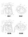
- FIG. 37 is a vertical cross-sectional view of a patient's heart illustrating a preferred path of access to the left ventricle from the right ventricle and through the septal wall.
- FIG. 38 is a horizontal cross-sectional view of the heart illustrating the preferred access path of FIG. 37 .
- FIGS. 39 a through 39 h illustrate the catheter of FIG. 1 being used to deliver a purse string device, which includes a plurality of tissue anchors, to remodel the left ventricle of a patient's heart.
- FIGS. 40 a through 40 h illustrate a preferred method of deployment of multiple tissue anchors interconnected by a line, such as a suture, within the left ventricle of a patient.
- a line such as a suture
- the tissue anchors and suture are deployed with the system of FIGS. 27-29 .
- FIGS. 41 a and 41 b illustrate a preferred method of utilizing a linear coil purse string device to remodel the left ventricle of a heart.
- FIG. 42 a illustrates another preferred arrangement of multiple purse strings within the left ventricle, wherein the purse string rows originate from a common point and are inclined relative to one another.
- FIG. 42 b illustrates a first arrangement of multiple purse strings within the left ventricle wherein the purse string rows are oriented generally parallel to one another.
- FIG. 43 is a perspective view of another tissue remodeling system including a catheter assembly having certain features, aspects and advantages of the present invention.
- the catheter assembly of FIG. 43 includes multiple co-axial components.
- FIG. 44 illustrates a tissue tunneling member of the catheter assembly of FIG. 43 .
- FIG. 45 illustrates a steering catheter of the catheter assembly of FIG. 43 .
- FIG. 46 illustrates a guide catheter of the catheter assembly of FIG. 43 .
- FIG. 47 illustrates a preferred path of deployment of the system of the catheter of FIG. 43 .
- FIGS. 48 a through 48 h illustrate the system of FIG. 43 being utilized to place a suture within a passage through the myocardial wall surrounding the left ventricle of a patient's heart.
- Preferred embodiments of the present tissue remodeling system facilitate remodeling, tissue joining, or tying of soft tissue preferably in a percutaneous manner utilizing a catheter.
- the system permits a remodeling of the left ventricle of a heart to reduce the volume of the ventricle or reposition papillary muscles.
- the preferred systems may be utilized to remodel valve structures, such as the aortic or mitral valves of the heart.
- Certain systems and methods disclosed herein may be well-suited to treat Ischemic Mitral Regurgitation (IMR), for example.
- IMR Ischemic Mitral Regurgitation
- preferred embodiments may be utilized to reduce the cross-sectional area of body cavities or lumens, or completely close body cavities or lumens. Preferred methods for remodeling soft tissue are also disclosed.
- the preferred embodiments permit soft tissue remodeling while avoiding the disadvantages of more invasive procedures and the complications that may occur as a result of such procedures.
- the preferred embodiments and methods may also permit tissue remodeling in patients that are otherwise unable to undergo conventional surgical procedures.
- Preferred embodiments of the present system permit the duplication of the results of surgical procedures by percutaneous transvascular techniques using catheter-based devices.
- the preferred embodiments and methods disclosed herein maybe modified or adopted for use in the remodeling of soft tissue other than the heart or body lumens.
- the methods described herein are accomplished with a suitable imaging technique, or techniques, such as transesophageal echocardiogram (TEE), angiographic fluoroscopy or cineangiographic guidance, for example.
- TEE transesophageal echocardiogram
- FIGS. 1-3 illustrate a tissue remodeling system having certain features, aspects and advantages of the present invention and is generally referred to by the reference numeral 50 .
- the illustrated system 50 includes a catheter assembly 52 , which preferably includes multiple catheters, or catheter bodies.
- catheter assembly 52 preferably includes multiple catheters, or catheter bodies.
- both the overall catheter assembly 52 and the individual catheter bodies that make up the catheter assembly 52 may be referred to herein by the term “catheter.”
- the catheter 52 may include other components as well, such as tissue anchors, for example.
- the catheter 52 is sized, shaped and otherwise configured to be movable within a patient's vasculature to a desired remodeling site from a desired insertion site.
- the insertion site may be the femoral artery, for example. However, other suitable insertion sites may also be used.
- the catheter 52 includes multiple components that are coaxial with one another and are capable of telescopic movement relative to one another.
- the catheter assembly 52 includes a guide catheter 54 .
- a delivery catheter 56 is configured to be received within the guide catheter 54 and is movable relative to the guide catheter 54 .
- a tissue piercing or penetrating member, such as a needle 58 is received within, and is movable relative to, the delivery catheter 56 , as shown in FIG. 2 .
- a pushing mechanism such as a push rod 60 , is received within, and movable relative to, the needle 58 and may be used to deploy tissue anchors 62 ( FIG. 2 ) from the needle 58 , as is described in greater below.
- a line such as a suture 64 , extends through the needle 58 , interconnects the anchors 62 and extends back through the delivery catheter 56 such that both ends 64 a and 64 b of the suture 64 are external of the proximal end of the catheter assembly 52 .
- the individual components of the catheter assembly 52 may be constructed from any suitable material(s) using any suitable fabrication techniques, such as those commonly known and used in the medical device industry.
- the guide catheter 54 and delivery catheter 56 maybe constructed from a suitable polymeric material, such as polyethylene, polyurethane, silicon, or polytetraflouroethylene, for example.
- the catheters 54 and 56 may be constructed by an extrusion process; however, other suitable materials and/or suitable processes may be used.
- the needle 58 and push rod 60 preferably are constructed from a metal material suitable for use in medical applications, such as stainless steel or a shape-memory material, for example. However, other suitable materials and/or suitable fabrication processes may also be used.
- the guide catheter 54 preferably is configured to be introduced to the patient's vasculature at an introduction site and guided through the vasculature through the desired working site, such as the heart, for example.
- the guide catheter 54 is of a sufficient length such that the distal end of the catheter 54 may reach the desired work site while the proximal end of the catheter 54 remains external of the patient.
- the guide catheter 54 may have an outer diameter of about 26 F (French) and an inner (lumen) diameter of about 22 F.
- Other dimensions may be employed depending on the specific use of the catheter 54 , as will be appreciated by one of skill in the art.
- the guide catheter 54 is configured to be steerable to permit the catheter 54 to be guided through vasculature to the desired work site or to be steerable once at the work site.
- a deflection wire (not shown) may be connected to a distal end of the guide catheter 54 and extend within a wall of the catheter 54 to a proximal end of the catheter 54 where it is connected to a control knob 66 .
- the control knob 66 permits the user to selectively push or pull the deflection wire to deflect the distal end of the guide catheter 54 .
- Deflection of the distal end of the guide catheter 54 assists a user in routing the catheter 54 through the vasculature of a patient in a desired path or position the distal end of the catheter 54 once at the work site.
- Such a system is disclosed in greater detail in the applicant's co-pending U.S. patent application Ser. No. 11/059,866, filed Feb. 17, 2005 and entitled “Catheter Based Tissue Remodeling Devices and Methods,” the entirety of which is incorporated by reference herein.
- other suitable steering arrangements or positioning methods of the guide catheter 54 may be employed.
- the guide catheter 54 may be configured to slide over a previously placed guide wire (not shown) or may be passed through a previously placed catheter.
- the distal end of the guide catheter 54 is configured to be atraumatic to the patient and, in particular, to the tissue proximate the work site.
- the distal tip of the guide catheter 54 carries an inflatable, preferably annular balloon 68 .
- the balloon 68 is normally carried by the guide catheter 54 in an uninflated condition so as not to interfere with the passage of the catheter 54 through a patient's vasculature.
- the balloon 68 may be inflated such that a distal end of the balloon 68 contacts the patient's soft tissue to help stabilize the catheter 54 and inhibit the distal tip of the catheter 54 from damaging tissue.
- the balloon 68 in an inflated condition, extends beyond an end surface of the distal end of the catheter 54 to create a space 70 between the distal end of the catheter 54 and the tissue 72 , as illustrated in FIG. 4 .
- the balloon 68 may be constructed from any suitable material, such as silicon, urethane, Pebax, polyimide and multi-layered polymers, for example.
- the balloon 68 may be inflated by a suitable fluid, including liquid or gas.
- a passage (not shown) extends from a proximal end of the catheter 54 to an internal space of the balloon 68 so that an inflation fluid may be introduced into the balloon 68 from the proximal end of the catheter 54 .
- the passage may be defined within the body of the catheter 54 or may be formed by a separate element of the catheter assembly 52 .
- the catheter assembly 52 also includes a system for creating a vacuum within the space 70 bounded by the balloon 68 .
- the catheter assembly 52 includes a conduit, or tube 74 , which is configured to be connectable to a vacuum pump (not shown).
- the tube 74 communicates with a passage preferably within the catheter body of the catheter 54 (not shown) which, in turn, communicates with the space 70 .
- the vacuum pump may be operated to produce a vacuum condition within the space 70 to assist in securing the distal end of the catheter 54 at a desired location on the patient's tissue 72 .
- the vacuum passage may be created by a component of the catheter assembly 52 other than the guide catheter 54 .
- the delivery catheter 56 is configured to reside within the guide catheter 54 and is capable of movement within the guide catheter 54 .
- the delivery catheter 56 may have an outer diameter of about 18 F and an inner (lumen) diameter of about 13 F. However, other dimensions may be selected to suit a desired application of the catheter 56 .
- the delivery catheter 56 may be constructed of any suitable material or combination of materials, such as polyethylene, polyurethane, silicone or polytetraflouroethylene, for example.
- a proximal end of the catheter 56 includes a handle 76 , or hub, which permits a user to grasp and manipulate the catheter 56 .
- the delivery catheter 56 may take on any suitable cross-sectional shape. As described above, the delivery catheter 56 is configured to receive the needle 58 . In addition, desirably, a length of the suture 64 extends through the delivery catheter 56 external of the needle 58 . To accommodate both the needle 58 and the suture 64 , preferably the lumen of the catheter 56 defines a generally circular space 78 to accommodate the needle 58 and an additional space 80 configured to accommodate the suture 64 adjacent the space 78 . As illustrated in FIG. 3 a , in one arrangement, the space 80 may be connected to the space 78 or, as illustrated in FIG. 3 b , the space 80 may be defined by a separate lumen of the catheter 56 .
- the needle 58 preferably is configured to stow the tissue anchors 62 and a portion of the suture 64 and facilitate the deployment of the tissue anchors 62 and suture 64 .
- the needle 58 preferably includes a handle, or hub 82 , at its proximal end to permit a user to manipulate the needle 58 .
- a distal tip 84 of the needle 58 is beveled to facilitate tissue penetration.
- the needle 58 is constructed from a shape-memory material, such as Nitinol, for example, to provide a beneficial degree of flexibility. However, other suitable materials may also be used, such as stainless steel, for example.
- the needle 58 could be of a composite construction to permit the optimization of certain characteristics, such as needle penetration and shaft flexibility, for example.
- the hub 82 may be constructed from a plastic or metal material, for example, through any suitable process, such as molding or machining.
- the suture 64 may also be constructed of any suitable material.
- the material is non-bioabsorbable.
- the suture 64 may be constructed from a bioabsorbable material, if desired.
- the material may be monofilament or multifilament, single strand or braided and a natural material or a synthetic material.
- Suitable materials may include Polyglactin (e.g., coated vicryl), Polydioxanone (PDS), Polyamide or Nylon (e.g., ETHILON), Polyester (DACRON) or Polypropylene (PROLENE), among others.
- FIGS. 5 a - 5 f illustrate a preferred method of using the system 50 to deliver tissue anchors that are interconnected by a line, such as the suture 64 , to the soft tissue 72 of a patient.
- a line such as the suture 64
- FIGS. 5 a - 5 f the guide catheter 54 has been omitted for the purpose of clarity. However, preferably the guide catheter 54 is placed against the tissue 72 as described above with reference to FIG. 4 .
- FIG. 5 a illustrates the needle 58 in suture 64 stowed within the delivery catheter 56 . Accordingly, the needle 58 and suture 64 do not protrude from a distal end of the delivery catheter 56 .
- FIG. 5 b illustrates the delivery catheter spaced a slight distance from the soft tissue 72 of the patient.
- the needle 58 is advanced from the delivery catheter 56 and carries the suture 64 with it.
- FIG. 5 c the delivery catheter 56 is advanced until it contacts the tissue 72 .
- the needle 58 is advanced through the tissue 72 , bringing the suture 64 along with it.
- the movement of the delivery catheter 56 into contact with the tissue 72 and the movement of the needle 58 through the tissue 72 maybe performed in any order, or simultaneously, if desired.
- the tissue anchor 62 is illustrated in the process of being deployed from the needle 58 .
- the suture 64 extends through an eyelet 62 a of the tissue anchor 62 .
- the tissue anchor 62 is deployed from an end of the needle 58 due to the advancement of the push rod 60 .
- the push rod 60 may be used to exert a pushing force on the proximal most tissue anchor 62 , which is transferred through the stack of tissue anchors 62 within the needle 58 to deploy the distal most tissue anchor 62 .
- the tissue anchor 62 is positioned on the opposite side of the tissue 72 from the delivery catheter 56 and the suture 64 extends through the hole in the tissue 72 created by the passage of the needle 58 through the tissue 72 .
- the tissue anchor contacts the surface of the tissue 72 opposite the catheter 52 , which may be the outside surface of the heart chamber or vessel, for example.
- the tissue anchor may be configured to reside within the tissue 72 and, accordingly, the delivery catheter 58 may not be passed completely through the tissue 72 before the anchor is deployed.
- the needle 58 may be retracted into the delivery catheter 56 . Both the needle 58 and delivery catheter 56 may be retracted within the guide catheter 54 and away from the tissue 72 .
- the catheter 52 is moved along the tissue a desired distance D from the first tissue anchor 62 and another tissue anchor 62 is deployed, preferably by the same or a similar method. This process is repeated until a desired number of tissue anchor 62 have been deployed.
- the distance D may be modified as desired to suit an individual application. For example, for remodeling a lumen, preferably the distance D is adjusted such that preferably at least three and, more preferably, about 3 to 10 tissue anchors 62 may be implanted.
- FIGS. 6 a and 6 b illustrate multiple tissue anchors 62 interconnected by a line, such as the suture 64 , which may be utilized to reduce the cross-sectional dimension of a tissue wall 90 that defines a cavity or lumen 92 .
- the tissue anchors 62 are positioned at desired space locations around the circumference of the tissue wall 90 and interconnected by the suture 64 . With such an arrangement, desirably, very little foreign material is placed across the lumen such that blood flow is left relatively or substantially completely undisturbed.
- the tissue anchors 62 are generally positioned within a single plane.
- Other suture placement orientations may also be used.
- Tension may be applied to one or both of the ends 64 a , 64 b of the suture 64 to draw the tissue anchors 62 towards one another.
- the suture ends 64 a , 64 b may then be secured together to secure the tissue wall 90 in a reduced configuration.
- the ends 64 a and 64 b may be tied in a knot 94 .
- the knot 94 may be created outside of the catheter 52 and advanced through the catheter by any suitable method, such as by a conventional knot pusher or other suitable instrument, for example.
- the ends 64 a , 64 b may be secured to one another by a suitable connector.
- FIGS. 7 a and 7 b illustrate a plurality of tissue anchors 62 interconnected by a suture 64 and used to fold tissue or, more specifically in the illustrated arrangement, to collapse a wall of tissue 96 which defines a cavity or lumen 98 and, thus, reduce or close the lumen 98 .
- the tissue anchors 62 instead of being placed sequentially around the circumference of the tissue wall 96 , the tissue anchors 62 are placed in a non-serial manner.
- the tissue anchors 62 are positioned generally across the tissue wall 96 from the previously placed tissue anchor 62 and, in some cases, even directly across from one another such that the suture 64 extends across the lumen or cavity 98 .
- the tissue wall 96 is collapsed onto itself.
- the suture ends 62 a , 62 b may be secured to one another, such as by a knot 94 , for example.
- Such a method is well-suited for tubal ligation, such as closing a fallopian tube, for example.
- FIGS. 8 a - 8 c illustrate the system 50 being utilized to reduce the volume of the left ventricle LV of a patient's heart H.
- multiple rows of tissue anchors 62 are positioned within the wall 100 of the heart H surrounding the left ventricle LV.
- each row of tissue anchors 62 includes its own suture 64 and, thus, each suture row may be tightened to a desired cross-sectional dimension separate from the other rows of tissue anchors 62 .
- the tissue anchors 62 may be configured to be passed completely through the wall 100 to contact an external surface of the wall 100 or may be embedded within the tissue wall 100 , as desired or necessitated by the tissue structure being remodeled.
- Such an arrangement provides essentially an internal basket structure, which preferably is delivered by a percutaneous transvascular approach, thus providing the benefits of a conventional compression device while avoiding the disadvantages of a conventional surgical procedure.
- the rows of implanted sutures 64 preferably limit a maximum expanded dimension of the ventricle LV, but do not limit compression. Adjustment of the suture tension or length, and thus the maximum dimension, may be based on feedback from the ejection fraction measurement of the ventricle, for example.
- FIGS. 9 and 10 illustrate a preferred embodiment of a tissue anchor 62 that may be used in the system 50 described above, or the other systems described herein.
- the tissue anchor 62 is constructed from a generally tubular element. A semi-cylindrical section of the tubular element is removed from each end, leaving an intermediate portion of the tissue anchor 62 defining an annular loop. The intermediate section of the tissue anchor 62 thus forms an eyelet 102 of the tissue anchor 62 . Accordingly, the suture 64 may be passed through the eyelet 102 , with the portions of the tissue anchor 62 , on either side of the eyelet 102 configured to contact the soft tissue and prevent the tissue anchor 62 from being pulled through the passage in the tissue through which it was passed.
- FIGS. 11-14 illustrate another preferred embodiment of a tissue anchor, generally referred to by the reference numeral 104 .
- FIGS. 11 and 12 illustrate the tissue anchor 104 in an expanded configuration
- FIGS. 13 and 14 illustrate the tissue anchor 104 in a collapsed position.
- the tissue anchor 104 preferably is constructed from a tubular element that includes a first slot 106 which extends from a first end of the element toward an intermediate portion of the element and generally bisects the tubular element.
- the slot 106 extends about one-half the length of the tissue anchor 104 and, preferably, is aligned with the central axis of the tissue anchor 104 .
- a second slot 108 extends from a second end of the tissue anchor 104 preferably substantially the entire length of the tissue anchor 104 but terminates prior to the end of the tissue anchor 104 to leave a portion of material 110 at the first end of the tissue anchor 104 .
- the slot 108 is rotated on the tubular element from the slot 106 and, preferably, is located approximately 90 degrees about the central axis of the tissue anchor 104 from the slot 106 .
- the slot 108 passes only once through the wall of the tubular element.
- the tissue anchor 104 is constructed from a super-elastic, or shape-memory material, such as Nitinol, for example. However, other suitable materials may be used.
- the tissue anchor 104 is heat set in the expanded position as illustrated in FIGS. 11 and 12 , wherein a portion of the tubular element on one side of the slot 106 is bent away from the portion of the other side of the slot 106 until it is generally perpendicular to the remainder of the tubular element. Accordingly, in the absence of a biasing force, the tissue anchor 104 tends to assume the orientation shown in FIGS. 11 and 12 .
- the portion that is bent includes the slot 108 such that the slot 108 may function as an eyelet of the tissue anchor 104 .
- the tissue anchor 104 may be constrained, such as by the needle 58 , into the orientation illustrated in FIGS. 13 and 14 , or a collapsed orientation, so that the tissue anchors 104 may be delivered through tissue 72 .
- the tissue anchors 104 may be deployed in a similar manner to the tissue anchor 62 described above.
- the bent portion of the tissue anchor 104 including the slot 108 extends partially or completely through the tissue 72 .
- the suture 64 may be passed through the slot 108 such that, when tension is applied to the suture 64 , the tissue anchor 104 applies a force to the surface of the tissue 72 opposite the suture 64 . With such an arrangement, adjustment of the suture 64 will likely be less affected by friction in comparison to the tissue anchors described above because the suture 64 does not pass through tissue.
- the system 50 may be utilized to remodel the aortic valve AV of a patient's heart H. That is, the catheter 52 may be used to place a number of tissue anchors 62 (or 104 ) within the aorta A and, preferably, at a location adjacent to the aortic valve AV.
- the guide catheter 54 may be omitted from the catheter assembly 52 and the delivery catheter 56 may be steerable.
- the tissue anchors 62 may be interconnected with a line, such as suture 64 .
- tissue anchors 62 may be remodeled and, preferably, brought towards one another to reduce regurgitation through the valve AV.
- the tissue anchors 62 are disposed on a downstream side of the valve AV.
- additional rows may be provided.
- the system 50 also may be utilized to remodel the left ventricle LV to improve the function of the mitral valve MV.
- a plurality of tissue anchors 62 may be positioned in a row in a generally circular manner proximate the papillary muscles PM, which are connected to the leaflets of the mitral valve MV.
- the tissue anchors 62 may be interconnected by a line, such as the suture 64 , and tension applied to the suture 64 to draw the tissue anchors 62 towards one another.
- the papillary muscles PM may be drawn toward one another so that the leaflets of the mitral valve move toward one another, preferably until the leaflets properly coapt to reduce or eliminate regurgitation.
- the system 50 may be utilized to repair a defect, or hole, within a patient's heart H.
- a septal defect S such as a patent foramen ovale (PFO)
- PFO patent foramen ovale
- FIG. 19 illustrates the system 50 being used to place a plurality of tissue anchors 62 around the septal defect S.
- FIGS. 20 a and 20 b illustrate another view of the system 50 being utilized to close the septal defect S before and after tightening of the suture 64 , respectively.
- FIGS. 21-25 illustrate another system having certain features, aspects, and advantages of the present invention and generally referred to by the reference numeral 150 .
- the system 150 is configured to deliver a plurality of tissue anchors to soft tissue of a patient.
- the tissue anchors preferably are interconnected by a line, such as a suture.
- the system 150 is configured to deliver a plurality of tissue anchors simultaneously or substantially simultaneously.
- the system 150 includes a catheter assembly 152 , which preferably includes a guide catheter 154 and plurality of delivery tubes 156 .
- the delivery tubes 156 are movable within the guide catheter 154 from a stowed position ( FIG. 21 ) to a deployed position ( FIGS. 22 and 23 ).
- Each of the delivery tubes 156 function in a manner similar to the delivery catheter 56 described above, or the other delivery catheters described herein.
- the collection of deliver tubes 156 essentially function as a plurality of delivery catheters, but preferably are sized such that multiple tubes 156 may be carried within the guide catheter 154 .
- each delivery tube 156 stows a tissue anchor 158 .
- the tissue anchors 158 preferably are interconnected by a line, such as a suture 160 .
- the guide catheter 154 is substantially similar to the guide catheter 54 of FIGS. 1-3 .
- a proximal end of the guide catheter 154 defines a handle portion, or hub 162 , which permits a user of the system 150 to manipulate the guide catheter 154 .
- the guide catheter 154 may also be steerable in a manner similar to that described above in connection with the guide catheter 54 or by any other suitable arrangement.
- the guide catheter 154 is sized such that the lumen is capable of accommodating a desired number of delivery tubes 156 .
- the guide catheter 154 may have a lumen diameter of about 20 to 24 F. However, other suitable dimensions are also possible, depending on the specific application or number of delivery tubes 156 desired.
- the guide catheter 154 may be constructed of any suitable material(s), such as those described above. If desired, the guide catheter 154 could include a braided material, or be otherwise manipulated, to increase axial or radial stiffness.
- the delivery tubes 156 are movable within the guide catheter 154 .
- the delivery tubes 156 are secured to a proximal handle, or hub 164 , which permits a user of the system 150 to move the plurality of delivery tubes 156 in an axial direction relative to the guide catheter 154 substantially simultaneously.
- the delivery tubes 156 extend through the guide catheter 154 and are interconnected at the hub 164 .
- the delivery tubes 156 may be coupled to the hub 164 by an intermediate component or components.
- each of the delivery tubes 156 has a curved distal end 166 when deployed from the guide catheter 154 .
- the curved distal end 166 extends radially outward from a longitudinal axis of the guide catheter 154 preferably such that the distal end surfaces of the delivery catheters face in a direction generally perpendicular to the longitudinal axis of the guide catheter 154 .
- the curved distal ends 166 are preshaped such that the distal ends 166 of the delivery tubes 156 tend to move toward their curved configuration when no restraining force is present.
- the distal ends 166 of the delivery tubes 156 are restrained in a generally linear configuration and move to the precurved configuration when the delivery tubes 156 are deployed from the guide catheter 154 .
- the delivery tubes 156 are constructed of a super-elastic or shape-memory material that is manipulated to have the desired curvature at the distal end 166 , as will be appreciated by one of skill in the art.
- the distal ends 166 of the delivery tubes 156 are configured to stow at least one tissue anchor 158 .
- each of the distal ends 166 of the delivery tubes 156 include a slot 168 extending in an axial direction along the delivery tube 156 .
- the slot 168 accommodates the suture 160 in interconnecting the tissue anchors 158 while in their stowed position within the delivery tubes 156 .
- the slots 168 may be omitted and the suture 160 may enter through the distal end of the lumen of the delivery tubes 156 .
- multiple tissue anchors 158 may be provided, if desired.
- FIGS. 24 and 25 illustrate one preferred tissue anchor 158 in stowed and deployed orientations, respectively.
- the tissue anchor 158 includes an eyelet 170 at its proximal end and a pair of barbs 172 at its distal end.
- the barbs 172 converge into a sharp tip 174 that permits the tissue anchor 158 to penetrate soft tissue.
- the tissue anchor 158 is advanced into soft tissue, the barbs 172 inhibit withdrawal of the tissue anchor 158 in response to a pulling force applied the tissue anchor 158 .
- the tissue anchor 158 is also constructed from a shape-memory material to provide elasticity to the tissue anchors 158 .
- tissue anchors 158 may assume the precurved shape of the distal ends 166 of the delivery tubes 156 .
- the barbs 172 may be configured to expand outwardly when the tissue anchor 158 is deployed from the delivery tube 156 to enhance the tissue anchor's 158 resistance to removal, as illustrated in FIG. 25 .
- the barbs 172 may be held in a restrained position within the delivery tube 156 prior to deployment, as shown in FIG. 24 .
- a deployment device such as a pushrod 176
- a handle, or hub 178 is coupled to each pushrod 176 such that the pushrods 176 are advanced within the delivery tubes 156 when a pushing force is applied to the hub 178 .
- the hub 178 may be pushed to advance the push rods 176 which, in turn, push the tissue anchors 158 to deploy them from the delivery tubes 156 .
- the pushrods 176 are interconnected with the hub 178 , the deployment of the tissue anchors 158 occurs substantially simultaneously.
- the tissue anchors 158 may be deployable independently of one another.
- FIGS. 26 a - 26 f illustrate the system 150 being utilized to remodel an aortic valve AV to treat aortic insufficiency.
- Aortic insufficiency or regurgitation is a heart valve disease in which the aortic valve weakens or balloons, preventing the valve leaflets from closing tightly. This typically leads to backward flow of blood from the aorta A into the left ventricle LV. Over time, this condition could result in heart failure.
- the system 150 is advantageously well suited for treatment of aortic insufficiency, it can be used in, or adapted for use in, other remodeling applications as well.
- the catheter 152 preferably is advanced through the patient's vasculature to a position downstream from the aortic valve AV, as shown in FIG. 26 a .
- the delivery tubes 156 may be deployed from the guide catheter 154 by advancement of the hub 164 .
- the delivery tubes 156 are advanced from the guide catheter 154 , they assume their pre-curved shape of the distal ends 166 and move toward the wall of the aorta A.
- the delivery tubes 156 are advanced until they pass through the wall of the aorta A, as illustrated in FIG. 26 b .
- the tissue anchors 158 may be configured to reside within the wall of the aorta A and, thus, the delivery tubes 156 may not pass completely through the aorta A.
- the tissue anchors 158 are disposed at a distal-most end of the catheter 152 . That is, preferably no portion of the catheter 152 extends beyond the general position of the tissue anchors 158 . Accordingly, the tissue anchors 158 may be deployed in close proximity to the valve AV (or other obstructing object) in comparison to devices that deliver anchors proximal of the distal-most end of the device.
- the tissue anchors 158 may be deployed from the delivery tubes 156 by advancement of the hub 178 , as illustrated in FIG. 26 c .
- the pushrods 176 and delivery tubes 156 may be retracted by retraction of the hubs 164 and 178 , leaving the tissue anchors 158 disposed within the tissue of the aorta A, as illustrated in FIG. 26 d .
- the suture 160 interconnects the tissue anchors 158 and tension may be applied to one or both of the suture ends 160 a , 160 b to move the tissue anchors 150 a toward one another and reduce the cross-sectional dimension of the aorta A to achieve the desired remodeling of the aortic valve AV.
- the suture is tightened until the leaflets of the aortic valve AV coapt to inhibit or prevent regurgitation, as illustrated in FIG. 26 e.
- FIGS. 27-30 illustrate another preferred system 200 , which is configured to deploy tissue anchors into soft tissue of a patient.
- the illustrated system 200 includes a catheter assembly 202 .
- the catheter assembly 202 preferably includes a guide catheter 204 , a delivery catheter 206 , and a tissue anchor deployment member, such as a pushrod 208 .
- the guide catheter 204 , delivery catheter 206 and pushrod 208 are assembled coaxially and are movable relative to one another, in a manner similar to the systems described above.
- a proximal end of the guide catheter 204 defines a handle, or hub 210 .
- a distal end of the guide catheter 204 preferably is adapted to receive a guide wire 212 ( FIG. 30 ) which may be used to assist in directing the catheter 202 , as is described in greater detail below.
- a distal end of the guide catheter 204 includes an enlarged portion 214 that defines a passage 216 , which extends in a direction generally parallel to a longitudinal axis of the guide catheter 204 and to the main lumen of the guide catheter 217 .
- the passage 216 is configured to receive the guide wire 212 so that the guide catheter 204 may be advanced or retracted along the guide wire 212 .
- the enlarged portion and the passage 216 extends along only a portion of the distal end of the guide catheter 204 , such as about 4 to 5 cm in one arrangement to provide for a quick set-up.
- the passage 216 may extend for other distances, may extend the entire length of the guide catheter 204 or may be divided into a plurality of intermittent passage sections extending along a portion or the entire length of the catheter 204 .
- the delivery catheter 206 defines a handle, or hub 220 at its proximal end.
- the hub 220 permits the delivery catheter 206 to be manipulated by a user of the system 200 .
- a distal end of the delivery catheter 206 preferably defines a beveled tip 222 , which facilitates piercing of soft tissue.
- the distal end portion of the delivery catheter 206 is configured to have a pre-set curved shape, as illustrated in FIG. 30 .
- the entirety or at least the distal end portion of the delivery catheter 206 may be constructed from a super-elastic or shape-memory material that is manipulated into a desired curved shape.
- the guide catheter 204 may function to restrain the delivery catheter 206 in a generally linear configuration while the delivery catheter 206 is stowed within the guide catheter 204 .
- the distal end portion of the delivery catheter 206 tends to move toward its pre-curved shape.
- the longitudinal axis of the guide catheter 204 may be generally parallel to a surface of the soft tissue 224 and, when deployed, the distal end portion of the delivery catheter 206 may curve to penetrate the tissue 224 in a direction generally perpendicular to the longitudinal axis of the guide catheter 204 .
- the push rod 208 preferably is a solid, cylindrical elongate member which resides within the lumen of the delivery catheter 206 .
- the pushrod 208 is movable within the lumen of the delivery catheter 206 and is positioned proximal of the tissue anchors 226 .
- the pushrod 208 is configured to permit a user of the system 200 to deploy the tissue anchors 226 from the catheter 202 .
- tissue anchors 226 are loaded within the delivery catheter 206 to be deployed individually by the pushrod 208 .
- the tissue anchors 226 are interconnected by a line, such as a suture 228 .
- the suture 228 passes through an eyelet 230 of each tissue anchor and, preferably, is secured to the distal most tissue anchor 226 .
- the free end of the suture 228 extends through the delivery catheter 206 and exits a proximal end of the catheter assembly 202 .
- Tension may be applied to the free end of the suture 228 to draw the tissue anchors 226 towards one another after they have been implanted into tissue 224 to remodel the tissue 224 as desired.
- the distal tip 232 of the tissue anchors 226 include a pair of barbs 234 that inhibit withdrawal of the tissue anchors 226 from the soft tissue 224 .
- FIGS. 31-36 illustrate yet another preferred system 250 which is configured to deploy a plurality of tissue anchors into soft tissue of a patient.
- the system 250 includes a catheter assembly 252 .
- the catheter assembly 252 preferably includes a guide catheter 254 and a delivery catheter 256 .
- the guide catheter 254 includes a guide passage 258 , which is configured to receive a guide wire to permit the guide catheter 254 to be passed over the guide wire, as is described in greater detail below.
- the guide passage 258 is similar to the passage 216 of the guide catheter 204 , described above.
- the catheter assembly 252 also includes an elastic member, or a linear coil purse string device 260 , an extension rod 262 , and an actuator rod 264 .
- the linear coil purse string device 260 may be housed within a distal end portion of the guide catheter 254 and is carried by the delivery catheter 256 , which is movable within the guide catheter 254 .
- the linear coil purse string device 260 is carried outside of the lumen of the delivery catheter 256 .
- the extension rod 262 and actuator rod 264 extend through the lumen of the delivery catheter 256 and interact with the device 260 , as is described in greater detail below.
- the linear coil purse string device 260 includes a helically wound wire 270 , having a first end 270 a and a second end 270 b .
- the first and second ends 270 a , 270 b may comprise end caps which encapsulate the ends of the coiled wire 270 to inhibit the wire 270 from damaging tissue.
- the ends 270 a , 270 b may be secured together by a lock nut 271 .
- the ends 270 a , 270 b may be received within cavities 271 a , 271 b of the lock nut 271 , as is described in greater detail below.
- the coiled wire 270 is constructed from a super-elastic or shape-memory material and is expandable from its relaxed state.
- the wound length of the device 260 is configured according to the specific anatomical structure into which it is designated for implantation.
- the wire diameter preferably is between about 0.002 and 0.015 inches.
- the inner diameter of the wound coil (or size of the mandrel on which it may be wound) preferably is between about 0.010 and 0.125 inches. However, other suitable dimensions may also be used.
- a plurality of tissue anchor stowage members, or capsules 272 are spaced along the coiled wire 270 .
- the capsules 272 are positioned about every centimeter along the device 260 .
- the capsules 272 may be secured to the coiled wire 270 by any suitable mechanism or technique, such as swaging, welding or gluing, for example.
- the capsules 272 are generally cylindrical in shape and reside within the interior space defined by the coiled wire 270 .
- Each tissue anchor stowage capsule 272 preferably houses a tissue anchor 274 .
- the tissue anchor 274 is initially in a stowed orientation within the capsule 272 and is configured to be deployed therefrom, as illustrated in FIG. 34 .
- the tissue anchor 274 initially resides within a generally cylindrical passage 276 defined by the capsule 272 .
- An exit hole 278 communicates with the passage 276 and opens to an outer surface of the capsule 272 .
- the exit hole 278 is configured to permit at least a portion of the tissue anchor 274 to be deployed from the capsule 272 .
- the tissue anchor 274 is substantially similar to the tissue anchors described above, and includes a tissue penetrating tip 280 and, desirably, a pair of barbs 282 .
- the barbs 282 inhibit the tissue anchor 274 from withdrawing from tissue once the tissue anchor 274 has been deployed.
- the tissue anchor 274 also includes an enlarged head 284 opposite the tissue penetrating tip 280 .
- the head 284 is sized and/or shaped such that the head 284 may not pass through the exit hole 278 and, thus, the tissue anchor 274 remains secured to the capsule 272 even after deployment.
- the actuator rod 264 extends through the delivery catheter 256 and through the device 260 .
- a proximal end of the actuator rod 264 is accessible from external a proximal end of the guide catheter 254 and delivery catheter 256 so that the actuator rod 264 may be manipulated by a user of the system 250 .
- the actuator rod 264 passes through a space within the coiled wire of the device 260 and through the passages 276 of each tissue anchor stowage capsule 272 .
- the actuator rod 264 includes an enlarged actuation head 266 , which is initially positioned distal of the distal most tissue anchor capsule 272 .
- the actuator head 266 of the actuator rod 264 is configured such that, when the actuator rod 264 is retracted, the actuator head 266 contacts the head 284 of tissue anchor 274 and pushes the tip 280 of the tissue anchor 274 through the exit hole 278 .
- the head 284 of the tissue anchor 274 moves into the exit hole 278 (and out of the passage 276 ) a sufficient distance to permit the enlarged head 266 of the actuator rod 264 to pass through the passage 276 and exit the capsule 272 .
- the tissue anchor 274 is not completely separated from the capsule 272 .
- the actuator rod 264 may be continued to be retracted to deploy the tissue anchor 274 of each capsule 272 in a similar manner.
- the extension rod 262 is configured to maintain the linear coil purse string device 260 in an expanded configuration relative to its relaxed configuration. That is, the extension rod 262 secures the device 260 in an extended orientation wherein it is longer than its relaxed orientation.
- the extension rod 262 passes through the delivery catheter 256 and through the device 260 .
- a proximal end of the extension rod 262 is accessible external of the proximal end of the catheter assembly 252 so that the extension rod 262 can be manipulated by a user of the system 250 .
- the extension rod 262 extends through a passage 290 within each of the tissue anchor stowage capsules 272 .
- the passage 290 is generally parallel to the passage 276 .
- the extension rod 262 includes a plurality of steps in which the diameter of the extension rod 262 increases. Each step 292 contacts a proximal end surface of the capsule 272 to hold each tissue anchor stowage capsule 272 in a desired position relative to each other capsule 272 .
- the extension rod 262 includes an equal number of steps 292 as capsules 272 . Desirably, the spacing of the steps 292 determines the length of extension of the device 260 .
- the diameter of the extension rod 262 decreases in a direction from the proximal end toward the distal end such that the extension rod 262 may be retracted without interference between the steps 292 and the tissue anchor stowage capsules 272 .
- the linear coil purse string device 260 is permitted to move from its extended orientation toward its relaxed orientation and remodel the tissue in which the device 260 is implanted.
- FIGS. 37 and 38 illustrate one preferred access pathway P for gaining access to the left ventricle LV of a patient's heart H.
- Such an approach advantageously reduces or minimizes the potential complications of crossing the aortic valve in the case of a retrograde approach, or crossing the atrial septal and the mitral valve in the case of an antegrade approach.
- FIGS. 39 a through 39 h illustrate the system 50 of FIGS. 1-3 being utilized to remodel the left ventricle LV of a patient's heart H and reaching the left ventricle LV along an access path P as shown in FIGS. 37 and 38 .
- the catheter 52 is introduced through vasculature to the right ventricle. Once in place within the right ventricle RV, the distal end of the catheter 52 is abutted against the septal wall SW, preferably near a junction of the septal wall SW and the outer wall, or anterior wall portion, of the heart H ( FIG. 39 b ).
- a transeptal device 300 is utilized to create an access pathway through the septal wall ( FIG.
- the transeptal device 300 is a conventional device known in the art for creating a transeptal passageway, such as a Mullins sheath, for example.
- a Mullins sheath for example.
- other suitable devices or methods may also be used.
- Tissue anchors 302 which may be of any suitable construction, such as one of the various constructions described above, are introduced at desired spaced locations preferably in a semi-circular pattern within the external wall of the heart H ( FIG. 39 f ). Desirably, when used in this method, one end of the suture 64 is secured to the first tissue anchor 302 .
- the free end of the suture 64 is passed through the access passageway within the septal wall SW and pulled tight to draw the tissue anchors 302 towards one another to remodel the left ventricle LV, as illustrated in FIG. 39 g.
- a retaining structure such as a knot 304
- a knot 304 can then be created in the suture 64 within the right ventricle RV to abut the septal wall SW to secure the tissue anchors 302 in the desired spacing to maintain the remodeling of the left ventricle LV.
- the knot 304 may be created outside of the patient and advanced through the catheter 53 by a suitable device, such as a knot pusher.
- a pledget (not shown) or similar member is positioned between the septal wall SW and the knot 304 to increase the surface area acting on the septal wall SW and inhibit the knot from being pulled through the septal wall SW.
- other alternative devices or methods for maintaining tension on the suture 64 may also be used.
- the illustrated method has the advantage of using the septal wall SW as a back stop for tying off the suture 64 , which is a relatively simple method of maintaining tension on the suture 64 .
- FIGS. 40 a - 40 j illustrate another preferred method for remodeling the left ventricle LV of a patient's heart H.
- the system 200 of FIGS. 27-30 is utilized.
- a first catheter 320 and a guide wire 322 are routed through vasculature to the right ventricle RV and through the septal wall SW to the left ventricle LV.
- a second catheter 324 is similarly advanced to the left ventricle LV, preferably to an opposite side of the left ventricle LV from the first catheter 320 and through a separate access passageway, as illustrated in FIG. 40 a.
- a retrieval device 326 is advanced through the second catheter 324 and is used to grasp the guide wire 322 , as shown in FIG. 40 b .
- the retrieval device 326 may be of any suitable construction to permit the retrieval of the guide wire 322 , such as one of the devices disclosed in U.S. Pat. No. 5,522,819 or U.S. Pat. No. 5,868,754, for example, the entireties of which are incorporated by reference herein and made a part of this specification.
- the guide wire 322 can then be pulled through the second catheter 324 such that both ends of the guide wire 322 are located external of the patient.
- the catheters 320 and 324 may then be removed from the patient, leaving the guide wire 322 in place, as illustrated in FIG. 40 c.
- the user of the system 200 may apply a force to the free ends of the guide wire 322 to force the portion of the guide wire 322 within the left ventricle LV against the interior surface of the wall of the heart H, as shown in FIG. 40 d .
- the guide wire 322 may be used to reduce the amount of motion due to the heart beat during deployment of the tissue anchors 226 .
- the catheter 202 may be advanced over the guide wire 322 until a distal end portion of the catheter 202 is within the left ventricle LV, as shown in FIG. 40 e .
- Tissue anchors 226 may then be deployed within the external wall of the left ventricle LV as described above with reference to FIGS. 27-30 and shown in FIG. 40 f.
- tension may be applied to the suture 228 interconnecting the tissue anchors 226 to draw the tissue anchors 226 towards one another and remodel the left ventricle.
- a knot 328 may be created in the free ends of the suture 228 external of the patient and advanced through the catheter 202 to secure the free ends together and maintain tension on the suture 228 , as illustrated in FIG. 40 g .
- the knot 328 is positioned within the right ventricle RV such that the portion of the suture 228 and knot 328 rest against the septal wall SW within the right ventricle RV to maintain the left ventricle LV in its remodeled orientation, as illustrated in FIG. 40 h .
- This method may be used to reduce to the volume of the left ventricle LV to treat congestive heart failure or to reposition the papillary muscles to improve mitral valve function, among other therapeutic uses.
- FIGS. 41 a and 41 b illustrate a similar method using the system 250 of FIGS. 31-36 .
- the ends 270 a and 270 b are secured together within the right ventricle RV, such as by the lock nut 271 (as described above with reference to FIG. 33 ) or another suitable method.
- the tissue anchors 274 may be positioned within the heart H while the device 260 is in a stretched state. Desirably, the stretched state of the device 260 is no more than about 6 to 8 percent of the elastic limit of the material.
- the device 260 preferably retracts towards its relaxed state, thereby remodeling the left ventricle LV.
- the resistance to stretching of the device 260 assists in inhibiting excessive enlargement of the ventricle LV.
- This method may be used to reduce to the volume of the left ventricle LV to treat congestive heart failure or to reposition the papillary muscles to improve mitral valve function, among other therapeutic uses.
- multiple rows R of tissue anchors may be implanted within the left ventricle LV.
- the multiple rows R may converge at a common point and be inclined relative to one another.
- the multiple rows R may be positioned substantially parallel to one another, depending on the desired shape of the remodeled ventricle.
- Other suitable relative orientations of the rows R of tissue anchors may also be used, including arrangements wherein two or more rows R intersect with one another.
- FIGS. 43-46 illustrate yet another system 350 for remodeling soft tissue of a patient.
- the system 350 includes a catheter assembly 352 .
- the catheter assembly 352 preferably includes a guide catheter 354 and a delivery catheter 356 .
- a tunneling device 358 is received within the delivery catheter 356 .
- the delivery catheter 356 has a pre-curved distal end portion 360 that may be constrained in a generally linear configuration when positioned within the guide catheter 354 .
- the distal end portion 360 of the delivery catheter 356 preferably tends to move toward its pre-curved configuration.
- the pre-curved shape of the delivery catheter 356 may be configured by heat setting or otherwise manipulating a super-elastic or shape-memory material.
- the distal end of the guide catheter 354 may be deflectable, such as in a manner similar to the guide catheters described previously.
- the tunneling device 358 preferably is an elongate rod with a tissue penetrating tip 362 .
- the tip 362 in one arrangement, may include one or more helical flutes such that when the tunneling device 358 is rotated, the tip 362 tends to facilitate passage through tissue.
- the tunneling device 358 preferably is constructed from a super-elastic or shape-memory material to provide a desired degree of flexibility. However, other suitable materials may also be used, such as stainless steel, for example.
- the delivery catheter 356 and tunneling device 358 may be advanced and rotated relative to one another and the guide catheter 354 to permit the tunneling device 358 to move through tissue in a desired tunneling path.
- the tunneling device 358 is configured to carry an end of a suture or other line, through the passage created by the tunneling device 358 , as is described in greater detail below.
- the tunneling device 358 may be configured to carry other suitable cinching devices, such as an elastic member similar to the coiled member 270 , with or without the tissue anchor capsules 272 .
- FIG. 47 illustrates one desired path Q for the tunneling device 358 to facilitate placement of a suture to remodel the left ventricle LV of a patient's heart H.
- FIGS. 48 a through 48 h illustrate a preferred method of utilizing the system 350 to pass a cinching member, such as suture 364 , through the heart wall and around the left ventricle LV.
- a cinching member such as suture 364
- FIG. 48 a preferably by methods similar to those described immediately above, access is gained to the right ventricle RV through the patient's vasculature.
- a catheter 362 preferably is utilized to gain access to the right ventricle RV and provide a path to permit the catheter assembly 352 to be advanced through the catheter 362 to the right ventricle RV.
- the catheter assembly 352 is positioned at a junction J of the septal wall SW and the external wall of the heart H.
- the tunneling device 358 may be advanced from the guide catheter 354 by a pushing force and, preferably, simultaneous rotation so that the tip 362 bores into the tissue.
- the guide catheter 354 and the delivery catheter 356 may be advanced along the tunneling device 358 after the tunneling device 358 has created a path through the heart wall.
- the delivery catheter 356 may be advanced from the guide catheter 354 such that the distal end portion 360 of the delivery catheter 356 assumes a desired pre-curved configuration. Accordingly, the delivery catheter 356 may be used as a steering device to urge the tunneling device 358 in a desired direction.
- the extent that the delivery catheter 356 is exposed from the guide catheter 354 , along with the rotational position of the delivery catheter 356 relative to the guide catheter 354 may be selected and modified throughout the procedure to achieve a desired tunneling path of the tunneling device 358 . In this manner, as illustrated successively in FIGS.
- the tunneling device 358 may be steered in a path through the external wall of the heart H circumscribing the left ventricle LV until the tunneling device 358 reenters the right ventricle RV, preferably at a junction between the septal wall SW and external wall of the heart H opposite its entry point.
- a retrieval tube, or retrieval catheter 364 may be introduced to the right ventricle RV by a suitable method to receive the tip of the tunneling device 358 so that the device 358 may be advanced through the catheter 362 until it may be retrieved from the proximal end of the retrieval catheter 364 .
- such a method permits the tunneling device 358 to not only create a tunnel, or passage, through the heart wall, but to also pass a cinching device, such as the suture 364 , through the passage.
- the suture 364 or other cinching device, may then be pulled, or cinched, to remodel the left ventricle LV.
- the suture 364 is tied off into a knot to maintain the remodeled orientation in a manner similar to that described above. Pledgets may be used if desired to spread the load acting on the heart wall from the knot to a larger area.
- the system 350 and described method may be used to remodel the left ventricle LV, such as reducing the volume to treat congestive heart failure or repositioning the papillary muscles to improve mitral valve function.
- the system 350 and method may be used to remodel other body cavities as well.
Abstract
Description
Claims (10)
Priority Applications (6)
| Application Number | Priority Date | Filing Date | Title |
|---|---|---|---|
| US11/408,717 US8333777B2 (en) | 2005-04-22 | 2006-04-21 | Catheter-based tissue remodeling devices and methods |
| US13/712,651 US9107658B2 (en) | 2005-04-22 | 2012-12-12 | Catheter-based tissue remodeling devices and methods |
| US14/827,707 US20160038130A1 (en) | 2005-04-22 | 2015-08-17 | Catheter-based tissue remodeling devices and methods |
| US16/886,447 US10912546B2 (en) | 2005-04-22 | 2020-05-28 | Catheter-based tissue remodeling devices and methods |
| US17/095,032 US10966696B2 (en) | 2005-04-22 | 2020-11-11 | Catheter-based tissue remodeling devices and methods |
| US17/212,575 US20220000464A1 (en) | 2005-04-22 | 2021-03-25 | Catheter-based tissue remodeling devices and methods |
Applications Claiming Priority (3)
| Application Number | Priority Date | Filing Date | Title |
|---|---|---|---|
| US67383805P | 2005-04-22 | 2005-04-22 | |
| US70282305P | 2005-07-27 | 2005-07-27 | |
| US11/408,717 US8333777B2 (en) | 2005-04-22 | 2006-04-21 | Catheter-based tissue remodeling devices and methods |
Related Child Applications (1)
| Application Number | Title | Priority Date | Filing Date |
|---|---|---|---|
| US13/712,651 Division US9107658B2 (en) | 2005-04-22 | 2012-12-12 | Catheter-based tissue remodeling devices and methods |
Publications (2)
| Publication Number | Publication Date |
|---|---|
| US20070112425A1 US20070112425A1 (en) | 2007-05-17 |
| US8333777B2 true US8333777B2 (en) | 2012-12-18 |
Family
ID=38041926
Family Applications (6)
| Application Number | Title | Priority Date | Filing Date |
|---|---|---|---|
| US11/408,717 Active 2029-06-04 US8333777B2 (en) | 2005-04-22 | 2006-04-21 | Catheter-based tissue remodeling devices and methods |
| US13/712,651 Expired - Fee Related US9107658B2 (en) | 2005-04-22 | 2012-12-12 | Catheter-based tissue remodeling devices and methods |
| US14/827,707 Abandoned US20160038130A1 (en) | 2005-04-22 | 2015-08-17 | Catheter-based tissue remodeling devices and methods |
| US16/886,447 Active US10912546B2 (en) | 2005-04-22 | 2020-05-28 | Catheter-based tissue remodeling devices and methods |
| US17/095,032 Active US10966696B2 (en) | 2005-04-22 | 2020-11-11 | Catheter-based tissue remodeling devices and methods |
| US17/212,575 Pending US20220000464A1 (en) | 2005-04-22 | 2021-03-25 | Catheter-based tissue remodeling devices and methods |
Family Applications After (5)
| Application Number | Title | Priority Date | Filing Date |
|---|---|---|---|
| US13/712,651 Expired - Fee Related US9107658B2 (en) | 2005-04-22 | 2012-12-12 | Catheter-based tissue remodeling devices and methods |
| US14/827,707 Abandoned US20160038130A1 (en) | 2005-04-22 | 2015-08-17 | Catheter-based tissue remodeling devices and methods |
| US16/886,447 Active US10912546B2 (en) | 2005-04-22 | 2020-05-28 | Catheter-based tissue remodeling devices and methods |
| US17/095,032 Active US10966696B2 (en) | 2005-04-22 | 2020-11-11 | Catheter-based tissue remodeling devices and methods |
| US17/212,575 Pending US20220000464A1 (en) | 2005-04-22 | 2021-03-25 | Catheter-based tissue remodeling devices and methods |
Country Status (1)
| Country | Link |
|---|---|
| US (6) | US8333777B2 (en) |
Cited By (87)
| Publication number | Priority date | Publication date | Assignee | Title |
|---|---|---|---|---|
| US8858623B2 (en) * | 2011-11-04 | 2014-10-14 | Valtech Cardio, Ltd. | Implant having multiple rotational assemblies |
| US20140309729A1 (en) * | 2008-04-15 | 2014-10-16 | Medtronic Vascular, Inc. | Devices and Methods for Treating Valvular Regurgitation |
| US8926695B2 (en) | 2006-12-05 | 2015-01-06 | Valtech Cardio, Ltd. | Segmented ring placement |
| US8956406B2 (en) | 2008-04-16 | 2015-02-17 | Heart Repair Technologies, Inc. | Transvalvular intraanular band and chordae cutting for ischemic and dilated cardiomyopathy |
| US8961597B2 (en) | 2008-04-16 | 2015-02-24 | Heart Repair Technologies, Inc. | Percutaneous transvalvular intraannular band for mitral valve repair |
| US9050078B2 (en) * | 2009-06-21 | 2015-06-09 | Aesthetics Point Ltd. | Implanted medical device useful for cosmetic surgery |
| US9119719B2 (en) | 2009-05-07 | 2015-09-01 | Valtech Cardio, Ltd. | Annuloplasty ring with intra-ring anchoring |
| US9168137B2 (en) | 2008-04-16 | 2015-10-27 | Heart Repair Technologies, Inc. | Transvalvular intraannular band for aortic valve repair |
| US9192472B2 (en) | 2008-06-16 | 2015-11-24 | Valtec Cardio, Ltd. | Annuloplasty devices and methods of delivery therefor |
| US20160045314A1 (en) * | 2013-03-04 | 2016-02-18 | Medical Research Infrastructure And Health Service Fund Of The Tel-Aviv Medical Center | Cardiac valve commissure brace |
| US9289282B2 (en) | 2011-05-31 | 2016-03-22 | Edwards Lifesciences Corporation | System and method for treating valve insufficiency or vessel dilatation |
| US20160135826A1 (en) * | 2014-11-18 | 2016-05-19 | David Michael Chadbourne | Fluid supply and suction device |
| US9414921B2 (en) | 2009-10-29 | 2016-08-16 | Valtech Cardio, Ltd. | Tissue anchor for annuloplasty device |
| US9474606B2 (en) | 2009-05-04 | 2016-10-25 | Valtech Cardio, Ltd. | Over-wire implant contraction methods |
| US9504571B2 (en) | 2012-06-07 | 2016-11-29 | Edwards Lifesciences Corporation | Systems for implanting annuloplasty rings with microanchors |
| US9526613B2 (en) | 2005-03-17 | 2016-12-27 | Valtech Cardio Ltd. | Mitral valve treatment techniques |
| US9610162B2 (en) | 2013-12-26 | 2017-04-04 | Valtech Cardio, Ltd. | Implantation of flexible implant |
| US9622861B2 (en) | 2009-12-02 | 2017-04-18 | Valtech Cardio, Ltd. | Tool for actuating an adjusting mechanism |
| US9662209B2 (en) | 2008-12-22 | 2017-05-30 | Valtech Cardio, Ltd. | Contractible annuloplasty structures |
| US9713530B2 (en) | 2008-12-22 | 2017-07-25 | Valtech Cardio, Ltd. | Adjustable annuloplasty devices and adjustment mechanisms therefor |
| US9724192B2 (en) | 2011-11-08 | 2017-08-08 | Valtech Cardio, Ltd. | Controlled steering functionality for implant-delivery tool |
| US9730793B2 (en) | 2012-12-06 | 2017-08-15 | Valtech Cardio, Ltd. | Techniques for guide-wire based advancement of a tool |
| US9883943B2 (en) | 2006-12-05 | 2018-02-06 | Valtech Cardio, Ltd. | Implantation of repair devices in the heart |
| US9913706B2 (en) | 2014-07-17 | 2018-03-13 | Millipede, Inc. | Adjustable endolumenal implant for reshaping the mitral valve annulus |
| US9949828B2 (en) | 2012-10-23 | 2018-04-24 | Valtech Cardio, Ltd. | Controlled steering functionality for implant-delivery tool |
| US9968452B2 (en) | 2009-05-04 | 2018-05-15 | Valtech Cardio, Ltd. | Annuloplasty ring delivery cathethers |
| US9968454B2 (en) | 2009-10-29 | 2018-05-15 | Valtech Cardio, Ltd. | Techniques for guide-wire based advancement of artificial chordae |
| US10004558B2 (en) | 2009-01-12 | 2018-06-26 | Ethicon Endo-Surgery, Inc. | Electrical ablation devices |
| US10098737B2 (en) | 2009-10-29 | 2018-10-16 | Valtech Cardio, Ltd. | Tissue anchor for annuloplasty device |
| US10105141B2 (en) | 2008-07-14 | 2018-10-23 | Ethicon Endo-Surgery, Inc. | Tissue apposition clip application methods |
| US10195030B2 (en) | 2014-10-14 | 2019-02-05 | Valtech Cardio, Ltd. | Leaflet-restraining techniques |
| US10206709B2 (en) | 2012-05-14 | 2019-02-19 | Ethicon Llc | Apparatus for introducing an object into a patient |
| US10226342B2 (en) | 2016-07-08 | 2019-03-12 | Valtech Cardio, Ltd. | Adjustable annuloplasty device with alternating peaks and troughs |
| US10231831B2 (en) | 2009-12-08 | 2019-03-19 | Cardiovalve Ltd. | Folding ring implant for heart valve |
| US10258466B2 (en) | 2015-02-13 | 2019-04-16 | Millipede, Inc. | Valve replacement using moveable restrains and angled struts |
| US10258406B2 (en) | 2011-02-28 | 2019-04-16 | Ethicon Llc | Electrical ablation devices and methods |
| US10278761B2 (en) | 2011-02-28 | 2019-05-07 | Ethicon Llc | Electrical ablation devices and methods |
| US10299793B2 (en) | 2013-10-23 | 2019-05-28 | Valtech Cardio, Ltd. | Anchor magazine |
| US10314603B2 (en) | 2008-11-25 | 2019-06-11 | Ethicon Llc | Rotational coupling device for surgical instrument with flexible actuators |
| US10335275B2 (en) | 2015-09-29 | 2019-07-02 | Millipede, Inc. | Methods for delivery of heart valve devices using intravascular ultrasound imaging |
| US10342598B2 (en) | 2012-08-15 | 2019-07-09 | Ethicon Llc | Electrosurgical system for delivering a biphasic waveform |
| US10350068B2 (en) | 2009-02-17 | 2019-07-16 | Valtech Cardio, Ltd. | Actively-engageable movement-restriction mechanism for use with an annuloplasty structure |
| US10376266B2 (en) | 2012-10-23 | 2019-08-13 | Valtech Cardio, Ltd. | Percutaneous tissue anchor techniques |
| US10449333B2 (en) | 2013-03-14 | 2019-10-22 | Valtech Cardio, Ltd. | Guidewire feeder |
| US10456259B2 (en) | 2008-04-16 | 2019-10-29 | Heart Repair Technologies, Inc. | Transvalvular intraannular band for mitral valve repair |
| US10470882B2 (en) | 2008-12-22 | 2019-11-12 | Valtech Cardio, Ltd. | Closure element for use with annuloplasty structure |
| US10478248B2 (en) | 2007-02-15 | 2019-11-19 | Ethicon Llc | Electroporation ablation apparatus, system, and method |
| US10492880B2 (en) | 2012-07-30 | 2019-12-03 | Ethicon Llc | Needle probe guide |
| US10517719B2 (en) | 2008-12-22 | 2019-12-31 | Valtech Cardio, Ltd. | Implantation of repair devices in the heart |
| US10543088B2 (en) | 2012-09-14 | 2020-01-28 | Boston Scientific Scimed, Inc. | Mitral valve inversion prostheses |
| US10548731B2 (en) | 2017-02-10 | 2020-02-04 | Boston Scientific Scimed, Inc. | Implantable device and delivery system for reshaping a heart valve annulus |
| US10555813B2 (en) | 2015-11-17 | 2020-02-11 | Boston Scientific Scimed, Inc. | Implantable device and delivery system for reshaping a heart valve annulus |
| US10682232B2 (en) | 2013-03-15 | 2020-06-16 | Edwards Lifesciences Corporation | Translation catheters, systems, and methods of use thereof |
| US10695046B2 (en) | 2005-07-05 | 2020-06-30 | Edwards Lifesciences Corporation | Tissue anchor and anchoring system |
| US10702274B2 (en) | 2016-05-26 | 2020-07-07 | Edwards Lifesciences Corporation | Method and system for closing left atrial appendage |
| US10751182B2 (en) | 2015-12-30 | 2020-08-25 | Edwards Lifesciences Corporation | System and method for reshaping right heart |
| US10765514B2 (en) | 2015-04-30 | 2020-09-08 | Valtech Cardio, Ltd. | Annuloplasty technologies |
| US10779882B2 (en) | 2009-10-28 | 2020-09-22 | Ethicon Endo-Surgery, Inc. | Electrical ablation devices |
| US10792152B2 (en) | 2011-06-23 | 2020-10-06 | Valtech Cardio, Ltd. | Closed band for percutaneous annuloplasty |
| US10828160B2 (en) | 2015-12-30 | 2020-11-10 | Edwards Lifesciences Corporation | System and method for reducing tricuspid regurgitation |
| US10835221B2 (en) | 2017-11-02 | 2020-11-17 | Valtech Cardio, Ltd. | Implant-cinching devices and systems |
| US10849755B2 (en) | 2012-09-14 | 2020-12-01 | Boston Scientific Scimed, Inc. | Mitral valve inversion prostheses |
| US10918373B2 (en) | 2013-08-31 | 2021-02-16 | Edwards Lifesciences Corporation | Devices and methods for locating and implanting tissue anchors at mitral valve commissure |
| US10918374B2 (en) | 2013-02-26 | 2021-02-16 | Edwards Lifesciences Corporation | Devices and methods for percutaneous tricuspid valve repair |
| US10925610B2 (en) | 2015-03-05 | 2021-02-23 | Edwards Lifesciences Corporation | Devices for treating paravalvular leakage and methods use thereof |
| US11013599B2 (en) | 2008-04-16 | 2021-05-25 | Heart Repair Technologies, Inc. | Percutaneous transvalvular intraannular band for mitral valve repair |
| US11026791B2 (en) | 2018-03-20 | 2021-06-08 | Medtronic Vascular, Inc. | Flexible canopy valve repair systems and methods of use |
| US11033391B2 (en) | 2016-12-22 | 2021-06-15 | Heart Repair Technologies, Inc. | Percutaneous delivery systems for anchoring an implant in a cardiac valve annulus |
| US11045627B2 (en) | 2017-04-18 | 2021-06-29 | Edwards Lifesciences Corporation | Catheter system with linear actuation control mechanism |
| US11083579B2 (en) | 2008-04-16 | 2021-08-10 | Heart Repair Technologies, Inc. | Transvalvular intraanular band and chordae cutting for ischemic and dilated cardiomyopathy |
| US11123191B2 (en) | 2018-07-12 | 2021-09-21 | Valtech Cardio Ltd. | Annuloplasty systems and locking tools therefor |
| US11135062B2 (en) | 2017-11-20 | 2021-10-05 | Valtech Cardio Ltd. | Cinching of dilated heart muscle |
| US11259924B2 (en) | 2006-12-05 | 2022-03-01 | Valtech Cardio Ltd. | Implantation of repair devices in the heart |
| US11285003B2 (en) | 2018-03-20 | 2022-03-29 | Medtronic Vascular, Inc. | Prolapse prevention device and methods of use thereof |
| US11395648B2 (en) | 2012-09-29 | 2022-07-26 | Edwards Lifesciences Corporation | Plication lock delivery system and method of use thereof |
| US11484191B2 (en) | 2013-02-27 | 2022-11-01 | Cilag Gmbh International | System for performing a minimally invasive surgical procedure |
| US11653910B2 (en) | 2010-07-21 | 2023-05-23 | Cardiovalve Ltd. | Helical anchor implantation |
| US11660191B2 (en) | 2008-03-10 | 2023-05-30 | Edwards Lifesciences Corporation | Method to reduce mitral regurgitation |
| US11660190B2 (en) | 2007-03-13 | 2023-05-30 | Edwards Lifesciences Corporation | Tissue anchors, systems and methods, and devices |
| US11666442B2 (en) | 2018-01-26 | 2023-06-06 | Edwards Lifesciences Innovation (Israel) Ltd. | Techniques for facilitating heart valve tethering and chord replacement |
| US11779458B2 (en) | 2016-08-10 | 2023-10-10 | Cardiovalve Ltd. | Prosthetic valve with leaflet connectors |
| US11779463B2 (en) | 2018-01-24 | 2023-10-10 | Edwards Lifesciences Innovation (Israel) Ltd. | Contraction of an annuloplasty structure |
| US11801135B2 (en) | 2015-02-05 | 2023-10-31 | Cardiovalve Ltd. | Techniques for deployment of a prosthetic valve |
| US11819411B2 (en) | 2019-10-29 | 2023-11-21 | Edwards Lifesciences Innovation (Israel) Ltd. | Annuloplasty and tissue anchor technologies |
| US11844691B2 (en) | 2013-01-24 | 2023-12-19 | Cardiovalve Ltd. | Partially-covered prosthetic valves |
| US11937795B2 (en) | 2016-02-16 | 2024-03-26 | Cardiovalve Ltd. | Techniques for providing a replacement valve and transseptal communication |
| US11969348B2 (en) | 2021-08-26 | 2024-04-30 | Edwards Lifesciences Corporation | Cardiac valve replacement |
Families Citing this family (177)
| Publication number | Priority date | Publication date | Assignee | Title |
|---|---|---|---|---|
| CA2293057C (en) | 1998-12-30 | 2008-04-01 | Depuy Orthopaedics, Inc. | Suture locking device |
| US8641727B2 (en) | 2002-06-13 | 2014-02-04 | Guided Delivery Systems, Inc. | Devices and methods for heart valve repair |
| US20060122633A1 (en) | 2002-06-13 | 2006-06-08 | John To | Methods and devices for termination |
| US7883538B2 (en) | 2002-06-13 | 2011-02-08 | Guided Delivery Systems Inc. | Methods and devices for termination |
| US9713465B1 (en) * | 2004-04-19 | 2017-07-25 | Granit Medical Innovation Llc | Surgical closure device and associated method |
| US20070055206A1 (en) * | 2005-08-10 | 2007-03-08 | Guided Delivery Systems, Inc. | Methods and devices for deployment of tissue anchors |
| US8267942B2 (en) * | 2005-12-23 | 2012-09-18 | Ethicon, Inc. | Systems and methods for closing a vessel wound |
| US8025670B2 (en) * | 2006-11-22 | 2011-09-27 | Minos Medical | Methods and apparatus for natural orifice vaginal hysterectomy |
| US8784439B1 (en) * | 2006-11-28 | 2014-07-22 | Stephen V. Ward | Percutaneous medical procedures and devices for closing vessels using mechanical closures |
| ATE514382T1 (en) * | 2006-11-30 | 2011-07-15 | Wilson Cook Medical Inc | FABRIC ANCHOR FOR SEAM CLOSURE OF PERFORATIONS |
| US9192471B2 (en) | 2007-01-08 | 2015-11-24 | Millipede, Inc. | Device for translumenal reshaping of a mitral valve annulus |
| US8075572B2 (en) | 2007-04-26 | 2011-12-13 | Ethicon Endo-Surgery, Inc. | Surgical suturing apparatus |
| US8100922B2 (en) | 2007-04-27 | 2012-01-24 | Ethicon Endo-Surgery, Inc. | Curved needle suturing tool |
| US10307041B2 (en) | 2007-06-08 | 2019-06-04 | Medeon Biodesign, Inc. | Lens cover modification |
| WO2008153841A2 (en) * | 2007-06-08 | 2008-12-18 | Thomas Hsu | Devices and methods for removal of debris from the objective lens of an endoscope |
| US8262655B2 (en) | 2007-11-21 | 2012-09-11 | Ethicon Endo-Surgery, Inc. | Bipolar forceps |
| US8579897B2 (en) | 2007-11-21 | 2013-11-12 | Ethicon Endo-Surgery, Inc. | Bipolar forceps |
| US8568410B2 (en) | 2007-08-31 | 2013-10-29 | Ethicon Endo-Surgery, Inc. | Electrical ablation surgical instruments |
| DE102007043830A1 (en) | 2007-09-13 | 2009-04-02 | Lozonschi, Lucian, Madison | Heart valve stent |
| WO2009052188A1 (en) | 2007-10-15 | 2009-04-23 | Edwards Lifesciences Corporation | Transcatheter heart valve with micro-anchors |
| US9125632B2 (en) * | 2007-10-19 | 2015-09-08 | Guided Delivery Systems, Inc. | Systems and methods for cardiac remodeling |
| US8480657B2 (en) | 2007-10-31 | 2013-07-09 | Ethicon Endo-Surgery, Inc. | Detachable distal overtube section and methods for forming a sealable opening in the wall of an organ |
| US20090112059A1 (en) | 2007-10-31 | 2009-04-30 | Nobis Rudolph H | Apparatus and methods for closing a gastrotomy |
| WO2009100242A2 (en) | 2008-02-06 | 2009-08-13 | Guided Delivery Systems, Inc. | Multi-window guide tunnel |
| US8262680B2 (en) | 2008-03-10 | 2012-09-11 | Ethicon Endo-Surgery, Inc. | Anastomotic device |
| CA2723810C (en) | 2008-05-07 | 2015-06-30 | Guided Delivery Systems, Inc. | Deflectable guide |
| US8317806B2 (en) | 2008-05-30 | 2012-11-27 | Ethicon Endo-Surgery, Inc. | Endoscopic suturing tension controlling and indication devices |
| US8679003B2 (en) | 2008-05-30 | 2014-03-25 | Ethicon Endo-Surgery, Inc. | Surgical device and endoscope including same |
| US8114072B2 (en) | 2008-05-30 | 2012-02-14 | Ethicon Endo-Surgery, Inc. | Electrical ablation device |
| US8070759B2 (en) | 2008-05-30 | 2011-12-06 | Ethicon Endo-Surgery, Inc. | Surgical fastening device |
| US8771260B2 (en) | 2008-05-30 | 2014-07-08 | Ethicon Endo-Surgery, Inc. | Actuating and articulating surgical device |
| US8652150B2 (en) | 2008-05-30 | 2014-02-18 | Ethicon Endo-Surgery, Inc. | Multifunction surgical device |
| US8906035B2 (en) | 2008-06-04 | 2014-12-09 | Ethicon Endo-Surgery, Inc. | Endoscopic drop off bag |
| US8403926B2 (en) | 2008-06-05 | 2013-03-26 | Ethicon Endo-Surgery, Inc. | Manually articulating devices |
| US8361112B2 (en) * | 2008-06-27 | 2013-01-29 | Ethicon Endo-Surgery, Inc. | Surgical suture arrangement |
| US8262563B2 (en) | 2008-07-14 | 2012-09-11 | Ethicon Endo-Surgery, Inc. | Endoscopic translumenal articulatable steerable overtube |
| US8211125B2 (en) | 2008-08-15 | 2012-07-03 | Ethicon Endo-Surgery, Inc. | Sterile appliance delivery device for endoscopic procedures |
| US8529563B2 (en) | 2008-08-25 | 2013-09-10 | Ethicon Endo-Surgery, Inc. | Electrical ablation devices |
| US8241204B2 (en) | 2008-08-29 | 2012-08-14 | Ethicon Endo-Surgery, Inc. | Articulating end cap |
| US8480689B2 (en) | 2008-09-02 | 2013-07-09 | Ethicon Endo-Surgery, Inc. | Suturing device |
| US8409200B2 (en) | 2008-09-03 | 2013-04-02 | Ethicon Endo-Surgery, Inc. | Surgical grasping device |
| US8114119B2 (en) | 2008-09-09 | 2012-02-14 | Ethicon Endo-Surgery, Inc. | Surgical grasping device |
| US8337394B2 (en) | 2008-10-01 | 2012-12-25 | Ethicon Endo-Surgery, Inc. | Overtube with expandable tip |
| EP2349020B1 (en) | 2008-10-10 | 2020-06-03 | Ancora Heart, Inc. | Tether tensioning device |
| AU2009302169B2 (en) | 2008-10-10 | 2016-01-14 | Ancora Heart, Inc. | Termination devices and related methods |
| US20100276296A1 (en) * | 2008-11-17 | 2010-11-04 | Etorus, Inc. | Electrolytic hydrogen generating system |
| US8377095B2 (en) * | 2008-12-05 | 2013-02-19 | Cook Medical Technologies, LLC | Tissue anchors for purse-string closure of perforations |
| US8172772B2 (en) | 2008-12-11 | 2012-05-08 | Ethicon Endo-Surgery, Inc. | Specimen retrieval device |
| US8940044B2 (en) | 2011-06-23 | 2015-01-27 | Valtech Cardio, Ltd. | Closure element for use with an annuloplasty structure |
| US8926697B2 (en) | 2011-06-23 | 2015-01-06 | Valtech Cardio, Ltd. | Closed band for percutaneous annuloplasty |
| US8828031B2 (en) | 2009-01-12 | 2014-09-09 | Ethicon Endo-Surgery, Inc. | Apparatus for forming an anastomosis |
| US20100198192A1 (en) | 2009-01-20 | 2010-08-05 | Eugene Serina | Anchor deployment devices and related methods |
| US20100185278A1 (en) * | 2009-01-21 | 2010-07-22 | Tendyne Medical | Apical Papillary Msucle Attachment for Left Ventricular Reduction |
| US9226772B2 (en) | 2009-01-30 | 2016-01-05 | Ethicon Endo-Surgery, Inc. | Surgical device |
| US8252057B2 (en) | 2009-01-30 | 2012-08-28 | Ethicon Endo-Surgery, Inc. | Surgical access device |
| US8037591B2 (en) | 2009-02-02 | 2011-10-18 | Ethicon Endo-Surgery, Inc. | Surgical scissors |
| US8790369B2 (en) * | 2009-07-24 | 2014-07-29 | Depuy Mitek, Llc | Apparatus and method for repairing tissue |
| US8608652B2 (en) | 2009-11-05 | 2013-12-17 | Ethicon Endo-Surgery, Inc. | Vaginal entry surgical devices, kit, system, and method |
| JP2013512765A (en) | 2009-12-08 | 2013-04-18 | アヴァロン メディカル リミテッド | Devices and systems for transcatheter mitral valve replacement |
| US8496574B2 (en) | 2009-12-17 | 2013-07-30 | Ethicon Endo-Surgery, Inc. | Selectively positionable camera for surgical guide tube assembly |
| US8353487B2 (en) | 2009-12-17 | 2013-01-15 | Ethicon Endo-Surgery, Inc. | User interface support devices for endoscopic surgical instruments |
| US9028483B2 (en) | 2009-12-18 | 2015-05-12 | Ethicon Endo-Surgery, Inc. | Surgical instrument comprising an electrode |
| US8506564B2 (en) | 2009-12-18 | 2013-08-13 | Ethicon Endo-Surgery, Inc. | Surgical instrument comprising an electrode |
| US20110178537A1 (en) * | 2010-01-20 | 2011-07-21 | Whitman Michael P | Tissue repair implant and delivery device and method |
| US10058314B2 (en) * | 2010-01-20 | 2018-08-28 | Micro Interventional Devices, Inc. | Tissue closure device and method |
| US9980708B2 (en) | 2010-01-20 | 2018-05-29 | Micro Interventional Devices, Inc. | Tissue closure device and method |
| US10743854B2 (en) * | 2010-01-20 | 2020-08-18 | Micro Interventional Devices, Inc. | Tissue closure device and method |
| US10959840B2 (en) | 2010-01-20 | 2021-03-30 | Micro Interventional Devices, Inc. | Systems and methods for affixing a prosthesis to tissue |
| US10058323B2 (en) | 2010-01-22 | 2018-08-28 | 4 Tech Inc. | Tricuspid valve repair using tension |
| US8475525B2 (en) | 2010-01-22 | 2013-07-02 | 4Tech Inc. | Tricuspid valve repair using tension |
| US9307980B2 (en) | 2010-01-22 | 2016-04-12 | 4Tech Inc. | Tricuspid valve repair using tension |
| US9005198B2 (en) | 2010-01-29 | 2015-04-14 | Ethicon Endo-Surgery, Inc. | Surgical instrument comprising an electrode |
| WO2011100625A2 (en) * | 2010-02-12 | 2011-08-18 | Stefan Josef Matthias Kraemer | Apparatus and method for gastric bypass surgery |
| US9855031B2 (en) * | 2010-04-13 | 2018-01-02 | Neosurgical Limited | Suture delivery system |
| GB2486497B (en) | 2010-12-17 | 2013-06-19 | Neosurgical Ltd | Laparoscopic trocar system |
| CN102258402B (en) * | 2010-05-25 | 2014-11-26 | 上海微创医疗器械(集团)有限公司 | Heart valve delivery system and delivery device |
| US20120053680A1 (en) | 2010-08-24 | 2012-03-01 | Bolling Steven F | Reconfiguring Heart Features |
| US10092291B2 (en) | 2011-01-25 | 2018-10-09 | Ethicon Endo-Surgery, Inc. | Surgical instrument with selectively rigidizable features |
| US8845717B2 (en) | 2011-01-28 | 2014-09-30 | Middle Park Medical, Inc. | Coaptation enhancement implant, system, and method |
| US8888843B2 (en) | 2011-01-28 | 2014-11-18 | Middle Peak Medical, Inc. | Device, system, and method for transcatheter treatment of valve regurgitation |
| US9314620B2 (en) | 2011-02-28 | 2016-04-19 | Ethicon Endo-Surgery, Inc. | Electrical ablation devices and methods |
| WO2012125785A1 (en) | 2011-03-17 | 2012-09-20 | Ethicon Endo-Surgery, Inc. | Hand held surgical device for manipulating an internal magnet assembly within a patient |
| US9918840B2 (en) | 2011-06-23 | 2018-03-20 | Valtech Cardio, Ltd. | Closed band for percutaneous annuloplasty |
| WO2013003228A1 (en) | 2011-06-27 | 2013-01-03 | University Of Maryland, Baltimore | Transapical mitral valve repair device |
| AU2012299311B2 (en) | 2011-08-11 | 2016-03-03 | Tendyne Holdings, Inc. | Improvements for prosthetic valves and related inventions |
| US9364637B2 (en) * | 2011-09-06 | 2016-06-14 | Medtronic, Inc. | Transcatheter balloon-assisted mitral valve navigation device and method |
| US9827092B2 (en) | 2011-12-16 | 2017-11-28 | Tendyne Holdings, Inc. | Tethers for prosthetic mitral valve |
| US10076414B2 (en) | 2012-02-13 | 2018-09-18 | Mitraspan, Inc. | Method and apparatus for repairing a mitral valve |
| EP2814428A4 (en) * | 2012-02-13 | 2016-05-25 | Mitraspan Inc | Method and apparatus for repairing a mitral valve |
| US8986199B2 (en) | 2012-02-17 | 2015-03-24 | Ethicon Endo-Surgery, Inc. | Apparatus and methods for cleaning the lens of an endoscope |
| US8961594B2 (en) | 2012-05-31 | 2015-02-24 | 4Tech Inc. | Heart valve repair system |
| US9078662B2 (en) | 2012-07-03 | 2015-07-14 | Ethicon Endo-Surgery, Inc. | Endoscopic cap electrode and method for using the same |
| CN104487026B (en) * | 2012-07-12 | 2016-11-02 | 波士顿科学国际有限公司 | Little profile heart valve delivery system |
| WO2014022124A1 (en) | 2012-07-28 | 2014-02-06 | Tendyne Holdings, Inc. | Improved multi-component designs for heart valve retrieval device, sealing structures and stent assembly |
| US9675454B2 (en) | 2012-07-30 | 2017-06-13 | Tendyne Holdings, Inc. | Delivery systems and methods for transcatheter prosthetic valves |
| US10314649B2 (en) | 2012-08-02 | 2019-06-11 | Ethicon Endo-Surgery, Inc. | Flexible expandable electrode and method of intraluminal delivery of pulsed power |
| US9572623B2 (en) | 2012-08-02 | 2017-02-21 | Ethicon Endo-Surgery, Inc. | Reusable electrode and disposable sheath |
| EP2943132B1 (en) | 2013-01-09 | 2018-03-28 | 4Tech Inc. | Soft tissue anchors |
| EP2967931B8 (en) | 2013-03-14 | 2017-04-12 | 4Tech Inc. | Stent with tether interface |
| FR2998167B1 (en) * | 2013-03-20 | 2015-01-09 | Marco Vola | DEVICE FOR PERFORMING AN ANNULOPLASTY BY THE TRANSAPICAL PATH OF THE MITRAL VALVE |
| US10463489B2 (en) | 2013-04-02 | 2019-11-05 | Tendyne Holdings, Inc. | Prosthetic heart valve and systems and methods for delivering the same |
| US9486306B2 (en) | 2013-04-02 | 2016-11-08 | Tendyne Holdings, Inc. | Inflatable annular sealing device for prosthetic mitral valve |
| US11224510B2 (en) | 2013-04-02 | 2022-01-18 | Tendyne Holdings, Inc. | Prosthetic heart valve and systems and methods for delivering the same |
| US10478293B2 (en) | 2013-04-04 | 2019-11-19 | Tendyne Holdings, Inc. | Retrieval and repositioning system for prosthetic heart valve |
| BR112015029211A2 (en) * | 2013-05-24 | 2017-07-25 | Bioventrix Inc | cardiac tissue penetration devices, methods, and systems for treating congestive heart failure and other conditions |
| EP3003452B1 (en) * | 2013-05-24 | 2019-09-11 | Bioventrix, Inc. | Cardiac tissue penetrating devices |
| US9610159B2 (en) | 2013-05-30 | 2017-04-04 | Tendyne Holdings, Inc. | Structural members for prosthetic mitral valves |
| JP6461122B2 (en) | 2013-06-25 | 2019-01-30 | テンダイン ホールディングス,インコーポレイテッド | Thrombus management and structural compliance features of prosthetic heart valves |
| US9999507B2 (en) | 2013-06-25 | 2018-06-19 | Mitralign, Inc. | Percutaneous valve repair by reshaping and resizing right ventricle |
| CA2919379C (en) | 2013-08-01 | 2021-03-30 | Tendyne Holdings, Inc. | Epicardial anchor devices and methods |
| WO2015058039A1 (en) | 2013-10-17 | 2015-04-23 | Robert Vidlund | Apparatus and methods for alignment and deployment of intracardiac devices |
| US10166098B2 (en) | 2013-10-25 | 2019-01-01 | Middle Peak Medical, Inc. | Systems and methods for transcatheter treatment of valve regurgitation |
| CN108403261B (en) | 2013-10-28 | 2021-02-12 | 坦迪尼控股股份有限公司 | Prosthetic heart valves and systems and methods for delivering prosthetic heart valves |
| US9526611B2 (en) | 2013-10-29 | 2016-12-27 | Tendyne Holdings, Inc. | Apparatus and methods for delivery of transcatheter prosthetic valves |
| US10052095B2 (en) | 2013-10-30 | 2018-08-21 | 4Tech Inc. | Multiple anchoring-point tension system |
| WO2015063580A2 (en) | 2013-10-30 | 2015-05-07 | 4Tech Inc. | Multiple anchoring-point tension system |
| US10022114B2 (en) | 2013-10-30 | 2018-07-17 | 4Tech Inc. | Percutaneous tether locking |
| US9681864B1 (en) | 2014-01-03 | 2017-06-20 | Harpoon Medical, Inc. | Method and apparatus for transapical procedures on a mitral valve |
| WO2016126942A2 (en) | 2015-02-05 | 2016-08-11 | Vidlund Robert M | Expandable epicardial pads and devices and methods for delivery of same |
| WO2015120122A2 (en) | 2014-02-05 | 2015-08-13 | Robert Vidlund | Apparatus and methods for transfemoral delivery of prosthetic mitral valve |
| US9986993B2 (en) | 2014-02-11 | 2018-06-05 | Tendyne Holdings, Inc. | Adjustable tether and epicardial pad system for prosthetic heart valve |
| EP3116409B1 (en) | 2014-03-10 | 2023-07-26 | Tendyne Holdings, Inc. | Devices for positioning and monitoring tether load for prosthetic mitral valve |
| CA2952077A1 (en) * | 2014-06-10 | 2015-12-17 | Micro Interventional Devices, Inc. | Tissue closure device and method |
| CN110623696A (en) * | 2014-06-15 | 2019-12-31 | 安奇拉医疗有限公司 | Apparatus and method for suturing tissue |
| ES2908178T3 (en) | 2014-06-18 | 2022-04-28 | Polares Medical Inc | Mitral valve implants for the treatment of valvular regurgitation |
| WO2015193728A2 (en) | 2014-06-19 | 2015-12-23 | 4Tech Inc. | Cardiac tissue cinching |
| ES2914153T3 (en) | 2014-06-24 | 2022-06-07 | Polares Medical Inc | Systems to anchor an implant |
| EP3284412A1 (en) | 2014-12-02 | 2018-02-21 | 4Tech Inc. | Off-center tissue anchors |
| US10390922B2 (en) | 2015-01-05 | 2019-08-27 | Boston Scientific Scimed, Inc. | Devices and methods for delivery of attachment devices |
| EP3242630A2 (en) | 2015-01-07 | 2017-11-15 | Tendyne Holdings, Inc. | Prosthetic mitral valves and apparatus and methods for delivery of same |
| EP3265004B1 (en) | 2015-03-05 | 2023-06-28 | Ancora Heart, Inc. | Devices of visualizing and determining depth of penetration in cardiac tissue |
| US10010315B2 (en) | 2015-03-18 | 2018-07-03 | Mitralign, Inc. | Tissue anchors and percutaneous tricuspid valve repair using a tissue anchor |
| CA2983002C (en) | 2015-04-16 | 2023-07-04 | Tendyne Holdings, Inc. | Apparatus and methods for delivery, repositioning, and retrieval of transcatheter prosthetic valves |
| WO2016183386A1 (en) | 2015-05-12 | 2016-11-17 | Guided Delivery Systems Inc. | Device and method for releasing catheters from cardiac structures |
| US10327894B2 (en) | 2015-09-18 | 2019-06-25 | Tendyne Holdings, Inc. | Methods for delivery of prosthetic mitral valves |
| EP3753498B1 (en) * | 2015-10-02 | 2023-12-06 | Harpoon Medical, Inc. | Distal anchor apparatus for mitral valve repair |
| CA2999513C (en) * | 2015-10-02 | 2022-03-22 | Harpoon Medical, Inc. | Distal anchor apparatus and methods for mitral valve repair |
| US9592121B1 (en) | 2015-11-06 | 2017-03-14 | Middle Peak Medical, Inc. | Device, system, and method for transcatheter treatment of valvular regurgitation |
| ES2777609T3 (en) | 2015-12-03 | 2020-08-05 | Tendyne Holdings Inc | Framework Features for Prosthetic Mitral Valves |
| EP3389565A1 (en) * | 2015-12-18 | 2018-10-24 | Boston Scientific Scimed, Inc. | Heart tissue anchors |
| WO2017117109A1 (en) | 2015-12-28 | 2017-07-06 | Tendyne Holdings, Inc. | Atrial pocket closures for prosthetic heart valves |
| US10624743B2 (en) | 2016-04-22 | 2020-04-21 | Edwards Lifesciences Corporation | Beating-heart mitral valve chordae replacement |
| US10470877B2 (en) | 2016-05-03 | 2019-11-12 | Tendyne Holdings, Inc. | Apparatus and methods for anterior valve leaflet management |
| US11039921B2 (en) | 2016-06-13 | 2021-06-22 | Tendyne Holdings, Inc. | Sequential delivery of two-part prosthetic mitral valve |
| CN109640887B (en) | 2016-06-30 | 2021-03-16 | 坦迪尼控股股份有限公司 | Prosthetic heart valve and apparatus and method for delivering same |
| WO2018013515A1 (en) | 2016-07-12 | 2018-01-18 | Tendyne Holdings, Inc. | Apparatus and methods for trans-septal retrieval of prosthetic heart valves |
| WO2018087769A1 (en) | 2016-11-13 | 2018-05-17 | Anchora Medical Ltd. | Minimally-invasive tissue suturing device |
| EP3595587A4 (en) | 2017-03-13 | 2020-11-11 | Polares Medical Inc. | Device, system, and method for transcatheter treatment of valvular regurgitation |
| US10653524B2 (en) | 2017-03-13 | 2020-05-19 | Polares Medical Inc. | Device, system, and method for transcatheter treatment of valvular regurgitation |
| US10478303B2 (en) | 2017-03-13 | 2019-11-19 | Polares Medical Inc. | Device, system, and method for transcatheter treatment of valvular regurgitation |
| US10213306B2 (en) * | 2017-03-31 | 2019-02-26 | Neochord, Inc. | Minimally invasive heart valve repair in a beating heart |
| US10765515B2 (en) | 2017-04-06 | 2020-09-08 | University Of Maryland, Baltimore | Distal anchor apparatus and methods for mitral valve repair |
| US11026672B2 (en) | 2017-06-19 | 2021-06-08 | Harpoon Medical, Inc. | Method and apparatus for cardiac procedures |
| US10542996B2 (en) * | 2017-06-27 | 2020-01-28 | Covidien Lp | Vessel closure device |
| CN111050702B (en) | 2017-07-13 | 2022-07-05 | 坦迪尼控股股份有限公司 | Prosthetic heart valve and apparatus and method for delivering a prosthetic heart valve |
| CN111031967B (en) | 2017-08-28 | 2022-08-09 | 坦迪尼控股股份有限公司 | Prosthetic heart valve with tether connection features |
| WO2019060415A1 (en) * | 2017-09-19 | 2019-03-28 | Boston Scientific Scimed, Inc. | Percutaneous repair of mitral prolapse |
| WO2019083894A1 (en) | 2017-10-24 | 2019-05-02 | University Of Maryland, Baltimore | Method and apparatus for cardiac procedures |
| WO2019108286A1 (en) * | 2017-11-30 | 2019-06-06 | Boston Scientific Scimed, Inc. | Connected anchor delivery systems and methods for valve repair |
| US11517435B2 (en) | 2018-05-04 | 2022-12-06 | Edwards Lifesciences Corporation | Ring-based prosthetic cardiac valve |
| WO2020134053A1 (en) * | 2018-12-29 | 2020-07-02 | 杭州德晋医疗科技有限公司 | Valve annulus shrinkage system |
| WO2020134052A1 (en) * | 2018-12-29 | 2020-07-02 | 杭州德晋医疗科技有限公司 | Single window-guided valve annulus reduction system |
| US20220142778A1 (en) * | 2019-01-28 | 2022-05-12 | Vesalius Cardiovascular Inc. | Apparatus for use in repairing mitral valves and method of use thereof |
| US20200268371A1 (en) * | 2019-02-27 | 2020-08-27 | Covidien Lp | Purse string suture device |
| EP3955855A4 (en) * | 2019-04-16 | 2023-01-25 | NeoChord, Inc. | Transverse helical cardiac anchor for minimally invasive heart valve repair |
| US11331502B2 (en) * | 2019-04-26 | 2022-05-17 | Medtronic, Inc. | Implantable medical leads and lead extensions having an encapsulation band |
| CA3185473A1 (en) * | 2019-07-12 | 2021-01-21 | AMX Axis, LLC | Device allowing large bore transseptal access with subsequent atrial re-access and method thereof |
| WO2021011659A1 (en) | 2019-07-15 | 2021-01-21 | Ancora Heart, Inc. | Devices and methods for tether cutting |
| EP4021349A1 (en) * | 2019-08-28 | 2022-07-06 | Boston Scientific Scimed Inc. | Method and device for mitral repair including papillary muscle relocation |
| EP3831343B1 (en) | 2019-12-05 | 2024-01-31 | Tendyne Holdings, Inc. | Braided anchor for mitral valve |
| US11648114B2 (en) | 2019-12-20 | 2023-05-16 | Tendyne Holdings, Inc. | Distally loaded sheath and loading funnel |
| US11951002B2 (en) | 2020-03-30 | 2024-04-09 | Tendyne Holdings, Inc. | Apparatus and methods for valve and tether fixation |
| IT202000012562A1 (en) | 2020-05-27 | 2021-11-27 | Milano Politecnico | DEVICE AND ASSEMBLY FOR REPAIRING A HEART VALVE |
| EP4199860A1 (en) | 2020-08-19 | 2023-06-28 | Tendyne Holdings, Inc. | Fully-transseptal apical pad with pulley for tensioning |
| US11464634B2 (en) | 2020-12-16 | 2022-10-11 | Polares Medical Inc. | Device, system, and method for transcatheter treatment of valvular regurgitation with secondary anchors |
| US11759321B2 (en) | 2021-06-25 | 2023-09-19 | Polares Medical Inc. | Device, system, and method for transcatheter treatment of valvular regurgitation |
| US20230149005A1 (en) * | 2021-11-12 | 2023-05-18 | Xdot Medical Inc. | Systems and methods for closure of tissue openings |
Citations (72)
| Publication number | Priority date | Publication date | Assignee | Title |
|---|---|---|---|---|
| US4235238A (en) * | 1978-05-11 | 1980-11-25 | Olympus Optical Co., Ltd. | Apparatus for suturing coeliac tissues |
| US4841888A (en) | 1984-09-11 | 1989-06-27 | Mills Timothy N | Sewing machine |
| US5002563A (en) | 1990-02-22 | 1991-03-26 | Raychem Corporation | Sutures utilizing shape memory alloys |
| US5156609A (en) | 1989-12-26 | 1992-10-20 | Nakao Naomi L | Endoscopic stapling device and method |
| US5549631A (en) * | 1993-05-14 | 1996-08-27 | Bonutti; Peter M. | Method of closing discontinuity in tissue |
| US5591180A (en) | 1995-04-19 | 1997-01-07 | United States Surgical Corporation | Surgical suturing apparatus |
| US5695504A (en) | 1995-02-24 | 1997-12-09 | Heartport, Inc. | Devices and methods for performing a vascular anastomosis |
| US5792153A (en) | 1994-03-23 | 1998-08-11 | University College London | Sewing device |
| US5797933A (en) | 1996-07-16 | 1998-08-25 | Heartport, Inc. | Coronary shunt and method of use |
| US5865791A (en) * | 1995-06-07 | 1999-02-02 | E.P. Technologies Inc. | Atrial appendage stasis reduction procedure and devices |
| US5961440A (en) | 1997-01-02 | 1999-10-05 | Myocor, Inc. | Heart wall tension reduction apparatus and method |
| US6045497A (en) | 1997-01-02 | 2000-04-04 | Myocor, Inc. | Heart wall tension reduction apparatus and method |
| US6050936A (en) | 1997-01-02 | 2000-04-18 | Myocor, Inc. | Heart wall tension reduction apparatus |
| US6071292A (en) * | 1997-06-28 | 2000-06-06 | Transvascular, Inc. | Transluminal methods and devices for closing, forming attachments to, and/or forming anastomotic junctions in, luminal anatomical structures |
| US6079414A (en) | 1993-02-22 | 2000-06-27 | Heartport, Inc. | Method for thoracoscopic intracardiac procedures including septal defect |
| US6117144A (en) | 1995-08-24 | 2000-09-12 | Sutura, Inc. | Suturing device and method for sealing an opening in a blood vessel or other biological structure |
| US6143015A (en) | 1997-05-19 | 2000-11-07 | Cardio Medical Solutions, Inc. | Device and method for partially occluding blood vessels using flow-through balloon |
| US6152144A (en) | 1998-11-06 | 2000-11-28 | Appriva Medical, Inc. | Method and device for left atrial appendage occlusion |
| US6155968A (en) | 1998-07-23 | 2000-12-05 | Wilk; Peter J. | Method and device for improving cardiac function |
| US6165183A (en) * | 1998-07-15 | 2000-12-26 | St. Jude Medical, Inc. | Mitral and tricuspid valve repair |
| WO2001001868A1 (en) | 1999-07-02 | 2001-01-11 | Quickpass, Inc. | Suturing device |
| US6193733B1 (en) | 1997-06-20 | 2001-02-27 | Boston Scientific Corporation | Hemostatic clips |
| US6231561B1 (en) | 1999-09-20 | 2001-05-15 | Appriva Medical, Inc. | Method and apparatus for closing a body lumen |
| US6332893B1 (en) | 1997-12-17 | 2001-12-25 | Myocor, Inc. | Valve to myocardium tension members device and method |
| US6346074B1 (en) | 1993-02-22 | 2002-02-12 | Heartport, Inc. | Devices for less invasive intracardiac interventions |
| US6360749B1 (en) | 1998-10-09 | 2002-03-26 | Swaminathan Jayaraman | Modification of properties and geometry of heart tissue to influence heart function |
| US20020049453A1 (en) * | 2000-06-14 | 2002-04-25 | Nobles Anthony A. | Suturing method and apparatus |
| US20020065451A1 (en) | 1997-09-17 | 2002-05-30 | Spence Paul A. | Device to permit offpump beating heart coronary bypass surgery |
| US6406420B1 (en) | 1997-01-02 | 2002-06-18 | Myocor, Inc. | Methods and devices for improving cardiac function in hearts |
| US20020120200A1 (en) | 1997-10-14 | 2002-08-29 | Brian Brockway | Devices, systems and methods for endocardial pressure measurement |
| US6461366B1 (en) | 1997-09-12 | 2002-10-08 | Evalve, Inc. | Surgical device for connecting soft tissue |
| US20020173694A1 (en) | 1998-07-29 | 2002-11-21 | Myocor, Inc. | Stress reduction apparatus and method |
| US20020188170A1 (en) | 2001-04-27 | 2002-12-12 | Santamore William P. | Prevention of myocardial infarction induced ventricular expansion and remodeling |
| US20020193830A1 (en) * | 1996-11-15 | 2002-12-19 | Bonutti Peter M. | Apparatus and method for use in positioning an anchor |
| US20030032979A1 (en) * | 1998-07-29 | 2003-02-13 | Myocor, Inc. | Transventricular implant tools and devices |
| US20030050685A1 (en) | 1999-08-09 | 2003-03-13 | Nikolic Serjan D. | Method for improving cardiac function |
| US20030069592A1 (en) | 2001-10-05 | 2003-04-10 | Scimed Life Systems, Inc. | Device and method for through the scope endoscopic hemostatic clipping |
| US20030171776A1 (en) | 2002-03-06 | 2003-09-11 | Cardiac Dimensions, Inc. | Transvenous staples, assembly and method for mitral valve repair |
| US20030181928A1 (en) | 2000-10-06 | 2003-09-25 | Myocor, Inc. | Endovascular splinting devices and methods |
| US6629534B1 (en) * | 1999-04-09 | 2003-10-07 | Evalve, Inc. | Methods and apparatus for cardiac valve repair |
| US6719767B1 (en) | 2000-08-11 | 2004-04-13 | Edwards Lifesciences Corporation | Device and a method for treatment of atrioventricular regurgitation |
| US6726923B2 (en) | 2001-01-16 | 2004-04-27 | Vascular Therapies, Llc | Apparatus and methods for preventing or treating failure of hemodialysis vascular access and other vascular grafts |
| US20040097865A1 (en) | 2001-12-07 | 2004-05-20 | Anderson Steven C. | Suture trimmer |
| US20040116949A1 (en) | 2002-12-11 | 2004-06-17 | Ewers Richard C. | Apparatus and methods for forming gastrointestinal tissue approximations |
| US20040122473A1 (en) | 2002-12-11 | 2004-06-24 | Ewers Richard C. | Delivery systems and methods for gastric reduction |
| US20040122456A1 (en) * | 2002-12-11 | 2004-06-24 | Saadat Vahid C. | Methods and apparatus for gastric reduction |
| US20040127916A1 (en) | 1998-09-18 | 2004-07-01 | Aptus Endosystems, Inc. | Catheter-based fastener implantation apparatus and methods with implantation force resolution |
| US20040127913A1 (en) | 2002-12-31 | 2004-07-01 | Voss Laveille Kao | Systems for anchoring a medical device in a body lumen |
| US20040127917A1 (en) | 2001-09-07 | 2004-07-01 | Ginn Richard S. | Needle apparatus for closing septal defects and methods for using such apparatus |
| US20040138704A1 (en) | 2002-09-06 | 2004-07-15 | Gambale Richard A. | Tissue capturing devices |
| US20040147958A1 (en) | 2002-12-11 | 2004-07-29 | Usgi Medical | Apparatus and methods for forming and securing gastrointestinal tissue folds |
| US20040152947A1 (en) | 2000-10-06 | 2004-08-05 | Schroeder Richard F. | Methods and devices for improving mitral valve function |
| US6776784B2 (en) | 2001-09-06 | 2004-08-17 | Core Medical, Inc. | Clip apparatus for closing septal defects and methods of use |
| US20040162568A1 (en) | 1999-06-25 | 2004-08-19 | Usgi Medical | Apparatus and methods for forming and securing gastrointestinal tissue folds |
| US20040162610A1 (en) | 2001-12-26 | 2004-08-19 | Tradinco Ab | Mitral and tricuspid vlave repair |
| US20040167546A1 (en) | 2002-12-11 | 2004-08-26 | Vahid Saadat | Methods for reduction of a gastric lumen |
| US20040172046A1 (en) | 2002-10-21 | 2004-09-02 | Hlavka Edwin J. | Method and apparatus for performing catheter-based annuloplasty using local plications |
| US20040186566A1 (en) | 2003-03-18 | 2004-09-23 | Hindrichs Paul J. | Body tissue remodeling methods and apparatus |
| US6800081B2 (en) | 1998-09-18 | 2004-10-05 | Aptus Endosystems, Inc. | Systems and methods for applying a suture within a blood vesel lumen |
| US20040199183A1 (en) | 1997-06-27 | 2004-10-07 | Oz Mehmet C. | Method and apparatus for circulatory valve repair |
| US6802858B2 (en) | 1998-09-30 | 2004-10-12 | C.R. Bard, Inc. | Vascular inducing implants |
| US20040243227A1 (en) * | 2002-06-13 | 2004-12-02 | Guided Delivery Systems, Inc. | Delivery devices and methods for heart valve repair |
| US20040249395A1 (en) * | 2003-06-06 | 2004-12-09 | Olympus Corporation | Suturing instrument |
| US20050137700A1 (en) * | 2003-12-23 | 2005-06-23 | Spence Paul A. | Tissue fastening systems and methods utilizing magnetic guidance |
| US20050177180A1 (en) * | 2001-11-28 | 2005-08-11 | Aptus Endosystems, Inc. | Devices, systems, and methods for supporting tissue and/or structures within a hollow body organ |
| US20050187565A1 (en) * | 2004-02-20 | 2005-08-25 | Baker Steve G. | Tissue fixation devices and a transoral endoscopic gastroesophageal flap valve restoration device and assembly using same |
| US20050234512A1 (en) * | 2004-04-19 | 2005-10-20 | Nakao Naomi L | Endoscopic anchoring device and associated method |
| US20050288694A1 (en) * | 2004-06-23 | 2005-12-29 | Stepehen Solomon | Adjustable percutaneous stomach lumen restriction device |
| US20060015002A1 (en) | 2004-07-15 | 2006-01-19 | Micardia Corporation | Shape memory devices and methods for reshaping heart anatomy |
| US7063710B2 (en) | 2002-04-15 | 2006-06-20 | Nipro Corporation | Intracardiac suture device |
| US7087064B1 (en) | 2002-10-15 | 2006-08-08 | Advanced Cardiovascular Systems, Inc. | Apparatuses and methods for heart valve repair |
| US7175636B2 (en) | 2001-08-31 | 2007-02-13 | Tetsuya Yamamoto | Endoscopic suturing instrument |
Family Cites Families (34)
| Publication number | Priority date | Publication date | Assignee | Title |
|---|---|---|---|---|
| US4925443A (en) | 1987-02-27 | 1990-05-15 | Heilman Marlin S | Biocompatible ventricular assist and arrhythmia control device |
| US5030228A (en) | 1989-09-15 | 1991-07-09 | Ethicon, Inc. | Surgical needle configuration with five-sided cross-section |
| US5607435A (en) * | 1994-05-23 | 1997-03-04 | Memory Medical Systems, Inc. | Instrument for endoscopic-type procedures |
| US7169141B2 (en) * | 1998-02-24 | 2007-01-30 | Hansen Medical, Inc. | Surgical instrument |
| US6306163B1 (en) | 1998-08-04 | 2001-10-23 | Advanced Cardiovascular Systems, Inc. | Assembly for collecting emboli and method of use |
| US10327743B2 (en) | 1999-04-09 | 2019-06-25 | Evalve, Inc. | Device and methods for endoscopic annuloplasty |
| SE514718C2 (en) * | 1999-06-29 | 2001-04-09 | Jan Otto Solem | Apparatus for treating defective closure of the mitral valve apparatus |
| US7507252B2 (en) | 2000-01-31 | 2009-03-24 | Edwards Lifesciences Ag | Adjustable transluminal annuloplasty system |
| ITPC20000013A1 (en) * | 2000-04-13 | 2000-07-13 | Paolo Ferrazzi | INTROVENTRICULAR DEVICE AND RELATED METHOD FOR THE TREATMENT AND CORRECTION OF MYOCARDIOPATHIES. |
| EP1330189B1 (en) * | 2000-06-23 | 2007-12-19 | Viacor Incorporated | Automated annular plication for mitral valve repair |
| US6808483B1 (en) * | 2000-10-03 | 2004-10-26 | Paul A. Spence | Implantable heart assist devices and methods |
| US20060069429A1 (en) | 2001-04-24 | 2006-03-30 | Spence Paul A | Tissue fastening systems and methods utilizing magnetic guidance |
| US6619291B2 (en) | 2001-04-24 | 2003-09-16 | Edwin J. Hlavka | Method and apparatus for catheter-based annuloplasty |
| US20050125011A1 (en) | 2001-04-24 | 2005-06-09 | Spence Paul A. | Tissue fastening systems and methods utilizing magnetic guidance |
| US7144363B2 (en) | 2001-10-16 | 2006-12-05 | Extensia Medical, Inc. | Systems for heart treatment |
| EP1465555B1 (en) | 2001-12-21 | 2015-05-06 | QuickRing Medical Technologies Ltd. | Implantation system for annuloplasty rings |
| US6764510B2 (en) | 2002-01-09 | 2004-07-20 | Myocor, Inc. | Devices and methods for heart valve treatment |
| AU2003247526A1 (en) * | 2002-06-12 | 2003-12-31 | Mitral Interventions, Inc. | Method and apparatus for tissue connection |
| US8287555B2 (en) * | 2003-02-06 | 2012-10-16 | Guided Delivery Systems, Inc. | Devices and methods for heart valve repair |
| EP1562522B1 (en) | 2002-10-01 | 2008-12-31 | Ample Medical, Inc. | Devices and systems for reshaping a heart valve annulus |
| US8979923B2 (en) | 2002-10-21 | 2015-03-17 | Mitralign, Inc. | Tissue fastening systems and methods utilizing magnetic guidance |
| US9060844B2 (en) * | 2002-11-01 | 2015-06-23 | Valentx, Inc. | Apparatus and methods for treatment of morbid obesity |
| WO2004112585A2 (en) | 2003-06-20 | 2004-12-29 | Medtronic Vascular, Inc. | Valve annulus reduction system |
| US20050049692A1 (en) | 2003-09-02 | 2005-03-03 | Numamoto Michael J. | Medical device for reduction of pressure effects of cardiac tricuspid valve regurgitation |
| US20050075730A1 (en) | 2003-10-06 | 2005-04-07 | Myers Keith E. | Minimally invasive valve replacement system |
| US7004176B2 (en) | 2003-10-17 | 2006-02-28 | Edwards Lifesciences Ag | Heart valve leaflet locator |
| US7655040B2 (en) * | 2003-11-12 | 2010-02-02 | Medtronic Vascular, Inc. | Cardiac valve annulus reduction system |
| EP1713402B1 (en) * | 2004-02-13 | 2018-07-04 | Ethicon Endo-Surgery, Inc. | Device for reducing stomach volume |
| US7993397B2 (en) | 2004-04-05 | 2011-08-09 | Edwards Lifesciences Ag | Remotely adjustable coronary sinus implant |
| EP1781179A1 (en) | 2004-07-06 | 2007-05-09 | Baker Medical Research Institute | Treating valvular insufficiency |
| CA2597066C (en) | 2005-02-07 | 2014-04-15 | Evalve, Inc. | Methods, systems and devices for cardiac valve repair |
| WO2006097931A2 (en) | 2005-03-17 | 2006-09-21 | Valtech Cardio, Ltd. | Mitral valve treatment techniques |
| EP1861045B1 (en) | 2005-03-25 | 2015-03-04 | St. Jude Medical, Cardiology Division, Inc. | Apparatus for controlling the internal circumference of an anatomic orifice or lumen |
| US8864823B2 (en) | 2005-03-25 | 2014-10-21 | StJude Medical, Cardiology Division, Inc. | Methods and apparatus for controlling the internal circumference of an anatomic orifice or lumen |
-
2006
- 2006-04-21 US US11/408,717 patent/US8333777B2/en active Active
-
2012
- 2012-12-12 US US13/712,651 patent/US9107658B2/en not_active Expired - Fee Related
-
2015
- 2015-08-17 US US14/827,707 patent/US20160038130A1/en not_active Abandoned
-
2020
- 2020-05-28 US US16/886,447 patent/US10912546B2/en active Active
- 2020-11-11 US US17/095,032 patent/US10966696B2/en active Active
-
2021
- 2021-03-25 US US17/212,575 patent/US20220000464A1/en active Pending
Patent Citations (87)
| Publication number | Priority date | Publication date | Assignee | Title |
|---|---|---|---|---|
| US4235238A (en) * | 1978-05-11 | 1980-11-25 | Olympus Optical Co., Ltd. | Apparatus for suturing coeliac tissues |
| US4841888A (en) | 1984-09-11 | 1989-06-27 | Mills Timothy N | Sewing machine |
| US5156609A (en) | 1989-12-26 | 1992-10-20 | Nakao Naomi L | Endoscopic stapling device and method |
| US5002563A (en) | 1990-02-22 | 1991-03-26 | Raychem Corporation | Sutures utilizing shape memory alloys |
| US6346074B1 (en) | 1993-02-22 | 2002-02-12 | Heartport, Inc. | Devices for less invasive intracardiac interventions |
| US6079414A (en) | 1993-02-22 | 2000-06-27 | Heartport, Inc. | Method for thoracoscopic intracardiac procedures including septal defect |
| US5549631A (en) * | 1993-05-14 | 1996-08-27 | Bonutti; Peter M. | Method of closing discontinuity in tissue |
| US5792153A (en) | 1994-03-23 | 1998-08-11 | University College London | Sewing device |
| US5695504A (en) | 1995-02-24 | 1997-12-09 | Heartport, Inc. | Devices and methods for performing a vascular anastomosis |
| US5591180A (en) | 1995-04-19 | 1997-01-07 | United States Surgical Corporation | Surgical suturing apparatus |
| US5865791A (en) * | 1995-06-07 | 1999-02-02 | E.P. Technologies Inc. | Atrial appendage stasis reduction procedure and devices |
| US6117144A (en) | 1995-08-24 | 2000-09-12 | Sutura, Inc. | Suturing device and method for sealing an opening in a blood vessel or other biological structure |
| US5797933A (en) | 1996-07-16 | 1998-08-25 | Heartport, Inc. | Coronary shunt and method of use |
| US20020193830A1 (en) * | 1996-11-15 | 2002-12-19 | Bonutti Peter M. | Apparatus and method for use in positioning an anchor |
| US20020169359A1 (en) | 1997-01-02 | 2002-11-14 | Myocor, Inc. | Methods and devices for improving cardiac function in hearts |
| US6059715A (en) | 1997-01-02 | 2000-05-09 | Myocor, Inc. | Heart wall tension reduction apparatus |
| US6050936A (en) | 1997-01-02 | 2000-04-18 | Myocor, Inc. | Heart wall tension reduction apparatus |
| US6406420B1 (en) | 1997-01-02 | 2002-06-18 | Myocor, Inc. | Methods and devices for improving cardiac function in hearts |
| US20040133063A1 (en) | 1997-01-02 | 2004-07-08 | Myocor | Methods and devices for improving cardiac function in hearts |
| US6045497A (en) | 1997-01-02 | 2000-04-04 | Myocor, Inc. | Heart wall tension reduction apparatus and method |
| US20020077524A1 (en) | 1997-01-02 | 2002-06-20 | Myocor, Inc. | Heart wall tension reduction apparatus |
| US5961440A (en) | 1997-01-02 | 1999-10-05 | Myocor, Inc. | Heart wall tension reduction apparatus and method |
| US6143015A (en) | 1997-05-19 | 2000-11-07 | Cardio Medical Solutions, Inc. | Device and method for partially occluding blood vessels using flow-through balloon |
| US6193733B1 (en) | 1997-06-20 | 2001-02-27 | Boston Scientific Corporation | Hemostatic clips |
| US20040199183A1 (en) | 1997-06-27 | 2004-10-07 | Oz Mehmet C. | Method and apparatus for circulatory valve repair |
| US6071292A (en) * | 1997-06-28 | 2000-06-06 | Transvascular, Inc. | Transluminal methods and devices for closing, forming attachments to, and/or forming anastomotic junctions in, luminal anatomical structures |
| US6461366B1 (en) | 1997-09-12 | 2002-10-08 | Evalve, Inc. | Surgical device for connecting soft tissue |
| US20020065451A1 (en) | 1997-09-17 | 2002-05-30 | Spence Paul A. | Device to permit offpump beating heart coronary bypass surgery |
| US20020120200A1 (en) | 1997-10-14 | 2002-08-29 | Brian Brockway | Devices, systems and methods for endocardial pressure measurement |
| US6332893B1 (en) | 1997-12-17 | 2001-12-25 | Myocor, Inc. | Valve to myocardium tension members device and method |
| US20020029080A1 (en) | 1997-12-17 | 2002-03-07 | Myocor, Inc. | Valve to myocardium tension members device and method |
| US20040127983A1 (en) | 1997-12-17 | 2004-07-01 | Myocor, Inc. | Valve to myocardium tension members device and method |
| US6165183A (en) * | 1998-07-15 | 2000-12-26 | St. Jude Medical, Inc. | Mitral and tricuspid valve repair |
| US6155968A (en) | 1998-07-23 | 2000-12-05 | Wilk; Peter J. | Method and device for improving cardiac function |
| US20020173694A1 (en) | 1998-07-29 | 2002-11-21 | Myocor, Inc. | Stress reduction apparatus and method |
| US20030032979A1 (en) * | 1998-07-29 | 2003-02-13 | Myocor, Inc. | Transventricular implant tools and devices |
| US20040127916A1 (en) | 1998-09-18 | 2004-07-01 | Aptus Endosystems, Inc. | Catheter-based fastener implantation apparatus and methods with implantation force resolution |
| US6800081B2 (en) | 1998-09-18 | 2004-10-05 | Aptus Endosystems, Inc. | Systems and methods for applying a suture within a blood vesel lumen |
| US6802858B2 (en) | 1998-09-30 | 2004-10-12 | C.R. Bard, Inc. | Vascular inducing implants |
| US6360749B1 (en) | 1998-10-09 | 2002-03-26 | Swaminathan Jayaraman | Modification of properties and geometry of heart tissue to influence heart function |
| US6152144A (en) | 1998-11-06 | 2000-11-28 | Appriva Medical, Inc. | Method and device for left atrial appendage occlusion |
| US6629534B1 (en) * | 1999-04-09 | 2003-10-07 | Evalve, Inc. | Methods and apparatus for cardiac valve repair |
| US20040030382A1 (en) | 1999-04-09 | 2004-02-12 | Evalve, Inc. | Methods and apparatus for cardiac valve repair |
| US20040039442A1 (en) | 1999-04-09 | 2004-02-26 | Evalve, Inc. | Methods and apparatus for cardiac valve repair |
| US20050245945A1 (en) * | 1999-06-25 | 2005-11-03 | Usgi Medical Inc. | Apparatus and methods for forming and securing gastrointestinal tissue folds |
| US20040162568A1 (en) | 1999-06-25 | 2004-08-19 | Usgi Medical | Apparatus and methods for forming and securing gastrointestinal tissue folds |
| WO2001001868A1 (en) | 1999-07-02 | 2001-01-11 | Quickpass, Inc. | Suturing device |
| US20030050685A1 (en) | 1999-08-09 | 2003-03-13 | Nikolic Serjan D. | Method for improving cardiac function |
| US6852076B2 (en) | 1999-08-09 | 2005-02-08 | Cardiokinetix, Inc. | Method for improving cardiac function |
| US6561969B2 (en) | 1999-09-20 | 2003-05-13 | Ev3 Inc. | Method of reducing the volume of the heart |
| US20040098047A1 (en) | 1999-09-20 | 2004-05-20 | Frazier Andrew G.C. | Method and apparatus for closing a body lumen |
| US6231561B1 (en) | 1999-09-20 | 2001-05-15 | Appriva Medical, Inc. | Method and apparatus for closing a body lumen |
| US6290674B1 (en) | 1999-09-20 | 2001-09-18 | Appriva Medical, Inc. | Method and apparatus for closing intracardiac septal defects |
| US20020049453A1 (en) * | 2000-06-14 | 2002-04-25 | Nobles Anthony A. | Suturing method and apparatus |
| US6719767B1 (en) | 2000-08-11 | 2004-04-13 | Edwards Lifesciences Corporation | Device and a method for treatment of atrioventricular regurgitation |
| US20040152947A1 (en) | 2000-10-06 | 2004-08-05 | Schroeder Richard F. | Methods and devices for improving mitral valve function |
| US20050075723A1 (en) | 2000-10-06 | 2005-04-07 | Myocor, Inc. | Methods and devices for improving mitral valve function |
| US20040225304A1 (en) | 2000-10-06 | 2004-11-11 | Myocor | Endovascular splinting devices and methods |
| US20030181928A1 (en) | 2000-10-06 | 2003-09-25 | Myocor, Inc. | Endovascular splinting devices and methods |
| US6726923B2 (en) | 2001-01-16 | 2004-04-27 | Vascular Therapies, Llc | Apparatus and methods for preventing or treating failure of hemodialysis vascular access and other vascular grafts |
| US20020188170A1 (en) | 2001-04-27 | 2002-12-12 | Santamore William P. | Prevention of myocardial infarction induced ventricular expansion and remodeling |
| US7175636B2 (en) | 2001-08-31 | 2007-02-13 | Tetsuya Yamamoto | Endoscopic suturing instrument |
| US6776784B2 (en) | 2001-09-06 | 2004-08-17 | Core Medical, Inc. | Clip apparatus for closing septal defects and methods of use |
| US20040127917A1 (en) | 2001-09-07 | 2004-07-01 | Ginn Richard S. | Needle apparatus for closing septal defects and methods for using such apparatus |
| US20030069592A1 (en) | 2001-10-05 | 2003-04-10 | Scimed Life Systems, Inc. | Device and method for through the scope endoscopic hemostatic clipping |
| US20050177180A1 (en) * | 2001-11-28 | 2005-08-11 | Aptus Endosystems, Inc. | Devices, systems, and methods for supporting tissue and/or structures within a hollow body organ |
| US20040097865A1 (en) | 2001-12-07 | 2004-05-20 | Anderson Steven C. | Suture trimmer |
| US20040162610A1 (en) | 2001-12-26 | 2004-08-19 | Tradinco Ab | Mitral and tricuspid vlave repair |
| US20030171776A1 (en) | 2002-03-06 | 2003-09-11 | Cardiac Dimensions, Inc. | Transvenous staples, assembly and method for mitral valve repair |
| US7063710B2 (en) | 2002-04-15 | 2006-06-20 | Nipro Corporation | Intracardiac suture device |
| US20040243227A1 (en) * | 2002-06-13 | 2004-12-02 | Guided Delivery Systems, Inc. | Delivery devices and methods for heart valve repair |
| US20040138704A1 (en) | 2002-09-06 | 2004-07-15 | Gambale Richard A. | Tissue capturing devices |
| US7087064B1 (en) | 2002-10-15 | 2006-08-08 | Advanced Cardiovascular Systems, Inc. | Apparatuses and methods for heart valve repair |
| US20040172046A1 (en) | 2002-10-21 | 2004-09-02 | Hlavka Edwin J. | Method and apparatus for performing catheter-based annuloplasty using local plications |
| US20040122473A1 (en) | 2002-12-11 | 2004-06-24 | Ewers Richard C. | Delivery systems and methods for gastric reduction |
| US20040167546A1 (en) | 2002-12-11 | 2004-08-26 | Vahid Saadat | Methods for reduction of a gastric lumen |
| US20040147958A1 (en) | 2002-12-11 | 2004-07-29 | Usgi Medical | Apparatus and methods for forming and securing gastrointestinal tissue folds |
| US20040116949A1 (en) | 2002-12-11 | 2004-06-17 | Ewers Richard C. | Apparatus and methods for forming gastrointestinal tissue approximations |
| US20040122456A1 (en) * | 2002-12-11 | 2004-06-24 | Saadat Vahid C. | Methods and apparatus for gastric reduction |
| US20040127913A1 (en) | 2002-12-31 | 2004-07-01 | Voss Laveille Kao | Systems for anchoring a medical device in a body lumen |
| US20040186566A1 (en) | 2003-03-18 | 2004-09-23 | Hindrichs Paul J. | Body tissue remodeling methods and apparatus |
| US20040249395A1 (en) * | 2003-06-06 | 2004-12-09 | Olympus Corporation | Suturing instrument |
| US20050137700A1 (en) * | 2003-12-23 | 2005-06-23 | Spence Paul A. | Tissue fastening systems and methods utilizing magnetic guidance |
| US20050187565A1 (en) * | 2004-02-20 | 2005-08-25 | Baker Steve G. | Tissue fixation devices and a transoral endoscopic gastroesophageal flap valve restoration device and assembly using same |
| US20050234512A1 (en) * | 2004-04-19 | 2005-10-20 | Nakao Naomi L | Endoscopic anchoring device and associated method |
| US20050288694A1 (en) * | 2004-06-23 | 2005-12-29 | Stepehen Solomon | Adjustable percutaneous stomach lumen restriction device |
| US20060015002A1 (en) | 2004-07-15 | 2006-01-19 | Micardia Corporation | Shape memory devices and methods for reshaping heart anatomy |
Cited By (162)
| Publication number | Priority date | Publication date | Assignee | Title |
|---|---|---|---|---|
| US11497605B2 (en) | 2005-03-17 | 2022-11-15 | Valtech Cardio Ltd. | Mitral valve treatment techniques |
| US9526613B2 (en) | 2005-03-17 | 2016-12-27 | Valtech Cardio Ltd. | Mitral valve treatment techniques |
| US10561498B2 (en) | 2005-03-17 | 2020-02-18 | Valtech Cardio, Ltd. | Mitral valve treatment techniques |
| US10695046B2 (en) | 2005-07-05 | 2020-06-30 | Edwards Lifesciences Corporation | Tissue anchor and anchoring system |
| US9872769B2 (en) | 2006-12-05 | 2018-01-23 | Valtech Cardio, Ltd. | Implantation of repair devices in the heart |
| US10363137B2 (en) | 2006-12-05 | 2019-07-30 | Valtech Cardio, Ltd. | Implantation of repair devices in the heart |
| US9883943B2 (en) | 2006-12-05 | 2018-02-06 | Valtech Cardio, Ltd. | Implantation of repair devices in the heart |
| US11344414B2 (en) | 2006-12-05 | 2022-05-31 | Valtech Cardio Ltd. | Implantation of repair devices in the heart |
| US11259924B2 (en) | 2006-12-05 | 2022-03-01 | Valtech Cardio Ltd. | Implantation of repair devices in the heart |
| US10357366B2 (en) | 2006-12-05 | 2019-07-23 | Valtech Cardio, Ltd. | Implantation of repair devices in the heart |
| US9351830B2 (en) | 2006-12-05 | 2016-05-31 | Valtech Cardio, Ltd. | Implant and anchor placement |
| US9974653B2 (en) | 2006-12-05 | 2018-05-22 | Valtech Cardio, Ltd. | Implantation of repair devices in the heart |
| US8926695B2 (en) | 2006-12-05 | 2015-01-06 | Valtech Cardio, Ltd. | Segmented ring placement |
| US10478248B2 (en) | 2007-02-15 | 2019-11-19 | Ethicon Llc | Electroporation ablation apparatus, system, and method |
| US11660190B2 (en) | 2007-03-13 | 2023-05-30 | Edwards Lifesciences Corporation | Tissue anchors, systems and methods, and devices |
| US11660191B2 (en) | 2008-03-10 | 2023-05-30 | Edwards Lifesciences Corporation | Method to reduce mitral regurgitation |
| US10130472B2 (en) | 2008-04-15 | 2018-11-20 | Medtronic Vascular, Inc. | Devices and methods for treating valvular regurgitation |
| US9452048B2 (en) * | 2008-04-15 | 2016-09-27 | Medtronic Vascular, Inc. | Devices and methods for treating valvular regurgitation |
| US20140309729A1 (en) * | 2008-04-15 | 2014-10-16 | Medtronic Vascular, Inc. | Devices and Methods for Treating Valvular Regurgitation |
| US11083579B2 (en) | 2008-04-16 | 2021-08-10 | Heart Repair Technologies, Inc. | Transvalvular intraanular band and chordae cutting for ischemic and dilated cardiomyopathy |
| US10238488B2 (en) | 2008-04-16 | 2019-03-26 | Heart Repair Technologies, Inc. | Percutaneous transvalvular intraannular band for mitral valve repair |
| US10456259B2 (en) | 2008-04-16 | 2019-10-29 | Heart Repair Technologies, Inc. | Transvalvular intraannular band for mitral valve repair |
| US9168137B2 (en) | 2008-04-16 | 2015-10-27 | Heart Repair Technologies, Inc. | Transvalvular intraannular band for aortic valve repair |
| US9615925B2 (en) | 2008-04-16 | 2017-04-11 | Heart Repair Technologies, Inc. | Transvalvular intraanular band for ischemic and dilated cardiomyopathy |
| US10219903B2 (en) | 2008-04-16 | 2019-03-05 | Heart Repair Technologies, Inc. | Transvalvular intraanular band and chordae cutting for ischemic and dilated cardiomyopathy |
| US9468526B2 (en) | 2008-04-16 | 2016-10-18 | Heart Repair Technologies, Inc. | Percutaneous transvalvular intraannular band for mitral valve repair |
| US9585753B2 (en) | 2008-04-16 | 2017-03-07 | Heart Repair Technologies, Inc. | Transvalvular intraannular band for valve repair |
| US8956406B2 (en) | 2008-04-16 | 2015-02-17 | Heart Repair Technologies, Inc. | Transvalvular intraanular band and chordae cutting for ischemic and dilated cardiomyopathy |
| US8961597B2 (en) | 2008-04-16 | 2015-02-24 | Heart Repair Technologies, Inc. | Percutaneous transvalvular intraannular band for mitral valve repair |
| US11013599B2 (en) | 2008-04-16 | 2021-05-25 | Heart Repair Technologies, Inc. | Percutaneous transvalvular intraannular band for mitral valve repair |
| US9192472B2 (en) | 2008-06-16 | 2015-11-24 | Valtec Cardio, Ltd. | Annuloplasty devices and methods of delivery therefor |
| US10105141B2 (en) | 2008-07-14 | 2018-10-23 | Ethicon Endo-Surgery, Inc. | Tissue apposition clip application methods |
| US11399834B2 (en) | 2008-07-14 | 2022-08-02 | Cilag Gmbh International | Tissue apposition clip application methods |
| US10314603B2 (en) | 2008-11-25 | 2019-06-11 | Ethicon Llc | Rotational coupling device for surgical instrument with flexible actuators |
| US10517719B2 (en) | 2008-12-22 | 2019-12-31 | Valtech Cardio, Ltd. | Implantation of repair devices in the heart |
| US9713530B2 (en) | 2008-12-22 | 2017-07-25 | Valtech Cardio, Ltd. | Adjustable annuloplasty devices and adjustment mechanisms therefor |
| US9662209B2 (en) | 2008-12-22 | 2017-05-30 | Valtech Cardio, Ltd. | Contractible annuloplasty structures |
| US10470882B2 (en) | 2008-12-22 | 2019-11-12 | Valtech Cardio, Ltd. | Closure element for use with annuloplasty structure |
| US10856986B2 (en) | 2008-12-22 | 2020-12-08 | Valtech Cardio, Ltd. | Adjustable annuloplasty devices and adjustment mechanisms therefor |
| US11116634B2 (en) | 2008-12-22 | 2021-09-14 | Valtech Cardio Ltd. | Annuloplasty implants |
| US10004558B2 (en) | 2009-01-12 | 2018-06-26 | Ethicon Endo-Surgery, Inc. | Electrical ablation devices |
| US11202709B2 (en) | 2009-02-17 | 2021-12-21 | Valtech Cardio Ltd. | Actively-engageable movement-restriction mechanism for use with an annuloplasty structure |
| US10350068B2 (en) | 2009-02-17 | 2019-07-16 | Valtech Cardio, Ltd. | Actively-engageable movement-restriction mechanism for use with an annuloplasty structure |
| US11844665B2 (en) | 2009-05-04 | 2023-12-19 | Edwards Lifesciences Innovation (Israel) Ltd. | Deployment techniques for annuloplasty structure |
| US10548729B2 (en) | 2009-05-04 | 2020-02-04 | Valtech Cardio, Ltd. | Deployment techniques for annuloplasty ring and over-wire rotation tool |
| US11185412B2 (en) | 2009-05-04 | 2021-11-30 | Valtech Cardio Ltd. | Deployment techniques for annuloplasty implants |
| US9474606B2 (en) | 2009-05-04 | 2016-10-25 | Valtech Cardio, Ltd. | Over-wire implant contraction methods |
| US11076958B2 (en) | 2009-05-04 | 2021-08-03 | Valtech Cardio, Ltd. | Annuloplasty ring delivery catheters |
| US11766327B2 (en) | 2009-05-04 | 2023-09-26 | Edwards Lifesciences Innovation (Israel) Ltd. | Implantation of repair chords in the heart |
| US9968452B2 (en) | 2009-05-04 | 2018-05-15 | Valtech Cardio, Ltd. | Annuloplasty ring delivery cathethers |
| US10856987B2 (en) | 2009-05-07 | 2020-12-08 | Valtech Cardio, Ltd. | Multiple anchor delivery tool |
| US9592122B2 (en) | 2009-05-07 | 2017-03-14 | Valtech Cardio, Ltd | Annuloplasty ring with intra-ring anchoring |
| US9119719B2 (en) | 2009-05-07 | 2015-09-01 | Valtech Cardio, Ltd. | Annuloplasty ring with intra-ring anchoring |
| US11723774B2 (en) | 2009-05-07 | 2023-08-15 | Edwards Lifesciences Innovation (Israel) Ltd. | Multiple anchor delivery tool |
| US9937042B2 (en) | 2009-05-07 | 2018-04-10 | Valtech Cardio, Ltd. | Multiple anchor delivery tool |
| US9050078B2 (en) * | 2009-06-21 | 2015-06-09 | Aesthetics Point Ltd. | Implanted medical device useful for cosmetic surgery |
| US10779882B2 (en) | 2009-10-28 | 2020-09-22 | Ethicon Endo-Surgery, Inc. | Electrical ablation devices |
| US10751184B2 (en) | 2009-10-29 | 2020-08-25 | Valtech Cardio, Ltd. | Apparatus and method for guide-wire based advancement of an adjustable implant |
| US9968454B2 (en) | 2009-10-29 | 2018-05-15 | Valtech Cardio, Ltd. | Techniques for guide-wire based advancement of artificial chordae |
| US11141271B2 (en) | 2009-10-29 | 2021-10-12 | Valtech Cardio Ltd. | Tissue anchor for annuloplasty device |
| US10098737B2 (en) | 2009-10-29 | 2018-10-16 | Valtech Cardio, Ltd. | Tissue anchor for annuloplasty device |
| US9414921B2 (en) | 2009-10-29 | 2016-08-16 | Valtech Cardio, Ltd. | Tissue anchor for annuloplasty device |
| US11617652B2 (en) | 2009-10-29 | 2023-04-04 | Edwards Lifesciences Innovation (Israel) Ltd. | Apparatus and method for guide-wire based advancement of an adjustable implant |
| US11602434B2 (en) | 2009-12-02 | 2023-03-14 | Edwards Lifesciences Innovation (Israel) Ltd. | Systems and methods for tissue adjustment |
| US9622861B2 (en) | 2009-12-02 | 2017-04-18 | Valtech Cardio, Ltd. | Tool for actuating an adjusting mechanism |
| US10492909B2 (en) | 2009-12-02 | 2019-12-03 | Valtech Cardio, Ltd. | Tool for actuating an adjusting mechanism |
| US10660751B2 (en) | 2009-12-08 | 2020-05-26 | Cardiovalve Ltd. | Prosthetic heart valve with upper skirt |
| US11839541B2 (en) | 2009-12-08 | 2023-12-12 | Cardiovalve Ltd. | Prosthetic heart valve with upper skirt |
| US11351026B2 (en) | 2009-12-08 | 2022-06-07 | Cardiovalve Ltd. | Rotation-based anchoring of an implant |
| US11141268B2 (en) | 2009-12-08 | 2021-10-12 | Cardiovalve Ltd. | Prosthetic heart valve with upper and lower skirts |
| US10548726B2 (en) | 2009-12-08 | 2020-02-04 | Cardiovalve Ltd. | Rotation-based anchoring of an implant |
| US10231831B2 (en) | 2009-12-08 | 2019-03-19 | Cardiovalve Ltd. | Folding ring implant for heart valve |
| US11653910B2 (en) | 2010-07-21 | 2023-05-23 | Cardiovalve Ltd. | Helical anchor implantation |
| US10278761B2 (en) | 2011-02-28 | 2019-05-07 | Ethicon Llc | Electrical ablation devices and methods |
| US10258406B2 (en) | 2011-02-28 | 2019-04-16 | Ethicon Llc | Electrical ablation devices and methods |
| US9289282B2 (en) | 2011-05-31 | 2016-03-22 | Edwards Lifesciences Corporation | System and method for treating valve insufficiency or vessel dilatation |
| US9999506B2 (en) | 2011-05-31 | 2018-06-19 | Edwards Lifesciences Corporation | System and method for treating valve insufficiency or vessel dilatation |
| US10792152B2 (en) | 2011-06-23 | 2020-10-06 | Valtech Cardio, Ltd. | Closed band for percutaneous annuloplasty |
| US11197759B2 (en) | 2011-11-04 | 2021-12-14 | Valtech Cardio Ltd. | Implant having multiple adjusting mechanisms |
| US9265608B2 (en) | 2011-11-04 | 2016-02-23 | Valtech Cardio, Ltd. | Implant having multiple rotational assemblies |
| US10363136B2 (en) | 2011-11-04 | 2019-07-30 | Valtech Cardio, Ltd. | Implant having multiple adjustment mechanisms |
| US8858623B2 (en) * | 2011-11-04 | 2014-10-14 | Valtech Cardio, Ltd. | Implant having multiple rotational assemblies |
| US9775709B2 (en) | 2011-11-04 | 2017-10-03 | Valtech Cardio, Ltd. | Implant having multiple adjustable mechanisms |
| US10568738B2 (en) | 2011-11-08 | 2020-02-25 | Valtech Cardio, Ltd. | Controlled steering functionality for implant-delivery tool |
| US9724192B2 (en) | 2011-11-08 | 2017-08-08 | Valtech Cardio, Ltd. | Controlled steering functionality for implant-delivery tool |
| US11857415B2 (en) | 2011-11-08 | 2024-01-02 | Edwards Lifesciences Innovation (Israel) Ltd. | Controlled steering functionality for implant-delivery tool |
| US11284918B2 (en) | 2012-05-14 | 2022-03-29 | Cilag GmbH Inlernational | Apparatus for introducing a steerable camera assembly into a patient |
| US10206709B2 (en) | 2012-05-14 | 2019-02-19 | Ethicon Llc | Apparatus for introducing an object into a patient |
| US10517727B2 (en) | 2012-06-07 | 2019-12-31 | Edwards Lifesciences Corporation | Systems for implanting annuloplasty rings with microanchors |
| US9504571B2 (en) | 2012-06-07 | 2016-11-29 | Edwards Lifesciences Corporation | Systems for implanting annuloplasty rings with microanchors |
| US10492880B2 (en) | 2012-07-30 | 2019-12-03 | Ethicon Llc | Needle probe guide |
| US10342598B2 (en) | 2012-08-15 | 2019-07-09 | Ethicon Llc | Electrosurgical system for delivering a biphasic waveform |
| US10543088B2 (en) | 2012-09-14 | 2020-01-28 | Boston Scientific Scimed, Inc. | Mitral valve inversion prostheses |
| US10849755B2 (en) | 2012-09-14 | 2020-12-01 | Boston Scientific Scimed, Inc. | Mitral valve inversion prostheses |
| US11395648B2 (en) | 2012-09-29 | 2022-07-26 | Edwards Lifesciences Corporation | Plication lock delivery system and method of use thereof |
| US9949828B2 (en) | 2012-10-23 | 2018-04-24 | Valtech Cardio, Ltd. | Controlled steering functionality for implant-delivery tool |
| US10376266B2 (en) | 2012-10-23 | 2019-08-13 | Valtech Cardio, Ltd. | Percutaneous tissue anchor techniques |
| US10893939B2 (en) | 2012-10-23 | 2021-01-19 | Valtech Cardio, Ltd. | Controlled steering functionality for implant delivery tool |
| US11890190B2 (en) | 2012-10-23 | 2024-02-06 | Edwards Lifesciences Innovation (Israel) Ltd. | Location indication system for implant-delivery tool |
| US11344310B2 (en) | 2012-10-23 | 2022-05-31 | Valtech Cardio Ltd. | Percutaneous tissue anchor techniques |
| US9730793B2 (en) | 2012-12-06 | 2017-08-15 | Valtech Cardio, Ltd. | Techniques for guide-wire based advancement of a tool |
| US11583400B2 (en) | 2012-12-06 | 2023-02-21 | Edwards Lifesciences Innovation (Israel) Ltd. | Techniques for guided advancement of a tool |
| US10610360B2 (en) | 2012-12-06 | 2020-04-07 | Valtech Cardio, Ltd. | Techniques for guide-wire based advancement of a tool |
| US11844691B2 (en) | 2013-01-24 | 2023-12-19 | Cardiovalve Ltd. | Partially-covered prosthetic valves |
| US11793505B2 (en) | 2013-02-26 | 2023-10-24 | Edwards Lifesciences Corporation | Devices and methods for percutaneous tricuspid valve repair |
| US10918374B2 (en) | 2013-02-26 | 2021-02-16 | Edwards Lifesciences Corporation | Devices and methods for percutaneous tricuspid valve repair |
| US11484191B2 (en) | 2013-02-27 | 2022-11-01 | Cilag Gmbh International | System for performing a minimally invasive surgical procedure |
| US20160045314A1 (en) * | 2013-03-04 | 2016-02-18 | Medical Research Infrastructure And Health Service Fund Of The Tel-Aviv Medical Center | Cardiac valve commissure brace |
| US11065118B2 (en) * | 2013-03-04 | 2021-07-20 | The Medical Research, Infrastructure And Health Services Fund Of The Tel-Aviv Medical Center | Cardiac valve commissure brace |
| US11534583B2 (en) | 2013-03-14 | 2022-12-27 | Valtech Cardio Ltd. | Guidewire feeder |
| US10449333B2 (en) | 2013-03-14 | 2019-10-22 | Valtech Cardio, Ltd. | Guidewire feeder |
| US10682232B2 (en) | 2013-03-15 | 2020-06-16 | Edwards Lifesciences Corporation | Translation catheters, systems, and methods of use thereof |
| US11890194B2 (en) | 2013-03-15 | 2024-02-06 | Edwards Lifesciences Corporation | Translation catheters, systems, and methods of use thereof |
| US10918373B2 (en) | 2013-08-31 | 2021-02-16 | Edwards Lifesciences Corporation | Devices and methods for locating and implanting tissue anchors at mitral valve commissure |
| US11744573B2 (en) | 2013-08-31 | 2023-09-05 | Edwards Lifesciences Corporation | Devices and methods for locating and implanting tissue anchors at mitral valve commissure |
| US11065001B2 (en) | 2013-10-23 | 2021-07-20 | Valtech Cardio, Ltd. | Anchor magazine |
| US10299793B2 (en) | 2013-10-23 | 2019-05-28 | Valtech Cardio, Ltd. | Anchor magazine |
| US11766263B2 (en) | 2013-10-23 | 2023-09-26 | Edwards Lifesciences Innovation (Israel) Ltd. | Anchor magazine |
| US10973637B2 (en) | 2013-12-26 | 2021-04-13 | Valtech Cardio, Ltd. | Implantation of flexible implant |
| US9610162B2 (en) | 2013-12-26 | 2017-04-04 | Valtech Cardio, Ltd. | Implantation of flexible implant |
| US10265170B2 (en) | 2013-12-26 | 2019-04-23 | Valtech Cardio, Ltd. | Implantation of flexible implant |
| US9913706B2 (en) | 2014-07-17 | 2018-03-13 | Millipede, Inc. | Adjustable endolumenal implant for reshaping the mitral valve annulus |
| US10136985B2 (en) | 2014-07-17 | 2018-11-27 | Millipede, Inc. | Method of reconfiguring a mitral valve annulus |
| US10695160B2 (en) | 2014-07-17 | 2020-06-30 | Boston Scientific Scimed, Inc. | Adjustable endolumenal implant for reshaping the mitral valve annulus |
| US11071628B2 (en) | 2014-10-14 | 2021-07-27 | Valtech Cardio, Ltd. | Leaflet-restraining techniques |
| US10195030B2 (en) | 2014-10-14 | 2019-02-05 | Valtech Cardio, Ltd. | Leaflet-restraining techniques |
| US20160135826A1 (en) * | 2014-11-18 | 2016-05-19 | David Michael Chadbourne | Fluid supply and suction device |
| US11801135B2 (en) | 2015-02-05 | 2023-10-31 | Cardiovalve Ltd. | Techniques for deployment of a prosthetic valve |
| US10258466B2 (en) | 2015-02-13 | 2019-04-16 | Millipede, Inc. | Valve replacement using moveable restrains and angled struts |
| US11918462B2 (en) | 2015-02-13 | 2024-03-05 | Boston Scientific Scimed, Inc. | Valve replacement using moveable restraints and angled struts |
| US10925610B2 (en) | 2015-03-05 | 2021-02-23 | Edwards Lifesciences Corporation | Devices for treating paravalvular leakage and methods use thereof |
| US10765514B2 (en) | 2015-04-30 | 2020-09-08 | Valtech Cardio, Ltd. | Annuloplasty technologies |
| US11020227B2 (en) | 2015-04-30 | 2021-06-01 | Valtech Cardio, Ltd. | Annuloplasty technologies |
| US10335275B2 (en) | 2015-09-29 | 2019-07-02 | Millipede, Inc. | Methods for delivery of heart valve devices using intravascular ultrasound imaging |
| US10555813B2 (en) | 2015-11-17 | 2020-02-11 | Boston Scientific Scimed, Inc. | Implantable device and delivery system for reshaping a heart valve annulus |
| US11890193B2 (en) | 2015-12-30 | 2024-02-06 | Edwards Lifesciences Corporation | System and method for reducing tricuspid regurgitation |
| US11660192B2 (en) | 2015-12-30 | 2023-05-30 | Edwards Lifesciences Corporation | System and method for reshaping heart |
| US10828160B2 (en) | 2015-12-30 | 2020-11-10 | Edwards Lifesciences Corporation | System and method for reducing tricuspid regurgitation |
| US10751182B2 (en) | 2015-12-30 | 2020-08-25 | Edwards Lifesciences Corporation | System and method for reshaping right heart |
| US11937795B2 (en) | 2016-02-16 | 2024-03-26 | Cardiovalve Ltd. | Techniques for providing a replacement valve and transseptal communication |
| US10702274B2 (en) | 2016-05-26 | 2020-07-07 | Edwards Lifesciences Corporation | Method and system for closing left atrial appendage |
| US11540835B2 (en) | 2016-05-26 | 2023-01-03 | Edwards Lifesciences Corporation | Method and system for closing left atrial appendage |
| US10226342B2 (en) | 2016-07-08 | 2019-03-12 | Valtech Cardio, Ltd. | Adjustable annuloplasty device with alternating peaks and troughs |
| US10959845B2 (en) | 2016-07-08 | 2021-03-30 | Valtech Cardio, Ltd. | Adjustable annuloplasty device with alternating peaks and troughs |
| US11779458B2 (en) | 2016-08-10 | 2023-10-10 | Cardiovalve Ltd. | Prosthetic valve with leaflet connectors |
| US11033391B2 (en) | 2016-12-22 | 2021-06-15 | Heart Repair Technologies, Inc. | Percutaneous delivery systems for anchoring an implant in a cardiac valve annulus |
| US10548731B2 (en) | 2017-02-10 | 2020-02-04 | Boston Scientific Scimed, Inc. | Implantable device and delivery system for reshaping a heart valve annulus |
| US11045627B2 (en) | 2017-04-18 | 2021-06-29 | Edwards Lifesciences Corporation | Catheter system with linear actuation control mechanism |
| US11883611B2 (en) | 2017-04-18 | 2024-01-30 | Edwards Lifesciences Corporation | Catheter system with linear actuation control mechanism |
| US10835221B2 (en) | 2017-11-02 | 2020-11-17 | Valtech Cardio, Ltd. | Implant-cinching devices and systems |
| US11832784B2 (en) | 2017-11-02 | 2023-12-05 | Edwards Lifesciences Innovation (Israel) Ltd. | Implant-cinching devices and systems |
| US11135062B2 (en) | 2017-11-20 | 2021-10-05 | Valtech Cardio Ltd. | Cinching of dilated heart muscle |
| US11779463B2 (en) | 2018-01-24 | 2023-10-10 | Edwards Lifesciences Innovation (Israel) Ltd. | Contraction of an annuloplasty structure |
| US11666442B2 (en) | 2018-01-26 | 2023-06-06 | Edwards Lifesciences Innovation (Israel) Ltd. | Techniques for facilitating heart valve tethering and chord replacement |
| US11026791B2 (en) | 2018-03-20 | 2021-06-08 | Medtronic Vascular, Inc. | Flexible canopy valve repair systems and methods of use |
| US11931261B2 (en) | 2018-03-20 | 2024-03-19 | Medtronic Vascular, Inc. | Prolapse prevention device and methods of use thereof |
| US11701228B2 (en) | 2018-03-20 | 2023-07-18 | Medtronic Vascular, Inc. | Flexible canopy valve repair systems and methods of use |
| US11285003B2 (en) | 2018-03-20 | 2022-03-29 | Medtronic Vascular, Inc. | Prolapse prevention device and methods of use thereof |
| US11890191B2 (en) | 2018-07-12 | 2024-02-06 | Edwards Lifesciences Innovation (Israel) Ltd. | Fastener and techniques therefor |
| US11123191B2 (en) | 2018-07-12 | 2021-09-21 | Valtech Cardio Ltd. | Annuloplasty systems and locking tools therefor |
| US11819411B2 (en) | 2019-10-29 | 2023-11-21 | Edwards Lifesciences Innovation (Israel) Ltd. | Annuloplasty and tissue anchor technologies |
| US11969348B2 (en) | 2021-08-26 | 2024-04-30 | Edwards Lifesciences Corporation | Cardiac valve replacement |
Also Published As
| Publication number | Publication date |
|---|---|
| US20200297333A1 (en) | 2020-09-24 |
| US20160038130A1 (en) | 2016-02-11 |
| US20220000464A1 (en) | 2022-01-06 |
| US9107658B2 (en) | 2015-08-18 |
| US20210059652A1 (en) | 2021-03-04 |
| US20130103055A1 (en) | 2013-04-25 |
| US10966696B2 (en) | 2021-04-06 |
| US20070112425A1 (en) | 2007-05-17 |
| US10912546B2 (en) | 2021-02-09 |
Similar Documents
| Publication | Publication Date | Title |
|---|---|---|
| US10966696B2 (en) | Catheter-based tissue remodeling devices and methods | |
| US11166818B2 (en) | Devices for adjusting the curvature of cardiac valve structures | |
| US20200229820A1 (en) | Multi-window guide tunnel | |
| US8777991B2 (en) | Mitral valve repair system and method for use | |
| US7374530B2 (en) | Catheter-based tissue remodeling devices and methods | |
| US7427279B2 (en) | Method of closing an opening in a wall of a heart | |
| US20170340443A1 (en) | Posterior mitral valve leaflet approximation |
Legal Events
| Date | Code | Title | Description |
|---|---|---|---|
| AS | Assignment |
Owner name: BENVENUE MEDICAL, INC.,CALIFORNIA Free format text: ASSIGNMENT OF ASSIGNORS INTEREST;ASSIGNORS:SCHALLER, LAURENT;TRAUTMAN JOSEPH;REEL/FRAME:017814/0119 Effective date: 20060410 Owner name: BENVENUE MEDICAL, INC., CALIFORNIA Free format text: ASSIGNMENT OF ASSIGNORS INTEREST;ASSIGNORS:SCHALLER, LAURENT;TRAUTMAN JOSEPH;REEL/FRAME:017814/0119 Effective date: 20060410 |
|
| STCF | Information on status: patent grant |
Free format text: PATENTED CASE |
|
| FPAY | Fee payment |
Year of fee payment: 4 |
|
| AS | Assignment |
Owner name: SCHALLER, LAURENT, CALIFORNIA Free format text: ASSIGNMENT OF ASSIGNORS INTEREST;ASSIGNOR:BENVENUE MEDICAL, INC.;REEL/FRAME:052345/0490 Effective date: 20200402 |
|
| MAFP | Maintenance fee payment |
Free format text: PAYMENT OF MAINTENANCE FEE, 8TH YR, SMALL ENTITY (ORIGINAL EVENT CODE: M2552); ENTITY STATUS OF PATENT OWNER: SMALL ENTITY Year of fee payment: 8 |
|
| AS | Assignment |
Owner name: EDWARDS LIFESCIENCES CORPORATION, CALIFORNIA Free format text: ASSIGNMENT OF ASSIGNORS INTEREST;ASSIGNOR:SCHALLER, LAURENT;REEL/FRAME:057425/0259 Effective date: 20210622 |
|
| FEPP | Fee payment procedure |
Free format text: ENTITY STATUS SET TO UNDISCOUNTED (ORIGINAL EVENT CODE: BIG.); ENTITY STATUS OF PATENT OWNER: LARGE ENTITY |