JP5580828B2 - End cap for secure deployment of tissue anchors - Google Patents
End cap for secure deployment of tissue anchors Download PDFInfo
- Publication number
- JP5580828B2 JP5580828B2 JP2011531086A JP2011531086A JP5580828B2 JP 5580828 B2 JP5580828 B2 JP 5580828B2 JP 2011531086 A JP2011531086 A JP 2011531086A JP 2011531086 A JP2011531086 A JP 2011531086A JP 5580828 B2 JP5580828 B2 JP 5580828B2
- Authority
- JP
- Japan
- Prior art keywords
- end cap
- control member
- needles
- stylet
- tissue
- Prior art date
- Legal status (The legal status is an assumption and is not a legal conclusion. Google has not performed a legal analysis and makes no representation as to the accuracy of the status listed.)
- Active
Links
Images


Classifications
-
- A—HUMAN NECESSITIES
- A61—MEDICAL OR VETERINARY SCIENCE; HYGIENE
- A61B—DIAGNOSIS; SURGERY; IDENTIFICATION
- A61B17/00—Surgical instruments, devices or methods, e.g. tourniquets
- A61B17/0057—Implements for plugging an opening in the wall of a hollow or tubular organ, e.g. for sealing a vessel puncture or closing a cardiac septal defect
-
- A—HUMAN NECESSITIES
- A61—MEDICAL OR VETERINARY SCIENCE; HYGIENE
- A61B—DIAGNOSIS; SURGERY; IDENTIFICATION
- A61B17/00—Surgical instruments, devices or methods, e.g. tourniquets
- A61B17/04—Surgical instruments, devices or methods, e.g. tourniquets for suturing wounds; Holders or packages for needles or suture materials
- A61B17/0401—Suture anchors, buttons or pledgets, i.e. means for attaching sutures to bone, cartilage or soft tissue; Instruments for applying or removing suture anchors
-
- A—HUMAN NECESSITIES
- A61—MEDICAL OR VETERINARY SCIENCE; HYGIENE
- A61B—DIAGNOSIS; SURGERY; IDENTIFICATION
- A61B17/00—Surgical instruments, devices or methods, e.g. tourniquets
- A61B17/04—Surgical instruments, devices or methods, e.g. tourniquets for suturing wounds; Holders or packages for needles or suture materials
- A61B17/0469—Suturing instruments for use in minimally invasive surgery, e.g. endoscopic surgery
-
- A—HUMAN NECESSITIES
- A61—MEDICAL OR VETERINARY SCIENCE; HYGIENE
- A61B—DIAGNOSIS; SURGERY; IDENTIFICATION
- A61B17/00—Surgical instruments, devices or methods, e.g. tourniquets
- A61B17/04—Surgical instruments, devices or methods, e.g. tourniquets for suturing wounds; Holders or packages for needles or suture materials
- A61B17/0487—Suture clamps, clips or locks, e.g. for replacing suture knots; Instruments for applying or removing suture clamps, clips or locks
-
- A—HUMAN NECESSITIES
- A61—MEDICAL OR VETERINARY SCIENCE; HYGIENE
- A61B—DIAGNOSIS; SURGERY; IDENTIFICATION
- A61B17/00—Surgical instruments, devices or methods, e.g. tourniquets
- A61B17/04—Surgical instruments, devices or methods, e.g. tourniquets for suturing wounds; Holders or packages for needles or suture materials
- A61B17/0401—Suture anchors, buttons or pledgets, i.e. means for attaching sutures to bone, cartilage or soft tissue; Instruments for applying or removing suture anchors
- A61B2017/0409—Instruments for applying suture anchors
-
- A—HUMAN NECESSITIES
- A61—MEDICAL OR VETERINARY SCIENCE; HYGIENE
- A61B—DIAGNOSIS; SURGERY; IDENTIFICATION
- A61B17/00—Surgical instruments, devices or methods, e.g. tourniquets
- A61B17/04—Surgical instruments, devices or methods, e.g. tourniquets for suturing wounds; Holders or packages for needles or suture materials
- A61B17/0401—Suture anchors, buttons or pledgets, i.e. means for attaching sutures to bone, cartilage or soft tissue; Instruments for applying or removing suture anchors
- A61B2017/0414—Suture anchors, buttons or pledgets, i.e. means for attaching sutures to bone, cartilage or soft tissue; Instruments for applying or removing suture anchors having a suture-receiving opening, e.g. lateral opening
-
- A—HUMAN NECESSITIES
- A61—MEDICAL OR VETERINARY SCIENCE; HYGIENE
- A61B—DIAGNOSIS; SURGERY; IDENTIFICATION
- A61B17/00—Surgical instruments, devices or methods, e.g. tourniquets
- A61B17/04—Surgical instruments, devices or methods, e.g. tourniquets for suturing wounds; Holders or packages for needles or suture materials
- A61B17/0401—Suture anchors, buttons or pledgets, i.e. means for attaching sutures to bone, cartilage or soft tissue; Instruments for applying or removing suture anchors
- A61B2017/0417—T-fasteners
-
- A—HUMAN NECESSITIES
- A61—MEDICAL OR VETERINARY SCIENCE; HYGIENE
- A61B—DIAGNOSIS; SURGERY; IDENTIFICATION
- A61B17/00—Surgical instruments, devices or methods, e.g. tourniquets
- A61B17/04—Surgical instruments, devices or methods, e.g. tourniquets for suturing wounds; Holders or packages for needles or suture materials
- A61B17/0469—Suturing instruments for use in minimally invasive surgery, e.g. endoscopic surgery
- A61B2017/0472—Multiple-needled, e.g. double-needled, instruments
-
- A—HUMAN NECESSITIES
- A61—MEDICAL OR VETERINARY SCIENCE; HYGIENE
- A61B—DIAGNOSIS; SURGERY; IDENTIFICATION
- A61B90/00—Instruments, implements or accessories specially adapted for surgery or diagnosis and not covered by any of the groups A61B1/00 - A61B50/00, e.g. for luxation treatment or for protecting wound edges
- A61B90/36—Image-producing devices or illumination devices not otherwise provided for
Description
本発明は、概括的には、組織を処置する場合、例えば組織の穿孔を閉合する場合の組織アンカーの内視鏡的配備に関する。 The present invention generally relates to the endoscopic deployment of tissue anchors when treating tissue, for example when closing tissue perforations.
体壁の穿孔は、自然に生じることもあれば、意図的又は偶発的に形成されることもある。これらの穿孔を永久的に閉合し組織を正しく癒合させるために、縫合糸、接着剤、クリップ、ステープルなどを利用した数多くの医療装置及び方法が開発されている。その様な装置の1つの部類は、一般に、組織アンカー(Tアンカー)又は内臓アンカーと呼ばれている。代表的な組織アンカーは、2007年11月28日出願の米国特許第5,123,914号及び米国特許出願第11/946,565号に開示されており、その内容全体を参考文献としてここに援用する。その様な組織アンカーは、内臓壁可動化又は内臓壁並置を要する医療処置に功を奏している。 Body wall perforations may occur naturally or may be intentionally or accidentally formed. Numerous medical devices and methods have been developed that utilize sutures, adhesives, clips, staples, and the like to permanently close these perforations and properly heal the tissue. One class of such devices is commonly referred to as tissue anchors (T anchors) or visceral anchors. Exemplary tissue anchors are disclosed in US Pat. No. 5,123,914 and US patent application Ser. No. 11 / 946,565 filed Nov. 28, 2007, the entire contents of which are hereby incorporated by reference. Incorporate. Such tissue anchors are successful in medical procedures that require visceral wall mobilization or visceral wall juxtaposition.
組織アンカーは、同様に穿孔を閉合するのに使用され功を奏しているが、欠点が無いわけではない。例えば、一連のアンカーを穿孔の周囲に設置したとき、アンカーに接続されている個々の縫合糸を全部束ねて一体に結合しなくてはならない。個々の縫合糸のそれぞれに、確実に、穿孔の周囲の組織が正しく隣接し穿孔が完全に閉じられるように、適切に張りを掛けるのは難しい場合が多い。これは、胃腸管内では、管の外から細菌を多量に含んだ流体が移入してくることによって、時に命にかかわることもある、望ましくない感染が引き起こされる恐れがあり、特に重大である。 Tissue anchors have also been successfully used to close perforations, but are not without drawbacks. For example, when a series of anchors are installed around a perforation, all of the individual sutures connected to the anchors must be bundled and joined together. It is often difficult to properly tension each individual suture to ensure that the tissue surrounding the perforation is correctly adjacent and the perforation is completely closed. This is particularly serious in the gastrointestinal tract where the introduction of fluids containing a large amount of bacteria from outside the tube can cause unwanted infections, which can sometimes be fatal.
本発明は、組織を処置するための、例えば体壁の穿孔を閉合するための医療装置、システム、及び関連方法を提供している。本発明の教示に従って構成されている医療装置の1つの実施形態は、概して、端部キャップ、複数の針、スタイレットキャップ、複数のスタイレット、複数の組織デバイス、及び保護先端を含んでいる。端部キャップは、内視鏡を受け入れるサイズの通路を有している。複数の針は、端部キャップに取り付けられていて、該端部キャップから遠位方向に突き出ている。複数の針は、組織を穿刺するための遠位端を有し、複数の針ルーメンを画定している。スタイレットキャップは、端部キャップ上に滑動可能に配置されている。複数のスタイレットは、スタイレットキャップに取り付けられており、複数の針ルーメンの中へ遠位方向に突き出ている。複数の組織デバイスは、針ルーメン内に配置されており、スタイレットキャップを端部キャップに対して並進させると、複数のスタイレットが複数の組織デバイスに係合して該複数の組織デバイスを複数の針から押し出す。保護先端は、複数の針に滑動可能に取り付けられている。保護先端は、複数の針の遠位端を保護する伸展位置と、遠位端を露出する引込位置の間で操作できる。 The present invention provides medical devices, systems, and related methods for treating tissue, for example, closing a perforation of a body wall. One embodiment of a medical device constructed in accordance with the teachings of the present invention generally includes an end cap, a plurality of needles, a stylet cap, a plurality of stylets, a plurality of tissue devices, and a protective tip. The end cap has a passage sized to receive the endoscope. The plurality of needles are attached to the end cap and protrude distally from the end cap. The plurality of needles have a distal end for piercing tissue and define a plurality of needle lumens. The stylet cap is slidably disposed on the end cap. The plurality of stylets are attached to the stylet cap and project distally into the plurality of needle lumens. The plurality of tissue devices are disposed within the needle lumen, and when the stylet cap is translated relative to the end cap, the plurality of stylets engage the plurality of tissue devices and the plurality of tissue devices Push out from the needle. The protective tip is slidably attached to the plurality of needles. The protective tip can be manipulated between an extended position that protects the distal ends of the plurality of needles and a retracted position that exposes the distal ends.
より詳細な態様によれば、本装置の1つの実施形態では、複数の針は、端部キャップの周囲に周方向に離間して、端部キャップの通路の半径方向外側に配置されている。複数の針は、少なくとも4本の針を含んでいるのが望ましい。複数の針は、それぞれ、保護先端よりも長い長さを有している。端部キャップは、複数のスタイレットを滑動可能に受け入れる複数の端部キャップ通路を画定している。保護先端は、複数の針を滑動可能に受け入れる複数の先端通路を画定している。複数の先端通路の遠位部分は側方に開口していてもよく、その場合、針は、それぞれ、各先端通路の側方開口部と周方向に整列した側方開口スロットを画定している。縫合糸が、組織デバイスの少なくとも1つに滑動可能に取り付けられ、保護先端の外側を囲むように組織デバイスのそれぞれの間に延ばされていてもよい。 According to a more detailed aspect, in one embodiment of the apparatus, the plurality of needles are circumferentially spaced about the end cap and disposed radially outward of the end cap passageway. The plurality of needles preferably includes at least four needles. Each of the plurality of needles has a length longer than the protective tip. The end cap defines a plurality of end cap passages that slidably receive a plurality of stylets. The protective tip defines a plurality of tip passages that slidably receive a plurality of needles. The distal portions of the plurality of tip passages may be laterally open, in which case the needles each define a side opening slot that is circumferentially aligned with the side opening of each tip passage. . A suture may be slidably attached to at least one of the tissue devices and extended between each of the tissue devices so as to surround the outside of the protective tip.
別の実施形態では、本発明の教示に従って構成されている医療システムは、概して、内視鏡、内視鏡の遠位端に取り付けられている端部キャップ、端部キャップに取り付けられている複数の針、端部キャップの滑動可能に配置されているスタイレットキャップ、スタイレットキャップに取り付けられている複数のスタイレット、及び複数の組織デバイスを含んでいる。複数の針は、組織を穿刺するための遠位端を有し、複数の針ルーメンを画定していて、端部キャップの通路の半径方向外側に配置されている。複数のスタイレットは、遠位方向に複数の針ルーメンの中へ突き出ている。複数の組織デバイスは、針ルーメン内に配置されており、それにより、スタイレットキャップを端部キャップに対して並進させると、複数のスタイレットが複数の組織デバイスに係合して該組織デバイスを複数の針から押し出す。 In another embodiment, a medical system configured in accordance with the teachings of the present invention generally includes an endoscope, an end cap attached to the distal end of the endoscope, and a plurality of attached to the end cap. A needle, a stylet cap slidably disposed on the end cap, a plurality of stylets attached to the stylet cap, and a plurality of tissue devices. The plurality of needles has a distal end for piercing tissue, defines a plurality of needle lumens, and is disposed radially outward of the end cap passageway. The plurality of stylets project distally into the plurality of needle lumens. The plurality of tissue devices are disposed within the needle lumen so that when the stylet cap is translated relative to the end cap, the plurality of stylets engage the plurality of tissue devices to cause the tissue device to move. Push out from multiple needles.
より詳細な態様によれば、複数の針の遠位端は、内視鏡の遠位端及び端部キャップの遠位端を遠位方向に越えて配置されている。幾つかの実施形態では、端部キャップは、半径方向外向きに延びるフランジを画定しており、フランジは、スタイレットキャップと端部キャップの相対並進を制限するようにスタイレットキャップに当接する形状である。本医療システムは、複数の針に滑動可能に取り付けられている保護先端を更に含むことができる。医療システムは、更に、保護先端に取り付けられている細長い先端制御部材と、端部キャップに取り付けられている細長い端部キャップ制御部材と、スタイレットキャップに取り付けられている細長いスタイレット制御部材とを含むことができる。先端制御部材と端部キャップ制御部材とスタイレット制御部材は、互いに対して並進させることができ、内視鏡の外側に沿って延びている。スタイレット制御部材は、端部キャップ制御部材と先端制御部材を滑動可能に受け入れるルーメンを画定しているのが望ましい。 According to a more detailed aspect, the distal ends of the plurality of needles are disposed distally beyond the distal end of the endoscope and the distal end of the end cap. In some embodiments, the end cap defines a radially outwardly extending flange that is configured to abut the stylet cap to limit relative translation between the stylet cap and the end cap. It is. The medical system can further include a protective tip slidably attached to the plurality of needles. The medical system further includes an elongate tip control member attached to the protective tip, an elongate end cap control member attached to the end cap, and an elongate stylet control member attached to the stylet cap. Can be included. The tip control member, the end cap control member, and the stylet control member can be translated relative to each other and extend along the outside of the endoscope. The stylet control member preferably defines a lumen that slidably receives the end cap control member and the tip control member.
本発明の教示に従った、複数の組織デバイスを設置する方法の1つの実施形態は、概して、内視鏡と上述の医療デバイスのうちの1つからなるような医療システムを準備するステップを含んでいる。保護先端が伸展位置にある状態で、本医療システムを体内管腔を通して組織に近接する場所まで進める。保護先端を引込位置へ操作して、組織を複数の針で穿刺する。スタイレットキャップを端部キャップに対して並進させて、複数の組織デバイスに複数のスタイレットを係合させ、組織デバイスを配備する。保護先端を伸展位置へ操作したら、次いで医療システムを体内管腔を通して後退させるようにしてもよい。 One embodiment of a method for installing a plurality of tissue devices in accordance with the teachings of the present invention generally includes providing a medical system comprising an endoscope and one of the medical devices described above. It is out. With the protective tip in the extended position, the medical system is advanced through the body lumen to a location near the tissue. The protective tip is manipulated to the retracted position and the tissue is punctured with a plurality of needles. The stylet cap is translated relative to the end cap to engage the plurality of stylets with the plurality of tissue devices and deploy the tissue device. Once the protective tip is manipulated to the extended position, the medical system may then be retracted through the body lumen.
明細書に組み込まれ、その一部を形成している添付図面は、本発明の幾つかの態様を示しており、記述と併せて本発明の原理を説明するために供されている。 The accompanying drawings, which are incorporated in and form a part of the specification, illustrate several aspects of the present invention and, together with the description, serve to explain the principles of the invention.
本出願では、「近位の」及び「近位方向に」という用語は、医療処置中において、概して医師に向かう位置、方向、又は向きを指し、「遠位の」及び「遠位方向に」という用語は、医療処置中において、概して医師から遠ざかり患者の解剖学的構造内の目標部位に向かう位置、方向、又は向きを指している。従って、装置又は身体領域の「近位」部分及び「遠位」部分は、当該処置での進入点(例えば、経皮的、腹腔鏡的、又は内視鏡的)によって変わってくる。 In this application, the terms “proximal” and “proximal direction” refer to a position, direction, or orientation generally toward a physician during a medical procedure, and “distal” and “distal direction”. The term refers to a position, direction, or orientation that is generally away from a physician and toward a target site within a patient's anatomy during a medical procedure. Thus, the “proximal” and “distal” portions of the device or body region will depend on the entry point (eg, percutaneous, laparoscopic, or endoscopic) in the procedure.
これより図を参照してゆくが、図1−図4は、本発明の教示に従って構成されている、組織12の穿孔14を縫合閉鎖するための医療システム20を示している。医療システム20は、概して、内視鏡22と、当該内視鏡22と共に使用するように適合させた医療装置24を備えている。内視鏡22は、概して、長手方向に延びる中心軸10を画定している。医療装置24は、内視鏡22に選択的に取り付けることができ、医療システム20は、患者の体内管腔の中を特定の体壁又は組織の様な体内の処置が施される所望の場所に行き来することができる。例えば、体内管腔は食道であり、体組織は胃壁であるかもしれないが、医療システム20は、当業者に理解される様に、何れの体内管腔又は体内腔部及び組織と共に使用することもできる。内視鏡22は、概して、当業者に知られている如何なる医用内視鏡であってもよく、従って、様々な長さ、直径、チャネル、及び機能性(例えば、超音波、画像化、トルク能力など)を有するものであってよい。
With reference now to the figures, FIGS. 1-4 show a
医療装置24は、内視鏡22の遠位端に嵌るサイズであって、内部通路28を画定している端部キャップ26を含んでいる。端部キャップ26は、端部キャップ26が内視鏡22上に選択的に保持されるようにするため、内視鏡22に摩擦係合する構造であってもよいが、端部キャップ26を内視鏡22に接続するための手段において、機械的なファスナー、接着剤、一体又は単一成形などを含め、当該技術分野で知られている他の手段が採用されていてもよい。図3に示されている様に、端部キャップ26は、内視鏡22を中に摩擦式に受け入れるサイズの通路28を含んでいる。従って、内視鏡22と医療装置24は、図に示されているこの接続構成では、患者の身体を通って行き来させるように適合されている。
The
医療装置24は、端部キャップ26に取り付けられている複数の針30を更に含んでいる。端部キャップ26の遠位端は、半径方向外向きに延びるフランジ32を画定しており、針30の近位端が端部キャップ26のフランジ32に取り付けられるか又は他のやり方で埋め込まれている。それぞれの針30は、組織12を穿刺するための尖った遠位端34を含んでいる。それぞれの針30は、図に示されている複数の組織アンカー36などの組織デバイスを滑動可能に受け入れるサイズの針ルーメン31(図5)を更に画定している。複数の組織アンカー36には、1本又はそれ以上の縫合糸38が結合されている。従って、当該技術分野で知られている様に、複数の針30は、それらの遠位端34に、(単数又は複数の)縫合糸38を通して延ばせるようにする側方開口スロット40を含んでいる。
The
注目すべきこととして、様々な型式及び設計の組織アンカー36を本発明と関連付けて採用することができ、代表的な組織アンカーは、米国特許第5,123,914号及び米国特許出願第11/946,565号に開示されている。更に、本発明の医療システム20及び医療装置24は、ステープルや鋲、及び針を介して配備できる他の既知の組織係合デバイスの様な、他の組織デバイスと関連付けて使用されてもよいことが認識されるであろう。代表的な組織ステープル及びシステムは、米国特許出願第12/191,277号に開示されており、代表的な鋲は、米国特許出願第12/428,226号に開示されている。以上に特定される特許/特許出願の全ての開示を、ここに参考文献としてその全体を援用する。
It should be noted that various types and designs of tissue anchors 36 can be employed in connection with the present invention, and exemplary tissue anchors are described in US Pat. No. 5,123,914 and US patent application Ser. No. 946,565. Furthermore, the
医療装置24は、端部キャップ26の上に滑動可能に配置されているスタイレットキャップ42を更に含んでいる。図3からよく分かる様に、スタイレットキャップ42は、管状の端部キャップ26を中に滑動可能に受け入れるサイズの通路44を含んでいる。複数のスタイレット46が、スタイレットキャップ42に取り付けられていて、遠位方向に向かって複数の針30の中へと、具体的には針ルーメン31(図5)の中へと突き出ている。図5からは、端部キャップ26、具体的にはそのフランジ32が、スタイレット46を滑動可能に受け入れるための、針30に整列したスタイレット通路48を画定していることが分かる。更に、針30も同様にスタイレット通路48の中へ延びていてもよいことが認識されるであろう。
The
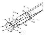
最後に、医療装置24は、更に、複数の針30に滑動可能に取り付けられている保護先端50を含んでいる。図5からよく分かる様に、保護先端50は、複数の針30を滑動可能に受け入れるサイズの複数の先端通路52を画定している。更に、保護先端50は、内部の先端ルーメン54を画定している。先端ルーメン54は、内視鏡22が端部キャップ26と保護先端50の両方を通して鮮明に視覚化されるように、端部キャップ通路28に略等しい(即ち25%以内)直径を有しているのが望ましい。同様に、内視鏡22の(単数又は複数の)作業チャネル23(図3と図5)を通して導入される医療器具は、医療システム20の遠位端を越えて、即ち端部キャップ26を通り更に保護先端50を通り抜けて使用されていてもよい。保護先端50は、図1に示されている伸展位置と、図2に示されている引込位置の間で操作できる。伸展位置では、保護先端50は複数の針30の遠位端34を保護し、引込位置では、保護先端50は組織12を穿刺するために遠位端34を露出する。
Finally, the
スタイレットキャップ42と複数のスタイレット46は、複数の針30内に収容されている複数の組織デバイス36に係合し、それらを図4に示されている様に針30から押し出すことによって配備する働きをする。保護先端50と端部キャップ26の間の相対並進を操作及び制御し、同様にスタイレットキャップ42と端部キャップ26の間の相対並進を制御するために、それぞれには細長い制御部材が設けられている。図3からよく分かる様に、保護先端50は、それに取り付けられている先端制御部材60を含んでいる。先端制御部材60は、保護先端50に埋め込まれているか又は他のやり方でそれに取り付けられているワイヤを備えていてもよい。端部キャップ26は、先端制御部材60を滑動可能に受け入れるカテーテルを備えた端部キャップ制御部材62を含んでいる。最後に、スタイレットキャップ42は、同様にカテーテルを備えるスタイレット制御部材64を含んでいる。スタイレット制御部材64は、端部キャップ制御部材62(及び、ひいては同様に先端制御部材60)を滑動可能に受け入れるサイズである。制御部材60、62、64のそれぞれは、それらの間の相対並進、そして同様に端部キャップ26とスタイレットキャップ42と保護先端50の間の相対並進を可能にするのに十分に力が長手方向に伝達される構造であるのが望ましい。制御部材がカテーテル又は他の管状部材を備えている場合、各種プラスチック類を採用することができ、その様なものとして、ポリテトラフルオロエチレン(PTFE)、発泡ポリテトラフルオロエチレン(EPTFE)、ポリエチレンエーテルケトン(PEEK)、ポリ塩化ビニル(PVC)、ポリカーボネート(PC)、ナイロン(商標)を含むポリアミド、ポリイミド、ポリウレタン、ポリエチレン(高、中、又は低密度)、及びサントプレン(商標)の様なエラストマー類があり、それらには強化ワイヤ、コイル、又はフィラメントの有無を問わず多層構造のもの又は単層構造のものが含まれる。制御部材がワイヤなどからなっている場合、制御部材は、単線中実ワイヤ設計であってもよいし、コイル状、巻き、編み、又は当技術で既知の他の設計を含む多線条設計であってもよい。
The
描かれている実施形態では、先端制御部材60と端部キャップ制御部材62とスタイレット制御部材64は、同心円状に配列され、近位方向に内視鏡22の長さに沿って延びている。従って、制御部材60、62、64は、内視鏡22の長さと概ね同様の長さを有している。制御部材は、帯体、テープなどを使用するなどして、制御部材の長さに沿って内視鏡22に取り付けられていてもよい。同様に、制御部材は、内視鏡と一体に形成することもできるであろうし、オーバーチューブ又は他の導入装置の一部として形成することもできるであろう。医療システム20の近位端には、当該技術分野で知られている様に、制御部材60、62、64の相対並進を制御する各種駆動部を含む適当なハンドルを採用することができる。例えば、ノースカロライナ州ウィンストンセーレムのWilson−Cook(Cook Endoscopy)社によって、同社のEchoTip(登録商標)内視鏡超音波針に関連して現在販売されているハンドルを、そのハンドルの本体を端部キャップ制御部材62に接続し、外側の滑動可能部材をスタイレット制御部材64に接続し、プランジャ(ワイヤ60の近位端に取り付けられている)を先端制御部材60に接続するなどして、本発明の医療システム20及び医療装置24と共に使えるように適合させてもよい。
In the depicted embodiment, the
本発明の教示に従った、例えば穿孔14を閉合する場合の上述の組織アンカー36の様な組織デバイスを配備する方法をこれより説明してゆく。本方法は、医療システム20を、保護先端50が図1に示されている伸展位置にある状態で、体内管腔を通して組織12の近くに前進させるステップを含んでいる。図2−図3に示されている様に、保護先端を操作して引込位置に入れ、組織12を複数の針30で、当該針の遠位端34が組織12の遠位側に位置付けられるように穿刺する。より具体的には、医療システムは、穿孔14に対して、針30が、それぞれ、穿孔14の周辺部周囲の組織12を穿刺するような向きに合わせられるのが望ましい。本方法の他の実施形態では、穿孔を組織12に形成するステップに先立ち、針30が組織12を穿刺し、及び/又は組織アンカー36が配備されてもよい。例えば、針30及び/又は組織アンカー36によって画定されている境界内に穿孔を形成するには、内視鏡用切削装置を、内視鏡22の作業チャネル23を通して配備し、使用することができる。
A method of deploying a tissue device, such as the
図4−図5に示されている様に、スタイレットキャップ42を端部キャップ26に対して並進させて、複数の組織アンカー36に複数のスタイレット46を係合させ、組織アンカー36を組織12の遠位側に配備する。次いで、保護先端50を、図6に示されている様に、伸展位置へ操作する。先端50の操作は、針30がまだ組織12に刺さっている間に(針を組織12から抜くのを支援するために)行われてもよいし、医療システム20を近位方向に動かして針30を組織12から引き出した後に行われてもよい。図7−図10に示されている様に、縫合糸38は、縫合糸38の2つの自由端を近位方向に体内管腔を通って延ばし、穿孔14を閉合するのに独立して張力を掛けることができるようにしたまま、内臓アンカー36のそれぞれに滑動可能に結合されている。図8からよく分かる様に、内臓アンカー36は、体壁12の遠位側に配置され、縫合糸38の大部分は、体壁12の近位側に配置されている。図9及び図10に描かれている様に、内臓アンカー36同士の距離を縮めて、穿孔14の周囲の組織12を圧縮するように、縫合糸38の両端に張力を掛ける。図10からよく分かる様に、縫合糸38の両端は、組織12の圧縮を維持するように、縫合糸ロック70を使用するなどして、固定されている。代表的な縫合糸ロックは、2008年5月22日出願の米国特許出願第12/125,525号及び2008年8月13日出願の同第12/191,001号に開示されており、それらの開示をここに参考文献としてその全体を援用する。縫合糸38の両端を固定するための方法は、結び目、結束、クランプ、リベットなどの様な、現時点で知られている、又は将来開発される、如何なる方法が採用されてもよいことが認識されるであろう。医療システム20は、保護先端50が伸展位置(図6)にある状態で、体内管腔を通して後退させることができる。
As shown in FIGS. 4-5, the
本発明の方法の継続中、内視鏡22を使用して、処置並びに医療装置と器具の操作を視覚化することもできることが認識されるであろう。更に、一組のアンカー36の設置は、蛍光透視法や超音波の助けを借りてもよいし、内視鏡22の作業チャネル23に通した光ファイバーカテーテルの様な画像化能力を有する医療器具の使用を介した目視によってもよいことが認識されるであろう。更に、上述の方法は、概して、組織デバイスを、身体内部の管腔を通して組織に設置するステップを含んでいるが、本システム、装置、及び方法は、人間又は動物の身体及び体内管腔と関連付けられているもの、そうでないものを問わず、如何なる材料層(例えば、織物、布、ポリマー、エストラマー、プラスチック、及びゴム)に対して使用されてもよいことが認識されるであろう。例えば、本システム、装置、及び方法は、研究所や工業環境で材料層を通して装置を設置する場合の利用が見込まれる。
It will be appreciated that, during the continuation of the method of the present invention, the
以上、本発明の様々な実施形態の説明を、例示と説明を目的に提示してきた。それは本発明を網羅すること又は本発明を開示されている厳密な実施形態に限定することを意図するものではない。以上の教示に照らし、数多くの修正又は変型が実施可能である。論じられている実施形態は、本発明の原理とその実際の適用を最も分かり易く例示し、それにより、当業者が、本発明を様々な実施形態で、また考えられる特定の用途に合わせた様々な修正を加えて、利用することができるように、選定され、記述されている。全てのその様な修正及び変型は、付随の特許請求の範囲の請求項によって、それら請求項が公平、法的、且つ公正に権利を有するとされる一定の許容幅に従って解釈された上に定まる本発明の範囲に含まれる。 The foregoing description of various embodiments of the present invention has been presented for purposes of illustration and description. It is not intended to be exhaustive or to limit the invention to the precise embodiments disclosed. Many modifications or variations are possible in light of the above teaching. The discussed embodiments best illustrate the principles of the invention and its practical application so that those skilled in the art will appreciate that the invention can be embodied in a variety of embodiments and for a particular application contemplated. Selected and described so that it can be used with minor modifications. All such modifications and variations are determined by the appended claims, after being interpreted according to a certain tolerance that they are entitled to be fairly, legally and fairly entitled. It is included in the scope of the present invention.
10 中心軸
12 組織
14 穿孔
20 医療システム
22 内視鏡
23 作業チャネル
24 医療装置
26 端部キャップ
28 キャップの内部通路
30 針
31 針ルーメン
32 フランジ
34 遠位端
36 組織アンカー(組織デバイス)
38 縫合糸
40 側方開口スロット
42 スタイレットキャップ
44 スタイレットキャップの通路
46 スタイレット
48 スタイレット通路
50 保護先端
52 先端通路
54 先端ルーメン
60 先端制御部材
62 端部キャップ制御部材
64 スタイレット制御部材
70 縫合糸ロック
10
38
Claims (15)
前記内視鏡を受け入れるサイズとされた通路を有する端部キャップと、
前記端部キャップに取り付けられ、該端部キャップから遠位方向に突き出ている複数の針であって、複数の針ルーメンを画定し、組織を穿刺するための遠位端を有している複数の針と、
前記端部キャップの外周面上に滑動可能に配置されているスタイレットキャップと、
前記スタイレットキャップに取り付けられていて、前記複数の針ルーメンの中へ遠位方向に向かって突き出ている複数のスタイレットと、
前記針ルーメン内に配置されている複数の組織デバイスと、を備え、
前記スタイレットキャップを前記端部キャップに対して並進させると、前記複数のスタイレットが前記複数の組織デバイスに係合して該複数の組織デバイスを前記複数の針から押し出すようになっている、医療装置。 A medical device for use with an endoscope for treating tissue,
An end cap having a passage sized to receive the endoscope;
A plurality of needles attached to the end cap and projecting distally from the end cap, the plurality of needles defining a plurality of needle lumens and having a distal end for piercing tissue The needle of
A stylet cap slidably disposed on the outer peripheral surface of the end cap;
A plurality of stylets attached to the stylet cap and projecting distally into the plurality of needle lumens;
A plurality of tissue devices disposed within the needle lumen,
When the stylet cap is translated relative to the end cap, the plurality of stylets engage the plurality of tissue devices to push the plurality of tissue devices out of the plurality of needles. Medical device.
Applications Claiming Priority (3)
| Application Number | Priority Date | Filing Date | Title |
|---|---|---|---|
| US10304208P | 2008-10-06 | 2008-10-06 | |
| US61/103,042 | 2008-10-06 | ||
| PCT/US2009/059364 WO2010042402A1 (en) | 2008-10-06 | 2009-10-02 | Endcap for safely deploying tissue anchors |
Publications (3)
| Publication Number | Publication Date |
|---|---|
| JP2012504482A JP2012504482A (en) | 2012-02-23 |
| JP2012504482A5 JP2012504482A5 (en) | 2012-11-15 |
| JP5580828B2 true JP5580828B2 (en) | 2014-08-27 |
Family
ID=41259552
Family Applications (1)
| Application Number | Title | Priority Date | Filing Date |
|---|---|---|---|
| JP2011531086A Active JP5580828B2 (en) | 2008-10-06 | 2009-10-02 | End cap for secure deployment of tissue anchors |
Country Status (5)
| Country | Link |
|---|---|
| US (1) | US8317679B2 (en) |
| EP (1) | EP2346411B1 (en) |
| JP (1) | JP5580828B2 (en) |
| CA (1) | CA2739475C (en) |
| WO (1) | WO2010042402A1 (en) |
Families Citing this family (15)
| Publication number | Priority date | Publication date | Assignee | Title |
|---|---|---|---|---|
| US9713465B1 (en) * | 2004-04-19 | 2017-07-25 | Granit Medical Innovation Llc | Surgical closure device and associated method |
| WO2011137252A1 (en) * | 2010-04-29 | 2011-11-03 | Vinay Badhwar | Automatic suturing apparatus and methods of use |
| JP5615112B2 (en) * | 2010-09-21 | 2014-10-29 | 日本コヴィディエン株式会社 | Organ fixing device and organ fixing device |
| JP5963559B2 (en) | 2012-06-18 | 2016-08-03 | 日本コヴィディエン株式会社 | Medical suture tool |
| JPWO2014148606A1 (en) * | 2013-03-22 | 2017-02-16 | テルモ株式会社 | Medical instruments |
| US10070851B2 (en) | 2013-08-02 | 2018-09-11 | Covidien Lp | Devices, systems, and methods for wound closure |
| US9848879B2 (en) | 2013-08-02 | 2017-12-26 | Covidien Lp | Devices, systems, and methods for wound closure |
| JP6594418B2 (en) | 2014-06-15 | 2019-10-23 | アンコラ メディカル リミテッド | Instruments and methods for suturing tissue |
| EP3331452A4 (en) * | 2015-08-07 | 2019-08-28 | Mayo Foundation for Medical Education and Research | Endoluminal anastomosis and tissue closure devices |
| EP3537991A4 (en) | 2016-11-13 | 2020-04-29 | Anchora Medical Ltd. | Minimally-invasive tissue suturing device |
| WO2018220818A1 (en) * | 2017-06-02 | 2018-12-06 | オリンパス株式会社 | Ligature aid |
| US11213288B2 (en) | 2018-05-02 | 2022-01-04 | Covidien Lp | Port site closure instrument |
| US11234690B2 (en) | 2018-05-02 | 2022-02-01 | Covidien Lp | Method and device for closing a port site incision |
| US11344292B2 (en) * | 2018-06-14 | 2022-05-31 | Covidien Lp | Trans-vaginal cuff anchor and method of deploying same |
| WO2023166220A1 (en) * | 2022-03-03 | 2023-09-07 | Cardiomech As | Soft tissue anchor system for heart repair |
Family Cites Families (69)
| Publication number | Priority date | Publication date | Assignee | Title |
|---|---|---|---|---|
| US5304184A (en) | 1992-10-19 | 1994-04-19 | Indiana University Foundation | Apparatus and method for positive closure of an internal tissue membrane opening |
| US5562688A (en) | 1994-03-25 | 1996-10-08 | Riza; Erol D. | Apparatus facilitating suturing in laparoscopic surgery |
| GB2298368B (en) | 1995-02-22 | 1999-01-20 | John Francis Cockburn | Medical needle assembly for use in ultrasound imaging |
| US5807304A (en) | 1995-03-09 | 1998-09-15 | Cockburn; John F. | Medical needle for use in ultrasound imaging |
| US5824010A (en) | 1996-05-23 | 1998-10-20 | Mcdonald; Garth R. | Suture needle guide |
| US6053871A (en) | 1997-01-21 | 2000-04-25 | William Cook Australia Pty. Ltd | Calibrated hollow probe for use with ultrasound imaging |
| US5908428A (en) | 1997-05-27 | 1999-06-01 | United States Surgical Corporation | Stitching devices for heart valve replacement surgery |
| EP1054703B1 (en) | 1998-12-09 | 2004-09-15 | Cook Incorporated | Hollow, curved, superelastic medical needle |
| US7618426B2 (en) | 2002-12-11 | 2009-11-17 | Usgi Medical, Inc. | Apparatus and methods for forming gastrointestinal tissue approximations |
| US7955340B2 (en) | 1999-06-25 | 2011-06-07 | Usgi Medical, Inc. | Apparatus and methods for forming and securing gastrointestinal tissue folds |
| US6398796B2 (en) * | 1999-07-13 | 2002-06-04 | Scion Cardio-Vascular, Inc. | Suture with toggle and delivery system |
| US6358197B1 (en) | 1999-08-13 | 2002-03-19 | Enteric Medical Technologies, Inc. | Apparatus for forming implants in gastrointestinal tract and kit for use therewith |
| US6231561B1 (en) | 1999-09-20 | 2001-05-15 | Appriva Medical, Inc. | Method and apparatus for closing a body lumen |
| US7615076B2 (en) * | 1999-10-20 | 2009-11-10 | Anulex Technologies, Inc. | Method and apparatus for the treatment of the intervertebral disc annulus |
| US8128698B2 (en) * | 1999-10-20 | 2012-03-06 | Anulex Technologies, Inc. | Method and apparatus for the treatment of the intervertebral disc annulus |
| JP4351458B2 (en) | 2002-03-18 | 2009-10-28 | オリンパス株式会社 | Endoscope insertion system |
| US7780687B2 (en) | 2002-04-17 | 2010-08-24 | Tyco Healthcare Group Lp | Method and apparatus for anastomosis including expandable anchor |
| US8070743B2 (en) * | 2002-11-01 | 2011-12-06 | Valentx, Inc. | Devices and methods for attaching an endolumenal gastrointestinal implant |
| US7615005B2 (en) | 2003-05-16 | 2009-11-10 | Ethicon Endo-Surgery, Inc. | Medical apparatus for use with an endoscope |
| JP4145200B2 (en) * | 2003-06-06 | 2008-09-03 | オリンパス株式会社 | Suture device |
| US7481826B2 (en) | 2003-09-30 | 2009-01-27 | Ethicon, Inc. | Fluid emitting suture needle |
| JP4217587B2 (en) | 2003-11-26 | 2009-02-04 | オリンパス株式会社 | Endoscope cap |
| US20050251189A1 (en) | 2004-05-07 | 2005-11-10 | Usgi Medical Inc. | Multi-position tissue manipulation assembly |
| US7166127B2 (en) | 2003-12-23 | 2007-01-23 | Mitralign, Inc. | Tissue fastening systems and methods utilizing magnetic guidance |
| WO2005065554A1 (en) | 2004-01-08 | 2005-07-21 | Olympus Corporation | Anastomosis device and method of excising wall portion of in vivo luminal organ |
| US8475476B2 (en) | 2004-06-01 | 2013-07-02 | Cook Medical Technologies Llc | System and method for accessing a body cavity |
| JP2006025934A (en) * | 2004-07-13 | 2006-02-02 | Jms Co Ltd | Suturing implement for living body |
| US20060020274A1 (en) | 2004-07-23 | 2006-01-26 | Usgi Medical Inc. | Manipulatable grasping needle |
| JP4302602B2 (en) | 2004-09-24 | 2009-07-29 | オリンパス株式会社 | Endoscopic treatment tool, endoscopic treatment system, and support adapter |
| CN101257852B (en) * | 2005-07-07 | 2011-08-10 | 科迪斯公司 | Patent foramen oval closure device with steerable delivery system |
| US7722631B2 (en) * | 2005-09-28 | 2010-05-25 | Olympus Medical Systems Corporation | Method for suturing perforation |
| US8702753B2 (en) * | 2005-09-28 | 2014-04-22 | Olympus Medical Systems Corp. | Method for suturing perforation and suture instrument |
| US8241279B2 (en) | 2006-02-23 | 2012-08-14 | Olympus Medical Systems Corp. | Overtube and natural opening medical procedures using the same |
| US20070213702A1 (en) | 2006-03-08 | 2007-09-13 | Olympus Medical Systems Corp. | Medical procedure carried out via a natural opening |
| US7785333B2 (en) | 2006-02-21 | 2010-08-31 | Olympus Medical Systems Corp. | Overtube and operative procedure via bodily orifice |
| US8728121B2 (en) | 2006-01-13 | 2014-05-20 | Olympus Medical Systems Corp. | Puncture needle and medical procedure using puncture needle that is performed via natural orifice |
| US7731727B2 (en) | 2006-04-26 | 2010-06-08 | Lsi Solutions, Inc. | Medical instrument to place a pursestring suture, open a hole and pass a guidewire |
| EP2021061A4 (en) | 2006-05-18 | 2013-05-15 | Aponos Medical Corp | Multifunctional instrument introducer |
| JP5010178B2 (en) * | 2006-05-30 | 2012-08-29 | 昌貴 鮒田 | Medical instruments |
| US20080097152A1 (en) | 2006-07-20 | 2008-04-24 | David Stefanchik | Braided endoscope accessories |
| US7837455B2 (en) | 2006-07-28 | 2010-11-23 | Ethicon, Inc. | Apparatus and method for making suture packages |
| EP2094167B1 (en) * | 2006-11-30 | 2011-06-29 | Wilson-Cook Medical, Inc. | Visceral anchors for purse-string closure of perforations |
| US8007432B2 (en) | 2007-01-26 | 2011-08-30 | Ethicon Endo-Surgery, Inc. | Endoscopic accessory control mechanism |
| US8308766B2 (en) | 2007-02-27 | 2012-11-13 | Olympus Medical Systems Corp. | Endoscopic treatment instrument |
| US8128657B2 (en) | 2007-02-27 | 2012-03-06 | Olympus Medical Systems Corp. | Suture instrument |
| US7780702B2 (en) | 2007-02-27 | 2010-08-24 | Olympus Medical Systems Corp. | Suture tool |
| EP2155071B1 (en) * | 2007-05-25 | 2014-07-16 | Cook Medical Technologies LLC | Medical devices and systems for closing perforations |
| US8088062B2 (en) | 2007-06-28 | 2012-01-03 | Ethicon Endo-Surgery, Inc. | Interchangeable endoscopic end effectors |
| US8992569B2 (en) | 2007-06-29 | 2015-03-31 | Ethicon Endo-Surgery, Inc. | Insertion device and method of use |
| US8128592B2 (en) | 2007-07-11 | 2012-03-06 | Apollo Endosurgery, Inc. | Methods and systems for performing submucosal medical procedures |
| US20100217151A1 (en) | 2007-07-11 | 2010-08-26 | Zach Gostout | Methods and Systems for Performing Submucosal Medical Procedures |
| CA2700663C (en) | 2007-09-25 | 2015-07-07 | Wilson-Cook Medical Inc. | Medical devices, systems, and methods for using tissue anchors |
| US7998150B2 (en) | 2007-09-28 | 2011-08-16 | Olympus Medical Systems Corp. | Suturing device |
| US9526487B2 (en) | 2007-12-05 | 2016-12-27 | Indiana University Research & Technology Corporation | Methods and apparatuses for delivering anchoring devices into body passage walls |
| US20090149714A1 (en) | 2007-12-05 | 2009-06-11 | Frank Bonadio | Surgical devices and methods |
| US20090281559A1 (en) | 2008-05-06 | 2009-11-12 | Ethicon Endo-Surgery, Inc. | Anastomosis patch |
| US8679003B2 (en) | 2008-05-30 | 2014-03-25 | Ethicon Endo-Surgery, Inc. | Surgical device and endoscope including same |
| US8361112B2 (en) | 2008-06-27 | 2013-01-29 | Ethicon Endo-Surgery, Inc. | Surgical suture arrangement |
| US8888792B2 (en) | 2008-07-14 | 2014-11-18 | Ethicon Endo-Surgery, Inc. | Tissue apposition clip application devices and methods |
| US20100048990A1 (en) | 2008-08-25 | 2010-02-25 | Ethicon Endo-Surgery, Inc. | Endoscopic needle for natural orifice translumenal endoscopic surgery |
| US8241204B2 (en) | 2008-08-29 | 2012-08-14 | Ethicon Endo-Surgery, Inc. | Articulating end cap |
| US20100056862A1 (en) | 2008-09-03 | 2010-03-04 | Ethicon Endo-Surgery, Inc. | Access needle for natural orifice translumenal endoscopic surgery |
| US8163022B2 (en) * | 2008-10-14 | 2012-04-24 | Anulex Technologies, Inc. | Method and apparatus for the treatment of the intervertebral disc annulus |
| WO2010051250A1 (en) | 2008-10-29 | 2010-05-06 | Wilson-Cook Medical, Inc. | Endoscope endcap for suturing tissue |
| EP2384149B1 (en) | 2008-12-05 | 2015-12-02 | Cook Medical Technologies LLC | Tissue anchors for purse-string closure of perforations |
| US20100191267A1 (en) | 2009-01-26 | 2010-07-29 | Ethicon Endo-Surgery, Inc. | Rotary needle for natural orifice translumenal endoscopic surgery |
| US8252057B2 (en) | 2009-01-30 | 2012-08-28 | Ethicon Endo-Surgery, Inc. | Surgical access device |
| US9226772B2 (en) | 2009-01-30 | 2016-01-05 | Ethicon Endo-Surgery, Inc. | Surgical device |
| CA2757494C (en) | 2009-04-03 | 2013-11-12 | Cook Medical Technologies Llc | Medical devices, systems, and methods for rapid deployment and fixation of tissue anchors |
-
2009
- 2009-10-02 EP EP09736745.2A patent/EP2346411B1/en active Active
- 2009-10-02 US US12/572,636 patent/US8317679B2/en active Active
- 2009-10-02 JP JP2011531086A patent/JP5580828B2/en active Active
- 2009-10-02 CA CA2739475A patent/CA2739475C/en active Active
- 2009-10-02 WO PCT/US2009/059364 patent/WO2010042402A1/en active Application Filing
Also Published As
| Publication number | Publication date |
|---|---|
| CA2739475C (en) | 2014-04-22 |
| US8317679B2 (en) | 2012-11-27 |
| EP2346411A1 (en) | 2011-07-27 |
| CA2739475A1 (en) | 2010-04-15 |
| AU2009302589A1 (en) | 2010-04-15 |
| WO2010042402A1 (en) | 2010-04-15 |
| EP2346411B1 (en) | 2013-10-02 |
| JP2012504482A (en) | 2012-02-23 |
| US20100087707A1 (en) | 2010-04-08 |
Similar Documents
| Publication | Publication Date | Title |
|---|---|---|
| JP5580828B2 (en) | End cap for secure deployment of tissue anchors | |
| CA2671030C (en) | Visceral anchors for purse-string closure of perforations | |
| AU2010241552B2 (en) | Medical systems, devices and methods for suturing perforations | |
| US5997555A (en) | Device and method for suturing blood vessels | |
| AU2008304583B2 (en) | Medical devices, systems, and methods for using tissue anchors | |
| JP5894930B2 (en) | Medical instrument and method for suturing tissue | |
| JP5619137B2 (en) | Tissue anchor and medical device for rapid deployment of tissue anchor | |
| JP5301544B2 (en) | Organ staple for closure of perforated purse string suture | |
| JP2010536486A (en) | Suture lock | |
| JP5485286B2 (en) | Endoscopic end cap for suturing tissue | |
| JP2010511436A5 (en) | ||
| JP5378523B2 (en) | Perforating and closing stapling device | |
| US20160157858A1 (en) | Double barbed suture with needle delivery system | |
| AU2009302589B2 (en) | Endcap for safely deploying tissue anchors |
Legal Events
| Date | Code | Title | Description |
|---|---|---|---|
| A711 | Notification of change in applicant |
Free format text: JAPANESE INTERMEDIATE CODE: A711 Effective date: 20120206 |
|
| A521 | Request for written amendment filed |
Free format text: JAPANESE INTERMEDIATE CODE: A523 Effective date: 20120928 |
|
| A621 | Written request for application examination |
Free format text: JAPANESE INTERMEDIATE CODE: A621 Effective date: 20120928 |
|
| A131 | Notification of reasons for refusal |
Free format text: JAPANESE INTERMEDIATE CODE: A131 Effective date: 20131112 |
|
| A601 | Written request for extension of time |
Free format text: JAPANESE INTERMEDIATE CODE: A601 Effective date: 20140212 |
|
| A602 | Written permission of extension of time |
Free format text: JAPANESE INTERMEDIATE CODE: A602 Effective date: 20140219 |
|
| A521 | Request for written amendment filed |
Free format text: JAPANESE INTERMEDIATE CODE: A523 Effective date: 20140310 |
|
| TRDD | Decision of grant or rejection written | ||
| A01 | Written decision to grant a patent or to grant a registration (utility model) |
Free format text: JAPANESE INTERMEDIATE CODE: A01 Effective date: 20140624 |
|
| A61 | First payment of annual fees (during grant procedure) |
Free format text: JAPANESE INTERMEDIATE CODE: A61 Effective date: 20140711 |
|
| R150 | Certificate of patent or registration of utility model |
Ref document number: 5580828 Country of ref document: JP Free format text: JAPANESE INTERMEDIATE CODE: R150 |
|
| R250 | Receipt of annual fees |
Free format text: JAPANESE INTERMEDIATE CODE: R250 |
|
| R250 | Receipt of annual fees |
Free format text: JAPANESE INTERMEDIATE CODE: R250 |
|
| R250 | Receipt of annual fees |
Free format text: JAPANESE INTERMEDIATE CODE: R250 |
|
| R250 | Receipt of annual fees |
Free format text: JAPANESE INTERMEDIATE CODE: R250 |
|
| R250 | Receipt of annual fees |
Free format text: JAPANESE INTERMEDIATE CODE: R250 |
|
| R250 | Receipt of annual fees |
Free format text: JAPANESE INTERMEDIATE CODE: R250 |
|
| R250 | Receipt of annual fees |
Free format text: JAPANESE INTERMEDIATE CODE: R250 |