EP2397068B1 - Analyse des intraokularen Drucks - Google Patents
Analyse des intraokularen Drucks Download PDFInfo
- Publication number
- EP2397068B1 EP2397068B1 EP11168232.4A EP11168232A EP2397068B1 EP 2397068 B1 EP2397068 B1 EP 2397068B1 EP 11168232 A EP11168232 A EP 11168232A EP 2397068 B1 EP2397068 B1 EP 2397068B1
- Authority
- EP
- European Patent Office
- Prior art keywords
- cornea
- derived
- deformation
- applanation
- intraocular pressure
- Prior art date
- Legal status (The legal status is an assumption and is not a legal conclusion. Google has not performed a legal analysis and makes no representation as to the accuracy of the status listed.)
- Active
Links
Images


Classifications
-
- A—HUMAN NECESSITIES
- A61—MEDICAL OR VETERINARY SCIENCE; HYGIENE
- A61B—DIAGNOSIS; SURGERY; IDENTIFICATION
- A61B3/00—Apparatus for testing the eyes; Instruments for examining the eyes
- A61B3/10—Objective types, i.e. instruments for examining the eyes independent of the patients' perceptions or reactions
- A61B3/16—Objective types, i.e. instruments for examining the eyes independent of the patients' perceptions or reactions for measuring intraocular pressure, e.g. tonometers
- A61B3/165—Non-contacting tonometers
-
- A—HUMAN NECESSITIES
- A61—MEDICAL OR VETERINARY SCIENCE; HYGIENE
- A61B—DIAGNOSIS; SURGERY; IDENTIFICATION
- A61B3/00—Apparatus for testing the eyes; Instruments for examining the eyes
- A61B3/10—Objective types, i.e. instruments for examining the eyes independent of the patients' perceptions or reactions
- A61B3/107—Objective types, i.e. instruments for examining the eyes independent of the patients' perceptions or reactions for determining the shape or measuring the curvature of the cornea
Definitions
- the invention relates to an ophthalmological analysis method for measuring an intraocular pressure in an eye with an analysis system, and to such an analysis system, formed from an actuating device with which a cornea of the eye is deformed in a contact-free manner, with the actuating device causing an air blast to deform the cornea on the Eye is applied, an observation system with which the deformation of the cornea is observed and recorded, with the observation system taking sectional images of the undeformed and deformed cornea, and an analysis device with which the intraocular pressure is derived from the sectional images of the cornea, wherein in the analysis device derives a material property of the cornea from the sectional images of the cornea.
- Such analysis methods and systems are well known and primarily serve the most accurate, contactless measurement of an intraocular pressure in an eye.
- a non-contact tonometer is used, with the help of which an air blast on the eye to be examined is applied, wherein a strength of the air blast is selected so that the cornea of the eye is pressed in to form a concave surface shape.
- the cornea Before a maximum deformation of the cornea is reached or before the cornea collapses in the direction of the eye lens, the cornea briefly forms a flat surface which is referred to as the so-called first applanation point. After the maximum deflection of the cornea and a folding back of the cornea into its original state, the cornea passes through a second applanation point of the same kind.
- the measured values determined with the non-contact tonometer are set in relation to comparison measured values, which were determined with a relatively more accurate applanation tonometer or contact tonometer, so that as a result an intraocular pressure approximating the actual intraocular pressure can be derived.
- an intraocular pressure measured with a non-contact tonometer is not yet sufficiently accurate compared to a pressure measurement with an applanation tonometer, since a measurement is distorted by the cornea, among other things.
- an attempt was therefore made to take into account the influence of the cornea on the measurement for example by measuring the thickness or measuring the corneal radius before the start of the non-contact tonometer measurement.
- a cornea has a modulus of elasticity that is the same in all areas of the cornea.
- a modulus of elasticity varies from eye to eye individually and, depending on the respective corneal areas, within the cornea itself. Including such material parameters and calculating the measurement result can therefore not yet lead to satisfactorily accurate measurement results.
- an air blast is applied to a cornea, with a pump pressure being continuously recorded by means of a pressure sensor during the course of the measurement. Furthermore, a time course of the measurement is recorded and a first and a second applanation point of the cornea are optically detected.
- An intraocular pressure can now be derived, for example, by determining the pressures prevailing at the time of the first and second applanation, in particular since the forces required to bend the cornea when the cornea is folded in or out are assumed to be of the same magnitude and thus cancel each other out. Consequently, an intraocular pressure results from an average of the force used to fold in and out the cornea, applied by the air blast.
- the first and second applanation points are optically detected and set in relation to the time curve of a pressure curve of a pump, that is to say an associated time value and a pressure value are determined for each applanation point.
- a pressure difference can be used to determine the hysteresis as a material property of the cornea.
- a disadvantage of these measurement methods is that movement of the cornea caused by an air blast is subject to dynamic effects, which can falsify such time / pressure measurements, in particular since the dynamic effects cannot be taken into account in the described non-contact tonometer measurements.
- the speed of the air blast is minimized as much as possible so as not to falsify a measurement result due to an undesired corneal movement. It is also necessary to establish a synchronicity between the start of the air blast and the required time measurement.
- a mechanical pump used to form an air blast such as a piston pump, however, this time synchronism cannot be achieved exactly, for example as a result of inertia or mass friction, and thus a measurement result can be falsified.
- the air blast is also monitored for pressure, that is to say influenced in the course of the measurement as required. After exceeding the first applanation point, the air blast is reduced or switched off in order to prevent the cornea from being pressed in excessively.
- this also requires continuous pressure monitoring of a pump pressure and monitoring of a time profile of the same relative to the times of the first and second applanations, which results in the inclusion of a number of possible sources of error which falsify a measurement.
- the analysis methods and systems known from the prior art with mutually dependent, parallel pressure and time measurement with simultaneous detection of the applanation points are compared a measurement with a contact tonometer is still relatively inaccurate.
- a method for measuring an intraocular pressure of an eye using an ophthalmic analysis device is known.
- a correction factor is used in particular, which is intended to describe a resistance of the cornea to deflection and which varies depending on a thickness and on viscoelastic properties of the cornea.
- a related stiffness of the cornea can be derived from a force or an air blast applied to the cornea with a non-contact tonometer and an applanation surface generated thereby. Deformation of the eye is recorded with a high-speed camera, which is arranged to the side of the eye. Since the cornea is transparent, lateral sectional images of the cornea can be obtained.
- the present invention is therefore based on the object of proposing an ophthalmological analysis method for measuring an intraocular pressure in an eye as well as an analysis system of this type with which a comparatively improved measurement accuracy can be achieved.
- the analysis system comprises an actuating device with which a cornea of the eye is deformed in a contact-free manner, with an actuation of air being applied to the eye to deform the cornea, an observation system with which the deformation of the cornea is observed and recorded, with the observation system recording sectional images of the undeformed and deformed cornea, and an analysis device with which the intraocular pressure is derived from the sectional images of the cornea, the analysis device from the A material property of the cornea is derived from sectional images of the cornea, wherein a maximum deformation of the cornea is derived from the sectional images of the cornea, with a diameter d 1 of a first applanation surface of the cornea as a material property t and a diameter d 2 of a deformation surface of the cornea deviating from the first applanation surface is derived at the maximum deformation of the cornea relative to the apex of the cornea, the first applanation surface lying in
- the cornea When the cornea is deformed by means of the air blast, the cornea is completely applanated, the first applanation surface being formed with the diameter d 1 .
- the applanation surface is essentially flat and lies in the area of an applanation level orthogonal to a visual axis of the eye or a device axis of an analysis system.
- a concave depression is formed in the cornea, which differs significantly from the first applanation surface.
- a comparison of the deformation surface of the depression deviating from the first applanation surface with the first applanation surface enables a definition of the material property of the cornea, since the deformation surface is formed as a function of the material property.
- the material property therefore does not relate to a generally known material property, but is based solely on a definition of two different geometries of the deformed cornea.
- a reference standard for the deviation is the first applanation surface or the diameter d 1 of the first applanation surface. Because the comparison is made with the diameter d n of the deformation surface of the cornea, the comparison can be carried out particularly easily.
- the diameter d n is particularly easy to determine in particular during a deformation movement of the cornea after passing the first applanation surface or a first applanation point, since the deformation surface then assumes a concave shape.
- the deformation surface or the diameter d n is used in a specific time period of the deformation relative to the first applanation surface or also another, measurable point or a position of the cornea during the deformation to define the deviating deformation surface of the cornea. Furthermore, the determined deviation and the relative values of the diameters in question are stored and compared in a database. An objective intraocular pressure or a corresponding correction value is known for the values stored in the database, so that the objective intraocular pressure of the measured eye is derived taking into account the geometrically defined material properties of the cornea.
- a pressure measurement of a pump pressure does not have to be necessary in the method. So any measurement can an intraocular pressure can always be carried out with the same, constant pump pressure. Since there is no need for a variation in the level of the pump pressure or a time synchronization of the pump pressure, a number of possible sources of error can be excluded and a particularly precise measurement can be carried out.
- the material properties of the cornea that are determined in this way can also be used in the context of refractive eye surgery, for example in order to adapt to the respective corneal properties during a LASIK procedure.
- a pump pressure for generating the air blast is relative to a time period of the same in the form of a bell curve.
- the pump pressure can act identically on the cornea for each individual measurement and be completely uninfluenced in the form of an air blast.
- the bell curve can have a symmetrical shape, among other things.
- a maximum pump pressure for generating the air blast can also be the same in previous and subsequent measurements. This enables particularly good comparability of different measurements.
- the maximum pump pressure can be, for example, 70 mm Hg.
- a pump pressure for generating the air blast can be measured when an applanation point of the cornea is reached.
- a pump can have a pressure sensor that enables the pump pressure to be monitored during the entire measurement. In this way, possible errors regarding the pump pressure during the measurement can be excluded and continuity of successive measurements can be ensured.
- a time period between the beginning and end of the deformation of the cornea can also be measured.
- all of the sectional images recorded can each be assigned to a specific point in time of the measurement, as a result of which a chronological course of the deformation can be traced.
- a time of the first and second applanation of the cornea and thus a time interval can be precisely determined. The determination of this time period can also be sufficient to determine the relevant material property.
- a period of the entire deformation of the cornea can be used to derive the material property.
- a speed of the moving cornea can also be measured to derive the material property. If, in particular, the time course of a deformation of the cornea is known, a dynamics of the deformation can also be examined in order to evaluate particular dynamic effects during the deformation with regard to the material property in question. For example, a reverberation of the cornea in the event of an air blast can no longer have a falsifying effect on a measurement result if the reverberation is taken into account in the measurement.
- a speed of an air blast in relation to otherwise undesirable dynamic effects can also be selected as desired for a measurement. It is also possible to infer an indentation depth or maximum amplitude from the measured speed, since there is a functional relationship between these variables.
- a maximum deformation of the cornea is derived from the sectional images of the cornea to derive the material property. Consequently, a maximum indentation depth of the cornea can be determined from the sectional images of the cornea, and in addition a time of the maximum deformation of the cornea can be determined at least relative to one of the applanation points.
- a diameter d 2 of a deformation surface of the cornea is determined according to the invention with a maximum deformation of the cornea in the direction of a visual axis or device axis.
- the maximum deformation of the cornea can be determined from a series of sectional images of the deformed cornea. This makes it possible to define a time of deformation or a geometry of the cornea for each measurement, which can serve as a benchmark for the first applanation surface of the cornea.
- the diameter d 2 can then also be determined simply by defining it as a distance from two opposite points in a longitudinal sectional plane of the cornea in the state of maximum deformation, the points each representing the points closest to the analysis system. These points can be taken from a sectional image and consequently represent the diameter d 2 of the maximum deformation or deformation of the cornea.
- a size of a flat applanation surface can also be measured to derive the material property when an applanation point of the cornea is reached.
- a size of the applanation surface or its diameter and / or its shape can be taken into account as an indicator of a stiffness of the cornea.
- the radii of the cornea adjoining the respective applanation surface can also be used as a further indicator.
- a ratio between the diameter d 1 of the first applanation surface of the cornea and a diameter d 3 of a second applanation surface of the cornea can be determined.
- the cornea is folded in with the formation of the first applanation surface up to a maximum deformation of the cornea with a concave depression and subsequently the cornea is unfolded with the formation of the second, largely flat applanation surface until the original shape of the cornea is reached.
- the second applanation surface thus represents a geometrical reference point which can be clearly recognized in the sectional images and which can be used to define the material property by comparison with the first applanation surface.
- a material property of the cornea can be defined or determined in particular by a possible deviation in the diameter of the applanation surfaces.
- the material properties of the cornea can be determined more precisely if an amplitude of the deformation of the cornea is derived from the sectional images of the cornea. This makes it easy to understand the precise geometric shape of the deformation. This means that the geometric shape of the deformation existing at that time can be recorded at any time of the deformation, so that the geometric course of the deformation can be recorded in the manner of a film of the deformation. For example, a reverberation of the cornea after being unfolded or after the second applanation point is also easy to detect.
- a curvature of the undeformed and / or deformed cornea can be derived from the sectional images of the cornea. Since the sectional images of the cornea also describe a geometry of the cornea, in particular before the air blast is applied, the geometry of the cornea can be included in the calculation of the objective, intraocular pressure in connection with the material property of the cornea in question. This means that the radii of curvature or a curvature of the cornea can be derived from the sectional images on an outer and / or inner corneal surface by means of image processing. The radii of curvature in the undeformed cornea can be included in the measurement as a correction factor and, for example, the thickness of the cornea in the deformed cornea and can be used as an indicator of the material property.
- the material property of the cornea can be further differentiated if the deformation of the cornea is continued by a free vibration of the cornea and if the free vibration of the cornea is then determined as a further material property.
- a swinging out of the cornea after applying the air blast and reaching the original shape of the cornea regularly differs in different eyes.
- the swinging out of the cornea can thus be defined as a further material property of the cornea, on the basis of which an intraocular pressure can be corrected. It can therefore be provided that the observation system uses the observation system to record sectional images of the cornea beyond the actual deformation of the cornea in order to determine the swinging out or the free oscillation of the cornea.
- a free vibration of the cornea can easily be determined by measuring a frequency and / or amplitude of the free vibration. This makes it possible to use the frequency and / or an amplitude variable and decrease during a decay to define the further material properties.
- a stiffness of the cornea can be derived as a further material property.
- the term stiffness is then explicitly not to be understood as a modulus of elasticity or Youngscher module, but rather as a material property that is characterized by or opposes a pressure load acting on the eye, i.e. it relates to the load case actually occurring during a tonometer measurement.
- the stiffness is therefore a direction-dependent parameter of the corneal material.
- the rigidity is determined by the material of the cornea itself and not by other external influences. Intrinsic tensions also affect the material of the cornea and influence the rigidity of the cornea.
- a first one intraocular pressure can be determined.
- the stiffness of the cornea can be derived from the deformation of the cornea, which is recorded by the observation system during the deformation. Since the rigidity of the cornea has a significant influence on the deformation behavior of the cornea or the measurement of the first intraocular pressure of the eye, the influence of the cornea on the measurement of the first intraocular pressure can be taken into account.
- the previously measured first intraocular pressure can be corrected for the influence of the cornea on the measurement, so that an objective intraocular pressure is derived as a result of the measurement.
- the stiffness of the cornea is essentially approximately a linear function of the first, measured, intraocular pressure of the eye and a measured maximum amplitude of the deformation of the cornea.
- the subjective intraocular pressure can be mapped, for example, on an ordinate axis and the maximum amplitude of the deformation on an abscissa axis, the stiffness then essentially having a shape of a straight line with a negative slope.
- the objective intraocular pressure is derived from the determined stiffness or can be plotted from the straight line of the stiffness from an intersection of the value for the subjective intraocular pressure or the value for the maximum amplitude with the straight line for the stiffness.
- the rigidity of the cornea can always be newly determined as a material property for each measurement, that is, as is known from the prior art, it is not assumed that the material property is constant for any eye.
- the diameters of the applanation surface and the deformation surface can be used in the graph and used analogously.
- a series or a plurality of sectional images of the cornea are recorded during the measurement or the deformation process of the cornea. This makes it possible to follow a deformation of the cornea in detail and to derive the appropriate material property or an objective intraocular pressure from the deformation process by means of image processing of the sectional images.
- a tension of the cornea can be derived as a further material property independent of the intraocular pressure, tension in the cornea material being visualized.
- the further material property can be defined in the present context as a property that is inherent in the material regardless of external influences.
- a structural property is a property that is influenced by external influences in the material or by a shape of the material. For example, it can be provided to visualize corneal tension by recording the sectional images.
- tensions that are independent of an intraocular pressure depending on an intraocular pressure and due to the deformation of the cornea in the corneal material. This distinction is made possible by the fact that the sectional images can be used to record and visualize tensions in the undeformed cornea and subsequently tensions in the deformed cornea.
- the intraocular pressure can be corrected and derived taking the stress into account.
- a correction of the intraocular pressure is in particular also possible by comparing a ratio of the stresses of the undeformed and the deformed cornea at a defined point or a position of the deformed cornea.
- it can be provided to compare the visualized voltages with visualized voltages stored in a database in order to apply the intraocular pressure correct.
- an objective intraocular pressure or a corresponding correction value can be known for the values stored in the database, so that the objective intraocular pressure of the measured eye can be derived taking into account the tensions of the cornea.
- a voltage-optical representation of the cornea can be used as a sectional image.
- a voltage-optical representation enables a simple visualization of a stress distribution in translucent bodies, whereby a distribution and magnitude of the mechanical tension at all points of the cornea, or also in other translucent areas of the eye, can be easily represented and evaluated by means of image processing.
- stresses occurring in the plane of the sectional image can be visualized. Stresses running across the plane of the sectional image are then not taken into account, although this is also not absolutely necessary for correcting the intraocular pressure.
- the additional material properties of the cornea can be derived particularly easily from stress lines in the stress-optical representation.
- the lines of tension are particularly well visually recognizable and it will then also be possible to distinguish between the material properties of the cornea.
- a distinction can be made between isochromats and isoclinics in the tension lines, the isochromats being tension lines with a constant principal stress difference and the isoclines representing tension trajectories of the cornea under a given load. It is possible to distinguish between a large number of sectional images obtained during deformation of the cornea, tension lines which change due to the load on the cornea caused by the air blast and due to a shape of the cornea itself, which tension lines do not change significantly relative to the cornea.
- the analysis system can then be designed in the manner of a polariscope, wherein the observation system can then comprise an illumination device and a camera device, each of which has a polarizer. For example, it may then be sufficient to provide a corresponding polarization filter on the illumination device and a polarization filter on the camera device in order to be able to visualize stresses in the material of the cornea.
- the measurement can be further improved by assigning different areas of the cornea to different material properties. If the thickness of the cornea is assumed to be the same, the material properties can vary or differ from one another in different areas of a cross section of the cornea or with respect to a surface area of the cornea.
- the observation system can comprise a camera and an illumination device in a Scheimpflug arrangement, in which case the sectional images can then be recorded by means of the camera.
- the camera can be arranged in a Scheimpflug arrangement relative to an optical axis of a slit illumination device for illuminating the eye, so that an illuminated cross-sectional image of the eye can be recorded with the camera.
- a camera can also be used as a high-speed camera that can take at least 4000 frames per second.
- the optical axis of the slit illuminating device can also fall into or coincide with a visual axis of the eye.
- An effective direction of the air blast can then preferably also run coaxially to the optical axis of the slit illumination device.
- the ophthalmic analysis system according to the invention for measuring an intraocular pressure in an eye comprises an actuating device with which a cornea of the eye is deformed without contact can be, with the actuator an air blast for deforming the cornea can be applied to the eye, an observation system with which the deformation of the cornea can be observed and recorded, with the observation system sectional images of the undeformed and / or deformed cornea can be recorded , and an analysis device with which the intraocular pressure can be derived from the sectional images of the cornea, a material property of the cornea being derived from the sectional images of the cornea, a maximum deformation of the cornea being derived from the sectional images of the cornea, wherein as a material property, a diameter d 1 of a first applanation surface of the cornea and a diameter d 2 of a deformation surface of the cornea deviating from the first applanation surface are derived at the maximum deformation of the cornea relative to the apex of the cornea, the first e applanation surface lies in the area of an applanation
- the 1a to 1e show selected deformation states of a cornea 10 of an eye 11 during a single measurement of an intraocular pressure by an analysis system, not shown here.
- the representations are longitudinal sectional representations along a visual axis 12 of the eye 11.
- Fig. 2 is a diagram with a time t on the abscissa axis and a pump pressure p on the ordinate axis.
- the pump pressure runs independently of the use of an observation system (not shown here) or a Scheimpflug camera with a slit illumination device in the manner of a symmetrical bell curve 13, starting with a pressure P 0 at a starting point T 0 of the pump up to a maximum pump pressure P 2 at time T 2 and falling again up to the pump pressure P 0 at an end point T 4 .
- the air blast emitted by the start of the pump at T 0 on the cornea 10 leads to a first deformation of the cornea 10 which can be recorded by the observation system immediately after the time A 0 .
- FIG. 1 a represents the shape of the as yet undeformed cornea 10 at time A 0 . It comes with increasing pump pressure at time A 1 according to a complete applanation of the cornea 10 Fig.
- an applanation surface 14 with a diameter d 1 is formed which is essentially flat and lies in an applanation plane 15.
- the cornea is then removed or depressed by an amount X 1 from the apex 16 of the cornea 10.
- a pump pressure P 1 to the matching point in time T.
- a point 17 determining a maximum deformation is removed by a dimension X 2 from the apex 16 of the cornea 10.
- a diameter d 2 of a concave deformation surface 18 is formed and recorded.
- the diameter d 2 is defined by a distance from two opposite points of a longitudinal section plane of the cornea 10, the points each representing the points of the cornea 10 that are closest to the analysis system. Subsequently there is a return movement or a swinging out of the cornea 10, wherein at the time A 3 of the so-called second applanation, as shown in Fig. 1d is achieved.
- a diameter d 3 and a distance X 3 are also recorded here. It is also optionally possible to determine a pump pressure P 3 at the corresponding time T 3 .
- FIGS 1a to 1e shown deformation states of the cornea 10, which are characterized by the respective times marked with A 0 to A 4 , determined.
- time intervals of the pertinent in particular become independent of a pump pressure p
- Time points A 0 to A 4 and the dimensions or indentation depths X 1 , X 2 and X 3 are recorded, a stiffness of the cornea 10 being derived from these parameters.
- a measured intraocular pressure is then corrected by a value determined by the rigidity of the cornea, so that an objective intraocular pressure is output as a result of the measurement.
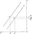
- Fig. 3 shows a diagram with a subjective, measured intraocular pressure on the ordinate axis and a deflection of an amplitude of a maximum deformation of the cornea 10 on the abscissa axis.
- a stiffness S 1 results as an essentially linear function with a negative slope.
- the stiffness S 1 can also deviate from a linear function and be designed as a line with a comparatively large radius of curvature.
- An objective intraocular pressure P o1 can be taken as a variable from the straight line defined by the stiffness S 1 .
- the 4a to 4b show the states of deformation of the cornea 10 of the eye 11 analogous to that 1a and 1b .
- the 4a and 4b Tensions in the corneal material visualized.
- stress lines 19 are visible in the material of the cornea 10, which represent main stresses along and across the visual axis 12.
- the Fig. 4a thus shows tensions in the eye 11 in a resting state of the cornea 10 and the Fig. 4b Tensions in the eye 11 of the deformed cornea 10, which differ significantly from the tensions in the resting state.
- a comparison of the voltage based on the Tension lines 19 thus make it possible to define a structural and / or material property of the cornea that can be used to correct a measured intraocular pressure and thus to derive an objective intraocular pressure.
Landscapes
- Life Sciences & Earth Sciences (AREA)
- Health & Medical Sciences (AREA)
- Medical Informatics (AREA)
- Biophysics (AREA)
- Ophthalmology & Optometry (AREA)
- Engineering & Computer Science (AREA)
- Biomedical Technology (AREA)
- Heart & Thoracic Surgery (AREA)
- Physics & Mathematics (AREA)
- Molecular Biology (AREA)
- Surgery (AREA)
- Animal Behavior & Ethology (AREA)
- General Health & Medical Sciences (AREA)
- Public Health (AREA)
- Veterinary Medicine (AREA)
- Eye Examination Apparatus (AREA)
Priority Applications (18)
| Application Number | Priority Date | Filing Date | Title |
|---|---|---|---|
| PL11168232T PL2397068T3 (pl) | 2010-06-21 | 2011-05-31 | Analiza ciśnienia wewnątrzgałkowego |
| EP11168232.4A EP2397068B1 (de) | 2010-06-21 | 2011-05-31 | Analyse des intraokularen Drucks |
| JP2011136548A JP5566957B2 (ja) | 2010-06-21 | 2011-06-20 | 眼科分析方法と分析システム |
| JP2011136552A JP5378457B2 (ja) | 2010-06-21 | 2011-06-20 | 眼科分析方法と分析システム |
| JP2011136550A JP5314090B2 (ja) | 2010-06-21 | 2011-06-20 | 眼科分析方法と分析システム |
| AU2011202970A AU2011202970B2 (en) | 2010-06-21 | 2011-06-21 | Ophthalmological analysis method and analysis system |
| KR1020110060129A KR101301894B1 (ko) | 2010-06-21 | 2011-06-21 | 안과 분석 방법 및 분석 시스템 |
| US13/165,541 US8556823B2 (en) | 2010-06-21 | 2011-06-21 | Ophthalmological analysis method and analysis system |
| AU2011202972A AU2011202972C1 (en) | 2010-06-21 | 2011-06-21 | Opthalmological analysis method and analysis system |
| US13/165,496 US8551014B2 (en) | 2010-06-21 | 2011-06-21 | Ophthalmological analysis method and analysis system |
| KR1020110060130A KR101301891B1 (ko) | 2010-06-21 | 2011-06-21 | 안과 분석 방법 및 분석 시스템 |
| US13/165,432 US8551013B2 (en) | 2010-06-21 | 2011-06-21 | Ophthalmological analysis method and analysis system |
| CN201110169822.9A CN102309313B (zh) | 2010-06-21 | 2011-06-21 | 眼科分析方法以及分析系统 |
| KR1020110060128A KR101301880B1 (ko) | 2010-06-21 | 2011-06-21 | 안과 분석 방법 및 분석 시스템 |
| AU2011202971A AU2011202971B8 (en) | 2010-06-21 | 2011-06-21 | Ophthalmological analysis method and analysis system |
| CN201110169845.XA CN102283636B (zh) | 2010-06-21 | 2011-06-21 | 眼科分析方法和分析系统 |
| CN201110169734.9A CN102309312B (zh) | 2010-06-21 | 2011-06-21 | 眼科分析方法和分析系统 |
| HK12104786.6A HK1165242B (en) | 2010-06-21 | 2012-05-16 | Ophthalmological analysis method and analysis system |
Applications Claiming Priority (4)
| Application Number | Priority Date | Filing Date | Title |
|---|---|---|---|
| EP10166681 | 2010-06-21 | ||
| DE102010049633 | 2010-10-28 | ||
| DE102010049634 | 2010-10-28 | ||
| EP11168232.4A EP2397068B1 (de) | 2010-06-21 | 2011-05-31 | Analyse des intraokularen Drucks |
Publications (2)
| Publication Number | Publication Date |
|---|---|
| EP2397068A1 EP2397068A1 (de) | 2011-12-21 |
| EP2397068B1 true EP2397068B1 (de) | 2020-05-27 |
Family
ID=44644898
Family Applications (3)
| Application Number | Title | Priority Date | Filing Date |
|---|---|---|---|
| EP11168232.4A Active EP2397068B1 (de) | 2010-06-21 | 2011-05-31 | Analyse des intraokularen Drucks |
| EP11168234.0A Active EP2397069B1 (de) | 2010-06-21 | 2011-05-31 | Analyse des intraokularen Drucks |
| EP11168235.7A Active EP2397070B1 (de) | 2010-06-21 | 2011-05-31 | Analyse des intraokularen Drucks |
Family Applications After (2)
| Application Number | Title | Priority Date | Filing Date |
|---|---|---|---|
| EP11168234.0A Active EP2397069B1 (de) | 2010-06-21 | 2011-05-31 | Analyse des intraokularen Drucks |
| EP11168235.7A Active EP2397070B1 (de) | 2010-06-21 | 2011-05-31 | Analyse des intraokularen Drucks |
Country Status (8)
| Country | Link |
|---|---|
| US (3) | US8551014B2 (pl) |
| EP (3) | EP2397068B1 (pl) |
| JP (3) | JP5566957B2 (pl) |
| KR (3) | KR101301891B1 (pl) |
| CN (3) | CN102309313B (pl) |
| AU (2) | AU2011202970B2 (pl) |
| ES (3) | ES2899473T3 (pl) |
| PL (2) | PL2397068T3 (pl) |
Families Citing this family (30)
| Publication number | Priority date | Publication date | Assignee | Title |
|---|---|---|---|---|
| DE102011076793A1 (de) * | 2011-05-31 | 2012-12-06 | Oculus Optikgeräte GmbH | Ophthalmologisches Analyseverfahren und Analysesystem |
| TWI474802B (zh) * | 2012-05-04 | 2015-03-01 | 光學眼壓量測裝置及其運作方法 | |
| TWI507170B (zh) * | 2012-10-24 | 2015-11-11 | Crystalvue Medical Corp | 光學裝置及其運作方法 |
| JP2014171722A (ja) * | 2013-03-11 | 2014-09-22 | Canon Inc | 非接触式眼科装置及びその制御方法 |
| JP2014217424A (ja) * | 2013-05-01 | 2014-11-20 | キヤノン株式会社 | 非接触式眼圧計 |
| ITMI20131262A1 (it) * | 2013-07-26 | 2015-01-27 | Phronema S R L | Apparecchiatura e metodo per determinare l'orientazione di strutture anatomiche corneali |
| US9357912B2 (en) * | 2013-09-09 | 2016-06-07 | Yan Zhang | Apparatus and method for characterizing biomechanical properties of eye tissue |
| EP3119268B1 (en) * | 2014-03-07 | 2024-05-01 | Lions Eye Institute Limited | Method and system for determining intracranial pressure |
| CN107529984A (zh) * | 2015-04-15 | 2018-01-02 | 卡茨血压计有限责任公司 | 光学仪器 |
| US11026576B2 (en) * | 2015-04-15 | 2021-06-08 | Cats Tonometer, Llc | Reducing errors of tonometric measurements by using a tonometer tip with a curved cornea-contacting surface |
| TW201703722A (zh) * | 2015-07-21 | 2017-02-01 | 明達醫學科技股份有限公司 | 量測裝置及其運作方法 |
| CN105231990B (zh) * | 2015-11-17 | 2017-04-19 | 深圳市亿领科技有限公司 | 基于oct三维成像分析角膜生物力学性能的装置及方法 |
| TWI568408B (zh) | 2015-12-23 | 2017-02-01 | 財團法人工業技術研究院 | 一種眼壓檢測裝置及其檢測方法 |
| CN109068977B (zh) * | 2016-03-03 | 2021-08-24 | 百欧莱特工程有限公司 | 采用经巩膜平坦部照明的眼底成像方法和装置 |
| US20190192115A1 (en) * | 2016-05-08 | 2019-06-27 | The Cleveland Clinic Foundation | Measurement of biomechanical properties of tissue |
| CN106108841A (zh) * | 2016-06-29 | 2016-11-16 | 无锡市康明医疗器械有限公司 | 一种非接触光摄眼压计及眼压测量方法 |
| CN107095643B (zh) * | 2017-04-11 | 2019-01-25 | 佛山科学技术学院 | 基于低相干光干涉的非接触眼压检测系统及其检测方法 |
| EP3449812A1 (en) | 2017-09-05 | 2019-03-06 | Frey Spolka Jawna | Ophthalmic analysis system for measuring the intraocular pressure in the eye |
| US20210022608A1 (en) * | 2018-03-12 | 2021-01-28 | Ioppreceyese Ltd. | Non-Contact Home-Tonometry System for Measuring Intraocular Pressure |
| US11026577B2 (en) | 2018-06-13 | 2021-06-08 | Reichert, Inc. | Rebound tonometry method and apparatus |
| SG11202012844VA (en) * | 2018-07-06 | 2021-01-28 | Sensimed Ag | Intraocular pressure measuring and/or monitoring device |
| WO2020210322A1 (en) | 2019-04-10 | 2020-10-15 | Smartlens, Inc. | Intraocular pressure monitoring devices and methods of using the same |
| US12268449B2 (en) | 2019-04-10 | 2025-04-08 | Smartlens, Inc. | Intraocular pressure monitoring devices and methods of using the same |
| WO2020232015A1 (en) * | 2019-05-13 | 2020-11-19 | Verily Life Sciences Llc | Systems, devices and methods for optical interrogation of an implantable intraocular pressure sensor |
| CN110731751A (zh) * | 2019-11-15 | 2020-01-31 | 刘果 | 一种方便、快捷测量眼内压的方法 |
| US20240016381A1 (en) * | 2020-11-17 | 2024-01-18 | N.M.B. Medical Applications Ltd | Device and method for non-contact measurement of an intraocular pressure |
| JP7048140B1 (ja) | 2021-05-18 | 2022-04-06 | 合同会社クオビス | 非接触式の眼球物性測定装置 |
| CN117012367A (zh) * | 2022-05-06 | 2023-11-07 | 香港科技大学 | 对眼睛的检测及分类系统和用于眼睛检测的装置 |
| KR102948265B1 (ko) * | 2023-11-21 | 2026-04-03 | 유한회사 웰씨 | 각막 돌출부 교정 장치 및 방법 |
| FI131741B1 (en) * | 2024-08-14 | 2025-10-29 | Icare Finland Oy | Tonometer for measuring intraocular pressure of eye using air pulse |
Family Cites Families (35)
| Publication number | Priority date | Publication date | Assignee | Title |
|---|---|---|---|---|
| US3406681A (en) * | 1963-05-13 | 1968-10-22 | Vishay Intertechnology Inc | Method of determining strain condition of the eye |
| US3585849A (en) * | 1968-10-09 | 1971-06-22 | American Optical Corp | Method and apparatus for measuring intraocular pressure |
| JPS61321A (ja) * | 1984-06-12 | 1986-01-06 | 株式会社トプコン | 非接触式眼圧計 |
| JP3221699B2 (ja) * | 1991-08-31 | 2001-10-22 | 株式会社ニデック | 非接触式眼圧計 |
| JPH08109B2 (ja) * | 1993-04-02 | 1996-01-10 | 株式会社トプコン | 非接触式眼圧計 |
| JP3108261B2 (ja) * | 1993-12-20 | 2000-11-13 | 株式会社トプコン | 眼科器械 |
| JP3308416B2 (ja) * | 1994-10-26 | 2002-07-29 | 株式会社トプコン | 眼科器械 |
| JPH08322803A (ja) * | 1995-05-31 | 1996-12-10 | Canon Inc | 眼圧計 |
| US5822035A (en) | 1996-08-30 | 1998-10-13 | Heidelberg Engineering Optische Messysteme Gmbh | Ellipsometer |
| US6544193B2 (en) * | 1996-09-04 | 2003-04-08 | Marcio Marc Abreu | Noninvasive measurement of chemical substances |
| JPH10309265A (ja) * | 1997-05-12 | 1998-11-24 | Konan:Kk | 眼科撮影装置 |
| JP3862869B2 (ja) * | 1998-06-05 | 2006-12-27 | 株式会社トプコン | 非接触式眼圧計 |
| JP2000254101A (ja) * | 1999-01-06 | 2000-09-19 | Konan Inc | 眼科検査装置 |
| JP2002263069A (ja) * | 2001-03-13 | 2002-09-17 | Konan Medical Inc | 眼硬性測定装置 |
| JP3970141B2 (ja) * | 2002-09-11 | 2007-09-05 | キヤノン株式会社 | 非接触式眼圧計 |
| JP3885015B2 (ja) * | 2002-09-17 | 2007-02-21 | キヤノン株式会社 | 非接触眼圧計 |
| US7004902B2 (en) * | 2003-03-21 | 2006-02-28 | Reichert, Inc. | Method and apparatus for measuring biomechanical characteristics of corneal tissue |
| US20050030473A1 (en) * | 2003-06-12 | 2005-02-10 | Welch Allyn, Inc. | Apparatus and method for determining intraocular pressure and corneal thickness |
| DE202005002562U1 (de) * | 2005-02-16 | 2005-06-09 | Oculus Optikgeräte GmbH | Ophthalmisches Analysesystem zur Messung des intraocularen Drucks im Auge |
| JP2007121174A (ja) * | 2005-10-31 | 2007-05-17 | Univ Nagoya | 応力検出装置 |
| US7798962B2 (en) * | 2005-09-08 | 2010-09-21 | Reichert, Inc. | Method and apparatus for measuring corneal resistance |
| US7481767B2 (en) | 2005-09-08 | 2009-01-27 | Reichert, Inc. | Method and apparatus for determining true intraocular pressure |
| JP5028057B2 (ja) * | 2005-11-01 | 2012-09-19 | 株式会社ニデック | 眼科装置 |
| WO2007056292A2 (en) | 2005-11-07 | 2007-05-18 | Zink Imaging, Llc | Thermal printing head with two-dimensional array of resistive heating elements |
| US20070121120A1 (en) * | 2005-11-16 | 2007-05-31 | Schachar Ronald A | Apparatus and method for measuring scleral curvature and velocity of tissues of the eye |
| JP4824400B2 (ja) * | 2005-12-28 | 2011-11-30 | 株式会社トプコン | 眼科装置 |
| JP4948922B2 (ja) * | 2006-06-30 | 2012-06-06 | 株式会社ニデック | 眼科装置 |
| JP5448353B2 (ja) * | 2007-05-02 | 2014-03-19 | キヤノン株式会社 | 光干渉断層計を用いた画像形成方法、及び光干渉断層装置 |
| JP5209341B2 (ja) * | 2008-02-27 | 2013-06-12 | 株式会社ニデック | 非接触式眼圧計 |
| JP5268053B2 (ja) | 2008-05-15 | 2013-08-21 | 晃太郎 石井 | 眼球組織固有振動数測定装置及びそれを利用した非接触式眼圧計 |
| BRPI1006732B8 (pt) * | 2009-03-04 | 2021-06-22 | Aaren Scientific Inc | lente dimensionada para uso num olho humano |
| US8025401B2 (en) * | 2009-08-05 | 2011-09-27 | Sis Ag, Surgical Instrument Systems | Ophthalmological measuring device and measurement method |
| DE102011082363B4 (de) * | 2010-10-28 | 2018-03-29 | Oculus Optikgeräte GmbH | Beleuchtungssystem, ophthalmologisches Analysegerät und Verfahren |
| JP5340434B2 (ja) * | 2011-02-25 | 2013-11-13 | キヤノン株式会社 | 眼科装置、処理装置、眼科システム、処理方法および眼科装置の制御方法、プログラム |
| DE102011076793A1 (de) * | 2011-05-31 | 2012-12-06 | Oculus Optikgeräte GmbH | Ophthalmologisches Analyseverfahren und Analysesystem |
-
2011
- 2011-05-31 EP EP11168232.4A patent/EP2397068B1/de active Active
- 2011-05-31 EP EP11168234.0A patent/EP2397069B1/de active Active
- 2011-05-31 PL PL11168232T patent/PL2397068T3/pl unknown
- 2011-05-31 PL PL11168234T patent/PL2397069T3/pl unknown
- 2011-05-31 ES ES11168235T patent/ES2899473T3/es active Active
- 2011-05-31 ES ES11168234T patent/ES2806934T3/es active Active
- 2011-05-31 EP EP11168235.7A patent/EP2397070B1/de active Active
- 2011-05-31 ES ES11168232T patent/ES2806024T3/es active Active
- 2011-06-20 JP JP2011136548A patent/JP5566957B2/ja active Active
- 2011-06-20 JP JP2011136552A patent/JP5378457B2/ja active Active
- 2011-06-20 JP JP2011136550A patent/JP5314090B2/ja active Active
- 2011-06-21 AU AU2011202970A patent/AU2011202970B2/en active Active
- 2011-06-21 CN CN201110169822.9A patent/CN102309313B/zh active Active
- 2011-06-21 KR KR1020110060130A patent/KR101301891B1/ko active Active
- 2011-06-21 US US13/165,496 patent/US8551014B2/en active Active
- 2011-06-21 AU AU2011202972A patent/AU2011202972C1/en active Active
- 2011-06-21 CN CN201110169845.XA patent/CN102283636B/zh active Active
- 2011-06-21 US US13/165,541 patent/US8556823B2/en active Active
- 2011-06-21 CN CN201110169734.9A patent/CN102309312B/zh active Active
- 2011-06-21 KR KR1020110060128A patent/KR101301880B1/ko active Active
- 2011-06-21 US US13/165,432 patent/US8551013B2/en active Active
- 2011-06-21 KR KR1020110060129A patent/KR101301894B1/ko active Active
Non-Patent Citations (1)
| Title |
|---|
| None * |
Also Published As
Similar Documents
| Publication | Publication Date | Title |
|---|---|---|
| EP2397068B1 (de) | Analyse des intraokularen Drucks | |
| EP2529662B1 (de) | Ophthalmologisches Analyseverfahren und Analysesystem | |
| EP2671505B1 (de) | Verfahren und Analysegerät zur Messung einer Hornhaut | |
| DE102005042436C5 (de) | Ophthalmo-Operationsmikroskop mit Messeinrichtung | |
| DE60130284T2 (de) | Verfahren der berührungslosen Tonometrie | |
| EP3682794B1 (de) | Verfahren und sehprüfsystem zum überprüfen der augen | |
| DE202014010515U1 (de) | Ophthalmisches Messsystem und computerlesbares Speichermedium zur Auswahl einer Intraokularlinse | |
| EP3705029A1 (de) | Verfahren und sehprüfsystem zum überprüfen der augen | |
| EP2561800B1 (de) | Ophthalmologisches Analysegerät und Verfahren | |
| DE102006039893A1 (de) | Verfahren und Vorrichtung zum Bestimmen des wahren Augeninnendrucks | |
| DE102016105962A1 (de) | Positionsermittlungsanordnung für Intraokularlinse | |
| EP4322858B1 (de) | Verfahren zur messung eines innendrucks eines kompartments | |
| DE102011015178B3 (de) | Vorrichtung und Verfahren zur automatisierten Bestimmung des Intraokulardruckes | |
| DE102018219902A1 (de) | Anordnung und Verfahren zur Kompensation der Temperaturabhängigkeit einer Facettenlinse für die Bestimmung der Topographie eines Auges | |
| HK1165242B (en) | Ophthalmological analysis method and analysis system |
Legal Events
| Date | Code | Title | Description |
|---|---|---|---|
| AK | Designated contracting states |
Kind code of ref document: A1 Designated state(s): AL AT BE BG CH CY CZ DE DK EE ES FI FR GB GR HR HU IE IS IT LI LT LU LV MC MK MT NL NO PL PT RO RS SE SI SK SM TR |
|
| AX | Request for extension of the european patent |
Extension state: BA ME |
|
| PUAI | Public reference made under article 153(3) epc to a published international application that has entered the european phase |
Free format text: ORIGINAL CODE: 0009012 |
|
| 17P | Request for examination filed |
Effective date: 20120529 |
|
| STAA | Information on the status of an ep patent application or granted ep patent |
Free format text: STATUS: EXAMINATION IS IN PROGRESS |
|
| 17Q | First examination report despatched |
Effective date: 20180606 |
|
| GRAP | Despatch of communication of intention to grant a patent |
Free format text: ORIGINAL CODE: EPIDOSNIGR1 |
|
| STAA | Information on the status of an ep patent application or granted ep patent |
Free format text: STATUS: GRANT OF PATENT IS INTENDED |
|
| RIC1 | Information provided on ipc code assigned before grant |
Ipc: A61B 3/107 20060101ALN20191127BHEP Ipc: A61B 3/16 20060101AFI20191127BHEP |
|
| INTG | Intention to grant announced |
Effective date: 20191220 |
|
| GRAS | Grant fee paid |
Free format text: ORIGINAL CODE: EPIDOSNIGR3 |
|
| GRAA | (expected) grant |
Free format text: ORIGINAL CODE: 0009210 |
|
| STAA | Information on the status of an ep patent application or granted ep patent |
Free format text: STATUS: THE PATENT HAS BEEN GRANTED |
|
| AK | Designated contracting states |
Kind code of ref document: B1 Designated state(s): AL AT BE BG CH CY CZ DE DK EE ES FI FR GB GR HR HU IE IS IT LI LT LU LV MC MK MT NL NO PL PT RO RS SE SI SK SM TR |
|
| REG | Reference to a national code |
Ref country code: GB Ref legal event code: FG4D Free format text: NOT ENGLISH |
|
| REG | Reference to a national code |
Ref country code: CH Ref legal event code: EP |
|
| REG | Reference to a national code |
Ref country code: AT Ref legal event code: REF Ref document number: 1273649 Country of ref document: AT Kind code of ref document: T Effective date: 20200615 |
|
| REG | Reference to a national code |
Ref country code: DE Ref legal event code: R096 Ref document number: 502011016690 Country of ref document: DE |
|
| REG | Reference to a national code |
Ref country code: CH Ref legal event code: NV Representative=s name: BODENSEEPATENT PATENTANWAELTE BEHRMANN WAGNER , CH |
|
| REG | Reference to a national code |
Ref country code: LT Ref legal event code: MG4D |
|
| PG25 | Lapsed in a contracting state [announced via postgrant information from national office to epo] |
Ref country code: IS Free format text: LAPSE BECAUSE OF FAILURE TO SUBMIT A TRANSLATION OF THE DESCRIPTION OR TO PAY THE FEE WITHIN THE PRESCRIBED TIME-LIMIT Effective date: 20200927 Ref country code: PT Free format text: LAPSE BECAUSE OF FAILURE TO SUBMIT A TRANSLATION OF THE DESCRIPTION OR TO PAY THE FEE WITHIN THE PRESCRIBED TIME-LIMIT Effective date: 20200928 Ref country code: FI Free format text: LAPSE BECAUSE OF FAILURE TO SUBMIT A TRANSLATION OF THE DESCRIPTION OR TO PAY THE FEE WITHIN THE PRESCRIBED TIME-LIMIT Effective date: 20200527 Ref country code: SE Free format text: LAPSE BECAUSE OF FAILURE TO SUBMIT A TRANSLATION OF THE DESCRIPTION OR TO PAY THE FEE WITHIN THE PRESCRIBED TIME-LIMIT Effective date: 20200527 Ref country code: LT Free format text: LAPSE BECAUSE OF FAILURE TO SUBMIT A TRANSLATION OF THE DESCRIPTION OR TO PAY THE FEE WITHIN THE PRESCRIBED TIME-LIMIT Effective date: 20200527 Ref country code: NO Free format text: LAPSE BECAUSE OF FAILURE TO SUBMIT A TRANSLATION OF THE DESCRIPTION OR TO PAY THE FEE WITHIN THE PRESCRIBED TIME-LIMIT Effective date: 20200827 Ref country code: GR Free format text: LAPSE BECAUSE OF FAILURE TO SUBMIT A TRANSLATION OF THE DESCRIPTION OR TO PAY THE FEE WITHIN THE PRESCRIBED TIME-LIMIT Effective date: 20200828 |
|
| REG | Reference to a national code |
Ref country code: NL Ref legal event code: MP Effective date: 20200527 |
|
| PG25 | Lapsed in a contracting state [announced via postgrant information from national office to epo] |
Ref country code: BG Free format text: LAPSE BECAUSE OF FAILURE TO SUBMIT A TRANSLATION OF THE DESCRIPTION OR TO PAY THE FEE WITHIN THE PRESCRIBED TIME-LIMIT Effective date: 20200827 Ref country code: LV Free format text: LAPSE BECAUSE OF FAILURE TO SUBMIT A TRANSLATION OF THE DESCRIPTION OR TO PAY THE FEE WITHIN THE PRESCRIBED TIME-LIMIT Effective date: 20200527 Ref country code: RS Free format text: LAPSE BECAUSE OF FAILURE TO SUBMIT A TRANSLATION OF THE DESCRIPTION OR TO PAY THE FEE WITHIN THE PRESCRIBED TIME-LIMIT Effective date: 20200527 Ref country code: HR Free format text: LAPSE BECAUSE OF FAILURE TO SUBMIT A TRANSLATION OF THE DESCRIPTION OR TO PAY THE FEE WITHIN THE PRESCRIBED TIME-LIMIT Effective date: 20200527 |
|
| PG25 | Lapsed in a contracting state [announced via postgrant information from national office to epo] |
Ref country code: AL Free format text: LAPSE BECAUSE OF FAILURE TO SUBMIT A TRANSLATION OF THE DESCRIPTION OR TO PAY THE FEE WITHIN THE PRESCRIBED TIME-LIMIT Effective date: 20200527 Ref country code: NL Free format text: LAPSE BECAUSE OF FAILURE TO SUBMIT A TRANSLATION OF THE DESCRIPTION OR TO PAY THE FEE WITHIN THE PRESCRIBED TIME-LIMIT Effective date: 20200527 |
|
| PG25 | Lapsed in a contracting state [announced via postgrant information from national office to epo] |
Ref country code: DK Free format text: LAPSE BECAUSE OF FAILURE TO SUBMIT A TRANSLATION OF THE DESCRIPTION OR TO PAY THE FEE WITHIN THE PRESCRIBED TIME-LIMIT Effective date: 20200527 Ref country code: EE Free format text: LAPSE BECAUSE OF FAILURE TO SUBMIT A TRANSLATION OF THE DESCRIPTION OR TO PAY THE FEE WITHIN THE PRESCRIBED TIME-LIMIT Effective date: 20200527 Ref country code: SM Free format text: LAPSE BECAUSE OF FAILURE TO SUBMIT A TRANSLATION OF THE DESCRIPTION OR TO PAY THE FEE WITHIN THE PRESCRIBED TIME-LIMIT Effective date: 20200527 Ref country code: RO Free format text: LAPSE BECAUSE OF FAILURE TO SUBMIT A TRANSLATION OF THE DESCRIPTION OR TO PAY THE FEE WITHIN THE PRESCRIBED TIME-LIMIT Effective date: 20200527 Ref country code: CZ Free format text: LAPSE BECAUSE OF FAILURE TO SUBMIT A TRANSLATION OF THE DESCRIPTION OR TO PAY THE FEE WITHIN THE PRESCRIBED TIME-LIMIT Effective date: 20200527 |
|
| REG | Reference to a national code |
Ref country code: ES Ref legal event code: FG2A Ref document number: 2806024 Country of ref document: ES Kind code of ref document: T3 Effective date: 20210216 |
|
| PG25 | Lapsed in a contracting state [announced via postgrant information from national office to epo] |
Ref country code: MC Free format text: LAPSE BECAUSE OF FAILURE TO SUBMIT A TRANSLATION OF THE DESCRIPTION OR TO PAY THE FEE WITHIN THE PRESCRIBED TIME-LIMIT Effective date: 20200527 Ref country code: SK Free format text: LAPSE BECAUSE OF FAILURE TO SUBMIT A TRANSLATION OF THE DESCRIPTION OR TO PAY THE FEE WITHIN THE PRESCRIBED TIME-LIMIT Effective date: 20200527 |
|
| REG | Reference to a national code |
Ref country code: CH Ref legal event code: PFA Owner name: OCULUS OPTIKGERAETE GMBH, DE Free format text: FORMER OWNER: OCULUS OPTIKGERAETE GMBH, DE |
|
| REG | Reference to a national code |
Ref country code: DE Ref legal event code: R097 Ref document number: 502011016690 Country of ref document: DE |
|
| REG | Reference to a national code |
Ref country code: BE Ref legal event code: MM Effective date: 20200531 |
|
| PG25 | Lapsed in a contracting state [announced via postgrant information from national office to epo] |
Ref country code: LU Free format text: LAPSE BECAUSE OF NON-PAYMENT OF DUE FEES Effective date: 20200531 |
|
| PLBE | No opposition filed within time limit |
Free format text: ORIGINAL CODE: 0009261 |
|
| STAA | Information on the status of an ep patent application or granted ep patent |
Free format text: STATUS: NO OPPOSITION FILED WITHIN TIME LIMIT |
|
| PG25 | Lapsed in a contracting state [announced via postgrant information from national office to epo] |
Ref country code: IE Free format text: LAPSE BECAUSE OF NON-PAYMENT OF DUE FEES Effective date: 20200531 |
|
| 26N | No opposition filed |
Effective date: 20210302 |
|
| PG25 | Lapsed in a contracting state [announced via postgrant information from national office to epo] |
Ref country code: BE Free format text: LAPSE BECAUSE OF NON-PAYMENT OF DUE FEES Effective date: 20200531 Ref country code: SI Free format text: LAPSE BECAUSE OF FAILURE TO SUBMIT A TRANSLATION OF THE DESCRIPTION OR TO PAY THE FEE WITHIN THE PRESCRIBED TIME-LIMIT Effective date: 20200527 |
|
| REG | Reference to a national code |
Ref country code: AT Ref legal event code: MM01 Ref document number: 1273649 Country of ref document: AT Kind code of ref document: T Effective date: 20200531 |
|
| PG25 | Lapsed in a contracting state [announced via postgrant information from national office to epo] |
Ref country code: AT Free format text: LAPSE BECAUSE OF NON-PAYMENT OF DUE FEES Effective date: 20200531 |
|
| PG25 | Lapsed in a contracting state [announced via postgrant information from national office to epo] |
Ref country code: TR Free format text: LAPSE BECAUSE OF FAILURE TO SUBMIT A TRANSLATION OF THE DESCRIPTION OR TO PAY THE FEE WITHIN THE PRESCRIBED TIME-LIMIT Effective date: 20200527 Ref country code: MT Free format text: LAPSE BECAUSE OF FAILURE TO SUBMIT A TRANSLATION OF THE DESCRIPTION OR TO PAY THE FEE WITHIN THE PRESCRIBED TIME-LIMIT Effective date: 20200527 Ref country code: CY Free format text: LAPSE BECAUSE OF FAILURE TO SUBMIT A TRANSLATION OF THE DESCRIPTION OR TO PAY THE FEE WITHIN THE PRESCRIBED TIME-LIMIT Effective date: 20200527 |
|
| PG25 | Lapsed in a contracting state [announced via postgrant information from national office to epo] |
Ref country code: MK Free format text: LAPSE BECAUSE OF FAILURE TO SUBMIT A TRANSLATION OF THE DESCRIPTION OR TO PAY THE FEE WITHIN THE PRESCRIBED TIME-LIMIT Effective date: 20200527 |
|
| P01 | Opt-out of the competence of the unified patent court (upc) registered |
Effective date: 20230525 |
|
| PGFP | Annual fee paid to national office [announced via postgrant information from national office to epo] |
Ref country code: PL Payment date: 20250410 Year of fee payment: 15 |
|
| PGFP | Annual fee paid to national office [announced via postgrant information from national office to epo] |
Ref country code: GB Payment date: 20250522 Year of fee payment: 15 Ref country code: ES Payment date: 20250616 Year of fee payment: 15 |
|
| PGFP | Annual fee paid to national office [announced via postgrant information from national office to epo] |
Ref country code: IT Payment date: 20250530 Year of fee payment: 15 |
|
| PGFP | Annual fee paid to national office [announced via postgrant information from national office to epo] |
Ref country code: FR Payment date: 20250526 Year of fee payment: 15 |
|
| PGFP | Annual fee paid to national office [announced via postgrant information from national office to epo] |
Ref country code: CH Payment date: 20250601 Year of fee payment: 15 |
|
| PGFP | Annual fee paid to national office [announced via postgrant information from national office to epo] |
Ref country code: DE Payment date: 20250722 Year of fee payment: 15 |