EP2322221B1 - Composition pharmaceutique pour le traitement et la prévention du cancer - Google Patents
Composition pharmaceutique pour le traitement et la prévention du cancer Download PDFInfo
- Publication number
- EP2322221B1 EP2322221B1 EP09805009.9A EP09805009A EP2322221B1 EP 2322221 B1 EP2322221 B1 EP 2322221B1 EP 09805009 A EP09805009 A EP 09805009A EP 2322221 B1 EP2322221 B1 EP 2322221B1
- Authority
- EP
- European Patent Office
- Prior art keywords
- seq
- antibody
- nos
- caprin
- chain variable
- Prior art date
- Legal status (The legal status is an assumption and is not a legal conclusion. Google has not performed a legal analysis and makes no representation as to the accuracy of the status listed.)
- Active
Links
- 206010028980 Neoplasm Diseases 0.000 title claims description 144
- 201000011510 cancer Diseases 0.000 title claims description 103
- 238000011282 treatment Methods 0.000 title claims description 23
- 230000002265 prevention Effects 0.000 title claims description 13
- 239000008194 pharmaceutical composition Substances 0.000 title claims description 12
- 125000003275 alpha amino acid group Chemical group 0.000 claims description 123
- 241000282414 Homo sapiens Species 0.000 claims description 118
- 238000000034 method Methods 0.000 claims description 91
- 108090000765 processed proteins & peptides Proteins 0.000 claims description 73
- 102000004196 processed proteins & peptides Human genes 0.000 claims description 50
- 229920001184 polypeptide Polymers 0.000 claims description 44
- 206010006187 Breast cancer Diseases 0.000 claims description 39
- 208000026310 Breast neoplasm Diseases 0.000 claims description 36
- 239000012634 fragment Substances 0.000 claims description 26
- 230000036961 partial effect Effects 0.000 claims description 11
- 208000003174 Brain Neoplasms Diseases 0.000 claims description 10
- 230000009257 reactivity Effects 0.000 claims description 10
- 206010025323 Lymphomas Diseases 0.000 claims description 9
- 230000001900 immune effect Effects 0.000 claims description 9
- 208000032839 leukemia Diseases 0.000 claims description 9
- 206010009944 Colon cancer Diseases 0.000 claims description 8
- 206010058467 Lung neoplasm malignant Diseases 0.000 claims description 7
- 208000029742 colonic neoplasm Diseases 0.000 claims description 7
- 201000005202 lung cancer Diseases 0.000 claims description 7
- 208000020816 lung neoplasm Diseases 0.000 claims description 7
- 101710072528 Caprin-1 Proteins 0.000 claims description 5
- 102100029949 Caprin-1 Human genes 0.000 claims description 5
- 208000000461 Esophageal Neoplasms Diseases 0.000 claims description 5
- 206010030155 Oesophageal carcinoma Diseases 0.000 claims description 5
- 201000004101 esophageal cancer Diseases 0.000 claims description 5
- 125000000539 amino acid group Chemical group 0.000 claims description 4
- 239000004480 active ingredient Substances 0.000 claims description 2
- 210000004027 cell Anatomy 0.000 description 202
- 108090000623 proteins and genes Proteins 0.000 description 152
- 108020004414 DNA Proteins 0.000 description 127
- 241000699660 Mus musculus Species 0.000 description 99
- 102000004169 proteins and genes Human genes 0.000 description 84
- 235000018102 proteins Nutrition 0.000 description 83
- 239000000243 solution Substances 0.000 description 61
- 108091007433 antigens Proteins 0.000 description 49
- 210000001519 tissue Anatomy 0.000 description 49
- 102000036639 antigens Human genes 0.000 description 48
- 241000699666 Mus <mouse, genus> Species 0.000 description 44
- 210000004408 hybridoma Anatomy 0.000 description 44
- 239000000427 antigen Substances 0.000 description 43
- 230000000694 effects Effects 0.000 description 43
- 230000014509 gene expression Effects 0.000 description 42
- 239000002773 nucleotide Substances 0.000 description 36
- 125000003729 nucleotide group Chemical group 0.000 description 36
- 235000001014 amino acid Nutrition 0.000 description 34
- 229940024606 amino acid Drugs 0.000 description 32
- 150000001413 amino acids Chemical class 0.000 description 31
- 230000027455 binding Effects 0.000 description 31
- LOKCTEFSRHRXRJ-UHFFFAOYSA-I dipotassium trisodium dihydrogen phosphate hydrogen phosphate dichloride Chemical compound P(=O)(O)(O)[O-].[K+].P(=O)(O)([O-])[O-].[Na+].[Na+].[Cl-].[K+].[Cl-].[Na+] LOKCTEFSRHRXRJ-UHFFFAOYSA-I 0.000 description 31
- 239000002953 phosphate buffered saline Substances 0.000 description 31
- 108010047041 Complementarity Determining Regions Proteins 0.000 description 28
- 241000699670 Mus sp. Species 0.000 description 26
- 238000006243 chemical reaction Methods 0.000 description 25
- 241000282472 Canis lupus familiaris Species 0.000 description 23
- 241000282465 Canis Species 0.000 description 22
- 239000002299 complementary DNA Substances 0.000 description 22
- 241001465754 Metazoa Species 0.000 description 20
- 230000000259 anti-tumor effect Effects 0.000 description 20
- 108091003079 Bovine Serum Albumin Proteins 0.000 description 19
- 210000002966 serum Anatomy 0.000 description 19
- XLYOFNOQVPJJNP-UHFFFAOYSA-N water Substances O XLYOFNOQVPJJNP-UHFFFAOYSA-N 0.000 description 18
- 101000793727 Homo sapiens Caprin-1 Proteins 0.000 description 17
- FAPWRFPIFSIZLT-UHFFFAOYSA-M Sodium chloride Chemical compound [Na+].[Cl-] FAPWRFPIFSIZLT-UHFFFAOYSA-M 0.000 description 17
- 102000052098 human CAPRIN1 Human genes 0.000 description 17
- 239000013598 vector Substances 0.000 description 17
- 241000588724 Escherichia coli Species 0.000 description 16
- 239000013604 expression vector Substances 0.000 description 16
- 239000011521 glass Substances 0.000 description 15
- -1 t-butyloxycarbonyl Chemical group 0.000 description 15
- 241000287828 Gallus gallus Species 0.000 description 14
- 206010035226 Plasma cell myeloma Diseases 0.000 description 14
- 230000010056 antibody-dependent cellular cytotoxicity Effects 0.000 description 14
- 230000001472 cytotoxic effect Effects 0.000 description 14
- 201000000050 myeloid neoplasm Diseases 0.000 description 14
- 230000003053 immunization Effects 0.000 description 13
- LFQSCWFLJHTTHZ-UHFFFAOYSA-N Ethanol Chemical compound CCO LFQSCWFLJHTTHZ-UHFFFAOYSA-N 0.000 description 12
- 108091028043 Nucleic acid sequence Proteins 0.000 description 12
- 238000007792 addition Methods 0.000 description 12
- 239000000872 buffer Substances 0.000 description 12
- 235000013330 chicken meat Nutrition 0.000 description 12
- 239000012091 fetal bovine serum Substances 0.000 description 12
- 239000006228 supernatant Substances 0.000 description 12
- 241000124008 Mammalia Species 0.000 description 11
- 230000004540 complement-dependent cytotoxicity Effects 0.000 description 11
- 239000008363 phosphate buffer Substances 0.000 description 11
- 241000283973 Oryctolagus cuniculus Species 0.000 description 10
- 239000003795 chemical substances by application Substances 0.000 description 10
- 238000002649 immunization Methods 0.000 description 10
- 239000000203 mixture Substances 0.000 description 10
- 238000002360 preparation method Methods 0.000 description 10
- 238000005406 washing Methods 0.000 description 10
- 230000007910 cell fusion Effects 0.000 description 9
- RAXXELZNTBOGNW-UHFFFAOYSA-N imidazole Natural products C1=CNC=N1 RAXXELZNTBOGNW-UHFFFAOYSA-N 0.000 description 9
- 239000002609 medium Substances 0.000 description 9
- 239000013612 plasmid Substances 0.000 description 9
- 238000000746 purification Methods 0.000 description 9
- 239000011780 sodium chloride Substances 0.000 description 9
- 210000001550 testis Anatomy 0.000 description 9
- 210000004881 tumor cell Anatomy 0.000 description 9
- 241000283690 Bos taurus Species 0.000 description 8
- 102220497176 Small vasohibin-binding protein_T47D_mutation Human genes 0.000 description 8
- 238000002835 absorbance Methods 0.000 description 8
- 238000010367 cloning Methods 0.000 description 8
- 108020004999 messenger RNA Proteins 0.000 description 8
- 241000283707 Capra Species 0.000 description 7
- VYZAMTAEIAYCRO-BJUDXGSMSA-N Chromium-51 Chemical compound [51Cr] VYZAMTAEIAYCRO-BJUDXGSMSA-N 0.000 description 7
- 241000283073 Equus caballus Species 0.000 description 7
- 238000012181 QIAquick gel extraction kit Methods 0.000 description 7
- 241000700159 Rattus Species 0.000 description 7
- 238000005119 centrifugation Methods 0.000 description 7
- 239000003814 drug Substances 0.000 description 7
- 238000011532 immunohistochemical staining Methods 0.000 description 7
- 230000005764 inhibitory process Effects 0.000 description 7
- 210000004698 lymphocyte Anatomy 0.000 description 7
- 238000006467 substitution reaction Methods 0.000 description 7
- 108091032973 (ribonucleotides)n+m Proteins 0.000 description 6
- 241000282412 Homo Species 0.000 description 6
- VEXZGXHMUGYJMC-UHFFFAOYSA-N Hydrochloric acid Chemical compound Cl VEXZGXHMUGYJMC-UHFFFAOYSA-N 0.000 description 6
- 108010052285 Membrane Proteins Proteins 0.000 description 6
- 102000018697 Membrane Proteins Human genes 0.000 description 6
- 239000002671 adjuvant Substances 0.000 description 6
- 230000000903 blocking effect Effects 0.000 description 6
- 239000003153 chemical reaction reagent Substances 0.000 description 6
- 239000011651 chromium Substances 0.000 description 6
- 239000012228 culture supernatant Substances 0.000 description 6
- 230000004927 fusion Effects 0.000 description 6
- 238000007429 general method Methods 0.000 description 6
- 230000016784 immunoglobulin production Effects 0.000 description 6
- 230000006320 pegylation Effects 0.000 description 6
- 238000003757 reverse transcription PCR Methods 0.000 description 6
- 238000012216 screening Methods 0.000 description 6
- 230000001235 sensitizing effect Effects 0.000 description 6
- 210000000952 spleen Anatomy 0.000 description 6
- CURLTUGMZLYLDI-UHFFFAOYSA-N Carbon dioxide Chemical compound O=C=O CURLTUGMZLYLDI-UHFFFAOYSA-N 0.000 description 5
- CTQNGGLPUBDAKN-UHFFFAOYSA-N O-Xylene Chemical compound CC1=CC=CC=C1C CTQNGGLPUBDAKN-UHFFFAOYSA-N 0.000 description 5
- 239000012980 RPMI-1640 medium Substances 0.000 description 5
- 239000002246 antineoplastic agent Substances 0.000 description 5
- 230000015572 biosynthetic process Effects 0.000 description 5
- 229940079593 drug Drugs 0.000 description 5
- 230000005847 immunogenicity Effects 0.000 description 5
- 238000004519 manufacturing process Methods 0.000 description 5
- 239000012528 membrane Substances 0.000 description 5
- 238000011275 oncology therapy Methods 0.000 description 5
- 210000000056 organ Anatomy 0.000 description 5
- 108091033319 polynucleotide Proteins 0.000 description 5
- 102000040430 polynucleotide Human genes 0.000 description 5
- 239000002157 polynucleotide Substances 0.000 description 5
- 238000010188 recombinant method Methods 0.000 description 5
- 239000000523 sample Substances 0.000 description 5
- 238000010186 staining Methods 0.000 description 5
- 239000008096 xylene Substances 0.000 description 5
- QKNYBSVHEMOAJP-UHFFFAOYSA-N 2-amino-2-(hydroxymethyl)propane-1,3-diol;hydron;chloride Chemical compound Cl.OCC(N)(CO)CO QKNYBSVHEMOAJP-UHFFFAOYSA-N 0.000 description 4
- FWMNVWWHGCHHJJ-SKKKGAJSSA-N 4-amino-1-[(2r)-6-amino-2-[[(2r)-2-[[(2r)-2-[[(2r)-2-amino-3-phenylpropanoyl]amino]-3-phenylpropanoyl]amino]-4-methylpentanoyl]amino]hexanoyl]piperidine-4-carboxylic acid Chemical compound C([C@H](C(=O)N[C@H](CC(C)C)C(=O)N[C@H](CCCCN)C(=O)N1CCC(N)(CC1)C(O)=O)NC(=O)[C@H](N)CC=1C=CC=CC=1)C1=CC=CC=C1 FWMNVWWHGCHHJJ-SKKKGAJSSA-N 0.000 description 4
- 206010055113 Breast cancer metastatic Diseases 0.000 description 4
- AOJJSUZBOXZQNB-TZSSRYMLSA-N Doxorubicin Chemical compound O([C@H]1C[C@@](O)(CC=2C(O)=C3C(=O)C=4C=CC=C(C=4C(=O)C3=C(O)C=21)OC)C(=O)CO)[C@H]1C[C@H](N)[C@H](O)[C@H](C)O1 AOJJSUZBOXZQNB-TZSSRYMLSA-N 0.000 description 4
- 229930040373 Paraformaldehyde Natural products 0.000 description 4
- 102000007056 Recombinant Fusion Proteins Human genes 0.000 description 4
- 108010008281 Recombinant Fusion Proteins Proteins 0.000 description 4
- QAOWNCQODCNURD-UHFFFAOYSA-N Sulfuric acid Chemical compound OS(O)(=O)=O QAOWNCQODCNURD-UHFFFAOYSA-N 0.000 description 4
- 229940098773 bovine serum albumin Drugs 0.000 description 4
- 230000010261 cell growth Effects 0.000 description 4
- 230000003013 cytotoxicity Effects 0.000 description 4
- 231100000135 cytotoxicity Toxicity 0.000 description 4
- 239000012153 distilled water Substances 0.000 description 4
- 238000002347 injection Methods 0.000 description 4
- 239000007924 injection Substances 0.000 description 4
- BPHPUYQFMNQIOC-NXRLNHOXSA-N isopropyl beta-D-thiogalactopyranoside Chemical compound CC(C)S[C@@H]1O[C@H](CO)[C@H](O)[C@H](O)[C@H]1O BPHPUYQFMNQIOC-NXRLNHOXSA-N 0.000 description 4
- 239000007788 liquid Substances 0.000 description 4
- 210000001165 lymph node Anatomy 0.000 description 4
- 238000002156 mixing Methods 0.000 description 4
- 108020004707 nucleic acids Proteins 0.000 description 4
- 102000039446 nucleic acids Human genes 0.000 description 4
- 150000007523 nucleic acids Chemical class 0.000 description 4
- 229920002866 paraformaldehyde Polymers 0.000 description 4
- 210000004988 splenocyte Anatomy 0.000 description 4
- WYWHKKSPHMUBEB-UHFFFAOYSA-N tioguanine Chemical compound N1C(N)=NC(=S)C2=C1N=CN2 WYWHKKSPHMUBEB-UHFFFAOYSA-N 0.000 description 4
- CSCPPACGZOOCGX-UHFFFAOYSA-N Acetone Chemical compound CC(C)=O CSCPPACGZOOCGX-UHFFFAOYSA-N 0.000 description 3
- WVDDGKGOMKODPV-UHFFFAOYSA-N Benzyl alcohol Chemical compound OCC1=CC=CC=C1 WVDDGKGOMKODPV-UHFFFAOYSA-N 0.000 description 3
- VYZAMTAEIAYCRO-UHFFFAOYSA-N Chromium Chemical compound [Cr] VYZAMTAEIAYCRO-UHFFFAOYSA-N 0.000 description 3
- 102100031181 Glyceraldehyde-3-phosphate dehydrogenase Human genes 0.000 description 3
- DHMQDGOQFOQNFH-UHFFFAOYSA-N Glycine Chemical compound NCC(O)=O DHMQDGOQFOQNFH-UHFFFAOYSA-N 0.000 description 3
- 208000008839 Kidney Neoplasms Diseases 0.000 description 3
- 108091034117 Oligonucleotide Proteins 0.000 description 3
- 239000002202 Polyethylene glycol Substances 0.000 description 3
- 229920001213 Polysorbate 20 Polymers 0.000 description 3
- DNIAPMSPPWPWGF-UHFFFAOYSA-N Propylene glycol Chemical compound CC(O)CO DNIAPMSPPWPWGF-UHFFFAOYSA-N 0.000 description 3
- 206010038389 Renal cancer Diseases 0.000 description 3
- 229920002684 Sepharose Polymers 0.000 description 3
- 238000012300 Sequence Analysis Methods 0.000 description 3
- 208000005718 Stomach Neoplasms Diseases 0.000 description 3
- 229930006000 Sucrose Natural products 0.000 description 3
- CZMRCDWAGMRECN-UGDNZRGBSA-N Sucrose Chemical compound O[C@H]1[C@H](O)[C@@H](CO)O[C@@]1(CO)O[C@@H]1[C@H](O)[C@@H](O)[C@H](O)[C@@H](CO)O1 CZMRCDWAGMRECN-UGDNZRGBSA-N 0.000 description 3
- 101710120037 Toxin CcdB Proteins 0.000 description 3
- JLCPHMBAVCMARE-UHFFFAOYSA-N [3-[[3-[[3-[[3-[[3-[[3-[[3-[[3-[[3-[[3-[[3-[[5-(2-amino-6-oxo-1H-purin-9-yl)-3-[[3-[[3-[[3-[[3-[[3-[[5-(2-amino-6-oxo-1H-purin-9-yl)-3-[[5-(2-amino-6-oxo-1H-purin-9-yl)-3-hydroxyoxolan-2-yl]methoxy-hydroxyphosphoryl]oxyoxolan-2-yl]methoxy-hydroxyphosphoryl]oxy-5-(5-methyl-2,4-dioxopyrimidin-1-yl)oxolan-2-yl]methoxy-hydroxyphosphoryl]oxy-5-(6-aminopurin-9-yl)oxolan-2-yl]methoxy-hydroxyphosphoryl]oxy-5-(6-aminopurin-9-yl)oxolan-2-yl]methoxy-hydroxyphosphoryl]oxy-5-(6-aminopurin-9-yl)oxolan-2-yl]methoxy-hydroxyphosphoryl]oxy-5-(6-aminopurin-9-yl)oxolan-2-yl]methoxy-hydroxyphosphoryl]oxyoxolan-2-yl]methoxy-hydroxyphosphoryl]oxy-5-(5-methyl-2,4-dioxopyrimidin-1-yl)oxolan-2-yl]methoxy-hydroxyphosphoryl]oxy-5-(4-amino-2-oxopyrimidin-1-yl)oxolan-2-yl]methoxy-hydroxyphosphoryl]oxy-5-(5-methyl-2,4-dioxopyrimidin-1-yl)oxolan-2-yl]methoxy-hydroxyphosphoryl]oxy-5-(5-methyl-2,4-dioxopyrimidin-1-yl)oxolan-2-yl]methoxy-hydroxyphosphoryl]oxy-5-(6-aminopurin-9-yl)oxolan-2-yl]methoxy-hydroxyphosphoryl]oxy-5-(6-aminopurin-9-yl)oxolan-2-yl]methoxy-hydroxyphosphoryl]oxy-5-(4-amino-2-oxopyrimidin-1-yl)oxolan-2-yl]methoxy-hydroxyphosphoryl]oxy-5-(4-amino-2-oxopyrimidin-1-yl)oxolan-2-yl]methoxy-hydroxyphosphoryl]oxy-5-(4-amino-2-oxopyrimidin-1-yl)oxolan-2-yl]methoxy-hydroxyphosphoryl]oxy-5-(6-aminopurin-9-yl)oxolan-2-yl]methoxy-hydroxyphosphoryl]oxy-5-(4-amino-2-oxopyrimidin-1-yl)oxolan-2-yl]methyl [5-(6-aminopurin-9-yl)-2-(hydroxymethyl)oxolan-3-yl] hydrogen phosphate Polymers Cc1cn(C2CC(OP(O)(=O)OCC3OC(CC3OP(O)(=O)OCC3OC(CC3O)n3cnc4c3nc(N)[nH]c4=O)n3cnc4c3nc(N)[nH]c4=O)C(COP(O)(=O)OC3CC(OC3COP(O)(=O)OC3CC(OC3COP(O)(=O)OC3CC(OC3COP(O)(=O)OC3CC(OC3COP(O)(=O)OC3CC(OC3COP(O)(=O)OC3CC(OC3COP(O)(=O)OC3CC(OC3COP(O)(=O)OC3CC(OC3COP(O)(=O)OC3CC(OC3COP(O)(=O)OC3CC(OC3COP(O)(=O)OC3CC(OC3COP(O)(=O)OC3CC(OC3COP(O)(=O)OC3CC(OC3COP(O)(=O)OC3CC(OC3COP(O)(=O)OC3CC(OC3COP(O)(=O)OC3CC(OC3COP(O)(=O)OC3CC(OC3CO)n3cnc4c(N)ncnc34)n3ccc(N)nc3=O)n3cnc4c(N)ncnc34)n3ccc(N)nc3=O)n3ccc(N)nc3=O)n3ccc(N)nc3=O)n3cnc4c(N)ncnc34)n3cnc4c(N)ncnc34)n3cc(C)c(=O)[nH]c3=O)n3cc(C)c(=O)[nH]c3=O)n3ccc(N)nc3=O)n3cc(C)c(=O)[nH]c3=O)n3cnc4c3nc(N)[nH]c4=O)n3cnc4c(N)ncnc34)n3cnc4c(N)ncnc34)n3cnc4c(N)ncnc34)n3cnc4c(N)ncnc34)O2)c(=O)[nH]c1=O JLCPHMBAVCMARE-UHFFFAOYSA-N 0.000 description 3
- 229960000723 ampicillin Drugs 0.000 description 3
- AVKUERGKIZMTKX-NJBDSQKTSA-N ampicillin Chemical compound C1([C@@H](N)C(=O)N[C@H]2[C@H]3SC([C@@H](N3C2=O)C(O)=O)(C)C)=CC=CC=C1 AVKUERGKIZMTKX-NJBDSQKTSA-N 0.000 description 3
- 238000004458 analytical method Methods 0.000 description 3
- 210000004369 blood Anatomy 0.000 description 3
- 239000008280 blood Substances 0.000 description 3
- 201000008274 breast adenocarcinoma Diseases 0.000 description 3
- 238000002512 chemotherapy Methods 0.000 description 3
- 229910052804 chromium Inorganic materials 0.000 description 3
- 238000004040 coloring Methods 0.000 description 3
- 230000000295 complement effect Effects 0.000 description 3
- 238000012217 deletion Methods 0.000 description 3
- 230000037430 deletion Effects 0.000 description 3
- 238000011161 development Methods 0.000 description 3
- 238000004090 dissolution Methods 0.000 description 3
- 230000001747 exhibiting effect Effects 0.000 description 3
- 239000012894 fetal calf serum Substances 0.000 description 3
- MHMNJMPURVTYEJ-UHFFFAOYSA-N fluorescein-5-isothiocyanate Chemical compound O1C(=O)C2=CC(N=C=S)=CC=C2C21C1=CC=C(O)C=C1OC1=CC(O)=CC=C21 MHMNJMPURVTYEJ-UHFFFAOYSA-N 0.000 description 3
- 230000006870 function Effects 0.000 description 3
- 102000037865 fusion proteins Human genes 0.000 description 3
- 108020001507 fusion proteins Proteins 0.000 description 3
- 206010017758 gastric cancer Diseases 0.000 description 3
- 108020004445 glyceraldehyde-3-phosphate dehydrogenase Proteins 0.000 description 3
- 238000000338 in vitro Methods 0.000 description 3
- 238000001727 in vivo Methods 0.000 description 3
- 230000005917 in vivo anti-tumor Effects 0.000 description 3
- 238000011534 incubation Methods 0.000 description 3
- 201000010982 kidney cancer Diseases 0.000 description 3
- 230000001404 mediated effect Effects 0.000 description 3
- GLVAUDGFNGKCSF-UHFFFAOYSA-N mercaptopurine Chemical compound S=C1NC=NC2=C1NC=N2 GLVAUDGFNGKCSF-UHFFFAOYSA-N 0.000 description 3
- KKZJGLLVHKMTCM-UHFFFAOYSA-N mitoxantrone Chemical compound O=C1C2=C(O)C=CC(O)=C2C(=O)C2=C1C(NCCNCCO)=CC=C2NCCNCCO KKZJGLLVHKMTCM-UHFFFAOYSA-N 0.000 description 3
- 239000003607 modifier Substances 0.000 description 3
- 238000010369 molecular cloning Methods 0.000 description 3
- 229940126619 mouse monoclonal antibody Drugs 0.000 description 3
- 239000002504 physiological saline solution Substances 0.000 description 3
- 229920001223 polyethylene glycol Polymers 0.000 description 3
- 239000000256 polyoxyethylene sorbitan monolaurate Substances 0.000 description 3
- 235000010486 polyoxyethylene sorbitan monolaurate Nutrition 0.000 description 3
- 108091008146 restriction endonucleases Proteins 0.000 description 3
- 238000010839 reverse transcription Methods 0.000 description 3
- 238000012163 sequencing technique Methods 0.000 description 3
- 235000020183 skimmed milk Nutrition 0.000 description 3
- 210000004989 spleen cell Anatomy 0.000 description 3
- 239000012192 staining solution Substances 0.000 description 3
- 201000011549 stomach cancer Diseases 0.000 description 3
- 239000005720 sucrose Substances 0.000 description 3
- 239000004094 surface-active agent Substances 0.000 description 3
- 238000003786 synthesis reaction Methods 0.000 description 3
- 230000008685 targeting Effects 0.000 description 3
- 230000004614 tumor growth Effects 0.000 description 3
- 210000005253 yeast cell Anatomy 0.000 description 3
- YBJHBAHKTGYVGT-ZKWXMUAHSA-N (+)-Biotin Chemical compound N1C(=O)N[C@@H]2[C@H](CCCCC(=O)O)SC[C@@H]21 YBJHBAHKTGYVGT-ZKWXMUAHSA-N 0.000 description 2
- GOJUJUVQIVIZAV-UHFFFAOYSA-N 2-amino-4,6-dichloropyrimidine-5-carbaldehyde Chemical group NC1=NC(Cl)=C(C=O)C(Cl)=N1 GOJUJUVQIVIZAV-UHFFFAOYSA-N 0.000 description 2
- BFSVOASYOCHEOV-UHFFFAOYSA-N 2-diethylaminoethanol Chemical compound CCN(CC)CCO BFSVOASYOCHEOV-UHFFFAOYSA-N 0.000 description 2
- UAIUNKRWKOVEES-UHFFFAOYSA-N 3,3',5,5'-tetramethylbenzidine Chemical compound CC1=C(N)C(C)=CC(C=2C=C(C)C(N)=C(C)C=2)=C1 UAIUNKRWKOVEES-UHFFFAOYSA-N 0.000 description 2
- TVZGACDUOSZQKY-LBPRGKRZSA-N 4-aminofolic acid Chemical compound C1=NC2=NC(N)=NC(N)=C2N=C1CNC1=CC=C(C(=O)N[C@@H](CCC(O)=O)C(O)=O)C=C1 TVZGACDUOSZQKY-LBPRGKRZSA-N 0.000 description 2
- STQGQHZAVUOBTE-UHFFFAOYSA-N 7-Cyan-hept-2t-en-4,6-diinsaeure Natural products C1=2C(O)=C3C(=O)C=4C(OC)=CC=CC=4C(=O)C3=C(O)C=2CC(O)(C(C)=O)CC1OC1CC(N)C(O)C(C)O1 STQGQHZAVUOBTE-UHFFFAOYSA-N 0.000 description 2
- 229920000936 Agarose Polymers 0.000 description 2
- 206010002091 Anaesthesia Diseases 0.000 description 2
- IJGRMHOSHXDMSA-UHFFFAOYSA-N Atomic nitrogen Chemical compound N#N IJGRMHOSHXDMSA-UHFFFAOYSA-N 0.000 description 2
- VTYYLEPIZMXCLO-UHFFFAOYSA-L Calcium carbonate Chemical compound [Ca+2].[O-]C([O-])=O VTYYLEPIZMXCLO-UHFFFAOYSA-L 0.000 description 2
- GAGWJHPBXLXJQN-UORFTKCHSA-N Capecitabine Chemical compound C1=C(F)C(NC(=O)OCCCCC)=NC(=O)N1[C@H]1[C@H](O)[C@H](O)[C@@H](C)O1 GAGWJHPBXLXJQN-UORFTKCHSA-N 0.000 description 2
- 201000009030 Carcinoma Diseases 0.000 description 2
- FBPFZTCFMRRESA-KVTDHHQDSA-N D-Mannitol Chemical compound OC[C@@H](O)[C@@H](O)[C@H](O)[C@H](O)CO FBPFZTCFMRRESA-KVTDHHQDSA-N 0.000 description 2
- 108010092160 Dactinomycin Proteins 0.000 description 2
- RTZKZFJDLAIYFH-UHFFFAOYSA-N Diethyl ether Chemical compound CCOCC RTZKZFJDLAIYFH-UHFFFAOYSA-N 0.000 description 2
- IAZDPXIOMUYVGZ-UHFFFAOYSA-N Dimethylsulphoxide Chemical compound CS(C)=O IAZDPXIOMUYVGZ-UHFFFAOYSA-N 0.000 description 2
- 108010013369 Enteropeptidase Proteins 0.000 description 2
- 102100029727 Enteropeptidase Human genes 0.000 description 2
- 101150112014 Gapdh gene Proteins 0.000 description 2
- 208000032612 Glial tumor Diseases 0.000 description 2
- 206010018338 Glioma Diseases 0.000 description 2
- 108090000288 Glycoproteins Proteins 0.000 description 2
- 102000003886 Glycoproteins Human genes 0.000 description 2
- WZUVPPKBWHMQCE-UHFFFAOYSA-N Haematoxylin Chemical compound C12=CC(O)=C(O)C=C2CC2(O)C1C1=CC=C(O)C(O)=C1OC2 WZUVPPKBWHMQCE-UHFFFAOYSA-N 0.000 description 2
- 102100026120 IgG receptor FcRn large subunit p51 Human genes 0.000 description 2
- 108060003951 Immunoglobulin Proteins 0.000 description 2
- 102100034343 Integrase Human genes 0.000 description 2
- HNDVDQJCIGZPNO-YFKPBYRVSA-N L-histidine Chemical compound OC(=O)[C@@H](N)CC1=CN=CN1 HNDVDQJCIGZPNO-YFKPBYRVSA-N 0.000 description 2
- AGPKZVBTJJNPAG-WHFBIAKZSA-N L-isoleucine Chemical compound CC[C@H](C)[C@H](N)C(O)=O AGPKZVBTJJNPAG-WHFBIAKZSA-N 0.000 description 2
- COLNVLDHVKWLRT-QMMMGPOBSA-N L-phenylalanine Chemical compound OC(=O)[C@@H](N)CC1=CC=CC=C1 COLNVLDHVKWLRT-QMMMGPOBSA-N 0.000 description 2
- AYFVYJQAPQTCCC-GBXIJSLDSA-N L-threonine Chemical compound C[C@@H](O)[C@H](N)C(O)=O AYFVYJQAPQTCCC-GBXIJSLDSA-N 0.000 description 2
- QIVBCDIJIAJPQS-VIFPVBQESA-N L-tryptophane Chemical compound C1=CC=C2C(C[C@H](N)C(O)=O)=CNC2=C1 QIVBCDIJIAJPQS-VIFPVBQESA-N 0.000 description 2
- OUYCCCASQSFEME-QMMMGPOBSA-N L-tyrosine Chemical compound OC(=O)[C@@H](N)CC1=CC=C(O)C=C1 OUYCCCASQSFEME-QMMMGPOBSA-N 0.000 description 2
- KZSNJWFQEVHDMF-BYPYZUCNSA-N L-valine Chemical compound CC(C)[C@H](N)C(O)=O KZSNJWFQEVHDMF-BYPYZUCNSA-N 0.000 description 2
- 241000711408 Murine respirovirus Species 0.000 description 2
- NWIBSHFKIJFRCO-WUDYKRTCSA-N Mytomycin Chemical compound C1N2C(C(C(C)=C(N)C3=O)=O)=C3[C@@H](COC(N)=O)[C@@]2(OC)[C@@H]2[C@H]1N2 NWIBSHFKIJFRCO-WUDYKRTCSA-N 0.000 description 2
- PXHVJJICTQNCMI-UHFFFAOYSA-N Nickel Chemical compound [Ni] PXHVJJICTQNCMI-UHFFFAOYSA-N 0.000 description 2
- ISWSIDIOOBJBQZ-UHFFFAOYSA-N Phenol Chemical compound OC1=CC=CC=C1 ISWSIDIOOBJBQZ-UHFFFAOYSA-N 0.000 description 2
- 241000276498 Pollachius virens Species 0.000 description 2
- 108010092799 RNA-directed DNA polymerase Proteins 0.000 description 2
- UIIMBOGNXHQVGW-UHFFFAOYSA-M Sodium bicarbonate Chemical compound [Na+].OC([O-])=O UIIMBOGNXHQVGW-UHFFFAOYSA-M 0.000 description 2
- 210000001744 T-lymphocyte Anatomy 0.000 description 2
- AYFVYJQAPQTCCC-UHFFFAOYSA-N Threonine Natural products CC(O)C(N)C(O)=O AYFVYJQAPQTCCC-UHFFFAOYSA-N 0.000 description 2
- 239000004473 Threonine Substances 0.000 description 2
- IQFYYKKMVGJFEH-XLPZGREQSA-N Thymidine Chemical compound O=C1NC(=O)C(C)=CN1[C@@H]1O[C@H](CO)[C@@H](O)C1 IQFYYKKMVGJFEH-XLPZGREQSA-N 0.000 description 2
- QIVBCDIJIAJPQS-UHFFFAOYSA-N Tryptophan Natural products C1=CC=C2C(CC(N)C(O)=O)=CNC2=C1 QIVBCDIJIAJPQS-UHFFFAOYSA-N 0.000 description 2
- XSQUKJJJFZCRTK-UHFFFAOYSA-N Urea Chemical compound NC(N)=O XSQUKJJJFZCRTK-UHFFFAOYSA-N 0.000 description 2
- KZSNJWFQEVHDMF-UHFFFAOYSA-N Valine Natural products CC(C)C(N)C(O)=O KZSNJWFQEVHDMF-UHFFFAOYSA-N 0.000 description 2
- 238000010521 absorption reaction Methods 0.000 description 2
- 239000002253 acid Substances 0.000 description 2
- RJURFGZVJUQBHK-UHFFFAOYSA-N actinomycin D Natural products CC1OC(=O)C(C(C)C)N(C)C(=O)CN(C)C(=O)C2CCCN2C(=O)C(C(C)C)NC(=O)C1NC(=O)C1=C(N)C(=O)C(C)=C2OC(C(C)=CC=C3C(=O)NC4C(=O)NC(C(N5CCCC5C(=O)N(C)CC(=O)N(C)C(C(C)C)C(=O)OC4C)=O)C(C)C)=C3N=C21 RJURFGZVJUQBHK-UHFFFAOYSA-N 0.000 description 2
- 230000009471 action Effects 0.000 description 2
- 238000001042 affinity chromatography Methods 0.000 description 2
- 229960003896 aminopterin Drugs 0.000 description 2
- 230000037005 anaesthesia Effects 0.000 description 2
- 238000000137 annealing Methods 0.000 description 2
- 210000000628 antibody-producing cell Anatomy 0.000 description 2
- 238000003491 array Methods 0.000 description 2
- SESFRYSPDFLNCH-UHFFFAOYSA-N benzyl benzoate Chemical compound C=1C=CC=CC=1C(=O)OCC1=CC=CC=C1 SESFRYSPDFLNCH-UHFFFAOYSA-N 0.000 description 2
- 229960002685 biotin Drugs 0.000 description 2
- 239000011616 biotin Substances 0.000 description 2
- 238000010805 cDNA synthesis kit Methods 0.000 description 2
- 230000032823 cell division Effects 0.000 description 2
- 210000004978 chinese hamster ovary cell Anatomy 0.000 description 2
- 229960004630 chlorambucil Drugs 0.000 description 2
- JCKYGMPEJWAADB-UHFFFAOYSA-N chlorambucil Chemical compound OC(=O)CCCC1=CC=C(N(CCCl)CCCl)C=C1 JCKYGMPEJWAADB-UHFFFAOYSA-N 0.000 description 2
- 238000003776 cleavage reaction Methods 0.000 description 2
- 210000001072 colon Anatomy 0.000 description 2
- 238000010276 construction Methods 0.000 description 2
- 238000007796 conventional method Methods 0.000 description 2
- STQGQHZAVUOBTE-VGBVRHCVSA-N daunorubicin Chemical compound O([C@H]1C[C@@](O)(CC=2C(O)=C3C(=O)C=4C=CC=C(C=4C(=O)C3=C(O)C=21)OC)C(C)=O)[C@H]1C[C@H](N)[C@H](O)[C@H](C)O1 STQGQHZAVUOBTE-VGBVRHCVSA-N 0.000 description 2
- 238000004925 denaturation Methods 0.000 description 2
- 230000036425 denaturation Effects 0.000 description 2
- 239000002552 dosage form Substances 0.000 description 2
- 239000012636 effector Substances 0.000 description 2
- VLCYCQAOQCDTCN-UHFFFAOYSA-N eflornithine Chemical compound NCCCC(N)(C(F)F)C(O)=O VLCYCQAOQCDTCN-UHFFFAOYSA-N 0.000 description 2
- 238000010828 elution Methods 0.000 description 2
- 210000003527 eukaryotic cell Anatomy 0.000 description 2
- 238000001400 expression cloning Methods 0.000 description 2
- 239000000284 extract Substances 0.000 description 2
- 238000009472 formulation Methods 0.000 description 2
- 230000012010 growth Effects 0.000 description 2
- HNDVDQJCIGZPNO-UHFFFAOYSA-N histidine Natural products OC(=O)C(N)CC1=CN=CN1 HNDVDQJCIGZPNO-UHFFFAOYSA-N 0.000 description 2
- 125000002887 hydroxy group Chemical group [H]O* 0.000 description 2
- FDGQSTZJBFJUBT-UHFFFAOYSA-N hypoxanthine Chemical compound O=C1NC=NC2=C1NC=N2 FDGQSTZJBFJUBT-UHFFFAOYSA-N 0.000 description 2
- 229960001101 ifosfamide Drugs 0.000 description 2
- HOMGKSMUEGBAAB-UHFFFAOYSA-N ifosfamide Chemical compound ClCCNP1(=O)OCCCN1CCCl HOMGKSMUEGBAAB-UHFFFAOYSA-N 0.000 description 2
- 102000018358 immunoglobulin Human genes 0.000 description 2
- 238000009169 immunotherapy Methods 0.000 description 2
- 230000016507 interphase Effects 0.000 description 2
- 238000004255 ion exchange chromatography Methods 0.000 description 2
- 238000002955 isolation Methods 0.000 description 2
- AGPKZVBTJJNPAG-UHFFFAOYSA-N isoleucine Natural products CCC(C)C(N)C(O)=O AGPKZVBTJJNPAG-UHFFFAOYSA-N 0.000 description 2
- 229960000310 isoleucine Drugs 0.000 description 2
- 210000003734 kidney Anatomy 0.000 description 2
- 210000003292 kidney cell Anatomy 0.000 description 2
- 244000144972 livestock Species 0.000 description 2
- 239000006166 lysate Substances 0.000 description 2
- 230000003211 malignant effect Effects 0.000 description 2
- 229960001428 mercaptopurine Drugs 0.000 description 2
- BDAGIHXWWSANSR-UHFFFAOYSA-N methanoic acid Natural products OC=O BDAGIHXWWSANSR-UHFFFAOYSA-N 0.000 description 2
- 229960001156 mitoxantrone Drugs 0.000 description 2
- 239000012120 mounting media Substances 0.000 description 2
- 230000035772 mutation Effects 0.000 description 2
- 210000005259 peripheral blood Anatomy 0.000 description 2
- 239000011886 peripheral blood Substances 0.000 description 2
- 102000013415 peroxidase activity proteins Human genes 0.000 description 2
- 108040007629 peroxidase activity proteins Proteins 0.000 description 2
- COLNVLDHVKWLRT-UHFFFAOYSA-N phenylalanine Natural products OC(=O)C(N)CC1=CC=CC=C1 COLNVLDHVKWLRT-UHFFFAOYSA-N 0.000 description 2
- 210000001236 prokaryotic cell Anatomy 0.000 description 2
- 239000012460 protein solution Substances 0.000 description 2
- RXWNCPJZOCPEPQ-NVWDDTSBSA-N puromycin Chemical compound C1=CC(OC)=CC=C1C[C@H](N)C(=O)N[C@H]1[C@@H](O)[C@H](N2C3=NC=NC(=C3N=C2)N(C)C)O[C@@H]1CO RXWNCPJZOCPEPQ-NVWDDTSBSA-N 0.000 description 2
- 230000002285 radioactive effect Effects 0.000 description 2
- 230000009467 reduction Effects 0.000 description 2
- 238000011160 research Methods 0.000 description 2
- 210000003079 salivary gland Anatomy 0.000 description 2
- 230000007017 scission Effects 0.000 description 2
- 208000000649 small cell carcinoma Diseases 0.000 description 2
- 239000003381 stabilizer Substances 0.000 description 2
- 210000002784 stomach Anatomy 0.000 description 2
- PVYJZLYGTZKPJE-UHFFFAOYSA-N streptonigrin Chemical compound C=1C=C2C(=O)C(OC)=C(N)C(=O)C2=NC=1C(C=1N)=NC(C(O)=O)=C(C)C=1C1=CC=C(OC)C(OC)=C1O PVYJZLYGTZKPJE-UHFFFAOYSA-N 0.000 description 2
- 239000000126 substance Substances 0.000 description 2
- 239000000725 suspension Substances 0.000 description 2
- 208000024891 symptom Diseases 0.000 description 2
- 230000001225 therapeutic effect Effects 0.000 description 2
- 125000003396 thiol group Chemical group [H]S* 0.000 description 2
- 229960003087 tioguanine Drugs 0.000 description 2
- OUYCCCASQSFEME-UHFFFAOYSA-N tyrosine Natural products OC(=O)C(N)CC1=CC=C(O)C=C1 OUYCCCASQSFEME-UHFFFAOYSA-N 0.000 description 2
- 238000000108 ultra-filtration Methods 0.000 description 2
- 238000002525 ultrasonication Methods 0.000 description 2
- 239000004474 valine Substances 0.000 description 2
- 239000003981 vehicle Substances 0.000 description 2
- 108700026220 vif Genes Proteins 0.000 description 2
- NNJPGOLRFBJNIW-HNNXBMFYSA-N (-)-demecolcine Chemical compound C1=C(OC)C(=O)C=C2[C@@H](NC)CCC3=CC(OC)=C(OC)C(OC)=C3C2=C1 NNJPGOLRFBJNIW-HNNXBMFYSA-N 0.000 description 1
- MTCFGRXMJLQNBG-REOHCLBHSA-N (2S)-2-Amino-3-hydroxypropansäure Chemical compound OC[C@H](N)C(O)=O MTCFGRXMJLQNBG-REOHCLBHSA-N 0.000 description 1
- WDQLRUYAYXDIFW-RWKIJVEZSA-N (2r,3r,4s,5r,6r)-4-[(2s,3r,4s,5r,6r)-3,5-dihydroxy-4-[(2r,3r,4s,5s,6r)-3,4,5-trihydroxy-6-(hydroxymethyl)oxan-2-yl]oxy-6-[[(2r,3r,4s,5s,6r)-3,4,5-trihydroxy-6-(hydroxymethyl)oxan-2-yl]oxymethyl]oxan-2-yl]oxy-6-(hydroxymethyl)oxane-2,3,5-triol Chemical compound O[C@@H]1[C@@H](CO)O[C@@H](O)[C@H](O)[C@H]1O[C@H]1[C@H](O)[C@@H](O[C@H]2[C@@H]([C@@H](O)[C@H](O)[C@@H](CO)O2)O)[C@H](O)[C@@H](CO[C@H]2[C@@H]([C@@H](O)[C@H](O)[C@@H](CO)O2)O)O1 WDQLRUYAYXDIFW-RWKIJVEZSA-N 0.000 description 1
- FLWWDYNPWOSLEO-HQVZTVAUSA-N (2s)-2-[[4-[1-(2-amino-4-oxo-1h-pteridin-6-yl)ethyl-methylamino]benzoyl]amino]pentanedioic acid Chemical compound C=1N=C2NC(N)=NC(=O)C2=NC=1C(C)N(C)C1=CC=C(C(=O)N[C@@H](CCC(O)=O)C(O)=O)C=C1 FLWWDYNPWOSLEO-HQVZTVAUSA-N 0.000 description 1
- CGMTUJFWROPELF-YPAAEMCBSA-N (3E,5S)-5-[(2S)-butan-2-yl]-3-(1-hydroxyethylidene)pyrrolidine-2,4-dione Chemical compound CC[C@H](C)[C@@H]1NC(=O)\C(=C(/C)O)C1=O CGMTUJFWROPELF-YPAAEMCBSA-N 0.000 description 1
- TVIRNGFXQVMMGB-OFWIHYRESA-N (3s,6r,10r,13e,16s)-16-[(2r,3r,4s)-4-chloro-3-hydroxy-4-phenylbutan-2-yl]-10-[(3-chloro-4-methoxyphenyl)methyl]-6-methyl-3-(2-methylpropyl)-1,4-dioxa-8,11-diazacyclohexadec-13-ene-2,5,9,12-tetrone Chemical compound C1=C(Cl)C(OC)=CC=C1C[C@@H]1C(=O)NC[C@@H](C)C(=O)O[C@@H](CC(C)C)C(=O)O[C@H]([C@H](C)[C@@H](O)[C@@H](Cl)C=2C=CC=CC=2)C/C=C/C(=O)N1 TVIRNGFXQVMMGB-OFWIHYRESA-N 0.000 description 1
- AESVUZLWRXEGEX-DKCAWCKPSA-N (7S,9R)-7-[(2S,4R,5R,6R)-4-amino-5-hydroxy-6-methyloxan-2-yl]oxy-6,9,11-trihydroxy-9-(2-hydroxyacetyl)-4-methoxy-8,10-dihydro-7H-tetracene-5,12-dione iron(3+) Chemical compound [Fe+3].COc1cccc2C(=O)c3c(O)c4C[C@@](O)(C[C@H](O[C@@H]5C[C@@H](N)[C@@H](O)[C@@H](C)O5)c4c(O)c3C(=O)c12)C(=O)CO AESVUZLWRXEGEX-DKCAWCKPSA-N 0.000 description 1
- JXVAMODRWBNUSF-KZQKBALLSA-N (7s,9r,10r)-7-[(2r,4s,5s,6s)-5-[[(2s,4as,5as,7s,9s,9ar,10ar)-2,9-dimethyl-3-oxo-4,4a,5a,6,7,9,9a,10a-octahydrodipyrano[4,2-a:4',3'-e][1,4]dioxin-7-yl]oxy]-4-(dimethylamino)-6-methyloxan-2-yl]oxy-10-[(2s,4s,5s,6s)-4-(dimethylamino)-5-hydroxy-6-methyloxan-2 Chemical compound O([C@@H]1C2=C(O)C=3C(=O)C4=CC=CC(O)=C4C(=O)C=3C(O)=C2[C@@H](O[C@@H]2O[C@@H](C)[C@@H](O[C@@H]3O[C@@H](C)[C@H]4O[C@@H]5O[C@@H](C)C(=O)C[C@@H]5O[C@H]4C3)[C@H](C2)N(C)C)C[C@]1(O)CC)[C@H]1C[C@H](N(C)C)[C@H](O)[C@H](C)O1 JXVAMODRWBNUSF-KZQKBALLSA-N 0.000 description 1
- FPVKHBSQESCIEP-UHFFFAOYSA-N (8S)-3-(2-deoxy-beta-D-erythro-pentofuranosyl)-3,6,7,8-tetrahydroimidazo[4,5-d][1,3]diazepin-8-ol Natural products C1C(O)C(CO)OC1N1C(NC=NCC2O)=C2N=C1 FPVKHBSQESCIEP-UHFFFAOYSA-N 0.000 description 1
- FDKXTQMXEQVLRF-ZHACJKMWSA-N (E)-dacarbazine Chemical compound CN(C)\N=N\c1[nH]cnc1C(N)=O FDKXTQMXEQVLRF-ZHACJKMWSA-N 0.000 description 1
- AGNGYMCLFWQVGX-AGFFZDDWSA-N (e)-1-[(2s)-2-amino-2-carboxyethoxy]-2-diazonioethenolate Chemical compound OC(=O)[C@@H](N)CO\C([O-])=C\[N+]#N AGNGYMCLFWQVGX-AGFFZDDWSA-N 0.000 description 1
- 125000003088 (fluoren-9-ylmethoxy)carbonyl group Chemical group 0.000 description 1
- QAPSNMNOIOSXSQ-YNEHKIRRSA-N 1-[(2r,4s,5r)-4-[tert-butyl(dimethyl)silyl]oxy-5-(hydroxymethyl)oxolan-2-yl]-5-methylpyrimidine-2,4-dione Chemical compound O=C1NC(=O)C(C)=CN1[C@@H]1O[C@H](CO)[C@@H](O[Si](C)(C)C(C)(C)C)C1 QAPSNMNOIOSXSQ-YNEHKIRRSA-N 0.000 description 1
- NVKAWKQGWWIWPM-ABEVXSGRSA-N 17-β-hydroxy-5-α-Androstan-3-one Chemical compound C1C(=O)CC[C@]2(C)[C@H]3CC[C@](C)([C@H](CC4)O)[C@@H]4[C@@H]3CC[C@H]21 NVKAWKQGWWIWPM-ABEVXSGRSA-N 0.000 description 1
- BTOTXLJHDSNXMW-POYBYMJQSA-N 2,3-dideoxyuridine Chemical compound O1[C@H](CO)CC[C@@H]1N1C(=O)NC(=O)C=C1 BTOTXLJHDSNXMW-POYBYMJQSA-N 0.000 description 1
- BOMZMNZEXMAQQW-UHFFFAOYSA-N 2,5,11-trimethyl-6h-pyrido[4,3-b]carbazol-2-ium-9-ol;acetate Chemical compound CC([O-])=O.C[N+]1=CC=C2C(C)=C(NC=3C4=CC(O)=CC=3)C4=C(C)C2=C1 BOMZMNZEXMAQQW-UHFFFAOYSA-N 0.000 description 1
- QCXJFISCRQIYID-IAEPZHFASA-N 2-amino-1-n-[(3s,6s,7r,10s,16s)-3-[(2s)-butan-2-yl]-7,11,14-trimethyl-2,5,9,12,15-pentaoxo-10-propan-2-yl-8-oxa-1,4,11,14-tetrazabicyclo[14.3.0]nonadecan-6-yl]-4,6-dimethyl-3-oxo-9-n-[(3s,6s,7r,10s,16s)-7,11,14-trimethyl-2,5,9,12,15-pentaoxo-3,10-di(propa Chemical compound C[C@H]1OC(=O)[C@H](C(C)C)N(C)C(=O)CN(C)C(=O)[C@@H]2CCCN2C(=O)[C@H](C(C)C)NC(=O)[C@H]1NC(=O)C1=C(N=C2C(C(=O)N[C@@H]3C(=O)N[C@H](C(N4CCC[C@H]4C(=O)N(C)CC(=O)N(C)[C@@H](C(C)C)C(=O)O[C@@H]3C)=O)[C@@H](C)CC)=C(N)C(=O)C(C)=C2O2)C2=C(C)C=C1 QCXJFISCRQIYID-IAEPZHFASA-N 0.000 description 1
- VNBAOSVONFJBKP-UHFFFAOYSA-N 2-chloro-n,n-bis(2-chloroethyl)propan-1-amine;hydrochloride Chemical compound Cl.CC(Cl)CN(CCCl)CCCl VNBAOSVONFJBKP-UHFFFAOYSA-N 0.000 description 1
- NDMPLJNOPCLANR-UHFFFAOYSA-N 3,4-dihydroxy-15-(4-hydroxy-18-methoxycarbonyl-5,18-seco-ibogamin-18-yl)-16-methoxy-1-methyl-6,7-didehydro-aspidospermidine-3-carboxylic acid methyl ester Natural products C1C(CC)(O)CC(CC2(C(=O)OC)C=3C(=CC4=C(C56C(C(C(O)C7(CC)C=CCN(C67)CC5)(O)C(=O)OC)N4C)C=3)OC)CN1CCC1=C2NC2=CC=CC=C12 NDMPLJNOPCLANR-UHFFFAOYSA-N 0.000 description 1
- PWMYMKOUNYTVQN-UHFFFAOYSA-N 3-(8,8-diethyl-2-aza-8-germaspiro[4.5]decan-2-yl)-n,n-dimethylpropan-1-amine Chemical compound C1C[Ge](CC)(CC)CCC11CN(CCCN(C)C)CC1 PWMYMKOUNYTVQN-UHFFFAOYSA-N 0.000 description 1
- AOJJSUZBOXZQNB-VTZDEGQISA-N 4'-epidoxorubicin Chemical compound O([C@H]1C[C@@](O)(CC=2C(O)=C3C(=O)C=4C=CC=C(C=4C(=O)C3=C(O)C=21)OC)C(=O)CO)[C@H]1C[C@H](N)[C@@H](O)[C@H](C)O1 AOJJSUZBOXZQNB-VTZDEGQISA-N 0.000 description 1
- OSWFIVFLDKOXQC-UHFFFAOYSA-N 4-(3-methoxyphenyl)aniline Chemical compound COC1=CC=CC(C=2C=CC(N)=CC=2)=C1 OSWFIVFLDKOXQC-UHFFFAOYSA-N 0.000 description 1
- IDPUKCWIGUEADI-UHFFFAOYSA-N 5-[bis(2-chloroethyl)amino]uracil Chemical compound ClCCN(CCCl)C1=CNC(=O)NC1=O IDPUKCWIGUEADI-UHFFFAOYSA-N 0.000 description 1
- NMUSYJAQQFHJEW-KVTDHHQDSA-N 5-azacytidine Chemical compound O=C1N=C(N)N=CN1[C@H]1[C@H](O)[C@H](O)[C@@H](CO)O1 NMUSYJAQQFHJEW-KVTDHHQDSA-N 0.000 description 1
- QRXMUCSWCMTJGU-UHFFFAOYSA-N 5-bromo-4-chloro-3-indolyl phosphate Chemical compound C1=C(Br)C(Cl)=C2C(OP(O)(=O)O)=CNC2=C1 QRXMUCSWCMTJGU-UHFFFAOYSA-N 0.000 description 1
- WYXSYVWAUAUWLD-SHUUEZRQSA-N 6-azauridine Chemical compound O[C@@H]1[C@H](O)[C@@H](CO)O[C@H]1N1C(=O)NC(=O)C=N1 WYXSYVWAUAUWLD-SHUUEZRQSA-N 0.000 description 1
- 229960005538 6-diazo-5-oxo-L-norleucine Drugs 0.000 description 1
- YCWQAMGASJSUIP-YFKPBYRVSA-N 6-diazo-5-oxo-L-norleucine Chemical compound OC(=O)[C@@H](N)CCC(=O)C=[N+]=[N-] YCWQAMGASJSUIP-YFKPBYRVSA-N 0.000 description 1
- ZGXJTSGNIOSYLO-UHFFFAOYSA-N 88755TAZ87 Chemical compound NCC(=O)CCC(O)=O ZGXJTSGNIOSYLO-UHFFFAOYSA-N 0.000 description 1
- HDZZVAMISRMYHH-UHFFFAOYSA-N 9beta-Ribofuranosyl-7-deazaadenin Natural products C1=CC=2C(N)=NC=NC=2N1C1OC(CO)C(O)C1O HDZZVAMISRMYHH-UHFFFAOYSA-N 0.000 description 1
- 208000010507 Adenocarcinoma of Lung Diseases 0.000 description 1
- 108010088751 Albumins Proteins 0.000 description 1
- 102000009027 Albumins Human genes 0.000 description 1
- CEIZFXOZIQNICU-UHFFFAOYSA-N Alternaria alternata Crofton-weed toxin Natural products CCC(C)C1NC(=O)C(C(C)=O)=C1O CEIZFXOZIQNICU-UHFFFAOYSA-N 0.000 description 1
- ATRRKUHOCOJYRX-UHFFFAOYSA-N Ammonium bicarbonate Chemical compound [NH4+].OC([O-])=O ATRRKUHOCOJYRX-UHFFFAOYSA-N 0.000 description 1
- 239000004475 Arginine Substances 0.000 description 1
- NJLLRXWFPQQPHV-SRVKXCTJSA-N Asp-Tyr-Asn Chemical compound [H]N[C@@H](CC(O)=O)C(=O)N[C@@H](CC1=CC=C(O)C=C1)C(=O)N[C@@H](CC(N)=O)C(O)=O NJLLRXWFPQQPHV-SRVKXCTJSA-N 0.000 description 1
- DCXYFEDJOCDNAF-UHFFFAOYSA-N Asparagine Natural products OC(=O)C(N)CC(N)=O DCXYFEDJOCDNAF-UHFFFAOYSA-N 0.000 description 1
- 208000010839 B-cell chronic lymphocytic leukemia Diseases 0.000 description 1
- VGGGPCQERPFHOB-MCIONIFRSA-N Bestatin Chemical compound CC(C)C[C@H](C(O)=O)NC(=O)[C@@H](O)[C@H](N)CC1=CC=CC=C1 VGGGPCQERPFHOB-MCIONIFRSA-N 0.000 description 1
- DWRXFEITVBNRMK-UHFFFAOYSA-N Beta-D-1-Arabinofuranosylthymine Natural products O=C1NC(=O)C(C)=CN1C1C(O)C(O)C(CO)O1 DWRXFEITVBNRMK-UHFFFAOYSA-N 0.000 description 1
- 108010006654 Bleomycin Proteins 0.000 description 1
- 241000255789 Bombyx mori Species 0.000 description 1
- 101000918303 Bos taurus Exostosin-2 Proteins 0.000 description 1
- MBABCNBNDNGODA-LTGLSHGVSA-N Bullatacin Natural products O=C1C(C[C@H](O)CCCCCCCCCC[C@@H](O)[C@@H]2O[C@@H]([C@@H]3O[C@H]([C@@H](O)CCCCCCCCCC)CC3)CC2)=C[C@H](C)O1 MBABCNBNDNGODA-LTGLSHGVSA-N 0.000 description 1
- KGGVWMAPBXIMEM-ZRTAFWODSA-N Bullatacinone Chemical compound O1[C@@H]([C@@H](O)CCCCCCCCCC)CC[C@@H]1[C@@H]1O[C@@H]([C@H](O)CCCCCCCCCC[C@H]2OC(=O)[C@H](CC(C)=O)C2)CC1 KGGVWMAPBXIMEM-ZRTAFWODSA-N 0.000 description 1
- KGGVWMAPBXIMEM-JQFCFGFHSA-N Bullatacinone Natural products O=C(C[C@H]1C(=O)O[C@H](CCCCCCCCCC[C@H](O)[C@@H]2O[C@@H]([C@@H]3O[C@@H]([C@@H](O)CCCCCCCCCC)CC3)CC2)C1)C KGGVWMAPBXIMEM-JQFCFGFHSA-N 0.000 description 1
- COVZYZSDYWQREU-UHFFFAOYSA-N Busulfan Chemical compound CS(=O)(=O)OCCCCOS(C)(=O)=O COVZYZSDYWQREU-UHFFFAOYSA-N 0.000 description 1
- UXVMQQNJUSDDNG-UHFFFAOYSA-L Calcium chloride Chemical compound [Cl-].[Cl-].[Ca+2] UXVMQQNJUSDDNG-UHFFFAOYSA-L 0.000 description 1
- KLWPJMFMVPTNCC-UHFFFAOYSA-N Camptothecin Natural products CCC1(O)C(=O)OCC2=C1C=C3C4Nc5ccccc5C=C4CN3C2=O KLWPJMFMVPTNCC-UHFFFAOYSA-N 0.000 description 1
- 101100348617 Candida albicans (strain SC5314 / ATCC MYA-2876) NIK1 gene Proteins 0.000 description 1
- GAGWJHPBXLXJQN-UHFFFAOYSA-N Capecitabine Natural products C1=C(F)C(NC(=O)OCCCCC)=NC(=O)N1C1C(O)C(O)C(C)O1 GAGWJHPBXLXJQN-UHFFFAOYSA-N 0.000 description 1
- SHHKQEUPHAENFK-UHFFFAOYSA-N Carboquone Chemical compound O=C1C(C)=C(N2CC2)C(=O)C(C(COC(N)=O)OC)=C1N1CC1 SHHKQEUPHAENFK-UHFFFAOYSA-N 0.000 description 1
- AOCCBINRVIKJHY-UHFFFAOYSA-N Carmofur Chemical compound CCCCCCNC(=O)N1C=C(F)C(=O)NC1=O AOCCBINRVIKJHY-UHFFFAOYSA-N 0.000 description 1
- DLGOEMSEDOSKAD-UHFFFAOYSA-N Carmustine Chemical compound ClCCNC(=O)N(N=O)CCCl DLGOEMSEDOSKAD-UHFFFAOYSA-N 0.000 description 1
- 208000006478 Cecal Neoplasms Diseases 0.000 description 1
- 241000282693 Cercopithecidae Species 0.000 description 1
- JWBOIMRXGHLCPP-UHFFFAOYSA-N Chloditan Chemical compound C=1C=CC=C(Cl)C=1C(C(Cl)Cl)C1=CC=C(Cl)C=C1 JWBOIMRXGHLCPP-UHFFFAOYSA-N 0.000 description 1
- XCDXSSFOJZZGQC-UHFFFAOYSA-N Chlornaphazine Chemical compound C1=CC=CC2=CC(N(CCCl)CCCl)=CC=C21 XCDXSSFOJZZGQC-UHFFFAOYSA-N 0.000 description 1
- MKQWTWSXVILIKJ-LXGUWJNJSA-N Chlorozotocin Chemical compound OC[C@@H](O)[C@@H](O)[C@H](O)[C@H](C=O)NC(=O)N(N=O)CCCl MKQWTWSXVILIKJ-LXGUWJNJSA-N 0.000 description 1
- 108020004635 Complementary DNA Proteins 0.000 description 1
- CMSMOCZEIVJLDB-UHFFFAOYSA-N Cyclophosphamide Chemical compound ClCCN(CCCl)P1(=O)NCCCO1 CMSMOCZEIVJLDB-UHFFFAOYSA-N 0.000 description 1
- UHDGCWIWMRVCDJ-CCXZUQQUSA-N Cytarabine Chemical compound O=C1N=C(N)C=CN1[C@H]1[C@@H](O)[C@H](O)[C@@H](CO)O1 UHDGCWIWMRVCDJ-CCXZUQQUSA-N 0.000 description 1
- FBPFZTCFMRRESA-FSIIMWSLSA-N D-Glucitol Natural products OC[C@H](O)[C@H](O)[C@@H](O)[C@H](O)CO FBPFZTCFMRRESA-FSIIMWSLSA-N 0.000 description 1
- FBPFZTCFMRRESA-JGWLITMVSA-N D-glucitol Chemical compound OC[C@H](O)[C@@H](O)[C@H](O)[C@H](O)CO FBPFZTCFMRRESA-JGWLITMVSA-N 0.000 description 1
- WQZGKKKJIJFFOK-QTVWNMPRSA-N D-mannopyranose Chemical compound OC[C@H]1OC(O)[C@@H](O)[C@@H](O)[C@@H]1O WQZGKKKJIJFFOK-QTVWNMPRSA-N 0.000 description 1
- 230000004544 DNA amplification Effects 0.000 description 1
- 108010014303 DNA-directed DNA polymerase Proteins 0.000 description 1
- 102000016928 DNA-directed DNA polymerase Human genes 0.000 description 1
- WEAHRLBPCANXCN-UHFFFAOYSA-N Daunomycin Natural products CCC1(O)CC(OC2CC(N)C(O)C(C)O2)c3cc4C(=O)c5c(OC)cccc5C(=O)c4c(O)c3C1 WEAHRLBPCANXCN-UHFFFAOYSA-N 0.000 description 1
- NNJPGOLRFBJNIW-UHFFFAOYSA-N Demecolcine Natural products C1=C(OC)C(=O)C=C2C(NC)CCC3=CC(OC)=C(OC)C(OC)=C3C2=C1 NNJPGOLRFBJNIW-UHFFFAOYSA-N 0.000 description 1
- 229920002307 Dextran Polymers 0.000 description 1
- AUGQEEXBDZWUJY-ZLJUKNTDSA-N Diacetoxyscirpenol Chemical compound C([C@]12[C@]3(C)[C@H](OC(C)=O)[C@@H](O)[C@H]1O[C@@H]1C=C(C)CC[C@@]13COC(=O)C)O2 AUGQEEXBDZWUJY-ZLJUKNTDSA-N 0.000 description 1
- AUGQEEXBDZWUJY-UHFFFAOYSA-N Diacetoxyscirpenol Natural products CC(=O)OCC12CCC(C)=CC1OC1C(O)C(OC(C)=O)C2(C)C11CO1 AUGQEEXBDZWUJY-UHFFFAOYSA-N 0.000 description 1
- 206010059866 Drug resistance Diseases 0.000 description 1
- 229930193152 Dynemicin Natural products 0.000 description 1
- KCXVZYZYPLLWCC-UHFFFAOYSA-N EDTA Chemical compound OC(=O)CN(CC(O)=O)CCN(CC(O)=O)CC(O)=O KCXVZYZYPLLWCC-UHFFFAOYSA-N 0.000 description 1
- 238000002965 ELISA Methods 0.000 description 1
- SAMRUMKYXPVKPA-VFKOLLTISA-N Enocitabine Chemical compound O=C1N=C(NC(=O)CCCCCCCCCCCCCCCCCCCCC)C=CN1[C@H]1[C@@H](O)[C@H](O)[C@@H](CO)O1 SAMRUMKYXPVKPA-VFKOLLTISA-N 0.000 description 1
- 102000004190 Enzymes Human genes 0.000 description 1
- 108090000790 Enzymes Proteins 0.000 description 1
- 206010014967 Ependymoma Diseases 0.000 description 1
- HTIJFSOGRVMCQR-UHFFFAOYSA-N Epirubicin Natural products COc1cccc2C(=O)c3c(O)c4CC(O)(CC(OC5CC(N)C(=O)C(C)O5)c4c(O)c3C(=O)c12)C(=O)CO HTIJFSOGRVMCQR-UHFFFAOYSA-N 0.000 description 1
- OBMLHUPNRURLOK-XGRAFVIBSA-N Epitiostanol Chemical compound C1[C@@H]2S[C@@H]2C[C@]2(C)[C@H]3CC[C@](C)([C@H](CC4)O)[C@@H]4[C@@H]3CC[C@H]21 OBMLHUPNRURLOK-XGRAFVIBSA-N 0.000 description 1
- 241000283086 Equidae Species 0.000 description 1
- 241000588722 Escherichia Species 0.000 description 1
- 241001198387 Escherichia coli BL21(DE3) Species 0.000 description 1
- 229930189413 Esperamicin Natural products 0.000 description 1
- JOYRKODLDBILNP-UHFFFAOYSA-N Ethyl urethane Chemical compound CCOC(N)=O JOYRKODLDBILNP-UHFFFAOYSA-N 0.000 description 1
- 241000282326 Felis catus Species 0.000 description 1
- GHASVSINZRGABV-UHFFFAOYSA-N Fluorouracil Chemical compound FC1=CNC(=O)NC1=O GHASVSINZRGABV-UHFFFAOYSA-N 0.000 description 1
- 108010010803 Gelatin Proteins 0.000 description 1
- WQZGKKKJIJFFOK-GASJEMHNSA-N Glucose Natural products OC[C@H]1OC(O)[C@H](O)[C@@H](O)[C@@H]1O WQZGKKKJIJFFOK-GASJEMHNSA-N 0.000 description 1
- WHUUTDBJXJRKMK-UHFFFAOYSA-N Glutamic acid Natural products OC(=O)C(N)CCC(O)=O WHUUTDBJXJRKMK-UHFFFAOYSA-N 0.000 description 1
- 239000004471 Glycine Substances 0.000 description 1
- HVLSXIKZNLPZJJ-TXZCQADKSA-N HA peptide Chemical compound C([C@@H](C(=O)N[C@@H](CC(O)=O)C(=O)N[C@@H](C(C)C)C(=O)N1[C@@H](CCC1)C(=O)N[C@@H](CC(O)=O)C(=O)N[C@@H](CC=1C=CC(O)=CC=1)C(=O)N[C@@H](C)C(O)=O)NC(=O)[C@H]1N(CCC1)C(=O)[C@@H](N)CC=1C=CC(O)=CC=1)C1=CC=C(O)C=C1 HVLSXIKZNLPZJJ-TXZCQADKSA-N 0.000 description 1
- 241000238631 Hexapoda Species 0.000 description 1
- 101100326758 Homo sapiens CAPRIN1 gene Proteins 0.000 description 1
- 101001005728 Homo sapiens Melanoma-associated antigen 1 Proteins 0.000 description 1
- 108010001336 Horseradish Peroxidase Proteins 0.000 description 1
- UGQMRVRMYYASKQ-UHFFFAOYSA-N Hypoxanthine nucleoside Natural products OC1C(O)C(CO)OC1N1C(NC=NC2=O)=C2N=C1 UGQMRVRMYYASKQ-UHFFFAOYSA-N 0.000 description 1
- MPBVHIBUJCELCL-UHFFFAOYSA-N Ibandronate Chemical compound CCCCCN(C)CCC(O)(P(O)(O)=O)P(O)(O)=O MPBVHIBUJCELCL-UHFFFAOYSA-N 0.000 description 1
- XDXDZDZNSLXDNA-TZNDIEGXSA-N Idarubicin Chemical compound C1[C@H](N)[C@H](O)[C@H](C)O[C@H]1O[C@@H]1C2=C(O)C(C(=O)C3=CC=CC=C3C3=O)=C3C(O)=C2C[C@@](O)(C(C)=O)C1 XDXDZDZNSLXDNA-TZNDIEGXSA-N 0.000 description 1
- XDXDZDZNSLXDNA-UHFFFAOYSA-N Idarubicin Natural products C1C(N)C(O)C(C)OC1OC1C2=C(O)C(C(=O)C3=CC=CC=C3C3=O)=C3C(O)=C2CC(O)(C(C)=O)C1 XDXDZDZNSLXDNA-UHFFFAOYSA-N 0.000 description 1
- 108010073807 IgG Receptors Proteins 0.000 description 1
- 102000009490 IgG Receptors Human genes 0.000 description 1
- 101710177940 IgG receptor FcRn large subunit p51 Proteins 0.000 description 1
- 108010021625 Immunoglobulin Fragments Proteins 0.000 description 1
- 102000008394 Immunoglobulin Fragments Human genes 0.000 description 1
- PIWKPBJCKXDKJR-UHFFFAOYSA-N Isoflurane Chemical compound FC(F)OC(Cl)C(F)(F)F PIWKPBJCKXDKJR-UHFFFAOYSA-N 0.000 description 1
- YQEZLKZALYSWHR-UHFFFAOYSA-N Ketamine Chemical compound C=1C=CC=C(Cl)C=1C1(NC)CCCCC1=O YQEZLKZALYSWHR-UHFFFAOYSA-N 0.000 description 1
- 238000012449 Kunming mouse Methods 0.000 description 1
- XUJNEKJLAYXESH-REOHCLBHSA-N L-Cysteine Chemical compound SC[C@H](N)C(O)=O XUJNEKJLAYXESH-REOHCLBHSA-N 0.000 description 1
- ONIBWKKTOPOVIA-BYPYZUCNSA-N L-Proline Chemical compound OC(=O)[C@@H]1CCCN1 ONIBWKKTOPOVIA-BYPYZUCNSA-N 0.000 description 1
- QNAYBMKLOCPYGJ-REOHCLBHSA-N L-alanine Chemical compound C[C@H](N)C(O)=O QNAYBMKLOCPYGJ-REOHCLBHSA-N 0.000 description 1
- ODKSFYDXXFIFQN-BYPYZUCNSA-P L-argininium(2+) Chemical compound NC(=[NH2+])NCCC[C@H]([NH3+])C(O)=O ODKSFYDXXFIFQN-BYPYZUCNSA-P 0.000 description 1
- DCXYFEDJOCDNAF-REOHCLBHSA-N L-asparagine Chemical compound OC(=O)[C@@H](N)CC(N)=O DCXYFEDJOCDNAF-REOHCLBHSA-N 0.000 description 1
- CKLJMWTZIZZHCS-REOHCLBHSA-N L-aspartic acid Chemical compound OC(=O)[C@@H](N)CC(O)=O CKLJMWTZIZZHCS-REOHCLBHSA-N 0.000 description 1
- WHUUTDBJXJRKMK-VKHMYHEASA-N L-glutamic acid Chemical compound OC(=O)[C@@H](N)CCC(O)=O WHUUTDBJXJRKMK-VKHMYHEASA-N 0.000 description 1
- ZDXPYRJPNDTMRX-VKHMYHEASA-N L-glutamine Chemical compound OC(=O)[C@@H](N)CCC(N)=O ZDXPYRJPNDTMRX-VKHMYHEASA-N 0.000 description 1
- ROHFNLRQFUQHCH-YFKPBYRVSA-N L-leucine Chemical compound CC(C)C[C@H](N)C(O)=O ROHFNLRQFUQHCH-YFKPBYRVSA-N 0.000 description 1
- KDXKERNSBIXSRK-YFKPBYRVSA-N L-lysine Chemical compound NCCCC[C@H](N)C(O)=O KDXKERNSBIXSRK-YFKPBYRVSA-N 0.000 description 1
- FFEARJCKVFRZRR-BYPYZUCNSA-N L-methionine Chemical compound CSCC[C@H](N)C(O)=O FFEARJCKVFRZRR-BYPYZUCNSA-N 0.000 description 1
- FBOZXECLQNJBKD-ZDUSSCGKSA-N L-methotrexate Chemical compound C=1N=C2N=C(N)N=C(N)C2=NC=1CN(C)C1=CC=C(C(=O)N[C@@H](CCC(O)=O)C(O)=O)C=C1 FBOZXECLQNJBKD-ZDUSSCGKSA-N 0.000 description 1
- 229920001491 Lentinan Polymers 0.000 description 1
- ROHFNLRQFUQHCH-UHFFFAOYSA-N Leucine Natural products CC(C)CC(N)C(O)=O ROHFNLRQFUQHCH-UHFFFAOYSA-N 0.000 description 1
- GQYIWUVLTXOXAJ-UHFFFAOYSA-N Lomustine Chemical compound ClCCN(N=O)C(=O)NC1CCCCC1 GQYIWUVLTXOXAJ-UHFFFAOYSA-N 0.000 description 1
- 239000006142 Luria-Bertani Agar Substances 0.000 description 1
- 208000031422 Lymphocytic Chronic B-Cell Leukemia Diseases 0.000 description 1
- KDXKERNSBIXSRK-UHFFFAOYSA-N Lysine Natural products NCCCCC(N)C(O)=O KDXKERNSBIXSRK-UHFFFAOYSA-N 0.000 description 1
- 239000004472 Lysine Substances 0.000 description 1
- VJRAUFKOOPNFIQ-UHFFFAOYSA-N Marcellomycin Natural products C12=C(O)C=3C(=O)C4=C(O)C=CC(O)=C4C(=O)C=3C=C2C(C(=O)OC)C(CC)(O)CC1OC(OC1C)CC(N(C)C)C1OC(OC1C)CC(O)C1OC1CC(O)C(O)C(C)O1 VJRAUFKOOPNFIQ-UHFFFAOYSA-N 0.000 description 1
- 229930126263 Maytansine Natural products 0.000 description 1
- 102100025050 Melanoma-associated antigen 1 Human genes 0.000 description 1
- IVDYZAAPOLNZKG-KWHRADDSSA-N Mepitiostane Chemical compound O([C@@H]1[C@]2(CC[C@@H]3[C@@]4(C)C[C@H]5S[C@H]5C[C@@H]4CC[C@H]3[C@@H]2CC1)C)C1(OC)CCCC1 IVDYZAAPOLNZKG-KWHRADDSSA-N 0.000 description 1
- VFKZTMPDYBFSTM-KVTDHHQDSA-N Mitobronitol Chemical compound BrC[C@@H](O)[C@@H](O)[C@H](O)[C@H](O)CBr VFKZTMPDYBFSTM-KVTDHHQDSA-N 0.000 description 1
- 241001529936 Murinae Species 0.000 description 1
- 101000983164 Mus musculus Proliferation-associated protein 2G4 Proteins 0.000 description 1
- 101001050293 Mus musculus Sperm-egg fusion protein Juno Proteins 0.000 description 1
- ZDZOTLJHXYCWBA-VCVYQWHSSA-N N-debenzoyl-N-(tert-butoxycarbonyl)-10-deacetyltaxol Chemical compound O([C@H]1[C@H]2[C@@](C([C@H](O)C3=C(C)[C@@H](OC(=O)[C@H](O)[C@@H](NC(=O)OC(C)(C)C)C=4C=CC=CC=4)C[C@]1(O)C3(C)C)=O)(C)[C@@H](O)C[C@H]1OC[C@]12OC(=O)C)C(=O)C1=CC=CC=C1 ZDZOTLJHXYCWBA-VCVYQWHSSA-N 0.000 description 1
- 208000009277 Neuroectodermal Tumors Diseases 0.000 description 1
- 201000004404 Neurofibroma Diseases 0.000 description 1
- 239000000020 Nitrocellulose Substances 0.000 description 1
- SYNHCENRCUAUNM-UHFFFAOYSA-N Nitrogen mustard N-oxide hydrochloride Chemical compound Cl.ClCC[N+]([O-])(C)CCCl SYNHCENRCUAUNM-UHFFFAOYSA-N 0.000 description 1
- KGTDRFCXGRULNK-UHFFFAOYSA-N Nogalamycin Natural products COC1C(OC)(C)C(OC)C(C)OC1OC1C2=C(O)C(C(=O)C3=C(O)C=C4C5(C)OC(C(C(C5O)N(C)C)O)OC4=C3C3=O)=C3C=C2C(C(=O)OC)C(C)(O)C1 KGTDRFCXGRULNK-UHFFFAOYSA-N 0.000 description 1
- 241000047703 Nonion Species 0.000 description 1
- 206010029719 Nonspecific reaction Diseases 0.000 description 1
- 108091005461 Nucleic proteins Proteins 0.000 description 1
- 229930187135 Olivomycin Natural products 0.000 description 1
- 229930012538 Paclitaxel Natural products 0.000 description 1
- VREZDOWOLGNDPW-ALTGWBOUSA-N Pancratistatin Chemical compound C1=C2[C@H]3[C@@H](O)[C@H](O)[C@@H](O)[C@@H](O)[C@@H]3NC(=O)C2=C(O)C2=C1OCO2 VREZDOWOLGNDPW-ALTGWBOUSA-N 0.000 description 1
- VREZDOWOLGNDPW-MYVCAWNPSA-N Pancratistatin Natural products O=C1N[C@H]2[C@H](O)[C@H](O)[C@H](O)[C@H](O)[C@@H]2c2c1c(O)c1OCOc1c2 VREZDOWOLGNDPW-MYVCAWNPSA-N 0.000 description 1
- 108010057150 Peplomycin Proteins 0.000 description 1
- 206010057249 Phagocytosis Diseases 0.000 description 1
- KMSKQZKKOZQFFG-HSUXVGOQSA-N Pirarubicin Chemical compound O([C@H]1[C@@H](N)C[C@@H](O[C@H]1C)O[C@H]1C[C@@](O)(CC=2C(O)=C3C(=O)C=4C=CC=C(C=4C(=O)C3=C(O)C=21)OC)C(=O)CO)[C@H]1CCCCO1 KMSKQZKKOZQFFG-HSUXVGOQSA-N 0.000 description 1
- 229920002535 Polyethylene Glycol 1500 Polymers 0.000 description 1
- HFVNWDWLWUCIHC-GUPDPFMOSA-N Prednimustine Chemical compound O=C([C@@]1(O)CC[C@H]2[C@H]3[C@@H]([C@]4(C=CC(=O)C=C4CC3)C)[C@@H](O)C[C@@]21C)COC(=O)CCCC1=CC=C(N(CCCl)CCCl)C=C1 HFVNWDWLWUCIHC-GUPDPFMOSA-N 0.000 description 1
- 241000288906 Primates Species 0.000 description 1
- HCBIBCJNVBAKAB-UHFFFAOYSA-N Procaine hydrochloride Chemical compound Cl.CCN(CC)CCOC(=O)C1=CC=C(N)C=C1 HCBIBCJNVBAKAB-UHFFFAOYSA-N 0.000 description 1
- ONIBWKKTOPOVIA-UHFFFAOYSA-N Proline Natural products OC(=O)C1CCCN1 ONIBWKKTOPOVIA-UHFFFAOYSA-N 0.000 description 1
- 108091034057 RNA (poly(A)) Proteins 0.000 description 1
- 238000010802 RNA extraction kit Methods 0.000 description 1
- 239000012979 RPMI medium Substances 0.000 description 1
- AHHFEZNOXOZZQA-ZEBDFXRSSA-N Ranimustine Chemical compound CO[C@H]1O[C@H](CNC(=O)N(CCCl)N=O)[C@@H](O)[C@H](O)[C@H]1O AHHFEZNOXOZZQA-ZEBDFXRSSA-N 0.000 description 1
- 108020004511 Recombinant DNA Proteins 0.000 description 1
- 208000015634 Rectal Neoplasms Diseases 0.000 description 1
- OWPCHSCAPHNHAV-UHFFFAOYSA-N Rhizoxin Natural products C1C(O)C2(C)OC2C=CC(C)C(OC(=O)C2)CC2CC2OC2C(=O)OC1C(C)C(OC)C(C)=CC=CC(C)=CC1=COC(C)=N1 OWPCHSCAPHNHAV-UHFFFAOYSA-N 0.000 description 1
- 239000006146 Roswell Park Memorial Institute medium Substances 0.000 description 1
- 101100007329 Saccharomyces cerevisiae (strain ATCC 204508 / S288c) COS1 gene Proteins 0.000 description 1
- 241000235343 Saccharomycetales Species 0.000 description 1
- 229920002305 Schizophyllan Polymers 0.000 description 1
- MTCFGRXMJLQNBG-UHFFFAOYSA-N Serine Natural products OCC(N)C(O)=O MTCFGRXMJLQNBG-UHFFFAOYSA-N 0.000 description 1
- 208000009125 Sigmoid Neoplasms Diseases 0.000 description 1
- 108010006785 Taq Polymerase Proteins 0.000 description 1
- CGMTUJFWROPELF-UHFFFAOYSA-N Tenuazonic acid Natural products CCC(C)C1NC(=O)C(=C(C)/O)C1=O CGMTUJFWROPELF-UHFFFAOYSA-N 0.000 description 1
- FOCVUCIESVLUNU-UHFFFAOYSA-N Thiotepa Chemical compound C1CN1P(N1CC1)(=S)N1CC1 FOCVUCIESVLUNU-UHFFFAOYSA-N 0.000 description 1
- UMILHIMHKXVDGH-UHFFFAOYSA-N Triethylene glycol diglycidyl ether Chemical compound C1OC1COCCOCCOCCOCC1CO1 UMILHIMHKXVDGH-UHFFFAOYSA-N 0.000 description 1
- FYAMXEPQQLNQDM-UHFFFAOYSA-N Tris(1-aziridinyl)phosphine oxide Chemical compound C1CN1P(N1CC1)(=O)N1CC1 FYAMXEPQQLNQDM-UHFFFAOYSA-N 0.000 description 1
- 108090000631 Trypsin Proteins 0.000 description 1
- 102000004142 Trypsin Human genes 0.000 description 1
- JXLYSJRDGCGARV-WWYNWVTFSA-N Vinblastine Natural products O=C(O[C@H]1[C@](O)(C(=O)OC)[C@@H]2N(C)c3c(cc(c(OC)c3)[C@]3(C(=O)OC)c4[nH]c5c(c4CCN4C[C@](O)(CC)C[C@H](C3)C4)cccc5)[C@@]32[C@H]2[C@@]1(CC)C=CCN2CC3)C JXLYSJRDGCGARV-WWYNWVTFSA-N 0.000 description 1
- 208000036142 Viral infection Diseases 0.000 description 1
- 241000700605 Viruses Species 0.000 description 1
- 238000012452 Xenomouse strains Methods 0.000 description 1
- 241000269370 Xenopus <genus> Species 0.000 description 1
- 101100020289 Xenopus laevis koza gene Proteins 0.000 description 1
- SPJCRMJCFSJKDE-ZWBUGVOYSA-N [(3s,8s,9s,10r,13r,14s,17r)-10,13-dimethyl-17-[(2r)-6-methylheptan-2-yl]-2,3,4,7,8,9,11,12,14,15,16,17-dodecahydro-1h-cyclopenta[a]phenanthren-3-yl] 2-[4-[bis(2-chloroethyl)amino]phenyl]acetate Chemical compound O([C@@H]1CC2=CC[C@H]3[C@@H]4CC[C@@H]([C@]4(CC[C@@H]3[C@@]2(C)CC1)C)[C@H](C)CCCC(C)C)C(=O)CC1=CC=C(N(CCCl)CCCl)C=C1 SPJCRMJCFSJKDE-ZWBUGVOYSA-N 0.000 description 1
- IFJUINDAXYAPTO-UUBSBJJBSA-N [(8r,9s,13s,14s,17s)-17-[2-[4-[4-[bis(2-chloroethyl)amino]phenyl]butanoyloxy]acetyl]oxy-13-methyl-6,7,8,9,11,12,14,15,16,17-decahydrocyclopenta[a]phenanthren-3-yl] benzoate Chemical compound C([C@@H]1[C@@H](C2=CC=3)CC[C@]4([C@H]1CC[C@@H]4OC(=O)COC(=O)CCCC=1C=CC(=CC=1)N(CCCl)CCCl)C)CC2=CC=3OC(=O)C1=CC=CC=C1 IFJUINDAXYAPTO-UUBSBJJBSA-N 0.000 description 1
- ZOZKYEHVNDEUCO-XUTVFYLZSA-N aceglatone Chemical compound O1C(=O)[C@H](OC(C)=O)[C@@H]2OC(=O)[C@@H](OC(=O)C)[C@@H]21 ZOZKYEHVNDEUCO-XUTVFYLZSA-N 0.000 description 1
- 229950002684 aceglatone Drugs 0.000 description 1
- 230000021736 acetylation Effects 0.000 description 1
- 238000006640 acetylation reaction Methods 0.000 description 1
- 230000002378 acidificating effect Effects 0.000 description 1
- 229930188522 aclacinomycin Natural products 0.000 description 1
- USZYSDMBJDPRIF-SVEJIMAYSA-N aclacinomycin A Chemical compound O([C@H]1[C@@H](O)C[C@@H](O[C@H]1C)O[C@H]1[C@H](C[C@@H](O[C@H]1C)O[C@H]1C[C@]([C@@H](C2=CC=3C(=O)C4=CC=CC(O)=C4C(=O)C=3C(O)=C21)C(=O)OC)(O)CC)N(C)C)[C@H]1CCC(=O)[C@H](C)O1 USZYSDMBJDPRIF-SVEJIMAYSA-N 0.000 description 1
- 229960004176 aclarubicin Drugs 0.000 description 1
- 229930183665 actinomycin Natural products 0.000 description 1
- RJURFGZVJUQBHK-IIXSONLDSA-N actinomycin D Chemical compound C[C@H]1OC(=O)[C@H](C(C)C)N(C)C(=O)CN(C)C(=O)[C@@H]2CCCN2C(=O)[C@@H](C(C)C)NC(=O)[C@H]1NC(=O)C1=C(N)C(=O)C(C)=C2OC(C(C)=CC=C3C(=O)N[C@@H]4C(=O)N[C@@H](C(N5CCC[C@H]5C(=O)N(C)CC(=O)N(C)[C@@H](C(C)C)C(=O)O[C@@H]4C)=O)C(C)C)=C3N=C21 RJURFGZVJUQBHK-IIXSONLDSA-N 0.000 description 1
- 230000004913 activation Effects 0.000 description 1
- 210000004100 adrenal gland Anatomy 0.000 description 1
- 229940009456 adriamycin Drugs 0.000 description 1
- 230000002411 adverse Effects 0.000 description 1
- 238000003450 affinity purification method Methods 0.000 description 1
- 238000000246 agarose gel electrophoresis Methods 0.000 description 1
- 235000004279 alanine Nutrition 0.000 description 1
- 150000001298 alcohols Chemical class 0.000 description 1
- 125000004453 alkoxycarbonyl group Chemical group 0.000 description 1
- 238000005804 alkylation reaction Methods 0.000 description 1
- SHGAZHPCJJPHSC-YCNIQYBTSA-N all-trans-retinoic acid Chemical compound OC(=O)\C=C(/C)\C=C\C=C(/C)\C=C\C1=C(C)CCCC1(C)C SHGAZHPCJJPHSC-YCNIQYBTSA-N 0.000 description 1
- 229960000473 altretamine Drugs 0.000 description 1
- 230000009435 amidation Effects 0.000 description 1
- 238000007112 amidation reaction Methods 0.000 description 1
- 125000003277 amino group Chemical group 0.000 description 1
- 229960003437 aminoglutethimide Drugs 0.000 description 1
- ROBVIMPUHSLWNV-UHFFFAOYSA-N aminoglutethimide Chemical compound C=1C=C(N)C=CC=1C1(CC)CCC(=O)NC1=O ROBVIMPUHSLWNV-UHFFFAOYSA-N 0.000 description 1
- 229960002749 aminolevulinic acid Drugs 0.000 description 1
- 239000001099 ammonium carbonate Substances 0.000 description 1
- 235000012501 ammonium carbonate Nutrition 0.000 description 1
- 238000012870 ammonium sulfate precipitation Methods 0.000 description 1
- 230000003321 amplification Effects 0.000 description 1
- 229960001220 amsacrine Drugs 0.000 description 1
- XCPGHVQEEXUHNC-UHFFFAOYSA-N amsacrine Chemical compound COC1=CC(NS(C)(=O)=O)=CC=C1NC1=C(C=CC=C2)C2=NC2=CC=CC=C12 XCPGHVQEEXUHNC-UHFFFAOYSA-N 0.000 description 1
- BBDAGFIXKZCXAH-CCXZUQQUSA-N ancitabine Chemical compound N=C1C=CN2[C@@H]3O[C@H](CO)[C@@H](O)[C@@H]3OC2=N1 BBDAGFIXKZCXAH-CCXZUQQUSA-N 0.000 description 1
- 229950000242 ancitabine Drugs 0.000 description 1
- 239000003098 androgen Substances 0.000 description 1
- 229940030486 androgens Drugs 0.000 description 1
- 210000004102 animal cell Anatomy 0.000 description 1
- 238000005349 anion exchange Methods 0.000 description 1
- 230000000890 antigenic effect Effects 0.000 description 1
- 239000003963 antioxidant agent Substances 0.000 description 1
- 230000003078 antioxidant effect Effects 0.000 description 1
- 239000007864 aqueous solution Substances 0.000 description 1
- ODKSFYDXXFIFQN-UHFFFAOYSA-N arginine Natural products OC(=O)C(N)CCCNC(N)=N ODKSFYDXXFIFQN-UHFFFAOYSA-N 0.000 description 1
- 201000010838 ascending colon cancer Diseases 0.000 description 1
- 235000009582 asparagine Nutrition 0.000 description 1
- 229960001230 asparagine Drugs 0.000 description 1
- 235000003704 aspartic acid Nutrition 0.000 description 1
- 229960002756 azacitidine Drugs 0.000 description 1
- VSRXQHXAPYXROS-UHFFFAOYSA-N azanide;cyclobutane-1,1-dicarboxylic acid;platinum(2+) Chemical compound [NH2-].[NH2-].[Pt+2].OC(=O)C1(C(O)=O)CCC1 VSRXQHXAPYXROS-UHFFFAOYSA-N 0.000 description 1
- 229950011321 azaserine Drugs 0.000 description 1
- 239000011324 bead Substances 0.000 description 1
- 235000019445 benzyl alcohol Nutrition 0.000 description 1
- 229960002903 benzyl benzoate Drugs 0.000 description 1
- IQFYYKKMVGJFEH-UHFFFAOYSA-N beta-L-thymidine Natural products O=C1NC(=O)C(C)=CN1C1OC(CO)C(O)C1 IQFYYKKMVGJFEH-UHFFFAOYSA-N 0.000 description 1
- OQFSQFPPLPISGP-UHFFFAOYSA-N beta-carboxyaspartic acid Natural products OC(=O)C(N)C(C(O)=O)C(O)=O OQFSQFPPLPISGP-UHFFFAOYSA-N 0.000 description 1
- 239000011230 binding agent Substances 0.000 description 1
- 230000033228 biological regulation Effects 0.000 description 1
- 235000020958 biotin Nutrition 0.000 description 1
- 229950008548 bisantrene Drugs 0.000 description 1
- 229960001561 bleomycin Drugs 0.000 description 1
- OYVAGSVQBOHSSS-UAPAGMARSA-O bleomycin A2 Chemical compound N([C@H](C(=O)N[C@H](C)[C@@H](O)[C@H](C)C(=O)N[C@@H]([C@H](O)C)C(=O)NCCC=1SC=C(N=1)C=1SC=C(N=1)C(=O)NCCC[S+](C)C)[C@@H](O[C@H]1[C@H]([C@@H](O)[C@H](O)[C@H](CO)O1)O[C@@H]1[C@H]([C@@H](OC(N)=O)[C@H](O)[C@@H](CO)O1)O)C=1N=CNC=1)C(=O)C1=NC([C@H](CC(N)=O)NC[C@H](N)C(N)=O)=NC(N)=C1C OYVAGSVQBOHSSS-UAPAGMARSA-O 0.000 description 1
- 238000010241 blood sampling Methods 0.000 description 1
- 230000037396 body weight Effects 0.000 description 1
- 210000001185 bone marrow Anatomy 0.000 description 1
- 210000004556 brain Anatomy 0.000 description 1
- 150000005693 branched-chain amino acids Chemical class 0.000 description 1
- FMWLUWPQPKEARP-UHFFFAOYSA-N bromodichloromethane Chemical compound ClC(Cl)Br FMWLUWPQPKEARP-UHFFFAOYSA-N 0.000 description 1
- 229960005520 bryostatin Drugs 0.000 description 1
- MJQUEDHRCUIRLF-TVIXENOKSA-N bryostatin 1 Chemical compound C([C@@H]1CC(/[C@@H]([C@@](C(C)(C)/C=C/2)(O)O1)OC(=O)/C=C/C=C/CCC)=C\C(=O)OC)[C@H]([C@@H](C)O)OC(=O)C[C@H](O)C[C@@H](O1)C[C@H](OC(C)=O)C(C)(C)[C@]1(O)C[C@@H]1C\C(=C\C(=O)OC)C[C@H]\2O1 MJQUEDHRCUIRLF-TVIXENOKSA-N 0.000 description 1
- MUIWQCKLQMOUAT-AKUNNTHJSA-N bryostatin 20 Natural products COC(=O)C=C1C[C@@]2(C)C[C@]3(O)O[C@](C)(C[C@@H](O)CC(=O)O[C@](C)(C[C@@]4(C)O[C@](O)(CC5=CC(=O)O[C@]45C)C(C)(C)C=C[C@@](C)(C1)O2)[C@@H](C)O)C[C@H](OC(=O)C(C)(C)C)C3(C)C MUIWQCKLQMOUAT-AKUNNTHJSA-N 0.000 description 1
- 239000006172 buffering agent Substances 0.000 description 1
- 210000005252 bulbus oculi Anatomy 0.000 description 1
- MBABCNBNDNGODA-LUVUIASKSA-N bullatacin Chemical compound O1[C@@H]([C@@H](O)CCCCCCCCCC)CC[C@@H]1[C@@H]1O[C@@H]([C@H](O)CCCCCCCCCC[C@@H](O)CC=2C(O[C@@H](C)C=2)=O)CC1 MBABCNBNDNGODA-LUVUIASKSA-N 0.000 description 1
- 229960002092 busulfan Drugs 0.000 description 1
- 238000010804 cDNA synthesis Methods 0.000 description 1
- 108700002839 cactinomycin Proteins 0.000 description 1
- 229950009908 cactinomycin Drugs 0.000 description 1
- 229910000019 calcium carbonate Inorganic materials 0.000 description 1
- 239000001110 calcium chloride Substances 0.000 description 1
- 229910001628 calcium chloride Inorganic materials 0.000 description 1
- 239000001506 calcium phosphate Substances 0.000 description 1
- 229910000389 calcium phosphate Inorganic materials 0.000 description 1
- 235000011010 calcium phosphates Nutrition 0.000 description 1
- 229930195731 calicheamicin Natural products 0.000 description 1
- HXCHCVDVKSCDHU-LULTVBGHSA-N calicheamicin Chemical compound C1[C@H](OC)[C@@H](NCC)CO[C@H]1O[C@H]1[C@H](O[C@@H]2C\3=C(NC(=O)OC)C(=O)C[C@](C/3=C/CSSSC)(O)C#C\C=C/C#C2)O[C@H](C)[C@@H](NO[C@@H]2O[C@H](C)[C@@H](SC(=O)C=3C(=C(OC)C(O[C@H]4[C@@H]([C@H](OC)[C@@H](O)[C@H](C)O4)O)=C(I)C=3C)OC)[C@@H](O)C2)[C@@H]1O HXCHCVDVKSCDHU-LULTVBGHSA-N 0.000 description 1
- 229950009823 calusterone Drugs 0.000 description 1
- IVFYLRMMHVYGJH-PVPPCFLZSA-N calusterone Chemical compound C1C[C@]2(C)[C@](O)(C)CC[C@H]2[C@@H]2[C@@H](C)CC3=CC(=O)CC[C@]3(C)[C@H]21 IVFYLRMMHVYGJH-PVPPCFLZSA-N 0.000 description 1
- VSJKWCGYPAHWDS-FQEVSTJZSA-N camptothecin Chemical compound C1=CC=C2C=C(CN3C4=CC5=C(C3=O)COC(=O)[C@]5(O)CC)C4=NC2=C1 VSJKWCGYPAHWDS-FQEVSTJZSA-N 0.000 description 1
- 229940127093 camptothecin Drugs 0.000 description 1
- 229960004117 capecitabine Drugs 0.000 description 1
- 239000004202 carbamide Substances 0.000 description 1
- 235000011089 carbon dioxide Nutrition 0.000 description 1
- 229960004562 carboplatin Drugs 0.000 description 1
- 229960002115 carboquone Drugs 0.000 description 1
- 125000003178 carboxy group Chemical group [H]OC(*)=O 0.000 description 1
- XREUEWVEMYWFFA-CSKJXFQVSA-N carminomycin Chemical compound C1[C@H](N)[C@H](O)[C@H](C)O[C@H]1O[C@@H]1C2=C(O)C(C(=O)C3=C(O)C=CC=C3C3=O)=C3C(O)=C2C[C@@](O)(C(C)=O)C1 XREUEWVEMYWFFA-CSKJXFQVSA-N 0.000 description 1
- 229930188550 carminomycin Natural products 0.000 description 1
- XREUEWVEMYWFFA-UHFFFAOYSA-N carminomycin I Natural products C1C(N)C(O)C(C)OC1OC1C2=C(O)C(C(=O)C3=C(O)C=CC=C3C3=O)=C3C(O)=C2CC(O)(C(C)=O)C1 XREUEWVEMYWFFA-UHFFFAOYSA-N 0.000 description 1
- 229960003261 carmofur Drugs 0.000 description 1
- 229960005243 carmustine Drugs 0.000 description 1
- 229950001725 carubicin Drugs 0.000 description 1
- 108010047060 carzinophilin Proteins 0.000 description 1
- 201000003959 cecum carcinoma Diseases 0.000 description 1
- 238000004113 cell culture Methods 0.000 description 1
- 238000003163 cell fusion method Methods 0.000 description 1
- 238000002659 cell therapy Methods 0.000 description 1
- 238000012512 characterization method Methods 0.000 description 1
- 239000013522 chelant Substances 0.000 description 1
- 238000007385 chemical modification Methods 0.000 description 1
- 229950008249 chlornaphazine Drugs 0.000 description 1
- 229960001480 chlorozotocin Drugs 0.000 description 1
- 238000004587 chromatography analysis Methods 0.000 description 1
- ZYVSOIYQKUDENJ-WKSBCEQHSA-N chromomycin A3 Chemical compound O([C@@H]1C[C@@H](O[C@H](C)[C@@H]1OC(C)=O)OC=1C=C2C=C3C[C@H]([C@@H](C(=O)C3=C(O)C2=C(O)C=1C)O[C@@H]1O[C@H](C)[C@@H](O)[C@H](O[C@@H]2O[C@H](C)[C@@H](O)[C@H](O[C@@H]3O[C@@H](C)[C@H](OC(C)=O)[C@@](C)(O)C3)C2)C1)[C@H](OC)C(=O)[C@@H](O)[C@@H](C)O)[C@@H]1C[C@@H](O)[C@@H](OC)[C@@H](C)O1 ZYVSOIYQKUDENJ-WKSBCEQHSA-N 0.000 description 1
- 208000032852 chronic lymphocytic leukemia Diseases 0.000 description 1
- DQLATGHUWYMOKM-UHFFFAOYSA-L cisplatin Chemical compound N[Pt](N)(Cl)Cl DQLATGHUWYMOKM-UHFFFAOYSA-L 0.000 description 1
- 229960004316 cisplatin Drugs 0.000 description 1
- 239000007979 citrate buffer Substances 0.000 description 1
- ACSIXWWBWUQEHA-UHFFFAOYSA-N clodronic acid Chemical compound OP(O)(=O)C(Cl)(Cl)P(O)(O)=O ACSIXWWBWUQEHA-UHFFFAOYSA-N 0.000 description 1
- 229960002286 clodronic acid Drugs 0.000 description 1
- 239000013599 cloning vector Substances 0.000 description 1
- 238000004440 column chromatography Methods 0.000 description 1
- 230000024203 complement activation Effects 0.000 description 1
- 239000002131 composite material Substances 0.000 description 1
- 238000012790 confirmation Methods 0.000 description 1
- 108010089438 cryptophycin 1 Proteins 0.000 description 1
- PSNOPSMXOBPNNV-VVCTWANISA-N cryptophycin 1 Chemical compound C1=C(Cl)C(OC)=CC=C1C[C@@H]1C(=O)NC[C@@H](C)C(=O)O[C@@H](CC(C)C)C(=O)O[C@H]([C@H](C)[C@@H]2[C@H](O2)C=2C=CC=CC=2)C/C=C/C(=O)N1 PSNOPSMXOBPNNV-VVCTWANISA-N 0.000 description 1
- 108010090203 cryptophycin 8 Proteins 0.000 description 1
- PSNOPSMXOBPNNV-UHFFFAOYSA-N cryptophycin-327 Natural products C1=C(Cl)C(OC)=CC=C1CC1C(=O)NCC(C)C(=O)OC(CC(C)C)C(=O)OC(C(C)C2C(O2)C=2C=CC=CC=2)CC=CC(=O)N1 PSNOPSMXOBPNNV-UHFFFAOYSA-N 0.000 description 1
- 238000012258 culturing Methods 0.000 description 1
- 229960004397 cyclophosphamide Drugs 0.000 description 1
- XUJNEKJLAYXESH-UHFFFAOYSA-N cysteine Natural products SCC(N)C(O)=O XUJNEKJLAYXESH-UHFFFAOYSA-N 0.000 description 1
- 235000018417 cysteine Nutrition 0.000 description 1
- 229960000684 cytarabine Drugs 0.000 description 1
- 210000001151 cytotoxic T lymphocyte Anatomy 0.000 description 1
- 229960003901 dacarbazine Drugs 0.000 description 1
- 229960000640 dactinomycin Drugs 0.000 description 1
- 229960000975 daunorubicin Drugs 0.000 description 1
- 230000003247 decreasing effect Effects 0.000 description 1
- 229960005052 demecolcine Drugs 0.000 description 1
- 201000003823 descending colon cancer Diseases 0.000 description 1
- 238000013461 design Methods 0.000 description 1
- 238000001514 detection method Methods 0.000 description 1
- 238000003745 diagnosis Methods 0.000 description 1
- 238000000502 dialysis Methods 0.000 description 1
- WVYXNIXAMZOZFK-UHFFFAOYSA-N diaziquone Chemical compound O=C1C(NC(=O)OCC)=C(N2CC2)C(=O)C(NC(=O)OCC)=C1N1CC1 WVYXNIXAMZOZFK-UHFFFAOYSA-N 0.000 description 1
- 229950002389 diaziquone Drugs 0.000 description 1
- 230000001079 digestive effect Effects 0.000 description 1
- 238000003113 dilution method Methods 0.000 description 1
- VSJKWCGYPAHWDS-UHFFFAOYSA-N dl-camptothecin Natural products C1=CC=C2C=C(CN3C4=CC5=C(C3=O)COC(=O)C5(O)CC)C4=NC2=C1 VSJKWCGYPAHWDS-UHFFFAOYSA-N 0.000 description 1
- 239000003534 dna topoisomerase inhibitor Substances 0.000 description 1
- 229960003668 docetaxel Drugs 0.000 description 1
- AMRJKAQTDDKMCE-UHFFFAOYSA-N dolastatin Chemical compound CC(C)C(N(C)C)C(=O)NC(C(C)C)C(=O)N(C)C(C(C)C)C(OC)CC(=O)N1CCCC1C(OC)C(C)C(=O)NC(C=1SC=CN=1)CC1=CC=CC=C1 AMRJKAQTDDKMCE-UHFFFAOYSA-N 0.000 description 1
- 229930188854 dolastatin Natural products 0.000 description 1
- ZWAOHEXOSAUJHY-ZIYNGMLESA-N doxifluridine Chemical compound O[C@@H]1[C@H](O)[C@@H](C)O[C@H]1N1C(=O)NC(=O)C(F)=C1 ZWAOHEXOSAUJHY-ZIYNGMLESA-N 0.000 description 1
- 229950005454 doxifluridine Drugs 0.000 description 1
- 229960004679 doxorubicin Drugs 0.000 description 1
- NOTIQUSPUUHHEH-UXOVVSIBSA-N dromostanolone propionate Chemical compound C([C@@H]1CC2)C(=O)[C@H](C)C[C@]1(C)[C@@H]1[C@@H]2[C@@H]2CC[C@H](OC(=O)CC)[C@@]2(C)CC1 NOTIQUSPUUHHEH-UXOVVSIBSA-N 0.000 description 1
- 229950004683 drostanolone propionate Drugs 0.000 description 1
- 229960005501 duocarmycin Drugs 0.000 description 1
- VQNATVDKACXKTF-XELLLNAOSA-N duocarmycin Chemical compound COC1=C(OC)C(OC)=C2NC(C(=O)N3C4=CC(=O)C5=C([C@@]64C[C@@H]6C3)C=C(N5)C(=O)OC)=CC2=C1 VQNATVDKACXKTF-XELLLNAOSA-N 0.000 description 1
- 229930184221 duocarmycin Natural products 0.000 description 1
- FSIRXIHZBIXHKT-MHTVFEQDSA-N edatrexate Chemical compound C=1N=C2N=C(N)N=C(N)C2=NC=1CC(CC)C1=CC=C(C(=O)N[C@@H](CCC(O)=O)C(O)=O)C=C1 FSIRXIHZBIXHKT-MHTVFEQDSA-N 0.000 description 1
- 229950006700 edatrexate Drugs 0.000 description 1
- 229960002759 eflornithine Drugs 0.000 description 1
- 238000001962 electrophoresis Methods 0.000 description 1
- 238000004520 electroporation Methods 0.000 description 1
- XOPYFXBZMVTEJF-PDACKIITSA-N eleutherobin Chemical compound C(/[C@H]1[C@H](C(=CC[C@@H]1C(C)C)C)C[C@@H]([C@@]1(C)O[C@@]2(C=C1)OC)OC(=O)\C=C\C=1N=CN(C)C=1)=C2\CO[C@@H]1OC[C@@H](O)[C@@H](O)[C@@H]1OC(C)=O XOPYFXBZMVTEJF-PDACKIITSA-N 0.000 description 1
- XOPYFXBZMVTEJF-UHFFFAOYSA-N eleutherobin Natural products C1=CC2(OC)OC1(C)C(OC(=O)C=CC=1N=CN(C)C=1)CC(C(=CCC1C(C)C)C)C1C=C2COC1OCC(O)C(O)C1OC(C)=O XOPYFXBZMVTEJF-UHFFFAOYSA-N 0.000 description 1
- 229950000549 elliptinium acetate Drugs 0.000 description 1
- 238000004945 emulsification Methods 0.000 description 1
- 239000003995 emulsifying agent Substances 0.000 description 1
- 238000012407 engineering method Methods 0.000 description 1
- 238000005516 engineering process Methods 0.000 description 1
- 239000003623 enhancer Substances 0.000 description 1
- JOZGNYDSEBIJDH-UHFFFAOYSA-N eniluracil Chemical compound O=C1NC=C(C#C)C(=O)N1 JOZGNYDSEBIJDH-UHFFFAOYSA-N 0.000 description 1
- 229950010213 eniluracil Drugs 0.000 description 1
- 229950011487 enocitabine Drugs 0.000 description 1
- 230000006862 enzymatic digestion Effects 0.000 description 1
- 229960001904 epirubicin Drugs 0.000 description 1
- 229950002973 epitiostanol Drugs 0.000 description 1
- 229930013356 epothilone Natural products 0.000 description 1
- 150000003883 epothilone derivatives Chemical class 0.000 description 1
- 210000003238 esophagus Anatomy 0.000 description 1
- ITSGNOIFAJAQHJ-BMFNZSJVSA-N esorubicin Chemical compound O([C@H]1C[C@@](O)(CC=2C(O)=C3C(=O)C=4C=CC=C(C=4C(=O)C3=C(O)C=21)OC)C(=O)CO)[C@H]1C[C@H](N)C[C@H](C)O1 ITSGNOIFAJAQHJ-BMFNZSJVSA-N 0.000 description 1
- 229950002017 esorubicin Drugs 0.000 description 1
- LJQQFQHBKUKHIS-WJHRIEJJSA-N esperamicin Chemical compound O1CC(NC(C)C)C(OC)CC1OC1C(O)C(NOC2OC(C)C(SC)C(O)C2)C(C)OC1OC1C(\C2=C/CSSSC)=C(NC(=O)OC)C(=O)C(OC3OC(C)C(O)C(OC(=O)C=4C(=CC(OC)=C(OC)C=4)NC(=O)C(=C)OC)C3)C2(O)C#C\C=C/C#C1 LJQQFQHBKUKHIS-WJHRIEJJSA-N 0.000 description 1
- 229960001842 estramustine Drugs 0.000 description 1
- FRPJXPJMRWBBIH-RBRWEJTLSA-N estramustine Chemical compound ClCCN(CCCl)C(=O)OC1=CC=C2[C@H]3CC[C@](C)([C@H](CC4)O)[C@@H]4[C@@H]3CCC2=C1 FRPJXPJMRWBBIH-RBRWEJTLSA-N 0.000 description 1
- QSRLNKCNOLVZIR-KRWDZBQOSA-N ethyl (2s)-2-[[2-[4-[bis(2-chloroethyl)amino]phenyl]acetyl]amino]-4-methylsulfanylbutanoate Chemical compound CCOC(=O)[C@H](CCSC)NC(=O)CC1=CC=C(N(CCCl)CCCl)C=C1 QSRLNKCNOLVZIR-KRWDZBQOSA-N 0.000 description 1
- 229960005237 etoglucid Drugs 0.000 description 1
- VJJPUSNTGOMMGY-MRVIYFEKSA-N etoposide Chemical compound COC1=C(O)C(OC)=CC([C@@H]2C3=CC=4OCOC=4C=C3[C@@H](O[C@H]3[C@@H]([C@@H](O)[C@@H]4O[C@H](C)OC[C@H]4O3)O)[C@@H]3[C@@H]2C(OC3)=O)=C1 VJJPUSNTGOMMGY-MRVIYFEKSA-N 0.000 description 1
- 229960005420 etoposide Drugs 0.000 description 1
- 238000011156 evaluation Methods 0.000 description 1
- 230000003203 everyday effect Effects 0.000 description 1
- 238000002474 experimental method Methods 0.000 description 1
- 238000010195 expression analysis Methods 0.000 description 1
- 238000000605 extraction Methods 0.000 description 1
- 238000001914 filtration Methods 0.000 description 1
- 239000000796 flavoring agent Substances 0.000 description 1
- 238000000684 flow cytometry Methods 0.000 description 1
- ODKNJVUHOIMIIZ-RRKCRQDMSA-N floxuridine Chemical compound C1[C@H](O)[C@@H](CO)O[C@H]1N1C(=O)NC(=O)C(F)=C1 ODKNJVUHOIMIIZ-RRKCRQDMSA-N 0.000 description 1
- 229960000961 floxuridine Drugs 0.000 description 1
- 229960000390 fludarabine Drugs 0.000 description 1
- GIUYCYHIANZCFB-FJFJXFQQSA-N fludarabine phosphate Chemical compound C1=NC=2C(N)=NC(F)=NC=2N1[C@@H]1O[C@H](COP(O)(O)=O)[C@@H](O)[C@@H]1O GIUYCYHIANZCFB-FJFJXFQQSA-N 0.000 description 1
- 125000005519 fluorenylmethyloxycarbonyl group Chemical group 0.000 description 1
- 229960002949 fluorouracil Drugs 0.000 description 1
- 235000013355 food flavoring agent Nutrition 0.000 description 1
- 235000019253 formic acid Nutrition 0.000 description 1
- 125000002485 formyl group Chemical group [H]C(*)=O 0.000 description 1
- 230000022244 formylation Effects 0.000 description 1
- 238000006170 formylation reaction Methods 0.000 description 1
- 229960004783 fotemustine Drugs 0.000 description 1
- YAKWPXVTIGTRJH-UHFFFAOYSA-N fotemustine Chemical compound CCOP(=O)(OCC)C(C)NC(=O)N(CCCl)N=O YAKWPXVTIGTRJH-UHFFFAOYSA-N 0.000 description 1
- 238000005194 fractionation Methods 0.000 description 1
- 230000037433 frameshift Effects 0.000 description 1
- 230000008014 freezing Effects 0.000 description 1
- 238000007710 freezing Methods 0.000 description 1
- 201000004528 gastrointestinal lymphoma Diseases 0.000 description 1
- 239000000499 gel Substances 0.000 description 1
- 238000001502 gel electrophoresis Methods 0.000 description 1
- 239000008273 gelatin Substances 0.000 description 1
- 229920000159 gelatin Polymers 0.000 description 1
- 235000019322 gelatine Nutrition 0.000 description 1
- 235000011852 gelatine desserts Nutrition 0.000 description 1
- 238000002523 gelfiltration Methods 0.000 description 1
- SDUQYLNIPVEERB-QPPQHZFASA-N gemcitabine Chemical compound O=C1N=C(N)C=CN1[C@H]1C(F)(F)[C@H](O)[C@@H](CO)O1 SDUQYLNIPVEERB-QPPQHZFASA-N 0.000 description 1
- 229960005277 gemcitabine Drugs 0.000 description 1
- 239000008103 glucose Substances 0.000 description 1
- 235000013922 glutamic acid Nutrition 0.000 description 1
- 239000004220 glutamic acid Substances 0.000 description 1
- ZDXPYRJPNDTMRX-UHFFFAOYSA-N glutamine Natural products OC(=O)C(N)CCC(N)=O ZDXPYRJPNDTMRX-UHFFFAOYSA-N 0.000 description 1
- 229930182470 glycoside Natural products 0.000 description 1
- 230000013595 glycosylation Effects 0.000 description 1
- 238000006206 glycosylation reaction Methods 0.000 description 1
- PCHJSUWPFVWCPO-UHFFFAOYSA-N gold Chemical compound [Au] PCHJSUWPFVWCPO-UHFFFAOYSA-N 0.000 description 1
- 239000010931 gold Substances 0.000 description 1
- 229910052737 gold Inorganic materials 0.000 description 1
- 239000008187 granular material Substances 0.000 description 1
- 239000001963 growth medium Substances 0.000 description 1
- 230000036541 health Effects 0.000 description 1
- 210000002216 heart Anatomy 0.000 description 1
- UUVWYPNAQBNQJQ-UHFFFAOYSA-N hexamethylmelamine Chemical compound CN(C)C1=NC(N(C)C)=NC(N(C)C)=N1 UUVWYPNAQBNQJQ-UHFFFAOYSA-N 0.000 description 1
- 210000003917 human chromosome Anatomy 0.000 description 1
- 230000002209 hydrophobic effect Effects 0.000 description 1
- 229940015872 ibandronate Drugs 0.000 description 1
- 229960000908 idarubicin Drugs 0.000 description 1
- 238000010185 immunofluorescence analysis Methods 0.000 description 1
- 230000002163 immunogen Effects 0.000 description 1
- 238000012151 immunohistochemical method Methods 0.000 description 1
- DBIGHPPNXATHOF-UHFFFAOYSA-N improsulfan Chemical compound CS(=O)(=O)OCCCNCCCOS(C)(=O)=O DBIGHPPNXATHOF-UHFFFAOYSA-N 0.000 description 1
- 229950008097 improsulfan Drugs 0.000 description 1
- 230000006872 improvement Effects 0.000 description 1
- 238000010348 incorporation Methods 0.000 description 1
- 230000006698 induction Effects 0.000 description 1
- 230000001939 inductive effect Effects 0.000 description 1
- 230000003834 intracellular effect Effects 0.000 description 1
- 238000010255 intramuscular injection Methods 0.000 description 1
- 239000007927 intramuscular injection Substances 0.000 description 1
- 238000007912 intraperitoneal administration Methods 0.000 description 1
- 239000007928 intraperitoneal injection Substances 0.000 description 1
- 238000010253 intravenous injection Methods 0.000 description 1
- PGLTVOMIXTUURA-UHFFFAOYSA-N iodoacetamide Chemical compound NC(=O)CI PGLTVOMIXTUURA-UHFFFAOYSA-N 0.000 description 1
- 229960004768 irinotecan Drugs 0.000 description 1
- UWKQSNNFCGGAFS-XIFFEERXSA-N irinotecan Chemical compound C1=C2C(CC)=C3CN(C(C4=C([C@@](C(=O)OC4)(O)CC)C=4)=O)C=4C3=NC2=CC=C1OC(=O)N(CC1)CCC1N1CCCCC1 UWKQSNNFCGGAFS-XIFFEERXSA-N 0.000 description 1
- 238000001155 isoelectric focusing Methods 0.000 description 1
- 229960002725 isoflurane Drugs 0.000 description 1
- 239000000644 isotonic solution Substances 0.000 description 1
- 229930027917 kanamycin Natural products 0.000 description 1
- SBUJHOSQTJFQJX-NOAMYHISSA-N kanamycin Chemical compound O[C@@H]1[C@@H](O)[C@H](O)[C@@H](CN)O[C@@H]1O[C@H]1[C@H](O)[C@@H](O[C@@H]2[C@@H]([C@@H](N)[C@H](O)[C@@H](CO)O2)O)[C@H](N)C[C@@H]1N SBUJHOSQTJFQJX-NOAMYHISSA-N 0.000 description 1
- 229960000318 kanamycin Drugs 0.000 description 1
- 229930182823 kanamycin A Natural products 0.000 description 1
- 229960003299 ketamine Drugs 0.000 description 1
- 238000002350 laparotomy Methods 0.000 description 1
- 210000002429 large intestine Anatomy 0.000 description 1
- 229940115286 lentinan Drugs 0.000 description 1
- 230000000670 limiting effect Effects 0.000 description 1
- 238000001638 lipofection Methods 0.000 description 1
- 239000002502 liposome Substances 0.000 description 1
- 238000004895 liquid chromatography mass spectrometry Methods 0.000 description 1
- 210000004185 liver Anatomy 0.000 description 1
- 229960002247 lomustine Drugs 0.000 description 1
- 229960003538 lonidamine Drugs 0.000 description 1
- WDRYRZXSPDWGEB-UHFFFAOYSA-N lonidamine Chemical compound C12=CC=CC=C2C(C(=O)O)=NN1CC1=CC=C(Cl)C=C1Cl WDRYRZXSPDWGEB-UHFFFAOYSA-N 0.000 description 1
- YROQEQPFUCPDCP-UHFFFAOYSA-N losoxantrone Chemical compound OCCNCCN1N=C2C3=CC=CC(O)=C3C(=O)C3=C2C1=CC=C3NCCNCCO YROQEQPFUCPDCP-UHFFFAOYSA-N 0.000 description 1
- 229950008745 losoxantrone Drugs 0.000 description 1
- 210000004072 lung Anatomy 0.000 description 1
- 201000005249 lung adenocarcinoma Diseases 0.000 description 1
- 229920002521 macromolecule Polymers 0.000 description 1
- 125000005439 maleimidyl group Chemical group C1(C=CC(N1*)=O)=O 0.000 description 1
- 210000004962 mammalian cell Anatomy 0.000 description 1
- 210000005075 mammary gland Anatomy 0.000 description 1
- 235000010355 mannitol Nutrition 0.000 description 1
- MQXVYODZCMMZEM-ZYUZMQFOSA-N mannomustine Chemical compound ClCCNC[C@@H](O)[C@@H](O)[C@H](O)[C@H](O)CNCCCl MQXVYODZCMMZEM-ZYUZMQFOSA-N 0.000 description 1
- 229950008612 mannomustine Drugs 0.000 description 1
- 238000013507 mapping Methods 0.000 description 1
- 239000003550 marker Substances 0.000 description 1
- 201000006512 mast cell neoplasm Diseases 0.000 description 1
- 208000006971 mastocytoma Diseases 0.000 description 1
- 239000000463 material Substances 0.000 description 1
- WKPWGQKGSOKKOO-RSFHAFMBSA-N maytansine Chemical compound CO[C@@H]([C@@]1(O)C[C@](OC(=O)N1)([C@H]([C@@H]1O[C@@]1(C)[C@@H](OC(=O)[C@H](C)N(C)C(C)=O)CC(=O)N1C)C)[H])\C=C\C=C(C)\CC2=CC(OC)=C(Cl)C1=C2 WKPWGQKGSOKKOO-RSFHAFMBSA-N 0.000 description 1
- 230000007246 mechanism Effects 0.000 description 1
- 229960004961 mechlorethamine Drugs 0.000 description 1
- HAWPXGHAZFHHAD-UHFFFAOYSA-N mechlorethamine Chemical compound ClCCN(C)CCCl HAWPXGHAZFHHAD-UHFFFAOYSA-N 0.000 description 1
- 229960001924 melphalan Drugs 0.000 description 1
- SGDBTWWWUNNDEQ-LBPRGKRZSA-N melphalan Chemical compound OC(=O)[C@@H](N)CC1=CC=C(N(CCCl)CCCl)C=C1 SGDBTWWWUNNDEQ-LBPRGKRZSA-N 0.000 description 1
- 206010027191 meningioma Diseases 0.000 description 1
- 229950009246 mepitiostane Drugs 0.000 description 1
- 229910052751 metal Inorganic materials 0.000 description 1
- 239000002184 metal Substances 0.000 description 1
- 229930182817 methionine Natural products 0.000 description 1
- 229960000485 methotrexate Drugs 0.000 description 1
- VJRAUFKOOPNFIQ-TVEKBUMESA-N methyl (1r,2r,4s)-4-[(2r,4s,5s,6s)-5-[(2s,4s,5s,6s)-5-[(2s,4s,5s,6s)-4,5-dihydroxy-6-methyloxan-2-yl]oxy-4-hydroxy-6-methyloxan-2-yl]oxy-4-(dimethylamino)-6-methyloxan-2-yl]oxy-2-ethyl-2,5,7,10-tetrahydroxy-6,11-dioxo-3,4-dihydro-1h-tetracene-1-carboxylat Chemical compound O([C@H]1[C@@H](O)C[C@@H](O[C@H]1C)O[C@H]1[C@H](C[C@@H](O[C@H]1C)O[C@H]1C[C@]([C@@H](C2=CC=3C(=O)C4=C(O)C=CC(O)=C4C(=O)C=3C(O)=C21)C(=O)OC)(O)CC)N(C)C)[C@H]1C[C@H](O)[C@H](O)[C@H](C)O1 VJRAUFKOOPNFIQ-TVEKBUMESA-N 0.000 description 1
- HPNSFSBZBAHARI-UHFFFAOYSA-N micophenolic acid Natural products OC1=C(CC=C(C)CCC(O)=O)C(OC)=C(C)C2=C1C(=O)OC2 HPNSFSBZBAHARI-UHFFFAOYSA-N 0.000 description 1
- 244000005700 microbiome Species 0.000 description 1
- 238000000520 microinjection Methods 0.000 description 1
- 229960005485 mitobronitol Drugs 0.000 description 1
- 229960003539 mitoguazone Drugs 0.000 description 1
- MXWHMTNPTTVWDM-NXOFHUPFSA-N mitoguazone Chemical compound NC(N)=N\N=C(/C)\C=N\N=C(N)N MXWHMTNPTTVWDM-NXOFHUPFSA-N 0.000 description 1
- VFKZTMPDYBFSTM-GUCUJZIJSA-N mitolactol Chemical compound BrC[C@H](O)[C@@H](O)[C@@H](O)[C@H](O)CBr VFKZTMPDYBFSTM-GUCUJZIJSA-N 0.000 description 1
- 229950010913 mitolactol Drugs 0.000 description 1
- 229960004857 mitomycin Drugs 0.000 description 1
- CPTIBDHUFVHUJK-NZYDNVMFSA-N mitopodozide Chemical compound C1([C@@H]2C3=CC=4OCOC=4C=C3[C@H](O)[C@@H](CO)[C@@H]2C(=O)NNCC)=CC(OC)=C(OC)C(OC)=C1 CPTIBDHUFVHUJK-NZYDNVMFSA-N 0.000 description 1
- 229960000350 mitotane Drugs 0.000 description 1
- 201000010225 mixed cell type cancer Diseases 0.000 description 1
- 208000029638 mixed neoplasm Diseases 0.000 description 1
- 230000004048 modification Effects 0.000 description 1
- 238000012986 modification Methods 0.000 description 1
- 210000003205 muscle Anatomy 0.000 description 1
- HPNSFSBZBAHARI-RUDMXATFSA-N mycophenolic acid Chemical compound OC1=C(C\C=C(/C)CCC(O)=O)C(OC)=C(C)C2=C1C(=O)OC2 HPNSFSBZBAHARI-RUDMXATFSA-N 0.000 description 1
- 229960000951 mycophenolic acid Drugs 0.000 description 1
- NJSMWLQOCQIOPE-OCHFTUDZSA-N n-[(e)-[10-[(e)-(4,5-dihydro-1h-imidazol-2-ylhydrazinylidene)methyl]anthracen-9-yl]methylideneamino]-4,5-dihydro-1h-imidazol-2-amine Chemical compound N1CCN=C1N\N=C\C(C1=CC=CC=C11)=C(C=CC=C2)C2=C1\C=N\NC1=NCCN1 NJSMWLQOCQIOPE-OCHFTUDZSA-N 0.000 description 1
- QZGIWPZCWHMVQL-UIYAJPBUSA-N neocarzinostatin chromophore Chemical compound O1[C@H](C)[C@H](O)[C@H](O)[C@@H](NC)[C@H]1O[C@@H]1C/2=C/C#C[C@H]3O[C@@]3([C@@H]3OC(=O)OC3)C#CC\2=C[C@H]1OC(=O)C1=C(O)C=CC2=C(C)C=C(OC)C=C12 QZGIWPZCWHMVQL-UIYAJPBUSA-N 0.000 description 1
- 108010068617 neonatal Fc receptor Proteins 0.000 description 1
- 208000007538 neurilemmoma Diseases 0.000 description 1
- 208000027831 neuroepithelial neoplasm Diseases 0.000 description 1
- 208000014500 neuronal tumor Diseases 0.000 description 1
- 229910052759 nickel Inorganic materials 0.000 description 1
- 229960001420 nimustine Drugs 0.000 description 1
- VFEDRRNHLBGPNN-UHFFFAOYSA-N nimustine Chemical compound CC1=NC=C(CNC(=O)N(CCCl)N=O)C(N)=N1 VFEDRRNHLBGPNN-UHFFFAOYSA-N 0.000 description 1
- 229920001220 nitrocellulos Polymers 0.000 description 1
- 229910052757 nitrogen Inorganic materials 0.000 description 1
- KGTDRFCXGRULNK-JYOBTZKQSA-N nogalamycin Chemical compound CO[C@@H]1[C@@](OC)(C)[C@@H](OC)[C@H](C)O[C@H]1O[C@@H]1C2=C(O)C(C(=O)C3=C(O)C=C4[C@@]5(C)O[C@H]([C@H]([C@@H]([C@H]5O)N(C)C)O)OC4=C3C3=O)=C3C=C2[C@@H](C(=O)OC)[C@@](C)(O)C1 KGTDRFCXGRULNK-JYOBTZKQSA-N 0.000 description 1
- 229950009266 nogalamycin Drugs 0.000 description 1
- 238000003199 nucleic acid amplification method Methods 0.000 description 1
- 235000015097 nutrients Nutrition 0.000 description 1
- 229950005848 olivomycin Drugs 0.000 description 1
- CZDBNBLGZNWKMC-MWQNXGTOSA-N olivomycin Chemical compound O([C@@H]1C[C@@H](O[C@H](C)[C@@H]1O)OC=1C=C2C=C3C[C@H]([C@@H](C(=O)C3=C(O)C2=C(O)C=1)O[C@H]1O[C@@H](C)[C@H](O)[C@@H](OC2O[C@@H](C)[C@H](O)[C@@H](O)C2)C1)[C@H](OC)C(=O)[C@@H](O)[C@@H](C)O)[C@H]1C[C@H](O)[C@H](OC)[C@H](C)O1 CZDBNBLGZNWKMC-MWQNXGTOSA-N 0.000 description 1
- 210000001672 ovary Anatomy 0.000 description 1
- DWAFYCQODLXJNR-BNTLRKBRSA-L oxaliplatin Chemical compound O1C(=O)C(=O)O[Pt]11N[C@@H]2CCCC[C@H]2N1 DWAFYCQODLXJNR-BNTLRKBRSA-L 0.000 description 1
- 229960001756 oxaliplatin Drugs 0.000 description 1
- 229960001592 paclitaxel Drugs 0.000 description 1
- VREZDOWOLGNDPW-UHFFFAOYSA-N pancratistatine Natural products C1=C2C3C(O)C(O)C(O)C(O)C3NC(=O)C2=C(O)C2=C1OCO2 VREZDOWOLGNDPW-UHFFFAOYSA-N 0.000 description 1
- 210000000496 pancreas Anatomy 0.000 description 1
- 208000004019 papillary adenocarcinoma Diseases 0.000 description 1
- 238000010827 pathological analysis Methods 0.000 description 1
- 239000008188 pellet Substances 0.000 description 1
- FPVKHBSQESCIEP-JQCXWYLXSA-N pentostatin Chemical compound C1[C@H](O)[C@@H](CO)O[C@H]1N1C(N=CNC[C@H]2O)=C2N=C1 FPVKHBSQESCIEP-JQCXWYLXSA-N 0.000 description 1
- 229960002340 pentostatin Drugs 0.000 description 1
- 229950003180 peplomycin Drugs 0.000 description 1
- QIMGFXOHTOXMQP-GFAGFCTOSA-N peplomycin Chemical compound N([C@H](C(=O)N[C@H](C)[C@@H](O)[C@H](C)C(=O)N[C@@H]([C@H](O)C)C(=O)NCCC=1SC=C(N=1)C=1SC=C(N=1)C(=O)NCCCN[C@@H](C)C=1C=CC=CC=1)[C@@H](O[C@H]1[C@H]([C@@H](O)[C@H](O)[C@H](CO)O1)O[C@@H]1[C@H]([C@@H](OC(N)=O)[C@H](O)[C@@H](CO)O1)O)C=1NC=NC=1)C(=O)C1=NC([C@H](CC(N)=O)NC[C@H](N)C(N)=O)=NC(N)=C1C QIMGFXOHTOXMQP-GFAGFCTOSA-N 0.000 description 1
- 238000010647 peptide synthesis reaction Methods 0.000 description 1
- 230000010412 perfusion Effects 0.000 description 1
- 229940021222 peritoneal dialysis isotonic solution Drugs 0.000 description 1
- 230000008782 phagocytosis Effects 0.000 description 1
- 239000000546 pharmaceutical excipient Substances 0.000 description 1
- 238000002205 phenol-chloroform extraction Methods 0.000 description 1
- WVDDGKGOMKODPV-ZQBYOMGUSA-N phenyl(114C)methanol Chemical compound O[14CH2]C1=CC=CC=C1 WVDDGKGOMKODPV-ZQBYOMGUSA-N 0.000 description 1
- 239000008055 phosphate buffer solution Substances 0.000 description 1
- 230000026731 phosphorylation Effects 0.000 description 1
- 238000006366 phosphorylation reaction Methods 0.000 description 1
- NJBFOOCLYDNZJN-UHFFFAOYSA-N pipobroman Chemical compound BrCCC(=O)N1CCN(C(=O)CCBr)CC1 NJBFOOCLYDNZJN-UHFFFAOYSA-N 0.000 description 1
- 229960000952 pipobroman Drugs 0.000 description 1
- NUKCGLDCWQXYOQ-UHFFFAOYSA-N piposulfan Chemical compound CS(=O)(=O)OCCC(=O)N1CCN(C(=O)CCOS(C)(=O)=O)CC1 NUKCGLDCWQXYOQ-UHFFFAOYSA-N 0.000 description 1
- 229950001100 piposulfan Drugs 0.000 description 1
- 229960001221 pirarubicin Drugs 0.000 description 1
- 239000010773 plant oil Substances 0.000 description 1
- 239000013600 plasmid vector Substances 0.000 description 1
- 229920000642 polymer Polymers 0.000 description 1
- 239000000244 polyoxyethylene sorbitan monooleate Substances 0.000 description 1
- 235000010482 polyoxyethylene sorbitan monooleate Nutrition 0.000 description 1
- 229920000053 polysorbate 80 Polymers 0.000 description 1
- 229940068968 polysorbate 80 Drugs 0.000 description 1
- 239000002244 precipitate Substances 0.000 description 1
- 238000001556 precipitation Methods 0.000 description 1
- 229960004694 prednimustine Drugs 0.000 description 1
- 239000003755 preservative agent Substances 0.000 description 1
- 230000002335 preservative effect Effects 0.000 description 1
- 238000002203 pretreatment Methods 0.000 description 1
- 230000003449 preventive effect Effects 0.000 description 1
- 208000029340 primitive neuroectodermal tumor Diseases 0.000 description 1
- 229960001309 procaine hydrochloride Drugs 0.000 description 1
- CPTBDICYNRMXFX-UHFFFAOYSA-N procarbazine Chemical compound CNNCC1=CC=C(C(=O)NC(C)C)C=C1 CPTBDICYNRMXFX-UHFFFAOYSA-N 0.000 description 1
- 229960000624 procarbazine Drugs 0.000 description 1
- 239000000047 product Substances 0.000 description 1
- 230000000069 prophylactic effect Effects 0.000 description 1
- 238000000159 protein binding assay Methods 0.000 description 1
- WOLQREOUPKZMEX-UHFFFAOYSA-N pteroyltriglutamic acid Chemical compound C=1N=C2NC(N)=NC(=O)C2=NC=1CNC1=CC=C(C(=O)NC(CCC(=O)NC(CCC(=O)NC(CCC(O)=O)C(O)=O)C(O)=O)C(O)=O)C=C1 WOLQREOUPKZMEX-UHFFFAOYSA-N 0.000 description 1
- 230000005180 public health Effects 0.000 description 1
- 229950010131 puromycin Drugs 0.000 description 1
- 238000001959 radiotherapy Methods 0.000 description 1
- 229960002185 ranimustine Drugs 0.000 description 1
- BMKDZUISNHGIBY-UHFFFAOYSA-N razoxane Chemical compound C1C(=O)NC(=O)CN1C(C)CN1CC(=O)NC(=O)C1 BMKDZUISNHGIBY-UHFFFAOYSA-N 0.000 description 1
- 229960000460 razoxane Drugs 0.000 description 1
- 239000011535 reaction buffer Substances 0.000 description 1
- 239000011541 reaction mixture Substances 0.000 description 1
- 238000003259 recombinant expression Methods 0.000 description 1
- 206010038038 rectal cancer Diseases 0.000 description 1
- 201000001275 rectum cancer Diseases 0.000 description 1
- 230000004044 response Effects 0.000 description 1
- 230000000717 retained effect Effects 0.000 description 1
- 229930002330 retinoic acid Natural products 0.000 description 1
- 238000004366 reverse phase liquid chromatography Methods 0.000 description 1
- 230000002441 reversible effect Effects 0.000 description 1
- OWPCHSCAPHNHAV-LMONGJCWSA-N rhizoxin Chemical compound C/C([C@H](OC)[C@@H](C)[C@@H]1C[C@H](O)[C@]2(C)O[C@@H]2/C=C/[C@@H](C)[C@]2([H])OC(=O)C[C@@](C2)(C[C@@H]2O[C@H]2C(=O)O1)[H])=C\C=C\C(\C)=C\C1=COC(C)=N1 OWPCHSCAPHNHAV-LMONGJCWSA-N 0.000 description 1
- 229950004892 rodorubicin Drugs 0.000 description 1
- MBABCNBNDNGODA-WPZDJQSSSA-N rolliniastatin 1 Natural products O1[C@@H]([C@@H](O)CCCCCCCCCC)CC[C@H]1[C@H]1O[C@@H]([C@H](O)CCCCCCCCCC[C@@H](O)CC=2C(O[C@@H](C)C=2)=O)CC1 MBABCNBNDNGODA-WPZDJQSSSA-N 0.000 description 1
- NSFWWJIQIKBZMJ-PAGWOCKZSA-N roridin a Chemical compound C([C@@]12[C@]3(C)[C@H]4C[C@H]1O[C@@H]1C=C(C)CC[C@@]13COC(=O)[C@@H](O)[C@H](C)CCO[C@H](\C=C\C=C/C(=O)O4)[C@H](O)C)O2 NSFWWJIQIKBZMJ-PAGWOCKZSA-N 0.000 description 1
- 102200082402 rs751610198 Human genes 0.000 description 1
- 238000005185 salting out Methods 0.000 description 1
- 150000003839 salts Chemical class 0.000 description 1
- 229930182947 sarcodictyin Natural products 0.000 description 1
- 206010039667 schwannoma Diseases 0.000 description 1
- 239000006152 selective media Substances 0.000 description 1
- 230000000405 serological effect Effects 0.000 description 1
- 239000008159 sesame oil Substances 0.000 description 1
- 235000011803 sesame oil Nutrition 0.000 description 1
- 201000003825 sigmoid colon cancer Diseases 0.000 description 1
- 238000002741 site-directed mutagenesis Methods 0.000 description 1
- 210000003491 skin Anatomy 0.000 description 1
- 210000004927 skin cell Anatomy 0.000 description 1
- 210000000813 small intestine Anatomy 0.000 description 1
- 239000007974 sodium acetate buffer Substances 0.000 description 1
- 229910000030 sodium bicarbonate Inorganic materials 0.000 description 1
- 238000002415 sodium dodecyl sulfate polyacrylamide gel electrophoresis Methods 0.000 description 1
- 239000002904 solvent Substances 0.000 description 1
- 229960002920 sorbitol Drugs 0.000 description 1
- 239000003549 soybean oil Substances 0.000 description 1
- 235000012424 soybean oil Nutrition 0.000 description 1
- 125000006850 spacer group Chemical group 0.000 description 1
- 229950006315 spirogermanium Drugs 0.000 description 1
- ICXJVZHDZFXYQC-UHFFFAOYSA-N spongistatin 1 Natural products OC1C(O2)(O)CC(O)C(C)C2CCCC=CC(O2)CC(O)CC2(O2)CC(OC)CC2CC(=O)C(C)C(OC(C)=O)C(C)C(=C)CC(O2)CC(C)(O)CC2(O2)CC(OC(C)=O)CC2CC(=O)OC2C(O)C(CC(=C)CC(O)C=CC(Cl)=C)OC1C2C ICXJVZHDZFXYQC-UHFFFAOYSA-N 0.000 description 1
- 206010041823 squamous cell carcinoma Diseases 0.000 description 1
- 208000017572 squamous cell neoplasm Diseases 0.000 description 1
- 238000003756 stirring Methods 0.000 description 1
- 229960001052 streptozocin Drugs 0.000 description 1
- ZSJLQEPLLKMAKR-GKHCUFPYSA-N streptozocin Chemical compound O=NN(C)C(=O)N[C@H]1[C@@H](O)O[C@H](CO)[C@@H](O)[C@@H]1O ZSJLQEPLLKMAKR-GKHCUFPYSA-N 0.000 description 1
- 238000010254 subcutaneous injection Methods 0.000 description 1
- 239000007929 subcutaneous injection Substances 0.000 description 1
- KDYFGRWQOYBRFD-UHFFFAOYSA-N succinic acid Chemical group OC(=O)CCC(O)=O KDYFGRWQOYBRFD-UHFFFAOYSA-N 0.000 description 1
- 150000005846 sugar alcohols Polymers 0.000 description 1
- 238000001356 surgical procedure Methods 0.000 description 1
- RCINICONZNJXQF-MZXODVADSA-N taxol Chemical compound O([C@@H]1[C@@]2(C[C@@H](C(C)=C(C2(C)C)[C@H](C([C@]2(C)[C@@H](O)C[C@H]3OC[C@]3([C@H]21)OC(C)=O)=O)OC(=O)C)OC(=O)[C@H](O)[C@@H](NC(=O)C=1C=CC=CC=1)C=1C=CC=CC=1)O)C(=O)C1=CC=CC=C1 RCINICONZNJXQF-MZXODVADSA-N 0.000 description 1
- NRUKOCRGYNPUPR-QBPJDGROSA-N teniposide Chemical compound COC1=C(O)C(OC)=CC([C@@H]2C3=CC=4OCOC=4C=C3[C@@H](O[C@H]3[C@@H]([C@@H](O)[C@@H]4O[C@@H](OC[C@H]4O3)C=3SC=CC=3)O)[C@@H]3[C@@H]2C(OC3)=O)=C1 NRUKOCRGYNPUPR-QBPJDGROSA-N 0.000 description 1
- 229960001278 teniposide Drugs 0.000 description 1
- 238000012360 testing method Methods 0.000 description 1
- 229960005353 testolactone Drugs 0.000 description 1
- BPEWUONYVDABNZ-DZBHQSCQSA-N testolactone Chemical compound O=C1C=C[C@]2(C)[C@H]3CC[C@](C)(OC(=O)CC4)[C@@H]4[C@@H]3CCC2=C1 BPEWUONYVDABNZ-DZBHQSCQSA-N 0.000 description 1
- 229940124597 therapeutic agent Drugs 0.000 description 1
- 229940126622 therapeutic monoclonal antibody Drugs 0.000 description 1
- 238000002560 therapeutic procedure Methods 0.000 description 1
- 229960001196 thiotepa Drugs 0.000 description 1
- 229940104230 thymidine Drugs 0.000 description 1
- 210000001541 thymus gland Anatomy 0.000 description 1
- YFTWHEBLORWGNI-UHFFFAOYSA-N tiamiprine Chemical compound CN1C=NC([N+]([O-])=O)=C1SC1=NC(N)=NC2=C1NC=N2 YFTWHEBLORWGNI-UHFFFAOYSA-N 0.000 description 1
- 229950011457 tiamiprine Drugs 0.000 description 1
- 229940044693 topoisomerase inhibitor Drugs 0.000 description 1
- 238000013519 translation Methods 0.000 description 1
- 230000032258 transport Effects 0.000 description 1
- 201000010986 transverse colon cancer Diseases 0.000 description 1
- 229950001353 tretamine Drugs 0.000 description 1
- IUCJMVBFZDHPDX-UHFFFAOYSA-N tretamine Chemical compound C1CN1C1=NC(N2CC2)=NC(N2CC2)=N1 IUCJMVBFZDHPDX-UHFFFAOYSA-N 0.000 description 1
- 229960001727 tretinoin Drugs 0.000 description 1
- PXSOHRWMIRDKMP-UHFFFAOYSA-N triaziquone Chemical compound O=C1C(N2CC2)=C(N2CC2)C(=O)C=C1N1CC1 PXSOHRWMIRDKMP-UHFFFAOYSA-N 0.000 description 1
- 229960004560 triaziquone Drugs 0.000 description 1
- QORWJWZARLRLPR-UHFFFAOYSA-H tricalcium bis(phosphate) Chemical compound [Ca+2].[Ca+2].[Ca+2].[O-]P([O-])([O-])=O.[O-]P([O-])([O-])=O QORWJWZARLRLPR-UHFFFAOYSA-H 0.000 description 1
- 229960001670 trilostane Drugs 0.000 description 1
- KVJXBPDAXMEYOA-CXANFOAXSA-N trilostane Chemical compound OC1=C(C#N)C[C@]2(C)[C@H]3CC[C@](C)([C@H](CC4)O)[C@@H]4[C@@H]3CC[C@@]32O[C@@H]31 KVJXBPDAXMEYOA-CXANFOAXSA-N 0.000 description 1
- 229960001099 trimetrexate Drugs 0.000 description 1
- NOYPYLRCIDNJJB-UHFFFAOYSA-N trimetrexate Chemical compound COC1=C(OC)C(OC)=CC(NCC=2C(=C3C(N)=NC(N)=NC3=CC=2)C)=C1 NOYPYLRCIDNJJB-UHFFFAOYSA-N 0.000 description 1
- 229960000875 trofosfamide Drugs 0.000 description 1
- UMKFEPPTGMDVMI-UHFFFAOYSA-N trofosfamide Chemical compound ClCCN(CCCl)P1(=O)OCCCN1CCCl UMKFEPPTGMDVMI-UHFFFAOYSA-N 0.000 description 1
- 239000012588 trypsin Substances 0.000 description 1
- HDZZVAMISRMYHH-LITAXDCLSA-N tubercidin Chemical compound C1=CC=2C(N)=NC=NC=2N1[C@@H]1O[C@@H](CO)[C@H](O)[C@H]1O HDZZVAMISRMYHH-LITAXDCLSA-N 0.000 description 1
- 229950009811 ubenimex Drugs 0.000 description 1
- 241000701447 unidentified baculovirus Species 0.000 description 1
- 229960001055 uracil mustard Drugs 0.000 description 1
- 229960005088 urethane Drugs 0.000 description 1
- 210000003932 urinary bladder Anatomy 0.000 description 1
- 210000004291 uterus Anatomy 0.000 description 1
- 229960005486 vaccine Drugs 0.000 description 1
- 229960003048 vinblastine Drugs 0.000 description 1
- JXLYSJRDGCGARV-XQKSVPLYSA-N vincaleukoblastine Chemical compound C([C@@H](C[C@]1(C(=O)OC)C=2C(=CC3=C([C@]45[C@H]([C@@]([C@H](OC(C)=O)[C@]6(CC)C=CCN([C@H]56)CC4)(O)C(=O)OC)N3C)C=2)OC)C[C@@](C2)(O)CC)N2CCC2=C1NC1=CC=CC=C21 JXLYSJRDGCGARV-XQKSVPLYSA-N 0.000 description 1
- 229960004528 vincristine Drugs 0.000 description 1
- OGWKCGZFUXNPDA-XQKSVPLYSA-N vincristine Chemical compound C([N@]1C[C@@H](C[C@]2(C(=O)OC)C=3C(=CC4=C([C@]56[C@H]([C@@]([C@H](OC(C)=O)[C@]7(CC)C=CCN([C@H]67)CC5)(O)C(=O)OC)N4C=O)C=3)OC)C[C@@](C1)(O)CC)CC1=C2NC2=CC=CC=C12 OGWKCGZFUXNPDA-XQKSVPLYSA-N 0.000 description 1
- OGWKCGZFUXNPDA-UHFFFAOYSA-N vincristine Natural products C1C(CC)(O)CC(CC2(C(=O)OC)C=3C(=CC4=C(C56C(C(C(OC(C)=O)C7(CC)C=CCN(C67)CC5)(O)C(=O)OC)N4C=O)C=3)OC)CN1CCC1=C2NC2=CC=CC=C12 OGWKCGZFUXNPDA-UHFFFAOYSA-N 0.000 description 1
- 229960004355 vindesine Drugs 0.000 description 1
- UGGWPQSBPIFKDZ-KOTLKJBCSA-N vindesine Chemical compound C([C@@H](C[C@]1(C(=O)OC)C=2C(=CC3=C([C@]45[C@H]([C@@]([C@H](O)[C@]6(CC)C=CCN([C@H]56)CC4)(O)C(N)=O)N3C)C=2)OC)C[C@@](C2)(O)CC)N2CCC2=C1N=C1[C]2C=CC=C1 UGGWPQSBPIFKDZ-KOTLKJBCSA-N 0.000 description 1
- GBABOYUKABKIAF-GHYRFKGUSA-N vinorelbine Chemical compound C1N(CC=2C3=CC=CC=C3NC=22)CC(CC)=C[C@H]1C[C@]2(C(=O)OC)C1=CC([C@]23[C@H]([C@]([C@H](OC(C)=O)[C@]4(CC)C=CCN([C@H]34)CC2)(O)C(=O)OC)N2C)=C2C=C1OC GBABOYUKABKIAF-GHYRFKGUSA-N 0.000 description 1
- 229960002066 vinorelbine Drugs 0.000 description 1
- 230000009385 viral infection Effects 0.000 description 1
- 238000001262 western blot Methods 0.000 description 1
- 229940053867 xeloda Drugs 0.000 description 1
- 229950009268 zinostatin Drugs 0.000 description 1
- 229960000641 zorubicin Drugs 0.000 description 1
- FBTUMDXHSRTGRV-ALTNURHMSA-N zorubicin Chemical compound O([C@H]1C[C@@](O)(CC=2C(O)=C3C(=O)C=4C=CC=C(C=4C(=O)C3=C(O)C=21)OC)C(\C)=N\NC(=O)C=1C=CC=CC=1)[C@H]1C[C@H](N)[C@H](O)[C@H](C)O1 FBTUMDXHSRTGRV-ALTNURHMSA-N 0.000 description 1
Images
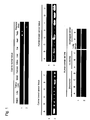
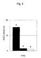

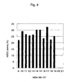


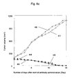
Classifications
-
- C—CHEMISTRY; METALLURGY
- C07—ORGANIC CHEMISTRY
- C07K—PEPTIDES
- C07K16/00—Immunoglobulins [IGs], e.g. monoclonal or polyclonal antibodies
- C07K16/18—Immunoglobulins [IGs], e.g. monoclonal or polyclonal antibodies against material from animals or humans
- C07K16/28—Immunoglobulins [IGs], e.g. monoclonal or polyclonal antibodies against material from animals or humans against receptors, cell surface antigens or cell surface determinants
- C07K16/30—Immunoglobulins [IGs], e.g. monoclonal or polyclonal antibodies against material from animals or humans against receptors, cell surface antigens or cell surface determinants from tumour cells
- C07K16/3015—Breast
-
- A—HUMAN NECESSITIES
- A61—MEDICAL OR VETERINARY SCIENCE; HYGIENE
- A61K—PREPARATIONS FOR MEDICAL, DENTAL OR TOILETRY PURPOSES
- A61K39/00—Medicinal preparations containing antigens or antibodies
- A61K39/395—Antibodies; Immunoglobulins; Immune serum, e.g. antilymphocytic serum
- A61K39/39533—Antibodies; Immunoglobulins; Immune serum, e.g. antilymphocytic serum against materials from animals
- A61K39/39558—Antibodies; Immunoglobulins; Immune serum, e.g. antilymphocytic serum against materials from animals against tumor tissues, cells, antigens
-
- A—HUMAN NECESSITIES
- A61—MEDICAL OR VETERINARY SCIENCE; HYGIENE
- A61P—SPECIFIC THERAPEUTIC ACTIVITY OF CHEMICAL COMPOUNDS OR MEDICINAL PREPARATIONS
- A61P1/00—Drugs for disorders of the alimentary tract or the digestive system
- A61P1/04—Drugs for disorders of the alimentary tract or the digestive system for ulcers, gastritis or reflux esophagitis, e.g. antacids, inhibitors of acid secretion, mucosal protectants
-
- A—HUMAN NECESSITIES
- A61—MEDICAL OR VETERINARY SCIENCE; HYGIENE
- A61P—SPECIFIC THERAPEUTIC ACTIVITY OF CHEMICAL COMPOUNDS OR MEDICINAL PREPARATIONS
- A61P11/00—Drugs for disorders of the respiratory system
-
- A—HUMAN NECESSITIES
- A61—MEDICAL OR VETERINARY SCIENCE; HYGIENE
- A61P—SPECIFIC THERAPEUTIC ACTIVITY OF CHEMICAL COMPOUNDS OR MEDICINAL PREPARATIONS
- A61P15/00—Drugs for genital or sexual disorders; Contraceptives
-
- A—HUMAN NECESSITIES
- A61—MEDICAL OR VETERINARY SCIENCE; HYGIENE
- A61P—SPECIFIC THERAPEUTIC ACTIVITY OF CHEMICAL COMPOUNDS OR MEDICINAL PREPARATIONS
- A61P25/00—Drugs for disorders of the nervous system
-
- A—HUMAN NECESSITIES
- A61—MEDICAL OR VETERINARY SCIENCE; HYGIENE
- A61P—SPECIFIC THERAPEUTIC ACTIVITY OF CHEMICAL COMPOUNDS OR MEDICINAL PREPARATIONS
- A61P35/00—Antineoplastic agents
-
- A—HUMAN NECESSITIES
- A61—MEDICAL OR VETERINARY SCIENCE; HYGIENE
- A61P—SPECIFIC THERAPEUTIC ACTIVITY OF CHEMICAL COMPOUNDS OR MEDICINAL PREPARATIONS
- A61P35/00—Antineoplastic agents
- A61P35/02—Antineoplastic agents specific for leukemia
-
- C—CHEMISTRY; METALLURGY
- C07—ORGANIC CHEMISTRY
- C07K—PEPTIDES
- C07K16/00—Immunoglobulins [IGs], e.g. monoclonal or polyclonal antibodies
- C07K16/18—Immunoglobulins [IGs], e.g. monoclonal or polyclonal antibodies against material from animals or humans
- C07K16/28—Immunoglobulins [IGs], e.g. monoclonal or polyclonal antibodies against material from animals or humans against receptors, cell surface antigens or cell surface determinants
- C07K16/30—Immunoglobulins [IGs], e.g. monoclonal or polyclonal antibodies against material from animals or humans against receptors, cell surface antigens or cell surface determinants from tumour cells
- C07K16/3053—Skin, nerves, brain
-
- A—HUMAN NECESSITIES
- A61—MEDICAL OR VETERINARY SCIENCE; HYGIENE
- A61K—PREPARATIONS FOR MEDICAL, DENTAL OR TOILETRY PURPOSES
- A61K39/00—Medicinal preparations containing antigens or antibodies
- A61K2039/505—Medicinal preparations containing antigens or antibodies comprising antibodies
-
- C—CHEMISTRY; METALLURGY
- C07—ORGANIC CHEMISTRY
- C07K—PEPTIDES
- C07K2317/00—Immunoglobulins specific features
- C07K2317/20—Immunoglobulins specific features characterized by taxonomic origin
- C07K2317/24—Immunoglobulins specific features characterized by taxonomic origin containing regions, domains or residues from different species, e.g. chimeric, humanized or veneered
-
- C—CHEMISTRY; METALLURGY
- C07—ORGANIC CHEMISTRY
- C07K—PEPTIDES
- C07K2317/00—Immunoglobulins specific features
- C07K2317/30—Immunoglobulins specific features characterized by aspects of specificity or valency
- C07K2317/34—Identification of a linear epitope shorter than 20 amino acid residues or of a conformational epitope defined by amino acid residues
-
- C—CHEMISTRY; METALLURGY
- C07—ORGANIC CHEMISTRY
- C07K—PEPTIDES
- C07K2317/00—Immunoglobulins specific features
- C07K2317/50—Immunoglobulins specific features characterized by immunoglobulin fragments
- C07K2317/56—Immunoglobulins specific features characterized by immunoglobulin fragments variable (Fv) region, i.e. VH and/or VL
- C07K2317/565—Complementarity determining region [CDR]
-
- C—CHEMISTRY; METALLURGY
- C07—ORGANIC CHEMISTRY
- C07K—PEPTIDES
- C07K2317/00—Immunoglobulins specific features
- C07K2317/70—Immunoglobulins specific features characterized by effect upon binding to a cell or to an antigen
- C07K2317/73—Inducing cell death, e.g. apoptosis, necrosis or inhibition of cell proliferation
- C07K2317/732—Antibody-dependent cellular cytotoxicity [ADCC]
-
- C—CHEMISTRY; METALLURGY
- C07—ORGANIC CHEMISTRY
- C07K—PEPTIDES
- C07K2317/00—Immunoglobulins specific features
- C07K2317/70—Immunoglobulins specific features characterized by effect upon binding to a cell or to an antigen
- C07K2317/73—Inducing cell death, e.g. apoptosis, necrosis or inhibition of cell proliferation
- C07K2317/734—Complement-dependent cytotoxicity [CDC]
Definitions
- the present invention relates to a novel medical use of antibodies to CAPRIN-1 or fragments thereof as, for example, therapeutic and/or preventive agents for cancer.
- Cancer is the leading cause of death.
- Treatment currently performed for cancer is mainly surgical therapy, which can be combined with radiation therapy or chemotherapy.
- treatment results of cancers are not greatly improved at present except for some cancers.
- antibodies that are specifically reactive with cancers, cancer antigens recognized by cytotoxic T cells, as well as the genes encoding the cancer antigens have been identified, and expectations for specific immunotherapies targeting cancer antigens have been raised ( Tsuyoshi AKIYOSHI, "Gan To Kagaku-Ryoho (Cancer and Chemotherapy),” 1997, vol. 24, pp. 551-519 (Jp) (Cancer and Chemotherapy Publishers, Inc., Japan )).
- Cytoplasmic-and proliferation-associated protein 1 is an intracellular protein that is expressed when normal cells in resting phase are activated or undergo cell division.
- CAPRIN-1 is also known to be involved in the regulation of the transport and translation of mRNAs through formation of ctytoplasmic stress granules with RNA in a cell.
- CAPRIN-1 has different names, such as GPI-anchored membrane protein 1 and membrane component surface marker 1 protein (M11S1), as if this protein is known to be a membrane protein. These different names are derived from the report ( J. Biol.
- CAPRIN-1 originally has a GPI-binding region and CAPRIN-1 is a membrane protein expressed in colon cancer cells. It was later reported that the CAPRIN-1 gene sequence described in said report was not correct; i.e., a frame shift took place by deletion of a single nucleotide from the CAPRIN-1 gene sequence currently registered with GenBank or the like, so that 80 amino acids were deleted from the C-terminus and the resulting artifact (74 amino acids) was the GPI binding portion in the report; and another error was also present on the 5' side of the gene sequence, thereby resulting in deletion of 53 amino acids from the N-terminus ( J.
- CAPRIN-1 is a cell membrane protein
- US2008/0075722 and WO2005/100998 disclose that CAPRIN-1 under the name of M11S1 can be used for cancer therapy as a target of antibody medicines for cancer therapy and as one of cell membrane proteins; however, the Examples contain no description of the cancer therapy using an antibody against the protein.
- J. Biol. Chem., 270: 20717-20723 1995 that CAPRIN-1 is a cell membrane protein
- US2008/0075722 and WO2005/100998 disclose that CAPRIN-1 under the name of M11S1 can be used for cancer therapy as a target of antibody medicines for cancer therapy and as one of cell membrane proteins; however, the Examples contain no description of the cancer therapy using an antibody against the protein.
- An object of the present invention is to identify cancer antigen proteins specifically expressed on the surface of cancer cells and to provide a use of antibodies targeting such proteins as therapeutic and/orpreventive (or prophylactic) agents for cancer.
- the present inventors have now obtained cDNA encoding a protein that binds to an antibody present in the serum from a tumor-bearing organism by the SEREX method using testis tissue-derived cDNA libraries and sera from dogs with breast cancer.
- CAPRIN-1 proteins having amino acid sequences shown in the even numbers of SEQ ID NOS: 2 to 30 (i.e., even-numbered SEQ ID NOS: 2 to 30) and antibodies against the CAPRIN-1 proteins have now been prepared.
- CAPRIN-1 is specifically expressed in the cells of breast cancer, brain tumor, leukemia, lymphoma, lung cancer, esophageal cancer, colon cancer, gastric cancer, and kidney cancer, and that portions of the CAPRIN-1 proteins are specifically expressed on the surface of such cancer cells. Further, the present inventors have now found that antibodies against the CAPRIN-1 portions expressed on cancer cell surfaces can damage (or impair) cancer cells expressing CAPRIN-1. These findings have led to the completion of the present invention.
- the invention provides antibodies or fragments thereof and pharmaceutical compositions, all for use in a method for the treatment and/or prevention of cancer, and all as defined in the claims.
- Antibodies against CAPRIN-1 used in the present invention damage (or impair) cancer cells. Therefore, such antibodies against CAPRIN-1 are useful for treatment or prevention of cancers.
- the antitumor activity of antibodies to the polypeptide shown in any one of the even-numbered SEQ ID NOS: 2 to 30 used in the present invention can be evaluated by examining in vivo the inhibition of tumor growth in a tumor-bearing animal, or by examing in vitro whether or not immunocyte- or complement-mediated cytotoxic activity against tumor cells expressing the polypeptide is exhibited.
- nucleotide sequences of polynucleotides encoding the proteins consisting of the amino acid sequences shown in the even-numbered SEQ ID NOS: 2 to 30 are shown in the odd-numbered SEQ ID NOS: 1 to 29 (i.e., SEQ ID NOS: 1, 3, 5...27, and 29), respectively.
- amino acid sequences shown in SEQ ID NOS: 6, 8, 10, 12 and 14 in the Sequence Listing disclosed according to the present invention are the amino acid sequences of the CPRIN-1 protains, which were isolated, by the SEREX method using canine testis tissue-derived cDNA libraries and sera from dogs with breast cancer, as polypeptides capable of binding to antibodies specifically existing in the sera from tumor-bearing dogs;
- amino acid sequences shown in SEQ ID NOS: 2 and 4 are the amino acid sequences of the CPRIN-1 protains isolated as human homologs of said dog polypeptides;
- amino acid sequence shown in SEQ ID NO: 16 is the amino acid sequence of the CPRIN-1 protain isolated as a bovine homolog of said dog polypeptide;
- amino acid sequence shown in SEQ ID NO: 18 is the amino acid sequence of the CPRIN-1 protain isolated as an equine homolog of said dog polypeptide;
- amino acid sequences shown in (even-numbered) SEQ ID NOS: 20 to 28 are
- CAPRIN-1 was not expressed on the surface of cells.
- an antibody that binds to a portion within CAPRIN-1 protein expressed on cancer cell surfaces is preferably used.
- the partial peptides within CAPRIN-1 protein expressed on cancer cell surfaces include polypeptides consisting of a sequence of 7 or more consecutive amino acids in the region of the amino acid residue Nos. (or the amino acids (aa)) 50-98 or the amino acid residue Nos.
- Specific examples thereof include the amino acid sequence shown in SEQ ID NO: 37 or 136 (preferably, the region of the amino acid seqeunce shown in SEQ ID NO: 137 or 138 in the amino acid sequence shown in SEQ ID NO: 136).
- Antibodies of the present invention include all antibodies capable of binding to the above peptides and having antitumor activity.
- the antibodies to CAPRIN-1 usable in the present invention as described above may be any types thereof, as long as they can exhibit antitumor activity. Examples thereof include monoclonal antibodies, polyclonal antibodies, synthetic antibodies, multispecific antibodies, human antibodies, humanized antibodies, chimeric antibodies, single-chain antibodies (scFV), and fragments thereof such as Fab and F(ab') 2 . These antibodies and fragments thereof can be prepared by methods known to persons skilled in the art.
- antibodies capable of specifically binding to a CAPRIN-1 protein are desirable. Such antibodies are preferably monoclonal antibodies; however, as long as homogenous antibodies can be stably produced, polyclonal antibodies may also be used.
- a human antibody or a humanized antibody is desirable in order to avoid or inhibit the immunorejection.
- the word "specifically binding to a CAPRIN-1 protein” as used herein means that an antibody of interest specifically binds to the CAPRIN-1 protein and does not substantially bind to other proteins.
- the antitumor activity of an antibody used in the present invention can be evaluated by examining in vivo the inhibition of tumor growth in a tumor-bearing animal, or examing in vitro whether or not the immunocyte- or complement-mediated cytotoxic activity against tumor cells expressing the polypeptide is exhibited.
- the subjects in need of treatment and/or prevention of cancer accordnig to the the present invention are mammals such as human, pet animals, livestock animals, or sport animals.
- the preferred subject is a human.
- Proteins or fragments thereof used as sensitizing antigens for obtaining antibodies to CAPRIN-1 used in the present invention are not limited in terms of their origins such as animals including, for example, humans, canines, bovines, horses, mice, rats, and chickens. However, such proteins or fragments thereof are preferably selected in view of compatibility with parent cells used for cell fusion. Mammal-derived proteins are generally preferable and human-derived proteins are particularly preferable. For instance, if the CAPRIN-1 is human CAPRIN-1, a human CAPRIN-1 protein, a partial peptide thereof, or cells capable of expressing human CAPRIN-1 can be used.
- Nucleotide sequences and amino acid sequences of human CAPRIN-1 and homologs thereof can be obtained by, for example, accessing GenBank (NCBI, USA) and using the BLAST or FASTA algorithm ( Karlin and Altschul, Proc. Natl. Acad. Sci. USA, 90:5873-5877,1993 ; Altschul et al., Nucleic Acids Res. 25:3389-3402, 1997 ).
- targets are nucleic acids or proteins each consisting of a sequence having 70% to 100%, preferably 80% to 100%, more preferably 90% to 100%, and further preferably 95% to 100% (e.g., 97% to 100%, 98% to 100%, 99% to 100%, or 99.5% to 100%) sequence identity with the nucleotide sequence or amino acid sequence of the ORF or mature portion of the base nucleotide sequence or amino acid sequence.
- % sequence identity means a percentage (%) of the number of identical amino acids (or nucleotides) to the total number of amino acids (or nucleotides) in the case that two sequences are aligned such that maximum similarity can be achieved with or without introduction of gaps.
- Fragments of a CAPRIN-1 protein have lengths ranging from the amino acid length of an epitope (or an antigenic determinant), which is the smallest unit of an antigen recognized by an antibody, to less than the full-length of the protein.
- the epitope refers to a polypeptide fragment having antigenicity or immunogenicity in mammals and preferably in humans.
- the smallest unit of polypeptide fragment consists of approximately 7 to 12 amino acids, and for example, 8 to 11 amino acids.
- a specific example thereof is the amino acid sequence shown in SEQ ID NO: 37, SEQ ID NO: 137, or SEQ ID NO: 138.
- Polypeptides comprising the aforementioned human CAPRIN-1 protein and partial peptides thereof can be synthesized according to chemical synthesis methods such as the Fmoc method (fluorenylmethyloxycarbonyl method) or the tBoc method (t-butyloxycarbonyl method) (the Japanese Biochemical Society (ed.), "Biochemical Experimentation Course (Seikagaku Jikken Koza) 1," Protein Chemistry IV, Chemical Modification and Peptide Synthesis, Kagaku-dojin Publishing Company, Inc. (Japan), 1981 ). Also, they can be synthesized by general methods using a variety of commercially available peptide synthesizers.
- chemical synthesis methods such as the Fmoc method (fluorenylmethyloxycarbonyl method) or the tBoc method (t-butyloxycarbonyl method) (the Japanese Biochemical Society (ed.), "Biochemical Experimentation Course (Seikagaku Jikken Koza) 1," Protein Chemistry IV,
- polipeptides of interest can be obtained by preparing polynucleotides encoding the above polypetides using known gene engineering methods ( Sambrook et al., Molecular Cloning, 2nd edition, Current Protocols in Molecular Biology (1989), Cold Spring Harbor Laboratory Press ; Ausubel et al., Short Protocols in Molecular Biology, 3rd edition, A Compendium of Methods from Current Protocols in Molecular Biology (1995), John Wiley & Sons , etc.), incorporating each of the polynucleotides into an expression vector and introducing the vector into a host cell, thereby allowing the host cell to produce the polypeptide.
- the dseired polypeptides can be obtained.
- Polynucleotides encoding the aforementioned polypeptides can be readily prepared by known gene engineering techniques or general methods using commercially available nucleic acid synthesizers.
- DNA comprising the nucleotide sequence shown in SEQ ID NO: 1 can be prepared by PCR using a human chromosome DNA or cDNA library as a template and a pair of primers designed to enable the amplification of the nucleotide sequence shown in SEQ ID NO: 1. PCR conditions can be appropriately determined.
- such conditions may comprise conducting 30 cycles of the reaction steps (as one cycle) consisitng of: 94°C, 30 seconds (denaturation); 55°C, 30 seconds to 1 minute (annealing); and 72°C,r 2 minutes (elongation) using a thermostable DNA polymerase (e.g., Taq polymerase) and a Mg 2+ -containing PCR buffer, followed by reaction at 72°C for 7 minutes after completion of the 30 cyles.
- a thermostable DNA polymerase e.g., Taq polymerase
- Mg 2+ -containing PCR buffer e.g., Mg 2+ -containing PCR buffer
- desired DNA can be isolated by preparing appropriate probes and primers based on information about the nucleotide and amino acid sequences shown in SEQ ID NOS: 1 to 30 in the Sequence Listing described herein, and screening a human cDNA library or the like with the use of such probes and primers.
- cDNA library is produced from a cell, organ, or tissue in which the protein with any one of the even-numbered SEQ ID NOS: 2 to 30 is expressed.
- the cell or tissue include cells or tissues from testis and cancers or tumors, such as leukemia, breast cancer, lymphoma, brain tumor, lung cancer, and colon cancer.
- the above-described host cells may be any cells, as long as they can express the above-described polypeptides.
- An example of prokaryotic host cell includes, but is not limited to, Escherichia coli.
- Examples of eukaryotic host cells include, but are not limited to, mammalian cells such as monkey kidney cell (COS1), Chinese hamster ovary cell (CHO), human embryonic kidney cell line (HEK293), and mouse embryonic skin cell line (NIH3T3), yeast cells such as budding yeast and dividing yeast cells, silkworm cells, and Xenopus egg cells.
- an expression vector having an origin replicable in prokaryotic cells, a promoter, a ribosome-binding site, a multicloning site, a terminator, a drug resistance gene, an auxotrophic complementary gene, or the like can be used.
- expression vectors for Escherichia coli pUC vectors, pBluescriptII, pET expression systems, pGEX expression systems, and the like can be exemplified.
- a DNA encoding the above polypeptide is incorporated into such an expression vector, a prokaryotic host cell is transformed with the vector, and then the thus obtained transformed cell is cultured, so that the polypeptide encoded by the DNA can be expressed in the prokaryotic host cell.
- the polypeptide can also be expressed as a fusion protein with another protein.
- expression vectors for eukaryotic cells having a promoter, a splicing region, a poly(A) addition site, or the like can be used.
- expression vectors include pKA1, pCDM8, pSVK3, pMSG, pSVL, pBK-CMV, pBK-RSV, EBV vector, pRS, pcDNA3, and pYES2.
- a DNA encoding the aforementioned polypeptide is incorporated into such an expression vector, an eukaryotic host cell is transformed with the vector, and then the thus obtained transformed cell is cultured, so that the polypeptide encoded by the above DNA can be expressed in the eukaryotic host cell.
- the above polypeptide may be expressed as a fusion protein with a tag, such as His tag (e.g., (His) 6 to (His) 10 ), FLAG tag, myc tag, HA tag, or GFP.
- an expression vector For introduction of an expression vector into a host cell, well known methods can be employed, such as electroporation, a calcium phosphate method, a liposome method, a DEAE dextran method, microinjection, viral infection, lipofection, and binding with a cell-membrane-permeable peptide.
- Isolation and purification of a polypeptide of interest from host cells can be performed using known isolation techniques in combination.
- known techniques include, but are not limited to, treatment using a denaturing agent such as urea or a surfactant, ultrasonication, enzymatic digestion, salting-out, solvent fractionation and precipitation, dialysis, centrifugation, ultrafiltration, gel filtration, SDS-PAGE, isoelectric focusing electrophoresis, ion exchange chromatography, hydrophobic chromatography, affinity chromatography, and reverse phase chromatography.
- antibodies are heteromultimeric glycoproteins each comprising at least two heavy chains and two light chains.
- antibodies except for IgM are heterotetrameric glycoproteins (approximately 150 kDa) each comprising two identical light (L) chains and two identical heavy (H) chains.
- each light chain is connected to a heavy chain via a single covalent disulfide bond.
- the number of disulfide bonds between heavy chains varies among different immunogloblin isotypes.
- Each of heavy chain and light chain also has an intrachain disulfide bond(s).
- Each heavy chain has a variable domain (VH region) at one end thereof, to which some constant regions are bound in series.
- Each light chain has a variable domain (VL region) at one end thereof and has a single constant region at the opposite end thereof.
- the constant region of a light chain is aligned with the first constant region of a heavy chain and the light-chain variable domain is aligned with the heavy-chain variable domain.
- a specific region of an antibody variable domain which is called “complementarity determining region (CDR)," exhibits specific variability so as to impart binding specificity to an antibody.
- CDR complementarity determining region
- a relatively conserved portion in a variable region is called a "framework region (FR)."
- a complete heavy-chain or light-chain viariable domain comprises 4 FRs connected to each other via 3 CDRs.
- CDRH1, CDRH2
- CDRH3 CDRH3 plays the most important role in terms of antibody-antigen binding specificity.
- CDRs in each chain are retained by FR regions in the state that they are close to each other, and they contribute to the formation of antibody-antigen binding sites with CDRs in a corresponding chain. Constant regions do not directly contribute to antibody-antigen binding.
- ADCC antibody-dependent cell-mediated cytotoxicity
- FcRn neonatal Fc receptor
- CDC complement-dependent cytotoxicity
- anti-CAPRIN-1 antibody used in the present invention refers to an antibody having an immunological reactivity with a full-length CAPRIN-1 protein or a fragment thereof, as defined in the claims.
- immunological reactivity indicates the characteristics of an anbibody binding in vivo to a CAPRIN-1 antigen.
- the tumor-damaging function e.g., death, inhibition, or regression
- any type of antibody may be used in the present invention as long as the antibody can bind to a CAPRIN-1 protein to damage a tumor or a cancer such as leukemia, lymphoma, breast cancer, brain tumor, lung cancer, esophageal cancer, gastric cancer, kidney cancer, or colon cancer.
- antibodies examples include monoclonal antibodies, polyclonal antibodies, synthetic antibodies, multispecific antibodies, human antibodies, humanized antibodies, chimeric antibodies, single-chain antibodies, and antibody fragments (e.g., Fab and F(ab') 2 ).
- examples of arbitrary immunoglobulin classes of such antibodies include IgG, IgE, IgM, IgA, IgD, and IgY, and examples of arbitrary immunoglobulin subclasses include IgG1, IgG2, IgG3, IgG4, IgA1, and IgA2.
- Antibodies may be further modified via acetylation, formylation, amidation, phosphorylation, or pegylation (PEG), in addition to glycosylation.
- acetylation formylation, amidation, phosphorylation, or pegylation (PEG), in addition to glycosylation.
- PEG pegylation
- an antibody of interest is a monoclonal antibody
- a breast cancer SK-BR-3 cell line expressing CAPRIN-1 or the like is administered to mice for immunization, followed by extraction of spleens from the mice. Cells are separated from each spleen and then are fused with mouse myeloma cells. Clones capable of producing an antibody having cancer cell growth inhibition action are selected from the obtained fusion cells (hybridomas). A monoclonal antibody-producing hybridoma having cancer cell growth inhibition action is isolated and cultured.
- An antibody of interest can be prepared via purification from the culture supernatant by a general affinity purification method.
- a monoclonal antibody-producing hybridoma can be produced in a manner described below, for example.
- an animal is immunized with a sensitizing antigen by a known method.
- immunization is carried out by intraperitoneally or subcutaneously injecting a sensitizing antigen into a mammal.
- a sensitizing antigen is diluted with or suspended in PBS (Phosphate-Buffered Saline), physiological saline, or the like to an appropriate resultant amount.
- PBS Phosphate-Buffered Saline
- physiological saline or the like
- an appropriate amount of a conventional adjuvant e.g., Freund's complete adjuvant
- the resultant is administered to a mammal several times every 4 to 21 days.
- an adequate carrier can be used for immunization with a sensitizing antigen.
- immunocytes are collected from the mammal and subjected to cell fusion.
- immunocytes are splenocytes.
- Mammalian myeloma cells are used as relevant parent cells subjected to fusion with the above immunocytes.
- myeloma cells the following various examples of known cell lines are preferably used: P3U1 (P3-X63Ag8U1), P3 (P3x63Ag8.653) ( J. Immunol. (1979) 123, 1548-1550 ), P3x63Ag8U.1 ( Current Topics in Microbiology and Immunology (1978) 81, 1-7 ), NS-1 ( Kohler. G. and Milstein, C. Eur. J. Immunol. (1976). 6, 511-519 ), MPC-11 ( Margulies. D. H.
- cell fusion of immunocytes and myeloma cells described above can be carried out according to a known method such as the method of Kohler and Milstein et al. ( Kohler, G. and Milstein, C. Methods Enzymol. (1981) 73, 3-46 ).
- cell fusion described above is carried out in the presence of a cell fusion promoter in a conventional nutrients-containing culture solution, for example.
- a fusion promoter to be used include polyethylene glycol (PEG) and Sendai virus (HVJ: hemagglutinating virus of Japan).
- PEG polyethylene glycol
- HVJ Sendai virus
- an adjuvant such as dimethylsulfoxide may be further added for improvement of fusion efficiency.
- the proportion of immunocytes used to that of myeloma cells used can be arbitrarily determined.
- the ratio of immunocytes to myeloma cells is preferably 1:1 to 10:1.
- Examples of a culture solution that can be used for cell fusion described above include an RPMI1640 culture solution and an MEM culture solution adequate for growth of the above myeloma cell lines as well as other conventional culture solutions used for this kind of cell culture.
- a serum replacement such as fetal calf serum (FCS) can be used in combination therewith.
- the above immunocytes and myeloma cells are sufficiently mixed at predetermined amounts in the culture solution.
- a PEG solution e.g., average molecular weight: approximately 1000 to 6000
- a concentration of generally 30% to 60% (w/v) is added thereto at a concentration of generally 30% to 60% (w/v), followed by mixing.
- operational steps of sequential addition of an appropriate culture solution and removal of the supernatant via centrifugation are repeatedly carried out to remove cell fusion agent(s) and the like that are not preferable for the growth of hybridomas.
- hybridomas are cultured in a conventional selection culture solution such as an HAT culture solution (a culture solution comprising hypoxanthine, aminopterin, and thymidine) for selection.
- a conventional selection culture solution such as an HAT culture solution (a culture solution comprising hypoxanthine, aminopterin, and thymidine) for selection.
- Culture in such an HAT culture solution is continuously carried out for a sufficient time period (generally several days to several weeks) for death of cells (non-fused cells) other than hybridomas of interest.
- a conventional limiting dilution method is employed to screen for hybridomas producing antibodies of interest and to carry out single cloning.
- human antibody-producing hybridomas having desired activity e.g., cell growth inhibition activity
- desired activity e.g., cell growth inhibition activity
- Human lymphocytes e.g., human lymphocytes infected with EB virus
- sensitized lymphocytes are fused with human-derived myeloma cells having the ability to parmanently divide (e.g., U266) (registration no. TIB 196).
- Monoclonal antibody-producing hybridomas produced as above can be subcultured in a conventional culture solution. In addition, they can be preserved in liquid nitrogen for a long period of time.
- immunization is carried out using a desired antigen or cells expressing a desired antigen as sensitizing antigen(s) according to a conventional immunization method.
- the obtained immunocytes are fused with known parent cells by a conventional cell fusion method.
- monoclonal antibody-producing cells are screened for by a conventional screening method.
- antibody production can be carried out.
- polyclonal antibodies can be used in a manner described below.
- Serum is obtained by immunizing small animals such as mice, human antibody-producing mice, or rabbits with a naturally occurring CAPRIN-1 protein, a recombinant CAPRIN-1 protein that has been expressed as a protein fused with GST or the like in a microorganism such as Escherichia coli, or a partial peptide thereof.
- the serum is purified via ammonium sulfate precipitation, protein A/protein G column chromatography, DEAE ion-exchange chromatography, affinity column chromatography with a coulumn to which a CAPRIN-1 protein or a synthetic peptide is coupled, or a similar technique for preparation of polyclonal antibodies.
- a rabbit polyclonal antibody was produced, and antitumor effects thereof were confirmed, such antibody being against a partial peptide (with the sequence shown in SEQ ID NO: 37) of a domain in a CAPRIN-1 protein amino acid sequence that is expressed on cancer cell surfaces.
- a known human antibody-producing mouse used herein is, for example, a KM Mouse (Kirin Pharma/Medarex) or a XenoMouse (Amgen) (e.g., WO02/43478 and WO02/092812 ).
- KM Mouse Kirin Pharma/Medarex
- Amgen XenoMouse
- human monoclonal antibodies can be produced by a method of fusing splenocytes collected from immunized mice with myeloma cells.
- Antigen preparation can be carried out in accordance with a method such as a method using animal cells ( JP Patent Publication (Kohyo) No. 2007-530068 ) or a method using a baculovirus (e.g., WO98/46777 ). If the immunogenicity of an antigen is low, an antigen is bound to a macromolecule having immunogenicity, such as albumin. Then, the antigen can be used for immunization.
- a method using animal cells JP Patent Publication (Kohyo) No. 2007-530068
- a method using a baculovirus e.g., WO98/46777 .
- a gene recombinant antibody produced by cloning an antibody gene from a hybridoma incorporating the clone into an adequate vector, introducing the vector into a host, and using a gene recombinant technique.
- a gene recombinant technique See, for example, Carl, A. K. Borrebaeck, James, W. Larrick, THERAPEUTIC MONOCLONAL ANTIBODIES, Published in the United Kingdom by MACMILLAN PUBLISHERS LTD, 1990 .
- V region variable region of an antibody is synthesized from mRNA of a hybridoma with the use of a reverse transcriptase.
- DNA encoding a V region of an antibody of interest is obtained, such DNA is ligated to desired DNA encoding an antibody constant region (C region).
- C region an antibody constant region
- the resultant is incoporated into an expression vector.
- DNA encoding an antibody V region may be incorporated into an expression vector comprising DNA of an antibody C region.
- Such DNA is incorporated into an expression vector in a manner such that it is expressed under control of an expression control region such as an enhancer or a promoter.
- host cells are transformed with such expression vector, thereby allowing the antibody to be expressed.
- Anti-CAPRIN-1 antibodies of the present invention are preferably monoclonal antibodies. However, they may be polyclonal antibodies, gene-modified antibodies (such as chimeric antibodies and humanized antibodies), and the like.
- Monoclonal antibodies include human monoclonal antibodies and non-human animal monoclonal antibodies (e.g., mouse monoclonal antibodies, rat monoclonal antibodies, rabbit monoclonal antibodies, and chicken monoclonal antibodies).
- Monoclonal antibodies can be produced by culturing hybridomas obtained via fusion of myeloma cells and splenocytes from non-human mammals (e.g., mice or human antibody-producing mice) immunized with CAPRIN-1 proteins. In the Examples described below, mouse monoclonal antibodies were produced and antitumor effects thereof were confirmed.
- Such a monoclonal antibody comprises a heavy-chain variable (VH) region having the amino acid sequence shown in SEQ ID NO: 43, SEQ ID NO: 73, SEQ ID NO: 83, SEQ ID NO: 93, SEQ ID NO: 103, SEQ ID NO: 113, or SEQ ID NO: 123 and a light-chain variable (VL) region having the amino acid sequence shown in SEQ ID NO: 47, SEQ ID NO: 53, SEQ ID NO: 58, SEQ ID NO: 63, SEQ ID NO: 68, SEQ ID NO: 77, SEQ ID NO: 87, SEQ ID NO: 97, SEQ ID NO: 107, SEQ ID NO: 117, or SEQ ID NO: 127.
- VH heavy-chain variable
- VL light-chain variable
- the VH region comprises: CDR1 represented by the amino acid sequence of SEQ ID NO: 40, SEQ ID NO: 70, SEQ ID NO: 80, SEQ ID NO: 90, SEQ ID NO: 100, SEQ ID NO: 110, or SEQ ID NO: 120; CDR2 represented by the amino acid sequence of SEQ ID NO: 41, SEQ ID NO: 71, SEQ ID NO: 81, SEQ ID NO: 91, SEQ ID NO: 101, SEQ ID NO: 111, or SEQ ID NO: 121; and CDR3 represented by the amino acid sequence of SEQ ID NO: 42, SEQ ID NO: 72, SEQ ID NO: 82, SEQ ID NO: 92, SEQ ID NO: 102, SEQ ID NO: 112, or SEQ ID NO: 122.
- CDR1 represented by the amino acid sequence of SEQ ID NO: 40, SEQ ID NO: 70, SEQ ID NO: 80, SEQ ID NO: 90, SEQ ID NO: 100, SEQ ID NO: 110, or SEQ ID
- the VL region comprises: CDR1 represented by the amino acid sequence of SEQ ID NO: 44, SEQ ID NO: 50, SEQ ID NO: 55, SEQ ID NO: 60, SEQ ID NO: 65, SEQ ID NO: 74, SEQ ID NO: 84, SEQ ID NO: 94, SEQ ID NO: 104, SEQ ID NO: 114, or SEQ ID NO: 124; CDR2 represented by the amino acid sequence of SEQ ID NO: 45, SEQ ID NO: 51, SEQ ID NO: 56, SEQ ID NO: 61, SEQ ID NO: 66, SEQ ID NO: 75, SEQ ID NO: 85, SEQ ID NO: 95, SEQ ID NO: 105, SEQ ID NO: 115, or SEQ ID NO: 125; and CDR3 represented by the amino acid sequence of SEQ ID NO: 46, SEQ ID NO: 52, SEQ ID NO: 57, SEQ ID NO: 62, SEQ ID NO: 67, SEQ ID NO: 76, SEQ ID NO
- a chimeric antibody is an antibody produced by combining sequences from different animals.
- An example thereof is an antibody consisting of mouse antibody heavy-chain and light-chain variable regions and human antibody heavy-chain and light-chain constant regions.
- Such a chimeric antibody can be produced by a known method. For example, it can be obtained by ligating DNA encoding an antibody V region to DNA encoding a human antibody C region, incorporating the resultant into an expression vector, and introducing the vector into a host for antibody production.
- Polyclonal antibodies include antibodies obtained by immunizing human antibody-producing animals (e.g., mice) with CAPRIN-1 proteins.
- a humanized antibody is a modified antibody, and it is sometimes referred to as a "reshaped human antibody.” It is known that a humanized antibody is constructed by transplanting CDRs of an immunized animal-derived antibody into complementarity determining regions of a human antibody. Also, a general gene recombinant technique therefor is known.
- a DNA sequence designed in a manner that allows mouse antibody CDRs to be ligated to human antibody framework regions (FRs) is synthesized by the PCR method using several oligonucleotides prepared in such a manner that the oligonucleotides have portions overlapping each other at one end of each thereof.
- a humanized antibody can be obtained by ligating the above obtained DNA to DNA encoding a human antibody constant region, incorporating the resultant into an expression vector, and introducing the vector into a host for antibody production (see EP-A-239400 and WO96/02576 ).
- Human antibody FRs ligated to each other via CDRs are selected on the assmption that complementarity determining regions can form a good antigen binding site.
- amino acids in framework regions of an antibody variable region may be substituted in such a manner that complementarity determining regions in a reshaped human antibody form an appropriate antigen binding site ( Sato K. et al., Cancer Research 1993, 53: 851-856 ).
- the framework regions may be substituted with framework regions from a different human antibody (see WO99/51743 ).
- Human antibody framework regions ligated to each other via CDRs are selected on the assmption that complementarity determining regions can form good antigen binding sites. If necessary, amino acids in framework regions of an antibody variable region may be substituted in such a manner that complementarity determining regions in reshaped human antibody form an appropriate antigen binding sites ( Sato K. et al., Cancer Research 1993, 53: 851-856 ).
- amino acids in a variable region e.g., FR
- a constant region e.g., FR
- amino acids in a variable region or a constant region may be substituted, for example, with different amino acids.
- the amino acid substitution is a substitution of, for example, less than 15, less than 10, not more than 8, not more than 7, not more than 6, not more than 5, not more than 4, not more than 3, or not more than 2 amino acids, preferably 1 to 5 amino acids, and more preferably 1 or 2 amino acids.
- a substituted antibody should be functionally equivelent to an unsubstituted antibody.
- the substitution is preferably a conservative amino acid substitution, which is a substitution between amino acids having similar characteristics in terms of charge, side chains, polarity, aromaticity, and the like.
- characteristically similar amino acids can be classified into the following types: basic amino acids (arginine, lysine, and histidine); acidic amino acids (aspartic acid and glutamic acid); uncharged polar amino acids (glycine, asparagine, glutamine, serine, threonine, cysteine, and tyrosine); nonpolar amino acids (leucine, isoleucine, alanine, valine, proline, phenylalanine, tryptophan, and methionine); branched-chain amino acids (threonine, valine, isoleucine); and aromatic amino acids (phenylalanine, tyrosine, tryptophan, and histidine).
- basic amino acids arginine, lysine, and histidine
- acidic amino acids aspartic acid and glutamic acid
- uncharged polar amino acids glycine, asparagine, glutamine, serine, threonine, cysteine, and tyros
- an antibody modifier is an antibody bound to a molecule such as polyethylene glycol (PEG).
- PEG polyethylene glycol
- substances that bind to an antibody are not limited.
- Such an antibody modifier can be obtained by chemically modifying an obtained antibody. A method of such modification has been already established in the field related to the present invention.
- the expression "functionally equivalent” used herein indicates a situation in which an antibody of interest has biological or biochemical activity similar to that of an antibody of the present invention. Specifically, such antibody has a function of damaging tumors and causes essentially no rejection reaction when applied to humans. An example of such activity is cell growth inhibition activity or binding activity.
- a known method for preparing a polypeptide functionally equivalent to a given polypeptide is a method comprising introducing a mutation into a polypeptide.
- a person skilled in the art can adequately introduce a mutation into an antibody of the present invention using a site-specific mutagenesis method ( Hashimoto-Gotoh, T. et al., (1995) Gene 152, 271-275 ; Zoller, MJ., and Smith, M. (1983) Methods Enzymol. 100, 468-500 ; Kramer, W. et al., (1984) Nucleic Acids Res. 12, 9441-9456 ; Kramer, W.
- An aforemenioned antibody capable of recognizing an epitope of a CAPRIN-1 protein recognized by an anti-CAPRIN-1 antibody can be obtained by a method known to persons skilled in the art. For example, it can be obtained by: a method comprising determining an epitope of a CAPRIN-1 protein recognized by an anti-CAPRIN-1 antibody by a general method (e.g., epitope mapping) and producing an antibody using a polypeptide having an amino acid sequence contained in the epitope as an immunogen; or a method comprising determining an epitope of an antibody produced by a general method and selecting an antibody having an epitope identical to an epitope of an anti-CAPRIN-1 antibody.
- epitope mapping e.g., epitope mapping
- the term "epitope” refers to a polypeptide fragment having antigenicity or immunogenicity in mammals and preferably in humans. The smallest unit threof consists of approximately 7 to 12 amino acids and preferably 8 to 11 amino acids.
- the affinity constant Ka (k on /k off ) of an antibody of the present invention is preferably at least 10 7 M -1 , at least 10 8 M -1 , at least 5 ⁇ 10 8 M -1 , at least 10 9 M -1 , at least 5 ⁇ 10 9 M -1 , at least 10 10 M -1 , at least 5 ⁇ 10 10 M -1 , at least 10 11 M -1 , at least 5 ⁇ 10 11 M -1 , at least 10 12 M -1 , or at least 10 13 M -1 .
- An antibody of the present invention can be conjugated with an antitumor agent. Binding between an antibody and an antitumor agent can be carried out via a spacer having a group reactive to an amino group, a carboxyl group, a hydroxy group, a thiol group, or the like (e.g., an imidyl succinate group, a formyl group, a 2-pyridyldithio group, a maleimidyl group, an alkoxycarbonyl group, or a hydroxy group).
- a spacer having a group reactive to an amino group, a carboxyl group, a hydroxy group, a thiol group, or the like (e.g., an imidyl succinate group, a formyl group, a 2-pyridyldithio group, a maleimidyl group, an alkoxycarbonyl group, or a hydroxy group).
- antitumor agents include the following antitumor agents known in references or the like: paclitaxel, doxorubicin, daunorubicin, cyclophosphamide, methotrexate, 5-fluorouracil, thiotepa, busulfan, improsulfan, piposulfan, benzodopa, carboquone, meturedopa, uredopa, altretamine, triethylenemelamine, triethylenephosphoramide, triethilenethiophosphoramide, trimethylolomelamine, bullatacin, bullatacinone, camptothecin, bryostatin, callystatin, cryptophycin 1, cryptophycin 8, dolastatin, duocarmycin, eleutherobin, pancratistatin, sarcodictyin, spongistatin, chlorambucil, chlornaphazine, cholophosphamide, estramustine, ifosf
- radioactive isotope such as 211 At, 131 I, 125 I, 90 Y, 186 Re, 188 Re, 153 Sm, 212 Bi, 32 P, 175 Lu, or 176 Lu known in references and the like to an antibody of the present invention. It is desirable for such radioactive isotopes to be effective for tumor treatment or diagnosis.
- An antibody of the present invention is an antibody having an immunological reactivity with CAPRIN-1 or an antibody capable of specifically recognizing CAPRIN-1.
- Such an antibody should be an antibody having a structure that allows a subject animal to which the antibody is administered to completely or almost completely avoid a rejection reaction. If the subject animal is a human, examples of the above antibody include human antibodies, humanized antibodies, chimeric antibodies (e.g., human-mouse chimeric antibodies), single-chain antibodies, and bispecific antibodies.
- Such an antibody is a recombinant antibody having human antibody-derived heavy-chain and light-chain variable regions, a recombinant antibody having heavy-chain and light-chain variable regions each consisting of non-human animal antibody-derived complementarity determining regions (CDR1, CDR2, and CDR3) and human antibody-derived framework regions, or a recombinant antibody having non-human animal antibody-derived heavy-chain and light-chain variable regions and human antibody-derived heavy-chain and light-chain constant regions.
- the first two antibodies are preferable.
- DNA encoding a monoclonal antibody against human CAPRIN-1 e.g., a human monoclonal antibody, a mouse monoclonal antibody, a rat monoclonal antibody, a rabbit monoclonal antibody, or a chicken monoclonal antibody
- DNA encoding a light-chain variable region and a heavy-chain variable region of the antibody are produced by an RT-PCR method or the like using the obtained clone as a template.
- sequences of a light-chain variable region and a heavy-chain variable region or the sequences of CDR1, CDR2, and CDR3 are determined by the Kabat EU numbering system ( Kabat et al., Sequences of Proteins of Immunological Interest, 5th Ed. Public Health Service, National Institute of Health, Bethesda, Md. (1991 )).
- DNAs encoding variable regions or DNAs encoding CDRs are produced by a gene recombinant technique ( Sambrook et al., Molecular Cloning A Laboratory Manual, Cold Spring Harbor Laboratory Press (1989 )) or a DNA synthesizer.
- the above human monoclonal antibody-producing hybridoma can be produced by immunizing a human antibody-producing animal (e.g., a mouse) with human CAPRIN-1 and fusing splenocytes from the spleen removed from the animal with myeloma cells.
- DNAs encoding human antibody-derived light-chain or heavy-chain variable regions and constant regions are produced by a gene recombinant technique or a DNA synthesizer.
- DNA in which the CDR coding sequences in a DNA encoding a human antibody-derived light-chain or heavy-chain variable region have been substituted with corresponding CDR coding sequences of an antibody from a non-human animal e.g., a mouse, a rat, or a chicken
- the DNA obtained as above is ligated to the DNA encoding a constant region of a human antibody-derived light chain or heavy chain.
- DNA encoidng a humanized antibody can be produced.
- DNA encoding an antibody light-chain or heavy-dhain variable region from a non-human animal is ligated to the DNA encoding a human antibody-derived light-chain or heavy-chain constant region.
- a non-human animal e.g., a mouse, a rat, or a chicken
- DNA encoding a chimeric antibody can be produced.
- a single-chain antibody is an antibody in which a heavy-chain variable region and a light-chain variable region are linearly ligated to each other via a linker.
- DNA encoding a single-chain antibody can be produced by binding DNA encoding a heavy-chain variable region, DNA encoding a linker, and a DNA encoding a light-chain variable region.
- a heavy-chain variable region and a light-chain variable region are those from a human antibody or those from a human antibody in which CDRs alone have been substituted with CDRs of an antibody from a non-human animal (e.g., a mouse, a rat, or a chicken).
- the linker consists of 12 to 19 amino acids. An example thereof is (G 4 S) 3 consisting of 15 amino acids ( G. B. Kim et al., Protein Engineering Design and Selection 2007, 20 (9): 425-432 ).
- a bispecific antibody is an antibody capable of specifically binding to two different epitopes in which, for example, DNA encoding a heavy-chain variable region A, DNA encoding a light-chain variable region B, DNA encoding a heavy-chain variable region B, and DNA encoding a light-chain variable region A are bound to each other in such order (provided that DNA encoding a light-chain variable region B and DNA encoding a heavy-chain variable region B are bound to each other via DNA encoding a linker described above).
- DNA encoding a bispecific antibody can be produced.
- both a heavy-chain variable region and a light-chain variable region are those from a human antibody or those from a human antibody in which CDRs alone have been substituted with CDRs of an antibody from a non-human animal (e.g., a mouse, a rat, or a chicken).
- Recombinant DNA produced as above is incorporated into one or a plurality of appropriate vector(s).
- Each such vector is introduced into a host cell (e.g., a mammal cell, a yeast cell, or an insect cell) for (co)expression.
- a host cell e.g., a mammal cell, a yeast cell, or an insect cell
- a recombinant antibody can be produced ( P. J. Delves., ANTIBODY PRODUCTION ESSENTIAL TECHNIQUES., 1997 WILEY, P. Shepherd and C. Dean., Monoclonal Antibodies., 2000 OXFORD UNIVERSITY PRESS ; J. W. Goding, Monoclonal Antibodies: Principles and Practice., 1993 ACADEMIC PRESS ).
- Examples of an antibody of the present invention produced by the above method include the following antibodies (a) to (k).
- a humanized antibody, a chimeric antibody, a single-chain antibody, or a bispecific antibody of the present invention is, for example, the folllowing antibody (i) or (ii) (an example of antibody (a) is described below).
- sequences of human antibody heavy-chain and light-chain constant and variable regions can be obtained from, for example, NCBI (U.S.A: GenBank, UniGene, etc.).
- sequences of human antibody heavy-chain and light-chain constant and variable regions can be obtained from, for example, NCBI (U.S.A: GenBank, UniGene, etc.).
- the following sequences can be used as reference sequences for the corresponding regions: the sequence with registration no. J00228 for a human IgG1 heavy-chain constant region; the sequence with registration no. J00230 for a human IgG2 heavy-chain constant region; the sequence with registration no. X03604 for a human IgG3 heavy-chain constant region; the sequence with registration no. K01316 for a human IgG4 heavy-chain constant region; the sequence with registration no. V00557, X64135, or X64133 for a human light-chain ⁇ constant region; and the sequence with registration no. X64132 or X64134 for a
- the above antibodies preferably have cytotoxic activity, thereby exhibiting antitumor effects.
- a hybridoma capable of producing a different human antibody or a non-human animal antibody (e.g., a mouse antibody) against human CAPRIN-1 is produced.
- a moloclonal antibody produced by the hybridoma is collected.
- a monoclonal antibody-producing hybridoma of interest is identified.
- DNAs encoding heavy-chain and light-chain variable regions of an antibody of interest are produced from the hybridoma for sequence determination. The DNAs are used for production of different antibodies.
- the above antibody of the present invention may be any one of antibodies (i) to (iv) above having a substitution, deletion, or addition of one or several (and preferably, 1 or 2) amino acid(s), particularly in a framework region sequence and/or a constant region sequence, as long as it has the specific property of specifically recognizing CAPRIN-1.
- severeal amino acids indicates 2 to 5 and preferably 2 or 3 amino acids.
- DNA encoding the above anbibody of the present invention DNA encoding a heavy chain or light chain of the antibody, or DNA encoding a heavy-chain or light-chain variable region of the antibody is also provided.
- examples of such DNA include: DNA encoding a heavy-chain variable region comprising nucleotide sequences encoding the amino acid sequences of SEQ ID NOS: 40, 41, and, 42; and DNA encoding a light-chain variable region comprising nucleotide sequences encoding the amino acid sequences of SEQ ID NOS: 44, 45, and 46.
- Complementarity determining regions (CDRs) encoded by DNAs of the above sequences are regions that determine antibody specificity. Therefore, sequences encoding the other regions (i.e., constant regions and framework regions) in an antibody may be sequences from a different antibody.
- different antibodies include antibodies from non-human organisms.
- human-derived antibodies are preferable. That is to say, in the above case, DNA regions encoding framework regions and constant regions of heavy and light chains preferably comprise nucleotide sequences encoding the relevant amino acid sequences from a human antibody.
- DNA encoding an antibody of the present invention include DNA encoding a heavy-chain variable region comprising a nucleotide sequence encoding the amino acid sequence of SEQ ID NO: 43 and DNA in which a region encoding a light-chain variable region comprises a nucleotide sequence encoding the amino acid sequence of SEQ ID NO: 47.
- an example of a nucleotide sequence encoding the amino acid sequence of SEQ ID NO: 43 is the nucleotide sequence of SEQ ID NO: 48.
- an example of a nucleotide sequence encoding the amino acid sequence of SEQ ID NO: 47 is the nucleotide sequence of SEQ ID NO: 49.
- the above DNAs encoding heavy-chain and light-chain constant regions preferably comprise nucleotide sequences encoding the corresponding human antibody-derived amino acid sequences.
- DNA of the present invention can be obtained by, for example, the aforementioned methods or the following methods.
- total RNA is prepared from a hybridoma for an antibody of the present invention using a commercially available RNA extraction kit.
- cDNA is synthesized with a reverse transcriptase using random primers and the like.
- cDNA encoding an antibody is amplified by a PCR method using, as primers, oligonucleotides having sequenses conserved in variable regions of known mouse antibody heavy-chain and light-chain genes. Sequences encoding constant regions can be obtained by amplifying known sequences by a PCR method.
- the nucleotide sequence of the DNA can be determined by a general method involving, for example, incorporation into a plasmid or phage for sequence determination.
- the cytotoxicities are effector cell-mediated antibody-dependent cellular cytotoxicity (ADCC) against CAPRIN-1-expressing cells and complement-dependent cytotoxicity (CDC) against CAPRIN-1-expressing cells.
- ADCC effector cell-mediated antibody-dependent cellular cytotoxicity
- CDC complement-dependent cytotoxicity
- an anti-CAPRIN-1 antibody used in the present invention can be evaluated via ex vivo determination of ADCC activity or CDC activity to CAPRIN-1-expressing cancer cells as specifically described in the Examples mentioned below.
- an anti-CAPRIN-1 antibody used in the present invention binds to a CAPRIN-1-protein on a cancer cell and exhibits antitumor effects based on the above activity. Therefore, such antibody is believed to be useful for cancer treatment or prevention.
- the pharmaceutical composition for treatment and/or prevention of cancer that comprises, as an active incredient, an anti-CAPRIN-1 antibody, is provided.
- an anti-CAPRIN-1 antibody is used for the purpose of administering an antibody to humans (antibody treatment)
- it is preferably used in the form of a human antibody or a humanized antibody in order to reduce immunogenicity.
- an anti-CAPRIN-1 antibody having high binding affinity to a CAPRIN-1 protein can be obtained, even stronger antitumor effects can be expected to be exhibited. Accordingly, it becomes possible to use such antibody as a parmaceutical composition for cancer treatment and/or prevention.
- the affinity constant Ka (k on k off ) is preferably at least 10 7 M -1 , at least 10 8 M -1 , at least 5 ⁇ 10 8 M -1 , at least 10 9 M -1 , at least 5 ⁇ 10 9 M -1 , at least 10 10 M -1 , at least 5 ⁇ 10 10 M -1 at least 10 11 M -1 at least 5 ⁇ 10 11 M -1 , at least 10 12 M -1 , or at least 10 13 M -1 .
- the capacity of an antibody to bind to CAPRIN-1 can be specified via binding assay using, for example, ELISA, a Western blot method, immunofluorescence, or flowcytometry analysis as described in the Examples.
- An antibody that recognizes CAPRIN-1 can be tested in terms of reactivity with CAPRIN-1 by an immunohistochemical method known to persons skilled in the art using a frozen tissue section fixed with paraformaldehyde or acetone or a paraffin-embedded tissue section fixed with paraformaldehyde.
- Such section is prepared from a tissue obtained from a patient during surgery or an animal carrying xenograft tissue that has been innoculated with a natural cell or transfected cell line that expresses CAPRIN-1.
- an antibody reactive to CAPRIN-1 can be stained by a variety of methods. For example, it can be visualized by reacting with a horseradish peroxidase-conjugated goat anti-mouse antibody or goat anti-rabbit antibody.
- a target of the pharmaceutical composition for treatment and/or prevention of cancer of the present invention is not particularly limited as long as the target is a cancer (cell) expressing the CAPRIN-1 gene.
- tumor and cancer used herein refer to malignant neoplasm, and thus they are used in an exchangeable manner.
- a cancer that can be a target in the present invention is a cancer expressing a gene encoding a polypeptide comprising an amino acid sequence of any one of the even-numbered SEQ ID NOS: 2 to 30 or a partical sequence consisting of 7 or more consecutive amino acids of said amino acid sequence.
- Preferable examples thereof include breast cancer, brain tumor, leukemia, lung cancer, lymphoma, mastocytoma, esophageal cancer, and colon cancer.
- Examples of these specific cancers include, but are not limited to, breast adenocarcinoma, composite type breast adenocarcinoma, malignant mammary mixed tumor, intraductal papillary adenocarcinoma, lung adenocarcinoma, squamous cell cancer, small cell cancer, large cell cancer, glioma that is a tumor of neuroepithelial tissue, ependymoma, neuronal tumor, embryonal neuroectodermal tumor, schwannoma, neurofibroma, meningioma, chronic lymphocytic leukemia, lymphoma, gastrointestinal lymphoma, digestive lymphoma, small-cell-to-medium-cell lymphoma, cecal cancer, ascending colon cancer, descending colon cancer, transverse colon cancer, sigmoid colon cancer, and rectal cancer.
- the subject animal of the present invention is a mammal.
- mammals such as primates, pet animals, livestock animals, and sport animals. Humans, dogs, and cats are particularly preferable.
- an antibody used in the present invention when used as a pharmaceutical composition, it can be formulated by a method known to persons skilled in the art. For instance, it can be parenterally used in the form of a parenteral injection of: an aseptic solution comprising water or a pharmacologically acceptable non-water solution; or a suspension liquid.
- a pharmacologically acceptable carrier or medium specifically sterilized water, physiological saline, plant oil, an emulsifier, a suspension, a surfactant, a stabilizer, a flavoring agent, an excipient, a vehicle, a preservative, or a binder in an appropriate manner by mixing in a unit dosage form reqruied for a generally acceptable pharmaceutical formulation.
- the amount of an active ingredient in a formulation is determined such that an appropriate dosage within the indicated range can be achieved.
- An aseptic composition for injection purposes can be formulated in accordance with general formulation practice using a vehicle such as distilled water for injection purposes.
- an aqueous solution for injection purposes examples include physiological saline and isotonic solutions comprising glucose and other adjuvants such as D-sorbitol, D-mannose, D-mannitol, and sodium chloride.
- Such solution may be used with an appropriate dissolution aid.
- dissolution aid include alcohols such as ethanol and polyalcohol, propylene glycol, polyethylene glycol, and nonion surfactants such as polysorbate 80 (TM) and HCO-60.
- oily liquid examples include sesame oil and soybean oil. Such oily liquid may be used in combination with a dissolution aid such as benzyl benzoate or benzyl alcohol. In addition, it may be mixed with a buffering agent such as a phosphate buffer solution, a sodium acetate buffer solution, a soothing agent such as procaine hydrochloride, a stabilizer such as benzyl alcohol, phenol, or an antioxidant. In general, a formulated injeciton solution is introduced into an adequate ample.
- a dissolution aid such as benzyl benzoate or benzyl alcohol.
- a buffering agent such as a phosphate buffer solution, a sodium acetate buffer solution, a soothing agent such as procaine hydrochloride, a stabilizer such as benzyl alcohol, phenol, or an antioxidant.
- a formulated injeciton solution is introduced into an adequate ample.
- the above pharmaceutical composition is orally or parenterally administered. Preferably, it is parenterally administered.
- dosage forms include injectable agents, intranasally-administered agents, transpulmonarily-administered agents, and percutaneously-administered agents.
- injectable agents can be systemically or locally administered via intravenous injection, intramuscular injection, intraperitoneal injection, or subcutaneous injection.
- the administration method can be appropriately determined depending on patient age, weight, gender, and symptoms.
- a single dose of a pharmaceutical composition comprising an antibody or a polynucleotide encoding an antibody can be selected within a range of, for example, 0.0001 mg to 1000 mg per kg of body weight.
- the dose can be selected within a range of, for example, 0.001 to 100000 mg per patient's body; however, it is not necessarily limited thereto.
- the dose and the administration method are changed depending on patient age, weight, gender, and symptoms. However, persons skilled in the art can appropriately select the dose and the method.
- polypeptides and DNAs can be produced by a gene recombinant technique as described above.
- Total RNA was extracted from a testis tissue of a healthy dog by an Acid guanidium-Phenol-Chloroform method and then a polyA RNA was purified according to protocols included with an Oligotex-dT30 mRNA purification Kit (Takara Shuzo Co., Ltd.).
- a canine testis cDNA phage library was synthesized using the thus obtained mRNA (5 ⁇ g).
- the cDNA phage library was constructed using a cDNA Synthesis Kit, a ZAP-cDNA Synthesis Kit, and a ZAP-cDNA GigapackIII Gold Cloning Kit (STRATAGENE) according to protocols included with the kits.
- the size of the thus constructed cDNA phage library was 7.73 ⁇ 10 5 pfu/ml.
- Immunoscreening was performed using the above constructed canine testis cDNA phage library. Specifically, host Escherichia coli (XL1-Blue MRF') was infected with the phage on an NZY agarose plate ( ⁇ 90 ⁇ 15 mm) so as to obtain 2210 clones. E. coli cells were cultured at 42°C for 3 to 4 hours to form plaques. The plate was covered with a nitrocellulose membrane (Hybond C Extra: GE Healthcare Bio-Science) impregnated with IPTG (isopropyl- ⁇ -D-thiogalactoside) at 37°C for 4 hours, so that the protein was induced, expressed, and then transferred to the membrane.
- a nitrocellulose membrane Hybond C Extra: GE Healthcare Bio-Science
- the membrane was collected and then immersed in TBS (10 mM Tris-HCl, 150 mM NaCl, and pH 7.5) containing 0.5% powdered skim milk, followed by overnight shaking at 4°C, thereby suppressing nonspecific reaction.
- TBS 10 mM Tris-HCl, 150 mM NaCl, and pH 7.5
- the filter was reacted with a 500-fold diluted serum of a canine patient at room temperature for 2 to 3 hours.
- a serum collected from a canine patient with breast cancer was used. These sera were stored at -80°C and then subjected to pre-treatment immediately before use.
- a method for pretreatment of serum is as follows. Specifically, host Escherichia coli (XL1-Blue MRF') was infected with a ⁇ ZAP Express phage in which no foreign gene had been inserted and then cultured overnight on a NZY plate medium at 37°C.
- buffer 0.2 M NaHCO 3 and pH 8.3 containing 0.5 M NaCl was added to the plate, the plate was left to stand at 4°C for 15 hours, and then a supernatant was collected as an Escherichia coli/phage extract.
- the thus collected Escherichia coli/phage extract was applied to an NHS-column (GE Healthcare Bio-Science), so that an Escherichia coli-phage-derived protein was immobilized.
- the serum of a canine patient was applied to the protein-immobilized column for reaction and then Escherichia coli and an antibody adsorbed to the phage were removed from the serum.
- the serum fraction that had passed through the column was diluted 500-fold with TBS containing 0.5% powdered skim milk. The resultant was used as an immunoscreening material.
- a membrane onto which the treated serum and the above fusion protein had been blotted was washed 4 times with TBS-T (0.05% Tween20/TBS) and then caused to react with goat anti-dog IgG (Goat anti-Dog IgG-h+I HRP conjugated (BETHYL Laboratories)) diluted 5000-fold with TBS containing 0.5% powdered skim milk as a secondary antibody for 1 hour at room temperature. Detection was performed via an enzyme coloring reaction using an NBT/BCIP reaction solution (Roche).
- Colonies that matched sites positive for a coloring reaction were collected from the NZY agarose plate ( ⁇ 90 ⁇ 15 mm) and then lysed in 500 ⁇ l of an SM buffer (100 mM NaCl, 10 mM MgClSO 4 , 50 mM Tris-HCl, 0.01% gelatin, and pH 7.5). Until colonies positive for coloring reaction were unified, secondary screening and tertiary screening were repeated so that 30,940 phage clones reacting with serum IgG were screened for by a method similar to the above. Thus, 5 positive clones were isolated.
- an SM buffer 100 mM NaCl, 10 mM MgClSO 4 , 50 mM Tris-HCl, 0.01% gelatin, and pH 7.5.
- a procedure for conversion from phage vectors to plasmid vectors was performed. Specifically, 200 ⁇ l of a solution was prepared to contain host Escherichia coli (XL1-Blue MRF') so that absorbance OD 600 was 1.0. The solution was mixed with 250 ⁇ l of a purified phage solution and then with 1 ⁇ l of an ExAssist helper phage (STRATAGENE), followed by 15 minutes of reaction at 37°C. Three (3) ml of LB medium was added and then culture was performed at 37°C for 2.5 to 3 hours.
- phagemid solution 200 ⁇ l of a solution was prepared to contain phagemid host Escherichia coli (SOLR) so that absorbance OD 600 was 1.0.
- SOLR phagemid host Escherichia coli
- the solution was mixed with 10 ⁇ l of a purified phage solution, followed by 15 minutes of reaction at 37°C.
- the solution 50 ⁇ l was seeded on LB agar medium containing ampicillin (final concentration of 50 ⁇ g/ml) and then cultured overnight at 37°C.
- Transformed SOLR single colony was collected and then cultured in LB medium containing ampicillin (final concentration: 50 ⁇ g/ml) at 37°C.
- a plasmid DNA containing the insert of interest was purified using a QIAGEN plasmid Miniprep Kit (QIAGEN).
- the purified plasmid was subjected to analysis of the full-length insert sequence by a primer walking method using the T3 primer of SEQ ID NO: 31 and the T7 primer of SEQ ID NO: 32.
- sequence analysis the gene sequences of SEQ ID NOS: 5, 7, 9, 11, and 13 were obtained.
- a homology search program BLAST search (http://www.ncbi.nlm.nih.gov/BLAST/), was performed using the nucleotide sequences of the genes and the corresponding amino acid sequences (SEQ ID NOS: 6, 8, 10, 12, and 14). As a result of this homology search with known genes, it was revealed that all of the 5 obtained genes encoded CAPRIN-1.
- sequence identity among the 5 genes was 100% in terms of nucleotide sequence and 99% in terms of amino acid sequence.
- sequence identity between the genes and genes encoding human factors homologous thereto was 94% in terms of nucleotide sequence and 98% in terms of amino acid sequence.
- the nucleotide sequences of the human homologues are shown in SEQ ID NOS: 1 and 3 and the amino acid sequences of the same are shown in SEQ ID NOS: 2 and 4.
- sequence identity between the thus obtained canine genes and a gene encoding a cattle homologue was 94% in terms of nucleotide sequence and 97% in terms of amino acid sequence.
- the nucleotide sequence of the cattle homologue is shown in SEQ ID NO: 15 and the amino acid sequence of the same is shown in SEQ ID NO: 16.
- sequence identity between the genes encoding the human homologues and the gene encoding the cattle homologue was 94% in terms of nucleotide sequence and ranged from 93% to 97% in terms of amino acid sequence.
- sequence identity between the obtained canine genes and a gene encoding an equine homologue was 93% in terms of nucleotide sequence and 97% in terms of amino acid sequence.
- the nucleotide sequence of the equine homologue is shown in SEQ ID NO: 17 and the amino acid sequence of the same is shown in SEQ ID NO: 18.
- sequence identity between the genes encoding the human homologues and the gene encoding the equine homologue was 93% in terms of nucleotide sequence and 96% in terms of amino acid sequence.
- sequence identity between the obtained canine genes and genes encoding mouse homologues ranged from 87% to 89% in terms of nucleotide sequence and ranged from 95% to 97% in terms of amino acid sequence.
- the nucleotide sequences of the mouse homologues are shown in SEQ ID NOS: 19, 21, 23, 25, and 27 and the amino acid sequences of the same are shown in SEQ ID NOS: 20, 22, 24, 26, and 28.
- sequence identity between the genes encoding the human homologues and the genes encoding the mouse homologues ranged from 89% to 91% in terms of nucleotide sequence and ranged from 95% to 96% in terms of amino acid sequence.
- sequence identity between the obtained canine genes and a gene encoding a chicken homologue was 82% in terms of nucleotide sequence and 87% in terms of amino acid sequence.
- the nucleotide sequence of the chicken homologue is shown in SEQ ID NO: 29 and the amino acid sequence of the same is shown in SEQ ID NO: 30.
- sequence identity between the genes encoding the human homologues and the gene encoding the chicken homologue ranged from 81 % to 82% in terms of nucleotide sequence and was 86% in terms of amino acid sequence.
- PCR was performed as follows using primers specific to the obtained genes (SEQ ID NOS: 33 and 34).
- PCR was performed by repeating 30 times a cycle of 94°C/30 seconds, 60°C/30 seconds, and 72°C/30 seconds using a Thermal Cycler (BIO RAD) and a reaction solution adjusted to a total amount of 25 ⁇ l through addition of each reagent and an attached buffer (0.25 ⁇ l of a sample prepared by reverse transcription reaction, the above primers (2 ⁇ M each), dNTP (0.2 mM each), and 0.65 U of ExTaq polymerase (Takara Shuzo)).
- a Thermal Cycler BIO RAD
- the gene-specific primers mentioned above were used to amplify the region between nucleotide number 206 and nucleotide number 632 in the nucleotide sequence (canine CAPRIN-1 gene) of SEQ ID NO: 5 and the region between nucleotide number 698 and nucleotide number 1124 in the nucleotide sequence (human CAPRIN-1 gene) of SEQ ID NO: 1.
- GAPDH-specific primers SEQ ID NOS: 35 and 36 were used at the same time. As a result, as shown in Fig. 1 , strong expression was observed in testis in the case of healthy canine tissues, while expression was observed in canine breast cancer and adenocarcinoma tissues.
- Reference No. 1 along the longitudinal axis indicates the expression pattern of each of the above-identified genes and Reference No. 2 along the same indicates the expression pattern of the GAPDH gene for comparison control.
- CAPRIN-1-derived peptide (Arg-Asn-Leu-Glu-Lys-Lys-Lys-Gly-Lys-Leu-Asp-Asp-Tyr-Gln (SEQ ID NO: 37)) was synthesized.
- the peptide (1 mg) as an antigen was mixed with an incomplete Freund's adjuvant (IFA) solution in an amount equivalent to the peptide.
- IFA incomplete Freund's adjuvant
- the antiserum was purified using a protein G support (GE Healthcare Bio-Sciences) and then a polyclonal antibody against the CAPRIN-1-derived peptide was obtained.
- a control antibody an antibody obtained by purifying serum of rabbits to which no antigen had been administered with the use of a protein G support in the manner described above was designated as a control antibody.
- CAPRIN-1 protein was expressed on cell surfaces of 7 types of breast cancer cell lines (MDA-MB-157, T47D, MRK-nu-1, MDA-MB-231V, BT20, SK-BR-3, and MDA-MB-231T) in which CAPRIN-1 gene expression had been strongly confirmed.
- Each human breast cancer cell line in which gene expression had been confirmed (10 6 cells) as described above was centrifuged in a 1.5-ml microcentrifugal tube.
- the polyclonal antibody against the CAPRIN-1-derived peptide (2 ⁇ g)(5 ⁇ l) prepared in (5) above was added thereto.
- the resultant was further suspended in PBS containing 0.1% fetal bovine serum (95 ⁇ l) and then left to stand on ice for 1 hour. After washing with PBS, the resultant was suspended in PBS containing an FITC-labeled goat anti-rabbit IgG antibody (Santa Cruz Biotechnology, Inc.)(5 ⁇ l) and 0.1% fetal bovine serum (FBS)(95 ⁇ l) and then left to stand on ice for 1 hour. After washing with PBS, fluorescence intensity was measured using FACSCalibur (Becton, Dickinson and Company).
- the rate of increase in fluorescence intensity is represented by the rate of increase in mean fluorescence intensity (MFI value) in cells. It was calculated by the following equation.
- mice (Balb/c, female) and dogs (beagle dogs, female) were exsanguinated under ether anesthesia and ketamine/isoflurane anesthesia.
- organs stomach, liver, eyeball, thymus gland, muscle, bone marrow, uterus, small intestine, esophagus, heart, kidney, salivary gland, large intestine (colon), mammary gland, brain, lung, skin, adrenal gland, ovary, pancreas, spleen, and bladder
- organs stomach, liver, eyeball, thymus gland, muscle, bone marrow, uterus, small intestine, esophagus, heart, kidney, salivary gland, large intestine (colon), mammary gland, brain, lung, skin, adrenal gland, ovary, pancreas, spleen, and bladder
- PBS 10-cm dish containing PBS.
- Each organ was cut open in PBS and then subjected to perfusion fixation overnight with 0.1 M phosphate buffer (pH 7.4) containing 4% paraformaldehyde (PFA). The perfusate was discarded, the tissue surface of each organ was rinsed with PBS, and then a PBS solution containing 10% sucrose was added to a 50-ml centrifugal tube. Each tissue was then placed in each tube and then shaken using a rotor at 4°C for 2 hours. Each solution was substituted with a PBS solution containing 20% sucrose and then left to stand at 4°C until tissues precipitated. Each solution was substituted with a PBS solution containing 30% sucrose and then left to stand at 4°C until tissues precipitated.
- PFA paraformaldehyde
- tissue Tek tissue Tek
- OCT compound tissue Tek
- Tissue Tek was applied and spread over each tissue surface, and then the tissues were placed on Cryomold. Cryomold was placed on dry ice for rapid freezing. Tissues were sliced into sections of 10 to 20 ⁇ m thickness using a cryostat (LEICA) and then the sliced tissue sections were air-dried on glass slides for 30 minutes using a hair dryer, so that glass slides on which sliced tissue sections had been placed were prepared. Next, each glass slide was placed in a staining bottle filled with PBS-T (saline containing 0.05% Tween20), so that a procedure involving exchange with PBS-T every 5 minutes was performed 3 times.
- PBS-T saline containing 0.05% Tween20
- DAKOPEN DAKOPEN
- VECTASTAIN MOM mouse Ig blocking reagent
- a solution was prepared to contain a monoclonal antibody (monoclonal antibody #6) against CAPRIN-1 having the heavy-chain variable region of SEQ ID NO: 73 and the light-chain variable region of SEQ ID NO: 77 and reacting with the cancer cell surfaces prepared in Example 4, which antibody was adjusted at a concentration of 10 ⁇ g/ml in the blocking solution.
- the solution was applied onto each slide glass and then left to stand within a moist chamber at 4°C overnight. After 3 times wash, each 10 minutes, with PBS-T, a MOM biotin-labeled anti-IgG antibody (VECTASTAIN) diluted 250-fold with the blocking solution was applied onto each glass slide and then left to stand within a moist chamber at room temperature for 1 hour.
- VECTASTAIN MOM biotin-labeled anti-IgG antibody
- the glass slides were immersed in 70%, 80%, 90%, 95%, and 100% ethanol solutions in such order for 1 minute each and then left to stand in xylene overnight.
- the glass slides were removed, coverslipped with Glycergel Mounting Medium (DAKO), and then observed.
- DAKO Glycergel Mounting Medium
- CAPRIN-1 expression was observed to a slight degree within cells in all salivary gland, kidney, colon, and stomach tissues, but CAPRIN-1 expression was never observed on cell surfaces. Also, absolutely no CAPRIN-1 expression was observed in tissues from other organs.
- similar results were obtained when the monoclonal antibody against CAPRIN-1 having the heavy-chain variable region of SEQ ID NO: 103 and the light-chain variable region of SEQ ID NO: 107 (monoclonal antibody #9) was used.
- Immunohistochemical staining was performed using 188 breast cancer tissue specimens of a paraffin-embedded human breast cancer tissue array (BIOMAX). After 3 hours of treatment at 60°C, the human breast cancer tissue array was added to a staining bottle filled with xylene and then xylene replacement every 5 minutes was performed 3 times. Next, a similar procedure was performed using ethanol and PBS-T instead of xylene. The human breast cancer tissue array was added to a staining bottle filled with 10 mM citrate buffer (pH 6.0) containing 0.05% Tween20, treated for 5 minutes at 125°C, and then left to stand at room temperature for 40 minutes or more.
- 10 mM citrate buffer pH 6.0
- the glass slides were rinsed with distilled water and then immersed in 70%, 80%, 90%, 95%, and 100% ethanol solutions in such order for 1 minute each and then left to stand in xylene overnight.
- the glass slides were removed, coverslipped with Glycergel Mounting Medium (DAKO), and then observed.
- DAKO Glycergel Mounting Medium
- strong CAPRIN-1 expression was observed for 138 (73%) out of the total 188 breast cancer tissue specimens.
- similar results were obtained when the monoclonal antibody #9 prepared in Example 4 was used.
- Example 2 Two types of human breast cancer cell lines (T47D and MDA-MB-157) (10 6 cells each), in which CAPRIN-1 expression had been confirmed, were separately collected into a 50-ml centrifugal tube. Chromium 51 (100 ⁇ Ci) was added thereto, followed by incubation at 37°C for 2 hours. Thereafter, cells were washed 3 times with an RPMI1640 medium containig 10% fetal calf serum and added to wells (10 3 cells per well) in 96-well V-bottom plates.
- the above polyclonal antibody against a human CAPRIN-1-derived peptide was added thereto (1 ⁇ g per well). Further, lymphocytes separated from rabbit peripheral blood were added thereto (2 ⁇ 10 5 cells per well), followed by culture under conditions of 37°C and 5% CO 2 for 4 hours. After culture, the level of chromium (Cr) 51 released from damaged tumor cells in each culture supernatant was determined. Then, the ADCC activity of the polyclonal antibody against a human CAPRIN-1-derived peptide to cancer cells was calculated. As a result, ADCC activities against T47D (15.4%) and MDA-MB-157 (17.3%) were confirmed (see Figs. 2 and 3 ).
- cytotoxic activity an antibody against CAPRIN-1 used in the present invention, mouse lymphocytes, and 10 3 cells incorporating chromium 51 from a leukemia cell line were mixed together and cultured for 4 hours. Thereafter, the level of chromium 51 released into the medium was determined. Then, the cytotoxic activity to the leukemia cell line was calculated by the following equation*.
- Cytotoxic activity (%) [(the level of chromium 51 released from T47D or MDA-MB-157 to which an antibody against CAPRIN-1 and mouse lymphocytes were added) / (the level of chromium 51 released from target cells to which 1N hydrochloric acid was added)] ⁇ 100
- a recombinant protein of a human homolog gene was prepared by the following method based on the gene of SEQ ID NO: 1 obtained in Example 1.
- PCR was performed by repeating 30 times a cycle of 98°C/10 seconds and 68°C/2.5 minutes using a Thermal Cycler (BIO RAD) and a reaction solution adjusted to a total amount of 50 ⁇ l through addition of each reagent and an attached buffer (1 ⁇ l of cDNA (which was from a variety of tissue/cell-derived cDNAs prepared in Example 1 and observed for their expression by RT-PCR), 2 types of primers (0.4 ⁇ M each; SEQ ID NOS: 38 and 39) containing Sac I and Xho I restriction enzyme cleavage sequences, 0.2 mM dNTP, 1.25 U PrimeSTAR HS polymerase (Takara Shuzo)).
- the above 2 types of primers were used to amplify the region encoding the full-length amino acid sequence of SEQ ID NO: 2. After PCR, the thus amplified DNA was subjected to 1% agarose gel electrophoresis and then a DNA fragment of about 2.1 kbp was purified using a QIAquick Gel Extraction Kit (QIAGEN).
- the purified DNA fragment was ligated to a pCR-Blunt cloning vector (Invitrogen).
- the vector was transformed into Escherichia coli and then the plasmid was collected. It was confirmed based on the sequence that the amplified gene fragment matched the target sequence.
- the plasmid that matched the sequence of interest was treated with Sac I and Xho I restriction enzymes and then the resultant was purified using a QIAquick Gel Extraction Kit. Then, the gene sequence of interest was inserted into a pET30a expression vector (Novagen) for Escherichia coli treated with Sac I and Xho I restriction enzymes. A His tag-fused recombinant protein can be produced using the vector.
- the plasmid was transformed into Escherichia coli BL21 (DE3) for expression and then expression induction was performed using 1 mM IPTG, so that the target protein was expressed within Escherichia coli.
- Each above-obtained recombinant Escherichia coli expressing SEQ ID NO: 1 was cultured at 37°C in LB medium containing 30 ⁇ g/ml kanamycin until the absorbance at 600 nm reached around 0.7. Then isopropyl- ⁇ -D-1-thiogalactopyranoside was added to a final concentration of 1 mM, followed by 4 hours of culture at 37°C. Subsequently, cells were collected by 10 minutes of centrifugation at 4800 rpm. The cell pellet was suspended in phosphate buffered saline and then centrifuged at 4800 rpm for 10 minutes for washing cells.
- the cells were suspended in phosphate buffered saline and then subjected to ultrasonication on ice.
- the thus ultrasonicated Escherichia coli lysate was centrifuged at 6000 rpm for 20 minutes.
- the thus obtained supernatant was used as a soluble fraction and the thus obtained precipitate was used as an insoluble fraction.
- the soluble fraction was added to a nickel chelate column (carrier: Chelating Sepharose (TradeMark) Fast Flow (GE Healthcare), column capacity: 5 mL, 50 mM hydrochloric acid buffer (pH 8.0) as equilibrated buffer)) prepared according to a conventional method.
- the unadsorbed fraction was washed with 50 mM hydrochloric acid buffer (pH 8.0) in an amount 10 times the capacity of the column and 20 mM phosphate buffer (pH 8.0) containing 20 mM imidazole.
- 6 beds were eluted with 20 mM phosphate buffer (pH 8.0) containing 100 mM imidazole.
- the antigen protein human CAPRIN-1 (100 ⁇ g) shown in SEQ ID NO: 2 prepared in Example 3 was mixed with a MPL+TDM adjuvant (Sigma) in an amount equivalent to that of the antigen protein. The mixture was used as an antigen solution per mouse.
- the antigen solution was administered intraperitoneally to 6-week-old Balb/c mice (Japan SLC Inc.) and then further administered 3 times or 24 times every week for completion of immunization. Spleen was removed on day 3 after the final immunization and then ground in between two sterilized glass slides.
- spleen cells were obtained.
- the thus obtained spleen cells were mixed with the mouse myeloma cell SP2/0 (purchased from ATCC) at a ratio of 10 : 1.
- the PEG solution prepared by mixing 200 ⁇ l of RPMI1640 medium containing 10% FBS heated at 37°C and 800 ⁇ l of PEG1500 (Boehringer) was added to the cells. The solution was left to stand for 5 minutes for cell fusion. Centrifugation was performed at 1700 rpm for 5 minutes to remove supernatants.
- Cells were suspended in 150 ml of RPMI1640 medium (HAT selective medium) containing 15% FBS, to which 2% equivalent of HAT solution (Gibco) had been added and then seeded onto fifteen 96-well plates (Nunc) at 100 ⁇ l per well. Cells were cultured for 7 days under conditions of 37°C and 5% CO 2 , so that hybridomas resulting from fusion of spleen cells to myeloma cells were obtained.
- Hybridomas were selected using as an indicator the binding affinity of the antibody produced by the thus prepared hybridomas for the CAPRIN-1 protein.
- the CAPRIN-1 protein solution (1 ⁇ g/ml) prepared in Example 3 was added at 100 ⁇ l per well of 96-well plates and then left to stand at 4°C for 18 hours. Each well was washed 3 times with PBS-T, 0.5% Bovine Serum Albumin (BSA) solution (Sigma) was added at 400 ⁇ l per well, and then the plates were left to stand at room temperature for 3 hours. The solution was removed and then each well was washed 3 times with 400 ⁇ l of PBS-T.
- BSA Bovine Serum Albumin
- Each culture supernatant of the hybridomas obtained above was added at 100 ⁇ l per well and then left to stand at room temperature for 2 hours. Each well was washed 3 times with PBS-T, an HRP-labeled anti-mouse IgG (H+L) antibody (Invitrogen) diluted 5000-fold with PBS was added at 100 ⁇ l per well and then left to stand at room temperature for 1 hour. Each well was washed 3 times with PBS-T, a TMB substrate solution (Thermo) was added at 100 ⁇ l per well and then left to stand for 15-30 minutes, so that a color reaction was performed. After color development, 1N sulfuric acid was added at 100 ⁇ l per well to stop the reaction. Absorbance at 450 nm and absorbance at 595 nm were measured using an absorption spectrometer. As a result, a plurality of hybridomas producing antibodies with high absorbances were selected.
- hybridomas were added at 0.5 hybridomas per well of 96-well plates and then cultured. After 1 week, hybridomas forming single colonies in wells were observed. Cells in these wells were further cultured. Hybridomas were selected using as an indicator the binding affinity (of the antibody produced by cloned hybridomas) for the CAPRIN-1 protein.
- the CAPRIN-1 protein solution (1 ⁇ g/ml) prepared in Example 3 was added at 100 ⁇ l per well of 96-well plates and then left to stand at 4°C for 18 hours. Each well was washed 3 times with PBS-T, a 0.5% BSA solution was added at 400 ⁇ l per well, and then left to stand at room temperature for 3 hours.
- monoclonal antibodies exerting reactivity with the surfaces of breast cancer cells expressing CAPRIN-1 were selected. Specifically, 10 6 cells of the MDA-MB-231V human breast cancer cell line were subjected to centrifugation with a 1.5-ml microcentrifugal tube. The supernatant (100 ⁇ l) of each hybridoma above was added and then left to stand on ice for 1 hour. After washing with PBS, an FITC-labeled goat anti-mouse IgG antibody (Invitrogen) diluted 500-fold with PBS containing 0.1% FBS was added and then left to stand on ice for 1 hour.
- FITC-labeled goat anti-mouse IgG antibody Invitrogen
- VH heavy-chain variable
- VL light-chain variable
- mRNA was prepared from each hybridoma cell line (10 6 cells) using an mRNA micro purification kit (GE Healthcare). Each obtained mRNA was subjected to reverse transcription using a SuperScriptII 1st strand synthesis kit (Invitrogen) for cDNA synthesis. The above procedures were carried out according to the protocols attached to the kits.
- Each obtained cDNA was used for antibody gene amplification by PCR.
- a primer specific to a mouse heavy-chain FR1 sequence SEQ ID NO: 130
- a primer specific to a mouse heavy-chain FR4 sequence SEQ ID NO: 131
- a primer specific to a mouse light-chain FR1 sequence SEQ ID NO: 132
- a primer specific to a mouse light-chain FR4 sequence SEQ ID NO: 133
- Each cDNA sample was mixed with a 10 x EX Taq Buffer (5 ⁇ l), dNTPs Mixture (2.5 mM)(4 ⁇ l), primers (1.0 ⁇ M)(2 ⁇ l each), and Ex Taq (5U/ ⁇ l)(0.25 ⁇ l). The total volume was adjusted to 50 ⁇ l with sterilized water. PCR was carried out under conditions comprising, after treatment at 94°C for 2 minutes, 30 cycles of a combination of denaturation at 94°C for 1 minute, annealing at 58°C for 30 seconds, and elongation reaction at 72°C for 1 minute.
- Each PCR product obtained above was subjected to agalose gel electrophoresis, followed by excision of DNA bands of the VH region and the VL region.
- DNA was purified using a QIAquick Gel purification kit (QIAGEN) according to the protocols attached to the kit.
- QIAGEN QIAquick Gel purification kit
- Each purified DNA was cloned into a pCR2.1 vector using a TA cloning kit (Invitrogen).
- Each DNA-ligated vector was transformed into DH5a competent cells (TOYOBO) according to a conventional method.
- Each transformant (10 clones) was cultured overnight in a medium (100 ⁇ g/ml ampicillin) at 37°C.
- the obtained plasmid DNA was purified using a Qiaspin Miniprep kit (QIAGEN).
- the obtained amino acid sequences of monoclonal antibody heavy-chain variable regions are shown in SEQ ID NO: 43, SEQ ID NO: 73, SEQ ID NO: 83, SEQ ID NO: 93, SEQ ID NO: 103, SEQ ID NO: 113, and SEQ ID NO: 123.
- the obtained amino acid sequences of light-chain variable regions are shown in SEQ ID NO: 47, SEQ ID NO: 53, SEQ ID NO: 58, SEQ ID NO: 63, SEQ ID NO: 68, SEQ ID NO: 77, SEQ ID NO: 87, SEQ ID NO: 97, SEQ ID NO: 107, SEQ ID NO: 117, and SEQ ID NO: 127.
- a monoclonal antibody #1 comprises the heavy-chain variable region of SEQ ID NO: 43 and the light-chain variable region of SEQ ID NO: 47.
- a monoclonal antibody #2 comprises the heavy-chain variable region of SEQ ID NO: 43 and the light-chain variable region of SEQ ID NO: 53.
- a monoclonal antibody #3 comprises the heavy-chain variable region of SEQ ID NO: 43 and the light-chain variable region of SEQ ID NO: 58.
- a monoclonal antibody #4 comprises the heavy-chain variable region of SEQ ID NO: 43 and the light-chain variable region of SEQ ID NO: 63.
- a monoclonal antibody #5 comprises the heavy-chain variable region of SEQ ID NO: 43 and the light-chain variable region of SEQ ID NO: 68.
- a monoclonal antibody #6 comprises the heavy-chain variable region of SEQ ID NO: 73 and the light-chain variable region of SEQ ID NO: 77.
- a monoclonal antibody #7 comprises the heavy-chain variable region of SEQ ID NO: 83 and the light-chain variable region of SEQ ID NO: 87.
- a monoclonal antibody #8 comprises the heavy-chain variable region of SEQ ID NO: 93 and the light-chain variable region of SEQ ID NO: 97.
- a monoclonal antibody #9 comprises the heavy-chain variable region of SEQ ID NO: 103 and the light-chain variable region of SEQ ID NO: 107.
- a monoclonal antibody #10 comprises the heavy-chain variable region of SEQ ID NO: 113 and the light-chain variable region of SEQ ID NO: 117.
- a monoclonal antibody #11 comprises the heavy-chain variable region of SEQ ID NO: 123 and the light-chain variable region of SEQ ID NO: 127.
- CAPRIN-1 protein was expressed on cell surfaces of 7 types of breast cancer cell lines (MDA-MB-157, T47D, MRK-nu-1, MDA-MB-231V, BT20, SK-BR-3, and DA-MB-231T) in which CAPRIN-1 gene expression had been confirmed, 3 types of other breast cancer cell lines (MDA-MB-231C, MCF-7, and ZR75-1), 6 types of glioma cell lines (T98G, SNB19, U251, and U87G), 3 types of kidney cancer cell lines (Caki-1, Caki2, and A498), 1 type of a gastric cancer cell line (MKN45), 1 type of a colon cancer cell line (Caco2), 3 types of lung cancer cell lines (A549, QG56, and PC8), and 3 types of leukemia cell lines (Namalwa, BDCM, and RPI1788).
- Each cell line (10 6 cells) was centrifuged in a 1.5-ml microcentrifugal tube.
- the hybridoma supernatants 100 ⁇ l each
- monoclonal antibodies #1 to #10 against CAPRIN-1 prepared in Example 4 reacting to cancer cell surfaces were separately added thereto and then left to stand on ice for 1 hour.
- each resultant was suspended in an FITC-labeled goat anti-mouse IgG antibody (Invitrogen Corporation) diluted 500-fold with PBS containing 0.1% FBS and then left to stand on ice for 1 hour.
- fluorescence intensity was measured using FACSCalibur (Becton, Dickinson and Company).
- the above selected monoclonal antibodies #1 to #11 against CAPRIN-1 were evaluated in terms of cytotoxic activity (ADCC activity) to cancer cells.
- the hybridomas producing monoclonal antibodies #1 to #11 were cultured using a hybridoma SFM medium (Invitrogen). Each obtained supernatant was purified using Hitrap ProteinA Sepharose FF (GE Healthcare), followed by substitution with PBS (-) and purification with a 0.22- ⁇ m filter (Millipore). Each resultant was used as an antibody for activity determination.
- the human breast cancer MDA-MB-157 cell line (10 6 cells) was collected into a 50-ml centrifugal tube. Chromium 51 (100 ⁇ Ci) was added thereto, followed by incubation at 37°C for 2 hours.
- the above selected monoclonal antibodies # to # 11 against CAPRIN-1 were evaluated in terms of cytotoxic activity (CDC activity) to cancer cells.
- Blood collected from rabbits by blood sampling was added to an Eppendorf tube and then left to stand at room temperature for 60 minutes, followed by centrifugation at 3000 rpm for 5 minutes.
- serum for CDC activity determination was prepared.
- the human breast cancer MDA-MB-231V cell line (10 5 cells) was collected into a 50-ml centrifugal tube. Chromium 51 (100 ⁇ Ci) was added thereto, followed by incubation at 37°C for 2 hours. Thereafter, cells were washed 3 times with an RPMI medium containing 10% FBS and then suspended in an RPMI containing 50% rabbit serum prepared above.
- the cells were added to wells (10 3 cells per well) in 96-well V-bottom plates.
- the antibodies #1 to #11 obtained in (3) above were separately added to wells (1 ⁇ g per well), followed by culture under conditions of 37°C and 5% CO 2 for 4 hours. After culture, the level of chromium (Cr) 51 released from damaged tumor cells in each culture supernatant was determined.
- the CDC activity against MDA-MB-231V exhibited by the anti-CAPRIN-1 monoclonal antibody in each hybridoma supernatant was calculated. As a result, all monoclonal antibodies #1 to #11 exhibited CDC activity (30% or more).
- Antibodies used in this Example were obtained by subjecting the supernatant of each hybridoma to colum purification in the manner described above.
- Antitumor effects of the monoclonal antibodies #1 to #11 against CAPRIN-1 were examined using tumor-bearing mice into which a mouse-derived cancer cell line expressing CAPRIN-1 had been transplanted.
- CT26 cells purchased from ATCC
- 70 Balb/c mice (Japan SLC, Inc.)(10 6 cells per mouse). Each tumor was allowed to grow until the diameter thereof became approximately 7 mm.
- the tumor-bearing mice (60 out of 70) were subjected to intraperitoneal administration of monoclonal antibodies #1 to #11 against CAPRIN-1 and one type of the monoclonal antibody (reactive to the CAPRIN-1 protein itself but not to cancer cell surfaces) prepared in Example 4 (5 mice per antibody) at a dose of 300 ⁇ g (300 ⁇ l) per mouse. Thereafter, each antibody was intraperitoneally administered in the same dose to the relevant tumor-bearing mice 3 times in total during 2 days. The tumor size was measured every day for observation of antitumor effects. The 10 remaining tumor-bearing mice were subjected to administration of PBS (-) instead of an antibody. The group of these mice was designated as a control group.
- tumor regression occurred to such an extent that the tumor volume at the start of antibody administration (100%) decreased to 50% by Day 4, approximately 10% by Day 6, and several percents by Day 8. Substantially complete tumor regression took place from Days 11 to 14 (see Fig. 5 ).
- the tumor volume increased to approximately 260%, 350%, 550%, and 800% of the original volume by Days 4, 6, 8, and 11, respectively (see Fig. 5 ).
- the tumor size was obtained by calculating the tumor volume by the following formula: long diameter x short diameter x short diameter x 0.5.
- monoclonal antibodies #1 to #11 against CAPRIN-1 were administered in the manner described above to tumor-bearing mice (Balb/c) into which mouse N1E cancer cells (purchased from ATCC) had been transplanted. This resulted in complete tumor regression by Day 15 after antibody administration.
- the tumor volume increased to as high as approximately 950% of the original volume (see Fig. 6 ).
- Example 7 Identification of a peptide in CAPRIN-1 protein, to which an antibody against CAPRIN-1 reacting to cancer cell surface binds
- monoclonal antibodies #1 to #11 against CAPRIN-1 reacting with the surfaces of cancer cells (obtained above), partial sequences in the CAPRIN-1 protein to be recognized by these monoclonal antibodies were identified.
- DTT (Fluka) was added to 100 ⁇ l of a solution prepared by dissolving a recombinant CAPRIN-1 protein at a concentration of 1 ⁇ g/ ⁇ l with PBS to a final concentration of 10 mM, followed by 5 minutes of reaction at 95°C, so that reduction of disulfide bonds within the CAPRIN-1 protein was performed.
- iodoacetamide (Wako Pure Chemical Industries, Ltd.) with a final concentration of 20 mM was added and then an alkylation reaction was performed for thiol groups at 37°C for 30 minutes under shading conditions.
- trypsin Promega was added to a final concentration of 0.2 ⁇ g. After 1 hour, 2 hours, 4 hours, and then 12 hours of reaction at 37°C, the resultants were mixed with protein A-glass beads (GE), which were subjected in advance to blocking with PBS containing 1% BSA (Sigma) and then to washing with PBS, in 1 mM calcium carbonate and NP-40 buffer (20 mM phosphate buffer (pH 7.4), 5 mM EDTA, 150 mM NaCl, and 1% NP-40), followed by 30 minutes of reaction.
- GE protein A-glass beads
- reaction mixtures were each washed with 25 mM ammonium carbonate buffer (pH 8.0) and then antigen-antibody complexes were eluted using 100 ⁇ l of 0.1 % formic acid.
- LC-MS analysis was conducted for eluates using Q-TOF Premier (Waters-MicroMass) according to the protocols attached to the instrument.
- the polypeptide of SEQ ID NO: 136 was identified as a partial sequence of CAPRIN-1, which was recognized by all of the monoclonal antibodies #1 to #11 against CAPRIN-1. Furthermore, the peptide of SEQ ID NO: 137 was identified as a partial sequence in the polypeptide of SEQ ID NO: 136 above, which peptide was recognized by the monoclonal antibodies #2 to #5, #6 to #8, and #10. It was further revealed that the monoclonal antibodies #2 to #5 recognized the peptide of SEQ ID NO: 138 that was a partial sequence peptide of the peptide of SEQ IS NO: 137.
- the antibodies of the present invention are useful for treatment and/or prevention of cancers.
Landscapes
- Health & Medical Sciences (AREA)
- Chemical & Material Sciences (AREA)
- Life Sciences & Earth Sciences (AREA)
- Organic Chemistry (AREA)
- Immunology (AREA)
- Medicinal Chemistry (AREA)
- General Health & Medical Sciences (AREA)
- Engineering & Computer Science (AREA)
- Molecular Biology (AREA)
- Genetics & Genomics (AREA)
- Biophysics (AREA)
- Biochemistry (AREA)
- Proteomics, Peptides & Aminoacids (AREA)
- Cell Biology (AREA)
- Pharmacology & Pharmacy (AREA)
- Veterinary Medicine (AREA)
- Public Health (AREA)
- Animal Behavior & Ethology (AREA)
- Biomedical Technology (AREA)
- Bioinformatics & Cheminformatics (AREA)
- Nuclear Medicine, Radiotherapy & Molecular Imaging (AREA)
- Chemical Kinetics & Catalysis (AREA)
- General Chemical & Material Sciences (AREA)
- Neurology (AREA)
- Oncology (AREA)
- Microbiology (AREA)
- Mycology (AREA)
- Epidemiology (AREA)
- Reproductive Health (AREA)
- Endocrinology (AREA)
- Pulmonology (AREA)
- Hematology (AREA)
- Neurosurgery (AREA)
- Medicines Containing Antibodies Or Antigens For Use As Internal Diagnostic Agents (AREA)
- Peptides Or Proteins (AREA)
- Preparation Of Compounds By Using Micro-Organisms (AREA)
Claims (9)
- Anticorps ou fragment de celui-ci destiné à être utilisé dans un procédé de traitement et/ou de prévention du cancer, l'anticorps ou le fragment présentant une réactivité immunologique avec une protéine CAPRINE-1 ayant une séquence d'acides aminés montrée dans l'une quelconque des SEQ ID NO: 2 à 30 ayant un numéro pair.
- Anticorps ou fragment selon la revendication 1, qui présente une réactivité immunologique avec un fragment de la protéine CAPRINE-1 qui est un polypeptide consistant en les résidus d'acides aminés n° 50-98 ou les résidus d'acides aminés n° 233-305 dans une séquence d'acides aminés montrée dans l'une quelconque des SEQ ID NO: 2 à 30 ayant un numéro pair, à l'exclusion des SEQ ID NO: 6 et 18.
- Anticorps ou fragment selon la revendication 1, qui présente une réactivité immunologique avec un polypeptide partiel de la CAPRINE-1 ayant une séquence d'acides aminés montrée dans SEQ ID NO: 37 ou SEQ ID NO: 136.
- Anticorps ou fragment selon l'une quelconque des revendications 1 à 3, où l'anticorps est un anticorps monoclonal ou polyclonal.
- Anticorps ou fragment selon l'une quelconque des revendications 1 à 4, où l'anticorps est un anticorps humain, un anticorps humanisé, un anticorps chimérique, un anticorps simple chaîne, ou un anticorps bispécifique.
- Anticorps ou fragment de celui-ci destiné à être utilisé dans un procédé de traitement et/ou de prévention du cancer, présentant une réactivité immunologique avec une protéine CAPRINE-1 et comprenant :(i) une région variable de chaîne lourde comprenant les séquences montrées dans SEQ ID NO: 40, 41 et 42, et une région variable de chaîne légère comprenant les séquences montrées dans SEQ ID NO: 44, 45 et 46 ; ou(ii) une région variable de chaîne lourde comprenant les séquences montrées dans SEQ ID NO: 40, 41 et 42, et une région variable de chaîne légère comprenant les séquences montrées dans SEQ ID NO: 50, 51 et 52 ; ou(iii) une région variable de chaîne lourde comprenant les séquences montrées dans SEQ ID NO: 40, 41 et 42, et une région variable de chaîne légère comprenant les séquences montrées dans SEQ ID NO: 55, 56 et 57 ; ou(iv) une région variable de chaîne lourde comprenant les séquences montrées dans SEQ ID NO: 40, 41 et 42, et une région variable de chaîne légère comprenant les séquences montrées dans SEQ ID NO: 60, 61 et 62 ; ou(v) une région variable de chaîne lourde comprenant les séquences montrées dans SEQ ID NO: 40, 41 et 42, et une région variable de chaîne légère comprenant les séquences montrées dans SEQ ID NO: 65, 66 et 67 ; ou(vi) une région variable de chaîne lourde comprenant les séquences montrées dans SEQ ID NO: 70, 71 et 72, et une région variable de chaîne légère comprenant les séquences montrées dans SEQ ID NO: 74, 75 et 76 ; ou(vii) une région variable de chaîne lourde comprenant les séquences montrées dans SEQ ID NO: 80, 81 et 82, et une région variable de chaîne légère comprenant les séquences montrées dans SEQ ID NO: 84, 85 et 86 ; ou(viii) une région variable de chaîne lourde comprenant les séquences montrées dans SEQ ID NO: 90, 91 et 92, et une région variable de chaîne légère comprenant les séquences montrées dans SEQ ID NO: 94, 95 et 96 ; ou(ix) une région variable de chaîne lourde comprenant les séquences montrées dans SEQ ID NO: 100, 101 et 102, et une région variable de chaîne légère comprenant les séquences montrées dans SEQ ID NO: 104, 105 et 106 ; ou(x) une région variable de chaîne lourde comprenant les séquences montrées dans SEQ ID NO: 110, 111 et 112, et une région variable de chaîne légère comprenant les séquences montrées dans SEQ ID NO: 114, 115 et 116 ; ou(xi) une région variable de chaîne lourde comprenant les séquences montrées dans SEQ ID NO: 120, 121 et 122, et une région variable de chaîne légère comprenant les séquences montrées dans SEQ ID NO: 124, 125 et 126.
- Anticorps selon la revendication 6, qui est un anticorps humain, un anticorps humanisé, un anticorps chimérique, un anticorps simple chaîne, ou un anticorps bispécifique.
- Anticorps ou fragment selon l'une quelconque des revendications précédentes, où le cancer est le cancer du sein, une tumeur cérébrale, une leucémie, un lymphome, le cancer du poumon, le cancer de l'oesophage, ou le cancer du côlon.
- Composition pharmaceutique destinée à être utilisée dans un procédé de traitement et/ou de prévention du cancer, qui comprend, en tant qu'ingrédient actif, soit :(a) l'anticorps ou le fragment selon l'une quelconque des revendications 1 à 5, ou(b) l'anticorps ou le fragment selon la revendication 6 ou la revendication 7.
Priority Applications (1)
| Application Number | Priority Date | Filing Date | Title |
|---|---|---|---|
| PL09805009T PL2322221T3 (pl) | 2008-08-05 | 2009-08-05 | Kompozycja farmaceutyczna do leczenia i zapobiegania raka |
Applications Claiming Priority (3)
| Application Number | Priority Date | Filing Date | Title |
|---|---|---|---|
| JP2008201928 | 2008-08-05 | ||
| JP2009087285 | 2009-03-31 | ||
| PCT/JP2009/063882 WO2010016526A1 (fr) | 2008-08-05 | 2009-08-05 | Composition pharmaceutique pour le traitement et la prévention du cancer |
Publications (3)
| Publication Number | Publication Date |
|---|---|
| EP2322221A1 EP2322221A1 (fr) | 2011-05-18 |
| EP2322221A4 EP2322221A4 (fr) | 2013-02-27 |
| EP2322221B1 true EP2322221B1 (fr) | 2014-07-09 |
Family
ID=41663743
Family Applications (1)
| Application Number | Title | Priority Date | Filing Date |
|---|---|---|---|
| EP09805009.9A Active EP2322221B1 (fr) | 2008-08-05 | 2009-08-05 | Composition pharmaceutique pour le traitement et la prévention du cancer |
Country Status (15)
| Country | Link |
|---|---|
| US (4) | US9416192B2 (fr) |
| EP (1) | EP2322221B1 (fr) |
| JP (2) | JP5691172B2 (fr) |
| KR (2) | KR101638490B1 (fr) |
| CN (2) | CN106039307A (fr) |
| AU (1) | AU2009278386B2 (fr) |
| BR (1) | BRPI0911926A2 (fr) |
| CA (1) | CA2733223C (fr) |
| DK (1) | DK2322221T3 (fr) |
| ES (1) | ES2502940T3 (fr) |
| MX (1) | MX341883B (fr) |
| PL (1) | PL2322221T3 (fr) |
| PT (1) | PT2322221E (fr) |
| RU (1) | RU2498819C2 (fr) |
| WO (1) | WO2010016526A1 (fr) |
Families Citing this family (50)
| Publication number | Priority date | Publication date | Assignee | Title |
|---|---|---|---|---|
| RU2519089C2 (ru) | 2008-08-05 | 2014-06-10 | Торэй Индастриз, Инк. | Способ обнаружения злокачественных опухолей |
| AU2009278386B2 (en) | 2008-08-05 | 2015-05-21 | Toray Industries, Inc. | Pharmaceutical composition for treatment and prevention of cancers |
| MX342291B (es) * | 2010-02-04 | 2016-09-23 | Toray Industries | Composicion farmaceutica para el tratamiento y/o prevencion del cancer. |
| KR101758117B1 (ko) * | 2010-02-04 | 2017-07-14 | 도레이 카부시키가이샤 | 암의 치료 및/또는 예방용 의약 조성물 |
| JP5923984B2 (ja) | 2010-02-04 | 2016-05-25 | 東レ株式会社 | 癌の治療及び/又は予防のための医薬品 |
| AU2011211682B2 (en) | 2010-02-04 | 2015-08-13 | Toray Industries, Inc. | Pharmaceutical composition for treating and/or preventing cancer |
| PL2532743T3 (pl) | 2010-02-04 | 2015-09-30 | Toray Industries | Kompozycja farmaceutyczna do leczenia i/lub zapobiegania raka |
| CN102821788B (zh) * | 2010-02-04 | 2016-11-16 | 东丽株式会社 | 癌的治疗和/或预防用药物组合物 |
| CA2844034C (fr) * | 2011-08-04 | 2019-07-16 | Toray Industries, Inc. | Composition de medicament pour traitement et/ou prevention du cancer |
| PT2740793T (pt) * | 2011-08-04 | 2018-02-23 | Toray Industries | Composição de fármacos para o tratamento e/ou a prevenção de cancro |
| US9409993B2 (en) * | 2011-08-04 | 2016-08-09 | Toray Industries, Inc. | Pharmaceutical composition for treatment and/or prevention of pancreatic cancer |
| CA2844030C (fr) * | 2011-08-04 | 2019-09-03 | Toray Industries, Inc. | Composition de medicament pour traitement et/ou prevention du cancer |
| KR101979208B1 (ko) * | 2011-08-04 | 2019-05-16 | 도레이 카부시키가이샤 | 암의 치료 및/또는 예방용 의약 조성물 |
| WO2013018891A1 (fr) | 2011-08-04 | 2013-02-07 | 東レ株式会社 | Composition pharmaceutique destinée à traiter ou à prévenir le cancer |
| HUE033628T2 (en) | 2011-08-04 | 2017-12-28 | Toray Industries | Method for detecting pancreatic cancer |
| IN2014KN01715A (fr) * | 2012-02-21 | 2015-10-23 | Toray Industries | |
| JP6187258B2 (ja) * | 2012-02-21 | 2017-08-30 | 東レ株式会社 | 癌の治療及び/又は予防用医薬組成物 |
| KR102005308B1 (ko) * | 2012-02-21 | 2019-07-30 | 도레이 카부시키가이샤 | 암의 치료 및/또는 예방용 의약 조성물 |
| RU2632645C2 (ru) * | 2012-02-21 | 2017-10-06 | Торэй Индастриз, Инк. | Фармацевтическая композиция для лечения и/или профилактики рака |
| US9416193B2 (en) | 2012-03-30 | 2016-08-16 | Toray Industries, Inc. | Pharmaceutical composition for treatment and/or prevention of liver cancer |
| DK2832366T3 (en) * | 2012-03-30 | 2018-01-22 | Toray Industries | PHARMACEUTICAL COMPOSITION FOR TREATMENT AND / OR PREVENTION OF Gallbladder cancer |
| US20130309223A1 (en) | 2012-05-18 | 2013-11-21 | Seattle Genetics, Inc. | CD33 Antibodies And Use Of Same To Treat Cancer |
| TR201902972T4 (tr) * | 2012-07-19 | 2019-03-21 | Toray Industries | Kanseri tespit etmek için yöntem. |
| US9753038B2 (en) | 2012-07-19 | 2017-09-05 | Toray Industries, Inc. | Method for detecting cancer via measurement of caprin-1 expression level |
| JP6447130B2 (ja) * | 2013-08-09 | 2019-01-09 | 東レ株式会社 | 癌の治療及び/又は予防用医薬組成物 |
| RU2016107435A (ru) | 2013-09-13 | 2017-10-18 | Дженентек, Инк. | Композиции и способы обнаружения и количественного определения белка клеток-хозяев в клеточных линиях и рекомбинантные полипептидные продукты |
| WO2015038888A1 (fr) | 2013-09-13 | 2015-03-19 | Genentech, Inc. | Procédés et compositions comprenant des polypeptides recombinants purifiés |
| KR20170141660A (ko) * | 2015-04-30 | 2017-12-26 | 도레이 카부시키가이샤 | 암의 치료 및/또는 예방용 의약 조성물 |
| US11027021B2 (en) | 2016-03-15 | 2021-06-08 | Seagen Inc. | Combinations of PBD-based antibody drug conjugates with Bcl-2 inhibitors |
| WO2017170365A1 (fr) * | 2016-03-28 | 2017-10-05 | 東レ株式会社 | Agent induisant l'immunité |
| US11110179B2 (en) | 2016-06-03 | 2021-09-07 | Seagen Inc. | Combination of CD33 antibody drug conjugates with chemotherapeutic agents |
| EP3469001A4 (fr) | 2016-06-09 | 2020-05-06 | Seattle Genetics, Inc. | Combinaisons de conjugués anticorps-médicament à base de pbd avec des d'inhibiteurs de flt3 |
| WO2018009916A1 (fr) | 2016-07-07 | 2018-01-11 | The Board Of Trustees Of The Leland Stanford Junior University | Conjugués d'adjuvant d'anticorps |
| CA3041979A1 (fr) | 2016-10-28 | 2018-05-03 | Toray Industries, Inc. | Composition pharmaceutique pour le traitement et/ou la prevention du cancer |
| US11525004B2 (en) | 2017-07-20 | 2022-12-13 | H. Lee Moffitt Cancer Center And Research Institute, Inc. | Recombinant CD123-binding antibodies |
| CN110117326B (zh) * | 2018-02-06 | 2022-03-01 | 中国科学院遗传与发育生物学研究所 | 黄腐醇相关蛋白及其在稳定黄腐醇合成途径中产物中的应用 |
| EP3777888A4 (fr) | 2018-03-30 | 2021-12-01 | Toray Industries, Inc. | Composition pharmaceutique pour le traitement et/ou la prévention du cancer |
| WO2020190725A1 (fr) | 2019-03-15 | 2020-09-24 | Bolt Biotherapeutics, Inc. | Immunoconjugués ciblant le her2 |
| JPWO2021182571A1 (fr) | 2020-03-12 | 2021-09-16 | ||
| US20230129035A1 (en) | 2020-03-12 | 2023-04-27 | Toray Industries, Inc. | Medicament for treatment and/or prevention of cancer |
| WO2021182570A1 (fr) | 2020-03-12 | 2021-09-16 | 東レ株式会社 | Médicament pour le traitement et/ou la prévention du cancer |
| CA3175279A1 (fr) | 2020-03-12 | 2021-09-16 | Toray Industries, Inc. | Medicament pour le traitement et/ou la prevention du cancer |
| EP4119159A4 (fr) | 2020-03-12 | 2024-03-27 | Toray Industries | Médicament pour le traitement et/ou la prévention du cancer |
| KR20240024803A (ko) | 2021-06-23 | 2024-02-26 | 도레이 카부시키가이샤 | 암의 치료 및/또는 예방을 위한 의약품 |
| AU2022296193A1 (en) | 2021-06-23 | 2024-01-18 | Toray Industries, Inc. | Medicament for treatment and/or prevention of cancer |
| AU2022319362A1 (en) | 2021-07-27 | 2024-02-29 | Toray Industries, Inc. | Medicament for treatment and/or prevention of cancer |
| WO2023008461A1 (fr) | 2021-07-27 | 2023-02-02 | 東レ株式会社 | Médicament pour le traitement et/ou la prévention du cancer |
| KR20240041917A (ko) | 2021-07-27 | 2024-04-01 | 도레이 카부시키가이샤 | 암의 치료 및/또는 예방을 위한 의약품 |
| US11807685B2 (en) * | 2021-08-05 | 2023-11-07 | The Uab Research Foundation | Anti-CD47 antibody and uses thereof |
| WO2023033129A1 (fr) | 2021-09-03 | 2023-03-09 | 東レ株式会社 | Composition pharmaceutique pour le traitement et/ou la prévention du cancer |
Family Cites Families (98)
| Publication number | Priority date | Publication date | Assignee | Title |
|---|---|---|---|---|
| US821163A (en) * | 1905-03-22 | 1906-05-22 | James H Gray | Tumbler for bucket elevators. |
| GB8607679D0 (en) | 1986-03-27 | 1986-04-30 | Winter G P | Recombinant dna product |
| EP0770628B9 (fr) | 1994-07-13 | 2007-02-28 | Chugai Seiyaku Kabushiki Kaisha | Anticorps humain reconstitue contre l'interleukine-8 humaine |
| EP1956374A1 (fr) | 1994-09-19 | 2008-08-13 | Ricardo J. Moro | Détection et traitement du cancer |
| US5698396A (en) | 1995-06-07 | 1997-12-16 | Ludwig Institute For Cancer Research | Method for identifying auto-immunoreactive substances from a subject |
| FR2761994B1 (fr) | 1997-04-11 | 1999-06-18 | Centre Nat Rech Scient | Preparation de recepteurs membranaires a partir de baculovirus extracellulaires |
| ES2364266T3 (es) | 1998-04-03 | 2011-08-30 | Chugai Seiyaku Kabushiki Kaisha | Anticuerpo humanizado hacia el factor tisular humano (tf) y procedimiento para construir el anticuerpo humanizado. |
| US20030118599A1 (en) | 1999-04-02 | 2003-06-26 | Corixa Corporation | Compositions and methods for the therapy and diagnosis of lung cancer |
| RU2234942C2 (ru) | 1998-07-14 | 2004-08-27 | Корикса Корпорейшн | Выделенный опухолевый полипептид предстательной железы и кодирующий его полинуклеотид |
| KR20070112860A (ko) | 1998-07-14 | 2007-11-27 | 코릭사 코포레이션 | 전립선 종양 단백질의 분리된 면역원성 부위 및 이를사용하여 전립선암을 진단하는 방법 |
| GB9815909D0 (en) | 1998-07-21 | 1998-09-16 | Btg Int Ltd | Antibody preparation |
| US6969518B2 (en) | 1998-12-28 | 2005-11-29 | Corixa Corporation | Compositions and methods for the therapy and diagnosis of breast cancer |
| WO2000052204A2 (fr) | 1999-02-22 | 2000-09-08 | Orntoft Torben F | Expression genique dans les tumeurs de la vessie |
| US6444425B1 (en) | 1999-04-02 | 2002-09-03 | Corixa Corporation | Compounds for therapy and diagnosis of lung cancer and methods for their use |
| WO2000060077A2 (fr) | 1999-04-02 | 2000-10-12 | Corixa Corporation | Composes utilises dans la therapie et le diagnostic du cancer du poumon et methodes d'utilisation |
| US6949245B1 (en) | 1999-06-25 | 2005-09-27 | Genentech, Inc. | Humanized anti-ErbB2 antibodies and treatment with anti-ErbB2 antibodies |
| EP1224285A4 (fr) | 1999-10-29 | 2004-12-08 | Human Genome Sciences Inc | 27 proteines humaines secretees |
| PL366626A1 (en) | 2000-03-29 | 2005-02-07 | Corixa Corporation | Compositions and methods for the therapy and diagnosis of lung cancer |
| ATE378403T1 (de) | 2000-11-30 | 2007-11-15 | Medarex Inc | Transchromosomale transgen-nagetiere zur herstellung von humänen antikörpern |
| US7919467B2 (en) | 2000-12-04 | 2011-04-05 | Immunotope, Inc. | Cytotoxic T-lymphocyte-inducing immunogens for prevention, treatment, and diagnosis of cancer |
| US7408041B2 (en) | 2000-12-08 | 2008-08-05 | Alexion Pharmaceuticals, Inc. | Polypeptides and antibodies derived from chronic lymphocytic leukemia cells and uses thereof |
| US20040029114A1 (en) | 2001-01-24 | 2004-02-12 | Eos Technology, Inc. | Methods of diagnosis of breast cancer, compositions and methods of screening for modulators of breast cancer |
| RU2306952C2 (ru) * | 2001-01-31 | 2007-09-27 | Байоджен Айдек Инк. | Лечение в-клеточных злокачественных опухолей с использованием комбинации применений, связанных с антителами, уменьшающими количество b-клеток, и с иммуномодулирующими антителами |
| AU2002311787A1 (en) | 2001-03-28 | 2002-10-15 | Zycos Inc. | Translational profiling |
| WO2002083070A2 (fr) * | 2001-04-10 | 2002-10-24 | Corixa Corporation | Composes pour l'ummunotherapie et le diagnostic du cancer du colon et methodes d'utilisation associees |
| AU2002311909A1 (en) | 2001-05-11 | 2002-11-25 | Corixa Corporation | Compositions and methods for the therapy and diagnosis of lung cancer |
| CA2508763C (fr) | 2001-05-11 | 2012-01-24 | Kirin Beer Kabushiki Kaisha | Souris produisant des anticorps humains et procede de production d'anticorps humains |
| US20030190640A1 (en) * | 2001-05-31 | 2003-10-09 | Mary Faris | Genes expressed in prostate cancer |
| RU2319709C2 (ru) | 2001-07-17 | 2008-03-20 | Рисерч Дивелопмент Фаундейшн | Терапевтические агенты, содержащие проапоптозные белки |
| JP2004535202A (ja) | 2001-07-17 | 2004-11-25 | リサーチ ディベロップメント ファンデーション | アポトーシス促進性蛋白質を含む治療剤 |
| US20040142325A1 (en) * | 2001-09-14 | 2004-07-22 | Liat Mintz | Methods and systems for annotating biomolecular sequences |
| WO2003072035A2 (fr) | 2002-02-22 | 2003-09-04 | Genentech, Inc. | Compositions et methodes de traitement de maladies relatives au systeme immunitaire |
| US20050003390A1 (en) * | 2002-05-17 | 2005-01-06 | Axenovich Sergey A. | Targets for controlling cellular growth and for diagnostic methods |
| US20040126379A1 (en) | 2002-08-21 | 2004-07-01 | Boehringer Ingelheim International Gmbh | Compositions and methods for treating cancer using cytotoxic CD44 antibody immunoconjugates and chemotherapeutic agents |
| US20060121029A1 (en) | 2002-08-30 | 2006-06-08 | Hiroshi Shiku | Method and composition for regulating the activity of regulatory t cells |
| EP2314676A1 (fr) | 2002-11-26 | 2011-04-27 | Genentech, Inc. | Compositions et procédés pour le traitement de maladies liées au système immunitaire |
| WO2004076682A2 (fr) | 2003-02-26 | 2004-09-10 | Surromed, Inc. | Cibles destinees a reguler la croissance cellulaire et destinees a des methodes de diagnostic |
| AU2004235382A1 (en) | 2003-04-29 | 2004-11-11 | Wyeth | Methods for diagnosing AML and MDS differential gene expression |
| JP2007516693A (ja) | 2003-06-09 | 2007-06-28 | ザ・リージェンツ・オブ・ザ・ユニバーシティ・オブ・ミシガン | 癌の治療および診断のための組成物および方法 |
| WO2005007830A2 (fr) | 2003-07-14 | 2005-01-27 | Mayo Foundation For Medical Education And Research | Procedes et compositions pour diagnostic, stadage et pronostic du cancer de la prostate |
| US20050032113A1 (en) | 2003-07-17 | 2005-02-10 | Mitsubishi Pharma Corporation | Membrane protein library for proteome analysis and method for preparing same |
| CA2553826A1 (fr) | 2004-01-26 | 2005-12-08 | Debiovision Inc. | Polypeptides specifiques aux tumeurs et leurs utilisations |
| JP4734319B2 (ja) * | 2004-03-19 | 2011-07-27 | イムクローン・リミテッド・ライアビリティ・カンパニー | ヒト抗上皮成長因子受容体抗体 |
| RU2429245C2 (ru) | 2004-03-30 | 2011-09-20 | Глаксо Груп Лимитед | Иммуноглобулины |
| WO2005100998A2 (fr) * | 2004-04-16 | 2005-10-27 | Europroteome Ag | Marqueurs membranaires destines a etre utilises pour le diagnostic et le traitement du cancer |
| DE102004026135A1 (de) | 2004-05-25 | 2006-01-05 | Immatics Biotechnologies Gmbh | An MHC-Moleküle bindende Tumor-assoziierte Peptide |
| WO2006002378A2 (fr) | 2004-06-23 | 2006-01-05 | Avalon Pharmaceuticals | Determination de genes associes au cancer et de cibles therapeutiques utilisant des methodes de cytogenetique moleculaire |
| KR101285047B1 (ko) | 2005-01-19 | 2013-07-10 | 제리아 신야쿠 고교 가부시키 가이샤 | 항종양제 |
| US7449184B2 (en) | 2005-01-21 | 2008-11-11 | Genentech, Inc. | Fixed dosing of HER antibodies |
| US7858324B2 (en) | 2005-02-18 | 2010-12-28 | Children's Medical Center Corporation | Cyr61 as a biomarker for diagnosis and prognosis of cancers of epithelial origin |
| KR101310000B1 (ko) | 2005-03-11 | 2013-09-25 | 싸이퍼젠 바이오시스템즈, 인코포레이티드 | 난소암 및 자궁내막암에 대한 바이오마커 : 헵시딘 |
| JP2006316040A (ja) | 2005-05-13 | 2006-11-24 | Genentech Inc | Herceptin(登録商標)補助療法 |
| US8014957B2 (en) | 2005-12-15 | 2011-09-06 | Fred Hutchinson Cancer Research Center | Genes associated with progression and response in chronic myeloid leukemia and uses thereof |
| EP1968622B1 (fr) | 2006-01-05 | 2014-08-27 | The Ohio State University Research Foundation | Anomalies dans l'expression des micro-arn dans des tumeurs endocrines pancréatiques et des tumeurs à cellules acineuses |
| CA2642342A1 (fr) | 2006-02-14 | 2007-08-23 | Dana-Farber Cancer Institute, Inc. | Compositions, kits, et procedes pour identifier, evaluer, prevenir et traiter un cancer |
| US20100015724A1 (en) | 2006-08-17 | 2010-01-21 | Peter Hornbeck | Lysine acetylation sites |
| US20080107668A1 (en) | 2006-08-30 | 2008-05-08 | Immunotope, Inc. | Cytotoxic t-lymphocyte-inducing immunogens for prevention, treatment, and diagnosis of cancer |
| US7615349B2 (en) | 2006-09-07 | 2009-11-10 | H. Lee Moffitt Cancer Center And Research Institute, Inc. | Melanoma gene signature |
| WO2008059252A2 (fr) | 2006-11-15 | 2008-05-22 | Oxford Biomedica (Uk) Limited | Procédés et composition de récepteurs des lymphocytes t qui reconnaissent l'antigène 5t4 |
| JP2009087285A (ja) | 2007-10-03 | 2009-04-23 | Seiko Epson Corp | XML構文解析システム、XML構文解析方法、XML構文解析プログラム、電子機器及びWebサービス |
| CN101836116A (zh) | 2007-10-25 | 2010-09-15 | 东丽株式会社 | 癌的检测方法 |
| JP5663834B2 (ja) | 2008-03-14 | 2015-02-04 | 東ソー株式会社 | 遺伝子組換え抗体の製造方法 |
| EP3692988A3 (fr) | 2008-03-18 | 2020-10-14 | Genentech, Inc. | Combinaisons d'un conjugué de médicament-anticorps anti-her2 et 5-fu, anticorps anti-vegf, carboplatin ou abt869 et procédés d'utilisation |
| RU2519089C2 (ru) | 2008-08-05 | 2014-06-10 | Торэй Индастриз, Инк. | Способ обнаружения злокачественных опухолей |
| KR101669827B1 (ko) | 2008-08-05 | 2016-10-27 | 도레이 카부시키가이샤 | 면역 유도제 |
| AU2009278386B2 (en) | 2008-08-05 | 2015-05-21 | Toray Industries, Inc. | Pharmaceutical composition for treatment and prevention of cancers |
| PT2467401T (pt) | 2009-08-19 | 2017-04-26 | Merck Patent Gmbh | Anticorpos para a deteção de complexos de integrina em material ffpe |
| CA2773240C (fr) | 2009-09-22 | 2015-11-10 | Volker Sandig | Procede de production de molecules contenant des structures glycanes specialisees |
| AU2011211682B2 (en) | 2010-02-04 | 2015-08-13 | Toray Industries, Inc. | Pharmaceutical composition for treating and/or preventing cancer |
| PL2532743T3 (pl) * | 2010-02-04 | 2015-09-30 | Toray Industries | Kompozycja farmaceutyczna do leczenia i/lub zapobiegania raka |
| JP5923984B2 (ja) | 2010-02-04 | 2016-05-25 | 東レ株式会社 | 癌の治療及び/又は予防のための医薬品 |
| CN102821788B (zh) | 2010-02-04 | 2016-11-16 | 东丽株式会社 | 癌的治疗和/或预防用药物组合物 |
| KR101758117B1 (ko) | 2010-02-04 | 2017-07-14 | 도레이 카부시키가이샤 | 암의 치료 및/또는 예방용 의약 조성물 |
| MX342291B (es) | 2010-02-04 | 2016-09-23 | Toray Industries | Composicion farmaceutica para el tratamiento y/o prevencion del cancer. |
| KR101271964B1 (ko) | 2010-07-08 | 2013-06-07 | 강원대학교산학협력단 | 담낭암 치료용 약제학적 조성물 및 이를 이용한 담낭암의 성장, 전이 억제 및 치료 방법 |
| KR101576174B1 (ko) | 2010-07-26 | 2015-12-09 | 르 라보레또레 쎄르비에르 | 간암 요법을 위한 방법 및 조성물 |
| KR101979208B1 (ko) | 2011-08-04 | 2019-05-16 | 도레이 카부시키가이샤 | 암의 치료 및/또는 예방용 의약 조성물 |
| US9409993B2 (en) | 2011-08-04 | 2016-08-09 | Toray Industries, Inc. | Pharmaceutical composition for treatment and/or prevention of pancreatic cancer |
| WO2013018891A1 (fr) | 2011-08-04 | 2013-02-07 | 東レ株式会社 | Composition pharmaceutique destinée à traiter ou à prévenir le cancer |
| HUE033628T2 (en) * | 2011-08-04 | 2017-12-28 | Toray Industries | Method for detecting pancreatic cancer |
| CA2844034C (fr) | 2011-08-04 | 2019-07-16 | Toray Industries, Inc. | Composition de medicament pour traitement et/ou prevention du cancer |
| PT2740793T (pt) | 2011-08-04 | 2018-02-23 | Toray Industries | Composição de fármacos para o tratamento e/ou a prevenção de cancro |
| CA2844030C (fr) | 2011-08-04 | 2019-09-03 | Toray Industries, Inc. | Composition de medicament pour traitement et/ou prevention du cancer |
| JP6187258B2 (ja) | 2012-02-21 | 2017-08-30 | 東レ株式会社 | 癌の治療及び/又は予防用医薬組成物 |
| IN2014KN01715A (fr) | 2012-02-21 | 2015-10-23 | Toray Industries | |
| KR102005308B1 (ko) | 2012-02-21 | 2019-07-30 | 도레이 카부시키가이샤 | 암의 치료 및/또는 예방용 의약 조성물 |
| RU2632645C2 (ru) | 2012-02-21 | 2017-10-06 | Торэй Индастриз, Инк. | Фармацевтическая композиция для лечения и/или профилактики рака |
| US9416193B2 (en) | 2012-03-30 | 2016-08-16 | Toray Industries, Inc. | Pharmaceutical composition for treatment and/or prevention of liver cancer |
| DK2832366T3 (en) | 2012-03-30 | 2018-01-22 | Toray Industries | PHARMACEUTICAL COMPOSITION FOR TREATMENT AND / OR PREVENTION OF Gallbladder cancer |
| US9753038B2 (en) * | 2012-07-19 | 2017-09-05 | Toray Industries, Inc. | Method for detecting cancer via measurement of caprin-1 expression level |
| JP6447130B2 (ja) * | 2013-08-09 | 2019-01-09 | 東レ株式会社 | 癌の治療及び/又は予防用医薬組成物 |
| CA3041979A1 (fr) * | 2016-10-28 | 2018-05-03 | Toray Industries, Inc. | Composition pharmaceutique pour le traitement et/ou la prevention du cancer |
| EP3777888A4 (fr) * | 2018-03-30 | 2021-12-01 | Toray Industries, Inc. | Composition pharmaceutique pour le traitement et/ou la prévention du cancer |
| JPWO2021182571A1 (fr) * | 2020-03-12 | 2021-09-16 | ||
| WO2021182570A1 (fr) * | 2020-03-12 | 2021-09-16 | 東レ株式会社 | Médicament pour le traitement et/ou la prévention du cancer |
| CA3175279A1 (fr) * | 2020-03-12 | 2021-09-16 | Toray Industries, Inc. | Medicament pour le traitement et/ou la prevention du cancer |
| US20230129035A1 (en) * | 2020-03-12 | 2023-04-27 | Toray Industries, Inc. | Medicament for treatment and/or prevention of cancer |
| EP4119159A4 (fr) * | 2020-03-12 | 2024-03-27 | Toray Industries | Médicament pour le traitement et/ou la prévention du cancer |
-
2009
- 2009-08-05 AU AU2009278386A patent/AU2009278386B2/en active Active
- 2009-08-05 CA CA2733223A patent/CA2733223C/fr active Active
- 2009-08-05 BR BRPI0911926-4A patent/BRPI0911926A2/pt not_active Application Discontinuation
- 2009-08-05 ES ES09805009.9T patent/ES2502940T3/es active Active
- 2009-08-05 EP EP09805009.9A patent/EP2322221B1/fr active Active
- 2009-08-05 KR KR1020117004586A patent/KR101638490B1/ko active IP Right Grant
- 2009-08-05 CN CN201610373466.5A patent/CN106039307A/zh active Pending
- 2009-08-05 US US13/057,709 patent/US9416192B2/en active Active
- 2009-08-05 JP JP2009546598A patent/JP5691172B2/ja active Active
- 2009-08-05 RU RU2011108260/10A patent/RU2498819C2/ru active
- 2009-08-05 PL PL09805009T patent/PL2322221T3/pl unknown
- 2009-08-05 WO PCT/JP2009/063882 patent/WO2010016526A1/fr active Application Filing
- 2009-08-05 DK DK09805009.9T patent/DK2322221T3/da active
- 2009-08-05 KR KR1020167017895A patent/KR101667319B1/ko active IP Right Grant
- 2009-08-05 MX MX2011001453A patent/MX341883B/es active IP Right Grant
- 2009-08-05 PT PT98050099T patent/PT2322221E/pt unknown
- 2009-08-05 CN CN2009801389599A patent/CN102170907A/zh active Pending
-
2014
- 2014-11-05 JP JP2014225640A patent/JP5930004B2/ja active Active
-
2016
- 2016-04-06 US US15/092,469 patent/US9982059B2/en active Active
-
2018
- 2018-05-04 US US15/971,647 patent/US20180312603A1/en not_active Abandoned
-
2021
- 2021-02-17 US US17/177,396 patent/US20210188998A1/en active Pending
Also Published As
| Publication number | Publication date |
|---|---|
| MX2011001453A (es) | 2011-04-14 |
| KR20110039475A (ko) | 2011-04-18 |
| KR101638490B1 (ko) | 2016-07-11 |
| JP2015034173A (ja) | 2015-02-19 |
| CA2733223A1 (fr) | 2010-02-11 |
| JP5691172B2 (ja) | 2015-04-01 |
| RU2498819C2 (ru) | 2013-11-20 |
| JPWO2010016526A1 (ja) | 2012-01-26 |
| KR20160085372A (ko) | 2016-07-15 |
| CA2733223C (fr) | 2018-02-27 |
| US20180312603A1 (en) | 2018-11-01 |
| PT2322221E (pt) | 2014-09-19 |
| CN102170907A (zh) | 2011-08-31 |
| US20110256144A1 (en) | 2011-10-20 |
| US20210188998A1 (en) | 2021-06-24 |
| EP2322221A4 (fr) | 2013-02-27 |
| RU2011108260A (ru) | 2012-09-10 |
| AU2009278386B2 (en) | 2015-05-21 |
| US9982059B2 (en) | 2018-05-29 |
| CN106039307A (zh) | 2016-10-26 |
| WO2010016526A1 (fr) | 2010-02-11 |
| JP5930004B2 (ja) | 2016-06-08 |
| AU2009278386A1 (en) | 2010-02-11 |
| DK2322221T3 (da) | 2014-09-01 |
| EP2322221A1 (fr) | 2011-05-18 |
| BRPI0911926A2 (pt) | 2020-08-18 |
| MX341883B (es) | 2016-09-07 |
| US9416192B2 (en) | 2016-08-16 |
| US20160208014A1 (en) | 2016-07-21 |
| KR101667319B1 (ko) | 2016-10-18 |
| PL2322221T3 (pl) | 2015-01-30 |
| ES2502940T3 (es) | 2014-10-06 |
Similar Documents
| Publication | Publication Date | Title |
|---|---|---|
| US20210188998A1 (en) | Pharmaceutical composition for treatment and prevention of cancers | |
| EP2532366B1 (fr) | Composition pharmaceutique pour le traitement et/ou la prévention du cancer | |
| EP2532743B1 (fr) | Composition pharmaceutique pour le traitement et/ou la prévention du cancer | |
| EP2532365B1 (fr) | Composition pharmaceutique pour le traitement et/ou la prévention du cancer | |
| EP2532367B1 (fr) | Agent pharmaceutique destiné au traitement et/ou à la prévention du cancer | |
| EP2532364B1 (fr) | Composition pharmaceutique comprenant des anticorps anti caprin-1 pour le traitement et/ou la prévention du cancer | |
| EP2532680B1 (fr) | Composition médicinale pour le traitement et/ou la prévention du cancer | |
| EP2740489B1 (fr) | Composition pharmaceutique destinée au traitement et/ou à la prévention du cancer du pancréas | |
| US20200247888A1 (en) | Pharmaceutical composition for treatment and prevention of cancer |
Legal Events
| Date | Code | Title | Description |
|---|---|---|---|
| PUAI | Public reference made under article 153(3) epc to a published international application that has entered the european phase |
Free format text: ORIGINAL CODE: 0009012 |
|
| 17P | Request for examination filed |
Effective date: 20110303 |
|
| AK | Designated contracting states |
Kind code of ref document: A1 Designated state(s): AT BE BG CH CY CZ DE DK EE ES FI FR GB GR HR HU IE IS IT LI LT LU LV MC MK MT NL NO PL PT RO SE SI SK SM TR |
|
| AX | Request for extension of the european patent |
Extension state: AL BA RS |
|
| DAX | Request for extension of the european patent (deleted) | ||
| RIC1 | Information provided on ipc code assigned before grant |
Ipc: A61K 39/395 20060101AFI20130117BHEP Ipc: C12P 21/08 20060101ALI20130117BHEP Ipc: C07K 16/18 20060101ALI20130117BHEP Ipc: A61P 35/00 20060101ALI20130117BHEP Ipc: C12N 15/09 20060101ALI20130117BHEP |
|
| A4 | Supplementary search report drawn up and despatched |
Effective date: 20130130 |
|
| RIC1 | Information provided on ipc code assigned before grant |
Ipc: C07K 16/30 20060101ALI20131125BHEP Ipc: A61P 35/00 20060101ALI20131125BHEP Ipc: A61K 39/395 20060101AFI20131125BHEP Ipc: C12P 21/08 20060101ALI20131125BHEP Ipc: C07K 16/18 20060101ALI20131125BHEP Ipc: C12N 15/09 20060101ALI20131125BHEP |
|
| GRAP | Despatch of communication of intention to grant a patent |
Free format text: ORIGINAL CODE: EPIDOSNIGR1 |
|
| INTG | Intention to grant announced |
Effective date: 20140123 |
|
| RIN1 | Information on inventor provided before grant (corrected) |
Inventor name: OKANO, FUMIYOSHI Inventor name: IDO, TAKAYOSHI Inventor name: SAITO, TAKANORI Inventor name: KOBAYASHI, SHINICHI |
|
| GRAS | Grant fee paid |
Free format text: ORIGINAL CODE: EPIDOSNIGR3 |
|
| GRAA | (expected) grant |
Free format text: ORIGINAL CODE: 0009210 |
|
| AK | Designated contracting states |
Kind code of ref document: B1 Designated state(s): AT BE BG CH CY CZ DE DK EE ES FI FR GB GR HR HU IE IS IT LI LT LU LV MC MK MT NL NO PL PT RO SE SI SK SM TR |
|
| REG | Reference to a national code |
Ref country code: GB Ref legal event code: FG4D |
|
| REG | Reference to a national code |
Ref country code: CH Ref legal event code: EP Ref country code: AT Ref legal event code: REF Ref document number: 676330 Country of ref document: AT Kind code of ref document: T Effective date: 20140715 |
|
| REG | Reference to a national code |
Ref country code: IE Ref legal event code: FG4D |
|
| REG | Reference to a national code |
Ref country code: DE Ref legal event code: R096 Ref document number: 602009025237 Country of ref document: DE Effective date: 20140814 |
|
| REG | Reference to a national code |
Ref country code: DK Ref legal event code: T3 Effective date: 20140827 |
|
| REG | Reference to a national code |
Ref country code: PT Ref legal event code: SC4A Free format text: AVAILABILITY OF NATIONAL TRANSLATION Effective date: 20140915 |
|
| REG | Reference to a national code |
Ref country code: ES Ref legal event code: FG2A Ref document number: 2502940 Country of ref document: ES Kind code of ref document: T3 Effective date: 20141006 |
|
| REG | Reference to a national code |
Ref country code: SE Ref legal event code: TRGR |
|
| REG | Reference to a national code |
Ref country code: NL Ref legal event code: T3 |
|
| REG | Reference to a national code |
Ref country code: GR Ref legal event code: EP Ref document number: 20140401932 Country of ref document: GR Effective date: 20141017 |
|
| REG | Reference to a national code |
Ref country code: LT Ref legal event code: MG4D |
|
| PG25 | Lapsed in a contracting state [announced via postgrant information from national office to epo] |
Ref country code: LT Free format text: LAPSE BECAUSE OF FAILURE TO SUBMIT A TRANSLATION OF THE DESCRIPTION OR TO PAY THE FEE WITHIN THE PRESCRIBED TIME-LIMIT Effective date: 20140709 Ref country code: BG Free format text: LAPSE BECAUSE OF FAILURE TO SUBMIT A TRANSLATION OF THE DESCRIPTION OR TO PAY THE FEE WITHIN THE PRESCRIBED TIME-LIMIT Effective date: 20141009 Ref country code: NO Free format text: LAPSE BECAUSE OF FAILURE TO SUBMIT A TRANSLATION OF THE DESCRIPTION OR TO PAY THE FEE WITHIN THE PRESCRIBED TIME-LIMIT Effective date: 20141009 Ref country code: FI Free format text: LAPSE BECAUSE OF FAILURE TO SUBMIT A TRANSLATION OF THE DESCRIPTION OR TO PAY THE FEE WITHIN THE PRESCRIBED TIME-LIMIT Effective date: 20140709 |
|
| REG | Reference to a national code |
Ref country code: PL Ref legal event code: T3 |
|
| PG25 | Lapsed in a contracting state [announced via postgrant information from national office to epo] |
Ref country code: CY Free format text: LAPSE BECAUSE OF FAILURE TO SUBMIT A TRANSLATION OF THE DESCRIPTION OR TO PAY THE FEE WITHIN THE PRESCRIBED TIME-LIMIT Effective date: 20140709 Ref country code: IS Free format text: LAPSE BECAUSE OF FAILURE TO SUBMIT A TRANSLATION OF THE DESCRIPTION OR TO PAY THE FEE WITHIN THE PRESCRIBED TIME-LIMIT Effective date: 20141109 Ref country code: HR Free format text: LAPSE BECAUSE OF FAILURE TO SUBMIT A TRANSLATION OF THE DESCRIPTION OR TO PAY THE FEE WITHIN THE PRESCRIBED TIME-LIMIT Effective date: 20140709 Ref country code: LV Free format text: LAPSE BECAUSE OF FAILURE TO SUBMIT A TRANSLATION OF THE DESCRIPTION OR TO PAY THE FEE WITHIN THE PRESCRIBED TIME-LIMIT Effective date: 20140709 |
|
| REG | Reference to a national code |
Ref country code: DE Ref legal event code: R097 Ref document number: 602009025237 Country of ref document: DE |
|
| REG | Reference to a national code |
Ref country code: HU Ref legal event code: AG4A Ref document number: E022204 Country of ref document: HU |
|
| PG25 | Lapsed in a contracting state [announced via postgrant information from national office to epo] |
Ref country code: SK Free format text: LAPSE BECAUSE OF FAILURE TO SUBMIT A TRANSLATION OF THE DESCRIPTION OR TO PAY THE FEE WITHIN THE PRESCRIBED TIME-LIMIT Effective date: 20140709 Ref country code: MC Free format text: LAPSE BECAUSE OF FAILURE TO SUBMIT A TRANSLATION OF THE DESCRIPTION OR TO PAY THE FEE WITHIN THE PRESCRIBED TIME-LIMIT Effective date: 20140709 Ref country code: RO Free format text: LAPSE BECAUSE OF FAILURE TO SUBMIT A TRANSLATION OF THE DESCRIPTION OR TO PAY THE FEE WITHIN THE PRESCRIBED TIME-LIMIT Effective date: 20140709 Ref country code: EE Free format text: LAPSE BECAUSE OF FAILURE TO SUBMIT A TRANSLATION OF THE DESCRIPTION OR TO PAY THE FEE WITHIN THE PRESCRIBED TIME-LIMIT Effective date: 20140709 |
|
| PLBE | No opposition filed within time limit |
Free format text: ORIGINAL CODE: 0009261 |
|
| STAA | Information on the status of an ep patent application or granted ep patent |
Free format text: STATUS: NO OPPOSITION FILED WITHIN TIME LIMIT |
|
| 26N | No opposition filed |
Effective date: 20150410 |
|
| PG25 | Lapsed in a contracting state [announced via postgrant information from national office to epo] |
Ref country code: SI Free format text: LAPSE BECAUSE OF FAILURE TO SUBMIT A TRANSLATION OF THE DESCRIPTION OR TO PAY THE FEE WITHIN THE PRESCRIBED TIME-LIMIT Effective date: 20140709 |
|
| PG25 | Lapsed in a contracting state [announced via postgrant information from national office to epo] |
Ref country code: SM Free format text: LAPSE BECAUSE OF FAILURE TO SUBMIT A TRANSLATION OF THE DESCRIPTION OR TO PAY THE FEE WITHIN THE PRESCRIBED TIME-LIMIT Effective date: 20140709 |
|
| PG25 | Lapsed in a contracting state [announced via postgrant information from national office to epo] |
Ref country code: MT Free format text: LAPSE BECAUSE OF FAILURE TO SUBMIT A TRANSLATION OF THE DESCRIPTION OR TO PAY THE FEE WITHIN THE PRESCRIBED TIME-LIMIT Effective date: 20140709 |
|
| REG | Reference to a national code |
Ref country code: FR Ref legal event code: PLFP Year of fee payment: 8 |
|
| PG25 | Lapsed in a contracting state [announced via postgrant information from national office to epo] |
Ref country code: LU Free format text: LAPSE BECAUSE OF NON-PAYMENT OF DUE FEES Effective date: 20140805 |
|
| REG | Reference to a national code |
Ref country code: FR Ref legal event code: PLFP Year of fee payment: 9 |
|
| PG25 | Lapsed in a contracting state [announced via postgrant information from national office to epo] |
Ref country code: MK Free format text: LAPSE BECAUSE OF FAILURE TO SUBMIT A TRANSLATION OF THE DESCRIPTION OR TO PAY THE FEE WITHIN THE PRESCRIBED TIME-LIMIT Effective date: 20140709 |
|
| REG | Reference to a national code |
Ref country code: FR Ref legal event code: PLFP Year of fee payment: 10 |
|
| P01 | Opt-out of the competence of the unified patent court (upc) registered |
Effective date: 20230515 |
|
| PGFP | Annual fee paid to national office [announced via postgrant information from national office to epo] |
Ref country code: PT Payment date: 20230630 Year of fee payment: 15 |
|
| PGFP | Annual fee paid to national office [announced via postgrant information from national office to epo] |
Ref country code: NL Payment date: 20230718 Year of fee payment: 15 |
|
| PGFP | Annual fee paid to national office [announced via postgrant information from national office to epo] |
Ref country code: TR Payment date: 20230803 Year of fee payment: 15 Ref country code: IT Payment date: 20230711 Year of fee payment: 15 Ref country code: IE Payment date: 20230710 Year of fee payment: 15 Ref country code: GB Payment date: 20230629 Year of fee payment: 15 Ref country code: ES Payment date: 20230901 Year of fee payment: 15 Ref country code: CZ Payment date: 20230718 Year of fee payment: 15 Ref country code: CH Payment date: 20230901 Year of fee payment: 15 Ref country code: AT Payment date: 20230725 Year of fee payment: 15 |
|
| PGFP | Annual fee paid to national office [announced via postgrant information from national office to epo] |
Ref country code: SE Payment date: 20230630 Year of fee payment: 15 Ref country code: PL Payment date: 20230712 Year of fee payment: 15 Ref country code: HU Payment date: 20230720 Year of fee payment: 15 Ref country code: GR Payment date: 20230713 Year of fee payment: 15 Ref country code: FR Payment date: 20230703 Year of fee payment: 15 Ref country code: DK Payment date: 20230814 Year of fee payment: 15 Ref country code: DE Payment date: 20230627 Year of fee payment: 15 Ref country code: BE Payment date: 20230719 Year of fee payment: 15 |