EP2307071B1 - Composite extracellular matrix materials and medical products formed therefrom - Google Patents
Composite extracellular matrix materials and medical products formed therefrom Download PDFInfo
- Publication number
- EP2307071B1 EP2307071B1 EP09767914.6A EP09767914A EP2307071B1 EP 2307071 B1 EP2307071 B1 EP 2307071B1 EP 09767914 A EP09767914 A EP 09767914A EP 2307071 B1 EP2307071 B1 EP 2307071B1
- Authority
- EP
- European Patent Office
- Prior art keywords
- expanded
- extracellular matrix
- product
- submucosa
- matrix material
- Prior art date
- Legal status (The legal status is an assumption and is not a legal conclusion. Google has not performed a legal analysis and makes no representation as to the accuracy of the status listed.)
- Not-in-force
Links
- 210000002744 extracellular matrix Anatomy 0.000 title claims description 154
- 102000010834 Extracellular Matrix Proteins Human genes 0.000 title claims description 151
- 108010037362 Extracellular Matrix Proteins Proteins 0.000 title claims description 151
- 239000011159 matrix material Substances 0.000 title claims description 113
- 239000002131 composite material Substances 0.000 title claims description 30
- 229940127554 medical product Drugs 0.000 title description 3
- 239000000463 material Substances 0.000 claims description 337
- 210000001519 tissue Anatomy 0.000 claims description 73
- 210000004876 tela submucosa Anatomy 0.000 claims description 66
- 230000000975 bioactive effect Effects 0.000 claims description 41
- 239000006260 foam Substances 0.000 claims description 40
- 239000000126 substance Substances 0.000 claims description 34
- HEMHJVSKTPXQMS-UHFFFAOYSA-M Sodium hydroxide Chemical compound [OH-].[Na+] HEMHJVSKTPXQMS-UHFFFAOYSA-M 0.000 claims description 27
- 230000000968 intestinal effect Effects 0.000 claims description 24
- 239000002609 medium Substances 0.000 claims description 17
- 239000003102 growth factor Substances 0.000 claims description 13
- 239000007788 liquid Substances 0.000 claims description 13
- 238000002360 preparation method Methods 0.000 claims description 13
- 230000023597 hemostasis Effects 0.000 claims description 8
- -1 hydroxide ions Chemical class 0.000 claims description 8
- 210000002784 stomach Anatomy 0.000 claims description 6
- 102000003886 Glycoproteins Human genes 0.000 claims description 5
- 108090000288 Glycoproteins Proteins 0.000 claims description 5
- 229920002683 Glycosaminoglycan Polymers 0.000 claims description 5
- 239000012736 aqueous medium Substances 0.000 claims description 5
- 239000011343 solid material Substances 0.000 claims description 5
- 210000003932 urinary bladder Anatomy 0.000 claims description 3
- 108091005804 Peptidases Proteins 0.000 claims description 2
- 239000004365 Protease Substances 0.000 claims description 2
- 102100037486 Reverse transcriptase/ribonuclease H Human genes 0.000 claims description 2
- 239000003814 drug Substances 0.000 claims description 2
- 239000013543 active substance Substances 0.000 claims 2
- 238000000034 method Methods 0.000 description 54
- KFSLWBXXFJQRDL-UHFFFAOYSA-N Peracetic acid Chemical compound CC(=O)OO KFSLWBXXFJQRDL-UHFFFAOYSA-N 0.000 description 38
- 239000000243 solution Substances 0.000 description 33
- 239000000654 additive Substances 0.000 description 32
- 239000000047 product Substances 0.000 description 27
- LFQSCWFLJHTTHZ-UHFFFAOYSA-N Ethanol Chemical compound CCO LFQSCWFLJHTTHZ-UHFFFAOYSA-N 0.000 description 26
- 239000010410 layer Substances 0.000 description 26
- XLYOFNOQVPJJNP-UHFFFAOYSA-N water Substances O XLYOFNOQVPJJNP-UHFFFAOYSA-N 0.000 description 26
- 108010035532 Collagen Proteins 0.000 description 22
- 102000008186 Collagen Human genes 0.000 description 22
- 229920001436 collagen Polymers 0.000 description 20
- 238000013019 agitation Methods 0.000 description 19
- 239000012498 ultrapure water Substances 0.000 description 19
- QTBSBXVTEAMEQO-UHFFFAOYSA-N Acetic acid Chemical compound CC(O)=O QTBSBXVTEAMEQO-UHFFFAOYSA-N 0.000 description 18
- 239000012567 medical material Substances 0.000 description 18
- 238000001000 micrograph Methods 0.000 description 18
- 239000000203 mixture Substances 0.000 description 18
- FAPWRFPIFSIZLT-UHFFFAOYSA-M Sodium chloride Chemical compound [Na+].[Cl-] FAPWRFPIFSIZLT-UHFFFAOYSA-M 0.000 description 16
- 239000000284 extract Substances 0.000 description 15
- 235000002639 sodium chloride Nutrition 0.000 description 15
- 239000000523 sample Substances 0.000 description 14
- 230000000996 additive effect Effects 0.000 description 13
- 238000001035 drying Methods 0.000 description 13
- 102000018233 Fibroblast Growth Factor Human genes 0.000 description 12
- 108050007372 Fibroblast Growth Factor Proteins 0.000 description 12
- 210000000936 intestine Anatomy 0.000 description 12
- 230000002491 angiogenic effect Effects 0.000 description 11
- 238000004132 cross linking Methods 0.000 description 11
- 229940126864 fibroblast growth factor Drugs 0.000 description 11
- 230000008569 process Effects 0.000 description 11
- 239000011780 sodium chloride Substances 0.000 description 11
- 238000004659 sterilization and disinfection Methods 0.000 description 11
- XSQUKJJJFZCRTK-UHFFFAOYSA-N Urea Chemical compound NC(N)=O XSQUKJJJFZCRTK-UHFFFAOYSA-N 0.000 description 10
- 238000004108 freeze drying Methods 0.000 description 10
- 230000001954 sterilising effect Effects 0.000 description 10
- ZRALSGWEFCBTJO-UHFFFAOYSA-N Guanidine Chemical compound NC(N)=N ZRALSGWEFCBTJO-UHFFFAOYSA-N 0.000 description 9
- 230000006835 compression Effects 0.000 description 9
- 238000007906 compression Methods 0.000 description 9
- 230000018044 dehydration Effects 0.000 description 9
- 238000006297 dehydration reaction Methods 0.000 description 9
- 239000000645 desinfectant Substances 0.000 description 8
- 230000002500 effect on skin Effects 0.000 description 8
- 238000000605 extraction Methods 0.000 description 8
- 238000001574 biopsy Methods 0.000 description 7
- 239000003795 chemical substances by application Substances 0.000 description 7
- 230000008878 coupling Effects 0.000 description 7
- 238000010168 coupling process Methods 0.000 description 7
- 238000005859 coupling reaction Methods 0.000 description 7
- 238000002513 implantation Methods 0.000 description 7
- 150000002632 lipids Chemical class 0.000 description 7
- CIWBSHSKHKDKBQ-JLAZNSOCSA-N Ascorbic acid Chemical compound OC[C@H](O)[C@H]1OC(=O)C(O)=C1O CIWBSHSKHKDKBQ-JLAZNSOCSA-N 0.000 description 6
- 239000004971 Cross linker Substances 0.000 description 6
- HTTJABKRGRZYRN-UHFFFAOYSA-N Heparin Chemical compound OC1C(NC(=O)C)C(O)OC(COS(O)(=O)=O)C1OC1C(OS(O)(=O)=O)C(O)C(OC2C(C(OS(O)(=O)=O)C(OC3C(C(O)C(O)C(O3)C(O)=O)OS(O)(=O)=O)C(CO)O2)NS(O)(=O)=O)C(C(O)=O)O1 HTTJABKRGRZYRN-UHFFFAOYSA-N 0.000 description 6
- MHAJPDPJQMAIIY-UHFFFAOYSA-N Hydrogen peroxide Chemical compound OO MHAJPDPJQMAIIY-UHFFFAOYSA-N 0.000 description 6
- 241001465754 Metazoa Species 0.000 description 6
- 239000002202 Polyethylene glycol Substances 0.000 description 6
- 206010052428 Wound Diseases 0.000 description 6
- 208000027418 Wounds and injury Diseases 0.000 description 6
- 239000000546 pharmaceutical excipient Substances 0.000 description 6
- 229920001223 polyethylene glycol Polymers 0.000 description 6
- 239000008223 sterile water Substances 0.000 description 6
- 238000005406 washing Methods 0.000 description 6
- WSFSSNUMVMOOMR-UHFFFAOYSA-N Formaldehyde Chemical compound O=C WSFSSNUMVMOOMR-UHFFFAOYSA-N 0.000 description 5
- 238000007605 air drying Methods 0.000 description 5
- 230000033115 angiogenesis Effects 0.000 description 5
- 230000015572 biosynthetic process Effects 0.000 description 5
- 239000004202 carbamide Substances 0.000 description 5
- LOKCTEFSRHRXRJ-UHFFFAOYSA-I dipotassium trisodium dihydrogen phosphate hydrogen phosphate dichloride Chemical compound P(=O)(O)(O)[O-].[K+].P(=O)(O)([O-])[O-].[Na+].[Na+].[Cl-].[K+].[Cl-].[Na+] LOKCTEFSRHRXRJ-UHFFFAOYSA-I 0.000 description 5
- 210000003195 fascia Anatomy 0.000 description 5
- 239000006261 foam material Substances 0.000 description 5
- 229960004198 guanidine Drugs 0.000 description 5
- 239000002245 particle Substances 0.000 description 5
- 239000002953 phosphate buffered saline Substances 0.000 description 5
- 230000008439 repair process Effects 0.000 description 5
- 238000010008 shearing Methods 0.000 description 5
- 230000002792 vascular Effects 0.000 description 5
- VTYYLEPIZMXCLO-UHFFFAOYSA-L Calcium carbonate Chemical compound [Ca+2].[O-]C([O-])=O VTYYLEPIZMXCLO-UHFFFAOYSA-L 0.000 description 4
- RTZKZFJDLAIYFH-UHFFFAOYSA-N Diethyl ether Chemical compound CCOCC RTZKZFJDLAIYFH-UHFFFAOYSA-N 0.000 description 4
- 108090000379 Fibroblast growth factor 2 Proteins 0.000 description 4
- 102000003974 Fibroblast growth factor 2 Human genes 0.000 description 4
- 241000699670 Mus sp. Species 0.000 description 4
- CHJJGSNFBQVOTG-UHFFFAOYSA-N N-methyl-guanidine Natural products CNC(N)=N CHJJGSNFBQVOTG-UHFFFAOYSA-N 0.000 description 4
- 108010038512 Platelet-Derived Growth Factor Proteins 0.000 description 4
- 102000010780 Platelet-Derived Growth Factor Human genes 0.000 description 4
- 102000016611 Proteoglycans Human genes 0.000 description 4
- 108010067787 Proteoglycans Proteins 0.000 description 4
- VMHLLURERBWHNL-UHFFFAOYSA-M Sodium acetate Chemical compound [Na+].CC([O-])=O VMHLLURERBWHNL-UHFFFAOYSA-M 0.000 description 4
- UIIMBOGNXHQVGW-UHFFFAOYSA-M Sodium bicarbonate Chemical compound [Na+].OC([O-])=O UIIMBOGNXHQVGW-UHFFFAOYSA-M 0.000 description 4
- 239000002253 acid Substances 0.000 description 4
- 239000012620 biological material Substances 0.000 description 4
- 150000001875 compounds Chemical class 0.000 description 4
- 239000013068 control sample Substances 0.000 description 4
- SWSQBOPZIKWTGO-UHFFFAOYSA-N dimethylaminoamidine Natural products CN(C)C(N)=N SWSQBOPZIKWTGO-UHFFFAOYSA-N 0.000 description 4
- 229920000669 heparin Polymers 0.000 description 4
- 229960002897 heparin Drugs 0.000 description 4
- 150000007523 nucleic acids Chemical class 0.000 description 4
- 102000039446 nucleic acids Human genes 0.000 description 4
- 108020004707 nucleic acids Proteins 0.000 description 4
- 230000010412 perfusion Effects 0.000 description 4
- 229920000642 polymer Polymers 0.000 description 4
- SCVFZCLFOSHCOH-UHFFFAOYSA-M potassium acetate Chemical compound [K+].CC([O-])=O SCVFZCLFOSHCOH-UHFFFAOYSA-M 0.000 description 4
- 238000003825 pressing Methods 0.000 description 4
- 108090000623 proteins and genes Proteins 0.000 description 4
- 102000004169 proteins and genes Human genes 0.000 description 4
- 230000005855 radiation Effects 0.000 description 4
- 238000002271 resection Methods 0.000 description 4
- 239000001632 sodium acetate Substances 0.000 description 4
- 235000017281 sodium acetate Nutrition 0.000 description 4
- 239000007858 starting material Substances 0.000 description 4
- 239000000725 suspension Substances 0.000 description 4
- LWIHDJKSTIGBAC-UHFFFAOYSA-K tripotassium phosphate Chemical compound [K+].[K+].[K+].[O-]P([O-])([O-])=O LWIHDJKSTIGBAC-UHFFFAOYSA-K 0.000 description 4
- FHVDTGUDJYJELY-UHFFFAOYSA-N 6-{[2-carboxy-4,5-dihydroxy-6-(phosphanyloxy)oxan-3-yl]oxy}-4,5-dihydroxy-3-phosphanyloxane-2-carboxylic acid Chemical compound O1C(C(O)=O)C(P)C(O)C(O)C1OC1C(C(O)=O)OC(OP)C(O)C1O FHVDTGUDJYJELY-UHFFFAOYSA-N 0.000 description 3
- 229920002134 Carboxymethyl cellulose Polymers 0.000 description 3
- 229920002307 Dextran Polymers 0.000 description 3
- IAYPIBMASNFSPL-UHFFFAOYSA-N Ethylene oxide Chemical compound C1CO1 IAYPIBMASNFSPL-UHFFFAOYSA-N 0.000 description 3
- 102100037362 Fibronectin Human genes 0.000 description 3
- 108010067306 Fibronectins Proteins 0.000 description 3
- SXRSQZLOMIGNAQ-UHFFFAOYSA-N Glutaraldehyde Chemical compound O=CCCCC=O SXRSQZLOMIGNAQ-UHFFFAOYSA-N 0.000 description 3
- PEDCQBHIVMGVHV-UHFFFAOYSA-N Glycerine Chemical compound OCC(O)CO PEDCQBHIVMGVHV-UHFFFAOYSA-N 0.000 description 3
- 229920002230 Pectic acid Polymers 0.000 description 3
- 239000004372 Polyvinyl alcohol Substances 0.000 description 3
- 229920002125 Sokalan® Polymers 0.000 description 3
- 108010077465 Tropocollagen Proteins 0.000 description 3
- 229940072056 alginate Drugs 0.000 description 3
- 235000010443 alginic acid Nutrition 0.000 description 3
- 229920000615 alginic acid Polymers 0.000 description 3
- 239000011668 ascorbic acid Substances 0.000 description 3
- 235000010323 ascorbic acid Nutrition 0.000 description 3
- 229960005070 ascorbic acid Drugs 0.000 description 3
- 210000005068 bladder tissue Anatomy 0.000 description 3
- 230000000740 bleeding effect Effects 0.000 description 3
- 239000000872 buffer Substances 0.000 description 3
- 239000001768 carboxy methyl cellulose Substances 0.000 description 3
- 235000010948 carboxy methyl cellulose Nutrition 0.000 description 3
- 239000008112 carboxymethyl-cellulose Substances 0.000 description 3
- 230000001413 cellular effect Effects 0.000 description 3
- 230000000249 desinfective effect Effects 0.000 description 3
- 230000009969 flowable effect Effects 0.000 description 3
- 239000012634 fragment Substances 0.000 description 3
- 238000007710 freezing Methods 0.000 description 3
- 230000008014 freezing Effects 0.000 description 3
- 239000007789 gas Substances 0.000 description 3
- 239000000499 gel Substances 0.000 description 3
- 239000011521 glass Substances 0.000 description 3
- 238000000227 grinding Methods 0.000 description 3
- 229920002674 hyaluronan Polymers 0.000 description 3
- 239000007943 implant Substances 0.000 description 3
- 238000001727 in vivo Methods 0.000 description 3
- 210000003734 kidney Anatomy 0.000 description 3
- 210000005240 left ventricle Anatomy 0.000 description 3
- 210000004185 liver Anatomy 0.000 description 3
- 238000002156 mixing Methods 0.000 description 3
- 239000004584 polyacrylic acid Substances 0.000 description 3
- 239000010318 polygalacturonic acid Substances 0.000 description 3
- 229920002451 polyvinyl alcohol Polymers 0.000 description 3
- 229920000036 polyvinylpyrrolidone Polymers 0.000 description 3
- 239000001267 polyvinylpyrrolidone Substances 0.000 description 3
- 235000013855 polyvinylpyrrolidone Nutrition 0.000 description 3
- 238000012545 processing Methods 0.000 description 3
- 230000001737 promoting effect Effects 0.000 description 3
- 150000003839 salts Chemical class 0.000 description 3
- 239000002356 single layer Substances 0.000 description 3
- 239000002002 slurry Substances 0.000 description 3
- 238000012360 testing method Methods 0.000 description 3
- ZRKFYGHZFMAOKI-QMGMOQQFSA-N tgfbeta Chemical compound C([C@H](NC(=O)[C@H](C(C)C)NC(=O)CNC(=O)[C@H](CCC(O)=O)NC(=O)[C@H](CCCNC(N)=N)NC(=O)[C@H](CC(N)=O)NC(=O)[C@H](CC(C)C)NC(=O)[C@H]([C@@H](C)O)NC(=O)[C@H](CCC(O)=O)NC(=O)[C@H]([C@@H](C)O)NC(=O)[C@H](CC(C)C)NC(=O)CNC(=O)[C@H](C)NC(=O)[C@H](CO)NC(=O)[C@H](CCC(N)=O)NC(=O)[C@@H](NC(=O)[C@H](C)NC(=O)[C@H](C)NC(=O)[C@@H](NC(=O)[C@H](CC(C)C)NC(=O)[C@@H](N)CCSC)C(C)C)[C@@H](C)CC)C(=O)N[C@@H]([C@@H](C)O)C(=O)N[C@@H](C(C)C)C(=O)N[C@@H](CC=1C=CC=CC=1)C(=O)N[C@@H](C)C(=O)N1[C@@H](CCC1)C(=O)N[C@@H]([C@@H](C)O)C(=O)N[C@@H](CC(N)=O)C(=O)N[C@@H](CCC(O)=O)C(=O)N[C@@H](C)C(=O)N[C@@H](CC=1C=CC=CC=1)C(=O)N[C@@H](CCCNC(N)=N)C(=O)N[C@@H](C)C(=O)N[C@@H](CC(C)C)C(=O)N1[C@@H](CCC1)C(=O)N1[C@@H](CCC1)C(=O)N[C@@H](CCCNC(N)=N)C(=O)N[C@@H](CCC(O)=O)C(=O)N[C@@H](CCCNC(N)=N)C(=O)N[C@@H](CO)C(=O)N[C@@H](CCCNC(N)=N)C(=O)N[C@@H](CC(C)C)C(=O)N[C@@H](CC(C)C)C(O)=O)C1=CC=C(O)C=C1 ZRKFYGHZFMAOKI-QMGMOQQFSA-N 0.000 description 3
- 210000005166 vasculature Anatomy 0.000 description 3
- KIUKXJAPPMFGSW-DNGZLQJQSA-N (2S,3S,4S,5R,6R)-6-[(2S,3R,4R,5S,6R)-3-Acetamido-2-[(2S,3S,4R,5R,6R)-6-[(2R,3R,4R,5S,6R)-3-acetamido-2,5-dihydroxy-6-(hydroxymethyl)oxan-4-yl]oxy-2-carboxy-4,5-dihydroxyoxan-3-yl]oxy-5-hydroxy-6-(hydroxymethyl)oxan-4-yl]oxy-3,4,5-trihydroxyoxane-2-carboxylic acid Chemical compound CC(=O)N[C@H]1[C@H](O)O[C@H](CO)[C@@H](O)[C@@H]1O[C@H]1[C@H](O)[C@@H](O)[C@H](O[C@H]2[C@@H]([C@@H](O[C@H]3[C@@H]([C@@H](O)[C@H](O)[C@H](O3)C(O)=O)O)[C@H](O)[C@@H](CO)O2)NC(C)=O)[C@@H](C(O)=O)O1 KIUKXJAPPMFGSW-DNGZLQJQSA-N 0.000 description 2
- LMDZBCPBFSXMTL-UHFFFAOYSA-N 1-Ethyl-3-(3-dimethylaminopropyl)carbodiimide Substances CCN=C=NCCCN(C)C LMDZBCPBFSXMTL-UHFFFAOYSA-N 0.000 description 2
- 206010002091 Anaesthesia Diseases 0.000 description 2
- HEDRZPFGACZZDS-UHFFFAOYSA-N Chloroform Chemical compound ClC(Cl)Cl HEDRZPFGACZZDS-UHFFFAOYSA-N 0.000 description 2
- 229930091371 Fructose Natural products 0.000 description 2
- 239000005715 Fructose Substances 0.000 description 2
- RFSUNEUAIZKAJO-ARQDHWQXSA-N Fructose Chemical compound OC[C@H]1O[C@](O)(CO)[C@@H](O)[C@@H]1O RFSUNEUAIZKAJO-ARQDHWQXSA-N 0.000 description 2
- 108010010803 Gelatin Proteins 0.000 description 2
- WQZGKKKJIJFFOK-GASJEMHNSA-N Glucose Natural products OC[C@H]1OC(O)[C@H](O)[C@@H](O)[C@@H]1O WQZGKKKJIJFFOK-GASJEMHNSA-N 0.000 description 2
- 229920002971 Heparan sulfate Polymers 0.000 description 2
- 102100026236 Interleukin-8 Human genes 0.000 description 2
- 108090001007 Interleukin-8 Proteins 0.000 description 2
- TWRXJAOTZQYOKJ-UHFFFAOYSA-L Magnesium chloride Chemical compound [Mg+2].[Cl-].[Cl-] TWRXJAOTZQYOKJ-UHFFFAOYSA-L 0.000 description 2
- 241000699666 Mus <mouse, genus> Species 0.000 description 2
- UIIMBOGNXHQVGW-DEQYMQKBSA-M Sodium bicarbonate-14C Chemical compound [Na+].O[14C]([O-])=O UIIMBOGNXHQVGW-DEQYMQKBSA-M 0.000 description 2
- 229930006000 Sucrose Natural products 0.000 description 2
- 241000282887 Suidae Species 0.000 description 2
- 102000004887 Transforming Growth Factor beta Human genes 0.000 description 2
- 108090001012 Transforming Growth Factor beta Proteins 0.000 description 2
- 108010073929 Vascular Endothelial Growth Factor A Proteins 0.000 description 2
- 108010019530 Vascular Endothelial Growth Factors Proteins 0.000 description 2
- 102100039037 Vascular endothelial growth factor A Human genes 0.000 description 2
- 241000251539 Vertebrata <Metazoa> Species 0.000 description 2
- 230000002378 acidificating effect Effects 0.000 description 2
- 150000007513 acids Chemical class 0.000 description 2
- 230000001070 adhesive effect Effects 0.000 description 2
- 230000037005 anaesthesia Effects 0.000 description 2
- 210000001367 artery Anatomy 0.000 description 2
- 230000004888 barrier function Effects 0.000 description 2
- 210000002469 basement membrane Anatomy 0.000 description 2
- 230000008901 benefit Effects 0.000 description 2
- 230000003115 biocidal effect Effects 0.000 description 2
- 230000002201 biotropic effect Effects 0.000 description 2
- 210000004204 blood vessel Anatomy 0.000 description 2
- 229910000019 calcium carbonate Inorganic materials 0.000 description 2
- 239000002775 capsule Substances 0.000 description 2
- 238000005266 casting Methods 0.000 description 2
- 229960001139 cefazolin Drugs 0.000 description 2
- MLYYVTUWGNIJIB-BXKDBHETSA-N cefazolin Chemical compound S1C(C)=NN=C1SCC1=C(C(O)=O)N2C(=O)[C@@H](NC(=O)CN3N=NN=C3)[C@H]2SC1 MLYYVTUWGNIJIB-BXKDBHETSA-N 0.000 description 2
- 210000004027 cell Anatomy 0.000 description 2
- 238000001816 cooling Methods 0.000 description 2
- 239000003431 cross linking reagent Substances 0.000 description 2
- 239000011243 crosslinked material Substances 0.000 description 2
- 238000005520 cutting process Methods 0.000 description 2
- 239000003599 detergent Substances 0.000 description 2
- 238000002224 dissection Methods 0.000 description 2
- 229940088679 drug related substance Drugs 0.000 description 2
- 238000010981 drying operation Methods 0.000 description 2
- 210000001951 dura mater Anatomy 0.000 description 2
- 239000002158 endotoxin Substances 0.000 description 2
- 150000002170 ethers Chemical class 0.000 description 2
- 230000001747 exhibiting effect Effects 0.000 description 2
- 239000012530 fluid Substances 0.000 description 2
- 230000006870 function Effects 0.000 description 2
- 239000008273 gelatin Substances 0.000 description 2
- 229920000159 gelatin Polymers 0.000 description 2
- 235000019322 gelatine Nutrition 0.000 description 2
- 235000011852 gelatine desserts Nutrition 0.000 description 2
- 239000003349 gelling agent Substances 0.000 description 2
- 239000008103 glucose Substances 0.000 description 2
- 150000004676 glycans Chemical class 0.000 description 2
- 238000010438 heat treatment Methods 0.000 description 2
- 230000002439 hemostatic effect Effects 0.000 description 2
- 238000013427 histology analysis Methods 0.000 description 2
- 229960003160 hyaluronic acid Drugs 0.000 description 2
- 239000000017 hydrogel Substances 0.000 description 2
- 238000010348 incorporation Methods 0.000 description 2
- 238000001802 infusion Methods 0.000 description 2
- XKTZWUACRZHVAN-VADRZIEHSA-N interleukin-8 Chemical compound C([C@H](NC(=O)[C@H](CC(O)=O)NC(=O)[C@H](CC=1C2=CC=CC=C2NC=1)NC(=O)[C@@H](NC(C)=O)CCSC)C(=O)N[C@@H](CC(O)=O)C(=O)N[C@@H](CC(O)=O)C(=O)N[C@@H](CC(C)C)C(=O)N[C@@H](CC(N)=O)C(=O)N[C@@H](CC=1C=CC=CC=1)C(=O)N[C@@H]([C@@H](C)O)C(=O)NCC(=O)N[C@@H](CCSC)C(=O)N1[C@H](CCC1)C(=O)N1[C@H](CCC1)C(=O)N[C@@H](C)C(=O)N[C@H](CC(O)=O)C(=O)N[C@H](CCC(O)=O)C(=O)N[C@H](CC(O)=O)C(=O)N[C@H](CC=1C=CC(O)=CC=1)C(=O)N[C@H](CO)C(=O)N1[C@H](CCC1)C(N)=O)C1=CC=CC=C1 XKTZWUACRZHVAN-VADRZIEHSA-N 0.000 description 2
- 229940096397 interleukin-8 Drugs 0.000 description 2
- 210000003141 lower extremity Anatomy 0.000 description 2
- 238000012978 minimally invasive surgical procedure Methods 0.000 description 2
- 150000002772 monosaccharides Chemical class 0.000 description 2
- 210000003205 muscle Anatomy 0.000 description 2
- 210000000056 organ Anatomy 0.000 description 2
- 238000004806 packaging method and process Methods 0.000 description 2
- 210000004738 parenchymal cell Anatomy 0.000 description 2
- 210000003516 pericardium Anatomy 0.000 description 2
- 125000000864 peroxy group Chemical group O(O*)* 0.000 description 2
- 230000000704 physical effect Effects 0.000 description 2
- 229920000647 polyepoxide Chemical class 0.000 description 2
- 229920000223 polyglycerol Polymers 0.000 description 2
- 229920001282 polysaccharide Polymers 0.000 description 2
- 239000005017 polysaccharide Substances 0.000 description 2
- 239000013641 positive control Substances 0.000 description 2
- 235000011056 potassium acetate Nutrition 0.000 description 2
- 229910000160 potassium phosphate Inorganic materials 0.000 description 2
- 235000011009 potassium phosphates Nutrition 0.000 description 2
- 238000007634 remodeling Methods 0.000 description 2
- 210000000813 small intestine Anatomy 0.000 description 2
- 238000002791 soaking Methods 0.000 description 2
- 235000017557 sodium bicarbonate Nutrition 0.000 description 2
- 229910000030 sodium bicarbonate Inorganic materials 0.000 description 2
- 239000001509 sodium citrate Substances 0.000 description 2
- NLJMYIDDQXHKNR-UHFFFAOYSA-K sodium citrate Chemical compound O.O.[Na+].[Na+].[Na+].[O-]C(=O)CC(O)(CC([O-])=O)C([O-])=O NLJMYIDDQXHKNR-UHFFFAOYSA-K 0.000 description 2
- 239000002904 solvent Substances 0.000 description 2
- 238000007920 subcutaneous administration Methods 0.000 description 2
- 239000005720 sucrose Substances 0.000 description 2
- 239000004094 surface-active agent Substances 0.000 description 2
- 210000003462 vein Anatomy 0.000 description 2
- 230000000007 visual effect Effects 0.000 description 2
- 230000029663 wound healing Effects 0.000 description 2
- PJVXUVWGSCCGHT-ZPYZYFCMSA-N (2r,3s,4r,5r)-2,3,4,5,6-pentahydroxyhexanal;(3s,4r,5r)-1,3,4,5,6-pentahydroxyhexan-2-one Chemical compound OC[C@@H](O)[C@@H](O)[C@H](O)[C@@H](O)C=O.OC[C@@H](O)[C@@H](O)[C@H](O)C(=O)CO PJVXUVWGSCCGHT-ZPYZYFCMSA-N 0.000 description 1
- MZDFTCYQDDMLON-UHFFFAOYSA-N (4-amino-4-oxobutanoyl)-hydroxysulfamic acid Chemical class NC(=O)CCC(=O)N(O)S(O)(=O)=O MZDFTCYQDDMLON-UHFFFAOYSA-N 0.000 description 1
- KATAXDCYPGGJNJ-UHFFFAOYSA-N 1,3-bis(oxiran-2-ylmethoxy)propan-2-ol Chemical compound C1OC1COCC(O)COCC1CO1 KATAXDCYPGGJNJ-UHFFFAOYSA-N 0.000 description 1
- UWFRVQVNYNPBEF-UHFFFAOYSA-N 1-(2,4-dimethylphenyl)propan-1-one Chemical compound CCC(=O)C1=CC=C(C)C=C1C UWFRVQVNYNPBEF-UHFFFAOYSA-N 0.000 description 1
- AOBIOSPNXBMOAT-UHFFFAOYSA-N 2-[2-(oxiran-2-ylmethoxy)ethoxymethyl]oxirane Chemical compound C1OC1COCCOCC1CO1 AOBIOSPNXBMOAT-UHFFFAOYSA-N 0.000 description 1
- YNJSNEKCXVFDKW-UHFFFAOYSA-N 3-(5-amino-1h-indol-3-yl)-2-azaniumylpropanoate Chemical compound C1=C(N)C=C2C(CC(N)C(O)=O)=CNC2=C1 YNJSNEKCXVFDKW-UHFFFAOYSA-N 0.000 description 1
- FPQQSJJWHUJYPU-UHFFFAOYSA-N 3-(dimethylamino)propyliminomethylidene-ethylazanium;chloride Chemical compound Cl.CCN=C=NCCCN(C)C FPQQSJJWHUJYPU-UHFFFAOYSA-N 0.000 description 1
- SQDAZGGFXASXDW-UHFFFAOYSA-N 5-bromo-2-(trifluoromethoxy)pyridine Chemical compound FC(F)(F)OC1=CC=C(Br)C=N1 SQDAZGGFXASXDW-UHFFFAOYSA-N 0.000 description 1
- 208000021970 Abdominal wall defect Diseases 0.000 description 1
- 102100022987 Angiogenin Human genes 0.000 description 1
- 108010048154 Angiopoietin-1 Proteins 0.000 description 1
- 102000009088 Angiopoietin-1 Human genes 0.000 description 1
- 241000283690 Bos taurus Species 0.000 description 1
- 101100170173 Caenorhabditis elegans del-1 gene Proteins 0.000 description 1
- 229920001661 Chitosan Polymers 0.000 description 1
- 229920001287 Chondroitin sulfate Polymers 0.000 description 1
- HMFHBZSHGGEWLO-SOOFDHNKSA-N D-ribofuranose Chemical compound OC[C@H]1OC(O)[C@H](O)[C@@H]1O HMFHBZSHGGEWLO-SOOFDHNKSA-N 0.000 description 1
- 206010056340 Diabetic ulcer Diseases 0.000 description 1
- 238000008157 ELISA kit Methods 0.000 description 1
- 206010051425 Enterocutaneous fistula Diseases 0.000 description 1
- 102000004190 Enzymes Human genes 0.000 description 1
- 108090000790 Enzymes Proteins 0.000 description 1
- 108010049003 Fibrinogen Proteins 0.000 description 1
- 102000008946 Fibrinogen Human genes 0.000 description 1
- 108090000368 Fibroblast growth factor 8 Proteins 0.000 description 1
- 206010016717 Fistula Diseases 0.000 description 1
- 102000016970 Follistatin Human genes 0.000 description 1
- 108010014612 Follistatin Proteins 0.000 description 1
- 241000233866 Fungi Species 0.000 description 1
- 102000004269 Granulocyte Colony-Stimulating Factor Human genes 0.000 description 1
- 108010017080 Granulocyte Colony-Stimulating Factor Proteins 0.000 description 1
- 238000012752 Hepatectomy Methods 0.000 description 1
- 102000003745 Hepatocyte Growth Factor Human genes 0.000 description 1
- 108090000100 Hepatocyte Growth Factor Proteins 0.000 description 1
- 206010019909 Hernia Diseases 0.000 description 1
- 239000005057 Hexamethylene diisocyanate Substances 0.000 description 1
- 101000595923 Homo sapiens Placenta growth factor Proteins 0.000 description 1
- 108090000144 Human Proteins Proteins 0.000 description 1
- 102000003839 Human Proteins Human genes 0.000 description 1
- 239000004354 Hydroxyethyl cellulose Substances 0.000 description 1
- 229920000663 Hydroxyethyl cellulose Polymers 0.000 description 1
- 206010061218 Inflammation Diseases 0.000 description 1
- 208000008081 Intestinal Fistula Diseases 0.000 description 1
- YQEZLKZALYSWHR-UHFFFAOYSA-N Ketamine Chemical compound C=1C=CC=C(Cl)C=1C1(NC)CCCCC1=O YQEZLKZALYSWHR-UHFFFAOYSA-N 0.000 description 1
- 108010092277 Leptin Proteins 0.000 description 1
- 102000016267 Leptin Human genes 0.000 description 1
- 241000124008 Mammalia Species 0.000 description 1
- CERQOIWHTDAKMF-UHFFFAOYSA-M Methacrylate Chemical compound CC(=C)C([O-])=O CERQOIWHTDAKMF-UHFFFAOYSA-M 0.000 description 1
- 108010092801 Midkine Proteins 0.000 description 1
- 102100030335 Midkine Human genes 0.000 description 1
- OVRNDRQMDRJTHS-FMDGEEDCSA-N N-acetyl-beta-D-glucosamine Chemical compound CC(=O)N[C@H]1[C@H](O)O[C@H](CO)[C@@H](O)[C@@H]1O OVRNDRQMDRJTHS-FMDGEEDCSA-N 0.000 description 1
- 239000004677 Nylon Substances 0.000 description 1
- 229910019142 PO4 Inorganic materials 0.000 description 1
- 102000057297 Pepsin A Human genes 0.000 description 1
- 108090000284 Pepsin A Proteins 0.000 description 1
- 102100035194 Placenta growth factor Human genes 0.000 description 1
- 102100039277 Pleiotrophin Human genes 0.000 description 1
- GOOHAUXETOMSMM-UHFFFAOYSA-N Propylene oxide Chemical compound CC1CO1 GOOHAUXETOMSMM-UHFFFAOYSA-N 0.000 description 1
- 108010003894 Protein-Lysine 6-Oxidase Proteins 0.000 description 1
- 102100026858 Protein-lysine 6-oxidase Human genes 0.000 description 1
- 208000003776 Rectovaginal Fistula Diseases 0.000 description 1
- PYMYPHUHKUWMLA-LMVFSUKVSA-N Ribose Natural products OC[C@@H](O)[C@@H](O)[C@@H](O)C=O PYMYPHUHKUWMLA-LMVFSUKVSA-N 0.000 description 1
- CZMRCDWAGMRECN-UGDNZRGBSA-N Sucrose Chemical compound O[C@H]1[C@H](O)[C@@H](CO)O[C@@]1(CO)O[C@@H]1[C@H](O)[C@@H](O)[C@H](O)[C@@H](CO)O1 CZMRCDWAGMRECN-UGDNZRGBSA-N 0.000 description 1
- QAOWNCQODCNURD-UHFFFAOYSA-L Sulfate Chemical compound [O-]S([O-])(=O)=O QAOWNCQODCNURD-UHFFFAOYSA-L 0.000 description 1
- 108090000190 Thrombin Proteins 0.000 description 1
- 208000007536 Thrombosis Diseases 0.000 description 1
- 108010009583 Transforming Growth Factors Proteins 0.000 description 1
- 102000009618 Transforming Growth Factors Human genes 0.000 description 1
- 108060008539 Transglutaminase Proteins 0.000 description 1
- 108090000631 Trypsin Proteins 0.000 description 1
- 102000004142 Trypsin Human genes 0.000 description 1
- 206010054094 Tumour necrosis Diseases 0.000 description 1
- 241000700605 Viruses Species 0.000 description 1
- 229910021536 Zeolite Inorganic materials 0.000 description 1
- VRGWBRLULZUWAJ-XFFXIZSCSA-N [(2s)-2-[(1r,3z,5s,8z,12z,15s)-5,17-dihydroxy-4,8,12,15-tetramethyl-16-oxo-18-bicyclo[13.3.0]octadeca-3,8,12,17-tetraenyl]propyl] acetate Chemical compound C1\C=C(C)/CC\C=C(C)/CC[C@H](O)\C(C)=C/C[C@@H]2C([C@@H](COC(C)=O)C)=C(O)C(=O)[C@]21C VRGWBRLULZUWAJ-XFFXIZSCSA-N 0.000 description 1
- GCSPRLPXTPMSTL-IBDNADADSA-N [(2s,3r,4s,5s,6r)-2-[(2s,3s,4s,5r)-3,4-dihydroxy-2,5-bis(hydroxymethyl)oxolan-2-yl]-3,4,5-trihydroxy-6-(hydroxymethyl)oxan-2-yl] dodecanoate Chemical compound CCCCCCCCCCCC(=O)O[C@@]1([C@]2(CO)[C@H]([C@H](O)[C@@H](CO)O2)O)O[C@H](CO)[C@@H](O)[C@H](O)[C@H]1O GCSPRLPXTPMSTL-IBDNADADSA-N 0.000 description 1
- 230000002730 additional effect Effects 0.000 description 1
- 239000000853 adhesive Substances 0.000 description 1
- 150000001299 aldehydes Chemical class 0.000 description 1
- HMFHBZSHGGEWLO-UHFFFAOYSA-N alpha-D-Furanose-Ribose Natural products OCC1OC(O)C(O)C1O HMFHBZSHGGEWLO-UHFFFAOYSA-N 0.000 description 1
- WQZGKKKJIJFFOK-PHYPRBDBSA-N alpha-D-galactose Chemical compound OC[C@H]1O[C@H](O)[C@H](O)[C@@H](O)[C@H]1O WQZGKKKJIJFFOK-PHYPRBDBSA-N 0.000 description 1
- 206010002156 anal fistula Diseases 0.000 description 1
- 108010072788 angiogenin Proteins 0.000 description 1
- 239000003242 anti bacterial agent Substances 0.000 description 1
- 229940088710 antibiotic agent Drugs 0.000 description 1
- 239000007864 aqueous solution Substances 0.000 description 1
- 230000003416 augmentation Effects 0.000 description 1
- 238000010009 beating Methods 0.000 description 1
- 230000009286 beneficial effect Effects 0.000 description 1
- WQZGKKKJIJFFOK-VFUOTHLCSA-N beta-D-glucose Chemical compound OC[C@H]1O[C@@H](O)[C@H](O)[C@@H](O)[C@@H]1O WQZGKKKJIJFFOK-VFUOTHLCSA-N 0.000 description 1
- 230000004071 biological effect Effects 0.000 description 1
- 210000004369 blood Anatomy 0.000 description 1
- 239000008280 blood Substances 0.000 description 1
- 239000003114 blood coagulation factor Substances 0.000 description 1
- 230000036770 blood supply Effects 0.000 description 1
- 238000007664 blowing Methods 0.000 description 1
- 230000036760 body temperature Effects 0.000 description 1
- 239000007767 bonding agent Substances 0.000 description 1
- 210000000988 bone and bone Anatomy 0.000 description 1
- 239000007975 buffered saline Substances 0.000 description 1
- 239000008366 buffered solution Substances 0.000 description 1
- 239000000648 calcium alginate Substances 0.000 description 1
- 235000010410 calcium alginate Nutrition 0.000 description 1
- 229960002681 calcium alginate Drugs 0.000 description 1
- 235000010216 calcium carbonate Nutrition 0.000 description 1
- OKHHGHGGPDJQHR-YMOPUZKJSA-L calcium;(2s,3s,4s,5s,6r)-6-[(2r,3s,4r,5s,6r)-2-carboxy-6-[(2r,3s,4r,5s,6r)-2-carboxylato-4,5,6-trihydroxyoxan-3-yl]oxy-4,5-dihydroxyoxan-3-yl]oxy-3,4,5-trihydroxyoxane-2-carboxylate Chemical compound [Ca+2].O[C@@H]1[C@H](O)[C@H](O)O[C@@H](C([O-])=O)[C@H]1O[C@H]1[C@@H](O)[C@@H](O)[C@H](O[C@H]2[C@H]([C@@H](O)[C@H](O)[C@H](O2)C([O-])=O)O)[C@H](C(O)=O)O1 OKHHGHGGPDJQHR-YMOPUZKJSA-L 0.000 description 1
- 150000001718 carbodiimides Chemical class 0.000 description 1
- 230000036755 cellular response Effects 0.000 description 1
- 230000008859 change Effects 0.000 description 1
- 239000013043 chemical agent Substances 0.000 description 1
- 238000010382 chemical cross-linking Methods 0.000 description 1
- 229940059329 chondroitin sulfate Drugs 0.000 description 1
- 238000004587 chromatography analysis Methods 0.000 description 1
- 230000001332 colony forming effect Effects 0.000 description 1
- 238000002316 cosmetic surgery Methods 0.000 description 1
- 230000007547 defect Effects 0.000 description 1
- 230000032798 delamination Effects 0.000 description 1
- 230000002939 deleterious effect Effects 0.000 description 1
- 238000004925 denaturation Methods 0.000 description 1
- 230000036425 denaturation Effects 0.000 description 1
- 230000001419 dependent effect Effects 0.000 description 1
- AVJBPWGFOQAPRH-FWMKGIEWSA-L dermatan sulfate Chemical compound CC(=O)N[C@H]1[C@H](O)O[C@H](CO)[C@H](OS([O-])(=O)=O)[C@@H]1O[C@H]1[C@H](O)[C@@H](O)[C@H](O)[C@H](C([O-])=O)O1 AVJBPWGFOQAPRH-FWMKGIEWSA-L 0.000 description 1
- 238000000502 dialysis Methods 0.000 description 1
- 125000005442 diisocyanate group Chemical group 0.000 description 1
- 238000010790 dilution Methods 0.000 description 1
- 239000012895 dilution Substances 0.000 description 1
- HNPSIPDUKPIQMN-UHFFFAOYSA-N dioxosilane;oxo(oxoalumanyloxy)alumane Chemical compound O=[Si]=O.O=[Al]O[Al]=O HNPSIPDUKPIQMN-UHFFFAOYSA-N 0.000 description 1
- 238000007598 dipping method Methods 0.000 description 1
- 229940079593 drug Drugs 0.000 description 1
- 230000000694 effects Effects 0.000 description 1
- 238000005516 engineering process Methods 0.000 description 1
- 230000003898 enterocutaneous fistula Effects 0.000 description 1
- 230000007613 environmental effect Effects 0.000 description 1
- 229940088598 enzyme Drugs 0.000 description 1
- YQGOJNYOYNNSMM-UHFFFAOYSA-N eosin Chemical compound [Na+].OC(=O)C1=CC=CC=C1C1=C2C=C(Br)C(=O)C(Br)=C2OC2=C(Br)C(O)=C(Br)C=C21 YQGOJNYOYNNSMM-UHFFFAOYSA-N 0.000 description 1
- 150000002118 epoxides Chemical group 0.000 description 1
- 210000000109 fascia lata Anatomy 0.000 description 1
- 230000002349 favourable effect Effects 0.000 description 1
- 239000000835 fiber Substances 0.000 description 1
- 229940012952 fibrinogen Drugs 0.000 description 1
- 210000002950 fibroblast Anatomy 0.000 description 1
- 230000003890 fistula Effects 0.000 description 1
- VRGWBRLULZUWAJ-UHFFFAOYSA-N fusaproliferin Natural products C1C=C(C)CCC=C(C)CCC(O)C(C)=CCC2C(C(COC(C)=O)C)=C(O)C(=O)C21C VRGWBRLULZUWAJ-UHFFFAOYSA-N 0.000 description 1
- 229930182830 galactose Natural products 0.000 description 1
- 230000014509 gene expression Effects 0.000 description 1
- 239000003292 glue Substances 0.000 description 1
- 210000002216 heart Anatomy 0.000 description 1
- RRAMGCGOFNQTLD-UHFFFAOYSA-N hexamethylene diisocyanate Chemical compound O=C=NCCCCCCN=C=O RRAMGCGOFNQTLD-UHFFFAOYSA-N 0.000 description 1
- 235000019534 high fructose corn syrup Nutrition 0.000 description 1
- 239000012456 homogeneous solution Substances 0.000 description 1
- 229940099552 hyaluronan Drugs 0.000 description 1
- KIUKXJAPPMFGSW-MNSSHETKSA-N hyaluronan Chemical compound CC(=O)N[C@H]1[C@H](O)O[C@H](CO)[C@@H](O)C1O[C@H]1[C@H](O)[C@@H](O)[C@H](O[C@H]2[C@@H](C(O[C@H]3[C@@H]([C@@H](O)[C@H](O)[C@H](O3)C(O)=O)O)[C@H](O)[C@@H](CO)O2)NC(C)=O)[C@@H](C(O)=O)O1 KIUKXJAPPMFGSW-MNSSHETKSA-N 0.000 description 1
- 239000001257 hydrogen Substances 0.000 description 1
- 229910052739 hydrogen Inorganic materials 0.000 description 1
- 235000019447 hydroxyethyl cellulose Nutrition 0.000 description 1
- 238000011065 in-situ storage Methods 0.000 description 1
- 239000011261 inert gas Substances 0.000 description 1
- 230000008595 infiltration Effects 0.000 description 1
- 238000001764 infiltration Methods 0.000 description 1
- 230000004054 inflammatory process Effects 0.000 description 1
- 238000002347 injection Methods 0.000 description 1
- 239000007924 injection Substances 0.000 description 1
- 208000014674 injury Diseases 0.000 description 1
- 230000009545 invasion Effects 0.000 description 1
- 238000002955 isolation Methods 0.000 description 1
- FZWBNHMXJMCXLU-BLAUPYHCSA-N isomaltotriose Chemical compound O[C@@H]1[C@@H](O)[C@H](O)[C@@H](CO)O[C@@H]1OC[C@@H]1[C@@H](O)[C@H](O)[C@@H](O)[C@@H](OC[C@@H](O)[C@@H](O)[C@H](O)[C@@H](O)C=O)O1 FZWBNHMXJMCXLU-BLAUPYHCSA-N 0.000 description 1
- 210000002510 keratinocyte Anatomy 0.000 description 1
- 229960003299 ketamine Drugs 0.000 description 1
- 238000011862 kidney biopsy Methods 0.000 description 1
- 239000002648 laminated material Substances 0.000 description 1
- 229940039781 leptin Drugs 0.000 description 1
- NRYBAZVQPHGZNS-ZSOCWYAHSA-N leptin Chemical compound O=C([C@H](CO)NC(=O)[C@H](CC(C)C)NC(=O)[C@H](CC(O)=O)NC(=O)[C@H](CC(C)C)NC(=O)[C@H](CCC(N)=O)NC(=O)[C@H](CC=1C2=CC=CC=C2NC=1)NC(=O)[C@H](CC(C)C)NC(=O)[C@@H](NC(=O)[C@H](CC(O)=O)NC(=O)[C@H](CCC(N)=O)NC(=O)[C@H](CC(C)C)NC(=O)[C@H](CO)NC(=O)CNC(=O)[C@H](CCC(N)=O)NC(=O)[C@@H](N)CC(C)C)CCSC)N1CCC[C@H]1C(=O)NCC(=O)N[C@@H](CS)C(O)=O NRYBAZVQPHGZNS-ZSOCWYAHSA-N 0.000 description 1
- 238000012317 liver biopsy Methods 0.000 description 1
- 210000005228 liver tissue Anatomy 0.000 description 1
- 229910001629 magnesium chloride Inorganic materials 0.000 description 1
- 238000004519 manufacturing process Methods 0.000 description 1
- 210000004379 membrane Anatomy 0.000 description 1
- 239000012528 membrane Substances 0.000 description 1
- 239000004005 microsphere Substances 0.000 description 1
- 239000003068 molecular probe Substances 0.000 description 1
- 229950006780 n-acetylglucosamine Drugs 0.000 description 1
- 238000013188 needle biopsy Methods 0.000 description 1
- 238000013059 nephrectomy Methods 0.000 description 1
- 229920001778 nylon Polymers 0.000 description 1
- 230000008520 organization Effects 0.000 description 1
- 210000003101 oviduct Anatomy 0.000 description 1
- 239000007800 oxidant agent Substances 0.000 description 1
- 238000012856 packing Methods 0.000 description 1
- 239000011236 particulate material Substances 0.000 description 1
- 229920001277 pectin Polymers 0.000 description 1
- 239000001814 pectin Substances 0.000 description 1
- 235000010987 pectin Nutrition 0.000 description 1
- 229940111202 pepsin Drugs 0.000 description 1
- 230000003239 periodontal effect Effects 0.000 description 1
- 210000004303 peritoneum Anatomy 0.000 description 1
- 210000000562 peritoneum layer Anatomy 0.000 description 1
- 230000035699 permeability Effects 0.000 description 1
- 150000004965 peroxy acids Chemical class 0.000 description 1
- 230000002688 persistence Effects 0.000 description 1
- NBIIXXVUZAFLBC-UHFFFAOYSA-K phosphate Chemical compound [O-]P([O-])([O-])=O NBIIXXVUZAFLBC-UHFFFAOYSA-K 0.000 description 1
- 239000010452 phosphate Substances 0.000 description 1
- 229920001184 polypeptide Polymers 0.000 description 1
- 239000011148 porous material Substances 0.000 description 1
- 239000000843 powder Substances 0.000 description 1
- 102000004196 processed proteins & peptides Human genes 0.000 description 1
- 108090000765 processed proteins & peptides Proteins 0.000 description 1
- 230000035755 proliferation Effects 0.000 description 1
- 229930185346 proliferin Natural products 0.000 description 1
- 239000002510 pyrogen Substances 0.000 description 1
- 238000011084 recovery Methods 0.000 description 1
- 230000009467 reduction Effects 0.000 description 1
- 210000005084 renal tissue Anatomy 0.000 description 1
- 238000011160 research Methods 0.000 description 1
- 230000004044 response Effects 0.000 description 1
- 230000000717 retained effect Effects 0.000 description 1
- 210000003491 skin Anatomy 0.000 description 1
- NRHMKIHPTBHXPF-TUJRSCDTSA-M sodium cholate Chemical compound [Na+].C([C@H]1C[C@H]2O)[C@H](O)CC[C@]1(C)[C@@H]1[C@@H]2[C@@H]2CC[C@H]([C@@H](CCC([O-])=O)C)[C@@]2(C)[C@@H](O)C1 NRHMKIHPTBHXPF-TUJRSCDTSA-M 0.000 description 1
- 235000011083 sodium citrates Nutrition 0.000 description 1
- JAJWGJBVLPIOOH-IZYKLYLVSA-M sodium taurocholate Chemical compound [Na+].C([C@H]1C[C@H]2O)[C@H](O)CC[C@]1(C)[C@@H]1[C@@H]2[C@@H]2CC[C@H]([C@@H](CCC(=O)NCCS([O-])(=O)=O)C)[C@@]2(C)[C@@H](O)C1 JAJWGJBVLPIOOH-IZYKLYLVSA-M 0.000 description 1
- 239000007787 solid Substances 0.000 description 1
- 241000894007 species Species 0.000 description 1
- 210000000952 spleen Anatomy 0.000 description 1
- 238000005507 spraying Methods 0.000 description 1
- 238000010186 staining Methods 0.000 description 1
- 235000000346 sugar Nutrition 0.000 description 1
- 150000008163 sugars Chemical class 0.000 description 1
- 239000006228 supernatant Substances 0.000 description 1
- 238000001356 surgical procedure Methods 0.000 description 1
- 230000008961 swelling Effects 0.000 description 1
- 230000009885 systemic effect Effects 0.000 description 1
- 210000000115 thoracic cavity Anatomy 0.000 description 1
- 229960004072 thrombin Drugs 0.000 description 1
- 230000001988 toxicity Effects 0.000 description 1
- 231100000419 toxicity Toxicity 0.000 description 1
- 102000003601 transglutaminase Human genes 0.000 description 1
- 230000008733 trauma Effects 0.000 description 1
- GPRLSGONYQIRFK-MNYXATJNSA-N triton Chemical compound [3H+] GPRLSGONYQIRFK-MNYXATJNSA-N 0.000 description 1
- 239000012588 trypsin Substances 0.000 description 1
- VBEQCZHXXJYVRD-GACYYNSASA-N uroanthelone Chemical compound C([C@@H](C(=O)N[C@H](C(=O)N[C@@H](CS)C(=O)N[C@@H](CC(N)=O)C(=O)N[C@@H](CS)C(=O)N[C@H](C(=O)N[C@@H]([C@@H](C)CC)C(=O)NCC(=O)N[C@@H](CC=1C=CC(O)=CC=1)C(=O)N[C@@H](CO)C(=O)NCC(=O)N[C@@H](CC(O)=O)C(=O)N[C@@H](CCCNC(N)=N)C(=O)N[C@@H](CS)C(=O)N[C@@H](CCC(N)=O)C(=O)N[C@@H]([C@@H](C)O)C(=O)N[C@@H](CCCNC(N)=N)C(=O)N[C@@H](CC(O)=O)C(=O)N[C@@H](CC(C)C)C(=O)N[C@@H](CCCNC(N)=N)C(=O)N[C@@H](CC=1C2=CC=CC=C2NC=1)C(=O)N[C@@H](CC=1C2=CC=CC=C2NC=1)C(=O)N[C@@H](CCC(O)=O)C(=O)N[C@@H](CC(C)C)C(=O)N[C@@H](CCCNC(N)=N)C(O)=O)C(C)C)[C@@H](C)O)NC(=O)[C@H](CO)NC(=O)[C@H](CC(O)=O)NC(=O)[C@H](CC(C)C)NC(=O)[C@H](CO)NC(=O)[C@H](CCC(O)=O)NC(=O)[C@@H](NC(=O)[C@H](CC=1NC=NC=1)NC(=O)[C@H](CCSC)NC(=O)[C@H](CS)NC(=O)[C@@H](NC(=O)CNC(=O)CNC(=O)[C@H](CC(N)=O)NC(=O)[C@H](CC(C)C)NC(=O)[C@H](CS)NC(=O)[C@H](CC=1C=CC(O)=CC=1)NC(=O)CNC(=O)[C@H](CC(O)=O)NC(=O)[C@H](CC=1C=CC(O)=CC=1)NC(=O)[C@H](CO)NC(=O)[C@H](CO)NC(=O)[C@H]1N(CCC1)C(=O)[C@H](CS)NC(=O)CNC(=O)[C@H]1N(CCC1)C(=O)[C@H](CC=1C=CC(O)=CC=1)NC(=O)[C@H](CO)NC(=O)[C@@H](N)CC(N)=O)C(C)C)[C@@H](C)CC)C1=CC=C(O)C=C1 VBEQCZHXXJYVRD-GACYYNSASA-N 0.000 description 1
- 238000011179 visual inspection Methods 0.000 description 1
- 239000011800 void material Substances 0.000 description 1
- 239000002699 waste material Substances 0.000 description 1
- BPICBUSOMSTKRF-UHFFFAOYSA-N xylazine Chemical compound CC1=CC=CC(C)=C1NC1=NCCCS1 BPICBUSOMSTKRF-UHFFFAOYSA-N 0.000 description 1
- 229960001600 xylazine Drugs 0.000 description 1
- 239000010457 zeolite Substances 0.000 description 1
Images
Classifications
-
- A—HUMAN NECESSITIES
- A61—MEDICAL OR VETERINARY SCIENCE; HYGIENE
- A61L—METHODS OR APPARATUS FOR STERILISING MATERIALS OR OBJECTS IN GENERAL; DISINFECTION, STERILISATION OR DEODORISATION OF AIR; CHEMICAL ASPECTS OF BANDAGES, DRESSINGS, ABSORBENT PADS OR SURGICAL ARTICLES; MATERIALS FOR BANDAGES, DRESSINGS, ABSORBENT PADS OR SURGICAL ARTICLES
- A61L27/00—Materials for grafts or prostheses or for coating grafts or prostheses
- A61L27/50—Materials characterised by their function or physical properties, e.g. injectable or lubricating compositions, shape-memory materials, surface modified materials
- A61L27/56—Porous materials, e.g. foams or sponges
-
- A—HUMAN NECESSITIES
- A61—MEDICAL OR VETERINARY SCIENCE; HYGIENE
- A61L—METHODS OR APPARATUS FOR STERILISING MATERIALS OR OBJECTS IN GENERAL; DISINFECTION, STERILISATION OR DEODORISATION OF AIR; CHEMICAL ASPECTS OF BANDAGES, DRESSINGS, ABSORBENT PADS OR SURGICAL ARTICLES; MATERIALS FOR BANDAGES, DRESSINGS, ABSORBENT PADS OR SURGICAL ARTICLES
- A61L27/00—Materials for grafts or prostheses or for coating grafts or prostheses
- A61L27/36—Materials for grafts or prostheses or for coating grafts or prostheses containing ingredients of undetermined constitution or reaction products thereof, e.g. transplant tissue, natural bone, extracellular matrix
- A61L27/3604—Materials for grafts or prostheses or for coating grafts or prostheses containing ingredients of undetermined constitution or reaction products thereof, e.g. transplant tissue, natural bone, extracellular matrix characterised by the human or animal origin of the biological material, e.g. hair, fascia, fish scales, silk, shellac, pericardium, pleura, renal tissue, amniotic membrane, parenchymal tissue, fetal tissue, muscle tissue, fat tissue, enamel
- A61L27/3633—Extracellular matrix [ECM]
Definitions
- the present invention relates generally to improved extracellular matrix materials and, in certain aspects, to physically modified extracellular matrix materials, medical devices prepared therefrom, and uses thereof.
- Biomaterials have been used in a variety of medical applications, including joint repair and replacement; periodontal reconstruction; repair or replacement of injured, diseased or malformed bones and tissues; wound healing; and the treatment of burns and diabetic ulcers.
- Extracellular matrix (ECM) materials including those derived from submucosa and other tissues, are known tissue graft materials used in these medical applications. See, e.g.. U. S. Patent Nos. 4,902,508 , 4,956,178 , 5,281,422 , 5,372,821 , 5,554,389 . 6.099,567 , and 6,206,931 . These materials are typically derived from a variety of biological sources including, for example, small intestine, stomach, the urinary bladder, skin, pericardium, dura mater, fascia, and the like.
- a compressible medical foam product comprising:
- the present invention provides unique collagenous matrix materials that exhibit beneficial properties relating to implant persistence, tissue generation, compressivity and/or expansivity, and/or other physical or biological properties.
- Desirable matrix materials comprise a denatured, expanded extracellular matrix material and possess an ability to persist when implanted and encourage the ingrowth of vascular structures into the matrix.
- the invention provides a bioactive composite extracellular matrix material product comprising a dried body formed with an extracellular matrix material that has been treated with an alkaline medium under conditions effective to produce an expanded extracellular matrix material, and particles of a bioactive extracellular matrix material entrapped within the dried body, with the particles of bioactive extracellular matrix material retaining at least one bioactive component, such as a growth factor, from a source tissue for the particulate extracellular matrix material.
- a bioactive composite extracellular matrix material product comprising a dried body formed with an extracellular matrix material that has been treated with an alkaline medium under conditions effective to produce an expanded extracellular matrix material, and particles of a bioactive extracellular matrix material entrapped within the dried body, with the particles of bioactive extracellular matrix material retaining at least one bioactive component, such as a growth factor, from a source tissue for the particulate extracellular matrix material.
- Methods for preparing such bioactive composites are also disclosed, and can include preparing a mixture including a liquid, the expanded extracellular matrix material and
- the invention provides composite extracellular matrix products that include an expanded extracellular matrix material in combination with a particulate of a non-expanded collagenous extracellular matrix material.
- Inventive products are disclosed by which modified physical characteristics are imparted to extracellular matrix materials by controlled contact with an alkaline substance.
- such treatment can be used to promote substantial expansion (i.e. greater than about 20% expansion) of the extracellular matrix material.
- this expanded material is processed into a variety of useful medical materials and devices. In certain embodiments, it is preferred to expand the material to at least about 2, at least about 3, at least about 4, at least about 5, or even at least about 6 times its original bulk volume.
- the magnitude of expansion is related to the concentration of the alkaline substance, the exposure time of the alkaline substance to the material, and temperature, among others. These factors can be varied through routine experimentation to achieve a material having the desired level of expansion, given the disclosures herein.
- a collagen fibril is comprised of a quarter-staggered array of tropocollagen molecules.
- the tropocollagen molecules themselves are formed from three polypeptide chains linked together by covalent intramolecular bonds and hydrogen bonds to form a triple helix. Additionally, covalent intermolecular bonds are formed between different tropocollagen molecules within the collagen fibril. Frequently, multiple collagen fibrils assemble with one another to form collagen fibers. It is believed that the addition of an alkaline substance to the material as described herein will not significantly disrupt the intramolecular and intermolecular bonds, but will denature the material to an extent that provides to the material a processed thickness that is at least twice the naturally-occurring thickness.
- the collagenous matrix material comprises a sterile, processed collagenous matrix material derived from a collagenous animal tissue layer, the collagenous animal tissue layer has a naturally-occurring thickness and includes a network of collagen fibrils having naturally-occurring intramolecular cross links and naturally-occurring intermolecular cross links.
- the naturally-occurring intramolecular cross links and naturally-occurring intermolecular cross links have been retained in the sterile, processed collagenous matrix material sufficiently to maintain the sterile, collagenous matrix material as an intact collagenous sheet material, and the collagen fibrils as they occur in the intact collagenous sheet material are denatured to an extent that provides to the intact collagenous sheet material a processed thickness that is substantially greater (i.e. at least about 20% greater) than, and preferably at least twice the naturally-occurring thickness of, the collagenous animal tissue layer.
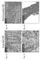
- Figs. 1A-D depict surface and cross-sectional views of both an expanded and a non-expanded extracellular matrix material sheet (porcine small intestine submucosa) wherein collagen has been stained such that its content and structure can be visualized.
- the four micrographs shown are as follows: (1A) the surface of the expanded ECM sheet material, (1B) the surface of a non-expanded ECM sheet material, (1C) a cross section of the expanded ECM sheet material, and (1D) a cross section of the non-expanded ECM sheet material.
- the surface and cross section views of the non-expanded material exhibit a tightly bound collagenous network whereas the same views of an expanded material exhibit a denatured, but still intact, collagenous network which has resulted in the expansion of the material.
- an alkaline substance can alter the collagen packing characteristics of the material as illustrated in Figs. 1A-D . Altering such characteristics of the material can be caused, at least in part, by the disruption of the tightly bound collagenous network.
- a non-expanded remodelable collagenous material having a tightly bound collagenous network typically has a continuous surface that is substantially uniform even when viewed under magnification, e.g. 100x magnification as shown in the Figures.
- an expanded remodelable collagenous material typically has a surface that is quite different in that the surface is typically not continuous but rather presents collagen strands or bundles in many regions that are separated by substantial gaps in material between the strands or bundles.
- an expanded remodelable collagenous material typically appears more porous than a non-expanded remodelable collagenous material.
- the expanded remodelable collagenous material can be demonstrated as having increased porosity, e.g. by measuring its permeability to water or other fluid passage.
- the more foamy and porous structure of an expanded remodelable collagenous material can allow the material to be easily cast into a variety of foam shapes for use in the preparation of medical materials and devices. It can further allow for the compression and subsequent expansion of the material, which is useful, for example, when the material needs to be loaded into a deployment device for delivery into a patient. Once delivered, the material can expand to its original form.
- a non-expanded remodelable collagenous ECM material can typically comprise a variety of bioactive components including, for example, growth factors, glycoproteins, glycosaminoglycans, proteoglycans, nucleic acids, and lipids.
- Treating the material with an alkaline substance under conditions as described herein can significantly reduce, if not completely eliminate, these bioactive components from the material.
- the treatment of the remodelable collagenous material with an alkaline substance can result in a remodelable collagenous material which is substantially devoid of growth factors, glycoproteins, glycosaminoglycans, proteoglycans, nucleic acids, and lipids.
- the treatment of a remodelable collagenous material with an alkaline substance as described herein can cause the material to expand to at least about twice its original volume, can alter the surface and/or porosity characteristics of the material, and can deplete the material of certain bioactive components. In some embodiments, this is accomplished while maintaining the material as an intact collagenous sheet, wherein the sheet can be further processed into any of a variety of medical materials and/or devices.
- the remodelable collagenous material such as an ECM sheet
- the alkaline medium so at tn expand it as described herein, while the material retains an amount of a growth factor such as FGF-2, or another bioactive component such as fibronectin and/or heparin, that is/are native to the source tissue for the ECM or other collagenous material.
- a growth factor such as FGF-2
- another bioactive component such as fibronectin and/or heparin
- selected bioactive components that were previously removed from the remodelable collagenous material can be returned to the material.
- an expanded remodelable collagenous material which is substantially devoid of nucleic acids and lipids, can be replenished with one or more growth factors, glycoproteins, glycosaminoglycans, or proteoglycans or combinations thereof.
- These bioactive components can be returned to the material by any suitable method.
- a tissue extract containing these components can be prepared and applied to an expanded remodelable collagenous material.
- the expanded remodelable collagenous material form is incubated in a tissue extract for a sufficient time to allow the bioactive components contained therein to associate with the expanded remodelable collagenous material.
- the tissue extract may, for example, be obtained from non-expanded remodelable collagenous tissue of the same type used to prepare the expanded material.
- Other means for returning or providing bioactive components to an expanded remodelable collagenous material include spraying, impregnating, dipping, etc. as known in the art.
- an expanded remodelable collagenous material may be modified by the addition of one or more growth factors such as basic fibroblast growth factor (FGF-2), transforming growth factor beta (TGF beta), epidermal growth factor (EGF), platelet derived growth factor (PDGF), and/or cartilage derived growth factor (CDGF).
- FGF-2 basic fibroblast growth factor
- TGF beta transforming growth factor beta
- EGF epidermal growth factor
- PDGF platelet derived growth factor
- CDGF cartilage derived growth factor
- an expanded remodelable collagenous material may be replenished with other biological components such as heparin, heparin sulfate, hyaluronic acid, fibronectin and the like.
- an expanded remodelable collagenous material may include a bioactive component that induces, directly or indirectly, a cellular response such as a change in cell morphology, proliferation, growth, protein or gene expression.
- a submucosa extract can be prepared by the addition of an extraction excipient, such as urea, guanidine, sodium chloride, magnesium chloride, or a surfactant, to a submucosa tissue to isolate bioactive components from the tissue. The bioactive components are then separated from the extraction excipient.
- an extraction excipient such as urea, guanidine, sodium chloride, magnesium chloride, or a surfactant
- a submucosa extract is prepared by mixing submucosa tissue with a phosphate buffered solution, such as phosphate buffered saline (PBS). This mixture is processed into a slurry as buffer circulation and physical pressure are applied.
- PBS phosphate buffered saline
- the bioactive components present in the tissue are drawn into solution and subsequently isolated from the slurry.
- the bioactive submucosa extract is then formed by separating the extracted bioactive components in the solution from the slurry using art-recognized procedures such as dialysis and/or chromatographic techniques.
- the extraction solution is dialyzed to reduce or remove the concentration of extraction excipients to provide a solution of the extracted bioactive components.
- Any source of submucosa tissue can be used to prepare a submucosa extract.
- similar extraction techniques can be applied to other remodelable ECM materials to provide biologically active extracts for use in the invention.
- the nature and quantity of the bioactive components contained in the submucosa or other extracellular matrix (ECM) extract is dependent on the nature and composition of the extraction excipients used for the extraction solution.
- ECM extracellular matrix
- bioactive extracts comprising proteoglycans, glycoproteins and glycosaminoglycans such as heparin, heparin sulfate, hyaluronic acid, chondroitin sulfate A and chondroitin sulfate B.
- non-native bioactive components including those synthetically produced by recombinant technology or other methods, may be incorporated into the expanded remodelable collagenous material.
- These non-native bioactive components may be naturally-derived or recombinantly produced proteins that correspond to those natively occurring in the ECM tissue, but perhaps of a different species (e.g. human proteins applied to collagenous ECMs from other animals, such as pigs).
- the non-native bioactive components may also be drug substances.
- Illustrative drug substances that may be incorporated into and/or onto the expanded remodelable collagenous materials used in the invention include, for example, antibiotics, thrombus-promoting substances such as blood clotting factors, e.g. thrombin, fibrinogen, and the like. As with the bioactive components previously described, these substances may be applied to the expanded remodelable collagenous material as a premanufactured step, immediately prior to the procedure (e.g. by soaking the material in a solution containing a suitable antibiotic such as cefazolin), or during or after engraftment of the material in the patient.
- antibiotics e.g. thrombin, fibrinogen, and the like.
- these substances may be applied to the expanded remodelable collagenous material as a premanufactured step, immediately prior to the procedure (e.g. by soaking the material in a solution containing a suitable antibiotic such as cefazolin), or during or after engraftment of the material in the patient.
- the expanded remodelable collagenous material may also exhibit an angiogenic character and thus be effective to induce angiogenesis in a host engrafted with the material.
- Angiogenic growth factors are well known in the art and include, for example, angiogenin, angiopoietin-1, Del-1, fibroblast growth factors (both acidic and basic), follistatin, granulocyte colony-stimulating factor, hepatocyte growth factor, interleukin-8 (IL-8), leptin, midkine, placental growth factor, platelet derived growth factor (PDGF), pleiotrophin, proliferin, transforming growth factors (both alpha and beta), tumor necrosis growth factor, and vascular endothelial growth factor (VEGF).
- angiogenin angiopoietin-1, Del-1
- fibroblast growth factors both acidic and basic
- follistatin granulocyte colony-stimulating factor
- hepatocyte growth factor hepatocyte growth factor
- Angiogenesis is the process through which the body makes new blood vessels to generate increased blood supply to tissues.
- angiogenic materials when contacted with host tissues, promote or encourage the formation of new blood vessels.
- Methods for measuring in vivo angiogenesis in response to biomaterial implantation have recently been developed. For example, one such method uses a subcutaneous implant model to determine the angiogenic character of a material. See, C. Heeschen et al., Nature Medicine 7 (2001), No. 7, 833-839 . When combined with a fluorescence microangiography technique, this model can provide both quantitative and qualitative measures of angiogenesis into biomaterials. C. Johnson et al., Circulation Research 94 (2004), No. 2, 262-268 .
- Expanded remodelable collagenous materials are prepared, for example, from collagenous materials isolated from a suitable tissue source from a warm-blooded vertebrate, and especially a mammal. Such isolated collagenous material can be processed so as to have remodelable properties and promote cellular invasion and ingrowth. Suitable remodelable materials can be provided by collagenous extracellular matrix (ECM) materials possessing biotropic properties.
- ECM extracellular matrix
- Suitable bioremodelable materials can be provided by collagenous extracellular matrix materials (ECMs) possessing biotropic properties, including in certain forms angiogenic collagenous extracellular matrix materials.
- ECMs extracellular matrix materials
- suitable collagenous materials include ECMs such as submucosa, renal capsule membrane, dermal collagen, dura mater, pericardium, fascia lata, serosa, peritoneum or basement membrane layers, including liver basement membrane, all of which can be derived for example from porcine, ovine or bovine tissue sources.
- ECMs extracellular matrix materials possessing biotropic properties, including in certain forms angiogenic collagenous extracellular matrix materials.
- suitable collagenous materials include ECMs such as submucosa, renal capsule membrane, dermal collagen, dura mater, pericardium, fascia lata, serosa, peritoneum or basement membrane layers, including liver basement membrane, all of which can be derived for example from porcine, ovine or bovine tissue sources.
- Suitable submucosa materials for these purposes include, for instance, intestinal submucosa, including small intestinal submucosa, stomach submucosa, urinary bladder submucosa, and uterine submucosa.
- Submucosa or other ECM tissue used in the invention is preferably highly purified, for example, as described in U.S. Patent No. 6,206,931 to Cook et al.
- preferred ECM material will exhibit an endotoxin level of less than about 12 endotoxin units (EU) per gram, more preferably less than about 5 EU per gram, and most preferably less than about 1 EU per gram.
- EU endotoxin units
- the submucosa or other ECM material may have a bioburden of less than about 1 colony forming units (CFU) per gram, more preferably less than about 0.5 CFU per gram.
- CFU colony forming units
- Fungus levels are desirably similarly low, for example less than about 1 CFU per gram, more preferably less than about 0.5 CFU per gram.
- Nucleic acid levels are preferably less than about 5 ⁇ g/mg, more preferably less than about 2 ⁇ g/mg, and virus levels are preferably less than about 50 plaque forming units (PFU) per gram, more preferably less than about 5 PFU per gram.
- PFU plaque forming units
- the material is preferably treated with a disinfecting agent so as to produce a disinfected, expanded remodelable collagenous material.
- Treatment with a disinfecting agent can be done either prior to or after isolation of the remodelable collagenous material from the tissue source or can be done either prior to or after expansion.
- the tissue source material is rinsed with a solvent, such as water, and is subsequently treated with a disinfecting agent prior to delamination. It has been found that by following this post-disinfection-stripping procedure, it is easier to separate the remodelable collagenous material from the attached tissues as compared to stripping the remodelable collagenous material prior to disinfection.
- the resultant remodelable collagenous material in its most preferred form exhibits superior histology, in that there is less attached tissue and debris on the surface compared to a remodelable collagenous material obtained by first delaminating the submucosa layer from its source and then disinfecting the material. Moreover, a more uniform remodelable collagenous material can be obtained from this process, and a remodelable collagenous material having the same or similar physical and biochemical properties can be obtained more consistently from each separate processing run. Importantly, a highly purified, substantially disinfected remodelable collagenous material is obtained by this process.
- an expanded remodelable collagenous material may be prepared by a method comprising providing a tissue source including a remodelable collagenous material, disinfecting the tissue source, isolating the remodelable collagenous material from the tissue source, and contacting the disinfected remodelable collagenous material with an alkaline substance under conditions effective to expand the remodelable collagenous material to at least about two times its original volume, thereby forming the expanded remodelable collagenous material.
- the material can be further processed into medical materials and/or devices, or can be stored, e.g. in high purity water at 4 °C, for later use.
- Preferred disinfecting agents are desirably oxidizing agents such as peroxy compounds, preferably organic peroxy compounds, and more preferably peracids.
- peracid compounds that can be used, these include peracetic acid, perpropioic acid, or perbenzoic acid.
- Peracetic acid is the most preferred disinfecting agent for purposes of the present invention.
- Such disinfecting agents are desirably used in a liquid medium, preferably a solution, having a pH of about 1.5 to about 10, more preferably a pH of about 2 to about 6, and most preferably a pH of about 2 to about 4.
- the disinfecting agent will generally be used under conditions and for a period of time which provide the recovery of characteristic, purified submucosa materials as described herein, preferably exhibiting a bioburden of essentially zero and/or essential freedom from pyrogens.
- desirable processes of the invention involve immersing the tissue source or isolated remodelable collagenous material (e.g. by submersing or showering) in a liquid medium containing the disinfecting agent for a period of at least about 5 minutes, typically in the range of about 5 minutes to about 40 hours, and more typically in the range of about 0.5 hours to about 5 hours.
- peracetic acid When used, peracetic acid is desirably diluted into about a 2% to about 50% by volume of alcohol solution, preferably ethanol.
- concentration of the peracetic acid may range, for instance, from about 0.05% by volume to about 1.0% by volume. Most preferably, the concentration of the peracetic acid is from about 0.1% to about 0.3% by volume.
- hydrogen peroxide When hydrogen peroxide is used, the concentration can range from about 0.05% to about 30% by volume. More desirably the hydrogen peroxide concentration is from about 1% to about 10% by volume, and most preferably from about 2% to about 5% by volume.
- the solution may or may not be buffered to a pH from about 5 to about 9, with more preferred pH's being from about 6 to about 7.5. These concentrations of hydrogen peroxide can be diluted in water or in an aqueous solution of about 2% to about 50% by volume of alcohol, most preferably ethanol.
- any suitable alkaline substance generally known in the art can be used.
- Suitable alkaline substances can include, for example, salts or other compounds that that provide hydroxide ions in an aqueous medium.
- the alkaline substance comprises sodium hydroxide (NaOH).
- the concentration of the alkaline substance that is added to the material can be in the range of about 0.5 to about 4 M.
- the concentration of the alkaline substance is in the range of about 1 to about 3 M.
- the pH of the alkaline substance will typically range from about 8 to about 14.
- the alkaline substance will have a pH of from about 10 to about 14, and most preferably of from about 12 to about 14.
- the exposure of the remodelable collagenous material to the alkaline substance is performed at a temperature of about 4 to about 45 °C. In preferred embodiments, the exposure is performed at a temperature of about 25 to about 37 °C, with 37 °C being most preferred.
- the exposure time can range from about several minutes to about 5 hours or more. In preferred embodiments, the exposure time is about 1 to about 2 hours.
- the remodelable collagenous material is exposed to a 3 M solution of NaOH having a pH of 14 at a temperature of about 37 °C for about 1.5 to 2 hours.
- Such treatment results in the expansion of a remodelable collagenous material to at least about twice its original volume. As indicated above, these processing steps can be modified to achieve the desired level of expansion.
- a lipid removal agent can also be added to a remodelable collagenous material either prior to, in conjunction with, or after the addition of the alkaline substance.
- Suitable lipid removal agents include, for example, solvents such as ether and chloroform, or surfactants.
- solvents such as ether and chloroform, or surfactants.
- surfactants Other suitable lipid removal agents will be apparent to those of ordinary skill in the art. Accordingly, the lipid removal agents listed herein serve only as examples, and are therefore in no way limiting.
- the expanded remodelable collagenous materials, as well as tissue extracts containing bioactive components that can optionally be added to an expanded remodelable collagenous material are sterilized using conventional sterilization techniques including tanning with glutaraldehyde, formaldehyde tanning at acidic pH, ethylene oxide treatment, propylene oxide treatment, gas plasma sterilization, gamma radiation, and peracetic acid sterilization.
- a sterilization technique which does not significantly alter the remodelable properties of the expanded remodelable collagenous material is preferably used.
- the sterilization technique preferably does not significantly alter the bioactivity of the expanded remodelable collagenous material.
- Preferred sterilization techniques include exposing the extract to peracetic acid, low dose gamma irradiation (2.5 mRad) and gas plasma sterilization.
- the expanded remodelable collagenous materials of the invention can be provided in any suitable form, including a flowable aqueous composition (e.g., a fluidized composition), a powder, a gel, a sponge, one or more sheets, or a cast body.
- a flowable aqueous composition e.g., a fluidized composition
- a powder e.g., a powder, a gel, a sponge, one or more sheets, or a cast body.
- a particulate non-expanded extracellular matrix material e.g., a fluidized composition
- the non-expanded material can be prepared in any of these forms so long as the expanded and non-expanded material can be combined to form a composite extracellular matrix material.
- the expanded remodelable collagenous material is processed into a fluidized composition, for instance using techniques as described in U.S. Patent No. 5,275,826 .
- solutions or suspensions of the expanded remodelable collagenous material can be prepared by comminuting and/or digesting the material with a protease (e.g. trypsin or pepsin), for a period of time sufficient to solubilize the material and form substantially homogeneous solution.
- a protease e.g. trypsin or pepsin
- the expanded remodelable collagenous material is desirably comminuted by, tearing, cutting, grinding, shearing (e.g. combined with a liquid and sheared in a blender), or the like, which can generate random fragments of the expanded remodelable collagenous material.
- the expanded remodelable collagenous material typically has a spongy and porous structure, so these techniques may not be needed to the extent they would be needed to solubilize a non-expanded remodelable collagenous material.
- Grinding the material in a frozen or freeze-dried state is advantageous, although good results can be obtained as well by subjecting a suspension of pieces of the material to treatment in a high speed blender and dewatering, if necessary, by centrifuging and decanting excess waste.
- the comminuted material can be dried, for example freeze dried, to form a particulate.
- the particulate can be used itself to treat a patient, e.g., for trauma wounds, or can be hydrated, that is, combined with water or buffered saline and optionally other pharmaceutically acceptable excipients, to form a fluidized, expanded remodelable collagenous material, e.g. having a viscosity of about 2 to about 300.000 cps at 25 °C.
- the higher viscosity graft compositions can have a gel or paste consistency.
- a particulate, non-expanded remodelable collagenous material formed separately from the expanded remodelable collagenous material can be combined with a fluidized, expanded remodelable collagenous material.
- Such particulate, non-expanded modelable collagenous materials can be prepared by cutting, tearing, grinding, shearing or otherwise comminuting a remodelable collagenous source material, which can provide random fragments of the remodelable collagenous material.
- the non-expanded particulate can include one or more additives to promote hemostasis. Suitable such additives include, as examples, calcium alginate or zeolite.
- Such additives can include adhesive properties that allow the particulate to adhere to a desired location (e.g., tissue surface) after implantation.
- a particulate ECM material having an average particle size of about 50 microns to about 500 microns may be included in the fluidized, expanded remodelable collagenous material, more preferably about 100 microns to about 400 microns.
- the remodelable collagenous particulate can be added in any suitable amount relative to the fluidized, expanded remodelable collagenous material, with preferred remodelable collagenous particulate to fluidized, expanded remodelable collagenous material weight ratios (based on dry solids) being about 0.1:1 to about 200:1, more preferably in the range of 1:1 to about 100:1.
- the inclusion of such remodelable collagenous particulates in the ultimate fluidized, expanded remodelable collagenous material can serve to provide additional material that can function to provide bioactivity to the composition (e.g. itself including growth factors or other bioactive components as discussed herein), serve as scaffolding material for tissue ingrowth and/or promote expansion of a compressed remodelable collagenous material.
- a fluidized composition prepared from an expanded remodelable collagenous material can optionally be dried to form a sponge solid or foam material.
- the particulate of a non-expanded material may be combined with a fluidized composition as described herein by addition to the fluidized composition prior to drying such that the particulate becomes embedded within the dried, expanded material.
- Dry sponge or foam form materials of the invention prepared by drying expanded remodelable collagenous material gels combined with non-expanded particulate material can be used, for example, in wound healing, tissue reconstructive applications, occlusive applications, hemostatic applications, in the culture of cells, and in a variety of additional applications including those disclosed elsewhere herein.
- An expanded remodelable collagenous material of the invention can also be used in body wall repair including, for example, in the repair of abdominal wall defects such as hernias, using techniques analogous to those described in Ann. Plast. Surg., 1995, 35:3740380 ; and J. Surg. Res., 1996, 60:107-114 .
- preferred materials of the invention are in sheet form and promote favorable organization, vascularity and consistency in the remodeled tissue.
- Sheet form expanded remodelable collagenous material can for example be formed by stretching an expanded material over a backing board, freezing the material, and lyophilizing the material.
- an expanded remodelable collagenous material can be used in the repair of partial or full thickness wounds and in dermal augmentation using general grafting techniques which are known to the art and literature (see, e.g. Annals of Plastic Surgery 1995, 35:381-388 ).
- general grafting techniques which are known to the art and literature (see, e.g. Annals of Plastic Surgery 1995, 35:381-388 ).
- cultured epidermal grafts preferably cultured epidermal autografts, or CEA's
- Such cultured grafts have typically involved transplanting keratinocytes and/or fibroblasts onto the dermal substitute.
- the purified, expanded remodelable collagenous material of the present invention can be used as the dermal substitute, for example in sheet form, and the CEA accordingly transplanted onto the material.
- the expanded remodelable collagenous material of the invention can be used to prepare a wound care matrix.
- a wound care matrix can be prepared by providing a tissue source including a remodelable collagenous material, disinfecting the tissue source, isolating the remodelable collagenous material from the tissue source, contacting the disinfected remodelable collagenous material with an alkaline substance under conditions effective to expand the remodelable collagenous material to at least about two times its original volume, forming the expanded remodelable collagenous material into one or more sheets and lyophilizing the material to form the wound care matrix.
- the one or more sheets can be vacuum pressed or sutured together as known in the art to form the wound care matrix.
- Such embodiments can also involve coupling or otherwise physically associating sheet form expanded material with sheet form non-expanded extracellular matrix material, for example by making a multilaminate device including these two types of materials.
- the material can have a thickness in the range of about 0.2 mm to about 2mm, more preferably about 0.4 mm to about 1.5 mm, and most preferably about 0.5 mm to about 1mm.
- a multilaminate material can be used.
- a plurality of (i.e. two or more) layers of an expanded remodelable collagenous ECM material can be bonded or otherwise coupled together to form a multilaminate structure.
- two, three, four, five, six, seven, or eight or more layers of an expanded remodelable collagenous material can be bonded together to provide a multilaminate material.
- two to six expanded, submucosa-containing layers isolated from intestinal tissue of a warm-blooded vertebrate, particularly small intestinal tissue are bonded together to provide a medical material. Porcine-derived small intestinal tissue is preferred for this purpose.
- one or more sheets of a non-expanded collagenous material e.g., submucosa
- the layers of collagenous tissue can be bonded together in any suitable fashion, including dehydrothermal bonding under heated, non-heated or lyophilization conditions, using adhesives as described herein, glues or other bonding agents, crosslinking with chemical agents or radiation (including UV radiation), or any combination of these with each other or other suitable methods.
- dehydration-induced bonding methods can be used to fuse portions of multi-layered medical materials together.
- the multiple layers of material are compressed under dehydrating conditions.
- dehydrating conditions can include any mechanical or environmental condition which promotes or induces the removal of water from the multi-layered medical material.
- at least one of the two surfaces compressing the matrix structure can be water permeable.
- Dehydration of the material can optionally be further enhanced by applying blotting material, heating the matrix structure or blowing air, or other inert gas, across the exterior of the compressing surfaces.
- One particularly useful method of dehydration bonding multi-layered medical materials is lyophilization, e.g. freeze-drying or evaporative cooling conditions.
- Another method of dehydration bonding comprises pulling a vacuum on the assembly while simultaneously pressing the assembly together. This method is known as vacuum pressing.
- vacuum pressing dehydration of the multi-layered medical materials in forced contact with one another effectively bonds the materials to one another, even in the absence of other agents for achieving a bond, although such agents can be used while also taking advantage at least in part of the dehydration-induced bonding.
- the multi-layered medical materials can be caused to form a generally unitary laminate structure.
- drying operations conducted with no or substantially no duration of exposure to temperatures above human body temperature or slightly higher, say, no higher than about 38 °C will preferably be used in some forms of the present invention. These include, for example, vacuum pressing operations at less than about 38 °C, forced air drying at less than about 38 °C, or either of these processes with no active heating - at about room temperature (about 25 °C) or with cooling. Relatively low temperature conditions also, of course, include lyophilization conditions. It will be understood that the above-described means for coupling two or more multi-layered medical materials together to form a laminate can also apply for coupling together one or more layers of peritoneum and fascia when these layers are isolated independent from one another.
- the expanded remodelable collagenous material of the present invention can be used to prepare a molded or shaped construct for example a sponge useful as an occluder device or biopsy plug.
- the method for preparing such device comprises providing an expanded remodelable collagenous material, comminuting the expanded material, casting the expanded remodelable collagenous material into a shape, and freezing and lyophilizing the cast, expanded remodelablc collagonous material to form the oonstruct Freezing can be done at a temperature of about -80 ° C for about 1 to about 4 hours, and lyophilization can be performed for about 8 to about 48 hours.
- the material used to prepare the construct can be an expanded remodelable collagenous material that has been supplemented with a particulate, non-expanded remodelable collagenous material and optionally replenished with one or more bioactive components.
- the expanded remodelable collagenous material and other components can he cast into any shape desired and will typically be shaped by a skilled artisan to occlude a particular area in need or occlusion.
- a biopsy plug is formed and is used, for example, to fill a void in a tissue (e.g., organ tissue) after surgery.
- the lyophilized, expanded remodelable collagenous material can be compressed and loaded into a deployment device for delivery into a patient Once delivered, the device can expand to occlude the area in which it was deployed.
- Suitable deployment devices will be generally known to those of ordinary skill in the art and include, for example, delivery catheters and the like.
- Suitable additives include, for example, salts, such as sodium chloride, sodium acetate, sodium bicarbonate, sodium citrate, calcium carbonate, potassium acetate, potassium phosphate; hydrogel and water-swelling polymers, such as alginate, polyhydroxethyl methacralate, polyhydroxypropyl methacrylate, polyvinyl alcohol, polyethylene glycol, carboxymethyl cellulose, polyvinyl pyrrolidone; proteins, such as gelatin and SIS particulate; acids and bases, such as acetic acid and ascorbic acid; superabsorbing polymers and gelling agents, such as polyacrylic acid, pectin, polygalacturonic acid, polyacrylic acid-co-acrylamide, polyisobutylene-co-malcic acid; monosaccharides, polysaccharides, and derivatives
- salts such as sodium chloride, sodium acetate, sodium bicarbonate, sodium citrate, calcium carbonate, potassium acetate, potassium phosphate
- the one or more additives can provide a variety of functions, including promoting expansion of the material once implanted into a patient.
- a sponge form expanded remodelable collagenous material including one or more additives can be compressed and placed into a delivery device. Compression of the material allows the material to be more easily transferred to a patient. Upon delivery, the material can expand to at least about its original size prior to compression. This is typically done with an occluder device or a biopsy plug where it is desirable for the material to have a smaller diameter prior to delivery and expand upon delivery.
- Such additives can be included in the remodelable collagenous material to expand the material at a faster rate than would otherwise be achievable in the absence of the one or more additives.
- one or more additives can be included with a compressed remodelable collagenous material so as to promote the re-expansion of the material back to its original size within at least about 30 seconds, 45 seconds, 1 minute, 2 minutes, 3 minutes, 4 minutes, or even about least about 5 minutes after implantation.
- these additives may, be applied to the expanded remodelable collagenous material as a premanufactured step, immediately prior to the procedure (e.g. by soaking the material in a solution containing a suitable antibiotic such as cefazolin), or during or after engraftment of the material in the patient.
- the expanded remodelable collagenous material in any form, is crosslinked.
- An expanded remodelable collagenous material can be crosslinked either before or after it is formed into a medical device (e.g., a composite extracellular matrix material product), or both. Increasing the amount (or number) of crosslinkages within the material or between two or more layers of the material can be used to enhance its strength.
- a remodelable material when used, the introduction of crosslinkages within the material may also affect its resorbability or remodelability. Consequently, in certain embodiments, a remodelable collagenous material will substantially retain its native level of crosslinking, or the amount of added crosslinkages within the medical device will be judiciously selected depending upon the desired treatment regime.
- the material will exhibit remodelable properties such that the remodeling process occurs over the course of several days or several weeks. In certain preferred embodiments, the remodeling process occurs within a matter of about 5 days to about 12 weeks.
- crosslinking of a compressed construct may promote re-expansion of the construct after implantation into a patient.
- the constructs are capable of volumetric compression when dry at a ratio of at least 10:1 (i.e. the compressed form occupies no more than 10% of its original, relaxed and unexpanded volume), more preferably at a ratio of at least 20:1.
- the compressed constructs are capable of re-expansion to substantially their original volume (e.g. at least about 80% of their original volume, more preferably at least 90%, and most preferably at least 95%) within about 30 seconds when delivered in their dry, compressed form into a volume of water.
- introduced crosslinking of the expanded remodelable collagenous material may be achieved by photo-crosslinking techniques, or by the application of a crosslinking agent, such as by chemical crosslinkers, or by protein crosslinking induced by dehydration or other means.
- a crosslinking agent such as by chemical crosslinkers, or by protein crosslinking induced by dehydration or other means.
- Chemical crosslinkers that may be used include for example aldehydes such as glutaraldehydes, diimides such as carbodiimides, e.g., 1-ethyl-3-(3-dimethylaminopropyl)carbodiimide hydrochloride (BDC), diisocyanates such as hexamethylene-diisocyanate, ribose or other sugars, acyl-azide, sulfo-N-hydroxysuccinamide, or polyepoxide compounds, including for example polyglycidyl ethers such as ethyleneglycol diglycidyl ether, available under the trade name DENACOL EX810 from Nagese Chemical Co., Osaka, Japan, and glycerol polyglycerol ether available under the trade name DENACOL EX 313 also from Nagese Chemical Co. Typically, when used, polyglycerol ethers or other polyepoxide compounds will have from 2 to about 10 ep
- the layers of the laminate can be additionally crosslinked to bond multiple layers of a multi-layered medical material to one another.
- Cross-linking of multi-layered medical materials can also be catalyzed by exposing the matrix to UV radiation, by treating the collagen-based matrix with enzymes such as transglutaminase and lysyl oxidase, and by photocrosslinking.
- additional crosslinking may be added to individual layers prior to coupling to one another, during coupling to one another, and/or after coupling to one another.
- the medical materials, constructs and devices of the invention can be provided in sterile packaging suitable for medical materials and devices. Sterilization may be achieved, for example, by irradiation, ethylene oxide gas, or any other suitable sterilization technique, and the materials and other properties of the medical packaging will be selected accordingly.
- the invention provides compressible medical foam products.
- the medical foam products include a dried, compressible foam body formed with an extracellular matrix solid material that has been treated with an alkaline medium under conditions effective to produce an expanded extracellular matrix collagen material.
- the compressible foam body further includes a particulate of a non-expanded extracellular matrix material as described herein, preferably one that retains one or more bioactive components, such as a growth factor, with which it naturally occurs.
- the foam body can have introduced chemical crosslinks sufficient to increase the resiliency of the foam body.
- foam bodies produced from the expanded extracellular matrix collagen material possess resiliency, but for certain applications, including for example hemostatic plug applications, it has been discovered that increased resiliency is desired for certain uses.
- the introduction of collagen crosslinks for example with chemical crosslinkers such as glutaraldehyde, carbodimides, or other chemical crosslinkers identified herein, has been found to significantly enhance the resiliency of the foam plugs, while leaving the compressible to a small size for delivery.
- Increased resiliency in turn provides additional compression upon adjacent tissues when the foam plugs are inserted in a compressed state and then allowed to expand in situ in a patient at a site at which hemostasis is desired.
- crosslinked, resilient foam plugs as disclosed herein can be utilized to provide hemostasis at surgical sites, including biopsy sites. These biopsy or other surgical sites can be located within parenchymal organ tissues, such as those of a kidney, liver or spleen of a patient.
- Surgical methods include resecting tissue from a parenchymal organ such as a liver or kidney, and then implanting a crosslinked, resilient foam material in accordance with the invention at the resection site so as to facilitate hemostasis.
- the resection can, as examples, occur as a part of a nephrectomy or hepatectomy, e.g. to removed cancerous or other diseased tissue, or as a part of a kidney or liver biopsy performed with a biopsy needle.
- the crosslinked, resilient foam plug can be delivered from within a cannulated device such as a needle or catheter, and/or through a laparoscopic device.
- the resilient foam plug can be in a compressed state during delivery, and then allowed to expand once delivered to the surgical site.
- the expansion of the plug can compress the adjacent tissues to facilitate hemostasis.
- the expanded dimensions of the plug can provide a volume that is at least equal to or preferably greater than the volume of the biopsy or other surgical defect, to ensure compression of surrounding tissues by the delivered, expanded plug.
- a crosslinked, resilient foam material in accordance with the invention can be deployed at a site within a bodily vessel, for example an artery or a vein, so as to cause occlusion of the vessel and thereby stop the flow of fluid (e.g. blood) within the vessel.
- fluid e.g. blood
- the crosslinked, resilient foam plug can be delivered from within a cannulated device such as a catheter or sheath.
- the resilient foam plug can be in a compressed state during delivery, and then allowed to expand once delivered from within the cannulated device to the desired occlusion site. The expansion of the plug can compress the walls of the vessel to facilitate occlusion.
- the expanded dimensions of the plug can be greater than the diameter of the vessel at the desired site of occlusion, to ensure outward compression against surrounding vessel walls by the delivered, expanded plug.
- vascular vessels other vessels that can be occluded in accordance with the invention include, for example, fallopian tube(s).
- other open tracts through patient tissue can be occluded with crosslinked, resilient foam plugs of the invention, including for example needle tracts (e.g. resultant of percutaneous entry to a vein or artery) and fistulas, such as anorectal fistulas, enterocutaneous fistulas, recto-vaginal fistulas, and others.
- Crosslinked, resilient foam plugs can be prepared by a process that includes:
- the extracellular matrix material and chemical crosslinked agent can, for example, be selected from among any of those disclosed herein.
- the washing can suitably be conducted with an aqueous medium, such as saline or water.
- the drying can be conducted by any suitable method, including as examples air drying at ambient temperature, heated drying, or lyophilization. It is preferred to contact the extracellular matrix material with the chemical crosslinker after the formation of the lyophilized extracellular matrix material foam (e.g. as opposed to incorporating the chemical crosslinker in the material charged to the mold), as this has been found to provide more uniformly shaped crosslinked plugs that resist shrinkage.
- the expanded extracellular matrix material can be comminuted prior to charging to the mold, forming expanded extracellular matrix fragments that will be incorporated within and will characterize the extracellular matrix foam material.
- the material is comminuted by shearing the material with a rotating blade, e.g. in a blender.
- a rotating blade e.g. in a blender.
- the sheet prior to comminution by the blade or otherwise, can be treated with the alkaline medium for a time and under conditions sufficient to reduce the tensile strength of the sheet to less than about 50% of its original tensile strength, more preferably to less than about 30% of its original tensile strength.
- harvested sheet-form ECM materials such as submucosa-containing sheets, e.g. obtained from small intestinal, stomach or bladder tissue, pericardial tissue, peritoneal tissue, fascia, dermal tissue, and other sheet-form ECM materials.
- a composite extracellular matrix material product can be prepared by coupling or otherwise associating an expanded extracellular matrix material with a non-expanded extracellular matrix material.
- the expanded and non-expanded materials can each be in any suitable form as described above so long as the two materials can be combined to form a composite extracellular matrix material product.
- the composite product comprises a dried body formed with an extracellular matrix material that has been treated with an alkaline medium under conditions effective to produce an expanded extracellular matrix material and particles of a bioactive extracellular matrix material entrapped within said dried body, wherein the particles of bioactive extracellular matrix material retain at least one growth factor from a source tissue for the particulate extracellular matrix material.
- the composite products can be prepared by an inventive method that includes the steps of:
- the extracellular matrix material that is expanded, and the particulate extracellular matrix material can, for example, be selected from among any of those disclosed herein.
- the washing can suitably be conducted with an aqueous medium, such as saline or water.
- the liquid for preparing the mixture can be any suitable liquid, preferably biocompatible, and typically an aqueous liquid such as water or saline.
- the drying step can be conducted by any suitable method, including as examples air drying at ambient temperature, heated drying, or lyophilization.
- the expanded extracellular matrix material is desirably comminuted prior to or during the formation of the mixture.
- the material is comminuted by shearing the material with a rotating blade, e.g. in a blender, alone or in the presence of the bioactive particulate extracellular matrix material.
- a rotating blade e.g. in a blender
- Such methods can be practiced, for example, with harvested sheet-form ECM materials such as submucosa-containing sheets, e.g. obtained from small intestinal, stomach or bladder tissue, pericardial tissue, peritoneal tissue, fascia, dermal tissue, and other sheet-form ECM materials.
- the expanded ECM material and the bioactive particulate ECM material can be from the same ECM starting material or from different ECM starting materials. It has been discovered that the incorporation of the particulate ECM material can serve not only to enhance the bioactivity of the foam product, but also enhance the resiliency of the foam product.
- the invention provides composite extracellular matrix material products that include an extracellular matrix sheet material and a dried material adhered to the extracellular matrix sheet material, wherein the dried material is formed from an extracellular matrix material that has been contacted with an alkaline medium to form an expanded extracellular matrix material.
- composite products can be prepared by a method that includes the steps of:
- the extracellular matrix material that is expanded, and the particulate extracellular matrix material can, for example, be selected from among any of those disclosed herein.
- the washing can suitably be conducted with an aqueous medium, such as saline or water.
- the liquid for preparing the wet preparation can be any suitable liquid, preferably biocompatible, and typically an aqueous liquid such as water or saline.
- the drying step can be conducted by any suitable method, including as examples air drying at ambient temperature, heated drying, or lyophilization. Lyophilization is preferred as it forms a more porous, resilient foam material as compared to air drying or heated drying.
- the expanded extracellular matrix material in the flowable, wet preparation is desirably comminuted.
- the material is comminuted by shearing the material with a rotating blade, e.g. in a blender.
- a rotating blade e.g. in a blender.
- harvested sheet-form ECM materials such as submucosa-containing sheets, e.g. obtained from small intestinal, stomach or bladder tissue, pericardial tissue, peritoneal tissue, fascia, dermal tissue, and other sheet-form ECM materials.
- the expanded ECM material and the sheet-form ECM material can be from the same ECM starting material or from different ECM starting materials.
- the incorporation of the sheet-form ECM material can serve not only to enhance the bioactivity of the overall product, but can also provide a barrier material and/or suturable sheet attached to the dried expanded ECM material (e.g. foam).
- a barrier material and/or suturable sheet attached to the dried expanded ECM material e.g. foam
- such constructs can be used to provide hemostasis to surgical sites or other injured tissue.
- the construct can be placed against the bleeding tissue with the dried, expanded ECM material (especially a foam) against the bleeding tissue.
- the sheet-form ECM can then provide an additional barrier (besides the expanded ECM material) to protect the bleeding tissue, and or can provide a suturable sheet material which can be used to fix the construct in place, e.g. with sutures in strand or staple form.
- such constructs can be used to apply hemostasis to surgically-treated (e.g. subject to resection) or otherwise injured parenchymous organ tissue, such as liver or kidney tissue.
- surgically-treated e.g. subject to resection
- otherwise injured parenchymous organ tissue such as liver or kidney tissue.
- the dried, expanded ECM material is desirably pressed against the injured parenchymous tissue, and the sheet-form ECM material can optionally be used to fix the construct in place, as discussed above.
- Figs. 2-4 illustrate certain sheet form composite extracellular matrix sheet form products.
- Fig 2 provides a composite extracellular matrix sheet form product 20.
- the composite extracellular matrix sheet form product 20 includes a sheet form of a non-expanded extracellular matrix material 22 associated with an expanded extracellular matrix material 24. It will be understood by a skilled artisan that any number of expanded material layers can be associated with any number of non-expanded layers.
- Fig. 3 illustrates a composite extracellular matrix material product 30 including two sheets of a non-expanded extracellular matrix material 32 and 34 associated with a sheet of an expanded extracellular matrix material 36.
- Fig. 3 illustrates a composite extracellular matrix material product 40 including single layer of a non-expanded material 46 sandwiched between two sheets of an expanded extracellular matrix material 42 and 44.
- Such composite sheet form extracellular matrix material products can be prepared as described above.
- This example demonstrates the process used to prepare a disinfected small intestinal submucosa tissue (i.e., non-expanded SIS), which can subsequently be used in the preparation of various medical materials and devices.
- a disinfected small intestinal submucosa tissue i.e., non-expanded SIS
- Surface and cross section micrographs of the material are depicted in Figs. 1B and 1D .
- a 300 cm (ten foot) section of porcine whole intestine was extracted and washed with water. After rinsing, this section of submucosa intestinal collagen source material was treated for about two and a half hours in 0.2% peracetic acid by volume in a 5% by volume aqueous ethanol solution with agitation. Following the treatment with the peracetic acid solution, the submucosa layer was delaminated in a disinfected casing machine from the whole intestine. The resultant submucosa was then rinsed four (4) times with sterile water. A 1 cm by 1 cm section of this material was extracted and stained using a solution of direct red prepared by mixing 10 mg direct red in 100 mL high purity water.
- the section of material was stained for approximately 5 minutes.
- the stained material was washed twice with high purity water to remove any unbound stain.
- the stained material was placed on a glass slide and covered with a cover slip.
- a micrograph was taken (Olympus microscope) at 100x magnification of the surface of the material.
- a cross section of the material was then prepared and a similar micrograph was taken.
- the resulting micrograph was analyzed using Spot RT software.
- the surface and cross section micrographs are depicted in Figs. 1B and 1D . Both the surface and cross section micrographs show a tightly bound collagenous matrix with no expansion.
- a 300 cm (ten foot) section of porcine whole intestine was extracted and washed with water. After rinsing, this section of submucosa intestinal collagen source material was treated for about two and a half hours in 0.2% peracetic acid by volume in a 5% by volume aqueous ethanol solution with agitation. Following the treatment with peracetic acid, the submucosa layer was delaminated in a disinfected casing machine from the whole intestine. The resultant submucosa was then rinsed four (4) times with sterile water. 300 g of this material was soaked with agitation in 1 L of a 1M NaOH solution at 37 °C for 1 hour and 45 minutes.
- the material was removed and rinsed in a 1L solution of high purity water for 5 minutes. This rinsing step was repeated 8 additional times.
- a 1cm by 1cm section of this material was extracted and stained using a solution of direct red prepared by mixing 10 mg direct red in 100 mL high purity water. The section of material was stained for approximately 5 minutes. The stained material was washed twice with high purity water to remove any unbound stain. The stained material was placed on a glass slide and covered with a cover slip.
- a micrograph was taken (Olympus microscope) at 100x magnification of the surface of the material.
- a cross section of the material was then prepared and a similar micrograph was taken. The resulting micrograph was analyzed using Spot RT software.
- the surface and cross section micrographs are depicted in Pigs. 1A and 1C. Both the surface and cross section micrographs show disruption of the tightly bound collagenous matrix and an expansion of the material.
- both the surface view and the cross-section view of the non-expanded SIS show a tightly bound collagenous matrix whereby collagen content is substantially uniform throughout.
- the surface view and cross-section view of the expanded SIS show a denatured collagenous network and an expansion of the material.
- This Example was performed to identify additives that can be included in an expanded remodelable collagenous material for purposes of promoting rapid re-expansion of the material after implantation into a patient.
- An expanded remodel able material was prepared generally as described in Example 2. Briefly, a 300 cm (a ten foot) section of porcine whole intestine was extracted and washed with water. After rinsing, this section of submucosa intestinal collagen source material was treated for about two and a half hours in 0.2% peracetic acid by volume in a 5% by volume aqueous ethanol solution with agitation. Following the treatment with peracetic acid, the submucosa layer was delaminated in a disinfected casing machine from the whole intestine. The resultant submucosa was then rinsed four (4) times with sterile water.
- the rinsed material was mechanically agitated using the pulse setting of a blender to the extent that the blended material could be transferred using a disposable 25 mL pipette.
- Samples of the blended material were combined with a handheld blender with the various additives as identified in Table 1. The samples were then cast into cylindrical molds, frozen at -80 °C for 5 hours, and lyophilized for 24 hours to yield 14 mm diameter cylindrical constructs ranging in length from about 15 mm to about 19 mm.
- the initial sample diameter was recorded. All cylindrical samples were then compressed by hand to between 2.7 mm and 6.7 mm, and the final diameter of the compressed material was recorded. Approximately 20 mL of high purity water at room temperature was transferred into a weight boat. The compressed material was placed on the surface of the high purity water and submerged using forceps to expose all surfaces of the material to the high purity water. A digital timer was started at the time the sample was submerged. Visual assessment of the material was continuously conducted until the sample returned to the initial sample diameter as assessed through visual inspection. When the sample returned to the initial sample diameter, the timer was stopped and the expansion time recorded. Visual assessment was discontinued after 15 minutes for samples that did not return to the initial sample diameter in the time allotted.
- preferred additives include sodium chloride, sodium acetate, sodium bicarbonate, polyethylene glycol (MW 6000), and small intestinal submucosa particulate
- This Example was performed to measure the angiogenic activity of various forms of an expanded remodelable collagenous material as described herein.
- An expanded remodelable material was prepared generally as described in Example 3. Briefly, a 300 cm (ten foot) section of porcine whole intestine was extracted and washed with water. After rinsing, this section of submucosa intestinal collagen source material was treated for about two and a half hours in 0.2% peracetic acid by volume in a 5% by volume aqueous ethanol solution with agitation. Following the treatment with peracetic acid, the submucosa layer was delaminated in a disinfected casing machine from the whole intestine. The resultant submucosa was then rinsed four (4) times with sterile water.
- mice were sacrificed using a double dose of anesthesia to ensure intact flow in vasculature. While the heart was still beating, the chest cavity was exposed, vena cava severed, and 10 mL of heparized saline injected into the left ventricle using a 23 ga butterfly infusion set to exsanguinate the mouse. After transferring syringes (while maintaining infusion needle in left ventricle), 4 mL of a fluorescent microsphere (yellow-green, 0.1 ⁇ m diameter, Molecular Probes, F-8803) suspension (1:20 dilution of stock suspension) was injected through the left ventricle resulting in perfusion of the entire vasculature.
- a fluorescent microsphere yellow-green, 0.1 ⁇ m diameter, Molecular Probes, F-8803
- This Example was performed to investigate the angiogenic activity of a crosslinked, expanded remodelable collagenous material as described herein.
- An expanded remodelable material was prepared generally as described in Example 3. Briefly, a 300 cm (ten foot) section of porcine whole intestine was extracted and washed with water. After rinsing, this section of submucosa intestinal collagen source material was treated for about two and a half hours in 0.2% peracetic acid by volume in a 5% by volume aqueous ethanol solution with agitation. Following the treatment with peracetic acid, the submucosa layer was delaminated in a disinfected casing machine from the whole intestine. The resultant submucosa was then rinsed four (4) times with sterile water.
- the resulting material was cast into a 5x10 cm mold having a thickness of approximately 1 mm. This mold was placed in a freezer at -80 °C for 5 hours followed by lyophilization for 24 hours. 15 mm disc samples were cut from the resulting blended sheet.
- the samples formed above were combined with 200 mL of 50 mM EDC crosslinking solution in a shallow glass dish.
- the disc with samples were submerged under solution and placed onto a rotating shaker for 24 hours at room temperature.
- Each sample was then rinsed with 200 mL of high purity water squeezing five (5) times. This step was repeated four (4) times for a total of five (5) rinses.
- the rinsed material was then lyophilized for approximately 8 hours.
- each of the samples showed some angiogenic activity when fluorescence microangiography was performed. Similarly, the histology analysis confirmed that all three sample groups had some vascular and cellular ingrowth. Indeed, the crosslinked material had robust angiogenesis (1442 + 108 ⁇ m) and was still present in plug form. The plug expanded at explant indicating that the crosslinked material was substantive and did not collapse after implantation. Moreover, there were no signs of systemic or local toxicity and no evidence of increased local inflammation in these samples.
- This Example further demonstrates that a crosslinked form of an expanded remodelable collagenous material can exhibit angiogenic activity in vivo.
- This Example was performed to determine the FGF-2 content of an expanded remodelable collagenous material as described herein.
- An expanded remodelable material was prepared generally as described in Example 3. Briefly, a 300 cm (ten foot) section of porcine whole intestine was extracted and washed with water. After rinsing, this section of submucosa intestinal collagen source material was treated for about two and a half hours in 0.2% peracetic acid by volume in a 5% by volume aqueous ethanol solution with agitation. Following the treatment with peracetic acid, the submucosa layer was delaminated in a disinfected casing machine from the whole intestine. The resultant submucosa was then rinsed four (4) times with sterile water.
- the resulting content of FGF-2 was calculated by dividing the FGF-2 content by the weights of the samples.
- the means measured FGF-2 content in the sheet form expanded remodelable collagenous material was 0 pg/g.
- the mean measured FGF-2 content in expanded remodelable collagenous material including a submucosa particulate was 4500 pglg + 1600 pg/g.
- This Example demonstrates that an expanded remodelable collagenous material in sheet form, prepared and tested as described in this example, contains no detectable levels of FGF-2, and that FGF-2 can be provided back to an expanded remodelable collagenous material by virtue of the inclusion of a submucosa particulate into the material.
Landscapes
- Health & Medical Sciences (AREA)
- Life Sciences & Earth Sciences (AREA)
- Chemical & Material Sciences (AREA)
- General Health & Medical Sciences (AREA)
- Veterinary Medicine (AREA)
- Oral & Maxillofacial Surgery (AREA)
- Transplantation (AREA)
- Epidemiology (AREA)
- Dermatology (AREA)
- Animal Behavior & Ethology (AREA)
- Biomedical Technology (AREA)
- Public Health (AREA)
- Medicinal Chemistry (AREA)
- Engineering & Computer Science (AREA)
- Biophysics (AREA)
- Dispersion Chemistry (AREA)
- Molecular Biology (AREA)
- Urology & Nephrology (AREA)
- Zoology (AREA)
- Botany (AREA)
- Chemical Kinetics & Catalysis (AREA)
- Materials For Medical Uses (AREA)
- Manufacture Of Porous Articles, And Recovery And Treatment Of Waste Products (AREA)
Applications Claiming Priority (2)
| Application Number | Priority Date | Filing Date | Title |
|---|---|---|---|
| US7444108P | 2008-06-20 | 2008-06-20 | |
| PCT/US2009/048168 WO2009155607A2 (en) | 2008-06-20 | 2009-06-22 | Composite extracellular matrix materials and medical products formed therefrom |
Publications (2)
| Publication Number | Publication Date |
|---|---|
| EP2307071A2 EP2307071A2 (en) | 2011-04-13 |
| EP2307071B1 true EP2307071B1 (en) | 2015-08-12 |
Family
ID=41117620
Family Applications (2)
| Application Number | Title | Priority Date | Filing Date |
|---|---|---|---|
| EP09767914.6A Not-in-force EP2307071B1 (en) | 2008-06-20 | 2009-06-22 | Composite extracellular matrix materials and medical products formed therefrom |
| EP09767907.0A Not-in-force EP2358409B1 (en) | 2008-06-20 | 2009-06-22 | Compressible/expandable medical graft products, and methods for applying hemostasis |
Family Applications After (1)
| Application Number | Title | Priority Date | Filing Date |
|---|---|---|---|
| EP09767907.0A Not-in-force EP2358409B1 (en) | 2008-06-20 | 2009-06-22 | Compressible/expandable medical graft products, and methods for applying hemostasis |
Country Status (8)
| Country | Link |
|---|---|
| US (2) | US8741354B2 (enExample) |
| EP (2) | EP2307071B1 (enExample) |
| JP (2) | JP2011525140A (enExample) |
| CN (3) | CN104491928A (enExample) |
| AU (2) | AU2009259835B2 (enExample) |
| BR (2) | BRPI0914304A2 (enExample) |
| CA (2) | CA2728513C (enExample) |
| WO (2) | WO2009155607A2 (enExample) |
Cited By (1)
| Publication number | Priority date | Publication date | Assignee | Title |
|---|---|---|---|---|
| US11998654B2 (en) | 2018-07-12 | 2024-06-04 | Bard Shannon Limited | Securing implants and medical devices |
Families Citing this family (43)
| Publication number | Priority date | Publication date | Assignee | Title |
|---|---|---|---|---|
| US9788821B2 (en) * | 2005-04-29 | 2017-10-17 | Cook Biotech Incorporated | Physically modified extracellular matrix materials and uses thereof |
| US8741354B2 (en) * | 2008-06-20 | 2014-06-03 | Cook Biotech Incorporated | Composite extracellular matrix materials and medical products formed therefrom |
| US10413636B2 (en) | 2008-08-14 | 2019-09-17 | Kci Licensing, Inc. | Tissue scaffolds |
| US8652500B2 (en) | 2009-07-22 | 2014-02-18 | Acell, Inc. | Particulate tissue graft with components of differing density and methods of making and using the same |
| US8518064B2 (en) * | 2009-12-17 | 2013-08-27 | Cook Medical Technologies Llc | Method for anchoring occlusion plug |
| US8329219B2 (en) | 2009-12-22 | 2012-12-11 | Cook Biotech Incorporated | Methods for producing ECM-based biomaterials |
| US9050067B2 (en) | 2011-09-26 | 2015-06-09 | Cook Medical Technologies, LLC | Percutaneous nephrostomy plug delivery device |
| WO2013086153A1 (en) | 2011-12-09 | 2013-06-13 | Acell, Inc. | Hemostatic device |
| US10363343B2 (en) | 2012-07-05 | 2019-07-30 | Lifecell Corporation | Tissue-based drain manifolds |
| WO2014160060A1 (en) * | 2013-03-14 | 2014-10-02 | Cell and Molecular Tissue Engineering, LLC | Sensors, cannulas, collars and coated surgical mesh, and corresponding systems and methods |
| US10130288B2 (en) | 2013-03-14 | 2018-11-20 | Cell and Molecular Tissue Engineering, LLC | Coated sensors, and corresponding systems and methods |
| US9717583B2 (en) | 2014-03-13 | 2017-08-01 | Cell and Molecular Tissue Engineering, LLC | Sensors, cannulas, collars and coated surgical mesh, and corresponding systems and methods |
| US10405961B2 (en) | 2013-03-14 | 2019-09-10 | Cell and Molecular Tissue Engineering, LLC | Coated surgical mesh, and corresponding systems and methods |
| WO2014149953A1 (en) * | 2013-03-15 | 2014-09-25 | Cryolife, Inc. | Methods for preparing tissue implants |
| JP6448615B2 (ja) * | 2013-03-15 | 2019-01-09 | クック・バイオテック・インコーポレイテッドCook Biotech Incorporated | Ecm移植組成物および方法 |
| AU2014227914B2 (en) * | 2013-03-15 | 2018-05-10 | Taris Biomedical Llc | Drug delivery devices and methods for drug delivery |
| WO2014144294A1 (en) * | 2013-03-15 | 2014-09-18 | Cook Biotech Incorporated | Methods and devices for lung volume reduction with extracellular matrix material |
| JP6461096B2 (ja) * | 2013-04-08 | 2019-01-30 | リージェンティーズ コーポレイション | 結腸切除なしに炎症性腸疾患を処置するための方法および組成物 |
| CN103272278B (zh) * | 2013-05-28 | 2015-04-15 | 北京博辉瑞进生物科技有限公司 | 一种动物源性植入性医用生物材料的制备方法 |
| CN103272277B (zh) * | 2013-05-28 | 2015-04-08 | 北京博辉瑞进生物科技有限公司 | 用于制备脱细胞小肠粘膜下层基质材料的模具 |
| US20150190227A1 (en) * | 2014-01-08 | 2015-07-09 | Cook Medical Technologies Llc | Esis heart valve support ring |
| US9610143B2 (en) | 2014-06-19 | 2017-04-04 | Osteolife Biomedical, Llc | Compressed decalcified trabecular bone grafts and tooth socket repair |
| US9775598B2 (en) | 2014-07-20 | 2017-10-03 | New York University | Anchored suture line, device and method for surgical suturing |
| AU2015321554C1 (en) * | 2014-09-25 | 2018-10-11 | Acell, Inc. | Porous foams derived from extracellular matrix, porous foam ECM medical devices, and methods of use and making thereof |
| US9801983B2 (en) | 2014-12-18 | 2017-10-31 | Cook Medical Technologies Llc | Medical devices for delivering a bioactive to a point of treatment and methods of making medical devices |
| US9238090B1 (en) | 2014-12-24 | 2016-01-19 | Fettech, Llc | Tissue-based compositions |
| CN110975011B (zh) * | 2015-07-29 | 2022-06-14 | 广东博与再生医学有限公司 | 一种皮肤溃疡修复基质的制备方法 |
| US20170056164A1 (en) * | 2015-09-02 | 2017-03-02 | Medtronic Vascular, Inc. | Transcatheter valve prostheses having a sealing component formed from tissue having an altered extracellular matrix |
| US10549011B2 (en) | 2015-10-26 | 2020-02-04 | Osteolife Biomedical, Llc | Bone putty and gel systems and methods |
| US20170128633A1 (en) | 2015-11-10 | 2017-05-11 | Theodore Malinin | Bioactive Implants and Methods of Making and Using |
| US11179505B2 (en) | 2016-02-11 | 2021-11-23 | Lifecell Corporation | Methods for stabilizing collagen-containing tissue products against enzymatic degradation |
| EP3471703A4 (en) | 2016-06-15 | 2020-03-18 | The General Hospital Corporation | METABOLIC MARKING AND MOLECULAR IMPROVEMENT OF BIOLOGICAL MATERIALS USING BIOORTHOGONAL REACTIONS |
| WO2018009428A1 (en) * | 2016-07-05 | 2018-01-11 | Lifecell Corporation | Tissue matrices incorporating multiple tissue types |
| CN107007870B (zh) * | 2017-03-03 | 2019-11-19 | 北京博辉瑞进生物科技有限公司 | 一种生物止血材料、制备方法及其应用 |
| KR102387327B1 (ko) * | 2017-04-28 | 2022-04-15 | 쿡 메디컬 테크놀러지스 엘엘씨 | 활발한 출혈을 갖는 위장 병변에 대한 이중양식 치료 방법 및 조성물 |
| US11679177B2 (en) | 2017-08-08 | 2023-06-20 | Baxter International Inc. | Polymeric compositions, delivery devices, and methods |
| CN109224132A (zh) * | 2018-09-17 | 2019-01-18 | 上海大学 | 多层复合腹壁修复补片支架及其制备方法 |
| CN111195173A (zh) * | 2018-11-19 | 2020-05-26 | 江苏昌吉永生物科技股份有限公司 | 一种注射型止血涂覆器及其制备方法 |
| CN111249518B (zh) | 2018-12-01 | 2021-03-05 | 浙江大学 | 一种止血组合物及其制备方法 |
| JP2022516562A (ja) * | 2019-01-07 | 2022-02-28 | ミロマトリックス メディカル インコーポレイテッド | 細胞外マトリクスの脱水方法およびそれにより形成される粒子 |
| CN110038163B (zh) * | 2019-04-16 | 2021-11-05 | 上海大学 | 一种用于修复腹壁缺损的水凝胶复合生物补片制备方法 |
| WO2020242942A1 (en) | 2019-05-24 | 2020-12-03 | Cook Biotech Incorporated | Nasal septum cartilage compositions and methods |
| US11826490B1 (en) | 2020-12-29 | 2023-11-28 | Acell, Inc. | Extracellular matrix sheet devices with improved mechanical properties and method of making |
Family Cites Families (39)
| Publication number | Priority date | Publication date | Assignee | Title |
|---|---|---|---|---|
| GB731501A (en) | 1952-08-08 | 1955-06-08 | Ethicon Suture Lab Inc | Preparation of collagenous material |
| FR1358465A (fr) * | 1963-02-21 | 1964-04-17 | Procédé de traitement de tissus animaux, en particulier en vue de la séparation de polysaccharides | |
| US3551560A (en) | 1967-10-02 | 1970-12-29 | Heinrich F Thiele | Process of reconstructing tendons,cartilage,nerve sheaths,and products |
| FR2516927B1 (fr) * | 1981-11-26 | 1986-05-23 | Merieux Fond | Procede de preparation industrielle de materiaux collageniques a partir de tissus placentaires humains, materiaux collageniques humains obtenus, leur application comme biomateriaux |
| ATE109491T1 (de) * | 1988-03-11 | 1994-08-15 | Chemokol G B R Ing Buero Fuer | Verfahren zur herstellung von kollagenmembranen für hämostase, wundbehandlung und implantate. |
| US6350274B1 (en) * | 1992-05-11 | 2002-02-26 | Regen Biologics, Inc. | Soft tissue closure systems |
| US5523291A (en) * | 1993-09-07 | 1996-06-04 | Datascope Investment Corp. | Injectable compositions for soft tissue augmentation |
| AU700584C (en) * | 1994-08-12 | 2002-03-28 | Meadox Medicals, Inc. | Vascular graft impregnated with a heparin-containing collagen sealant |
| US5752974A (en) | 1995-12-18 | 1998-05-19 | Collagen Corporation | Injectable or implantable biomaterials for filling or blocking lumens and voids of the body |
| US6666892B2 (en) | 1996-08-23 | 2003-12-23 | Cook Biotech Incorporated | Multi-formed collagenous biomaterial medical device |
| US5993844A (en) * | 1997-05-08 | 1999-11-30 | Organogenesis, Inc. | Chemical treatment, without detergents or enzymes, of tissue to form an acellular, collagenous matrix |
| US6090996A (en) | 1997-08-04 | 2000-07-18 | Collagen Matrix, Inc. | Implant matrix |
| CA2334435C (en) | 1998-06-05 | 2010-10-05 | Organogenesis Inc. | Bioengineered vascular graft prostheses |
| US8882850B2 (en) * | 1998-12-01 | 2014-11-11 | Cook Biotech Incorporated | Multi-formed collagenous biomaterial medical device |
| DK1051206T3 (da) * | 1998-12-01 | 2008-11-24 | Cook Biotech Inc | En multi-formet medicinsk anordning af kollagenbaseret biomateriale |
| AU1801899A (en) | 1998-12-03 | 2000-06-19 | Histatek, Llc | Small peptides and methods for treatment of asthma and inflammation |
| US6376244B1 (en) | 1999-12-29 | 2002-04-23 | Children's Medical Center Corporation | Methods and compositions for organ decellularization |
| US6471723B1 (en) | 2000-01-10 | 2002-10-29 | St. Jude Medical, Inc. | Biocompatible prosthetic tissue |
| US6893462B2 (en) * | 2000-01-11 | 2005-05-17 | Regeneration Technologies, Inc. | Soft and calcified tissue implants |
| DE10025367A1 (de) * | 2000-05-23 | 2001-12-13 | Basf Ag | Frisch gefälltes CuO als Katalysator für die Trialkoxysilan-Synthese |
| CA2777791A1 (en) * | 2000-09-18 | 2002-03-21 | Organogenesis Inc. | Methods for treating a patient using a bioengineered flat sheet graft prostheses |
| US6685626B2 (en) * | 2001-02-02 | 2004-02-03 | Regeneration Technologies, Inc. | Compositions, devices, methods, and kits for induction of adhesions |
| AU2002320182B2 (en) * | 2001-06-29 | 2008-02-21 | Cook Biotech Incorporated | Porous sponge matrix medical devices and methods |
| ATE294535T1 (de) | 2001-07-26 | 2005-05-15 | Univ Oregon Health Sciences | Verschlusseinrichtung für ein gefäss und anbringungsvorrichtung |
| US20050281999A1 (en) * | 2003-03-12 | 2005-12-22 | Petritech, Inc. | Structural and other composite materials and methods for making same |
| US8021692B2 (en) * | 2003-08-25 | 2011-09-20 | Cook Biotech Incorporated | Graft materials containing bioactive substances, and methods for their manufacture |
| US20050117339A1 (en) | 2003-12-01 | 2005-06-02 | Frank Pan | Illuminating light string arrangement |
| GB2449784B8 (en) * | 2004-02-09 | 2009-04-29 | Cook Biotech Inc | Stent graft devices having collagen coating. |
| JP2007532153A (ja) * | 2004-03-31 | 2007-11-15 | クック インコーポレイテッド | Ecmを基材としたグラフト材料 |
| EP1787664A4 (en) * | 2004-08-24 | 2011-08-24 | Gunze Kk | METHOD FOR PRODUCING COLLAGEN SPONGE, METHOD FOR PRODUCING ARTIFICIAL SKIN, ARTIFICIAL SKIN, AND CELL TISSUE CULTURE SUBSTRATE |
| US9138445B2 (en) * | 2005-03-09 | 2015-09-22 | Cook Biotech Incorporated | Medical graft materials with adherent extracellular matrix fibrous mass |
| US9788821B2 (en) * | 2005-04-29 | 2017-10-17 | Cook Biotech Incorporated | Physically modified extracellular matrix materials and uses thereof |
| CN103654880B (zh) | 2005-04-29 | 2016-05-18 | 库克生物科技公司 | 治疗瘘的立体移植物和相关方法与系统 |
| US9271817B2 (en) * | 2005-07-05 | 2016-03-01 | Cook Biotech Incorporated | Tissue augmentation devices and methods |
| US20070029247A1 (en) * | 2005-08-04 | 2007-02-08 | Compost And Technology Solutions, Inc. | Apparatus to separate waste from wastewater |
| JP5208752B2 (ja) | 2005-10-18 | 2013-06-12 | オーガノジェネシス・インコーポレイテッド | 抗菌性コラーゲン構築物 |
| EP1987134A4 (en) * | 2006-02-10 | 2012-08-01 | Tengion Inc | EQUIPMENT FOR ORGANIC DESIGN AND AUGMENTATION |
| CN106390200A (zh) * | 2006-10-23 | 2017-02-15 | 库克生物科技公司 | 组分特性增强的处理的ecm材料 |
| US8741354B2 (en) * | 2008-06-20 | 2014-06-03 | Cook Biotech Incorporated | Composite extracellular matrix materials and medical products formed therefrom |
-
2009
- 2009-06-22 US US12/488,996 patent/US8741354B2/en active Active
- 2009-06-22 JP JP2011514890A patent/JP2011525140A/ja active Pending
- 2009-06-22 CA CA2728513A patent/CA2728513C/en not_active Expired - Fee Related
- 2009-06-22 CN CN201410708559.XA patent/CN104491928A/zh active Pending
- 2009-06-22 BR BRPI0914304A patent/BRPI0914304A2/pt active Search and Examination
- 2009-06-22 JP JP2011514887A patent/JP2011525139A/ja active Pending
- 2009-06-22 AU AU2009259835A patent/AU2009259835B2/en not_active Ceased
- 2009-06-22 BR BRPI0914303A patent/BRPI0914303A2/pt active Search and Examination
- 2009-06-22 EP EP09767914.6A patent/EP2307071B1/en not_active Not-in-force
- 2009-06-22 CN CN2009801299075A patent/CN102215883A/zh active Pending
- 2009-06-22 WO PCT/US2009/048168 patent/WO2009155607A2/en not_active Ceased
- 2009-06-22 AU AU2009259828A patent/AU2009259828B2/en not_active Ceased
- 2009-06-22 CN CN200980129908.XA patent/CN102112162B/zh not_active Expired - Fee Related
- 2009-06-22 WO PCT/US2009/048152 patent/WO2009155600A2/en not_active Ceased
- 2009-06-22 US US12/488,974 patent/US20090318934A1/en not_active Abandoned
- 2009-06-22 EP EP09767907.0A patent/EP2358409B1/en not_active Not-in-force
- 2009-06-22 CA CA2734560A patent/CA2734560C/en not_active Expired - Fee Related
Cited By (1)
| Publication number | Priority date | Publication date | Assignee | Title |
|---|---|---|---|---|
| US11998654B2 (en) | 2018-07-12 | 2024-06-04 | Bard Shannon Limited | Securing implants and medical devices |
Also Published As
| Publication number | Publication date |
|---|---|
| CA2728513C (en) | 2016-03-15 |
| AU2009259828A1 (en) | 2009-12-23 |
| CA2734560A1 (en) | 2009-12-23 |
| US20090317469A1 (en) | 2009-12-24 |
| WO2009155607A2 (en) | 2009-12-23 |
| WO2009155600A3 (en) | 2010-09-30 |
| WO2009155607A3 (en) | 2010-09-10 |
| CN102112162B (zh) | 2014-12-24 |
| CA2734560C (en) | 2018-10-23 |
| CN104491928A (zh) | 2015-04-08 |
| WO2009155600A2 (en) | 2009-12-23 |
| CA2728513A1 (en) | 2009-12-23 |
| AU2009259835B2 (en) | 2014-02-20 |
| CN102215883A (zh) | 2011-10-12 |
| BRPI0914304A2 (pt) | 2015-10-13 |
| US20090318934A1 (en) | 2009-12-24 |
| JP2011525139A (ja) | 2011-09-15 |
| EP2358409B1 (en) | 2017-01-18 |
| US8741354B2 (en) | 2014-06-03 |
| AU2009259828B2 (en) | 2014-05-08 |
| JP2011525140A (ja) | 2011-09-15 |
| CN102112162A (zh) | 2011-06-29 |
| AU2009259835A1 (en) | 2009-12-23 |
| EP2358409A2 (en) | 2011-08-24 |
| EP2307071A2 (en) | 2011-04-13 |
| BRPI0914303A2 (pt) | 2015-10-13 |
Similar Documents
| Publication | Publication Date | Title |
|---|---|---|
| EP2307071B1 (en) | Composite extracellular matrix materials and medical products formed therefrom | |
| US9788821B2 (en) | Physically modified extracellular matrix materials and uses thereof | |
| US8784890B2 (en) | Methods for producing ECM-based biomaterials | |
| JP5964904B2 (ja) | 増強した成分プロファイルを有する加工したecm材料 | |
| Cui et al. | Progress in developing decellularized bioscaffolds for enhancing skin construction | |
| CA2717619C (en) | Graft materials and methods for staged delivery of bioactive components | |
| US20100266654A1 (en) | Medical composition including an extracellular matrix particulate | |
| CN114984323B (zh) | 一种腹壁缺损修复材料及制备方法 | |
| WO2010088678A2 (en) | Medical bead products |
Legal Events
| Date | Code | Title | Description |
|---|---|---|---|
| PUAI | Public reference made under article 153(3) epc to a published international application that has entered the european phase |
Free format text: ORIGINAL CODE: 0009012 |
|
| 17P | Request for examination filed |
Effective date: 20110120 |
|
| AK | Designated contracting states |
Kind code of ref document: A2 Designated state(s): AT BE BG CH CY CZ DE DK EE ES FI FR GB GR HR HU IE IS IT LI LT LU LV MC MK MT NL NO PL PT RO SE SI SK TR |
|
| AX | Request for extension of the european patent |
Extension state: AL BA RS |
|
| RAP1 | Party data changed (applicant data changed or rights of an application transferred) |
Owner name: ERNST, DAVID M. J. Owner name: COOK BIOTECH INCORPORATED |
|
| DAX | Request for extension of the european patent (deleted) | ||
| 17Q | First examination report despatched |
Effective date: 20120110 |
|
| GRAP | Despatch of communication of intention to grant a patent |
Free format text: ORIGINAL CODE: EPIDOSNIGR1 |
|
| INTG | Intention to grant announced |
Effective date: 20131204 |
|
| GRAP | Despatch of communication of intention to grant a patent |
Free format text: ORIGINAL CODE: EPIDOSNIGR1 |
|
| INTG | Intention to grant announced |
Effective date: 20141010 |
|
| GRAS | Grant fee paid |
Free format text: ORIGINAL CODE: EPIDOSNIGR3 |
|
| GRAA | (expected) grant |
Free format text: ORIGINAL CODE: 0009210 |
|
| AK | Designated contracting states |
Kind code of ref document: B1 Designated state(s): AT BE BG CH CY CZ DE DK EE ES FI FR GB GR HR HU IE IS IT LI LT LU LV MC MK MT NL NO PL PT RO SE SI SK TR |
|
| REG | Reference to a national code |
Ref country code: GB Ref legal event code: FG4D |
|
| REG | Reference to a national code |
Ref country code: CH Ref legal event code: EP |
|
| REG | Reference to a national code |
Ref country code: AT Ref legal event code: REF Ref document number: 741543 Country of ref document: AT Kind code of ref document: T Effective date: 20150815 |
|
| REG | Reference to a national code |
Ref country code: IE Ref legal event code: FG4D |
|
| REG | Reference to a national code |
Ref country code: DE Ref legal event code: R096 Ref document number: 602009032869 Country of ref document: DE |
|
| REG | Reference to a national code |
Ref country code: LT Ref legal event code: MG4D |
|
| REG | Reference to a national code |
Ref country code: AT Ref legal event code: MK05 Ref document number: 741543 Country of ref document: AT Kind code of ref document: T Effective date: 20150812 |
|
| REG | Reference to a national code |
Ref country code: NL Ref legal event code: MP Effective date: 20150812 |
|
| PG25 | Lapsed in a contracting state [announced via postgrant information from national office to epo] |
Ref country code: NO Free format text: LAPSE BECAUSE OF FAILURE TO SUBMIT A TRANSLATION OF THE DESCRIPTION OR TO PAY THE FEE WITHIN THE PRESCRIBED TIME-LIMIT Effective date: 20151112 Ref country code: FI Free format text: LAPSE BECAUSE OF FAILURE TO SUBMIT A TRANSLATION OF THE DESCRIPTION OR TO PAY THE FEE WITHIN THE PRESCRIBED TIME-LIMIT Effective date: 20150812 Ref country code: LT Free format text: LAPSE BECAUSE OF FAILURE TO SUBMIT A TRANSLATION OF THE DESCRIPTION OR TO PAY THE FEE WITHIN THE PRESCRIBED TIME-LIMIT Effective date: 20150812 Ref country code: GR Free format text: LAPSE BECAUSE OF FAILURE TO SUBMIT A TRANSLATION OF THE DESCRIPTION OR TO PAY THE FEE WITHIN THE PRESCRIBED TIME-LIMIT Effective date: 20151113 Ref country code: LV Free format text: LAPSE BECAUSE OF FAILURE TO SUBMIT A TRANSLATION OF THE DESCRIPTION OR TO PAY THE FEE WITHIN THE PRESCRIBED TIME-LIMIT Effective date: 20150812 |
|
| PG25 | Lapsed in a contracting state [announced via postgrant information from national office to epo] |
Ref country code: ES Free format text: LAPSE BECAUSE OF FAILURE TO SUBMIT A TRANSLATION OF THE DESCRIPTION OR TO PAY THE FEE WITHIN THE PRESCRIBED TIME-LIMIT Effective date: 20150812 Ref country code: IS Free format text: LAPSE BECAUSE OF FAILURE TO SUBMIT A TRANSLATION OF THE DESCRIPTION OR TO PAY THE FEE WITHIN THE PRESCRIBED TIME-LIMIT Effective date: 20151212 Ref country code: PL Free format text: LAPSE BECAUSE OF FAILURE TO SUBMIT A TRANSLATION OF THE DESCRIPTION OR TO PAY THE FEE WITHIN THE PRESCRIBED TIME-LIMIT Effective date: 20150812 Ref country code: SE Free format text: LAPSE BECAUSE OF FAILURE TO SUBMIT A TRANSLATION OF THE DESCRIPTION OR TO PAY THE FEE WITHIN THE PRESCRIBED TIME-LIMIT Effective date: 20150812 Ref country code: AT Free format text: LAPSE BECAUSE OF FAILURE TO SUBMIT A TRANSLATION OF THE DESCRIPTION OR TO PAY THE FEE WITHIN THE PRESCRIBED TIME-LIMIT Effective date: 20150812 Ref country code: PT Free format text: LAPSE BECAUSE OF FAILURE TO SUBMIT A TRANSLATION OF THE DESCRIPTION OR TO PAY THE FEE WITHIN THE PRESCRIBED TIME-LIMIT Effective date: 20151214 Ref country code: HR Free format text: LAPSE BECAUSE OF FAILURE TO SUBMIT A TRANSLATION OF THE DESCRIPTION OR TO PAY THE FEE WITHIN THE PRESCRIBED TIME-LIMIT Effective date: 20150812 |
|
| PG25 | Lapsed in a contracting state [announced via postgrant information from national office to epo] |
Ref country code: NL Free format text: LAPSE BECAUSE OF FAILURE TO SUBMIT A TRANSLATION OF THE DESCRIPTION OR TO PAY THE FEE WITHIN THE PRESCRIBED TIME-LIMIT Effective date: 20150812 |
|
| PG25 | Lapsed in a contracting state [announced via postgrant information from national office to epo] |
Ref country code: DK Free format text: LAPSE BECAUSE OF FAILURE TO SUBMIT A TRANSLATION OF THE DESCRIPTION OR TO PAY THE FEE WITHIN THE PRESCRIBED TIME-LIMIT Effective date: 20150812 Ref country code: CZ Free format text: LAPSE BECAUSE OF FAILURE TO SUBMIT A TRANSLATION OF THE DESCRIPTION OR TO PAY THE FEE WITHIN THE PRESCRIBED TIME-LIMIT Effective date: 20150812 Ref country code: SK Free format text: LAPSE BECAUSE OF FAILURE TO SUBMIT A TRANSLATION OF THE DESCRIPTION OR TO PAY THE FEE WITHIN THE PRESCRIBED TIME-LIMIT Effective date: 20150812 Ref country code: EE Free format text: LAPSE BECAUSE OF FAILURE TO SUBMIT A TRANSLATION OF THE DESCRIPTION OR TO PAY THE FEE WITHIN THE PRESCRIBED TIME-LIMIT Effective date: 20150812 Ref country code: IT Free format text: LAPSE BECAUSE OF FAILURE TO SUBMIT A TRANSLATION OF THE DESCRIPTION OR TO PAY THE FEE WITHIN THE PRESCRIBED TIME-LIMIT Effective date: 20150812 |
|
| REG | Reference to a national code |
Ref country code: DE Ref legal event code: R097 Ref document number: 602009032869 Country of ref document: DE |
|
| PG25 | Lapsed in a contracting state [announced via postgrant information from national office to epo] |
Ref country code: RO Free format text: LAPSE BECAUSE OF FAILURE TO SUBMIT A TRANSLATION OF THE DESCRIPTION OR TO PAY THE FEE WITHIN THE PRESCRIBED TIME-LIMIT Effective date: 20150812 |
|
| PLBE | No opposition filed within time limit |
Free format text: ORIGINAL CODE: 0009261 |
|
| STAA | Information on the status of an ep patent application or granted ep patent |
Free format text: STATUS: NO OPPOSITION FILED WITHIN TIME LIMIT |
|
| 26N | No opposition filed |
Effective date: 20160513 |
|
| PG25 | Lapsed in a contracting state [announced via postgrant information from national office to epo] |
Ref country code: SI Free format text: LAPSE BECAUSE OF FAILURE TO SUBMIT A TRANSLATION OF THE DESCRIPTION OR TO PAY THE FEE WITHIN THE PRESCRIBED TIME-LIMIT Effective date: 20150812 |
|
| PG25 | Lapsed in a contracting state [announced via postgrant information from national office to epo] |
Ref country code: BE Free format text: LAPSE BECAUSE OF FAILURE TO SUBMIT A TRANSLATION OF THE DESCRIPTION OR TO PAY THE FEE WITHIN THE PRESCRIBED TIME-LIMIT Effective date: 20150812 |
|
| PG25 | Lapsed in a contracting state [announced via postgrant information from national office to epo] |
Ref country code: MC Free format text: LAPSE BECAUSE OF FAILURE TO SUBMIT A TRANSLATION OF THE DESCRIPTION OR TO PAY THE FEE WITHIN THE PRESCRIBED TIME-LIMIT Effective date: 20150812 |
|
| REG | Reference to a national code |
Ref country code: CH Ref legal event code: PL |
|
| REG | Reference to a national code |
Ref country code: FR Ref legal event code: ST Effective date: 20170228 |
|
| PG25 | Lapsed in a contracting state [announced via postgrant information from national office to epo] |
Ref country code: CH Free format text: LAPSE BECAUSE OF NON-PAYMENT OF DUE FEES Effective date: 20160630 Ref country code: FR Free format text: LAPSE BECAUSE OF NON-PAYMENT OF DUE FEES Effective date: 20160630 Ref country code: LI Free format text: LAPSE BECAUSE OF NON-PAYMENT OF DUE FEES Effective date: 20160630 |
|
| PG25 | Lapsed in a contracting state [announced via postgrant information from national office to epo] |
Ref country code: CY Free format text: LAPSE BECAUSE OF FAILURE TO SUBMIT A TRANSLATION OF THE DESCRIPTION OR TO PAY THE FEE WITHIN THE PRESCRIBED TIME-LIMIT Effective date: 20150812 Ref country code: HU Free format text: LAPSE BECAUSE OF FAILURE TO SUBMIT A TRANSLATION OF THE DESCRIPTION OR TO PAY THE FEE WITHIN THE PRESCRIBED TIME-LIMIT; INVALID AB INITIO Effective date: 20090622 |
|
| PG25 | Lapsed in a contracting state [announced via postgrant information from national office to epo] |
Ref country code: MK Free format text: LAPSE BECAUSE OF FAILURE TO SUBMIT A TRANSLATION OF THE DESCRIPTION OR TO PAY THE FEE WITHIN THE PRESCRIBED TIME-LIMIT Effective date: 20150812 Ref country code: LU Free format text: LAPSE BECAUSE OF NON-PAYMENT OF DUE FEES Effective date: 20160622 Ref country code: TR Free format text: LAPSE BECAUSE OF FAILURE TO SUBMIT A TRANSLATION OF THE DESCRIPTION OR TO PAY THE FEE WITHIN THE PRESCRIBED TIME-LIMIT Effective date: 20150812 Ref country code: MT Free format text: LAPSE BECAUSE OF NON-PAYMENT OF DUE FEES Effective date: 20160630 |
|
| PG25 | Lapsed in a contracting state [announced via postgrant information from national office to epo] |
Ref country code: BG Free format text: LAPSE BECAUSE OF FAILURE TO SUBMIT A TRANSLATION OF THE DESCRIPTION OR TO PAY THE FEE WITHIN THE PRESCRIBED TIME-LIMIT Effective date: 20150812 |
|
| PGFP | Annual fee paid to national office [announced via postgrant information from national office to epo] |
Ref country code: IE Payment date: 20180612 Year of fee payment: 10 Ref country code: DE Payment date: 20180612 Year of fee payment: 10 |
|
| PGFP | Annual fee paid to national office [announced via postgrant information from national office to epo] |
Ref country code: GB Payment date: 20180620 Year of fee payment: 10 |
|
| REG | Reference to a national code |
Ref country code: DE Ref legal event code: R119 Ref document number: 602009032869 Country of ref document: DE |
|
| GBPC | Gb: european patent ceased through non-payment of renewal fee |
Effective date: 20190622 |
|
| PG25 | Lapsed in a contracting state [announced via postgrant information from national office to epo] |
Ref country code: DE Free format text: LAPSE BECAUSE OF NON-PAYMENT OF DUE FEES Effective date: 20200101 Ref country code: IE Free format text: LAPSE BECAUSE OF NON-PAYMENT OF DUE FEES Effective date: 20190622 Ref country code: GB Free format text: LAPSE BECAUSE OF NON-PAYMENT OF DUE FEES Effective date: 20190622 |