EP2307064B1 - Biomateriaux a base de phosphate de calcium - Google Patents
Biomateriaux a base de phosphate de calcium Download PDFInfo
- Publication number
- EP2307064B1 EP2307064B1 EP09797556.9A EP09797556A EP2307064B1 EP 2307064 B1 EP2307064 B1 EP 2307064B1 EP 09797556 A EP09797556 A EP 09797556A EP 2307064 B1 EP2307064 B1 EP 2307064B1
- Authority
- EP
- European Patent Office
- Prior art keywords
- bcp
- calcium
- biomaterial
- hydroxyapatite
- support
- Prior art date
- Legal status (The legal status is an assumption and is not a legal conclusion. Google has not performed a legal analysis and makes no representation as to the accuracy of the status listed.)
- Active
Links
Images


Classifications
-
- A—HUMAN NECESSITIES
- A61—MEDICAL OR VETERINARY SCIENCE; HYGIENE
- A61L—METHODS OR APPARATUS FOR STERILISING MATERIALS OR OBJECTS IN GENERAL; DISINFECTION, STERILISATION OR DEODORISATION OF AIR; CHEMICAL ASPECTS OF BANDAGES, DRESSINGS, ABSORBENT PADS OR SURGICAL ARTICLES; MATERIALS FOR BANDAGES, DRESSINGS, ABSORBENT PADS OR SURGICAL ARTICLES
- A61L27/00—Materials for grafts or prostheses or for coating grafts or prostheses
- A61L27/02—Inorganic materials
- A61L27/12—Phosphorus-containing materials, e.g. apatite
-
- A—HUMAN NECESSITIES
- A61—MEDICAL OR VETERINARY SCIENCE; HYGIENE
- A61B—DIAGNOSIS; SURGERY; IDENTIFICATION
- A61B17/00—Surgical instruments, devices or methods, e.g. tourniquets
- A61B17/56—Surgical instruments or methods for treatment of bones or joints; Devices specially adapted therefor
- A61B17/58—Surgical instruments or methods for treatment of bones or joints; Devices specially adapted therefor for osteosynthesis, e.g. bone plates, screws, setting implements or the like
- A61B17/88—Osteosynthesis instruments; Methods or means for implanting or extracting internal or external fixation devices
- A61B17/8802—Equipment for handling bone cement or other fluid fillers
- A61B17/8805—Equipment for handling bone cement or other fluid fillers for introducing fluid filler into bone or extracting it
-
- A—HUMAN NECESSITIES
- A61—MEDICAL OR VETERINARY SCIENCE; HYGIENE
- A61F—FILTERS IMPLANTABLE INTO BLOOD VESSELS; PROSTHESES; DEVICES PROVIDING PATENCY TO, OR PREVENTING COLLAPSING OF, TUBULAR STRUCTURES OF THE BODY, e.g. STENTS; ORTHOPAEDIC, NURSING OR CONTRACEPTIVE DEVICES; FOMENTATION; TREATMENT OR PROTECTION OF EYES OR EARS; BANDAGES, DRESSINGS OR ABSORBENT PADS; FIRST-AID KITS
- A61F2/00—Filters implantable into blood vessels; Prostheses, i.e. artificial substitutes or replacements for parts of the body; Appliances for connecting them with the body; Devices providing patency to, or preventing collapsing of, tubular structures of the body, e.g. stents
- A61F2/02—Prostheses implantable into the body
- A61F2/28—Bones
-
- A—HUMAN NECESSITIES
- A61—MEDICAL OR VETERINARY SCIENCE; HYGIENE
- A61K—PREPARATIONS FOR MEDICAL, DENTAL OR TOILETRY PURPOSES
- A61K33/00—Medicinal preparations containing inorganic active ingredients
- A61K33/14—Alkali metal chlorides; Alkaline earth metal chlorides
-
- A—HUMAN NECESSITIES
- A61—MEDICAL OR VETERINARY SCIENCE; HYGIENE
- A61K—PREPARATIONS FOR MEDICAL, DENTAL OR TOILETRY PURPOSES
- A61K33/00—Medicinal preparations containing inorganic active ingredients
- A61K33/42—Phosphorus; Compounds thereof
-
- A—HUMAN NECESSITIES
- A61—MEDICAL OR VETERINARY SCIENCE; HYGIENE
- A61L—METHODS OR APPARATUS FOR STERILISING MATERIALS OR OBJECTS IN GENERAL; DISINFECTION, STERILISATION OR DEODORISATION OF AIR; CHEMICAL ASPECTS OF BANDAGES, DRESSINGS, ABSORBENT PADS OR SURGICAL ARTICLES; MATERIALS FOR BANDAGES, DRESSINGS, ABSORBENT PADS OR SURGICAL ARTICLES
- A61L27/00—Materials for grafts or prostheses or for coating grafts or prostheses
- A61L27/40—Composite materials, i.e. containing one material dispersed in a matrix of the same or different material
- A61L27/42—Composite materials, i.e. containing one material dispersed in a matrix of the same or different material having an inorganic matrix
-
- A—HUMAN NECESSITIES
- A61—MEDICAL OR VETERINARY SCIENCE; HYGIENE
- A61L—METHODS OR APPARATUS FOR STERILISING MATERIALS OR OBJECTS IN GENERAL; DISINFECTION, STERILISATION OR DEODORISATION OF AIR; CHEMICAL ASPECTS OF BANDAGES, DRESSINGS, ABSORBENT PADS OR SURGICAL ARTICLES; MATERIALS FOR BANDAGES, DRESSINGS, ABSORBENT PADS OR SURGICAL ARTICLES
- A61L27/00—Materials for grafts or prostheses or for coating grafts or prostheses
- A61L27/50—Materials characterised by their function or physical properties, e.g. injectable or lubricating compositions, shape-memory materials, surface modified materials
- A61L27/54—Biologically active materials, e.g. therapeutic substances
-
- A—HUMAN NECESSITIES
- A61—MEDICAL OR VETERINARY SCIENCE; HYGIENE
- A61L—METHODS OR APPARATUS FOR STERILISING MATERIALS OR OBJECTS IN GENERAL; DISINFECTION, STERILISATION OR DEODORISATION OF AIR; CHEMICAL ASPECTS OF BANDAGES, DRESSINGS, ABSORBENT PADS OR SURGICAL ARTICLES; MATERIALS FOR BANDAGES, DRESSINGS, ABSORBENT PADS OR SURGICAL ARTICLES
- A61L2300/00—Biologically active materials used in bandages, wound dressings, absorbent pads or medical devices
- A61L2300/10—Biologically active materials used in bandages, wound dressings, absorbent pads or medical devices containing or releasing inorganic materials
- A61L2300/102—Metals or metal compounds, e.g. salts such as bicarbonates, carbonates, oxides, zeolites, silicates
-
- A—HUMAN NECESSITIES
- A61—MEDICAL OR VETERINARY SCIENCE; HYGIENE
- A61L—METHODS OR APPARATUS FOR STERILISING MATERIALS OR OBJECTS IN GENERAL; DISINFECTION, STERILISATION OR DEODORISATION OF AIR; CHEMICAL ASPECTS OF BANDAGES, DRESSINGS, ABSORBENT PADS OR SURGICAL ARTICLES; MATERIALS FOR BANDAGES, DRESSINGS, ABSORBENT PADS OR SURGICAL ARTICLES
- A61L2300/00—Biologically active materials used in bandages, wound dressings, absorbent pads or medical devices
- A61L2300/40—Biologically active materials used in bandages, wound dressings, absorbent pads or medical devices characterised by a specific therapeutic activity or mode of action
- A61L2300/418—Agents promoting blood coagulation, blood-clotting agents, embolising agents
-
- A—HUMAN NECESSITIES
- A61—MEDICAL OR VETERINARY SCIENCE; HYGIENE
- A61L—METHODS OR APPARATUS FOR STERILISING MATERIALS OR OBJECTS IN GENERAL; DISINFECTION, STERILISATION OR DEODORISATION OF AIR; CHEMICAL ASPECTS OF BANDAGES, DRESSINGS, ABSORBENT PADS OR SURGICAL ARTICLES; MATERIALS FOR BANDAGES, DRESSINGS, ABSORBENT PADS OR SURGICAL ARTICLES
- A61L2430/00—Materials or treatment for tissue regeneration
- A61L2430/02—Materials or treatment for tissue regeneration for reconstruction of bones; weight-bearing implants
Definitions
- the subject of the invention is a novel biomaterial based on calcium phosphate, in particular based on hydroxyapatite or on the basis of a material comprising hydroxyapatite such as two-phase calcium phosphates and phosphocalcic cements, a process for its preparation and its use for the manufacture of an implant or for the fitting of a prosthesis to allow the regeneration of bone tissue.
- coagulated blood promotes bone reconstruction.
- Okazaki et al., Clin. Oral Impl. Res., 16, 2005, 236-243 discloses implants based on demineralized bone powder or coagulated blood.
- the document WO 02/068010 discloses a bone marrow composite material, which material comprises a biocompatible and porous implantable matrix and a coagulated material, such as bone marrow, blood, plasma coagulate.
- the methods of manufacturing these implants require the collection of blood from a donor, most often the recipient of the implant, and then require steps of handling the support (demineralized bone or synthetic polymer, ceramic), in particular mixing steps with the blood, which are sources of contamination of the biomaterial. In addition, it is difficult by these methods to obtain a homogeneous biomaterial.
- the invention makes it possible to overcome the drawbacks of the prior art and makes it possible to obtain a bone of excellent quality in terms of hardness and vascularization.
- the method of manufacturing this biomaterial is simple, easy to implement, does not require multiple interventions on the individual to be treated, is inexpensive compared to the processes of the prior art.
- Hydroxyapatite is a component of many bone reconstruction materials. Hydroxyapatite can be used alone in this application or in admixture with other components, as is the case, for example, in biphasic calcium phosphate, or in phosphocalcic cements.
- Biphasic calcium phosphate BCP
- BCP Biphasic calcium phosphate
- the PCO consists of a mixture of hydroxyapatite (HA) Ca 10 (PO 4 ) 6 (OH) 2 and tricalcium phosphate beta (Ca 3 (PO 4 ) 2 ) ( ⁇ -TCP). Its bioactivity and bioreselectivity can be controlled by the proportion of hydroxyapatite and ⁇ -TCP that constitute it.
- the document US-2005/0226939 discloses a method of making hydroxyapatite nanoparticles, which process comprises mixing a calcium ion and phosphate ion composition and microwave treatment.
- the conditions employed in this document do not lead to the formation of hydroxyapatite or PCO impregnated with calcium chloride solution.
- Biomaterials based on BCP have the advantage, compared to other biomaterials of synthesis, to promote osteogenesis.
- the document WO 2006/015275 discloses a method for promoting bone regeneration, which method comprises preparing a composition consisting of calcium phosphate-based carrier material, platelet-rich plasma, calcium and a PAR receptor distinct from thrombin . But the concentration of CaCl 2 in these compositions is so high that it would act, if it were carried out under the conditions of the present invention, as anticoagulant.
- the two components of BCP, HA and ⁇ -TCP represent the two main types of calcium phosphates used in bone and dental surgery. They are remarkably biocompatible and their combination is considered to have a better bioactivity and thus greater efficacy than HA alone or ⁇ -TCP alone. Indeed, in the BCP the two components (HA and ⁇ -TCP) exhibit a synergy of action:
- Hydroxyapatite as soon as it is implanted in vivo and by its chemical structure, can promote, on its surface, the formation of polysubstituted non-stoichiometric phosphocalcic apatite (called "biological apatites”) by epitaxial growth.
- biological apatites polysubstituted non-stoichiometric phosphocalcic apatite
- This layer of biological apatite very close to the crystals present in the bone matrix, would facilitate cell adhesion and activity.
- ⁇ -TCP much more soluble than HA, maintains supersaturation of calcium and phosphates of biological fluids surrounding the implant in BCP. This makes it possible to maintain the phenomenon of precipitation of biological apatite on the HA phase. In addition, this phase is much more resorbable than HA, it is therefore possible to modulate the resorbability of the implant by varying the ratio HA / ⁇ -TCP.
- the present invention is based on the observation that certain calcium phosphates, in particular phosphocalcic apatites, such as hydroxyapatite, inhibit the spontaneous coagulation of whole blood when they are brought into contact. Indeed, it has been found that HA, and also BCP, which contains HA, inhibit the spontaneous coagulation of whole blood when brought into contact. It has also been found that if the hydroxyapatite or the BCP is pre-impregnated with physiological saline (which is an aqueous phase of NaCl), as recommended in the instructions for use of these biomaterials, the blood put in contact then do not coagulate.
- physiological saline which is an aqueous phase of NaCl
- the invention firstly relates to a process for preparing an implantable biomaterial comprising a support based on at least one calcium phosphate, such as hydroxyapatite or a mixture of hydroxyapatite and at least one other material, process comprising at least one step of impregnating the support with at least one coagulating agent.
- a calcium phosphate such as hydroxyapatite or a mixture of hydroxyapatite and at least one other material
- calcium phosphate-based support is intended to mean a material comprising at least one phosphocalcic apatite-type component chosen from: hydroxyapatite, fluorapatite, polysubstituted non-stoichiometric apatites (ANSPS) as well as their mixtures with other phosphocalcic biomaterials.
- phosphocalcic apatite-type component chosen from: hydroxyapatite, fluorapatite, polysubstituted non-stoichiometric apatites (ANSPS) as well as their mixtures with other phosphocalcic biomaterials.
- ANSPS polysubstituted non-stoichiometric apatites
- the support thus impregnated is then implanted on the site where a bone defect must be filled. Its implantation then causes the coagulation of blood that comes into contact with the biomaterial or that enters the biomaterial in situ.
- BCP associated with coagulated blood
- the support based on calcium phosphate in particular based on hydroxyapatite or on a mixture of hydroxyapatite and another material, is implanted in the site where a bone defect must be filled and then it is impregnated in situ with a coagulating agent solution.
- the support used in the invention is based on hydroxyapatite, fluorapatite, polysubstituted non-stoichiometric apatites (ANSPS) or a mixture of one of these compounds with at least one other biomaterial such as phosphate tricalcium in ⁇ and ⁇ form (Ca 3 (PO 4 ) 2 ), dicalcium phosphate dihydrate (CaHPO 4 , 2H 2 O), anhydrous dicalcium phosphate (CaHPO 4 ), monocalcium phosphate monohydrate (Ca (HPO 4 ) 2 , H 2 0), tetracalcium phosphate (Ca (PO 4 ) 2 O), and octocalcium phosphate (Ca 8 H 2 (PO 4 ) 6 ).
- the support is based on hydroxyapatite or BCP, preferably it is based on BCP.
- the support, and in particular apatite or BCP, which may be used in the invention may be of any form: either in the form of a monolith or in the form of granules, calibrated or not.
- the PCO that can be used in the invention consists of a high temperature sinter. When it is in the form of granules, it is crushed and calibrated, for example by sieving, according to the diameter chosen.
- the BCP which can be used in the invention comprises hydroxyapatite and tri-calcium phosphate ⁇ in a ratio by weight / weight HA / ⁇ -TCP of between 5/95 and 95/5, preferably between / 70 and 80/20, advantageously between 40/60 and 60/40.
- it is a support, and in particular a porous BCP, with pore sizes ranging from 50 nm to 1000 ⁇ m, advantageously from 500 nm to 100 ⁇ m and more advantageously from 1 ⁇ m to 50 ⁇ m.
- the support, and in particular the BCP, used in the present invention is in the form of granules, it advantageously has a particle size of between 40 and 500 ⁇ m, preferably between 40 and 400 ⁇ m, even more preferentially between 40 and 300 ⁇ m, and advantageously between 80 and 200 ⁇ m.
- the granules or the powder of BCP can be obtained according to the methods described by Bouler et al., J. Biomed Mater Res, 1996, 32, 603-609 ; Bouler et al., J. Biomed Mater Res, 2000, 51, 680-684 ; Obadia et al., J Biomed Mater Res, 2006, 80 (B), 32-42 .
- the BCP can be obtained commercially from GRAFTYS SARL (Aix en patented).
- the hydroxyapatite which can be used in the invention is preferably in the form of granules. It is commercially available from GRAFTYS SARL.
- the invention relates to a biomaterial comprising a support based on calcium phosphate, impregnated with a solution of at least one calcium-derived coagulant agent, the support being chosen from: hydroxyapatite and a BCP, the
- the coagulating agent is in the form of an aqueous solution of a concentration ranging from 1 to 50 mMol / l, the proportion of coagulating agent solution and HA or BCP is 0.5 to 5 by volume / volume of solution. of coagulating agent with respect to the volume of HA or BCP.
- the invention relates to a biomaterial comprising a calcium phosphate-based support impregnated with a solution of at least one calcium-derived coagulant agent, the support being chosen from: hydroxyapatite and a BCP, calcium-derived coagulant agent being present in a proportion ranging from 2.5 to 60 ⁇ mol of calcium per gram of HA or BCP, and preferably from 5 to 40 ⁇ mol of calcium per gram of HA or BCP.
- the coagulating agent is a calcium-based coagulant agent, such as a biocompatible calcium salt, for example: CaCl 2 , Ca (NO 3 ) 2 , Ca (AcOEt) 2 , CaSO 4 .
- the coagulating agent is based on calcium and is chosen from biocompatible calcium salts, advantageously CaCl 2 .
- the latter is used in aqueous solution, advantageously in aqueous solution with a concentration ranging from 1 to 50 mMol / l, preferably from From 3 to 40 mMol / l, advantageously from 5 to 20 mMol / l.
- concentration ranging from 1 to 50 mMol / l, preferably from From 3 to 40 mMol / l, advantageously from 5 to 20 mMol / l.
- the proportion of coagulating agent solution and HA or BCP used in the process of the invention is 0.5 to 5 by volume / volume of coagulating agent solution relative to the weight of HA or PCO. preferably from 1 to 3, advantageously about 2.
- the biomaterial is prepared extemporaneously by impregnating the calcium phosphate-based support just before implantation.
- biomaterial of the invention by applying the following procedure: impregnate the calcium phosphate-based support with a coagulating agent solution and then dry it or lyophilize it and then package it under sterile conditions and keep it until its implantation.
- the duration of the impregnation is from 1 minute to 1 hour, preferably from 1 to 30 minutes, advantageously from 5 to 15 minutes.
- a biomaterial of the invention by impregnating the calcium phosphate-based support with a coagulating agent solution, and then this biomaterial is conditioned under sterile conditions and thus preserved until 'to its implantation.
- the support biomaterial (based on calcium phosphate) to be associated with the coagulating agent in the form of a powder.
- a biomaterial such as BCP or HA in the form of powder or granules can be mixed with a calcium salt in the form of a solid powder.
- This biomaterial can thus be stored until its use and impregnated with an aqueous solution, such as physiological saline, extemporaneously, just before implantation, at the time of its use. It can also be implanted in dry, non-impregnated form.
- the support and in particular the BCP, to add one or more optional additives such as: polymers, ceramic particles, pharmaceutical molecules, bioactive agents, the conditions for the use of these materials being : their biocompatibility, the absence of negative effect on the blood coagulation reaction. If any of these additives have an adverse effect on blood coagulation, this should be taken into account in the amount of coagulant to be used.
- additives or active agents can be used by grafting the support, BCP or other, by mixing or impregnation or by coating.
- Such additives well known to those skilled in the art are intended to modify the rheology of the biomaterial, its behavior in vivo (hardness, resorption, osteogenesis) or to act on the appearance of infections or inflammatory phenomena (antibiotics, ant infectious, anti-inflammatory agents).
- one or more therapeutic molecules such as molecules intended to prevent or treat a pathology selected for example from: cancer, osteoporosis.
- adipose tissue or any other preparation of tissue or cells, taken from the patient for whom the biomaterial is intended, the adipose tissue or this preparation having been previously suspended. in blood or plasma or saline.
- biomaterial of the invention It is also possible to introduce growth factors, natural or synthetic, into the biomaterial of the invention. We can also predict the presence of biomarkers or contrast agents that promote visualization by medical imaging of the resorption of biomaterial and its fate in the body.
- the support and in particular the HA or the BCP, is placed in a cavity of a closed and sterile container.
- the support When the support is in the form of granules, it can for example be placed in the inner cavity of a syringe.
- the appropriate amount of coagulating agent is introduced into this container, for example by suction using the syringe if such a device has been used.
- the support and in particular the HA or the BCP, is in the form of a monolith, it is placed in a container of appropriate shape and size.
- the volume of the container is intended to allow the introduction of the desired amount of coagulating agent solution.
- the closed container containing the support, in particular the HA or the BCP, and the coagulating agent can be agitated, so as to allow the homogeneous impregnation of the biomaterial. But it can also provide passive impregnation of the support by the coagulating agent.
- the support material in the space it is possible to directly implant the support material in the space to be filled, optionally mixed with the coagulating agent in the form of a powder, and then to impregnate it in situ , either by the solution of coagulating agent, either when it is already in mixture with the coagulating agent, with a suitable aqueous solution such as physiological saline. It is also possible to implant it, when it is in the form of a dry mixture with the coagulating agent, without impregnating it, and to let it be impregnated with the blood of the surrounding tissues.
- Another subject of the invention is a biomaterial comprising a calcium phosphate-based support such as hydroxyapatite or a BCP, impregnated with a solution of at least one coagulating agent, as described above.
- a tool such as a spatula
- a syringe whose end has an opening adapted to the rheology and particle size of the biomaterial of the invention. It can also be implanted directly in the form of a monolith. In the latter case it has been designed or cut so that its shape and dimensions correspond to those of the space to be filled.
- the invention also relates to a method of filling a bone defect, said method comprising the steps listed above and further comprising a step of insertion of the biomaterial into the space where a bone defect has been found.
- This method may further include tissue incision and suture steps.
- the filling with the biomaterial of the invention may be associated with the laying of a temporary osteosynthesis which makes it possible to confer on the tissue reached the necessary mechanical resistance while bone reconstruction takes place on the bone.
- implantation site of the biomaterial of the invention may be associated with the laying of a temporary osteosynthesis which makes it possible to confer on the tissue reached the necessary mechanical resistance while bone reconstruction takes place on the bone.
- the implantation of the biomaterial of the invention has made it possible to promote the formation of bone tissue within a short time (a few weeks), this bone tissue being very richly vascularized.
- kits for carrying out the process of the invention comprising the combination of a support based on a calcium phosphate such as a hydroxyapatite or a mixture of hydroxyapatite and at least one other material, such as a microporous BCP, with a calcium-derived coagulant.
- the coagulating agent is a biocompatible calcium salt, such as CaCl 2 .
- the amount of coagulating agent is calculated to compensate for the anticoagulant effect of calcium phosphate, and in particular hydroxyapatite, and to promote blood coagulation of the surrounding tissues.
- the reservoir (b) may be part of the device (a) or may be a separate entity such as a tube or vial into which the coagulating agent can be taken to transfer it into the internal cavity of the device (a), or a syringe for injecting the coagulating agent into the cavity where the support is placed.
- the inner cavity of the device (a) is of a size to introduce the amount of coagulant required to produce the biomaterial of the invention, as well as the other components of the mixture such as for example active ingredients.
- the device (a) comprises means for the application of the biomaterial in the area where a bone defect has been found.
- Such a device may consist of a syringe.
- a device such as that described in WO 02/068010 comprising a tube inside which is retained the support, in particular the BCP, in which the coagulating agent is injected and which can be adapted a piston to restore the biomaterial once it is formed.
- a biomaterial according to the invention can be used for the manufacture of a bone implant, whether to fill a fracture, a loss of substance of origin traumatic or tumoral, a defect following a surgical intervention, or to help in the setting up of a prosthesis.
- the biomaterial can be introduced by surgery in the area where a bone defect needs to be filled. After incision, the biomaterial is implanted and the incision is closed.
- the biomaterial of the invention may be associated with osteosynthesis so as to allow temporary consolidation pending the stabilization of the defective area by the bone tissue.
- Coating the prosthesis with the biomaterial of the invention makes it possible to promote the implantation of living bone tissue in or around the prosthesis.
- the biomaterial of the invention can also be used in vitro or ex vivo as a support for the production of bone tissue:
- Another object of the invention is the in vitro or ex vivo use of a biomaterial as described above to produce a bone implant.
- the bi-phase calcium phosphate biomaterial is composed of 60% hydroxyapatite (HA, Ca 10 (PO 4 ) 6 (C) H) 2 ) and 40% tricalcium phosphate (TCP, Ca 3 (PO 4 ) 2 ).
- the particles of BCP calibrated between 40 and 200 microns were supplied by GRAFTYS SARL (Aix-en-speaking, France). The particles were sterilized by heating at 180 ° C for two hours.
- the calcium concentration was measured in C57BL / 6 mouse plasma (January, Le Genest-St-Isle, France). This plasma was prepared from blood taken on heparin, by centrifugation at 1800g for 15 minutes. Heparin is used as an anticoagulant that does not alter the plasma calcium concentration. The analysis was performed in a Hitachi PLC (Orleans, France).
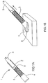
- a syringe (1) having a hollow cylindrical body (2) in which a piston (3) moves is used.
- the body of the syringe is closed by a filter plug (4).
- the filter plug (4) In the body (2) of the syringe, between the end (5) of the piston and the filter plug (4) are granules (6) of BCP ( Figure 1A ). The set was sterilized prior to use.
- the end of the syringe carrying the filter plug (4) is placed in a container (7) filled with an aqueous solution (8) of CaCl 2 at a concentration of 1%.
- a rearward movement of the piston (3) allows the solution (8) to be sucked into the body (2) of the syringe (1) ( Figure 1B ).
- the whole is left to rest for 10 minutes so that the particles of BCP impregnate the solution, then with the aid of the piston (3) the excess solution (8) of CaCl 2 is expelled through the filter plug (4) ( Figure 1C ).
- the filter (9) is removed from the filter plug (4) and a pressure on the piston (3) is used to deposit the solution-impregnated BCP granules (6) on the operating site (10) ( Figure 1D ).
- the implantation site is then closed again (step not shown).
- HA powder or TCP powder were placed in the body of a 1 ml syringe.
- 100 ⁇ l of blood was added to each syringe containing either HA or TCP. This mixture was placed on a wheel to keep the powder in suspension in the blood during the coagulation time, ie 10 minutes.
- the wheel is stopped, the syringes are recovered, their tip is cut and the blood / powder mixture is extracted by pushing with the plunger of the syringe. The coagulation of the blood around the powder is observed or not.
- Each experiment was repeated 3 times.
- Control positive control of coagulation. A clot and serum exudate are observed.
- the calcium assay was performed using the QuantiChrom Calcium Assay Kit (CENTAUR, Brussels, Belgium) and following the manufacturer's instructions. For this, aliquots of 5 .mu.l of supernatant were placed in the presence of 200 .mu.l of a solution of phenolsulphonephthalein, a dye which forms a stable complex of blue color in the presence of free calcium. After incubation for 3 minutes, a staining intensity measured at 612 nm is obtained, which is directly proportional to the calcium concentration of the sample. In each plate, a standard range is made from the following concentrations of calcium 0 - 0,5 - 1 - 1,5 - 2 - 3 - 4 - 5 mM.
- coagulation assays were performed on 50 mg aliquots of microparticles. BCP (60/40) and 50 mg of HA powder, after addition of 50 ⁇ l of 150 mM NaCl or 50 ⁇ l of 2.5 mM CaCl 2 , 2H 2 O.
- the biomaterial was prepared by adding 100 ⁇ l of total blood not containing an anticoagulant to 50 mg of BCP particles in the presence of a fixed volume of 50 ⁇ l of CaCl 2 , 2H 2 O, prepared at concentrations of 0.01. % (0.68 mM) - 0.02% (1.36mM) - 0.05% (3.4 mM) - 0.1% (6.8 mM) - 0.2% (13.6 mM) 0.5% (34 mM) - 1% (68 mM) - 10% (680 mM) or the same volume of NaCl at 150 mM.
Description
- L'invention a pour objet un nouveau biomatériau à base de phosphate de calcium, notamment à base d'hydroxyapatite ou à base d'un matériau comprenant de l'hydroxyapatite tel que les phosphates de calcium biphasés et les ciments phosphocalciques, un procédé pour sa préparation et son utilisation pour la fabrication d'un implant ou pour la pose d'une prothèse pour permettre la régénération du tissu osseux.
- La reconstruction des pertes de substance osseuse, d'origine principalement traumatique et plus rarement tumorale, est une des grandes difficultés rencontrées par les chirurgiens orthopédistes. Les défauts de petite taille, depuis la pseudarthrose « serrée » (défaut de consolidation d'une fracture où la perte de substance est virtuelle) jusqu'à des pertes osseuses de 5-6 cm font, le plus souvent, l'objet d'une greffe autologue de tissu osseux spongieux ou cortico-spongieux prélevé sur la crête iliaque (gold standard). Les défauts de grandes tailles (≥ 6 cm) nécessitent des interventions beaucoup plus lourdes, transferts osseux vascularisés ou procédure de Masquelet. Malgré tout, la quantité d'os autologue disponible est limitée, la consolidation osseuse reste aléatoire et ces différentes techniques sont fortement pourvoyeuses de complications post opératoires au niveau du site de prélèvement de la greffe.
- Différents biomatériaux disponibles en pratique clinique permettent d'éviter, en théorie, les inconvénients de la greffe autologue. Malheureusement, aucun d'eux n'égale les résultats de la greffe osseuse et ils ne permettent jamais la reconstruction de pertes de substance de grande taille.
- La majorité des matériaux étudiés actuellement associent aux biomatériaux des cellules souches mésenchymateuses obtenues à partir de moelle osseuse après plusieurs semaines de sélection et de culture cellulaire in vitro. Cette approche est lourde et coûteuse, ce qui limite les retombées cliniques.
- Il est connu que le sang coagulé favorise la reconstruction osseuse. L. Okazaki et al., Clin. Oral Impl. Res., 16, 2005, 236-243 décrit des implants à base de poudre d'os déminéralisé ou de sang coagulé. Le document
WO 02/068010 - De tels matériaux, résultant de l'association d'un support et de sang coagulé ou non, ont été jusqu'à présent utilisés en chirurgie maxillo-faciale où les problèmes de consolidation osseuse sont peu importants, mais ils ont été peu ou pas utilisés dans la réparation d'os diaphysaires.
- Les procédés de fabrication de ces implants nécessitent de prélever du sang sur un donneur, le plus souvent le destinataire de l'implant, et requièrent ensuite des étapes de manipulation du support (os déminéralisé ou polymère de synthèse, céramique), notamment des étapes de mélange avec le sang, qui sont sources de contamination du biomatériau. En outre, il est difficile par ces procédés d'obtenir un biomatériau homogène.
- Il subsiste donc le besoin d'un procédé de préparation d'un biomatériau implantable à partir d'un support de synthèse, donc facile à produire, avec des propriétés constantes et homogènes, ce procédé permettant d'obtenir des propriétés supérieures en termes de biocompatibilité, et permettant la reconstruction rapide d'un tissu osseux de qualité, sans qu'il soit nécessaire de faire appel à des étapes de culture ou de prélèvement.
- L'invention permet de remédier aux inconvénients de l'art antérieur et permet l'obtention d'un os d'excellente qualité en termes de dureté et de vascularisation. En outre le procédé de fabrication de ce biomatériau est simple, facile à mettre en oeuvre, ne nécessite pas de multiples interventions sur l'individu à traiter, est peu coûteux comparativement aux procédés de l'art antérieur.
- L'hydroxyapatite entre dans la composition de nombreux matériaux de reconstruction osseuse. L'hydroxyapatite peut être employée seule dans cette application ou en mélange avec d'autres composants, comme c'est le cas par exemple dans le phosphate de calcium biphasique, ou dans les ciments phosphocalciques.
- Le phosphate de calcium biphasique, BCP, est utilisé dans de nombreuses applications médicales et dentaires. Le phosphate de calcium biphasique a été décrit pour la première fois comme matériau de réparation osseuse par Nery et al., J Periodontol. 1992 Sep; 63(9):729-35. Le BCP est constitué d'un mélange d'hydroxyapatite (HA) Ca10(PO4)6(OH)2 et de phosphate tricalcique bêta (Ca3(PO4)2) (β-TCP). Sa bioactivité et sa biorésorbatilité peuvent être contrôlées par la proportion d'hydroxyapatite et de β-TCP qui le constituent.
- Le document
US-2005/0226939 décrit un procédé de fabrication de nanoparticules d'hydroxyapatite, ce procédé comportant le mélange d'une composition à base d'ions calcium et d'ions phosphate et un traitement par des micro-ondes. Les conditions employées dans ce document ne conduisent pas à la formation d'hydroxyapatite ou de BCP imprégné par une solution de chlorure de calcium. - Les biomatériaux à base de BCP ont l'avantage, par rapport aux autres biomatériaux de synthèse, de favoriser l'ostéogenèse.
- Le BCP a fait l'objet de nombreuses études : Lerouxel et al. Biomaterials, 2006, Sept. 27(26) :4566-72, 18, 287-294 ; Malard O. et al., J. Biomed. Mater. Res., 46(1), 1999, 103 ; Mankani M.H. et al., Biotechnology and Bioengineering, 72(1), 2001, 96-107. Ces différents auteurs ont produit des observations relatives à l'influence de la taille des particules de BCP. Dans le document Mankani M.H. et al., le procédé comporte le mélange de particules de HA/TCP avec des cellules puis du fibrinogène, et de la thrombine reconstituée dans une solution de CaCl2. Mais la solution de CaCl2 est plus concentrée que celle utilisée dans l'invention et le ratio mole/poids de CaCl2/ BCP est supérieur à celui employé dans les biomatériaux de l'invention.
- Trojani C. et al., Biomaterials, 27, 2006, 3256-3264, ont montré qu'une bonne ostéoinduction pouvait être obtenue pour l'implantation d'un matériau composite BCP/hydrogel de Si- hydroxypropylméthyl cellulose auquel ont été additionnées des cellules de moelle osseuse, avec des particules de BCP calibrées de 40 à 80 µm. Ces derniers procédés requièrent cependant une étape de prélèvement de cellules de moelle osseuse ainsi que leur mise en culture.
- Le document
WO 2006/015275 décrit un procédé favorisant la régénération osseuse, ce procédé comprenant la préparation d'une composition constituée d'un matériau support à base de phosphate de calcium, d'un plasma riche en plaquettes, de calcium et un activateur du récepteur PAR distinct de la thrombine. Mais la concentration en CaCl2 dans ces compositions est si élevée qu'il agirait, s'il était mis en oeuvre dans les conditions de la présente invention, comme anticoagulant. - Les deux composants du BCP, HA et β-TCP, représentent les deux principaux types de phosphates de calcium utilisés en chirurgie osseuse et dentaire. Ils sont remarquablement biocompatibles et on considère que leur association présente une meilleure bioactivité et donc une plus grande efficacité que l'HA seule ou le β-TCP seul. En effet, dans les BCP les deux composantes (HA et β-TCP) présentent une synergie d'action :
- L'hydroxyapatite, dès son implantation in vivo et de par sa structure chimique, peut promouvoir à sa surface, la formation d'apatite phosphocalcique non stoechiométrique polysusbtituée (dites « apatites biologiques ») par croissance épitaxique. Cette couche d'apatite biologique, très proche des cristaux présents dans la matrice osseuse, faciliterait l'adhésion et l'activité cellulaires.
- Le β-TCP, beaucoup plus soluble que l'HA, maintient une sursaturation en calcium et phosphates des fluides biologiques environnant l'implant en BCP. Ceci permet d'entretenir le phénomène de précipitation d'apatite biologique sur la phase HA. De plus cette phase est beaucoup plus résorbable que HA, il est donc possible de moduler la résorbabilité de l'implant en faisant varier le rapport HA/β-TCP.
- Ces phénomènes chimiques ont été montrés in vitro (J.M. Bouler, G. Daculsi, Key Engineering Materials 2001 ;192-195:119-122; Yamada S, et al., Biomaterials 1997;18:1037-41 ; S. Yamada et al., J. Biomed. Mater. Res 1997;37:346-52) et in vivo (Daculsi G, et al., J Biomed Mater Res 1989;23:883-94 ; G. Daculsi et al. Int. Rev. Cytol 1997;129-191). Ces mécanismes semblent participer à la meilleure efficacité clinique des BCP par rapport aux matériaux monophasés en HA ou TCP (Nery EB et al., J Periodontol 1992;63:729-35 ; Ellinger RFet al., Int J Periodontics Restorative Dent 1986;6:22-33 ; Passuti N. et al., Clin Orthop Relat Res 1989;(248):169-76 ; Gouin F et al., Rev Chir Orthop Reparatrice Appar Mot 1995;81:59-65 ; Ransford AOet al., J Bone Joint Surg Br 1998;80:13-8 ; R. Cavagna et al., J. Long-Term effect of Med. Impl 1999;9:403-412).
- La présente invention se fonde sur la constatation que certains phosphates de calcium, en particulier les apatites phosphocalciques, comme l'hydroxyapatite, inhibent la coagulation spontanée du sang total lorsqu'ils sont mis en contact. En effet, il a été constaté que l'HA, et aussi le BCP, qui contient de l'HA, inhibent la coagulation spontanée du sang total lorsqu'ils sont mis en contact. Il a été constaté aussi que si l'on imprègne au préalable l'hydroxyapatite ou le BCP avec du sérum physiologique (qui est une phase aqueuse de NaCl), comme recommandé dans les notices d'utilisation de ces biomatériaux, le sang mis en contact ensuite ne coagule pas.
- Des conditions expérimentales permettant d'établir les propriétés anticoagulantes d'un matériau support sont détaillées dans la partie expérimentale.
- L'invention a pour premier objet un procédé de préparation d'un biomatériau implantable comportant un support à base d'au moins un phosphate de calcium tel que l'hydroxyapatite ou un mélange d'hydroxyapatite et d'au moins un autre matériau, ce procédé comportant au moins une étape d'imprégnation du support par au moins un agent coagulant.
- Par support à base de phosphates de calcium on entend un matériau comprenant au moins un constituant du type apatite phosphocalcique choisi parmi : l'hydroxyapatite, la fluorapatite, les apatites non stoechiométriques polysubstituées (ANSPS) ainsi que leurs mélanges avec d'autres biomatériaux phosphocalciques. De tels supports peuvent être constitués par de l'hydroxyapatite, du BCP, des ANSPS et des ciments phosphocalciques apatitiques.
- Le support ainsi imprégné est ensuite implanté sur le site où un défaut osseux doit être comblé. Son implantation provoque alors la coagulation du sang qui entre en contact avec le biomatériau ou qui pénètre dans le biomatériau in situ. Dans les essais qui ont été réalisés avec du BCP, il a été constaté que le BCP, associé à du sang coagulé, permet d'obtenir une bonne ostéogenèse et conduit à un tissu osseux de qualité très satisfaisante par un procédé très simple comparativement à ceux de l'art antérieur. Sans imprégnation du BCP par une solution d'agent coagulant, on n'obtient pas d'ostéogenèse.
- Selon une variante de l'invention, le support à base de phosphate de calcium, notamment à base d'hydroxyapatite ou d'un mélange d'hydroxyapatite et d'un autre matériau est implanté dans le site où un défaut osseux doit être comblé puis il est imprégné in situ par une solution d'agent coagulant.
- De préférence, le support utilisé dans l'invention est à base d'hydroxyapatite, de fluorapatite, d'apatites non stoechiométriques polysubstituées (ANSPS) ou d'un mélange d'un de ces composés avec au moins un autre biomatériau tel que le phosphate tricalcique sous forme α et β (Ca3(PO4)2), le phosphate dicalcique dihydraté (CaHPO4, 2H2O), le phosphate dicalcique anhydre (CaHPO4), le phosphate monocalcique monohydrate (Ca (HPO4)2, H20), le phosphate tétracalcique (Ca (PO4)2 O), et le phosphate octocalcique (Ca8 H2 (PO4)6). De façon avantageuse, le support est à base d'hydroxyapatite ou de BCP, de préférence il est à base de BCP.
- Le support, et notamment l'apatite ou le BCP qui peuvent être utilisés dans l'invention peuvent être de toute forme : soit sous la forme d'un monolithe soit sous forme de granulés, calibrés ou non.
- Le BCP qui peut être utilisé dans l'invention consiste en un fritté haute température. Lorsqu'il est sous forme de granulés il est broyé et calibré, par exemple par tamisage, en fonction du diamètre choisi. De façon avantageuse le BCP qui peut être utilisé dans l'invention comporte de l'hydroxyapatite et du phosphate tri calcique β dans un rapport en poids/poids HA/ β-TCP compris entre 5/95 et 95/5, de préférence entre 30/70 et 80/20, avantageusement entre 40/60 et 60/40.
- Avantageusement il s'agit d'un support, et en particulier d'un BCP, poreux, avec des tailles de pores allant de 50 nm à 1000 µm, avantageusement de 500 nm à 100 µm et plus avantageusement de 1 µm à 50 µm.
- Lorsque le support, et en particulier le BCP, utilisé dans la présente invention est sous forme de granulés, il présente avantageusement une granulométrie comprise entre 40 et 500 µm, préférentiellement entre 40 et 400 µm, encore plus préférentiellement entre 40 et 300 µm, et avantageusement entre 80 et 200 µm.
- Les granulés ou la poudre de BCP peuvent être obtenus conformément aux méthodes décrites par Bouler et al., J Biomed Mater Res, 1996, 32, 603-609 ; Bouler et al., J Biomed Mater Res, 2000, 51, 680-684 ; Obadia et al., J Biomed Mater Res, 2006, 80(B), 32-42.
- Le BCP peut être obtenu commercialement auprès de la société GRAFTYS SARL (Aix en Provence).
- L'hydroxyapatite qui peut être utilisée dans l'invention est préférentiellement sous la forme de granulés. Elle est disponible commercialement auprès de la société GRAFTYS SARL.
- Plus particulièrement, l'invention concerne un biomatériau comprenant un support à base de phosphate de calcium, imprégné par une solution d'au moins un agent coagulant dérivé du calcium, le support étant choisi parmi : de l'hydroxyapatite et un BCP, l'agent coagulant est sous forme d'une solution aqueuse d'une concentration allant de 1 à 50 mMol/l, la proportion de solution d'agent coagulant et de HA ou de BCP est de 0,5 à 5 en volume/volume de solution d'agent coagulant par rapport au volume de HA ou de BCP.
- De façon préférentielle, l'invention concerne un biomatériau comprenant un support à base de phosphate de calcium, imprégné par une solution d'au moins un agent coagulant dérivé du calcium, le support étant choisi parmi : de l'hydroxyapatite et un BCP, l'agent coagulant dérivé du calcium étant présent en proportion allant de 2,5 à 60 µmol de calcium par gramme de HA ou de BCP, et de préférence de 5 à 40 µmol de calcium par gramme de HA ou de BCP.
- L'agent coagulant est un agent coagulant à base de calcium, tel qu'un sel de calcium biocompatible, comme par exemple : CaCl2, Ca(NO3)2, Ca(AcOEt)2, CaSO4.
- Avantageusement l'agent coagulant est à base de calcium et il est choisi parmi les sels de calcium biocompatibles, avantageusement CaCl2. Pour permettre l'imprégnation du support, en particulier du HA ou du BCP, par l'agent coagulant, ce dernier est utilisé en solution aqueuse, avantageusement en solution aqueuse d'une concentration allant de 1 à 50 mMol/l, de préférence de 3 à 40 mMol/l, avantageusement de 5 à 20 mMol/l. Ces valeurs sont particulièrement préférées dans le cas où l'agent coagulant est un sel de calcium et notamment CaCl2.
- La proportion de solution d'agent coagulant et de HA ou de BCP mis en oeuvre dans le procédé de l'invention est de 0,5 à 5 en volume/volume de solution d'agent coagulant par rapport au poids de HA ou de BCP, de préférence de 1 à 3, avantageusement environ 2.
- Selon une autre variante de l'invention, le biomatériau est préparé de façon extemporanée, par imprégnation du support à base de phosphate de calcium juste avant son implantation.
- On peut aussi prévoir de préparer le biomatériau de l'invention en appliquant la procédure suivante : imprégner le support à base de phosphates de calcium par une solution d'agent coagulant puis le sécher ou le lyophiliser puis le conditionner dans des conditions stériles et le conserver jusqu'à son implantation.
- Avantageusement, la durée de l'imprégnation est de 1 minute à 1 heure, de préférence de 1 à 30 minutes, avantageusement de 5 à 15 minutes.
- Selon une autre variante de l'invention, on peut préparer un biomatériau de l'invention en imprégnant le support à base de phosphate de calcium par une solution d'agent coagulant puis on conditionne ce biomatériau dans des conditions stériles et on le conserve ainsi jusqu'à son implantation.
- Selon une variante de l'invention, on peut prévoir que le biomatériau support (à base de phosphate de calcium) est associé à l'agent coagulant sous forme de poudre. En particulier on peut mélanger un biomatériau tel que du BCP ou de l'HA sous forme de poudre ou de granulés avec un sel de calcium sous forme de poudre solide. Ce biomatériau peut ainsi être stocké jusqu'à son utilisation et imprégné par une solution aqueuse, comme par exemple du sérum physiologique, de façon extemporanée, juste avant son implantation, au moment de son utilisation. Il peut aussi être implanté sous forme sèche, non imprégnée.
- Selon l'invention, on peut prévoir qu'au support, et notamment au BCP, soient ajoutés un ou plusieurs additifs éventuels tels que : polymères, particules de céramiques, molécules pharmaceutiques, agents bioactifs, les conditions pour l'emploi de ces matériaux étant : leur biocompatibilité, l'absence d'effet négatif sur la réaction de coagulation sanguine. Si l'un de ces additifs avait un effet défavorable sur la coagulation sanguine, celui-ci devrait être pris en compte dans la quantité d'agent coagulant à mettre en oeuvre. Par exemple, de tels additifs ou actifs peuvent être employés par greffage du support, BCP ou autre, par mélange ou imprégnation ou par enrobage. De tels additifs bien connus de l'homme du métier sont destinés à modifier la rhéologie du biomatériau, son comportement in vivo (dureté, résorption, ostéogenèse) ou à agir sur l'apparition d'infections ou de phénomènes inflammatoires (antibiotiques, anti-infectieux, agents anti-inflammatoires).
- On peut aussi prévoir d'introduire dans le biomatériau de l'invention une ou plusieurs molécules thérapeutiques comme des molécules destinées à prévenir ou traiter une pathologie choisie par exemple parmi : un cancer, l'ostéoporose.
- On peut aussi prévoir d'introduire dans le biomatériau de l'invention du tissu adipeux, ou toute autre préparation de tissu ou de cellules, prélevé chez le patient auquel est destiné le biomatériau, ce tissu adipeux ou cette préparation ayant été préalablement mis en suspension dans du sang ou du plasma ou du sérum physiologique.
- On peut également introduire dans le biomatériau de l'invention des facteurs de croissance, naturels ou de synthèse. On peut aussi prévoir la présence de biomarqueurs ou d'agents de contraste qui favorisent la visualisation par imagerie médicale de la résorption du biomatériau et son devenir dans l'organisme.
- Selon le procédé de l'invention, le support, et notamment le HA ou le BCP, est placé dans une cavité d'un récipient fermé et stérile. Lorsque le support est sous forme de granulés, on peut par exemple le placer dans la cavité intérieure d'une seringue. La quantité appropriée d'agent coagulant est introduite dans ce récipient, par exemple par aspiration à l'aide de la seringue si l'on a utilisé un tel dispositif.
- Dans le cas où le support, et notamment le HA ou le BCP, est sous forme d'un monolithe, celui-ci est placé dans un récipient de forme et de dimensions appropriées.
- Dans tous les cas le volume du récipient est prévu pour permettre l'introduction de la quantité voulue de solution d'agent coagulant.
- Le récipient clos contenant le support, notamment le HA ou le BCP, et l'agent coagulant peut être agité, de façon à permettre l'imprégnation homogène du biomatériau. Mais on peut aussi prévoir une imprégnation passive du support par l'agent coagulant.
- A l'issue de cette étape, le biomatériau est sous forme :
- d'une pâte liquide, homogène, lorsque le support a été employé sous forme de granulés,
- d'un monolithe dont les cavités sont remplies de liquide, lorsque le support a été employé sous forme d'un monolithe.
- Selon une variante de l'invention on peut prévoir d'implanter directement le matériau support dans l'espace à combler, éventuellement en mélange avec l'agent coagulant sous forme de poudre, puis de l'imprégner in situ, soit par la solution d'agent coagulant, soit lorsqu'il est déjà en mélange avec l'agent coagulant, avec une solution aqueuse adaptée telle que du sérum physiologique. On peut aussi prévoir de l'implanter, lorsqu'il est sous forme de mélange sec avec l'agent coagulant, sans l'imprégner, et de le laisser être imprégné par le sang des tissus environnants.
- Un autre objet de l'invention est un biomatériau comportant un support à base de phosphate de calcium tel que l'hydroxyapatite ou un BCP, imprégné par une solution d'au moins un agent coagulant, tel que décrit ci-dessus.
- Suivant la forme physique du support, HA ou BCP, et le type de dispositif qui a été employé pour la préparation du biomatériau de l'invention, celui-ci peut ensuite être appliqué à l'aide des moyens les plus adaptés à l'emplacement où un défaut osseux doit être comblé :
- A l'aide d'un outil tel qu'une spatule, ou à l'aide d'une seringue dont l'extrémité comporte une ouverture adaptée à la rhéologie et à la taille des particules du biomatériau de l'invention. Il peut aussi être implanté directement sous forme d'un monolithe. Dans ce dernier cas celui-ci aura été conçu ou taillé pour que sa forme et ses dimensions correspondent à celles de l'espace à combler.
- L'invention a encore pour objet un procédé de comblement d'un défaut osseux, ce procédé comportant les étapes énumérées ci-dessus et comportant en outre une étape d'insertion du biomatériau dans l'espace où un défaut osseux a été constaté. Ce procédé peut en outre comprendre des étapes d'incision de tissu et de suture.
- Suivant la taille et la configuration du défaut osseux, le comblement par le biomatériau de l'invention peut être associé à la pose d'une ostéosynthèse provisoire qui permet de conférer au tissu atteint la résistance mécanique nécessaire pendant que se produit la reconstruction osseuse sur le site d'implantation du biomatériau de l'invention.
- Comme les inventeurs l'ont constaté, l'implantation du biomatériau de l'invention a permis de favoriser la formation de tissu osseux dans des délais courts (quelques semaines), ce tissu osseux étant très richement vascularisé.
- Un autre objet de l'invention est constitué par un kit pour la mise en oeuvre du procédé de l'invention, ce kit comprenant l'association d'un support à base d'un phosphate de calcium comme une hydroxyapatite ou un mélange d'hydroxyapatite et d'au moins un autre matériau, comme par exemple un BCP microporeux, avec un agent coagulant dérivé du calcium. Avantageusement l'agent coagulant est un sel de calcium biocompatible, comme CaCl2.
- La quantité d'agent coagulant est calculée pour compenser l'effet anticoagulant du phosphate de calcium, et en particulier de l'hydroxyapatite et favoriser la coagulation sanguine des tissus environnants.
- Une telle association peut être sous la forme d'un kit comportant :
- (a) un dispositif stérile comportant une cavité intérieure stérile dans lequel est placé le support, comme par exemple le BCP ou l'HA,
- (b) un réservoir stérile comportant l'agent coagulant.
- Le réservoir (b) peut faire partie du dispositif (a) ou être une entité distincte telle qu'un tube ou un flacon dans lequel on peut prélever l'agent coagulant pour le transférer dans la cavité intérieure du dispositif (a), ou une seringue permettant l'injection de l'agent coagulant dans la cavité où est placé le support.
- La cavité intérieure du dispositif (a) est d'une taille permettant d'y introduire la quantité d'agent coagulant nécessaire pour produire le biomatériau de l'invention, ainsi que les autres constituants du mélange tels que par exemple des principes actifs.
- De façon avantageuse également le dispositif (a) comporte des moyens permettant l'application du biomatériau dans la zone où un défaut osseux a été constaté.
- Un tel dispositif peut être constitué d'une seringue.
- On peut également prévoir d'utiliser un dispositif tel que celui décrit dans
WO 02/068010 - Un biomatériau selon l'invention peut être utilisé pour la fabrication d'un implant osseux, qu'il s'agisse de combler une fracture, une perte de substance d'origine traumatique ou tumorale, un défaut consécutif à une intervention chirurgicale, ou d'aider à la mise en place d'une prothèse.
- Le biomatériau peut être introduit par une intervention chirurgicale dans la zone où un défaut osseux doit être comblé. Après incision, le biomatériau est implanté et l'incision est refermée.
- Le biomatériau de l'invention peut être associé à une ostéosynthèse de façon à permettre la consolidation provisoire en attendant la stabilisation de la zone déficiente par les tissus osseux.
- Un enrobage de la prothèse par le biomatériau de l'invention permet de favoriser l'implantation de tissu osseux vivant dans ou autour de la prothèse.
- Le biomatériau de l'invention peut encore être utilisé in vitro ou ex-vivo comme support pour la production de tissu osseux :
- En effet, la culture de cellules osseuses autour de ce biomatériau permet de produire un tissu osseux ultérieurement implantable.
- Un autre objet de l'invention est l'utilisation in vitro ou ex-vivo d'un biomatériau tel que décrit ci-dessus pour produire un implant osseux.
- On peut, selon l'invention cultiver des cellules osseuses sur le biomatériau de l'invention dans un moule ayant la forme de la prothèse que l'on souhaite fabriquer. La culture de cellules dans ces conditions permet d'obtenir une prothèse biocompatible de forme et de dimensions adaptées.
-
-
Figures 1A à 1D : Préparation des implants et procédure chirurgicale -
Figure 2 : Illustration graphique de la concentration en calcium d'un plasma au contact des biomatériaux de phosphate de calcium. -
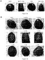
Figure 3A : Photographie du produit obtenu par l'ajout d'une solution de chlorure de calcium au BCP et à l'HA avant l'addition de sang. -
Figure 3B : Photographie du produit obtenu par l'ajout d'une solution de chlorure de calcium en concentrations croissantes sur du BCP. -
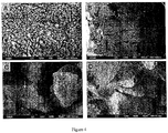
Figure 4 : Analyse en microscopie électronique à balayage d'implants constitués de sang total prélevé sans anticoagulant (A, C) et de microparticules de BCP (80-200µm), ou d'implants préparés selon le protocole habituel (B, D). Dans les implants préparés sans calcium, on note l'absence de formation de réseau de fibrine et de caillot sanguin autour des grains (A, C). La flèche blanche en (C) indique quelques globules rouges déposés sur les grains. Echelles A, B: 100 µm, C, D : 10 µm. - Il s'agit d'une procédure extemporanée, réalisée au bloc opératoire. Elle consiste à mélanger dans le corps d'une seringue en polypropylène des particules de BCP et un agent coagulant : CaCl2. L'implantation de ce biomatériau dans le site où un défaut osseux a été constaté favorise la coagulation autour du biomatériau.
- Le biomatériau de phosphate de calcium biphasé (BCP) est composé de 60 % d'hydroxyapatite (HA; Ca10(PO4)6(C)H)2) et de 40% de phosphate tricalcique (TCP; Ca3(PO4)2). Les particules de BCP calibrées entre 40 et 200 microns ont été fournies par la société GRAFTYS SARL (Aix-en-Provence, France). Les particules ont été stérilisées par chauffage à 180°C pendant deux heures.
- La concentration en calcium a été mesurée dans du plasma de souris C57BL/6 (Janvier, Le Genest-St-Isle, France). Ce plasma a été préparé à partir de sang prélevé sur héparine, par centrifugation à 1800g pendant 15 minutes. L'héparine est utilisée comme anticoagulant ne modifiant pas la concentration en calcium du plasma. L'analyse a été réalisée dans un automate Hitachi (Orléans, France).
- Comme illustré sur les
figures 1A à 1D , on utilise une seringue (1) comportant un corps (2) cylindrique creux dans lequel se déplace un piston (3). A l'extrémité du corps (2) qui n'est pas obturée par le piston, le corps de la seringue est obturé par un bouchon filtre (4). Dans le corps (2) de la seringue, entre l'extrémité (5) du piston et le bouchon filtre (4) se trouvent des granules (6) de BCP (Figure 1A ). L'ensemble a été stérilisé préalablement à son emploi. L'extrémité de la seringue portant le bouchon filtre (4) est placée dans un récipient (7) rempli d'une solution (8) aqueuse de CaCl2 à une concentration de 1%. Un mouvement arrière du piston (3) permet d'aspirer la solution (8) à l'intérieur du corps (2) de la seringue (1) (Figure 1B ). L'ensemble est laissé au repos pendant 10 mn afin que les particules de BCP s'imprègnent de la solution, puis à l'aide du piston (3) la solution (8) de CaCl2 en excès est expulsée au travers du bouchon filtre (4) (Figure 1C ). Le filtre (9) est retiré du bouchon-filtre (4) et une pression sur le piston (3) permet de déposer les granulés (6) de BCP imprégnés de solution (8) sur le site opératoire (10) (Figure 1D ). Le site d'implantation est ensuite refermé (étape non représentée). - 50 mg de poudre de HA ou de poudre de TCP ont été placés dans le corps d'une seringue de 1 ml. 100 µl de sang ont été ajoutés dans chaque seringue contenant soit de l'HA, soit du TCP. Ce mélange a été placé sur une roue permettant de maintenir la poudre en suspension dans le sang pendant le temps de la coagulation, i.e 10 minutes. Dans chaque expérience une seringue contenant 100 µl de sang total traitée comme les autres i.e 10 min sur la roue, a servi de contrôle positif de la coagulation. Après 10 minutes, la roue est arrêtée, les seringues sont récupérées, leur bout est sectionné et le mélange sang/poudre est extrait en poussant avec le piston de la seringue. On observe ou pas la coagulation du sang autour de la poudre. Chaque expérience a été répétée 3 fois.
- On a observé qu'en présence de 50 mg de HA et 100 µl de sang total la coagulation a été inhibée. Le sang reste liquide.
- Témoin : contrôle positif de coagulation. On observe un caillot et un exsudat de sérum.
- En présence de 50 mg de TCP + 100 µl de sang, la coagulation a eu lieu, elle se traduit par la formation d'un implant dans lequel le réseau de fibrine maintient la poudre en suspension de façon homogène.
- La même expérience a été réalisée en ayant ajouté du chlorure de calcium dans la seringue contenant le HA préalablement à l'introduction de sang : on retrouve une coagulation et la formation d'un implant.
- Nous avons observé que le sang fraîchement prélevé (100 µl) en absence d'anticoagulant et immédiatement mélangé aux particules de BCP (50 mg), ne coagule pas. Cet effet anticoagulant est annulé par l'ajout de CaCl2 (20 µl d'une solution de CaCl2 à 1%) suggérant une captation du calcium plasmatique par le BCP. Cette hypothèse a été confirmée par la mesure de la concentration en calcium dans le plasma avant et après contact avec du BCP. Pour cela, du plasma a été préparé à partir de sang de souris C57BL/6 prélevé sur héparine (anticoagulant ne modifiant pas la concentration plasmatique en calcium). En présence de BCP nous avons observé une chute de la concentration du calcium plasmatique de 2,06 ± 0,06 mmol / L (valeur normale) à 0,59 ± 0,07 mmol / L en présence de BCP.
- Des aliquots de 50 mg de microparticules de BCP (60/40), des aliquots de 50 mg de HA ou de β-TCP ont été mis en présence soit de 50 µl d'H2O soit de 50 µl d'une solution à 2,5 mM de CaCl2, 2H2O, et mis à sécher pendant une nuit à 56°C. Ces biomatériaux ont été déposés dans les puits d'une microplaque de 96 puits. Dans chaque puits a été ajouté 100 µl de plasma préparé à partir de sang de souris C57BL/6 prélevé sur héparine, anticoagulant qui n'interfère pas avec le taux de calcium. Après 15 minutes d'incubation, la plaque a été centrifugée pendant 2 minutes à 800 g et les surnageants prélevés afin de doser la concentration en calcium du plasma. Le dosage du calcium a été réalisé en utilisant le Kit QuantiChrom Calcium Assay (CENTAUR, Bruxelles, Belgique) et en suivant les instructions du fabricant. Pour cela des aliquots de 5 µl de surnageant ont été mis en présence de 200 µl d'une solution de phenolsulphonephthalein, colorant qui forme un complexe stable de couleur bleue en présence de calcium libre. Après incubation pendant 3 minutes, on obtient une intensité de coloration mesurée à 612 nm, qui est directement proportionnelle à la concentration en calcium de l'échantillon. Dans chaque plaque, une gamme étalon est réalisée à partir des concentrations suivantes en calcium 0 - 0,5 - 1 - 1,5 - 2 - 3 - 4 - 5 mM.
- Nous avons constaté (
Figure 2 ) que le BCP sous forme de microparticules ainsi que la poudre de HA, mis en contact avec du plasma, induisaient une diminution importante et significative de sa concentration en calcium. La chute de la concentration en calcium est similaire pour le BCP et l'HA et n'est pas observée pour le β-TCP. A partir des valeurs obtenues pour le plasma seul (1,960 ± 0,044 mM), le plasma en présence de BCP (0,871 ± 0,160 mM) et le plasma en présence de HA (0,840 ± 0,121 mM), nous avons évalué la captation de calcium à 0,125 µmole de calcium pour 50 mg de BCP ou de HA. - Nous avons également constaté que l'ajout de 50 µl d'une solution à 2,5 mM (soit 0,125 µmole) au BCP ou à l'HA avant d'ajouter le plasma permettait de restaurer une concentration plasmatique de calcium normale (
Figure 2 ). La même quantité de chlorure de calcium ajoutée au β-TCP s'ajoute au calcium plasmatique initial et confirme l'absence de captation par ce biomatériau dans ces conditions. - Par ailleurs nous avons observé que la captation de calcium était identique pour les trois granulosités de BCP testées, c'est-à-dire pour les microparticules de 40-80 µm, de 80-200 µm et celles de 200-500 µm.
- De plus nous avons obtenu les mêmes résultats de compensation par l'ajout de calcium, que la solution de chlorure de calcium soit ajoutée extemporanément sous forme liquide juste avant d'ajouter le plasma ou que cette solution soit d'abord séchée au contact des particules.
- Afin de démontrer qu'il existait un lien de cause à effet entre l'inhibition de la coagulation et la chute du calcium plasmatique induite par le BCP et l'HA, nous avons réalisé des essais de coagulation sur des aliquots de 50 mg de microparticules de BCP (60/40) et de 50 mg de poudre de HA, après ajout de 50 µl de 150 mM NaCl ou de 50 µl de 2,5 mM CaCl2, 2H2O.
- Après ajout du sang non traité par un anticoagulant et rotation pendant 15 minutes nous avons constaté (
Figure 3A ) que l'ajout préalable de calcium au BCP et à l'HA permettait de rétablir la coagulation du sang mis en présence de ces deux biomatériaux. - Ces résultats démontrent que l'effet anticoagulant du BCP et de l'HA est bien lié à la diminution de la concentration en calcium plasmatique que ces deux biomatériaux induisent, et que l'ajout de calcium permet de rétablir la coagulation.
- Nous avons analysé l'effet d'une dose réponse de calcium sur la coagulation du sang mis en contact avec le BCP (
Figure 3B ). Le biomatériau a été préparé en ajoutant 100 µl de sang total non additionné d'un anticoagulant à 50 mg de particules de BCP en présence d'un volume fixe de 50 µl de CaCl2,2H2O, préparé aux concentrations de 0,01 % (0,68 mM) - 0,02 % (1,36mM) - 0,05 % (3,4 mM) - 0,1 % (6,8 mM) - 0,2 % (13,6 mM) - 0,5 % (34 mM) - 1 % (68 mM) - 10% (680 mM) ou du même volume de NaCl à 150 mM. Après incubation pendant 15 minutes sur une roue, le biomatériau est démoulé. Nous avons constaté qu'à faible concentration, correspondant ici à 0,01 et 0,02 %, le calcium ajouté ne permettait pas de rétablir la coagulation. En présence de concentrations comprises entre 0,05 et 0,5 % on observe une coagulation. De façon surprenante, nous avons constaté que l'augmentation de la concentration en CaCl2, 2H2O à 1% et au-delà induisaient à nouveau une inhibition de la coagulation (Figure 3B et tableau 1). - Ces expériences ont permis de déterminer les concentrations optimales en calcium permettant de bloquer l'effet anticoagulant du BCP 60/40 et ont montré qu'il existe une fourchette de concentration très importante à respecter.
- L'effet anticoagulant du BCP visualisé par l'absence de formation d'implants gélifiés cohésifs au cours des essais décrits ci-dessus, correspond au niveau moléculaire à l'inhibition de la formation du réseau de fibrine formant l'armature du caillot. Nous avons analysé la présence du réseau fibrine en microscopie électronique à balayage. Pour cela des implants ont été préparés en mélangeant 100 µl de sang non additionné d'anticoagulant avec 50 mg de BCP avec ou 50 mg de BCP incubé en présence de calcium puis séché. Après 15 minutes de rotation sur une roue, les mélanges sont démoulés et directement plongés dans une solution de fixation à 1,6% de glutaraldéhyde dans un tampon phosphate 0,1M pH 7. Les échantillons sont ensuite lavés, déshydratés par des bains d'alcool à concentrations croissantes, immergés en hexamethyldisilazane (Sigma-Aldrich, L'isle d'Abeau Chesnes, France) pendant 5 minutes et séchés à température ambiante. Après montage sur des supports en aluminium et recouvrement à l'or palladium pendant 4 minutes (Polaron, A5100, UK), l'analyse est réalisée à l'aide d'un microscope électronique à balayage (JEOL 6700F, Japan).
- Comme on le voit sur la
figure 4 , dans les conditions BCP nous n'avons pas observé de réseau de fibrine (Figures 4A, 4C ) entre les microparticules de BCP. La présence de quelques globules rouges déposés sur les grains atteste du mélange des particules avec le sang. Au contraire, en présence de BCP/calcium on constate la présence d'un caillot enserrant les particules, caillot visualisé par les mailles du réseau de fibrine et un très grand nombre de globules rouges (Figures 4B, 4D ).Tableau 1 Concentration de la solution de CaCl2,2H2O ajoutée au mélange de BCP et de sang (en % et en molarité) Nombre de µmoles de calcium apportées par 50 µl de CaCl2, 2H2O Test de coagulation 50 mg BCP + 100 µl sang + 50µl CaCl2, 2H2O (liq ou séché) 0 - 0,01% (0,68 mM) 0,034 - 0,02 % 0,068 + 0,03 % 0,102 + 0,04 % 0,136 + 0,05 % 0,17 + 0,1 % 0,34 + 0,2 % 0,68 + 0,5 % 1,7 + 0,6 % 2,04 + 0,7 % 2,38 + 0,8 % 2,72 + 0,9 % 3,06 + 1 % (68 mM) 3,4 +/- 2% - 10 % (680 mM) 34 -
Claims (15)
- Procédé de préparation d'un biomatériau comprenant un support à base de phosphate de calcium, caractérisé en ce que :- le support est choisi parmi l'hydroxyapatite et un BCP, de préférence un BCP, et- en ce que ledit support est imprégné par au moins un agent coagulant dérivé du calcium, de préférence CaCl2, sous forme d'une solution aqueuse d'une concentration allant de 1 à 50 mMol/l, de préférence de 3 à 40 mMol/l, avantageusement de 5 à 20 mMol/l,la proportion de solution d'agent coagulant et d'hydroxyapatite ou de BCP étant de 0,5 à 5, de préférence de 1 à 3, en volume de solution d'agent coagulant par rapport au volume d'hydroxyapatite ou de BCP.
- Biomatériau comprenant un support à base de phosphate de calcium, imprégné par une solution d'au moins un agent coagulant dérivé du calcium, caractérisé en ce que le support est choisi parmi l'hydroxyapatite et un BCP, et en ce que l'agent coagulant dérivé du calcium est présent en une proportion allant de 2,5 à 60 µmol de calcium par gramme d'hydroxyapatite ou de BCP, et de préférence de 5 à 40 µmol de calcium par gramme d'hydroxyapatite ou de BCP.
- Biomatériau comprenant un support à base de phosphate de calcium, associé à au moins un agent coagulant dérivé du calcium sous forme de poudre, caractérisé en ce que le support est choisi parmi l'hydroxyapatite et un BCP, et en ce que l'agent coagulant dérivé du calcium est présent en une proportion allant de 2,5 à 60 µmol de calcium par gramme d'hydroxyapatite ou de BCP, et de préférence de 5 à 40 µmol de calcium par gramme d'hydroxyapatite ou de BCP.
- Biomatériau selon la revendication 2 ou selon la revendication 3, dans lequel l'agent coagulant est CaCl2.
- Biomatériau selon l'une quelconque des revendications 2 à 4 dans lequel le support est sous forme de monolithe.
- Biomatériau selon l'une quelconque des revendications 2 à 4 dans lequel le support est sous forme de granulés.
- Biomatériau selon l'une quelconque des revendications 2 à 6 et comprenant en outre au moins un additif choisi parmi : des polymères, des particules de céramiques, des molécules pharmaceutiques, des facteurs de croissance, naturels ou de synthèse, des biomarqueurs, des agents de contraste, une préparation de tissu ou de cellules.
- Biomatériau selon l'une quelconque des revendications 2 à 7 ou obtenu selon la revendication 1 pour son utilisation comme implant dans un procédé de comblement d'un défaut osseux.
- Association d'un biomatériau selon l'une quelconque des revendications 2 à 7 ou obtenu selon le procédé de la revendication 1 avec une ostéosynthèse.
- Kit pour la mise en oeuvre du procédé de fabrication d'un biomatériau selon l'une quelconque des revendications 2 et 4 à 7 ou obtenu selon le procédé de la revendication 1, ce kit comprenant- un support à base d'hydroxyapatite ou de BCP, et- un agent coagulant dérivé du calcium, sous forme d'une solution aqueuse de concentration allant de 1 à 50 mM/l et la proportion de solution d'agent coagulant et d'hydroxyapatite ou de BCP est de 0,5 à 5 en volume/volume de solution d'agent coagulant par rapport au volume d'hydroxyapatite ou de BCP.
- Kit pour la mise en oeuvre d'un procédé de fabrication d'un biomatériau selon l'une quelconque des revendications 3 à 7, ce kit comprenant- un support à base d'hydroxyapatite ou de BCP, et- un agent coagulant dérivé du calcium sous forme de poudre.
- Kit selon la revendication 10 ou 11 qui comporte :(a) un dispositif stérile comportant une cavité intérieure stérile dans lequel est placé le support,(b) un réservoir stérile comportant l'agent coagulant.
- Kit selon la revendication 12 dans lequel le dispositif (a) comporte des moyens permettant l'application du biomatériau dans la zone où un défaut osseux a été constaté.
- Kit selon la revendications 12 ou 13 dans lequel le dispositif (a) est constitué d'une seringue.
- Utilisation d'un biomatériau selon l'une quelconque des revendications 2 à 7 ou obtenu selon le procédé de la revendication 1, in vitro ou ex-vivo comme support pour la production de tissu osseux, ou pour produire un implant osseux.
Applications Claiming Priority (2)
| Application Number | Priority Date | Filing Date | Title |
|---|---|---|---|
| FR0803493A FR2932687B1 (fr) | 2008-06-23 | 2008-06-23 | Biomateriaux a base de phosphates de calcium. |
| PCT/FR2009/000748 WO2010007229A1 (fr) | 2008-06-23 | 2009-06-22 | Biomateriaux a base de phosphate de calcium |
Publications (2)
| Publication Number | Publication Date |
|---|---|
| EP2307064A1 EP2307064A1 (fr) | 2011-04-13 |
| EP2307064B1 true EP2307064B1 (fr) | 2018-03-14 |
Family
ID=40292572
Family Applications (1)
| Application Number | Title | Priority Date | Filing Date |
|---|---|---|---|
| EP09797556.9A Active EP2307064B1 (fr) | 2008-06-23 | 2009-06-22 | Biomateriaux a base de phosphate de calcium |
Country Status (14)
| Country | Link |
|---|---|
| US (2) | US9233124B2 (fr) |
| EP (1) | EP2307064B1 (fr) |
| JP (2) | JP5595388B2 (fr) |
| KR (1) | KR101777427B1 (fr) |
| CN (1) | CN102123744B (fr) |
| AU (1) | AU2009272593B2 (fr) |
| BR (1) | BRPI0915392A2 (fr) |
| CA (1) | CA2728937C (fr) |
| ES (1) | ES2672779T3 (fr) |
| FR (1) | FR2932687B1 (fr) |
| IL (1) | IL210182A (fr) |
| RU (1) | RU2501571C2 (fr) |
| WO (1) | WO2010007229A1 (fr) |
| ZA (1) | ZA201100080B (fr) |
Families Citing this family (5)
| Publication number | Priority date | Publication date | Assignee | Title |
|---|---|---|---|---|
| FR2932687B1 (fr) * | 2008-06-23 | 2010-09-17 | Centre Nat Rech Scient | Biomateriaux a base de phosphates de calcium. |
| CN104511051B (zh) * | 2013-09-27 | 2016-08-17 | 上海交通大学医学院附属第九人民医院 | 一种预防和治疗骨感染的复合骨水泥及其制备方法 |
| US20170080122A1 (en) * | 2015-09-17 | 2017-03-23 | Metrex Research, LLC | Plasma-enriched hydroxyapatite-based filler material and method of filling bone gaps with the same |
| EP3228334A1 (fr) * | 2016-04-06 | 2017-10-11 | Graftys | Ciment phosphocalcique comprenant du sang |
| RU2699093C1 (ru) * | 2018-11-01 | 2019-09-03 | Федеральное государственное бюджетное учреждение науки Институт высокотемпературной электрохимии Уральского отделения Российской Академии наук | Способ получения остеопластического керамического материала на основе фосфата кальция |
Family Cites Families (30)
| Publication number | Priority date | Publication date | Assignee | Title |
|---|---|---|---|---|
| JPS60256461A (ja) | 1984-05-31 | 1985-12-18 | 株式会社 ミドリ十字 | 骨欠損部及び空隙部充てん用組成物調製用キツト |
| EP0166263A1 (fr) * | 1984-05-31 | 1986-01-02 | Green Cross Corporation | Composition de remplissage pour défaut ou partie creuse dans un os, "kit" ou "set" pour la préparation de cette composition |
| JPS60256460A (ja) | 1984-05-31 | 1985-12-18 | 株式会社 ミドリ十字 | フイブリン及びリン酸カルシウム化合物を含有する骨欠損部及び空隙部充てん用組成物 |
| JPH01131667A (ja) | 1987-11-18 | 1989-05-24 | Toa Nenryo Kogyo Kk | 複合化アパタイト系繊維状成形物及びその製造方法 |
| JPH01288269A (ja) | 1988-05-16 | 1989-11-20 | Tonen Corp | 複合化成形物 |
| JPH04322656A (ja) | 1990-02-09 | 1992-11-12 | Asahi Optical Co Ltd | 顆粒状骨補填材組成物の調製用キット及び顆粒状骨補填材の固定方法 |
| DE19956503A1 (de) * | 1999-11-24 | 2001-06-21 | Universitaetsklinikum Freiburg | Spritzbares Knochenersatzmaterial |
| WO2001081243A1 (fr) | 2000-04-26 | 2001-11-01 | Ecole Polytechnique Federale De Lausanne (Epfl) | Microgranules phosphocalciques |
| US7045125B2 (en) * | 2000-10-24 | 2006-05-16 | Vita Special Purpose Corporation | Biologically active composites and methods for their production and use |
| US7052517B2 (en) * | 2000-10-24 | 2006-05-30 | Vita Special Purpose Corporation | Delivery device for biological composites and method of preparation thereof |
| WO2002040071A1 (fr) | 2000-11-14 | 2002-05-23 | Osteogenesis Co., Ltd. | Compositions stimulant la formation d'un os ou d'un parodonte et injections pour la formation d'un os ou d'un parodonte |
| US6723131B2 (en) | 2001-02-28 | 2004-04-20 | The Cleveland Clinic Foundation | Composite bone marrow graft material with method and kit |
| US6949251B2 (en) * | 2001-03-02 | 2005-09-27 | Stryker Corporation | Porous β-tricalcium phosphate granules for regeneration of bone tissue |
| US20030198687A1 (en) * | 2002-04-18 | 2003-10-23 | Keith Bennett, M.D. | Wound care composition |
| JP2004097259A (ja) | 2002-09-05 | 2004-04-02 | National Institute Of Advanced Industrial & Technology | 自己組織化的に安定構造・骨再生空間を形成する有突起人工骨ユニット及び用途 |
| RU2224549C1 (ru) * | 2003-01-08 | 2004-02-27 | Мустафин Айрат Харисович | Способ получения комбинированного биоматериала с противоспаечным эффектом |
| JP4535691B2 (ja) | 2003-05-02 | 2010-09-01 | 独立行政法人産業技術総合研究所 | 生体材料製骨材・セメント複合体及びセメント硬化体 |
| US7351280B2 (en) * | 2004-02-10 | 2008-04-01 | New York University | Macroporous, resorbable and injectible calcium phosphate-based cements (MCPC) for bone repair, augmentation, regeneration, and osteoporosis treatment |
| US20050226939A1 (en) * | 2004-04-07 | 2005-10-13 | National University Of Singapore | Production of nano-sized hydroxyapatite particles |
| US20060293231A1 (en) * | 2004-07-30 | 2006-12-28 | Regina Landesberg | Method for enhancing bone formation |
| EP1833452A1 (fr) * | 2004-11-23 | 2007-09-19 | Smith and Nephew, Inc. | Melangeur composite |
| JP2006230817A (ja) | 2005-02-25 | 2006-09-07 | Tokyo Medical & Dental Univ | 生体組織補填材、生体組織補填体およびその製造方法 |
| US20070004035A1 (en) * | 2005-06-30 | 2007-01-04 | James Sitzmann | Gene therapy methods and cell growth and cell transplant devices for use therein |
| CN100438928C (zh) * | 2005-09-02 | 2008-12-03 | 南方医院 | 一种注射型组织工程骨修复材料及其构建方法 |
| JP5420907B2 (ja) * | 2005-12-07 | 2014-02-19 | タケダ ニコメド エイエス | 予備圧縮されたカルシウム含有化合物 |
| US8048857B2 (en) * | 2006-12-19 | 2011-11-01 | Warsaw Orthopedic, Inc. | Flowable carrier compositions and methods of use |
| GB2447019A (en) * | 2007-02-27 | 2008-09-03 | Apatech Ltd | Bone-replacement material |
| US20080281431A1 (en) * | 2007-05-10 | 2008-11-13 | Biomet Manufacturing Corp. | Resorbable bone graft materials |
| FR2932687B1 (fr) * | 2008-06-23 | 2010-09-17 | Centre Nat Rech Scient | Biomateriaux a base de phosphates de calcium. |
| FR2932686B1 (fr) | 2008-06-23 | 2010-08-27 | Centre Nat Rech Scient | Combinaison de sang et de particules de ceramique de phosphates de calcium biphases. |
-
2008
- 2008-06-23 FR FR0803493A patent/FR2932687B1/fr active Active
-
2009
- 2009-06-22 KR KR1020117001525A patent/KR101777427B1/ko active IP Right Grant
- 2009-06-22 US US13/000,653 patent/US9233124B2/en active Active
- 2009-06-22 CN CN200980131750.XA patent/CN102123744B/zh active Active
- 2009-06-22 JP JP2011514089A patent/JP5595388B2/ja active Active
- 2009-06-22 EP EP09797556.9A patent/EP2307064B1/fr active Active
- 2009-06-22 ES ES09797556.9T patent/ES2672779T3/es active Active
- 2009-06-22 WO PCT/FR2009/000748 patent/WO2010007229A1/fr active Application Filing
- 2009-06-22 AU AU2009272593A patent/AU2009272593B2/en active Active
- 2009-06-22 CA CA2728937A patent/CA2728937C/fr active Active
- 2009-06-22 RU RU2011102372/15A patent/RU2501571C2/ru active
- 2009-06-22 BR BRPI0915392A patent/BRPI0915392A2/pt not_active Application Discontinuation
-
2010
- 2010-12-22 IL IL210182A patent/IL210182A/en active IP Right Grant
-
2011
- 2011-01-04 ZA ZA2011/00080A patent/ZA201100080B/en unknown
-
2013
- 2013-06-21 US US13/923,577 patent/US9421227B2/en active Active
-
2014
- 2014-05-30 JP JP2014111968A patent/JP5865431B2/ja active Active
Also Published As
| Publication number | Publication date |
|---|---|
| IL210182A (en) | 2011-10-31 |
| KR20110043610A (ko) | 2011-04-27 |
| RU2011102372A (ru) | 2012-07-27 |
| AU2009272593A1 (en) | 2010-01-21 |
| ZA201100080B (en) | 2011-09-28 |
| CA2728937A1 (fr) | 2010-01-21 |
| RU2501571C2 (ru) | 2013-12-20 |
| US9421227B2 (en) | 2016-08-23 |
| EP2307064A1 (fr) | 2011-04-13 |
| JP5865431B2 (ja) | 2016-02-17 |
| US20140155904A1 (en) | 2014-06-05 |
| JP2011525380A (ja) | 2011-09-22 |
| US20110178525A1 (en) | 2011-07-21 |
| JP5595388B2 (ja) | 2014-09-24 |
| ES2672779T3 (es) | 2018-06-18 |
| CN102123744B (zh) | 2014-09-03 |
| CA2728937C (fr) | 2017-03-14 |
| AU2009272593B2 (en) | 2013-02-14 |
| CN102123744A (zh) | 2011-07-13 |
| BRPI0915392A2 (pt) | 2015-11-03 |
| US9233124B2 (en) | 2016-01-12 |
| KR101777427B1 (ko) | 2017-09-11 |
| FR2932687A1 (fr) | 2009-12-25 |
| IL210182A0 (en) | 2011-03-31 |
| WO2010007229A1 (fr) | 2010-01-21 |
| FR2932687B1 (fr) | 2010-09-17 |
| JP2014195712A (ja) | 2014-10-16 |
Similar Documents
| Publication | Publication Date | Title |
|---|---|---|
| FR2706768A1 (fr) | ||
| EP0881920A1 (fr) | Procede de preparation d'un materiau composite implantable, materiau obtenu, implant comprenant ce materiau et kit de mise en oeuvre | |
| EP2307064B1 (fr) | Biomateriaux a base de phosphate de calcium | |
| WO2012039592A1 (fr) | Procede d'elaboration d'un ciment biphasé macroporeux a base de bioverre d'une apatite, bioactif, bioresorbable a usage biomedical | |
| EP2307060B1 (fr) | Combinaison de sang et de particules de ceramique de phosphates de calcium biphases | |
| EP3568166B1 (fr) | Materiau de regeneration osseuse | |
| US9375511B2 (en) | Bone-replacement materials, methods and devices | |
| US20230364305A1 (en) | Injectable calcium phosphate-based bone graft composition having high elasticity and preparation method thereof | |
| Chemerovskiy et al. | Effect of implants of hydroxyapatite with tricalcium phosphates alloyed with Si on histomorphological and biochemical parameters in cases of bone defects of rabbits | |
| FR2937872A1 (fr) | Comblement osseux a forte resistance mecanique. | |
| CA2461925A1 (fr) | Charge osseuse de sel de calcium injectable comportant des cellules | |
| AU2002328023A1 (en) | Injectable calcium salt bone filler comprising cells |
Legal Events
| Date | Code | Title | Description |
|---|---|---|---|
| PUAI | Public reference made under article 153(3) epc to a published international application that has entered the european phase |
Free format text: ORIGINAL CODE: 0009012 |
|
| 17P | Request for examination filed |
Effective date: 20110121 |
|
| AK | Designated contracting states |
Kind code of ref document: A1 Designated state(s): AT BE BG CH CY CZ DE DK EE ES FI FR GB GR HR HU IE IS IT LI LT LU LV MC MK MT NL NO PL PT RO SE SI SK TR |
|
| AX | Request for extension of the european patent |
Extension state: AL BA RS |
|
| DAX | Request for extension of the european patent (deleted) | ||
| GRAP | Despatch of communication of intention to grant a patent |
Free format text: ORIGINAL CODE: EPIDOSNIGR1 |
|
| STAA | Information on the status of an ep patent application or granted ep patent |
Free format text: STATUS: GRANT OF PATENT IS INTENDED |
|
| INTG | Intention to grant announced |
Effective date: 20170313 |
|
| GRAJ | Information related to disapproval of communication of intention to grant by the applicant or resumption of examination proceedings by the epo deleted |
Free format text: ORIGINAL CODE: EPIDOSDIGR1 |
|
| STAA | Information on the status of an ep patent application or granted ep patent |
Free format text: STATUS: REQUEST FOR EXAMINATION WAS MADE |
|
| INTC | Intention to grant announced (deleted) | ||
| GRAP | Despatch of communication of intention to grant a patent |
Free format text: ORIGINAL CODE: EPIDOSNIGR1 |
|
| STAA | Information on the status of an ep patent application or granted ep patent |
Free format text: STATUS: GRANT OF PATENT IS INTENDED |
|
| INTG | Intention to grant announced |
Effective date: 20171013 |
|
| GRAS | Grant fee paid |
Free format text: ORIGINAL CODE: EPIDOSNIGR3 |
|
| GRAA | (expected) grant |
Free format text: ORIGINAL CODE: 0009210 |
|
| STAA | Information on the status of an ep patent application or granted ep patent |
Free format text: STATUS: THE PATENT HAS BEEN GRANTED |
|
| AK | Designated contracting states |
Kind code of ref document: B1 Designated state(s): AT BE BG CH CY CZ DE DK EE ES FI FR GB GR HR HU IE IS IT LI LT LU LV MC MK MT NL NO PL PT RO SE SI SK TR |
|
| REG | Reference to a national code |
Ref country code: GB Ref legal event code: FG4D Free format text: NOT ENGLISH |
|
| REG | Reference to a national code |
Ref country code: CH Ref legal event code: EP Ref country code: AT Ref legal event code: REF Ref document number: 978219 Country of ref document: AT Kind code of ref document: T Effective date: 20180315 |
|
| REG | Reference to a national code |
Ref country code: IE Ref legal event code: FG4D Free format text: LANGUAGE OF EP DOCUMENT: FRENCH |
|
| REG | Reference to a national code |
Ref country code: DE Ref legal event code: R096 Ref document number: 602009051264 Country of ref document: DE |
|
| REG | Reference to a national code |
Ref country code: ES Ref legal event code: FG2A Ref document number: 2672779 Country of ref document: ES Kind code of ref document: T3 Effective date: 20180618 |
|
| REG | Reference to a national code |
Ref country code: FR Ref legal event code: PLFP Year of fee payment: 10 |
|
| REG | Reference to a national code |
Ref country code: NL Ref legal event code: MP Effective date: 20180314 |
|
| REG | Reference to a national code |
Ref country code: LT Ref legal event code: MG4D |
|
| PG25 | Lapsed in a contracting state [announced via postgrant information from national office to epo] |
Ref country code: HR Free format text: LAPSE BECAUSE OF FAILURE TO SUBMIT A TRANSLATION OF THE DESCRIPTION OR TO PAY THE FEE WITHIN THE PRESCRIBED TIME-LIMIT Effective date: 20180314 Ref country code: CY Free format text: LAPSE BECAUSE OF FAILURE TO SUBMIT A TRANSLATION OF THE DESCRIPTION OR TO PAY THE FEE WITHIN THE PRESCRIBED TIME-LIMIT Effective date: 20180314 Ref country code: LT Free format text: LAPSE BECAUSE OF FAILURE TO SUBMIT A TRANSLATION OF THE DESCRIPTION OR TO PAY THE FEE WITHIN THE PRESCRIBED TIME-LIMIT Effective date: 20180314 Ref country code: NO Free format text: LAPSE BECAUSE OF FAILURE TO SUBMIT A TRANSLATION OF THE DESCRIPTION OR TO PAY THE FEE WITHIN THE PRESCRIBED TIME-LIMIT Effective date: 20180614 Ref country code: FI Free format text: LAPSE BECAUSE OF FAILURE TO SUBMIT A TRANSLATION OF THE DESCRIPTION OR TO PAY THE FEE WITHIN THE PRESCRIBED TIME-LIMIT Effective date: 20180314 |
|
| REG | Reference to a national code |
Ref country code: AT Ref legal event code: MK05 Ref document number: 978219 Country of ref document: AT Kind code of ref document: T Effective date: 20180314 |
|
| PG25 | Lapsed in a contracting state [announced via postgrant information from national office to epo] |
Ref country code: SE Free format text: LAPSE BECAUSE OF FAILURE TO SUBMIT A TRANSLATION OF THE DESCRIPTION OR TO PAY THE FEE WITHIN THE PRESCRIBED TIME-LIMIT Effective date: 20180314 Ref country code: LV Free format text: LAPSE BECAUSE OF FAILURE TO SUBMIT A TRANSLATION OF THE DESCRIPTION OR TO PAY THE FEE WITHIN THE PRESCRIBED TIME-LIMIT Effective date: 20180314 Ref country code: BG Free format text: LAPSE BECAUSE OF FAILURE TO SUBMIT A TRANSLATION OF THE DESCRIPTION OR TO PAY THE FEE WITHIN THE PRESCRIBED TIME-LIMIT Effective date: 20180614 Ref country code: GR Free format text: LAPSE BECAUSE OF FAILURE TO SUBMIT A TRANSLATION OF THE DESCRIPTION OR TO PAY THE FEE WITHIN THE PRESCRIBED TIME-LIMIT Effective date: 20180615 |
|
| PG25 | Lapsed in a contracting state [announced via postgrant information from national office to epo] |
Ref country code: MT Free format text: LAPSE BECAUSE OF FAILURE TO SUBMIT A TRANSLATION OF THE DESCRIPTION OR TO PAY THE FEE WITHIN THE PRESCRIBED TIME-LIMIT Effective date: 20180314 |
|
| PG25 | Lapsed in a contracting state [announced via postgrant information from national office to epo] |
Ref country code: EE Free format text: LAPSE BECAUSE OF FAILURE TO SUBMIT A TRANSLATION OF THE DESCRIPTION OR TO PAY THE FEE WITHIN THE PRESCRIBED TIME-LIMIT Effective date: 20180314 Ref country code: PL Free format text: LAPSE BECAUSE OF FAILURE TO SUBMIT A TRANSLATION OF THE DESCRIPTION OR TO PAY THE FEE WITHIN THE PRESCRIBED TIME-LIMIT Effective date: 20180314 Ref country code: RO Free format text: LAPSE BECAUSE OF FAILURE TO SUBMIT A TRANSLATION OF THE DESCRIPTION OR TO PAY THE FEE WITHIN THE PRESCRIBED TIME-LIMIT Effective date: 20180314 Ref country code: NL Free format text: LAPSE BECAUSE OF FAILURE TO SUBMIT A TRANSLATION OF THE DESCRIPTION OR TO PAY THE FEE WITHIN THE PRESCRIBED TIME-LIMIT Effective date: 20180314 |
|
| PG25 | Lapsed in a contracting state [announced via postgrant information from national office to epo] |
Ref country code: AT Free format text: LAPSE BECAUSE OF FAILURE TO SUBMIT A TRANSLATION OF THE DESCRIPTION OR TO PAY THE FEE WITHIN THE PRESCRIBED TIME-LIMIT Effective date: 20180314 Ref country code: CZ Free format text: LAPSE BECAUSE OF FAILURE TO SUBMIT A TRANSLATION OF THE DESCRIPTION OR TO PAY THE FEE WITHIN THE PRESCRIBED TIME-LIMIT Effective date: 20180314 Ref country code: SK Free format text: LAPSE BECAUSE OF FAILURE TO SUBMIT A TRANSLATION OF THE DESCRIPTION OR TO PAY THE FEE WITHIN THE PRESCRIBED TIME-LIMIT Effective date: 20180314 |
|
| REG | Reference to a national code |
Ref country code: DE Ref legal event code: R097 Ref document number: 602009051264 Country of ref document: DE |
|
| PG25 | Lapsed in a contracting state [announced via postgrant information from national office to epo] |
Ref country code: PT Free format text: LAPSE BECAUSE OF FAILURE TO SUBMIT A TRANSLATION OF THE DESCRIPTION OR TO PAY THE FEE WITHIN THE PRESCRIBED TIME-LIMIT Effective date: 20180716 |
|
| PLBE | No opposition filed within time limit |
Free format text: ORIGINAL CODE: 0009261 |
|
| STAA | Information on the status of an ep patent application or granted ep patent |
Free format text: STATUS: NO OPPOSITION FILED WITHIN TIME LIMIT |
|
| PG25 | Lapsed in a contracting state [announced via postgrant information from national office to epo] |
Ref country code: DK Free format text: LAPSE BECAUSE OF FAILURE TO SUBMIT A TRANSLATION OF THE DESCRIPTION OR TO PAY THE FEE WITHIN THE PRESCRIBED TIME-LIMIT Effective date: 20180314 |
|
| REG | Reference to a national code |
Ref country code: CH Ref legal event code: PL |
|
| 26N | No opposition filed |
Effective date: 20181217 |
|
| PG25 | Lapsed in a contracting state [announced via postgrant information from national office to epo] |
Ref country code: SI Free format text: LAPSE BECAUSE OF FAILURE TO SUBMIT A TRANSLATION OF THE DESCRIPTION OR TO PAY THE FEE WITHIN THE PRESCRIBED TIME-LIMIT Effective date: 20180314 |
|
| REG | Reference to a national code |
Ref country code: BE Ref legal event code: MM Effective date: 20180630 |
|
| REG | Reference to a national code |
Ref country code: IE Ref legal event code: MM4A |
|
| PG25 | Lapsed in a contracting state [announced via postgrant information from national office to epo] |
Ref country code: LU Free format text: LAPSE BECAUSE OF NON-PAYMENT OF DUE FEES Effective date: 20180622 Ref country code: MC Free format text: LAPSE BECAUSE OF FAILURE TO SUBMIT A TRANSLATION OF THE DESCRIPTION OR TO PAY THE FEE WITHIN THE PRESCRIBED TIME-LIMIT Effective date: 20180314 |
|
| PG25 | Lapsed in a contracting state [announced via postgrant information from national office to epo] |
Ref country code: LI Free format text: LAPSE BECAUSE OF NON-PAYMENT OF DUE FEES Effective date: 20180630 Ref country code: IE Free format text: LAPSE BECAUSE OF NON-PAYMENT OF DUE FEES Effective date: 20180622 Ref country code: CH Free format text: LAPSE BECAUSE OF NON-PAYMENT OF DUE FEES Effective date: 20180630 |
|
| PG25 | Lapsed in a contracting state [announced via postgrant information from national office to epo] |
Ref country code: BE Free format text: LAPSE BECAUSE OF NON-PAYMENT OF DUE FEES Effective date: 20180630 |
|
| PG25 | Lapsed in a contracting state [announced via postgrant information from national office to epo] |
Ref country code: TR Free format text: LAPSE BECAUSE OF FAILURE TO SUBMIT A TRANSLATION OF THE DESCRIPTION OR TO PAY THE FEE WITHIN THE PRESCRIBED TIME-LIMIT Effective date: 20180314 |
|
| PG25 | Lapsed in a contracting state [announced via postgrant information from national office to epo] |
Ref country code: HU Free format text: LAPSE BECAUSE OF FAILURE TO SUBMIT A TRANSLATION OF THE DESCRIPTION OR TO PAY THE FEE WITHIN THE PRESCRIBED TIME-LIMIT; INVALID AB INITIO Effective date: 20090622 |
|
| PG25 | Lapsed in a contracting state [announced via postgrant information from national office to epo] |
Ref country code: MK Free format text: LAPSE BECAUSE OF NON-PAYMENT OF DUE FEES Effective date: 20180314 |
|
| PG25 | Lapsed in a contracting state [announced via postgrant information from national office to epo] |
Ref country code: IS Free format text: LAPSE BECAUSE OF FAILURE TO SUBMIT A TRANSLATION OF THE DESCRIPTION OR TO PAY THE FEE WITHIN THE PRESCRIBED TIME-LIMIT Effective date: 20180714 |
|
| PGFP | Annual fee paid to national office [announced via postgrant information from national office to epo] |
Ref country code: IT Payment date: 20230608 Year of fee payment: 15 Ref country code: FR Payment date: 20230622 Year of fee payment: 15 Ref country code: DE Payment date: 20230613 Year of fee payment: 15 |
|
| PGFP | Annual fee paid to national office [announced via postgrant information from national office to epo] |
Ref country code: GB Payment date: 20230620 Year of fee payment: 15 Ref country code: ES Payment date: 20230706 Year of fee payment: 15 |