EP2277516A1 - Retinyl derivatives for treating ophtalmic conditions - Google Patents
Retinyl derivatives for treating ophtalmic conditions Download PDFInfo
- Publication number
- EP2277516A1 EP2277516A1 EP10181656A EP10181656A EP2277516A1 EP 2277516 A1 EP2277516 A1 EP 2277516A1 EP 10181656 A EP10181656 A EP 10181656A EP 10181656 A EP10181656 A EP 10181656A EP 2277516 A1 EP2277516 A1 EP 2277516A1
- Authority
- EP
- European Patent Office
- Prior art keywords
- compound
- group
- hpr
- pharmaceutical composition
- compounds
- Prior art date
- Legal status (The legal status is an assumption and is not a legal conclusion. Google has not performed a legal analysis and makes no representation as to the accuracy of the status listed.)
- Withdrawn
Links
Images


Classifications
-
- A—HUMAN NECESSITIES
- A61—MEDICAL OR VETERINARY SCIENCE; HYGIENE
- A61K—PREPARATIONS FOR MEDICAL, DENTAL OR TOILETRY PURPOSES
- A61K31/00—Medicinal preparations containing organic active ingredients
- A61K31/16—Amides, e.g. hydroxamic acids
- A61K31/165—Amides, e.g. hydroxamic acids having aromatic rings, e.g. colchicine, atenolol, progabide
- A61K31/167—Amides, e.g. hydroxamic acids having aromatic rings, e.g. colchicine, atenolol, progabide having the nitrogen of a carboxamide group directly attached to the aromatic ring, e.g. lidocaine, paracetamol
-
- A—HUMAN NECESSITIES
- A61—MEDICAL OR VETERINARY SCIENCE; HYGIENE
- A61K—PREPARATIONS FOR MEDICAL, DENTAL OR TOILETRY PURPOSES
- A61K31/00—Medicinal preparations containing organic active ingredients
- A61K31/16—Amides, e.g. hydroxamic acids
-
- A—HUMAN NECESSITIES
- A61—MEDICAL OR VETERINARY SCIENCE; HYGIENE
- A61K—PREPARATIONS FOR MEDICAL, DENTAL OR TOILETRY PURPOSES
- A61K31/00—Medicinal preparations containing organic active ingredients
- A61K31/21—Esters, e.g. nitroglycerine, selenocyanates
- A61K31/215—Esters, e.g. nitroglycerine, selenocyanates of carboxylic acids
-
- A—HUMAN NECESSITIES
- A61—MEDICAL OR VETERINARY SCIENCE; HYGIENE
- A61P—SPECIFIC THERAPEUTIC ACTIVITY OF CHEMICAL COMPOUNDS OR MEDICINAL PREPARATIONS
- A61P27/00—Drugs for disorders of the senses
-
- A—HUMAN NECESSITIES
- A61—MEDICAL OR VETERINARY SCIENCE; HYGIENE
- A61P—SPECIFIC THERAPEUTIC ACTIVITY OF CHEMICAL COMPOUNDS OR MEDICINAL PREPARATIONS
- A61P27/00—Drugs for disorders of the senses
- A61P27/02—Ophthalmic agents
-
- G—PHYSICS
- G01—MEASURING; TESTING
- G01N—INVESTIGATING OR ANALYSING MATERIALS BY DETERMINING THEIR CHEMICAL OR PHYSICAL PROPERTIES
- G01N2800/00—Detection or diagnosis of diseases
- G01N2800/16—Ophthalmology
- G01N2800/164—Retinal disorders, e.g. retinopathy
Definitions
- compositions described herein are directed to the treatment of ophthalmic conditions.
- the visual cycle or retinoid cycle is a series of light-driven and enzyme catalyzed reactions in which the active visual chromophore rhodopsin is converted to an all- trans -isomer that is then subsequently regenerated.
- Part of the cycle occurs within the outer segment of the rods and part of the cycle occurs in the retinal pigment epithelium (RPE).
- RPE retinal pigment epithelium
- Components of this cycle include various dehydrogenases and isomerases, as well as proteins for transporting intermediates between the photoreceptors and the RPE.
- N -retinylidene- N -retinylethanolamine arises from the condensation of all-trans-retinal with phosphatidylethanolamine.
- A2E can be cytotoxic to the RPE, which can lead to retinal damage and destruction.
- Drusen are extracellular deposits that accumulate below the RPE and are risk factors for developing age-related macular degeneration. See, e.g., Crabb, J.W., et al., Proc. Natl. Acad. Sci., 99:14682-87 (2002 ).
- Crabb J.W.
- removal and disposal of toxic products that arise from side reactions in the visual cycle are important because several lines of evidence indicate that the over-accumulation of toxic products is partially responsible for the symptoms associated with the macular degenerations and retinal dystrophies.
- Dry macular degeneration which accounts for about 90 percent of all cases, is also known as atrophic, nonexudative, or drusenoid macular degeneration.
- drusen typically accumulate beneath the RPE tissue in the retina. Vision loss can then occur when drusen interfere with the function of photoreceptors in the macula. This form of macular degeneration results in the gradual loss of vision over many years.
- Wet macular degeneration which accounts for about 10 percent of cases, is also known as choroidal neovascularization, subretinal neovascularization, exudative, or disciform degeneration.
- wet macular degeneration abnormal blood vessel growth can form beneath the macula; these vessels can leak blood and fluid into the macula and damage photoreceptor cells.
- Studies have shown that the dry form of macular degeneration can lead to the wet form of macular degeneration.
- the wet form of macular degeneration can progress rapidly and cause severe damage to central vision.
- Stargardt Disease also known as Stargardt Macular Dystrophy or Fundus Flavimaculatus
- Stargardt Macular Dystrophy is the most frequently encountered juvenile onset form of macular dystrophy. Research indicates that this condition is transmitted as an autosomal recessive trait in the ABCA4 gene (also known as the ABCR gene).
- ABCA4 gene also known as the ABCR gene.
- This gene is a member of the ABC Super Family of genes that encode for transmembrane proteins involved in the energy dependent transport of a wide spectrum of substances across membranes.
- Symptoms of Stargardt Disease include a decrease in central vision and difficulty with dark adaptation, problems that generally worsen with age so that many persons afflicted with Stargardt Disease experience visual loss of 20/100 to 20/400. Persons with Stargardt Disease are generally encouraged to avoid bright light because of the potential over-production of all- trans- retinal.
- Methods for diagnosing Stargardt Disease include the observation of an atrophic or "beaten-bronze” appearance of deterioration in the macula, and the presence of numerous yellowish-white spots that occur within the retina surrounding the atrophic-appearing central macular lesion.
- Other diagnostic tests include the use of an electroretinogram, electrooculogram, and dark adaptation testing.
- a fluorescein angiogram can be used to confirm the diagnosis. In this latter test, observation of a "dark” or "silent" choroid appears associated with the accumulation of lipofuscin in the retinal pigment epithelium of the patient, one of the early symptoms of macular degeneration.
- ophthalmic conditions are methods, compostions and formulations for (a) treating ophthalmic conditions, and (b) controlling symptoms that presage ( e . g ., risk factors) or are associated with such ophthalmic conditions.
- such methods and formulations comprise the use of retinyl derivatives.
- the ophthalmic conditions are macular degenerations, macular dystrophies and retinal dystrophies.
- the methods and formulations are used to protect eyes of a mammal from light; in other aspects the methods and formulations are used to limit the formation of all-trans-retinal, N -retinylidene- N -retinylethanolamine, N- retinylidene-phosphatidylethanolamine, dihydro- N -retinylidene- N -retinyl-phosphatidylethanolamine, N- retinylidene- N -retinyl-phosphatidylethanolamine, dihydro- N -retinylidene- N -retinyl-ethanolamine, N- retinylidene-phosphatidylethanolamine, lipofuscin, geographic atrophy (of which scotoma is one non-limiting example), photoreceptor degeneration and/or drusen in the eye of a mammal.
- such methods and formulations comprise the use of agents that can impair night vision.
- such methods and formulations comprise the use of agents to treat ophthalmic conditions by (a) lowering the levels of serum retinol in the body of a patient, (b) modulating the activity of enzymes or proteins in the eye of a patient wherein such enzymes or proteins are involved in the visual cycle, such as, by way of example, lecithin-retinol acyltransferase and/or cellular retinaldehyde binding protein, or (c) combining the effects of (a) and (b).
- the methods and formulations are used in combination with other treatment modalities.
- X 1 is selected from the group consisting of NR 2 , O, S, CHR 2 ; R 1 is (CHR 2 ) x L 1 -R 3 , wherein x is 0, 1, 2, or 3; L 1 is a single bond or -C(O)-; R 2 is a moiety selected from the group consisting of H, (C 1 -C 4 )alkyl, F, (C 1 -C 4 )fluoroalkyl, (C 1 -C 4 )alkoxy, -C(O)OH, -C(O)-NH 2 , -(C 1 -C 4 )alkylamine, -C(O)-(C 1 -C 4 )alkyl, - C(O)-(C 1 -(C 1 )alkyl, - C(O)-(C 1 )-(C 1 )alkyl, - C(O)-(C 1 ): wherein X 1 is selected from the group consisting
- N -retinylidene- N -retinylethanolamine N- retinylidene-phosphatidylethanolamine, dihydro- N -retinylidene- N -retinyl-phosphatidylethanolamine, N- retinylidene- N -retinyl-phosphatidylethanolamine, dihydro- N -retinylidene- N -retinyl-ethanolamine, and/or N- retinylidene-phosphatidylethanolamine, in an eye of a mammal comprising administering to the mammal at least once an effective amount of a first compound having the structure of Formula (I).
- the macular degeneration is juvenile macular degeneration, including Stargardt Disease.
- the macular degeneration is dry form age-related macular degeneration, or (b) the macular degeneration is cone-rod dystrophy.
- the macular degeneration is the wet form of age-related macular degeneration.
- the macular degeneration is choroidal neovascularization, subretinal neovascularization, exudative, or disciform degeneration.
- a compound of Formula (I) in the manufacture of a medicament for treating an ophthalmic disease or condition in an animal in which the activity of at least one visual cycle protein contributes to the pathology and/or symptoms of the disease or condition.
- the visual cycle protein is selected from the group consisting of lecithin-retinol acyltransferase and cellular retinaldehyde binding protein.
- the ophthalmic disease or condition is a retinopathy.
- the retinopathy is a macular degeneration.
- the symptom of the disease or condition is formation of all- trans -retinal, N- retinylidene- N -retinylethanolamine, N -retinylidene-phosphatidylethanolamine, dihydro- N -retinylidene- N- retinyl-phosphatidylethanolamine, N -retinylidene- N -retinyl-phosphatidylethanolamine, dihydro- N -retinylidene- N -retinyl-ethanolamine, N -retinylidene-phosphatidylethanolamine, lipofuscin, photoreceptor degeneration, geographic atrophy (of which scotoma is one non-limiting example), choroidal neovascularization, and/or drusen in the eye of a mammal.
- X 1 is NR 2 , wherein R 2 is H or (C 1 -C 4 )alkyl; (b) wherein x is 0; (c) x is 1 and L 1 is -C(O)-; (d) R 3 is an optionally substituted aryl; (e) R 3 is an optionally substituted heteroaryl; (f) X 1 is NH and R 3 is an optionally substituted aryl, including yet further embodiments in which (i) the aryl group has one substituent, (ii) the aryl group has one substituent selected from the group consisting of halogen, OH, O(C 1 -C 4 )alkyl, NH(C 1 -C 4 )alkyl, O(C 1 -C 4 )fluoroalkyl, and N[(C 1 -C 4 )alkyl] 2 , (iii) the aryl group has one substituent,
- any of the aforementioned aspects are further embodiments in which (a) the effective amount of the compound is systemically administered to the mammal; (b) the effective amount of the compound is administered orally to the mammal; (c) the effective amount of the compound is intravenously administered to the mammal; (d) the effective amount of the compound is ophthalmically administered to the mammal; (e) the effective amount of the compound is administered by iontophoresis; or (f) the effective amount of the compound is administered by injection to the mammal.
- the mammal is a human, including embodiments wherein (a) the human is a carrier of the mutant ABCA4 gene for Stargardt Disease or the human has a mutant ELOV4 gene for Stargardt Disease, or has a genetic variation in complement factor H associated with age-related macular degeneration, or (b) the human has an ophthalmic condition or trait selected from the group consisting of Stargardt Disease, recessive retinitis pigmentosa, geographic atrophy (of which scotoma is one non-limiting example), photoreceptor degeneration, dry-form AMD, recessive cone-rod dystrophy, exudative age-related macular degeneration, cone-rod dystrophy, and retinitis pigmentosa.
- the mammal is an animal model for retinal degeneration, examples of which are provided herein.
- any of the aforementioned aspects are further embodiments comprising multiple administrations of the effective amount of the compound, including further embodiments in which (i) the time between multiple administrations is at least one week; (ii) the time between multiple administrations is at least one day; and (iii) the compound is administered to the mammal on a daily basis; or (iv) the compound is administered to the mammal every 12 hours.
- the method comprises a drug holiday, wherein the administration of the compound is temporarily suspended or the dose of the compound being administered is temporarily reduced; at the end of the drug holiday, dosing of the compound is resumed.
- the length of the drug holiday can vary from 2 days to 1 year.
- any of the aforementioned aspects are further embodiments comprising administering at least one additional agent selected from the group consisting of an inducer of nitric oxide production, an anti-inflammatory agent, a physiologically acceptable antioxidant, a physiologically acceptable mineral, a negatively charged phospholipid, a carotenoid, a statin, an anti-angiogenic drug, a matrix metalloproteinase inhibitor, 13- cis -retinoic acid (including derivatives of 13- cis -retinoic acid), 11- cis -retinoic acid (including derivatives of 11- cis -retinoic acid), 9- cis -retinoic acid (including derivatives of 9- cis -retinoic acid), and retinylamine derivatives.
- an inducer of nitric oxide production an anti-inflammatory agent
- a physiologically acceptable antioxidant e.g., a physiologically acceptable antioxidant
- a physiologically acceptable mineral e.g., a physiologically acceptable mineral
- any of the aforementioned aspects are further embodiments comprising administering extracorporeal rheopheresis to the mammal.
- any of the aforementioned aspects are further embodiments comprising administering to the mammal a therapy selected from the group consisting of limited retinal translocation, photodynamic therapy, drusen lasering, macular hole surgery, macular translocation surgery, Phi-Motion, Proton Beam Therapy, Retinal Detachment and Vitreous Surgery, Scleral Buckle, Submacular Surgery, Transpupillary Thermotherapy, Photosystem I therapy, MicroCurrent Stimulation, anti-inflammatory agents, RNA interference, administration of eye medications such as phospholine iodide or echothiophate or carbonic anhydrase inhibitors, microchip implantation, stem cell therapy, gene replacement therapy, ribozyme gene therapy, photoreceptor/retinal cells transplantation, and acupuncture.
- a therapy selected from the group consisting of limited retinal translocation, photodynamic therapy, drusen lasering, macular hole surgery, macular translocation surgery, Phi-Motion, Proton Beam Therapy, Retinal
- any of the aforementioned aspects are further embodiments comprising the use of laser photocoagulation to remove drusen from the eye of the mammal.
- any of the aforementioned aspects are further embodiments comprising administering to the mammal at least once an effective amount of a second compound having the structure of Formula (I), wherein the first compound is different from the second compound.
- any of the aforementioned aspects are further embodiments comprising (a) monitoring formation of drusen in the eye of the mammal; (b) measuring levels of lipofuscin in the eye of the mammal by autofluorescence; (c) measuring visual acuity in the eye of the mammal; (d) conducting a visual field examination on the eye of the mammal, including embodiments in which the visual field examination is a Humphrey visual field exam; (e) measuring the autofluorescence or absorption spectra of N -retinylidene-phosphatidylethanolamine, dihydro- N -retinylidene- N -retinyl-phosphatidylethanolamine, N -retinylidene- N- retinyl-phosphatidylethanolamine, dihydro- N -retiylidene- N -retinyl-ethanolamine, and/or N -retinylidene-phosphatidylethanolamine in the eye of the mam
- any of the aforementioned aspects are further embodiments comprising determining whether the mammal is a carrier of the mutant ABCA4 allele for Stargardt Disease or has a mutant ELOV4 allele for Stargardt Disease or has a genetic variation in complement factor H associated with age-related macular degeneration.
- compositions comprising an effective amount of compound having the structure: wherein X 1 is selected from the group consisting of NR 2 , O, S, CHR 2 ; R 1 is (CHR 2 ) x -L 1 -R 3 , wherein x is 0, 1, 2, or 3; L 1 is a single bond or -C(O)-; R 2 is a moiety selected from the group consisting of H, (C 1 -C 4 )alkyl, F, (C 1 -C 4 )fluoroakyl, (C 1 -C 4 )alkoxy, -C(O)OH, -C(O)-NH 2 , -(C 1 -C 4 )alkylanline, -C(O)-(C 1 -C 4 )alkyl, - C(O)-(C 1 -C 4 )fluoroalkyl, -C(O)-(C 1 -C 4 )alkylamine, and -
- the pharmaceutically acceptable carrier is suitable for ophthalmic administration;
- the pharmaceutically acceptable carrier comprises lysophosphatidylcholine, monoglyceride and a fatty acid;
- the pharmaceutically acceptable carrier further comprises flour, a sweetener, and a humectant;
- the pharmaceutically acceptable carrier comprises corn oil and a non-ionic surfactant;
- the pharmaceutically acceptable carrier comprises dimyristoyl phosphatidylcholine, soybean oil, t-butyl alcohol and water;
- the pharmaceutically acceptable carrier comprises ethanol, alkoxylated caster oil, and a non-ionic surfactant;
- the pharmaceutically acceptable carrier comprises an extended release formulation; or
- the pharmaceutically acceptable carrier comprises a rapid release formulation.
- the pharmaceutical composition further comprising an effective amount of at least one additional agent selected from the group consisting of an inducer of nitric oxide production, an anti-inflammatory agent, a physiologically acceptable antioxidant, a physiologically acceptable mineral, a negatively charged phospholipid, a carotenoid, a statin, an anti-angiogenic drug, a matrix metalloproteinase inhibitor, 13- cis -retinoic acid (including derivatives of 13- cis -retinoic acid), 11- cis -retinoic acid (including derivatives of 11- cis -retinoic acid), 9- cis -retinoic acid (including derivatives of 9- cis -retinoic acid), and retinylamine derivatives.
- the additional agent is a physiologically acceptable antioxidant;
- the additional agent is an inducer of nitric oxide production;
- the additional agent is an anti-inflammatory agent;
- the additional agent is a physiologically acceptable mineral;
- the additional agent is a negatively charged phospholipid;
- the additional agent is a carotenoid;
- the additional agent is a statin;
- the additional agent is an anti-angiogenic agent;
- he additional agent is a matrix metalloproteinase inhibitor; or
- the additional agent is a 13- cis -retinoic acid.
- a retinopathy comprising modulating the serum level of retinol in the body of a mammal, including embodiments wherein (a) the retinopathy is juvenile macular degeneration, including Stargardt Disease; (b) the retinopathy is dry form age-related macular degeneration; (c) the retinopathy is cone-rod dystrophy; (d) the retinopathy is retinitis pigmentosa; (e) the retinopathy is wet-form age-related macular degeneration; (f) the retinopathy is or presents geographic atrophy and/or photoreceptor degeneration; or (g) the retinopathy is a lipofuscin-based retinal degeneration.
- the method further comprises administering to the mammal at least once an effective amount of a first compound having the structure: wherein X 1 is selected from the group consisting of NR 2 , O, S, CHR 2 ; R 1 is (CHR 2 ) x -L 1 -R 3 , wherein x is 0, 1, 2, or 3; L 1 is a single bond or -C(O)-; R 2 is a moiety selected from the group consisting of H, (C 1 -C 4 )alkyl, F, (C 1 -C 4 )fluoroallcyl, (C 1 -C 4 )alkoxy, -C(O)OH, -C(O)-NH 2 , -(C 1 -C 4 )alkylamine, -C(O)-(C 1 -C 4 )alkyl, - C(O)-(C 1 -C 4 )fluoroalkyl, -C(
- the method further comprises administering at least one additional agent selected from the group consisting of an inducer of nitric oxide production, an anti-inflammatory agent, a physiologically acceptable antioxidant, a physiologically acceptable mineral, a negatively charged phospholipid, a carotenoid, a statin, an anti-angiogenic drug, a matrix metalloproteinase inhibitor, 13- cis -retinoic acid (including derivatives of 13- cis -retinoic acid), 11- cis -retinoic acid (including derivatives of 11- cis -retinoic acid), 9- cis -retinoic acid (including derivatives of 9- cis -retinoic acid), and retinylamine derivatives.
- Further embodiments include methods wherein: (a) the additional agent is an inducer of nitric oxide production; (b) the additional agent is an anti-inflammatory agent; (c) the additional agent is at least one physiologically acceptable antioxidant; (d) the additional agent is at least one physiologically acceptable mineral; (e) the additional agent is a negatively charged phospholipid; (f) the additional agent is a carotenoid; (g) the additional agent is a statin; (h) the additional agent is an anti-angiogenic drug; (i) the additional agent is a matrix metalloproteinase inhibitor; or (j) the additional agent is 13- cis -retinoic acid.
- the method for treating a retinopathy further comprises modulating lecithin-retinol acyltransferase in an eye of a mammal, including embodiments wherein (a) the retinopathy is juvenile macular degeneration, including Stargardt Disease; (b) the retinopathy is dry form age-related macular degeneration; (c) the retinopathy is cone-rod dystrophy; (d) the retinopathy is retinitis pigmentosa; (e) the retinopathy is wet-form age-related macular degeneration; (f) the retinopathy is or presents geographic atrophy and/or photoreceptor degeneration; or (g) the retinopathy is a lipofuscin-based retinal degeneration.
- the method further comprises administering to the mammal at least once an effective amount of a first compound having the structure: wherein X 1 is selected from the group consisting of NR 2 , O, S, CHR 2 ; R 1 is (CHR 2 ) x L 1 -R 3 , wherein x is 0, 1, 2, or 3; L 1 is a single bond or -C(O)-; R 2 is a moiety selected from the group consisting ofH, (C 1 -C 4 )alkyl, F, (C 1 -C 4 )fluoroalkyl, (C 1 -C 4 )alkoxy, -C(O)OH, -C(O)-NH 2 , -(C 1 -C 4 )alkylamine, -C(O)-(C 1 -C 4 )alkyl, - C(O)-(C 1 -C 4 )fluoroalkyl, -C(O)-(C 1 -(C 1 )
- the method further comprises administering at least one additional agent selected from the group consisting of an inducer of nitric oxide production, an anti-inflammatory agent, a physiologically acceptable antioxidant, a physiologically acceptable mineral, a negatively charged phospholipid, a carotenoid, a statin, an anti-angiogenic drug, a matrix metalloproteinase inhibitor, 13- cis -retinoic acid (including derivatives of 13- cis -retinoic acid), 11- cis -retinoic acid (including derivatives of 11- cis -retinoic acid), 9- cis -retinoic acid (including derivatives of 9- cis -retinoic acid), and retinylamine derivatives.
- Further embodiments include methods wherein: (a) the additional agent is an inducer of nitric oxide production; (b) the additional agent is an anti-inflammatory agent; (c) the additional agent is at least one physiologically acceptable antioxidant; (d) the additional agent is at least one physiologically acceptable mineral; (e) the additional agent is a negatively charged phospholipid; (f) the additional agent is a carotenoid; (g) the additional agent is a statin; (h) the additional agent is an anti-angiogenic drug; (i) the additional agent is a matrix metalloproteinase inhibitor; or (j) the additional agent is 13- cis -retinoic acid.
- retinopathy in another aspect are methods for treating retinopathy comprising administering to a mammal an agent that impairs the night vision of the mammal, including embodiments wherein (a) the retinopathy is juvenile macular degeneration, including Stargardt Disease; (b) the retinopathy is dry form age-related macular degeneration; (c) the retinopathy is cone-rod dystrophy; (d) the retinopathy is retinitis pigmentosa; (e) the retinopathy is wet-form age-related macular degeneration; (f) the retinopathy is or presents geographic atrophy and/or photoreceptor degeneration; or (g) the retinopathy is a lipofuscin-based retinal degeneration.
- the method further comprises administering to the mammal at least once an effective amount of a first compound having the structure: wherein X 1 is selected from the group consisting of NR 2 , O, S, CHR 2 ; R 1 is (CHR 2 )x-L 1 -R 3 , wherein x is 0, 1, 2, or 3; L 1 is a single bond or -C(O)-; R 2 is a moiety selected from the group consisting ofH, (C 1 -C 4 )alkyl, F, (C 1 -C 4 )fluoroalkyl, (C 1 -C 4 )alkoxy, -C(O)OH, -C(O)-NH 2 , -(C 1 -C 4 )alkylamine, -C(O)-(C 1 -C 4 )alkyl, - C(O)-(C 1 -C 4 )fluoroalkyl, -C(O)-(C 1 -(C 1 )
- the method further comprises administering at least one additional agent selected from the group consisting of an inducer of nitric oxide production, an anti-inflammatory agent, a physiologically acceptable antioxidant, a physiologically acceptable mineral, a negatively charged phospholipid, a carotenoid, a statin, an anti-angiogenic drug, a matrix metalloproteinase inhibitor, 13- cis -retinoic acid (including derivatives of 13- cis -retinoic acid), 11- cis -retinoic acid (including derivatives of 11- cis -retinoic acid), 9- cis -retinoic acid (including derivatives of 9- cis -retinoic acid), and retinylamine derivatives.
- Further embodiments include methods wherein: (a) the additional agent is an inducer of nitric oxide production; (b) the additional agent is an anti-inflammatory agent; (c) the additional agent is at least one physiologically acceptable antioxidant; (d) the additional agent is at least one physiologically acceptable mineral; (e) the additional agent is a negatively charged phospholipid; (f) the additional agent is a carotenoid; (g) the additional agent is a statin; (h) the additional agent is an anti-angiogenic drug; (i) the additional agent is a matrix metalloproteinase inhibitor; or (j) the additional agent is 13- cis -retinoic acid.
- methods further comprising administering at least one additional agent selected from the group consisting of an inducer of nitric oxide production, an anti-inflammatory agent, a physiologically acceptable antioxidant, a physiologically acceptable mineral, a negatively charged phospholipid, a carotenoid, a statin, an anti-angiogenic drug, a matrix metalloproteinase inhibitor, 13- cis -retinoic acid (including derivatives of 13- cis -retinoic acid), 11- cis -retinoic acid (including derivatives of 11- cis -retinoic acid), 9- cis -retinoic acid (including derivatives of 9- cis -retinoic acid), and retinylamine derivatives.
- any of the aforementioned methods involving the administration of a compound having the structure of Formula (I) are methods further comprising measuring the reading speed and/or reading acuity of the mammal.
- any of the aforementioned methods involving the administration of a compound having the structure of Formula (I) are methods further comprising measuring the number and/or size of the scotoma in the eye of the mammal.
- any of the aforementioned methods involving the administration of a compound having the structure of Formula (I) are methods further comprising measuring the size and/or number of the geographic atrophy lesions in the eye of the mammal.
- any of the aforementioned methods involving the administration of a compound having the structure of Formula (I) are methods further comprising reducing the esterification of vitamin A in the eye of the mammal.
- any of the aforementioned methods involving the administration of a compound having the structure of Formula (I) are methods further comprising ising lowering the autofluorescence of lipofuscin in the retinal pigment epithelium in the eye of the mammal.
- any of the aforementioned methods involving the administration of a compound having the structure of Formula (I) are methods further comprising reducing the concentration of a substrate for a visual cycle protein downstream from LRAT in the eye of the mammal.
- the downstream visual cycle protein is selected from the group consisting of a chaperone protein, an isomerase, and a dehydrogenase.
- a substrate for a visual cycle protein downstream from LRAT in an eye of a mammal comprising administering to the mammal at least once an effective amount of a first compound having the structure of Formula (I).
- the downstream visual cycle protein is selected from the group consisting of a chaperone protein, an isomerase, and a dehydrogenase.
- CRALBP Cellular Retinaldehyde Binding Protein
- the compound directly contacts Cellular Retinaldehyde Binding Protein.
- modulation occurs in vivo.
- modulation occurs in vitro.
- modulation occurs in the eye of a mammal.
- modulation provides therapeutic benefit to a mammal having an ophthalmic disease or condition.
- such modulation improves or otherwise alleviates at least one symptom associated with an ophthalmic disease or condition in a mammal.
- the disease or condition is selected from the group consisting of a macular degeneration, a macular dystrophy, a retinopathy.
- the compound is 4-hydroxyphenylretinamide; or a metabolite, or a pharmaceutically acceptable prodrug or solvate thereof.
- the compound is 4-methoxyphenylretinamide; or a metabolite, or a pharmaceutically acceptable prodrug or solvate thereof.
- the compounds of Formula (I) directly modulate one of the visual cycle proteins (by binding to such a protein or by binding to the ligand of such a protein, wherein the binding may be chemical binding, physical binding, or a combination thereof, including hydrogen bonding) so as to reduce the concentration of the expected reaction product of the that visual cycle protein.
- the visual cycle protein that is directly modulated by the compounds of Formula (I) is LRAT.
- the direct modulation of LRAT by a compound of Formula (I) reduces the concentration of all-trans-retinyl esters.
- reduction in the concentration of all- trans -retinyl esters indirectly modulates the activity of downstream visual cycle proteins by lowering the concentration of substrates for such downstream visual cycle proteins.
- downstream visual cycle proteins include isomerases, chaperone proteins, and dehydrogenases.
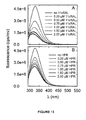
- N -(4-hydroxyphenyl)retinamide can induce neuronal-like differentiation in certain cultured human RPE cells. See Chen, S., et al., J. Neurochem., 84:972-81 (2003 ).
- the compounds of Formula (I) can be used to provide benefit to patients suffering from or susceptible to various macular degenerations and dystrophies, including but not limited to dry-form age-related macular degeneration and Stargardt Disease.
- compounds of Formula (I) provide at least some of the following benefits to such human patients: reduction in the amount of all-trans-retinal (atRAL), reduction in the formation of A2E, reduction in the formation of lipofuscin, reduction in the formation of drusen, and reduction in light sensitivity.
- A2E all-trans-retinal
- ophthalmic and ocular tissues caused, in part, by a reduction in the over-accumulation of all- trans -retinal in these tissues.
- A2E itself is cytotoxic to the RPE (which can lead to retina cell death)
- administration of compounds having the structure of Formula (I) reduces the rate of accumulation of A2E, a cytotoxic agent, thus providing patient benefit.
- A2E is the major fluorophore of lipofuscin
- reduced quantities of A2E in ophthalmic and ocular tissues also results in a reduced tendency to accumulate lipofuscin in such tissues.
- compositions described herein can be considered to be lipofuscin-based treatments because administration of compounds having the structure of Formula (I) (alone, or in combination with other agents, as described herein) reduces, lowers or otherwise impacts the accumulation of lipofuscin in ophthalmic and/or ocular tissues.
- a reduction in the rate of accumulation of lipofuscin in ophthalmic and/or ocular tissues benefits patients that have diseases or conditions such as macular degenerations and/or dystrophies.
- the compounds of Formula (I) and/or its derivatives also have an effect on enzymes or proteins in the visual cycle.
- esterification in the retinal pigment epithelium involves lecithin-retinol acyltransferase (LRAT) which catalyzes the transfer of an acyl group from lecithin to retinol.
- LRAT lecithin-retinol acyltransferase
- Administration of Formula (I) and/or its derivatives modifies the activity of LRAT which could benefit patients suffering from or susceptible to various macular degenerations and dystrophies.
- Vitamin A in serum is delivered to extra-hepatic target tissues and immediately esterified by the membrane-bound enzyme LRAT.
- LRAT catalyzes the transfer of a fatty acid from membrane phospholipids to retinol thereby generating all -trans retinyl esters, the principal storage form of vitamin A in all tissues.
- all-trans retinyl esters are the sole substrate for a unique isomerase enzyme which generates a light-sensitive visual chromophore precursor, 11- cis retinol. Subsequent oxidation of this retinoid and conjugation to the opsin apoprotein in the retina yields rhodopsin.
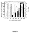
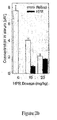
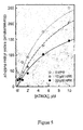
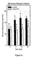
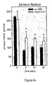
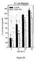
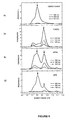
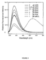
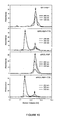
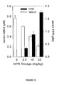
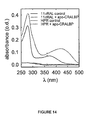
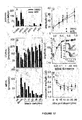
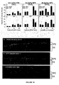
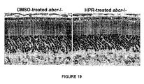
- N -4-(hydroxyphenyl)retinamide has been shown to cause marked inhibition of LRAT activity in membranes prepared from liver and small intestine. Additionally, we have demonstrated (e.g., Example 13) for the first time that LRAT activity in the RPE of the eye is inhibited by HPR. As discussed in the Examples, administration of HPR is also associated with decreased serum retinol and retinol binding protein (RBP). Thus, in addition to the systemic effects of HPR (e.g., decreased serum retinol levels), there is also an intracellular, enzyme-specific effect (e.g., LRAT activity in RPE cells).
- HPR serum retinol and retinol binding protein
- vitamin A homeostasis in the eye relies not only upon delivery of retinol from serum but also upon intracellular stores of retinyl esters to provide visual chromophore, suggests that effects of HPR may be most pronounced in this organ.
- CRALBP Cellular Retinaldehyde Binding Protein
- compounds having the structure of Formula (I) are expected to bind to CRALBP, and consequently, (a) modulate the binding of other compounds, such as retinaldehyde, to CRALBP, (b) modulate the activity of CRALBP, (c) serve as a ligand to CRALBP, (d) undergo activity catalyzed by CRALBP, including transport activity, and/or (e) serve as a therapeutic agent in the methods and compositions described herein.
- the vertebrate retina contains two types of photoreceptor cells - rods and cones.
- Rods are specialized for vision under low light conditions. Cones are less sensitive, provide vision at high temporal and spatial resolutions, and afford color perception. Under daylight conditions, the rod response is saturated and vision is mediated entirely by cones. Both cell types contain a structure called the outer segment comprising a stack of membranous discs. The reactions of visual transduction take place on the surfaces of these discs.
- the first step in vision is absorption of a photon by an opsin-pigment molecule (rhodopsin), which involves 11-cis to all- trans isomerization of the chromophore.
- rhodopsin opsin-pigment molecule
- the resulting all- trans -retinal must be converted back 11- cis -retinal in a multi-enzyme process which takes place in the retinal pigment epithelium, a monolayer of cells adjacent to the retina.
- Macular or Retinal Degenerations and Dystrophies Macular degeneration (also referred to as retinal degeneration) is a disease of the eye that involves deterioration of the macula, the central portion of the retina. Approximately 85% to 90% of the cases of macular degeneration are the "dry" (atrophic or non-neovascular) type. In dry macular degeneration, the deterioration of the retina is associated with the formation of small yellow deposits, known as drusen, under the macula; in addition, the accumulation of lipofuscin in the RPE leads to photoreceptor degeneration and geographic atrophy. This phenomena leads to a thinning and drying out of the macula.
- the location and amount of thinning in the retina caused by the drusen directly correlates to the amount of central vision loss.
- Degeneration of the pigmented layer of the retina and photoreceptors overlying drusen become atrophic and can cause a slow loss of central vision.
- loss of retinal pigment epithelium and underlying photoreceptor cells results in geographic atrophy.
- Administration of at least one compound having the structure of Formula (I) to a mammal can reduce the formation of, or limit the spread of, photoreceptor degeneration and/or geographic atrophy in the eye of the mammal.
- administration of HPR and/or MPR to a mammal can be used to treat photoreceptor degeneration and/or geographic atrophy in the eye of the mammal.
- wet macular degeneration new blood vessels form (i.e., neovascularization) to improve the blood supply to retinal tissue, specifically beneath the macula, a portion of the retina that is responsible for our sharp central vision.
- the new vessels are easily damaged and sometimes rupture, causing bleeding and injury to the surrounding tissue.
- wet macular degeneration only occurs in about 10 percent of all macular degeneration cases, it accounts for approximately 90% of macular degeneration-related blindness.
- Neovascularization can lead to rapid loss of vision and eventual scarring of the retinal tissues and bleeding in the eye. This scar tissue and blood produces a dark, distorted area in the vision, often rendering the eye legally blind.
- Wet macular degeneration usually starts with distortion in the central field of vision. Straight lines become wavy.
- VEGF vascular endothelial growth factor
- Administration of at least one compound having the structure of Formula (I) to a mammal can reduce the formation of, or limit the spread of, wet-form age-related macular degeneration in the eye of the mammal.
- administration of HPR and/or MPR to a mammal can be used to treat wet-form age-related macular degeneration in the eye of the mammal.
- the compounds of Formula (I) can be used to treat choroidal neovascularization and the formation of abnormal blood vessels beneath the macula of the eye of a mammal.
- Stargardt Disease is a macular dystrophy that manifests as a recessive form of macular degeneration with an onset during childhood. See e.g., Allikmets et al., Science, 277:1805-07 (1997 ); Lewis et al., Am. J. Hum. Genet., 64:422-34 (1999 ); Stone et al., Nature Genetics, 20:328-29 (1998 ); Allikmets, Am. J. Hum. Gen., 67:793-799 (2000 ); Klevering, et al, Ophthalmology, 111:546-553 (2004 ). Stargardt Disease is characterized clinically by progressive loss of central vision and progressive atrophy of the RPE overlying the macula.
- ABCA4 has also been implicated in recessive retinitis pigmentosa, see, e.g., Cremers et al., Hum. Mol. Genet., 7:355-62 (1998 ), recessive cone-rod dystrophy, see id., and non-exudative age-related macular degeneration, see e.g., Allikmets et al., Science, 277:1805-07 (1997 ); Lewis et al., Am. J. Hum. Genet., 64:422-34 (1999 ), although the prevalence of ABCA4 mutations in AMD is still uncertain. See Stone et al., Nature Genetics, 20:328-29 (1998 ); Allikmets, Am. J.
- macular degenerations that affect children, teenagers or adults that are commonly known as early onset or juvenile macular degeneration. Many of these types are hereditary and are looked upon as macular dystrophies instead of degeneration. Some examples of macular dystrophies include: Cone-Rod Dystrophy, Corneal Dystrophy, Fuch's Dystrophy, Sorsby's Macular Dystrophy, Best Disease, and Juvenile Retinoschisis, as well as Stargardt Disease.
- alkoxy refers to a (alkyl)O- group, where alkyl is as defined herein.
- alkyl refers to an aliphatic hydrocarbon group.
- the alkyl moiety may be a "saturated alkyl” group, which means that it does not contain any alkene or alkyne moieties.
- the alkyl moiety may also be an "unsaturated alkyl” moiety, which means that it contains at least one alkene or alkyne moiety.
- An “alkene” moiety refers to a group consisting of at least two carbon atoms and at least one carbon-carbon double bond
- an “alkyne” moiety refers to a group consisting of at least two carbon atoms and at least one carbon-carbon triple bond.
- the alkyl moiety, whether saturated or unsaturated may be branched, straight chain, or cyclic.
- alkyl moiety may have 1 to 10 carbon atoms (whenever it appears herein, a numerical range such as “1 to 10" refers to each integer in the given range; e.g ., "1 to 10 carbon atoms” means that the alkyl group may consist of 1 carbon atom, 2 carbon atoms, 3 carbon atoms, etc., up to and including 10 carbon atoms, although the present definition also covers the occurrence of the term "alkyl” where no numerical range is designated).
- the alkyl group could also be a "lower alkyl” having 1 to 5 carbon atoms.
- the alkyl group of the compounds described herein may be designated as "C 1 -C 4 alkyl" or similar designations.
- C 1 -C 4 alkyl indicates that there are one to four carbon atoms in the alkyl chain, i.e., the alkyl chain is selected from the group consisting of methyl, ethyl, propyl, iso-propyl, n-butyl, iso-butyl, sec-butyl, and t-butyl.
- Typical alkyl groups include, but are in no way limited to, methyl, ethyl, propyl, isopropyl, butyl, isobutyl, tertiary butyl, pentyl, hexyl, ethenyl, propenyl, butenyl, cyclopropyl, cyclobutyl, cyclopentyl, cyclohexyl, and the like.
- the alkenyl moiety may be branched, straight chain, or cyclic (in which case, it would also be known as a "cycloalkenyl" group).
- alkynyl refers to a type of alkyl group in which the first two atoms of the alkyl group form a triple bond. That is, an alkynyl group begins with the atoms -C ⁇ C-R, wherein R refers to the remaining portions of the alkynyl group, which may be the same or different.
- Non-limiting examples of an alkynyl group include -C ⁇ CH, -C ⁇ CH3 and -C ⁇ CCH 2 CH 3 .
- the "R" portion of the alkynyl moiety may be branched, straight chain, or cyclic.
- amide is a chemical moiety with formula -C(O)NHR or -NHC(O)R, where R is selected from the group consisting of alkyl, cycloalkyl, aryl, heteroaryl (bonded through a ring carbon) and heteroalicyclic (bonded through a ring carbon).
- R is selected from the group consisting of alkyl, cycloalkyl, aryl, heteroaryl (bonded through a ring carbon) and heteroalicyclic (bonded through a ring carbon).
- An amide may be an amino acid or a peptide molecule attached to a compound of Formula (I), thereby forming a prodrug. Any amine, hydroxy, or carboxyl side chain on the compounds described herein can be amidified.
- aromatic refers to an aromatic group which has at least one ring having a conjugated pi electron system and includes both carbocyclic aryl (e.g ., phenyl) and heterocyclic aryl (or “heteroaryl” or “heteroaromatic") groups (e.g ., pyridine).
- the term includes monocyclic or fused-ring polycyclic ( i.e ., rings which share adjacent pairs of carbon atoms) groups.
- carbocyclic refers to a compound which contains one or more covalently closed ring structures, and that the atoms forming the backbone of the ring are all carbon atoms. The term thus distinguishes carbocyclic from heterocyclic rings in which the ring backbone contains at least one atom which is different from carbon.
- a "cyano" group refers to a -CN group.
- cycloalkyl refers to a monocyclic or polycyclic radical that contains only carbon and hydrogen, and may be saturated, partially unsaturated, or fully unsaturated. Cycloalkyl groups include groups having from 3 to 10 ring atoms. Illustrative examples of cycloalkyl groups include the following moieties: and the like.
- esters refers to a chemical moiety with formula -COOR, where R is selected from the group consisting of alkyl, cycloalkyl, aryl, heteroaryl (bonded through a ring carbon) and heteroalicyclic (bonded through a ring carbon). Any amine, hydroxy, or carboxyl side chain on the compounds described herein can be esterified.
- the procedures and specific groups to make such esters are known to those of skill in the art and can readily be found in reference sources such as Greene and Wuts, Protective Groups in Organic Synthesis, 3rd Ed., John Wiley & Sons, New York, NY, 1999 , which is incorporated herein by reference in its entirety.
- halo or, alternatively, "halogen” means fluoro, chloro, bromo or iodo. Preferred halo groups are fluoro, chloro and bromo.
- haloalkyl include alkyl, alkenyl, alkynyl and alkoxy structures that are substituted with one or more halo groups or with combinations thereof.
- fluoroalkyl and “fluoroalkoxy” include haloalkyl and haloalkoxy groups, respectively, in which the halo is fluorine.
- heteroalkyl “heteroalkenyl” and “heteroalkynyl” include optionally substituted alkyl, alkenyl and alkynyl radicals and which have one or more skeletal chain atoms selected from an atom other than carbon, e.g ., oxygen, nitrogen, sulfur, phosphorus or combinations thereof.
- heteroaryl or, alternatively, “heteroaromatic” refers to an aryl group that includes one or more ring heteroatoms selected from nitrogen, oxygen and sulfur.
- An N -containing “heteroaromatic” or “heteroaryl” moiety refers to an aromatic group in which at least one of the skeletal atoms of the ring is a nitrogen atom.
- the polycyclic heteroaryl group may be fused or non-fused.
- Illustrative examples of heteroaryl groups include the following moieties: and the like.
- heterocycle refers to heteroaromatic and heteroalicyclic groups containing one to four heteroatoms each selected from O, S and N, wherein each heterocyclic group has from 4 to 10 atoms in its ring system, and with the proviso that the ring of said group does not contain two adjacent O or S atoms.
- Non-aromatic heterocyclic groups include groups having only 4 atoms in their ring system, but aromatic heterocyclic groups must have at least 5 atoms in their ring system.
- the heterocyclic groups include benzo-fused ring systems.
- An example of a 4-membered heterocyclic group is azetidinyl (derived from azetidine).
- An example of a 5-membered heterocyclic group is thiazolyl.
- An example of a 6-membered heterocyclic group is pyridyl, and an example of a 10-membered heterocyclic group is quinolinyl.
- Examples of non-aromatic heterocyclic groups are pyrrolidinyl, tetrahydrofuranyl, dihydrofuranyl, tetrahydrothienyl, tetrahydropyranyl, dihydropyranyl, tetrahydrothiopyranyl, piperidino, morpholino, thiomorpholino, thioxanyl, piperazinyl, azetidinyl, oxetanyl, thietanyl, homopiperidinyl, oxepanyl, thiepanyl, oxazepinyl, diazepinyl, thiazepinyl, 1,2,3,6-tetrahydropyr
- aromatic heterocyclic groups are pyridinyl, imidazolyl, pyrimidinyl, pyrazolyl, triazolyl, pyrazinyl, tetrazolyl, furyl, thienyl, isoxazolyl, thiazolyl, oxazolyl, isothiazolyl, pyrrolyl, quinolinyl, isoquinolinyl, indolyl, benzimidazolyl, benzofuranyl, cinnolinyl, indazolyl, indolizinyl, phthalazinyl, pyridazinyl, triazinyl, isoindolyl, pteridinyl, purinyl, oxadiazolyl, thiadiazolyl, furazanyl, benzofurazanyl, benzothiophenyl, benzothiazolyl, benzoxazolyl, quinazolinyl, quinox
- a group derived from pyrrole may be pyrrol-1-yl ( N -attached) or pyrrol-3-yl (C-attached).
- a group derived from imidazole may be imidazol-1-yl or imidazol-3-yl (both N -attached) or imidazol-2-yl, imidazol-4-yl or imidazol-5-yl (all C-attached).
- heteroalicyclic refers to a cycloalkyl group that includes at least one heteroatom selected from nitrogen, oxygen and sulfur.
- the radicals may be fused with an aryl or heteroaryl.
- Illustrative examples of heterocycloalkyl groups include: and the like.
- the term heteroalicyclic also includes all ring forms of the carbohydrates, including but not limited to the monosaccharides, the disaccharides and the oligosaccharides.
- membered ring can embrace any cyclic structure.
- membered is meant to denote the number of skeletal atoms that constitute the ring.
- cyclohexyl, pyridine, pyran and thiopyran are 6-membered rings and cyclopentyl, pyrrole, furan, and thiophene are 5-membered rings.
- An “isocyanato” group refers to a -NCO group.
- An “isothiocyanato” group refers to a -NCS group.
- a “mercaptyl” group refers to a (alkyl)S- group.
- nucleophile and “electrophile” as used herein have their usual meanings familiar to synthetic and/or physical organic chemistry.
- Carbon electrophiles typically comprise one or more alkyl, alkenyl, alkynyl or aromatic (sp 3 , sp 2 , or sp hybridized) carbon atoms substituted with any atom or group having a Pauling electronegativity greater than that of carbon itself.
- Examples of carbon electrophiles include but are not limited to carbonyls (aldehydes, ketones, esters, amides), oximes, hydrazones, epoxides, aziridines, alkyl-, alkenyl,-, and aryl halides, acyls, sulfonates (aryl, alkyl and the like).
- Other examples of carbon electrophiles include unsaturated carbon atoms electronically conjugated with electron withdrawing groups, examples being the 6-carbon in alpha-unsaturated ketones or carbon atoms in fluorine substituted aryl groups.
- aromatic substituents imparts distinctive chemistry for such stereoisomers and is well recognized within the field of aromatic chemistry.
- Para- and meta-substitutional patterns project the two substituents into different orientations. Ortho-disposed substituents are oriented at 60° with respect to one another; meta-disposed substituents are oriented at 120° with respect to one another; para-disposed substituents are oriented at 180° with respect to one another.
- Relative dispositions of substituents also affect the electronic properties of the substituents. Without being bound to any particular type or level of theory, it is known that ortho- and para-disposed substituents electronically affect one another to a greater degree than do corresponding meta-disposed substituents. Meta-disubstituted aromatics are often synthesized using different routes than are the corresponding ortho and para-disubstituted aromatics.
- moiety refers to a specific segment or functional group of a molecule. Chemical moieties are often recognized chemical entities embedded in or appended to a molecule.
- bond refers to a chemical bond between two atoms, or two moieties when the atoms joined by the bond are considered to be part of larger substructure.
- a "thiocyanato" group refers to a -CNS group.
- optionally substituted means that the referenced group may be substituted with one or more additional group(s) individually and independently selected from alkyl, cycloalkyl, aryl, heteroaryl, heteroalicyclic, hydroxy, alkoxy, aryloxy, mercapto, alkylthio, arylthio, cyano, halo, carbonyl, thiocarbonyl, isocyanato, thiocyanato, isothiocyanato, nitro, perhaloalkyl, perfluoroalkyl, silyl, and amino, including mono-and di-substituted amino groups, and the protected derivatives thereof.
- the protecting groups that may form the protective derivatives of the above substituents are known to those of skill in the art and may be found in references such as Greene and Wuts, above.
- the compounds presented herein may possess one or more chiral centers and each center may exist in the R or S configuration.
- the compounds presented herein include all diastereomeric, enantiomeric, and epimeric forms as well as the appropriate mixtures thereof.
- Stereoisomers may be obtained, if desired, by methods known in the art as, for example, the separation of stereoisomers by chiral chromatographic columns.
- N -oxides include the use of N -oxides, crystalline forms (also known as polymorphs), or pharmaceutically acceptable salts of compounds having the structure of Formula (I), as well as active metabolites of these compounds having the same type of activity.
- a known metabolite of fenretinide is N -(4-methoxyphenyl)retinamide, also known as 4-MPR or MPR.
- Another known metabolite of fenretinide is 4-oxo fenretinide.
- compounds may exist as tautomers. All tautomers are included within the scope of the compounds presented herein.
- the compounds described herein can exist in unsolvated as well as solvated forms with pharmaceutically acceptable solvents such as water, ethanol, and the like. The solvated forms of the compounds presented herein are also considered to be disclosed herein.
- compositions comprising a compound of Formula (I) and a pharmaceutically acceptable diluent, excipient, or carrier.
- composition refers to a mixture of a compound of Formula (I) with other chemical components, such as carriers, stabilizers, diluents, dispersing agents, suspending agents, thickening agents, and/or excipients.
- the pharmaceutical composition facilitates administration of the compound to an organism. Multiple techniques of administering a compound exist in the art including, but not limited to: intravenous, oral, aerosol, parenteral, ophthalmic, pulmonary and topical administration.
- carrier refers to relatively nontoxic chemical compounds or agents that facilitate the incorporation of a compound into cells or tissues.
- dilute refers to chemical compounds that are used to dilute the compound of interest prior to delivery. Diluents can also be used to stabilize compounds because they can provide a more stable environment. Salts dissolved in buffered solutions (which also can provide pH control or maintenance) are utilized as diluents in the art, including, but not limited to a phosphate buffered saline solution.
- physiologically acceptable refers to a material, such as a carrier or diluent, that does not abrogate the biological activity or properties of the compound, and is nontoxic.
- pharmaceutically acceptable salt refers to a formulation of a compound that does not cause significant irritation to an organism to which it is administered and does not abrogate the biological activity and properties of the compound.
- Pharmaceutically acceptable salts may be obtained by reacting a compound of Formula (I) with acids such as hydrochloric acid, hydrobromic acid, sulfuric acid, nitric acid, phosphoric acid, methanesulfonic acid, ethanesulfonic acid, p-toluenesulfonic acid, salicylic acid and the like.
- Pharmaceutically acceptable salts may also be obtained by reacting a compound of Formula (I) with a base to form a salt such as an ammonium salt, an alkali metal salt, such as a sodium or a potassium salt, an alkaline earth metal salt, such as a calcium or a magnesium salt, a salt of organic bases such as dicyclohexylamine, N -methyl-D-glucamine, tris(hydroxymethyl)methylamine, and salts with amino acids such as arginine, lysine, and the like, or by other methods known in the art
- a salt such as an ammonium salt, an alkali metal salt, such as a sodium or a potassium salt, an alkaline earth metal salt, such as a calcium or a magnesium salt, a salt of organic bases such as dicyclohexylamine, N -methyl-D-glucamine, tris(hydroxymethyl)methylamine, and salts with amino acids such as arginine, lysine, and the like, or by other methods
- a “metabolite” of a compound disclosed herein is a derivative of that compound that is formed when the compound is metabolized.
- active metabolite refers to a biologically active derivative of a compound that is formed when the compound is metabolized.
- metabolized refers to the sum of the processes (including, but not limited to, hydrolysis reactions and reactions catalyzed by enzymes) by which a particular substance is changed by an organism. Thus, enzymes may produce specific structural alterations to a compound.
- cytochrome P450 catalyzes a variety of oxidative and reductive reactions while uridine diphosphate glucuronyltransferases catalyze the transfer of an activated glucuronic-acid molecule to aromatic alcohols, aliphatic alcohols, carboxylic acids, amines and free sulphydryl groups. Further information on metabolism may be obtained from The Pharmacological Basis of Therapeutics, 9th Edition, McGraw-Hill (1996 ).
- Metabolites of the compounds disclosed herein can be identified either by administration of compounds to a host and analysis of tissue samples from the host, or by incubation of compounds with hepatic cells in vitro and analysis of the resulting compounds. Both methods are well known in the art.
- MPR is a known metabolite of HPR, both of which are contained within the structure of Formula (I). MPR accumulates systemically in patients that have been chronically treated with HPR. One of the reasons that MPR accumulates systemically is that MPR is only (if at all) slowly metabolized, whereas HPR is metabolized to MPR. In addition, MPR may undergo relatively slow clearance. Thus, (a) the pharmacokinetics and pharmacodynamics of MPR must be taken into consideration when administering and determining the bioavailability of HPR, (b) MPR is more stable to metabolism than HPR, and (c) MPR can be more immediately bioavailable than HPR following absorption. Another known metabolite of fenretinide is 4-oxo fenretinide.
- MPR may also be considered an active metabolite.
- MPR (like HPR) can bind to Retinol Binding Protein (RBP) and prevent the binding of RBP to Transerythrin (TTR).
- RBP Retinol Binding Protein
- TTR Transerythrin
- MPR can (a) serve as an inhibitor of retinol binding to RBP, (b) serve as an inhibitor of RBP to TTR, (c) limit the transport of retinol to certain tissues, including ophthalmic tissues, and (d) be transported by RBP to certain tissues, including ophthalmic tissues.
- MPR appears to bind more weakly to RBP than HPR, and is thus a less strong inhibitor of retinol binding to RBP. Nevertheless, both MPR and HPR are expected to inhibit, approximately equivalently, the binding of RBP to TTR. Furthermore, it is expected that MPR (like HPR) will bind to visual cycle proteins, including LRAT and CRALBP. MPR has, in these respects, the same mode of action as HPR and can serve as a therapeutic agent in the methods and compositions described herein.
- prodrug refers to an agent that is converted into the parent drug in vivo. Prodrugs are often useful because, in some situations, they may be easier to administer than the parent drug. They may, for instance, be bioavailable by oral administration whereas the parent is not. The prodrug may also have improved solubility in pharmaceutical compositions over the parent drug.
- An example, without limitation, of a prodrug would be a compound of Formula (I) which is administered as an ester (the "prodrug") to facilitate transmittal across a cell membrane where water solubility is detrimental to mobility but which then is metabolically hydrolyzed to the carboxylic acid, the active entity, once inside the cell where water-solubility is beneficial.
- a further example of a prodrug might be a short peptide (polyaminoacid) bonded to an acid group where the peptide is metabolized to reveal the active moiety.
- the compounds described herein can be administered to a human patient per se, or in pharmaceutical compositions where they are mixed with other active ingredients, as in combination therapy, or suitable carrier(s) or excipient(s).
- suitable carrier(s) or excipient(s) include butyl alcohol
- Suitable routes of administration may, for example, include oral, rectal, transmucosal, transdermal, pulmonary, ophthalmic or intestinal administration; parenteral delivery, including intramuscular, subcutaneous, intravenous, intramedullary injections, as well as intrathecal, direct intraventricular, intraperitoneal, intranasal, or intraocular injections.
- the liposomes will be targeted to and taken up selectively by the organ.
- the drug may be provided in the form of a rapid release formulation, in the form of an extended release formulation, or in the form of an intermediate release formulation.
- compositions comprising a compound of Formula (I) may be manufactured in a manner that is itself known, e.g ., by means of conventional mixing, dissolving, granulating, dragee-making, levigating, emulsifying, encapsulating, entrapping or compression processes.
- compositions may be formulated in conventional manner using one or more physiologically acceptable carriers comprising excipients and auxiliaries which facilitate processing of the active compounds into preparations which can be used pharmaceutically. Proper formulation is dependent upon the route of administration chosen. Any of the well-known technique, carriers, and excipients may be used as suitable and as understood in the art; e.g ., in Remington's Pharmaceutical Sciences, above.
- the compounds of Formula (I) can be administered in a variety of ways, including all forms of local delivery to the eye. Additionally, the compounds of Formula (I) can be administered systemically, such as orally or intravenously. The compounds of Formula (I) can be administered topically to the eye and can be formulated into a variety of topically administrable ophthalmic compositions, such as solutions, suspensions, gels or ointments.
- ophthalmic administration encompasses, but is not limited to, intraocular injection, subretinal injection, intravitreal injection, periocular administration, subconjuctival injections, retrobulbar injections, intracameral injections (including into the anterior or vitreous chamber), sub-Tenon's injections or implants, ophthalmic solutions, ophthalmic suspensions, ophthalmic ointments, ocular implants and ocular inserts, intraocular solutions, use of iontophoresis, incorporation in surgical irrigating solutions, and packs (by way of example only, a saturated cotton pledget inserted in the fornix).
- Administration of a composition to the eye generally results in direct contact of the agents with the cornea, through which at least a portion of the administered agents pass.
- the composition has an effective residence time in the eye of about 2 to about 24 hours, more typically about 4 to about 24 hours and most typically about 6 to about 24 hours.
- a composition comprising a compound of Formula (I) can illustratively take the form of a liquid where the agents are present in solution, in suspension or both. Typically when the composition is administered as a solution or suspension a first portion of the agent is present in solution and a second portion of the agent is present in particulate form, in suspension in a liquid matrix.
- a liquid composition may include a gel formulation. In other embodiments, the liquid composition is aqueous. Alternatively, the composition can take the form of an ointment.
- Useful compositions can be an aqueous solution, suspension or solution/suspension, which can be presented in the form of eye drops.
- a desired dosage can be administered via a known number of drops into the eye. For example, for a drop volume of 25 ⁇ l, administration of 1-6 drops will deliver 25-150 ⁇ l of the composition.
- Aqueous compositions typically contain from about 0.01% to about 50%, more typically about 0.1% to about 20%, still more typically about 0.2% to about 10%, and most typically about 0.5% to about 5%, weight/volume of a compound of Formula (I).
- aqueous compositions typically have ophthalmically acceptable pH and osmolality.
- “Ophthalmically acceptable” with respect to a formulation, composition or ingredient typically means having no persistent detrimental effect on the treated eye or the functioning thereof, or on the general health of the subject being treated. Transient effects such as minor irritation or a "stinging" sensation are common with topical ophthalmic administration of agents and consistent with the formulation, composition or ingredient in question being “ophthalmically acceptable.”
- Useful aqueous suspension can also contain one or more polymers as suspending agents.
- Useful polymers include water-soluble polymers such as cellulosic polymers, e.g ., hydroxypropyl methylcellulose, and water-insoluble polymers such as cross-linked carboxyl-containing polymers.
- Useful compositions can also comprise an ophthalmically acceptable mucoadhesive polymer, selected for example from carboxymethylcellulose, carbomer (acrylic acid polymer), poly(methylmethacrylate), polyacrylamide, polycarbophil, acrylic acid/butyl acrylate copolymer, sodium alginate and dextran.
- compositions may also include ophthalmically acceptable solubilizing agents to aid in the solubility of a compound of Formula (I).
- solubilizing agent generally includes agents that result in formation of a micellar solution or a true solution of the agent.
- Certain ophthalmically acceptable nonionic surfactants for example polysorbate 80, can be useful as solubilizing agents, as can ophthalmically acceptable glycols, polyglycols, e.g ., polyethylene glycol 400, and glycol ethers.
- Useful compositions may also include one or more ophthalmically acceptable pH adjusting agents or buffering agents, including acids such as acetic, boric, citric, lactic, phosphoric and hydrochloric acids; bases such as sodium hydroxide, sodium phosphate, sodium borate, sodium citrate, sodium acetate, sodium lactate and tris-hydroxymethylaminomethane; and buffers such as citrate/dextrose, sodium bicarbonate and ammonium chloride. Such acids, bases and buffers are included in an amount required to maintain pH of the composition in an ophthalmically acceptable range.
- acids such as acetic, boric, citric, lactic, phosphoric and hydrochloric acids
- bases such as sodium hydroxide, sodium phosphate, sodium borate, sodium citrate, sodium acetate, sodium lactate and tris-hydroxymethylaminomethane
- buffers such as citrate/dextrose, sodium bicarbonate and ammonium chloride.
- Such acids, bases and buffers are included in an amount required to
- Useful compositions may also include one or more ophthalmically acceptable salts in an amount required to bring osmolality of the composition into an ophthalmically acceptable range.
- ophthalmically acceptable salts include those having sodium, potassium or ammonium cations and chloride, citrate, ascorbate, borate, phosphate, bicarbonate, sulfate, thiosulfate or bisulfite anions; suitable salts include sodium chloride, potassium chloride, sodium thiosulfate, sodium bisulfite and ammonium sulfate.
- compositions may also include one or more ophthalmically acceptable preservatives to inhibit microbial activity.
- Suitable preservatives include mercury-containing substances such as merfen and thiomersal; stabilized chlorine dioxide; and quaternary ammonium compounds such as benzalkonium chloride, cetyltrimethylammonium bromide and cetylpyridinium chloride.
- compositions may include one or more ophthalmically acceptable surfactants to enhance physical stability or for other purposes.
- Suitable nonionic surfactants include polyoxyethylene fatty acid glycerides and vegetable oils, e.g ., polyoxyethylene (60) hydrogenated castor oil; and polyoxyethylene alkylethers and alkylphenyl ethers, e.g ., octoxynol 10, octoxynol 40.
- compositions may include one or more antioxidants to enhance chemical stability where required.
- Suitable antioxidants include, by way of example only, ascorbic acid and sodium metabisulfite.
- Aqueous suspension compositions can be packaged in single-dose non-reclosable containers.
- multiple-dose reclosable containers can be used, in which case it is typical to include a preservative in the composition.
- the ophthalmic composition may also take the form of a solid article that can be inserted between the eye and eyelid or in the conjunctival sac, where it releases the agent. Release is to the lacrimal fluid that bathes the surface of the cornea, or directly to the cornea itself, with which the solid article is generally in intimate contact.
- Solid articles suitable for implantation in the eye in such fashion are generally composed primarily of polymers and can be biodegradable or non-biodegradable.
- compounds of Formula (I) may be formulated in aqueous solutions, preferably in physiologically compatible buffers such as Hank's solution, Ringer's solution, or physiological saline buffer.
- physiologically compatible buffers such as Hank's solution, Ringer's solution, or physiological saline buffer.
- penetrants appropriate to the barrier to be permeated are used in the formulation. Such penetrants are generally known in the art.
- appropriate formulations may include aqueous or nonaqueous solutions, preferably with physiologically compatible buffers or excipients. Such excipients are generally known in the art.
- One useful formulation for solubilizing higher quantities of the compounds of Formula (I) are, by way of example only, positively, negatively or neutrally charged phospholipids, or bile salt/phosphatidylcholine mixed lipid aggregate systems, such as those described in Li, C.Y., et al., Pharm. Res. 13:907-913 (1996 ).
- An additional formulation that can be used for the same purpose with compounds having the structure of Formula (I) involves use of a solvent comprising an alcohol, such as ethanol, in combination with an alkoxylated caster oil. See, e.g., U.S. Patent Publication Number 2002/0183394 .
- a formulation comprising a compound of Formula (I) is an emulsion composed of a lipoid dispersed in an aqueous phase, a stabilizing amount of a non-ionic surfactant, optionally a solvent, and optionally an isotonic agent.
- a formulation comprising a compound of Formula (I) includes corn oil and a non-ionic surfactant. See U.S. Patent No. 4,665,098 .
- Still another formulation comprising a compound of Formula (I) includes lysophosphatidylcholine, monoglyceride and a fatty acid. See U.S. Patent No. 4,874,795 .
- Still another formulation comprising a compound of Formula (I) includes flour, a sweetener, and a humectant. See International Publication No. WO 2004/069203 . And still another formulation comprising a compound of Formula (I) includes dimyristoyl phosphatidylcholine, soybean oil, t-butyl alcohol and water. See U.S. Patent Application Publication No. US 2002/0143062 .
- compounds of Formula (I) can be formulated readily by combining the active compounds with pharmaceutically acceptable carriers or excipients well known in the art.
- Such carriers enable the compounds described herein to be formulated as tablets, powders, pills, dragees, capsules, liquids, gels, syrups, elixirs, slurries, suspensions and the like, for oral ingestion by a patient to be treated.
- Pharmaceutical preparations for oral use can be obtained by mixing one or more solid excipient with one or more of the compounds described herein, optionally grinding the resulting mixture, and processing the mixture of granules, after adding suitable auxiliaries, if desired, to obtain tablets or dragee cores.
- Suitable excipients are, in particular, fillers such as sugars, including lactose, sucrose, mannitol, or sorbitol; cellulose preparations such as: for example, maize starch, wheat starch, rice starch, potato starch, gelatin, gum tragacanth, methylcellulose, microcrystalline cellulose, hydroxypropylmethylcellulose, sodium carboxymethylcellulose; or others such as: polyvinylpyrrolidone (PVP or povidone) or calcium phosphate.
- disintegrating agents may be added, such as the cross-linked croscarmellose sodium, polyvinylpyrrolidone, agar, or alginic acid or a salt thereof such as sodium alginate.
- Dragee cores are provided with suitable coatings.
- suitable coatings For this purpose, concentrated sugar solutions may be used, which may optionally contain gum arabic, talc, polyvinylpyrrolidone, carbopol gel, polyethylene glycol, and/or titanium dioxide, lacquer solutions, and suitable organic solvents or solvent mixtures.
- Dyestuffs or pigments may be added to the tablets or dragee coatings for identification or to characterize different combinations of active compound doses.
- compositions which can be used orally include push-fit capsules made of gelatin, as well as soft, sealed capsules made of gelatin and a plasticizer, such as glycerol or sorbitol.
- the push-fit capsules can contain the active ingredients in admixture with filler such as lactose, binders such as starches, and/or lubricants such as talc or magnesium stearate and, optionally, stabilizers.
- the active compounds may be dissolved or suspended in suitable liquids, such as fatty oils, liquid paraffin, or liquid polyethylene glycols.
- stabilizers may be added. All formulations for oral administration should be in dosages suitable for such administration.
- compositions may take the form of tablets, lozenges, or gels formulated in conventional manner.
- transdermal delivery devices Such transdermal patches may be used to provide continuous or discontinuous infusion of the compounds of the present invention in controlled amounts.
- the construction and use of transdermal patches for the delivery of pharmaceutical agents is well known in the art. See, e.g ., U.S. Pat. No. 5,023,252 .
- patches may be constructed for continuous, pulsatile, or on demand delivery of pharmaceutical agents.
- transdermal delivery of the compounds of Formula (I) can be accomplished by means of iontophoretic patches and the like. Transdermal patches can provide controlled delivery of the compounds.
- the rate of absorption can be slowed by using rate-controlling membranes or by trapping the compound within a polymer matrix or gel.
- absorption enhancers can be used to increase absorption.
- Formulations suitable for transdermal administration can be presented as discrete patches and can be lipophilic emulsions or buffered, aqueous solutions, dissolved and/or dispersed in a polymer or an adhesive. Transdermal patches may be placed over different portions of the patient's body, including over the eye.
- iontophoretic devices that can be used for ocular administration of compounds having the structure of Formula (I) are the Eyegate applicator, created and patented by Optis France S.A., and the OcuphorTM Ocular iontophoresis system developed Iomed, Inc.
- the compounds of Formula (I) are conveniently delivered in the form of an aerosol spray presentation from pressurized packs or a nebuliser, with the use of a suitable propellant, e.g ., dichlorodifluoromethane, trichlorofluoromethane, dichlorotetrafluoroethane, carbon dioxide or other suitable gas.
- a suitable propellant e.g ., dichlorodifluoromethane, trichlorofluoromethane, dichlorotetrafluoroethane, carbon dioxide or other suitable gas.
- a suitable propellant e.g ., dichlorodifluoromethane, trichlorofluoromethane, dichlorotetrafluoroethane, carbon dioxide or other suitable gas.
- a suitable propellant e.g ., dichlorodifluoromethane, trichlorofluoromethane, dichlorotetrafluoroethane, carbon
- the compounds may be formulated for parenteral administration by injection, e.g ., by bolus injection or continuous infusion.
- Formulations for injection may be presented in unit dosage form, e.g ., in ampoules or in multi-dose containers, with an added preservative.
- the compositions may take such forms as suspensions, solutions or emulsions in oily or aqueous vehicles, and may contain formulatory agents such as suspending, stabilizing and/or dispersing agents.
- compositions for parenteral administration include aqueous solutions of the active compounds in water-soluble form. Additionally, suspensions of the active compounds may be prepared as appropriate oily injection suspensions. Suitable lipophilic solvents or vehicles include fatty oils such as sesame oil, or synthetic fatty acid esters, such as ethyl oleate or triglycerides, or liposomes. Aqueous injection suspensions may contain substances which increase the viscosity of the suspension, such as sodium carboxymethyl cellulose, sorbitol, or dextran. Optionally, the suspension may also contain suitable stabilizers or agents which increase the solubility of the compounds to allow for the preparation of highly concentrated solutions.
- the active ingredient may be in powder form for constitution with a suitable vehicle, e.g ., sterile pyrogen-free water, before use.
- a suitable vehicle e.g ., sterile pyrogen-free water
- the compounds may also be formulated in rectal compositions such as rectal gels, rectal foam, rectal aerosols, suppositories or retention enemas, e.g ., containing conventional suppository bases such as cocoa butter or other glycerides.
- rectal compositions such as rectal gels, rectal foam, rectal aerosols, suppositories or retention enemas, e.g ., containing conventional suppository bases such as cocoa butter or other glycerides.
- the compounds may also be formulated as a depot preparation. Such long acting formulations may be administered by implantation (for example subcutaneously or intramuscularly) or by intramuscular injection.
- the compounds may be formulated with suitable polymeric or hydrophobic materials (for example as an emulsion in an acceptable oil) or ion exchange resins, or as sparingly soluble derivatives, for example, as a sparingly soluble salt.
- Injectable depot forms may be made by forming microencapsulated matrices (also known as microencapsule matrices) of the compound of Formula (I) in biodegradable polymers. Depending upon the ratio of drug to polymer and the nature of the particular polymer employed, the rate of drug release can be controlled. Depot injectable formulations may be also prepared by entrapping the drug in liposomes or microemulsions.
- posterior juxtascleral depots may be used as a mode of administration for compounds having the structure of Formula (I).
- the sclera is a thin avascular layer, comprised of highly ordered collagen network surrounding most of vertebrate eye.
- the sclera Since the sclera is avascular it can be utilized as a natural storage depot from which injected material cannot rapidly removed or cleared from the eye.
- the formulation used for administration of the compound into the scleral layer of the eye can be any form suitable for application into the sclera by injection through a cannula with small diameter suitable for injection into the scleral layer. Examples for injectable application forms are solutions, suspensions or colloidal suspensions.
- a pharmaceutical carrier for the hydrophobic compounds of Formula (I) is a cosolvent system comprising benzyl alcohol, a nonpolar surfactant, a water-miscible organic polymer, and an aqueous phase.
- the cosolvent system may be a 10% ethanol, 10% polyethylene glycol 300, 10% polyethylene glycol 40 castor oil (PEG-40 castor oil) with 70% aqueous solution.
- PEG-40 castor oil polyethylene glycol 40 castor oil
- This cosolvent system dissolves hydrophobic compounds well, and itself produces low toxicity upon systemic administration. Naturally, the proportions of a cosolvent system may be varied considerably without destroying its solubility and toxicity characteristics.
- cosolvent components may be varied: for example, other low-toxicity nonpolar surfactants may be used instead of PEG-40 castor oil, the fraction size of polyethylene glycol 300 may be varied; other biocompatible polymers may replace polyethylene glycol, e.g ., polyvinyl pyrrolidone; and other sugars or polysaccharides maybe included in the aqueous solution.
- hydrophobic pharmaceutical compounds may be employed.
- Liposomes and emulsions are well known examples of delivery vehicles or carriers for hydrophobic drugs.
- Certain organic solvents such as N -methylpyrrolidone also may be employed, although usually at the cost of greater toxicity.
- the compounds may be delivered using a sustained-release system, such as semipermeable matrices of solid hydrophobic polymers containing the therapeutic agent.
- sustained-release materials have been established and are well known by those skilled in the art. Sustained-release capsules may, depending on their chemical nature, release the compounds for a few weeks up to over 100 days. Depending on the chemical nature and the biological stability of the therapeutic reagent, additional strategies for protein stabilization may be employed.
- Lym-X-SorbTM One formulation for the administration of compounds having the structure of Formula (I) has been used with fenretinide in the treatment of neuroblastoma, prostate and ovarian cancers, and is marketed by Avanti Polar Lipids, Inc. (Alabaster, Alabama) under the name Lym-X-SorbTM.
- This formulation which comprises an organized lipid matrix that includes lysophosphatidylcholine, monoglyceride and fatty acid, is designed to improve the oral availability of fenretinide.
- Such a formulation i.e ., an oral formulation that includes lysophosphatidylcholine, monoglyceride and fatty acid, is proposed to also provide improved bioavailability of compounds having the structure of Formula (I) for the treatment of ophthalmic and ocular diseases and conditions, including but not limited to the macular degenerations and dystrophies.
- All of the formulations described herein may benefit from antioxidants, metal chelating agents, thiol containing compounds and other general stabilizing agents.
- stabilizing agents include, but are not limited to: (a) about 0.5% to about 2% w/v glycerol, (b) about 0.1% to about 1% w/v methionine, (c) about 0.1% to about 2% w/v monothioglycerol, (d) about 1 mM to about 10 mM EDTA, (e) about 0.01% to about 2% w/v ascorbic acid, (f) 0.003% to about 0.02% w/v polysorbate 80, (g) 0.001% to about 0.05% w/v.
- polysorbate 20 (h) arginine, (i) heparin, (j) dextran sulfate, (k) cyclodextrins, (1) pentosan polysulfate and other heparinoids, (m) divalent cations such as magnesium and zinc; or (n) combinations thereof.
- compositions of Formula (I) may be provided as salts with pharmaceutically compatible counterions.
- Pharmaceutically compatible salts may be formed with many acids, including but not limited to hydrochloric, sulfuric, acetic, lactic, tartaric, malic, succinic, etc. Salts tend to be more soluble in aqueous or other protonic solvents than are the corresponding free acid or base forms.
- mammal means all mammals including humans. Mammals include, by way of example only, humans, non-human primates, cows, dogs, cats, goats, sheep, pigs, rats, mice and rabbits.
- an effective amount refers to that amount of the compound being administered which will relieve to some extent one or more of the symptoms of the disease, condition or disorder being treated.
- compositions containing the compound(s) described herein can be administered for prophylactic and/or therapeutic treatments.
- treating is used to refer to either prophylactic and/or therapeutic treatments.
- the compositions are administered to a patient already suffering from a disease, condition or disorder, in an amount sufficient to cure or at least partially arrest the symptoms of the disease, disorder or condition. Amounts effective for this use will depend on the severity and course of the disease, disorder or condition, previous therapy, the patient's health status and response to the drugs, and the judgment of the treating physician. It is considered well within the skill of the art for one to determine such therapeutically effective amounts by routine experimentation ( e.g ., a dose escalation clinical trial).
- compositions containing the compounds described herein are administered to a patient susceptible to or otherwise at risk of a particular disease, disorder or condition.
- a patient susceptible to or otherwise at risk of a particular disease, disorder or condition is defined to be a "prophylactically effective amount or dose.”
- prophylactically effective amounts or dose are defined to be a "prophylactically effective amount or dose.”
- the precise amounts also depend on the patient's state of health, weight, and the like. It is considered well within the skill of the art for one to determine such prophylactically effective amounts by routine experimentation ( e.g ., a dose escalation clinical trial).
- an “enhance” or “enhancing” means to increase or prolong either in potency or duration a desired effect.
- the term “enhancing” refers to the ability to increase or prolong, either in potency or duration, the effect of other therapeutic agents on a system.
- An “enhancing-effective amount,” as used herein, refers to an amount adequate to enhance the effect of another therapeutic agent in a desired system. When used in a patient, amounts effective for this use will depend on the severity and course of the disease, disorder or condition, previous therapy, the patient's health status and response to the drugs, and the judgment of the treating physician.
- the administration of the compounds may be administered chronically, that is, for an extended period of time, including throughout the duration of the patient's life in order to ameliorate or otherwise control or limit the symptoms of the patient's disease or condition.
- the administration of the compounds may be given continuously; alternatively, the dose of drug being administered may be temporarily reduced or temporarily suspended for a certain length of time (i.e., a "drug holiday").
- the length of the drug holiday can vary between 2 days and 1 year, including by way of example only, 2 days, 3 days, 4 days, 5 days, 6 days, 7 days, 10 days, 12 days, 15 days, 20 days, 28 days, 35 days, 50 days, 70 days, 100 days, 120 days, 150 days, 180 days, 200 days, 250 days, 280 days, 300 days, 320 days, 350 days, and 365 days.
- the dose reduction during a drug holiday may be from 10%-100%, including by way of example only 10%, 15%, 20%, 25%, 30%, 35%, 40%, 45%, 50%, 55%, 60%, 65%, 70%, 75%, 80%, 85%, 90%, 95%, and 100%.
- a maintenance dose is administered if necessary. Subsequently, the dosage or the frequency of administration, or both, can be reduced, as a function of the symptoms, to a level at which the improved disease, disorder or condition is retained. Patients can, however, require intermittent treatment on a long-term basis upon any recurrence of symptoms.
- the amount of a given agent that will correspond to such an amount will vary depending upon factors such as the particular compound, disease condition and its severity, the identity (e.g ., weight) of the subject or host in need of treatment, but can nevertheless be routinely determined in a manner known in the art according to the particular circumstances surrounding the case, including, e.g ., the specific agent being administered, the route of administration, the condition being treated, and the subject or host being treated.
- doses employed for adult human treatment will typically be in the range of 0.02-5000 mg per day, preferably 1-1500 mg per day.
- the desired dose may conveniently be presented in a single dose or as divided doses administered simultaneously (or over a short period of time) or at appropriate intervals, for example as two, three, four or more sub-doses per day.
- the compounds described herein may be administered in combination with another therapeutic agent.
- another therapeutic agent such as a pharmaceutically acceptable salt, ester, amide, prodrug, or solvate.
- an adjuvant i. e., by itself the adjuvant may only have minimal therapeutic benefit, but in combination with another therapeutic agent, the overall therapeutic benefit to the patient is enhanced.
- the benefit of experienced by a patient may be increased by administering one of the compounds described herein with another therapeutic agent (which also includes a therapeutic regimen) that also has therapeutic benefit.
- another therapeutic agent which also includes a therapeutic regimen
- increased therapeutic benefit may result by also providing the patient with other therapeutic agents or therapies for macular degeneration.
- the overall benefit experienced by the patient may simply be additive of the two therapeutic agents or the patient may experience a synergistic benefit.
- non-limiting examples of possible combination therapies include use of at least one compound of formula (I) with nitric oxide (NO) inducers, statins, negatively charged phospholipids, anti-oxidants, minerals, anti-inflammatory agents, anti-angiogenic agents, matrix metalloproteinase inhibitors, and carotenoids.
- suitable combination agents may fall within multiple categories (by way of example only, lutein is an anti-oxidant and a carotenoid).
- the compounds of Formula (I) may also be administered with additional agents that may provide benefit to the patient, including by way of example only cyclosporin A.
- the compounds of Formula (I) may also be used in combination with procedures that may provide additional or synergistic benefit to the patient, including, by way of example only, the use of extracorporeal rheopheresis (also known as membrane differential filtration), the use of implantable miniature telescopes, laser photocoagulation of drusen, and microstimulation therapy.
- Suitable anti-oxidants that could be used in combination with at least one compound having the structure of Formula (I) include vitamin C, vitamin E, beta-carotene and other carotenoids, coenzyme Q, 4-hydroxy-2,2,6,6-tetramethylpiperidine- N -oxyl (also known as Tempol), lutein, butylated hydroxytoluene, resveratrol, a trolox analogue (PNU-83836-E), and bilberry extract.
- suitable minerals include copper-containing minerals, such as cupric oxide (by way of example only); zinc-containing minerals, such as zinc oxide (by way of example only); and selenium-containing compounds.
- Carotenoids are naturally-occurring yellow to red pigments of the terpenoid group that can be found in plants, algae, bacteria, and certain animals, such as birds and shellfish. Carotenoids are a large class of molecules in which more than 600 naturally occurring carotenoids have been identified. Carotenoids include hydrocarbons (carotenes) and their oxygenated, alcoholic derivatives (xanthophylls).
- carotenoids include actinioerythrol, astaxanthin, canthaxanthin, capsanthin, capsorubin, ⁇ -8'-apo-carotenal (apo-carotenal), ⁇ -12'-apo-carotenal, ⁇ -carotene, ⁇ -carotene, "carotene” (a mixture of ⁇ - and ⁇ -carotenes), ⁇ -carotenes, ⁇ -cyrptoxanthin, lutein, lycopene, violerythrin, zeaxanthin, and esters of hydroxyl- or carboxyl-containing members thereof. Many of the carotenoids occur in nature as cis- and trans -isomeric forms, while synthetic compounds are frequently racemic mixtures.
- zeaxanthin In humans, the retina selectively accumulates mainly two carotenoids: zeaxanthin and lutein. These two carotenoids are thought to aid in protecting the retina because they are powerful antioxidants and absorb blue light. Studies with quails establish that groups raised on carotenoid-deficient diets had retinas with low concentrations of zeaxanthin and suffered severe light damage, as evidenced by a very high number of apoptotic photoreceptor cells, while the group with high zeaxanthin concentrations had minimal damage.
- suitable carotenoids for in combination with at least one compound having the structure of Formula (I) include lutein and zeaxanthin, as well as any of the aforementioned carotenoids.
- Suitable nitric oxide inducers include compounds that stimulate endogenous NO or elevate levels of endogenous endothelium-derived relaxing factor (EDRF) in vivo or are substrates for nitric oxide synthase.
- Such compounds include, for example, L-arginine, L-homoarginine, and N -hydroxy- L -arginine, including their nitrosated and nitrosylated analogs ( e.g ., nitrosated L-arginine, nitrosylated L-arginine, nitrosated N- hydroxy-L-arginine, nitrosylated N -hydroxy- L -arginine, nitrosated L-homoarginine and nitrosylated L-homoarginine), precursors of L -arginine and/or physiologically acceptable salts thereof, including, for example, citrulline, ornithine, glutamine, lysine, polypeptides comprising at least one of these amino acids, inhibitor
- EDRF is a vascular relaxing factor secreted by the endothelium, and has been identified as nitric oxide or a closely related derivative thereof ( Palmer et al, Nature, 327:524-526 (1987 ); Ignarro et al, Proc. Natl. Acad. Sci. USA, 84:9265-9269 (1987 )).
- Statins serve as lipid-lowering agents and/or suitable nitric oxide inducers.
- a relationship has been demonstrated between statin use and delayed onset or development of macular degeneration. G. McGwin, et al., British Journal of Ophthalmology, 87:1121-25 (2003 ).
- Statins can thus provide benefit to a patient suffering from an ophthalmic condition (such as the macular degenerations and dystrophies, and the retinal dystrophies) when administered in combination with compounds of Formula (I).
- Suitable statins include, by way of example only, rosuvastatin, pitivastatin, simvastatin, pravastatin, cerivastatin, mevastatin, velostatin, fluvastatin, compactin, lovastatin, dalvastatin, fluindostatin, atorvastatin, atorvastatin calcium (which is the hemicalcium salt of atorvastatin), and dihydrocompactin.
- Suitable anti-inflammatory agents with which the Compounds of Formula (I) may be used include, by way of example only, aspirin and other salicylates, cromolyn, nedocromil, theophylline, zileuton, zafirlukast, montelukast, pranlukast, indomethacin, and lipoxygenase inhibitors; non-steroidal antiinflammatory drugs (NSAIDs) (such as ibuprofen and naproxin); prednisone, dexamethasone, cyclooxygenase inhibitors ( i.e ., COX-1 and/or COX-2 inhibitors such as NaproxenTM, or CelebrexTM); statins (by way of example only, rosuvastatin, pitivastatin, simvastatin, pravastatin, cerivastatin, mevastatin, velostatin, fluvastatin, compactin, lovastatin, dalvastatin
- Suitable matrix metalloproteinases (MMPs) inhibitors may also be administered in combination with compounds of Formula (I) in order to treat ophthalmic conditions or symptoms associated with macular or retinal degenerations.
- MMPs are known to hydrolyze most components of the extracellular matrix. These proteinases play a central role in many biological processes such as normal tissue remodeling, embryogenesis, wound healing and angiogenesis. However, excessive expression of MMP has been observed in many disease states, including macular degeneration. Many MMPs have been identified, most of which are multidomain zinc endopeptidases. A number of metalloproteinase inhibitors are known (see for example the review of MMP inhibitors by Whittaker M. et al, Chemical Reviews 99(9):2735-2776 (1999 )).
- MMP Inhibitors include Tissue Inhibitors of Metalloproteinases (TIMPs) (e.g ., TIMP-1, TIMP-2, TIMP-3, or TIMP-4), ⁇ 2 -macroglobulin, tetracyclines (e.g ., tetracycline, minocycline, and doxycycline), hydroxamates (e.g ., BATIMASTAT, MARIMISTAT and TROCADE), chelators (e.g ., EDTA, cysteine, acetylcysteine, D-penicillamine, and gold salts), synthetic MMP fragments, succinyl mercaptopurines, phosphonamidates, and hydroxaminic acids.
- MMP inhibitors that may be used in combination with compounds of Formula (I) include, by way of example only, any of the aforementioned inhibitors.
- antiangiogenic or anti-VEGF drugs has also been shown to provide benefit for patients with macular degenerations and dystrophies.
- suitable antiangiogenic or anti-VEGF drugs that could be used in combination with at least one compound having the structure of Formula (I) include Rhufab V2 (Lucentis TM ), Tryptophanyl-tRNA synthetase (TrpRS), Eye001 (Anti-VEGF Pegylated Aptamer), squalamine, Retaane TM 15mg (anecortave acetate for depot suspension; Alcon, Inc.), Combretastatin A4 Prodrug (CA4P), Macugen TM , Mifeprex TM (mifepristone - ru486), subtenon triamcinolone acetonide, intravitreal crystalline triamcinolone acetonide, Prinomastat (AG3340 - synthetic matrix metalloproteinase inhibitor, Pfizer), fluocino
- Other pharmaceutical therapies that have been used to relieve visual impairment can be used in combination with at least one compound of Formula (I).
- Such treatments include but are not limited to agents such as Visudyne TM with use of a non-thermal laser, PKC 412, Endovion (NeuroSearch A/S), neurotrophic factors, including by way of example Glial Derived Neurotrophic Factor and Ciliary Neurotrophic Factor, diatazem, dorzolamide, Phototrop, 9- cis -retinal, eye medication (including Echo Therapy) including phospholine iodide or echothiophate or carbonic anhydrase inhibitors, AE-941 (AEtema Laboratories, Inc.), Sirna-027 (Sirna Therapeutics, Inc.), pegaptanib (NeXstar Pharmaceuticals/Gilead Sciences), neurotrophins (including, by way of example only, NT-4/5, Genentech), Cand5 (Acuity Pharmaceuticals), ranibizumab (Gen
- the multiple therapeutic agents may be administered in any order or even simultaneously. If simultaneously, the multiple therapeutic agents may be provided in a single, unified form, or in multiple forms (by way of example only, either as a single pill or as two separate pills). One of the therapeutic agents may be given in multiple doses, or both may be given as multiple doses. If not simultaneous, the timing between the multiple doses may vary from more than zero weeks to less than four weeks.
- the combination methods, compositions and formulations are not to be limited to the use of only two agents; we envision the use of multiple therapeutic combinations.
- a compound having the structure of Formula (I) may be provided with at least one antioxidant and at least one negatively charged phospholipid; or a compound having the structure of Formula (I) may be provided with at least one antioxidant and at least one inducer of nitric oxide production; or a compound having the structure of Formula (I) may be provided with at least one inducer of nitric oxide productions and at least one negatively charged phospholipid; and so forth.
- the compounds of Formula (I) may also be used in combination with procedures that may provide additional or synergistic benefit to the patient.
- Procedures known, proposed or considered to relieve visual impairment include but are not limited to 'limited retinal translocation', photodynamic therapy (including, by way of example only, receptor-targeted PDT, Bristol-Myers Squibb, Co.; porfimer sodium for injection with PDT; verteporfm, QLT Inc.; rostaporfm with PDT, Miravent Medical Technologies; talaporfin sodium with PDT, Nippon Petroleum; motexafin lutetium, Pharmacyclics” Inc.), antisense oligonucleotides (including, by way of example, products tested by Novagali Pharma SA and ISIS-13650, Isis Pharmaceuticals), laser photocoagulation, drusen lasering, macular hole surgery, macular translocation surgery, implantable miniature telescopes, Phi-Motion Angiography (also known as Micro-Laser Therapy and Feeder Vessel
- Further combinations that may be used to benefit an individual include using genetic testing to determine whether that individual is a carrier of a mutant gene that is known to be correlated with certain ophthalmic conditions.
- defects in the human ABCA4 gene are thought to be associated with five distinct retinal phenotypes including Stargardt disease, cone-rod dystrophy, age-related macular degeneration and retinitis pigmentosa. See e.g., Allikmets et al., Science, 277:1805-07 (1997 ); Lewis et al., Am. J. Hum. Genet., 64:422-34 (1999 ); Stone et al., Nature Genetics, 20:328-29 (1998 ); Allikmets, Am. J. Hum.
- Compounds of Formula (I) may be synthesized using standard synthetic techniques known to those of skill in the art or using methods known in the art in combination with methods described herein. See, e.g., U.S. Patent Application Publication 2004/0102650 ; Um, S. J., et al., Chem. Pharm. Bull., 52:501-506 (2004 ).
- several of the compounds of Formula (I), such as fenretinide may be purchased from various commercial suppliers. As a further guide the following synthetic methods may also be utilized.
- carbon electrophiles are susceptible to attack by complementary nucleophiles, including carbon nucleophiles, wherein an attacking nucleophile brings an electron pair to the carbon electrophile in order to form a new bond between the nucleophile and the carbon electrophile.
- Suitable carbon nucleophiles include, but are not limited to alkyl, alkenyl, aryl and alkynyl Grignard, organolithium, organozinc, alkyl,-, alkenyl, aryl- and alkynyl-tin reagents (organostannanes), alkyl-, alkenyl-, aryl- and alkynyl-borane reagents (organoboranes and organoboronates); these carbon nucleophiles have the advantage of being kinetically stable in water or polar organic solvents.
- carbon nucleophiles include phosphorus ylids, enol and enolate reagents; these carbon nucleophiles have the advantage of being relatively easy to generate from precursors well known to those skilled in the art of synthetic organic chemistry. Carbon nucleophiles, when used in conjunction with carbon electrophiles, engender new carbon-carbon bonds between the carbon nucleophile and carbon electrophile.
- Non-carbon nucleophiles suitable for coupling to carbon electrophiles include but are not limited to primary and secondary amines, thiols, thiolates, and thioethers, alcohols, alkoxides, azides, semicarbazides, and the like. These non-carbon nucleophiles, when used in conjunction with carbon electrophiles, typically generate heteroatom linkages (C-X-C), wherein X is a hetereoatom, e. g, oxygen or nitrogen.
- protecting group refers to chemical moieties that block some or all reactive moieties and prevent such groups from participating in chemical reactions until the protective group is removed. It is preferred that each protective group be removable by a different means. Protective groups that are cleaved under totally disparate reaction conditions fulfill the requirement of differential removal. Protective groups can be removed by acid, base, and hydrogenolysis. Groups such as trityl, dimethoxytrityl, acetal and t-butyldimethylsilyl are acid labile and may be used to protect carboxy and hydroxy reactive moieties in the presence of amino groups protected with Cbz groups, which are removable by hydrogenolysis, and Fmoc groups, which are base labile.
- Carboxylic acid and hydroxy reactive moieties may be blocked with base labile groups such as, without limitation, methyl, ethyl, and acetyl in the presence of amines blocked with acid labile groups such as t-butyl carbamate or with carbamates that are both acid and base stable but hydrolytically removable.
- base labile groups such as, without limitation, methyl, ethyl, and acetyl in the presence of amines blocked with acid labile groups such as t-butyl carbamate or with carbamates that are both acid and base stable but hydrolytically removable.
- Carboxylic acid and hydroxy reactive moieties may also be blocked with hydrolytically removable protective groups such as the benzyl group, while amine groups capable of hydrogen bonding with acids may be blocked with base labile groups such as Fmoc.
- Carboxylic acid reactive moieties may be protected by conversion to simple ester derivatives as exemplified herein, or they may be blocked with oxidatively-removable protective groups such as 2,4-dimethoxybenzyl, while co-existing amino groups may be blocked with fluoride labile silyl carbamates.
- Allyl blocking groups are useful in then presence of acid- and base- protecting groups since the former are stable and can be subsequently removed by metal or pi-acid catalysts.
- an allyl-blocked carboxylic acid can be deprotected with a Pd 0 -catalyzed reaction in the presence of acid labile t-butyl carbamate or base-labile acetate amine protecting groups.
- Yet another form of protecting group is a resin to which a compound or intermediate may be attached. As long as the residue is attached to the resin, that functional group is blocked and cannot react. Once released from the resin, the functional group is available to react.
- blocking/protecting groups may be selected from:
- Detection of Macular or Retinal Degeneration Identification of abnormal blood vessels in the eye can be done with an angiogram. This identification can help determine which patients are candidates for the use of a candidate substance or other treatment method to hinder or prevent further vision loss. Angiograms can also be useful for follow-up of treatment as well as for future evaluation of any new vessel growth.
- a fluorescein angiogram (fluorescein angiography, fluorescein angioscopy) is a technique for the visualization of choroidal and retinal circulation at the back of the eye. Fluorescein dye is injected intravenously followed by multiframe photography (angiography), ophthalmoscopic evaluation (angioscopy), or by a Heidelberg retina angiograph (a confocal scanning laser system). Additionally, the retina can be examined by OCT, a non-invasive way to obtain high-resolution cross-sectional images of the retina. Fluorescein angiograms are used in the evaluation of a wide range of retinal and choroidal diseases through the analysis of leakage or possible damage to the blood vessels that feed the retina. It has also been used to evaluate abnormalities of the optic nerve and iris by Berkow et al., Am. J. Ophthalmol. 97:143-7 (1984 ).
- angiograms using indocyanine green can be used for the visualization circulation at the back of the eye.
- fluorescein is more efficient for studying retinal circulation
- indocyanine is better for observing the deeper choroidal blood vessel layer.
- the use of indocyanine angiography is helpful when neovascularization may not be observed with fluorescein dye alone.
- fenretinide which is a compound having the structure of Formula (I)
- fenretinide has been administered to patients with a variety of cancers.
- Such doses were administered three times daily and observed toxicities were minimal.
- the recommended dose for such patients was 900 mg/m 2 based on an observed ceiling on achievable plasma levels.
- the bioavailability of fenretinide is increased with meals, with the plasma concentration being three times greater after high fat meals than after carbohydrate meals.
- Example 1 Testing for the Efficacy of Compounds of Formula (I) to Treat Macular Degeneration
- ophthalmologic examination including fluorescein angiography, measurement of visual acuity, electrophysiologic parameters and biochemical and rheologic parameters.
- Inclusion criteria are as follows: visual acuity between 20/160 and 20/32 in at least one eye and signs of AMD such as drusen, areolar atrophy, pigment clumping, pigment epithelium detachment, or subretinal neovascularization. Patients that are pregnant or actively breast-feeding children are excluded from the study.
- Two hundred human patients diagnosed with macular degeneration, or who have progressive formations of A2E, lipofuscin, or drusen in their eyes are divided into a control group of about 100 patients and an experimental group of 100 patients.
- Fenretinide is administered to the experimental group on a daily basis.
- a placebo is administered to the control group in the same regime as fenretinide is administered to the experimental group.
- Administration of fenretinide or placebo to a patient can be either orally or parenterally administered at amounts effective to inhibit the development or reoccurrence of macular degeneration.
- Effective dosage amounts are in the range of from about 1-4000 mg/m 2 up to three times a day.
- EDRS Early Treatment Diabetic Retinopathy Study
- Typical methods for measuring progression of macular degeneration in both control and experimental groups include use of visual field examinations, including but not limited to a Humphrey visual field examination, and measuring/monitoring the autofluorescence or absorption spectra of N- retinylidene-phosphatidylethanolamine, dihydro- N -retinylidene- N -retinyl-phosphatidylethanolamine, N- retinylidene- N -retinyl-phosphatidylethanolamine, dihydro- N -retinylidene- N -retinyl-ethanolamine, and/or N- retinylidene-phosphatidylethanolamine in the eye of the patient.
- Autofluorescence is measured using a variety of equipment, including but not limited to a confocal scanning laser ophthalmoscope. See Bindewald, et al., Am. J. Ophthalmol., 137:556-8 (2004 ).
- Additional methods for measuring progression of macular degeneration in both control and experimental groups include taking fundus photographs, observing changes in autofluorescence over time using a Heidelberg retina angiograph (or alternatively, techniques described in M. Hammer, et al. Ophthalmologe 2004 Apr. 7 [Epub ahead of patent]), and taking fluorescein angiograms at baseline, three, six, nine and twelve months at follow-up visits.
- Documentation of morphologic changes include changes in (a) drusen size, character, and distribution; (b) development and progression of choroidal neovascularization; (c) other interval fundus changes or abnormalities; (d) reading speed and/or reading acuity; (e) scotoma size; or (f) the size and number of the geographic atrophy lesions.
- Amsler Grid Test and color testing are optionally administered.
- Toxicity evaluation after the commencement of the study include check ups every three months during the subsequent year, every four months the year after and subsequently every six months. Plasma levels of fenretinide and its metabolite N -(4-methoxyphenyl)-retinamide can also be assessed during these visits.
- the toxicity evaluation includes patients using fenretinide as well as the patients in the control group.
- Example 1 The same protocol design, including pre-testing, administration, dosing and toxicity evaluation protocols, that are described in Example 1 are also used to test for the efficacy of compounds of Formula (I) in reducing or otherwise limiting the production of A2E in the eye of a patient.
- Methods for measuring or monitoring formation of A2E include the use of autofluorescence measurements of N -retinylidene-phosphatidylethanolamine, dihydro- N -retinylidene- N -retinyl-phosphatidylethanolamine, N -retinylidene- N -retinyl-phosphatidylethanolamine, dihydro- N -retinylidene- N- retinyl-ethanolamine, and/or N -retinylidene-phosphatidylethanolamine in the eye of the patient.
- Autofluorescence is measured using a variety of equipment, including but not limited to a confocal scanning laser ophthalmoscope, see Bindewald, et al., Am. J. Ophthalmol., 137:556-8 (2004 ), or the autofluorescence or absorption spectra measurement techniques noted in Example 1.
- Other tests that can be used as surrogate markers for the efficacy of a particular treatment include the use of visual acuity and visual field examinations, reading speed and/or reading acuity examinations, measurements on the size and number of scotoma and/or geographic atrophic lesions, as described in Example 1. The statistical analyses described in Example 1 is employed.
- Example 1 The same protocol design, including pre-testing, administration, dosing and toxicity evaluation protocols, that are described in Example 1 are also used to test for the efficacy of compounds of Formula (I) in reducing or otherwise limiting the production of lipofuscin in the eye of a patient.
- the statistical analyses described in Example 1 may also be employed.
- Tests that can be used as surrogate markers for the efficacy of a particular treatment include the use of visual acuity and visual field examinations, reading speed and/or reading acuity examinations, measurements on the size and number of scotoma and/or geographic atrophic lesions, and the measuring/monitoring of autofluorescence of certain compounds in the eye of the patient, as described in Example 1.
- Example 1 The same protocol design, including pre-testing, administration, dosing and toxicity evaluation protocols, that are described in Example 1 are also used to test for the efficacy of compounds of Formula (I) in reducing or otherwise limiting the production or formation of drusen in the eye of a patient.
- the statistical analyses described in Example 1 may also be employed.
- Methods for measuring progressive formations of drusen in both control and experimental groups include taking fundus photographs and fluorescein angiograms at baseline, three, six, nine and twelve months at follow-up visits.
- Documentation of morphologic changes may include changes in (a) drusen size, character, and distribution (b) development and progression of choroidal neovascularization and (c) other interval fundus changes or abnormalities.
- tests that can be used as surrogate markers for the efficacy of a particular treatment include the use of visual acuity and visual field examinations, reading speed and/or reading acuity examinations, measurements on the size and number of scotoma and/or geographic atrophic lesions, and the measuring/monitoring of autofluorescence of certain compounds in the eye of the patient, as described in Example 1.
- Defects in the human ABCA4 gene are thought to be associated with five distinct retinal phenotypes including Stargardt Disease, cone-rod dystrophy, age-related macular degeneration (both dry form and wet form) and retinitis pigmentosa. See e.g., Allikmets et al., Science, 277:1805-07 (1997 ); Lewis et al., Am. J. Hum. Genet., 64:422-34 (1999 ); Stone et al., Nature Genetics, 20:328-29 (1998 ); Allikmets, Am. J. Hum. Gen., 67:793-799 (2000 ); Klevering, et al, Ophthalmology, 111:546-553 (2004 ).
- Stargardt Disease an autosomal dominant form of Stargardt Disease is caused by mutations in the ELOV4 gene. See Karan, et al., Proc. Natl. Acad. Sci. (2005 ). Patients can be diagnosed as having Stargardt Disease by any of the following assays: (a) A direct-sequencing mutation detection strategy which can involve sequencing all exons and flanking intron regions of ABCA4 or ELOV4 for sequence mutation(s); (b) Genomic Southern analysis; (c) Microarray assays that include all known ABCA4 or ELOV4 variants; and (d) Analysis by liquid chromatography tandem mass spectrometry coupled with immunocytochemical analysis using antibodies and Western analysis. Fundus photographs, fluorescein anigograms, and scanning laser ophthalmoscope imaging along with the history of the patient and his or her family can anticipate and/or confirm diagnosis.
- the optimal dose of compounds of Formula (I) to block formation of A2E in abca4 - / - mice can be determined using a standard dose escalation study.
- One illustrative approach, utilizing fenretinide, which is a compound having the structure of Formula (I) is presented below. However, similar approaches may be utilized for other compounds having the structure of Formula (I).
- the effects of fenretinide on all-trans-retinal in retinas from light-adapted mice would preferably be determined at doses that bracket the human therapeutic dose.
- the preferred method includes treating mice with a single morning intraperitoneal dose. An increased frequency of injections may be required to maintain reduced levels of all- trans -retinal in the retina throughout the day.
- ABCA4 Knockout Mice ABCA4 encodes rim protein (RmP), an ATP-binding cassette (ABC) transporter in the outer-segment discs of rod and cone photoreceptors.
- the transported substrate for RmP is unknown.
- Rates of photoreceptor degeneration can be monitored in treated and untreated wild-type and abca4 - l mice by two techniques.
- One is the study of mice at different times by ERG analysis and is adopted from a clinical diagnostic procedure. See Weng et al., Cell, 98:13-23 (1999 ).
- An electrode is placed on the corneal surface of an anesthetized mouse and the electrical response to a light flash is recorded from the retina. Amplitude of the ⁇ -wave, which results from light-induced hyperpolarization of photoreceptors, is a sensitive indicator of photoreceptor degeneration. See Kedzierski et al., Invest. Ophthalmol. Vis. Sci., 38:498-509 (1997 ). ERGs are done on live animals.
- the same mouse can therefore be analyzed repeatedly during a time-course study.
- the definitive technique for quantitating photoreceptor degeneration is histological analysis of retinal sections. The number of photoreceptors remaining in the retina at each time point will be determined by counting the rows of photoreceptor nuclei in the outer nuclear layer.
- fenretinide administered to an experimental group of mice and administration of DMSO alone to a control group of mice is performed and assayed for accumulation of A2E.
- the experimental group is given 2.5 to 20 mg/ kg of fenretinide per day in 10 to 25 ⁇ l of DMSO. Higher dosages are tested if no effect is seen with the highest dose of 50 mg/kg.
- the control group is given 10 to 25 ⁇ l injections of DMSO alone.
- Mice are administered either experimental or control substances by intraperitoneal (i.p.) injection for various experimental time periods not to exceed one month.
- the autofluorescence or absorption spectra of N -retinylidene-phosphatidylethanolamine, dihydro- N -retinylidene- N -retinyl-phosphatidylethanolamine, N -retinylidene- N -retinyl-phosphatidylethanolamine, dihydro- N -retinylidene- N- retinyl-ethanolamine, and/or N -retinylidene-phosphatidylethanolamine may be monitored using a UV/Vis spectrophotometer.
- mice Administration of fenretinide to an experimental group of mice and administration of DMSO alone to a control group of mice is performed and assayed for the accumulation of lipofuscin.
- the experimental group is given 2.5 to 20 mg/ kg of fenretinide per day in 10 to 25 ⁇ l of DMSO. Higher dosages are tested if no effect is seen with the highest dose of 50 mg/kg.
- the control group are given 10 to 25 ⁇ l injections of DMSO alone.
- Mice are administered either experimental or control substances by i.p. injection for various experimental time periods not to exceed one month.
- mice can be implanted with a pump which delivers either experimental or control substances at a rate of 0.25 ⁇ l/hr for various experimental time periods not to exceed one month.
- eyes can be examined by electron or fluorescence microscopy.
- mice Administration of fenretinide to an experimental group of mice and administration of DMSO alone to a control group of mice is performed and assayed for the effects of fenretinide on rod cell death or rod functional impairment.
- the experimental group is given 2.5 to 20 mg/kg of fenretinide per day in 10 to 25 ⁇ l of DMSO. Higher dosages are tested if no effect is seen with the highest dose of 50 mg/kg.
- the control group is given 10 to 25 ⁇ l injections of DMSO alone.
- Mice are administered either experimental or control substances by i.p. injection for various experimental time periods not to exceed one month.
- mice can be implanted with a pump which delivers either experimental or control substances at a rate of 0.25 ⁇ l/hr for various experimental time periods not to exceed one month.
- mice that are treated to 2.5 to 20 mg/kg of fenretinide per day for approximately 8 weeks can be assayed for the effects of fenretinide on rod cell death or rod functional impairment by monitoring ERG recordings and performing retinal histology.
- rats are dark-adapted overnight and given a single i.p. injection of fenretinide 20-50 mg/kg in 0.18 ml DMSO under dim red light and kept in darkness for 1 h before being exposed to the bleaching light before ERG measurements. Rats exposed to 2,000 lux white fluorescent light for 48 h. ERGs are recorded 7 d later, and histology is performed immediately.
- Rats are euthanized and eyes are removed.
- Column cell counts of outer nuclear layer thickness and rod outer segment (ROS) length are measured every 200 ⁇ m across both hemispheres, and the numbers are averaged to obtain a measure of cellular changes across the entire retina.
- ERGs are recorded from chronic rats at 4 and 8 wks of treatment.
- rod recovery from bleaching light is tracked by dark-adapted ERGs by using stimuli that elicit no cone contribution. Cone recovery is tracked with photopic ERGs.
- animals Prior to ERGs, animals are prepared in dim red light and anaesthetized. Pupils are dilated and ERGs are recorded from both eyes simultaneously by using gold-wire corneal loops.
- mice and/or rats are tested in the manner described in Examples 6-9, but with an additional two arms.
- groups of mice and/or rats are treated with increasing doses of Accutane, from 5 mg/kg per day to 50 mg/kg per day.
- groups of mice and/or rats are treated with a combination of 20 mg/kg per day of fenretinide and increasing doses of Accutane, from 5 mg/kg per day to 50 mg/kg per day.
- the benefits of the combination therapy are assayed as described in Examples 6-9.
- HPR may displace retinol by competing at the retinol binding site on RBP.
- HPR will absorb (quench) light energy in the region of protein fluorescence; however, unlike retinol, HPR does not emit fluorescence. Therefore, one can measure displacement of retinol from the RBP holoprotein by observing decreases in both protein (340 nm) and retinol (470 nm) fluorescence.
- Example 12 Efficacy of Fenretinide on the Accumulation of Lipofuscin (and/or A2E) in abca4 null Mutant Mice: Phase II - Chronic Treatment of abea4 Null Mutant Mice.
- HPR was administered in DMSO (20 mg/kg, ip) to abca4 null mutant mice (BL6/129, aged 2 months) daily for a period of 28 days.
- Control age/strain matched mice received only the DMSO vehicle.
- the treatment of the macular degenerations and dystrophies including but not limited to controlling the formation of all- trans retinal, N-retinylidene-N-retinylethanolamine, N -retinylidene-phosphatidylethanolamine, dihydro- N -retinylidene- N -retinyl-phosphatidylethanolamine, N -retinylidene- N -retinyl-phosphatidylethanolamine, dihydro- N -retinylidene- N- retinyl-ethanolamine, N -retinylidene-phosphatidylethanolamine, geographic atrophy, scotoma, lipofuscin and drusen in the eye of a mammal, may be effected by administration of an agent or agents that can both lower the levels of serum retinol and modulate at least one enzyme or protein in the visual cycle, including by way of example, LRAT activity.
- Assays such as those described herein, may be used to select further agents possessing this dual action, including agents selected from compounds having the structure of Formula (I) as well as other agents.
- Putative lead compounds include other agents known or demonstrated to effect the serum level of retinol.
- Example 14 Combination Therapy Involving Fenretinide and a Statin
- mice and/or rats are tested in the manner described in Examples 6-9, but with an additional two arms.
- groups of mice and/or rats are treated with a suitable statin such as: Lipitor ® (Atorvastatin), Mevacor ® (Lovastatin), Pravachol ® (Pravastatin sodium), Zocor TM (Simvastatin), Leschol (fluvastatin sodium) and the like with optimal dosage based on weight.
- a suitable statin such as: Lipitor ® (Atorvastatin), Mevacor ® (Lovastatin), Pravachol ® (Pravastatin sodium), Zocor TM (Simvastatin), Leschol (fluvastatin sodium) and the like with optimal dosage based on weight.
- a suitable statin such as: Lipitor ® (Atorvastatin), Mevacor ® (Lovastatin), Pravachol ® (Pravastatin sodium), Zocor TM (Simvastatin), Leschol (fluvastatin
- statins for example: Lipitor ® (Atorvastatin) 10-80 mg/day, Mevacor ® (Lovastatin) 10-80 mg/day, Pravachol ® (Pravastatin sodium) 10-40 mg/day, Zocor TM (Simvastatin) 5-80 mg/day, Leschol (fluvastatin sodium) 20-80 mg/day.
- Dosage of statins for mice and/or rat subjects should be calculated based on weight. The benefits of the combination therapy are assayed as described in Examples 6-9.
- Example 15 Combination Therapy Involving Fenretinide, Vitamins and Minerals
- mice and/or rats are tested in the manner described in Example 14, but with selected vitamins and minerals.
- Administration of fenretinide in combination with vitamins and minerals can be either orally or parenterally administered at amounts effective to inhibit the development or reoccurrence of macular degeneration.
- Test dosages are initially in the range of about 20 mg/kg per day of fenretinide with 100-1000 mg vitamin C, 100 - 600 mg vitamin E, 10,000 - 40,000 IU vitamin A, 50-200 mg zinc and 1-5 mg copper for 15 to 20 weeks.
- the benefits of the combination therapy are assayed as described in Examples 6-9.
- Apo-CRALBP at 0.5 ⁇ M was incubated with 1 ⁇ M of 11-cis Retinal (11cRAL), all-trans retinal (atRAL) or N-4-hydroxyphenyl retinamide (HPR) in PBS at room temperature for 1 hour.
- 11cRAL 11-cis Retinal
- atRAL all-trans retinal
- HPR N-4-hydroxyphenyl retinamide
- same volume of DMSO was added to the Apo-CRABLP solution.
- the emission spectra were measured between 290 nm to 500 nm with excitation wavelength at 280 nm and 2 nm bandpass (See FIG. 7 ).
- Apo-CRALBP at 4 ⁇ M was incubated with 8 ⁇ M of 11cRAL, atRAL or HPR in PBS at room temperature for 1 hour.
- equivalent volume of DMSO was added to the CRALBP solution. 50 ⁇ l of each sample mixture was analyzed by BioRad Bio-Sil SEC125 Gel Filtration Column (300x7.8 mm).
- apo-CRALBP eluted as multimers (elution peak at 8.1 ml); while ligand-bound holo-protein shifted to monomer form (elution peak at 9.4 ml).
- a majority of the CRALBP is bound with ligand and displays strong 430 nm absorbance at the monomer elution position (see FIG. 8b ).
- Less than half of the atRAL is bound to CRALBP (see FIG. 8c ), and only small amount of HPR is bound to CRALBP, indicated by 350 nm absorbance peak (see FIG. 8d ).
- Apo-RBP at 0.5 ⁇ M was incubated with 0, 0.25, 0.5, 1 and 2 ⁇ M of MPR in PBS at room temperature for 1 hour, respectively. As controls, the same concentration of Apo-RBP was also incubated with 1 ⁇ M of HPR or 1 ⁇ M of atROL. All mixtures contained 0.2% Ethanol (v/v). The emission spectra were measured between 290 nm to 550 nm with excitation wavelength at 280 nm and 3 nm bandpass.
- MPR exhibited concentration-dependent quenching of RBP fluorescence, and the quenching saturated at 1 ⁇ M of MPR for 0.5 ⁇ M of RBP. Because the observed fluorescence quenching is likely due to fluorescence resonance energy transfer between protein aromatic residues and bound MPR molecule, MPR is proposed to bind to RBP. The degree of quenching by MPR is smaller than those by atROL and HPR, two other ligands that bind to RBP.
- Apo-RBP at 10 ⁇ M was incubated with 50 ⁇ M of MPR in PBS at room temperature for 1 hour. 10 ⁇ M of TTR was then added to the solution, and the mixture was incubated for another hour at room temperature. 50 ⁇ l of the sample mixtures with and without TTR addition were analyzed by BioRad Bio-Sil SEC125 Gel Filtration Column (300x7.8 mm). In control experiments, atROL-RBP and atROL-RBP-TTR mixture were analyzed in the same manner.
- the MPR-RBP sample exhibited an RBP elution peak (at 11 ml) with strong absorbance at 360 nm, indicating RBP binds to MPR; after incubation with TTR, this 360 nm absorbance stayed with the RBP elution peak, while TTR elution peak (at 8.6 ml) did not contain any apparent 360 nm absorbance (see FIG. 10b ), indicating MPR-RBP did not bind to TTR.
- RBP elution peak showed strong 330 nm absorbance (see FIG.
- Example 20 Analysis of serum retinol as a function of HPR concentration
- Example 21 Correlation of HPR concentration to reductions in retinol, A2PE-H 2 and A2E in ABCA4 null mutant mice
- Example 22 Fluorescence analysis of HPR binding to cellular retinaldehyde binding protein (CRALBP)
- Example 23 Spectroscopic analysis of HPR binding to cellular retinaldehyde binding protein (CRALBP)
- LRAT Lecithin retinol acyl transferase catalyzes the conversion of retinol into retinyl esters.
- LRAT is critical not only for retinol-retinyl ester homeostasis but also for generation of substrate for visual chromophore biosynthesis.
- the data shown in panel A of FIG. 16 illustrate the inhibitory effect of HPR on the rate of retinyl ester synthesis.
- bovine RPE microsomes are used as an enzyme source and all-trans retinol (atROL) is the substrate.
- HPR decreases net retinyl ester synthesis in a concentration-dependent manner.
- a secondary transformation (Eadie-Hofstee) of the kinetic data in panel A reveal that the mode of inhibition is competitive (see FIG. 16 , panel B). Therefore, HPR competes with atROL for binding sites on LRAT.
- the apparent inhibition constant (K i ) was determined to be ⁇ 6 ⁇ M. This means that at 6 ⁇ M HPR, the rate of retinyl ester synthesis would be decreased by 50%.
- HPR concentrations in the RPE approach 10 ⁇ M with a 10 mg/kg dose of HPR.
- FIG. 17 panel A shows approximately 50% reduction of visual cycle retinoids as a result of HPR treatment (10 mg/kg daily for 28 days).
- Panels B and C of FIG. 17 show that HPR does not affect regeneration of visual chromophore in these mice (panel B is visual chromophore biosynthesis, panel C is bleached chromophore recycling).
- Panels D - F of FIG. 17 are electrophysiological measurements of rod function (panel D), rod and cone function (panel E) and recovery from photobleaching (panel F). The only notable difference is delayed dark adaptation in the HPR-treated mice (panel F).
- Panels G - I in FIG. 18 show morphological/histological evidence that HPR significantly reduces lipofuscin autofluorescence in the RPE of abcr null mutant mice (Stargardt's animal model). Treatment conditions are as described above. The level of autofluorescence in the HPR-treated animal is comparable to that of an age-matched wild-type animal.
- FIG. 19 shows light microscopy images of the retinas from DMSO-and HPR-treated animals. No aberrant morphology or compromise of the integrity in retinal cytostructure was observed.
- RPE retinal pigment epithelium
- a toxic vitamin A-based fluorophore (A2E) present within lipofuscin granules has been implicated in death of RPE and photoreceptor cells.
- A2E a toxic vitamin A-based fluorophore present within lipofuscin granules
- Fenretinide potently and reversibly reduces serum retinol.
- HPR (10 mg/kg in DMSO) was administered to ABCA4- / - mice daily for a period of 28 days. Control ABCA4- / - mice received only DMSO for the same period.
Applications Claiming Priority (6)
| Application Number | Priority Date | Filing Date | Title |
|---|---|---|---|
| US58229304P | 2004-06-23 | 2004-06-23 | |
| US62969504P | 2004-11-19 | 2004-11-19 | |
| US66090405P | 2005-03-11 | 2005-03-11 | |
| US67240505P | 2005-04-18 | 2005-04-18 | |
| EP05758777A EP1768657B1 (en) | 2004-06-23 | 2005-06-06 | Methods and compositions for treating ophthalmic conditions with retinyl derivates |
| EP08014275A EP2025336A1 (en) | 2004-06-23 | 2005-06-06 | Retinyl derivatives for treating ophtalmic conditions |
Related Parent Applications (2)
| Application Number | Title | Priority Date | Filing Date |
|---|---|---|---|
| EP05758777.6 Division | 2005-06-06 | ||
| EP08014275.5 Division | 2008-08-11 |
Publications (1)
| Publication Number | Publication Date |
|---|---|
| EP2277516A1 true EP2277516A1 (en) | 2011-01-26 |
Family
ID=34981643
Family Applications (3)
| Application Number | Title | Priority Date | Filing Date |
|---|---|---|---|
| EP05758777A Not-in-force EP1768657B1 (en) | 2004-06-23 | 2005-06-06 | Methods and compositions for treating ophthalmic conditions with retinyl derivates |
| EP08014275A Withdrawn EP2025336A1 (en) | 2004-06-23 | 2005-06-06 | Retinyl derivatives for treating ophtalmic conditions |
| EP10181656A Withdrawn EP2277516A1 (en) | 2004-06-23 | 2005-06-06 | Retinyl derivatives for treating ophtalmic conditions |
Family Applications Before (2)
| Application Number | Title | Priority Date | Filing Date |
|---|---|---|---|
| EP05758777A Not-in-force EP1768657B1 (en) | 2004-06-23 | 2005-06-06 | Methods and compositions for treating ophthalmic conditions with retinyl derivates |
| EP08014275A Withdrawn EP2025336A1 (en) | 2004-06-23 | 2005-06-06 | Retinyl derivatives for treating ophtalmic conditions |
Country Status (27)
| Country | Link |
|---|---|
| US (3) | US8314152B2 (ko) |
| EP (3) | EP1768657B1 (ko) |
| JP (1) | JP4855396B2 (ko) |
| KR (1) | KR100862930B1 (ko) |
| AR (1) | AR049929A1 (ko) |
| AT (1) | ATE404188T1 (ko) |
| AU (1) | AU2005262558B8 (ko) |
| BR (1) | BRPI0512569A (ko) |
| CA (1) | CA2569691C (ko) |
| CY (1) | CY1110435T1 (ko) |
| DE (1) | DE602005008970D1 (ko) |
| DK (1) | DK1768657T3 (ko) |
| EA (1) | EA011864B1 (ko) |
| ES (1) | ES2313368T3 (ko) |
| GE (1) | GEP20084520B (ko) |
| HK (1) | HK1107671A1 (ko) |
| IL (1) | IL179872A (ko) |
| LV (1) | LV13564B (ko) |
| MX (1) | MXPA06014978A (ko) |
| NO (1) | NO20070408L (ko) |
| NZ (1) | NZ551955A (ko) |
| PL (1) | PL1768657T3 (ko) |
| PT (1) | PT1768657E (ko) |
| SG (1) | SG140607A1 (ko) |
| SI (1) | SI1768657T1 (ko) |
| TW (1) | TWI396532B (ko) |
| WO (1) | WO2006007314A1 (ko) |
Cited By (1)
| Publication number | Priority date | Publication date | Assignee | Title |
|---|---|---|---|---|
| US8314152B2 (en) | 2004-06-23 | 2012-11-20 | Acucela, Inc. | Methods and compositions for treating ophthalmic conditions with retinyl derivatives |
Families Citing this family (43)
| Publication number | Priority date | Publication date | Assignee | Title |
|---|---|---|---|---|
| AU2004222279B2 (en) | 2003-03-14 | 2010-01-21 | University Of Washington | Retinoid replacements and opsin agonists and methods for the use thereof |
| WO2005079774A2 (en) | 2004-02-17 | 2005-09-01 | President And Fellows Of Harvard College | Management of ophthalmologic disorders, including macular degeneration |
| US7566808B2 (en) | 2004-02-17 | 2009-07-28 | President And Fellows Of Harvard College | Management of ophthalmologic disorders, including macular degeneration |
| JP2007525496A (ja) * | 2004-02-17 | 2007-09-06 | プレジデント・アンド・フェロウズ・オブ・ハーバード・カレッジ | 黄班変性症を含む眼科疾患の管理 |
| BRPI0511396B1 (pt) | 2004-06-18 | 2021-07-27 | University Of Washington | Uso de 9-cis-retinil éster e uso de um derivado de retinal sintético |
| US20060089411A1 (en) * | 2004-08-07 | 2006-04-27 | Gierhart Dennis L | Treatment of Stargardt's disease and other lipofuscin disorders with combined retinaldehyde inhibitor and zeaxanthin |
| JP2008515778A (ja) * | 2004-08-18 | 2008-05-15 | シリオン セラピューティクス, インコーポレイテッド | 眼の状態を13−シス−レチニル誘導体で処置するための組み合わせ方法、組成物および治療 |
| CA2580363A1 (en) * | 2004-10-28 | 2006-05-11 | Idexx Laboratories, Inc. | Compositions for controlled delivery of pharmaceutically active compounds |
| KR20070091283A (ko) * | 2004-11-04 | 2007-09-10 | 시리온 테라퓨틱스, 인크. | 레티놀-레티놀 결합단백질(rbp)-트랜스타이레틴(ttr) 복합체 형성의조절 인자 |
| NZ561148A (en) * | 2004-12-08 | 2008-12-24 | Sirion Therapeutics Inc | Methods, assays and compositions for treating retinol-related diseases |
| UA81382C2 (en) * | 2005-07-11 | 2007-12-25 | Composition for treating retinol-related diseases by modulation of retinol binding | |
| US7754679B2 (en) * | 2005-11-16 | 2010-07-13 | Idexx Laboratories, Inc. | Pharmaceutical compositions for the administration of aptamers |
| US8114440B2 (en) * | 2005-11-16 | 2012-02-14 | Idexx Laboratories Inc. | Pharmaceutical compositions for the administration of aptamers |
| CN101500594A (zh) * | 2006-06-22 | 2009-08-05 | 西来昂诊疗公司 | 通过调节巨蛋白活性治疗眼病的方法和组合物 |
| WO2008013984A2 (en) * | 2006-07-27 | 2008-01-31 | University Of Florida Research Foundation, Inc. | Compositions and methods for treating or preventing ophthalmic disease |
| WO2008013983A2 (en) * | 2006-07-27 | 2008-01-31 | University Of Florida Research Foundation, Inc. | Opsin stabilizing compounds and methods of use |
| CA2657164A1 (en) * | 2006-07-27 | 2008-01-31 | University Of Florida Research Foundation, Inc. | Compositions and methods for treating or preventing ophthalmic light toxicity |
| US8828960B2 (en) * | 2007-07-17 | 2014-09-09 | Idexx Laboratories, Inc. | Amino acid vitamin ester compositions for controlled delivery of pharmaceutically active compounds |
| RU2565448C2 (ru) | 2008-02-11 | 2015-10-20 | Юниверсити Оф Вашингтон | Способы лечения и предупреждения обусловленной возрастом дисфункции сетчатки |
| US8460635B2 (en) * | 2008-03-12 | 2013-06-11 | Albert Einstein College Of Medicine Of Yeshiva University | Fenretinide derivatives and uses thereof as therapeutic, diagnostic and imaging agents |
| WO2010008995A2 (en) * | 2008-07-14 | 2010-01-21 | Otonomy, Inc. | Controlled-release apoptosis modulating compositions and methods for the treatment of otic disorders |
| ES2549916T3 (es) * | 2009-05-04 | 2015-11-03 | Acucela, Inc. | Composiciones para usar en el tratamiento de glaucoma o hipertensión ocular |
| IN2012DN00352A (ko) | 2009-06-16 | 2015-08-21 | Bikam Pharmaceuticals Inc | |
| MX2012003220A (es) | 2009-09-15 | 2012-08-03 | Quadra Logic Tech Inc | Formulaciones farmaceuticas que comprenden 9-cis-retinil esteres en un vehiculo de lipido. |
| AU2010328186B2 (en) | 2009-12-08 | 2014-12-18 | Case Western Reserve University | Compounds and methods of treating ocular disorders |
| RU2622763C2 (ru) | 2010-04-19 | 2017-06-19 | Новелион Терапьютикс Инк. | Лечебная схема и способы лечения или уменьшения нарушений зрения, связанных с дефицитом эндогенных ретиноидов |
| AU2012346214B2 (en) | 2011-11-30 | 2017-09-14 | Bikam Pharmaceuticals, Inc. | Opsin-binding ligands, compositions and methods of use |
| KR102060210B1 (ko) * | 2012-02-10 | 2019-12-27 | 타이완 리포좀 캄파니 리미티드 | 안용 스테로이드의 합병증을 감소시키기 위한 약학 조성물 |
| WO2013134867A1 (en) | 2012-03-01 | 2013-09-19 | Qlt Inc. | Therapeutic regimens and methods for improving visual function in visual disorders associated with an endogenous retinoid deficiency |
| US9011845B2 (en) | 2012-06-26 | 2015-04-21 | Board Of Supervisors Of Louisiana State University And Agricultural And Mechanical College | Methods for treatment of inflammatory and infectious viral diseases |
| US8877183B2 (en) | 2012-06-26 | 2014-11-04 | National Institutes Of Health (Nih) | Methods for treatment of ocular diseases |
| US9265458B2 (en) | 2012-12-04 | 2016-02-23 | Sync-Think, Inc. | Application of smooth pursuit cognitive testing paradigms to clinical drug development |
| US9380976B2 (en) | 2013-03-11 | 2016-07-05 | Sync-Think, Inc. | Optical neuroinformatics |
| US10092393B2 (en) | 2013-03-14 | 2018-10-09 | Allotex, Inc. | Corneal implant systems and methods |
| US10471118B2 (en) * | 2014-05-30 | 2019-11-12 | Case Western Reserve University | Retinylamine derivitives for treatment of ocular disorders |
| WO2016063933A1 (ja) | 2014-10-24 | 2016-04-28 | 武田薬品工業株式会社 | 複素環化合物 |
| JP2017538728A (ja) * | 2014-12-19 | 2017-12-28 | ケミン、インダストリーズ、インコーポレーテッドKemin Industries, Inc. | イオントフォレシスを用いた生物活性分子の眼内送達 |
| US10449090B2 (en) | 2015-07-31 | 2019-10-22 | Allotex, Inc. | Corneal implant systems and methods |
| ES2949696T3 (es) * | 2016-02-15 | 2023-10-02 | Kemin Ind Inc | Liposomas cargados positivamente como portadores de moléculas lipofílicas |
| US11395853B2 (en) * | 2016-06-23 | 2022-07-26 | University Of Pittsburgh - Of The Commonwealth System Of Higher Education | Biomimetic drug delivery of an immunomodulatory agent for the treatment of ocular conditions |
| CN110213987A (zh) | 2016-12-05 | 2019-09-06 | 杰克逊实验室 | 视网膜和视神经中的脂肪滴作为人类的神经变性和青光眼的诊断标志物 |
| EP3650045A4 (en) * | 2017-07-04 | 2021-07-07 | Daiichi Sankyo Company, Limited | MEDICINAL PRODUCTS FOR RETINA DEGENERATIVE DISEASE ASSOCIATED WITH PHOTORECEPTOR DEGENERATION |
| BR112021022270A2 (pt) * | 2019-05-06 | 2022-01-11 | Univ California | Materiais e métodos para tratar degeneração macular relacionada à idade |
Citations (10)
| Publication number | Priority date | Publication date | Assignee | Title |
|---|---|---|---|---|
| US4190594A (en) | 1975-11-03 | 1980-02-26 | Johnson & Johnson | Retinoic acid derivatives |
| US4323581A (en) | 1978-07-31 | 1982-04-06 | Johnson & Johnson | Method of treating carcinogenesis |
| US4665098A (en) | 1985-03-28 | 1987-05-12 | Mcneilab, Inc. | Pharmaceutical composition of N-(4-hydroxyphenyl) retinamide having increased bioavailability |
| US4874795A (en) | 1985-04-02 | 1989-10-17 | Yesair David W | Composition for delivery of orally administered drugs and other substances |
| US5023252A (en) | 1985-12-04 | 1991-06-11 | Conrex Pharmaceutical Corporation | Transdermal and trans-membrane delivery of drugs |
| US20020143062A1 (en) | 2000-10-17 | 2002-10-03 | Board Of Regents, The University Of Texas System | Method to incorporate N-(4-hydroxyphenyl) retinamide in liposomes |
| US20020183394A1 (en) | 2000-12-05 | 2002-12-05 | Shanker Gupta | Pharmaceutical compositions of fenretinide having increased bioavailability and methods of using the same |
| US6696606B2 (en) | 2001-07-06 | 2004-02-24 | The Ohio State University Research Foundation | Solid phase synthesis of arylretinamides |
| US20040092435A1 (en) | 2002-11-07 | 2004-05-13 | Peyman Gholam A. | Treatment of ocular disease |
| WO2004069203A2 (en) | 2003-01-31 | 2004-08-19 | Childrens Hospital Los Angeles Research Institute | Oral compositions of fenretinide having increased bioavailability and methods of using the same |
Family Cites Families (51)
| Publication number | Priority date | Publication date | Assignee | Title |
|---|---|---|---|---|
| US4673044A (en) * | 1985-08-02 | 1987-06-16 | Eastman Christensen Co. | Earth boring bit for soft to hard formations |
| US4682663A (en) * | 1986-02-18 | 1987-07-28 | Reed Tool Company | Mounting means for cutting elements in drag type rotary drill bit |
| US4743400A (en) | 1986-09-22 | 1988-05-10 | Mcneilab, Inc. | Process for preparing retinoyl chlorides |
| AU6141490A (en) | 1989-07-28 | 1991-03-11 | Sloan-Kettering Institute For Cancer Research | Growth factors containing vitamin a or other retinoids and uses thereof |
| US5814612A (en) | 1991-04-09 | 1998-09-29 | Sloan-Kettering Institute For Cancer Research | Retinol derivatives and uses thereof |
| US6992108B1 (en) | 1991-12-18 | 2006-01-31 | The Salk Institute For Biological Studies | Means for the modulation of processes mediated by retinoid receptors and compounds useful therefor |
| US6050354A (en) * | 1992-01-31 | 2000-04-18 | Baker Hughes Incorporated | Rolling cutter bit with shear cutting gage |
| US5314909A (en) | 1993-03-17 | 1994-05-24 | Merck & Co., Inc. | Use of non-steroidal antiiflammatory agents in macular degeneration |
| US5399757A (en) | 1993-07-20 | 1995-03-21 | Ortho Pharmaceutical Corporation | Process for the preparation of N-(4-hydroxyphenyl)-retinamide |
| US5824685A (en) | 1995-02-01 | 1998-10-20 | The Johns Hopkins University School Of Medicine | Method of preventing proliferation of retinal pigment epithelium by retinoic acid receptor agonists |
| US5596011A (en) | 1995-04-06 | 1997-01-21 | Repine; Karen M. | Method for the treatment of macular degeneration |
| US5702357A (en) * | 1995-06-07 | 1997-12-30 | Cobe Laboratories, Inc. | Extracorporeal blood processing methods and apparatus |
| US5706906A (en) * | 1996-02-15 | 1998-01-13 | Baker Hughes Incorporated | Superabrasive cutting element with enhanced durability and increased wear life, and apparatus so equipped |
| US5758733A (en) * | 1996-04-17 | 1998-06-02 | Baker Hughes Incorporated | Earth-boring bit with super-hard cutting elements |
| US5846220A (en) | 1996-04-30 | 1998-12-08 | Medtronic, Inc. | Therapeutic method for treatment of Alzheimer's disease |
| US6034211A (en) | 1996-06-03 | 2000-03-07 | Kelly; Jeffery W. | β-sheet nucleating peptidomimetics |
| GB9621217D0 (en) * | 1996-10-11 | 1996-11-27 | Camco Drilling Group Ltd | Improvements in or relating to preform cutting elements for rotary drill bits |
| AU5727798A (en) | 1996-12-23 | 1998-07-17 | Texas A & M University, The | Anti-amyloidogenic agents |
| US5955305A (en) | 1997-04-28 | 1999-09-21 | Incyte Pharmaceuticals, Inc. | Human retinoid binding protein |
| US5776915A (en) | 1997-08-12 | 1998-07-07 | Clarion Pharmaceuticals Inc. | Phosphocholines of retinoids |
| CZ300929B6 (cs) | 1997-08-15 | 2009-09-16 | Duke University | Sloucenina pro použití pri lécení nebo prevenci estrogenem stimulované rakoviny |
| AU3980499A (en) | 1998-05-11 | 1999-11-29 | Endowment for Research in Human Biology, Inc., The | Use of neomycin for treating angiogenesis-related diseases |
| US6669951B2 (en) | 1999-08-24 | 2003-12-30 | Cellgate, Inc. | Compositions and methods for enhancing drug delivery across and into epithelial tissues |
| US7229961B2 (en) | 1999-08-24 | 2007-06-12 | Cellgate, Inc. | Compositions and methods for enhancing drug delivery across and into ocular tissues |
| EP1216221A2 (en) | 1999-09-14 | 2002-06-26 | Ligand Pharmaceuticals Incorporated | Rxr modulators with improved pharmacologic profile |
| WO2001038344A2 (en) | 1999-11-23 | 2001-05-31 | Gerhart Graupner | Modulation of signal transduction |
| US20020031539A1 (en) | 2000-08-30 | 2002-03-14 | The Brigham And Women's Hospital, Inc. | Oxidized forms of retinoic acid as ligands for peroxisome proliferator activated receptor gamma |
| US6408958B1 (en) * | 2000-10-23 | 2002-06-25 | Baker Hughes Incorporated | Superabrasive cutting assemblies including cutters of varying orientations and drill bits so equipped |
| US20030032078A1 (en) | 2001-01-23 | 2003-02-13 | Board Of Regents, The University Of Texas System | Methods and compositions for the treatment of macular and retinal degenerations |
| JP2004526807A (ja) | 2001-05-29 | 2004-09-02 | ケビジェン カンパニー リミテッド | 新規のレチノイド誘導体及びその製造方法並びにその化合物を含有する抗癌剤組成物 |
| CA2450562A1 (en) | 2001-06-22 | 2003-01-03 | Merck & Co., Inc. | Tyrosine kinase inhibitors |
| WO2003007944A1 (en) | 2001-07-20 | 2003-01-30 | Qlt, Inc. | Treatment of macular edema with photodynamic therapy |
| US20030229062A1 (en) | 2001-12-07 | 2003-12-11 | The Regents Of The University Of California | Treatments for age-related macular degeneration (AMD) |
| US20040014131A1 (en) | 2002-07-09 | 2004-01-22 | Pfizer Inc. | Assay methods |
| WO2004041160A2 (en) | 2002-10-30 | 2004-05-21 | University Of Kentucky Research Foundation | Methods and animal model for analyzing age-related macular degeneration |
| AU2003294757A1 (en) | 2002-11-29 | 2004-06-23 | Axxima Pharmaceuticals Ag | Formulations useful against hepatitis c virus infections |
| SI21392A (sl) | 2002-12-24 | 2004-06-30 | Vinko Kunc | Postopek za avtomatsko nastavitev ojačenja izpraševalnikovega sprejemnika v brezkontaktnem identifikacijskem sistemu |
| FI20030426A (fi) | 2003-03-24 | 2004-09-25 | Tomi Jaervinen | Syklodekstriinikompleksit |
| US7178609B2 (en) * | 2003-08-19 | 2007-02-20 | Baker Hughes Incorporated | Window mill and drill bit |
| CA2548285A1 (en) | 2003-12-11 | 2005-06-30 | Beth Israel Deaconess Medical Center, Inc. | Rbp4 in insulin sensitivity/resistance, diabetes, and obesity |
| WO2005077176A1 (en) | 2004-02-11 | 2005-08-25 | The Trustees Of Columbia University In The City Of New York | Anthocyanin compounds and methods of use thereof |
| WO2005079774A2 (en) * | 2004-02-17 | 2005-09-01 | President And Fellows Of Harvard College | Management of ophthalmologic disorders, including macular degeneration |
| US7566808B2 (en) | 2004-02-17 | 2009-07-28 | President And Fellows Of Harvard College | Management of ophthalmologic disorders, including macular degeneration |
| EP1727529B1 (en) | 2004-03-17 | 2016-03-02 | Lars Michael Larsen | Prevention of retinopathy by inhibition of the visual cycle |
| BRPI0511396B1 (pt) | 2004-06-18 | 2021-07-27 | University Of Washington | Uso de 9-cis-retinil éster e uso de um derivado de retinal sintético |
| PL1768657T3 (pl) | 2004-06-23 | 2009-01-30 | Revision Therapeutics Inc | Sposoby i kompozycje do leczenia stanów ocznych za pomocą pochodnych retinylowych |
| US7286860B2 (en) | 2004-07-23 | 2007-10-23 | Dyna Llc | Systems and methods for a comfortable wireless communication device |
| JP2008515778A (ja) | 2004-08-18 | 2008-05-15 | シリオン セラピューティクス, インコーポレイテッド | 眼の状態を13−シス−レチニル誘導体で処置するための組み合わせ方法、組成物および治療 |
| KR20070091283A (ko) | 2004-11-04 | 2007-09-10 | 시리온 테라퓨틱스, 인크. | 레티놀-레티놀 결합단백질(rbp)-트랜스타이레틴(ttr) 복합체 형성의조절 인자 |
| NZ561148A (en) | 2004-12-08 | 2008-12-24 | Sirion Therapeutics Inc | Methods, assays and compositions for treating retinol-related diseases |
| UA81382C2 (en) | 2005-07-11 | 2007-12-25 | Composition for treating retinol-related diseases by modulation of retinol binding |
-
2005
- 2005-06-06 PL PL05758777T patent/PL1768657T3/pl unknown
- 2005-06-06 DK DK05758777T patent/DK1768657T3/da active
- 2005-06-06 EP EP05758777A patent/EP1768657B1/en not_active Not-in-force
- 2005-06-06 EA EA200700109A patent/EA011864B1/ru not_active IP Right Cessation
- 2005-06-06 GE GEAP20059816A patent/GEP20084520B/en unknown
- 2005-06-06 PT PT05758777T patent/PT1768657E/pt unknown
- 2005-06-06 MX MXPA06014978A patent/MXPA06014978A/es active IP Right Grant
- 2005-06-06 WO PCT/US2005/020080 patent/WO2006007314A1/en active Application Filing
- 2005-06-06 AT AT05758777T patent/ATE404188T1/de active
- 2005-06-06 SI SI200530415T patent/SI1768657T1/sl unknown
- 2005-06-06 JP JP2007518098A patent/JP4855396B2/ja not_active Expired - Fee Related
- 2005-06-06 SG SG200801546-3A patent/SG140607A1/en unknown
- 2005-06-06 BR BRPI0512569-3A patent/BRPI0512569A/pt not_active IP Right Cessation
- 2005-06-06 ES ES05758777T patent/ES2313368T3/es active Active
- 2005-06-06 NZ NZ551955A patent/NZ551955A/en not_active IP Right Cessation
- 2005-06-06 DE DE602005008970T patent/DE602005008970D1/de active Active
- 2005-06-06 AU AU2005262558A patent/AU2005262558B8/en not_active Ceased
- 2005-06-06 KR KR1020077001270A patent/KR100862930B1/ko not_active IP Right Cessation
- 2005-06-06 EP EP08014275A patent/EP2025336A1/en not_active Withdrawn
- 2005-06-06 CA CA2569691A patent/CA2569691C/en not_active Expired - Fee Related
- 2005-06-06 EP EP10181656A patent/EP2277516A1/en not_active Withdrawn
- 2005-06-10 US US11/150,641 patent/US8314152B2/en not_active Expired - Fee Related
- 2005-06-21 AR ARP050102545A patent/AR049929A1/es not_active Application Discontinuation
- 2005-06-22 TW TW094120880A patent/TWI396532B/zh not_active IP Right Cessation
-
2006
- 2006-12-06 IL IL179872A patent/IL179872A/en not_active IP Right Cessation
-
2007
- 2007-01-19 LV LVP-07-05A patent/LV13564B/lv unknown
- 2007-01-22 NO NO20070408A patent/NO20070408L/no not_active Application Discontinuation
- 2007-12-06 HK HK07113340.3A patent/HK1107671A1/xx not_active IP Right Cessation
-
2008
- 2008-11-12 CY CY20081101287T patent/CY1110435T1/el unknown
-
2010
- 2010-04-21 US US12/764,764 patent/US8410168B2/en not_active Expired - Fee Related
-
2011
- 2011-05-27 US US13/118,330 patent/US20110294891A1/en not_active Abandoned
Patent Citations (11)
| Publication number | Priority date | Publication date | Assignee | Title |
|---|---|---|---|---|
| US4190594A (en) | 1975-11-03 | 1980-02-26 | Johnson & Johnson | Retinoic acid derivatives |
| US4323581A (en) | 1978-07-31 | 1982-04-06 | Johnson & Johnson | Method of treating carcinogenesis |
| US4665098A (en) | 1985-03-28 | 1987-05-12 | Mcneilab, Inc. | Pharmaceutical composition of N-(4-hydroxyphenyl) retinamide having increased bioavailability |
| US4874795A (en) | 1985-04-02 | 1989-10-17 | Yesair David W | Composition for delivery of orally administered drugs and other substances |
| US5023252A (en) | 1985-12-04 | 1991-06-11 | Conrex Pharmaceutical Corporation | Transdermal and trans-membrane delivery of drugs |
| US20020143062A1 (en) | 2000-10-17 | 2002-10-03 | Board Of Regents, The University Of Texas System | Method to incorporate N-(4-hydroxyphenyl) retinamide in liposomes |
| US20020183394A1 (en) | 2000-12-05 | 2002-12-05 | Shanker Gupta | Pharmaceutical compositions of fenretinide having increased bioavailability and methods of using the same |
| US6696606B2 (en) | 2001-07-06 | 2004-02-24 | The Ohio State University Research Foundation | Solid phase synthesis of arylretinamides |
| US20040102650A1 (en) | 2001-07-06 | 2004-05-27 | Curley Robert W. | Solid phase synthesis of arylretinamides |
| US20040092435A1 (en) | 2002-11-07 | 2004-05-13 | Peyman Gholam A. | Treatment of ocular disease |
| WO2004069203A2 (en) | 2003-01-31 | 2004-08-19 | Childrens Hospital Los Angeles Research Institute | Oral compositions of fenretinide having increased bioavailability and methods of using the same |
Non-Patent Citations (42)
| Title |
|---|
| "Pharmacological Basis of Therapeutics, 9th Edition", 1996, MCGRAW-HILL |
| "Remington: The Science and Practice of Pharmacy," 20th ed.", 2000, LIPPINCOTT WILLIAMS & WILKINS |
| ALLIKMETS ET AL., SCIENCE, vol. 277, 1997, pages 1805 - 07 |
| ALLIKMETS, AM. J. HUM. GEN., vol. 67, 2000, pages 793 - 799 |
| ARCH. OPHTHALMOL., vol. 119, 2001, pages 1417 - 36 |
| BERGSMA ET AL., NATURE, vol. 265, 1977, pages 62 - 67 |
| BERKOW ET AL., ANZ. J. OPHTHALMOL., vol. 97, 1984, pages 143 - 7 |
| BINDEWALD ET AL., 4M. J. OPHTHALMOL., vol. 137, 2004, pages 556 - 8 |
| BINDEWALD ET AL., AM. J OPHTHALMOL., vol. 137, 2004, pages 556 - 8 |
| CHEN, S. ET AL., J. NEUROCHEM., vol. 84, 2003, pages 972 - 81 |
| CRABB, J.W. ET AL., PROC. NATL. ACAD. SCI., vol. 99, 2002, pages 14682 - 87 |
| CREMERS ET AL., HUM. MOL. GENET., vol. 7, 1998, pages 355 - 62 |
| DECENSI, A. ET AL., J. NATL. CANCER INST., vol. 86, 1994, pages 1 - 5,110 |
| FERRIS ET AL., AM J OPHTHALMOL, vol. 94, 1982, pages 97 - 98 |
| FINNEMANN, S.C., PROC. NATL. ACAD. SCI., vol. 99, 2002, pages 3842 - 47 |
| G. MCGWIN ET AL., BRITISH JOURNAL OF OPHTHALMOLOGY, vol. 87, 2003, pages 1121 - 25 |
| GREENE; WUTS: "Protective Groups in Organic Synthesis, 3rd Ed.,", 1999, JOHN WILEY & SONS |
| IGNARRO, PROC. NATL. ACAD. SCI. USA, vol. 84, 1987, pages 9265 - 9269 |
| KARAN ET AL., PROC. NATL. ACAD. SCI., 2005 |
| KEDZIERSKI ET AL., INVEST. OPHTHALMOL. VIS. SCI., vol. 38, 1997, pages 498 - 509 |
| KLEVERING ET AL., OPHTHALMOLOGY, vol. 111, 2004, pages 546 - 553 |
| KLEVERING, OPHTHALMOLOGY, vol. 111, 2004, pages 546 - 553 |
| KLIFFEN ET AL., MICROSC. RES. TECH., vol. 36, 1997, pages 106 - 22 |
| LEWIS ET AL., AM. J. HUM. GENET., vol. 64, 1999, pages 422 - 34 |
| LEWIS ET AL., ANT. J. HUM. GENET., vol. 64, 1999, pages 422 - 34 |
| LEWIS K C ET AL: "Effects of chronic administration of N-(4-hydroxyphenyl)retinamide (4-HPR) in rats on vitamin A metabolism in the eye", EUROPEAN JOURNAL OF CANCER, PERGAMON PRESS, OXFORD, GB, vol. 32A, no. 10, 1 January 1996 (1996-01-01), pages 1803 - 1808, XP002348735, ISSN: 0959-8049 * |
| LI, C.Y. ET AL., PHARM. RES., vol. 13, 1996, pages 907 - 913 |
| MARIANI, L., TUMORI., vol. 82, 1996, pages 444 - 49 |
| MOON ET AL., CANCER RES., vol. 39, 1979, pages 1339 - 46 |
| PALMER, NATURE, vol. 327, 1987, pages 524 - 526 |
| RADU R A ET AL: "Treatment with isotretinoin inhibits lipofuscin accumulation in a mouse model of recessive Stargardt's macular degeneration", PROCEEDINGS OF THE NATIONAL ACADEMY OF SCIENCES OF USA, NATIONAL ACADEMY OF SCIENCE, WASHINGTON, DC.; US, vol. 100, no. 8, 15 April 2003 (2003-04-15), pages 4742 - 4747, XP002348736, ISSN: 0027-8424 * |
| SHABAN ET AL., EXP. EYE RES., vol. 75, 2002, pages 99 - 108 |
| SHABAN; RICHTER, BIOL. CHEM., vol. 383, 2002, pages 537 - 45 |
| SIEVING, P.A., PROC. NATL. ACAD. SCI., vol. 98, 2001, pages 1835 - 40 |
| SPARROW ET AL., J BIOL. CHEM., vol. 278, 2003, pages 18207 - 13 |
| STEINMETZ ET AL., BRIT. J. OPHTHALM., vol. 77, 1993, pages 549 - 54 |
| STON ET AL., NATURE GENETICS, vol. 20, 1998, pages 328 - 29 |
| STONE ET AL., NATURE GENETICS, vol. 20, 1998, pages 328 - 29 |
| UM, S. J. ET AL., CHEM. PHARM. BULL., vol. 52, 2004, pages 501 - 506 |
| VILLENEUVE; CHAN, TETRAHEDRON LETTERS, vol. 38, 1997, pages 6489 - 92 |
| WENG ET AL., CELL, vol. 98, 1999, pages 13 - 23 |
| WHITTAKER M. ET AL., CHEMICAL REVIEWS, vol. 99, no. 9, 1999, pages 2735 - 2776 |
Cited By (2)
| Publication number | Priority date | Publication date | Assignee | Title |
|---|---|---|---|---|
| US8314152B2 (en) | 2004-06-23 | 2012-11-20 | Acucela, Inc. | Methods and compositions for treating ophthalmic conditions with retinyl derivatives |
| US8410168B2 (en) | 2004-06-23 | 2013-04-02 | Acucela, Inc. | Methods and compositions for treating ophthalmic conditions with retinyl derivatives |
Also Published As
Similar Documents
| Publication | Publication Date | Title |
|---|---|---|
| EP1768657B1 (en) | Methods and compositions for treating ophthalmic conditions with retinyl derivates | |
| CA2614627C (en) | Methods and compositions for treating ophthalmic conditions via serum retinol, serum retinol binding protein (rbp), and/or serum retinol-rbp modulation | |
| US20080254140A1 (en) | Combination Methods and Therapies for Treating Opthalmic Conditions with 13-Cis-Retinyl Derivatives | |
| US7973079B2 (en) | Methods and compounds for treating retinol-related diseases | |
| KR20100097098A (ko) | 레티놀 관련 질환을 치료하기 위한 방법 및 화합물 |
Legal Events
| Date | Code | Title | Description |
|---|---|---|---|
| PUAI | Public reference made under article 153(3) epc to a published international application that has entered the european phase |
Free format text: ORIGINAL CODE: 0009012 |
|
| 17P | Request for examination filed |
Effective date: 20100929 |
|
| AC | Divisional application: reference to earlier application |
Ref document number: 1768657 Country of ref document: EP Kind code of ref document: P Ref document number: 2025336 Country of ref document: EP Kind code of ref document: P |
|
| AK | Designated contracting states |
Kind code of ref document: A1 Designated state(s): AT BE BG CH CY CZ DE DK EE ES FI FR GB GR HU IE IS IT LI LT LU MC NL PL PT RO SE SI SK TR |
|
| AX | Request for extension of the european patent |
Extension state: AL BA HR LV MK YU |
|
| RIN1 | Information on inventor provided before grant (corrected) |
Inventor name: MATA, NATHAN, L. Inventor name: LICHTER, JAY Inventor name: WIDDER, KENNETH |
|
| STAA | Information on the status of an ep patent application or granted ep patent |
Free format text: STATUS: THE APPLICATION HAS BEEN WITHDRAWN |
|
| 18W | Application withdrawn |
Effective date: 20131127 |