EP2270196B1 - Methodes pour la découverte de médicaments utilisant des cellules différentiées in vitro - Google Patents
Methodes pour la découverte de médicaments utilisant des cellules différentiées in vitro Download PDFInfo
- Publication number
- EP2270196B1 EP2270196B1 EP10010425.6A EP10010425A EP2270196B1 EP 2270196 B1 EP2270196 B1 EP 2270196B1 EP 10010425 A EP10010425 A EP 10010425A EP 2270196 B1 EP2270196 B1 EP 2270196B1
- Authority
- EP
- European Patent Office
- Prior art keywords
- cell
- cells
- phenotype
- gene
- expression
- Prior art date
- Legal status (The legal status is an assumption and is not a legal conclusion. Google has not performed a legal analysis and makes no representation as to the accuracy of the status listed.)
- Active
Links
Images


Classifications
-
- G—PHYSICS
- G01—MEASURING; TESTING
- G01N—INVESTIGATING OR ANALYSING MATERIALS BY DETERMINING THEIR CHEMICAL OR PHYSICAL PROPERTIES
- G01N33/00—Investigating or analysing materials by specific methods not covered by groups G01N1/00 - G01N31/00
- G01N33/48—Biological material, e.g. blood, urine; Haemocytometers
- G01N33/50—Chemical analysis of biological material, e.g. blood, urine; Testing involving biospecific ligand binding methods; Immunological testing
- G01N33/5005—Chemical analysis of biological material, e.g. blood, urine; Testing involving biospecific ligand binding methods; Immunological testing involving human or animal cells
- G01N33/5008—Chemical analysis of biological material, e.g. blood, urine; Testing involving biospecific ligand binding methods; Immunological testing involving human or animal cells for testing or evaluating the effect of chemical or biological compounds, e.g. drugs, cosmetics
- G01N33/5014—Chemical analysis of biological material, e.g. blood, urine; Testing involving biospecific ligand binding methods; Immunological testing involving human or animal cells for testing or evaluating the effect of chemical or biological compounds, e.g. drugs, cosmetics for testing toxicity
-
- A—HUMAN NECESSITIES
- A61—MEDICAL OR VETERINARY SCIENCE; HYGIENE
- A61P—SPECIFIC THERAPEUTIC ACTIVITY OF CHEMICAL COMPOUNDS OR MEDICINAL PREPARATIONS
- A61P43/00—Drugs for specific purposes, not provided for in groups A61P1/00-A61P41/00
-
- A—HUMAN NECESSITIES
- A61—MEDICAL OR VETERINARY SCIENCE; HYGIENE
- A61P—SPECIFIC THERAPEUTIC ACTIVITY OF CHEMICAL COMPOUNDS OR MEDICINAL PREPARATIONS
- A61P9/00—Drugs for disorders of the cardiovascular system
-
- A—HUMAN NECESSITIES
- A61—MEDICAL OR VETERINARY SCIENCE; HYGIENE
- A61P—SPECIFIC THERAPEUTIC ACTIVITY OF CHEMICAL COMPOUNDS OR MEDICINAL PREPARATIONS
- A61P9/00—Drugs for disorders of the cardiovascular system
- A61P9/04—Inotropic agents, i.e. stimulants of cardiac contraction; Drugs for heart failure
-
- A—HUMAN NECESSITIES
- A61—MEDICAL OR VETERINARY SCIENCE; HYGIENE
- A61P—SPECIFIC THERAPEUTIC ACTIVITY OF CHEMICAL COMPOUNDS OR MEDICINAL PREPARATIONS
- A61P9/00—Drugs for disorders of the cardiovascular system
- A61P9/06—Antiarrhythmics
-
- A—HUMAN NECESSITIES
- A61—MEDICAL OR VETERINARY SCIENCE; HYGIENE
- A61P—SPECIFIC THERAPEUTIC ACTIVITY OF CHEMICAL COMPOUNDS OR MEDICINAL PREPARATIONS
- A61P9/00—Drugs for disorders of the cardiovascular system
- A61P9/10—Drugs for disorders of the cardiovascular system for treating ischaemic or atherosclerotic diseases, e.g. antianginal drugs, coronary vasodilators, drugs for myocardial infarction, retinopathy, cerebrovascula insufficiency, renal arteriosclerosis
-
- A—HUMAN NECESSITIES
- A61—MEDICAL OR VETERINARY SCIENCE; HYGIENE
- A61P—SPECIFIC THERAPEUTIC ACTIVITY OF CHEMICAL COMPOUNDS OR MEDICINAL PREPARATIONS
- A61P9/00—Drugs for disorders of the cardiovascular system
- A61P9/12—Antihypertensives
-
- C—CHEMISTRY; METALLURGY
- C12—BIOCHEMISTRY; BEER; SPIRITS; WINE; VINEGAR; MICROBIOLOGY; ENZYMOLOGY; MUTATION OR GENETIC ENGINEERING
- C12N—MICROORGANISMS OR ENZYMES; COMPOSITIONS THEREOF; PROPAGATING, PRESERVING, OR MAINTAINING MICROORGANISMS; MUTATION OR GENETIC ENGINEERING; CULTURE MEDIA
- C12N5/00—Undifferentiated human, animal or plant cells, e.g. cell lines; Tissues; Cultivation or maintenance thereof; Culture media therefor
- C12N5/06—Animal cells or tissues; Human cells or tissues
- C12N5/0602—Vertebrate cells
- C12N5/0652—Cells of skeletal and connective tissues; Mesenchyme
- C12N5/0657—Cardiomyocytes; Heart cells
-
- C—CHEMISTRY; METALLURGY
- C12—BIOCHEMISTRY; BEER; SPIRITS; WINE; VINEGAR; MICROBIOLOGY; ENZYMOLOGY; MUTATION OR GENETIC ENGINEERING
- C12Q—MEASURING OR TESTING PROCESSES INVOLVING ENZYMES, NUCLEIC ACIDS OR MICROORGANISMS; COMPOSITIONS OR TEST PAPERS THEREFOR; PROCESSES OF PREPARING SUCH COMPOSITIONS; CONDITION-RESPONSIVE CONTROL IN MICROBIOLOGICAL OR ENZYMOLOGICAL PROCESSES
- C12Q1/00—Measuring or testing processes involving enzymes, nucleic acids or microorganisms; Compositions therefor; Processes of preparing such compositions
- C12Q1/02—Measuring or testing processes involving enzymes, nucleic acids or microorganisms; Compositions therefor; Processes of preparing such compositions involving viable microorganisms
- C12Q1/025—Measuring or testing processes involving enzymes, nucleic acids or microorganisms; Compositions therefor; Processes of preparing such compositions involving viable microorganisms for testing or evaluating the effect of chemical or biological compounds, e.g. drugs, cosmetics
-
- C—CHEMISTRY; METALLURGY
- C12—BIOCHEMISTRY; BEER; SPIRITS; WINE; VINEGAR; MICROBIOLOGY; ENZYMOLOGY; MUTATION OR GENETIC ENGINEERING
- C12Q—MEASURING OR TESTING PROCESSES INVOLVING ENZYMES, NUCLEIC ACIDS OR MICROORGANISMS; COMPOSITIONS OR TEST PAPERS THEREFOR; PROCESSES OF PREPARING SUCH COMPOSITIONS; CONDITION-RESPONSIVE CONTROL IN MICROBIOLOGICAL OR ENZYMOLOGICAL PROCESSES
- C12Q1/00—Measuring or testing processes involving enzymes, nucleic acids or microorganisms; Compositions therefor; Processes of preparing such compositions
- C12Q1/68—Measuring or testing processes involving enzymes, nucleic acids or microorganisms; Compositions therefor; Processes of preparing such compositions involving nucleic acids
- C12Q1/6876—Nucleic acid products used in the analysis of nucleic acids, e.g. primers or probes
-
- G—PHYSICS
- G01—MEASURING; TESTING
- G01N—INVESTIGATING OR ANALYSING MATERIALS BY DETERMINING THEIR CHEMICAL OR PHYSICAL PROPERTIES
- G01N33/00—Investigating or analysing materials by specific methods not covered by groups G01N1/00 - G01N31/00
- G01N33/48—Biological material, e.g. blood, urine; Haemocytometers
- G01N33/50—Chemical analysis of biological material, e.g. blood, urine; Testing involving biospecific ligand binding methods; Immunological testing
- G01N33/5005—Chemical analysis of biological material, e.g. blood, urine; Testing involving biospecific ligand binding methods; Immunological testing involving human or animal cells
- G01N33/5008—Chemical analysis of biological material, e.g. blood, urine; Testing involving biospecific ligand binding methods; Immunological testing involving human or animal cells for testing or evaluating the effect of chemical or biological compounds, e.g. drugs, cosmetics
- G01N33/502—Chemical analysis of biological material, e.g. blood, urine; Testing involving biospecific ligand binding methods; Immunological testing involving human or animal cells for testing or evaluating the effect of chemical or biological compounds, e.g. drugs, cosmetics for testing non-proliferative effects
- G01N33/5023—Chemical analysis of biological material, e.g. blood, urine; Testing involving biospecific ligand binding methods; Immunological testing involving human or animal cells for testing or evaluating the effect of chemical or biological compounds, e.g. drugs, cosmetics for testing non-proliferative effects on expression patterns
-
- G—PHYSICS
- G01—MEASURING; TESTING
- G01N—INVESTIGATING OR ANALYSING MATERIALS BY DETERMINING THEIR CHEMICAL OR PHYSICAL PROPERTIES
- G01N33/00—Investigating or analysing materials by specific methods not covered by groups G01N1/00 - G01N31/00
- G01N33/48—Biological material, e.g. blood, urine; Haemocytometers
- G01N33/50—Chemical analysis of biological material, e.g. blood, urine; Testing involving biospecific ligand binding methods; Immunological testing
- G01N33/5005—Chemical analysis of biological material, e.g. blood, urine; Testing involving biospecific ligand binding methods; Immunological testing involving human or animal cells
- G01N33/5008—Chemical analysis of biological material, e.g. blood, urine; Testing involving biospecific ligand binding methods; Immunological testing involving human or animal cells for testing or evaluating the effect of chemical or biological compounds, e.g. drugs, cosmetics
- G01N33/502—Chemical analysis of biological material, e.g. blood, urine; Testing involving biospecific ligand binding methods; Immunological testing involving human or animal cells for testing or evaluating the effect of chemical or biological compounds, e.g. drugs, cosmetics for testing non-proliferative effects
- G01N33/5026—Chemical analysis of biological material, e.g. blood, urine; Testing involving biospecific ligand binding methods; Immunological testing involving human or animal cells for testing or evaluating the effect of chemical or biological compounds, e.g. drugs, cosmetics for testing non-proliferative effects on cell morphology
-
- G—PHYSICS
- G01—MEASURING; TESTING
- G01N—INVESTIGATING OR ANALYSING MATERIALS BY DETERMINING THEIR CHEMICAL OR PHYSICAL PROPERTIES
- G01N33/00—Investigating or analysing materials by specific methods not covered by groups G01N1/00 - G01N31/00
- G01N33/48—Biological material, e.g. blood, urine; Haemocytometers
- G01N33/50—Chemical analysis of biological material, e.g. blood, urine; Testing involving biospecific ligand binding methods; Immunological testing
- G01N33/5005—Chemical analysis of biological material, e.g. blood, urine; Testing involving biospecific ligand binding methods; Immunological testing involving human or animal cells
- G01N33/5008—Chemical analysis of biological material, e.g. blood, urine; Testing involving biospecific ligand binding methods; Immunological testing involving human or animal cells for testing or evaluating the effect of chemical or biological compounds, e.g. drugs, cosmetics
- G01N33/5044—Chemical analysis of biological material, e.g. blood, urine; Testing involving biospecific ligand binding methods; Immunological testing involving human or animal cells for testing or evaluating the effect of chemical or biological compounds, e.g. drugs, cosmetics involving specific cell types
- G01N33/5061—Muscle cells
-
- G—PHYSICS
- G01—MEASURING; TESTING
- G01N—INVESTIGATING OR ANALYSING MATERIALS BY DETERMINING THEIR CHEMICAL OR PHYSICAL PROPERTIES
- G01N33/00—Investigating or analysing materials by specific methods not covered by groups G01N1/00 - G01N31/00
- G01N33/48—Biological material, e.g. blood, urine; Haemocytometers
- G01N33/50—Chemical analysis of biological material, e.g. blood, urine; Testing involving biospecific ligand binding methods; Immunological testing
- G01N33/5005—Chemical analysis of biological material, e.g. blood, urine; Testing involving biospecific ligand binding methods; Immunological testing involving human or animal cells
- G01N33/5008—Chemical analysis of biological material, e.g. blood, urine; Testing involving biospecific ligand binding methods; Immunological testing involving human or animal cells for testing or evaluating the effect of chemical or biological compounds, e.g. drugs, cosmetics
- G01N33/5044—Chemical analysis of biological material, e.g. blood, urine; Testing involving biospecific ligand binding methods; Immunological testing involving human or animal cells for testing or evaluating the effect of chemical or biological compounds, e.g. drugs, cosmetics involving specific cell types
- G01N33/5073—Stem cells
-
- C—CHEMISTRY; METALLURGY
- C12—BIOCHEMISTRY; BEER; SPIRITS; WINE; VINEGAR; MICROBIOLOGY; ENZYMOLOGY; MUTATION OR GENETIC ENGINEERING
- C12N—MICROORGANISMS OR ENZYMES; COMPOSITIONS THEREOF; PROPAGATING, PRESERVING, OR MAINTAINING MICROORGANISMS; MUTATION OR GENETIC ENGINEERING; CULTURE MEDIA
- C12N2503/00—Use of cells in diagnostics
- C12N2503/02—Drug screening
-
- C—CHEMISTRY; METALLURGY
- C12—BIOCHEMISTRY; BEER; SPIRITS; WINE; VINEGAR; MICROBIOLOGY; ENZYMOLOGY; MUTATION OR GENETIC ENGINEERING
- C12N—MICROORGANISMS OR ENZYMES; COMPOSITIONS THEREOF; PROPAGATING, PRESERVING, OR MAINTAINING MICROORGANISMS; MUTATION OR GENETIC ENGINEERING; CULTURE MEDIA
- C12N2506/00—Differentiation of animal cells from one lineage to another; Differentiation of pluripotent cells
- C12N2506/02—Differentiation of animal cells from one lineage to another; Differentiation of pluripotent cells from embryonic cells
-
- C—CHEMISTRY; METALLURGY
- C12—BIOCHEMISTRY; BEER; SPIRITS; WINE; VINEGAR; MICROBIOLOGY; ENZYMOLOGY; MUTATION OR GENETIC ENGINEERING
- C12N—MICROORGANISMS OR ENZYMES; COMPOSITIONS THEREOF; PROPAGATING, PRESERVING, OR MAINTAINING MICROORGANISMS; MUTATION OR GENETIC ENGINEERING; CULTURE MEDIA
- C12N2510/00—Genetically modified cells
-
- C—CHEMISTRY; METALLURGY
- C12—BIOCHEMISTRY; BEER; SPIRITS; WINE; VINEGAR; MICROBIOLOGY; ENZYMOLOGY; MUTATION OR GENETIC ENGINEERING
- C12N—MICROORGANISMS OR ENZYMES; COMPOSITIONS THEREOF; PROPAGATING, PRESERVING, OR MAINTAINING MICROORGANISMS; MUTATION OR GENETIC ENGINEERING; CULTURE MEDIA
- C12N2830/00—Vector systems having a special element relevant for transcription
- C12N2830/008—Vector systems having a special element relevant for transcription cell type or tissue specific enhancer/promoter combination
-
- C—CHEMISTRY; METALLURGY
- C12—BIOCHEMISTRY; BEER; SPIRITS; WINE; VINEGAR; MICROBIOLOGY; ENZYMOLOGY; MUTATION OR GENETIC ENGINEERING
- C12Q—MEASURING OR TESTING PROCESSES INVOLVING ENZYMES, NUCLEIC ACIDS OR MICROORGANISMS; COMPOSITIONS OR TEST PAPERS THEREFOR; PROCESSES OF PREPARING SUCH COMPOSITIONS; CONDITION-RESPONSIVE CONTROL IN MICROBIOLOGICAL OR ENZYMOLOGICAL PROCESSES
- C12Q2600/00—Oligonucleotides characterized by their use
- C12Q2600/136—Screening for pharmacological compounds
-
- C—CHEMISTRY; METALLURGY
- C12—BIOCHEMISTRY; BEER; SPIRITS; WINE; VINEGAR; MICROBIOLOGY; ENZYMOLOGY; MUTATION OR GENETIC ENGINEERING
- C12Q—MEASURING OR TESTING PROCESSES INVOLVING ENZYMES, NUCLEIC ACIDS OR MICROORGANISMS; COMPOSITIONS OR TEST PAPERS THEREFOR; PROCESSES OF PREPARING SUCH COMPOSITIONS; CONDITION-RESPONSIVE CONTROL IN MICROBIOLOGICAL OR ENZYMOLOGICAL PROCESSES
- C12Q2600/00—Oligonucleotides characterized by their use
- C12Q2600/142—Toxicological screening, e.g. expression profiles which identify toxicity
-
- C—CHEMISTRY; METALLURGY
- C12—BIOCHEMISTRY; BEER; SPIRITS; WINE; VINEGAR; MICROBIOLOGY; ENZYMOLOGY; MUTATION OR GENETIC ENGINEERING
- C12Q—MEASURING OR TESTING PROCESSES INVOLVING ENZYMES, NUCLEIC ACIDS OR MICROORGANISMS; COMPOSITIONS OR TEST PAPERS THEREFOR; PROCESSES OF PREPARING SUCH COMPOSITIONS; CONDITION-RESPONSIVE CONTROL IN MICROBIOLOGICAL OR ENZYMOLOGICAL PROCESSES
- C12Q2600/00—Oligonucleotides characterized by their use
- C12Q2600/158—Expression markers
-
- G—PHYSICS
- G01—MEASURING; TESTING
- G01N—INVESTIGATING OR ANALYSING MATERIALS BY DETERMINING THEIR CHEMICAL OR PHYSICAL PROPERTIES
- G01N2800/00—Detection or diagnosis of diseases
- G01N2800/52—Predicting or monitoring the response to treatment, e.g. for selection of therapy based on assay results in personalised medicine; Prognosis
Definitions
- the present invention does not concern the use of cells derived from a human embryo.
- the present invention relates to the technical field of cell-based assays for identifying and/or obtaining a drug for the amelioration or treatment of a disease or for determining the toxicity of a given compound.
- the present invention relates to a method for identifying and/or obtaining a drug for the amelioration or treatment of a disease or for determining the toxicity of a compound comprising contacting a test sample comprising an in vitro differentiated cell with a test substance to be screened, wherein said cell is induced to display a predefined diseased phenotype which substantially corresponds to a phenotype of a diseased cell, tissue or organ; and determining a responsive change of the phenotype in said test sample, wherein a responsive change preventing or delaying the onset or the progression of the diseased phenotype is indicative for a useful drug, and enhancing the onset or progression the diseased phenotype is indicative for the toxicity of the compound.
- the method of the present invention is preferably employed with embryonic stem cells and can be generally applied for the identification of protective effects of any promising therapeutic compound, and also for determining potential side effects a given compound may have for a subject suffering from a particular disease.
- the assay of the present invention is particularly suited for screening the ability of a substance to ameliorate cardiomyopathy.
- the present invention concerns kits and an apparatus for performing the cell-based assay of the invention and for analyzing the results so obtained.
- Heart disease is one of the most serious health concerns in the western world. It is estimated that 61 million Americans (nearly 1 in 5 men and women) have one or more types of cardiovascular disease ( National Health and Nutrition Examination Survey III, 1988-1994, Center of Disease Control and the American Heart Association ). Widespread conditions include coronary heart disease (12.4 million), congenital cardiovascular defects (1 million), and congestive heart failure (4.7 million). A central challenge for research in regenerative medicine is to identify and develop drugs that can help reconstitute cardiac function in these conditions.
- cardiomyocytes Some receptors expressed by cardiomyocytes as well as by non-cardiomyocyte cells and cardiomyocytes secrete molecules which interact with receptors from non-cardiomyocytes, as well as vice versa non-cardiomyocytes secrete molecules which bind to receptors from cardiomyocytes.
- transgenic animal models are still used such as the transgenic animal model of heart failure described in international application WO97/36477 or for human cardiomyopathy described in German patent application No. 198 15 128 .
- transgenic animal model to produce cardiac hypertrophy in transgenic mice has been described in US patent No. 6,657,104 .
- test procedures have the disadvantage that they require the use of a large number of live mammals, in particular rats and mice, and obviously are not amenable to high throughput screening.
- the present invention does not concern the use of cells derived from a human embryo.
- the present invention is directed to a method for identifying and/or obtaining a drug for the amelioration or treatment of a disease or for determining the toxicity of a compound
- a test sample comprising an in vitro differentiated cell with a test substance to be screened, wherein said cell is induced to display a predefined diseased phenotype which substantially corresponds to a phenotype of a diseased cell, tissue or organ; and determining a responsive change of the phenotype in said test sample, wherein a responsive change preventing or delaying the onset or the progression of the diseased phenotype is indicative for a useful drug, and enhancing the onset or progression the diseased phenotype is indicative for the toxicity of the compound.
- the present invention relates to a method for screening a substance for the ability to ameliorate cardiomyopathy comprising contacting a test sample comprising an in vitro differentiated cardiomyocyte with a test substance prior, during or after said cell is induced to display a predefined diseased phenotype which substantially corresponds to a phenotype of a diseased cell, tissue or organ; measuring a cardiomyopathic parameter in the cardiomyocyte; and comparing the measurement so obtained to that of a cardiomyocyte not subjected to the substance; wherein the measurement of the cardiomyopathic parameter in the cardiomyocytes is consistent with a reduction in cardiac hypertrophy.
- the present invention also concerns a kit or composition useful for conducting the in vitro differentiated cell-based assay of the present invention, containing a multi- or pluripotent cell, an in vitro differentiated cell, a physiologically active agent, and optionally culture medium, recombinant nucleic acid molecules, and/or standard compounds.
- It is another object of the present invention to provide a method of validating a potential drug target comprising altering the expression of a target gene and/or activity of the target gene product in an in vitro differentiated cell prior, during or after said cell is induced to display a predefined diseased phenotype which substantially corresponds to a phenotype of a diseased cell, tissue or organ; and determining a responsive change of the phenotype of said cell, wherein a responsive change preventing or delaying the onset or the progression of the diseased phenotype is indicative for a drug target to be activated, and enhancing the onset or progression the diseased phenotype is indicative for a drug target to be inhibited for the treatment of the disease.
- the invention relates to the use of an in vitro differentiated cell which is induced to display a predefined diseased phenotype in target validation, drug discovery or pharmacokinetic or pharmacological profiling.
- a method of conducting a target discovery business comprising providing a cell-based assay of the invention; and/or conducting toxicity and/or therapeutic profiling of a compound in such assay; and licensing, to a third party, the rights for further drug development and/or sales for a drug identified in an assay of the present invention and/or providing the information on the profile so obtained is provided.
- stem cell can refer to either stem cell or germ cell, for example embryonic stem (ES) and germ (EG) cell, provided that the cell is not a human ES cell or derived therefrom or a human EG cell or derived therefrom, respectively, but also including adult stem cells.
- ES embryonic stem
- EG germ
- a stem cell has the ability to proliferate and form cells of more than one different phenotype, and is also capable of self renewal-either as part of the same culture, or when cultured under different conditions.
- Embryonic stem cells are also typically telomerase positive and OCT-4 positive.
- Telomerase activity can be determined using TRAP activity assay ( Kim et al., Science 266 (1997), 2011 ), using a commercially available kit (TRAPeze(R) XK Telomerase Detection Kit, Cat. s7707; Intergen Co., Purchase N.Y.; or TeloTAGGG(TM) Telomerase PCR ELISAplus, Cat. 2,013,89; Roche Diagnostics, Indianapolis).
- hTERT expression can also be evaluated at the mRNA level by RT-PCR.
- the LightCycler TeloTAGGG(TM) hTERT quantification kit (Cat. 3,012,344; Roche Diagnostics) is available commercially for research purposes.
- the term embryonic stem (ES) cell includes any multi- or pluripotent stem cell-derived from pre-embryonic, embryonic, or fetal tissue at any time after fertilization, and have the characteristic of being capable under appropriate conditions of producing progeny of several different cell types that are derivatives of all of the three germinal layers (endoderm, mesoderm, and ectoderm), according to a standard art-accepted test, such as the ability to form a teratoma in 8-12 week old SCID mice.
- Embryonic germ cells or “EG cells” are cells derived from primordial germ cells.
- the term “embryonic germ cell” is used to describe cells of the present invention that exhibit an embryonic pluripotent phenotype.
- the terms “embryonic germ cell” is used herein to describe mammalian, but not human cells, or cell lines thereof, of the present invention that exhibit a pluripotent embryonic stem cell phenotype as defined herein.
- EG cells are capable of differentiation into cells of ectodermal, endodermal, and mesodermal germ layers.
- EG cells can also be characterized by the presence or absence of markers associated with specific epitope sites identified by the binding of particular antibodies and the absence of certain markers as identified by the lack of binding of certain antibodies.
- “Pluripotent” refers to cells that retain the developmental potential to differentiate into a wide range of cell lineages including the germ line.
- the terms “embryonic stem cell phenotype” and “embryonic stem-like cell” also are used interchangeably herein to describe cells that are undifferentiated and thus are pluripotent cells and that preferably are capable of being visually distinguished from other adult cells of the same animal.
- ES cells include embryonic cells of various types, exemplified by embryonic stem cells from primates, such as Rhesus stem cells ( Thomson et al., Proc. Natl. Acad. Sci. USA 92 (1995), 7844 ), and marmoset stem cells ( Thomson et al., Biol. Reprod. 55 (1996), 254 ). Other types of pluripotent cells are also included in the term. Any cells of mammal origin that are capable of producing progeny that are derivatives of all three germinal layers are included, regardless of whether they were derived from embryonic tissue, fetal tissue, or other sources, provided that the cells are not derived from a human embryo.
- the stem cells employed in accordance with the present invention that are preferably (but not always necessarily) karyotypically normal. However, it is preferred not to use ES cells that are derived from a malignant source.
- feeder cells or “feeders” are terms used to describe cells of one type that are co-cultured with cells of another type, to provide an environment in which the cells of the second type can grow.
- the feeder cells are optionally from a different species as the cells they are supporting.
- certain types of ES cells can be supported by primary mouse embryonic fibroblasts, immortalized mouse embryonic fibroblasts (such as murine STO cells, e.g., Martin and Evans, Proc. Natl. Acad. Sci. USA 72 (1975), 1441-1445 ).
- STO cell refers to embryonic fibroblast mouse cells such as are commercially available and include those deposited as ATCC CRL 1503.
- embryonic stem cells typically refers to a morphological structure comprised of a population of cells, the majority of which are derived from embryonic stem (ES) cells that have undergone differentiation.
- ES cells proliferate and form small mass of cells that begin to differentiate.
- first phase of differentiation usually corresponding to about days 1-4 of differentiation for humans, the small mass of cells forms a layer of endodermal cells on the outer layer, and is considered a "simple embryoid body”.
- second phase usually corresponding to about days 3-20 post-differentiation for humans, "complex embryoid bodies" are formed, which are characterized by extensive differentiation of ectodermal and mesodermal cells and derivative tissues.
- embryoid body or "EB” encompasses both simple and complex embryoid bodies unless otherwise required by context.
- the determination of when embryoid bodies have formed in a culture of ES cells is routinely made by persons of skill in the art by, for example, visual inspection of the morphology. Floating masses of about 20 cells or more are considered to be embryoid bodies; see. e.g., Schmitt et al., Genes Dev. 5 (1991), 728-740 ; Doetschman et al., J. Embryol. Exp. Morph. 87 (1985), 27-45 .
- embryoid body encompasses a population of cells, the majority of which being pluripotent cells capable of developing into different cellular lineages when cultured under appropriate conditions.
- the term also refers to equivalent structures derived from primordial germ cells, which are primitive cells extracted from embryonic gonadal regions; see, e.g., Shamblott, et al. (1998), supra.
- Primordial germ cells sometimes also referred to in the art as EG cells or embryonic germ cells, when treated with appropriate factors form pluripotent ES cells from which embryoid bodies can be derived; see, e.g., US Patent No. 5,670,372 ; Shamblott, et al., supra.
- polynucleotide and “nucleic acid molecule” refer to a polymer of nucleotides of any length. Included are genes and gene fragments, mRNA, tRNA, rRNA, ribozymes, cDNA, recombinant polynucleotides, branched polynucleotides, plasmids, vectors, isolated DNA and RNA, nucleic acid probes, and primers. As used in this disclosure, the term polynucleotides refer interchangeably to double- and single-stranded molecules.
- any embodiment of the invention that is a polynucleotide encompasses both a double-stranded form, and each of the two complementary single-stranded forms known or predicted to make up the double-stranded form. Included are nucleic acid analogs such as phosporamidates and thiophosporamidates.
- a cell is said to be "genetically altered”, “transfected”, or “genetically transformed” when a polynucleotide has been transferred into the cell by any suitable means of artificial manipulation, or where the cell is a progeny of the originally altered cell that has inherited the polynucleotide.
- the polynucleotide will often comprise a transcribable sequence encoding a protein of interest, which enables the cell to express the protein at an elevated level.
- the genetic alteration is said to be “inheritable” if progeny of the altered cell have the same alteration.
- a "regulatory sequence” or “control sequence” is a nucleotide sequence involved in an interaction of molecules that contributes to the functional regulation of a polynucleotide, such as replication, duplication, transcription, splicing, polyadenylation, translation, or degradation of the polynucleotide.
- Transcriptional control elements include promoters, enhancers, and repressors.
- promoters like the " ⁇ MHC” or “collagen” promoter, are polynucleotide sequences derived from the gene referred to that promote transcription of an operatively linked gene expression product. It is recognized that various portions of the upstream and intron untranslated gene sequence may in some instances contribute to promoter activity, and that all or any subset of these portions may be present in the genetically engineered construct referred to.
- the promoter may be based on the gene sequence of any species having the gene, unless explicitly restricted, and may incorporate any additions, substitutions or deletions desirable, as long as the ability to promote transcription in the target tissue. Genetic constructs designed for treatment of humans typically comprise a segment that is at least 90 % identical to a promoter sequence of a human gene.
- the term "cell- and/or development-dependent promoter” is intended to mean a promoter which displays its promoter activity only in particular cell types and/or only in particular stages of cellular development, both in cell cultures (embryoid bodies) and in transgenic non-human mammals derived from the ES cells according to the invention.
- any other known cell-specific promoter can be employed, e.g. for nerve cells, heart cells, neurons, glia cells, hematopoietic cells, endothelial cells, smooth muscle cells, skeletal muscle cells, cartilage cells, fibroblasts and epithelial cells.
- Genetic elements are said to be "operatively linked” if they are in a structural relationship permitting them to operate in a manner according to their expected function. For instance, if a promoter helps initiate transcription of the coding sequence, the coding sequence can be referred to as operatively linked to (or under control of) the promoter. There may be intervening sequence between the promoter and coding region so long as this functional relationship is maintained.
- heterologous indicates that the element is derived from a genotypically distinct entity from that of the rest of the entity to which it is being compared.
- a promoter or gene introduced by genetic engineering techniques into an animal of a different species is said to be a heterologous polynucleotide.
- An "endogenous" genetic element is an element that is in the same place in the chromosome where it occurs in nature, although other elements may be artificially introduced into a neighboring position.
- polypeptide polypeptide
- peptide protein
- the terms “polypeptide”, “peptide” and “protein” are used interchangeably in this disclosure to refer to polymers of amino acids of any length.
- the polymer may comprise modified amino acids, it may be linear or branched, and it may be interrupted by non-amino acids.
- the terms “compound”, “substance” and “(chemical) composition” are used interchangeably herein and include but are not limited to therapeutic agents (or potential therapeutic agents), food additives and nutraceuticals, agents of known toxicities such as neurotoxins, hepatic toxins, toxins of hematopoietic cells, myotoxins, carcinogens, teratogens, or toxins to one or more reproductive organs.
- the chemical compositions can further be agricultural chemicals, such as pesticides, fungicides, nematicides, and fertilizers, cosmetics, including so-called “cosmeceuticals", industrial wastes or by-products, or environmental contaminants. They can also be animal therapeutics or potential animal therapeutics.
- Industrial products that can be tested with the methods of the present invention include bleaches, toilet, blocks, washing-up liquids, soap powders and liquids, fabric conditioners, window, oven, floor, bathroom, kitchen and carpet cleaners, dishwater detergents and rinse aids, watersoftening agents, descalers, stain removers, polishes, oil products, paints, paint removers, glues, solvents, varnishes, air fresheners, moth balls and insecticides.
- New ingredients for household products are constantly being developed and needed to be tested.
- new enzymes to digest stains
- optical brighteners which make washing appear whiter
- new surfactants which cut through grease to remove ingrained dirt
- chemical "builders” which act as water softeners and enable surfactants to work more effectively
- dental materials such as new filling polymers, metal alloys, and bioactive ceramic.
- chemical compositions of any part of a device such as catheters, electrodes, adhesives, paste, gel or cream may be tested with the method of the present invention in different concentrations and with different ingredients and impurities present.
- Compounds to be screened may also be obtained from diversity libraries, such as random or combinatorial peptide or non-peptide libraries.
- diversity libraries such as random or combinatorial peptide or non-peptide libraries.
- Many libraries are known in the art that can be used, e.g., chemically synthesized libraries, recombinant (e.g., phage display libraries), and in vitro translation-based libraries.
- phage display libraries are described in Scott and Smith, Science 249 (1990), 386-390 ; Devlin et al., Science 249 (1990), 404-406 ; Christian et al., J. Mol. Biol. 227 (1992), 711-718 ; Lenstra, J. Immunol. Meth. 152 (1992), 149-157 ; Kay et al., Gene 128 (1993), 59-65 ; and international application WO94/18318 .
- In vitro translation-based libraries include but are not limited to those described in international application WO91/05058 ; and Mattheakis et al., Proc. Natl. Acad. Sci. USA 91 (1994), 9022-9026 .
- a benzodiazepine library (see e.g., Bunin et al., Proc. Natl. Acad. Sci. USA 91 (1994), 4708-4712 ) can be adapted for use.
- Peptide libraries Simon et al., Proc. Natl. Acad. Sci. USA 89 (1992), 9367-9371 ) can also be used.
- Another example of a library that can be used, in which the amide functionalities in peptides have been permethylated to generate a chemically transformed combinatorial library, is described by Ostresh et al., Proc. Natl. Acad. Sci. USA 91 (1994), 11138-11142 .
- Screening the libraries can be accomplished by any of a variety of commonly known methods; see, e.g., the following references, which disclose screening of peptide libraries: Parmley and Smith, Adv. Exp. Med. Biol. 251 (1989), 215-218 ; Scott and Smith, Science 249 (1990), 386-390 ; Fowlkes et al., BioTechniques 13 (1992), 422-427 ; Oldenburg et al., Proc. Natl. Acad. Sci.
- profile or “profiling” of a chemical composition or compound refers to a pattern of alterations in gene or protein expression, or both, or physiological properties in an ES cell, embryoid body, tissue, etc. contacted by the chemical composition compared to a like cell, embryoid body or tissue in contact only with culture medium.
- Differentiation is the process whereby relatively unspecialized cells (e.g., stem cells) acquire specialized structural and/or functional features characteristic of mature cells.
- differentiate refers to this process. Typically, during differentiation, cellular structure alters and tissue-specific proteins appear.
- diseased is used herein to denote a cell, tissue or organ caused by or altered by or manifesting a disease or pathology.
- a “diseased cardiomyocyte” may be a "myopathic" cardiomyocyte, i.e. a cardiomyocyte suffering from cardiomyopathy.
- the term “diseased phenotype” is used herein to indicate that a cell which otherwise would be regarded as a diseased cell is not originally derived from a diseased tissue or organ but has been induced in vitro to display substantially the same phenotype as said diseased cell.
- pathologic or “pathological” may be used interchangeably with the term “diseased”.
- the present invention does not concern the use of cells derived from a human embryo.
- the present invention relates to a method for identifying and/or obtaining a drug for the amelioration or treatment of a disease or for determining the toxicity of a compound
- a method for identifying and/or obtaining a drug for the amelioration or treatment of a disease or for determining the toxicity of a compound comprising (a) contacting a test sample comprising an in vitro differentiated cell with a test substance to be screened, wherein said cell is induced to display a predefined diseased phenotype which substantially corresponds to a phenotype of a diseased cell, tissue or organ; and (b) determining a responsive change of the phenotype in said test sample, wherein a responsive change (i) preventing or delaying the onset or the progression of the diseased phenotype is indicative for a useful drug; and (ii) enhancing the onset or progression the diseased phenotype is indicative for the toxicity of the compound.
- the present invention is based on the observation that in vitro differentiated cardiomyocytes derived from embryonic stem cells upon hormonal stimuli behave in substantially the same way as correspondingly treated heart cells isolated from neonatal rats; see Example 1.
- a diseased phenotype could be induced in the in vitro differentiated cells resembling the phenotype observed for diseased adult heart cells.
- in vitro differentiated cells are suitable and appropriate to substitute cardiomyopathic cells obtained from the heart and thus can be used for the screening of substances, for example such which influence the response of the cardiomyocytes on hormone stimulation.
- cells derived from multipotent cells, in particular from embryonic stem cells, differentiated in vitro to a particular cell or tissue type can be induced to display a predefined diseased phenotype which substantially resembles the diseased phenotype of cells and tissue derived from a subject suffering from the disease.
- a reliable source of cells is provided, which can be used to study the therapeutic and toxic effects, respectively, of drugs and other compounds.
- in vitro differentiated cells Besides the simple availability of standardized cell preparations a further advantage of the use of in vitro differentiated cells consists in that the cells can be easily genetically engineered by various ways, for example by manipulating the multipotent cells such as embryonic stem cells which served as the starting material for the in vitro differentiation.
- Cardiomyocytes derived from animals may also be efficiently genetically modified ( Sen et al., J. Biol. Chem. 263 (1988), 19132-19136 ; Bonci et al., Gene Ther. 10 (2003), 630-636 ). However, such modification has to be performed for each cell preparation, or a transgenic animal must be produced for each and every desired modification in order to obtain cells for the in vitro analysis.
- the present invention allows the efficient and fast generation of multiple in vitro differentiated cells which are also amenable to screening systems of industrial scale.
- embryonic stem cells may also be modified to express a reporter gene in order to ease a read-out of the assay with the in vitro differentiated cells.
- the present invention provides a method for identifying and/or obtaining a drug for the amelioration or treatment of a disease or for determining the toxicity of a compound comprising (a) contacting a test sample comprising an in vitro differentiated cell with a test substance to be screened, wherein said cell is induced to display a predefined diseased phenotype which substantially corresponds to a phenotype of a diseased cell, tissue or organ; and (b) determining a responsive change of the phenotype in said test sample, wherein a responsive change (i) preventing or delaying the onset or the progression of the diseased phenotype is indicative for a useful drug; and (ii) enhancing the onset or progression the diseased phenotype is indicative for the toxicity of the compound.
- said cells are preferably maintained in starvation medium before the addition of the test compound; see also Examples 1 to 3.
- the isolated in vitro differentiated cells are maintained in serum-free medium between about 6 hours to about 4 days, preferably between about 12 hours to about 2 days, and most preferably about 24 hours before the induction of the diseased phenotype, for example by the addition of a physiologically active agent capable of inducing said phenotype and/or the addition of the test compound; see Examples 1 and 3.
- In vitro differentiated cells which have been induced to display a disease phenotype in accordance with the present invention can be used to screen for factors (such as solvents, small molecule drugs, peptides, oligonucleotides) or environmental conditions (such as culture conditions or manipulation) that exert a phenotypic change of such cells. Screening may be done either because the compound is designed to have a pharmacological effect on the cells, or because a compound designed to have effects elsewhere may have unintended side effects on cells of this tissue type. The screening can be conducted using any of the in vitro differentiated cells of the invention.
- factors such as solvents, small molecule drugs, peptides, oligonucleotides
- environmental conditions such as culture conditions or manipulation
- Screening may be done either because the compound is designed to have a pharmacological effect on the cells, or because a compound designed to have effects elsewhere may have unintended side effects on cells of this tissue type.
- the screening can be conducted using any of the in vitro differentiated cells
- Assessment of the activity of candidate pharmaceutical compounds generally involves combining the differentiated cells of this invention with the candidate compound, either alone or in combination with other drugs.
- the investigator determines any change in the morphology, marker phenotype, or functional activity of the cells that is attributable to the compound (compared with untreated cells or cells treated with an inert compound), and then correlates the effect of the compound with the observed change.
- the diseased phenotype as well as the phenotypic changes effected by the test compound on the in vitro differentiated cell contacted with an agent can be assessed by any means known to one of skill in the art.
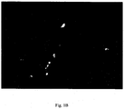
- the morphology is examined, for example (electron) microscopy is used to assess the (ultra)structure of the cells; see Example 1 and Figure 1 .
- Suitable parameters for evaluation include, but are not limited to the evaluation of gap junctions between contacting cells such as cardiomyocytes.
- immunohistochemical or immunofluorescence techniques are used to assess the phenotype; see Example 1 and Figure 2 .
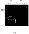
- phenotypic changes are assessed by analysis expression of specific mRNA molecules expressed in the diseased cells. Suitable assay systems include, but are not limited to RT-PCR, in situ hybridization, Northern analysis, or RNase protection assays; see Examples 1 to 3 as well as Figures 3 and 4 .
- the levels of polypeptides expressed in the differentiated cells are assayed.
- polypeptide assays of use include Western blot analysis, ELISA assay, or immunofluorescence.
- calcium transients are measured, as described infra.
- the assay can also be used to screen the effect of an agent on the function of a cell, e.g. cardiomyocyte function. Any method known to one of skill in the art can be utilized to assess cardiac function.
- the beating rate of a cardiomyocyte is assayed to identify agents that increase or decrease beating.
- One method for assessing the beating rate is to observe beating under a microscope.
- Agents that can be screened in this manner include inotropic drugs, such as sympathomimetic agents.
- cells contacted with the agent are compared with a control. Suitable controls include cells not contacted with the agent, or contacted with vehicle alone. Standard values can also be used as a control.
- Cytotoxicity can be determined in the first instance by the effect on cell viability, survival, morphology, and the expression of certain markers and receptors. Effects of a drug on chromosomal DNA can be determined by measuring DNA synthesis or repair. [ 3 H]-thymidine or BrdU incorporation, especially at unscheduled times in the cell cycle, or above the level required for cell replication, is consistent with a drug effect. Unwanted effects can also include unusual rates of sister chromatid exchange, determined by metaphase spread. It can be referred to A. Vickers (375-410) in In vitro Methods in Pharmaceutical Research, Academic Press, 1997 ) for further elaboration.
- Effect of cell function can be assessed using any standard assay to observe phenotype or for example activity of cardiomyocytes, such as marker expression, receptor binding, contractile activity, or electrophysiology in cell culture. Pharmaceutical candidates can also be tested for their effect on contractile activity such as whether they increase or decrease the extent or frequency of contraction. Where an effect is observed, the concentration of the compound can be titrated to determine the median effective dose.
- the assays may be simple "yes/no" assays to determine whether there is a responsive change compared to a control.
- the test compound or a plurality of test compounds can also be subjected to the test cell, preferably embryoid body in different concentrations or dilution series, preferably at doses that correspond to physiological levels of the corresponding type of test compounds. It is thus also possible to easy generate compound profiles in purpose similar to those described in WO00/34525 .
- two or more assays may be used and/or parameters may be assessed. Those assays/parameters can be performed/assessed in parallel or subsequently; or the results of one assay may be compared with results of a corresponding assay performed elsewhere.
- the molecular profile of the test composition can be compared to that of a chemical composition with predetermined biological activities or, preferably, to a library of molecular profiles of chemical compositions with predetermined biological activities.
- the outcome of such comparison provides information for one to predict the likelihood of whether the test composition has the potential of a drug or is toxic, what type of toxicities, and how toxic it would be as compared to the other known toxic compositions.
- test compound is subjected to the test sample before or during inducing the onset of the diseased phenotype.
- Performing the method of the invention can be done according to screening methods known in the art employing cell preparations from animals. For example, the effects of doxorubicin (DOX) on intracellular calcium transients and the cardioprotective effects of a calcium antagonist on DOX-induced impairment of calcium handling were examined in neonatal rat cultured cardiac myocytes; see Maeda et al., Jpn. Circ. J. 63 (1999), 123-129 . Here, cultured cardiac myocytes isolated from neonatal Wistar-Kyoto rats were treated with DOX for 24 h.
- DOX doxorubicin
- said in vitro differentiated cell is derived from pluri- or multipotent cells, preferably from embryonic stem (ES) cells, most preferably said pluri- or multipotent cell is derived from mouse or rat.
- ES embryonic stem
- the invention can be practiced using stem cells of any vertebrate species. Included are stem cells from humans, but not cells from a human embryo; as well as non-human primates, domestic animals, livestock, and other non-human mammal. Amongst the stem cells suitable for use in this invention are primate pluripotent stem cells derived from tissue formed after gestation, such as a blastocyst, or fetal or embryonic tissue taken any time during gestation. Non-limiting examples are primary cultures or established lines of embryonic stem cells. The invention is also applicable to adult stem cells. It is referred to the literature of Anderson et al., Nat. Med. 7 (2001), 393-395 ; Gage, Science 287 (2000), 433-438 , and Prockop, Science 276 (1997), 71-74 , wherein the extraction and culture of those cells is described.
- Media for isolating and propagating stem cells can have any of several different formulas, as long as the cells obtained have the desired characteristics, and can be propagated further. Suitable sources include Iscove's modified Dulbecco's medium (IMDM), Gibco, #12440-053; Dulbecco's modified Eagles medium (DMEM), Gibco #11965-092; Knockout Dulbecco's modified Eagles medium (KO DMEM), Gibco #10829-018; 200 mM L-glutamine, Gibco # 15039-027; non-essential amino acid solution, Gibco 11140-050; [beta]-19 mercaptoethanol, Sigma # M7522; human recombinant basic fibroblast growth factor (bFGF), Gibco # 13256-029. Exemplary serum-containing ES medium and conditions for culturing stem cells are known, and can be optimized appropriately according to the cell type. Media and culture techniques for particular cell types referred to in the previous section are provided in the references cited herein.
- Embryonic stem cells can be isolated from blastocysts of members of the primate species ( Thomson et al., Proc. Natl. Acad. Sci. USA 92 (1995), 7844 ).
- exfoliated human deciduous tooth a comparable very accessible tissue
- neural cells adipocytes
- odontoblasts see Miura et al., Proc. Natl. Acad. Sci. USA 100 (2003), 5807-5812 .
- those cells were found to be able to induce bone formation, generate dentin, and survive in mouse brain along with expression of neural markers.
- multilineage potential of homozygous stem cells derived from metaphase II oocytes has been described in by Lin et al.
- HSC Hematopoietic Stem Cell
- embryonic stem (ES) cell lines that are genetically identical to those of the recipient have been reviewed by Colman and Kind, Trends Biotechnol. 18 (2000), 192-196 .
- ES embryonic stem
- syngenic or autologous cells and recipients are preferably used in the corresponding embodiments of the invention.
- stem cells such as from the bone marrow and tooth it should be possible to accomplish this demand without the need to resort to embryonic cells and tissue.
- cells may be genetically manipulated to suppress relevant transplantation antigens, see also infra, immunosuppressive agents may be used.
- transgenic non-human animals in particular mammals as source for embryonic stem cells.
- compositions and methods for making transgenic swines to be used as xenograft donors is described in US patent No. 5,523,226 .
- WO97/12035 describes methods of producing transgenic animals for xenotransplantation.
- immunologically compatible animal tissue suitable for xenotransplantation into human patients, is described in WO01/88096 .
- Method for making embryonic germ cells from porcine are described for example in US patent No. 6,545,199 .
- Cells immunologically compatible with humans can also be employed for purposes of the present invention.
- Stem cells can be propagated continuously in culture, using a combination of culture conditions that promote proliferation without promoting differentiation.
- stem cells are cultured on a layer of feeder cells, typically fibroblast type cells, often derived from embryonic or fetal tissue.
- the cell lines are plated to near confluence, usually irradiated to prevent proliferation, and then used to support when cultured in medium conditioned by certain cells (e.g. Koopman and Cotton, Exp. Cell 154 (1984), 233-242 ; Smith and Hooper, Devel. Biol. 121 (1987), 1-91 ), or by the exogenous addition of leukemia inhibitory factor (LIF).
- LIF leukemia inhibitory factor
- ES or EG cells spontaneously differentiate into a wide variety of cell types, including cells found in each of the endoderm, mesoderm, and ectoderm germ layers. With the appropriate combinations of growth and differentiation factors, however, cell differentiation can be controlled.
- mouse ES and EG cells can generate cells of the hematopoietic lineage in vitro ( Keller et al., Mol. Cell. Biol. 13 (1993), 473-486 ; Palacios et al., Proc. Natl. Acad. Sci USA 92 (1995), 7530-7534 ; Rich, Blood 86 (1995), 463-472 ).
- mouse ES cells have been used to generate in vitro cultures of neurons ( Bain et al., Developmental Biology 168 (1995), 342-357 ; Fraichard et al., J. Cell Science 108 (1995), 3161-3188 ), cardiomyocytes (heart muscle cells) ( Klug et al., Am. J. Physiol. 269 (1995), H1913-H1921 ), skeletal muscle cells ( Rohwedel et al., Dev. Biol. 164 (1994), 87-101 ), vascular cells ( Wang et al., Development 114 (1992), 303-316 ), US patent No. 5,773,255 relates to glucose-responsive insulin secreting pancreatic beta cell lines, US patent No.
- 5,789,246 relates to hepatocyte precursor cells. Hepatic differentiation of murine embryonic stem cells is also described in Jones et al., Exp. Cell Res. 272 (2002), 15-22 . Other progenitors of interest include but are not limited to chondrocytes, osteoblasts, retinal pigment epithelial cells, fibroblasts, skin cells such as keratinocytes, dendritic cells, hair follicle cells, renal duct epithelial cells, smooth and skeletal muscle cells, testicular progenitors, and vascular endothelial cells. Embryonic stem cell differentiation models for cardiogenesis, myogenesis, neurogenesis, epithelial and vascular smooth muscle cell differentiation in vitro have been generally described in Guan et al., Cytotechnology 30 (1999), 211-226 .
- In vitro differentiated cardiomyocytes neural cells, hepatocytes, adipocytes, skeletal muscle cells, vascular endothelial cells and osteoblasts are described in US patent application US2002/142457 .
- the preparation of cells of the cardiomyocyte lineage produced from human pluripotent stem cells is described in international application WO03/006950 ; see also references cited therein.
- a method for the generation of in vitro differentiated cardiomyocytes from particular stem cells called spoc cells is described in international application WO03/035838 .
- the production of cardiomyocyte-enriched cellular populations, and methods and materials for obtaining the same are also described in international application WO01/68814 .
- differentiation is promoted by withdrawing one or more medium component(s) that promote(s) growth of undifferentiated cells, or act(s) as an inhibitor of differentiation.
- medium component(s) that promote(s) growth of undifferentiated cells, or act(s) as an inhibitor of differentiation.
- such components include certain growth factors, mitogens, leukocyte inhibitory factor (LIF), and basic fibroblast growth factor (bFGF).
- LIF leukocyte inhibitory factor
- bFGF basic fibroblast growth factor
- Differentiation may also be promoted by adding a medium component that promotes differentiation towards the desired cell lineage, or inhibits the growth of cells with undesired characteristics.
- the cells may have the ability to replicate in certain drug screening and therapeutic applications, and to provide a reservoir for the generation of in vitro differentiated cells such as cardiomyocytes and their precursors.
- the cells of this invention can optionally be telomerized to increase their replication potential, either before or after they progress to restricted developmental lineage cells or terminally differentiated cells.
- ES cells that are telomerized may be taken down the differentiation pathway described earlier; or differentiated cells can be telomerized directly.
- telomere catalytic component typically under a heterologous promoter that increases telomerase expression beyond what occurs under the endogenous promoter.
- hTERT human telomerase
- hTERT human telomerase
- hTERT clones ( WO98/14592 ) are used as a source of hTERT encoding sequence, and spliced into an EcoR1 site of a PBBS212 vector under control of the MPSV promoter, or into the EcoRl site of commercially available pBABE retrovirus vector, under control of the LTR promoter.
- telomere activity Assay
- immunocytochemical staining for hTERT or replicative capacity
- Continuously replicating colonies will be enriched by further culturing under conditions that support proliferation, and cells with desirable phenotypes can optionally be cloned by limiting dilution.
- other methods of immortalization may also be acceptable, such as transforming the cells with DNA encoding myc, the SV40 large T antigen, or MOT-2 ( US patent No. 5,869,243 ; international applications WO97/32972 and WO01/23555 ).
- populations of differentiated cells to be used in the assay are preferably depleted of relatively undifferentiated cells and/or of cells of undesired cell types by using a selection system that is lethal to the undesired cells and cell types, i.e. by expressing a selectable marker gene that renders cells of a specific cell type resistant to a lethal effect of an external agent, under control of a regulatory sequence that causes the gene to be preferentially expressed in the desired cell type and/or at a certain stage of development.
- the cells are genetically altered before the process used to differentiate the cells into the desired lineage for therapy, in a way that the cells comprises a selectable marker operably linked to a first cell type specific regulatory sequence specific for the desired first cell type.
- Suitable expression vector for this purpose can be used.
- Suitable viral vector systems for producing stem cells altered according to this invention can be prepared using commercially available virus components.
- the introduction of the vector construct or constructs into the embryonic stem cells occurs in a known manner, e.g. by transfection, electroporation, lipofection or with the help of viral vectors.
- Viral vectors comprising effector genes are generally described in the publications referenced in the last section.
- vector plasmids can be introduced into cells by electroporation, or using lipid/DNA complexes. Exemplary is the formulation Lipofectamine 2000(TM), available from Gibco/Life Technologies.
- Another exemplary reagent is FuGENE(TM) 6 Transfection Reagent, a blend of lipids in non-liposomal form and other compounds in 80 % ethanol, obtainable from Roche Diagnostics Corporation.
- FuGENE(TM) 6 Transfection Reagent a blend of lipids in non-liposomal form and other compounds in 80 % ethanol, obtainable from Roche Diagnostics Corporation.
- the vector constructs and transfection methods described in WO02/051987 are used.
- Resistance genes per se are known. Examples for these are nucleoside and aminoglycoside-antibiotic-resistance genes, e.g. puromycin (puromycin-N-acetyltransferase), streptomycin, neomycin, gentamycin or hygromycin. Further examples for resistance genes are dehydrofolate-reductase, which confers a resistance against aminopterine and methotrexate, as well as multi drug resistance genes, which confer a resistance against a number of antibiotics, e.g. against vinblastin, doxorubicin and actinomycin D.
- nucleoside and aminoglycoside-antibiotic-resistance genes e.g. puromycin (puromycin-N-acetyltransferase), streptomycin, neomycin, gentamycin or hygromycin.
- resistance genes are dehydrofolate-reductase, which confers a resistance against aminopterine
- said selectable marker confers resistance to puromycin.
- Puromycin is particularly suited for the fast elimination of non-cardiac cells in adherent culture of transgenic EBs; see also Examples.
- drug selection of cardiac cells can be implemented entirely in the suspension culture of transgenic EBs.
- purified ES derived cardiomyocytes survive much longer in culture than untreated counterparts.
- the elimination of undifferentiated ES cells during drug selection process has itself been shown to have clear positive effect on viability and longevity of such differentiated ES derived cells as cardiomyocytes.
- the release from surrounding non-differentiated cells induces proliferation of cardiomyocytes.
- the drug selection possesses both purifying and multiplying effect.
- said ES cell of said ES cell-derived first cell type comprises a reporter gene, wherein said reporter is operably linked to a cell type specific regulatory sequence specific for said first cell type.
- This type of vector has the advantages of providing visualization of differentiation, definition of the time point for beginning of drug selection, visualization of drug selection and tracing of the fate of purified cells grafted in recipient tissue.
- Such vectors which are preferably employed in accordance with the methods of the present invention are described in WO02/051987 .
- said cell type specific regulatory sequence of the reporter gene is substantially the same as said first cell type specific regulatory sequence of the marker gene.
- the reporter can be of any kind as long as it is non-damaging for the cell and confers an observable or measurable phenotype.
- the green fluorescent protein (GFP) from the jellyfish Aequorea victoria (described in WO95/07463 , WO96/27675 and WO95/21191 ) and its derivates "Blue GFP” ( Heim et al., Curr. Biol. 6 (1996), 178-182 and "Redshift GFP” ( Muldoon et al., Biotechniques 22 (1997), 162-167 ) can be used.
- EGFP Enhanced Green Fluorescent Protein
- Further embodiments are the Enhanced Yelow and Cyan Fluorescent Proteins (EYFP and ECFP, respectively) and Red Fluorescent proteins (DsRed, HcRed).
- Further fluorescent proteins are known to the person skilled in the art and can be used according to the invention as long as they do not damage the cells. The detection of fluorescent proteins takes places through per se known fluorescence detection methods; see, e.g., Kolossov et al., J. Cell Biol. 143 (1998), 2045-2056 .
- other detectable proteins particularly epitopes of those proteins, can also be used.
- the epitope of proteins though able to damage the cell per se, but whose epitopes do not damage the cells, can be used; see also WO02/051987 .
- vector constructs contain a further selectable marker gene, which confers e.g. a resistance against an antibiotic, e.g. neomycin.
- a further selectable marker gene confers e.g. a resistance against an antibiotic, e.g. neomycin.
- antibiotic e.g. neomycin
- other known resistance genes can be used as well, e.g. the resistance genes described above in association with the fluorescent protein encoding genes.
- the selection gene for the selection for stably transfected ES cells is under the control of a different promoter than that which regulates the control of the expression of the detectable protein. Often constitutively active promoters are used, e.g. the PGK-promoter.
- a second selection gene is advantageous for the ability to identify the successfully transfected clones (efficiency is relatively low) at all. Otherwise a smothering majority of non-transfected ES cell may exist and during differentiation e.g. no EGFP positive cells might be detected.
- the cells can be manipulated additionally so that specific tissues are not formed. This can occur for instance by inserting of repressor elements, e.g. a doxycyclin inducible repressor element. Thereby, a possible contamination of the desired differentiated cells with pluripotent, potentially tumorigenic cells can be excluded.
- repressor elements e.g. a doxycyclin inducible repressor element.
- the desired cell type intended for the stem cell to differentiate to may be of any kind and includes but not limited to neuronal cells, glial cells, cardiomyocytes, glucose-responsive insulin secreting pancreatic beta cells, hepatocytes, astrocytes, oligodendrocytes, chondrocytes, osteoblasts, retinal pigment epithelial cells, fibroblasts, keratinocytes, dendritic cells, hair follicle cells, renal duct epithelial cells, vascular endothelial cells, testicular progenitors, smooth and skeletal muscle cells; see also supra.
- said in vitro differentiated cells are cardiomyocytes.
- said cell type specific regulatory sequence is preferably atrial and/or ventricular specific.
- Corresponding regulatory sequences i.e. cardiac specific promoters are described in the prior art; see also supra.
- Nkx-2.5 specific for very early cardiomyocytes and mesodermal precursor cells, respectively, ( Lints et al., Development 119 (1993), 419-431 ); human-cardiac- ⁇ -actin specific for heart tissue, ( Sartorelli et al., Genes Dev. 4 (1990), 1811-1822 ), and MLC-2V specific for ventricular heart muscle cells ( O'Brien et al., Proc. Natl.
- fibroblasts can also be generated de novo from ES cells in accordance with the method of the present invention.
- ES cells are transfected with a recombinant nucleic acid molecule comprising a marker and optionally reporter gene operatively linked to a cell type specific regulatory sequence, i.e. fibroblast specific promoter such as the a2 (I) collagen promoter though also active in bone cells; Lindahl et al., Biol. Chem. 277 (2002), 6153-6161 ; Zheng et al., Am. J. Pathol. 160 (2002), 1609-1617 ; Antoniv et al., J. Biol. Chem.
- fibroblast specific promoter such as the a2 (I) collagen promoter though also active in bone cells
- a further cell type are endothelial cells which can be derived from ES cells transfected with a vector construct as generally described before, wherein said cell type specific regulatory sequence is an endothelial specific promoter; see, e.g., vascular endothelial-cadherin promoter described by Gory et al., Blood 93(1999), 184-192 ; the Tie-2 promoter/ enhancer by Schlaeger et al., Proc. Natl. Acad. Sci. USA 94 (1997), 3058-3063 ; the Flk-1 promoter/ enhancer by Kappel et al., Biochem. Biophys. Res. Commun. 276 (2000), 1089-1099 .
- endothelial specific promoter see, e.g., vascular endothelial-cadherin promoter described by Gory et al., Blood 93(1999), 184-192 ; the Tie-2 promoter/ enhancer by Schlaeger e
- II collagen chain
- GFAP glial fibrillary acidic protein
- tissue specific promoters are those, which are active in glia cells, hematopoietic cells, neuronal cells, preferably embryonal neuronal cells endothelial cells, cartilage cells or epidermal cells as well as insulin secreting ⁇ -cells. "Tissue specific" is to be subsumed under the term "cell specific”.
- non-heart-specific promoters are: PECAM1, FLK-1 (endothelium), nestine (neuronal precursor cells), tyrosin-hydroxylase-1-promoter (dopaminergic neurons), smooth muscle ⁇ -actin, smooth muscle myosin (smooth muscles), ⁇ 1-fetoprotein (endoderm), smooth muscle heavy chain (SMHC minimal promoter (specific for smooth muscles, ( Kallmeier et al., J. Biol. Chem. 270 (1995); 30949-30957 ).
- development specific promoter refers to promoters, that are active during certain points of time during development. Examples for such promoters are the ⁇ -MHC promoter that is expressed during embryonal development in the ventriculum of the mouse and is superseded by the ⁇ -MHC promoter in the prenatal phase.
- NKx2.5 a promoter during the early mesoderm/heart development, atrial-natriuretic-factor, a marker of the early embryonal heart with exception of the pacemaker, that is down regulated also in later developmental stages, FIk-1, an endothelium specific promoter that is active during the early vasculogenesis, intron 2-segment of the nestine gene that is expressed in neuronal precursor cells (embryonal neurons and glia cells) and adult glia cells (partially still able to divide) ( Lothian and Lendahl, Eur. J. Neurosci. 9 (1997), 452-462U ).
- said resistance gene and said reporter gene are preferably contained in a bicistronic vector and are preferably separated by an IRES.
- IRES an IRES
- an in vitro differentiated cell of one cell type is cocultured with at least one cell of a second cell type, and/or comprised in tissue or tissue-like structures comprising at least one second cell type such as any one of those described hereinbefore.
- Said second cell type may be for example an embryonic second cell type.
- the in vitro differentiated cell in said tissue or tissue-like structure is obtained by culturing an embryonic stem (ES) cell derived first cell type in the presence of at least one embryonic second cell type; and allowing integration and alignment of said at least two cell types into tissue or tissue-like structures.
- Said at least second cell type may also be generated as the first cell type, i.e. by in vitro differentiation of ES cells which have been genetically engineered with corresponding marker genes; see also supra for appropriate methods and materials.
- a corresponding method for providing a variety of tissue or tissue-like structures and like in vitro differentiated cells and tissue is described in detail in international application WO2004/113515 .
- in vitro differentiated cell is also meant to include a plurality of in vitro differentiated cells of the same or different cell types as well as in vitro differentiated tissue and organs, and cocultures of in vitro differentiated cells with other cell types such as of embryonic origin.
- in vitro differentiated cell does not necessarily exclude the presence of a cell or cell type other than that which the original stem cell has been differentiated to.
- the use of a substantially pure culture of in vitro differentiated cells is preferred or the use of even a single cell.
- said at least second cell type preferably corresponds to an endothelial cell and/or fibroblast.
- bradykinin blocks angiotensin II-induced hypertrophy in the presence of endothelial cells; see Ritchie et al., Hypertension 31 (1998), 39-44 .
- effects of bradykinin on isolated ventricular cardiomyocytes from adult and neonatal rat hearts have been determined and the extent to which bradykinin blocks hypertrophy in vitro. Bradykinin was found to be a hypertrophic agonist, as defined by increased protein synthesis and atrial natriuretic peptide secretion and expression.
- bradykinin did not increase protein synthesis.
- bradykinin has a direct hypertrophic effect on ventricular myocytes.
- endothelial cells is required for the antihypertrophic effects of bradykinin.
- the in vitro differentiated cell to be tested is obtained by a method which is preferably performed such that it allows self-assembly of the different cell types, for example into the desired tissue or tissue-like structures that should reflect the tissue or organ of a mammal, preferably human, that is supposed to be exposed to a given compound.
- the stem cells are in a preferred embodiment of the invention available in form of aggregates that are known as embryoid bodies (EBs).
- EBs embryoid bodies
- WO02/051987 describes a protocol to obtain embryoid bodies. The manufacturing takes place preferably with the "hanging drop" method or by methylcellulose culture ( Wobus et al., Differentiation 48 (1991), 172-182 ).
- the functional tissue assay of the present invention is performed with embryoid bodies (EBs).
- EBs embryoid bodies
- embryoid bodies represent a complex group of cells differentiating into different tissues.
- the cells within an embryoid body are substantially synchronized for their differentiation. Accordingly, at known intervals, the majority of the synchronized cells differentiate into the three embryonic germ layers and further differentiate into multiple tissue types, such as cartilage, bone, smooth and striated muscle, and neural tissue, including embryonic ganglia; see also Snodgrass et al., "Embryonic Stem Cells: Research and Clinical Potentials" in Smith and Sacher, eds. Peripheral Blood Stem Cells American Association of Blood Banks, Bethesda MD (1993 ).
- the cells within embryoid bodies provide a much closer model to the complexity of whole organisms than do traditional single cell or yeast assays, while still avoiding the cost and difficulties associated with the use of mice and larger mammals.
- the recent availability of human embryoid bodies provides an even closer vehicle for modeling toxicity and identification of drugs useful for the treatment of heart disorders in human organ systems, and in humans.
- spinner flasks (stirring cultures) can be used as culture method.
- the undifferentiated ES cells are introduced into stirring cultures and are mixed permanently according to an established procedure. Therefore, 10 million ES cells are introduced into 150 ml medium with 20 % FCS and are stirred constantly with the rate of 20 rpm., wherein the direction of the stirring motion is changed regularly. 24 hours after introduction of the ES cells an extra 100 ml medium with serum is added and thereupon 100 - 150 ml of the medium is exchanged every day ( Wartenberg et al., FASEB J. 15 (2001), 995-1005 ). Under these culture conditions large amounts of ES cell-derived cells, i.e. cardiomyocytes, endothelial cells, neurons etc. depending on the composition of the medium can be obtained.
- the cells are selected by means of the resistance gene either still within the stirring culture or after plating, respectively.
- the EBs differentiated in the hanging drop might be not plated, but kept simply in suspension. Even under these conditions a progression of a differentiation could be observed experimentally.
- the washing off of the non-desired cell types can be done with mechanical mixing alone and addition of low concentration of enzyme (e.g. collagenase, trypsin); a single cell suspension is achieved with easy washing off of the non-desired cell types.
- embryoid bodies are prepared according a recent developed "mass culture" system employed in the appended examples and described in detail in international application WO2005/005621 .
- the disease said diseased phenotype corresponds to is a heart disease such as heart failure or a cardiomyopathy.
- the diseased phenotype to be induced and assessed is a cardiac hypertrophic phenotype; see also the Examples.
- Heart failure is the inability of the heart to supply sufficient oxygenated blood to meet the metabolic needs of the tissues and cells in a subject. This can be accompanied by circulatory congestion, such as congestion in the pulmonary or systemic veins.
- heart failure encompasses heart failure from any cause, and is intended herein to encompass terms such as “congestive heart failure,” “forward heart failure,” “backward heart failure,” “high output heart failure,” “low output heart failure,” and the like; see also Chapters 13-17 in Braunwald for a detailed discussion.
- Conditions that could lead to heart failure include, but are not limited to, coronary artery disease, cardiomyopathy, or congenital heart disease.
- Cardiomyopathy is any disease or dysfunction of the myocardium (heart muscle) in which the heart is abnormally enlarged, thickened and/or stiffened. As a result, the heart muscle's ability to pump blood is usually weakened.
- the disease or disorder can be, for example, inflammatory, metabolic, toxic, infiltrative, fibroplastic, hematological, genetic, or unknown in origin.
- cardiomyopathies There are two general types of cardiomyopathies: ischemic (resulting from a lack of oxygen) and non-ischemic.
- Ischemic cardiomyopathy is a chronic disorder caused by coronary artery disease - a disease in which there is atherosclerotic narrowing or occlusion of the coronary arteries on the surface of the heart. Coronary artery disease often leads to episodes of cardiac ischemia, in which the heart muscle is not supplied with enough oxygen-rich blood. Eventually, the heart muscle enlarges from the additional work it must do in the absence of sufficient oxygen-rich blood.
- said phenotype preferably includes a parameter selected from the group consisting of cell size, cell shape, protein synthesis, organization of actin/myosin filament, activation of gene expression pattern characteristic of cardiomyopathic cells, and/or activation of genes expressed during early embryonic development; see also infra.
- hepatocytes can also be assessed in accordance with the present invention, for which appropriate parameters for determining phenotypic changes are well known in the art.
- WO01/81549 describes the generation of in vitro differentiated cells derived from pluripotent stem cells with morphological features of hepatocytes, expressing surface markers characteristic of hepatocytes, and having enzymatic and biosynthetic activity important for liver function.
- the cells can be characterized according to a number of phenotypic criteria. The criteria include but are not limited to the detection or quantitation of expressed cell markers, and enzymatic activity, and the characterization of morphological features and intercellular signaling.
- a polygonal cell shape a binucleate phenotype
- the presence of rough endoplasmic reticulum for synthesis of secreted protein the presence of Golgi-endoplasmic reticulum lysosome complex for intracellular protein sorting
- the presence of peroxisomes and glycogen granules relatively abundant mitochondria
- the ability to form tight intercellular junctions resulting in creation of bile canalicular spaces Cell markers useful in distinguishing liver progenitors, hepatocytes, and biliary epithelium, are shown in Table 1 of WO01/81549 .
- cytochrome p450 can also be measured at the protein level, for example, using specific antibody in Western blots, or at the mRNA level, using specific probes and primers in Northern blots or RT-PCR; see Borlakoglu et al., Int. J. Biochem. 25 (1993), 1659 .
- Particular activities of the p450 system can also be measured: 7-ethoxycoumarin O-de-ethylase activity, aloxyresorufin O-de-alkylase activity, coumarin 7-hydroxylase activity, p-nitrophenol hydroxylase activity, testosterone hydroxylation, UDP-glucuronyltransferase activity, glutathione S-transferase activity, and others; see, e.g., review by Gomes-Lechon et al. in "In vitro Methods in Pharmaceutical Research” Academic Press (1997), 411-431 .
- the diseased phenotype is preferably induced during culturing the in vitro differentiated cell, since the diseases phenotype may be lethal for the stem cell used for differentiation. Furthermore, the possibility to induce the diseased phenotype allows investigation whether a given compound is capable of preventing the onset of a disease if added prior to the induction of the diseased phenotype. This embodiment is particularly useful for identifying and obtaining drugs that can be used as a prophylactic means, which is especially worthwhile to consider for the prevention of heart diseases. However, as demonstrated in Example 2, it is also possible and envisaged in accordance with the present invention to confer the diseased phenotype in a constitutive manner, for example by constitutive expression of a disease-related gene.
- said phenotype in the method of the present invention is induced by culturing the in vitro differentiated cell in the presence of a physiologically active compound.
- a physiologically active compound such as preferably endothelin-1, angiotensin II, or an ⁇ 1-adrenergic agonist, most preferably said ⁇ 1-adrenergic agonist is phenylephrine.
- the in vitro differentiated cell is genetically engineered to display said phenotype.
- Genetically engineering can be done by various means.
- the in vitro differentiated cell can be transduced using standard procedures known in molecular biology in order to introduce a nucleic acid molecule of interest into the cell.
- the nucleic acid molecule encodes a polypeptide the expression of which confers the diseased phenotype.
- the polypeptide encoded by the nucleic acid molecule can be from the same species as the cells (homologous) or can be from a different species (heterologous).
- the polypeptide may either correspond to a wild-type or a mutant allele either of which may be responsible for the diseased phenotype.
- the polypeptide may be of any kind, for example an enzyme, structural protein or transcriptional regulator.
- the nucleic acid sequence of interest is operably linked to a regulatory element, such as a transcriptional and/or translational regulatory element; see also supra.
- Regulatory elements include elements such as a promoter, an initiation codon, a stop codon, mRNA stability regulatory elements, and a polyadenylation signal.
- a promoter can be a constitutive promoter or an inducible promoter. Specific non-limiting examples of promoters include the CMV promoter, an atrial natriuretic factor promoter, and promoters including TET-responsive element for inducible expression of transgene.
- the nucleic acid sequence of interest is inserted into a vector, such as an expression vector. Procedures for preparing expression vectors are known to those of skill in the art and can be found in Sambrook et al., Molecular Cloning: A Laboratory Manual, 2nd Ed., Cold Spring Harbor Press, Cold Spring Harbor, N.Y. (1989 ).
- Said phenotype can be due to (over)expression of a (mutated) gene, knock-down of gene(s) (e.g. by RNA interference; RNAi), knock-out, or knock-in of (a) gene(s) in said in vitro differentiated cell.
- a (mutated) gene e.g. by RNA interference; RNAi), knock-out, or knock-in of (a) gene(s) in said in vitro differentiated cell.
- an in vitro differentiated cell or the underlying ES cells may be transfected with a nucleic acid molecule designed to functionally delete or "knock-out" a gene of interest.
- the nucleic acid molecule of interest is a nucleic acid molecule that undergoes homologous recombination and is inserted into the genome of the cell.
- Methods for producing "knock-outs" in ES cells are known to one of skill in the art (see, e.g., US patent No. 5,939,598 ).
- cells are cultured in vitro as described herein and an exogenous nucleic acid is introduced into the cells by any method known to one of skill in the art, for example, by transfection or electroporation.
- the transfected cultured cells can then be studied in vitro.
- Methods for the introduction of nucleic acid sequences into stem cells are known in the art (e.g., see US patent No. 6,110,743 ).
- Calcium is central in the regulation of cardiac contractility, growth and gene expression. Variations in the amplitude, frequency and compartmentalization of calcium signals are decoded by calcium/calmodulin-dependent enzymes, ion channels and transcription factors.
- the circuitry for calcium signaling creates opportunities for pharmacological modification of cardiac function and thus provides a multitude of putative target genes for use in the method of the present invention; see for review, e.g., Frey et al., Nature Med. 6 (2000), 1221-1227 .
- HCM hypertrophic cardiomyopathy
- MyBPC cardiac myosin binding protein C
- TnT cardiac troponin T
- TnI cardiac troponin I
- ⁇ TM ⁇ tropomyosin
- dilated cardiomyopathy including the heterogeneity in the clinical features of dilated cardiomyopathy resulting from a single gene mutation are described in Schönberger et., Am. J. Hum. Genet. 69 (2001), 249-260 .
- Table 1 of Schönberger et al. provides an overview for loci with dilated cardiomyopathy as the predominant phenotype. These genes can serve as parameters to be assessed with respect to the diseased phenotype as well as target genes for inducing a diseased phenotype, in particular a dilated cardiomyopathic phenotype.
- phopholamban is an endogenous inhibitor of sarcoplasmic reticulum calcium ATPase and plays a prime role in cardiac contractility and relaxation. Recently, it has been reported that human phospholamban null results in lethal dilated cardiomyopathy revealing a critical difference between mouse and human; see Haghighi et al., J. Clin. Invest. 111 (2003), 869-876 .
- phopholamban may be further investigated using the in vitro differentiated cell-based assay of the present invention.
- Means and methods for mediating expression or suppression of the phospholamban gene as well as of mutants thereof are well known to those skilled in the art; see supra and, for example, Eizema et al., Circulation 101 (2000), 2193-2199 , reporting on adenovirus-based phospholamban antisense expression as an approach to improve cardiac contractile dysfunction and comparison of a constitutive viral versus an endothelin-1-responsive cardiac promoter.
- genes may be employed, which hitherto have been considered for the generation of transgenic animal models and treatment for heart diseases.
- an alpha-myosin heavy chain promoter operatively linked to a coding sequence comprising DNA coding for a betal-adrenergic receptor is described in US patent No. 6,218,597 .
- Over-expression of Gsalpha, and and beta-adrenergic receptor antagonists have been used to establish a transgenic animal model of heart failure in international application WO97/36477 .
- a heart muscle specific expression cassette comprising a nucleotide sequence encoding a human cardiomyopathy inducer under the control of a myocardium-specific regulatory sequence is described in German patent application No. 198 151 28 .
- a coding region which encodes calcineurin, calcium calmodulin dependent kinase IV (CaMKIV), or a functional fragment of calcineurin or CaMKIV, operatively linked to a promoter that is preferentially active in cardiomyocytes has been described to produce cardiac hypertrophy in transgenic mice; see US patent No. 6,657,104 and Example 2.
- genes which are differentially expressed in hypertrophic cardiac tissue as compared to normal cardiac tissue have been described in US patent application US2003/148296 .
- said in vitro differentiated cell (over)expresses a polypeptide selected from the group consisting of (mutant) troponin, heavy myosin chain, calcineurin, calmodulin, protein kinase C, phospholamban or calcium calmodulin dependent kinase IV (CaMKIV).
- a polypeptide selected from the group consisting of (mutant) troponin, heavy myosin chain, calcineurin, calmodulin, protein kinase C, phospholamban or calcium calmodulin dependent kinase IV (CaMKIV).
- KCNJ2 Another example is provided by Andersen mutations of KCNJ2, which suppress the native inward rectifier current IK1 in a dominant-negative fashion.
- the Andersen's syndrome is a hereditary disease, which is characterized by cardiac arrhythmias, periodic paralysis and dysmorphic features. Mutations of the KCNJ2 gene, which encodes the inward rectifying potassium channel subunit Kir2.1, have been identified in affected individuals.
- stem cells can be transfected with a plasmid vector comprising a cDNA sequence of a gene which has been identified in context with its differential expression in a diseased cell, tissue or organ.
- target gene expression is induced and the resultant phenotype analyzed compared to a control which either does not express the target gene or expresses the target gene at a normal level. If the induction of target gene expression results in a diseased phenotype, e.g. an enhanced diseased phenotype, this may be taken as evidence that the target gene is responsible or at least involved in disease development, and thus may be focused on for therapeutic intervention.
- said in vitro differentiated cell is genetically engineered to express or suppress a gene encoding potential drug target; see also infra.
- in vitro differentiated cells which have been genetically engineered to display the diseased phenotype
- said phenotype is inducible, for example, by expressing the gene responsible for said phenotype under the control of an inducible promoter.
- inducible promoter systems are known to the person skilled in the art; see also supra.
- the promoters employed are preferably inducible and useful under appropriate conditions to direct high-level expression target gene.
- an inducible promoter in the present invention provides a molecular switch capable of turning on expression of the polynucleotide sequence which is operatively linked when such expression is desired, or turning off the expression when expression is not desired.
- inducible promoters include, but are not limited to a metallothioneine promoter, a glucocorticoid promoter, a progesterone promoter and a tetracycline promoter.
- inducible expression system is available from Invitrogen, which carries the T-REXTM (tetracycline-regulated expression) system, an inducible mammalian expression system that uses the full-length CMV promoter.
- T-REXTM tetracycline-regulated expression
- the tetracycline-inducible system for regulation of gene expression in transgenic mice is described in Grill et al., Transgenic Res. 12 (2003), 33-43 .
- tetracycline-regulated gene expression in replication-incompetent herpes simplex virus vectors is described by Schmeisser et al., Hum. Gene Ther. 13 (2002), 2113-2124 .
- the gene responsible for said phenotype can be a gene, for example cDNA encoding a RNA, which either may be functional itself such as ribosomal RNA or may encode the functional polypeptide that is responsible for said phenotype.
- the gene responsible for the induction of said phenotype is capable of mediating the suppression of an endogenous target gene or inhibition of the activity of the product encoded by said target gene. In this case, the lack of expression of the target gene and activity of its gene product would be responsible for the diseased phenotype.
- the target gene to be expressed or suppressed can be a wild-type or mutant allele. Means and methods for conferring expression or suppression of a gene are well known to the person skilled in the art; see also supra.
- a diseased phenotype may also be induced before the test compound is added and then monitor the progression of the disease in the presence and absence of the test compound, respectively.
- Arrays for use in the assay of the present invention usually comprise a solid support and attached thereto or suspended thereon the in vitro differentiated cells.
- planar microelectrode arrays for cultured cells and cell aggregates as biosensors is of particular interest.
- Such arrays generally consist of a substrate of glass, plastic or silicon over which a conductor, e.g. gold, platinum, indium-tin-oxide, iridium, etc., is deposited and patterned.
- An insulating layer e.g.
- photoresist, polyimide, silicon dioxide, silicon nitride, etc. is deposited over the conducting electrodes and interconnects and then removed in regions over the electrodes to define the recording sites.
- Cells are cultured directly on this surface and contact the exposed conductor at the deinsulated recording sites.
- recordings of electrical activity can be from a single cell or populations of cells including cell aggregates.
- Each electrode site is generally connected to the input of a high input impedance, low noise amplifier, with or without AC coupling capacitors, to allow amplification of the relatively small extracellular signals. Examples of such biosensors are described by Novak et al.
- the method of the present invention is preferably performed with a multi- or microelectrode array (MEA), such as those mentioned above.
- MAA microelectrode array
- This assay system of the present invention is a particular advantageous alternative for animal testing for cardiac affect analyses, which are usually quite time-consuming and expensive.
- the functional tissue assay system is particularly useful in drug development and toxicity testing of any compound a human or animal might get in contact with.
- Microelectrode arrays are devices which allow the multiple extracellular recording of action potential generation and propagation within for example ES cell-derived cardiomyocytes. This recordings resemble the well-known ECG as it is used by physicians.
- the matrix of the MEAs usually consists of 60 gold electrodes integrated into the bottom of a specially designed cell culture device.
- ES cell-derived embryoid bodies can be cultured in such devices. After attachment and spreading on the surface, the cells of the EBs containing the cardiomyocytes get in contact with the electrodes. All outcoming extracellular action potentials can then be recorded synchroneously during both short- and long time observation experiments. The following analysis of frequencies and latencies with an appropriate program allows to reveal the fine "electrical map" of the beating clusters.
- electrophysiological properties prior, during and after adding the test compound to cardiac myocytes can be followed by recordings of extracellular field potentials with microelectrode arrays (MEA) consisting of, e.g., 60 substrate-integrated electrodes; see Banach et al. Am. J. Physiol. Heart Circ. Physiol. 284 (2003), H2114-2123 . Multiple arrays of tungsten microelectrodes were used to record the concurrent responses of brain stem neurons that contribute to respiratory motor pattern generation; see Morris et al., Respir. Physiol. 121 (2000), 119-133 .
- MEA microelectrode arrays
- the above mentioned parameters may be used in the cell-based assay system of the present invention any one of said further parameters besides the measuring of electrical activity of said biological material through said electrode array.
- embryoid bodies are used in the assays of the present invention to test the chemical composition; see also infra.
- the choice of the particular species from which the embryoid body is derived will typically reflect a balance of several factors.
- one or more species may be of particular interest.
- human embryoid bodies will be of particular interest for use with compositions being tested as potential human therapeutics but also for toxicological tests for substances including industrial chemicals, while equine, feline, bovine, porcine, caprine, canine, or sheep embryoid bodies may be of more interest for a potential veterinary therapeutic.
- Embryoid bodies of other species commonly used in preclinical testing such as guinea pigs, mice, rat, rabbits, pigs, and dogs, are also preferred. Typically, embryoid bodies of these species will be used for "first pass" screening, or where detailed information on toxicity in humans is not needed, or where a result in a murine or other one of these laboratory species has been correlated to a known toxicity or other effect in humans.
- regulatory agencies generally require animal data before human trials can begin; it will generally be desirable to use embryoid bodies of species which will be used in the preclinical animal studies. The results of testing in the embryoid bodies can then guide the researcher on the degree and type of toxicity to anticipate during the animal trials.
- Certain animal species are known in the art to be better models of human toxicity of different types than are others, and species also differ in their ability to metabolize drugs; see, e.g., Williams, Environ. Health Perspect. 22 (1978), 133-138 ; Duncan, Adv. Sci. 23 (1967), 537-541 .
- the particular species preferred for use in a particular preclinical toxicity study may vary according to the intended use of the drug candidate. For example, a species which provide a suitable model for a drug intended to affect the reproductive system may not be as suitable a model for a drug intended to affect the nervous system. Criteria for selecting appropriate species for preclinical testing are well known in the art.
- the chemical composition is in an aqueous solution, preferably in a solvent conventionally used in cell culture, for example DMSO, and is introduced to the culture medium; see also the examples.
- the introduction can be by any convenient means, but will usually be by means of a pipette, a micropipettor, or a syringe.
- the chemical compositions will be introduced by automated means, such as automated pipetting systems, which may be on robotic arms.
- Chemical compositions can also be introduced into the medium as in powder or solid forms, with or without pharmaceutical excipients, binders, and other materials commonly used in pharmaceutical compositions, or with other carriers which might be employed in the intended use.
- chemical compositions intended for use as agricultural chemicals or as petrochemical agents can be introduced into the medium by themselves to test the toxicity of those chemicals or agents, or introduced in combination with other materials with which they might be used or which might be found in the environment, to determine if the combination of the chemicals or agents has a synergistic effect.
- the cultures will be shaken at least briefly after introduction of a chemical composition to ensure the composition is dispersed throughout the medium.
- a chemical composition is added to the culture is within the discretion of the practitioner and will vary with the particular study objective.
- the chemical composition will be added as soon as the embryoid body develops from the stem cells, permitting the determination of the alteration in protein or gene expression on the development of all the tissues of the embryoid body. It may be of interest, however, to focus the study on the effect of the composition on a particular tissue type. As previously noted, individual tissues, such as muscle, nervous, and hepatic tissue, are known to develop at specific times after the embryoid body has formed. Addition of the chemical composition can therefore be staged to occur at the time the tissue of interest commences developing, or at a chosen time after commencement of that development, in order to observe the effect on altering gene or protein expression in the tissue of interest.
- a chemical composition will be used to contact an embryoid body depending on the amount of information known about the toxicity of that composition, the purposes of the study, the time available, and the resources of the practitioner.
- a chemical composition can be administered at just one concentration, particularly where other studies or past work or field experience with the compound have indicated that a particular concentration is the one which is most commonly found in the body. More commonly, the chemical composition will be added in different concentrations to cultures of embryoid bodies run in parallel, so that the effects of the concentration differences on gene or protein expression and, hence, the differences in toxicity of the composition at different concentrations, can be assessed.
- the chemical composition will be added at a normal or medium concentration, and bracketed by twofold or fivefold increases and decreases in concentration, depending on the degree of precision desired.
- the composition is one of unknown toxicity
- a preliminary study is conveniently first performed to determine the concentration ranges at which the composition will be tested.
- a variety of procedures for determining concentration dosages are known in the art.
- One common procedure, for example, is to determine the dosage at which the agent is directly toxic.
- the practitioner then reduces the dose by one half and performs a dosing study, typically by administering the agent of interest at fivefold or twofold dilutions of concentration to parallel cultures of cells of the type of interest.
- the composition will usually also be tested at the concentration at which it is found in the environment.
- the agent will usually be tested at the concentration at which the residue is found, although it will likely be tested at other concentrations as well.
- the dilution of test compounds can be done by making in separated tubes a series of dilution of 50 or 100 fold concentrated compounds in DMSO. One or two ⁇ l of each dilution are distributed in each well before cell suspension distribution.
- any one or all of the following parameters are analyzed:
- MEAs and methods for their use in analyses of biological cells are known to the person skilled in the art.
- international application WO97/05922 describes a microelectrode arrangement for leaking, with local resolution, electrical cell potentials, or for electrical stimulation of networks of biological cells such as for example cell cultures, tissue slices "in vitro" or biological tissue "in vivo”.
- a micro-element device such as described in international application WO98/22819 may be used, which has a plurality of microelements, which may be configured as microelectrodes, arranged on a substrate and adapted for making contact to cells present in a liquid environment. The cells are guided onto the microelectrodes, are isolated or are mechanically attracted to the microelectrodes. A negative-pressure force or a hydrodynamic force may be applied on the cells.
- the use of an electrode array as described in international application WO01/65251 may be adapted in accordance with the teaching of the present invention.
- the test sample comprises embryoid bodies (EBs) differentiated into cardiomyocytes, most preferably EBs that consist of functional cardiac tissue that beats autonomously and covers electrophysiological properties of atrial and ventricular cardiomyocytes, as well as of pacemaker cells.
- EBs embryoid bodies differentiated into cardiomyocytes, most preferably EBs that consist of functional cardiac tissue that beats autonomously and covers electrophysiological properties of atrial and ventricular cardiomyocytes, as well as of pacemaker cells.
- the methods and assays described herein can replace various animal models, and form novel mammal-based tests and extreme environment biosensors.
- the methods of the invention can also be used for toxicological, mutagenic, and/or teratogenic in vitro tests. This is because people suffering from a disease are more vulnerable to intoxication and susceptible to side effects of pharmaceuticals, nutritions or any other compounds that one gets in contact with. Since the cells and tissue obtained in accordance with the present invention more closely resemble the in vivo situation the results obtained by the toxicological assays of the present invention are expected to correlate to in vivo toxicity of the tested compounds as well.
- cardiobodies i.e. embryoid bodies (EBs) differentiated into cardiomyocytes, preferably those described in international application WO2005/005621 .
- Cardiobodies are preferably derived from mouse, or rat embryonic stem cells. Cardiobodies consist of functional cardiac tissue that beats autonomously and covers electrophysiological properties of atrial and ventricular cardiomyocytes, as well as of pacemaker cells.
- ES cells of the mouse cell line R1 Nagy et al., Proc. Natl. Acad. Sci. 90 (1993), 8424-8428 , available from the ATCC under accession no. SCRC-1011
- a cell line derived thereof are used in the assays of the present invention; see also Example 2.
- cardiobodies are plated on a multielectrode array system ( MEA, Multichannel Systems, Reutlingen, Germany ). Recordings of extracellular field potentials with microelectrode arrays consisting of 60 substrate-integrated electrodes can be done as described for example in Banach et al., Am. J. Physiol. Heart Circ. Physiol. 284 (2003), H2114-2123 . Extracellular recordings of the field potential reflect the electrophysiological changes during excitation of the cardiomyocytes in Cardiobodies. In a particular preferred embodiment, automated analysis is performed using the AxioTools software developed by the Axiogenesis AG, Cologne, Germany.
- the present invention relates to a method for screening a substance for the ability to ameliorate cardiomyopathy comprising:
- Cardiomyocytes can be done by various parameters known to those skilled in the art. For example, the cells can be characterized according to a number of phenotypic criteria. Cardiomyocytes often have morphological characteristics, for example they can be spindle, round, triangular or multi-angular shaped, with striations characteristic of sarcomeric structures detectable by immunostaining; see also Figure 2 . They may form myotube-like structures and show typical sarcomeres and atrial granules when examined by electron microscopy.
- stem cell-derived cardiomyocytes often show spontaneous periodic contractile activity. This means that when they are cultured in a suitable tissue culture environment with an appropriate Ca 2+ concentration and electrolyte balance, the cells can be observed to contract across one axis of the cell, and then release from contraction, without having to add any additional components to the culture medium.
- the contractions are periodic, which means that they repeat on a regular or irregular basis, at a frequency between 6 and 200 contractions per minute, and often between 20 and 90 contractions per minute.
- Individual cells may show spontaneous periodic contractile activity on their own, or they may show spontaneous periodic contractile activity in concert with neighboring cells in a tissue, cell aggregate, or cultured cell mass.
- the contractile activity of the cells can be characterized according to the influence of culture conditions on the nature and frequency of contractions.
- Compounds that reduce available Ca 2+ concentration or otherwise interfere with transmembrane transport of Ca 2+ often affect contractile activity.
- the L-type calcium channel blocker diltiazem inhibits contractile activity in a dose-dependent manner.
- adrenoceptor agonists like isoprenaline and phenylephrine have a positive chronotropic effect.
- Further characterization of functional properties of the cell can involve characterizing channels for Na + , K + , and Ca 2+ . Electrophysiology can be studied by patch clamp analysis for cardiomyocyte like action potentials; see Igelmund et al., Pfluges Arch. 437 (1999), 669 ; Wobus et al., Ann. N. Y. Acad. Sci. 27 (1995), 752 ; and Doevendans et al., J. Mol. Cell Cardiol. 32 (2000), 839 .
- the cardiomyopathic parameter may be any one of those described hereinbefore and preferably is expression of a gene or activity of a gene product selected from the group consisting of an atrial natriuretic factor gene, a b-type natriuretic peptide gene, a ⁇ -myosin heavy chain gene, an ⁇ -skeletal actin gene, c-FOS, c-JUN, c-MYC, early growth response genes, heat shock protein 70, alpha-myosin heavy chain, collagen III, preproendothelin-1, myosin light chain 2, Na + /H + exchanger, cardiac alpha-actin, Na + /Ca 2+ exchanger, phosphatidylinositol-3 receptor, angiotensin-converting enzyme, collagen I, collagen XV, sarcoplasmic reticulum Ca-ATPase-2 alpha, beta-adrenoreceptor, protein kinase C, and phospholamban.
- a therapeutic of the invention can be assayed for activity in treating or preventing cardiac hypertrophy by contacting cultured cells that exhibit an indicator of a cardiac hypertrophy disease in vitro with the therapeutic; and comparing the level of said indicator in the cells contacted with the therapeutic, with said level of said indicator in cells not so contacted, wherein an altered level of such indicators in said contacted cells indicates that the therapeutic has activity in treating or preventing cardiac hypertrophy disease.
- cardiac hypertrophy indicators include, but are not limited to: increased myocardial cell size ( Simpson et al., J. Clin. Invest.
- transient upregulation of [alpha]-skeletal isoactin gene Izumo et al., Proc. Natl. Acad. Sci. USA 85 (1988), 339-343 ); permanent reactivation of [alpha]-smooth actin isoform ( Black et al., J. Clin. Invest. 88 (1991), 1581-1588 ), increased expression of myosin light chains 1 and 2 ( Cummins, Biochem. J. 205 (1982), 195-204 ), transient activation of [beta] isoform of tropomyosin ( Izumo et al., Proc. Natl. Acad. Sci.
- TGF[beta]1 transforming growth factor [beta]1
- Egr-1 insulin like growth factor 1
- a serum-inducible zinc finger protein Schneider and Parker, Mol. Biol. Med. 8 (1991), 167-183 ; Chien et al., FASEB J. 5 (1991), 3037-3046
- AMF atrial natriuretic factor
- cardiac active compounds in particular cardiac active compounds can be tested in accordance with methods described in DE 195 25 285 A1 ; Seiler et al., ALTEX 19 Suppl 1 (2002), 55-63 ; Takahashi et al., Circulation 107 (2003), 1912-1916 and Schmidt et al., Int. J. Dev. Biol. 45 (2001), 421-429 ; the latter describing ES cell test (EST) used in a European Union validation study for screening of embryotoxic agents by determining concentration-dependently the differentiation of ES cells into cardiac and myogenic cells.
- EST describing ES cell test
- Cells and tissue of the CNS may also be analyzed using an electrode array as described above.
- Means and methods for analyzing regulatory interactions of neuronal activity of cells and tissue cultures on microelectrode arrays are known to the person skilled in the art; see for example van Bergen et al., Brain Res. Brain Res. Protocol 2003/11 (2003), 123-133 and international application WO01/65251 .
- cells and tissue related to the liver can be tested; see, e.g., US2003/0003573 .
- preferred compound formulations for testing do not include additional components, such as preservatives, that have a significant effect on the overall formulation; see also supra.
- preferred formulations consist essentially of a biologically active compound and a physiologically acceptable carrier, e.g. water, ethanol, DMSO, etc.
- a physiologically acceptable carrier e.g. water, ethanol, DMSO, etc.
- a compound is liquid without an excipient the formulation may consist essentially of the compound itself.
- a plurality of assays may be run in parallel with different compound concentrations to obtain a differential response to the various concentrations.
- determining the effective concentration of a compound typically uses a range of concentrations resulting from 1:10, or other log scale, dilutions. The concentrations may be further refined with a second series of dilutions, if necessary.
- one of these concentrations serves as a negative control, i.e. at zero concentration or below the level of detection.
- Candidate agents comprise functional groups necessary for structural interaction with proteins, particularly hydrogen bonding, and typically include at least an amine, carbonyl, hydroxyl or carboxyl group, preferably at least two of the functional chemical groups.
- the candidate agents often comprise cyclical carbon or heterocyclic structures and/or aromatic or polyaromatic structures substituted with one or more of the above functional groups.
- Candidate agents are also found among biomolecules including peptides, saccharides, fatty acids, steroids, purines, pyrimidines, derivatives, structural analogs or combinations thereof.
- Compounds and candidate agents are obtained from a wide variety of sources including libraries of synthetic or natural compounds; see also supra.
- numerous means are available for random and directed synthesis of a wide variety of organic compounds and biomolecules, including expression of randomized oligonucleotides and oligopeptides.
- libraries of natural compounds in the form of bacterial, fungal, plant and animal extracts are available or readily produced.
- inhibition of tumor-induced angiogenesis and matrix-metalloproteinase expression in confrontation cultures of embryoid bodies and tumor spheroids by plant ingredients used in traditional Chinese medicine has been described by Wartenberg et al. in Lab. Invest. 83 (2003), 87-98 .
- natural or synthetically produced libraries and compounds are readily modified through conventional chemical, physical and biochemical means, and may be used to produce combinatorial libraries.
- Known pharmacological agents may be subjected to directed or random chemical modifications, such as acylation, alkylation, esterification, amidification, etc. to produce structural analogs.
- the compounds may also be included in a sample including fluids to which additional components have been added, for example components that affect the ionic strength, pH, total protein concentration, etc.
- the samples may be treated to achieve at least partial fractionation or concentration.
- Biological samples may be stored if care is taken to reduce degradation of the compound, e.g. under nitrogen, frozen, or a combination thereof.
- the volume of sample used is sufficient to allow for measurable detection, usually from about 0.1 ⁇ l to 1 ml of a biological sample is sufficient.
- Test compounds include all of the classes of molecules described above, and may further comprise samples of unknown content. While many samples will comprise compounds in solution, solid samples that can be dissolved in a suitable solvent may also be assayed.
- Samples of interest include environmental samples, e.g. ground water, sea water, mining waste, etc.; biological samples, e.g. lysates prepared from crops, tissue samples, etc.; manufacturing samples, e.g. time course during preparation of pharmaceuticals; as well as libraries of compounds prepared for analysis; and the like. Samples of interest compounds being assessed for potential therapeutic value, i.e. drug candidates.
- the test compound may optionally be a combinatorial library for screening a plurality of compounds.
- Such a collection of test substances can have a diversity of about 10 3 to about 10 5 is successively reduced in running the method, optionally combined with others twice or more.
- Compounds identified in the method of the invention can be further evaluated, detected, cloned, sequenced, and the like, either in solution or after binding to a solid support, by any method usually applied to the detection of a specific DNA sequence such as PCR, oligomer restriction ( Saiki et al., Bio/Technology 3 (1985), 1008-1012 , allele-specific oligonucleotide (ASO) probe analysis ( Conner et al., Proc. Natl. Acad. Sci.
- ASO allele-specific oligonucleotide
- the method of the present invention can also be used for transcriptional profiling of the in vitro differentiated cell; see, e.g., Ramalho-Santos et al., Science 298 (2002), 597-600 ; Tanaka et al., Genome Res. 12 (2002), 1921-1928 .
- the cell-based assay of the present invention is particularly suited to provide modulation reference patterns and databases of modulation reference patterns for a wide range of biologically active compounds.
- the reference patterns are then used for the identification and classification of test compounds. Evaluation of test compounds may be used to achieve different results.
- the cells are coupled with a substrate such that electrophysiological changes in the cells in response to external stimuli can be measured, e.g., for use as a high-throughput screen for bioactive substances.
- the cells can also be transfected with DNA that targets, expresses, or knocks-out specific genes or gene products in the cell.
- measuring devices such as a computer
- many compounds can be screened rapidly and accurately.
- the cells or chips could also be coupled to the measuring device in arrays for large-scale parallel screening.
- the assay methods of the present invention can be in conventional laboratory format or adapted for high throughput.
- high throughput refers to an assay design that allows easy analysis of multiple samples simultaneously, and capacity for robotic manipulation.
- Another desired feature of high throughput assays is an assay design that is optimized to reduce reagent usage, or minimize the number of manipulations in order to achieve the analysis desired.
- the method of the present invention comprises taking 2, 3, 4, 5, 7, 10 or more measurements, optionally at different positions within the array.
- test substances can be combined and either added simultaneously or sequentially to gain information about possible enhancing or quenching effects.
- a further aspect of the invention relates to the method described previously, wherein said contacting step further includes contacting said test sample with at least one second test substance in the presence of said first test substance. Two or more substances tested in combination will provide information about their interaction in general.
- a compound known to activate or inhibit disease process is added to the sample or culture medium.
- Methods for clinical compound discovery comprises for example ultrahigh-throughput screening ( Sundberg, Curr. Opin. Biotechnol. 11 (2000), 47-53 ) for lead identification, and structure-based drug design ( Verlinde and Hol, Structure 2 (1994), 577-587 ) and combinatorial chemistry ( Salemme et al., Structure 15 (1997), 319-324 ) for lead optimization.
- the method can have the additional step of repeating the method used to perform rational drug design using the modified drug and to assess whether said modified drug displays better affinity according to for example interaction/energy analysis.
- the method of the present invention may be repeated one or more times such that the diversity of said collection of compounds is successively reduced.
- Substances are metabolized after their in vivo administration in order to be eliminated either by excretion or by metabolism to one or more active or inactive metabolites ( Meyer, J. Pharmacokinet. Biopharm. 24 (1996), 449-459 ).
- a corresponding formulation as a pro-drug can be used which is converted into its active form in the patient by his/her metabolism.
- Precautionary measures that may be taken for the application of pro-drugs and drugs are described in the literature; see, for review, Ozama, J. Toxicol. Sci. 21 (1996), 323-329 .
- the present invention relates to the use of a compound identified, isolated and/or produced by any of these methods for the preparation of a composition for the treatment of disorders related to, for example damaged tissue or aberrant tissue or organ formation, heart insufficiency, etc.; see also supra.
- the isolated compound or corresponding drug is useful for the treatment of a cardiomyopathy.
- the identified substance or the composition containing it can be administered to a subject suffering from such a disorder.
- Compounds identified, isolated and/or produced by the method described above can also be used as lead compounds in drug discovery and preparation of drugs or prodrugs.
- the method may further comprise mixing the substance isolated or modified with a pharmaceutically acceptable carrier.
- the drug or a pro-drug thereof can be synthesized in a therapeutically effective amount.
- therapeutically effective amount means the total 54 amount of the drug or pro-drug that is sufficient to show a meaningful patient benefit, i.e., treatment, healing, prevention or amelioration of damaged tissue, or an increase in rate of treatment, healing, prevention or amelioration of such conditions.
- therapeutically effective amount includes the total amount of the drug or pro-drug that is sufficient to elicit a physiological response in a non-human animal test.
- the method of the invention further comprises mixing the substance isolated or modified with a pharmaceutically acceptable carrier.
- a pharmaceutically acceptable carrier examples include but not limited to
- the present invention also relates to kit compositions containing specific reagents such as those described herein-before useful for conducting any one of the above described methods of the present invention, containing the vector or the composition of vectors described hereinbefore, multi- or pluripotent cells, provided that the cells are not human embryonic stem cells or derived therefrom or human embryonic germ cells or derived therefrom, and optionally culture medium, recombinant nucleic acid molecules, physiologically active agents, standard compounds, etc.
- kit would typically comprise a compartmentalized carrier suitable to hold in close confinement at least one container.
- the carrier would further comprise reagents useful for performing said methods.
- the carrier may also contain a means for detection such as labeled enzyme substrates or the like.
- kit Instructions can be provided to detail the use of the components of the kit, such as written instructions, video presentations, or instructions in a format that can be opened on a computer (e.g. a diskette or CD-ROM disk). These instructions indicate, for example, how to use the cells to screen test agents of interest (such as inotropic drugs).
- the present invention relates to an apparatus and array, respectively, for use in the methods and assays of the present invention described herein.
- a cell-potential measurement apparatus having a plurality of microelectrodes and which may be used and/or adapted in accordance with the teaching of the present invention is described in European patent application EP 0 689 051 A3 .
- international application WO98/54294 describes an apparatus and method for monitoring cells and a method for monitoring changes in cells upon addition of an analyte to the cell's environment, comprising a device which includes an array of microelectrodes disposed in a cell culture chamber, upon which array a portion of cells adhere to the surfaces of the microelectrodes.
- the diameter of the cells are larger than the diameters of the microelectrodes.
- a voltage signal is applied across each of the microelectrodes and a reference electrode.
- Detection and monitoring of the signals resulting from the application of the voltage signal provides information regarding the electrical characteristics of the individual cells, including impedance (combined cell membrane capacitance and conductance), action potential parameters, cell membrane capacitance, cell membrane conductance, and cell/substrate seal resistance.
- the observations made in accordance with the present invention can also be applied to establish a novel method of identifying putative target genes for therapeutic intervention within the treatment of a given disease. Therefore, in a further aspect the present invention relates to a method of identifying and/or obtaining a gene or gene product involved in a disease as a drug target comprising expression profiling of an in vitro differentiated cell as defined above before and after induction of said phenotype, wherein the differential expression of a gene or gene product is indicative for a potential drug target, and optionally comprising cloning the identified gene or a corresponding cDNA or fragment thereof.
- the diseased phenotype can be induced for example by adding a physiogically active compound as described above.
- Techniques for assaying differential expression are well known to the person skilled in the art; see also the references cited herein.
- the cloning of the identified sequences can be done according to standard methods such as described in Sambrook et al. and others; see also supra.
- the in vitro differentiated cells of this invention are also of interest in identifying expression patterns of transcripts and newly synthesized proteins that are characteristic for a disease state.
- Expression patterns of the differentiated cells are obtained and compared with control cell lines, such as differentiated cells, which have been treated so as to induce the disease phenotype.
- control cell lines such as differentiated cells, which have been treated so as to induce the disease phenotype.
- the use of microarray in analyzing gene expression is reviewed generally by Fritz et al., Science 288 (2000), 316 ; Microarray Biochip Technology, www.Gene-Chips.com.
- An exemplary method is conducted using a Genetic Microsystems array generator, and an Axon GenePix Scanner.
- Microarrays are prepared by first amplifying cDNA fragments encoding marker sequences to be analyzed, and spotted directly onto glass slides To compare mRNA preparations from two cells of interest, one preparation is converted into Cy3-labeled cDNA, while the other is converted into Cy5-labeled cDNA. The two cDNA preparations are hybridized simultaneously to the microarray slide, and then washed to eliminate non-specific binding. The slide is then scanned at wavelengths appropriate for each of the labels, the resulting fluorescence is quantified, and the results are formatted to give an indication of the relative abundance of mRNA for each marker on the array. Furthermore, subtractive suppression hybridization (SSH) can be used.
- SSH subtractive suppression hybridization
- SSH assay and uses thereof are described for example in international application WO03/093501 .
- methods of identifying and isolating nucleic acid sequences, which are unique for a certain cell, tissue or organism are provided, wherein said unique nucleic acid sequences are related to for example diseased genes.
- Description how to perform subtractive suppression hybridization is also described in Diatchenko et al., Proc. Natl. Acad. Sci. USA 93 (1996), 6025-6030 ; Diatchenko et al., Meth. Enzym. 303 (1999), 349-380 ; and international application WO96/23079 .
- the method of the present invention can be adapted to validate a potential drug target, for example by inducing the expression of a target gene in the in vitro differentiated cell during the induction of the diseased phenotype and monitoring whether the expression of the target gene suppresses or enhances the progression of the disease.
- the present invention relates to a method of validating a potential drug target comprising (a) altering the expression of a target a gene and/or activity of the target gene product in an in vitro differentiated cell as described above prior, during or after said cell is induced to display a predefined diseased phenotype which substantially corresponds to a phenotype of a diseased cell, tissue or organ; and (b) determining a responsive change of the phenotype of said cell, wherein a responsive change (i) preventing or delaying the onset or the progression of the diseased phenotype is indicative for a drug target to be activated, and (ii) enhancing the onset or progression the diseased phenotype is indicative for a drug target to be inhibited for the treatment of the disease.
- This aspect of the present invention is particularly useful for determining synergistic effects of components in multifactoral diseases, for example diseases which are determined by mutations in several genes and/or are based on genetic predisposition as well as environmental factors.
- Yet another aspect of the present invention relates to a method of conducting a drug discovery business, comprising:
- the identity of a drug can be determined.
- Agents are identified by their ability to alter the certain parameters such as those described hereinbefore, e.g. those described for MEAs.
- further therapeutic profiling of the agent, or analogs thereof can be carried out for assessing efficacy and toxicity in animals.
- Those compounds having therapeutic profiles after animal testing can be formulated into pharmaceutical preparations for use in humans or for veterinary uses.
- the subject business method can include an additional step of establishing a distribution system for distributing the pharmaceutical preparation for sale, and may optionally include establishing a sales group for marketing the pharmaceutical preparation.
- a further aspect of the present invention relates to a method of conducting a target discovery business comprising:
- the present invention also relates to drugs identified according to the methods and assays described above as well as to pharmaceutical compositions for use in therapy comprising such a drug.
- the drug according to the invention can be combined with suitable diluents or carriers, preferably those which are pharmaceutically acceptable. Examples of such carriers, diluents and methods of formulation may be found in Remington's Pharmaceutical Sciences.
- suitable diluents or carriers preferably those which are pharmaceutically acceptable. Examples of such carriers, diluents and methods of formulation may be found in Remington's Pharmaceutical Sciences.
- Carriers or diluents are usually sterile and non-toxic, and defined as vehicles commonly used to formulate pharmaceutical compositions for animal or human administration. The diluent is selected so as not to affect the biological activity of the combination.
- compositions or formulation may also include other carriers, adjuvants, or non-toxic, non-therapeutic, non-immunogenic stabilizers and the like.
- a therapeutically effective dose refers to that amount of modulator which is sufficient to achieve the desired effect on differentiation of target cells.
- suitable pharmaceutical carriers include phosphate buffered saline solutions, emulsions, such as oil/water emulsions, various types of wetting agents, sterile solutions etc. Compositions comprising such carriers can be formulated by well known conventional methods. Accordingly, the present invention also provides a method of making a pharmaceutical composition for use in modulating cell differentiation comprising mixing a modulator of cell differentiation identified according to a method of the invention with a suitable diluent or carrier.
- the appropriate concentration of the therapeutic agent might be dependent on the particular agent.
- the therapeutically effective dose has to be compared with the toxic concentrations; the clearance rate as well as the metabolic products play a role as do the solubility and the formulation.
- Therapeutic efficacy and toxicity of compounds can be determined by standard pharmaceutical procedures in cell cultures or experimental animals, e.g., ED50 (the dose therapeutically effective in 50 % of the population) and LD50 (the dose lethal to 50 % of the population).
- the dose ratio between therapeutic and toxic effects is the therapeutic index, and it can be expressed as the ratio, LD50/ED50.
- therapeutics of the invention are administered therapeutically, and preferably, prophylactically, to patients suffering from or in danger of suffering from cardiac hypertrophy disease, preferably pressure overload cardiac hypertrophy, have previously suffered a systematic hypertension or aortic stenosis event, or exhibit one or more "risk factors" for cardiac hypertrophy (i.e., a characteristic, behavior or disorder correlated with increased incidence of cardiac hypertrophy) or one or more conditions associated with cardiac hypertrophy; see Hutter, "Congestive Heart Failure", in Scientific American: Medicine, Volume 1(1:II), eds. Dale and Federman (Scientific American, Inc. 1994 ) and " Hypertrophic Cardiomyopathy", in The Merck Manual of Diagnosis and Therapy, Chapter 27, 519-522, eds. Berkow et al. (Merck Sharp & Dohme Research Laboratories 1987 ).
- predisposition for cardiac hypertrophy predisposition are chest pains, syncope, palpitations, effort dyspnea or symptoms of aortic stenosis or coronary artery disease, or any combination the foregoing indications.
- Chest pain is usually typical angina related to exertion.
- Syncope is usually exertional, due to a combination of arrhythmia, outflow tract obstruction, and diastolic filling of the ventricle.
- Dyspnea on exertion is a result of poor diastolic compliance of the left ventricle that leads to rapid rise in LVEDP as flow increases. Palpitations are produced by ventricle or atrial arrhythmias.
- Patients suffering from heart failure may also be predisposed to cardiac hypertrophy.
- coronary artery disease, cardiomyopathy, myocarditis, aortic stenosis, hypertension, coarctation of the aorta, aortic regurgitation, mitral regurgitation, left-to-right shunts, restrictive cardiomyopathy, ischeric heart disease, pericardial tamponade, constrictive pericarditis, or restrictive cardiomyopathy can increase the likelihood that a patient will suffer a cardiac hypertrophy.
- Therapeutics of the invention may also be administered with drugs which treat or ameliorate the effect of certain risk factors for cardiac hypertrophy.
- a therapeutic of the invention is administered with one or more anti-cardiac-hypertrophy drug such as, but not limited to, [beta]-Adrenoceptor blockers and Ca-channel blockers, or carried out in conjunction with anti-arrhythmic therapy, antibiotic prophylaxis, or surgical treatment in the form of septal myotomy, myormectomy, or mitral valve replacement.
- Example 1 Generation of hypertrophic cardiomyocytes from embryonic stem cells
- cardiomyocytes prepared from rodent hearts are used as the standard in vitro model to study hypertrophic cardiomyocytes at the molecular level ( Chlopcikova et al., Biomed. Pap. Med. Fac. Univ. Palacky Olomouc Czech. Repub. 145 (2001), 49-55 ). These cells display upon stimulation numerous features of hypertrophic cardiomyocytes in vivo ( Chien et al., FASEB J. 5 (1991, 3037-3046 ). Various substances can be used as hypertrophic stimuli in this system, including endothelins ( Shubeita et al., J. Biol. Chem.
- Hypertrophic cardiomyocytes in that cell culture system are characterized by increased size, increased protein synthesis, increase in sarcomeric assembly and reexpression of a fetal gene program, e.g. enhanced expression of the ANF (atrial natriuretic factor) gene.
- ANF atrial natriuretic factor
- ES cells line D3, ATCC, CRL 1934
- a bi-cistronic vector containing the genes for green fluorescent protein (GFP) and puromycin-resistance under the transcriptional control of the 2-myosin heavy chain promoter (see WO02/051987 )
- GFP green fluorescent protein
- puromycin-resistance under the transcriptional control of the 2-myosin heavy chain promoter
- EBs were cultured for 9 days in IMDM (Invitrogen) supplemented with 20 % FCS (Invitrogen, batch controlled) at 37 °C, 5 % CO 2 and 95 % humidity on 10 cm bacteriological dishes (Greiner). Subsequently, puromycin (Sigma, 2.5 ⁇ g/ml) was added to the cell culture medium and cells were cultured for further 10 days. Cells were then transferred to a layer of inactivated mouse embryonic fibroblasts in a 24-well plate (Costar) and cultured for two days, in the absence of puromycin (IMDM, 20 % FCS). After two days, serum containing medium was replaced by serum-free medium (Medium 199, Invitrogen). After 24 h serum starvation cells were stimulated for 24 h by addition of endothelin-1 (100 nM, Sigma) or phenylephrine (200 ⁇ M, Sigma).
- ES cell-derived cardiomyocytes increase in size upon stimulation by endothelin-1 or phenylephrine.
- Immunostaining for sarcomeric - actinin (clone EA-53, Sigma) reveals that these stimuli lead to an increase in sarcomeric organization ( Fig. 2 ).
- the ES cell-derived cardiomyocytes react to these stimuli in a similar way as described for rodent cardiomyocytes obtained from heart preparations (e.g. Yanazume et al., Mol. Cell Biol. 23 (2003), 3593-3606 ; Vara et al., J. Biol. Chem.
- ANF and brain natriuretic peptide was analysed by PCR amplification of the respective cDNAs.
- Fig. 3 demonstrates that cells stimulated by endothelin-1 or phenylephrine show increased ANF and BNP expression compared to control.
- Angiotensin II stimulation leads to a modest increase in ANF and BNP expression.
- Increased ANF and BNP expression is a hallmark of hypertrophic cardiomyocytes and has been observed also in endothelin-1, angiotensin II and phenylephrine treated cardiomyocytes from rodent heart preparations ( Day et al., Hypertension 9 (1987), 485-491 ; Saito et al., J. Clin. Invest. 83 (1989), 298-305 ; Kawakami et al., Clin. Sci. 90 (1996), 197-204 ; Cameron and Ellmers, Endocrinology 144 (2003), 2191-2194 ).
- ES cell-derived cardiomyocytes display features similar to cardiomyocytes from rodent heart preparations with respect to stimulation by endothelin-1, angiotensin II, and phenylephrine.
- Example 2 Up-regulation of ANF expression in mouse ES cell-derived cardiomyocytes transgenic for constitutively active calcineurin
- Cardiac hypertrophy is an adaptive response of the heart to different stimuli and is accompanied by a variety of changes of the cardiomyocytes at the molecular level.
- Studies analyzing the features of this process have shown Ca 2+ to play a central role, and thus proteins involved in the regulation of Ca 2+ homeostasis or proteins regulated by Ca 2+ are thought to play a critical role in the hypertrophic response (reviewed in McLennan, Eur. J. Biochem. 267 (2000), 5291-5297 ; Frey et al., Nat. Med. 6 (2000), 1221-1227 ).
- calcineurin One protein involved in connecting Ca 2 + fluctuations and altered gene regulation is the Ca 2+ - calmodulin-dependent protein phosphatase-2B, calcineurin (reviewed in Rao et al., Annu. Rev. Immunol. 15 (1997), 707-747 ).
- An elevation in intracellular calcium increases the activation of calcineurin, which in its activated form dephosphorylates the nuclear factor of activated T-cells (NFAT), thereby activating it ( Okamura et al., Mol. Cell 6 (2000), 539-550 ; reviewed in Crabtree, J. Biol. Chem. 276 (2001), 2313-2316 ).
- a transgenic ES cell line containing a gene for a constitutively active form of calcineurin A catalytic subunit ( O'Keefe et al., Nature 357 (1992), 692-694 ) under the control of the cardiomyocyte specific MHC promoter was generated.
- the MHC-pcDNA3 vector was generated by replacing the CMV promoter between the NruI-BamHI site in the pcDNA3 vector (Invitrogen) by the MHC promoter (Genebank: U71441).
- RNA was extracted from adult mouse heart and used for cDNA generation using TRIzol reagent (Invitrogen) and SuperscriptTMII RNaseH-Reverse Transciptase (Invitrogen), respectively, according to the manufacturers' protocol.
- Primers used for amplification were 5-GGACTAGTCCAGCCACCATGTCCGAGCCCAAGGC-3' (SEQ ID NO: 1) and 5'-ATAAGAATGCGGCCGCTAAACTATTCAGTTTCTGATGACTTCCTTCCGG-3' (SEQ ID NO: 2), which harbor a SpeI site and a NotI site, respectively.
- the PCR product was cloned into MHC-pcDNA3 between the SpeI and NotI site in the MHC-pcDNA3 vector.
- the resulting vector was termed MHC-Calci*-pcDNA3.
- sequence encoding the constitutively active calcineurin was verified by sequencing the construct using the following primers (forward 5'-CACCAGAAATGACAGAC-3', (SEQ ID NO: 3) reverse 5'-AAAGGACAGTGGGAGTG-3' (SEQ ID NO: 4) situated in the vector, forward 5'-CACTCGCTACCTCTTCT-3' (SEQ ID NO: 5), reverse 5'-TCGTACTTCAACACTGC-3', (SEQ ID NO: 6) reverse 5'-AAATGTTCCTGAGTCTT-3' (SEQ ID NO: 7)).
- primers forward 5'-CACCAGAAATGACAGAC-3', (SEQ ID NO: 3) reverse 5'-AAAGGACAGTGGGAGTG-3' (SEQ ID NO: 4) situated in the vector, forward 5'-CACTCGCTACCTCTTCT-3' (SEQ ID NO: 5), reverse 5'-TCGTACTTCAACACTGC-3', (SEQ ID NO: 6) reverse 5'-AAAT
- the PIG vector (see WO02/051987 ), harboring an MHC promoter regulating the expression of a puromycin resistant gene-IRES-EGFP construct, was co-transfected with the MHCCalci* pcDNA3 vector.
- the MHC-Calci*-pcDNA3 and PIG vectors were linearized with PvuI and SacI, respectively, and co-transfected into R1 ES cells ( Nagy et al., Proc. Natl. Acad. Sci. 90 (1993), 8424-8428 ) by electroporation.
- a second R1 ES cell line was generated harboring the PIG construct only.
- Transfected cells were selected by neomycin resistance.
- PCR screening for identification of clones harboring both constructs was preformed using the following primers: forward 5'-CCTCACCCCCTGGCTTGT-3' (SEQ ID NO: 8) and reverse 5'- TTCCAGCCTGCCCTCCTT-3' (SEQ ID NO: 9), annealing temperature 57 °C for MHC-Calci*-pcDNA3 resulting in a product of 676 bp; forward 5-CAAGGACGACGGCAACTAC-3' (SEQ ID NO: 10) and reverse 5'-CGCTTCTCGTTGGGGTCT-3' (SEQ ID NO: 11), annealing temperature 57°C for detection of PIG construct resulting in a fragment of 345 bp.
- Differentiation of cells harboring both MHC-Calci*-pcDNA3 and PIG vectors was performed as follows: 1,5 x 10 6 undifferentiated ES cells/ ml Iscove's medium (Invitrogen) with 15 % FCS (Invitrogen) were cultivated in bacterial dishes (Greiner) during shaking (50 rpm, 37 °C, 5 % CO2) and after 6 h the cultures were diluted 1:10 and cultivated for additional 12h to induce embryonic body (EB) formation. The EBs were then further diluted to a concentration 250 EBs/30ml Iscove's medium with 15% FCS and were further cultivated under these conditions until day 9 of differentiation.
- RT-PCR was performed for identification of clones expressing the constitutively active calcineurin transcript using the following primers: forward 5'-CTGCTCCGACGATGAACT-3' (SEQ ID NO: 12) and reverse 5'-AAAGGACAGTGGGAGTGG-3' (SEQ ID NO: 13), annealing temperature 57°C, product size 258bp.
- Clones expressing the constitutively active Calcineurin transcript were selected for further analysis.
- a MHC-Calci*- PIG clone expressing constitutively active calcineurin transcripts and a control clone (harboring the PIG vector only) were differentiated as described in Example 1.
- cardiomyocytes could be identified by fluorescent microscopy based on EGFP expression and the selection of cardiomyocytes was initiated by addition of 2 mg/ml puromycin to the medium. Selection was continued for 4 days and followed by cultivation in Iscove's medium with 15 % FCS for another 5 days in the absence of puromycin. On day 18 of differentiation the cells were either starved in medium 199 (Invitrogen) without FCS or as control further cultured in Iscove's medium with 15 % FCS for another 48h. RNA was prepared using the RNeasy mini kit (Qiagen) followed by RT-PCR analysis.
- Primers used for the ANF RT-PCR were, forward 5'-CTCCTTCTCCATCACCCTG-3' (SEQ ID NO: 14) and reverse 5'-TTTCCTCCTTGGCTGTTATC-3' (SEQ ID NO: 15), annealing temperature 56°C, PCR resulting in a product of 468 bp.
- gapdh cDNA was amplified by using the following primers: forward 5'-GTGTTCCTACCCCCAATGTG-3' (SEQ ID NO: 16) and reverse 5'-CTTGCTCAGTGTCCTTGCTG-3' (SEQ ID NO: 17), annealing temperature 60°C, 349 bp PCR product.
- cardiomyocytes derived in vitro from ES cells transgenic for constitutively active calcineurin display features similar to conventional, previously described experimental systems of cardiomyocyte hypertrophy.
- Example 1 In order to test if the hypertrophied cardiomyocytes obtained from ES cells as described in Example 1 represent a suitable tool for drug screening purposes, these cells were treated with compounds known to influence hypertrophic growth.
- cDNA was synthezised, and ANF and BNP cDNAs were amplified by PCR (24 PCR cycles).
- Primers used for ANF amplification were as given in Example 2, and primers for BNP amplification were, forward 5'-CAGCTCTTGAAGGACCAAGG-3' (SEQ ID NO: 20) and reverse 5'-AGACCCAGGCAGAGTCAGAA-3' (SEQ ID NO: 21), annealing temperature 56°C, PCR resulting in a product of 242 bp.
- gapdh cDNA was amplified to control for input RNA and cDNA synthesis as described in Example 2. PCR products were separated by agarose gel electrophoresis and band intensity was analysed using the BioDocAnalyze system (Biometra).
- Expression levels of ANF and BNP in samples that had been treated with ET-1 or PE, respectively, and with a test compound (Table 1) were determined relative to expression levels of the two genes in samples that had been stimulated by ET-1 or PE, respectively, but had not been treated with a test compound (these expression levels were set 100%).
- Prazosin is a an alpha(1)-adrenergic antagonist. It was shown to block the hypertrophic response to alpha(1)-adrenergic stimulation in rat neonatal cardiomyocytes ( Barron et al., Biochem J. 371 (PT 1) (2003), 71-79 ) and in adult rat ventricular myocytes ( Xiao et al., J. Mol. Cell. Cardiol. 33 (2001), 779-787 ), and to prevent the phenotypic onset of cardiomyopathy in a hamster model ( Sole and Liew, Am. J. Cardiol. 62 (1988), 20G-24G ).
- BQ-123 and BQ-788 are selective blockers of endothelin receptors A (ETA) and B (ETB), respectively. As expected, they inhibit ANF and BNP induction upon ET-1 stimulation, but not upon PE stimulation (Table 1).
- Nifedipine and verapamil are Ca 2+ channel antagonists that have been used for treatment of hypertension and hypertrophic cardiomyopathy, among other disorders, in humans.
- both compounds Upon ET-1 stimulation of cardiomyocytes, both compounds inhibited BNP up-regulation, but not ANF up-regulation.
- ANF and BNP up-regulation was inhibited by nifedipine as well as by verapamil (Table 1). This is compatible with previous reports ( Sole and Liew, Am. J. Cardiol. 62 (1988), 20G-24G ; Pignier et al., Receptors Channels. 7 (2000), 173-187 ; Lubic et al., J. Mol. Cell. Cardiol. 27 (1995), 917-925 ).
- the calcium/calmodulin-dependent protein phosphatase calcineurin has been implicated as an essential mediator of cardiac hypertrophy ( Wilkins and Molkentin, Biochem. Biophys. Res. Commun. 322 (2004), 1178-1191 ). Calcineurin regulates the activity of a number of downstream targets, including the transcription factors NFAT, MEF2, and NF-kappaB, and the apoptotic factor Bad ( Pu et al., Circ. Res. 92 (2003), 725-731 ). Cyclosporin A is an inhibitor of calcineurin and can prevent cardiac hypertrophy in cultured cardiomyocytes and in transgenic animal models ( Zhang, Cardiovasc. Res. 53 (2002), 294-303 ). In ES cell-derived cardioyocytes, cyclosporin A inhibits the up-regulation of ANF and BNP expression upon treatment of cardiomyocytes by ET-1 as well as by PE (Table 1).
- the protein kinase C inhibitor staurosporine was shown to block cardiomyocyte hypertrophic responses in cultured neonatal rat cardiac myocytes induced by ET-1 ( Wu et al., Sheng Li Xue Bao. 150 (1998), 87-93 ) and by PE ( Gaughan et al., Am. J. Physiol. 275 (1998), H577-H590 ).
- staurosporine inhibited up-regulation of ANF and BNP expression in ES cell-derived cardiomyocytes stimulated by ET-1 or PE (Table 1).
- Table 1 the data given in Table 1 demonstrate that the compounds tested interfere with the induced hypertrophic phenotype of the ES cell-derived cardiomyocytes in a way that is consistent with published data on experimental animals with heart disease or on ex vivo isolated, hypertrophic cardiomyocytes. Therefore, these ES cell-derived cardiomyocytes that have been induced in vitro to obtain a hypertrophic phenotype are suitable for use in a drug discovery system that aims at drugs ameliorating pathologic hypertrophy of cardiomyocytes in heart disease.
- Table 1 Interference of compounds with hypertrophy of ES cell-derived cardiomyocytes.
- ES cell-derived cardiomyocytes were stimulated with either 100 nM endothelin-1 (ET-1) or 100 ⁇ M phenylephrine (PE) in the presence of the compounds indicated. After 24 hours expression of ANF and BNP mRNA was analyzed by RT-PCR. Expression levels of ANF and BNP were determined relative to expression levels of the two genes in control samples that had been stimulated by ET-1 or PE but had not been treated with a test compound (these expression levels were set 100%).
Claims (19)
- Procédé d'identification et/ou d'obtention d'un médicament pour l'amélioration ou le traitement d'une maladie ou pour la détermination de la toxicité d'un composé comprenant:(a) la mise en contact d'un échantillon à tester comprenant une cellule différenciée in vitro avec une substance à tester et à cribler, où ladite cellule est induite pour afficher un phénotype malade prédéterminé qui correspond substantiellement à un phénotype d'une cellule de cellule, de tissu ou d'organe malade;(b) la détermination d'un changement en réponse du phénotype dans ledit échantillon à tester, où un changement en réponse(i) prévenant ou retardant l'installation ou la progression du phénotype malade indique un médicament utile; et(ii) amplifiant l'installation la progression du phénotype malade indique la toxicité du composé;
à condition que la cellule ne soit pas dérivée d'un embryon humain. - Procédé selon la revendication 1, dans lequel lesdites cellules différenciées in vitro sont dérivées de cellules pluri- ou multipotentes.
- Procédé selon la revendication 2, dans lequel lesdites cellules pluri- ou multipotentes sont dérivées de la souris ou du rat.
- Procédé selon l'une quelconque des revendications 1 à 3, dans lequel ladite cellule différenciée in vitro comprend un gène marqueur sélectionnable lié de façon fonctionnelle à une séquence régulatrice spécifique du type cellulaire et un gène rapporteur lié de façon fonctionnelle à une séquence régulatrice spécifique du type cellulaire.
- Procédé selon la revendication 4, dans lequel ledit marqueur sélectionnable confère une résistance à la puromycine et/ou ledit rapporteur est choisi parmi différentes versions de couleurs de la protéine fluorescente verte amplifiée (EGFP).
- Procédé selon la revendication 4 ou 5, dans lequel la séquence régulatrice spécifique du type cellulaire du gène rapporteur est substantiellement la même que ladite séquence régulatrice spécifique du type cellulaire du gène marqueur.
- Procédé selon l'une quelconque des revendications 1 à 6, dans lequel ladite cellule différenciée in vitro est présente dans un corps embryoïde (EB).
- Procédé selon l'une quelconque des revendications 1 à 7, dans lequel ledit phénotype comprend un paramètre choisi dans le groupe constitué de la taille cellulaire, la forme cellulaire, la synthèse des protéines, l'organisation du filament d'actine/myosine, l'activation du profil d'expression de gènes caractéristique des cellules cardiomyopathiques, et/ou l'activation de gènes exprimés durant le développement embryonnaire précoce.
- Procédé selon l'une quelconque des revendications 1 à 8, dans lequel ledit phénotype est induit par la culture de ladite cellule différenciée in vitro en présence d'un composé physiologiquement actif.
- Procédé selon la revendication 9, dans lequel ledit composé est un agoniste hypertrophique choisi dans le groupe constitué de l'endothéline 1, l'angiotensine II, ou un agoniste α1-adrénergique.
- Procédé selon l'une quelconque des revendications 1 à 10, dans lequel ladite cellule différenciée in vitro est génétiquement modifiée pour afficher ledit phénotype et/ou pour exprimer ou supprimer un gène codant pour une cible potentielle de médicaments.
- Procédé selon l'une quelconque des revendications 1 à 11, comprenant la mesure de l'activité électrique à l'aide d'un ensemble d'électrodes, et éventuellement d'autres paramètres, de préférence dans lequel l'un quelconque ou la totalité des paramètres suivants sont analysés:(i) les canaux Na+;(ii) les canaux Ca2+/K+;(iii) les canaux K+;(iv) l'amplitude et/ou la durée du potentiel de champ (FDP);(v) la chronotrophie des cellules cardiaques ou les périodes de salves des cellules neuronales;(vi) les arythmies, les phénomènes de type EAD;(vii) la valeur du pH;(viii) la pression partielle d'oxygène (pO2);(ix) l'arrêt des battements; et(x) l'analyse de la contractilité de dissociation AV, les effets du NO, et/ou les changements morphologiques.
- Procédé selon l'une quelconque des revendications 1 à 12, dans lequel ladite étape de mise en contact comprend en outre la mise en contact dudit échantillon à tester avec au moins une seconde substance à tester en présence de ladite première substance à tester et/ou dans lequel un composé connu pour activer ou inhiber le processus de différenciation et/ou la formation des structures tissulaires est ajouté au milieu de culture.
- Utilisation d'un kit ou d'une composition dans un procédé selon l'une quelconque des revendications 1 à 13, contenant une cellule multi- ou pluripotente, à condition que la cellule ne soit pas une cellule souche embryonnaire humaine ni qu'elle en dérive, une cellule différenciée in vitro, un milieu de culture, des molécules d'acide nucléique recombinant, et/ou des composés étalons.
- Utilisation d'un appareil dans le procédé selon l'une quelconque des revendications 1 à 13 pour l'analyse d'un paramètre du phénotype.
- Utilisation d'une cellule différenciée in vitro qui est induite pour afficher un phénotype malade prédéfini conformément à un procédé selon l'une quelconque des revendications 1 à 13, dans la validation de cibles, la découverte de médicaments ou la détermination du profil pharmacocinétique ou pharmacologique.
- Procédé d'identification et/ou d'obtention d'un gène ou d'un produit de gène impliqué dans une maladie en tant que cible de médicaments comprenant la détermination du profil d'expression d'une cellule différenciée in vitro telle que définie dans l'une quelconque des revendications 1 à 13, avant et après l'induction dudit phénotype, dans lequel l'expression différentielle d'un gène ou d'un produit de gène indique une cible potentielle de médicaments.
- Procédé selon la revendication 17 comprenant le clonage du gène identifié ou d'un ADNc correspondant ou de l'un de ses fragments.
- Procédé de validation d'une cible potentielle de médicaments comprenant(a) la modification de l'expression d'un gène cible et/ou de l'activité du produit du gène cible dans une cellule différenciée in vitro telle que définie dans l'une quelconque des revendications 1 à 13 avant, durant ou après que ladite cellule est induite pour afficher un phénotype malade prédéfini qui correspond substantiellement à un phénotype d'une cellule d'une cellule, d'un tissu ou d'un organe malade; et(b) la détermination d'un changement en réponse du phénotype de ladite cellule, où un changement en réponse(i) prévenant ou retardant l'installation ou la progression du phénotype malade indique qu'une cible de médicaments est activée; et(ii) amplifiant l'installation ou la progression du phénotype malade indique qu'une cible de médicaments est inhibée pour le traitement de la maladie.
Priority Applications (3)
| Application Number | Priority Date | Filing Date | Title |
|---|---|---|---|
| EP10010425.6A EP2270196B1 (fr) | 2004-05-11 | 2005-05-11 | Methodes pour la découverte de médicaments utilisant des cellules différentiées in vitro |
| DK16165848.9T DK3070174T3 (da) | 2004-05-11 | 2005-05-11 | Opsporing af lægemidler på grundlag af in vitro-differentierede celler |
| EP16165848.9A EP3070174B1 (fr) | 2004-05-11 | 2005-05-11 | Découverte de médicaments utilisant des cellules différentiées in vitro |
Applications Claiming Priority (3)
| Application Number | Priority Date | Filing Date | Title |
|---|---|---|---|
| EP04011214 | 2004-05-11 | ||
| EP05740642A EP1745144B1 (fr) | 2004-05-11 | 2005-05-11 | Dosage pour la decouverte de medicament reposant sur des cellules differenciees in vitro |
| EP10010425.6A EP2270196B1 (fr) | 2004-05-11 | 2005-05-11 | Methodes pour la découverte de médicaments utilisant des cellules différentiées in vitro |
Related Parent Applications (2)
| Application Number | Title | Priority Date | Filing Date |
|---|---|---|---|
| EP05740642A Division EP1745144B1 (fr) | 2004-05-11 | 2005-05-11 | Dosage pour la decouverte de medicament reposant sur des cellules differenciees in vitro |
| EP05740642.3 Division | 2005-05-11 |
Related Child Applications (1)
| Application Number | Title | Priority Date | Filing Date |
|---|---|---|---|
| EP16165848.9A Division EP3070174B1 (fr) | 2004-05-11 | 2005-05-11 | Découverte de médicaments utilisant des cellules différentiées in vitro |
Publications (3)
| Publication Number | Publication Date |
|---|---|
| EP2270196A2 EP2270196A2 (fr) | 2011-01-05 |
| EP2270196A3 EP2270196A3 (fr) | 2011-03-02 |
| EP2270196B1 true EP2270196B1 (fr) | 2016-04-20 |
Family
ID=34924969
Family Applications (3)
| Application Number | Title | Priority Date | Filing Date |
|---|---|---|---|
| EP16165848.9A Active EP3070174B1 (fr) | 2004-05-11 | 2005-05-11 | Découverte de médicaments utilisant des cellules différentiées in vitro |
| EP10010425.6A Active EP2270196B1 (fr) | 2004-05-11 | 2005-05-11 | Methodes pour la découverte de médicaments utilisant des cellules différentiées in vitro |
| EP05740642A Active EP1745144B1 (fr) | 2004-05-11 | 2005-05-11 | Dosage pour la decouverte de medicament reposant sur des cellules differenciees in vitro |
Family Applications Before (1)
| Application Number | Title | Priority Date | Filing Date |
|---|---|---|---|
| EP16165848.9A Active EP3070174B1 (fr) | 2004-05-11 | 2005-05-11 | Découverte de médicaments utilisant des cellules différentiées in vitro |
Family Applications After (1)
| Application Number | Title | Priority Date | Filing Date |
|---|---|---|---|
| EP05740642A Active EP1745144B1 (fr) | 2004-05-11 | 2005-05-11 | Dosage pour la decouverte de medicament reposant sur des cellules differenciees in vitro |
Country Status (9)
| Country | Link |
|---|---|
| US (6) | US8318488B1 (fr) |
| EP (3) | EP3070174B1 (fr) |
| JP (2) | JP4814875B2 (fr) |
| AT (1) | ATE490306T1 (fr) |
| CA (1) | CA2565858C (fr) |
| DE (1) | DE602005025106D1 (fr) |
| DK (2) | DK3070174T3 (fr) |
| ES (3) | ES2354997T3 (fr) |
| WO (1) | WO2005108598A1 (fr) |
Families Citing this family (40)
| Publication number | Priority date | Publication date | Assignee | Title |
|---|---|---|---|---|
| JP5074921B2 (ja) | 2004-04-07 | 2012-11-14 | アキシオジェネシス エージー | 非侵襲性生体外機能組織検定システム |
| EP3070174B1 (fr) | 2004-05-11 | 2019-11-06 | Ncardia AG | Découverte de médicaments utilisant des cellules différentiées in vitro |
| US8568761B2 (en) * | 2005-07-15 | 2013-10-29 | Cormatrix Cardiovascular, Inc. | Compositions for regenerating defective or absent myocardium |
| WO2007114947A2 (fr) | 2006-04-04 | 2007-10-11 | Singulex, Inc. | Système et procédés hautement sensibles destinés à une analyse de la troponine |
| US7838250B1 (en) | 2006-04-04 | 2010-11-23 | Singulex, Inc. | Highly sensitive system and methods for analysis of troponin |
| US20100248229A1 (en) * | 2007-04-20 | 2010-09-30 | Stempeutics Research Private Limited | Invitro human embryonic model and a method thereof |
| EP2373993B1 (fr) * | 2008-12-16 | 2015-08-05 | Onconova Therapeutics, Inc. | Procédés de détermination de l'efficacité d'un régime thérapeutique contre les effets délétères d'agents cytotoxiques chez l'homme |
| US10386360B2 (en) | 2009-03-13 | 2019-08-20 | University Of Central Florida Research Foundation, Inc. | Bio-microelectromechanical system transducer and associated methods |
| US8815584B1 (en) | 2009-04-23 | 2014-08-26 | University Of Central Florida Research Foundation, Inc. | Method of co-culturing mammalian muscle cells and motoneurons |
| US9163216B1 (en) | 2009-04-23 | 2015-10-20 | University Of Central Florida Research Foundation, Inc. | Method for culturing skeletal muscle for tissue engineering |
| US8835168B2 (en) | 2009-04-23 | 2014-09-16 | University Of Central Florida Research Foundation, Inc. | Synthetic mammalian neuromuscular junction and method of making |
| US8385624B2 (en) | 2009-05-13 | 2013-02-26 | David J. Charlot | Automated transient image cytometry |
| US8828721B1 (en) | 2009-05-28 | 2014-09-09 | University Of Central Florida Research Foundation, Inc. | Method of myelinating isolated motoneurons |
| US8450069B2 (en) | 2009-06-08 | 2013-05-28 | Singulex, Inc. | Highly sensitive biomarker panels |
| CA2769456A1 (fr) * | 2009-07-31 | 2011-02-03 | Chromocell Corporation | Procedes et composition permettant d'identifier et de valider des modulateurs du sort cellulaire |
| DE102009041254A1 (de) * | 2009-09-11 | 2011-06-30 | Fraunhofer-Gesellschaft zur Förderung der angewandten Forschung e.V., 80686 | Spontan kontrahierende Fischzellaggregate, deren Verwendung und Verfahren zu deren Erzeugung |
| US9404140B1 (en) * | 2009-11-03 | 2016-08-02 | The University Of Central Florida Research Foundation, Inc. | Patterned cardiomyocyte culture on microelectrode array |
| WO2011097574A1 (fr) | 2010-02-05 | 2011-08-11 | University Of Central Florida Research Foundation, Inc. | Modèle et procédés pour l'identification de points d'action dans des cellules électriquement actives |
| US8615311B2 (en) * | 2010-03-15 | 2013-12-24 | Rutgers, The State University Of New Jersey | Microelectorode array, methods for preparing the same and uses thereof |
| US9133496B2 (en) | 2010-06-29 | 2015-09-15 | Public University Corporation Nagoya City University | Material for screening for compound acting on ion channel and use thereof |
| WO2012004778A1 (fr) * | 2010-07-09 | 2012-01-12 | Ecole Polytechnique Federale De Lausanne (Epfl) | Procédé pour la surveillance in vitro de troubles neuronaux et son utilisation |
| KR101774182B1 (ko) * | 2010-10-29 | 2017-09-01 | 에프. 호프만-라 로슈 아게 | 부정맥 위험의 확인 방법 |
| WO2012098260A1 (fr) | 2011-01-21 | 2012-07-26 | Axiogenesis Ag | Système non viral pour générer des cellules souches pluripotentes induites (ips) |
| JP5995086B2 (ja) * | 2011-02-07 | 2016-09-21 | 国立大学法人京都大学 | 心筋症特異的多能性幹細胞およびその用途 |
| CN111500665A (zh) | 2011-07-21 | 2020-08-07 | 小利兰·斯坦福大学托管委员会 | 来自患者的诱导性多能干细胞的心肌细胞及其使用方法 |
| WO2014028940A1 (fr) * | 2012-08-17 | 2014-02-20 | University Of Central Florida Research Foundation, Inc. | Procédés, systèmes et compositions pour des modèles cellulaires in vitro fonctionnels de systèmes de mammifère |
| US9939372B2 (en) | 2012-12-14 | 2018-04-10 | Vala Science, Inc. | Analysis of action potentials, transients, and ion flux in excitable cells |
| EP2951281B1 (fr) | 2013-01-30 | 2018-06-27 | University of Central Florida Research Foundation, Inc. | Système et procédé pour imiter une fonction cardiaque |
| CN104237308B (zh) * | 2013-06-09 | 2016-08-31 | 国家纳米科学中心 | 一种在体外筛选药物的方法 |
| DE102013114671B4 (de) * | 2013-12-20 | 2015-10-29 | Universität Rostock | Verfahren zur Erzeugung von Sinusknotenzellen ("Herz-Schrittmacherzellen") aus Stammzellen |
| US10697882B2 (en) * | 2014-04-03 | 2020-06-30 | Imec Vzw | Method and device for drug screening |
| US10935541B2 (en) | 2014-08-07 | 2021-03-02 | University Of Central Florida Research Foundation, Inc. | Devices and methods comprising neuromuscular junctions |
| JP6940920B2 (ja) * | 2017-02-04 | 2021-09-29 | アナバイオス コーポレーション | 医薬誘発性の変力作用および不整脈誘発のリスクを予測するためのシステムおよび方法 |
| WO2018208332A2 (fr) * | 2017-02-17 | 2018-11-15 | Koniku, Inc. | Systèmes de détection |
| NL2019618B1 (en) | 2017-09-22 | 2019-03-28 | Ncardia B V | In vitro method for providing stem cell derived cardiomyocytes |
| US20190376039A1 (en) * | 2018-06-08 | 2019-12-12 | L. Lee Lochbaum Eckhardt | Cells with improved inward rectifier current |
| WO2020066396A1 (fr) | 2018-09-28 | 2020-04-02 | 日本ゼオン株式会社 | Procédé d'évaluation de cardiotoxicité |
| CN110161111B (zh) * | 2019-04-15 | 2021-09-07 | 广东工业大学 | 一种检测动物组织中β肾上腺素受体激动剂类药物的方法 |
| EP4355862A1 (fr) * | 2021-06-18 | 2024-04-24 | The Texas A&M University System | Méthodes de traitement de cellules neurales adultes provenant de mammifères et leurs dosages |
| WO2023070016A1 (fr) * | 2021-10-20 | 2023-04-27 | Exir Llc | Compositions et procédés d'utilisation d'ensembles génomiques et de lignées de cellules souches pluripotentes induites individualisés de primates non humains pour une évaluation préclinique |
Citations (23)
| Publication number | Priority date | Publication date | Assignee | Title |
|---|---|---|---|---|
| DD299439B5 (de) | 1990-05-11 | 1995-10-19 | Inst Pflanzengenetik & Kultur | Verfahren zur Untersuchung der Wirkung von chemischen Wirkstoffen und medizinisch-pharmazeutischen Praeparaten |
| DE19653685A1 (de) | 1996-12-13 | 1998-06-18 | Inst Pflanzengenetik & Kultur | In vitro Zellsystem vaskulärer glatter Muskulatur für pharmakologische und physiologische Untersuchungen |
| WO1999001552A1 (fr) | 1997-07-02 | 1999-01-14 | Hescheler Juergen | Proteines fluorescentes utilisees comme rapporteurs specifiques du type cellulaire |
| US5928943A (en) | 1994-11-22 | 1999-07-27 | Institut Fur Pflanzengenetik Und Kulturpflanzenforschung | Embryonal cardiac muscle cells, their preparation and their use |
| DE19843234A1 (de) | 1998-09-09 | 2000-03-23 | Inst Pflanzengenetik & Kultur | Differenzierte Herzzellen mit pathologischen Merkmalen als in vitro-Modell für Herzerkrankungen |
| WO2000017326A1 (fr) | 1998-09-21 | 2000-03-30 | Musc Foundation For Research Development | Cellules non hematopoietiques, y compris les cardiomyocytes et les cellules de muscle squelettique, que l'on derive de cellules souches hematopoietiques, et procedes relatifs a leur elaboration et a leur utilisation |
| WO2000020625A1 (fr) | 1998-10-07 | 2000-04-13 | The Board Of Trustees Of The University Of Arkansas | Modeles in vitro et in vivo pour la detection de composes permettant de prevenir la destruction osseuse induite par les glucocorticoides |
| WO2001004354A2 (fr) | 1999-07-07 | 2001-01-18 | Regeneron Pharmaceuticals, Inc. | Procedes permettant d'inhiber l'amyotrophie |
| WO2001042784A2 (fr) | 1999-12-09 | 2001-06-14 | Trophos | Procede de criblage de composes agissant sur les neurones |
| WO2002051987A1 (fr) | 2000-12-27 | 2002-07-04 | Axiogenesis Ag | Systeme de selection specifique aux cellules et au developpement de cellules souches embryonnaires, de cellules souches adultes, et de cellules germinatives embryonnaires de differentiation |
| WO2002059150A2 (fr) | 2001-01-03 | 2002-08-01 | The University Court Of The University Of Aberdeen | Matieres et procedes associes a l'agregation de proteines dans une maladie neurodegenerative |
| WO2003001202A1 (fr) | 2001-06-25 | 2003-01-03 | Cardion Ag | Methodes et populations cellulaires convenant pour l'identification et la validation de cibles genomiques, et pour le criblage de medicaments |
| WO2003006950A2 (fr) | 2001-07-12 | 2003-01-23 | Geron Corporation | Cellules de la lignee des cardiomyocytes produites a partir de cellules souches humaines pluripotentielles |
| WO2003010303A1 (fr) | 2001-07-24 | 2003-02-06 | Es Cell International Pte Ltd | Methodes destinees a induire une differenciation de cellules souches |
| WO2003025205A2 (fr) | 2001-09-19 | 2003-03-27 | Medigene Ag | Kinase regulee extracellulaire 2 (erk2) |
| US20030059797A1 (en) | 1999-08-27 | 2003-03-27 | Elena Paley | Animal model of and test for Alzheimer's disease |
| US20030108895A1 (en) | 2001-06-25 | 2003-06-12 | Field Loren J. | Methods and cell populations for identifying and validating genomic targets, and for drug screening |
| US20030170890A1 (en) | 1999-12-22 | 2003-09-11 | Roenicke Volker | Pathologically modified myocardial cell, production and use thereof |
| WO2003080816A2 (fr) | 2002-03-19 | 2003-10-02 | University Of Sheffield | Culture de cellules souches |
| US6657104B1 (en) | 1997-10-16 | 2003-12-02 | Texas Systems, University Of The Board Of The Regents | Transgenic mouse model for cardiac hypertrophy and methods of use thereof |
| WO2004005882A2 (fr) | 2001-06-18 | 2004-01-15 | Psychiatric Genomics, Inc. | Methodes de criblage a haut rendement multi-parametres (mphts) |
| WO2005105986A2 (fr) | 2004-05-05 | 2005-11-10 | Novartis Forschungsstiftung, Zweigniederlassung Friedrich Miescher Institute For Biomedical Research | Procede de differenciation de cellules neuronales |
| EP1745144A1 (fr) | 2004-05-11 | 2007-01-24 | Axiogenesis Ag | Dosage pour la decouverte de medicament reposant sur des cellules differenciees in vitro |
Family Cites Families (92)
| Publication number | Priority date | Publication date | Assignee | Title |
|---|---|---|---|---|
| US6072402A (en) | 1992-01-09 | 2000-06-06 | Slc Technologies, Inc. | Secure entry system with radio communications |
| GB2197915B (en) | 1986-11-19 | 1990-11-14 | Rolls Royce Plc | Improvements in or relating to fluid bearings |
| US4931561A (en) | 1987-07-29 | 1990-06-05 | Koei Chemical Co., Ltd. | Process for preparing nitriles |
| US4937190A (en) | 1987-10-15 | 1990-06-26 | Wisconsin Alumni Research Foundation | Translation enhancer |
| US5223409A (en) | 1988-09-02 | 1993-06-29 | Protein Engineering Corp. | Directed evolution of novel binding proteins |
| US5096815A (en) | 1989-01-06 | 1992-03-17 | Protein Engineering Corporation | Generation and selection of novel dna-binding proteins and polypeptides |
| US5198346A (en) | 1989-01-06 | 1993-03-30 | Protein Engineering Corp. | Generation and selection of novel DNA-binding proteins and polypeptides |
| US5464764A (en) | 1989-08-22 | 1995-11-07 | University Of Utah Research Foundation | Positive-negative selection methods and vectors |
| AU638762B2 (en) | 1989-10-05 | 1993-07-08 | Optein Inc | Cell-free synthesis and isolation of novel genes and polypeptides |
| DE69133566T2 (de) | 1990-01-12 | 2007-12-06 | Amgen Fremont Inc. | Bildung von xenogenen Antikörpern |
| US5747334A (en) | 1990-02-15 | 1998-05-05 | The University Of North Carolina At Chapel Hill | Random peptide library |
| EP1559778A1 (fr) | 1991-08-07 | 2005-08-03 | Albert Einstein College Of Medicine Of Yeshiva University | Prolifération de précurseurs d'hépatocytes |
| US5573905A (en) | 1992-03-30 | 1996-11-12 | The Scripps Research Institute | Encoded combinatorial chemical libraries |
| US5453357A (en) | 1992-10-08 | 1995-09-26 | Vanderbilt University | Pluripotential embryonic stem cells and methods of making same |
| GB9308271D0 (en) | 1993-04-21 | 1993-06-02 | Univ Edinburgh | Method of isolating and/or enriching and/or selectively propagating pluripotential animal cells and animals for use in said method |
| US5523226A (en) | 1993-05-14 | 1996-06-04 | Biotechnology Research And Development Corp. | Transgenic swine compositions and methods |
| US6146826A (en) | 1993-09-10 | 2000-11-14 | The Trustees Of Columbia University In The City Of New York | Green fluorescent protein |
| US5602301A (en) | 1993-11-16 | 1997-02-11 | Indiana University Foundation | Non-human mammal having a graft and methods of delivering protein to myocardial tissue |
| US6015671A (en) | 1995-06-07 | 2000-01-18 | Indiana University Foundation | Myocardial grafts and cellular compositions |
| US5534404A (en) | 1993-12-10 | 1996-07-09 | Cytotherapeutics, Inc. | Glucose responsive insulin secreting β-cell lines and method for producing same |
| WO1995021191A1 (fr) | 1994-02-04 | 1995-08-10 | William Ward | Indicateur bioluminescent fonde sur l'expression d'un gene codant pour une proteine modifiee a fluorescence verte |
| US5563067A (en) | 1994-06-13 | 1996-10-08 | Matsushita Electric Industrial Co., Ltd. | Cell potential measurement apparatus having a plurality of microelectrodes |
| US5641644A (en) | 1994-12-09 | 1997-06-24 | Board Of Regents, The University Of Texas System | Method and apparatus for the precise positioning of cells |
| US5565340A (en) | 1995-01-27 | 1996-10-15 | Clontech Laboratories, Inc. | Method for suppressing DNA fragment amplification during PCR |
| US6110743A (en) | 1995-02-10 | 2000-08-29 | The Regents Of The University Of California | Development and use of human pancreatic cell lines |
| GB9504446D0 (en) | 1995-03-06 | 1995-04-26 | Medical Res Council | Improvements in or relating to gene expression |
| GB9505663D0 (en) | 1995-03-21 | 1995-05-10 | Stringer Bradley M J | Genetically modified neural cells |
| DE19525285C2 (de) | 1995-06-28 | 1999-04-15 | Inst Pflanzengenetik & Kultur | In vitro Testverfahren zum Nachweis Chemikalien-induzierter embryotoxischer/teratogener Effekte |
| DE19529371C3 (de) | 1995-08-10 | 2003-05-28 | Nmi Univ Tuebingen | Mikroelektroden-Anordnung |
| US6166288A (en) | 1995-09-27 | 2000-12-26 | Nextran Inc. | Method of producing transgenic animals for xenotransplantation expressing both an enzyme masking or reducing the level of the gal epitope and a complement inhibitor |
| NZ326996A (en) | 1996-01-09 | 1998-11-25 | Univ California | Sustained culture of pluripotent ungulate embryonic stem-like cells, methods of making and using the cells to produce transgenic ungulates |
| US5869243A (en) | 1996-03-05 | 1999-02-09 | Rhode Island Hospital | Immortalized hepatocytes |
| US6333447B1 (en) | 1996-03-29 | 2001-12-25 | The General Hospital Corporation | Transgenic model of heart failure |
| GB2321642B8 (en) | 1996-10-01 | 2006-08-22 | Geron Corp | Human telomerase reverse transcriptase promoter |
| EP0938674B1 (fr) | 1996-11-16 | 2005-06-01 | NMI Naturwissenschaftliches und Medizinisches Institut an der Universität Tübingen in Reutlingen Stiftung Bürgerlichen Rechts | Systeme de microelements, procede de mise en contact de cellules situees dans un environnement liquide et procede permettant de realiser un systeme de microelements |
| US6670449B1 (en) | 1997-02-13 | 2003-12-30 | Memorial Sloan-Kettering Cancer Center | Hybrid molecules and their use for optically detecting changes in cellular microenvironments |
| JPH10292688A (ja) | 1997-02-20 | 1998-11-04 | Kouji Kagetani | 扉の施解錠方法 |
| US6331406B1 (en) | 1997-03-31 | 2001-12-18 | The John Hopkins University School Of Medicine | Human enbryonic germ cell and methods of use |
| DE19815128A1 (de) | 1997-04-03 | 1998-10-08 | Franz Wolfgang M Dr | Transgenes Tiermodell für humane Kardiomyopathien |
| US6218597B1 (en) | 1997-04-03 | 2001-04-17 | University Technology Corporation | Transgenic model and treatment for heart disease |
| GB9708526D0 (en) | 1997-04-25 | 1997-06-18 | Royal Free Hosp School Med | Eukaryotic gene expression cassette and uses thereof |
| US5981268A (en) | 1997-05-30 | 1999-11-09 | Board Of Trustees, Leland Stanford, Jr. University | Hybrid biosensors |
| EP1012262A4 (fr) | 1997-08-12 | 2002-11-06 | Human Genome Sciences Inc | Proteine humaine hflp |
| US6610480B1 (en) | 1997-11-10 | 2003-08-26 | Genentech, Inc. | Treatment and diagnosis of cardiac hypertrophy |
| WO1999027113A1 (fr) | 1997-11-26 | 1999-06-03 | Geron Corporation | Transcriptase inverse de telomerase de souris |
| US6080576A (en) | 1998-03-27 | 2000-06-27 | Lexicon Genetics Incorporated | Vectors for gene trapping and gene activation |
| GB2342003A (en) | 1998-09-21 | 2000-03-29 | Riva Limited | Telephone apparatus for handling anonymous calls |
| US6667176B1 (en) | 2000-01-11 | 2003-12-23 | Geron Corporation | cDNA libraries reflecting gene expression during growth and differentiation of human pluripotent stem cells |
| AU778844B2 (en) | 1998-12-09 | 2004-12-23 | Vistagen, Inc. | Toxicity typing using embryoid bodies |
| GB2344670B (en) | 1998-12-12 | 2003-09-03 | Ibm | System, apparatus and method for controlling access |
| US6377057B1 (en) | 1999-02-18 | 2002-04-23 | The Board Of Trustees Of The Leland Stanford Junior University | Classification of biological agents according to the spectral density signature of evoked changes in cellular electric potential |
| EP1192168A4 (fr) | 1999-04-21 | 2003-04-16 | Human Genome Sciences Inc | Galectine 11 |
| CA2382045A1 (fr) * | 1999-08-20 | 2001-03-01 | Board Of Regents, The University Of Texas System | Methodes et compositions relatives a la regulation par hdac 4 ou 5 de l'expression genique au niveau cardiaque |
| AU7445800A (en) | 1999-09-27 | 2001-04-30 | Chugai Seiyaku Kabushiki Kaisha | Method of prolonging normal cell life span |
| US20020142457A1 (en) | 1999-12-28 | 2002-10-03 | Akihiro Umezawa | Cell having the potentiality of differentiation into cardiomyocytes |
| US6602711B1 (en) | 2000-02-21 | 2003-08-05 | Wisconsin Alumni Research Foundation | Method of making embryoid bodies from primate embryonic stem cells |
| WO2001065251A2 (fr) | 2000-03-01 | 2001-09-07 | NMI Naturwissenschaftliches und Medizinisches Institut an der Universität Tübingen | Utilisation d'un reseau d'electrodes |
| AU2001243551A1 (en) | 2000-03-10 | 2001-09-24 | Indiana University Research & Technology Corporation | Multipotent cell and cardiomyocyte cell populations, and routes to and uses of same |
| US20050165612A1 (en) | 2000-04-19 | 2005-07-28 | Van Rysselberghe Pierre C. | Security systems for delivering goods and services |
| US7256042B2 (en) * | 2000-04-27 | 2007-08-14 | Geron Corporation | Process for making hepatocytes from pluripotent stem cells |
| US6458589B1 (en) | 2000-04-27 | 2002-10-01 | Geron Corporation | Hepatocyte lineage cells derived from pluripotent stem cells |
| US7282366B2 (en) | 2000-04-27 | 2007-10-16 | Geron Corporation | Hepatocytes for therapy and drug screening made from embryonic stem cells |
| AU2001261650A1 (en) | 2000-05-15 | 2001-11-26 | Geron Corporation | Ovine tissue for xenotransplantation |
| WO2002010347A2 (fr) | 2000-08-01 | 2002-02-07 | Yissum Research Development Company | Differentiation dirigee de cellules embryonnaires |
| JP2002051782A (ja) | 2000-08-09 | 2002-02-19 | Sankyo Co Ltd | 骨粗鬆症もしくは関節リウマチの治療または予防剤の試験方法 |
| SE520984C2 (sv) | 2000-08-10 | 2003-09-16 | Volvo Technology Corp | Metod och system för att låsa upp ett föremål |
| US7214371B1 (en) | 2000-09-01 | 2007-05-08 | Ben-Gurion University Of The Negev Research & Development Authority | Tissue engineered biografts for repair of damaged myocardium |
| US6844184B2 (en) | 2000-11-08 | 2005-01-18 | Surface Logix, Inc. | Device for arraying biomolecules and for monitoring cell motility in real-time |
| US20030027331A1 (en) | 2000-11-30 | 2003-02-06 | Yan Wen Liang | Isolated homozygous stem cells, differentiated cells derived therefrom, and materials and methods for making and using same |
| AU2002306763A1 (en) | 2001-03-20 | 2002-10-03 | University Of Virginian Patent Foundation | Methods for identifying and purifying smooth muscle progenitor cells |
| US20030148296A1 (en) | 2001-03-30 | 2003-08-07 | Brown Harlan Roger | Differential gene expression in cardiac hypertrophy |
| US20030032185A1 (en) | 2001-05-03 | 2003-02-13 | Sharkis Saul J. | Method of making a homogeneous preparation of hematopoietic stem cells |
| DE10126371A1 (de) | 2001-05-30 | 2003-02-27 | Medigene Ag | Verfahren zur Identifizierung von Substanzen gegen Herzmuskelzellen-Hypertrophie |
| CA2351156A1 (fr) | 2001-07-04 | 2003-01-04 | Peter W. Zandstra | Bioprocede de production de cellules et de tissus derives de cellules pluripotentes |
| CA2497343C (fr) | 2001-08-14 | 2013-04-02 | Washington University In St. Louis | Systemes et procedes de criblage de produits chimiques pharmaceutiques |
| JP2005500847A (ja) | 2001-08-24 | 2005-01-13 | アドバンスド セル テクノロジー、インコーポレイテッド | 分化誘導薬の同定のためのスクリーニング検定及び細胞治療用の分化細胞の調製 |
| US6706686B2 (en) | 2001-09-27 | 2004-03-16 | The Regents Of The University Of Colorado | Inhibition of histone deacetylase as a treatment for cardiac hypertrophy |
| US20030082153A1 (en) | 2001-10-22 | 2003-05-01 | The Government Of The United States Of America | Stem cells that transform to beating cardiomyocytes |
| MXPA04005010A (es) | 2001-11-26 | 2005-04-08 | Advanced Cell Tech Inc | METODO PARA HACER Y USAR NUCLEOS CELULARES SOMáTICOS METODOS PARA HACER Y USAR NUCLEOS CELULARES SOMáTICOS HUMANOS REPROGRAMADOS Y CELULAS DEL TALLO HUMANAS AUTOLOGAS E ISOGENICAS. |
| US6743962B2 (en) | 2002-01-31 | 2004-06-01 | Chevron U.S.A. Inc. | Preparation of high octane alkylate from Fischer-Tropsch olefins |
| CA2385734A1 (fr) | 2002-03-04 | 2003-09-04 | Board Of Regents, The University Of Texas System | Cardiomyocytes transgeniques a proliferation et a differenciation controlees |
| GB2387501A (en) | 2002-04-11 | 2003-10-15 | Andrew Nicholas | Door intercom system with remote screening/access control via a mobile phone |
| WO2003093501A2 (fr) | 2002-04-30 | 2003-11-13 | University Of Bremen | Methodes d'identification et d'isolement de sequences d'acides nucleiques specifiques |
| GB0215044D0 (en) | 2002-06-28 | 2002-08-07 | Sciona Ltd | Sampling kits, devices and uses thereof |
| AU2003252155B2 (en) | 2002-07-26 | 2009-01-22 | Wisconsin Alumni Research Foundation | Functional cardiomyocytes from human embryonic stem cells |
| US7627535B2 (en) | 2002-12-13 | 2009-12-01 | Newspaper Delivery Technologies, Inc. | Method and apparatus for supporting delivery, sale and billing of perishable and time-sensitive goods such as newspapers, periodicals and direct marketing and promotional materials |
| JP4904153B2 (ja) | 2003-06-20 | 2012-03-28 | アキシオジェネシス エージー | 胚性幹(es)細胞系での組織モデリング |
| AU2004256208B2 (en) | 2003-07-08 | 2010-05-20 | Axiogenesis Ag | Secreted proteins as markers for cell differentiation |
| JP2009513107A (ja) | 2003-07-08 | 2009-04-02 | アキシオジェネシス エージー | 新規な胚様体(eb)の調製方法およびその使用 |
| US7452718B2 (en) | 2004-03-26 | 2008-11-18 | Geron Corporation | Direct differentiation method for making cardiomyocytes from human embryonic stem cells |
| JP5074921B2 (ja) | 2004-04-07 | 2012-11-14 | アキシオジェネシス エージー | 非侵襲性生体外機能組織検定システム |
| ZA200804673B (en) | 2005-12-13 | 2009-11-25 | Univ Kyoto | Nuclear reprogramming factor |
-
2005
- 2005-05-11 EP EP16165848.9A patent/EP3070174B1/fr active Active
- 2005-05-11 ES ES05740642T patent/ES2354997T3/es active Active
- 2005-05-11 EP EP10010425.6A patent/EP2270196B1/fr active Active
- 2005-05-11 US US11/596,262 patent/US8318488B1/en active Active
- 2005-05-11 DK DK16165848.9T patent/DK3070174T3/da active
- 2005-05-11 EP EP05740642A patent/EP1745144B1/fr active Active
- 2005-05-11 CA CA2565858A patent/CA2565858C/fr active Active
- 2005-05-11 ES ES16165848T patent/ES2770067T3/es active Active
- 2005-05-11 WO PCT/EP2005/005087 patent/WO2005108598A1/fr active Application Filing
- 2005-05-11 JP JP2007512085A patent/JP4814875B2/ja active Active
- 2005-05-11 DE DE602005025106T patent/DE602005025106D1/de active Active
- 2005-05-11 DK DK05740642.3T patent/DK1745144T3/da active
- 2005-05-11 AT AT05740642T patent/ATE490306T1/de active
- 2005-05-11 ES ES10010425.6T patent/ES2579954T3/es active Active
-
2011
- 2011-07-18 JP JP2011157310A patent/JP2012010701A/ja active Pending
-
2012
- 2012-10-17 US US13/654,115 patent/US20130102497A1/en not_active Abandoned
-
2016
- 2016-01-15 US US14/996,622 patent/US20160209398A1/en not_active Abandoned
- 2016-01-15 US US14/996,602 patent/US20160209400A1/en not_active Abandoned
- 2016-01-15 US US14/996,641 patent/US9726662B2/en active Active
-
2023
- 2023-01-18 US US18/156,205 patent/US20230160877A1/en active Pending
Patent Citations (25)
| Publication number | Priority date | Publication date | Assignee | Title |
|---|---|---|---|---|
| DD299439B5 (de) | 1990-05-11 | 1995-10-19 | Inst Pflanzengenetik & Kultur | Verfahren zur Untersuchung der Wirkung von chemischen Wirkstoffen und medizinisch-pharmazeutischen Praeparaten |
| US5928943A (en) | 1994-11-22 | 1999-07-27 | Institut Fur Pflanzengenetik Und Kulturpflanzenforschung | Embryonal cardiac muscle cells, their preparation and their use |
| DE19653685A1 (de) | 1996-12-13 | 1998-06-18 | Inst Pflanzengenetik & Kultur | In vitro Zellsystem vaskulärer glatter Muskulatur für pharmakologische und physiologische Untersuchungen |
| WO1999001552A1 (fr) | 1997-07-02 | 1999-01-14 | Hescheler Juergen | Proteines fluorescentes utilisees comme rapporteurs specifiques du type cellulaire |
| US6657104B1 (en) | 1997-10-16 | 2003-12-02 | Texas Systems, University Of The Board Of The Regents | Transgenic mouse model for cardiac hypertrophy and methods of use thereof |
| DE19843234A1 (de) | 1998-09-09 | 2000-03-23 | Inst Pflanzengenetik & Kultur | Differenzierte Herzzellen mit pathologischen Merkmalen als in vitro-Modell für Herzerkrankungen |
| WO2000017326A1 (fr) | 1998-09-21 | 2000-03-30 | Musc Foundation For Research Development | Cellules non hematopoietiques, y compris les cardiomyocytes et les cellules de muscle squelettique, que l'on derive de cellules souches hematopoietiques, et procedes relatifs a leur elaboration et a leur utilisation |
| WO2000020625A1 (fr) | 1998-10-07 | 2000-04-13 | The Board Of Trustees Of The University Of Arkansas | Modeles in vitro et in vivo pour la detection de composes permettant de prevenir la destruction osseuse induite par les glucocorticoides |
| WO2001004354A2 (fr) | 1999-07-07 | 2001-01-18 | Regeneron Pharmaceuticals, Inc. | Procedes permettant d'inhiber l'amyotrophie |
| US20030059797A1 (en) | 1999-08-27 | 2003-03-27 | Elena Paley | Animal model of and test for Alzheimer's disease |
| WO2001042784A2 (fr) | 1999-12-09 | 2001-06-14 | Trophos | Procede de criblage de composes agissant sur les neurones |
| US20030170890A1 (en) | 1999-12-22 | 2003-09-11 | Roenicke Volker | Pathologically modified myocardial cell, production and use thereof |
| GB2386609A (en) | 2000-12-27 | 2003-09-24 | Axiogenesis Ag | System for the cell-specific and development-specific selection of differentiating embryonic stem cells adult stem cells and embryonic germline cells |
| WO2002051987A1 (fr) | 2000-12-27 | 2002-07-04 | Axiogenesis Ag | Systeme de selection specifique aux cellules et au developpement de cellules souches embryonnaires, de cellules souches adultes, et de cellules germinatives embryonnaires de differentiation |
| WO2002059150A2 (fr) | 2001-01-03 | 2002-08-01 | The University Court Of The University Of Aberdeen | Matieres et procedes associes a l'agregation de proteines dans une maladie neurodegenerative |
| WO2004005882A2 (fr) | 2001-06-18 | 2004-01-15 | Psychiatric Genomics, Inc. | Methodes de criblage a haut rendement multi-parametres (mphts) |
| WO2003001202A1 (fr) | 2001-06-25 | 2003-01-03 | Cardion Ag | Methodes et populations cellulaires convenant pour l'identification et la validation de cibles genomiques, et pour le criblage de medicaments |
| US20030108895A1 (en) | 2001-06-25 | 2003-06-12 | Field Loren J. | Methods and cell populations for identifying and validating genomic targets, and for drug screening |
| WO2003006950A2 (fr) | 2001-07-12 | 2003-01-23 | Geron Corporation | Cellules de la lignee des cardiomyocytes produites a partir de cellules souches humaines pluripotentielles |
| US20030022367A1 (en) | 2001-07-12 | 2003-01-30 | Chunhui Xu | Cardiomyocyte precursors from human embryonic stem cells |
| WO2003010303A1 (fr) | 2001-07-24 | 2003-02-06 | Es Cell International Pte Ltd | Methodes destinees a induire une differenciation de cellules souches |
| WO2003025205A2 (fr) | 2001-09-19 | 2003-03-27 | Medigene Ag | Kinase regulee extracellulaire 2 (erk2) |
| WO2003080816A2 (fr) | 2002-03-19 | 2003-10-02 | University Of Sheffield | Culture de cellules souches |
| WO2005105986A2 (fr) | 2004-05-05 | 2005-11-10 | Novartis Forschungsstiftung, Zweigniederlassung Friedrich Miescher Institute For Biomedical Research | Procede de differenciation de cellules neuronales |
| EP1745144A1 (fr) | 2004-05-11 | 2007-01-24 | Axiogenesis Ag | Dosage pour la decouverte de medicament reposant sur des cellules differenciees in vitro |
Non-Patent Citations (53)
| Title |
|---|
| .CHIEN ET AL: "TRANSCRIPTIONAL REGULATION DURING CARDIAC GROWTH AND DEVELOPMENT", ANN. REV. PHYSIOL., vol. 55, 1993, pages 77 - 95, XP055346654 |
| ANONYMOUS: "Myosatellite cell, Wikipedia the free encyclopedia", 14 February 2017 (2017-02-14), pages 1 - 5, XP055345666, Retrieved from the Internet <URL:https://en.wikipedia.org/wiki/Myosatellite_cell> |
| ANONYMOUS: "PC12 cell line- Wikipedia, the free encyclopedia", 14 January 2017 (2017-01-14), pages 1 - 3, XP055345512, Retrieved from the Internet <URL:https://en.wikipedia.org/w/index.php?title=PC12_ cell _line&printable=yes> |
| ARAD ET AL.: "Phenotypic diversity in hypertrophic cardiomyopathy", HUM. MOL. GENET., vol. 11, 2002, pages 2499 - 2506, XP055346711 |
| BRUCE ALBERTS ET AL.: "Cells of the Adult Human Body: A Catalogue", MOLECULAR BIOLOGY OF THE CELL ., 1994, XP055346648 |
| BUYS ET AL.: "Adrenomedullin upregulates M2-muscarinic receptors in cardiomyocytes from P19 cell line", BRITISH JOURNAL OF PHARMACOLOGY, vol. 139, 2003, pages 1219 - 1227, XP055346634 |
| CONNOLLY P. ET AL.: "AN EXTRACELLULAR MICROELECTRODE ARRAY FOR MONITORING ELECTROGENIC CELLS IN CULTURE", BIOSENS BIOELECTRONICS, vol. 5, no. 3, 1990, pages 223 - 34, XP000196168 |
| DATABASE Biosis [O] 2003, WANG, W. ET AL.: "Screening of potential therapeutic compounds for Huntington`s disease using an inducible PC-12 cell toxicity assay", XP055345520, Database accession no. PREV200400196217 |
| DAVILA ET AL.: "USE AND APPLICATION OF STEM CELLS IN TOXICOLOGY", TOXICOLOGICAL SCIENCES, vol. 79, no. 2, 10 March 2004 (2004-03-10), pages 214 - 223, XP009046444 |
| DOETSCHMAN ET AL.: "THE IN VITRO DEVELOPMENT OF BLASTOCYST-DERIVED EMBRYONIC STEMM CELL LINES: FORMATION OF VISCERAL YOLK SAC, BLOOD ISLANDS AND MYOCARDIUM", J EMBRYOL. EXP. MORPH, vol. 87, 1985, pages 27 - 45, XP000569978 |
| DOETSCHMAN T. ET AL.: "THE IN VITRO DEVELOPMENT OF BLASTOCYST-DERIVED EMBRYONIC STEMM CELL LINES: FORMATION OF VISCERAL YOLK SAC, BLOOD ISLANDS AND MYOCARDIUM", JOURNAL OF EMBRYOLOGY AND EXPERIMENTAL MORPHOLOGY, vol. 87, 1985, pages 27 - 45, XP000569978 |
| FIJNVANDRAAT ET AL.: "Cardiomyocytes derived from embryonic stem cells resemble cardiomyocytes of the embryonic heart tube", CARDIOVASCULAR RESEARCH, vol. 58, no. 2, 2003, pages 399 - 409, XP055196187 |
| GORBA ET AL., PHARMACOLOGICAL RES., vol. 47, 2003, pages 269 - 278, XP055346652 |
| GUAN K. ET AL.: "EMBRYONIC STEM CELL DIFFERENTIATION MODELS: CARDIOGENESIS, MYOGENESIS, NEUROGENESIS, EPITHELIAL AND VASCULAR SMOOTH MUSCLE CELL DIFFERENTIATION IN VITRO", CYTOTECHNOLOGY, vol. 30, May 1999 (1999-05-01), pages 211 - 226, XP002938940 |
| GUAN K. ET AL.: "EMBRYONIC STEM CELLS IN VITRO - PROSPECTS FOR CELL AND DEVELOPMENTAL BIOLOGY, EMBRYOTOXICOLOGY AND CELL THERAPY", ALTEX, vol. 16, no. 3, pages 135 - 141, XP009018838 |
| HAKUNO D. ET AL.: "Bone marrow-derived regenerated cardiomyocytes (CMG Cells) express functional adrenergic and muscarinic receptors", CIRCULATION, vol. 105, 22 January 2002 (2002-01-22), pages 380 - 386, XP002451501 |
| HAKUNO ET AL.: "Bone marrow-derived regenerated cardiomyocytes (CMG Cells) express functional adrenergic and muscarinic receptors", CIRCULATION, vol. 105, 2002, pages 380 - 386, XP002451501 |
| HESCHELER ET AL.: "Determination of electrical properties of ES cell-derived cardiomyocytes using MEAs", J. ELECTROCARDIOLOGY, vol. 37, October 2004 (2004-10-01), pages 110 - 116, XP004623100 |
| HESCHELER ET AL.: "Embryonic stem cells: A model to study structural and functional properties in cardiomyogenesis", CARDIOVASCULAR RESEARCH, vol. 36, no. 2, 1997, pages 149 - 162, XP002197257 |
| HESCHELER ET AL.: "Methods in Molecular Biology", EMBRYONIC STEM CELLS METHODS AND PROTOCOLS, vol. 185, 2002, pages 169 - 187, XP009119983 |
| KEHAT ET AL.: "Human embryonic stem cells can differentiate into myocytes with structural and functional properties of cardiomyocytes", THE J. CLINICAL INVESTIGATION, vol. 108, no. 3, August 2001 (2001-08-01), pages 407 - 413 ., XP002282071 |
| KOLOSSOV ET AL.: "FUNCTIONAL CHARACTERISTICS OF ES CELL-DERIVED CARDIAC PRESURSOR CELLS IDENTIFIED BY TISSUE-SPECIFIC EXPRESSION OF THE GREEN FLUORESCENT PROTEIN", THE J. CELL BIOLOGY, vol. 143, no. 7, 28 December 1998 (1998-12-28), pages 2045 - 2056 ., XP002938941 |
| MAKINO ET AL.: "CARDIOMYOCYTES CAN BE GENERATAD FROM MARROW STROMAL CELLS IN VITRO", J. CLINICAL INVESTIGATION, vol. 103, no. 5, 1999, pages 697 - 705 ., XP002936398 |
| MAKINO S. ET AL.: "CARDIOMYOCYTES CAN BE GENERATAD FROM MARROW STROMAL CELLS IN VITRO", JOURNAL OF CLINICAL INVESTIGATION, vol. 103, no. 5, March 1999 (1999-03-01), pages 697 - 705, XP002936398 |
| MCNEISH, J., NATURE REVIEW, vol. 3, no. 1, January 2004 (2004-01-01), pages 70 - 80, XP002458180 |
| MILLER-HANCE ET AL., JOURNAL OF BIOLOGICAL CHEMISTRY, vol. 268, 25 November 1993 (1993-11-25), pages 25244 - 25252, XP055196194 |
| MINAMINO ET AL.: "MEKK1 suppresses oxidative stress-induced apoptosis of embryonic stem cell-derived cardiac myocytes", PNAS, vol. 96, no. 26, 1999, pages 15127 - 15132, XP055346632 |
| MINAMINO ET AL.: "MEKK1 suppresses oxidative stress-induced apoptosis of embryonic stem cell-derived cardiac myocytes", PNAS, vol. 96, no. 26, pages 15127 - 132, XP055346632 |
| MUTHUCHAMY ET AL.: "Developmental analysis of tropomyosin gene expression in embryonic stem cells and mouse embryos", MOLECULAR AND CELLULAR BIOLOGY, vol. 13, 1993, pages 3311 - 3323, XP055196199 |
| OTTERBEIN ET AL.: "Prednimustine", THE MERCK INDEX, AN ENCYCLOPEDIA OF CHEMICALS, DRUGS, AND BIOLOGICAL;, 1983, pages 1109, XP055346726 |
| PERA ET AL.: "Human embryonic stem cells", J. CELL SCIENCE, vol. 113, 2000, pages 5 - 10, XP000882172 |
| S. BURATTINI ET AL.: "C2C12 murine myoblasts as a model of skeletal muscle development: morpho-functional characterization", EUROPEAN JOURNAL OF HISTOCHEMISTRY, vol. 48, no. 3, 2004, pages 223 - 233, XP055345656 |
| S. IGARASHI ET AL.: "Inducible PCl2 cell model of Huntington's disease shows toxicity and decreased histone acetylation", MOLECULAR NEUROSCIENCE, vol. 14, no. 4, March 2003 (2003-03-01), pages 565 - 568, XP055345684 |
| SCHEINMAN ET AL.: "Use of Atropine in Patients with Acute Myocardial Infarction and Sinus Bradycardia", CIRCULATION, vol. 52, 1975, pages 627 - 633, XP055346637 |
| SCHONBERGER ET AL.: "Many Roads Lead to a Broken Heart: The Genetics of Dilated Cardiomyopathy", AM. J. HUM. GENET., vol. 69, 2001, pages 249 - 260, XP055346706 |
| SIGMA-ALDRICH: "ES -R1 from mouse", 14 January 2017 (2017-01-14), pages 1 - 3, XP055345524, Retrieved from the Internet <URL:http://www.sigmaaldrich.com/catalog/product/sigma/07072001?lang=en®ion=NL> |
| SIMPSON P.: "Stimulation of Hypertrophy of Cultured Neonatal RatHeart Cells through an ai-Adrenergic Receptor andInduction of Beating through an ai- and P1·AdrenergicReceptor InteractionEvidence for Independent Regulation of Growth and Beating", CIRCULATION RESEARCH, vol. 56, 1985, pages 884 - 894 ., XP055346732 |
| SKERJANC ET AL.: "Cardiac and Skeletal Muscle Development in P19 Emhryonal Carcinoma Cells", TRENDS CARDIOVASC. MED., vol. 9, 1999, pages 139 - 143, XP055346646 |
| SUZUKI ET AL.: "Endothelin stimulates hypertrophy and contractility of neonatal rat cardiac myocytes in a serum-free medium", FEBS LETTERS, vol. 268, no. 1, 30 July 1990 (1990-07-30), pages 149 - 151, XP025890416 |
| THOMSON ET AL.: "EMBRYONIC STEM CELL LINES DERIVED FROM HUMAN BLASTOCYSTS", SCIENCE, vol. 282, 6 November 1998 (1998-11-06), pages 1145 - 47, XP002121340 |
| TSAI M-S. ET AL.: "Isolation of human multipotent mesenchymal stem cells from second-trimester amniotic fluid using a novel two-stage culture protocol", HUMAN REPRODUCTION, vol. 19, no. 6, June 2004 (2004-06-01), pages 1450 - 1456, XP002404972 |
| URBACH ET AL.: "Modeling for Lesch-Nyhan Disease by Gene Targeting in Human Embryonic Stem Cells", STEM CELL, vol. 22, pages 635 - 641, XP055346674 |
| WANG ET AL.: "cardiac sodium channel mutations in patients with long QT syndrome, and inherited cardiac arrhythmia", HUMAN MOLECULAR GENETICS, vol. 4, no. 9, 1995, pages 1603 - 1607, XP002154290 |
| WANG ET AL.: "Neuroprotective Effects of IGF-1 against TNFa-lnduced Neuronal Damagein HIV-Associated Dementia", VIROLOGY, vol. 305, 2003, pages 66 - 76, XP055346636 |
| WARTENBERG ET AL.: "Inhibition of Tumor-Induced Angiogenesis andMatrix-Metalloproteinase Expression in ConfrontationCultures of Embryoid Bodies and Tumor Spheroids byPlant Ingredients Used in TraditionalChinese Medicine", LABORATORY INVESTIGATION, vol. 83, no. 1, 2003, pages 87 - 98, XP055346625 |
| WARTENBERG ET AL.: "The Embryoid Body as a Novel In Vitro Assay Systemfor Antiangiogenic Agents", LABORATORY INVESTIGATION, vol. 78, no. 10, 1998, pages 1301 - 1314, XP001107032 |
| WARTENBERG ET AL.: "Tumor-induced angiogenesis studied in confrontationcultures of multicellular tumor spheroids andembryoid bodies grown from pluripotent embryonicstem cells", THE FASEB JOURNAL, vol. 15, no. 6, 2001, pages 995 - 1005, XP002987298 |
| WOBUS ET AL.: "PLURIPOTENT MOUSE EMBRYONIC STEM CELLS ARE ABLE TO DIFFERENTIATE INTO CARDIOMYOCYTES EXPRESSING CHRONOTROPIC RESPONSES TO ADRENERGIC AND CHOLINERGIC AGENTS AND CA2+ CHANNEL BLOCKERS", DIFFERENTIATION, vol. 48, no. 3, 1 December 1991 (1991-12-01), pages 173 - 182, XP000673472 |
| WOBUS ET AL.: "PLURIPOTENT MOUSE EMBRYONIC STEM CELLS ARE ABLE TO DIFFERENTIATE INTO CARDIOMYOCYTES EXPRESSING CHRONOTROPIC RESPONSES TO ADRENERGIC AND CHOLINERGIC AGENTS AND CA2+ CHANNEL BLOCKERS", DIFFERENTIATION, vol. 48, no. 3, 1991, pages 173 - 182, XP000673472 |
| WOBUS ET AL.: "PLURIPOTENT MOUSE EMBRYONIC STEM CELLS ARE ABLE TO DIFFERENTIATE INTO CARDIOMYOCYTES EXPRESSING CHRONOTROPIC RESPONSES TO ADRENERGIC AND CHOLINERGIC AGENTS AND CA2+ CHANNEL BLOCKERS", DIFFERENTIATION, vol. 48, no. 3, December 1991 (1991-12-01), pages 173 - 182, XP000673472 |
| YUJIRI ET AL.: "Role of MEKK1 in Cell Survival and Activation of JNK and ERK Pathways Defined by Targeted Gene Disruption", SCIENCE, vol. 282, 1998, pages 1911 - 1914, XP055346641 |
| ZWAKA ET AL.: "Homologous recombination in human embryonic stem cells", NATURE BIOTECH, vol. 21, no. 3, 10 February 2003 (2003-02-10), pages 319 - 321 ., XP002427413 |
| ZWEIGERDT ET AL.: "Generation of confluent cardiomyocyte monolayers derived from embryonic stem cells in suspension: a cell source for new therapies and screening strategies", CYTOTHERAPY, vol. 5, no. 5, 2003, pages 399 - 413, XP009106631 |
Also Published As
| Publication number | Publication date |
|---|---|
| ES2770067T3 (es) | 2020-06-30 |
| EP3070174B1 (fr) | 2019-11-06 |
| US20230160877A1 (en) | 2023-05-25 |
| ES2354997T3 (es) | 2011-03-21 |
| EP2270196A3 (fr) | 2011-03-02 |
| US8318488B1 (en) | 2012-11-27 |
| DK1745144T3 (da) | 2011-02-07 |
| US20160209398A1 (en) | 2016-07-21 |
| CA2565858C (fr) | 2021-06-22 |
| WO2005108598A1 (fr) | 2005-11-17 |
| JP4814875B2 (ja) | 2011-11-16 |
| DK3070174T3 (da) | 2020-01-27 |
| JP2007537429A (ja) | 2007-12-20 |
| US9726662B2 (en) | 2017-08-08 |
| ATE490306T1 (de) | 2010-12-15 |
| ES2579954T3 (es) | 2016-08-17 |
| CA2565858A1 (fr) | 2005-11-17 |
| DE602005025106D1 (de) | 2011-01-13 |
| EP1745144A1 (fr) | 2007-01-24 |
| EP1745144B1 (fr) | 2010-12-01 |
| US20130102497A1 (en) | 2013-04-25 |
| US20160209399A1 (en) | 2016-07-21 |
| EP3070174A1 (fr) | 2016-09-21 |
| EP2270196A2 (fr) | 2011-01-05 |
| JP2012010701A (ja) | 2012-01-19 |
| US20160209400A1 (en) | 2016-07-21 |
Similar Documents
| Publication | Publication Date | Title |
|---|---|---|
| US20230160877A1 (en) | Assay for drug discovery based on in vitro differentiated cells | |
| US11835433B2 (en) | Non-invasive, in vitro functional tissue assay systems | |
| EP1644486B1 (fr) | Nouveau procede de preparation de corps embryoides (ebs) et utilisations correspondantes | |
| US20170160259A1 (en) | Tissue modeling in embryonic stem (es) cell system | |
| WO2006021459A1 (fr) | Compositions et methodes destinees a la modulation de la differenciation cellulaire | |
| WO2006021460A1 (fr) | Moyens et procedes pour generer des cardiomyocytes et un tissu correspondant ainsi que leur utilisation |
Legal Events
| Date | Code | Title | Description |
|---|---|---|---|
| PUAI | Public reference made under article 153(3) epc to a published international application that has entered the european phase |
Free format text: ORIGINAL CODE: 0009012 |
|
| AC | Divisional application: reference to earlier application |
Ref document number: 1745144 Country of ref document: EP Kind code of ref document: P |
|
| AK | Designated contracting states |
Kind code of ref document: A2 Designated state(s): AT BE BG CH CY CZ DE DK EE ES FI FR GB GR HU IE IS IT LI LT LU MC NL PL PT RO SE SI SK TR |
|
| PUAL | Search report despatched |
Free format text: ORIGINAL CODE: 0009013 |
|
| AK | Designated contracting states |
Kind code of ref document: A3 Designated state(s): AT BE BG CH CY CZ DE DK EE ES FI FR GB GR HU IE IS IT LI LT LU MC NL PL PT RO SE SI SK TR |
|
| RIN1 | Information on inventor provided before grant (corrected) |
Inventor name: SCHWENGBERG, SILKE Inventor name: EHLICH, ANDREAS Inventor name: TRESSAT, KRISTINA DR. Inventor name: BOHLEN, HERIBERT |
|
| RIN1 | Information on inventor provided before grant (corrected) |
Inventor name: SCHWENGBERG, SILKE Inventor name: EHLICH, ANDREAS Inventor name: TRESSAT, KRISTINA, DR. Inventor name: BOHLEN, HERIBERT |
|
| 17P | Request for examination filed |
Effective date: 20110901 |
|
| 17Q | First examination report despatched |
Effective date: 20121019 |
|
| REG | Reference to a national code |
Ref country code: DE Ref legal event code: R079 Ref document number: 602005049110 Country of ref document: DE Free format text: PREVIOUS MAIN CLASS: C12Q0001020000 Ipc: C12N0005077000 |
|
| RIC1 | Information provided on ipc code assigned before grant |
Ipc: G01N 33/50 20060101ALI20150616BHEP Ipc: C12Q 1/02 20060101ALI20150616BHEP Ipc: C12N 5/077 20100101AFI20150616BHEP |
|
| GRAP | Despatch of communication of intention to grant a patent |
Free format text: ORIGINAL CODE: EPIDOSNIGR1 |
|
| INTG | Intention to grant announced |
Effective date: 20151019 |
|
| GRAS | Grant fee paid |
Free format text: ORIGINAL CODE: EPIDOSNIGR3 |
|
| GRAA | (expected) grant |
Free format text: ORIGINAL CODE: 0009210 |
|
| AC | Divisional application: reference to earlier application |
Ref document number: 1745144 Country of ref document: EP Kind code of ref document: P |
|
| AK | Designated contracting states |
Kind code of ref document: B1 Designated state(s): AT BE BG CH CY CZ DE DK EE ES FI FR GB GR HU IE IS IT LI LT LU MC NL PL PT RO SE SI SK TR |
|
| REG | Reference to a national code |
Ref country code: GB Ref legal event code: FG4D |
|
| REG | Reference to a national code |
Ref country code: CH Ref legal event code: EP |
|
| REG | Reference to a national code |
Ref country code: AT Ref legal event code: REF Ref document number: 792486 Country of ref document: AT Kind code of ref document: T Effective date: 20160515 |
|
| REG | Reference to a national code |
Ref country code: IE Ref legal event code: FG4D |
|
| REG | Reference to a national code |
Ref country code: FR Ref legal event code: PLFP Year of fee payment: 12 |
|
| REG | Reference to a national code |
Ref country code: DE Ref legal event code: R096 Ref document number: 602005049110 Country of ref document: DE |
|
| REG | Reference to a national code |
Ref country code: CH Ref legal event code: NV Representative=s name: WEINMANN ZIMMERLI, CH |
|
| REG | Reference to a national code |
Ref country code: DK Ref legal event code: T3 Effective date: 20160728 |
|
| REG | Reference to a national code |
Ref country code: SE Ref legal event code: TRGR |
|
| REG | Reference to a national code |
Ref country code: NL Ref legal event code: FP |
|
| REG | Reference to a national code |
Ref country code: ES Ref legal event code: FG2A Ref document number: 2579954 Country of ref document: ES Kind code of ref document: T3 Effective date: 20160817 |
|
| REG | Reference to a national code |
Ref country code: LT Ref legal event code: MG4D |
|
| PG25 | Lapsed in a contracting state [announced via postgrant information from national office to epo] |
Ref country code: PL Free format text: LAPSE BECAUSE OF FAILURE TO SUBMIT A TRANSLATION OF THE DESCRIPTION OR TO PAY THE FEE WITHIN THE PRESCRIBED TIME-LIMIT Effective date: 20160420 Ref country code: LT Free format text: LAPSE BECAUSE OF FAILURE TO SUBMIT A TRANSLATION OF THE DESCRIPTION OR TO PAY THE FEE WITHIN THE PRESCRIBED TIME-LIMIT Effective date: 20160420 |
|
| PG25 | Lapsed in a contracting state [announced via postgrant information from national office to epo] |
Ref country code: PT Free format text: LAPSE BECAUSE OF FAILURE TO SUBMIT A TRANSLATION OF THE DESCRIPTION OR TO PAY THE FEE WITHIN THE PRESCRIBED TIME-LIMIT Effective date: 20160822 Ref country code: GR Free format text: LAPSE BECAUSE OF FAILURE TO SUBMIT A TRANSLATION OF THE DESCRIPTION OR TO PAY THE FEE WITHIN THE PRESCRIBED TIME-LIMIT Effective date: 20160721 |
|
| REG | Reference to a national code |
Ref country code: DE Ref legal event code: R026 Ref document number: 602005049110 Country of ref document: DE |
|
| PLBI | Opposition filed |
Free format text: ORIGINAL CODE: 0009260 |
|
| PG25 | Lapsed in a contracting state [announced via postgrant information from national office to epo] |
Ref country code: MC Free format text: LAPSE BECAUSE OF FAILURE TO SUBMIT A TRANSLATION OF THE DESCRIPTION OR TO PAY THE FEE WITHIN THE PRESCRIBED TIME-LIMIT Effective date: 20160420 Ref country code: RO Free format text: LAPSE BECAUSE OF FAILURE TO SUBMIT A TRANSLATION OF THE DESCRIPTION OR TO PAY THE FEE WITHIN THE PRESCRIBED TIME-LIMIT Effective date: 20160420 Ref country code: EE Free format text: LAPSE BECAUSE OF FAILURE TO SUBMIT A TRANSLATION OF THE DESCRIPTION OR TO PAY THE FEE WITHIN THE PRESCRIBED TIME-LIMIT Effective date: 20160420 Ref country code: CZ Free format text: LAPSE BECAUSE OF FAILURE TO SUBMIT A TRANSLATION OF THE DESCRIPTION OR TO PAY THE FEE WITHIN THE PRESCRIBED TIME-LIMIT Effective date: 20160420 Ref country code: SK Free format text: LAPSE BECAUSE OF FAILURE TO SUBMIT A TRANSLATION OF THE DESCRIPTION OR TO PAY THE FEE WITHIN THE PRESCRIBED TIME-LIMIT Effective date: 20160420 |
|
| PLAX | Notice of opposition and request to file observation + time limit sent |
Free format text: ORIGINAL CODE: EPIDOSNOBS2 |
|
| 26 | Opposition filed |
Opponent name: LEEMING, JOHN GERARD Effective date: 20170119 Opponent name: HOFFMANN EITLE Effective date: 20170119 Opponent name: PLURIOMICS B.V. Effective date: 20170120 |
|
| REG | Reference to a national code |
Ref country code: FR Ref legal event code: PLFP Year of fee payment: 13 |
|
| PG25 | Lapsed in a contracting state [announced via postgrant information from national office to epo] |
Ref country code: SI Free format text: LAPSE BECAUSE OF FAILURE TO SUBMIT A TRANSLATION OF THE DESCRIPTION OR TO PAY THE FEE WITHIN THE PRESCRIBED TIME-LIMIT Effective date: 20160420 |
|
| PLAF | Information modified related to communication of a notice of opposition and request to file observations + time limit |
Free format text: ORIGINAL CODE: EPIDOSCOBS2 |
|
| PLAB | Opposition data, opponent's data or that of the opponent's representative modified |
Free format text: ORIGINAL CODE: 0009299OPPO |
|
| R26 | Opposition filed (corrected) |
Opponent name: LEEMING, JOHN GERARD Effective date: 20170119 |
|
| PLBP | Opposition withdrawn |
Free format text: ORIGINAL CODE: 0009264 |
|
| PLBD | Termination of opposition procedure: decision despatched |
Free format text: ORIGINAL CODE: EPIDOSNOPC1 |
|
| PLBP | Opposition withdrawn |
Free format text: ORIGINAL CODE: 0009264 |
|
| REG | Reference to a national code |
Ref country code: DE Ref legal event code: R100 Ref document number: 602005049110 Country of ref document: DE |
|
| PLAS | Information related to reply of patent proprietor to notice(s) of opposition deleted |
Free format text: ORIGINAL CODE: EPIDOSDOBS3 |
|
| PLBB | Reply of patent proprietor to notice(s) of opposition received |
Free format text: ORIGINAL CODE: EPIDOSNOBS3 |
|
| PLAN | Information deleted related to communication of a notice of opposition and request to file observations + time limit |
Free format text: ORIGINAL CODE: EPIDOSDOBS2 |
|
| PLAX | Notice of opposition and request to file observation + time limit sent |
Free format text: ORIGINAL CODE: EPIDOSNOBS2 |
|
| PLBB | Reply of patent proprietor to notice(s) of opposition received |
Free format text: ORIGINAL CODE: EPIDOSNOBS3 |
|
| PLBM | Termination of opposition procedure: date of legal effect published |
Free format text: ORIGINAL CODE: 0009276 |
|
| STAA | Information on the status of an ep patent application or granted ep patent |
Free format text: STATUS: OPPOSITION PROCEDURE CLOSED |
|
| 27C | Opposition proceedings terminated |
Effective date: 20170929 |
|
| REG | Reference to a national code |
Ref country code: BE Ref legal event code: FP Effective date: 20160718 |
|
| REG | Reference to a national code |
Ref country code: FR Ref legal event code: PLFP Year of fee payment: 14 |
|
| PG25 | Lapsed in a contracting state [announced via postgrant information from national office to epo] |
Ref country code: HU Free format text: LAPSE BECAUSE OF FAILURE TO SUBMIT A TRANSLATION OF THE DESCRIPTION OR TO PAY THE FEE WITHIN THE PRESCRIBED TIME-LIMIT; INVALID AB INITIO Effective date: 20050511 Ref country code: CY Free format text: LAPSE BECAUSE OF FAILURE TO SUBMIT A TRANSLATION OF THE DESCRIPTION OR TO PAY THE FEE WITHIN THE PRESCRIBED TIME-LIMIT Effective date: 20160420 |
|
| PG25 | Lapsed in a contracting state [announced via postgrant information from national office to epo] |
Ref country code: LU Free format text: LAPSE BECAUSE OF NON-PAYMENT OF DUE FEES Effective date: 20160511 Ref country code: TR Free format text: LAPSE BECAUSE OF FAILURE TO SUBMIT A TRANSLATION OF THE DESCRIPTION OR TO PAY THE FEE WITHIN THE PRESCRIBED TIME-LIMIT Effective date: 20160420 Ref country code: IS Free format text: LAPSE BECAUSE OF FAILURE TO SUBMIT A TRANSLATION OF THE DESCRIPTION OR TO PAY THE FEE WITHIN THE PRESCRIBED TIME-LIMIT Effective date: 20160420 |
|
| PG25 | Lapsed in a contracting state [announced via postgrant information from national office to epo] |
Ref country code: BG Free format text: LAPSE BECAUSE OF FAILURE TO SUBMIT A TRANSLATION OF THE DESCRIPTION OR TO PAY THE FEE WITHIN THE PRESCRIBED TIME-LIMIT Effective date: 20160420 |
|
| REG | Reference to a national code |
Ref country code: DE Ref legal event code: R082 Ref document number: 602005049110 Country of ref document: DE Representative=s name: WITTHOFF JAEKEL STEINECKE PATENTANWAELTE PARTG, DE Ref country code: DE Ref legal event code: R082 Ref document number: 602005049110 Country of ref document: DE Representative=s name: MUELLER FOTTNER STEINECKE RECHTSANWALTS- UND P, DE Ref country code: DE Ref legal event code: R081 Ref document number: 602005049110 Country of ref document: DE Owner name: EVOTEC INTERNATIONAL GMBH, DE Free format text: FORMER OWNER: AXIOGENESIS AG, 50829 KOELN, DE Ref country code: DE Ref legal event code: R081 Ref document number: 602005049110 Country of ref document: DE Owner name: NCARDIA AG, DE Free format text: FORMER OWNER: AXIOGENESIS AG, 50829 KOELN, DE |
|
| REG | Reference to a national code |
Ref country code: AT Ref legal event code: UEP Ref document number: 792486 Country of ref document: AT Kind code of ref document: T Effective date: 20160420 |
|
| REG | Reference to a national code |
Ref country code: DE Ref legal event code: R082 Ref document number: 602005049110 Country of ref document: DE Representative=s name: WITTHOFF JAEKEL STEINECKE PATENTANWAELTE PARTG, DE |
|
| REG | Reference to a national code |
Ref country code: DE Ref legal event code: R082 Ref document number: 602005049110 Country of ref document: DE Representative=s name: WITTHOFF JAEKEL STEINECKE PATENTANWAELTE PARTG, DE Ref country code: DE Ref legal event code: R081 Ref document number: 602005049110 Country of ref document: DE Owner name: EVOTEC INTERNATIONAL GMBH, DE Free format text: FORMER OWNER: NCARDIA AG, 50829 KOELN, DE |
|
| REG | Reference to a national code |
Ref country code: GB Ref legal event code: 732E Free format text: REGISTERED BETWEEN 20210902 AND 20210908 |
|
| P01 | Opt-out of the competence of the unified patent court (upc) registered |
Effective date: 20230507 |
|
| PGFP | Annual fee paid to national office [announced via postgrant information from national office to epo] |
Ref country code: NL Payment date: 20230519 Year of fee payment: 19 Ref country code: IT Payment date: 20230531 Year of fee payment: 19 Ref country code: IE Payment date: 20230516 Year of fee payment: 19 Ref country code: FR Payment date: 20230517 Year of fee payment: 19 Ref country code: ES Payment date: 20230621 Year of fee payment: 19 Ref country code: DK Payment date: 20230522 Year of fee payment: 19 Ref country code: DE Payment date: 20230519 Year of fee payment: 19 Ref country code: CH Payment date: 20230605 Year of fee payment: 19 |
|
| PGFP | Annual fee paid to national office [announced via postgrant information from national office to epo] |
Ref country code: SE Payment date: 20230522 Year of fee payment: 19 Ref country code: FI Payment date: 20230523 Year of fee payment: 19 Ref country code: AT Payment date: 20230516 Year of fee payment: 19 |
|
| PGFP | Annual fee paid to national office [announced via postgrant information from national office to epo] |
Ref country code: BE Payment date: 20230517 Year of fee payment: 19 |
|
| PGFP | Annual fee paid to national office [announced via postgrant information from national office to epo] |
Ref country code: GB Payment date: 20230522 Year of fee payment: 19 |